Method for controlling the movement of a polynucleotide through a transmembrane pore
Bowen , et al. A
U.S. patent number 10,392,658 [Application Number 15/113,174] was granted by the patent office on 2019-08-27 for method for controlling the movement of a polynucleotide through a transmembrane pore. This patent grant is currently assigned to Oxford Nanopore Technologies Ltd.. The grantee listed for this patent is Oxford Nanopore Technologies Ltd.. Invention is credited to David Antoni Alves, Rebecca Victoria Bowen, Clive Gavin Brown, Mark Bruce, Domenico Caprotti, Andrew John Heron, Lakmal Jayasinghe, Joseph Hargreaves Lloyd, Luke McNeill, John Milton, Antonino Puglisi, Szabolcs Soeroes, Elizabeth Jayne Wallace, James White.


| United States Patent | 10,392,658 |
| Bowen , et al. | August 27, 2019 |
Method for controlling the movement of a polynucleotide through a transmembrane pore
Abstract
The invention relates to new methods of controlling the movement of polynucleotides through transmembrane pores. The invention also relates to new methods of characterizing target polynucleotides using helicases.
| Inventors: | Bowen; Rebecca Victoria (Oxford, GB), Brown; Clive Gavin (Cambridge, GB), Bruce; Mark (Oxford, GB), Heron; Andrew John (Oxford, GB), Wallace; Elizabeth Jayne (Oxford, GB), White; James (Oxford, GB), Lloyd; Joseph Hargreaves (Oxford, GB), Alves; David Antoni (Oxford, GB), Caprotti; Domenico (Oxford, GB), Jayasinghe; Lakmal (Kidlington, GB), McNeill; Luke (Oxford, GB), Milton; John (Oxford, GB), Puglisi; Antonino (Oxford, GB), Soeroes; Szabolcs (Oxford, GB) | ||||||||||
|---|---|---|---|---|---|---|---|---|---|---|---|
| Applicant: |
|
||||||||||
| Assignee: | Oxford Nanopore Technologies
Ltd. (Oxford, GB) |
||||||||||
| Family ID: | 50776873 | ||||||||||
| Appl. No.: | 15/113,174 | ||||||||||
| Filed: | September 10, 2014 | ||||||||||
| PCT Filed: | September 10, 2014 | ||||||||||
| PCT No.: | PCT/GB2014/052737 | ||||||||||
| 371(c)(1),(2),(4) Date: | July 21, 2016 | ||||||||||
| PCT Pub. No.: | WO2015/110777 | ||||||||||
| PCT Pub. Date: | July 30, 2015 |
Prior Publication Data
| Document Identifier | Publication Date | |
|---|---|---|
| US 20170002406 A1 | Jan 5, 2017 | |
Related U.S. Patent Documents
| Application Number | Filing Date | Patent Number | Issue Date | ||
|---|---|---|---|---|---|
| PCT/GB2014/050175 | Jan 22, 2014 | ||||
Foreign Application Priority Data
| Apr 4, 2014 [GB] | 1406151.9 | |||
| Current U.S. Class: | 1/1 |
| Current CPC Class: | G01N 27/44791 (20130101); G01N 27/44721 (20130101); G01N 27/4473 (20130101); G01N 27/4145 (20130101); C12Q 1/6869 (20130101); G01N 27/44752 (20130101); C12Q 1/6869 (20130101); C12Q 2521/513 (20130101); C12Q 2565/631 (20130101) |
| Current International Class: | C12Q 1/68 (20180101); G01N 27/447 (20060101); G01N 27/26 (20060101); C12Q 1/6869 (20180101); G01N 27/414 (20060101) |
References Cited [Referenced By]
U.S. Patent Documents
| 7338807 | March 2008 | Harris et al. |
| 7625706 | December 2009 | Akeson et al. |
| 7745116 | June 2010 | Williams |
| 7851203 | December 2010 | Letant |
| 7947454 | May 2011 | Akeson |
| 8105846 | January 2012 | Bayley et al. |
| 8785211 | July 2014 | Bayley et al. |
| 8828208 | September 2014 | Canas et al. |
| 9617591 | April 2017 | Moysey et al. |
| 9758823 | September 2017 | Moysey et al. |
| 9797009 | October 2017 | Heron et al. |
| 2003/0010638 | January 2003 | Hansford |
| 2004/0248114 | December 2004 | Taira et al. |
| 2006/0063171 | March 2006 | Akeson |
| 2009/0256116 | October 2009 | Shumaker-Parry et al. |
| 2010/0035260 | February 2010 | Olasagasti et al. |
| 2010/0092960 | April 2010 | Fehr |
| 2010/0120098 | May 2010 | Grunenwald et al. |
| 2010/0221212 | September 2010 | Stagliano et al. |
| 2011/0177498 | July 2011 | Clarke et al. |
| 2011/0229877 | September 2011 | Jayasinghe et al. |
| 2011/0311965 | December 2011 | Maglia et al. |
| 2012/0058468 | March 2012 | Mckeown |
| 2012/0107802 | May 2012 | Stoddart et al. |
| 2013/0149769 | June 2013 | Kizaki et al. |
| 2013/0225421 | August 2013 | Li |
| 2014/0051069 | February 2014 | Jayasinghe et al. |
| 2014/0186823 | July 2014 | Clarke et al. |
| 2014/0255921 | September 2014 | Moysey et al. |
| 2014/0262784 | September 2014 | Clarke et al. |
| 2014/0335512 | November 2014 | Moysey et al. |
| 2015/0008126 | January 2015 | Maglia et al. |
| 2015/0031020 | January 2015 | Jayasinghe et al. |
| 2015/0065354 | March 2015 | Moysey et al. |
| 2015/0152492 | June 2015 | Brown et al. |
| 2015/0191709 | July 2015 | Heron et al. |
| 2015/0197796 | July 2015 | White et al. |
| 2015/0218629 | August 2015 | Heron et al. |
| 2016/0257942 | September 2016 | Bruce et al. |
| 2018/0030530 | February 2018 | Moysey et al. |
| 2018/0037874 | February 2018 | Bruce et al. |
| 2018/0179500 | June 2018 | Heron et al. |
| 2006-500028 | Jan 2006 | JP | |||
| WO 2000/28312 | May 2000 | WO | |||
| WO 2002/092821 | Nov 2002 | WO | |||
| WO 2004/027025 | Apr 2004 | WO | |||
| WO 2005/124888 | Dec 2005 | WO | |||
| WO 2006/028508 | Mar 2006 | WO | |||
| WO 2006/100484 | Sep 2006 | WO | |||
| WO 2007/057668 | May 2007 | WO | |||
| WO 2008/102120 | Aug 2008 | WO | |||
| WO 2008/102121 | Aug 2008 | WO | |||
| WO 2009/035647 | Mar 2009 | WO | |||
| WO 2009/044170 | Apr 2009 | WO | |||
| WO 2009/077734 | Jun 2009 | WO | |||
| WO 2010/004265 | Jan 2010 | WO | |||
| WO 2010/004273 | Jan 2010 | WO | |||
| WO 2010/034018 | Mar 2010 | WO | |||
| WO 2010/086602 | Aug 2010 | WO | |||
| WO 2010/086603 | Aug 2010 | WO | |||
| WO 2010/086622 | Aug 2010 | WO | |||
| WO 2010/109197 | Sep 2010 | WO | |||
| WO 2010/122293 | Oct 2010 | WO | |||
| WO 2011/067559 | Jun 2011 | WO | |||
| WO 2012/033524 | Mar 2012 | WO | |||
| WO 2012/164270 | Dec 2012 | WO | |||
| WO 2013/014451 | Jan 2013 | WO | |||
| WO 2013/041878 | Mar 2013 | WO | |||
| WO 2013/057495 | Apr 2013 | WO | |||
| WO 2013/098561 | Jul 2013 | WO | |||
| WO 2013/098562 | Jul 2013 | WO | |||
| WO 2013/153359 | Oct 2013 | WO | |||
| WO 2014/013259 | Jan 2014 | WO | |||
| WO 2014/013260 | Jan 2014 | WO | |||
| WO 2014/013262 | Jan 2014 | WO | |||
| WO 2014/135838 | Sep 2014 | WO | |||
| WO 2014/158665 | Oct 2014 | WO | |||
| WO 2015/055981 | Apr 2015 | WO | |||
Other References
|
Blast.RTM. NCBI. Sequence ID No. 10; ZSYBNHWV114. Sep. 18, 2015. cited by applicant . Blast.RTM. NCBI. Sequence ID No. 52; ZT1133A811N. Sep. 18, 2015. cited by applicant . Genbank Accession No. AEA72977. Apr. 6, 2011. cited by applicant . Genbank Accession No. AM778123. Richards et al.; Sep. 18, 2008. cited by applicant . GenPept Accession No. XP 003728286. Jun. 7, 2012. cited by applicant . Sequence ID No. 2 Search Results. US-14-351-038-2. Sep. 16, 2015. 69 pages. cited by applicant . Press release: Oxford Nanopore introduces DNA `strand sequencing` on the high-throughput GridlON platform and presents MinlON, a sequencer the size of a USB; memory stick, Feb. 2012. cited by applicant . [No Author Listed] Antibodies bind specific molecules through their hypervariable loops. 33.3 Antibody Binding. 6th edition. 2007;953-954. cited by applicant . Allen et al., The genome sequence of the psychrophilic archaeon, Methanococcoides burtonii: the role of genome evolution in cold adaptation. ISME J. Sep. 2009;3(9):1012-35. doi: 10.1038/ismej.2009.45. cited by applicant . Altschul et al., Basic local alignment search tool. J Mol Biol. Oct. 5, 1990;215(3):403-10. cited by applicant . Altschul, A protein alignment scaring system sensitive at all evolutionary distances. J Mal Evol. Mar. 1993;36(3):290-300. cited by applicant . Astier et al., Toward single molecule DNA sequencing: direct identification of ribonucleoside and deoxyribonucleoside 5'-monophosphates by using an engineered protein nanopore equipped with a molecular adapter. J Am Chem Soc. Feb. 8, 2006;128(5):1705-10. cited by applicant . Benner et al., Sequence-specific detection of individual DNA polymerase complexes in real time using a nanopore. Nat Nanotechnol. Nov. 2007;2(11):718-24. doi: 10.1038/nnano.2007.344. Epub Oct. 28, 2007. cited by applicant . Braha et al., Designed protein pores as components far biosensors. Chem Biol. Jul. 1997;4(7):497-505. cited by applicant . Butler et al., Single-molecule DNA detection with an engineered MspA protein nanopore. Proc Natl Acad Sci U S A. Dec. 30, 2008;105(52):20647-52. doi: 10.1073/pnas.0807514106. Epub Dec. 19, 2008. cited by applicant . Buttner et al., Structural basis for DNA duplex separation by a superfamily-2 helicase. Nat Struct Mol Biol. Jul. 2007;14(7):647-52. cited by applicant . Byrd et al., A parallel quadruplex DNA is bound tightly but unfolded slowly by pif1 helicase. J Biol Chem. Mar. 6, 2015;290(10):6482-94. doi:10.1074/jbc.M114.630749. Epub Jan. 14, 2015. cited by applicant . Cheng et al., Functional characterization of the multidomain F plasmid TraI relaxase-helicase. J Biol Chem. Apr. 8, 2011;286(14):12670-82. doi: 10.1074/jbc.M110.207563. Epub Feb. 2, 2011. cited by applicant . Colas et al., Microscopical investigations of nisin-loaded nanoliposomes prepared by Mozafari method and their bacterial targeting. Micron. 2007;38(8):841-7. cited by applicant . Comer et al., Microscopic mechanics of hairpin DNA translocation through synthetic nanopores. Biophys J. Jan. 2009;96(2):593-608. doi: 10.1016/j.bpj.2008.09.023. cited by applicant . Deamer, Nanopore analysis of nucleic acids bound to exonucleases and polymerases. Annu Rev Biophys. 2010;39:79-90. doi:10.1146/annurev.biophys.093008.131250. cited by applicant . Derrington et al., Nanopore DNA sequencing with MspA. Proc Natl Acad Sci U S A. Sep. 14, 2010;107(37):16060-5. doi: 10.1073/pnas.1001831107. Epub Aug. 26, 2010. cited by applicant . Devereux et al., A comprehensive set of sequence analysis programs for the VAX. Nucleic Acids Res. Jan. 11, 1984;12(1 Pt 1):387-95. cited by applicant . Dostal et al., Tracking F plasmid TraI relaxase processing reactions provides insight into F plasmid transfer. Nucleic Acids Res. Apr. 2011;39(7):2658-70. doi: 10.1093/nar/gkq1137. Epub Nov. 24, 2010. cited by applicant . Dou et al., The DNA binding properties of the Escherichia coli RecQ helicase. J Biol Chem. Feb. 20, 2004;279(8):6354-63. Epub Dec. 9, 2003. cited by applicant . Eliseev et al., Molecular Recognition of Nucleotides, Nucleosides, and Sugars by Aminocyclodextrins. J. Am. Chem. Soc., vol. 116:6081-6088 (1994). cited by applicant . Fairman-Williams et al., SF1 and SF2 helicases: family matters. Curr Opin Struct Biol. Jun. 2010;20(3):313-24. doi:10.1016/j.sbi.2010.03.011. Epub Apr. 22, 2010. cited by applicant . Garalde et al., Highly parallel direct RNA sequencing on an array of nanopores. bioRxiv. 2016. doi: http://dx.doi.org/10.1101/068809. cited by applicant . Garcillan-Barcia et al., The diversity of conjugative relaxases and its application in plasmid classification. FEMS Microbial Rev. May 2009;33(3):657-87. cited by applicant . Gonzalez-Perez et al., Biomimetic triblock copolymer membrane arrays: a stable template for functional membrane proteins. Langmuir. Sep. 15, 2009;25(18):10447-50. doi: 10.1021/1a902417m. cited by applicant . Graham et al., Sequence-specific assembly of FtsK hexamers establishes directional translocation on DNA. Proc Natl Acad Sci U S A. Nov. 23, 2010;107(47):20263-8. doi: 10.1073/pnas.1007518107. Epub Nov. 3, 2010. cited by applicant . Grant et al., A facile method for attaching nitroxide spin labels at the 5' terminus of nucleic acids. Nucleic Acids Res. 2007;35(10):e77. Epub May 21, 2007. cited by applicant . Green et al., Quantitative evaluation of the lengths of homobifunctional protein cross-linking reagents used as molecular rulers. Protein Sci. Jul. 2001:10(7):1293-304. cited by applicant . Hammerstein et al., Subunit dimers of alpha-hemolysin expand the engineering toolbox for protein nanopores. J Biol Chem. Apr. 22, 2011;286(16):14324-34. doi: 10.1074/jbc.M111.218164. Epub Feb. 15, 2011. cited by applicant . He et al, The T4 phage SF1B helicase Dda is structurally optimized to perform DNA strand separation. Structure. Jul. 3, 2012;20(7):1189-200. doi:10.1016/j.str.2012.04.013. Epub May 31, 2012. cited by applicant . Holden et al., Direct introduction of single protein channels and pores into lipid bilayers. J Am Chem Soc. May 11, 2005;127(18):6502-3. cited by applicant . Holden et al., Functional bionetworks from nanoliter water droplets. J Am Chem Soc. Jul. 11, 2007;129(27):8650-5. Epub Jun. 16, 2007. cited by applicant . Hopfner et al., Mechanisms of nucleic acid translocases: lessons from structural biology and single-molecule biophysics. Curr Opin Struct Biol. Feb. 2007;17(1):87-95. Epub Dec. 6, 2006. cited by applicant . Hornblower et al., Single-molecule analysis of DNA-protein complexes using nanopores. Nat Methods. Apr. 2007;4(4):315-7. Epub Mar. 4, 2007. cited by applicant . Howorka et al., Nanopore analytics: sensing of single molecules. Chem Soc Rev. Aug. 2009;38(8):2360-84. doi: 10.1039/b813796j. Epub Jun. 15, 2009. cited by applicant . Ivanov et al., DNA tunneling detector embedded in a nanopore. Nano Lett. Jan. 12, 2011;11(1):279-85. doi: 10.1021/n1103873a. Epub Dec. 6, 2010. cited by applicant . James, Aptamers. Encyclopedia of Analytical Chemistry. R.A. Meyers (Ed.). 4848-4871. John Wiley & Sons Ltd, Chichester, 2000. cited by applicant . Jezewska et al., Interactions of Escherichia coli replicative helicase PriA protein with single-stranded DNA. Biochemistry. Aug. 29, 2000;39(34):10454-67. cited by applicant . Kafri et al., Dynamics of molecular motors and polymer translocation with sequence heterogeneity. Biophys J. Jun. 2004;86(6):3373-91. cited by applicant . Kar et al., Defining the structure-function relationships of bluetongue virus helicase protein VP6. J Virol. Nov. 2003;77(21):11347-56. cited by applicant . Keyser, Controlling molecular transport through nanopores. J R Soc Interface. Oct. 7, 2011;8(63):1369-78. doi: 10.1098/rsif.2011.0222. Epub Jun. 29, 2011. cited by applicant . Khafizov, Single Molecule Force Spectroscopy Of Single Stranded DNA Binding Protein And Rep Helicase. University of Illinois at Urbana-Champaign Dissertation. 2012. cited by applicant . Korolev et al., Major domain swiveling revealed by the crystal structures of complexes of E. coli Rep helicase bound to single-stranded DNA and ADP. Cell. Aug. 22, 1997;90(4):635-47. cited by applicant . Kumar et al., Nonradioactive labeling of synthetic oligonucleotide probes with terminal deoxynucleotidyl transferase. Anal Biochem. Mar. 1988;169(2):376-82. Erratum in: Anal Biochem Sep. 1988;173(2):469. cited by applicant . Kuper et al., Functional and structural studies of the nucleotide excision repair helicase XPD suggest a polarity for DNA translocation. EMBO J. Jan. 18, 2012;31(2):494-502. doi: 10.1038/emboj.2011.374. cited by applicant . Langecker et al., Synthetic lipid membrane channels formed by designed DNA nanostructures. Science. Nov. 16, 2012;338(6109):932-6. doi: 10.1126/science.1225624. cited by applicant . Lee et al., Direct imaging of single UvrD helicase dynamics on long single-stranded DNA. Nat Commun. 2013;4:1878. doi:10.1038/ncomms2882. cited by applicant . Lieberman et al., Processive replication of single DNA molecules in a nanopore catalyzed by phi29 DNA polymerase. J Am Chem Soc. Dec. 22, 2010;132(50):17961-72. doi:10.1021/ja1087612. Epub Dec. 1, 2010. cited by applicant . Liu et al., Adding new chemistries to the genetic code. Annu Rev Biochem. 2010;79:413-44. doi: 10.1146/annurev.biochem.052308.105824. cited by applicant . Liu et al., Structure of the DNA repair helicase XPD. Cell. May 30, 2008;133(5):801-12. doi: 10.1016/j.cell.2008.04.029. cited by applicant . Lohman et al., Mechanisms of helicase-catalyzed DNA unwinding. Annu Rev Biochem. 1996;65:169-214. cited by applicant . Lohman et al., Non-hexameric DNA helicases and translocases:mechanisms and regulation. Nat Rev Mol Cell Biol. May 2008;9(5):391-401. doi:10.1038/nrm2394. cited by applicant . Ma et al., Bright functional rotaxanes. Chem Soc Rev. Jan. 2010;39(1):70-80. doi: 10.1039/b901710k. Epub Jul. 21, 2009. cited by applicant . Maddox et al., Elevated serum levels in human pregnancy of a molecule immunochemically similar to eosinophil granule major basic protein. J Exp Med. Oct. 1, 1983;158(4):1211-26. cited by applicant . Marini et al., A human DNA helicase homologous to the DNA cross-link sensitivity protein Mus308. J Biol Chem. Mar. 8, 2002;277(10):8716-23. Epub Dec. 18, 2001. cited by applicant . Montal et al., Formation of bimolecular membranes from lipid monolayers and a study of their electrical properties. Proc Natl Acad Sci U S A. Dec. 1972;69(12):3561-6. cited by applicant . Morris et al., Evidence for a functional monomeric form of the bacteriophage T4 DdA helicase. Dda does not form stable oligomeric structures. J Biol Chem. Jun. 8, 2001;276(23):19691-8. Epub Feb. 27, 2001. cited by applicant . Nikolov et al., Behavior of giant vesicles with anchored DNA molecules. Biophys J. Jun. 15, 2007;92(12):4356-68. Epub Mar. 23, 2007. cited by applicant . O'Shea et al., X-ray structure of the GCN4 leucine zipper, a two-stranded, parallel coiled coil. Science. Oct. 25, 1991;254(5031):539-44. cited by applicant . Pfeiffer et al., Bivalent cholesterol-based coupling of oligonucletides to lipid membrane assemblies. J Am Chem Soc. Aug. 25, 2004;126(33):10224-5. cited by applicant . Pinero-Fernandez et al., Indole transport across Escherichia coli membranes. J Bacterial. Apr. 2011;193(8):1793-8. doi:10.1128/JB.01477-10. Epub Feb. 4, 2011. cited by applicant . Portakal et al., Construction of recB-recD genetic fusion and functional analysis of RecBDC fusion enzyme in Escherichia coli. BMC Biochem. Oct. 10, 2008;9:27. doi: 10.1186/1471-2091-927. cited by applicant . Raney et al., Structure and Mechanisms of SF1 DNA Helicases. Adv Exp Med Biol. 2013;767:17-46. doi: 10.1007/978-1-4614-5037-5_2. cited by applicant . Remaut et al., Protein-protein interaction through beta-strand addition. Trends Biochem Sci. Aug. 2006;31(8):436-44. Epub Jul. 7, 2006. cited by applicant . Richards et al., Structure of the DNA repair helicase he1308 reveals DNA binding and autoinhibitory domains. J Biol Chem. Feb. 22, 2008;283(8):5118-26. Epub Dec. 4, 2007. cited by applicant . Rudolf et al., The DNA repair helicases XPD and FancJ have essential iron-sulfur domains. Mal Cell. Sep. 15, 2006;23(6):801-8. cited by applicant . Rudolf et al.,The helicase XPD unwinds bubble structures and is not stalled by DNA lesions removed by the nucleotide excision repair pathway. Nucleic Acids Res. Jan. 2010;38(3):931-41. doi:10.1093/nar/gkp1058. cited by applicant . Saariaho et al., Characteristics of MuA transposase-catalyzed processing of model transposon end DNA hairpin substrates. Nucleic Acids Res. Jun. 6, 2006;34(10):3139-49. Print 2006. cited by applicant . Satapathy et al., ATPase activity of RecD is essential for growth of the Antarctic Pseudomonas syringae Lz4W at low temperature. FEBS J. Apr. 2008;275(8):1835-51. doi:10.1111/j.1742-4658.2008.06342.x. Epub Mar. 9, 2008. cited by applicant . Sathiyamoorthy et al., The crystal structure of Escherichia coli group 4 capsule protein GfcC reveals a domain organization resembling that of Wza. Biochemistry. Jun. 21, 2011;50(24):5465-76. doi: 10.1021/b1101869h. cited by applicant . Schneider et al., DNA sequencing with nanopores. Nat Biotechnol. Apr. 10, 2012;30(4):326-8. doi: 10.1038/nbt.2181. cited by applicant . Singleton et al., Structure and mechanism of helicases and nucleic acid translocases. Annu Rev Biochem. 2007;76:23-50. cited by applicant . Soni et al., Synchronous optical and electrical detection of biomolecules traversing through solid-state nanopores. Rev Sci Instrum. Jan. 2010;81(1):014301. doi: 10.1063/1.3277116. cited by applicant . Stoddart et al., Single-nucleotide discrimination in immobilized DNA oligonucleotides with a biological nanopore. Proc Natl Acad Sci U S A. May 12, 2009;106(19):7702-7. doi: 10.1073/pnas.0901054106. Epub Apr. 20, 2009. cited by applicant . Troutt et al., Ligation-anchored PCR: a simple amplification technique with single-sided specificity. Proc Natl Acad Sci U S A. Oct. 15, 1992;89(20):9823-5. Erratum in: Proc Natl Acad Sci U S A Apr. 15, 1993;90(8):3775. cited by applicant . Tuteja et al., Unraveling DNA helicases. Motif, structure, mechanism and function. Eur J Biochem. May 2004;271(10):1849-63. Review. Erratum in: Eur J Biochem. Aug. 2004;271(15):3283. cited by applicant . UniProt Database accession No. a4s1e1 sequence. May 15, 2007. cited by applicant . UniProt Database accession No. b4kac8 sequence. Sep. 23, 2008. cited by applicant . UniProt Database accession No. D0KN27. Dec. 15, 2009. cited by applicant . UniProt Database accession No. D7RM26 sequence. Aug. 10, 2010. cited by applicant . UniProt Database accession No. e1qus6 sequence. Nov. 30, 2010. cited by applicant . UniProt Database accession No. i3d0e7 sequence. Jul. 11, 2012. cited by applicant . UniProt Database accession No. I6ZR75 sequence. Oct. 3, 2012. cited by applicant . UniProt Database accession No. I7J3V8 sequence. Oct. 3, 2012. cited by applicant . UniProt Database accession No. k0im99 sequence. Nov. 28, 2012. cited by applicant . UniProt Database accession No. k7nri8 sequence. Feb. 6, 2013. cited by applicant . UniProt Database accession No. Q12WZ6 sequence. Apr. 12, 2017. cited by applicant . UniProt Database accession No. Q7Y5C3 sequence. Oct. 1, 2003. cited by applicant . Van Heel et al., Single-particle electron cryo-microscopy:towards atomic resolution. Q Rev Biophys. Nov. 2000;33(4):307-69. cited by applicant . Van Lengerich et al., Covalent attachment of lipid vesicles to a fluid-supported bilayer allows observation of DNA-mediated vesicle interactions. Langmuir. Jun. 1, 2010;26(11):8666-72. doi: 10.1021/1a904822f. cited by applicant . Venkatesan et al., Nanopore sensors for nucleic acid analysis. Nat Nanotechnol. Sep. 18, 2011;6(10):615-24. doi: 10.1038/nnano.2011.129. cited by applicant . Vinson, Proteins in motion. Introduction. Science. Apr. 10, 2009;324(5924):197. doi: 10.1126/science.324.5924.197. cited by applicant . Wang et al., DNA helicase activity of the RecD protein from Deinococcus radiodurans. J Biol Chem. Dec. 10, 2004;279(50):52024-32. cited by applicant . White, Structure, function and evolution of the XPD family of iron-sulfur-containing 5'.fwdarw.3' DNA helicases. Biochem Soc Trans. 2009;37:547-551. cited by applicant . Woodman et al., Archaeal Hel308 domain V couples DNA binding to ATP hydrolysis and positions DNA for unwinding over the helicase ratchet. J Mol Biol. Dec. 14, 2007;374(5):1139-44. Epub Oct. 10, 2007. cited by applicant . Woodman et al., Molecular biology of Hel308 helicase in archaea. Biochem Soc Trans. Feb. 2009;37(Pt 1):74-8. doi: 10.1042/BST0370074. cited by applicant . Woodman et al., Winged helix domains with unknown function in Hel308 and related helicases. Biochem Soc Trans. Jan. 2011;39(1):140-4. doi:10.1042/BST0390140. cited by applicant . Yoshina-Ishii et al., Arrays of mobile tethered vesicles on supported lipid bilayers. J Am Chem Soc. Apr. 2, 2003;125(13):3696-7. cited by applicant . Zhang et al., Structural evidence for consecutive Hel308-like modules in the spliceosomal ATPase Brr2. Nat Struct Mol Biol. Jul. 2009;16(7):731-9. doi: 10.1038/nsmb.1625. cited by applicant . PCT/GB2014/052737, Aug. 4, 2016, International Preliminary Report on Patentability. cited by applicant . PCT/GB2014/052737, Dec. 23, 2014, International Search Report and Written Opinion. cited by applicant . U.S. Appl. No. 15/441,695, filed Feb. 24, 2017, Moysey et al. cited by applicant . U.S. Appl. No. 15/517,592, filed Apr. 7, 2017, Heron et al. cited by applicant . U.S. Appl. No. 15/674,653, filed Aug. 11, 2017, Moysey et al. cited by applicant . U.S. Appl. No. 15/704,395, filed Sep. 14, 2017, Heron et al. cited by applicant . Arslan et al., Protein structure. Engineering of a superhelicase through conformational control. Science. Apr. 17, 2015;348(6232):344-7. doi: 10.1126/science.aaa0445. cited by applicant . Balci et al., Single-molecule nanopositioning: structural transitions of a helicase-DNA complex during ATP hydrolysis. Biophys J. Aug. 17, 2011;101(4):976-84. doi: 10.1016/j.bpj.2011.07.010. cited by applicant . Bennett et al., Association of yeast DNA topoisomerase III and Sgs1 DNA helicase: studies of fusion proteins. Proc Natl Acad Sci U S A. Sep. 25, 2001;98(20):11108-13. Epub Sep. 11, 2001. cited by applicant . Bessler et al., The amino terminus of the Saccharomyces cerevisiae DNA helicase Rrm3p modulates protein function Itering replication and checkpoint activity. Genetics. Nov. 2004;168(3):1205-18. cited by applicant . Durrieu et al., Interactions between neuronal fusion proteins explored by molecular dynamics. Biophys J. May 1, 2008;94(9):3436-46. doi:10.1529/biophysj.107.123117. Epub Jan. 22, 2008. cited by applicant . Guo et al., The linker region between the helicase and primase domains of the bacteriophage T7 gene 4 protein is critical for hexamer formation. J Biol Chem. Oct. 15, 1999;274(42):30303-9. cited by applicant . Kalli et al., Conformational changes in talin on binding to anionic phospholipid membranes facilitate signaling by integrin transmembrane helices. PLoS Comput Biol. Oct. 2013;9(10):e1003316. doi:10.1371/journal.pcbi.1003316. Epub Oct. 31, 2013. cited by applicant . Manrao et al., Reading DNA at single-nucleotide resolution with a mutant MspA nanopore and phi29 DNA polymerase. Nat Biotechnol. Mar. 25, 2012;30(4):349-53. doi: 10.1038/nbt.2171. cited by applicant . Marsault et al., Macrocycles are great cycles: applications, opportunities, and challenges of synthetic macrocycles in drug discovery. J Med Chem. Apr. 14, 2011;54(7):1961-2004. doi: 10.1021/jm1012374. Epub Mar. 7, 2011. cited by applicant . Mechanic et al., Escherichia coli DNA helicase II is active as a monomer. J Biol Chem. Apr. 30, 1999;274(18):12488-98. cited by applicant . Miles et al., Properties of Bacillus cereus hemolysin II: a heptameric transmembrane pore. Protein Sci. Jul. 2002;1 1(7):1813-24. cited by applicant. |
Primary Examiner: Crow; Robert T.
Attorney, Agent or Firm: Wolf, Greenfield & Sacks, P.C.
Parent Case Text
RELATED APPLICATIONS
This Application is a national stage filing under U.S.C. .sctn. 371 of PCT International Application PCT/GB2014/052737, which has an international filing date of Sep. 10, 2014; is a continuation-in-part of PCT International Application PCT/GB2014/050175, which has an international filing date of Jan. 22, 2014; and claims foreign priority benefits under 35 U.S.C. .sctn. 119(a)-(d) or 35 U.S.C. .sctn. 365(b) of British application number 1406151.9, filed Apr. 4, 2014, the contents of each of which are herein incorporated by reference in their entireties.
Claims
The invention claimed is:
1. A method for controlling the movement of a polynucleotide through a transmembrane pore, comprising: (a) providing, on a cis side of the pore, the polynucleotide with one or more helicases and one or more molecular brakes; (b) contacting the polynucleotide provided in (a) with the pore, wherein the one or more helicases and one or more molecular brakes are separately attached to the polynucleotide prior to contacting the pore; and (c) applying a potential across the pore such that the one or more helicases and the one or more molecular brakes are brought together and both control the movement of the polynucleotide through the pore.
2. A method according to claim 1, wherein the one or more molecular brakes comprise (a) one or more compounds which bind to the polynucleotide and/or (b) one or more proteins which bind to the polynucleotide.
3. A method according to claim 2, wherein the one or more compounds are cyclodextrins, calixarenes, cyclic peptides, crown ethers, cucurbiturils, pillararenes, derivatives thereof or a combination thereof.
4. A method according to claim 1, wherein (i) the one or more molecular brakes are not one or more single stranded binding proteins (SSB); and/or (ii) the one or more molecular brakes are derived from one or more polynucleotide handling enzymes.
5. A method according to claim 4, wherein the one or more polynucleotide handling enzymes are one or more polymerases, exonucleases, helicases, topoisomerases or a combination thereof.
6. A method according to claim 1, wherein the one or more molecular brakes are derived from one or more helicases.
7. A method according to claim 6, wherein the one or more molecular brakes derived from helicases are modified to reduce the size of an opening in the polynucleotide binding domain through which in at least one conformational state the polynucleotide can unbind from the one or more helicases recited in (b).
8. A method according to claim 7, wherein (i) the one or more molecular brakes derived from helicases are modified such that they bind the polynucleotide but do not function as a helicase; and/or (ii) the one or more molecular brakes derived from helicases are not stalled at a spacer.
9. A method according to claim 1, wherein the polynucleotide is a double stranded polynucleotide.
10. A method according to claim 9, wherein in (a) the one or more helicases are attached to a Y-shaped adaptor attached to one end of the double stranded polynucleotide and wherein the one or more molecular brakes are attached to a bridging moiety adaptor attached to the other end of the double stranded polynucleotide or hairpin loop adaptor attached to the other end of the double stranded polynucleotide.
11. A method according to claim 10, wherein the one or more helicases and the one or more molecular brakes are brought together when the one or more helicases reach the bridging moiety or hairpin loop.
12. A method according to claim 1, wherein the one or more helicases are a) Hel308 helicases, RecD helicases, XPD helicases or Dda helicases (b) helicases derived from any of the helicases in (a); or (c) a combination of any of the helicases in (a) and/or (b).
Description
FIELD OF THE INVENTION
The invention relates to new methods of controlling the movement of polynucleotides through transmembrane pores. The invention also relates to new methods of characterising target polynucleotides using helicases.
BACKGROUND OF THE INVENTION
There is currently a need for rapid and cheap polynucleotide (e.g. DNA or RNA) sequencing and identification technologies across a wide range of applications. Existing technologies are slow and expensive mainly because they rely on amplification techniques to produce large volumes of polynucleotide and require a high quantity of specialist fluorescent chemicals for signal detection.
Transmembrane pores (nanopores) have great potential as direct, electrical biosensors for polymers and a variety of small molecules. In particular, recent focus has been given to nanopores as a potential DNA sequencing technology.
When a potential is applied across a nanopore, there is a change in the current flow when an analyte, such as a nucleotide, resides transiently in the barrel for a certain period of time. Nanopore detection of the nucleotide gives a current change of known signature and duration. In the strand sequencing method, a single polynucleotide strand is passed through the pore and the identities of the nucleotides are derived. Strand sequencing can involve the use of a polynucleotide binding protein to control the movement of the polynucleotide through the pore.
SUMMARY OF THE INVENTION
The inventors have surprisingly demonstrated that the movement of a polynucleotide though a transmembrane pore is improved if it is controlled by one or more helicase in combination with one or more molecular brakes. The one or more helicases and one or more molecular brakes typically start at different positions on the polynucleotide and are brought together as the polynucleotide moves through the pore. Accordingly, the invention provides a method for controlling the movement of a polynucleotide through a transmembrane pore, comprising:
(a) providing the polynucleotide with one or more helicases attached to the polynucleotide and one or more molecular brakes attached to the polynucleotide;
(b) contacting the polynucleotide provided in step (a) with the pore; and
(c) applying a potential across the pore such that the one or more helicases and the one or more molecular brakes are brought together and both control the movement of the polynucleotide through the pore.
The invention also provides a method of characterising a target polynucleotide, comprising:
(a) carrying out the method of the invention; and
(b) taking one or more measurements as the polynucleotide moves with respect to the pore wherein the measurements are indicative of one or more characteristics of the polynucleotide and thereby characterising the target polynucleotide.
The invention further provides a kit for controlling the movement of a polynucleotide through a transmembrane pore, wherein the kit comprises one or more helicases and one or more molecular brakes.
The invention further provides a series of one or more helicases and one or more molecular brakes attached to a polynucleotide.
DESCRIPTION OF THE FIGURES
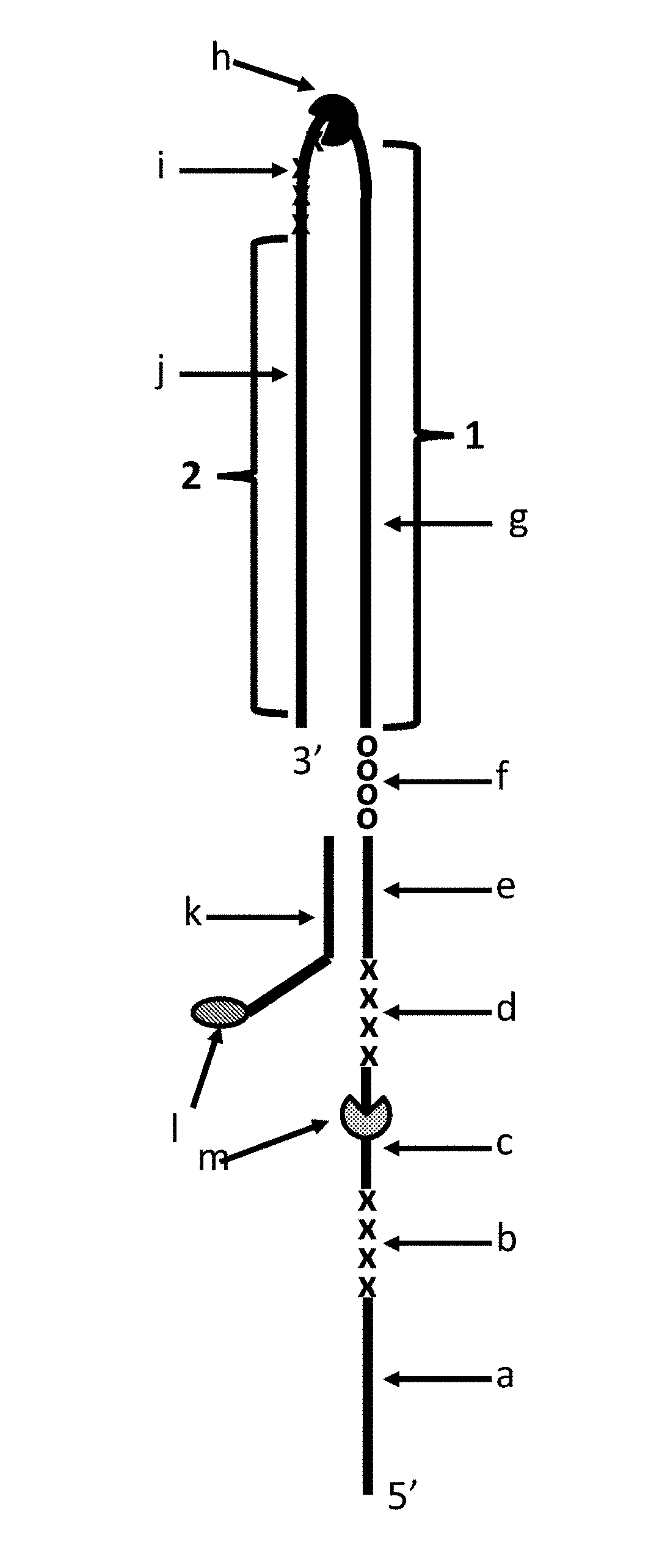
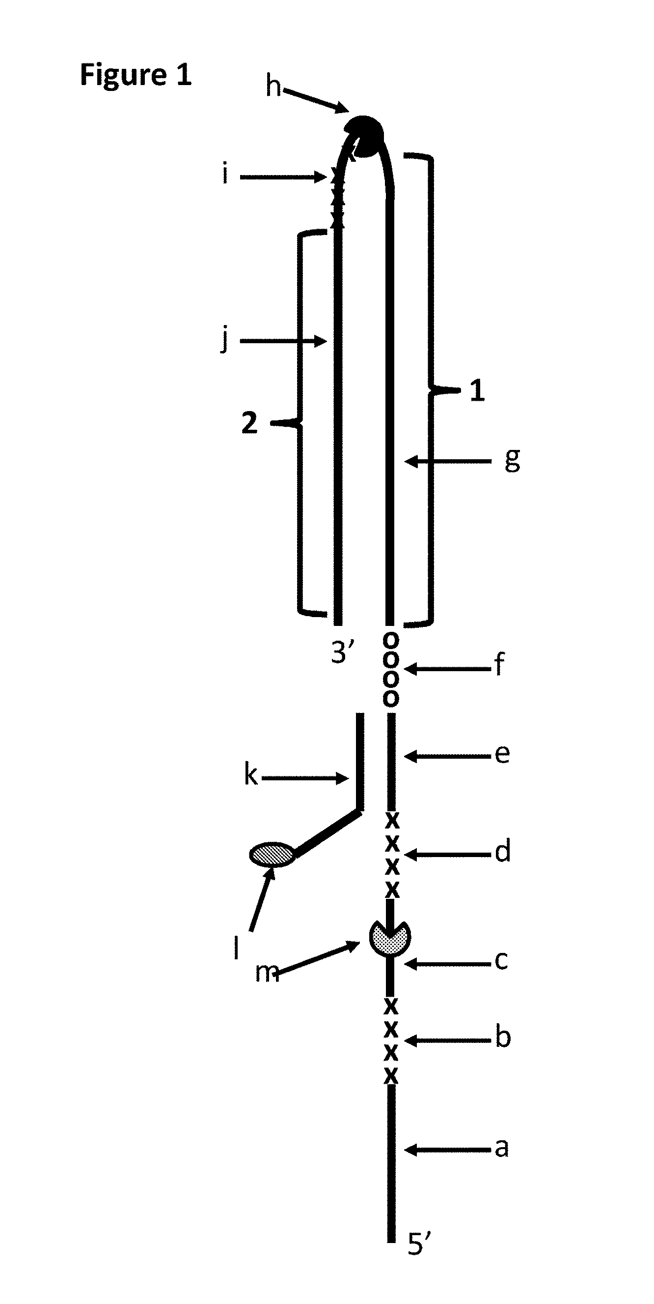
FIG. 1 shows DNA construct Y which was used in Example 1. Section a of DNA construct Y corresponds to SEQ ID NO: 27. Section b corresponds to four iSpC3 spacers. Section c corresponds to SEQ ID NO: 28. Section c is one of the regions of construct Y to which the helicase enzymes T4 Dda--E94C/A360C or T4 Dda--E94C/C109A/C136A/A360C (depending on the experiment) bound (labelled m). The length of section c corresponded to the footprint (binding region) of one enzyme e.g. it was long enough to allow one enzyme to bind to this region. Section d corresponds to four iSpC3 spacers. Section e corresponds to SEQ ID NO: 26. Section f corresponds to four 5'-nitroindoles. Section g corresponds to SEQ ID NO: 29 (this section of the strand was referred to as region 1 of DNA construct Y). Section i corresponds to four iSpC3 spacers. The TrwC Cba-Q594A helicase (SEQ ID NO: 25 with the mutation Q594A) which bound to part of SEQ ID NO: 29 is labelled h. Section j corresponds to SEQ ID NO: 30 (this section of the strand was referred to as region 2 of DNA construct Y). Section k corresponds to SEQ ID NO: 31 which was attached at its 3' end to six iSp18 spacers which were attached at the opposite end to two thymines and a 3' cholesterol TEG. It was possible to distinguish between regions 1 and 2 as they translocated through a nanopore as they produced different characteristics. Furthermore, the section i spacers (four iSpC3 spacers) produced a current spike in the current trace which aided identification of the transition from region 1 to region 2.
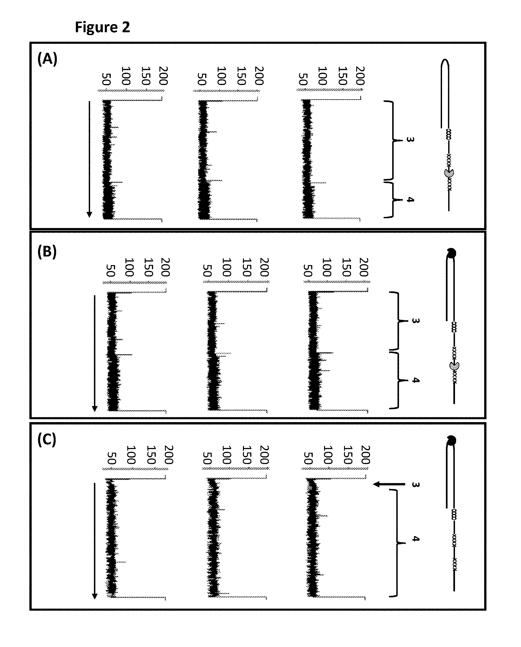
FIG. 2 shows a number of example current traces after helicase controlled DNA movement detection (all traces have the following axes labels y-axis label=Current (pA), x-axis label=Time (seconds)). The traces in section A show single DNA strands moving through a nanopore under the control of only the T4 Dda--E94C/A360C helicase, the labelled regions 1 and 2 corresponded to the translocation of region 1 and 2 of DNA construct Y (see FIG. 1). The traces in section B show single DNA strands moving through a nanopore under the control of both T4 Dda--E94C/A360C and TrwC Cba-Q594A helicases, the labelled regions 1 and 2 corresponded to the translocation of region 1 and 2 of DNA construct Y (see FIG. 1). The traces in section C show single DNA strands moving through a nanopore under the control of only the TrwC Cba-Q594A helicase, the labelled regions 1 and 2 corresponded to the translocation of region 1 and 2 of DNA construct Y (see FIG. 1). Traces A and C show that unequal regions 1 and 2 are obtained when DNA construct Y was translocated through the pore under the control of only one type of helicase either T4 Dda--E94C/A360C helicase (section A) or TrwC Cba-Q594A helicase (section B). Trace B shows improved helicase-controlled DNA movement when construct Y translocated through the pore under the control of both T4 Dda--E94C/A360C and TrwC Cba-Q594A helicase (in this trace regions 1 and 2 were approximately equal). When both enzymes were used to control the movement of region 2 of construct Y through the nanopore the translocation speed was slower and the number of observed stepwise changes in the measured current levels was higher than when a single enzyme was used, and the number of observed stepwise changes in the measured current levels was approximately the same as region 1. This meant that more information was obtained from region 2 when it translocated through the pore under the control of the two enzymes rather than one and therefore improved movement was observed.
FIG. 3 shows example plots of when either the helicases T4 Dda--E94C/A360C only (Section A) or both T4 Dda--E94C/A360C and TrwC Cba-Q594A (Section B) controlled the translocation of DNA construct Y (see FIG. 1 for details) through an MspA nanopore. The x-axis corresponds to the movement index and the y-axis corresponds to the current (pA). For each DNA strand which moved through the pore the current was measured as a function of time. The moving DNA resulted in stepwise changes in the measured current levels. The observed current levels were fitted to obtain a mean current for each step, and assigned an incrementing movement index point. The mean current against movement index therefore closely approximated the original current signal, and was used to characterise the translocated DNA. Plots A and B each showed single DNA strands moving through the nanopore under the control of helicases, the labelled regions 1 and 2 corresponded to the translocation of region 1 and 2 of DNA construct Y (see FIG. 1). Trace A shows the movement index observed when construct Y was translocated through the pore under the control of a single T4 Dda--E94C/A360C helicase only. Trace B shows the movement index observed when construct Y was translocated through the pore under the control of both T4 Dda--E94C/A360C and TrwC Cba-Q594A helicases. As region 1 and region 2 were approximately the same length, the movement index observed for each region would have been expected to have had approximately the same number of points. Plot A shows a significantly reduced number of points in the movement index for region 2 when compared to region 1, therefore, less information was derived from region 2 than region 1. However, plot B (where the movement of construct Y was controlled by both T4 Dda--E94C/A360C and TrwC Cba-Q594A helicases) showed many more points in the movement index of region 2 (and approximately the same amount as in region 1), which indicated that approximately the same amount of information was derived from region 2 as region 1. Using the combination of helicases (T4 Dda--E94C/A360C and TrwC Cba-Q594A) to control the movement of construct Y provided improved movement as more information was derived from region 2 than when a single helicase controlled the movement.
FIG. 4 shows example plots of when either the helicases T4 Dda--E94C/C109A/C136A/A360C only (Section A) or both T4 Dda--E94C/C109A/C136A/A360C and TrwC Cba-Q594A (Section B) controlled the translocation of DNA construct Y (see FIG. 1 for details) through an MspA nanopore. The x-axis corresponds to the movement index (see FIG. 3's figure legend for description of movement index) and the y-axis corresponds to the current (pA). Plots A and B each showed a single DNA strand moving through the nanopore under the control of helicases, the labelled regions 1 and 2 corresponded to the translocation of region 1 and 2 of DNA construct Y (see FIG. 1). Trace A shows the movement index observed when construct Y was translocated through the pore under the control of a single T4 Dda--E94C/C109A/C136A/A360C helicase only. Trace B shows the movement index observed when construct Y was translocated through the pore under the control of both T4 Dda--E94C/C109A/C136A/A360C and TrwC Cba-Q594A helicases. As region 1 and region 2 were approximately the same length, the movement index observed for each region would have been expected to have had approximately the same number of points. Plot A shows a significantly reduced number of points in the movement index for region 2 when compared to region 1, therefore, less information was derived from region 2 than region 1. However, plot B (where the movement of construct Y was controlled by both T4 Dda--E94C/C109A/C136A/A360C and TrwC Cba-Q594A helicases) showed many more points in the movement index of region 2 (and approximately the same amount as in region 1), which indicated that approximately the same amount of information was derived from region 2 as region 1. Using the combination of helicases (T4 Dda--E94C/C109A/C136A/A360C and TrwC Cba-Q594A) to control the movement of construct Y provided improved movement as more information was derived from region 2 than when a single helicase controlled the movement.
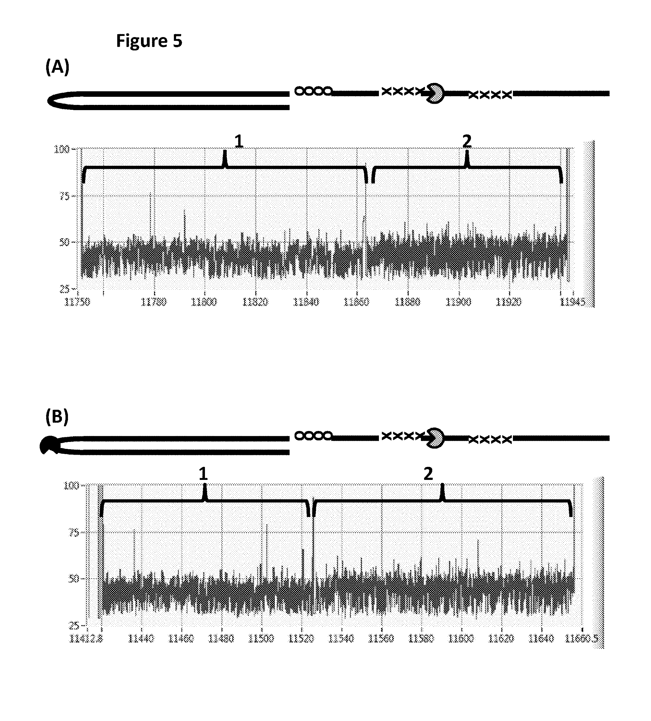
FIG. 5 shows example current traces of when either the helicase T4 Dda--E94C/C109A/C136A/A360C only (Section A) or both T4 Dda--E94C/C109A/C136A/A360C and TrwC Cba-L376C/Q594A/K762C (Section B) controlled the translocation of DNA construct Y (see FIG. 1 for details) through an MspA nanopore. The x-axis corresponds to the time (s) and the y-axis corresponds to the current (pA). Plots A and B each showed a single DNA strand moving through the nanopore under the control of helicases, the labelled regions 1 and 2 corresponded to the translocation of region 1 and 2 of DNA construct Y (see FIG. 1). Trace A shows a current trace observed when construct Y was translocated through the pore under the control of a single T4 Dda--E94C/C109A/C136A/A360C helicase only. Trace B shows a current trace observed when construct Y was translocated through the pore under the control of both T4 Dda--E94C/C109A/C136A/A360C and TrwC Cba-L376C/Q594A/K762C helicases. Plot A shows a significantly reduced number of observed stepwise changes in the measured current levels in the current trace for region 2 when compared to region 1, therefore, less information was derived from region 2 than region 1. However, plot B (where the movement of construct Y was controlled by both T4 Dda--E94C/C109A/C136A/A360C and TrwC Cba-L376C/Q594A/K762C helicases) showed many more observed stepwise changes in the measured current levels in the current trace of region 2 (and approximately the same amount as in region 1), which indicated that approximately the same amount of information was derived from region 2 as region 1. Using the combination of helicases (T4 Dda--E94C/C109A/C136A/A360C and TrwC Cba-L376C/Q594A/K762C) to control the movement of construct Y provided improved movement as more information was derived from region 2 than when a single helicase controlled the movement.
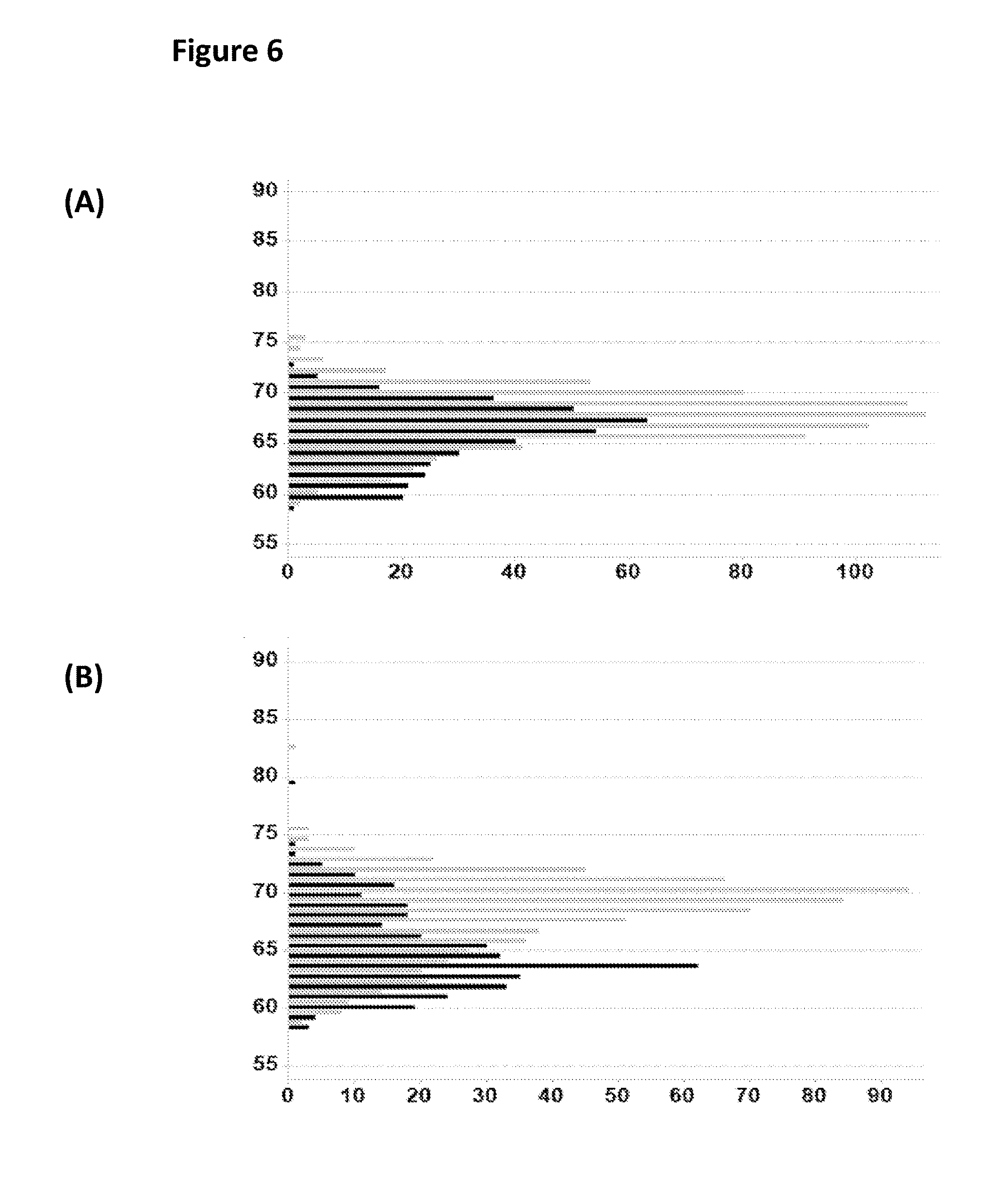
FIG. 6 shows two histogram plots which show the base calling accuracy (as a percentage) for helicase controlled DNA movement events detected in the experiments carried out in Example 3 when either a single enzyme (T4 Dda--E94C/C109A/C136A/A360C) or two enzymes (T4 Dda--E94C/C109A/C136A/A360C and TrwC Cba-L376C/Q594A/K762C) controlled the movement of region 1(trace A) or region 2 (trace B) of the DNA construct Y. The x-axis label was count and the y-axis label was % base calling accuracy for either region 1 (trace A) or region 2 (trace B) based on the known sequence of construct Y. Plot A shows the base calling accuracy of the helicase controlled translocation of region 1 (shown in FIG. 1). Plot B shows the base calling accuracy of the helicase controlled translocation of region 2 (shown in FIG. 1). Each helicase controlled DNA translocation was categorised as either having more observed stepwise changes in the measured current levels in region 1 (shown as black bars which was indicative of T4 Dda--E94C/C109A/C136A/A360C only) or more observed stepwise changes in the measured current levels in region 2 (shown as grey bars, which was indicative of both T4 Dda--E94C/C109A/C136A/A360C and TrwC Cba-L376C/Q594A/K762C bound to construct Y). As the TrwC Cba-L376C/Q594A/K762C only affected the movement of region 2, the sequencing accuracies for region 1 of the strand have the same distribution for both class of strand (either one or two enzymes bound). However, the sequencing accuracy of the region 2 of construct Y was improved as there were more observed stepwise changes in the measured current levels in region 2 when TrwC Cba-L376C/Q594A/K762C (grey bars) was bound. Therefore, the bulk accuracy of the base calling distribution was improved by approximately 5-10% when both enzymes were bound (the grey bars shown in trace B).
DESCRIPTION OF THE SEQUENCE LISTING
SEQ ID NO: 1 shows the codon optimised polynucleotide sequence encoding the MS-B1 mutant MspA monomer. This mutant lacks the signal sequence and includes the following mutations: D90N, D91N, D93N, D118R, D134R and E139K.
SEQ ID NO: 2 shows the amino acid sequence of the mature form of the MS-B1 mutant of the MspA monomer. This mutant lacks the signal sequence and includes the following mutations: D90N, D91N, D93N, D118R, D134R and E139K.
SEQ ID NO: 3 shows the polynucleotide sequence encoding one monomer of .alpha.-hemolysin-E111N/K147N (.alpha.-HL-NN; Stoddart et al., PNAS, 2009; 106(19): 7702-7707).
SEQ ID NO: 4 shows the amino acid sequence of one monomer of .alpha.-HL-NN.
SEQ ID NOs: 5 to 7 show the amino acid sequences of MspB, C and D.
SEQ ID NO: 8 shows the polynucleotide sequence encoding the Phi29 DNA polymerase.
SEQ ID NO: 9 shows the amino acid sequence of the Phi29 DNA polymerase.
SEQ ID NO: 10 shows the codon optimised polynucleotide sequence derived from the sbcB gene from E. coli. It encodes the exonuclease I enzyme (EcoExo I) from E. coli.
SEQ ID NO: 11 shows the amino acid sequence of exonuclease I enzyme (EcoExo I) from E. coli.
SEQ ID NO: 12 shows the codon optimised polynucleotide sequence derived from the xthA gene from E. coli. It encodes the exonuclease III enzyme from E. coli.
SEQ ID NO: 13 shows the amino acid sequence of the exonuclease III enzyme from E. coli. This enzyme performs distributive digestion of 5' monophosphate nucleosides from one strand of double stranded DNA (dsDNA) in a 3'--5' direction. Enzyme initiation on a strand requires a 5' overhang of approximately 4 nucleotides.
SEQ ID NO: 14 shows the codon optimised polynucleotide sequence derived from the red gene from T. thermophilus. It encodes the RecJ enzyme from T. thermophilus (TthRecJ-cd).
SEQ ID NO: 15 shows the amino acid sequence of the RecJ enzyme from T. thermophilus (TthRecJ-cd). This enzyme performs processive digestion of 5' monophosphate nucleosides from ssDNA in a 5'-3' direction. Enzyme initiation on a strand requires at least 4 nucleotides.
SEQ ID NO: 16 shows the codon optimised polynucleotide sequence derived from the bacteriophage lambda exo (redX) gene. It encodes the bacteriophage lambda exonuclease.
SEQ ID NO: 17 shows the amino acid sequence of the bacteriophage lambda exonuclease. The sequence is one of three identical subunits that assemble into a trimer. The enzyme performs highly processive digestion of nucleotides from one strand of dsDNA, in a 5'-3' direction (neb.com/nebecomm/products/productM0262.asp). Enzyme initiation on a strand preferentially requires a 5' overhang of approximately 4 nucleotides with a 5' phosphate.
SEQ ID NO: 18 shows the amino acid sequence of He1308 Mbu.
SEQ ID NO: 19 shows the amino acid sequence of Hel308 Csy.
SEQ ID NO: 20 shows the amino acid sequence of Hel308 Tga.
SEQ ID NO: 21 shows the amino acid sequence of He1308 Mhu.
SEQ ID NO: 22 shows the amino acid sequence of TraI Eco.
SEQ ID NO: 23 shows the amino acid sequence of XPD Mbu.
SEQ ID NO: 24 shows the amino acid sequence of Dda 1993.
SEQ ID NO: 25 shows the amino acid sequence of Trwc Cba.
SEQ ID NO: 26 shows a polynucleotide sequence used in Example 1 and 2.
SEQ ID NO: 27 shows a polynucleotide sequence used in Example 1 and 2.
SEQ ID NO: 28 shows a polynucleotide sequence used in Example 1 and 2.
SEQ ID NO: 29 shows a polynucleotide sequence used in Example 1 and 2.
SEQ ID NO: 30 shows a polynucleotide sequence used in Example 1 and 2.
SEQ ID NO: 31 shows a polynucleotide sequence used in Example 1 and 2.
DETAILED DESCRIPTION OF THE INVENTION
It is to be understood that different applications of the disclosed products and methods may be tailored to the specific needs in the art. It is also to be understood that the terminology used herein is for the purpose of describing particular embodiments of the invention only, and is not intended to be limiting.
In addition as used in this specification and the appended claims, the singular forms "a", "an", and "the" include plural referents unless the content clearly dictates otherwise. Thus, for example, reference to "a polynucleotide" includes two or more polynucleotides, reference to "a helicase" includes two or more helicases, reference to "a molecular brake" refers to two or more molecular brakes, reference to "a transmembrane pore" includes two or more pores and the like.
All publications, patents and patent applications cited herein, whether supra or infra, are hereby incorporated by reference in their entirety.
Method of the Invention
The invention provides a method of controlling the movement of a polynucleotide through a transmembrane pore. The polynucleotide is provided with one or more helicases and one or more molecular brakes. The polynucleotide, the one or more helicases and the one or more molecular brakes are contacted with a transmembrane pore. Once a potential is applied, the polynucleotide moves through the pore and brings the one or more helicases and the one or more molecular brakes together and they both control the movement of the polynucleotide through the pore. The combination of the one or more helicases and the one or more molecular brakes results in an improved movement of the polynucleotide through the pore.
Helicases can control the movement of polynucleotides in at least two active modes of operation (when the helicase is provided with all the necessary components to facilitate movement, e.g. ATP and Mg.sup.2+) and one inactive mode of operation (when the helicase is not provided with the necessary components to facilitate movement or is modified to prevent or hinder movement). When provided with all the necessary components to facilitate movement, the helicase moves along the polynucleotide in a 5' to 3' or a 3' to 5' direction (depending on the helicase), but the orientation of the polynucleotide in the pore (which is dependent on which end of the polynucleotide is captured by the pore) means that the helicase can be used to either move the polynucleotide out of the pore against the applied field or move the polynucleotide into the pore with the applied field. When the end of the polynucleotide towards which the helicase moves is captured by the pore, the helicase works against the direction of the field resulting from the applied potential and pulls the threaded polynucleotide out of the pore and into the cis chamber. However, when the end away from which the helicase moves is captured in the pore, the helicase works with the direction of the field resulting from the applied potential and pushes the threaded polynucleotide into the pore and into the trans chamber.
When the helicase is not provided with the necessary components to facilitate movement it can bind to the polynucleotide and act as a brake slowing the movement of the polynucleotide when it is pulled into the pore by the field resulting from the applied potential. In the inactive mode, it does not matter which end of the polynucleotide is captured, it is the applied field which pulls the polynucleotide into the pore towards the trans side with the helicase acting as a brake. When in the inactive mode, the movement control of the polynucleotide by the helicase can be described in a number of ways including ratcheting, sliding and braking.
In the method of the invention, the one or more helicases preferably control the movement of the target polynucleotide through the pore with the field resulting from the applied potential. In one preferred embodiment, the one or more helicases are used in the active mode and the end away from which the one or more helicases move is captured by the pore such that the one or more helicases work with the field resulting from the applied potential and push the polynucleotide through the pore. If the one or more helicases move in the 5' to 3' direction, the 5' end of the polynucleotide is preferably captured by the pore. In such embodiments, the one or more helicases move along the polynucleotide in the 5' to 3' direction. If the one or more helicases move in the 3' to 5' direction, the 3' end of the polynucleotide is preferably captured by the pore. In such embodiments, the one or more helicases move along the polynucleotide in the 3' to 5' direction.
In another preferred embodiment, the one or more helicases are used in the inactive mode such that the applied field pulls the polynucleotide through the pore and the one or more helicases act as a brake. In another preferred embodiment, the one or more helicases are modified such that they retain their polynucleotide binding ability but lack helicase activity (i.e. the ability to actively move along the polynucleotide) such that the applied field pulls the polynucleotide through the pore and the one or more helicases act as a brake. In the method of the invention, the one or more helicases preferably slow or brake the movement of the polynucleotide through the pore with the field resulting from the applied potential. In either case, the one or more helicases are typically too large to move through the pore and the pore pushes the one or more helicases along the polynucleotide as the polynucleotide moves through the pore with the field resulting from the applied potential. This brings to the one or more helicases and one or more molecular brakes together.
The method of controlling the movement of a polynucleotide through a transmembrane pore can be helpful during characterisation of the polynucleotide using the pore, for instance during strand sequencing. The invention also provides a method of characterising a target polynucleotide. Once a potential is applied, the polynucleotide moves through the pore and brings the one or more helicases and the one or more molecular brakes together and they both control the movement of the polynucleotide through the pore. The method also comprises taking one or more measurements as the polynucleotide moves with respect to the pore. The measurements are indicative of one or more characteristics of the polynucleotide, such as the sequence.
It has been shown that double stranded polynucleotides can be effectively characterised using a transmembrane pore if they are modified to include a Y adaptor (a double stranded stem and two non-complementary arms) containing a leader sequence and a bridging moiety adaptor, such as a hairpin loop adaptor (WO 2013/014451). It is preferred that that Y adaptor containing the leader sequence is attached to one end of the polynucleotide and the bridging moiety adaptor is attached to the other end. The leader sequence preferentially threads into the nanopore and the bridging moiety connecting the two strands of the polynucleotide allows both strands to be investigated as the polynucleotide unzips and both strands (connected via the bridging moiety) move through the pore. This is advantageous because it doubles the amount of information obtained from a single double stranded polynucleotide. Moreover, because the sequences in the two strands are complementary, the information from the two strands can be combined informatically. This mechanism provides an orthogonal proof-reading capability that provides higher confidence observations.
One or more helicases may be attached to the Y adaptor and used to control the movement of both strands of the double stranded polynucleotide (connected via the bridging moiety) through the pore. The inventors have shown that, once the one or more helicases move past the bridging moiety and control the movement of the second strand of the double stranded polynucleotide, the one or more helicases are less effective at controlling the movement of the second strand through the pore and less information is derived from the second strand. The invention overcomes this decrease in efficiency of movement control by using one or more molecular brakes. When the one or more helicases and one or more molecular brakes are brought together, they effectively control the movement of the second strand through the pore. The one or more molecular brakes are preferably attached to the bridging moiety so that the one or more helicases and one or more molecular brakes are brought together at the appropriate time. The increased efficiency of movement control means that more information is derived from the second strand. In particular, approximately the same amount of information can be derived from both strands.
Polynucleotide
A polynucleotide, such as a nucleic acid, is a macromolecule comprising two or more nucleotides. The polynucleotide or nucleic acid may comprise any combination of any nucleotides. The nucleotides can be naturally occurring or artificial. One or more nucleotides in the polynucleotide can be oxidized or methylated. One or more nucleotides in the polynucleotide may be damaged. For instance, the polynucleotide may comprise a pyrimidine dimer. Such dimers are typically associated with damage by ultraviolet light and are the primary cause of skin melanomas. One or more nucleotides in the polynucleotide may be modified, for instance with a label or a tag. Suitable labels are described below. The polynucleotide may comprise one or more spacers.
A nucleotide typically contains a nucleobase, a sugar and at least one phosphate group. The nucleobase and sugar form a nucleoside.
The nucleobase is typically heterocyclic. Nucleobases include, but are not limited to, purines and pyrimidines and more specifically adenine (A), guanine (G), thymine (T), uracil (U) and cytosine (C).
The sugar is typically a pentose sugar. Nucleotide sugars include, but are not limited to, ribose and deoxyribose. The sugar is preferably a deoxyribose.
The nucleotide in the polynucleotide is typically a ribonucleotide or deoxyribonucleotide. The polynucleotide may comprise the following nucleosides: adenosine, uridine, guanosine and cytidine. The nucleotide is preferably a deoxyribonucleotide. The polynucleotide preferably comprises the following nucleosides: deoxyadenosine (dA), deoxyuridine (dU) and/or thymidine (dT), deoxyguanosine (dG) and deoxycytidine (dC).
The nucleotide typically contains a monophosphate, diphosphate or triphosphate. Phosphates may be attached on the 5' or 3' side of a nucleotide.
Suitable nucleotides include, but are not limited to, adenosine monophosphate (AMP), guanosine monophosphate (GMP), thymidine monophosphate (TMP), uridine monophosphate (UMP), cytidine monophosphate (CMP), cyclic adenosine monophosphate (cAMP), cyclic guanosine monophosphate (cGMP), deoxyadenosine monophosphate (dAMP), deoxyguanosine monophosphate (dGMP), deoxythymidine monophosphate (dTMP), deoxyuridine monophosphate (dUMP) and deoxycytidine monophosphate (dCMP). The nucleotides are preferably selected from AMP, TMP, GMP, CMP, UMP, dAMP, dTMP, dGMP, dCMP and dUMP. The nucleotides are most preferably selected from dAMP, dTMP, dGMP, dCMP and dUMP. The polynucleotide preferably comprises the following nucleotides: dAMP, dUMP and/or dTMP, dGMP and dCMP.
The nucleotides in the polynucleotide may be attached to each other in any manner. The nucleotides are typically attached by their sugar and phosphate groups as in nucleic acids. The nucleotides may be connected via their nucleobases as in pyrimidine dimers.
The polynucleotide may be single stranded or double stranded. At least a portion of the polynucleotide is preferably double stranded.
The polynucleotide can be a nucleic acid. The polynucleotide may be any synthetic nucleic acid known in the art, such as peptide nucleic acid (PNA), glycerol nucleic acid (GNA), threose nucleic acid (TNA), locked nucleic acid (LNA) or other synthetic polymers with nucleotide side chains. The PNA backbone is composed of repeating N-(2-aminoethyl)-glycine units linked by peptide bonds. The GNA backbone is composed of repeating glycol units linked by phosphodiester bonds. The TNA backbone is composed of repeating threose sugars linked together by phosphodiester bonds. LNA is formed from ribonucleotides as discussed above having an extra bridge connecting the 2' oxygen and 4' carbon in the ribose moiety.
The polynucleotide is most preferably ribonucleic nucleic acid (RNA) or deoxyribonucleic acid (DNA).
The polynucleotide may be any length. For example, the polynucleotide can be at least 10, at least 50, at least 100, at least 150, at least 200, at least 250, at least 300, at least 400 or at least 500 nucleotides in length. The polynucleotide can be 1000 or more nucleotides, 5000 or more nucleotides in length or 100000 or more nucleotides in length.
The helicase may move along the whole or only part of the polynucleotide in the method of the invention. The whole or only part of the target polynucleotide may be characterised using the method of the invention.
The polynucleotide may be single stranded. At least a portion of the polynucleotide is preferably double stranded. Helicases typically bind to single stranded polynucleotides. If at least a portion of the polynucleotide is double stranded, the polynucleotide preferably comprises a single stranded region or a non-hybridised region. The one or more helicases are capable of binding to the single stranded region or one strand of the non-hybridised region. The polynucleotide preferably comprises one or more single stranded regions or one or more non-hybridised regions.
The one or more spacers are preferably included in the single stranded region or the non-hybridised region of the polynucleotide. The polynucleotide may comprise more than one single stranded region or more than one non-hybridised region. The polynucleotide may comprise a single stranded region or a non-hybridised region within its sequence and/or at one or both ends. The one or more spacers may be included in the double stranded region of the polynucleotide.
If the one or more helicases used in the method move in the 5' to 3' direction, the polynucleotide preferably comprises a single stranded region or a non-hybridised region at its 5' end. If the one or more helicases used in the method move in the 3' to 5' direction, the polynucleotide preferably comprises a single stranded region or a non-hybridised region at its 3' end. If the one or more helicases are used in the inactive mode (i.e. as a brake), it does not matter where the single stranded region or the non-hybridised region is located.
The single stranded region preferably comprises a leader sequence which preferentially threads into the pore. This is discussed in more detail below.
If at least a portion of the polynucleotide is double stranded, the two strands of the double stranded portion are preferably linked using a bridging moiety, such as a hairpin or a hairpin loop. This facilitates characterisation method of the invention and is discussed in more detail below.
The polynucleotide is present in any suitable sample. The invention is typically carried out on a sample that is known to contain or suspected to contain the polynucleotide. The invention may be carried out on a sample to confirm the identity of one or more polynucleotides whose presence in the sample is known or expected.
The sample may be a biological sample. The invention may be carried out in vitro on a sample obtained from or extracted from any organism or microorganism. The organism or microorganism is typically archaeal, prokaryotic or eukaryotic and typically belongs to one of the five kingdoms: plantae, animalia, fungi, monera and protista. The invention may be carried out in vitro on a sample obtained from or extracted from any virus. The sample is preferably a fluid sample. The sample typically comprises a body fluid of the patient. The sample may be urine, lymph, saliva, mucus or amniotic fluid but is preferably blood, plasma or serum. Typically, the sample is human in origin, but alternatively it may be from another mammal animal such as from commercially farmed animals such as horses, cattle, sheep, fish, chickens or pigs or may alternatively be pets such as cats or dogs. Alternatively, the sample may be of plant origin, such as a sample obtained from a commercial crop, such as a cereal, legume, fruit or vegetable, for example wheat, barley, oats, canola, maize, soya, rice, rhubarb, bananas, apples, tomatoes, potatoes, grapes, tobacco, beans, lentils, sugar cane, cocoa, cotton.
The sample may be a non-biological sample. The non-biological sample is preferably a fluid sample. Examples of a non-biological sample include surgical fluids, water such as drinking water, sea water or river water, and reagents for laboratory tests.
The sample is typically processed prior to being used in the invention, for example by centrifugation or by passage through a membrane that filters out unwanted molecules or cells, such as red blood cells. The sample may be measured immediately upon being taken. The sample may also be typically stored prior to assay, preferably below -70.degree. C.
Helicases
Any helicase may be used in the invention. The helicase may be or be derived from a Hel308 helicase, a RecD helicase, such as TraI helicase or a TrwC helicase, a XPD helicase or a Dda helicase. The helicase may be any of the helicases, modified helicases or helicase constructs disclosed in International Application Nos. PCT/GB2012/052579 (published as WO 2013/057495); PCT/GB2012/053274 (published as WO 2013/098562); PCT/GB2012/053273 (published as WO2013/098561); PCT/GB2013/051925 (published as WO 2014/013260); PCT/GB2013/051924 (published as WO 2014/013259) and PCT/GB2013/051928 (published as WO 2014/013262); and in UK Application No. 1318464.3 filed on 18 Oct. 2013. In particular, the one or more helicases are preferably modified to reduce the size of an opening in the polynucleotide binding domain through which in at least one conformational state the polynucleotide can unbind from the helicase. This is disclosed in WO 2014/013260.
The one or more helicases may be derived from any helicase, such as He1308 Mbu (SEQ ID NO: 18), Hel308 Csy (SEQ ID NO: 19), Hel308 Tga (SEQ ID NO: 20), He1308 Mhu (SEQ ID NO: 21), TraI Eco (SEQ ID NO: 22), XPD Mbu (SEQ ID NO: 23) or a variant thereof.
The helicase preferably comprises the sequence shown in SEQ ID NO: 25 (Trwc Cba) or as variant thereof, the sequence shown in SEQ ID NO: 18 (He1308 Mbu) or a variant thereof or the sequence shown in SEQ ID NO: 24 (Dda) or a variant thereof.
Variants may differ from the native sequences in any of the ways discussed below for transmembrane pores. Variants retain helicase activity. This can be assayed using known methods and the methods disclosed in the Examples. In particular, over the entire length of the amino acid sequence of SEQ ID NO: 18, 19, 20, 21, 22, 23, 24 or 25, a variant will preferably be at least 50% homologous to that sequence based on amino acid identity. More preferably, the variant may be at least 55%, at least 60%, at least 65%, at least 70%, at least 75%, at least 80%, at least 85%, at least 90% and more preferably at least 95%, 97% or 99% homologous based on amino acid identity to the amino acid sequence of SEQ ID NO: 18, 19, 20, 21, 22, 23, 24 or 25 over the entire sequence. There may be at least 80%, for example at least 85%, 90% or 95%, amino acid identity over a stretch of 100 or more, for example 125, 150, 175 or 200 or more, contiguous amino acids ("hard homology").
A preferred variant of SEQ ID NO: 24 comprises (or only comprises) (a) E94C/A360C, (b) E94C/A360C and then (.DELTA.M1)G1G2 (i.e. deletion of M1 and then addition G1 and G2), (c) E94C/A360C/C109A/C136A or (d) E94C/A360C/C109A/C136A and then (.DELTA.M1)G1G2 (i.e. deletion of M1 and then addition G1 and G2).
Other preferred variants of SEQ ID NO: 24 comprise W378A. Preferred variants of SEQ ID NO: 24 comprise (or comprise only) (a) E94C/A360C/W378A, (b) E94C/A360C/W378A and then (.DELTA.M1)G1G2 (i.e. deletion of M1 and then addition G1 and G2), (c) E94C/A360C/C109A/C136A/W378A or (d) E94C/A360C/C109A/C136A/W378A and then (.DELTA.M1)G1G2 (i.e. deletion of M1 and then addition G1 and G2).
Any number of helicases may be used in accordance with the invention. For instance, 1, 2, 3, 4, 5, 6, 7, 8, 9, 10 or more helicases may be used
If two or more helicases are used, they may be attached to one another. The two or more helicases may be covalently attached to one another. The helicases may be attached in any order and using any method. Preferred helicase constructs for use in the invention are described in International Application Nos. PCT/GB2013/051925 (published as WO 2014/013260); PCT/GB2013/051924 (published as WO 2014/013259) and PCT/GB2013/051928 (published as WO 2014/013262); and in UK Application No. 1318464.3 filed on 18 Oct. 2013.
If two or more helicases are used, they are preferably not attached to one another except via the polynucleotide. The two or more helicases are more preferably not covalently attached to one another.
The one or more helicases may be any of those discussed below with reference to the molecular brakes, including all variants of helicases.
Any steps in the method using one or more helicases are typically carried out in the presence of free nucleotides or free nucleotide analogues and an enzyme cofactor that facilitates the action of the one or more helicases. The free nucleotides may be one or more of any of the individual nucleotides discussed above. The free nucleotides include, but are not limited to, adenosine monophosphate (AMP), adenosine diphosphate (ADP), adenosine triphosphate (ATP), guanosine monophosphate (GMP), guanosine diphosphate (GDP), guanosine triphosphate (GTP), thymidine monophosphate (TMP), thymidine diphosphate (TDP), thymidine triphosphate (TTP), uridine monophosphate (UMP), uridine diphosphate (UDP), uridine triphosphate (UTP), cytidine monophosphate (CMP), cytidine diphosphate (CDP), cytidine triphosphate (CTP), cyclic adenosine monophosphate (cAMP), cyclic guanosine monophosphate (cGMP), deoxyadenosine monophosphate (dAMP), deoxyadenosine diphosphate (dADP), deoxyadenosine triphosphate (dATP), deoxyguanosine monophosphate (dGMP), deoxyguanosine diphosphate (dGDP), deoxyguanosine triphosphate (dGTP), deoxythymidine monophosphate (dTMP), deoxythymidine diphosphate (dTDP), deoxythymidine triphosphate (dTTP), deoxyuridine monophosphate (dUMP), deoxyuridine diphosphate (dUDP), deoxyuridine triphosphate (dUTP), deoxycytidine monophosphate (dCMP), deoxycytidine diphosphate (dCDP) and deoxycytidine triphosphate (dCTP). The free nucleotides are preferably selected from AMP, TMP, GMP, CMP, UMP, dAMP, dTMP, dGMP or dCMP. The free nucleotides are preferably adenosine triphosphate (ATP). The enzyme cofactor is a factor that allows the construct to function. The enzyme cofactor is preferably a divalent metal cation. The divalent metal cation is preferably mg.sup.2+, mn.sup.2+, Ca.sup.2+ or Co.sup.2+. The enzyme cofactor is most preferably mg.sup.2+.
Molecular Brakes
The one or more molecular brakes may be any compound or molecule which binds to the polynucleotide and slows the movement of the polynucleotide through the pore.
The one or more molecular brakes preferably comprise one or more compounds which bind to the polynucleotide. The one or more compounds are preferably one or more macrocycles. Suitable macrocycles include, but are not limited to, cyclodextrins, calixarenes, cyclic peptides, crown ethers, cucurbiturils, pillararenes, derivatives thereof or a combination thereof. The cyclodextrin or derivative thereof may be any of those disclosed in Eliseev, A. V., and Schneider, H-J. (1994) J. Am. Chem. Soc. 116, 6081-6088. The agent is more preferably heptakis-6-amino-.beta.-cyclodextrin (am.sub.7-.beta.CD), 6-monodeoxy-6-monoamino-.beta.-cyclodextrin (am.sub.1-.beta.CD) or heptakis-(6-deoxy-6-guanidino)-cyclodextrin (guy-.beta.CD).
The one or more molecular brakes are preferably not one or more single stranded binding proteins (SSB). The one or more molecular brakes are more preferably not a single-stranded binding protein (SSB) comprising a carboxy-terminal (C-terminal) region which does not have a net negative charge or (ii) a modified SSB comprising one or more modifications in its C-terminal region which decreases the net negative charge of the C-terminal region. The one or more molecular brakes are most preferably not any of the SSBs disclosed in International Application No. PCT/GB2013/051924 (published as WO 2014/013259).
The one or more molecular brakes are preferably one or more polynucleotide binding proteins. The polynucleotide binding protein may be any protein that is capable of binding to the polynucleotide and controlling its movement through the pore. It is straightforward in the art to determine whether or not a protein binds to a polynucleotide. The protein typically interacts with and modifies at least one property of the polynucleotide. The protein may modify the polynucleotide by cleaving it to form individual nucleotides or shorter chains of nucleotides, such as di- or trinucleotides. The moiety may modify the polynucleotide by orienting it or moving it to a specific position, i.e. controlling its movement.
The polynucleotide binding protein is preferably derived from a polynucleotide handling enzyme. A polynucleotide handling enzyme is a polypeptide that is capable of interacting with and modifying at least one property of a polynucleotide. The enzyme may modify the polynucleotide by cleaving it to form individual nucleotides or shorter chains of nucleotides, such as di- or trinucleotides. The enzyme may modify the polynucleotide by orienting it or moving it to a specific position. The polynucleotide handling enzyme does not need to display enzymatic activity as long as it is capable of binding the polynucleotide and controlling its movement through the pore. For instance, the enzyme may be modified to remove its enzymatic activity or may be used under conditions which prevent it from acting as an enzyme. Such conditions are discussed in more detail below.
The one or more molecular brakes are preferably derived from a nucleolytic enzyme. The enzyme is more preferably derived from a member of any of the Enzyme Classification (EC) groups 3.1.11, 3.1.13, 3.1.14, 3.1.15, 3.1.16, 3.1.21, 3.1.22, 3.1.25, 3.1.26, 3.1.27, 3.1.30 and 3.1.31. The enzyme may be any of those disclosed in International Application No. PCT/GB10/000133 (published as WO 2010/086603).
Preferred enzymes are polymerases, exonucleases, helicases and topoisomerases, such as gyrases. Suitable enzymes include, but are not limited to, exonuclease I from E. coli (SEQ ID NO: 11), exonuclease III enzyme from E. coli (SEQ ID NO: 13), RecJ from T. thermophilus (SEQ ID NO: 15) and bacteriophage lambda exonuclease (SEQ ID NO: 17), TatD exonuclease and variants thereof. Three subunits comprising the sequence shown in SEQ ID NO: 15 or a variant thereof interact to form a trimer exonuclease. The polymerase may be PYROPHAGE.RTM. 3173 DNA Polymerase (thermostable bacteriophage enzyme, which is commercially available from LUCIGEN.RTM. Corporation), SD Polymerase (commercially available from Bioron) or variants thereof. The enzyme is preferably Phi29 DNA polymerase (SEQ ID NO: 9) or a variant thereof. Modified versions of Phi29 polymerase (SEQ ID NO: 8) which act as molecular brakes are disclosed in U.S. Pat. No. 5,576,204. The topoisomerase is preferably a member of any of the Moiety Classification (EC) groups 5.99.1.2 and 5.99.1.3.
The one or more molecular brakes are most preferably derived from a helicase, such as He1308 Mbu (SEQ ID NO: 18), Hel308 Csy (SEQ ID NO: 19), Hel308 Tga (SEQ ID NO: 20), He1308 Mhu (SEQ ID NO: 21), TraI Eco (SEQ ID NO: 22), XPD Mbu (SEQ ID NO: 23) or a variant thereof. The one or more helicases and the one or more molecular brakes derived from helicases are different from one another. In other words, the one or more helicases are not the same as the one or more molecular brakes derived from helicases.
Any helicase may be used in the invention. The helicase may be or be derived from a Hel308 helicase, a RecD helicase, such as TraI helicase or a TrwC helicase, a XPD helicase or a Dda helicase. The helicase may be any of the helicases, modified helicases or helicase constructs disclosed in International Application Nos. PCT/GB2012/052579 (published as WO 2013/057495); PCT/GB2012/053274 (published as WO 2013/098562); PCT/GB2012/053273 (published as WO2013098561); PCT/GB2013/051925 (published as WO 2014/013260); PCT/GB2013/051924 (published as WO 2014/013259) and PCT/GB2013/051928 (published as WO 2014/013262); and in UK Application No. 1318464.3 filed on 18 Oct. 2013.
The helicase preferably comprises the sequence shown in SEQ ID NO: 25 (Trwc Cba) or as variant thereof, the sequence shown in SEQ ID NO: 18 (He1308 Mbu) or a variant thereof or the sequence shown in SEQ ID NO: 24 (Dda) or a variant thereof. Variants may differ from the native sequences in any of the ways discussed below for helicases or transmembrane pores.
Preferred molecular brake variants of SEQ ID NO: 25 comprises (or only comprises) (a) Q594A, (b) L376C/Q594A/K762C, (c) L376C/Q594A/A779C, (d) Q346C/Q594A/A779C, (e) Q346C/Q594A/A783C, (f) D411/Q594A/A783C, (g) Q594A/R353C/E722C, (h) Q594A/Q357C/T720C, (i) Q594A/R358C/T720C, (j) Q594A/H354C/T720C, (k) Q594A/F374C/E722C or (1) Q594A/S350C/E722C. Any of (a) to (1) may further comprise and then (.DELTA.M1)G1G2 (i.e. deletion of M1 and then addition G1 and G2. Other Preferred variants are discussed above.
Any number of helicases may be used as molecular brakes. For instance, 1, 2, 3, 4, 5, 6, 7, 8, 9, 10 or more helicases may be used as molecular brakes. If two or more helicases are be used as molecular brakes, the two or more helicases are typically the same helicases. The two or more helicases may be different helicases.
The two or more molecular brakes may be any combination of the helicases mentioned above. The two or more molecular brakes may be two or more Dda helicases. The two or more molecular brakes may be one or more Dda helicases and one or more TrwC helicases. The two or more molecular brakes may be different variants of the same helicase.
If two or more molecular brakes are used, they may be attached to one another. The two or more molecular brakes may be covalently attached to one another. The molecular brakes may be attached in any order and using any method.
If two or more molecular brakes are used, they are preferably not attached to one another except via the polynucleotide. The two or more molecular brakes are more preferably not covalently attached to one another.
The one or more molecular brakes derived from helicases are preferably modified to reduce the size of an opening in the polynucleotide binding domain through which in at least one conformational state the polynucleotide can unbind from the helicase. This is disclosed in WO 2014/013260.
Preferred helicase constructs for use in the invention are described in International Application Nos. PCT/GB2013/051925 (published as WO 2014/013260); PCT/GB2013/051924 (published as WO 2014/013259) and PCT/GB2013/051928 (published as WO 2014/013262); and in UK Application No. 1318464.3 filed on 18 Oct. 2013.
A variant of SEQ ID NOs: 9, 11, 13, 15, 17, 18, 19, 20, 21, 22, 23, 24 or 25 is an enzyme that has an amino acid sequence which varies from that of SEQ ID NO: 9, 11, 13, 15, 17, 18, 19, 20, 21, 22, 23, 24 or 25 and which retains polynucleotide binding ability. This can be measured using any method known in the art. For instance, the variant can be contacted with a polynucleotide and its ability to bind to and move along the polynucleotide can be measured. The variant may include modifications that facilitate binding of the polynucleotide and/or facilitate its activity at high salt concentrations and/or room temperature. Variants may be modified such that they bind polynucleotides (i.e. retain polynucleotide binding ability) but do not function as a helicase (i.e. do not move along polynucleotides when provided with all the necessary components to facilitate movement, e.g. ATP and Mg.sup.2+). Such modifications are known in the art. For instance, modification of the Mg.sup.2+ binding domain in helicases typically results in variants which do not function as helicases. These types of variants may act as molecular brakes (see below).
Over the entire length of the amino acid sequence of SEQ ID NO: 9, 11, 13, 15, 17, 18, 19, 20, 21, 22, 23, 24 or 25, a variant will preferably be at least 50% homologous to that sequence based on amino acid identity. More preferably, the variant polypeptide may be at least 55%, at least 60%, at least 65%, at least 70%, at least 75%, at least 80%, at least 85%, at least 90% and more preferably at least 95%, 97% or 99% homologous based on amino acid identity to the amino acid sequence of SEQ ID NO: 9, 11, 13, 15, 17, 18, 19, 20, 21, 22, 23, 24 or 25 over the entire sequence. There may be at least 80%, for example at least 85%, 90% or 95%, amino acid identity over a stretch of 200 or more, for example 230, 250, 270, 280, 300, 400, 500, 600, 700, 800, 900 or 1000 or more, contiguous amino acids ("hard homology"). Homology is determined as described above. The variant may differ from the wild-type sequence in any of the ways discussed above with reference to SEQ ID NO: 2 and 4 above. The enzyme may be covalently attached to the pore. Any method may be used to covalently attach the enzyme to the pore.
A preferred molecular brake is TrwC Cba-Q594A (SEQ ID NO: 25 with the mutation Q594A). This variant does not function as a helicase (i.e. binds to polynucleotides but does not move along them when provided with all the necessary components to facilitate movement, e.g. ATP and Mg.sup.2+). The one or more molecular brake helicases can be used in any direction and/or mode discussed above.
One or More Helicases and One or More Molecular Brakes
If the one or more helicases are used in the active mode (i.e. when the one or more helicases are provided with all the necessary components to facilitate movement, e.g. ATP and Mg.sup.2+), the one or more molecular brakes are preferably (a) used in an inactive mode (i.e. are used in the absence of the necessary components to facilitate movement or are incapable of active movement), (b) used in an active mode where the one or more molecular brakes move in the opposite direction to the one or more helicases or (c) used in an active mode where the one or more molecular brakes move in the same direction as the one or more helicases and more slowly than the one or more helicases.
If the one or more helicases are used in the inactive mode (i.e. when the one or more helicases are not provided with all the necessary components to facilitate movement, e.g. ATP and Mg.sup.2+ or are incapable of active movement), the one or more molecular brakes are preferably (a) used in an inactive mode (i.e. are used in the absence of the necessary components to facilitate movement or are incapable of active movement) or (b) used in an active mode where the one or more molecular brakes move along the polynucleotide in the same direction as the polynucleotide through the pore.
The one or more helicases and one or more molecular brakes may be attached to the polynucleotide at any positions so that they are brought together and both control the movement of the polynucleotide through the pore. The one or more helicases and one or more molecular brakes are at least one nucleotide apart, such as at least 5, at least 10, at least 50, at least 100, at least 500, at least 1000, at least 5000, at least 10,000, at least 50,000 nucleotides or more apart. If the method concerns characterising a double stranded polynucleotide provided with a Y adaptor at one end and a bridging moiety adaptor, such as a hairpin loop adaptor, at the other end, the one or more helicases are preferably attached to the Y adaptor and the one or more molecular brakes are preferably attached to the bridging moiety adaptor. In this embodiment, the one or more molecular brakes are preferably one or more helicases that are modified such that they bind the polynucleotide but do not function as a helicase. The one or more helicases attached to the Y adaptor are preferably stalled at a spacer as discussed in more detail below.
The one or more molecular brakes attached to the bridging moiety adaptorare preferably not stalled at a spacer. The one or more helicases and the one or more molecular brakes are preferably brought together when the one or more helicases reach the bridging moiety. The one or more helicases may be attached to the Y adaptor before the Y adaptor is attached to the polynucleotide or after the Y adaptor is attached to the polynucleotide. The one or more molecular brakes may be attached to the bridging moiety adaptor before the bridging moiety adaptor is attached to the polynucleotide or after the bridging moiety adaptor is attached to the polynucleotide.
The one or more helicases and the one or more molecular brakes are preferably not attached to one another. The one or more helicases and the one or more molecular brakes are more preferably not covalently attached to one another. The one or more helicases and the one or more molecular brakes are preferably not attached as described in International Application Nos. PCT/GB2013/051925 (published as WO 2014/013260); PCT/GB2013/051924 (published as WO 2014/013259) and PCT/GB2013/051928 (published as WO 2014/013262); and in UK Application No. 1318464.3 filed on 18 Oct. 2013.
Membrane
Any membrane may be used in accordance with the invention. Suitable membranes are well-known in the art. The membrane is preferably an amphiphilic layer. An amphiphilic layer is a layer formed from amphiphilic molecules, such as phospholipids, which have both hydrophilic and lipophilic properties. The amphiphilic molecules may be synthetic or naturally occurring. Non-naturally occurring amphiphiles and amphiphiles which form a monolayer are known in the art and include, for example, block copolymers (Gonzalez-Perez et al., Langmuir, 2009, 25, 10447-10450). Block copolymers are polymeric materials in which two or more monomer sub-units that are polymerized together to create a single polymer chain. Block copolymers typically have properties that are contributed by each monomer sub-unit. However, a block copolymer may have unique properties that polymers formed from the individual sub-units do not possess. Block copolymers can be engineered such that one of the monomer sub-units is hydrophobic (i.e. lipophilic), whilst the other sub-unit(s) are hydrophilic whilst in aqueous media. In this case, the block copolymer may possess amphiphilic properties and may form a structure that mimics a biological membrane. The block copolymer may be a diblock (consisting of two monomer sub-units), but may also be constructed from more than two monomer sub-units to form more complex arrangements that behave as amphipiles. The copolymer may be a triblock, tetrablock or pentablock copolymer. The membrane is preferably a triblock copolymer membrane.
Archaebacterial bipolar tetraether lipids are naturally occurring lipids that are constructed such that the lipid forms a monolayer membrane. These lipids are generally found in extremophiles that survive in harsh biological environments, thermophiles, halophiles and acidophiles. Their stability is believed to derive from the fused nature of the final bilayer. It is straightforward to construct block copolymer materials that mimic these biological entities by creating a triblock polymer that has the general motif hydrophilic-hydrophobic-hydrophilic. This material may form monomeric membranes that behave similarly to lipid bilayers and encompasse a range of phase behaviours from vesicles through to laminar membranes. Membranes formed from these triblock copolymers hold several advantages over biological lipid membranes. Because the triblock copolymer is synthesized, the exact construction can be carefully controlled to provide the correct chain lengths and properties required to form membranes and to interact with pores and other proteins.
Block copolymers may also be constructed from sub-units that are not classed as lipid sub-materials; for example a hydrophobic polymer may be made from siloxane or other non-hydrocarbon based monomers. The hydrophilic sub-section of block copolymer can also possess low protein binding properties, which allows the creation of a membrane that is highly resistant when exposed to raw biological samples. This head group unit may also be derived from non-classical lipid head-groups.
Triblock copolymer membranes also have increased mechanical and environmental stability compared with biological lipid membranes, for example a much higher operational temperature or pH range. The synthetic nature of the block copolymers provides a platform to customize polymer based membranes for a wide range of applications.
In a preferred embodiment, the invention provides a method for determining the presence, absence or one or more characteristics of two or more analytes in two or more samples, comprising (a) coupling a first analyte in a first sample to a membrane using one or more anchors comprising a triblock copolymer, optionally wherein the membrane is modified to facilitate the coupling; (b) allowing the first analyte to interact with a detector present in the membrane and thereby determining the presence, absence or one or more characteristics of the first analyte; (c) uncoupling the first analyte from the membrane; (d) coupling a second analyte in a second sample to the membrane using one or more anchors; and (e) allowing the second analyte to interact with a detector in the membrane and thereby determining the presence, absence or one or more characteristics of the second analyte.
The membrane is most preferably one of the membranes disclosed in International Application No. PCT/GB2013/052766 or PCT/GB2013/052767.
The amphiphilic molecules may be chemically-modified or functionalised to facilitate coupling of the analyte.
The amphiphilic layer may be a monolayer or a bilayer. The amphiphilic layer is typically planar. The amphiphilic layer may be curved. The amphiphilic layer may be supported.
Amphiphilic membranes are typically naturally mobile, essentially acting as two dimensional fluids with lipid diffusion rates of approximately 10.sup.-8 cm S-1. This means that the detector and coupled analyte can typically move within an amphiphilic membrane.
The membrane may be a lipid bilayer. Lipid bilayers are models of cell membranes and serve as excellent platforms for a range of experimental studies. For example, lipid bilayers can be used for in vitro investigation of membrane proteins by single-channel recording. Alternatively, lipid bilayers can be used as biosensors to detect the presence of a range of substances. The lipid bilayer may be any lipid bilayer. Suitable lipid bilayers include, but are not limited to, a planar lipid bilayer, a supported bilayer or a liposome. The lipid bilayer is preferably a planar lipid bilayer. Suitable lipid bilayers are disclosed in International Application No. PCT/GB08/000563 (published as WO 2008/102121), International Application No. PCT/GB08/004127 (published as WO 2009/077734) and International Application No. PCT/GB2006/001057 (published as WO 2006/100484).
Methods for forming lipid bilayers are known in the art. Suitable methods are disclosed in the Example. Lipid bilayers are commonly formed by the method of Montal and Mueller (Proc. Natl. Acad. Sci. USA., 1972; 69: 3561-3566), in which a lipid monolayer is carried on aqueous solution/air interface past either side of an aperture which is perpendicular to that interface. The lipid is normally added to the surface of an aqueous electrolyte solution by first dissolving it in an organic solvent and then allowing a drop of the solvent to evaporate on the surface of the aqueous solution on either side of the aperture. Once the organic solvent has evaporated, the solution/air interfaces on either side of the aperture are physically moved up and down past the aperture until a bilayer is formed. Planar lipid bilayers may be formed across an aperture in a membrane or across an opening into a recess.
The method of Montal & Mueller is popular because it is a cost-effective and relatively straightforward method of forming good quality lipid bilayers that are suitable for protein pore insertion. Other common methods of bilayer formation include tip-dipping, painting bilayers and patch-clamping of liposome bilayers.
Tip-dipping bilayer formation entails touching the aperture surface (for example, a pipette tip) onto the surface of a test solution that is carrying a monolayer of lipid. Again, the lipid monolayer is first generated at the solution/air interface by allowing a drop of lipid dissolved in organic solvent to evaporate at the solution surface. The bilayer is then formed by the Langmuir-Schaefer process and requires mechanical automation to move the aperture relative to the solution surface.
For painted bilayers, a drop of lipid dissolved in organic solvent is applied directly to the aperture, which is submerged in an aqueous test solution. The lipid solution is spread thinly over the aperture using a paintbrush or an equivalent. Thinning of the solvent results in formation of a lipid bilayer. However, complete removal of the solvent from the bilayer is difficult and consequently the bilayer formed by this method is less stable and more prone to noise during electrochemical measurement.
Patch-clamping is commonly used in the study of biological cell membranes. The cell membrane is clamped to the end of a pipette by suction and a patch of the membrane becomes attached over the aperture. The method has been adapted for producing lipid bilayers by clamping liposomes which then burst to leave a lipid bilayer sealing over the aperture of the pipette. The method requires stable, giant and unilamellar liposomes and the fabrication of small apertures in materials having a glass surface.
Liposomes can be formed by sonication, extrusion or the Mozafari method (Colas et al. (2007) Micron 38:841-847).
In a preferred embodiment, the lipid bilayer is formed as described in International Application No. PCT/GB08/004127 (published as WO 2009/077734). Advantageously in this method, the lipid bilayer is formed from dried lipids. In a most preferred embodiment, the lipid bilayer is formed across an opening as described in WO2009/077734 (PCT/GB08/004127).
A lipid bilayer is formed from two opposing layers of lipids. The two layers of lipids are arranged such that their hydrophobic tail groups face towards each other to form a hydrophobic interior. The hydrophilic head groups of the lipids face outwards towards the aqueous environment on each side of the bilayer. The bilayer may be present in a number of lipid phases including, but not limited to, the liquid disordered phase (fluid lamellar), liquid ordered phase, solid ordered phase (lamellar gel phase, interdigitated gel phase) and planar bilayer crystals (lamellar sub-gel phase, lamellar crystalline phase).
Any lipid composition that forms a lipid bilayer may be used. The lipid composition is chosen such that a lipid bilayer having the required properties, such surface charge, ability to support membrane proteins, packing density or mechanical properties, is formed. The lipid composition can comprise one or more different lipids. For instance, the lipid composition can contain up to 100 lipids. The lipid composition preferably contains 1 to 10 lipids. The lipid composition may comprise naturally-occurring lipids and/or artificial lipids.
The lipids typically comprise a head group, an interfacial moiety and two hydrophobic tail groups which may be the same or different. Suitable head groups include, but are not limited to, neutral head groups, such as diacylglycerides (DG) and ceramides (CM); zwitterionic head groups, such as phosphatidylcholine (PC), phosphatidylethanolamine (PE) and sphingomyelin (SM); negatively charged head groups, such as phosphatidylglycerol (PG); phosphatidylserine (PS), phosphatidylinositol (PI), phosphatic acid (PA) and cardiolipin (CA); and positively charged headgroups, such as trimethylammonium-Propane (TAP). Suitable interfacial moieties include, but are not limited to, naturally-occurring interfacial moieties, such as glycerol-based or ceramide-based moieties. Suitable hydrophobic tail groups include, but are not limited to, saturated hydrocarbon chains, such as lauric acid (n-Dodecanolic acid), myristic acid (n-Tetradecononic acid), palmitic acid (n-Hexadecanoic acid), stearic acid (n-Octadecanoic) and arachidic (n-Eicosanoic); unsaturated hydrocarbon chains, such as oleic acid (cis-9-Octadecanoic); and branched hydrocarbon chains, such as phytanoyl. The length of the chain and the position and number of the double bonds in the unsaturated hydrocarbon chains can vary. The length of the chains and the position and number of the branches, such as methyl groups, in the branched hydrocarbon chains can vary. The hydrophobic tail groups can be linked to the interfacial moiety as an ether or an ester. The lipids may be mycolic acid.
The lipids can also be chemically-modified. The head group or the tail group of the lipids may be chemically-modified. Suitable lipids whose head groups have been chemically-modified include, but are not limited to, PEG-modified lipids, such as 1,2-Diacyl-sn-Glycero-3-Phosphoethanolamine-N-[Methoxy(Polyethylene glycol)-2000]; functionalised PEG Lipids, such as 1,2-Distearoyl-sn-Glycero-3 Phosphoethanolamine-N-[Biotinyl(Polyethylene Glycol)2000]; and lipids modified for conjugation, such as 1,2-Dioleoyl-sn-Glycero-3-Phosphoethanolamine-N-(succinyl) and 1,2-Dipalmitoyl-sn-Glycero-3-Phosphoethanolamine-N-(Biotinyl). Suitable lipids whose tail groups have been chemically-modified include, but are not limited to, polymerisable lipids, such as 1,2-bis(10,12-tricosadiynoyl)-sn-Glycero-3-Phosphocholine; fluorinated lipids, such as 1-Palmitoyl-2-(16-Fluoropalmitoyl)-sn-Glycero-3-Phosphocholine; deuterated lipids, such as 1,2-Dipalmitoyl-D62-sn-Glycero-3-Phosphocholine; and ether linked lipids, such as 1,2-Di-O-phytanyl-sn-Glycero-3-Phosphocholine. The lipids may be chemically-modified or functionalised to facilitate coupling of the analyte.
The amphiphilic layer, for example the lipid composition, typically comprises one or more additives that will affect the properties of the layer. Suitable additives include, but are not limited to, fatty acids, such as palmitic acid, myristic acid and oleic acid; fatty alcohols, such as palmitic alcohol, myristic alcohol and oleic alcohol; sterols, such as cholesterol, ergosterol, lanosterol, sitosterol and stigmasterol; lysophospholipids, such as 1-Acyl-2-Hydroxy-sn-Glycero-3-Phosphocholine; and ceramides.
In another preferred embodiment, the membrane is a solid state layer. Solid state layers can be formed from both organic and inorganic materials including, but not limited to, microelectronic materials, insulating materials such as Si.sub.3N.sub.4, Al.sub.2O.sub.3, and SiO, organic and inorganic polymers such as polyamide, plastics such as TEFLON.RTM. (polytetrafluoroethylene) or elastomers such as two-component addition-cure silicone rubber, and glasses. The solid state layer may be formed from graphene. Suitable graphene layers are disclosed in International Application No. PCT/US2008/010637 (published as WO 2009/035647).
The method is typically carried out using (i) an artificial amphiphilic layer comprising a pore, (ii) an isolated, naturally-occurring lipid bilayer comprising a pore, or (iii) a cell having a pore inserted therein. The method is typically carried out using an artificial amphiphilic layer, such as an artificial triblock copolymer layer. The layer may comprise other transmembrane and/or intramembrane proteins as well as other molecules in addition to the pore. Suitable apparatus and conditions are discussed below. The method of the invention is typically carried out in vitro.
Coupling
The polynucleotide is preferably coupled to the membrane using one or more anchors. The polynucleotide may be coupled to the membrane using any known method.
Each anchor comprises a group which couples (or binds) to the polynucleotide and a group which couples (or binds) to the membrane. Each anchor may covalently couple (or bind) to the polynucleotide and/or the membrane. If a Y adaptor and/or a bridging moiety adaptor is/are used, the polynucleotide is preferably coupled to the membrane using the adaptor(s).
Each polynucleotide may be coupled to the membrane using any number of anchors, such as 2, 3, 4 or more anchors. For instance, one polynucleotide may be coupled to the membrane using two anchors each of which separately couples (or binds) to both the polynucleotide and membrane.
The one or more anchors may comprise the one or more helicases and/or the one or more molecular brakes.
If the membrane is an amphiphilic layer, such as a copolymer membrane or a lipid bilayer, the one or more anchors preferably comprise a polypeptide anchor present in the membrane and/or a hydrophobic anchor present in the membrane. The hydrophobic anchor is preferably a lipid, fatty acid, sterol, carbon nanotube, polypeptide, protein or amino acid, for example cholesterol, palmitate or tocopherol. In preferred embodiments, the one or more anchors are not the detector.
The components of the membrane, such as the amphiphilic molecules, copolymer or lipids, may be chemically-modified or functionalised to form the one or more anchors. Examples of suitable chemical modifications and suitable ways of functionalising the components of the membrane are discussed in more detail below. Any proportion of the membrane components may be functionalized, for example at least 0.01%, at least 0.1%, at least 1%, at least 10%, at least 25%, at least 50% or 100%.
The polynucleotide may be coupled directly to the membrane. The one or more anchors used to couple the polynucleotide to the membrane preferably comprise a linker. The one or more anchors may comprise one or more, such as 2, 3, 4 or more, linkers. One linker may be used couple more than one, such as 2, 3, 4 or more, polynucleotides to the membrane.
Preferred linkers include, but are not limited to, polymers, such as polynucleotides, polyethylene glycols (PEGs), polysaccharides and polypeptides. These linkers may be linear, branched or circular. For instance, the linker may be a circular polynucleotide. The polynucleotide may hybridise to a complementary sequence on the circular polynucleotide linker.
The one or more anchors or one or more linkers may comprise a component that can be cut to broken down, such as a restriction site or a photolabile group.
Functionalised linkers and the ways in which they can couple molecules are known in the art. For instance, linkers functionalised with maleimide groups will react with and attach to cysteine residues in proteins. In the context of this invention, the protein may be present in the membrane or may be used to couple (or bind) to the polynucleotide. This is discussed in more detail below.
Crosslinkage of polynucleotides can be avoided using a "lock and key" arrangement. Only one end of each linker may react together to form a longer linker and the other ends of the linker each react with the polynucleotide or membrane respectively. Such linkers are described in International Application No. PCT/GB10/000132 (published as WO 2010/086602).
The use of a linker is preferred in the sequencing embodiments discussed below. If a polynucleotide is permanently coupled directly to the membrane in the sense that it does not uncouple when interacting with the detector (i.e. does not uncouple in step (b) or (e)), then some sequence data will be lost as the sequencing run cannot continue to the end of the polynucleotide due to the distance between the membrane and the detector. If a linker is used, then the polynucleotide can be processed to completion.
The coupling may be permanent or stable. In other words, the coupling may be such that the polynucleotide remains coupled to the membrane when interacting with the pore.
The coupling may be transient. In other words, the coupling may be such that the polynucleotide may decouple from the membrane when interacting with the pore.
For certain applications, such as aptamer detection, the transient nature of the coupling is preferred. If a permanent or stable linker is attached directly to either the 5' or 3' end of a polynucleotide and the linker is shorter than the distance between the membrane and the transmembrane pore's channel, then some sequence data will be lost as the sequencing run cannot continue to the end of the polynucleotide. If the coupling is transient, then when the coupled end randomly becomes free of the membrane, then the polynucleotide can be processed to completion. Chemical groups that form permanent/stable or transient links are discussed in more detail below. The polynucleotide may be transiently coupled to an amphiphilic layer or triblock copolymer membrane using cholesterol or a fatty acyl chain. Any fatty acyl chain having a length of from 6 to 30 carbon atom, such as hexadecanoic acid, may be used.
In preferred embodiments, a polynucleotide, such as a nucleic acid, is coupled to an amphiphilic layer such as a triblock copolymer membrane or lipid bilayer. Coupling of nucleic acids to synthetic lipid bilayers has been carried out previously with various different tethering strategies. These are summarised in Table 1 below.
TABLE-US-00001 TABLE 1 Anchor Type of comprising coupling Reference Thiol Stable Yoshina-Ishii, C. and S. G. Boxer (2003). "Arrays of mobile tethered vesicles on supported lipid bilayers." J Am Chem Soc 125(13): 3696-7. Biotin Stable Nikolov, V., R. Lipowsky, et al. (2007). "Behavior of giant vesicles with anchored DNA molecules." Biophys J 92(12): 4356-68 Cholesterol Transient Pfeiffer, I. and F. Hook (2004). "Bivalent cholesterol-based coupling of oligonucletides to lipid membrane assemblies." J Am Chem Soc 126(33): 10224-5 Surfactant Stable van Lengerich, B., R. J. Rawle, et al. (e.g. Lipid, "Covalent attachment of lipid vesicles to Palmitate, a fluid-supported bilayer allows observation etc) of DNA-mediated vesicle interactions." Langmuir 26(11): 8666-72
Synthetic polynucleotides and/or linkers may be functionalised using a modified phosphoramidite in the synthesis reaction, which is easily compatible for the direct addition of suitable anchoring groups, such as cholesterol, tocopherol, palmitate, thiol, lipid and biotin groups. These different attachment chemistries give a suite of options for attachment to polynucleotides. Each different modification group couples the polynucleotide in a slightly different way and coupling is not always permanent so giving different dwell times for the polynucleotide to the membrane. The advantages of transient coupling are discussed above.
Coupling of polynucleotides to a linker or to a functionalised membrane can also be achieved by a number of other means provided that a complementary reactive group or an anchoring group can be added to the polynucleotide. The addition of reactive groups to either end of a polynucleotide has been reported previously. A thiol group can be added to the 5' of ssDNA or dsDNA using T4 polynucleotide kinase and ATP.gamma.S (Grant, G. P. and P. Z. Qin (2007). "A facile method for attaching nitroxide spin labels at the 5' terminus of nucleic acids." Nucleic Acids Res 35(10): e77). An azide group can be added to the 5'-phosphate of ssDNA or dsDNA using T4 polynucleotide kinase and .gamma.-[2-Azidoethyl]-ATP or .gamma.-[6-Azidohexyl]-ATP. Using thiol or Click chemistry a tether, containing either a thiol, iodoacetamide OPSS or maleimide group (reactive to thiols) or a DIBO (dibenzocyclooxtyne) or alkyne group (reactive to azides), can be covalently attached to the polynucleotide. A more diverse selection of chemical groups, such as biotin, thiols and fluorophores, can be added using terminal transferase to incorporate modified oligonucleotides to the 3' of ssDNA (Kumar, A., P. Tchen, et al. (1988). "Nonradioactive labeling of synthetic oligonucleotide probes with terminal deoxynucleotidyl transferase." Anal Biochem 169(2): 376-82). Streptavidin/biotin and/or streptavidin/desthiobiotin coupling may be used for any other polynucleotide. The Examples below describes how a polynucleotide can be coupled to a membrane using streptavidin/biotin and streptavidin/desthiobiotin. It may also be possible that anchors may be directly added to polynucleotides using terminal transferase with suitably modified nucleotides (e.g. cholesterol or palmitate).
The one or more anchors preferably couple the polynucleotide to the membrane via hybridisation. Hybridisation in the one or more anchors allows coupling in a transient manner as discussed above. The hybridisation may be present in any part of the one or more anchors, such as between the one or more anchors and the polynucleotide, within the one or more anchors or between the one or more anchors and the membrane. For instance, a linker may comprise two or more polynucleotides, such as 3, 4 or 5 polynucleotides, hybridised together. The one or more anchors may hybridise to the polynucleotide. The one or more anchors may hybridise directly to the polynucleotide or directly to a Y adaptor and/or leader sequence attached to the polynucleotide or directly to a bridging moiety adaptor, such as a hairpin loop adaptor, attached to the polynucleotide (as discussed below). Alternatively, the one or more anchors may be hybridised to one or more, such as 2 or 3, intermediate polynucleotides (or "splints") which are hybridised to the polynucleotide, to a Y adaptor and/or leader sequence attached to the polynucleotide or to a bridging moiety adaptor attached to the polynucleotide (as discussed below).
The one or more anchors may comprise a single stranded or double stranded polynucleotide. One part of the anchor may be ligated to a single stranded or double stranded polynucleotide. Ligation of short pieces of ssDNA have been reported using T4 RNA ligase I (Troutt, A. B., M. G. McHeyzer-Williams, et al. (1992). "Ligation-anchored PCR: a simple amplification technique with single-sided specificity." Proc Natl Acad Sci USA 89(20): 9823-5). Alternatively, either a single stranded or double stranded polynucleotide can be ligated to a double stranded polynucleotide and then the two strands separated by thermal or chemical denaturation. To a double stranded polynucleotide, it is possible to add either a piece of single stranded polynucleotide to one or both of the ends of the duplex, or a double stranded polynucleotide to one or both ends. For addition of single stranded polynucleotides to the a double stranded polynucleotide, this can be achieved using T4 RNA ligase I as for ligation to other regions of single stranded polynucleotides. For addition of double stranded polynucleotides to a double stranded polynucleotide then ligation can be "blunt-ended", with complementary 3' dA/dT tails on the polynucleotide and added polynucleotide respectively (as is routinely done for many sample prep applications to prevent concatemer or dimer formation) or using "sticky-ends" generated by restriction digestion of the polynucleotide and ligation of compatible adapters. Then, when the duplex is melted, each single strand will have either a 5' or 3' modification if a single stranded polynucleotide was used for ligation or a modification at the 5' end, the 3' end or both if a double stranded polynucleotide was used for ligation.
If the polynucleotide is a synthetic strand, the one or more anchors can be incorporated during the chemical synthesis of the polynucleotide. For instance, the polynucleotide can be synthesised using a primer having a reactive group attached to it.
Adenylated polynucleotides are intermediates in ligation reactions, where an adenosine-monophosphate is attached to the 5'-phosphate of the polynucleotide. Various kits are available for generation of this intermediate, such as the 5' DNA Adenylation Kit from NEB. By substituting ATP in the reaction for a modified nucleotide triphosphate, then addition of reactive groups (such as thiols, amines, biotin, azides, etc) to the 5' of a polynucleotide can be possible. It may also be possible that anchors could be directly added to polynucleotides using a 5' DNA adenylation kit with suitably modified nucleotides (e.g. cholesterol or palmitate).
A common technique for the amplification of sections of genomic DNA is using polymerase chain reaction (PCR). Here, using two synthetic oligonucleotide primers, a number of copies of the same section of DNA can be generated, where for each copy the 5' of each strand in the duplex will be a synthetic polynucleotide. Single or multiple nucleotides can be added to 3' end of single or double stranded DNA by employing a polymerase. Examples of polymerases which could be used include, but are not limited to, Terminal Transferase, Klenow and E. coli Poly(A) polymerase). By substituting ATP in the reaction for a modified nucleotide triphosphate then anchors, such as a cholesterol, thiol, amine, azide, biotin or lipid, can be incorporated into double stranded polynucleotides. Therefore, each copy of the amplified polynucleotide will contain an anchor.
Ideally, the polynucleotide is coupled to the membrane without having to functionalise the polynucleotide. This can be achieved by coupling the one or more anchors, such as a polynucleotide binding protein or a chemical group, to the membrane and allowing the one or more anchors to interact with the polynucleotide or by functionalizing the membrane. The one or more anchors may be coupled to the membrane by any of the methods described herein. In particular, the one or more anchors may comprise one or more linkers, such as maleimide functionalised linkers.
In this embodiment, the polynucleotide is typically RNA, DNA, PNA, TNA or LNA and may be double or single stranded. This embodiment is particularly suited to genomic DNA polynucleotides.
The one or more anchors can comprise any group that couples to, binds to or interacts with single or double stranded polynucleotides, specific nucleotide sequences within the polynucleotide or patterns of modified nucleotides within the polynucleotide, or any other ligand that is present on the polynucleotide.
Suitable binding proteins for use in anchors include, but are not limited to, E. coli single stranded binding protein, P5 single stranded binding protein, T4 gp32 single stranded binding protein, the TOPO V dsDNA binding region, human histone proteins, E. coli HU DNA binding protein and other archaeal, prokaryotic or eukaryotic single stranded or double stranded polynucleotide (or nucleic acid) binding proteins, including those listed below.
The specific nucleotide sequences could be sequences recognised by transcription factors, ribosomes, endonucleases, topoisomerases or replication initiation factors. The patterns of modified nucleotides could be patterns of methylation or damage.
The one or more anchors can comprise any group which couples to, binds to, intercalates with or interacts with a polynucleotide. The group may intercalate or interact with the polynucleotide via electrostatic, hydrogen bonding or Van der Waals interactions. Such groups include a lysine monomer, poly-lysine (which will interact with ssDNA or dsDNA), ethidium bromide (which will intercalate with dsDNA), universal bases or universal nucleotides (which can hybridise with any polynucleotide) and osmium complexes (which can react to methylated bases). A polynucleotide may therefore be coupled to the membrane using one or more universal nucleotides attached to the membrane. Each universal nucleotide may be coupled to the membrane using one or more linkers. The universal nucleotide preferably comprises one of the following nucleobases: hypoxanthine, 4-nitroindole, 5-nitroindole, 6-nitroindole, formylindole, 3-nitropyrrole, nitroimidazole, 4-nitropyrazole, 4-nitrobenzimidazole, 5-nitroindazole, 4-aminobenzimidazole or phenyl (C6-aromatic ring). The universal nucleotide more preferably comprises one of the following nucleosides: 2'-deoxyinosine, inosine, 7-deaza-2'-deoxyinosine, 7-deaza-inosine, 2-aza-deoxyinosine, 2-aza-inosine, 2-O'-methylinosine, 4-nitroindole 2'-deoxyribonucleoside, 4-nitroindole ribonucleoside, 5-nitroindole 2'-deoxyribonucleoside, 5-nitroindole ribonucleoside, 6-nitroindole 2'-deoxyribonucleoside, 6-nitroindole ribonucleoside, 3-nitropyrrole 2'-deoxyribonucleoside, 3-nitropyrrole ribonucleoside, an acyclic sugar analogue of hypoxanthine, nitroimidazole 2'-deoxyribonucleoside, nitroimidazole ribonucleoside, 4-nitropyrazole 2'-deoxyribonucleoside, 4-nitropyrazole ribonucleoside, 4-nitrobenzimidazole 2'-deoxyribonucleoside, 4-nitrobenzimidazole ribonucleoside, 5-nitroindazole 2'-deoxyribonucleoside, 5-nitroindazole ribonucleoside, 4-aminobenzimidazole 2'-deoxyribonucleoside, 4-aminobenzimidazole ribonucleoside, phenyl C-ribonucleoside, phenyl C-2'-deoxyribosyl nucleoside, 2'-deoxynebularine, 2'-deoxyisoguanosine, K-2'-deoxyribose, P-2'-deoxyribose and pyrrolidine. The universal nucleotide more preferably comprises 2'-deoxyinosine. The universal nucleotide is more preferably IMP or dIMP. The universal nucleotide is most preferably dPMP (T-Deoxy-.beta.-nucleoside monophosphate) or dKMP (N6-methoxy-2, 6-diaminopurine monophosphate).
The one or more anchors may couple to (or bind to) the polynucleotide via Hoogsteen hydrogen bonds (where two nucleobases are held together by hydrogen bonds) or reversed Hoogsteen hydrogen bonds (where one nucleobase is rotated through 180.degree. with respect to the other nucleobase). For instance, the one or more anchors may comprise one or more nucleotides, one or more oligonucleotides or one or more polynucleotides which form Hoogsteen hydrogen bonds or reversed Hoogsteen hydrogen bonds with the polynucleotide. These types of hydrogen bonds allow a third polynucleotide strand to wind around a double stranded helix and form a triplex. The one or more anchors may couple to (or bind to) a double stranded polynucleotide by forming a triplex with the double stranded duplex.
In this embodiment at least 1%, at least 10%, at least 25%, at least 50% or 100% of the membrane components may be functionalized.
Where the one or more anchors comprise a protein, they may be able to anchor directly into the membrane without further functonalisation, for example if it already has an external hydrophobic region which is compatible with the membrane. Examples of such proteins include, but are not limited to, transmembrane proteins, intramembrane proteins and membrane proteins. Alternatively the protein may be expressed with a genetically fused hydrophobic region which is compatible with the membrane. Such hydrophobic protein regions are known in the art.
The one or more anchors are preferably mixed with the polynucleotide before contacting with the membrane, but the one or more anchors may be contacted with the membrane and subsequently contacted with the polynucleotide.
In another aspect the polynucleotide may be functionalised, using methods described above, so that it can be recognised by a specific binding group. Specifically the polynucleotide may be functionalised with a ligand such as biotin (for binding to streptavidin), amylose (for binding to maltose binding protein or a fusion protein), Ni-NTA (for binding to poly-histidine or poly-histidine tagged proteins) or a peptides (such as an antigen).
According to a preferred embodiment, the one or more anchors may be used to couple a polynucleotide to the membrane when the polynucleotide is attached to a leader sequence which preferentially threads into the pore. Leader sequences are discussed in more detail below. Preferably, the polynucleotide is attached (such as ligated) to a leader sequence which preferentially threads into the pore. Such a leader sequence may comprise a homopolymeric polynucleotide or an abasic region. The leader sequence is typically designed to hybridise to the one or more anchors either directly or via one or more intermediate polynucleotides (or splints). In such instances, the one or more anchors typically comprise a polynucleotide sequence which is complementary to a sequence in the leader sequence or a sequence in the one or more intermediate polynucleotides (or splints). In such instances, the one or more splints typically comprise a polynucleotide sequence which is complementary to a sequence in the leader sequence.
An example of a molecule used in chemical attachment is EDC (1-ethyl-3-[3-dimethylaminopropyl]carbodiimide hydrochloride). Reactive groups can also be added to the 5' of polynucleotides using commercially available kits (Thermo Pierce, Part No. 22980). Suitable methods include, but are not limited to, transient affinity attachment using histidine residues and Ni-NTA, as well as more robust covalent attachment by reactive cysteines, lysines or non natural amino acids.
Transmembrane Pore
The method comprises taking one or more measurements as at least one strand of the polynucleotide moves with respect to the transmembrane pore. A variety of different types of measurements may be made using the pore. This includes without limitation: electrical measurements and optical measurements. Possible electrical measurements include: current measurements, impedance measurements, tunneling measurements (Ivanov A P et al., Nano Lett. 2011 Jan. 12; 11(1):279-85), and FET measurements (International Application WO 2005/124888). Optical measurements may be combined with electrical measurements (Soni G V et al., Rev Sci Instrum. 2010 January; 81(1):014301). The measurement may be a transmembrane current measurement such as measurement of ionic current flowing through the pore.
Electrical measurements may be made using standard single channel recording equipment as describe in Stoddart D et al., Proc Natl Acad Sci, 12; 106(19):7702-7, Lieberman K R et al, J Am Chem Soc. 2010; 132(50):17961-72, and International Application WO 2000/28312. Alternatively, electrical measurements may be made using a multi-channel system, for example as described in International Application WO 2009/077734 and International Application WO 2011/067559.
The method is preferably carried out with a potential applied across the membrane. The applied potential may be a voltage potential. Alternatively, the applied potential may be a chemical potential. An example of this is using a salt gradient across a membrane, such as an amphiphilic layer. A salt gradient is disclosed in Holden et al., J Am Chem Soc. 2007 Jul. 11; 129(27):8650-5. In some instances, the current passing through the detector (or pore) as a polynucleotide moves with respect to the pore is used to estimate or determine the sequence of the polynucleotide. This is strand sequencing.
The method comprises contacting the polynucleotide with a transmembrane pore. A transmembrane pore is a structure that crosses the membrane to some degree. It permits hydrated ions driven by an applied potential to flow across or within the membrane. The transmembrane pore typically crosses the entire membrane so that hydrated ions may flow from one side of the membrane to the other side of the membrane. However, the transmembrane pore does not have to cross the membrane. It may be closed at one end. For instance, the pore may be a well, gap, channel, trench or slit in the membrane along which or into which hydrated ions may flow.
Any transmembrane pore may be used in the invention. The pore may be biological or artificial. Suitable pores include, but are not limited to, protein pores, polynucleotide pores and solid state pores. The pore may be a DNA origami pore (Langecker et al., Science, 2012; 338: 932-936).
The transmembrane pore is preferably a transmembrane protein pore. A transmembrane protein pore is a polypeptide or a collection of polypeptides that permits hydrated ions, such as analyte, to flow from one side of a membrane to the other side of the membrane. In the present invention, the transmembrane protein pore is capable of forming a pore that permits hydrated ions driven by an applied potential to flow from one side of the membrane to the other. The transmembrane protein pore preferably permits analyte such as nucleotides to flow from one side of the membrane, such as a triblock copolymer membrane, to the other. The transmembrane protein pore allows a polynucleotide, such as DNA or RNA, to be moved through the pore.
The transmembrane protein pore may be a monomer or an oligomer. The pore is preferably made up of several repeating subunits, such as at least 6, at least 7, at least 8 or at least 9 subunits. The pore is preferably a hexameric, heptameric, octameric or nonameric pore. The pore may be a homo-oligomer or a hetero-oligomer.
The transmembrane protein pore typically comprises a barrel or channel through which the ions may flow. The subunits of the pore typically surround a central axis and contribute strands to a transmembrane .beta. barrel or channel or a transmembrane .alpha.-helix bundle or channel.
The barrel or channel of the transmembrane protein pore typically comprises amino acids that facilitate interaction with analyte, such as nucleotides, polynucleotides or nucleic acids. These amino acids are preferably located near a constriction of the barrel or channel. The transmembrane protein pore typically comprises one or more positively charged amino acids, such as arginine, lysine or histidine, or aromatic amino acids, such as tyrosine or tryptophan. These amino acids typically facilitate the interaction between the pore and nucleotides, polynucleotides or nucleic acids.
Transmembrane protein pores for use in accordance with the invention can be derived from .beta.-barrel pores or .alpha.-helix bundle pores. .beta.-barrel pores comprise a barrel or channel that is formed from .beta.-strands. Suitable .beta.-barrel pores include, but are nut limited to, .beta.-toxins, such as .alpha.-hemolysin, anthrax toxin and leukocidins, and outer membrane proteins/porins of bacteria, such as Mycobacterium smegmatis porin (Msp), for example MspA, MspB, MspC or MspD, lysenin, outer membrane porin F (OmpF), outer membrane porin G (OmpG), outer membrane phospholipase A and Neisseria autotransporter lipoprotein (NalP). .alpha.-helix bundle pores comprise a barrel or channel that is formed from .alpha.-helices. Suitable .alpha.-helix bundle pores include, but are not limited to, inner membrane proteins and a outer membrane proteins, such as WZA and ClyA toxin. The transmembrane pore may be derived from lysenin. Suitable pores derived from lysenin are disclosed in International Application No. PCT/GB2013/050667 (published as WO 2013/153359). The transmembrane pore may be derived from Msp or from .alpha.-hemolysin (.alpha.-HL).
The transmembrane protein pore is preferably derived from Msp, preferably from MspA. Such a pore will be oligomeric and typically comprises 7, 8, 9 or 10 monomers derived from Msp. The pore may be a homo-oligomeric pore derived from Msp comprising identical monomers. Alternatively, the pore may be a hetero-oligomeric pore derived from Msp comprising at least one monomer that differs from the others. Preferably the pore is derived from MspA or a homolog or paralog thereof.
A monomer derived from Msp typically comprises the sequence shown in SEQ ID NO: 2 or a variant thereof. SEQ ID NO: 2 is the MS-(B1)8 mutant of the MspA monomer. It includes the following mutations: D90N, D91N, D93N, D118R, D134R and E139K. A variant of SEQ ID NO: 2 is a polypeptide that has an amino acid sequence which varies from that of SEQ ID NO: 2 and which retains its ability to form a pore. The ability of a variant to form a pore can be assayed using any method known in the art. For instance, the variant may be inserted into an amphiphilic layer along with other appropriate subunits and its ability to oligomerise to form a pore may be determined. Methods are known in the art for inserting subunits into membranes, such as amphiphilic layers. For example, subunits may be suspended in a purified form in a solution containing a triblock copolymer membrane such that it diffuses to the membrane and is inserted by binding to the membrane and assembling into a functional state. Alternatively, subunits may be directly inserted into the membrane using the "pick and place" method described in M. A. Holden, H. Bayley. J. Am. Chem. Soc. 2005, 127, 6502-6503 and International Application No. PCT/GB2006/001057 (published as WO 2006/100484).
Over the entire length of the amino acid sequence of SEQ ID NO: 2, a variant will preferably be at least 50% homologous to that sequence based on amino acid identity. More preferably, the variant may be at least 55%, at least 60%, at least 65%, at least 70%, at least 75%, at least 80%, at least 85%, at least 90% and more preferably at least 95%, 97% or 99% homologous based on amino acid identity to the amino acid sequence of SEQ ID NO: 2 over the entire sequence. There may be at least 80%, for example at least 85%, 90% or 95%, amino acid identity over a stretch of 100 or more, for example 125, 150, 175 or 200 or more, contiguous amino acids ("hard homology").
Standard methods in the art may be used to determine homology. For example the UWGCG Package provides the BESTFIT program which can be used to calculate homology, for example used on its default settings (Devereux et al (1984) Nucleic Acids Research 12, p 387-395). The PILEUP and BLAST algorithms can be used to calculate homology or line up sequences (such as identifying equivalent residues or corresponding sequences (typically on their default settings)), for example as described in Altschul S. F. (1993) J Mol Evol 36:290-300; Altschul, S. F et al (1990) J Mol Biol 215:403-10. Software for performing BLAST analyses is publicly available through the National Center for Biotechnology Information (http://www.ncbi.nlm.nih.gov/).
SEQ ID NO: 2 is the MS-(B1)8 mutant of the MspA monomer. The variant may comprise any of the mutations in the MspB, C or D monomers compared with MspA. The mature forms of MspB, C and D are shown in SEQ ID NOs: 5 to 7. In particular, the variant may comprise the following substitution present in MspB: A138P. The variant may comprise one or more of the following substitutions present in MspC: A96G, N102E and A138P. The variant may comprise one or more of the following mutations present in MspD: Deletion of G1, L2V, E5Q, L8V, D13G, W21A, D22E, K47T, I49H, 168V, D91G, A96Q, N102D, S103T, V104I, S136K and G141A. The variant may comprise combinations of one or more of the mutations and substitutions from Msp B, C and D. The variant preferably comprises the mutation L88N. A variant of SEQ ID NO: 2 has the mutation L88N in addition to all the mutations of MS-B1 and is called MS-(B2)8. The pore used in the invention is preferably MS-(B2)8. A variant of SEQ ID NO: 2 has the mutations G75S/G77S/L88N/Q126R in addition to all the mutations of MS-B1 and is called MS-B2C. The pore used in the invention is preferably MS-(B2)8 or MS-(B2C)8.
Amino acid substitutions may be made to the amino acid sequence of SEQ ID NO: 2 in addition to those discussed above, for example up to 1, 2, 3, 4, 5, 10, 20 or 30 substitutions. Conservative substitutions replace amino acids with other amino acids of similar chemical structure, similar chemical properties or similar side-chain volume. The amino acids introduced may have similar polarity, hydrophilicity, hydrophobicity, basicity, acidity, neutrality or charge to the amino acids they replace. Alternatively, the conservative substitution may introduce another amino acid that is aromatic or aliphatic in the place of a pre-existing aromatic or aliphatic amino acid. Conservative amino acid changes are well-known in the art and may be selected in accordance with the properties of the 20 main amino acids as defined in Table 2 below. Where amino acids have similar polarity, this can also be determined by reference to the hydropathy scale for amino acid side chains in Table 3.
TABLE-US-00002 TABLE 2 Chemical properties of amino acids Ala aliphatic, hydrophobic, Met hydrophobic, neutral neutral Cys polar, hydrophobic, neutral Asn polar, hydrophilic, neutral Asp polar, hydrophilic, Pro hydrophobic, neutral charged (-) Glu polar, hydrophilic, Gln polar, hydrophilic, charged (-) neutral Phe aromatic, hydrophobic, Arg polar, hydrophilic, neutral charged (+) Gly aliphatic, neutral Ser polar, hydrophilic, neutral His aromatic, polar, Thr polar, hydrophilic, neutral hydrophilic, charged (+) Ile aliphatic, hydrophobic, Val aliphatic, hydrophobic, neutral neutral Lys polar, hydrophilic, Trp aromatic, hydrophobic, charged(+) neutral Leu aliphatic, hydrophobic, Tyr aromatic, polar, neutral hydrophobic
TABLE-US-00003 TABLE 3 Hydropathy scale Side Chain Hydropathy Ile 4.5 Val 4.2 Leu 3.8 Phe 2.8 Cys 2.5 Met 1.9 Ala 1.8 Gly -0.4 Thr -0.7 Ser -0.8 Trp -0.9 Tyr -1.3 Pro -1.6 His -3.2 Glu -3.5 Gln -3.5 Asp -3.5 Asn -3.5 Lys -3.9 Arg -4.5
One or more amino acid residues of the amino acid sequence of SEQ ID NO: 2 may additionally be deleted from the polypeptides described above. Up to 1, 2, 3, 4, 5, 10, 20 or 30 residues may be deleted, or more.
Variants may include fragments of SEQ ID NO: 2. Such fragments retain pore forming activity. Fragments may be at least 50, 100, 150 or 200 amino acids in length. Such fragments may be used to produce the pores. A fragment preferably comprises the pore forming domain of SEQ ID NO: 2. Fragments must include one of residues 88, 90, 91, 105, 118 and 134 of SEQ ID NO: 2. Typically, fragments include all of residues 88, 90, 91, 105, 118 and 134 of SEQ ID NO: 2.
One or more amino acids may be alternatively or additionally added to the polypeptides described above. An extension may be provided at the amino terminal or carboxy terminal of the amino acid sequence of SEQ ID NO: 2 or polypeptide variant or fragment thereof. The extension may be quite short, for example from 1 to 10 amino acids in length. Alternatively, the extension may be longer, for example up to 50 or 100 amino acids. A carrier protein may be fused to an amino acid sequence according to the invention. Other fusion proteins are discussed in more detail below.
As discussed above, a variant is a polypeptide that has an amino acid sequence which varies from that of SEQ ID NO: 2 and which retains its ability to form a pore. A variant typically contains the regions of SEQ ID NO: 2 that are responsible for pore formation. The pore forming ability of Msp, which contains a .beta.-barrel, is provided by .beta.-sheets in each subunit. A variant of SEQ ID NO: 2 typically comprises the regions in SEQ ID NO: 2 that form .beta.-sheets. One or more modifications can be made to the regions of SEQ ID NO: 2 that form .beta.-sheets as long as the resulting variant retains its ability to form a pore. A variant of SEQ ID NO: 2 preferably includes one or more modifications, such as substitutions, additions or deletions, within its .alpha.-helices and/or loop regions.
The monomers derived from Msp may be modified to assist their identification or purification, for example by the addition of histidine residues (a hist tag), aspartic acid residues (an asp tag), a streptavidin tag or a flag tag, or by the addition of a signal sequence to promote their secretion from a cell where the polypeptide does not naturally contain such a sequence. An alternative to introducing a genetic tag is to chemically react a tag onto a native or engineered position on the pore. An example of this would be to react a gel-shift reagent to a cysteine engineered on the outside of the pore. This has been demonstrated as a method for separating hemolysin hetero-oligomers (Chem Biol. 1997 July; 4(7):497-505).
The monomer derived from Msp may be labelled with a revealing label. The revealing label may be any suitable label which allows the pore to be detected. Suitable labels are described below.
The monomer derived from Msp may also be produced using D-amino acids. For instance, the monomer derived from Msp may comprise a mixture of L-amino acids and D-amino acids. This is conventional in the art for producing such proteins or peptides.
The monomer derived from Msp contains one or more specific modifications to facilitate nucleotide discrimination. The monomer derived from Msp may also contain other non-specific modifications as long as they do not interfere with pore formation. A number of non-specific side chain modifications are known in the art and may be made to the side chains of the monomer derived from Msp. Such modifications include, for example, reductive alkylation of amino acids by reaction with an aldehyde followed by reduction with NaBH.sub.4, amidination with methylacetimidate or acylation with acetic anhydride.
The monomer derived from Msp can be produced using standard methods known in the art. The monomer derived from Msp may be made synthetically or by recombinant means. For example, the pore may be synthesized by in vitro translation and transcription (IVTT). Suitable methods for producing pores are discussed in International Application Nos. PCT/GB09/001690 (published as WO 2010/004273), PCT/GB09/001679 (published as WO 2010/004265) or PCT/GB10/000133 (published as WO 2010/086603). Methods for inserting pores into membranes are discussed.
The transmembrane protein pore is also preferably derived from .alpha.-hemolysin (.alpha.-HL). The wild type .alpha.-HL pore is formed of seven identical monomers or subunits (i.e. it is heptameric). The sequence of one monomer or subunit of .alpha.-hemolysin-NN is shown in SEQ ID NO: 4. The transmembrane protein pore preferably comprises seven monomers each comprising the sequence shown in SEQ ID NO: 4 or a variant thereof. Amino acids 1, 7 to 21, 31 to 34, 45 to 51, 63 to 66, 72, 92 to 97, 104 to 111, 124 to 136, 149 to 153, 160 to 164, 173 to 206, 210 to 213, 217, 218, 223 to 228, 236 to 242, 262 to 265, 272 to 274, 287 to 290 and 294 of SEQ ID NO: 4 form loop regions. Residues 113 and 147 of SEQ ID NO: 4 form part of a constriction of the barrel or channel of .alpha.-HL.
In such embodiments, a pore comprising seven proteins or monomers each comprising the sequence shown in SEQ ID NO: 4 or a variant thereof are preferably used in the method of the invention. The seven proteins may be the same (homo-heptamer) or different (hetero-heptamer).
A variant of SEQ ID NO: 4 is a protein that has an amino acid sequence which varies from that of SEQ ID NO: 4 and which retains its pore forming ability. The ability of a variant to form a pore can be assayed using any method known in the art. For instance, the variant may be inserted into an amphiphilic layer, such as a triblock copolymer membrane, along with other appropriate subunits and its ability to oligomerise to form a pore may be determined. Methods are known in the art for inserting subunits into amphiphilic layers, such as triblock copolymer membranes. Suitable methods are discussed above.
The variant may include modifications that facilitate covalent attachment to or interaction with the construct. The variant preferably comprises one or more reactive cysteine residues that facilitate attachment to the construct. For instance, the variant may include a cysteine at one or more of positions 8, 9, 17, 18, 19, 44, 45, 50, 51, 237, 239 and 287 and/or on the amino or carboxy terminus of SEQ ID NO: 4. Preferred variants comprise a substitution of the residue at position 8, 9, 17, 237, 239 and 287 of SEQ ID NO: 4 with cysteine (ABC, T9C, N17C, K237C, S239C or E287C). The variant is preferably any one of the variants described in International Application No. PCT/GB09/001690 (published as WO 2010/004273), PCT/GB09/001679 (published as WO 2010/004265) or PCT/GB10/000133 (published as WO 2010/086603).
The variant may also include modifications that facilitate any interaction with nucleotides.
The variant may be a naturally occurring variant which is expressed naturally by an organism, for instance by a Staphylococcus bacterium. Alternatively, the variant may be expressed in vitro or recombinantly by a bacterium such as Escherichia coli. Variants also include non-naturally occurring variants produced by recombinant technology. Over the entire length of the amino acid sequence of SEQ ID NO: 4, a variant will preferably be at least 50% homologous to that sequence based on amino acid identity. More preferably, the variant polypeptide may be at least 55%, at least 60%, at least 65%, at least 70%, at least 75%, at least 80%, at least 85%, at least 90% and more preferably at least 95%, 97% or 99% homologous based on amino acid identity to the amino acid sequence of SEQ ID NO: 4 over the entire sequence. There may be at least 80%, for example at least 85%, 90% or 95%, amino acid identity over a stretch of 200 or more, for example 230, 250, 270 or 280 or more, contiguous amino acids ("hard homology"). Homology can be determined as discussed above.
Amino acid substitutions may be made to the amino acid sequence of SEQ ID NO: 4 in addition to those discussed above, for example up to 1, 2, 3, 4, 5, 10, 20 or 30 substitutions. Conservative substitutions may be made as discussed above.
One or more amino acid residues of the amino acid sequence of SEQ ID NO: 4 may additionally be deleted from the polypeptides described above. Up to 1, 2, 3, 4, 5, 10, 20 or 30 residues may be deleted, or more.
Variants may be fragments of SEQ ID NO: 4. Such fragments retain pore-forming activity. Fragments may be at least 50, 100, 200 or 250 amino acids in length. A fragment preferably comprises the pore-forming domain of SEQ ID NO: 4. Fragments typically include residues 119, 121, 135. 113 and 139 of SEQ ID NO: 4.
One or more amino acids may be alternatively or additionally added to the polypeptides described above. An extension may be provided at the amino terminus or carboxy terminus of the amino acid sequence of SEQ ID NO: 4 or a variant or fragment thereof. The extension may be quite short, for example from 1 to 10 amino acids in length. Alternatively, the extension may be longer, for example up to 50 or 100 amino acids. A carrier protein may be fused to a pore or variant.
As discussed above, a variant of SEQ ID NO: 4 is a subunit that has an amino acid sequence which varies from that of SEQ ID NO: 4 and which retains its ability to form a pore. A variant typically contains the regions of SEQ ID NO: 4 that are responsible for pore formation. The pore forming ability of .alpha.-HL, which contains a .beta.-barrel, is provided by .beta.-strands in each subunit. A variant of SEQ ID NO: 4 typically comprises the regions in SEQ ID NO: 4 that form .beta.-strands. The amino acids of SEQ ID NO: 4 that form .beta.-strands are discussed above. One or more modifications can be made to the regions of SEQ ID NO: 4 that form .beta.-strands as long as the resulting variant retains its ability to form a pore. Specific modifications that can be made to the .beta.-strand regions of SEQ ID NO: 4 are discussed above.
A variant of SEQ ID NO: 4 preferably includes one or more modifications, such as substitutions, additions or deletions, within its .alpha.-helices and/or loop regions. Amino acids that form .alpha.-helices and loops are discussed above.
The variant may be modified to assist its identification or purification as discussed above.
Pores derived from .alpha.-HL can be made as discussed above with reference to pores derived from Msp.
In some embodiments, the transmembrane protein pore is chemically modified. The pore can be chemically modified in any way and at any site. The transmembrane protein pore is preferably chemically modified by attachment of a molecule to one or more cysteines (cysteine linkage), attachment of a molecule to one or more lysines, attachment of a molecule to one or more non-natural amino acids, enzyme modification of an epitope or modification of a terminus. Suitable methods for carrying out such modifications are well-known in the art. The transmembrane protein pore may be chemically modified by the attachment of any molecule. For instance, the pore may be chemically modified by attachment of a dye or a fluorophore.
Any number of the monomers in the pore may be chemically modified. One or more, such as 2, 3, 4, 5, 6, 7, 8, 9 or 10, of the monomers is preferably chemically modified as discussed above.
The reactivity of cysteine residues may be enhanced by modification of the adjacent residues. For instance, the basic groups of flanking arginine, histidine or lysine residues will change the pKa of the cysteines thiol group to that of the more reactive 5'' group. The reactivity of cysteine residues may be protected by thiol protective groups such as dTNB. These may be reacted with one or more cysteine residues of the pore before a linker is attached.
The molecule (with which the pore is chemically modified) may be attached directly to the pore or attached via a linker as disclosed in International Application Nos. PCT/GB09/001690 (published as WO 2010/004273), PCT/GB09/001679 (published as WO 2010/004265) or PCT/GB10/000133 (published as WO 2010/086603).
Any of the proteins described herein, such as the transmembrane protein pores, may be modified to assist their identification or purification, for example by the addition of histidine residues (a his tag), aspartic acid residues (an asp tag), a streptavidin tag, a flag tag, a SUMO tag, a GST tag or a MBP tag, or by the addition of a signal sequence to promote their secretion from a cell where the polypeptide does not naturally contain such a sequence. An alternative to introducing a genetic tag is to chemically react a tag onto a native or engineered position on the pore or construct. An example of this would be to react a gel-shift reagent to a cysteine engineered on the outside of the pore. This has been demonstrated as a method for separating hemolysin hetero-oligomers (Chem Biol. 1997 July; 4(7):497-505).
The pore may be labelled with a revealing label. The revealing label may be any suitable label which allows the pore to be detected. Suitable labels include, but are not limited to, fluorescent molecules, radioisotopes, e.g. .sup.125I, .sup.35S, enzymes, antibodies, antigens, polynucleotides and ligands such as biotin.
Any of the proteins described herein, such as the transmembrane protein pores, may be made synthetically or by recombinant means. For example, the pore may be synthesized by in vitro translation and transcription (IVTT). The amino acid sequence of the pore may be modified to include non-naturally occurring amino acids or to increase the stability of the protein. When a protein is produced by synthetic means, such amino acids may be introduced during production. The pore may also be altered following either synthetic or recombinant production.
The pore may also be produced using D-amino acids. For instance, the pore or construct may comprise a mixture of L-amino acids and D-amino acids. This is conventional in the art for producing such proteins or peptides.
The pore may also contain other non-specific modifications as long as they do not interfere with pore formation or construct function. A number of non-specific side chain modifications are known in the art and may be made to the side chains of the protein(s). Such modifications include, for example, reductive alkylation of amino acids by reaction with an aldehyde followed by reduction with NaBH.sub.4, amidination with methylacetimidate or acylation with acetic anhydride.
Any of the proteins described herein, such as the transmembrane protein pores, can be produced using standard methods known in the art. Polynucleotide sequences encoding a pore or construct may be derived and replicated using standard methods in the art. Polynucleotide sequences encoding a pore or construct may be expressed in a bacterial host cell using standard techniques in the art. The pore may be produced in a cell by in situ expression of the polypeptide from a recombinant expression vector. The expression vector optionally carries an inducible promoter to control the expression of the polypeptide. These methods are described in Sambrook, J. and Russell, D. (2001). Molecular Cloning: A Laboratory Manual, 3rd Edition. Cold Spring Harbor Laboratory Press, Cold Spring Harbor, N.Y.
The pore may be produced in large scale following purification by any protein liquid chromatography system from protein producing organisms or after recombinant expression. Typical protein liquid chromatography systems include FPLC, AKTA systems, the Bio-Cad system, the Bio-Rad BioLogic system and the Gilson HPLC system.
Spacers
The one or more helicases may be stalled at one or more spacers as discussed in International Application No. PCT/GB2014/050175. Any configuration of one or more helicases and one or more spacers disclosed in the International Application may be used in this invention.
When a part of the polynucleotide enters the pore and moves through the pore along the field resulting from the applied potential, the one or more helicases are moved past the spacer by the pore as the polynucleotide moves through the pore. This is because the polynucleotide (including the one or more spacers) moves through the pore and the one or more helicases remain on top of the pore.
The one or more spacers are preferably part of the polynucleotide, for instance it/they interrupt(s) the polynucleotide sequence. The one or more spacers are preferably not part of one or more blocking molecules, such as speed bumps, hybridised to the polynucleotide.
There may be any number of spacers in the polynucleotide, such as 1, 2, 3, 4, 5, 6, 7, 8, 9, 10 or more spacers. There are preferably two, four or six spacers in the polynucleotide. There may be spacer in different regions of the polynucleotide, such as a spacer in the leader sequence and a spacer in the bridging moiety or the hairpin loop.
The one or more spacers each provides an energy barrier which the one or more helicases cannot overcome even in the active mode. The one or more spacers may stall the one or more helicases by reducing the traction of the helicase (for instance by removing the bases from the nucleotides in the polynucleotide) or physically blocking movement of the one or more helicases (for instance using a bulky chemical group).
The one or more spacers may comprise any molecule or combination of molecules that stalls the one or more helicases. The one or more spacers may comprise any molecule or combination of molecules that prevents the one or more helicases from moving along the polynucleotide. It is straightforward to determine whether or not the one or more helicases are stalled at one or more spacers in the absence of a transmembrane pore and an applied potential. For instance, this can be assayed as shown in the Examples, for instance the ability of a helicase to move past a spacer and displace a complementary strand of DNA can be measured by PAGE.
The one or more spacers typically comprise a linear molecule, such as a polymer. The one or more spacers typically have a different structure from the polynucleotide. For instance, if the polynucleotide is DNA, the one or more spacers are typically not DNA. In particular, if the polynucleotide is deoxyribonucleic acid (DNA) or ribonucleic acid (RNA), the one or more spacers preferably comprise peptide nucleic acid (PNA), glycerol nucleic acid (GNA), threose nucleic acid (TNA), locked nucleic acid (LNA) or a synthetic polymer with nucleotide side chains. The one or more spacers may comprise one or more nucleotides in the opposite direction from the polynucleotide. For instance, the one or more spacers may comprise one or more nucleotides in the 3' to 5' direction when the polynucleotide is in the 5' to 3' direction. The nucleotides may be any of those discussed above.
The one or more spacers preferably comprises one or more nitroindoles, such as one or more 5-nitroindoles, one or more inosines, one or more acridines, one or more 2-aminopurines, one or more 2-6-diaminopurines, one or more 5-bromo-deoxyuridines, one or more inverted thymidines (inverted dTs), one or more inverted dideoxy-thymidines (ddTs), one or more dideoxy-cytidines (ddCs), one or more 5-methylcytidines, one or more 5-hydroxymethylcytidines, one or more 2'-O-Methyl RNA bases, one or more Iso-deoxycytidines (Iso-dCs), one or more Iso-deoxyguanosines (Iso-dGs), one or more iSpC3 groups (i.e. nucleotides which lack sugar and a base), one or more photo-cleavable (PC) groups, one or more hexandiol groups, one or more spacer 9 (iSp9) groups, one or more spacer 18 (iSp18) groups, a polymer or one or more thiol connections. The one or more spacers may comprise any combination of these groups. Many of these groups are commercially available from IDT.RTM. (INTEGRATED DNA TECHNOLOGIES.RTM.).
The one or more spacers may contain any number of these groups. For instance, for 2-aminopurines, 2-6-diaminopurines, 5-bromo-deoxyuridines, inverted dTs, ddTs, ddCs, 5-methylcytidines, 5-hydroxymethylcytidines, 2'-O-Methyl RNA bases, Iso-dCs, Iso-dGs, iSpC3 groups, PC groups, hexandiol groups and thiol connections, the one or more spacers preferably comprise 2, 3, 4, 5, 6, 7, 8, 9, 10, 11, 12 or more. The one or more spacers preferably comprise 2, 3, 4, 5, 6, 7, 8 or more iSp9 groups. The one or more spacers preferably comprise 2, 3, 4, 5 or 6 or more iSp18 groups. The most preferred spacer is four iSp18 groups.
The polymer is preferably a polypeptide or a polyethylene glycol (PEG). The polypeptide preferably comprises 2, 3, 4, 5, 6, 7, 8, 9, 10, 11, 12 or more amino acids. The PEG preferably comprises 2, 3, 4, 5, 6, 7, 8, 9, 10, 11, 12 or more monomer units.
The one or more spacers preferably comprise one or more abasic nucleotides (i.e. nucleotides lacking a nucleobase), such as 2, 3, 4, 5, 6, 7, 8, 9, 10, 11, 12 or more abasic nucleotides. The nucleobase can be replaced by --H (idSp) or --OH in the abasic nucleotide. Abasic spacers can be inserted into polynucleotides by removing the nucleobases from one or more adjacent nucleotides. For instance, polynucleotides may be modified to include 3-methyladenine, 7-methylguanine, 1,N6-ethenoadenine inosine or hypoxanthine and the nucleobases may be removed from these nucleotides using Human Alkyladenine DNA Glycosylase (hAAG). Alternatively, polynucleotides may be modified to include uracil and the nucleobases removed with Uracil-DNA Glycosylase (UDG). In one embodiment, the one or more spacers do not comprise any abasic nucleotides.
The one or more helicases may be stalled by (i.e. before) or on each linear molecule spacer. If linear molecule spacers are used, the polynucleotide is preferably provided with a double stranded region of polynucleotide adjacent to the end of each spacer past which the one or more helicases are to be moved. The double stranded region typically helps to stall the one or more helicases on the adjacent spacer. The presence of the double stranded region(s) is particularly preferred if the method is carried out at a salt concentration of about 100 mM or lower. Each double stranded region is typically at least 10, such as at least 12, nucleotides in length. If the polynucleotide used in the invention is single stranded, a double stranded region may formed by hybridising a shorter polynucleotide to a region adjacent to a spacer. The shorter polynucleotide is typically formed from the same nucleotides as the polynucleotide, but may be formed from different nucleotides. For instance, the shorter polynucleotide may be formed from LNA.
If linear molecule spacers are used, the polynucleotide is preferably provided with a blocking molecule at the end of each spacer opposite to the end past which the one or more helicases are to be moved. This can help to ensure that the one or more helicases remain stalled on each spacer. It may also help retain the one or more helicases on the polynucleotide in the case that it/they diffuse(s) off in solution. The blocking molecule may be any of the chemical groups discussed below which physically cause the one or more helicases to stall. The blocking molecule may be a double stranded region of polynucleotide.
The one or more spacers preferably comprise one or more chemical groups which physically cause the one or more helicases to stall. The one or more chemical groups are preferably one or more pendant chemical groups. The one or more chemical groups may be attached to one or more nucleobases in the polynucleotide. The one or more chemical groups may be attached to the polynucleotide backbone. Any number of these chemical groups may be present, such as 2, 3, 4, 5, 6, 7, 8, 9, 10, 11, 12 or more. Suitable groups include, but are not limited to, fluorophores, streptavidin and/or biotin, cholesterol, methylene blue, dinitrophenols (DNPs), digoxigenin and/or anti-digoxigenin and dibenzylcyclooctyne groups.
Different spacers in the polynucleotide may comprise different stalling molecules. For instance, one spacer may comprise one of the linear molecules discussed above and another spacer may comprise one or more chemical groups which physically cause the one or more helicases to stall. A spacer may comprise any of the linear molecules discussed above and one or more chemical groups which physically cause the one or more helicases to stall, such as one or more abasics and a fluorophore.
Suitable spacers can be designed depending on the type of polynucleotide and the conditions under which the method of the invention is carried out. Most helicases bind and move along DNA and so may be stalled using anything that is not DNA. Suitable molecules are discussed above.
The method of the invention is preferably carried out in the presence of free nucleotides and/or the presence of a helicase cofactor. This is discussed in more detail below. In the absence of the transmembrane pore and an applied potential, the one or more spacers are preferably capable of stalling the one or more helicases in the presence of free nucleotides and/or the presence of a helicase cofactor.
If the method of the invention is carried out in the presence of free nucleotides and a helicase cofactor as discussed below (such that the one of more helicases are in the active mode), one or more longer spacers are typically used to ensure that the one or more helicases are stalled on the polynucleotide before they are contacted with the transmembrane pore and a potential is applied. One or more shorter spacers may be used in the absence of free nucleotides and a helicase cofactor (such that the one or more helicases are in the inactive mode).
The salt concentration also affects the ability of the one or more spacers to stall the one or more helicases. In the absence of the transmembrane pore and an applied potential, the one or more spacers are preferably capable of stalling the one or more helicases at a salt concentration of about 100 mM or lower. The higher the salt concentration used in the method of the invention, the shorter the one or more spacers that are typically used and vice versa.
Preferred combinations of features are shown in Table 4 below.
TABLE-US-00004 Poly- Spacer Spacer Free nucleo- compo- length (i.e. nucleo- Helicase tide sition* number of *) Salt [ ] tides? cofactor? DNA iSpC3 4 1M Yes Yes DNA iSp18 4 100-1000 mM Yes Yes DNA iSp18 6 <100-1000 mM Yes Yes DNA iSp18 2 1M Yes Yes DNA iSpC3 12 <100-1000 mM Yes Yes DNA iSpC3 20 <100-1000 mM Yes Yes DNA iSp9 6 100-1000 mM Yes Yes DNA idSp 4 1M Yes Yes
The method may concern moving two or more helicases past a spacer. In such instances, the length of the spacer is typically increased to prevent the trailing helicase from pushing the leading helicase past the spacer in the absence of the pore and applied potential. If the method concerns moving two or more helicases past one or more spacers, the spacer lengths discussed above may be increased at least 1.5 fold, such 2 fold, 2.5 fold or 3 fold. For instance, if the method concerns moving two or more helicases past one or more spacers, the spacer lengths in the third column of Table 4 above may be increased 1.5 fold, 2 fold, 2.5 fold or 3 fold.
The two or more helicases may also be separated such that each has its own one or more spacers. This is discussed in more detail below.
Double Stranded Polynucleotide
The polynucleotide may be double stranded. If the polynucleotide is double stranded, the method preferably comprises providing the polynucleotide with a bridging moiety adaptor, such as a hairpin loop adaptor, at one end of the polynucleotide and separating the two strands of the polynucleotide to form a single stranded polynucleotide construct. The single stranded polynucleotide construct may then be moved through the pore in accordance with the invention. Linking and interrogating both strands on a double stranded construct in this way increases the efficiency and accuracy of characterisation.
The bridging moiety is capable of linking the two strands of the polynucleotide. The bridging moiety typically covalently links the two strands of the polynucleotide. The bridging moiety can be anything that is capable of linking the two strands of the polynucleotide, provided that the bridging moiety does not interfere with movement of the polynucleotide through the transmembrane pore.
The bridging moiety may be linked to the polynucleotide by any suitable means known in the art. The bridging moiety may be synthesized separately and chemically attached or enzymatically ligated to the polynucleotide. Alternatively, the bridging moiety may be generated in the processing of the polynucleotide.
The bridging moiety is linked to the polynucleotide at or near one end of the polynucleotide. The bridging moiety is preferably linked to the polynucleotide within 10 nucleotides of the end of the polynucleotide
Suitable bridging moieties include, but are not limited to a polymeric linker, a chemical linker, a polynucleotide or a polypeptide. Preferably, the bridging moiety comprises DNA, RNA, modified DNA (such as abasic DNA), RNA, PNA, LNA or PEG. The bridging moiety is more preferably DNA or RNA.
The bridging moiety is most preferably a hairpin loop or a hairpin loop adaptor. Suitable hairpin loop adaptors can be designed using methods known in the art. The hairpin loop may be any length. The hairpin loop is typically 110 or fewer nucleotides, such as 100 or fewer nucleotides, 90 or fewer nucleotides, 80 or fewer nucleotides, 70 or fewer nucleotides, 60 or fewer nucleotides, 50 or fewer nucleotides, 40 or fewer nucleotides, 30 or fewer nucleotides, 20 or fewer nucleotides or 10 or fewer nucleotides, in length. The hairpin loop is preferably from about 1 to 110, from 2 to 100, from 5 to 80 or from 6 to 50 nucleotides in length. Longer lengths of the hairpin loop, such as from 50 to 110 nucleotides, are preferred if the loop is involved in the differential selectability of the adaptor. Similarly, shorter lengths of the hairpin loop, such as from 1 to 5 nucleotides, are preferred if the loop is not involved in the selectable binding as discussed below.
The bridging moiety adaptor or hairpin loop adaptor may be ligated to either end of the polynucleotide, i.e. the 5' or the 3' end. The bridging moiety adaptor or hairpin loop adaptor may be ligated to the polynucleotide using any method known in the art. The bridging moiety adaptor or hairpin adaptor may be ligated using a ligase, such as T4 DNA ligase, E. coli DNA ligase, Taq DNA ligase, Tma DNA ligase and 9.degree. N DNA ligase.
The two strands of the polynucleotide may be separated using any method known in the art. For instance, they may be separated by the one or more helicase and/or the one or more molecular brakes or using conditions which favour dehybridsation (examples of conditions which favour dehybridisation include, but are not limited to, high temperature, high pH and the addition of agents that can disrupt hydrogen bonding or base pairing, such as formamide and urea).
The bridging moiety adaptor, such as the hairpin loop adaptor, preferably comprises a selectable binding moiety. This allows the polynucleotide to be purified or isolated. A selectable binding moiety is a moiety that can be selected on the basis of its binding properties. Hence, a selectable binding moiety is preferably a moiety that specifically binds to a surface. A selectable binding moiety specifically binds to a surface if it binds to the surface to a much greater degree than any other moiety used in the invention. In preferred embodiments, the moiety binds to a surface to which no other moiety used in the invention binds.
Suitable selective binding moieties are known in the art. Preferred selective binding moieties include, but are not limited to, biotin, a polynucleotide sequence, antibodies, antibody fragments, such as Fab and ScSv, antigens, polynucleotide binding proteins, poly histidine tails and GST tags. The most preferred selective binding moieties are biotin and a selectable polynucleotide sequence. Biotin specifically binds to a surface coated with avidins. Selectable polynucleotide sequences specifically bind (i.e. hybridise) to a surface coated with homologus sequences. Alternatively, selectable polynucleotide sequences specifically bind to a surface coated with polynucleotide binding proteins.
The bridging moiety adaptor (such as the hairpin loop adaptor) and/or the selectable binding moiety may comprise a region that can be cut, nicked, cleaved or hydrolysed. Such a region can be designed to allow the first and/or second polynucleotide to be removed from the surface to which it is bound following purification or isolation. Suitable regions are known in the art. Suitable regions include, but are not limited to, an RNA region, a region comprising desthiobiotin and streptavidin, a disulphide bond and a photocleavable region.
The one or more molecular brakes are preferably attached to the bridging moiety adaptor (such as the hairpin loop adaptor).
Leader Sequence
The polynucleotide may be provided with a leader sequence which preferentially threads into the pore. The leader sequence facilitates the method of the invention. The leader sequence is designed to preferentially thread into the transmembrane pore and thereby facilitate the movement of polynucleotide analyte through the pore. The leader sequence can also be used to link the polynucleotide to the one or more anchors as discussed above.
The leader sequence typically comprises a polymer. The polymer is preferably negatively charged. The polymer is preferably a polynucleotide, such as DNA or RNA, a modified polynucleotide (such as abasic DNA), PNA, LNA, polyethylene glycol (PEG) or a polypeptide. The leader preferably comprises a polynucleotide and more preferably comprises a single stranded polynucleotide. The leader sequence can comprise any of the polynucleotides discussed above. The single stranded leader sequence most preferably comprises a single strand of DNA, such as a poly dT section. The leader sequence preferably comprises the one or more spacers.
The leader sequence can be any length, but is typically 10 to 150 nucleotides in length, such as from 20 to 150 nucleotides in length. The length of the leader typically depends on the transmembrane pore used in the method.
Double Coupling
The method of the invention may involve double coupling of double stranded polynucleotides. In a preferred embodiment, the invention provides a method of controlling the movement of a double stranded polynucleotide through a transmembrane pore, comprising:
(a) providing the double stranded polynucleotide with a Y adaptor at one end and a bridging moiety adaptor, such as a hairpin loop adaptor, at the other end, wherein the Y adaptor comprises the one or more helicases and one or more first anchors for coupling the polynucleotide to the membrane, wherein the bridging moiety adaptor comprises the one or more molecular brakes and one or more second anchors for coupling the polynucleotide to the membrane and wherein the strength of coupling of the bridging moiety adaptor to the membrane is greater than the strength of coupling of the Y adaptor to the membrane;
(b) contacting the polynucleotide provided in step (a) with the pore; and
(c) applying a potential across the pore such that the one or more helicases and the one or more molecular brakes are brought together and both control the movement of the polynucleotide through the pore.
This type of method is discussed in detail in UK Application No. 1406147.7.
The double stranded polynucleotide is provided with a Y adaptor at one end and a bridging moiety adaptor at the other end. The Y adaptor and/or the bridging moiety adaptor are typically polynucleotide adaptors. They may be formed from any of the polynucleotides discussed above.
The Y adaptor typically comprises (a) a double stranded region and (b) a single stranded region or a region that is not complementary at the other end. The Y adaptor may be described as having an overhang if it comprises a single stranded region. The presence of a non-complementary region in the Y adaptor gives the adaptor its Y shape since the two strands typically do not hybridise to each other unlike the double stranded portion. The Y adaptor comprises the one or more first anchors. Anchors are discussed in more detail above.
The Y adaptor preferably comprises a leader sequence which preferentially threads into the pore. This is discussed above.
The bridging moiety adaptor preferably comprises a selectable binding moiety as discussed above. The bridging moiety adaptor and/or the selectable binding moiety may comprise a region that can be cut, nicked, cleaved or hydrolysed as discussed above.
The Y adaptor and/or the bridging moiety adaptor may be ligated to the polynucleotide using any method known in the art. One or both of the adaptors may be ligated using a ligase, such as T4 DNA ligase, E. coli DNA ligase, Taq DNA ligase, Tma DNA ligase and 9.degree. N DNA ligase. Alternatively, the adaptors may be added to the polynucleotide using the methods of the invention discussed below.
In a preferred embodiment, step a) of the method comprises modifying the double stranded polynucleotide so that it comprises the Y adaptor at one end and the bridging moiety adaptor at the other end. Any manner of modification can be used. The method preferably comprises modifying the double stranded polynucleotide in accordance with the invention. This is discussed in more detail below. The methods of modification and characterisation may be combined in any way.
The strength of coupling (or binding) of the bridging moiety adaptor to the membrane is greater than the strength of coupling (or binding) of the Y adaptor to the membrane. This can be measured in any way. A suitable method for measuring the strength of coupling (or binding) is disclosed in the Examples of UK Application No. 1406147.7.
The strength of coupling (or binding) of the bridging moiety adaptor is preferably at least 1.5 times the strength of coupling (or binding) of the Y adaptor, such as at least twice, at least three times, at least four times, at least five or at least ten times the strength of coupling (or binding) of the Y adaptor. The affinity constant (Kd) of the bridging moiety adaptor for the membrane is preferably at least 1.5 times the affinity constant of the Y adaptor, such as at least twice, at least three times, at least four times, at least five or at least ten times the strength of coupling of the Y adaptor.
There are several ways in which the bridging moiety adaptor couples (or binds) more strongly to the membrane than the Y adaptor. For instance, the bridging moiety adaptor may comprise more anchors that than the Y adaptor. For instance, the bridging moiety adaptor may comprise 2, 3 or more second anchors whereas the Y adaptor may comprise one first anchor.
The strength of coupling (or binding) of the one or more second anchors to the membrane may be greater than the strength of coupling (or binding) of the one or more first anchors to the membrane. The strength of coupling (or binding) of the one or more second anchors to the bridging moiety adaptor may be greater than the strength of coupling (or binding) of the one or more first anchors to the Y adaptor. The one or more first anchors and the one or more second anchors may be attached to their respective adaptors via hybridisation and the strength of hybridisation is greater in the one or more second anchors than in the one or more first anchors. Any combination of these embodiments may also be used in the invention. Strength of coupling (or binding) may be measured using known techniques in the art.
The one or more second anchors preferably comprise one or more groups which couples(s) (or bind(s)) to the membrane with a greater strength than the one or more groups in the one or more first anchors which couple(s) (or bind(s)) to the membrane. In preferred embodiments, the bridging moiety adaptor/one or more second anchors couple (or bind) to the membrane using cholesterol and the Y adaptor/one or more first anchors couple (or bind) to the membrane using palmitate. Cholesterol binds to triblock copolymer membranes and lipid membranes more strongly than palmitate. In an alternative embodiment, the bridging moiety adaptor/one or more second anchors couple (or bind) to the membrane using a mono-acyl species, such as palmitate, and the Y adaptor/one or more first anchors couple (or bind) to the membrane using a diacyl species, such as dipalmitoylphosphatidylcholine.
Adding Bridging Moieties and Leader Sequences
Before provision with one or more helicases and one or more molecular brakes attached, a double stranded polynucleotide may be contacted with a MuA transposase and a population of double stranded MuA substrates, wherein a proportion of the substrates in the population are Y adaptors comprising the leader sequence and wherein a proportion of the substrates in the population are bridging moiety adaptors, such as hairpin loop adaptors. The transposase fragments the double stranded polynucleotide analyte and ligates MuA substrates to one or both ends of the fragments. This produces a plurality of modified double stranded polynucleotides comprising the leader sequence at one end and the bridging moiety (or hairpin loop) at the other. The modified double stranded polynucleotides may then be investigated using the method of the invention.
Each substrate in the population preferably comprises at least one overhang of universal nucleotides such that the transposase fragments the template polynucleotide and ligates a substrate to one or both ends of the double stranded fragments and thereby produces a plurality of fragment/substrate constructs and wherein the method further comprises ligating the overhangs to the fragments in the constructs and thereby producing a plurality of modified double stranded polynucleotides. Suitable universal nucleotides are discussed above. The overhang is preferably five nucleotides in length.
Alternatively, each substrate in population preferably comprises (i) at least one overhang and (ii) at least one nucleotide in the same strand as the at least one overhang which comprises a nucleoside that is not present in the template polynucleotide such that the transposase fragments the template polynucleotide and ligates a substrate to one or both ends of the double stranded fragments and thereby produces a plurality of fragment/substrate constructs, and wherein the method further comprises (a) removing the overhangs from the constructs by selectively removing the at least one nucleotide and thereby producing a plurality of double stranded constructs comprising single stranded gaps and (b) repairing the single stranded gaps in the constructs and thereby producing a plurality of modified double stranded polynucleotides. The polynucleotide analyte typically comprises the nucleosides deoxyadenosine (dA), deoxyuridine (dU) and/or thymidine (dT), deoxyguanosine (dG) and deoxycytidine (dC). The nucleoside that is not present in the polynucleotide is preferably abasic, adenosine (A), uridine (U), 5-methyluridine (m.sup.5U), cytidine (C) or guanosine (G) or comprises urea, 5, 6 dihydroxythymine, thymine glycol, 5-hydroxy-5 methylhydanton, uracil glycol, 6-hydroxy-5, 6-dihdrothimine, methyltartronylurea, 7, 8-dihydro-8-oxoguanine (8-oxoguanine), 8-oxoadenine, fapy-guanine, methy-fapy-guanine, fapy-adenine, aflatoxin B1-fapy-guanine, 5-hydroxy-cytosine, 5-hydroxy-uracil, 3-methyladenine, 7-methylguanine, 1,N6-ethenoadenine, hypoxanthine, 5-hydroxyuracil, 5-hydroxymethyluracil, 5-formyluracil or a cis-syn-cyclobutane pyrimidine dimer. The at least one nucleotide preferably is 10 nucleotides or fewer from the overhang. The at least one nucleotide is the first nucleotide in the overhang. All of the nucleotides in the overhang preferably comprise a nucleoside that is not present in the template polynucleotide.
These MuA based methods are disclosed in UK Application No. 1314695.6. They are also discussed in detail in UK Application No. 1406147.7.
The one or more helicases may be attached to the MuA substrate Y adaptors before they are contacted with the double stranded polynucleotide and MuA transposase. Alternatively, the one or more helicases may be attached to the MuA substrate Y adaptors after they are contacted with the double stranded polynucleotide and MuA transposase.
The one or more molecular brakes may be attached to the MuA substrate bridging moiety (or hairpin loop) adaptors before they are contacted with the double stranded polynucleotide and MuA transposase. Alternatively, the one or more molecular brakes may be attached to the MuA substrate bridging moiety (or hairpin loop) adaptors after they are contacted with the double stranded polynucleotide and MuA transposase.
Polynucleotide Characterisation
The invention provides a method of characterising a target polynucleotide. The target polynucleotide may also be called the template polynucleotide or the polynucleotide of interest.
The method of the invention involves measuring one or more characteristics of the polynucleotide. In particular, one of the methods above for controlling the movement of a polynucleotide through a transmembrane pore is carried out as step (a) and then in step (b) one or more measurements are taken as the polynucleotide moves with respect to the pore wherein the measurements are indicative of one or more characteristics of the polynucleotide. Suitable measurements are discussed above.
Any number of target polynucleotides can be investigated. For instance, the method of the invention may concern characterising 2, 3, 4, 5, 6, 7, 8, 9, 10, 20, 30, 50, 100 or more target polynucleotides. The target polynucleotide can be naturally occurring or artificial. For instance, the method may be used to verify the sequence of manufactured oligonucleotides. The methods are typically carried out in vitro.
The method may involve measuring one, two, three, four or five or more characteristics of the target polynucleotide. The one or more characteristics are preferably selected from (i) the length of the polynucleotide, (ii) the identity of the polynucleotide, (iii) the sequence of the polynucleotide, (iv) the secondary structure of the polynucleotide and (v) whether or not the polynucleotide is modified. Any combination of (i) to (v) may be measured in accordance with the invention, such as {i}, {ii}, {iii}, {iv}, {v}, {i,ii}, {i,iii}, {i,iv}, {i,v}, {ii,iii}, {ii,iv}, {ii,v}, {iii,iv}, {iii,v}, {iv,v}, {i,ii,iii}, {i,ii,iv}, {i,ii,v}, {i,iii,iv}, {i,iii,v}, {i,iv,v}, {ii,iii,iv}, {ii,iii,v}, {ii,iv,v}, {iii,iv,v}, {i,ii,iii,iv}, {i,ii,iii,v}, {i,ii,iv,v}, {i,iii,iv,v}, {ii,iii,iv,v} or {i,ii,iii,iv,v}.
For (i), the length of the polynucleotide may be measured for example by determining the number of interactions between the polynucleotide and the pore or the duration of interaction between the polynucleotide and the pore.
For (ii), the identity of the polynucleotide may be measured in a number of ways. The identity of the polynucleotide may be measured in conjunction with measurement of the sequence of the polynucleotide or without measurement of the sequence of the polynucleotide. The former is straightforward; the polynucleotide is sequenced and thereby identified. The latter may be done in several ways. For instance, the presence of a particular motif in the polynucleotide may be measured (without measuring the remaining sequence of the polynucleotide). Alternatively, the measurement of a particular electrical and/or optical signal in the method may identify the polynucleotide as coming from a particular source.
For (iii), the sequence of the polynucleotide can be determined as described previously. Suitable sequencing methods, particularly those using electrical measurements, are described in Stoddart D et al., Proc Natl Acad Sci, 12; 106(19):7702-7, Lieberman K R et al, J Am Chem Soc. 2010; 132(50):17961-72, and International Application WO 2000/28312.
For (iv), the secondary structure may be measured in a variety of ways. For instance, if the method involves an electrical measurement, the secondary structure may be measured using a change in dwell time or a change in current flowing through the pore. This allows regions of single-stranded and double-stranded polynucleotide to be distinguished.
For (v), the presence or absence of any modification may be measured. The method preferably comprises determining whether or not the polynucleotide is modified by methylation, by oxidation, by damage, with one or more proteins or with one or more labels, tags or spacers.
Specific modifications will result in specific interactions with the pore which can be measured using the methods described below. For instance, methylcyotsine may be distinguished from cytosine on the basis of the current flowing through the pore during its interaction with each nucleotide.
The methods may be carried out using any apparatus that is suitable for investigating a membrane/pore system in which a pore is present in a membrane. The method may be carried out using any apparatus that is suitable for transmembrane pore sensing. For example, the apparatus comprises a chamber comprising an aqueous solution and a barrier that separates the chamber into two sections. The barrier typically has an aperture in which the membrane containing the pore is formed. Alternatively the barrier forms the membrane in which the pore is present.
The methods may be carried out using the apparatus described in International Application No. PCT/GB08/000562 (WO 2008/102120).
The methods may involve measuring the current passing through the pore as the polynucleotide moves with respect to the pore. Therefore the apparatus may also comprise an electrical circuit capable of applying a potential and measuring an electrical signal across the membrane and pore. The methods may be carried out using a patch clamp or a voltage clamp. The methods preferably involve the use of a voltage clamp.
The methods of the invention may involve the measuring of a current passing through the pore as the polynucleotide moves with respect to the pore. Suitable conditions for measuring ionic currents through transmembrane protein pores are known in the art and disclosed in the Example. The method is typically carried out with a voltage applied across the membrane and pore. The voltage used is typically from +5 V to -5 V, such as from +4 V to -4 V, +3 V to -3 V or +2 V to -2 V. The voltage used is typically from -600 mV to +600 mV or -400 mV to +400 mV. The voltage used is preferably in a range having a lower limit selected from -400 mV, -300 mV, -200 mV, -150 mV, -100 mV, -50 mV, -20 mV and 0 mV and an upper limit independently selected from +10 mV, +20 mV, +50 mV, +100 mV, +150 mV, +200 mV, +300 mV and +400 mV. The voltage used is more preferably in the range 100 mV to 240 mV and most preferably in the range of 120 mV to 220 mV. It is possible to increase discrimination between different nucleotides by a pore by using an increased applied potential.
The methods are typically carried out in the presence of any charge carriers, such as metal salts, for example alkali metal salt, halide salts, for example chloride salts, such as alkali metal chloride salt. Charge carriers may include ionic liquids or organic salts, for example tetramethyl ammonium chloride, trimethylphenyl ammonium chloride, phenyltrimethyl ammonium chloride, or 1-ethyl-3-methyl imidazolium chloride. In the exemplary apparatus discussed above, the salt is present in the aqueous solution in the chamber. Potassium chloride (KCl), sodium chloride (NaCl), caesium chloride (CsCl) or a mixture of potassium ferrocyanide and potassium ferricyanide is typically used. KCl, NaCl and a mixture of potassium ferrocyanide and potassium ferricyanide are preferred. The charge carriers may be asymmetric across the membrane. For instance, the type and/or concentration of the charge carriers may be different on each side of the membrane.
The salt concentration may be at saturation. The salt concentration may be 3 M or lower and is typically from 0.1 to 2.5 M, from 0.3 to 1.9 M, from 0.5 to 1.8 M, from 0.7 to 1.7 M, from 0.9 to 1.6 M or from 1 M to 1.4 M. The salt concentration is preferably from 150 mM to 1 M. The method is preferably carried out using a salt concentration of at least 0.3 M, such as at least 0.4 M, at least 0.5 M, at least 0.6 M, at least 0.8 M, at least 1.0 M, at least 1.5 M, at least 2.0 M, at least 2.5 M or at least 3.0 M. High salt concentrations provide a high signal to noise ratio and allow for currents indicative of the presence of a nucleotide to be identified against the background of normal current fluctuations.
The methods are typically carried out in the presence of a buffer. In the exemplary apparatus discussed above, the buffer is present in the aqueous solution in the chamber. Any buffer may be used in the method of the invention. Typically, the buffer is phosphate buffer. Other suitable buffers are HEPES and Tris-HCl buffer. The methods are typically carried out at a pH of from 4.0 to 12.0, from 4.5 to 10.0, from 5.0 to 9.0, from 5.5 to 8.8, from 6.0 to 8.7 or from 7.0 to 8.8 or 7.5 to 8.5. The pH used is preferably about 7.5.
The methods may be carried out at from 0.degree. C. to 100.degree. C., from 15.degree. C. to 95.degree. C., from 16.degree. C. to 90.degree. C., from 17.degree. C. to 85.degree. C., from 18.degree. C. to 80.degree. C., 19.degree. C. to 70.degree. C., or from 20.degree. C. to 60.degree. C. The methods are typically carried out at room temperature. The methods are optionally carried out at a temperature that supports enzyme function, such as about 37.degree. C.
RTC Sequencing
In a preferred embodiment, a target double stranded polynucleotide is provided with a bridging moiety (or hairpin loop) adaptor at one end and the method comprises contacting the polynucleotide with a transmembrane pore such that both strands of the polynucleotide move through the pore and taking one or more measurements as the both strands of the polynucleotide move with respect to the pore wherein the measurements are indicative of one or more characteristics of the strands of the polynucleotide and thereby characterising the target double stranded polynucleotide. Any of the embodiments discussed above equally apply to this embodiment.
Uncoupling
The method of the invention may involve characterising multiple target polynucleotides and uncoupling of the at least the first target polynucleotide.
In a preferred embodiment, the invention involves characterising two or more target polynucleotides. The method comprises: (a) providing a first polynucleotide in a first sample with one or more helicases attached to the first polynucleotide and one or more molecular brakes attached to the first polynucleotide; (b) providing a second polynucleotide in a second sample with one or more helicases attached to the second polynucleotide and one or more molecular brakes attached to the second polynucleotide; (c) coupling the first polynucleotide in the first sample to a membrane using one or more anchors; (d) contacting the first polynucleotide with a transmembrane pore and applying a potential across the pore such that the one or more helicases and the one or more molecular brakes are brought together and both control the movement of the first polynucleotide through the pore; (e) taking one or more measurements as the first polynucleotide moves with respect to the pore wherein the measurements are indicative of one or more characteristics of the first polynucleotide and thereby characterising the first polynucleotide; (f) uncoupling the first polynucleotide from the membrane; (g) coupling the second polynucleotide in the second sample to the membrane using one or more anchors; (h) contacting the second polynucleotide with a transmembrane pore and applying a potential across the pore such that the one or more helicases and the one or more molecular brakes are brought together and both control the movement of the second polynucleotide through the pore; and (i) taking one or more measurements as the second polynucleotide moves with respect to the pore wherein the measurements are indicative of one or more characteristics of the second polynucleotide and thereby characterising the second polynucleotide.
This type of method is discussed in detail in UK Application No. 1406155.0.
Step (f) (i.e. uncoupling of the first polynucleotide) may be performed before step (g) (i.e. before coupling the second polynucleotide to the membrane). Step (g) may be performed before step (f). If the second polynucleotide is coupled to the membrane before the first polynucleotide is uncoupled, step (f) preferably comprises selectively uncoupling the first polynucleotide from the membrane (i.e. uncoupling the first polynucleotide but not the second polynucleotide from the membrane). A skilled person can design a system in which selective uncoupling is achieved. Steps (f) and (g) may be performed at the same time. This is discussed in more detail below.
In step (f), at least 10% of the first polynucleotide is preferably uncoupled from the membrane. For instance, at least 20%, at least 30%, at least 40%, at least 50%, at least 60%, at least 70%, at least 80%, at least 90% or at least 95% of the first polynucleotide may be uncoupled from the membrane. Preferably, all of the first polynucleotide is uncoupled from the membrane. The amount of the first polynucleotide uncoupled from the membrane can be determined using the pore.
The first polynucleotide and second polynucleotide may be different from one another. Alternatively, the first and second polynucleotides may be different polynucleotides. In such instances, there may be no need to remove at least part of the first sample before adding the second polynucleotide. This is discussed in more detail below. If the method concerns investigating three or more polynucleotides, they may all be different from one another or some of them may be different from one another.
The first polynucleotide and the second polynucleotide may be two instances of the same polynucleotide. The first polynucleotide may be identical to the second polynucleotide. This allows proof reading. If the method concerns investigating three or more polynucleotides, they may all be three or more instances of the same polynucleotide or some of them may be separate instances of the same polynucleotide.
The first sample and second sample may be different from one another. For instance, the first sample may be derived from a human and the second sample may be derived from a virus. If the first and second samples are different from one another, they may contain or be suspected of containing the same first and second polynucleotides. If the method concerns investigating three or more samples, they may all be different from one another or some of them may be different from one another.
The first sample and the second sample are preferably two instances of the same sample. The first sample is preferably identical to the second sample. This allows proof reading. If the method concerns investigating three or more samples, they may all be three or more instances of the same sample or some of them may be separate instances of the same sample.
Any number of polynucleotides can be investigated. For instance, the method of the invention may concern characterising 3, 4, 5, 6, 7, 8, 9, 10, 20, 30, 50, 100 or more polynucleotides. If three or more polynucleotides are investigated using the method of the invention, the second polynucleotide is also uncoupled from the membrane and the requisite number of steps are added for the third polynucleotide. The same is true for four or more polynucleotides.
The method of the invention involves uncoupling the first polynucleotide from the membrane. The method of the invention may involve uncoupling the second polynucleotide from the membrane if three or more polynucleotides are being investigated.
The first polynucleotide can be uncoupled from the membrane using any known method. The first polynucleotide is preferably not uncoupled from the membrane in step (f) using the transmembrane pore. The first polynucleotide is preferably not uncoupled from the membrane using a voltage or an applied potential.
Step (f) preferably comprises uncoupling the first polynucleotide from the membrane by removing the one or more anchors from the membrane. If the anchors are removed, the second polynucleotide is coupled to the membrane using other (or separate) anchors. The anchors used to couple the second polynucleotide may be the same type of anchors used to couple the first polynucleotide or different type of anchors.
Step (f) more preferably comprises contacting the one or more anchors with an agent which has a higher affinity for the one or more anchors than the anchors have for the membrane. A variety of protocols for competitive binding or immunoradiometric assays to determine the specific binding capability of molecules are well known in the art (see for example Maddox et al, J. Exp. Med. 158, 1211-1226, 1993). The agent removes the anchor(s) from the membrane and thereby uncouples the first polynucleotide. The agent is preferably a sugar. Any sugar which binds to the one or more anchors with a higher affinity than the one or more anchors have for the membrane may be used. The sugar may be a cyclodextrin or derivative thereof as discussed below.
If one or more anchors comprise a hydrophobic anchor, such as cholesterol, the agent is preferably a cyclodextrin or a derivative thereof or a lipid. The cyclodextrin or derivative thereof may be any of those disclosed in Eliseev, A. V., and Schneider, H-J. (1994) J. Am. Chem. Soc. 116, 6081-6088. The agent is more preferably heptakis-6-amino-.beta.-cyclodextrin (am.sub.7-.beta.CD), 6-monodeoxy-6-monoamino-.beta.-cyclodextrin (am.sub.1-PCD) or heptakis-(6-deoxy-6-guanidino)-cyclodextrin (guy-.beta.CD). Any of the lipids disclosed herein may be used.
If an anchor comprise(s) streptavidin, biotin or desthiobiotin, the agent is preferably biotin, desthiobiotin or streptavidin. Both biotin and desthiobiotin bind to streptavidin with a higher affinity than streptavidin binds to the membrane and vice versa. Biotin has a stronger affinity for streptavidin than desthiobiotin. An anchor comprising streptavidin may therefore be removed from the membrane using biotin or streptavidin and vice versa.
If an anchor comprises a protein, the agent is preferably an antibody or fragment thereof which specifically binds to the protein. An antibody specifically binds to a protein if it binds to the protein with preferential or high affinity, but does not bind or binds with only low affinity to other or different proteins. An antibody binds with preferential or high affinity if it binds with a Kd of 1.times.10.sup.-6 M or less, more preferably 1.times.10.sup.-7 M or less, 5.times.10.sup.-8 M or less, more preferably 1.times.10.sup.-8 M or less or more preferably 5.times.10.sup.-9 M or less. An antibody binds with low affinity if it binds with a Kd of 1.times.10.sup.-6 M or more, more preferably 1.times.10.sup.-5 M or more, more preferably 1.times.10.sup.-4 M or more, more preferably 1.times.10.sup.-3 M or more, even more preferably 1.times.10.sup.-2 M or more.
Any method may be used to detect binding or specific binding. Methods of quantitatively measuring the binding of an antibody to a protein are well known in the art. The antibody may be a monoclonal antibody or a polyclonal antibody. Suitable fragments of antibodies include, but are not limited to, Fv, F(ab') and F(ab').sub.2 fragments, as well as single chain antibodies. Furthermore, the antibody or fragment thereof may be a chimeric antibody or fragment thereof, a CDR-grafted antibody or fragment thereof or a humanised antibody or fragment thereof.
Step (f) preferably comprises contacting the one or more anchors with an agent which reduces ability if the one or more anchors to couple to the membrane. For instance, the agent could interfere with the structure and/or hydrophobicity of the one or more anchors and thereby reduce their ability to couple to the membrane. If an anchor comprises cholesterol, the agent is preferably cholesterol dehydrogenase. If an anchor comprises a lipid, the agent is preferably a phospholipase. If an anchor comprises a protein, the agent is preferably a proteinase or urea. Other combination of suitable anchors and agents will be clear to a person skilled in the art.
Step (f) preferably comprises uncoupling the first polynucleotide from the membrane by separating the first polynucleotide from the one or more anchors. This can be done in any manner. For instance, the linker could be cut in an anchor comprising a linker. This embodiment is particularly applicable to anchors which involve linkage via hybridisation. Such anchors are discussed above.
Step (f) more preferably comprises uncoupling the first polynucleotide from the membrane by contacting the first polynucleotide and the one or more anchors with an agent which competes with the first polynucleotide for binding to one or more anchors. Methods for determining and measuring competitive binding are known in the art. The agent is preferably a polynucleotide which competes with the first polynucleotide for hybridisation to the one or more anchors. For instance, if the first polynucleotide is coupled to the membrane using one or more anchors which involve hybridisation, the polynucleotide can be uncoupled by contacting the one or more anchors with a polynucleotide which also hybridises to the site of hybridisation. The polynucleotide agent is typically added at a concentration that is higher than the concentration of the first polynucleotide and one or more anchors. Alternatively, the polynucleotide agent may hybridise more strongly to the one or more anchors than the first polynucleotide.
Step (f) more preferably comprises (i) contacting the first polynucleotide and the one or more anchors with urea, tris(2-carboxyethyl)phosphine (TCEP), dithiothreitol (DTT), streptavidin or biotin, UV light, an enzyme or a binding agent; (ii) heating the first polynucleotide and the one or more anchors; or (iii) altering the pH. Urea, tris(2-carboxyethyl)phosphine (TCEP) or dithiothreitol (DTT) are capable of disrupting anchors and separating the first polynucleotide from the membrane. If an anchor comprises a streptavidin-biotin link, then a streptavidin agent will compete for binding to the biotin. If an anchor comprises a streptavidin-desthiobiotin link, then a biotin agent will compete for binding to the streptavidin. UV light can be used to breakdown photolabile groups. Enzymes and binding agents can be used to cut, breakdown or unravel the anchor. Preferred enzymes include, but are not limited to, an exonuclease, an endonuclease or a helicase. Preferred binding agents include, but are not limited to, an enzyme, an antibody or a fragment thereof or a single-stranded binding protein (SSB). Any of the enzymes discussed below or antibodies discussed above may be used. Heat and pH can be used to disrupt hybridisation and other linkages.
If the first polynucleotide is uncoupled from the membrane by separating the first polynucleotide from the one or more anchors, the one or more anchors will remain in the membrane. Step (g) preferably comprises coupling the second polynucleotide to the membrane using the one or more anchors that was separated from the first polynucleotide. For instance, the second polynucleotide may also be provided with one or more polynucleotides which hybridise(s) to the one or more anchors that remain in the membrane. Alternatively, step (g) preferably comprises coupling the second polynucleotide to the membrane using one or more separate anchors from the ones separated from the first polynucleotide (i.e. one or more other anchors). The one or more separate anchors may be the same type of anchors used to couple the first polynucleotide to the membrane or may be different types of anchors. Step (g) preferably comprises coupling the second polynucleotide to the membrane using one or more different anchors from the one or more anchors separated from the first polynucleotide.
In a preferred embodiment, steps (f) and (g) comprise uncoupling the first polynucleotide from the membrane by contacting the membrane with the second polynucleotide such that the second polynucleotide competes with the first polynucleotide for binding to the one or more anchors and replaces the first polynucleotide. For instance, if the first polynucleotide is coupled to the membrane using one or more anchors which involve hybridisation, the first polynucleotide can be uncoupled by contacting the anchors with the second polynucleotide attached to polynucleotides which also hybridise to the sites of hybridisation in the one or more anchors. The second polynucleotide is typically added at a concentration that is higher than the concentration of the first polynucleotide and the one or more anchors. Alternatively, the second polynucleotide may hybridise more strongly to the one or more anchors than the first polynucleotide.
Removal or Washing
Although the first polynucleotide is uncoupled from the membrane in step (f), it is not necessarily removed or washed away. If the second polynucleotide can be easily distinguished from the first polynucleotide, there is no need to remove the first polynucleotide.
Between steps (f) and (g), the method preferably further comprises removing at least some of the first sample from the membrane. At least 10% of the first sample may be removed, such as at least 20%, at least 30%, at least 40%, at least 50%, at least 60%, at least 70%, at least 80% or at least 90% of the first sample may be removed.
The method more preferably further comprises removing all of the first sample from the membrane. This can be done in any way. For instance, the membrane can be washed with a buffer after the first polynucleotide has been uncoupled. Suitable buffers are discussed below.
Modified Polynucleotide Analytes
Before characterisation, a target polynucleotide may be modified by contacting the polynucleotide analyte with a polymerase and a population of free nucleotides under conditions in which the polymerase forms a modified polynucleotide using the target polynucleotide as a template, wherein the polymerase replaces one or more of the nucleotide species in the target polynucleotide with a different nucleotide species when forming the modified polynucleotide. The modified polynucleotide may then be provided with one or more helicases attached to the polynucleotide and one or more molecular brakes attached to the polynucleotide. This type of modification is described in UK Application No. 1403096.9. Any of the polymerases discussed above may be used. The polymerase is preferably Klenow or 9.degree. North.
The template polynucleotide is contacted with the polymerase under conditions in which the polymerase forms a modified polynucleotide using the template polynucleotide as a template. Such conditions are known in the art. For instance, the polynucleotide is typically contacted with the polymerase in commercially available polymerase buffer, such as buffer from NEW ENGLAND BIOLABS.RTM.. The temperature is preferably from 20 to 37.degree. C. for Klenow or from 60 to 75.degree. C. for 9.degree. North. A primer or a 3' hairpin is typically used as the nucleation point for polymerase extension.
Characterisation, such as sequencing, of a polynucleotide using a transmembrane pore typically involves analyzing polymer units made up of k nucleotides where k is a positive integer (i.e `k-mers`). This is discussed in International Application No. PCT/GB2012/052343 (published as WO 2013/041878). While it is desirable to have clear separation between current measurements for different k-mers, it is common for some of these measurements to overlap. Especially with high numbers of polymer units in the k-mer, i.e. high values of k, it can become difficult to resolve the measurements produced by different k-mers, to the detriment of deriving information about the polynucleotide, for example an estimate of the underlying sequence of the polynucleotide.
By replacing one or more nucleotide species in the target polynucleotide analyte with different nucleotide species in the modified polynucleotide analyte, the modified polynucleotide analyte contains k-mers which differ from those in the target polynucleotide. The different k-mers in the modified polynucleotide are capable of producing different current measurements from the k-mers in the target polynucleotide analyte and so the modified polynucleotide provides different information from the target polynucleotide. The additional information from the modified polynucleotide can make it easier to characterise the target polynucleotide. In some instances, the modified polynucleotide itself may be easier to characterise. For instance, the modified polynucleotide may be designed to include k-mers with an increased separation or a clear separation between their current measurements or k-mers which have a decreased noise.
The polymerase preferably replaces two or more of the nucleotide species in the target polynucleotide with different nucleotide species when forming the modified polynucleotide. The polymerase may replace each of the two or more nucleotide species in the target polynucleotide analyte with a distinct nucleotide species. The polymerase may replace each of the two or more nucleotide species in the target polynucleotide analyte with the same nucleotide species.
If the target polynucleotide analyte is DNA, the different nucleotide species in the modified analyte typically comprises a nucleobase which differs from adenine, guanine, thymine, cytosine or methylcytosine and/or comprises a nucleoside which differs from deoxyadenosine, deoxyguanosine, thymidine, deoxycytidine or deoxymethylcytidine. If the target polynucleotide is RNA, the different nucleotide species in the modified polynucleotide typically comprises a nucleobase which differs from adenine, guanine, uracil, cytosine or methylcytosine and/or comprises a nucleoside which differs from adenosine, guanosine, uridine, cytidine or methylcytidine. The different nucleotide species may be any of the universal nucleotides discussed above.
The polymerase may replace the one or more nucleotide species with a different nucleotide species which comprises a chemical group or atom absent from the one or more nucleotide species. The chemical group may be a propynyl group, a thio group, an oxo group, a methyl group, a hydroxymethyl group, a formyl group, a carboxy group, a carbonyl group, a benzyl group, a propargyl group or a propargylamine group.
The polymerase may replace the one or more nucleotide species with a different nucleotide species which lacks a chemical group or atom present in the one or more nucleotide species. The polymerase may replace the one or more of the nucleotide species with a different nucleotide species having an altered electronegativity. The different nucleotide species having an altered electronegativity preferably comprises a halogen atom.
The method preferably further comprises selectively removing the nucleobases from the one or more different nucleotides species in the modified polynucleotide.
Kits
The present invention also provides a kit for controlling the movement of a polynucleotide through a transmembrane pore, wherein the kit comprises one or more helicases and one or more molecular brakes. The one or more helicases and one or more molecular brakes may be any of those discussed above with reference to the method of the invention.
The kit is preferably for controlling the movement of a double stranded polynucleotide through a transmembrane pore and the kit preferably comprises a Y adaptor having one or more helicases attached and a bridging moiety (or hairpin loop) adaptor having one or more molecular brakes attached. The Y adaptor preferably comprises one or more first anchors for coupling the polynucleotide to the membrane, the bridging moiety (or hairpin loop) adaptor preferably comprises one or more second anchors for coupling the polynucleotide to the membrane and the strength of coupling of the bridging moiety (or hairpin loop) adaptor to the membrane is preferably greater than the strength of coupling of the Y adaptor to the membrane.
The kit preferably further comprises a transmembrane pore. Any of the membrane and pores discussed above may be in the kit.
Any of the embodiments discussed above with reference to the method of the invention equally apply to the kits. The kit may further comprise the components of a membrane, such as the components of an amphiphilic layer or a triblock copolymer membrane.
The kit of the invention may additionally comprise one or more other reagents or instruments which enable any of the embodiments mentioned above to be carried out. Such reagents or instruments include one or more of the following: suitable buffer(s) (aqueous solutions), means to obtain a sample from a subject (such as a vessel or an instrument comprising a needle), means to amplify and/or express polynucleotides, a membrane as defined above or voltage or patch clamp apparatus. Reagents may be present in the kit in a dry state such that a fluid sample resuspends the reagents. The kit may also, optionally, comprise instructions to enable the kit to be used in the method of the invention or details regarding for which organism the method may be used.
Series
The invention also provides a series of one or more helicases and one or more molecular brakes attached (or bound) to a polynucleotide. The series may comprise any number and combination of one or more helicases and one or more molecular brakes discussed above.
The one or more helicases preferably comprise a variant of SEQ ID NO: 8 comprising (or comprising only) (i) E94C/A360C, (ii) E94C/A360C and then (.DELTA.M1)G1G2 (i.e. deletion of M1 and then addition G1 and G2), (iii) E94C/A360C/C109A/C136A or (iv) E94C/A360C/C109A/C136A and then (.DELTA.M1)G1G2 (i.e. deletion of M1 and then addition G1 and G2). The one or more helicases preferably comprise a variant of SEQ ID NO: 8 which comprises (or only comprises) (a) E94C/A360C/W378, (b) E94C/A360C/W378A and then (.DELTA.M1)G1G2 (i.e. deletion of M1 and then addition G1 and G2), (c) E94C/A360C/C109A/C136A/W378A or (d) E94C/A360C/C109A/C136A/W378A and then (.DELTA.M1)G1G2 (i.e. deletion of M1 and then addition G1 and G2).
The one or more helicases and one or more molecular brakes in the series may be separate from one another. The one or more helicases and one or more molecular brakes in the series may be brought together. The one or more helicases and one or more molecular brakes in the series may contact one another.
If two or more helicases are present, they may be attached to one another, such as covalently attached to one another. A series of two or more attached helicases may be called a train. The two or more helicases are preferably not attached to one another except via the polynucleotide. The two or more helicases are preferably not covalently attached to one another.
If two or more molecule brakes are present, they may be attached to one another, such as covalently attached to one another. A series of two or more attached molecular brakes may be called a train. The two or more molecular brakes are preferably not attached to one another except via the polynucleotide. The two or more molecular brakes are preferably not covalently attached to one another.
The one or more helicases and the one or more molecular brakes are preferably not attached to one another except via the polynucleotide. The one or more helicases and the one or more molecular brakes are preferably not covalently attached to one another.
Polynucleotides to which the series of the invention may be attached/bound are discussed in more detail above.
The following Examples illustrate the invention.
Example 1
This example compared the use of a single T4 Dda--E94C/A360C to T4 Dda--E94C/A360C and TrwC Cba-Q594A (SEQ ID NO: 25 with the mutation Q594A) in tandem in order to control the movement of DNA construct Y (shown in FIG. 1) through an MspA nanopore. T4 Dda--E94C/A360C is an active helicase which moved along the DNA when provided with appropriate fuel, whereas, TrwC Cba-Q594A is an inactive helicase (which has had its helicase activity knocked out) and therefore acted as a molecular brake. When two different helicases were used to control the movement of the construct through the nanopore then improved movement was observed in comparison to when the movement was controlled by a single helicase.
The Dda helicase used in this Example moves along the polynucleotide in a 5' to 3' direction. When the 5' end of the polynucleotide (the end away from which the helicase moves) is captured by the pore, the helicase works with the direction of the field resulting from the applied potential and moves the threaded polynucleotide into the pore and into the trans chamber. In this Example, slipping forward involves the DNA moving forwards relative to the the pore (i.e. towards its 3' and away from it 5' end in this Example) at least 4 consecutive nucleotides and typically more than 10 consecutive nucleotides.
Slipping forward may involve movement forward of 100 consecutive nucleotides or more and this may happen more than once in each strand. This phenomenon was called skipping and slipping in UK Application Nos. 1406151.9.
Materials and Methods
Prior to setting up the experiment, DNA construct Y (see FIG. 1 for diagram and sequences used in construct Y, final concentration added to the nanopore system 0.1 nM) was pre-incubated at room temperature for three hours with T4 Dda--E94C/A360C (final concentration added to nanopore system 10 nM, SEQ ID NO: 24 with mutations E94C/A360C, which was provided in buffer (253 mM KCl, 50 mM potassium phosphate, pH 8.0)). After three hours, TrwC Cba-Q594A (5 nM final concentration added to the nanopore system, SEQ ID NO: 25 with the mutation Q594A, which was provided in buffer (100 mM NaCl, 50 mM CAPS, pH 10) was added to the pre-mix and the mixture incubated overnight. Finally, MgCl2 (10 mM final concentration added to the nanopore system), ATP (1 mM final concentration added to the nanopore system) and buffer (600 mM KCl, 25 mM potassium phosphate, 75 mM potassium ferrocyanide (II), 25 mM potassium ferricyanide (III) pH 8.0) were added to the pre-mix.
Electrical measurements were acquired from single MspA nanopores inserted in block co-polymer in buffer (25 mM potassium phosphate, 75 mM potassium ferrocyanide (II), 25 mM potassium ferricyanide (III), 600 mM KCl, pH 8.0). After achieving a single pore inserted in the block co-polymer, then buffer (2 mL, 25 mM potassium phosphate, 75 mM potassium ferrocyanide (II), 25 mM potassium ferricyanide (III), 600 mM KCl, pH 8.0) was flowed through the system to remove any excess MspA nanopores. The enzyme (T4 Dda--E94C/A360C, 10 nM final concentration, TrwC Cba-Q594A, 5 nM final concentration), DNA construct Y (0.1 nM final concentration), fuel (MgCl2 10 mM final concentration, ATP 1 mM final concentration) pre-mix (300 .mu.L total) was then flowed into the single nanopore experimental system and the experiment run at a holding potential of -120 mV for 6 hours (with potential flips to -180 mV for 2 seconds then to 0 mV for 2 seconds) and helicase-controlled DNA movement monitored.
Results
Helicase controlled DNA movement was observed for DNA construct Y using both T4 Dda--E94C/A360C and TrwC Cba-Q594A (SEQ ID NO: 25 with the mutation Q594A) (see FIGS. 2 and 3) in tandem. When either T4 Dda--E94C/A360C (current trace shown in FIG. 2A) or TrwC Cba-Q594A (current trace shown in FIG. 2C) or both T4 Dda--E94C/A360C and TrwC Cba-Q594A (current trace shown in FIG. 2B) bound to DNA construct Y, then helicase controlled DNA movement through the nanopore was observed for regions 1 and 2 (see FIG. 1). The movement of region 1 through the nanopore was either uncontrolled (FIG. 2C--no T4 Dda--E94C/A360C bound) or controlled by T4 Dda--E94C/A360C (FIGS. 2A and B--T4 Dda--E94C/A360C bound). The movement of region 2 through the nanopore was either controlled by T4 Dda--E94C/A360C only (FIG. 2A) or controlled by T4 Dda--E94C/A360C and TrwC Cba-Q594A (FIG. 2B) or controlled by TrwC Cba-Q594A (FIG. 2C) only.
The traces shown in FIG. 2 section A show DNA controlled movement of regions 1 and 2 through the nanopore by T4 Dda--E94C/A360C only. When region 1 translocated through the nanopore, observed stepwise changes in the measured current levels were plotted in FIG. 2A.
However, when region 2 translocated through the nanopore, fewer observed stepwise changes in the measured current levels were detected and plotted in FIG. 2A, indicating that less information was obtained when this part of the strand translocated through the nanopore than when region 1 translocated through the nanopore. The movement control of region 2 provided by T4 Dda--E94C/A360C alone was not as consistent as for region 1, as a comparable number of observed stepwise changes in the measured current levels would have been expected for region 2 as for region 1 (owing to the DNA regions being similar in length). The enzyme was also observed to travel more quickly along region 2 of DNA construct Y. Furthermore, this inconsistency of movement resulted in slipping forward of the DNA region labelled 2 resulting in sections of DNA sequence having been missed. FIG. 3A also shows an example movement index plot from a single DNA strand when the helicase T4 Dda--E94C/A360C controlled the translocation of DNA construct Y (see FIG. 1) through an MspA nanopore. This figure showed that the movement index for region 2 had many less points than for region 1, which again indicated that less information was obtained for this region of DNA construct Y when it translocated through the nanopore and that the enzyme movement was less consistent.
The traces shown in FIG. 2 section C show DNA controlled movement of regions 1 and 2 through the nanopore by TrwC Cba-Q594A only. When region 1 translocated through the nanopore, it was in an uncontrolled fashion as there was no enzyme bound to the DNA in front of region 1 to control the movement of this region of the construct. Therefore, this region translocated through the nanopore very quickly and it was not possible to pick out separate current levels. However, when the pore contacted the TrwC Cba-Q594A, the enzyme controlled the movement of region 2 through the nanopore, and it was possible to pick out observed stepwise changes in the measured current levels which were plotted in FIG. 2C. The TrwC Cba-Q594A helicase had been mutated so that its helicase activity had been removed. Therefore, the helicase acted like a molecular brake as it controlled DNA movement through the nanopore under an applied potential. This meant that the movement of region 1 of construct Y was not controlled through the nanopore and the movement of region 2 was controlled only by the TrwC Cba-Q594A helicase.
The traces shown in FIG. 2 section B show DNA controlled movement of regions 1 and 2 through the nanopore by both T4 Dda--E94C/A360C and TrwC Cba-Q594A. When region 1 translocated through the nanopore under the control of T4 Dda--E94C/A360C, it was possible to pick out observed stepwise changes in the measured current levels which were plotted in FIG. 2B. Moreover, when region 2 translocated through the nanopore, the movement was controlled by both T4 Dda--E94C/A360C and TrwC Cba-Q594A (the pore brought the T4 Dda-E94C/A360C helicase into contact with the TrwC Cba-Q594A molecular brake). When region 2 translocated through the nanopore under the control of the two enzymes (T4 Dda--E94C/A360C and TrwC Cba-Q594A) then the DNA movement was significantly different from that observed when a single T4 Dda--E94C/A360C or TrwC Cba-Q594A helicase controlled the movement. A similar number of observed stepwise changes in the measured current levels were detected and plotted for translocation of region 2 as were observed for translocation of region 1. This indicated that more information was obtained when this part of the strand translocated through the nanopore than when region 2 translocated through the nanopore under the control of only a single helicase either T4 Dda--E94C/A360C or TrwC Cba-Q594A. This illustrated that more consistent movement of the DNA was observed when T4 Dda--E94C/A360C and TrwC Cba-Q594A enzymes were used to control movement (e.g. slower movement or less slipping forward of the DNA region labelled 2).
Control experiments were also run using DNA construct Y when the construct was not pre-incubated with TrwC Cba-Q594A, therefore, only T4 Dda--E94C/A360C could control the DNA movement through the nanopore. The controls showed no improved strand movement when only the T4 Dda--E94C/A360C was used to control movement.
FIG. 3B also shows an example movement index plot when T4 Dda--E94C/A360C and TrwC Cba-Q594A controlled the translocation of DNA construct Y (see FIG. 1) through an MspA nanopore. This figure showed that the movement index for region 2, when the helicase movement was controlled using T4 Dda--E94C/A360C and TrwC Cba-Q594A, had many more points than for region 2 when the helicase movement was controlled by a single enzyme either T4 Dda--E94C/A360C or TrwC Cba-Q594A which again indicated that more information was obtained for this region of DNA construct Y when it translocated through the nanopore under the control of two different enzymes and that the DNA movement was more consistent (e.g. slower movement or less slipping forward of the DNA region labelled 2). This meant that a combination of T4 Dda--E94C/A360C and TrwC Cba-Q594A enzymes could be used to improve sequencing of a strand of DNA.
Example 2
This example compared the use of a single T4 Dda--E94C/C109A/C136A/A360C to T4 Dda--E94C/C109A/C136A/A360C and TrwC Cba-Q594A (SEQ ID NO: 25 with the mutation Q594A) in tandem in order to control the movement of DNA construct Y (shown in FIG. 1) through an MspA nanopore. T4 Dda--E94C/C109A/C136A/A360C is an active helicase which moved along the DNA when provided with appropriate fuel, whereas, TrwC Cba-Q594A is an inactive helicase (which has had its helicase activity knocked out) and therefore acted as a molecular brake. When two different helicases were used to control the movement of the construct through the nanopore then improved movement was observed in comparison to when the movement was controlled by a single helicase.
Materials and Methods
Prior to setting up the experiment, the DNA construct Y pre-mix was prepared as described in Example 1 except the first helicase to be incubated with the DNA was T4 Dda--E94C/C109A/C136A/A360C instead of T4 Dda--E94C/A360C.
Electrical measurements were acquired from single MspA nanopores as described in Example 1 above, except the helicases used in this experiment were T4 Dda--E94C/C109A/C136A/A360C and TrwC Cba-Q594A.
Results
Helicase controlled DNA movement was observed for DNA construct Y using both T4 Dda--E94C/C109A/C136A/A360C and TrwC Cba-Q594A (see FIG. 4) in tandem. Helicase controlled DNA movements corresponding to controlled translocation by T4 Dda--E94C/C109A/C136A/A360C only, or TrwC Cba-Q594A only or both T4 Dda--E94C/C109A/C136A/A360C and TrwC Cba-Q594A bound to DNA were observed.
The trace shown in FIG. 4 section A showed an example movement index plot when the helicase T4 Dda--E94C/C109A/C136A/A360C controlled the translocation of DNA construct Y (see FIG. 1) through an MspA nanopore. When region 1 translocated through the nanopore, it was possible to observe the movement index for region 1. However, this figure shows that the movement index for region 2 had less points than for region 1 which indicated that less information was obtained for this region of DNA construct Y when it translocated through the nanopore. This resulted in DNA movement that was less consistent (e.g. more slipping forward of the DNA region labelled 2) and sections of DNA sequence were missed.
FIG. 4B shows the movement index when T4 Dda--E94C/C109A/C136A/A360C and TrwC Cba-Q594A controlled the translocation of DNA construct Y (see FIG. 1) through an MspA nanopore. When region 1 translocated through the nanopore under the control of T4 Dda-E94C/C109A/C136A/A360C, it was possible to observe a movement index. Moreover, when region 2 translocated through the nanopore, the movement was controlled by both T4 Dda--E94C/C109A/C136A/A360C and TrwC Cba-Q594A. When region 2 translocated through the nanopore under the control of the two enzymes (T4 Dda--E94C/C109A/C136A/A360C and TrwC Cba-Q594A) then the DNA movement was significantly different from that observed when a single T4 Dda--E94C/C109A/C136A/A360C or TrwC Cba-Q594A helicase controlled the movement. This figure showed that the movement index for region 2, when the helicase movement was controlled using T4 Dda--E94C/C109A/C136A/A360C and TrwC Cba-Q594A, had many more points than for region 2 when the helicase movement was controlled by a single enzyme either T4 Dda--E94C/C109A/C136A/A360C or TrwC Cba-Q594A which indicated that more information was obtained for this region of DNA construct Y when it translocated through the nanopore under the control of two different enzymes and that the enzyme movement was more consistent (e.g. slower movement or less slipping forward of the DNA region labelled 2). This meant that a combination of T4 Dda--E94C/C109A/C136A/A360C and TrwC Cba-Q594A enzymes could be used to improve sequencing of a strand of DNA.
Control experiments were also run using DNA construct Y when the construct was not pre-incubated with TrwC Cba-Q594A, therefore, only T4 Dda--E94C/C109A/C136A/A360C controlled the DNA movement through the nanopore. The controls showed no improved strand movement when only the T4 Dda--E94C/C109A/C136A/A360C was used to control movement.
Example 3
This example compared the use of a single T4 Dda--E94C/C109A/C136A/A360C to T4 Dda--E94C/C109A/C136A/A360C and TrwC Cba-L376C/Q594A/K762C (SEQ ID NO: 25 with mutations L376C/Q594A/K762C) in tandem in order to control the movement of DNA construct Y (shown in FIG. 1) through an MspA nanopore. T4 Dda--E94C/C109A/C136A/A360C is an active helicase which moved along the DNA when provided with appropriate fuel, whereas, TrwC Cba-L376C/Q594A/K762C is an inactive helicase (which has had its helicase activity knocked out) and therefore acted as a molecular brake. This helicase has also been mutated in order to reduce the size of an opening in the polynucleotide binding domain through which in at least one conformational state the polynucleotide can unbind from the helicase. When two different helicases were used to control the movement of the construct through the nanopore then improved movement was observed in comparison to when the movement was controlled by a single helicase.
Materials and Methods
The DNA construct Y (final concentration added to the nanopore system 0.1 nM) which either had both enzymes pre-bound (see FIG. 5B data) or only T4 Dda--E94C/C109A/C136A/A360C pre-bound (control experiment, see FIG. 5A data) was added to buffer (final concentrations added to the nanopore system were 500 mM KCl, 25 mM potassium phosphate pH 8.0), ATP (final concentration added to the nanopore system 2 mM) and MgCL2 (final concentration added to the nanopore system 2 mM). This was the pre-mix which was then added to the nanopore system (total volume 150 .mu.L).
Electrical measurements were acquired from single MspA nanopores inserted in block co-polymer in buffer (25 mM potassium phosphate, 150 mM potassium ferrocyanide (II), 150 mM potassium ferricyanide (III), pH 8.0). After achieving a single pore inserted in the block co-polymer, then buffer (2 mL, 25 mM potassium phosphate, 150 mM potassium ferrocyanide (II), 150 mM potassium ferricyanide (III), pH 8.0) was flowed through the system to remove any excess MspA nanopores. The enzyme pre-bound to construct Y (either a single T4 Dda--E94C/C109A/C136A/A360C (control) or T4 Dda--E94C/C109A/C136A/A360C and TrwC Cba-L376C/Q594A/K762C), fuel (MgCl2 and ATP) pre-mix (150 .mu.L total) was then flowed into the single nanopore experimental system and the experiment run at a holding potential of -120 mV for 6 hours (with potential flips to +60 mV for 2 seconds) and helicase-controlled DNA movement monitored.
Results
Helicase controlled DNA movement was observed for DNA construct Y using both T4 Dda--E94C/C109A/C136A/A360C and TrwC Cba-L376C/Q594A/K762C in tandem (see FIG. 5). Helicase controlled DNA movements corresponding to controlled translocation by T4 Dda--E94C/C109A/C136A/A360C only or both T4 Dda--E94C/C109A/C136A/A360C and TrwC Cba-L376C/Q594A/K762C bound to DNA were observed.
The trace shown in FIG. 5 section A showed an example current trace when the helicase T4 Dda--E94C/C109A/C136A/A360C controlled the translocation of DNA construct Y (see FIG. 1) through an MspA nanopore. When region 1 translocated through the nanopore, it was possible to observe the current trace for region 1. However, this figure shows that the current trace for region 2 had less observed stepwise changes in the measured current levels than for region 1 which indicated that less information was obtained for region 2 of DNA construct Y when it translocated through the nanopore. This resulted in DNA movement that was less consistent (e.g. more slipping forward of the DNA region labelled 2) and sections of DNA sequence were missed.
FIG. 5B shows the current trace when T4 Dda--E94C/C109A/C136A/A360C and TrwC Cba-L376C/Q594A/K762C controlled the translocation of DNA construct Y (see FIG. 1) through an MspA nanopore. When region 1 translocated through the nanopore under the control of T4 Dda--E94C/C109A/C136A/A360C, it was possible to observe a current trace. Moreover, when region 2 translocated through the nanopore, the movement was controlled by both T4 Dda--E94C/C109A/C136A/A360C and TrwC Cba-L376C/Q594A/K762C. When region 2 translocated through the nanopore under the control of the two enzymes (T4 Dda--E94C/C109A/C136A/A360C and TrwC Cba-L376C/Q594A/K762C) then the DNA movement was significantly different from that observed when a single T4 Dda--E94C/C109A/C136A/A360C helicase controlled the movement. This figure showed that the current trace for region 2, when the helicase movement was controlled using T4 Dda--E94C/C109A/C136A/A360C and TrwC Cba-L376C/Q594A/K762C had many more observed stepwise changes in the measured current levels than for region 2 when the helicase movement was controlled by a single enzyme which indicated that more information was obtained for this region of DNA construct Y when it translocated through the nanopore under the control of two different enzymes and that the enzyme movement was more consistent (e.g. slower movement or less slipping forward of the DNA region labelled 2). This meant that a combination of T4 Dda--E94C/C109A/C136A/A360C and TrwC Cba-L376C/Q594A/K762C enzymes could be used to improve sequencing of a strand of DNA.
FIG. 6 shows two histogram plots which show the base calling accuracy (as a percentage and based on the known sequence of construct Y) for helicase controlled DNA movement events detected when either a single enzyme (T4 Dda--E94C/C109A/C136A/A360C) or two enzymes (T4 Dda--E94C/C109A/C136A/A360C and TrwC Cba-L376C/Q594A/K762C) controlled the movement of region 1(trace A) or region 2 (trace B) of the DNA construct Y. Each helicase controlled DNA translocation was categorised as either having more observed stepwise changes in the measured current levels in region 1 (shown as black bars which was indicative of T4 Dda--E94C/C109A/C136A/A360C only) or more observed stepwise changes in the measured current levels in region 2 (shown as grey bars, which was indicative of both T4 Dda--E94C/C109A/C136A/A360C and TrwC Cba-L376C/Q594A/K762C bound to construct Y). As the TrwC Cba-L376C/Q594A/K762C only affected the movement of region 2, the sequencing accuracies for region 1 of the strand have the same distribution for both class of strand (either one (black) or two enzymes (grey) bound). However, the sequencing accuracy of the region 2 of construct Y was improved as there were more observed stepwise changes in the measured current levels in region 2 when TrwC Cba-L376C/Q594A/K762C was bound. Therefore, the bulk accuracy of the base calling distribution was improved by approximately 5-10% when both enzymes were bound (the grey bars shown in trace B).
A similar experiment was carried out to compare the single enzyme T4 Dda--E94C/C109A/C136A/A360C with the following enzyme combination T4 Dda--E94C/C109A/C136A/A360C with TrwC Cba-D411C/Q594A/A783C. When helicase controlled DNA movement was compared between the single enzyme and the double enzyme combinations then improved movement of construct Y was observed when it translocated through the nanopore under the control of two different enzymes and the enzyme movement was more consistent (e.g. slower movement or less slipping forward of the DNA region labelled 2) than that observed when movement was controlled by a single enzyme.
SEQUENCE LISTINGS
1
311558DNAArtificial SequenceMycobacterium smegmatis porin A mutant (D90N/D91N/D93N/D118R/D134R/E139K) 1atgggtctgg ataatgaact gagcctggtg gacggtcaag atcgtaccct gacggtgcaa 60caatgggata cctttctgaa tggcgttttt ccgctggatc gtaatcgcct gacccgtgaa 120tggtttcatt ccggtcgcgc aaaatatatc gtcgcaggcc cgggtgctga cgaattcgaa 180ggcacgctgg aactgggtta tcagattggc tttccgtggt cactgggcgt tggtatcaac 240ttctcgtaca ccacgccgaa tattctgatc aacaatggta acattaccgc accgccgttt 300ggcctgaaca gcgtgattac gccgaacctg tttccgggtg ttagcatctc tgcccgtctg 360ggcaatggtc cgggcattca agaagtggca acctttagtg tgcgcgtttc cggcgctaaa 420ggcggtgtcg cggtgtctaa cgcccacggt accgttacgg gcgcggccgg cggtgtcctg 480ctgcgtccgt tcgcgcgcct gattgcctct accggcgaca gcgttacgac ctatggcgaa 540ccgtggaata tgaactaa 5582184PRTArtificial SequenceMycobacterium smegmatis porin A mutant (D90N/D91N/D93N/D118R/D134R/E139K) 2Gly Leu Asp Asn Glu Leu Ser Leu Val Asp Gly Gln Asp Arg Thr Leu1 5 10 15Thr Val Gln Gln Trp Asp Thr Phe Leu Asn Gly Val Phe Pro Leu Asp 20 25 30Arg Asn Arg Leu Thr Arg Glu Trp Phe His Ser Gly Arg Ala Lys Tyr 35 40 45Ile Val Ala Gly Pro Gly Ala Asp Glu Phe Glu Gly Thr Leu Glu Leu 50 55 60Gly Tyr Gln Ile Gly Phe Pro Trp Ser Leu Gly Val Gly Ile Asn Phe65 70 75 80Ser Tyr Thr Thr Pro Asn Ile Leu Ile Asn Asn Gly Asn Ile Thr Ala 85 90 95Pro Pro Phe Gly Leu Asn Ser Val Ile Thr Pro Asn Leu Phe Pro Gly 100 105 110Val Ser Ile Ser Ala Arg Leu Gly Asn Gly Pro Gly Ile Gln Glu Val 115 120 125Ala Thr Phe Ser Val Arg Val Ser Gly Ala Lys Gly Gly Val Ala Val 130 135 140Ser Asn Ala His Gly Thr Val Thr Gly Ala Ala Gly Gly Val Leu Leu145 150 155 160Arg Pro Phe Ala Arg Leu Ile Ala Ser Thr Gly Asp Ser Val Thr Thr 165 170 175Tyr Gly Glu Pro Trp Asn Met Asn 1803885DNAArtificial Sequencealpha-hemolysin mutant E111N/K147N 3atggcagatt ctgatattaa tattaaaacc ggtactacag atattggaag caatactaca 60gtaaaaacag gtgatttagt cacttatgat aaagaaaatg gcatgcacaa aaaagtattt 120tatagtttta tcgatgataa aaatcacaat aaaaaactgc tagttattag aacaaaaggt 180accattgctg gtcaatatag agtttatagc gaagaaggtg ctaacaaaag tggtttagcc 240tggccttcag cctttaaggt acagttgcaa ctacctgata atgaagtagc tcaaatatct 300gattactatc caagaaattc gattgataca aaaaactata tgagtacttt aacttatgga 360ttcaacggta atgttactgg tgatgataca ggaaaaattg gcggccttat tggtgcaaat 420gtttcgattg gtcatacact gaactatgtt caacctgatt tcaaaacaat tttagagagc 480ccaactgata aaaaagtagg ctggaaagtg atatttaaca atatggtgaa tcaaaattgg 540ggaccatacg atcgagattc ttggaacccg gtatatggca atcaactttt catgaaaact 600agaaatggtt ctatgaaagc agcagataac ttccttgatc ctaacaaagc aagttctcta 660ttatcttcag ggttttcacc agacttcgct acagttatta ctatggatag aaaagcatcc 720aaacaacaaa caaatataga tgtaatatac gaacgagttc gtgatgatta ccaattgcat 780tggacttcaa caaattggaa aggtaccaat actaaagata aatggacaga tcgttcttca 840gaaagatata aaatcgattg ggaaaaagaa gaaatgacaa attaa 8854293PRTArtificial Sequencealpha-hemolysin mutant E111N/K147N 4Ala Asp Ser Asp Ile Asn Ile Lys Thr Gly Thr Thr Asp Ile Gly Ser1 5 10 15Asn Thr Thr Val Lys Thr Gly Asp Leu Val Thr Tyr Asp Lys Glu Asn 20 25 30Gly Met His Lys Lys Val Phe Tyr Ser Phe Ile Asp Asp Lys Asn His 35 40 45Asn Lys Lys Leu Leu Val Ile Arg Thr Lys Gly Thr Ile Ala Gly Gln 50 55 60Tyr Arg Val Tyr Ser Glu Glu Gly Ala Asn Lys Ser Gly Leu Ala Trp65 70 75 80Pro Ser Ala Phe Lys Val Gln Leu Gln Leu Pro Asp Asn Glu Val Ala 85 90 95Gln Ile Ser Asp Tyr Tyr Pro Arg Asn Ser Ile Asp Thr Lys Asn Tyr 100 105 110Met Ser Thr Leu Thr Tyr Gly Phe Asn Gly Asn Val Thr Gly Asp Asp 115 120 125Thr Gly Lys Ile Gly Gly Leu Ile Gly Ala Asn Val Ser Ile Gly His 130 135 140Thr Leu Asn Tyr Val Gln Pro Asp Phe Lys Thr Ile Leu Glu Ser Pro145 150 155 160Thr Asp Lys Lys Val Gly Trp Lys Val Ile Phe Asn Asn Met Val Asn 165 170 175Gln Asn Trp Gly Pro Tyr Asp Arg Asp Ser Trp Asn Pro Val Tyr Gly 180 185 190Asn Gln Leu Phe Met Lys Thr Arg Asn Gly Ser Met Lys Ala Ala Asp 195 200 205Asn Phe Leu Asp Pro Asn Lys Ala Ser Ser Leu Leu Ser Ser Gly Phe 210 215 220Ser Pro Asp Phe Ala Thr Val Ile Thr Met Asp Arg Lys Ala Ser Lys225 230 235 240Gln Gln Thr Asn Ile Asp Val Ile Tyr Glu Arg Val Arg Asp Asp Tyr 245 250 255Gln Leu His Trp Thr Ser Thr Asn Trp Lys Gly Thr Asn Thr Lys Asp 260 265 270Lys Trp Thr Asp Arg Ser Ser Glu Arg Tyr Lys Ile Asp Trp Glu Lys 275 280 285Glu Glu Met Thr Asn 2905184PRTMycobacterium smegmatis 5Gly Leu Asp Asn Glu Leu Ser Leu Val Asp Gly Gln Asp Arg Thr Leu1 5 10 15Thr Val Gln Gln Trp Asp Thr Phe Leu Asn Gly Val Phe Pro Leu Asp 20 25 30Arg Asn Arg Leu Thr Arg Glu Trp Phe His Ser Gly Arg Ala Lys Tyr 35 40 45Ile Val Ala Gly Pro Gly Ala Asp Glu Phe Glu Gly Thr Leu Glu Leu 50 55 60Gly Tyr Gln Ile Gly Phe Pro Trp Ser Leu Gly Val Gly Ile Asn Phe65 70 75 80Ser Tyr Thr Thr Pro Asn Ile Leu Ile Asp Asp Gly Asp Ile Thr Ala 85 90 95Pro Pro Phe Gly Leu Asn Ser Val Ile Thr Pro Asn Leu Phe Pro Gly 100 105 110Val Ser Ile Ser Ala Asp Leu Gly Asn Gly Pro Gly Ile Gln Glu Val 115 120 125Ala Thr Phe Ser Val Asp Val Ser Gly Pro Ala Gly Gly Val Ala Val 130 135 140Ser Asn Ala His Gly Thr Val Thr Gly Ala Ala Gly Gly Val Leu Leu145 150 155 160Arg Pro Phe Ala Arg Leu Ile Ala Ser Thr Gly Asp Ser Val Thr Thr 165 170 175Tyr Gly Glu Pro Trp Asn Met Asn 1806184PRTMycobacterium smegmatis 6Gly Leu Asp Asn Glu Leu Ser Leu Val Asp Gly Gln Asp Arg Thr Leu1 5 10 15Thr Val Gln Gln Trp Asp Thr Phe Leu Asn Gly Val Phe Pro Leu Asp 20 25 30Arg Asn Arg Leu Thr Arg Glu Trp Phe His Ser Gly Arg Ala Lys Tyr 35 40 45Ile Val Ala Gly Pro Gly Ala Asp Glu Phe Glu Gly Thr Leu Glu Leu 50 55 60Gly Tyr Gln Ile Gly Phe Pro Trp Ser Leu Gly Val Gly Ile Asn Phe65 70 75 80Ser Tyr Thr Thr Pro Asn Ile Leu Ile Asp Asp Gly Asp Ile Thr Gly 85 90 95Pro Pro Phe Gly Leu Glu Ser Val Ile Thr Pro Asn Leu Phe Pro Gly 100 105 110Val Ser Ile Ser Ala Asp Leu Gly Asn Gly Pro Gly Ile Gln Glu Val 115 120 125Ala Thr Phe Ser Val Asp Val Ser Gly Pro Ala Gly Gly Val Ala Val 130 135 140Ser Asn Ala His Gly Thr Val Thr Gly Ala Ala Gly Gly Val Leu Leu145 150 155 160Arg Pro Phe Ala Arg Leu Ile Ala Ser Thr Gly Asp Ser Val Thr Thr 165 170 175Tyr Gly Glu Pro Trp Asn Met Asn 1807183PRTMycobacterium smegmatis 7Val Asp Asn Gln Leu Ser Val Val Asp Gly Gln Gly Arg Thr Leu Thr1 5 10 15Val Gln Gln Ala Glu Thr Phe Leu Asn Gly Val Phe Pro Leu Asp Arg 20 25 30Asn Arg Leu Thr Arg Glu Trp Phe His Ser Gly Arg Ala Thr Tyr His 35 40 45Val Ala Gly Pro Gly Ala Asp Glu Phe Glu Gly Thr Leu Glu Leu Gly 50 55 60Tyr Gln Val Gly Phe Pro Trp Ser Leu Gly Val Gly Ile Asn Phe Ser65 70 75 80Tyr Thr Thr Pro Asn Ile Leu Ile Asp Gly Gly Asp Ile Thr Gln Pro 85 90 95Pro Phe Gly Leu Asp Thr Ile Ile Thr Pro Asn Leu Phe Pro Gly Val 100 105 110Ser Ile Ser Ala Asp Leu Gly Asn Gly Pro Gly Ile Gln Glu Val Ala 115 120 125Thr Phe Ser Val Asp Val Lys Gly Ala Lys Gly Ala Val Ala Val Ser 130 135 140Asn Ala His Gly Thr Val Thr Gly Ala Ala Gly Gly Val Leu Leu Arg145 150 155 160Pro Phe Ala Arg Leu Ile Ala Ser Thr Gly Asp Ser Val Thr Thr Tyr 165 170 175Gly Glu Pro Trp Asn Met Asn 18081830DNABacillus subtilis phage phi29 8atgaaacaca tgccgcgtaa aatgtatagc tgcgcgtttg aaaccacgac caaagtggaa 60gattgtcgcg tttgggccta tggctacatg aacatcgaag atcattctga atacaaaatc 120ggtaacagtc tggatgaatt tatggcatgg gtgctgaaag ttcaggcgga tctgtacttc 180cacaacctga aatttgatgg cgcattcatt atcaactggc tggaacgtaa tggctttaaa 240tggagcgcgg atggtctgcc gaacacgtat aataccatta tctctcgtat gggccagtgg 300tatatgattg atatctgcct gggctacaaa ggtaaacgca aaattcatac cgtgatctat 360gatagcctga aaaaactgcc gtttccggtg aagaaaattg cgaaagattt caaactgacg 420gttctgaaag gcgatattga ttatcacaaa gaacgtccgg ttggttacaa aatcaccccg 480gaagaatacg catacatcaa aaacgatatc cagatcatcg cagaagcgct gctgattcag 540tttaaacagg gcctggatcg catgaccgcg ggcagtgata gcctgaaagg tttcaaagat 600atcatcacga ccaaaaaatt caaaaaagtg ttcccgacgc tgagcctggg tctggataaa 660gaagttcgtt atgcctaccg cggcggtttt acctggctga acgatcgttt caaagaaaaa 720gaaattggcg agggtatggt gtttgatgtt aatagtctgt atccggcaca gatgtacagc 780cgcctgctgc cgtatggcga accgatcgtg ttcgagggta aatatgtttg ggatgaagat 840tacccgctgc atattcagca catccgttgt gaatttgaac tgaaagaagg ctatattccg 900accattcaga tcaaacgtag tcgcttctat aagggtaacg aatacctgaa aagctctggc 960ggtgaaatcg cggatctgtg gctgagtaac gtggatctgg aactgatgaa agaacactac 1020gatctgtaca acgttgaata catcagcggc ctgaaattta aagccacgac cggtctgttc 1080aaagatttca tcgataaatg gacctacatc aaaacgacct ctgaaggcgc gattaaacag 1140ctggccaaac tgatgctgaa cagcctgtat ggcaaattcg cctctaatcc ggatgtgacc 1200ggtaaagttc cgtacctgaa agaaaatggc gcactgggtt ttcgcctggg cgaagaagaa 1260acgaaagatc cggtgtatac cccgatgggt gttttcatta cggcctgggc acgttacacg 1320accatcaccg cggcccaggc atgctatgat cgcattatct actgtgatac cgattctatt 1380catctgacgg gcaccgaaat cccggatgtg attaaagata tcgttgatcc gaaaaaactg 1440ggttattggg cccacgaaag tacgtttaaa cgtgcaaaat acctgcgcca gaaaacctac 1500atccaggata tctacatgaa agaagtggat ggcaaactgg ttgaaggttc tccggatgat 1560tacaccgata tcaaattcag tgtgaaatgc gccggcatga cggataaaat caaaaaagaa 1620gtgaccttcg aaaacttcaa agttggtttc agccgcaaaa tgaaaccgaa accggtgcag 1680gttccgggcg gtgtggttct ggtggatgat acgtttacca ttaaatctgg cggtagtgcg 1740tggagccatc cgcagttcga aaaaggcggt ggctctggtg gcggttctgg cggtagtgcc 1800tggagccacc cgcagtttga aaaataataa 18309608PRTBacillus subtilis phage phi29 9Met Lys His Met Pro Arg Lys Met Tyr Ser Cys Ala Phe Glu Thr Thr1 5 10 15Thr Lys Val Glu Asp Cys Arg Val Trp Ala Tyr Gly Tyr Met Asn Ile 20 25 30Glu Asp His Ser Glu Tyr Lys Ile Gly Asn Ser Leu Asp Glu Phe Met 35 40 45Ala Trp Val Leu Lys Val Gln Ala Asp Leu Tyr Phe His Asn Leu Lys 50 55 60Phe Asp Gly Ala Phe Ile Ile Asn Trp Leu Glu Arg Asn Gly Phe Lys65 70 75 80Trp Ser Ala Asp Gly Leu Pro Asn Thr Tyr Asn Thr Ile Ile Ser Arg 85 90 95Met Gly Gln Trp Tyr Met Ile Asp Ile Cys Leu Gly Tyr Lys Gly Lys 100 105 110Arg Lys Ile His Thr Val Ile Tyr Asp Ser Leu Lys Lys Leu Pro Phe 115 120 125Pro Val Lys Lys Ile Ala Lys Asp Phe Lys Leu Thr Val Leu Lys Gly 130 135 140Asp Ile Asp Tyr His Lys Glu Arg Pro Val Gly Tyr Lys Ile Thr Pro145 150 155 160Glu Glu Tyr Ala Tyr Ile Lys Asn Asp Ile Gln Ile Ile Ala Glu Ala 165 170 175Leu Leu Ile Gln Phe Lys Gln Gly Leu Asp Arg Met Thr Ala Gly Ser 180 185 190Asp Ser Leu Lys Gly Phe Lys Asp Ile Ile Thr Thr Lys Lys Phe Lys 195 200 205Lys Val Phe Pro Thr Leu Ser Leu Gly Leu Asp Lys Glu Val Arg Tyr 210 215 220Ala Tyr Arg Gly Gly Phe Thr Trp Leu Asn Asp Arg Phe Lys Glu Lys225 230 235 240Glu Ile Gly Glu Gly Met Val Phe Asp Val Asn Ser Leu Tyr Pro Ala 245 250 255Gln Met Tyr Ser Arg Leu Leu Pro Tyr Gly Glu Pro Ile Val Phe Glu 260 265 270Gly Lys Tyr Val Trp Asp Glu Asp Tyr Pro Leu His Ile Gln His Ile 275 280 285Arg Cys Glu Phe Glu Leu Lys Glu Gly Tyr Ile Pro Thr Ile Gln Ile 290 295 300Lys Arg Ser Arg Phe Tyr Lys Gly Asn Glu Tyr Leu Lys Ser Ser Gly305 310 315 320Gly Glu Ile Ala Asp Leu Trp Leu Ser Asn Val Asp Leu Glu Leu Met 325 330 335Lys Glu His Tyr Asp Leu Tyr Asn Val Glu Tyr Ile Ser Gly Leu Lys 340 345 350Phe Lys Ala Thr Thr Gly Leu Phe Lys Asp Phe Ile Asp Lys Trp Thr 355 360 365Tyr Ile Lys Thr Thr Ser Glu Gly Ala Ile Lys Gln Leu Ala Lys Leu 370 375 380Met Leu Asn Ser Leu Tyr Gly Lys Phe Ala Ser Asn Pro Asp Val Thr385 390 395 400Gly Lys Val Pro Tyr Leu Lys Glu Asn Gly Ala Leu Gly Phe Arg Leu 405 410 415Gly Glu Glu Glu Thr Lys Asp Pro Val Tyr Thr Pro Met Gly Val Phe 420 425 430Ile Thr Ala Trp Ala Arg Tyr Thr Thr Ile Thr Ala Ala Gln Ala Cys 435 440 445Tyr Asp Arg Ile Ile Tyr Cys Asp Thr Asp Ser Ile His Leu Thr Gly 450 455 460Thr Glu Ile Pro Asp Val Ile Lys Asp Ile Val Asp Pro Lys Lys Leu465 470 475 480Gly Tyr Trp Ala His Glu Ser Thr Phe Lys Arg Ala Lys Tyr Leu Arg 485 490 495Gln Lys Thr Tyr Ile Gln Asp Ile Tyr Met Lys Glu Val Asp Gly Lys 500 505 510Leu Val Glu Gly Ser Pro Asp Asp Tyr Thr Asp Ile Lys Phe Ser Val 515 520 525Lys Cys Ala Gly Met Thr Asp Lys Ile Lys Lys Glu Val Thr Phe Glu 530 535 540Asn Phe Lys Val Gly Phe Ser Arg Lys Met Lys Pro Lys Pro Val Gln545 550 555 560Val Pro Gly Gly Val Val Leu Val Asp Asp Thr Phe Thr Ile Lys Ser 565 570 575Gly Gly Ser Ala Trp Ser His Pro Gln Phe Glu Lys Gly Gly Gly Ser 580 585 590Gly Gly Gly Ser Gly Gly Ser Ala Trp Ser His Pro Gln Phe Glu Lys 595 600 605101390DNAEscherichia coli 10atgatgaacg atggcaaaca gcagagcacc ttcctgtttc atgattatga aaccttcggt 60acccatccgg ccctggatcg tccggcgcag tttgcggcca ttcgcaccga tagcgaattc 120aatgtgattg gcgaaccgga agtgttttat tgcaaaccgg ccgatgatta tctgccgcag 180ccgggtgcgg tgctgattac cggtattacc ccgcaggaag cgcgcgcgaa aggtgaaaac 240gaagcggcgt ttgccgcgcg cattcatagc ctgtttaccg tgccgaaaac ctgcattctg 300ggctataaca atgtgcgctt cgatgatgaa gttacccgta atatctttta tcgtaacttt 360tatgatccgt atgcgtggag ctggcagcat gataacagcc gttgggatct gctggatgtg 420atgcgcgcgt gctatgcgct gcgcccggaa ggcattaatt ggccggaaaa cgatgatggc 480ctgccgagct ttcgtctgga acatctgacc aaagccaacg gcattgaaca tagcaatgcc 540catgatgcga tggccgatgt ttatgcgacc attgcgatgg cgaaactggt taaaacccgt 600cagccgcgcc tgtttgatta tctgtttacc caccgtaaca aacacaaact gatggcgctg 660attgatgttc cgcagatgaa accgctggtg catgtgagcg gcatgtttgg cgcctggcgc 720ggcaacacca gctgggtggc cccgctggcc tggcacccgg aaaatcgtaa cgccgtgatt 780atggttgatc tggccggtga tattagcccg ctgctggaac tggatagcga taccctgcgt 840gaacgcctgt ataccgccaa aaccgatctg ggcgataatg ccgccgtgcc ggtgaaactg 900gttcacatta acaaatgccc ggtgctggcc caggcgaaca ccctgcgccc ggaagatgcg 960gatcgtctgg gtattaatcg ccagcattgt ctggataatc tgaaaatcct gcgtgaaaac 1020ccgcaggtgc gtgaaaaagt ggtggcgatc ttcgcggaag cggaaccgtt caccccgagc 1080gataacgtgg atgcgcagct gtataacggc ttctttagcg atgccgatcg cgcggcgatg 1140aaaatcgttc tggaaaccga accgcgcaat ctgccggcgc tggatattac ctttgttgat 1200aaacgtattg aaaaactgct gtttaattat cgtgcgcgca attttccggg taccctggat 1260tatgccgaac agcagcgttg gctggaacat
cgtcgtcagg ttttcacccc ggaatttctg 1320cagggttatg cggatgaact gcagatgctg gttcagcagt atgccgatga taaagaaaaa 1380gtggcgctgc 139011485PRTEscherichia coli 11Met Met Asn Asp Gly Lys Gln Gln Ser Thr Phe Leu Phe His Asp Tyr1 5 10 15Glu Thr Phe Gly Thr His Pro Ala Leu Asp Arg Pro Ala Gln Phe Ala 20 25 30Ala Ile Arg Thr Asp Ser Glu Phe Asn Val Ile Gly Glu Pro Glu Val 35 40 45Phe Tyr Cys Lys Pro Ala Asp Asp Tyr Leu Pro Gln Pro Gly Ala Val 50 55 60Leu Ile Thr Gly Ile Thr Pro Gln Glu Ala Arg Ala Lys Gly Glu Asn65 70 75 80Glu Ala Ala Phe Ala Ala Arg Ile His Ser Leu Phe Thr Val Pro Lys 85 90 95Thr Cys Ile Leu Gly Tyr Asn Asn Val Arg Phe Asp Asp Glu Val Thr 100 105 110Arg Asn Ile Phe Tyr Arg Asn Phe Tyr Asp Pro Tyr Ala Trp Ser Trp 115 120 125Gln His Asp Asn Ser Arg Trp Asp Leu Leu Asp Val Met Arg Ala Cys 130 135 140Tyr Ala Leu Arg Pro Glu Gly Ile Asn Trp Pro Glu Asn Asp Asp Gly145 150 155 160Leu Pro Ser Phe Arg Leu Glu His Leu Thr Lys Ala Asn Gly Ile Glu 165 170 175His Ser Asn Ala His Asp Ala Met Ala Asp Val Tyr Ala Thr Ile Ala 180 185 190Met Ala Lys Leu Val Lys Thr Arg Gln Pro Arg Leu Phe Asp Tyr Leu 195 200 205Phe Thr His Arg Asn Lys His Lys Leu Met Ala Leu Ile Asp Val Pro 210 215 220Gln Met Lys Pro Leu Val His Val Ser Gly Met Phe Gly Ala Trp Arg225 230 235 240Gly Asn Thr Ser Trp Val Ala Pro Leu Ala Trp His Pro Glu Asn Arg 245 250 255Asn Ala Val Ile Met Val Asp Leu Ala Gly Asp Ile Ser Pro Leu Leu 260 265 270Glu Leu Asp Ser Asp Thr Leu Arg Glu Arg Leu Tyr Thr Ala Lys Thr 275 280 285Asp Leu Gly Asp Asn Ala Ala Val Pro Val Lys Leu Val His Ile Asn 290 295 300Lys Cys Pro Val Leu Ala Gln Ala Asn Thr Leu Arg Pro Glu Asp Ala305 310 315 320Asp Arg Leu Gly Ile Asn Arg Gln His Cys Leu Asp Asn Leu Lys Ile 325 330 335Leu Arg Glu Asn Pro Gln Val Arg Glu Lys Val Val Ala Ile Phe Ala 340 345 350Glu Ala Glu Pro Phe Thr Pro Ser Asp Asn Val Asp Ala Gln Leu Tyr 355 360 365Asn Gly Phe Phe Ser Asp Ala Asp Arg Ala Ala Met Lys Ile Val Leu 370 375 380Glu Thr Glu Pro Arg Asn Leu Pro Ala Leu Asp Ile Thr Phe Val Asp385 390 395 400Lys Arg Ile Glu Lys Leu Leu Phe Asn Tyr Arg Ala Arg Asn Phe Pro 405 410 415Gly Thr Leu Asp Tyr Ala Glu Gln Gln Arg Trp Leu Glu His Arg Arg 420 425 430Gln Val Phe Thr Pro Glu Phe Leu Gln Gly Tyr Ala Asp Glu Leu Gln 435 440 445Met Leu Val Gln Gln Tyr Ala Asp Asp Lys Glu Lys Val Ala Leu Leu 450 455 460Lys Ala Leu Trp Gln Tyr Ala Glu Glu Ile Val Ser Gly Ser Gly His465 470 475 480His His His His His 48512804DNAEscherichia coli 12atgaaatttg tctcttttaa tatcaacggc ctgcgcgcca gacctcacca gcttgaagcc 60atcgtcgaaa agcaccaacc ggatgtgatt ggcctgcagg agacaaaagt tcatgacgat 120atgtttccgc tcgaagaggt ggcgaagctc ggctacaacg tgttttatca cgggcagaaa 180ggccattatg gcgtggcgct gctgaccaaa gagacgccga ttgccgtgcg tcgcggcttt 240cccggtgacg acgaagaggc gcagcggcgg attattatgg cggaaatccc ctcactgctg 300ggtaatgtca ccgtgatcaa cggttacttc ccgcagggtg aaagccgcga ccatccgata 360aaattcccgg caaaagcgca gttttatcag aatctgcaaa actacctgga aaccgaactc 420aaacgtgata atccggtact gattatgggc gatatgaata tcagccctac agatctggat 480atcggcattg gcgaagaaaa ccgtaagcgc tggctgcgta ccggtaaatg ctctttcctg 540ccggaagagc gcgaatggat ggacaggctg atgagctggg ggttggtcga taccttccgc 600catgcgaatc cgcaaacagc agatcgtttc tcatggtttg attaccgctc aaaaggtttt 660gacgataacc gtggtctgcg catcgacctg ctgctcgcca gccaaccgct ggcagaatgt 720tgcgtagaaa ccggcatcga ctatgaaatc cgcagcatgg aaaaaccgtc cgatcacgcc 780cccgtctggg cgaccttccg ccgc 80413268PRTEscherichia coli 13Met Lys Phe Val Ser Phe Asn Ile Asn Gly Leu Arg Ala Arg Pro His1 5 10 15Gln Leu Glu Ala Ile Val Glu Lys His Gln Pro Asp Val Ile Gly Leu 20 25 30Gln Glu Thr Lys Val His Asp Asp Met Phe Pro Leu Glu Glu Val Ala 35 40 45Lys Leu Gly Tyr Asn Val Phe Tyr His Gly Gln Lys Gly His Tyr Gly 50 55 60Val Ala Leu Leu Thr Lys Glu Thr Pro Ile Ala Val Arg Arg Gly Phe65 70 75 80Pro Gly Asp Asp Glu Glu Ala Gln Arg Arg Ile Ile Met Ala Glu Ile 85 90 95Pro Ser Leu Leu Gly Asn Val Thr Val Ile Asn Gly Tyr Phe Pro Gln 100 105 110Gly Glu Ser Arg Asp His Pro Ile Lys Phe Pro Ala Lys Ala Gln Phe 115 120 125Tyr Gln Asn Leu Gln Asn Tyr Leu Glu Thr Glu Leu Lys Arg Asp Asn 130 135 140Pro Val Leu Ile Met Gly Asp Met Asn Ile Ser Pro Thr Asp Leu Asp145 150 155 160Ile Gly Ile Gly Glu Glu Asn Arg Lys Arg Trp Leu Arg Thr Gly Lys 165 170 175Cys Ser Phe Leu Pro Glu Glu Arg Glu Trp Met Asp Arg Leu Met Ser 180 185 190Trp Gly Leu Val Asp Thr Phe Arg His Ala Asn Pro Gln Thr Ala Asp 195 200 205Arg Phe Ser Trp Phe Asp Tyr Arg Ser Lys Gly Phe Asp Asp Asn Arg 210 215 220Gly Leu Arg Ile Asp Leu Leu Leu Ala Ser Gln Pro Leu Ala Glu Cys225 230 235 240Cys Val Glu Thr Gly Ile Asp Tyr Glu Ile Arg Ser Met Glu Lys Pro 245 250 255Ser Asp His Ala Pro Val Trp Ala Thr Phe Arg Arg 260 265141275DNAThermus thermophilus 14atgtttcgtc gtaaagaaga tctggatccg ccgctggcac tgctgccgct gaaaggcctg 60cgcgaagccg ccgcactgct ggaagaagcg ctgcgtcaag gtaaacgcat tcgtgttcac 120ggcgactatg atgcggatgg cctgaccggc accgcgatcc tggttcgtgg tctggccgcc 180ctgggtgcgg atgttcatcc gtttatcccg caccgcctgg aagaaggcta tggtgtcctg 240atggaacgcg tcccggaaca tctggaagcc tcggacctgt ttctgaccgt tgactgcggc 300attaccaacc atgcggaact gcgcgaactg ctggaaaatg gcgtggaagt cattgttacc 360gatcatcata cgccgggcaa aacgccgccg ccgggtctgg tcgtgcatcc ggcgctgacg 420ccggatctga aagaaaaacc gaccggcgca ggcgtggcgt ttctgctgct gtgggcactg 480catgaacgcc tgggcctgcc gccgccgctg gaatacgcgg acctggcagc cgttggcacc 540attgccgacg ttgccccgct gtggggttgg aatcgtgcac tggtgaaaga aggtctggca 600cgcatcccgg cttcatcttg ggtgggcctg cgtctgctgg ctgaagccgt gggctatacc 660ggcaaagcgg tcgaagtcgc tttccgcatc gcgccgcgca tcaatgcggc ttcccgcctg 720ggcgaagcgg aaaaagccct gcgcctgctg ctgacggatg atgcggcaga agctcaggcg 780ctggtcggcg aactgcaccg tctgaacgcc cgtcgtcaga ccctggaaga agcgatgctg 840cgcaaactgc tgccgcaggc cgacccggaa gcgaaagcca tcgttctgct ggacccggaa 900ggccatccgg gtgttatggg tattgtggcc tctcgcatcc tggaagcgac cctgcgcccg 960gtctttctgg tggcccaggg caaaggcacc gtgcgttcgc tggctccgat ttccgccgtc 1020gaagcactgc gcagcgcgga agatctgctg ctgcgttatg gtggtcataa agaagcggcg 1080ggtttcgcaa tggatgaagc gctgtttccg gcgttcaaag cacgcgttga agcgtatgcc 1140gcacgtttcc cggatccggt tcgtgaagtg gcactgctgg atctgctgcc ggaaccgggc 1200ctgctgccgc aggtgttccg tgaactggca ctgctggaac cgtatggtga aggtaacccg 1260gaaccgctgt tcctg 127515425PRTThermus thermophilus 15Met Phe Arg Arg Lys Glu Asp Leu Asp Pro Pro Leu Ala Leu Leu Pro1 5 10 15Leu Lys Gly Leu Arg Glu Ala Ala Ala Leu Leu Glu Glu Ala Leu Arg 20 25 30Gln Gly Lys Arg Ile Arg Val His Gly Asp Tyr Asp Ala Asp Gly Leu 35 40 45Thr Gly Thr Ala Ile Leu Val Arg Gly Leu Ala Ala Leu Gly Ala Asp 50 55 60Val His Pro Phe Ile Pro His Arg Leu Glu Glu Gly Tyr Gly Val Leu65 70 75 80Met Glu Arg Val Pro Glu His Leu Glu Ala Ser Asp Leu Phe Leu Thr 85 90 95Val Asp Cys Gly Ile Thr Asn His Ala Glu Leu Arg Glu Leu Leu Glu 100 105 110Asn Gly Val Glu Val Ile Val Thr Asp His His Thr Pro Gly Lys Thr 115 120 125Pro Pro Pro Gly Leu Val Val His Pro Ala Leu Thr Pro Asp Leu Lys 130 135 140Glu Lys Pro Thr Gly Ala Gly Val Ala Phe Leu Leu Leu Trp Ala Leu145 150 155 160His Glu Arg Leu Gly Leu Pro Pro Pro Leu Glu Tyr Ala Asp Leu Ala 165 170 175Ala Val Gly Thr Ile Ala Asp Val Ala Pro Leu Trp Gly Trp Asn Arg 180 185 190Ala Leu Val Lys Glu Gly Leu Ala Arg Ile Pro Ala Ser Ser Trp Val 195 200 205Gly Leu Arg Leu Leu Ala Glu Ala Val Gly Tyr Thr Gly Lys Ala Val 210 215 220Glu Val Ala Phe Arg Ile Ala Pro Arg Ile Asn Ala Ala Ser Arg Leu225 230 235 240Gly Glu Ala Glu Lys Ala Leu Arg Leu Leu Leu Thr Asp Asp Ala Ala 245 250 255Glu Ala Gln Ala Leu Val Gly Glu Leu His Arg Leu Asn Ala Arg Arg 260 265 270Gln Thr Leu Glu Glu Ala Met Leu Arg Lys Leu Leu Pro Gln Ala Asp 275 280 285Pro Glu Ala Lys Ala Ile Val Leu Leu Asp Pro Glu Gly His Pro Gly 290 295 300Val Met Gly Ile Val Ala Ser Arg Ile Leu Glu Ala Thr Leu Arg Pro305 310 315 320Val Phe Leu Val Ala Gln Gly Lys Gly Thr Val Arg Ser Leu Ala Pro 325 330 335Ile Ser Ala Val Glu Ala Leu Arg Ser Ala Glu Asp Leu Leu Leu Arg 340 345 350Tyr Gly Gly His Lys Glu Ala Ala Gly Phe Ala Met Asp Glu Ala Leu 355 360 365Phe Pro Ala Phe Lys Ala Arg Val Glu Ala Tyr Ala Ala Arg Phe Pro 370 375 380Asp Pro Val Arg Glu Val Ala Leu Leu Asp Leu Leu Pro Glu Pro Gly385 390 395 400Leu Leu Pro Gln Val Phe Arg Glu Leu Ala Leu Leu Glu Pro Tyr Gly 405 410 415Glu Gly Asn Pro Glu Pro Leu Phe Leu 420 42516738DNABacteriophage lambda 16tccggaagcg gctctggtag tggttctggc atgacaccgg acattatcct gcagcgtacc 60gggatcgatg tgagagctgt cgaacagggg gatgatgcgt ggcacaaatt acggctcggc 120gtcatcaccg cttcagaagt tcacaacgtg atagcaaaac cccgctccgg aaagaagtgg 180cctgacatga aaatgtccta cttccacacc ctgcttgctg aggtttgcac cggtgtggct 240ccggaagtta acgctaaagc actggcctgg ggaaaacagt acgagaacga cgccagaacc 300ctgtttgaat tcacttccgg cgtgaatgtt actgaatccc cgatcatcta tcgcgacgaa 360agtatgcgta ccgcctgctc tcccgatggt ttatgcagtg acggcaacgg ccttgaactg 420aaatgcccgt ttacctcccg ggatttcatg aagttccggc tcggtggttt cgaggccata 480aagtcagctt acatggccca ggtgcagtac agcatgtggg tgacgcgaaa aaatgcctgg 540tactttgcca actatgaccc gcgtatgaag cgtgaaggcc tgcattatgt cgtgattgag 600cgggatgaaa agtacatggc gagttttgac gagatcgtgc cggagttcat cgaaaaaatg 660gacgaggcac tggctgaaat tggttttgta tttggggagc aatggcgatc tggctctggt 720tccggcagcg gttccgga 73817226PRTBacteriophage lambda 17Met Thr Pro Asp Ile Ile Leu Gln Arg Thr Gly Ile Asp Val Arg Ala1 5 10 15Val Glu Gln Gly Asp Asp Ala Trp His Lys Leu Arg Leu Gly Val Ile 20 25 30Thr Ala Ser Glu Val His Asn Val Ile Ala Lys Pro Arg Ser Gly Lys 35 40 45Lys Trp Pro Asp Met Lys Met Ser Tyr Phe His Thr Leu Leu Ala Glu 50 55 60Val Cys Thr Gly Val Ala Pro Glu Val Asn Ala Lys Ala Leu Ala Trp65 70 75 80Gly Lys Gln Tyr Glu Asn Asp Ala Arg Thr Leu Phe Glu Phe Thr Ser 85 90 95Gly Val Asn Val Thr Glu Ser Pro Ile Ile Tyr Arg Asp Glu Ser Met 100 105 110Arg Thr Ala Cys Ser Pro Asp Gly Leu Cys Ser Asp Gly Asn Gly Leu 115 120 125Glu Leu Lys Cys Pro Phe Thr Ser Arg Asp Phe Met Lys Phe Arg Leu 130 135 140Gly Gly Phe Glu Ala Ile Lys Ser Ala Tyr Met Ala Gln Val Gln Tyr145 150 155 160Ser Met Trp Val Thr Arg Lys Asn Ala Trp Tyr Phe Ala Asn Tyr Asp 165 170 175Pro Arg Met Lys Arg Glu Gly Leu His Tyr Val Val Ile Glu Arg Asp 180 185 190Glu Lys Tyr Met Ala Ser Phe Asp Glu Ile Val Pro Glu Phe Ile Glu 195 200 205Lys Met Asp Glu Ala Leu Ala Glu Ile Gly Phe Val Phe Gly Glu Gln 210 215 220Trp Arg22518760PRTMethanococcoides burtonii 18Met Met Ile Arg Glu Leu Asp Ile Pro Arg Asp Ile Ile Gly Phe Tyr1 5 10 15Glu Asp Ser Gly Ile Lys Glu Leu Tyr Pro Pro Gln Ala Glu Ala Ile 20 25 30Glu Met Gly Leu Leu Glu Lys Lys Asn Leu Leu Ala Ala Ile Pro Thr 35 40 45Ala Ser Gly Lys Thr Leu Leu Ala Glu Leu Ala Met Ile Lys Ala Ile 50 55 60Arg Glu Gly Gly Lys Ala Leu Tyr Ile Val Pro Leu Arg Ala Leu Ala65 70 75 80Ser Glu Lys Phe Glu Arg Phe Lys Glu Leu Ala Pro Phe Gly Ile Lys 85 90 95Val Gly Ile Ser Thr Gly Asp Leu Asp Ser Arg Ala Asp Trp Leu Gly 100 105 110Val Asn Asp Ile Ile Val Ala Thr Ser Glu Lys Thr Asp Ser Leu Leu 115 120 125Arg Asn Gly Thr Ser Trp Met Asp Glu Ile Thr Thr Val Val Val Asp 130 135 140Glu Ile His Leu Leu Asp Ser Lys Asn Arg Gly Pro Thr Leu Glu Val145 150 155 160Thr Ile Thr Lys Leu Met Arg Leu Asn Pro Asp Val Gln Val Val Ala 165 170 175Leu Ser Ala Thr Val Gly Asn Ala Arg Glu Met Ala Asp Trp Leu Gly 180 185 190Ala Ala Leu Val Leu Ser Glu Trp Arg Pro Thr Asp Leu His Glu Gly 195 200 205Val Leu Phe Gly Asp Ala Ile Asn Phe Pro Gly Ser Gln Lys Lys Ile 210 215 220Asp Arg Leu Glu Lys Asp Asp Ala Val Asn Leu Val Leu Asp Thr Ile225 230 235 240Lys Ala Glu Gly Gln Cys Leu Val Phe Glu Ser Ser Arg Arg Asn Cys 245 250 255Ala Gly Phe Ala Lys Thr Ala Ser Ser Lys Val Ala Lys Ile Leu Asp 260 265 270Asn Asp Ile Met Ile Lys Leu Ala Gly Ile Ala Glu Glu Val Glu Ser 275 280 285Thr Gly Glu Thr Asp Thr Ala Ile Val Leu Ala Asn Cys Ile Arg Lys 290 295 300Gly Val Ala Phe His His Ala Gly Leu Asn Ser Asn His Arg Lys Leu305 310 315 320Val Glu Asn Gly Phe Arg Gln Asn Leu Ile Lys Val Ile Ser Ser Thr 325 330 335Pro Thr Leu Ala Ala Gly Leu Asn Leu Pro Ala Arg Arg Val Ile Ile 340 345 350Arg Ser Tyr Arg Arg Phe Asp Ser Asn Phe Gly Met Gln Pro Ile Pro 355 360 365Val Leu Glu Tyr Lys Gln Met Ala Gly Arg Ala Gly Arg Pro His Leu 370 375 380Asp Pro Tyr Gly Glu Ser Val Leu Leu Ala Lys Thr Tyr Asp Glu Phe385 390 395 400Ala Gln Leu Met Glu Asn Tyr Val Glu Ala Asp Ala Glu Asp Ile Trp 405 410 415Ser Lys Leu Gly Thr Glu Asn Ala Leu Arg Thr His Val Leu Ser Thr 420 425 430Ile Val Asn Gly Phe Ala Ser Thr Arg Gln Glu Leu Phe Asp Phe Phe 435 440 445Gly Ala Thr Phe Phe Ala Tyr Gln Gln Asp Lys Trp Met Leu Glu Glu 450 455 460Val Ile Asn Asp Cys Leu Glu Phe Leu Ile Asp Lys Ala Met Val Ser465 470 475 480Glu Thr Glu Asp Ile Glu Asp Ala Ser Lys Leu Phe Leu Arg Gly Thr 485 490 495Arg Leu Gly Ser Leu Val Ser Met Leu Tyr Ile Asp Pro Leu Ser Gly 500 505 510Ser Lys Ile Val Asp Gly Phe Lys Asp Ile Gly Lys Ser Thr Gly Gly 515 520 525Asn Met Gly Ser
Leu Glu Asp Asp Lys Gly Asp Asp Ile Thr Val Thr 530 535 540Asp Met Thr Leu Leu His Leu Val Cys Ser Thr Pro Asp Met Arg Gln545 550 555 560Leu Tyr Leu Arg Asn Thr Asp Tyr Thr Ile Val Asn Glu Tyr Ile Val 565 570 575Ala His Ser Asp Glu Phe His Glu Ile Pro Asp Lys Leu Lys Glu Thr 580 585 590Asp Tyr Glu Trp Phe Met Gly Glu Val Lys Thr Ala Met Leu Leu Glu 595 600 605Glu Trp Val Thr Glu Val Ser Ala Glu Asp Ile Thr Arg His Phe Asn 610 615 620Val Gly Glu Gly Asp Ile His Ala Leu Ala Asp Thr Ser Glu Trp Leu625 630 635 640Met His Ala Ala Ala Lys Leu Ala Glu Leu Leu Gly Val Glu Tyr Ser 645 650 655Ser His Ala Tyr Ser Leu Glu Lys Arg Ile Arg Tyr Gly Ser Gly Leu 660 665 670Asp Leu Met Glu Leu Val Gly Ile Arg Gly Val Gly Arg Val Arg Ala 675 680 685Arg Lys Leu Tyr Asn Ala Gly Phe Val Ser Val Ala Lys Leu Lys Gly 690 695 700Ala Asp Ile Ser Val Leu Ser Lys Leu Val Gly Pro Lys Val Ala Tyr705 710 715 720Asn Ile Leu Ser Gly Ile Gly Val Arg Val Asn Asp Lys His Phe Asn 725 730 735Ser Ala Pro Ile Ser Ser Asn Thr Leu Asp Thr Leu Leu Asp Lys Asn 740 745 750Gln Lys Thr Phe Asn Asp Phe Gln 755 76019707PRTCenarchaeum symbiosum 19Met Arg Ile Ser Glu Leu Asp Ile Pro Arg Pro Ala Ile Glu Phe Leu1 5 10 15Glu Gly Glu Gly Tyr Lys Lys Leu Tyr Pro Pro Gln Ala Ala Ala Ala 20 25 30Lys Ala Gly Leu Thr Asp Gly Lys Ser Val Leu Val Ser Ala Pro Thr 35 40 45Ala Ser Gly Lys Thr Leu Ile Ala Ala Ile Ala Met Ile Ser His Leu 50 55 60Ser Arg Asn Arg Gly Lys Ala Val Tyr Leu Ser Pro Leu Arg Ala Leu65 70 75 80Ala Ala Glu Lys Phe Ala Glu Phe Gly Lys Ile Gly Gly Ile Pro Leu 85 90 95Gly Arg Pro Val Arg Val Gly Val Ser Thr Gly Asp Phe Glu Lys Ala 100 105 110Gly Arg Ser Leu Gly Asn Asn Asp Ile Leu Val Leu Thr Asn Glu Arg 115 120 125Met Asp Ser Leu Ile Arg Arg Arg Pro Asp Trp Met Asp Glu Val Gly 130 135 140Leu Val Ile Ala Asp Glu Ile His Leu Ile Gly Asp Arg Ser Arg Gly145 150 155 160Pro Thr Leu Glu Met Val Leu Thr Lys Leu Arg Gly Leu Arg Ser Ser 165 170 175Pro Gln Val Val Ala Leu Ser Ala Thr Ile Ser Asn Ala Asp Glu Ile 180 185 190Ala Gly Trp Leu Asp Cys Thr Leu Val His Ser Thr Trp Arg Pro Val 195 200 205Pro Leu Ser Glu Gly Val Tyr Gln Asp Gly Glu Val Ala Met Gly Asp 210 215 220Gly Ser Arg His Glu Val Ala Ala Thr Gly Gly Gly Pro Ala Val Asp225 230 235 240Leu Ala Ala Glu Ser Val Ala Glu Gly Gly Gln Ser Leu Ile Phe Ala 245 250 255Asp Thr Arg Ala Arg Ser Ala Ser Leu Ala Ala Lys Ala Ser Ala Val 260 265 270Ile Pro Glu Ala Lys Gly Ala Asp Ala Ala Lys Leu Ala Ala Ala Ala 275 280 285Lys Lys Ile Ile Ser Ser Gly Gly Glu Thr Lys Leu Ala Lys Thr Leu 290 295 300Ala Glu Leu Val Glu Lys Gly Ala Ala Phe His His Ala Gly Leu Asn305 310 315 320Gln Asp Cys Arg Ser Val Val Glu Glu Glu Phe Arg Ser Gly Arg Ile 325 330 335Arg Leu Leu Ala Ser Thr Pro Thr Leu Ala Ala Gly Val Asn Leu Pro 340 345 350Ala Arg Arg Val Val Ile Ser Ser Val Met Arg Tyr Asn Ser Ser Ser 355 360 365Gly Met Ser Glu Pro Ile Ser Ile Leu Glu Tyr Lys Gln Leu Cys Gly 370 375 380Arg Ala Gly Arg Pro Gln Tyr Asp Lys Ser Gly Glu Ala Ile Val Val385 390 395 400Gly Gly Val Asn Ala Asp Glu Ile Phe Asp Arg Tyr Ile Gly Gly Glu 405 410 415Pro Glu Pro Ile Arg Ser Ala Met Val Asp Asp Arg Ala Leu Arg Ile 420 425 430His Val Leu Ser Leu Val Thr Thr Ser Pro Gly Ile Lys Glu Asp Asp 435 440 445Val Thr Glu Phe Phe Leu Gly Thr Leu Gly Gly Gln Gln Ser Gly Glu 450 455 460Ser Thr Val Lys Phe Ser Val Ala Val Ala Leu Arg Phe Leu Gln Glu465 470 475 480Glu Gly Met Leu Gly Arg Arg Gly Gly Arg Leu Ala Ala Thr Lys Met 485 490 495Gly Arg Leu Val Ser Arg Leu Tyr Met Asp Pro Met Thr Ala Val Thr 500 505 510Leu Arg Asp Ala Val Gly Glu Ala Ser Pro Gly Arg Met His Thr Leu 515 520 525Gly Phe Leu His Leu Val Ser Glu Cys Ser Glu Phe Met Pro Arg Phe 530 535 540Ala Leu Arg Gln Lys Asp His Glu Val Ala Glu Met Met Leu Glu Ala545 550 555 560Gly Arg Gly Glu Leu Leu Arg Pro Val Tyr Ser Tyr Glu Cys Gly Arg 565 570 575Gly Leu Leu Ala Leu His Arg Trp Ile Gly Glu Ser Pro Glu Ala Lys 580 585 590Leu Ala Glu Asp Leu Lys Phe Glu Ser Gly Asp Val His Arg Met Val 595 600 605Glu Ser Ser Gly Trp Leu Leu Arg Cys Ile Trp Glu Ile Ser Lys His 610 615 620Gln Glu Arg Pro Asp Leu Leu Gly Glu Leu Asp Val Leu Arg Ser Arg625 630 635 640Val Ala Tyr Gly Ile Lys Ala Glu Leu Val Pro Leu Val Ser Ile Lys 645 650 655Gly Ile Gly Arg Val Arg Ser Arg Arg Leu Phe Arg Gly Gly Ile Lys 660 665 670Gly Pro Gly Asp Leu Ala Ala Val Pro Val Glu Arg Leu Ser Arg Val 675 680 685Glu Gly Ile Gly Ala Thr Leu Ala Asn Asn Ile Lys Ser Gln Leu Arg 690 695 700Lys Gly Gly70520720PRTThermococcus gammatolerans 20Met Lys Val Asp Glu Leu Pro Val Asp Glu Arg Leu Lys Ala Val Leu1 5 10 15Lys Glu Arg Gly Ile Glu Glu Leu Tyr Pro Pro Gln Ala Glu Ala Leu 20 25 30Lys Ser Gly Ala Leu Glu Gly Arg Asn Leu Val Leu Ala Ile Pro Thr 35 40 45Ala Ser Gly Lys Thr Leu Val Ser Glu Ile Val Met Val Asn Lys Leu 50 55 60Ile Gln Glu Gly Gly Lys Ala Val Tyr Leu Val Pro Leu Lys Ala Leu65 70 75 80Ala Glu Glu Lys Tyr Arg Glu Phe Lys Glu Trp Glu Lys Leu Gly Leu 85 90 95Lys Val Ala Ala Thr Thr Gly Asp Tyr Asp Ser Thr Asp Asp Trp Leu 100 105 110Gly Arg Tyr Asp Ile Ile Val Ala Thr Ala Glu Lys Phe Asp Ser Leu 115 120 125Leu Arg His Gly Ala Arg Trp Ile Asn Asp Val Lys Leu Val Val Ala 130 135 140Asp Glu Val His Leu Ile Gly Ser Tyr Asp Arg Gly Ala Thr Leu Glu145 150 155 160Met Ile Leu Thr His Met Leu Gly Arg Ala Gln Ile Leu Ala Leu Ser 165 170 175Ala Thr Val Gly Asn Ala Glu Glu Leu Ala Glu Trp Leu Asp Ala Ser 180 185 190Leu Val Val Ser Asp Trp Arg Pro Val Gln Leu Arg Arg Gly Val Phe 195 200 205His Leu Gly Thr Leu Ile Trp Glu Asp Gly Lys Val Glu Ser Tyr Pro 210 215 220Glu Asn Trp Tyr Ser Leu Val Val Asp Ala Val Lys Arg Gly Lys Gly225 230 235 240Ala Leu Val Phe Val Asn Thr Arg Arg Ser Ala Glu Lys Glu Ala Leu 245 250 255Ala Leu Ser Lys Leu Val Ser Ser His Leu Thr Lys Pro Glu Lys Arg 260 265 270Ala Leu Glu Ser Leu Ala Ser Gln Leu Glu Asp Asn Pro Thr Ser Glu 275 280 285Lys Leu Lys Arg Ala Leu Arg Gly Gly Val Ala Phe His His Ala Gly 290 295 300Leu Ser Arg Val Glu Arg Thr Leu Ile Glu Asp Ala Phe Arg Glu Gly305 310 315 320Leu Ile Lys Val Ile Thr Ala Thr Pro Thr Leu Ser Ala Gly Val Asn 325 330 335Leu Pro Ser Phe Arg Val Ile Ile Arg Asp Thr Lys Arg Tyr Ala Gly 340 345 350Phe Gly Trp Thr Asp Ile Pro Val Leu Glu Ile Gln Gln Met Met Gly 355 360 365Arg Ala Gly Arg Pro Arg Tyr Asp Lys Tyr Gly Glu Ala Ile Ile Val 370 375 380Ala Arg Thr Asp Glu Pro Gly Lys Leu Met Glu Arg Tyr Ile Arg Gly385 390 395 400Lys Pro Glu Lys Leu Phe Ser Met Leu Ala Asn Glu Gln Ala Phe Arg 405 410 415Ser Gln Val Leu Ala Leu Ile Thr Asn Phe Gly Ile Arg Ser Phe Pro 420 425 430Glu Leu Val Arg Phe Leu Glu Arg Thr Phe Tyr Ala His Gln Arg Lys 435 440 445Asp Leu Ser Ser Leu Glu Tyr Lys Ala Lys Glu Val Val Tyr Phe Leu 450 455 460Ile Glu Asn Glu Phe Ile Asp Leu Asp Leu Glu Asp Arg Phe Ile Pro465 470 475 480Leu Pro Phe Gly Lys Arg Thr Ser Gln Leu Tyr Ile Asp Pro Leu Thr 485 490 495Ala Lys Lys Phe Lys Asp Ala Phe Pro Ala Ile Glu Arg Asn Pro Asn 500 505 510Pro Phe Gly Ile Phe Gln Leu Ile Ala Ser Thr Pro Asp Met Ala Thr 515 520 525Leu Thr Ala Arg Arg Arg Glu Met Glu Asp Tyr Leu Asp Leu Ala Tyr 530 535 540Glu Leu Glu Asp Lys Leu Tyr Ala Ser Ile Pro Tyr Tyr Glu Asp Ser545 550 555 560Arg Phe Gln Gly Phe Leu Gly Gln Val Lys Thr Ala Lys Val Leu Leu 565 570 575Asp Trp Ile Asn Glu Val Pro Glu Ala Arg Ile Tyr Glu Thr Tyr Ser 580 585 590Ile Asp Pro Gly Asp Leu Tyr Arg Leu Leu Glu Leu Ala Asp Trp Leu 595 600 605Met Tyr Ser Leu Ile Glu Leu Tyr Lys Leu Phe Glu Pro Lys Glu Glu 610 615 620Ile Leu Asn Tyr Leu Arg Asp Leu His Leu Arg Leu Arg His Gly Val625 630 635 640Arg Glu Glu Leu Leu Glu Leu Val Arg Leu Pro Asn Ile Gly Arg Lys 645 650 655Arg Ala Arg Ala Leu Tyr Asn Ala Gly Phe Arg Ser Val Glu Ala Ile 660 665 670Ala Asn Ala Lys Pro Ala Glu Leu Leu Ala Val Glu Gly Ile Gly Ala 675 680 685Lys Ile Leu Asp Gly Ile Tyr Arg His Leu Gly Ile Glu Lys Arg Val 690 695 700Thr Glu Glu Lys Pro Lys Arg Lys Gly Thr Leu Glu Asp Phe Leu Arg705 710 715 72021799PRTMethanospirillum hungatei 21Met Glu Ile Ala Ser Leu Pro Leu Pro Asp Ser Phe Ile Arg Ala Cys1 5 10 15His Ala Lys Gly Ile Arg Ser Leu Tyr Pro Pro Gln Ala Glu Cys Ile 20 25 30Glu Lys Gly Leu Leu Glu Gly Lys Asn Leu Leu Ile Ser Ile Pro Thr 35 40 45Ala Ser Gly Lys Thr Leu Leu Ala Glu Met Ala Met Trp Ser Arg Ile 50 55 60Ala Ala Gly Gly Lys Cys Leu Tyr Ile Val Pro Leu Arg Ala Leu Ala65 70 75 80Ser Glu Lys Tyr Asp Glu Phe Ser Lys Lys Gly Val Ile Arg Val Gly 85 90 95Ile Ala Thr Gly Asp Leu Asp Arg Thr Asp Ala Tyr Leu Gly Glu Asn 100 105 110Asp Ile Ile Val Ala Thr Ser Glu Lys Thr Asp Ser Leu Leu Arg Asn 115 120 125Arg Thr Pro Trp Leu Ser Gln Ile Thr Cys Ile Val Leu Asp Glu Val 130 135 140His Leu Ile Gly Ser Glu Asn Arg Gly Ala Thr Leu Glu Met Val Ile145 150 155 160Thr Lys Leu Arg Tyr Thr Asn Pro Val Met Gln Ile Ile Gly Leu Ser 165 170 175Ala Thr Ile Gly Asn Pro Ala Gln Leu Ala Glu Trp Leu Asp Ala Thr 180 185 190Leu Ile Thr Ser Thr Trp Arg Pro Val Asp Leu Arg Gln Gly Val Tyr 195 200 205Tyr Asn Gly Lys Ile Arg Phe Ser Asp Ser Glu Arg Pro Ile Gln Gly 210 215 220Lys Thr Lys His Asp Asp Leu Asn Leu Cys Leu Asp Thr Ile Glu Glu225 230 235 240Gly Gly Gln Cys Leu Val Phe Val Ser Ser Arg Arg Asn Ala Glu Gly 245 250 255Phe Ala Lys Lys Ala Ala Gly Ala Leu Lys Ala Gly Ser Pro Asp Ser 260 265 270Lys Ala Leu Ala Gln Glu Leu Arg Arg Leu Arg Asp Arg Asp Glu Gly 275 280 285Asn Val Leu Ala Asp Cys Val Glu Arg Gly Ala Ala Phe His His Ala 290 295 300Gly Leu Ile Arg Gln Glu Arg Thr Ile Ile Glu Glu Gly Phe Arg Asn305 310 315 320Gly Tyr Ile Glu Val Ile Ala Ala Thr Pro Thr Leu Ala Ala Gly Leu 325 330 335Asn Leu Pro Ala Arg Arg Val Ile Ile Arg Asp Tyr Asn Arg Phe Ala 340 345 350Ser Gly Leu Gly Met Val Pro Ile Pro Val Gly Glu Tyr His Gln Met 355 360 365Ala Gly Arg Ala Gly Arg Pro His Leu Asp Pro Tyr Gly Glu Ala Val 370 375 380Leu Leu Ala Lys Asp Ala Pro Ser Val Glu Arg Leu Phe Glu Thr Phe385 390 395 400Ile Asp Ala Glu Ala Glu Arg Val Asp Ser Gln Cys Val Asp Asp Ala 405 410 415Ser Leu Cys Ala His Ile Leu Ser Leu Ile Ala Thr Gly Phe Ala His 420 425 430Asp Gln Glu Ala Leu Ser Ser Phe Met Glu Arg Thr Phe Tyr Phe Phe 435 440 445Gln His Pro Lys Thr Arg Ser Leu Pro Arg Leu Val Ala Asp Ala Ile 450 455 460Arg Phe Leu Thr Thr Ala Gly Met Val Glu Glu Arg Glu Asn Thr Leu465 470 475 480Ser Ala Thr Arg Leu Gly Ser Leu Val Ser Arg Leu Tyr Leu Asn Pro 485 490 495Cys Thr Ala Arg Leu Ile Leu Asp Ser Leu Lys Ser Cys Lys Thr Pro 500 505 510Thr Leu Ile Gly Leu Leu His Val Ile Cys Val Ser Pro Asp Met Gln 515 520 525Arg Leu Tyr Leu Lys Ala Ala Asp Thr Gln Leu Leu Arg Thr Phe Leu 530 535 540Phe Lys His Lys Asp Asp Leu Ile Leu Pro Leu Pro Phe Glu Gln Glu545 550 555 560Glu Glu Glu Leu Trp Leu Ser Gly Leu Lys Thr Ala Leu Val Leu Thr 565 570 575Asp Trp Ala Asp Glu Phe Ser Glu Gly Met Ile Glu Glu Arg Tyr Gly 580 585 590Ile Gly Ala Gly Asp Leu Tyr Asn Ile Val Asp Ser Gly Lys Trp Leu 595 600 605Leu His Gly Thr Glu Arg Leu Val Ser Val Glu Met Pro Glu Met Ser 610 615 620Gln Val Val Lys Thr Leu Ser Val Arg Val His His Gly Val Lys Ser625 630 635 640Glu Leu Leu Pro Leu Val Ala Leu Arg Asn Ile Gly Arg Val Arg Ala 645 650 655Arg Thr Leu Tyr Asn Ala Gly Tyr Pro Asp Pro Glu Ala Val Ala Arg 660 665 670Ala Gly Leu Ser Thr Ile Ala Arg Ile Ile Gly Glu Gly Ile Ala Arg 675 680 685Gln Val Ile Asp Glu Ile Thr Gly Val Lys Arg Ser Gly Ile His Ser 690 695 700Ser Asp Asp Asp Tyr Gln Gln Lys Thr Pro Glu Leu Leu Thr Asp Ile705 710 715 720Pro Gly Ile Gly Lys Lys Met Ala Glu Lys Leu Gln Asn Ala Gly Ile 725 730 735Ile Thr Val Ser Asp Leu Leu Thr Ala Asp Glu Val Leu Leu Ser Asp 740 745 750Val Leu Gly Ala Ala Arg Ala Arg Lys Val Leu Ala Phe Leu Ser Asn 755 760 765Ser Glu Lys Glu Asn Ser Ser Ser Asp Lys Thr Glu Glu Ile Pro Asp 770 775 780Thr Gln Lys Ile Arg Gly Gln Ser Ser Trp Glu Asp Phe Gly Cys785 790 795221756PRTEscherichia coli 22Met Met Ser Ile Ala Gln Val
Arg Ser Ala Gly Ser Ala Gly Asn Tyr1 5 10 15Tyr Thr Asp Lys Asp Asn Tyr Tyr Val Leu Gly Ser Met Gly Glu Arg 20 25 30Trp Ala Gly Lys Gly Ala Glu Gln Leu Gly Leu Gln Gly Ser Val Asp 35 40 45Lys Asp Val Phe Thr Arg Leu Leu Glu Gly Arg Leu Pro Asp Gly Ala 50 55 60Asp Leu Ser Arg Met Gln Asp Gly Ser Asn Lys His Arg Pro Gly Tyr65 70 75 80Asp Leu Thr Phe Ser Ala Pro Lys Ser Val Ser Met Met Ala Met Leu 85 90 95Gly Gly Asp Lys Arg Leu Ile Asp Ala His Asn Gln Ala Val Asp Phe 100 105 110Ala Val Arg Gln Val Glu Ala Leu Ala Ser Thr Arg Val Met Thr Asp 115 120 125Gly Gln Ser Glu Thr Val Leu Thr Gly Asn Leu Val Met Ala Leu Phe 130 135 140Asn His Asp Thr Ser Arg Asp Gln Glu Pro Gln Leu His Thr His Ala145 150 155 160Val Val Ala Asn Val Thr Gln His Asn Gly Glu Trp Lys Thr Leu Ser 165 170 175Ser Asp Lys Val Gly Lys Thr Gly Phe Ile Glu Asn Val Tyr Ala Asn 180 185 190Gln Ile Ala Phe Gly Arg Leu Tyr Arg Glu Lys Leu Lys Glu Gln Val 195 200 205Glu Ala Leu Gly Tyr Glu Thr Glu Val Val Gly Lys His Gly Met Trp 210 215 220Glu Met Pro Gly Val Pro Val Glu Ala Phe Ser Gly Arg Ser Gln Ala225 230 235 240Ile Arg Glu Ala Val Gly Glu Asp Ala Ser Leu Lys Ser Arg Asp Val 245 250 255Ala Ala Leu Asp Thr Arg Lys Ser Lys Gln His Val Asp Pro Glu Ile 260 265 270Arg Met Ala Glu Trp Met Gln Thr Leu Lys Glu Thr Gly Phe Asp Ile 275 280 285Arg Ala Tyr Arg Asp Ala Ala Asp Gln Arg Thr Glu Ile Arg Thr Gln 290 295 300Ala Pro Gly Pro Ala Ser Gln Asp Gly Pro Asp Val Gln Gln Ala Val305 310 315 320Thr Gln Ala Ile Ala Gly Leu Ser Glu Arg Lys Val Gln Phe Thr Tyr 325 330 335Thr Asp Val Leu Ala Arg Thr Val Gly Ile Leu Pro Pro Glu Asn Gly 340 345 350Val Ile Glu Arg Ala Arg Ala Gly Ile Asp Glu Ala Ile Ser Arg Glu 355 360 365Gln Leu Ile Pro Leu Asp Arg Glu Lys Gly Leu Phe Thr Ser Gly Ile 370 375 380His Val Leu Asp Glu Leu Ser Val Arg Ala Leu Ser Arg Asp Ile Met385 390 395 400Lys Gln Asn Arg Val Thr Val His Pro Glu Lys Ser Val Pro Arg Thr 405 410 415Ala Gly Tyr Ser Asp Ala Val Ser Val Leu Ala Gln Asp Arg Pro Ser 420 425 430Leu Ala Ile Val Ser Gly Gln Gly Gly Ala Ala Gly Gln Arg Glu Arg 435 440 445Val Ala Glu Leu Val Met Met Ala Arg Glu Gln Gly Arg Glu Val Gln 450 455 460Ile Ile Ala Ala Asp Arg Arg Ser Gln Met Asn Leu Lys Gln Asp Glu465 470 475 480Arg Leu Ser Gly Glu Leu Ile Thr Gly Arg Arg Gln Leu Leu Glu Gly 485 490 495Met Ala Phe Thr Pro Gly Ser Thr Val Ile Val Asp Gln Gly Glu Lys 500 505 510Leu Ser Leu Lys Glu Thr Leu Thr Leu Leu Asp Gly Ala Ala Arg His 515 520 525Asn Val Gln Val Leu Ile Thr Asp Ser Gly Gln Arg Thr Gly Thr Gly 530 535 540Ser Ala Leu Met Ala Met Lys Asp Ala Gly Val Asn Thr Tyr Arg Trp545 550 555 560Gln Gly Gly Glu Gln Arg Pro Ala Thr Ile Ile Ser Glu Pro Asp Arg 565 570 575Asn Val Arg Tyr Ala Arg Leu Ala Gly Asp Phe Ala Ala Ser Val Lys 580 585 590Ala Gly Glu Glu Ser Val Ala Gln Val Ser Gly Val Arg Glu Gln Ala 595 600 605Ile Leu Thr Gln Ala Ile Arg Ser Glu Leu Lys Thr Gln Gly Val Leu 610 615 620Gly His Pro Glu Val Thr Met Thr Ala Leu Ser Pro Val Trp Leu Asp625 630 635 640Ser Arg Ser Arg Tyr Leu Arg Asp Met Tyr Arg Pro Gly Met Val Met 645 650 655Glu Gln Trp Asn Pro Glu Thr Arg Ser His Asp Arg Tyr Val Ile Asp 660 665 670Arg Val Thr Ala Gln Ser His Ser Leu Thr Leu Arg Asp Ala Gln Gly 675 680 685Glu Thr Gln Val Val Arg Ile Ser Ser Leu Asp Ser Ser Trp Ser Leu 690 695 700Phe Arg Pro Glu Lys Met Pro Val Ala Asp Gly Glu Arg Leu Arg Val705 710 715 720Thr Gly Lys Ile Pro Gly Leu Arg Val Ser Gly Gly Asp Arg Leu Gln 725 730 735Val Ala Ser Val Ser Glu Asp Ala Met Thr Val Val Val Pro Gly Arg 740 745 750Ala Glu Pro Ala Ser Leu Pro Val Ser Asp Ser Pro Phe Thr Ala Leu 755 760 765Lys Leu Glu Asn Gly Trp Val Glu Thr Pro Gly His Ser Val Ser Asp 770 775 780Ser Ala Thr Val Phe Ala Ser Val Thr Gln Met Ala Met Asp Asn Ala785 790 795 800Thr Leu Asn Gly Leu Ala Arg Ser Gly Arg Asp Val Arg Leu Tyr Ser 805 810 815Ser Leu Asp Glu Thr Arg Thr Ala Glu Lys Leu Ala Arg His Pro Ser 820 825 830Phe Thr Val Val Ser Glu Gln Ile Lys Ala Arg Ala Gly Glu Thr Leu 835 840 845Leu Glu Thr Ala Ile Ser Leu Gln Lys Ala Gly Leu His Thr Pro Ala 850 855 860Gln Gln Ala Ile His Leu Ala Leu Pro Val Leu Glu Ser Lys Asn Leu865 870 875 880Ala Phe Ser Met Val Asp Leu Leu Thr Glu Ala Lys Ser Phe Ala Ala 885 890 895Glu Gly Thr Gly Phe Thr Glu Leu Gly Gly Glu Ile Asn Ala Gln Ile 900 905 910Lys Arg Gly Asp Leu Leu Tyr Val Asp Val Ala Lys Gly Tyr Gly Thr 915 920 925Gly Leu Leu Val Ser Arg Ala Ser Tyr Glu Ala Glu Lys Ser Ile Leu 930 935 940Arg His Ile Leu Glu Gly Lys Glu Ala Val Thr Pro Leu Met Glu Arg945 950 955 960Val Pro Gly Glu Leu Met Glu Thr Leu Thr Ser Gly Gln Arg Ala Ala 965 970 975Thr Arg Met Ile Leu Glu Thr Ser Asp Arg Phe Thr Val Val Gln Gly 980 985 990Tyr Ala Gly Val Gly Lys Thr Thr Gln Phe Arg Ala Val Met Ser Ala 995 1000 1005Val Asn Met Leu Pro Ala Ser Glu Arg Pro Arg Val Val Gly Leu 1010 1015 1020Gly Pro Thr His Arg Ala Val Gly Glu Met Arg Ser Ala Gly Val 1025 1030 1035Asp Ala Gln Thr Leu Ala Ser Phe Leu His Asp Thr Gln Leu Gln 1040 1045 1050Gln Arg Ser Gly Glu Thr Pro Asp Phe Ser Asn Thr Leu Phe Leu 1055 1060 1065Leu Asp Glu Ser Ser Met Val Gly Asn Thr Glu Met Ala Arg Ala 1070 1075 1080Tyr Ala Leu Ile Ala Ala Gly Gly Gly Arg Ala Val Ala Ser Gly 1085 1090 1095Asp Thr Asp Gln Leu Gln Ala Ile Ala Pro Gly Gln Ser Phe Arg 1100 1105 1110Leu Gln Gln Thr Arg Ser Ala Ala Asp Val Val Ile Met Lys Glu 1115 1120 1125Ile Val Arg Gln Thr Pro Glu Leu Arg Glu Ala Val Tyr Ser Leu 1130 1135 1140Ile Asn Arg Asp Val Glu Arg Ala Leu Ser Gly Leu Glu Ser Val 1145 1150 1155Lys Pro Ser Gln Val Pro Arg Leu Glu Gly Ala Trp Ala Pro Glu 1160 1165 1170His Ser Val Thr Glu Phe Ser His Ser Gln Glu Ala Lys Leu Ala 1175 1180 1185Glu Ala Gln Gln Lys Ala Met Leu Lys Gly Glu Ala Phe Pro Asp 1190 1195 1200Ile Pro Met Thr Leu Tyr Glu Ala Ile Val Arg Asp Tyr Thr Gly 1205 1210 1215Arg Thr Pro Glu Ala Arg Glu Gln Thr Leu Ile Val Thr His Leu 1220 1225 1230Asn Glu Asp Arg Arg Val Leu Asn Ser Met Ile His Asp Ala Arg 1235 1240 1245Glu Lys Ala Gly Glu Leu Gly Lys Glu Gln Val Met Val Pro Val 1250 1255 1260Leu Asn Thr Ala Asn Ile Arg Asp Gly Glu Leu Arg Arg Leu Ser 1265 1270 1275Thr Trp Glu Lys Asn Pro Asp Ala Leu Ala Leu Val Asp Asn Val 1280 1285 1290Tyr His Arg Ile Ala Gly Ile Ser Lys Asp Asp Gly Leu Ile Thr 1295 1300 1305Leu Gln Asp Ala Glu Gly Asn Thr Arg Leu Ile Ser Pro Arg Glu 1310 1315 1320Ala Val Ala Glu Gly Val Thr Leu Tyr Thr Pro Asp Lys Ile Arg 1325 1330 1335Val Gly Thr Gly Asp Arg Met Arg Phe Thr Lys Ser Asp Arg Glu 1340 1345 1350Arg Gly Tyr Val Ala Asn Ser Val Trp Thr Val Thr Ala Val Ser 1355 1360 1365Gly Asp Ser Val Thr Leu Ser Asp Gly Gln Gln Thr Arg Val Ile 1370 1375 1380Arg Pro Gly Gln Glu Arg Ala Glu Gln His Ile Asp Leu Ala Tyr 1385 1390 1395Ala Ile Thr Ala His Gly Ala Gln Gly Ala Ser Glu Thr Phe Ala 1400 1405 1410Ile Ala Leu Glu Gly Thr Glu Gly Asn Arg Lys Leu Met Ala Gly 1415 1420 1425Phe Glu Ser Ala Tyr Val Ala Leu Ser Arg Met Lys Gln His Val 1430 1435 1440Gln Val Tyr Thr Asp Asn Arg Gln Gly Trp Thr Asp Ala Ile Asn 1445 1450 1455Asn Ala Val Gln Lys Gly Thr Ala His Asp Val Leu Glu Pro Lys 1460 1465 1470Pro Asp Arg Glu Val Met Asn Ala Gln Arg Leu Phe Ser Thr Ala 1475 1480 1485Arg Glu Leu Arg Asp Val Ala Ala Gly Arg Ala Val Leu Arg Gln 1490 1495 1500Ala Gly Leu Ala Gly Gly Asp Ser Pro Ala Arg Phe Ile Ala Pro 1505 1510 1515Gly Arg Lys Tyr Pro Gln Pro Tyr Val Ala Leu Pro Ala Phe Asp 1520 1525 1530Arg Asn Gly Lys Ser Ala Gly Ile Trp Leu Asn Pro Leu Thr Thr 1535 1540 1545Asp Asp Gly Asn Gly Leu Arg Gly Phe Ser Gly Glu Gly Arg Val 1550 1555 1560Lys Gly Ser Gly Asp Ala Gln Phe Val Ala Leu Gln Gly Ser Arg 1565 1570 1575Asn Gly Glu Ser Leu Leu Ala Asp Asn Met Gln Asp Gly Val Arg 1580 1585 1590Ile Ala Arg Asp Asn Pro Asp Ser Gly Val Val Val Arg Ile Ala 1595 1600 1605Gly Glu Gly Arg Pro Trp Asn Pro Gly Ala Ile Thr Gly Gly Arg 1610 1615 1620Val Trp Gly Asp Ile Pro Asp Asn Ser Val Gln Pro Gly Ala Gly 1625 1630 1635Asn Gly Glu Pro Val Thr Ala Glu Val Leu Ala Gln Arg Gln Ala 1640 1645 1650Glu Glu Ala Ile Arg Arg Glu Thr Glu Arg Arg Ala Asp Glu Ile 1655 1660 1665Val Arg Lys Met Ala Glu Asn Lys Pro Asp Leu Pro Asp Gly Lys 1670 1675 1680Thr Glu Leu Ala Val Arg Asp Ile Ala Gly Gln Glu Arg Asp Arg 1685 1690 1695Ser Ala Ile Ser Glu Arg Glu Thr Ala Leu Pro Glu Ser Val Leu 1700 1705 1710Arg Glu Ser Gln Arg Glu Arg Glu Ala Val Arg Glu Val Ala Arg 1715 1720 1725Glu Asn Leu Leu Gln Glu Arg Leu Gln Gln Met Glu Arg Asp Met 1730 1735 1740Val Arg Asp Leu Gln Lys Glu Lys Thr Leu Gly Gly Asp 1745 1750 175523726PRTMethanococcoides burtonii 23Met Ser Asp Lys Pro Ala Phe Met Lys Tyr Phe Thr Gln Ser Ser Cys1 5 10 15Tyr Pro Asn Gln Gln Glu Ala Met Asp Arg Ile His Ser Ala Leu Met 20 25 30Gln Gln Gln Leu Val Leu Phe Glu Gly Ala Cys Gly Thr Gly Lys Thr 35 40 45Leu Ser Ala Leu Val Pro Ala Leu His Val Gly Lys Met Leu Gly Lys 50 55 60Thr Val Ile Ile Ala Thr Asn Val His Gln Gln Met Val Gln Phe Ile65 70 75 80Asn Glu Ala Arg Asp Ile Lys Lys Val Gln Asp Val Lys Val Ala Val 85 90 95Ile Lys Gly Lys Thr Ala Met Cys Pro Gln Glu Ala Asp Tyr Glu Glu 100 105 110Cys Ser Val Lys Arg Glu Asn Thr Phe Glu Leu Met Glu Thr Glu Arg 115 120 125Glu Ile Tyr Leu Lys Arg Gln Glu Leu Asn Ser Ala Arg Asp Ser Tyr 130 135 140Lys Lys Ser His Asp Pro Ala Phe Val Thr Leu Arg Asp Glu Leu Ser145 150 155 160Lys Glu Ile Asp Ala Val Glu Glu Lys Ala Arg Gly Leu Arg Asp Arg 165 170 175Ala Cys Asn Asp Leu Tyr Glu Val Leu Arg Ser Asp Ser Glu Lys Phe 180 185 190Arg Glu Trp Leu Tyr Lys Glu Val Arg Ser Pro Glu Glu Ile Asn Asp 195 200 205His Ala Ile Lys Asp Gly Met Cys Gly Tyr Glu Leu Val Lys Arg Glu 210 215 220Leu Lys His Ala Asp Leu Leu Ile Cys Asn Tyr His His Val Leu Asn225 230 235 240Pro Asp Ile Phe Ser Thr Val Leu Gly Trp Ile Glu Lys Glu Pro Gln 245 250 255Glu Thr Ile Val Ile Phe Asp Glu Ala His Asn Leu Glu Ser Ala Ala 260 265 270Arg Ser His Ser Ser Leu Ser Leu Thr Glu His Ser Ile Glu Lys Ala 275 280 285Ile Thr Glu Leu Glu Ala Asn Leu Asp Leu Leu Ala Asp Asp Asn Ile 290 295 300His Asn Leu Phe Asn Ile Phe Leu Glu Val Ile Ser Asp Thr Tyr Asn305 310 315 320Ser Arg Phe Lys Phe Gly Glu Arg Glu Arg Val Arg Lys Asn Trp Tyr 325 330 335Asp Ile Arg Ile Ser Asp Pro Tyr Glu Arg Asn Asp Ile Val Arg Gly 340 345 350Lys Phe Leu Arg Gln Ala Lys Gly Asp Phe Gly Glu Lys Asp Asp Ile 355 360 365Gln Ile Leu Leu Ser Glu Ala Ser Glu Leu Gly Ala Lys Leu Asp Glu 370 375 380Thr Tyr Arg Asp Gln Tyr Lys Lys Gly Leu Ser Ser Val Met Lys Arg385 390 395 400Ser His Ile Arg Tyr Val Ala Asp Phe Met Ser Ala Tyr Ile Glu Leu 405 410 415Ser His Asn Leu Asn Tyr Tyr Pro Ile Leu Asn Val Arg Arg Asp Met 420 425 430Asn Asp Glu Ile Tyr Gly Arg Val Glu Leu Phe Thr Cys Ile Pro Lys 435 440 445Asn Val Thr Glu Pro Leu Phe Asn Ser Leu Phe Ser Val Ile Leu Met 450 455 460Ser Ala Thr Leu His Pro Phe Glu Met Val Lys Lys Thr Leu Gly Ile465 470 475 480Thr Arg Asp Thr Cys Glu Met Ser Tyr Gly Thr Ser Phe Pro Glu Glu 485 490 495Lys Arg Leu Ser Ile Ala Val Ser Ile Pro Pro Leu Phe Ala Lys Asn 500 505 510Arg Asp Asp Arg His Val Thr Glu Leu Leu Glu Gln Val Leu Leu Asp 515 520 525Ser Ile Glu Asn Ser Lys Gly Asn Val Ile Leu Phe Phe Gln Ser Ala 530 535 540Phe Glu Ala Lys Arg Tyr Tyr Ser Lys Ile Glu Pro Leu Val Asn Val545 550 555 560Pro Val Phe Leu Asp Glu Val Gly Ile Ser Ser Gln Asp Val Arg Glu 565 570 575Glu Phe Phe Ser Ile Gly Glu Glu Asn Gly Lys Ala Val Leu Leu Ser 580 585 590Tyr Leu Trp Gly Thr Leu Ser Glu Gly Ile Asp Tyr Arg Asp Gly Arg 595 600 605Gly Arg Thr Val Ile Ile Ile Gly Val Gly Tyr Pro Ala Leu Asn Asp 610 615 620Arg Met Asn Ala Val Glu Ser Ala Tyr Asp His Val Phe Gly Tyr Gly625 630 635 640Ala Gly Trp Glu Phe Ala Ile Gln Val Pro Thr Ile Arg Lys Ile Arg 645 650 655Gln Ala Met Gly Arg Val Val Arg Ser Pro Thr Asp Tyr Gly Ala Arg 660 665 670Ile Leu Leu Asp Gly Arg Phe Leu Thr Asp Ser Lys Lys Arg Phe Gly 675 680 685Lys Phe Ser Val Phe Glu Val Phe Pro Pro Ala Glu Arg Ser Glu Phe 690 695 700Val Asp Val
Asp Pro Glu Lys Val Lys Tyr Ser Leu Met Asn Phe Phe705 710 715 720Met Asp Asn Asp Glu Gln 72524439PRTUnknownDda 1993 24Met Thr Phe Asp Asp Leu Thr Glu Gly Gln Lys Asn Ala Phe Asn Ile1 5 10 15Val Met Lys Ala Ile Lys Glu Lys Lys His His Val Thr Ile Asn Gly 20 25 30Pro Ala Gly Thr Gly Lys Thr Thr Leu Thr Lys Phe Ile Ile Glu Ala 35 40 45Leu Ile Ser Thr Gly Glu Thr Gly Ile Ile Leu Ala Ala Pro Thr His 50 55 60Ala Ala Lys Lys Ile Leu Ser Lys Leu Ser Gly Lys Glu Ala Ser Thr65 70 75 80Ile His Ser Ile Leu Lys Ile Asn Pro Val Thr Tyr Glu Glu Asn Val 85 90 95Leu Phe Glu Gln Lys Glu Val Pro Asp Leu Ala Lys Cys Arg Val Leu 100 105 110Ile Cys Asp Glu Val Ser Met Tyr Asp Arg Lys Leu Phe Lys Ile Leu 115 120 125Leu Ser Thr Ile Pro Pro Trp Cys Thr Ile Ile Gly Ile Gly Asp Asn 130 135 140Lys Gln Ile Arg Pro Val Asp Pro Gly Glu Asn Thr Ala Tyr Ile Ser145 150 155 160Pro Phe Phe Thr His Lys Asp Phe Tyr Gln Cys Glu Leu Thr Glu Val 165 170 175Lys Arg Ser Asn Ala Pro Ile Ile Asp Val Ala Thr Asp Val Arg Asn 180 185 190Gly Lys Trp Ile Tyr Asp Lys Val Val Asp Gly His Gly Val Arg Gly 195 200 205Phe Thr Gly Asp Thr Ala Leu Arg Asp Phe Met Val Asn Tyr Phe Ser 210 215 220Ile Val Lys Ser Leu Asp Asp Leu Phe Glu Asn Arg Val Met Ala Phe225 230 235 240Thr Asn Lys Ser Val Asp Lys Leu Asn Ser Ile Ile Arg Lys Lys Ile 245 250 255Phe Glu Thr Asp Lys Asp Phe Ile Val Gly Glu Ile Ile Val Met Gln 260 265 270Glu Pro Leu Phe Lys Thr Tyr Lys Ile Asp Gly Lys Pro Val Ser Glu 275 280 285Ile Ile Phe Asn Asn Gly Gln Leu Val Arg Ile Ile Glu Ala Glu Tyr 290 295 300Thr Ser Thr Phe Val Lys Ala Arg Gly Val Pro Gly Glu Tyr Leu Ile305 310 315 320Arg His Trp Asp Leu Thr Val Glu Thr Tyr Gly Asp Asp Glu Tyr Tyr 325 330 335Arg Glu Lys Ile Lys Ile Ile Ser Ser Asp Glu Glu Leu Tyr Lys Phe 340 345 350Asn Leu Phe Leu Gly Lys Thr Ala Glu Thr Tyr Lys Asn Trp Asn Lys 355 360 365Gly Gly Lys Ala Pro Trp Ser Asp Phe Trp Asp Ala Lys Ser Gln Phe 370 375 380Ser Lys Val Lys Ala Leu Pro Ala Ser Thr Phe His Lys Ala Gln Gly385 390 395 400Met Ser Val Asp Arg Ala Phe Ile Tyr Thr Pro Cys Ile His Tyr Ala 405 410 415Asp Val Glu Leu Ala Gln Gln Leu Leu Tyr Val Gly Val Thr Arg Gly 420 425 430Arg Tyr Asp Val Phe Tyr Val 43525970PRTClostridium botulinum 25Met Leu Ser Val Ala Asn Val Arg Ser Pro Ser Ala Ala Ala Ser Tyr1 5 10 15Phe Ala Ser Asp Asn Tyr Tyr Ala Ser Ala Asp Ala Asp Arg Ser Gly 20 25 30Gln Trp Ile Gly Asp Gly Ala Lys Arg Leu Gly Leu Glu Gly Lys Val 35 40 45Glu Ala Arg Ala Phe Asp Ala Leu Leu Arg Gly Glu Leu Pro Asp Gly 50 55 60Ser Ser Val Gly Asn Pro Gly Gln Ala His Arg Pro Gly Thr Asp Leu65 70 75 80Thr Phe Ser Val Pro Lys Ser Trp Ser Leu Leu Ala Leu Val Gly Lys 85 90 95Asp Glu Arg Ile Ile Ala Ala Tyr Arg Glu Ala Val Val Glu Ala Leu 100 105 110His Trp Ala Glu Lys Asn Ala Ala Glu Thr Arg Val Val Glu Lys Gly 115 120 125Met Val Val Thr Gln Ala Thr Gly Asn Leu Ala Ile Gly Leu Phe Gln 130 135 140His Asp Thr Asn Arg Asn Gln Glu Pro Asn Leu His Phe His Ala Val145 150 155 160Ile Ala Asn Val Thr Gln Gly Lys Asp Gly Lys Trp Arg Thr Leu Lys 165 170 175Asn Asp Arg Leu Trp Gln Leu Asn Thr Thr Leu Asn Ser Ile Ala Met 180 185 190Ala Arg Phe Arg Val Ala Val Glu Lys Leu Gly Tyr Glu Pro Gly Pro 195 200 205Val Leu Lys His Gly Asn Phe Glu Ala Arg Gly Ile Ser Arg Glu Gln 210 215 220Val Met Ala Phe Ser Thr Arg Arg Lys Glu Val Leu Glu Ala Arg Arg225 230 235 240Gly Pro Gly Leu Asp Ala Gly Arg Ile Ala Ala Leu Asp Thr Arg Ala 245 250 255Ser Lys Glu Gly Ile Glu Asp Arg Ala Thr Leu Ser Lys Gln Trp Ser 260 265 270Glu Ala Ala Gln Ser Ile Gly Leu Asp Leu Lys Pro Leu Val Asp Arg 275 280 285Ala Arg Thr Lys Ala Leu Gly Gln Gly Met Glu Ala Thr Arg Ile Gly 290 295 300Ser Leu Val Glu Arg Gly Arg Ala Trp Leu Ser Arg Phe Ala Ala His305 310 315 320Val Arg Gly Asp Pro Ala Asp Pro Leu Val Pro Pro Ser Val Leu Lys 325 330 335Gln Asp Arg Gln Thr Ile Ala Ala Ala Gln Ala Val Ala Ser Ala Val 340 345 350Arg His Leu Ser Gln Arg Glu Ala Ala Phe Glu Arg Thr Ala Leu Tyr 355 360 365Lys Ala Ala Leu Asp Phe Gly Leu Pro Thr Thr Ile Ala Asp Val Glu 370 375 380Lys Arg Thr Arg Ala Leu Val Arg Ser Gly Asp Leu Ile Ala Gly Lys385 390 395 400Gly Glu His Lys Gly Trp Leu Ala Ser Arg Asp Ala Val Val Thr Glu 405 410 415Gln Arg Ile Leu Ser Glu Val Ala Ala Gly Lys Gly Asp Ser Ser Pro 420 425 430Ala Ile Thr Pro Gln Lys Ala Ala Ala Ser Val Gln Ala Ala Ala Leu 435 440 445Thr Gly Gln Gly Phe Arg Leu Asn Glu Gly Gln Leu Ala Ala Ala Arg 450 455 460Leu Ile Leu Ile Ser Lys Asp Arg Thr Ile Ala Val Gln Gly Ile Ala465 470 475 480Gly Ala Gly Lys Ser Ser Val Leu Lys Pro Val Ala Glu Val Leu Arg 485 490 495Asp Glu Gly His Pro Val Ile Gly Leu Ala Ile Gln Asn Thr Leu Val 500 505 510Gln Met Leu Glu Arg Asp Thr Gly Ile Gly Ser Gln Thr Leu Ala Arg 515 520 525Phe Leu Gly Gly Trp Asn Lys Leu Leu Asp Asp Pro Gly Asn Val Ala 530 535 540Leu Arg Ala Glu Ala Gln Ala Ser Leu Lys Asp His Val Leu Val Leu545 550 555 560Asp Glu Ala Ser Met Val Ser Asn Glu Asp Lys Glu Lys Leu Val Arg 565 570 575Leu Ala Asn Leu Ala Gly Val His Arg Leu Val Leu Ile Gly Asp Arg 580 585 590Lys Gln Leu Gly Ala Val Asp Ala Gly Lys Pro Phe Ala Leu Leu Gln 595 600 605Arg Ala Gly Ile Ala Arg Ala Glu Met Ala Thr Asn Leu Arg Ala Arg 610 615 620Asp Pro Val Val Arg Glu Ala Gln Ala Ala Ala Gln Ala Gly Asp Val625 630 635 640Arg Lys Ala Leu Arg His Leu Lys Ser His Thr Val Glu Ala Arg Gly 645 650 655Asp Gly Ala Gln Val Ala Ala Glu Thr Trp Leu Ala Leu Asp Lys Glu 660 665 670Thr Arg Ala Arg Thr Ser Ile Tyr Ala Ser Gly Arg Ala Ile Arg Ser 675 680 685Ala Val Asn Ala Ala Val Gln Gln Gly Leu Leu Ala Ser Arg Glu Ile 690 695 700Gly Pro Ala Lys Met Lys Leu Glu Val Leu Asp Arg Val Asn Thr Thr705 710 715 720Arg Glu Glu Leu Arg His Leu Pro Ala Tyr Arg Ala Gly Arg Val Leu 725 730 735Glu Val Ser Arg Lys Gln Gln Ala Leu Gly Leu Phe Ile Gly Glu Tyr 740 745 750Arg Val Ile Gly Gln Asp Arg Lys Gly Lys Leu Val Glu Val Glu Asp 755 760 765Lys Arg Gly Lys Arg Phe Arg Phe Asp Pro Ala Arg Ile Arg Ala Gly 770 775 780Lys Gly Asp Asp Asn Leu Thr Leu Leu Glu Pro Arg Lys Leu Glu Ile785 790 795 800His Glu Gly Asp Arg Ile Arg Trp Thr Arg Asn Asp His Arg Arg Gly 805 810 815Leu Phe Asn Ala Asp Gln Ala Arg Val Val Glu Ile Ala Asn Gly Lys 820 825 830Val Thr Phe Glu Thr Ser Lys Gly Asp Leu Val Glu Leu Lys Lys Asp 835 840 845Asp Pro Met Leu Lys Arg Ile Asp Leu Ala Tyr Ala Leu Asn Val His 850 855 860Met Ala Gln Gly Leu Thr Ser Asp Arg Gly Ile Ala Val Met Asp Ser865 870 875 880Arg Glu Arg Asn Leu Ser Asn Gln Lys Thr Phe Leu Val Thr Val Thr 885 890 895Arg Leu Arg Asp His Leu Thr Leu Val Val Asp Ser Ala Asp Lys Leu 900 905 910Gly Ala Ala Val Ala Arg Asn Lys Gly Glu Lys Ala Ser Ala Ile Glu 915 920 925Val Thr Gly Ser Val Lys Pro Thr Ala Thr Lys Gly Ser Gly Val Asp 930 935 940Gln Pro Lys Ser Val Glu Ala Asn Lys Ala Glu Lys Glu Leu Thr Arg945 950 955 960Ser Lys Ser Lys Thr Leu Asp Phe Gly Ile 965 9702627DNAArtificial SequenceSynthetic Polynucleotide 26ggttgtttct gttggtgctg atattgc 272730DNAArtificial SequenceSynthetic Polynucleotide 27tttttttttt tttttttttt tttttttttt 302815DNAArtificial SequenceSynthetic Polynucleotide 28tttttttttt ttttt 15293637DNAArtificial SequenceSynthetic Polynucleotide 29gccatcagat tgtgtttgtt agtcgctgcc atcagattgt gtttgttagt cgcttttttt 60ttttggaatt ttttttttgg aatttttttt ttgcgctaac aacctcctgc cgttttgccc 120gtgcatatcg gtcacgaaca aatctgatta ctaaacacag tagcctggat ttgttctatc 180agtaatcgac cttattccta attaaataga gcaaatcccc ttattggggg taagacatga 240agatgccaga aaaacatgac ctgttggccg ccattctcgc ggcaaaggaa caaggcatcg 300gggcaatcct tgcgtttgca atggcgtacc ttcgcggcag atataatggc ggtgcgttta 360caaaaacagt aatcgacgca acgatgtgcg ccattatcgc ctagttcatt cgtgaccttc 420tcgacttcgc cggactaagt agcaatctcg cttatataac gagcgtgttt atcggctaca 480tcggtactga ctcgattggt tcgcttatca aacgcttcgc tgctaaaaaa gccggagtag 540aagatggtag aaatcaataa tcaacgtaag gcgttcctcg atatgctggc gtggtcggag 600ggaactgata acggacgtca gaaaaccaga aatcatggtt atgacgtcat tgtaggcgga 660gagctattta ctgattactc cgatcaccct cgcaaacttg tcacgctaaa cccaaaactc 720aaatcaacag gcgccggacg ctaccagctt ctttcccgtt ggtgggatgc ctaccgcaag 780cagcttggcc tgaaagactt ctctccgaaa agtcaggacg ctgtggcatt gcagcagatt 840aaggagcgtg gcgctttacc tatgattgat cgtggtgata tccgtcaggc aatcgaccgt 900tgcagcaata tctgggcttc actgccgggc gctggttatg gtcagttcga gcataaggct 960gacagcctga ttgcaaaatt caaagaagcg ggcggaacgg tcagagagat tgatgtatga 1020gcagagtcac cgcgattatc tccgctctgg ttatctgcat catcgtctgc ctgtcatggg 1080ctgttaatca ttaccgtgat aacgccatta cctacaaagc ccagcgcgac aaaaatgcca 1140gagaactgaa gctggcgaac gcggcaatta ctgacatgca gatgcgtcag cgtgatgttg 1200ctgcgctcga tgcaaaatac acgaaggagt tagctgatgc taaagctgaa aatgatgctc 1260tgcgtgatga tgttgccgct ggtcgtcgtc ggttgcacat caaagcagtc tgtcagtcag 1320tgcgtgaagc caccaccgcc tccggcgtgg ataatgcagc ctccccccga ctggcagaca 1380ccgctgaacg ggattatttc accctcagag agaggctgat cactatgcaa aaacaactgg 1440aaggaaccca gaagtatatt aatgagcagt gcagatagag ttgcccatat cgatgggcaa 1500ctcatgcaat tattgtgagc aatacacacg cgcttccagc ggagtataaa tgcctaaagt 1560aataaaaccg agcaatccat ttacgaatgt ttgctgggtt tctgttttaa caacattttc 1620tgcgccgcca caaattttgg ctgcatcgac agttttcttc tgcccaattc cagaaacgaa 1680gaaatgatgg gtgatggttt cctttggtgc tactgctgcc ggtttgtttt gaacagtaaa 1740cgtctgttga gcacatcctg taataagcag ggccagcgca gtagcgagta gcattttttt 1800catggtgtta ttcccgatgc tttttgaagt tcgcagaatc gtatgtgtag aaaattaaac 1860aaaccctaaa caatgagttg aaatttcata ttgttaatat ttattaatgt atgtcaggtg 1920cgatgaatcg tcattgtatt cccggattaa ctatgtccac agccctgacg gggaacttct 1980ctgcgggagt gtccgggaat aattaaaacg atgcacacag ggtttagcgc gtacacgtat 2040tgcattatgc caacgccccg gtgctgacac ggaagaaacc ggacgttatg atttagcgtg 2100gaaagatttg tgtagtgttc tgaatgctct cagtaaatag taatgaatta tcaaaggtat 2160agtaatatct tttatgttca tggatatttg taacccatcg gaaaactcct gctttagcaa 2220gattttccct gtattgctga aatgtgattt ctcttgattt caacctatca taggacgttt 2280ctataagatg cgtgtttctt gagaatttaa catttacaac ctttttaagt ccttttatta 2340acacggtgtt atcgttttct aacacgatgt gaatattatc tgtggctaga tagtaaatat 2400aatgtgagac gttgtgacgt tttagttcag aataaaacaa ttcacagtct aaatcttttc 2460gcacttgatc gaatatttct ttaaaaatgg caacctgagc cattggtaaa accttccatg 2520tgatacgagg gcgcgtagtt tgcattatcg tttttatcgt ttcaatctgg tctgacctcc 2580ttgtgttttg ttgatgattt atgtcaaata ttaggaatgt tttcacttaa tagtattggt 2640tgcgtaacaa agtgcggtcc tgctggcatt ctggagggaa atacaaccga cagatgtatg 2700taaggccaac gtgctcaaat cttcatacag aaagatttga agtaatattt taaccgctag 2760atgaagagca agcgcatgga gcgacaaaat gaataaagaa caatctgctg atgatccctc 2820cgtggatctg attcgtgtaa aaaatatgct taatagcacc atttctatga gttaccctga 2880tgttgtaatt gcatgtatag aacataaggt gtctctggaa gcattcagag caattgaggc 2940agcgttggtg aagcacgata ataatatgaa ggattattcc ctggtggttg actgatcacc 3000ataactgcta atcattcaaa ctatttagtc tgtgacagag ccaacacgca gtctgtcact 3060gtcaggaaag tggtaaaact gcaactcaat tactgcaatg ccctcgtaat taagtgaatt 3120tacaatatcg tcctgttcgg agggaagaac gcgggatgtt cattcttcat cacttttaat 3180tgatgtatat gctctctttt ctgacgttag tctccgacgg caggcttcaa tgacccaggc 3240tgagaaattc ccggaccctt tttgctcaag agcgatgtta atttgttcaa tcatttggtt 3300aggaaagcgg atgttgcggg ttgttgttct gcgggttctg ttcttcgttg acatgaggtt 3360gccccgtatt cagtgtcgct gatttgtatt gtctgaagtt gtttttacgt taagttgatg 3420cagatcaatt aatacgatac ctgcgtcata attgattatt tgacgtggtt tgatggcctc 3480cacgcacgtt gtgatatgta gatgataatc attatcactt tacgggtcct ttccggtgaa 3540aaaaaaggta ccaaaaaaaa catcgtcgtg agtagtgaac cgtaagcacg ttctgtttat 3600gtttcttgtt tgttagcctt tttttttttt ttttttt 3637303650DNAArtificial SequenceSynthetic Polynucleotide 30tttttggcta acaaacaaga aacataaaca gaacgtgctt acggttcact actcacgacg 60atgttttttt tggtaccttt tttttcaccg gaaaggaccc gtaaagtgat aatgattatc 120atctacatat cacaacgtgc gtggaggcca tcaaaccacg tcaaataatc aattatgacg 180caggtatcgt attaattgat ctgcatcaac ttaacgtaaa aacaacttca gacaatacaa 240atcagcgaca ctgaatacgg ggcaacctca tgtcaacgaa gaacagaacc cgcagaacaa 300caacccgcaa catccgcttt cctaaccaaa tgattgaaca aattaacatc gctcttgagc 360aaaaagggtc cgggaatttc tcagcctggg tcattgaagc ctgccgtcgg agactaacgt 420cagaaaagag agcatataca tcaattaaaa gtgatgaaga atgaacatcc cgcgttcttc 480cctccgaaca ggacgatatt gtaaattcac ttaattacga gggcattgca gtaattgagt 540tgcagtttta ccactttcct gacagtgaca gactgcgtgt tggctctgtc acagactaaa 600tagtttgaat gattagcagt tatggtgatc agtcaaccac cagggaataa tccttcatat 660tattatcgtg cttcaccaac gctgcctcaa ttgctctgaa tgcttccaga gacaccttat 720gttctataca tgcaattaca acatcagggt aactcataga aatggtgcta ttaagcatat 780tttttacacg aatcagatcc acggagggat catcagcaga ttgttcttta ttcattttgt 840cgctccatgc gcttgctctt catctagcgg ttaaaatatt acttcaaatc tttctgtatg 900aagatttgag cacgttggcc ttacatacat ctgtcggttg tatttccctc cagaatgcca 960gcaggaccgc actttgttac gcaaccaata ctattaagtg aaaacattcc taatatttga 1020cataaatcat caacaaaaca caaggaggtc agaccagatt gaaacgataa aaacgataat 1080gcaaactacg cgccctcgta tcacatggaa ggttttacca atggctcagg ttgccatttt 1140taaagaaata ttcgatcaag tgcgaaaaga tttagactgt gaattgtttt attctgaact 1200aaaacgtcac aacgtctcac attatattta ctatctagcc acagataata ttcacatcgt 1260gttagaaaac gataacaccg tgttaataaa aggacttaaa aaggttgtaa atgttaaatt 1320ctcaagaaac acgcatctta tagaaacgtc ctatgatagg ttgaaatcaa gagaaatcac 1380atttcagcaa tacagggaaa atcttgctaa agcaggagtt ttccgatggg ttacaaatat 1440ccatgaacat aaaagatatt actatacctt tgataattca ttactattta ctgagagcat 1500tcagaacact acacaaatct ttccacgcta aatcataacg tccggtttct tccgtgtcag 1560caccggggcg ttggcataat gcaatacgtg tacgcgctaa accctgtgtg catcgtttta 1620attattcccg gacactcccg cagagaagtt ccccgtcagg gctgtggaca tagttaatcc 1680gggaatacaa tgacgattca tcgcacctga catacattaa taaatattaa caatatgaaa 1740tttcaactca ttgtttaggg tttgtttaat tttctacaca tacgattctg cgaacttcaa 1800aaagcatcgg gaataacacc atgaaaaaaa tgctactcgc tactgcgctg gccctgctta 1860ttacaggatg tgctcaacag acgtttactg ttcaaaacaa accggcagca gtagcaccaa 1920aggaaaccat cacccatcat ttcttcgttt ctggaattgg gcagaagaaa actgtcgatg 1980cagccaaaat ttgtggcggc gcagaaaatg ttgttaaaac agaaacccag caaacattcg 2040taaatggatt gctcggtttt attactttag gcatttatac tccgctggaa gcgcgtgtgt 2100attgctcaca ataattgcat gagttgccca tcgatatggg caactctatc tgcactgctc 2160attaatatac ttctgggttc cttccagttg
tttttgcata gtgatcagcc tctctctgag 2220ggtgaaataa tcccgttcag cggtgtctgc cagtcggggg gaggctgcat tatccacgcc 2280ggaggcggtg gtggcttcac gcactgactg acagactgct ttgatgtgca accgacgacg 2340accagcggca acatcatcac gcagagcatc attttcagct ttagcatcag ctaactcctt 2400cgtgtatttt gcatcgagcg cagcaacatc acgctgacgc atctgcatgt cagtaattgc 2460cgcgttcgcc agcttcagtt ctctggcatt tttgtcgcgc tgggctttgt aggtaatggc 2520gttatcacgg taatgattaa cagcccatga caggcagacg atgatgcaga taaccagagc 2580ggagataatc gcggtgactc tgctcataca tcaatctctc tgaccgttcc gcccgcttct 2640ttgaattttg caatcaggct gtcagcctta tgctcgaact gaccataacc agcgcccggc 2700agtgaagccc agatattgct gcaacggtcg attgcctgac ggatatcacc acgatcaatc 2760ataggtaaag cgccacgctc cttaatctgc tgcaatgcca cagcgtcctg acttttcgga 2820gagaagtctt tcaggccaag ctgcttgcgg taggcatccc accaacggga aagaagctgg 2880tagcgtccgg cgcctgttga tttgagtttt gggtttagcg tgacaagttt gcgagggtga 2940tcggagtaat cagtaaatag ctctccgcct acaatgacgt cataaccatg atttctggtt 3000ttctgacgtc cgttatcagt tccctccgac cacgccagca tatcgaggaa cgccttacgt 3060tgattattga tttctaccat cttctactcc ggctttttta gcagcgaagc gtttgataag 3120cgaaccaatc gagtcagtac cgatgtagcc gataaacacg ctcgttatat aagcgagatt 3180gctacttagt ccggcgaagt cgagaaggtc acgaatgaac taggcgataa tggcgcacat 3240cgttgcgtcg attactgttt ttgtaaacgc accgccatta tatctgccgc gaaggtacgc 3300cattgcaaac gcaaggattg ccccgatgcc ttgttccttt gccgcgagaa tggcggccaa 3360caggtcatgt ttttctggca tcttcatgtc ttacccccaa taaggggatt tgctctattt 3420aattaggaat aaggtcgatt actgatagaa caaatccagg ctactgtgtt tagtaatcag 3480atttgttcgt gaccgatatg cacgggcaaa acggcaggag gttgttagcg caaaaaaaaa 3540attccaaaaa aaaaattcca aaaaaaaaaa gcgactaaca aacacaatct gatggcagcg 3600actaacaaac acaatctgat ggcgccatca gattgtgttt gttagtcgct 36503128DNAArtificial SequenceSynthetic Polynucleotide 31gcaatatcag caccaacaga aacaacct 28
* * * * *
References
D00000
D00001
D00002
D00003
D00004
D00005
D00006
S00001
XML
uspto.report is an independent third-party trademark research tool that is not affiliated, endorsed, or sponsored by the United States Patent and Trademark Office (USPTO) or any other governmental organization. The information provided by uspto.report is based on publicly available data at the time of writing and is intended for informational purposes only.
While we strive to provide accurate and up-to-date information, we do not guarantee the accuracy, completeness, reliability, or suitability of the information displayed on this site. The use of this site is at your own risk. Any reliance you place on such information is therefore strictly at your own risk.
All official trademark data, including owner information, should be verified by visiting the official USPTO website at www.uspto.gov. This site is not intended to replace professional legal advice and should not be used as a substitute for consulting with a legal professional who is knowledgeable about trademark law.