Combination of an oxidant and a photoactivator for the healing of wounds
Piergallini , et al. A
U.S. patent number 10,384,072 [Application Number 15/426,892] was granted by the patent office on 2019-08-20 for combination of an oxidant and a photoactivator for the healing of wounds. This patent grant is currently assigned to KLOX TECHNOLOGIES INC.. The grantee listed for this patent is KLOX Technologies Inc.. Invention is credited to Francesco Bellini, Nikolaos Loupis, Remigio Piergallini.


| United States Patent | 10,384,072 |
| Piergallini , et al. | August 20, 2019 |
| **Please see images for: ( Certificate of Correction ) ** |
Combination of an oxidant and a photoactivator for the healing of wounds
Abstract
There is provided a wound healing composition which comprises at least one oxidant, at least one photoactivator capable of activating the oxidant and at least one healing factor chosen from hyaluronic acid, glucosamine and allantoin in association with a pharmaceutically acceptable carrier. In addition, a method of topically treating wounds using at least one oxidant and at least one photoactivator capable of activating the oxidant followed by illumination of said photosensitizer is disclosed.
| Inventors: | Piergallini; Remigio (A.P., IT), Loupis; Nikolaos (Athens, GR), Bellini; Francesco (Calgary, CA) | ||||||||||
|---|---|---|---|---|---|---|---|---|---|---|---|
| Applicant: |
|
||||||||||
| Assignee: | KLOX TECHNOLOGIES INC. (Laval,
Quebec, CA) |
||||||||||
| Family ID: | 42152429 | ||||||||||
| Appl. No.: | 15/426,892 | ||||||||||
| Filed: | February 7, 2017 |
Prior Publication Data
| Document Identifier | Publication Date | |
|---|---|---|
| US 20170143989 A1 | May 25, 2017 | |
Related U.S. Patent Documents
| Application Number | Filing Date | Patent Number | Issue Date | ||
|---|---|---|---|---|---|
| 14538482 | Nov 11, 2014 | 9597349 | |||
| 14137736 | Mar 10, 2015 | 8974833 | |||
| 13127435 | Jan 21, 2014 | 8632822 | |||
| PCT/CA2009/001608 | Nov 6, 2009 | ||||
| 61112235 | Nov 7, 2008 | ||||
| Current U.S. Class: | 1/1 |
| Current CPC Class: | A61K 31/403 (20130101); A61Q 19/08 (20130101); A61N 5/0616 (20130101); A61K 31/353 (20130101); A01H 5/10 (20130101); A61K 31/728 (20130101); A61K 47/22 (20130101); A61K 8/492 (20130101); A61K 31/352 (20130101); A61K 45/06 (20130101); A61P 43/00 (20180101); A61K 31/7008 (20130101); A61Q 19/00 (20130101); A61K 8/498 (20130101); A61K 8/735 (20130101); A61K 8/736 (20130101); A61K 31/17 (20130101); A61P 17/00 (20180101); A61P 17/02 (20180101); A61K 31/404 (20130101); A61K 47/46 (20130101); A61P 1/02 (20180101); A61K 8/60 (20130101); A61K 41/0057 (20130101); A61K 47/02 (20130101); A61K 31/737 (20130101); A61N 5/062 (20130101); A61K 31/4166 (20130101); A61K 33/40 (20130101); A61K 8/22 (20130101); A61P 17/10 (20180101); A61K 31/352 (20130101); A61K 2300/00 (20130101); A61K 31/353 (20130101); A61K 2300/00 (20130101); A61K 31/403 (20130101); A61K 2300/00 (20130101); A61K 31/4166 (20130101); A61K 2300/00 (20130101); A61K 31/7008 (20130101); A61K 2300/00 (20130101); A61K 31/728 (20130101); A61K 2300/00 (20130101); A61K 33/40 (20130101); A61K 2300/00 (20130101); A61K 31/17 (20130101); A61K 2300/00 (20130101); A61N 2005/0663 (20130101); A61N 2005/0662 (20130101) |
| Current International Class: | A61K 33/40 (20060101); A01H 5/10 (20180101); A61K 47/18 (20170101); A61K 31/737 (20060101); A61K 31/404 (20060101); A61K 31/17 (20060101); A61K 47/46 (20060101); A61K 41/00 (20060101); A61K 47/02 (20060101); A61K 47/22 (20060101); A61K 31/728 (20060101); A61K 31/7008 (20060101); A61K 31/4166 (20060101); A61Q 19/00 (20060101); A61N 5/06 (20060101); A61K 31/352 (20060101); A61K 31/353 (20060101); A61K 31/403 (20060101); A61K 45/06 (20060101); A61K 8/73 (20060101) |
References Cited [Referenced By]
U.S. Patent Documents
| 2877221 | March 1959 | Lanbach |
| 3293127 | December 1966 | Beck |
| 3309274 | March 1967 | Herbert |
| 3372125 | March 1968 | Hill |
| 3595798 | July 1971 | Smith et al. |
| 3597362 | August 1971 | Rauhut et al. |
| 3652420 | March 1972 | Hill |
| 3671450 | June 1972 | Rauhut et al. |
| 3728446 | April 1973 | Roberts et al. |
| 4574097 | March 1986 | Honeycutt |
| 4846165 | July 1989 | Hare et al. |
| 4891211 | January 1990 | Winston |
| 4992256 | February 1991 | Skaggs et al. |
| 5658148 | August 1997 | Neuberger et al. |
| 5749968 | May 1998 | Melanson et al. |
| 5785527 | July 1998 | Jensen et al. |
| 5844016 | December 1998 | Sawhney et al. |
| 5858332 | January 1999 | Jensen et al. |
| 5922331 | July 1999 | Mausner |
| 5977199 | November 1999 | Xie |
| 6030222 | February 2000 | Tarver |
| 6036493 | March 2000 | Sharma |
| 6056548 | May 2000 | Neuberger et al. |
| 6084005 | July 2000 | Fukunishi et al. |
| 6107466 | August 2000 | Hasan et al. |
| 6121341 | September 2000 | Sawhney et al. |
| 6149895 | November 2000 | Kutsch |
| 6162055 | December 2000 | Montgomery et al. |
| 6254388 | July 2001 | Yarborough |
| 6267976 | July 2001 | Barnhart et al. |
| 6337357 | January 2002 | Fukunishi et al. |
| 6343933 | February 2002 | Montgomery et al. |
| 6361329 | March 2002 | Dekker et al. |
| 6365134 | April 2002 | Orlowski et al. |
| 6387353 | May 2002 | Jensen et al. |
| 6391283 | May 2002 | Jensen et al. |
| 6420455 | July 2002 | Landgrebe et al. |
| 6423697 | July 2002 | Friedman |
| 6440396 | August 2002 | McLaughlin |
| 6444725 | September 2002 | Trom et al. |
| 6475497 | November 2002 | Rajaiah et al. |
| 6485709 | November 2002 | Banerjee et al. |
| 6488914 | December 2002 | Montgomery |
| 6514543 | February 2003 | Montgomery |
| 6528555 | March 2003 | Nikutowski et al. |
| 6536628 | March 2003 | Montgomery |
| 6541460 | April 2003 | Petito |
| 6558653 | May 2003 | Andersen et al. |
| 6846182 | January 2005 | Sibner |
| 6905672 | June 2005 | Rajaiah et al. |
| 6960079 | November 2005 | Brennan et al. |
| 7066941 | June 2006 | Perricone |
| 7081128 | July 2006 | Hart et al. |
| 7114953 | October 2006 | Wagner |
| 7220438 | May 2007 | Quintanilla Almagro et al. |
| 7314470 | January 2008 | Malodobry |
| 7354448 | April 2008 | Altshuler et al. |
| 8075875 | December 2011 | Piergallini et al. |
| 8182473 | May 2012 | Altshuler et al. |
| 8632822 | January 2014 | Piergallini |
| 8637086 | January 2014 | Piergallini |
| 8658219 | February 2014 | Piergallini |
| 8658466 | February 2014 | Hsieh |
| 8685466 | April 2014 | Piergallini et al. |
| 8911791 | December 2014 | Piergallini |
| 8974833 | March 2015 | Piergallini |
| 8986719 | March 2015 | Piergallini et al. |
| 8986745 | March 2015 | Piergallini |
| 8986746 | March 2015 | Piergallini |
| 9603929 | March 2017 | Piergallini |
| 2001/0022970 | September 2001 | Dees |
| 2003/0021834 | January 2003 | Petito |
| 2003/0133940 | July 2003 | Dees et al. |
| 2003/0198605 | October 2003 | Montgomery |
| 2004/0026569 | February 2004 | Preston |
| 2004/0136971 | July 2004 | Scharp et al. |
| 2004/0191330 | September 2004 | Keefe et al. |
| 2005/0020696 | January 2005 | Montgomery et al. |
| 2005/0026298 | February 2005 | Bickett et al. |
| 2005/0049228 | March 2005 | Albrecht et al. |
| 2005/0059731 | March 2005 | Albrecht et al. |
| 2005/0123588 | June 2005 | Zhu et al. |
| 2006/0099155 | May 2006 | MacDonald et al. |
| 2006/0198796 | September 2006 | Giniger et al. |
| 2006/0287211 | December 2006 | Barbizan et al. |
| 2007/0092469 | April 2007 | Jacobs |
| 2007/0128132 | June 2007 | Piergallini et al. |
| 2007/0166369 | July 2007 | Neuberger et al. |
| 2007/0191249 | August 2007 | Lant |
| 2007/0244195 | October 2007 | Burkhart et al. |
| 2008/0058689 | March 2008 | Holloway et al. |
| 2008/0108681 | May 2008 | Scimeca et al. |
| 2008/0113037 | May 2008 | Green et al. |
| 2008/0206159 | August 2008 | Tamarkin et al. |
| 2008/0255498 | October 2008 | Houle |
| 2008/0305101 | December 2008 | Ruoslahti et al. |
| 2010/0266989 | October 2010 | Piergallini et al. |
| 2011/0218482 | September 2011 | Piergallini et al. |
| 2011/0224599 | September 2011 | Piergallini et al. |
| 2014/0303547 | October 2014 | Loupis et al. |
| 495814 | Sep 1953 | CA | |||
| 2222027 | Jun 1998 | CA | |||
| 2360202 | Jul 2000 | CA | |||
| 2457590 | Mar 2003 | CA | |||
| 2495814 | Mar 2004 | CA | |||
| 2551613 | Dec 2005 | CA | |||
| 2580381 | Jan 2006 | CA | |||
| 2809405 | Jan 2012 | CA | |||
| 105640987 | Jun 2016 | CN | |||
| 0356868 | Mar 1990 | EP | |||
| 0380157 | Aug 1990 | EP | |||
| 1235543 | Sep 2002 | EP | |||
| 1235544 | Sep 2002 | EP | |||
| 1749532 | Feb 2007 | EP | |||
| 1779891 | May 2007 | EP | |||
| 1951184 | Aug 2008 | EP | |||
| 2338465 | Jun 2011 | EP | |||
| 02-233612 | Sep 1990 | JP | |||
| 03-169805 | Jul 1991 | JP | |||
| 04-219756 | Aug 1992 | JP | |||
| H092925 | Jan 1997 | JP | |||
| 10-182390 | Jul 1998 | JP | |||
| 10-330235 | Dec 1998 | JP | |||
| 2000-053550 | Feb 2000 | JP | |||
| 2001-511137 | Aug 2001 | JP | |||
| 2002-502864 | Jan 2002 | JP | |||
| 2002-226349 | Aug 2002 | JP | |||
| 2002-293747 | Oct 2002 | JP | |||
| 2003-339875 | Dec 2003 | JP | |||
| 81/00513 | Mar 1981 | WO | |||
| 90/09779 | Sep 1990 | WO | |||
| 91/02530 | Mar 1991 | WO | |||
| 97/21420 | Jun 1997 | WO | |||
| 98/10738 | Mar 1998 | WO | |||
| 98/11827 | Mar 1998 | WO | |||
| 98/23219 | Jun 1998 | WO | |||
| 98/30169 | Jul 1998 | WO | |||
| 98/33761 | Aug 1998 | WO | |||
| 1998/036700 | Aug 1998 | WO | |||
| 99/39238 | Aug 1999 | WO | |||
| 99/40870 | Aug 1999 | WO | |||
| 99/49823 | Oct 1999 | WO | |||
| 99/63900 | Dec 1999 | WO | |||
| 01/00190 | Jan 2001 | WO | |||
| 01/12181 | Feb 2001 | WO | |||
| 02/22097 | Mar 2002 | WO | |||
| 2002/087642 | Nov 2002 | WO | |||
| 03/000215 | Jan 2003 | WO | |||
| 03/017824 | Mar 2003 | WO | |||
| 03/061696 | Jul 2003 | WO | |||
| 2004/028498 | Apr 2004 | WO | |||
| 2004/073540 | Sep 2004 | WO | |||
| 2004/081222 | Sep 2004 | WO | |||
| 2005/009604 | Feb 2005 | WO | |||
| 2005/051305 | Jun 2005 | WO | |||
| 2006/014597 | Feb 2006 | WO | |||
| 2006/032847 | Mar 2006 | WO | |||
| 2006/047868 | May 2006 | WO | |||
| 2006/072243 | Jul 2006 | WO | |||
| 2006/125650 | Nov 2006 | WO | |||
| 2006/135344 | Dec 2006 | WO | |||
| 2007/025244 | Mar 2007 | WO | |||
| WO 2007025244 | Mar 2007 | WO | |||
| 2007/080453 | Jul 2007 | WO | |||
| 2008/011707 | Jan 2008 | WO | |||
| 2008/013962 | Jan 2008 | WO | |||
| 2008052081 | May 2008 | WO | |||
| 2008/096182 | Aug 2008 | WO | |||
| 2009/089345 | Jul 2009 | WO | |||
| 2009/089346 | Jul 2009 | WO | |||
| 2010/051636 | May 2010 | WO | |||
| 2010/051641 | May 2010 | WO | |||
| 2011/006263 | Jan 2011 | WO | |||
| 2011/058448 | May 2011 | WO | |||
| 2011/134087 | Nov 2011 | WO | |||
| 2012/011875 | Jan 2012 | WO | |||
| 2013/155620 | Oct 2013 | WO | |||
| 2014/040176 | Mar 2014 | WO | |||
Other References
|
Resources: Fluorochrome Absorption Emission Wavelengths. [Online] XP002449595 Retrieved from the Internet: URL: <http://www.sciencegateway.org/resource>s/fae1.htm>[retrieved on Sep. 6, 2007] see p. 2: Rhoadmine WT emission nm 555 p. 2. cited by applicant . Alster et al., "Photodynamic therapy: practical cosmetic applications," Journal of Drugs in Dermatology, 2006, vol. 5(8); pp. 764-768, XP008147410. cited by applicant . Antunes et al., "Evaluation of the clastogenicity and anticlastongenicity of the carotenoid bixin in human lymphocyte cultures," Mutat Res., 2005, 585(1-2), pp. 113-119. cited by applicant . Berneburg et al., "Phototherapy with Narrowband UVB," Acta Derm Venereol, 2005, 85(1-11). cited by applicant . Colman and Roenigk, "The healing of wounds in the skin of piglets treated with benzoyl peroxide," The Journal of Dermatologic Surgery and Oncology, 1978, vol. 4(9), pp. 705-707, XP 009151883. cited by applicant . Darzynkiewicz et al., "Photosensitizing effects of the tricyclic heteroaromatic cationic dyes pyronin Y and toluidine blue O (tolonium chloride)," Cancer Res.,1988, 48(5), pp. 1295-1299. cited by applicant . De et al., "Environmental effects on the aggregation of some xanthene dyes used in lasers," Spectrochim Acta A Mol Biomol Spectrosc., 2005, 61(8), pp. 1821-1833. cited by applicant . FDA, Color Additive Status List: http://www.cfsanJda.gov/-dms/opa-appc.html.; FDA, Product Classification Database Search, Device: Eosin y: database updated Jun. 6, 2008. cited by applicant . Fischersci, "Material Safty Data Sheet: Sodium acetate buffer". https://fscimage.fishersci.com/msds/91502.htm (Apr. 13, 2000). cited by applicant . Goldberg, "Photodynamic therapy in skin rejuvenation," Clinics in Dermatology, 2008, vol. 26(6), pp. 608-613, XP025545890. cited by applicant . Jankowski et al., "The action of photosensitizers and serum in a bactericidal process. II. The effects of dyes: hypericin, eosin Y and saphranine O," Pol J Microbiol., 2005, 54(4), pp. 323-330. cited by applicant . Kelly et al., "Combined photodynamic and photothermal induced injury enhances damage to in vivo model blood vessels," Lasers in Surgery and Medicine, 2004, vol. 34(5), pp. 407-413, XP008147412. cited by applicant . McCullach et al., "Photosensitized destruction of Chlorella vulgaris by methylene blue or nuclear fast red combined with hydrogen peroxide under visible light irradiation," Environ. Sci. Tech, 2006, vol. 40, pp. 2421-2425. cited by applicant . Meisel, et al., "Photodynamic therapy for periodontal diseases: State of the art," Journal of Photochemistry and Photobiology B: Biology, 2005, 79, pp. 159-170. cited by applicant . Montenegro et al., "Model studies on the photosensitized isomerication of bixin," J Agric Food Chem, 2004, 52(2): 367-373. cited by applicant . Nolan et al., "The efficacy of topical hyaluronic acid in the management of oral lichen planus," Journal of Oral Pathology and Medicine, 2009, 38:3, pp. 299-303, XP055103025. cited by applicant . Publication date of following document established by Internet Archive Wayback Machine<URL: <http://web.archive.org/web/20090208211504/http://en.wikipedia.org/wik- i/Eosin-08-02-2009. cited by applicant . Rodgers, "Fluorescence Polarization Standards for High-Throughput Screening and Imaging," Bio Techniques, 2002, vol. 32, pp. 34-42. cited by applicant . Roy et al., "Dermal Wound Healing Is Subject to Redox Control," Molecular Therapy, 2006, vol. 13(1), pp. 211-220. cited by applicant . Sezer et al., "Topical Drug Delivery Using Chitosan Nano- and Microparticles," Informa UK, 2012, pp. 1129-1146, (ISSN 1742-5247). cited by applicant . Steinberg et al., "Genetic and Physiological Effects of Noncoherent Visible Light Combined with Hydrogen Peroxide on Streptococcus mutans in Biofilm," Antimicrobial Agents and Chemotherapy, 2008, 52:7, pp. 2626-2631, (XP055103315). cited by applicant . Subba et al, "Photocatalytic transformation of dyes and by-products in the presence of hydrogen peroxide," Environ Technol., 2003, 24(8) 1025-1030. cited by applicant . Sun, "Lasers and Light Amplification in Dentistry," retrieved online at http://www.sundds.comllaser/. on Jun. 23, 2005. cited by applicant . Gurgan et al, "Antibacterial activity of 10% carbamide peroxide bleaching agents", Journal of Endodontics, Lippincott Williams & Wilkins, Philadelphia, PA, US, (Jul. 1, 1996), vol. 22, No. 7, pp. 356-357. cited by applicant . Chen et al, "Study of chemiluminescent characteristics of some xanthone dyes", Analytica Chimica Acta, Elsevier, Amsterdam, NL, vol. 292, No. 1-2, doi:10.1016/0003-2670(94)00037-9, ISSN 0003-2670, (Jun. 30, 1994), pp. 159-167. cited by applicant . Mintel, "Teens Braces Cleaner", [Online], Jan. 2004, Retrieved from the Internet: URL: <http://gnpd.com. cited by applicant . Ariizumi et al., "Clinical Evaluation of a Topical Applicant TSG-88 for Periodontal Disease," Dental Drug Therapy, 1998, 10(2), pp. 157-168. cited by applicant . PCT International Search Report and Written Opinion of International Application No. PCT/GR2007/000006, dated Oct. 12, 2007. cited by applicant . PCT International Search Report and Written Opinion of International Application No. PCT/IB2006/004034, dated Sep. 20, 2007. cited by applicant. |
Primary Examiner: Ahmed; Hasan S
Attorney, Agent or Firm: BCF LLP
Parent Case Text
CROSS-REFERENCE TO RELATED APPLICATIONS
This application is a continuation of U.S. patent application 14/538,482, filed on Nov. 11, 2014, which is a divisional of U.S. patent application Ser. No. 14/137,736, filed on Dec. 20, 2013 and now U.S. Pat. No. 8,974,833, which is a continuation of U.S. patent application Ser. No. 13/127,435, filed on May 3, 2011 and now U.S. Pat. No. 8,632,822, which is a national stage filing under 35.U.S.C. .sctn. 371 of International Application No. PCT/CA2009/001608, filed on Nov. 6, 2009, which claims the benefit of the filing date under 35 U.S.C. .sctn. 119(e) to U.S. Provisional Application 61/112,235, filed on Nov. 7, 2008, the disclosures of which are incorporated herein by reference in their entireties. International Application No. PCT/CA2009/001608 was published under PCT Article 21(2) in English.
Claims
What is claimed is:
1. A method for treating periodontitis, comprising: a) topically applying in pockets of infected tissue a composition comprising at least one oxidant, and Eosin Y; and b) exposing said composition to actinic light having an emission wavelength between about 400 nm and about 600 nm for a time sufficient for Eosin Y to cause activation of said oxidant.
2. The method according to claim 1, wherein said composition is exposed to actinic light for a period of about 60 seconds to about 5 minutes.
3. The method according to claim 2, wherein said composition is exposed to actinic light for a period of about 60 seconds to about 5 minutes per cm.sup.2 of an area to be treated.
4. The method according to claim 1, wherein the source of actinic light is in continuous motion over an area to be treated.
5. The method according to claim 1, wherein the oxidant is selected from hydrogen peroxide, carbamide peroxide, and benzoyl peroxide.
6. The method according to claim 1, wherein the composition further comprises at least one hydrophilic gelling agent.
7. The method according to claim 6, wherein the hydrophilic gelling agent is selected from glucose, modified starch, methyl cellulose, carboxymethyl cellulose, propyl cellulose, hydroxypropyl cellulose, carbomer polymers, alginic acid, sodium alginate, potassium alginate, ammonium alginate, calcium alginate, agar, carrageenan, locust bean gum, pectin, and gelatin.
8. The method according to claim 1, wherein the composition further comprises a xanthene derivative dye, an azo dye, a biological stain, or a carotenoid.
9. The method according to claim 1, wherein the composition further comprises at least one of phloxine B, rose bengal, eosin B, fluorescein, erythrosine B, saffranin O, basic fuchsin, acid fuschin, 3,3' dihexylocarbocyanine iodide, carminic acid, indocyanine green, crocetin, .alpha.-crocin (8,8-diapo-8,8-carotenoic acid), zeaxanthine, lycopene, .alpha.-carotene, .beta.-carotene, merbromine, bixin, fucoxanthine, methyl violet, neutral red, para red, amaranth, carmoisine, allura red AC, tartrazine, orange G, ponceau 4R, methyl red, murexide-ammonium purpurate, pyronine Y, and pyronine B.
10. The method according to claim 1, wherein the composition further comprises a healing factor selected from hyaluronic acid, glucosamine, and allantoin.
11. The method according to claim 1, wherein the composition further comprises at least one chelating agent selected from ethylenediaminetetraacetic acid (EDTA) and ethylene glycol tetraacetic acid (EGTA).
Description
BACKGROUND
(a) Field
The subject matter disclosed generally relates to a wound healing composition and method of treatments thereof.
(b) Related Prior Art
The process of wound repair is fundamental to the restoration of tissue integrity and function after operative procedures or traumatic injury. Delayed wound healing and dehiscence of operative wounds represent a significant clinical problem.
Photodynamic therapy using photoactive dyes such as Erythrosine B, Safranin O has been employed to destroy bacteria, as described in WO 05/032459 and WO 05/021094, both to Albrecht et al. The photoactive dyes are employed to directly destroy bacteria. The compositions described in these patent applications lack oxidants and healing factors, and they are not employed for directly promoting wound healing.
U.S. Pat. No. 6,056,548 to Neuberger et al. describes a method of destroying bacteria in the oral cavity, and promotes bucal hygiene using photoactive dyes. This patent also describes using a bleaching agent, hydrogen peroxide, to photobleach and destroy the photoactive dye used for destroying bacteria. However, the compositions used do not mention healing factors and they are not employed for directly promoting wound healing.
WO 08/013962 to Grafe et al. describe the use of a composition which comprises collagen and a photoactivatable molecule, temoporfin (mTHPC) for the in vivo cross linking of collagen to strengthen and stabilize the microstructure of a collagen scaffold. This patent also describes that composition displayed anti-microbial effect, and disinfects the treatment site and curbs microbial growth. However, these compositions do not contain oxidants, or healing factors and therefore promote wound healing by strengthening the collagen scaffold formed and bacteria destruction.
Although destroying bacteria present in a wounded site is conductive to wound healing, it does not directly stimulate wound repair. Therefore, it would be highly desirable to be provided with a novel composition for the healing of skin damages and wounds in order to not only destroy bacteria, but also to improve and accelerate the healing process following the establishment of pathologic lesions, trauma or injury.
SUMMARY
In accordance with one embodiment there is disclosed a wound healing composition which comprises at least one oxidant, at least one photoactivator capable of activating the oxidant, and at least one healing factor chosen from hyaluronic acid, glucosamine and allantoin, in association with a pharmaceutically acceptable carrier.
The oxidants may be chosen from hydrogen peroxide, carbamide peroxide and benzoyl peroxide.
The wound healing composition may further comprise at least one hydrophilic gelling agent.
The hydrophilic gelling agent may be chosen from glucose, modified starch, methyl cellulose, carboxymethyl cellulose, propyl cellulose, hydroxypropyl cellulose, carbopol.RTM. polymers, alginic acid, sodium alginate, potassium alginate, ammonium alginate, calcium alginate, agar, carrageenan, locust bean gum, pectin, gelatin.
The photoactivators may be chosen from a xanthene derivative dye, and azo dye, a biological stain, and a carotenoid.
The xanthene derivative dye may be chosen from a fluorene dye, a fluorone dye, and a rhodole dye.
The fluorene dye may be chosen from a pyronine dye and a rhodamine dye.
The pyronine dye may be chosen from pyronine Y and pyronine B.
The rhodamine dye may be chosen from rhodamine B, rhodamine G and rhodamine WT.
The fluorone dye may be chosen from fluorescein and fluorescein derivatives.
The fluorescein derivative may be chosen from phloxine B, rose bengal, and merbromine.
The fluorescein derivative may be chosen from eosin and erythrosine.
The azo dye may be chosen from methyl violet, neutral red, para red, amaranth, carmoisine, allura red AC, tartrazine, orange G, ponceau 4R, methyl red, and murexide-ammonium purpurate.
The biological stain may be chosen from saffranin O, basic fuchsin, acid fuschin, 3,3' dihexylocarbocyanine iodide, carminic acid, and indocyanine green.
The carotenoid may be chosen from crocetin, .alpha.-crocin (8,8-diapo-8,8-carotenoic acid), zeaxanthine, lycopene, .alpha.-carotene, .beta.-carotene, bixin, and fucoxanthine.
The carotenoid may be present in the composition as a mixture chosen from saffron red powder, annatto extract and brown algae extract.
The wound healing composition may further comprise at least one chelating agent.
The chelating agent may be chosen from ethylenediaminetetraacetic acid (EDTA) and ethylene glycol tetraacetic acid (EGTA).
The wound healing composition may further comprise at least one lipolysis stimulating factor.
The lipolysis stimulating factor may be chosen from caffeine and paraxanthine.
In accordance with one embodiment, there is disclosed a method for wound healing which comprises the steps of a) topically applying on a patient's skin a composition comprising at least one oxidant, at least one photoactivator capable of activating the oxidant; and b) treating said skin of step a) to actinic light for a time sufficient for said photoactivator to cause activation of said oxidant.
The method for wound healing may comprise exposing the skin to actinic light for a period of about 60 seconds to about 5 minutes.
The method for wound healing may comprise exposing the skin to actinic light for a period of about 60 seconds to about 5 minutes per cm.sup.2 of an area to be treated.
The method for wound healing may comprise exposing the skin to a source of actinic light that is in continuous motion over the area being treated.
The method for wound healing may comprise exposing the skin to actinic light that may be visible light having a wavelength between 400 nm and 600 nm.
The following terms are defined below.
The term "hydrophilic gelling agent" is intended to mean a material that thickens and stabilizes liquid solutions, emulsions, and suspensions. Hydrophilic gelling agents dissolve in liquid and provide a structure giving the resulting gel an appearance of a solid matter, while being mostly composed of a liquid. Hydrophillic gelling agents are very similar to thickeners.
The term "actinic light" is intended to mean light energy emitted from a specific light source (lamp, LED, or laser) and capable of being absorbed by matter (e.g. the photoactivator defined below) and produce an identifiable or measurable change when it interacts with it as clinically identifiable change we can presume a change in the color of the photoactivator used (e.g. from red to transparent).
The term "photoactivator" is intended to mean a chemical compound capable of absorbing actinic light. The photoactivator readily undergoes photoexcitation and then transfers its energy to other molecules, thus enhancing or accelerating the dispersion of light, and enhancing or activating the oxidant present in the reaction mixture.
The term "oxidant" is intended to mean a either a chemical compound that readily transfers oxygen atoms and oxidize other compounds, or a substance that gains electrons in a redox chemical reaction.
The term "chelating agent" is intended to mean a chemical that removes metal ions, such as iron, and holds them in solution.
The term "healing factor" is intended to mean a compound that promotes or enhances the healing or regenerative process of a tissue.
The term "lipolysis" is intended to mean the process in which lipids are broken down into their constituent fatty acids.
The term "time of exposure to actinic light" is intended to mean the time a tissue, skin or wound is exposed to actinic light per application of actinic light.
The term "total time of exposure to actinic light" is intended to mean the cumulative time a tissue, skin or wound is exposed to actinic light after several application of actinic light.
The term "pharmaceutically acceptable carrier" is intended to mean a preservative solution, a saline solution, an isotonic (about 0.9%) saline solution, or about a 5% albumin solution, suspension, sterile water, phosphate buffered saline, and the like. Other buffering agents, dispersing agents, and inert non-toxic substances suitable for delivery to a patient may be included in the compositions of the present invention. The compositions may be solutions, suspensions or any appropriate formulation suitable for administration, and are typically sterile free of undesirable particulate matter. The compositions may be sterilized by conventional sterilization techniques.
Features and advantages of the subject matter hereof will become more apparent in light of the following detailed description of selected embodiments, as illustrated in the accompanying figures. As will be realized, the subject matter disclosed and claimed is capable of modifications in various respects, all without departing from the scope of the claims. Accordingly, the drawings and the description are to be regarded as illustrative in nature, and not as restrictive and the full scope of the subject matter is set forth in the claims.
BRIEF DESCRIPTION OF THE DRAWINGS
FIG. 1 illustrates that Eosin Y does not affect cellular viability. Hep G2 cells were treated for 24 hours with increasing concentrations (0.001 to 100 .mu.M) of Eosin Y, or left untreated (CTL). Staurosporine (STS) was used as a positive control inducing cellular mortality. Higher concentrations (0.5 and 1 mM) of Eosin Y could not be tested as the dye interfered with the assay.
FIG. 2 illustrates that Erythrosine B does not affect cellular viability. Hep G2 cells were treated for 24 hours with increasing concentrations (0.001 to 100 .mu.M) of Erythrosine B, or left untreated (CTL). Staurosporine (STS) was used as a possitive control inducing cellular mortality. Higher concentrations (0.5 and 1 mM) of Erythrosine B could not be tested as the dye interfered with the assay.
FIG. 3 illustrates that initial wound closure is improved following application of a wound healing composition. Rats (n=2 per group) with an excision wound were treated or not with a wound healing composition comprising an oxidant (carbamide peroxide) and a photoactivator mixture (eosin Y, erythrosine B and Saffron Red Powder). Lozenges: treated animals; circles; untreated animals (controls).
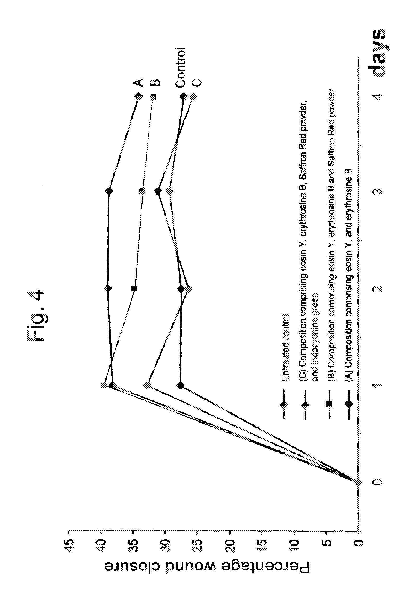
FIG. 4 illustrated that wound closure is improved following application of wound healing compositions. Rats (n=2 per group) with an excision wound were treated or not with wound healing composition (A) comprising an oxidant (carbamide peroxide) and a photoactivator mixture (eosin Y, erythrosine B), wound healing composition (B) comprising an oxidant (carbamide peroxide) and a photoactivator mixture (eosin Y, erythrosin B and Saffron Red powder) or wound healing composition (C) comprising an oxidant (carbamide peroxide) and a photoactivator mixture (eosin Y, erythrosine B, Saffron Red powder and Indocyanin green).
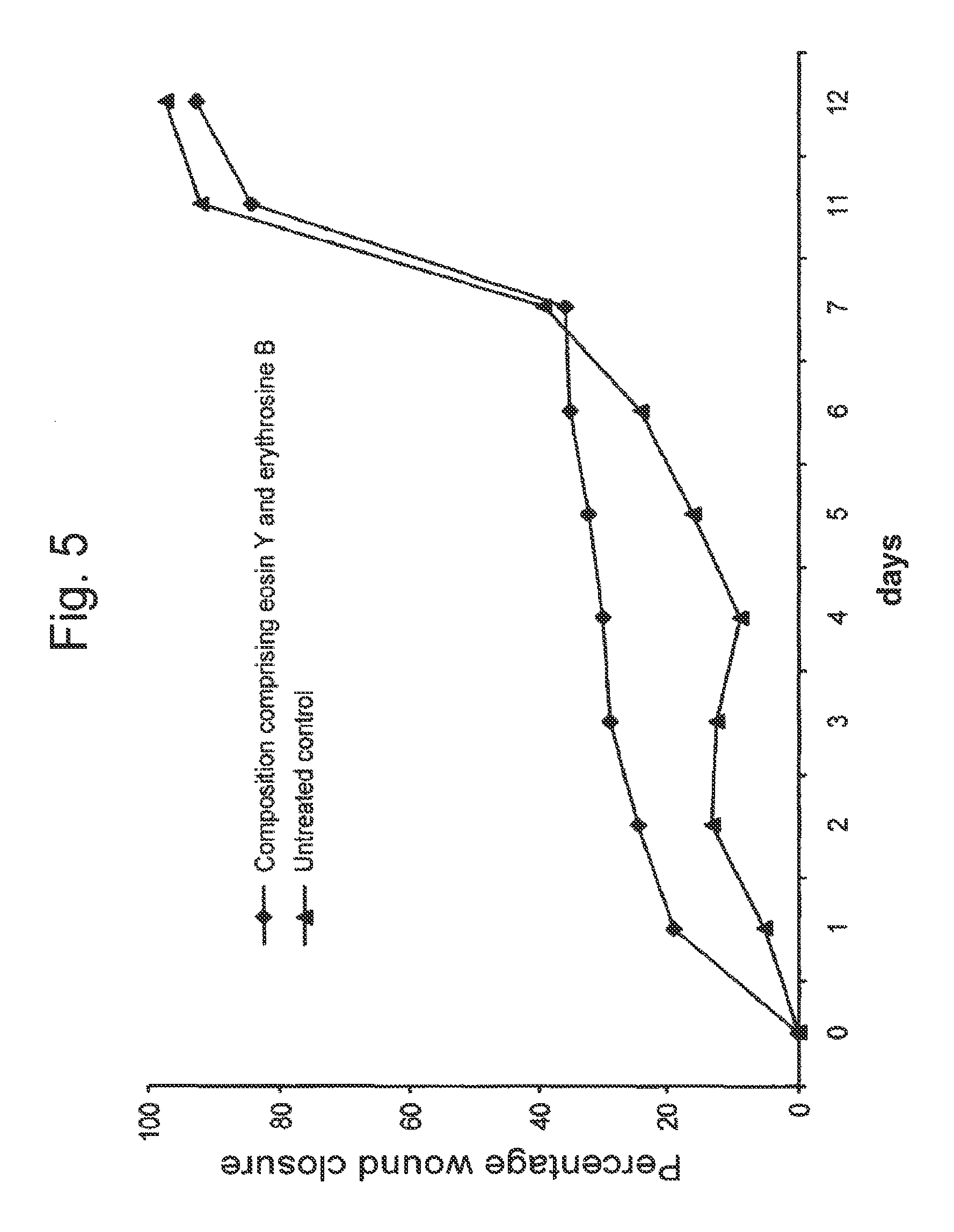
FIG. 5 illustrates that wound closure is improved following application of a wound healing composition. Rats (n=2 per group) with an excision wound were treated or not with a wound healing composition comprising an oxidant (carbamide peroxide) and a photoactivator mixture (eosin Y, erythrosine B). Lozenges: treated animals; triangles: untreated animals (controls).
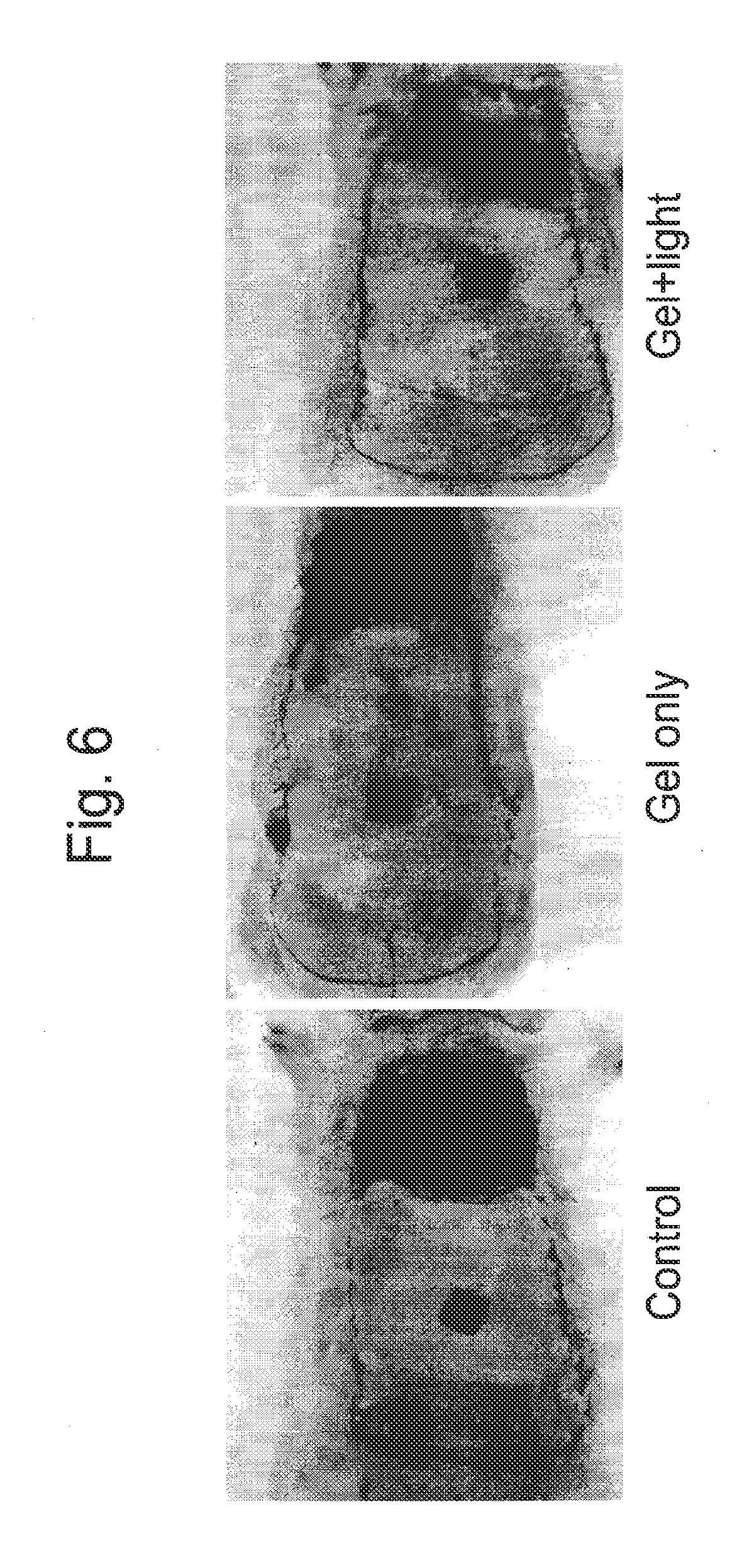
FIG. 6 illustrates that wound closure is improved following application of a wound healing composition.
FIG. 7 illustrates that wound healing is improved following application of a wound healing composition.
DETAILED DESCRIPTION OF THE PREFERRED EMBODIMENTS
In accordance with one embodiment, there is provided a wound healing composition and the method of use of the composition on a patient's skin or wound. This product accelerate healing and restoration of a wound.
In accordance with another embodiment, there is provided a method of use of the composition that is a photodynamic technique whereby the composition is activated by light providing a beneficial effect on the skin or wound and promoting healing.
The composition and method may be used to treat injuries to the different layers of the skin, including incisions, lacerations, abrasions, puncture wounds, penetrations wounds, gunshot wounds, contusions, hematomas and crushing injuries. Lesions to muccosae may also be treated with the composition of the present invention, which may be used for example, to treat pathological lesions of the oral mucosa, such as periodontitis, ulcers, and cold sores (orafacial herpes).
The composition comprises a number of active principles selected from groups of possible components. These various active principles each have their mechanism of action.
Oxidants
The composition comprises oxidants as a source of oxygen radicals. Peroxide compounds are oxidants that contain the peroxy group (R--O--O--R) which is a chainlike structure containing two oxygen atoms, each of which is bonded to the other and a radical or some element. Suitable oxidants for preparation of the active medium include, but are not limited to:
Hydrogen peroxide (H.sub.2O.sub.2) is the starting material to prepare organic peroxides. H.sub.2O.sub.2 is a powerful oxidizing agent, and the unique property of hydrogen peroxide is that it breaks down into water and oxygen and does not form any persistent, toxic residual compound. Hydrogen peroxide for use in this composition can be used in a gel, for example with 6% hydrogen peroxide A suitable range of concentration over which hydrogen peroxide can be used in the present composition is from about 3.5% to about 6%.
Urea hydrogen peroxide (also known as urea peroxide carbamide peroxide or percarbamide) is soluble in water and contains approximately 35% hydrogen peroxide. Carbamide peroxide for use in this composition can be used as a gel, for example with 16% carbamide peroxide that represents 5.6% hydrogen peroxide. A suitable range of concentration over which urea peroxide can be used in the present composition is from about 10% to about 16%. Urea peroxide brakes down to urea and hydrogen peroxide in a slow-release fashion that can be accelerated with heat or photochemical reactions. The released urea [carbamide, (NH.sub.2)], is highly soluble in water and is a powerful protein denaturant. It increases solubility of some proteins and enhances rehydration of the skin and/or mucosa.
Benzoyl peroxide consists of two benzoyl groups (benzoic acid with the H of the carboxylic acid removed) joined by a peroxide group. It is found in treatments for acne, in concentrations varying from 2.5% to 10%. The released peroxide groups are effective at killing bacteria. Benzoyl peroxide also promotes skin turnover and clearing of pores, which further contributes to decreasing bacterial counts and reduce acne. Benzoyl peroxide breaks down to benzoic acid and oxygen upon contact with skin, neither of which are toxic. A suitable range of concentration over which benzoyl peroxide can be used in the present composition is from about 2.5% to about 5%.
Inclusion of other forms of peroxides (e.g. organic or inorganic peroxides) should be avoided due to their increased toxicity and their unpredictable reaction with the photodynamic energy transfer.
Photoactivators:
The photoactivators transfer light energy to the oxidants. Suitable photoactivators can be fluorescent dyes (or stains), although other dye groups or dyes (biological and histological dyes, food colorings, carotenoids) can also be used. Combining photoactivators may increase photo-absorbtion by the combined dye molecules and enhance absorption and photo-biomodulation selectivity. This creates multiple possibilities of generating new photosensitive, and/or selective photoactivator mixtures.
A advantageous characteristic of a photoactivator is increased fluorescence. In the present invention, re-emission of light in the green to yellow spectrum would be advantageous, since it is a deep penetrating wavelength range, with deep absorption by the blood. This confers a strong increase on the blood flow, vasodilation and angiokinetic phenomena. Suitable photoactivators include, but are not limited to:
Xanthene Derivatives:
The xanthene derivative dyes have been used and tested for a long time worldwide. They display low toxicity and increased fluorescence. The xanthene group consists of 3 sub-groups that are: a) the fluorenes; b) fluorones; and c) the rhodoles.
The fluorenes group comprises the pyronines (e.g. pyronine Y and B) and the rhodamines (e.g. rhodamine B, G and WT). Depending on the concentration used, both pyronines and rhodamines may be toxic and their interaction with light may lead to increased toxicity. Similar effects are known to occur for the rhodole dye group.
The fluorone group comprises the fluorescein dye and the fluorescein derivatives.
Fluorescein is a fluorophore commonly used in microscopy with an absorption max of 494 nm and an emission max. of 521 nm. The disodium salt of fluorescein is known as D&C Yellow 8. It has very high fluorescence but photodegrades quickly. In the present composition, mixtures of fluorescein with other photoactivators such as indocyanin green and/or saffron red powder will confer increased photoabsorption to these other compounds.
Eosins group comprises Eosin Y (tetrabromofluorescein, acid red 87, D&C Red 22) with an abs max 514-518 nm, stains cytoplasm of cells, collagen, muscle fibers and red blood cells intensely red; and Eosin B (acid red 91, eosin scarlet, dibromo-dinitrofluorescein), with the same staining characteristics as Eosin Y. Eosin Y, eosin B, or a mixture of both can be used because of their sensitivity to the light spectra used: broad spectrum blue light, blue to green light and green light. Their tissue and biofilm staining properties and their low toxicity are also advantageous. Both eosin Y and eosin B stain red blood cells and thus confer to the composition of the present invention haemostatic (controls the flow or stops the flow of blood) properties as well as increase the selective targeting of light to the soft tissues of the lesion or wound during the application of the composition.
Phloxine B (2,4,5,7 tetrabromo 4,5,6,7, tetrachlorofluorescein, D&C Red 28, acid red 92) is a red dye derivative of fluorescein which is used for disinfection and detoxification of waste water through photooxidation. It has an abs. max. of 535-548 nm. It is also used as an intermediate for making photosensitive dyes and drugs.
Erythrosine B (acid red 51, tetralodofluorescein) is a cherry-pink coal-based fluorine food dye used as a biological stain, and a biofilm and dental plaque disclosing agent, with max. abs. 524-530 nm in aqueous solution. It is subject to photodegradation. Erythrosine is also used in some embodiments due to its photosensitivity to the light spectra used and its ability to stain biofilms. Inclusion of erythrosine should be favored when using the composition in deep pockets of infected or contaminated tissue, such as periodontal pockets in periodontal therapy.
Rose Bengal (4,5,6,7 tetrachloro 2,4,5,7tetraiodofluorescein, acid red 94) is a bright bluish-pink biological dye with an absorption max of 544-549 nm, that has been used as a dye, biological stain and diagnostic aid. Also used in synthetic chemistry to generate singlet from triplet oxygen.
Merbromine (mercurochrome) is an organo-mercuric disodium salt of fluorescein with an abs. max. of 508 nm. It is used as an antiseptic.
Azo Dyes:
The azo (or diazo-) dyes share the N-N group, called azo the group. They are used mainly in analytical chemistry or as food colorings and are not fluorescent. Suitable azo dyes include: Methyl violet, neutral red, para red (pigment red 1), amaranth (Azorubine S), Carmoisine (azorubine, food red 3, acid red 14), allura red AC (FD&C 40), tartrazine (FD&C Yellow 5), orange G (acid orange 10), Ponceau 4R (food red 7), methyl red (acid red 2), murexide-ammonium purpurate.
Biological Stains:
Dye molecules commonly used in staining protocols for biological materials can also be used as photoactivators. Suitable biological stains include:
Saffranin (Saffranin O, basic red 2) is also an azo-dye and is used in histology and cytology. It is a classic counter stain in a Gram stain protocol.
Fuchsin (basic or acid) rosaniline hydrochloride) is a magenta biological dye that can stain bacteria and has been used as an antiseptic. It has an abs. max. 540-555 nm.
3,3' dihexylocarbocyanine iodide (DiOC6) is a fluorescent dye used for staining cell's endoplasmic reticulum. vesicle membranes and mitochondria. It shows photodynamic toxicity; when exposed to blue light, has a green fluorescence.
Carminic acid (acid red 4, natural red 4) is a red glucosidal hydroxyanthrapurin naturally obtained from cochineal insects.
Indocyanin green (ICG) is used as a diagnostic aid for blood volume determination, cardiac output, or hepatic function. ICG binds strongly to red blood cells and when used in mixture with fluorescein, it increases the absorption of blue to green light.
Caroteriods
Carotenoid dyes can also act as photoactivators.
Saffron red powder is a natural carotenoid-containing compound. Saffron is a spice derived from crocus sativus. It is characterized by a bitter taste and iodoform or hay-like fragrance; these are caused by the compounds picrocrocin and saffranal. It also contains the carotenoid dye crocin that gives its characteristic yellow-red color.
Saffron contains more that 150 different compounds many of them are carotenoids: mangicrocin, reaxanthine; lycopene, and various .alpha.- and .beta.-carotenes, that show good absorption of light and beneficial biological activity. Also saffron can act as both a photo-transfer agent and a healing factor. Saffron color is primarily the result of .alpha.-crocin (8,8 diapo-8,8-carotenoid acid). Dry saffron red powder is highly sensitive to fluctuating pH levels and rapidly breaks down chemically in the presence of light and oxidizing agents. It is more resistant to heat. Data show that saffron has anticarcinogenic, immunomodulating and antioxidant properties. For absorbance, it is determined for the crocin specific photon wavelength of 440 nm (blue light). It has a deep red colour and forms crystals with a melting point of 186.degree. C. When dissolved in water it forms an orange solution.
Crocetin is another compound of saffron that was found to express an antilipidermic action and promote oxygen penetration in different tissues. More specifically it was observed an increased oxygenation of the endothelial cells of the capillaries. An increase of the oxygenation of muscles and cerebral cortex was observed and led to an increased survival rate in laboratory animals with induced hemorrhagic shock or emphysema.
Anatto a spice contains as main constituent (70-80%) the carotenoid bixin which displayed relevant antioxidative properties.
.beta.-carotene, also displayed suitable characteristics.
Fucoxanthine is a constituent of brown algae with a pronounced ability for photosensitization of red-ox reactions.
Healing Factors:
Healing factors comprise compounds that promote or enhance the healing or regenerative process of the tissues on the application site of the composition. During the photoactivation of the composition, there is an increase of the absorption of molecules at the treatment site by the skin or the mucosa. An augmentation in the blood flow at the site of treatment is observed for an extent period of time. An increase in the lymphatic drainage and a possible change in the osmotic equilibrium due to the dynamic interaction of the free radical cascades can be enhanced or even fortified with the inclusion of healing factors. Suitable healing factors include, but are not limited to:
Hyaluronic acid (Hyaluronan, hyaluronate): is a non-sulfated glycosaminoglycan, distributed widely throughout connective, epithelial and neural tissues. It is one of the primary components of the extracellular matrix, and contributes significantly to cell proliferation and migration. Hyaluronan is a major component of the skin, where it is involved in tissue repair. While it is abundant in extracellular matrices, it contributes to tissues hydrodynamics, movement and proliferation of cells and participates in a wide number of cell surface receptor interactions, notably those including primary receptor CD44. The hyaluronidases enzymes degrade hyaluronan. There are at least seven types of hyaluronidase-like enzymes in humans, several of which are tumor suppressors. The degradation products of hyaluronic acid, the oligosaccharides and the very-low molecular weight hyaluronic acid, exhibit pro-angiogenic properties. In addition, recent studies show that hyaluronan fragments, but not the native high molecular mass of hyaluronan, can induce inflammatory responses in macrophages and dendritic cells in tissue injury. Hyaluronic acid is well suited to biological applications targeting the skin. Due to its high biocompatibility, it is used to stimulate tissue regeneration. Current studies evidenced hyaluronic acid appearing in the early stages of healing to physically create room for white blood cells that mediate the immune response. It is used in the synthesis of biological scaffolds for wound healing applications and in wrinkle treatment.
Glucosamine: is one of the most abundant monosaccharides in human tissues and a precursor in the biological synthesis of glycosilated proteins and lipids. It is commonly used in the treatment of osteoarthritis. The common form of glucosamine used is its sulfate salt. Glucosamine shows a number of effects including an anti-inflammatory activity, stimulation of the synthesis of proteoglycans and the synthesis of proteolytic enzymes. A suitable range of concentration over which glucosamine can be used in the present composition is from about 1% to about 3%.
Allantoin: is a diureide of glyosilic acid. It has keratolytic effect, increases the water content of the extracellular matrix, enhances the desquamation of the upper layers of dead (apoptotic) skin cells, and promotes skin proliferation and wound healing.
Also, saffron can act as both a photo-transfer agent and a healing factor.
Chelating Agents:
Chelating agents can be included to promote smear layer removal in closed infected pockets and difficult to reach lesions; act as a metal ion quencher and as a buffer. Suitable chelating agents include, but are not limited to:
Ethylenediaminotetraacetic acid (EDTA): It is an aminoacid, used to sequester di- and trivalent metal ions. EDTA binds to metals via 4 carboxylate and 2 amine groups. EDTA forms especially strong complexes with Mn(III), Fe(III), Cu(III), Co(III). Prevents collection of the platelets and blood clots formation. It is used in the endodontic therapy as a smear layer removal agent during instrumentation. It is used to buffer solutions.
Ethylene glycol tetraacetic acid (EGTA) is related to EDTA, but with a much higher affinity for calcium than for magnesium ions. It is useful for making buffer solutions that resemble the environment inside living cells and is often employed in dentistry, more specifically endodontics, in the removal of smear layer.
Lipolysis Stimulation Factors:
Lipolysis stimulating factors can be included for use of the composition in cosmetic applications, such as wrinkle treatment.
Caffeine, and the metabolic derivative of caffeine paraxanthine can increase in the lipolysis process to releases glycerol and fatty acids into the blood stream.
Hydrophilic Gelling Agents
The wound healing composition may also contain one or more hydrophilic gelling agent. The hydrophilic gelling agent enhances the consistency of the composition and contributes to facilitating the application of the composition to the skin or wounded area. Also, when used with hydrogen peroxide (H.sub.2O.sub.2), it may contribute to the slow the release of the H.sub.2O.sub.2, and provide a more immediate reaction because pure H.sub.2O.sub.2 can be used directly. Suitable hydrophilic gelling agent include, but are not limited to glucose, modified starch, methyl cellulose, carboxymethyl cellulose, propyl cellulose, hydroxypropyl cellulose, carbopol.RTM. polymers, alginic acid, sodium alginate, potassium alginate, ammonium alginate, calcium alginate, agar, carrageenan, locust bean gum, pectin, and gelatin.
Use of the Composition
The inclusion of suitable photosensitive compounds and activation with a light source of a proper wavelength, leads to the acceleration in the breakdown process of the source of peroxide (the oxidant) and the other reactions that take place, via a photodynamic phenomenon. The included dyes are illuminated by photons of a certain wavelength and excited to a higher energy state. When the photoactivators' excited electrons return to a lower energy state, they emit photons with a lower energy level, thus causing the emission of light of a longer wavelength (Stokes shift). In the proper environment, much of this energy transfer is transferred to oxygen or the reactive hydrogen peroxide and causes the formation of oxygen radicals, such as singlet oxygen.
The singlet oxygen and other reactive oxygen species generated by the activation of the composition are thought to operate in a hormetic fashion. That is, a health beneficial effect is brought about by the low exposure to a normally toxic stimuli (e.g. reactive oxygen), by stimulating and modulating stress response pathways in cells of the targeted tissues. Endogenous response to exogenous generated free radicals (reactive oxygen species) is modulated in increased defense capacity against the exogenous free radicals and induces acceleration of healing and regenerative processes. Furthermore, activation of the composition will also produce an antibacterial effect. The extreme sensitivity of bacteria to exposure to free radicals makes the composition of the present invention a de facto bactericidal composition.
Possible mechanism of action should be a fortified redox signalling phenomenon resulting in accentuated signal transduction process in which cells convert one kind of signal into another; activated "second messengers" induce a "signal cascade" beginning with a relatively small stimulus that elicits a large response via biologically monitored amplification of such signals. These complex mechanisms act possibly involving angiogenic phenomena via growth factor activation.
This method could be described as a form of photodynamic therapy. However, unlike other photodynamic techniques, where the photoactoactivators are incorporated in the tissue structure. In the present method, the photoactive material is in simple contact with the tissue and acts when activated by light, as a "photodynamic device" that chemically interacts with the tissue. Additionally, the actinic light penetrates the tissue, and the light that is emitted by the photoactivator (light of a longer wavelength) is also absorbed by the tissue.
Any source of actinic light can be used. Any type of halogen, LED or plasma arc lamp, or laser may be suitable. The primary characteristic of suitable sources of actinic light will be that they emit light in a wavelength (or wavelengths) appropriate for activating the one or more photoactivators present in the composition. In one embodiment, an argon laser is used. In another embodiment, a potassium-titanyl phosphate (KTP) laser (e.g. a GreenLight.TM. laser) is used. In yet another embodiment, a LED photocuring device is the source of the actinic light. In yet another embodiment, the source of the actinic light is a source of visible light having a wavelength between 400 and 600 nm. Furthermore, the source of actinic light should have a suitable power density. Suitable power density for non-collimated light sources (LED, halogen or plasma lamps) are in the range from about 900 mW/cm.sup.2 to about 2000 mW/cm.sup.2. Suitable power density for laser light sources are in the range from about 0.5 mW/cm.sup.2 to about 0.8 mW/cm.sup.2.
The duration of the exposure to actinic light will be dependent on the surface of the treated area, and on the type of lesion, trauma or injury that is being treated. The photoactivation of the composition may take place within seconds or even fragment of seconds, but a prolonged exposure period is beneficial to exploit the synergistic effects of the absorbed, reflected and reemitted light on the composition of the present invention and its interaction with the tissue being treated. In one embodiment, the time of exposure to actinic light of the tissue, skin or wound on which the wound healing composition has been applied is a period between 60 second and 5 minutes. In another embodiment, the time of exposure to actinic light of the tissue, skin or wound on which the wound healing composition has been applied is a period between 60 seconds and 5 minutes per cm.sup.2 of the area to be treated, so that the total time of exposure of a 10 cm.sup.2 are would be between 10 minutes and 50 minutes. In yet another embodiment, the source of actinic light is in continuous motion over the treated area for the appropriate time of exposure. In yet another embodiment, multiple applications of the wound healing composition and actinic light are performed. In some embodiments, the tissue, skin or wound is exposed to actinic light at least two, three, four, five or six times. In some embodiments, a fresh application of the wound healing composition is applied before exposure to actinic light.
Alternative Embodiments
EXAMPLE I
An exemplary wound healing composition was prepared by mixing the following components:
TABLE-US-00001 Oxidant Photoactivators Healing factor(s) Carbamide Erythrosine B (0.5%) Glucosamine sulfate (3%) peroxide Eosin B (0.25%) Hyaluronic acid (3%) (16%) Saffron Red powder (0.25%)
The oxidant (4 mL) and healing factors (1.5 mL) were mixed and the combined with the photoactivators (1 mL). The resulting composition was applied to the skin of a wounded patient, and activated with actinic light provided by a LED photocuring device (blue light). The composition was removed following treatment.
EXAMPLE II
An second exemplary wound healing composition was prepared by mixing the following components:
TABLE-US-00002 Oxidant Photoactivators Healing factor(s) Carbamide Fluorescein Glucosamine sulfate (3%) peroxide Indocyanin green Hyaluronic acid (3%) (16%) Saffron Red powder (0.25%)
The oxidant (4 mL) and healing factors (1.5 mL) were mixed and the combined with the photoactivators (1 mL). The resulting composition was applied to the skin of a wounded patient, and activated with actinic light provided by a LED photocuring device (blue light). The composition was removed following treatment.
This second exemplary composition is using the fluorescein dye as a photoactivator to other dyes (indocyanine green and saffron red powder) present in the composition. The addition of a small amount of fluorescein to the indocyanine green and saffron red powder solution caused reemission of light at wavelengths that activated the other dye compounds and improved the treatment by increasing the established clinical absorption/reemission criteria.
Indocyanine green binds well to hemoglobin and helps the selective energy absorption by the tissues and also helps targeting these tissues with the generated free radical cascades. Also, this photoactivators mixture is able to render saffron red fluorescent, which again improves both the photodynamic and biostimulating phenomena.
EXAMPLE III
The toxicity of the photoactivators Eosin Y and Erythrosine B was evaluated by measuring the cytotoxicity of these compounds on human cells. Hep G2 human hepatocellular carcinoma cells with an epithelial morphology were treated for 24 hours with increasing concentrations (0.001 to 100 .mu.M) of Eosin Y or Erythrosine B, and the cellular survival was evaluated. Increasing concentrations of either Eosin Y (FIG. 1) or Erythrosine B (FIG. 2) did not affect cellular viability when compared to untreated cells. Straurosporine (STS) was used as a positive control for inducing cellular mortality and caused a dose-dependent effect (FIGS. 1 and 2). Similar results were obtained by measuring cell death by release of lactate dehydrogenase (LDH). Therefore, neither Eosin Y or Erythrosine B caused increased cellular mortality.
EXAMPLE IV
Excisional Wound Model in Pat
Random skin flaps in rat were used to study the wound healing procedures, for evaluating the benefits of ischemic and pharmacologic preconditioning methods on skin flap survival, applying blood flow assessment technologies on flaps, demonstrating the effects of vascular shunts and the studies on skin flap viability. The random skin flap model was used to study the effect of the comosition of the present invention on skin flap survival and associated modulations contributing to healing process.
Excision wound of 1 cm in width by 2 cm in length were cut dorsally on the midline of the back, 2 cm below the interior angle of the scapulae. The skin was cut with a surgical blade, the panniculus carnosus and a 0.5 cm layer subcutaneous to the panniculus camosus was excised from the wound edges. The wound was next photographed with an 8 mm by 8 mm size marker. One gram of the wound healing composition was applied to the wound (0.5 g/cm.sup.2) and irradiated with a blue LED light during 3 minutes.
EXAMPLE V
Excisions were performed on rats (n=2 per group) as described above in Example IV, and the excisions were treated or not with a single application of 1 gram of a wound healing composition comprising the oxidant (carbamide peroxide) and a photoactivator mixture containing eosin Y, erythrosine B and saffron red powder. The excisions were irradiated with a LED light (blue light) for 3 minutes. The percentage of wound closure was evaluated (FIG. 3) over a ten-day period following treatment. Animals treated with the composition showed a more rapid wound closure over the initial first three-day period following treatment.
EXAMPLE VI
Excision were performed on rats (n=2 per group) as described above in Example IV, and the excisions were treated or not with a single application of 1 gram of a wound healing compositions comprising: (A) the oxidant (carbamide peroxide) and a photoactivator mixture containing eosin Y, and erythrosine B; (B) the oxidant (carbamide peroxide) and a photoactivator mixture containing eosin Y, erythrosine B and saffron red powder, or (C) the oxidant (carbamide peroxide) and a photoactivator mixture containing eosin Y. erythrosine B, saffron red powder and indocyanine green. The excisions were irradiated with a LED light (blue light) for 3 minutes. The percentage of wound closure was evaluated daily for four days (FIG. 4). Animals treated with compositions (A) and (B) showed improved wound closure over the four-day period following treatment. The addition of indocyanine green in composition (C) repressed the wound healing effect observed for compositions (A) and (B).
EXAMPLE VII
Excision were performed on rats (n=2 per group) as described above in Example IV, and the excisions were treated or not with a single application of 1 gram of a wound healing composition comprising the oxidant (carbamide peroxide) and a photoactivator mixture containing eosin Y and erythrosine B. The excisions were irradiated with a LED light (blue light) for 3 minutes. The percentage of wound closure was evaluated (FIG. 5) over a twelve-day period. Animals treated with the composition showed a more rapid wound closure over the first seven-day period following treatment.
EXAMPLE VIII
Using a template of 3 cm by 9 cm (3.times.9) in plexiglas, a flap was traced on the dorsal skin with a surgical marker, taking as limits the inferior angles of the scapulae and the superior bones of pelvis. A pure random pattern flap with cranial base was cut using sterile techniques and elevated through deep fascia, including superficial fascia, panniculus carnosus, the subcutaneous tissue and skin. To minimize wound contraction and simulating the human condition, a 0.5 cm subcutaneous layer of panniculus carnosus from the wound edges was removed. During one hour, an impermeable barrier (e.g. a silicone sheeting) was placed between the flap and its donor site to eliminate the possibility of wound bed support. The sheet was then removed, the flap returned to its original position and the flap edges were surgically closed using 4/0 nylon suture in an interrupted fashion. Immediately following flap closure, flap pedicle were coated with 13.5 g of gel formulation (0.5 g/cm.sup.2) and irradiated. The controls did not receive any treatment. Care was taken to distribute ointment evenly along the entire flap. Gel formulation was prepared the same day of the experiment. For Gel+Light group the animals were treated with gel formulation, the flap was irradiated for 3 minutes with a LED lamp.
EXAMPLE IX
Excision were performed on rats (n=2 per group) as described above, and the excisions were treated or not with the gel formulation, and irradiated with a LED light (blue light) as described in Example VIII.
The results demonstrate a direct correlation of necrosis from fluorescein injection and direct visualization. Biopsies were evaluated for changes in histology. Data from the treated group demonstrate a clinically significant 1.5 times reduction in necrosis, (percent necrosis, mean, SD of 45.7 (.+-.17.36) vs. 30.42 (.+-.20.18), in the control and treatment groups respectively). Now referring to FIG. 7A, the clinical evaluation of necrosis following flap surgery, in the control and treatment group shows that higher necrosis is observed on the control group versus the treatment group.
Hematoxylin and eosin staining of biopsies from the control and treatment group (FIG. 7B) reveal that greater vascular recruitment occurs in the treated group (see black arrows therein). Masson Trichrome staining for the assessment of collagen fibril deposition (FIG. 7C) at a 40.times. magnification shows that new collagen deposition is occurring in the treatment group vs the control group. The photodynamic treatment using photoactivators and wavelength specific light aimed at increasing the viability of the skin flap by stimulating the vascular recruitment of collaterals in flaps to improve the loco-regional state of the new wound, including the formation of new collagen therein.
The embodiments and examples presented herein are illustrative of the general nature of the subject matter claimed and are not limiting. It will be understood by those skilled in the art how these embodiments can be readily modified and/or adapted for various applications and in various ways without departing from the spirit and scope of the subject matter disclosed claimed. The claims herof are to be understood to include without limitation all alternative embodiments and equivalents of the subject matter hereof. Phrases, words and terms employed herein are illustrative and are not limiting. Where permissible by law, all references cited herein are incorporated by reference in their entirety. It will be appreciated that any aspects of the different embodiments disclosed herein may be combined in a range of possible alternative embodiments, and alternative combinations of features, all of which varied combinations of features are to be understood to form a part of the subject matter claimed.
* * * * *
References
D00001
D00002
D00003
D00004
D00005
D00006
D00007
XML
uspto.report is an independent third-party trademark research tool that is not affiliated, endorsed, or sponsored by the United States Patent and Trademark Office (USPTO) or any other governmental organization. The information provided by uspto.report is based on publicly available data at the time of writing and is intended for informational purposes only.
While we strive to provide accurate and up-to-date information, we do not guarantee the accuracy, completeness, reliability, or suitability of the information displayed on this site. The use of this site is at your own risk. Any reliance you place on such information is therefore strictly at your own risk.
All official trademark data, including owner information, should be verified by visiting the official USPTO website at www.uspto.gov. This site is not intended to replace professional legal advice and should not be used as a substitute for consulting with a legal professional who is knowledgeable about trademark law.