Thrombin-binding antibody molecules and uses thereof
Huntington , et al.
U.S. patent number 10,370,454 [Application Number 15/976,595] was granted by the patent office on 2019-08-06 for thrombin-binding antibody molecules and uses thereof. This patent grant is currently assigned to JANSSEN PHARMACEUTICALS, INC.. The grantee listed for this patent is Janssen Pharmaceuticals, Inc.. Invention is credited to Trevor Baglin, James Andrew Huntington, Jonathan Langdown.


View All Diagrams
| United States Patent | 10,370,454 |
| Huntington , et al. | August 6, 2019 |
Thrombin-binding antibody molecules and uses thereof
Abstract
This invention relates to isolated antibodies which recognize the exosite 1 epitope of thrombin and selectively inhibit thrombin without promoting bleeding. These antibody molecules may be useful in the treatment and prevention of thrombosis, embolism and other conditions mediated by thrombin.
| Inventors: | Huntington; James Andrew (Cambridge, GB), Baglin; Trevor (Cambridge, GB), Langdown; Jonathan (Cambridge, GB) | ||||||||||
|---|---|---|---|---|---|---|---|---|---|---|---|
| Applicant: |
|
||||||||||
| Assignee: | JANSSEN PHARMACEUTICALS, INC.
(Titusville, NJ) |
||||||||||
| Family ID: | 51654618 | ||||||||||
| Appl. No.: | 15/976,595 | ||||||||||
| Filed: | May 10, 2018 |
Prior Publication Data
| Document Identifier | Publication Date | |
|---|---|---|
| US 20180334512 A1 | Nov 22, 2018 | |
Related U.S. Patent Documents
| Application Number | Filing Date | Patent Number | Issue Date | ||
|---|---|---|---|---|---|
| 15434639 | Feb 16, 2017 | 9988461 | |||
| 15206896 | Mar 28, 2017 | 9605082 | |||
| 14309403 | Dec 13, 2016 | 9518128 | |||
| 14363514 | Dec 13, 2016 | 9518129 | |||
| PCT/GB2012/053140 | Dec 14, 2012 | ||||
Foreign Application Priority Data
| Dec 14, 2011 [GB] | 1121513.4 | |||
| Current U.S. Class: | 1/1 |
| Current CPC Class: | C07K 16/40 (20130101); C07K 16/36 (20130101); C07K 2317/55 (20130101); C07K 2317/92 (20130101); C07K 2317/34 (20130101); C07K 2317/626 (20130101); C07K 2317/94 (20130101); C07K 2317/14 (20130101); C07K 2317/21 (20130101); C07K 2317/41 (20130101); C07K 2317/565 (20130101); A61K 2039/505 (20130101); C07K 2317/56 (20130101); C07K 2317/76 (20130101); C07K 2317/52 (20130101) |
| Current International Class: | C07K 16/36 (20060101); A61K 39/395 (20060101); C07K 16/40 (20060101); A61K 39/00 (20060101) |
References Cited [Referenced By]
U.S. Patent Documents
| 5196404 | March 1993 | Maraganore et al. |
| 5240913 | August 1993 | Maraganore et al. |
| 5433940 | July 1995 | Maraganore et al. |
| 5530101 | June 1996 | Queen et al. |
| 5985833 | November 1999 | Mosesson et al. |
| 7998939 | August 2011 | Diener et al. |
| 8568724 | October 2013 | Hack |
| 9518128 | December 2016 | Huntington |
| 9518129 | December 2016 | Huntington |
| 9605082 | March 2017 | Huntington et al. |
| 9988461 | June 2018 | Huntington et al. |
| 9988463 | June 2018 | Huntington et al. |
| 2014/0302047 | October 2014 | Huntington et al. |
| 0489070 | Jun 1992 | EP | |||
| 0529031 | Mar 1993 | EP | |||
| 2791177 | Oct 2014 | EP | |||
| 3275903 | Jan 2018 | EP | |||
| 91/17258 | Nov 1991 | WO | |||
| 92/01047 | Jan 1992 | WO | |||
| 94/25491 | Nov 1994 | WO | |||
| 95/17907 | Jul 1995 | WO | |||
| 98/00443 | Jan 1998 | WO | |||
| 01/00667 | Jan 2001 | WO | |||
| 01/07072 | Feb 2001 | WO | |||
| 02/17711 | Mar 2002 | WO | |||
| 03/03988 | Jan 2003 | WO | |||
| 2007/106893 | Sep 2007 | WO | |||
| 2008/155658 | Dec 2008 | WO | |||
| 2010/033167 | Mar 2010 | WO | |||
| 2013/014092 | Jan 2013 | WO | |||
| 2013/088164 | Jun 2013 | WO | |||
Other References
|
Baglin et al., J Thromb Haemost. Jan. 2016;14(1):137-42. doi: 10.1111/jth.13171. Epub Dec. 29, 2015. cited by examiner . Chang et al., Biochem Biophys Res Commun. Jun. 28, 1991;177(3):1198-204. cited by examiner . Wu et al., J Biol Chem. Feb. 4, 1994;269(5):3725-30. cited by examiner . Bagshawe et al., "Antibody-Enzyme Conjugates Can Generate Cytotoxic Drugs from Inactive Precursors at Tumor Sites.", Antibody, Immunoconjugates and Radiopharmaceuticals, 1991, pp. 915-922, vol. 4(4). cited by applicant . Baglin, T.P. et al., "Discovery and characterization of an antibody directed against exosite I of thrombin", Journal of Thrombosis and Haemostasis, 2016, 14:137-142. cited by applicant . Baerga-Ortiz et al., Protein Sci. Jun. 2002;11 (6):1300-8. cited by applicant . Baegra-Ortiz, A. et al., "Two different proteins that compete for binding to thrombin have opposite kinetic and thermodynamic profiles", Protein Science (2004), vol. 13, pp. 166-176. cited by applicant . Arnaud et al,, Blood. Sep. 15, 1994;84(6):1843-50. cited by applicant . Armitage et al., "Molecular and biological characterization of a murine ligand for CD40.", Nature, 1992, pp. 80-82, vol. 357(6373). cited by applicant . Anderson et al., "Characterization of Proexosite I on Prothrombin.", Journal of Biological Chemistry, Jun. 2, 2000, pp. 16428-16434, vol. 275(22). cited by applicant . Ahmed et al,, Curr Drug ther. Apr. 2016;11 (1 ):3-20. cited by applicant . Zehnder et al,, Blood. Nov. 15, 1990;76(10):2011-6. cited by applicant . Xu et al., "Diversity in the CDR3 Region of Vh is Sufficient for Most Antibody Specificities", Immunity, vol. 13, Jul. 2000, pp. 37-45. cited by applicant . Wu, Q. et al., "Activation-induced Exposure of the Thrombin Anion-binding Exosite", The Journal of Biological Chemistry (1994), vol. 269:5, pp. 3725-3730. cited by applicant . Wong, Y.W. et al., "Structural Requirements for a Specificity Switch and for Maintenance of Affinity Using Mutational Analysis of a Phage-Displayed Anti-Arsonate Antibody of Fab Heavy Chain First Complementarity-Determining Region", Journal of Immunology, 1998, 160:5990-5997. cited by applicant . Westrick et al., "Murine Models of Vascular Thrombosis.", Arteriosclerosis, Thrombosis, and Vascular Biology, 2007, pp. 2079-2093, vol. 27. cited by applicant . Tsopanoglou et al., "Thrombin's central role in angiogenesis and pathophysiological processes.", Eur Cytokine Netw., Dec. 1, 2009, pp. 171-179, vol. 20(4). cited by applicant . Tanaka, M. et al., "O-linked glucosylation of a therapeutic recombinant humanised monoclonal antibody produced in CHO cells", European Journal of Pharmaceutics and Biopharmaceutics (2013), vol. 83, pp. 123-130. cited by applicant . Tachibana, H. et al., "Building high affinity human antibodies by altering the glycosylation on the light chain variable region in N-acetylglucosamine-supplemented hybridoma cultures", Cytotechnology (1997), vol. 23, pp. 151-159. cited by applicant . Sokolova et al., Thromb Haemost. Oct. 2008;100(4):576-81. cited by applicant . Seiler, S.M. et al., "Multiple Pathways of Thrombin-Induced Platelet Activation Differentiated by Desensitization and a Thrombin Exosite Inhibitor", Biochemical and Biophysical Research Communications (1991), vol. 181:2, pp. 636-643. cited by applicant . Seiler, S.M. et al., "Involvement of the `Tethered-Ligand` Receptor in Thrombin Inhibition of Platelet Adenylate Cyclase", Biochemical and Biophysical Research Communications (1992), vol. 182:3, pp. 1296-1302. cited by applicant . Schumacher, W.A., et al., "Comparison of Thrombin Active Site and Exosite Inhibitors and Heparin in Experimental Models of Arterial and Venous Thrombosis and Bleeding", The Joumal of Pharmacology and Experimental Therapeutics (1993), vol. 267:3, pp. 1237-1242. cited by applicant . Rudikoff et al,, Proc Natl Acad Sci U S A. Mar. 1982;79(6):1979-83. cited by applicant . Response filed in European Patent Application No. 17177235.3 dated Jul. 23, 2018, 4 pages. cited by applicant . Qian, T., et al., "Structural characterization of N-linked oligosaccharides on monoclonal antibody cetuximab by the combination of orthogonal matrix-assisted laser desorption/ionization hybrid quadrupole-quadrupole time-of-flight tandem mass spectrometry and sequential enzymatic digestion", Analytical Biochemistry (2007), vol. 364, pp. 8-18. cited by applicant . Prasa et al., "Inhibition of Thrombin Generation in Plasma by Inhibitors of Factor Xa.", Thromb. Haemost., 1997, pp. 1215-1220, vol. 78. cited by applicant . Portolano et al,, J Immunol. Feb. 1, 1993;150(3):880-7. cited by applicant . Omtvedt, L.A. et al., "Glycosylation of immunoglobulin light chains associated with amyloidosis", Amyloid: Int. J. Exp. Clin. Invest. (2000), vol. 7, pp. 227-244. cited by applicant . Nimjee, S.M. et al. "Synergistic effect of aptamers that inhibit exosites 1 and 2 on thrombin", RNA (2009), vol. 15, pp. 2105-2111. cited by applicant . Naski et al., "The COOH-terminal Domain of Hirudin. The exosite-directed competitive inhibitor of the action of a-Thrombin on Fibrinogen.", Journal of Biological Chemistry, Aug. 15, 1990, pp. 3484-13489, vol. 265(23). cited by applicant . Mushunje, A. et al., "Heparin-induced substrate behavior of antithrombin Cambridge II", Blood (2003), vol. 02:12, pp. 4028-4034. cited by applicant . Mollica, et al., "Antibodies to thrombin directed against both of its cryptic exosites", British Journal of Haematology, 2005, 132: 487-493. cited by applicant . Moiseenko V.M. "Monoklonal'nye antitela .nu. lechenii zlokachestvennykh opukholey"., Prakticheskaya onkologiya, 2003, vol. 4, 3:148-156 (English Translation provided herewith). cited by applicant . Mackman, N., "Triggers, targets and treatments for thrombosis", Insight Review, Nature Publishing Group (2008), pp. 914-918. cited by applicant . Licari, L.G. et al., "Thrombin physiology and pathophysiology", Journal of Veterinary Emergency and Critical Care (2009), vol. 19:1, pp. 11-22. cited by applicant . Igawa et al., MAbs, May-Jun. 2011;3(3):243-52. cited by applicant . Ledermann et al., "A Phase-I Study of Repeated Therapy With Radiolabelled Antibody to Carcinoembryonic Antigen Using Intermittent or Continuous Administration of Cyclosporin A to Suppress the Immune Response.", International J. Cancer, 1991, pp. 659-664, vol. 47. cited by applicant . Lechtenberg, B.C. et al., "NMR resonance assignments of thrombin reveal the conformational and dynamic effects of ligation", PNAS (2010), vol. 107:32, pp. 14087-14092. cited by applicant . Krenzlin et al,, Int. J. Mol. Sci. 2016, 17(1), 84; doi:10.3390/ijms17010084. cited by applicant . Krenzlin et al., "The Importance of Thrombin in Cerebral Injury and Disease", Int J Mol Sci, 2016, 17(1), pp. 84. cited by applicant . Kontiola, A. et al., "Glycosyolation Pattern of Kappa Light Chains in Massive Cutaneous Hyalinosis", Glycoconjugate J. (1987), vol. 4, pp. 297-305. cited by applicant . Janeway et al., Immunobiology, 3rd ed., Current Biology, 1997, pp. 3:1-3:11. cited by applicant . Jackson, J.R. et al., "In Vitro Antibody Maturation, Improvement of a High Affinity, Neutralizing Antibody Against IL-1.beta.", The Journal of Immunology, 1995, 14:3310-3319. cited by applicant . Illustrated dictionary of Immunology, Julius M. Cruse, Robert E. Lewis.--2nd ed., CRC Press, 2003, pp. 37, 316-317. cited by applicant . Igawa et al,, MAbs. May-Jun. 2011;3(3):243-52. Epub May 1, 2011. cited by applicant . Hwang et al,, J Immunol. Dec. 15, 2001;167(12):7192-8. cited by applicant . Huntington, J.A. "Structural Insights into the Life History of Thrombin", chapter in Recent Advances in Thrombosis and Hemostasis (2008), pp. 80-106. cited by applicant . Huntington, J.A. "Molecular Recognition Mechanisms of Thrombin", Journal of Thrombosis and Haemostasis (2005), vol. 3, pp. 1861-1872. cited by applicant . Hollinger et al., "Engineered antibody fragments and the rise of single domains.", Nature Biotechnology, Sep. 2005, pp. 1126-1136, vol. 23(9). cited by applicant . Holliger et al,, Nat Biotechnol. Sep. 2005;23(9):1126-36. cited by applicant . Henikoff and Henikoff, "Amino acid substitution matrices from protein blocks.", Proc. Natl. Acad. Sci., Nov. 1992, pp. 10915-10919, vol. 88. cited by applicant . Guillin, M. et al., "Thrombin Specificity" Thrombosis and Haemostasis (1995), vol. 74:1, pp. 129-133. cited by applicant . Fundamental Immunology, W. Paul, ed., Raven Press, 1993, p. 242. cited by applicant . DeFeo et al., "Use of dabigatran etexilate to reduce breast cancer progression", Cancer Biol Ther, 2010, 10(10), pp. 1001-1008. cited by applicant . Costa,J.M. et al., "Partial Characterization of an Autoantibody Recognizing the Secondary Binding Site(s) of Thrombin in a Patient with Recurrent Spontaneous Arterial Thrombosis" Thrombosis and Haemostasis (1992), vol. 67:2, pp. 193-199. cited by applicant . Cook, J.J. et al., "An Antibody Against the Exosite of the Cloned Thrombin Receptor Inhibits Experimental Arterial Thrombosis in the African Green Monkey", Circulation (1995), vol. 91, pp. 2961-2971. cited by applicant . Chouhan et al., Thromb Haemost. Feb. 1997;77(2):343-9. cited by applicant . Chang, A.C. et al., The Reaction of Thrombin With Platelet-Derived Nexin Requires a Secondary Recognition Site in Addition to the Catalytic Site, Biochem. Biophys. Res. Comm. (1991), vol. 177:3, pp. 1198-1204. cited by applicant . Bode et al., "The refined 1.9 A crystal structure of human a-thrombin: interaction with D-Phe-Pro-Arg chloromethylketone and significance of the Tyr-Pro-Pro-Trp insertion segment.", The EMBO Journal, 1989, pp. 3467-3475, vol. 8(11). cited by applicant . Bernett, et al., Engineering Fully Human Monoclonal Antibodies from Murine Variable Regions; J. Mol. Biol. (2010), 396, 1474-1490. cited by applicant. |
Primary Examiner: Szperka; Michael
Attorney, Agent or Firm: BakerHostetler
Parent Case Text
CROSS REFERENCE TO RELATED APPLICATIONS
This application is a continuation of U.S. patent application Ser. No. 15/434,639, filed Feb. 16, 2017, now U.S. Pat. No. 9,988,461, which is a divisional of U.S. patent application Ser. No. 15/206,896, filed Jul. 11, 2016, now U.S. Pat. No. 9,605,082, which is a divisional of U.S. patent application Ser. No. 14/309,403, filed Jun. 19, 2014, now U.S. Pat. No. 9,518,128, which is a continuation-in-part of U.S. patent application Ser. No. 14/363,514, filed Jun. 6, 2014, now U.S. Pat. No. 9,518,129, which is an application filed under Section 371 of International Patent Application No. PCT/GB2012/053140, filed Dec. 14, 2012, which claims benefit of priority to GB 1121513.4, filed Dec. 14, 2011. The contents of these applications are hereby incorporated by reference in their entireties.
Claims
What is claimed:
1. An isolated or recombinant antibody molecule that specifically binds to the exosite 1 region of thrombin, wherein the antibody molecule inhibits thrombosis or embolism but does not promote bleeding, and wherein the antibody molecule binds amino acids Q38, R73, T74, and Y76 relative to the amino acid sequence of SEQ ID NO: 1, the antibody molecule comprising a variable heavy domain, a variable light domain, and an IgG constant region, wherein the variable heavy domain comprises a heavy chain complementarity determining region 3 (HCDR3) having the amino acid sequence of SEQ ID NO:5, a HCDR2 having the amino acid sequence of SEQ ID NO: 4, and a HCDR1 having the amino acid sequence of SEQ ID NO: 3, a variable light domain comprising a light chain complementarity determining region 1 (LCDR1) having the amino acid sequence of SEQ ID NO: 15, a LCDR2 having the amino acid sequence of SEQ ID NO: 8, and a LCDR3 having the amino acid sequence of SEQ ID NO: 9.
2. The antibody molecule of claim 1, wherein the antibody molecule further binds one or more amino acids selected from the group consisting of: M32, F34, S36a, P37, E39, L40, L65, R67, R75, R77a, I82 and Q151 relative to the amino acid sequence of SEQ ID NO: 1.
3. The antibody molecule of claim 1, wherein the antibody molecule further binds R77a relative to the amino acid sequence of SEQ ID NO: 1.
4. A pharmaceutical composition comprising the antibody molecule according to claim 1 and a pharmaceutically acceptable carrier or excipient.
5. The antibody molecule of claim 1 wherein the IgG constant region is an IgG1 constant region or an IgG4 constant region.
Description
REFERENCE TO A SEQUENCE LISTING
This application includes a Sequence Listing submitted electronically as a text file named "103693_001264_revSL.txt", created on Jul. 26, 2018 with a size of 10,615 bytes. The Sequence Listing is incorporated by reference herein.
TECHNICAL FIELD
This invention relates to antibody molecules that inhibit thrombin.
BACKGROUND
Blood coagulation is a key process in the prevention of bleeding from damaged blood vessels (haemostasis). However, a blood clot that obstructs the flow of blood through a vessel (thrombosis) or breaks away to lodge in a vessel elsewhere in the body (thromboembolism) can be a serious health threat.
A number of anticoagulant therapies are available to treat pathological blood coagulation. A common drawback of these therapies is an increased risk of bleeding (Mackman (2008) Nature 451(7181): 914-918). Many anticoagulant agents have a narrow therapeutic window between the dose that prevents thrombosis and the dose that induces bleeding. This window is often further restricted by variations in the response in individual patients.
SUMMARY
The present invention relates to the unexpected finding that antibody molecules which recognize the exosite 1 epitope of thrombin selectively inhibit thrombin without promoting bleeding. These antibody molecules may be useful in the treatment and prevention of thrombosis, embolism and other conditions mediated by thrombin.
The invention encompasses the following items:
1. An isolated antibody molecule that specifically binds to the exosite 1 region of thrombin.
2. The antibody molecule according to item 1 that inhibits thrombin activity.
3. The antibody molecule according to item 2 which causes minimal inhibition of haemostasis and/or bleeding.
4. The antibody molecule according to item 2 or item 3 which does not inhibit haemostasis and/or cause bleeding.
5. The antibody molecule according to any one of the preceding items wherein the antibody molecule comprises an HCDR3 having the amino acid sequence of SEQ ID NO: 5 or the amino acid sequence of SEQ ID NO: 5 with one or more amino acid substitutions, deletions or insertions.
6. The antibody molecule according to item 5 wherein the antibody molecule comprises an HCDR2 having the amino acid sequence of SEQ ID NO: 4 or the amino acid sequence of SEQ ID NO: 4 with one or more amino acid substitutions, deletions or insertions.
7. The antibody molecule according to item 5 or item 6 wherein the antibody molecule comprises an HCDR1 having the amino acid sequence of SEQ ID NO: 3 or the amino acid sequence of SEQ ID NO: 3 with one or more amino acid substitutions, deletions or insertions.
8. The antibody molecule according to any one of items 1 to 7 wherein the antibody molecule comprises a VH domain having the amino acid sequence of SEQ ID NO: 2 or the amino acid sequence of SEQ ID NO: 2 with one or more amino acid substitutions, deletions or insertions.
9. The antibody molecule according to any one of items 1 to 8 wherein antibody molecule comprises LCDR1, LCDR2 and LCDR3 having the sequences of SEQ ID NOs 7, 8 and 9 respectively, or the sequences of SEQ ID NOs 7, 8 and 9 respectively, with one or more amino acid substitutions, deletions or insertions.
10. The antibody molecule according to any one of items 1 to 9 wherein the antibody molecule comprises a VL domain having the amino acid sequence of SEQ ID NO: 6 or the amino acid sequence of SEQ ID NO: 6 with one or more amino acid substitutions, deletions or insertions.
11. The antibody molecule according to any one of items 1 to 10 comprising a VH domain comprising a HCDR1, HCDR2 and HCDR3 having the sequences of SEQ ID NOs 3, 4 and 5, respectively, and a VL domain comprising a LCDR1, LCDR2 and LCDR3 having the sequences of SEQ ID NOs 7, 8 and 9, respectively.
12. The antibody molecule according to item 11 comprising a VH domain having the amino acid sequence of SEQ ID NO: 2 and a VL domain having the amino acid sequence of SEQ ID NO: 6.
13. The antibody molecule according to any one of items 1 to 12 comprising one or more substitutions, deletions or insertions which remove a glycosylation site.
14. The antibody molecule according to item 13 comprising a VL domain having the amino acid sequence of SEQ ID NO: 6 wherein the glycosylation site is mutated out by introducing a substitution at N28 or S30.
15. An antibody molecule which competes with an antibody molecule according to any one of items 5 to 12 for binding to exosite 1.
16. The antibody molecule according to any one of items 1 to 15 which is a whole antibody.
17. The antibody molecule according to item 16 which is an IgA or IgG.
18. The antibody molecule according to any one of items 1 to 15 which is an antibody fragment.
19. A pharmaceutical composition comprising an antibody molecule according to any one of items 1 to 18 and a pharmaceutically acceptable excipient.
20. An antibody molecule according to any one of items 1 to 18 for use in a method of treatment of the human or animal body.
21. An antibody molecule according to any one of items 1 to 18 for use in a method of treatment of a thrombin-mediated condition.
22. Use of an antibody molecule according to any one of items 1 to 18 in the manufacture of a medicament for use in treating a thrombin-mediated condition.
23. A method of treatment of a thrombin-mediated condition comprising administering an antibody molecule according to any one of items 1 to 18 to an individual in need thereof.
24. An antibody molecule for use according to item 21, use according to item 22 or method according to item 23, wherein the thrombin-mediated condition is a thrombotic condition.
25. An antibody molecule for use, use or method according to item 24 wherein the thrombotic condition is thrombosis or embolism.
26. An antibody molecule for use according to item 21, use according to item 22 or method according to item 23 wherein the thrombin-mediated condition is inflammation, infection, tumour growth, tumour metastasis or dementia.
27. A method for producing an antibody antigen-binding domain for the exosite 1 epitope of thrombin, the method comprising; (i) providing, by way of addition, deletion, substitution or insertion of one or more amino acids in the amino acid sequence of a parent VH domain comprising HCDR1, HCDR2 and HCDR3, wherein the parent VH domain HCDR1, HCDR2 and HCDR3 have the amino acid sequences of SEQ ID NOS: 3, 4 and 5 respectively, a VH domain which is an amino acid sequence variant of the parent VH domain, (ii) optionally combining the VH domain thus provided with one or more VL domains to provide one or more VH/VL combinations; and (iii) testing said VH domain which is an amino acid sequence variant of the parent VH domain or the VH/VL combination or combinations to identify an antibody antigen binding domain for the exosite 1 epitope of thrombin.
28. A method for producing an antibody molecule that specifically binds to the exosite 1 epitope of thrombin, which method comprises: providing starting nucleic acid encoding a VH domain or a starting repertoire of nucleic acids each encoding a VH domain, wherein the VH domain or VH domains either comprise a HCDR1, HCDR2 and/or HCDR3 to be replaced or lack a HCDR1, HCDR2 and/or HCDR3 encoding region; combining said starting nucleic acid or starting repertoire with donor nucleic acid or donor nucleic acids encoding or produced by mutation of the amino acid sequence of an HCDR1, HCDR2, and/or HCDR3 having the amino acid sequences of SEQ ID NOS: 3, 4 and 5 respectively, such that said donor nucleic acid is or donor nucleic acids are inserted into the CDR1, CDR2 and/or CDR3 region in the starting nucleic acid or starting repertoire, so as to provide a product repertoire of nucleic acids encoding VH domains; expressing the nucleic acids of said product repertoire to produce product VH domains; optionally combining said product VH domains with one or more VL domains; selecting an antibody molecule that binds exosite 1 of thrombin, which antibody molecule comprises a product VH domain and optionally a VL domain; and recovering said antibody molecule or nucleic acid encoding it.
29. An isolated antibody molecule that specifically binds to the exosite 1 region of thrombin comprising an LCDR1 having the amino acid sequence of SEQ ID NO: 7 with one or more amino acid substitutions, deletions or insertions and wherein said LCDR1 has an amino acid substitution of alanine for serine at the residue corresponding to S30 of SEQ ID NO: 6 (SEQ ID NO: 15).
30. The antibody molecule according to item 29 that inhibits thrombin activity.
31. The antibody molecule according to item 30 which causes minimal inhibition of haemostasis and/or bleeding.
32. The antibody molecule according to item 30 which does not inhibit haemostasis and/or cause bleeding.
33. The antibody molecule according to item 29 wherein the antibody molecule further comprises an HCDR3 having the amino acid sequence of SEQ ID NO: 5 or the amino acid sequence of SEQ ID NO: 5 with one or more amino acid substitutions, deletions or insertions.
34. The antibody molecule according to item 29 wherein the antibody molecule further comprises an HCDR2 having the amino acid sequence of SEQ ID NO: 4 or the amino acid sequence of SEQ ID NO: 4 with one or more amino acid substitutions, deletions or insertions.
35. The antibody molecule according to item 29 wherein the antibody molecule further comprises an HCDR1 having the amino acid sequence of SEQ ID NO: 3 or the amino acid sequence of SEQ ID NO: 3 with one or more amino acid substitutions, deletions or insertions.
36. The antibody molecule according to item 29 wherein the antibody molecule further comprises a VH domain having the amino acid sequence of SEQ ID NO: 2 or the amino acid sequence of SEQ ID NO: 2 with one or more amino acid substitutions, deletions or insertions.
37. The antibody molecule according to item 29 wherein the antibody molecule further comprises an LCDR2 and LCDR3 having the sequences of SEQ ID NOs 8 and 9 respectively, or the sequences of SEQ ID NOs 8 and 9 respectively, with one or more amino acid substitutions, deletions or insertions.
38. The antibody molecule according to item 29 wherein the antibody molecule comprises the amino acid sequence of SEQ ID NO: 6 with an amino acid substitution of S30A, and optionally one or more additional amino acid substitutions, deletions or insertions.
39. The antibody molecule according to item 29 comprising a VH domain comprising an HCDR1, HCDR2 and HCDR3 having the sequences of SEQ ID NOs 3, 4 and 5, respectively, and a VL domain comprising an LCDR2 and LCDR3 having the sequences of SEQ ID NOs 8 and 9, respectively.
40. The antibody molecule according to item 39 comprising a VH domain having the amino acid sequence of SEQ ID NO: 2 and a VL domain having the amino acid sequence of SEQ ID NO: 6 with an amino acid substitution of S30A (SEQ ID NO: 14).
41. The antibody molecule according to item 29 which is a whole antibody.
42. The antibody molecule according to item 41 which is an IgA or IgG.
43. The antibody molecule according to item 29 which is an antibody fragment.
44. A pharmaceutical composition comprising an antibody molecule according to item 29 and a pharmaceutically acceptable excipient.
45. A method of treatment of a thrombin-mediated condition comprising administering an antibody molecule according to item 29 to an individual in need thereof.
46. The method of treatment of item 45 wherein the thrombin-mediated condition is a thrombotic condition.
47. The method of treatment of item 45 wherein the thrombotic-mediated condition is thrombosis or embolism.
48. The method of treatment of item 45 wherein the thrombotic-mediated condition is inflammation, infection, tumor growth, tumor metastasis or dementia.
49. A method of treatment of a thrombin-mediated condition comprising administering a pharmaceutical composition according to item 44 to an individual in need thereof.
50. The method of treatment of item 49 wherein the thrombin-mediated condition is a thrombotic condition.
51. The method of treatment of item 49 wherein the thrombotic-mediated condition is thrombosis or embolism.
52. The method of treatment of item 49 wherein the thrombotic-mediated condition is inflammation, infection, tumor growth, tumor metastasis or dementia.
53. A method for producing an antibody antigen-binding domain for the exosite 1 epitope of thrombin, the method comprising: (i) providing, by way of addition, deletion, substitution or insertion of one or more amino acids in the amino acid sequence of a parent VH domain comprising HCDR1, HCDR2 and HCDR3, wherein the parent VH domain HCDR1, HCDR2 and HCDR3 have the amino acid sequences of SEQ ID NOS: 3, 4 and 5 respectively, a VH domain which is an amino acid sequence variant of the parent VH domain, (ii) combining the VH domain thus provided with a VL domain having an amino acid substitution of alanine for serine at the residue corresponding to S30 of SEQ ID NO: 6 (SEQ ID NO: 14) to provide one or more VH/VL combinations; and (iii) testing the VH/VL combination or combinations to identify an antibody antigen binding domain for the exosite 1 epitope of thrombin.
54. A method for producing an antibody molecule that specifically binds to the exosite 1 epitope of thrombin, which method comprises: providing starting nucleic acid encoding a VH domain or a starting repertoire of nucleic acids each encoding a VH domain, wherein the VH domain or VH domains either comprise a HCDR1, HCDR2 and/or HCDR3 to be replaced or lack a HCDR1, HCDR2 and/or HCDR3 encoding region; combining said starting nucleic acid or starting repertoire with donor nucleic acid or donor nucleic acids encoding or produced by mutation of the amino acid sequence of an HCDR1, HCDR2, and/or HCDR3 having the amino acid sequences of SEQ ID NOS: 3, 4 and 5 respectively, such that said donor nucleic acid is or donor nucleic acids are inserted into the CDR1, CDR2 and/or CDR3 region in the starting nucleic acid or starting repertoire, so as to provide a product repertoire of nucleic acids encoding VH domains; expressing the nucleic acids of said product repertoire to produce product VH domains; combining said product VH domains with a VL domain having an amino acid substitution of alanine for serine at the residue corresponding to S30 of SEQ ID NO: 6 (SEQ ID NO: 14); selecting an antibody molecule that binds exosite 1 of thrombin, which antibody molecule comprises a product VH domain and a VL domain having an amino acid substitution of alanine for serine at the residue corresponding to S30 of SEQ ID NO: 6 (SEQ ID NO: 14); and recovering said antibody molecule or nucleic acid encoding it.
The present invention further provides recombinant expression vectors engineered to express the antibodies of the present invention as described above, including for example those antibodies having the S30A substitution. Such expression vectors and their uses are well known to those of skill in the art. In an embodiment of the invention the expression vector may be one designed for expression of a protein of interest, such as an antibody molecule, or fragment thereof, in prokaryotic cells such as bacteria or eukaryotic cells such as mammalian cells. In a specific embodiment of the invention the expression vector may provide for protein expression in CHO cells.
The invention encompasses the additional following items:
55. A recombinant expression vector encoding for an isolated antibody molecule that specifically binds to the exosite 1 region of thrombin.
56. The recombinant expression vector according to item 55 comprising an LCDR1 having the amino acid sequence of SEQ ID NO: 7 with one or more amino acid substitutions, deletions or insertions and wherein said LCDR1 has an amino acid substitution of alanine for serine at the residue corresponding to S30 of SEQ ID NO: 6 (SEQ ID NO: 15).
57. The recombinant expression vector according to item 56 wherein the antibody molecule further comprises an HCDR3 having the amino acid sequence of SEQ ID NO: 5 or the amino acid sequence of SEQ ID NO: 5 with one or more amino acid substitutions, deletions or insertions.
58. The recombinant expression vector according to item 56 wherein the antibody molecule further comprises an HCDR2 having the amino acid sequence of SEQ ID NO: 4 or the amino acid sequence of SEQ ID NO: 4 with one or more amino acid substitutions, deletions or insertions.
59. The recombinant expression vector according to item 56 wherein the antibody molecule further comprises an HCDR1 having the amino acid sequence of SEQ ID NO: 3 or the amino acid sequence of SEQ ID NO: 3 with one or more amino acid substitutions, deletions or insertions.
60. The recombinant expression vector according to item 56 wherein the antibody molecule further comprises a VH domain having the amino acid sequence of SEQ ID NO: 2 or the amino acid sequence of SEQ ID NO: 2 with one or more amino acid substitutions, deletions or insertions.
61. The recombinant expression vector according to item 56 wherein the antibody molecule further comprises an LCDR2 and LCDR3 having the sequences of SEQ ID NOs 8 and 9 respectively, or the sequences of SEQ ID NOs 8 and 9 respectively, with one or more amino acid substitutions, deletions or insertions.
62. The recombinant expression vector according to item 56 wherein the antibody molecule comprises the amino acid sequence of SEQ ID NO: 6 with an amino acid substitution of S30A, and optionally one or more additional amino acid substitutions, deletions or insertions.
63. The recombinant expression vector according to item 56 comprising a VH domain comprising an HCDR1, HCDR2 and HCDR3 having the sequences of SEQ ID NOs 3, 4 and 5, respectively, and a VL domain comprising an LCDR2 and LCDR3 having the sequences of SEQ ID NOs 7 and 8, respectively.
64. The recombinant expression vector according to item 63 comprising a VH domain having the amino acid sequence of SEQ ID NO: 2 and a VL domain having the amino acid sequence of SEQ ID NO: 6 with an amino acid substitution of S30A (SEQ ID NO: 14).
The present invention is also directed to recombinant cells engineered to express the antibodies of the present invention as described above, including for example those antibodies having the S30A substitution. In an embodiment of the invention, such recombinant cells may comprise recombinant expression vectors that provide for the expression of the antibody molecules of the present invention in such cells. Recombinant cells may be prokaryotic cells such as bacteria, as well as eukaryotic cells such as mammalian cells. In a specific embodiment of the invention, the recombinant cells may be CHO cells such as those described in the working examples of the specification.
The invention encompasses the additional following items:
65. A recombinant cell expressing an antibody molecule that specifically binds to the exosite 1 region of thrombin.
66. The recombinant cell according to item 65 expressing an antibody comprising an LCDR1 having the amino acid sequence of SEQ ID NO: 7 with one or more amino acid substitutions, deletions or insertions and wherein said LCDR1 has an amino acid substitution of alanine for serine at the residue corresponding to S30 of SEQ ID NO: 6 (SEQ ID NO: 15).
67. The recombinant cell according to item 66 wherein the antibody molecule further comprises an HCDR3 having the amino acid sequence of SEQ ID NO: 5 or the amino acid sequence of SEQ ID NO: 5 with one or more amino acid substitutions, deletions or insertions.
68. The recombinant cell according to item 66 wherein the antibody molecule further comprises an HCDR2 having the amino acid sequence of SEQ ID NO: 4 or the amino acid sequence of SEQ ID NO: 4 with one or more amino acid substitutions, deletions or insertions.
69. The recombinant cell according to item 66 wherein the antibody molecule further comprises an HCDR1 having the amino acid sequence of SEQ ID NO: 3 or the amino acid sequence of SEQ ID NO: 3 with one or more amino acid substitutions, deletions or insertions.
70. The recombinant cell according to item 66 wherein the antibody molecule further comprises a VH domain having the amino acid sequence of SEQ ID NO: 2 or the amino acid sequence of SEQ ID NO: 2 with one or more amino acid substitutions, deletions or insertions.
71. The recombinant cell according to item 66 wherein the antibody molecule further comprises an LCDR2 and LCDR3 having the sequences of SEQ ID NOs 8 and 9 respectively, or the sequences of SEQ ID NOs 8 and 9 respectively, with one or more amino acid substitutions, deletions or insertions.
72. The recombinant cell according to item 66 wherein the antibody molecule comprises the amino acid sequence of SEQ ID NO: 6 with an amino acid substitution of S30A, and optionally one or more additional amino acid substitutions, deletions or insertions.
73. The recombinant cell according to item 66 comprising a VH domain comprising an HCDR1, HCDR2 and HCDR3 having the sequences of SEQ ID NOs 3, 4 and 5, respectively, and a VL domain comprising an LCDR2 and LCDR3 having the sequences of SEQ ID NOs 8 and 9, respectively.
74. The recombinant cell according to item 73 comprising a VH domain having the amino acid sequence of SEQ ID NO: 2 and a VL domain having the amino acid sequence of SEQ ID NO: 6 with an amino acid substitution of S30A (SEQ ID NO: 14).
75. A recombinant cell comprising the expression vector according to items 55-64.
DETAILED DESCRIPTION OF ILLUSTRATIVE EMBODIMENTS
An aspect of the invention provides an isolated antibody molecule that specifically binds to exosite 1 of thrombin.
Isolated anti-exosite 1 antibody molecules may inhibit thrombin in vivo without promoting or substantially promoting bleeding or haemorrhage, i.e. the antibody molecules do not inhibit or substantially inhibit normal physiological responses to vascular injury (i.e. haemostasis). For example, haemostasis may not be inhibited or may be minimally inhibited by the antibody molecules (i.e. inhibited to an insignificant extent which does not affect the well-being of patient or require further intervention). Bleeding may not be increased or may be minimally increased by the antibody molecules.
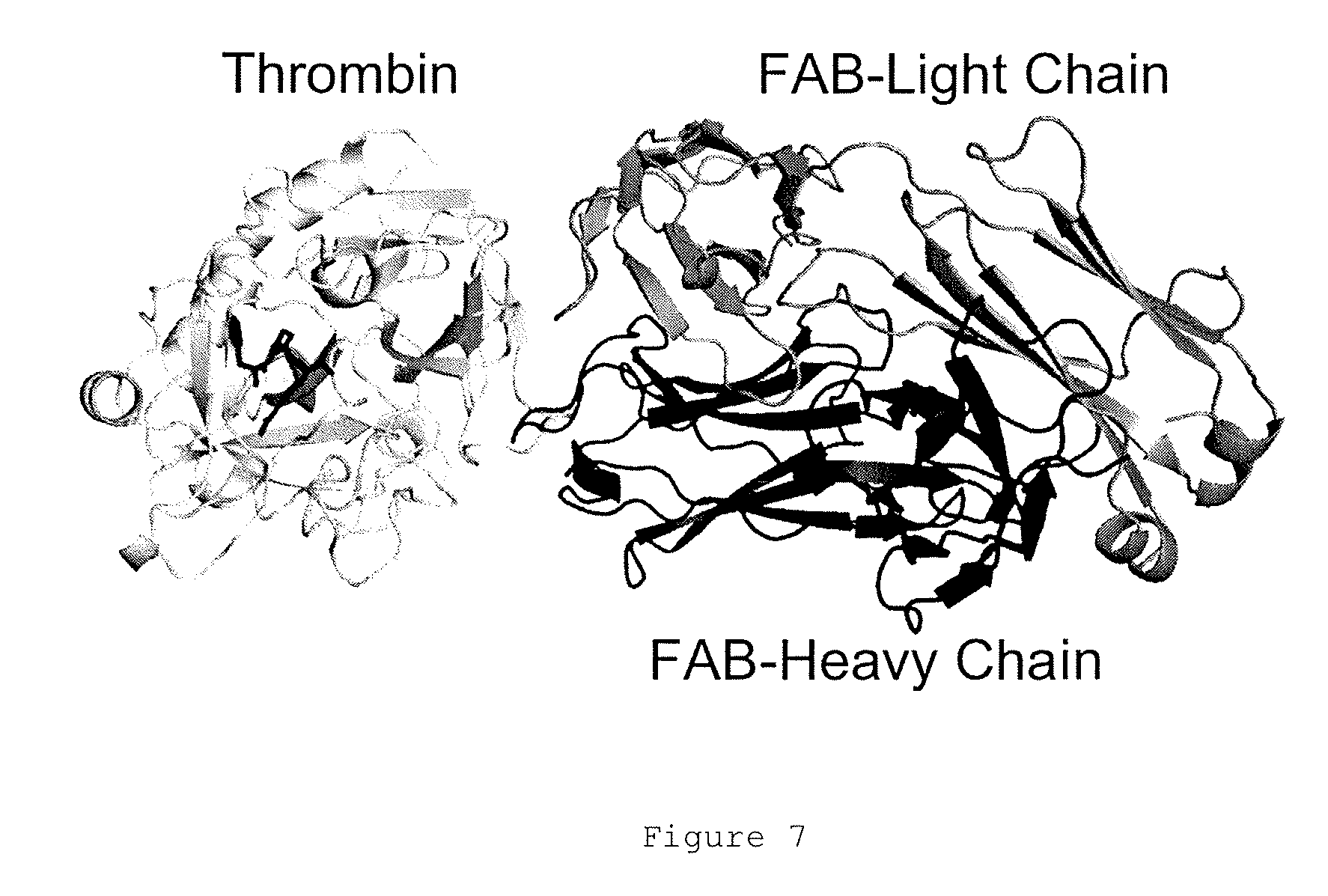
Exosite 1 (also known as `anion binding exosite 1` and the `fibrinogen recognition exosite`) is a well-characterized secondary binding site on the thrombin molecule (see for example James A. Huntington, 2008, Structural Insights into the Life History of Thrombin, in RECENT ADVANCES IN THROMBOSIS AND HEMOSTASIS 2008, editors; K. Tanaka and E. W. Davie, Springer Japan KK, Tokyo, pp. 80-106). Exosite 1 is formed in mature thrombin but is not formed in prothrombin (see for example Anderson et al (2000) JBC 2775 16428-16434).
Exosite 1 is involved in recognizing thrombin substrates, such as fibrinogen, but is remote from the catalytic active site. Various thrombin binding factors bind to exosite 1, including the anticoagulant dodecapeptide hirugen (Naski et al 1990 JBC 265 13484-13489), factor V, factor VIII, thrombomodulin (cofactor for protein C and TAFI activation), fibrinogen, PAR1 and fibrin (the co-factor for factor XIII activation).
An anti-exosite 1 antibody may bind to exosite 1 of mature human thrombin. The sequence of human preprothrombin is set out in SEQ ID NO: 1. Human prothrombin has the sequence of residues 44 to 622 of SEQ ID NO: 1. Mature human thrombin has the sequence of residues 314-363 (light chain) and residues 364 to 622 (heavy chain).
In some embodiments, an anti-exosite 1 antibody may also bind to exosite 1 of mature thrombin from other species. Thrombin sequences from other species are known in the art and available on public databases such as Genbank. The corresponding residues in thrombin sequences from other species may be easily identified using sequence alignment tools.
The numbering scheme for thrombin residues set out herein is conventional in the art and is based on the chymotrypsin template (Bode W et al EMBO J. 1989 November; 8(11):3467-75). Thrombin has insertion loops relative to chymotrypsin that are lettered sequentially using lower case letters.
Exosite 1 of mature human thrombin is underlined in SEQ ID NO: 1 and may include the following residues: M32, F34, R35, K36, S36a, P37, Q38, E39, L40, L65, R67, S72, R73, T74, R75, Y76, R77a, N78, E80, K81, I82, S83, M84, K109, K110, K149e, G150, Q151, S153 and V154. In some embodiments, other thrombin residues which are located close to (i.e. within 0.5 nm or within 1 nm) of any one of these residues may also be considered to be part of exosite 1.
An anti-exosite 1 antibody may bind to an epitope which comprises 1, 2, 3, 4, 5, 6, 7, 8, 9, 10, 11, 12, 13, 14, 15, 16, 17, 18, 19, 20 or more than 20 residues of exosite 1. Preferably, an anti-exosite 1 antibody binds to an epitope which consists entirely of exosite 1 residues.
For example, an anti-exosite 1 antibody may bind to an epitope which comprises 1, 2, 3, 4, 5, 6, 7, 8, 9, 10, 11, 12, 13, 14, 15 or all 16 residues selected from the group consisting of M32, F34, S36a, P37, Q38, E39, L40, L65, R67, R73, T74, R75, Y76, R77a, I82 and Q151 of human thrombin or the equivalent residues in thrombin from another species. In some preferred embodiments, the epitope may comprise the thrombin residues Q38, R73, T74, Y76 and R77a and optionally one or more additional residues.
Anti-exosite 1 antibody molecules as described herein are specific for thrombin exosite 1 and bind to this epitope with high affinity relative to other epitopes, for example epitopes from mammalian proteins other than mature thrombin. For example, an anti-exosite 1 antibody molecule may display a binding affinity for thrombin exosite 1 which is at least 500 fold, at least 1000 fold or at least 2000 fold greater than other epitopes.
Preferably, an antibody molecule as described herein which is specific for exosite 1 may bind to mature thrombin but display no binding or substantially no binding to prothrombin.
Without being bound by any theory, anti-exosite 1 antibodies may be unable to access thrombin within the core of a haemostatic clot, and are therefore unable to affect haemostasis by interrupting normal thrombin function at sites of vascular injury. However, because the anti-exosite 1 antibodies still bind to thrombin on the surface of the clot and in the outer shell of the clot, thrombosis is prevented, i.e. non-haemostatic clot extension is prevented.
An anti-exosite 1 antibody molecule may have a dissociation constant for exosite 1 of less than 50 nM, less than 40 nM, less than 30 nM, less than 20 nM, less than 10 nM, or less than 1 nM. For example, an antibody molecule may have an affinity for exosite 1 of 0.1 to 50 nM, e.g. 0.5 to 10 nM. A suitable anti-exosite 1 antibody molecule may, for example, have an affinity for thrombin exosite 1 of about 1 nM.
Binding kinetics and affinity (expressed as the equilibrium dissociation constant, K.sub.d) of the anti-exosite 1 antibody molecules may be determined using standard techniques, such as surface plasmon resonance e.g. using BIAcore analysis.
An anti-exosite 1 antibody molecule as described herein may be an immunoglobulin or fragment thereof, and may be natural or partly or wholly synthetically produced, for example a recombinant molecule.
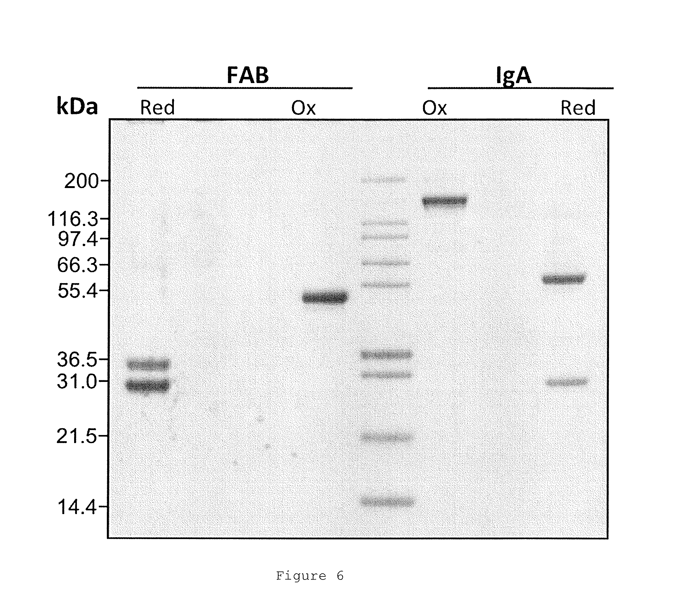
Anti-exosite 1 antibody molecules may include any polypeptide or protein comprising an antibody antigen-binding site, including Fab, Fab.sub.2, Fab.sub.3, diabodies, triabodies, tetrabodies, minibodies and single-domain antibodies, including nanobodies, as well as whole antibodies of any isotype or sub-class. Antibody molecules and methods for their construction and use are described, in for example Holliger & Hudson, Nature Biotechnology 23(9):1126-1136 (2005).
In some preferred embodiments, the anti-exosite 1 antibody molecule may be a whole antibody. For example, the anti-exosite 1 antibody molecule may be an IgG, IgA, IgE or IgM or any of the isotype sub-classes, particularly IgG1 and IgG4. The anti-exosite 1 antibody molecules may be monoclonal antibodies. In other preferred embodiments, the anti-exosite 1 antibody molecule may be an antibody fragment.
Anti-exosite 1 antibody molecules may be chimeric, humanized or human antibodies.
Anti-exosite 1 antibody molecules as described herein may be isolated, in the sense of being free from contaminants, such as antibodies able to bind other polypeptides and/or serum components. Monoclonal antibodies are preferred for some purposes, though polyclonal antibodies may also be employed.
Anti-exosite 1 antibody molecules may be obtained using techniques which are standard in the art. Methods of producing antibodies include immunizing a mammal (e.g. mouse, rat, rabbit, horse, goat, sheep or monkey) with the protein or a fragment thereof. Antibodies may be obtained from immunized animals using any of a variety of techniques known in the art, and screened, preferably using binding of antibody to antigen of interest. For instance, Western blotting techniques or immunoprecipitation may be used (Armitage et al., 1992, Nature 357: 80-82). Isolation of antibodies and/or antibody-producing cells from an animal may be accompanied by a step of sacrificing the animal.
As an alternative or supplement to immunizing a mammal with a peptide, an antibody specific for a protein may be obtained from a recombinantly produced library of expressed immunoglobulin variable domains, e.g. using lambda bacteriophage or filamentous bacteriophage which display functional immunoglobulin binding domains on their surfaces; for instance see WO92/01047. The library may be naive, that is constructed from sequences obtained from an organism which has not been immunized with any of the proteins (or fragments), or may be one constructed using sequences obtained from an organism which has been exposed to the antigen of interest.
Other anti-exosite 1 antibody molecules may be identified by screening patient serum for antibodies which bind to exosite 1.
In some embodiments, anti-thrombin antibody molecules may be produced by any convenient means, for example a method described above, and then screened for differential binding to mature thrombin relative to thrombin with an exosite 1 mutation, gamma thrombin (exosite 1 defective due to autolysis at R75 and R77a) or prothrombin. Suitable screening methods are well-known in the art.
An antibody which displays increased binding to mature thrombin, relative to non-thrombin proteins, thrombin with an exosite 1 mutation, gamma-thrombin or prothrombin, for example an antibody which binds to mature thrombin but does not bind to thrombin with an exosite 1 mutation, gamma thrombin or prothrombin, may be identified as an anti-exosite 1 antibody molecule.
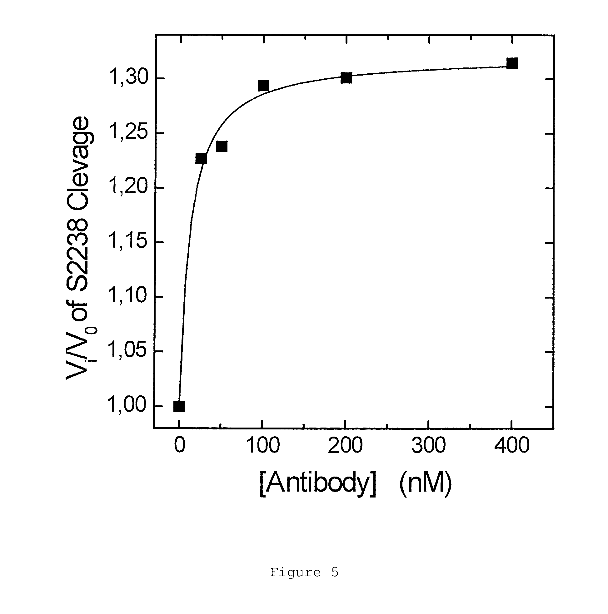
After production and/or isolation, the biological activity of an anti-exosite 1 antibody molecule may be tested. For example, the ability of the antibody molecule to inhibit thrombin substrate, cofactor or inhibitor binding and/or cleavage by thrombin may be determined and/or the ability of the antibody molecule to inhibit thrombosis without promoting bleeding may be determined.
Suitable antibody molecules may be tested for activity using a fibrinogen clotting or thrombin time assay. Suitable assays are well-known in the art.
The effect of an antibody molecule on coagulation and bleeding may be determined using standard techniques. For example, the effect of an antibody molecule on thrombosis may be determined in an animal model, such as a mouse model with ferric chloride induced clots in blood vessels. Effects on haemostasis may also be determined in an animal model, for example, by measuring tail bleed of a mouse.
Antibody molecules normally comprise an antigen binding domain comprising an immunoglobulin heavy chain variable domain (VH) and an immunoglobulin light chain variable domain (VL), although antigen binding domains comprising only a heavy chain variable domain (VH) are also possible (e.g. camelid or shark antibodies).
Each of the VH and VL domains typically comprise three complementarity determining regions (CDRs) responsible for antigen binding, interspersed by framework regions.
In some embodiments, binding to exosite 1 may occur wholly or substantially through the VHCDR3 of the anti-exosite 1 antibody molecule.
For example, an anti-exosite 1 antibody molecule may comprise a VH domain comprising a HCDR3 having the amino acid sequence of SEQ ID NO: 5 or the sequence of SEQ ID NO: 5 with 1 or more, for example 2, 3, 4 or 5 or more amino acid substitutions, deletions or insertions. The substitutions may be conservative substitutions. In some embodiments, the HCDR3 may comprise the amino acid residues at positions 4 to 9 of SEQ ID NO: 5 (SEFEPF), or more preferably the amino acid residues at positions 2, and 4 to 10 of SEQ ID NO: 5 (D and SEFEPFS) with substitutions, deletions or insertions at one or more other positions in SEQ ID NO:5. The HCDR3 may be the only region of the antibody molecule that interacts with a thrombin exosite 1 epitope or substantially the only region. The HCDR3 may therefore determine the specificity and/or affinity of the antibody molecule for the exosite 1 region of thrombin.
The VH domain of an anti-exosite 1 antibody molecule may additionally comprise an HCDR2 having the amino acid sequence of SEQ ID NO: 4 or the sequence of SEQ ID NO: 4 with 1 or more, for example 2, 3, 4 or 5 or more amino acid substitutions, deletions or insertions. In some embodiments, the HCDR2 may comprise the amino acid residues at positions 3 to 7 of SEQ ID NO: 4 (DPQDG) or the amino acid residues at positions 2 and 4 to 7 of SEQ ID NO: 4 (L and PQDG) of SEQ ID NO: 4, with substitutions, deletions or insertions at one or more other positions in SEQ ID NO: 4.
The VH domain of an anti-exosite 1 antibody molecule may further comprise an HCDR1 having the amino acid sequence of SEQ ID NO: 3 or the sequence of SEQ ID NO: 3 with 1 or more, for example 2, 3, 4 or 5 or more amino acid substitutions, deletions or insertions. In some embodiments, the HCDR1 may comprise amino acid residue T at position 5 of SEQ ID NO: 3 with substitutions, deletions or insertions at one or more other positions in SEQ ID NO: 3.
In some embodiments, an antibody molecule may comprise a VH domain comprising a HCDR1, a HCDR2 and a HCDR3 having the sequences of SEQ ID NOs 3, 4 and 5 respectively. For example, an antibody molecule may comprise a VH domain having the sequence of SEQ ID NO: 2 or the sequence of SEQ ID NO: 2 with 1 or more, for example 2, 3, 4, 5, 6, 7, 8, 9, 10 or more amino acid substitutions, deletions or insertions in SEQ ID NO: 2.
The anti-exosite 1 antibody molecule may further comprise a VL domain, for example a VL domain comprising LCDR1, LCDR2 and LCDR3 having the sequences of SEQ ID NOs 7, 8 and 9 respectively, or the sequences of SEQ ID NOs 7, 8 and 9 respectively with, independently, 1 or more, for example 2, 3, 4 or 5 or more amino acid substitutions, deletions or insertions. The substitutions may be conservative substitutions. For example, an antibody molecule may comprise a VL domain having the sequence of SEQ ID NO: 6 or the sequence of SEQ ID NO: 6 with 1 or more, for example 2, 3, 4, 5, 6, 7, 8, 9, 10 or more amino acid substitutions, deletions or insertions in SEQ ID NO: 6.
In some embodiments, the VL domain may comprise Tyr49.
The anti-exosite 1 antibody molecule may for example comprise one or more amino acid substitutions, deletions or insertions which improve one or more properties of the antibody, for example affinity, functional half-life, on and off rates.
The techniques that are required in order to introduce substitutions, deletions or insertions within amino acid sequences of CDRs, antibody VH or VL domains and antibodies are generally available in the art. Variant sequences may be made, with substitutions, deletions or insertions that may or may not be predicted to have a minimal or beneficial effect on activity, and tested for ability to bind exosite 1 of thrombin and/or for any other desired property.
In some embodiments, anti-exosite 1 antibody molecule may comprise a VH domain comprising a HCDR1, a HCDR2 and a HCDR3 having the sequences of SEQ ID NOs 3, 4, and 5, respectively, and a VL domain comprising a LCDR1, a LCDR2 and a LCDR3 having the sequences of SEQ ID NOs 7, 8 and 9, respectively.
For example, the VH and VL domains may have the amino acid sequences of SEQ ID NO: 2 and SEQ ID NO: 6 respectively; or may have the amino acid sequences of SEQ ID NO: 2 and SEQ ID NO: 6 comprising, independently 1 or more, for example 2, 3, 4, 5, 6, 7, 8, 9, 10 or more amino acid substitutions, deletions or insertions. The substitutions may be conservative substitutions.
In some embodiments, an antibody may comprise one or more substitutions, deletions or insertions which remove a glycosylation site. For example, a glycosylation site in VL domain of SEQ ID NO 6 may be mutated out by introducing a substitution at either N28 or S30.
The anti-exosite 1 antibody molecule may be in any format, as described above, In some preferred embodiments, the anti-exosite 1 antibody molecule may be a whole antibody, for example an IgG, such as IgG1 or IgG4, IgA, IgE or IgM.
An anti-exosite 1 antibody molecule of the invention may be one which competes for binding to exosite 1 with an antibody molecule described above, for example an antibody molecule which (i) binds thrombin exosite 1 and (ii) comprises a VH domain of SEQ ID NO: 2 and/or VL domain of SEQ ID NO: 6; an HCDR3 of SEQ ID NO: 5; an HCDR1, HCDR2, LCDR1, LCDR2, or LCDR3 of SEQ ID NOS: 3, 4, 7, 8 or 9 respectively; a VH domain comprising HCDR1, HCDR2 and HCDR3 sequences of SEQ ID NOS: 3, 4 and 5 respectively; and/or a VH domain comprising HCDR1, HCDR2 and HCDR3 sequences of SEQ ID NOS: 3, 4 and 5 and a VL domain comprising LCDR1, LDR2 and LCDR3 sequences of SEQ ID NOS: 7, 8 and 9 respectively.
Competition between antibody molecules may be assayed easily in vitro, for example using ELISA and/or by tagging a specific reporter molecule to one antibody molecule which can be detected in the presence of one or more other untagged antibody molecules, to enable identification of antibody molecules which bind the same epitope or an overlapping epitope. Such methods are readily known to one of ordinary skill in the art. Thus, a further aspect of the present invention provides an antibody molecule comprising a antibody antigen-binding site that competes with an antibody molecule, for example an antibody molecule comprising a VH and/or VL domain, CDR e.g. HCDR3 or set of CDRs of the parent antibody described above for binding to exosite 1 of thrombin. A suitable antibody molecule may comprise an antibody antigen-binding site which competes with an antibody antigen-binding site for binding to exosite 1 wherein the antibody antigen-binding site is composed of a VH domain and a VL domain, and wherein the VH and VL domains comprise HCDR1, HCDR2 and HCDR3 sequences of SEQ ID NOS: 3, 4, and 5 and LCDR1, LDR2 and LCDR3 sequences of SEQ ID NOS: 7, 8, and 9 respectively, for example the VH and VL domains of SEQ ID NOS: 2 and 6.
An anti-exosite 1 antibody molecule as described herein may inhibit the binding of thrombin-binding factors, including factors which bind to exosite 1. For example, an antibody molecule may competitively or non-competitively inhibit the binding of one or more of fV, fVIII, thrombomodulin, fibrinogen or fibrin, PAR1 and/or hirugen and hirudin analogues to thrombin.
An anti-exosite 1 antibody molecule as described herein may inhibit one or more activities of thrombin. For example, an anti-exosite 1 antibody molecule may inhibit the hydrolytic cleavage of one or more thrombin substrates, such as fibrinogen, platelet receptor PAR-1 and coagulation factor FVIII. For example, binding of the antibody molecule to thrombin may result in an at least 5-fold, at least 10-fold, or at least 15-fold decrease in the hydrolysis of fibrinogen, PAR-1, coagulation factor FVIII and/or another thrombin substrates, such as factor V, factor XIII in the presence of fibrin, and protein C and/or TAFI in the presence of thrombomodulin. In some embodiments, binding of thrombin by the anti-exosite 1 antibody molecule may result in no detectable cleavage of the thrombin substrate by thrombin.
Techniques for measuring thrombin activity, for example by measuring the hydrolysis of thrombin substrates in vitro are standard in the art and are described herein.
Anti-exosite 1 antibody molecules may be further modified by chemical modification, for example by PEGylation, or by incorporation in a liposome, to improve their pharmaceutical properties, for example by increasing in vivo half-life.
The effect of an anti-exosite 1 antibody molecule on coagulation and bleeding may be determined using standard techniques. For example, the effect of an antibody on a thrombosis model may be determined. Suitable models include ferric chloride clot induction in blood vessels in a murine model, followed by a tail bleed to test normal haemostasis. Other suitable thrombosis models are well known in the art (see for example Westrick et al ATVB (2007) 27:2079-2093)
Anti-exosite 1 antibody molecules may be comprised in pharmaceutical compositions with a pharmaceutically acceptable excipient.
A pharmaceutically acceptable excipient may be a compound or a combination of compounds entering into a pharmaceutical composition which does not provoke secondary reactions and which allows, for example, facilitation of the administration of the anti-exosite 1 antibody molecule, an increase in its lifespan and/or in its efficacy in the body or an increase in its solubility in solution. These pharmaceutically acceptable vehicles are well known and will be adapted by the person skilled in the art as a function of the mode of administration of the anti-exosite 1 antibody molecule.
In some embodiments, anti-exosite 1 antibody molecules may be provided in a lyophilised form for reconstitution prior to administration. For example, lyophilized antibody molecules may be re-constituted in sterile water and mixed with saline prior to administration to an individual.
Anti-exosite 1 antibody molecules will usually be administered in the form of a pharmaceutical composition, which may comprise at least one component in addition to the antibody molecule. Thus pharmaceutical compositions may comprise, in addition to the anti-exosite 1 antibody molecule, a pharmaceutically acceptable excipient, carrier, buffer, stabilizer or other materials well known to those skilled in the art. Such materials should be non-toxic and should not interfere with the efficacy of the anti-exosite 1 antibody molecule. The precise nature of the carrier or other material will depend on the route of administration, which may be by bolus, infusion, injection or any other suitable route, as discussed below.
For parenteral, for example sub-cutaneous or intra-venous administration, e.g. by injection, the pharmaceutical composition comprising the anti-exosite 1 antibody molecule may be in the form of a parenterally acceptable aqueous solution which is pyrogen-free and has suitable pH, isotonicity and stability. Those of relevant skill in the art are well able to prepare suitable solutions using, for example, isotonic vehicles, such as Sodium Chloride Injection, Ringer's Injection, Lactated Ringer's Injection. Preservatives, stabilizers, buffers, antioxidants and/or other additives may be employed as required including buffers such as phosphate, citrate and other organic acids; antioxidants, such as ascorbic acid and methionine; preservatives (such as octadecyldimethylbenzyl ammonium chloride; hexamethonium chloride; benzalkonium chloride; benzethonium chloride; phenol, butyl or benzyl alcohol; alkyl parabens, such as methyl or propyl paraben; catechol; resorcinol; cyclohexanol; 3'-pentanol; and m-cresol); low molecular weight polypeptides; proteins, such as serum albumin, gelatin or immunoglobulins; hydrophilic polymers, such as polyvinylpyrrolidone; amino acids, such as glycine, glutamine, asparagines, histidine, arginine, or lysine; monosaccharides, disaccharides and other carbohydrates including glucose, mannose or dextrins; chelating agents, such as EDTA; sugars, such as sucrose, mannitol, trehalose or sorbitol; salt-forming counter-ions, such as sodium; metal complexes (e.g. Zn-protein complexes); and/or non-ionic surfactants, such as TWEEN.TM., PLURONICS.TM. or polyethylene glycol (PEG).
A pharmaceutical composition comprising an anti-exosite 1 antibody molecule may be administered alone or in combination with other treatments, either simultaneously or sequentially dependent upon the condition to be treated.
An anti-exosite 1 antibody molecule as described herein may be used in a method of treatment of the human or animal body, including prophylactic or preventative treatment (e.g. treatment before the onset of a condition in an individual to reduce the risk of the condition occurring in the individual; delay its onset; or reduce its severity after onset). The method of treatment may comprise administering an anti-exosite 1 antibody molecule to an individual in need thereof.
Administration is normally in a "therapeutically effective amount", this being sufficient to show benefit to a patient. Such benefit may be at least amelioration of at least one symptom. The actual amount administered, and rate and time-course of administration, will depend on the nature and severity of what is being treated, the particular mammal being treated, the clinical condition of the individual patient, the cause of the disorder, the site of delivery of the composition, the method of administration, the scheduling of administration and other factors known to medical practitioners. Prescription of treatment, e.g. decisions on dosage etc., is within the responsibility of general practitioners and other medical doctors and may depend on the severity of the symptoms and/or progression of a disease being treated. Appropriate doses of antibody molecules are well known in the art (Ledermann J. A. et al. (1991) Int. J. Cancer 47: 659-664; Bagshawe K. D. et al. (1991) Antibody, Immunoconjugates and Radiopharmaceuticals 4: 915-922). Specific dosages may be indicated herein or in the Physician's Desk Reference (2003) as appropriate for the type of medicament being administered may be used. A therapeutically effective amount or suitable dose of an antibody molecule may be determined by comparing its in vitro activity and in vivo activity in an animal model. Methods for extrapolation of effective dosages in mice and other test animals to humans are known. The precise dose will depend upon a number of factors, including whether the antibody is for prevention or for treatment, the size and location of the area to be treated, the precise nature of the antibody (e.g. whole antibody, fragment) and the nature of any detectable label or other molecule attached to the antibody.
A typical antibody dose will be in the range 100 .mu.g to 1 g for systemic applications, and 1 .mu.g to 1 mg for topical applications. An initial higher loading dose, followed by one or more lower doses, may be administered. Typically, the antibody will be a whole antibody, e.g. the IgG1 or IgG4 isotype. This is a dose for a single treatment of an adult patient, which may be proportionally adjusted for children and infants, and also adjusted for other antibody formats in proportion to molecular weight. Treatments may be repeated at daily, twice-weekly, weekly or monthly intervals, at the discretion of the physician. The treatment schedule for an individual may be dependent on the pharmocokinetic and pharmacodynamic properties of the antibody composition, the route of administration and the nature of the condition being treated.
Treatment may be periodic, and the period between administrations may be about two weeks or more, e.g. about three weeks or more, about four weeks or more, about once a month or more, about five weeks or more, or about six weeks or more. For example, treatment may be every two to four weeks or every four to eight weeks. Treatment may be given before, and/or after surgery, and/or may be administered or applied directly at the anatomical site of surgical treatment or invasive procedure. Suitable formulations and routes of administration are described above.
In some embodiments, anti-exosite 1 antibody molecules as described herein may be administered as sub-cutaneous injections. Sub-cutaneous injections may be administered using an auto-injector, for example for long term prophylaxis/treatment.
In some preferred embodiments, the therapeutic effect of the anti-exosite 1 antibody molecule may persist for several half-lives, depending on the dose. For example, the therapeutic effect of a single dose of anti-exosite 1 antibody molecule may persist in an individual for 1 month or more, 2 months or more, 3 months or more, 4 months or more, 5 months or more, or 6 months or more.
Anti-exosite 1 antibody molecules described herein inhibit thrombin and may be useful in the treatment of thrombin-mediated conditions.
Haemostasis is the normal coagulation response i.e. the prevention of bleeding or haemorrhage, for example from a damaged blood vessel. Haemostasis arrests bleeding and haemorrhage from blood vessels in the body.
Anti-exosite 1 antibody molecules may have no effect or substantially no effect on haemostasis i.e. they do not promote bleeding or haemorrhage.
Aspects of the invention provide; an anti-exosite 1 antibody molecule as described herein for use in a method of treatment of the human or animal body; an anti-exosite 1 antibody molecule as described herein for use in a method of treatment of a thrombin-mediated disorder; the use of an anti-exosite 1 antibody molecule as described herein in the manufacture of a medicament for the treatment of a thrombin-mediated condition; and a method of treatment of a thrombin-mediated condition comprising administering an anti-exosite 1 antibody molecule as described herein to an individual in need thereof.
Inhibition of thrombin by anti-exosite 1 antibodies as described herein may be of clinical benefit in the treatment of any thrombin-mediated condition. A thrombin-mediated condition may include disorders associated with the formation or activity of thrombin.
Thrombin plays a key role in haemostasis, coagulation and thrombosis. Thrombin-mediated conditions include thrombotic conditions, such as thrombosis and embolism.
Thrombosis is coagulation which is in excess of what is required for haemostasis (i.e. excessive coagulation), or which is not required for haemostasis (i.e. extra-haemostatic or non-haemostatic coagulation).
Thrombosis is blood clotting within the blood vessel lumen. It is characterized by the formation of a clot (thrombus) that is in excess of requirement or not required for haemostasis. The clot may impede blood flow through the blood vessel leading to medical complications. A clot may break away from its site of formation, leading to embolism elsewhere in the circulatory system. In the arterial system, thrombosis is typically the result of atherosclerotic plaque rupture.
In some embodiments, thrombosis may occur after an initial physiological haemostatic response, for example damage to endothelial cells in a blood vessel. In other embodiments, thrombosis may occur in the absence of any physiological haemostatic response.
Thrombosis may occur in individuals with an intrinsic tendency to thrombosis (i.e. thrombophilia) or in `normal` individuals with no intrinsic tendency to thrombosis, for example in response to an extrinsic stimulus.
Thrombosis and embolism may occur in any vein, artery or other blood vessel within the circulatory system and may include microvascular thrombosis.
Thrombosis and embolism may be associated with surgery (either during surgery or afterwards) or the insertion of foreign objects, such as coronary stents, into a patient.
For example, anti-exosite 1 antibodies as described herein may be useful in the surgical and other procedures in which blood is exposed to artificial surfaces, such as open heart surgery and dialysis.
Thrombotic conditions may include thrombophilia, thrombotic stroke and coronary artery occlusion.
Patients suitable for treatment as described herein include patients with conditions in which thrombosis is a symptom or a side-effect of treatment or which confer an increased risk of thrombosis or patients who are predisposed to or at increased risk of thrombosis, relative to the general population. For example, an anti-exosite 1 antibody molecule as described herein may also be useful in the treatment or prevention of venous thrombosis in cancer patients, and in the treatment or prevention of hospital-acquired thrombosis, which is responsible for 50% of cases of venous thromboembolism.
Anti-exosite 1 antibody molecules as described herein may exert a therapeutic or other beneficial effect on thrombin-mediated conditions, such as thrombotic conditions, without substantially inhibiting or impeding haemostasis. For example, the risk of haemorrhage in patients treated with anti-exosite 1 antibody molecules may not be increased or substantially increased relative to untreated individuals.
Individuals treated with conventional anticoagulants, such as natural and synthetic heparins, warfarin, direct serine protease inhibitors (e.g. argatroban, dabigatran, apixaban, and rivaroxaban), hirudin and its derivatives (e.g. lepirudin and bivalirudin), and anti-platelet drugs (e.g. clopidogrel, ticlopidine and abciximab) cause bleeding. The risk of bleeding in patients treated with anti-exosite 1 antibody molecules as described herein may be reduced relative to individuals treated with conventional anticoagulants.
Thrombin-mediated conditions include non-thrombotic conditions associated with thrombin activity, including inflammation, infection, tumor growth and metastasis, organ rejection and dementia (vascular and non-vascular, e.g. Alzheimer's disease) (Licari et al J Vet Emerg Crit Care (San Antonio). 2009 February; 19(1):11-22; Tsopanoglou et al Eur Cytokine Netw. 2009 Dec. 1; 20(4):171-9).
Anti-exosite 1 antibody molecules as described herein may also be useful in in vitro testing, for example in the analysis and characterization of coagulation, for example in a sample obtained from a patient.
Anti-exosite 1 antibody molecules may be useful in the measurement of thrombin generation. Assays of thrombin generation are technically problematic because the conversion of fibrinogen to fibrin causes turbidity, which precludes the use of a simple chromogenic end-point.
The addition of an anti-exosite 1 antibody molecule as described herein to a sample of blood prevents or inhibits fibrin formation and hence turbidity and permits thrombin generation to be measured using a chromogenic substrate, without the need for a defibrination step.
For example, a method of measuring thrombin generation may comprise contacting a blood sample with a chromogenic thrombin substrate in the presence of an anti-exosite 1 antibody molecule as described herein and measuring the chromogenic signal from the substrate; wherein the chromogenic signal is indicative of thrombin generation in the sample.
The chromogenic signal may be measured directly without defibrination of the sample.
Suitable substrates are well known in the art and include S2238 (H-D-Phe-Pip-Arg-pNa), .beta.-Ala-Gly-Arg-p-nitroanilide diacetate (Prasa, D. et al. (1997) Thromb. Haemost. 78, 1215; Sigma Aldrich Inc) and Tos-Gly-Pro-Arg-pNa.AcOH (Biophen CS-01(81); Aniara Inc OH USA).
Anti-exosite 1 antibody molecules may also be useful in inhibiting or preventing the coagulation of blood as described above in extracorporeal circulations, such as haemodialysis and extracorporeal membrane oxygenation.
For example, a method of inhibiting or preventing blood coagulation in vitro or ex vivo may comprise introducing an anti-exosite 1 antibody molecule as described herein to a blood sample. The blood sample may be introduced into an extracorporeal circulation system before, simultaneous with or after the introduction of the anti-exosite 1 antibody and optionally subjected to treatment such as haemodialysis or oxygenation. In some embodiments, the treated blood may be subsequently administered to an individual. Other embodiments provide an anti-exosite 1 antibody molecule as described herein for use in a method of inhibiting or preventing blood coagulation in a blood sample ex vivo and the use of an anti-exosite 1 antibody molecule as described herein in the manufacture of a medicament for use in a method of inhibiting or preventing blood coagulation in a blood sample ex vivo.
Other aspects of the invention relate to the production of antibody molecules which bind to the exosite 1 epitope of thrombin and may be useful, for example in the treatment of pathological blood coagulation or thrombosis.
A method for producing an antibody antigen-binding domain for the exosite 1 epitope of thrombin, may comprise: providing, by way of addition, deletion, substitution or insertion of one or more amino acids in the amino acid sequence of a parent VH domain comprising HCDR1, HCDR2 and HCDR3, wherein HCDR1, HCDR2 and HCDR3 have the amino acid sequences of SEQ ID NOS: 3, 4 and 5 respectively, a VH domain which is an amino acid sequence variant of the parent VH domain, and; optionally combining the VH domain thus provided with one or more VL domains to provide one or more VH/VL combinations; and testing said VH domain which is an amino acid sequence variant of the parent VH domain or the VH/VL combination or combinations to identify an antibody antigen binding domain for the exosite 1 epitope of thrombin.
A VH domain which is an amino acid sequence variant of the parent VH domain may have the HCDR3 sequence of SEQ ID NO: 5 or a variant with the addition, deletion, substitution or insertion of one, two, three or more amino acids.
The VH domain which is an amino acid sequence variant of the parent VH domain may have the HCDR1 and HCDR2 sequences of SEQ ID NOS: 3 and 4 respectively, or variants of these sequences with the addition, deletion, substitution or insertion of one, two, three or more amino acids.
A method for producing an antibody molecule that specifically binds to the exosite 1 epitope of thrombin may comprise: providing starting nucleic acid encoding a VH domain or a starting repertoire of nucleic acids each encoding a VH domain, wherein the VH domain or VH domains either comprise a HCDR1, HCDR2 and/or HCDR3 to be replaced or lack a HCDR1, HCDR2 and/or HCDR3 encoding region; combining said starting nucleic acid or starting repertoire with donor nucleic acid or donor nucleic acids encoding or produced by mutation of the amino acid sequence of an HCDR1, HCDR2, and/or HCDR3 having the amino acid sequences of SEQ ID NOS: 3, 4 and 5 respectively, such that said donor nucleic acid is or donor nucleic acids are inserted into the CDR1, CDR2 and/or CDR3 region in the starting nucleic acid or starting repertoire, so as to provide a product repertoire of nucleic acids encoding VH domains; expressing the nucleic acids of said product repertoire to produce product VH domains; optionally combining said product VH domains with one or more VL domains; selecting an antibody molecule that binds exosite 1 of thrombin, which antibody molecule comprises a product VH domain and optionally a VL domain; and recovering said antibody molecule or nucleic acid encoding it.
Suitable techniques for the maturation and optimization of antibody molecules are well-known in the art.
Antibody antigen-binding domains and antibody molecules for the exosite 1 epitope of thrombin may be tested as described above. For example, the ability to bind to thrombin and/or inhibit the cleavage of thrombin substrates may be determined.
The effect of an antibody molecule on coagulation and bleeding may be determined using standard techniques. For example, a mouse thrombosis model of ferric chloride clot induction in a blood vessel, such as the femoral vein or carotid artery, followed by a tail bleed to test normal haemostasis, may be employed.
Various further aspects and embodiments of the present invention will be apparent to those skilled in the art in view of the present disclosure.
All documents mentioned in this specification are incorporated herein by reference in their entirety.
Unless stated otherwise, antibody residues are numbered herein in accordance with the Kabat numbering scheme.
"and/or" where used herein is to be taken as specific disclosure of each of the two specified features or components with or without the other. For example "A and/or B" is to be taken as specific disclosure of each of (i) A, (ii) B and (iii) A and B, just as if each is set out individually herein.
Unless context dictates otherwise, the descriptions and definitions of the features set out above are not limited to any particular aspect or embodiment of the invention and apply equally to all aspects and embodiments which are described. Thus, the features set out above are disclosed in all combinations and permutations.
Certain aspects and embodiments of the invention will now be illustrated by way of example and with reference to the figures and tables described below.
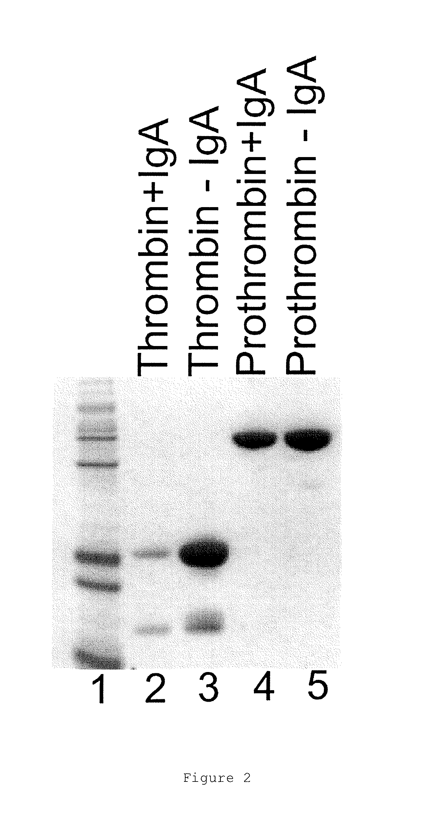
FIGS. 1A and 1B show the binding and elution of the IgA on human thrombin-Sepharose.RTM. column. FIG. 1A shows an elution profile for IgA (narrow peak) from a thrombin-Sepharose.RTM. column using a pH gradient (neutral to low, indicated by upward sloping line). FIG. 1B shows a native blue gel showing total IgA load, flow-through from the human thrombin column and eluate following elution at low pH.
FIG. 2 shows a non-reducing SDS-PAGE gel which indicates that the IgA binds thrombin but not prothrombin. In this pull-down assay, lectin agarose is used to bind to IgA in the presence of thrombin or prothrombin. The supernatant is then run on an SDS gel. Lane 1 is size standards; lane 2 shows a depletion of thrombin from the supernatant; Lane 3 shows that depletion is dependent on the presence of the IgA; Lanes 3 and 4 show that prothrombin is not depleted, and therefore does not bind to the IgA.
FIG. 3 shows the relative rate of S2238 cleavage by thrombin in the presence or absence of IgA (i.e. a single slope of Abs405 with time for S2238 hydrolysis). This indicates that the IgA does not bind at the thrombin active site.
FIG. 4 shows the results of binding studies which indicate that the IgA competes with the fluorescently labelled dodecapeptide hirugen for binding to thrombin.
FIG. 5 shows the effect of the IgA on the cleavage of S2238 by thrombin. This analysis allows the estimate of Kd for the IgA-thrombin interaction of 12 nM.
FIG. 6 shows an SDS-PAGE gel of whole IgA and Fab fragments under reducing and non-reducing (ox) conditions. The non-reduced IgA is shown to have a molecular weight of between 100-200 kDa and the non-reduced Fab has a molecular weight of about 50 kDa.
FIG. 7 shows the crystal structure of Thrombin-Fab complex showing interaction between the exosite 1 of thrombin and HCDR3 of the Fab fragment.
FIG. 8 shows detail of crystal structure showing interaction between specific residues of thrombin exosite 1 and HCDR3 of the Fab fragment.
FIG. 9 shows fluorescence microscopy images of FeCl.sub.3 induced blood clots in femoral vein injuries in C57BL/6 mice injected with FITC labelled fibrinogen taken at between 2 and 30 minutes. 100 .mu.l of PBS was administered (vehicle control).
FIG. 10 shows fluorescence microscopy images of FeCl.sub.3 induced blood clots in femoral vein injuries in C57BL/6 mice injected with FITC labelled fibrinogen and 40 nM (final concentration in mouse blood, equivalent to a dose of approximately 0.6 mg/Kg) anti-exosite 1 IgA (100 .mu.l in PBS).
FIG. 11 shows fluorescence microscopy images of FeCl.sub.3 induced blood clots in femoral vein injuries in C57BL/6 mice injected with FITC labelled fibrinogen and 80 nM (final concentration in mouse blood, equivalent to a dose of approximately 1.2 mg/Kg) anti-exosite 1 IgA (100 .mu.l in PBS), and a region outside of injury site for comparison.
FIG. 12 shows fluorescence microscopy images of FeCl.sub.3 induced blood clots in femoral vein injuries in C57BL/6 mice injected with FITC labelled fibrinogen and 200 nM (final concentration in mouse blood, equivalent to a dose of approximately 3 mg/Kg) anti-exosite 1 IgA (100 .mu.l in PBS), and a region outside of injury site for comparison.
FIG. 13 shows fluorescence microscopy images of FeCl.sub.3 induced blood clots in femoral vein injuries in C57BL/6 mice injected with FITC labelled fibrinogen and 400 nM (final concentration in mouse blood, equivalent to a dose of approximately 6 mg/Kg) anti-exosite 1 IgA (100 .mu.l in PBS).
FIG. 14 shows fluorescence microscopy images of FeCl.sub.3 induced blood clots in femoral vein injuries in C57BL/6 mice treated with FITC labelled fibrinogen and 4 .mu.M (final concentration in mouse blood, equivalent to a dose of approximately 60 mg/Kg) anti-exosite 1 IgA (100 .mu.l in PBS).
FIG. 15 shows a quantitation of the dose response to anti-exosite 1 IgA from the fluorescent images shown in FIGS. 9 to 13.
FIG. 16 shows tail bleed times in control C57BL/6 mice and in mice treated with increasing amounts of anti-exosite 1 IgA. The second average excludes the outlier.
FIG. 17 shows the results of tail clip assays on wild-type male C57BL/6 mice (n=5) after injection into tail vein with either IgA or PBS. 15 min after injection, tails were cut at diameter of 3 mm and blood loss monitored over 10 min.
FIG. 18A to 18D show the results of an FeCl.sub.3 carotid artery occlusion model on 9 week old WT C57BL/6 male mice injected as previously with 400 nM anti-thrombin IgA (final concentration in blood, equivalent to a dose of approximately 6 mg/Kg) or PBS 15 min prior to injury with 5% FeCl.sub.3 for 2 min. FIG. 18A shows results for a typical PBS-injected mice (occlusion in 20 min) and FIGS. 18B, 18C and 18D show examples of results for mice treated with 400 nM anti-thrombin IgA (no occlusion).
FIG. 19 shows thrombin times (i.e. clotting of pooled plasma) with increasing concentrations of IgG and IgA of the invention, upon addition of 20 nM human thrombin.
FIG. 20 shows the binding of synthetic IgG to immobilized thrombin (on ForteBio.RTM. Octet Red instrument).
FIG. 21 shows a typical Octet trace for the binding of 24 nM S195A thrombin to immobilized IgG showing the on phase, followed by an off phase. The black line is the fit.
FIG. 22 shows an Octet trace of 500 nM prothrombin with a tip loaded with immobilized IgG. The same conditions were used as the experiment with thrombin in FIG. 21. There is no evidence of binding, even at this high concentration.
FIG. 23 shows the ELISA binding curves for anti-exosite 1 IgG and an IgG S30A variant binding to thrombin.
FIG. 24 shows the potency of IgG and IgG S30A in an ex vivo activated partial thromboplastin time (APTT) coagulation assay.
FIG. 25 shows time to stop bleeding for 30 seconds data for IgG S30A and IgG in the rat tail clip bleeding model.
FIG. 26 shows total bleeding time data for IgG S30A and IgG in the rat tail clip bleeding model.
FIG. 27 shows total hemoglobin lost data for IgG S30A and IgG in the rat tail clip bleeding model.
FIG. 28 shows data on the prevention of thrombus formation by IgG S30A and IgG in the rat venous thrombosis model using ferric chloride (FeCl.sub.3) at 2.5% concentration.
FIG. 29 shows data on the prevention of thrombus formation by IgG S30A and IgG in the rat venous thrombosis model using ferric chloride (FeCl.sub.3) at 5% concentration.
EXPERIMENTS
1. Antibody Isolation and Characterization
Coagulation screening was carried out on a blood plasma sample from a patient. The coagulation tests were performed on a patient who suffered subdural haematoma following head injury. The haematoma spontaneously resolved without intervention. There was no previous history of bleeding and in the 4 years since the patient presented, there have been no further bleeding episodes. The results are shown in Table 1.
The prothrombin time (PT), activated partial thromboplastin time (APTT), and thrombin time (TT) were all prolonged in the patient compared to controls, but reptilase time was normal.
Thrombin time was not corrected by heparinase, indicating that heparin treatment or contamination was not responsible. Fibrinogen levels were normal in the patient, according to ELISA and Reptilase assays. The Clauss assay gave an artifactally low fibrinogen level due to the presence of the thrombin inhibitor. The PT and APTT clotting times were found to remain prolonged following a mixing test using a 50:50 mix with pooled plasma from normal individuals. This showed the presence of an inhibitor in the sample from the patient.
The patient's blood plasma was found to have a high titre of an IgA. This IgA molecule was found to bind to a human thrombin column (FIGS. 1A and 1B). IgA binding lectin-agarose pulled down thrombin in the presence but not the absence of the IgA. Prothrombin was not pulled down by the lectin-agarose in the presence of the IgA, indicating that the IgA specifically binds to thrombin but not prothrombin (FIG. 2).
The binding site of the IgA on the thrombin molecule was then investigated.
A slightly higher rate of cleavage of S2238 by thrombin was measured in the presence of the IgA, indicating that the IgA does not block the active site of thrombin (FIG. 3).
The binding of fluorescently labelled hirugen to thrombin is inhibited by the presence of 700 nM of the IgA, indicating that the epitope for the antibody overlaps with the binding site of hirugen on thrombin, namely the exosite 1 of thrombin (FIG. 4).
The effect of the IgA on the hydrolysis of some of thrombin's procoagulant substrates was tested. The results are shown in Table 2. These results demonstrate that the IgA molecule isolated from the patient sample inhibits multiple procoagulant activities of thrombin.
Inhibition of thrombin by antithrombin (AT) in the presence of the IgA was only marginally affected in both the absence and presence of heparin (Table 3).
The dissociation constant (K.sub.d) of the IgA for thrombin was initially estimated based on rate of S2238 hydrolysis to be approximately 12 nM (FIG. 5). The K.sub.d for the binding of the IgA to S195A thrombin (inactivated by mutation of the catalytic serine) was determined to be 2 nM using the ForteBio.RTM. Octet Red instrument (Table 4).
The purified IgA was cleaved with papain (FIG. 6), and the Fab fragment was isolated and combined with human PPACK-Thrombin (PPACK is a covalent active site inhibitor). The human PPACK-Thrombin-FAB complex was crystallized and used for structural analysis. The statistics of the structure obtained were as follows: resolution is 1.9 .ANG.; Rfactor=19.43%; Rfree=23.42%; one complex in the asymmetric unit; Ramachandran: favoured=97.0%, outliers=0%. The crystal structure revealed a close association between the HCDR3 of the IgA Fab and the exosite 1 of thrombin (FIG. 7).
In particular, residues M32, F34, Q38, E39, L40, L65, R67, R73, T74, R75, Y76, R77a and I82 of the exosite 1 all directly interact with the HCDR3 loop of the IgA Fab (FIG. 8).
PISA analysis of the antibody-thrombin interface showed that the total buried surface area in the complex is 1075 .ANG..sup.2. The contact residues in the IgA heavy chain were (Kabat numbering): 30, 51, 52a, 53-55, 96, 98, 99, 100, 100a, 100b, 100c, 100d). These are all in CDRs: CDRH1-GYTLTEAAIH (SEQ ID NO: 3); CDRH2-GLDPQDGETVYAQQFKG (SEQ ID NO: 4); CDRH3-GDFSEFEPFSMDYFHF (SEQ ID NO: 5) (underlined residues contacting). CDRH3 was found to be the most important, providing 85% of the buried surface area on the antibody. The light chain made one marginal contact with Tyr49, right before CDRL2 (with Ser36a of thrombin). Some individual contributions to buried surface were: Glu99 54 .ANG..sup.2, Phe100 134.8 .ANG..sup.22, Glu100a 80.6 .ANG..sup.2, Phe100c 141.7 .ANG..sup.2.
The contact residues in thrombin were found to be (chymotrypsin numbering): 32, 34, 36a-40, 65, 67, 73-76, 77a, 82, and 151. The most important individual contributors to the buried surface were: Gln38 86.4 .ANG..sup.2, Arg73 44.5 .ANG..sup.2, Thr74 60.1 .ANG..sup.2, Tyr76 78.4 .ANG..sup.2, Arg77a 86.9 .ANG..sup.2.
The patient did not display increased or abnormal bleeding or haemorrhage, in spite of 3 g/l circulating levels of this IgA, demonstrating that the antibody inhibits thrombin without affecting normal haemostasis.
2. The Effect of IgA on Animal Thrombosis Models
C57BL/6 mice were anaesthetized. A catheter was inserted in the carotid artery (for compound injection). FITC labelled fibrinogen (2 mg/ml) was injected via the carotid artery. PBS (control) or IgA was also injected via the carotid artery. The femoral vein was exposed and 10% FeCl.sub.3 applied (saturated blotting paper 3 mm in length) for 3 min to induce clotting.
Fluorescence microscopy images were taken along the length of injury site at 0, 5, 10, and 20 min post FeCl.sub.3 injury using fluorescence microscopy techniques.
Clots (fibrin deposits) in the femoral vein were clearly visible as bright areas (FIG. 9). The lowest dose of the antibody was observed to cause significant inhibition of clotting but as the dose increased, clotting was abolished (FIGS. 10 to 15).
The bleeding times of the mice were also measured. Bleeding times were assessed as time to cessation of blood flow after a tail cut. Despite the presence of a single outlier sample, the bleeding time was found to be unaffected by treatment with anti-exosite 1 IgA (FIG. 16).
These results show that the anti-exosite 1 IgA antibody is a potent inhibitor of thrombosis but has no effect on bleeding time.
3. Tail Clip Assays
A tail clip assay was performed on wild-type male C57BL/6 mice injected with either 400 nM IgA (final concentration in blood, equivalent to a dose of approximately 6 mg/Kg) or PBS. Blood loss was monitored over 10 mins after the tail was cut at 3 mm diameter 15 minutes after the injection. Total blood loss was found to be unaffected by treatment with anti-exosite 1 IgA (FIG. 17).
4. FeCl.sub.3 Injury Carotid Artery Occlusion
FeCl.sub.3 injury carotid artery occlusion studies were performed on 9 week old WT C57BL/6 male mice. Mice were injected with 400 nM anti-IIa IgA (final concentration in blood, equivalent to a dose of approximately 6 mg/Kg) or PBS 15 min prior to injury with 5% FeCl.sub.3 for 2 min. Blood flow was then monitored by Doppler and the time to occlusion measured. A "clot" was defined as stable occlusive thrombus where blood flow was reduced to values typically less than 0.1 ml/min and stayed reduced. In the control mice, a stable clot was observed to form about 20 mins after injury (FIG. 18A). However, the majority of mice treated with 400 nM anti-IIa IgA were unable to form stable clots and gave traces in which the clots were quickly resolved, repeatedly resolved or never formed. Three representative traces are shown in FIGS. 18B to 18D.
5. Anti-Exosite 1 IgG
The IgA molecule identified in the patient described above was re-formatted as an IgG using standard techniques.
The clotting time of pooled human plasma spiked with increasing amounts of the original IgA and the new IgG was tested upon addition of human thrombin to 20 nM (FIG. 19). Both parent IgA and the synthetic IgG increased time to clot formation in an identical concentration-dependent manner, implying identical affinities for thrombin.
This was confirmed by measuring the binding of synthetic IgG to immobilized S195A thrombin using a ForteBio.RTM. Octet Red instrument. Thrombin was attached to the probe and the binding of the antibodies (at various concentrations) was monitored. On-rates and off-rates were determined. Both antibodies gave similar on-rates of approximately 3.times.10.sup.5 M.sup.-1 s.sup.-1 and off-rates of approximately 5.times.10.sup.-4 s.sup.-1, and dissociation constants (Kd) of approximately 2 nM. Kds of approximately 2 nM were also obtained for the IgA and the IgG by steady-state analysis (Table 4). A representative steady state curve is shown in FIG. 20. The properties of the IgA were therefore reproduced on an IgG framework.
Binding of prothrombin to the IgG antibody was tested using the Octet system by immobilizing IgG. Thrombin bound to the immobilized IgG with comparable rates and affinities as those obtained using immobilized thrombin (Table 4); prothrombin did not bind to the IgG. FIG. 21 is a trace of 24 nM thrombin binding to and dissociating from the immobilized IgG. FIG. 22 is the same experiment using 500 nM prothrombin, and shows no evidence of binding.
6. Anti-Exosite 1 IgG S30A Variant Antibody
6.1 Introduction
Glycosylation sites in an antibody can raise issues during manufacture and/or therapeutic use of the antibody. The oligosaccharides added to glycosylation sites are typically heterogeneous, for example with complex di-antenary and hybrid oligosaccharides with sialic acids and galactoses (for Fab oligosaccharides) or with fucosylated non-galactosylated di-antenary oligosaccharides (for Fc oligosaccharides). The presence of more than one glycosylation site in an antibody (or active fragment thereof) thus adds further to potential heterogeneity. Removal of incorrectly glycosylated forms of an antibody during the purification process is very difficult and can lead to extended process development activities and reduced yields.
Therefore, if a glycosylation site in an antibody (or active fragment thereof) is determined not to be required directly or indirectly for antigen binding activity, it may be desirable from a manufacturing and quality control perspective to remove that glycosylation site by engineering.
As noted above, it was envisaged that a glycosylation site in VL domain of SEQ ID NO: 6 of the antibody of the present invention could be mutated out by introducing a substitution at either N28 or 530.
Of the two residues N28 and 530, S30 was targeted for substitution as it was considered, based on crystal structure analysis, less likely to be involved in antibody folding or stability.
6.2 Methods and Materials
An "IgG S30A" variant monoclonal antibody was produced using standard site-directed mutagenesis techniques from the anti-exosite IgG antibody ("IgG") described in section 5 above by substituting serine residue 30 (S30) with an alanine (hence, S30A).
The IgG S30A variant was expressed for analysis using standard transient expression techniques as described below.
In outline, single gene vectors (SGVs) were constructed using GS Xceed vectors (Lonza Biologics, Slough, UK) (pXC IgG4pro .DELTA.K for the heavy chain constant domain encoding region and pXC Kappa for light chain constant domain encoding region) and the variable domain encoding regions as synthesized by GeneArt AG. The SGVs were amplified and transiently co-transfected into Chinese Hamster Ovary CHOK1SV GS KO cells for initial expression at a volume of 200 ml and then subsequently at a scaled-up volume of 2.5 liters.
The methods used will be described below. Where manufacturer's instructions were followed, this will be indicated.
6.2.1 Vector Construction
The sequences of the light and heavy chain variable domain encoding regions were synthesized by GeneArt AG. Light chain variable domain encoding region was sub-cloned into pXC Kappa and heavy chain variable domain encoding region into pXC IgG4pro .DELTA.K vectors respectively using the N-terminal restriction site Hind III and the C-terminal restriction sites BsiWI (light chain) and ApaI (heavy chain). In short, the 5 .mu.l of lyophilized shuttle vectors, as produced by GeneArt AG, were resuspended in 50 .mu.l endotoxin free, sterile water. 1 .mu.g of DNA was digested with the relevant restriction enzymes in a total volume of 50 .mu.l and samples were incubated for 2 hours at 37.degree. C. 8.3 .mu.l of 6.times.DNA loading buffer was added and samples electrophoresed at 120 V for 40 min on a 1% w/v agarose gel stained with ethidium bromide. 10 .mu.l Lonza SimplyLoad.TM. Tandem DNA ladder was used as reference ladder.
The relevant fragments were gel-extracted using a QIAquick gel extraction kit (QIAGEN, 28704) according to a manufacturer's instructions. Ligations were set-up using Roche's quick ligation kit with a 1:12 ratio of vector backbone to insert DNA, 1 .mu.l T4 quick ligase, 10 .mu.l of 2.times.T4 quick ligation buffer, reaction volume adjusted to 21 .mu.l with endotoxin-free, sterile water when necessary and samples incubated at room temperature for 10 minutes. 10 .mu.l aliquots of the ligation reactions were used to transform One Shot Top 10 Chemically Competent Escherichia coli cells (Invitrogen, C404003) using the heat-shock method according to manufacturer's instructions. Cells were spread onto ampicillin-containing (50 .mu.g/ml) Luria Bertani agar plates (LB Agar, Sigma-Aldrich L7025) and incubated overnight at 37.degree. C. until bacterial colonies were evident. Positive clones were screened by PCR amplification and verified by restriction digest (using a double digest of EcoRI-HF and HindIII-HF) and nucleotide sequencing of the gene of interest through a 3rd party provider.
6.2.2 DNA Amplification
A single bacterial colony was picked into 15 ml Luria Bertani (LB) medium (LB Broth, Sigma-Aldrich, L7275) containing 50 .mu.l/ml ampicillin and incubated at 37.degree. C. overnight with shaking at 220 rpm. The resulting starter culture was used to inoculate 1 L Luria Bertani (LB) medium containing 50 .mu.l/mg ampicillin and incubated at 37.degree. C. overnight with shaking at 220 rpm. Vector DNA was isolated using the QIAGEN Plasmid Plus Gigaprep system (QIAGEN, 12991). In all instances, DNA concentration was measured using a Nanodrop 1000 spectrophotometer (Thermo-Scientific) and adjusted to 1 mg/ml with EB buffer (10 mM Tris-Cl, pH 8.5).
6.2.3 Routine Culture of CHOK1SV GS KO Cells
CHOK1SV GS KO cells were cultured in CD-CHO media (Invitrogen 10743-029) supplemented with 6 mM glutamine (Invitrogen, 25030-123). Cells were incubated in a shaking incubator at 36.5.degree. C., 5% CO.sub.2, 85% humidity, 140 rpm. Cells were routinely sub-cultured every 3-4 days, sending at 2.times.10.sup.5 cells/ml and were propagated in order to have sufficient cells available for transfection. Cells were discarded by passage 20.
6.2.4 Transient Transfections of CHOK1SV GS KO Cells
Transient transfections were performed using CHOK1SV GS KO cells which had been in culture a minimum two weeks. Cells were sub-cultured 24 h prior to transfection.
All transfections were carried out via electroporation using either the Gene Pulse XCell (Bio-Rad), a cuvette based electroporation system for small scale (200 ml) transfections or a Gene Pulse MXCell (Bio-Rad), a plate based system for electroporation for the larger scale (2.5 L) transfection. For each transfection, viable cells were resuspended in pre-warmed media to 2.86.times.10.sup.7 cells/ml. 80 .mu.g DNA (1:1 ratio of heavy and light chain SGVs) and 700 .mu.l cell suspension were aliquotted into each cuvette/well. Cells were electroporated at 300 V, 900 .mu.F for the Gene Pulse XCell system and 300 V, 1300 .mu.F for the Gene Pulse MXCell system. Transfected cells were transferred to pre-warmed media in Erlenmeyer flasks and the cuvette/wells rinsed twice with pre-warmed media which was also transferred to the flasks. Transfected cell cultures were incubated in a shaking incubator at 36.5.degree. C., 5% CO.sub.2, 85% humidity, 140 rpm for 6 days. Cell viability and viable cell concentrations were measured at the time of harvest using a Cedex HiRes automated cell counter (Roche).
6.2.5 Protein A Affinity Chromatography
Small (200 ml) and large (2.5 L) scale culture supernatant were harvested and clarified by centrifugation at 2000 rpm for 10 min, then filtered through a 0.22 .mu.m filter. Clarified supernatant was purified using a pre-packed 5 ml HiTrap MabSelect.TM. SuRE.TM. column (GE Healthcare, 11-0034-94) on an AKTA purifier (10 ml/min). The column was equilibrated with 50 mM sodium phosphate, 125 mM sodium chloride, pH 7.0 (equilibration buffer) for 5 column volumes (CVs). After sample loading, the column was washed with 2 CVs of equilibration buffer followed by 3 CVs of 50 mM sodium phosphate, 1 M sodium chloride pH 7.0 and a repeat wash of 2 CVs of equilibration buffer. The Product was then eluted with 10 mM sodium formate, pH 3.5 over 5 CVs. Protein containing, eluted fractions were immediately pH adjusted to pH 7.2 and filtered through a 0.2 .mu.m filter.
6.2.6 SE-HPLC Analysis
Duplicate samples were analysed to SE-HPLC on an Agilent 1200 series HPLC system, using a Zorbax.RTM. GF-250 4 .mu.m 9.4 mm ID.times.250 mm column (Agilent). Aliquots of sample at a concentration of 1 mg/ml were filtered through a 0.2 .mu.m filter prior to injection. 80 .mu.l aliquots were injected respectively and run at 1 ml/min for 15 minutes. Soluble aggregate levels were analyzed using Chemstation (Agilent) software.
6.2.7 SDS-PAGE Analysis
Reduced samples were prepared for analysis by mixing with NuPage.RTM. 4.times.LDS sample buffer (Invitrogen, NP0007) and NuPage.RTM. 10.times. sample reducing agent (Invitrogen NP0009), and incubated at 70.degree. C., 10 min. For non-reduced samples, the reducing agent and heat incubation were omitted. Samples were electrophoresed on 1.5 mm NuPage.RTM. 4-12% Bis-Tris Novex.RTM. pre-cast gels (Invitrogen, NP0335PK2) with NuPage.RTM. MES SDS running buffer under denaturing conditions. 10 .mu.l aliquots of SeeBlue.TM. Plus 2 pre-stained molecular weight standard (Invitrogen, LC5925) and a control IgG4 antibody at 1 mg/ml were included on the gel. 1 .mu.l of each sample at 1 mg/ml were loaded onto the gel. Once electrophoresed, gels were stained with InstantBlue.TM. (TripleRed, ISB01L) for 30 min at room temperature. Images of the stained gels were analyzed on a BioSpectrum Imagine System (UVP).
6.2.7 Endotoxin Analysis
Endotoxin levels purified protein from the larger scale (2.5 L) production was measured at 2.54 mg/ml using the Endosafe.RTM.-PTS instrument, a cartridge based method based on the LAL assay (Charles River).
6.3 Results and Discussion
The transfectant culture from the initial expression at a volume of 200 ml was harvested on Day 6 post-transfection and clarified by centrifugation and sterile filtration. The clarified cell culture supernatant was purified using one-step Protein A chromatography. Quantification was by absorbance at A.sub.280 nm. Production quality analysis in the form of SE-HPLC and SDS-PAGE showed a high level of purity was achieved post-purification.
For scaling up the culture volume up to 2.5 liters, as before, Day 6 harvested, clarified cell culture supernatant was purified using one-step Protein A chromatography. Product quality analysis in the form of SE-HPLC, SDS-PAGE and endotoxin detection was carried out using purified material at a concentration of 1 mg/ml, alongside an in-house human IgG4 antibody as a control sample. High level of purity was observed from the purified ichorcumab S30A with a small trace of high molecular weight impurity (1.8%) and an endotoxin level below the detectable scale of <0.02 EU/mg.
Thereafter, analysis of the IgG S30A variant produced as above was performed using standard techniques to check in vitro and in vivo activity compared with the anti-exosite IgG antibody.
FIG. 23 shows that IgG S30A has equivalent or higher binding affinity to thrombin than the IgG antibody, as determined by a standard ELISA binding assay.
Using a standard ex vivo activated partial thromboplastin time (APTT) coagulation assay, IgG S30A was found to be equivalent or more potent than IgG.
Table 5 shows IgG and IgG S30A binding affinities to thrombin using Biacorem surface binding analysis (GE Healthcare, Little Chalfont, Buckinghamshire, UK). IgG S30A has equivalent or higher affinity to thrombin compared to IgG. Affinities were not affected for either IgG S30A or IgG by storage for one month at 4.degree. C. or by exposure to light (PO).
Table 6 shows that both IgG S30A and IgG have equivalent solubility and both are soluble to >100 mg/ml concentration, with little reduction in solubility (and no aggregate formation) on storage.
FIG. 24 shows the potency of IgG and IgG S30A in an ex vivo activated partial thromboplastin time (APTT) coagulation assay. IgG S30A is equivalent or more potent than IgG.
FIG. 25 shows that both IgG S30A and IgG are equivalent in the rat tail clip bleeding model (see experimental section 3 above), with both showing no difference to vehicle control in time to stop bleeding for 30 seconds.
FIG. 26 shows that both IgG S30A and IgG are equivalent in the rat tail clip bleeding model, with both showing no difference to vehicle control in total bleeding time.
FIG. 27 shows that both IgG S30A and IgG are equivalent in the rat tail clip bleeding model, with both showing no difference to vehicle control in total hemoglobin lost.
FIG. 28 shows that both IgG S30A and IgG are equivalent in the rat venous thrombosis model using ferric chloride (FeCl.sub.3; see experimental section 2 above) at 2.5% concentration, with both IgG S30A and IgG causing total prevention of thrombus formation.
FIG. 29 shows that both IgG S30A and IgG are equivalent in the rat venous thrombosis model using ferric chloride (FeCl.sub.3) at 5% concentration, with both IgG S30A and IgG causing similar reduction of thrombus formation.
The results showed that the removal of the S30 glycosylation site in the IgG antibody to form the IgG S30A variant did not negatively impact on the binding or other beneficial characteristics of the antibody. The IgG S30A variant thus may be preferable from a manufacturing and production perspective for reasons described above.
Specific anti-exosite 1 antibody molecules disclosed herein include the following: (1) a wild-type anti-exosite 1 IgA antibody; (2) a synthetic anti-exosite 1 IgG antibody (also referred to herein as "IgG"), re-formatted from the wild-type IgA antibody; and (3) a synthetic anti-exosite 1 IgG S30A variant antibody (also referred to herein as "IgG S30A"), which compared with the IgG antibody above has an S30A substitution. The IgG antibody has the wild-type sequence of IgA in the VH and VL domains. The IgG S30A antibody has the wild type sequence of IgA and IgG in the VH and VL domains, except that a glycosylation site in VL domain of SEQ ID NO 6 has been mutated out by introducing a substitution (alanine for serine) at 530.
In the specific examples, the synthetic monoclonal antibodies IgG and IgG S30A are also referred to by the name "ichorcumab".
7. Large-Scale Production of IgG S30A Variant Antibody
7.1 Introduction
In experimental section 6 above, the IgG S30A variant was expressed transiently using standard techniques for the purposes of analysing the variant. Here, we show that large scale production of IgG S30A following stable cell transfection using standard techniques is also possible.
7.2 Materials and Methods
In outline, double gene vector (DGV) was constructed using previously established single gene vectors (see experimental section 6 above) in Lonza's GS Xceed vectors (pXC IgG4pro .DELTA.K for the heavy chain constant domain encoding region and pXC Kappa for light chain constant domain encoding region). The DGV was amplified and stably transfected into CHOK1SV GS-KO cells and analysed.
The methods used will be described below. Where manufacturer's instructions were followed, this will be indicated.
7.2.1 Vector Construction
Single gene vectors (SGVs) established in Lonza's GS Xceed.RTM. vectors from the previous transient production of ichorcumab S30A (see experimental section 6 above) were used to generate a double gene vector (DGV). The DGV was constructed by restriction digest of the established SGVs using Pvul (Roche, 10650129001) and NotI (Roche, 11014714001) in a total reaction volume of 20 .mu.l and incubated at 37.degree. C. for 2 hours. 4.0 .mu.l of 6.times.DNA loading buffer was added to the digested samples and electrophoresed at 120 V for 40 min on a 1% w/v agarose gel stained with ethidium bromide. 10 .mu.l Lonza SimplyLoad.RTM. Tandem DNA ladder was used as a reference ladder. The agarose gel was imaged using BioSpectrum Imaging System (IVP).
The relevant fragments were gel-extracted using a QIAquick.RTM. gel extraction kit (QIAGEN, 28704) according to manufacturer's instructions. Ligations were set-up using Roche's quick ligation kit (Roche, 11635379001) with a 1:3 ratio of vector backbone to insert DNA, 1 .mu.l T4 quick ligase, 10 .mu.l of 2.times.T4 quick ligation buffer, 2 .mu.l of 10.times.DNA dilution buffer, reaction volume adjusted to 21 .mu.l with endotoxin-free, sterile water when necessary and samples incubated at room temperature for 10 minutes. 10 .mu.l aliquots of the ligation reactions were used to transform One Shot Top 10 Chemically Competent Escherichia coli cells (Invitrogen, C404003) using the heat-shock method according to manufacturer's instructions.
Cells were spread onto ampicillin-containing (50 .mu.g/ml) APS Media (APS LB Broth base, BD 292438) agar plates and incubated overnight at 37.degree. C. until bacterial colonies were evident. Positive clones were screened by PCT amplification and verified by restriction digest (using a HindIII/EcoRI double restriction digest) and nucleotide sequencing of the coding regions through a 3rd party provider.
7.2.2 DNA Amplification
For DNA amplification, 5 ml of the growth cultures produced during the colony screening were used to inoculate 1 L APS medium (APS LB Broth base, BD 292438) containing 50 .mu.g/ml ampicillin, incubated at 37.degree. C. overnight and shaking at 220 rpm. Vector DNA was isolated using the QIAGEN Plasmid Plus Gigaprep system (QIAGEN, 12991) and quantified using a Nanodrop 1000 spectrophotometer (Thermo-Scientific).
7.2.3 Routine Culture of CHOK1SV GS-KO Cells
CHOK1SV GS-KO cells were cultured in CD-CHO media (Invitrogen, 10743-029) supplemented with 6 mM L-glutamine (Invitrogen, 25030-123). Cells were incubated in a shaking incubator at 36.5.degree. C., 5% CO.sub.2, 85% humidity, 140 rpm. Cells were routinely sub-cultured every 3-4 days, seeding at 2.times.10.sup.5 cells/ml and were propagated in order to have sufficient cells available for transfection. Cells were discarded by passage 20.
7.2.4 Stable Pooled Transfection of CHOK1SV GS-KO Cells
Double gene vector DNA plasmids were prepared for transfection by linearizing with Pvul followed by ethanol precipitation and resuspension in EB buffer to a final concentration of 400 .mu.g/ml. Transfections were carried out via electroporation using either the Gene Pulse XCell.RTM. (Bio-Rad). For each transfection, viable cells were resuspended in a pre-warmed CD-CHO media to 1.43.times.10.sup.7 cells/ml. 100 .mu.l linearized DNA at a concentration of 400 .mu.g/ml was aliquotted into a 0.4 cm gap electroporation cuvette and 700 .mu.l cell suspension added. Three cuvettes of cells and DNA were electroporated at 300 V, 900 .mu.F and immediately recovered to 30 ml pre-warmed CD-CHO supplemented with 10 ml/L SP4 (Lonza, BESP1076E) to generate a stable pool. The transfectants were incubated in a shaking incubator at 36.5.degree. C., 5% CO.sub.2, 85% humidity, 140 rpm.
A total of 5 stable pool transfectants were established. 24 h post-transfection the cultures were centrifuged and resuspended into pre-warmed CD-CHO supplemented with 50 .mu.M MSX (L-Methionine Sulfoximine, Sigma-Aldrich, M5379) and 10 ml/L SP4. Cell growth and viability were periodically checked post-transfection.
When the viable cell density reached >0.6.times.10.sup.5 cells/ml, the transfectant cultures were suitable to process. Cells were seeded at 0.2.times.10.sup.6 cells/ml in a final volume of 100 ml in CD-CHO medium supplemented with 50 .mu.M MSX/10 ml/L SP4, in a 500 ml vented Erlenmeyer flask (Fisher Scientific (Corning), 10352742) and incubated in a shaking incubator at 36.5.degree. C., 5% CO.sub.2, 85% humidity, 140 rpm. Cell cultures were monitored and expanded once cultures had adapted to exponential growth. Cultures were then expanded to the appropriate production volume.
7.2.5 Protein A HPLC
Duplicate samples of clarified cell culture supernatant were analyzed by Protein A HPLC on an Agilent 1200 series HPLC system, using a POROS.TM. Protein A cartridge (Applied Biosystems, 2-1001-00). 100 .mu.l aliquots of supernatant samples, 0.22 .mu.m filtered, were injected and run in 50 mM glycine, 150 mM sodium chloride, pH 8.0 at 2 ml/min for 5 minutes eluting with 50 mM glycine, 150 mM sodium chloride, pH 2.5. An 8-point standard curve was generated with 2-fold dilutions of a 1 mg/ml IgG4 in-house standard. All sample chromatograms were analyzed using Chemstation software.
7.2.6 Cryopreservation of Cells
Five (5) vials each of the top two producing stable pools, as screened by Protein A HPLC during the suspension adaptation phase, were cryopreserved. Each vial contains 1.5 ml cell culture at 1.times.10.sup.7 cells/ml, passage number 3, with viability in excess of 98% prior to cryopreservation. Cells were centrifuged at 900 rpm for 5 minutes, the supernatant discarded and the cell pellet resuspended in ambient CD-CHO supplemented with 7.5% v/v DMSO. The vials were transferred into a Mr. Frosty.TM. (ThermoFisher) to -80.degree. C. before the frozen vials were transferred into vapour phase nitrogen storage.
7.2.7 Abridged Fed-Batch Overgrow Study
Cells were propagated to production volume by seeding the appropriate culture at 0.2.times.10.sup.6 cells/ml in Lonza's CM42 base media supplemented with 4 ml/L SPE using the established stable pools. The production volume was established in 5 L shake flasks (Generon, 931116). Shake flask cultures were incubated in a shaking incubator at 36.5.degree. C., 5% CO.sub.2, 85% humidity, and 140 rpm. Two batches of culture were initiated with a preliminary 1 L culture to deduce production titre followed by a 40 L production initiated one week later. Cell count and viability were monitored on day 4, before feeding was initiated, and periodically until the culture was harvested on day 12. The bolus feeds were administered on day 4 and 8 consisting of a mixture of Lonza's proprietary feeds.
7.2.8 Harvesting and Concentrating of Production Culture
2.9 L of the 40 L production culture was harvested by centrifugation at 6000 rpm prior to depth filtration using a KLEENPAK.RTM. nova cartridge (PALL, NT6UBP1G), followed by filter sterilization using a KLEENPAK.RTM. 0.22 .mu.m filter cartridge (PALL, KA2EKVP1G). The remaining supernatant was centrifuged as above and subject to clarification using pilot scale systems. The supernatant was frozen and stored at -20.degree. C.
7.2.9 Protein A Affinity Chromatography
Clarified supernatant was purified using a 100 ml HiTrap MabSelect.RTM. SuRE.RTM. column (GE Healthcare, 17-5438-02) on an AKTA purifier (20 ml/min). The column was equilibrated with 50 mM sodium phosphate, 125 mM sodium chloride, pH 7.0 (equilibration buffer) for 5 column volumes (CVs). After sample loading, the column was washed with 2 CVs of equilibration buffer followed by 3 CVs of 50 mM sodium phosphate, 1 M sodium chloride pH 7.0 and a repeat wash of 2 CVs of equilibration buffer. The product was then eluted with 10 mM sodium formate, pH 3.5 over 5 CVs. Protein containing, eluted fractions were immediately pH adjusted to pH 7.2 and filtered through a 0.2 .mu.m filter.
7.2.10 SE-HPLC Analysis
Duplicate samples were analysed by SE-HPLC on an Agilent 1200 series HPLC system, using a Zorbax.RTM. GF-250 4.mu.m 9.4 mm ID.times.250 mm column (Agilent). Aliquots of sample at a concentration of 1 mg/ml were filtered through a 0.2 .mu.m filter prior to injection. 80 .mu.l aliquots were injected respectively and run at 1 ml/min for 15 minutes. Soluble aggregate levels were analyzed using Chemstation (Agilent) software.
7.2.11 SDS-PAGE Analysis
Reduced samples were prepared for analysis by mixing with NuPage.RTM. 4.times.LDS sample buffer (Invitrogen, NP0007) and NuPage.RTM. 10.times. sample reducing agent (Invitrogen, NP0009), and incubated at 70.degree. C., 10 min. For non-reduced samples, the reducing agent and heat incubation were omitted. Samples were electrophoresed on 1.5 mm NuPage.RTM. 4-12% Bis-Tris Novex.RTM. pre-cast gels (Invitrogen, NP0335PK2) with NuPage.RTM. MES SDS running buffer under denaturing conditions. 10 .mu.l aliquots of SeeBlue.TM. Plus 2 pre-stained molecular weight standard (Invitrogen, LC5925) and a control IgG4 antibody at 1 mg/ml were included on the gel. 1 .mu.l of each sample at 1 mg/ml were loaded onto the gel. Once electrophoresed, gels were stained with InstantBlue.TM. (TripleRed, ISB01L) for 30 min at room temperature. Images of the stained gels were analyzed on a BioSpectrum Imaging System (UVP).
7.2.12 Endotoxin Analysis
Endotoxin levels of the purified product were tested once concentrating to 20 mg/ml was completed. The product was tested at 1 mg/ml using the Endosafe-PTS instrument, a cartridge based method based on the LAL assay (Charles River).
7.3 Results and Discussion
Initially, 5 stable pools of transfectant cultures were produced. The transfectant cultures were screened by Protein A HPLC to identify the top 2 expressing pools. A 1 L preliminary culture followed by a 40 L production culture were initiated and subjected to an abridged fed-batch overgrow study including the administration of bolus feeds on days 4 and 8. Cultures were harvested on Day 12 and supernatant titre determined prior to harvest. A volume of the sample culture was clarified by centrifugation followed by depth and sterile filtration. The clarified cell culture supernatant was purified using one-step Protein A chromatography.
Product quality analysis in the form of SE-HPLC, SDS-PAGE and endotoxin detection showed a high level of purity was achieved post-purification. The remaining supernatant was clarified using a pilot scale filtration system due to high viscosity and large amount of product present within the supernatant.
TABLE-US-00001 SEQUENCES Amino acid sequence of human preprothrombin (SEQ ID NO: 1; GeneID: 2147; NP 000497.1 GI: 4503635; exosite 1 residues underlined) 1 mahvrglqlp gclalaalcs lvhsqhvfla pqqarsllqr vrrantflee vrkgnlerec 61 veetcsyeea fealesstat dvfwakytac etartprdkl aaclegncae glgtnyrghv 121 nitrsgiecq lwrsryphkp einstthpga dlgenfcrnp dssttgpwcy ttdptvrrqe 181 csipvcgqdq vtvamtprse gssvnlsppl eqcvpdrgqq yqgrlavtth glpclawasa 241 qakalskhqd fnsavqlven fcrnpdgdee gvwcyvagkp gdfgycdlny ceeaveeetg 301 dgldedsdra iegrtatsey qtffnprtfg sgeadcglrp lfekksledk terellesyi 361 dgrivegsda eigmspwqvm lfrkspqell cgaslisdrw vltaahclly ppwdknften 421 dllvrigkhs rtryerniek ismlekiyih prynwrenld rdialmklkk pvafsdyihp 481 vclpdretaa sllqagykgr vtgwgnlket wtanvgkgqp svlqvvnlpi verpvckdst 541 riritdnmfc agykpdegkr gdacegdsgg pfvmkspfnn rwyqmgivsw gegcdrdgky 601 gfythvfrlk kwiqkvidqf ge Amino acid sequence of anti-exosite 1 IgA and IgG VH domain with Kabat Numbering (CDRs underlined):. (SEQ ID NO: 2) QVQLIQSGSAVKKPGASVRVSCKVSGYTLTEAAIHWVRQAPGKGLEWMGG 10 20 30 40 50 LDPQDGETVYAQQFKGRVIMTEDRSTDTAYMEVNNLRSEDTATYYCITGD 52a 60 70 8082abc 90 FSEFEPFSMDYFHFWGQGTVVTVAS 100abcdefgh 110 Amino acid sequence of anti-exosite 1 IgA and IgG HCDR1. (SEQ ID NO: 3) GYTLTEAAIH Amino acid sequence of anti-exosite 1 IgA and IgG HCDR2. (SEQ ID NO: 4) GLDPQDGETVYAQQFKG Amino acid sequence of anti-exosite 1 IgA and IgG HCDR3. (SEQ ID NO: 5) GDFSEFEPFSMDYFHF Amino acid sequence of anti-exosite 1 IgA and IgG VL domain with Kabat Numbering:. (SEQ ID NO: 6) EIVLTQSPATLSLSPGERATLSCRASQNVSSFLAWYQHKPGQAPRLLIYD 10 20 30 40 50 ASSRATDIPIRFSGSGSGTDFTLTISGLEPEDFAVYYCQQRRSWPPLTFG 60 70 80 90 95a GGTKVEIKR 100 108 Amino acid sequence of anti-exosite 1 IgA and IgG LCDR1. (SEQ ID NO: 7) RASQNVSSFLA Amino acid sequence of anti-exosite 1 IgA and IgG LCDR2. (SEQ ID NO: 8) DASSRAT Amino acid sequence of anti-exosite 1 IgA and IgG LCDR3. (SEQ ID NO: 9) QQRRSWPPLT
TABLE-US-00002 TABLE 1 Coagulation Screening Results Control/Normalised Test Result Ratio (NR) Prothrombin Time 43 sec. NR = 11-13 sec. 50:50 35 sec. correction Act. part. 157 sec. NR = 22-23 sec. Thromboplastin Time 50:50 105 sec. correction Thrombin Time >150 sec. NR = 10-13 sec. Reptilase Time 16 sec. Control = 15 sec. Fibrinogen Clauss 0.7 g/l NR = 1.5-4.5 g/l Antigenic 5.0 g/l
TABLE-US-00003 TABLE 2 Effect of anti-exosite 1 IgA on thrombin hydrolysis of procoagulant substrates Thrombin substrate Activity Antibody Effect Fibrinogen Formation of fibrin No detectable clot cleavage Platelet receptor Activation of 15-fold decrease PAR-1 peptide platelets in hydrolysis FVIII Feedback activation 7-fold decrease of thrombin via in hydrolysis Xase complex
TABLE-US-00004 TABLE 3 Effect of saturating concentration of anti-exosite 1 IgA (Fab) on thrombin inhibition by antithrombin (AT) in the absence and presence of 1 nM heparin (Hep). Rate of inhibition (M.sup.-1s.sup.-1) Heparin effect AT 4.8 .+-. 0.2 .times. 10.sup.3 2.4-fold AT + Hep 11.8 .+-. 0.3 .times. 10.sup.3 AT + Fab 1.7 .+-. 0.1 .times. 10.sup.3 3.3-fold AT + Hep + Fab 5.6 .+-. 0.3 .times. 10.sup.3
TABLE-US-00005 TABLE 4 Binding constants of anti-exosite 1 IgA (n = 1 under this precise condition), IgG (n = 3) antibodies, and IgG-derived FAB to S195A thrombin (active site free, recombinant thrombin). Kd (nM)* k.sub.on (M.sup.-1s.sup.-1) k.sub.off (s.sup.-1) Kd (nM)# IgA 1.8 3.3 .times. 10.sup.5 3.7 .times. 10.sup.-4 1.2 IgG 1.5 .+-. 0.3 3.3 .+-. 0.5 .times. 10.sup.5 6.8 .+-. 1.1 .times. 10.sup.-4 2.1 .+-. 0.3 IgG FAB ND 5.0 .times. 10.sup.5 2.7 .times. 10.sup.-3 5.3 IgG FAB.sup.+ 3.3 .+-. 0.3 4.3 .times. 10.sup.5 2.1 .times. 10.sup.-3 4.9 *Kd determined from steady-state analysis of response vs. concentration. #Kd calculated from rates. .sup.+Determined using immobilised FAB.
TABLE-US-00006 TABLE 5 Binding affinities of IgG and IgG S30A to thrombin using Biacore .TM. surface binding analysis. Binding at ambient condition ("Control") was compared with binding (1) after storage for one month at 4.degree. C. or (2) after exposure to light ("PO"). Ligand Kd (nM) IgG S30A Control 1.77 IgG S30A 4.degree. C. 1.74 IgG S30A PO 1.80 IgG Control 4.08 IgG 4.degree. C. 3.95 IgG PO 4.00
TABLE-US-00007 TABLE 6 Solubility of IgG S30A and IgG (in mg/ml) and effect of storage (at 4.degree. C.) Time IgG S30A (mg/mL) IgG (mg/mL) Day 0 128 .+-. 0 129 .+-. 0 Day 6 120 .+-. 0 122 .+-. 0 Day 28 113 .+-. 1 123 .+-. 2
SEQUENCE LISTINGS
1
151622PRTHomo sapiens 1Met Ala His Val Arg Gly Leu Gln Leu Pro Gly Cys Leu Ala Leu Ala1 5 10 15Ala Leu Cys Ser Leu Val His Ser Gln His Val Phe Leu Ala Pro Gln 20 25 30Gln Ala Arg Ser Leu Leu Gln Arg Val Arg Arg Ala Asn Thr Phe Leu 35 40 45Glu Glu Val Arg Lys Gly Asn Leu Glu Arg Glu Cys Val Glu Glu Thr 50 55 60Cys Ser Tyr Glu Glu Ala Phe Glu Ala Leu Glu Ser Ser Thr Ala Thr65 70 75 80Asp Val Phe Trp Ala Lys Tyr Thr Ala Cys Glu Thr Ala Arg Thr Pro 85 90 95Arg Asp Lys Leu Ala Ala Cys Leu Glu Gly Asn Cys Ala Glu Gly Leu 100 105 110Gly Thr Asn Tyr Arg Gly His Val Asn Ile Thr Arg Ser Gly Ile Glu 115 120 125Cys Gln Leu Trp Arg Ser Arg Tyr Pro His Lys Pro Glu Ile Asn Ser 130 135 140Thr Thr His Pro Gly Ala Asp Leu Gln Glu Asn Phe Cys Arg Asn Pro145 150 155 160Asp Ser Ser Thr Thr Gly Pro Trp Cys Tyr Thr Thr Asp Pro Thr Val 165 170 175Arg Arg Gln Glu Cys Ser Ile Pro Val Cys Gly Gln Asp Gln Val Thr 180 185 190Val Ala Met Thr Pro Arg Ser Glu Gly Ser Ser Val Asn Leu Ser Pro 195 200 205Pro Leu Glu Gln Cys Val Pro Asp Arg Gly Gln Gln Tyr Gln Gly Arg 210 215 220Leu Ala Val Thr Thr His Gly Leu Pro Cys Leu Ala Trp Ala Ser Ala225 230 235 240Gln Ala Lys Ala Leu Ser Lys His Gln Asp Phe Asn Ser Ala Val Gln 245 250 255Leu Val Glu Asn Phe Cys Arg Asn Pro Asp Gly Asp Glu Glu Gly Val 260 265 270Trp Cys Tyr Val Ala Gly Lys Pro Gly Asp Phe Gly Tyr Cys Asp Leu 275 280 285Asn Tyr Cys Glu Glu Ala Val Glu Glu Glu Thr Gly Asp Gly Leu Asp 290 295 300Glu Asp Ser Asp Arg Ala Ile Glu Gly Arg Thr Ala Thr Ser Glu Tyr305 310 315 320Gln Thr Phe Phe Asn Pro Arg Thr Phe Gly Ser Gly Glu Ala Asp Cys 325 330 335Gly Leu Arg Pro Leu Phe Glu Lys Lys Ser Leu Glu Asp Lys Thr Glu 340 345 350Arg Glu Leu Leu Glu Ser Tyr Ile Asp Gly Arg Ile Val Glu Gly Ser 355 360 365Asp Ala Glu Ile Gly Met Ser Pro Trp Gln Val Met Leu Phe Arg Lys 370 375 380Ser Pro Gln Glu Leu Leu Cys Gly Ala Ser Leu Ile Ser Asp Arg Trp385 390 395 400Val Leu Thr Ala Ala His Cys Leu Leu Tyr Pro Pro Trp Asp Lys Asn 405 410 415Phe Thr Glu Asn Asp Leu Leu Val Arg Ile Gly Lys His Ser Arg Thr 420 425 430Arg Tyr Glu Arg Asn Ile Glu Lys Ile Ser Met Leu Glu Lys Ile Tyr 435 440 445Ile His Pro Arg Tyr Asn Trp Arg Glu Asn Leu Asp Arg Asp Ile Ala 450 455 460Leu Met Lys Leu Lys Lys Pro Val Ala Phe Ser Asp Tyr Ile His Pro465 470 475 480Val Cys Leu Pro Asp Arg Glu Thr Ala Ala Ser Leu Leu Gln Ala Gly 485 490 495Tyr Lys Gly Arg Val Thr Gly Trp Gly Asn Leu Lys Glu Thr Trp Thr 500 505 510Ala Asn Val Gly Lys Gly Gln Pro Ser Val Leu Gln Val Val Asn Leu 515 520 525Pro Ile Val Glu Arg Pro Val Cys Lys Asp Ser Thr Arg Ile Arg Ile 530 535 540Thr Asp Asn Met Phe Cys Ala Gly Tyr Lys Pro Asp Glu Gly Lys Arg545 550 555 560Gly Asp Ala Cys Glu Gly Asp Ser Gly Gly Pro Phe Val Met Lys Ser 565 570 575Pro Phe Asn Asn Arg Trp Tyr Gln Met Gly Ile Val Ser Trp Gly Glu 580 585 590Gly Cys Asp Arg Asp Gly Lys Tyr Gly Phe Tyr Thr His Val Phe Arg 595 600 605Leu Lys Lys Trp Ile Gln Lys Val Ile Asp Gln Phe Gly Glu 610 615 6202125PRTHomo sapiens 2Gln Val Gln Leu Ile Gln Ser Gly Ser Ala Val Lys Lys Pro Gly Ala1 5 10 15Ser Val Arg Val Ser Cys Lys Val Ser Gly Tyr Thr Leu Thr Glu Ala 20 25 30Ala Ile His Trp Val Arg Gln Ala Pro Gly Lys Gly Leu Glu Trp Met 35 40 45Gly Gly Leu Asp Pro Gln Asp Gly Glu Thr Val Tyr Ala Gln Gln Phe 50 55 60Lys Gly Arg Val Thr Met Thr Glu Asp Arg Ser Thr Asp Thr Ala Tyr65 70 75 80Met Glu Val Asn Asn Leu Arg Ser Glu Asp Thr Ala Thr Tyr Tyr Cys 85 90 95Thr Thr Gly Asp Phe Ser Glu Phe Glu Pro Phe Ser Met Asp Tyr Phe 100 105 110His Phe Trp Gly Gln Gly Thr Val Val Thr Val Ala Ser 115 120 125310PRTHomo sapiens 3Gly Tyr Thr Leu Thr Glu Ala Ala Ile His1 5 10417PRTHomo sapiens 4Gly Leu Asp Pro Gln Asp Gly Glu Thr Val Tyr Ala Gln Gln Phe Lys1 5 10 15Gly516PRTHomo sapiens 5Gly Asp Phe Ser Glu Phe Glu Pro Phe Ser Met Asp Tyr Phe His Phe1 5 10 156109PRTHomo sapiens 6Glu Ile Val Leu Thr Gln Ser Pro Ala Thr Leu Ser Leu Ser Pro Gly1 5 10 15Glu Arg Ala Thr Leu Ser Cys Arg Ala Ser Gln Asn Val Ser Ser Phe 20 25 30Leu Ala Trp Tyr Gln His Lys Pro Gly Gln Ala Pro Arg Leu Leu Ile 35 40 45Tyr Asp Ala Ser Ser Arg Ala Thr Asp Ile Pro Ile Arg Phe Ser Gly 50 55 60Ser Gly Ser Gly Thr Asp Phe Thr Leu Thr Ile Ser Gly Leu Glu Pro65 70 75 80Glu Asp Phe Ala Val Tyr Tyr Cys Gln Gln Arg Arg Ser Trp Pro Pro 85 90 95Leu Thr Phe Gly Gly Gly Thr Lys Val Glu Ile Lys Arg 100 105711PRTHomo sapiens 7Arg Ala Ser Gln Asn Val Ser Ser Phe Leu Ala1 5 1087PRTHomo sapiens 8Asp Ala Ser Ser Arg Ala Thr1 5910PRTHomo sapiens 9Gln Gln Arg Arg Ser Trp Pro Pro Leu Thr1 5 10106PRTHomo sapiens 10Ser Glu Phe Glu Pro Phe1 5117PRTHomo sapiens 11Ser Glu Phe Glu Pro Phe Ser1 5125PRTHomo sapiens 12Asp Pro Gln Asp Gly1 5134PRTHomo sapiens 13Pro Gln Asp Gly114109PRTHomo sapiens 14Glu Ile Val Leu Thr Gln Ser Pro Ala Thr Leu Ser Leu Ser Pro Gly1 5 10 15Glu Arg Ala Thr Leu Ser Cys Arg Ala Ser Gln Asn Val Ala Ser Phe 20 25 30Leu Ala Trp Tyr Gln His Lys Pro Gly Gln Ala Pro Arg Leu Leu Ile 35 40 45Tyr Asp Ala Ser Ser Arg Ala Thr Asp Ile Pro Ile Arg Phe Ser Gly 50 55 60Ser Gly Ser Gly Thr Asp Phe Thr Leu Thr Ile Ser Gly Leu Glu Pro65 70 75 80Glu Asp Phe Ala Val Tyr Tyr Cys Gln Gln Arg Arg Ser Trp Pro Pro 85 90 95Leu Thr Phe Gly Gly Gly Thr Lys Val Glu Ile Lys Arg 100 1051511PRTHomo sapiens 15Arg Ala Ser Gln Asn Val Ala Ser Phe Leu Ala1 5 10
* * * * *
D00001
D00002
D00003
D00004
D00005
D00006
D00007
D00008
D00009
D00010
D00011
D00012
D00013
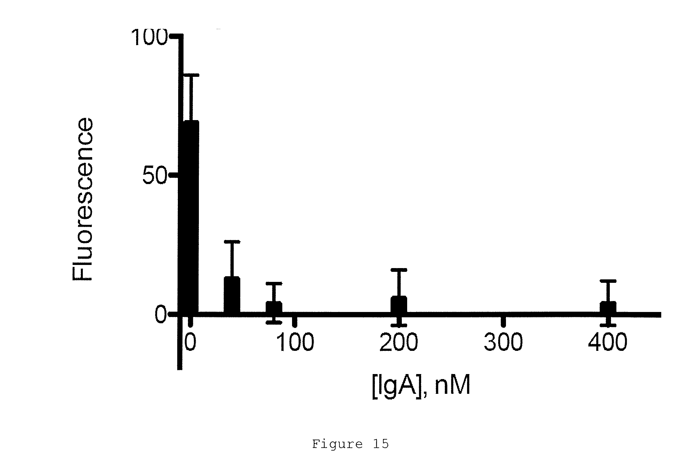
D00014

D00015
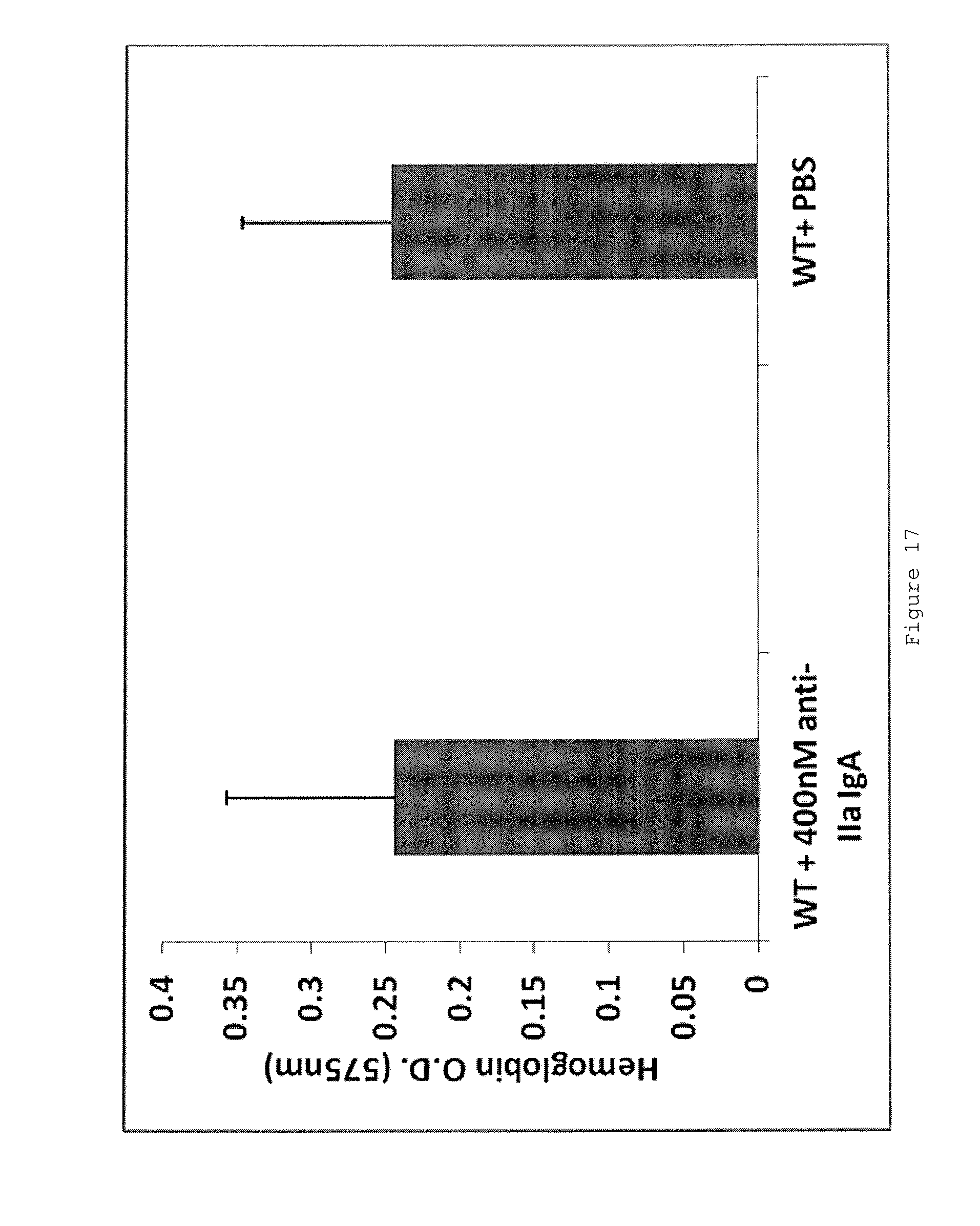
D00016

D00017
D00018

D00019
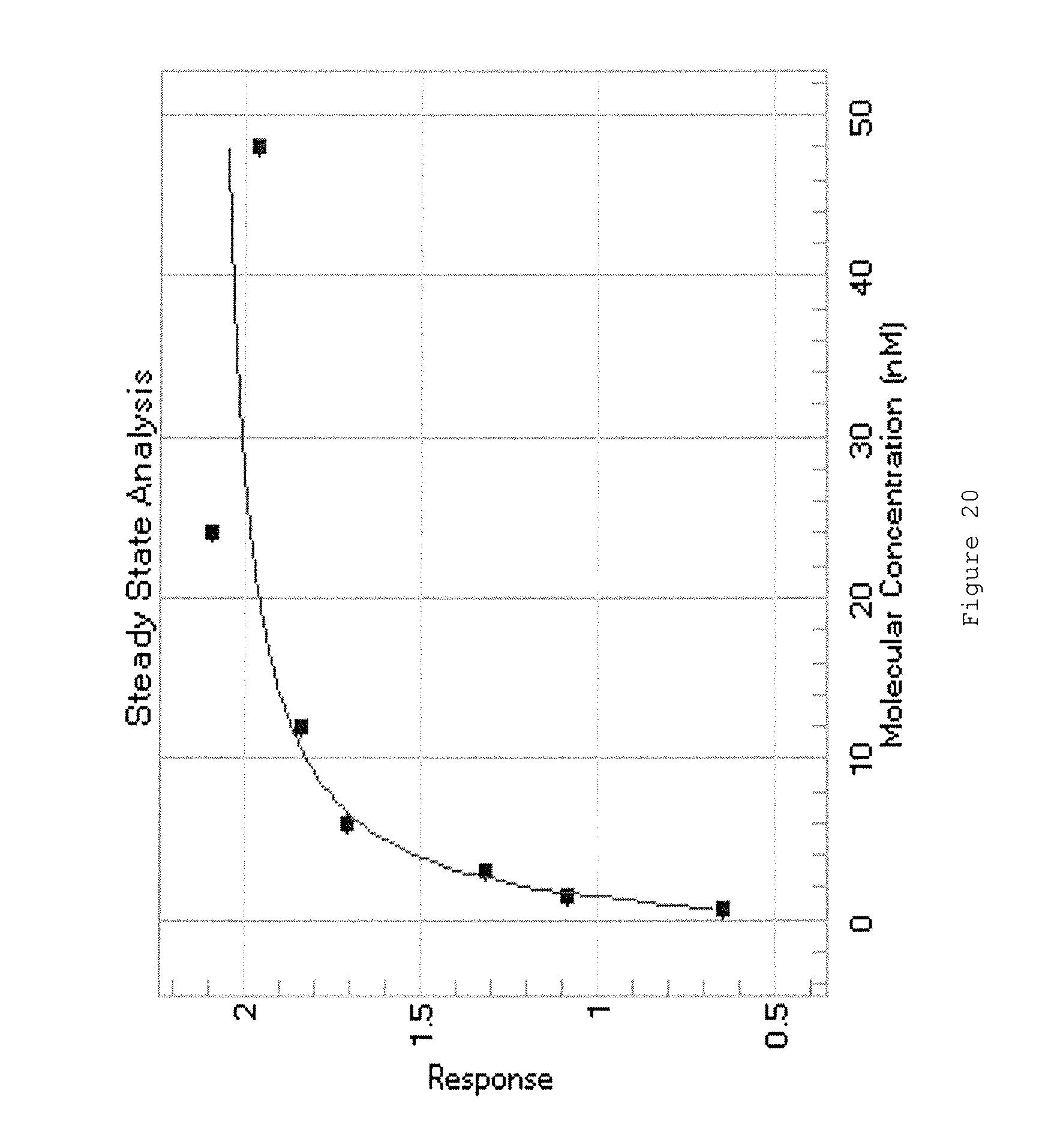
D00020

D00021
D00022
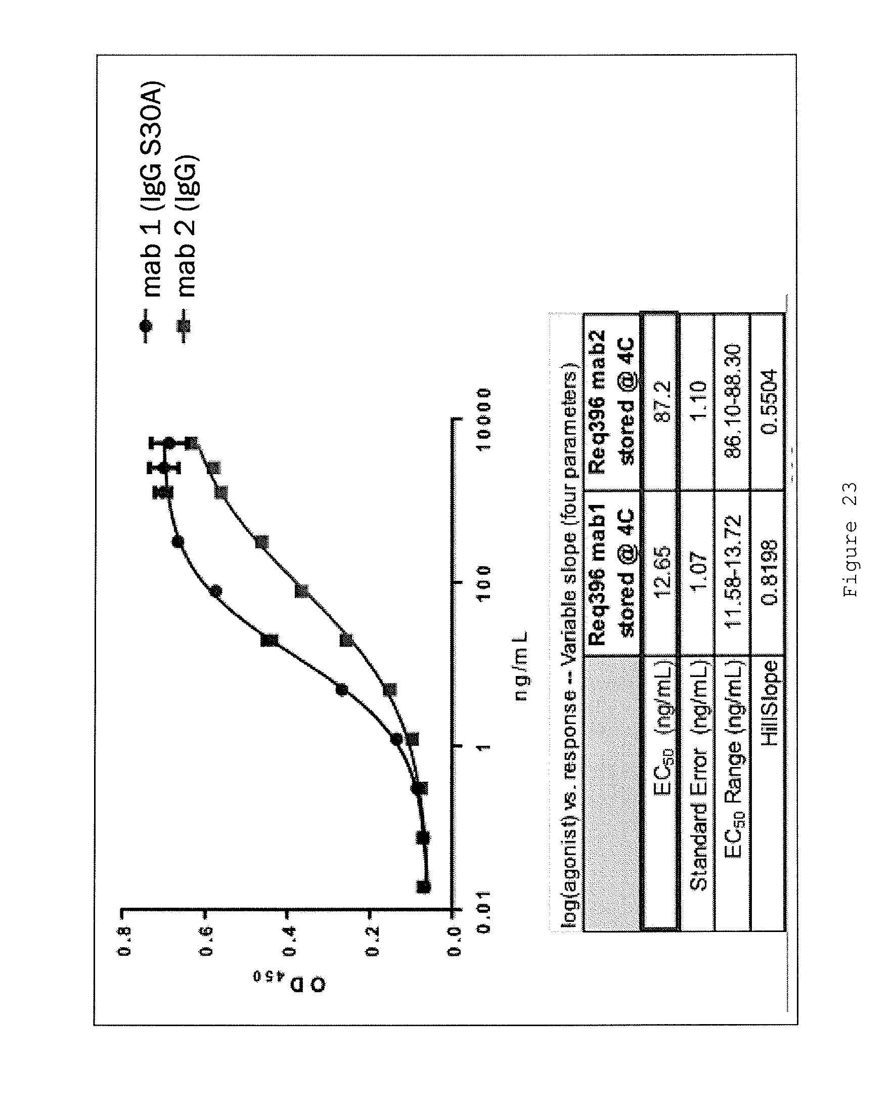
D00023

D00024

D00025
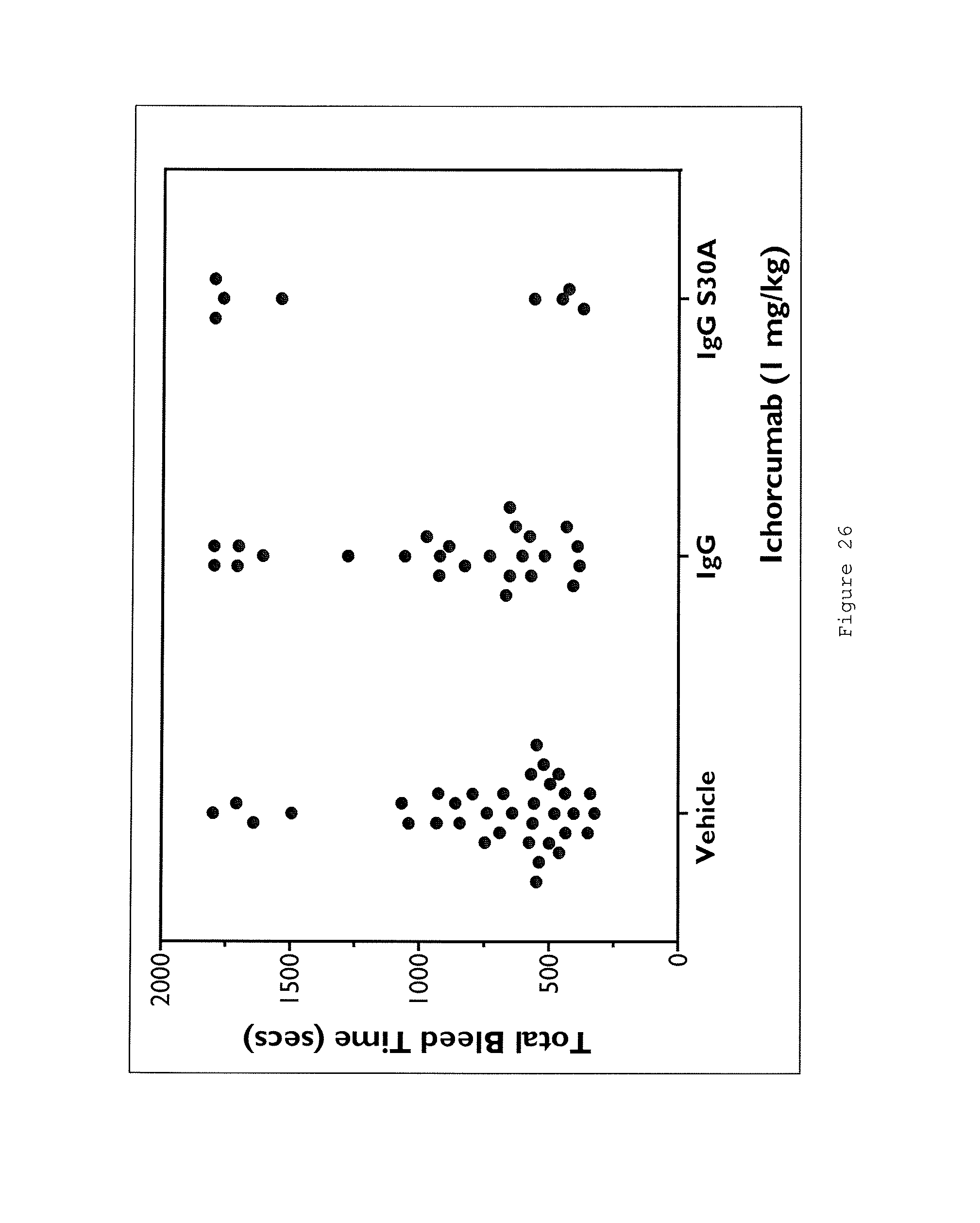
D00026

D00027
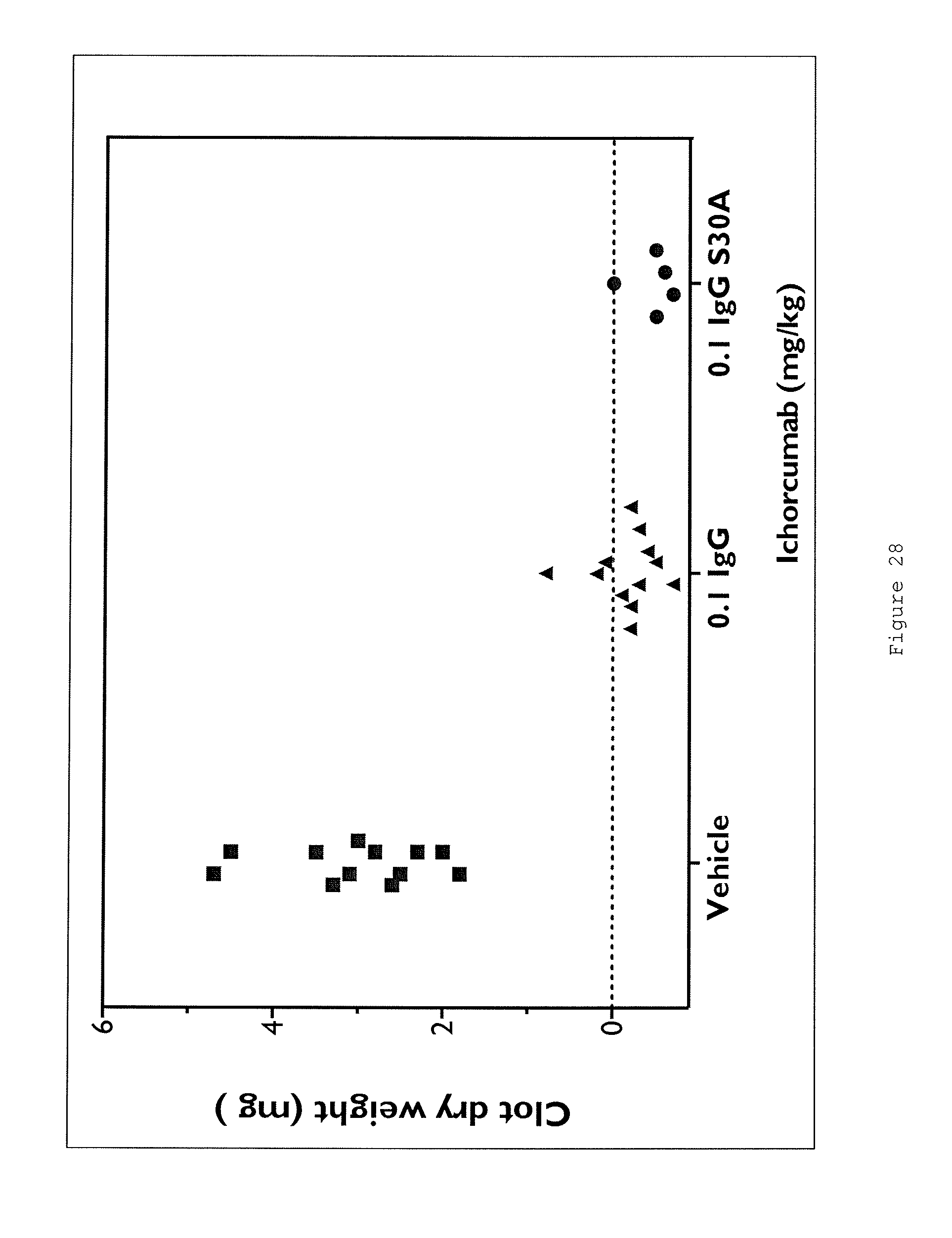
D00028

S00001
XML
uspto.report is an independent third-party trademark research tool that is not affiliated, endorsed, or sponsored by the United States Patent and Trademark Office (USPTO) or any other governmental organization. The information provided by uspto.report is based on publicly available data at the time of writing and is intended for informational purposes only.
While we strive to provide accurate and up-to-date information, we do not guarantee the accuracy, completeness, reliability, or suitability of the information displayed on this site. The use of this site is at your own risk. Any reliance you place on such information is therefore strictly at your own risk.
All official trademark data, including owner information, should be verified by visiting the official USPTO website at www.uspto.gov. This site is not intended to replace professional legal advice and should not be used as a substitute for consulting with a legal professional who is knowledgeable about trademark law.