System and method to estimate region of tissue activation
Lujan , et al.
U.S. patent number 10,360,511 [Application Number 13/839,906] was granted by the patent office on 2019-07-23 for system and method to estimate region of tissue activation. This patent grant is currently assigned to THE CLEVELAND CLINIC FOUNDATION. The grantee listed for this patent is Ashutosh Chaturvedi, J. Luis Lujan, Cameron McIntyre. Invention is credited to Ashutosh Chaturvedi, J. Luis Lujan, Cameron McIntyre.


View All Diagrams
| United States Patent | 10,360,511 |
| Lujan , et al. | July 23, 2019 |
System and method to estimate region of tissue activation
Abstract
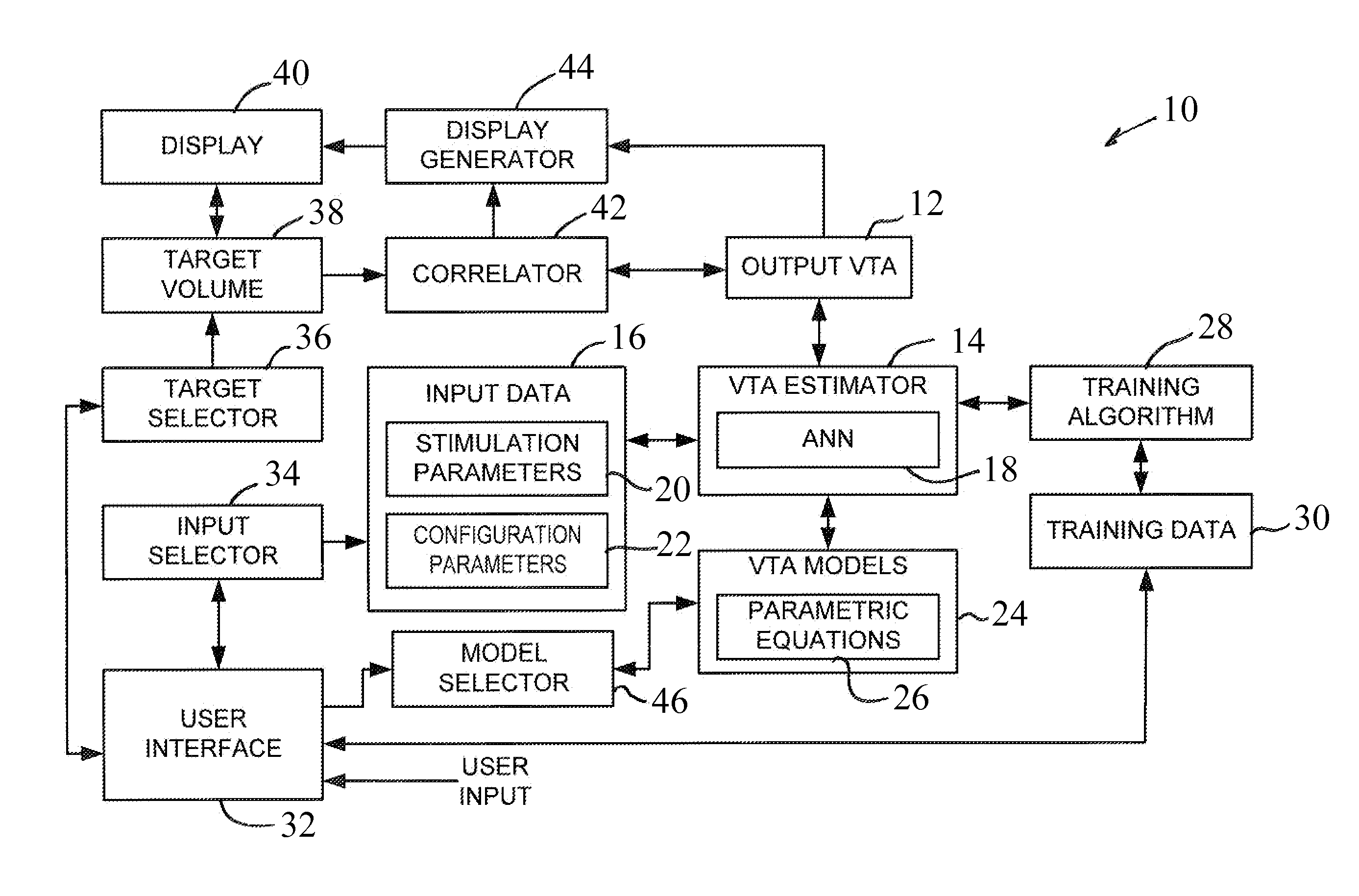
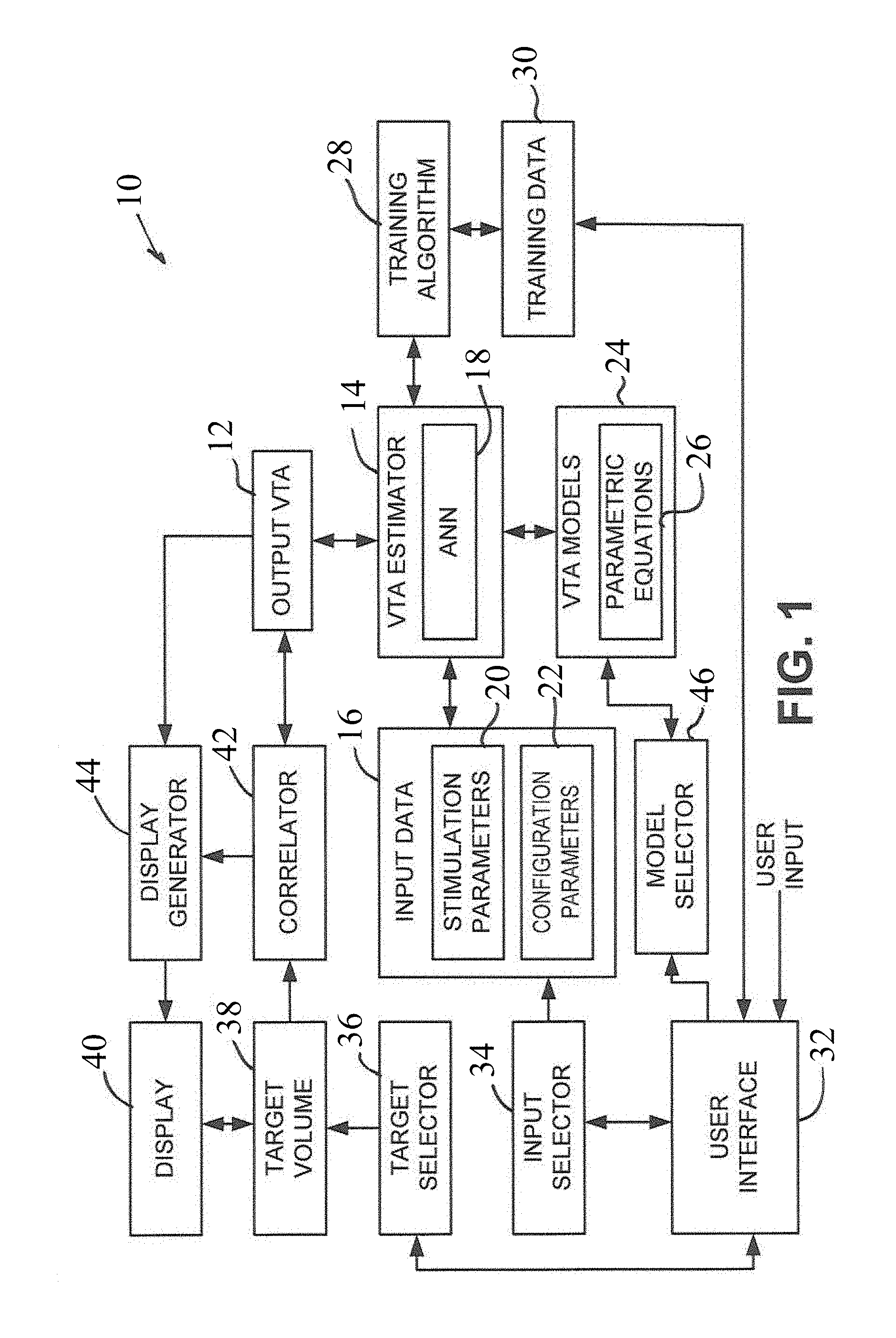
A computer-implemented method for determining the volume of activation of neural tissue. In one embodiment, the method uses one or more parametric equations that define a volume of activation, wherein the parameters for the one or more parametric equations are given as a function of an input vector that includes stimulation parameters. After receiving input data that includes values for the stimulation parameters and defining the input vector using the input data, the input vector is applied to the function to obtain the parameters for the one or more parametric equations. The parametric equation is solved to obtain a calculated volume of activation.
| Inventors: | Lujan; J. Luis (Mayfield Heights, OH), Chaturvedi; Ashutosh (Powell, OH), McIntyre; Cameron (Cleveland, OH) | ||||||||||
|---|---|---|---|---|---|---|---|---|---|---|---|
| Applicant: |
|
||||||||||
| Assignee: | THE CLEVELAND CLINIC FOUNDATION
(Cleveland, OH) |
||||||||||
| Family ID: | 43037018 | ||||||||||
| Appl. No.: | 13/839,906 | ||||||||||
| Filed: | March 15, 2013 |
Prior Publication Data
| Document Identifier | Publication Date | |
|---|---|---|
| US 20130218819 A1 | Aug 22, 2013 | |
Related U.S. Patent Documents
| Application Number | Filing Date | Patent Number | Issue Date | ||
|---|---|---|---|---|---|
| 12869159 | Aug 26, 2010 | 8589316 | |||
| 13480858 | May 25, 2012 | 8538543 | |||
| 11606260 | Nov 28, 2006 | 8209027 | |||
| 61237375 | Aug 27, 2009 | ||||
| 60740031 | Nov 28, 2005 | ||||
| Current U.S. Class: | 1/1 |
| Current CPC Class: | G06N 20/00 (20190101); G16H 50/50 (20180101); A61N 1/36185 (20130101); G06F 19/3481 (20130101); G16H 20/40 (20180101); A61N 1/0534 (20130101); G06K 9/6269 (20130101); G06N 7/005 (20130101); G16H 20/30 (20180101); G06K 9/6256 (20130101); G06N 5/025 (20130101); A61N 1/36082 (20130101) |
| Current International Class: | A61N 1/36 (20060101); G06N 20/00 (20190101); G16H 50/50 (20180101); G06K 9/62 (20060101); G06N 5/02 (20060101); G06N 7/00 (20060101); A61N 1/05 (20060101) |
References Cited [Referenced By]
U.S. Patent Documents
| 7 | August 1836 | Blanchard |
| 3999555 | December 1976 | Person |
| 4144889 | March 1979 | Tyers et al. |
| 4177818 | December 1979 | De Pedro |
| 4341221 | July 1982 | Testerman |
| 4378797 | April 1983 | Osterholm |
| 4445500 | May 1984 | Osterholm |
| 4735208 | April 1988 | Wyler et al. |
| 4765341 | August 1988 | Mower et al. |
| 4841973 | June 1989 | Stecker |
| 5067495 | November 1991 | Brehm |
| 5099846 | March 1992 | Hardy |
| 5222494 | June 1993 | Baker, Jr. |
| 5255693 | October 1993 | Dutcher |
| 5259387 | November 1993 | dePinto |
| 5304206 | April 1994 | Baker, Jr. et al. |
| 5344438 | September 1994 | Testerman et al. |
| 5361763 | November 1994 | Kao et al. |
| 5452407 | September 1995 | Crook |
| 5560360 | October 1996 | Filler et al. |
| 5565949 | October 1996 | Kasha, Jr. |
| 5593427 | January 1997 | Gliner et al. |
| 5601612 | February 1997 | Gliner et al. |
| 5607454 | March 1997 | Cameron et al. |
| 5620470 | April 1997 | Gliner et al. |
| 5651767 | July 1997 | Schulmann |
| 5711316 | January 1998 | Elsberry et al. |
| 5713922 | February 1998 | King |
| 5716377 | February 1998 | Rise et al. |
| 5724985 | March 1998 | Snell et al. |
| 5749904 | May 1998 | Gliner et al. |
| 5749905 | May 1998 | Gliner et al. |
| 5776170 | July 1998 | MacDonald et al. |
| 5778238 | July 1998 | Hofhine |
| 5782762 | July 1998 | Vining |
| 5843148 | December 1998 | Gijsbers et al. |
| 5859922 | January 1999 | Hoffmann |
| 5868740 | February 1999 | LeVeen et al. |
| 5895416 | April 1999 | Barreras, Sr. et al. |
| 5897583 | April 1999 | Meyer et al. |
| 5910804 | June 1999 | Fortenbery et al. |
| 5925070 | July 1999 | King et al. |
| 5938688 | August 1999 | Schiff |
| 5938690 | August 1999 | Law et al. |
| 5978713 | November 1999 | Prutchi et al. |
| 6016449 | January 2000 | Fischell et al. |
| 6029090 | February 2000 | Herbst |
| 6029091 | February 2000 | de la Rama et al. |
| 6050992 | April 2000 | Nichols |
| 6058331 | May 2000 | King |
| 6066163 | May 2000 | John |
| 6083162 | July 2000 | Vining |
| 6094598 | July 2000 | Elsberry et al. |
| 6096756 | August 2000 | Crain et al. |
| 6106460 | August 2000 | Panescu et al. |
| 6109269 | August 2000 | Rise et al. |
| 6128538 | October 2000 | Fischell et al. |
| 6129685 | October 2000 | Howard, III |
| 6146390 | November 2000 | Heilbrun et al. |
| 6161044 | December 2000 | Silverstone |
| 6167311 | December 2000 | Rezai |
| 6181969 | January 2001 | Gord |
| 6192266 | February 2001 | Dupree et al. |
| 6205361 | March 2001 | Kuzma |
| 6208881 | March 2001 | Champeau |
| 6240308 | May 2001 | Hardy et al. |
| 6246912 | June 2001 | Sluijter et al. |
| 6253109 | June 2001 | Gielen |
| 6289239 | September 2001 | Panescu et al. |
| 6301492 | October 2001 | Zonenshayn |
| 6310619 | October 2001 | Rice |
| 6319241 | November 2001 | King |
| 6330466 | December 2001 | Hofmann et al. |
| 6336899 | January 2002 | Yamazaki |
| 6343226 | January 2002 | Sunde et al. |
| 6351675 | February 2002 | Tholen et al. |
| 6353762 | March 2002 | Baudino et al. |
| 6366813 | April 2002 | Dilorenzo |
| 6368331 | April 2002 | Front et al. |
| 6389311 | May 2002 | Whayne et al. |
| 6393325 | May 2002 | Mann et al. |
| 6421566 | July 2002 | Holsheimer |
| 6435878 | August 2002 | Reynolds et al. |
| 6442432 | August 2002 | Lee |
| 6463328 | October 2002 | John |
| 6470207 | October 2002 | Simon et al. |
| 6491699 | December 2002 | Henderson et al. |
| 6494831 | December 2002 | Koritzinsky |
| 6507759 | January 2003 | Prutchi et al. |
| 6510347 | January 2003 | Borkan |
| 6516227 | February 2003 | Meadows et al. |
| 6517480 | February 2003 | Krass |
| 6526415 | February 2003 | Smith et al. |
| 6539263 | March 2003 | Schiff et al. |
| 6560490 | May 2003 | Grill et al. |
| 6579280 | June 2003 | Kovach et al. |
| 6600956 | July 2003 | Maschino et al. |
| 6606523 | August 2003 | Jenkins |
| 6609029 | August 2003 | Mann et al. |
| 6609031 | August 2003 | Law et al. |
| 6609032 | August 2003 | Woods et al. |
| 6622048 | September 2003 | Mann et al. |
| 6631297 | October 2003 | Mo |
| 6654642 | November 2003 | North et al. |
| 6662053 | December 2003 | Borkan |
| 6675046 | January 2004 | Holsheimer |
| 6684106 | January 2004 | Herbst |
| 6687392 | February 2004 | Touzawa et al. |
| 6690972 | February 2004 | Conley et al. |
| 6690974 | February 2004 | Archer et al. |
| 6692315 | February 2004 | Soumillion et al. |
| 6694162 | February 2004 | Hartlep |
| 6694163 | February 2004 | Vining |
| 6708096 | March 2004 | Frei et al. |
| 6741892 | May 2004 | Meadows et al. |
| 6748098 | June 2004 | Rosenfeld |
| 6748276 | June 2004 | Daignault, Jr. et al. |
| 6754374 | June 2004 | Miller et al. |
| 6778846 | August 2004 | Martinez et al. |
| 6788969 | September 2004 | Dupree et al. |
| 6795737 | September 2004 | Gielen et al. |
| 6827681 | December 2004 | Tanner et al. |
| 6830544 | December 2004 | Tanner |
| 6845267 | January 2005 | Harrison et al. |
| 6850802 | February 2005 | Holsheimer |
| 6873872 | March 2005 | Gluckman et al. |
| 6892090 | May 2005 | Verard et al. |
| 6895280 | May 2005 | Meadows et al. |
| 6909913 | June 2005 | Vining |
| 6937891 | August 2005 | Leinders et al. |
| 6937903 | August 2005 | Schuler et al. |
| 6944497 | September 2005 | Stypulkowski |
| 6944501 | September 2005 | Pless |
| 6950707 | September 2005 | Whitehurst |
| 6969388 | November 2005 | Goldman et al. |
| 6993384 | January 2006 | Bradley et al. |
| 7003349 | February 2006 | Andersson et al. |
| 7003352 | February 2006 | Whitehurst |
| 7008370 | March 2006 | Tanner et al. |
| 7008413 | March 2006 | Kovach et al. |
| 7035690 | April 2006 | Goetz |
| 7043293 | May 2006 | Baura |
| 7047082 | May 2006 | Schrom et al. |
| 7047084 | May 2006 | Erickson et al. |
| 7050857 | May 2006 | Samuelsson et al. |
| 7054692 | May 2006 | Whitehurst et al. |
| 7058446 | June 2006 | Schuler et al. |
| 7082333 | July 2006 | Bauhahn et al. |
| 7107102 | September 2006 | Daignault, Jr. et al. |
| 7126000 | October 2006 | Ogawa et al. |
| 7127297 | October 2006 | Law et al. |
| 7136518 | November 2006 | Griffin et al. |
| 7136695 | November 2006 | Pless et al. |
| 7142923 | November 2006 | North et al. |
| 7146219 | December 2006 | Sieracki et al. |
| 7146223 | December 2006 | King |
| 7151961 | December 2006 | Whitehurst |
| 7155279 | December 2006 | Whitehurst |
| 7167760 | January 2007 | Dawant et al. |
| 7177674 | February 2007 | Echauz et al. |
| 7181286 | February 2007 | Sieracki et al. |
| 7184837 | February 2007 | Goetz |
| 7191014 | March 2007 | Kobayashi et al. |
| 7209787 | April 2007 | Dilorenzo |
| 7211050 | May 2007 | Caplygin |
| 7216000 | May 2007 | Sieracki et al. |
| 7217276 | May 2007 | Henderson |
| 7218968 | May 2007 | Condie et al. |
| 7228179 | June 2007 | Campen et al. |
| 7231254 | June 2007 | DiLorenzo |
| 7236830 | June 2007 | Gliner |
| 7239910 | July 2007 | Tanner |
| 7239916 | July 2007 | Thompson et al. |
| 7239926 | July 2007 | Goetz |
| 7242984 | July 2007 | DiLorenzo |
| 7244150 | July 2007 | Brase et al. |
| 7252090 | August 2007 | Goetz |
| 7254445 | August 2007 | Law et al. |
| 7254446 | August 2007 | Erickson |
| 7257447 | August 2007 | Cates et al. |
| 7266412 | September 2007 | Stypulkowski |
| 7294107 | November 2007 | Simon et al. |
| 7295876 | November 2007 | Erickson |
| 7299096 | November 2007 | Balzer et al. |
| 7308302 | December 2007 | Schuler et al. |
| 7313430 | December 2007 | Urquhart |
| 7324851 | January 2008 | DiLorenzo |
| 7346382 | March 2008 | McIntyre |
| 7388974 | June 2008 | Yanagita |
| 7437193 | October 2008 | Parramon et al. |
| 7463928 | December 2008 | Lee et al. |
| 7499048 | March 2009 | Sieracki et al. |
| 7505815 | March 2009 | Lee et al. |
| 7520848 | April 2009 | Schneider et al. |
| 7548786 | June 2009 | Lee et al. |
| 7565199 | July 2009 | Sheffield et al. |
| 7603177 | October 2009 | Sieracki et al. |
| 7617002 | November 2009 | Goetz |
| 7623918 | November 2009 | Goetz |
| 7650184 | January 2010 | Walter |
| 7657319 | February 2010 | Goetz et al. |
| 7672734 | March 2010 | Anderson et al. |
| 7676273 | March 2010 | Goetz et al. |
| 7680526 | March 2010 | McIntyre et al. |
| 7734340 | June 2010 | De Ridder |
| 7761165 | July 2010 | He et al. |
| 7826902 | November 2010 | Stone et al. |
| 7848802 | December 2010 | Goetz et al. |
| 7860548 | December 2010 | McIntyre et al. |
| 7904134 | March 2011 | McIntyre et al. |
| 7945105 | May 2011 | Jaenisch |
| 7949395 | May 2011 | Kuzma |
| 7974706 | July 2011 | Moffitt et al. |
| 8019439 | September 2011 | Kuzma et al. |
| 8175710 | May 2012 | He |
| 8180601 | May 2012 | Butson et al. |
| 8195300 | June 2012 | Gliner et al. |
| 8209027 | June 2012 | Butson et al. |
| 8224450 | July 2012 | Brase |
| 8257684 | September 2012 | Covalin et al. |
| 8262714 | September 2012 | Hulvershorn et al. |
| 8364278 | January 2013 | Pianca et al. |
| 8429174 | April 2013 | Ramani et al. |
| 8452415 | May 2013 | Goetz et al. |
| 8543189 | September 2013 | Paitel et al. |
| 8620452 | December 2013 | Butson et al. |
| 8918184 | December 2014 | Torgerson et al. |
| 2001/0029509 | October 2001 | Smith et al. |
| 2001/0031071 | October 2001 | Nichols et al. |
| 2002/0032375 | March 2002 | Bauch et al. |
| 2002/0062143 | May 2002 | Baudino et al. |
| 2002/0087201 | July 2002 | Firlik et al. |
| 2002/0099295 | July 2002 | Gil et al. |
| 2002/0115603 | August 2002 | Whitehouse |
| 2002/0116030 | August 2002 | Rezei |
| 2002/0123780 | September 2002 | Grill et al. |
| 2002/0128694 | September 2002 | Holsheimer |
| 2002/0151939 | October 2002 | Rezai |
| 2002/0183607 | December 2002 | Bauch et al. |
| 2002/0183740 | December 2002 | Edwards et al. |
| 2002/0183817 | December 2002 | Van Venrooij et al. |
| 2003/0013951 | January 2003 | Stefanescu et al. |
| 2003/0097159 | May 2003 | Schiff et al. |
| 2003/0149450 | August 2003 | Mayberg |
| 2003/0171791 | September 2003 | KenKnight et al. |
| 2003/0212439 | November 2003 | Schuler et al. |
| 2003/0216630 | November 2003 | Jersey-Willuhn et al. |
| 2003/0228042 | December 2003 | Sinha |
| 2004/0034394 | February 2004 | Woods et al. |
| 2004/0044279 | March 2004 | Lewin et al. |
| 2004/0044378 | March 2004 | Holsheimer |
| 2004/0044379 | March 2004 | Holsheimer |
| 2004/0054297 | March 2004 | Wingeier et al. |
| 2004/0059395 | March 2004 | North et al. |
| 2004/0092809 | May 2004 | DeCharms |
| 2004/0096089 | May 2004 | Borsook et al. |
| 2004/0106916 | June 2004 | Quaid et al. |
| 2004/0133248 | July 2004 | Frei et al. |
| 2004/0152957 | August 2004 | Stivoric et al. |
| 2004/0181262 | September 2004 | Bauhahn |
| 2004/0186532 | September 2004 | Tadlock |
| 2004/0199216 | October 2004 | Lee et al. |
| 2004/0267330 | December 2004 | Lee et al. |
| 2005/0021090 | January 2005 | Schuler et al. |
| 2005/0033380 | February 2005 | Tanner et al. |
| 2005/0049649 | March 2005 | Luders et al. |
| 2005/0060001 | March 2005 | Singhal et al. |
| 2005/0060009 | March 2005 | Goetz |
| 2005/0070781 | March 2005 | Dawant et al. |
| 2005/0075689 | April 2005 | Toy et al. |
| 2005/0085714 | April 2005 | Foley et al. |
| 2005/0165294 | July 2005 | Weiss |
| 2005/0171587 | August 2005 | Daglow et al. |
| 2005/0205566 | September 2005 | Kassayan |
| 2005/0228250 | October 2005 | Bitter et al. |
| 2005/0251061 | November 2005 | Schuler et al. |
| 2005/0261061 | November 2005 | Nguyen et al. |
| 2005/0261601 | November 2005 | Schuler et al. |
| 2005/0261747 | November 2005 | Schuler et al. |
| 2005/0267347 | December 2005 | Oster |
| 2005/0288732 | December 2005 | Schuler et al. |
| 2006/0004422 | January 2006 | De Ridder |
| 2006/0017749 | January 2006 | McIntyre et al. |
| 2006/0020292 | January 2006 | Goetz et al. |
| 2006/0069415 | March 2006 | Cameron et al. |
| 2006/0094951 | May 2006 | Dean et al. |
| 2006/0095088 | May 2006 | De Riddler |
| 2006/0155340 | July 2006 | Schuler et al. |
| 2006/0206169 | September 2006 | Schuler |
| 2006/0218007 | September 2006 | Bjorner et al. |
| 2006/0224189 | October 2006 | Schuler et al. |
| 2006/0235472 | October 2006 | Goetz et al. |
| 2006/0259079 | November 2006 | King |
| 2006/0259099 | November 2006 | Goetz et al. |
| 2007/0000372 | January 2007 | Rezai et al. |
| 2007/0017749 | January 2007 | Dold et al. |
| 2007/0027514 | February 2007 | Gerber |
| 2007/0043268 | February 2007 | Russell |
| 2007/0049817 | March 2007 | Preiss et al. |
| 2007/0067003 | March 2007 | Sanchez et al. |
| 2007/0078498 | April 2007 | Rezai et al. |
| 2007/0083104 | April 2007 | Butson et al. |
| 2007/0123953 | May 2007 | Lee et al. |
| 2007/0129769 | June 2007 | Bourget et al. |
| 2007/0135855 | June 2007 | Foshee et al. |
| 2007/0150036 | June 2007 | Anderson |
| 2007/0156186 | July 2007 | Lee et al. |
| 2007/0162086 | July 2007 | DiLorenzo |
| 2007/0162235 | July 2007 | Zhan et al. |
| 2007/0168004 | July 2007 | Walter |
| 2007/0168007 | July 2007 | Kuzma et al. |
| 2007/0185544 | August 2007 | Dawant et al. |
| 2007/0191887 | August 2007 | Schuler et al. |
| 2007/0191912 | August 2007 | Ficher et al. |
| 2007/0197891 | August 2007 | Shachar et al. |
| 2007/0203450 | August 2007 | Berry |
| 2007/0203532 | August 2007 | Tass et al. |
| 2007/0203537 | August 2007 | Goetz et al. |
| 2007/0203538 | August 2007 | Stone et al. |
| 2007/0203539 | August 2007 | Stone et al. |
| 2007/0203540 | August 2007 | Goetz et al. |
| 2007/0203541 | August 2007 | Goetz et al. |
| 2007/0203543 | August 2007 | Stone et al. |
| 2007/0203544 | August 2007 | Goetz et al. |
| 2007/0203545 | August 2007 | Stone et al. |
| 2007/0203546 | August 2007 | Stone et al. |
| 2007/0213789 | September 2007 | Nolan et al. |
| 2007/0213790 | September 2007 | Nolan et al. |
| 2007/0244519 | October 2007 | Keacher et al. |
| 2007/0245318 | October 2007 | Goetz et al. |
| 2007/0255321 | November 2007 | Gerber et al. |
| 2007/0255322 | November 2007 | Gerber et al. |
| 2007/0265664 | November 2007 | Gerber et al. |
| 2007/0266280 | November 2007 | Ng et al. |
| 2007/0276441 | November 2007 | Goetz |
| 2007/0282189 | December 2007 | Dan et al. |
| 2007/0288064 | December 2007 | Butson et al. |
| 2008/0027514 | January 2008 | DeMulling et al. |
| 2008/0039895 | February 2008 | Fowler et al. |
| 2008/0071150 | March 2008 | Miesel et al. |
| 2008/0081982 | April 2008 | Simon et al. |
| 2008/0086451 | April 2008 | Torres et al. |
| 2008/0103533 | May 2008 | Patel et al. |
| 2008/0114233 | May 2008 | McIntyre et al. |
| 2008/0114579 | May 2008 | McIntyre et al. |
| 2008/0123922 | May 2008 | Gielen et al. |
| 2008/0123923 | May 2008 | Gielen et al. |
| 2008/0133141 | June 2008 | Frost |
| 2008/0141217 | June 2008 | Goetz et al. |
| 2008/0154340 | June 2008 | Goetz et al. |
| 2008/0154341 | June 2008 | McIntyre et al. |
| 2008/0163097 | July 2008 | Goetz et al. |
| 2008/0183256 | July 2008 | Keacher |
| 2008/0188734 | August 2008 | Suryanarayanan et al. |
| 2008/0215118 | September 2008 | Goetz et al. |
| 2008/0227139 | September 2008 | Deisseroth et al. |
| 2008/0242950 | October 2008 | Jung et al. |
| 2008/0261165 | October 2008 | Steingart et al. |
| 2008/0269588 | October 2008 | Csavoy et al. |
| 2008/0300654 | December 2008 | Lambert et al. |
| 2008/0300797 | December 2008 | Tabibiazar et al. |
| 2009/0016491 | January 2009 | Li |
| 2009/0054950 | February 2009 | Stephens |
| 2009/0082640 | March 2009 | Kovach et al. |
| 2009/0082829 | March 2009 | Panken et al. |
| 2009/0112289 | April 2009 | Lee et al. |
| 2009/0118635 | May 2009 | Lujan et al. |
| 2009/0118786 | May 2009 | Meadows et al. |
| 2009/0149917 | June 2009 | Whitehurst et al. |
| 2009/0196471 | August 2009 | Goetz et al. |
| 2009/0196472 | August 2009 | Goetz et al. |
| 2009/0198306 | August 2009 | Goetz et al. |
| 2009/0198354 | August 2009 | Wilson |
| 2009/0204192 | August 2009 | Carlton et al. |
| 2009/0208073 | August 2009 | McIntyre et al. |
| 2009/0210208 | August 2009 | McIntyre et al. |
| 2009/0242399 | October 2009 | Kamath et al. |
| 2009/0276008 | November 2009 | Lee et al. |
| 2009/0281595 | November 2009 | King et al. |
| 2009/0281596 | November 2009 | King et al. |
| 2009/0287271 | November 2009 | Blum et al. |
| 2009/0287272 | November 2009 | Kokones et al. |
| 2009/0287273 | November 2009 | Carlton et al. |
| 2009/0287467 | November 2009 | Sparks et al. |
| 2009/0299164 | December 2009 | Singhal et al. |
| 2009/0299165 | December 2009 | Singhal et al. |
| 2009/0299380 | December 2009 | Singhal et al. |
| 2010/0010566 | January 2010 | Thacker et al. |
| 2010/0010646 | January 2010 | Drew et al. |
| 2010/0023103 | January 2010 | Elborno |
| 2010/0023130 | January 2010 | Henry et al. |
| 2010/0030312 | February 2010 | Shen |
| 2010/0049276 | February 2010 | Blum et al. |
| 2010/0049280 | February 2010 | Goetz |
| 2010/0064249 | March 2010 | Groetken |
| 2010/0113959 | May 2010 | Pascual-Leone et al. |
| 2010/0121409 | May 2010 | Kothandaraman et al. |
| 2010/0135553 | June 2010 | Joglekar |
| 2010/0137944 | June 2010 | Zhu |
| 2010/0152604 | June 2010 | Kuala et al. |
| 2010/0179562 | July 2010 | Linker et al. |
| 2010/0324410 | December 2010 | Paek et al. |
| 2010/0331883 | December 2010 | Schmitz et al. |
| 2011/0040351 | February 2011 | Butson et al. |
| 2011/0066407 | March 2011 | Butson et al. |
| 2011/0172737 | July 2011 | Davis et al. |
| 2011/0184487 | July 2011 | Alberts et al. |
| 2011/0191275 | August 2011 | Lujan et al. |
| 2011/0196253 | August 2011 | McIntyre et al. |
| 2011/0213440 | September 2011 | Fowler et al. |
| 2011/0306845 | December 2011 | Osorio |
| 2011/0306846 | December 2011 | Osorio |
| 2011/0307032 | December 2011 | Goetz et al. |
| 2012/0027272 | February 2012 | Akinyemi et al. |
| 2012/0046715 | February 2012 | Moffitt et al. |
| 2012/0078106 | March 2012 | Dentinger et al. |
| 2012/0089205 | April 2012 | Boyden et al. |
| 2012/0116476 | May 2012 | Kothandaraman |
| 2012/0165898 | June 2012 | Moffitt |
| 2012/0165901 | June 2012 | Zhu et al. |
| 2012/0207378 | August 2012 | Gupta et al. |
| 2012/0226138 | September 2012 | DeSalles et al. |
| 2012/0229468 | September 2012 | Lee et al. |
| 2012/0265262 | October 2012 | Osorio |
| 2012/0265268 | October 2012 | Blum et al. |
| 2012/0302912 | November 2012 | Moffitt et al. |
| 2012/0303087 | November 2012 | Moffitt et al. |
| 2012/0314924 | December 2012 | Carlton et al. |
| 2012/0316619 | December 2012 | Goetz et al. |
| 2013/0039550 | February 2013 | Blum et al. |
| 2013/0060305 | March 2013 | Bokil |
| 2013/0116748 | May 2013 | Bokil et al. |
| 2013/0116749 | May 2013 | Carlton et al. |
| 2013/0116929 | May 2013 | Carlton et al. |
| 2014/0067018 | March 2014 | Carcieri et al. |
| 2014/0277284 | September 2014 | Chen et al. |
| 2015/0134031 | May 2015 | Moffitt et al. |
| 1048320 | Nov 2000 | EP | |||
| 1166819 | Jan 2002 | EP | |||
| 1372780 | Jan 2004 | EP | |||
| 1372780 | Jan 2004 | EP | |||
| 1559369 | Aug 2005 | EP | |||
| 97/39797 | Oct 1997 | WO | |||
| 98/48880 | Nov 1998 | WO | |||
| 01/90876 | Nov 2001 | WO | |||
| 02/26314 | Apr 2002 | WO | |||
| 02/28473 | Apr 2002 | WO | |||
| 02/065896 | Aug 2002 | WO | |||
| 02065896 | Aug 2002 | WO | |||
| 02/072192 | Sep 2002 | WO | |||
| 03/086185 | Oct 2003 | WO | |||
| 03086185 | Oct 2003 | WO | |||
| 2004/019799 | Mar 2004 | WO | |||
| 2004041080 | May 2005 | WO | |||
| 2006017053 | Feb 2006 | WO | |||
| 2006017053 | Feb 2006 | WO | |||
| 2006113305 | Oct 2006 | WO | |||
| 20071097859 | Aug 2007 | WO | |||
| 20071097861 | Aug 2007 | WO | |||
| 2007/100427 | Sep 2007 | WO | |||
| 2007/100428 | Sep 2007 | WO | |||
| 07/115120 | Oct 2007 | WO | |||
| 2007/112061 | Oct 2007 | WO | |||
| 2009097224 | Aug 2009 | WO | |||
| 2010/120823 | Oct 2010 | WO | |||
| 2011025865 | Mar 2011 | WO | |||
| 2011/139779 | Nov 2011 | WO | |||
| 2011/159688 | Dec 2011 | WO | |||
| 2012088482 | Jun 2012 | WO | |||
Other References
|
Tissue and electrode capacitance reduce neural activation volumes during deep brain stimulation Christopher R. Butson and Cameron C. McIntyre Department of Biomedical Engineering, Cleveland Clinic Foundation, 9500 Euclid Avenue ND20, Cleveland, OH 44195, USA. cited by examiner . Exploring Connectivity of the Brain's White Matter with Dynamic Queries Anthony Sherbondy, David Akers, Rachel Mackenzie, Robert Dougherty, and Brian Wandell. cited by examiner . Tissue and electrode capacitance reduce neural activation volumes during deep brain stimulationChristopher R. Butson and Cameron C. McIntyre Department of Biomedical Engineering, Cleveland Clinic Foundation, 9500 Euclid Avenue ND20, Cleveland, OH 44195, USA (Year: 2005). cited by examiner . Exploring Connectivity of the Brain's White Matter with Dynamic Queries Anthony Sherbondy, David Akers, Rachel Mackenzie, Robert Dougherty, and Brian Wandell (Year: 2005). cited by examiner . "BioPSE: The Biomedical Problem Solving Environment," http://www.sci.utah.edu/cibc/software/index.html, NCRR Center for Integrative biomedical Computing (2004), 5 pages. cited by applicant . "Deep-brain stimulation of the subthalamic nucleus or the pars interna of the globus pallidus in Parkinson's disease," N Engl J Med., 345(13), Author: Deep-Brain Stimulation for Parkinson's Disease Study Group (Sep. 27, 2001), pp. 956-963. cited by applicant . European Patent Office, International Search Report and Written Opinion in International Application No. PCT/US2005/023672, dated Jan. 20, 2006, 19 Pages. cited by applicant . European Patent Office, International Search Report and the Written Opinion in International Application No. PCT/US09/66821, dated Aug. 31, 2010, 19 pages. cited by applicant . European Patent Office, International Search Report and the Written Opinion in International Application No. PCT/US2010/046772, dated Nov. 23, 2010, 17 pages. cited by applicant . "U.S. Appl. No. 10/885,982, Restriction Requirement dated Nov. 2, 2005," 6 pgs. cited by applicant . "U.S. Appl. No. 10/885,982, Response filed Feb. 2, 2006 to Restriction Requirement dated Nov. 12, 2005," 18 pgs. cited by applicant . "U.S. Appl. No. 10/885,982, Non-Final Office Action dated Apr. 21, 2006," 20 pgs. cited by applicant . "U.S. Appl. No. 10/885,982, Response filed Jul. 21, 2006 to Non-Final Office Action dated Apr. 21, 2006," 24 pgs. cited by applicant . "U.S. Appl. No. 10/885,982, Final Office Action dated Dec. 12, 2006," 10 pgs. cited by applicant . "U.S. Appl. No. 10/885,982, Response filed Mar. 12, 2007 to Final Office Action dated Dec. 12, 2006," 26 pgs. cited by applicant . "U.S. Appl. No. 10/885,982, Non-Final Office Action dated Apr. 19, 2007," 17 pgs. cited by applicant . "U.S. Appl. No. 10/885,982, Interview Summary dated Apr. 19, 2007," 2 pgs. cited by applicant . "U.S. Appl. No. 10/885,982, Response filed Jul. 19, 2007 to Non-Final Office Action dated Apr. 19, 2007," 19 pgs. cited by applicant . "U.S. Appl. No. 10/885,982, Final Office Action dated Aug. 9, 2007," 8 pgs. cited by applicant . "U.S. Appl. No. 10/885,982, Notice of Allowance and Examiner's Amendment dated Oct. 5, 2007," 13 pgs. cited by applicant . "U.S. Appl. No. 10/885,982, Interview Summary and Proposed Claims dated Oct. 18, 2007," 14 pgs. cited by applicant . "U.S. Appl. No. 11/278,223 Response filed Jul. 15, 2008 to Non-Final Office Action dated Apr. 15, 2008," 10 pages. cited by applicant . "U.S. Appl. No. 11/278,223 Non-Final Office Action dated Apr. 15, 2008," 8 pages. cited by applicant . Adler, D E., et al., "The tentorial notch: anatomical variation, morphometric analysis, and classification in 100 human autopsy cases," J. Neurosurg., 96(6), (Jun. 2002), pp. 1103-1112. cited by applicant . Alexander, D C., et al., "Spatial transformations of diffusion tensor magnetic resonance images," IEEE Transactions on Medical Imaging, 20 (11), (2001), pp. 1131-1139. cited by applicant . Alo, K. M., et al., "New trends in neuromodulation for the management of neuropathic pain," Neurosurgery, 50(4), (Apr. 2002), pp. 690-704. cited by applicant . Andrews, R. J., "Neuroprotection trek--the next generation: neuromodulation I. Techniques--deep brain stimulation, vagus nerve stimulation, and transcranial magnetic stimulation," Ann NY Acad Sci. 993, (May 2003), pp. 1-13. cited by applicant . Andrews, R. J., "Neuroprotection trek--the next generation: neuromodulation II. Applications--epilepsy, nerve regeneration, neurotrophins," Ann NY Acad Sci., 993, (May 2003), pp. 14-24. cited by applicant . Ashby, P., et al., "Neurophysiological effects of stimulation through electrodes in the human subthalamic nucleus," Brain, 122 (Pt 10), (Oct. 1999), pp. 1919-1931. cited by applicant . Astrom, M., et al., "The effect of cystic cavities on deep brain stimulation in the basal ganglia: a simulation-based study," J Neural Eng., 3(2), (Jun. 2006), pp. 132-138. cited by applicant . Back, C., et al., "Postoperative Monitoring of the Electrical Properties of Tissue and Electrodes in Deep Brain Stimulation," Neuromodulation, 6(4), (Oct. 2003), pp. 248-253. cited by applicant . Baker, K. B., et al., "Evaluation of specific absorption rate as a dosimeter of MRI-related implant heating," J Magn Reson Imaging., 20 (2), (Aug. 2004), pp. 315-320. cited by applicant . Baker, K. B., et al., "Subthalamic nucleus deep brain stimulus evoked potentials: Physiological and therapeutic implications," Movement Disorders, 17(5), (Sep./Oct. 2002), pp. 969-983. cited by applicant . Bammer, R , et al., "Diffusion tensor imaging using single-shot SENSE-EPI," Magn Reson Med., 48(1), (Jul. 2002), pp. 128-136. cited by applicant . Basser, P J., et al., "MR diffusion tensor spectroscopy and imaging," Biophys J., 66(1), (Jan. 1994), pp. 259-267. cited by applicant . Basser, P J., et al., "New currents in electrical stimulation of excitable tissues," Annu Rev Biomed Eng., 2, (2000), pp. 377-397. cited by applicant . Bedard, C., et al., "Modeling extracellular field potentials and the frequency-filtering properties of extracellular space," Biophys J., 86(3), (Mar. 2004), pp. 1829-1842. cited by applicant . Benabid, A L., et al., "Chronic electrical stimulation of the ventralis intermedius nucleus of the thalamus as a treatment of movement disorders," J. Neurosurg., 84(2), (Feb. 1996), pp. 203-214. cited by applicant . Benabid, A L., et al., "Combined (thalamotoy and stimulation) stereotactic surgery of the VIM thalamic nucleus for bilateral Parkinson disease," Appl Neurophysiol, vol. 50, (1987), pp. 344-346. cited by applicant . Benabid, A. L., et al., "Future prospects of brain stimulation," Neurol Res., 22 (3), (Apr. 2000), pp. 237-246. cited by applicant . Benabid, A L., et al., "Long-term suppression of tremor by chronic stimulation of the ventral intermediate thalamic nucleus," Lancet, 337 (8738), (Feb. 16, 1991), pp. 403-406. cited by applicant . Brummer, S. B., et al., "Electrical Stimulation with Pt Electrodes: II--Estimation of Maximum Surface Redox (Theoretical Non-Gassing) Limits," IEEE Transactions on Biomedical Engineering, vol. BME-24, Issue 5, (Sep. 1977), pp. 440-443. cited by applicant . Butson, C. R., et al., "Deep Brain Stimulation of the Subthalamic Nucleus: Model-Based Analysis of the Effects of Electrode Capacitance on the Volume of Activation," Proceedings of the 2nd International IEEE EMBS, (Mar. 16-19, 2005), pp. 196-197. cited by applicant . Butson, C. R., et al., "Patient Specific Analysis of the volume of tissue activated during deep brain stimulation," NeuroImage, Academic Press, vol. 34, No. 2, Dec. 2, 2006, pp. 661-670. cited by applicant . Butson, C. R., et al., "Current Steering to Control the Volume of Tissue Activated During Deep Brain Stimulation," Brain Stimulation 1, 2008, pp. 7-15. cited by applicant . Butson, C. R., et al., "Predicting the effects of deep brain stimulation with diffusion tensor based electric field models," Medical Image Computing and Computer-Assisted Intervention--Mic Cai 2006, Lecture Notes in Computer Science (LNCS), vol. 4191, pp. 429-437, LNCS, Springer, Berlin, DE. cited by applicant . Butson, C. R., et al., "Deep brain stimulation interactive visualization system," Society for Neuroscience, vol. 898.7 (2005), 1 page. cited by applicant . Butson, C. R., et al., "Role of Electrode Design on the Volume of Tissue Activated During Deep Brain Stimulation," Journal of Neural Engineering, Mar. 1, 2006, vol. 3, No. 1, pp. 1-8. cited by applicant . Butson, C. R., et al., "Patient-specific models of deep brain stimulation: 3D visualization of anatomy, electrode and volume of activation as a function of the stimulation parameters," Soc Neurosci Abstr. 30, (2004), p. 1011.11. cited by applicant . Butson, C. R., et al., "StimExplorer: Deep Brain Stimulation Parameter Selection Software System," Acta Neurochir Suppl, Jan. 1, 2007, vol. 97, No. 2, pp. 569-574. cited by applicant . Butson, C. R., et al., "Sources and effects of electrode impedance during deep brain stimulation," Clinical Neurophysiology, vol. 117, (2006), pp. 447-454. cited by applicant . Butson, C. R., et al., "Tissue and electrode capacitance reduce neural activation volumes during deep brain stimulation," Clinical Neurophysiology, vol. 116, (2005), pp. 2490-2500. cited by applicant . Chaturvedi, et al., "Subthalamic Nucleus Deep Brain Stimulation: Accurate Axonal Threshold Prediction with Diffusion Tensor Based Electric Field Models," Engineering in Medicine and Biology Society, 2006, EMBS' 06 28th Annual International Conference of the IEEE, IEEE, Piscataway, NJ USA Aug. 30, 2006, 4 pages. cited by applicant . Christensen, Gary E., et al., "Volumetric transformation of brain anatomy," IEEE Transactions on Medical Imaging, 16 (6), (Dec. 1997), pp. 864-877. cited by applicant . Cooper, S, et al., "Differential effects of thalamic stimulation parameters on tremor and paresthesias in essential tremor," Movement Disorders, 17 (Supp. 5), (2002), p. S193. cited by applicant . Cover, T.M. et al., "Elements of information theory," (1991) John Wiley & Sons, New York, NY, pp. 1-542. cited by applicant . Coubes, P, et al., "Treatment of DYT1-generalised dystonia by stimulation of the internal globus pallidus," Lancet, 355 (9222), (Jun. 24, 2000), pp. 2220-2221. cited by applicant . Dasilva, A.F. M., et al., "A Primer Diffusion Tensor Imaging of Anatomical Substructures," Neurosurg. Focus; 15(1) (Jul. 2003), pp. 1-4. cited by applicant . Dawant, B. M., et al., "Compuerized atlas-guided positioning of deep brain stimulators: a feasibility study," Biomedical Image registration, Second International Workshop, WBIR 2003, Revised Papers (Lecture notes in Comput. Sci. vol. (2717), Springer-Verlag Berlin, Germany (2003), pp. 142-150. cited by applicant . Finnis, K. W., et al., "3-D functional atalas of subcortical structures for image guided stereotactic neurosurgery," Neuroimage, vol. 9, No. 6, Iss. 2 (1999), p. S206. cited by applicant . Finnis, K. W., et al., "3D Functional Database of Subcorticol Structures for Surgical Guidance in Image Guided Stereotactic Neurosurgery," Medical Image Computing and Computer-Assisted Intervention--MICCA1'99, Second International Conference,Cambridge, UK, Sep. 19-22, 1999, Proceedings (1999), pp. 758-767. cited by applicant . Finnis, K. W., et al., "A 3-Dimensional Database of Deep Brain Functional Anatomy, and Its Application to Image-Guided Neurosurgery," Proceedings of the Third International Conference on Medical Image Computing and Computer-Assisted Intervention,Lecture Notes in Computer Science; vol. 1935 (2000), pp. 1-8. cited by applicant . Finnis, K. W., et al., "A functional database for guidance of surgical and therapeutic procedures in the deep brain," Proceedings of the 22nd Annual International Conference of the IEEE Engineering in Medicine and Biology Society, vol. 3 (2000), pp. 1787-1789. cited by applicant . Finnis, K. W., et al., "Application of a Population Based Electrophysiological Database to the Planning and Guidance of Deep Brain Stereotactic Neurosurgery," Proceedings of the 5th International Conference on Medical Image Computing andComputer-Assisted Intervention--Part II, Lecture Notes in Computer Science; vol. 2489 (2002), pp. 69-76. cited by applicant . Finnis, K. W., et al., "Subcortical physiology deformed into a patient-specific brain atlas for image-guided stereotaxy," Proceedings of SPIE--vol. 4681 Medical Imaging 2002: Visualization, Image-Guided Procedures, and Display (May 2002), pp. 184-195. cited by applicant . Finnis, Krik W., et al., "Three-Dimensional Database of Subcortical Electrophysiology for Image-Guided Stereotatic Functional Neurosurgery," IEEE Transactions on Medical Imaging, 22(1) (Jan. 2003), pp. 93-104. cited by applicant . Foster, K. R., et al., "Dielectric properties of tissues and biological materials: a critical review," Crit Rev Biomed Eng., 17(1) (1989), pp. 25-104. cited by applicant . Gabriels, L , et al., "Deep brain stimulation for treatment-refractory obsessive-compulsive disorder: psychopathological and neuropsychological outcome in three cases," Acta Psychiatr Scand., 107(4) (2003), pp. 275-282. cited by applicant . Gabriels, L A., et al., "Long-term electrical capsular stimulation in patients with obsessive-compulsive disorder," Neurosurgery, 52(6) (Jun. 2003), pp. 1263-1276. cited by applicant . Geddes, L. A., et al., "The specific resistance of biological material--a compendium of data for the biomedical engineer and physiologist," Med Biol Eng., 5(3) (May 1967), pp. 271-293. cited by applicant . Gimsa, J., et al., "Choosing electrodes for deep brain stimulation experiments-electrochemical considerations," J Neurosci Methods, 142(2) (Mar. 30, 2005), pp. 251-265. cited by applicant . Goodall, E. V., et al., "Modeling study of activation and propagation delays during stimulation of peripheral nerve fibers with a tripolar cuff electrode," IEEE Transactions on Rehabilitation Engineering, [see also IEEE Trans. on Neural Systems andRehabilitation], 3(3) (Sep. 1995), pp. 272-282. cited by applicant . Goodall, E. V., et al., "Position-selective activation of peripheral nerve fibers with a cuff electrode," IEEE Transactions on Biomedical Engineering, 43(8) (Aug. 1996), pp. 851-856. cited by applicant . Goodall, E. V., "Simulation of activation and propagation delay during tripolar neural stimulation," Proceedings of the 15th Annual International Conference of the IEEE Engineering in Medicine and Biology Society (1993), pp. 1203-1204. cited by applicant . Grill, W. M., et al., "Deep brain stimulation creates an informational lesion of the stimulated nucleus," Neuroreport., 15 (7) (May 19, 2004), pp. 1137-1140. cited by applicant . Grill, W M., et al., "Electrical properties of implant encapsulation tissue," Ann Biomed Eng., vol. 22 (1994), pp. 23-33. cited by applicant . Grill, W M., "Modeling the effects of electric fields on nerve fibers: influence of tissue electrical properties," IEEE Transactions on Biomedical Engineering, 46(8) (1999), pp. 918-928. cited by applicant . Grill, W. M., et al., "Neural and connective tissue response to long-term implantation of multiple contact nerve cuff electrodes," J Biomed Mater Res., 50(2) (May 2000), pp. 215-226. cited by applicant . Grill, W. M., "Neural modeling in neuromuscular and rehabilitation research," Proceedings of the 23rd Annual International Conference of the IEEE Engineering in Medicine and Biology Society, vol. 4 (2001), pp. 4065-4068. cited by applicant . Grill, W. M., et al., "Non-invasive measurement of the input-output properties of peripheral nerve stimulating electrodes," Journal of Neuroscience Methods, 65(1) (Mar. 1996), pp. 43-50. cited by applicant . Grill, W. M., et al., "Quantification of recruitment properties of multiple contact cuff electrodes," IEEE Transactions on Rehabilitation Engineering, [see also IEEE Trans. on Neural Systems and Rehabilitation], 4(2) (Jun. 1996), pp. 49-62. cited by applicant . Grill, W. M., "Spatially selective activation of peripheral nerve for neuroprosthetic applications," Ph.D. Case Western Reserve University, (1995), 245 pages. cited by applicant . Grill, W. M., "Stability of the input-output properties of chronically implanted multiple contact nerve cuff stimulating electrodes," IEEE Transactions on Rehabilitation Engineering [see also IEEE Trans. on Neural Systems and Rehabilitation] (1998), pp. 364-373. cited by applicant . Grill, W. M., "Stimulus waveforms for selective neural stimulation," IEEE Engineering in Medicine and Biology Magazine, 14(4) (Jul.-Aug. 1995), pp. 375-385. cited by applicant . Grill, W. M., et al., "Temporal stability of nerve cuff electrode recruitment properties," IEEE 17th Annual Conference Engineering in Medicine and Biology Society, vol. 2 (1995), pp. 1089-1090. cited by applicant . Guridi et al. "The subthalamic nucleus, hemiballismus and Parkinson's disease: reappraisal of a neurological dogma," Brain, vol. 124, 2001, pp. 5-19. cited by applicant . Haberler, C, et al., "No tissue damage by chronic deep brain stimulation in Parkinson's disease," Ann Neurol., 48(3) (Sep. 2000), pp. 372-376. cited by applicant . Hamel, W, et al., "Deep brain stimulation of the subthalamic nucleus in Parkinson's disease: evaluation of active electrode contacts," J Neurol Neurosurg Psychiatry, 74(8) (Aug. 2003), pp. 1036-1046. cited by applicant . Hanekom, "Modelling encapsulation tissue around cochlear implant electrodes," Med. Biol. Eng. Comput. vol. 43 (2005), pp. 47-55. cited by applicant . Hardman, C. D., et al., "Comparison of the basal ganglia in rats, marmosets, macaques, baboons, and humans: volume and neuronal number for the output, internal relay, and striatal modulating nuclei," J Comp Neurol., 445(3) (Apr. 8, 2002), pp. 238-255. cited by applicant . Hashimoto, T., et al., "Stimulation of the subthalamic nucleus changes the firing pattern of pallidal neurons," J Neurosci., 23(5) (Mar. 1, 2003), pp. 1916-1923. cited by applicant . Haslinger, B., et al., "Frequency-correlated decreases of motor cortex activity associated with subthalamic nucleus stimulation Parkinson's disease," Neuroimage, 28(3) (Nov. 15, 2005), pp. 598-606. cited by applicant . Haueisen, J, et al., "The influence of brain tissue anisotropy on human EEG and MEG," Neuroimage, 15(1) (Jan. 2002), pp. 159-166. cited by applicant . Hemm, S. et al., "Deep brain stimulation in movement disorders: stereotactic coregistration of two-dimensional electrical field modeling and magnetic resonance imaging," J Neurosurg., 103(6) (Dec. 2005), pp. 949-955. cited by applicant . Hemm, S., et al., "Evolution of Brain Impedance in Dystonic Patients Treated by GPi Electrical Stimulation," Neuromodulation, 7(2) (Apr. 2004), pp. 67-75. cited by applicant . Hershey, T., et al., "Cortical and subcortical blood flow effects of subthalamic nucleus stimulation in PD," Neurology, 61(6) (Sep. 23, 2003), pp. 816-821. cited by applicant . Herzog, J., et al., "Most effective stimulation site in subthalamic deep brain stimulation for Parkinson's disease," Mov Disord., 19(9) (Sep. 2004), pp. 1050-1054. cited by applicant . Hines, M. L., et al., "The NEURON simulation environment," Neural Comput., 9(6) (Aug. 15, 1997), pp. 1179-1209. cited by applicant . Hodaie, M, et al., "Chronic anterior thalamus stimulation for intractable epilepsy," Epilepsia, 43(6) (Jun. 2002), pp. 603-608. cited by applicant . Hoekema, R., et al., "Multigrid solution of the potential field in modeling electrical nerve stimulation," Comput Biomed Res., 31(5) (Oct. 1998), pp. 348-362. cited by applicant . Holsheimer, J., et al., "Chronaxie calculated from current-duration and voltage-duration data," J Neurosci Methods, 97(1) (Apr. 1, 2000), pp. 45-50. cited by applicant . Holsheimer, J., et al., "Identification of the target neuronal elements in electrical deep brain stimulation," Eur J Neurosci., 12(12) (Dec. 2000), pp. 4573-4577. cited by applicant . Jezernik, S., et al., "Neural network classification of nerve activity recorded in a mixed nerve," Neurol Res., 23(5) (Jul. 2001), pp. 429-434. cited by applicant . Ranck, J B., "Specific impedance of rabbit cerebral cortex," Exp. Neurol., vol. 7 (Feb. 1963), pp. 144-152. cited by applicant . Ranck, J B., et al., "The Specific impedance of the dorsal columns of the cat: an anisotropic medium," Exp. Neurol., 11 (Apr. 1965), pp. 451-463. cited by applicant . Ranck, J B., "Which elements are excited in electrical stimulation of mammalian central nervous system: a review," Brain Res., 98(3) (Nov. 21, 1975), pp. 417-440. cited by applicant . Rattay, F., et al., "A model of the electrically excited human cochlear neuron. I. Contribution of neural substructures to the generation and propagation of spikes," Hear Res., 153(1-2) (Mar. 2001), pp. 43-63. cited by applicant . Rattay, F., "A model of the electrically excited human cochlear neuron. II. Influence of the three-dimensional cochlear structure on neural excitability," Hear Res., 153(1-2) (Mar. 2001), pp. 64-79. cited by applicant . Rattay, F, "Analysis of models for external stimulation of axons," IEEE Trans. Biomed. Eng., vol. 33 (1986), pp. 974-977. cited by applicant . Rattay, F., "Analysis of the electrical excitation of CNS neurons," IEEE Transactions on Biomedical Engineering, 45(6) (Jun. 1998), pp. 766-772. cited by applicant . Rattay, F., "Arrival at Functional Electrostimulation by modelling of fiber excitation," Proceedings of the Ninth annual Conference of the IEEE Engineering in Medicine and Biology Society (1987), pp. 1459-1460. cited by applicant . Rattay, F., "The influence of intrinsic noise can preserve the temporal fine structure of speech signals in models of electrically stimulated human cochlear neurones," Journal of Physiology, Scientific Meeting of the Physiological Society, London, England, UK Apr. 19-21, 1999 (Jul. 1999), p. 170P. cited by applicant . Rizzone, M, et al., "Deep brain stimulation of the subthalamic nucleus in Parkinson's disease: effects of variation in stimulation parameters," J. Neurol. Neurosurg. Psychiatry., 71(2) (Aug. 2001), pp. 215-219. cited by applicant . Rose, T. L., et al., "Electrical stimulation with Pt electrodes. VIII. Electrochemically safe charge injection limits with 0.2 ms pulses [neuronal application]," IEEE Transactions on Biomedical Engineering, 37(11) (Nov. 1990), pp. 1118-1120. cited by applicant . Rubinstein, J. T., et al., "Signal coding in cochlear implants: exploiting stochastic effects of electrical stimulation," Ann Otol Rhinol Laryngol Suppl., 191 (Sep. 2003), pp. 14-19. cited by applicant . Saint-Cyr, J A., et al., "Localization of clinically effective stimulating electrodes in the human subthalamic nucleus on magnetic resonance imaging," J. Neurosurg., 97(5) (Nov. 2002), pp. 1152-1166. cited by applicant . Sances, A, et al., "In Electroanesthesia: Biomedical and Biophysical Studies," A Sances and SJ Larson, Eds., Academic Press, NY (1975), pp. 114-124. cited by applicant . Schwan, H. P., et al., "The conductivity of living tissues," Ann NY Acad Sci., 65(6) (Aug. 1957), pp. 1007-1013. cited by applicant . Sotiropoulos, P. N., et al., "A biophysical model of deep brain stimulation of the subthalamic nucleus," Society for Neuroscience Meeting, 1011.5 (2004), 1 page. cited by applicant . St. Jean, P, et al., "Automated atlas integration and interactive three-dimensional visualization tools for planning and guidance in functional neurosurgery," IEEE Transactions on Medical Imaging, 17(5) (1998), pp. 672-680. cited by applicant . Starr, P A., et al., "Implantation of deep brain stimulators into the subthalamic nucleus: technical approach and magnetic resonance imaging-verified lead locations," J. Neurosurg., 97(2) (Aug. 2002), pp. 370-387. cited by applicant . Sterio, D, et al., "Neurophysiological refinement of subthalamic nucleus targeting," Neurosurgery, 50(1) (Jan. 2002), pp. 58-69. cited by applicant . Struijk, J. J., et al., "Excitation of dorsal root fibers in spinal cord stimulation: a theoretical study," IEEE Transactions on Biomedical Engineering, 40(7) (Jul. 1993), pp. 632-639. cited by applicant . Struijk, J J., et al., "Recruitment of dorsal column fibers in spinal cord stimulation: influence of collateral branching," IEEE Transactions on Biomedical Engineering, 39(9) (Sep. 1992), pp. 903-912. cited by applicant . Tamma, F, et al., "Anatomo-clinical correlation of intraoperative stimulation-induced side-effects during HF-DBS of the subthalamic nucleus," Neurol Sci., vol. 23 (Suppl 2) (2002), pp. 109-110. cited by applicant . Tarler, M., et al., "Comparison between monopolar and tripolar configurations in chronically implanted nerve cuff electrodes," IEEE 17th Annual Conference Engineering in Medicine and Biology Society, vol. 2 (1995), pp. 1093-1094. cited by applicant . Taylor, R. S., et al., "Spinal cord stimulation for chronic back and leg pain and failed back surgery syndrome: a systematic review and analysis of prognostic factors," Spine, 30(1) (Jan. 1, 2005), pp. 152-160. cited by applicant . Testerman, Roy L., "Coritical response to callosal stimulation: A model for determining safe and efficient stimulus parameters," Annals of Biomedical Engineering, 6(4) (1978), pp. 438-452. cited by applicant . Trost, M., et al., "Network modulation by the subthalamic nucleus in the treatment of Parkinson's disease," Neuroimage, 31(1) (May 15, 2006), pp. 301-307. cited by applicant . Tuch, D S., et al., "Conductivity mapping of biological tissue using diffusion MRI," Ann NY Acad Sci., 888 (Oct. 30, 1999), pp. 314-316. cited by applicant . Tuch, D S., et al., "Conductivity tensor mapping of the human brain using diffusion tensor MRI," Proc Natl Acad Sci USA, 98(20) (Sep. 25, 2001), pp. 11697-11701. cited by applicant . Tyler, R. S., et al., "Update on bilateral cochlear implantation," Curr Opin Otolaryngol Head Neck Surg., 11(5) (Oct. 2003), pp. 388-393. cited by applicant . Veraart, C., et al., "Selective control of muscle activation with a multipolar nerve cuff electrode," IEEE Transactions on Biomedical Engineering, 40(7) (Jul. 1993), pp. 640-653. cited by applicant . Vercueil, L, et al., "Deep brain stimulation in the treatment of severe dystonia," J. Neurol., 248(8) (Aug. 2001), pp. 695-700. cited by applicant . Vidailhet, M., et al., "Bilateral deep-brain stimulation of the globus pallidus in primary generalized dystonia," N Engl J Med., 352(5) (Feb. 3, 2005), pp. 459-467. cited by applicant . Vilalte, "Circuit Design of the Power-on-Reset," Apr. 2000, pp. 1-25. cited by applicant . Viola, P., et al., "Alignment by maximization of mutual information," International Journal of Computer Vision, 24(2) (1997), pp. 137-154. cited by applicant . Vitek, J L., "Mechanisms of deep brain stimulation: excitation or inhibition," Mov. Disord., vol. 17 (Suppl. 3) (2002), pp. 69-72. cited by applicant . Voges, J, et al., "Bilateral high-frequency stimulation in the subthalamic nucleus for the treatment of Parkinson disease: correlation of therapeutic effect with anatomical electrode position," J. Neurosurg., 96(2) (Feb. 2002), pp. 269-279. cited by applicant . Volkmann, J., et al., "Basic algorithms for the programming of deep brain stimulation in Parkinson's disease," Mov Disord., 21 Suppl 14 (Jun. 2006), pp. S284-S289. cited by applicant . Volkmann, J, et al., "Introduction to the programming of deep brain stimulators," Mov. Disord., vol. 17 (Suppl 3) (2002), pp. 181-187. cited by applicant . Walter, B. L., et al., "Surgical treatment for Parkinson's disease," Lancet Neurol., 3(12) (Dec. 2004), pp. 719-728. cited by applicant . Warman, E N., et al., "Modeling the effects of electric fields on nerve fibers: Determination of excitation thresholds," IEEE Transactions on Biomedical Engineering, 39(12) (1992), pp. 1244-1254. cited by applicant . Wei, X. F., et al., "Current density distributions, field distributions and impedance analysis of segmented deep brain stimulation electrodes," J Neural Eng., 2(4) (Dec. 2005), pp. 139-147. cited by applicant . Wu, Y R., et al., "Does Stimulation of the GPi control dyskinesia by activating inhibitory axons?," Mov. Disord., vol. 16 (2001), pp. 208-216. cited by applicant . Yelnik, J, et al., "Localization of stimulating electrodes in patients with Parkinson disease by using a three-dimensional atlas-magnetic resonance imaging coregistration method," J Neurosurg., 99(1) (Jul. 2003), pp. 89-99. cited by applicant . Yianni, John, et al., "Globus pallidus internus deep brain stimulation for dystonic conditions: a prospective audit," Mov. Disord., vol. 18 (2003), pp. 436-442. cited by applicant . Zonenshayn, M, et al., "Comparison of anatomic and neurophysiological methods for subthalamic nucleus targeting," Neurosurgery, 47(2) (Aug. 2000), pp. 282-294. cited by applicant . Zonenshayn, M., et al., "Location of the active contact within the subthalamic nucleus (STN) in the treatment of idiopathic Parkinson's disease," Surg Neurol., 62(3) (Sep. 2004), pp. 216-225. cited by applicant . McIntyre, C. C., et al., "Computational analysis of deep brain stimulation," Expert Review of Medical Devices, vol. 4, No. 5 (Sep. 1, 2007), pp. 616-620, Future Drugs Ltd., London, GB. cited by applicant . Eaton, H., "Electric field induced in a spherical volume conductor from arbitrary coils: application to magnetic stimulation and MEG," Medical & Biological Engineering & Computing (Jul. 1992), pp. 433-440. cited by applicant . Johnson, M. D., et al., "Repeated voltage biasing improves unit recordings by reducing resistive tissue impedances," IEEE Transactions on Neural Systems and Rehabilitation Engineering [see also IEEE Trans. on Rehabilitation Engineering] (2005), pp. 160-165. cited by applicant . Jones, D K., et al., "Optimal strategies for measuring diffusion in anisotropic systems by magnetic resonance imaging," Magn. Reson. Med., 42(3) (Sep. 1999), pp. 515-525. cited by applicant . Khan, et al., "A Sequence Independent Power-on-Reset Circuit for Multi-Voltage Systems," Jan. 2006, pp. 1271-1274. cited by applicant . Kitagawa, M., et al., "Two-year follow-up of chronic stimulation of the posterior subthalamic white matter for tremor-dominant Parkinson's disease," Neurosurgery, 56(2) (Feb. 2005), pp. 281-289. cited by applicant . Krack, P, et al., "Postoperative management of subthalamic nucleus stimulation for Parkinson's disease," Mov. Disord., vol. 17(suppl 3) (2002), pp. 188-197. cited by applicant . Le Bihan, D, et al., "Diffusion tensor imaging: concepts and applications," J Magn Reson Imaging, 13(4) (Apr. 2001), pp. 534-546. cited by applicant . Lee, D C., et al., "Extracellular electrical stimulation of central neurons: quantitative studies," In: Handbook of neuroprosthetic methods, WE Finn and PG Lopresti (eds) CRC Press (2003), pp. 95-125. cited by applicant . Levy, A L., et al., "An Internet-connected, patient-specific, deformable brain atlas integrated into a surgical navigation system," J Digit Imaging, 10 (3 Suppl 1) (Aug. 1997), pp. 231-237. cited by applicant . Limousin, P., et al., "Electrical stimulation of the subthalamic nucleus in advanced Parkinson's disease," N Engl J Med., 339(16) (Oct. 15, 1998), pp. 1105-1111. cited by applicant . Liu, Haiying, et al., "Intra-operative MR-guided DBS implantation for treating PD and ET," Proceedings of SPIE vol. 4319, Department of Radiology & Neurosurgery, University of Minnesota, Minneapolis, MN 55455 (2001), pp. 272-276. cited by applicant . Mayberg, H. S., et al., "Deep brain stimulation for treatment-resistant depression," Neuron, 45(5) (Mar. 3, 2005), pp. 651-660. cited by applicant . McIntyre, C. C., et al., "Extracellular stimulation of central neurons: influence of stimulus waveform and frequency on neuronal output," J. Neurophysiol., 88(4), (Oct. 2002), pp. 1592-1604. cited by applicant . McIntyre, Cameron , et al., "Finite element analysis of the current-density and electric field generated by metal microelectrodes," Ann Biomed Eng., 29(3), (2001), pp. 227-235. cited by applicant . McIntyre, C. C., et al., "How does deep brain stimulation work? Present understanding and future questions," J Clin Neurophysiol., 21(1), (Jan.-Feb. 2004), pp. 40-50. cited by applicant . McIntyre, C. C., et al., "Microstimulation of spinal motoneurons: a model study," Proceedings of the 19th Annual International Conference of the IEEE Engineering in Medicine and Biology society, vol. 5, (1997), pp. 2032-2034. cited by applicant . McIntyre, Cameron C., et al., "Model-based Analysis of deep brain stimulation of the thalamus," Proceedings of the Second joint EMBS/BMES Conference, vol. 3, Annual Fall Meeting of the Biomedical Engineering Society (Cat. No. 02CH37392) IEEEPiscataway, NJ (2002), pp. 2047-2048. cited by applicant . McIntyre, C. C., et al., "Model-based design of stimulus trains for selective microstimulation of targeted neuronal populations," Proceedings of the 23rd Annual International Conference of the IEEE Engineering in Medicine and Biology Society, vol. 1 (2001), pp. 806-809. cited by applicant . McIntyre, C. C., et al., "Model-based design of stimulus waveforms for selective microstimulation in the central nervous system," Proceedings of the First Joint [Engineering in Medicine and Biology, 1999, 21st Annual Conf. and the 1999 Annual FallMeeting of the Biomedical Engineering Soc.] BMES/EMBS Conference, vol. 1 (1999), p. 384. cited by applicant . McIntyre, Cameron C., et al., "Modeling the excitability of mammalian nerve fibers: influence of afterpotentials on the recovery cycle," J Neurophysiol, 87(2) (Feb. 2002), pp. 995-1006. cited by applicant . McIntyre, Cameron C., et al., "Selective microstimulation of central nervous system neurons," Annals of biomedical engineering, 28(3) (Mar. 2000), pp. 219-233. cited by applicant . McIntyre, C. C., et al., "Sensitivity analysis of a model of mammalian neural membrane," Biol Cybern., 79(1) (Jul. 1998), pp. 29-37. cited by applicant . McIntyre, Cameron C., et al., "Uncovering the mechanism(s) of action of deep brain stimulation: activation, inhibition, or both," Clin Neurophysiol, 115(6) (Jun. 2004), pp. 1239-1248. cited by applicant . McIntyre, Cameron C., et al., "Uncovering the mechanisms of deep brain stimulation for Parkinson's disease through functional imaging, neural recording, and neural modeling," Crit Rev Biomed Eng., 30(4-6) (2002), pp. 249-281. cited by applicant . McIntyre, Cameron C., et al., "Cellular effects of deep brain stimulation: model-based analysis of activation and inhibition," J Neurophysiol, 91(4) (Apr. 2004), pp. 1457-1469. cited by applicant . McIntyre, Cameron C., et al., "Electric Field and Stimulating Influence generated by Deep Brain Stimulation of the Subthalamaic Nucleus," Clinical Neurophysiology, 115(3) (Mar. 2004), pp. 589-595. cited by applicant . McIntyre, Cameron C., et al., "Electric field generated by deep brain stimulation of the subthalamic nucleus," Biomedical Engineering Society Annual Meeting, Nashville TN (Oct. 2003), 16 pages. cited by applicant . McIntyre, Cameron C., et al., "Computational analysis of deep brain stimulation," Expert Review of Medical Devices, vol. 4, No. 5, Sep. 1, 2007, pp. 615-622, London, GB. cited by applicant . McIntyre, Cameron C., et al., "Excitation of central nervous system neurons by nonuniform electric fields," Biophys. J., 76(2) (1999), pp. 878-888. cited by applicant . McNeal, D R., et al., "Analysis of a model for excitation of myelinated nerve," IEEE Trans Biomed Eng., vol. 23 (1976), pp. 329-337. cited by applicant . Merrill, D. R., et al., "Electrical stimulation of excitable tissue: design of efficacious and safe protocols," J Neurosci Methods, 141(2) (Feb. 15, 2005), pp. 171-198. cited by applicant . Micheli-Tzanakou, E., et al., "Computational Intelligence for target assessment in Parkinson's disease," Proceedings of SPIE vol. 4479, Applications and Science of Neural Networks, Fuzzy Systems, and Evolutionary Computation IV (2001), pp. 54-69. cited by applicant . Miocinovic, S., et al., "Computational analysis of subthalamic nucleus and lenticular fasciculus activation during therapeutic deep brain stimulation," J Neurophysiol., 96(3) (Sep. 2006), pp. 1569-1580. cited by applicant . Miocinovic, S., et al., "Cicerone: Stereotactic Neurophysiological Recording and Deep Brain Stimulation Electrode Placement Software System," Acta Neurochirurgica Suppl., Jan. 1, 2007, vol. 97, No. 2, pp. 561-567. cited by applicant . Miocinovic, S., et al., "Sensitivity of temporal excitation properties to the neuronal element activated by extracellular stimulation," J Neurosci Methods, 132(1) (Jan. 15, 2004), pp. 91-99. cited by applicant . Miranda, P. C., et al., "The distribution of currents inducedin the brain by Magnetic Stimulation: a finite element analysis incorporating DT-MRI-derived conductivity data," Proc. Intl. Soc. Mag. Reson. Med. 9 (2001), p. 1540. cited by applicant . Miranda, P. C., et al., "The Electric Field Induced in the Brain by Magnetic Stimulation: A 3-D Finite-Element Analysis of the Effect of Tissue Heterogeneity and Anisotropy," IEEE Transactions on Biomedical Enginering, 50(9) (Sep. 2003), pp. 1074-1085. cited by applicant . Moffitt, M A., et al., "Prediction of myelinated nerve fiber stimulation thresholds: limitations of linear models," IEEE Transactions on Biomedical Engineering, 51(2) (2003), pp. 229-236. cited by applicant . Montgomery, E. B., et al., "Mechanisms of deep brain stimulation and future technical developments," Neurol Res., 22 (3) (Apr. 2000), pp. 259-266. cited by applicant . Moro, E, et al., "The impact on Parkinson's disease of electrical parameter settings in STN stimulation," Neurology, 59 (5) (Sep. 10, 2002), pp. 706-713. cited by applicant . Moss, J., et al., "Electron microscopy of tissue adherent to explanted electrodes in dystonia and Parkinson's disease," Brain, 127(Pt 12) (Dec. 2004), pp. 2755-2763. cited by applicant . Nowak, L G., et al., "Axons, but not cell bodies, are activated by electrical stimulation in cortical gray matter. II. Evidence from selective inactivation of cell bodies and axon initial segments," Exp. Brain Res., 118(4) (Feb. 1998), pp. 489-500. cited by applicant . Nowinski, W. L., et al., "Statistical analysis of 168 bilateral subthalamic nucleus implantations by means of the probabilistic functional atlas," Neurosurgery, 57(4 Suppl) (Oct. 2005), pp. 319-330. cited by applicant . Obeso, J. A., et al., "Deep-brain stimulation of the subthalamic nucleus or the pars interna of the globus pallidus in Parkinson's disease," N Engl J Med., 345(13), The Deep-Brain Stimulation for Parkinson's Disease Study Group (Sep. 27, 2001), pp. 956-963. cited by applicant . O'Suilleabhain, P E., et al., "Tremor response to polarity, voltage, pulsewidth and frequency of thalamic stimulation," Neurology, 60(5) (Mar. 11, 2003), pp. 786-790. cited by applicant . Patrick, S. K., et al., "Quantification of the UPDRS rigidity scale," IEEE Transactions on Neural Systems and Rehabilitation Engineering [see also IEEE Trans. on Rehabilitation Engineering], 9(1) (2001), pp. 31-41. cited by applicant . Phillips, M. D., et al., "Parkinson disease: pattern of functional MR imaging activation during deep brain stimulation of subthalamic nucleus--initial experience," Radiology, 239(1) (Apr. 2006), pp. 209-216. cited by applicant . Pierpaoli, C, et al., "Toward a quantitative assessment of diffusion anisotropy," Magn Reson Med., 36(6) (Dec. 1996), pp. 893-906. cited by applicant . Plaha, P., et al., "Stimulation of the caudal zona incerta is superior to stimulation of the subthalamic nucleus in improving contralateral parkinsonism," Brain, 129 (Pt 7) (Jul. 2006), pp. 1732-1747. cited by applicant . Plonsey, R, et al., "Considerations of quasi-stationarity in electrophysiological systems," Bull Math Biophys., 29(4) (Dec. 1967), pp. 657-664. cited by applicant . McNaughtan et al., "Electrochemical Issues in Impedance Tomography", 1st World Congress on Industrial Process Tomography, Buxton, Greater Manchester, Apr. 14-17, 1999. cited by applicant . D'Haese et al., "Computer-Aided Placement of Deep Brain Stimulators: From Planning to Intraoperative Guidance", IEEE Transaction on Medical Imaging, 24:1469-1478, Nov. 2005. cited by applicant . Gross et al., "Electrophysiological Mapping for the Implantation of Deep Brain Stimulators for Parkinson's Disease and Tremor", Movement Disorders, 21 :S259-S283, Jun. 2006. cited by applicant . Halpern et al., "Brain Shift During Deep Brain Stimulation Surgery for Parkinson's Disease", Stereotact Funct. Neurosurg., 86:37-43, published online Sep. 2007. cited by applicant . Jeon et al., A Feasibility Study of Optical Coherence Tomography for Guiding Deep Brain Probes, Journal of Neuroscience Methods, 154:96-101, Jun. 2006. cited by applicant . Ericsson, A. et al., "Construction of a patient-specific atlas of the brain: Application to normal aging," Biomedical Imaging: From Nano to Macro, ISBI 2008, 5th IEEE International Symposium, May 14, 2008, pp. 480-483. cited by applicant . Kaikai Shen et al., "Atlas selection strategy using least angle regression in multi-atlas segmentation propagation," Biomedical Imaging: From Nano to Macro, 2011, 8th IEEE International Symposium, ISBI 2011, Mar. 30, 2011, pp. 1746-1749. cited by applicant . Liliane Ramus et al., "Assessing selection methods in the cotnext of multi-atlas based segmentation," Biomedical Imaging: From Nano to Macro, 2010, IEEE International Symposium, Apr. 14, 2010, pp. 1321-1324. cited by applicant . Olivier Commowick et al., "Using Frankenstein's Creature Paradigm to Build a Patient Specific Atlas," Sep. 20, 2009, Medical Image Computing and Computer-Assisted Intervention, pp. 993-1000. cited by applicant . Lotjonen J.M.P. et al., "Fast and robust multi-atlas segmentation of brain magnetic resonance images," NeuroImage, Academic Press, vol. 49, No. 3, Feb. 1, 2010, pp. 2352-2365. cited by applicant . Khan et al., "Assessment of Brain Shift Related to Deep Brain Stimulation Surgery", Sterreotact Funct. Neurosurg., 86:44-53, published online Sep. 2007. cited by applicant . Sanchez Castro et al., "A cross validation study of deep brain stimulation targeting: From experts to Atlas-Based, Segmentation-Based and Automatic Registration Algorithms," IEEE Transactions on Medical Imaging, vol. 25, No. 11, Nov. 1, 2006, pp. 1440-1450. cited by applicant . Koop et al., "Improvement in a Quantitative Measure of Bradykinesia After Microelectrode Recording in Patients with Parkinson's Disease During Deep Brain Stimulation Surgery", Movement Disorders, 21 :673-678, published on line Jan. 2006. cited by applicant . Lemaire et al., "Brain Mapping in Stereotactic Surgery: A Brief Overview from the Probabilistic Targeting to the Patient-Based Anatomic Mapping", NeuroImage, 37:S109-S115, available online Jun. 2007. cited by applicant . Machado et al., "Deep Brain Stimulation for Parkinson's Disease: Surgical Technique and Perioperative Management", Movement Disorders, 21 :S247-S258, Jun. 2006. cited by applicant . Maks et al., "Deep Brain Stimulation Activation Volumes and Their Association with Neurophysiological Mapping and Therapeutic Outcomes", Downloaded jnnp.bmj.com, pp. 1-21, published online Apr. 2008. cited by applicant . Moran et al., "Real-Time Refinment of Subthalamic Nucleous Targeting Using Bayesian Decision-Making on the Root Mean Square Measure", Movement Disorders, 21: 1425-1431, published online Jun. 2006. cited by applicant . Sakamoto et al., "Homogeneous Fluorescence Assays for RNA Diagnosis by Pyrene-Conjugated 2'-0-Methyloligoribonucleotides", Nucleosides, Nucleotides, and Nucleric Acids, 26:1659-1664, on line publication Oct. 2007. cited by applicant . Winkler et al., The First Evaluation of Brain Shift During Functional Neurosurgery by Deformation Field Analysis, J. Neural. Neurosurg. Psychiatry, 76:1161-1163, Aug. 2005. cited by applicant . Siegel, Ralph M. et al., "Spatiotemporal dynamics of the functional architecture for gain fields in inferior parietal lobule of behaving monkey," Cerebral Cortex, New York, NY, vol. 17, No. 2, Feb. 2007, pp. 378-390. cited by applicant . Klein, A. et al., "Evaluation of 14 nonlinear deformation algorithms applied to human brain MRI registration," NeuroImage, Academic Press, Orlando, FL, vol. 46, No. 3, Jul. 2009, pp. 786-802. cited by applicant . Yelnik et al., "A Three-Dimensional, Histological and Deformable Atlas of the Human Basal J Ganglia. I. Atlas Construction Based on Immunohistochemical and MRI Data", NeuroImage, 34:618-638, Jan. 2007. cited by applicant . Ward, H. E., et al., "Update on deep brain stimulation for neuropsychiatric disorders," Neurobiol Dis 38 (3) (2010), pp. 346-353. cited by applicant . Alberts et al. "Bilateral subthalamic stimulation impairs cognitive-motor performance in Parkinson's disease patients." Brain (2008), 131, 3348-3360, Abstract. cited by applicant . Izad, Oliver, "Computationally Efficient Method in Predicating Axonal Excitation," Dissertation for Master Degree, Department of Biomedical Engineering, Case Western Reserve University, May 2009. cited by applicant . Jaccard, Paul, "Elude comparative de la distribution florale dans une portion odes Aples et des Jura," Bulletin de la Societe Vaudoise des Sciences Naturelles (1901), 37:547-579. cited by applicant . Dice, Lee R., "Measures of the Amount of Ecologic Association Between Species," Ecology 26(3) (1945): 297-302. doi:10.2307/ 1932409, http://jstor.org/stable/1932409. cited by applicant . Rand, WM., "Objective criteria for the evaluation of clustering methods," Journal of the American Statistical Association (American Statistical Association) 66 (336) (1971): 846-850, doi:10.2307/2284239, http://jstor.org/stable/2284239. cited by applicant . Hubert, Lawrence et al., "Comparing partitions," Journal of Classification 2(1) (1985): 193-218, doi:10.1007/BF01908075. cited by applicant . An, et al., "Prefronlal cortical projections to longitudinal columns in the midbrain periaqueductal gray in macaque monkeys," J Comp Neural 401 (4) (1998), pp. 455-479. cited by applicant . Melia, Marina, "Comparing Clusterings by the Variation of Information," Learning Theory and Kernel Machines (2003): 173-187. cited by applicant . Carmichael, S. T., et al., "Connectional networks within the orbital and medial prefronlal cortex of macaque monkeys," J Comp Neural 371 (2) (1996), pp. 179-207. cited by applicant . Croxson, et al., "Quantitative investigation of connections of the prefronlal cortex in the human and macaque using probabilistic diffusion tractography," J Neurosci 25 (39) (2005), pp. 8854-8866. cited by applicant . Frankemolle, et al., "Reversing cognitive-motor impairments in Parkinson's disease patients using a computational modelling approach to deep brain stimulation programming," Brain 133 (2010), pp. 746-761. cited by applicant . Freedman, et al., "Subcortical projections of area 25 (subgenual cortex) of the macaque monkey," J Comp Neurol 421 (2) (2000), pp. 172-188. cited by applicant . Giacobbe, et al., "Treatment resistant depression as a failure of brain homeostatic mechanisms: implications for deep brain stimulation," Exp Neural 219 (1) (2009), pp. 44-52. cited by applicant . Schmidt et al. "Sketching and Composing Widgets for 3D Manipulation," Eurographics, Apr. 2008, vol. 27, No. 2, pp. 301-310. cited by applicant . Goodman, et al., "Deep brain stimulation for intractable obsessive compulsive disorder: pilot study using a blinded, staggered-onset design," Biol Psychiatry 67 (6) (2010), pp. 535-542. cited by applicant . Greenberg, et al., "Deep brain stimulation of the ventral internal capsule/ventral striatum for obsessive-compulsive disorder: worldwide experience," Mol Psychiatry 15 (1) (2010), pp. 64-79. cited by applicant . Greenberg. et al., "Three-year outcomes in deep brain stimulation for highly resistant obsessive-compulsive disorder," Neuropsychopharmacology 31 (11) (2006), pp. 2384-2393. cited by applicant . Gutman, et al., "A tractography analysis of two deep brain stimulation white matter targets for depression," Biol Psychiatry 65 (4) (2009), pp. 276-282. cited by applicant . Haber, et al., "Reward-related cortical inputs define a large striatal region in primates that interface with associative cortical connections, providing a substrate for incentive-based learning," J Neurosci 26 (32) (2006), pp. 8368-8376. cited by applicant . Haber, et al., "Cognitive and limbic circuits that are affected by deep brain stimulation," Front Biosci 14 (2009), pp. 1823-1834. cited by applicant . Hua, et al., "Tract probability maps in stereotaxic spaces: analyses of white matter anatomy and tract-specific quantification," Neuroimage 39 (1) (2008), pp. 336-347. cited by applicant . Johansen-Berg, et al., "Anatomical connectivity of the subgenual cingulate region targeted with deep brain stimulation for treatment-resistant depression," Cereb Cortex 18 (6) (2008), pp. 1374-1383. cited by applicant . Kopell, et al., "Deep brain stimulation for psychiatric disorders," J Clin Neurophysiol 21 (1) (2004), pp. 51-67. cited by applicant . Lozano, et al., "Subcallosal cingulate gyrus deep brain stimulation for treatment-resistant depression," Biol Psychiatry 64 (6) (2008), pp. 461-467. cited by applicant . Lujan, et al., "Tracking the mechanisms of deep brain stimulation for neuropsychiatric disorders," Front Biosci 13 (2008), pp. 5892-5904. cited by applicant . Lujan, J.L. et al., "Automated 3-Dimensional Brain Atlas Fitting to Microelectrode Recordings from Deep Brain Stimulation Surgeries," Stereotact. Funel. Neurosurg. 87(2009), pp. 229-240. cited by applicant . Fisekovic et al., "New Controller for Functional Electrical Stimulation Systems", Med. Eng. Phys. 2001; 23:391-399. cited by applicant . Zhang, Y., et al., "Atlas-guided tract reconstruction for automated and comprehensive examination of the white matter anatomy," Neuroimage 52(4) (2010), pp. 1289-1301. cited by applicant . Machado. et al., "Functional topography of the ventral striatum and anterior limb of the internal capsule determined by electrical stimulation of awake patients," Clin Neurophysiol 120 (11) (2009), pp. 1941-1948. cited by applicant . Malone, et al., "Deep brain stimulation of the ventral capsule/ventral striatum for treatment-resistant depression," Biol Psychiatry 65 (4) (2009), pp. 267-275. cited by applicant . Carnevale, N.T. et al., "The Neuron Book," Cambridge, UK: Cambridge University Press (2006), 480 pages. cited by applicant . Chaturvedi: "Development of Accurate Computational Models for Patient-Specific Deep Brain Stimulation," Electronic Thesis or Dissertation, Jan. 2012, 162 pages. cited by applicant . Chaturvedi, A. et al.: "Patient-specific models of deep brain stimulation: Influence of field model complexity on neural activation predictions." Brain Stimulation, Elsevier, Amsterdam, NL, vol. 3, No. 2 Apr. 2010, pp. 65-77. cited by applicant . Frankemolle, et al., "Reversing cognitive-motor impairments in Parkinson's disease patients using a computational modeling approach to deep brain stimulation programming," Brian 133 (2010), pp. 746-761. cited by applicant . McIntyre, C.C., et al., "Modeling the excitablitity of mammalian nerve fibers: influence of afterpotentials on the recovery cycle," J Neurophysiol, 87(2) (Feb. 2002), pp. 995-1006. cited by applicant . Peterson, et al., "Predicting myelinated axon activation using spatial characteristics of the extracellular field," Journal of Neural Engineering, 8 (2011), 12 pages. cited by applicant . Mayberg, H. S., et al., "Limbic-cortical dysregulation: a proposed model of depression," J Neuropsychiatry Clin Neurosci. 9 (3) (1997), pp. 471-481. cited by applicant . Wesselink, et al., "Analysis of Current Density and Related Parameters in Spinal Cord Stimulation," IEEE Transactions on Rehabilitation Engineering, vol. 6, No. 2 Jun. 1998, pp. 200-207. cited by applicant . McIntyre,C. C., et al., "Network perspectives on the mechanisms of deep brain stimulation," Neurobiol Dis 38 (3) (2010), pp. 329-337. cited by applicant . Miocinovic, S., et al., "Experimental and theoretical characterization of the voltage distribution generated by deep brain stimulation," Exp Neurol 216 (i) (2009), pp. 166-176. cited by applicant . Bazin et al., "Free Software Tools for Atlas-based Volumetric Neuroimage Analysis", Proc. SPIE 5747, Medical Imaging 2005: Image Processing, 1824 May 5, 2005. cited by applicant . Nuttin, et al., "Electrical stimulation in anterior limbs of internal capsules in patients with obsessive-compulsive disorder," Lancet 354 (9189) (1999), p. 1526. cited by applicant . Saxena, et al., "Cerebral glucose metabolism in obsessive-compulsive hoarding," Am J Psychiatry. 161 (6) (2004), pp. 1038-1048. cited by applicant . Brown, J. "Motor Cortex Stimulation," Neurosurgical Focus (Sep. 15, 2001) 11(3):E5. cited by applicant . Budai et al., "Endogenous Opioid Peptides Acting at m-Opioid Receptors in the Dorsal Horn Contribute to Midbrain Modulation of Spinal Nociceptive Neurons," Journal of Neurophysiology (1998) 79(2): 677-687. cited by applicant . Cesselin, F. "Opioid and anti-opioid peptides," Fundamental and Clinical Pharmacology (1995) 9(5): 409-33 (Abstract only). cited by applicant . Rezai et al., "Deep Brain Stimulation for Chronic Pain" Surgical Management of Pain, Chapter 44 pp. 565-576 (2002). cited by applicant . Xu, MD., Shi-Ang, article entitled "Comparison of Half-Band and Full-Band Electrodes for Intracochlear Electrical Stimulation", Annals of Otology, Rhinology & Laryngology (Annals of Head & Neck Medicine & Surgery), vol. 102 (5) pp. 363-367 May 1993. cited by applicant . Viola, et al., "Importance-driven focus of attention," IEEE Trans Vis Comput Graph 12 (5) (2006), pp. 933-940. cited by applicant . Mayr et al., "Basic Design and Construction of the Vienna FES Implants: Existing Solutions and Prospects for New Generations of Implants", Medical Engineering & Physics, 2001; 23:53-60. cited by applicant . Dawant, B. M., et al., "Computerized atlas-guided positioning of deep brain stimulators: a feasibility study," Biomedical Image registration, Second International Workshop, WBIR 2003, Revised Papers (Lecture notes in Comput. Sci. vol. (2717), Springer-Verlag Berlin, Germany(2003). pp. 142-150. cited by applicant . Gross, RE., et al., "Advances in neurostimulation for movement disorders," Neurol Res., 22(3) (Apr. 2000), pp. 247-258. cited by applicant . D'Haese et al. Medical Image Computing and Computer-Assisted Intervention--MICCAI 2005 Lecture Notes in Computer Science, 2005, vol. 3750, 2005, 427-434. cited by applicant . Rohde et al. IEEE Transactions on Medical Imaging, vol. 22 No. 11, 2003 p. 1470-1479. cited by applicant . Miocinovic et al., "Stereotactive Neurosurgical Planning, Recording, and Visualization for Deep Brain Stimulation in Non-Human Primates", Journal of Neuroscience Methods, 162:32-41, Apr. 5, 2007, XP022021469. cited by applicant . Gemmar et al., "Advanced Methods for Target Navigation Using Microelectrode Recordings in Stereotactic Neurosurgery for Deep Brain Stimulation", 21st IEEE International Symposium on Computer-Based Medical Systems, Jun. 17, 2008, pp. 99-104, XP031284774. cited by applicant . Acar et al., "Safety Anterior Commissure-Posterior Commissure-Based Target Calculation of the Subthalamic Nucleus in Functional Stereotactic Procedures", Stereotactic Funct. Neurosura., 85:287-291, Aug. 2007. cited by applicant . Andrade-Souza, "Comparison of Three Methods of Targeting the Subthalamic Nucleus for Chronic Stimulation in Parkinson's Disease", Neurosurgery, 56:360-368, Apr. 2005. cited by applicant . Anheim et al., "Improvement in Parkinson Disease by Subthalamic Nucleus Stimulation Based on Electrode Placement", Arch Neural., 65:612-616, May 2008. cited by applicant . Nowak, LG., et al., "Axons, but not cell bodies, are activated by electrical stimulation in cortical gray matter. I. Evidence from chronaxie measurements," Exp. Brain Res., 118(4) (Feb. 1998), pp. 477-488. cited by applicant . Wakana, S., et al., "Fiber tract-based atlas of human white matter anatomy," Radiology, 230(1) (Jan. 2004), pp. 77-87. cited by applicant . Voghell et al., "Programmable Current Source Dedicated to Implantable Microstimulators" ICM '98 Proceedings of the Tenth International Conference, pp. 67-70. cited by applicant . Jones et al., "An Advanced Demultiplexing System for Physiological Stimulation", IEEE Transactions on Biomedical Engineering, vol. 44, No. 12 Dec. 1997, pp. 1210-1220. cited by applicant . Mouine et al. "Multi-Strategy and Multi-Algorithm Cochlear Prostheses", Biomed. Sci. Instrument, 2000; 36:233-238. cited by applicant. |
Primary Examiner: Waldron; Scott A.
Assistant Examiner: Seck; Ababacar
Attorney, Agent or Firm: Lowe Graham Jones PLLC Black; Bruce E.
Government Interests
GOVERNMENT FUNDING
This invention was made with government support under Grant No. NIH R01 NS059736. The U.S. government has certain rights in the invention.
Parent Case Text
CROSS-REFERENCE TO RELATED APPLICATIONS
This application is a continuation of U.S. patent application Ser. No. 12/869,159, filed Aug. 26, 2010, which claims the benefit of U.S. Prov. Pat. App. Ser. No. 61/237,375, filed Aug. 27, 2009, the entire contents of each of which is incorporated by reference herein. This application is also a continuation-in-part of U.S. patent application Ser. No. 13/480,858, filed May 25, 2012, which is a divisional of U.S. patent application Ser. No. 11/606,260, filed Nov. 28, 2006, which issued as U.S. Pat. No. 8,209,027 and which claims the benefit of U.S. Prov. Pat. App. Ser. No. 60/740,031, filed Nov. 28, 2005, the entire contents of each of which is incorporated by reference herein. The entire contents of each of U.S. Prov. Pat. App. Ser. No. 61/120,006, filed Dec. 4, 2008, and U.S. patent application Ser. No. 10/885,982, filed Jul. 7, 2004, which issued as U.S. Pat. No. 7,346,382 are also incorporated by reference herein.
Claims
What is claimed is:
1. A method of providing electrical stimulation to a patient, comprising: having one or more parametric equations that define a volume of activation, wherein the one or more parametric equations represent a geometric shape, wherein the parameters for the one or more parametric equations are given as a function of an input vector that includes stimulation parameters; receiving input data that includes values for the stimulation parameters and defining the input vector using the input data; applying, by one or more processors, the input vector to the function and obtaining the parameters for the one or more parametric equations; solving, by the one or more processors, the one or more parametric equations to calculate the volume of activation; displaying the calculated volume of activation on a display screen; comparing the calculated volume of activation to a target volume; and based on the comparison, delivering the electrical stimulation to the patient according to the stimulation parameters.
2. The method of claim 1, wherein equation parameters are given as a linear function of the input vector.
3. The method of claim 1, wherein the input vector further includes electrode configuration parameters, and wherein the input data further includes values for the electrode configuration parameters.
4. The method of claim 1, wherein the geometric shape is an ellipsoid shape.
5. The method of claim 1, wherein the input vector include one or more of pulse width, electrode contact impedance, encapsulation tissue conductivity, voltage, or electrode contact configuration.
6. The method of claim 1, wherein the calculated volume of activation is obtained directly from the solution to the one or more parametric equations.
7. The method of claim 1, wherein having one or more parametric equations comprises: having an electric field model of an electrode and a neural tissue model; coupling the electric field model to the neural tissue model to obtain volumes of activation for multiple different sets of stimulation parameters and electrode configuration parameters; fitting the geometric shape to the volumes of activation; and using a computational training algorithm to design a function that correlates the different sets of stimulation parameters and electrode configuration parameters to the parameters for the one or more parametric equations that represent the geometric shape.
8. The method of claim 7, wherein the fitting of the geometric shape to the volume of activation is performed using an optimization algorithm.
9. The method of claim 7, wherein the geometric shape is an ellipsoid.
10. The method of claim 7, wherein the geometric shape is a super ellipsoid shape.
11. The method of claim 1, wherein the geometric shape is a super ellipsoid shape.
12. The method of claim 1, wherein the function is generated from a computational training algorithm that maps a range of stimulation parameters and electrode configuration parameters to volumes of activation produced by the range of stimulation parameters and electrode configuration parameters.
13. The method of claim 12, wherein the volumes of activation produced by the range of stimulation parameters and electrode configuration parameters are represented as geometric shapes that are defined by the one or more parametric equations.
14. The method of claim 12, wherein the volumes of activation are obtained from a coupling of an electric field model of an electrode inserted into neural tissue to a neural tissue model.
15. An electrical stimulation system for providing electrical stimulation to a patient, the system comprising one or more processors programmed to perform steps that comprise: providing one or more parametric equations that define a volume of activation, wherein the one or more parametric equations represent a geometric shape, wherein the parameters for the one or more parametric equations are given as a function of an input vector that includes stimulation parameters; receiving input data that includes values for the stimulation parameters and defining the input vector using the input data; applying the input vector to the function and obtaining the parameters for the one or more parametric equations; solving the parametric equation to calculate the volume of activation; and comparing the calculated volume of activation to a target volume; and based on the comparison, delivering the electrical stimulation to the patient according to the stimulation parameters.
16. The electrical stimulation system of claim 15, wherein the geometric shape is an ellipsoid shape.
17. The electrical stimulation system of claim 15, wherein the geometric shape is a super ellipsoid shape.
18. The electrical stimulation system of claim 15, further comprising a plurality of electrodes coupled to at least one of the one or more processors and configured for delivery of the electrical stimulation.
19. The electrical stimulation system of claim 18, wherein the input vector further includes electrode configuration parameters for the plurality of electrodes, and wherein the input data further includes values for the electrode configuration parameters.
20. The electrical stimulation system of claim 15, wherein having one or more parametric equations comprises: having an electric field model of an electrode and a neural tissue model; coupling the electric field model to the neural tissue model to obtain volumes of activation for multiple different sets of stimulation parameters and electrode configuration parameters; fitting the geometric shape to the volumes of activation; and using a computational training algorithm to design a function that correlates the different sets of stimulation parameters and electrode configuration parameters to the parameters for the one or more parametric equations that represent the geometric shape.
Description
TECHNICAL FIELD
The present invention relates generally to systems and methods for estimating a region of tissue activation, such as for stimulation in a patient's brain or spinal cord.
BACKGROUND
Electrical stimulation of the nervous system has provided a therapeutic treatment for a variety of disorders. For example, electrical stimulation has been applied to pain management, such as by performing stimulation of the spinal cord. Electrical stimulation has also been performed to augment hearing in the context of cochlear implants. Deep brain stimulation (DBS) has become an established therapy for treating various conditions including, for example, Parkinson's disease and dystonia. DBS has also been employed to treat several other conditions, such as clinical depression, obsessive compulsive disorder, and epilepsy to name a few.
By way of further example, the discovery that high frequency DBS generates clinical benefits analogous to those achieved by surgical lesioning has transformed the use of functional neurosurgery for the treatment of movement disorders. In first world countries, thalamic DBS for intractable tremor has replaced ablative lesions of the thalamus, and DBS of the subthalamic nucleus or globus pallidus internus (GPi). GPi has replaced pallidotomy in the treatment of the cardinal motor features of Parkinson's disease (e.g., tremor, rigidity, bradykinesia). In addition, GPi DBS has emerged as an effective therapy for dystonia, and the utility of DBS is being examined for the treatment of epilepsy, obsessive-compulsive disorder, Tourette's syndrome, and major depression.
Despite the documented clinical successes of neurostimulation, the mechanisms and effects of neurostimulation at the neuronal level remain difficult to predict. As a result, modeling and simulation have played increasingly important roles in the engineering design and scientific analysis of neurostimulation.
SUMMARY
The invention relates generally to systems and methods for estimating a region of tissue activation, such as associated with stimulation in a patient's neural tissue (e.g., brain or spinal cord).
In one embodiment, an artificial neural network (ANN) is programmed to output parameters of a mathematical expression that corresponds to an estimated volume of tissue activation (VTA) for a set of input parameters (electrode configuration, stimulation parameters). A given ANN can determine the estimated VTA for different for electrode configurations and stimulation parameters for which no simulations or clinical studies have been performed. Additionally, the ANN can be trained to provide the estimated VTA for a plurality of different types of electrodes without requiring retraining of the ANN for the different types of electrodes.
In another embodiment, the present invention provides a computer-implemented method for determining the volume of activation of neural tissue, comprising: (a) having one or more parametric equations that define a volume of activation, wherein the parameters for the one or more parametric equations are given as a function of an input vector that includes stimulation parameters; (b) receiving input data that includes values for the stimulation parameters and defining the input vector using the input data; (c) applying the input vector to the function and obtaining the parameters for the one or more parametric equations; and (d) solving the parametric equation to calculate the volume of activation. The calculated volume of activation may be displayed on a display screen.
In another embodiment, the present invention provides a method for determining a function that outputs the parameters of one or more parametric equations that define a volume of activation, comprising: (a) having an electric field model of an electrode and a neural tissue model; (b) coupling the electric field model to the neural tissue model to obtain volumes of activation for multiple different sets of stimulation parameters and electrode configuration parameters; (c) fitting a geometric shape to the volumes of activation, wherein the geometric shape is defined by one or more parametric equations; and using a computational training algorithm to design a function that correlates the different sets of stimulation parameters and electrode configuration parameters to the parameters for the one or more parametric equations that represent the geometric shapes that are fitted to the volumes of activation. In another embodiment, the present invention provides a non-transitory computer-readable storage medium comprising instructions for determining the volume of activation using one or more parametric equations whose parameters are given as a function of an input vector that includes stimulation parameters and electrode configuration parameters, wherein the function is obtained by this method.
BRIEF DESCRIPTION OF THE DRAWINGS
FIG. 1 depicts a functional block diagram of an example approach that can be employed to determine a volume of tissue activation according to an aspect of the invention.
FIG. 2 depicts an example of models that can be utilized to describe a region of tissue activated.
FIG. 3 depicts an example of an artificial neural network that can be implemented.
FIG. 4 depicts an example of an axon model and electrode structure that can be utilized to identify a region of tissue activated.
FIG. 5 depicts an example of a region of tissue activated for a first set of stimulation parameters and configuration parameters.
FIG. 6 depicts an example of a region of tissue activated for a second set of stimulation parameters and configuration parameters.
FIG. 7 depicts an example computer environment that can be used to perform methods and processes according to an aspect of the invention.
FIG. 8 depicts the electrode configuration for three Medtronic electrodes used in the computational trials.
FIG. 9 depicts a Medtronic 3387 electrode in an axi-symmetric finite element model with the axon fibers oriented perpendicular to the electrode shaft.
FIG. 10 depicts another example of an artificial neural network that can be used in the present invention.
FIG. 11 depicts an example of regions of tissue activated by different configurations for electrode contact activation.
DETAILED DESCRIPTION
The invention relates generally to systems and methods for estimating or predicting a volume of neural tissue (for example, in the brain or spinal cord) activated by electrical stimulation. FIG. 1 depicts an example of a system 10 that can be implemented to estimate a volume of neural tissue activated, indicated as an output volume of tissue activation (VTA) 12. The system 10 includes a VTA estimator 14 that is programmed to provide the output VTA for a set of input data 16. The VTA estimator 14 can employ artificial intelligence and/or machine learning techniques (e.g., artificial neural networks, function predictors, expert systems or the like).
In the example of FIG. 1, the artificial intelligence component of the VTA estimator 14 is depicted as being artificial neural network (ANN), although it would be understood and appreciated other techniques and methodologies could be utilized based on teachings herein. The ANN 18 is trained to provide the output VTA 12 in response to the input data 16 according to the complex relationships between the input data 16 described or modeled by the ANN 18.
The input data 16 includes stimulation parameters 20 and configuration parameters 22. Collectively the stimulation parameters 20 and configuration parameters 22 can define an input vector, based on which the output VTA 12 is to be calculated. It will be understood and appreciated that the particular number of dimensions for the input vector can vary according to application requirements and capabilities of a corresponding ANN 18. The input stimulation parameters 20, for example, can include an indication whether the stimulation is activated by voltage or current control device. The input data 16 can also include stimulation parameters 20, such as voltage or current amplitude, frequency, pulse width and pulse shape.
The electrode configuration parameters 22 can define structural characteristics, such as dimensions and configurations and interrelationships for an electrode design. The configuration can be set for commercially available electrode designs or to a custom design. For the example of an electrode having a plurality of cylindrical electrode contacts, the electrode configuration parameters 22 can include the height, diameter and spacing (or distribution) of the electrode contacts along the electrode shaft. Relationships between parameters can also be represented in the input data 16, such as the aspect ratio (d/h). The aspect ratio further can be utilized to constrain the optimization procedure, such as by limiting the search space to a predefined range of aspect ratios (e.g., d/h<some predefined value).
Additionally, the stimulation parameters 20 and the configuration parameters 22 can be interrelated. For instance, a given configuration of electrode can have any number of one or more electrode contacts, corresponding stimulation parameters 20 may thus be provided for stimulation of the one or more electrode contacts. Additionally, the stimulation parameters can define whether a given contact is an anode or cathode. It will be appreciated that a given set of input data (e.g., stimulation and configuration parameters) corresponds to a given output VTA 12. Thus there can be any number of input data 16, which can be varied to provide corresponding output VTAs, as described herein.
The ANN 18 can employ VTA models 24 to mathematically represent or describe a region, which can be an area (e.g., in two dimensions) or volume (e.g., in three dimensions) for the activated region of tissue represented in the output VTA 12. Thus the VTA models 24 can include a set of one or more parametric equations 26 which can be utilized individually or in any combination thereof in the ANN 18 for use in computing the output VTA 12 based on the input data 16.
FIG. 2 depicts examples of some parametric equations 26 that can be utilized by the VTA estimator 14. In the example of FIG. 2, parametric equations are illustrated for an ellipsoid, a super ellipsoid as well as for higher order polynomials. It will be understood and appreciated other shapes and parametric equations and combinations thereof can be utilized as VTA models 24. In the example equations in FIGS. 2, A and B are the equatorial radii (along the x and y axis, respectively) and C is the polar radius along the Z axis. The parameters r and t with respect to parametric equations control the amount of flattening at the tips at the equator.
It will be understood and appreciated that the center of each geometric volume described by the parametric equations 26 can represent an active region which may or may not be at the center of the active contacts in the electrodes being modeled thereby. The particular center of the contacts and the center of the active regions may differ depending on, for example, the stimulation and configuration parameters (e.g., the contact configuration on the voltage, electrode type pulse width, impedance or other stimulation or configuration parameters) 20 and 22, respectively.
The VTA estimator 14 can utilize any number of one or more artificial neural networks (ANN) 18. By further example, the VTA estimator 14 can estimate the parameters of the mathematical parameters for the parametrical equations 26 defining the volumes of neural tissue activated without having to perform neuronal model simulations or refit equations to a given activation spread.
As a further example, the VTA estimator 14 can employ one or more artificial neural networks, such as two-layer neural networks depicted in FIG. 3 (see, e.g., Mathworks Neural Network Toolbox, www.mathworks.com/access/helpdesk/help/pdf_doc/nnet/nnet.pdf), to create VTA predictors programmed to estimate the parameters of the mathematical equation(s) 26 defining the volumes of neural tissue activated. Advantageously, the estimation can be implemented without having to perform neuronal model simulations or refit the equations to the activation spread. As one example case where VTAs are modeled with ellipsoids, the VTA estimator 14 can be implemented with a pair of two-layer neural networks with sigmoid and linear transfer functions in the hidden and output layers, respectively.
Examples of sigmoid transfer function that can be utilized in the layers of the ANN 18 are described in Narendra K. S., Parthasarathy K. (March 1991) "Gradient methods for the optimization of dynamical systems containing neural networks," IEEE Transactions on Neural Networks, Vol. 2, No. 2, 252-262. Examples of linear transfer function that can be utilized in the layers of the ANN 18 are described Jonic S., Jankovic T., Gajic V., Popovic D. (March 1999) "Three machine learning techniques for automatic determination of rules to control locomotion," IEEE Trans. On Biomedical Engineering, Vol. 46, No. 3, 300-310.
Continuing with the example of two ANNs 18, one ANN can contain 12 inputs (e.g. representing pulsewidth, impedance, configuration number, voltage amplitude, contact configuration, active ellipsoids), 20 hidden neurons, and 8 output neurons (a0, c0, a1, c1, a2, c2, a3, c3) that define the ellipsoid parameters (b=a due to axi-symmetric properties of model). The architecture of the second ANN 18 can be the same as the first, but the outputs were the centers of up to four VTAs or active regions along the vertical axis of the electrode shaft (z0, z1, z2, z3). Those skilled in the art will understand and appreciate various types of transfer functions (other than the sigmoidal and linear functions described above) and any numbers of layers that can be utilized to implement the ANN 18.
The ANN 18 can be pre-trained on the system 10 such with a set of training data 30 so that the ANN is valid for generating the output VTA 12 over a range of the input parameters 16. The system 10 can also be trained for a given application or retrained in the event that a user wishes to expand the range of the VTA estimator to accommodate additional input data outside the range of the original training of the ANN 18.
The system 10 can include a training algorithm 28 that can retrain the ANN 18 based on the set of training data 30. The training data 30 can be pre-processed or normalized to provide a normalized set of training data 32 according to the requirements of the ANN 18. For instance, the training data 30 can be obtained using computer simulations of axonal cable models.
The training algorithm 28 can implement back propagation or propagation of error based on applying the training data 30 to desired output data using the ANN 18. As one example, each ANN 18 can be trained (or retrained) using the Levenberg-Marquardt and the gradient decent with momentum algorithm.
Examples of possible training algorithms according to these methods are disclosed in the following: Gill P. R., Murray W., Wright M. H. (1981) "The Levenberg-Marquardt Method" in Practical Optimization. London: Academic Press, 136-137; Levenberg K. (1944) "A Method for the Solution of Certain Problems in Least Squares" Quart. Appl. Math. Vol. 2, 164-168; and Marquardt D. (1963) "An Algorithm for Least-Squares Estimation of Nonlinear Parameters" SIAM J. Appl. Math. Vol. 11, 431-441.
As an example for initial training, some or all of the available input/output data corresponding to actual stimulation cases, which has been utilized to determine VTAs, can be utilized to train the VTA estimator 14. A subset of the available cases can be used as validation data to avoid over-fitting, and yet another subset or the remaining cases can be utilized to estimate the generalization error. The neural network weights and biases can be initialized randomly. Each ANN 18 can be trained until the normalized fitting mean squared error (MSE) reached a predetermined value (e.g., less than 1e-5), until the validation error increased with respect to the fitting error, or for a maximum of number (e.g., 1000) of epochs.
After the VTA estimator 14 has been programmed such as by training the ANN 18, the system 10 can then be utilized to generate output VTA 12 for a set of the input data 16. To facilitate such process, the system 10 can include a user interface 32 which can include a variety of buttons, drop down menus or other graphical and text based user interface elements, as is known in the art. The user interface 32, for example, can be utilized to provide or access a set of input data 16 or otherwise define a method of operation or mode of operation to be implemented by the system 10.
The generalized relationships between the input data vector 16 and output VTA 12 is achieved through trained artificial neural networks. To calculate the parameters of the mathematical equation (s) that define a VTA, a series of simple equations can be solved. As one example, the equations can have the following form:
.times..function..function..function..function. ##EQU00001## .times..times..times..times. ##EQU00001.2## .times..times. ##EQU00001.3## .times..times..times..times..function..function..function..function. ##EQU00001.4##
Where n is the number of hidden neurons;
m is the number of network outputs;
I is the 12.times.1 input vector for which we want to calculate the VTA;
max(I.sub.T) and min(I.sub.T) define the maximum and minimum values, respectively, that the inputs can achieve;
IW are the n.times.12 input weights;
b1 is the n.times.1 input bias vector;
o1 is a n.times.1 vector that contains the output from the input layer. LW and b2 are the m.times.n matrix of weights and m.times.1 vector of biases, respectively, for the hidden layer;
o2 is the m.times.1 normalized output vector;
max(T.sub.T) and min(T.sub.T) are the m.times.1 vectors that define the maximum and minimum values, respectively, that the outputs can achieve; and
output is the m.times.1 vector of parameters that define the VTA.
Referring back to FIG. 1, the system 10 can also include a model selector 46 that is operative to select one or more VTA models 24. The model selector 46 can be implemented manually, such as in response to user input received via the user interface 32. Alternatively, the model selector 46 can be implemented as an automatic process that is programmed to select an appropriate one or more of the parametric equations based on the input data. For instance, the model selector 46 can employ a constrained optimization algorithm to minimize a cost function formed by multiple (e.g., three) components and ensure maximum fit between the parametric equations 26 and the spread of fiber activation corresponding to the VTA. As a further example, a first cost function component can correspond to the difference between the most distal edges (on both the horizontal and vertical planes) of our parametric equation and the 2D contour. A second component can correspond to the perimeter-length difference between the area covered by the parametric equation and the 2D contours. A third component can correspond to the difference in area covered by the parametric equation and by the 2D active fiber contour.
In one example embodiment, a constrained optimization on the data can be performed using the Matlab.RTM. Optimization Toolbox's fmincon function (The MathWorks Inc., Natick, Mass.), although other commercially available or proprietary methods can be utilized. This optimization method finds a minimum of a constrained nonlinear multivariable function using Sequential Quadratic Programming (SQP) [Coleman and Zhang 2006]. SQP is described in detail in the following: Powell M. J. D., (1983) "Variable Metric Methods for Constrained Optimization," Mathematical Programming: The State of the Art, (A. Bachem, M. Grotschel and B. Korte, eds.) Springer Verlag, 288-311; Fletcher R., (1987) "Practical Methods of Optimization," John Wiley and Sons; Gill P. R., Murray W., Wright M. H. (1981) "The Levenberg-Marquardt Method" in Practical Optimization. London: Academic Press, 136-137; and Hock W., Schittkowski K., (1983) "A Comparative Performance Evaluation of 27 Nonlinear Programming Codes," Computing, Vol. 30, 335. Those skilled in the art will understand and appreciate other commercially available and proprietary tools and software that can be utilized for selection of appropriate parametric equations 26.
Each active area thus can be described by one of the parametric equations (e.g., ellipsoids, super ellipsoids, second or higher order polynomials, etc.) or combination of any two or more parametric equations, such as shown in FIG. 2. While three parametric equations are depicted in FIG. 2, those skilled in the art will appreciate that other numbers and shapes can be utilized as parametric equations.
By way of further example, the user interface can invoke an input selector 34 to define one or more set of input data 16, such as including the stimulation parameters and/or the configuration parameters 20 and 22. Alternatively, the input selector 34 can be utilized to select an input mode, such as to set the system for determining a set of input data that can be utilized to provide a desired target VTA. The system 10 can also include a target selector 36 that is utilized to define and/or set a desired target volume of tissue to be activated 38. The target volume 38 can be selected via the user interface, such as corresponding to a 2-D and/or 3-D representation, which may be presented on a corresponding display 40. For example, the display 40 can provide a three dimensional or two dimensional representation of an anatomical region such as the brain in which the target volume resides. The 3-D model presented on the display can correspond to a general model of brain anatomy. Alternatively, the model represented on the display 40 can correspond to a patient-specific model, such as can be generated using a corresponding imaging modality (e.g., CT scan or MRI or the like).
Where a target volume 38 has been selected via the target selector 36, the system 10 can employ a correlator 42 that is programmed to perform mathematical correlation between the target volume and the output VTA 12. The correlator 42 can perform correlation between the output VTA 12 and the target volume 38 to calculate a volume or area of overlap between the output VTA and the model volume represented by the target VTA. As one example, the correlator 42 can be programmed to perform a constrained optimization algorithm. The corresponding results can be sent to a display generator 44 and in turn reproduced for visual presentation (and comparison) on the display 40.
Where the system 10 is being utilized to determine a set of input data 16 to achieve a corresponding target volume 38 that has been selected, the input selector 34 can vary the stimulation and/or configuration parameters 20 and 22 over a range of available settings. Each corresponding output VTA can be correlated by the correlator 42 relative to the target volume 38 and provided a score indicative of the amount of overlap. The VTAs 12 and 38 and their scores can be displayed to a user, such as by providing corresponding data to the display 40 or other output device (e.g., a printer). The output VTA having a maximum amount of overlap relative to the target volume can be determined and the corresponding input data provided as an output on the display or to another output device (not shown).
The given set of input data thus can represent a particular structure for an electrode as well as stimulation parameters, which can be utilized to achieve the desired target VTA. The system 10 may also provide a graphical representation on the display 40 of an electrode design corresponding to the configuration parameters 22 for the output VTA. The electrode design can be a commercially available design or a custom design. It will be appreciated that there can be more than one target VTA and thus more than set of input data for a given patient.
It will be further appreciated that for each of the different sets of input data (e.g., including stimulation parameters and configuration parameters) the trained ANN 18 can be utilized for rapid evaluation of the respective designs to ascertain design and stimulation parameters without having to retrain or perform additional simulations for clinical measurements. Accordingly, those skilled in the art will understand and appreciate that the system 10 and the VTA estimator 14 can be utilized to assist the interoperative planning of electrode tracks or deep brain stimulation (DBS) or other stereotactic surgery by helping to identify an implant location for the DBS electrodes. This could be utilized by adjusting the target volume with respect to an accordance system in the user and determining the output VTA 12 at each respective location and for different simulation parameters until a best match is found.
The system 10 can also include one or more other input or output devices (not shown). Such devices can provide an interface through which a user can input data as well as control the methods. For example, a user can employ the I/O device to input data, such as instructions to initiate or modify the electrode design procedure. Alternatively, the I/O device can be employed to acquire the training data 30, such as from a location in local memory, from another storage location, or to access another process running on another computer.
EXPERIMENTAL
The following discussion relates to activities of the inventors that include experiments and procedures that provide a foundation for the concepts described herein, including procedures used for determining parametric equations and VTAs for training data suitable for use in an ANN.
A computational model of deep brain stimulation was created to estimate the spatial spread of neural activation within the brain and characterize the volumes of tissue activated. This computational of deep brain stimulation had both anatomical and electrical components. The electrical components included three virtual electrodes created from geometric representations of Medtronic deep brain stimulation electrodes (model numbers 3389, 3387, and 3391; Medtronic, Minneapolis, Minn.), as shown in FIG. 8.
Electric Field Model
The computational modeling studies included a model of a virtual electrode inserted inside brain tissue. The virtual electrodes were modeled to represent Medtronic 3389, 3387, and 3391 deep brain stimulation electrodes (see FIG. 8). For each virtual electrode, over 150 axi-symmetric, multi-resolution, finite element models (FEM) were created to model the electric field within the brain tissue. The FEMs were created using COMSOL (Comsol Inc., Burlington, Mass.) and SCIRun 3.3 (Scientific Computing and Imaging Institute, Salt Lake City, Utah), and included representations of the virtual electrode, the electrical conductivity of brain tissue surrounding the electrode, capacitance at the electrode-tissue interface, a thin layer of encapsulation tissue around the electrode, and stimulation settings typically used in clinical applications.
The electrodes were represented as purely capacitive elements with a 3.3 .mu.F (for the Medtronic 3387 and 3389 electrodes) or 6.6 .mu.F (for the Medtronic 3391 electrode) capacitance to reflect the contact size. The brain tissue was represented as a homogeneous, isotropic medium having a bulk conductivity of 0.3 S/m. The model also incorporated a 0.5 mm thick encapsulation layer surrounding the electrode to account for charge transduction reactions and a 42% voltage drop at the electrode-tissue interface. The conductivity of the encapsulation layer in each model was adjusted to match the target impedance. A range of stimulation settings and electrode configurations (see Table 1 below) were applied to the electric field model and a Fourier FEM solver was used to solve Poisson's equation with Dirichlet and Neumann boundary conditions. The ground conditions of the electric field model were the boundaries of the FEM mesh if there were no anodes present during stimulation, or the anode(s) if they were present.
TABLE-US-00001 TABLE 1 Electrodes: 3389, 3387, 3391 Frequency: 130 Hz Pulse-widths: 60, 90, 120, 150, 180, 210, 450 .mu.s Amplitudes: 1, 2, 3, 4, 5, 6, 7, 8, 9, 10 V Impedances: 0-749, 750-1250, 1251+ .OMEGA. Contact configuration: 15 monopolar, 12 bipolar, 28 tripolar, 10 quadripolar
Neural Tissue Model
The electric field model was coupled to a neural tissue model to determine neuronal activation. As shown in FIG. 9, the neural tissue model had over 2,500 trajectories of white matter axon fibers which were distributed in a matrix perpendicular to the electrode shaft with an inter-fiber resolution of 0.2 mm along the vertical (dorsal-ventral) and horizontal (medial-lateral) axes. The axon population was positioned about 0.7 to 11.0 mm lateral to the electrode shaft, and -7.0 to +32.0 mm above the tip of the electrode. FIG. 4 shows another illustration of how the axons can be arranged in a matrix adjacent the electrode shaft. A multi-compartment model of a myelinated axon was created to represent each of these axons. Axonal parameters for these models were defined according to McIntyre et al. for 5.7 .mu.m axons (McIntyre et al., J Neurophysiol. 2002 February; 87(2):995-1006). The geometry required to explicitly define the trajectory of each axon was determined using Matlab (Mathworks Inc., Natick, Mass.). Because of the perpendicular orientation of the axons, the spread of activation relative to the electrode shaft could be determined.
The extracellular voltages along each axon model was determined by interpolating each electric field onto each axon compartment. The axonal behavior in response to extracellular stimulation was simulated for all axon models and each of the electric field FEMs using the NEURON simulator. In this simulation, an axon was considered activated if it fired an action potential in response to the applied electric field simulation.
The activated fibers were grouped into active regions. For simplicity, active regions for each active electrode contact (cathode or anode) on the virtual electrode were defined. Each activated fiber was assigned to the nearest active region by its distance from the center of each fiber to the center of each electrode contact. Each active region could have more than one contact, but a given contact could only belong to a single active region. As such, each active region was defined as either cathodic or anodic, depending on the nature of its corresponding electrode contact. If an activated fiber was equidistant to a cathode and an anode, the fiber was considered to be activated by the cathode. In some cases, adjacent active regions were merged into a single active region depending upon the electrode configuration and symmetry.
The two-dimensional boundaries of each active region were defined by a parametric equation for an ellipse that best encompassed the spread of activated fibers within its horizontal and vertical planes. A constrained optimization algorithm was used to find the parameters of this parametric equation that minimized the root mean squared (RMS) error between the actual boundaries of the active region and the geometric outline defined by the parametric equation. The upper and lower bounds were defined by obtaining the maximum lateral and vertical distances from each active fiber to the center of the active region. Because this was an axi-symmetric stimulation model, the 2D elliptical outline generated by the parametric equation could be rotated around the electrode shaft (z-axis) to create a three-dimensional VTA. FIGS. 5 and 6 depict examples of active regions 60 that can be determined by measuring the spread of the fibers 64 in each active region 60. The elliptical outline 66 of each active region 60 are 2D contours on a plane parallel to the electrode contacts 62 and centered on the electrode shaft. The edges 66 define an ellipse that has been optimally fitted to its respective active region 60. FIG. 11 shows another example of active regions 60 under different configurations for electrode contact activation. The elliptical outlines 66 of the parametric equation fitted to the active regions 60 are shown as a white lines around active regions 60. On the electrodes 68, active contacts 62 that are cathodes are shown as blank circles and those that are anodes are shown as cross-hatched circles.
Once the spatial spread of activation was quantified for every stimulation setting through a set of parameters describing each 2D outline/shape, artificial neural networks were used to model the complex relationships between the electrode parameters (stimulation settings and configurations) and the optimally fit ellipsoid shape that represents the volume of activation. Two feed-forward artificial neural networks (ANNs) were trained using the activation spreads and the associated set of electrode parameters as the training data.
The ANNs were used to design a predictor function of the form P=wI+b, which linearized the complex non-linear relationships between stimulation settings (I) and the equation parameters (P) describing the 3D tissue activation, where w and b represent the weights and biases, respectively. The ANN finds the set of weights and biases mapping these inputs to their corresponding outputs. The first ANN was used to calculate the vertical and lateral spread of activation for each active region. The second ANN was used to calculate the vertical center of each active region. Because the model was axi-symmetric, the 2D boundaries of activation could be swept along the major axis of the virtual electrode to generate 3D volumes of tissue activated. Use of the ANNs allowed interpolation between stimulation settings that were not explicitly analyzed through the FEM solutions and NEURON simulations. This allowed selection of any stimulation setting within the parameter space to generate a 3D VTA within the brain.
Each artificial neural network was created using MATLAB's neural network toolbox (Mathworks Inc., Natick, Mass.). Each network received 12 inputs: stimulation parameter set, pulse-width, encapsulation conductivity, contact configuration, voltage amplitude, and Boolean flags (1=active, 0=inactive) describing the electrode configuration (i.e., active contacts). Each ANN also included one hidden layer with 20 neurons using a sigmoid transfer function and a linear output layer. The first neural network had eight outputs (radial and vertical spread of simulation for each active region) and the second neural network had four outputs (ellipsoid center along the electrode shaft for each active region).
Both neural networks were trained using the Levenberg-Marquardt algorithm on a random sampling of 70% of the stimulation settings and their corresponding ellipsoid parameters. The remaining 30% of the data were used for validation and assessing the performance of the neural networks. The initial weights of each ANN were initialized randomly, and the stopping criteria used to terminate training included a mean squared error less than 10.sup.-5 or up to 500 epochs.
With the predictor function, it is possible to calculate the ellipse equation parameters that defined the volume of tissue activation for a wide range of multipolar electrode configurations and stimulation settings. The input (I) consisted of 12 values: I=[PW,.sigma..sub.encap,n,V,c.sub.0.c.sub.1,c.sub.2,c.sub.3,e.sub.0.e.sub- .1,e.sub.2,e.sub.3], where PW is the pulse-width, .sigma..sub.encap is the conductivity of the tissue encapsulation layer surrounding the electrode (dynamically based on patient-specific impedance values), n is the configuration number for the specific stimulation settings, V is the voltage amplitude, C.sub.0-3 are the explicit contact configurations (0 if dormant, -1 if cathode, and +1 if anode), and e.sub.0-3 are the active ellipsoids generated based upon the active contact configurations using a pseudo-algorithm as follows: * Set e.sub.0-3 to 1 if the corresponding C.sub.0-3 contacts are active; * If there are no anodes in contacts C.sub.0-3, then determine if there are adjacent cathodes; * If there are adjacent cathodes, then set 1 to the active e.sub.0-3 corresponding to the lowest adjacent cathode contact(s) and 0 to the active e.sub.0-3 corresponding to the higher adjacent cathode contact(s). This pseudo-algorithm demonstrates an example of how adjacent active regions can be combined if any overlap is present.
The ellipse parameters were solved using weights and biases calculated for each electrode type by the artificial neural networks. The inputs of the ANN were normalized to the range .+-.1 prior to calculation of the outputs. The normalized inputs (I.sub.norm) were defined by:
.times. ##EQU00002## where I.sub.max and I.sub.min are matrices containing the extreme maximum and minimum values of each input parameter, respectively.
The calculation of the normalized output (O.sub.norm) was performed using the generalized sigmoid-based equation:
.times. ##EQU00003## where W.sub.layer and W.sub.input are the layer and input weights, respectively, and b.sub.layer and b.sub.input are the layer and input biases, respectively. The final output (O) was defined as:
.times. ##EQU00004## where T.sub.max and T.sub.min are matrices containing the extreme maximum and minimum values of each output parameter, respectively. The output was an 8.times.1 (network 1) or 4.times.1 (network 2) matrix containing the parameters for the parametric equation. For example, in the case of the equation for an ellipsoid of the form x.sup.2/a.sup.2+y.sup.2/b.sup.2+z.sup.2/c.sup.2=1, the a and c parameters for each active ellipse, and the z center of each active ellipse, respectively. The VTAs were visualized in 3D along with their appropriate virtual electrode using the SCIRun/BioPSE visualization environment.
In certain embodiments, the present invention provides a computer-implemented method for determining the volume of activation of neural tissue. The method uses one or more parametric equations that define a volume of activation. The parameters of the parametric equations are given as a function of an input vector. As explained above, the input vector may include stimulation parameters and/or electrode configuration parameters. Also as explained above, examples of such stimulation parameters include voltage or current amplitude, frequency, pulse width, and pulse shape. Also as explained above, the electrode configuration parameters can define structural characteristics of the electrode, such as its dimensions (e.g., height and diameter of the electrode contacts) or the spacing or distribution of the electrode contacts. Also as explained above, the parametric equations may represent various geometric shapes, such as ellipsoid shapes (including super-ellipsoid shapes). In some cases, the function defining the parameters of the parametric equation may be a linear function of the input vector. In other cases, the function defining the parameters of the parametric equation may be a non-linear function of the input vector.
The desired stimulation parameters and/or electrode configuration parameters are received as input data. The values in the input data are used to define the input vector for the function. For example, the input data may include values for pulse width, encapsulation tissue conductivity, voltage, and electrode contact configuration, and these values are used to define the corresponding variables in the input vector. The output of the function can then be calculated, which is then used to define the parameters of the parametric equation. For example, in the case of a parametric equation for an ellipse (x.sup.2/a.sup.2+y.sup.2/b.sup.2+z.sup.2/c.sup.2=1), the output of the function can define the parameters of the ellipse. This parametric equation can then be solved for the volume of activation as an ellipsoid shape. Thus, this method allows for the volume of activation to be calculated directly from the solution to the parametric equation.
As explained above, the parametric equation used to calculate the volume of activation can be obtained using a computational training algorithm, such as an artificial neural network. In particular, the computational training algorithm can be used to design a function that correlates different stimulation parameters and/or electrode configuration parameters to the volumes of activations (i.e., the training data set) produced by the different stimulation parameters and/or electrode configuration parameters.
This training data set can be obtained by coupling an electric field model to a neural tissue model to obtain volumes of activation for multiple different sets of stimulation parameters and/or configurations. Examples of how an electric field model can be coupled to a neural tissue model to calculate a volume of activation are described in U.S. Pat. No. 7,346,382 (McIntyre et al.), U.S. Patent Application Publication No. 2007/0288064 (Butson et al.), and U.S. Patent Application Publication No. 2009/0287271 (Blum et al.), which are all incorporated by reference herein.
As explained above, in some cases, these volumes of activation can be fit to a geometric shape defined by a parametric equation. Also as explained above, this fitting of the volume of activation can be performed using an optimization algorithm. Thus, with this information, the training set may include multiple different sets of electrode parameters (stimulation settings and configurations) and the parameters for the parametric equation(s) that represent the geometric shapes that has been fitted to the volumes of activation associated with set of electrode parameters.
As explained above, in some cases, the computational training algorithm uses an artificial neural network. The artificial neural network may be used to infer the mapping between the electrode parameters and the parameters of the parametric equation for the geometric shape that has been fitted to the volume of activation that is associated with set of electrode parameters. Using this training data set as the input, the output of the artificial neural network can be used to design a function that maps this relationship. An example of an artificial neural network that can be used in the present invention is shown in FIG. 10. This artificial neural network has three layers: an input layer (the nodes on the left), an output layer (the nodes on the right), and an intermediate, hidden neuron layer (the nodes in the middle). The inset shows how the weights (W.sub.n,h) and biases are used to change the parameters of the throughput and vary the neural connections in the neural network.
In certain embodiments, the present invention provides a method for determining a function that outputs the parameters of one or more parametric equations that define a volume of activation. The method comprises having an electric field model of an electrode and a neural tissue model. By coupling the electric field model to the neural tissue model as explained above, volumes of activation can be obtained for multiple different sets of stimulation parameters and electrode configuration parameters. A geometric shape (e.g., an ellipsoid), which is defined by one or more parametric equations, is fitted to the volumes of activation. As explained above, this may be performed using an optimization algorithm.
Also as explained above, a computational training algorithm may be used to design a function that correlates the different sets of stimulation parameters and electrode configuration parameters to the parameters for the one or more parametric equations that represent the geometric shapes that are fitted to the volumes of activation. Having designed such a function, this function may be incorporated into computer software (embodied as non-transitory computer-readable storage medium) that comprises instructions for determining the volume of activation using one or more parametric equations whose parameters are given as the function of an input vector that includes stimulation parameters and/or electrode configuration parameters.
Computing Environment
Certain embodiments of the invention have also been described herein with reference to block illustrations of methods, systems, and computer program products. It will be understood that blocks of the illustrations, and combinations of blocks in the illustrations, can be implemented by computer-executable instructions. These computer-executable instructions may be provided to one or more processor of a general purpose computer, special purpose computer, or other programmable data processing apparatus (or a combination of devices and circuits) to produce a machine, such that the instructions, which execute via the processor, implement the functions specified in the block or blocks.
These computer-executable instructions may also be stored in computer-readable memory that can direct a computer or other programmable data processing apparatus to function in a particular manner, such that the instructions stored in the computer-readable memory result in an article of manufacture including instructions which implement the function specified in the flowchart block or blocks. The computer program instructions may also be loaded onto a computer or other programmable data processing apparatus to cause a series of operational steps to be performed on the computer or other programmable apparatus to produce a computer-implemented process such that the instructions which execute on the computer or other processor-based apparatus provide steps for implementing the functions specified in the block or blocks.
In this regard, FIG. 7 illustrates one example of a computer system 500 that can be employed to execute one or more embodiments of the invention by storing and/or executing computer executable instructions. Computer system 500 can be implemented on one or more general purpose networked computer systems, embedded computer systems, routers, switches, server devices, client devices, various intermediate devices/nodes or stand alone computer systems. Additionally, computer system 500 can be implemented on various mobile clients such as, for example, a personal digital assistant (PDA), laptop computer, pager, and the like, provided it includes sufficient processing capabilities.
Computer system 500 includes processing unit 501, system memory 502, and system bus 503 that couples various system components, including the system memory, to processing unit 501. Dual microprocessors and other multi-processor architectures also can be used as processing unit 501. System bus 503 may be any of several types of bus structure including a memory bus or memory controller, a peripheral bus, and a local bus using any of a variety of bus architectures. System memory 502 includes read only memory (ROM) 504 and random access memory (RAM) 505. A basic input/output system (BIOS) 506 can reside in ROM 504 containing the basic routines that help to transfer information among elements within computer system 500.
Computer system 500 can include a hard disk drive 507, magnetic disk drive 508, e.g., to read from or write to removable disk 509, and an optical disk drive 510, e.g., for reading CD-ROM disk 511 or to read from or write to other optical media. Hard disk drive 507, magnetic disk drive 508, and optical disk drive 510 are connected to system bus 503 by a hard disk drive interface 512, a magnetic disk drive interface 513, and an optical drive interface 514, respectively. The drives and their associated computer-readable media provide nonvolatile storage of data, data structures, and computer-executable instructions for computer system 500. Although the description of computer-readable media above refers to a hard disk, a removable magnetic disk and a CD, other types of non-transitory computer-readable media that are readable by a computer, such as magnetic cassettes, flash memory cards, digital video disks and the like, in a variety of forms, may also be used in the operating environment; further, any such media may contain computer-executable instructions for implementing one or more parts of the invention. The term "non-transitory computer-readable storage medium" encompasses all computer-readable storage media, with the sole exception being a transitory, propagating signal.
A number of program modules may be stored in drives and RAM 505, including operating system 515, one or more application programs 516, other program modules 517, and program data 518. The application programs and program data can include functions and methods programmed to train a neural network, provide a neural network or otherwise enable a user to interact with or interface with the network via a user interface, such as shown and described herein.
A user may enter commands and information into computer system 500 through one or more input devices 520, such as a pointing device (e.g., a mouse, touch screen), keyboard, microphone, joystick, game pad, scanner, and the like. For instance, the user can employ input device 520 to edit or modify a domain model. Additionally or alternatively, a user can access a user interface via the input device to create one or more instances of a given domain model and associated data management tools, as described herein. These and other input devices 520 are often connected to processing unit 501 through a corresponding port interface 522 that is coupled to the system bus, but may be connected by other interfaces, such as a parallel port, serial port, or universal serial bus (USB). One or more output devices 524 (e.g., display, a monitor, printer, projector, or other type of displaying device) is also connected to system bus 503 via interface 526, such as a video adapter.
Computer system 500 may operate in a networked environment using logical connections to one or more remote computers, such as remote computer 528. Remote computer 528 may be a workstation, computer system, router, peer device, or other common network node, and typically includes many or all the elements described relative to computer system 500. The logical connections, schematically indicated at 530, can include a local area network (LAN) and a wide area network (WAN).
When used in a LAN networking environment, computer system 500 can be connected to the local network through a network interface or adapter 532. When used in a WAN networking environment, computer system 500 can include a modem, or can be connected to a communications server on the LAN. The modem, which may be internal or external, can be connected to system bus 503 via an appropriate port interface. In a networked environment, application programs 516 or program data 518 depicted relative to computer system 500, or portions thereof, may be stored in a remote memory storage device 540.
What have been described above are examples and embodiments of the invention. It is, of course, not possible to describe every conceivable combination of components or methodologies for purposes of describing the invention, but one of ordinary skill in the art will recognize that many further combinations and permutations of the present invention are possible. Accordingly, the invention is intended to embrace all such alterations, modifications and variations that fall within the scope of the appended claims. In the claims, unless otherwise indicated, the article "a" is to refer to "one or more than one."
* * * * *
References
D00000
D00001
D00002
D00003
D00004
D00005
D00006
D00007
D00008
D00009
D00010
M00001
M00002

M00003

M00004

XML
uspto.report is an independent third-party trademark research tool that is not affiliated, endorsed, or sponsored by the United States Patent and Trademark Office (USPTO) or any other governmental organization. The information provided by uspto.report is based on publicly available data at the time of writing and is intended for informational purposes only.
While we strive to provide accurate and up-to-date information, we do not guarantee the accuracy, completeness, reliability, or suitability of the information displayed on this site. The use of this site is at your own risk. Any reliance you place on such information is therefore strictly at your own risk.
All official trademark data, including owner information, should be verified by visiting the official USPTO website at www.uspto.gov. This site is not intended to replace professional legal advice and should not be used as a substitute for consulting with a legal professional who is knowledgeable about trademark law.