Methods comprising dosing combinations for reducing undesired humoral immune responses
Maldonado , et al. July 23, 2
U.S. patent number 10,357,483 [Application Number 14/269,056] was granted by the patent office on 2019-07-23 for methods comprising dosing combinations for reducing undesired humoral immune responses. This patent grant is currently assigned to Selecta Biosciences, Inc.. The grantee listed for this patent is Selecta Biosciences, Inc.. Invention is credited to Takashi Kei Kishimoto, Roberto A. Maldonado.


View All Diagrams
| United States Patent | 10,357,483 |
| Maldonado , et al. | July 23, 2019 |
Methods comprising dosing combinations for reducing undesired humoral immune responses
Abstract
Disclosed are dosings of therapeutic macromolecules and immunosuppressants, in some embodiments attached to synthetic nanocarriers, in combination with dosings of therapeutic macromolecules without synthetic nanocarriers, and related methods that provide reduced humoral immune responses.
| Inventors: | Maldonado; Roberto A. (Jamaica Plain, MA), Kishimoto; Takashi Kei (Lexington, MA) | ||||||||||
|---|---|---|---|---|---|---|---|---|---|---|---|
| Applicant: |
|
||||||||||
| Assignee: | Selecta Biosciences, Inc.
(Watertown, MA) |
||||||||||
| Family ID: | 51841530 | ||||||||||
| Appl. No.: | 14/269,056 | ||||||||||
| Filed: | May 2, 2014 |
Prior Publication Data
| Document Identifier | Publication Date | |
|---|---|---|
| US 20140328923 A1 | Nov 6, 2014 | |
Related U.S. Patent Documents
| Application Number | Filing Date | Patent Number | Issue Date | ||
|---|---|---|---|---|---|
| 61948384 | Mar 5, 2014 | ||||
| 61948313 | Mar 5, 2014 | ||||
| 61907177 | Nov 21, 2013 | ||||
| 61881851 | Sep 24, 2013 | ||||
| 61881913 | Sep 24, 2013 | ||||
| 61881921 | Sep 24, 2013 | ||||
| 61819517 | May 3, 2013 | ||||
| Current U.S. Class: | 1/1 |
| Current CPC Class: | A61K 9/5115 (20130101); A61K 9/0019 (20130101); A61P 35/00 (20180101); A61K 38/47 (20130101); A61P 43/00 (20180101); A61K 47/6937 (20170801); A61K 9/5153 (20130101); A61K 38/19 (20130101); A61K 38/21 (20130101); A61K 39/3955 (20130101); A61P 3/00 (20180101); A61K 47/6923 (20170801); A61P 19/02 (20180101); A61K 9/5192 (20130101); C07K 16/18 (20130101); C07K 16/241 (20130101); A61K 39/39533 (20130101); A61P 37/06 (20180101); A61K 45/06 (20130101); A61K 31/192 (20130101); A61K 47/6935 (20170801); A61K 47/6929 (20170801); C07K 16/40 (20130101); A61K 31/436 (20130101); A61P 37/08 (20180101); A61K 38/37 (20130101); A61P 29/00 (20180101); A61P 37/00 (20180101); A61K 9/127 (20130101); A61K 38/43 (20130101); A61K 38/37 (20130101); A61K 2300/00 (20130101); C12Y 305/01001 (20130101); A61K 2039/505 (20130101) |
| Current International Class: | A61K 39/395 (20060101); A61K 31/436 (20060101); A61K 38/19 (20060101); A61K 38/21 (20060101); A61K 38/37 (20060101); A61K 38/43 (20060101); A61K 45/06 (20060101); A61K 31/192 (20060101); A61K 9/00 (20060101); A61K 9/127 (20060101); A61K 9/51 (20060101); C07K 16/18 (20060101); C07K 16/24 (20060101); C07K 16/40 (20060101); A61K 38/47 (20060101); A61K 47/69 (20170101); A61K 39/00 (20060101) |
References Cited [Referenced By]
U.S. Patent Documents
| 5100669 | March 1992 | Hyon et al. |
| 5543158 | August 1996 | Gref et al. |
| 5679347 | October 1997 | Porcelli et al. |
| 5762904 | June 1998 | Okada et al. |
| 5912017 | June 1999 | Mathiowitz et al. |
| 6060082 | May 2000 | Chen et al. |
| 6197229 | March 2001 | Ando et al. |
| 6251957 | June 2001 | Wilson |
| 6306640 | October 2001 | Nicolette |
| 6387397 | May 2002 | Chen et al. |
| 6838089 | January 2005 | Carlsson et al. |
| 7045508 | May 2006 | Scaria |
| 8629151 | January 2014 | Zepp et al. |
| 8652487 | February 2014 | Maldonado et al. |
| 8865487 | October 2014 | Kostka |
| 9005665 | April 2015 | Gourapura |
| 9006254 | April 2015 | Zepp et al. |
| 9017697 | April 2015 | Thomas |
| 9066978 | June 2015 | Ilyinskii et al. |
| 9265815 | February 2016 | Fraser |
| 9289476 | March 2016 | Fraser et al. |
| 9289477 | March 2016 | Fraser |
| 9295718 | March 2016 | Fraser |
| 9764031 | September 2017 | Ilyinskii et al. |
| 9884112 | February 2018 | Zepp et al. |
| 9987354 | June 2018 | Fraser et al. |
| 9993548 | June 2018 | Maldonado et al. |
| 9994443 | June 2018 | Zepp et al. |
| 10004802 | June 2018 | Kishimoto et al. |
| 10039822 | August 2018 | Altreuter et al. |
| 10046064 | August 2018 | Kishimoto |
| 10071114 | September 2018 | Kishimoto |
| 2002/0014242 | February 2002 | Scaria et al. |
| 2002/0019361 | February 2002 | Scaria |
| 2002/0086049 | July 2002 | Bolton et al. |
| 2002/0095135 | July 2002 | Meeker |
| 2004/0204379 | January 2004 | Cheng et al. |
| 2004/0038406 | February 2004 | Unger et al. |
| 2004/0043483 | March 2004 | Qian et al. |
| 2006/0002852 | January 2006 | Saltzman et al. |
| 2006/0002971 | January 2006 | Saltzman et al. |
| 2006/0147432 | July 2006 | Moore et al. |
| 2006/0210638 | September 2006 | Liversidge et al. |
| 2006/0222652 | October 2006 | Sebbel et al. |
| 2006/0251677 | November 2006 | Bachmann et al. |
| 2006/0251710 | November 2006 | Kwon et al. |
| 2006/0251711 | November 2006 | Konduri et al. |
| 2006/0269540 | November 2006 | Robert et al. |
| 2007/0110685 | May 2007 | Auspitz et al. |
| 2007/0254897 | November 2007 | Gjorstrup |
| 2008/0031899 | February 2008 | Reddy et al. |
| 2008/0145441 | June 2008 | Penades et al. |
| 2008/0160089 | July 2008 | Vitiello et al. |
| 2008/0254045 | October 2008 | Donda et al. |
| 2008/0311140 | December 2008 | Lee et al. |
| 2009/0004259 | January 2009 | Rabinovich et al. |
| 2009/0028910 | January 2009 | DeSimone et al. |
| 2009/0074828 | March 2009 | Alexis et al. |
| 2009/0082260 | March 2009 | Lamb et al. |
| 2009/0155292 | June 2009 | Santamaria et al. |
| 2009/0226525 | September 2009 | de los Rios et al. |
| 2010/0008932 | January 2010 | Bensussan et al. |
| 2010/0028450 | February 2010 | Vasu et al. |
| 2010/0055076 | March 2010 | Lim et al. |
| 2010/0055189 | March 2010 | Hubbell et al. |
| 2010/0062968 | March 2010 | Pulendran et al. |
| 2010/0068261 | March 2010 | Tamarkin et al. |
| 2010/0068286 | March 2010 | Troiano et al. |
| 2010/0069426 | March 2010 | Zale et al. |
| 2010/0080816 | April 2010 | Hadeiba et al. |
| 2010/0112077 | May 2010 | Desai et al. |
| 2010/0129392 | May 2010 | Shi et al. |
| 2010/0129439 | May 2010 | Alexis et al. |
| 2010/0151000 | June 2010 | Thomas et al. |
| 2010/0172994 | July 2010 | Sigmund et al. |
| 2010/0183602 | July 2010 | Carballido Herrera et al. |
| 2010/0183727 | July 2010 | Iannacone et al. |
| 2010/0196401 | August 2010 | Scaria |
| 2010/0233197 | September 2010 | Wakatsuki Pedersen et al. |
| 2010/0233251 | September 2010 | Von Andrian |
| 2010/0273220 | October 2010 | Yanik et al. |
| 2010/0303850 | December 2010 | Lipford et al. |
| 2011/0004148 | January 2011 | Ishii et al. |
| 2011/0020388 | January 2011 | Zepp et al. |
| 2011/0027217 | February 2011 | Zepp et al. |
| 2011/0070153 | March 2011 | Hyde et al. |
| 2011/0070154 | March 2011 | Hyde et al. |
| 2011/0076273 | March 2011 | Adler et al. |
| 2011/0110965 | May 2011 | Fraser et al. |
| 2011/0171248 | July 2011 | Pittet et al. |
| 2011/0223201 | September 2011 | Lipford et al. |
| 2011/0262491 | October 2011 | Keegan et al. |
| 2011/0272836 | November 2011 | Keegan et al. |
| 2011/0293700 | December 2011 | Bratzler et al. |
| 2011/0293701 | December 2011 | Bratzler et al. |
| 2011/0293723 | December 2011 | Bratzler et al. |
| 2012/0014966 | January 2012 | Solinger et al. |
| 2012/0027806 | February 2012 | Ilyinskii et al. |
| 2012/0027808 | February 2012 | Iannacone et al. |
| 2012/0039989 | February 2012 | Hubbell et al. |
| 2012/0058153 | March 2012 | Ilyinskii et al. |
| 2012/0058154 | March 2012 | Ilyinskii et al. |
| 2012/0064110 | March 2012 | Ilyinskii et al. |
| 2012/0070493 | March 2012 | Fraser et al. |
| 2012/0076831 | March 2012 | Miller et al. |
| 2012/0077860 | March 2012 | Garcia |
| 2012/0114677 | May 2012 | Zepp et al. |
| 2012/0171229 | July 2012 | Zepp et al. |
| 2012/0244222 | September 2012 | Altreuter et al. |
| 2012/0276109 | November 2012 | Fraser et al. |
| 2012/0276133 | November 2012 | Maldonado et al. |
| 2012/0276134 | November 2012 | Fraser |
| 2012/0276155 | November 2012 | Kishimoto |
| 2012/0276156 | November 2012 | Fraser |
| 2012/0276157 | November 2012 | Fraser |
| 2012/0276158 | November 2012 | Fraser |
| 2012/0276159 | November 2012 | Fraser |
| 2012/0276160 | November 2012 | Maldonado et al. |
| 2012/0294888 | November 2012 | Kishimoto |
| 2012/0301498 | November 2012 | Altreuter |
| 2012/0301510 | November 2012 | Kishimoto |
| 2012/0308563 | December 2012 | Arya et al. |
| 2013/0028857 | January 2013 | Gao et al. |
| 2013/0028941 | January 2013 | Altreuter et al. |
| 2013/0039954 | February 2013 | Pittet et al. |
| 2013/0058894 | March 2013 | Maldonado et al. |
| 2013/0058901 | March 2013 | Maldonado et al. |
| 2013/0058902 | March 2013 | Kishimoto et al. |
| 2013/0058963 | March 2013 | Maldonado et al. |
| 2013/0058970 | March 2013 | Kishimoto et al. |
| 2013/0058974 | March 2013 | Maldonado et al. |
| 2013/0058975 | March 2013 | Maldonado et al. |
| 2013/0058976 | March 2013 | Kishimoto et al. |
| 2013/0058977 | March 2013 | Maldonado et al. |
| 2013/0058978 | March 2013 | Maldonado et al. |
| 2013/0059009 | March 2013 | Kishimoto et al. |
| 2014/0030344 | January 2014 | Zepp et al. |
| 2014/0199340 | July 2014 | Maldonado |
| 2014/0212462 | July 2014 | Kang et al. |
| 2014/0242173 | August 2014 | Zepp et al. |
| 2014/0294982 | October 2014 | Freund et al. |
| 2014/0328854 | November 2014 | Maldonado et al. |
| 2014/0328921 | November 2014 | Maldonado |
| 2014/0328922 | November 2014 | Maldonado |
| 2014/0328924 | November 2014 | Kishimoto |
| 2014/0335186 | November 2014 | Kishimoto et al. |
| 2014/0356361 | December 2014 | Maldonado et al. |
| 2015/0024007 | January 2015 | Hessel et al. |
| 2015/0320728 | November 2015 | Fraser |
| 2015/0320856 | November 2015 | Altreuter |
| 2015/0320870 | November 2015 | Maldonado |
| 2015/0320884 | November 2015 | Fraser |
| 2015/0328300 | November 2015 | Zepp et al. |
| 2015/0328309 | November 2015 | Ilyinskii et al. |
| 2015/0335762 | November 2015 | Fraser |
| 2015/0358333 | December 2015 | Cronin |
| 2015/0359865 | December 2015 | Kishimoto |
| 2015/0374815 | December 2015 | Kishimoto et al. |
| 2016/0022650 | January 2016 | Fraser |
| 2016/0030554 | February 2016 | Kishimoto |
| 2016/0030555 | February 2016 | Kishimoto |
| 2016/0067228 | March 2016 | Kishimoto et al. |
| 2016/0074372 | March 2016 | Kishimoto |
| 2016/0074427 | March 2016 | Kishimoto |
| 2016/0074531 | March 2016 | Kishimoto |
| 2016/0074532 | March 2016 | Kishimoto |
| 2016/0128986 | May 2016 | O'Neil et al. |
| 2016/0128987 | May 2016 | Griset et al. |
| 2016/0220501 | August 2016 | Fraser et al. |
| 2016/0243253 | August 2016 | Fraser et al. |
| 2016/0256401 | September 2016 | Fraser et al. |
| 2016/0279234 | September 2016 | Kishimoto et al. |
| 2017/0258927 | September 2017 | Johnston |
| 2017/0349433 | December 2017 | Lipford et al. |
| 2018/0043023 | February 2018 | Ilyinski et al. |
| 2018/0071394 | March 2018 | O'Neil et al. |
| 2018/0085319 | March 2018 | Kishimoto |
| 2018/0193482 | July 2018 | Ilyinski et al. |
| 2018/0256709 | September 2018 | Zepp et al. |
| 2018/0289776 | October 2018 | Johnston et al. |
| 101437491 | May 2009 | CN | |||
| 101646418 | Feb 2010 | CN | |||
| 101703781 | May 2010 | CN | |||
| 101861165 | Oct 2010 | CN | |||
| 102871966 | Nov 2013 | CN | |||
| 0759941 | Sep 2000 | EP | |||
| 1 932 538 | Jun 2008 | EP | |||
| 2073848 | Jul 2009 | EP | |||
| 2345412 | Jul 2011 | EP | |||
| 2522338 | Nov 2012 | EP | |||
| 2217269 | Apr 2017 | EP | |||
| H01-502909 | Oct 1989 | JP | |||
| 2005-516893 | Jun 2005 | JP | |||
| 2006-257095 | Sep 2006 | JP | |||
| 2007-532517 | Nov 2007 | JP | |||
| 2008-515806 | May 2008 | JP | |||
| 2008-532953 | Aug 2008 | JP | |||
| 2009-531068 | Sep 2009 | JP | |||
| 2010-100578 | May 2010 | JP | |||
| 2010-514805 | May 2010 | JP | |||
| 2010-533160 | Oct 2010 | JP | |||
| 10-2010-0099849 | Sep 2010 | KR | |||
| WO 88/06451 | Sep 1988 | WO | |||
| WO 95/11696 | May 1995 | WO | |||
| WO 1996/012406 | Feb 1996 | WO | |||
| WO 96/20698 | Jul 1996 | WO | |||
| WO 1998/010056 | Dec 1998 | WO | |||
| WO 99/22762 | May 1999 | WO | |||
| WO 99/34826 | Jul 1999 | WO | |||
| WO 02/09770 | Feb 2002 | WO | |||
| WO 02/32404 | Apr 2002 | WO | |||
| WO 02/088304 | Nov 2002 | WO | |||
| WO 2003/033526 | Apr 2003 | WO | |||
| WO 03/094840 | Nov 2003 | WO | |||
| WO 2005/097116 | Oct 2005 | WO | |||
| WO 2006/041890 | Apr 2006 | WO | |||
| WO 2006/094507 | Sep 2006 | WO | |||
| WO 2007/067683 | Jun 2007 | WO | |||
| WO 2007/087341 | Aug 2007 | WO | |||
| WO 2007/098254 | Aug 2007 | WO | |||
| WO 2007/133835 | Nov 2007 | WO | |||
| WO 2008/036374 | Mar 2008 | WO | |||
| WO 2008/043157 | Apr 2008 | WO | |||
| WO 2008/083331 | Jul 2008 | WO | |||
| WO 2008/109163 | Sep 2008 | WO | |||
| WO 2008/150868 | Dec 2008 | WO | |||
| WO 2009/007750 | Jan 2009 | WO | |||
| WO 2009/022154 | Feb 2009 | WO | |||
| WO 2009/039502 | Mar 2009 | WO | |||
| WO 2009/051837 | Apr 2009 | WO | |||
| WO 2009/106999 | Sep 2009 | WO | |||
| WO 2009/131712 | Oct 2009 | WO | |||
| WO 2009/145238 | Dec 2009 | WO | |||
| WO 2010/018384 | Feb 2010 | WO | |||
| WO 2010/027471 | Mar 2010 | WO | |||
| WO 2010/037402 | Apr 2010 | WO | |||
| WO 2010/042863 | Apr 2010 | WO | |||
| WO 2010/042866 | Apr 2010 | WO | |||
| WO 2010/042870 | Apr 2010 | WO | |||
| WO 2010/042876 | Apr 2010 | WO | |||
| WO 2010/047839 | Apr 2010 | WO | |||
| WO 2010/075072 | Jul 2010 | WO | |||
| WO 2010/116141 | Oct 2010 | WO | |||
| WO 2010/123569 | Oct 2010 | WO | |||
| WO 2010/125565 | Nov 2010 | WO | |||
| WO 2010/138192 | Dec 2010 | WO | |||
| WO 2010/138193 | Dec 2010 | WO | |||
| WO 2010/138194 | Dec 2010 | WO | |||
| WO 2011/033090 | Mar 2011 | WO | |||
| WO 2011/109833 | Sep 2011 | WO | |||
| WO 2011/150240 | Dec 2011 | WO | |||
| WO 2011/156119 | Dec 2011 | WO | |||
| WO 2012/019041 | Feb 2012 | WO | |||
| WO 2012/021512 | Feb 2012 | WO | |||
| WO 2012/054920 | Apr 2012 | WO | |||
| WO 2012/149247 | Nov 2012 | WO | |||
| WO 2012/149252 | Nov 2012 | WO | |||
| WO 2012/149255 | Nov 2012 | WO | |||
| WO 2012/149259 | Nov 2012 | WO | |||
| WO 2012/149265 | Nov 2012 | WO | |||
| WO 2012/149268 | Nov 2012 | WO | |||
| WO 2012/149393 | Nov 2012 | WO | |||
| WO 2012/149405 | Nov 2012 | WO | |||
| WO 2012/149411 | Nov 2012 | WO | |||
| WO 2012/158362 | Nov 2012 | WO | |||
| WO 2013/058812 | Apr 2013 | WO | |||
| WO 2014/179771 | Nov 2014 | WO | |||
| WO 2015/162594 | Oct 2015 | WO | |||
Other References
|
US. Appl. No. 12/788,260, filed May 26, 2010, Zepp et al. cited by applicant . U.S. Appl. No. 14/273,099, filed May 8, 2014, Zepp et al. cited by applicant . U.S. Appl. No. 14/658,040, filed Mar. 13, 2015, Zepp et al. cited by applicant . U.S. App. No. 12/862,076, filed Aug. 24, 2010, Fraser et al. cited by applicant . U.S. Appl. No. 14/717,451, filed May 20, 2015, Ilyinskii et al. cited by applicant . U.S. Appl. No. 13/289,211, filed Nov. 4, 2011, Zepp et al. cited by applicant . U.S. Appl. No. 14/802,260, filed Jul. 17, 2015, Altreuter et al. cited by applicant . U.S. Appl. No. 13/457,936, filed Apr. 27, 2012, Kishimoto et al. cited by applicant . U.S. Appl. No. 13/458,220, filed Apr. 27, 2012, Fraser et al. cited by applicant . U.S. Appl. No. 14/161,660, filed Jan. 22, 2014, Maldonado. cited by applicant . U.S. Appl. No. 14/269,042, filed May 2, 2014, Kishimoto et al. cited by applicant . U.S. Appl. No. 14/846,964, filed Sep. 7, 2015, Kishimoto. cited by applicant . U.S. Appl. No. 14/846,967, filed Sep. 7, 2015, Kishimoto. cited by applicant . PCT/US2014/036698, Sep. 26, 2014, International Search Report and Written Opinion. cited by applicant . PCT/US2014/036698, Nov. 12, 2015, International Preliminary Report on Patentability. cited by applicant . EP 14791089.7, Oct. 14, 2016, Extended European Search Report. cited by applicant . International Search Report and Written Opinion dated Sep. 26, 2014 in connection with PCT/US2014/036698. cited by applicant . International Preliminary Report on Patentability dated Nov. 12, 2015 in connection with PCT/US2014/036698. cited by applicant . Extended European Search Report dated Oct. 14, 2016 in connection with EP 14791089.7. cited by applicant . "Pluronic."Oxford Dictionary entry accessed via www.oxforddictionary.com on May 6, 2016. 8 pages. cited by applicant . Aalbers et al., Preclinical Potency and Biodistribution Studies of an AAV 5 Vector Expressing Human Interferon-.beta. (ART-I02) for Local Treatment of Patients with Rheumatoid Arthritis. PLoS One. Jun. 24, 2015;10(6):e0130612. doi:10.1371/journal.pone.0130612. 17 pages. cited by applicant . Adorini et al., Tolerogenic dendritic cells induced by vitamin D receptor ligands enhance regulatory T cells inhibiting allograft rejection and autoimmune diseases. J Cell Biochem. Feb. 1, 2003;88(2):227-33. cited by applicant . Anguela et al., Robust ZFN-mediated genome editing in adult hemophilic mice. Blood. Nov. 7, 2013;122(19):3283-7. doi: 10.1182/blood-2013-04-497354. Epub Oct. 1, 2013. cited by applicant . Arruda et al., Strategies to modulate immune responses: a new frontier for gene therapy. Mol Ther. Sep. 2009;17(9):1492-503. doi: 10.1038/mt.2009.150. Epub Jul. 7, 2009. Review. cited by applicant . Ashe et al., Inhibition of glycogen biosynthesis via mTORC1 suppression as an adjunct therapy for Pompe disease. Mol Genet Metab. Aug. 2010;100(4):309-15. doi: 10.1016/j.ymgme.2010.05.001. Epub May 5, 2010. cited by applicant . Bae et al., Vinyl sulfone-terminated PEG-PLLA diblock coploymer for thiol-reactive polymeric micelle. Apr. 9, 2009;42(10):3437-42. cited by applicant . Barzel et al., Promoterless gene targeting without nucleases ameliorates haemophilia B in mice. Nature. Jan. 15, 2015;517(7534):360-4. doi: 10.1038/nature13864. Epub Jul. 15, 2015. 21 pages. cited by applicant . Bawarski et al., Emerging nanopharmaceuticals. Nanomed: Nanotechnol Biol Med. 2008;4:273-82. cited by applicant . Binder et al., Tumor necrosis factor-inhibiting therapy preferentially targets bone destruction but not synovial inflammation in a tumor necrosis factor-driven model of rheumatoid arthritis. Arthritis Rheum. Mar. 2013;65(3):608-17. doi: 10.1002/art.37797. cited by applicant . Bisset et al., Therapeutic impact of systemic AAV-mediated RNA interference in a mouse model of myotonic dystrophy. Hum Mol Genet. Sep. 1, 2015;24(17):4971-83. doi: 10.1093/hmg/ddv219. Epub Jun. 16, 2015. cited by applicant . Boden et al., Regulatory T cells in inflammatory bowel disease. Curr Opin Gastroenterol. Nov. 2008;24(6):733-41. cited by applicant . Bouaziz et al., Regulatory B cells as inhibitors of immune responses and inflammation. Immunol Rev. Aug. 2008;224:201-14.doi: 10.1111/j.1600-065X.2008.00661.x. Review. cited by applicant . Brown et al., Endogenous microRNA regulation suppresses transgene expression in hematopoietic lineages and enables stable gene transfer. Nat Med. May 2006;12(5):585-91. Epub Apr. 23, 2006. cited by applicant . Bryant et al., Nanoparticle delivery of donor antigens for transplant tolerance in allogeneic islet transplantation. Biomaterials. Oct. 2014;35(31):8887-94. doi: 10.1016/j.biomateria1s.2014.06.044. cited by applicant . Caccamo et al., Rapamycin rescues TDP-43 mislocalization and the associated low molecular mass neurofilament instability. J Biol Chem. Oct. 2, 2009;284(40):27416-24. doi: 10.1074/jbc.M109.031278. Epub Aug. 3, 2009. cited by applicant . Cappellano et al., Subcutaneous inverse vaccination with PLGA particles loaded with a MOG peptide and IL-10 decreases the severity of experimental autoimmune encephalomyelitis. Vaccine. Aug. 20, 2014. pii: S0264-410X(14)01129-3. doi: 10.1016/j.vaccine.2014.08.016. 9 pages. cited by applicant . Carpentier et al., Effect of alipogene tiparvovec (AAV1-LPL(S447X)) on postprandial chylomicron metabolism in lipoprotein lipase-deficient patients. J Clin Endocrinol Metab. May 2012;97(5):1635-44. doi: 10.1210/jc.2011-3002. Epub Mar. 21, 2012. cited by applicant . Chen et al., Targeting transgene to the heart and liver with AAV9 by different promoters. Clin Exp Pharmacol Physiol. Oct. 2015;42(10):1108-17. doi: 10.1111/1440-1681.12453. Original Article. 24 pages. cited by applicant . Cheng et al., Efficient gene editing in adult mouse livers via adenoviral delivery of CRISPR/Cas9. FEBS Lett. Nov. 3, 2014;588(21):3954-8. doi: 10.1016/j.febslet.2014.09.008. Epub Sep. 19, 2014. cited by applicant . Colman et al., Effects of amino acid sequence changes on antibody-antigen interactions. Res Immunol. Jan. 1994;145(1):33-6. cited by applicant . Coombes et al., A functionally specialized population of mucosal CD103+ DCs induces Foxp3+ regulatory T cells via a TGF-beta and retinoic acid-dependent mechanism. J Exp Med. Aug. 6, 2007;204(8):1757-64. Epub Jul. 9, 2007. cited by applicant . Corti et al., B-Cell Depletion is Protective Against Anti-AAV Capsid Immune Response: A Human Subject Case Study. Mol Ther Methods Clin Dev. 2014;1. pii: 14033. 7 pages. cited by applicant . Cvetanovich et al., Human regulatory T cells in autoimmune diseases. Curr Opin Immunol. Dec. 2010;22(6):753-60. Epub Sep. 24, 2010. cited by applicant . Das et al., Delivery of rapamycin-loaded nanoparticle down regulates ICAM-1 expression and maintains an immunosuppressive profile in human CD34+ progenitor-derived dendritic cells. J Biomed Mater Res A. Jun. 15, 2008;85(4):983-92. cited by applicant . Davila et al., Cell-based immunotherapy with suppressor CD8+ T cells in rheumatoid arthritis. J Immunol. Jun. 1, 2005;174(11):7292-301. cited by applicant . Denti et al., Body-wide gene therapy of Duchenne muscular dystrophy in the mdx mouse model. Proc Natl Acad Sci U S A. Mar. 7, 2006;103(10):3758-63. Epub Feb. 24, 2006. cited by applicant . Dinarvand et al., Polylactide-co-glycolide nanoparticles for controlled delivery of anticancer agents. Int J Nanomedicine. 2011;6:877-95. doi: 10.2147/Un.S18905. Epub May 27, 2011. cited by applicant . Dinesh et al., CD8+ Tregs in lupus, autoimmunity, and beyond. Autoimmun Rev. Jun. 2010;9(8):560-8. doi: 10.1016/j.autrev.2010.03.006. Epub Jun. 1, 2011. 21 pages. cited by applicant . Dobrolovskaja et al., Immunological properties of engineered nonomaterials. Nat Nanotechnol. Aug. 2007;2(8):469-78. Review. cited by applicant . Duchs, Dissertation entitled: Effects of Toll-like receptor agonists on the pathogenesis of atopic asthma in mice, University of Wurzburg, Sep. 2011. 147 pages. cited by applicant . Eghtesad et al., Effect of rapamycin on immunity induced by vector-mediated dystrophin expression in mdx skeletal muscle. Sci Rep. 2012;2:399. doi: 10.1038/srep00399. Epub May 8, 2012. 6 pages. cited by applicant . Endharti et al., Cutting edge: CD8+CD122+ regulatory T cells produce IL-10 to suppress IFN-gamma production and proliferation of CD8+ T cells. J Immunol. Dec. 1, 2005;175(11):7093-7. cited by applicant . Falk et al., Induction and suppression of an autoimmune disease by oligomerized T cell epitopes: enhanced in vivo potency of encephalitogenic peptides. J Exp Med. Feb. 21, 2000;191(4):717-30. cited by applicant . Fasier et al., Antagonistic peptides specifically inhibit proliferation, cytokine production, CD40L expression, and help for IgE synthesis by Der p 1-specific human T-cell clones. J Allergy Clin Immunol. Apr. 1998;101(4 Pt 1):521-30. cited by applicant . Faunce et al., Cutting edge: in vitro-generated tolerogenic APC induce CD8+ T regulatory cells that can suppress ongoing experimental autoimmune encephalomyelitis. J Immunol. Feb. 15, 2004;172(4):1991-5. cited by applicant . Fifis et al., Size-dependent immunogenicity: therapeutic and protective properties of nano-vaccines against tumors. J Immunol. Sep. 1, 2004;173(5):3148-54. cited by applicant . Fiorino et al., A single cohort, dose escalation phase 1 study of intravenous infusion of pegsiticase (formerly Uricase-PEG 20), a drug for managing hyperuricemia in refractory gout [Abstract]. Abstracts of the American College of Rheumatology/Association of Rheumatology Health Professionals Annual Scientific Meeting. Atlanta, Georgia. Nov. 6-11, 2010. Arthritis Rheum. Nov. 2010;62 Suppl 10: 144. DOI: 10.1002/art.27913. 2 pages. cited by applicant . Fischer et al., Rapamycin-conditioned, alloantigen-pulsed myeloid dendritic cells present donor MHC class I/peptide via the semi-direct pathway and inhibit survival of antigen-specific CD8(+) T cells in vitro and in vivo. Transpl Immunol. Jul. 2011;25(1):20-6. Epub May 10, 2011. cited by applicant . Fraser et al., Nanoparticle therapy for allergic and inflammatory disease. Anti-Inflammatory & Anti-Allergy Agents Med Chem. Mar. 2010;9(1):54-70. cited by applicant . Gajofatto et al., Treatment strategies for multiple sclerosis: When to start, when to change, when to stop? World J Clin Cases. Jul. 16, 2015;3(7):545-55. doi: 10.12998/wjcc.v3.i7.545. cited by applicant . Gao et al., Contrasting effects of cyclosporine and rapamycin in de novo generation of alloantigen-specific regulatory T cells. Am J Transplant. Jul. 2007;7(7):1722-32. Epub May 19, 2007. cited by applicant . Garcia et al., CCR9+ and CD103+ tolerogenic dendritic cell populations in food allergy patients undergoing oral immunotherapy. Clin Transl Allergy. 2011; 1(Suppl 1): O51. cited by applicant . Getts et al., Harnessing nanoparticles for immune modulation. Trends Immunol. Jul. 2015;36(7):419-27. cited by applicant . Goyenvalle et al., Engineering multiple U7snRNA constructs to induce single and multiexon-skipping for Duchenne muscular dystrophy. Mol Ther. Jun. 2012;20(6):1212-21. doi: 10.1038/mt.2012.26. Epub Feb. 21, 2012. cited by applicant . Gray et al., Apoptotic cells protect mice from autoimmune inflammation by the induction of regulatory B cells. Proc Natl Acad Sci U S A. Aug. 28, 2007;104(35):14080-5. Epub Aug. 21, 2007. cited by applicant . Gray et al., What are regulatory B cells? Eur J Immunol. Oct. 2010;40(10):2677-9. cited by applicant . Haddadi et al., Delivery of rapamycin by PLGA nanoparticles enhances its suppressive activity on dendritic cells. J Biomed Mater Res A. Mar. 15, 2008;84(4):885-98. cited by applicant . Hahn et al., Cellular and molecular mechanisms of regulation of autoantibody production in lupus. Ann N Y Acad Sci. Jun. 2005;1051:433-41. Review. Epub Apr. 10, 2008. 9 pages. cited by applicant . Hahn et al., Tolerogenic treatment of lupus mice with consensus peptide induces Foxp3-expressing, apoptosis-resistant, TGFbeta-secreting CD8+ T cell suppressors. J Immunol. Dec. 1, 2005 1;175(11):7728-37. cited by applicant . Hamdy et al., Co-delivery of cancer-associated antigen and Toll-like receptor 4 ligand in PLGA nanoparticles induces potent CD8+ T cell-mediated anti-tumor immunity. Vaccine. Sep. 15, 2008;26(39):5046-57. doi: 10.1016/j.vaccine.2008.07.035. Epub Aug. 3, 2008. cited by applicant . Hamdy et al., Part I: targeted particles for cancer immunotherapy. Curr Drug Deliv. May 2011;8(3):261-73. cited by applicant . Hamdy et al., Targeting dendritic cells with nano-particulate PLGA cancer vaccine formulations. Adv Drug Deliv Rev. Sep. 10, 2011;63(10-11):943-55. doi: 10.1016/j.addr.2011.05.021. Epub Jun. 6, 2011. Review. cited by applicant . Hamdy et al., The immunosuppressive activity of polymeric micellar formulation of cyclosporine A: in vitro and in vivo studies. AAPS J. Jun. 2011;13(2):159-68. doi: 10.1208/s12248-011-9259-8. Epub Feb. 19, 2011. cited by applicant . Handel et al., Versatile and efficient genome editing in human cells by combining zinc-finger nucleases with adeno-associated viral vectors. HumGene Ther. Mar. 2012;23(3):321-9. doi: 10.1089/hum.2011.140. Epub Dec. 14, 2011. cited by applicant . Hashimoto et al., Stimulation of host NKT cells by synthetic glycolipid regulates acute graft-versus-host disease by inducing Th2 polarization of donor T cells. J Immunol. Jan. 1, 2005;174(1):551-6. cited by applicant . Hui et al., Modulation of CD8+ T cell responses to AAV vectors with IgG-derived MHC class II epitopes. Mol Ther. Sep. 2013;21(9):1727-37. doi: 10.1038/mt.2013.166. Epub Jul. 16, 2013. cited by applicant . Imamura et al., Pravastatin attenuates allergic airway inflammation by suppressing antigen sensitisation, interleukin 17 production and antigen presentation in the lung. Thorax. Jan. 2009;64(1):44-9. doi: 10.1136/thx.2007.094540. Epub Oct. 3, 2008. cited by applicant . Ito et al., A convenient enzyme-linked immunosorbent assay for rapid screening of anti-adeno-associated virus neutralizing antibodies. Ann Clin Biochem. Nov. 2009;46(Pt 6):508-10. doi: 10.1258/acb.2009.009077. Epub Sep. 3, 2009. cited by applicant . Jhunjhunwala et al., Delivery of rapamycin to dendritic cells using degradable microparticles. J Control Release. Feb. 10, 2009;133(3):191-7. doi: 10.1016/j.jconre1.2008.10.011. Epub Oct. 26, 2008. cited by applicant . Jiang et al., Effects of transient immunosuppression on adenoassociated, virus-mediated, liver-directed gene transfer in rhesus macaques and implications for human gene therapy. Blood. Nov. 15, 2006;108(10):3321-8. Epub Jul. 25, 2006. cited by applicant . Jones, Critically assessing the state-of-the-art in protein structure prediction. Pharmacogenomics J. 2001;1(2):126-34. Review. cited by applicant . Kang et al., Very low-dose tolerance with nucleosomal peptides controls lupus and induces potent regulatory T cell subsets. J Immunol. Mar. 15, 2005;174(6):3247-55. cited by applicant . Karamloo et al., Prevention of allergy by a recombinant multi-allergen vaccine with reduced IgE binding and preserved T cell epitopes. Eur J Immunol. Nov. 2005;35(11):3268-76. cited by applicant . Keselowsky et al., Multifunctional dendritic cell-targeting polymeric microparticles: engineering new vaccines for type 1 diabetes. Hum Vaccin. Jan. 1, 2011;7(1):37-44. Epub Jan. 1, 2011. Review. cited by applicant . Kim et al., Inhibition of follicular T-helper cells by CD8(+) regulatory T cells is essential for self. Nature. Sep. 16, 2010;467(7313):328-32. cited by applicant . Kim et al., Simvastatin induces Foxp3+ T regulatory cells by modulation of transforming growth factor-beta signal transduction. Immunology. Aug. 2010;130(4):484-93. doi: 10.1111/j.1365-2567.2010.03269.x. Epub Apr. 12, 2010. cited by applicant . Kingsley et al., Transplantation tolerance: lessons from experimental rodent models. Transpl Int. Oct. 2007;20(10):828-41. Epub Aug. 17, 2007. cited by applicant . Konya et al., Treating autoimmune disease by targeting CD8(+) T suppressor cells. Expert Opin Biol Ther. Aug. 2009;9(8):951-65. doi: 10.1517/14712590903020759. Review. Epub Aug. 1, 2010. 22 pages. cited by applicant . Lassmann et al., The molecular basis of neurodegeneration in multiple sclerosis. FEBS Lett. Dec. 1, 2011;585(23):3715-23. doi: 10.1016/j.febslet.2011.08.004. Epub Aug. 16, 2011. cited by applicant . Le Hir et al., AAV genome loss from dystrophic mouse muscles during AAV-U7 snRNA-mediated exon-skipping therapy. Mol Ther. Aug. 2013;21(8):1551-8. doi: 10.1038/mt.2013.121. Epub Jun. 11, 2013. cited by applicant . Louis Jeune et al., Pre-existing anti-adeno-associated virus antibodies as a challenge in AAV gene therapy. Hum Gene Ther Methods. Apr. 2013;24(2):59-67. doi: 10.1089/hgtb.2012.243. Epub Apr. 3, 2013. Review. cited by applicant . Lutsiak et al., Analysis of poly(D,L-lactic-co-glycolic acid) nanosphere uptake by human dendritic cells and macrophages in vitro. Pharm Res. Oct. 2002;19(10):1480-7. cited by applicant . Manno et al., Successful transduction of liver in hemophilia by AAV-Factor IX and limitations imposed by the host immune response. Nat Med. Mar. 2006;12(3):342-7. Epub Feb. 12, 2006. Erratum in: Nat Med. May 2006;12(5):592. cited by applicant . Martino et al., Engineered AAV vector minimizes in vivo targeting of transduced hepatocytes by capsid-specific CD8+ T cells. Blood. Mar. 21, 2013;121(12):2224-33. doi: 10.1182/blood-2012-10-460733. Epub Jan. 16, 2013. cited by applicant . Mason, Functional Analysis of the Cysteine Residues of Activin A. Mol Endocrinol. 1994;8(3):325-32. cited by applicant . McMahon et al., Epitope spreading initiates in the CNS in two mouse models of multiple sclerosis. Nat Med. Mar. 2005;11(3):335-9. Epub Feb. 27, 2005. cited by applicant . Meliani et al., Determination of anti-adeno-associated virus vector neutralizing antibody titer with an in vitro reporter system. Hum Gene Ther Methods. Apr. 2015;26(2):45-53. doi: 10.1089/hgtb.2015.037. cited by applicant . Menzies et al., Simvastatin does not exhibit therapeutic anti-inflammatory effects in asthma. J Allergy Clin Immunol. Feb. 2007;119(2):328-35. Epub Dec. 4, 2006. cited by applicant . Mingozzi et al., Modulation of tolerance to the transgene product in a nonhuman primate model of AAV-mediated gene transfer to liver. Blood. Oct. 1, 2007;110(7):2334-41. Epub Jul. 3, 2007. cited by applicant . Miyara et al., Therapeutic approaches to allergy and autoimmunity based on FoxP3+ regulatory T-cell activation and expansion. J Allergy Clin Immunol. Apr. 2009;123(4):749-55. cited by applicant . Moghimi et al., Induction of tolerance to factor VIII by transient co-administration with rapamycin. J Thromb Haemost. Aug. 2011;9(8):1524-33. doi: 10.1111/j.1538-7836.2011.04351.x. cited by applicant . Mottram et al., Type 1 and 2 immunity following vaccination is influenced by nanoparticle size: formulation of a model vaccine for respiratory syncytial virus. Mol Pharm. Jan.-Feb. 2007;4(1):73-84. cited by applicant . Nathwani et al., Adenovirus-associated virus vector-mediated gene transfer in hemophilia B. N Engl J Med. Dec. 22, 2011;365(25):2357-65. doi: 10.1056/NEJMoa1108046. Epub Dec. 10, 2011. cited by applicant . Nathwani et al., Long-term safety and efficacy of factor IX gene therapy in hemophilia B. N Engl J Med. Nov. 20, 2014;371(21):1994-2004. doi: 10.1056/NEJMoa1407309. Epub May 20, 2015. 17 pages. cited by applicant . Nathwani et al., Self-complementary adeno-associated virus vectors containing a novel liverspecific human factor IX expression cassette enable highly efficient transduction of murine and nonhuman primate liver. Blood. Apr. 1, 2006;107(7):2653-61. doi: 10.1182/blood2005104035. Epub Dec. 1, 2005. cited by applicant . Nayak et al., Prevention and Reversal of Antibody Responses Against Factor IX in Gene Therapy for Hemophilia B. Front Microbiol. Dec. 7, 2011;2:244. doi: 10.3389/fmicb.2011.00244. eCollection 2011. cited by applicant . Neuhaus et al., mTOR inhibitors: an overview. Liver Transpl. Jun. 2001;7(6):473-84. cited by applicant . Ngo et al., In the Protein Folding Problem and Tertiary Structure Prediction, 1994. Eds Mertz et al. Birkhauser. Boston, MA. 1994:433,491-5. cited by applicant . Nixon et al., Synthetic peptides entrapped in microparticles can elicit cytotoxic T cell activity. Vaccine. Nov. 1996;14(16):1523-30. cited by applicant . Oh et al., CD4+CD25+ regulatory T cells in autoimmune arthritis. Immunol Rev. Jan. 2010;233(1):97-111. cited by applicant . Omata et al., Ovalbumin-specific IgE modulates ovalbumin-specific T-cell response after repetitive oral antigen administration. J Allergy Clin Immunol. Apr. 2005;115(4):822-7. cited by applicant . Paolicelli et al., Surface-modified PLGA-based nanoparticles that can efficiently associate and deliver virus-like particles. Nanomedicine (Lond). Aug. 2010;5(6):843-53. cited by applicant . Platt et al., CRISPR-Cas9 knockin mice for genome editing and cancer modeling. Cell. Oct. 9, 2014;159(2):440-55. doi: 10.1016/j.ce11.2014.09.014. Epub Sep. 25, 2014. cited by applicant . Post et al., Adenoviral PR39 improves blood flow and myocardial function in a pig model of chronic myocardial ischemia by enhancing collateral formation. Am J Physiol Regul Integr Comp Physiol. Mar. 2006;290(3):R494-500. Epub Oct. 27, 2005. cited by applicant . Ran et al., In vivo genome editing using Staphylococcus aureus Cas9. Nature. Apr. 9, 2015;520(7546):186-91. doi: 10.1038/nature14299. Epub Apr. 1, 2015. cited by applicant . Reichardt et al., Impact of Mammalian Target of Rapamycin Inhibition on Lymphoid Homing and Tolerogenic Function of Nanoparticle-Labeled Dendritic Cells following Allogeneic Hematopoietic Cell Transplantation. J Immunol. 2008;181:4770-9. cited by applicant . Samuel et al., Nanoparticle delivery systems for control of immunity. Proceedings of the 2004 Intl. Conference on MEMS, NANO and Smart Systems (ICMENS '04). IEEE 2004. 3 pages. cited by applicant . Samuel et al., Polymeric nanoparticles for targeted delivery of Therapeutic Vaccines to dendritic cells. Proceedings of the International Conference on MEMS, NANO and Smart Systems. (ICMENS '03). IEEE 2003. 5 pages. cited by applicant . Schmidt et al., CRISPR genome engineering and viral gene delivery: a case of mutual attraction. Biotechnol J. Feb. 2015;10(2):258-72. doi: 10.1002/biot.201400529. Epub Feb. 6, 2015. cited by applicant . Seffernick et al., Melamine deaminase and atrazine chlorohydrolase: 98 percent identical but functionally different. J Bacteriol. Apr. 2001;183(8):2405-10. cited by applicant . Senis et al., CRISPR/Cas9-mediated genome engineering: an adeno-associated viral (AAV) vector toolbox. Biotechnol J. Nov. 2014;9(11):1402-12. doi: 10.1002/biot.201400046. Epub Oct. 6, 2014. Supporting Information. 26 pages. cited by applicant . Sharabi et al., The suppression of murine lupus by a tolerogenic peptide involves foxp3-expressing CD8 cells that are required for the optimal induction and function of foxp3-expressing CD4 cells. J Immunol. Sep. 1, 2008;181(5):3243-51. cited by applicant . Shen et al., Combined effect of cyclosporine and sirolimus on improving the longevity of recombinant adenovirus-mediated transgene expression in the retina. Arch Ophthalmol. Jul. 2001;119(7):1033-43. cited by applicant . Shimizu et al., Direct anti-inflammatory mechanisms contribute to attenuation of experimental allograft arteriosclerosis by statins. Circulation. Oct. 28, 2003;108(17):2113-20. Epub Sep. 29, 2003. cited by applicant . Skolnick et al., From genes to protein structure and function: novel applications of computational approaches in the genomic era. Trends Biotechnol. Jan. 2000;18(1):34-9. Review. cited by applicant . Soroosh et al., Th9 and allergic disease. Immunology. Aug. 2009;127(4):450-8. doi:.10.1111/j.1365-2567.2009.03114.x. cited by applicant . Stanek et al., Silencing mutant huntingtin by adeno-associated virus-mediated RNA interference ameliorates disease manifestations in the YAC128 mouse model of Huntington's disease. Hum Gene Ther. May 2014;25(5):461-74. doi: 10.1089/hum.2013.200. Epub Mar. 21, 2014. cited by applicant . Stepkowski et al., Inhibition of host-versus-graft and graft-versus-host responses after small bowel transplantation in rats by rapamycin. Transplantation. Feb. 1992;53(2):258-64. cited by applicant . Suzuki et al., Inhibitory CD8+ T cells in Autoimmune Disease. Hum Immunol. Nov. 2008;69(11):781-9. doi:10.1016/j.humimm.2008.08.283. Epub Nov. 1, 2009. cited by applicant . Tai et al., A novel rapamycin-polymer conjugate based on a new poly(ethylene glycol) multiblock copolymer. Pharm Res. Mar. 2014;31(3):706-19. doi: 10.1007/s11095-013-1192-3. Epub Sep. 26, 2013. cited by applicant . Tarzi et al., Peptide immunotherapy for allergic disease. Expert Opin Biol Ther. Jul. 2003;3(4):617-26. Review. cited by applicant . Thomson et al., Immunoregulatory functions of mTOR inhibition. Nat Rev Immunol. May 2009;9(5):324-37. doi: 10.1038/nri2546. cited by applicant . Tosatto et al., Large-scale prediction of protein structure and function from sequence. Curr Pharm Des. 2006;12(17):2067-86. Review. cited by applicant . Tuohy, Peptide determinants of myelin proteolipid protein (PLP) in autoimmune demyelinating disease: a review. Neurochem Res. Aug. 1994;19(8):935-44. cited by applicant . Turnquist et al., Rapamycin-conditioned dendritic cells are poor stimulators of allogeneic CD4+ T cells, but enrich for antigen-specific Foxp3+ T regulatory cells and promote organ transplant tolerance. J Immunol. Jun. 1, 2007;178(11):7018-31. cited by applicant . Vila et al., Regulatory T cells and autoimmunity. Curr Opin Hematol. Jul. 2009;16(4):274-9. cited by applicant . Wang et al., A systematic assessment of MHC class II peptide binding predictions and evaluation of a consensus approach. PLoS Comput Biol. Apr. 4, 2008;4(4):e1000048. doi: 10.1371/journal.pcbi.1000048. cited by applicant . Weber et al., AAV-mediated delivery of zinc finger nucleases targeting hepatitis B virus inhibits active replication. PLoS One. May 14, 2014;9(5):e97579. doi: 10.1371/journal.pone.0097579. eCollection 2014. 14 pages. cited by applicant . Witkowski et al., Conversion of a beta-ketoacyl synthase to a malonyl decarboxylase by replacement of the active-site cysteine with glutamine. Biochemistry. Sep. 7, 1999;38(36):11643-50. cited by applicant . Wu et al., Humanization of a murine monoclonal antibody by simultaneous optimization of framework and CDR residues. J Mol Biol. Nov. 19, 1999;294(1):151-62. cited by applicant . Yamaguchi et al., Around hematological malignancies. Trends in Hematological Malignancies. 2010;2(2):96-98. cited by applicant . Yamaki et al., Preventive and therapeutic effects of rapamycin, a mammalian target of rapamycin inhibitor, on food allergy in mice. Allergy. Oct. 2012;67(10):1259-70. doi: 10.1111/a11.12000. Epub Aug. 23, 2012. cited by applicant . Yeste et al., Nanoparticle-mediated codelivery of myelin antigen and a tolerogenic small molecule suppresses experimental autoimmune encephalomyelitis. Proc Natl Acad Sci U S A. Jul. 10, 2012;109(28):11270-5. doi: 10.1073/pnas.1120611109. Epub Jun. 27, 2012. cited by applicant . Yuan et al., Preparation of rapamycin-loaded chitosan/PLA nanoparticles for immunosuppression in corneal transplantation. Int J Pharm. Feb. 12, 2008;349(1-2):241-8. Epub Aug. 11, 2007. cited by applicant . Zhang et al., The mechanism of B lymphocytes in inducing immune tolerance. Immunol J. Jul. 2010;26(7):643-6. cited by applicant . Zhang-Hoover et al., Tolerogenic APC generate CD8+ T regulatory cells that modulate pulmonary interstitial fibrosis. J Immunol. Jan. 1, 2004;172(1):178-85. cited by applicant . Zhou et al., Updates of mTOR inhibitors. Anticancer Agents Med Chem. Sep. 2010;10(7):571-81. cited by applicant . Zweers, Biodegradable nanoparticles of intravascular drug delivery. Unversiteit Twente, 2003. cited by applicant . Amu et al., Regulatory B cells prevent and reverse allergic airway inflammation via FoxP3-positive T regulatory cells in a murine model. J Allergy Clin Immunol. 2010;125:1114-24. cited by applicant . Battaglia et al., Rapamycin promotes expansion of functional CD4+CD25+FOXP3+ regulatory T cells of both healthy subjects and type 1 diabetic patients. J Immunol. Dec. 15, 2006;177(12):8338-47. cited by applicant . Beevers et al., Curcumin inhibits the mammalian target of rapamycin-mediated signaling pathways in cancer cells. Int J Cancer. Aug. 15, 2006;119(4):757-64. cited by applicant . Bocian et al., Rapamycin, unlike cyclosporine A, enhances suppressive functions of in vitro-induced CD4+CD25+ Tregs. Nephrol Dial Transplant. Mar. 2010;25(3):710-7. doi:. 10.1093/ndt/gfp586. Epub Nov. 9, 2009 cited by applicant . Delgoffe et al., The mTOR kinase differentially regulates effector and regulatory T cell lineage commitment. Immunity. Jun. 19, 2009;30(6):832-44. doi: 10.1016/j.immuni.2009.04.014. cited by applicant . Dilillo et al., B10 cells and regulatory B cells balance immune responses during inflammation, autoimmunity, and cancer. Ann N Y Acad Sci. Jan. 2010;1183:38-57. doi: 10.1111/j.1749-6632.2009.05137.x. Review. cited by applicant . Fourtounas et al., Different immunosuppressive combinations on T-cell regulation in renal transplant recipients. Am J Nephrol. 2010;32(1):1-9. doi: 10.1159/000313940. Epub May 20, 2010. cited by applicant . Kim et al., Effects of cyclosporine and rapamycin on immunoglobulin production by preactivated human B cells. Clin Exp Immunol. Jun. 1994;96(3):508-12. cited by applicant . Lu et al., Rapamycin promotes the expansion of CD4(+) Foxp3(+) regulatory T cells after liver transplantation. Transplant Proc. Jun. 2010;42(5):1755-7. doi: 10.1016/j.transproceed.2009.10.008. cited by applicant . Maldonado et al., Polymeric synthetic nanoparticles for the induction of antigen-specific immunological tolerance. Proc Natl Acad Sci U S A. Jan. 13, 2015;112(2):E156-65. doi: 10.1073/pnas.1408686111. Epub Dec. 29, 2014. cited by applicant . Moraes-Fontes et al., Steroid treatments in mice do not alter the number and function of regulatory T cells, but amplify cyclophosphamide-induced autoimmune disease. J Autoimmun. Sep. 2009;33(2):109-20. doi: 10.1016/j.jaut.2009.03.008. Epub Apr. 11, 2009. cited by applicant . Papisov, Acyclic polyacetals from polysaccharides: biomimetic biomedical "stealth" polymers. Chapter 19. ACS Symposium Series. Feb. 15, 2001:786:301-14. cited by applicant . Sbiera et al., Influence of short-term glucocorticoid therapy on regulatory T cells in vivo. PLoS One. 2011;6(9):e24345. doi: 10.1371/journal.pone.0024345. Epub Sep. 2, 2011. cited by applicant . [No Author Listed] Selecta Biosciences Announces Dosing of First Patent in Phase 1b Clinical Trial of SEL-212, Designed to be The First Non-Immunogenic Biologic Treatment for Gout. Press Release. Dec. 23, 2015. Retrieved from the Internet via http://selectabio.com/2015/12/23/selecta-biosciences-announces-dosing-of-- first-patient-in-phase-1b-clinical-trial-of-sel-212-designed-to-be-the-fir- st-non-immunogenic-biologic-treatment-for-gout. Last access on May 10, 2017. cited by applicant . Bayle et al., Rapamycin analogs with differential binding specificity permit orthogonal control of protein activity. Chem Biol. Jan. 2006;13(1):99-107. cited by applicant . Berhanu et al., Pegloticase failure and a possible solution: Immunosuppression to prevent intolerance and inefficacy in patients with gout. Semin Arthritis Rheum. Jun. 2017;46(6):754-758. doi: 10.1016/j.semarthrit.2016.09.007. Epub Sep. 20, 2016. cited by applicant . Dao et al., Pharmacokinetics and pharmacodynamics evaluation of therapeutic protein drugs. China Pharm. Dec. 31, 2007;18(32): 2546-7. cited by applicant . Esposito et al., Rapamycin inhibits relapsing experimental autoimmune encephalomyelitis by both effector and regulatory T cells modulation. J Neuroimmunol. Mar. 30, 2010;220(1-2):52-63. doi: 10.1016/j.jneuroim.2010.01.001. Epub Feb. 11, 2010. cited by applicant . Hershfield et al., Induced and pre-existing anti-polyethylene glycol antibody in a trial of every 3-week dosing of pegloticase for refractory gout, including in organ transplant recipients. Arthritis Res Ther. Mar. 7, 2014;16(2):R63. doi: 10.1186/ar4500. cited by applicant . Ishii, [Allergen-specific immunotherapy utilizing mechanisms for immune regulation]. Nihon Rinsho Meneki Gakkai Kaishi. Oct. 2008;31(5):392-8. Review. cited by applicant . Kishimoto et al., Improving the efficacy and safety of biologic drugs with tolerogenic nanoparticles. Nat Nanotechnol. Oct. 2016;11(10):890-899. doi: 10.1038/nnano.2016.135. Epub Aug. 1, 2016. cited by applicant . Kunisawa et al., Fusogenic liposome functions as an efficient immunoadjuvant in inducing humoral immune-responses to soluble antigen. Drug Delivery System. Jan. 1998;13(1):21-26. cited by applicant . Lipsky et al., Pegloticase immunogenicity: the relationship between efficacy and antibody development in patients treated for refractory chronic gout. Arthritis Res Ther. Mar. 4, 2014;16(2):R60. doi: 10.1186/ar4497. cited by applicant . MaCary et al., Ovalbumin-specific, MHC class I-restricted, alpha beta-positive, Tcl and Tc0 CD8+ T cell clones mediate the in vivo inhibition of rat IgE. J Immunol. Jan. 15, 1998;160(2):580-7. cited by applicant . Maher et al., Targeting cytotoxic T lymphocytes for cancer immunotherapy. Br J Cancer. Aug. 31, 2004;91(5):817-21. Review. cited by applicant . McKay et al., A novel anti-inflammatory role of simvastatin in a murine model of allergic asthma. J Immunol. Mar. 1, 2004 ;172(5):2903-8. cited by applicant . Mine et al., Epitope characterization of ovalbumin in BALB/c mice using different entry routes. Biochim Biophys Acta. Feb. 2007;1774(2):200-12. Epub Dec. 19, 2006. cited by applicant . Quarcoo et al., Resiquimod, a new immune response modifier from the family of imidazoquinolinamines, inhibits allergen-induced Th2 responses, airway inflammation and airway hyper-reactivity in mice. Clin Exp Allergy. Aug. 2004;34(8):1314-20. cited by applicant . Reddy et al., Detection of autoreactive myelin proteolipid protein 139-151-specific T cells by using MHC II (IAs) tetramers. J Immunol. Jan. 15, 2003;170(2):870-7. cited by applicant . Renz et al., Comparison of the allergenicity of ovalbumin and ovalbumin peptide 323-339. Differential expansion of V beta-expressing T cell populations. J Immunol. Dec. 15, 1993;151(12):7206-13. cited by applicant . Rice-Ficht et al., Polymeric particles in vaccine delivery. Curr Opin Microbiol. Feb. 2010;13(1):106-12. doi: 10.1016/j.mib.2009.12.001. Epub Jan. 14, 2010. Review. cited by applicant . [No Author Listed] Anaphylaxis. Manuals for Management of Individual Serious Adverse Drug Reactions. Ministry of Health, Labor and Welfare. Mar. 2008:1-34. Accessed online via http://www.info.pmda.go.jp/juutoku/file/jfm0803003.pdf. cited by applicant . [No Author Listed] Drug delivery system. Nankodo Co., Ltd. Apr. 15, 1986:70-1. cited by applicant . [No Author Listed] New pharmacology. Nankodo Co. Ltd. 3rd Revised Ed. 1996:p468. cited by applicant . Alewine et al., Efficacy of RG7787, a next-generation mesothelin-targeted immunotoxin, against triple-negative breast and gastric cancers. Mol Cancer Ther. Nov. 2014;13(11):2653-61. doi: 10.1158/1535-7163.MCT-14/0132. Epub Sep. 19, 2014. cited by applicant . Aronovich et al., Quantitative analysis of .alpha.-L-iduronidase expression in immunocompetent mice treated with the Sleeping Beauty transposon system. PLoS One. Oct. 21, 2013;8(10):e78161. doi: 10.1371/journal.pone.0078161. eCollection 2013. cited by applicant . Azzi et al., Polylactide-cyclosporin A nanoparticles for targeted immunosuppression. FASEB J. Oct. 2010;24(10):3927-38. doi: 10.1096/fj.10-154690. Epub Jun. 14, 2010. cited by applicant . Baker et al., Immunogenicity of protein therapeutics: The key causes, consequences and challenges. Self Nonself--Immune Recognition and Signaling. Dec. 1, 2010;1(4):314-22. cited by applicant . Bi et al., High-efficiency targeted editing of large viral genomes by RNA-guided nucleases. PLoS Pathog. May 1, 2014;10(5):e1004090. doi: 10.1371/journal.ppat.1004090. eCollection May 2014. cited by applicant . Comas et al., New nanoformulation of rapamycin Rapatar extends lifespan in homozygous p53-/- mice by delaying carcinogenesis. Aging (Albany NY). Oct. 2012;4(10):715-22. cited by applicant . Goyenvalle et al., Rescue of dystrophic muscle through U7 snRNA-mediated exon skipping. Science. Dec. 3, 2004;306(5702):1796-9. Epub Nov. 4, 2004. cited by applicant . Hassan et al., Major cancer regressions in mesothelioma after treatment with an anti-mesothelin immunotoxin and immune suppression. Sci Transl Med. Oct. 23, 2013;5(208):208ra147. doi: 10.1126/scitranslmed.3006941. cited by applicant . Lowenstein, The case for immunosuppression in clinical gene transfer. Mol Ther. Aug. 2005;12(2):185-6. cited by applicant . Maldonado et al., How tolerogenic dendritic cells induce regulatory T cells. Adv Immunol. 2010;108:111-65. doi: 10.1016/B978-0-12-380995-7.00004-5. Review. cited by applicant . Matsui et al., Delivery of full-length factor VIII using a piggyBac transposon vector to correct a mouse model of hemophilia A. PLoS One. Aug. 15, 2014;9(8):e104957. doi: 10.1371/journal.pone.0104957. eCollection 2014. cited by applicant . Mazor et al., Immunogenicity of therapeutic recombinant immunotoxins. Immunol Rev. Mar. 2016;270(1):152-64. doi: 10.1111/imr.12390. Review. cited by applicant . McFarland et al., Ovalbumin(323-339) peptide binds to the major histocompatibility complex class II I-A(d) protein using two functionally distinct registers. Biochemistry. Dec. 14, 1999;38(50):16663-70. cited by applicant . Mori et al., Biological drug for refractory juvenile idiopathic arthritis. Clin Rheum. 2006;18(2):191-6. cited by applicant . Nayak et al., Prophylactic immune tolerance induced by changing the ratio of antigen-specific effector to regulatory T cells. J Thromb Haemost. Sep. 2009;7(9):1523-32. doi: 10.1111/j.1538-7836.2009.03548.x. Epub Jul. 6, 2009. cited by applicant . Onda et al., Tofacitinib suppresses antibody responses to protein therapeutics in murine hosts. J Immunol. Jul. 1, 2014;193(1):48-55. doi: 10.4049/jimmuno1.1400063. Epub Jun. 2, 2014. cited by applicant . Pastan et al., Immunotoxin therapy of cancer. Nat Rev Cancer. Jul. 2006;6(7):559-65. Review. cited by applicant . Rizvi et al., Activity and safety of nivolumab, an anti-PD-1 immune checkpoint inhibitor, for patients with advanced, refractory squamous non-small-cell lung cancer (CheckMate 063): a phase 2, single-arm trial. Lancet Oncol. Mar. 2015;16(3):257-65. doi: 10.1016/S1470-2045(15)70054-9. Epub Feb. 20, 2015. cited by applicant . Rybak-Smith et al., Complement activation by carbon nanotubes. Adv Drug Deliv Rev. Sep. 16, 2011;63(12):1031-41. doi: 10.1016/j.addr.2011.05.012. Epub Jun. 12, 2011. Review. cited by applicant . Sato et al., Induction of immunotolerance by the application of chase-sulzberger effect. JP J Translpant. 1995;30(3):231-9. cited by applicant . Sato et al., Prolongation of the immunosuppression by repeated injections of donor antigen via the portal vein. JP J Transplant. 1995;30(2):149-54. cited by applicant . Tardieu et al., Intracerebral administration of adeno-associated viral vector serotype rh.10 carrying human SGSH and SUMF1 cDNAs in children with mucopolysaccharidosis type IIIA disease: results of a phase I/II trial. Hum Gene Ther. Jun. 2014;25(6):506-16. doi: 10.1089/hum.2013.238. Epub May 5, 2014. cited by applicant . Wang et al., Sustained AAV-mediated dystrophin expression in a canine model of Duchenne muscular dystrophy with a brief course of immunosuppression. Mol Ther. Jun. 2007;15(6):1160-6. Epub Apr. 10, 2007. cited by applicant . Zhang , Introduction to basic medicine. China University of Science and Technology Press. Aug. 31, 2012:423. cited by applicant . Zhang et al., Induction of tolerance to FVIII using nanoparticles in a murine model of hemophilia A. Blood. Nov. 15, 2013;122:2337. cited by applicant . Abeles, PEG-ing down (and preventing?) the cause of pegloticase failure. Arthritis Res Ther. May 30. 2014;16(3):112. doi: 10.1186/ar4572. cited by applicant . Crittenden et al., New therapies for gout. Annu Rev Med. 2013;64:325-37. doi: 10.1146/annurev-med-080911-105830. cited by applicant . Dai et al., Cellular and humoral immune responses to adenoviral vectors containing factor IX gene: tolerization of factor IX and vector antigens allows for long-term expression. Proc Natl Acad Sci U S A. Feb. 28, 1995;92(5):1401-5. cited by applicant . Dupont et al., The evolving role of sirolimus in renal transplantation. QJM. Jun. 2003;96(6):401-9. Review. cited by applicant . Garay et al., Therapeutic perspectives on uricases for gout. Joint Bone Spine. May 2012;79(3):237-42. doi: 10.1016/j jbspin.2012.01.004. Epub Feb. 25, 2012. Review. cited by applicant . Heidt et al., Effects of immunosuppressive drugs on purified human B cells: evidence supporting the use of MMF and rapamycin. Transplantation. Nov. 2008;86(9):1292-1300. doi: 10.1097/TP.0b013e3181874a36. cited by applicant . Horibe et al., Rapamycin-conditioned, alloantigen-pulsed dendritic cells promote indefinite survival of vascularized skin allografts in association with T regulatory cell expansion. Transplant Immunol. Feb. 2008;18(4):307-318. doi: 10.1016/j.trim.2007.10.007. cited by applicant . Hushmendy et al., Select phytochemicals suppress human T-lymphocytes and mouse splenocytes suggesting their use in autoimmunity and transplantation. Nutr Res. Aug. 2009;29(8):568-78. doi: 10.1016/j.nutres.2009.08.003. PubMed PMID: 19761891. cited by applicant . Kaplan et al., Transient immunosuppression with deoxyspergualin improves longevity of transgene expression and ability to readminister adenoviral vector to the mouse lung. Hum Gene Ther. Jun. 10, 1997;8(9):1095-104. cited by applicant . Ming et al. Medical Immunology. Yunnan University Press. Feb. 28, 2009. p. 40-41. cited by applicant . Perez-Ruiz et al., Lesinurad in combination with allopurinol: results of a phase 2, randomised, double-blind study in patients with gout with an inadequate response to allopurinol. Ann Rheum Dis. Jun. 2016;75(6):1074-80. doi: 10.1136/annrheumdis-2015-207919. Epub Jan. 7, 2016. PubMed PMID: 26742777; PubMed Central PMCID: PMC4893096. cited by applicant . Reinders et al., New advances in the treatment of gout: review of pegloticase. Ther Clin Risk Manag. Oct. 27, 2010;6:543-50. doi: 10.2147/TCRM.S6043. cited by applicant . Sundy et al., Reduction of plasma urate levels following treatment with multiple doses of pegloticase (polyethylene glycol-conjugated uricase) in patients with treatment-failure gout: results of a phase II randomized study. Arthritis Rheum. Sep. 2008;58(9):2882-91. doi: 10.1002/art.23810. cited by applicant . Ulivieri et al., Simvastatin impairs humoral and cell-mediated immunity in mice by inhibiting lymphocyte homing, T-cell activation and antigen cross-presentation. Eur J Immunol. Oct. 2008;38(10):2832-44. doi: 10.1002/eji.200838278. PubMed PMID: 18958884. cited by applicant . Vogt et al., Urate oxidase (rasburicase) for treatment of severe tophaceous gout. Nephrol Dial Transplant. Feb. 2005;20(2):431-3. cited by applicant . U.S. Appl. No. 13/948,129, filed Jul. 22, 2013, Zepp et al. cited by applicant . U.S. Appl. No. 12/788,261, filed May 26, 2010, Lipford et al. cited by applicant . U.S. Appl. No. 15/889,014, filed Feb. 5, 2018, Zepp et al. cited by applicant . U.S. Appl. No. 12/862,076, filed Aug. 24, 2010, Fraser et al. cited by applicant . U.S. Appl. No. 13/116,453, filed May 26, 2011, Bratzler et al. cited by applicant . U.S. Appl. No. 12/764,569, filed Apr. 21, 2010, Lipford et al. cited by applicant . U.S. Appl. No. 15/629,973, filed Jun. 22, 2017, Lipford et al. cited by applicant . U.S. Appl. No. 13/116.488, filed May 26, 2011, Bratzler et al. cited by applicant . U.S. Appl. No. 15/684,896, filed Aug. 23, 2017, Ilyinskii et al. cited by applicant . U.S. Appl. No. 13/116,556, filed May 26, 2011, Bratzler et al. cited by applicant . U.S. Appl. No. 13/428,340, filed Mar. 23, 2012, Altreuter et al. cited by applicant . U.S. Appl. No. 14/810,418, filed Jul. 27, 2015, Fraser et al. cited by applicant . U.S. Appl. No. 15/050,397, filed Feb. 22, 2016, Fraser et al. cited by applicant . U.S. Appl. No. 16/056,204, filed Aug. 6, 2018, Altreuter et al. cited by applicant . U.S. Appl. No. 13/560,955, filed Jul. 27, 2012, Altreuter et al. cited by applicant . U.S. Appl. No. 14/810,427, filed Jul. 27, 2015, Fraser et al. cited by applicant . U.S. Appl. No. 15/061,096, filed Mar. 4, 2016, Fraser et al. cited by applicant . U.S. Appl. No. 13/457,994, filed Apr. 27, 2012, Fraser et al. cited by applicant . U.S. Appl. No. 14/810,442, filed Jul. 27, 2015, Fraser et al. cited by applicant . U.S. Appl. No. 14/810,450, filed Jul. 27, 2015, Fraser et al. cited by applicant . U.S. Appl. No. 14/810,457, filed Jul. 27, 2015, Kishimoto et al. cited by applicant . U.S. Appl. No. 15/061,204, filed Mar. 4, 2016, Kishimoto et al. cited by applicant . U.S. Appl. No. 14/810,466, filed Jul. 27, 2015, Kishimoto et al. cited by applicant . U.S. Appl. No. 14/810,472, filed Jul. 27, 2015, Fraser et al. cited by applicant . U.S. Appl. No. 14/810,476, filed Jul. 27, 2015, Maldonado. cited by applicant . U.S. Appl. No. 14/269,047, filed May 2, 2014, Maldonado et al. cited by applicant . U.S. Appl. No. 14/296,204, filed Jun. 4, 2014, Maldonado et al. cited by applicant . U.S. Appl. No. 14/269,048, filed May 2, 2014, Maldonado. cited by applicant . U.S. Appl. No. 14/269,054, filed May 2, 2014, Maldonado. cited by applicant . U.S. Appl. No. 14/269,058, filed May 2, 2014, Kishimoto. cited by applicant . U.S. Appl. No. 14/269,042, May 2, 2014, Kishimoto et al. cited by applicant . U.S. Appl. No. 14/742,583, filed Jun. 17, 2015, Kishimoto. cited by applicant . U.S. Appl. No. 14/751,106, filed Jun. 25, 2015, Kishimoto et al. cited by applicant . U.S. Appl. No. 14/846,949, filed Sep. 7, 2015, Kishimoto. cited by applicant . U.S. Appl. No. 14/846,952, filed Sep. 7, 2015, Kishimoto. cited by applicant . U.S. Appl. No. 14/846,958, filed Sep. 7, 2015, Kishimoto. cited by applicant . U.S. Appl. No. 16/100,040, filed Aug. 9, 2018, Kishimoto. cited by applicant . U.S. Appl. No. 14/934,132, filed Nov. 5, 2015, Griset et al. cited by applicant . U.S. Appl. No. 14/934,135, filed Nov. 5, 2015, Griset et al. cited by applicant . U.S. Appl. No. 15/456,520, filed Mar. 11, 2017, Johnston. cited by applicant . U.S. Appl. No. 15/685,648, filed Aug. 24, 2017, O'Neil. cited by applicant . U.S. Appl. No. 15/717,710, filed Sep. 27, 2017, Kishimoto. cited by applicant . U.S. Appl. No. 15/863,076, filed Jan. 5, 2018, Ilyinskii et al. cited by applicant . U.S. Appl. No. 15/917,742, filed Mar. 11, 2018, Johnston. cited by applicant . U.S. Appl. No. 16/159,166, filed Oct. 12, 2018, Ilyinskii et al. cited by applicant. |
Primary Examiner: Kim; Yunsoo
Attorney, Agent or Firm: Wolf, Greenfield & Sacks, P.C.
Parent Case Text
RELATED APPLICATIONS
This application claims the benefit under 35 U.S.C. .sctn. 119 of U.S. provisional applications 61/819,517, filed May 3, 2013; 61/881,851, filed Sep. 24, 2013; 61/881,913, filed Sep. 24, 2013; 61/881,921, filed Sep. 24, 2013; 61/907,177, filed Nov. 21, 2013; 61/948,313, filed Mar. 5, 2014; and 61/948,384, filed Mar. 5, 2014, the entire contents of each of which are incorporated herein by reference.
Claims
What is claimed is:
1. A method comprising: (1) a first dosing that comprises concomitantly administering (a) therapeutic macromolecules that are not attached to any synthetic nanocarriers, and (b) a population of synthetic nanocarriers that are attached to immunosuppressants, and that comprise no therapeutic macromolecule antigen-presenting cell (APC) presentable antigens of the therapeutic macromolecules; (2) a second dosing that comprises (c) administering the therapeutic macromolecules that are not attached to any synthetic nanocarriers, and not administering any synthetic nanocarriers; and (3) administering the first and second dosings to a subject according to an administration schedule that reduces an undesired humoral immune response to the therapeutic macromolecules.
2. The method of claim 1, further comprising (4) determining the administration schedule for the first and second dosings that reduces an undesired humoral immune response to the therapeutic macromolecules.
3. The method of claim 1, wherein the undesired humoral immune response to the therapeutic macromolecules results from the second dosing without the first dosing.
4. The method of claim 1, wherein the first dosing comprises administering (a) and (b) to the subject one or more times.
5. The method of claim 4, wherein (a) and (b) are administered at least 1, 2, 3, 4 or 5 times.
6. The method of claim 1, wherein the second dosing is administered to the subject at least 1, 2, 3, 4, 5, 6, 7 or 8 weeks after the first dosing.
7. The method of claim 1, wherein the method further comprises assessing the undesired humoral immune response in the subject prior to and/or after the administration of the first dosing and second dosing.
8. The method of claim 1, wherein the method further comprises identifying the subject as having or at risk of having an undesired humoral immune response to the therapeutic macromolecules.
9. A composition comprising: (1) one or more first doses that each comprise (a) therapeutic macromolecules that are not attached to any synthetic nanocarriers, and (b) a population of synthetic nanocarriers that are attached to immunosuppressants and that comprise no therapeutic macromolecule APC presentable antigens of the therapeutic macromolecules; and (2) one or more second doses that comprises therapeutic macromolecules that are not attached to any synthetic nanocarriers, for use in a method of reducing an undesired humoral immune response to the therapeutic macromolecules, wherein the method comprises administering the first and second doses to a subject according to an administration schedule.
10. The composition of claim 9, wherein the second dose does not comprise any synthetic nanocarriers.
11. The composition of claim 9, wherein the method further comprises determining the administration schedule for the first and second doses that reduce an undesired humoral immune response to the therapeutic macromolecules.
12. The composition of claim 9, wherein the composition is a kit and one or more of the first doses and the second dose are each housed in a container in the kit.
13. The method of claim 1, wherein the immunosuppressants comprise a statin, an mTOR inhibitor, a TGF-.beta. signaling agent, a corticosteroid, an inhibitor of mitochondrial function, a P38 inhibitor, an NF-.kappa.B inhibitor, an adenosine receptor agonist, a prostaglandin E2 agonist, a phosphodiesterase 4 inhibitor, an HDAC inhibitor or a proteasome inhibitor.
14. The method claim 1, wherein the therapeutic macromolecules are therapeutic proteins.
15. The method of claim 14, wherein the therapeutic proteins are for protein replacement or protein supplementation therapy.
16. The method of claim 1, wherein the therapeutic macromolecules comprise infusible or injectable therapeutic proteins, enzymes, enzyme cofactors, hormones, blood or blood coagulation factors, cytokines, interferons, growth factors, monoclonal antibodies, polyclonal antibodies or proteins associated with Pompe's disease.
17. The method of claim 1, wherein a load of the immunosuppressants on average across the population of synthetic nanocarriers is between 0.1% and 50%.
18. The method of claim 1, wherein the synthetic nanocarriers of the population comprise lipid nanoparticles, polymeric nanoparticles, metallic nanoparticles, surfactant-based emulsions, dendrimers, buckyballs, nanowires, virus-like particles or peptide or protein particles.
19. The method of claim 1, wherein the mean of a particle size distribution obtained using dynamic light scattering of the synthetic nanocarriers of the population is a diameter greater than 100 nm.
20. The method of claim 1, wherein an aspect ratio of the synthetic nanocarriers of the population is greater than 1:1, 1:1.2, 1:1.5, 1:2, 1:3, 1:5, 1:7 or 1:10.
Description
FIELD OF THE INVENTION
This invention relates to doses of therapeutic macromolecules administered concomitantly with immunosuppressants, such as those attached to synthetic nanocarriers, in combination with doses of therapeutic macromolecules alone, and related methods. The compositions and methods allow for efficient reduction of undesired humoral immune responses. Such undesired immune responses can neutralize the efficacy of the therapeutic treatment or cause hypersensitive reactions to the therapeutic. The compositions and methods provided, thus, can be used for subjects in which the administration of a therapeutic macromolecule that results in undesired humoral immune responses.
BACKGROUND OF THE INVENTION
Therapeutic treatments, such as protein or enzyme replacement therapies, often result in undesired immune responses to the particular therapeutic. In such cases, cells of the immune system recognize the therapeutic as foreign and attempt to neutralize or destroy it, just as they attempt to destroy infecting organisms such as bacteria and viruses. Such undesired immune responses may be reduced through the use of immunosuppressant drugs. Conventional immunosuppressant drugs, however, are broad-acting, and the use of broad-acting immunosuppressants are associated with a risk of severe side effects, such as tumors, infections, nephrotoxicity and metabolic disorders. Accordingly, new therapies would be beneficial.
SUMMARY OF THE INVENTION
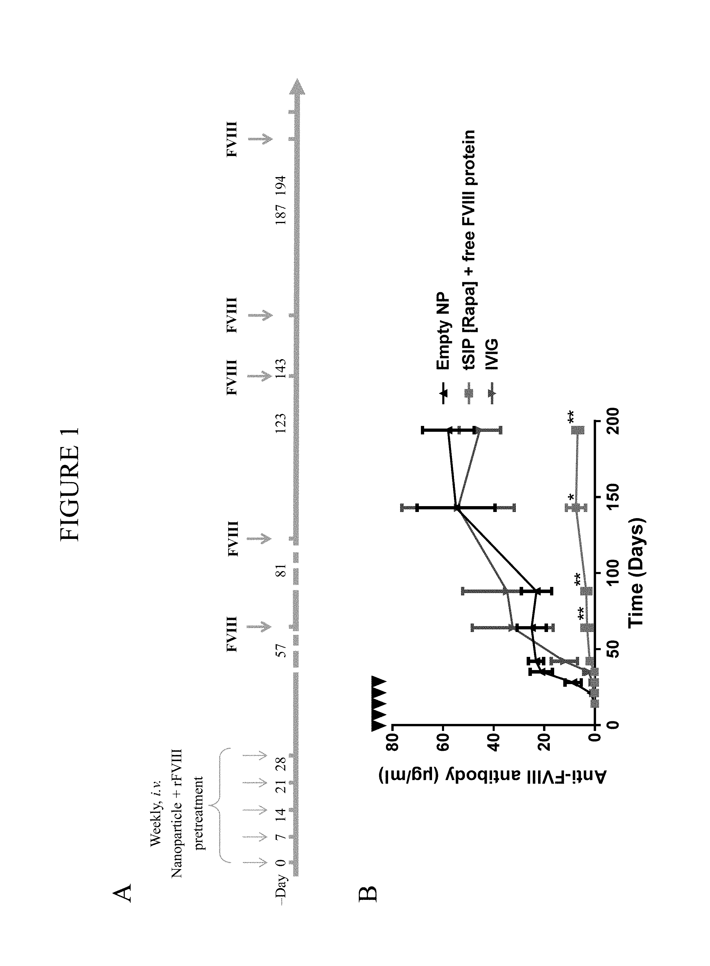
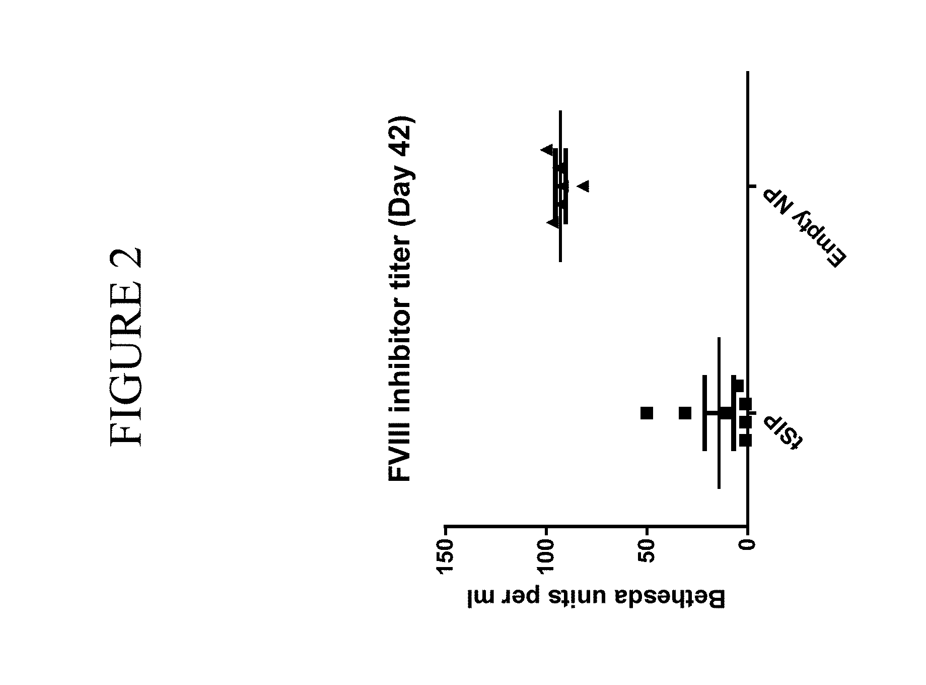
In one aspect a method comprising (1) a first dosing that comprises concomitantly administering (a) therapeutic macromolecules that are not attached to any synthetic nanocarriers, and (b) immunosuppressants, such as those attached to synthetic nanocarriers, and that comprise no therapeutic macromolecule antigen-presenting cell (APC) presentable antigens of the therapeutic macromolecules; (2) a second dosing that comprises (c) administering the therapeutic macromolecules that are not attached to any synthetic nanocarriers, and not administering any synthetic nanocarriers; and (3) administering the first and second dosings to a subject according to an administration schedule that reduces an undesired humoral immune response to the therapeutic macromolecules is provided.
In one embodiment of any one of the methods provided herein, the method further comprises (4) determining the administration schedule for the first and second dosings that reduces an undesired humoral immune response to the therapeutic macromolecules. In another embodiment of any one of the methods provided herein, the undesired humoral immune response to the therapeutic macromolecules results from the second dosing without the first dosing. In another embodiment of any one of the methods provided herein, the first dosing comprises administering (a) and (b) to the subject one or more times. In another embodiment of any one of the methods provided herein, (a) and (b) are administered at least 1, 2, 3, 4 or 5 times. In another embodiment of any one of the methods provided herein, the second dosing is administered to the subject at least 1, 2, 3, 4, 5, 6, 7 or 8 weeks after the first dosing. In another embodiment of any one of the methods provided herein, the method further comprises assessing the undesired humoral immune response in the subject prior to and/or after the administration of the first dosing and second dosing. In another embodiment of any one of the methods provided herein, the administering of the first dosing and/or second dosing is by intravenous, intraperitoneal or subcutaneous administration. In another embodiment of any one of the methods provided herein, the method further comprises identifying the subject as having or at risk of having an undesired humoral immune response to the therapeutic macromolecules. In some embodiments, the subject is expected or suspected to have an undesired humoral immune response to the therapeutic macromolecules.
In another aspect, a composition comprising (1) one or more first doses that each comprise (a) therapeutic macromolecules that are not attached to any synthetic nanocarriers, and (b) immunosuppressants, such as those attached to a population of synthetic nanocarriers and that comprise no therapeutic macromolecule APC presentable antigens of the therapeutic macromolecules; and (2) one or more second doses that comprises therapeutic macromolecules that are not attached to any synthetic nanocarriers is provided. In one embodiment of any one of the compositions provided herein, the composition is for use in a method of reducing an undesired humoral immune response to the therapeutic macromolecules. In another embodiment of any one of the methods provided herein, the method comprises administering the first and second doses to a subject according to an administration schedule. In another embodiment, the method further comprises determining the administration schedule for the first and second doses that reduce an undesired humoral immune response to the therapeutic macromolecules. In another embodiment, the method is any method as provided herein.
In another embodiment of any one of the compositions provided herein, the second dose does not comprise any synthetic nanocarriers. In another embodiment of any one of the compositions provided herein, the composition is a kit and one or more of the first doses and one or more of the second doses are each housed in a container in the kit. In another embodiment of any one of the compositions provided herein, the composition further comprises a pharmaceutically acceptable carrier.
In one embodiment of any one of the methods or compositions provided herein, the immunosuppressants comprise a statin, an mTOR inhibitor, a TGF-.beta. signaling agent, a corticosteroid, an inhibitor of mitochondrial function, a P38 inhibitor, an NF-.kappa.B inhibitor, an adenosine receptor agonist, a prostaglandin E2 agonist, a phosphodiesterasse 4 inhibitor, an HDAC inhibitor or a proteasome inhibitor. In another embodiment of any one of the methods or compositions provided herein, the mTOR inhibitor is rapamycin.
In another embodiment of any one of the methods or compositions provided herein, the therapeutic macromolecules are therapeutic proteins. In another embodiment of any one of the methods or compositions provided herein, the therapeutic macromolecules are therapeutic polynucleotides. In another embodiment of any one of the methods or compositions provided herein, the therapeutic proteins are for protein replacement or protein supplementation therapy. In another embodiment of any one of the methods or compositions provided herein, the therapeutic macromolecules comprise infusible or injectable therapeutic proteins, enzymes, enzyme cofactors, hormones, blood or blood coagulation factors, cytokines, interferons, growth factors, monoclonal antibodies, polyclonal antibodies or proteins associated with Pompe's disease. In another embodiment of any one of the methods or compositions provided herein, the infusible or injectable therapeutic proteins comprise Tocilizumab, alpha-1 antitrypsin, Hematide, albinterferon alfa-2b, Rhucin, tesamorelin, ocrelizumab, belimumab, pegloticase, pegsiticase, taliglucerase alfa, agalsidase alfa or velaglucerase alfa. In another embodiment of any one of the methods or compositions provided herein, the enzymes comprise an oxidoreductase, transferase, hydrolase, lyase, isomerase or ligase. In another embodiment of any one of the methods or compositions provided herein, the enzymes comprise an enzyme for enzyme replacement therapy for a lysosomal storage disorder. In another embodiment of any one of the methods or compositions provided herein, the enzyme for enzyme replacement therapy for a lysosomal storage disorder comprises imiglucerase, a-galactosidase A (a-gal A), agalsidase beta, acid a-glucosidase (GAA), alglucosidase alfa, LUMIZYME, MYOZYME, arylsulfatase B, laronidase, ALDURAZYME, idursulfase, ELAPRASE, arylsulfatase B or NAGLAZYME. In another embodiment of any one of the methods or compositions provided herein, the enzymes comprise KRYSTEXXA (pegloticase) or pegsiticase. In another embodiment of any one of the methods or compositions provided herein, the monoclonal antibodies comprises HUMIRA (adalimumab). In another embodiment of any one of the methods or compositions provided herein, the cytokines comprise a lymphokine, interleukin, chemokine, type 1 cytokine or a type 2 cytokine. In another embodiment of any one of the methods or compositions provided herein, the blood or blood coagulation factors comprise Factor I, Factor II, tissue factor, Factor V, Factor VII, Factor VIII, Factor IX, Factor X, Factor Xa, Factor XII, Factor XIII, von Willebrand factor, prekallikrein, high-molecular weight kininogen, fibronectin, antithrombin III, heparin cofactor II, protein C, protein S, protein Z, protein Z-related protease inhibitor (ZPI), plasminogen, alpha 2-antiplasmin, tissue plasminogen activator (tPA), urokinase, plasminogen activator inhibitor-1 (PAI1), plasminogen activator inhibitor-2 (PAI2), cancer procoagulant or epoetin alfa. In another embodiment of any one of the methods or compositions provided herein, the blood or blood coagulation factors comprise Factor VIII.
In another embodiment of any one of the methods or compositions provided herein, a load of the immunosuppressants on average across the population of synthetic nanocarriers is between 0.1% and 50%. In another embodiment of any one of the methods or compositions provided herein, the load of the immunosuppressants on average across the population of synthetic nanocarriers is between 0.1% and 20%.
In another embodiment of any one of the methods or compositions provided herein, the synthetic nanocarriers of the population comprise lipid nanoparticles, polymeric nanoparticles, metallic nanoparticles, surfactant-based emulsions, dendrimers, buckyballs, nanowires, virus-like particles or peptide or protein particles. In another embodiment of any one of the methods or compositions provided herein, the synthetic nanocarriers of the population comprise lipid nanoparticles. In another embodiment of any one of the methods or compositions provided herein, the synthetic nanocarriers of the population comprise liposomes. In another embodiment of any one of the methods or compositions provided herein, the synthetic nanocarriers of the population comprise metallic nanoparticles. In another embodiment of any one of the methods or compositions provided herein, the metallic nanoparticles comprise gold nanoparticles. In another embodiment of any one of the methods or compositions provided herein, the synthetic nanocarriers of the population comprise polymeric nanoparticles. In another embodiment of any one of the methods or compositions provided herein, the polymeric nanoparticles comprise polymer that is a non-methoxy-terminated, pluronic polymer. In another embodiment of any one of the methods or compositions provided herein, the polymeric nanoparticles comprise a polyester, polyester coupled to a polyether, polyamino acid, polycarbonate, polyacetal, polyketal, polysaccharide, polyethyloxazoline or polyethyleneimine. In another embodiment of any one of the methods or compositions provided herein, the polyester comprises a poly(lactic acid), poly(glycolic acid), poly(lactic-co-glycolic acid) or polycaprolactone. In another embodiment of any one of the methods or compositions provided herein, the polymeric nanoparticles comprise a polyester and a polyester coupled to a polyether. In another embodiment of any one of the methods or compositions provided herein, the polyether comprises polyethylene glycol or polypropylene glycol.
In another embodiment of any one of the methods or compositions provided herein, the mean of a particle size distribution obtained using dynamic light scattering of the synthetic nanocarriers of the population is a diameter greater than 100 nm. In another embodiment of any one of the methods or compositions provided herein, the diameter is greater than 150 nm. In another embodiment of any one of the methods or compositions provided herein, the diameter is greater than 200 nm. In another embodiment of any one of the methods or compositions provided herein, the diameter is greater than 250 nm. In another embodiment of any one of the methods or compositions provided herein, the diameter is greater than 300 nm.
In another embodiment of any one of the methods or compositions provided herein, an aspect ratio of the synthetic nanocarriers of the population is greater than 1:1, 1:1.2, 1:1.5, 1:2, 1:3, 1:5, 1:7 or 1:10.
In another aspect, a method of manufacturing any one of the compositions or kits provided herein is provided. In one embodiment, the method of manufacturing comprises producing one or more doses or dosage forms of a therapeutic macromolecule and producing one or more doses or dosage forms of an immunosuppressant. In another embodiment of any one of the methods of manufacturing provided, the step of producing one or more doses or dosage forms of an immunosuppressant comprises attaching the immunosuppressant to synthetic nanocarriers. In another embodiment of any one of the methods of manufacturing provided, the method further comprises combining the one or more doses or dosage forms of the immunosuppressant and one or more doses or dosage forms of the therapeutic macromolecule in a kit.
In another aspect, a use of any one of the compositions or kits provided herein for the manufacture of a medicament for reducing an undesired immune response to a therapeutic macromolecule, in a subject is provided. In one embodiment, the composition or kit comprises one or more doses or dosage forms comprising an immunosuppressant and one or more doses or dosage forms comprising a therapeutic macromolecule, wherein the immunosuppressant and therapeutic macromolecule are administered according to any one of the method provided herein. In another embodiment of any one of the uses provided herein, the immunosuppressant is attached to synthetic nanocarriers. In some embodiments of any one of the uses provided herein, the immunosuppressant comprises no therapeutic macromolecule antigen-presenting cell (APC) presentable antigens of the therapeutic macromolecule. In some embodiments of any one of the uses provided herein, the composition or kit further comprises one or more doses or dosage forms comprising the therapeutic macromolecule for use as one or more second dosings.
In another aspect, any one of the compositions or kits provided herein are provided for use in any one of the comprising an immunosuppressant and one or more doses or dosage forms comprising a therapeutic macromolecule. In another embodiment, the composition or kit further comprises one or more doses or dosage forms comprising the therapeutic macromolecule for use as one or more second dosings. In another embodiment, the immunosuppressant is attached to synthetic nanocarriers. In another embodiment, the immunosuppressant comprises no therapeutic macromolecule antigen-presenting cell (APC) presentable antigens of the therapeutic macromolecule.
BRIEF DESCRIPTION OF FIGURES
FIGS. 1A and B show the efficacy of nanocarrier and FVIII dosing in Hemophilia A mice.
FIG. 2 shows the antibody recall response to FVIII one month following the final nanocarrier and FVIII dosing.
FIGS. 3A and B show immune responses to HUMIRA in mice that were treated with HUMIRA/adalimumab with or without nanocarriers attached to rapamycin.
FIG. 4 shows anti-Keyhole Limpet Hemocyanin (KLH) antibody titers in mice that were treated with KLH with or without nanocarriers attached to rapamycin.
FIG. 5 shows anti-ovalbumin (OVA) antibody titers in mice that were treated with OVA with or without nanocarriers attached to rapamycin.
FIG. 6 shows anti-KRYSTEXXA antibody titers in mice that were treated with KRYSTEXXA with or without nanocarriers attached to rapamycin.
FIG. 7 shows antibody titers in mice that were treated with OVA and KLH either in the presence or absence of nanocarriers attached to rapamycin.
FIGS. 8A and B show immune responses to KLH in mice that were treated with KLH with or without nanocarriers attached to rapamycin.
FIGS. 9A and B show immune responses to HUMIRA/adalimumab in mice that were treated with HUMIRA/adalimumab with or without nanocarriers attached to rapamycin.
FIG. 10 shows immune responses to PEG-KLH in mice treated with or without nanocarriers attached to rapamycin.
FIG. 11 shows immune and therapeutic responses with HUMIRA with or without nanocarriers attached to rapamycin.
FIG. 12 shows an administration schedule.
FIG. 13 demonstrates the effects of synthetic nanocarriers attached to rapamycin or nanocarriers attached to GSK1059615 administered concomitantly with encapsulated protein.
DETAILED DESCRIPTION OF THE INVENTION
Before describing the present invention in detail, it is to be understood that this invention is not limited to particularly exemplified materials or process parameters as such may, of course, vary. It is also to be understood that the terminology used herein is for the purpose of describing particular embodiments of the invention only, and is not intended to be limiting of the use of alternative terminology to describe the present invention.
All publications, patents and patent applications cited herein, whether supra or infra, are hereby incorporated by reference in their entirety for all purposes.
As used in this specification and the appended claims, the singular forms "a," "an" and "the" include plural referents unless the content clearly dictates otherwise. For example, reference to "a polymer" includes a mixture of two or more such molecules or a mixture of differing molecular weights of a single polymer species, reference to "a synthetic nanocarrier" includes a mixture of two or more such synthetic nanocarriers or a plurality of such synthetic nanocarriers, reference to "a RNA molecule" includes a mixture of two or more such RNA molecules or a plurality of such RNA molecules, reference to "an immunosuppressant" includes a mixture of two or more such materials or a plurality of immunosuppressant molecules, and the like.
As used herein, the term "comprise" or variations thereof such as "comprises" or "comprising" are to be read to indicate the inclusion of any recited integer (e.g. a feature, element, characteristic, property, method/process step or limitation) or group of integers (e.g. features, element, characteristics, properties, method/process steps or limitations) but not the exclusion of any other integer or group of integers. Thus, as used herein, the term "comprising" is inclusive and does not exclude additional, unrecited integers or method/process steps.
In embodiments of any one of the compositions and methods provided herein, "comprising" may be replaced with "consisting essentially of" or "consisting of". The phrase "consisting essentially of" is used herein to require the specified integer(s) or steps as well as those which do not materially affect the character or function of the claimed invention. As used herein, the term "consisting" is used to indicate the presence of the recited integer (e.g. a feature, element, characteristic, property, method/process step or limitation) or group of integers (e.g. features, element, characteristics, properties, method/process steps or limitations) alone.
A. Introduction
It has been surprisingly found that delivering immunosuppressants, such as when attached to synthetic nanocarriers, concomitantly with therapeutic macromolecules in combination with the delivery of therapeutic macromolecules administered without synthetic nanocarriers can effectively reduce the production of anti-therapeutic macromolecule-specific antibodies. As such a reduction can be beneficial during therapeutic treatment with therapeutic macromolecules, the invention is useful for subjects in need of treatment with therapeutic macromolecules against which undesired humoral immune responses are generated or are expected to be generated. The present invention, in some embodiments, prevents or suppresses undesired humoral immune responses that may neutralize the beneficial effect of certain therapeutic macromolecule treatments.
The inventors have unexpectedly and surprisingly discovered that the problems and limitations noted above can be overcome by practicing the invention disclosed herein. In particular, the inventors have unexpectedly discovered that it is possible to provide immunosuppressant and therapeutic macromolecule dosings in certain combinations that reduce undesired humoral immune responses specific to the therapeutic macromolecule, particularly in embodiments wherein the immunosuppressant compositions and the therapeutic macromolecules are administered concomitantly followed by administration of the therapeutic macromolecules without synthetic nanocarriers.
The invention will now be described in more detail below.
B. Definitions
"Administering" or "administration" or "administer" means providing a material to a subject in a manner that is pharmacologically useful. The term is intended to include "causing to be administered" in some embodiments. "Causing to be administered" means causing, urging, encouraging, aiding, inducing or directing, directly or indirectly, another party to administer the material.
"Administration schedule" refers to administration of first dosings and second dosings according to a determined schedule. The schedule can include the number of first and second doings as well as the frequency of such dosings or interval between dosings. Such an administration schedule may include a number of parameters that are varied to achieve a particular objective, preferably reduction of an undesired humoral immune response to a therapeutic macromolecule. In embodiments, the administration schedule is any of the administration schedules as provided below in the Examples. In some embodiments, administration schedules according to the invention may be used to administer first and second dosings to one or more test subjects. Immune responses in these test subjects can then be assessed to determine whether or not the schedule was effective in reducing an undesired humoral immune response. Whether or not a schedule had a desired effect can be determined using any of the methods provided herein or otherwise known in the art. For example, a sample may be obtained from a subject to which dosings provided herein have been administered according to a specific administration schedule in order to determine whether or not specific immune cells, cytokines, antibodies, etc. were reduced, generated, activated, etc. Useful methods for detecting the presence and/or number of immune cells include, but are not limited to, flow cytometric methods (e.g., FACS), ELISpot, proliferation responses, cytokine production, and immunohistochemistry methods. Useful methods for determining the level of antibody production are well known in the art and include the assays provided herein. Such assays include ELISA assays.
"Amount effective" in the context of a composition or dosage form for administration to a subject refers to an amount of the composition or dosage form that produces one or more desired immune responses in the subject, for example, a reduction in an undesired humoral immune response. Therefore, in some embodiments, an amount effective is any amount of a composition or dosage form provided herein that reduces an undesired humoral immune response. This amount can be for in vitro or in vivo purposes. For in vivo purposes, the amount can be one that a clinician would believe may have a clinical benefit for a subject as provided herein, such as one in need of therapeutic macromolecule administration and/or antigen-specific immune tolerance thereto.
Amounts effective can involve reducing the level of an undesired immune response, although in some embodiments, it involves preventing an undesired immune response altogether. Amounts effective can also involve delaying the occurrence of an undesired immune response. An amount that is effective can also be an amount that produces a desired therapeutic endpoint or a desired therapeutic result. Amounts effective, preferably, result in a reduction in an undesired humoral immune response in a subject specific to a therapeutic macromolecule. Amounts effective, can also result in a tolerogenic immune response in a subject to an antigen, such as a therapeutic macromolecule. In other embodiments, the amounts effective can involve enhancing the level of a desired response, such as a therapeutic endpoint or result. The achievement of any of the foregoing can be monitored by routine methods.
In some embodiments of any one of the compositions and methods provided, the amount effective is one in which the desired immune response persists in the subject for at least 1 week, at least 2 weeks, at least 1 month, at least 2 months, at least 3 months, at least 4 months, at least 5 months, or longer. In other embodiments of any of the compositions and methods provided, the amount effective is one which produces a measurable desired immune response, for example, a measurable decrease in a humoral immune response (e.g., to a specific antigen), for at least 1 week, at least 2 weeks, at least 1 month, at least 2 months, at least 3 months, at least 4 months, at least 5 months, or longer.
Amounts effective will depend, of course, on the particular subject being treated; the severity of a condition, disease or disorder; the individual patient parameters including age, physical condition, size and weight; the duration of the treatment; the nature of concurrent therapy (if any); the specific route of administration and like factors within the knowledge and expertise of the health practitioner. These factors are well known to those of ordinary skill in the art and can be addressed with no more than routine experimentation. It is generally preferred that a maximum dose be used, that is, the highest safe dose according to sound medical judgment. It will be understood by those of ordinary skill in the art, however, that a patient may insist upon a lower dose or tolerable dose for medical reasons, psychological reasons or for virtually any other reason.
In general, doses of the immunosuppressants and/or therapeutic macromolecules in the compositions of the invention refer to the amount of the immunosuppressants and/or therapeutic macromolecules. Alternatively, the dose can be administered based on the number of synthetic nanocarriers that provide the desired amount of immunosuppressants.
"Antigen" means a B cell antigen or T cell antigen. "Type(s) of antigens" means molecules that share the same, or substantially the same, antigenic characteristics. In some embodiments, antigens may be proteins, polypeptides, peptides, lipoproteins, glycolipids, polynucleotides, polysaccharides or are contained or expressed in cells. In some embodiments, such as when the antigens are not well defined or characterized, the antigens may be contained within a cell or tissue preparation, cell debris, cell exosomes, conditioned media, etc.
"Antigen-specific" refers to any immune response that results from the presence of the antigen, or portion thereof, or that generates molecules that specifically recognize or bind the antigen. In some embodiments, when the antigen comprises the therapeutic macromolecule, antigen-specific may mean therapeutic macromolecule-specific. For example, where the immune response is antigen-specific antibody production, such as therapeutic macromolecule-specific antibody production, antibodies are produced that specifically bind the antigen (e.g., therapeutic macromolecule). As another example, where the immune response is antigen-specific B cell or CD4+ T cell proliferation and/or activity, the proliferation and/or activity results from recognition of the antigen, or portion thereof, alone or in complex with MHC molecules, B cells, etc.
"Assessing an immune response" refers to any measurement or determination of the level, presence or absence, reduction, increase in, etc. of an immune response in vitro or in vivo. Such measurements or determinations may be performed on one or more samples obtained from a subject. Such assessing can be performed with any of the methods provided herein or otherwise known in the art.
"Attach" or "Attached" or "Couple" or "Coupled" (and the like) means to chemically associate one entity (for example a moiety) with another. In some embodiments, the attaching is covalent, meaning that the attachment occurs in the context of the presence of a covalent bond between the two entities. In non-covalent embodiments, the non-covalent attaching is mediated by non-covalent interactions including but not limited to charge interactions, affinity interactions, metal coordination, physical adsorption, host-guest interactions, hydrophobic interactions, TT stacking interactions, hydrogen bonding interactions, van der Waals interactions, magnetic interactions, electrostatic interactions, dipole-dipole interactions, and/or combinations thereof. In embodiments, encapsulation is a form of attaching. In embodiments, therapeutic macromolecules and immunosuppressants are not attached to one another, meaning that the therapeutic macromolecules and immunosuppressants are not subjected to a process specifically intended to chemically associate one with another. In embodiments, therapeutic macromolecules and/or immunosuppressants are not attached to synthetic nanocarriers, meaning that the therapeutic macromolecules (and/or immunosuppressants) and synthetic nanocarriers are not subjected to a process specifically intended to chemically associate one with another.
An "at risk" subject is one in which a health practitioner believes has a chance of having a disease, disorder or condition or is one a health practitioner believes has a chance of experiencing an undesired humoral immune response as provided herein and would benefit from the compositions and methods provided. In some embodiments, the subjects are those that are expected to have an undesired humoral immune response to a therapeutic macromolecule.
"Average", as used herein, refers to the arithmetic mean unless otherwise noted.
"Combination", as applied to two or more materials and/or agents (also referred to herein as the components), is intended to define material in which the two or more materials/agents are associated. Components may be separately identified, e.g., first component, second component, third component, etc. The terms "combined" and "combining" in this context are to be interpreted accordingly.
The association of the two or more materials/agents in a combination may be physical or non-physical. Examples of physically associated combined materials/agents include: compositions (e.g., unitary formulations) comprising the two or more materials/agents in admixture (for example within the same unit dose); compositions comprising material in which the two or more materials/agents are chemically/physicochemically linked (for example by crosslinking, molecular agglomeration or binding to a common vehicle moiety); compositions comprising material in which the two or more materials/agents are chemically/physicochemically co-packaged (for example, disposed on or within lipid vesicles, particles (e.g., micro- or nanoparticles) or emulsion droplets); pharmaceutical kits, pharmaceutical packs or patient packs in which the two or more materials/agents are co-packaged or co-presented (e.g., as part of an array of unit doses);
Examples of non-physically associated combined materials/agents include: material (e.g., a non-unitary formulation) comprising at least one of the two or more materials/agents together with instructions for the extemporaneous association of the at least one compound/agent to form a physical association of the two or more materials/agents; material (e.g., a non-unitary formulation) comprising at least one of the two or more materials/agents together with instructions for combination therapy with the two or more materials/agents; material comprising at least one of the two or more materials/agents together with instructions for administration to a patient population in which the other(s) of the two or more materials/agents have been (or are being) administered; material comprising at least one of the two or more materials/agents in an amount or in a form which is specifically adapted for use in combination with the other(s) of the two or more materials/agents.
As used herein, the term "combination therapy" is intended to define therapies which comprise the use of a combination of two or more materials/agents (as defined above). Thus, references to "combination therapy", "combinations" and the use of materials/agents "in combination" in this application may refer to materials/agents that are administered as part of the same overall treatment regimen. As such, the posology of each of the two or more materials/agents may differ: each may be administered at the same time or at different times. It will, therefore, be appreciated that the materials/agents of the combination may be administered sequentially (e.g., before or after) or simultaneously, either in the same pharmaceutical formulation (i.e., together), or in different pharmaceutical formulations (i.e., separately). Simultaneously in the same formulation is as a unitary formulation whereas simultaneously in different pharmaceutical formulations is non-unitary. The posologies of each of the two or more materials/agents in a combination therapy may also differ with respect to the route of administration.
"Concomitantly" means administering two or more materials/agents to a subject in a manner that is correlated in time, preferably sufficiently correlated in time so as to provide a modulation in an immune or physiologic response, and even more preferably the two or more materials/agents are administered in combination. In embodiments, concomitant administration may encompass administration of two or more materials/agents within a specified period of time, preferably within 1 month, more preferably within 1 week, still more preferably within 1 day, and even more preferably within 1 hour. In embodiments, the materials/agents may be repeatedly administered concomitantly, that is concomitant administration on more than one occasion, as may be provided in the Examples.
"Determining" or "determine" means to ascertain a factual relationship. Determining may be accomplished in a number of ways, including but not limited to performing experiments, or making projections. For instance, a dose of an immunosuppressant or therapeutic macromolecule may be determined by starting with a test dose and using known scaling techniques (such as allometric or isometric scaling) to determine the dose for administration. Such may also be used to determine a protocol or administration schedule as provided herein. In another embodiment, the dose may be determined by testing various doses in a subject, i.e. through direct experimentation based on experience and guiding data. In embodiments, "determining" or "determine" comprises "causing to be determined." "Causing to be determined" means causing, urging, encouraging, aiding, inducing or directing or acting in coordination with an entity for the entity to ascertain a factual relationship; including directly or indirectly, or expressly or impliedly.
"Dosing" means the administration of a pharmacologically and/or immunologically active material or combination of pharmacologically and/or immunologically active materials to a subject.
"Dose" refers to a specific quantity of a pharmacologically and/or immunologically active material for administration to a subject for a given time.
"Encapsulate" means to enclose at least a portion of a substance within a synthetic nanocarrier. In some embodiments, a substance is enclosed completely within a synthetic nanocarrier. In other embodiments, most or all of a substance that is encapsulated is not exposed to the local environment external to the synthetic nanocarrier. In other embodiments, no more than 50%, 40%, 30%, 20%, 10% or 5% (weight/weight) is exposed to the local environment. Encapsulation is distinct from absorption, which places most or all of a substance on a surface of a synthetic nanocarrier, and leaves the substance exposed to the local environment external to the synthetic nanocarrier.
"Generating" means causing an action, such as an immune or physiologic response (e.g., a tolerogenic immune response) to occur, either directly oneself or indirectly.
"Identifying a subject" is any action or set of actions that allows a clinician to recognize a subject as one who may benefit from the methods, compositions or kits provided herein. Preferably, the identified subject is one who is in need of a therapeutic benefit from a therapeutic macromolecule and in which an undesired humoral immune response is expected to occur as provided herein. The action or set of actions may be either directly oneself or indirectly. In one embodiment of any one of the methods provided herein, the method further comprises identifying a subject in need of a method, composition or kit as provided herein.
"Immunosuppressant" means a compound that causes an APC to have an immunosuppressive effect (e.g., tolerogenic effect) or a T cell or a B cell to be suppressed. An immunosuppressive effect generally refers to the production or expression of cytokines or other factors by the APC that reduces, inhibits or prevents an undesired immune response or that promotes a desired immune response, such as a regulatory immune response. When the APC acquires an immunosuppressive function (under the immunosuppressive effect) on immune cells that recognize an antigen presented by this APC, the immunosuppressive effect is said to be specific to the presented antigen. Without being bound by any particular theory, it is thought that the immunosuppressive effect is a result of the immunosuppressant being delivered to the APC, preferably in the presence of an antigen. In one embodiment, the immunosuppressant is one that causes an APC to promote a regulatory phenotype in one or more immune effector cells. For example, the regulatory phenotype may be characterized by the inhibition of the production, induction, stimulation or recruitment of antigen-specific CD4+ T cells or B cells, the inhibition of the production of antigen-specific antibodies, the production, induction, stimulation or recruitment of Treg cells (e.g., CD4+CD25highFoxP3+ Treg cells), etc. This may be the result of the conversion of CD4+ T cells or B cells to a regulatory phenotype. This may also be the result of induction of FoxP3 in other immune cells, such as CD8+ T cells, macrophages and iNKT cells. In one embodiment, the immunosuppressant is one that affects the response of the APC after it processes an antigen. In another embodiment, the immunosuppressant is not one that interferes with the processing of the antigen. In a further embodiment, the immunosuppressant is not an apoptotic-signaling molecule. In another embodiment, the immunosuppressant is not a phospholipid.
Immunosuppressants include, but are not limited to, statins; mTOR inhibitors, such as rapamycin or a rapamycin analog; TGF-.beta. signaling agents; TGF-.beta. receptor agonists; histone deacetylase inhibitors, such as Trichostatin A; corticosteroids; inhibitors of mitochondrial function, such as rotenone; P38 inhibitors; NF-.kappa..beta. inhibitors, such as 6Bio, Dexamethasone, TCPA-1, IKK VII; adenosine receptor agonists; prostaglandin E2 agonists (PGE2), such as Misoprostol; phosphodiesterase inhibitors, such as phosphodiesterase 4 inhibitor (PDE4), such as Rolipram; proteasome inhibitors; kinase inhibitors; G-protein coupled receptor agonists; G-protein coupled receptor antagonists; glucocorticoids; retinoids; cytokine inhibitors; cytokine receptor inhibitors; cytokine receptor activators; peroxisome proliferator-activated receptor antagonists; peroxisome proliferator-activated receptor agonists; histone deacetylase inhibitors; calcineurin inhibitors; phosphatase inhibitors; PI3 KB inhibitors, such as TGX-221; autophagy inhibitors, such as 3-Methyladenine; aryl hydrocarbon receptor inhibitors; proteasome inhibitor I (PSI); and oxidized ATPs, such as P2X receptor blockers. Immunosuppressants also include IDO, vitamin D3, cyclosporins, such as cyclosporine A, aryl hydrocarbon receptor inhibitors, resveratrol, azathiopurine (Aza), 6-mercaptopurine (6-MP), 6-thioguanine (6-TG), FK506, sanglifehrin A, salmeterol, mycophenolate mofetil (MMF), aspirin and other COX inhibitors, niflumic acid, estriol, methotrexate and triptolide. In embodiments, the immunosuppressant may comprise any of the agents provided herein.
The immunosuppressant can be a compound that directly provides the immunosuppressive effect on APCs or it can be a compound that provides the immunosuppressive effect indirectly (i.e., after being processed in some way after administration). Immunosuppressants, therefore, include prodrug forms of any of the compounds provided herein.
In embodiments of any one of the methods, compositions or kits provided herein, the immunosuppressants provided herein are attached to synthetic nanocarriers. In preferable embodiments, the immunosuppressant is an element that is in addition to the material that makes up the structure of the synthetic nanocarrier. For example, in one embodiment, where the synthetic nanocarrier is made up of one or more polymers, the immunosuppressant is a compound that is in addition and attached to the one or more polymers. As another example, in one embodiment, where the synthetic nanocarrier is made up of one or more lipids, the immunosuppressant is again in addition and attached to the one or more lipids. In embodiments, such as where the material of the synthetic nanocarrier also results in an immunosuppressive effect, the immunosuppressant is an element present in addition to the material of the synthetic nanocarrier that results in an immunosuppressive effect.
Other exemplary immunosuppressants include, but are not limited, small molecule drugs, natural products, antibodies (e.g., antibodies against CD20, CD3, CD4), biologics-based drugs, carbohydrate-based drugs, nanoparticles, liposomes, RNAi, antisense nucleic acids, aptamers, methotrexate, NSAIDs; fingolimod; natalizumab; alemtuzumab; anti-CD3; tacrolimus (FK506); cytokines and growth factors, such as TGF-.beta. and IL-10; etc. Further immunosuppressants, are known to those of skill in the art, and the invention is not limited in this respect.
In embodiments of any one of the methods, compositions or kits provided herein, the immunosuppressant is in a form, such as a nanocrystalline form, whereby the form of the immunosuppressant itself is a particle or particle-like. In embodiments, such forms mimic a virus or other foreign pathogen. Many drugs have been nanonized and appropriate methods for producing such drug forms would be known to one of ordinary skill in the art. Drug nanocrystals, such as nanocrystalline rapamycin are known to those of ordinary skill in the art (Katteboinaa, et al. 2009, International Journal of PharmTech Resesarch; Vol. 1, No. 3; pp 682-694. As used herein a "drug nanocrystal" refers to a form of a drug (e.g., an immunosuppressant) that does not include a carrier or matrix material. In some embodiments, drug nanocrystals comprise 90%, 95%, 98%, or 99% or more drug. Methods for producing drug nanocrystals include, without limitation, milling, high pressure homogenization, precipitation, spray drying, rapid expansion of supercritical solution (RESS), Nanoedge.RTM. technology (Baxter Healthcare), and Nanocrystal Technology.TM. (Elan Corporation). In some embodiments, a surfactant or a stabilizer may be used for steric or electrostatic stability of the drug nanocrystal. In some embodiments the nanocrystal or nanocrytalline form of an immunosuppressant may be used to increase the solubility, stability, and/or bioavailability of the immunosuppressant, particularly immunosuppressants that are insoluble or labile. In some embodiments, administration of a first dosing comprising therapeutic macromolecules and immunosuppressants in nanocrystalline form followed by a second dosing of the therapeutic macromolecule results in a reduction of the undesired therapeutic macromolecule-specific humoral immune response.
"Load", when attached to a synthetic nanocarrier, is the amount of the immunosuppressant attached to a synthetic nanocarrier based on the total dry recipe weight of materials in an entire synthetic nanocarrier (weight/weight). Generally, such a load is calculated as an average across a population of synthetic nanocarriers. In one embodiment, the load of the immunosuppressant on average across the synthetic nanocarriers is between 0.1% and 99%. In another embodiment, the load is between 0.1% and 50%. In yet another embodiment, the load of the immunosuppressant is between 0.1% and 20%. In a further embodiment, the load of the immunosuppressant is between 0.1% and 10%. In still a further embodiment, the load of the immunosuppressant is between 1% and 10%. In still a further embodiment, the load of the immunosuppressant is between 7% and 20%. In yet another embodiment, the load of the immunosuppressant is at least 0.1%, at least 0.2%, at least 0.3%, at least 0.4%, at least 0.5%, at least 0.6%, at least 0.7%, at least 0.8%, at least 0.9%, at least 1%, at least 2%, at least 3%, at least 4%, at least 5%, at least 6%, at least at least 7%, at least 8%, at least 9%, at least 10%, at least 11%, at least 12%, at least 13%, at least 14%, at least 15%, at least 16%, at least 17%, at least 18%, at least 19% or at least 20%, at least 25%, at least 30%, at least 40%, at least 50%, at least 60%, at least 70%, at least 80%, at least 90%, at least 95%, at least 96%, at least 97%, at least 98% or at least 99% on average across the population of synthetic nanocarriers. In yet a further embodiment, the load of the immunosuppressant is 0.1%, 0.2%, 0.3%, 0.4%, 0.5%, 0.6%, 0.7%, 0.8%, 0.9%, 1%, 2%, 3%, 4%, 5%, 6%, 7%, 8%, 9%, 10%, 11%, 12%, 13%, 14%, 15%, 16%, 17%, 18%, 19% or 20% on average across the population of synthetic nanocarriers. In some embodiments of the above embodiments, the load of the immunosuppressant is no more than 25% on average across a population of synthetic nanocarriers. In embodiments, the load is calculated as may be described in the Examples or as otherwise known in the art.
In some embodiments, when the form of the immunosuppressant is itself a particle or particle-like, such as a nanocrystalline immunosuppressant, the load of immunosuppressant is the amount of the immunosuppressant in the particles or the like (weight/weight). In such embodiments, the load can approach 97%, 98%, 99% or more.
"Maximum dimension of a synthetic nanocarrier" means the largest dimension of a nanocarrier measured along any axis of the synthetic nanocarrier. "Minimum dimension of a synthetic nanocarrier" means the smallest dimension of a synthetic nanocarrier measured along any axis of the synthetic nanocarrier. For example, for a spheroidal synthetic nanocarrier, the maximum and minimum dimension of a synthetic nanocarrier would be substantially identical, and would be the size of its diameter. Similarly, for a cuboidal synthetic nanocarrier, the minimum dimension of a synthetic nanocarrier would be the smallest of its height, width or length, while the maximum dimension of a synthetic nanocarrier would be the largest of its height, width or length. In an embodiment, a minimum dimension of at least 75%, preferably at least 80%, more preferably at least 90%, of the synthetic nanocarriers in a sample, based on the total number of synthetic nanocarriers in the sample, is equal to or greater than 100 nm. In an embodiment, a maximum dimension of at least 75%, preferably at least 80%, more preferably at least 90%, of the synthetic nanocarriers in a sample, based on the total number of synthetic nanocarriers in the sample, is equal to or less than 5 .mu.m. Preferably, a minimum dimension of at least 75%, preferably at least 80%, more preferably at least 90%, of the synthetic nanocarriers in a sample, based on the total number of synthetic nanocarriers in the sample, is greater than 110 nm, more preferably greater than 120 nm, more preferably greater than 130 nm, and more preferably still greater than 150 nm. Aspects ratios of the maximum and minimum dimensions of synthetic nanocarriers may vary depending on the embodiment. For instance, aspect ratios of the maximum to minimum dimensions of the synthetic nanocarriers may vary from 1:1 to 1,000,000:1, preferably from 1:1 to 100,000:1, more preferably from 1:1 to 10,000:1, more preferably from 1:1 to 1000:1, still more preferably from 1:1 to 100:1, and yet more preferably from 1:1 to 10:1. Preferably, a maximum dimension of at least 75%, preferably at least 80%, more preferably at least 90%, of the synthetic nanocarriers in a sample, based on the total number of synthetic nanocarriers in the sample is equal to or less than 3 .mu.m, more preferably equal to or less than 2 .mu.m, more preferably equal to or less than 1 .mu.m, more preferably equal to or less than 800 nm, more preferably equal to or less than 600 nm, and more preferably still equal to or less than 500 nm. In preferred embodiments, a minimum dimension of at least 75%, preferably at least 80%, more preferably at least 90%, of the synthetic nanocarriers in a sample, based on the total number of synthetic nanocarriers in the sample, is equal to or greater than 100 nm, more preferably equal to or greater than 120 nm, more preferably equal to or greater than 130 nm, more preferably equal to or greater than 140 nm, and more preferably still equal to or greater than 150 nm. Measurement of synthetic nanocarrier dimensions (e.g., effective diameter) may be obtained, in some embodiments, by suspending the synthetic nanocarriers in a liquid (usually aqueous) media and using dynamic light scattering (DLS) (e.g., using a Brookhaven ZetaPALS instrument). For example, a suspension of synthetic nanocarriers can be diluted from an aqueous buffer into purified water to achieve a final synthetic nanocarrier suspension concentration of approximately 0.01 to 0.1 mg/mL. The diluted suspension may be prepared directly inside, or transferred to, a suitable cuvette for DLS analysis. The cuvette may then be placed in the DLS, allowed to equilibrate to the controlled temperature, and then scanned for sufficient time to acquire a stable and reproducible distribution based on appropriate inputs for viscosity of the medium and refractive indicies of the sample. The effective diameter, or mean of the distribution, is then reported. Determining the effective sizes of high aspect ratio, or non-spheroidal, synthetic nanocarriers may require augmentative techniques, such as electron microscopy, to obtain more accurate measurements. "Dimension" or "size" or "diameter" of synthetic nanocarriers means the mean of a particle size distribution, for example, obtained using dynamic light scattering.
"Non-methoxy-terminated polymer" means a polymer that has at least one terminus that ends with a moiety other than methoxy. In some embodiments, the polymer has at least two termini that ends with a moiety other than methoxy. In other embodiments, the polymer has no termini that ends with methoxy. "Non-methoxy-terminated, pluronic polymer" means a polymer other than a linear pluronic polymer with methoxy at both termini. Polymeric nanoparticles as provided herein can comprise non-methoxy-terminated polymers or non-methoxy-terminated, pluronic polymers.
"Pharmaceutically acceptable excipient" or "pharmaceutically acceptable carrier" means a pharmacologically inactive material used together with a pharmacologically active material to formulate the compositions. Pharmaceutically acceptable excipients comprise a variety of materials known in the art, including but not limited to saccharides (such as glucose, lactose, and the like), preservatives such as antimicrobial agents, reconstitution aids, colorants, saline (such as phosphate buffered saline), and buffers.
"Providing" means an action or set of actions that an individual performs that supply a needed item or set of items or methods for practicing of the present invention. The action or set of actions may be taken either directly oneself or indirectly.
"Providing a subject" is any action or set of actions that causes a clinician to come in contact with a subject and administer a composition provided herein thereto or to perform a method provided herein thereupon. Preferably, the subject is one who is in need of therapeutic macromolecule administration and antigen-specific immune tolerance thereto. The action or set of actions may be taken either directly oneself or indirectly. In one embodiment of any one of the methods provided herein, the method further comprises providing a subject.
"Subject" means animals, including warm blooded mammals such as humans and primates; avians; domestic household or farm animals such as cats, dogs, sheep, goats, cattle, horses and pigs; laboratory animals such as mice, rats and guinea pigs; fish; reptiles; zoo and wild animals; and the like.
"Synthetic nanocarrier(s)" means a discrete object that is not found in nature, and that possesses at least one dimension that is less than or equal to 5 microns in size. Albumin nanoparticles are generally included as synthetic nanocarriers, however in certain embodiments the synthetic nanocarriers do not comprise albumin nanoparticles. In embodiments, synthetic nanocarriers do not comprise chitosan. In other embodiments, synthetic nanocarriers are not lipid-based nanoparticles. In further embodiments, synthetic nanocarriers do not comprise a phospholipid.
A synthetic nanocarrier can be, but is not limited to, one or a plurality of lipid-based nanoparticles (also referred to herein as lipid nanoparticles, i.e., nanoparticles where the majority of the material that makes up their structure are lipids), polymeric nanoparticles, metallic nanoparticles, surfactant-based emulsions, dendrimers, buckyballs, nanowires, virus-like particles (i.e., particles that are primarily made up of viral structural proteins but that are not infectious or have low infectivity), peptide or protein-based particles (also referred to herein as protein particles, i.e., particles where the majority of the material that makes up their structure are peptides or proteins) (such as albumin nanoparticles) and/or nanoparticles that are developed using a combination of nanomaterials such as lipid-polymer nanoparticles. Synthetic nanocarriers may be a variety of different shapes, including but not limited to spheroidal, cuboidal, pyramidal, oblong, cylindrical, toroidal, and the like. Synthetic nanocarriers according to the invention comprise one or more surfaces. Exemplary synthetic nanocarriers that can be adapted for use in the practice of the present invention comprise: (1) the biodegradable nanoparticles disclosed in U.S. Pat. No. 5,543,158 to Gref et al., (2) the polymeric nanoparticles of Published US Patent Application 20060002852 to Saltzman et al., (3) the lithographically constructed nanoparticles of Published US Patent Application 20090028910 to DeSimone et al., (4) the disclosure of WO 2009/051837 to von Andrian et al., (5) the nanoparticles disclosed in Published US Patent Application 2008/0145441 to Penades et al., (6) the protein nanoparticles disclosed in Published US Patent Application 20090226525 to de los R10s et al., (7) the virus-like particles disclosed in published US Patent Application 20060222652 to Sebbel et al., (8) the nucleic acid attached virus-like particles disclosed in published US Patent Application 20060251677 to Bachmann et al., (9) the virus-like particles disclosed in WO2010047839A1 or WO2009106999A2, (10) the nanoprecipitated nanoparticles disclosed in P. Paolicelli et al., "Surface-modified PLGA-based Nanoparticles that can Efficiently Associate and Deliver Virus-like Particles" Nanomedicine. 5(6):843-853 (2010), (11) apoptotic cells, apoptotic bodies or the synthetic or semisynthetic mimics disclosed in U.S. Publication 2002/0086049, or (12) those of Look et al., Nanogel-based delivery of mycophenolic acid ameliorates systemic lupus erythematosus in mice" J. Clinical Investigation 123(4):1741-1749 (2013). In embodiments, synthetic nanocarriers may possess an aspect ratio greater than 1:1, 1:1.2, 1:1.5, 1:2, 1:3, 1:5, 1:7, or greater than 1:10.
Synthetic nanocarriers according to the invention that have a minimum dimension of equal to or less than about 100 nm, preferably equal to or less than 100 nm, do not comprise a surface with hydroxyl groups that activate complement or alternatively comprise a surface that consists essentially of moieties that are not hydroxyl groups that activate complement. In a preferred embodiment, synthetic nanocarriers according to the invention that have a minimum dimension of equal to or less than about 100 nm, preferably equal to or less than 100 nm, do not comprise a surface that substantially activates complement or alternatively comprise a surface that consists essentially of moieties that do not substantially activate complement. In a more preferred embodiment, synthetic nanocarriers according to the invention that have a minimum dimension of equal to or less than about 100 nm, preferably equal to or less than 100 nm, do not comprise a surface that activates complement or alternatively comprise a surface that consists essentially of moieties that do not activate complement. In embodiments, synthetic nanocarriers exclude virus-like particles. In embodiments, synthetic nanocarriers may possess an aspect ratio greater than 1:1, 1:1.2, 1:1.5, 1:2, 1:3, 1:5, 1:7, or greater than 1:10.
A "therapeutic macromolecule" refers to any protein, carbohydrate, lipid or nucleic acid that may be administered to a subject and have a therapeutic effect. In some embodiments, administration of the therapeutic macromolecule to a subject may result in an undesired humoral immune responses, including production of anti-therapeutic macromolecule-specific antibodies. As described herein, administration of a therapeutic macromolecule with immunosuppressant as provided herein can reduce the undesired humoral immune response and/or enhance the therapeutic effectiveness of the therapeutic macromolecule. In some embodiments, the therapeutic macromolecule may be a therapeutic polynucleotide or therapeutic protein.
"Therapeutic polynucleotide" means any polynucleotide or polynucleotide-based therapy that may be administered to a subject and have a therapeutic effect. Such therapies include gene silencing. Examples of such therapy are known in the art, and include, but are not limited to, naked RNA (including messenger RNA, modified messenger RNA, and forms of RNAi). Examples of other therapeutic polynucleotides are provided elsewhere herein. Therapeutic polynucleotides may be produced in, on or by cells and also may be obtained using cell free or from fully synthetic in vitro methods. Subjects, therefore, include any subject that is in need of treatment with any of the foregoing. Such subject include those that will receive any of the foregoing.
"Therapeutic protein" means any protein or protein-based therapy that may be administered to a subject and have a therapeutic effect. Such therapies include protein replacement and protein supplementation therapies. Such therapies also include the administration of exogenous or foreign proteins, antibody therapies, and cell or cell-based therapies. Therapeutic proteins comprise, but are not limited to, enzymes, enzyme cofactors, hormones, blood clotting factors, cytokines, growth factors, monoclonal antibodies, antibody-drug conjugates, and polyclonal antibodies. Examples of other therapeutic proteins are provided elsewhere herein. Therapeutic proteins may be produced in, on or by cells and may be obtained from such cells or administered in the form of such cells. In embodiments, the therapeutic protein is produced in, on or by mammalian cells, insect cells, yeast cells, bacteria cells, plant cells, transgenic animal cells, transgenic plant cells, etc. The therapeutic protein may be recombinantly produced in such cells. The therapeutic protein may be produced in, on or by a virally transformed cell. Subjects, therefore, include any subject that is in need of treatment with any of the foregoing. Such subject include those that will receive any of the foregoing.
"Therapeutic macromolecule APC presentable antigen" means an antigen that is associated with a therapeutic macromolecule (i.e., the therapeutic macromolecule or a fragment thereof that can generate an immune response against the therapeutic macromolecule (e.g., the production of anti-therapeutic macromolecule-specific antibodies)). Generally, therapeutic macromolecule antigen-presenting cell (APC) presentable antigens can be presented for recognition by the immune system (e.g., cells of the immune system, such as presented by antigen presenting cells, including but not limited to dendritic cells, B cells or macrophages). The therapeutic macromolecule APC presentable antigen can be presented for recognition by, for example, T cells. Such antigens may be recognized by and trigger an immune response in a T cell via presentation of an epitope of the antigen bound to a Class I or Class II major histocompatability complex molecule (MHC). Therapeutic macromolecule APC presentable antigens generally include proteins, polypeptides, peptides, polynucleotides, lipoproteins, or are contained or expressed in, on or by cells. The therapeutic macromolecule antigens, in some embodiments, comprise MHC Class I-restricted epitopes and/or MHC Class II-restricted epitopes and/or B cell epitopes. Preferably, one or more tolerogenic immune responses specific to the therapeutic macromolecule result with the methods, compositions or kits provided herein. In embodiments, populations of the synthetic nanocarriers comprise no added therapeutic macromolecule APC presentable antigens, meaning that no substantial amounts of therapeutic macromolecule APC presentable antigens are intentionally added to the synthetic nanocarriers during the manufacturing thereof.
"Undesired humoral immune response" refers to any undesired humoral immune response that results from exposure to an antigen, promotes or exacerbates a disease, disorder or condition provided herein (or a symptom thereof), or is symptomatic of a disease, disorder or condition provided herein. Such immune responses generally have a negative impact on a subject's health or is symptomatic of a negative impact on a subject's health. Undesired humoral immune responses include antigen-specific antibody production, antigen-specific B cell proliferation and/or activity or antigen-specific CD4+ T cell proliferation and/or activity. Generally, these undesired immune responses are specific to a therapeutic macromolecule and counteract the beneficial effects desired of administration with the therapeutic macromolecule.
C. Compositions Useful in the Practice of the Methods
Provided herein are first and second dosings that in combination can reduce undesired humoral immune responses. Generally, such first and second dosings can also result in improved efficacy of therapeutic macromolecules. The methods and related compositions provided herein, therefore, can be used for subjects in need of treatment with a therapeutic macromolecule. Specifically, first dosings comprise immunosuppressants, in some embodiments attached to synthetic nanocarriers, in combination with therapeutic macromolecules that are not attached to synthetic nanocarriers, and second dosings comprise therapeutic macromolecules that are also not attached to synthetic nanocarriers.
A wide variety of synthetic nanocarriers can be used to attach to immunosuppressants of the first dosings. In some embodiments, synthetic nanocarriers are spheres or spheroids. In some embodiments, synthetic nanocarriers are flat or plate-shaped. In some embodiments, synthetic nanocarriers are cubes or cubic. In some embodiments, synthetic nanocarriers are ovals or ellipses. In some embodiments, synthetic nanocarriers are cylinders, cones, or pyramids.
In some embodiments, it is desirable to use a population of synthetic nanocarriers that is relatively uniform in terms of size or shape so that each synthetic nanocarrier has similar properties. For example, at least 80%, at least 90%, or at least 95% of the synthetic nanocarriers, based on the total number of synthetic nanocarriers, may have a minimum dimension or maximum dimension that falls within 5%, 10%, or 20% of the average diameter or average dimension of the synthetic nanocarriers.
Synthetic nanocarriers can be solid or hollow and can comprise one or more layers. In some embodiments, each layer has a unique composition and unique properties relative to the other layer(s). To give but one example, synthetic nanocarriers may have a core/shell structure, wherein the core is one layer (e.g., a polymeric core) and the shell is a second layer (e.g., a lipid bilayer or monolayer). Synthetic nanocarriers may comprise a plurality of different layers.
In some embodiments, synthetic nanocarriers may optionally comprise one or more lipids. In some embodiments, a synthetic nanocarrier may comprise a liposome. In some embodiments, a synthetic nanocarrier may comprise a lipid bilayer. In some embodiments, a synthetic nanocarrier may comprise a lipid monolayer. In some embodiments, a synthetic nanocarrier may comprise a micelle. In some embodiments, a synthetic nanocarrier may comprise a core comprising a polymeric matrix surrounded by a lipid layer (e.g., lipid bilayer, lipid monolayer, etc.). In some embodiments, a synthetic nanocarrier may comprise a non-polymeric core (e.g., metal particle, quantum dot, ceramic particle, bone particle, viral particle, proteins, nucleic acids, carbohydrates, etc.) surrounded by a lipid layer (e.g., lipid bilayer, lipid monolayer, etc.).
In other embodiments, synthetic nanocarriers may comprise metal particles, quantum dots, ceramic particles, etc. In some embodiments, a non-polymeric synthetic nanocarrier is an aggregate of non-polymeric components, such as an aggregate of metal atoms (e.g., gold atoms).
In some embodiments, synthetic nanocarriers may optionally comprise one or more amphiphilic entities. In some embodiments, an amphiphilic entity can promote the production of synthetic nanocarriers with increased stability, improved uniformity, or increased viscosity. In some embodiments, amphiphilic entities can be associated with the interior surface of a lipid membrane (e.g., lipid bilayer, lipid monolayer, etc.). Many amphiphilic entities known in the art are suitable for use in making synthetic nanocarriers in accordance with the present invention. Such amphiphilic entities include, but are not limited to, phosphoglycerides; phosphatidylcholines; dipalmitoyl phosphatidylcholine (DPPC); dioleylphosphatidyl ethanolamine (DOPE); dioleyloxypropyltriethylammonium (DOTMA); dioleoylphosphatidylcholine; cholesterol; cholesterol ester; diacylglycerol; diacylglycerolsuccinate; diphosphatidyl glycerol (DPPG); hexanedecanol; fatty alcohols such as polyethylene glycol (PEG); polyoxyethylene-9-lauryl ether; a surface active fatty acid, such as palmitic acid or oleic acid; fatty acids; fatty acid monoglycerides; fatty acid diglycerides; fatty acid amides; sorbitan trioleate (Span.RTM.85) glycocholate; sorbitan monolaurate (Span.RTM.20); polysorbate 20 (Tween.RTM.20); polysorbate 60 (Tween.RTM.60); polysorbate 65 (Tween.RTM.65); polysorbate 80 (Tween.RTM.80); polysorbate 85 (Tween.RTM.85); polyoxyethylene monostearate; surfactin; a poloxomer; a sorbitan fatty acid ester such as sorbitan trioleate; lecithin; lysolecithin; phosphatidylserine; phosphatidylinositol; sphingomyelin; phosphatidylethanolamine (cephalin); cardiolipin; phosphatidic acid; cerebrosides; dicetylphosphate; dipalmitoylphosphatidylglycerol; stearylamine; dodecylamine; hexadecyl-amine; acetyl palmitate; glycerol ricinoleate; hexadecyl sterate; isopropyl myristate; tyloxapol; poly(ethylene glycol)5000-phosphatidylethanolamine; poly(ethylene glycol)400-monostearate; phospholipids; synthetic and/or natural detergents having high surfactant properties; deoxycholates; cyclodextrins; chaotropic salts; ion pairing agents; and combinations thereof. An amphiphilic entity component may be a mixture of different amphiphilic entities. Those skilled in the art will recognize that this is an exemplary, not comprehensive, list of substances with surfactant activity. Any amphiphilic entity may be used in the production of synthetic nanocarriers to be used in accordance with the present invention.
In some embodiments, synthetic nanocarriers may optionally comprise one or more carbohydrates. Carbohydrates may be natural or synthetic. A carbohydrate may be a derivatized natural carbohydrate. In certain embodiments, a carbohydrate comprises monosaccharide or disaccharide, including but not limited to glucose, fructose, galactose, ribose, lactose, sucrose, maltose, trehalose, cellbiose, mannose, xylose, arabinose, glucoronic acid, galactoronic acid, mannuronic acid, glucosamine, galatosamine, and neuramic acid. In certain embodiments, a carbohydrate is a polysaccharide, including but not limited to pullulan, cellulose, microcrystalline cellulose, hydroxypropyl methylcellulose (HPMC), hydroxycellulose (HC), methylcellulose (MC), dextran, cyclodextran, glycogen, hydroxyethylstarch, carageenan, glycon, amylose, chitosan, N,O-carboxylmethylchitosan, algin and alginic acid, starch, chitin, inulin, konjac, glucommannan, pustulan, heparin, hyaluronic acid, curdlan, and xanthan. In embodiments, the synthetic nanocarriers do not comprise (or specifically exclude) carbohydrates, such as a polysaccharide. In certain embodiments, the carbohydrate may comprise a carbohydrate derivative such as a sugar alcohol, including but not limited to mannitol, sorbitol, xylitol, erythritol, maltitol, and lactitol.
In some embodiments, synthetic nanocarriers can comprise one or more polymers. In some embodiments, the synthetic nanocarriers comprise one or more polymers that is a non-methoxy-terminated, pluronic polymer. In some embodiments, at least 1%, 2%, 3%, 4%, 5%, 10%, 15%, 20%, 25%, 30%, 35%, 40%, 45%, 50%, 55%, 60%, 65%, 70%, 75%, 80%, 85%, 90%, 95%, 97%, or 99% (weight/weight) of the polymers that make up the synthetic nanocarriers are non-methoxy-terminated, pluronic polymers. In some embodiments, all of the polymers that make up the synthetic nanocarriers are non-methoxy-terminated, pluronic polymers. In some embodiments, the synthetic nanocarriers comprise one or more polymers that is a non-methoxy-terminated polymer. In some embodiments, at least 1%, 2%, 3%, 4%, 5%, 10%, 15%, 20%, 25%, 30%, 35%, 40%, 45%, 50%, 55%, 60%, 65%, 70%, 75%, 80%, 85%, 90%, 95%, 97%, or 99% (weight/weight) of the polymers that make up the synthetic nanocarriers are non-methoxy-terminated polymers. In some embodiments, all of the polymers that make up the synthetic nanocarriers are non-methoxy-terminated polymers. In some embodiments, the synthetic nanocarriers comprise one or more polymers that do not comprise pluronic polymer. In some embodiments, at least 1%, 2%, 3%, 4%, 5%, 10%, 15%, 20%, 25%, 30%, 35%, 40%, 45%, 50%, 55%, 60%, 65%, 70%, 75%, 80%, 85%, 90%, 95%, 97%, or 99% (weight/weight) of the polymers that make up the synthetic nanocarriers do not comprise pluronic polymer. In some embodiments, all of the polymers that make up the synthetic nanocarriers do not comprise pluronic polymer. In some embodiments, such a polymer can be surrounded by a coating layer (e.g., liposome, lipid monolayer, micelle, etc.). In some embodiments, various elements of the synthetic nanocarriers can be attached to the polymer.
The immunosuppressants can be attached to the synthetic nanocarriers by any of a number of methods. Generally, the attaching can be a result of bonding between the immunosuppressants and the synthetic nanocarriers. This bonding can result in the immunosuppressants being attached to the surface of the synthetic nanocarriers and/or contained (encapsulated) within the synthetic nanocarriers. In some embodiments, however, the immunosuppressants are encapsulated by the synthetic nanocarriers as a result of the structure of the synthetic nanocarriers rather than bonding to the synthetic nanocarriers. In preferable embodiments, the synthetic nanocarrier comprises a polymer as provided herein, and the immunosuppressants are attached to the polymer.
When attaching occurs as a result of bonding between the immunosuppressants and synthetic nanocarriers, the attaching may occur via a coupling moiety. A coupling moiety can be any moiety through which an immunosuppressant is bonded to a synthetic nanocarrier. Such moieties include covalent bonds, such as an amide bond or ester bond, as well as separate molecules that bond (covalently or non-covalently) the immunosuppressant to the synthetic nanocarrier. Such molecules include linkers or polymers or a unit thereof. For example, the coupling moiety can comprise a charged polymer to which an immunosuppressant electrostatically binds. As another example, the coupling moiety can comprise a polymer or unit thereof to which it is covalently bonded.
In preferred embodiments, the synthetic nanocarriers comprise a polymer as provided herein. These synthetic nanocarriers can be completely polymeric or they can be a mix of polymers and other materials.
In some embodiments, the polymers of a synthetic nanocarrier associate to form a polymeric matrix. In some of these embodiments, a component, such as an immunosuppressant, can be covalently associated with one or more polymers of the polymeric matrix. In some embodiments, covalent association is mediated by a linker. In some embodiments, a component can be noncovalently associated with one or more polymers of the polymeric matrix. For example, in some embodiments, a component can be encapsulated within, surrounded by, and/or dispersed throughout a polymeric matrix. Alternatively or additionally, a component can be associated with one or more polymers of a polymeric matrix by hydrophobic interactions, charge interactions, van der Waals forces, etc. A wide variety of polymers and methods for forming polymeric matrices therefrom are known conventionally.
Polymers may be natural or unnatural (synthetic) polymers. Polymers may be homopolymers or copolymers comprising two or more monomers. In terms of sequence, copolymers may be random, block, or comprise a combination of random and block sequences. Typically, polymers in accordance with the present invention are organic polymers.
In some embodiments, the polymer comprises a polyester, polycarbonate, polyamide, or polyether, or unit thereof. In other embodiments, the polymer comprises poly(ethylene glycol) (PEG), polypropylene glycol, poly(lactic acid), poly(glycolic acid), poly(lactic-co-glycolic acid), or a polycaprolactone, or unit thereof. In some embodiments, it is preferred that the polymer is biodegradable. Therefore, in these embodiments, it is preferred that if the polymer comprises a polyether, such as poly(ethylene glycol) or polypropylene glycol or unit thereof, the polymer comprises a block-co-polymer of a polyether and a biodegradable polymer such that the polymer is biodegradable. In other embodiments, the polymer does not solely comprise a polyether or unit thereof, such as poly(ethylene glycol) or polypropylene glycol or unit thereof.
Other examples of polymers suitable for use in the present invention include, but are not limited to polyethylenes, polycarbonates (e.g. poly(1,3-dioxan-2one)), polyanhydrides (e.g. poly(sebacic anhydride)), polypropylfumerates, polyamides (e.g. polycaprolactam), polyacetals, polyethers, polyesters (e.g., polylactide, polyglycolide, polylactide-co-glycolide, polycaprolactone, polyhydroxyacid (e.g. poly(.beta.-hydroxyalkanoate))), poly(orthoesters), polycyanoacrylates, polyvinyl alcohols, polyurethanes, polyphosphazenes, polyacrylates, polymethacrylates, polyureas, polystyrenes, and polyamines, polylysine, polylysine-PEG copolymers, and poly(ethyleneimine), poly(ethylene imine)-PEG copolymers.
In some embodiments, polymers in accordance with the present invention include polymers which have been approved for use in humans by the U.S. Food and Drug Administration (FDA) under 21 C.F.R. .sctn. 177.2600, including but not limited to polyesters (e.g., polylactic acid, poly(lactic-co-glycolic acid), polycaprolactone, polyvalerolactone, poly(1,3-dioxan-2one)); polyanhydrides (e.g., poly(sebacic anhydride)); polyethers (e.g., polyethylene glycol); polyurethanes; polymethacrylates; polyacrylates; and polycyanoacrylates.
In some embodiments, polymers can be hydrophilic. For example, polymers may comprise anionic groups (e.g., phosphate group, sulphate group, carboxylate group); cationic groups (e.g., quaternary amine group); or polar groups (e.g., hydroxyl group, thiol group, amine group). In some embodiments, a synthetic nanocarrier comprising a hydrophilic polymeric matrix generates a hydrophilic environment within the synthetic nanocarrier. In some embodiments, polymers can be hydrophobic. In some embodiments, a synthetic nanocarrier comprising a hydrophobic polymeric matrix generates a hydrophobic environment within the synthetic nanocarrier. Selection of the hydrophilicity or hydrophobicity of the polymer may have an impact on the nature of materials that are incorporated (e.g., attached) within the synthetic nanocarrier.
In some embodiments, polymers may be modified with one or more moieties and/or functional groups. A variety of moieties or functional groups can be used in accordance with the present invention. In some embodiments, polymers may be modified with polyethylene glycol (PEG), with a carbohydrate, and/or with acyclic polyacetals derived from polysaccharides (Papisov, 2001, ACS Symposium Series, 786:301). Certain embodiments may be made using the general teachings of U.S. Pat. No. 5,543,158 to Gref et al., or WO publication WO2009/051837 by Von Andrian et al.
In some embodiments, polymers may be modified with a lipid or fatty acid group. In some embodiments, a fatty acid group may be one or more of butyric, caproic, caprylic, capric, lauric, myristic, palmitic, stearic, arachidic, behenic, or lignoceric acid. In some embodiments, a fatty acid group may be one or more of palmitoleic, oleic, vaccenic, linoleic, alpha-linoleic, gamma-linoleic, arachidonic, gadoleic, arachidonic, eicosapentaenoic, docosahexaenoic, or erucic acid.
In some embodiments, polymers may be polyesters, including copolymers comprising lactic acid and glycolic acid units, such as poly(lactic acid-co-glycolic acid) and poly(lactide-co-glycolide), collectively referred to herein as "PLGA"; and homopolymers comprising glycolic acid units, referred to herein as "PGA," and lactic acid units, such as poly-L-lactic acid, poly-D-lactic acid, poly-D,L-lactic acid, poly-L-lactide, poly-D-lactide, and poly-D,L-lactide, collectively referred to herein as "PLA." In some embodiments, exemplary polyesters include, for example, polyhydroxyacids; PEG copolymers and copolymers of lactide and glycolide (e.g., PLA-PEG copolymers, PGA-PEG copolymers, PLGA-PEG copolymers, and derivatives thereof. In some embodiments, polyesters include, for example, poly(caprolactone), poly(caprolactone)-PEG copolymers, poly(L-lactide-co-L-lysine), poly(serine ester), poly(4-hydroxy-L-proline ester), poly[.alpha.-(4-aminobutyl)-L-glycolic acid], and derivatives thereof.
In some embodiments, a polymer may be PLGA. PLGA is a biocompatible and biodegradable co-polymer of lactic acid and glycolic acid, and various forms of PLGA are characterized by the ratio of lactic acid:glycolic acid. Lactic acid can be L-lactic acid, D-lactic acid, or D,L-lactic acid. The degradation rate of PLGA can be adjusted by altering the lactic acid:glycolic acid ratio. In some embodiments, PLGA to be used in accordance with the present invention is characterized by a lactic acid:glycolic acid ratio of approximately 85:15, approximately 75:25, approximately 60:40, approximately 50:50, approximately 40:60, approximately 25:75, or approximately 15:85.
In some embodiments, polymers may be one or more acrylic polymers. In certain embodiments, acrylic polymers include, for example, acrylic acid and methacrylic acid copolymers, methyl methacrylate copolymers, ethoxyethyl methacrylates, cyanoethyl methacrylate, aminoalkyl methacrylate copolymer, poly(acrylic acid), poly(methacrylic acid), methacrylic acid alkylamide copolymer, poly(methyl methacrylate), poly(methacrylic acid anhydride), methyl methacrylate, polymethacrylate, poly(methyl methacrylate) copolymer, polyacrylamide, aminoalkyl methacrylate copolymer, glycidyl methacrylate copolymers, polycyanoacrylates, and combinations comprising one or more of the foregoing polymers. The acrylic polymer may comprise fully-polymerized copolymers of acrylic and methacrylic acid esters with a low content of quaternary ammonium groups.
In some embodiments, polymers can be cationic polymers. In general, cationic polymers are able to condense and/or protect negatively charged strands of nucleic acids. Amine-containing polymers such as poly(lysine) (Zauner et al., 1998, Adv. Drug Del. Rev., 30:97; and Kabanov et al., 1995, Bioconjugate Chem., 6:7), poly(ethylene imine) (PEI; Boussif et al., 1995, Proc. Natl. Acad. Sci., USA, 1995, 92:7297), and poly(amidoamine) dendrimers (Kukowska-Latallo et al., 1996, Proc. Natl. Acad. Sci., USA, 93:4897; Tang et al., 1996, Bioconjugate Chem., 7:703; and Haensler et al., 1993, Bioconjugate Chem., 4:372) are positively-charged at physiological pH, form ion pairs with nucleic acids. In embodiments, the synthetic nanocarriers may not comprise (or may exclude) cationic polymers.
In some embodiments, polymers can be degradable polyesters bearing cationic side chains (Putnam et al., 1999, Macromolecules, 32:3658; Barrera et al., 1993, J. Am. Chem. Soc., 115:11010; Kwon et al., 1989, Macromolecules, 22:3250; Lim et al., 1999, J. Am. Chem. Soc., 121:5633; and Zhou et al., 1990, Macromolecules, 23:3399). Examples of these polyesters include poly(L-lactide-co-L-lysine) (Barrera et al., 1993, J. Am. Chem. Soc., 115:11010), poly(serine ester) (Zhou et al., 1990, Macromolecules, 23:3399), poly(4-hydroxy-L-proline ester) (Putnam et al., 1999, Macromolecules, 32:3658; and Lim et al., 1999, J. Am. Chem. Soc., 121:5633), and poly(4-hydroxy-L-proline ester) (Putnam et al., 1999, Macromolecules, 32:3658; and Lim et al., 1999, J. Am. Chem. Soc., 121:5633).
The properties of these and other polymers and methods for preparing them are well known in the art (see, for example, U.S. Pat. Nos. 6,123,727; 5,804,178; 5,770,417; 5,736,372; 5,716,404; 6,095,148; 5,837,752; 5,902,599; 5,696,175; 5,514,378; 5,512,600; 5,399,665; 5,019,379; 5,010,167; 4,806,621; 4,638,045; and 4,946,929; Wang et al., 2001, J. Am. Chem. Soc., 123:9480; Lim et al., 2001, J. Am. Chem. Soc., 123:2460; Langer, 2000, Acc. Chem. Res., 33:94; Langer, 1999, J. Control. Release, 62:7; and Uhrich et al., 1999, Chem. Rev., 99:3181). More generally, a variety of methods for synthesizing certain suitable polymers are described in Concise Encyclopedia of Polymer Science and Polymeric Amines and Ammonium Salts, Ed. by Goethals, Pergamon Press, 1980; Principles of Polymerization by Odian, John Wiley & Sons, Fourth Edition, 2004; Contemporary Polymer Chemistry by Allcock et al., Prentice-Hall, 1981; Deming et al., 1997, Nature, 390:386; and in U.S. Pat. Nos. 6,506,577, 6,632,922, 6,686,446, and 6,818,732.
In some embodiments, polymers can be linear or branched polymers. In some embodiments, polymers can be dendrimers. In some embodiments, polymers can be substantially cross-linked to one another. In some embodiments, polymers can be substantially free of cross-links. In some embodiments, polymers can be used in accordance with the present invention without undergoing a cross-linking step. It is further to be understood that the synthetic nanocarriers may comprise block copolymers, graft copolymers, blends, mixtures, and/or adducts of any of the foregoing and other polymers. Those skilled in the art will recognize that the polymers listed herein represent an exemplary, not comprehensive, list of polymers that can be of use in accordance with the present invention.
In some embodiments, synthetic nanocarriers do not comprise a polymeric component. In some embodiments, synthetic nanocarriers may comprise metal particles, quantum dots, ceramic particles, etc. In some embodiments, a non-polymeric synthetic nanocarrier is an aggregate of non-polymeric components, such as an aggregate of metal atoms (e.g., gold atoms).
First and/or second dosings according to the invention can comprise pharmaceutically acceptable excipients, such as preservatives, buffers, saline, or phosphate buffered saline. The compositions may be made using conventional pharmaceutical manufacturing and compounding techniques to arrive at useful dosage forms. In some embodiments, the compositions of the dosings are suspended in sterile saline solution for injection together with a preservative. In some embodiments, synthetic nanocarriers are suspended in sterile saline solution for injection together with a preservative.
In embodiments, when preparing synthetic nanocarriers for use with immunosuppressants, methods for attaching components to the synthetic nanocarriers may be useful. If the component is a small molecule it may be of advantage to attach the component to a polymer prior to the assembly of the synthetic nanocarriers. In embodiments, it may also be an advantage to prepare the synthetic nanocarriers with surface groups that are used to attach the component to the synthetic nanocarrier through the use of these surface groups rather than attaching the component to a polymer and then using this polymer conjugate in the construction of synthetic nanocarriers.
In certain embodiments, the attaching can be with a covalent linker. In embodiments, components according to the invention can be covalently attached to the external surface via a 1,2,3-triazole linker formed by the 1,3-dipolar cycloaddition reaction of azido groups on the surface of the nanocarrier with a component containing an alkyne group or by the 1,3-dipolar cycloaddition reaction of alkynes on the surface of the nanocarrier with a component containing an azido group. Such cycloaddition reactions are preferably performed in the presence of a Cu(I) catalyst along with a suitable Cu(I)-ligand and a reducing agent to reduce Cu(II) compound to catalytic active Cu(I) compound. This Cu(I)-catalyzed azide-alkyne cycloaddition (CuAAC) can also be referred as the click reaction.
Additionally, the covalent attaching may comprise a covalent linker that comprises an amide linker, a disulfide linker, a thioether linker, a hydrazone linker, a hydrazide linker, an imine or oxime linker, an urea or thiourea linker, an amidine linker, an amine linker, and a sulfonamide linker.
An amide linker is formed via an amide bond between an amine on one component such as an immunosuppressant with the carboxylic acid group of a second component such as the nanocarrier. The amide bond in the linker can be made using any of the conventional amide bond forming reactions with suitably protected amino acids and activated carboxylic acid such N-hydroxysuccinimide-activated ester.
A disulfide linker is made via the formation of a disulfide (S--S) bond between two sulfur atoms of the form, for instance, of R1-S--S--R2. A disulfide bond can be formed by thiol exchange of a component containing thiol/mercaptan group(--SH) with another activated thiol group on a polymer or nanocarrier or a nanocarrier containing thiol/mercaptan groups with a component containing activated thiol group.
A triazole linker, specifically a 1,2,3-triazole of the form
##STR00001## wherein R1 and R2 may be any chemical entities, is made by the 1,3-dipolar cycloaddition reaction of an azide attached to a first component, such as the nanocarrier, with a terminal alkyne attached to a second component, such as the immunosuppressant. The 1,3-dipolar cycloaddition reaction is performed with or without a catalyst, preferably with Cu(I)-catalyst, which links the two components through a 1,2,3-triazole function. This chemistry is described in detail by Sharpless et al., Angew. Chem. Int. Ed. 41(14), 2596, (2002) and Meldal, et al, Chem. Rev., 2008, 108(8), 2952-3015 and is often referred to as a "click" reaction or CuAAC.
In embodiments, a polymer containing an azide or alkyne group, terminal to the polymer chain is prepared. This polymer is then used to prepare a synthetic nanocarrier in such a manner that a plurality of the alkyne or azide groups are positioned on the surface of that nanocarrier. Alternatively, the synthetic nanocarrier can be prepared by another route, and subsequently functionalized with alkyne or azide groups. The component is prepared with the presence of either an alkyne (if the polymer contains an azide) or an azide (if the polymer contains an alkyne) group. The component is then allowed to react with the nanocarrier via the 1,3-dipolar cycloaddition reaction with or without a catalyst which covalently attaches the component to the particle through the 1,4-disubstituted 1,2,3-triazole linker.
A thioether linker is made by the formation of a sulfur-carbon (thioether) bond in the form, for instance, of R1-S--R2. Thioether can be made by either alkylation of a thiol/mercaptan (--SH) group on one component with an alkylating group such as halide or epoxide on a second component. Thioether linkers can also be formed by Michael addition of a thiol/mercaptan group on one component to an electron-deficient alkene group on a second component containing a maleimide group or vinyl sulfone group as the Michael acceptor. In another way, thioether linkers can be prepared by the radical thiol-ene reaction of a thiol/mercaptan group on one component with an alkene group on a second component.
A hydrazone linker is made by the reaction of a hydrazide group on one component with an aldehyde/ketone group on the second component.
A hydrazide linker is formed by the reaction of a hydrazine group on one component with a carboxylic acid group on the second component. Such reaction is generally performed using chemistry similar to the formation of amide bond where the carboxylic acid is activated with an activating reagent.
An imine or oxime linker is formed by the reaction of an amine or N-alkoxyamine (or aminooxy) group on one component with an aldehyde or ketone group on the second component.
An urea or thiourea linker is prepared by the reaction of an amine group on one component with an isocyanate or thioisocyanate group on the second component.
An amidine linker is prepared by the reaction of an amine group on one component with an imidoester group on the second component.
An amine linker is made by the alkylation reaction of an amine group on one component with an alkylating group such as halide, epoxide, or sulfonate ester group on the second component. Alternatively, an amine linker can also be made by reductive amination of an amine group on one component with an aldehyde or ketone group on the second component with a suitable reducing reagent such as sodium cyanoborohydride or sodium triacetoxyborohydride.
A sulfonamide linker is made by the reaction of an amine group on one component with a sulfonyl halide (such as sulfonyl chloride) group on the second component.
A sulfone linker is made by Michael addition of a nucleophile to a vinyl sulfone. Either the vinyl sulfone or the nucleophile may be on the surface of the nanocarrier or attached to a component.
The component, preferably an immunosuppressant, can also be conjugated to the nanocarrier via non-covalent conjugation methods. For example, a negative charged immunosuppressant can be conjugated to a positive charged nanocarrier through electrostatic adsorption. A component containing a metal ligand can also be conjugated to a nanocarrier containing a metal complex via a metal-ligand complex.
In embodiments, the component can be attached to a polymer, for example polylactic acid-block-polyethylene glycol, prior to the assembly of the synthetic nanocarrier or the synthetic nanocarrier can be formed with reactive or activatible groups on its surface. In the latter case, the component may be prepared with a group which is compatible with the attachment chemistry that is presented by the synthetic nanocarriers' surface. In other embodiments, a peptide component can be attached to VLPs or liposomes using a suitable linker. A linker is a compound or reagent that is capable of attaching two molecules together. In an embodiment, the linker can be a homobifuntional or heterobifunctional reagent as described in Hermanson 2008. For example, an VLP or liposome synthetic nanocarrier containing a carboxylic group on the surface can be treated with a homobifunctional linker, adipic dihydrazide (ADH), in the presence of EDC to form the corresponding synthetic nanocarrier with the ADH linker. The resulting ADH linked synthetic nanocarrier is then conjugated with a peptide component containing an acid group via the other end of the ADH linker on nanocarrier to produce the corresponding VLP or liposome peptide conjugate.
For detailed descriptions of available conjugation methods, see Hermanson G T "Bioconjugate Techniques", 2nd Edition Published by Academic Press, Inc., 2008. In addition to covalent attachment the component can be attached by adsorption to a pre-formed synthetic nanocarrier or it can be attached by encapsulation during the formation of the synthetic nanocarrier.
Any immunosuppressant as provided herein can be used, and in some embodiments attached to the synthetic nanocarriers. Immunosuppressants include, but are not limited to, statins; mTOR inhibitors, such as rapamycin or a rapamycin analog; TGF-.beta. signaling agents; TGF-.beta. receptor agonists; histone deacetylase (HDAC) inhibitors; corticosteroids; inhibitors of mitochondrial function, such as rotenone; P38 inhibitors; NF-.kappa..beta. inhibitors; adenosine receptor agonists; prostaglandin E2 agonists; phosphodiesterase inhibitors, such as phosphodiesterase 4 inhibitor; proteasome inhibitors; kinase inhibitors; G-protein coupled receptor agonists; G-protein coupled receptor antagonists; glucocorticoids; retinoids; cytokine inhibitors; cytokine receptor inhibitors; cytokine receptor activators; peroxisome proliferator-activated receptor antagonists; peroxisome proliferator-activated receptor agonists; histone deacetylase inhibitors; calcineurin inhibitors; phosphatase inhibitors and oxidized ATPs. Immunosuppressants also include IDO, vitamin D3, cyclosporine A, aryl hydrocarbon receptor inhibitors, resveratrol, azathiopurine, 6-mercaptopurine, aspirin, niflumic acid, estriol, tripolide, interleukins (e.g., IL-1, IL-10), cyclosporine A, siRNAs targeting cytokines or cytokine receptors and the like.
Examples of statins include atorvastatin (LIPITOR.RTM., TORVAST.RTM.), cerivastatin, fluvastatin (LESCOL.RTM., LESCOL.RTM. XL), lovastatin (MEVACOR.RTM., ALTOCOR.RTM., ALTOPREV.RTM.), mevastatin (COMPACTIN.RTM.), pitavastatin (LIVALO.RTM., PIAVA.RTM.), rosuvastatin (PRAVACHOL.RTM., SELEKTINE.RTM., LIPOSTAT.RTM.), rosuvastatin (CRESTOR.RTM.), and simvastatin (ZOCOR.RTM., LIPEX.RTM.).
Examples of mTOR inhibitors include rapamycin and analogs thereof (e.g., CCL-779, RAD001, AP23573, C20-methallylrapamycin (C20-Marap), C16-(S)-butylsulfonamidorapamycin (C16-BSrap), C16-(S)-3-methylindolerapamycin (C16-iRap) (Bayle et al. Chemistry & Biology 2006, 13:99-107)), AZD8055, BEZ235 (NVP-BEZ235), chrysophanic acid (chrysophanol), deforolimus (MK-8669), everolimus (RAD0001), KU-0063794, PI-103, PP242, temsirolimus, and WYE-354 (available from Selleck, Houston, Tex., USA).
Examples of TGF-.beta. signaling agents include TGF-.beta. ligands (e.g., activin A, GDF1, GDF11, bone morphogenic proteins, nodal, TGF-.beta.s) and their receptors (e.g., ACVR1B, ACVR1c, ACVR2A, ACVR2B, BMPR2, BMPR1A, BMPR1B, TGF.beta.R1, TGF.beta.RII), R-SMADS/co-SMADS (e.g., SMAD1, SMAD2, SMAD3, SMAD4, SMADS, SMAD8), and ligand inhibitors (e.g., follistatin, noggin, chordin, DAN, lefty, LTBP1, THBS1, Decorin).
Examples of inhibitors of mitochondrial function include atractyloside (dipotassium salt), bongkrekic acid (triammonium salt), carbonyl cyanide m-chlorophenylhydrazone, carboxyatractyloside (e.g., from Atractylis gummifera), CGP-37157, (-)-Deguelin (e.g., from Mundulea sericea), F16, hexokinase II VDAC binding domain peptide, oligomycin, rotenone, Ru360, SFK1, and valinomycin (e.g., from Streptomyces fulvissimus) (EMD4Biosciences, USA).
Examples of P38 inhibitors include SB-203580 (4-(4-Fluorophenyl)-2-(4-methylsulfinylphenyl)-5-(4-pyridyl)1H-imidazole)- , SB-239063 (trans-1-(4hydroxycyclohexyl)-4-(fluorophenyl)-5-(2-methoxy-pyrimidin-4-y- l)imidazole), SB-220025 (5-(2-amino-4-pyrimidinyl)-4-(4-fluorophenyl)-1-(4-piperidinyl)imidazole)- ), and ARRY-797.
Examples of NF (e.g., NK-.kappa..beta.) inhibitors include IFRD1,2-(1,8-naphthyridin-2-yl)-Phenol, 5-aminosalicylic acid, BAY 11-7082, BAY 11-7085, CAPE (Caffeic Acid Phenethylester), diethylmaleate, IKK-2 Inhibitor IV, IMD 0354, lactacystin, MG-132 [Z-Leu-Leu-Leu-CHO], NF.kappa.B Activation Inhibitor III, NF-.kappa.B Activation Inhibitor II, JSH-23, parthenolide, Phenylarsine Oxide (PAO), PPM-18, pyrrolidinedithiocarbamic acid ammonium salt, QNZ, RO 106-9920, rocaglamide, rocaglamide AL, rocaglamide C, rocaglamide I, rocaglamide J, rocaglaol, (R)-MG-132, sodium salicylate, triptolide (PG490), and wedelolactone.
Examples of adenosine receptor agonists include CGS-21680 and ATL-146e.
Examples of prostaglandin E2 agonists include E-Prostanoid 2 and E-Prostanoid 4.
Examples of phosphodiesterase inhibitors (non-selective and selective inhibitors) include caffeine, aminophylline, IBMX (3-isobutyl-1-methylxanthine), paraxanthine, pentoxifylline, theobromine, theophylline, methylated xanthines, vinpocetine, EHNA (erythro-9-(2-hydroxy-3-nonyl)adenine), anagrelide, enoximone (PERFAN.TM.), milrinone, levosimendon, mesembrine, ibudilast, piclamilast, luteolin, drotaverine, roflumilast (DAXAS.TM., DALIRESP.TM.), sildenafil (REVATION.RTM., VIAGRA.RTM.), tadalafil (ADCIRCA.RTM., CIALIS.RTM.), vardenafil (LEVITRA.RTM., STAXYN.RTM.), udenafil, avanafil, icariin, 4-methylpiperazine, and pyrazolo pyrimidin-7-1.
Examples of proteasome inhibitors include bortezomib, disulfuram, epigallocatechin-3-gallate, and salinosporamide A.
Examples of kinase inhibitors include bevacizumab, BIBW 2992, cetuximab (ERBITUX.RTM.), imatinib (GLEEVEC.RTM.), trastuzumab (HERCEPTIN.RTM.), gefitinib (IRESSA.RTM.), ranibizumab (LUCENTIS.RTM.), pegaptanib, sorafenib, dasatinib, sunitinib, erlotinib, nilotinib, lapatinib, panitumumab, vandetanib, E7080, pazopanib, and mubritinib.
Examples of glucocorticoids include hydrocortisone (cortisol), cortisone acetate, prednisone, prednisolone, methylprednisolone, dexamethasone, betamethasone, triamcinolone, beclometasone, fludrocortisone acetate, deoxycorticosterone acetate (DOCA), and aldosterone.
Examples of retinoids include retinol, retinal, tretinoin (retinoic acid, RETIN-A.RTM.), isotretinoin (ACCUTANE.RTM., AMNESTEEM.RTM., CLARAVIS, SOTRET.RTM.), alitretinoin (PANRETIN.RTM.), etretinate (TEGISON) and its metabolite acitretin (SORIATANE.RTM.), tazarotene (TAZORAC.RTM., AVAGE.RTM., ZORAC.RTM.), bexarotene (TARGRETIN.RTM.), and adapalene (DIFFERIN.RTM.).
Examples of cytokine inhibitors include IL1ra, IL1 receptor antagonist, IGFBP, TNF-BF, uromodulin, Alpha-2-Macroglobulin, Cyclosporin A, Pentamidine, and Pentoxifylline (PENTOPAK.RTM., PENTOXIL.RTM., TRENTAL.RTM.).
Examples of peroxisome proliferator-activated receptor antagonists include GW9662, PPAR.gamma. antagonist II1, G335, and T0070907 (EMD4Biosciences, USA).
Examples of peroxisome proliferator-activated receptor agonists include pioglitazone, ciglitazone, clofibrate, GW1929, GW7647, L-165,041, LY 171883, PPAR.gamma. activator, Fmoc-Leu, troglitazone, and WY-14643 (EMD4Biosciences, USA).
Examples of histone deacetylase inhibitors include hydroxamic acids (or hydroxamates) such as trichostatin A, cyclic tetrapeptides (such as trapoxin B) and depsipeptides, benzamides, electrophilic ketones, aliphatic acid compounds such as phenylbutyrate and valproic acid, hydroxamic acids such as vorinostat (SAHA), belinostat (PXD101), LAQ824, and panobinostat (LBH589), benzamides such as entinostat (MS-275), CI994, and mocetinostat (MGCD0103), nicotinamide, derivatives of NAD, dihydrocoumarin, naphthopyranone, and 2-hydroxynaphaldehydes.
Examples of calcineurin inhibitors include cyclosporine, pimecrolimus, voclosporin, and tacrolimus.
Examples of phosphatase inhibitors include BN82002 hydrochloride, CP-91149, calyculin A, cantharidic acid, cantharidin, cypermethrin, ethyl-3,4-dephostatin, fostriecin sodium salt, MAZ51, methyl-3,4-dephostatin, NSC 95397, norcantharidin, okadaic acid ammonium salt from prorocentrum concavum, okadaic acid, okadaic acid potassium salt, okadaic acid sodium salt, phenylarsine oxide, various phosphatase inhibitor cocktails, protein phosphatase 1C, protein phosphatase 2A inhibitor protein, protein phosphatase 2A1, protein phosphatase 2A2, and sodium orthovanadate.
In some embodiments, the therapeutic macromolecules may be delivered in the form of the therapeutic macromolecule itself, or fragments or derivatives thereof. Therapeutic macromolecules can include therapeutic proteins or therapeutic polynucleotides. Therapeutic proteins include, but are not limited to, infusible therapeutic proteins, enzymes, enzyme cofactors, hormones, blood clotting factors, cytokines and interferons, growth factors, monoclonal antibodies, and polyclonal antibodies (e.g., that are administered to a subject as a replacement therapy), and proteins associated with Pompe's disease (e.g., acid glucosidase alfa, rhGAA (e.g., Myozyme and Lumizyme (Genzyme)). Therapeutic proteins also include proteins involved in the blood coagulation cascade. Therapeutic proteins include, but are not limited to, Factor VIII, Factor VII, Factor IX, Factor V, von Willebrand Factor, von Heldebrant Factor, tissue plasminogen activator, insulin, growth hormone, erythropoietin alfa, VEGF, thrombopoietin, lysozyme, antithrombin and the like. Therapeutic proteins also include adipokines, such as leptin and adiponectin. Other examples of therapeutic proteins are as described below and elsewhere herein.
Examples of therapeutic proteins used in enzyme replacement therapy of subjects having a lysosomal storage disorder include, but are not limited to, imiglucerase for the treatment of Gaucher's disease (e.g., CEREZYME.TM.), .alpha.-galactosidase A (.alpha.-gal A) for the treatment of Fabry disease (e.g., agalsidase beta, FABRYZYME.TM.), acid .alpha.-glucosidase (GAA) for the treatment of Pompe disease (e.g., acid glucosidase alfa, LUMIZYME.TM., MYOZYME.TM.), arylsulfatase B for the treatment of Mucopolysaccharidoses (e.g., laronidase, ALDURAZYME.TM., idursulfase, ELAPRASE, arylsulfatase B, NAGLAZYME.TM.)), pegloticase (KRYSTEXXA) and pegsiticase.
Examples of enzymes include oxidoreductases, transferases, hydrolases, lyases, isomerases, asparaginases, uricases, glycosidases, asparaginases, uricases, proteases, nucleases, collagenases, hyaluronidases, heparinases, heparanases, lysins, and ligases.
Therapeutic proteins may also include any enzyme, toxin, or other protein or peptide isolated or derived from a bacterial, fungal, or viral source.
Examples of hormones include Melatonin (N-acetyl-5-methoxytryptamine), Serotonin, Thyroxine (or tetraiodothyronine) (a thyroid hormone), Triiodothyronine (a thyroid hormone), Epinephrine (or adrenaline), Norepinephrine (or noradrenaline), Dopamine (or prolactin inhibiting hormone), Antimullerian hormone (or mullerian inhibiting factor or hormone), Adiponectin, Adrenocorticotropic hormone (or corticotropin), Angiotensinogen and angiotensin, Antidiuretic hormone (or vasopressin, arginine vasopressin), Atrial-natriuretic peptide (or atriopeptin), Calcitonin, Cholecystokinin, Corticotropin-releasing hormone, Erythropoietin, Follicle-stimulating hormone, Gastrin, Ghrelin, Glucagon, Glucagon-like peptide (GLP-1), GIP, Gonadotropin-releasing hormone, Growth hormone-releasing hormone, Human chorionic gonadotropin, Human placental lactogen, Growth hormone, Inhibin, Insulin, Insulin-like growth factor (or somatomedin), Leptin, Luteinizing hormone, Melanocyte stimulating hormone, Orexin, Oxytocin, Parathyroid hormone, Prolactin, Relaxin, Secretin, Somatostatin, Thrombopoietin, Thyroid-stimulating hormone (or thyrotropin), Thyrotropin-releasing hormone, Cortisol, Aldosterone, Testosterone, Dehydroepiandrosterone, Androstenedione, Dihydrotestosterone, Estradiol, Estrone, Estriol, Progesterone, Calcitriol (1,25-dihydroxyvitamin D3), Calcidiol (25-hydroxyvitamin D3), Prostaglandins, Leukotrienes, Prostacyclin, Thromboxane, Prolactin releasing hormone, Lipotropin, Brain natriuretic peptide, Neuropeptide Y, Histamine, Endothelin, Pancreatic polypeptide, Renin, and Enkephalin.
Examples of blood or blood coagulation factors include Factor I (fibrinogen), Factor II (prothrombin), tissue factor, Factor V (proaccelerin, labile factor), Factor VII (stable factor, proconvertin), Factor VIII (antihemophilic globulin), Factor IX (Christmas factor or plasma thromboplastin component), Factor X (Stuart-Prower factor), Factor Xa, Factor XI, Factor XII (Hageman factor), Factor XIII (fibrin-stabilizing factor), von Willebrand factor, prekallikrein (Fletcher factor), high-molecular weight kininogen (HMWK) (Fitzgerald factor), fibronectin, fibrin, thrombin, antithrombin III, heparin cofactor II, protein C, protein S, protein Z, protein Z-related protease inhibitot (ZPI), plasminogen, alpha 2-antiplasmin, tissue plasminogen activator (tPA), urokinase, plasminogen activator inhibitor-1 (PAI1), plasminogen activator inhibitor-2 (PAI2), cancer procoagulant, and epoetin alfa (Epogen, Procrit).
Examples of cytokines include lymphokines, interleukins, and chemokines, type 1 cytokines, such as IFN-.gamma., TGF-.beta., and type 2 cytokines, such as IL-4, IL-10, and IL-13.
Examples of growth factors include Adrenomedullin (AM), Angiopoietin (Ang), Autocrine motility factor, Bone morphogenetic proteins (BMPs), Brain-derived neurotrophic factor (BDNF), Epidermal growth factor (EGF), Erythropoietin (EPO), Fibroblast growth factor (FGF), Glial cell line-derived neurotrophic factor (GDNF), Granulocyte colony-stimulating factor (G-CSF), Granulocyte macrophage colony-stimulating factor (GM-CSF), Growth differentiation factor-9 (GDF9), Hepatocyte growth factor (HGF), Hepatoma-derived growth factor (HDGF), Insulin-like growth factor (IGF), Migration-stimulating factor, Myostatin (GDF-8), Nerve growth factor (NGF) and other neurotrophins, Platelet-derived growth factor (PDGF), Thrombopoietin (TPO), Transforming growth factor alpha(TGF-.alpha.), Transforming growth factor beta(TGF-.beta.), Tumour_necrosis_factor-alpha(TNF-.alpha.), Vascular endothelial growth factor (VEGF), Wnt Signaling Pathway, placental growth factor (PlGF), (Foetal Bovine Somatotrophin) (FBS), IL-1, IL-2, IL-3, IL-4, IL-5, IL-6, and IL-7.
Examples of monoclonal antibodies include Abagovomab, Abciximab, Adalimumab, Adecatumumab, Afelimomab, Afutuzumab, Alacizumab pegol, ALD, Alemtuzumab, Altumomab pentetate, Anatumomab mafenatox, Anrukinzumab, Anti-thymocyte globin, Apolizumab, Arcitumomab, Aselizumab, Atlizumab (tocilizumab), Atorolimumab, Bapineuzumab, Basiliximab, Bavituximab, Bectumomab, Belimumab, Benralizumab, Bertilimumab, Besilesomab, Bevacizumab, Biciromab, Bivatuzumab mertansine, Blinatumomab, Brentuximab vedotin, Briakinumab, Canakinumab, Cantuzumab mertansine, Capromab pendetide, Catumaxomab, Cedelizumab, Certolizumab pegol, Cetuximab, Citatuzumab bogatox, Cixutumumab, Clenoliximab, Clivatuzumab tetraxetan, Conatumumab, Dacetuzumab, Daclizumab, Daratumumab, Denosumab, Detumomab, Dorlimomab aritox, Dorlixizumab, Ecromeximab, Eculizumab, Edobacomab, Edrecolomab, Efalizumab, Efungumab, Elotuzumab, Elsilimomab, Enlimomab pegol, Epitumomab cituxetan, Epratuzumab, Erlizumab, Ertumaxomab, Etaracizumab, Exbivirumab, Fanolesomab, Faralimomab, Farletuzumab, Felvizumab, Fezakinumab, Figitumumab, Fontolizumab, Foravirumab, Fresolimumab, Galiximab, Gantenerumab, Gavilimomab, Gemtuzumab ozogamicin, GC1008, Girentuximab, Glembatumumab vedotin, Golimumab, Gomiliximab, Ibalizumab, Ibritumomab tiuxetan, Igovomab, Imciromab, Infliximab, Intetumumab, Inolimomab, Inotuzumab ozogamicin, Ipilimumab, Iratumumab, Keliximab, Labetuzumab, Lebrikizumab, Lemalesomab, Lerdelimumab, Lexatumumab, Libivirumab, Lintuzumab, Lorvotuzumab mertansine, Lucatumumab, Lumiliximab, Mapatumumab, Maslimomab, Matuzumab, Mepolizumab, Metelimumab, Milatuzumab, Minretumomab, Mitumomab, Morolimumab, Motavizumab, Muromonab-CD3, Nacolomab tafenatox, Naptumomab estafenatox, Natalizumab, Nebacumab, Necitumumab, Nerelimomab, Nimotuzumab, Nofetumomab merpentan, Ocrelizumab, Odulimomab, Ofatumumab, Olaratumab, Omalizumab, Oportuzumab monatox, Oregovomab, Otelixizumab, Pagibaximab, Palivizumab, Panitumumab, Panobacumab, Pascolizumab, Pemtumomab, Pertuzumab, Pexelizumab, Pintumomab, Priliximab, Pritumumab, Rafivirumab, Ramucirumab, Ranibizumab, Raxibacumab, Regavirumab Reslizumab, Rilotumumab, Rituximab, Robatumumab, Rontalizumab, Rovelizumab, Ruplizumab, Satumomab pendetide, Sevirumab, Sibrotuzumab, Sifalimumab, Siltuximab, Siplizumab, Solanezumab, Sonepcizumab, Sontuzumab, Stamulumab, Sulesomab, Tacatuzumab tetraxetan, Tadocizumab, Talizumab, Tanezumab, Taplitumomab paptox, Tefibazumab, Telimomab aritox, Tenatumomab, Teneliximab, Teplizumab, Ticilimumab (tremelimumab), Tigatuzumab, Tocilizumab (atlizumab), Toralizumab, Tositumomab, Trastuzumab, Tremelimumab, Tucotuzumab celmoleukin, Tuvirumab, Urtoxazumab, Ustekinumab, Vapaliximab, Vedolizumab, Veltuzumab, Vepalimomab, Visilizumab, Volociximab, Votumumab, Zalutumumab, Zanolimumab, Ziralimumab, and Zolimomab aritox. Monoclonal antibodies further include anti-TNF-.alpha. antibodies.
Examples of infusion therapy or injectable therapeutic proteins include, for example, Tocilizumab (Roche/Actemra.RTM.), alpha-1 antitryp sin(Kamada/AAT), Hematide.RTM. (Affymax and Takeda, synthetic peptide), albinterferon alfa-2b (Novartis/Zalbin.TM.), Rhucin.RTM. (Pharming Group, C1 inhibitor replacement therapy), tesamorelin (Theratechnologies/Egrifta, synthetic growth hormone-releasing factor), ocrelizumab (Genentech, Roche and Biogen), belimumab (GlaxoSmithKline/Benlysta.RTM.), pegloticase (Savient Pharmaceuticals/Krystexxa.TM.), pegsiticase, taliglucerase alfa (Protalix/Uplyso), agalsidase alfa (Shire/Replagal.RTM.), velaglucerase alfa (Shire), and Keyhole Limpet Hemocyanin (KLH).
Additional therapeutic proteins include, for example, engineered proteins, such as Fc fusion proteins, bispecific antibodies, multi-specific antibodies, nanobodies, antigen-binding proteins, antibody fragments, and protein conjugates, such as antibody drug conjugates.
Therapeutic polynucleotides include, but are not limited to nucleic acid aptamers such as Pegaptanib (Macugen, a pegylated anti-VEGF aptamer), antisense therapeutics such as antisense poly- or oligonucleotides (e.g., antiviral drug Fomivirsen, or Mipomersen, an antisense therapeutic that targets the messenger RNA for apolipoprotein B for reduction of cholesterol level); small interfering RNAs (siRNAs) (e.g., dicer substrate siRNA molecules (DsiRNAs) which are 25-30 base pair asymmetric double-stranded RNAs that mediate RNAi with extremely high potency); or modified messenger RNAs (mmRNAs) such as those disclosed in US Patent application 2013/0115272 to de Fougerolles et al. and in Published US Patent application 2012/0251618 to Schrum et al.
Additional therapeutic macromolecules useful in accordance with aspects of this invention will be apparent to those of skill in the art, and the invention is not limited in this respect.
In some embodiments, a component, such as a therapeutic macromolecule or immunosuppressant, may be isolated. Isolated refers to the element being separated from its native environment and present in sufficient quantities to permit its identification or use. This means, for example, the element may be (i) selectively produced by expression cloning or (ii) purified as by chromatography or electrophoresis. Isolated elements may be, but need not be, substantially pure. Because an isolated element may be admixed with a pharmaceutically acceptable excipient in a pharmaceutical preparation, the element may comprise only a small percentage by weight of the preparation. The element is nonetheless isolated in that it has been separated from the substances with which it may be associated in living systems, i.e., isolated from other lipids or proteins. Any of the elements provided herein may be isolated and included in the compositions or used in the methods in isolated form.
D. Methods of Making and Using the Compositions and Related Methods
An administration schedule can be determined by varying the number of first dosing(s) and second dosing(s) and/or the length of time between the first dosing(s) and second dosing(s) and assessing an undesired humoral immune response to a therapeutic macromolecule. For example, after administering first dosing(s) and second dosing(s) an undesired humoral immune response to a therapeutic macromolecule can be measured. This undesired humoral immune response can be compared to the same type of immune response that occurs without the first and second dosing(s), such as when only one or more dosings of therapeutic macromolecule has occurred without concomitant administration with immunosuppressant, such as when attached to synthetic nanocarriers. Generally, if it is found that the level of the undesired immune response is reduced after the first and second dosing(s), an administration schedule can be beneficial for subjects in need of treatment with a therapeutic macromolecule and can be used with the methods and compositions of the invention provided herein. Administration schedules may be determined by starting with a test schedule of first dosing(s) and second dosing(s) and using known scaling techniques (such as allometric or isometric scaling) as appropriate. In another embodiment, the administration schedule may be determined by testing various schedules in a subject, e.g., through direct experimentation based on experience and guiding data.
Synthetic nanocarriers may be prepared using a wide variety of methods known in the art. For example, synthetic nanocarriers can be formed by methods such as nanoprecipitation, flow focusing fluidic channels, spray drying, single and double emulsion solvent evaporation, solvent extraction, phase separation, milling, microemulsion procedures, microfabrication, nanofabrication, sacrificial layers, simple and complex coacervation, and other methods well known to those of ordinary skill in the art. Alternatively or additionally, aqueous and organic solvent syntheses for monodisperse semiconductor, conductive, magnetic, organic, and other nanomaterials have been described (Pellegrino et al., 2005, Small, 1:48; Murray et al., 2000, Ann. Rev. Mat. Sci., 30:545; and Trindade et al., 2001, Chem. Mat., 13:3843). Additional methods have been described in the literature (see, e.g., Doubrow, Ed., "Microcapsules and Nanoparticles in Medicine and Pharmacy," CRC Press, Boca Raton, 1992; Mathiowitz et al., 1987, J. Control. Release, 5:13; Mathiowitz et al., 1987, Reactive Polymers, 6:275; and Mathiowitz et al., 1988, J. Appl. Polymer Sci., 35:755; U.S. Pat. Nos. 5,578,325 and 6,007,845; P. Paolicelli et al., "Surface-modified PLGA-based Nanoparticles that can Efficiently Associate and Deliver Virus-like Particles" Nanomedicine. 5(6):843-853 (2010)).
Various materials may be encapsulated into synthetic nanocarriers as desirable using a variety of methods including but not limited to C. Astete et al., "Synthesis and characterization of PLGA nanoparticles" J. Biomater. Sci. Polymer Edn, Vol. 17, No. 3, pp. 247-289 (2006); K. Avgoustakis "Pegylated Poly(Lactide) and Poly(Lactide-Co-Glycolide) Nanoparticles: Preparation, Properties and Possible Applications in Drug Delivery" Current Drug Delivery 1:321-333 (2004); C. Reis et al., "Nanoencapsulation I. Methods for preparation of drug-loaded polymeric nanoparticles" Nanomedicine 2:8-21 (2006); P. Paolicelli et al., "Surface-modified PLGA-based Nanoparticles that can Efficiently Associate and Deliver Virus-like Particles" Nanomedicine. 5(6):843-853 (2010). Other methods suitable for encapsulating materials into synthetic nanocarriers may be used, including without limitation methods disclosed in U.S. Pat. No. 6,632,671 to Unger issued Oct. 14, 2003.
In certain embodiments, synthetic nanocarriers are prepared by a nanoprecipitation process or spray drying. Conditions used in preparing synthetic nanocarriers may be altered to yield particles of a desired size or property (e.g., hydrophobicity, hydrophilicity, external morphology, "stickiness," shape, etc.). The method of preparing the synthetic nanocarriers and the conditions (e.g., solvent, temperature, concentration, air flow rate, etc.) used may depend on the materials to be attached to the synthetic nanocarriers and/or the composition of the polymer matrix.
If synthetic nanocarriers prepared by any of the above methods have a size range outside of the desired range, such synthetic nanocarriers can be sized, for example, using a sieve.
Elements (i.e., components) of the synthetic nanocarriers may be attached to the overall synthetic nanocarrier, e.g., by one or more covalent bonds, or may be attached by means of one or more linkers. Additional methods of functionalizing synthetic nanocarriers may be adapted from Published US Patent Application 2006/0002852 to Saltzman et al., Published US Patent Application 2009/0028910 to DeSimone et al., or Published International Patent Application WO/2008/127532 A1 to Murthy et al.
Alternatively or additionally, synthetic nanocarriers can be attached to components directly or indirectly via non-covalent interactions. In non-covalent embodiments, the non-covalent coupling is mediated by non-covalent interactions including but not limited to charge interactions, affinity interactions, metal coordination, physical adsorption, host-guest interactions, hydrophobic interactions, TT stacking interactions, hydrogen bonding interactions, van der Waals interactions, magnetic interactions, electrostatic interactions, dipole-dipole interactions, and/or combinations thereof. Such couplings may be arranged to be on an external surface or an internal surface of a synthetic nanocarrier. In embodiments, encapsulation and/or absorption is a form of coupling. In embodiments, the synthetic nanocarriers can be combined with a therapeutic macromolecule or other composition by admixing in the same vehicle or delivery system.
Compositions provided herein may comprise inorganic or organic buffers (e.g., sodium or potassium salts of phosphate, carbonate, acetate, or citrate) and pH adjustment agents (e.g., hydrochloric acid, sodium or potassium hydroxide, salts of citrate or acetate, amino acids and their salts) antioxidants (e.g., ascorbic acid, alpha-tocopherol), surfactants (e.g., polysorbate 20, polysorbate 80, polyoxyethylene9-10 nonyl phenol, sodium desoxycholate), solution and/or cryo/lyo stabilizers (e.g., sucrose, lactose, mannitol, trehalose), osmotic adjustment agents (e.g., salts or sugars), antibacterial agents (e.g., benzoic acid, phenol, gentamicin), antifoaming agents (e.g., polydimethylsilozone), preservatives (e.g., thimerosal, 2-phenoxyethanol, EDTA), polymeric stabilizers and viscosity-adjustment agents (e.g., polyvinylpyrrolidone, poloxamer 488, carboxymethylcellulose) and co-solvents (e.g., glycerol, polyethylene glycol, ethanol).
Compositions according to the invention may comprise pharmaceutically acceptable excipients. The compositions may be made using conventional pharmaceutical manufacturing and compounding techniques to arrive at useful dosage forms. Techniques suitable for use in practicing the present invention may be found in Handbook of Industrial Mixing: Science and Practice, Edited by Edward L. Paul, Victor A. Atiemo-Obeng, and Suzanne M. Kresta, 2004 John Wiley & Sons, Inc.; and Pharmaceutics: The Science of Dosage Form Design, 2nd Ed. Edited by M. E. Auten, 2001, Churchill Livingstone. In an embodiment, compositions are in a sterile saline solution for injection together with a preservative.
It is to be understood that the compositions of the invention can be made in any suitable manner, and the invention is in no way limited to compositions that can be produced using the methods described herein. Selection of an appropriate method of manufacture may require attention to the properties of the particular moieties being associated.
In some embodiments, compositions are manufactured under sterile conditions or are terminally sterilized. This can ensure that resulting compositions are sterile and non-infectious, thus improving safety when compared to non-sterile compositions. This provides a valuable safety measure, especially when subjects receiving the compositions have immune defects, are suffering from infection, and/or are susceptible to infection. In some embodiments, the compositions may be lyophilized and stored in suspension or as lyophilized powder depending on the formulation strategy for extended periods without losing activity.
Administration according to the present invention may be by a variety of routes, including but not limited to subcutaneous, intravenous, intraperitoneal, intramuscular, transmucosal, transdermal, transcutaneous or intradermal routes. In a preferred embodiment, administration is via a subcutaneous route of administration. The compositions referred to herein may be manufactured and prepared for administration, preferably concomitant administration, using conventional methods.
The compositions of the invention can be administered in effective amounts, such as the effective amounts described elsewhere herein. Doses of dosage forms may contain varying amounts of immunosuppressants, according to the invention. Doses of dosage forms may contain varying amounts of therapeutic macromolecules, according to the invention. The amount of respective components present in the dosage forms can be varied according to the nature of the components, the therapeutic benefit to be accomplished, and other such parameters. In embodiments, dose ranging studies can be conducted to establish optimal therapeutic amounts of the components to be present in the dosage forms. In embodiments, the components are present in the dosage forms in an amount effective to reduce an undesired humoral immune response to the therapeutic macromolecule upon administration to a subject. It may be possible to determine amounts of the components effective to reduce an undesired humoral immune response using conventional dose ranging studies and techniques in subjects. Dosage forms may be administered at a variety of frequencies (i.e., according to an administration schedule). In a preferred embodiment, at least one administration of the dosage forms is sufficient to generate a pharmacologically relevant response. In more preferred embodiments, at least two administrations, or at least three administrations are utilized to ensure a pharmacologically relevant response.
Another aspect of the disclosure relates to kits. In some embodiments, the kit comprises one or more first doses and/or one or more second doses as provided herein. Each of the doses of a kit can be contained within separate containers or within the same container in the kit. In some embodiments, the container is a vial or an ampoule. In some embodiments, each of the doses can be contained within a solution separate from the container, such that the dose may be added to a container at a subsequent time. In some embodiments, the doses are in lyophilized form each in a separate container or in the same container, such that they may be reconstituted at a subsequent time. In some embodiments, the kit further comprises instructions for reconstitution, mixing, administration, etc. In some embodiments, the instructions include a description of the methods described herein. Instructions can be in any suitable form, e.g., as a printed insert or a label. In some embodiments, the kit further comprises one or more syringes.
EXAMPLES
Example 1: Method for Preparing Nanocarriers Attached to Rapamycin
Materials
Rapamycin was purchased from TSZ CHEM (185 Wilson Street, Framingham, Mass. 01702; Product Code R1017). PLGA with a lactide:glycolide ratio of 3:1 and an inherent viscosity of 0.75 dL/g was purchased from SurModics Pharmaceuticals (756 Tom Martin Drive, Birmingham, Ala. 35211; Product Code 7525 DLG 7A). PLA-PEG-OMe block co-polymer with a methyl ether terminated PEG block of approximately 5,000 Da and an overall inherent viscosity of 0.5 DL/g was purchased from Lakeshore Biochemicals (756 Tom Martin Drive, Birmingham, Ala. 35211; Product Code 100 DL mPEG 5000 5CE). EMPROVE.RTM. Polyvinyl Alcohol 4-88, USP (85-89% hydrolyzed, viscosity of 3.4-4.6 mPas) was purchased from EMD Chemicals Inc. (480 South Democrat Road Gibbstown, N.J. 08027. Product Code 1.41350).
Method
Solutions were prepared as follows:
Solution 1: PLGA at 75 mg/mL, PLA-PEG-OMe at 25 mg/mL, and rapamycin at 12.5 mg/mL in methylene chloride. The solution was prepared by dissolving PLGA, PLA-PEG-OMe, and rapamycin in pure methylene chloride.
Solution 2: Polyvinyl alcohol @ 50 mg/mL in 100 mM pH 8 phosphate buffer.
An oil-in-water (0/W) emulsion was used to prepare the nanocarriers. The 0/W emulsion was prepared by combining Solution 1 (1.0 mL) and Solution 2 (3.0 mL) in a small pressure tube and sonicating at 30% amplitude for 60 seconds using a Branson Digital Sonifier 250. The 0/W emulsion was added to a beaker containing 70 mM pH 8 phosphate buffer solution and stirred at room temperature for 2 hours to allow the methylene chloride to evaporate and for the nanocarriers to form. A portion of the nanocarriers was washed by transferring the nanocarrier suspension to a centrifuge tube and centrifuging at 75,600.times.g and 4.degree. C. for 50 min, removing the supernatant, and re-suspending the pellet in phosphate buffered saline. The washing procedure was repeated, and the pellet was re-suspended in phosphate buffered saline for a final nanocarrier dispersion of about 10 mg/mL.
Nanocarrier size was determined by dynamic light scattering. The amount of rapamycin in the nanocarrier was determined by HPLC analysis. The total dry-nanocarrier mass per mL of suspension was determined by a gravimetric method.
Effective diameter and rapamycin content of nanocarriers attached to rapamycin
TABLE-US-00001 Nanocarrier Effective Rapamycin ID Diameter (nm) Content (% w/w) 1 238 10.6 2 241 11.1 3 241 11.5
Materials
Rapamycin was purchased from TSZ CHEM (185 Wilson Street, Framingham, Mass. 01702; Product Code R1017). PLGA with a lactide:glycolide ratio of 1:1 and an inherent viscosity of 0.24 dL/g was purchased from SurModics Pharmaceuticals (756 Tom Martin Drive, Birmingham, Ala. 35211; Product Code 5050 DLG 2.5A). PLA-PEG-OMe block co-polymer with a methyl ether terminated PEG block of approximately 5,000 Da and an overall inherent viscosity of 0.5 DL/g was purchased from Lakeshore Biochemicals (756 Tom Martin Drive, Birmingham, Ala. 35211; Product Code 100 DL mPEG 5000 5CE). EMPROVE.RTM. Polyvinyl Alcohol 4-88, USP (85-89% hydrolyzed, viscosity of 3.4-4.6 mPas) was purchased from EMD Chemicals Inc. (480 South Democrat Road Gibbstown, N.J. 08027. Product Code 1.41350).
Method
Solutions were prepared as follows:
Solution 1: PLGA at 75 mg/mL, PLA-PEG-OMe at 25 mg/mL, and rapamycin at 12.5 mg/mL in methylene chloride. The solution was prepared by dissolving PLGA, PLA-PEG-OMe, and rapamycin in pure methylene chloride.
Solution 2: Polyvinyl alcohol at 50 mg/mL in 100 mM pH 8 phosphate buffer.
Solution 3: 70 mM phosphate buffer, pH 8.
A an oil-in-water emulsion was created by mixing Solutions 1 (1.0 mL) and Solution 2 (3.0 mL) in a small glass pressure tube and sonicating at 30% amplitude for 60 seconds using a Branson Digital Sonifier 250. The emulsion was added to an open 50 mL beaker containing Solution 3 (30 mL) and stirred at room temperature for 2 hours to allow the dichloromethane to evaporate and the nanocarriers to form in suspension. A portion of the suspended nanocarriers was then washed by transferring the nanocarrier suspension to a centrifuge tube, spinning at 75,600 rcf for 40 minutes, removing the supernatant, and re-suspending the pellet in phosphate buffered saline. This washing procedure was repeated and then the pellet was re-suspended in PBS 1.times. to achieve a nanocarrier suspension having a nominal concentration of 10 mg/mL on a polymer basis. The suspension was stored frozen at -20.degree. C. until use.
Nanocarrier size was determined by dynamic light scattering. The amount rapamycin in the nanocarrier was determined by HPLC analysis. The total dry-nanocarrier mass per mL of suspension was determined by a gravimetric method.
Effective diameter and rapamycin content of nanocarriers attached to rapamycin
TABLE-US-00002 Nanocarrier Effective Rapamycin ID Diameter (nm) Load (% w/w) 4 218 9.9
Materials
PLGA with a lactide:glycolide ratio of 1:1 and an inherent viscosity of 0.24 dL/g was purchased from SurModics Pharmaceuticals (756 Tom Martin Drive, Birmingham, Ala. 35211; Product Code 5050 DLG 2.5A). PLA-PEG-OMe block co-polymer with a methyl ether terminated PEG block of approximately 5,000 Da and an overall inherent viscosity of 0.5 DL/g was purchased from Lakeshore Biochemicals (756 Tom Martin Drive, Birmingham, Ala. 35211; Product Code 100 DL mPEG 5000 5CE). EMPROVE.RTM. Polyvinyl Alcohol 4-88, USP (85-89% hydrolyzed, viscosity of 3.4-4.6 mPas) was purchased from EMD Chemicals Inc. (480 South Democrat Road Gibbstown, N.J. 08027. Product Code 1.41350).
Method
Solutions were prepared as follows:
Solution 1: PLGA at 75 mg/mL and PLA-PEG-OMe at 25 mg/mL in methylene chloride. The solution was prepared by dissolving PLGA and PLA-PEG-OMe in pure methylene chloride.
Solution 2: Polyvinyl alcohol at 50 mg/mL in 100 mM pH 8 phosphate buffer.
Solution 3: 70 mM phosphate buffer, pH 8.
A an oil-in-water emulsion was created by mixing Solutions 1 (1.0 mL) and Solution 2 (3.0 mL) in a small glass pressure tube and sonicating at 30% amplitude for 60 seconds using a Branson Digital Sonifier 250. The emulsion was added to an open 50 mL beaker containing Solution 3 (30 mL) and stirred at room temperature for 2 hours to allow the dichloromethane to evaporate and the nanocarriers to form in suspension. A portion of the suspended nanocarriers was then washed by transferring the nanocarrier suspension to a centrifuge tube, spinning at 75,600 rcf for 40 minutes, removing the supernatant, and re-suspending the pellet in phosphate buffered saline. This washing procedure was repeated and then the pellet was re-suspended in PBS 1.times. to achieve a nanocarrier suspension having a nominal concentration of 10 mg/mL on a polymer basis. The suspension was stored frozen at -20.degree. C. until use.
Nanocarrier size was determined by dynamic light scattering. The total dry-nanocarrier mass per mL of suspension was determined by a gravimetric method.
TABLE-US-00003 Nanocarrier Effective ID Diameter (nm) 5 183
Example 2: Evaluating Undesired Humoral Immune Responses to FVIII
Hemophilia A mice (E16 mice) (n=8 at day 0) received weekly first dosings of concomitant intravenous (i.v.) injections of the blood clotting protein, Factor VIII (FVIII), and the synthetic nanocarriers attached to rapamycin (Nanocarrier ID 4), or empty nanoparticles (NP) (Nanocarrier ID 5) and FVIII or IVIG (intravenous immunoglobulin) and FVIII for 5 consecutive weeks, as schematically presented in FIG. 1A. At days 57, 81, 125, 143, and 187, respectively, the mice were challenged with second dosings of FVIII (i.v. or intraperitoneal (i.p.)) in the absence of further treatment. Anti-FVIII antibody levels were determined by ELISA. Data at each time point are expressed as mean.+-.SEM (FIG. 1B). For statistical analysis, the synthetic nanocarrier attached to rapamycin group was compared with the empty nanoparticle (NP) control group using the student t-test with two tailed distribution. *p<0.05 and **p<0.01. All injections of FVIII were 1 .mu.g per injection. All injections of Nanocarrier ID 4 were 100 .mu.g per injection.
To evaluate the antibody recall response to FVIII one month after the final dosing of FVIII and synthetic nanocarrier attached to rapamycin, the inhibitor titer was determined by Bethesda assay using a chromogenic FVIII activity assay kit (Coatest SP4 FVIII), as presented in FIG. 2.
The data show that first dosings of immunosuppressant attached to synthetic nanocarriers with FVIII reduced the anti-Factor FVIII antibody response. The data also show that such administration can result in improved activity of Factor VIII. This demonstrates the reduction of an undesired humoral immune response against a protein as well as improved efficacy with first and second dosings as provided herein. The data also demonstrate an administration schedule that reduces an undesired humoral response to Factor VIII.
Example 3: Evaluating Undesired Humoral Immune Responses to HUMIRA/Adalimumab
Control C57BL/6 age-matched (5-6 weeks) females were injected subcutaneously (s.c.) in the front limbs with 60 .mu.g of HUMIRA once a week until day 29 (on d0, 7, 14, 22, 29, across all groups and conditions). Another group received similar injections, but 0.9 mg of synthetic nanocarrier attached to rapamycin (Nanocarrier ID 1, 2 or 3) were admixed to the solution of HUMIRA on the priming day 0 (first dosings). The results presented in FIG. 3A show the antibody titers in the blood of all animals at day 21. The control animals develop a robust antibody response against HUMIRA while treated animals remain completely negative even after 20 days and two injections of HUMIRA without treatment (second dosings). At day 29, the animals received another challenge in one hind limb (a further second dosing) while the other hind limb received saline in order to test the local antibody-mediated type I hypersensitivity response. For this, the thickness of the hind limbs were measured with the help of a caliper one hour after the injection. The difference in thickness between the two limbs is presented in FIG. 3B. Similar to the antibody results, the treatment with the synthetic nanocarriers abolished the inflammatory response induced by local administration of HUMIRA.
These results show that administration of first and second dosings can reduce undesired anti-HUMIRA antibody response as well as eliminate undesired inflammatory reactions that can result from the administration of HUMIRA. This demonstrates the reduction of undesired humoral immune responses against a protein with first and second dosings as provided herein. The data also demonstrate an administration schedule that reduces an undesired humoral response to Humira.TM..
Example 4: Evaluating Undesired Humoral Immune Responses to Keyhole Limpet Hemocyanin (KLH)
Control C57BL/6 age-matched (5-6 weeks) females were injected intravenously (i.v.) in the tail vain with 200 .mu.g of Keyhole Limpet Hemocyanin (KLH) starting at day 0 or day 21 once a week until 34 (on d0, 7, 14, 21, 28, 34). Another group received similar injections from day 0 to day 34, but 0.47 mg of synthetic nanocarriers attached to rapamycin (Nanocarrier ID 1, 2 or 3) were admixed to the solution of KLH on days 0, 14 and 21 (first dosings) followed by KLH alone injections weekly (second dosings) between days 21 and 34 (on d21, 28, 34). The results presented in FIG. 4 show KLH antibody titers in the blood of animals at the indicated time points. The control animals that started immunization at day 0 or at day 21 develop very similar responses (EC50=3-5.times.10.sup.3) after three injections using the same amounts of KLH (day 26 and 40, respectively). In contrast, animals that received three injections of synthetic nanocarriers attached to rapamycin remain completely negative after three injections of KLH alone.
These results show that administration of first and second dosings can reduce undesired anti-KLH antibody responses that can result from the administration of KLH. This demonstrates the reduction of undesired humoral immune responses against a protein with first and second dosings as provided herein. The data also demonstrate an administration schedule that reduces an undesired humoral response to KLH.
Example 5: Evaluating Undesired Humoral Immune Responses to Ovalbumin
C57BL/6 age-matched (5-6 weeks) female were injected i.v. in the tail vein on days -21 and -13 with 1.2 mg of uncoupled nanoparticles (NP[Empty]) (Nanocarrier ID 5), free rapamycin (fRapa) (100 .mu.g), 22 .mu.g of free chicken Ovalbumin (fOVA), a combination of fRapa and fOVA, 1.2 mg of synthetic nanocarriers attached to rapamycin (NP[Rapa], 100 .mu.g of rapamycin content) (Nanocarrier ID 1, 2 or 3) alone or in combination with 22 .mu.g of fOVA. At day 0 all animals were injected s.c. in the hind limbs with 2.5 .mu.g of particulate OVA (pOVA) admixed to 2 .mu.g of CpG followed by injections of 2.5 .mu.g pOVA on days 7 and 14. In the absence of any treatment (NP[Empty]), the animals develop a robust immune response toward OVA that can be measured by the anti-OVA IgG antibody titers. The antibody titers at day 22 shown in FIG. 5 demonstrate that 2 doses of synthetic nanocarriers attached to rapamycin administered concomitantly with OVA in the same solution (fOVA+NP[Rapa]) (first dosings) were effective in preventing antibody formation to OVA even after 1 injection of OVA+CpG and 2 injections of OVA alone (second dosings).
These results show that administration of first and second dosings can reduce undesired anti-OVA antibody response, and such a reduction can remain even after being administered OVA in combination with a strong adjuvant. This demonstrates the reduction of undesired humoral immune responses against a protein with first and second dosings as provided herein. The data also demonstrate an administration schedule that reduces an undesired humoral response to OVA.
Example 6: Evaluating Undesired Humoral Immune Responses to KRYSTEXXA
A control group of C57BL/6 age-matched (5-6 weeks) female were injected i.v. in the tail vain with PBS while the treated group was injected with 0.9 mg of synthetic nanocarriers attached to rapamycin (Nanocarrier ID 1, 2 or 3) in combination with 40 .mu.g of KRYSTEXXA on days -21 and -14. All animals received weekly injections from day 0 to day 28 of 100 .mu.g of KRYSTEXXA combined with 20 .mu.g of CpG s.c. in the hind limb (d0, 7, 12, 28). The control animals develop an immune response against KRYSTEXXA that can be measured by the anti-KRYSTEXXA IgM antibody titers. The results in FIG. 6 show that treating animals with synthetic nanocarriers attached to rapamycin administered concomitantly with KRYSTEXXA in the same solution (first dosings) were effective in preventing antibody formation to KRYSTEXXA for a prolonged period of time. The treated animals did not develop an anti-KRYSTEXXA response even after five injections of KRYSTEXXA+CpG without the synthetic nanocarriers (second dosings).
These results show that administration of first and second dosings can reduce undesired anti-KRYSTEXXA antibody response, and such a reduction can remain even after being administered KRYSTEXXA in combination with a strong adjuvant. This demonstrates the reduction of undesired humoral immune responses against a protein with first and second dosings as provided herein. The data also demonstrate an administration schedule that reduces an undesired humoral response to Krystexxa.TM..
Example 7: Evaluating Undesired Humoral Immune Responses to Ovalbumin and KLH
The control group of C57BL/6 age-matched (5-6 weeks) female were injected i.v. in the tail vein with 2.5 .mu.g of an immunogenic form of particulate Ovalbumin (pOVA) and 2 .mu.g of Keyhole Limpet Hemocyanin (KLH) s.c. in the hind limb once a week for 49 days (d0, 7, 14, 20, 28, 35, 42, 49). The other group of animals received pOVA and KLH in the same routes and amounts but admixed with 0.2 mg of synthetic nanocarriers attached to rapamycin (Nanocarrier ID 1, 2 or 3) on days 0, 7 and 14 followed by pOVA injections between days 20 and 42 (same amounts as before). The control animals develop a robust immune response against OVA and KLH that can be measured by the anti-OVA or anti-KLH IgG antibody titers. The antibody titers at day 54 shown in FIG. 7 demonstrate that 3 doses of synthetic nanocarriers attached to rapamycin administered concomitantly with pOVA in the same solution (first dosings) were effective in reducing and preventing antibody formation to OVA for a prolonged period of time but not the KLH (that was injected in another location s.c.). The treated animals did not develop an anti-OVA response even after five injections of pOVA alone (second dosings).
These results show that administration of first and second dosings can reduce undesired anti-OVA antibody responses, and such a reduction can remain even after being administered OVA. However, the response is specific to the protein administered at the same location. This demonstrates the reduction of undesired humoral immune responses against a protein with first and second dosings as provided herein. The data also demonstrate an administration schedule as provided herein that reduces of undesired humoral immune response.
Example 8: Evaluating Undesired Humoral Immune Responses to KLH
Control C57BL/6 age-matched (5-6 weeks) female were injected i.v. in the tail vain with 200 .mu.g of Keyhole Limpet Hemocyanin (KLH) once a week for 63 days (d0, 7, 14, 21, 28, 35, 42, 49, 56, 63). The other group received similar injections but 0.9 mg of synthetic nanocarriers attached to rapamycin (Nanocarrier ID 1, 2 or 3) were admixed to the solution of KLH at days 0, 7, 14 and 21 followed by KLH six injections (same amount) between days 28 and 63. The control animals developed a robust response to KLH that can be measured by the anti-KLH IgG antibody titers as well as the anaphylactic reaction induced by the injections. Anaphylaxis scores were determined by three independent observers as follows: 0=no symptom, 1=lethargy, 2=lethargy and inability to right, 3=moribund. The results in FIG. 8A show the antibody titers in the blood of all animals at the indicated time points, and the anaphylaxis scores induced by the injection of the antigen are presented in FIG. 8B. Four doses of synthetic nanocarriers attached to rapamycin administered concomitantly with KLH (first dosings) were effective in reducing and preventing antibody formation and anaphylaxis for a prolonged period of time. Indeed, treated animals did not develop an anti-KLH response even after four injections of KLH alone (second dosings) which in control animals was largely sufficient to create a response (day 26).
These results show that administration of first and second dosings can reduce undesired anti-KLH antibody responses as well as eliminate undesired anaphylactic reactions. This demonstrates the reduction of undesired humoral immune responses against a protein with first and second dosings as provided herein. The data also demonstrate an administration schedule that reduces an undesired humoral response to KLH.
Example 9: Evaluating Anti-HUMIRA Immune Responses in Arthritic Animals
Transgenic animals overexpressing human tumor necrosis factor alpha (huTNFaTg) develop progressive rheumatoid arthritis over the course of 20 weeks from birth. This process can be prevented by using the fully human anti-human TNF.alpha. antibody HUMIRA/adalimumab. However, repetitive administration of the initial therapeutic dose of HUMIRA (60 .mu.g) leads to anti-drug antibody formation (ADA) that neutralizes the therapeutic benefit. Only very high doses can then maintain the therapeutic benefit and surmount the neutralizing immune response. For example, it is thought that about 200 .mu.g is needed to effect maximal inhibition of inflammation in an example of such a mouse model (Binder et al., Arthritis & Rheumatism, Vol. 65 (No. 3), March 2013, pp. 608-617).
Age-matched huTNFaTg 5 week old female animals were either injected s.c. in the subscapular area with saline (PBS) or 60 .mu.g of HUMIRA weekly or with a mixture of HUMIRA and 0.87 mg of synthetic nanocarriers attached to rapamycin (Nanocarrier ID 1, 2 or 3) for the first 7 injections (day 0, 7, 14, 21, 28, 35, 42, weeks 5 to 12 of age) (first dosings) followed by 3 injections of HUMIRA alone (same amount) (day 49 to 63, weeks 13 to 15 of age) (second dosings). The results in FIG. 9A show the antibody titers in the blood of all animals at day 21. The control animals that did not receive HUMIRA have no anti-HUMIRA titers, as expected; whereas a robust antibody response can be observed in the control animals that received only HUMIRA. In contrast, the animals treated with the synthetic nanocarriers remain completely negative even after three injections of HUMIRA without treatment. The monitoring of the limbs of these animals revealed a progressive disease that was already evident at 10 weeks of age in the control animals. The animals treated with HUMIRA alone had a significant blockade of the disease progression however adding the synthetic nanocarriers to this regimen dramatically blocked the emergence of arthritic symptoms. The scores shown in FIG. 9B represent the total of 4 independent scorers as: 1) represents synovitis, joint effusions and soft tissue swelling 2) includes proliferating inflamed synovial tissue which grows into joint cavity and destroys cartilage 3) shows extensive loss of cartilage, erosion around the margins of joint, and deformities 4) is almost end stage of the disease with fibrous or bony stiffening of joint, which ends it's functional life.
These results show that administration of first and second dosings can reduce undesired anti-HUMIRA antibody responses as well as improve the therapeutic efficacy of the therapeutic protein. This demonstrates the reduction of undesired humoral immune responses against a protein with first and second dosings as provided herein. The data also demonstrate an administration schedule that reduces an undesired humoral response to Humira.TM..
Example 10: Mesoporous Silica Nanoparticles with Attached Ibuprofen (Prophetic)
Mesoporous SiO2 nanoparticle cores are created through a sol-gel process. Hexadecyltrimethyl-ammonium bromide (CTAB) (0.5 g) is dissolved in deionized water (500 mL), and then 2 M aqueous NaOH solution (3.5 mL) is added to the CTAB solution. The solution is stirred for 30 min, and then Tetraethoxysilane (TEOS) (2.5 mL) is added to the solution. The resulting gel is stirred for 3 h at a temperature of 80.degree. C. The white precipitate which forms is captured by filtration, followed by washing with deionized water and drying at room temperature. The remaining surfactant is then extracted from the particles by suspension in an ethanolic solution of HCl overnight. The particles are washed with ethanol, centrifuged, and redispersed under ultrasonication. This wash procedure is repeated two additional times. The SiO2 nanoparticles are then functionalized with amino groups using (3-aminopropyl)-triethoxysilane (APTMS). To do this, the particles are suspended in ethanol (30 mL), and APTMS (50 .mu.L) is added to the suspension. The suspension is allowed to stand at room temperature for 2 h and then is boiled for 4 h, keeping the volume constant by periodically adding ethanol. Remaining reactants are removed by five cycles of washing by centrifugation and redispersing in pure ethanol.
In a separate reaction, 1-4 nm diameter gold seeds are created. All water used in this reaction is first deionized and then distilled from glass. Water (45.5 mL) is added to a 100 mL round-bottom flask. While stirring, 0.2 M aqueous NaOH (1.5 mL) is added, followed by a 1% aqueous solution of tetrakis(hydroxymethyl)phosphonium chloride (THPC) (1.0 mL). Two minutes after the addition of THPC solution, a 10 mg/mL aqueous solution of chloroauric acid (2 mL), which has been aged at least 15 min, is added. The gold seeds are purified through dialysis against water.
To form the core-shell nanocarriers, the amino-functionalized SiO2 nanoparticles formed above are first mixed with the gold seeds for 2 h at room temperature. The gold-decorated SiO2 particles are collected through centrifugation and mixed with an aqueous solution of chloroauric acid and potassium bicarbonate to form the gold shell. The particles are then washed by centrifugation and redispersed in water. Ibuprofen is loaded by suspending the particles in a solution of sodium ibuprofen (1 mg/L) for 72 h. Free ibuprofen is then washed from the particles by centrifugation and redispersing in water.
Example 11: Liposomes Containing Cyclosporine A (Prophetic)
The liposomes are formed using thin film hydration. 1,2-Dipalmitoyl-sn-glycero-3-phosphocholine (DPPC) (32 .mu.mol), cholesterol (32 .mu.mol), and cyclosporin A (6.4 .mu.mol) are dissolved in pure chloroform (3 mL). This lipid solution is added to a 50 mL round-bottom flask, and the solvent is evaporated on a rotary evaporator at a temperature of 60.degree. C. The flask is then flushed with nitrogen gas to remove remaining solvent. Phosphate buffered saline (2 mL) and five glass beads are added to the flask, and the lipid film is hydrated by shaking at 60.degree. C. for 1 h to form a suspension. The suspension is transferred to a small pressure tube and sonicated at 60.degree. C. for four cycles of 30 s pulses with a 30 s delay between each pulse. The suspension is then left undisturbed at room temperature for 2 h to allow for complete hydration. The liposomes are washed by centrifugation followed by resuspension in fresh phosphate buffered saline.
Example 12: Preparation of Gold Nanocarriers (AuNCs) Containing Rapamycin (Prophetic)
Preparation of HS-PEG-Rapamycin:
A solution of PEG acid disulfide (1.0 eq), rapamycin (2.0-2.5 eq), DCC (2.5 eq) and DMAP (3.0 eq) in dry DMF is stirred at rt overnight. The insoluble dicyclohexylurea is removed by filtration and the filtrate is added to isopropyl alcohol (IPA) to precipitate out the PEG-disulfide-di-rapamycin ester and washed with IPA and dried. The polymer is then treated with tris(2-carboxyethyl)phosphine hydrochloride in DMF to reduce the PEG disulfide to thiol PEG rapamycin ester (HS-PEG-rapamycin). The resulting polymer is recovered by precipitation from IPA and dried as previously described and analyzed by H NMR and GPC.
Formation of Gold NCs (AuNCs):
An aq. solution of 500 mL of 1 mM HAuCl4 is heated to reflux for 10 min with vigorous stirring in a 1 L round-bottom flask equipped with a condenser. A solution of 50 mL of 40 mM of trisodium citrate is then rapidly added to the stirring solution. The resulting deep wine red solution is kept at reflux for 25-30 min and the heat is withdrawn and the solution is cooled to room temperature. The solution is then filtered through a 0.8 .mu.m membrane filter to give the AuNCs solution. The AuNCs are characterized using visible spectroscopy and transmission electron microscopy. The AuNCs are ca. 20 nm diameter capped by citrate with peak absorption at 520 nm.
AuNCs Conjugate with HS-PEG-Rapamycin:
A solution of 150 .mu.l of HS-PEG-rapamycin (10 .mu.M in 10 mM pH 9.0 carbonate buffer) is added to 1 mL of 20 nm diameter citrate-capped gold nanocarriers (1.16 nM) to produce a molar ratio of thiol to gold of 2500:1. The mixture is stirred at room temperature under argon for 1 hour to allow complete exchange of thiol with citrate on the gold nanocarriers. The AuNCs with PEG-rapamycin on the surface is then purified by centrifuge at 12,000 g for 30 minutes. The supernatant is decanted and the pellet containing AuNC--S-PEG-rapamycin is then pellet washed with 1.times.PBS buffer. The purified Gold-PEG-rapamycin nanocarriers are then resuspended in suitable buffer for further analysis and bioassays.
Example 13: Evaluating Anti-PEG Immune Responses
Materials
Rapamycin was purchased from TSZ CHEM (185 Wilson Street, Framingham, Mass. 01702; Product Code R1017). PLGA with a lactide:glycolide ratio of 3:1 and an inherent viscosity of 0.75 dL/g was purchased from SurModics Pharmaceuticals (756 Tom Martin Drive, Birmingham, Ala. 35211; Product Code 7525 DLG 7A). PLA-PEG-OMe block co-polymer with a methyl ether terminated PEG block of approximately 5,000 Da and an overall inherent viscosity of 0.5 DL/g was purchased from Lakeshore Biochemicals (756 Tom Martin Drive, Birmingham, Ala. 35211; Product Code 100 DL mPEG 5000 5CE). EMPROVE.RTM. Polyvinyl Alcohol 4-88, USP (85-89% hydrolyzed, viscosity of 3.4-4.6 mPas) was purchased from EMD Chemicals Inc. (480 South Democrat Road Gibbstown, N.J. 08027. Product Code 1.41350).
Method
Solutions were prepared as follows:
Solution 1: PLGA at 75 mg/mL, PLA-PEG-OMe at 25 mg/mL, and rapamycin at 12.5 mg/mL in methylene chloride. The solution was prepared by dissolving PLGA, PLA-PEG-OMe, and rapamycin in pure methylene chloride. Solution 2: Polyvinyl alcohol @ 50 mg/mL in 100 mM pH 8 phosphate buffer.
An oil-in-water emulsion was used to prepare the nanocarriers. The O/W emulsion was prepared by combining Solution 1 (1.0 mL) and Solution 2 (3.0 mL) in a small pressure tube and sonicating at 30% amplitude for 60 seconds using a Branson Digital Sonifier 250. The O/W emulsion was added to a beaker containing 70 mM pH 8 phosphate buffer solution and stirred at room temperature for 2 hours to allow the methylene chloride to evaporate and for the nanocarriers to form. A portion of the nanocarriers was washed by transferring the nanocarrier suspension to a centrifuge tube and centrifuging at 75,600.times.g and 4.degree. C. for 50 min, removing the supernatant, and re-suspending the pellet in phosphate buffered saline. The washing procedure was repeated, and the pellet was re-suspended in phosphate buffered saline for a final nanocarrier dispersion of about 10 mg/mL.
Nanocarrier size was determined by dynamic light scattering. The amount of rapamycin in the nanocarrier was determined by HPLC analysis. The total dry-nanocarrier mass per mL of suspension was determined by a gravimetric method.
TABLE-US-00004 Effective Rapamycin Diameter (nm) Content (% w/w) 238 10.6
C57BL/6 age-matched (5-6 weeks) females were injected i.v. in the tail vein weekly (days 0, 7, 14, 21, 28, 35, 42, 49) with 250 .mu.g of a conjugate of keyhole limpet hemocyanin and polyethylene glycol (KLH-PEG) with (first dosings) (days 0, 7, 14, 21, 28) or without 0.47 mg of rapamycin-containing nanocarriers for the first 5 injections (NP[Rapa], 50 .mu.g of rapamycin content). The following 3 injections consisted of just KLH-PEG (second dosings) in the same amounts. The IgM antibody response to PEG was monitored weekly (days 12, 20, 34, 40, 47, 54) in the blood of these animals.
As shown in FIG. 10, 5 doses of synthetic nanocarriers administered concomitantly with KLH-PEG in the same solution i.v. were effective in preventing antibody formation to PEG. These results show that rapamycin-containing nanocarriers administered concomitantly with a PEGylated protein can reduce or prevent antibody formation to PEG. Thus, administration of first and second dosings can be used to reduced an undesired immune response against pegylated therapeutic proteins. The data also demonstrate an administration schedule for achieving such effect.
Example 14: Evaluating Anti-HUMIRA Immune Responses
Rapamycin-containing nanocarriers were generated using the materials and methods described above in Example 13.
Nanocarrier size was determined by dynamic light scattering. The amount of rapamycin in the nanocarrier was determined by HPLC analysis. The total dry-nanocarrier mass per mL of suspension was determined by a gravimetric method.
TABLE-US-00005 Effective Rapamycin Diameter (nm) Content (% w/w) 241 11.5
Anti-HUMIRA immune and neutralizing responses in arthritic animals were evaluated. Transgenic animals overexpressing human tumor necrosis factor alpha (huTNF.alpha.Tg) develop progressive rheumatoid arthritis over the course of 20 weeks from birth. This process can be prevented by using the fully human anti-human TNF.alpha. antibody Adalimumab or HUMIRA. However repetitive administration of the initial therapeutic dose of HUMIRA (60 .mu.g) leads to anti-drug antibody formation (ADA) that neutralizes the therapeutic benefit. Higher quantities of Humira (200 .mu.g can be injected to overcome this antagonizing immune response to allow for the therapeutic effect of Humira.
Age-matched HuTNF.alpha.Tg 5 week old female animals were either injected s.c. in the subscapular area with saline (PBS) or 60 .mu.g or 200 .mu.g of HUMIRA weekly (weeks 5-10) or with a mixture of 60 .mu.g of Humira and 0.87 mg of tolerogenic nanoparticles (first dosings) for the first 3 injections (week 5 to 7 of age) followed by 3 weekly injections of HUMIRA alone (same amounts) (second dosings) (weeks 8 to 10 of age). The results in FIG. 11 (left panel) show the antibody titers in the blood of all animals at different time points. The control mock-treated animals have no titers as expected whereas a robust anti-HUMIRA antibody response can be observed in the animals that received just HUMIRA. Treatment with the nanocarriers from week 5 to 7 of age led to a complete resistance to develop anti-HUMIRA titers even after 3 injections of HUMIRA without the treatment (weeks 8 to 10). Of note, three injections of HUMIRA led to very high titers on the control animals (week 7 titers) while three similar injections (for a total of six) in animals treated with the nanocarriers were totally resistant to developing titers (week 10). FIG. 12 illustrates the dosing regimen.
The monitoring of the limbs of these animals revealed a progressive disease that was already evident at 6 weeks of age. HUMIRA alone had a significant blockade of the disease progression however adding nanocarriers to this regimen dramatically blocked the emergence of arthritic symptoms. The scores (FIG. 11 (right panel)) represent the average total of 4 independent scorers as: 1) represents synovitis, joint effusions and soft tissue swelling 2) includes proliferating inflamed synovial tissue which grows into joint cavity and destroys cartilage 3) shows extensive loss of cartilage, erosion around the margins of joint, and deformities 4) is almost end stage of the disease with fibrous or bony stiffening of joint, which ends it's functional life.
These results show that methods and compositions provided herein can reduce or prevent antibody formation to a therapeutic protein and improve its efficacy and therapeutic window. Specifically, these results show that administration of first and second dosings can reduce undesired anti-HUMIRA antibody responses as well as improve the therapeutic efficacy of the therapeutic protein. This demonstrates the reduction of undesired humoral immune responses against a protein with first and second dosings as provided herein. Also provided is an administration schedule that reduces an undesired humoral response to Humira.TM..
Example 15: Antigen-Specific Tolerogenic Responses to Chicken Ovalbumin with Encapsulated Rapamycin
NP[Rapa] Materials and Methods
Materials
Rapamycin was purchased from TSZ CHEM (185 Wilson Street, Framingham, Mass. 01702), product code R1017. PLGA with a lactide:glycolide ratio of 1:1 and an inherent viscosity of 0.24 dL/g was purchased from Lakeshore Biomaterials (756 Tom Martin Drive, Birmingham, Ala. 35211), product code 5050 DLG 2.5A. PLA-PEG-OMe block co-polymer with a methyl ether terminated PEG block of approximately 5,000 Da and an overall inherent viscosity of 0.50 DL/g was purchased from Lakeshore Biomaterials (756 Tom Martin Drive, Birmingham, Ala. 35211), product code 100 DL mPEG 5000 5CE. EMPROVE.RTM. Polyvinyl Alcohol 4-88, USP (85-89% hydrolyzed, viscosity of 3.4-4.6 mPas) was purchased from EMD Chemicals Inc. (480 South Democrat Road Gibbstown, N.J. 08027), product code 1.41350. Cellgro phosphate buffered saline 1.times. (PBS 1.times.) was purchased from Corning (9345 Discovery Blvd. Manassas, Va. 20109), product code 21-040-CV.
Method
Solutions were prepared as follows:
Solution 1: A polymer and rapamycin mixture was prepared by dissolving PLGA at 75 mg per 1 mL, PLA-PEG-Ome at 25 mg per 1 mL, and rapamycin as 12.5 mg per 1 mL in dichloromethane. Solution 2: Polyvinyl alcohol was prepared at 50 mg/mL in 100 mM pH 8 phosphate buffer.
An O/W emulsions was prepared by combining Solution 1 (1.0 mL) and Solution 2 (3.0 mL) in a small glass pressure tube and sonicating at 30% amplitude for 60 seconds using a Branson Digital Sonifier 250. The O/W emulsion was added to an open beaker containing 70 mM pH 8 phosphate buffer solution (60 mL). Three additional, identical O/W emulsions were prepared and added to the same beaker as the first. These were then stirred at room temperature for 2 hours to allow the dichloromethane to evaporate and for the nanocarriers to form. A portion of the nanocarriers was washed by transferring the nanocarrier suspension to centrifuge tubes and centrifuging at 75,600.times.g and 4.degree. C. for 35 minutes, removing the supernatant, and re-suspending the pellet in PBS 1.times.. The wash procedure was repeated and then the pellet was re-suspended in PBS 1.times. to achieve a nanocarrier suspension having a nominal concentration of 10 mg/mL on a polymer basis. An identical formulation was prepared as above in a separate beaker, and combined with the first after the wash step. The mixed nanocarrier solution was then filtered using 1.2 .mu.m PES membrane syringe filters from Pall part number 4656, and stored at -20.degree. C.
Nanocarrier size was determined by dynamic light scattering. The amount of rapamycin in the nanocarrier was determined by HPLC analysis. The total dry-nanocarrier mass per mL of suspension was determined by a gravimetric method.
TABLE-US-00006 Effective Rapamycin Diameter (nm) Content (% w/w) 220 11.85
NP[OVA] Materials and Methods Materials
Ovalbumin protein, was purchased from Worthington Biochemical Corporation (730 Vassar Avenue, Lakewood, N.J. 08701), product code LS003054). PLGA with 54% lactide and 46% glycolide content and an inherent viscosity of 0.24 dL/g was purchased from Lakeshore Biomaterials (756 Tom Martin Drive, Birmingham, Ala. 35211), product code 5050 DLG 2.5A). PLA-PEG block co-polymer with a methyl ether terminated PEG block of approximately 5,000 Da and Mw of 28,000 Da, inherent viscosity of 0.38 dL/g was purchased from Lakeshore Biomaterials (756 Tom Martin Drive, Birmingham, Ala. 35211), product code 100 DL mPEG 5000 4CE. EMPROVE.RTM. Polyvinyl Alcohol 4-88, USP, 85-89% hydrolyzed, viscosity of 3.4-4.6 mPas, was purchased from EMD Chemicals Inc. (480 South Democrat Road Gibbstown, N.J. 08027), product code 1.41350.1001. Cellgro Phosphate-buffered saline 1.times. (PBS 1.times.) was purchased from Corning (9345 Discovery Blvd. Manassas, Va. 20109), product code 21-040-CV.
Method
Solutions were prepared as follows:
Solution 1: Ovalbumin protein @ 50 mg/mL was prepared in 10 mM phosphate buffer pH 8 with 10% by weight sucrose. Solution 2: PLGA was prepared by dissolving PLGA at 100 mg per 1 mL of dichloromethane in the chemical fume hood. Solution 3: PLA-PEG-OMe was prepared by dissolving PLA-PEG-OMe at 100 mg per 1 mL of dichloromethane in the chemical fume hood. Solution 4: Polyvinyl alcohol @ 65 mg/mL in 100 mM phosphate buffer, pH 8.
A primary (W1/O) emulsion was first created by mixing Solutions 1 through 3. Solution 1 (0.2 mL), Solution 2 (0.75 mL), and Solution 3 (0.25 mL) were combined in a small glass pressure tube which was pre-chilled >4 minutes in an ice water bath, and sonicated at 50% amplitude for 40 seconds over an ice bath using a Branson Digital Sonifier 250. A secondary (W1/O/W2) emulsion was then formed by adding Solution 4 (3 mL) to the primary emulsion, vortex mixing to create a milky dispersion, and then sonicating at 30% amplitude for 60 seconds over an ice bath using the Branson Digital Sonifier 250. The secondary emulsion was added to an open 50 mL beaker containing PBS 1.times. (30 mL). A second identical double emulsion formulation was prepared as described above, and added to the same 50 mL beaker as the first. The two preparations were stirred at room temperature for 2 hours to allow the dichloromethane to evaporate and the nanocarriers to form in suspension. A portion of the suspended nanocarriers was washed by transferring the nanocarrier suspension to a centrifuge tube, spinning at 75,600 rcf for 50 minutes, removing the supernatant, and re-suspending the pellet in PBS 1.times.. This washing procedure was repeated and then the pellet was re-suspended in PBS 1.times. to achieve a nanocarrier suspension having a nominal concentration of 10 mg/mL on a polymer basis. The suspension was stored frozen at -20 C until use.
TABLE-US-00007 Effective Ovalbumin Diameter (nm) Content (% w/w) 164 5.81
NP[GSK1059615] Materials and Methods Materials
GSK1059615 was purchased from MedChem Express (11 Deer Park Drive, Suite 102D, Monmouth Junction, N.J. 08852), product code HY-12036. PLGA with a lactide:glycolide ratio of 1:1 and an inherent viscosity of 0.24 dL/g was purchased from Lakeshore Biomaterials (756 Tom Martin Drive, Birmingham, Ala. 35211), product code 5050 DLG 2.5A. PLA-PEG-OMe block co-polymer with a methyl ether terminated PEG block of approximately 5,000 Da and an overall inherent viscosity of 0.26 DL/g was purchased from Lakeshore Biomaterials (756 Tom Martin Drive, Birmingham, Ala. 35211; Product Code 100 DL mPEG 5000 5K-E). Cellgro phosphate buffered saline 1.times. pH 7.4 (PBS 1.times.) was purchased from Corning (9345 Discovery Blvd. Manassas, Va. 20109), product code 21-040-CV.
Method
Solutions were prepared as follows:
Solution 1: PLGA (125 mg), and PLA-PEG-OMe (125 mg), were dissolved in 10 mL of acetone. Solution 2: GSK1059615 was prepared at 10 mg in 1 mL of N-methyl-2-pyrrolidinone (NMP).
Nanocarriers were prepared by combining Solution 1 (4 mL) and Solution 2 (0.25 mL) in a small glass pressure tube and adding the mixture drop wise to a 250 mL round bottom flask containing 20 mL of ultra-pure water under stirring. The flask was mounted onto a rotary evaporation device, and the acetone was removed under reduced pressure. A portion of the nanocarriers was washed by transferring the nanocarrier suspension to centrifuge tubes and centrifuging at 75,600 rcf and 4.degree. C. for 50 minutes, removing the supernatant, and re-suspending the pellet in PBS 1.times.. The washing procedure was repeated, and the pellet was re-suspended in PBS 1.times. to achieve a nanocarrier suspension having a nominal concentration of 10 mg/mL on a polymer basis. The washed nanocarrier solution was then filtered using 1.2 .mu.m PES membrane syringe filters from Pall, part number 4656. An identical nanocarrier solution was prepared as above, and pooled with the first after the filtration step. The homogenous suspension was stored frozen at -20.degree. C.
Nanocarrier size was determined by dynamic light scattering. The amount of GSK1059615 in the nanocarrier was determined by UV absorption at 351 nm. The total dry-nanocarrier mass per mL of suspension was determined by a gravimetric method.
TABLE-US-00008 Effective GSK1059615 Diameter (nm) Content (% w/w) 143 1.02
C57BL/6 age-matched (5-6 weeks) female mice were injected i.v. in the tail vein on days -21 and -14 with saline (No Treatment), 1.1 mg of whole Ovalbumin-loaded nanocarriers (NP[OVA]) combined to either 1.2 mg of rapamycin-containing nanocarriers (NP[Rapa]) (first dosings) or 8 mg of GSK1059615-loaded nanocarriers (NP[GSK1059615]).
At day 0 all animals were injected s.c. in the hind limbs with 25 .mu.g of particulate OVA (pOVA) admixed to 2 .mu.g of CpG followed by injections of just 25 .mu.g pOVA on days 7 and 14 (second dosings). Antibody titers were measured on day 21. In the absence of any treatment, the animals developed a robust immune response against OVA that can be measured by the anti-OVA IgG antibody titers. The antibody titers at day 21 shown in FIG. 13 demonstrate that 2 doses of synthetic tolerogenic nanocarriers administered concomitantly with encapsulated OVA in the same solution (NP[OVA]+NP[Rapa] or NP[GSK1059615]) were effective in reducing antibody formation to OVA even after 1 injection of OVA+CpG and 2 injections of OVA alone.
These results show that encapsulated immunosuppressants (such as rapamycin and GSK1059615]) when concomitantly delivered with a protein can prevent antibody formation to that protein for multiple challenges and periods of time. Further, this example provides an effective regimen of first and second dosings for reducing undesired humoral immune responses.
Example 16: Inventive Dosings Using Synthetic Nanocarriers and HUMIRA/Adalimumab (Prophetic)
The synthetic nanocarriers of Example 11 and HUMIRA are prepared for use in a first dosing in human subjects at risk of developing anti-drug antibody responses to HUMIRA. The first dosing comprises 3 concomitant q2 week administrations of HUMIRA (40 mg) and synthetic nanocarriers of Example 11 (50 mg). The second dosing comprises 3 q2 week administrations of HUMIRA alone (without the synthetic nanocarriers). An administration schedule is generated in which the first dosing is followed by the second dosing. The human subjects are evaluated for anti-HUMIRA antibodies (using standard ELISA techniques) before the first dosing and following the second dosing. Any difference between the two anti-HUMIRA antibody readouts is noted.
Example 17: Inventive Dosings Using Synthetic Nanocarriers and HUMIRA/adalimumab (Prophetic)
The synthetic nanocarriers of Example 1, nanocarrier ID 1 are prepared. They are combined with HUMIRA and administered in first dosings and second dosings administered concomitantly according to the following schedules, as shown in Table 1:
TABLE-US-00009 TABLE 1 Repeats of First/ Sched- First # First Second # Second Second ule # Dosing Dosings Dosing Dosings Dosings 1 Q2wk HUMIRA 3 Q2wk 3 6 (40 mg s.c.) + HUMIRA NCs (30 mg s.c.) (40 mg s.c.) 2 Q2wk HUMIRA 2 Q2wk 2 4 (40 mg s.c.) + HUMIRA NCs (30 mg s.c.) (40 mg s.c.) 3 Q2wk HUMIRA 4 Q2wk 2 2 (20 mg s.c.) + HUMIRA NCs (50 mg s.c.) (20 mg s.c.) 4 Q2wk HUMIRA 10 Q2wk 10 Repeat (40 mg s.c.) + HUMIRA annu- NCs (20 mg s.c.) (20 mg s.c.) ally 5 Q2wk HUMIRA 2 Q2wk 2 Repeat (40 mg s.c.) + HUMIRA bi- NCs (50 mg s.c.) (40 mg s.c.) monthly 6 Q2wk HUMIRA 1 Q2wk 1 Repeat (40 mg s.c.) + HUMIRA monthly NCs (50 mg s.c.) (40 mg s.c.) 7 Q2wk HUMIRA 2 Q2wk 10 Twice (40 mg s.c.) + HUMIRA annu- NCs (50 mg s.c.) (40 mg s.c.) ally
Example 18: Inventive Dosings Using Synthetic Nanocarriers and mmRNA (Prophetic)
The synthetic nanocarriers of Example 11, and mmRNA encoding asparaginase (made generally according to prepared according to US Patent application 2013/0115272 to de Fougerolles et al. ("mmRNA")) are prepared for use in a first dosing in human subjects at risk of developing anti-drug antibody responses to mmRNA. The first dosing comprises 3 concomitant q2 week administrations of mmRNA (100 ng) and synthetic nanocarriers of Example 11 (50 mg). The second dosing comprises 3 q2 week administrations of mmRNA alone (without the synthetic nanocarriers). An administration schedule is generated in which the first dosing is followed by the second dosing. The human subjects are evaluated for anti-mmRNA antibodies (using standard ELISA techniques) before the first dosing and following the second dosing. Any difference between the two anti-mmRNA antibody readouts is noted.
Example 19: Antigen-Specific Tolerogenic Responses to Chicken Ovalbumin with Nanocrytalline Rapamycin (Prophetic)
C57BL/6 age-matched (5-6 weeks) female mice are injected i.v. in the tail vein on days -21 and -14 with saline (No Treatment) or 1.1 mg of whole Ovalbumin and 1.2 mg of nanocrystalline rapamycin (first dosings).
At day 0 all animals are injected s.c. in the hind limbs with a first dosing of 25 .mu.g of particulate OVA (pOVA) admixed to 2 .mu.g of CpG followed by injections of just of 25 .mu.g pOVA on days 7 and 14 (second dosings). Antibody titers are measured on day 21. In the absence of any treatment, the animals develop a robust immune response against OVA that can be measured by the anti-OVA IgG antibody titers.
A reduction in OVA-specific IgG antibodies in the animals that received OVA in combination with nanocrystalline rapamycin indicates that the nanocrystal-form of the immunosuppressant when concomitantly delivered with a protein can prevent antibody formation to that protein for multiple challenges and periods of time.
Example 20: Inventive Dosings Using Nanocrystalline Immunosuppressants and HUMIRA/Adalimumab (Prophetic)
Nanocrystalline rapamycin is purchased. The nanocrystalline rapamycin is combined with HUMIRA and administered in first dosings and second dosings administered concomitantly according to the following schedules, as shown in Table 1.
Example 21: Inventive Dosings Using Nanocrystalline Immunosuppressants and mmRNA (Prophetic)
Nanocrystalline rapamycin is purchased and mmRNA encoding asparaginase (made generally according to prepared according to US Patent application 2013/0115272 to de Fougerolles et al. ("mmRNA")) are prepared for use in a first dosing in human subjects at risk of developing anti-drug antibody responses to mmRNA. The first dosing comprises 3 concomitant q2 week administrations of mmRNA (100 ng) and nanocrystalline rapamycin (50 mg). The second dosing comprises 3 q2 week administrations of mmRNA alone (without the nanocrystalline rapamycin). An administration schedule is generated in which the first dosing is followed by the second dosing. The human subjects are evaluated for anti-mmRNA antibodies (using standard ELISA techniques) before the first dosing and following the second dosing. Any difference between the two anti-mmRNA antibody readouts is noted.
* * * * *
References
C00001
D00001
D00002
D00003
D00004
D00005
D00006
D00007
D00008
D00009
D00010
D00011
D00012

D00013

XML
uspto.report is an independent third-party trademark research tool that is not affiliated, endorsed, or sponsored by the United States Patent and Trademark Office (USPTO) or any other governmental organization. The information provided by uspto.report is based on publicly available data at the time of writing and is intended for informational purposes only.
While we strive to provide accurate and up-to-date information, we do not guarantee the accuracy, completeness, reliability, or suitability of the information displayed on this site. The use of this site is at your own risk. Any reliance you place on such information is therefore strictly at your own risk.
All official trademark data, including owner information, should be verified by visiting the official USPTO website at www.uspto.gov. This site is not intended to replace professional legal advice and should not be used as a substitute for consulting with a legal professional who is knowledgeable about trademark law.