Fluorescent compounds
Hermanson , et al. July 16, 2
U.S. patent number 10,351,551 [Application Number 15/817,561] was granted by the patent office on 2019-07-16 for fluorescent compounds. This patent grant is currently assigned to Dyomics GmbH, Pierce Biotechnology, Inc.. The grantee listed for this patent is Dyomics GmbH, PIERCE BIOTECHNOLOGY, INC.. Invention is credited to Peter T. Czerney, Surbhi Desai, Boguslawa Dworecki, Greg Hermanson, Frank G. Lehmann, Matthias S. Wenzel.


View All Diagrams
| United States Patent | 10,351,551 |
| Hermanson , et al. | July 16, 2019 |
| **Please see images for: ( Certificate of Correction ) ** |
Fluorescent compounds
Abstract
Compounds used as labels with properties comparable to known fluorescent compounds. The compounds can be conjugated to proteins and nucleic acids for biological imaging and analysis. Synthesis of the compounds, formation and use of the conjugated compounds, and specific non-limiting examples of each are provided.
| Inventors: | Hermanson; Greg (Loves Park, IL), Czerney; Peter T. (Weimar, DE), Desai; Surbhi (Rockford, IL), Wenzel; Matthias S. (Jena, DE), Dworecki; Boguslawa (Rockford, IL), Lehmann; Frank G. (Jena, DE) | ||||||||||
|---|---|---|---|---|---|---|---|---|---|---|---|
| Applicant: |
|
||||||||||
| Assignee: | Pierce Biotechnology, Inc.
(Rockville, IL) Dyomics GmbH (Jena, DE) |
||||||||||
| Family ID: | 45478565 | ||||||||||
| Appl. No.: | 15/817,561 | ||||||||||
| Filed: | November 20, 2017 |
Prior Publication Data
| Document Identifier | Publication Date | |
|---|---|---|
| US 20180118723 A1 | May 3, 2018 | |
Related U.S. Patent Documents
| Application Number | Filing Date | Patent Number | Issue Date | ||
|---|---|---|---|---|---|
| 13330993 | Dec 20, 2011 | 10053447 | |||
| 61425446 | Dec 21, 2010 | ||||
| 61446319 | Feb 24, 2011 | ||||
| 61480840 | Apr 29, 2011 | ||||
| 61482933 | May 5, 2011 | ||||
| Current U.S. Class: | 1/1 |
| Current CPC Class: | C07D 403/06 (20130101); C07D 209/10 (20130101) |
| Current International Class: | A61B 10/00 (20060101); A61B 8/00 (20060101); A61B 5/00 (20060101); C07D 403/06 (20060101); C07D 209/10 (20060101) |
| Field of Search: | ;424/1.11,1.49,1.65,1.73,1.81,9.1,9.2,9.3,9.4,9.5,9.6,9.7,9.8 |
References Cited [Referenced By]
U.S. Patent Documents
| 1524791 | February 1925 | Konig |
| 4839265 | June 1989 | Ohno et al. |
| 4981977 | January 1991 | Southwick et al. |
| 5268486 | December 1993 | Waggoner et al. |
| 5486616 | January 1996 | Waggoner et al. |
| 5556959 | September 1996 | Brush et al. |
| 5569587 | October 1996 | Waggoner |
| 5569766 | October 1996 | Waggoner et al. |
| 5627027 | May 1997 | Waggoner |
| 5846737 | December 1998 | Kang |
| 5972838 | October 1999 | Pearce et al. |
| 5986086 | November 1999 | Brush et al. |
| 6048982 | April 2000 | Waggoner |
| 6083485 | July 2000 | Licha et al. |
| 6136612 | October 2000 | Della Ciania et al. |
| 6225050 | May 2001 | Waggoner |
| 6258340 | July 2001 | Licha et al. |
| 6342326 | January 2002 | Milton |
| 6534041 | March 2003 | Licha et al. |
| 6641093 | November 2003 | Coudrais |
| 6673334 | January 2004 | Achilefu et al. |
| 6761878 | July 2004 | Achilefu |
| 6939532 | September 2005 | Achilefu et al. |
| 6974873 | December 2005 | Leung et al. |
| 6977305 | December 2005 | Leung et al. |
| 7172907 | February 2007 | Chen et al. |
| 7175831 | February 2007 | Achilefu |
| 7566790 | July 2009 | Leung et al. |
| 7671214 | March 2010 | Leung et al. |
| 7745640 | June 2010 | Czerney et al. |
| 7750163 | July 2010 | West et al. |
| 7790893 | September 2010 | Leung et al. |
| 7820824 | October 2010 | Leung et al. |
| 7855293 | December 2010 | Haalck et al. |
| 7927830 | April 2011 | Cheung et al. |
| 7951959 | May 2011 | Brush et al. |
| 8431111 | April 2013 | Nairne et al. |
| 8889884 | November 2014 | Hermanson |
| 9097667 | August 2015 | Mao |
| 9249307 | February 2016 | Hermanson et al. |
| 9365598 | June 2016 | Hermanson |
| 9676787 | June 2017 | Hermanson et al. |
| 9751868 | September 2017 | Hermanson et al. |
| 9791450 | October 2017 | Mao |
| 10000467 | June 2018 | Hermanson et al. |
| 10053447 | August 2018 | Hermanson |
| 10125120 | November 2018 | Hermanson et al. |
| 2001/0055567 | December 2001 | Licha et al. |
| 2002/0064794 | May 2002 | Leung et al. |
| 2002/0077487 | June 2002 | Leung et al. |
| 2004/0241095 | December 2004 | Achilefu et al. |
| 2006/0004188 | January 2006 | Leung et al. |
| 2006/0099638 | May 2006 | Leung et al. |
| 2007/0128659 | June 2007 | Czerney et al. |
| 2007/0178512 | August 2007 | Leung et al. |
| 2007/0203343 | August 2007 | West et al. |
| 2008/0233050 | September 2008 | Achilefu et al. |
| 2009/0035809 | February 2009 | Leung et al. |
| 2009/0305410 | December 2009 | Mao et al. |
| 2010/0040547 | February 2010 | Frangioni |
| 2010/0196282 | August 2010 | Nairne |
| 2010/0215585 | August 2010 | Frangioni |
| 2010/0267937 | October 2010 | West et al. |
| 2010/0303732 | December 2010 | Bahner |
| 2011/0065896 | March 2011 | Licha et al. |
| 2011/0171678 | July 2011 | Leung et al. |
| 2011/0178397 | July 2011 | Bahner |
| 2012/0114563 | May 2012 | Carter et al. |
| 2012/0156140 | June 2012 | Hermanson et al. |
| 2013/0045488 | February 2013 | Hermanson et al. |
| 2013/0230465 | September 2013 | Hermanson et al. |
| 2013/0230466 | September 2013 | Hermanson et al. |
| 2014/0106349 | April 2014 | Mao et al. |
| 2014/0255312 | September 2014 | Hermanson et al. |
| 2015/0119281 | April 2015 | Hermanson et al. |
| 2015/0322078 | November 2015 | Hermanson et al. |
| 2016/0168383 | June 2016 | Hermanson et al. |
| 2016/0176852 | June 2016 | Hermanson et al. |
| 2016/0176853 | June 2016 | Hermanson et al. |
| 2018/0002340 | January 2018 | Hermanson et al. |
| 2018/0134689 | May 2018 | Hermanson et al. |
| 2018/0327387 | November 2018 | Hermanson et al. |
| 2006200511 | Feb 2006 | AU | |||
| 4445065 | Jun 1996 | DE | |||
| 19717904 | Oct 1998 | DE | |||
| 19926460 | Dec 1999 | DE | |||
| 10046215 | Apr 2002 | DE | |||
| 1 152 008 | Nov 2001 | EP | |||
| 1181940 | Feb 2002 | EP | |||
| 1322710 | Jul 2003 | EP | |||
| 1770129 | Apr 2007 | EP | |||
| 1792949 | Jun 2007 | EP | |||
| 1801165 | Jun 2007 | EP | |||
| 2325263 | Jul 2010 | EP | |||
| 434875 | Sep 1935 | GB | |||
| 03217837 | Sep 1991 | JP | |||
| Hei 5-313304 | Nov 1993 | JP | |||
| 96/17628 | Jun 1996 | WO | |||
| 98/48838 | Nov 1998 | WO | |||
| 00/075237 | Dec 2000 | WO | |||
| 02/26891 | Apr 2002 | WO | |||
| 02/32466 | Apr 2002 | WO | |||
| 2004/065491 | Aug 2004 | WO | |||
| 05/044923 | May 2005 | WO | |||
| 2005/044923 | May 2005 | WO | |||
| 05/103162 | Nov 2005 | WO | |||
| 06/020947 | Feb 2006 | WO | |||
| 2008/017079 | Feb 2008 | WO | |||
| 2009/016180 | Feb 2009 | WO | |||
| 2009/016181 | Feb 2009 | WO | |||
| 2009/078970 | Jun 2009 | WO | |||
| 10/091126 | Aug 2010 | WO | |||
| 2010/106169 | Sep 2010 | WO | |||
| 2012/088007 | Jun 2012 | WO | |||
| 2012/129128 | Sep 2012 | WO | |||
Other References
|
Search Report issued by the German Patent Office regarding App #10 2006 029 454.8 dated Oct. 10, 2006 (with English language summary). cited by applicant . Search Report issued by the German Patent Office regarding App #10 2006 057 345.5 dated May 21, 2007 (with English language summary). cited by applicant . International Search Report and Written Opinion PCT/US2013/028252, issued by the European Patent Office, and dated Apr. 25, 2013 (12 pages). cited by applicant . International Search Report of the World Intellectual Property Bureau for PCT/US2011/065975, dated Mar. 15, 2012. cited by applicant . Written Opinion of the World Intellectual Property Bureau for PCT/US2011/065975, dated Mar. 15, 2012. cited by applicant . International Preliminary Report on Patentability, PCT/US/2011/065975, dated Jul. 4, 2013 (8 pages). cited by applicant . Examination Report, Great Britain Application No. 1214580.1, dated May 31, 2013 (4 pages). cited by applicant . Extended European Search Report, European Patent Application No. 15198169.3 (dated Mar. 29, 2016, 8 pages). cited by applicant . Extended European Search Report and Written Opinion issued in European Patent Application No. 16169172.0 (dated Jul. 14, 2016, 7 pages). cited by applicant . Second Office Action with English translation issued in Chinese Patent Application No. 201380005497.X (dated Apr. 28, 2016, 21 pages). cited by applicant . Rejection Decision with English translation issued in Chinese Patent Application No. 201380005497.X (Nov. 2, 2016, 11 pages). cited by applicant . United Kingdom Search and Examination Report GB1214580.1, dated Nov. 22, 2012, 4 pages. cited by applicant . Office Action in European Patent Application No. 15198169.3, dated Jan. 13, 2017, 3 pages. cited by applicant . Office Action in European Patent Application No. 15198169.3, dated Jul. 13, 2017, 4 pages. cited by applicant . Alvarez-Maubecin, V. et al. Functional Coupling Between Neurons and Glia. The Journal of Neuroscience. Jun. 1, 2000, 20(11):4091-4098. cited by applicant . Bharaj, B.S. et al. Rapid sequencing of the p53 gene with a new automated Dna sequencer. Clinical Chemistry. 44:7 1397-1403 1998. cited by applicant . Biotium. Product brochure titled CF.TM. Dyes the next-generation dyes for protein labeling. Apr. 6, 2006. cited by applicant . Burns, M.A. et al. An Integrated Nanoliter DNA Analysis Device. Science. vol. 282, pp. 484-487, Oct. 1998. cited by applicant . DeRisi, J.L. et al. Exploring the Metabolic and Genetic Control of Gene Expression on a Genomic Scale. Science. vol. 278, pp. 680-686, Oct. 24, 1997. cited by applicant . Fradelizi, J. et al. Quantitative Measurement of Proteins by Western Blotting with Cy5.TM.-Coupled Secondary Antibodies. BioTechniques. 26:484-494 Mar. 1999. cited by applicant . Gragg, J. L. Synthesis of Near-Infrared Heptamethine Cyanine Dyes. Chemistry Theses. Paper 28 (2010). http://digitalarchive.gsu.edu/chemistry_theses/28. cited by applicant . Hermanson, Bioconjugate Techniques, 2nd Ed., London, Elsevier Inc. 2008, pp. 464-474; 690-697. cited by applicant . Licha et al. Synthesis and Characterization of Cyanine Dye--Poly( ethylene Glycol) Conjugates as Contrast Agents for In Vivo Fluorescence Imaging. SPIE 3196 (1998) 98-102. cited by applicant . MacBeath, G. and S.L. Schreiber. Printing Proteins as Microarrays for High-Throughput Function Determination. Science. vol. 289, pp. 1760-1763, Sep. 8, 2000. cited by applicant . Manders, E.M.M. et al. Direct Imaging of DNA in Living Cells Reveals the Dynamics of Chromosome Formation. The Journal of Cell Biology. vol. 144, No. 5, Mar. 8, 1999 813-821. cited by applicant . Mank, A.J.G. et al., Visible Diode Laser-Induced Fluorescence Detection in Liquid Chromatography after Precolumn Derivatization of Amines. Anal. Chem. vol. 67, pp. 1742-1748, 1995. cited by applicant . Mujumdar, R.B. et al. Cyanine Dye Labeling Reagents: Sulfoindocyanine Succinimidyl Esters. Bioconjug Chem. vol. 4, No. 2, pp. 105-111, Mar./Apr. 1993. cited by applicant . Patonay, G., et al. Noncovalent Labeling of Biomolecules with Red and Near-Infrared Dyes. Molecules. 9, 40-49, 2004. cited by applicant . Pharmacia Biotech. Table of Contents p. 294 and p. 295 of the Pharmacia Biotech Catalogue. 1994. cited by applicant . Riefke et al. Tumor Detection with Cyanine Dye-Poly(ethylene Glycol) Conjugates as Contrast Agents for Near-Infrared Imaging. SPIE 3196 (1998) 103-110. cited by applicant . Roman, B.L. et al. Non-Radioisotopic AFLP Method Using PCR Primers Fluorescently Labeled with Cy.TM. 5. BioTechniques. vol. 26, No. 2, pp. 236-238, Feb. 1999. cited by applicant . Schena, M. et al. Parallel human genome analysis: Microarray-based expression monitoring of 1000 genes. Proc. Natl. Acad. Sci. USA. vol. 93, pp. 10614-10619, Oct. 1996. cited by applicant . Shao, F. et al. Monofunctional Carbocyanine Dyes for Bio- and Bioorthogonal Conjugation. Bioconjugate Chemistry. 19(12): 2487-2491, Dec. 2008. cited by applicant . Strekowski (ed.), Heterocyclic Polymethine Dyes: Synthesis, Properties and Applications, (2008) Springer-Verlag, Berlin Heidelberg, pp. 1-241. cited by applicant . Voss, H. et al. Automated Cycle Sequencing with Taquenase.TM.: Protocols for Internal Labeling, Dye Primer and "Doublex" Simultaneous Sequencing. BioTechniques. vol. 23, No. 2, pp. 312-318, Aug. 1997. cited by applicant . Wilchek and Miron Activation of Sepharose with N,N'-disuccinimidyl carbonate. Applied Biochemistry and Biotechnology, vol. 11, pp. 191-193 (1985). cited by applicant. |
Primary Examiner: Jones; D. L.
Attorney, Agent or Firm: McNeill Baur PLLC
Parent Case Text
This application is a continuation of U.S. Ser. No. 13/330,993 filed Dec. 20, 2011, which claims the benefit of U.S. Provisional Application Nos. 61/425,446 filed Dec. 21, 2010; 61/446,319 filed Feb. 24, 2011; 61/480,840 filed Apr. 29, 2011; and 61/482,933 filed May 5, 2011; each of which is expressly incorporated by reference herein in its entirety.
Claims
What is claimed is:
1. A compound of general formula I ##STR00181## where each of R.sup.1 and R.sup.2 is sulfoalkyl; X is selected from the group consisting of -NETS (hydroxysuccinimidyl), -O-TFP (2,3,5,6-tetrafluorophenoxy), -NR-L-maleimide and combinations thereof where R is --H or an aliphatic or heteroaliphatic group, and L is selected from the group consisting of a divalent linear, crossed, and cyclic alkyl group optionally substituted by at least one oxygen atom and/or sulfur atom; Kat is one or more cations necessary to balance the negative charge of the compound; m is 0; o is 3; and n is 1, 2 or 3, conjugated to a biomolecule selected from at least one of a protein, antibody, enzyme, nucleoside triphosphate, oligonucleotide, biotin, hapten, cofactor, lectin, antibody binding protein, carotenoid, hormone, neurotransmitter, growth factor, toxin, biological cell, lipid, receptor binding drug, organic polymer carrier material, or inorganic polymeric carrier material.
2. A method of labeling at least one biomolecule, the method comprising providing a composition comprising at least one excipient and the compound of claim 1 in an effective concentration to bind to at least one biomolecule, resulting in the compound of claim 1 being bound to the at least one biomolecule.
3. The method of claim 2 wherein the biomolecule is selected from the group consisting of a protein, antibody, enzyme, nucleoside triphosphate, oligonucleotide, biotin, hapten, cofactor, lectin, antibody binding protein, carotenoid, hormone, neurotransmitter, growth factors, toxin, biological cell, lipid, receptor binding drug, fluorescent proteins, organic polymer carrier material, or inorganic polymeric carrier material.
4. A method of detecting at least one biomolecule, the method comprising providing a composition comprising at least one excipient and the compound of claim 1 in an effective concentration to bind to at least one biomolecule, resulting in the compound of claim 1 being bound to the at least one biomolecule, and detecting the biomolecule-bound compound.
5. The method of claim 4 wherein the biomolecule is selected from a protein, antibody, enzyme, nucleoside triphosphate, oligonucleotide, biotin, hapten, cofactor, lectin, antibody binding protein, carotenoid, hormone, neurotransmitter, growth factors, toxin, biological cell, lipid, receptor binding drug, fluorescent proteins, organic polymer carrier material, or inorganic polymeric carrier material.
6. The method of claim 4 wherein the at least one biomolecule is detected in an assay selected from fluorescence microscopy, flow cytometry, in vivo imaging, immunoassay, hybridization, chromatographic assay, electrophoretic assay, microwell plate based assay, fluorescence resonance energy transfer (FRET) system, high throughput screening, or microarray.
7. The method of claim 4 wherein when the biomolecule is detected by in vivo imaging, the method comprises providing the biomolecule-bound compound to at least one of a biological sample, tissue, or organism, and detecting the biomolecule within the at least one of a biological sample, tissue, or organism.
8. A kit for performing an assay to detect at least one biomolecule in a sample, the kit comprising the compound of claim 1 and at least one excipient, and instructions for use.
Description
Compounds useful as labels with properties comparable to known fluorescent compounds are disclosed. The compounds can be conjugated to proteins and nucleic acids for biological imaging and analysis. Synthesis of the compounds, formation and use of the conjugated compounds, and specific non-limiting examples of each are disclosed.
Compounds that react with biomolecules (e.g., antigens, antibodies, DNA-segments with the corresponding complimentary species for measuring enzyme kinetics, receptor-ligand interactions, nucleic acid hybridization kinetics in vitro as well as in vivo, etc.), termed labels or dyes, are useful for, e.g., pharmacological characterization of receptors and drugs, binding data, etc. Compounds such as xanthylium salts (U.S. Pat. No. 5,846,737) and/or cyanines (U.S. Pat. No. 5,627,027) are used for such applications, but aggregate and form dimers, especially in aqueous solution, due to planarity of their .pi.-system. Compounds that have insufficient hydrophilicity undergo non-specific interactions with various surfaces, resulting in problems when attempting purify the corresponding conjugate, and an unsatisfactory signal to noise ratio.
Efforts are directed to reducing undesirable properties by introducing substituents that increase the hydrophilicity of the compounds. For example, sulfonic acid function substituents have been introduced into the cyanine chromophore. U.S. Pat. No. 6,083,485 (Licha) and U.S. Pat. Nos. 6,977,305 and 6,974,873 (Molecular Probes) disclose cyanine compounds having one of the common methyl groups in the 3-position of the terminal indole heterocycle substituted by a .omega.-carboxyalkyl function, and in which the previously present (e.g. in Cy3 or Cy5) N-alkyl or N-.omega.-carboxyalkyl functions are replaced by N-.omega.-alkyl sulfonic acid functions. WO 05/044923 discloses cyanine compounds having the common methyl substituent in the 3-position of the terminal indole heterocycle substituted by a N-.omega.-alkyl sulfonic acid function. In these publications, cyanine compounds having more than two sulfonic acid function substituents exhibited higher solubility and correspondingly a lower tendency to dimer formation, in comparison to cyanine compounds (Cy3, Cy5) described in U.S. Pat. No. 5,627,027.
The disclosed cyanine compounds are useful as labels in optical, especially fluorescence optical, determination and detection methods. The compounds have high hydrophilicity, high molar absorbance, high photo-stability, and high storage stability. These compounds can be excited by monochromatic (e.g., lasers, laser diodes) or polychromatic (e.g., white light sources) light in the ultraviolet (UV), visible, and near infrared (NIR) spectral region to generate emission of fluorescence light.
Typical application methods are based on the reaction of the compounds with biomolecules such as proteins (e.g., antigens, antibodies, etc.), DNA and/or RNA segments, etc. with the corresponding complimentary species. Thus, among other embodiments, the compounds are useful to measure enzyme kinetics, receptor-ligand interactions, and nucleic acid hybridization kinetics in vitro and/or in vivo. The compounds are useful for the pharmacological characterization of receptors and/or drugs. Applications include, but are not limited to, uses in medicine, pharmacy, biological sciences, materials sciences, environmental control, detection of organic and inorganic micro samples occurring in nature, etc.
The following nomenclature is used to describe various embodiments: 550 Compound 1 (ethylene glycol group) 550 Compound 2 (diethylene glycol group) 550 Compound 3 (polyethylene glycol (3) group) 550 Compound 4 (polyethylene glycol (4) group) 550 Compound 5 (polyethylene glycol (5) group) 550 Compound 6 (polyethylene glycol (6) group) 550 Compound 1 (isomer 1) 550 Compound 1 (isomer 2) 550 Compound 2 (isomer 1) 550 Compound 2 (isomer 2) 550 Compound 3 (isomer 1) 550 Compound 3 (isomer 2) 550 Compound 4 (isomer 1) 550 Compound 4 (isomer 2) 550 Compound 5 (isomer 1) 550 Compound 5 (isomer 2) 550 Compound 6 (isomer 1) 550 Compound 6 (isomer 2) 650 Compound 1 (ethylene glycol group) 650 Compound 2 (diethylene glycol group) 650 Compound 3 (polyethylene glycol (3) group) 650 Compound 4 (polyethylene glycol (4) group) 650 Compound 5 (polyethylene glycol (5) group) 650 Compound 6 (polyethylene glycol (6) group) 650 Compound 1 (isomer 1) 650 Compound 1 (isomer 2) 650 Compound 2 (isomer 1) 650 Compound 2 (isomer 2) 650 Compound 3 (isomer 1) 650 Compound 3 (isomer 2) 650 Compound 4 (isomer 1) 650 Compound 4 (isomer 2) 650 Compound 5 (isomer 1) 650 Compound 5 (isomer 2) 650 Compound 6 (isomer 1) 650 Compound 6 (isomer 2) 650 Compound 1 (substituted polymethine) 650 Compound 2 (substituted polymethine) 650 Compound 3 (substituted polymethine) 650 Compound 4 (substituted polymethine) 650 Compound 5 (substituted polymethine) 650 Compound 6 (substituted polymethine) 755 Compound 1 (ethylene glycol group) 755 Compound 2 (diethylene glycol group) 755 Compound 3 (polyethylene glycol (3) group) 755 Compound 4 (ethylene glycol group) 755 Compound 5 (diethylene glycol group) 755 Compound 6 (polyethylene glycol (3) group) 755 Compound 1 (isomer 1) 755 Compound 1 (isomer 2) 755 Compound 2 (isomer 1) 755 Compound 2 (isomer 2) 755 Compound 3 (isomer 1) 755 Compound 3 (isomer 2) 755 Compound 4 (isomer 1) 755 Compound 4 (isomer 2) 755 Compound 5 (isomer 1) 755 Compound 5 (isomer 2) 755 Compound 6 (isomer 1) 755 Compound 6 (isomer 2) 755 Compound 1 (substituted polymethine) 755 Compound 2 (substituted polymethine) 755 Compound 3 (substituted polymethine) 755 Compound 4 (substituted polymethine) 755 Compound 5 (substituted polymethine) 755 Compound 6 (substituted polymethine)
In one embodiment, the cyanine compounds have, in a N-position of one heterocycle, an ethylene glycol group, and the other heterocycle has, in a N-position, a function for conjugating the compound to a biomolecule.
In one embodiment, the cyanine compounds have, in a N-position of one heterocycle, an ethylene glycol polymer (i.e., poly(ethylene) glycol abbreviated as PEG), and the other heterocycle has, in a N-position, a function for conjugating the compound to a biomolecule.
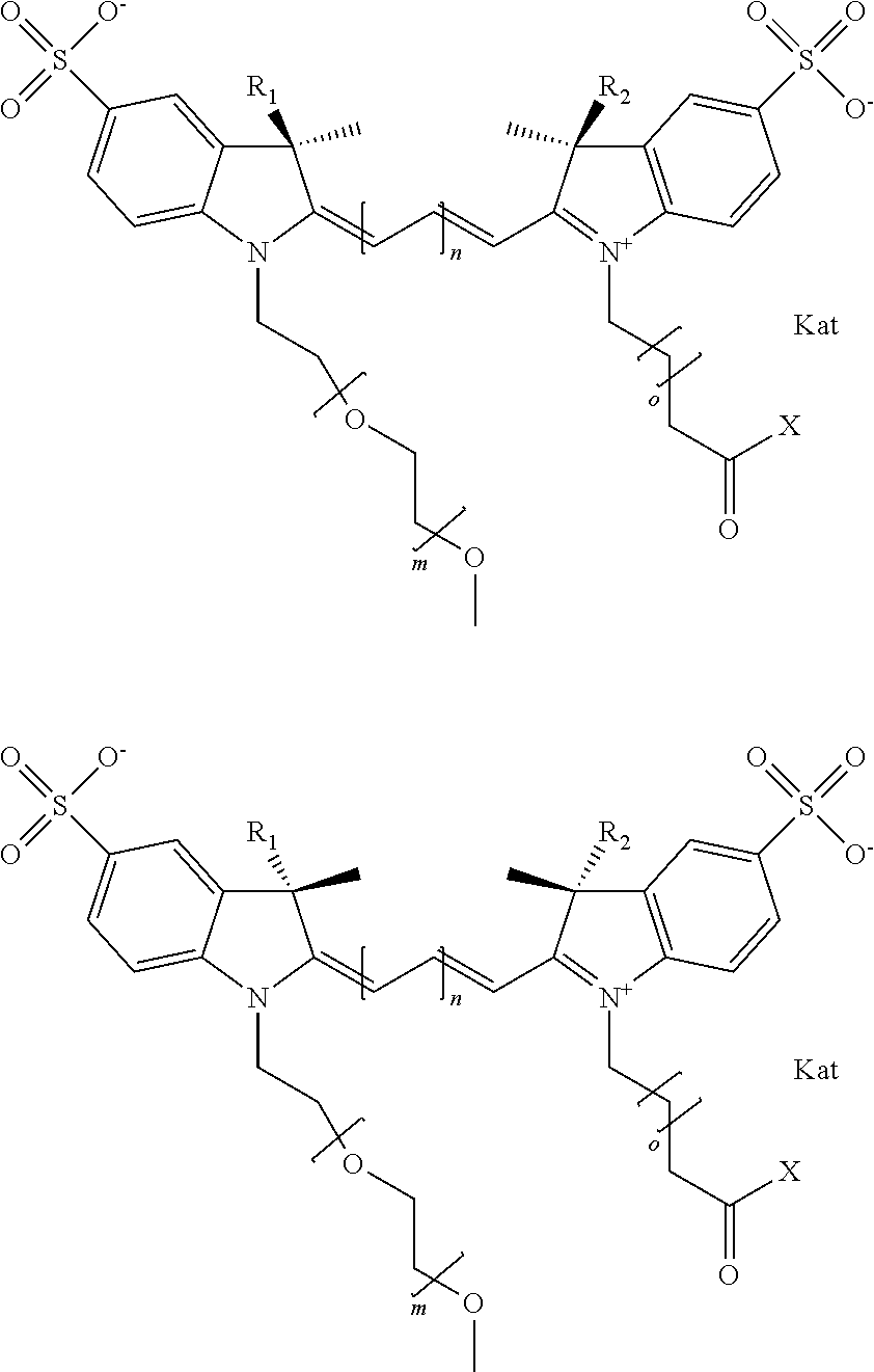
In one embodiment, the compound is a compound according to general formula I
##STR00001## wherein each of R.sup.1 and R.sup.2 is the same or different and is independently selected from the group consisting of an aliphatic, heteroaliphatic, or sulfoalkyl group; X is selected from the group consisting of --OH, --SH, --NH.sub.2, --NH--NH.sub.2, --F, --Cl, --Br, I, --NHS (hydroxysuccinimidyl/sulfosuccinimidyl), --O-TFP (2,3,5,6-tetrafluorophenoxy), --O-STP (4-sulfo-2,3,5,6-tetrafluorophenoxy), --O-benzotriazole, -benzotriazole, --NR-L-OH, --NR-L-O-phosphoramidite, --NR-L-SH, --NR-L-NH.sub.2, --NR-L-NH--NH.sub.2, --NR-L-CO.sub.2H, --NR-L-CO--NHS, --NR-L-CO-STP, --NR-L-CO-TFP, --NR-L-CO-benzotriazole, --NR-L-CHO, --NR-L-maleimide, and --NR-L-NH--CO--CH2-I, where R is --H or an aliphatic or heteroaliphatic group, and L is selected from the group consisting of a divalent linear, crossed, or cyclic alkyl group optionally substituted by at least one oxygen atom and/or sulfur atom; Kat is a number of Na.sup.+, K.sup.+, Ca.sup.2+, ammonia, or other cation(s) needed to compensate the negative charge brought by the cyanine; m is an integer from 0 to 5 inclusive; o is an integer from 0 to 12 inclusive; and n is an integer from 1 to 3 inclusive.
In one embodiment, the compound is a compound according to general formula I, wherein each of R1 and R2 is sulfoalkyl; X is --OH, --NHS, --O-TFP, or --NR-L-maleimide; m is 0; o is 3; and n is 1. In one embodiment, the compound is a compound according to general formula I, wherein each of R1 and R2 is sulfoalkyl; X is --OH, --NHS, --O-TFP, or --NR-L-maleimide; m is 1; o is 3; and n is 1. In one embodiment, the compound is a compound according to general formula I, wherein each of R1 and R2 is sulfoalkyl; X is --OH, --NHS, --O-TFP, or --NR-L-maleimide; m is 2; o is 3; and n is 1. In one embodiment, the compound is a compound according to general formula I, wherein each of R1 and R2 is sulfoalkyl; X is --OH, --NHS, --O-TFP, or --NR-L-maleimide; m is 3; o is 3; and n is 1. In one embodiment, the compound is a compound according to general formula I, wherein each of R1 and R2 is sulfoalkyl; X is --OH, --NHS, --O-TFP, or --NR-L-maleimide; m is 4; o is 3; and n is 1. In one embodiment, the compound is a compound according to general formula I, wherein each of R1 and R2 is sulfoalkyl; X is --OH, --NHS, --O-TFP, or --NR-L-maleimide; m is 5; o is 3; and n is 1.
In one embodiment, the compound is a compound according to general formula I, wherein each of R1 and R2 is sulfoalkyl; X is --OH, --NHS, --O-TFP, or --NR-L-maleimide; m is 0; o is 3; and n is 2. In one embodiment, the compound is a compound according to general formula I, wherein each of R1 and R2 is sulfoalkyl; X is --OH, --NHS, --O-TFP, or --NR-L-maleimide; m is 1; o is 3; and n is 2. In one embodiment, the compound is a compound according to general formula I, wherein each of R1 and R2 is sulfoalkyl; X is --OH, --NHS, --O-TFP, or --NR-L-maleimide; m is 2; o is 3; and n is 2. In one embodiment, the compound is a compound according to general formula I, wherein each of R1 and R2 is sulfoalkyl; X is --OH, --NHS, --O-TFP, or --NR-L-maleimide; m is 3; o is 3; and n is 2. In one embodiment, the compound is a compound according to general formula I, wherein each of R1 and R2 is sulfoalkyl; X is --OH, --NHS, --O-TFP, or --NR-L-maleimide; m is 4; o is 3; and n is 2. In one embodiment, the compound is a compound according to general formula I, wherein each of R1 and R2 is sulfoalkyl; X is --OH, --NHS, --O-TFP, or --NR-L-maleimide; m is 5; o is 3; and n is 2.
In one embodiment, the compound is a compound according to general formula I, wherein each of R1 and R2 is sulfoalkyl; X is --OH, --NHS, --O-TFP, or --NR-L-maleimide; m is 0; o is 3; and n is 3. In one embodiment, the compound is a compound according to general formula I, wherein each of R1 and R2 is sulfoalkyl; X is --OH, --NHS, --O-TFP, or --NR-L-maleimide; m is 1; o is 3; and n is 3. In one embodiment, the compound is a compound according to general formula I, wherein each of R1 and R2 is sulfoalkyl; X is --OH, --NHS, --O-TFP, or --NR-L-maleimide; m is 2; o is 3; and n is 3. In one embodiment, the compound is a compound according to general formula I, wherein each of R1 and R2 is sulfoalkyl; X is --OH, --NHS, --O-TFP, or --NR-L-maleimide; m is 3; o is 3; and n is 3. In one embodiment, the compound is a compound according to general formula I, wherein each of R1 and R2 is sulfoalkyl; X is --OH, --NHS, --O-TFP, or --NR-L-maleimide; m is 4; o is 3; and n is 3. In one embodiment, the compound is a compound according to general formula I, wherein each of R1 and R2 is sulfoalkyl; X is --NHS, --O-TFP, or --NR-L-maleimide; m is 5; o is 3; and n is 3.
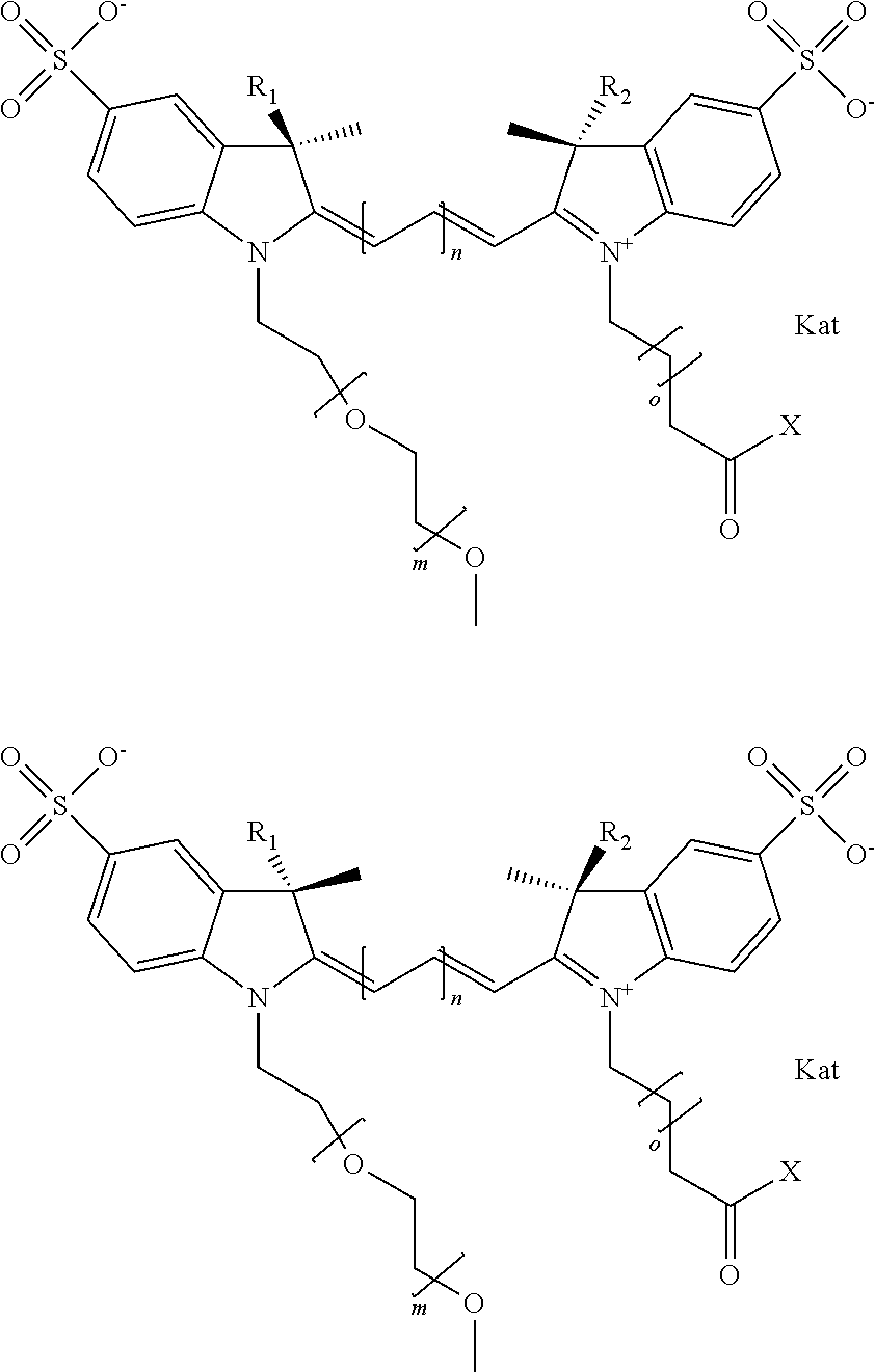
In one embodiment, an isolated enantiomeric mixture selected from diastereomer Ia of general formula I
##STR00002## or diastereomer Ib of general formula I
##STR00003## is provided, wherein each of R.sup.1 and R.sup.2 is the same or different and is independently selected from the group consisting of an aliphatic, heteroaliphatic, sulfoalkyl group, or heteroaliphatic with terminal SO.sub.3; X is selected from the group consisting of --OH, --SH, --NH.sub.2, --NH--NH.sub.2, --F, --Cl, --Br, I, --NHS (hydroxysuccinimidyl/sulfosuccinimidyl), --O-TFP (2,3,5,6-tetrafluorophenoxy), --O-STP (4-sulfo-2,3,5,6-tetrafluorophenoxy), --O-benzotriazole, -benzotriazole, --NR-L-OH, --NR-L-O-phosphoramidite, --NR-L-SH, --NR-L-NH.sub.2, --NR-L-NH--NH.sub.2, --NR-L-CO.sub.2H, --NR-L-CO--NHS, --NR-L-CO-STP, --NR-L-CO-TFP, --NR-L-CO-benzotriazole, --NR-L-CHO, --NR-L-maleimide, and --NR-L-NH--CO--CH2-I, where R is --H or an aliphatic or heteroaliphatic group, and L is selected from the group consisting of a divalent linear, crossed, or cyclic alkyl group optionally substituted by at least one oxygen atom and/or sulfur atom; Kat is a number of Na.sup.+, K.sup.+, Ca.sup.2+, ammonia, or other cation(s) needed to compensate the negative charge brought by the cyanine; m is an integer from 0 to 5 inclusive; o is an integer from 0 to 12 inclusive; and n is an integer from 1 to 3 inclusive.
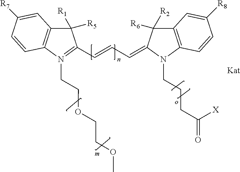
In one embodiment, the compound has general formula II
##STR00004## where each of R.sup.1, R.sup.2, R.sup.5, and R.sup.6 is the same or different and is independently selected from the group consisting of an aliphatic, heteroaliphatic, or sulfoalkyl group; each of R.sup.7 and R.sup.8 is the same or different and is independently selected from either H or SO.sub.3; X is selected from the group consisting of --OH, --SH, --NH.sub.2, --NH--NH.sub.2, --F, --Cl, --Br, I, --NHS (hydroxysuccinimidyl/sulfosuccinimidyl), --O-TFP (2,3,5,6-tetrafluorophenoxy), --O-STP (4-sulfo-2,3,5,6-tetrafluorophenoxy), --O-benzotriazole, -benzotriazole, --NR-L-OH, --NR-L-O-phosphoramidite, --NR-L-SH, --NR-L-NH.sub.2, --NR-L-NH--NH.sub.2, --NR-L-CO.sub.2H, --NR-L-CO--NHS, --NR-L-CO-STP, --NR-L-CO-TFP, --NR-L-CO-benzotriazole, --NR-L-CHO, --NR-L-maleimide, and --NR-L-NH--CO--CH2-I, where R is --H or an aliphatic or heteroaliphatic group, and L is selected from the group consisting of a divalent linear, crossed, or cyclic alkyl group optionally substituted by at least one oxygen atom and/or sulfur atom; Kat is a number of Na.sup.+, K.sup.+, Ca.sup.2+, ammonia, or other cation(s) needed to compensate the negative charge brought by the cyanine; m is an integer from 0 to 5 inclusive; o is an integer from 0 to 12 inclusive; and n is an integer from 1 to 3 inclusive.
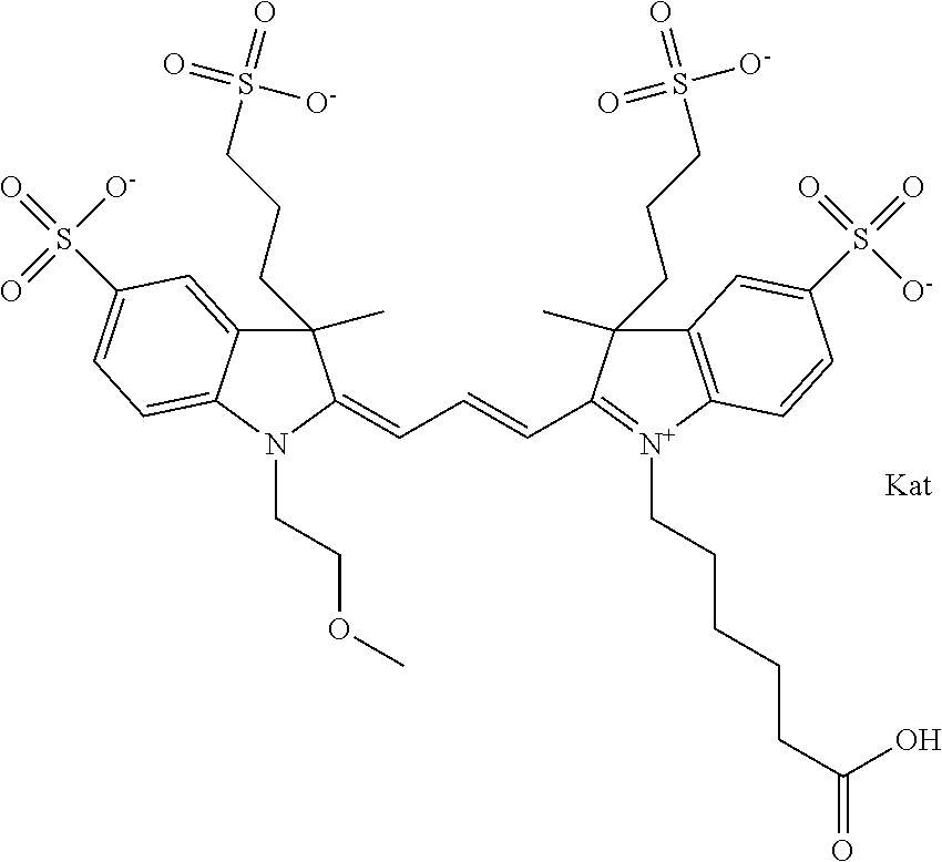
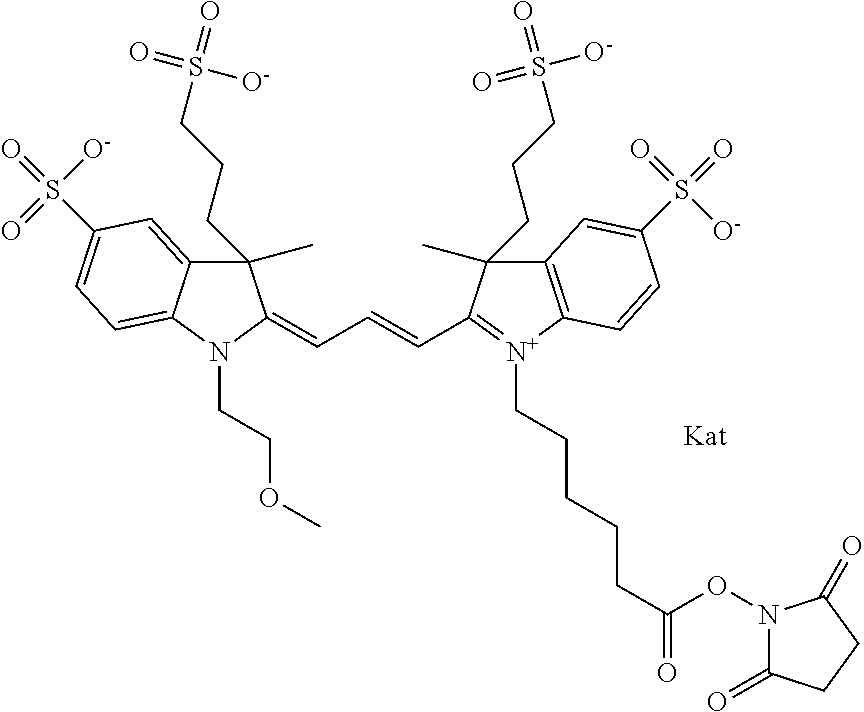
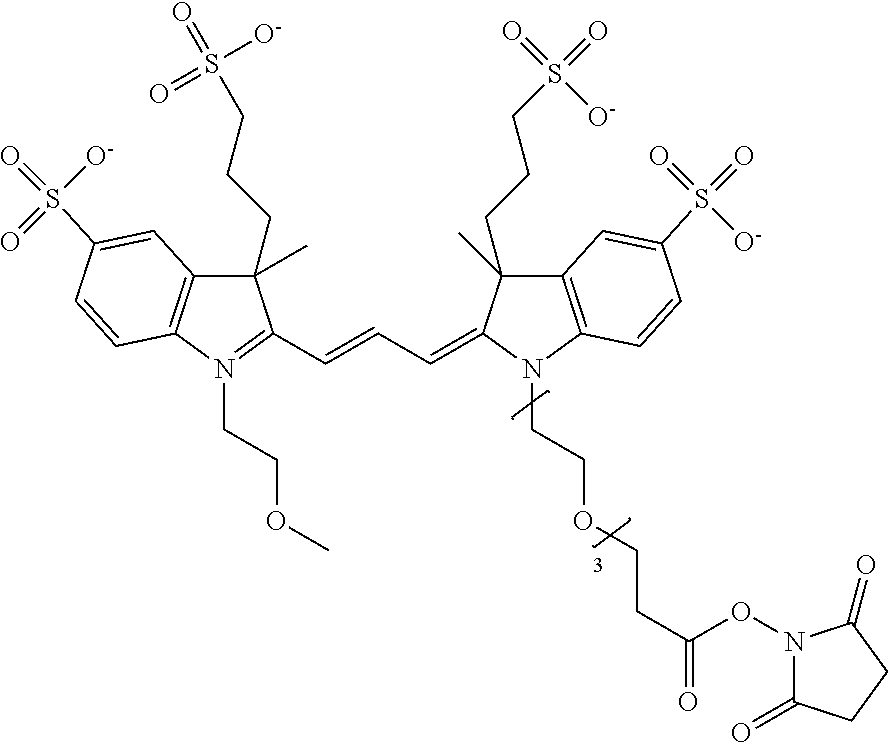
In one embodiment, the compound is 550 Compound 1
##STR00005##
550 Compound 1 (2-{(E)-3-[1-(5-carboxypentyl)-3-methyl-5-sulfo-3-(3-sulfopropyl)-1,3-dih- ydro-indol-(2E)-ylidene]-propenyl}-1-(2-methoxy-ethyl)-3-methyl-5-sulfo-3-- (3-sulfo-propyl)-3H-indolium) contains an ethylene glycol on the indole N of the left heterocycle, i.e., a methylated ethylene glycol, as shown in the structure above, and the ethylene glycol can be represented in abbreviated format as --[C--C--O].sub.1--, which is used throughout. The methyl group on the ethylene glycol prevents the terminal --OH from oxidation. Oxidation is known to occur, over time, on an unprotected terminus of an ethylene glycol group, diethylene glycol group, or polyethylene glycol group, collectively referred to herein as an unprotected PEG terminus. Adding a methyl ether provides this protection, and prevents reaction with electrophilic reactive groups.
In embodiments, e.g., for functional assays, the inventive compounds are activated. Activation of the compound adds a chemical moiety such that the compound is in a form that can be conjugated to a biological moiety. Examples of chemical moieties for activation are described below with reference to activation of 550 Compound 1, but one skilled in the art appreciates that activation is not limited to these examples. One non-limiting example of an activated compound is the NHS-ester of 550 Compound 1, shown below:
##STR00006##
In one embodiment, the compound is a NHS-ester of 550 Compound 1 where, according to general formula I, o is 1, shown below:
##STR00007##
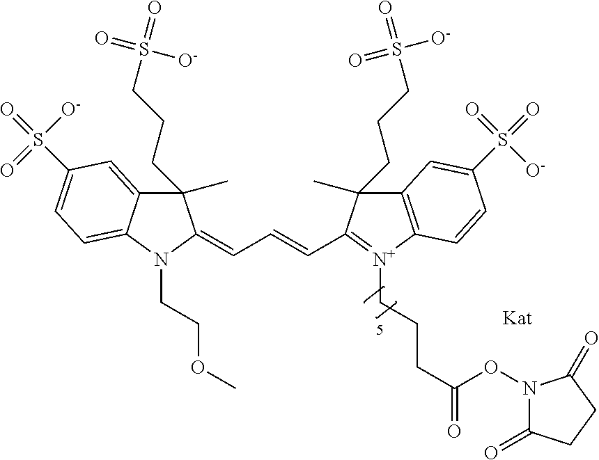
In one embodiment, the compound is an NHS-ester of 550 Compound 1 where, according to general formula I, o is 5, shown below:
##STR00008##
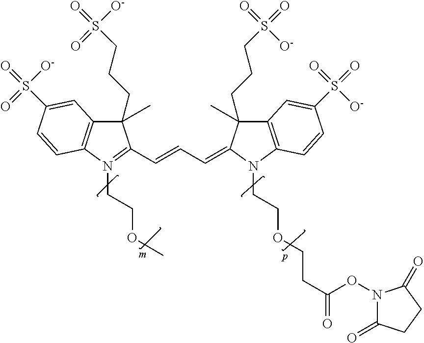
In one embodiment, the compound is described by the following general formula III, where m=1-6, and p=1-6:
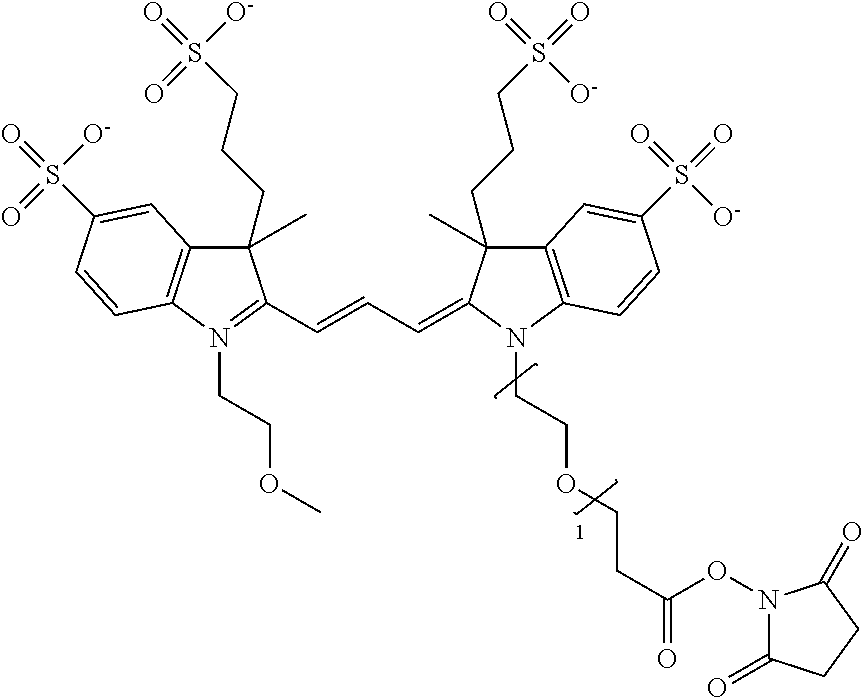
##STR00009## One non-limiting example of a NHS-ester of 550 Compound 1, according to general formula III, where m=1 and p=1, is shown below:
##STR00010## One non-limiting example of a NHS-ester of 550 Compound 1, according to general formula III, where m=1 and p=2, is shown below:
##STR00011## One non-limiting example of a NHS-ester of 550 Compound 1, according to general formula III, where m=1 and p=3, is shown below:
##STR00012## One non-limiting example of a NHS-ester of 550 Compound 1, according to general formula III, where m=1 and p=4, is shown below:
##STR00013## One non-limiting example of a NHS-ester of 550 Compound 1, according to general formula III, where m=1 and p=5, is shown below:
##STR00014## One non-limiting example of a NHS-ester of 550 Compound 1, according to general formula III, where m=1 and p=6, is shown below:
##STR00015## One non-limiting example of a NHS-ester of 550 Compound 2, according to general formula III, where m=2 and p=1, is shown below:
##STR00016## One non-limiting example of a NHS-ester of 550 Compound 2, according to general formula III, where m=2 and p=2, is shown below:
##STR00017## One non-limiting example of a NHS-ester of 550 Compound 2, according to general formula III, where m=2 and p=3, is shown below:
##STR00018## One non-limiting example of a NHS-ester of 550 Compound 3, according to general formula III, where m=3 and p=1, is shown below:
##STR00019## One non-limiting example of a NHS-ester of 550 Compound 3, according to general formula III, where m=3 and p=2, is shown below:
##STR00020## One non-limiting example of a NHS-ester of 550 Compound 3, according to general formula III, where m=3 and p=3, is shown below:
##STR00021## One non-limiting example of a NHS-ester of 550 Compound 4, according to general formula III, where m=4 and p=1, is shown below:
##STR00022## One non-limiting example of a NHS-ester of 550 Compound 5, according to general formula III, where m=5 and p=1, is shown below:
##STR00023## One non-limiting example of a NHS-ester of 550 Compound 6, according to general formula III, where m=6 and p=1, is shown below:
##STR00024## One non-limiting example of an activated 550 Compound 1 is a tetrafluorophenyl (TFP)-ester form of 550 Compound 1, shown below:
##STR00025## One non-limiting example of an activated 550 Compound 1 is a sulfotetrafluorophenyl (STP)-ester form of 550 Compound 1, shown below:
##STR00026## One non-limiting example of an activated 550 Compound 1 is a hydrazide form of 550 Compound 1, shown below:
##STR00027## One non-limiting example of an activated 550 Compound 1 is a maleimide form of 550 Compound 1, shown below:
##STR00028##
In one embodiment, the compound is 550 Compound 2
##STR00029##
550 Compound 2 (2-{(E)-3-[1-(5-Carboxypentyl)-3-methyl-5-sulfo-3-(3-sulfopropyl)-1,3-dih- ydro-indol-(2E)-ylidene]-propenyl}-1-[2-(2-methoxy-ethoxy)-ethyl]-3-methyl- -5-sulfo-3-(3-sulfo-propyl)-3H-indolium) contains a diethylene glycol on the indole N of the left heterocycle. 550 Compound 2, with the diethylene glycol shown in abbreviated notation used throughout, represents the following structure.
##STR00030## The methyl group on the ethylene glycol prevents the terminal --OH from oxidation. Oxidation is known to occur, over time, on an unprotected PEG terminus. Adding a methyl ether provides this protection, and prevents reaction with electrophilic reactive groups. For functional assays, 550 Compound 2 is activated as described above, one non-limiting example of which is the NHS-ester form of 550 Compound 2, shown below.
##STR00031## In one embodiment, the compound is 550 Compound 3
##STR00032##
550 Compound 3 (2-{(E)-3-[1-(5-Carboxpentyl)-3-methyl-5-sulfo-3-(3-sulfopropyl)-1,3-dihy- dro-indol-(2E)-ylidene]-propenyl}-1-{2-[2-(2-methoxy-ethoxy)-ethoxy]-ethyl- }-3-methyl-5-sulfo-3-(3-sulfo-propyl)-3H-indolium) contains a (poly)ethylene glycol on the indole N of the left heterocycle. 550 Compound 3, with the (poly)ethylene glycol shown in abbreviated notation used throughout, represents the following structure.
##STR00033## The methyl group on the ethylene glycol prevents the terminal --OH from oxidation. Oxidation is known to occur, over time, on an unprotected PEG terminus. Adding a methyl ether provides this protection, and prevents reaction with electrophilic reactive groups. For functional assays, 550 Compound 3 is activated as described above.
In one embodiment, the compound is 550 Compound 4
##STR00034##
550 Compound 4 (1-(5-carboxypentyl)-3-methyl-2-((1E,3E)-3-(3-methyl-5-sulfonato-3-(3-sul- fonatopropyl)-1-(2,5,8,11-tetraoxatridecan-13-yl)indolin-2-ylidene)prop-1-- en-1-yl)-3-(3-sulfonatopropyl)-3H-indol-1-ium-5-sulfonate) contains a (poly)ethylene glycol on the indole N of the left heterocycle. 550 Compound 4, with the (poly)ethylene glycol shown in abbreviated notation used throughout, represents the following structure.
##STR00035## The methyl group on the ethylene glycol prevents the terminal --OH from oxidation. Oxidation is known to occur, over time, on an unprotected PEG terminus. Adding a methyl ether provides this protection, and prevents reaction with electrophilic reactive groups. For functional assays, 550 Compound 4 is activated as described above.
In one embodiment, the compound is 550 Compound 5
##STR00036##
550 Compound 5 (2-((1E,3E)-3-(1-(2,5,8,11,14-pentaoxahexadecan-16-yl)-3-methyl-5-sulfona- to-3-(3-sulfonatopropyl)indolin-2-ylidene)prop-1-en-1-yl)-1-(5-carboxypent- yl)-3-methyl-3-(3-sulfonatopropyl)-3H-indol-1-ium-5-sulfonate) contains a (poly)ethylene glycol on the indole N of the left heterocycle. 550 Compound 5, with the (poly)ethylene glycol shown in abbreviated notation used throughout, represents the following structure.
##STR00037## The methyl group on the ethylene glycol prevents the terminal --OH from oxidation. Oxidation is known to occur, over time, on an unprotected PEG terminus. Adding a methyl ether provides this protection, and prevents reaction with electrophilic reactive groups. For functional assays, 550 Compound 5 is activated as described above.
In one embodiment, the compound is 550 Compound 6
##STR00038##
550 Compound 6 (1-(5-carboxypentyl)-3-methyl-2-((1E,3E)-3-(3-methyl-1-(2,5,8,11,14,17-he- xaoxanonadecan-19-yl)-5-sulfonato-3-(3-sulfonatopropyl)indolin-2-ylidene)p- rop-1-en-1-yl)-3-(3-sulfonatopropyl)-3H-indol-1-ium-5-sulfonate) contains a (poly)ethylene glycol on the indole N of the left heterocycle. 550 Compound 6, with the (poly)ethylene glycol shown in abbreviated notation used throughout, represents the following structure.
##STR00039## The methyl group on the ethylene glycol prevents the terminal --OH from oxidation. Oxidation is known to occur, over time, on an unprotected PEG terminus. Adding a methyl ether provides this protection, and prevents reaction with electrophilic reactive groups. For functional assays, 550 Compound 6 is activated as described above.
In embodiments, the compound contains one or more substitutions of the polymethine linker. In one embodiment, the compound has general formula IV
##STR00040## where each of R.sup.1, R.sup.2, R.sup.5, and R.sup.6 is the same or different and is independently selected from an aliphatic, heteroaliphatic, or sulfoalkyl group; each of R.sup.7 and R.sup.8 is the same or different and is independently selected from H or SO.sub.3; X is selected from --OH, --SH, --NH.sub.2, --NH--NH.sub.2, --F, --Cl, --Br, --I, --NHS (hydroxysuccinimidyl/sulfosuccinimidyl), --O-TFP (2,3,5,6-tetrafluorophenoxy), --O-STP (4-sulfo-2,3,5,6-tetrafluorophenoxy), --O-benzotriazole, -benzotriazole, imidazole, azide, --O-carbodiimide, --NR-L-OH, --NR-L-O-phosphoramidite, --NR-L-SH, --NR-L-NH.sub.2, --NR-L-NH--NH.sub.2, --NR-L-CO.sub.2H, --NR-L-CO--NHS, --NR-L-CO-STP, --NR-L-CO-TFP, --NR-L-CO-benzotriazole, --NR-L-CHO, --NR-L-maleimide, --NR-L-NH--CO--CH.sub.2--I, or --NR-L-NH--CO--CH.sub.2--Br where R is --H or an aliphatic or heteroaliphatic group, and L is selected from the group consisting of a divalent linear, crossed, or cyclic alkyl group optionally substituted by at least one oxygen atom and/or sulfur atom; Kat is a number of Na.sup.+, K.sup.+, Ca.sup.2+, ammonia, or other cation(s) needed to compensate the negative charge brought by the cyanine; m is an integer from 0 to 5 inclusive; o is an integer from 0 to 12 inclusive; each of R3 and R4 is the same or different and is independently hydrogen, an aliphatic group, or a heteroaliphatic group; and Y is selected from the group consisting of hydrogen, alkyl, sulfoalkyl, fluorine, chlorine, or bromine.
One non-limiting example is a substituted polymethine form of 550 Compound 1, shown below:
##STR00041##
One non-limiting example is a substituted polymethine form of 550 Compound 2, shown below:
##STR00042##
One non-limiting example is a substituted polymethine form of 550 Compound 3, shown below:
##STR00043##
One non-limiting example is a substituted polymethine form of 550 Compound 4, shown below:
##STR00044##
One non-limiting example is a substituted polymethine form of 550 Compound 5, shown below:
##STR00045##
One non-limiting example is a substituted polymethine form of 550 Compound 6, shown below:
##STR00046##
One non-limiting example is a substituted polymethine form of 550 having an ethylene glycol, diethylene glycol, or polyethylene glycol as described for general formula III, such as the compound shown below:
##STR00047##
In embodiments, the degree of sulfonation is varied to, e.g., vary the compound's degree of hydrophilicity or hydrophobicity. One non-limiting example is a monosulfonate form of 550 Compound 1, shown below, but it is understood that the single sulfo group can be at any of the described positions:
##STR00048##
One non-limiting example is a disulfonate form of 550 Compound 1, shown below, but it is understood that each of the two sulfo groups can be at any of the described positions:
##STR00049##
One non-limiting example is a trisulfonate form of 550 Compound 1, shown below, but it is understood that each of the three sulfo groups can be at any of the described positions:
##STR00050##
One non-limiting example is a tetrasulfonate form of 550 Compound 1, shown below, but it is understood that each of the four sulfo groups can be at any of the described positions:
##STR00051##
In one embodiment, the compound has general formula V
##STR00052## where each of R.sup.1, R.sup.2, R.sup.5, and R.sup.6 is the same or different and is independently selected from an aliphatic, heteroaliphatic, or sulfoalkyl group; each of R.sup.7 and R.sup.8 is the same or different and is independently selected from H or SO.sub.3; X is selected from --OH, --SH, --NH.sub.2, --NH--NH.sub.2, --F, --Cl, --Br, --I, --NHS (hydroxysuccinimidyl/sulfosuccinimidyl), --O-TFP (2,3,5,6-tetrafluorophenoxy), --O-STP (4-sulfo-2,3,5,6-tetrafluorophenoxy), --O-benzotriazole, -benzotriazole, imidazole, azide, --O-carbodiimide, --NR-L-OH, --NR-L-O-phosphoramidite, --NR-L-SH, --NR-L-NH.sub.2, --NR-L-NH--NH.sub.2, --NR-L-CO.sub.2H, --NR-L-CO--NHS, --NR-L-CO-STP, --NR-L-CO-TFP, --NR-L-CO-benzotriazole, --NR-L-CHO, --NR-L-maleimide, --NR-L-NH--CO--CH.sub.2--I, or --NR-L-NH--CO--CH.sub.2--Br where R is --H or an aliphatic or heteroaliphatic group, and L is selected from the group consisting of a divalent linear, crossed, or cyclic alkyl group optionally substituted by at least one oxygen atom and/or sulfur atom; Kat is a number of Na.sup.+, K.sup.+, Ca.sup.2+, ammonia, or other cation(s) needed to compensate the negative charge brought by the cyanine; m is an integer from 0 to 5 inclusive; p is an integer from 1 to 6 inclusive; each of R3 and R4 is the same or different and is independently hydrogen, an aliphatic group, or a heteroaliphatic group; and Y is selected from the group consisting of hydrogen, alkyl, sulfoalkyl, fluorine, chlorine, or bromine.
In one embodiment, the compound is 650 Compound 1
##STR00053##
650 Compound 1 (2-{(1E,3E)-5-[1-(5-Carboxypentyl)-3-methyl-5-sulfo-3-(3-sulfopropyl)-1,3- -dihydro-indol-(2E)-ylidene]-penta-1,3-dienyl}-1-(2-methoxy-ethyl)-3-methy- l-5-sulfo-3-(3-sulfopropyl)-3H-indolium tri sodium salt) contains an ethylene glycol on the indole N of the left heterocycle, i.e., a methylated ethylene glycol. The methyl group on the ethylene glycol prevents the terminal --OH from oxidation. Oxidation is known to occur, over time, on an unprotected PEG terminus (i.e., an unprotected ethylene glycol group, diethylene glycol group, or polyethylene glycol group). Adding a methyl ether provides this protection, and prevents reaction with electrophilic reactive groups.
In embodiments, e.g., for functional assays, the inventive compounds are activated. Activation of the compound adds a chemical moiety such that the compound is in a form that can be conjugated to a biological moiety. Examples of chemical moieties for activation are described below with reference to activation of 650 Compound 1, but one skilled in the art appreciates that activation is not limited to these examples. One non-limiting example of an activated compound is the NHS-ester of 650 Compound 1, shown below:
##STR00054## In one embodiment, the compound is a NHS-ester of 650 Compound 1 where, according to general formula I, o is 1, shown below:
##STR00055##
In one embodiment, the compound is an NHS-ester of 650 Compound 1 where, according to general formula I, o is 5, shown below:
##STR00056##
In one embodiment, the compound is described by the following general formula VI, where m=1-6, and p=1-6:
##STR00057## One non-limiting example of a NHS-ester of 650 Compound 1, according to formula VI, where m=1 and p=1, is shown below:
##STR00058## One non-limiting example of a NHS-ester of 650 Compound 1, according to general formula VI, where m=1 and p=2, is shown below:
##STR00059## One non-limiting example of a NHS-ester of 650 Compound 1, according to general formula VI, where m=1 and p=3, is shown below:
##STR00060## One non-limiting example of a NHS-ester of 650 Compound 1, according to general formula VI, where m=1 and p=4, is shown below:
##STR00061## One non-limiting example of a NHS-ester of 650 Compound 1, according to general formula VI, where m=1 and p=5, is shown below:
##STR00062## One non-limiting example of a NHS-ester of 650 Compound 1, according to general formula VI, where m=1 and p=6, is shown below:
##STR00063## One non-limiting example of a NHS-ester of 650 Compound 2, according to general formula VI, where m=2 and p=1, is shown below:
##STR00064## One non-limiting example of a NHS-ester of 650 Compound 2, according to general formula VI, where m=2 and p=2, is shown below:
##STR00065## One non-limiting example of a NHS-ester of 650 Compound 2, according to general formula VI, where m=2 and p=3, is shown below:
##STR00066## One non-limiting example of a NHS-ester of 650 Compound 3, according to general formula VI, where m=3 and p=1, is shown below:
##STR00067## One non-limiting example of a NHS-ester of 650 Compound 3, according to general formula VI, where m=3 and p=2, is shown below:
##STR00068## One non-limiting example of a NHS-ester of 650 Compound 3, according to general formula VI, where m=3 and p=3, is shown below:
##STR00069## One non-limiting example of a NHS-ester of 650 Compound 4, according to general formula VI, where m=4 and p=1, is shown below:
##STR00070## One non-limiting example of a NHS-ester of 650 Compound 5, according to general formula VI, where m=5 and p=1, is shown below:
##STR00071## One non-limiting example of a NHS-ester of 650 Compound 6, according to general formula VI, where m=6 and p=1, is shown below:
##STR00072## One non-limiting example of an activated 650 Compound 1 is the tetrafluorophenyl (TFP)-ester of 650 Compound 1, shown below:
##STR00073## One non-limiting example of an activated 650 Compound 1 is the sulfotetrafluorophenyl (STP)-ester of 650 Compound 1, shown below:
##STR00074## One non-limiting example of an activated 650 Compound 1 is the hydrazide of 650 Compound 1, shown below:
##STR00075## One non-limiting example of an activated 650 Compound 1 is the maleimide of 650 Compound 1, shown below:
##STR00076## In one embodiment, the compound is 650 Compound 2
##STR00077##
650 Compound 2 (2-{(1E,3E)-5-[1-(5-carboxypentyl)-3-methyl-5-sulfo-3-(3-sulfopropyl)-1,3- -dihydroindol-(2E)-ylidene]-penta-1,3-dienyl}-1-[2-(2-methoxy-ethoxy)-ethy- l]-3-methyl-5-sulfo-3-(3-sulfopropyl)-3H-indolium tri sodium salt.) contains a (poly)ethylene glycol on the indole N of the left heterocycle. The methyl group on the ethylene glycol prevents the terminal --OH from oxidation. Oxidation is known to occur, over time, on an unprotected PEG terminus. Adding a methyl ether provides this protection, and prevents reaction with electrophilic reactive groups. For functional assays, 650 Compound 2 is activated as described above, one non-limiting example of which is the NHS-ester form of 650 Compound 2, shown below.
##STR00078##
In one embodiment, the compound is 650 Compound 3
##STR00079##
650 Compound 3 (2-{(1E,3E)-5-[1-(5-carboxypentyl)-3-methyl-5-sulfo-3-(3-sulfopropyl)-1,3- -dihydro-indol-(2E)-ylidene]-penta-1,3-dienyl}-1-{2-[2-(2-methoxy-ethoxy)-- ethoxy]-ethyl}-3-methyl-5-sulfo-3-(3-sulfopropyl)-3H-indolium tri sodium salt) contains a (poly)ethylene glycol on the indole N of the left heterocycle. The methyl group on the ethylene glycol prevents the terminal --OH from oxidation. Oxidation is known to occur, over time, on an unprotected PEG terminus. Adding a methyl ether provides this protection, and prevents reaction with electrophilic reactive groups. For functional assays, 650 Compound 3 is activated as described above, one non-limiting example of which is the NHS-ester form of 650 Compound 3, shown below.
##STR00080##
In one embodiment, the compound is 650 Compound 4
##STR00081##
650 Compound 4 (2-((1E,3E,5E)-5-(1-(5-carboxypentyl)-3-methyl-5-sulfonato-3-(3-sulfonato- propyl)indolin-2-ylidene)penta-1,3-dien-1-yl)-3-methyl-3-(3-sulfonatopropy- l)-1-(2,5,8,11-tetraoxatridecan-13-yl)-3H-indol-1-ium-5-sulfonate) contains a (poly)ethylene glycol on the indole N of the left heterocycle. The methyl group on the ethylene glycol prevents the terminal --OH from oxidation. Oxidation is known to occur, over time, on an unprotected PEG terminus. Adding a methyl ether provides this protection, and prevents reaction with electrophilic reactive groups. For functional assays, 650 Compound 4 is activated as described above.
In one embodiment, the compound is 650 Compound 5
##STR00082##
650 Compound 5 (2-((1E,3E,5E)-5-(1-(5-carboxypentyl)-3-methyl-5-sulfonato-3-(3-sulfonato- propyl)indolin-2-ylidene)penta-1,3-dien-1-yl)-1-(2,5,8,11,14-pentaoxahexad- ecan-16-yl)-3-methyl-3-(3-sulfonatopropyl)-3H-indol-1-ium-5-sulfonate) contains a (poly)ethylene glycol on the indole N of the left heterocycle. The methyl group on the ethylene glycol prevents the terminal --OH from oxidation. Oxidation is known to occur, over time, on an unprotected PEG terminus. Adding a methyl ether provides this protection, and prevents reaction with electrophilic reactive groups. For functional assays, 650 Compound 5 is activated as described above.
In one embodiment, the compound is 650 Compound 6
##STR00083##
650 Compound 6 (2-((1E,3E,5E)-5-(1-(5-carboxypentyl)-3-methyl-5-sulfonato-3-(3-sulfonato- propyl)indolin-2-ylidene)penta-1,3-dien-1-yl)-3-methyl-1-(2,5,8,11,14,17-h- exaoxanonadecan-19-yl)-3-(3-sulfonatopropyl)-3H-indol-1-ium-5-sulfonate) contains a (poly)ethylene glycol on the indole N of the left heterocycle. The methyl group on the ethylene glycol prevents the terminal --OH from oxidation. Oxidation is known to occur, over time, on an unprotected PEG terminus. Adding a methyl ether provides this protection, and prevents reaction with electrophilic reactive groups. For functional assays, 650 Compound 6 is activated as described above.
In embodiments, the compound contains one or more substitutions of the polymethine linker. In one embodiment, the compound has general formula VII
##STR00084## wherein each of R.sup.1 and R.sup.2 is the same or different and is independently selected from the group consisting of an aliphatic, heteroaliphatic, or sulfoalkyl group; X is selected from the group consisting of --OH, --SH, --NH.sub.2, --NH--NH.sub.2, --F, --Cl, --Br, I, --NHS (hydroxysuccinimidyl/sulfosuccinimidyl), --O-TFP (2,3,5,6-tetrafluorophenoxy), --O-STP (4-sulfo-2,3,5,6-tetrafluorophenoxy), --O-benzotriazole, -benzotriazole, --NR-L-OH, --NR-L-O-phosphoramidite, --NR-L-SH, --NR-L-NH.sub.2, --NR-L-NH--NH.sub.2, --NR-L-CO.sub.2H, --NR-L-CO--NHS, --NR-L-CO-STP, --NR-L-CO-TFP, --NR-L-CO-benzotriazole, --NR-L-CHO, --NR-L-maleimide, and --NR-L-NH--CO--CH2-I, where R is --H or an aliphatic or heteroaliphatic group, and L is selected from the group consisting of a divalent linear, crossed, or cyclic alkyl group optionally substituted by at least one oxygen atom and/or sulfur atom; Kat is a number of Na.sup.+, K.sup.+, Ca.sup.2+, ammonia, or other cation(s) needed to compensate the negative charge brought by the cyanine; m is an integer from 0 to 5 inclusive; o is an integer from 0 to 12 inclusive; each of R3 and R4 is the same or different and is independently hydrogen, an aliphatic group, or a heteroaliphatic group, or R3 and R4 together form a cyclic structure where R3 and R4 are joined using a divalent structural element selected from the group consisting of --(CH.sub.2).sub.q--, --(CH.sub.2).sub.qO(CH.sub.2).sub.q'--, --(CH.sub.2).sub.qS(CH.sub.2).sub.q'--, --(CH.sub.2).sub.qCH.dbd.CH--, --OCH.dbd.CH-- where each of q and q' is the same or different and is an integer from 2 to 6 inclusive; and Y is selected from the group consisting of hydrogen, alkyl, sulfoalkyl, fluorine, chlorine, bromine, a substituted or unsubstituted aryl-, phenoxy- and a phenylmercapto function.
In one embodiment, the compound of general formula VII wherein each of R3 and R4 is the same or different and is independently hydrogen, an aliphatic group, or a heteroaliphatic group, or R3 and R4 together form a cyclic structure where R3 and R4 are directly joined or joined using a divalent structural element selected from the group consisting of --(CH.sub.2).sub.q-- and CH.dbd.CH, where q is an integer from 1 to 2 inclusive, to result in a 3-, 4-, or 5-membered ring.
In one embodiment, an isolated enantiomeric mixture selected from diastereomer Ia of general formula VII
##STR00085## or diastereomer Ib of general formula VII
##STR00086## is provided, wherein each of R.sup.1 and R.sup.2 is the same or different and is independently selected from the group consisting of an aliphatic, heteroaliphatic, sulfoalkyl group, or heteroaliphatic with terminal SO.sub.3; X is selected from the group consisting of --OH, --SH, --NH.sub.2, --NH--NH.sub.2, --F, --Cl, --Br, I, --NHS (hydroxysuccinimidyl/sulfosuccinimidyl), --O-TFP (2,3,5,6-tetrafluorophenoxy), --O-STP (4-sulfo-2,3,5,6-tetrafluorophenoxy), --O-benzotriazole, -benzotriazole, --NR-L-OH, --NR-L-O-phosphoramidite, --NR-L-SH, --NR-L-NH.sub.2, --NR-L-NH--NH.sub.2, --NR-L-CO.sub.2H, --NR-L-CO--NHS, --NR-L-CO-STP, --NR-L-CO-TFP, --NR-L-CO-benzotriazole, --NR-L-CHO, --NR-L-maleimide, and --NR-L-NH--CO--CH2-I, where R is --H or an aliphatic or heteroaliphatic group, and L is selected from the group consisting of a divalent linear, crossed, or cyclic alkyl group optionally substituted by at least one oxygen atom and/or sulfur atom; Kat is a number of Na.sup.+, K.sup.+, Ca.sup.2+, ammonia, or other cation(s) needed to compensate the negative charge brought by the cyanine; m is an integer from 0 to 5 inclusive; o is an integer from 0 to 12 inclusive; each of R.sup.3 and R.sup.4 is the same or different and is independently hydrogen, an aliphatic group, or a heteroaliphatic group, or R.sup.3 and R.sup.4 together form a cyclic structure where R.sup.3 and R.sup.4 are joined using a divalent structural element selected from the group consisting of --(CH.sub.2).sub.q--, --(CH.sub.2).sub.qO(CH.sub.2).sub.q'--, --(CH.sub.2).sub.qS(CH.sub.2).sub.q'--, --(CH.sub.2).sub.qCH.dbd.CH--, --OCH.dbd.CH-- where each of q and q' is the same or different and is a integer from 2 to 6 inclusive; and Y is selected from the group consisting of hydrogen, alkyl, sulfoalkyl, fluorine, chlorine, bromine, a substituted or unsubstituted aryl-, phenoxy- and a phenylmercapto function.
In one embodiment, the compound has general formula VIII
##STR00087## where each of R.sup.1, R.sup.2, R.sup.5, and R.sup.6 is the same or different and is independently selected from an aliphatic, heteroaliphatic, or sulfoalkyl group; each of R.sup.7 and R.sup.8 is the same or different and is independently selected from H or SO.sub.3; X is selected from --OH, --SH, --NH.sub.2, --NH--NH.sub.2, --F, --Cl, --Br, --I, --NHS (hydroxysuccinimidyl/sulfosuccinimidyl), --O-TFP (2,3,5,6-tetrafluorophenoxy), --O-STP (4-sulfo-2,3,5,6-tetrafluorophenoxy), --O-benzotriazole, -benzotriazole, imidazole, azide, --O-carbodiimide, --NR-L-OH, --NR-L-O-phosphoramidite, --NR-L-SH, --NR-L-NH.sub.2, --NR-L-NH--NH.sub.2, --NR-L-CO.sub.2H, --NR-L-CO--NHS, --NR-L-CO-STP, --NR-L-CO-TFP, --NR-L-CO-benzotriazole, --NR-L-CHO, --NR-L-maleimide, --NR-L-NH--CO--CH.sub.2--I, or --NR-L-NH--CO--CH.sub.2--Br where R is --H or an aliphatic or heteroaliphatic group, and L is selected from the group consisting of a divalent linear, crossed, or cyclic alkyl group optionally substituted by at least one oxygen atom and/or sulfur atom; Kat is a number of Na.sup.+, K.sup.+, Ca.sup.2+, ammonia, or other cation(s) needed to compensate the negative charge brought by the cyanine; m is an integer from 0 to 5 inclusive; o is an integer from 0 to 12 inclusive; each of R3 and R4 is the same or different and is independently hydrogen, an aliphatic group, or a heteroaliphatic group, or R3 and R4 together form a cyclic structure where R3 and R4 are joined using a divalent structural element selected from the group consisting of --(CH.sub.2).sub.q--, --(CH.sub.2).sub.qO(CH.sub.2).sub.q'--, --(CH.sub.2).sub.qS(CH.sub.2).sub.q'--, --(CH.sub.2).sub.qCH.dbd.CH--, --OCH.dbd.CH-- where each of q and q' is the same or different and is a integer from 2 to 6 inclusive; and Y is selected from the group consisting of hydrogen, alkyl, sulfoalkyl, fluorine, chlorine, bromine, a substituted or unsubstituted aryl-, phenoxy- or phenylmercapto function.
In one embodiment, the compound of general formula VIII wherein each of R3 and R4 is the same or different and is independently hydrogen, an aliphatic group, or a heteroaliphatic group, or R3 and R4 together form a cyclic structure where R3 and R4 are directly joined or joined using a divalent structural element selected from the group consisting of --(CH.sub.2).sub.q-- and CH.dbd.CH, where q is an integer from 1 to 2 inclusive, to result in a 3-, 4-, or 5-membered ring.
One non-limiting example is a substituted polymethine form of 650 Compound 1, shown below:
##STR00088##
One non-limiting example is a substituted polymethine form of 650 Compound 2, shown below:
##STR00089##
One non-limiting example is a substituted polymethine form of 650 Compound 3, shown below:
##STR00090##
One non-limiting example is a substituted polymethine form of 650 Compound 4, shown below:
##STR00091##
One non-limiting example is a substituted polymethine form of 650 Compound 5, shown below:
##STR00092##
One non-limiting example is a substituted polymethine form of 650 Compound 6, shown below:
##STR00093##
One non-limiting example is a substituted polymethine form of 650 having an ethylene glycol, diethylene glycol, or polyethylene glycol as described for general formula VI, such as the compound shown below:
##STR00094##
One non-limiting example is a substituted polymethine form of 650 Compound 1, shown below:
##STR00095##
One non-limiting example is a substituted polymethine form of 650 Compound 2, shown below:
##STR00096##
One non-limiting example is a substituted polymethine form of 650 Compound 3, shown below:
##STR00097##
One non-limiting example is a substituted polymethine form of 650 Compound 4, shown below:
##STR00098##
One non-limiting example is a substituted polymethine form of 650 Compound 5, shown below:
##STR00099##
One non-limiting example is a substituted polymethine form of 650 Compound 6, shown below:
##STR00100##
One non-limiting example is a substituted polymethine form of 650 having an ethylene glycol, diethylene glycol, or polyethylene glycol as described for general formula VI, such as the compound shown below:
##STR00101##
In embodiments, the degree of sulfonation is varied to, e.g., vary the compound's degree of hydrophilicity or hydrophobicity. One non-limiting example is a monosulfonate form of 650 Compound 1, shown below, but it is understood that the single sulfo group can be at any of the described positions:
##STR00102##
One non-limiting example is a disulfonate form of 650 Compound 1, shown below, but it is understood that each of the two sulfo groups can be at any of the described positions:
##STR00103##
One non-limiting example is a trisulfonate form of 650 Compound 1, shown below, but it is understood that each of the three sulfo groups can be at any of the described positions:
##STR00104##
One non-limiting example is a tetrasulfonate form of 650 Compound 1, shown below, but it is understood that each of the four sulfo groups can be at any of the described positions:
##STR00105##
In one embodiment, the compound has general formula IX
##STR00106## where each of R.sup.1, R.sup.2, R.sup.5, and R.sup.6 is the same or different and is independently selected from an aliphatic, heteroaliphatic, or sulfoalkyl group; each of R.sup.7 and R.sup.8 is the same or different and is independently selected from H or SO.sub.3; X is selected from --OH, --SH, --NH.sub.2, --NH--NH.sub.2, --F, --Cl, --Br, --I, --NHS (hydroxysuccinimidyl/sulfosuccinimidyl), --O-TFP (2,3,5,6-tetrafluorophenoxy), --O-STP (4-sulfo-2,3,5,6-tetrafluorophenoxy), --O-benzotriazole, -benzotriazole, imidazole, azide, --O-carbodiimide, --NR-L-OH, --NR-L-O-phosphoramidite, --NR-L-SH, --NR-L-NH.sub.2, --NR-L-NH--NH.sub.2, --NR-L-CO.sub.2H, --NR-L-CO--NHS, --NR-L-CO-STP, --NR-L-CO-TFP, --NR-L-CO-benzotriazole, --NR-L-CHO, --NR-L-maleimide, --NR-L-NH--CO--CH.sub.2-1, or --NR-L-NH--CO--CH.sub.2--Br where R is --H or an aliphatic or heteroaliphatic group, and L is selected from the group consisting of a divalent linear, crossed, or cyclic alkyl group optionally substituted by at least one oxygen atom and/or sulfur atom; Kat is a number of Na.sup.+, K.sup.+, Ca.sup.2+, ammonia, or other cation(s) needed to compensate the negative charge brought by the cyanine; m is an integer from 0 to 5 inclusive; p is an integer from 1 to 6 inclusive; each of R3 and R4 is the same or different and is independently hydrogen, an aliphatic group, or a heteroaliphatic group, or R3 and R4 together form a cyclic structure where R3 and R4 are joined using a divalent structural element selected from the group consisting of --(CH.sub.2).sub.q--, --(CH.sub.2).sub.qO(CH.sub.2).sub.q'--, --(CH.sub.2).sub.qS(CH.sub.2).sub.q'--, --(CH.sub.2).sub.qCH.dbd.CH--, --OCH.dbd.CH-- where each of q and q' is the same or different and is a integer from 2 to 6 inclusive; and Y is selected from the group consisting of hydrogen, alkyl, sulfoalkyl, fluorine, chlorine, bromine, a substituted or unsubstituted aryl-, phenoxy- or phenylmercapto function.
In one embodiment, the compound of general formula IX wherein each of R3 and R4 is the same or different and is independently hydrogen, an aliphatic group, or a heteroaliphatic group, or R3 and R4 together form a cyclic structure where R3 and R4 are directly joined or joined using a divalent structural element selected from the group consisting of --(CH.sub.2).sub.q-- and CH.dbd.CH, where q is an integer from 1 to 2 inclusive, to result in a 3-, 4-, or 5-membered ring.
In one embodiment, the compound is 755 Compound 1
##STR00107##
755 Compound 1 (2-{(1E,3E,5E)-7-[1-(5-carboxypentyl)-3-methyl-5-sulfo-3-(3-sulfopropyl)-- 1,3-dihydro-indol-(2E)-ylidene]-hepta-1,3,5-trienyl}-1-(2-methoxy-ethyl)-3- -methyl-5-sulfo-3-(3-sulfopropyl)-3H-indolium tri sodium salt) contains an ethylene glycol on the indole N of the left heterocycle, i.e., a methylated ethylene glycol. The methyl group on the ethylene glycol prevents the terminal --OH from oxidation. Oxidation is known to occur, over time, on an unprotected PEG terminus (i.e., an unprotected terminus of an ethylene glycol group, diethylene glycol group, or polyethylene glycol group). Adding a methyl ether provides this protection, and prevents reaction with electrophilic reactive groups.
In embodiments, e.g., for functional assays, the inventive compounds are activated. Activation of the compound adds a chemical moiety such that the compound is in a form that can be conjugated to a biological moiety. Examples of chemical moieties for activation are described below with reference to activation of 755 Compound 1, but one skilled in the art appreciates that activation is not limited to these examples. One non-limiting example of an activated compound is the NHS-ester of 755 Compound 1, shown below:
##STR00108##
In one embodiment, the compound is an NHS-ester of 755 Compound 1 where, according to general formula I, o is 1, shown below:
##STR00109##
In one embodiment, the compound is an NHS-ester of 755 Compound 1 where, according to general formula I, o is 5, shown below:
##STR00110##
In one embodiment, the compound is described by the following general formula X, where m=1-6, and p=1-6:
##STR00111## One non-limiting example of a NHS-ester of 755 Compound 1, according to general formula X, where m=1 and p=1, is shown below:
##STR00112## One non-limiting example of a NHS-ester of 755 Compound 1, according to general formula X, where m=1 and p=2, is shown below:
##STR00113## One non-limiting example of a NHS-ester of 755 Compound 1, according to general formula X, where m=1 and p=3, is shown below:
##STR00114## One non-limiting example of a NHS-ester of 755 Compound 1, according to general formula X, where m=1 and p=4, is shown below:
##STR00115## One non-limiting example of a NHS-ester of 755 Compound 1, according to general formula X, where m=1 and p=5, is shown below:
##STR00116## One non-limiting example of a NHS-ester of 755 Compound 1, according to general formula X, where m=1 and p=6, is shown below:
##STR00117## One non-limiting example of a NHS-ester of 755 Compound 2, according to general formula X, where m=2 and p=1, is shown below:
##STR00118## One non-limiting example of a NHS-ester of 755 Compound 2, according to general formula X, where m=2 and p=2, is shown below:
##STR00119## One non-limiting example of a NHS-ester of 755 Compound 2, according to general formula X, where m=2 and p=3, is shown below:
##STR00120## One non-limiting example of a NHS-ester of 755 Compound 3, according to general formula X, where m=3 and p=1, is shown below:
##STR00121## One non-limiting example of a NHS-ester of 755 Compound 3, according to general formula X, where m=3 and p=2, is shown below:
##STR00122## One non-limiting example of a NHS-ester of 755 Compound 3, according to general formula X, where m=3 and p=3, is shown below:
##STR00123## One non-limiting example of a NHS-ester of 755 Compound 4, according to general formula X, where m=4 and p=1, is shown below:
##STR00124## One non-limiting example of a NHS-ester of 755 Compound 5, according to general formula X, where m=5 and p=1, is shown below:
##STR00125## One non-limiting example of a NHS-ester of 755 Compound 6, according to general formula X, where m=6 and p=1, is shown below:
##STR00126## One non-limiting example of an activated 755 Compound 1 is a tetrafluorophenyl (TFP)-ester form of 755 Compound 1, shown below:
##STR00127## One non-limiting example of an activated 755 Compound 1 is a sulfotetrafluorophenyl (STP)-ester form of 755 Compound 1, shown below:
##STR00128## One non-limiting example of an activated 755 Compound 1 is a hydrazide form of 755 Compound 1, shown below:
##STR00129## One non-limiting example of an activated 755 Compound 1 is a maleimide form of 755 Compound 1, shown below:
##STR00130##
In one embodiment, the compound is 755 Compound 2
##STR00131##
755 Compound 2 (2-{(1E,3E,5E)-7-[1-(5-carboxypentyl)-3-methyl-5-sulfo-3-(3-sulfopropyl)-- 1,3-dihydro-indol-(2E)-ylidene]-hepta-1,3,5-trienyl}-1-(2-methoxy-ethoxy)-- 3-methyl-5-sulfo-3-(3-sulfopropyl)-3H-indolium) contains a (poly)ethylene glycol on the indole N of the left heterocycle. The methyl group on the ethylene glycol prevents the terminal --OH from oxidation. Oxidation is known to occur, over time, on an unprotected PEG terminus. Adding a methyl ether provides this protection, and prevents reaction with electrophilic reactive groups. For functional assays, 755 Compound 2 is activated as described above, one non-limiting example of which is the NHS-ester form of 755 Compound 2, shown below.
##STR00132##
In one embodiment, the compound is 755 Compound 3
##STR00133##
755 Compound 3 (2-{(1E,3E,5E)-7-[1-(5-carboxypentyl)-3-methyl-5-sulfo-3-(3-sulfopropyl)-- 1,3-dihydro-indol-(2E)-ylidene]-hepta-1,3,5-trienyl}-1-{2-[2-(2-methoxy-et- hoxy)-ethoxy]ethyl}-3-methyl-5-sulfo-3-(3-sulfopropyl)-3H-indolium) contains a (poly)ethylene glycol on the indole N of the left heterocycle. The methyl group on the ethylene glycol prevents the terminal --OH from oxidation. Oxidation is known to occur, over time, on an unprotected PEG terminus. Adding a methyl ether provides this protection, and prevents reaction with electrophilic reactive groups. For functional assays, 755 Compound 3 is activated as described above, one non-limiting example of which is the NHS-ester form of 755 Compound 3, shown below.
##STR00134##
In one embodiment, the compound is 755 Compound 4
##STR00135##
755 Compound 4 (1-(5-carboxypentyl)-3-methyl-2-((1E,3E,5E,7E)-7-(3-methyl-5-sulfonato-3-- (3-sulfonatopropyl)-1-(2,5,8,11-tetraoxatridecan-13-yl)indolin-2-ylidene)h- epta-1,3,5-trien-1-yl)-3-(3-sulfonatopropyl)-3H-indol-1-ium-5-sulfonate) contains a (poly)ethylene glycol on the indole N of the left heterocycle. The methyl group on the ethylene glycol prevents the terminal --OH from oxidation. Oxidation is known to occur, over time, on an unprotected PEG terminus. Adding a methyl ether provides this protection, and prevents reaction with electrophilic reactive groups. For functional assays, 755 Compound 4 is activated as described above.
In one embodiment, the compound is 755 Compound 5
##STR00136##
755 Compound 5 (2-((1E,3E,5E,7E)-7-(1-(2,5,8,11,14-pentaoxahexadecan-16-yl)-3-methyl-5-s- ulfonato-3-(3-sulfonatopropyl)indolin-2-ylidene)hepta-1,3,5-trien-1-yl)-1-- (5-carboxypentyl)-3-methyl-3-(3-sulfonatopropyl)-3H-indol-1-ium-5-sulfonat- e) contains a (poly)ethylene glycol on the indole N of the left heterocycle. The methyl group on the ethylene glycol prevents the terminal --OH from oxidation. Oxidation is known to occur, over time, on an unprotected PEG terminus. Adding a methyl ether provides this protection, and prevents reaction with electrophilic reactive groups. For functional assays, 755 Compound 5 is activated as described above.
In one embodiment, the compound is 755 Compound 6
##STR00137##
755 Compound 6 (1-(5-carboxypentyl)-3-methyl-2-((1E,3E,5E,7E)-7-(3-methyl-1-(2,5,8,11,14- ,17-hexaoxanonadecan-19-yl)-5-sulfonato-3-(3-sulfonatopropyl)indolin-2-yli- dene)hepta-1,3,5-trien-1-yl)-3-(3-sulfonatopropyl)-3H-indol-1-ium-5-sulfon- ate) contains a (poly)ethylene glycol on the indole N of the left heterocycle. The methyl group on the ethylene glycol prevents the terminal --OH from oxidation. Oxidation is known to occur, over time, on an unprotected PEG terminus. Adding a methyl ether provides this protection, and prevents reaction with electrophilic reactive groups. For functional assays, 755 Compound 6 is activated as described above.
In embodiments, the compound contains one or more substitutions of the polymethine linker. In one embodiment, the compound has general formula XI
##STR00138## wherein each of R.sup.1 and R.sup.2 is the same or different and is independently selected from the group consisting of an aliphatic, heteroaliphatic, or sulfoalkyl group; X is selected from the group consisting of --OH, --SH, --NH.sub.2, --NH--NH.sub.2, --F, --Cl, --Br, --I, --NHS (hydroxysuccinimidyl/sulfosuccinimidyl), --O-TFP (2,3,5,6-tetrafluorophenoxy), --O-STP (4-sulfo-2,3,5,6-tetrafluorophenoxy), --O-benzotriazole, -benzotriazole, imidazole, azide, --O-carbodiimide, --NR-L-OH, --NR-L-O-phosphoramidite, --NR-L-SH, --NR-L-NH.sub.2, --NR-L-NH--NH.sub.2, --NR-L-CO.sub.2H, --NR-L-CO--NHS, --NR-L-CO-STP, --NR-L-CO-TFP, --NR-L-CO-benzotriazole, --NR-L-CHO, --NR-L-maleimide, --NR-L-NH--CO--CH.sub.2--I, and --NR-L-NH--CO--CH.sub.2--Br where R is --H or an aliphatic or heteroaliphatic group, and L is selected from the group consisting of a divalent linear, crossed, or cyclic alkyl group optionally substituted by at least one oxygen atom and/or sulfur atom; Kat is a number of Na.sup.+, K.sup.+, Ca.sup.2+, ammonia, or other cation(s) needed to compensate the negative charge brought by the cyanine; m is an integer from 0 to 5 inclusive; o is an integer from 0 to 12 inclusive; each of R3 and R4 is the same or different and is independently hydrogen, an aliphatic group, or a heteroaliphatic group, or R3 and R4 together form a cyclic structure where R3 and R4 are joined using a divalent structural element selected from the group consisting of --(CH.sub.2).sub.q--, --(CH.sub.2).sub.qO(CH.sub.2).sub.q'--, --(CH.sub.2).sub.qS(CH.sub.2).sub.q'--, --(CH.sub.2).sub.qCH.dbd.CH--, --OCH.dbd.CH-- where each of q and q' is the same or different and is a integer from 2 to 6 inclusive; and Y is selected from the group consisting of hydrogen, alkyl, sulfoalkyl, fluorine, chlorine, bromine, a substituted or unsubstituted aryl-, phenoxy- and a phenylmercapto function.
In one embodiment, the compound of general formula XI wherein each of R3 and R4 is the same or different and is independently hydrogen, an aliphatic group, or a heteroaliphatic group, or R3 and R4 together form a cyclic structure where R3 and R4 are directly joined or joined using a divalent structural element selected from the group consisting of --(CH.sub.2).sub.q-- and CH.dbd.CH, where q is an integer from 1 to 2 inclusive, to result in a 3-, 4-, or 5-membered ring.
In one embodiment, the compound has general formula XII
##STR00139## where each of R.sup.1, R.sup.2, R.sup.5, and R.sup.6 is the same or different and is independently selected from an aliphatic, heteroaliphatic, or sulfoalkyl group; each of R.sup.7 and R.sup.8 is the same or different and is independently selected from H or SO.sub.3; X is selected from --OH, --SH, --NH.sub.2, --NH--NH.sub.2, --F, --Cl, --Br, --I, --NHS (hydroxysuccinimidyl/sulfosuccinimidyl), --O-TFP (2,3,5,6-tetrafluorophenoxy), --O-STP (4-sulfo-2,3,5,6-tetrafluorophenoxy), --O-benzotriazole, -benzotriazole, imidazole, azide, --O-carbodiimide, --NR-L-OH, --NR-L-O-phosphoramidite, --NR-L-SH, --NR-L-NH.sub.2, --NR-L-NH--NH.sub.2, --NR-L-CO.sub.2H, --NR-L-CO--NHS, --NR-L-CO-STP, --NR-L-CO-TFP, --NR-L-CO-benzotriazole, --NR-L-CHO, --NR-L-maleimide, --NR-L-NH--CO--CH.sub.2--I, or --NR-L-NH--CO--CH.sub.2--Br where R is --H or an aliphatic or heteroaliphatic group, and L is selected from the group consisting of a divalent linear, crossed, or cyclic alkyl group optionally substituted by at least one oxygen atom and/or sulfur atom; Kat is a number of Na.sup.+, K.sup.+, Ca.sup.2+, ammonia, or other cation(s) needed to compensate the negative charge brought by the cyanine; m is an integer from 0 to 5 inclusive; o is an integer from 0 to 12 inclusive; each of R3 and R4 is the same or different and is independently hydrogen, an aliphatic group, or a heteroaliphatic group, or R3 and R4 together form a cyclic structure where R3 and R4 are joined using a divalent structural element selected from the group consisting of --(CH.sub.2).sub.q, --(CH.sub.2).sub.qO(CH.sub.2).sub.q', --(CH.sub.2).sub.qS(CH.sub.2).sub.q'--, --(CH.sub.2).sub.qCH.dbd.CH--, --OCH.dbd.CH-- where each of q and q' is the same or different and is a integer from 2 to 6 inclusive; and Y is selected from the group consisting of hydrogen, alkyl, sulfoalkyl, fluorine, chlorine, bromine, a substituted or unsubstituted aryl-, phenoxy- or phenylmercapto function.
In one embodiment, the compound of general formula XII wherein each of R3 and R4 is the same or different and is independently hydrogen, an aliphatic group, or a heteroaliphatic group, or R3 and R4 together form a cyclic structure where R3 and R4 are directly joined or joined using a divalent structural element selected from the group consisting of --(CH.sub.2).sub.q-- and CH.dbd.CH, where q is an integer from 1 to 2 inclusive, to result in a 3-, 4-, or 5-membered ring.
One non-limiting example is a substituted polymethine form of 755 Compound 1, shown below:
##STR00140##
One non-limiting example is a substituted polymethine form of 755 Compound 2, shown below:
##STR00141##
One non-limiting example is a substituted polymethine form of 755 Compound 3, shown below:
##STR00142##
One non-limiting example is a substituted polymethine form of 755 Compound 4, shown below:
##STR00143##
One non-limiting example is a substituted polymethine form of 755 Compound 5, shown below:
##STR00144##
One non-limiting example is a substituted polymethine form of 755 Compound 6, shown below:
##STR00145##
One non-limiting example is a substituted polymethine form of 755 having an ethylene glycol, diethylene glycol, or polyethylene glycol as described for general formula X, such as the compound shown below:
##STR00146##
In embodiments, the degree of sulfonation is varied to, e.g., vary the compound's degree of hydrophilicity or hydrophobicity. One non-limiting example is a monosulfonate form of 755 Compound 1, shown below, but it is understood that the single sulfo group can be at any of the described positions:
##STR00147##
One non-limiting example is a disulfonate form of 755 Compound 1, shown below, but it is understood that the each of the two sulfo groups can be at any of the described positions:
##STR00148##
One non-limiting example is a trisulfonate form of 755 Compound 1, shown below, but it is understood that the each of the three sulfo groups can be at any of the described positions:
##STR00149##
One non-limiting example is a tetrasulfonate form of 755 Compound 1, shown below, but it is understood that the each of the four sulfo groups can be at any of the described positions:
##STR00150##
In one embodiment, the compound has general formula XIII
##STR00151## where each of R.sup.1, R.sup.2, R.sup.5 and R.sup.6 is the same or different and is independently selected from an aliphatic, heteroaliphatic, or sulfoalkyl group; each of R.sup.7 and R.sup.8 is the same or different and is independently selected from H or SO.sub.3; X is selected from --OH, --SH, --NH.sub.2, --NH--NH.sub.2, --F, --Cl, --Br, --I, --NHS (hydroxysuccinimidyl/sulfosuccinimidyl), --O-TFP (2,3,5,6-tetrafluorophenoxy), --O-STP (4-sulfo-2,3,5,6-tetrafluorophenoxy), --O-benzotriazole, -benzotriazole, imidazole, azide, --O-carbodiimide, --NR-L-OH, --NR-L-O-phosphoramidite, --NR-L-SH, --NR-L-NH.sub.2, --NR-L-NH--NH.sub.2, --NR-L-CO.sub.2H, --NR-L-CO--NHS, --NR-L-CO-STP, --NR-L-CO-TFP, --NR-L-CO-benzotriazole, --NR-L-CHO, --NR-L-maleimide, --NR-L-NH--CO--CH.sub.2--I, or --NR-L-NH--CO--CH.sub.2--Br where R is --H or an aliphatic or heteroaliphatic group, and L is selected from the group consisting of a divalent linear, crossed, or cyclic alkyl group optionally substituted by at least one oxygen atom and/or sulfur atom; Kat is a number of Na.sup.+, K.sup.+, Ca.sup.2+, ammonia, or other cation(s) needed to compensate the negative charge brought by the cyanine; m is an integer from 0 to 5 inclusive; p is an integer from 1 to 6 inclusive; each of R3 and R4 is the same or different and is independently hydrogen, an aliphatic group, or a heteroaliphatic group, or R3 and R4 together form a cyclic structure where R3 and R4 are joined using a divalent structural element selected from the group consisting of --(CH.sub.2).sub.q--, --(CH.sub.2).sub.qO(CH.sub.2).sub.q'--, --(CH.sub.2).sub.qS(CH.sub.2).sub.q'--, --(CH.sub.2).sub.qCH.dbd.CH--, --OCH.dbd.CH-- where each of q and q' is the same or different and is a integer from 2 to 6 inclusive; and Y is selected from the group consisting of hydrogen, alkyl, sulfoalkyl, fluorine, chlorine, bromine, a substituted or unsubstituted aryl-, phenoxy- or phenylmercapto function.
In one embodiment, the compound of general formula XIII wherein each of R3 and R4 is the same or different and is independently hydrogen, an aliphatic group, or a heteroaliphatic group, or R3 and R4 together form a cyclic structure where R3 and R4 are directly joined or joined using a divalent structural element selected from the group consisting of --(CH.sub.2).sub.q-- and CH.dbd.CH, where q is an integer from 1 to 2 inclusive, to result in a 3-, 4-, or 5-membered ring.
The disclosed compounds are used and are useful as chromophores and/or fluorophores. For example, they can be used for optical labelling and, therefore, for the qualitative and/or quantitative detection of proteins, nucleic acids, oligomers, DNA, RNA, biological cells, lipids, mono-, oligo- and polysaccharides, ligands, receptors, polymers, drugs, polymeric beads, etc.
The present compounds, containing the disclosed functionality or functionalities, may be synthesized using methods known in the art, e.g., as described as follows with all references expressly incorporated by reference herein in their entirety.
The core indocyanine structure without additional functionalilities, along with its synthesis, was described by Konig in U.S. Pat. No. 1,524,791 and BP 434875, and included 3-, 5-, and 7-membered polymethine chains.
##STR00152## Synthesis of numerous modifications of the core indocyanine structure have been described. Such modifications provided various functionalilities, e.g., synthesis of N-isothiocyanato-alkyl- and aromatic-carboxyalkyl-functionalizedindocyanines were described in U.S. Pat. Nos. 5,627,027; 6,048,982; 4,981,977; U.S. Publication No. 2006/0199949; Southwick, Anal. Chem. 67 (1995)1742-48).
##STR00153## Synthesis of indocyanines with one or two N-carboxyalkyl functionalities were described in U.S. Pat. Nos. 5,268,486; 5,486,616; 5,569,587; 5,569,766; JP 03217837.
##STR00154## Synthesis of indocyanines containing C-carboxyalkyl groups were described in JP 05-313304; U.S. Publication Nos. 2006/0099638, 2006/0004188; 2002/0077487; 2002/0064794; U.S. Pat. Nos. 6,977,305 and 6,974,873.
##STR00155## Synthesis of indocyanines with N- and C-sulfoalkyl groups were described in JP 05-313304; WO 2005/044923; U.S. Publication No. 2007/0203343.
##STR00156## ##STR00157## Synthesis of indocyanines with mixed C-carboxyalkyl and C-sulfoalkyl were described in EP 1792949 and U.S. Pat. No. 7,745,640.
##STR00158## Synthesis of indocyanaines having a PEG-containing, N-carboxyalkyl spacer were described in U.S. Pat. No. 6,939,532.
##STR00159## Functionalization of the N-carboxyalkyl with an amino-functionalized PEG-alkyl chain, and N- and C-substituted PEG-alkyl chains, were described in U.S. Publication No. 2009/0305410.
##STR00160## ##STR00161## ##STR00162## Synthesis of various polymethine bridge substitutions, and other functionalizations of indocyanines, were described in Strekowski, Heterocyclic Polymethine Dyes: Synthesis, Properties and Applications, (2008) Springer-Verlag, Berlin Heidelberg; Gragg, "Synthesis of Near-Infrared Heptamethine Cyanine Dyes" (2010). Chemistry Theses. Paper 28. http://digitalarchive.gsu.edu/chemistry_theses/28; Patonay et al. (2004) Noncovalent Labeling of Biomolecules with Red and Near-Infrared Dyes. Molecules 9 (2004) 40-49; and U.S. Pat. No. 7,172,907. Examples 1-18 also disclose synthesis reactions for the compounds.
In one embodiment, the compound is synthesized by a condensation reaction, known to one skilled in the art, of the two differently substituted indole heterocycles separated by a (poly)methine linker or bridge, e.g., C1, C3, or C5. Other synthesis methods are possible. As only one example, one of the indole heterocycles is first reacted with the C1, C3, or C5 linker. The 1:1 condensation product is isolated, and then condensed with the second indole heterocycle to result in the cyanine compound. The sequence of reacting the indole heterocycles is irrelevant. Thus, a plurality of differently functionalized, strongly hydrophilic, diastereomeric compounds that differ in total charge and specificity/reactivity of the active groups used for their immobilization, were prepared.
Conjugates of the compounds were prepared by covalently coupling the compounds to a biomolecule using the functional substituent on the N-position of the indole ring. This functional substituent was activated by routine protein chemistry reaction methods known to one skilled in the art. The activated compound may be converted to, e.g, and without limitation, aN-hydroxysuccinimide (NHS)-ester, an acid fluoride, a tetrafluorophenyl (TFP)- or sulfotetrafluorophenyl (STP)-ester, an iodoacetyl group, a maleimide, a hydrazide, a sulfonyl chloride, or a phenylazide. Methods for preparing such compounds are known to one skilled in the art. In one embodiment, the activated substituent was then reacted with an amino group on the biomolecule under conditions to conjugate the desired biomolecule.
In one embodiment, a non-activated carboxyl group on the N-position of the indole in the compound was coupled to an amine using a carbodimide.
In one embodiment, a N-hydroxysuccinimidyl ester (X.dbd.--NHS) of a compound was formed as follows: 20 .mu.mol dye with X.dbd.OH (carboxyalkyl group), 8 mg (40 .mu.mol) dicyclohexylcarbodiimide, and 5 mg (40 .mu.mol) N-hydroxysuccinimide were dissolved in 2 ml DMF and 100 .mu.l water. Six .mu.l (40 .mu.mol) triethylamine was added. The reaction mixture was stirred at room temperature (about 20.degree. C. to about 22.degree. C.) for 24 hours and then filtered. The solvent was removed and the residue was washed with diethylether. The reaction proceeded quantitatively.
In one embodiment, a maleimide (X.dbd.--NH--CH.sub.2CH.sub.2-maleimide) of a compound is formed as follows: 20 .mu.mol dye with X.dbd.--NHS (N-hydroxysuccinimid-ester) was dissolved in 2 ml DMF and 100 .mu.l water and mixed with 7.6 mg (30 .mu.mol) 2-maleimidoethylamine-trifluoracetate and 5 .mu.l (30 .mu.mol) N-ethyldiisopropyl-amine. The reaction mixture is stirred for 3 h at room temperature (about 20.degree. C. to about 22.degree. C.). The solvent was evaporated under reduced pressure. The residue is washed with diethylether and acetone and dried in vacuum. The reaction proceeds quantitatively.
In one embodiment, a iodoacetamide (X.dbd.--NH--CH.sub.2CH.sub.2--NH--CO--CH.sub.2--I) of a compound is formed as follows: 20 .mu.mol dye with X.dbd.--NHS (N-hydroxysuccinimid-ester) was dissolved in 2 ml DMF and 100 .mu.l water, followed by addition of 40 mg (300 .mu.mol) ethylendiamindihydrochloride and 26 .mu.l (150 .mu.mol) N-ethyldiisopropyl-amine. The reaction mixture is stirred for 3 h at room temperature (about 20.degree. C. to about 22.degree. C.). The solvent is then evaporated under reduced pressure, the residue was dissolved in methanol, and the ethylendiamindihydrochlorid was removed by filtration. The methanol is evaporated under reduced pressure. The residue is dissolved in 2 ml dry DMF, followed by addition of 7 mg (25 .mu.mol) N-succinimidyl iodoacetate and 4 .mu.l (25 .mu.mol) N-ethyldiisopropylamine. The reaction mixture is stirred for 3 h at room temperature. The solvent was evaporated under reduced pressure and the residue was purified by reverse phase HPLC.
Coupling between the compound and the biomolecule may be performed as follows. The compound was reacted with the biomolecule in an organic or aqueous solution at pH between pH 5-pH 12 inclusive. The compound need not be dissolved in an organic solvent, such as dimethyl formamide (DMF) or dimethyl sulfoxide (DMSO) prior to adding the biomolecule. In one embodiment, coupling reaction may be performed in a 100% aqueous solution. In one embodiment, the coupling reaction occurs at room temperature (about 20.degree. C. to about 22.degree. C.).
To form a composition (dye), at least one biocompatible excipient was added to the compound(s), as known to one of ordinary skill in the art. Excipients include, but are not limited to, buffers, solubility enhancing agents, stabilizing agents, etc.
In one embodiment, a kit for performing an assay method comprises a disclosed compound, and instructions for performing the method using the compound.
The disclosed activated compounds (i.e., the compound modified with a reactive group) are useful to label macromolecules (e.g., antibodies, streptavidin, etc) using methods known to one skilled in the art, e.g., Hermanson, Bioconjugate Techniques, 2nd Ed., London, Elsevier Inc. 2008. The reaction was carried out for 1-2 h at room temperature (about 20.degree. C. to about 22.degree. C.), and then desalted by dialyzing against several changes of phosphate buffered saline (pH 7.2) or purified by gel filtration to remove the unreacted fluorescent dye. The resulting compound-biomolecule conjugate was used to detect, e.g., specific proteins in immunoassays, sugars in glycoproteins with lectins, protein-protein interactions, oligonucleotides in nucleic acid, hybridization, and in electrophoretic mobility shift assays (EMSA).
The resulting compound-biomolecule conjugates exhibited fluorescent properties. In this embodiment, they were used in optical methods including fluorescence opticalqualitative and quantitative determination methods. Examples of such methods include, but are not limited to, microscopy, immunoassays, hybridization methods, chromatographic and electrophoretic methods, fluorescence resonance energy transfer (FRET) systems, high throughput screenings, analysis of receptor-ligand interactions on a microarray, etc.
Compounds of any of the embodiments were used as dyes for optical labelling of organic or inorganic biomolecules, also referred to as recognition units. Recognition units are molecules having specificity and/or affinity for a specific group of molecules. Examples of recognition units include, but are not limited to, antibodies that have affinity for antigens, enzymes that bind and/or react with a specific bond or bonds within a sequence of amino acids in a peptide or react with a substrate, cofactors such as metals that enhance or inhibit specific interactions, lectins that bind specific sugars or sugar sequences (e.g., oligosaccharides, polysaccharides, dextrans, etc.), biotin binding proteins such as avidin and streptavidin that bind biotin and biotinylated molecules, antibody binding proteins such as Protein A, Protein G, Protein A/G and Protein L, sequences of amino acids or metals that have affinity for each other (e.g., histidine sequences that bind nickel or copper, phosphate containing proteins that bind gallium, aluminium, etc.), specific sequences of nucleic acids such as DNA and/or RNA oligonucleotides that have affinity for proteins, specific sequences of amino acids that have affinity for DNA and/or RNA, haptens, carotenoids, hormones (e.g., neurohormones), neurotransmitters, growth factors, toxins, biological cells, lipids, receptor binding drugs or organic or inorganic polymeric carrier materials, fluorescent proteins such as phycobilliproteins (e.g., phycoethrin, allophycocyanin), etc. The ionic interactions between these recognition units and the disclosed compounds results in labeling of the recognition units. The recognition unit and compound can be covalently bound. The result is a conjugate for qualitative or quantitative determination of various biomaterials or other organic or inorganic materials using optical methods.
The inventive compounds and/or conjugates were used in optical, including fluorescence optical, qualitative and/or quantitative determination methods to diagnose properties of cells (molecular imaging), in biosensors (point of care measurements), for investigation of the genome, and in miniaturizing technologies. Microscopy, cytometry, cell sorting, fluorescence correlation spectroscopy (FCS), ultra high throughput screening (uHTS), multicolor fluorescence in situ hybridisation (mc-FISH), FRET-systems and microarrays (DNA- and protein-chips) are exemplary application fields. As known to one skilled in the art, a microarray is a grid-like arrangement where more than two different molecules are immobilized in a known predefined region on at least one surface, and is useful to evaluate receptor ligand interactions. As known to one skilled in the art, a receptor is a naturally occurring or synthetic molecule that exhibits an affinity to a given ligand. Receptors can be used in a pure form or bound to another specie. Receptors can be coupled covalently or noncovalently to a binding partner either directly or indirectly (e.g., through a coupling mediator). Receptor examples include, but are not limited to, agonists and antagonists for cell membrane receptors, toxins and other poisons, viral epitopes, hormones (e.g., opiates, steroids), hormone receptors, peptides, enzymes, enzyme substrates, drugs acting as cofactors, lectins, sugars, oligonucleotides, nucleic acids, oligosaccharides, cells, cell fragments, tissue fragments, proteins, antibodies, etc. As known to one skilled in the art, a ligand is a molecule that is recognized by a certain receptor. Ligand examples include, but are not limited to, agonists and antagonists for cell membrane receptors, toxins and other poisons, viral epitopes, hormones (e.g., opiates, steroids), hormone receptors, peptides, enzymes, enzyme substrates, drugs acting as cofactors, lectins, sugars, oligonucleotides, nucleic acids, oligosaccharides, proteins, antibodies, etc.
BRIEF DESCRIPTION OF THE DRAWINGS
The patent or application file contains at least one drawing executed in color. A Petition under 37 C.F.R. .sctn. 1.84 requesting acceptance of the color drawing is being filed separately. Copies of this patent or patent application publication with color drawing(s) will be provided by the Office upon request and payment of the necessary fee.
FIG. 1 shows absorption profiles of some inventive compounds and commercial dyes.
FIG. 2 shows fluorescence intensity of some inventive compounds and commercial dyes.
FIG. 3 shows labeling efficiency of some inventive compounds and commercial dyes with one conjugate.
FIG. 4 shows functional assay results with some inventive compounds and commercial dyes with one conjugate produced in one embodiment.
FIG. 5 shows functional assay results with some inventive compounds and commercial dyes with one conjugate produced in one embodiment.
FIG. 6 shows functional assay results with some inventive compounds and commercial dyes with one conjugate produced in one embodiment.
FIG. 7 shows functional assay results with some inventive compounds and commercial dyes with one conjugate produced in one embodiment.
FIG. 8 shows functional assay results with some inventive compounds and commercial dyes with one conjugate produced in one embodiment.
FIG. 9 shows results of the functional assay of FIG. 8 expressed as signal-to-background ratio.
FIG. 10 shows functional assay results with some inventive compounds and commercial dyes with one conjugate produced in one embodiment.
FIG. 11 shows results of the functional assay of FIG. 10 expressed as signal-to-background ratio.
FIG. 12 shows results of an immunofluorescence assay with some inventive compounds and commercial dyes forming a conjugate in one embodiment.
FIG. 13 shows results of the functional assay of FIG. 12 expressed as percent fluorescence intensity.
FIG. 14 shows results of the functional assay of FIG. 12 expressed as signa-to-background ratio and signal-to-noise ratio.
FIG. 15 shows results of an immunofluorescence assay with some inventive compounds and commercial dyes forming a conjugate in one embodiment.
FIG. 16 shows results of the functional assay of FIG. 15 expressed as percent fluorescence intensity.
FIG. 17 shows results of the functional assay of FIG. 15 expressed as signal-to-background ratio and signal-to-noise ratio.
FIG. 18 shows results of an immunofluorescence assay with some inventive compounds and commercial dyes forming a conjugate in one embodiment.
FIG. 19 shows results of the functional assay of FIG. 18 expressed as fluorescence intensity.
FIG. 20 shows results of the functional assay of FIG. 18 expressed as signal-to-background ratio and signal-to-noise ratio.
FIG. 21 shows results of an immunofluorescence assay with some inventive compounds and commercial dyes forming a conjugate in one embodiment.
FIG. 22 shows results of the functional assay of FIG. 21 expressed as fluorescence intensity.
FIG. 23 shows results of the functional assay of FIG. 21 expressed as signal-to-background ratio and signal-to-noise ratio.
FIG. 24 shows results of direct fluorescence labeling of cell surface proteins with some inventive compounds and commercial dyes in one embodiment.
FIG. 25 shows relative absorption profiles of an inventive compound and commercial dye.
FIG. 26 shows fluorescence intensity of some inventive compounds and commercial dyes assessed at one emission/excitation wavelength.
FIG. 27 shows fluorescence intensity of some inventive compounds and commercial dyes assessed at one emission/excitation wavelength.
FIG. 28 shows labeling efficiency of some inventive compounds and commercial dyes.
FIG. 29 shows functional assay results with one conjugate.
FIG. 30 shows results of the assay of FIG. 4 assay expressed as signal-to-noise.
FIG. 31 shows functional assay results with one conjugate.
FIG. 32 shows results of the assay of FIG. 6 expressed as signal to noise.
FIG. 33 shows functional assay results with some inventive compounds and commercial dyes.
FIG. 34 shows functional assay results with some inventive compounds and commercial dyes.
FIG. 35 shows functional assay results with some inventive compounds and commercial dyes.
FIG. 36 shows results of the assay of FIG. 10 expressed as signal-to-noise.
FIG. 37 shows functional assay results with some inventive compounds and commercial dyes.
FIG. 38 shows functional assay results with some inventive compounds and commercial dyes.
FIG. 39 shows functional assay results with some inventive compounds and commercial dyes.
FIG. 40 shows functional assay results with some inventive compounds and commercial dyes.
FIG. 41 shows functional assay results with some inventive compounds and commercial dyes.
FIG. 42 shows functional assay results with some inventive compounds and commercial dyes.
FIG. 43 shows immunofluorescence assay results with some inventive compounds and commercial dyes.
FIG. 44 shows immunofluorescence assay results with some inventive compounds and commercial dyes.
FIG. 45 shows immunofluorescence assay results with some inventive compounds and commercial dyes
FIG. 46 shows immunofluorescence assay results with some inventive compounds and commercial dyes.
FIG. 47 shows quantitative analysis, expressed as normalized percent fluorescence intensity, of conjugates shown in FIG. 46.
FIG. 48 shows immunofluorescence assay results with some inventive compounds and commercial dyes.
FIG. 49 shows quantitative analysis, expressed as normalized percent fluorescence intensity, of conjugates shown in FIG. 48.
FIG. 50 shows immunofluorescence assay results with some inventive compounds and commercial dyes.
FIG. 51 shows results of direct fluorescence labeling of cell surface proteins with an inventive compound and commercial dyes.
FIG. 52 shows absorption profiles of some inventive compounds and commercial dyes.
FIG. 53 shows functional assay results with some inventive compounds and commercial dyes.
FIG. 54 shows functional assay results with some inventive compounds and commercial dyes.
FIG. 55 shows functional assay results with some inventive compounds and commercial dyes.
FIG. 56 shows functional assay results with some inventive compounds and commercial dyes.
FIG. 57 shows signal-to-background ratio of a functional assay with some inventive compounds and commercial dyes.
FIG. 58 shows functional assay results with some inventive compounds and commercial dyes.
FIG. 59 shows functional assay results with some inventive compounds and commercial dyes.
FIG. 60 shows functional assay results with some inventive compounds and commercial dyes.
FIG. 61 shows functional assay results with some inventive compounds and commercial dyes.
FIG. 62 shows signal-to-background ratio of a functional assay with some inventive compounds and commercial dyes.
FIG. 63 shows functional assay results with some inventive compounds and commercial dyes.
FIG. 64 shows functional assay results with some inventive compounds and commercial dyes.
FIG. 65 shows signal-to-background ratio of a functional assay with some inventive compounds and commercial dyes.
FIG. 66 shows functional assay results with some inventive compounds and commercial dyes.
FIG. 67 shows functional assay results with some inventive compounds and commercial dyes.
FIG. 68 shows functional assay results with some inventive compounds and commercial dyes.
FIG. 69 shows functional assay results with some inventive compounds and commercial dyes.
FIG. 70 shows signal-to-background ratio of a functional assay with some inventive compounds and commercial dyes.
FIG. 71 shows signal-to-background ratio of a functional assay with some inventive compounds and commercial dyes.
FIG. 72 shows functional assay results with some inventive compounds and commercial dyes.
FIG. 73 shows functional assay results with some inventive compounds and commercial dyes.
FIG. 74 shows functional assay results with some inventive compounds and commercial dyes.
FIG. 75 shows functional assay results with some inventive compounds and commercial dyes.
FIG. 76 shows signal-to-background ratio of a functional assay with some inventive compounds and commercial dyes.
FIG. 77 shows signal-to-background ratio of a functional assay with some inventive compounds and commercial dyes.
FIG. 78 shows functional assay results with some inventive compounds and commercial dyes.
FIG. 79 shows functional assay results with some inventive compounds and commercial dyes.
FIG. 80 shows signal-to-background ratio of a functional assay with some inventive compounds and commercial dyes.
FIG. 81 shows signal-to-background ratio of a functional assay with some inventive compounds and commercial dyes.
The following examples disclose properties of the inventive compounds relative to commercially available fluorescent dyes. Signal-to-noise ratio (S/N) is the ratio between the desired signal and the mean of the blank, accounting for the standard deviation of the signal and the blank. Signal-to-background ratio (S/B) is the ratio between the desired average signal and the average blank.
550 Compound 1 was compatible with gel filtration and Pierce Dye Removal Resin (PDRR) demonstrating its versatility and ease of purification. 550 Compound 1 exhibited similar labeling efficiency of goat anti-mouse (GAM), goat anti-rabbit (GAR), and streptavidin (SA), compared to DyLight 549-NHS ester. The performance of 550 Compound 1 conjugates (GAM, GAR, SA) in the functional assay was similar to DyLight 549 conjugates (GAM, GAR, SA) in terms of signal-to-noise. In an immunofluorescence experiment using 550 Compound 1 conjugates (GAM, GAR), 550 Compound 1 exhibited at least similar performance to DyLight 549 conjugates (GAM, GAR), and in some cases superior performance (e.g., mouse .alpha.-tubulin binding assay).
650 Compound 1 was compatible with gel filtration and PDRR demonstrating its versatility and ease of purification. 650 Compound 1 exhibited similar labeling efficiency of goat anti-mouse (GAM), goat anti-rabbit (GAR), and streptavidin (SA), compared to DyLight 649-NHS ester. The performance of 650 Compound 1 conjugates (GAM, GAR, SA) in the functional assay was superior to DyLight 649 conjugates (GAM, GAR, SA) in terms of signal to noise. In an immunofluorescence experiment using 650 Compound 1 conjugates (GAM, GAR), 650 Compound 1 exhibited at least similar performance to DyLight 649 conjugates (GAM, GAR), and in some cases superior performance (e.g., mouse .alpha.-tubulin binding assay).
650 Compound 2, having a (poly)ethylene glycol, was compatible with gel filtration and PDRR, demonstrating its versatility and ease of purification. 650 Compound 2 exhibited similar labeling efficiency of GAM, GAR, and SA, compared to DyLight 649-NHS ester. The performance of 650 Compound 2 conjugates (GAM, GAR, SA) in the functional assay was superior to DyLight 649 conjugates (GAM, GAR, SA) in terms of signal to noise. In an immunofluorescence experiment using 650 Compound 2 conjugates (GAM, GAR), 650 Compound 2 exhibited at least similar performance to DyLight 649 conjugates (GAM, GAR), and in some cases superior performance (e.g., mouse .alpha.-tubulin binding assay).
650 Compound 3, having a (poly)ethylene glycol, was compatible with gel filtration and PDRR, demonstrating its versatility and ease of purification. 650 Compound 3 exhibited similar labeling efficiency of GAM, GAR, and SA, compared to DyLight 649-NHS ester. The performance of 650 Compound 3 conjugates (GAM, GAR, SA) in the functional assay was superior to DyLight 649 conjugates (GAM, GAR, SA) in terms of signal to noise. In an immunofluorescence experiment using 650 Compound 3 conjugates (GAM, GAR), 650 Compound 3 exhibited at least similar performance to DyLight 649 conjugates (GAM, GAR), and in some cases superior performance (e.g., mouse .alpha.-tubulin binding assay).
755 Compound 1 (isomer 1)-NHS and 755 Compound 1 (isomer 2)-NHS were compatible with gel filtration, dialysis, and PDRR, demonstrating their versatility and ease of purification. 755 Compound 1 (isomer 1)-NHS showed slightly lower labeling efficiency of GAM and GAR compared to DyLight 750-NHS, however 755 Compound 1 (isomer 1)-NHS showed higher labeling efficiency of GAM and GAR compared to 755 Compound 1 (isomer 2)-NHS. Both 755 Compound 1 (isomer 1)-NHS and 755 Compound 1 (isomer 2)-NHS showed similar labeling efficiency of SA compared to DyLight 750-NHS.
The inventive compounds thus provide at least equivalent, and in some cases, superior performance as dyes when compared to commercially available known dyes such as DyLight.RTM. series dyes 549, 649, and 750 series dyes, and Alexa Fluor 555 and 647 dyes.
The following non-limiting examples further describe the compounds, methods, compositions, uses, and embodiments.
EXAMPLE 1 SYNTHESIS OF 4-METHYL-5-OXOHEXANE SULFONIC ACID
Used to Synthesize Example 2 Compound 2,3-dimethyl-3-(3-sulfopropyl)-3H-indole-5-sulfonic Acid Di-Potassium Salt and
EXAMPLE 8 COMPOUND 1,2-DIMETHYL-1-(3-SULFOPROPYL)-1H-BENZO[E]INDOLE-6,8-DISULFONIC ACID TRIPOTASSIUM SALT
##STR00163##
Sodium hydride (2.1 g, 80 wt %=69 mmol) was slurried in 10 ml of dry THF. The suspension was cooled to 0.degree. C. and a solution of ethyl-2-methylacetoacetate (10 g, 69 mmol) in 10 ml of dry THF was added dropwise. The solution was stirred at room temperature for 1 h. A solution of 1,3-propanesultone (8.42 g, 69 mmol) in 10 ml of dry THF was added dropwise. Once the addition was complete, the solution was stirred for 2 h at 40.degree. C. The solution was evaporated to dryness. The residue was dissolved in 100 ml water. The aqueous solution was extracted twice with ethylacetate, then 100 ml concentrated HCl was added and the solution was refluxed for 2 h. The solvent was evaporated in vacuum. The residue was purified by column chromatography (silica, methanol/dichloromethane) to give 4-methyl-5-oxohexane sulfonic acid. Yield 10 g; MS (ESI-): 193.2 [M].sup.-
EXAMPLE 2 SYNTHESIS OF 2,3-DIMETHYL-3-(3-SULFOPROPYL)-3H-INDOLE-5-SULFONIC ACID DI-POTASSIUM SALT USED TO SYNTHESIZE EXAMPLE 3 COMPOUND 1-(2-METHOXY-ETHYL)-2,3-DIMETHYL-5-SULFO-3-(3-SULFO-PROPYL)-3H-INDOLIUM AND
EXAMPLE 4 COMPOUND 1-[2-(2-METHOXY-ETHOXY)-ETHYL]-2,3-DIMETHYL-5-SULFO-3-(3-SULFO-PROPYL)-3H- -INDOLIUM AND
EXAMPLE 5 COMPOUND 1-{2-[2-(2-METHOXY-ETHOXY)-ETHOXY]-ETHYL}-2,3-DIMETHYL-5-SULFO-3-(3-SULFO- -PROPYL)-3H-INDOLIUM AND
EXAMPLE 6 COMPOUND 1-(5-CARBOXYPENTYL)-2,3-DIMETHYL-5-SULFO-3-(3-SULFOPROPYL)-3H-INDOLIUM
##STR00164##
Ten g (51 mmol) 4-hydrazino-benzene sulfonic acid and 9.85 g (51 mmol) 4-methyl-5-oxohexane sulfonic acid were dissolved in 50 ml acetic acid. The solution was heated at 140.degree. C. for 4 h. The solvent was evaporated in vacuum. The oily residue was dissolved in 20 ml methanol, then 50 ml of a saturated solution of KOH in 2-propanol was added to yield a yellow precipitate. The solid was filtered off and dried in vacuum. Yield 11 g, MS (ESI-): 172.5 [M].sup.2-
EXAMPLE 3 SYNTHESIS OF 1-(2-METHOXY-ETHYL)-2,3-DIMETHYL-5-SULFO-3-(3-SULFO-PROPYL)-3H-INDOLIUM USED TO SYNTHESIZE 550, 650, 755 COMPOUND 1
##STR00165##
A mixture of 5 g (12.4 mmol) 2,3-dimethyl-3-(3-sulfopropyl)-3H-indole-5-sulfonic acid dipotassium salt and 5.89 g (25.6 mmol) 2-methoxyethyl-p-toluene sulfonate was heated under argon for 24 h. The residue was purified by column chromatography (reversed phase silica, methanol/water, TFA).
Yield 2.3 g, MS (ESI-): 404.1 [M-H].sup.-
EXAMPLE 4 SYNTHESIS OF 1-[2-(2-METHOXY-ETHOXY)-ETHYL]-2,3-DIMETHYL-5-SULFO-3-(3-SULFO-PROPYL)-3H- -INDOLIUM USED TO SYNTHESIZE 550, 650, 755 COMPOUND 2
##STR00166##
A mixture of 5 g (12.4 mmol) 2,3-dimethyl-3-(3-sulfopropyl)-3H-indole-5-sulfonic acid dipotassium salt and 7.1 g (25.6 mmol) [2-(2-methoxyethoxy)ethoxy]-p-toluene sulfonate was heated under argon for 24 h. The residue was purified by column chromatography (reversed phase silica, methanol/water, TFA). Yield 2.0 g. MS (ESI-): 448.2 [M-H].sup.-
EXAMPLE 5 SYNTHESIS OF 1-{2-[2-(2-METHOXY-ETHOXY)-ETHOXY]-ETHYL}-2,3-DIMETHYL-5-SULFO-3-(3-SULFO- -PROPYL)-3H-INDOLIUM USED TO SYNTHESIZE 550, 650, 755 COMPOUND 3
##STR00167##
A mixture of 5 g (12.8 mmol) 2,3-dimethyl-3-(3-sulfopropyl)-3H-indole-5-sulfonic acid dipotassium salt and 8.14 g (25.6 mmol) [2-[2-(2-methoxyethoxy)ethoxy]ethoxy]p-toluene sulfonate was heated under argon for 24 h. The residue was purified by column chromatography (reversed phase silica, methanol/water, TFA). Yield 1.9 g, MS (ESI-): 492.1 [M-H].sup.-
EXAMPLE 6 SYNTHESIS OF 1-(5-CARBOXYPENTYL)-2,3-DIMETHYL-5-SULFO-3-(3-SULFOPROPYL)-3H-INDOLIUM USED TO SYNTHESIZE
EXAMPLE 7 COMPOUND 1-(5-CARBOXYPENTYL)-3-METHYL-2-((E)-2-PHENYLAMINO-VINYL)-5-SULFO-3-(3-SUL- FO-PROPYL)-3H-INDOLIUM AND
EXAMPLE 8 COMPOUND 1-(5-CARBOXYPENTYL)-3-METHYL-2-((1E,3E)-4-PHENYLAMINO-BUTA-1,3-DIENYL)-5-- SULFO-3-(3-SULFOPROPYL)-3H-INDOLIUM AND
EXAMPLE 9 COMPOUND 1-(5-CARBOXYPENTYL)-3-METHYL-2-((1E,3E,5E)-6-PHENYLAMINO-HEXA-1,3,5-TRIEN- YL)-5-SULFO-3-(3-SULFOPROPYL)-3H-INDOLIUM
##STR00168##
Both 5 g (15.7 mmol) 6-hydrazino-naphthalene-1,3-disulfonic acid and 4.93 g (25 mmol) 4-methyl-5-oxohexane sulfonic acid were dissolved in 50 ml acetic acid. The solution was heated at 140.degree. C. for 4 h. The solvent was evaporated in a vacuum. The oily residue was dissolved in 20 ml methanol, then 50 ml of a saturated solution of KOH in 2-propanol was added to yield a yellow precipitate. The solid was filtered off and dried in vacuum. Yield 4.1 g, MS (ESI-): 158.2 [M].sup.3-
EXAMPLE 7 SYNTHESIS OF 1-(5-CARBOXYPENTYL)-3-METHYL-2-((E)-2-PHENYLAMINO-VINYL)-5-SULFO-3-(3-SUL- FO-PROPYL)-3H-INDOLIUM USED TO SYNTHESIZE 550 COMPOUNDS
##STR00169##
A combination of 0.92 g (2 mmol) 1-(5-carboxypentyl)-2,3-dimethyl-5-sulfo-3-(3-sulfopropyl)-3H-indolium and 0.43 g (2.2 mmol) N,N'-diphenylformamidine was dissolved in 20 ml methanol and stirred for 4 h under reflux. The solvent was removed under vacuum. The residue was washed carefully with ethyl acetate. A dark yellow solid was obtained which was processed without further purification.
MS (ESI-): 563.1 [M-H].sup.-
EXAMPLE 8 SYNTHESIS OF 1-(5-CARBOXYPENTYL)-3-METHYL-2-((1E,3E)-4-PHENYLAMINO-BUTA-1,3-DIENYL)-5-- SULFO-3-(3-SULFOPROPYL)-3H-INDOLIUM USED TO SYNTHESIZE 650 COMPOUNDS
##STR00170##
A combination of 0.92 g (2 mmol) 1-(5-carboxypentyl)-2,3-dimethyl-5-sulfo-3-(3-sulfopropyl)-3H-indolium and 0.57 g (2.2 mmol) malonaldehyde-bisphenylimine-hydrochloride were dissolved in 10 ml acetic acid and 10 ml acetic anhydride and stirred for 4 h at 120.degree. C. The solvent was removed under vacuum. The residue was washed carefully with ethyl acetate. A dark brown solid was obtained which was processed without further purification. MS (ESI-): 589.2 [M-H].sup.-
EXAMPLE 9 SYNTHESIS OF 1-(5-CARBOXYPENTYL)-3-METHYL-2-((1E,3E,5E)-6-PHENYLAMINO-HEXA-1,3,5-TRIEN- YL)-5-SULFO-3-(3-SULFOPROPYL)-3H-INDOLIUM USED TO SYNTHESIZE 755 COMPOUNDS
##STR00171##
A combination of 0.92 g (2 mmol) 1-(5-carboxypentyl)-2,3-dimethyl-5-sulfo-3-(3-sulfopropyl)-3H-indolium and 0.63 g (2.2 mmol) glutacondianil-hydrochloride were dissolved in 10 ml acetic acid and 10 ml acetic anhydride and stirred for 4 h at 120.degree. C. The solvent was removed under vacuum. The residue was washed carefully with ethyl acetate. A dark solid was obtained which was processed without further purification. MS (ESI-): 615.2 [M-H].sup.-
EXAMPLE 10 SYNTHESIS OF 550 COMPOUND 1
2-{(E)-3-[1-(5-carboxypentyl)-3-methyl-5-sulfo-3-(3-sulfopropyl)-1,3-dihyd- ro-indol-(2E)-ylidene]-propenyl}-1-(2-methoxy-ethyl)-3-methyl-5-sulfo-3-(3- -sulfo-propyl)-3H-indolium Tri Sodium Salt
##STR00172##
Five hundred sixty-four mg (1 mmol) 1-(5-carboxypentyl)-3-methyl-2-((E)-2-phenylamino-vinyl)-5-sulfo-3-(3-sul- fopropyl)-3H-indolium and 404 mg (1 mmol) 1-(2-methoxy-ethyl)-2,3-dimethyl-5-sulfo-3-(3-sulfopropyl)-3H-indolium were dissolved in 20 ml of acetic acid/acetic anhydride (1/1), followed by 200 mg sodium acetate. The solution was stirred under reflux for 15 min. After cooling to room temperature, 20 ml diethylether was added. The resulting precipitate (mixture of the diastereomers 550 Compound 1 (isomer 1) and 550 Compound 1 (isomer 2)) was extracted by suction, washed with ether, and dried.
The residue was purified by column chromatography (RP-18, acetonitrile/water and concentrated HCl) to separate the diastereomers from each other. The diastereomer that first eluted from the column was termed diastereomer 1 (550 Compound 1 (isomer 1)). The diastereomer that eluted second from the column was termed diastereomer 2 (550 Compound 1 (isomer 2)). The diastereomers were separated, followed by neutralization and evaporation. Purification of the single diastereomeric compound was completed on a RP-18 column, acetonitrile/water. The corresponding fractions were pooled and the solvent was removed by distillation. The two products (diastereomers 550 Compound 1 (isomer 1) and 550 Compound 1 (isomer 2)) were dried in high vacuum.
550 Compound 1 (Isomer 1):
yield: 12%
UV-vis (PBS): .lamda.max=557 nm, .lamda.em=572 nm
MS (ESI-) [M/z]: 291.2 [M].sup.3-; 448.3 [M+Na].sup.2-
550 Compound 1 (Isomer 2):
yield: 23%
UV-vis (PBS): .lamda.max=557 nm, .lamda.em=572 nm
MS (ESI-) [M/z]: 291.1 [M].sup.3-; 448.2 [M+Na].sup.2-
EXAMPLE 11 SYNTHESIS OF 550 COMPOUND 2
2-{(E)-3-[1-(5-carboxypentyl)-3-methyl-5-sulfo-3-(3-sulfopropyl)-1,3-dihyd- ro-indol-(2E)-ylidene]-propenyl}-1-[2-(2-methoxy-ethoxy)-ethyl]-3-methyl-5- -sulfo-3-(3-sulfo-propyl)-3H-indolium Tri Sodium Salt
##STR00173##
Both 1 mmol 1-(5-carboxypentyl)-3-methyl-2-((E)-2-phenylamino-vinyl)-5-sulfo-3-(3-sul- fopropyl)-3H-indolium and 1 mmol 1-[2-(2-methoxy-ethoxy)-ethyl]-2,3-dimethyl-5-sulfo-3-(3-sulfo-propyl)-3H- -indolium were dissolved in 20 ml acetic acid/acetic anhydride (1/1) followed by the addition of 200 mg sodium acetate. The solution was stirred under reflux for 15 min. After cooling to room temperature, 20 ml diethylether was added. The resulting precipitate (mixture of the diastereomers 550-1 compound 2 and 550-2 compound 2) was extracted by suction, washed with ether and dried.
The residue was purified by column chromatography (RP-18, acetonitrile/water and concentrated HCl), thereby separating the diastereomers from each other, as described in Example 10.
EXAMPLE 12 SYNTHESIS OF 550 COMPOUND 3
2-{(E)-3-[1-(5-carboxypentyl)-3-methyl-5-sulfo-3-(3-sulfopropyl)-1,3-dihyd- ro-indol-(2E)-ylidene]-propenyl}-1-{2-[2-(2-methoxy-ethoxy)-ethoxy]-ethyl}- -3-methyl-5-sulfo-3-(3-sulfo-propyl)-3H-indolium Tri Sodium Salt
##STR00174##
One mmol 1-(5-carboxypentyl)-3-methyl-2-((E)-2-phenylamino-vinyl)-5-sulfo- -3-(3-sulfopropyl)-3H-indolium and 1 mmol 1-{2-[2-(2-methoxy-ethoxy)-ethoxy]-ethyl}-2,3-dimethyl-5-sulfo-3-(3-sulfo- -propyl)-3H-indolium were dissolved in 20 ml acetic acid/acetic anhydride (1/1) followed by the addition of 200 mg sodium acetate. The solution was stirred under reflux for 15 min. After cooling to room temperature, 20 ml diethylether was added. The resulting precipitate (mixture of the diastereomers 550-1 compound 2 and 550-2 compound 2) was extracted by suction, washed with ether and dried.
The residue was purified by column chromatography (RP-18, acetonitrile/water and concentrated HCl), thereby separating the diastereomers from each other, as described in Example 10.
EXAMPLE 13 650 COMPOUND 1
Synthesis of 2-{(1E,3E)-5-[1-(5-carboxypentyl)-3-methyl-5-sulfo-3-(3-sulfopropyl)-1,3-- dihydro-indol-(2E)-ylidene]-penta-1,3-dienyl}-1-(2-methoxy-ethyl)-3-methyl- -5-sulfo-3-(3-sulfopropyl)-3H-indolium Tri Sodium Salt
##STR00175##
Both 90 mg (1 mmol) 1-(5-carboxypentyl)-3-methyl-2-((1E,3E)-4-phenylamino-buta-1,3-dienyl)-5-- sulfo-3-(3-sulfopropyl)-3H-indolium and 404 mg (1 mmol) 1-(2-methoxy-ethyl)-2,3-dimethyl-5-sulfo-3-(3-sulfopropyl)-3H-indolium were dissolved in 20 ml of acetic acid/acetic anhydride (1/1) followed by the addition of 200 mg of sodium acetate. The solution was stirred under reflux for 15 min. After cooling to room temperature, 20 ml diethylether was added. The resulting precipitate (mixture of the diastereomers 650 Compound 1 (isomer 1) and 650 Compound 1 (isomer 2)) was extracted by suction, washed with ether, and dried.
The residue was purified by column chromatography (RP-18, acetonitrile/water and concentrated HCl) to separate the diastereomers from each other. The diastereomer that first eluted from the column was termed diastereomer 1 (650 Compound 1 (isomer 1)). The diastereomer that eluted second from the column was termed diastereomer 2 (650 Compound 1 (isomer 2)). The diastereomers were separated, followed by neutralization and evaporation. Purification of the single diastereomeric compound was completed on a RP-18 column, acetonitrile/water. The corresponding fractions were pooled and the solvent was removed by distillation. The two products (diastereomers 650 Compound 1 (isomer 1) and 650 Compound 1 (isomer 2)) were dried in high vacuum.
650 Compound 1 (Isomer 1):
yield: 11%
UV-vis (PBS): .lamda.max=654 nm, .lamda.em=672 nm
MS (ESI-) [M/z]: 299.7 [M].sup.3-; 461.0 [M+Na].sup.2-
650 Compound 1 (Isomer 2):
yield: 24%
UV-vis (PBS): .lamda.max=654 nm, .lamda.em=672 nm
MS (ESI-) [M/z]: 299.6 [M].sup.3-; 461.1 [M+Na].sup.2-
EXAMPLE 14 650 COMPOUND 2
Synthesis of 2-{(1E,3E)-5-[1-(5-carboxypentyl)-3-methyl-5-sulfo-3-(3-sulfopropyl)-1,3-- dihydro-indol-(2E)-ylidene]-penta-1,3-dienyl}-1-[2-(2-methoxy-ethoxy)-ethy- l]-3-methyl-5-sulfo-3-(3-sulfopropyl)-3H-indolium Tri Sodium Salt
##STR00176##
Both 564 mg (1 mmol) 1-(5-carboxypentyl)-3-methyl-2-((1E,3E)-4-phenylamino-buta-1,3-dienyl)-5-- sulfo-3-(3-sulfopropyl)-3H-indolium and 449 mg (1 mmol) 1-[2-(2-methoxy-ethoxy)-ethyl]-2,3-dimethyl-5-sulfo-3-(3-sulfopropyl)-3H-- indolium were dissolved in 20 ml of acetic acid/acetic anhydride (1/1) followed by the addition of 200 mg of sodium acetate. The synthesis and work-up were carried out according to Example 13.
650-1 Compound 2:
yield: 11%
UV-vis (PBS): .lamda.max=654 nm, .lamda.em=672 nm
MS (ESI-) [M/z]: 314.4 [M].sup.3-; 483.0 [M+Na].sup.2-
650-2 Compound 2:
yield: 16%
UV-vis (PBS): .lamda.max=654 nm, .lamda.em=672 nm
MS (ESI-) [M/z]: 314.5 [M].sup.3-; 483.1 [M+Na].sup.2-
EXAMPLE 15 650 COMPOUND 3
Synthesis of 2-{(1E,3E)-5-[1-(5-carboxypentyl)-3-methyl-5-sulfo-3-(3-sulfopropyl)-1,3-- dihydro-indol-(2E)-ylidene]-penta-1,3-dienyl}-1-{2-[2-(2-methoxy-ethoxy)-e- thoxy]-ethyl}-3-methyl-5-sulfo-3-(3-sulfopropyl)-3H-indolium Tri Sodium Salt-650 compound 3
##STR00177##
Both 564 mg (1 mmol) 1-(5-carboxpentyl)-3-methyl-2-((1E,3E)-4-phenylamino-buta-1,3-dienyl)-5-s- ulfo-3-(3-sulfopropyl)-3H-indolium and 493 mg (1 mmol) 1-{2-[2-(2-methoxy-ethoxy)-ethoxy]-ethyl}-2,3-dimethyl-5-sulfo-3-(3-sulfo- propyl)-3H-indolium were dissolved in 20 ml acetic acid/acetic anhydride (1/1) followed by the addition of 200 mg sodium acetate. The synthesis and work-up were carried out according to Example 13.
650-1 Compound 3:
yield: 10%
UV-vis (PBS): .lamda.max=654 nm, .lamda.em=672 nm
MS (ESI-) [M/z]: 329.2 [M].sup.3-; 505.0 [M+Na].sup.2-
650-2 Compound 3:
yield: 23%
UV-vis (PBS): .lamda.max=654 nm, .lamda.em=672 nm
MS (ESI-) [M/z]: 329.1 [M].sup.3-; 505.1 [M+Na].sup.2-
EXAMPLE 16 SYNTHESIS OF 755 COMPOUND 1
2-{(1E,3E,5E)-7-[1-(5-Carboxypentyl)-3-methyl-5-sulfo-3-(3-sulfopropyl)-1,- 3-dihydro-indol-(2E)-ylidene]-hepta-1,3,5-trienyl}-1-(2-methoxy-ethyl)-3-m- ethyl-5-sulfo-3-(3-sulfopropyl)-3H-indolium Tri Sodium Salt
##STR00178## Six hundred and sixteen mg (1 mmol) 1-(5-Carboxypentyl)-3-methyl-2-((1E,3E,5E)-6-phenylamino-hexa-1,3,5-trien- yl)-5-sulfo-3-(3-sulfopropyl)-3H-indolium and 404 mg (1 mmol) 1-(2-methoxy-ethyl)-2,3-dimethyl-5-sulfo-3-(3-sulfopropyl)-3H-indolium were dissolved in 20 ml of acetic acid/acetic anhydride (1/1) followed by the addition of 200 mg of sodium acetate. The solution was stirred under reflux for 15 min. After cooling to room temperature, 20 ml diethylether was added. The resulting precipitate (mixture of the diastereomers 755 Compound 1 (isomer 1) and 755 Compound 1 (isomer 2)) was extracted by suction, washed with ether, and dried.
The residue was purified by column chromatography (RP-18, acetonitrile/water and concentrated HCl) to separate the diastereomers from each other. The diastereomer that first eluted from the column was termed diastereomer 1 (755 Compound 1 (isomer 1)). The diastereomer that eluted second from the column was termed diastereomer 2 (755 Compound 1 (isomer 2)). The diastereomers were separated, followed by neutralization and evaporation. Purification of the single diastereomeric compound was completed on a RP-18 column, acetonitrile/water. The corresponding fractions were pooled and the solvent was removed by distillation. The two products (diastereomers 755 Compound 1 (isomer 1) and 755 Compound 1 (isomer 2)) were dried in high vacuum.
755 Compound 1 (Isomer 1):
yield: 8%
UV-vis (PBS): .lamda..sub.max=752 nm; .lamda..sub.em=778 nm
MS (ESI-) [M/z]: 308.4 [M].sup.3-; 474.2 [M+Na].sup.2-
755 Compound 1 (Isomer 2):
yield: 16%
UV-vis (PBS): .lamda..sub.max 752 nm; .lamda..sub.em=778 nm
MS (ESI-) [M/z]: 308.4 [M].sup.3-; 474.2 [M+Na].sup.2-.
EXAMPLE 17 SYNTHESIS OF 755 COMPOUND 2
2-{(1E,3E,5E)-7-[1-(5-Carboxypentyl)-3-methyl-5-sulfo-3-(3-sulfopropyl)-1,- 3-dihydro-indol-(2E)-ylidene]-hepta-1,3,5-trienyl}-1-(2-methoxy-ethoxy)-3-- methyl-5-sulfo-3-(3-sulfopropyl)-3H-indolium
##STR00179## Both 1 mmol 1-(5-Carboxypentyl)-3-methyl-2-((1E,3E,5E)-6-phenylamino-hexa-1,3,5-trien- yl)-5-sulfo-3-(3-sulfopropyl)-3H-indolium and 1 mmol 1-[2-(2-methoxy-ethoxy)-ethyl]-2,3-dimethyl-5-sulfo-3-(3-sulfo-propyl)-3H- -indolium were dissolved in 20 ml acetic acid/acetic anhydride (1/1) followed by the addition of 200 mg sodium acetate. The solution was stirred under reflux for 15 min. After cooling to room temperature, 20 ml diethylether was added. The resulting precipitate (mixture of the diastereomers 755 compound 2 (isomer 1) and 755 compound 2 (isomer 2)) was extracted by suction, washed with ether and dried. The residue is purified by column chromatography (RP-18, acetonitrile/water and concentrated HCl), thereby separating the diastereomers from each other, as described in Example 16.
EXAMPLE 18 SYNTHESIS OF 755 COMPOUND 3
2-{(1E,3E,5E)-7-[1-(5-Carboxypentyl)-3-methyl-5-sulfo-3-(3-sulfopropyl)-1,- 3-dihydro-indol-(2E)-ylidene]-hepta-1,3,5-trienyl}-1-{2-[2-(2-methoxy-etho- xy)-ethoxy]-ethyl}-3-methyl-5-sulfo-3-(3-sulfopropyl)-3H-indolium
##STR00180## Both 1 mmol 1-(5-carboxypentyl)-3-methyl-2-((1E,3E,5E)-6-phenylamino-hexa-1,3,5-trien- yl)-5-sulfo-3-(3-sulfopropyl)-3H-indolium and 1 mmol 1-{2-[2-(2-methoxy-ethoxy)-ethoxy]-ethyl}-2,3-dimethyl-5-sulfo-3-(3-sulfo- -propyl)-3H-indolium were dissolved in 20 ml acetic acid/acetic anhydride (1/1) followed by the addition of 200 mg sodium acetate. The solution was stirred under reflux for 15 min. After cooling to room temperature, 20 ml diethylether was added. The resulting precipitate (mixture of the diastereomers 755 compound 3 (isomer 1) and 755 compound 3 (isomer 2)) was extracted by suction, washed with ether and dried. The residue is purified by column chromatography (RP-18, acetonitrile/water and concentrated HCl), thereby separating the diastereomers from each other, as described in Example 16.
EXAMPLE 19
Properties of 550 Compounds 1-NHS were compared with commercially available dyes, as shown below.
TABLE-US-00001 DyLight 549-NHS 550 Compound 1-NHS Alexa Fluor 555-NHS MW (g/mol) 982 1040.06 ~1250 Ex (nm) 562 557 555 Em (nm) 576 572 565 .epsilon. 150,000 142,000 150,000 (M-1cm-1) (theoretical 150,000)
The quantum yield (QY) was determined at an excitation wavelength of 500 nm and 530 nm for inventive and commercial compounds, as shown below
TABLE-US-00002 QY excitation QY excitation at 500 nm at 530 nm DyLight 549-NHS isomer 2 0.110 0.093 550 Compound 1(isomer 1)-NHS 0.105 0.098 550 Compound 1 (isomer 2)-NHS 0.096 0.090
QY measurements were performed in PBS using a Hamamatsu C9920 PL Absolute Quantum Yield Measurement System. The maximum absorbance at a 1 cm pathlength was set to 0.08. The lower values for the excitation at 530 nm was due to the rising emission which was not included in the counting because it was too close to the excitation light.
Absorption profiles for inventive and commercial compounds were determined, and shown in FIG. 1 as indicated, and where DyLight 549-NHS (blue), 550 Compound 1 (isomer 1)-NHS (pink), 550 Compound 1 (isomer 2; lot 1)-NHS (yellow), and 550 Compound 1 (isomer 2; lot 2)-NHS (red) all showed very similar profiles (baseline (100% transmission)).
Fluorescence intensity was measured for DyLight 549 (blue), 550 Compound 1 (isomer 1)-NHS (pink), 550 Compound 1 (isomer 2; lot 1)-NHS (yellow), and 550 Compound 1 (isomer 2; lot 2)-NHS (red) with an excitation and emission of 562 nm/576 nm and compared as a percent fluorescence intensity based on DyLight 549-NHS (FIG. 2). The fluorescence intensities for the inventive compounds were similar to each other and within 5% of DyLight 549-NHS.
EXAMPLE 20
Inventive and commercial compounds, each as the NHS ester, were conjugated to goat anti-mouse (GAM), goat anti-rabbit (GAR) antibodies, and streptavidin (SA). GAM, GAR, and SA, at 10 mg/ml in phosphate buffered saline (PBS), were dialyzed against 50 mM borate buffer, pH 8.5. The compounds were reconstituted in dimethylformamide (DMF) at 10 mg/ml and combined at 2.times., 4.times., 5.times., 7.5.times., or 10.times. molar excess with GAM, GAR, or SA for 2 h at room temperature to label the antibodies or SA.
The labeled compounds, also termed dyes or labels, were subjected to PDDR to remove the unlabeled (free) compound; 100 .mu.l of the packed resin was used per mg of protein purified. The purified antibody-labeled dyes were then diluted 1:50 in PBS and scanned for absorbance from 700 nm to 230 nm to determine the protein concentration, and to determine the mole dye to mole protein ratio. Each conjugate was diluted 1:10 in 50% glycerol and heated in the presence of 10 mM dithiothreitol (DTT) for 5 min at 95.degree. C., then separated by electrophoresis on polyacrylamide gels in the presence of sodium dodecyl sulfate (SDS-PAGE). The gels were scanned using the Typhoon 9400 Imager to verify removal of the unconjugated compound. Labeling efficiency was compared, with results showing degree of labeling below.
TABLE-US-00003 550 DyLight Compound 1 550 Compound 1 550 Compound 1 549 (isomer 1) (isomer 2; lot 1) (isomer 2; lot 2) GAM-2X 1.5 1.7 1.7 1.4 GAM-5X 2.9 3.6 3.7 3.6 GAM-10X 5.3 6.5 6.5 6.4 GAR-2X 1.3 1.6 1.7 1.4 GAR-5X 2.6 3.3 3.7 3.5 GAR-10X 5.6 6.4 6.7 6.5
At 2.times. molar excess, there was no significant difference in degree of labeling (dye to protein ratio (D/P)) for GAM and GAR between DyLight 549 and the inventive compounds. At 5.times. and 10.times. molar excess, the inventive compounds showed better incorporation of dye and higher D/P for GAM and GAR than DyLight 549 conjugates.
TABLE-US-00004 550 550 550 Compound 1 Compound 1 Compound 1 (isomer 2; (isomer 2; DyLight 549 (isomer 1) lot#1) lot#2) SA-2X (PBS) 1.2 1.4 1.5 1.3 SA-4X (PBS) 2.3 2.7 2.9 2.6 SA-2X (BB) 1.2 1.3 1.4 1.3 SA-4X (BB) 2.3 2.5 2.7 2.5
At 2.times. and 4.times. molar excess for SA conjugates (PBS and borate buffers (BB)), there was no significant difference in D/P for the inventive compounds and DyLight 549.
Labeling efficiencies for inventive and commercial compounds, each having a TFP ester, were also compared. Dyes were reconstituted in DMF at 10 mg/ml; antibodies were buffer-exchanged (dialyzed) from PBS pH 7.4 into 50 mM borate buffer pH 8.5. Antibodies were labeled with 5.times. molar excess of dye and the conjugates were purified with PDRR. The conjugates were scanned and moles of dye per mole of protein (D/P) was calculated. As shown in FIG. 3, 550 Compound 1 (isomer 1)-TFP and 550 Compound 1 (isomer 2)-TFP labeled GAM (blue, first bar in each set) and GAR (red, second bar in each set) with similar efficiency as DyLight 549-TFP. Both 550 Compound 1 (isomer 1)-TFP and 550 Compound 1 (isomer 2)-TFP were similar in labeling efficiency as DyLight 549-TFP.
EXAMPLE 21
Performance of the dye-GAM conjugates, dye-GAR conjugates, and dye-SA conjugates was evaluated in a functional assay. Wells of a 96 white opaque plate were coated with target proteins mouse IgG immunoglobulin, rabbit IgG immunoglobulin, or biotinylated bovine serum albumin (BBSA). One hundred .mu.l mouse or rabbit IgG or BBSA at a concentration of 10 .mu.g/ml was applied to the corresponding wells in columns 1 and 2. The target proteins were serially diluted 1:1 from the wells in columns 2 to 11 using 100 .mu.l PBS. One hundred 100 .mu.l of the samples from the wells in column 11 were discarded. One hundred .mu.l PBS was added to the wells in column 12. The plates were incubated overnight at 4.degree. C. and then blocked 2.times.200 .mu.l with Thermo Scientific SuperBlock.RTM. Blocking Buffer. The coated plates were washed 2.times.200 .mu.l with PBS-Tween and 1.times.200 .mu.l with PBS. Conjugates diluted in PBS to 4 .mu.g/ml were added to the corresponding plates (100 .mu.l/well) and then incubated for 1 h in the dark. The plates were washed with 2.times.200 .mu.l with PBS-Tween and 1.times.200 .mu.l with PBS and filled with PBS buffer (100 .mu.l/well) prior to scanning on Tecan Safire using 562 nm.sub.excitation/576 nm.sub.emission to detect fluorescence intensity.
As shown in FIGS. 4-11, the relative fluorescence units (RFU) and/or signal-to-background ratio (S/B) of the dyes were compared at various concentrations, using the indicated conjugation conditions.
FIG. 4 shows results of a functional assay using GAM conjugated with a 10.times. molar excess of the dyes (DyLight 549 red open diamond; 550 Compound 1 (isomer 1) blue closed circle; 550 Compound 1 (isomer 2 lot 1) red closed triangle; and 550 Compound 1 (isomer 2 lot 2) light green "X".
FIG. 5 shows results of a functional assay using GAR conjugated with a 10.times. molar excess of the dyes (DyLight 549 dark blue closed diamond; 550 Compound 1 (isomer 1) red closed square; 550 Compound 1 (isomer 2 lot 1) yellow closed triangle; and 550 Compound 1 (isomer 2 lot 2) light blue "X".
FIGS. 6 and 7 shows results of a functional assay using SA conjugated in the presence of PBS buffer (FIG. 6) or borate buffer (FIG. 7) with a 4.times. molar excess of the dyes (DyLight 549 dark blue closed diamond; 550 Compound 1 (isomer 1) red closed square; 550 Compound 1 (isomer 2 lot 1) yellow closed triangle; and 550 Compound 1 (isomer 2 lot 2) light blue "X".
FIG. 8 shows RFU results, and FIG. 9 shows signal-to-background ratio at 125 ng mouse IgG results, of a functional assay using GAM conjugated with a 5.times. molar excess of TFP ester containing dyes (DyLight 549-TFP blue closed diamond in FIG. 8; 550 Compound 1 (isomer 1)-TFP red closed square in FIG. 8; and 550 Compound 1 (isomer 2)-TFP green closed triangle in FIG. 8. Inventive compounds showed similar performance to DyLight 549-TFP in the plate assay with slightly lower signal-to-background.
FIG. 10 shows RFU results, and FIG. 11 shows signal-to-background ratio at 125 ng rabbit IgG results, of a functional assay using GAR conjugated with a 5.times. molar excess of TFP ester containing dyes (DyLight 549-TFP blue closed diamond in FIG. 10; 550 Compound 1 (isomer 1)-TFP red closed square in FIG. 10; and 550 Compound 1 (isomer 2)-TFP green closed triangle in FIG. 10). Inventive compounds showed similar performance to DyLight 549-TFP in the plate assay with slightly lower signal-to-background.
EXAMPLE 22
The inventive compounds and commercial dye were evaluated for immunofluorescence in cell based assays using the following protocol. Plates containing U2OS cells (human osteosarcoma cell line) were fixed in 4% paraformaldehyde in PBS/0.1% Triton X-100 for 15 min at room temperature. The cells were then permeabilized with 2% BSA in PBS/0.1% Triton X-100 for 30 min at room temperature. Diluted primary antibodies in PBS/0.1% Triton X-100 were added to the plates and incubated overnight at 4.degree. C. The plates were washed 3.times.100 .mu.l with PBS. Based on the calculated protein concentrations, the conjugates were diluted to 4 .mu.g/ml (1:250 from 1 mg/ml stock) in PBS/0.1% Triton X-100 and added to the plates (50 .mu.l/well) and incubated 1 h in the dark at room temperature. The plates were washed 3.times.100 .mu.l with PBS. One hundred .mu.l of 2 .mu.g/ml Hoechst dye in PBS was added per well. The plates were then scanned on an ArrayScan.RTM. Plate Reader for imaging and quantitation.
As shown in FIGS. 12-23 and in the following tables, 550 Compound 1 exhibited fluorescence that was similar or better than DyLight 549.
FIG. 12 shows results of an immunofluorescence assay using mouse anti-lamin A as a primary antibody, and either DyLight 549-GAM (row A), 550 Compound 1 (isomer 1)-GAM (row B), 550 Compound 1 (isomer 2/lot1)-GAM (row C), or 550 Compound 1 (isomer 2/lot2)-GAM (row D) as secondary antibody, where the compound was conjugated to GAM (secondary antibody) at 2.times. molar excess (column 1), 5.times. molar excess (column 2), or 10.times. molar excess (column 3). The resulting D/P ratio for the conjugates is shown below:
TABLE-US-00005 2X molar 5X molar 10X molar ratio ratio ratio DyLight 549-GAM 1.5 2.9 5.3 550 Compound 1 (isomer 1)-GAM 1.7 3.6 6.5 550 Compound 1 (isomer 2 lot 1)-GAM 1.7 3.7 6.5 550 Compound 1 (isomer 2/lot 2)-GAM 1.4 3.6 6.4
550 Compound 1 (isomer 1)-GAM conjugates were equivalent or better than DyLight 549-GAM conjugates. Controls (no primary antibody) showed no non-specific binding from DyLight 549 or inventive compound GAM conjugates.
FIG. 13 shows quantitative analysis, expressed as normalized percent fluorescence intensity, of GAM conjugates formed at a 2.times. molar ratio (A) (black and gray bars), 5.times. molar ratio (B) (pink bars), and 10.times. molar ratio (C) (green bars), to DyLight 549 (1), 550 Compound 1 (isomer 1) (2), 550 Compound 1 (isomer 2 lot1) (3), or 550 Compound 1 (isomer 2 lot 2) (4). This normalized percent fluorescence intensity was the average total intensity of all pixels within a defined area or defined primary objected such as a nucleus, abbreviated as Mean Circ Total Intensity, of the data in FIG. 12 using an ArrayScan HCS. The number of cells analyzed per well was set to 200 (variable from experiment to experiment depending on the cell density).
FIG. 14 shows quantitative analysis, expressed as signal-to-background (orange bar in each pair) or signal-to-noise (green bar in each pair), of the data in FIG. 12 showing GAM conjugates formed at a 2.times. molar ratio (A), 5.times. molar ratio (B), and 10.times. molar ratio (C), to either DyLight 549 (1), 550 Compound 1 (isomer 1) (2), 550 Compound 1 (isomer 2/lot 1) (3), or 550 Compound 1 (isomer 2/lot2) (4).
FIG. 15 shows results of an immunofluorescence assay using rabbit anti-lamin B1 as a primary antibody, and either DyLight 549-GAR (row A), 550 Compound 1 (isomer 1)-GAR (row B), 550 Compound 1 (isomer 2/lot 1)-GAR (row C), or 550 Compound 1 (isomer 2/lot 2)-GAR (row D) as secondary antibody, where the compound was conjugated to GAM (secondary antibody) at 2.times. molar excess (column 1), 5.times. molar excess (column 2), or 10.times. molar excess (column 3). The resulting dye protein ratio (D/P) for the conjugates is shown below:
TABLE-US-00006 2X molar ratio 5X molar ratio 10X molar ratio DyLight 549-GAR 1.3 2.6 5.6 550 Compound 1 1.6 3.3 6.4 (isomer 1)-GAR 550 Compound 1 1.7 3.7 6.7 (isomer 2 lot1)-GAR 550 Compound 1 1.4 3.5 6.5 (isomer 2 lot 2)-GAR
550 Compound 1 (isomer 1)-GAR conjugate was equivalent or better than DyLight 549-GAR conjugate. Controls (no primary antibody) showed no non-specific binding from DyLight 549 or inventive compound GAR conjugates.
FIG. 16 shows quantitative analysis, expressed as normalized percent fluorescence intensity, of the data presented in FIG. 15, as described for FIG. 13 above, showing GAR conjugates formed at a 2.times. molar ratio (A) (black and gray bars), 5.times. molar ratio (B) (pink bars), and 10.times. molar ratio (C) (green bars), to either DyLight 549 (1), 550 Compound 1 (isomer 1) (2), 550 Compound 1 (isomer 2/lot1) (3), or 550 Compound 1 (isomer 2/lot2) (4).
FIG. 17 shows quantitative analysis, expressed as signal-to-background (orange bar in each pair) or signal-to-noise (light blue bar in each pair), of the data presented in FIG. 15 showing GAR conjugates formed at a 2.times. molar ratio (A), a 5.times. molar ratio (B), and a 10.times. molar ratio (C), to either DyLight 549 (1), 550 Compound 1 (isomer 1) (2), 550 Compound 1 (isomer 2/lot 1) (3), or 550 Compound 1 (isomer 2/lot2) (4).
FIG. 18 shows results of an immunofluorescence assay in the presence (row A) or absence (row B) of mouse anti-lamin A as a primary antibody, and either DyLight 549-GAM (column 1), 550 Compound 1 (isomer 1)-GAM (column 2), or 550 Compound 1 (isomer 2)-GAM (column 3), as secondary antibody, where a TFP ester form of the compound was conjugated to GAM (secondary antibody) to result in a 4.1, 4.6, and 4.7 D/P ratio, respectively.
FIG. 19 shows quantitative analysis, as described for FIG. 13, expressed as nuclear fluorescence intensity of the data presented in FIG. 18 of the presence (blue bar of each pair) or absence (red bar of each pair) of primary antibody.
FIG. 20 shows quantitative analysis, expressed as signal-to-background (S/B)(green bar in each pair) or signal-to-noise (S/N) (purple bar in each pair), of the data in FIG. 18.
FIG. 21 shows results of an immunofluorescence assay in the presence (row A) or absence (row B) of rabbit anti-lamin B1 as a primary antibody, and either DyLight 549-GAR (column 1), 550 Compound 1 (isomer 1)-GAR (column 2), or 550 Compound 1 (isomer 2)-GAR (column 3), as secondary antibody, where a TFP ester form of the compound was conjugated to GAR (secondary antibody) to result in a 4.3, 4.3, and 4.4 D/P ratio, respectively.
FIG. 22 shows quantitative analysis, as described for FIG. 13, expressed as nuclear fluorescence intensity, of the data in FIG. 21 of the presence (blue bar in each pair) or absence (red bar of each pair) of primary antibody.
FIG. 23 shows quantitative analysis, expressed as signal-to-background (S/B) (green bar in each pair) or signal-to-noise (S/N) (purple bar in each pair), of the data in FIG. 21.
EXAMPLE 23
The inventive compounds were evaluated for stability. All compounds were packed under argon in plastic vials. The vials sere sealed with a drying pad in an aluminium coated pouch, and then stored at 50.degree. C. for seven days. The results of the stability study for selected compounds are shown below:
TABLE-US-00007 Purity Purity Dye Unit size at day 1 at day 7 550 Compound 1(isomer 1)-NHS 1 mg 96% 93% 50 .mu.g 95% 91% 15 .mu.g 95% 91% 550 Compound 1(isomer 1)-TFP 1 mg 97% 96% 550 Compound 1(isomer 1)-maleimide 1 mg 99% 97% 550 Compound 1(isomer 2)-NHS 50 .mu.g 96% 95% 15 .mu.g 96% 94% 550 Compound 1(isomer 2)-maleimide 1 mg 98% 97%
Purity was determined based on ester activity.
EXAMPLE 24
The inventive compounds and commercial dyes were evaluated in direct fluorescence labeling of cell surface proteins using the following protocol. DyLight 549-NHS, 550 Compound 1-NHS, and Whole Cell Stain Orange (Thermo Fisher Scientific) were reconstituted in DMF and diluted to 6 .mu.M in Dulbecco's PBS (DPBS). A total of four 1:1 serial dilutions of the dyes were prepared in DPBS. Frozen IMR90 cells (human lung embryonic fibroblast) on a plate were thawed for 1 h at 37.degree. C. The cell plates were washed two times with DPBS and incubated with dye dilutions for 30 min at room temperature protected from light. The cell plates were then washed three times with DPBS. The cell plates were incubated with 100 .mu.l/well of 1 .mu.g/ml Hoechst dye in DPBS. The cell plates were sealed and imaged using the Thermo Scientific ArrayScan VTI HCS Reader.
As shown in FIG. 24, 550 Compound 1-NHS (row 3) performed equivalently as DyLight 549-NHS (row 2) and Whole Cell Stain Orange (row 1) at a concentration of 6 .mu.m (column A), 3 .mu.m (column B), 1.5 .mu.m (column C), and 0.75 .mu.m (column D).
EXAMPLE 25
Evaluation of 650 Compounds 1-3 (compound 1-NHS, compound 2-NHS, and compound 3-NHS, respectively) were compared with commercially available dyes.
TABLE-US-00008 650 650 650 Cy5 DyLight Compound Compound Compound Alexa mono Property 649-NHS 1-NHS 2-NHS 3-NHS 647-NHS ester MW (g/mol) 1008 1066.10 1110.16 1154.21 ~1250 791.99 excitation (nm) 652 654 654 654 659 650 (+/- 4 nm) emission (nm) 672 672 672 672 665 670 (+/- 4 nm) .epsilon. (M-1cm-1) 250,000 223,000 250,000 250,000 250,000 250,000
Evaluation of 650 Compound 1-TFP was compared with commercially available dyes.
TABLE-US-00009 DyLight 649 DyLight 649 Property isomer 1-TFP isomer 2-TFP 650 Compound 1-TFP MW (g/mol) 1059.01 1059.01 1117.09 Excitation (nm) 652 654 654 Emission (nm) 670 672 672 .epsilon. (M-1cm-1) 250,000 250,000 250,000
The quantum yield (QY) was determined at an excitation wavelength of 600 nm for inventive and commercial compounds, as shown below
TABLE-US-00010 QY excitation QY excitation NHS Forms at 600 nm at 630 nm DyLight 649-NHS (isomer 2) 0.328 0.32 650 Compound 1 (isomer 1)-NHS 0.335 0.319 650 Compound 1 (isomer 2)-NHS 0.338 0.326 Cy5 ND 0.282 Alexa 647 ND 0.323
QY measurements were performed in PBS using a Hamamatsu C9920 PL Absolute Quantum Yield Measurement System. The maximum absorbance at a 1 cm pathlength was set to 0.08.
Absorption profiles for inventive and commercial compounds were determined as shown in FIG. 25, where DyLight 649-NHS (red line) and 650 Compound 1 (isomer 2)-NHS (blue line) showed very similar profiles.
Fluorescence intensity, in relative fluorescence units (RFU), for inventive and commercial compounds was compared using 650 nm.sub.excitation/665 nm.sub.emission (Alexa 647 settings; FIG. 26) or 654 nm.sub.excitation/673 nm.sub.emission (DyLight 649 settings; FIG. 27), for DyLight 649-NHS, 650 Compound 1-NHS, 650 Compound 2-NHS, and 650 Compound 3-NHS (lots 1, 2, and 3), Alexa 647-NHS, and Cy5. The fluorescence intensities for the inventive compounds were similar to each other and within 5% of DyLight 649-NHS using the DyLight 649 settings.
EXAMPLE 26
Compounds 1, 2, and 3, each as the NHS ester, and DyLight 649-NHS were conjugated to goat anti-mouse (GAM) and goat anti-rabbit (GAR) antibodies. GAM and GAR, at a concentration of 10 mg/ml in phosphate buffered saline (PBS), were dialyzed against 50 mM borate buffer, pH 8.5. The compounds were reconstituted in dimethylformamide (DMF) at 10 mg/ml. To label the antibodies, the compounds were added at 2.5.times. molar excess and 5.times. molar excess for 2 h at room temperature. Streptavidin (SA) was reconstituted in PBS buffer, pH 7.2, and 50 mM borate buffer, pH 8.5, and labeled with 2.5.times. molar excess and 4.times. molar excess of the compound.
The antibody-labeled compounds, also termed dyes or labels, were subjected to gel filtration chromatography (15 cm.times.0.8 cm columns) or to the PDDR to remove the unlabeled (free) compound; 100 .mu.l of the packed resin was used per mg of protein purified. The purified antibody-labeled dyes were then diluted 1:100 in PBS and scanned for absorbance from 700 nm to 230 nm to determine the protein concentration, and to determine the mole dye to mole protein ratio. Each conjugate was diluted 1:10 in 50% glycerol and heated in the presence of 10 mM dithiothreitol (DTT) for 5 min at 95.degree. C., then separated by electrophoresis on polyacrylamide gels in the presence of sodium dodecyl sulfate (SDS-PAGE). The gels were scanned using the Typhoon 9400 Imager to verify removal of the unconjugated compound. Labeling efficiency was compared using labeling of the compounds to GAM antibody at a 2.5.times. molar excess of compound, with results shown below.
TABLE-US-00011 Compound/Protein Compound/ GAM (2.5X Protein molar excess) GAM (2.5X purified by molar excess) Conjugate gel filtration purified on PDRR DyLight 649-GAM 1.8 1.9 650 Compound 1-GAM 2.4 2.3 650 Compound 2-GAM 2.0 2.0 650 Compound 3 (lot 1)-GAM 1.5 1.6 650 Compound 3 (lot 2)-GAM 1.9 2.0 650 Compound 3 (lot 3)-GAM 2.0 1.9 Alexa647-GAM 2.2 2.2 Cy5-GAM 1.6 1.5
Labeling efficiency was compared using labeling of the compounds to GAM antibody at a 5.times. molar excess of the compound, and to goat anti-rabbit (GAR) at a 5.times. molar excess of the compound, with results shown below.
TABLE-US-00012 Compound/Protein GAM (5X molar Compound/Protein Compound/Protein excess) GAM (5X molar GAR (5X molar purified by gel excess) excess) Conjugate filtration purified on PDRR purified on PDRR DyLight 649-GAM 3.6 3.3 2.9 650 Compound 1-GAM 3.9 3.5 3.3 650 Compound 2-GAM 3.8 3.4 2.9 650 Compound 3 (lot 1)-GAM 4.0 3.6 2.4 650 Compound 3 (lot 2)-GAM 4.0 3.5 3.1 650 Compound 3 (lot 3)-GAM 3.8 3.4 3.0 Alexa 647-GAM 4.2 3.8 3.5 Cy5-GAM 3.0 2.7 2.7
Labeling efficiency of GAM and GAR was within 10% of that obtained with DyLight 649-NHS, Alexa 647-NHS, and Cy 5-NHS.
Labeling efficiency was also compared using high molar excess of the dyes with GAM and GAR.
TABLE-US-00013 % Test # Conjugate Compound/Protein free dye* 1 DyLight 649-GAM 5X 2.8 0.5% 2 DyLight 649-GAM 10X 6.1 0.4% 3 DyLight 649-GAM 20X 8.3 0.5% 4 DyLight 649-GAM 25X 10.8 0.5% 5 650 Compound 1-GAM 5X 3.3 0.5% 6 650 Compound 1-GAM 10X 6.6 0.3% 7 650 Compound 1-GAM 20X 11.3 0.3% 8 650 Compound 1-GAM 25X 13.4 0.3% 9 DyLight 649-GAR 5X 2.7 0.4% 10 DyLight 649-GAR 10X 5.5 0.4% 11 DyLight 649-GAR 20X 9.3 0.3% 12 DyLight 649-GAR 25X 14.0 0.3% 13 650 Compound 1-GAR 5X 3.3 0.6% 14 650 Compound 1-GAR 10X 5.9 0.3% 15 650 Compound 1-GAR 20X 10.6 0.3% 16 650 Compound 1-GAR 25X 12.7 0.4% *Calculated from conjugates separated on SDS-PAGE gels scanned using the Typhoon 9400 Imager.
Labeling efficiency was also compared using labeling of the dyes to streptavidin (SA) at either a 2.5 fold or a 4 fold molar excess of the dye, with SA in PBS, pH 7.2, and borate buffer, pH 8.5.
TABLE-US-00014 Compound/Protein Compound/Protein Compound/Protein SA in PBS Compound/Protein SA in borate SA in borate purified by gel SA in PBS purified by gel purified by gel Conjugate filtration purified by PDRR filtration PDRR DyLight 649- 2.3 2.2 SA (2.5X) 650 2.4 2.2 Compound 1- SA (2.5X) 650 2.2 2.0 Compound 2- SA (2.5X) 650 1.8 1.7 Compound 3 (lot 1)-SA (2.5X) 650 2.4 2.1 Compound 3 (lot 2)-SA (2.5X) 650 2.3 2.2 Compound 3 (lot 3)-SA (2.5X) Alexa 647-SA- 2.6 2.4 (2.5X) Cy5-SA (2.5X) 2.3 2.0 DyLight 649- 3.5 2.6 3.5 2.9 SA-4X 650 3.7 3.2 3.5 2.8 Compound 1- SA-4X 650 3.4 3.2 3.6 3.2 Compound 2- SA-4X 650 3.0 2.6 2.6 2.5 Compound 3 (lot 1)-SA-4X 650 3.7 3.2 3.4 3.1 Compound 3 (lot 2)-SA-4X 650 3.5 3.2 3.1 2.9 Compound 3 (lot 3)-SA-4X Alexa647-SA- 3.9 3.5 3.9 3.4 4X Cy5-SA-4X 4.2 3.8 3.8 3.3
Labeling efficiency of SA in PBS pH 7.2 was the same with 650 Compound 1-NHS and DyLight 649-NHS. Labeling efficiency of SA in borate pH 8.5 was the same with 650 Compound 1-NHS and DyLight 649-NHS.
TFP-ester forms of the compounds were compared with NHS-ester forms of the compounds. 650 Compound 1 (isomer 2)-TFP, 650 Compound 1 (isomer 1)-TFP, 650 Compound 1 (isomer 2)-TFP, 650 Compound 1-NHS, and DyLight 649-NHS were conjugated to GAM, GAR, and SA. GAM and GAR, at a concentration of 10 mg/ml in phosphate buffered saline (PBS), were dialyzed against 50 mM borate buffer, pH 8.5. The compounds were reconstituted in dimethylformamide (DMF) at 10 mg/ml and used to label antibodies at 5.times. molar excess of compound for two hours at room temperature to label the antibodies. SA was reconstituted in 50 mM borate buffer, pH 8.5, and combined with 4.times. molar excess to label the SA.
The antibody-conjugated and SA-conjugated compounds, also termed dyes or labels, were subjected to the PDDR to remove the unlabeled (free) compound. The purified antibodies and SA were then diluted 1:25 in PBS and scanned for absorbance from 700 nm to 230 nm to determine the protein concentration, and to determine the mole dye to mole protein ratio. Labeling efficiency is shown below and in FIG. 28, showing the GAM-conjugates (blue bar in each triplet), GAR-conjugates (red bar in each triplet), and SA-conjugates (green bar in each triplet).
TABLE-US-00015 GAM (5X) GAR (5X) SA (4X) Average SD % CV Average SD % CV Average SD % CV DyLight 649 3.82 0.07 1.8% 4.00 0.25 6.3% 2.75 0.01 0.4% (isomer 1)-TFP DyLight 649 2.48 0.01 0.4% 2.58 0.03 1.3% 2.05 0.02 0.9% (isomer 2)-TFP 650 Compound 1 3.68 0.07 2.0% 3.89 0.04 0.9% 2.42 0.03 1.3% (isomer 2)-TFP 650 Compound 3.36 0.12 3.7% 3.51 0.15 4.1% 2.40 0.03 1.2% 1-NHS DyLight 649-NHS 3.08 0.05 1.7% 3.17 0.15 4.9% 2.43 0.01 0.3% SD = standard deviation % CV = % coefficient of variation
EXAMPLE 27
Performance of the dye-GAM conjugates and dye-GAR conjugates was evaluated in a functional assay. Wells of a 96 well white opaque plate were coated with mouse IgG, or rabbit IgG. One hundred .mu.l mouse or rabbit IgG at a concentration of 10 .mu.g/ml was applied to the corresponding wells in columns 1 and 2. The IgG proteins were serially diluted 1:1 from the wells in columns 2 to 11 using 100 .mu.l PBS. One hundred 100 .mu.l of the samples from the wells in column 11 were discarded. One hundred .mu.l PBS was added to the wells in column 12. The plates were incubated overnight at 4.degree. C. and then blocked 2.times.200 .mu.l with Thermo Scientific SuperBlock.RTM. Blocking Buffer. The mouse IgG and rabbit IgG coated plates were washed 2.times.200 .mu.l with PBS-Tween and 1.times.200 .mu.l with PBS. Conjugates were diluted in PBS, then 4 .mu.g/ml were added to the corresponding plates (100 .mu.l/well) and then incubated for one hour in the dark. The plates were washed with 2.times.200 .mu.l with PBS-Tween and 1.times.200 .mu.l with PBS and filled with PBS buffer (100 .mu.l/well) prior to scanning on Tecan Safire using the Alexa 647 setting (650 nm.sub.excitation/665 nm.sub.emission) and the DyLight 649 setting (654 nm.sub.excitation/673 nm.sub.emission to detect fluorescence intensity
As shown in FIGS. 29-42, the relative fluorescence units (RFU) and signal-to-background (S/B) ratios of the dyes were compared at various concentrations, using the indicated conjugation conditions.
650 Compound 1-GAM conjugates and 650 Compound 1-GAR conjugates had a higher signal to noise ratios compared to DyLight 649-GAM conjugates and DyLight 649-GAR conjugates, respectively, in the functional assay.
FIG. 29 shows results of a functional assay using GAM conjugated with a 5.times. molar excess of the commercial dyes and the inventive compounds (measurement of 650 nm.sub.excitation/665 nm.sub.emission) with conjugates purified by gel filtration. The relative fluorescence units (RFU) are shown for each of DyLight 649 (thick green line), 650 Compound 1 (thick blue line), 650 Compound 2 (thin pink line), 650 Compound 3 lot 1 (thin red line), 650 Compound 3 lot 2 (dashed red line), and 650 Compound 3 lot 3 (thin yellow line) were plotted as a function of the coated IgG amount (ng/well).
FIG. 30 shows selected results from FIG. 29, expressed as signal-to-noise ratio (S/N) for DyLight 649 (green line) and 650 Compound 1 (blue line) plotted as a function of the amount of IgG per well (ng/well).
FIG. 31 shows results of a functional assay using GAR conjugated with 5.times. molar excess of the commercial dyes and the inventive compounds (measurement of 650 nm.sub.excitation/665 nm.sub.emission) with conjugates purified using PDRR. RFU are shown for each of DyLight 649 (thick green line), 650 Compound 1 (thick blue line), 650 Compound 2 (thin pink line), 650 Compound 3 lot 1 (thin red line), 650 Compound 3 lot 2 (dashed red line), and 650 Compound 3 lot 3 (thin yellow line) were plotted as a function of the amount of IgG per well (ng/well).
FIG. 32 shows selected results from FIG. 31, expressed as signal to noise ratio (S/N) for Dylight 649 (green line) and 650 Compound 1 (blue line) plotted as a function of the IgG concentration per well (ng/well). S/N was calculated as described above.
FIG. 33 shows results of a functional assay using GAM conjugated with the following molar excesses of commercial dyes and the inventive compounds (measurement of 654 nm.sub.excitation/673 nm.sub.emission with conjugates purified using PDRR. RFU are shown for DyLight 649 at each of 5.times. molar excess (solid black diamond), 10.times. molar excess (open blue square), 20.times. molar excess (solid blue triangle), 25.times. molar excess (solid blue circle), 650 Compound 1 at 5.times. molar excess (orange "X"), 650 Compound 1 at 10.times. molar excess (open orange circle), 650 Compound 1 at 20.times. molar excess (solid orange triangle), and 650 Compound 1 at 25.times. molar excess (solid beige square) were plotted as a function of the amount of coated IgG per well (ng/well).
FIG. 34 shows results of a functional assay using GAR conjugated with the following molar excesses of commercial dyes and the inventive compounds (measured at 650 nm.sub.excitation/665 nm.sub.emission) with conjugates purified using PDRR. RFU are shown for DyLight 649 at each of 5.times. molar excess (solid black diamond), 10.times. molar excess (open blue square), 20.times. molar excess (solid blue triangle), 25.times. molar excess (solid blue circle), 650 Compound 1 at 5.times. molar excess (red "X"), 650 Compound 1 at 10.times. molar excess (open orange circle), 650 Compound 1 at 20.times. molar excess (solid orange triangle), and 650 Compound 1 at 25.times. molar excess (closed beige square) were plotted as a function of the amount of coated IgG per well (ng/well).
FIG. 35 shows results of a functional assay using SA conjugated with 4.times. molar excess of commercial dyes and the inventive compounds (measured at 650 nm.sub.excitation/665 nm.sub.emission) with conjugates purified by gel filtration. RFU are shown for DyLight 649 (thick green line), 650 Compound 1 (thick blue line), 650 Compound 2 (solid light middle line at midpoint), 650 Compound 3 lot 1 (thin red line), 650 Compound 3 lot 2 (thin yellow line), and 650 Compound 3 lot 3 (dashed pink line) were plotted as a function of the biotinylated BSA amount per well (ng/well).
FIG. 36 shows selected results from FIG. 35, expressed as S/N for DyLight 649 (green line) and compound 1 (blue line) plotted as a function of the biotinylated BSA amount per well (ng/well).
Conjugates prepared using TFP- and NHS-forms of the compounds were compared in a functional assay. Using the method described above, FIGS. 37-42 show the RFU and the signal-to-background ratio of the compounds compared at various binding target concentrations.
FIG. 37 shows results in fluorescence intensity of a functional assay using SA conjugated with 4.times. molar excess of DyLight 649-TFP (isomer 1) (blue diamond), DyLight 649-TFP (isomer 2) (red square), 650 Compound 1-TFP (green triangle), 650 Compound 1-NHS (purple "X"), and DyLight 649-NHS (blue "X").
FIG. 38 shows results in signal-to-background ratio of a functional assay using SA conjugated with 4.times. molar excess of DyLight 649-TFP (isomer 1) (blue diamond), DyLight 649-TFP (isomer 2) (red square), 650 Compound 1-TFP (green triangle), 650 Compound 1-NHS (purple "X"), and DyLight 649-NHS (blue "X").
FIG. 39 shows results in fluorescence intensity of a functional assay using GAM conjugated with 5.times. molar excess of DyLight 649-TFP (isomer 1) (blue diamond), DyLight 649-TFP (isomer 2) (red square), 650 Compound 1-TFP (green triangle), 650 Compound 1-NHS (purple "X"), and DyLight 649-NHS (blue "X").
FIG. 40 shows results in signal-to-background ratio of a functional assay using GAM conjugated with 5.times. molar excess of DyLight 649-TFP (isomer 1; blue diamond), DyLight 649-TFP (isomer 2; red square), 650 Compound 1-TFP (green triangle), 650 Compound 1-NHS (purple "X"), and DyLight 649-NHS (blue "X").
FIG. 41 shows results in fluorescence intensity of a functional assay using GAR conjugated with 5.times. molar excess of DyLight 649-TFP (isomer 1; blue diamond), DyLight 649-TFP (isomer 2; red square), 650 Compound 1-TFP (green triangle), 650 Compound 1-NHS (purple "X"), and DyLight 649-NHS (blue "X").
FIG. 42 shows results in signal-to-background ratio of a functional assay using GAR conjugated with 5.times. molar excess of DyLight 649-TFP (isomer 1; blue diamond), DyLight 649-TFP (isomer 2; red square), 650 Compound 1-TFP (green triangle), 650 Compound 1-NHS (purple "X"), and DyLight 649-NHS (blue "X").
EXAMPLE 28
The inventive compounds and commercial dyes were evaluated for immunofluorescence in cell based assays using the following protocol. Plates containing A550 cells (adenocarcinoma human alveolar basal epithelial cells, P5, Sep. 23, 2010) and stored at -80.degree. C. were placed at 50.degree. C. for 30 min. The storage buffer (PBS) was removed and then the cells were permeabilized for 15 min (100 .mu.l/well) with 0.1% Triton-X100 in 1.times.PBS buffer. The plates were blocked for 30 min in 2% BSA in 1.times.PBS-0.1% Triton-X100 and then incubated with primary antibodies diluted in 2% BSA in 1.times.PBS, 0.1% Trion-X100, overnight at 4.degree. C. Mouse anti-lamin A (AbCam, diluted as suggested by the manufacturer), rabbit anti-lamin B (AbCam 1:100, 0.3 mg/ml)), or mouse anti-.alpha.-tubulin (AbCam, diluted as suggested by the manufacturer) was added to wells of half of the plate. The remaining wells were incubated in blocking buffer only (no primary antibody). After overnight incubation, the antibody solution was removed from the wells, and the plates were washed with PBS-Tween (2.times.100 .mu.l/well). The labeled secondary antibody conjugates, diluted to 4 .mu.g/ml in PBS were added to corresponding wells and incubated for 1 h at room temperature (about 20.degree. C. to about 22.degree. C.). The plates were washed 3.times.100 .mu.l/well with PBS and Hoechst stain diluted 1 .mu.g/ml in PBS were added to each well (100 .mu.l/well) of each plate. The plates were then scanned on the Thermo Scientific ArrayScan.RTM. HCS Reader for imaging and quantitation.
As shown in the figures and in the following tables, compound 1 exhibited fluorescence that was similar to DyLight 649 dyes, a higher signal-to-background ratio, and a higher signal-to-noise ratio.
Mouse .alpha.-Tubulin
TABLE-US-00016 Conjugate, molar excess Signal:Background Signal:Noise DyLight 649-GAM (commercial) 52.6 12.1 Alexa 647-GAM (freshly purchased 17.6 7.9 from Molecular Probes) DyLight 649-GAM 5X 19.7 8.3 DyLight 649-GAM 10X 15.0 14.3 DyLight 649-GAM 20X 5.9 10.0 DyLight 649-GAM 25X 1.7 4.6 650 Compound 1-GAM 5X 33.0 95.1 650 Compound 1-GAM 10X 17.2 7.5 650 Compound 1-GAM 20X 6.3 8.2 650 Compound 1-GAM 25X 6.1 7.2
Rabbit Anti-Lamin B1
TABLE-US-00017 Conjugate, molar excess Signal:Background Signal:Noise DyLight 649-GAR (commercial) 56.9 4.4 Alexa647-GAR (freshly purchased 28.7 6.8 from Molecular Probes) DyLight 649-GAR 5X 44.6 12.5 DyLight 649-GAR 10X 29.0 5.5 DyLight 649-GAR 20X 7.6 21.0 DyLight 649-GAR 25X 5.1 7.8 650 Compound 1-GAR 5X 36.5 8.9 650 Compound 1-GAR 10X 46.5 11.6 650 Compound 1-GAR 20X 20.2 48.7 650 Compound 1-GAR 25X 8.0 4.5
FIG. 43 shows results of an immunofluorescence assay using mouse anti-lamin A as a primary antibody, and commercial dyes and the inventive compounds conjugated to GAM (secondary antibody) at 2.5.times. molar excess, then purified by gel filtration. Immunofluorescence was obtained for DyLight 649 (A), 650 Compound 1 (B), 650 Compound 2 (C), 650 Compound 3 lot 2 (D), Alexa 647 (E), and Cy5 (F).
FIG. 44 shows results of an immunofluorescence assay using mouse anti-lamin A as a primary antibody, and commercial dyes and the inventive compounds conjugated to GAM (secondary antibody) at 5.times. molar excess, then purified by gel filtration. Immunofluorescence was obtained for DyLight 649 (A), 650 Compound 1 (B), 650 Compound 2 (C), 650 Compound 3 lot 2 (D), Alexa647 (E), and Cy5 (F).
FIG. 45 shows results of an immunofluorescence assay using rabbit anti-lamin B1 as a primary antibody, and commercial dyes and the inventive compounds conjugated to GAR (secondary antibody) at 5.times. molar excess, purified using PDRR. Immunofluorescence was obtained for DyLight 650 (A), compound 1 (B), compound 2 (C), compound 3 lot 1 (D), compound 3 lot 2 (E), compound 3 lot 3 (F), Alexa 647 (G), and Cy5 (H).
FIG. 46 shows results of an immunofluorescence assay using mouse anti-.alpha. tubulin as a primary antibody, and commercial dyes and the inventive compounds conjugated to GAM (secondary antibody) at different molar excess of each dye and compound, purified using PDRR. Immunofluorescence was obtained for DyLight 649 at each of 5.times. molar excess (A), 10.times. molar excess (B), 20.times. molar excess (C), 25.times. molar excess (D), 650 Compound 1 at 5.times. molar excess (E), 650 Compound 1 at 10.times. molar excess (F), 650 Compound 1 at 20.times. molar excess (G), and 650 Compound 1 at 25.times. molar excess (H).
The following table shows signal-to-background and signal-to-noise ratios determined from the results of FIG. 46.
TABLE-US-00018 Conjugate, molar excess Image Signal:Background Signal:Noise DyLight 649-GAM 5X A 19.7 8.3 DyLight 649-GAM 10X B 15 14.3 DyLight 649-GAM 20X C 5.9 10 DyLight 649-GAM 25X D 1.7 4.6 650 Compound 1-GAM 5X E 33 95.1 650 Compound 1-GAM F 17.2 7.5 10X 650 Compound 1-GAM G 6.3 8.2 20X 650 Compound 1-GAM H 6.1 7.2 25X
FIG. 47 shows quantitative analysis, expressed as normalized percent fluorescence intensity, of GAM conjugates shown in FIG. 46. This normalized percent fluorescence intensity was the average total intensity of all pixels within a defined area or defined primary objected such as a nucleus, abbreviated as Mean Circ Total Intensity, using an ArrayScan HCS. The number of cells analyzed per well was set to 200 (variable from experiment to experiment depending on the cell density).
FIG. 48 shows results of an immunofluorescence assay with rabbit anti-lamin B1 as a primary antibody, and commercial dyes and the inventive compounds conjugated to GAR (secondary antibody) at different molar excess of each dye and compound, purified using PDRR. Immunofluorescence was obtained for DyLight 649 at each of 5.times. molar excess (A), 10.times. molar excess (B), 20.times. molar excess (C), 25.times. molar excess (D), 650 Compound 1 at 5.times. molar excess (E), 650 Compound 1 at 10.times. molar excess (F), 650 Compound 1 at 20.times. molar excess (G), and 650 Compound 1 at 25.times. molar excess (H).
The following table shows signal-to-background and signal-to-noise ratios determined from the results of FIG. 48.
TABLE-US-00019 Conjugate, molar excess Image Signal:Background Signal:Noise DyLight 649-GAR 5X A 44.6 12.5 DyLight 649-GAR 10X B 29.0 5.5 DyLight 649-GAR 20X C 7.6 21.0 DyLight 649-GAR 25X D 5.1 7.8 650 Compound 1-GAR 5X E 36.5 8.9 650 Compound 1-GAR F 46.5 11.6 10X 650 Compound 1-GAR G 20.2 48.7 20X 650 Compound 1-GAR H 8.0 4.5 25X
FIG. 49 shows quantitative analysis, expressed as normalized percent fluorescence intensity, of GAM conjugates shown in FIG. 48. This normalized percent fluorescence intensity is the average total intensity of all pixels within a defined area or defined primary object such as a nucleus, abbreviated as Mean Circ Total Intensity, using an ArrayScan HCS. The number of cells analyzed per well was set to 200 (variable from experiment to experiment depending on the cell density).
The inventive compounds and commercial dye having a TFP or NHS function were compared in an immunofluorescence cell based assay using the above protocol. As shown in FIG. 50, DyLight 649-TFP (isomer 1; 1A, 1B, and 1C), DyLight 649-TFP (isomer 2; 2A, 2B, and 2C), 650 Compound 1-TFP (isomer 2; 3A, 3B, and 3C), 650 Compound 1-NHS (4A, 4B, and 4C), and DyLight 649-NHS (5A, 5B, and 5C), where all A panels show results of a SA-conjugate, all B panels show results of a GAM-conjugate, and all C panels show results of a GAR-conjugate.
EXAMPLE 29
The inventive compounds were evaluated for stability compared to commercial dyes. All compounds were packed under argon as 1 mg portions in plastic vials. The vials sere sealed with a drying pad in an aluminium coated pouch, and then stored at 50.degree. C. for 14 days. The results of the stability study for selected compounds are shown below:
TABLE-US-00020 Dye Amount Purity at day 1 Purity at day 14 650 Compound 1 NHS 1 mg 97% 97% 650 Compound 1 TFP 1 mg 98% 97%
EXAMPLE 30
The inventive compounds and commercial dye were evaluated in direct fluorescence labeling of cell surface proteins. DyLight 649-NHS, 650 Compound 1-NHS, and Whole Cell Stain Orange (Thermo Fisher Scientific) were reconstituted in DMF and diluted to 6 .mu.M in Dulbecco's PBS (DPBS). A total of four 1:1 serial dilutions of the dyes were prepared in DPBS. Frozen IMR90 cells (human lung embryonic fibroblast) on a plate were thawed for 1 h at 37.degree. C. The cell plates were washed two times with DPBS and incubated with diluted dye for 30 min at room temperature, protected from light. The cell plates were then washed three times with DPBS. The cell plates were incubated with 100 .mu.l/well of 1 .mu.g/ml Hoechst dye in DPBS. The cell plates were sealed and imaged using the Thermo Scientific ArrayScan VTI HCS Reader.
As shown in FIG. 51, 650 Compound 1-NHS (row 3) performed equivalently as DyLight 649-NHS (row 2) and Whole Cell Stain Orange (row 1) at 6 .mu.m (column A), 3 .mu.m (column B), 1.5 .mu.m (column C), and 0.75 .mu.m (column D).
EXAMPLE 31
The inventive compounds are used for in vivo imaging to obtain information about biological tissues that are not readily accessible. The compounds are responsive to light in the near infrared (NIR) region of the spectrum, which is a part of the spectrum that has minimal interference from the absorbance of biological materials. In one embodiment, the compounds are used for fluorescent imaging of targets within animals. For example, in vivo imaging information can be obtained using methods such as X-ray, magnetic resonance imaging, positron emission tomography, ultrasound imaging and probing, and other non-invasive methods used for diagnosing and treating disease. Light in the NIR region, from about 650 nm to about 1000 nm wavelength, can permeate through several centimeters of tissue and therefore, can be used for in vivo imaging. Fluorescent dyes, such as the inventive compounds that are responsive to light in these longer wavelengths, can be used as conjugates with targeting molecules such as antibodies to bind and accumulate in, e.g., diseased tissue such as tumors, and may be used to distinguish healthy from diseased tissue. In some methods, the inventive compound may be attached to a biomolecule, such as a protein, peptide, or a drug, which is localized or retained in the desired tissue environment. Fluorescent in vivo imaging using NIR dyes such as the inventive compounds are diagnostic agents to discretely target disease tissue directly within animals or humans.
For in vivo imaging, the compound or a conjugate of the compound with a targeting agent, is administered to a tissue (e.g., intravenously), permitted to accumulate with excess compound removed by the circulatory system, then the tissue is irradiated with light at an appropriate wavelength. The NIR fluorescent light is recorded and/or an image is generated from the data obtained to specifically detect and visualize the targeted cells or tissues. The dose of compound administered can differ depending upon the specific tissue, application, etc., as long as the method achieves a detectable concentration of the compound in the tissue to be assessed.
EXAMPLE 32
In Vivo Imaging Using 650 Compound 1 (Isomer 1) Conjugated to Anti-HER2 Antibody
650 Compound 1 (isomer 1)-NHS is conjugated to a rabbit anti-HER2 antibody (Genscript USA, Piscataway N.J.) by reconstituting the compound in dimethylformamide (DMF) at 10 mg/ml, then incubating at 10.times. molar excess with rabbit anti-HER2 antibody (0.1 mg) for 1 h at room temperature to result in a 650 Compound 1 (isomer 1)-anti-HER2 conjugate. The conjugation reaction is then subjected to PDDR to remove unlabeled (free) 650 Compound 1 (isomer 1). Ten microgram of the conjugate is injected intravenously (IV) to athymic mice bearing BT474 tumors. The animals are imaged over time at 1, 24, 48, 72, 96, and 120 hours post-injection using Pearl Imager from LI-COR Biosciences (LI-COR Instruments, Lincoln Nebr.).
Upon whole body imaging, fluorescence intensity is observed to be distributed over the whole animal during the first hour imagining and diminishes significantly at 72 hours. After 96 hours, the signal is localized and specific to the tumor.
EXAMPLE 33
In Vivo Imaging Using Either Monosulfonated or Disulfonated 650 Compound 1 (Isomer 1)
The compound may be rendered less hydrophilic, i.e., more hydrophobic, by altering the number of sulfonate groups. Fewer sulfonates render the compound more hydrophobic. In this embodiment, the compound may be more readily retained in a desired tissue or location if the appropriate number of sulfonates is determined. For example, compound penetration into cells is more efficient if fewer sulfonates are on the compound. The compound may contain one, two, three, or four sulfonate groups. Hydrophobic compounds are also known to more efficiently cross the cell membrane, and therefore are more desirable when the target of interest is located within the cell.
Alendronate, a compound that binds to, and is retained in, LNCap prostate cancer cells, is conjugated with disulfonated or monosulfonated 650 Compound 1 (isomer 1) by incubating a solution containing 1 mM disulfonated or monosulfonated 650 Compound 1 (isomer 1)-NHS in 1 ml of PBS and 0.5 ml tetrahydrofuran (THF) with 0.1 mM alendronate and 0.2 mM diisopropylethylamine at room temperature overnight. The conjugate is purified using reverse phase HPLC with 0-50% methanol against a 0.1 M ammonium acetate buffer, and is then lyophilized.
LNCap cells are grown orthotopically in nude mice. 650 Compound 1 (isomer 1)-alendronate (5 nmole) is injected into the tumor. Control mice are injected with free 650 Compound 1 (isomer 1) containing a carboxylic acid residue instead of the reactive NHS ester. X-ray and near infra-red fluorescence images are captured.
Upon imaging the whole mouse, 650 Compound 1 (isomer 1)-alendroneate conjugate is retained in mouse tissue greater than the unconjugated compound; the conjugate is retained in the LNCap cell-induced tumor for at least 18 hrs.
EXAMPLE 34
In Vivo Imaging Using Either Monosulfonated or Disulfonated 650 Compound 1 (Isomer 1)
A drug delivery nanoparticle system conjugated with disulfonated or monosulfonated 650 Compound 1 (isomer 1) is prepared as followed. A solution containing 1 mM disulfonated or monosulfonated 650 Compound 1 (isomer 1)-NHS in 1 ml of PBS is incubated overnight at room temperature with 0.1 mM of an anti-cancer drug conjugated with transferrin in the form of a nanoparticle. The resulting 650 Compound 1 (isomer 1)-nanoparticle conjugate is purified by centrifugation, and then lyophilized.
The 650 Compound 1 (isomer 1)-nanoparticle conjugate (1 nmole) is injected intravenously into the tail vein of a mouse. Control mice are injected with free 650 Compound 1 (isomer 1) dye. X-ray and near infra-red fluorescence images of mouse brain are captured.
650 Compound 1 (isomer 1)-nanoparticle conjugate localizes in the mouse brain for greater than about 24 hours after injection. Tumor size progressively decreases after injection of 650 Compound 1 (isomer 1)-nanoparticle conjugate, compared to 650 Compound 1 (isomer 1)-nanoparticle without the anti-cancer drug.
EXAMPLE 35
The mono-sulfonated derivative could be on any one of six possible positions on the 650 compound, accounting for the stereochemistry around the carbon positions on the rings as well as the non-symmetrical nature of the two ends of each dye. Similarly, the di- and tri-substituted sulfonates could be on multiple possible positions on the inventive compounds.
EXAMPLE 36
Properties of 755 Compounds 1-NHS were compared with commercially available dyes, and shown below.
TABLE-US-00021 755 Compound 1 (isomer 1)-NHS; DyLight 755 Compound 1 Alexa Cy7 Mono 750-NHS (isomer 2)-NHS 750-NHS ester MW (g/mol) 1034 1092.11 ~1300 818.0 Excitation 752 754 755 747 (nm) Emission 778 776 775 776 (nm) .epsilon. (M-1 cm-1) 220,000 220,000 240,000 200,000
The quantum yield (QY) was determined at an excitation wavelength of 710 nm for inventive and commercial compounds, as shown below.
TABLE-US-00022 QY with excitation at 710 nm 755 Compound 1 (isomer 1)-NHS 0.119 755 Compound 1 (isomer 2)-NHS 0.110 DyLight 750-NHS (isomer 1) 0.112 DyLight 750-NHS (isomer 2) 0.102 Cy7-Mono NHS ester 0.119 Alexa Fluor 750-NHS 0.108
QY measurements were performed in PBS using a Hamamatsu C9920 PL Absolute Quantum Yield Measurement System. The maximum absorbance at a 1 cm path length was set to 0.08.
Absorption profiles for inventive and commercial compounds were determined, as shown in FIG. 52, where DyLight 750-NHS (red), 755 Compound 1 (isomer 1)-NHS (yellow), 755 Compound 1 (isomer 2)-NHS (blue), and Alexa Fluor 750-NHS (green) all showed very similar profiles (baseline (100% transmission)).
The maximum absorbance peak for DyLight 750 was 751 nm; the maximum absorbance peak for 755 Compound 1 (isomer 1) and (isomer 2) was 753 nm. The maximum absorbance peak (Abs max) in nm for inventive and commercial compounds conjugated to goat anti-mouse IgG (GAM), goat anti-rabbit IgG (GAR), and streptavidin (SA) are shown below:
TABLE-US-00023 Abs max GAM Abs max GAR Abs max SA DyLight 750 754-755 754-755 755 755 Compound 1 (isomer 1) 755 755 761-763 755 Compound 1 (isomer 2) 756-757 755-757 759-760 Alexa 750 750-752 750-752 754
EXAMPLE 37
Inventive and commercial compounds, each as the NHS ester, were conjugated to goat anti-mouse (GAM) and goat anti-rabbit (GAR) antibodies, and streptavidin (SA). GAM, GAR, and SA, each at 10 mg/ml in phosphate buffered saline (PBS), were dialyzed against 50 mM borate buffer, pH 8.5. The compounds were reconstituted in dimethylformamide (DMF) at 10 mg/ml and combined at the indicated molar excess with GAM, GAR, or SA for 2 h at room temperature to label the antibodies and SA.
The labeled compounds, also termed dyes or labels, were subjected to PDDR to remove the unlabeled (free) compound; 100 .mu.l of the packed resin was used per mg of protein purified. The purified antibody-labeled dyes were then diluted 1:50 in PBS and scanned for absorbance from 700 nm to 230 nm to determine the protein concentration, and to determine the mole dye to mole protein ratio (D/P). Each conjugate was diluted 1:10 in 50% glycerol and heated in the presence of 10 mM dithiothreitol (DTT) for 5 min at 95.degree. C., then separated by electrophoresis on polyacrylamide gels in the presence of sodium dodecyl sulfate (SDS-PAGE). The gels were scanned using the Typhoon 9400 Imager to verify removal of the unconjugated compound. Labeling efficiency was compared for the dye/protein (D/P) conjugates, at the indicated molar excess, with results showing degree of labeling below.
TABLE-US-00024 D/P 2.5X 5X 7.5X 10X 15X DyLight 750-GAM 2.3 4.6 6.3 8.3 10.4 755 Compound 1 (isomer 1)-GAM 2.3 4.3 5.8 7.6 10.0 755 Compound 1 (isomer 2)-GAM 1.8 3.3 4.8 5.7 9.0 Alexa 750-GAM 2.1 4.0 5.7 7.3 9.9 DyLight 750-GAR 2.4 4.3 6.0 7.8 10.3 755 Compound 1 (isomer 1)- 2.0 4.5 5.7 7.2 9.8 GAR 755 Compound 1 (isomer 2)- 1.7 3.2 5.6 5.8 8.5 GAR Alexa 750-GAR 2.2 4.0 5.6 7.4 10.0 D/P 3X 5X DyLight 750-SA 2.5 4.0 755 Compound 1 (isomer 1)-SA 2.1 3.3 755 Compound 1 (isomer 2)-SA 2.0 3.6 Alexa 750-SA 3.3 4.7
Labeling efficiency of GAM, GAR, and SA was slightly lower with 755 Compound 1 (isomer 1)-NHS and 755 Compound 1 (isomer 2)-NHS compared to DyLight 750-NHS, however 755 Compound 1 (isomer 1) showed better labeling, especially of antibodies, than 755 Compound 1 (isomer 2). Therefore, the inventive and commercial compounds exhibited comparable labeling efficiencies.
EXAMPLE 38
Performance of the dye-GAM conjugates, dye-GAR conjugates, and dye-SA conjugates was evaluated in a functional assay. Wells of a 96 white opaque plate were coated with target proteins mouse IgG immunoglobulin, rabbit IgG immunoglobulin, or biotinylated bovine serum albumin (BBSA). One hundred .mu.l mouse or rabbit IgG, or BBSA, each at 10 .mu.g/ml was applied to the corresponding wells in columns 1 and 2. Target proteins were serially diluted 1:1 from the wells in columns 2 to 11 using 100 .mu.l PBS. One hundred 100 .mu.l of the samples from the wells in column 11 were discarded. One hundred .mu.l PBS was added to the wells in column 12. The plates were incubated overnight at 4.degree. C. and then blocked 2.times.200 .mu.l with Thermo Scientific SuperBlock.RTM. Blocking Buffer. The coated plates were washed 2.times.200 .mu.l with PBS-Tween and 1.times.200 .mu.l with PBS. Conjugates diluted in PBS to 4 .mu.g/ml were added to the corresponding plates (100 .mu.l/well) and then incubated for 1 h in the dark. The plates were washed with 2.times.200 .mu.l with PBS-Tween and 1.times.200 .mu.l with PBS and filled with PBS buffer (100 .mu.l/well) prior to scanning on Tecan Safire using 754 nm.sub.excitation/776 nm.sub.emission, or on LICOR Odyssey using the "800" channel, to detect fluorescence intensity.
As shown in FIGS. 53-81, the relative fluorescence units (RFU), raw intensity, or signal-to-background ratio (S/B) of dyes were compared at various concentrations of mouse IgG (MsIgG), rabbit IgG (RbIgG), or biotinylated bovine serum albumin (BBSA), using the indicated conjugation conditions.
FIG. 53 shows results of a functional assay, measured on Tecan Safire, using GAM conjugated with 2.5.times. molar excess of the dyes DyLight 750 (blue diamond); 755 Compound 1 (isomer 1) (red square); 755 Compound 1 (isomer 2) (green triangle); and Alexa Fluor 750 (black "X").
FIG. 54 shows results of a functional assay, measured on Tecan Safire, using GAM conjugated with 7.5.times. molar excess of the dyes DyLight 750 (blue diamond); 755 Compound 1 (isomer 1) (red square); 755 Compound 1 (isomer 2) (green triangle); and Alexa Fluor 750 (black "X").
FIG. 55 shows results of a functional assay, measured on Tecan Safire, using GAM conjugated with 10.times. molar excess of the dyes DyLight 750 (blue diamond); 755 Compound 1 (isomer 1) (red square); 755 Compound 1 (isomer 2) (green triangle); and Alexa Fluor 750 (black "X").
FIG. 56 shows results of a functional assay, measured on Tecan Safire, using GAM conjugated with 15.times. molar excess of the dyes DyLight 750 (blue diamond); 755 Compound 1 (isomer 1) (red square); 755 Compound 1 (isomer 2) (green triangle); and Alexa Fluor 750 (black "X").
FIG. 57 shows signal-to-background ratio (S/B) at 125 ng mouse IgG results of a functional assay using GAM conjugated with 2.5.times., 7.5.times., 10.times., or 15.times. molar excess of NHS ester containing dyes. Inventive compounds showed similar performance to DyLight 750-NHS in the plate assay with slightly lower S/B, and showed better performance than Alexa Fluor 750 at higher molar excesses. All dyes exhibited signal quenching at 10.times. and 15.times. molar excesses.
FIG. 58 shows results of a functional assay, measured on Tecan Safire, using GAR conjugated with 2.5.times. molar excess of the dyes DyLight 750 (blue diamond); 755 Compound 1 (isomer 1) (red square); 755 Compound 1 (isomer 2) (green triangle); and Alexa Fluor 750 (black "X").
FIG. 59 shows results of a functional assay, measured on Tecan Safire, using GAR conjugated with 7.5.times. molar excess of the dyes DyLight 750 (blue diamond); 755 Compound 1 (isomer 1) (red square); 755 Compound 1 (isomer 2) (green triangle); and Alexa Fluor 750 (black "X").
FIG. 60 shows results of a functional assay, measured on Tecan Safire, using GAR conjugated with 10.times. molar excess of the dyes DyLight 750 (blue diamond); 755 Compound 1 (isomer 1) (red square); 755 Compound 1 (isomer 2) (green triangle); and Alexa Fluor 750 (black "X").
FIG. 61 shows results of a functional assay, measured on Tecan Safire, using GAR conjugated with 15.times. molar excess of the dyes DyLight 750 (blue diamond); 755 Compound 1 (isomer 1) (red square); 755 Compound 1 (isomer 2) (green triangle); and Alexa Fluor 750 (black "X").
FIG. 62 shows signal to background ratio (S/B) at 125 ng rabbit IgG) results of a functional assay using GAR conjugated with 2.5.times., 7.5.times., 10.times., or 15.times. molar excess of NHS ester containing dyes. Inventive compounds showed similar performance to DyLight 750-NHS in the plate assay with slightly lower SIB, and showed better performance than Alexa Fluor 750 at higher molar excesses. All dyes exhibited signal quenching at 10.times. and 15.times. molar excesses.
755 Compound 1 (isomer 1)-GAR showed similar binding intensity to DyLight 750-GAR (2.5.times. condition) and increased intensity compared to DyLight 750-GAR (10.times. condition). All dyes exhibited signal quenching at 15.times. molar excess.
FIG. 63 shows results of a functional assay, measured on Tecan Safire, using SA conjugated with 3.times. molar excess of the dyes DyLight 750 (blue diamond); 755 Compound 1 (isomer 1) (red square); 755 Compound 1 (isomer 2) (green triangle); and Alexa Fluor 750 (black "X").
FIG. 64 shows results of a functional assay, measured on Tecan Safire, using SA conjugated with 5.times. molar excess of the dyes DyLight 750 (blue diamond); 755 Compound 1 (isomer 1) (red square); 755 Compound 1 (isomer 2) (green triangle); and Alexa Fluor 750 (black "X").
FIG. 65 shows signal-to-background ratio (S/B) at 25 ng BBSA results of a functional assay using SA conjugated with either 3.times. or 5.times. molar excess of NHS ester containing dyes. Inventive compounds showed similar performance to DyLight 750-NHS in the plate assay with equivalent or slightly lower S/B, and showed better performance than Alexa Fluor 750.
FIG. 66 shows results of a functional assay, measured on LICOR Odyssey, using GAM conjugated with 2.5.times. molar excess of the dyes DyLight 750 (blue diamond); 755 Compound 1 (isomer 1) (red square); 755 Compound 1 (isomer 2) (green triangle); and Alexa Fluor 750 (black "X").
FIG. 67 shows results of a functional assay, measured on LICOR Odyssey, using GAM conjugated with 7.5.times. molar excess of the dyes DyLight 750 (blue diamond); 755 Compound 1 (isomer 1) (red square); 755 Compound 1 (isomer 2) (green triangle); and Alexa Fluor 750 (black "X").
FIG. 68 shows results of a functional assay, measured on LICOR Odyssey, using GAM conjugated with 10.times. molar excess of the dyes DyLight 750 (blue diamond); 755 Compound 1 (isomer 1) (red square); 755 Compound 1 (isomer 2) (green triangle); and Alexa Fluor 750 (black "X").
FIG. 69 shows results of a functional assay, measured on LICOR Odyssey, using GAM conjugated with 15.times. molar excess of the dyes (DyLight 750 (blue diamond); 755 Compound 1 (isomer 1) (red square); 755 Compound 1 (isomer 2) (green triangle); and Alexa Fluor 750 (black "X").
FIGS. 70 (signal-to-background ratio at 125 ng mouse IgG) and 71 (signal-to-background ratio at 1000 ng mouse IgG) show SIB results of a functional assay using GAM conjugated with 2.5.times., 7.5.times., 10.times., or 15.times. molar excess of NHS ester containing dyes. At 2.5.times. molar excess, GAM conjugated to DyLight 750 and 755 Compound 1 (isomer 1) showed similar performance; 755 Compound 1 (isomer 2) and Alexa 750 exhibited similar performance, but less than DyLight 750 and 755 Compound 1 (isomer 1). At 7.5.times. molar excess, DyLight 750 showed the best performance, followed by 755 Compound 1 (isomer 1), 755 Compound 1 (isomer 2), and Alexa 750, respectively. At 10.times. molar excess, the DyLight 750 and 755 Compound 1 conjugates showed similar performance, and were better than Alexa 750. All dyes exhibited signal quenching at 15.times. molar excess.
FIG. 72 shows results of a functional assay, measured on LICOR Odyssey, using GAR conjugated with 2.5.times. molar excess of the dyes DyLight 750 (blue diamond; 755 Compound 1 (isomer 1) (red square); 755 Compound 1 (isomer 2) (green triangle); and Alexa Fluor 750 (black "X").
FIG. 73 shows results of a functional assay, measured on LICOR Odyssey, using GAR conjugated with 7.5.times. molar excess of the dyes DyLight 750 (blue diamond); 755 Compound 1 (isomer 1) (red square); 755 Compound 1 (isomer 2) (green triangle); and Alexa Fluor 750 (black "X").
FIG. 74 shows results of a functional assay, measured on LICOR Odyssey, using GAR conjugated with 10.times. molar excess of the dyes DyLight 750 (blue diamond); 755 Compound 1 (isomer 1) (red square); 755 Compound 1 (isomer 2) (green triangle); and Alexa Fluor 750 (black "X").
FIG. 75 shows results of a functional assay, measured on LICOR Odyssey, using GAR conjugated with 15.times. molar excess of the dyes DyLight 750 (blue diamond); 755 Compound 1 (isomer 1) (red square); 755 Compound 1 (isomer 2) (green triangle); and Alexa Fluor 750 (black "X").
FIGS. 76 (signal-to-background ratio at 125 ng mouse IgG) and 77 (signal-to-background ratio at 1000 ng mouse IgG) show S/B results of a functional assay using GAR conjugated with 2.5.times., 7.5.times., 10.times., or 15.times. molar excess of NHS ester containing dyes. At 2.5.times. molar excess, GAR conjugated to DyLight 750, 755 Compound 1 (isomer 1), and 755 Compound 1 (isomer 2) exhibited lower performance compared to Alexa 750, but at 7.5.times. molar excess, DyLight 750, 755 Compound 1 (isomer 1), and 755 Compound 1 (isomer 2) all exhibited similar performance and were superior to Alexa 750.
FIG. 78 shows results of a functional assay, measured on LICOR Odyssey, using SA conjugated with 3.times. molar excess of the dyes (DyLight 750 (blue diamond); 755 Compound 1 (isomer 1) (red square); 755 Compound 1 (isomer 2) (green triangle); and Alexa Fluor 750 (black "X").
FIG. 79 shows results of a functional assay, measured on LICOR Odyssey, using SA conjugated with 5.times. molar excess of the dyes DyLight 750 (blue diamond); 755 Compound 1 (isomer 1) (red square); 755 Compound 1 (isomer 2) (green triangle); and Alexa Fluor 750 (black "X").
FIGS. 80 (3.times. molar excess) and 81 (5.times. molar excess) show S/B results of a functional assay using SA conjugated with 3.times. or 5.times. of NHS ester containing dyes, with 200 ng or 25 ng of BBSA coating. At 3.times. molar excess, Dylight 750 exhibited superior performance than the other conjugates. At 5.times. molar excess, 755 Compound 1 (isomer 2) exhibited slightly better performance than the other conjugates.
EXAMPLE 39
The inventive compound 755 Compound 1 (isomer 1)-NHS was evaluated for stability. The compound was packed under argon in plastic vials. The vials sere sealed with a drying pad in an aluminium coated pouch, and then stored at 50.degree. C. for 14 days. Purity was determined by HPLC before and after storage. The results of the stability study are shown below:
TABLE-US-00025 Unit size Purity at day 1 Purity at day 14 1 mg 99% 98% 60 .mu.g 99% 97% 50 .mu.g 99% 97% 15 .mu.g 99% 96%
EXAMPLE 40
In Vivo Imaging Using 755 Compound 1 (Isomer 1) Conjugated to Anti-HER2 Antibody
755 Compound 1 (isomer 1)-NHS was conjugated to a rabbit anti-HER2 antibody (Genscript USA, Piscataway N.J.) by reconstituting the compound in dimethylformamide (DMF) at 10 mg/ml, then incubating at 10.times. molar excess with rabbit anti-HER2 antibody (0.1 mg) for 1 h at room temperature to result in a 755 Compound 1 (isomer 1)-anti-HER2 conjugate. The conjugation reaction was then subjected to PDDR to remove unlabeled (free) 755 Compound 1 (isomer 1). Ten microgram of the conjugate is injected intravenously (IV) to athymic mice bearing BT474 tumors. The animals were imaged over time at 1, 24, 48, 72, 96, and 120 hours post-injection using Pearl Imager from LI-COR Biosciences (LI-COR Instruments; Lincoln Nebr.).
Upon whole body imaging, fluorescence intensity was observed to be distributed over the whole animal during the first hour imagining and diminishes significantly at 72 hours. After 96 hours, the signal is localized and specific to the tumor.
EXAMPLE 41
In Vivo Imaging Using Either Monosulfonated or Disulfonated 755 Compound 1 (Isomer 1)
The compound may be rendered less hydrophilic, i.e., more hydrophobic by altering the number of sulfonate groups. The fewer sulfonates, the more hydrophobic the compound becomes. In this embodiment, the compound may be more readily retained in a desired tissue or location if the appropriate number of sulfonates is determined. For example, compound penetration into cells is more efficient if fewer sulfonates are present on the molecule. the compound may contain one, two, three, or four sulfonate groups. Hydrophobic compounds are also known to more efficiently cross the cell membrane, and therefore are more desirable when the target of interest is located within the cell.
Alendronate, a compound that binds to, and is retained in, LNCap prostate cancer cells, is conjugated with disulfonated or monosulfonated 755 Compound 1 (isomer 1) by incubating a solution containing 1 mM disulfonated or monosulfonated 755 Compound 1 (isomer 1)-NHS in 1 ml of PBS and 0.5 ml tetrahydrofuran (THF) with 0.1 mM alendronate and 0.2 mM diisopropylethylamine at room temperature overnight. The conjugate is purified using reverse phase HPLC with 0-50% methanol against a 0.1 M ammonium acetate buffer, and is then lyophilized.
LNCap cells are grown orthotopically in nude mice. 755 Compound 1 (isomer 1)-alendronate (5 nmole) is injected into the tumor. Control mice are injected with free 755 Compound 1 (isomer 1) containing a carboxylic acid residue instead of the reactive NHS ester. X-ray and near infra-red fluorescence images are captured.
Upon imaging the whole mouse, 755 Compound 1 (isomer 1)-alendroneate conjugate is retained in mouse tissue greater than the unconjugated compound; the conjugate is retained in the LNCap cell-induced tumor for at least 18 hrs.
EXAMPLE 42
In Vivo Imaging Using Either Monosulfonated or Disulfonated 755 Compound 1 (Isomer 1)
A drug delivery nanoparticle system conjugated with disulfonated or monosulfonated 755 Compound 1 (isomer 1) is prepared as followed. A solution containing 1 mM disulfonated or monosulfonated 755 Compound 1 (isomer 1)-NHS in 1 ml of PBS is incubated overnight at room temperature with 0.1 mM of an anti-cancer drug conjugated with transferrin in the form of a nanoparticle. The resulting 755 Compound 1 (isomer 1)-nanoparticle conjugate is purified by centrifugation, and then lyophilized.
The 755 Compound 1 (isomer 1)-nanoparticle conjugate (1 nmole) is injected intravenously into the tail vein of a mouse. Control mice are injected with free 755 Compound 1 (isomer 1) dye. X-ray and near infra-red fluorescence images of mouse brain are captured.
755 Compound 1 (isomer 1)-nanoparticle conjugate localizes in the mouse brain for greater than about 24 hours after injection. Tumor size progressively decreases after injection of 755 Compound 1 (isomer 1)-nanoparticle conjugate, compared to 755 Compound 1 (isomer 1)-nanoparticle without the anti-cancer drug.
EXAMPLE 43
The mono-sulfonated derivative could be on any one of six possible positions on the 755 compound, accounting for the stereochemistry around the carbon positions on the rings as well as the non-symmetrical nature of the two ends of each dye. Similarly, the di- and tri-substituted sulfonates could be on multiple possible positions on the inventive compounds.
The embodiments shown and described in the specification are only specific embodiments of inventors who are skilled in the art and are not limiting in any way. Therefore, various changes, modifications, or alterations to those embodiments may be made without departing from the spirit of the invention in the scope of the following claims. The references cited are expressly incorporated by reference herein in their entirety.
* * * * *
References
C00001
C00002
C00003
C00004
C00005
C00006
C00007
C00008
C00009
C00010
C00011
C00012
C00013

C00014

C00015

C00016

C00017

C00018

C00019
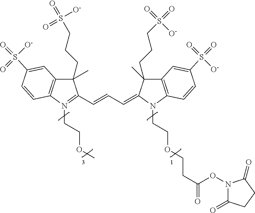
C00020

C00021

C00022

C00023
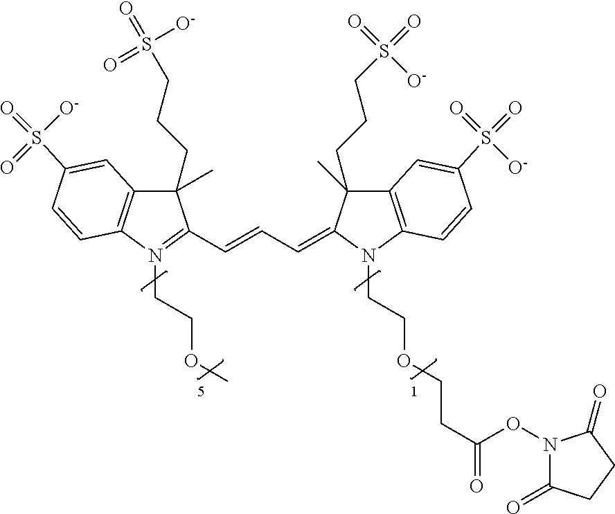
C00024

C00025

C00026

C00027
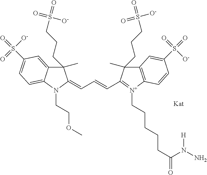
C00028

C00029
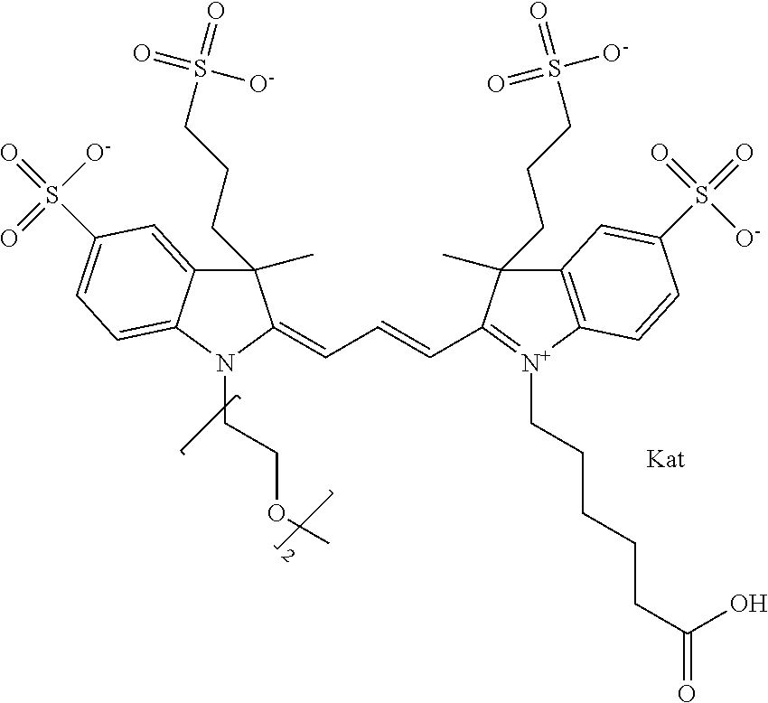
C00030
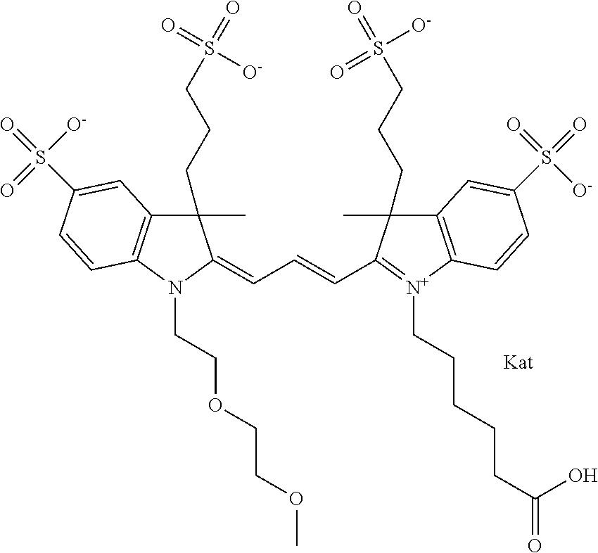
C00031

C00032

C00033

C00034

C00035
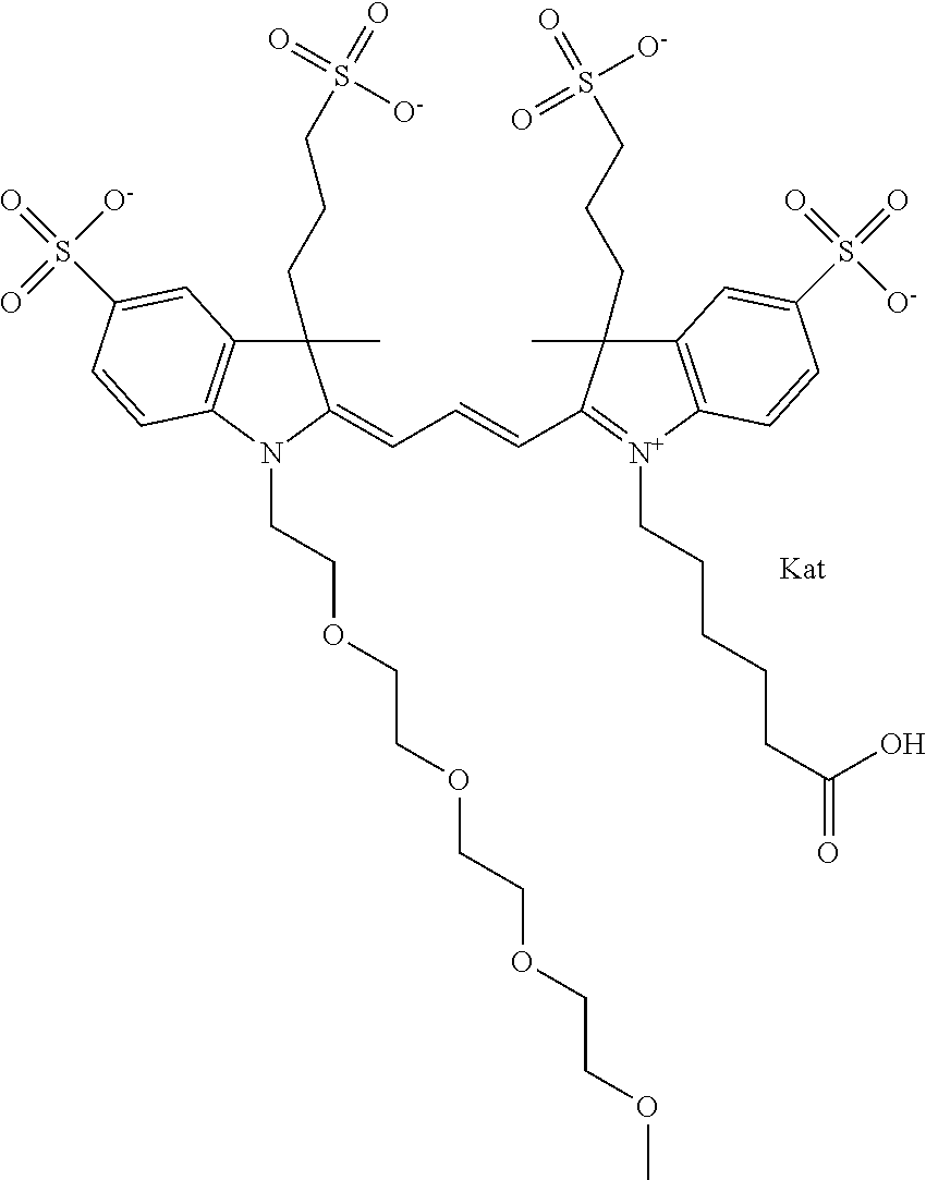
C00036
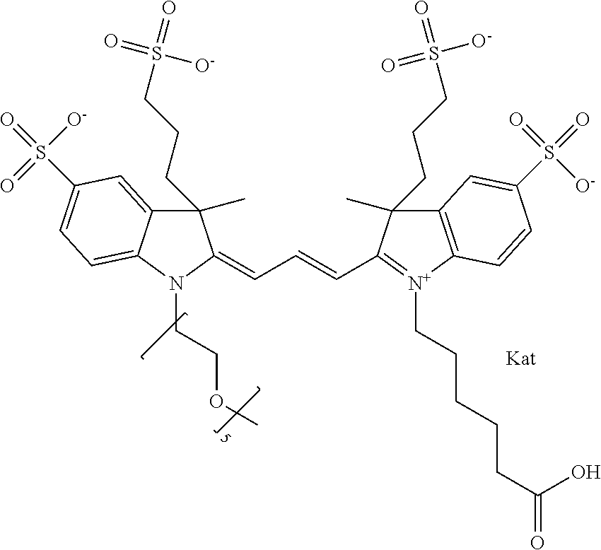
C00037
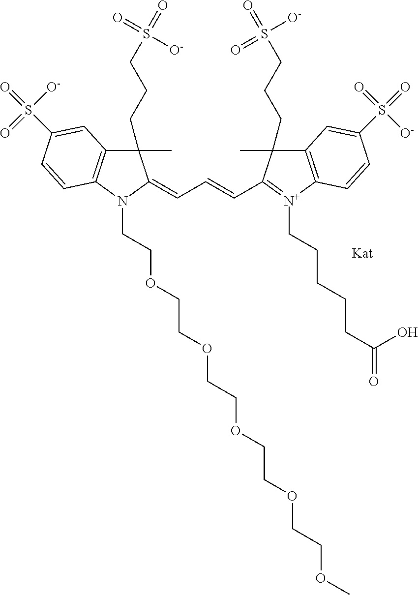
C00038
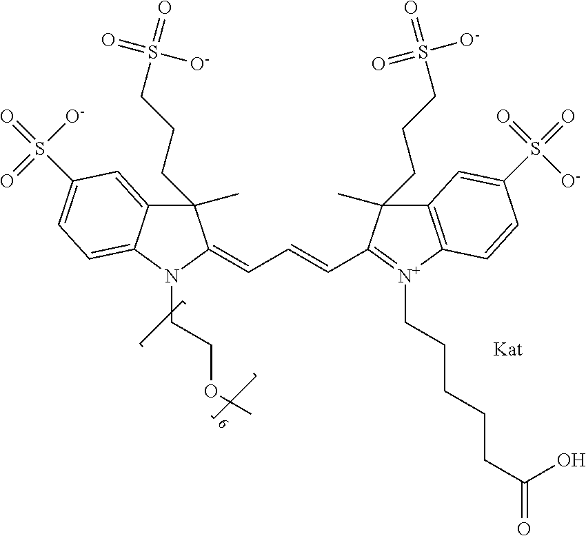
C00039
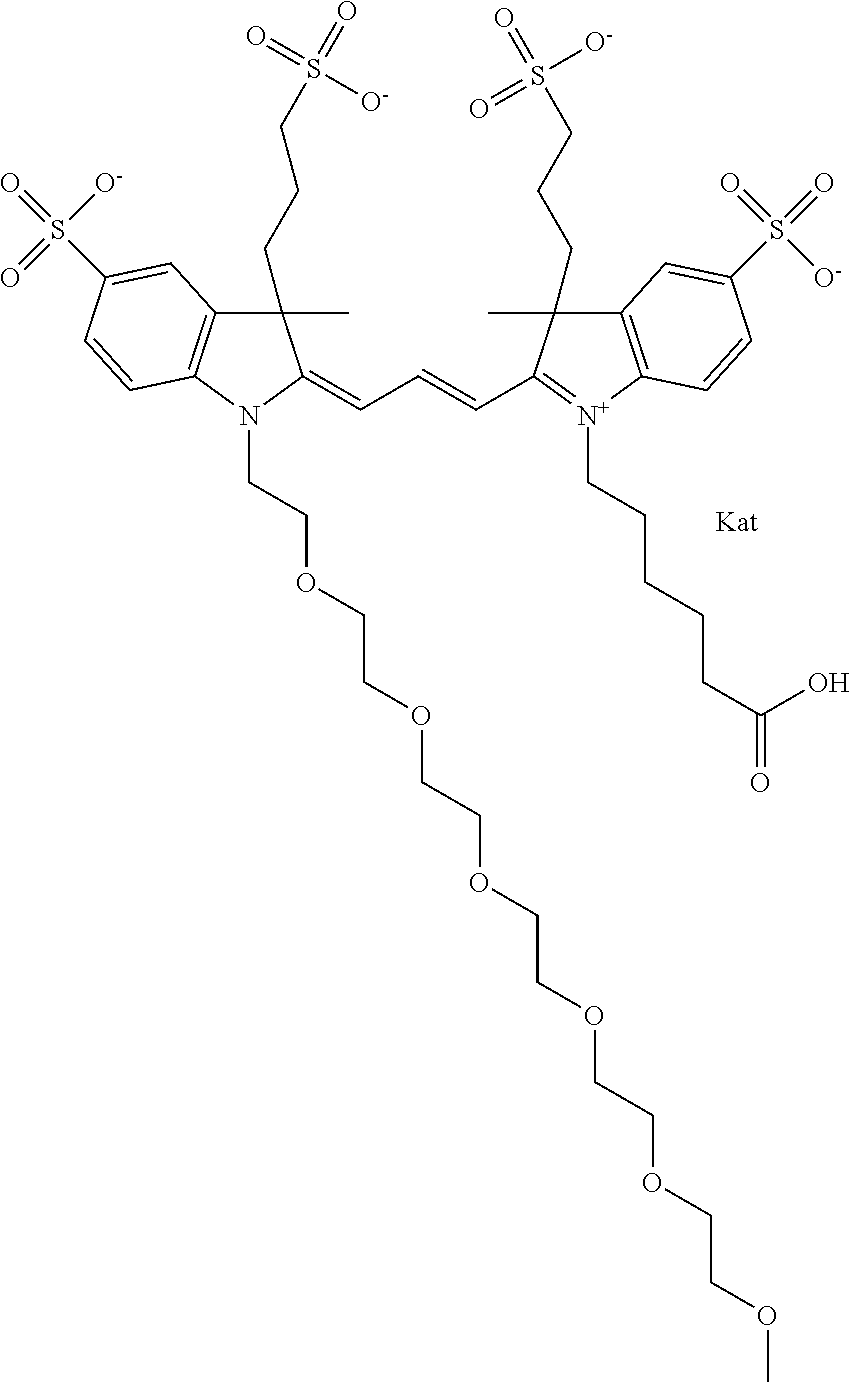
C00040
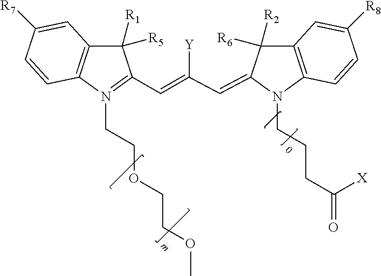
C00041

C00042

C00043

C00044
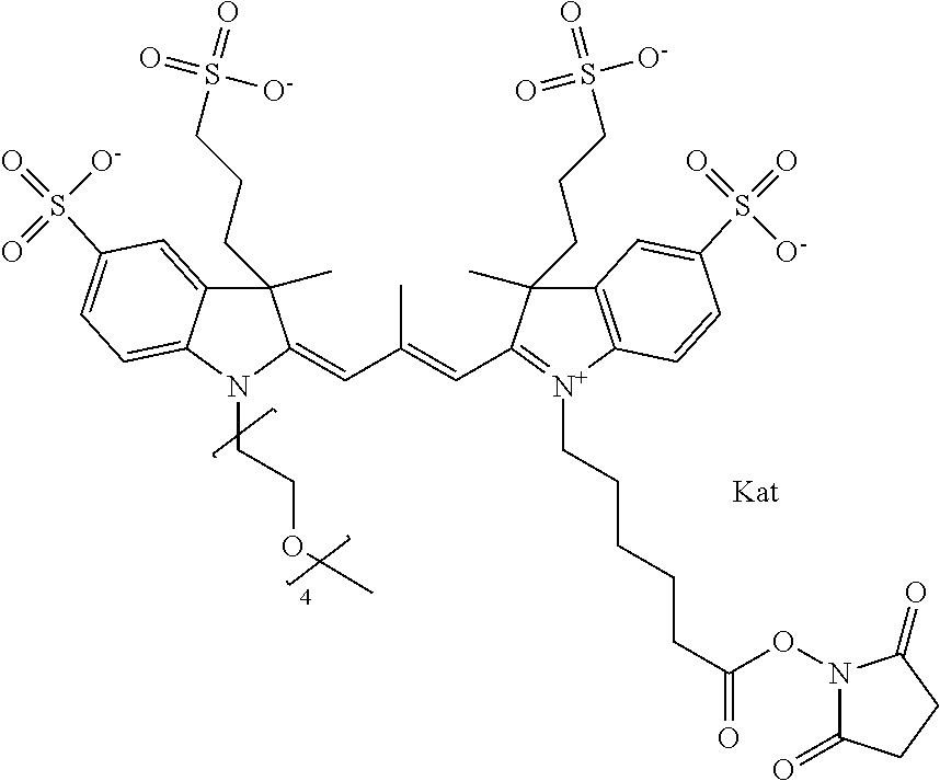
C00045

C00046

C00047
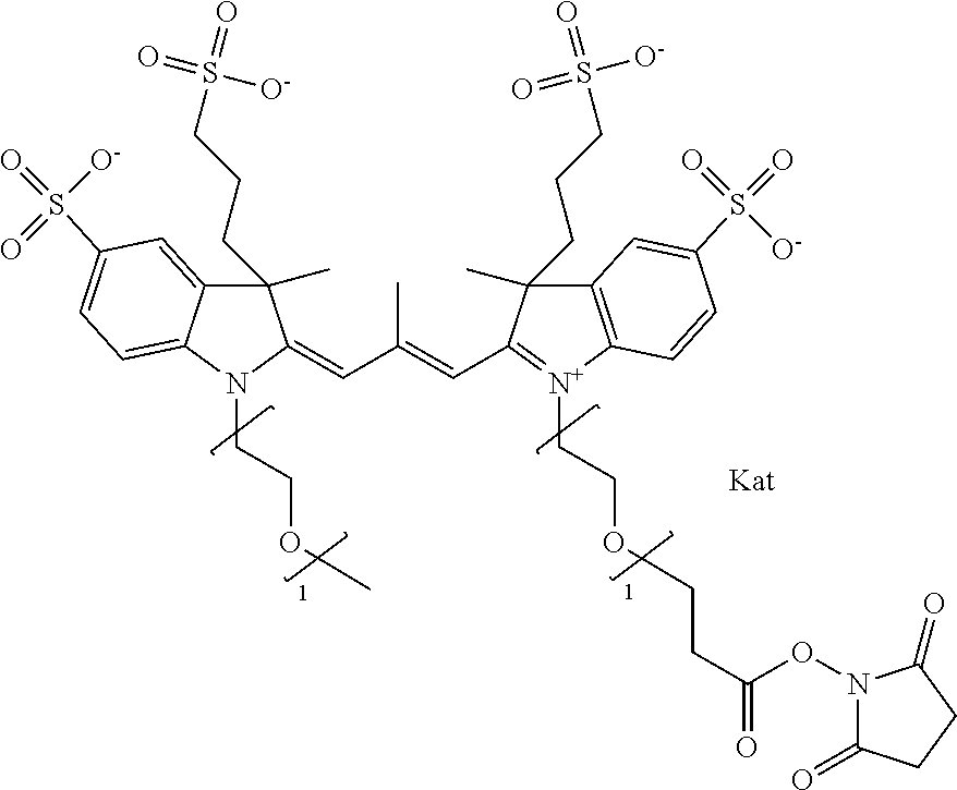
C00048
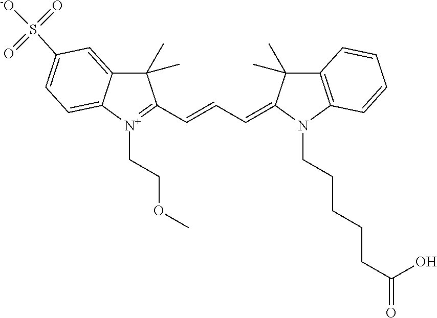
C00049

C00050
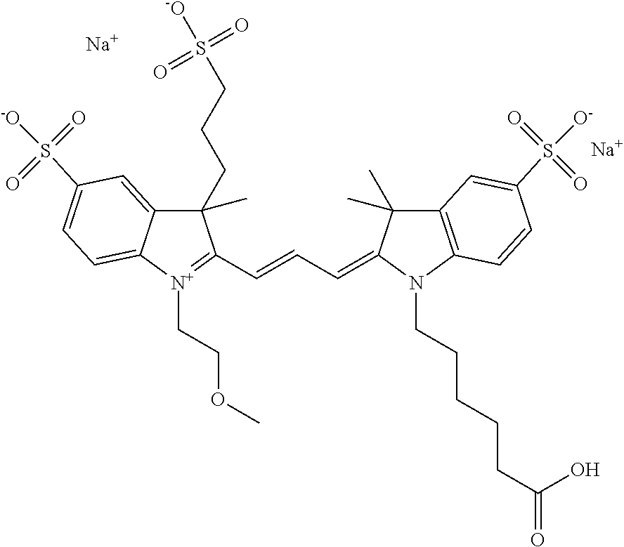
C00051
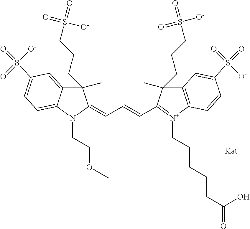
C00052
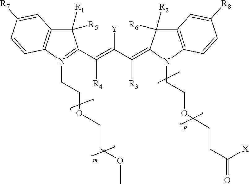
C00053
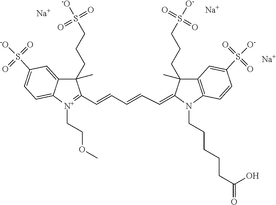
C00054
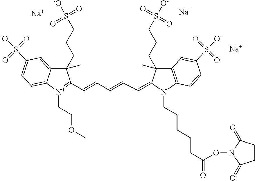
C00055

C00056
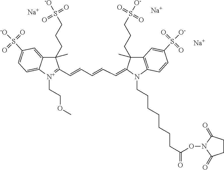
C00057

C00058

C00059

C00060

C00061
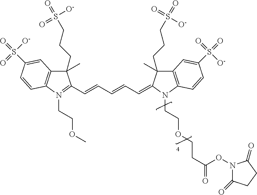
C00062

C00063

C00064
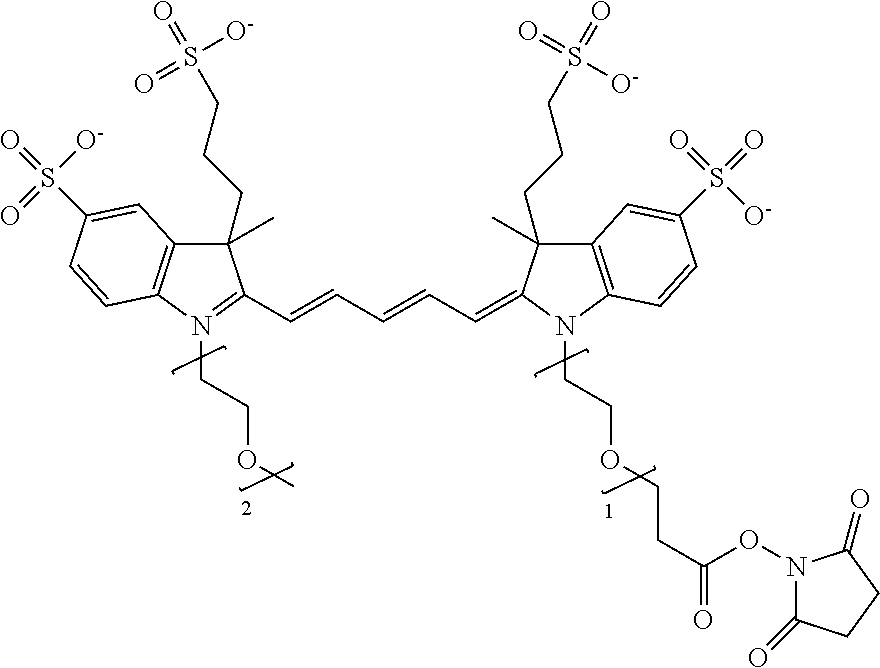
C00065

C00066

C00067

C00068

C00069

C00070

C00071

C00072
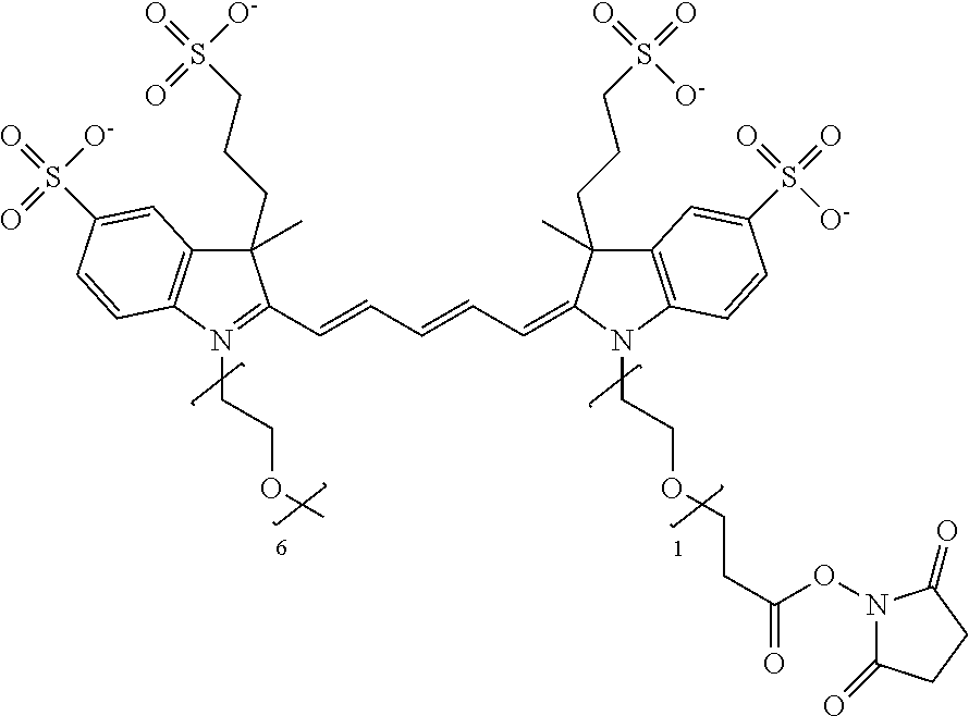
C00073

C00074
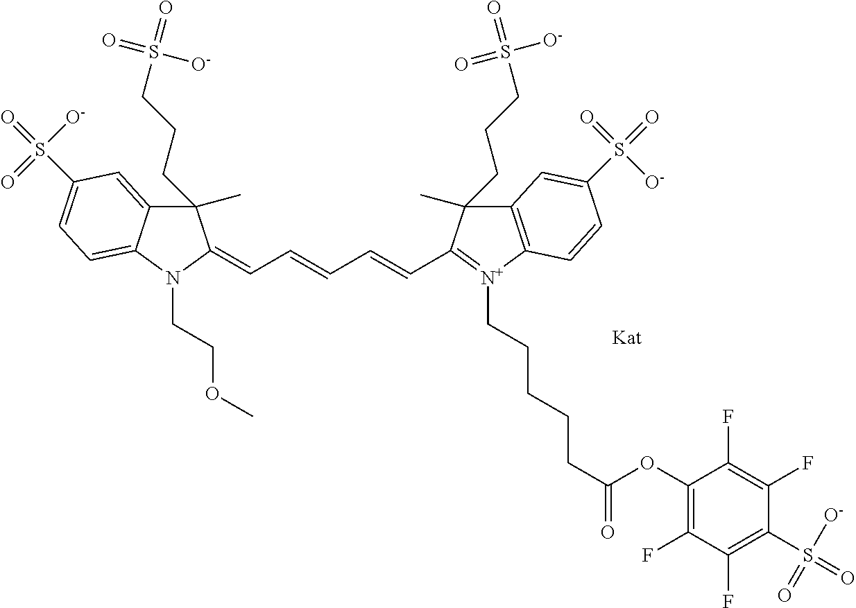
C00075
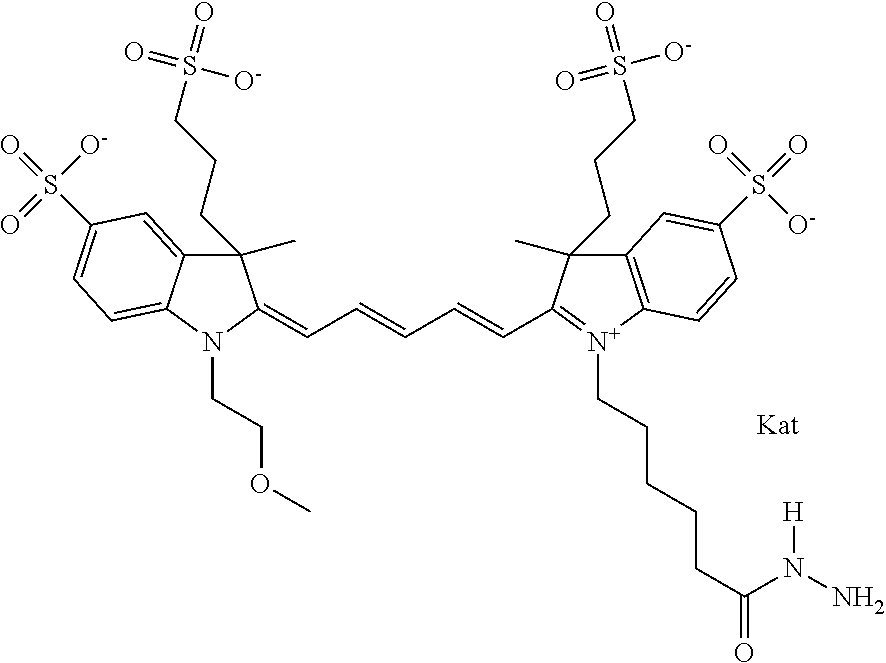
C00076
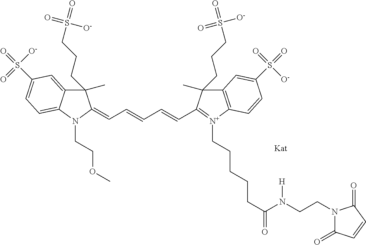
C00077
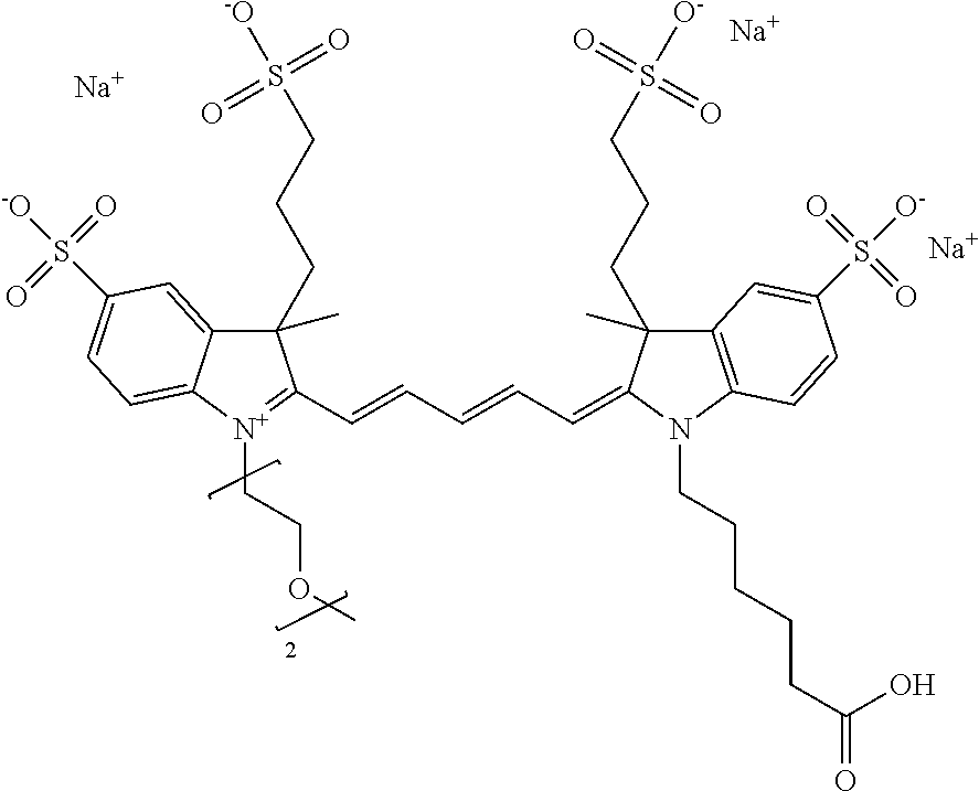
C00078
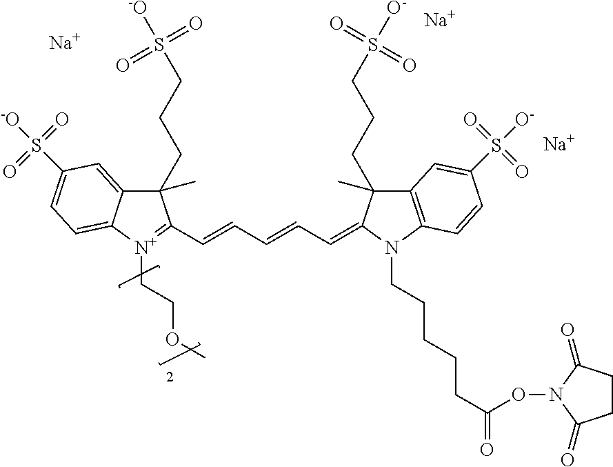
C00079

C00080

C00081
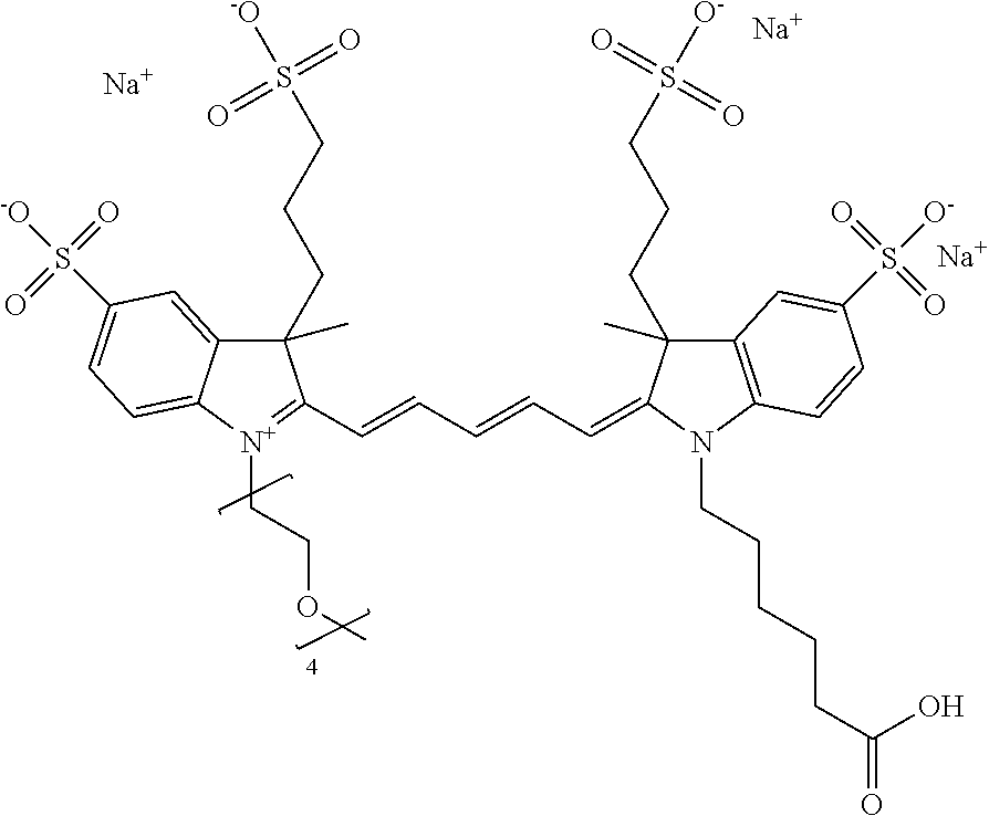
C00082
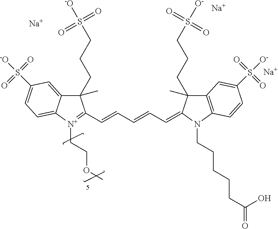
C00083
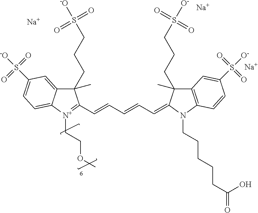
C00084

C00085
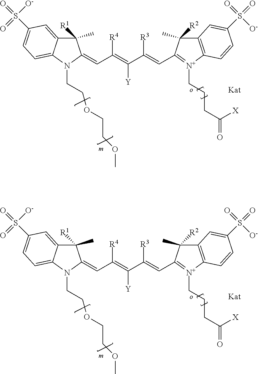
C00086
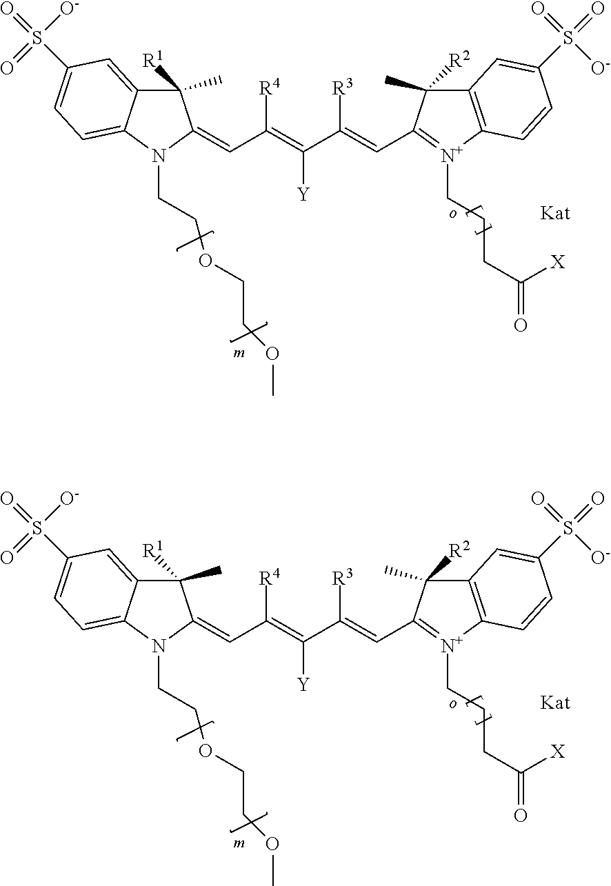
C00087
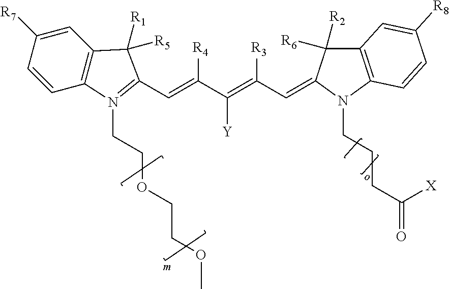
C00088

C00089
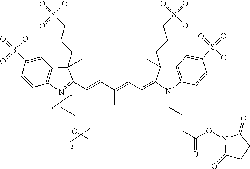
C00090

C00091
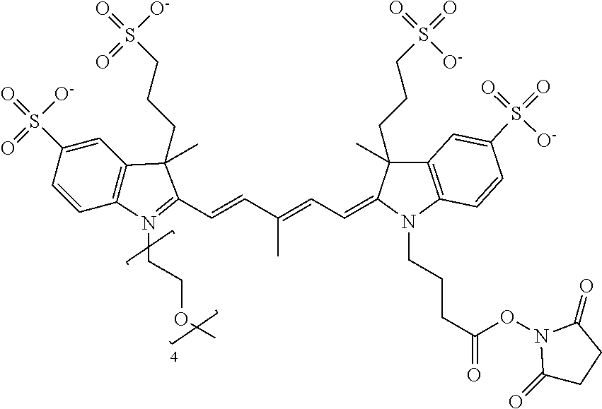
C00092

C00093

C00094

C00095
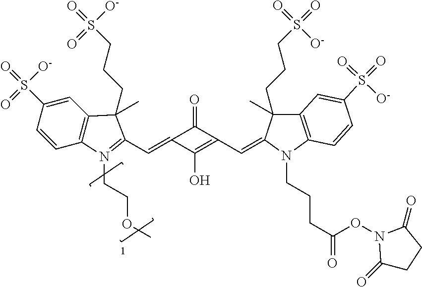
C00096
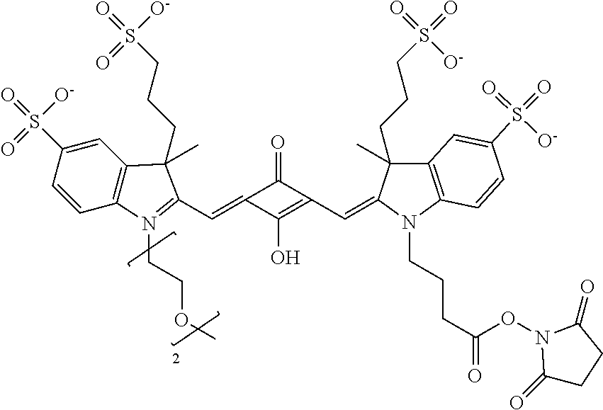
C00097

C00098
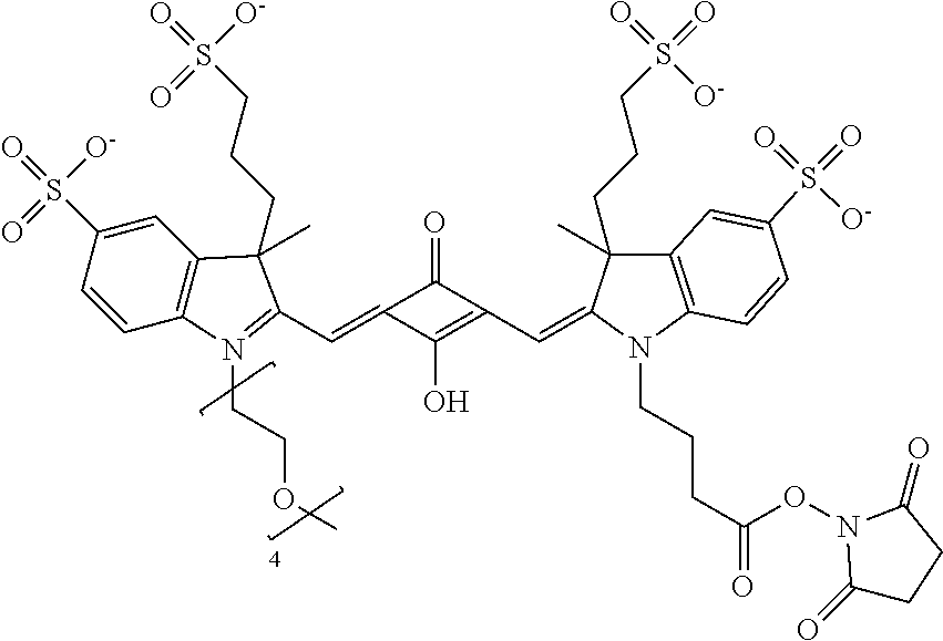
C00099

C00100
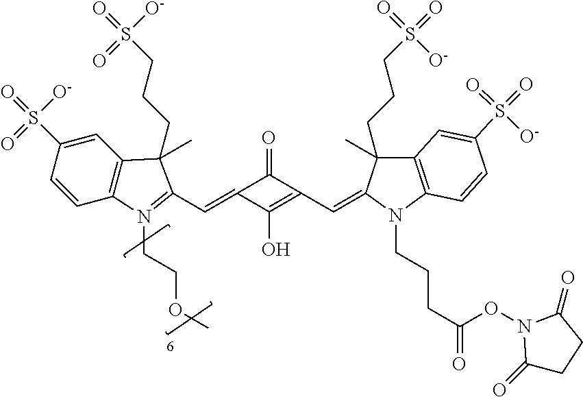
C00101
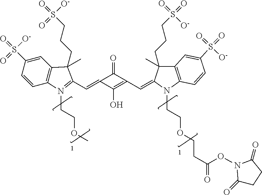
C00102
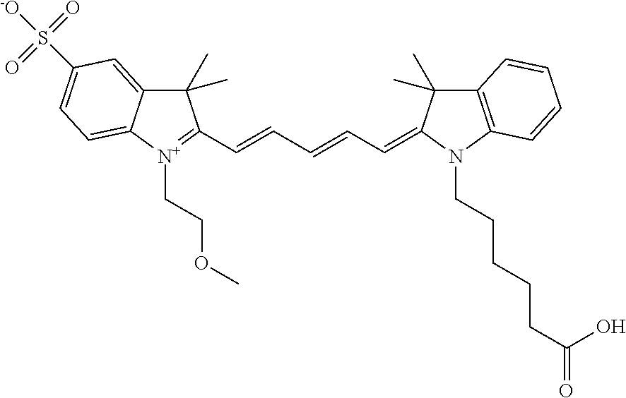
C00103
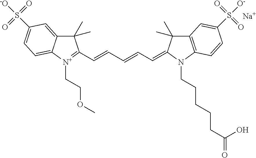
C00104
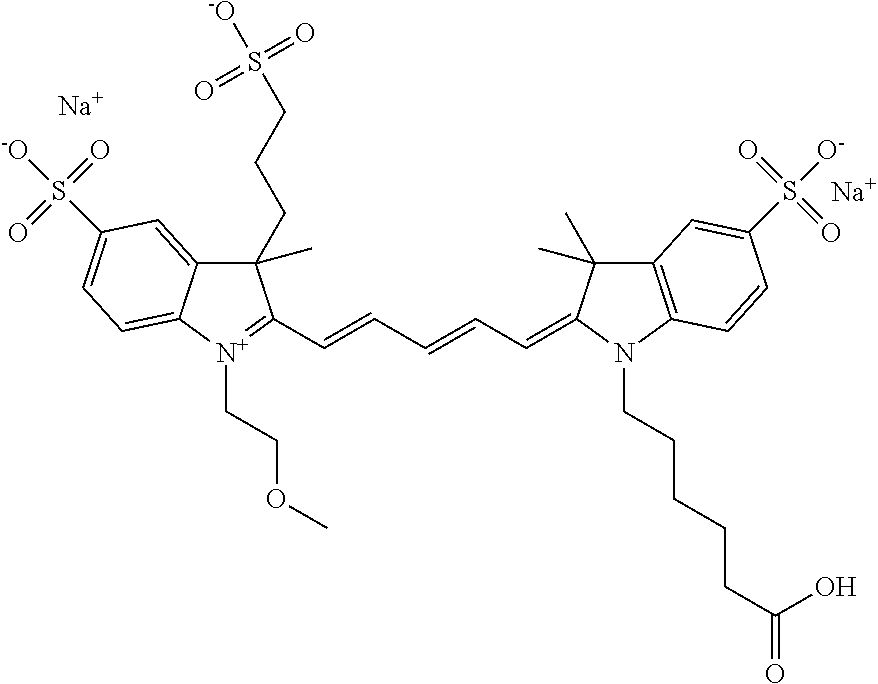
C00105

C00106

C00107
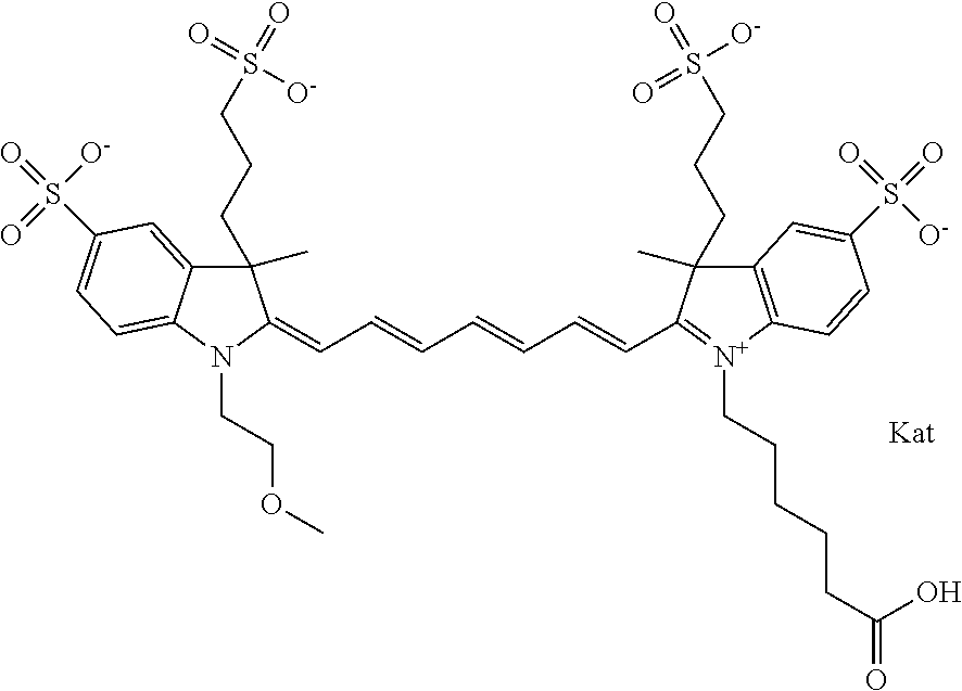
C00108
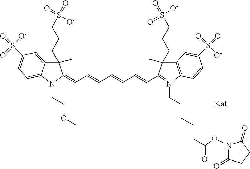
C00109

C00110

C00111

C00112

C00113
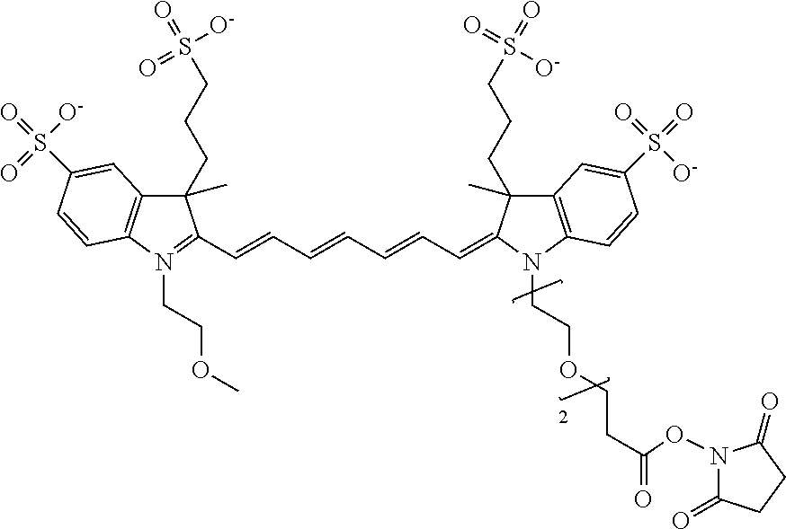
C00114
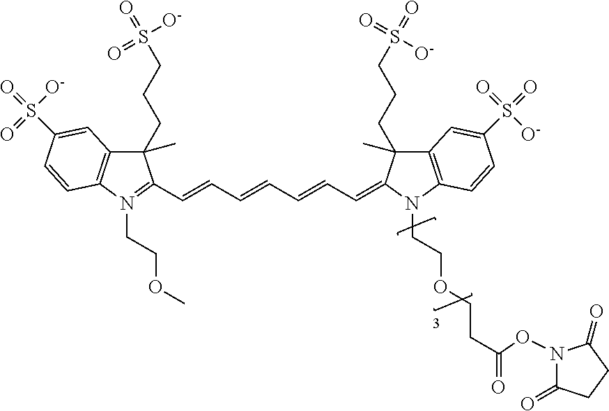
C00115

C00116

C00117
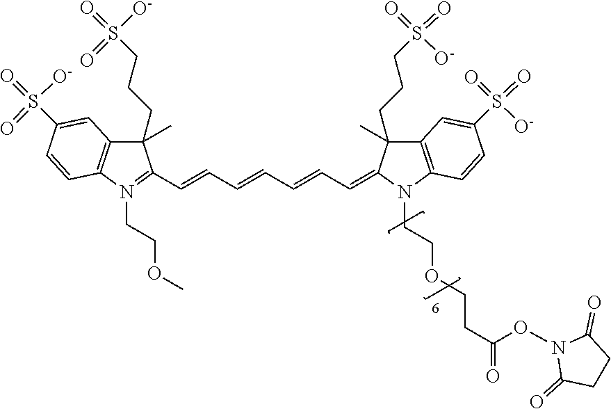
C00118

C00119

C00120

C00121

C00122

C00123

C00124

C00125
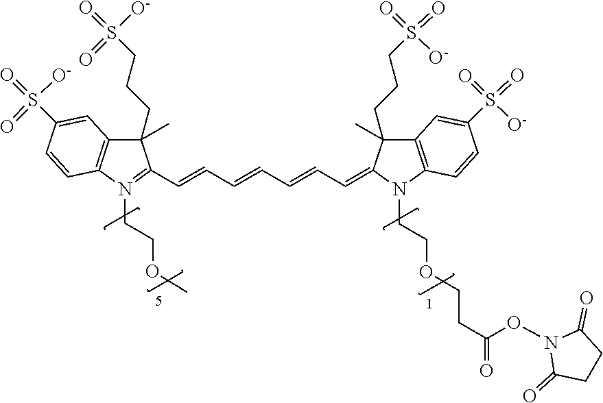
C00126

C00127

C00128
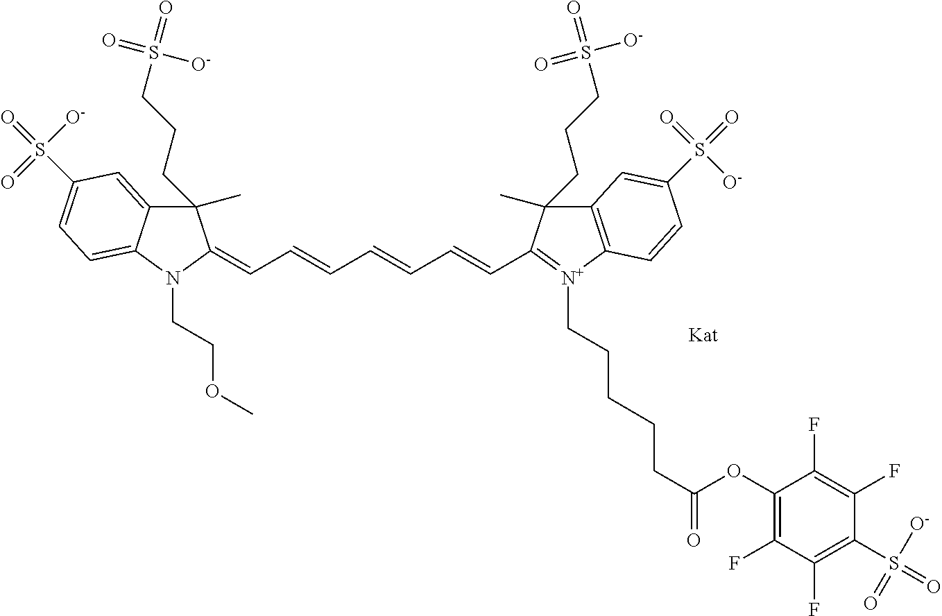
C00129

C00130
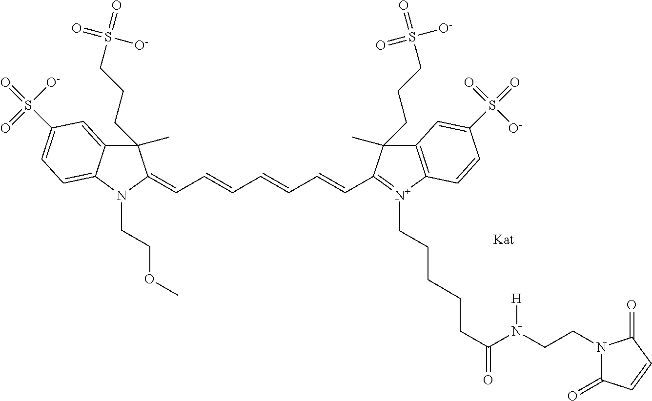
C00131
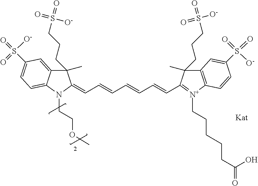
C00132
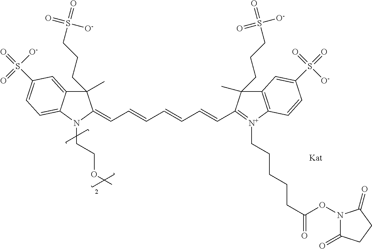
C00133

C00134

C00135

C00136
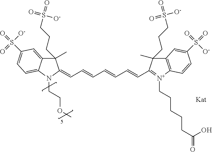
C00137

C00138

C00139
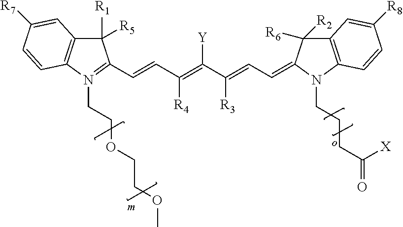
C00140

C00141
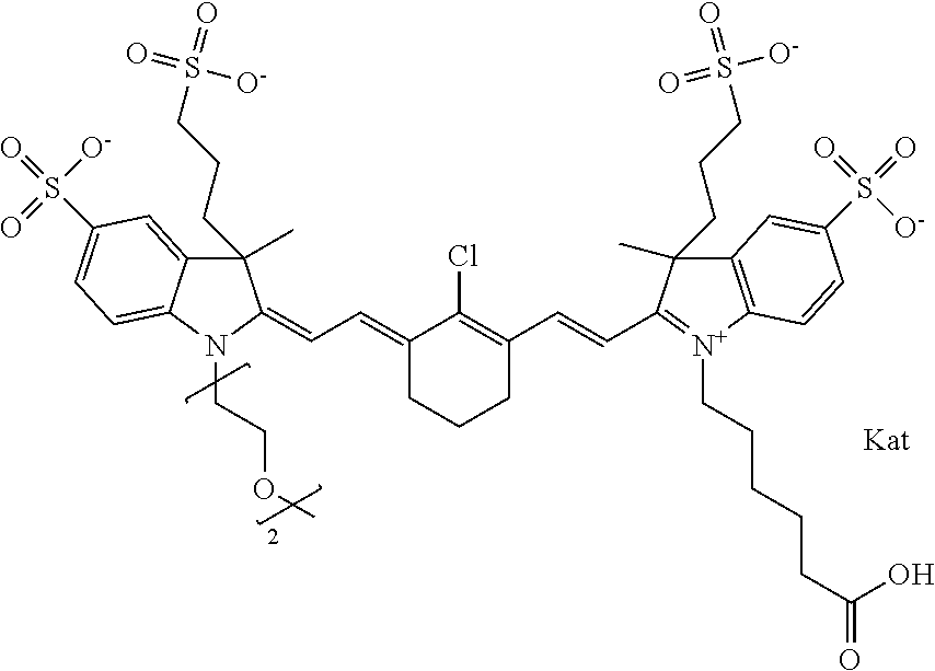
C00142

C00143

C00144

C00145

C00146

C00147
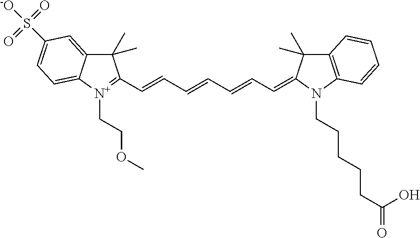
C00148
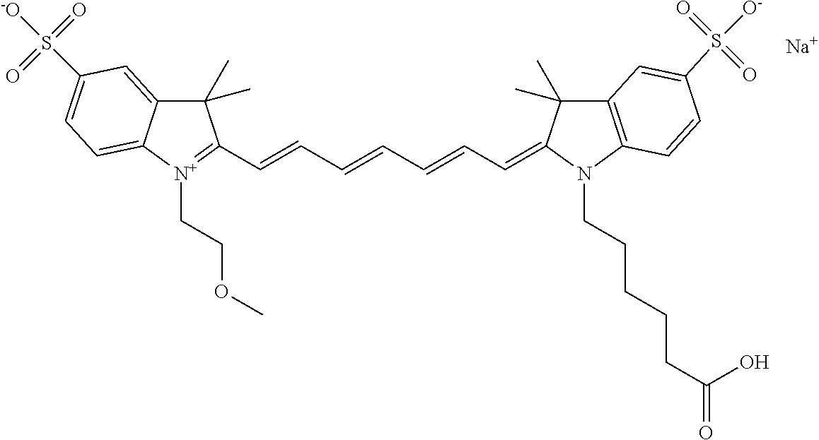
C00149
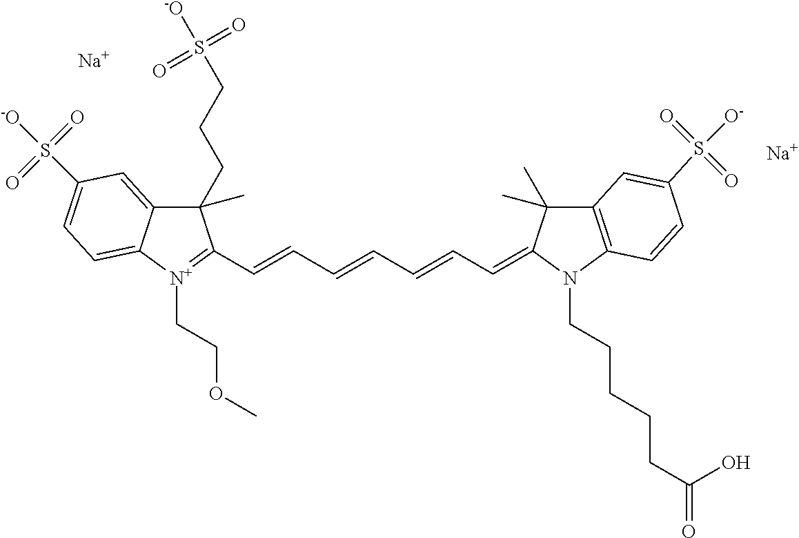
C00150
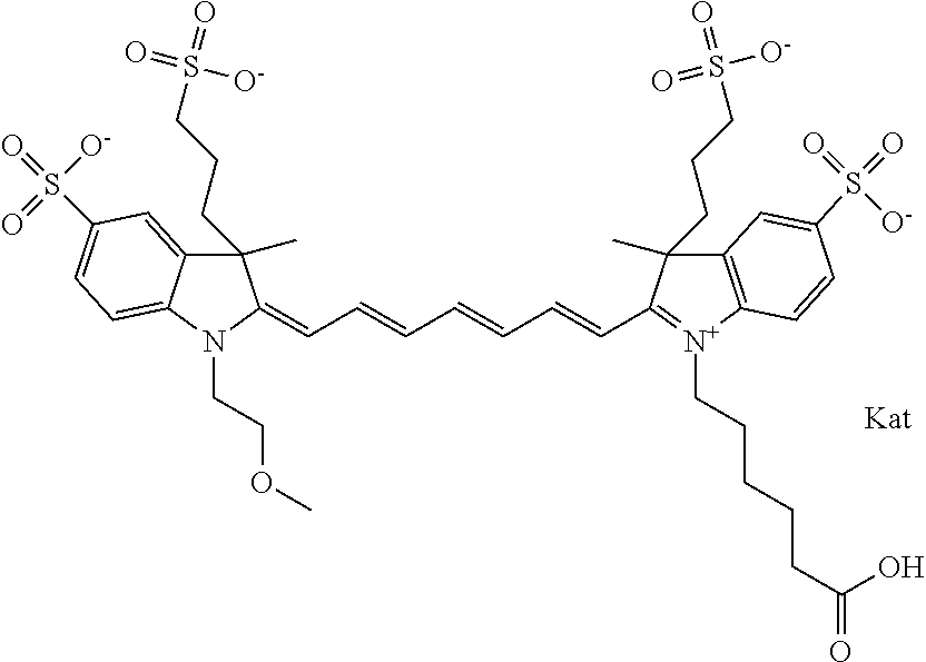
C00151

C00152

C00153
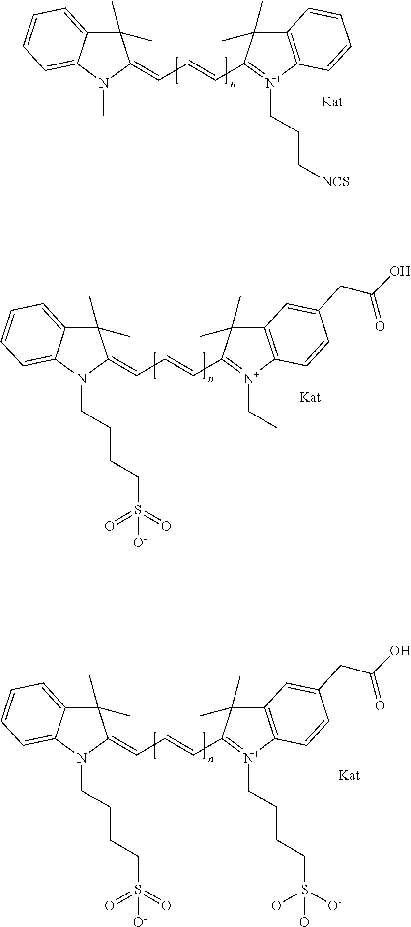
C00154
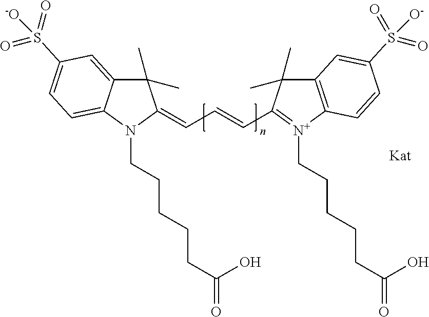
C00155

C00156

C00157
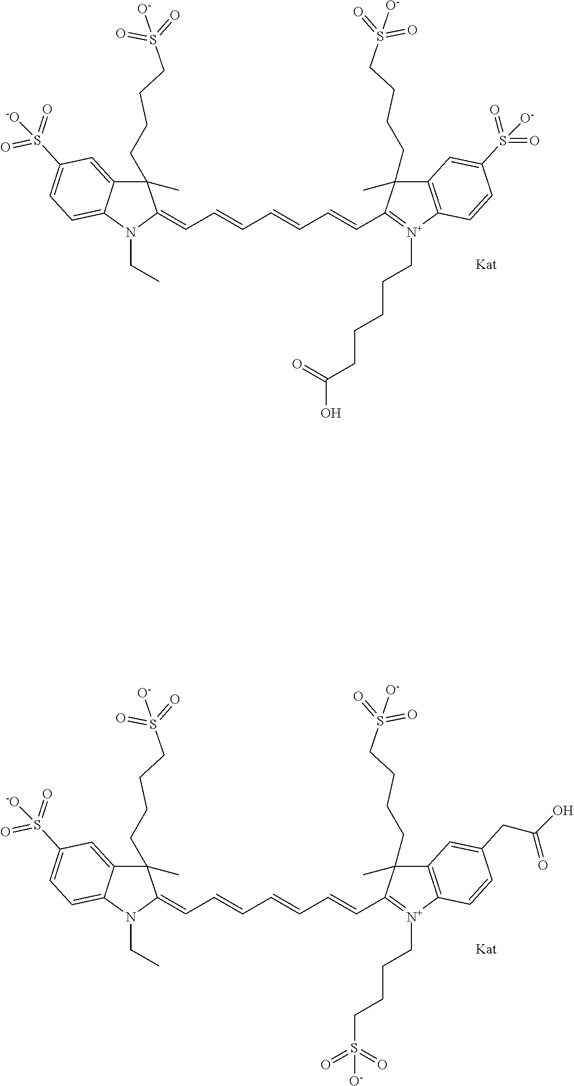
C00158
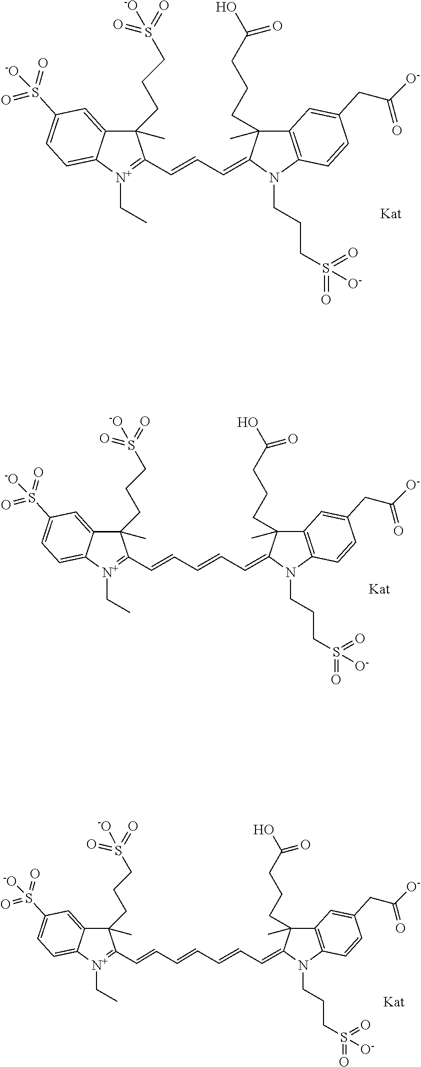
C00159
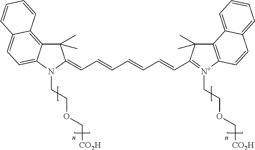
C00160

C00161
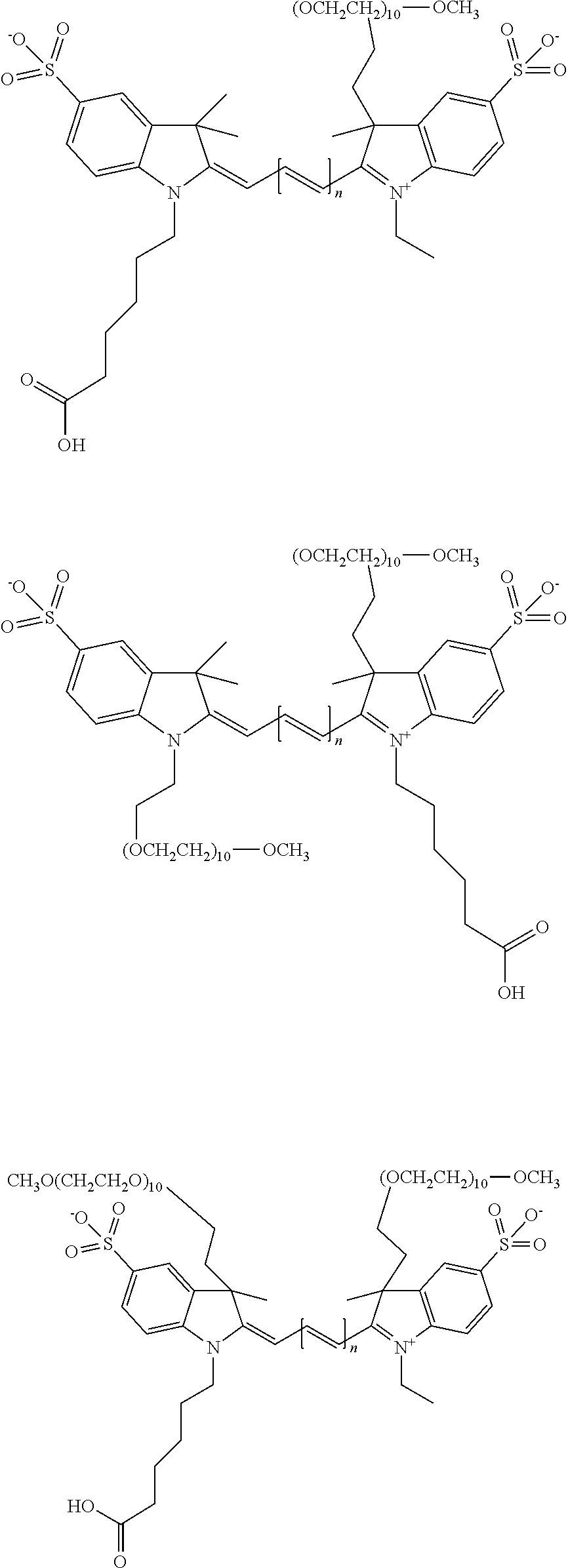
C00162
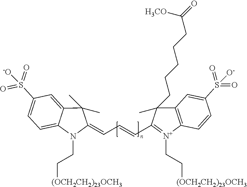
C00163
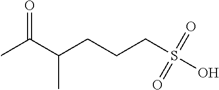
C00164

C00165
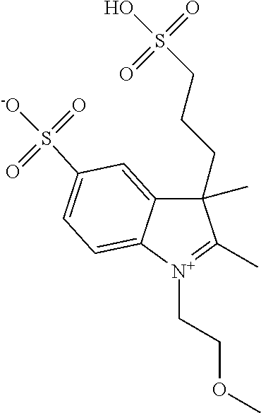
C00166
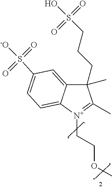
C00167

C00168
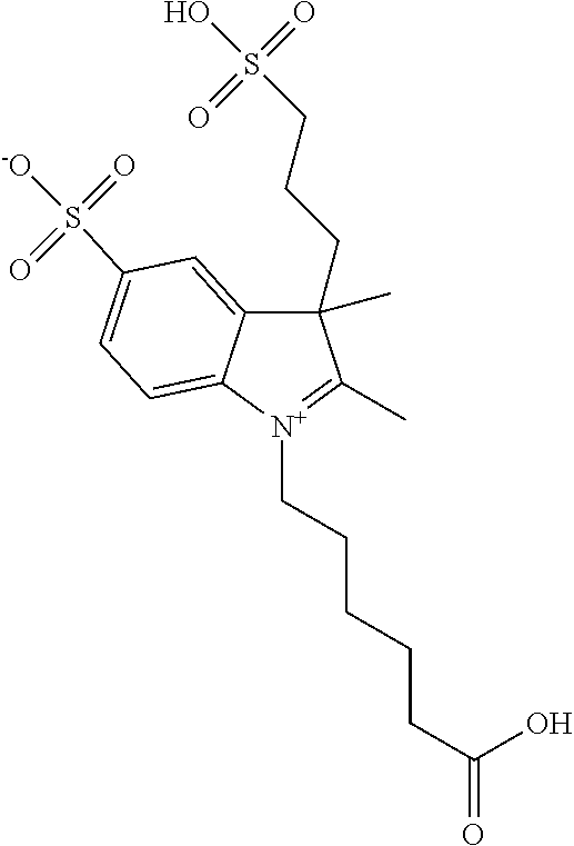
C00169
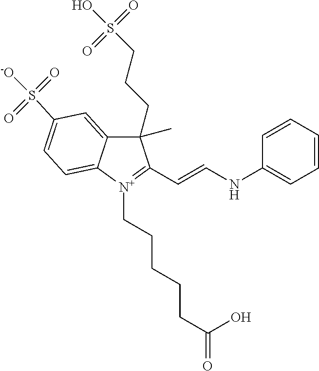
C00170
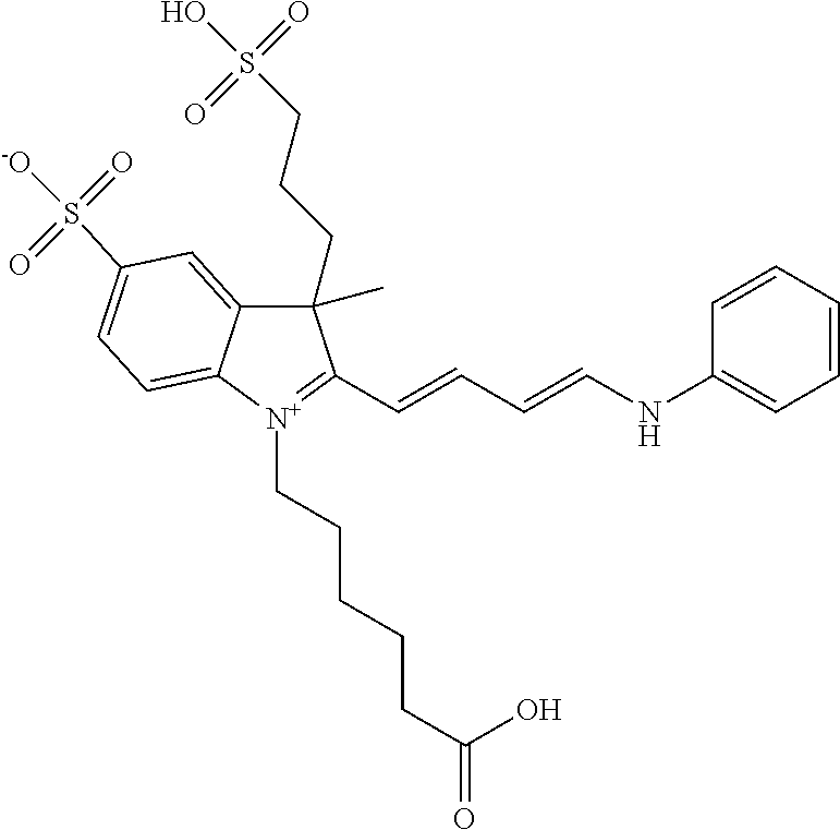
C00171
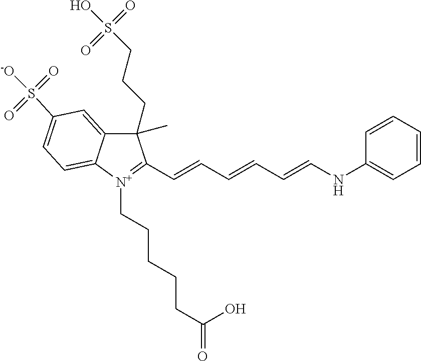
C00172

C00173
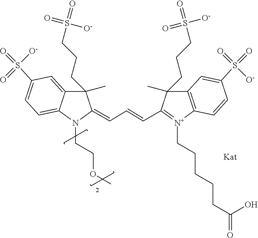
C00174
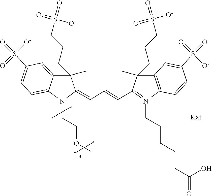
C00175

C00176

C00177
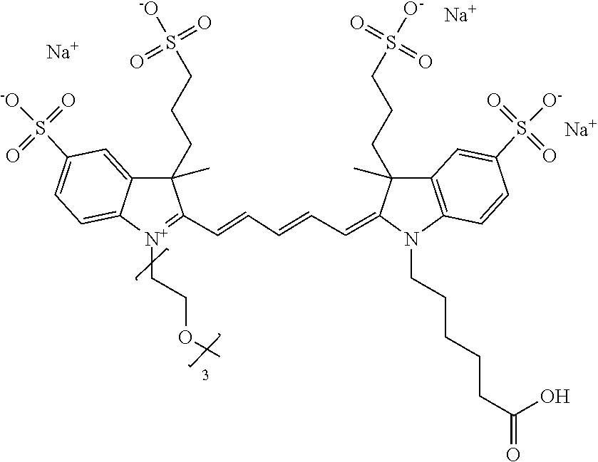
C00178
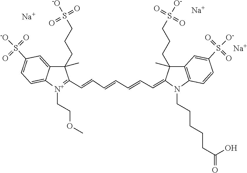
C00179

C00180
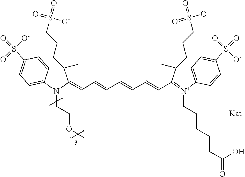
C00181

D00001
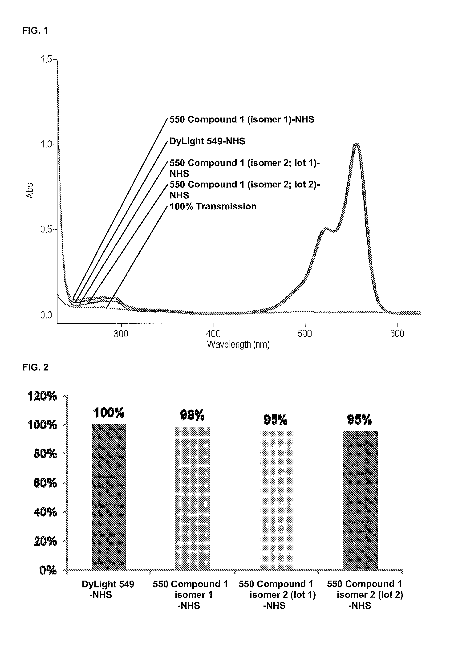
D00002

D00003
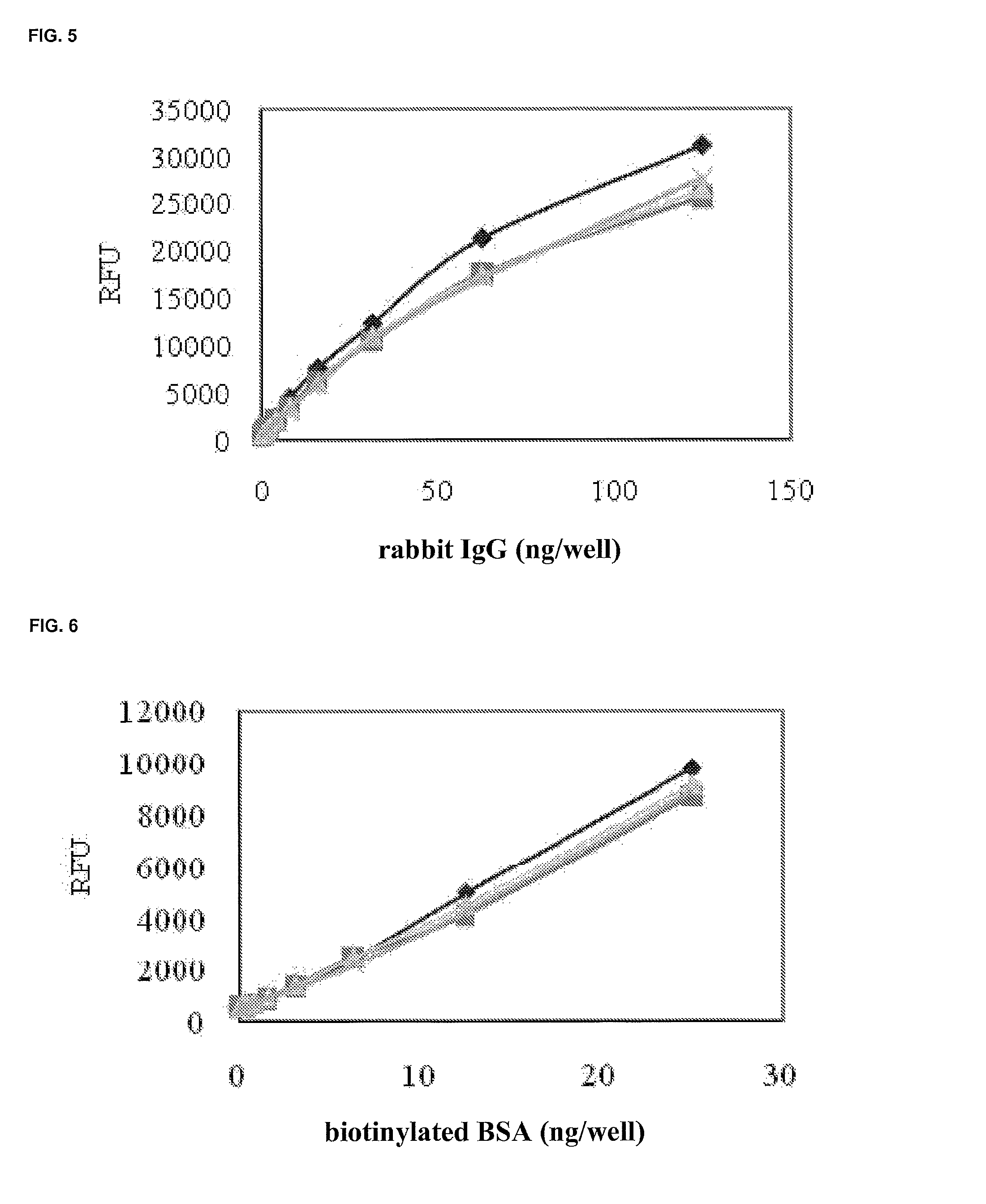
D00004

D00005
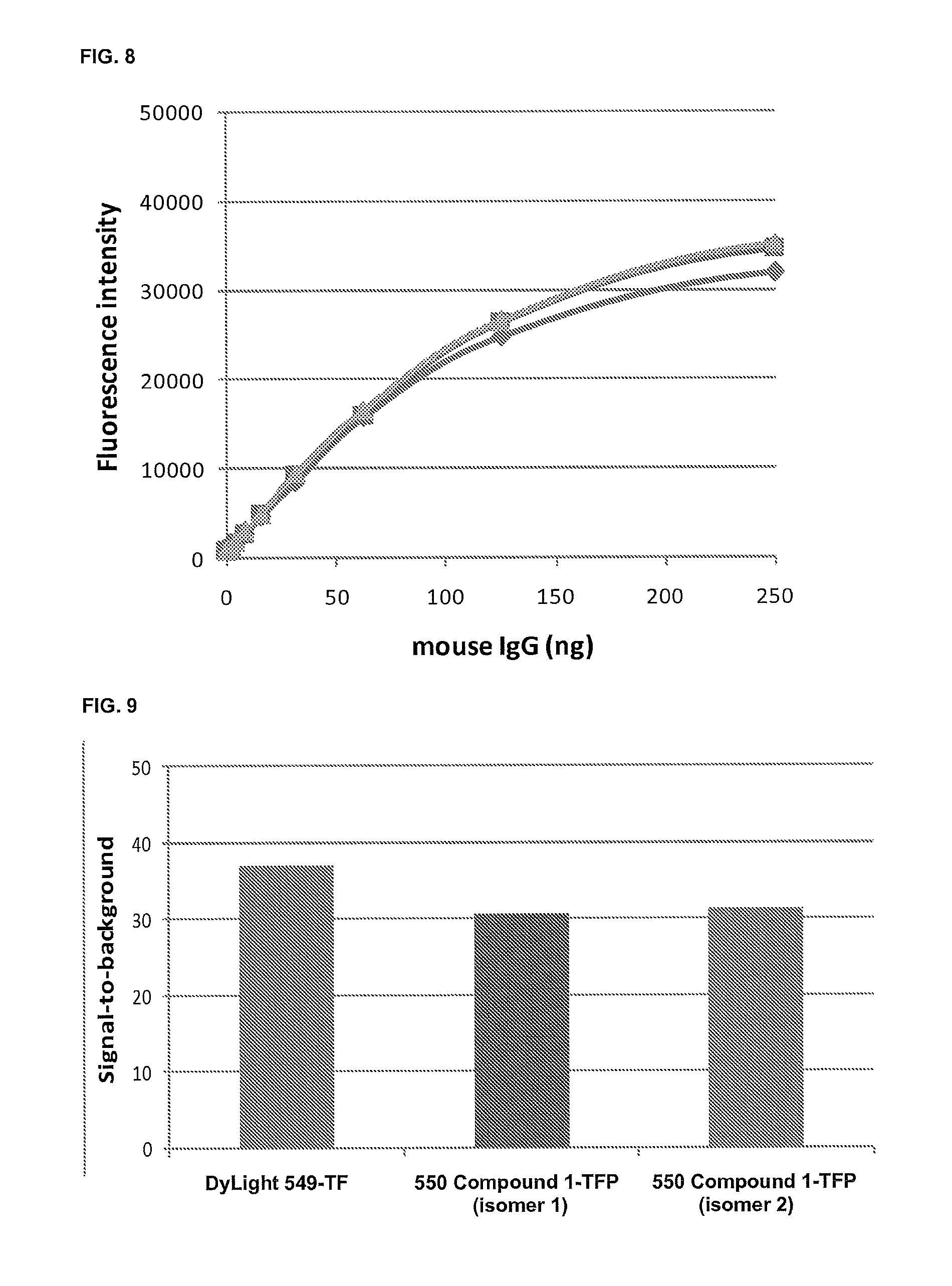
D00006
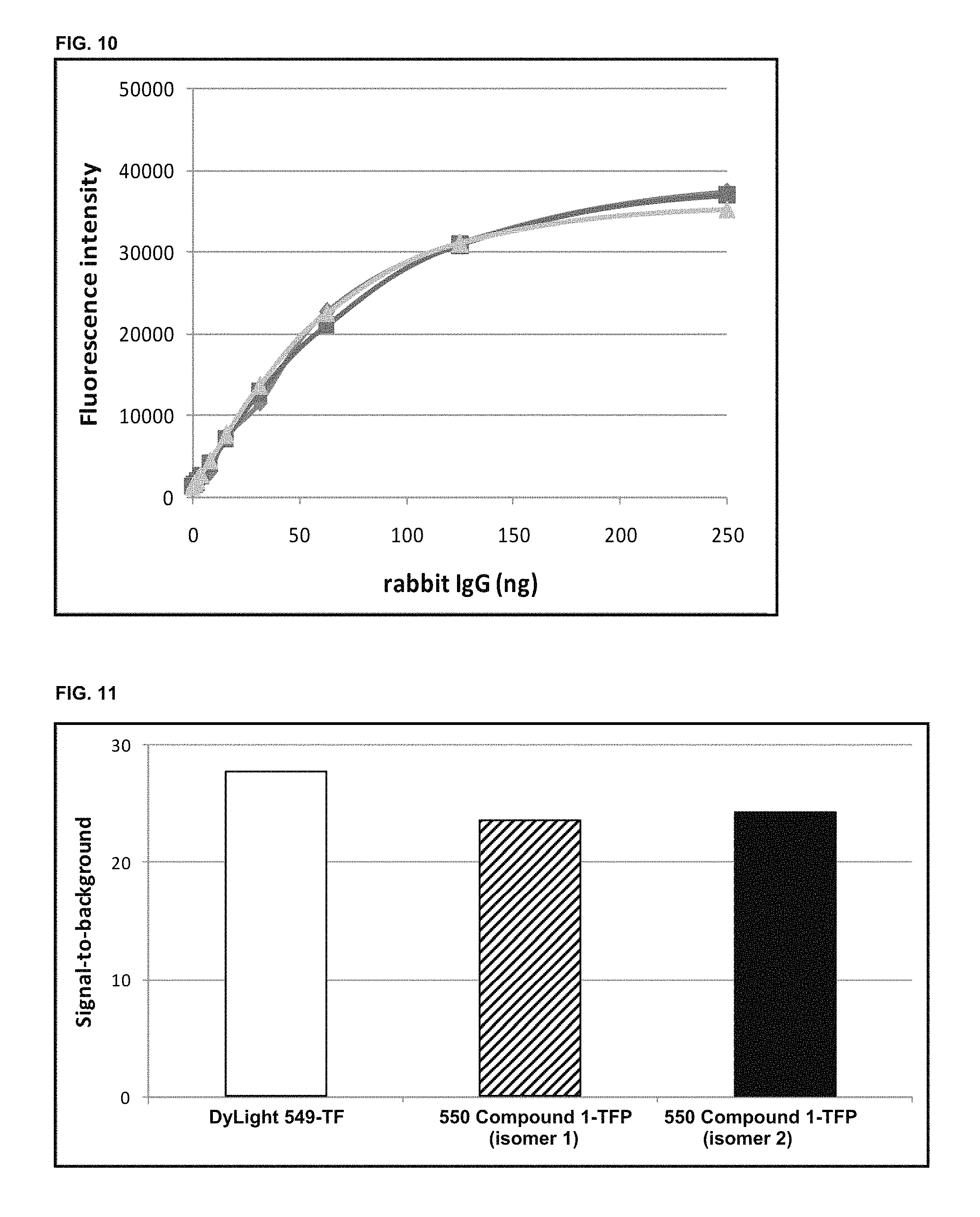
D00007

D00008
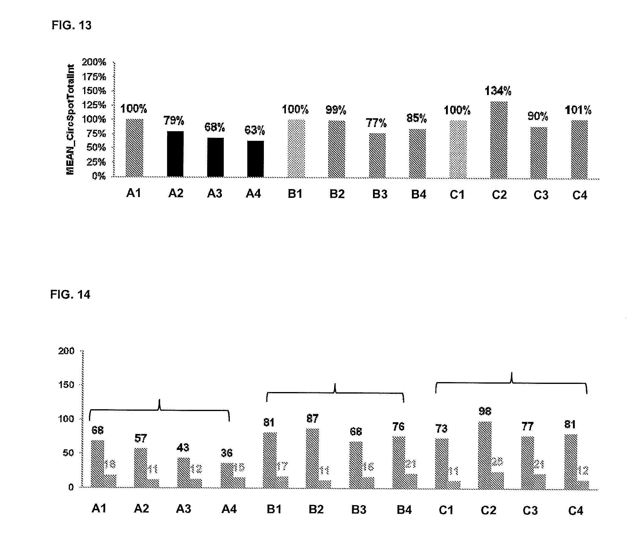
D00009
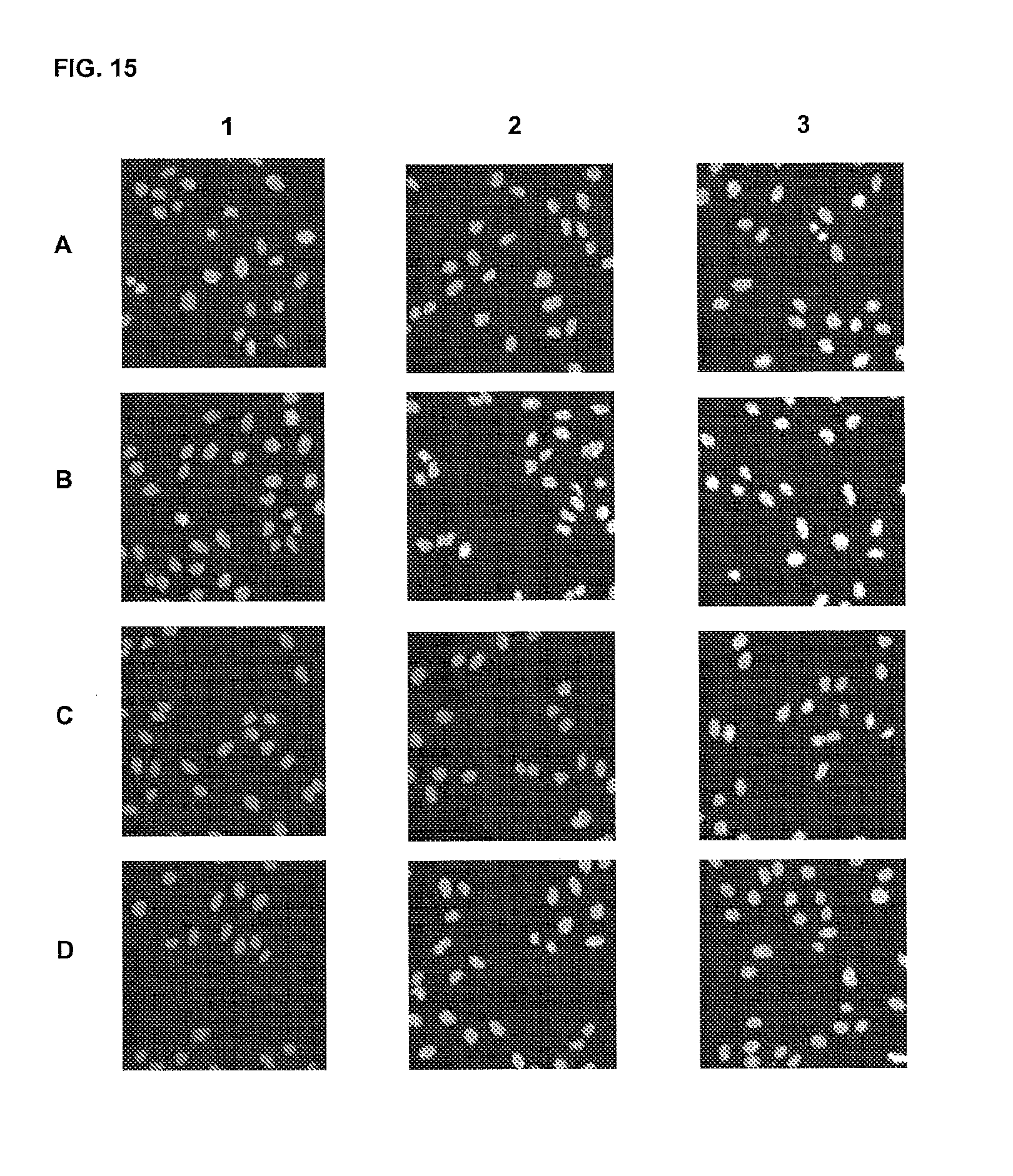
D00010
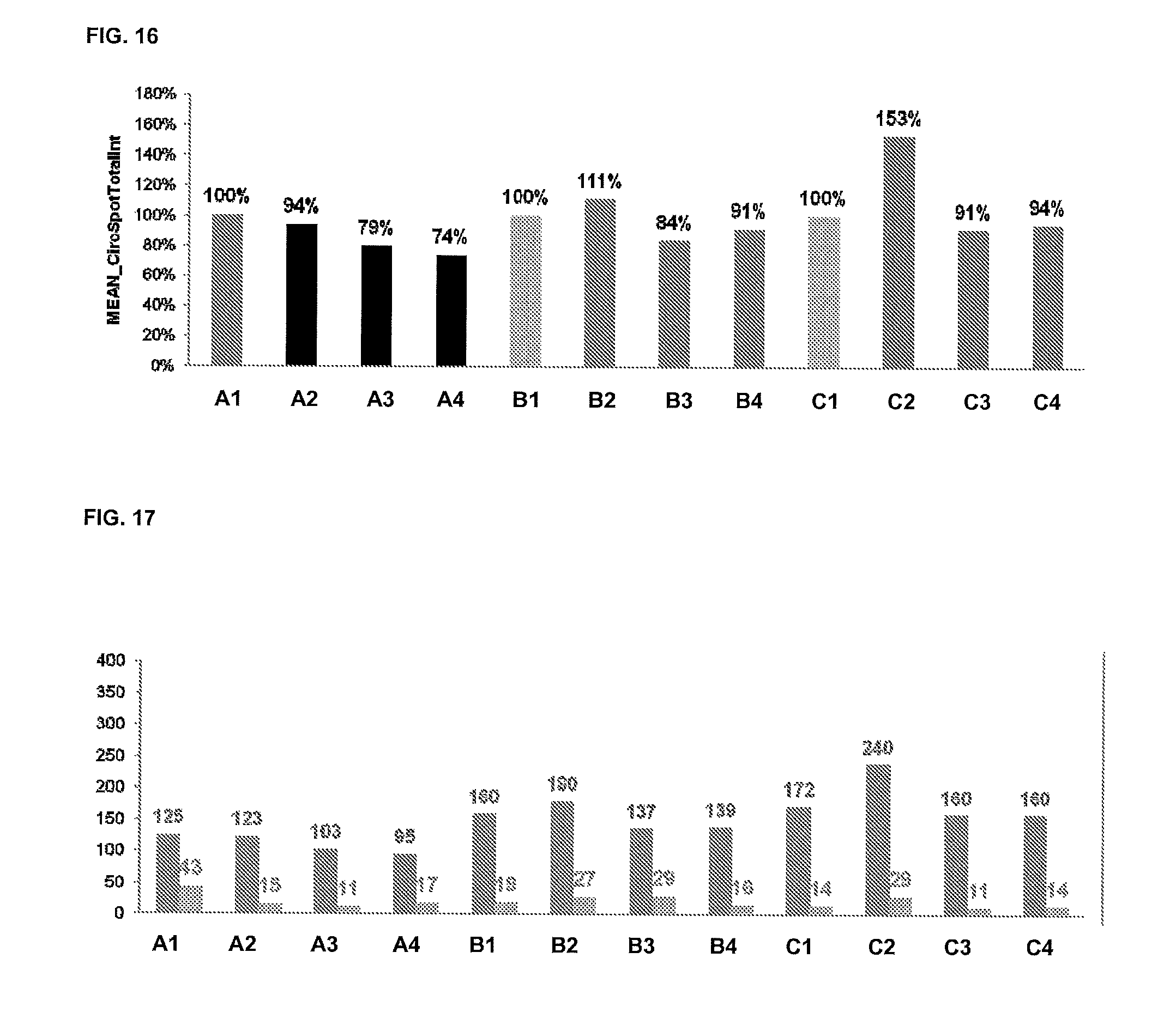
D00011
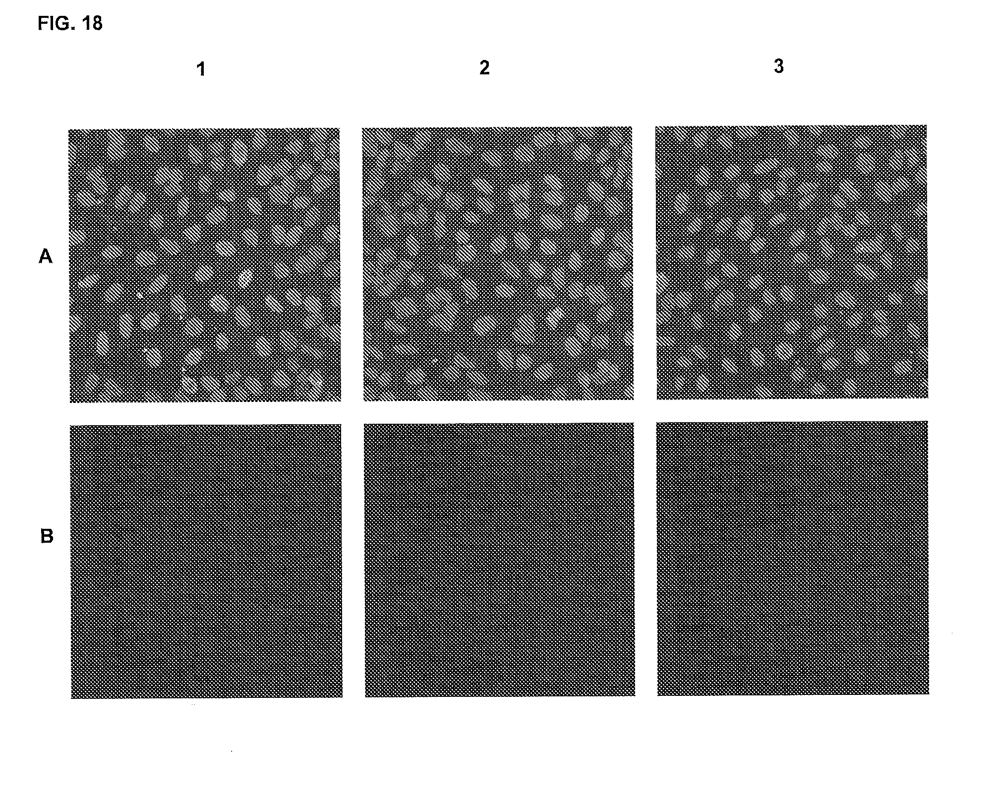
D00012
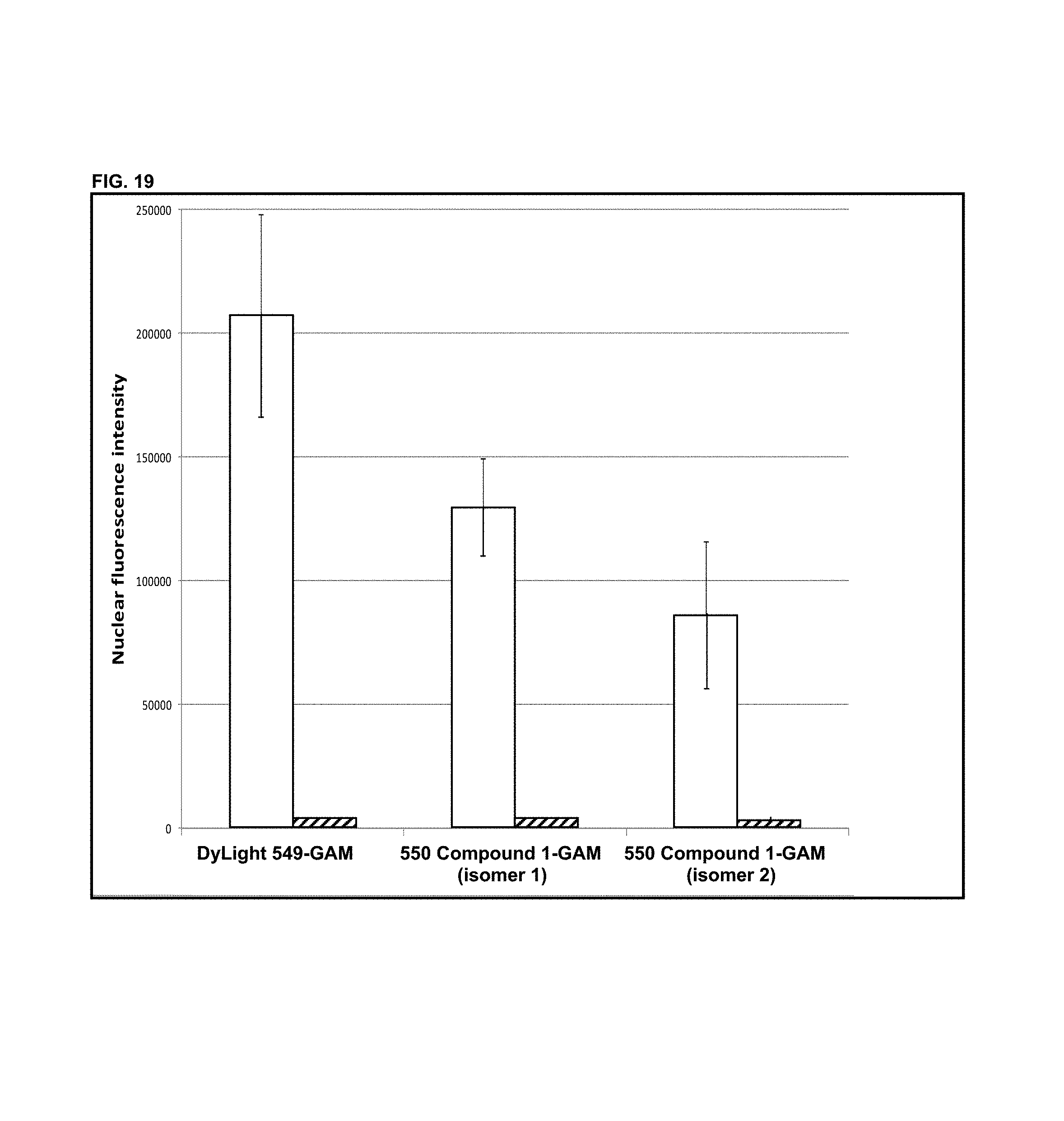
D00013
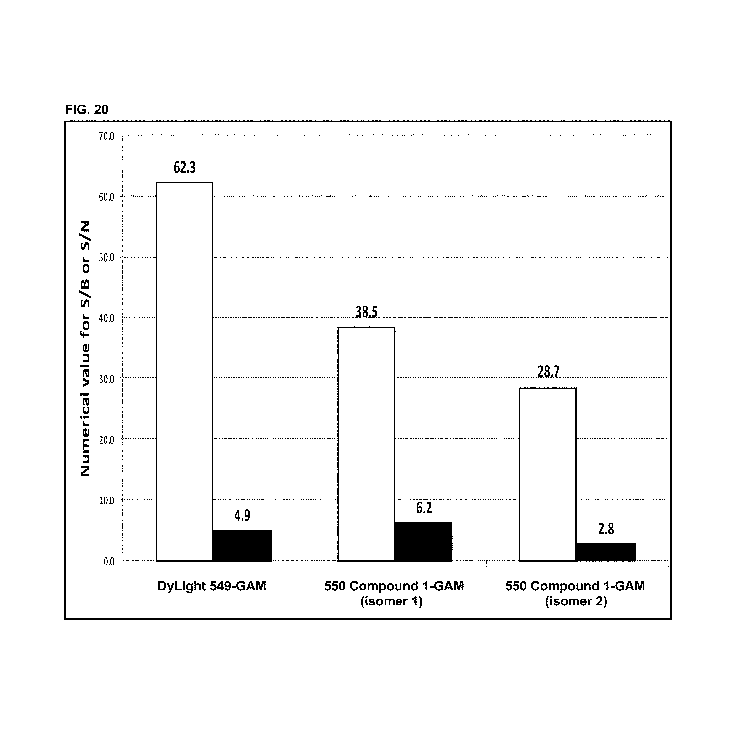
D00014
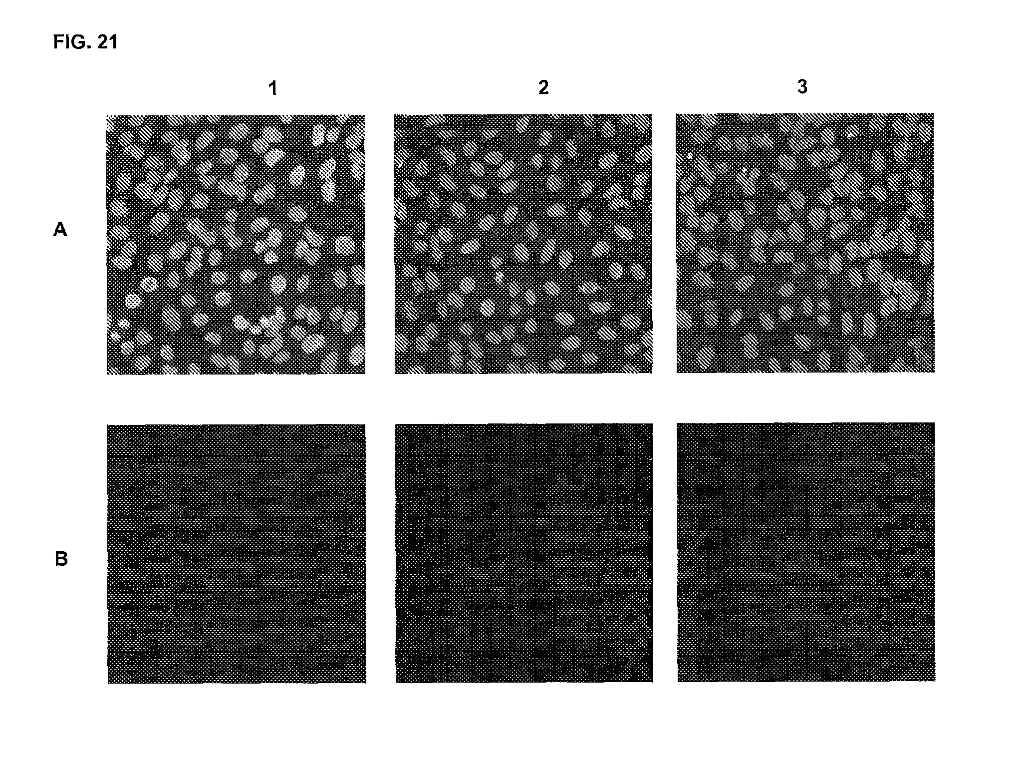
D00015
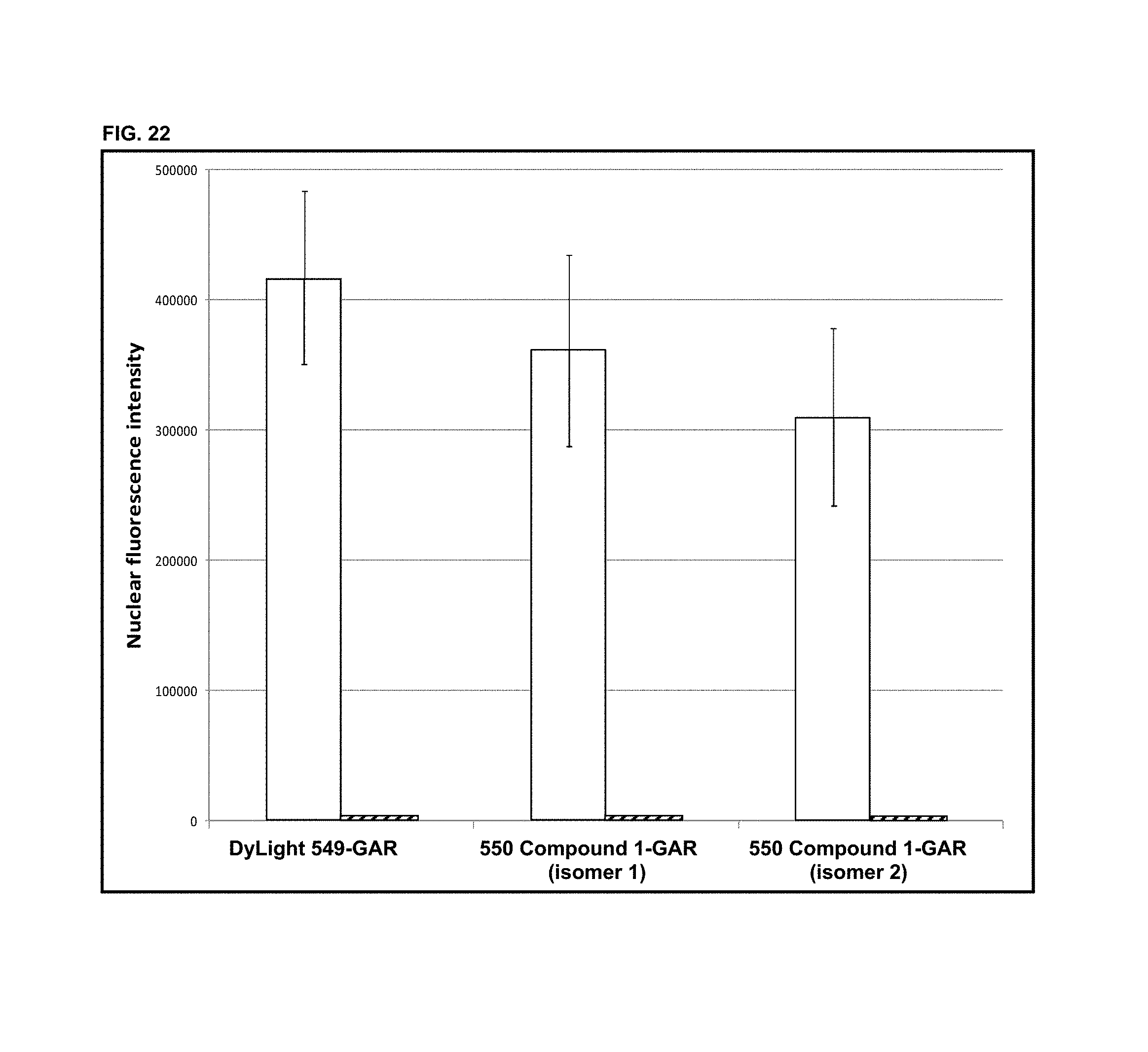
D00016
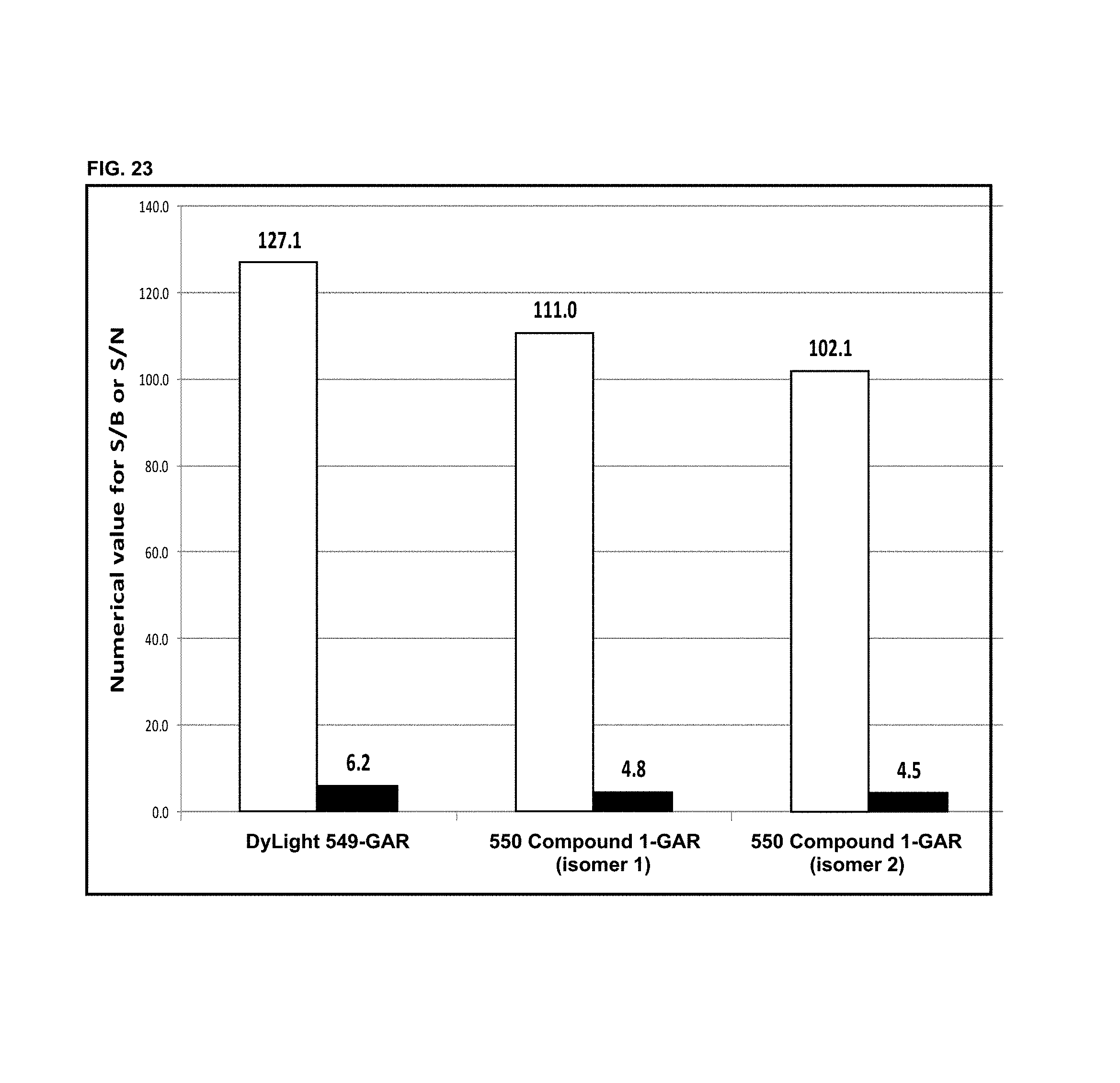
D00017
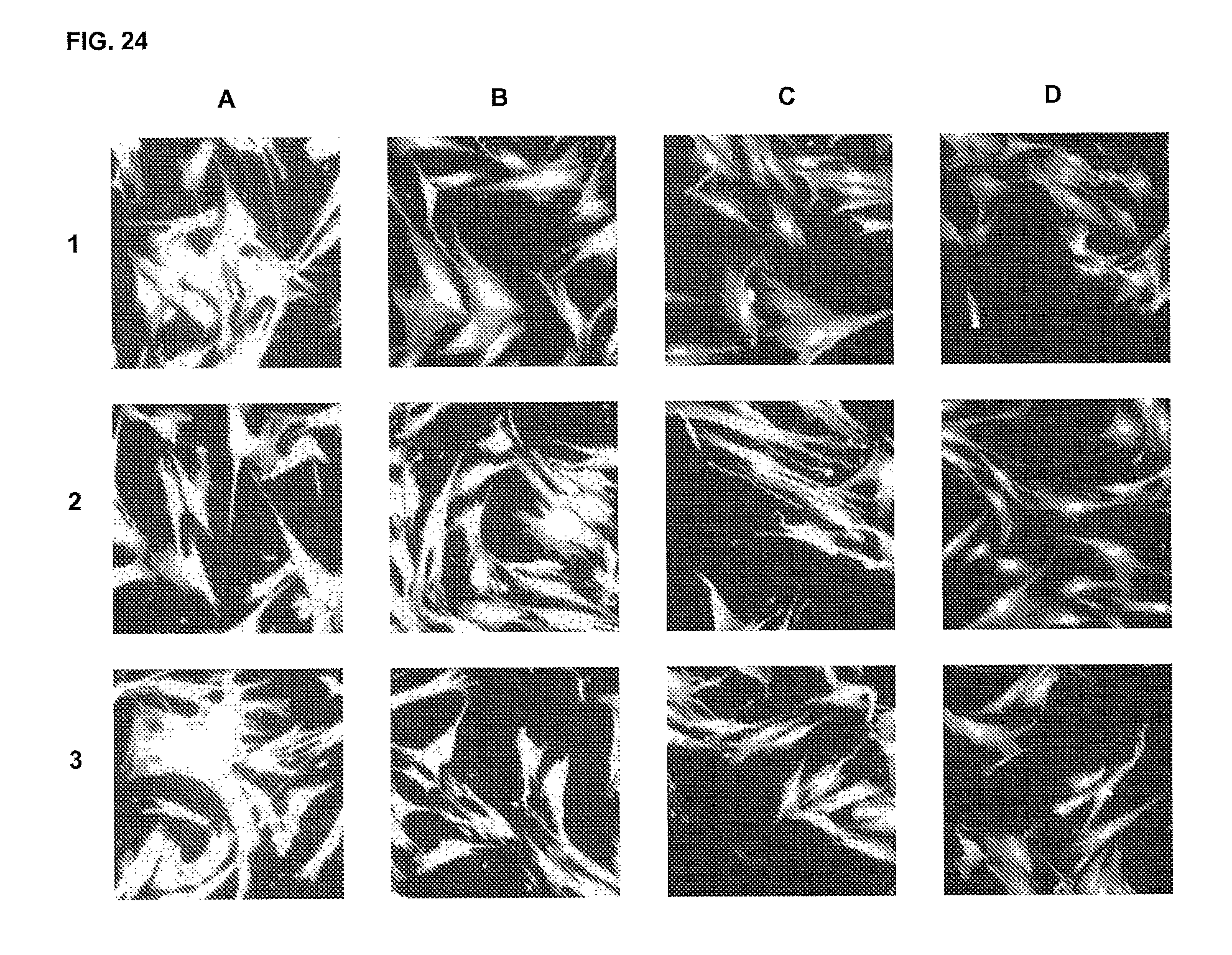
D00018

D00019
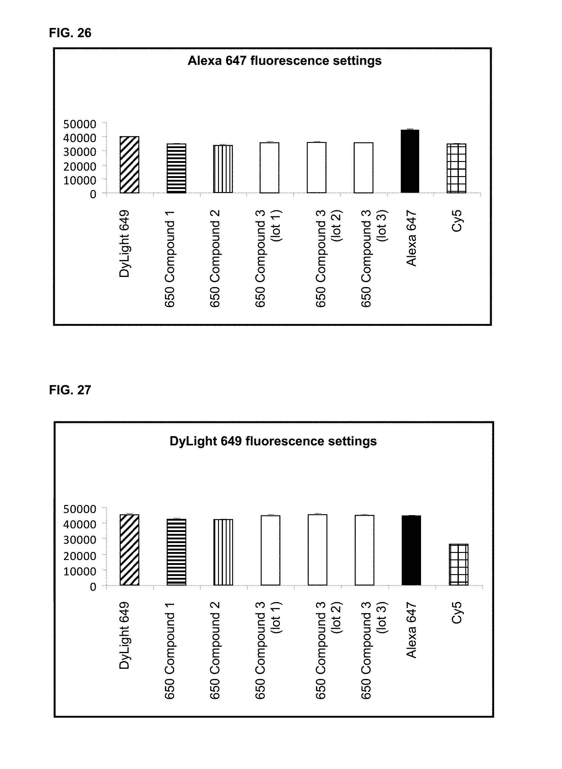
D00020

D00021
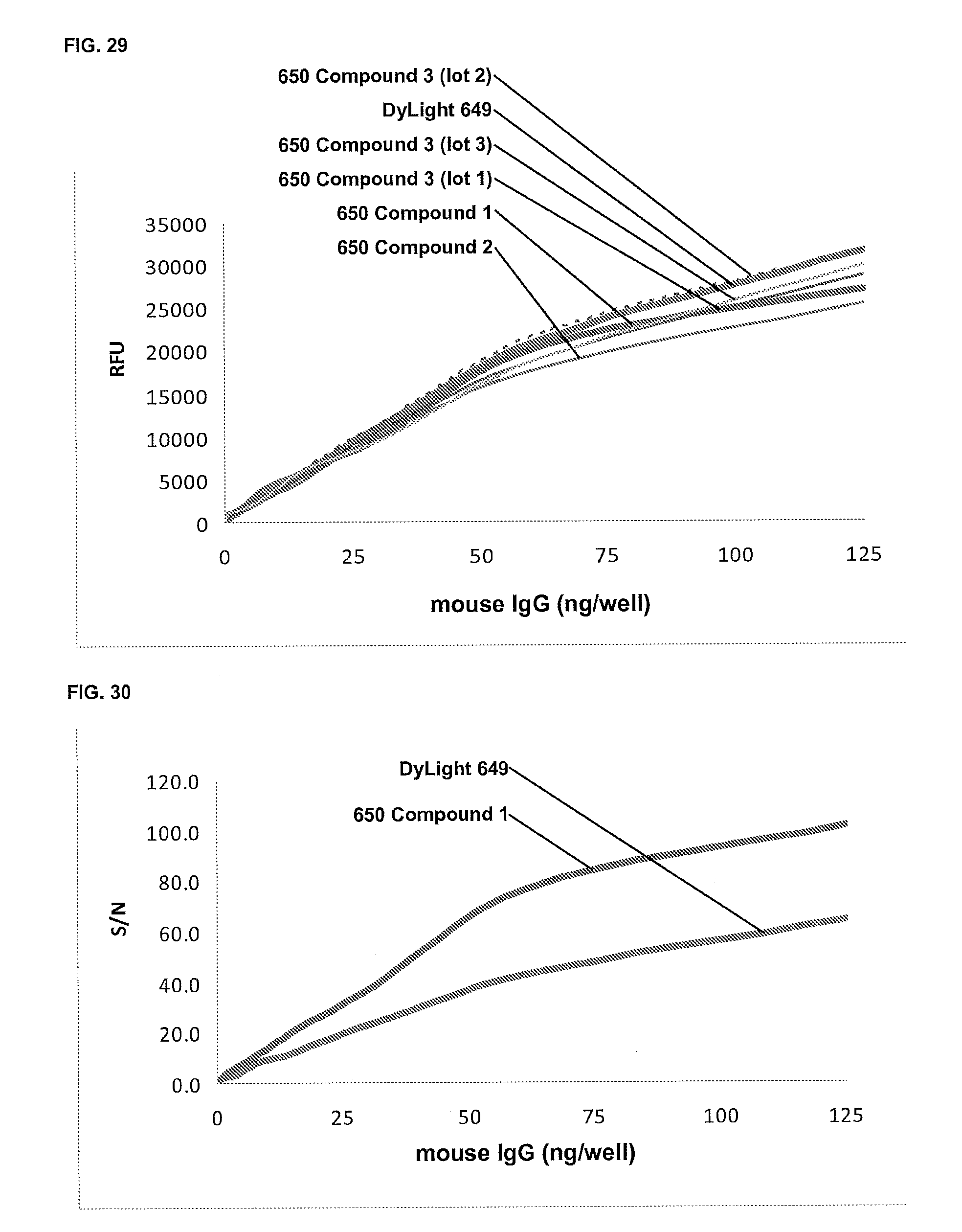
D00022

D00023
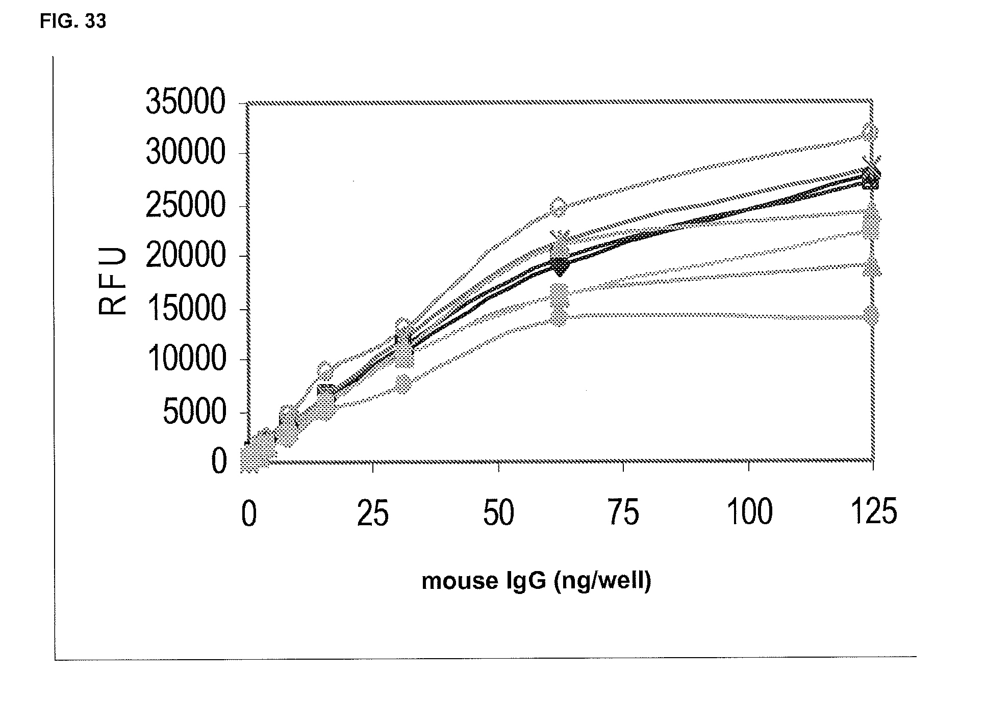
D00024
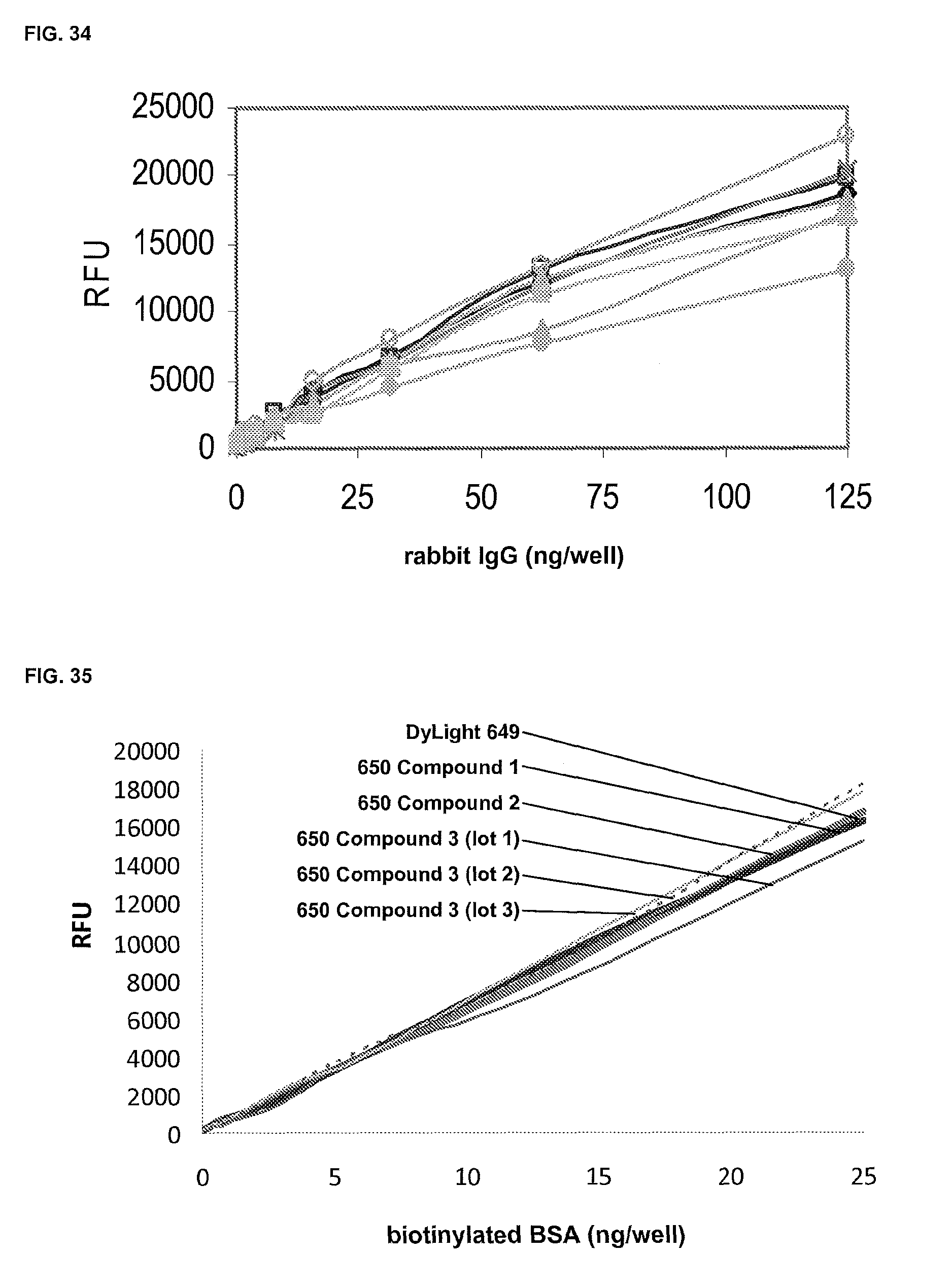
D00025

D00026
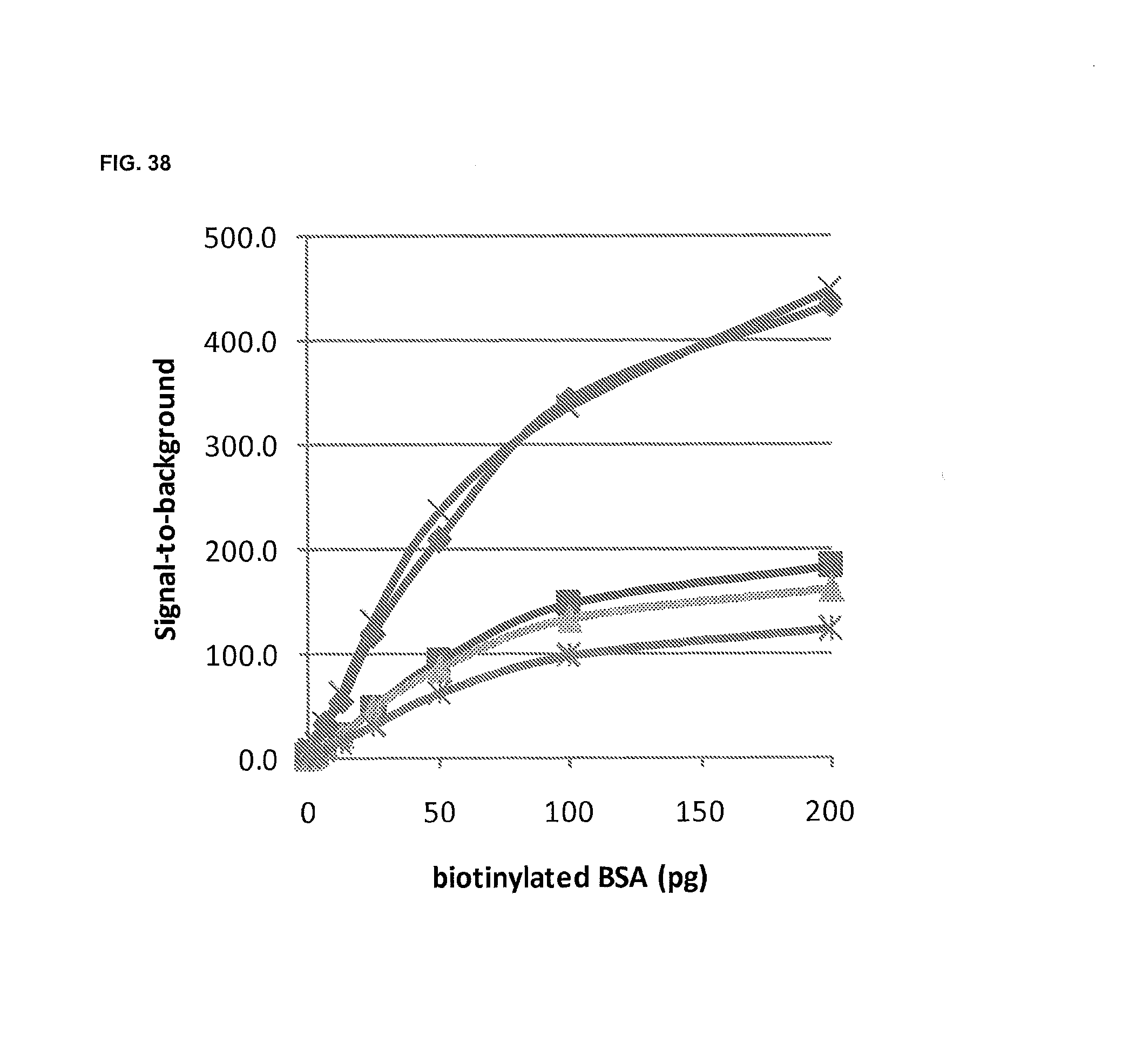
D00027

D00028
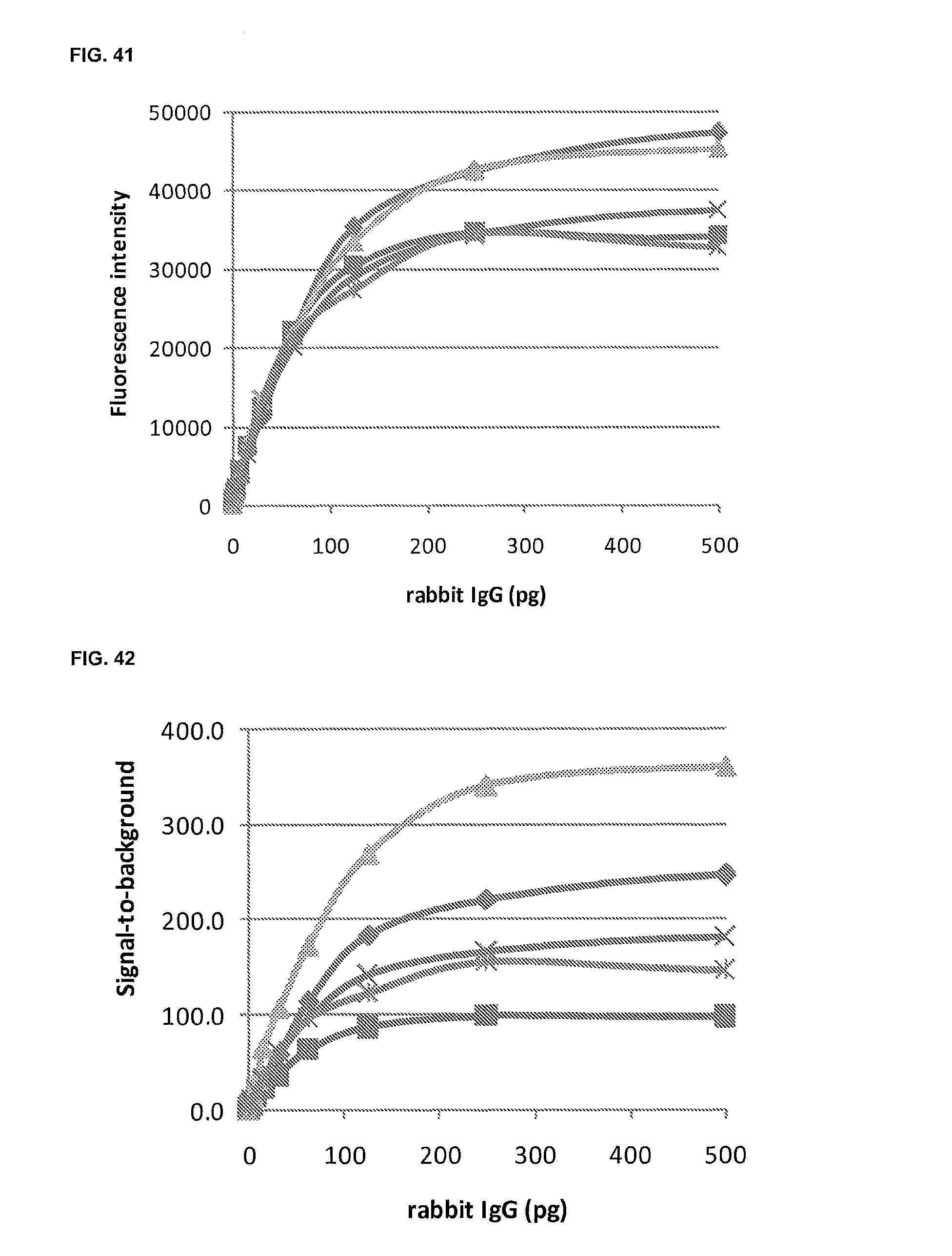
D00029

D00030
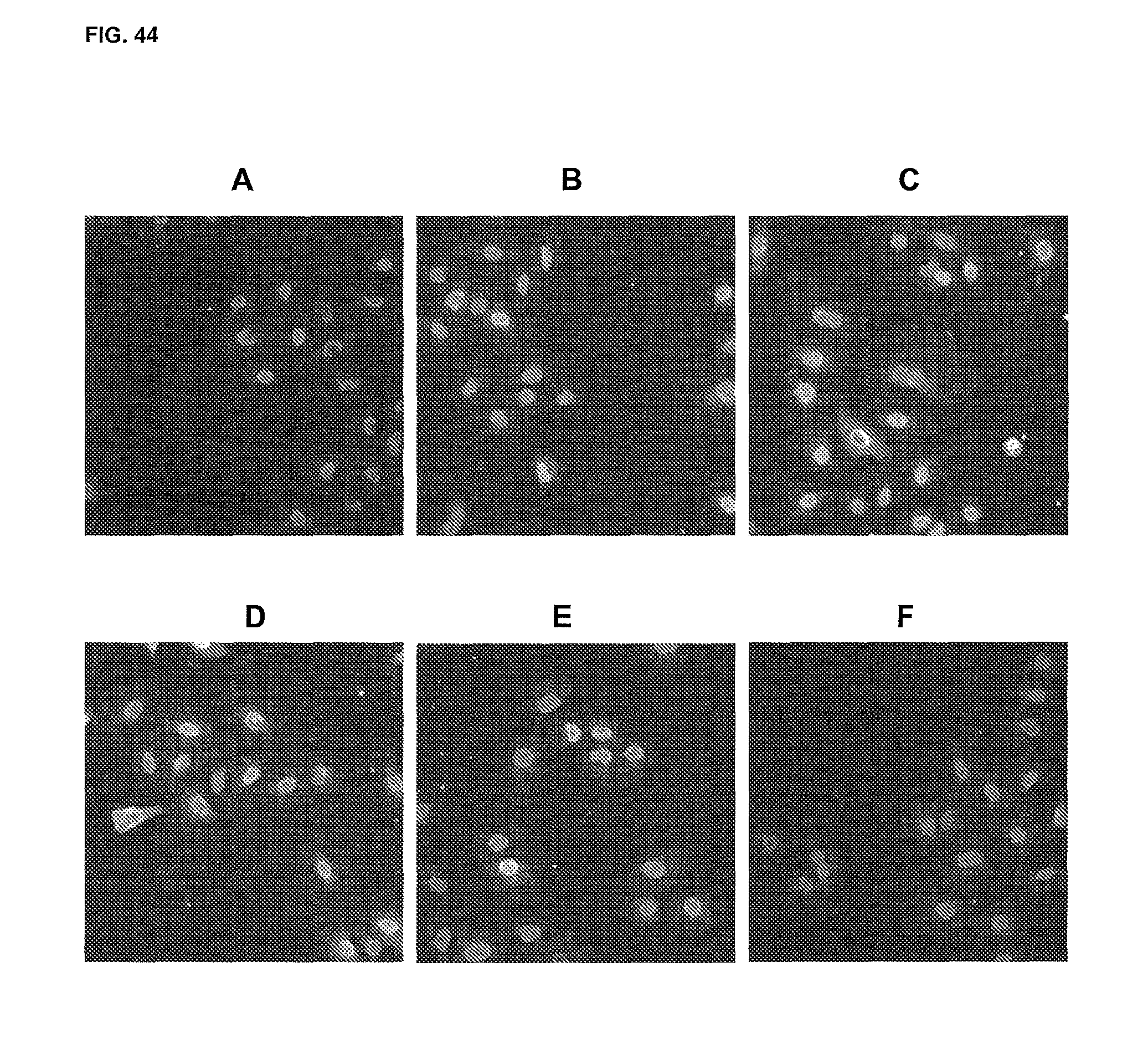
D00031
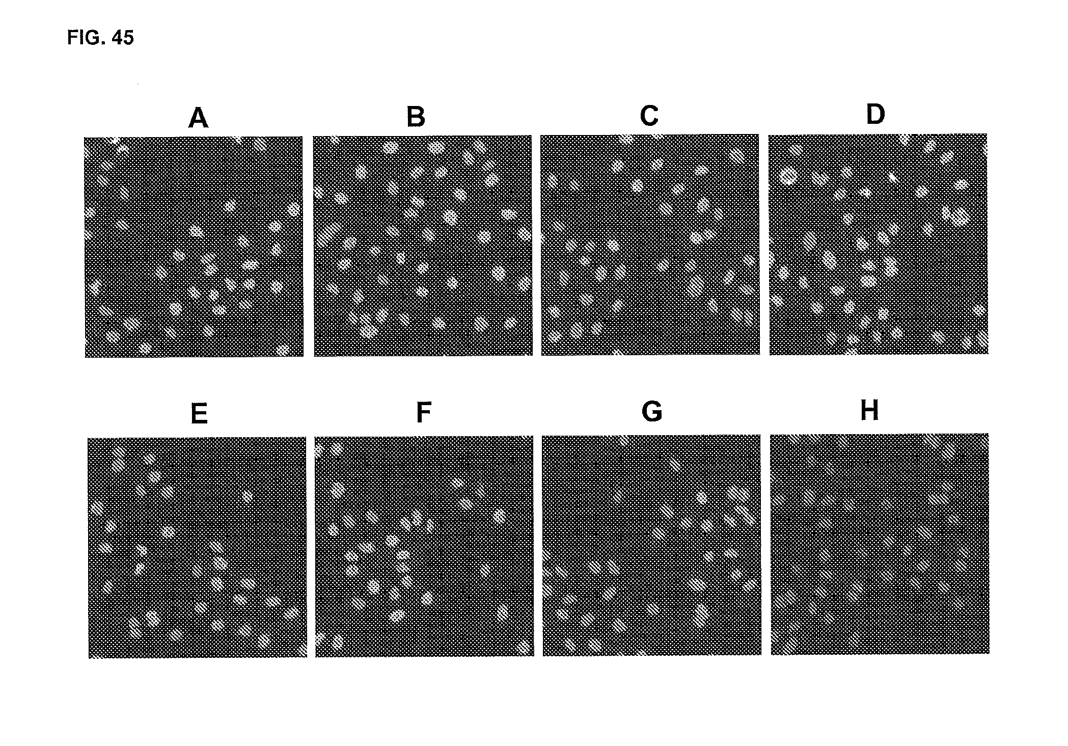
D00032
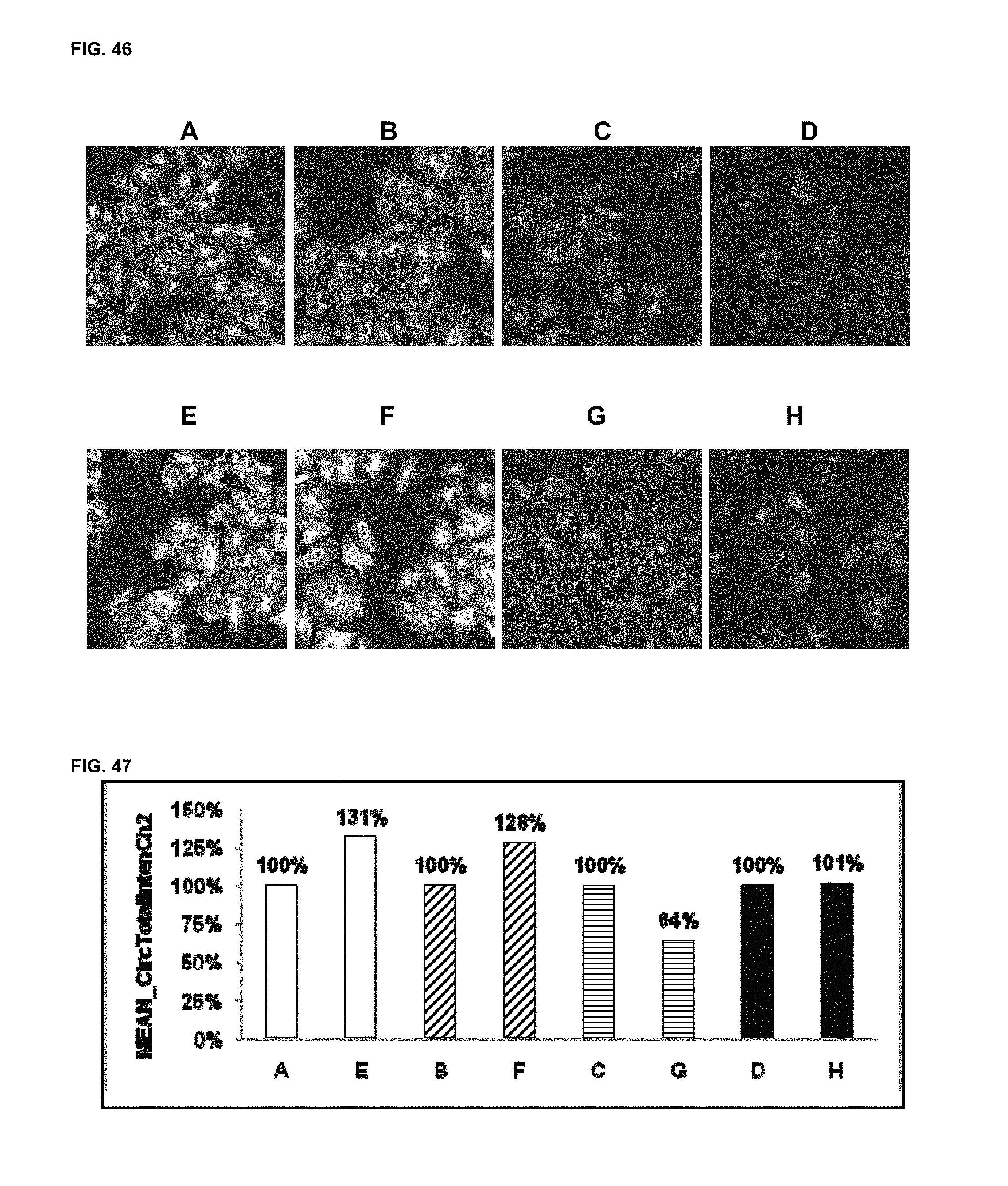
D00033
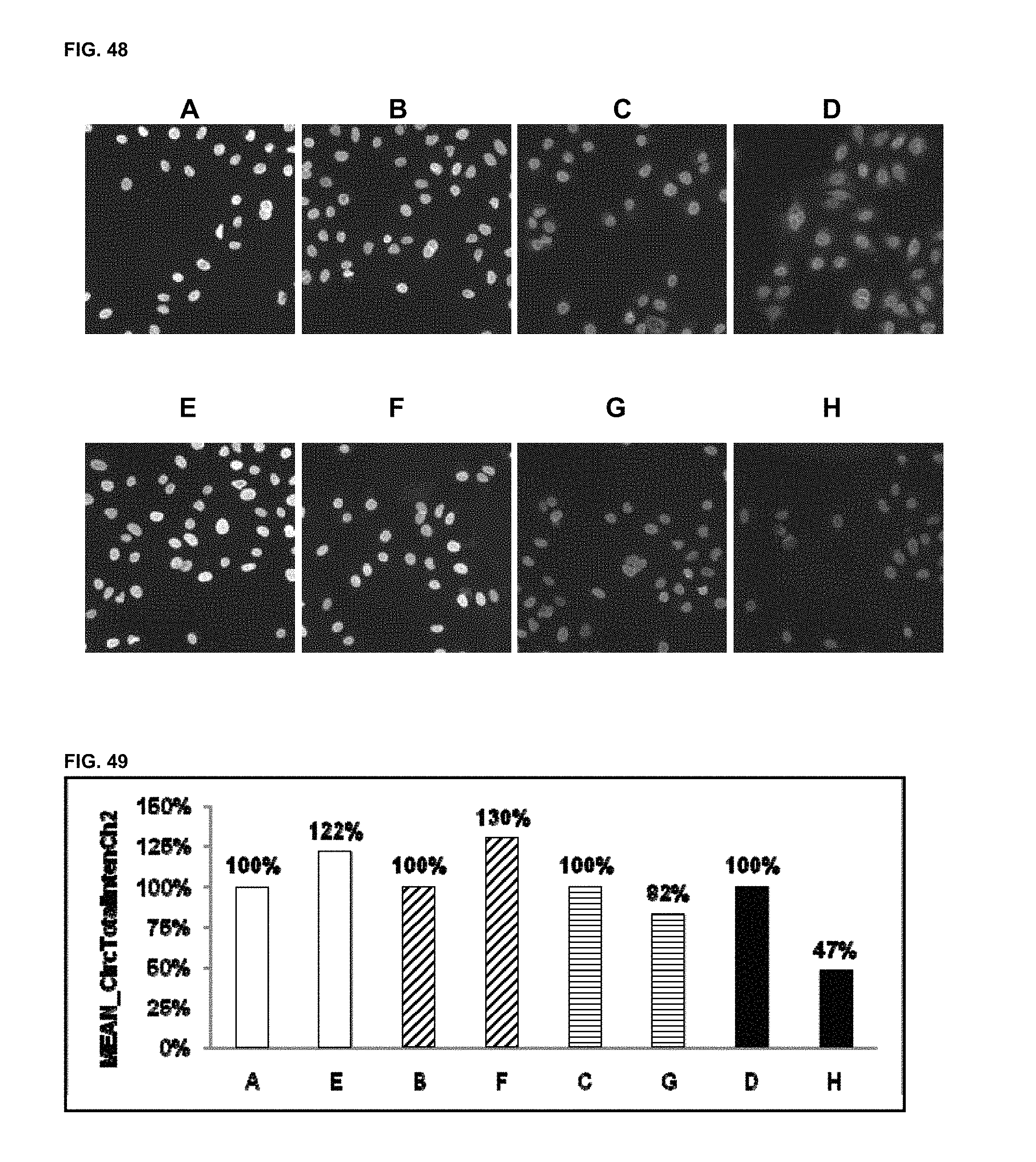
D00034
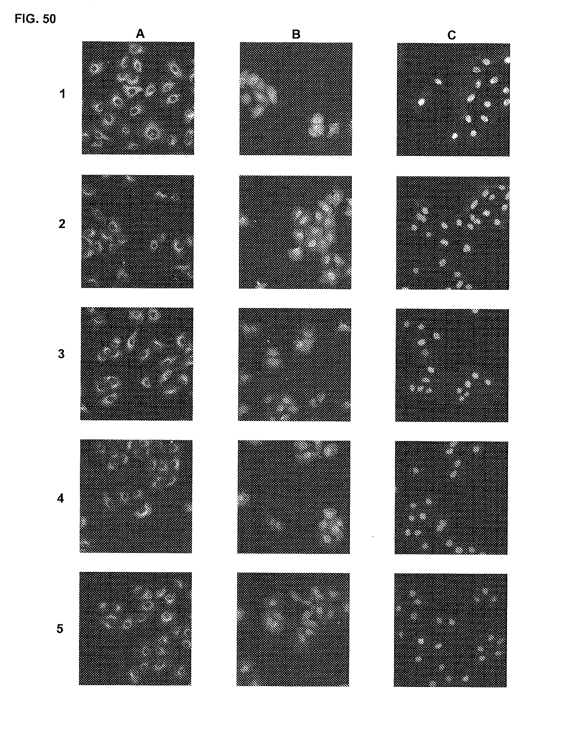
D00035

D00036
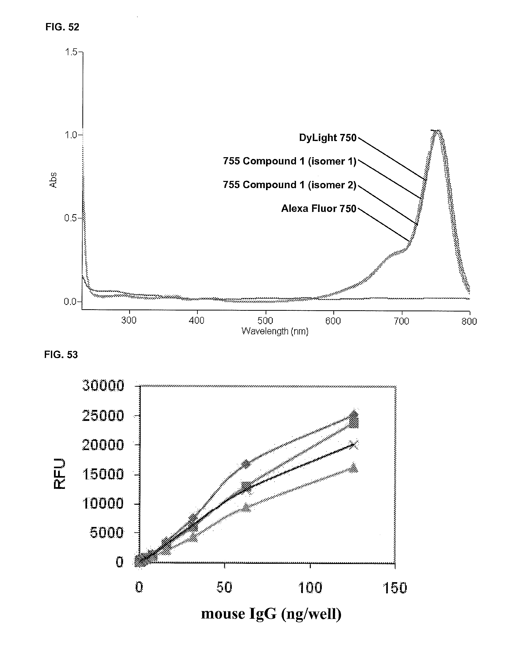
D00037
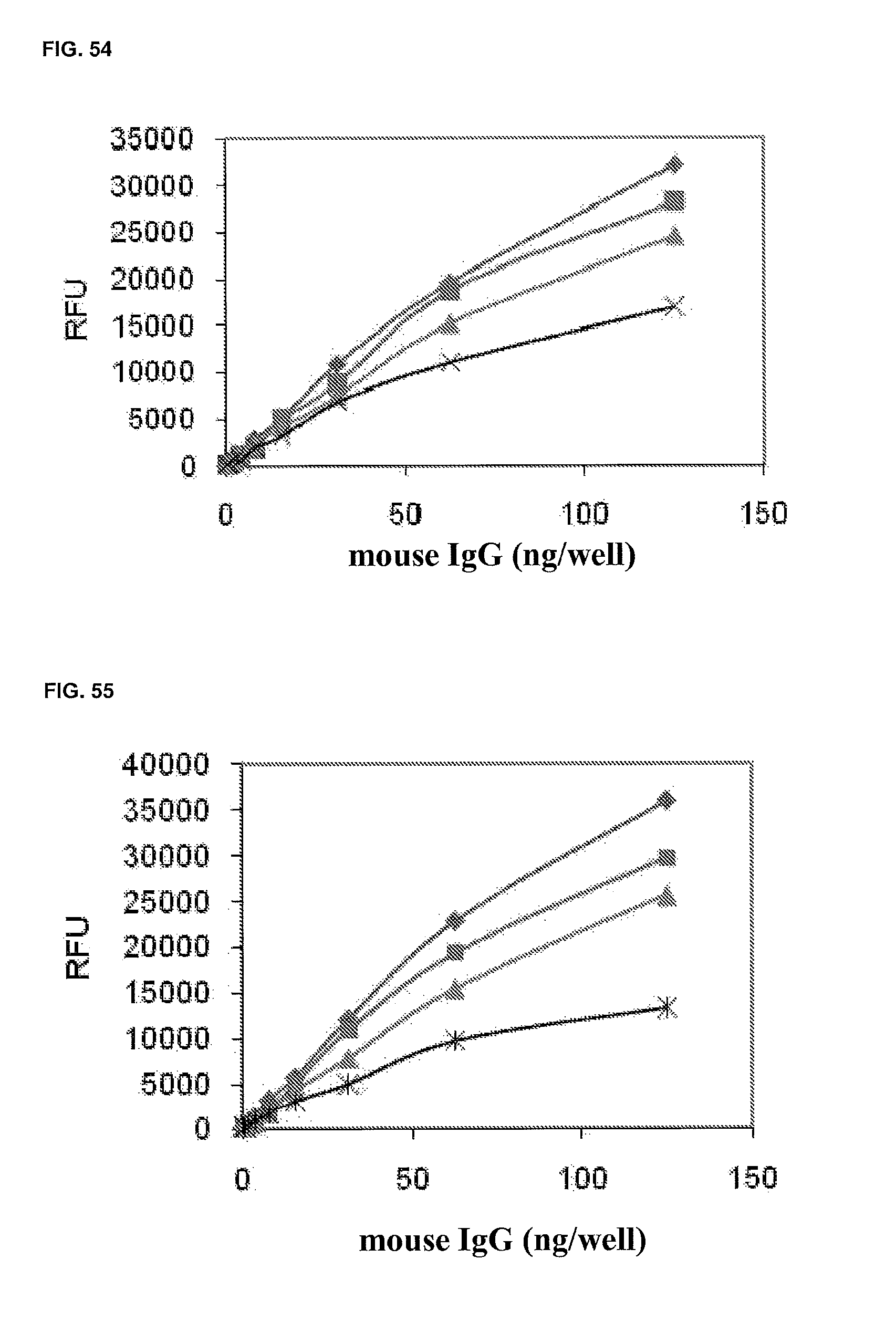
D00038
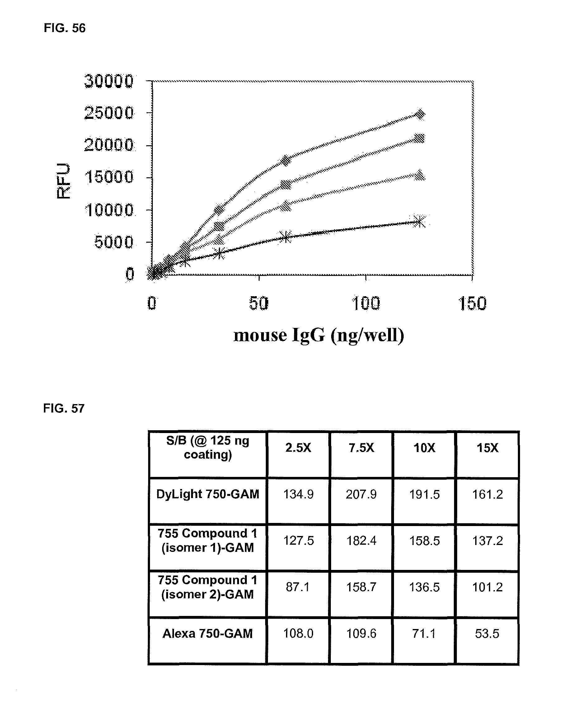
D00039
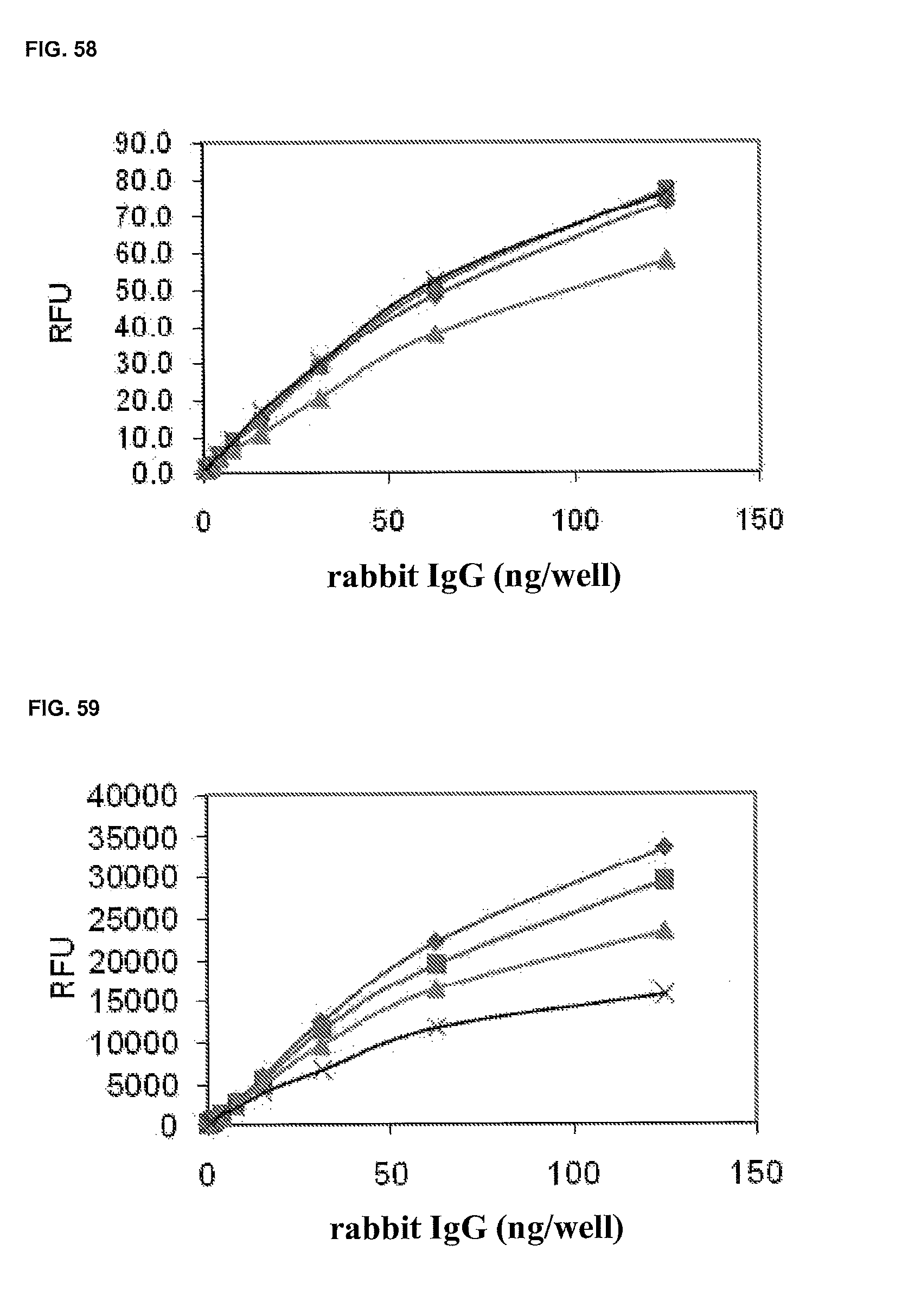
D00040

D00041
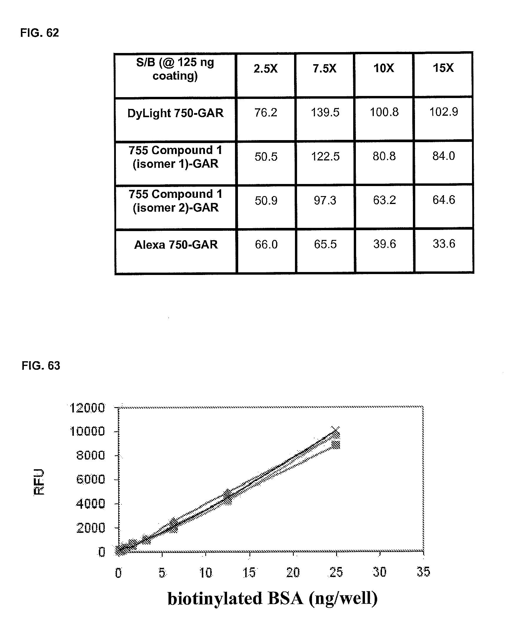
D00042

D00043

D00044
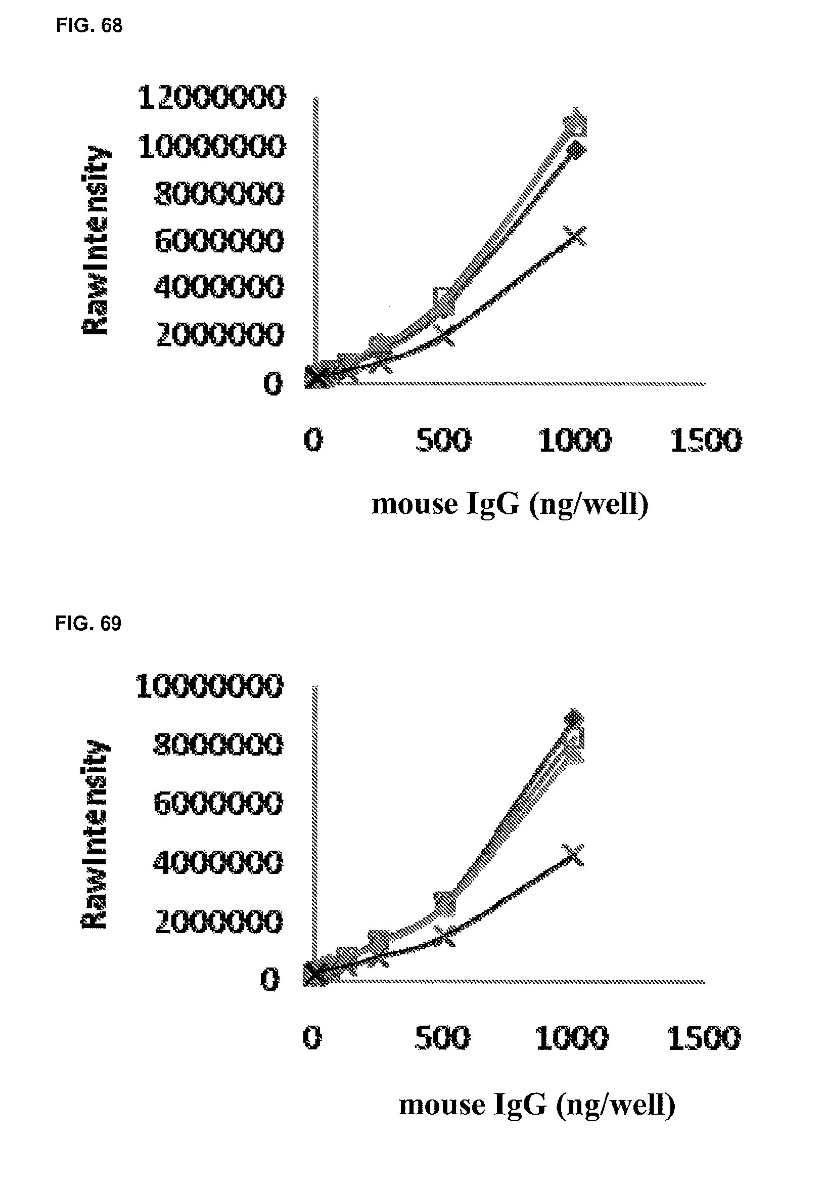
D00045

D00046
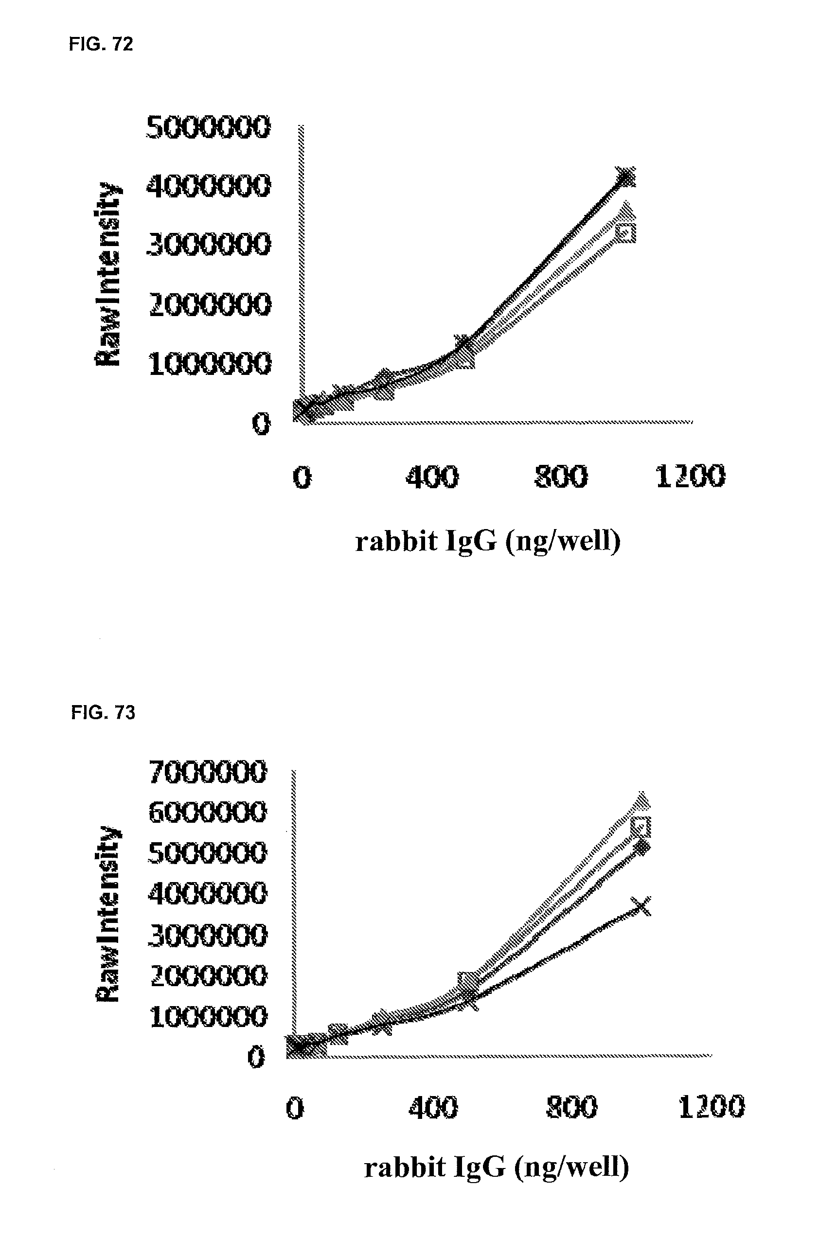
D00047
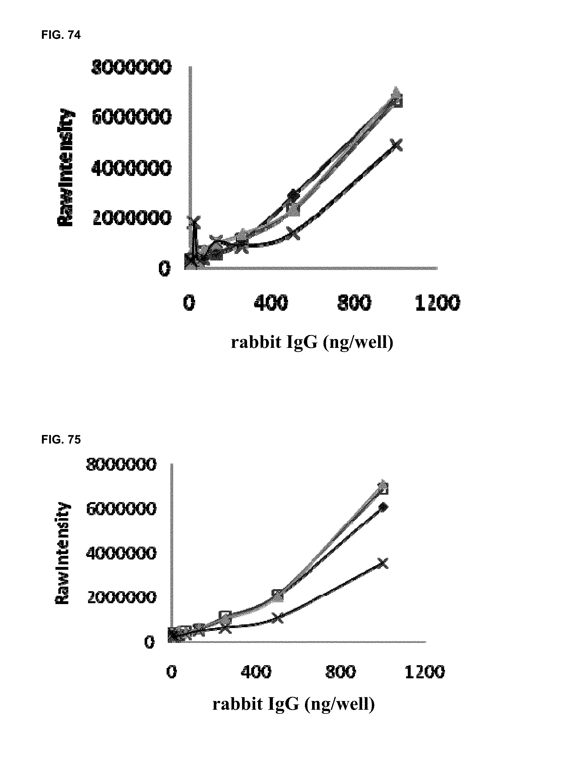
D00048
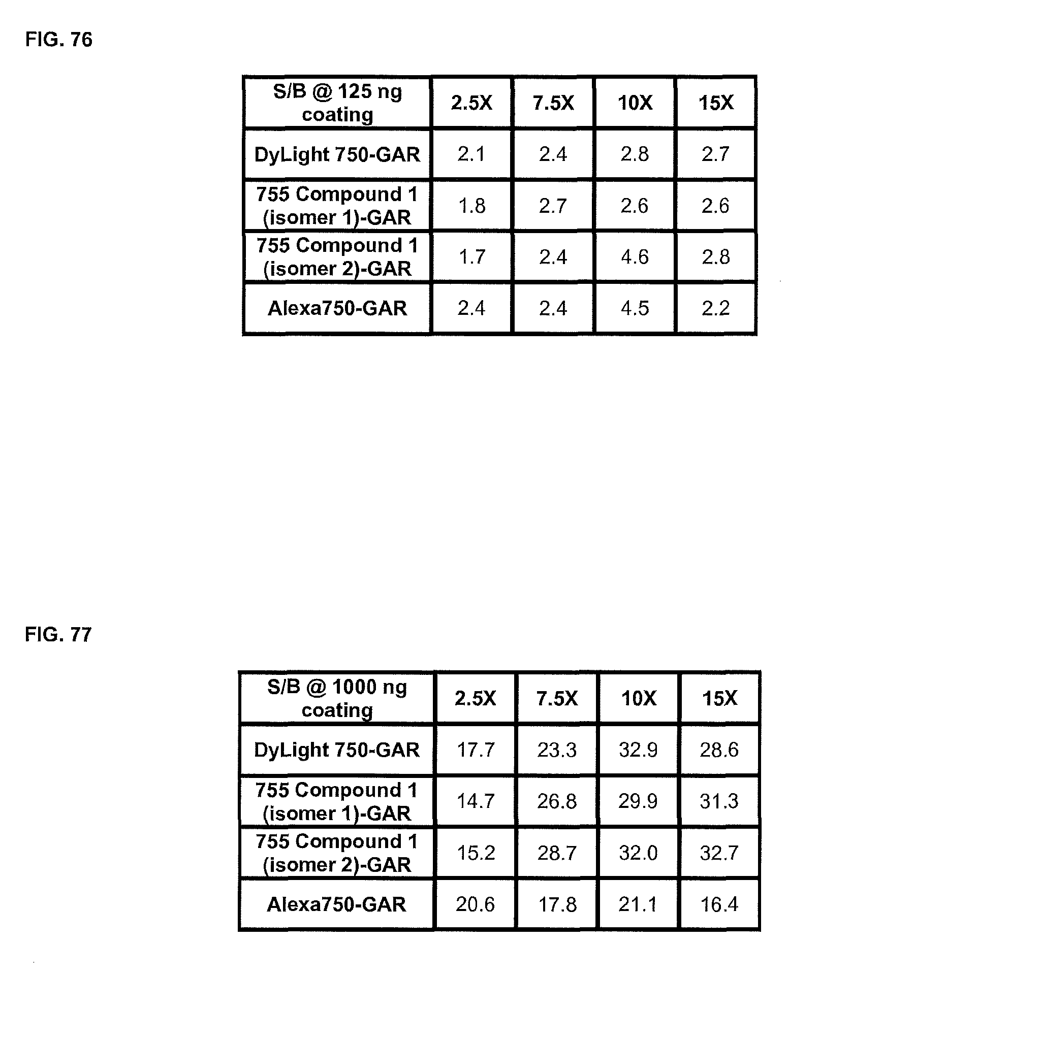
D00049

D00050

XML
uspto.report is an independent third-party trademark research tool that is not affiliated, endorsed, or sponsored by the United States Patent and Trademark Office (USPTO) or any other governmental organization. The information provided by uspto.report is based on publicly available data at the time of writing and is intended for informational purposes only.
While we strive to provide accurate and up-to-date information, we do not guarantee the accuracy, completeness, reliability, or suitability of the information displayed on this site. The use of this site is at your own risk. Any reliance you place on such information is therefore strictly at your own risk.
All official trademark data, including owner information, should be verified by visiting the official USPTO website at www.uspto.gov. This site is not intended to replace professional legal advice and should not be used as a substitute for consulting with a legal professional who is knowledgeable about trademark law.