Methods for improving expression levels of foreign proteins by means of phospholipase fusion expression
Yu , et al.
U.S. patent number 10,323,247 [Application Number 15/995,134] was granted by the patent office on 2019-06-18 for methods for improving expression levels of foreign proteins by means of phospholipase fusion expression. This patent grant is currently assigned to Jiangnan University. The grantee listed for this patent is Yan Xu, Xiaowei Yu. Invention is credited to Yan Xu, Xiaowei Yu.


| United States Patent | 10,323,247 |
| Yu , et al. | June 18, 2019 |
Methods for improving expression levels of foreign proteins by means of phospholipase fusion expression
Abstract
The invention discloses a method for improving the extracellular expression level of a foreign protein by means of phospholipase fusion expression. Four proteins, PLA.sub.2, MBP, CBD and SUMO, are used as a fusion tag to construct a fusion gene. Compared with an original protein MOH without any fusion tag, the extracellular expression level and enzymatic activity of all the four fusion proteins are increased to some degree. Among them, the fusion protein using PLA.sub.2 as the fusion tag has the highest expression level, which is 7.4 times higher than that of the original protein. Compared with other fusion tags, PLA.sub.2 has a low molecular weight and the fusion protein having PLA2 as the fusion tag has the highest expression level (up to 12 gL.sup.-1 in a 7 L fermentation tank for high-density fermentation). It is shown that the secretory expression of a foreign protein can be effectively increased by using PLA.sub.2 as a fusion tag.
| Inventors: | Yu; Xiaowei (Wuxi, CN), Xu; Yan (Wuxi, CN) | ||||||||||
|---|---|---|---|---|---|---|---|---|---|---|---|
| Applicant: |
|
||||||||||
| Assignee: | Jiangnan University (Wuxi, JS,
CN) |
||||||||||
| Family ID: | 59310650 | ||||||||||
| Appl. No.: | 15/995,134 | ||||||||||
| Filed: | June 1, 2018 |
Prior Publication Data
| Document Identifier | Publication Date | |
|---|---|---|
| US 20180265876 A1 | Sep 20, 2018 | |
Related U.S. Patent Documents
| Application Number | Filing Date | Patent Number | Issue Date | ||
|---|---|---|---|---|---|
| PCT/CN2016/071043 | Jan 15, 2016 | ||||
| Current U.S. Class: | 1/1 |
| Current CPC Class: | C12Y 301/01004 (20130101); C07K 14/38 (20130101); C12N 15/62 (20130101); C12N 9/50 (20130101); C12N 15/81 (20130101); C12N 9/20 (20130101); C07K 2319/24 (20130101); C12N 15/815 (20130101); C07K 2319/61 (20130101); C07K 2319/20 (20130101) |
| Current International Class: | C12N 9/20 (20060101); C07K 14/38 (20060101); C12N 15/62 (20060101); C07K 1/00 (20060101); C07H 21/04 (20060101); C12N 1/14 (20060101); C12P 21/04 (20060101); C12P 21/06 (20060101); C12N 1/20 (20060101); C12N 15/00 (20060101); C12N 15/81 (20060101); C12N 9/50 (20060101) |
| 102654504 | Sep 2012 | CN | |||
| 2009023270 | Feb 2009 | WO | |||
Other References
|
Li. Machine translation of CN 102654504. retrieved from https://worldwide.espacenet.com/?locale=EN_ep on Oct. 15, 2018. cited by examiner . Jiang. High-level expression of prolyl endopeptidase in Pichia pastoris usingPLA2as a fusion partner. Journal of Molecular Catalysis B: Enzymatic 125 (2016) 81-87. cited by examiner . Kang, Chao et al. "Cloning and expression of a novel prolyl endopeptidase from Aspergillus Oryzea and its application in beer stabilization", Journal of Insdustral Microbiology and Biotechnology, vol. 42, No. 2, Dec. 30, 2014. cited by applicant. |
Primary Examiner: Pak; Yong D
Attorney, Agent or Firm: Chen; Lili
Parent Case Text
CROSS-REFERENCES AND RELATED APPLICATIONS
This application is a continuation of the international application PCT/CN2016/071043, with an international filing date of Jan. 15, 2016, the content of which is herein incorporated by reference in its entirety.
Claims
What is claimed is:
1. A fusion gene, comprising a gene fragment encoding a fusion tag, a gene fragment encoding a linker peptide, and a gene fragment encoding a foreign protein, wherein the linker peptide has the amino acid sequence of SEQ ID NO:1, wherein the fusion tag is selected from a group of a phospholipase A2 and mutants thereof consisting of (a) a phospholipase A2 having the amino acid sequence of SEQ ID NO: 3; (b) a mutant of the sequence (a), wherein said mutant of the sequence (a) has the first four amino acids deleted from the sequence (a); (c) a mutant of the sequence (a), wherein said mutant of the sequence (a) has the amino acid at position 69, 70, 73, 74, 77 or 90 mutated to alanine in the sequence (a); and (d) a phospholipase A2 mutant having 90% or more homology to the sequence (a).
2. The fusion gene of claim 1, wherein the foreign protein is a prolyl endopeptidase.
3. The fusion gene of claim 2, wherein the prolyl endopeptidase has the amino acid sequence of SEQ ID NO: 4.
4. The fusion gene of claim 1, wherein the fusion gene is inserted into an expression vector.
5. The fusion gene of claim 4, wherein the expression vector with the fusion gene is transformed in a host microorganism to obtain an genetically engineered microorganism expressing the fusion gene.
6. The fusion gene of claim 5, wherein the genetically engineered microorganism is yeast Pichia pastoris comprising the fusion gene of claim 1 inserted into vector pPICZ.alpha.A.
7. A method for expressing a foreign protein, comprising constructing a fusion gene of claim 1 and expressing the fusion gene.
8. The method of claim 7, wherein the foreign protein is a prolyl endopeptidase.
9. The method of claim 8, wherein the fusion tag comprises a phospholipase A2 having the amino acid sequence of SEQ ID NO: 3.
10. The method of claim 8, wherein the fusion tag comprises a mutant of SEQ ID NO:3, wherein said mutant of SEQ ID NO:3 has a point mutation to alanine at position 69, 70, 73, 74, 77 or 90.
Description
BACKGROUND OF THE INVENTION
Field of the Invention
The disclosure relates to the field of bioengineering, and more particularly relates to a method for improving expression levels of foreign proteins by means of phospholipase fusion expression.
Description of the Related Art
Prolyl endopeptidase (PEP for short) [EC 3.4.21.26] does not belong to a typical serine protease family, but is a special serine protease, which is known as prolyl endopeptidase family together with escherichia coli protease II (protease II). The molecular weight of most prolyl endopeptidase is about 75 KDa, the optimum temperature is about 45.degree. C., and the optimum pH is about 4.5. Prolyl endopeptidase is a site-specific protease that can specifically hydrolyze a proline carboxy-terminal peptide bond in a molecular polypeptide (the rate of hydrolysis of small peptides is much higher than that of macromolecular peptides). Prolyl endopeptidase can also act on alanine residues but with much less hydrolysis efficiency.
Prolyl endopeptidase has important applications in biomedical therapy, which makes it a hotspot of international biomedical research. PEP can degrade many polypeptide neurotransmitters and hormones, and the aberrant activity of PEP can cause learning and spatial memory disorder. PEP can be applied to Alzheimer's disease and regulation of glucose metabolism and pancreatic functions. Therefore, further study of the physiological functions of the enzyme and development of inhibitors of the enzyme to treat memory disorders can bring great social and economic benefits.
Almost all protein hydrolysates have different levels of bitterness, mainly due to the existence of hydrophobic amino acids (such as proline), and the degree of bitterness is positively correlated with the content of hydrophobic amino acids. Edens et al. found that the prolyl endopeptidase obtained from Aspergillus niger through extracellular extraction and purification can effectively remove the bitterness of a bitter casein hydrolysate.
Celiac disease is the T-cell-predominant intolerance to bran of plants including wheat and the like. Gluten-mediated T-cell immune epitopes are abundant in proline and are therefore highly resistant to protein degradation in the gastrointestinal tract. Common prolyl endopeptidase oral preparations have many limitations, for example, low stability in a low-pH environment, high sensitivity to pepsin, and inability to completely digest all gluten in normal diets of each meal. Stepniak et al. obtained a novel prolyl endopeptidase (AN-PEP) from Aspergillus niger, and the novel prolyl endopeptidase can effectively reduce glutenin content in an in vitro simulated gastrointestinal environment and overcome many defects mentioned above.
Lopez et al. found that the addition of a very small amount of specific acidic prolyl endopeptidase during bottled beer fermentation can effectively prevent the problem of cold turbidity in the beer production process while the foam stability of beer is not affected. In addition, compared with the silica gel and PVPP method, the PEPase treatment method also has an advantage in terms of processing: no need to treat raw materials. The proteins that cause turbidity and precipitation during beer storage are mainly special proline-rich peptides. Proline endoprotease can be used for decomposing such turbidity-causing proteins so as to improve the non-biological stability of the beer. This can replace the current treatment methods used for improving beer colloid stability.
In addition, PEP can be used as an instrumental enzyme in molecular biology applications such as protein sequence determination, peptide mapping analysis, digestion of specific loci, peptide chain modification and processing, and the like.
Since the prolyl endopeptidase has many important functions, some researchers have already exogenously expressed the prolyl endopeptidase. For example, in 1980, Japanese researchers identified Flavobacterium meningosepticum capable of producing the prolyl endopeptidase, and enzyme activity reached 34.5 Ug.sup.-1 after purification (wet cells). Subsequently, the research team introduced the PEP gene into Escherichia coli for exogenous expression 558 Ug.sup.-1 (wet cells). In 1993, the prolyl endopeptidase derived from Aeromonas hydrophila was cloned and expressed in Escherichia coli JM83, and enzyme activity reached 320 Ug.sup.-1. However, the exogenous expression activity of the prolyl endopeptidase is still not very high. Therefore, there is a need to develop an effective strategy to improve the expression level of PEP in foreign hosts.
DETAILED DESCRIPTION
In order to overcome the above problems, the invention provides a method for improving the expression level of a foreign protein by means of phospholipase fusion expression, a genetically engineered yeast obtained by the method and applications thereof.
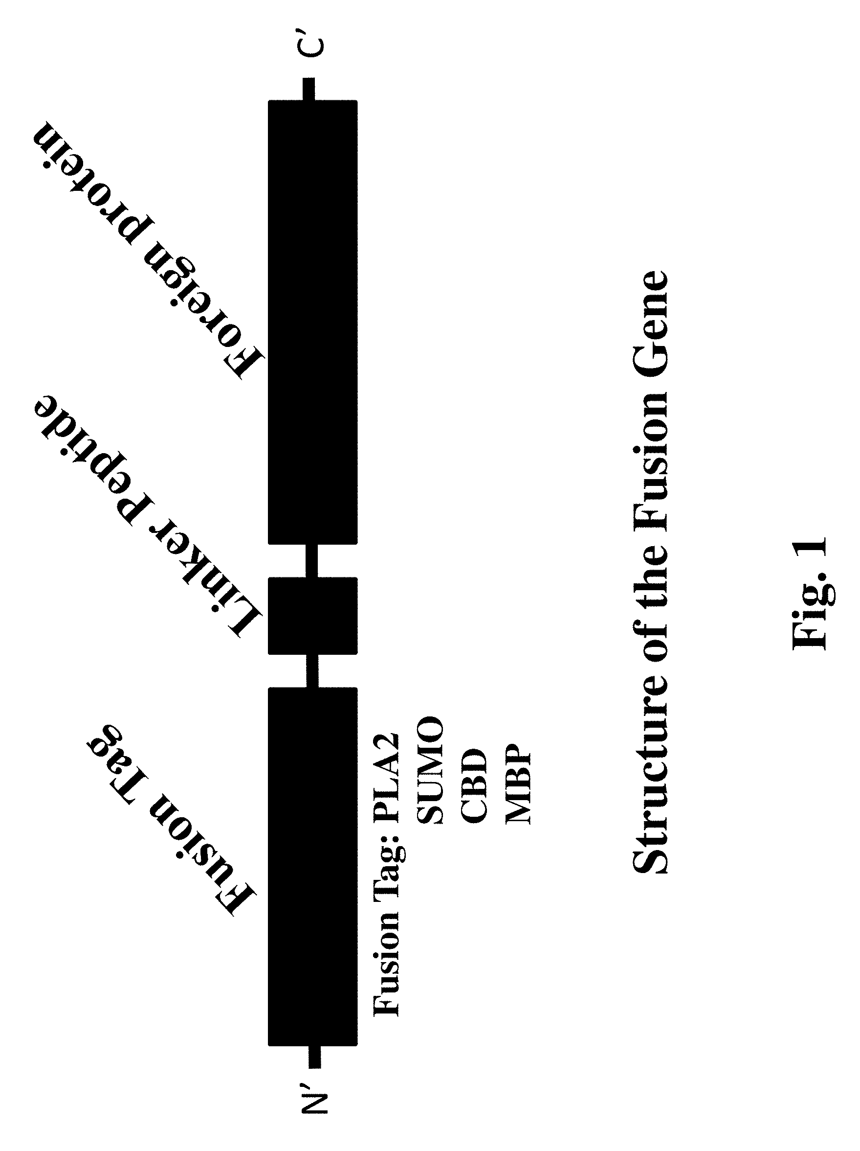
The first objective of the invention is to provide a fusion gene, wherein the fusion gene is obtained by sequentially ligating a gene fragment encoding a fusion tag, a gene fragment encoding a linker peptide and a gene fragment encoding a foreign protein.
In one embodiment of the invention, the fusion tag is any one of phospholipase A2 (PLA2), cellulose binding domain (CBD), small ubiquitin-related modifier (SUMO) and maltose binding protein (MBP).
In one embodiment of the invention, the fusion tag is phospholipase A2, and the amino acid sequence of the phospholipase A2 is:
(a) the sequence of SEQ ID NO: 3; or
(b) a sequence obtained after deleting four amino acids (from position 1 to position 4) from the sequence of SEQ ID NO: 3; or
(c) an amino acid sequence obtained after mutating an amino acid into an alanine at position 69, 70, 73, 74, 77 or 90 of the sequence of SEQ ID NO: 3; or
(d) a sequence which has 90% or more homology to the sequence of (a).
In one embodiment of the invention, the nucleotide sequence encoding the amino acid sequence of SEQ ID NO: 3 is shown in SEQ ID NO: 8.
In one embodiment of the invention, the amino acid sequence of the linker peptide is shown in SEQ ID NO: 1 (GGGGSGGGGS).
In one embodiment of the invention, the foreign protein is a prolyl endopeptidase.
In one embodiment of the invention, the amino acid sequence of the prolyl endopeptidase is shown in SEQ ID NO: 4.
In one embodiment of the invention, the fusion gene is obtained by ligating a gene fragment encoding the amino acid sequence of the phospholipase A2 with a gene fragment encoding a linker peptide (e.g. SEQ ID NO: 1), and ligating a gene fragment encoding prolyl endopeptidase (e.g. SEQ ID NO: 4) to the linker peptide fragment.
In one embodiment of the invention, the nucleotide sequence of the fusion gene is shown in SEQ ID NO: 2.
The second object of the invention is to provide a genetically engineered yeast expressing the fusion gene.
In one embodiment of the invention, the engineered yeast is an engineered Pichia pastoris, such as Pichia pastoris X33, GS115, KM71 or SMD1168, preferably Pichia pastoris GS115.
In one embodiment of the invention, a vector used for the expression may be pPIC9, pPIC3.5K, pPIC3.5, pPIC9K, PA0815 or pPICZ.alpha. series and similar vectors thereof; preferably pPICZ.alpha.A.
In one embodiment of the invention, the engineered Pichia pastoris is obtained by ligating the fusion gene with nucleotide sequence shown in SEQ ID NO.2 to pPICZ.alpha.A first to obtain a recombinant vector pPICZ.alpha.A-PLMH, then linearizing a recombinant plasmid and transforming the recombinant plasmid into a host Pichia pastoris.
In one embodiment of the invention, the host Pichia pastoris is Pichia pastoris GS115, or Pichia pastoris GS115 integrated with pPIC9K plasmids (in order to overcome the histidine defect of Pichia pastoris for ease of operation).
In one embodiment of the invention, a 6.times.His tag is added after the sequence of the fusion gene to facilitate isolation and purification.
The third object of the invention is to provide a method for producing a prolyl endopeptidase by using the fusion gene, an expression vector containing the fusion gene, or a genetically engineered yeast expressing the fusion gene.
In one embodiment of the invention, after being activated, the genetically engineered Pichia pastoris yeast expressing the fusion gene is inoculated into a BMGY medium, cultured at 30.degree. C. and 200 rmin.sup.-1 for 16-18 hr; when OD600 reaches 2-6, the culture medium is centrifuged, yeast cells are re-suspended in the BMMY medium and cultured at 28.degree. C. and 250 rmin.sup.-1, and 1% methanol is added every 24 h to induce expression.
In one embodiment of the invention, after being activated, the genetically engineered Pichia pastoris yeast expressing the fusion gene is inoculated into a fermentation tank for glycerol growth phase culture at 30.degree. C., pH 5.5, in a BSM basal salt medium (containing 1.1 gL.sup.-1 of CaSO.sub.4 (cp), 18.2 gL.sup.-1 of K.sub.2SO.sub.4(AR), 7.27 gL.sup.-1 of anhydrous magnesium sulfate (AR), 4.128 gL.sup.-1 of KOH (AR), 40 gL.sup.-1 of glycerol and 26.7 mlL.sup.-1 of 85% H.sub.3PO.sub.4). When glycerol in the BSM basal salt medium runs out and the dissolved oxygen value increases sharply, the glycerol transition phase starts and 50% (V/V) glycerol (containing 12 mLL.sup.-1 of PTM1, wherein PTM1 contains 6 gL.sup.-1 of copper sulfate pentahydrate, 0.089 gL.sup.-1 of potassium iodide, 3.0 gL.sup.-1 of magnesium sulfate monohydrate, 0.2 gL.sup.-1 of sodium molybdate, 0.02 gL.sup.-1 of boric acid, 42.2 gL.sup.-1 of zinc sulfate heptahydrate, 65 gL.sup.-1 of ferrous sulfate heptahydrate, 0.2 gL.sup.-1 of biotin, 0.5 gL.sup.-1 of cobalt chloride hexahydrate and 5 mlL.sup.-1 of sulfuric acid) is fed to the cells. When the cell OD600 value reaches 90-110, glycerol feeding is stopped and the induction phase starts after starving the cells for 0.5 hr, during which methanol is fed to the cells, the methanol concentration is maintained at 0.08-0.12%, and the culture temperature is controlled at 26-28.degree. C.
The fourth object of the invention is to provide applications for prolyl endopeptidases produced by using the fusion gene, an expression vector containing the fusion gene, or a genetically engineered microorganism expressing the fusion gene.
The applications are in the fields like food, preparation medicine and molecular biology.
The applications include reducing the content of a sensitive protein in a beer fermentation broth, increasing the non-biological stability of beer, and removing the bitterness of a protein hydrolysate.
The applications also include preparing drugs for regulating glucose metabolism or pancreas functions.
The applications also include the use of the prolyl endopeptidase as an instrumental enzyme in molecular biology for being applied to protein sequence determination, peptide mapping analysis, digestion of specific loci, peptide chain modification and processing and the like.
The fifth object of the invention is to provide a method for improving the expression level of a foreign protein by means of phospholipase fusion expression. The method comprises sequentially ligating a gene fragment encoding a phospholipase A2, a linker peptide fragment encoding an amino acid sequence such as SEQ ID NO.1 (GGGGSGGGGS) and a gene fragment encoding a foreign protein to obtain a fusion gene, and then expressing the fusion gene.
In one embodiment of the invention, the fusion tag is phospholipase A2 having an amino acid sequence shown in SEQ ID NO: 3 or a sequence being 90% or more homologous to the sequence of SEQ ID NO: 3.
In one embodiment of the invention, the foreign protein is a prolyl endopeptidase having an amino acid sequence shown in SEQ ID NO: 4.
In one embodiment of the invention, the nucleotide sequence of the fusion gene is shown in SEQ ID NO: 2.
In the invention, the four proteins PLA.sub.2, MBP, CBD and SUMO are used as a fusion tag to construct a fusion expression strain. Results show that compared with an original strain MOH, the extracellular protein expression level and enzyme activity of the four fusion proteins are increased to some extent, and, among them, the expression level of the target protein is the highest when PLA.sub.2 is used as a fusion tag, which is 7.4 times higher than that of the original strain. Compared with other fusion tags, PLA.sub.2 derived from S. violaceoruber A-2688 has the characteristics of being high in protein expression level (the highest extracellular protein concentration can reach 12 gL.sup.-1 in a 7 L fermentation tank for high-density fermentation) and low in molecular weight. The results of the study show that the secretory expression of a foreign protein can be effectively increased by using PLA.sub.2 as a fusion tag.
BRIEF DESCRIPTION OF FIGURES
FIG. 1. Schematic diagram of the structure of the fusion gene of the invention.
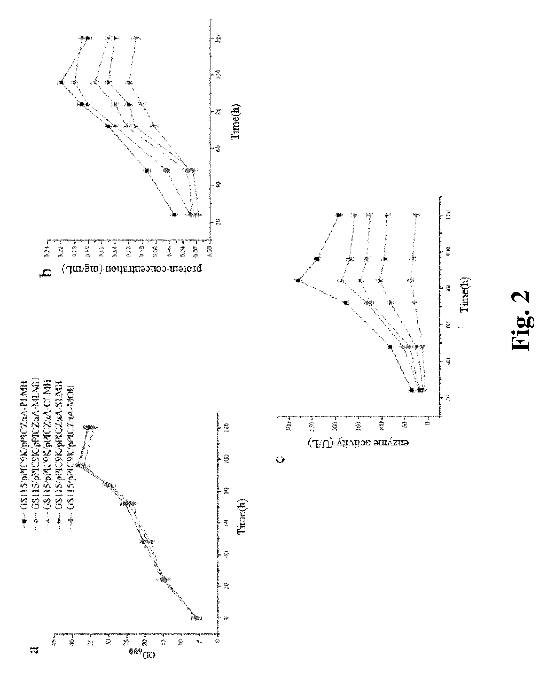
FIG. 2. Culture indicators of shake flask fermentation of genetically engineered Pichia pastoris GS115 strains, a: OD600; b: protein concentration; c: enzyme activity.
FIG. 3. Western blot results of proteins from genetically engineered Pichia pastoris GS115 strains. a: extracellular protein; b: intracellular protein; M: marker; 1-6 are proteins from MOH, SLMH, CLMH, MLMH, PLMH expressing cells and blank control, respectively.
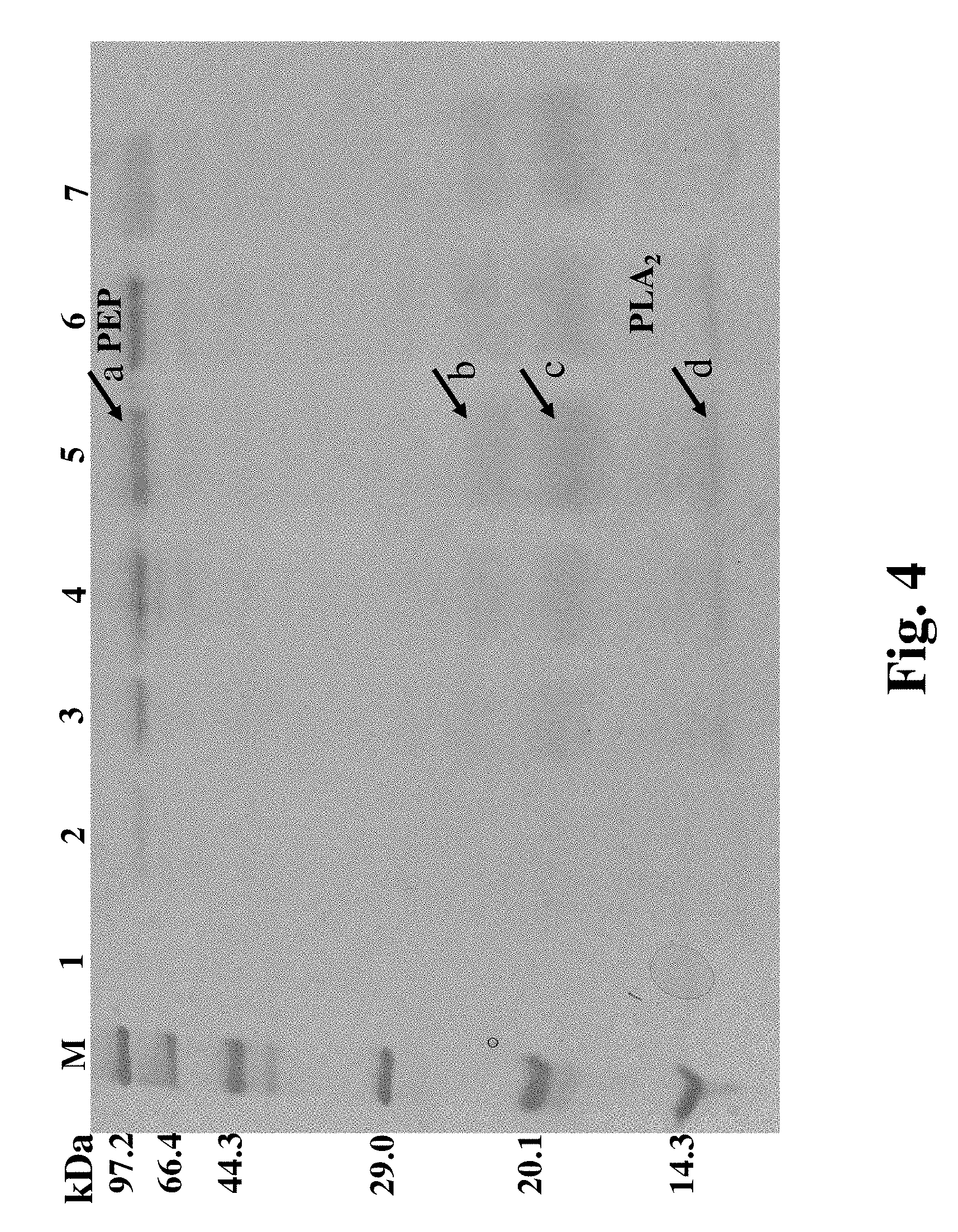
FIG. 4. SDS-PAGE Image of PLMH extracellular protein expression at different times of the shake flask fermentation. M: marker; 1-7 are shake flask fermentation broths after 0, 24, 48, 72, 84, 96 and 120 hr fermentation, respectively.
FIG. 5. Mass spectrum identification of fusion protein PEP and fusion tag PLA2.
FIG. 6. Comparison of enzymatic properties of PLMH and MOH, a: optimum temperature and temperature stability; b: optimum pH and pH stability.
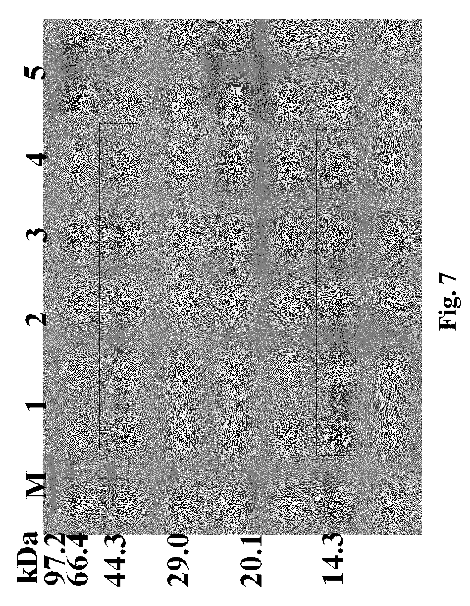
FIG. 7. Reduction of sensitive protein in beer fermentation broth by proline proteases; M: marker; 1-4: beer fermentation broth samples with addition of 0, 5, 15 and 25 UL.sup.-1 proline protease; 5: proline protease protein.
EXAMPLES
Materials and Methods:
1. Method for Determining Protein Concentration
20 .mu.l of supernatant of fermentation broth was added into wells of a 96-well plate, and then 200 .mu.l of G250 staining reagent was added. Let it stand at room temperature for 3-5 minutes. The absorbance at 595 nm wavelength was measured with a spectrophotometer, and protein concentration in a sample was calculated according to a standard curve.
2. Method for Determining Protease Activity
A reaction system was consisted of 10 .mu.l of enzyme solution, 10 .mu.l of 5 mM substrate (Ala-Ala-Pro-pNA) and 80 .mu.l of disodium hydrogen phosphate-citrate buffer. The reaction was carried out at 40.degree. C. for 10 min, and the absorbance at 410 nm wavelength was measured with a spectrophotometer.
Definition of enzyme activity unit (U): under the enzyme activity determination conditions specified above, the amount of enzyme required to catalyze and decompose aforementioned substrate to generate 1 .mu.mol of pNA per minute equals to a prolyl endopeptidase enzyme activity unit.
Enzyme activity calculation formula: enzyme activity (UmL.sup.-1)=.DELTA. A*V/(v1rbt), where, .DELTA.A: change in absorbance (OD.sub.test-OD.sub.blank); V: total volume of reaction system (mL); v1: sample amount (mL); r: molar extinction coefficient (cm.sup.2.mu.mol.sup.-1); b: optical path of a cuvette or a Elisa plate (cm); and t: reaction time (min).
3. Western Blot Analysis
Western blot analysis was conducted on supernatant and intracellular protein of a fusion protein shake flask fermentation, and the western blot procedure was as follows:
(1) Cut a protein gel to an appropriate size after conventional protein electrophoresis, and immerse the protein gels in a transfer buffer for being balanced.
(2) Cut a PVDF membrane and filter paper of the same size as the gel (8 pieces of filter paper for one PVDF membrane), and soak them in pure methanol first and then in the transfer buffer for 10 min.
(3) Neatly stack the PVDF membrane, the filter paper and the gel in a transfer box, wherein four layers of filter paper, the PVDF membrane, the gel and four layers of filter paper were arranged from bottom to top in sequence. Rolling gently with a roller to get rid of excess air bubbles and buffer every time a layer was applied; finally, absorbing surrounding buffer with a paper towel, closing a lid, and placing the transfer box in a transfer instrument.
(4) Transferring: turn on the power; select List, User Defined (this is an edited program 25 V, 1.0 A, 10 min) and Run in sequence.
(5) Take the PVDF membrane out after transferring, immerse the PVDF membrane in a blocking reagent, and gently shake in a decolorization shaker for 1 hr.
(6) Immerse the confined PVDF membrane in a primary antibody solution diluted in PBST (0.137 M NaCl, 0.0027 M KCl, 0.01 M Na.sub.2HPO.sub.4, 0.0018 M KH.sub.2PO.sub.4, 0.1% (v/v)Tween-20) with a dilution rate of 1:1000 (v/v), conduct shaking and incubation at room temperature for 1 hr. Wash the membrane with PBST 5 times, 10 min for each time.
(7) Immerse the PVDF membrane in a secondary antibody solution diluted with PBST at a ratio of 1:400 (v/v), and add an antibody pre-stained with a Marker at a ratio of 1:10000. Shake in the decolorization shaker for 1 hr. Wash the membrane again with PBST 5 times, 10 min for each time.
(8) Place the washed PVDF membrane in a chemiluminescent baseplate. Conduct elution with a chromogenic agent (A/B liquid mixed based on the ratio of 1:1), and take pictures with a gel imager.
Intracellular protein extraction was conducted with a one-step yeast active protein extraction kit purchased from Sangon Biotech (Shanghai) Co., Ltd.
4. Purification of Fusion Protein
A fermentation broth was centrifuged at 6000.times.g for 30 min after 84 hr of shake flask fermentation, pellets were discarded, a supernatant was collected, ultrafiltration concentration was conducted with a 30-kDa ultrafiltration tube, and the fermentation broth was precipitated with 60% ammonium sulfate (NH.sub.4).sub.2SO.sub.4 for 4 hr after concentration. Protein precipitation was dissolved in a 20 mM phosphate buffer (pH 5.0) and dialyzed overnight. The whole process was operated on ice at 4.degree. C. A dialyzed protease solution was subjected to Ni column purification. Ni-NTA nickel column purification comprised the steps of: centrifuging 500 mL of fermented broth obtained by shake flask fermentation at 4.degree. C. and 6300 rpm for 30 min, taking a supernatant, conducting filtration with a 0.22 .mu.m aqueous phase microfiltration membrane, and then conducting concentration to 50 mL with a 30-kDa ultrafiltration tube. Protein purification was conducted with an AKTA purifier protein purification system by means of a conventional method. Before loading a sample, the system and a 5 mL Ni-NTA Superflow Cartridge chromatographic column were pre-equilibrated using a solution A, then the sample was loaded, and collection of flow through peaks needed to be noted. The system and the Ni chromatographic column were washed again with an equilibration solution for being equilibrated. Flushing was conducted with a 5% solution B prior to formally eluting with the solution B and collecting the protein, so as to elute off impurity proteins attached to the column. Linear elution was conducted with a 0-0.05 molL.sup.-1 eluant, collection of elution peaks needed to be noted, the flow rate was controlled to be 2 mLmin.sup.-1, and SDS-PAGE gel electrophoresis was conducted on the elution peaks for purity detection. SDS-PAGE analysis was conducted on the purified protein.
5. Mass Spectrum Identification of Fusion Protein PLMH
A recombinant protein sample was separated by conventional SDS-PAGE, stained and decolorized, then a target band to be identified was cut off, and enzyme protein verification was conducted through peptide mass fingerprinting. The method comprised the steps of:
(1) cutting off the target band on a gel with a scalpel blade and placing the target band in an EP tube (cutting a gel block into pieces in the size of about 1 mm.sup.3);
(2) adding 200-400 .mu.L of 100 mM NH.sub.4HCO.sub.3/30% for decolorization, washing the gel, and removing the supernatant;
(3) adding 90 .mu.L of 100 mM NH.sub.4HCO.sub.3 and 10 .mu.L of 100 mM DTT into each tube, incubating at 56.degree. C. for 30 min to reduce the protein;
(4) removing the supernatant, adding 100 .mu.L of 100% CAN into each tube, and absorbing the 100% CAN after 5 min;
(5) adding 70 .mu.L of 100 mM NH.sub.4HCO.sub.3 and 30 .mu.L of 200 mM IAA into each tube, and keeping it dark for 20 min;
(6) removing the supernatant, adding 100 .mu.L of 100 mM NH.sub.4HCO.sub.3 into each tube, and standing at room temperature for 15 min;
(7) removing the supernatant, adding 100 .mu.L of 100% ACN, absorbing the 100% ACN after 5 min, and conducting lyophilization;
(8) after lyophilization, adding 5 .mu.L of 2.5-10 ng.mu.L.sup.-1 trypsin, and standing at 4.degree. C. for 30-60 min for full imbibition of the gel blocks;
(9) adding about 20-30 .mu.L of 25 mM NH.sub.4HCO.sub.3 buffer for reaction overnight at 37.degree. C., lasting about 20 hr;
(10) sucking out an enzymatic hydrolysate and transferring to a new EP tube for lyophilization; and
(11) after completing sample preparation, adding 0.1% TFA for redissolving, conducting sample application, and conducting mass spectrum analysis.
6. Property Study of Fusion Protein PLMH
(1) Method for determining optimum temperature: according to a method for determining enzyme activity, the reaction system was placed at 25-80.degree. C. for reaction for 5 minutes.
(2) Method for determining temperature stability: a purified fusion protease solution was placed at 25-80.degree. C. for 120 hr, and remaining enzyme activity was measured according to a conventional enzymatic activity determination method.
(3) Method for detecting optimum pH: buffers with different pH values were prepared, including 0.02 molL.sup.-1 citrate phosphate buffer (pH 2.0-8.0), 0.05 molL.sup.-1 Tris-HCl buffer (pH 9.0-10.5) and glycine/NaOH buffer (pH 11.0-12.0), and the enzyme activity of the prolyl endopeptidase in these pH ranges was measured with 100% relative enzyme activity as the highest enzyme activity.
(4) Method for detecting pH stability: the enzyme solution was placed in the above buffer for incubation at room temperature for 120 hr, and remaining enzyme activity was detected at pH 5.0 and 40.degree. C.
(5) Using Ala-Ala-Pro-pNA as the substrate, a substrate solution with final substrate concentration of 0.1-1.0 mM was prepared, the activity of the prolyl endopeptidase was measured at 40.degree. C., and corresponding kinetic parameters were calculated by means of double-reciprocal plot. All experiments have three repeats.
Example 1. Preparation of Fusion Genes
(1) Gene source: a phospholipase A2 gene was derived from Streptomyces violaceoruber with the amino acid sequence of SEQ ID NO: 3, and the sequence was amplified from a pPIC9K-PLA2 plasmid previously constructed in our laboratory; a prolyl endopeptidase (PEP) gene MO was derived from Aspergillus oryzae with the amino acid sequence of SEQ ID NO: 4, and the sequence was amplified from a pPIC9K-MO plasmid previously constructed in the laboratory; and the amino acid sequences of a maltose binding protein (MBP), a cellulose binding domain (CBD) and a small ubiquitin-related modifier (SUMO) gene were shown in SEQ ID NO: 5, SEQ ID NO: 6 and SEQ ID NO: 7 respectively. The above genes were optimized according to Pichia pastoris codon preference and synthesized by Sangon Biotech (Shanghai) Co., Ltd.
(2) A fusion tag fragment comprising a phospholipase A2, an MBP, a CBD or a SUMO gene, and a PEP gene fragment were obtained through PCR amplification or chemical synthesis. After column purification, overlap extension PCR was conducted to obtain a fusion gene, wherein the fusion gene sequentially comprised a fusion tag fragment, a linker peptide and a PEP gene fragment, wherein the nucleotide sequence of the linker peptide (GGGGSGGGGS) was shown in SEQ ID NO. 1. The fusion genes comprising a phospholipase A2, an MEP, a CBD and a SUMO gene sequence are named as PLMH, MLMH, CLMH and SLMH, respectively. The nucleotide sequence of the fusion gene PLMH containing the fusion tag phospholipase A2 (nucleotide sequence shown in SEQ ID NO: 8) was shown in SEQ ID NO. 2. Primers used in the invention were shown in Table 1.
TABLE-US-00001 TABLE 1 Primers used in the invention Primers Sequence (5'-3'') Serial number PLA2-F ATCAGAATTCGCTCCACCTCAGGCTGC SEQ ID NO: 9 PLA2 R ACAATCCTAAAGAACCACCACCACCAGAACCACCACCACCA AGAATTTTC SEQ ID NO: 10 CBD-F AGAATTCCAGCAGACTGTCTGGGGACA SEQ ID NO: 11 CBD-R AGAACCACCACCACCAATGCATTGGGCATA SEQ ID NO: 12 SUMO-F AGAATTCGGTCACCATCATCATCATCA SEQ ID NO: 13 SUMO-R AGAACCACCACCACCGGGATCAAACTCACC SEQ ID NO: 14 MBP-F AGAATTCGCTAGTAAGATCGAAGAAGG SEQ ID NO: 15 MBP-R AGAACCACCACCACCTCCAGCACCATCGCC SEQ ID NO: 16 PEP-F GGTGGTGGTGGTTCTGGTGGTGGTGGTTCTTTAGGATTGT SEQ ID NO: 17 PEP-R AGCGGCCGCCATAACTGCACCCTTAG SEQ ID NO: 18 Among them, the underlined nucleotides were a restriction enzyme site, and the black bold nucleotides refer to the nucleotide sequence for the linker peptide.
Example 2. Construction of Recombinant Expression Vector
Example of the fusion gene PLMH:
The fusion gene fragment PLMH obtained from the Example 1 after column purification was subjected to A tail addition (aKaRa rTaq 0.5 .mu.L, 10.times.PCR Buffer 5 .mu.L, dNTP Mixture 4 .mu.L and purified product 40.5 .mu.L reacted for 15-20 min at 72.degree. C.), then ligated to a pMD-19T vector, and then transformed into Escherichia coli. The plasmids of a transformant were extracted and sequenced, and the obtained validated recombinant plasmid was named pMD-19T-PLMH.
The plasmid pMD-19T-PLMH and pPICZ.alpha.A were digested with EcoR I and Not I at the same time and then ligated to obtain a recombinant expression vector pPICZ.alpha.A-PLMH containing the fusion gene PLMH (in the construction process of the recombinant plasmid, a 6.times.His tag was added to the C-terminal of the prolyl endopeptidase to facilitate isolation and purification).
In a similar manner, recombinant expression vectors pPICZ.alpha.A-MLMH, pPICZ.alpha.A-CLMH, pPICZ.alpha.A-SLMH and pPICZ.alpha.A-MOH were obtained. Among them, MOH was obtained by adding a 6.times.His tag to the C-terminal of an original prolyl endopeptidase during the construction of the recombinant plasmid.
Example 3. Construction of Genetically Engineered Bacteria
The recombinant expression vector pPICZ.alpha.A-PLMH was used as an example to illustrate the process of constructing the genetically engineered bacteria.
The pPICZ.alpha.A-PLMH was linearized with Sac I and then transformed into a P. Pichia GS115/pPIC9K yeast competent cell (pPIC9K was introduced into a host to overcome the histidine defect of the host for ease of culture).
Electrotransformation: transferring and mixing a tube of competent cells with the linearized plasmid, keeping it on ice for 3-5 min, and then transferring it into a 2 mm pre-cooled electrotransformation cup for electroporation. The parameters of an electroporation instrument were: voltage 1500 V, resistance 200.OMEGA., and capacitance 25 .mu.F. Immediately after electrotransformation, 1 mL of 1 molL.sup.-1 sorbitol solution was added, evenly mixed and transferred to a 1.5 mL EP tube for resuscitation at 30.degree. C. for 1 hr. The supernatant was removed after centrifugation with 100 .mu.L remaining. A YPD+Zeocin plate was coated with the re-suspending bacterial cells, and single-colony transformants were obtained after culturing at 30.degree. C. for 3 days in an incubator. Monoclonal strains were selected for validation. The validated strain was identified as GS115/pPIC9K/pPICZ.alpha.A-PLMH P. Pichia strain that expresses the fusion gene PLMH, whose secreted proline protease was named PLMH.
In a similar manner, recombinant strains GS115/pPIC9K/pPICZ.alpha.A-MLMH, GS115/pPIC9K/pPICZ.alpha.A-CLMH and GS115/pPIC9K/pPICZ.alpha.A-SLMH expressing fusion genes MLMH, CLMH, and SLMH and a recombinant strain GS115/pPIC9K/pPICZ.alpha.A-MOH expressing the non-fusion/control gene MOH were prepared. Proline proteases secreted by the above strains were named MLMH, CLMH, SLMH and MOH proteins, respectively.
Example 4. Shake Flask Fermentation of Recombinant Strains Expressing Four Proline Proteases
The recombinant strains were streaked and cultured on a YPD plate for about 3 days. Monoclonal strains were selected and placed in a 250 ml triangular flask containing 25 mL BMGY medium and cultured for 16-18 hr at 200 rpm, 30.degree. C. The fermentation broth was centrifuged after OD600 reached 2-6, and yeast cells were re-suspended in 50 mL BMMY medium and cultured at 28.degree. C., 250 rpm, and 1% methanol was added every 24 hr to induce exogenous protein expression. Sampling was conducted periodically to determine cell concentration, protein concentration and enzyme activity.
The results of shake flask fermentation of four recombinant strains expressing the fusion proteins and a control MOH strain were shown in FIG. 1. The results showed that the cell concentrations (OD600) of the five strains were almost identical during the 120-hour fermentation period (FIG. 1a). In terms of protein concentration, the extracellular protein concentrations of bacterial cells reached the peak at 96 hr (FIG. 1b), with the concentrations of PLMH, MLMH, CLMH, SLMH and MOH being 0.22.+-.0.05 mgml.sup.-1, 0.20.+-.0.05 mgml.sup.-1, 0.17.+-.0.03 mgml.sup.-1, 0.15.+-.0.05 mgml.sup.-1 and 0.12.+-.0.04 mgmF.sup.-1, respectively, and the highest enzyme activities appeared at 84 hr (FIG. 1c), which were 280.+-.7.1 UL.sup.-1, 187.+-.6.9 UL.sup.-1 and 146.+-.5.5 UL.sup.-1, 105.+-.6.5 UL.sup.-1 and 38.+-.7.2 UL.sup.-1, respectively. During shake flask fermentation, PLMH was higher than those of other three fusion proteins and the MOH protein in terms of total protein concentration and enzyme activity.
Example 5. Influences of Different Linker Peptides on Fusion Gene Expression
A linker peptide GGGGSGGGGSKR (SEQ ID NO: 19) containing a KEX2 protease cleavage site and a linker peptide LEVLFQGPENLYFQS (SEQ ID NO: 20) containing two protease cleavage sites were used to replace the linker peptide GGGGSGGGGS in the previous fusion gene PLMH. Similar methods as described above were used to construct recombinant strains expressing the two fusion genes with different linker peptides and similar fermentation methods were used for culturing the same.
The expression level of the fusion gene with the GGGGSGGGGS as the linker peptide was shown in FIG. 1, and extracellular fermentation liquid enzyme activity reached 280 UL.sup.-1, whereas secretory expression of fusion enzymes using the other two linker peptides could not be achieved in Pichia pastoris, and enzyme activity was undetectable in a fermentation supernatant. These results showed that the selection of linker peptides can play a significant role in the expression of the fusion proteins.
Example 6. Western Blot Detection of Fusion Proteins
Western blot detection analysis was conducted on intracellular and extracellular proteins of the five recombinant strains. The results showed that a target band in the extracellular western blot (FIG. 2a) appeared in the vicinity of 80 kDa, and only one single band existed. At the same time, the results of western blot were consistent with extracellular protein content and enzyme activity, and proteins contents of the fusion proteins were higher than that of the MOH protein. The results of intracellular western blot (FIG. 2b) showed that the PEP protein synthesized by Pichia pastoris cells was completely secreted extracellularly, and there was no intracellular accumulation of the PEP protein.
Example 7. SDS-PAGE Analysis and Mass Spectrum Identification of Fusion Protein PLMH
The GS115/pPIC9K/pPICZ.alpha.A-PLMH recombinant cells were cultured in a shake flask. The fermentation broth was collected at different times during the fermentation and analyzed using a SDS-PAGE. The SDS-PAGE analysis showed that the target bands in the vicinity of 80 kDa became brighter with the increase of fermentation time, and the target bands at 24, 18 and 14 kDa also became brighter. It indicated that with the increase of methanol induction time, the secretion of target proteins also increases. The four bands labeled as "a, b, c and d" in FIG. 3 were cut out from the SDS-PAGE gel and extracted for mass spectrum identification. The mass spectrum identification results showed that the protein at the position "a" in FIG. 3 was PEP, proteins at the positions "b, c, and d" are different forms of PLA2 protein, where the protein at the position "d" was the original PLA2 protein, and proteins at positions "b and c" were glycosylated PLA2 proteins (only the mass spectrum results of the protein band a and c were shown in FIG. 4.
Example 8. Purification of Fusion Protein PLMH and MOH Protein
PLMH and MOH proteins were purified using ultrafiltration, ammonium sulfate precipitation, dialysis, and Ni column purification. Yield and specific activity after each purification step were shown in Table 2. After ultrafiltration concentration, ammonium sulfate precipitation, dialysis, Ni column purification and other steps, PLMH and MOH proteins of electrophoretic purity were obtained.
TABLE-US-00002 TABLE 2 Purification of PLMH and MOH Specific activity Purification Yield Purification steps (U mg.sup.-1) factor (%) PLMH Fermentation supernatant 2.9 1 100 Ultrafiltration (30 kDa) 5.8 2 84 (NH.sub.4).sub.2SO.sub.4 precipitation 10.2 3.5 17.5 (60%), dialysis Ni-NTA affinity purification 43.2 14.9 8.9 MOH Fermentation supernatant 3.2 1 100 Ultrafiltration (30 kDa) 5.8 1.8 77 (NH.sub.4).sub.2SO.sub.4 precipitation 10.2 3.2 16 (60%), dialysis Ni-NTA affinity purification 49.3 15.4 4.6
Example 9. Comparison of Enzymatic Properties of Fusion Protein PLMH and MOH Protein
Enzymatic property study was conducted on purified PLMH and MOH enzymes to determine the enzymatic properties such as optimum temperature, temperature stability, optimum pH, pH stability, km and kcat. As shown in FIG. 5, PLMH and MOH proteins had the same enzymatic properties. The optimum temperatures for both enzymes were 40.degree. C. However, only 30% of enzyme activity was left after 120 min of incubation at 40.degree. C., and when temperature reached 45.degree. C., remaining enzyme activity was almost zero after 120 min of incubation. The optimum pH for both enzymes were 5.5, and the best stable phase was obtained at pH 6.0. After incubation at pH 5.0-7.5 for 120 min, remaining enzyme activity was still above 40%, indicating that PEP was relatively stable at a neutral pH.
In addition, Km, kcat and kcat/Km of the fusion protein PLMH were 0.23.+-.0.01 mM, 112.51.+-.0.02 S.sup.-1 and 489.17 s.sup.-1mM.sup.-1, respectively. Km, kcat and kcat/Km of MOH were 0.28.+-.0.01 mM, 139.4.+-.0.02 S.sup.-1 and 496.4 s.sup.-1mM.sup.-1, respectively. There was little difference between the two enzymes.
Example 10. Applications of Prolyl Endopeptidase PLMH
(1) The Effect of Prolyl Endopeptidase/Proline Protease on the Reduction of Sensitive Proteins in Beer
500 ml fermented beer was filtered, added with a certain amount of PEP, and incubated in a water bath at 40.degree. C. for 1 hr. It was then precipitated overnight with 100% saturated ammonium sulfate at 4.degree. C., and centrifuged at 10,000 rpm for 30 min to collect the precipitate. The precipitate was dissolved by citrate buffer and the total protein of beer was obtained after dialysis at 4.degree. C. Different concentrations of PLMH protein (5 U/L, 15 U/L, 25 U/L) was added to the beer total protein. After glycolysis for 3 hr at 40.degree. C., SDS-PAGE electrophoresis was performed to detect the sensitive proteins.
From FIG. 6, it can be found that the sensitive proteins that cause turbidity of the beer fermentation broth are approximately 40 kDa and 12 kDa in size. Compared with the blank control, the proline protease can reduce the amount of sensitive proteins in the beer. With the increasing amount of proline protease added, the amount of large sensitive proteins in the beer was gradually decreased.
(2) Effects of Proline Proteases on Non-Biological Stability During Storage of Fermented Beer
Prolyl endopeptidase PLMH of different concentrations (5 UL.sup.-1, 15 UL.sup.-1, and 25 UL.sup.-1) was added to filtered and sterilized beer fermentation broth and refrigerated at 4.degree. C. Samples were collected every other week for turbidity determination. Three samples in each group were collected for 6 weeks.
The effect of the proline protease on the non-biological stability of the beer fermentation broth was determined. The accumulation of protein in beer during storage resulted in the increase of turbidity (EBC). The addition of the proline protease could reduce the turbidity during storage of beer, and this effect was increased with the increase of added proline protease. EBC was reduced to 0.3 after 15 UL.sup.-1 proline protease PLMH was added, indicating that the proline protease of the invention can be effectively applied to reduce the turbidity of beer.
Example 11. Production of Prolyl Endopeptidase Using the Genetically Engineered Strain
A 7 L fermentation tank high-density fermentation was performed to produce prolyl endopeptidase PLMH using the genetically engineered strain GS115/pPIC9K/pPICZ.alpha.A-PLMH containing the fusion gene PLMH. The enzyme activity of PLMH was measured after fermentation, and the highest enzyme activity reached 1800 UL.sup.-1.
The fermentation method was as follows:
The recombinant strain was cultured in the YPD medium for 16-18 hr, and the recombinant strain was inoculated into a 7 L fermentation tank for glycerol growth phase culture at 30.degree. C. and pH 5.5, wherein the fermentation tank contained 2.1 L of BSM basal salt medium. When glycerol in the BSM basal salt medium was consumed and ran out and the dissolved oxygen value increased sharply, the glycerol transition phase started. 50% (V/V) glycerol (containing 12 mLL.sup.-1 of PTM1) was fed to the cells until the cell OD600 value reached 90-110, and glycerol feeding was then stopped. The induction phase started after starving the cells for 0.5 hr during which methanol was fed and maintained at the concentration of 0.08-0.12%, and temperature was controlled at 26-28.degree. C.
The materials used in this example were as follows: YPD medium: protein powder 20 gL.sup.-1, yeast powder 10 gL.sup.-1, glucose 20 gL.sup.-1; BSM basal salt medium: CaSO.sub.4(cp) 1.1 gL.sup.-1, K.sub.2SO.sub.4 (AR) 18.2 gL.sup.-1, anhydrous magnesium sulfate AR 7.27 gL.sup.-1, KOH(AR) 4.128 gL.sup.-1, glycerol 40 gL.sup.-1, 85% H3PO4 26.7 mlL.sup.-1; PTM1: copper sulfate pentahydrate 6 gL.sup.-1, potassium iodide 0.089 gL.sup.-1, sulfate monohydrate 3.0 gL.sup.-1, sodium molybdate 0.2 gL.sup.-1, boric acid 0.02 gL.sup.-1, zinc sulfate heptahydrate 42.2 gL.sup.-1, ferrous sulfate septihydrate 65 gL.sup.-1, biotin 0.2 gL.sup.-1, cobalt chloride hexahydrate 0.5 gL.sup.-1, sulfuric acid 5 mlL.sup.-1.
Example 12. Effects of Different PLA2 Mutants on the Production of the Fusion Proteins Having PEP and PLA2 Mutants
Since phospholipase A2 has phospholipase activity, the phospholipase activity of the enzyme can have a negative effect on the fermentation and the expression of the fusion gene containing phospholipase A2. While the fusion of phospholipase A2 to a foreign protein (e.g. PEP) facilitates the secretion of the fusion protein, the phospholipase activity of the enzyme is neither needed nor desirable for the expression and activity of the foreign/target protein. Therefore, two groups of phospholipase A2 mutations were designed to study the effects of mutations on the phospholipase A2 activity and the expression level of the fusion protein.
The first group of mutation was a truncation mutation, that is, four amino acids (from position 1 to position 4) were deleted from the amino acid sequence of phospholipase A2. The results showed that phospholipase activity could still be detected from the recombinant strain expressing the fusion gene with the PLA2 truncation mutation. The expression of the fusion protein was not affected, and the proline protease activity of the fusion protein with the truncated PLA2 mutant did not change compared to that of the fusion protein with the PLA2.
The second group of mutation was point mutations in which an amino acid at position 69, 70, 73, 74, 77 or 90 of the sequence of phospholipase A2 (SEQ ID NO: 3) was mutated into an alanine. The results showed that phospholipase activity could not be detected in a fermentation supernatant from the recombinant strain expressing the fusion gene containing one of the six PLA2 point mutations. But the activity of proline protease was not affected, and the proline protease activity of the fusion protein with a mutated phospholipase did not change compared to that of the fusion protein with the wild-type PLA2.
Based on the above findings, it was suggested that the fusion protein with a proline protease fused to one of the six point mutant PLA2 can achieve the best results.
SEQUENCE LISTINGS
1
20110PRTArtificial SequenceSynthetic amino acid sequence 1Gly Gly Gly Gly Ser Gly Gly Gly Gly Ser1 5 1022085DNAArtificial SequenceSynthetic DNA 2gctccacctc aggctgctcc agctgataaa cctcaagttt tagcttcctt tacccaaact 60tccgctagtt ctcagaatgc atggctggcc gcaaatcgta accaatctgc atgggccgct 120tacgaatttg attggtcaac tgacttgtgt tcccaagctc cagataaccc attcggcttt 180ccctttaaca cagcttgtgc taggcatgat ttcggttaca gaaattacaa ggcagccggt 240tctttcgatg caaacaagtc acgaatcgac agtgccttct atgaagacat gaagagagtc 300tgcactgggt ataccggaga gaaaaacact gcctgtaatt ctacagcctg gacgtattac 360caggccgtga aaattcttgg tggtggtggt tctggtggtg gtggttcttt aggattgttt 420agaggctcta gatacatgag agaacttcaa ctggcagcag agcttaacct agatcctaga 480agtctttcta agaaaaacac agtccacagt gttctggcca aagctaacac tcagattgaa 540aaagtgacca cagaatacat cacaatcccc atcgatcaca acgatacctc agttggaact 600tatcagaaca gattttgggt taacgacgac tattacgaag ccggaagacc tatcatcatg 660tacgatgcag gagaaaccaa tgctgaatct attgccaaga accatctaac ttcatcccta 720tcctttttca gaaaaatatt ggaagacaca catgccatgg gtatcatttg ggaacataga 780tactatggaa atagtacccc tttccccatt tctagagaca ctcctcccga acattttaag 840tatctgacta ccaaacaggc cctggaagat attccctatt tcgctagaaa tttctcaaga 900cctaaatttg ctgagcatga cttgacccca tcttcaaccc cttgggtctt ggttggtgga 960tcatatgctg gtattagagc tgcatttgcc agaaataaat atccagacgt catttttgct 1020gcatactctt catctgcccc agtacaagct cagttgaata tgtccatata ctatgatcaa 1080gtctatagag gcttggtggg tcatggtttt gaaaactgcg ccaaggatat acacgctgct 1140ctgggttaca ttgatcagca gttaagtaac aatcacacag cagccgctat taagaaattg 1200ttctttggcc caggcgctga tcagaatagt aacgaaggtt ttactgctgc tctagctacc 1260atttactcct actttcaaaa ttacggattg gatggtccag aaggaacttt gagagaatta 1320tgtgaacatt tagaagtcga tccaactaca aaggaggctg ctggaccaga tggatttgca 1380cctgtaagag gatcaaagca tgttgccgag agatgggcag catggccagc ttttacccca 1440ttggttaaca acttcatgga gactaattgc agaggcttat ctgatcctgc taagccatct 1500tgtaagcttg atatgacata ctacgacccc gactctatta gttggagttg gcagtattgt 1560actgagtggg gtttctatca atcctccaat ttcggtcccc actcccttct ttccagatac 1620caaaccctgg agtatcaaca agaggtttgt aataatcaat ttgctttggc agttgctaac 1680ggtgtgttgc cttcataccc acaaactgaa gcactgaata aggagtatgg tggttggaac 1740ataagaccta gtaatacatt cttcaccggt ggagagttcg atccatggag aacattgtcc 1800atgcttacta ccgaggacat cgccccagag gtagcccctg acggtatcac tttctcaact 1860aagattccta actgcggcga gacttccgag gataaagttt ttggttactt attgaaagat 1920tccgagcact gttacgactt tcaaggttta tctactgaag gaaaggctgc cagagacctg 1980ttcaaggaag ctctaacaaa atggttgcct tgtttcaagc catcatcttc taaagcttct 2040atggtgaacg ttacacaagc cgaaattact aagggtgcag ttatg 20853126PRTArtificial SequenceProtein translated from Synthetic DNA 3Ala Pro Pro Gln Ala Ala Pro Ala Asp Lys Pro Gln Val Leu Ala Ser1 5 10 15Phe Thr Gln Thr Ser Ala Ser Ser Gln Asn Ala Trp Leu Ala Ala Asn 20 25 30Arg Asn Gln Ser Ala Trp Ala Ala Tyr Glu Phe Asp Trp Ser Thr Asp 35 40 45Leu Cys Ser Gln Ala Pro Asp Asn Pro Phe Gly Phe Pro Phe Asn Thr 50 55 60Ala Cys Ala Arg His Asp Phe Gly Tyr Arg Asn Tyr Lys Ala Ala Gly65 70 75 80Ser Phe Asp Ala Asn Lys Ser Arg Ile Asp Ser Ala Phe Tyr Glu Asp 85 90 95Met Lys Arg Val Cys Thr Gly Tyr Thr Gly Glu Lys Asn Thr Ala Cys 100 105 110Asn Ser Thr Ala Trp Thr Tyr Tyr Gln Ala Val Lys Ile Leu 115 120 1254559PRTArtificial SequenceProtein translated from Synthetic DNA 4Leu Gly Leu Phe Arg Gly Ser Arg Tyr Met Arg Glu Leu Gln Leu Ala1 5 10 15Ala Glu Leu Asn Leu Asp Pro Arg Ser Leu Ser Lys Lys Asn Thr Val 20 25 30His Ser Val Leu Ala Lys Ala Asn Thr Gln Ile Glu Lys Val Thr Thr 35 40 45Glu Tyr Ile Thr Ile Pro Ile Asp His Asn Asp Thr Ser Val Gly Thr 50 55 60Tyr Gln Asn Arg Phe Trp Val Asn Asp Asp Tyr Tyr Glu Ala Gly Arg65 70 75 80Pro Ile Ile Met Tyr Asp Ala Gly Glu Thr Asn Ala Glu Ser Ile Ala 85 90 95Lys Asn His Leu Thr Ser Ser Leu Ser Phe Phe Arg Lys Ile Leu Glu 100 105 110Asp Thr His Ala Met Gly Ile Ile Trp Glu His Arg Tyr Tyr Gly Asn 115 120 125Ser Thr Pro Phe Pro Ile Ser Arg Asp Thr Pro Pro Glu His Phe Lys 130 135 140Tyr Leu Thr Thr Lys Gln Ala Leu Glu Asp Ile Pro Tyr Phe Ala Arg145 150 155 160Asn Phe Ser Arg Pro Lys Phe Ala Glu His Asp Leu Thr Pro Ser Ser 165 170 175Thr Pro Trp Val Leu Val Gly Gly Ser Tyr Ala Gly Ile Arg Ala Ala 180 185 190Phe Ala Arg Asn Lys Tyr Pro Asp Val Ile Phe Ala Ala Tyr Ser Ser 195 200 205Ser Ala Pro Val Gln Ala Gln Leu Asn Met Ser Ile Tyr Tyr Asp Gln 210 215 220Val Tyr Arg Gly Leu Val Gly His Gly Phe Glu Asn Cys Ala Lys Asp225 230 235 240Ile His Ala Ala Leu Gly Tyr Ile Asp Gln Gln Leu Ser Asn Asn His 245 250 255Thr Ala Ala Ala Ile Lys Lys Leu Phe Phe Gly Pro Gly Ala Asp Gln 260 265 270Asn Ser Asn Glu Gly Phe Thr Ala Ala Leu Ala Thr Ile Tyr Ser Tyr 275 280 285Phe Gln Asn Tyr Gly Leu Asp Gly Pro Glu Gly Thr Leu Arg Glu Leu 290 295 300Cys Glu His Leu Glu Val Asp Pro Thr Thr Lys Glu Ala Ala Gly Pro305 310 315 320Asp Gly Phe Ala Pro Val Arg Gly Ser Lys His Val Ala Glu Arg Trp 325 330 335Ala Ala Trp Pro Ala Phe Thr Pro Leu Val Asn Asn Phe Met Glu Thr 340 345 350Asn Cys Arg Gly Leu Ser Asp Pro Ala Lys Pro Ser Cys Lys Leu Asp 355 360 365Met Thr Tyr Tyr Asp Pro Asp Ser Ile Ser Trp Ser Trp Gln Tyr Cys 370 375 380Thr Glu Trp Gly Phe Tyr Gln Ser Ser Asn Phe Gly Pro His Ser Leu385 390 395 400Leu Ser Arg Tyr Gln Thr Leu Glu Tyr Gln Gln Glu Val Cys Asn Asn 405 410 415Gln Phe Ala Leu Ala Val Ala Asn Gly Val Leu Pro Ser Tyr Pro Gln 420 425 430Thr Glu Ala Leu Asn Lys Glu Tyr Gly Gly Trp Asn Ile Arg Pro Ser 435 440 445Asn Thr Phe Phe Thr Gly Gly Glu Phe Asp Pro Trp Arg Thr Leu Ser 450 455 460Met Leu Thr Thr Glu Asp Ile Ala Pro Glu Val Ala Pro Asp Gly Ile465 470 475 480Thr Phe Ser Thr Lys Ile Pro Asn Cys Gly Glu Thr Ser Glu Asp Lys 485 490 495Val Phe Gly Tyr Leu Leu Lys Asp Ser Glu His Cys Tyr Asp Phe Gln 500 505 510Gly Leu Ser Thr Glu Gly Lys Ala Ala Arg Asp Leu Phe Lys Glu Ala 515 520 525Leu Thr Lys Trp Leu Pro Cys Phe Lys Pro Ser Ser Ser Lys Ala Ser 530 535 540Met Val Asn Val Thr Gln Ala Glu Ile Thr Lys Gly Ala Val Met545 550 5555376PRTArtificial SequenceProtein translated from Synthetic DNA 5Ala Ser Lys Ile Glu Glu Gly Lys Leu Val Ile Trp Ile Asn Gly Asp1 5 10 15Lys Gly Tyr Asn Gly Leu Ala Glu Val Gly Lys Lys Phe Glu Lys Asp 20 25 30Thr Gly Ile Lys Val Thr Val Glu His Pro Asp Lys Leu Glu Glu Lys 35 40 45Phe Pro Gln Val Ala Ala Thr Gly Asp Gly Pro Asp Ile Ile Phe Trp 50 55 60Ala His Asp Arg Phe Gly Gly Tyr Ala Gln Ser Gly Leu Leu Ala Glu65 70 75 80Ile Thr Pro Asp Lys Ala Phe Gln Asp Lys Leu Tyr Pro Phe Thr Trp 85 90 95Asp Ala Val Arg Tyr Asn Gly Lys Leu Ile Ala Tyr Pro Ile Ala Val 100 105 110Glu Ala Leu Ser Leu Ile Tyr Asn Lys Asp Leu Leu Pro Asn Pro Pro 115 120 125Lys Thr Trp Glu Glu Ile Pro Ala Leu Asp Lys Glu Leu Lys Ala Lys 130 135 140Gly Lys Ser Ala Leu Met Phe Asn Leu Gln Glu Pro Tyr Phe Thr Trp145 150 155 160Pro Leu Ile Ala Ala Asp Gly Gly Tyr Ala Phe Lys Tyr Glu Asn Gly 165 170 175Lys Tyr Asp Ile Lys Asp Val Gly Val Asp Asn Ala Gly Ala Lys Ala 180 185 190Gly Leu Thr Phe Leu Val Asp Leu Ile Lys Asn Lys His Met Asn Ala 195 200 205Asp Thr Asp Tyr Ser Ile Ala Glu Ala Ala Phe Asn Lys Gly Glu Thr 210 215 220Ala Met Thr Ile Asn Gly Pro Trp Ala Trp Ser Asn Ile Asp Thr Ser225 230 235 240Lys Val Asn Tyr Gly Val Thr Val Leu Pro Thr Phe Lys Gly Gln Pro 245 250 255Ser Lys Pro Phe Val Gly Val Leu Ser Ala Gly Ile Asn Ala Ala Ser 260 265 270Pro Asn Lys Glu Leu Ala Lys Glu Phe Leu Glu Asn Tyr Leu Leu Thr 275 280 285Asp Glu Gly Leu Glu Ala Val Asn Lys Asp Lys Pro Leu Gly Ala Val 290 295 300Ala Leu Lys Ser Tyr Glu Glu Glu Leu Ala Lys Asp Pro Arg Ile Ala305 310 315 320Ala Thr Met Glu Asn Ala Gln Lys Gly Glu Ile Met Pro Asn Ile Pro 325 330 335Gln Met Ser Ala Phe Trp Tyr Ala Val Arg Thr Ala Val Ile Asn Ala 340 345 350Ala Ser Gly Arg Gln Thr Val Asp Glu Ala Leu Lys Asp Ala Gln Thr 355 360 365Leu Ile Asn Gly Asp Gly Ala Gly 370 375636PRTArtificial SequenceProtein translated from Synthetic DNA 6Gln Gln Thr Val Trp Gly Gln Cys Gly Gly Gln Gly Trp Ser Gly Pro1 5 10 15Thr Ser Cys Val Ala Gly Ser Ala Cys Ser Thr Leu Asn Pro Tyr Tyr 20 25 30Ala Gln Cys Ile 357105PRTArtificial SequenceProtein translated from Synthetic DNA 7Gly His His His His His His Gly Ser Leu Gln Glu Glu Lys Pro Lys1 5 10 15Glu Gly Val Lys Thr Glu Asn Asp His Ile Asn Leu Lys Val Ala Gly 20 25 30Gln Asp Gly Ser Val Val Gln Phe Lys Ile Lys Arg His Thr Pro Leu 35 40 45Ser Lys Leu Met Lys Ala Tyr Cys Glu Arg Gln Gly Leu Ser Met Arg 50 55 60Gln Ile Arg Phe Arg Phe Asp Gly Gln Pro Ile Asn Glu Thr Asp Thr65 70 75 80Pro Ala Gln Leu Glu Met Glu Asp Glu Asp Thr Ile Asp Val Phe Gln 85 90 95Gln Gln Thr Gly Gly Glu Phe Asp Pro 100 1058378DNAArtificial SequenceSynthetic DNA 8gctccacctc aggctgctcc agctgataaa cctcaagttt tagcttcctt tacccaaact 60tccgctagtt ctcagaatgc atggctggcc gcaaatcgta accaatctgc atgggccgct 120tacgaatttg attggtcaac tgacttgtgt tcccaagctc cagataaccc attcggcttt 180ccctttaaca cagcttgtgc taggcatgat ttcggttaca gaaattacaa ggcagccggt 240tctttcgatg caaacaagtc acgaatcgac agtgccttct atgaagacat gaagagagtc 300tgcactgggt ataccggaga gaaaaacact gcctgtaatt ctacagcctg gacgtattac 360caggccgtga aaattctt 378927DNAArtificial SequenceSynthetic DNA 9atcagaattc gctccacctc aggctgc 271050DNAArtificial SequenceSynthetic DNA 10acaatcctaa agaaccacca ccaccagaac caccaccacc aagaattttc 501127DNAArtificial SequenceSynthetic DNA 11agaattccag cagactgtct ggggaca 271230DNAArtificial SequenceSynthetic DNA 12agaaccacca ccaccaatgc attgggcata 301327DNAArtificial SequenceSynthetic DNA 13agaattcggt caccatcatc atcatca 271430DNAArtificial SequenceSynthetic DNA 14agaaccacca ccaccgggat caaactcacc 301527DNAArtificial SequenceSynthetic DNA 15agaattcgct agtaagatcg aagaagg 271630DNAArtificial SequenceSynthetic DNA 16agaaccacca ccacctccag caccatcgcc 301740DNAArtificial SequenceSynthetic DNA 17ggtggtggtg gttctggtgg tggtggttct ttaggattgt 401826DNAArtificial SequenceSynthetic DNA 18agcggccgcc ataactgcac ccttag 261912PRTArtificial SequenceProtein translated from Synthetic DNA 19Gly Gly Gly Gly Ser Gly Gly Gly Gly Ser Lys Arg1 5 102015PRTArtificial SequenceProtein translated from Synthetic DNA 20Leu Glu Val Leu Phe Gln Gly Pro Glu Asn Leu Tyr Phe Gln Ser1 5 10 15
* * * * *
References
D00000
D00001
D00002
D00003
D00004
D00005
D00006
D00007
S00001
XML
uspto.report is an independent third-party trademark research tool that is not affiliated, endorsed, or sponsored by the United States Patent and Trademark Office (USPTO) or any other governmental organization. The information provided by uspto.report is based on publicly available data at the time of writing and is intended for informational purposes only.
While we strive to provide accurate and up-to-date information, we do not guarantee the accuracy, completeness, reliability, or suitability of the information displayed on this site. The use of this site is at your own risk. Any reliance you place on such information is therefore strictly at your own risk.
All official trademark data, including owner information, should be verified by visiting the official USPTO website at www.uspto.gov. This site is not intended to replace professional legal advice and should not be used as a substitute for consulting with a legal professional who is knowledgeable about trademark law.