Expression vector for expressing heterogeneous gene
Kim , et al.
U.S. patent number 10,301,645 [Application Number 15/105,663] was granted by the patent office on 2019-05-28 for expression vector for expressing heterogeneous gene. This patent grant is currently assigned to MOGAM BIOTECHNOLOGY INSTITUTE. The grantee listed for this patent is MOGAM BIOTECHNOLOGY INSTITUTE. Invention is credited to Jung-Seob Kim, Heechun Kwak, Sumin Lee, Mee Sook Oh, Seongtae Yun.

View All Diagrams
| United States Patent | 10,301,645 |
| Kim , et al. | May 28, 2019 |
Expression vector for expressing heterogeneous gene
Abstract
Provided is an expression vector having an improved ability to express a gene. Also provided are cells transformed by the expression vector and a method for mass-producing a target protein by using the cells. The expression vector contains a simian virus 40 promoter, a scaffold attachment region or matrix attachment region element, and a chimeric intron. The vector shows an improved ability to express a gene, and thus, can attain a significantly increased expression of a heterogeneous gene.
| Inventors: | Kim; Jung-Seob (Yongin-si, KR), Yun; Seongtae (Yongin-si, KR), Kwak; Heechun (Yongin-si, KR), Oh; Mee Sook (Yongin-si, KR), Lee; Sumin (Yongin-si, KR) | ||||||||||
|---|---|---|---|---|---|---|---|---|---|---|---|
| Applicant: |
|
||||||||||
| Assignee: | MOGAM BIOTECHNOLOGY INSTITUTE
(Yongin-si, KR) |
||||||||||
| Family ID: | 53479267 | ||||||||||
| Appl. No.: | 15/105,663 | ||||||||||
| Filed: | December 29, 2014 | ||||||||||
| PCT Filed: | December 29, 2014 | ||||||||||
| PCT No.: | PCT/KR2014/012976 | ||||||||||
| 371(c)(1),(2),(4) Date: | June 17, 2016 | ||||||||||
| PCT Pub. No.: | WO2015/099513 | ||||||||||
| PCT Pub. Date: | July 02, 2015 |
Prior Publication Data
| Document Identifier | Publication Date | |
|---|---|---|
| US 20160304902 A1 | Oct 20, 2016 | |
Foreign Application Priority Data
| Dec 27, 2013 [KR] | 10-2013-0165340 | |||
| Current U.S. Class: | 1/1 |
| Current CPC Class: | C12N 15/85 (20130101); C12P 21/00 (20130101); C12N 7/00 (20130101); C12N 2830/46 (20130101); C12N 2830/60 (20130101); C12N 2710/22043 (20130101); C12N 2830/42 (20130101) |
| Current International Class: | C12N 15/85 (20060101); C12N 7/00 (20060101); C12P 21/00 (20060101) |
References Cited [Referenced By]
U.S. Patent Documents
| 2009/0181426 | July 2009 | DiLeo |
| 2010/0226912 | September 2010 | Mehtali |
| 2011/0061117 | March 2011 | Mermod et al. |
| 2013/0244280 | September 2013 | Parikh et al. |
| 2013/0323302 | December 2013 | Constable et al. |
| 2014/0038233 | February 2014 | Yoon |
| 2010-501170 | Jan 2010 | JP | |||
| 2010-525789 | Jul 2010 | JP | |||
| 10-2010-0016540 | Feb 2010 | KR | |||
| 2005/040384 | May 2005 | WO | |||
| 2008153366 | Dec 2008 | WO | |||
| 2009102085 | Aug 2009 | WO | |||
| 2012134215 | Oct 2012 | WO | |||
| WO 2013157020 | Oct 2013 | WO | |||
Other References
|
Hanson et al., Blood, 1992, vol. 79 (3) pp. 610-618. cited by examiner . Suemizu, H. et al. "Mammalian expression vector pmAlbEPintPlauGH DNA, complete sequence," GenBank: AB453180.1 Aug. 16, 2011; [online] https://www.ncbi.nlm.nih.govb/nuccore/AB453180.1; (3 pages total). cited by applicant . Nemeth, M.J., et al., "An Erythroid-Specific Chromatin Opening Element Increases .beta.-Globin Gene Expression from Integrated Retroviral Gene Transfer Vectors", Gene Ther. Mol. Biol., 2004; (B) pp. 475-486 (16 pages total). cited by applicant . Japanese Patent Office: Communication dated Jun. 29, 2017, in counterpart Japanese application No. 2016-543136. cited by applicant . Accession No. M62716, Human CSP-B gene flanking sequence, Database GenBank: M62716.1 [online], Nov. 1, 1994, [retrieved on Jan. 29, 2018], Retrieved from the Internet, URL <https://www.ncbi.nlm.nih.gov/nuccore/M62716>. cited by applicant . Japanese Patent Office, Communication dated Feb. 8, 2018, in counterpart Japanese application No. 2016-543136. cited by applicant . GenBank Accession No. JQ394894, Cloning vector SV40p TV, complete sequence, Dec. 12, 2012. cited by applicant . GenBank Accession No. M62716, Human CSP-B gene flanking sequence, Nov. 1, 1994. cited by applicant . GenBank Accession No. U53602, Cloning vector pCI-nGFP-C656G green fluorescent protein-glucocorticoid receptor (GFP-GR) chimeric protein mRNA, complete cds, Sep. 12, 1996. cited by applicant . International Searching Authority International Search Report for PCT/KR2014/012976 dated Apr. 6, 2015. cited by applicant . Database Geneseq [Online] Dec. 19, 2013 (Dec. 19, 2013), "SV40 early enhancer/promoter, SEQ:4.", XP002772416, retrieved from EBI accession No. GSN: BAX25353, Database accession No. BAX25353 (1 page total). cited by applicant . Database Geneseq [Online] Nov. 22, 2012 (Nov. 22, 2012), "Plasmid pC(F)mEGM(R) sequence, SEQ ID 35.", XP002772417, retrieved from EBI accession No. GSN: BAC10530, Database accession No. BAC10530 (2 pages total). cited by applicant . Database Geneseq [Online] Mar. 14, 2013 (Mar. 14, 2013), "Human alpha 1-antitrypsin locus control region (LCR) AAT1081cr, SEQ 58.", XP002772418, retrieved from EBI accession No. GSN: BAJ93140, Database accession No. BAJ93140 (1 page total). cited by applicant . Database Geneseq [Online] Mar. 14, 2013 (Mar. 14, 2013), "Human albumin gene locus control region (LCR) E61cr, SEQ 61.", XP002772419, retrieved from EBI accession No. GSN: BAJ93143, Database accession No. BAJ93143 (1 page total). cited by applicant . European Patent Office, Communication dated Aug. 4, 2017 issued in counterpart European application No. 14875376.7. cited by applicant. |
Primary Examiner: Brown; Mindy G
Attorney, Agent or Firm: Sughrue Mion, PLLC
Claims
What is claimed is:
1. An expression vector comprising an operably linked simian virus 40 (SV40) promoter, matrix attachment region (MAR) element, and a chimeric intron, wherein the MAR element has the sequence of SEQ ID NO: 40 or 41; and wherein the expression vector comprises pMSI-P-ER2LC or pMSI-D-ER2HC disclosed in FIG. 19, wherein the ER2LC region of the pMSI-P-ER2LC and the ER2HC region of the pMSI-D-ER2HC are optionally substituted with a target gene.
2. The expression vector according to claim 1, wherein the SV40 promoter has the sequence of SEQ ID NO: 12.
3. The expression vector according to claim 1, wherein the chimeric intron has the sequence of SEQ ID NO: 25.
4. The expression vector according to claim 1, further comprising a locus control region (LCR) element having the sequence of SEQ ID NO: 15 or 16.
5. The expression vector of claim 1, wherein the target gene is selected from the group consisting of: factor VII (FVII) gene, factor VIII (FVIII) gene, factor IX (FIX) gene, epidermal growth factor receptor family antibody gene, insulin gene, interleukin gene, tumor necrosis factor gene, interferon gene, colony stimulating factor gene, chemokine gene, erythropoietin gene, .alpha.-fetoprotein gene, a-glucosidase gene, al-antitrypsin gene, antithrombin gene, lipoprotein gene, celluloplasmin gene, fibrinogen gene, glucocerebrosidase gene, haptoglobin gene, plasminogen gene, prothrombin gene, and transferrin gene.
6. A cell transformed by the expression vector of claim 1.
7. The cell of claim 6, wherein the cell is selected from the group consisting of CHO-K1 cell, CHO-DG44 cell, CHO-S cell, CHO DUKX-B11 cell, COST cell, COS3 cell, COS1 cell, NSO cell, HeLa cell, Vero cell, PER-C6 cell, HepG2 cell, and BHK cell.
8. A method for producing a target protein comprising: culturing cells transformed by the expression vector of claim 1 to express the target protein; and recovering the target protein.
9. A cell transformed by the expression vector of claim 2.
10. A cell transformed by the expression vector of claim 3.
11. A cell transformed by the expression vector of claim 4.
12. A cell transformed by the expression vector of claim 5.
13. A method for producing a target protein comprising: culturing cells transformed by the expression vector of claim 5 to express the target protein; and recovering the target protein.
Description
CROSS REFERENCE TO RELATED APPLICATIONS
This application is a National Stage of International Application No. PCT/KR2014/012976 filed Dec. 29, 2014, claiming priority based on Korean Patent Application No. 10-2013-0165340 filed Dec. 27, 2013, the contents of all of which are incorporated herein by reference in their entirety.
FIELD OF THE PRESENT INVENTION
The present invention relates to an expression vector for improved gene expression, cells transformed by the expression vector, and a method for mass-producing a target protein by using the transformed cells.
BACKGROUND OF THE PRESENT INVENTION
The production of therapeutic recombinant proteins and antibodies has been conducted by using animal cells in most cases, and the efforts are being made to increase the efficiency of production.
For example, a suspended host cell line which was pre-adapted in a serum-free medium has been used for the fast selection of a high producer cell line. Also, high-expression vectors and high producer cell lines have continuously been developed to remove or shorten the time required for gene amplification. Efforts have also been made to develop an efficient way by using an automated cell selection device in the initial selection of cell lines.
High producer cell lines show the expression levels of 30 to 80 pcd even without gene amplification, and high producer cell lines can be prepared within 4 months by using high-expression vectors based on matrix attachment region (MAR) or ubiquitous chromatin opening element (UCOE) which is similar to MAR.
The recent studies show that vector optimization is necessary to achieve 3 to 5 g/L antibody productivity within six months. Recently, the possibility of productivity enhancement is suggested by the optimization of a MAR element such as dual-MAR, multiple-MAR, trans-MAR, etc., but there is an ongoing need for a stable vector with improved production efficiency which may be obtained by the introduction of 5'UTR or intron element.
SUMMARY OF THE PRESENT INVENTION
Therefore, it is an object of the present invention to provide an expression vector for expressing and producing a target protein in a large amount.
It is another object of the present invention to provide a cell transformed by the above expression vector.
It is still another object of the present invention to provide a method for producing a target protein in a large amount by using the transformed cells.
To achieve an object of the present invention as above, there is provided an expression vector comprising an operably linked simian virus 40 (SV40) promoter, a scaffold attachment region (SAR) or matrix attachment region (MAR) element, and a chimeric intron.
To achieve another object of the present invention as above, there is provided a cell transformed by the above expression vector.
To achieve still another object of the present invention as above, there is provided a method for mass-producing a target protein by culturing the transformed cells.
An expression vector according to the present invention shows superior gene expression as compared to a conventional expression vector, and thus, protein expression of an exogenous gene can be significantly increased.
BRIEF DESCRIPTION OF THE DRAWING
The above and other objects and features of the present invention will become apparent from the following descriptions of the present invention, when taken in conjunction with the accompanying drawings.
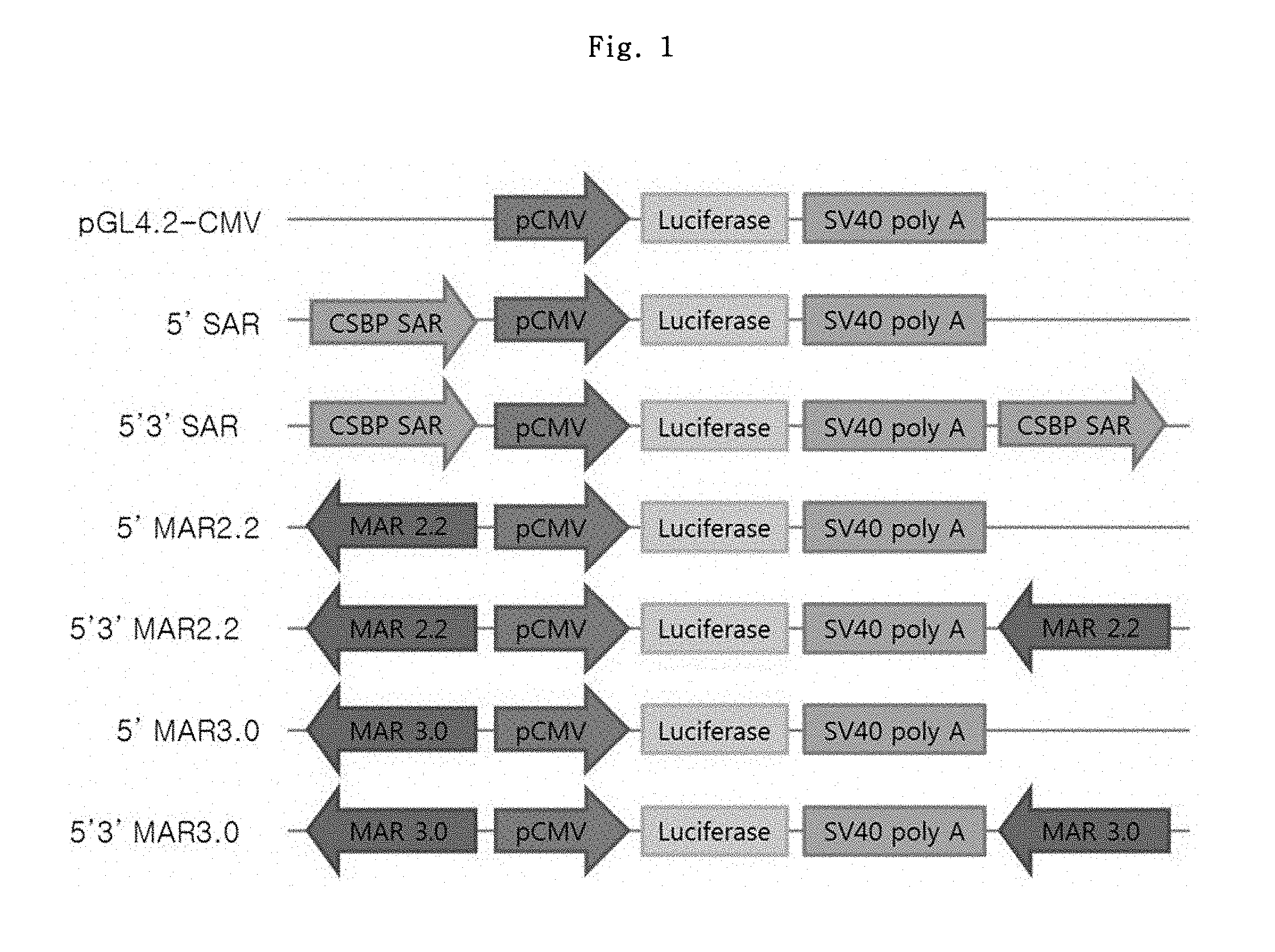
FIG. 1 shows a schematic diagram of luciferase vectors regarding MAR candidate elements.
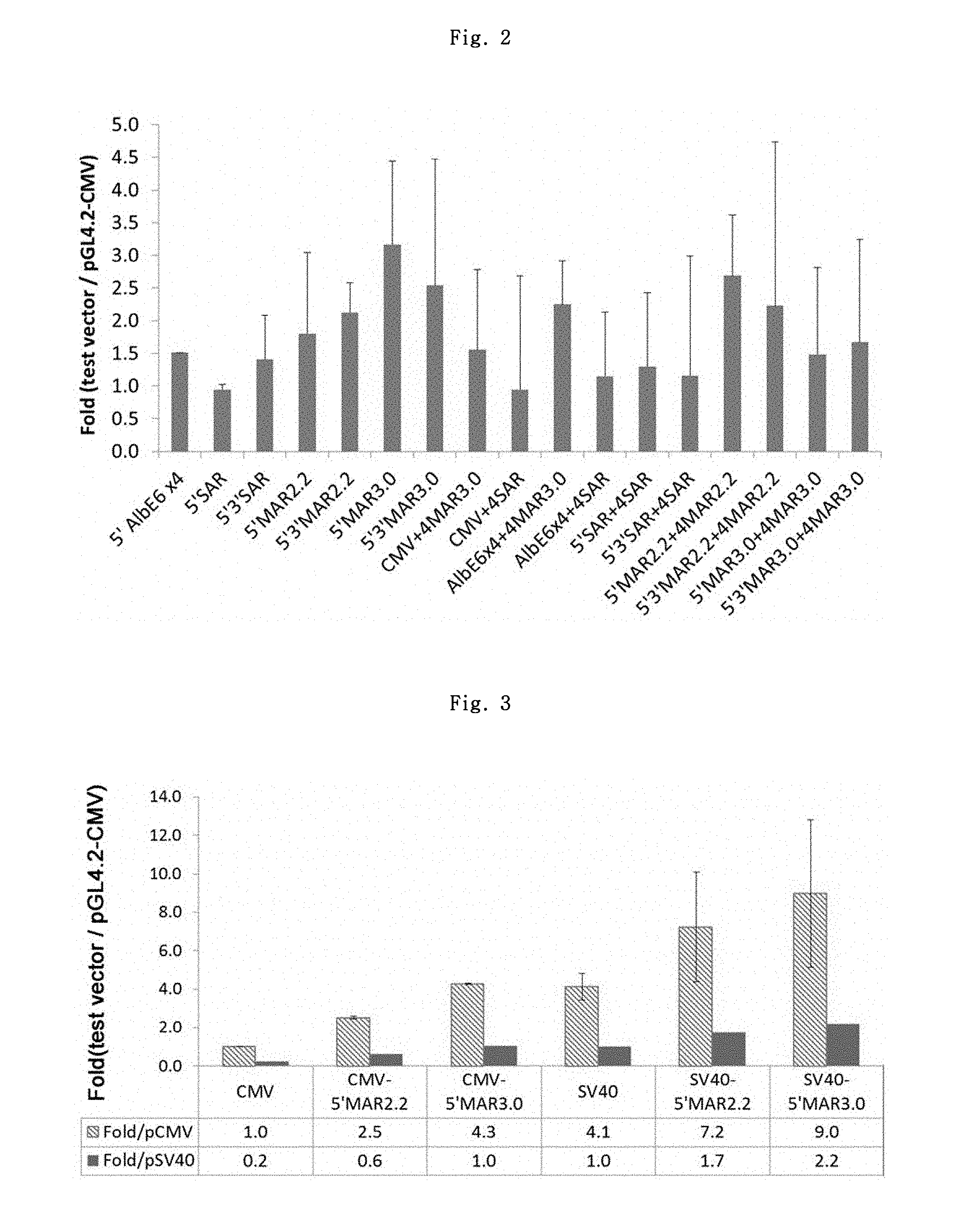
FIG. 2 shows the luciferase expression result of MAR candidate elements.
FIG. 3 shows the luciferase expression result of the comparative promoter test.
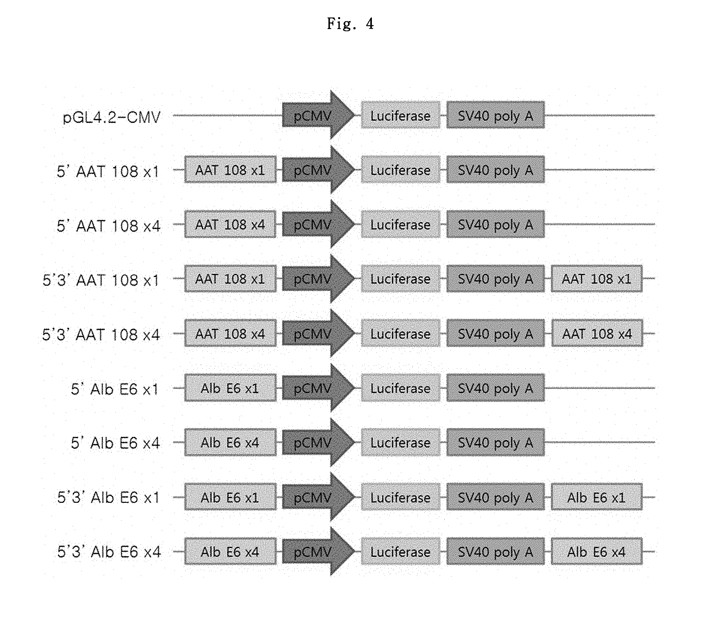
FIG. 4 shows a schematic diagram of luciferase vectors regarding LCR (locus control region) candidate elements.
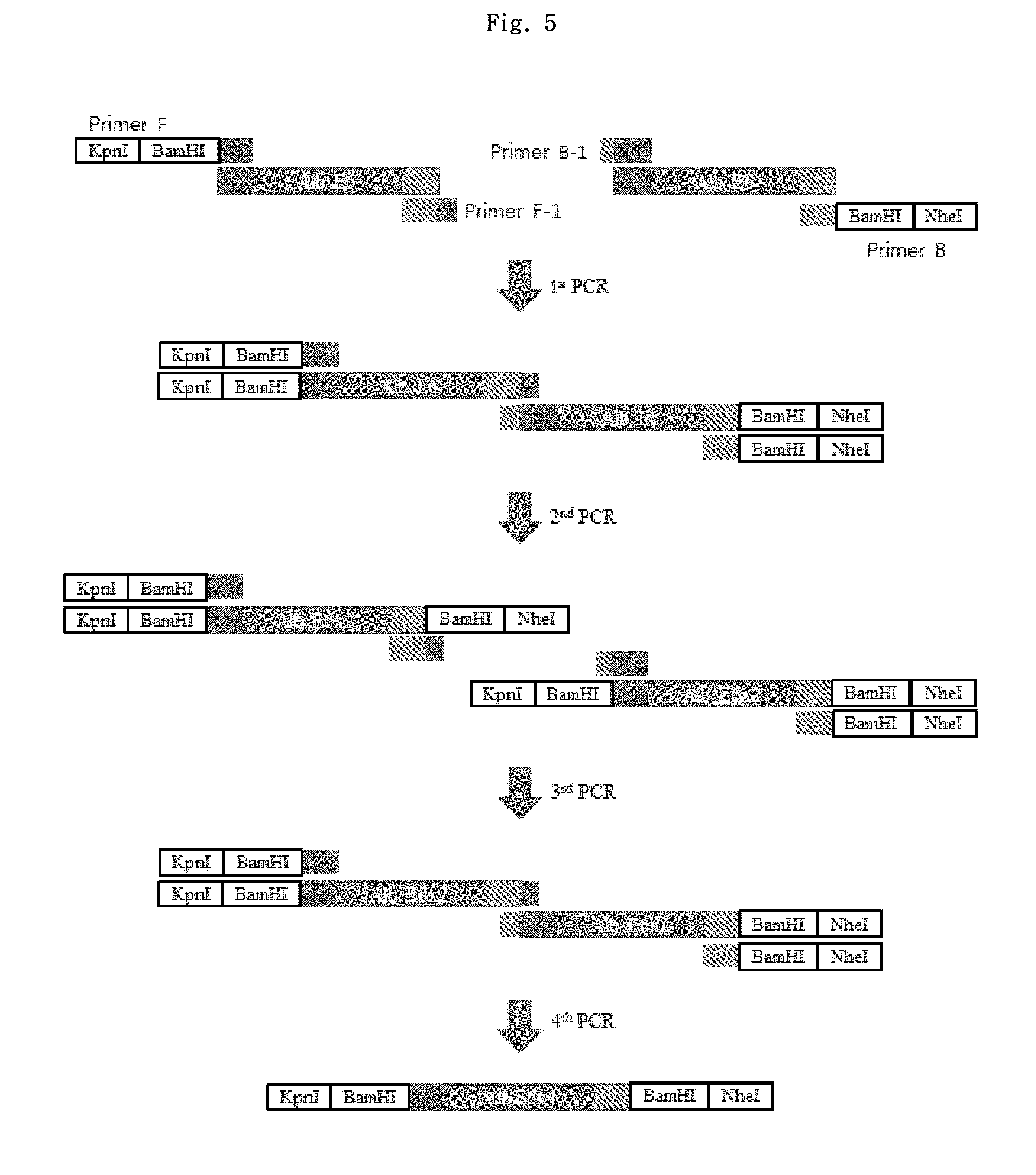
FIG. 5 shows a PCR strategy for preparing Alb E64.times.4 LCR element.
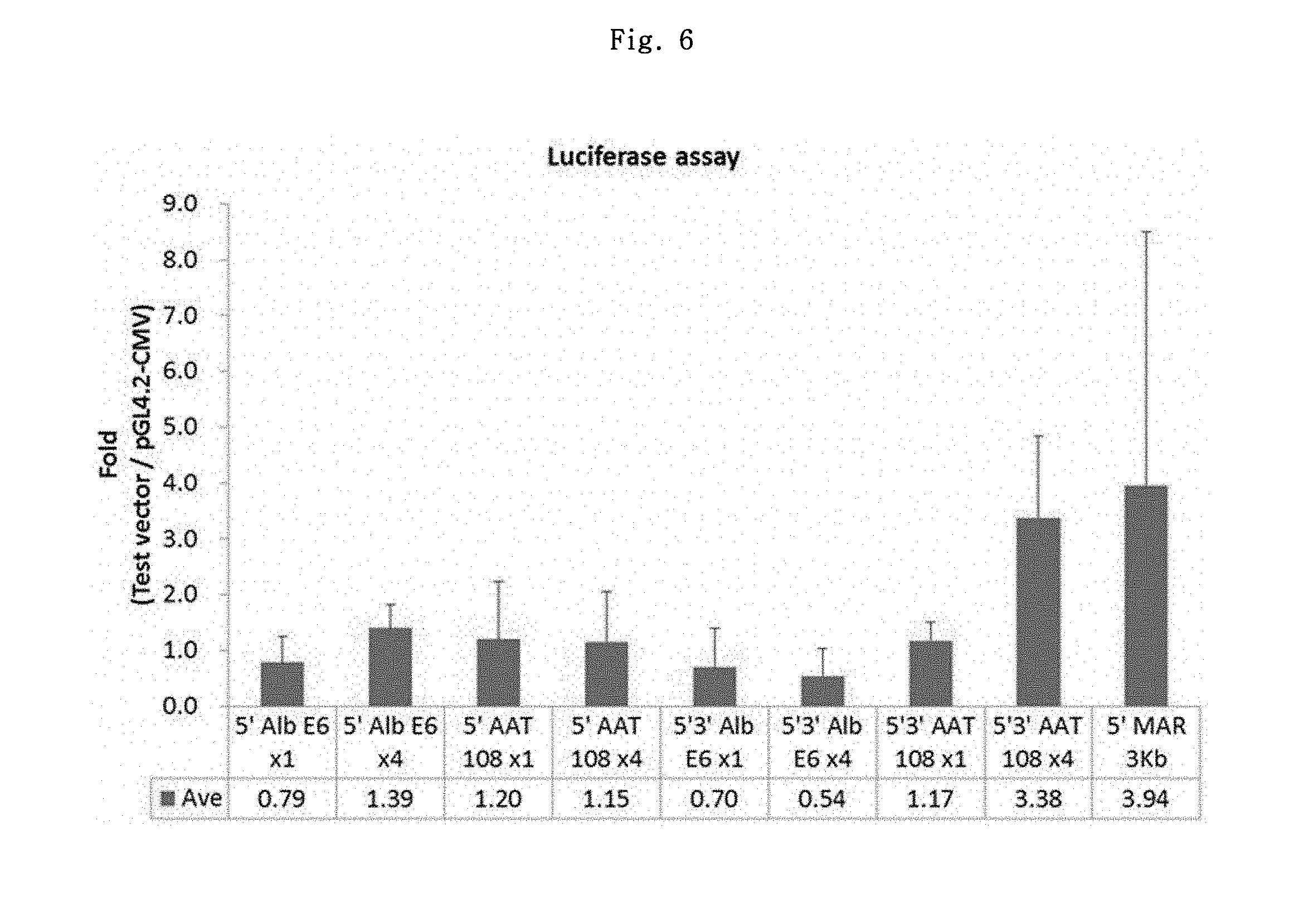
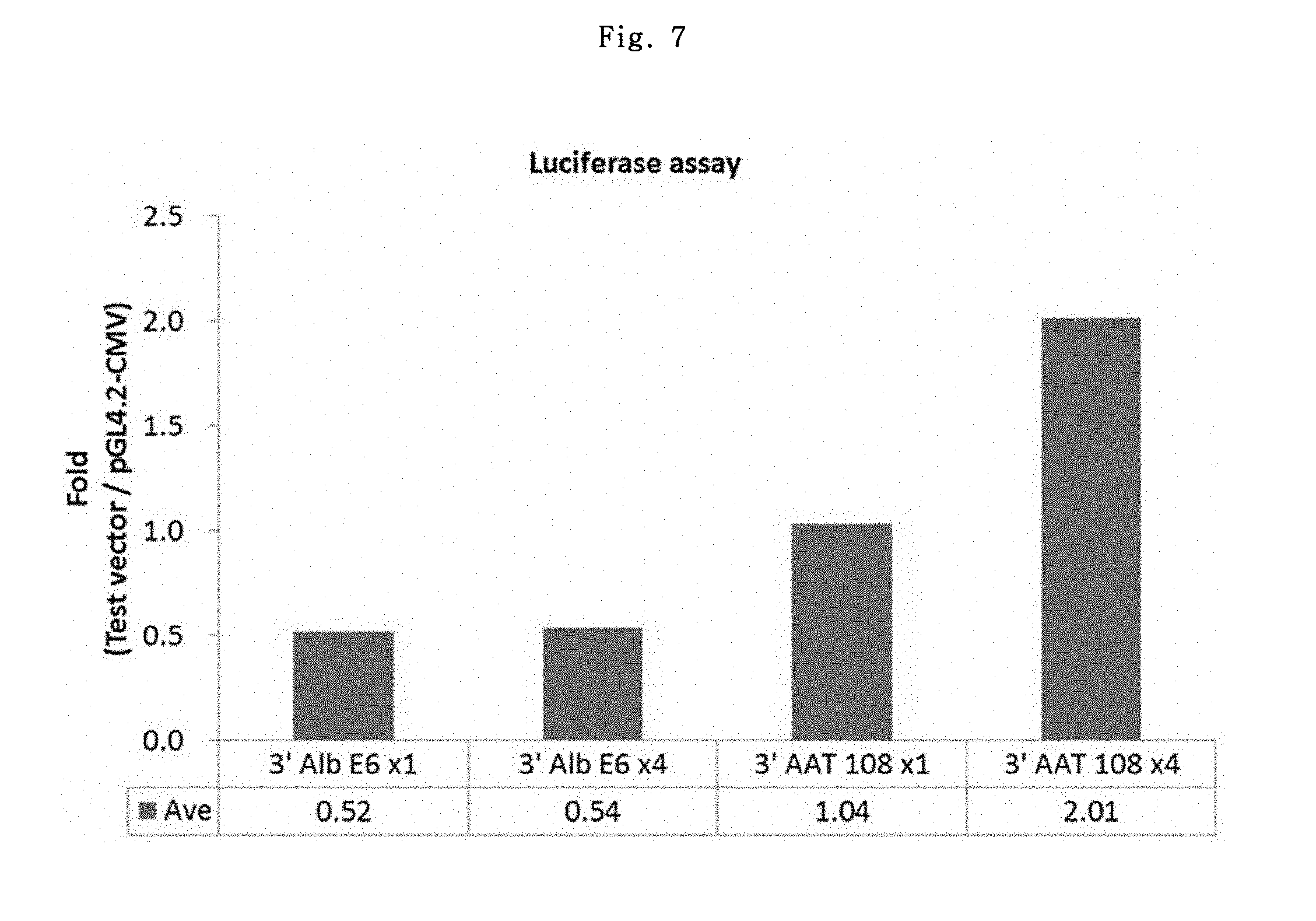
FIGS. 6 and 7 show the luciferase expression results of LCR candidate elements.
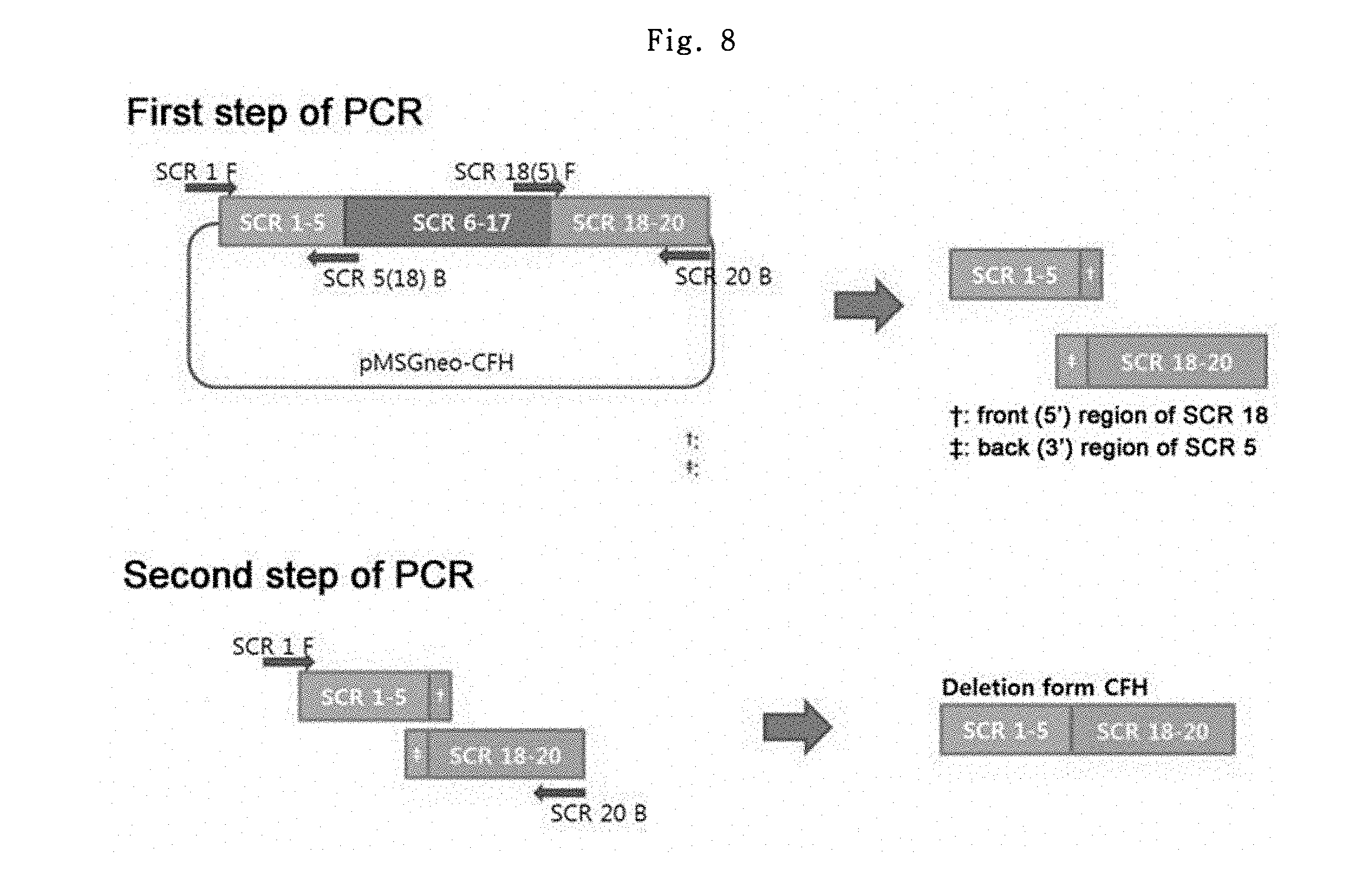
FIG. 8 shows the preparation process of dCFH (a deletion form of human complement factor H).
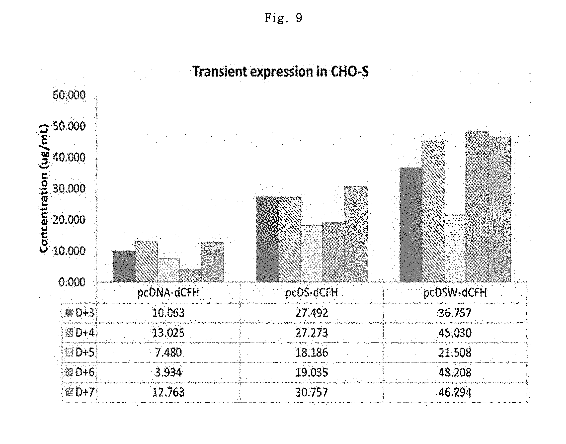
FIG. 9 shows the expression levels of dCFH in CHO--S cells.
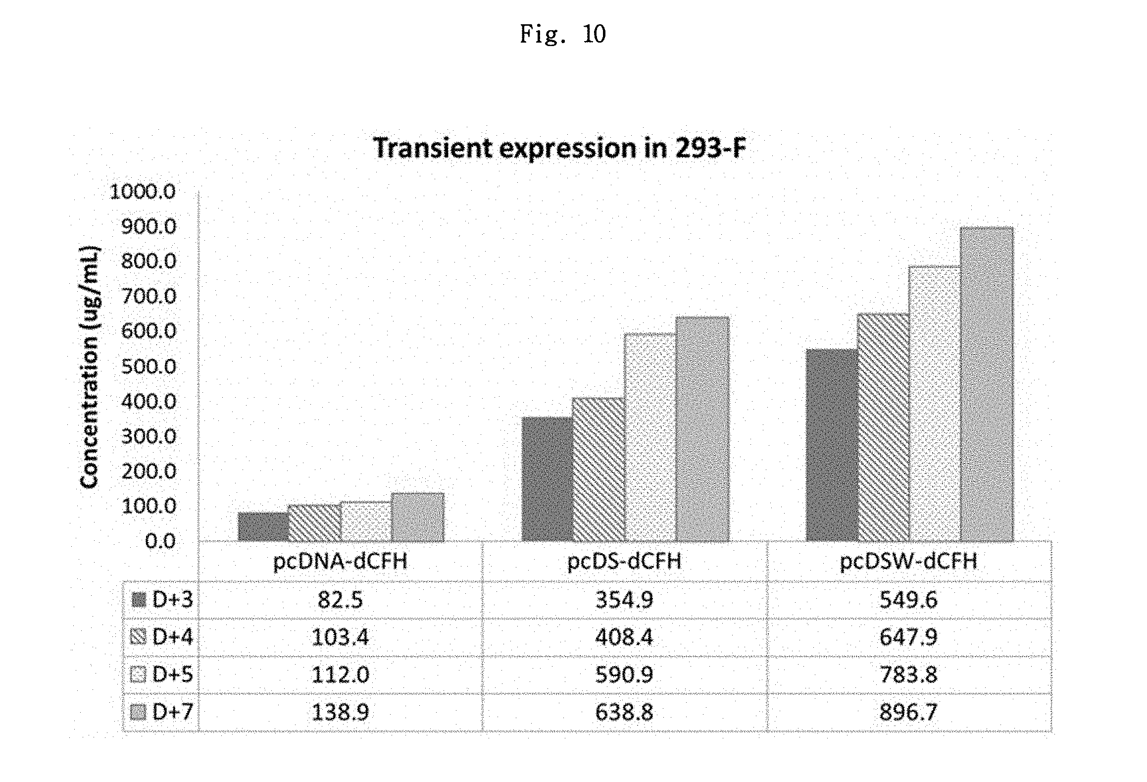
FIG. 10 shows the expression levels of dCFH in 293-F cells.
FIG. 11 shows the expression levels of dCFH in CHO-DG44 stable cells.
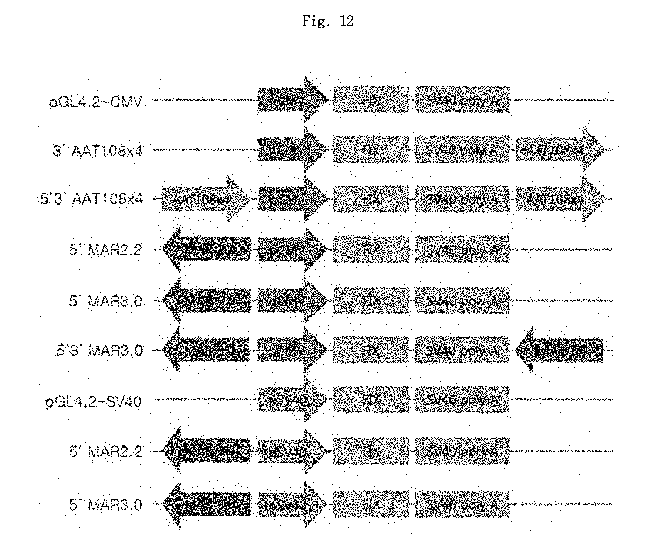
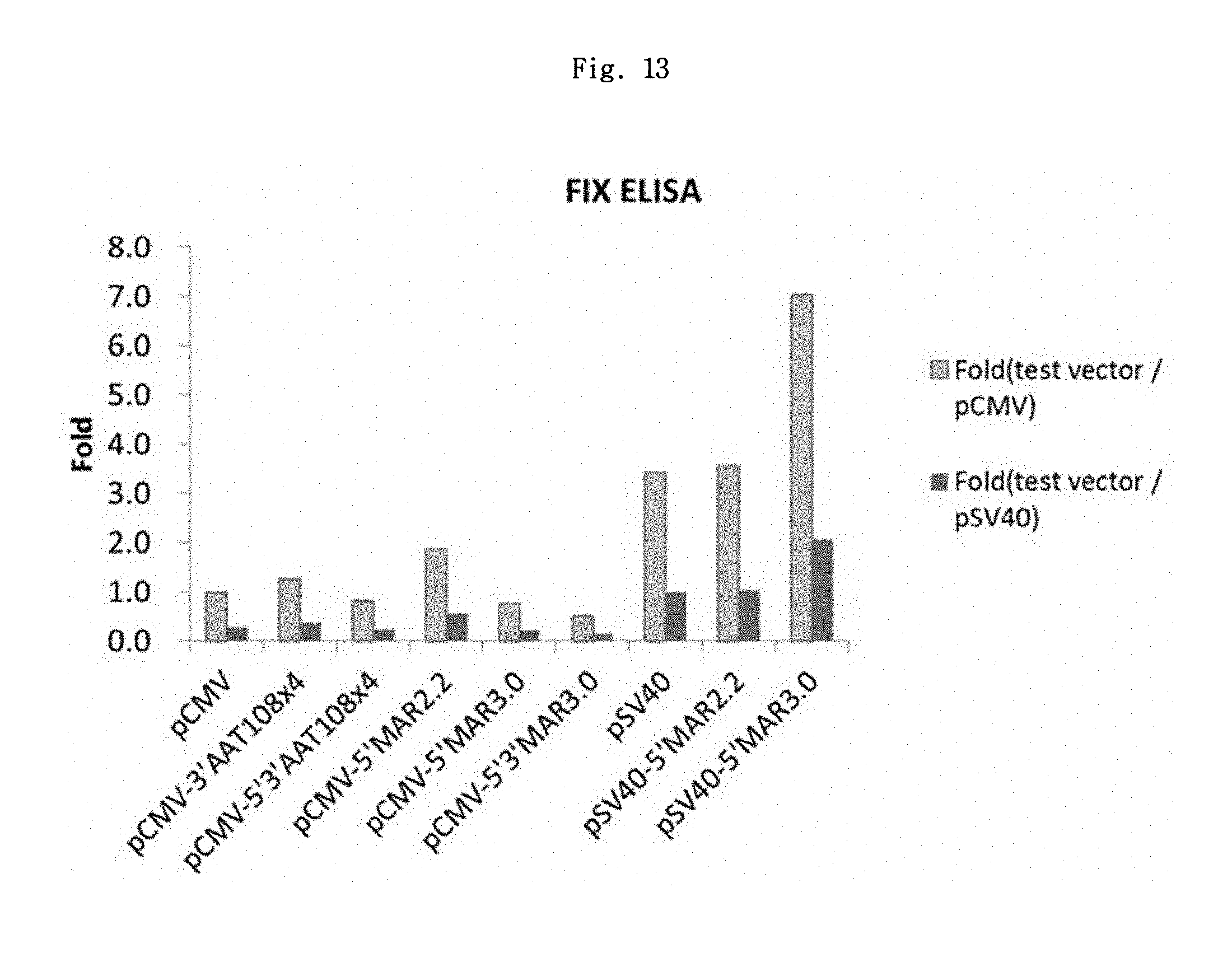
FIGS. 12 and 13 show a schematic diagram of FIX expression vector set 1 and its expression result, respectively.
FIGS. 14 and 15 show a schematic diagram of FIX expression vector set 2 and its expression result, respectively.
FIG. 16 shows a restriction enzyme map of puromycin expression system.
FIG. 17 shows the structures of a pair of novel vectors, pMS-P-MCS and pMS-D-MC.
FIGS. 18A and 18B show the structures of a first pair of novel vectors ("novel vector pair I") for ER2 antibody expression (pMS-P-ER2LC and pMS-D-ER2HC), and a pair of MAR vectors (pMSGneo-ER2LC and pMSGneo-ER2HC), respectively.
FIG. 19 shows the structures of a second pair of novel vectors ("novel vector pair II") for ER2 antibody expression (pMSI-P-ER2LC and pMSI-D-ER2HC).
FIGS. 20A and 20B show the structures of a pair of promoter vectors (pS) (pS-P-ER2LC and pS-D-ER2HC), and a pair of intron vectors (pSI-P-ER2LC and pSI-D-ER2HC), respectively.
FIGS. 21A and 21B show the ER2 antibody expression levels in the CHO adherent cells CHO-K1 and DG44, respectively.
DETAILED DESCRIPTION OF THE PRESENT INVENTION
In the present invention, to prepare a stable expression vector with improved production efficiency for the expression of an exogenous gene in CHO (Chinese Hamster Ovary) cells and the like, elements which can increase the gene expression level are identified among various MAR elements, scaffold attachment region (SAR) elements, locus control regions (LCRs), and intron elements.
Therefore, the present invention provides an expression vector comprising an operably linked simian virus 40 (SV40) promoter, a scaffold attachment region (SAR) or matrix attachment region (MAR) element, and a chimeric intron.
As used herein, the term "promoter" refers to an untranslated nucleic acid sequence upstream of a coding region, which comprises a binding site for a polymerase and which has a transcription initiation activity to mRNA for downstream genes. Specifically, a "promoter" comprises a TATA box or a TATA-like region which serves to initiate transcription by a RNA polymerase; but is not limited to the regions around it; and may comprise such regions that are necessary for the association with proteins other than the RNA polymerase in order to regulate the expression.
In the present invention, a promoter may include a SV40 promoter or its variant, preferably a SV40 promoter represented by SEQ ID NO: 12. A promoter variant refers to a variant in which a certain portion of the promoter sequence is modified by addition, deletion or substitution, in order to increase the gene expression.
According to an embodiment of the present invention, an expression vector of the present invention, which uses a SV40 promoter, shows about 4-fold expression increment as compared to a CMV promoter (see Example 1-1).
In addition, such promoter is operably linked to a target gene, which is a exogenous gene; and as used herein, "operably linked" means that a nucleic acid sequence regulating target gene expression and the nucleic acid sequence encoding a target protein are functionally linked such that they can perform a general function. The operable linkage to a recombinant vector can be implemented using a recombinant DNA technique known in the art; and site-specific DNA cleavage and linkage may be conducted using an enzyme commonly known in the art.
In the present invention, for the gene expression-increasing elements, a SAR or MAR element may be included. The SAR or MAR element may be included to constitute an expression vector such that such element(s) may be present at the 5' end; the 3'end; or both of the 5' and 3' ends of the promoter.
As used herein, a "MAR element" refers to a DNA sequence which temporarily attaches a transcriptionally active DNA loop domain to the nuclear matrix (Pienta et al., (1991) Crit. Rev. Eukaryotic Gene Expres., 1:355-385), and examples of the MAR sequences are known in the art.
In the present invention, in order to select gene expression-increasing elements, dual (5' and 5'+3') and cis+trans related tests were performed using luciferase assay with regard to human beta-globin MAR (Yu, J., Bock, J. H., Slightom, J. L. and Villeponteau, B., Gene 139(2), 139-145(1994), Genbank No. L22754) and human CSP-B gene flanking SAR (Hanson, R. D. and Ley, T. J., Blood, 79(3), 610-618(1992), Genbank No. M62716); and as a result, certain desired elements were selected (see Example 1-1).
The above MAR or SAR element may be a human CSP-B SAR which is represented by SEQ ID NO: 39; MAR2.2, represented by SEQ ID NO: 40 or MAR3.0 represented by SEQ ID NO: 41 isolated from human beta-globin MAR. The SAR or MAR element may be included such that the element(s) may be present at the 5' end; the 3' end; or both of the 5' and 3' ends of the promoter.
According to one embodiment of the present invention, the element may be one designated as 5' MAR2.2, 5' MAR3.0, 3' MAR2.2, 3' MAR3.0, 5'3' MAR2.2 or 5'3' MAR3.0.
An expression vector according to the present invention may comprise a chimeric intron to increase the gene expression.
Such chimeric intron may be represented by SEQ ID NO: 25 (Backliwal, G, Hildinger, M., Chenuet, S., Wulhfard, S., De Jesus, M., Wurm, F. M., Nucleic Acids Res., 36(15), e96 (2008), Genbank No. JQ795930), but the present invention is not limited thereto. In the present invention, the effect on increment of gene expression was confirmed through luciferase assay (see Example 1-3).
According to the present invention, an expression vector comprising both the MAR element and the chimeric intron element shows a synergistic effect on productivity, leading to the increase in the gene expression level (see Example 3-3 and Table 9).
According to one embodiment of the present invention, the novel vector pair II (pMSI-P-ER2LC and pMSI-D-ER2HC) which comprise a MAR element, a SV promoter and a chimeric intron show excellent gene expression-increasing effect as compared to a conventional promoter vectors, a conventional intron vector and the novel vector pair I (pMS-P-D-ER2LC and pMS-ER2HC) which do not contain a chimeric intron.
An expression vector according to the present invention may further comprise a LCR element.
In the present invention, in order to identify gene expression-increasing elements, multimer (.times.1 and .times.4), and dual (5', 3' and 5'+3') related tests were performed using luciferase assay with regard to AAT108 (.alpha.-1-antitrypsin precursor, 120 bp, SEQ ID NO: 13) and Alb E6 (serum albumin preproprotein, 81 bp, SEQ ID NO: 14), which previously showed Factor IX expression-increasing effect as disclosed in International Publication No. WO 2009/102085, and as a result, preferred LCR elements were selected (see Example 1-2).
Preferred LCR element in the present invention may be AAT108.times.4 represented by SEQ ID NO: 15, which may be comprised such that it is present only at the 3' end (3' AAT 108.times.4); or both of the 5' and 3' ends of the vector (5'3' AAT 108.times.4). Also, the LCR element may be Alb E6.times.4 which is represented by SEQ ID NO: 16.
Also, a target gene whose expression is regulated by a promoter may be inserted into an expression vector of the present invention.
The target gene is a gene coding for an exogenous product to be expressed, and may be a gene encoding any type of protein which can be expressed as a recombinant protein. Such target gene comprises a "multiple cloning site (MCS)", which is a nucleic acid sequence having restriction enzyme cleavage sites, so as to be inserted into the vector.
The above target gene may be one of the blood coagulation-related genes and various antibody genes; and may be selected, for example, from the group consisting of factor VII (FVII) gene, factor VIII (FVIII) gene, factor IX (FIX) gene, an epidermal growth factor receptor family antibody gene, insulin gene, interleukin gene, tumor necrosis factor gene, interferon gene, colony stimulating factor gene, chemokine gene, erythropoietin gene, .alpha.-fetoprotein gene, a-glucosidase gene, .alpha.1-antitrypsin gene, antithrombin gene, lipoprotein gene, celluloplasmin gene, fibrinogen gene, glucocerebrosidase gene, haptoglobin gene, plasminogen gene, prothrombin gene, and transferrin gene, but is not limited thereto. According to one embodiment of the present invention, the target gene may be FIX (factor IX) gene or epidermal growth factor receptor ER2 antibody gene.
The expression vector according to the present invention may comprise any conventionally known transcription terminator. Preferably, it may be connected to polyA. For example, it may comprise SV40 late polyadenylation signal (polyA).
According to one embodiment of the present invention, an expression vector of the present invention has the cleavage map of pMSI-P-ER2LC or pMSI-D-ER2HC shown in FIG. 19. FIG. 19 schematically illustrates an expression vector comprising gene expression-increasing elements such as a MAR element, a chimeric intron element, a SV40 promoter, poly A, etc.
Meanwhile, the present invention provides a cell transformed by the above expression vector.
As used herein, the cell may be an animal cell generally known, and may preferably be selected from the group consisting of CHO-K1 cell, CHO-DG44 cell, CHO--S cell, CHO DUKX-B11 cell, COS3 cell, COST cell, COS1 cell, NSO cell, HeLa cell, Vero cell, PER-C6 cell, HepG2 cell, and BHK cell, but is not limited thereto.
Also, the present invention provides a method for producing a target protein using the above expression vector. More particularly, the present invention provides a method for producing a target protein in a large amount comprising: culturing cells transformed by the above expression vector to express a target protein, and then recovering the target protein.
Such method may comprise the steps of:
(1) inserting a target gene whose expression is regulated by a promoter into the expression vector;
(2) transforming a cell with the expression vector in which the target gene was inserted; and
(3) culturing the cell transformed by the expression vector to express a target protein, and then recovering the target protein.
The method for inserting a target gene, transforming a cell, and culturing the cell to express a target protein, and recovering the target protein may be carried out by using a technique known in the art.
The present invention will be described in more detail with the following Examples. The following Examples are intended to further illustrate the present invention without limiting its scope.
Example 1: Selection of Elements
1-1: MAR Element Selection
In order to improve vectors, MAR of the human .beta.-globin (.beta.-globin MAR, 3 kb, 2.2 kb) and a human CSP-B gene flanking SAR (1.2 kb) were selected as candidates. Also, in order to perform a reporter gene assay which allows indirect evaluation of the protein expression, luciferase assay was chosen considering the convenience of the examination.
It was planned to access the effects of vectors for analysis by comparing the differences in fluorescence values between them and a control vector not containing the MAR element, the vectors for analysis were prepared accordingly. To assess the effects in a stable situation, pGL4.20[luc2/Puro] Firefly luciferase vector (Promega Inc.) was selected, which contains puromycin-resistance gene, a fast and strong cell selection marker.
First, PCR was carried out by using pcDNA3.1(+) vector (Invitrogen, V790-20) as a template, the primer set of SEQ ID NOs: 1 and 2, and a DNA polymerase (Ex-Taq, Takara), to obtain DNA fragments comprising a CMV promoter. Then, the template was denatured at 94.degree. C. for 5 min to separate DNA strands, and the DNA molecules were amplified by repeating a process of denaturing at 94.degree. C. for 30 sec, annealing at 56.degree. C. for 30 sec, and elongation at 72.degree. C. for 1 minute for 30 cycles, followed by additional elongation at 72.degree. C. for 5 min. The DNA fragments amplified by PCR were purified by gel extraction and inserted into pCR2.1-TOPO plasmid vector (Invitrogen, K4500-01). The presence of CMV promoter represented by SEQ ID NO: 11 was verified through a cleavage mapping using restriction enzymes and base sequence analysis, and the CMV promoter was inserted into the NheI/XhoI site in the multiple cloning site of pGL4.20[luc2/Puro] vector. The resulting vector was designated as pGL4.2-CMV and used as a control vector.
Next, using the primer set for SAR (SEQ ID NOs: 3 and 4), the primer set for MAR2.2 (SEQ ID NOs: 5 and 6) and the primer set for MAR3.0 (SEQ ID NOs: 7 and 8), the SAR, MAR2.2 and MAR3.0 were respectively inserted into pCR2.1-TOPO plasmid vectors by the same method as above. The prepared vectors were designated as pCR2.1-SAR, pCR2.1-MAR2.2 and pCR2.1-MAR3.0.
The pMSG and 5'B1-P1 vectors (see Korea Patent No. 408844) were used as the PCR templates for MAR and SAR, respectively. Through a cleavage mapping using restriction enzymes and base sequence analysis, the presence of the SAR, MAR2.2 and MAR3.0 respectively represented by SEQ ID NOs: 39, 40 and 41 were verified. In the subsequent cloning procedure, all inserts were used after cleavage with restriction enzymes and purification through gel extraction; and the vectors were used after cleavage with restriction enzymes and treatment with shrimp alkaline phosphatase.
The insert prepared by cutting pCR2.1-SAR vectors with BamHI was inserted into pGL4.2-CMV vector prepared by cutting with the same restriction enzyme. Through a cleavage mapping using restriction enzymes, the desired SAR DNA fragment was verified to be present in the forward direction, and the resulting vector was designated as pGL4.2-CMV-3'SAR. Next, the inserts prepared by cutting pCR2.1-SAR vector with KpnI and NheI were respectively inserted into pGL4.2-CMV-3'SAR vector and pGL4.2-CMV vector which were cut with the same restriction enzymes. Through a cleavage mapping using restriction enzymes, the presence of the target DNA fragments in each plasmid were verified, and the resulting plasmids were designated as pGL4.2-CMV-5'3'SAR and pGL4.2-CMV-5'SAR, respectively.
To prepare the vectors comprising MAR2.2 and MAR3.0, the inserts prepared by cutting pCR2.1-MAR2.2 and pCR2.1-MAR3.0 with KpnI and NheI, respectively, were inserted into pGL4.2-CMV vectors prepared by cutting with the same restriction enzymes, thereby preparing pGL4.2-CMV-5'MAR2.2 and pGL4.2-CMV-5'MAR3.0 vectors first; and also, the inserts prepared by cutting pCR2.1-MAR2.2 and pCR2.1-MAR3.0 with BamHI were respectively inserted into pGL4.2-CMV-5'MAR2.2 and pGL4.2-CMV-5'MAR3.0 vectors which were prepared by cutting with the same restriction enzyme, to prepare pGL4.2-CMV-5'3'MAR2.2 and pGL4.2-CMV-5'3'MAR3.0 vectors. The MAR element was inserted in a reverse direction. The schematic picture of the prepared luciferase vectors is shown in FIG. 1.
The luciferase expression levels of the vectors with the MAR candidate elements were measured using the adherent cell line CHO-K1. A set of vectors for evaluating Cis+Trans effect were prepared by additionally supplying the above prepared vectors to have 4-fold moles of SAR or MAR element which were also used for the transfection.
Using lipofectamine 2000 (Invitrogen, 11668027), CHO-K1 cell line (ATCC, CCL-61) was transfected with 1.0 .mu.g of the above firefly luciferase vector, and after 48 hrs, cultured with passages in DMEM medium (Lonza, 112-604F) containing puromycin (50 .mu.g/mL) The stable cells were established through the selection period of about 2-4 weeks. The test was carried out in total of 3 sets, and the firefly luciferase analysis was conducted by One-glo luciferase analysis system (Promega, E6110) in which the values were corrected for the number of cells.
Firefly luciferase analysis method is as follows. First, the wells in which cells were being cultured were removed of the culture media, and washed by 1 mL each of 1.times.PBS. The transfected cells were added with Trypsin-EDTA at 100 .mu.L/well, and allowed to react for 1 min at 37.degree. C., to detach the cells from the bottom. Each well was added with 1 mL of culture medium. After counting the cell numbers, the transformed cells were dispensed into 1.5 mL tubes respectively after dilution to 1.0.times.10.sup.5 cells/mL (based on viable cell counts) with culture medium. To a 96-well white plate, cells were placed in 100 .mu.L/well for each tested DNA. The One-glo luciferase analysis reagent, which had been prepared in advance, was added to the wells in 100 .mu.L/well, and allowed to react for 3 min at room temperature. Using a photometer (EG&G Berthold, Microplate luminometer LB 96V), the firefly RLU (relative light units) was measured. The measurement results regarding the test vectors as compared to pGL4.2-CMV are shown in FIG. 2 and Table 1.
As a result, 5' MAR element showed the best effect, which showed 1.6 to 5.3-fold increase in effect (average) as compared to pGL4.2-CMV control vector. Further, the differences in effects between the single and dual MARs or between Cis and Cis+Trans MARs were not found. As such, the test result values of the candidate elements showed no significant differences among them but showed great variations, and thus, the two elements, 5'MAR2.2 and 5'MAR3.0, were selected primarily.
TABLE-US-00001 TABLE 1 Fold (Test Vector/pGL4.2-CMV) Vector 1st 2nd 3rd Ave. S.D. 5'Alb E6 x4 1.43 1.56 1.54 1.51 0.07 5'SAR 1.71 0.45 0.67 0.94 0.68 5'3'SAR 0.59 0.79 2.84 1.41 1.24 5'MAR2.2 1.30 2.10 2.01 1.80 0.44 5'3'MAR2.2 1.69 1.13 3.57 2.13 1.28 5'MAR3.0 2.64 1.55 5.31 3.17 1.93 5'3'MAR3.0 2.37 1.41 3.84 2.54 1.22 CMV + 4MAR3.0 1.03 0.14 3.49 1.55 1.74 CMV + 4SAR 0.54 0.58 1.71 0.94 0.66 Alb E6 x4 + 4MAR3.0 1.18 3.12 2.47 2.25 0.99 Alb E6 x4 + 4SAR 0.68 0.32 2.45 1.15 1.14 5'SAR + 4SAR 0.40 0.07 3.41 1.29 1.84 5'3'SAR + 4SAR 0.94 0.35 2.17 1.15 0.93 5'MAR2.2 + 4MAR2.2 2.41 0.33 5.32 2.69 2.51 5'3'MAR2.2 + 4MAR2.2 2.01 1.03 3.66 2.23 1.33 5'MAR3.0 + 4MAR3.0 1.10 0.14 3.21 1.48 1.57 5'3'MAR3.0 + 4MAR3.0 1.46 0.60 2.96 1.67 1.19
The effect of the MAR element was not good enough as expected, and thus the MAR effects regarding CMV and SV40 promoters were assessed. Specifically, PCR was conducted using pGL4.20 vector as a template and using the primer set of SEQ ID NOs: 9 and 10, to obtain DNA fragments containing an SV40 promoter. The DNA fragments amplified by PCR were purified by gel extraction and inserted into pCR2.1-TOPO plasmid vector. The presence of SV40 promoter represented by SEQ ID NO: 12 was verified through a cleavage mapping using restriction enzymes and DNA sequence analysis. And then pGL4.2-CMV, pGL4.2-CMV-5'MAR2.2 and pGL4.2-CMV-5'MAR3.0 vectors were treated with NheI and XhoI to remove the CMV promoter, and the SV40 promoter treated with the same restriction enzymes was inserted thereto, to prepare pGL4.2-SV40, pGL4.2-SV40-5'MAR2.2 and pGL4.2-SV40-5'MAR3.0 vectors.
Luciferase expression level in the adherent cell line CHO-K1 was measured by the same method as in MAR candidate element test. The stable cells were established through a selection period of about 2 weeks. The test was carried out in a total of 2 sets, and the firefly fluorescence values were measured in the same manner as described above.
As shown in FIG. 3, SV40 showed about 4-fold increase in effect as compared to CMV promoter. Among them, pGL4.2-SV40-5'MAR3.0 vector showed the best result, which showed 9-fold and 2.2-fold gene expression-increasing effects as compared to CMV control group and SV40 control group, respectively.
1-2: LCR Element Selection
Among the LCR elements disclosed in International Patent Publication No. WO 2009/102085, AAT 108 (.alpha.-1-antitrypsin precursor, 120 bp, SEQ ID NO: 13) and Alb E6 (serum albumin preproprotein, 81 bp, SEQ ID NO: 14), which showed Factor IX expression-increasing effects in vivo, were selected to evaluate the effects in vitro. AAT 108 or Alb E6 was added to the 5' region, the 3' region of the SV40 late poly (A) signal, and both the 5' and 3' regions of pGL4.2-CMV control vector to obtain various vectors for the test (See FIG. 4).
In addition, since the LCR elements are very short in length, the vectors in which 4 copies of the elements were multi-inserted into each site were prepared to assess the effect on the expression when the elements were multi-inserted.
Using the vectors (pCR-AAT108LCR, pCR-E6LCR) disclosed in International Patent Publication No. WO 2009-102085, the monomer of each element was obtained by conducting PCR one-time; and Alb E6.times.4 multimer was prepared by conducting the recombinant PCR for 4 times.
First, in order to obtain a monomer of each element, the primer set of SEQ ID NOs: 17 and 18 and the primer set of SEQ ID NOs: 19 and 20 were used for Alb E6.times.1 and AAT108.times.1, respectively. Then, the template was denatured at 94.degree. C. for 2 min to separate DNA strands, and the DNA molecules were amplified by repeating a process of denaturing at 94.degree. C. for 15 sec, annealing at 55.degree. C. for 15 sec, and elongation at 72.degree. C. for 1 min for 30 cycles, followed by additional elongation at 72.degree. C. for 5 min. The DNA fragments amplified by PCR were purified by gel extraction and inserted into pCR2.1-TOPO plasmid vector. The presence of AAT108 and A1bE6 gene represented by SEQ ID NOs: 13 and 14, respectively, were verified through a cleavage mapping using restriction enzymes and DNA sequence analysis.
On the other hand, Alb E6.times.4 multimer was prepared by conducting the recombinant PCR for 4 times, as shown in FIG. 5. The PCR was conducted by the same method as above except that the annealing time was extended to 30 sec. Through a DNA sequence analysis, the presence of Alb E6.times.4 gene represented by SEQ ID NO: 16 was verified. AAT 108.times.4 multimer (SEQ ID NO: 15) was synthesized at Genscript Inc.
To insert an LCR element to the 5' region of the promoter in pGL4.2-CMV vector, vectors and the PCR product were cut with KpnI and NheI, followed by ligation. The cloned product was treated with the same restriction enzymes, and it was verified whether the band with a desired size was present on a DNA gel. Through the above procedure, 5'AAT 108.times.1, 5'AAT 108.times.4, 5'Alb E6.times.1, and 5'Alb E6.times.4 vectors were obtained.
In order to insert an LCR element to the 3' region of SV40 late poly(A) signal in pGL4.2-CMV vector, pGL4.2-CMV vector and Alb E6 element were cut with BamHI restriction enzyme and AAT 108 element was cut with BglII restriction enzyme. The resultants were inserted to pGL4.2-CMV vector using compatible ends. Through the above procedure, 3'AAT 108.times.1, 3'AAT 108.times.4, 3'Alb E6.times.1 and 3'Alb E6.times.4 vectors were obtained.
To insert LCR into the 5' region of the promoter and the 3' region of SV40 late poly(A) signal in pGL4.2-CMV vector, the vectors containing an LCR element at the 3' region of SV40 late poly(A) signal and the PCR product prepared above were cut with the restriction enzymes KpnI and NheI, followed by ligation. Through the above procedure, 5'3'AAT 108.times.1, 5'3'AAT 108.times.4, 5'3'Alb E6.times.1, and 5'3'Alb E6.times.4 vectors were obtained.
To assess the effect of the LCR element, CHO-K1 adherent cell line was subjected to transfection. 24 hrs prior to transfection, 500 .mu.L each of CHO-K1 cell culture being subcultured in the DMEM (LONZA, 12-604F) (w/v 10% FBS) culture medium was dispensed into a 24-well plate at 0.5.times.10.sup.5 cells/well. 800 ng of test DNA (pGL4.2-CMV, 5'AAT 108.times.1, 5'AAT 108.times.4, 5'Alb E6.times.1, 5'Alb E6.times.4, 5'3'AAT 108.times.1, 5'3'AAT 108.times.4, 5'3'Alb E6.times.1, and 5'3'Alb E6.times.4, respectively) was mixed with Opti-MEM (Gibco, 31985-088) in a 1.5 mL tube to the total volume of 50 .mu.L (based on 1 well). In other 1.5 mL tube, 2 .mu.L of lipofectamine (Invitrogen, 11668027) was mixed with Opti-MEM to the total volume of 50 .mu.L and allowed to react for 5 min at room temperature. The two solutions prepared above were mixed and allowed to react for 20 min at room temperature. The resultant was dispensed into the wells in 100 .mu.L/well. 24 hrs later, the medium in each well was refreshed and 50 .mu.g/mL puromycin (Gibco, A11138-03) as added to the wells for cell selection.
Regarding 7 sets, the stable cells were established through the selection period of about 2-4 weeks. Using the established stable cells, luciferase analysis was conducted by One-glo luciferase analysis system (Promega, E6110) by the same method as the MAR candidate element analysis. Among the results of 7 sets, the results of 3 sets which showed most similar results were chosen, and the effects of individual elements were compared. The firefly fluorescence value of each element was measured and corrected for the number of cells, which showed 0.54 to 3.94-fold increase as compared to pGL4.2-CMV vector (see FIG. 6).
As shown in FIG. 6, the element which showed the best result was the conventionally used 5'MAR 3.0 kb element, which showed 0.24 to 9.02 (3.94.+-.4.55)-fold increasing effect. Next, 5'3'AAT 108.times.4 element showed 1.79 to 4.67 (3.38.+-.1.46)-fold increasing effect.
To assess the effects of pGL4.2-CMV-3'Alb E6, pGL4.2-CMV-3'Alb E6.times.4, pGL4.2-CMV-3'AAT 108.times.1 and pGL4.2-CMV-3'AAT 108.times.4 vectors in stable expression, the same tests as above were conducted. Puromycin selection was conducted for 2 weeks and 1 set of test was conducted. The result is shown in FIG. 7.
As shown in FIG. 7, pGL4.2-CMV-3'AAT 108.times.4 vector showed about 2-fold increase as compared to pGL4.2-CMV vector. It was considered that this result might explain although indirectly, that pGL4.2-CMV-5'AAT 108.times.4 vector showed no more than 1.15-fold average increase but pGL4.2-CMV-5'3'AAT 108.times.4 vector showed 3.38-fold average increase (see FIG. 6).
From results of the luciferase analysis test under the stable condition, 2 types of effective LCR elements were identified, but they did not show significant improvement as compared to the conventionally used 5'MAR 3.0 kb. Also, considering the big variations in the fluorescence measurement test, the vectors whose effects were verified in the present test, pGL4.2-CMV-3'AAT 108.times.4 vector and pGL4.2-CMV-5'3'AAT 108.times.4 vector, were chosen and it was planned that their expression levels of recombinant proteins would be assessed directly.
1-3: Intron Element Selection
To assess the effect of a chimeric intron by the expression of the deletion form of the human complement factor H (dCFH), the dCFH gene was prepared first.
Through 2 steps of PCR, dCFH having a total of 7 SCRs (short consensus repeats) was prepared by linking the SCRs #1.about.4 (Regulatory region) to SCRs #18.about.20 (Binding region), which were selected among 20 SCRs in the CFH, excluding 13 middle SCRs. In the test to be conducted as below, Phusion High-Fidelity DNA polymerase (Thermo/F-530S) was used as a polymerase; and the template was denatured at 98.degree. C. for 30 sec to separate DNA strands, and the DNA molecules were amplified by repeating a process of denaturing at 98.degree. C. for 10 sec, annealing at 55.degree. C. for 30 sec, and elongation at 72.degree. C. for 30 sec for 30 cycles, followed by additional elongation at 72.degree. C. for 5 min.
SCR 1 F primer (SEQ ID NO: 21) contained the restriction enzyme NotI site, and SCR 20 B primer (SEQ ID NO: 24) contained the XhoI site, so that they may be used in the future cloning.
In the first step of PCR, the SCRs #1.about.5 were obtained using pMSGneo-hCFH as a template, and using SCR 1 F primer (SEQ ID NO: 21) and SCR 5(18) B primer (SEQ ID NO: 22) containing the back (3') region of SCR #5 and the front (5') region of SCR #18. Also, SCR fragments #18.about.20 were obtained using pMSGneo-hCFH as a template, and using SCR 18(5) F primer (SEQ ID NO: 23) containing the back (3') region of SCR #5 and the front (5') region of SCR #18 and SCR 20 B primer (SEQ ID NO: 24). The prepared two kinds of fragments were purified and recovered with Wizard.RTM. SV Gel and PCR Clean-Up system (Promega).
The preparation method of the above pMSGneo hCFH is as follows.
First, PCR was carried out using pCMV-SPORT6-hCFH vector (Imagene, #IRATp970B10140D) as a template, and using a primer pair (SEQ ID NOs: 47 and 48). PCR was carried out as follows: a total of 50 .mu.L of PCR reaction mixture was prepared by mixing 5 ng of pCMV-SPORT6-hCFH, 1 .mu.L of each primer (10 nmol/mL), 1 .mu.L of PCR pre-mix (AccuPower.RTM. ProFi Taq PCR PreMix, Bioneer), and 46 .mu.L of distilled water; and then the mixture was subjected to a reaction process of at 94.degree. C. for 30 sec, at 65.degree. C. for 60 sec, at 65.degree. C. for 3 minute and 20 sec, for 30 cycles. After the PCR product was electrophoresed on 0.8% agarose gel, the gene fragments encoding hCFH were obtained by gel elution.
Then, the gene fragments encoding hCFH, and pMSGneo vector (Mogam Institute, Korea), a backbone vector, were treated with NotI and XhoI, respectively; then, they were ligated using T4 ligase to obtain an expression vector of hCFH; and the expression vector thus prepared was designated as "pMSGneo-hCFH".
The second step of PCR was conducted using the 2 kinds of fragments obtained in the first step of PCR as templates, and using SCR 1 F primer (SEQ ID NO: 21) and the SCR 20 B primer (SEQ ID NO: 24). Using Phusion High-Fidelity DNA polymerase (Thermo/F-530S), the templates were denatured at 98.degree. C. for 30 sec to separate DNA strands, and the DNA molecules were amplified by repeating a process of denaturing at 98.degree. C. for 10 sec, annealing at 55.degree. C. for 30 sec, and elongation at 72.degree. C. for 45 sec for 30 cycles, followed by additional elongation at 72.degree. C. for 5 min, to obtain dCFH gene (SEQ ID NO: 46) (FIG. 8).
Next, the expression vector pcDNA-dCFH was prepared.
The pMSGneo-dCFH vector and pcDNA3.1(+) vector were cut with NotI and XhoI restriction enzymes for about 3 hr at 37.degree. C. The cut vectors were electrophoresed on the agarose gel; and the dCFH genes among the cut pMSGneo-dCFH genes, and the gene with a size of the vector among the cut pcDNA3.1(+) genes were excised from the gel. The excised genes were purified and recovered with Wizard.RTM. SV Gel and PCR Clean-Up system. The cut genes were reacted at 16.degree. C. for about 6 hrs using the DNA Ligation Kit Ver. 2.1, to prepare pcDNA-dCFH expression vector.
Then, pcDS-dCFH expression vector was prepared. The pcDNA3.1(+) vector and Y11 LC pcIW vector (See: Nucleic Acids Res., 2008 September; 36(15):e96) were cut with NheI (NEB/R0131S) and NotI restriction enzymes. The cut genes were electrophoresed on the agarose gel; and the chimeric intron genes (SEQ ID NO: 25) among the cut Y11 LC pcIW vector genes, and the cut pcDNA3.1(+) vector gene were excised from the gel. The excised genes were purified and recovered with Wizard.RTM. SV Gel and PCR Clean-Up system. The cut genes were reacted at 16.degree. C. for about 6 hrs using the DNA Ligation Kit Ver. 2.1, to prepare pcDS-MCS vector. Further, using the pcDS-MCS vector and pcDNA-dCFH vector prepared above, pcDS-dDFH vector was prepared through the method as above, in which dCFH gene was inserted into the NheI/NotI cleavage site of pcDS-MCS vector.
To secure WPRE gene (SEQ ID NO: 26), PCR was conducted using Y11 LC pcIW vector as a template. In the test to be conducted as below, Phusion High-Fidelity DNA polymerase (Thermo/F-530S) was used as a polymerase; and the template was denatured at 98.degree. C. for 30 sec to separate DNA strands, and the DNA molecules were amplified by repeating a process of denaturing at 98.degree. C. for 10 sec, annealing at 55.degree. C. for 30 sec, and elongation at 72.degree. C. for 30 sec for 30 cycles, followed by additional elongation at 72.degree. C. for 5 min. The WPRE F primer used herein (SEQ ID NO: 27) contained the restriction enzyme XhoI site, and the WPRE B primer (SEQ ID NO: 28) contained the XbaI site, so that they may be used in the future cloning. The obtained PCR products were purified and recovered with Wizard.RTM. SV Gel and PCR Clean-Up system.
The WPRE gene obtained by the above PCR and pcDS-dCFH vector were cut with XhoI and XbaI (NEB/R0145S) restriction enzymes at 37.degree. C. for about 1 hr. The cut genes were purified and recovered with Wizard.RTM. SV Gel and PCR Clean-Up system. The cut genes were reacted at 16.degree. C. for about 3 hrs using the DNA Ligation Kit Ver. 2.1, to prepare pcDSW-dCFH vector, in which WPRE gene was inserted into the XhoI/XbaI cleavage site of pcDS-dCFH vector.
In order to verify the transient expression level-increasing effect of the chimeric intron, CHO--S cell (Invitrogen, #R800-07) was prepared. The cells were thawed; seeded into 30 mL of FreeStyle CHO expression medium (Invitrogen, #12651-014) in 3.times.10.sup.5 cells/mL using Erlenmeyer scale; and kept for 2.about.3 passages so that they could grow up to 1.2.about.2.0.times.10.sup.6 cells/mL in 2.about.3 days. Then, the cells were transfected with pcDNA-dCFH vector, pcDS-dCFH vector and pcDSW-dCFH vector prepared above using Freestyle.TM. Max reagent (Invitrogen, #16447-100). After the transfection, 1 mL of the culture solution was collected once a day from day 3 to day 7. Using the collected culture solutions, the number of cells and viability were measured; and using the human complement factor H ELISA kit (Hycult biotech, #HK342), the dCFH expression levels were verified (FIG. 9).
The CHO--S cells transfected with pcDNA-dCFH vector showed the maximum expression level of about 13.025 .mu.g/mL, whereas the CHO--S cells transfected with pcDS-dCFH vector and pcDSW-dCFH vector showed the maximum expression levels of about 30.757 .mu.g/mL and 48.208 .mu.g/mL, respectively. Each vector showed about 2.4 to 3.7 times higher expression level than pcDNA-dCFH.
Second, in order to verify the effect of a chimeric intron, the prepared dCFH expression vector was transfected into FreeStyle.TM. 293-F cell (Invitrogen/R790-07). FreeStyle.TM. 293-F cell was cultured in Freestyle 293 expression medium (Gibco/12338-018). 24 hr prior to the transfection, the cells were passaged at 7.times.10.sup.5 cells/mL.
On the day of transfection, the cells were diluted in a 125 mL Erlenmeyer flask using culture medium to the total volume of 30 mL at 7.times.10.sup.6 cells/mL In a 1.5 mL tube, 30 .mu.g of plasmids for transfection (pcDNA-dCFH, pcDS-dCFH and pcDSW-dCFH) and OptiPRO.TM. SFM (Gibco/12309-019) were mixed to the total volume of 600 .mu.L. In a new 1.5 mL tube, 90 .mu.L of PEI MAX (Polyscience/24765-2) (1 .mu.g/.mu.L) and 510 .mu.L of OptiPRO.TM. SFM were mixed. The solution containing plasmids and the solution containing PEI MAX were mixed and reacted for 10 min at room temperature. While the Erlenmeyer flask containing 293-F cell was slowly turned around, the mixed solution was added thereto. The cells were cultured in a 5% CO.sub.2 suspension incubator at 37.degree. C. with 130 rpm for 7 days. The culture medium was collected in a 1.5 mL tube for 7 day from day 3, and centrifuged for 3 min at 5000 rpm to precipitate the cells. Then, the supernatant was taken and using the human complement facter H ELISA kit (Hycult biotech/HK342), dCFH expression level was measured (FIG. 10).
As shown in FIG. 10, the cells transfected with pcDNA-dCFH, pcDS-dCFH and pcDSW-dCFH showed the maximum expression levels of about 138.9 .mu.g/mL, 638.8 .mu.g/mL and 896.7 .mu.g/mL, respectively. The cells transfected with the vectors containing a chimeric intron showed about 4.6-fold increase, and the cells transfected with the vectors containing both the chimeric intron and WPRE showed about 6.5-fold increase, as compared to pcDNA-dCFH expression vector.
Lastly, in order to verify the effect on the stable expression of a chimeric intron, CHO-DG44 cell (DR L. Chasin, Columbia University) was prepared. The cells were thawed; seeded into the MEM a w/HT (Gibco, 12571-063)+10% DFBS (Biotechnics research, 7103) medium at 3.times.10.sup.6 cells/T75 flask; and kept for 2.about.3 passages so that they could grow up to the confluency of 90% in 2.about.3 days. Then, the prepared pcDNA-dCFH vector, pcDS-dCFH vector and pcDSW-dCFH vector were transfected into the cells using lipofectamine.RTM. 2000 transformation reagent (Invitrogen, #11668-027) at 5.times.10.sup.6 cells/flask. 48 hrs after the transfection, the medium was replaced with a medium supplemented with 500 .mu.g/mL of G418 for the cell selection. While monitoring the cell growth for about a week, the first passage was carried out at 80.about.90% confluency. If the cells showed about 80.about.90% confluency in 2.about.3 days after the first passaging, two more passages were carried out, and the cells were inoculated into a T75 flask at 5.times.10.sup.6 cells/flask in the same manner, and then 1 mL of the culture solution was collected once a day from day 3 to day 7. On the last day of day 7, the number of cells and viability were counted after treating the cells with 0.25% trypsin-EDTA (Invirtogen, #25200-056); and the dCFH expression level was measured using the human complement factor H ELISA kit (Hycult biotech, # HK342) (FIG. 11).
As shown in FIG. 11, CHO-DG44 cells transfected with pcDNA-dCFH vector showed the maximum expression level of about 0.054 .mu.g/mL, whereas CHO-DG44 cells transfected with pcDS-dCFH vector and pcDSW-dCFH vector showed the maximum expression levels of about 0.934 .mu.g/mL and 0.924 .mu.g/mL, respectively. Each vector showed about 17.3 to 17.1 times higher expression level than pcDNA-dCFH.
1-4: Selection of Additional Elements
Additional selection was carried out by comparing the levels of FIX (Factor IX) protein expression using the four elements which were selected at the primary selection of MAR and LCR elements. It was planned that vectors would be prepared by removing luciferase gene from the luciferase vector prepared above and inserting FIX gene thereto. For use in the novel vector preparations, a primer set (SEQ ID NOs: 29 and 30) was prepared such that the primers include XhoI, Pad and ApaI restriction enzyme sites at the front, and ApaI and FseI sties at the back of the FIX gene.
Then, PCR was carried out using pMSG-FIX vector, as a template, and the primer set (SEQ ID NOs: 29 and 30).
The preparation method of the pMSG-FIX vector is as follows. First, mRNA was extracted from human liver cells using oligo-dT column (Invitrogen, USA) for the separation of FIX gene. After preparing a primer set (SEQ ID NOs: 49 and 50) which binds with the FIX gene-specific 5' end and 3' flanking region based on the FIX gene sequence registered in the gene bank (Ref.; Genbank accession No. M11309; 1.about.2775 bp), RT-PCR was carried out using the Titan one tube RT-PCR system (Roche) to synthesize FIX cDNA with the size of about 1,383 bp (461 amino acids), which was then cloned into pGEM-T vector (Promega).
The separated FIX gene was amplified by PCR using a primer set (SEQ ID NOs: 51 and 52) including NheI and BglII, and cloned into pMSG vector (Mogam Biotechnology institute, Korea), which is animal cell expression vector. FIX gene obtained from the PCR was cleaved using Nhe I and BglII restriction enzymes (New England Bio Labs, USA), and then subjected to 1.2% agarose gel electrophoresis to separate the gene with the size of 1.4 kb. The separated FIX gene was ligated to pMSG vector which was pre-treated with NheI and BglII restriction enzymes, to prepare FIX expression vector. E. coli DH5a was transformed with the DNA mixture, in which ligation reactions were completed, using CaCl.sub.2 precipitation. The transformed E. coli was spread on an LB plate containing 50 .mu.g/mL ampicillin, and cultured over night at 37.degree. C. After culturing the colonies formed on the culture plate, plasmids were isolated using QIA filter mini-Plasmid Kit (Qiagen, Germany). The isolated plasmid DNA was confirmed to contain the FIX DNA fragments by the treatment with NheI/BglII restriction enzymes. The expression vector thus prepared was designated as "pMSG-FIX".
The DNA fragments amplified by PCR were purified by gel extraction and inserted into pCR2.1-TOPO plasmid vector. The presence of FIX gene represented by SEQ ID NO: 31 was verified through a cleavage mapping using restriction enzymes and DNA sequence analysis. To the vector in which luciferase gene was removed using XhoI and FseI restriction enzymes, FIX gene was inserted using the same restriction enzymes. The schematic picture of the FIX expression vector set 1 thus prepared is shown in FIG. 12.
FIX expression in the adherent cell line CHO-K1 was measured by the same method as luciferase expression test. In the same manner as described above, stable cells were established through a selection period of about 2 weeks. The test was carried out in a total of 4 sets, and the FIX expression level was measured by ELISA method and corrected for the number of cells.
FIX ELISA method is as follows. Culture media in the wells were collected to 1.5 mL tubes. Each well was washed with 1 mL of 1.times.PBS. 100 .mu.L/well of trypsin-EDTA was added to the wells, which were then allowed to react in a cell incubator at 37.degree. C. for one minute to detach the transfected cells from the bottom. Each well was added with 1 mL of culture medium to collect cells, and the number of cells was counted. In the case of continuous cultures, the cells were cultured with passages at 0.5.about.1.0.times.10.sup.5 cells/mL using 12-well plates, and cell selection was carried out using puromycin (50 .mu.g/mL).
The capture antibodies provided by the Matched-pair Antibody set for ELISA of human Factor IX antigen (Affinity Biological, FIX-EIA) were diluted to 1/100 using a coating buffer (50 mM carbonate buffer; pH adjusted to 9.6 after adding 1.59 g of Na.sub.2CO.sub.3 and 2.93 g of NaHCO.sub.3 to 1 L of tertiary distilled water). The diluted capture antibodies were dispensed into a 96-well ELISA plate at 100 .mu.L/well, and allowed to react for 2 hrs at room temperature. The wells were washed 3 times with PBS-Tween buffer. Then Factor FIX (Benefix, 250 .mu.g/mL) as a standard was prepared through 1/2 stepwise dilutions from 50 ng/mL to 0.781 ng/mL using a dilution buffer. The culture medium collected first was diluted to 1/10, 1/100 and 1/1000 using a dilution buffer (1% BSA in 1.times.PBS), and then added to the wells at 100 .mu.L/well and allowed to react for 1 hr and 30 min at room temperature. The wells were washed 3 times with PBS-Tween buffer. Each well was added with 100 .mu.L of TMB microwell peroxidase substrate, and allowed to react for 10 min at room temperature. Each well was then added with 100 .mu.L of 2.5M H.sub.2SO.sub.4 to stop the reaction, and absorbance was read using a microplate reader at 450 nm.
As shown in FIG. 13 and Table 2 below, the overall tendency was similar to the previous results with the luciferase. The SV40 promoter showed about 3.4 times higher increasing effect as compared to CMV promoter. The most effective was pGL4.2-SV40-5'MAR3.0 vector, which showed 7-fold and 2.1-fold expression level-increasing effect as compared to the CMV control group and SV40 control group, respectively.
TABLE-US-00002 TABLE 2 Fold (Test Vector/pCMV-FIX) #1 #2 #3 #4 Vector 1st 2nd 1st 2nd 1st 2nd 1st 2nd Ave. S.D. pCMV 1.00 1.00 1.00 1.00 1.00 1.00 1.00 1.00 1.00 0 pCMV- 1.67 1.86 0.58 0.25 2.19 3.24 3.10 5.27 1.26 1.61 3'AAT108x4 pCMV- 0.78 0.50 0.65 0.46 1.73 1.13 2.76 6.66 0.81 2.1 5'3'AAT108x4 pCMV-5'MAR2.2 0.47 0.23 2.26 3.89 1.24 0.89 2.79 3.30 1.87 1.37 pCMV-5'MAR3.0 0.89 0.80 0.51 0.41 1.69 1.28 1.58 2.00 0.74 0.58 pCMV- 0.87 0.27 0.29 0.23 0.91 1.25 1.60 2.48 0.50 0.78 5'3'MAR3.0 pSV40 3.58 3.27 1.95 2.42 7.44 12.07 7.44 8.81 3.42 3.61 pSV40-5'MAR2.2 4.21 2.69 2.14 1.74 8.86 10.88 9.67 20.82 3.56 6.45 pSV40-5'MAR3.0 5.97 5.07 5.62 6.47 13.11 12.51 18.54 27.96 7.05 8.06
In order to examine the effects of the LCR elements under the control of the SV40 promoter, the vectors were subjected to a cloning process for promoter replacement. The CMV promoter was removed from the above-prepared vectors having CMV promoter using NheI and XhoI restriction enzymes, and SV40 promoter was inserted using the same restriction enzymes. Additionally, vectors having the combination of 5'MAR and 3'AAT108.times.4, and 5'3'MAR3.0 vectors were prepared and used in the test. The schematic picture of the prepared FIX expression vector set 2 is illustrated in FIG. 14.
FIX expression in the adherent cell line CHO-K1 was measured by the same method as for FIX expression vector set 1. The stable cells were established through a selection period of about 2 weeks. The test was carried out in a total of 3 sets, and the FIX expression level was measured by ELISA method and corrected for the number of cells.
As shown in FIG. 15 and Table 3 below, there were no cases which showed increase in the expression level as compared to SV40 control group; and in the LCR candidate group with a replaced SV40 promoter, no vector showed higher expression level as compared to pSV40-5'MAR3.0. As such, through the comparison of the FIX expression levels, SV40 promoter and 5'MAR3.0 element were selected.
TABLE-US-00003 TABLE 3 Fold (Test Vector/pSV40-FIX) Vector #1 #2 #3 Ave. S.D. pSV40 1.00 1.00 1.00 1.00 0.00 pSV40-3'AAT108x4 0.23 0.82 0.60 0.49 0.30 pSV40-5'AAT108x4 0.04 0.38 0.66 0.27 0.31 pSV40-5'3'AAT108x4 0.35 0.39 1.55 0.60 0.68 pSV40-5'MAR3.03'AAT108x4 0.70 1.38 0.98 0.97 0.34 pSV40-5'MAR3.0 0.65 1.52 0.78 0.95 0.47 pSV40-5'3'MAR3.0 0.67 0.80 0.84 0.74 0.09 pMSG -- 0.28 0.41 0.28 0.09
Example 2: Preparation of the Novel Vectors
The novel vectors were prepared using the elements selected in Example 1. The pSV40-5'MAR3.0-FIX vector used in the FIX expression comparison test was treated with ApaI restriction enzyme to remove FIX gene, and allowed to self-ligate, to obtain pSV40-5'MAR3.0-MCS vector. The MCS included 4 types of restriction enzyme sites: XhoI, PacI, ApaI and FseI.
Puromycin selection region in the pSV40-5'MAR3.0-MCS vector was replaced with DHFR expression region to additionally prepare a vector having DHFR gene. Upon examining the restriction enzyme sites for the replacement, it was found that two SapI sites were present (at the front and the back); and the SapI site at the back was removed by treating with San and AflIII and preparing blunt ends using Klenow enzyme followed by self-ligation. A schematic picture of the restriction enzyme map of the puromycin expression system (pMS-P-MCS) is shown in FIG. 16.
As the required DHFR expression system, that of pDCH1P vector, which had been conventionally used in the present institute, was used. In about 2.5 kb including Hamster DHFR gene, 5' flank and 3' flank, promoter and polyA portion were confirmed based on the references (Proc Natl Acad Sci., 1991, vol 88. p 8572; and Mol. Cell. Biol., 1984, Vol. 4, p 38), and PCR primers (SEQ ID NOs: 32 and 33) were designed for reducing the size to about 1.5 kb.
PCR was carried out using pDCH1P vector as a template to obtain a DNA fragment, which was then inserted into pCR2.1-TOPO vector, and sequenced (SEQ ID NO: 34). The DHFR expression region whose gene sequence had been verified was inserted into pSV40-5'MAR 3.0-MCS vector. Vectors and inserts were each cut with SapI and San restriction enzymes, and were connected again. The cloned product was treated with the same restriction enzymes, and it was verified whether the band with a desired size was present on the DNA gel. Through the above procedure, a novel vector pair I (pMS-P-MCS and pMS-D-MCS) was prepared (FIG. 17).
In order to compare the gene expression abilities of the prepared pair of novel vectors with the conventional vectors, ER2 antibody (EGFR antibody; see Korea Patent No. 1108642) gene was cloned into each of the pair of novel vectors and the pMSGneo vector (Mogam Biotechnology Institute, Korea) which is a conventional MAR vector. For the cloning into two kinds of vectors, both light and heavy chains of ER2 antibody were added with XhoI and NheI sites at the 5' region, and PacI and XhoI sites at the 3' region, and then PCR was carried out. For the PCR, pOptiVEC-ER2HC and pcDNA3.3-ER2LC vectors were each used as a template, and the HC (SEQ ID NOs: 35 and 36) set and the LC (SEQ ID NOs: 37 and 38) set were used as the primers for the heavy chain and the light chain, respectively. The PCR product was inserted into pCR2.1-TOPO vector, and DNA sequence (SEQ ID NOs: 42 and 43) was confirmed.
Light chain and heavy chain genes of the ER2 antibody whose sequences had been verified were cloned into pMSGneo vector using NheI and XhoI restriction enzymes, and were also cloned respectively into the pair of novel vectors (pMS-P-MCS and pMS-D-MCS) using XhoI and PacI restriction enzymes.
The cloned product was treated with the same restriction enzymes, and it was verified whether the band with a desired size was present on the DNA gel. As such, 4 vectors were prepared, which comprises the novel vector pair I (pMS-P-ER2LC and pMS-D-ER2HC) for ER2 antibody expression and a pair of MAR vectors (pMSGneo-ER2LC and pMSGneo-ER2HC) (FIGS. 18A and 18B).
On the other hand, vectors were additionally prepared to verify the effect of adding a chimeric intron to the novel vector pair I prepared above. PCR was carried out using pcDS-dCFH vector as a template and using the primer set of SEQ ID NOs: 44 and 45, to obtain a DNA fragment, and the PCR product was inserted into pCR2.1-TOPO vector, and the DNA sequence of the chimeric intron (SEQ ID NO: 25) was verified. The novel vector pair I for ER2 antibody expression (pMS-P-ER2LC vector and pMS-D-ER2HC vector) was cut with XhoI, and the chimeric introns treated with the same restriction enzymes were respectively inserted thereto. Through the above procedure, the novel vector pair II (pMSI-P-ER2LC and pMSI-D-ER2HC ER2) for ER2 antibody expression was prepared (FIG. 19).
ER2 antibody expression vectors were additionally prepared to verify the element properties of the novel vector pair II.
As for the vectors with a SV40 promoter only, pS-P-MCS vector was prepared by removing FIX gene from pGL4.2-SV40-FIX vector by the same method as above, and pS-D-MCS vector was prepared by further replacing the puromycin gene with DHFR gene. Then, the light and heavy chain genes of ER2 were inserted thereto using XhoI and PacI, to prepare a pair of vectors (pS-P-ER2LC and pS-D-ER2HC) for ER2 antibody expression vector with the SV40 promoter (FIG. 20A). Chimeric intron was inserted into the prepared vectors using XhoI restriction enzyme to prepare a pair of vectors (pSI-P-ER2LC and pSI-D-ER2HC) comprising a chimeric intron (FIG. 20B).
Example 3: Evaluation of Gene Expression Ability of Vectors
The gene expression ability of the novel vectors prepared above was verified as follows.
3-1: Evaluation of the Novel Vector Pair I and the Novel Vector Pair II for ER2 Antibody Expression in the Adherent Cell Lines
First, ER2 antibody expression using the novel vector pair I and the novel vector pair II were carried out in the CHO-K1 and CHO-DG44 adherent cell lines. The expression levels were compared with a pair of commercial double vectors and a pair of MAR vectors shown in Table 4.
TABLE-US-00004 TABLE 4 Pair of commercial double vectors pcDNA3.3-ER2HC pOptiVec-ER2LC Pair of MAR vectors pMSGneo-ER2LC pMSGneo-ER2HC pDCH1p Novel vector pair I pMS-P-ER2LC pMS-D-ER2HC Novel vector pair II pMSI-P-ER2LC pMSI-D-ER2HC
24 hrs prior to transfection, CHO-K1 cells being cultured in the DMEM (w/10% FBS) culture medium were dispensed into the wells of a 12-well plate in an amount of 500 .mu.L each at 0.8.times.10.sup.5 cells/well. The CHO-DG44 cells being cultured in the MEM-.alpha. w/HT (Gibco, 12571-063) culture medium were dispensed into the wells in an amount of 500 .mu.L each at 1.5.times.10.sup.5 cells/well. 24 hrs after the dispensing, Opti-MEM and the test vector were mixed in a 1.5 mL tube to the total volume of 50 .mu.L (based on 1 well). 47 .mu.L of Opti-MEM and 3 .mu.L of lipofectamine 2000 were mixed in a new 1.5 mL tube and allowed to react for 5 min at room temperature (based on 1 well). After the reaction was completed, the two kinds of solutions prepared above were mixed and allowed to react for 20 min at room temperature. 24 hrs later, each well was removed of the transfected medium and washed with PBS. Trypsin-EDTA (1.times.) was dispensed thereto at 100 .mu.L/well and allowed to react for 1 minute at 37.degree. C. to detach the cells. The culture medium was dispensed thereto at 1000 .mu.L/well and the number of cells was counted.
CHO-K1 cells were dispensed into the wells of a 12-well plate at 1.0.times.10.sup.5 cells/well to the total volume of 1 mL; 1000 .mu.g/mL of G418 (PAA, P11-012) or 50 .mu.g/mL of puromycin were added thereto appropriately for each vector; and the cell selection was carried out. DG44 cells were dispensed into the wells of a 12-well plate at 1.5.times.10.sup.5 cells/well to the total volume of 1 mL; 750 .mu.g/mL of G418 or 20 .mu.g/mL of puromycin were added thereto appropriately for each vector; and the cell selection was carried out. The transfection was conducted in a total of 2 sets, and the samples were obtained in a total of 3 times per set throughout 3 weeks, and the ELISA test was conducted.
ER2 ELSIA method is as follows. Goat anti-human IgG (Fab-specific) (Sigma, 15260) was diluted to 5 .mu.g/mL using a coating buffer. The diluted capture antibodies were dispensed into a 96-well ELISA plate at 100 .mu.L/well, and allowed to react for 2 hrs or longer at room temperature. They were washed 3 times using 300 .mu.L of PBS-0.1% Tween 20 buffer. PBS-1% BSA was added thereto at 300 .mu.L/well, and blocking was carried out for 2 hrs at room temperature. The resultant was washed 3 times using 300 .mu.L of PBS-0.1% Tween 20 buffer. HBIG gene (2.8 mg/mL) as a standard was prepared in the concentrations of 280, 140, 70, 35, 17.5, 8.75 and 4.375 ng/mL using PBS-1% BSA. The culture solutions were diluted using PBS-1% BSA. The standard and diluted culture solutions were dispensed into a 96-well plate coated with the capture antibodies at 100 .mu.L/well in duplication, and allowed to react for 1 hour at room temperature. The wells were washed 3 times using 300 .mu.L of PBS-0.1% Tween 20 buffer. Goat anti-human IgG (Fc specific)-HRP (Sigma, A0170) was diluted to 1/100,000 using PBS-BSA. The diluted solution was dispensed into the plate at 100 .mu.L/well, and allowed to react for 1 hr at room temperature. The resultant was washed 3 times with 300 .mu.L of PBS-0.1% Tween 20 buffer. TMB microwell peroxidase substrate was dispensed at 100 .mu.L/well and allowed to react for 30 min at room temperature. 2.5 M H.sub.2SO.sub.4 was dispensed at 100 .mu.L/well to stop the reaction, and the absorbance was measured at 450 nm using a microplate reader. A standard curve was made with 4-parameter fitting, and the measured absorbance of the culture solutions was substituted for parameters, to calculate the concentrations.
As shown in Table 5, and FIGS. 21A and 21B, the expression level of ER2 Ab in the pair of commercial double vectors in CHO-K1 could not be verified. The pair of conventional MAR vectors, the novel vector pair I, and the novel vector pair II showed the average expression levels of about 9.7 ng/mL, about 14.0 ng/mL, and about 1473.2 ng/mL, respectively. The novel vector pair I and the novel vector pair II showed about 1.4 times and about 151.3 times higher expression levels than a pair of MAR vectors, respectively. In addition, the novel vector pair II showed about 105.4 times higher expression level than the novel vector pair I, which exhibited that the effect of chimeric intron was excellent.
The average expression levels of ER2 Ab of the pair of commercial double vectors, the pair of MAR vectors, the novel vector pair I and the novel vector pair II in CHO DG44 cell were 13.1 ng/mL, 3.8 ng/mL, 27.5 ng/mL and 653.0 ng/mL, respectively. The pair of commercial double vectors showed about 3.5 times higher expression level than the pair of MAR vectors. The novel vector pair I and the novel vector pair II showed about 7.3 and 173.7 times higher expression levels than the pair of MAR vectors, respectively. In addition, the novel vector pair II showed about 23.8 times higher expression level than the novel vector pair I.
TABLE-US-00005 TABLE 5 ER2 antibody expression level (ng/ml) CHO-K1 cell CHO DG44 cell Vector 1st 2nd Ave SD 1st 2nd Ave SD Pair of commercial 0.0 0.0 0.0 0.0 18.2 7.9 13.1 7.3 double vectors Pair of conventional 14.0 5.5 9.7 6.0 3.1 4.4 3.8 0.9 MAR vectors Novel vector pair I 9.4 18.6 14.0 6.5 28.8 26.1 27.5 1.9 Novel vector pair II 1490.7 1455.7 1473.2 24.7 764.9 541.0 653.0 158.3
3-2: Evaluation of the Novel Vector Pair II in the Suspended Cell Line
The novel vector pair II was evaluated in CHO--S and CD DG44-S suspension cell lines.
The CHO--S cell line (Gibco, A13696-01) was passaged in CD FortiCHO culture medium at 5.about.6.times.10.sup.5 cells/mL to a required amount, 24 hrs prior to the transfection. On the day of transfection, the number and viability (necessarily 95% or higher) of the cells were measured. The cells were dispensed into a 125 mL flask at 1.times.10.sup.6 cells/mL using CD FortiCHO medium (Gibco, A11483-01). 90 .mu.g of the plasmid DNA was mixed with OptiPro.TM. SFM to the total volume of 1.5 mL 270 .mu.L of PEI MAX (1 .mu.g/.mu.L) and 1230 .mu.L of OptiPro.TM. SFM were mixed, absolutely without vortexing at all. OptiPro.TM. SFM containing PEI MAX was put into the OptiPro.TM. SFM containing plasmid DNA, which were then gently mixed. Then, the resultant was allowed to react for 5 min at room temperature. 3 mL of the produced DNA:PEI complex was added dropwise to the CHO--S cells in the 125 mL flask prepared in advance. The cells were cultured in a CO.sub.2 shaking incubator at 37.degree. C. with stirring at 130.about.150 rpm, and the cell selection was carried out 48 hrs later.
On the other hand, CD DG44-S cell line was prepared by the following method. First, CHO DG44 adherent cell line (cultured in an .alpha.-MEM (w/)+10% cFBS medium) was passaged in 50 mL medium using a 125 mL spinner flask with an interval of 2.about.3 days, using the serum-free medium (HyQ SFM4CHO (Thermo Scientific, Hyclone)+1.times.HT Supplement), thereby adapting the cells to suspension culture. The adapted cells were subcultured with an interval of 2.about.3 days, with the replaced medium of EX-CELL.TM. CD CHO (Sigma, 14361C)/4 mM L-Glutamine/1.times.HT Supplement medium, thereby adapting the cells to the medium. If the growth of the cells was stable and the viability was maintained at 90% or higher, and the concentration of the cells reached 1.about.2.times.10.sup.6 cells/mL, the CHO DG44-S host cell line was subjected to banking, thereby securing the cell line.
24 hrs prior to the transfection, the cells were passaged in EX-CELL.TM. CD CHO (Sigma, 14361C) at 5.about.7.times.10.sup.5 cells/mL to a required amount. The cells were dispensed into a 125 mL flask in an amount of 15 mL at 2.times.10.sup.6 cells/mL using a Freestyle CHO medium. 30 .mu.g of plasmid DNA was mixed with 150 mM NaCl to the total volume of 0.75 mL 90 .mu.L of PEI MAX (1 .mu.g/.mu.L) and 150 mM NaCl were mixed, absolutely without vortexing at all. 150 mM NaCl containing PEI MAX was put into 150 mM NaCl containing the plasmid DNA and were gently mixed. The resultant was allowed to react for 5 min at room temperature. 1.5 mL of the produced DNA:PEI complex was added dropwise to CD DG44-S cells in 125 mL flask prepared in advance. The cells were cultured in a CO.sub.2 shaking incubator at 37.degree. C. with 130 rpm for 5 hrs, and selection of the cell was carried out 48 hrs later.
In the case of CHO--S, selection of the cell was carried out using the selection medium shown in Table 6.
TABLE-US-00006 TABLE 6 Vector Composition of CHO-S selection medium Novel vector pair II CD FortiCHO containing 10 .mu.g/mL puromycin, 50 nM MTX and anti-clumping agent (1:100) MAR, Commercial CD FortiCHO containing 100 .mu.g/mL G418, double vector 50 nM MTX and anti-clumping agent (1:100)
First, the number of transfected cells was counted. Using the selection medium suitable for each condition, the cells were dispensed into a T-175 flask in an amount of 50 mL at 5.times.10.sup.5 cells/mL If the number of cells was not enough, the total volume was reduced. The cells were cultured under static conditions in a CO.sub.2 incubator at 37.degree. C. 7 days after the selection, the number of cells was counted. If the viability exceeded 30%, the test was proceeded to the next step, but if not, the number of cells was counted after 10.about.11 days. If the viability was less than 30% even after 11 days, the selection medium was replaced with a new one, and the size of T-flask was reduced such that the concentration became 3.times.10.sup.5 cells/mL or higher. The medium was completely replaced with an interval of 3.about.4 days, and the recovery signals were checked if any. If the recovery signal was present and the viability was in the range of 30.about.50%, the cells were passaged in a 125 mL flask at 3.times.10.sup.5 cells/mL using the selection medium suitable for the condition. The cells were cultured in a CO.sub.2 incubator at 37.degree. C. with stirring at 150 rpm. With an interval of 3.about.4 days, the cells were passaged in a 125 flask in an amount of 30 mL at 3.times.10 cells/mL, using the selection medium suitable for the condition. If the dilution fold was 2-fold or higher, the medium was not refreshed completely. If the viability was 85% or higher and the viable cell density was 1.times.10.sup.6 cells/mL or higher, the selection was considered to be completed.
On the other hand, in the case of CD DG44-S, selection of the cell was carried out using the selection medium shown in Table 7.
TABLE-US-00007 TABLE 7 Vector Composition of CD DG44-S selection medium Novel vector pair II EX-CELL .TM. CD CHO containing 2.5 .mu.g/mL puromycin MAR, Commercial EX-CELL .TM. CD CHO containing double vector 100 .mu.g/mL G418
The number of transfected cells was counted. The cells were dispensed into a 125 mL flask in an amount of 30 mL each at 5.times.10.sup.5 cells/mL using a selection medium suitable for each vector. If the number of cells was not enough, the total volume was to be reduced. The cells were cultured in a CO.sub.2 incubator at 37.degree. C. with stirring at 130 rpm. With an interval of 3.about.4 days, the cells were passaged in a 125 mL flask in an amount of 30 mL at 3.times.10.sup.5 cells/mL using a selection medium. If the dilution fold was 2-fold or higher, the medium was not refreshed completely. If the viability was 80% or higher and the viable cell density was 1.times.10.sup.6 cells/mL or higher, the selection was considered to be completed. The transfection was carried out in a total of 2 sets, and the samples were obtained for a total of 3 times throughout 3.about.4 weeks per each set, and the ELISA test was conducted.
ER2 ELISA was carried out in the same manner as described for the adherent cell lines, and the results are shown in Table 8.
As shown in Table 8 below, regarding the 3 day productivity of ER2 antibody in the CHO--S cell line, the expression levels of the pair of commercial double vectors, the pair of MAR vectors, and the novel vector pair II were 0.2.about.0.3 .mu.g/mL, 1.0.about.2.0 .mu.g/mL, and 9.5.about.17.0 .mu.g/mL (average), respectively. On the batch productivity, the maximum production amounts of the pair of commercial double vectors, the pair of MAR vectors, and the novel vector pair II were 0.7 .mu.g/mL, 10.3 .mu.g/mL, and 35.0 .mu.g/mL (average), respectively. Therefore, on the batch productivity, the novel vector pair II of the present invention showed about 50-fold, and about 3.4-fold or higher antibody expression-increasing effects as compared to the pair of commercial double vectors and the pair of MAR vectors, respectively.
In addition, in the CD DG44-S cell line, regarding the 3 day productivity of ER2 antibody, the expression levels of the pair of commercial double vectors, the pair of MAR vectors, and the novel vector pair II were 0.2.about.0.3 .mu.g/mL, 0.2.about.0.25 .mu.g/mL, and 1.0.about.1.5 .mu.g/mL, respectively. On the batch productivity, the maximum production amounts of the pair of commercial double vectors, the pair of MAR vectors, and the novel vector pair II were 0.7 .mu.g/mL, 0.5 .mu.g/mL, and 3.6 .mu.g/mL (average), respectively.
On the batch productivity, the novel vector pair II showed about 5.1-fold, and about 7.2-fold or higher antibody expression-increasing effects as compared to the pair of commercial double vectors and the pair of MAR vectors, respectively.
TABLE-US-00008 TABLE 8 3 day Batch productivity productivity Cell line Vector (.mu.g/ml) Period (.mu.g/ml) CHO-S Novel vector pair II 9.5-17.0 3.5 wk <35.0 Pair of MAR vectors 1.0-2.0 3.5 wk <10.3 Pair of commercial 0.2-0.3 4 wk <0.7 double vectors CD DG44-S Novel vector pair II 1.0-1.5 3.5 wk <3.6 Pair of MAR vectors 0.2-0.25 4 wk <0.5 Pair of commercial 0.2-0.3 4 wk <0.7 double vectors
3-3: Property Evaluation of Constituting Elements of the Novel Vectors
Property evaluation of the constituting elements of the novel vectors were carried out in CHO--S and CD DG44-S cell lines. As shown in the Table 9 below, the pairs of vectors used in the test were as follows: pS (a pair of promoter vectors), pSI (a pair of intron vectors), pMS (the novel vector pair I) and pMSI (the novel vector pair II).
TABLE-US-00009 TABLE 9 pS (pair of promoter vectors) pS-P-ER2LC pS-D-ER2HC pSI (pair of intron vectors) pSI-P-ER2LC pSI-D-ER2HC pMS (novel vector pair I) pMS-P-ER2LC pMS-D-ER2HC pMSI (novel vector pair II) pMSI-P-ER2LC pMSI-D-ER2HC
The same evaluation method as in Example 3-2 was used as follows. Each pairs of vectors was transfected, and when the selection was completed through the selection period, the ER2 antibody expression levels were compared. The results are shown in Table 10 below.
TABLE-US-00010 TABLE 10 Adap- 3 day tation Pairs of productivity Fold Fold period Cell line vectors (mg/L) PCD (3 days) (PCD) (day) CHO-S pS 0.23 0.056 1.0 1.0 31 (10P50M) pSI 4.21 0.747 18.4 13.3 48 pMS 0.40 0.056 1.8 1.0 27 pMSI 4.71 1.461 20.6 26.1 27 CD DG44-S pS 0.02 0.002 1.0 1.0 17 (2.5P0M) pSI 0.30 0.033 13.8 14.7 17 pMS 0.05 0.009 2.4 3.8 14 pMSI 1.40 0.195 64.8 86.4 14
As shown in Table 10, in the CHO--S cell line, the cell selections regarding the novel vector pair I, the novel vector pair II, the pair of promoter vectors, and the pair of intron vectors were completed in about 4 weeks, about 4.5 weeks, and about 7 weeks, respectively. The novel vector pair I and the novel vector pair II having a MAR element showed about 1.8 times shorter selection period than the pair of intron vectors. Selection results showed that, as compared to the pair of promotor vectors, the 3 day productivities of the pair of intron vectors, the novel vector pair I and the novel vector pair II were increased 18.4-fold, 1.8-fold, and 20.6-fold, respectively; and PCD (pg/cells/date) of the pair of intron vectors, the novel vector pair I, and the novel vector pair II were increased 13.3-fold, 1.0-fold, and 26.1-fold, respectively.
In the CD DG44-S cell line, selection of the cell regarding the four vectors was all completed in about three weeks. Unlike in CHO--S, all vectors were adapted during similar periods. Also, it was found that, as compared to the pair of promotor vectors, the 3 day productivities of the pair of intron vectors, the novel vector pair I and the novel vector pair II were increased 13.8-fold, 2.4-fold, and 64.8-fold, respectively; and PCD of the pair of intron vectors, the novel vector pair I and the novel vector pair II were increased 14.7-fold, 3.8-fold, and 86.4-fold, respectively.
In conclusion, about 1.6 to 2.0-fold synergistic effects in 3 day productivity and PCD were observed when the MAR element and the intron element were used together rather than separately.
While the present invention has been described with respect to the above specific embodiments, it should be recognized that various modifications and changes may be made to the present invention by those skilled in the art which also fall within the scope of the present invention as defined by the appended claims.
SEQUENCE LISTINGS
1
52131DNAArtificial SequencePCR Forward primer for DNA fragment comprising CMV promoter 1agctagcgtt gacattgatt attgactagt t 31231DNAArtificial SequencePCR reverse primer for DNA fragment comprising CMV promoter 2gctcgagagc tctgcttata tagacctccc a 31327DNAArtificial SequencePCR Forward primer for Scaffold attachment region (SAR) 3aggtaccgga tcccattctc cttgatg 27427DNAArtificial SequencePCR reverse primer for SAR 4agctagcgga tccgaattca aacaact 27533DNAArtificial SequencePCR Forward primer for MAR2.2 5aggatccgct agcaagttgt taaggagccc ttt 33635DNAArtificial SequencePCR reverse primer for MAR2.2 6aggatccggt acccctcctg agtagctggg gacta 35733DNAArtificial SequencePCR Forward primer for MAR3.0 7ggatccgcta gcttttcctc tttaggttct cct 33834DNAArtificial SequencePCR reverse primer for MAR3.0 8aggatccggt acccctcctg agtagctggg gact 34927DNAArtificial SequencePCR Forward primer for DNA fragment comprising SV40 promoter 9agctagcgcg cagcaccatg gcctgaa 271027DNAArtificial SequencePCR reverse primer for DNA fragment comprising SV40 promoter 10actcgaggag ctttttgcaa aagccta 2711588DNAArtificial SequenceCMV promoter 11gttgacattg attattgact agttattaat agtaatcaat tacggggtca ttagttcata 60gcccatatat ggagttccgc gttacataac ttacggtaaa tggcccgcct ggctgaccgc 120ccaacgaccc ccgcccattg acgtcaataa tgacgtatgt tcccatagta acgccaatag 180ggactttcca ttgacgtcaa tgggtggagt atttacggta aactgcccac ttggcagtac 240atcaagtgta tcatatgcca agtacgcccc ctattgacgt caatgacggt aaatggcccg 300cctggcatta tgcccagtac atgaccttat gggactttcc tacttggcag tacatctacg 360tattagtcat cgctattacc atggtgatgc ggttttggca gtacatcaat gggcgtggat 420agcggtttga ctcacgggga tttccaagtc tccaccccat tgacgtcaat gggagtttgt 480tttggcacca aaatcaacgg gactttccaa aatgtcgtaa caactccgcc ccattgacgc 540aaatgggcgg taggcgtgta cggtgggagg tctatataag cagagctc 58812419DNAArtificial SequenceSV40 promoter 12gcgcagcacc atggcctgaa ataacctctg aaagaggaac ttggttagct accttctgag 60gcggaaagaa ccagctgtgg aatgtgtgtc agttagggtg tggaaagtcc ccaggctccc 120cagcaggcag aagtatgcaa agcatgcatc tcaattagtc agcaaccagg tgtggaaagt 180ccccaggctc cccagcaggc agaagtatgc aaagcatgca tctcaattag tcagcaacca 240tagtcccgcc cctaactccg cccatcccgc ccctaactcc gcccagttcc gcccattctc 300cgccccatgg ctgactaatt ttttttattt atgcagaggc cgaggccgcc tctgcctctg 360agctattcca gaagtagtga ggaggctttt ttggaggcct aggcttttgc aaaaagctc 41913120DNAArtificial Sequencealpha-1-antitrypsin precursor, AAT 108 13cgactcagat cccagccagt ggacttagcc cctgtttgct cctccgataa ctggggtgac 60cttggttaat attcaccagc agcctccccc gttgcccctc tggatccact gcttaaatac 1201481DNAArtificial SequenceSerum albumin preproprotein, Alb E6 14agctttctga acagccaaac agagattcca aagttcaggc accaaagttc agaccctaac 60agttatttac aagggtcagt t 8115480DNAArtificial SequenceAAT 108x4 15cgactcagat cccagccagt ggacttagcc cctgtttgct cctccgataa ctggggtgac 60cttggttaat attcaccagc agcctccccc gttgcccctc tggatccact gcttaaatac 120cgactcagat cccagccagt ggacttagcc cctgtttgct cctccgataa ctggggtgac 180cttggttaat attcaccagc agcctccccc gttgcccctc tggatccact gcttaaatac 240cgactcagat cccagccagt ggacttagcc cctgtttgct cctccgataa ctggggtgac 300cttggttaat attcaccagc agcctccccc gttgcccctc tggatccact gcttaaatac 360cgactcagat cccagccagt ggacttagcc cctgtttgct cctccgataa ctggggtgac 420cttggttaat attcaccagc agcctccccc gttgcccctc tggatccact gcttaaatac 48016324DNAArtificial SequenceAlb E6x4 16agctttctga acagccaaac agagattcca aagttcaggc accaaagttc agaccctaac 60agttatttac aagggtcagt tagctttctg aacagccaaa cagagattcc aaagttcagg 120caccaaagtt cagaccctaa cagttattta caagggtcag ttagctttct gaacagccaa 180acagagattc caaagttcag gcaccaaagt tcagacccta acagttattt acaagggtca 240gttagctttc tgaacagcca aacagagatt ccaaagttca ggcaccaaag ttcagaccct 300aacagttatt tacaagggtc agtt 3241729DNAArtificial SequencePCR Forward primer for Alb E6x1 17atggtaccgg atccagcttt ctgaacagc 291829DNAArtificial SequencePCR reverse primer for Alb E6x1 18aggctagcgg atccaactga cccttgtaa 291931DNAArtificial SequencePCR Forward primer for AAT 108x1 19atatggtacc agatctcgac tcagatccca g 312031DNAArtificial SequencePCR reverse primer for AAT 108x1 20ggctgctagc agatctgtat ttaagcagtg g 312135DNAArtificial SequenceSCR 1 F primer 21tagcggccgc catgagactt ctagcaaaga ttatt 352230DNAArtificial SequenceSCR5(18) B primer 22acaggaggtg tcacatctcg gagcaggtat 302331DNAArtificial SequenceSCR18(5) F primer 23gctccgagat gtgacacctc ctgtgtgaat c 312430DNAArtificial SequenceSCR 20 B primer 24cgcctcgagc tatctttttg cacaagttgg 3025133DNAArtificial Sequencechimeric intron, gene bank #JQ795930 25gtaagtatca aggttacaag acaggtttaa ggagaccaat agaaactggg cttgtcgaga 60cagagaagac tcttgcgttt ctgataggca cctattggtc ttactgacat ccactttgcc 120tttctctcca cag 13326542DNAArtificial SequenceWPRE gene 26aatcaacctc tggattacaa aatttgtgaa agattgactg gtattcttaa ctatgttgct 60ccttttacgc tatgtggata cgctgcttta atgcctttgt atcatgctat tgcttcccgt 120atggctttca ttttctcctc cttgtataaa tcctggttgc tgtctcttta tgaggagttg 180tggcccgttg tcaggcaacg tggcgtggtg tgcactgtgt ttgctgacgc aacccccact 240ggttggggca ttgccaccac ctgtcagctc ctttccggga ctttcgcttt ccccctccct 300attgccacgg cggaactcat cgccgcctgc cttgcccgct gctggacagg ggctcggctg 360ttgggcactg acaattccgt ggtgttgtcg gggaagctga cgtcctttcc atggctgctc 420gcctgtgttg ccacctggat tctgcgcggg acgtccttct gctacgtccc ttcggccctc 480aatccagcgg accttccttc ccgcggcctg ctgccggctc tgcggcctct tccgcgtctt 540cg 5422723DNAArtificial SequenceWPRE F primer 27cactcgagaa tcaacctctg gat 232823DNAArtificial SequenceWPRE B primer 28attctagacg aagacgcgga aga 232939DNAArtificial SequencePCR Forward primer for FIX expression vector 29actcgagtta attaagggcc catgcagcgc gtgaacatg 393033DNAArtificial SequencePCR Reverse primer for FIX expression vector 30cggccggccg ggccctcatt aagtgagctt tgt 33311705DNAArtificial SequenceFIX gene 31atgcagcgcg tgaacatgat catggcagaa tcaccaggcc tcatcaccat ctgcctttta 60ggatatctac tcagtgctga atgtacaggt ttgtttgtcg accctttttt aaaatacatt 120gagtatgctt gccttttaga tatagaaata tctgatgctg tcttcttcac taaattttga 180ttacatgatt tgacagcaat attgaagagt ctaacagcca gcacgcaggt tggtaacgcg 240tgtactgtgg gaacatcaca gattttggct ccatgcccta aagagaaatt ggctttcaga 300ttatttggat taaaaacaaa gactttctta agagatgtaa aattttcatg atgttttctt 360ttttgctaaa actaaagaat taactagttt cttttacatt tcagtttttc ttgatcatga 420aaacgccaac aaaattctga atcggccaaa gaggtataat tcaggtaaat tggaagagtt 480tgttcaaggg aaccttgaga gagaatgtat ggaagaaaag tgtagttttg aagaagcacg 540agaagttttt gaaaacactg aaagaacaac tgaattttgg aagcagtatg ttgatggaga 600tcagtgtgag tccaatccat gtttaaatgg cggcagttgc aaggatgaca ttaattccta 660tgaatgttgg tgtccctttg gatttgaagg aaagaactgt gaattagatg taacatgtaa 720cattaagaat ggcagatgcg agcagttttg taaaaatagt gctgataaca aggtggtttg 780ctcctgtact gagggatatc gacttgcaga aaaccagaag tcctgtgaac cagcagtgcc 840atttccatgt ggaagagttt ctgtttcaca aacttctaag ctcacccgtg ctgaggctgt 900ttttcctgat gtggactatg taaattctac tgaagctgaa accattttgg ataacatcac 960tcaaagcacc caatcattta atgacttcac tcgggttgtt ggtggagaag atgccaaacc 1020aggtcaattc ccttggcagg ttgttttgaa tggtaaagtt gatgcattct gtggaggctc 1080tatcgttaat gaaaaatgga ttgtaactgc tgcccactgt gttgaaactg gtgttaaaat 1140tacagttgtc gcaggtgaac ataatattga ggagacagaa catacagagc aaaagcgaaa 1200tgtgattcga attattcctc accacaacta caatgcagct attaataagt acaaccatga 1260cattgccctt ctggaactgg acgaaccctt agtgctaaac agctacgtta cacctatttg 1320cattgctgac aaggaataca cgaacatctt cctcaaattt ggatctggct atgtaagtgg 1380ctggggaaga gtcttccaca aagggagatc agctttagtt cttcagtacc ttagagttcc 1440acttgttgac cgagccacat gtcttcgatc tacaaagttc accatctata acaacatgtt 1500ctgtgctggc ttccatgaag gaggtagaga ttcatgtcaa ggagatagtg ggggacccca 1560tgttactgaa gtggaaggga ccagtttctt aactggaatt attagctggg gtgaagagtg 1620cgcaatgaaa ggcaaatatg gaatatatac caaggtatcc cggtatgtca actggattaa 1680ggaaaaaaca aagctcactt aatga 17053232DNAArtificial SequencePCR forward primer for DHFR expression system 32agctcttccg ctgcgcacgc cgcgactggg cg 323330DNAArtificial SequencePCR reverse primer for DHFR expression system 33tgtcgaccat gctctcaggg gctctatgtc 30341508DNAArtificial SequenceDHFR gene 34gcgcacgccg cgactgggcg gggccggcct ggtggaggcg gagtctgacc tcgtggaggc 60ggggcctctg atgttcaaat aggatgctag gcttgttgag ggcgtggcct ccgattcaca 120agtgggaagc agcgccgggc gactgcaatt tcgcgccaaa cttgggggaa gcacagcgta 180caggctgcct aggtgatcgc tgctgctgtc atggttcgac cgctgaactg catcgtcgcc 240gtgtcccaga atatgggcat cggcaagaac ggagaccttc cctggccaat gctcaggtac 300tggctggatt gggttaggga aaccgaggcg gttcgctgaa tcgggtcgag cacttggcgg 360agacgcgcgg gccaactact tagggacagt catgaggggt aggcccgccg gctgcagccc 420ttgcccatgc ccgcggtgat ccccatgctg tgccagcctt tgcccagagg cgctctagct 480gggagcaaag tccggtcact gggcagcacc accccccgga cttgcatggg tagccgctga 540gatggagcct gagcacacgt gacagggtcc ctgttaacgc agtgtttctc taactttcag 600gaacgaattc aagtacttcc aaagaatgac caccacctcc tcagtggaag gtaaacagaa 660cctggtgatt atgggccgga aaacctggtt ctccattcct gagaagaatc gacctttaaa 720ggacagaatt aatatagttc tcagtagaga gctcaaggaa ccaccacaag gagctcattt 780tcttgccaaa agtctggacg atgccttaaa acttattgaa caaccagagt tagcagataa 840agtggacatg gtttggatag ttggaggcag ttccgtttac aaggaagcca tgaatcagcc 900aggccatctc agactctttg tgacaaggat catgcaggaa tttgaaagtg acacgttctt 960cccagaaatt gatttggaga aatataaact tctcccagag tacccagggg tcctttctga 1020agtccaggag gaaaaaggca tcaagtataa atttgaagtc tatgagaaga aaggctaaca 1080gaaagatact tgctgattga cttcaagttc tactgctttc ctcctaaaat tatgcatttt 1140tacaagacca tgggacttgt gttggcttta gatctatgag ttattctttc tttagagagg 1200gatagttagg aagatgtatt tgttttgtgg taccagagat ggaacctggg atcctgtgca 1260tcctgggcaa ctgttgtact ctaagccact ccccaaagtc atgccccagc ccctgtataa 1320ttctaaacaa ttagaattat tttcattttc attagtctaa ccaggttata ttaaatatac 1380tttaagaaac accatttgcc ataaagttct caatgcccct cccatgcagc ctcaagtggc 1440tccccagcag atgcataggg tagtgtgtgt acaagagacc ccaaagacat agagcccctg 1500agagcatg 15083534DNAArtificial SequencePCR forward primer for EGFR(ER2) antibody heavy chain 35atctcgaggc tagccggccg ccagtgtgct ggaa 343636DNAArtificial SequencePCR reverse primer for EGFR(ER2) antibody heavy chain 36atctcgagtt aattaactat ttacccggag acaggg 363734DNAArtificial SequencePCR forward primer for EGFR(ER2) antibody light chain 37atctcgaggc tagccggagc aagatggatt caca 343836DNAArtificial SequencePCR reverse primer for EGFR(ER2) antibody light chain 38atctcgagtt aattaactaa cactcccccc tgttga 36391233DNAArtificial SequenceHuman CSP-B Scaffold attachment region (SAR) 39ggatcccatt ctccttgatg tactaatttt tctttaaaag tgataataat agctcccatt 60tagaattttt aaataacaca acaaatgtaa agtaactaat gtgtcctctg gatcatggta 120agtaatgaat aaatttaact ccctttacct tctccctttg ctattttttc catgctagga 180tttatacatt tttaaaaaac taaatctgct atcaaatgac agctttaaat ttacttttta 240aaatttgtta ttgtatatat ttatggggta taaagtgatg ttatgatata tatatacaca 300atgtacactg attaaatcaa gccaattaac attttatcat ctcaaatact taacattttt 360tgtagtgaga acatttgaaa tttactttta gcaatttcaa aacatacatt attattatta 420actatagtca ccatgatgta ccatagatct ttaaaaactt attcttcctg cctaactgaa 480actttgtact ctttgactaa catcttttca ttcccccact tcccagcctc tggtaatcac 540cattacacac tctgcttcta tgagttcaat tgctttagac tccacgtaat aaatgagatc 600atgcagcatt tggctttctg tgcctggctt atccttgctt agcatggtgt cttacaggtt 660catccatgtt gcaacaaata acagaatctc attctttgtt aaggctgaat actattccat 720tgggtatata taccacattt tccttatcca ttaatccact gatggaccct taggttgttg 780attccatata ttggctattg taaatagtgc agcaatgaac atgagagtgc aactatctct 840tcaatgtact gatttcgaat ccttcggatc tatctcagaa gtgagattgc aggatcatat 900aattctactt ttagtctttt gaggagctcc atacagcttt ccatatggcc atactaatta 960cattctcatc aacagtgtac aatggtttcc ttttctccac atcctcacca acatttataa 1020ttttttgtct ttttgataat agccatctga caggtgtaaa gtgatagctc attgcagttt 1080taatttgcat tttttgatga ttagtaatgt tgagaatttt ttcatatatc tcttggccag 1140ttgcatgtct tctttggaaa aatgtctatt cagttccttt gcccattttt taattgggat 1200ttttggtttc ttgctattga gttgtttgaa ttc 1233402248DNAArtificial SequenceHuman betaglobin MAR fragment, MAR2.2 40aagttgttaa ggagcccttt tgattgaagg tgggtgcccc caccttacag ggacaggaca 60tctggatact cctcccagtt tctccagttt ccctttttcc taatatatct cctgataaaa 120tgtctatact cacttcccca tttctaataa taaagcaaag gctagttagt aagacatcac 180cttgcatttt gaaaatgcca tagactttca aaatcatttc atacatcggt ctttctttat 240ttcaagagtc cagaaatggc aacattacct ttgattcaat gtaatggaaa gagctctttc 300aagagacaga gaaaagaata atttaatttc tttccccaca cctccttccc tgtctcttac 360cctatcttcc ttccttctac cctccccatt tctctctctc atttctcaga agtatatttt 420gaaaggattc atagcagaca gctaaggctg gttttttcta agtgaagaag tgatattgag 480aaggtagggt tgcatgagcc ctttcagttt tttagtttat atacatctgt attgttagaa 540tgttttataa tataaataaa attatttctc agttatatac tagctatgta acttgtggat 600atttccttaa gtattacaag ctatacttaa ctcacttgga aaactcaaat aaatacctgc 660ttcatagtta ttaataagga ttaagtgaga taatgcccat aagattccta ttaataacag 720ataaatacat acacacacac acacattgaa aggattctta ctttgtgcta ggaactataa 780taagttcatt gatgcattat atcattaagt tctaatttca acactagaag gcaggtatta 840tctaaatttc atactggata cctccaaact cataaagata attaaattgc cttttgtcat 900atatttattc aaaagggtaa actcaaacta tggcttgtct aattttatat atcaccctac 960tgaacatgac cctattgtga tattttataa aattattctc aagttattat gaggatgttg 1020aaagacagag aggatggggt gctatgcccc aaatcagcct cacaattaag ctaagcagct 1080aagagtcttg cagggtagtg tagggaccac agggttaagg gggcagtaga attatactcc 1140cactttagtt tcatttcaaa caatccatac acacacagcc ctgagcactt acaaattata 1200ctacgctcta tactttttgt ttaaatgtat aaataagtgg atgaaagaat agatagatag 1260acagatagat gatagataga ataaatgctt gccttcatag ctgtctccct accttgttca 1320aaatgttcct gtccagacca aagtaccttg ccttcactta agtaatcaat tcctaggtta 1380tattctgatg tcaaaggaag tcaaaagatg tgaaaaacaa tttctgaccc acaactcatg 1440ctttgtagat gactagatca aaaaatttca gccatatctt aacagtgagt gaacaggaaa 1500tctcctcttt tccctacatc tgagatccca gcttctaaga ccttcaatac tcactcttga 1560tgcaacagac cttggaagta tacaggagag ctgaacttgg tcaacaaagg agaaaagttt 1620gttggcctcc aaaggcacag ctcaaacttt tcaagccttc tctaatctta aaggtaaaca 1680agggtctcat ttctttgaga acttcaggga aaatagacaa ggacttgcct ggtgcttttg 1740gtaggggagc ttgcactttc cccctttctg gaggaaatat ttatccccag gtagttccct 1800ttttgcacca gtggttcttt gaagagactt ccacctggga acagttaaac agcaactaca 1860gggccttgaa ctgcacactt tcagtccggt cctcacagtt gaaaagacct aagcttgtgc 1920ctgatttaag cctttttggt cataaaacat tgaattctaa tctccctctc aaccctacag 1980tcacccattt ggtatattaa agatgtgttg tctactgtct agtatccctc aagtagtgtc 2040aggaattagt catttaaata gtctgcaagc caggagtggt ggctcatgtc tgtaattcca 2100gcactggaga ggtagaagtg ggaggactgc ttgagctcaa gagtttgata ttatcctgga 2160caacatagca agacctcgtc tctacttaaa aaaaaaaaaa ttagccaggc atgtgatgta 2220cacctgtagt ccccagctac tcaggagg 2248412969DNAArtificial SequenceHuman betaglobin MAR fragment, MAR3.0 41ttttcctctt taggttctcc tttatggaat cttctgtact gatggccatg tcctttaact 60actatgtaga tatctgctac tacctgtatt atgcctctac ctttattagc agagttatct 120gtactgttgg catgacaatc atttgttaat atgacttgcc tttccttttt ctgctattct 180tgatcaaatg gctcctcttt cttgctcctc tcatttctcc tgccttcact tggacgtgct 240tcacgtagtc tgtgcttatg actggattaa aaattgatat ggacttatcc taatgttgtt 300cgtcataata tgggttttat ggtccattat tatttcctat gcattgatct ggagaaggct 360tcaatccttt tactctttgt ggaaaatatc tgtaaacctt ctggttcact ctgctatagc 420aatttcagtt taggctagta agcatgagga tgcctccttc tctgattttt cccacagtct 480gttggtcaca gaataacctg agtgattact gatgaaagag tgggaatgtt attgatagtc 540acaatgacaa aaaacaaaca actacagtca aaatgtttct ctttttatta gtggattata 600tttcctgacc tatatctggc aggactcttt agagaggtag ctgaagctgc tgttatgacc 660actagaggga agaagatacc tgtggagcta atggtccaag atggtggagc cccaagcaag 720gaagttgtta aggagccctt ttgattgaag gtgggtgccc ccaccttaca gggacaggac 780atctggatac tcctcccagt ttctccagtt tccctttttc ctaatatatc tcctgataaa 840atgtctatac tcacttcccc atttctaata ataaagcaaa ggctagttag taagacatca 900ccttgcattt tgaaaatgcc atagactttc aaaatcattt catacatcgg tctttcttta 960tttcaagagt ccagaaatgg caacattacc tttgattcaa tgtaatggaa agagctcttt 1020caagagacag agaaaagaat aatttaattt ctttccccac acctccttcc ctgtctctta 1080ccctatcttc cttccttcta ccctccccat ttctctctct catttctcag aagtatattt 1140tgaaaggatt catagcagac agctaaggct ggttttttct aagtgaagaa gtgatattga 1200gaaggtaggg ttgcatgagc cctttcagtt ttttagttta tatacatctg tattgttaga 1260atgttttata atataaataa aattatttct cagttatata ctagctatgt aacttgtgga
1320tatttcctta agtattacaa gctatactta actcacttgg aaaactcaaa taaatacctg 1380cttcatagtt attaataagg attaagtgag ataatgccca taagattcct attaataaca 1440gataaataca tacacacaca cacacattga aaggattctt actttgtgct aggaactata 1500ataagttcat tgatgcatta tatcattaag ttctaatttc aacactagaa ggcaggtatt 1560atctaaattt catactggat acctccaaac tcataaagat aattaaattg ccttttgtca 1620tatatttatt caaaagggta aactcaaact atggcttgtc taattttata tatcacccta 1680ctgaacatga ccctattgtg atattttata aaattattct caagttatta tgaggatgtt 1740gaaagacaga gaggatgggg tgctatgccc caaatcagcc tcacaattaa gctaagcagc 1800taagagtctt gcagggtagt gtagggacca cagggttaag ggggcagtag aattatactc 1860ccactttagt ttcatttcaa acaatccata cacacacagc cctgagcact tacaaattat 1920actacgctct atactttttg tttaaatgta taaataagtg gatgaaagaa tagatagata 1980gacagataga tgatagatag aataaatgct tgccttcata gctgtctccc taccttgttc 2040aaaatgttcc tgtccagacc aaagtacctt gccttcactt aagtaatcaa ttcctaggtt 2100atattctgat gtcaaaggaa gtcaaaagat gtgaaaaaca atttctgacc cacaactcat 2160gctttgtaga tgactagatc aaaaaatttc agccatatct taacagtgag tgaacaggaa 2220atctcctctt ttccctacat ctgagatccc agcttctaag accttcaata ctcactcttg 2280atgcaacaga ccttggaagt atacaggaga gctgaacttg gtcaacaaag gagaaaagtt 2340tgttggcctc caaaggcaca gctcaaactt ttcaagcctt ctctaatctt aaaggtaaac 2400aagggtctca tttctttgag aacttcaggg aaaatagaca aggacttgcc tggtgctttt 2460ggtaggggag cttgcacttt ccccctttct ggaggaaata tttatcccca ggtagttccc 2520tttttgcacc agtggttctt tgaagagact tccacctggg aacagttaaa cagcaactac 2580agggccttga actgcacact ttcagtccgg tcctcacagt tgaaaagacc taagcttgtg 2640cctgatttaa gcctttttgg tcataaaaca ttgaattcta atctccctct caaccctaca 2700gtcacccatt tggtatatta aagatgtgtt gtctactgtc tagtatccct caagtagtgt 2760caggaattag tcatttaaat agtctgcaag ccaggagtgg tggctcatgt ctgtaattcc 2820agcactggag aggtagaagt gggaggactg cttgagctca agagtttgat attatcctgg 2880acaacatagc aagacctcgt ctctacttaa aaaaaaaaaa attagccagg catgtgatgt 2940acacctgtag tccccagcta ctcaggagg 2969421448DNAArtificial SequenceEGFR(ER2) antibody heavy chain 42cggccgccag tgtgctggaa ttcacattca cgatgtactt gggactgaac tatgtattca 60tagtttttct cttaaatggt gtccagagtg aggtgcagct ggtggagtct gggggaggcg 120tggtacagcc tggagggtcc ctgagactct cctgtgcagc ctctggattc accttcagtg 180actacgacat gagctggatc cgccaggctc cagggaaggg gctggagtgg gtctcaggga 240tccttggtgg tagtgagcgt tcgtactata gggactccgt gaagggccgg ttcaccatct 300ccagagacaa ttccaggaaa accctgtatc tgcaaatgaa cagcctgaga gccgaggaca 360cggctgtgta ttactgtgcg agacacggca gcccgggata cacgttgtat gcgtgggact 420actggggcca agggaccacg gtcaccgtct cctcagcctc caccaagggc ccatcggtct 480tccccctggc accctcctcc aagagcacct ctgggggcac agcggccctg ggctgcctgg 540tcaaggacta cttccccgaa ccggtgacgg tgtcgtggaa ctcaggcgcc ctgaccagcg 600gcgtgcacac cttcccggct gtcctacagt cctcaggact ctactccctc agcagcgtgg 660tgaccgtgcc ctccagcagc ttgggcaccc agacctacat ctgcaacgtg aatcacaagc 720ccagcaacac caaggtggac aagaaagttg agcccaaatc ttgtgacaaa actcacacat 780gcccaccgtg cccagcacct gaactccttg ggggaccgtc agtcttcctc ttccccccaa 840aacccaagga caccctcatg atctcccgga cccctgaggt cacatgcgtg gtggtggacg 900tgagccacga agaccctgag gtcaagttca actggtacgt ggacggcgtg gaggtgcata 960atgccaagac aaagccgcgg gaggagcagt acaacagcac gtaccgtgtg gtcagcgtcc 1020tcaccgtcct gcaccaggac tggctgaatg gcaaggagta caagtgcaag gtctccaaca 1080aagccctccc agcccccatc gagaaaacca tctccaaagc caaagggcag ccccgagaac 1140cacaggtgta caccctgccc ccatcccggg atgagctgac caagaaccag gtcagcctga 1200cctgcctggt caaaggcttc tatcccagcg acatcgccgt ggagtgggag agcaatgggc 1260agccggagaa caactacaag accacgcctc ccgtgctgga ctccgacggc tccttcttcc 1320tctacagcaa gctcaccgtg gacaagagca ggtggcagca ggggaacgtc ttctcatgct 1380ccgtgatgca tgaggctctg cacaaccact acacgcagaa gagcctctcc ctgtctccgg 1440gtaaatag 144843729DNAArtificial SequenceER2 antibody light chain 43cggagcaaga tggattcaca ggcccaggtt cttatgttac tgctgctatg ggtatctggt 60acctgtgggg atattgtgat gacccagact ccactctccc tgcccgtcac ccctggagag 120ccggcctcca tctcatgcag gtctaatcag gacttgaccc atagtaacgg aaacacctat 180ttggagtggt acctgcagaa gccagggcag tctccaagac tcctaattta taaggtttct 240aaccggttct ctggggtccc agacagattc agtggcagtg gggcaggtac agatttcaca 300ctgagaatca gcagggtgga agctgaggat gttggggttt attactgcat gcaaggtaca 360cactggccgt ggacgttcgg ccaagggacc aaggtggata tcaaacgtac tgtggctgca 420ccatctgtct tcatcttccc gccatctgat gagcagttga aatctggaac tgcctctgtt 480gtgtgcctgc tgaataactt ctatcccaga gaggccaaag tacagtggaa ggtggataac 540gccctccaat cgggtaactc ccaggagagt gtcacagagc aggacagcaa ggacagcacc 600tacagcctca gcagcaccct gacgctgagc aaagcagact acgagaaaca caaagtctac 660gcctgcgaag tcacccatca gggcctgagc tcgcccgtca caaagagctt caacaggggg 720gagtgttag 7294425DNAArtificial SequencePCR forward primer for deleted from of human complement factor H(dCFH) 44actcgaggta agtatcaagg ttaca 254525DNAArtificial SequencePCR reverse primer for deleted from of human complement factor H(dCFH) 45actcgagctg tggagagaaa ggcaa 25461524DNAArtificial Sequencedeleted from of human complement factor H (dCFH) 46atgagacttc tagcaaagat tatttgcctt atgttatggg ctatttgtgt agcagaagat 60tgcaatgaac ttcctccaag aagaaataca gaaattctga caggttcctg gtctgaccaa 120acatatccag aaggcaccca ggctatctat aaatgccgcc ctggatatag atctcttgga 180aatataataa tggtatgcag gaagggagaa tgggttgctc ttaatccatt aaggaaatgt 240cagaaaaggc cctgtggaca tcctggagat actccttttg gtacttttac ccttacagga 300ggaaatgtgt ttgaatatgg tgtaaaagct gtgtatacat gtaatgaggg gtatcaattg 360ctaggtgaga ttaattaccg tgaatgtgac acagatggat ggaccaatga tattcctata 420tgtgaagttg tgaagtgttt accagtgaca gcaccagaga atggaaaaat tgtcagtagt 480gcaatggaac cagatcggga ataccatttt ggacaagcag tacggtttgt atgtaactca 540ggctacaaga ttgaaggaga tgaagaaatg cattgttcag acgatggttt ttggagtaaa 600gagaaaccaa agtgtgtgga aatttcatgc aaatccccag atgttataaa tggatctcct 660atatctcaga agattattta taaggagaat gaacgatttc aatataaatg taacatgggt 720tatgaataca gtgaaagagg agatgctgta tgcactgaat ctggatggcg tccgttgcct 780tcatgtgaag aaaaatcatg tgataatcct tatattccaa atggtgacta ctcaccttta 840aggattaaac acagaactgg agatgaaatc acgtaccagt gtagaaatgg tttttatcct 900gcaacccggg gaaatacagc caaatgcaca agtactggct ggatacctgc tccgagatgt 960gacacctcct gtgtgaatcc gcccacagta caaaatgctt atatagtgtc gagacagatg 1020agtaaatatc catctggtga gagagtacgt tatcaatgta ggagccctta tgaaatgttt 1080ggggatgaag aagtgatgtg tttaaatgga aactggacgg aaccacctca atgcaaagat 1140tctacaggaa aatgtgggcc ccctccacct attgacaatg gggacattac ttcattcccg 1200ttgtcagtat atgctccagc ttcatcagtt gagtaccaat gccagaactt gtatcaactt 1260gagggtaaca agcgaataac atgtagaaat ggacaatggt cagaaccacc aaaatgctta 1320catccgtgtg taatatcccg agaaattatg gaaaattata acatagcatt aaggtggaca 1380gccaaacaga agctttattc gagaacaggt gaatcagttg aatttgtgtg taaacgggga 1440tatcgtcttt catcacgttc tcacacattg cgaacaacat gttgggatgg gaaactggag 1500tatccaactt gtgcaaaaag atag 15244729DNAArtificial SequencePCR forward primer (pMSGneo-hCHF) 47tagcggccgc catgagactt ctagcaaag 294830DNAArtificial SequencePCR reverse primer (pMSGneo-hCHF) 48cgcctcgagc tatctttttg cacaagttgg 304921DNAArtificial SequencePCR forward primer 49atgcagcgcg tgaacatgat c 215023DNAArtificial SequencePCR reverse primer 50ccaatgaatt aaccttggaa atc 235134DNAArtificial SequencePCR forward primer (pMS-FIX) 51ctagctagcc accatgcagc gcgtgaacat gatc 345233DNAArtificial SequencePCR reverse primer (pMS-FIX) 52gaagatcttc attaagtgag ctttgttttt tcc 33
* * * * *
References
D00001
D00002
D00003
D00004
D00005
D00006
D00007
D00008
D00009
D00010
D00011
D00012
D00013

D00014
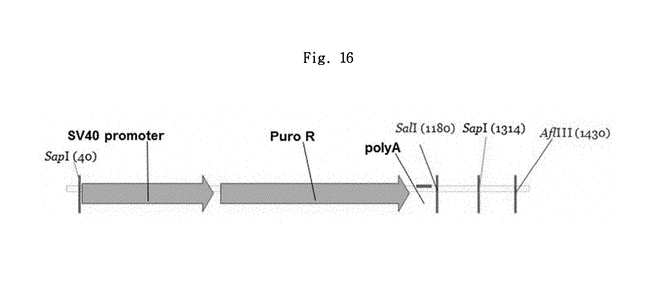
D00015
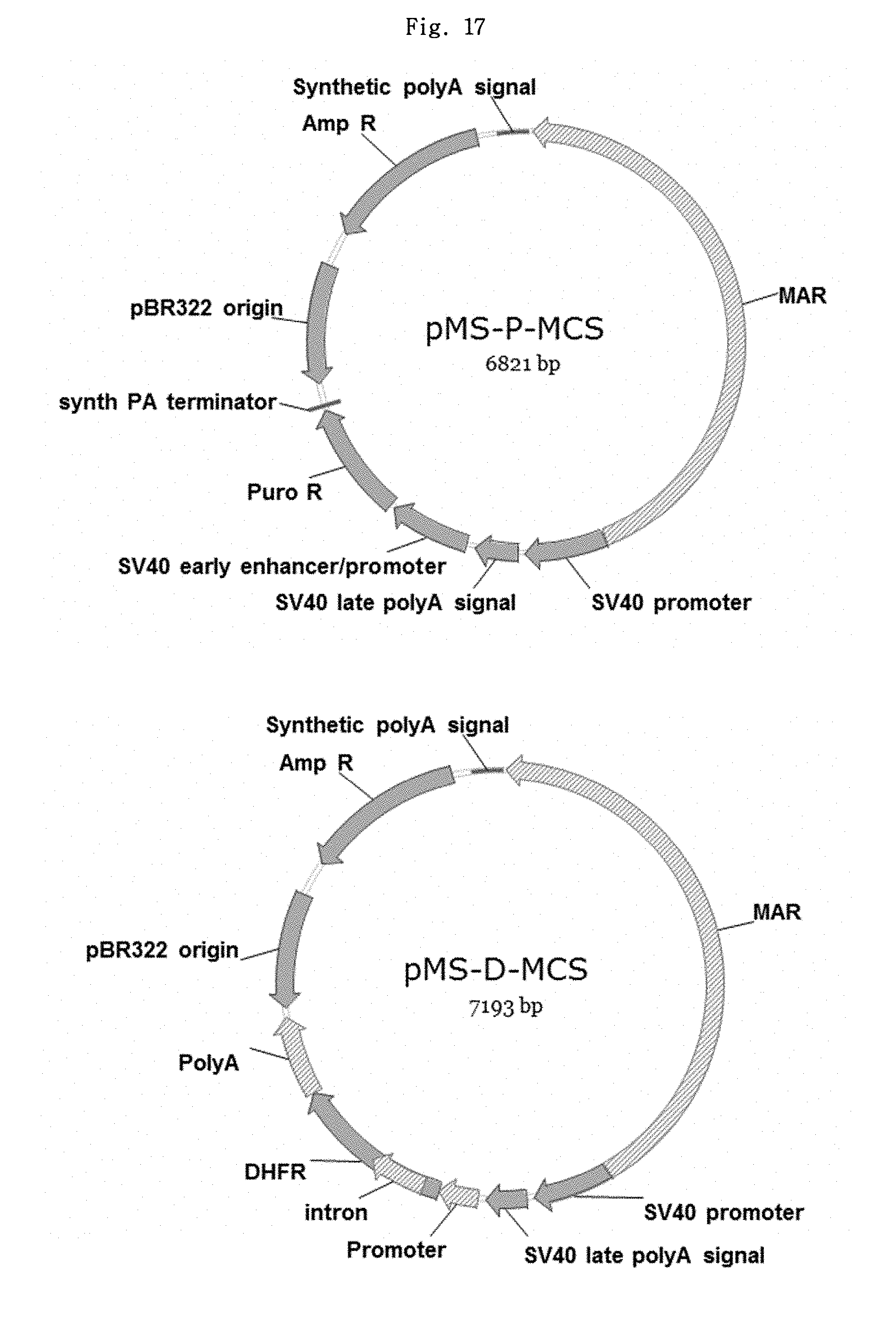
D00016

D00017
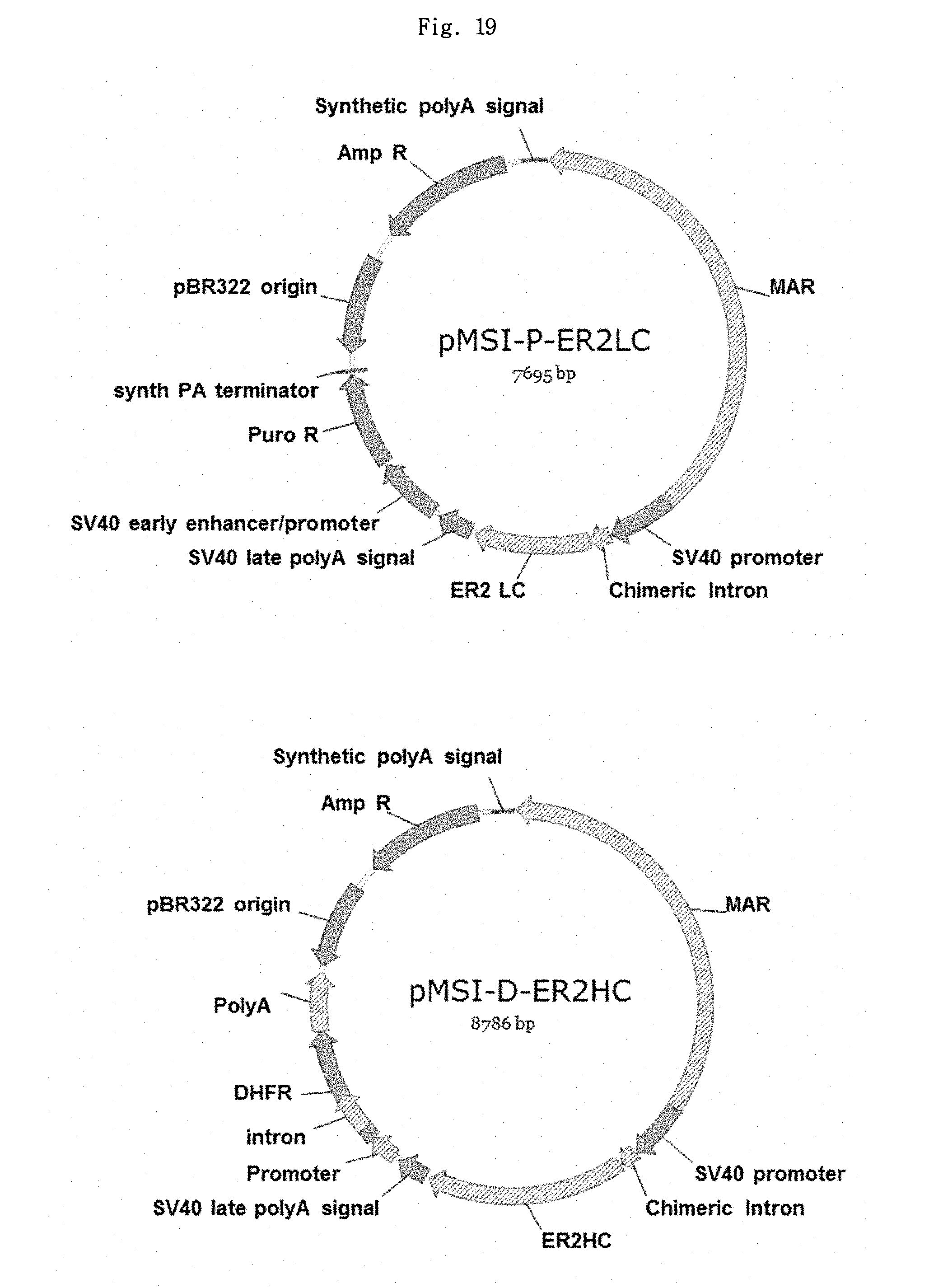
D00018
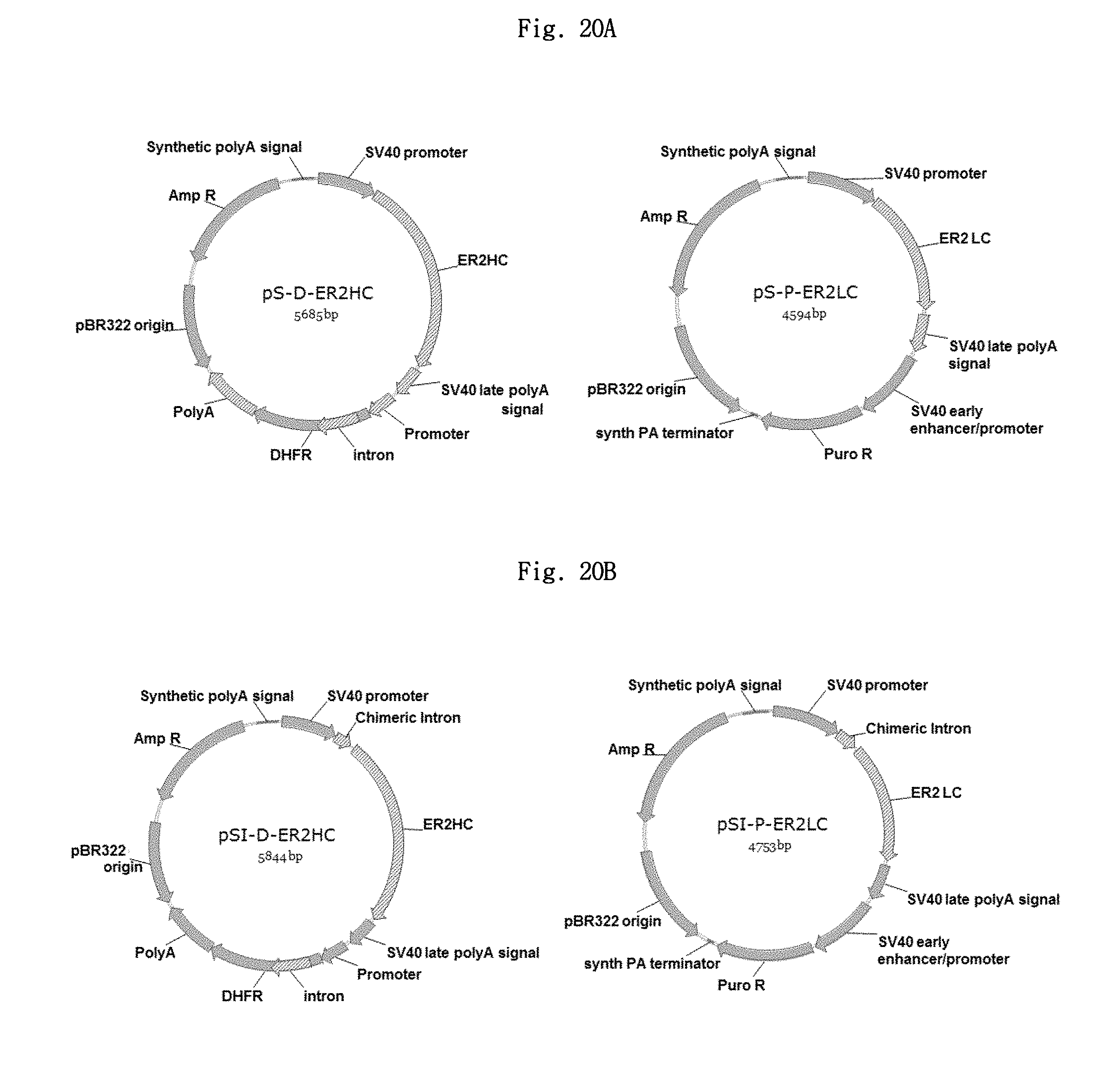
D00019

D00020
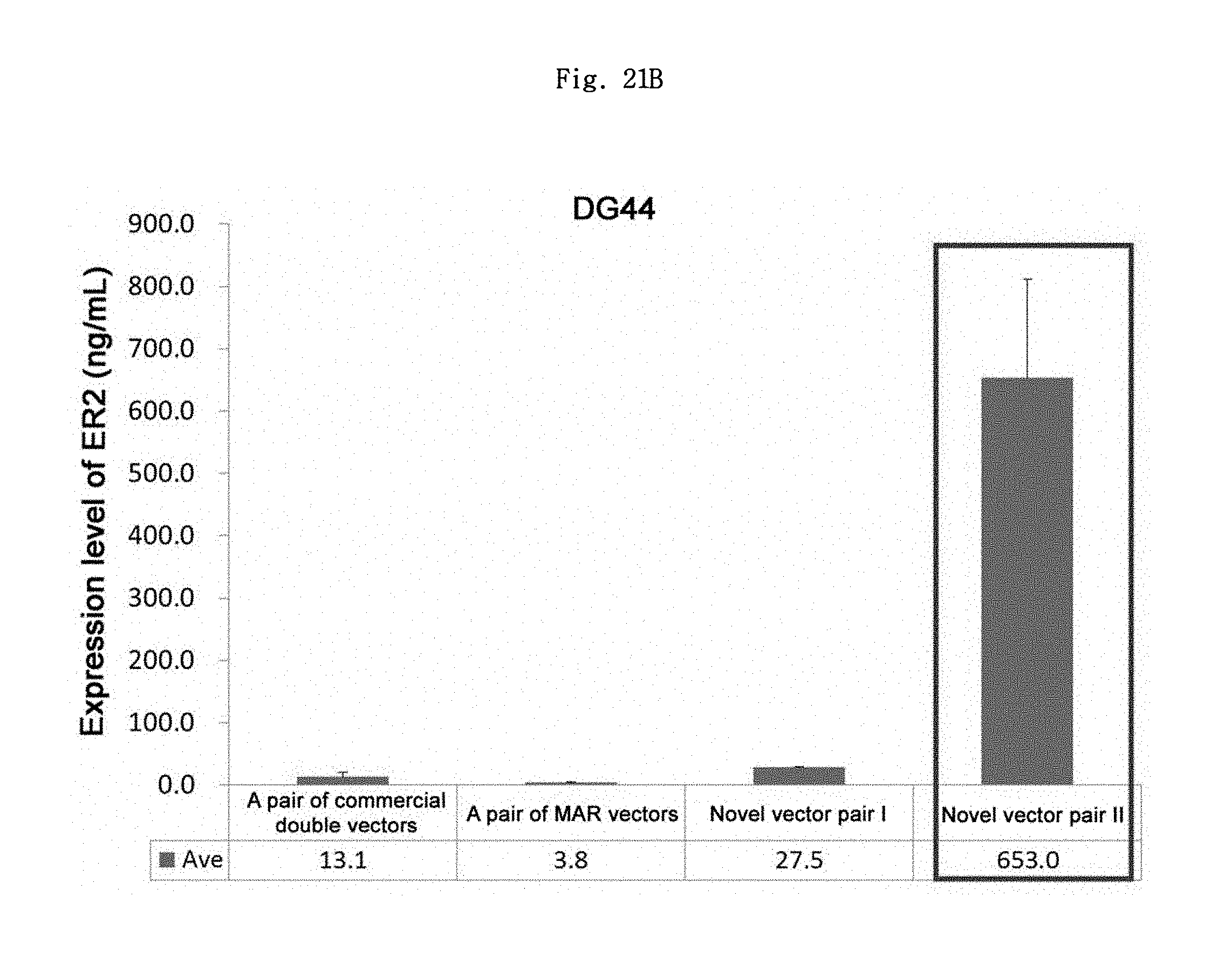
S00001
XML
uspto.report is an independent third-party trademark research tool that is not affiliated, endorsed, or sponsored by the United States Patent and Trademark Office (USPTO) or any other governmental organization. The information provided by uspto.report is based on publicly available data at the time of writing and is intended for informational purposes only.
While we strive to provide accurate and up-to-date information, we do not guarantee the accuracy, completeness, reliability, or suitability of the information displayed on this site. The use of this site is at your own risk. Any reliance you place on such information is therefore strictly at your own risk.
All official trademark data, including owner information, should be verified by visiting the official USPTO website at www.uspto.gov. This site is not intended to replace professional legal advice and should not be used as a substitute for consulting with a legal professional who is knowledgeable about trademark law.