Antibody specific to Staphylococcus aureus, therapeutic method and detection method using same
Church
U.S. patent number 10,273,312 [Application Number 16/027,538] was granted by the patent office on 2019-04-30 for antibody specific to staphylococcus aureus, therapeutic method and detection method using same. The grantee listed for this patent is William R Church. Invention is credited to William R Church.


View All Diagrams
| United States Patent | 10,273,312 |
| Church | April 30, 2019 |
Antibody specific to Staphylococcus aureus, therapeutic method and detection method using same
Abstract
We provide new monoclonal antibody inhibitors of coagulases for treatment of S. aureus. The monoclonal antibodies are useful in targeting the SC N-terminus and inhibiting SC-ProT activation. The monoclonal antibodies are able to bind to and interfere with, modulate, and/or inhibit the binding interactions between the staphylocoagulase protein and its ligand protein prothrombin in blood and tissues. The antibodies are effective in inhibiting the activation of prothrombin.
| Inventors: | Church; William R (Burlington, VT) | ||||||||||
|---|---|---|---|---|---|---|---|---|---|---|---|
| Applicant: |
|
||||||||||
| Family ID: | 53678417 | ||||||||||
| Appl. No.: | 16/027,538 | ||||||||||
| Filed: | July 5, 2018 |
Prior Publication Data
| Document Identifier | Publication Date | |
|---|---|---|
| US 20180371108 A1 | Dec 27, 2018 | |
Related U.S. Patent Documents
| Application Number | Filing Date | Patent Number | Issue Date | ||
|---|---|---|---|---|---|
| 15583384 | May 1, 2017 | 10023655 | |||
| 15155209 | May 2, 2017 | 9637555 | |||
| 14604521 | May 31, 2016 | 9353189 | |||
| 61931236 | Jan 24, 2014 | ||||
| Current U.S. Class: | 1/1 |
| Current CPC Class: | C07K 16/36 (20130101); C07K 16/40 (20130101); C07K 16/1271 (20130101); A61K 2039/505 (20130101); C07K 2317/24 (20130101); C07K 2317/94 (20130101); C07K 2317/92 (20130101); C07K 2317/565 (20130101); C07K 2317/76 (20130101); C07K 2317/55 (20130101) |
| Current International Class: | C07K 16/40 (20060101); C07K 16/12 (20060101); C07K 16/36 (20060101); A61K 39/00 (20060101) |
References Cited [Referenced By]
U.S. Patent Documents
| 6299879 | October 2001 | Boden Wastfalt |
| 9353189 | May 2016 | Church |
| 9637555 | May 2017 | Church |
| 10023655 | July 2018 | Church |
Parent Case Text
RELATED APPLICATIONS
The present patent document is a continuation application of U.S. patent application Ser. No. 15/583,384 filed May 1, 2017, issued as U.S. Pat. No. 10,023,655, and which is a continuation of U.S. patent application Ser. No. 15/155,209 filed May 16, 2016, issued as U.S. Pat. No. 9,637,555, which is a divisional application of U.S. patent application Ser. No. 14/604,521 filed Jan. 23, 2015, issued as U.S. Pat. No. 9,353,189 which claims the benefit of the filing date under 35 U.S.C. .sctn. 119(e) of Provisional U.S. Patent Application Ser. No. 61/931,236, filed Jan. 24, 2014, each of which is hereby incorporated by reference in its entirety.
Claims
The invention claimed is:
1. A method of treating a staphylococcal infection, said method comprising using an antibody or antigen-binding fragment thereof which binds an epitope of staphylocoagulase peptide that is within SEQ ID. No: 2 and which comprises the following CDRS: TABLE-US-00003 CDRL1: QNVDIY (SEQ ID NO: 10) CDRL2: SAS (SEQ ID NO.: 11) CDRL3: QQYNNYPYT (SEQ ID NO: 12) CDRH1: GFTFSDAW (SEQ ID NO: 13) CDRH2: IRTKANNHAT (SEQ ID NO: 14) CDRH3: CTNVYYGNNDVKDY (SEQ ID NO: 15).
2. The method of claim 1, wherein the epitope that binds the isolated antibody is within residues 1-10 of SEQ ID No: 2.
3. The method of claim 1, wherein the isolated monoclonal antibody or antigen-binding fragment thereof is produced from a hybridoma cell having accession number PTA-120537.
4. The method of claim 1, wherein the isolated monoclonal antibody or antigen-binding fragment thereof comprises a V.sub.H region having at least 95% identity with SEQ ID NO: 6 and a V.sub.L region having at least 95% identity with SEQ ID NO: 8.
5. The method of claim 1, wherein the isolated monoclonal antibody or antigen-binding fragment thereof is humanized.
Description
SEQUENCE LISTING
The instant application contains a Sequence Listing which has been submitted electronically in ASCII format and is hereby incorporated by reference in its entirety. Said ASCII copy, created on Jan. 21, 2015, is named 163-303_SL.txt and is 29,860 bytes in size.
BACKGROUND
Infections caused by Staphylococcus aureus ("S. aureus") are a major causative agent of hospital and non-hospital infections. These infections cause longer hospitalization time and cost. S. aureus infections range from common, minor skin infections to blood-borne infections of the heart valves called infective endocarditis.
Infective endocarditis may be used as a model for illustrating S. aureus infection. Endocarditis is an inflammation of the endocardium, the inner layer of the heart. In some variations, it involves the heart valves. In other variations it may involve the interventricular septum, the chordae tendineae, the mural endocardium, and even surfaces of implanted medical devices such as intracardiac devices and prosthetic valves.
One characteristic of endocarditis is a lesion, which may also be referred to as a vegetation. A vegetation includes but is not limited to, a mass of platelets, fibrin, microcolonies of microorganisms, and inflammatory cells. In some variations, infective endocarditis vegetations may also include a center of granulomatus tissue, e.g., a collection of the immune cells called macrophages. Granulomatus tissue may fibrose (e.g., form excess tissue) and/or calcify.
Heart valves do not receive dedicated blood supply, which may blunt the immune response, making it difficult for immune defenses (such as white blood cells) to directly reach the valves via the bloodstream. Valves may have an increased susceptibility to infection, e.g. bacterial infection, due to (among other factors) the blunted immune response. The lack of blood supply to the valves may also decrease the effectiveness of traditional treatments, since drugs (e.g., those delivered via bloodstream) also have difficulty reaching infected valves.
S. aureus infection rates continue to increase. S. aureus acute infective endocarditis is 25-47% fatal despite antibiotic therapy. Vancomycin is a common antimicrobial treatment for infections caused by S. aureus (e.g., methicillin resistant S. aureus). Of great concern is the observation that drug resistant strains of S. aureus are rapidly evolving. The rapid spread of hypervirulent, multi-drug resistant strains of S. aureus suggest S. aureus will likely become resistant to all antibiotics and an even greater threat to public health. This threat is exacerbated due to, among other things, the reluctance of drug companies to develop new antibiotics.
BRIEF SUMMARY
We provide monoclonal antibodies able to recognize and bind to staphylocoagulase. The monoclonal antibodies are able to bind to and interfere with, modulate, and/or inhibit the binding interactions between the staphylocoagulase protein and its ligand protein prothrombin in blood and tisdues. It is effective in inhibiting the activation of prothrombin.
The provided monoclonal antibodies are represented by sequences SEQ ID NO.: 5, SEQ ID NO.: 6, SEQ ID NO.: 7, SEQ ID NO.: 8. The CDRs are represented by SEQ ID NOS.: 10-15. A cell line expressing monoclonal antibodies specific to staphylocoagulase and homologs thereof is deposited with the ATCC under accession number PTA-120537.
Other systems, methods, features, and advantages of the disclosure will be, or will become, apparent to one with skill in the art upon examination of the following figures and detailed description. It is intended that all such additional systems, methods, features and advantages be included within this description, be within the scope of the invention, and be protected by the following claims.
BRIEF DESCRIPTION OF THE DRAWINGS
The following description of the drawings and the various system, method, and apparatus is not intended to limit the inventive system, methods and apparatus disclosed herein to one embodiment, but rather to enable any person skilled in the art of art of antibody production to make and use the inventive system, method and monoclonal antibodies.
FIG. 1 provides a schematic of the pathogenesis of acute bacterial endocarditis.
FIG. 2 provides an exemplary representation of staphylocoagulase, and discloses SEQ ID NO: 9.
FIG. 3 provides an exemplary representation of prothrombin.
FIG. 4 provides an exemplary representation of thrombin.
FIG. 5 provides a representation of staphylocoagulase and prothrombin and their interaction resulting in the non-proteolytic formation of an active site and discloses SEQ ID NO: 9.
FIG. 6 shows immune response of three mice injected with IVTKDYSKES (SEQ ID NO: 9) conjugated to carrier protein.
FIG. 7 shows serum immune response from splenocyte donor mouse used for hybridoma production.
FIG. 8 shows binding of anti-IVTKDYSKES (SEQ ID NO: 9) antibody in hybridoma cell supernatant in ELISA.
FIG. 9 shows purified antibody designated GMA-2105 binding to conjugated- and unconjugated-IVTKDYSKES (SEQ ID NO: 9) in ELISA.
FIG. 10 shows the purified GMA-2105 antibody IgG on the SDS-PAGE assay according to a variation.
FIG. 11 shows binding of GMA-2105 F(ab) to IVTKDYSKES (SEQ ID NO: 9) coated wells in ELISA.
FIG. 12 shows a Western blot detection of staphylocoagulase in cultured bacteria of various strains using purified GMA-2105 F(ab) fragments.
FIG. 13 is a chart comparing amino acid substitutions in three mutated peptides (SEQ ID NOS 25-27, respectively, in order of appearance).
FIG. 14 shows binding of GMA-2105 to variant staphylocoagulase peptides to in solid phase competitive inhibition ELISA.
FIG. 15 shows BOPIDY-labeled staphylocoagulase fragment SC(1-325) with (S7C) titrated with GMA-2105 in solution in the presence of unlabeled SC(1-246).
FIG. 16 shows inhibition of SC(1-246)-ProT.sup.3Q by GMA-2105.
FIG. 17 shows results of clotting assay demonstrating that GMA-2105 blocks staphylocoagulase activity inhibiting prothrombin activation and downstream plasma clotting.
FIG. 18 shows the effect of GMA-2105 on prothrombin activation by staphylocoagulase activity in S. aureus supernatant.
FIG. 19 shows purified GMA-2105 analyzed by size exclusion chromatography.
FIG. 20 shows dynamic light scattering of purified GMA-2105.
FIG. 21 shows detection of GMA-2105 antibody in the blood of mice 21 days post intraperitaneal administration.
FIG. 22 provides a survival curve demonstrating GMA-2105 promotes survival in a mouse model of S. aureus sepsis.
FIG. 23 provides a schematic of an exemplary chimeric GMA-2105 construct.
FIG. 24 shows binding of GMA-2105 chimeric antibody in HEK293 cell transfection supernatants to IVTKDYSKES (SEQ ID NO: 9).
FIG. 25 shows a Western blot detection of human antibody heavy chain Fc and human kappa light chain in HEK293 GMA-2105 chimeric heavy chain and light chain transfected supernatant.
FIG. 26 shows SDS-PAGE analysis of purified GMA-2105 chimera.
FIG. 27 shows purified chimeric and murine GMA-2105 antibody binding to IVTKDYSKES (SEQ ID NO: 9) in ELISA assay.
FIG. 28 shows dynamic light scattering of GMA-2105 chimera.
FIG. 29 shows BODIPY-labeled staphylocoagulase fragment SC(1-325) with (S7C) titrated with GMA-2105 chimera in solution.
FIG. 30 shows comparison of binding characteristics of murine GMA-2105 and chimeric GMA-2105.
FIG. 31 provides a theoretical model of GMA-2105 light chain SEQ ID No. 8(4-105) and GMA-2105 heavy chain SEQ ID No.:6(4-110).
FIG. 32 provides representatives from each of the six clusters formed from the 26 lowest energy GMA-2105 models.
FIG. 33 shows a theoretical model of substitution at GMA-2105 light chain residue 46 (SEQ ID No. 8, residue 46) from alanine to leucine.
FIG. 34 shows a theoretical model of substituting an asparagine for aspartic acid at heavy chain residue 76 (SEQ ID No.: 6, residue 76).
FIG. 35 shows a theoretical model of subsitution of a leucine for a valine at heavy chain residue 81 (SEQ ID No. 6, residue 81).
FIG. 36 shows by theoretical model the low energy models of the humanized GMA-2105 sequence.
DETAILED DESCRIPTION
S. aureus, is a highly adaptive human pathogen, causing recurrent skin and soft tissue infections by evading the immune system. S. aureus colonizes the nose and skin of 20-30% of the human population. Healthy individuals with intact skin and mucosal barriers may harbor S. aureus with no adverse result. However, when the integrity of the skin and mucosa are breached, S. aureus can invade and enter the tissue and blood stream and potentially cause injury.
Pathologies associated with S. aureus colonization include but are not limited to meningitis, sepsis, pneumonia. Pathologies also include endocarditis and septic arthritis in high risk populations, such as infants, immunocompromised adults, and intravenous drug users. The presence of foreign materials in the body, including intravenous catheters, greatly increases the risk of developing S. aureus-induced endocarditis. For example, catheters may become coated with fibrinogen and fibronectin, to which the bacterium can easily adhere.
Therapy of staphylococcus infections, including but not limited to, MRSA infections, is complicated by the fact that the organisms have evolved resistance to commonly used therapeutics and quickly develop immunity to new therapeutics. For example, despite antibiotic therapy, MRSA infections are associated with a poor outcome. It is estimated that only 5% of S. aureus isolates are susceptible to penicillin treatment. Another feature of staphylococcal infections is its reoccurrence rate. Further complication treatment indications, clinical and experimental observations suggest that infections with S. aureus do not generate protective immune response.
The immune invasion strategies of S. aureus allow it to survive in the blood. Through the blood and/or subepidermal tissues, S. aureus encounters and escapes the host innate immune defenses. S. aureus displays cell-surface proteins and secreted virulence factors, including the procoagulant staphylocoagulase (SC) which allow it to compromise the effectiveness of both the innate and adaptive immune responses. Some examples of how S. aureus evades the host immune system includes, but is not limited to, invading endothelial cells and neutrophils, inhibiting the complement system, and subverting the coagulation system for infective purposes.
Acute infective endocarditis (AIE) is one of the illnesses caused by S. aureus. Other illnesses include etastatic infections migrating to bones/joints, spleen, kidneys, liver, and lungs; sepsis; toxic shock syndrome; and pneumonia (among others). S. aureus recruits new virulence factors and antibiotic resistenace encoded by mobile genetic elements (MGEs) from other strains or different bacterial species. The emergence of methicillin-resistant S. aureus (MRSA) is attributed to MGE transfer, as is the ongoing global epidemic of the hypervirulent USA300 strain of community associated-MRSA (CA-MRSA).
Antibiotic resistance of CA-MRSA is leading to a reliance on vancomycin for treatment of severe infections. However, S. aureus has also developed resistance to this antibiotic. Partially vancomycin-resistant S. aureus with thickened cell walls have been described and clinical isolates carrying the vanA gene complex have appeared in the United States.
The adaptability of S. aureus and the rapid spread of hypervirulent and multidrug-resistant strains supports the prediction that S. aureus will soon become resistant to all available antibiotics. This huge public health problem will be associated with increased morbidity and mortality based on the propensity of S. aureus to cause many potentially lethal infections. The elderly are particularly susceptible and as the U.S. population ages there will be an increase in these infections.
The public health problem is exacerbated by the lack of interest of pharmaceutical companies in developing new antibiotics. This derives from the rapid emergence of antibiotic resistance that reduces the market life of the drugs and low profit due to the short treatment time. Attempts to create staphylococcal vaccines have uniformly failed, as demonstrated by clinical trial data.
To begin to address the public health threat posed by S. aureus we disclose new monoclonal antibodies as mechanism-targeted inhibitors of S. aureus procoagulant staphylocoagulase (SC).
Accute infectious endocarditis (AIE) pathogenesis of S. aureus has at least two initiating events (FIG. 1). First, injury to the endothelium covering the heart valves. In one example, injury may result from turbulent flow due to a congenital or acquired defect. Injury may also result from the presence of intravascular catheters, intravenous drug use, or physiological stress from hypersensitivity states, hormonal changes, or exposure to high altitude.
Injury may initiate coagulation and formation of a sterile thrombus composed of, e.g., fibrin and platelets. Coagulation may be initiated due to exposure of blood to, among others, tissue factor. Exposure of blood to tissue factor triggers activation of blood coagulation. Activation of blood coagulation may result in formation of a sterile thrombus. The sterile thrombus may be composed of, among other factors, activated platelets and fibrin (e.g., thrombin-generated fibrin).
A compromised system may permit bacteria to enter the bloodstream and adhere to the sterile thrombis, for example, by binding fibrinogen and fibronectin. S. aureus is among the most common bacterial pathogens. S. aureus expresses cell surface components, for example but not limited to adhesins. S. aureus cell surface components include but are not limited to wall-bound adhesins that bind fibrin(ogen), e.g., clumping factor A (ClfA) and other microbial surface components. The various cell surface components recognize adhesive matrix molecules that mediate binding of the bacteria to the site of vascular injury.
The most aggressive S. aureus strains in AIE secrete SC and also vWFbp, which bind to host prothrombin, abbreviated "Il." The active (active is designated "*") SC.circle-solid.prothrombin*(SC.circle-solid.Il*) and vWFbp.circle-solid.Il* complexes bind host fibrinogen as their substrate and convert it into fibrin. Deposition of fibrin propagates the formation of platelet-fibrin-bacteria vegetations, characteristic of AIE, at the site of vascular injury.
Vegetations grow by layering of more bacteria onto fibrin, and fibrin generation by the SC.circle-solid.Il* and vWFbp.circle-solid.Il* catalytic complexes (FIG. 1). Fibrin layers on vegetations and protects the bacteria from clearance by immune cells and killing by antibiotics. Turbulent flow nearby vegetations propagates endothelial damage across heart valves, ultimately leading to valvular dysfunction due to tissue damage and heart failure. Large vegetations produce infectious emboli that spread through the bloodstream to distant sites where they form abscesses in the brain, spleen, and kidneys.
Embolism to the brain is common in S. aureus AIE occurring in 30% of patients and results in ischemic or hemorrhagic stroke, which is often fatal. Mortality from AIE is very high at 25-47%, despite aggressive antibiotic treatment. Clearly, adjunctive therapy is sorely needed, which we begin to address here in the form of mechanism-based monoclonal antibodies.
FIG. 2 provides a schematic diagram of staphylocoagulase. FIG. 3 provides a schematic diagram of prothrombin. FIG. 5 is a diagram of the interaction of staphylocoagulase with prothrombin resulting in the non-proteolytic formation of an active site. The N-terminal Ile of staphylocoagulase forms a salt bridge with Asp 194 (chymostrypsin numbering) in the prothrombin activation site resulting in a conformational change in prothrombin which nonproteolytically stabilizes the thrombin active site. Fibrinogen can then be cleaved to fibrin by thrombin, resulting in the formation of a clot. (FIG. 1).
The SC(1-325).circle-solid.Pre 2* structure shows the N-terminus of SC (Ile.sup.1-Val.sup.2) inserted into the Ile.sup.16 (chymotrypsinogen numbering) N-terminal binding cleft of the prothrombin fragment designated Pre 2 where it forms a salt-bridge with Asp.sup.194 that triggers non-proteolytic activation of Pre 2 within the complex.
Recognizing the problems inherent in existing treatments, such as but not limited to, rapid resistance including emerging Vancomycin resistance and off-target events associated with anticoagulants, we disclose a method, system, and monoclonal antibody secreting cell line which enables the treatment of S. aureus infection in a specific and accurate manner by using SC-specific monoclonal antibodies.
Accordingly, we provide monoclonal antibodies that can bind to the staphylocoagulase protein from S. aureus, or certain subregions thereof, with high affinity, preventing the staphylocoagulase protein from forming an active pro-thrombin-bacterial cofactor complex. The monoclonal antibodies disclosed herein, thereby prevent the formation of fibrin, which prevents S. aureus' ability to create the vegetations used to protect the bacteria from clearance by immune cells and killing by antibiotics.
We demonstrate herein the effectiveness of anti-staphylocoagulase monoclonal antibodies to prevent fibrin formation (as demonstrated by clotting data, among other things) and to successfully treat S. aureus infections in an animal model.
The results of our experiments herein were surprising in light of the fact that the role of staphylocoagulase as a virulence factor in S. aureus infection has been in doubt due to conflicting results in a rat model of endocarditis that showed no role of staphylocoagulase, whereas a role for staphylocoagulase was demonstrated in a blood-borne pneumonia mouse model.
Fully developed S. aureus abscesses exhibit a defined structure with the bacteria in the center, bordered by fibrin-rich barriers that protect the bacteria from immune cells and antibiotics. The role of staphylocoagulase has been in doubt also in light of research demonstrating that S. aureus lacking the staphylocoagulas gene could form abscesses.
The use of anticoagulants, which have the potential to produce lethal bleeding, to treat AIE has been debated. Given the complexity of thrombin regulation through its two exosites that bind protein substrates, inhibitors, and regulatory macromolecules, and its many procoagulant and anticoagulant roles, systemic inhibition of thrombin (via anti-coagulants) in an attempt to inhibit staphylocoagulase /vWFbp-Il* may lead to off-target events with adverse consequences. Comparison of 35 patients with S. aureus AIE to those with native valve AIE, of which none were taking anticoagulants, to 21 with prosthetic valve AIE 90% of which were taking anticoagulants, found that the groups had similar incidence of neurologic embolisms, and the mortality in the anticoagulant group was 71% compared to 37% in the no anticoagulant group. More of those patients had brain hemorrhages. An old study found the opposite outcome for a small group of patients with prosthetic valves; i.e., mortality was slightly greater (57% vs. 47%) if anticoagulant therapy was stopped.
Our novel monoclonal antibody targeting staphylocoagulase avoids the off-target events experienced with anti-coagulants. The SC monoclonal antibody inhibits fibrin formation by targeting the staphylocoagulase protein specifically. It therefore has a localized effect at the site of S. aureus infections and does not have system-wide consequences.
The mouse models for blood coagulation are similar to the human coagulation system. Emeis, et al., A guide to murine coagulation factor structure, function, assays and genetic alterations, J. Thromb. Haemost. 2007; 5:670-9, incorporated herein by reference in its entirety. Numerous therapeutics directed at human coagulation systems (clotting, platelets, fibrinolysis) are similarly effective at comparable doses in mice. This suggests that targeting staphylocoagulase with an antibody might give physiologic and pharmacologic data that is reflective of what happens in humans.
Targeting S. aureus via the host hemostatic response is a novel therapeutic approach to treating S. aureus infection. The mechanistic details of prothrombin activation are well known. (See, e.g., Krishnaswamy, S., The transition of prothrombin to thrombin, J. Thromb. Haemost, 2013; 11:256-76, incorporated herein by reference in its entirety.) The novel approach involves using a monoclonal antibody to block S. aureus' ability to form a fibrin mesh, preventing the course of disease. (The course of disease is illustrated in FIG. 1). A therapeutic monoclonal antibody provides advantages over vaccines. In contrast to a vaccine that requires the development of an immune response which may take several days or months, the therapeutic monoclonal antibody can be used in acute infections and can also offer prophylactic protection before an infection. We demonstrate that our novel anti-staphylocoagulase monoclonal antibodies prevent fibrin formation.
Accordingly, we provide monoclonal antibodies that can bind to the SC protein from S. aureus, or certain subregions thereof, with high affinity and which can thus be useful in methods to treat, prevent, or diagnose staphylococcal infections.
We also provide a therapeutic antigen binding protein which recognizes an epitope of the SC protein from S. aureus that contains residues 1-10 from the N-terminal SC ("SC(1-10)").
We also provide a therapeutic antigen binding protein which recognizes an epitope of the SC protein from S. aureus that contains residues 1-3, 1-4, 1-5, 1-6, 1-7, 1-8, 1-9, or 1-10 of the SC protein.
In a variation, the therapeutic antigen binding protein is an antibody or antigen binding fragment and/or derivative thereof.
We also provide monoclonal antibodies which are able to bind SC and peptides which are generated from the SC or a subregion thereof and which are useful in methods to treat, prevent, or diagnose staphylococcal infections. In one example, we provide monoclonal antibodies generated from the N-terminal SC, in another example, we provide monoclonal antibodies generated from SC(1-10). In another example, we provide monoclonal antibodies generated from a peptide comprising amino acids IVTKDYSKES (SEQ ID No. 9) and homologs and/or degenerations thereof.
We also provide monoclonal antibodies to the SC protein which can be useful in inhibiting the SC-II activation.
We also provide monoclonal antibodies able to recognize and bind to Staphylocoagulase protein (SEQ ID No. 2) and other proteins presenting sequences homologous to Staphylocoagulase including vWFbp (SEQ ID No. 4), and other bacteria possessing Staphylocoagulase or sequences homologous to this protein.
We also provide monoclonal antibodies to recognize and bind to proteins encoded by staphylocoagulase nucleotide sequences (SEQ ID No. 1) and other proteins encoded by sequences homologous to staphylocoagulase. The monoclonal antibodies may also recognize and bind to proteins of bacteria possessing staphylocoagulase or sequences homologous to staphylocoagulase.
We also provide monoclonal antibodies able to recognize and bind to peptide sequence IVTKDYSKES-CONH2 (SEQ ID NO: 9), Staphylocoagulase peptide sequence IVTKDYSKES-CONH2 (SEQ ID NO: 9), and other proteins presenting sequences homologous to peptide sequence IVTKDYSKES-CONH2 (SEQ ID NO: 9), and Staphylocoagulase peptide sequence IVTKDYSKES-CONH2 (SEQ ID NO: 9).
We also provide a method which enables the treatment of S. aureus in a specific and accurate manner by using SC specific monoclonal antibodies generated using SC(1-10) peptide sequence (SEQ ID No. 9 and amino acids 1-10 of Seq. ID. NO. 2).
We provide an antibody specifically binding to a polypeptide comprising a SC, wherein the monoclonal antibody was generated using a peptide comprising the amino acid sequence IVTKDYSKES (SEQ ID No. 9).
We also provide an antibody capable of binding SC(1-10) and inhibiting the binding of SC to prothrombin.
We also provide an antibody capable of binding SC(1-10) (SEQ ID No. 9) inhibiting the activation of prothrombin.sup.3Q (a non-cleavable form of prothrombin) by SC(1-246) with 0.9 nM affinity.
An antibody capable of binding to SC which has an affinity constant (K.sub.D) for SC of between 0.1 nm and 10 nM, specifically 2 nM for binding of 1.6 mol Bodipy-SC/mol mAb.
We provide a hybridoma cell line producing a monoclonal antibody capable of binding SC, in which said hybridoma cell line has accession number ATCC PTA-120537.
We also provide a method of treating an animal infected with S. aureus by injecting the infected animal with monoclonal antibody against SC.
We also provide a therapeutic antigen binding protein, such as an antibody or antigen binding fragment and/or derivative thereof, which binds S. aureus SC and which comprises the heavy chain variable region amino acid sequence of SEQ ID NO 6 and the antibody light chain variable region of SEQ ID NO 8.
We also provide an antigen binding protein, such as an antibody or antigen binding fragment thereof which specifically binds to SC protein and comprises a heavy chain region that is a variant of the sequences set forth in SEQ ID NO 6.
We also provide an antigen binding protein, such as an antibody or antigen binding fragment thereof which specifically binds to SC protein and comprising a light chain region that is a variant of the sequence set forth in SEQ ID NO 8.
We also provide a therapeutic antigen binding protein, such as an antibody or antigen binding fragment and/or derivative thereof, which binds SC and which comprises the following CDRs:
TABLE-US-00001 CDRL1: QNVDIY (residues 27-32 of SEQ ID No. 8 and SEQ ID No: 10) CDRL2: SAS (residues 50-52 of SEQ ID No. 8 and SEQ ID No.: 11) CDRL3: QQYNNYPYT (residues 89-97 of SEQ ID No. 8 and SEQ ID No: 12) CDRH1: GFTFSDAW (residues 26-33 of SEQ ID No. 6 and SEQ ID NO: 13) CDRH2: IRTKANNHAT (residues 51-60 of SEQ ID No. 6 and SEQ ID NO: 14) CDRH3: CTNVYYGNNDVKDY (residues 98-111 of SEQ ID No. 6 and SEQ ID NO: 15).
We also provide an antigen binding protein, such as an antibody or antigen binding fragment thereof which specifically binds SC and comprises CDR's which are variants of the sequences set forth in SEQ ID NOS.: 10-15.
A variant includes a partial alteration of the CDR, heavy chain and/or light chain amino acid sequence by deletion or substitution of one to several amino acids of the CDR, heavy chain and/or light chain, or by addition or insertion of one to several amino acids to the CDR, heavy chain, and/or light chain, or by a combination thereof. The variant may contain 1, 2, 3, 4, 5, or 6 amino acid substitutions, additions or deletions in the amino acid sequence of the CDR, heavy chain, and/or light chain sequence. The substitutions in amino acid residues may be conservative substitutions, for example, substituting one hydrophobic amino acid for an alternative hydrophobic amino acid.
Antigen binding proteins which are variants of the CDR, heavy chain, and/or light chain will have the same or similar functional properties to those comprising the CDR, heavy chain, and/or light chain described herein. Therefore, antigen binding proteins which comprise a variant CDR will bind to the same target protein or epitope with the same or similar binding affinity to the CDR, heavy chain, and/or light chain described herein.
An exemplary antibody is GMA-2105 murine monoclonal antibody. In a variation there is provided a humanized or chimeric antibody comprising the following CDRs of GMA-2105:
TABLE-US-00002 CDRL1: QNVDIY (residues 27-32 of SEQ ID No. 8 and SEQ ID No: 10) CDRL2: SAS (residues 50-52 of SEQ ID No. 8 and SEQ ID No.: 11) CDRL3: QQYNNYPYT (residues 89-97 of SEQ ID No. 8 and SEQ ID No: 12) CDRH1: GFTFSDAW (residues 26-33 of SEQ ID No. 6 and SEQ ID NO: 13) CDRH2: IRTKANNHAT (residues 51-60 of SEQ ID No. 6 and SEQ ID NO: 14) CDRH3: CTNVYYGNNDVKDY (residues 98-111 of SEQ ID No. 6 and SEQ ID NO: 15).
For example, a chimeric antibody may comprise the variable regions of the GMA-2105 antibody, namely SEQ ID No: 6 (V.sub.H) and SEQ ID No. 8 (V.sub.L). A first example of a humanized antibody based on GMA-2105 is an antibody comprising a heavy chain having SEQ ID NO.: 18. A second example of a humanized antibody based on GMA-2105 is an antibody comprising a heavy chain sequence having SEQ ID NO. 19. A third example of a humanized antibody based on GMA-2105 is an antibody comprising a light chain having SEQ ID No. 16. A fourth example of a humanized antibody based on GMA-2105 is an antibody comprising a light chain having SEQ ID No. 17. A fifth example of a humanized antibody based on GMA-2105 is an antibody comprising a heavy chain sequence having either SEQ ID NO. 18 or SEQ ID No.: 19 and a light chain sequence having either SEQ ID NO. 16 or SEQ ID No.: 17.
Method of Generating Monoclonal Antibodies to Staphylocoagulase
We provide monoclonal antibodies that can recognize and bind to, among other proteins, staphylocoagulase, and homologs thereof. In one method, the antibodies are raised against a synthetic peptide comprising the sequence IVTKDYSKES (SEQ ID No.: 9). The synthetic peptide was used to generate a panel of murine monoclonal antibodies. Monoclonal antibodies recognizing staphylocoagulase may be raised from peptides comprising expressed and purified staphylocoagulase, and/or synthetic peptides raised from other subregions or larger immunogenic regions of the staphylocoagulase protein.
Antibodies may be obtained in conventional ways including steps of introducing the staphylocoagulase antigen, subregions, peptides, or degenerate versions thereof, into a host animal, followed by isolation of the antibody-producing spleen cells and formation of a suitable hybridoma.
An inhibitory mouse anti-staphylocoagulase antibody designated GMA-2105 was produced against a peptide IVTKDYSKES (SEQ ID No.: 9), representing residues 1-10 of staphylocoagulase (SEQ ID No.: 2), with the achieved goal of blocking activation of prothrombin by binding to the staphylocoagulase sequence that inserts into prothrombin.
Example Method of Generating Anti-Staphylocoagulase Monoclonal Antibody
Female Balb/c mice approximately 6-8 weeks old received an initial intraperitoneal (ip) injection of 100 .mu.g staphylocoagulase peptide (sequence IVTKDYSKES, SEQ ID NO 9) conjugated with-keyhole limpet hemocyanin (KLH) and emulsified in complete Freund's adjuvant on day 0. Booster injections of 50 .mu.g of the above peptide conjugate emulsified in incomplete Freund's were given on days 10, 20 and 30.
Two additional 50 .mu.g injections (peptide-KLH conjugate in incomplete Freunds adjuvant) were given on days 61 and 111. A serum sample from each mouse was collected on Day 122 and the antibody titer measured by solid-phase Enzyme Linked Immunosorbant Assay (ELISA) using the staphylocoagulase peptide SC(1-10) (SEQ ID No.: 9) conjugated to ovalbumin coated on 96-well ELISA plates at 2 ug/ml. Uncoated areas of the wells were blocked with albumin from bovine serum (BSA). The wells were then incubated with dilutions of the mouse serum to test whether the serum contained antibodies to the staphylocoagulase peptide SC(1-10) (SEQ ID. No.: 9). Bound antibodies were detected with anti-mouse IgG secondary antibody conjugated to horseradish peroxidase. The plates were then incubated with o-phenylenediamine (OPD) substrate which undergoes a color change in the presence of HRP the intensity of which can be measured using absorption at 490 nm wavelength in a microplate reader.
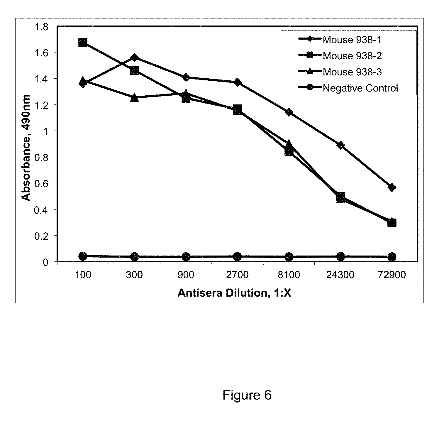
FIG. 6 provides the results of the ELISA demonstrating the presence of anti-staphylocoagulase antibodies in mouse serum of mice (designated 938-1, 938-2, and 938-3 on FIG. 6) injected with the staphylocoagulase peptide SC(1-10) (SEQ ID No.: 9). Serum from an irrelevant mouse was used as a negative control (designated Negative Control on FIG. 6).
Fusions were performed using three of the mice resulting in positive clones. Splenocytes from the mouse designated 938-3 on FIG. 6 were harvested for a fusion on Day 128. A total of 2.9.times.10.sup.8 spleen cells were mixed with 3.times.10.sup.7 NS1 myeloma cells, centrifuged to pellet the cells. The supernatant was aspirated and 1.0 ml 50% polyethylene glycol in media (PEG) was added drop wise over 1 minute to allow cell membranes to fuse. Culture media was then added drop wise to dilute the PEG 20-fold. The fused cells were centrifuged and resuspended in 100 ml Selection media consisting of Dulbecco's Modified Eagle Medium high glucose (DMEM) base with added 15% fetal bovine serum (FBS), 100 uM hypoxanthine, 2% (v/v) Hybridoma Cloning Supplement (Roche), 4 mM L-glutamine, 50 U/ml penicillin, 50 .mu.g/ml streptomycin, 1.times. nonessential amino acids and the selective agent azaserine at 5.7 uM used to kill unfused NS1 cells. The cell suspension was dispensed into 24 well plates, 1 ml/well. Plates were incubated at 37.degree. C. in 8% CO.sub.2 atmosphere for 6 days and then fed with 1 ml per well of Growth media consisting of the components described above minus the azaserine. Cells were allowed to grow an additional 4-5 days.
On Day 11 post fusion, cell supernatants were tested for the presence of anti-staphylocoagulase peptide antibodies by ELISA using the ELISA assay described above. The positive well was subcloned by the limiting dilution method in Growth media to obtain a monoclonal cell line.
For this method cells were seeded into 96-well plates at sufficiently low density to increase the probability of a colony growing from a single cell. Approximately 10 days later, wells were screened by solid-phase ELISA to identify antibody-producing clones. The contents of the positive well was subcloned again by limiting dilution seeding into a single 96 well plate. The contents of the positive well was given the designation 2A1.5H4.B7. The cells were grown to sufficient numbers for cryopreservation and antibody production.
For antibody production, cells were grown to high density in roller bottles in 500 ml serum-free media. Antibody was purified from the supernatant by allowing the antibody to bind to protein G Sepharose on an affinity column (Pierce) that allows non-antibody proteins to flow through. Bound antibodies were then eluted from the Sepharose using 0.1 M glycine, pH 2.7. Eluted antibodies were neutralized to pH 7 with 1 M Tris buffer. A buffer transfer into phosphate buffered saline (PBS) was performed by dialysis using 30 kd molecular weight cut off dialysis tubing. Purified antibody was stored at 4.degree. C. in the presence of 0.1% sodium azide as a preservative.
FIG. 10 is an SDS-PAGE image of purified GMA-2105. An antibody sample of 3 .mu.g was electrophoresed at 200V on a 4-12% Bis-Tris gel under reducing and non-reducing conditions. Lane 1 shows GMA-2105 under non-reduced conditions resulting in a single band of molecular weight 155 kDa. Lane 4 shows GMA-2105 under reduced conditions with about 55 kDa (heavy chain) and 26 kDa (light chain). Lane 3 refers to the molecular weight marker. Lane 2 is empty.
FIG. 7 shows the serum immune response from the splenocyte donor mouse that was used for hybridoma production. The splenocyte donor mouse was bled at two time points. Binding of mouse serum antibody to staphylocoagulase peptide SC(1-10) (SEQ ID No.: 9) ovalbumin coated wells of a 96 well plate was detected using a goat anti-mouse secondary antibody conjugated to horseradish peroxidase. Substrate ortho-phenylenediamine conversion to product was measured at 490 nm using a spectrophotometer. The negative control was the serum from an irrelevant mouse.
FIG. 8 shows the binding of anti-staphylocoagulase antibody GMA-2105 in hybridoma cell supernatant to SC(1-10) peptide ovalbumin conjugate bound to the wells of a 96 well plate. The antibody-antigen complex was detected using a goat anti-mouse secondary antibody conjugated to horseradish peroxidase. Substrate ortho-phenylenediamine conversion to product was measured at 490 nm using a spectrophotometer. The negative control was a well that was coated with carbonate buffer with no antigen and then blocked with blocking buffer. The negative control well did not contain the peptide conjugate.
FIG. 9 demonstrates the ability of the purified monoclonal antibody GMA-2105 disclosed herein to bind to the staphylocoagulase peptide SC(1-10) (residues 1-10 of SEQ ID No.: 2). For ease of reference, the purified antibody was designated GMA-2105. The curve shows binding of GMA-2105 to unconjugated staphylocoagulase peptide SC(1-10)(SEQ ID NO.:9), and staphylocoagulase peptide SC(1-10)(SEQ ID NO.:9) -ovalbumin conjugate. A purified, irrelevant mouse IgG was used as a negative control.
FIG. 11 demonstrates the successful binding of GMA-2105 fragment antigen-binding fragment (F(ab) fragment). The GMA-2105 F(ab) was tested by ELISA against the IVTKDYSKES peptide (SEQ. ID. No.: 9). The negative control was a purified irrelevant F(ab) fragment. This demonstrates that the GMA-2105 F(ab) fragment, alone, is capable of binding staphylocoagulase IVTKDYSKES peptide (SEQ ID No.: 9).
FIG. 12 shows a Western blot detection of staphylocoagulase in cultured bacteria of various strains using purified GMA-2105 F(ab) fragments. Lanes A, B, and C contain concentrated (10.times.) supernatants from identically cultured bacteria. Lane A: S. saprophyticus (coagulase negative bacterium control); Lane B: S. aureus strain COL (MRSA); Lane C: S. aureus strain MW2 (MRSA); Lane D: media blank; Lane E: staphylocoagulase peptide conjugated to ovalubumin (SEQ ID No.: 9). The Western blot demonstrated the ability of the GMA-2105 F(ab) fragments to bind staphylocoagulase, even in cultured bacteria.
Experiment: Demonstration of GMA-2105 Binding to Staphylocoagulase Variants
There are reported strain specific sequence differences at residue positions 8 and 9 of staphylocoagulase (SEQ ID. No.: 2). For example, various strains have been found to have amino acid substitutions at positions 8 and/or 9. Wantabe, et al., Genetic Diversity of Staphylocoagulase Genes (coa): Insight into the evolution of variable chromosomal virulence factors in Staphylococcus aureus. PLoS ONE 2009; 4(5): 1-11, incorporated herein by reference in its entirety. To demonstrate the ability of GMA-2105 to recognize and bind to variations of staphylocoagulase among different S. aureus strains, we synthesized three peptides with alanine substitutions at positions 8 of SEQ ID No. 9 ("Mutated Peptide Ala 8"), position 9 of SEQ ID No. 9 ("Mutated Peptide Ala 9"), and position 8 and 9 of SEQ ID No.: 9 ("Mutated Peptide Ala 8 & 9") (see FIG. 13). We measured the binding of each synthetic peptide (Mutated Peptide Ala 8, Mutated Peptide Ala 9, and Mutated Peptide Ala 8 & 9) to GMA-2105 in solution.
FIG. 14 shows the results of the competitive binding assay. We tested binding of Mutated Peptide Ala 8, Mutated Peptide Ala 9, and Mutated Peptide Ala 8 & 9 to GMA-2105 in a solid phase competitive inhibition assay. GMA-2105 was incubated with increasing concentrations of each of Mutated Peptide Ala 8, Mutated Peptide Ala 9, and Mutated Peptide Ala 8 & 9 (referred to hereafter as "GMA-2105--Mutated Peptide samples") in solution overnight at 4 degrees C. The incubation was followed by transfer of the GMA-2105--Mutated Peptide samples to wells coated with the non-mutated peptide, IVTKDYSKES (SEQ ID. No.:9) and blocked with 0.1% BSA PBS. After a 30 minute incubation, wells were washed with PBS containing 0.05% TWEEN 20, followed by addition of goat anti-mouse secondary antibody conjugated to horseradish peroxidase. Substrate ortho-phenylenediamine conversion to product was measured at 490 nm using a spectrophotometer. The negative control as was a purified irrelevant synthetic peptide.
Mutated Peptide Ala 8, Mutated Peptide Ala 9, and Mutated Peptide Ala 8 & 9, all bound antibody in solution. As shown in FIG. 14. Mutated Peptide Ala 8, Mutated Peptide Ala 9, and Mutated Peptide Ala 8 & 9 each and all inhibited binding of antibody onto wells coated with IVTKDYSKES (SEQ ID No.: 9) in competitive assay.
As demonstrated in the example, we provide an isolated monoclonal antibody, and/or a purified monoclonal antibody (e.g., purified from ATCC PTA-120537). The antibody may be used in, for example, methods of inhibiting activation of prothrombin, for example but not limited to, inhibiting the binding of SC and/or vWFbp to prothrombin. In additional to the methods described above, monoclonal antibodies may be produced using a variety of methods, e.g., methods found in Monoclonal Antibodies: Methods and Protocols, ISBN: 1588295672; Antibodies: A Laboratory Manual, ISBN: 0879693142, incorporated herein by reference in their entirety.
Resulting antibodies may be prepared in a number of forms, including chimeric, humanized, or human in addition to murine. Monoclonal antibodies may be prepared from a single chain, such as the light or heavy chains. Alternatively or additionally, monoclonal antibodies may be prepared from active fragments, e.g., fragments that retain the binding characteristics (e.g., specificity and/or affinity) of the whole antibody. Antisera prepared using monoclonal or polyclonal antibodies provided herein may be prepared in a number of suitable ways.
EXAMPLE
Inhibition of Prothrombin Activation
A high affinity monoclonal antibody GMA-2105 targeting the staphylocoagulas N-terminus residues inhibits prothrombin activation. In solution, the antibody bound Ser 7 to Cys mutant of SC(1-325). Binding studies were done using the BODIPY-labeled staphylocoagulase SC (S7C)(1-325) (SEQ ID No.: 2, positions 1-325 with Cys at position 7). The change of fluorescence upon antibody binding is measured as a function of the unlabeled ligand concentration and fitting the data to the quadratic binding equation gives the dissociation constant (K.sub.D), and the stoichiometry (n). Panizzi, et al., Fibrinogen substrate recognition by staphylocoagulase-(pro)thrombin complexes, J. Biol. Chem. 2006; 281:118-95.
Analysis of the increase in BODIPY fluorescence on GMA-2105 binding gave K.sub.ID 2 nM for binding of 1.6 mol BODIPY-SC/mol GMA-2105, indicating that the antibody GMA-2105 is bivalent and binds the staphylocoagulase SC(1-246)(positions 1-246 of SEQ ID No.: 2) with high affinity. Binding of the labeled staphylocoagulase SC(1-246) (positions 1-246 of SEQ ID No.: 2) to the GMA-2105 was competitively inhibited by binding of an unlabeled staphylocoagulase peptide representing positions 1-246 (SC(1-246)) of SEQ ID No.: 2, with K.sub.D 4 nM (descending curve in FIG. 15), indicating comparable affinity of the antibody for staphylocoagulase (positions 1-246 of SEQ ID No.: 2).
FIG. 15 shows equilibrium binding of GMA-2105 to staphylocoagulase SC(1-246) (positions 1-246 of SEQ ID No.: 2). The results indicate high-affinity interaction. Direct binding of BODIPY-SC(S7C) at 27nM (.circle-solid.) and 130 nM (.largecircle.) probe titrated with GMA-2105 caused fluorescence increase, whereas unlabeled SC(1-246) (triangles) displaced BODIPY-SC(S7C) labeled staphylocoagulase from binding the antibody. Simultaneous fit of all datasets provide the Kd for the unmodified competitor to the antibody.
The GMA-2105 antibody inhibits prothrombin.sup.3Q activation by SC(1-246) (positions 1-246 of SEQ ID No. 246) with 0.9 nM affinity (FIG. 16). Inhibition of prothrombin activation is complete as determined by analysis of inhibition at two SC(1-246) (positions 1-246 of SEQ ID No. 246) concentrations as competitive binding of prothrombin.sup.3Q and GMA-2015 to SC(1-246) (positions 1-246 of SEQ ID No. 246), meaning that staphylocoagulase bound to the GMA-2105 cannot activate prothrombin.
Using fluorescently labeled staphylocoagulase (SC(1-325) and Ser to Cys at position 7 of SEQ ID No. 2), and unlabeled GMA-2105, gave a KD of 0.99.+-.0.07 nM and a stoichiometry of 2 mol staphylocoagulase/mol GMA-2105. Concentrations were: BODIPY-staphylocoagulase (1-325) at 27 nM ( ) and 130 nM (.smallcircle.) titrated with GMA-2105 and unlabeled staphylocoagulase (1-246)(residues 1-246 of SEQ ID No. 2).
FIG. 16 shows inhibition of SC(1-246).circle-solid.prothrombin.sup.3Q by GMA-2105. This data gave identical K.sub.D and n for antibody binding to staphylocoagulase. Rates of S2238 hydrolysis by prothrombin (1 nm) and SC(1-246) at 5.6 nM(.circle-solid.) and 28 nM (.smallcircle.) versus GMA-2105 concentration. The inset shows titration of 1nM prothrombin with SC(1-246).
Example
Inhibition of Plasma Clotting
GMA-2105 inhibits S. aureus plasma clotting. FIG. 17 shows the inhibition of plasma clotting by increasing concentration of purified GMA-2105. GMA-2105 at varying concentrations was incubated with supernatant from S. aureus strain Tager 104 grown to OD 600 of 0.5 for one hour at room temperature followed by addition of an equal volume of rabbit plasma. Clotting was measured using a microplate spectrophotometer at 340 nm. Addition of GMA-2105 at 100 ug/mL (triangles) and 500 ug/mL (diamonds) prevented clotting as shown. The controls included an isotype matched antibody (XX) and no antibody (solid circle).
FIG. 18 demonstrates the effect of increasing concentrations of GMA-2105 to inhibit staphylocoagulase activity in S. aureus Tager supernatant. S. aureus Tager supernatant was incubated with or without GMA-2105 antibody at various concentrations (6.25 nM GMA-2105, 3.125 nM GMA-2105, 1.625 nM GMA-2105, 0 nM GMA-2105) for three hours at room temperature in a buffer of 50 nM HEPES pH 7.4, 150 nM NaCl, 5 mM CaCl.sub.2, 1 mg/mL PEG8000. Prothrombin was added to a 40 nM final concentration. The samples were incubated for three hours at room temperature. After incubation, S-2238 chromogenic substrate was added to a final concentration of 286 uM and the resulting absorbance was read at 405 nM for 1 hour.
Example
GMA-2105 Antibody Does Not Aggregate
Protein therapeutics, including monoclonal antibodies, have the potential for instability in which the physical state of the protein changes even through the chemical composition is unaltered. Several clinical trials with monoclonal antibodies have failed due to antibody aggregation. Two methods are commonly used to measure protein aggregation: size exclusion chromatography and light scattering. FIGS. 19 and 20 demonstrate evidence using these methods that isolated GMA-2105 does not aggregate.
FIG. 19. Purified GMA-2105 was analyzed by size exclusion chromatography using a column packed with SEPHACRYL 300 resin. A total of 4.2 mgs or 5.9 absorbance units were loaded onto the column in a volume of 1.5 mL. Fractions (1 mL) were collected and absorbance determined by spectrophotometry at 280 nM. Recovery off the column was 5.6 absorbance units, accounting for 96 percent of the sample that was loaded.
FIG. 20. Dynamic light scattering using MALVERN ZETASIZER NANO S at 25.0 degrees Celsius. A 1 mL fraction from the size exclusion chromatography analysis was filtered using a WHATMAN 0.2 uM filter, loaded into a disposable sizing cuvette and run using protein analysis software. GMA-2105 appears to be adequately stabilized in solution following production and purification.
Example
Survival Study and Pharmacokinetics
The mAb targeting SC(1-10) characterized in Preliminary Results promotes survival in a mouse model of S. aureus sepsis. Injection of purified antibody into mice followed by challenge with S. aureus gave the survival curve shown in FIG. 22. The experiment shows statistically significant survival over the controls (irrelevant antibody or phosphate-buffered saline).
FIG. 21. Detection of GMA-2105 antibody in the blood of mice 21 day post administration. C57BL/6 mice were injected intraperitioneal with 120 .mu.g of the mAb against SC(1-10) (GMA-2105). Every 7 days for a total of 21 days, blood was collected from the tail vein of each of the three mice. The antisera was diluted and added to a 96 well ELISA plate coated with the peptide IVTKDYSKES (SEQ ID. No.: 9) and blocked with 0.1% BSA/PBS. After a 1 hour incubation, wells were washed with PBS containing 0.05% TWEEN 20 followed by addition of goat anti-mouse secondary antibody conjugated to horseradish peroxidase. Substrate orth-phenylenediamine conversion to product was measured at 490 nm using a spectrophotometer.
FIG. 22. Survival Curve. C57BL/6 mice were injected intraperitioneal with 120 .mu.g of the mAb against SC(1-10) (GMA-2105) or an isotype-matched irrelevant mAb or PBS buffer with no antibody. After 8-9 hours, treated an untreated mice were injected with 1.times.10.sup.8 CFU of S. aureus via tail vein and monitored for survival. The anti-SC(1-10) mAb GMA-2105 increased the median survival curves from 24 to 72 h (p<0.005 vs. untreated).
Chimeric GMA-2105
Chimeric GMA-2105 was made by sequencing cDNA from purified mRNA from GMA-2105 hybridoma cells and using this cDNA sequence to synthesize DNA fragments with appropriate restriction sites at the ends. The DNA segments were inserted into expression vectors containing the human constant regions. FIG. 23 provides a schematic. SEQ ID No. 20 provides an exemplary human IgG.sub.1CH1, SEQ ID No. 21 provides an exemplary human IgG.sub.1 hinge region; SEQ ID No. 22 provides an exemplary human IgG.sub.i CH2; SEQ ID No. 23 provides an exemplary human IgG.sub.1 CH3, SEQ ID No. 24 provides an exemplary human IgG.sub.1 kappa light chain constant region.
The ability of the GMA-2105 antibody chimera to bind staphylocoagulase was tested by analyzing the supernatants of the transfected cells. FIG. 24 provides the results of binding of GMA-2105 chimera in HEK293 cell transfection supernatant to IVTKDYSKES (SEQ ID No. 9) in an ELISA assay. A combination of 55 .mu.g of GMA-2105 heavy chain chimera DNA and 57 .mu.g of GMA-2105 light chain chimera DNA was mixed with 293 fectin and added into 6.times.10.sup.7 HEK293 cells in a total volume of 60 mL following Invitrogen's Freestyle 293 Expression system transfection protocol. The cells were incubated at 37 degrees C. with shaking to allow for antibody production and the supernatant was harvested 5 days post transfection. Antibody-antigen complex was detected using a goat anti-human secondary antibody conjugated to horseradish peroxidase for transfection supernatant or goat anti-mouse conjugated to horseradish peroxidase for purified murine GMA-2105 antibody detection. Substrate ortho-phenylenediamine conversion to product was measured at 490 nm using a spectrophotometer. Negative control was purified, irrelevant human IgG.
The expression of recombinant chimeric GMA-2105 antibodies in supernatants were analyzed with SDS-PAGE, shown as FIG. 25. Detection of human antibody heavy chain Fc and human kappa light chain in HEK293 GMA-2105 chimeric HC and LC transfected supernatant. Supernatants were electrophoresed on NOVEX NUPAGE 4-12% Bis-Tris gel at 200 V followed by electrophoretic transfer to nitrocellulose and blocking with 1% BSA in PBS. Antibody in supernatant was detected using a biotinylated anti-human kappa chain antibody (Vector Laboratories) or biotinylated anti-human IgG, gamma chain specific antibody (Vector Laboratories) followed by avidin peroxidase with chromogenic substrate. Negative control was non-transfected HEK cell supernatant and positive control was human IgG, 0.5 ug load. Lane A: Chimeric GMA-2105 transfected supernatant non-reduced; Lane B: Chimeric GMA-2105 transfected supernatant non-reduced; Lane C: Non-transfected supernatant reduced; Lane D: Human IgG control reduced
The purified chimeric GMA-2105 antibodies were analyzed with SDS-PAGE, shown as FIG. 26. An antibody sample of 3 .mu.g was electrophoresed at 200V on a NOVEX NUPAGE 4-12% Bis-Tris gel under reducing and non-reducing conditions. After 1 hour, the gel was removed and incubated in fixative (50% ethanol.water, 7% (v/v) acetic acid) for 1 hour followed by a 2.times. wash with ddH.sub.2O and inubation overnight in Gel Code Blue (Invitrogen) followed by destaining in ddH.sub.2O.
FIG. 27 shows binding of the purified chimeric GMA-2105 against staphylocoagulase. Purified chimeric GMA-2105 and murine GMA-2105 antibody were tested against staphylocoagulase peptide IVTKDYSKES (SEQ ID No.9) in an ELISA assay. The peptide IVTKDYSKES (SEQ ID No. 9) was coated onto wells of 96 well plates at a concentration of 0.5 .mu.g/mL un-conjugated. The plate was washed then blocked with 0.1% BSA followed by addition of antibody dilutions. Antibody-antigen complex was detected using a goat anti-mouse secondary antibody conjugated to horseradish peroxidase or goat anti-human secondary antibody conjugated to horseradish peroxidase. Substrate ortho-phenylene diamine conversion to product was measured at 490 nm using a spectrophotometer
The chimeric GMA-2105 antibody was purified by affinity chromatography and analyzed by dynamic light scattering to rule out aggregation. In FIG. 28 dynamic light scattering of GMA-2105 chimera using MALVERN ZETASIZER nano S at 25.0.degree. C. The chimeric antibody sample was filtered using a WHATMAN 0.2 .mu.m filter then .about.100 uL was loaded into a disposable low-volume sizing cuvette and run using software version 6.20 protein analysis mode.
In FIG. 29, BODIPY.RTM.-labeled staphylocoagulase fragment (1-325) at 27 nM (.circle-solid.) and 130 nM (.smallcircle.) titrated with chimeric GMA-2105 in solution in the presence of unlabeled stapylocoagulase fragment (1-246) (.tangle-solidup.).
FIG. 30 summarizes the binding characteristics of the murine GMA-2105 and the chimeric GMA-2105. The chimeric antibody bound staphylocoagulase with a K.sub.D of 0.79.+-.0.40 nM and a stoichiometry of 2 mol staphylocoagulase/mol chimeric antibody. The binding characterstics of the chimeric GMA-2105 were essentially identical to values for murine GMA-2105.
Modeling and Construction of Human GMA-2015
Murine antibody sequences were "humanized" to eliminate the human anti-mouse immune response. The first step was formation of a chimeric antibody by grafting the mouse heavy and light chain variable regions onto a human Fc region (described above). A second step was the refinement of the mouse variable regions by substituting specific amino acid residues while maintaining antigen binding of the complementarity determining regions (CDRs) and the supporting scaffold sequences. Human amino acid sequences are either consensus sequences for IgG subgroups, germline sequences, mature human antibody sequences, or sequences with a corresponding x-ray structure (See, e.g., Almagro, et al., Antibody Engineering: Humanization, Affinity Maturation, and Selection Techniques. An Z. editor. Therapeutic Monoclonal Antibodies. Hoboken, N.J.: John Wiley & Son; 2009, incorporated herein by reference).
With the assistance of the Rosetta Design Group, we undertook a structure- and sequence-guided approach to humanization of the GMA-2105 variable region. BLAST analysis of the Protein Data Bank (PDB) for human homologs of GMA-2105 identified a human germline antibody (B3/3-23) as a suitable scaffold, with an available crystal structure (PDB ID:3QOS), as a recipient for the GMA-2105 CDRs (Seq ID Nos. 10-15). A small ensemble of models for the GMA-2105 variable region resulted from 3000 folding simulations. The ensemble was analyzed to determine GMA-2105 scaffold amino acids that might effect CDR conformation and hence antigen binding. After grafting the GMA-2105 CDRs (SEQ ID Nos.: 10-15) into B3/3-23, further simulations were conducted to assess the effect of back-mutations.
FIG. 31 provides a theoretical model of GMA-2105 light chain SEQ ID No.:8, residues 4-105) and GMA-2105 heavy chain (SEQ ID No. 6, residues 4-110) using RosettaAntibody software. Model shows Tr-stacking interacions among the complementarity determining regions and aromatic residues that are well packed.
FIG. 32 provides representatives from each of the 6 clusters formed from the 26 lowest energy GMA-2105 models. A feature in the dominant cluster is .pi.-stacking interacions among residues W33, R52, Y58, and Y97 of the GMA-2105 heavy chain (SEQ ID No. 6).
FIG. 33 shows that substitution at GMA-2105 light chain residue 46 (SEQ ID No. 8, residue 46) from alanine to leucine in the human framework may effect inter-chain orientation as this residue lies at the light-heavy chain interface.
FIG. 34 shows that substituting an asparagine for aspartic acid at heavy chain residue 76 (SEQ ID No.: 6, residue 76) may affect the folding of the heavy chain. D76 (SEQ ID No.: 6, residue 76) may form a salt bridge with residue H58 of heavy chain CDR 2 (SEQ ID No.: 6, residue 58 and SEQ ID. No.:14, residue 8) and residue R74 (SEQ ID No.: 6, residue 74) of the framework. The subsittution of asparagine at position 76 (SEQ ID No.: 6, residue 76) forms a hydrogen bond to CDR 2 (SEQ ID No.: 10) of the heavy chain in the human model, but it is unclear what effect other differences in the sequence would make on this interaction. In a variation, a humanization of GMA-2105 sequence may retain an aspartic acid at position 76 (SEQ ID No.: 6, residue 76). On FIG. 34, the numeric indicators are shifted by 3. SEQ ID No.: 6, residue 76 is indicated by D/N73 on FIG. 34. SEQ ID No.: 6, residue 58 is indicated by H55 on FIG. 34. SEQ ID No.: 6, residue 74 is indicated by R71 on FIG. 34.
FIG. 35, subsitution of a leucine for a valine at heavy chain residue 81 (SEQ ID No. 6, residue 81) may impact the the van der Waals contact between heavy chain residue 81 and heavy chain CDR 1 residue F 29 (SEQ ID No. 6, residue 29 and SEQ ID No. 13, residue 4). Substitutions of (N for S) at heavy chain position 79 (SEQ ID No. 6, residue 79), substitution (T for S) at heavy chain position 80 (SEQ ID. No. 6, residue 80) and substitution (P for S) at light chain position 43 (SEQ ID. No. 8, residue 43) may also disrupt the contact beween heavy chain residue F29 and the H55-D73 salt bridge. In a variation, a humanization of GMA-2105 may may retained the mouse residue at at least one of positions 43, 79, 80, and 81. On FIG. 35, the number indicators are shifted in some instances. Therefore, SEQ ID No. 6, position 81 is indicated as V/L78 on FIG. 35. SEQ ID No. 6, residue 79 is indicated as S/N76, on FIG. 35. SEQ ID No. 6, residue 80 is indicated as S/T77, on FIG. 35.
FIG. 36, the low energy models of the humanized GMA-2105 sequence. Twenty three models grouped into five clusters based on the conformation of heavy chain CDR3 (SEQ ID No.: 15). The largest of the five clusters (16/23) clustered with the largest murine GMA-2105 cluster.
Therapeutic Applications
Monoclonal antibodies (mAbs) that specifically inhibit staphylocoagulase functions are less likely than anti-coagulants to have off-target adverse effects and would be a significant step forward.
The monoclonal antibodies disclosed herein are usable as inhibitors of staphylocoagulase.
The antibodies provided may also be formed into suitable pharmaceutical compositions, for administration to a human or animal patient in order to treat or prevent an infection caused by staphylococcal bacteria. Pharmaceutical compositions containing the antibodies provided, variations, and/or effective fragments thereof, may be formulated in combination with any suitable pharmaceutical vehicle, excipient or carrier that would commonly be used in this art, including such conventional materials for this purpose, e.g., saline, dextrose, water, glycerol, ethanol, other therapeutic compounds, and combinations thereof. The particular vehicle, excipient or carrier used will vary depending on the patient and the patient's condition, and a variety of modes of administration would be suitable for the compositions, as would be recognized by one of ordinary skill in this art. Suitable methods of administration of any pharmaceutical composition disclosed in this application include, but are not limited to, topical, oral, anal, vaginal, intravenous, intraperitoneal, intramuscular, subcutaneous, intranasal and intradermal administration.
If topical administration is desired, the composition may be formulated as needed in a suitable form, e.g., an ointment, cream, gel, lotion, drops (such as eye drops and ear drops), or solution (such as mouthwash). Wound or surgical dressings, sutures and aerosols may be impregnated with the composition. The composition may contain conventional additives, such as preservatives, solvents to promote penetration, and emollients. Topical formulations may also contain conventional carriers such as cream or ointment bases, ethanol, or others.
The antibody compositions provided will thus be useful for interfering with, modulating, or inhibiting binding interactions between the staphylococcal-secreted staphylocoagulase protein and its ligand protein prothrombin in blood and tissues, and will thus have particular applicability in developing compositions and methods of preventing or treating staphylococcal infection, and in inhibiting the activation of prothrom bin.
Methods are provided for preventing or treating a staphylococcal infection which comprise administering an effective amount of the monoclonal antibody as described above in amounts effective to treat or prevent the infection. In addition, these monoclonal antibodies (in various forms including murine, chimeric, humanized sequences, and F(ab) fragments), and have been shown to have high affinity in binding of staphylocoagulase secreted by staphylococcal bacteria, and effective in treating or preventing infection from staph bacteria such as S. aureus. This tendency is supported by the mouse survival data provided herein. Further, as demonstrated by the inhibition studies, these monoclonals will be useful in inhibiting S. aureus from activating host coagulation mechanisms, e.g., those involved in development of AIE.
Accordingly, administration of the antibodies disclosed herein in any of the conventional ways described above (e.g., topical, parenteral, intramuscular, etc.), may provide an extremely useful method of treating or preventing staphylococcal infections in human or animal patients. By effective amount is meant that level of use, such as of an antibody titer, that will be sufficient to either prevent adherence of the bacteria, to inhibit binding of staph bacteria to host cells and thus be useful in the treatment or prevention of a staph infection. As would be recognized by one of ordinary skill in this art, the level of antibody titer needed to be effective in treating or preventing staphylococcal infection will vary depending on the nature and condition of the patient, and/or the severity of the pre-existing staphylococcal infection.
In addition to the use of disclosed antibodies and degenerative or homologs thereof to treat or prevent S. aureus infection as described above, we contemplate the use of these antibodies in a variety of ways, including the detection of the presence of S. aureus to diagnose a staph infection, whether in a patient or on medical equipment, implants or prosthetics which may also become infected. For example, a method of detecting the presence of staph infections involves the steps of obtaining a sample suspected of being infected by one or more staphylococcal bacteria species or strains, such as a sample taken from an individual, for example, from one's blood, saliva, tissues, bone, muscle, cartilage, or skin. The cells can then be lysed, and the DNA extracted, precipitated and amplified. Following isolation of the sample, diagnostic assays utilizing the disclosed antibodies may be carried out to detect the presence of S. aureus, and such assay techniques for determining such presence in a sample are well known to those skilled in the art and include methods such as radioimmunoassay, Western blot analysis and ELISA assays. A method of diagnosing an S. aureus infection is contemplated wherein a sample suspected of being infected with S. aureus infection has added to it the monoclonal antibody described herein, and S. aureus is indicated by antibody binding to the staphylocoagulase proteins in the sample.
Accordingly, disclosed antibodies may be used for the specific detection or diagnosis of staphylococcal proteins, for the prevention of infection from staph bacteria, for the treatment of an ongoing infection, or for use as research tools. The term "antibodies" as used herein includes monoclonal, polyclonal, chimeric, single chain, bispecific, simianized, and humanized or primatized antibodies as well as F(ab) fragments, such as those fragments which maintain the binding specificity of the antibodies to the staphylocoagulase proteins, including the products of an F(ab) immunoglobulin expression library. Accordingly, we contemplate the use of single chains such as the variable heavy and light chains of the antibodies as will be set forth below. Generation of any of these types of antibodies or antibody fragments is well known to those skilled in the art. In the present case, monoclonal antibodies to staphylocoagulase proteins have been generated against N-terminal staphylocoagulase protein and have been isolated and shown to have high affinity to S. aureus. Moreover, the monoclonals provided have been shown to recognize a high number of strains, on an equivalent level to that recognize by polyclonal antibodies to staphylocoagulase, and thus can be used effectively in methods to protect against staphylococcal infection or treat same.
Antibodies to staphylocoagulase as described above may also be used in production facilities or laboratories to isolate additional quantities of the proteins, such as by affinity chromatography. For example, the antibodies may also be utilized to isolate additional amounts of the staphylocoagulase proteins or their active fragments.
The isolated antibodies provided herein, or active fragments thereof, may also be utilized in the development of vaccines for passive immunization against staph infections, Further, when administered as pharmaceutical composition to a wound or used to coat medical devices or polymeric biomaterials in vitro and in vivo, the antibodies may be useful in those cases where there is a previous staph infection because of the ability of this antibody to further restrict and inhibit S. aureus staphylocoagulase binding to prothrom bin and thus limit the extent of the infection. In addition, the antibody may be modified so that, in certain instances, to reduce the immunogenicity in the patient to whom it is administered. For example, if the patient is a human, the antibody may be "humanized" by transplanting the complementarity determining regions (CDR's) (e.g., SEQ ID Nos. 10-15) of the hybridoma-derived antibody into a human monoclonal antibody as described, e.g., by Jones et al., Nature 321:522-525 (1986) or Tempest et al. Biotechnology 9:266-273 (1991) and demonstrated herein. They may also be "veneered" by changing the surface exposed murine framework residues in the immunoglobulin variable regions to mimic a homologous human framework counterpart as described, e.g., by Padlan, Molecular Imm. 28:489-498 (1991) and U.S. Pat. No. 6,797,492, all of these references incorporated herein by reference. Even further, when so desired, the disclosed monoclonal antibodies may be administered in conjunction with a suitable antibiotic to further enhance the ability of the present compositions to fight bacterial infections.
The antibodies may also be used as a passive vaccine which will be useful in providing suitable antibodies to treat or prevent a staphylococcal infection. As would be recognized by one skilled in this art, a vaccine may be packaged for administration in a number of suitable ways, such as by parenteral (i.e., intramuscular, intradermal or subcutaneous) administration or nasopharyngeal (i.e., intranasal) administration. One such mode is where the vaccine is injected intramuscularly, e.g., into the deltoid muscle, however, the particular mode of administration will depend on the nature of the bacterial infection to be dealt with and the condition of the patient. The vaccine may be combined with a pharmaceutically acceptable carrier to facilitate administration, and the carrier is usually water or a buffered saline, with or without a preservative. The vaccine may be lyophilized for resuspension at the time of administration or in solution.
While "SC" and "staphylocoagulase" is used throughout, it is contemplated that the disclosed monoclonal antibodies are also useful against homologs or degenerate versions of SC. Similarly, while DNA and amino acid sequences are disclosed, it is contemplated that the claims cover the disclosed sequences as well as substantially similar sequences.
Two DNA sequences are "substantially similar" when approximately 70% or more (e.g., at least about 80%, at least about 89%, 90%, 91%, 92%, 93%, 94%, 95%, 96%, 97%, 98%, 99%) of the nucleotides match over the defined length of the DNA sequences. Sequences that are substantially homologous can be identified by comparing the sequences using standard software available in sequence data banks, or in a Southern hybridization experiment under, for example, stringent conditions as defined for that particular system. Defining appropriate hybridization conditions is within the skill of the art. See, e.g., Maniatis et al., Molecular Cloning: A Laboratory Manual, 1982; DNA Cloning, Vols. I & II, supra; Nucleic Acid Hybridization, [B. D. Hames & S. J. Higgins eds. (1985)].
By "substantially similar" is further meant a DNA sequence which, by virtue of the degeneracy of the genetic code, is not identical with that shown in any of the sequences disclosed, but which still encodes the same amino acid sequence; or a DNA sequence which encodes a different amino acid sequence that retains the activities of the proteins, either because one amino acid is replaced with a similar amino acid, or because the change (whether it be substitution, deletion or insertion) does not affect the active site of the protein.
Two amino acid sequences or two nucleic acid sequences are "substantially similar" when approximately 70% or more (e.g., at least about 80%, at least about 89%, 90%, 91%, 92%, 93%, 94%, 95%, 96%, 97%, 98%, 99%) of the amino acids match over the defined length of the sequences,
As demonstrated by evidence herein, modification and changes may be made in the structure of the peptides and DNA segments which encode them and still obtain a functional molecule that encodes a protein or peptide with desirable characteristics. The changing the amino acids of a protein may be used to create an equivalent, or even an improved, second generation molecule.
For example, certain amino acids may be substituted for other amino acids in a protein structure without appreciable loss of interactive binding capacity with structures such as, for example, antigen-binding regions of antibodies or binding sites on substrate molecules. Since it is the interactive capacity and nature of a protein that defines that protein's biological functional activity, certain amino acid sequence substitutions can be made in a protein sequence, and, of course, its underlying DNA coding sequence, and nevertheless obtain a protein with like properties. It is thus contemplated by the inventors that various changes may be made in the peptide sequences of the disclosed compositions, or corresponding DNA sequences which encode said peptides without appreciable loss of their biological utility or activity.
In making such changes, the hydropathic index of amino acids may be considered. The importance of the hydropathic amino acid index in conferring interactive biologic function on a protein is generally understood in the art (Kyte and Doolittle, J. Mol Biol, 157(1):105-132, 1982.
It is known in the art that certain amino acids may be substituted by other amino acids having a similar hydropathic index or score and still result in a protein with similar biological activity, i.e., still obtain a biological functionally equivalent protein. In making such changes, there is a general trend toward substitution of amino acids whose hydropathic indices are within about .+-.0.5 to about .+-.2. It is also understood in the art that the substitution of like amino acids can be made effectively on the basis of hydrophilicity. For example, local average hydrophilicity of a protein, as governed by the hydrophilicity of its adjacent amino acids, may correlate with a biological property of the protein.
It is understood that an amino acid can be substituted for another having a similar hydrophilicity value and still obtain a biologically equivalent, and in particular, an immunologically equivalent protein.
As outlined above, amino acid substitutions are generally therefore based on the relative similarity of the amino acid side-chain substituents, for example, their hydrophobicity, hydrophilicity, charge, size, and the like. Exemplary substitutions which take various of the foregoing characteristics into consideration are well known to those of skill in the art and include but are not limited to: arginine and lysine; glutamate and aspartate; serine and threonine; glutamine and asparagine; and valine, leucine and isoleucine.
The polypeptides can be can be chemically synthesized. The synthetic polypeptides are prepared using the well known techniques such as but not limited to solid phase, liquid phase, or peptide condensation techniques, or any combination thereof, and can include natural and unnatural amino acids.
ATCC Deposit
On Aug. 14, 2013, a deposit of biological material was made in a depository affording permanence of the deposit and ready accessibility of thereto by the public if a patent is granted. Specifically, a deposit of biological material has been made with the American Type Culture Collection (ATCC), 10801 University Boulevard, Manassas, Va., 20110, United States of America.
The deposited material is a hybridoma cell line derived from BALB/c mouse (Mus musculus) spleen cells. The cell lines express staphcoagulase GMA-2105, clone 2A1.5H4.2D3.137. The ATCC assigned the deposit accession number ATCC PTA-120537. All restrictions on the availability to the public of the material so deposited will be irrevocably removed upon the granting of the patent. The materials have been deposited under conditions that access to the materials will be available during the pendency of the patent application to one determined by the Commissioner to be entitled thereto under 37 C.F.R.1.14 and 35 U.S.C. .sctn. 122.
The deposited materials will be maintained with all the care necessary to keep it viable and uncontaminated for a period of at least five years after the most recent request for the furnishing of a sample of the deposited material, and in any case, for a period of at least thirty (30) years after the date of deposit or for the enforceable life of the patent, whichever period is longer. It should be understood that the availability of a deposit does not constitute a license to practice the subject invention in derogation of patent rights granted by government action.
The invention is not limited to the embodiments illustrated and described, as it also covers all equivalent implementations insofar as they do not depart form the spirit of the invention. Further, the invention is not yet limited to the combination of features as described herein but may be defined by any other combination of all of the individual features disclosed. Further, the invention is not yet limited to the sequence of method steps as described herein but may be defined by any other combination or order the steps disclosed. Any person skilled in the art of will recognize from the previous detailed description and from the figures and claims that modifications could be made to the disclosed embodiments of the invention without departing from the scope of the invention.
SEQUENCE LISTINGS
1
2712067DNAStaphylococcus aureus 1atagtaacaa aggattatag caaagagtca agagtgaatg agaacagtaa atatgggaca 60ttaatttcag actggtattt aaaagggaga ttaactagtc tagaatctca atttatcaat 120gcattggata ttttagagac atatcattat ggcgaaaaag agtataaaga tgcaaaagat 180aaattgatga caagaatttt aggggaagac caataccttt tagaaagaaa aaaagtgcag 240tatgaggaat acaaaaaatt ataccaaaaa tataaagaag agaatccaac ctctaaagtt 300aaaatgaaaa cattcgatca atatacaata gaagatttaa ctatgaggga atataatgag 360ttaacagaat cattaaaaag tgctgtaaaa gactttgaga aagatgttga aaaaatagaa 420aatcaacatc atgatttgaa accatttact gatgaaatgg aagagaaggc tacttctaga 480gttgatgatt tagcaaataa agcatatagt gtttattttg catttgttag ggatacacaa 540cataaaactg aggcattaga gttaaaagcg aaagtagatt tagttttagg tgatgaggat 600aaaccgcatc gtatttctaa tgaaagaatt gaaaaagaaa tgataaaaga tttagaatct 660attattgaag atttctttat agaaactggt ttaaataagc ctggtaatat tacgagttat 720gatagtagta aacatcacta taaaaatcac agtgaaggtt ttgaggctct agtcaaagaa 780acaagagaag cagtagcaaa cgctgacgaa tcttggaaaa ctaaaactgt aaaaaaatac 840ggtgaatctg aaacaaaatc tcctgttgta aaagaagaga acaaagttga agaccctcaa 900tcacctaaat ttgataacca acaagaggtt aaaactacgg ctggtaaagc tgaagaaaca 960acacaaccag ttgcacaacc attagttaaa attccacagg gcacaattac aggtgaaatt 1020gtgaaaggtc cggaatatcc aacgatggaa aataaaacgt tacaaggtga aatcgttcaa 1080ggtccagatt tcccaacaat ggaacaaagc ggtccatctt taagcgacaa ttatactcaa 1140ccgacgacac cgaaccctat tttagaaggt cttgaaggta gctcatctaa acttgaaata 1200aaaccacaag gtactgaatc aacgttgaaa ggtattcaag gagaatcaag tgatattgaa 1260gttaaacctc aagcaactga aacaacagaa gcttctcaat atggtccgag accgcaattt 1320aacaaaacac ctaagtatgt gaaatataga gatgctggta caggtattcg tgaatacaac 1380gatggaacat ttggatatga agcgagacca agattcaaca agccatcaga aacaaacgca 1440tacaacgtaa cgacaaatca agatggcaca gtatcatacg gcgcccgccc aacacaaaac 1500aaggcatcag aaacaaacgc atataacgta acaacacatg caaacggcca agtatcatac 1560ggagctcgcc caacacaaaa gaagccaagc gaaacaaatg catataacgt aacaacacat 1620gcaaacggcc aagtatcata tggcgcccgc ccgacataca acaagccaag cgaaacaaat 1680gcatataacg taacaacaca cggaaatggc caagtatcat atggagctcg tccgacatac 1740aagaaaccaa gtaaaacaaa tgcatataac gtaacaacac atgcaaacgg ccaagtgtca 1800tacggagctc gcccaacaca aaagaagcca agcgaaacaa acgcatataa cgtaacaaca 1860catgcaaatg gccaagtatc atacggagct cgcccaacac aaaagaagcc aagcgaaaca 1920aacgcatata acgtaacaac acacggaaac ggtcaagtgt catacggcgc tcgtccgaca 1980tacaacaagc caagtaaaac aaatgcatac aatgtaacaa cacatgcaga tggtactgcg 2040acatatggtc ctagagtaac aaaataa 20672689PRTStaphylococcus aureus 2Ile Val Thr Lys Asp Tyr Ser Lys Glu Ser Arg Val Asn Glu Asn Ser 1 5 10 15 Lys Tyr Gly Thr Leu Ile Ser Asp Trp Tyr Leu Lys Gly Arg Leu Thr 20 25 30 Ser Leu Glu Ser Gln Phe Ile Asn Ala Leu Asp Ile Leu Glu Thr Tyr 35 40 45 His Tyr Gly Glu Lys Glu Tyr Lys Asp Ala Lys Asp Lys Leu Met Thr 50 55 60 Arg Ile Leu Gly Glu Asp Gln Tyr Leu Leu Glu Arg Lys Lys Val Gln 65 70 75 80 Tyr Glu Glu Tyr Lys Lys Leu Tyr Gln Lys Tyr Lys Glu Glu Asn Pro 85 90 95 Thr Ser Lys Gly Leu Lys Leu Lys Thr Phe Asp Gln Tyr Thr Ile Glu 100 105 110 Asp Leu Thr Met Arg Glu Tyr Asn Glu Leu Thr Glu Ser Leu Lys Ser 115 120 125 Ala Val Lys Asp Phe Glu Lys Asp Val Glu Lys Ile Glu Asn Gln His 130 135 140 His Asp Leu Lys Pro Phe Thr Asp Glu Met Glu Glu Lys Ala Thr Ser 145 150 155 160 Arg Val Asp Asp Leu Ala Asn Lys Ala Tyr Ser Val Tyr Phe Ala Phe 165 170 175 Val Arg Asp Thr Gln His Lys Thr Glu Ala Leu Glu Leu Lys Ala Lys 180 185 190 Val Asp Leu Val Leu Gly Asp Glu Asp Lys Pro His Arg Ile Ser Asn 195 200 205 Glu Arg Ile Glu Lys Glu Met Ile Lys Asp Leu Glu Ser Ile Ile Glu 210 215 220 Asp Phe Phe Ile Glu Thr Gly Leu Asn Lys Pro Gly Asn Ile Thr Ser 225 230 235 240 Tyr Asp Ser Ser Lys His His Tyr Lys Asn His Ser Glu Gly Phe Glu 245 250 255 Ala Leu Val Lys Glu Thr Arg Glu Ala Val Ala Asn Ala Asp Glu Ser 260 265 270 Trp Lys Thr Lys Thr Val Lys Lys Tyr Gly Glu Ser Glu Thr Lys Ser 275 280 285 Pro Val Val Lys Glu Glu Asn Lys Val Glu Asp Pro Gln Ser Pro Lys 290 295 300 Phe Asp Asn Gln Gln Glu Val Lys Thr Thr Ala Gly Lys Ala Glu Glu 305 310 315 320 Thr Thr Gln Pro Val Ala Gln Pro Leu Val Lys Ile Pro Gln Gly Thr 325 330 335 Ile Thr Gly Glu Ile Val Lys Gly Pro Glu Tyr Pro Thr Met Glu Asn 340 345 350 Lys Thr Leu Gln Gly Glu Ile Val Gln Gly Pro Asp Phe Pro Thr Met 355 360 365 Glu Gln Ser Gly Pro Ser Leu Ser Asp Asn Tyr Thr Gln Pro Thr Thr 370 375 380 Pro Asn Pro Ile Leu Glu Gly Leu Glu Gly Ser Ser Ser Lys Leu Glu 385 390 395 400 Ile Lys Pro Gln Gly Thr Glu Ser Thr Leu Lys Gly Ile Gln Gly Glu 405 410 415 Ser Ser Asp Ile Glu Val Lys Pro Gln Ala Thr Glu Thr Thr Glu Ala 420 425 430 Ser Gln Tyr Gly Pro Arg Pro Gln Phe Asn Lys Thr Pro Lys Tyr Val 435 440 445 Lys Tyr Arg Asp Ala Gly Thr Gly Ile Arg Glu Tyr Asn Asp Gly Thr 450 455 460 Phe Gly Tyr Glu Ala Arg Pro Arg Phe Asn Lys Pro Ser Glu Thr Asn 465 470 475 480 Ala Tyr Asn Val Thr Thr Asn Gln Asp Gly Thr Val Ser Tyr Gly Ala 485 490 495 Arg Pro Thr Gln Asn Lys Ala Ser Glu Thr Asn Ala Tyr Asn Val Thr 500 505 510 Thr His Ala Asn Gly Gln Val Ser Tyr Gly Ala Arg Pro Thr Gln Lys 515 520 525 Lys Pro Ser Glu Thr Asn Ala Tyr Asn Val Thr Thr His Ala Asn Gly 530 535 540 Gln Val Ser Tyr Gly Ala Arg Pro Thr Tyr Asn Lys Pro Ser Glu Thr 545 550 555 560 Asn Ala Tyr Asn Val Thr Thr His Gly Asn Gly Gln Val Ser Tyr Gly 565 570 575 Ala Arg Pro Thr Tyr Lys Lys Pro Ser Lys Thr Asn Ala Tyr Asn Val 580 585 590 Thr Thr His Ala Asn Gly Gln Val Ser Tyr Gly Ala Arg Pro Thr Gln 595 600 605 Asn Lys Pro Ser Glu Thr Asn Ala Tyr Asn Val Thr Thr His Ala Asn 610 615 620 Gly Gln Val Ser Tyr Gly Ala Arg Pro Thr Gln Asn Lys Pro Ser Glu 625 630 635 640 Thr Asn Ala Tyr Asn Val Thr Thr His Gly Asn Gly Gln Val Ser Tyr 645 650 655 Gly Ala Arg Pro Thr Tyr Asn Lys Pro Ser Lys Thr Asn Ala Tyr Asn 660 665 670 Val Thr Thr His Ala Asp Gly Thr Ala Thr Tyr Gly Pro Arg Val Thr 675 680 685 Lys 3873DNAStaphylococcus aureus 3gtagtaccag aaacaggtat taataaaata attccagatt atgataaata taagaatgca 60ctaaagctaa atgtgagtag tttaactaac aacaataact ttgtagcttc tgaagataaa 120ttgaaaaaaa ttgcagatcc atcagcagct ggtaaaattg taggtggaaa atttgccgta 180ctagaatcaa agttaggaag tattgtacca gagtacaaag aaataaataa acgtgcgaat 240gtaacaggaa acaacaatcc cagtcataat attggaaagt cttttgttac taaaggtcca 300gaagtaaaaa gatttattac acaaaacaaa gtaaaatacc acttcattac tacacaaaca 360cactacaaga aagaagttac ttcattcaaa tcaacgcatg tacataaata tataaatcat 420gcaactactt ctagcaataa acattttact gttaaaccaa tagaagcgcc tagatataaa 480cacccatctc aatctttaat tataaatcat catattgcag tacctggcta ccatgctcat 540aaatttgtaa caccaggaca tgctagtatt aaaattcatc atttttgtat tgcaccaaaa 600ataaatagtt ttaaagtaat tccatcatat ggtcacagtt cacaccgcat gcatgtacca 660agttttcaaa gtaatacaaa atcagtacat caaaattcta gagtaaataa agtatataac 720tataaatact tctactctta taaagtagtg aaaggtgtga agaaatatta ctcattttca 780aaatcaaatg cttataaatt tgttaaacca ccatttaata tcaaaaatgt aaattaccaa 840tatgctgctt taagtaatag ccctacacac taa 8734481PRTStaphylococcus aureus 4Val Val Ser Gly Glu Glu Asn Pro Tyr Val Ser Lys Ala Ile Glu Leu 1 5 10 15 Lys Gly Thr Ser Asn Lys Ser Asn Thr Tyr Glu Asn Tyr Arg Glu Ser 20 25 30 Leu Glu Asn Leu Ile Phe Ser Leu Ser Phe Ala Asp Tyr Glu Lys Tyr 35 40 45 Glu Glu Pro Glu Tyr Asn Asn Ala Val Lys Lys Tyr Gln Gln Lys Phe 50 55 60 Met Ala Glu Asp Asp Ala Leu Lys Thr Phe Leu Ser Glu Glu Lys Lys 65 70 75 80 Leu Glu Lys Thr Asp Arg Ser Arg Asn Ser Asn Gly Leu Leu Gly Leu 85 90 95 Thr His Glu Arg Tyr Thr Tyr Ile Phe Asp Thr Leu Lys Lys Asn Lys 100 105 110 Gln Glu Phe Leu Gln Glu Ile Glu Glu Ile Asn Leu Lys Asn Ser Asp 115 120 125 Leu Lys Asp Phe Asn Asp Thr Glu Gln Tyr Asn Ala Asp Val Glu Ile 130 135 140 Asn Asn Leu Glu Asn Lys Val Leu Met Leu Gly Tyr Thr Phe Phe Ser 145 150 155 160 Thr Tyr Lys Asp Glu Val Glu Glu Leu Tyr Ser Glu Leu Asp Leu Ile 165 170 175 Val Gly Glu Val Gln Asp Lys Ser Asp Lys Lys Arg Ala Val Asn Gln 180 185 190 Arg Met Leu Ser Arg Lys Lys Glu Asp Leu Glu Ser Ile Ile Asp Lys 195 200 205 Phe Phe Lys Glu Ile Lys Gln Glu Arg Pro Glu Asn Ile Pro Ala Leu 210 215 220 Thr Ser Asp Lys Asn His Asn Gln Ser Met Ala Leu Lys Leu Lys Ser 225 230 235 240 Asp Thr Glu Ala Ala Lys Lys Asp Glu Ser Asn Arg Ser Ser Arg Ser 245 250 255 Lys Lys Ser Leu Asp Ser Gln Asn Tyr Lys Ser Val Ser Gln Glu Val 260 265 270 Thr Ala Glu Gln Lys Ala Glu Tyr Glu Lys Arg Ala Glu Glu Arg Lys 275 280 285 Ala Arg Phe Leu Asp Arg Gln Lys Ser Lys Lys Glu Pro Val Val Ser 290 295 300 Leu Glu Tyr Asp Phe Glu His Lys Gln Ser Val Asp Asn Glu Asn Asp 305 310 315 320 Lys Gln Leu Val Val Ser Glu Pro Thr Lys Asn Pro Thr Leu Pro Thr 325 330 335 Tyr Ile Glu Thr Thr Thr Gln Val Pro Met Pro Thr Val Glu Arg Gln 340 345 350 Thr Gln Gln Gln Ile Ile Tyr Lys Ala Pro Lys Gln Leu Ala Gly Leu 355 360 365 Asn Gly Glu Ser His Asp Phe Ser Thr Thr His Gln Thr Pro Thr Thr 370 375 380 Ser Asn His Thr His Asn Asn Val Val Glu Phe Glu Glu Thr Ser Ala 385 390 395 400 Leu Pro Gly Arg Lys Thr Gly Ser Leu Val Gly Leu Ser Gln Ile Asp 405 410 415 Ser Ser His Leu Thr Glu Arg Glu Lys Arg Val Ile Lys Arg Glu His 420 425 430 Val Arg Glu Ala Gln Lys Leu Val Glu Asn Tyr Lys Asp Thr His Ser 435 440 445 Tyr Lys Asp Arg Leu Asn Ala Gln Gln Lys Val Asn Thr Leu Ser Glu 450 455 460 Gly His Gln Lys Arg Phe Asn Lys Gln Ile Asn Lys Val Tyr Asn Gly 465 470 475 480 Lys 5366DNAArtificial SequenceDescription of Artificial Sequence Synthetic heavy chain variable region polynucleotide 5gaagtgaagc ttgaggagtc tggaggaggc ttggtgcaac ctggaggatc catgaaactc 60tcttgtgctg cctctggatt cacttttagt gacgcctgga tggactgggt ccgccagtct 120ccagagaagg ggcttgagtg ggttgctgaa attagaacca aagctaataa tcatgcaaca 180tactatgctg agtctgtgaa agggaggttc accatctcaa gagatgattc caaaagcagt 240gtctacctgc aaatgaacag cttaagagct gaagacactg gcatttatta ctgtaccaac 300gtctactatg gtaacaacga tgttaaggac tactggggtc aaggaacctc agtcaccgtc 360tcccca 3666124PRTArtificial SequenceDescription of Artificial Sequence Synthetic heavy chain variable region polypeptide 6Glu Val Lys Leu Glu Glu Ser Gly Gly Gly Leu Val Gln Pro Gly Gly 1 5 10 15 Ser Met Lys Leu Ser Cys Ala Ala Ser Gly Phe Thr Phe Ser Asp Ala 20 25 30 Trp Met Asp Trp Val Arg Gln Ser Pro Glu Lys Gly Leu Glu Trp Val 35 40 45 Ala Glu Ile Arg Thr Lys Ala Asn Asn His Ala Thr Tyr Tyr Ala Glu 50 55 60 Ser Val Lys Gly Arg Phe Thr Ile Ser Arg Asp Asp Ser Lys Ser Ser 65 70 75 80 Val Tyr Leu Gln Met Asn Ser Leu Arg Ala Glu Asp Thr Gly Ile Tyr 85 90 95 Tyr Cys Thr Asn Val Tyr Tyr Gly Asn Asn Asp Val Lys Asp Tyr Trp 100 105 110 Gly Gln Gly Thr Ser Val Thr Val Ser Pro Ala Ser 115 120 7378DNAArtificial SequenceDescription of Artificial Sequence Synthetic light chain variable region polynucleotide 7gatattgtga tgacccagtc tcaaaaattc atgtccacat cagtaggaga cagggtcagc 60gtcacctgca aggccagtca gaatgtggat atttatgtag cctggtatca acagaaacca 120gggcaatctc ctaaagcact gatttactcg gcatcctacc ggttcagtgg agtccctgat 180cgcttcacag gcagtggatc tgggacagat ttcactctca gcatcagcaa tgtgcagtct 240gaagacttgg cagagtattt ctgtcagcaa tataacaact atccgtatac gttcggaggg 300gggaccaagc tggaaataaa acgggctgat gctgcaccaa ctgtatccat cttcccacca 360tccagtgaag ggcgaatt 3788126PRTArtificial SequenceDescription of Artificial Sequence Synthetic light chain variable region polypeptide 8Asp Ile Val Met Thr Gln Ser Gln Lys Phe Met Ser Thr Ser Val Gly 1 5 10 15 Asp Arg Val Ser Val Thr Cys Lys Ala Ser Gln Asn Val Asp Ile Tyr 20 25 30 Val Ala Trp Tyr Gln Gln Lys Pro Gly Gln Ser Pro Lys Ala Leu Ile 35 40 45 Tyr Ser Ala Ser Tyr Arg Phe Ser Gly Val Pro Asp Arg Phe Thr Gly 50 55 60 Ser Gly Ser Gly Thr Asp Phe Thr Leu Ser Ile Ser Asn Val Gln Ser 65 70 75 80 Glu Asp Leu Ala Glu Tyr Phe Cys Gln Gln Tyr Asn Asn Tyr Pro Tyr 85 90 95 Thr Phe Gly Gly Gly Thr Lys Leu Glu Ile Lys Arg Ala Asp Ala Ala 100 105 110 Pro Thr Val Ser Ile Phe Pro Pro Ser Ser Glu Gly Arg Ile 115 120 125 910PRTArtificial SequenceDescription of Artificial Sequence Synthetic peptide 9Ile Val Thr Lys Asp Tyr Ser Lys Glu Ser 1 5 10 106PRTArtificial SequenceDescription of Artificial Sequence Synthetic CDRL1 peptide 10Gln Asn Val Asp Ile Tyr 1 5 113PRTArtificial SequenceDescription of Artificial Sequence Synthetic CDRL2 peptide 11Ser Ala Ser 1 129PRTArtificial SequenceDescription of Artificial Sequence Synthetic CDRL3 peptide 12Gln Gln Tyr Asn Asn Tyr Pro Tyr Thr 1 5 138PRTArtificial SequenceDescription of Artificial Sequence Synthetic CDRH1 peptide 13Gly Phe Thr Phe Ser Asp Ala Trp 1 5 1410PRTArtificial SequenceDescription of Artificial Sequence Synthetic CDRH2 peptide 14Ile Arg Thr Lys Ala Asn Asn His Ala Thr 1 5 10 1514PRTArtificial SequenceDescription of Artificial Sequence Synthetic CDRH3 peptide 15Cys Thr Asn Val Tyr Tyr Gly Asn Asn Asp Val Lys Asp Tyr 1 5 10 16102PRTArtificial SequenceDescription of Artificial Sequence Synthetic humanized GMA-2105 Light chain variable region version 1 polypeptide 16Met Thr Gln Ser Pro Asp Ser Leu Ala Val Ser Leu Gly Glu Arg Ala 1 5 10 15 Thr Ile Asn Cys Lys Ala Ser Gln Asn Val Asp Ile Tyr Val Ala Trp 20 25 30 Tyr Gln Gln Lys Pro Gly Gln Pro Pro Lys Ala Leu Ile Tyr Ser Ala 35 40 45 Ser Tyr Arg Phe Ser Gly Val Pro Asp Arg Phe Ser Gly Ser Gly Ser 50 55 60 Gly Thr Asp Phe Thr Leu Thr Ile Ser Ser Leu Gln Ala Glu Asp Val 65 70 75 80 Ala Val Tyr Tyr Cys Gln Gln Tyr Asn Asn Tyr Pro Tyr Thr Phe Gly 85
90 95 Gln Gly Thr Lys Val Glu 100 17102PRTArtificial SequenceDescription of Artificial Sequence Synthetic humanized GMA-2105 Light chain variable region version 2 polypeptide 17Met Thr Gln Ser Pro Asp Ser Leu Ala Val Ser Leu Gly Glu Arg Ala 1 5 10 15 Thr Ile Asn Cys Lys Ala Ser Gln Asn Val Asp Ile Tyr Val Ala Trp 20 25 30 Tyr Gln Gln Lys Pro Gly Gln Ser Pro Lys Ala Leu Ile Tyr Ser Ala 35 40 45 Ser Tyr Arg Phe Ser Gly Val Pro Asp Arg Phe Ser Gly Ser Gly Ser 50 55 60 Gly Thr Asp Phe Thr Leu Thr Ile Ser Ser Leu Gln Ala Glu Asp Val 65 70 75 80 Ala Val Tyr Tyr Cys Gln Gln Tyr Asn Asn Tyr Pro Tyr Thr Phe Gly 85 90 95 Gln Gly Thr Lys Val Glu 100 18116PRTArtificial SequenceDescription of Artificial Sequence Synthetic humanized GMA-2105 Heavy chain variable region version 1 polypeptide 18Leu Leu Glu Ser Gly Gly Gly Leu Val Gln Pro Gly Gly Ser Leu Arg 1 5 10 15 Leu Ser Cys Ala Ala Ser Gly Phe Thr Phe Ser Asp Ala Trp Met Asp 20 25 30 Trp Val Arg Gln Ala Pro Gly Lys Gly Leu Glu Trp Val Ala Glu Ile 35 40 45 Arg Thr Lys Ala Asn Asn His Ala Thr Tyr Tyr Ala Glu Ser Val Lys 50 55 60 Gly Arg Phe Thr Ile Ser Arg Asp Asp Ser Lys Asn Thr Leu Tyr Leu 65 70 75 80 Gln Met Asn Ser Leu Arg Ala Glu Asp Thr Ala Val Tyr Tyr Cys Thr 85 90 95 Asn Val Tyr Tyr Gly Asn Asn Asp Val Lys Asp Tyr Trp Gly Gln Gly 100 105 110 Thr Leu Val Thr 115 19116PRTArtificial SequenceDescription of Artificial Sequence Synthetic humanized GMA-2105 Heavy chain variable region version 2 polypeptide 19Leu Leu Glu Ser Gly Gly Gly Leu Val Gln Pro Gly Gly Ser Leu Arg 1 5 10 15 Leu Ser Cys Ala Ala Ser Gly Phe Thr Phe Ser Asp Ala Trp Met Asp 20 25 30 Trp Val Arg Gln Ala Pro Gly Lys Gly Leu Glu Trp Val Ala Glu Ile 35 40 45 Arg Thr Lys Ala Asn Asn His Ala Thr Tyr Tyr Ala Glu Ser Val Lys 50 55 60 Gly Arg Phe Thr Ile Ser Arg Asp Asp Ser Lys Ser Ser Val Tyr Leu 65 70 75 80 Gln Met Asn Ser Leu Arg Ala Glu Asp Thr Ala Val Tyr Tyr Cys Thr 85 90 95 Asn Val Tyr Tyr Gly Asn Asn Asp Val Lys Asp Tyr Trp Gly Gln Gly 100 105 110 Thr Leu Val Thr 115 2098PRTArtificial SequenceDescription of Artificial Sequence Synthetic IgG1 CH1 polypeptide 20Ala Ser Thr Lys Gly Pro Ser Val Phe Pro Leu Ala Pro Ser Ser Lys 1 5 10 15 Ser Thr Ser Gly Gly Thr Ala Ala Leu Gly Cys Leu Val Lys Asp Tyr 20 25 30 Phe Pro Glu Pro Val Thr Val Ser Trp Asn Ser Gly Ala Leu Thr Ser 35 40 45 Gly Val His Thr Phe Pro Ala Val Leu Gln Ser Ser Gly Leu Tyr Ser 50 55 60 Leu Ser Ser Val Val Thr Val Pro Ser Ser Ser Leu Gly Thr Gln Thr 65 70 75 80 Tyr Ile Cys Asn Val Asn His Lys Pro Ser Asn Thr Lys Val Asp Lys 85 90 95 Lys Val 2115PRTArtificial SequenceDescription of Artificial Sequence Synthetic IgG1 hinge region peptide 21Glu Pro Lys Ser Cys Asp Lys Thr His Thr Cys Pro Pro Cys Pro 1 5 10 15 22110PRTArtificial SequenceDescription of Artificial Sequence Synthetic IgG1 CH2 polypeptide 22Ala Pro Glu Leu Leu Gly Gly Pro Ser Val Phe Leu Phe Pro Pro Lys 1 5 10 15 Pro Lys Asp Thr Leu Met Ile Ser Arg Thr Pro Glu Val Thr Cys Val 20 25 30 Val Val Asp Val Ser His Glu Asp Pro Glu Val Lys Phe Asn Trp Tyr 35 40 45 Val Asp Gly Val Glu Val His Asn Ala Lys Thr Lys Pro Arg Glu Glu 50 55 60 Gln Tyr Asn Ser Thr Tyr Arg Val Val Ser Val Leu Thr Val Leu His 65 70 75 80 Gln Asp Trp Leu Asn Gly Lys Glu Tyr Lys Cys Lys Val Ser Asn Lys 85 90 95 Ala Leu Pro Ala Pro Ile Glu Lys Thr Ile Ser Lys Ala Lys 100 105 110 23107PRTArtificial SequenceDescription of Artificial Sequence Synthetic IgG1 CH3 polypeptide 23Gly Gln Pro Arg Glu Pro Gln Val Tyr Thr Leu Pro Pro Ser Arg Glu 1 5 10 15 Glu Met Thr Lys Asn Gln Val Ser Leu Thr Cys Leu Val Lys Gly Phe 20 25 30 Tyr Pro Ser Asp Ile Ala Val Glu Trp Glu Ser Asn Gly Gln Pro Glu 35 40 45 Asn Asn Tyr Lys Thr Thr Pro Pro Val Leu Asp Ser Asp Gly Ser Phe 50 55 60 Phe Leu Tyr Ser Lys Leu Thr Val Asp Lys Ser Arg Trp Gln Gln Gly 65 70 75 80 Asn Val Phe Ser Cys Ser Val Met His Glu Ala Leu His Asn His Tyr 85 90 95 Thr Gln Lys Ser Leu Ser Leu Ser Pro Gly Lys 100 105 24107PRTArtificial SequenceDescription of Artificial Sequence Synthetic IgG1 light chain constant region (kappa) polypeptide 24Arg Thr Val Ala Ala Pro Ser Val Phe Ile Phe Pro Pro Ser Asp Glu 1 5 10 15 Gln Leu Lys Ser Gly Thr Ala Ser Val Val Cys Leu Leu Asn Asn Phe 20 25 30 Tyr Pro Arg Glu Ala Lys Val Gln Trp Lys Val Asp Asn Ala Leu Gln 35 40 45 Ser Gly Asn Ser Gln Glu Ser Val Thr Glu Gln Asp Ser Lys Asp Ser 50 55 60 Thr Tyr Ser Leu Ser Ser Thr Leu Thr Leu Ser Lys Ala Asp Tyr Glu 65 70 75 80 Lys His Lys Val Tyr Ala Cys Glu Val Thr His Gln Gly Leu Ser Ser 85 90 95 Pro Val Thr Lys Ser Phe Asn Arg Gly Glu Cys 100 105 2510PRTArtificial SequenceDescription of Artificial Sequence Synthetic peptide 25Ile Val Thr Lys Asp Tyr Ser Ala Glu Ser 1 5 10 2610PRTArtificial SequenceDescription of Artificial Sequence Synthetic peptide 26Ile Val Thr Lys Asp Tyr Ser Lys Ala Ser 1 5 10 2710PRTArtificial SequenceDescription of Artificial Sequence Synthetic peptide 27Ile Val Thr Lys Asp Tyr Ser Ala Ala Ser 1 5 10
* * * * *
D00001
D00002
D00003
D00004
D00005
D00006
D00007
D00008
D00009
D00010
D00011
D00012
D00013
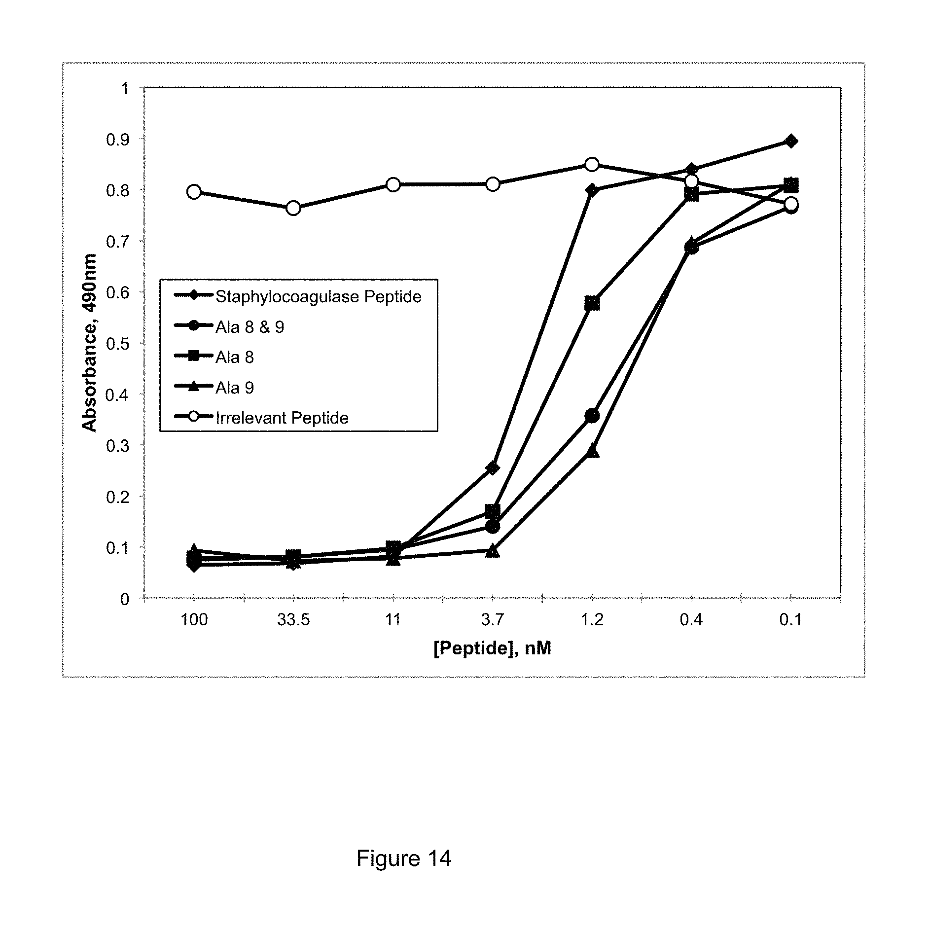
D00014
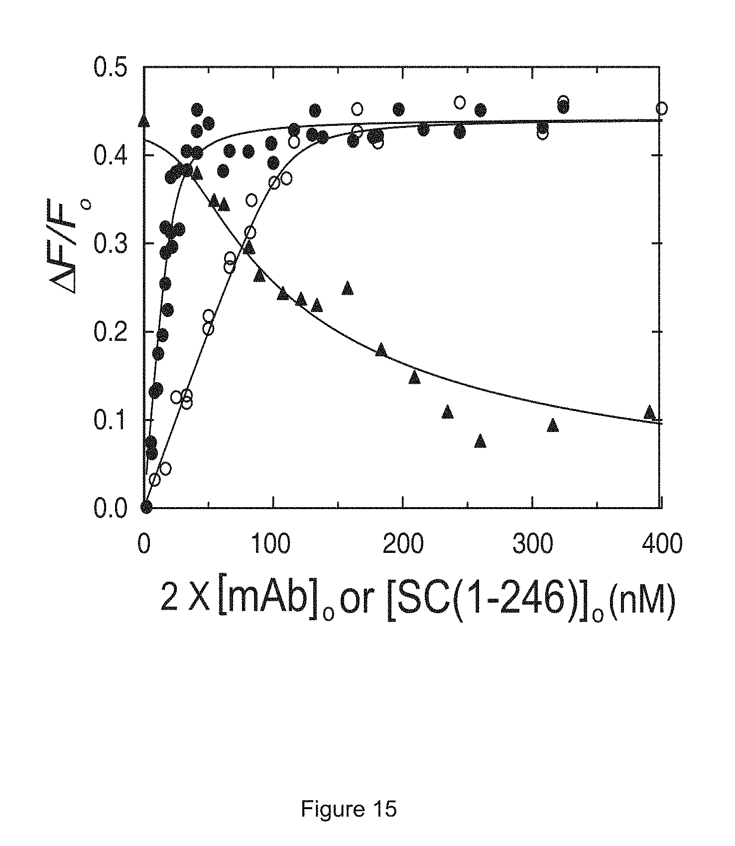
D00015

D00016
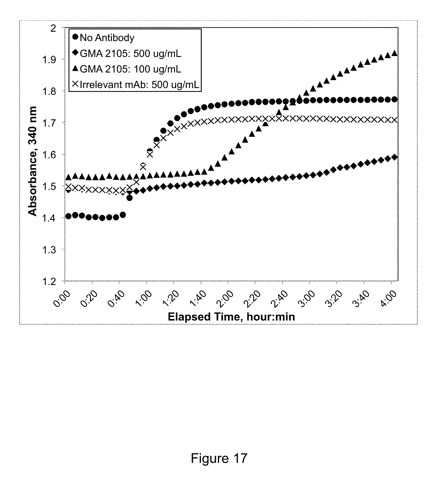
D00017
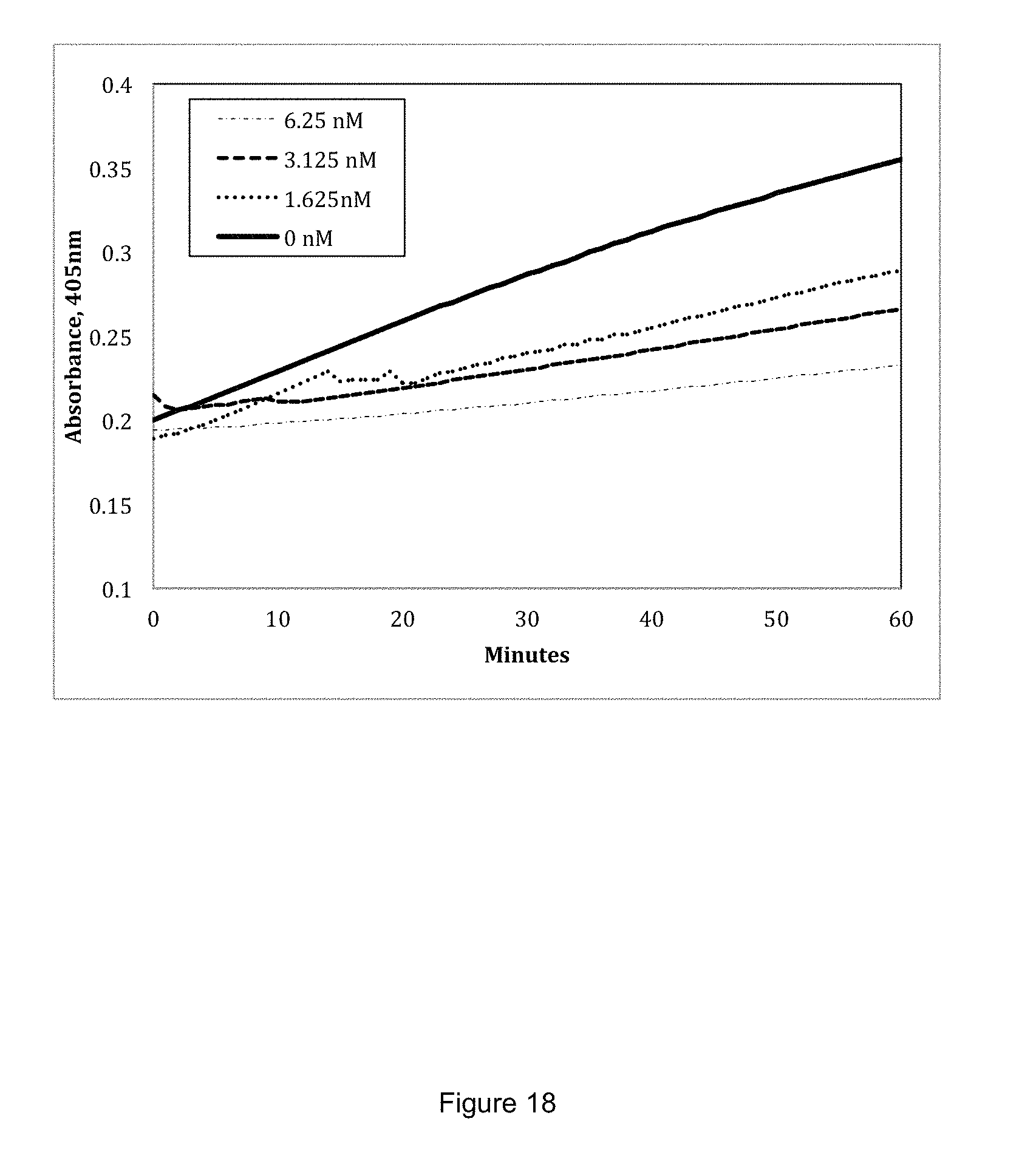
D00018

D00019
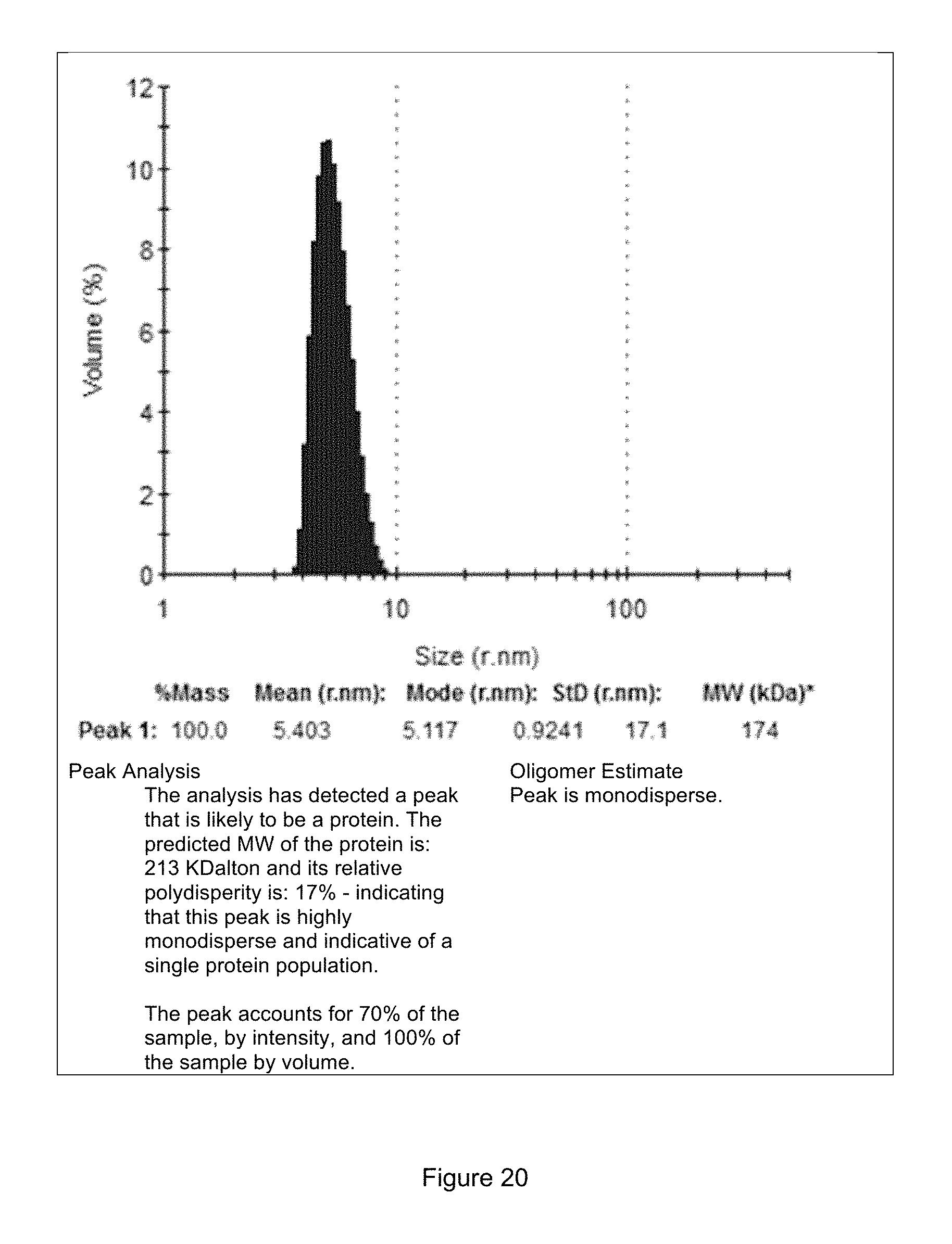
D00020

D00021

D00022
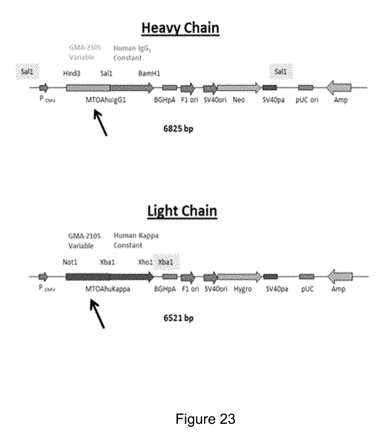
D00023
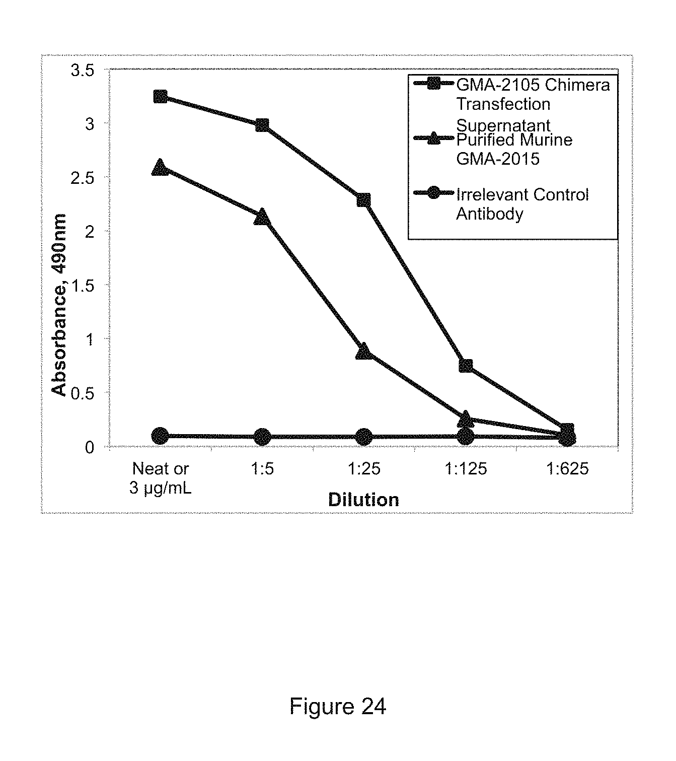
D00024

D00025

D00026

D00027

D00028
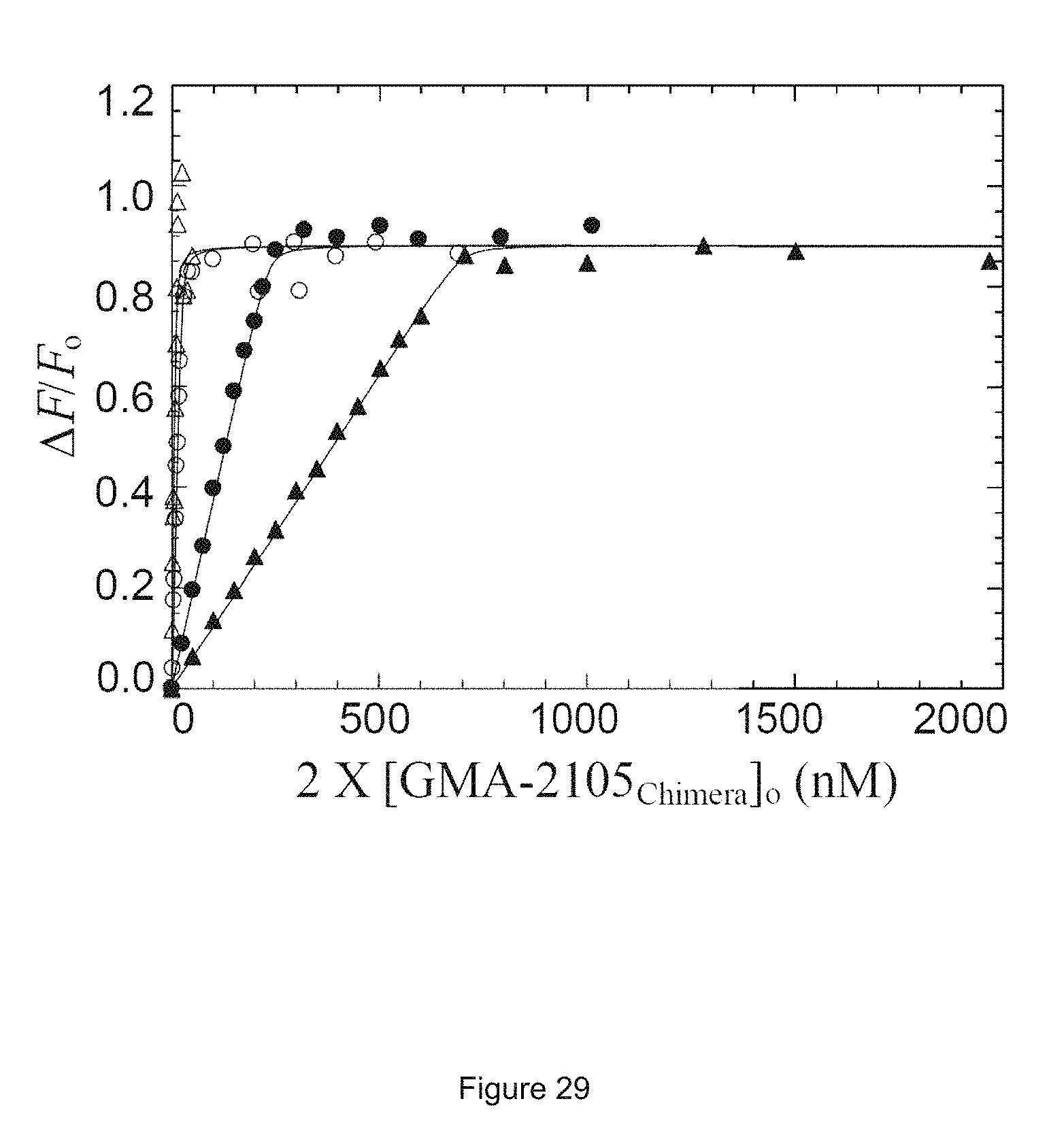
D00029

D00030

D00031

D00032

D00033

D00034

D00035

S00001
XML
uspto.report is an independent third-party trademark research tool that is not affiliated, endorsed, or sponsored by the United States Patent and Trademark Office (USPTO) or any other governmental organization. The information provided by uspto.report is based on publicly available data at the time of writing and is intended for informational purposes only.
While we strive to provide accurate and up-to-date information, we do not guarantee the accuracy, completeness, reliability, or suitability of the information displayed on this site. The use of this site is at your own risk. Any reliance you place on such information is therefore strictly at your own risk.
All official trademark data, including owner information, should be verified by visiting the official USPTO website at www.uspto.gov. This site is not intended to replace professional legal advice and should not be used as a substitute for consulting with a legal professional who is knowledgeable about trademark law.