Antibody for detecting epithelial ovarian cancer marker and method for diagnosing epithelial ovarian cancer
Sogabe , et al.
U.S. patent number 10,247,733 [Application Number 14/234,871] was granted by the patent office on 2019-04-02 for antibody for detecting epithelial ovarian cancer marker and method for diagnosing epithelial ovarian cancer. This patent grant is currently assigned to National Institute of Advanced Industrial Science and Technology. The grantee listed for this patent is Yuzuru Ikehara, Tomomi Kubota, Hayao Nakanishi, Toru Nakanishi, Hisashi Narimatsu, Hiromichi Sawaki, Maki Sogabe, Akira Togayachi. Invention is credited to Yuzuru Ikehara, Tomomi Kubota, Hayao Nakanishi, Toru Nakanishi, Hisashi Narimatsu, Hiromichi Sawaki, Maki Sogabe, Akira Togayachi.


| United States Patent | 10,247,733 |
| Sogabe , et al. | April 2, 2019 |
Antibody for detecting epithelial ovarian cancer marker and method for diagnosing epithelial ovarian cancer
Abstract
It is intended to find a highly specific epithelial ovarian cancer marker and to provide an antibody capable of specifically recognizing and detecting the marker or a fragment of the antibody. The present invention provides an anti-.beta.1,6-N-acetylglucosaminyltransferase 5B antibody for diagnosis of epithelial ovarian cancer, i.e., an antibody for detection of a glycosyltransferase .beta.1,6-N-acetylglucosaminyltransferase 5B as an epithelial ovarian cancer marker. The antibody recognizes, as an epitope, a part of a polypeptide of the enzyme consisting of the amino acid sequence represented by SEQ ID NO: 1.
| Inventors: | Sogabe; Maki (Ibaraki, JP), Kubota; Tomomi (Ibaraki, JP), Togayachi; Akira (Ibaraki, JP), Ikehara; Yuzuru (Ibaraki, JP), Narimatsu; Hisashi (Ibaraki, JP), Sawaki; Hiromichi (Ibaraki, JP), Nakanishi; Hayao (Nagoya, JP), Nakanishi; Toru (Nagoya, JP) | ||||||||||
|---|---|---|---|---|---|---|---|---|---|---|---|
| Applicant: |
|
||||||||||
| Assignee: | National Institute of Advanced
Industrial Science and Technology (Tokyo, JP) |
||||||||||
| Family ID: | 47601204 | ||||||||||
| Appl. No.: | 14/234,871 | ||||||||||
| Filed: | July 26, 2012 | ||||||||||
| PCT Filed: | July 26, 2012 | ||||||||||
| PCT No.: | PCT/JP2012/068990 | ||||||||||
| 371(c)(1),(2),(4) Date: | January 24, 2014 | ||||||||||
| PCT Pub. No.: | WO2013/015367 | ||||||||||
| PCT Pub. Date: | January 31, 2013 |
Prior Publication Data
| Document Identifier | Publication Date | |
|---|---|---|
| US 20140242607 A1 | Aug 28, 2014 | |
Foreign Application Priority Data
| Jul 26, 2011 [JP] | 2011-163323 | |||
| Current U.S. Class: | 1/1 |
| Current CPC Class: | C07K 16/40 (20130101); G01N 33/57449 (20130101); C07K 2317/34 (20130101); C07K 2317/33 (20130101) |
| Current International Class: | G01N 33/50 (20060101); G01N 33/574 (20060101); C07K 16/40 (20060101) |
References Cited [Referenced By]
U.S. Patent Documents
| 2011/0306049 | December 2011 | Yamashita et al. |
| 0445750 | Sep 1991 | EP | |||
| 1568775 | Aug 2005 | EP | |||
| 2187217 | May 2010 | EP | |||
| 03-259093 | Nov 1991 | JP | |||
| 03/091402 | Nov 2003 | WO | |||
| 2008/098129 | Aug 2008 | WO | |||
| 2009/028417 | Mar 2009 | WO | |||
Other References
|
Kaneko et al. (FEBS Letters, 554: 515-519, 2003). cited by examiner . Alvarez-Manilla et al. (Glycobiology, 20(2): 166-174, 2010). cited by examiner . Supplementary European Search Report dated Mar. 6, 2015, based on foreign counterpart European Patent Application No. 12817890; pp. 1-3. cited by applicant . Japanese Office Action dated Apr. 7, 2015 issued in counterpart Japanese Patent Application No. 2013-525755. cited by applicant . Kitazume, Shinobu "Soluble Glycosyltransferase as a Possible Biomarker", Progress of Medicine, May 24, 2008, vol. 225, No. 8, pp. 633-636 (with partial English Translation). cited by applicant . Kitazume, Shinobu "Soluble Glycosyltransferase as a Possible Biomarker", Igaku no Ayumi, May 24, 2008, vol. 225, No. 8, pp. 633-636 (with partial English Translation). cited by applicant . Nozawa, Shiro, et al., "Applications of Tumor Markers to the Screening of Endometrial and Ovarian Cancers", Japanese Journal of Clinical Medicine, 1996, vol. 54, pp. 1665-1673 (with English abstract). cited by applicant . Bast, Robert C., Jr., et al. "A Radioimmunoassay Using a Monoclongal Antibody to Monitor the Course of Epithelial Ovarian Cancer", The New England Journal of Medicine, Oct. 13, 1983, vol. 309, No. 15, pp. 883-887. cited by applicant . Susuki, Mitsuaki, et al., "Clinical Value of a New Serum Tumor Marker CA602 in Ovarian Cancers", Journal of Japan Society for Cancer Therapy, Jul. 1990, vol. 25, No. 7, pp. 1454-1460 (with English Abstract). cited by applicant . Inaba, Noriyuki, et al., "A Fundamental and Clinical Investigation of Cancer Antigen 130 (CA130) in the Field of Obstetrics and Gynecology", Journal of Japan Society for Cancer Therapy, Oct. 1989, vol. 24, No. 10, pp. 2426-2435 (with partial English translation). cited by applicant . Ohuchi, Noriaki, et al., "Levels of Circulating Tumor-Associated Glycoprotein (TAG-72) in Patients with Carcinoma Using a Novel Tumor Marker, CA72-4", Japanese Journal of Cancer and Chemotherapy, Sep. 1988, vol. 15, No. 9, pp. 2767-2772 (with partial English translation including English abstract). cited by applicant . Nozawa, Shiro, et al., "Applications of Tumor Markers to the Screening of Endometrial and Ovarian Cancers", 1996, vol. 54 pp. 1665-1673 (with English abstract). cited by applicant . Charpin, Colette, et al., "Carcinoembryonic Antigen (CEA) and Carbohydrate Determinant 19-9 (CA 19-9) Localization in 121 Primary and Metastatic Ovarian Tumors: An Immunohistochemical Study with the Use of Monoclonal Antibodies", International Journal of Gynecological Pathology, 1982, vol. 1, No. 3, pp. 231-245. cited by applicant . Konica Minolta, Inc., "GAT Test Kit for Determination of the Serum GAT Level by EIA", Oct. 2003, Instructions for Use, pp. 1-6 (with partial English translation). cited by applicant . Seko, Akira, et al., "b1,3-Galactosyltransferases-4/5 are Novel Tumor Markers for Gynecological Cancers", Tumor Biology, 2009, vol. 30, pp. 43-50. cited by applicant . Kaneko, Mika, et al., A Novel b(1,6)-N-acetylglucosaminyltransferase V (GnT-VB).sup.1, FEBS Letters, 2003, vol. 554, No. 3, pp. 515-519. cited by applicant . Takahashi, Noriko, et al., "P4-165: Significance of the Expression of N-acetylglucosamine Transferase in Ovarian Cancer", Acta Obstetrica et Gynaecologica Japonica, Feb. 1, 2008, vol. 60, No. 2, p. 839 (with English Translation). cited by applicant . Takahashi, Noriko, et al., "High Expression of N-acetylglucosaminyltransferase V in Mucinous Tumors of the Ovary", Oncology Reports, 2009, vol. 22, No. 5, pp. 1027-1032. cited by applicant . International Preliminary Examination Report on Patentability, dated Jan. 28, 2014 relating to International Application No. PCT/JP2012/068990. cited by applicant . Written Opinion of the International Searching Authority, dated Sep. 4, 2012, relating to International Application No. PCT/JP2012/068990. cited by applicant. |
Primary Examiner: Moseley, II; Nelson B
Attorney, Agent or Firm: McCarter & English, LLP
Claims
The invention claimed is:
1. A method for diagnosis of epithelial ovarian cancer, comprising: (a) quantitatively detecting a .beta.-1,6-N-acetylglucosaminyltransferase 5B polypeptide fragment present in a sample derived from a test subject using at least one anti-.beta.-1,6-N-acetylglucosaminyltransferase 5B monoclonal antibody, or antibody fragment thereof, and (b) determining that the test subject is diagnosed to be likely to have epithelial ovarian cancer when the results of quantitatively detecting the .beta.-1,6-N-acetylglucosaminyltransferase 5B polypeptide fragment are greater than a predetermined cut-off value, wherein the predetermined cut-off value is the 95th percentile of the quantitative detection results of the .beta.-1,6-N-acetylglucosaminyltransferase 5B polypeptide fragment in the sample derived from normal individuals or patients other than epithelial ovarian cancer patients, wherein the anti-.beta.-1,6-N-acetylglucosaminyltransferase 5B monoclonal antibody is produced by a hybridoma identified by International Accession No. FERM BP-11496, FERM BP-11497, FERM BP-11498, or FERM BP-11499.
2. The method for diagnosis of epithelial ovarian cancer of claim 1, wherein the .beta.-1,6-N-acetylglucosaminyltransferase 5B polypeptide fragment comprising an epitope recognized by the anti-.beta.-1,6-N-acetylglucosaminyltransferase 5B monoclonal antibody, or the antibody fragment thereof is the whole or a part of a polypeptide consisting of the amino acid sequence of SEQ ID NO: 1.
3. The method for diagnosis of epithelial ovarian cancer of claim 1, wherein two anti-.beta.-1,6-N-acetylglucosaminyltransferase 5B monoclonal antibodies are used to detect the .beta.-1,6-N-acetylglucosaminyltransferase 5B polypeptide fragment.
4. A method for diagnosis of epithelial ovarian cancer, comprising: (a) quantitatively detecting a .beta.-1,6-N-acetylglucosaminyltransferase 5B polypeptide fragment present in a sample derived from a test subject using at least one monoclonal antibody or antibody fragment thereof, and (b) determining that the test subject is diagnosed to be likely to have epithelial ovarian cancer when the results of quantitatively detecting the .beta.-1,6-N-acetylglucosaminyltransferase 5B polypeptide fragment are greater than a predetermined cut-off value of 4.0 ng/mL, wherein the monoclonal antibody is produced by a hybridoma identified by International Accession No. FERM BP-11496, FERM BP-11497, FERM BP-11498, or FERM BP-11499.
5. The method for diagnosis of epithelial ovarian cancer of claim 1, wherein the sample is a body fluid, a peritoneal lavage fluid, or a tissue.
6. The method for diagnosis of epithelial ovarian cancer of claim 4, wherein two anti-.beta.-1,6-N-acetylglucosaminyltransferase 5B monoclonal antibodies are used to detect the .beta.-1,6-N-acetylglucosaminyltransferase 5B polypeptide fragment.
Description
CROSS-REFERENCE TO RELATED APPLICATIONS
This application is a national stage application under 35 U.S.C. .sctn. 371 of PCT/JP2012/068990, filed Jul. 26, 2012, which claims benefit of Japanese Appl. No. 2011-163323, filed Jul. 26, 2011, which is incorporated herein by reference.
SEQUENCE LISTING
The Sequence Listing associated with this application is filed in electronic format via EFS-Web and hereby is incorporated by reference into the specification. The name of the text file containing the Sequence Listing is 14234871_Repl_Seq_List_ST25.txt. The size of the text file is 17 KB, and the text file was created on Mar. 10, 2014.
TECHNICAL FIELD
The present invention relates to an anti-.beta.1,6-N-acetylglucosaminyltransferase 5B antibody for detection of an epithelial ovarian cancer marker, a hybridoma producing the antibody, and a method for diagnosis of epithelial ovarian cancer using the antibody.
BACKGROUND ART
Ovarian cancer is a cancer with a low incidence, compared with breast cancer or uterine cancer, among gynecologic cancers. Both incidence and mortality of this cancer, however, have been on the increase in recent years. In general, ovarian cancer is substantially asymptomatic early in the course of the disease and often found at the already advanced stage of symptoms. Hence, this disease has poor prognosis and exhibits the highest mortality among gynecologic cancers. Ovarian cancer is known to include, for example, surface epithelial-stromal tumors (hereinafter, referred to as "epithelial ovarian cancer") developed from surface epithelial cells in the ovary and germ cell tumors developed from germ cells, depending on the area affected. Of them, epithelial ovarian cancer accounts for approximately 90% of all ovarian cancer cases and is often seen particularly in middle-aged women in their 40s or older. Thus, the early detection of epithelial ovarian cancer is important for the treatment of the disease.
The ovary is an organ that has no contact with the outside of the body. Unlike the uterus, the ovary can be neither examined endoscopically nor subjected to cell harvest without abdominal section or perforation. In addition, epithelial ovarian cancer is generally difficult to detect by palpation before the ovary is enlarged at the advanced stage of symptoms. Hence, epithelial ovarian cancer is often undetected in ordinary examination or diagnosis methods. Although echography, MRI, CT, or the like is relatively effective for the early detection of epithelial ovarian cancer, this examination itself is extensive work with a high cost. In addition, these approaches, unfortunately, do not always have high diagnostic accuracy to distinguish between benign and malignant tumors.
Against this backdrop, tumor markers have received attention in recent years. The tumor markers refer to substances that are produced by cancer cells or produced by cells surrounding cancer cells in response to the cancer cells. The abundance of each tumor marker in a body fluid can reflect the presence or absence of tumor or the prognosis thereof and therefore serve as an index for, for example, cancer diagnosis and decision on therapeutic strategies. Also, this approach permits examination using a body fluid and is thus relatively low invasive. Advantageously, this examination is also convenient and low in cost.
Tumor markers composed of proteins such as CA125, CA602, CA130, CA72-4, CA546, CA 19-9, and STN have been known so far as tumor markers for epithelial ovarian cancer (Non Patent Literatures 1 to 6). Methods for diagnosis of cancer using these tumor markers typically involve measuring the expression levels of the tumor markers in the serum of normal individuals and epithelial ovarian cancer patients and determining the presence or absence of cancer developed in a test subject on the basis of the difference in the levels.
These proteins, however, present specificity problems in such a way that CA125 exhibits positivity to a non-cancer benign gynecologic disease such as endometriosis or CA72-4, CA19-9, and STN exhibit positivity to various cancers of the digestive system including the stomach and the large intestine in addition to ovarian cancer. Epithelial ovarian cancer is further classified into serous, clear cell, mucinous, and endometrioid tumors depending on the histological type. The markers differ in reactivity among these histological types and therefore, do not correctly reflect the progression of cancer in some cases. For example, the ovarian cancer markers such as CA125, CA602, and CA546 have a low positive rate for mucinous ovarian cancer. Unfortunately, this histological type is therefore rarely detected even at the advanced stage.
Patent Literature 1 discloses a monoclonal antibody for use in the diagnosis of cancers including ovarian cancer against human galactosyltransferase associated with tumor (GAT) as a tumor marker, a hybridoma producing the antibody, and a method for assaying human galactosyltransferase associated with tumor in a specimen using the antibody. Also, Patent Literature 2 discloses a method for detecting gynecologic cancers early using glycosyltransferases .beta.1,3-galactosyltransferase 5, .beta.1,3-galactosyltransferase 4, and N-acetylglucosamine-6-O-sulfotransferase 2 as tumor markers. These glycosyltransferases used as tumor markers in Patent Literatures 1 and 2 are enzymes that synthesize sugar chains to be bound with, for example, glycoproteins, glycolipids or proteoglycans, and are usually anchored on Golgi membranes as membrane proteins localized to the Golgi bodies. Thus, these enzymes are not secreted to the outside of the cells and are therefore rarely detected in the body fluids of normal individuals. In ovarian cancer, however, it is known that glycosyltransferases are abnormally cleaved due to the increased expression level of a certain kind of protease and released to the outside of the cells. As a result, significant amounts of glycosyltransferase fragments are detected in body fluids.
The glycosyltransferase .beta.1,4-galactosyltransferase described in Patent Literature 1 has an exceedingly high expression level among glycosyltransferases and is thus secreted in large amounts to the outside of the cell. Hence, this glycosyltransferase exhibits a serum concentration of approximately 200 ng/mL even in normal individuals and thus fails to distinguish between benign disease and cancer by its enzymatic activity alone. Accordingly, Patent Literature 1 was focused on the presence of a fragment of the abnormally cleaved glycosyltransferase in the culture supernatant of ovarian cancer cells and the ascitic fluid of an ovarian cancer patient. An antibody that recognizes only this fragment was used to attempt the construction of an assay system highly specific for ovarian cancer. Nonetheless, a commercially available clinical diagnosis kit based on this assay system exhibited positivity even for healthy women in some cases (Non Patent Literature 7). For this reason, GAT is currently used mainly in the monitoring of ovarian cancer recurrence and rarely used as a marker for early detection.
In Patent Literature 2, an attempt was made to detect marker candidate proteins in the blood of gynecologic cancer patients on the basis of reports stating that the expression of various proteins including glycosyltransferases is generally increased or decreased in cancer tissues. As a result, two glycosyltransferases were found to be useful in assay. The literature discloses an assay method according to the findings. In this case, however, the expression or synthesis products of these glycosyltransferases are reportedly related to digestive system cancers rather than gynecologic cancers. Although their expression was then confirmed in ovarian cancer cell lines (Non Patent Literature 8), there has been no report on the comparison of the expression levels between ovarian cancer or other gynecologic cancers and digestive system cancers.
As mentioned above, Patent Literatures 1 and 2 are directed only to the detection of markers in the body fluids of ovarian cancer patients with little consideration given to glycosyltransferase expression in ovarian cancer and disclose a glycosyltransferase fragment that happened to be measurable, as an ovarian cancer marker. This may lead to specificity problems as ovarian cancer markers.
CITATION LIST
Patent Literature
Patent Literature 1: JP Patent Publication (Kokai) No. 3-259093 A (1991) Patent Literature 2: WO2009/028417
Non Patent Literature
Non Patent Literature 1: Bast R. C. Jr. et al., 1983, N. Engl. J. Med., 309: 883-887 Non Patent Literature 2: Suzuki M. et al., 1990, Nippon Gan Chiryo Gakkai Shi, 25: 1454-1460 Non Patent Literature 3: Inaba N. et al., 1989, Nippon Gan Chiryo Gakkai Shi, 24: 2426-2435 Non Patent Literature 4: Ohuchi N. et al., 1988, Gan To Kagaku Ryoho, 15, 2767-2772 Non Patent Literature 5: Nozawa S. et al., 1996, Nippon Rinsho, 54: 1665-1673 Non Patent Literature 6: Charpin C. et al., 1982, Int. J. Gynecol. Pathol., 1: 231-245 Non Patent Literature 7: Konica Minolta, Inc., GAT Test Kit product document Non Patent Literature 8: Seko A. et al., 2009, Tumor Biol., 30: 43-50
SUMMARY OF INVENTION
Technical Problem
An object of the present invention is to provide an antibody or a fragment of the antibody capable of specifically recognizing and quantitatively and/or qualitatively detecting a tumor marker highly specific for epithelial ovarian cancer.
Another object of the present invention is to provide a method for conveniently and relatively low invasively diagnosis with high accuracy rate whether or not a test subject has epithelial ovarian cancer by quantitatively and/or qualitatively detecting an epithelial ovarian cancer marker using the antibody or the fragment thereof.
Solution to Problem
The present inventors have conducted diligent studies and consequently found that a glycosyltransferase .beta.1,6-N-acetylglucosaminyltransferase 5B (hereinafter, the enzyme is also abbreviated to "MGAT5B" in the present specification) is present in larger amounts in the samples of epithelial ovarian cancer patients compared with normal individuals. The present invention is based on the findings and provides the following:
(1) An antibody for detection of an epithelial ovarian cancer marker recognizing, as an epitope, a part of a polypeptide consisting of the amino acid sequence represented by SEQ ID NO: 1.
(2) The antibody according to (1), wherein the antibody is a monoclonal antibody.
(3) The antibody according to (2), wherein the antibody is produced by a hybridoma identified by International Accession No. FERM ABP-11496, FERM ABP-11497, FERM ABP-11498, or FERM ABP-11499.
(4) A recombinant anti-MGAT5B antibody for detection of an epithelial ovarian cancer marker comprising at least one set of corresponding light chain complementarity determining regions and heavy chain complementarity determining regions in an antibody according to any of (1) to (3).
(5) An antibody fragment for detection of an epithelial ovarian cancer marker, which is a fragment of an antibody according to any of (1) to (3) or a recombinant antibody according to (4) and having the activity of specifically recognizing MGAT5B.
(6) A hybridoma producing an antibody according to (2).
(7) The hybridoma according to (6), wherein the hybridoma is identified by International Accession No. FERM ABP-11496.
(8) The hybridoma according to (6), wherein the hybridoma is identified by International Accession No. FERM ABP-11497.
(9) The hybridoma according to (6), wherein the hybridoma is identified by International Accession No. FERM ABP-11498.
(10) The hybridoma according to (6), wherein the hybridoma is identified by International Accession No. FERM ABP-11499.
(11) A method for diagnosis of epithelial ovarian cancer, comprising quantitatively and/or qualitatively detecting a MGAT5B polypeptide fragment present in a sample derived from a test subject, and determining the presence or absence of epithelial ovarian cancer developed in the test subject on the basis of the detection results.
(12) The method for diagnosis of epithelial ovarian cancer according to (11), wherein when the quantitative detection results of the MGAT5B polypeptide fragment show a quantification value of the MGAT5B polypeptide fragment equal to or higher than a predetermined value, the test subject is determined to be likely to have epithelial ovarian cancer.
(13) The method for diagnosis of epithelial ovarian cancer according to (11) or (12), wherein the MGAT5B polypeptide fragment is the whole or a part of a polypeptide consisting of the amino acid sequence represented by SEQ ID NO: 1.
(14) The method for diagnosis of epithelial ovarian cancer according to any of (11) to (13), wherein the MGAT5B polypeptide fragment is detected using at least one antibody, recombinant antibody, and/or antibody fragment selected from the group consisting of an antibody according to any of (1) to (3), a recombinant antibody according to (4), and an antibody fragment according to (5).
(15) The method for diagnosis of epithelial ovarian cancer according to (14), wherein two antibodies, recombinant antibodies, and/or antibody fragments that recognize different epitopes on the MGAT5B polypeptide fragment are used.
(16) The method for diagnosis of epithelial ovarian cancer according to any of (11) to (15), wherein the sample is a body fluid, a peritoneal lavage fluid, or a tissue.
The present specification encompasses the contents described in the specification and/or drawings of Japanese Patent Application No. 2011-163323 on which the priority of the present application is based.
Effects of Invention
The antibody of the present invention and/or the fragment thereof is capable of specifically recognizing and detecting a glycosyltransferase MGAT5B polypeptide fragment as an epithelial ovarian cancer marker.
The hybridoma of the present invention can stably supply an anti-MGAT5B monoclonal antibody capable of specifically recognizing and detecting a glycosyltransferase MGAT5B as an epithelial ovarian cancer marker.
The method for diagnosis of epithelial ovarian cancer according to the present invention can detect epithelial ovarian cancer conveniently and relatively low invasively with high accuracy rate. As a result, whether or not a test subject has epithelial ovarian cancer can be determined early.
BRIEF DESCRIPTION OF DRAWINGS
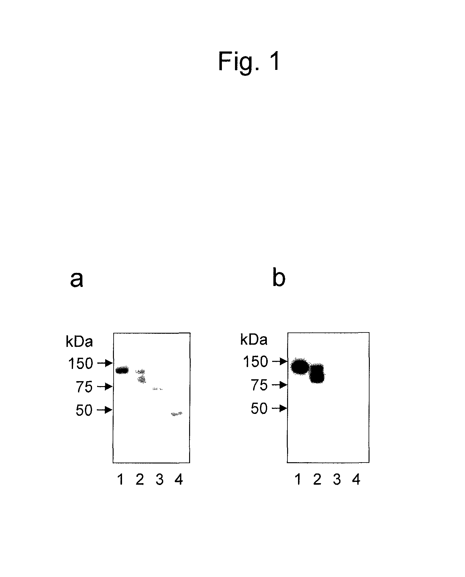
FIG. 1 shows results of Western blotting by which anti-MGAT5B monoclonal antibodies (a: GT131-12 antibody and b: GT131-18 antibody) derived from hybridomas obtained in Example 1 were studied for their antigen specificity. Lane 1 shows the results about a FLAG-MGAT5B polypeptide fragment. Lane 2 shows the results about a MGAT5B polypeptide fragment from which the FLAG tag was cleaved off by enzymatic treatment. Lane 3 shows the results about the immunoglobulin- and albumin-free normal human serum (NHS). Lane 4 shows the results about the untreated NHS.
FIG. 2 shows results of Western blotting by which FLAG-MGAT5B polypeptide fragments immunoprecipitated with anti-MGAT5B antibodies were detected using various antibodies (a: anti-FLAG antibody, b: GT131-12 antibody, and c: GT131-18 antibody). Lane 1 shows the results about a control FLAG-MGAT5B polypeptide fragment. Lane 2 shows the results about a sample obtained by the immunoprecipitation with a GT131-2 antibody of a FLAG-MGAT5B polypeptide fragment mixed with the serum of a normal individual. Lane 3 shows the results about a sample obtained by the immunoprecipitation with a GT131-7 antibody of a FLAG-MGAT5B polypeptide fragment mixed with the NHS. Lane 4 shows the results about a sample obtained by the immunoprecipitation with a GT131-12 antibody of a FLAG-MGAT5B polypeptide fragment mixed with the NHS. Lane 5 shows the results about a sample obtained by the immunoprecipitation with a GT131-18 antibody of a FLAG-GAT5B polypeptide fragment mixed with the NHS. Lane 6 shows the results about a non-immunoprecipitated FLAG-MGAT5B polypeptide fragment mixed with the NHS.
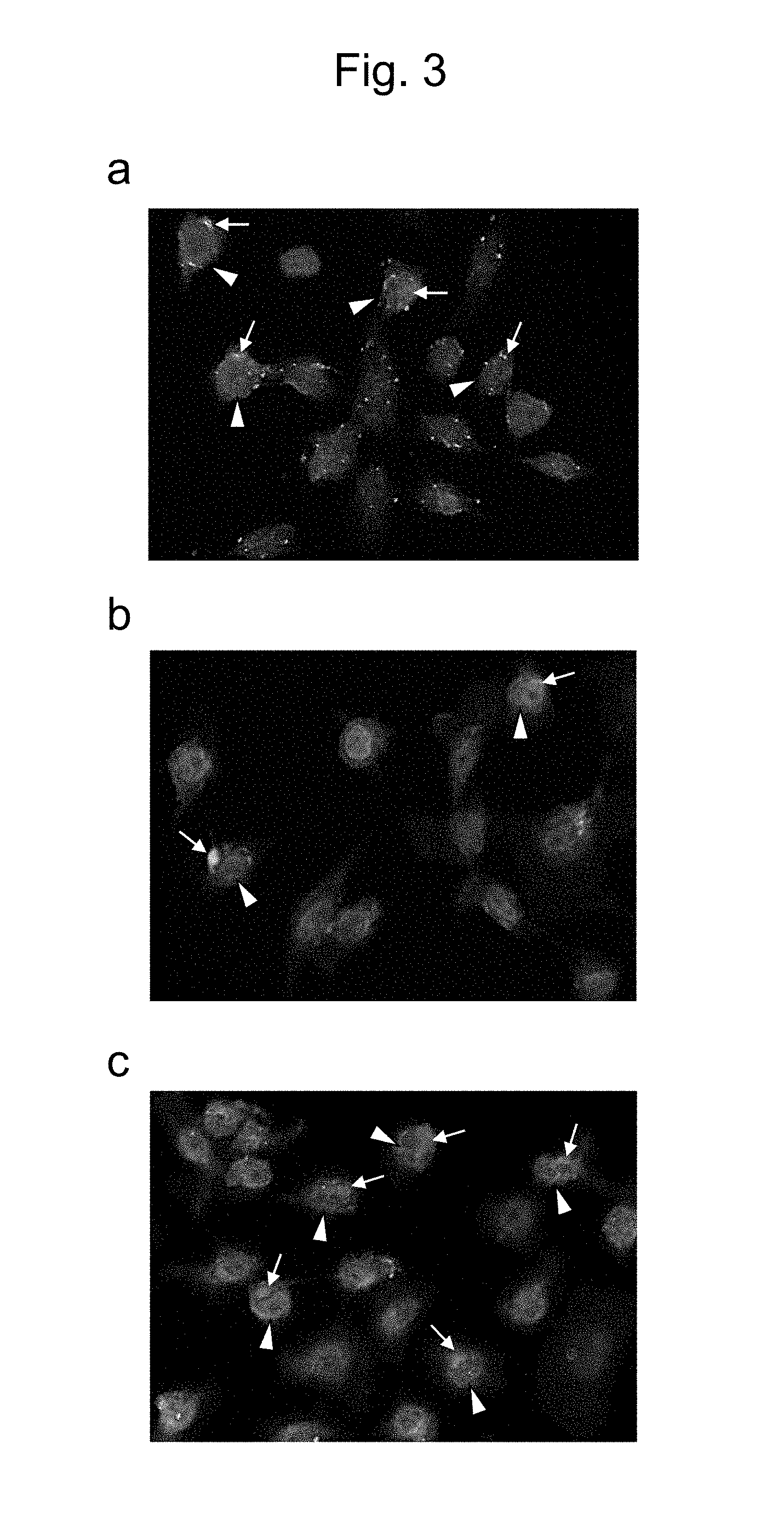
FIG. 3 shows the immunostaining of an ovarian cancer cell line using anti-MGAT5B antibodies. FIG. 3a is a staining pattern of an epithelial ovarian cancer cell line RMUG-S immunostained with a GT131-2 antibody. FIG. 3b is a staining pattern of the cell line immunostained with a GT131-18 antibody. FIG. 3c is a staining pattern of the cell line immunostained with a positive control MAb8628 antibody.
FIG. 4 shows signal intensity vs. a concentration standard in study on the combination of antibodies for sandwich ELISA.
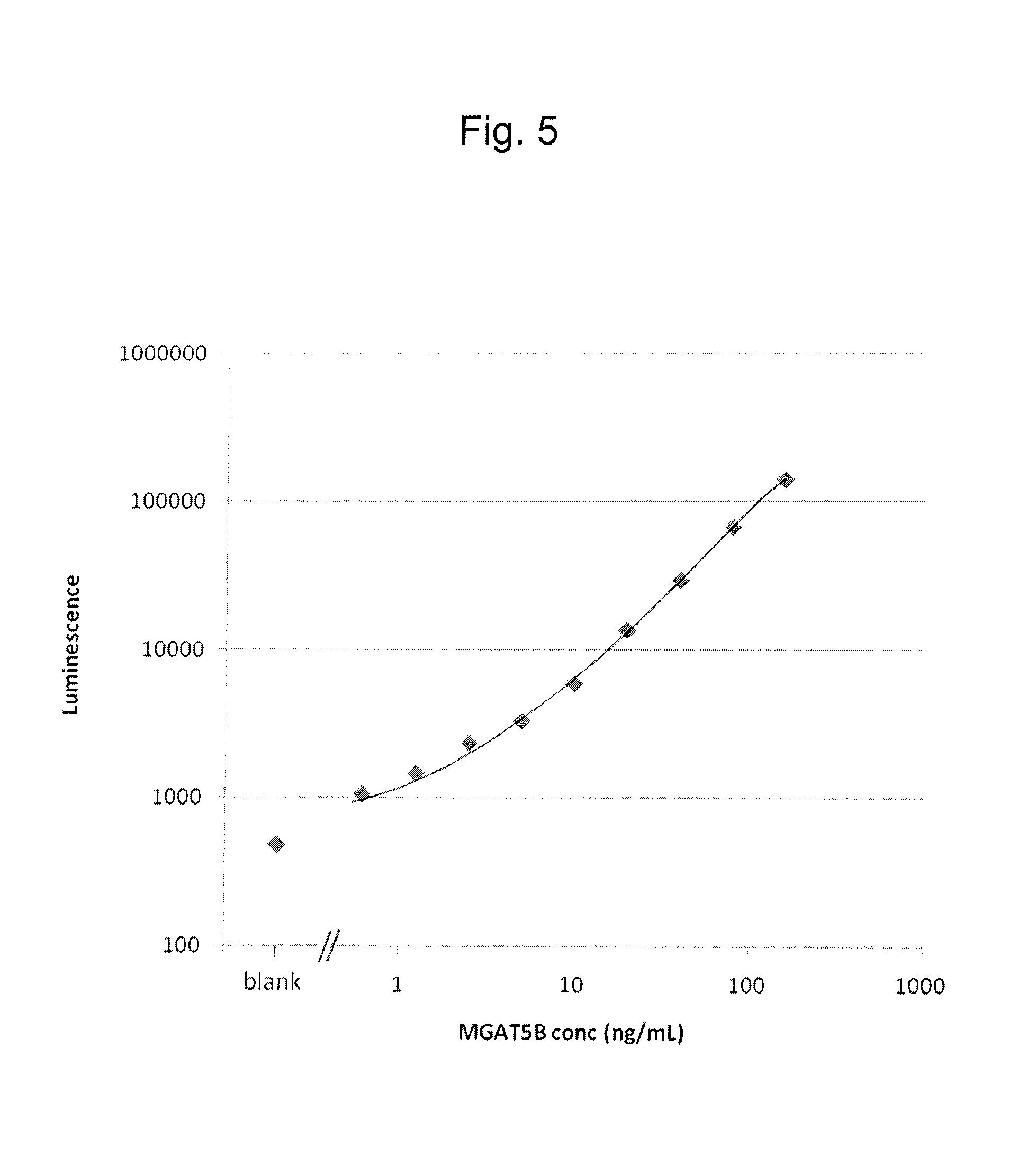
FIG. 5 shows a calibration curve of MGAT5B polypeptide fragments obtained from values of a concentration standard measured by sandwich CLEIA using a GT131-7 antibody and a GT131-12 antibody.
FIG. 6 shows the concentration of the epithelial ovarian cancer marker MGAT5B polypeptide fragment of the present invention in a peritoneal lavage fluid collected from a test subject, wherein the concentration was measured by sandwich CLEIA using a GT131-7 antibody and a GT131-12 antibody.
FIG. 7 shows the correlation between a measurement value of the MGAT5B polypeptide fragment and a measurement value of an existing ovarian cancer marker CA125 (a) or GAT (b) in peritoneal lavage fluids collected from epithelial ovarian cancer patients.
FIG. 8 shows the correlation between a CA125 measurement value and a GAT measurement value in peritoneal lavage fluids collected from epithelial ovarian cancer patients.
DESCRIPTION OF EMBODIMENTS
1. Antibody for Detection of Epithelial Ovarian Cancer Marker
1-1. Definition and Constitution
The first embodiment of the present invention relates to an antibody for detection of an epithelial ovarian cancer marker. The antibody of the present invention is an antibody that is used for detecting an epithelial ovarian cancer marker and recognizes and specifically binds to an epitope contained in the epithelial ovarian cancer marker.
In the present invention, the "epithelial ovarian cancer marker" refers to a biological marker for detection of epithelial ovarian cancer which is a biomaterial serving as an index showing that a test subject has epithelial ovarian cancer. The epithelial ovarian cancer marker according to the present invention is specifically a .beta.1,6-N-acetylglucosaminyltransferase 5B polypeptide fragment.
".beta.1,6-N-acetylglucosaminyltransferase 5B (MGAT5B)" is one type of N-linked sugar chain-synthesizing enzyme, and as a membrane protein mainly localized to the Golgi membrane. This enzyme transfers N-acetylglucosamine with the .beta.1-6 linkage (Kaneko M. et al., 2003, FEBS Letters, 554: 515-519). MGAT5B contains one N-terminal transmembrane domain and has an enzymatically active region as a C-terminal region. The MGAT5B according to the present invention is the human-derived wild-type MGAT5B composed of 790 amino acids and registered under GenBank Accession No. NP_653278. Specifically, the MGAT5B according to the present invention is a polypeptide consisting of the amino acid sequence represented by SEQ ID NO: 2. In the present specification, MGAT5B encompasses wild-type MGAT5B and natural variants thereof.
In the present specification, the "natural variant" of MGAT5B refers to a naturally occurring variant that contains the deletion, substitution, addition, or insertion of 1 to 10, preferably 1 to 5, more preferably 1 to 4, 1 to 3, or 1 or 2 amino acids in the amino acid sequence (SEQ ID NO: 2) constituting wild-type MGAT5B or is a polypeptide exhibiting approximately 90% or higher, preferably approximately 95% or higher, more preferably approximately 98% or higher identity to the amino acid sequence represented by SEQ ID NO: 2. In this context, the "identity" refers to the ratio (%) of identical amino acid residues of a target amino acid sequence to the total number of amino acid residues of the amino acid sequence represented by SEQ ID NO: 2 including the number of gaps when the amino acid sequence represented by SEQ ID NO: 2 and the target amino acid sequence are aligned such that the maximum degree of identity can be achieved with or without introduced gaps. This identity can be determined using a protein search system based on BLASTP or FASTA. Specific examples of the natural variant include variants based on polymorphisms such as SNP (single-nucleotide polymorphism) and splicing variants. In this context, the natural variant of MGAT5B is not necessarily required to have enzymatic activity equivalent to that of wild-type MGAT5B. This is because, reportedly, some natural variants of glycosyltransferases substantially lose activity even due to the substitution of only one amino acid (Nishihara S. et al., 1993, Biochem. Biophys. Res. Commun. 196: 624-631).
In the present specification, the "MGAT5B polypeptide fragment" refers to a MGAT5B-derived polypeptide fragment that is released to the outside of the epithelial ovarian cancer cell as a result of abnormal cleavage by protease from the Golgi membrane in the cell and is capable of functioning as an epithelial ovarian cancer marker as mentioned above. Specifically, the MGAT5B polypeptide fragment and a fragment derived from the polypeptide correspond to a polypeptide consisting of the whole or a part of the intra-Golgi C-terminal region of MGAT5B except for the N-terminal region containing the transmembrane domain, for example, a polypeptide consisting of the whole or a part of an amino acid sequence (SEQ ID NO: 1) from glycine at residue 51 (counted with initiating methionine as the 1st position) to downstream residues in human MGAT5B shown in SEQ ID NO: 2.
The "antibody for detection of epithelial ovarian cancer" of the present invention refers to an antibody that is induced with the epithelial ovarian cancer marker MGAT5B polypeptide fragment as an antigen. Thus, in the present specification, the "antibody for detection of an epithelial ovarian cancer marker" is also referred to as an "anti-MGAT5B antibody". The anti-MGAT5B antibody is capable of recognizing and binding to a part of the epithelial ovarian cancer marker as an epitope and specifically detecting the epithelial ovarian cancer marker. Specifically, a part of an amino acid consisting of the amino acid sequence represented by SEQ ID NO: 1 is used as an epitope. In this context, the term "a part" refers to one or more regions each composed of 5 to 15, preferably 5 to 10, more preferably 6 to 10 consecutive amino acids.
The anti-MGAT5B antibody of the present invention may be any of polyclonal and monoclonal antibodies. A monoclonal antibody is desirable for achieving more specific detection. Specific examples of the anti-MGAT5B monoclonal antibody include anti-MGAT5B antibodies produced by hybridomas identified by International Accession No. FERM ABP-11496, FERM ABP-11497, FERM ABP-11498, and FERM ABP-11499. These antibodies will be described in detail in the paragraph "(3) Preparation of anti-MGAT5B monoclonal antibody".
The anti-MGAT5B antibody of the present invention can be modified. In this context, the "modification" includes functional modification necessary for antigen-specific binding activation, such as glycosylation, and labeling necessary for antibody detection. Examples of the antibody labeling include labeling using fluorescent dyes (FITC, rhodamine, Texas Red, Cy3, and Cy5), fluorescent proteins (e.g., PE, APC, and GFP), enzymes (e.g., horseradish peroxidase, alkaline phosphatase, and glucose oxidase), and biotin or (strept)avidin.
The modification on the anti-MGAT5B antibody may be altered. For example, the glycosylation of the anti-MGAT5B antibody may be altered in order to adjust the affinity of the anti-MGAT5B antibody for the target antigen epithelial ovarian cancer marker. Specifically, examples thereof include alteration by which the glycosylation site in each framework region (FR) of the anti-MGAT5B antibody is removed by the introduction of substitution into amino acid residue(s) constituting the glycosylation site to thereby delete glycosylation at the site.
Preferably, the anti-MGAT5B antibody of the present invention has affinity for the epithelial ovarian cancer marker as high as a dissociation constant of 10.sup.-8 M or lower, preferably 10.sup.-9 M or lower, more preferably 10.sup.-10 M or lower. The dissociation constant can be measured using a technique known in the art. The dissociation constant may be measured using, for example, rate evaluation kit software of BIAcore system (GE Healthcare Japan Corp.).
The anti-MGAT5B polyclonal antibody or the anti-MGAT5B monoclonal antibody of the present invention can be obtained by a production method mentioned later. Alternatively, the anti-MGAT5B monoclonal antibody may be prepared by a chemical synthesis method or a recombinant DNA technique on the basis of its amino acid sequence. The anti-MGAT5B monoclonal antibody may also be obtained from a hybridoma producing the antibody.
The anti-MGAT5B antibody of the present invention is not particularly limited by an organism species of origin. The anti-MGAT5B antibody of the present invention can be derived from every animal source including bird and mammals. Examples of such animal sources include mice, rats, guinea pigs, rabbits, goats, donkeys, sheep, camels, horses, chickens, and humans. The anti-MGAT5B antibody of the present invention is not particularly limited by a globulin type and can be any of IgG, IgM, IgA, IgE, IgD, and IgY. IgG and IgM are preferred.
1-2. Preparation of Anti-MGAT5B Antibody
The anti-MGAT5B antibody of the present invention, i.e., the anti-MGAT5B polyclonal antibody or the anti-MGAT5B monoclonal antibody, or the hybridoma producing the anti-MGAT5B monoclonal antibody can be prepared by a method described below. However, the production method of the present invention is not limited to the method described below, and any other method known in the art can be used in the preparation of the antibody or the hybridoma.
(1) Preparation of Immunogen
The epithelial ovarian cancer marker is prepared as an immunogen. In the present invention, the epithelial ovarian cancer marker that may be used as an immunogen is, for example, the whole or a part of a human MGAT5B polypeptide fragment having the amino acid sequence represented by SEQ ID NO: 1, or the whole or a part of a variant polypeptide fragment thereof.
The MGAT5B polypeptide fragment as an immunogen can be prepared using, for example, a chemical synthesis method or a DNA recombination technique.
In the case of preparing the fragment using the chemical synthesis method, an appropriate MGAT5B polypeptide fragment for use as an immunogen can be chemically synthesized by an approach known in the art, for example, a solid-phase peptide synthesis method, on the basis of information about, for example, the amino acid sequence of SEQ ID NO: 1.
In the case of preparing the fragment using the DNA recombination technique, a MGAT5B-encoding cDNA (MGAT5B cDNA) can be incorporated into an appropriate expression system and expressed to obtain the MGAT5B polypeptide fragment. Hereinafter, the method for preparing the MGAT5B polypeptide fragment will be described with reference to specific examples.
(Preparation of MGAT5B cDNA)
The MGAT5B cDNA can be prepared by a technique known in the art, for example, a cDNA cloning method. Specifically, a human cDNA library is first prepared. The human cDNA library can be prepared by: extracting total RNAs according to a routine method from, for example, human fibroblasts (neuroblastoma cell line SK-N-SH) expressing the MGAT5B gene or the like; then recovering poly-A(+) RNAs by treatment with oligo dT cellulose columns; and performing RT-PCR with the recovered RNAs as templates. Alternatively, a commercially available human cDNA library may be used.
Subsequently, the MGAT5B cDNA clone of interest is isolated from the human cDNA library. Specifically, the clone can be isolated by a screening method known in the art, for example, a hybridization screening method, an expression screening method, or an antibody screening method, using primers and/or a probe appropriately designed on the basis of the MGAT5B gene sequence. The MGAT5B gene sequence is registered under Accession No. NM_144677 in the GenBank database. The primers are designed so that a MGAT5B gene region encoding the amino acid sequence represented by SEQ ID NO: 1 is incorporated in an amplification fragment. An appropriate restriction site for cloning after isolation or a tag sequence for protein purification (FLAG, HA, His, myc, GFP, etc.) may be introduced to the 5' end of the forward or reverse primer. Also, the probe is designed with care so that a nucleic acid sequence encoding the amino acid sequence represented by SEQ ID NO: 1 in the MGAT5B gene is incorporated in an amplification region. The isolated MGAT5B cDNA clone may be amplified, if necessary, by a nucleic acid amplification method such as PCR.
The details of the cDNA cloning technique are described in, for example, Sambrook, J. et al., (1989) Molecular Cloning: A Laboratory Manual Second Ed., Cold Spring Harbor Laboratory Press, Cold Spring Harbor, N.Y. The description thereof can therefore be referred to.
(Preparation of MGAT5B Expression Vector)
Next, the MGAT5B cDNA clone thus obtained is incorporated into an expression vector. Specifically, examples thereof include plasmid and viral expression vectors. An expression vector for E. coli (e.g., pET21.alpha. series, pGEX4T series, pUC118 series, pUC119 series, pUC18 series, and pUC19 series), a Bacillus subtilis-derived expression vector (e.g., pUB110 series and pTP5 series), a yeast-derived expression vector (e.g., YEp13 series, YEp24 series, and YCp50 series), an expression vector for insect cells (e.g., baculovirus), or an expression vector for animal cells (e.g., pA1-11, pXT1, pRc/CMV, pRc/RSV, and pcDNAI/Neo) can be used as the expression vector according to an expression host. The expression vector can usually contain, for example, a promoter, a terminator, an enhancer, a polyadenylation signal, a replication origin, and a selection marker, as regulatory elements. Also, the expression vector used may have a multicloning site for cloning of the cDNA fragment of interest or a tag sequence at the 5' or 3' end of the cDNA fragment insertion site for expression as a fusion polypeptide with a labeling peptide (tag) that facilitates purification. The expression vector used may further have a sequence encoding a secretory signal sequence, at the 5' end of the insertion site. As a result, an expressed mature polypeptide can be extracellularly secreted. Such expression vectors or other expression systems are commercially available as useful products from each manufacturer (Takara Bio Inc., Daiichi Pure Chemicals Co., Ltd., Agilent Technologies, Inc., Merck KGaA, Qiagen N.V., Promega K.K., Roche Diagnostics K.K., Life Technologies Corp., GE Healthcare Japan Corp., etc.). These products may therefore be used. For the insertion of the MGAT5B cDNA to the expression vector, the purified MGAT5B cDNA can be cleaved with appropriate restriction enzymes and inserted to a corresponding appropriate restriction site in the expression vector to ligate the cDNA fragment with the vector. If necessary, the MGAT5B cDNA may be subcloned using an appropriate plasmid or the like before the incorporation into the expression vector.
The details of the cDNA cloning technique are also described in, for example, Sambrook, J. et al., (1989) Molecular Cloning: A Laboratory Manual Second Ed., Cold Spring Harbor Laboratory Press, Cold Spring Harbor, N.Y. The description thereof can therefore be referred to.
(Expression of MGAT5B Polypeptide Fragment in Host Cell)
Subsequently, the obtained MGAT5B expression system (e.g., MGAT5B expression vector) is transferred to host cells to express the MGAT5B polypeptide fragment of interest serving as an immunogen.
The host cells used are not particularly limited as long as the host cells are adaptable to the expression vector used in the preparation of the MGAT5B expression vector and can express MGAT5B. For example, Escherichia coli (E. coli) can be used for the expression vector for E. coli used. Bacillus subtilis can be used for the Bacillus subtilis-derived expression vector used. Yeast (e.g., budding yeast: Saccharomyces cerevisiae and fission yeast: Schizosaccharomyces pombe) can be used for the yeast-derived expression vector used. Insect cells (e.g., Sf cells) can be used for the expression vector for insect cells used. Mammalian cells (e.g., HEK293, HeLa, COS, CHO, and BHK) or the like can be used for the expression vector for animal cells used. Alternatively, a cell-free translation system may be used. The MGAT5B expression vector can be transferred to the host cells according to a DNA transfer method known in the art for each host cell without particular limitation. Examples of the method for transferring the vector to bacteria include a heat shock method, a calcium ion method, and electroporation. All of these techniques are known in the art and described in various literatures including Sambrook, J. et al., (1989) Molecular Cloning: A Laboratory Manual Second Ed., Cold Spring Harbor Laboratory Press, Cold Spring Harbor, N.Y. For example, a Lipofectin method (PNAS (1989) Vol. 86, 6077), electroporation, a calcium phosphate method (Virology (1973) Vol. 52, 456-467), or DEAE-dextran method is preferably used as a method for transferring the MGAT5B expression vector to animal cells. Alternatively, a commercially available nucleic acid transfer agent such as Lipofectamine 2000 (Life Technologies Corp.) may be used. Transformants for MGAT5B expression can be obtained by these procedures.
When the obtained transformants for MGAT5B expression are microbes such as E. coli or yeast, any of natural and synthetic media that contain a carbon source, a nitrogen source, an inorganic salt, or the like utilizable by the microbes and permit efficient culture of the transformants can be used as a culture medium. Examples of the culture medium for E. coli include an LB medium. The transformants can be usually cultured at 37.degree. C. for 6 to 24 hours under aerobic conditions such as shake culture or aeration-stirring culture. Preferably, the pH is kept around neutral pH during the culture period. If necessary, the medium may be supplemented with an antibiotic such as ampicillin or tetracycline.
When the transformants for MGAT5B expression are mammalian cells or the like, these transformants can be cultured in a medium suitable for each cell. The medium may or may not contain serum. A serum-free medium is more desirable for the culture.
When the MGAT5B expression vector is, for example, a protein expression-inducible vector containing a repressor gene and an operator, etc., the transformants are required to induce the expression of the MGAT5B polypeptide fragment by predetermined treatment. The method for inducing the expression differs depending on a protein expression control system contained in the vector and can therefore involve induction treatment suitable for the system. For example, the protein expression control system most generally used for the protein expression-inducible vector in bacterial hosts is a system composed of a lac repressor gene and a lac operator. This system can induce expression by IPTG (isopropyl-1-thio-.beta.-D-galactoside) treatment. The transformants having the MGAT5B expression vector containing this system can express the MGAT5B of interest by the addition of IPTG in an appropriate amount (e.g., final concentration: 1 mM) into the medium.
(Recovery of MGAT5B Polypeptide Fragment)
Next, the MGAT5B polypeptide fragment produced in the host cells is recovered from the cells or the culture supernatant thereof. When the produced MGAT5B polypeptide fragment is accumulated within the microbial bodies or the cells, the microbial bodies or the cells are disrupted to extract the protein. When the MGAT5B polypeptide fragment is produced to the outside of the microbial bodies or the outside of the cells, the culture solution may be used directly or a supernatant may be used after removal of the microbial bodies or the cells by centrifugation or the like. Then, the MGAT5B polypeptide fragment can be isolated and purified using a general protein purification method. The MGAT5B polypeptide fragment expressed as a fusion peptide with a labeling peptide (tag) can be isolated and purified using, for example, affinity chromatography suitable for each labeling peptide. Alternatively, the MGAT5B polypeptide fragment expressed without the labeling peptide can be isolated and purified using, for example, an ammonium sulfate salting-out method, gel chromatography, ion-exchange chromatography, hydrophobic chromatography, or isoelectric focusing chromatography. Alternatively, two or more of these purification methods may be appropriately combined in the isolation and purification.
Finally, whether or not the MGAT5B polypeptide fragment of interest has been successfully recovered can be confirmed by SDS-PAGE or the like. The recombinant MGAT5B polypeptide fragment prepared by the above method retains glycosyltransferase activity and is soluble.
(2) Animal Immunization and Preparation of Anti-MGAT5B Polyclonal Antibody
The obtained MGAT5B polypeptide fragment can be used as an immunogen to obtain an anti-MGAT5B polyclonal antibody that specifically recognizes the polypeptide.
First, the MGAT5B polypeptide fragment is dissolved in a buffer solution to prepare an immunogen solution. If necessary, an adjuvant may be added thereto for efficient immunization. For example, a Freund's complete adjuvant (FCA), a Freund's incomplete adjuvant (FIA), an aluminum hydroxide gel, a Bordetella pertussis vaccine, Titer Max Gold (Vaxel, Inc.), or GERBU adjuvant (GERBU Biotechnik GmbH) can be used alone or as a mixture as the adjuvant.
Next, a mammal is immunized by the administration of the prepared immunogen solution. The animal used in the immunization is not particularly limited. For example, a non-human mammal, more specifically, a mouse, a rat, a hamster, a guinea pig, a rabbit, a goat, a donkey, sheep, a camel, a horse, or the like can be used. Hereinafter, the immunization method according to the present invention will be described specifically by taking a mouse as an example.
Examples of the method for administering the immunogen solution include, but not limited to, subcutaneous injection using FIA or FCA, intraperitoneal injection using FIA, and intravenous injection using saline. Alternatively, the immunogen solution may be administered by intracutaneous injection or intramuscular injection. The single dose of the immunogen is appropriately determined according to the type of the animal to be immunized, an administration route, etc. In the case of a mouse, approximately 50 to 200 .mu.g of the immunogen can be usually administered to a 4- to 10-week-old individual. The intervals between immunization shots are not particularly limited and are intervals of several days to several weeks, preferably 1 to 4 weeks. After the initial immunization, booster immunization is preferably performed. The number of booster shots is 2 to 6, preferably 3 to 4. After the initial immunization or later, blood is collected from the eye ground or the like of the immunized mouse, and an antibody titer in the serum is preferably measured by ELISA or the like. If a sufficient rise in the antibody titer can be confirmed, the immunogen solution can be intravenously or intraperitoneally injected to the mouse as the final immunization. Preferably, no adjuvant is used in the final immunization. Three to ten days, preferably 3 days, after the final immunization, blood is collected from the immunized mouse, and the serum can be treated according to a method known in the art (Antibodies: A Laboratory Manual, Cold Spring Harbor Laboratory, 1988) to obtain the anti-MGAT5B polyclonal antibody.
(3) Preparation of Anti-MGAT5B Monoclonal Antibody
The anti-MGAT5B monoclonal antibody can be prepared according to the method of Kohler & Milstein (Nature 256: 495-497 (1975)). For example, hybridomas are prepared by the cell fusion between antibody-producing cells obtained from the immunized animal and myeloma cells. A clone producing the anti-MGAT5B monoclonal antibody can be selected from the obtained hybridomas to prepare the monoclonal antibody of interest. Hereinafter, a specific example of the preparation will be described. However, the preparation of the antibody of the present invention is not limited to the method described below.
(Collection of Antibody-producing Cell)
First, antibody-producing cells are collected from the immunized mouse. This collection is preferably performed 2 to 5 days after the final immunization day. Examples of the antibody-producing cells include spleen cells, lymph node cells, and peripheral blood cells. Spleen cells or local lymph node cells are preferred. The method for collecting the antibody-producing cells from the mouse can be performed according to a technique known in the art.
(Preparation of Hybridoma)
Subsequently, the antibody-producing cells can be fused with myeloma cells to prepare hybridomas producing the anti-MGAT5B monoclonal antibody.
The myeloma cells used in the cell fusion are not particularly limited as long as the myeloma cells are of a generally available mouse-derived established cell line and can be proliferated in vitro. For convenient hybridoma screening in a step mentioned later, the myeloma cells preferably have drug selectivity and have the property of being unable to survive in an unfused state in a selective medium and being able to grow therein only in a state fused with the antibody-producing cells.
Various cell lines already known in the art are preferably used as the myeloma cells, for example, P3 (P3.times.63Ag8.653) (Kearney J. F. et al., 1979, J. Immunol., 123: 1548-1550), P3.times.63Ag8U.1 (Yelton D. E. et al., 1978, Curr. Top. Microbiol. Immunol., 81: 1-7), NS-1 (Kohler G. et al., 1976, Eur. J. Immunol., 6: 511-519), MPC-11 (Margulies D. H. et al., 1976, Cell, 8: 405-415), SP2/0 (Shulman M. et al., 1978, Nature, 276: 269-270), FO (de St. Groth S. F. et al., 1980, J. Immunol. Methods, 35: 1-21), 5194 (Trowbridge I. S. 1978, J. Exp. Med., 148: 313-323), or R210 (Galfre G. et al., 1979, Nature, 277: 131-133). These cell lines are available from RIKEN BioResource Center, ATCC (American Type Culture Collection), or ECACC (European Collection of Cell Cultures). These cell lines can be cultured and subcultured according to a culture method known in the art (e.g., Antibodies: A Laboratory Manual, Cold Spring Harbor Laboratory, 1988; and Selected Methods in Cellular Immunology, W.H. Freeman and Company, 1980). Examples of the selective medium include a HAT medium (RPMI1640 medium supplemented with 100 units/mL penicillin, 100 .mu.g/mL streptomycin, 10% fetal bovine serum (FBS), 10.sup.-4 M hypoxanthine, 1.5.times.10.sup.-5 M thymidine, and 4.times.10.sup.-7 M aminopterin).
For the cell fusion between the myeloma cells and the antibody-producing cells, the obtained spleen cells and the myeloma cells are washed. Then, the myeloma cells and the antibody-producing cells can be mixed at a mixing ratio of 1:1 to 1:10 in a medium for animal cell culture such as a MEM, DMEM, or RPMI1640 medium or a commercially available medium for cloning or cell fusion (preferably, serum-free) and contacted with each other at 30 to 37.degree. C. for 1 to 15 minutes in the presence of a cell fusion promoter. For example, polyethylene glycol (hereinafter, referred to as "PEG") having an average molecular weight of 1,500 to 4,000 Da can be used as the cell fusion promoter at a concentration of approximately 10 to 80%. Alternatively, a fusion promoter or a fusion virus such as polyvinyl alcohol or Sendai virus may be used. Usually, PEG having an average molecular weight of 1,500 Da is preferably used. If necessary, an aid such as dimethyl sulfoxide may be used in combination therewith in order to enhance fusion efficiency. Alternatively, the antibody-producing cells and the myeloma cells may be fused using a commercially available cell fusion apparatus based on electric stimulation (e.g., electroporation) (Nature, 1977, Vol. 266, 550-552).
After the cell fusion treatment, the cells are washed with the medium used in the myeloma cell fusion (e.g., RPMI1640 medium). Then, a cell suspension is prepared. Subsequently, the cell suspension is appropriately diluted with, for example, an RPMI1640 medium containing FBS and then added at a density of approximately 1.times.10.sup.4 cells/well to a 96-well plate. The selective medium is added to each well where the cells can then be cultured with the selective medium appropriately replaced with a fresh one. The culture temperature is 20 to 40.degree. C., preferably approximately 37.degree. C. In the case of using an HGPRT-deficient or thymidine kinase (TK)-deficient line as the myeloma cells, only hybridomas of the antibody-producing cells and the myeloma cells can be selectively grown and proliferated by use of a selective medium containing hypoxanthine, aminopterin, and thymidine (HAT medium). Thus, cells grown from approximately 10 days after the start of the culture in the selective medium can be selected as hybridomas.
Next, the culture supernatants of the proliferated hybridomas are screened for a hybridoma containing the anti-MGAT5B monoclonal antibody of interest. For the hybridoma screening, for example, a part of the culture supernatant contained in the well of each cultured hybridoma can be collected and screened by enzyme immunoassay (ELISA, etc.), radioimmunoassay (RIA), or the like with the binding activity against the MGAT5B polypeptide fragment used as an immunogen as an index. The antibody-producing hybridomas are cloned in order to obtain a hybridoma further stably producing the monoclonal antibody. The cloning can be performed by a usual method such as a limiting dilution method or a fluorescence-activated cell sorter method without particular limitations. These screening and cloning methods can be combined to finally establish hybridomas as anti-MGAT5B monoclonal antibody-producing cells.
If necessary, cross reactivity may be tested. Specifically, only a hybridoma producing an antibody that exhibits acceptable cross reactivity is selected by study on its binding activity against other proteins including other glycosyltransferases, etc. The acceptable cross reactivity means the nonspecific binding activity of the monoclonal antibody at a negligible level for the use of interest.
Specific examples of the anti-MGAT5B monoclonal antibody-producing hybridoma thus selected by the screening method include GT131-2 (National Deposition No: FERM P-22097; International Accession No: FERM ABP-11496), GT131-7 (National Deposition No: FERM P-22098; International Accession No: FERM ABP-11497), GT131-12 (National Deposition No: FERM P-22099; International Accession No: FERM ABP-11498), and GT131-18 (National Deposition No: FERM P-22100; International Accession No: FERM ABP-11499). These hybridomas were nationally deposited on Apr. 4, 2011 with International Patent Organism Depositary, National Institute of Advanced Industrial Science and Technology (Tsukuba Central 6, 1-1-1 Higashi, Tsukuba, Ibaraki, Japan) and internationally deposited on the same date with International Patent Organism Depositary, National Institute of Technology and Evaluation (Tsukuba Central 6, 1-1-1 Higashi, Tsukuba, Ibaraki, Japan (post code: 305-8566)).
These hybridoma cell lines can be preferably cultured at 37.degree. C. using an RPMI1640 medium supplemented with 10% FBS.
(Recovery of Anti-MGAT5B Monoclonal Antibody)
The anti-MGAT5B monoclonal antibody can be recovered by a common technique. The anti-MGAT5B monoclonal antibody can be recovered from the established hybridoma by the adoption of, for example, a usual cell culture method or an ascites formation method. The cell culture method involves: culturing the anti-MGAT5B monoclonal antibody-producing hybridoma in an animal cell culture medium such as an RPMI1640 medium containing 10% FBS, a MEM medium, or a serum-free medium, for example, at 37.degree. C. for 2 to 10 days in a 5% CO.sub.2 atmosphere; and obtaining the antibody from the culture supernatant thereof. The ascites formation method involves intraperitoneally administering the anti-MGAT5B monoclonal antibody-producing hybridoma at a dose of approximately 10,000,000 cells to an animal of the same species (in the case of the above method, a mouse) as the mammal from which the myeloma cells are derived to proliferate the hybridoma in large amounts. One to two weeks later, the ascitic fluid or serum of the animal can be collected to recover the monoclonal antibody of interest. A "GT131-2 antibody", a "GT131-7 antibody", a "GT131-12 antibody", and a "GT131-18 antibody" can also be obtained by such a method from GT131-2, GT131-7, GT131-12, and GT131-18, respectively, listed above as specific examples of the anti-MGAT5B monoclonal antibody-producing hybridoma.
The antibody can be purified, if necessary, using an appropriate purification method known in the art. The antibody can be purified using, for example, ion-exchange chromatography, affinity chromatography using protein A or protein G, gel chromatography, or an ammonium sulfate salting-out method.
1-3. Epithelial Ovarian Cancer Detection Reagent
The anti-MGAT5B antibody of the present invention, a recombinant antibody for detection of an epithelial ovarian cancer marker, and/or an antibody fragment for detection of an epithelial ovarian cancer marker mentioned later specifically react with the epithelial ovarian cancer marker and as such, can be used as an active ingredient in an epithelial ovarian cancer detection reagent. This detection reagent can also be used to detect the epithelial ovarian cancer marker contained in a sample collected from a test subject, thereby diagnosing epithelial ovarian cancer developed in the test subject.
The detection reagent of the present invention can be used in any means using an immunological approach. The detection reagent of the present invention can be used in combination with, for example, a reagent for a fully automatic immunoassay apparatus (e.g., chemiluminescent enzyme immunoassay (CLEIA) apparatus) to diagnose epithelial ovarian cancer conveniently and rapidly. This will be described in detail in the fourth embodiment. Also, the epithelial ovarian cancer detection reagent can be used for staining of epithelial ovarian cancer tissues. For example, the presence or absence of the epithelial ovarian cancer marker in tissues obtained at laparotomy or by needlestick can be detected by immunostaining to determine the presence or absence of epithelial ovarian cancer developed in the subject. Alternatively, the presence or absence of the epithelial ovarian cancer marker can be detected by a method such as Western blotting using extracts of the collected tissues to determine the presence or absence of epithelial ovarian cancer developed in the subject.
1-4. Effect
The anti-MGAT5B antibody of the present invention is capable of specifically recognizing and binding to the intra-Golgi region of the epithelial ovarian cancer marker glycosyltransferase MGAT5B. Thus, use of the anti-MGAT5B antibody of the present invention achieves efficient detection of the epithelial ovarian cancer marker from a sample of a test subject.
2. Recombinant Antibody for Detection of Epithelial Ovarian Cancer Marker
2-1. Definition and Constitution
The second embodiment of the present invention relates to a recombinant antibody for detection of an epithelial ovarian cancer marker. The recombinant antibody of the present invention comprises at least one set of corresponding light chain complementarity determining regions (CDRs) and heavy chain CDRs in the antibody for detection of an epithelial ovarian cancer marker according to the first embodiment. In the present specification, the "recombinant antibody for detection of an epithelial ovarian cancer marker" is also referred to as a "recombinant anti-MGAT5B antibody".
In the present specification, the "recombinant antibody" refers to, for example, a chimeric antibody, a humanized antibody, and a synthetic antibody.
The "chimeric antibody" refers to an antibody derived from a certain antibody by the replacement of its light chain and heavy chain constant regions (C regions) with light chain and heavy chain C regions of another antibody. The chimeric antibody corresponds to, for example, an antibody derived from the mouse anti-human MGAT5B monoclonal antibody GT131-2 antibody, GT131-7 antibody, GT131-12 antibody, or GT131-18 antibody by the replacement of its C regions with C regions of an appropriate human antibody. Specifically, the chimeric antibody has CDR-containing variable regions (V regions) derived from the GT131-2 antibody, the GT131-7 antibody, the GT131-12 antibody, or the GT131-18 antibody and C regions derived from the human antibody.
The "humanized antibody", also called reshaped human antibody, refers to a mosaic antibody derived from a non-human mammal antibody, for example, an anti-human MGAT5B mouse antibody, by the replacement of only its V region CDRs with CDRs of an appropriate human antibody. For example, DNA sequences encoding CDR regions (CDR1 to CDR3) derived from the GT131-2 antibody, the GT131-7 antibody, the GT131-12 antibody, or the GT131-18 antibody are replaced with DNA sequences encoding corresponding CDRs derived from the human antibody to prepare a recombinant antibody gene, which can then be expressed to obtain a recombinant antibody that mimics the properties of the particular antibody. A general gene recombination approach for preparing the humanized antibody is also known (European Patent Application Publication No. EP 125023). Examples of such approaches include a method which involves: designing DNA sequences so that mouse antibody CDRs are linked to human antibody framework regions (FRs); and synthesizing the DNA sequences by PCR using a few oligonucleotide primers prepared to have a part overlapping with the terminal regions of both CDR-- and FR-encoding sequences.
The "synthetic antibody" refers to an antibody synthesized using a chemical method or a recombinant DNA method. The synthetic antibody corresponds to, for example, a monomeric polypeptide molecule comprising one or more light chain V regions (VLs) of a particular antibody and one or more heavy chain V regions (VHs) thereof artificially linked via a linker peptide or the like having an appropriate length and sequence, or a multimeric polypeptide thereof. Specific examples of such polypeptides include a single chain fragment of variable region (scFv) (see Pierce Catalog and Handbook, 1994-1995, Pierce Chemical Co., Rockford, Ill.), a diabody, a triabody, and a tetrabody. In an immunoglobulin molecule, VL and VH are typically located on separate polypeptide chains (light chain and heavy chain). The scFv refers to a synthetic antibody fragment having a structure in which the V regions on these two polypeptide chains are linked via a flexible linker having a sufficient length to form a single polypeptide chain. Both the V regions in the scFv can form one functional antigen-binding site by the self-assembly of these regions. The scFv can be obtained by a technique known in the art which involves integrating a recombinant DNA encoding the scFv into a phage genome and expressing the DNA. The diabody refers to a molecule having a structure based on the structure of a scFv dimer (Holliger et al., 1993, Proc. Natl. Acad. Sci. USA 90: 6444-6448). For example, when the linker is shorter than approximately 12 amino acid residues in length, two variable domains in the scFv cannot self-assemble. By contrast, the formation of the diabody, i.e., the interaction between two scFvs, can allow VL on one Fv chain to assemble with VH on the other Fv chain to form two functional antigen-binding sites (Marvin et al., 2005, Acta Pharmacol. Sin. 26: 649-658). Alternatively, cysteine residues may be added to the C termini of scFvs to form a stable diabody through the disulfide bond between these two Fv chains (Olafsen et al., 2004, Prot. Engr. Des. Sel. 17: 21-27). Although the diabody is such a divalent antibody fragment, its two antigen-binding sites do not have to bind to the same epitope and may be bispecific to respectively recognize and specifically bind to different epitopes. The triabody and the tetrabody have trimeric and tetrameric structures, respectively, based on the scFv structure, as with the diabody. The triabody and the tetrabody are trivalent and tetravalent antibody fragments, respectively, and may be multispecific antibodies.
2-2. Preparation of Recombinant Anti-MGAT5B Antibody
The recombinant anti-MGAT5B antibody can be prepared by a DNA cloning technique known in the art using the anti-MGAT5B antibody prepared in the first embodiment. For example, each literature cited above as well as Sambrook, J. et al., (1989) Molecular Cloning: A Laboratory Manual Second Ed., Cold Spring Harbor Laboratory Press, Cold Spring Harbor, N.Y. can be referred to.
2-3. Effect
The recombinant anti-MGAT5B antibody of the present invention is capable of specifically recognizing and binding to the intra-Golgi region of the epithelial ovarian cancer marker glycosyltransferase MGAT5B, as with the anti-MGAT5B antibody of the first embodiment. Thus, use of the recombinant anti-MGAT5B antibody of the present invention achieves efficient detection of the epithelial ovarian cancer marker from a sample of a test subject.
3. Antibody Fragment for Detection of Epithelial Ovarian Cancer Marker
3-1. Definition and Constitution
The third embodiment of the present invention relates to an antibody fragment for detection of an epithelial ovarian cancer marker.
The "antibody fragment" of the present invention refers to a partial region of the anti-MGAT5B antibody according to the first embodiment or the recombinant anti-MGAT5B antibody according the second embodiment. This partial region is a polypeptide chain having activity substantially equivalent to the MGAT5B-specific recognition and binding activity of the antibody, or a complex of the polypeptide chain. Specifically, the antibody fragment corresponds to a partial antibody region containing at least one of the antigen-binding site(s) contained in the anti-MGAT5B antibody of the first embodiment or the anti-MGAT5B antibody of the second embodiment, i.e., a polypeptide chain having at least one set of VL and VH, or a complex thereof. Examples of the fragment of the anti-MGAT5B antibody of the first embodiment include a large number of sufficiently characterized fragments formed by the cleavage of the antibody with various peptidases. Specific examples of the antibody fragment include Fab, F(ab').sub.2, and Fab'. The Fab refers to a fragment that is formed by the papain cleavage of an IgG molecule at a site N-terminal to the hinge disulfide bond and composed of a polypeptide consisting of VH and CH1 (VH-adjacent domain among three domains (CH1, CH2, and CH3) constituting the heavy chain constant region (H chain C region; hereinafter, referred to as "CH")) and a light chain. The F(ab').sub.2 refers to a Fab' dimer that is formed by the pepsin cleavage of an IgG molecule at a site C-terminal to the hinge disulfide bond. The Fab' has a structure substantially equivalent to that of Fab except that the H chain is slightly longer than that of Fab by containing the hinge region. The Fab' can be obtained by the reduction of F(ab').sub.2 under mild conditions and the subsequent cleavage of the disulfide bond in the hinge region. All of these antibody fragments contain an antigen-binding site and are therefore able to specifically bind to the antigen (i.e., MGAT5B in the present invention).
3-2. Effect
The antibody fragment for detection of an epithelial ovarian cancer marker of the present invention is capable of specifically recognizing and binding to the intra-Golgi region of the epithelial ovarian cancer marker glycosyltransferase MGAT5B, as with the anti-MGAT5B antibody of the first embodiment. Thus, use of the antibody fragment for detection of an epithelial ovarian cancer marker of the present invention achieves efficient detection of the epithelial ovarian cancer marker from a sample of a test subject.
4. Method for Diagnosis of Epithelial Ovarian Cancer
The fourth embodiment of the present invention relates to a method for diagnosis of epithelial ovarian cancer. The method for diagnosis of epithelial ovarian cancer according to the present invention comprises quantitatively and/or qualitatively detecting an epithelial ovarian cancer marker present in a sample derived from a test subject, and determining the presence or absence of epithelial ovarian cancer developed in the test subject on the basis of the detection results.
4-1. Definition and Constitution
In the present specification, the "test subject" refers to an individual to be subjected to examination in the method of the present invention, i.e., an individual who provides a sample mentioned later. The test subject is preferably an individual who may have epithelial ovarian cancer or an epithelial ovarian cancer patient. In the present specification, the "normal individual" refers to a normal individual in a broad sense who is normal in terms of epithelial ovarian cancer, i.e., an individual having no epithelial ovarian cancer. Thus, the normal individual may have other diseases, for example, gastric cancer or uterine cancer as long as the normal individual has no epithelial ovarian cancer. The normal individual is preferably a normal individual in a narrow sense who has no disease, i.e., a healthy individual.
The "sample" refers to a material that is collected from the test subject and directly subjected to the method of the present invention. The sample corresponds to, for example, a body fluid, a peritoneal lavage fluid, or a tissue. The "body fluid" refers to a biological sample in a liquid state collected directly from the test subject. Examples thereof include blood (including serum, plasma, and interstitial fluid), lymph, spinal fluid, ascitic fluid, pleural effusion, sputum, lacrimal fluid, nasal discharge, saliva, urine, vaginal fluid, and semen. The "tissue" includes sections and extracts. In the method of the present invention, the sample is preferably a body fluid such as blood, lymph, or ascitic fluid, or a peritoneal lavage fluid.
The sample can be collected according to a method known in the art. For example, blood or lymph can be obtained according to a blood collection method known in the art. Specifically, peripheral blood can be collected by injection to a peripheral vein or the like. Alternatively, ascitic fluid or the peritoneal lavage fluid can be collected by transabdominal ultrasound-guided aspiration steering around the intestinal tract or collected by aspiration using a syringe or the like from the Douglas' pouch after intraperitoneal injection of approximately 100 mL of saline during abdominal section. The tissue can be collected by direct needling to the organ or collected from a site resected during surgery. The sample thus collected from the test subject can be used, if necessary, after dilution or concentration or after pretreatment such as the addition of an anticoagulant such as heparin or fixation with a fixative such as paraffin (in the case of the tissue). Alternatively, the sample may be used directly without such pretreatment.
The sample may be used immediately after collection or may be used by treatment such as thawing, if necessary, after cryopreservation for a given period. In the method of the present invention, a volume of 10 .mu.L to 100 .mu.L usually suffices for epithelial ovarian cancer marker detection in the case of using serum or a peritoneal lavage fluid as the sample.
In the method of the present invention, the epithelial ovarian cancer marker to be detected is the MGAT5B polypeptide fragment described in the first embodiment. Specifically, the epithelial ovarian cancer marker is a polypeptide fragment consisting of an amino acid sequence that is located in an intra-Golgi region and corresponds to the C-terminal region of MGAT5B. The epithelial ovarian cancer marker corresponds to, for example, the whole or a part of a polypeptide consisting of the amino acid sequence represented by SEQ ID NO: 1 in human MGAT5B.
4-2. Method for Detecting Epithelial Ovarian Cancer Marker
The method for detecting the epithelial ovarian cancer marker in the sample may be any method as long as the method used is known in the art and is capable of detecting the polypeptide marker. The method is preferably a detection method based on immunological reaction (immunological detection method) using an antibody that specifically recognizes and binds to the epithelial ovarian cancer marker.
The anti-MGAT5B antibody according to the first embodiment (including the anti-MGAT5B polyclonal antibody and the anti-MGAT5B monoclonal antibody), the recombinant anti-MGAT5B antibody according to the second embodiment, and/or the antibody fragment according to the third embodiment can be used as the epithelial ovarian cancer marker-specific antibody (hereinafter, referred to as an "anti-epithelial ovarian cancer marker antibody"). Specifically, examples thereof include the mouse anti-human MGAT5B monoclonal antibodies GT131-2 antibody, GT131-7 antibody, GT131-12 antibody, and GT131-18 antibody described in the first embodiment.
Examples of the immunological detection method for detecting the amount of the epithelial ovarian cancer marker present in the test subject-derived sample include enzyme immunoassay (including ELISA and EIA), fluorescent immunoassay, radioimmunoassay (RIA), luminescent immunoassay, a surface plasmon resonance (SPR) method, a quartz crystal microbalance (QCM) method, immunoturbidimetry, latex agglutination immunoassay, latex turbidimetry, hemagglutination, a particle agglutination method, a gold colloid method, capillary electrophoresis, Western blotting, and an immunohistochemical method (immunostaining method). All of these methods are known in the art and can be performed according to usual methods in the art as a rule.
When the epithelial ovarian cancer marker of the present invention is assayed by the immunoassay using labeling such as enzyme immunoassay, fluorescent immunoassay, radioimmunoassay, or luminescent immunoassay, the immunological reaction is preferably performed after immobilization of the anti-epithelial ovarian cancer marker antibody or the like or after immobilization of a component (i.e., the epithelial ovarian cancer marker) in the sample.
An insoluble carrier having a shape such as beads, a microplate, a test tube, a stick, or a test piece made of a material such as polystyrene, polycarbonate, polyvinyltoluene, polypropylene, polyethylene, polyvinyl chloride, nylon, polymethacrylate, latex, gelatin, agarose, cellulose, Sepharose, glass, metal, ceramics, or magnetic materials can be used as a solid-phase carrier. The immobilization can be achieved by the binding of the anti-epithelial ovarian cancer marker antibody or the epithelial ovarian cancer marker to the solid-phase carrier according to a method known in the art such as a physical adsorption method, a chemical binding method, or combined use thereof.
Examples of the label used for labeling the anti-epithelial ovarian cancer marker antibody in enzyme immunoassay include peroxidase (POD), alkaline phosphatase, .beta.-galactosidase, urease, catalase, glucose oxidase, lactate dehydrogenase, amylase, and biotin (or avidin). Examples of the label in fluorescent immunoassay include fluorescein isothiocyanate, tetramethylrhodamine isothiocyanate, substituted rhodamine isothiocyanate, dichlorotriazine isothiocyanate, Alexa, and Alexa Fluor. Examples of the label in radioimmunoassay include tritium, iodine 125, and iodine 131. In luminescent immunoassay, for example, NADH, FMNH2, a luciferase system, a luminol-hydrogen peroxide-POD system, an acridinium ester system, or a dioxetane compound system can be used as the label.
The labeling material can be bound to the antibody by a method known in the art such as a glutaraldehyde method, a maleimide method, a pyridyl disulfide method, or a periodic acid method for enzyme immunoassay or by a method known in the art such as a chloramine T method or a Bolton-Hunter method for radioimmunoassay. The assay procedures can be performed according to a method known in the art (Current protocols in Protein Sciences, 1995, John Wiley & Sons Inc.; and Current protocols in Immunology, 2001, John Wiley & Sons Inc.).
The immunoassay includes a direct detection method which involves labeling the anti-epithelial ovarian cancer marker antibody and quantifying the epithelial ovarian cancer marker in the sample by direct detection, and an indirect detection method which involves quantifying the epithelial ovarian cancer marker in the sample by indirect detection using a labeled secondary antibody. Either of the methods may be used in the present invention.
In the case of the direct detection method, for example, the polypeptide fragment in the sample is immobilized on the carrier and contacted with the labeled anti-epithelial ovarian cancer marker antibody to form a complex of the epithelial ovarian cancer marker of the present invention and the labeled anti-epithelial ovarian cancer marker antibody. Then, unbound labeled antibodies are washed off. From the amount of the bound labeled antibody or the amount of the unbound labeled antibody, the epithelial ovarian cancer marker in the sample can be detected and the amount thereof can be measured.
In the case of the indirect detection method, the epithelial ovarian cancer marker in the sample is reacted with the anti-epithelial ovarian cancer marker antibody as a primary antibody (primary reaction) and further reacted with a labeled secondary antibody (secondary reaction). The primary reaction and the secondary reaction may be performed in reverse order or may be performed simultaneously. The labeled secondary antibody may specifically recognize and bind to the primary antibody or may recognize and bind to the epithelial ovarian cancer marker. The primary reaction and the secondary reaction form a complex consisting of the immobilized epithelial ovarian cancer marker, the anti-epithelial ovarian cancer marker antibody, and the labeled secondary antibody or a complex consisting of the immobilized anti-epithelial ovarian cancer marker antibody, the epithelial ovarian cancer marker, and the labeled secondary antibody.
The indirect detection method involving forming the complex of the immobilized anti-epithelial ovarian cancer marker antibody, the epithelial ovarian cancer marker, and the labeled secondary antibody is called sandwich method. This method detects the antigen of interest using two different types of antibodies capable of concurrently binding to the same antigen, i.e., two or more types of monoclonal antibodies (or at least one type of polyclonal antibody) that recognize different epitopes on one antigen. So-called sandwich ELISA, which involves immobilizing one of these two types of antibodies onto the carrier, labeling the other antibody, and detecting the antigen of interest, is frequently used as the sandwich method. This method can also be preferably used in the method of the present invention. After the complex formation, proteins other than the epithelial ovarian cancer marker contained in the sample and unbound labeled secondary antibodies are washed off. From the amount of the bound labeled secondary antibody or the amount of the unbound labeled secondary antibody, the epithelial ovarian cancer marker in the sample can be detected and the amount thereof can be measured. The indirect detection method based on the sandwich method is preferably used because the epithelial ovarian cancer marker in the sample can be assayed highly precisely without sacrificing sensitivity. This approach is also applicable to automatization using an existing automatic immunological detection apparatus.
A preferred exemplary embodiment of the detection of the epithelial ovarian cancer marker of the present invention will be shown. First, the anti-epithelial ovarian cancer marker antibody of the present invention (e.g., any one of the GT131-2 antibody, the GT131-7 antibody, the GT131-12 antibody, and the GT131-18 antibody) is immobilized as a primary antibody onto the insoluble carrier. After the immobilization, the antibody-nonimmobilized solid-phase surface is blocked with a protein irrelevant to the antigen epithelial ovarian cancer marker (e.g., fetal calf serum, bovine serum albumin, and gelatin) or a non-protein blocking agent such as sucrose or a chemically synthesized polymer. Next, the immobilized primary antibody is contacted with the test subject-derived sample. Subsequently, another anti-epithelial ovarian cancer marker antibody (in the above case, any one antibody (other than the antibody selected above) selected from the group consisting of the GT131-2 antibody, the GT131-7 antibody, the GT131-12 antibody, and the GT131-18 antibody) that recognizes and binds to an epitope different from that for the primary antibody on the epithelial ovarian cancer marker is contacted therewith as a secondary antibody. The secondary antibody used is a labeled secondary antibody obtained by the above labeling method. Then, unbound labeled secondary antibodies are removed by washing. Then, a signal derived from the label on the labeled secondary antibody bound with the carrier can be detected.
4-3. Diagnosis of Developed Epithelial Ovarian Cancer
Epithelial ovarian cancer developed in the test subject is determined on the basis of detection results obtained by quantitatively and/or qualitatively detecting the epithelial ovarian cancer marker present in the test subject-derived sample.
Examples of the qualitative detection of the epithelial ovarian cancer marker include detection by an immunohistochemical method or Western blotting.
In the case of quantitatively detecting the epithelial ovarian cancer marker, for example, by immunoassay, epithelial ovarian cancer is diagnosed on the basis of the quantitative assay results. When the measurement value of the MGAT5B polypeptide fragment is equal to or higher than a predetermined value, the test subject is diagnosed to be likely to have epithelial ovarian cancer.
In the present specification, the "measurement value" refers to a value obtained by the above assay method and may be an absolute value such as concentration or may be a relative value such as signal intensity from the epithelial ovarian cancer marker per unit sample.
Usually, the glycosyltransferase MGAT5B polypeptide fragment found as an epithelial ovarian cancer marker by the present inventors is rarely detected in a sample derived from a normal individual. Thus, the test subject can be diagnosed to be likely to have epithelial ovarian cancer as a rule provided that the MGAT5B polypeptide fragment of interest is detected.
If more highly precise diagnosis is desired, a method based on the measurement value is preferred. Specifically, when the MGAT5B polypeptide fragment is detected by the assay method with a measurement value equal to or higher than a predetermined value, the test subject can be diagnosed to be likely to have epithelial ovarian cancer. The "predetermined value" refers to a measurement value capable of separating epithelial ovarian cancer patients from normal individuals, for example, a cutoff value. Specifically, examples of the predetermined value in the case of using the anti-epithelial ovarian cancer marker antibody of the present invention include a cutoff value mentioned later.
The anti-epithelial ovarian cancer marker antibody used, however, may have cross reactivity, albeit slightly, and detect other glycosyltransferases. Alternatively, nonspecific reaction derived from impurities may not be eliminated for normal individuals or patients with cancer other than epithelial ovarian cancer. In such cases, only the detection of the presence or absence of the epithelial ovarian cancer marker might not achieve accurate diagnosis.
Thus, for example, if the anti-epithelial ovarian cancer marker antibody used may have cross reactivity, the epithelial ovarian cancer marker can be detected by the sandwich method to thereby enhance its specificity and also significantly reduce the nonspecific reaction derived from impurities. The GT131-2 antibody, the GT131-7 antibody, the GT131-12 antibody, and the GT131-18 antibody of the present invention have all been confirmed to exhibit no cross reactivity.
If there is the possibility that the epithelial ovarian cancer marker of the present invention is detected even in a normal individual, the presence or absence of epithelial ovarian cancer may be diagnosed on the basis of a statistically significantly high measurement value of the epithelial ovarian cancer marker in the test subject-derived sample compared with that in the normal individual. The value of the epithelial ovarian cancer marker obtained by the above quantitative method can be used as the measurement value of the epithelial ovarian cancer marker. In this case, a protein known in the art expected to have no quantitative difference between the samples of the test subject and the normal individual can be used as an internal control to correct the quantitative detection results of the test subject and the normal individual. Thus, the amount of the epithelial ovarian cancer marker can be obtained more accurately. Examples of such internal control proteins include albumin.
The term "statistically significantly" means that the test subject and the normal individual exhibit significant difference therebetween when the quantitative difference in the epithelial ovarian cancer marker contained in their samples is statistically treated. Specifically, examples of the significant difference include difference with a significance level smaller than 5%, 1%, or 0.1%. A test method known in the art capable of determining the presence or absence of significance can be appropriately used as a testing method for the statistical treatment without particular limitations. For example, the student's t test or multiple comparison test method can be used. The term "statistically significantly high" specifically means that the obtained value is higher than a cutoff value defined as a value capable of separating individuals having epithelial ovarian cancer from normal individuals, etc. such that sensitivity and specificity set by a routine method in multiple-specimen analysis are optimized.
In the case of using, for example, the GT131-7 antibody and the GT131-12 antibody as the anti-epithelial ovarian cancer marker antibodies in the sandwich method according to the present invention, the cutoff value at which individuals having epithelial ovarian cancer are separated from normal individuals is 4.0 ng/mL (the upper 95 percentile of the peritoneal lavage fluids of disseminated gastric cancer patients), preferably 4.6 ng/mL (the optimal threshold point in the drawn ROC curve of the peritoneal lavage fluids of ovarian cancer patients vs. the peritoneal lavage fluids of disseminated gastric cancer patients), more preferably 14.3 ng/mL (the lower 95 percentile of the peritoneal lavage fluids of ovarian cancer patients). Thus, when the measurement value of the epithelial ovarian cancer marker in the test subject-derived sample is higher than the cutoff value, the test subject can be diagnosed to be likely to have epithelial ovarian cancer.
EXAMPLES
Example 1
Preparation of Anti-MGAT5B Antibody and Hybridoma Producing the Antibody
1. Selection and Preparation of Immunogen
An immunogen MGAT5B gene was selected by real-time PCR analysis on transcripts of a total of 186 glycosyltransferases known in the art in ovarian cancer cell lines RMG-I, RMG-II, and RMG-V (all derived from clear cell adenocarcinoma), RMUG-S (derived from mucinous adenocarcinoma), peripheral blood cells, and normal tissues of the large intestine, the liver, and the stomach. Specifically, their transcription levels in the ovarian cancer cell lines and the normal tissues were ranked by the comparison of mean values and the comparison of maximum values to select a top glycosyltransferase as an ovarian cancer marker candidate. MGAT5B, one of the selected candidates, was ranked in first in terms of both the mean value and the maximum value and had a much higher transcription level in ovarian cancer cells than in normal tissues. In addition, MGAT5B was placed in a higher rank than that of GAT (mean value: No. 14 and maximum value: No. 13) and .beta.1,3-galactosyltransferase 4 (mean value: No. 32 and maximum value: No. 17) and therefore selected as a promising marker candidate specific for ovarian cancer.
For the expression of the MGAT5B gene, a MGAT5B expression vector was prepared on the basis of pIRESpuro3 (Clontech Laboratories, Inc.). First, pFC3 (Promega K.K.) was cleaved with EcoRI and EcoRV. Next, a plasmid containing a full-length human MGAT5B gene sequence (SEQ ID NO: 5) was obtained by a method described in Non Patent Literature (Kaneko M. et al., 2003, FEBS Letters, 554: 515-519). This plasmid was used as a template in PCR amplification using primer A (aaagaattccGGGGACTCGC: SEQ ID NO: 6) and primer B (TCACAGACAGCCCTG: SEQ ID NO: 7). This PCR was performed by 30 cycles each involving 94.degree. C. for 30 seconds, then 55.degree. C. for 30 seconds, and 68.degree. C. for 30 seconds. The amplification product was purified using Qiagen Minielute Kit, then digested with EcoRI and EcoRV, and inserted to the cleaved pFC3 (pFC3-MGAT5B). The obtained plasmid pFC3-MGAT5B encodes a fusion protein (soluble MGAT5B polypeptide fragment) composed of: a region from Gly 51 to 372 Leu in MGAT5B represented by SEQ ID NO: 1; and preprotrypsin (MSALLILALVGAAVA: SEQ ID NO: 3), FLAG (DYKDDDDK: SEQ ID NO: 4), and LAAANSE (linker) linked to the N-terminus thereof. The preprotrypsin sequence is cleaved off during secretion from cells.
Subsequently, pFC3-MGAT5B was cleaved with restriction enzymes NruI and BamHI and inserted to similarly cleaved pIRESpuro3 (Clontech Laboratories, Inc.). In this way, an expression vector pIRES-F-puro3-MGAT5B was obtained.
pIRES-F-puro3-MGAT5B was transferred to E. coli DH5.alpha. and subcloned therein, followed by transfection of HEK293T by a routine method using Lipofectamine 2000. A stably expressing strain was obtained by selection in the presence of 10 .mu.g/mL puromycin. The soluble MGAT5B polypeptide fragment of interest was recovered using Anti-FLAG M2 Affinity Gel (A2220; Sigma-Aldrich Corp.). Its expression was then detected by SDS-PAGE and confirmed by Western blotting using Monoclonal Anti-FLAG M2 Antibody Affinity purified (F1804; Sigma-Aldrich Corp.).
For the large-scale preparation of the soluble MGAT5B polypeptide fragment, the strain was cultured at 37.degree. C. in 1500 mL of a medium (DMEM high glucose, 10% FBS, 10 .mu.g/mL puromycin, 500 U/mL penicillin, 10 .mu.g/mL streptomycin). The culture was started at 15% confluence and continued for approximately 2 days. Then, the culture supernatant was recovered in a 70% confluent state.
Insoluble residues were removed from the obtained culture supernatant using Nalgene PES Bottle Top Filter, 0.45 .mu.m cut-off. 1.5 mL of Anti-FLAG M2 Affinity Gel (Sigma-Aldrich Corp.) was added into the sample, and the mixture was left with stirring at 4.degree. C. The resin was recovered, then packed into an empty column, and washed with 25 mL of PBS, followed by elution using 1 mL of a FLAG peptide (manufactured by Sigma-Aldrich Corp.) dissolved at a concentration of 1 g/mL in PBS. The eluted fraction was concentrated into 500 .mu.L by ultrafiltration and subsequently applied to a Superdex 200 10/300 GL (GE Healthcare Japan Corp.) gel filtration column equilibrated in advance with a buffer solution (20 mM Tris-HCl, 50 mM NaCl, pH 8.0). 250 .mu.L each of fractions was separated, and each fraction was electrophoresed to obtain a fraction containing the soluble MGAT5B polypeptide fragment. These fractions were combined and then concentrated into 2.5 mg/mL with the solvent replaced with PBS to obtain a soluble MGAT5B polypeptide fragment preparation. This preparation was used as an immunogen.
2. Immunization and Cell Fusion
50 .mu.g of the soluble MGAT5B polypeptide fragment thus prepared as an immunogen was dissolved in 0.1 mL of saline and then supplemented and fully mixed with 0.1 mL of a Freund's complete adjuvant to prepare an emulsion. 0.2 mL of the emulsion was subcutaneously injected to the back of a Balb/c mouse (9-week-old female). 0.05 mL of an aluminum adjuvant was added to an immunogen solution containing 25 .mu.g of the immunogen dissolved in 0.05 mL of saline, and fully mixed, and the resulting emulsion was intraperitoneally injected to the mouse 1 week after the initial immunization. A solution containing 50 .mu.g of the immunogen dissolved in 0.1 mL of saline and a solution containing 25 .mu.g of the immunogen dissolved in 0.05 mL of saline were further intraperitoneally injected to the mouse 4 weeks after the initial immunization and 5 weeks after the initial immunization, respectively.
Three days after the final immunization, the spleen was excised from the mouse, and extracted spleen cells were washed with an RPMI1640 medium. A suspension of 1.2.times.10.sup.8 spleen cells and a suspension of 3.times.10.sup.7 mouse myeloma cells (P3/X63-Ag8.U1) were mixed and centrifuged, followed by removal of the medium. 2 mL of polyethylene glycol/RPMI1640 medium heated to 37.degree. C. was gradually added to the cells, which were then fused by mild stirring. Then, the medium was removed by centrifugation, and 20% S-Clone/Cloning Medium (Sanko Junyaku Co., Ltd.) and 60 mL of an RPMI1640 medium containing 10% FBS were added to the cells. Subsequently, the mixture was dispensed at a volume of 0.1 mL/well to a 96-well plate. Four hours later, a HAT medium (HAT medium.times.1 concentration: 0.1 mM hypoxanthine, 0.4 .mu.M aminopterin, 16 .mu.M thymidine, 20% S-Clone, RPMI1640 medium containing 10% FBS) adjusted to twice the usual concentration was added thereto at a volume of 0.1 mL/well. The medium in each well was replaced by half with a fresh one 2 days and 4 days after the fusion. After 10-day culture, hybridoma growth was observed in approximately 80% of the wells.
3. Selection of Hybridoma
For screening for an antibody in each hybridoma culture supernatant, study was made using ELISA with the MGAT5B polypeptide fragment prepared in the paragraph "1. Selection and preparation of immunogen" as an antigen. First, the antigen was adsorbed at a concentration of 1 .mu.g/mL in PBS onto a microtiter plate for ELISA. The plate was blocked with PBS containing 5% sucrose and 5% Tween 20 and then reacted with the hybridoma culture supernatant. The plate was further reacted with a peroxidase-labeled goat anti-mouse immunoglobulin antibody. The absorbance was measured at 450 nm using 3,3',5,5'-tetramethylbenzidine (TMB) as a substrate to detect the antibody of interest. As a result, antibody-producing hybridomas were obtained in a total of 18 wells. These hybridomas were each transferred to an HT medium (HAT medium except for aminopterin), further transferred to an RPMI1640 medium containing 10% FBS, and cultured.
Next, the hybridomas were cloned by the limiting dilution method. Each hybridoma was diluted into a density of approximately 1 cell/well in a 96-well plate and cultured in an RPMI1640 medium containing 15% FBS. Approximately 10 days later, the growth and number of colonies in the wells were confirmed by microscopic examination. Approximately 14 days later, 0.1 mL of each culture supernatant was recovered and used to select antibody-producing cells by ELISA.
Also, reactivity with impurities that might be contained in the immunogen was confirmed by ELISA. FBS for culture used in the paragraph "1. Selection and preparation of immunogen" and an arbitrary polypeptide fragment (different from the immunogen) expressed using the same vector as in the FLAG-MGAT5B polypeptide fragment were each adsorbed at a concentration of 2 .mu.g/mL in PBS onto a microtiter plate for ELISA. Hybridomas whose culture supernatants reacted therewith were discarded. In this way, 22 stable monoclonal antibody-producing hybridomas were obtained as a result of cloning.
These 22 hybridomas thus obtained were further cultured. Immunoglobulin adsorbed onto a protein-G agarose gel (GE Healthcare Japan Corp.) was partially purified from 10 mL of each culture supernatant. The FLAG-MGAT5B polypeptide fragment, a MGAT5B polypeptide fragment from which the FLAG tag was cleaved off by enzymatic treatment, an arbitrary polypeptide fragment different therefrom expressed using the same vector as in the FLAG-MGAT5B polypeptide fragment, and FBS were each electrophoresed under SDS-PAGE reduced conditions and transferred to a PVDF membrane. Each prepared membrane was reacted with the hybridoma-derived immunoglobulin adjusted to a concentration of 1 .mu.g/mL by Western blotting. Eleven hybridomas exhibited strong reactivity in the Western blotting. Four hybridomas exhibited weak reactivity in the Western blotting, but strong reactivity in ELISA. The immunoglobulin subclasses of these monoclonal antibodies strongly reacted in Western blotting or ELISA were determined using a mouse monoclonal antibody isotyping kit (AbD Serotec, A Bio-Rad Company). All of these antibodies were IgG1, .kappa..
Of the anti-MGAT5B monoclonal antibody-producing hybridomas thus obtained, 6 hybridomas (GT131-2, GT131-7, GT131-9, GT131-12, GT131-14, and GT131-18) having a feature such as particularly strong reactivity in Western blotting or ELISA were further used to prepare antibodies in large amounts.
Specifically, each hybridoma was expanded into 100 mL in an RPMI1640 medium containing 10% FBS and recovered at the logarithmic growth phase. The cells were dispersed in 2% FBS and 500 mL of an RPMI1640 medium containing ITS-A (10 mg/L insulin, 6.7 mg/L sodium selenate, 5.5 mg/L transferrin, 11.0 mg/L sodium pyruvate) and rotary cultured in a roller bottle. At day 2 and day 4 after the start of the rotary culture, the cells were further diluted twice with an ITS-A-containing RPMI1640 medium and maintained until day 10 to day 14. Finally, a culture supernatant derived from 1% FBS and 1 L of the ITS-A-containing RPMI1640 medium was obtained in two roller bottles. The cells were removed by centrifugation and filtration. Then, NaCl, glycine, sodium azide, and sodium hydroxide were added to the supernatant to adjust pH at 8.9 containing 3 M NaCl, 1.5 M glycine, and 0.1% sodium azide. Then, the culture supernatant was applied to a protein A-Sepharose affinity column, 4 mL (GE Healthcare Japan Corp.) equilibrated with the same buffer solution as above (containing 3 M NaCl, 1.5 M glycine, and 0.1% sodium azide, pH 8.9) to bind the antibody to the column. Next, the column was washed with the above buffer solution in an amount of 20 times the column, followed by elution with a 0.1 M citrate buffer solution (pH 6.0) containing 0.1% sodium azide. Peaks were fractionated using a fraction collector. Each peak fraction thus obtained was recovered and then salted out with 50% saturated ammonium sulfate. After centrifugation, the precipitate was dissolved in a 50 mM tris buffer solution (pH 8.0) containing 0.15 M NaCl and 0.1% sodium azide. In this way, approximately 10 to 40 mg of purified IgG was obtained from each of the 6 hybridomas.
The hybridomas GT131-2, GT131-7, GT131-12, and GT131-18 were deposited with International Patent Organism Depositary, National Institute of Advanced Industrial Science and Technology, as mentioned above.
Example 2
Confirmation of Antigen Specificity of Monoclonal Antibody
The hybridoma-derived anti-MGAT5B monoclonal antibodies (GT131-12 antibody and GT131-18 antibody) obtained in Example 1 were studied for their antigen specificity.
(Method)
10 ng of the FLAG-MGAT5B polypeptide fragment obtained in Example 1, 10 ng of the MGAT5B polypeptide fragment obtained by the cleavage of the FLAG tag from the fragment through enzymatic treatment, 1 .mu.L of immunoglobulin- and albumin-free serum from a pool normal human serum (NHS) containing a serum mixture from a plurality of healthy adults, and 0.5 .mu.L of the pool NHS were each electrophoresed using a 10% polyacrylamide gel under SDS-PAGE reduced conditions and transferred to a PVDF membrane. Each membrane was blocked with PBS containing 5% skimmed milk and then reacted with each purified MGAT5B monoclonal antibody (1 .mu.g/mL) at room temperature for 90 minutes. After washing, the membrane was soaked with an HRP-labeled anti-mouse IgG antibody (GE Healthcare Japan Corp.) as a secondary antibody and reacted at room temperature for 60 minutes. After washing, the antigen-antibody binding was detected on the basis of chemiluminescence using a Western blotting detection reagent (PerkinElmer Inc.).
(Results)
The results are shown in FIG. 1. The GT131-12 antibody and the GT131-18 antibody were both confirmed to sufficiently bind to the FLAG-MGAT5B polypeptide fragment and the MGAT5B polypeptide fragment from which the FLAG tag was cleaved off. By contrast, these antibodies exhibited weak (GT131-12 antibody) or no (GT131-18 antibody) reactivity with the NHS. These results demonstrated that the GT131-12 antibody and the GT131-18 antibody specifically react with the MGAT5B polypeptide fragment and do not react with other proteins contained in human serum.
Example 3
Detection of MGAT5B Polypeptide Fragment by Immunoprecipitation Using Anti-MGAT5B Antibody
(Method)
The pool NHS was diluted 100-fold with a 50 mM tris buffer solution (pH 8.0) containing 0.15 M NaCl and 0.1% sodium azide. The FLAG-MGAT5B polypeptide fragment prepared in Example 1 was added thereto at a final concentration of 10 .mu.g/mL. This mixture was used as an immunoprecipitation sample. 2 .mu.g of each monoclonal antibody (GT131-2 antibody, GT131-7 antibody, GT131-12 antibody, or GT131-18 antibody) and 20 .mu.L of a protein G gel (GE Healthcare Japan Corp.) were added to 0.5 mL of this sample and mixed at 4.degree. C. for 4 hours using a rotator. The sample solution was discarded by centrifugation, and the protein G gel was washed with the above tris buffer solution. Then, 40 .mu.L of an SDS-PAGE sample buffer was added thereto, and the mixture was heated at 98.degree. C. for 5 minutes to obtain immunoprecipitated fractions.
Subsequently, the FLAG-MGAT5B polypeptide fragment, each fraction immunoprecipitated with the GT131-2 antibody, the GT131-7 antibody, the GT131-12 antibody, or the GT131-18 antibody, and a mixture of a non-immunoprecipitated pool NHS and the FLAG-MGAT5B polypeptide fragment were each electrophoresed using a 10% polyacrylamide gel under SDS-PAGE reduced conditions and transferred to a PVDF membrane. Each membrane was blocked with PBS containing 5% skimmed milk and then reacted with an HRP-labeled anti-FLAG antibody (M2, Sigma-Aldrich Corp.) at room temperature for 1 hour. Alternatively, the membrane was reacted with the GT131-12 antibody (2 .mu.g/ml) or the GT131-18 antibody (1 .mu.g/ml) labeled using a biotin labeling kit (Dojindo Laboratories) at room temperature for 2 hours. After washing, the membrane was reacted with HRP-labeled streptavidin (GE Healthcare Japan Corp.) at room temperature for 60 minutes. These PVDF membranes were washed and then subjected to detection based on chemiluminescence using a Western blotting detection reagent (PerkinElmer Inc.).
(Results)
The results are shown in FIG. 2. The FLAG-MGAT5B polypeptide fragment added as a sample was detected from all of the immunoprecipitated fractions. These results showed that the monoclonal antibody of the present invention, i.e., the GT131-2 antibody, the GT131-7 antibody, the GT131-12 antibody, or the GT131-18 antibody is capable of specifically immunoprecipitating the MGAT5B polypeptide fragment even from a protein solution rich in impurities, such as serum.
Example 4
Immunostaining of Ovarian Cancer Cell Line Using Anti-MGAT5B Antibody
An ovarian cancer cell line was immunostained using the anti-MGAT5B monoclonal antibodies (GT131-2 antibody and GT131-18 antibody) prepared in Example 1.
(Method)
The culture supernatant (containing the GT131-2 antibody) of the GT131-2 antibody-producing hybridoma, the culture supernatant (containing the GT131-18 antibody) of the GT131-18 antibody-producing hybridoma, or an anti-B4GALT1 monoclonal antibody MAb8628 (Uemura M. et al., 1992, Cancer Res., 52: 6153-6157) for use as a trans-Golgi marker was added to an epithelial ovarian cancer cell line RMUG-S and reacted overnight at 4.degree. C. Then, the binding reaction of the antibodies with various glycosyltransferases was visualized using Alexa 488-labeled anti-mouse IgG (Life Technologies Corp.). The nuclei were stained with Hoechst 33342 (Life Technologies Corp.). Images were taken using a Keyence microscope BioZero.
(Results)
The results are shown in FIG. 3. FIG. 3a is a staining pattern of the epithelial ovarian cancer cell line RMUG-S immunostained with the GT131-2 antibody. FIG. 3b is a staining pattern of the cell line immunostained with the GT131-18 antibody. FIG. 3c is a staining pattern of the cell line immunostained with the staining positive control anti-B4GALT1 antibody. The antibody-stained part is indicated by an arrow, and the nuclear-stained part is indicated by an arrowhead. From these results, the GT131-2 antibody and the GT131-18 antibody were both confirmed to be capable of staining the epithelial ovarian cancer cells in a manner specific for the epithelial ovarian cancer marker. The intracellular localization pattern of the glycosyltransferase detected with the anti-B4GALT1 antibody was the same as that of the glycosyltransferase detected with the GT131-2 antibody or the GT131-18 antibody. This suggests that MGAT5B is a glycosyltransferase localized to the Golgi bodies. These results showed that the GT131-2 antibody and the GT131-18 antibody of the present invention can be used in the staining of epithelial ovarian cancer cells and tissues.
Example 5
Selection of Antibody for Sandwich ELISA Assay System
(Method)
Six anti-MGAT5B antibodies (GT131-2 antibody, GT131-7 antibody, GT131-9 antibody, GT131-12 antibody, GT131-14 antibody, and GT131-18 antibody) obtained in Example 1 were each used as an antibody for immobilization on an ELISA plate and as an antibody for detection to study a sandwich ELISA assay system. First, each antibody was diluted into 4 .mu.g/mL with PBS and added at a volume of 100 .mu.L/well to a microplate for ELISA. Each antibody was adsorbed onto the plate overnight at 4.degree. C. Then, the solution was discarded, and each well was washed. Next, PBS containing 3% bovine serum albumin (BSA) was added thereto as a blocking solution at a volume of 300 .mu.L/well to block the plate. The blocking solution was discarded, and each well was washed. Then, 100 .mu.L of a MGAT5B polypeptide fragment solution adjusted to 0, 31.25, 62.5, 125, 250, or 500 ng/mL was added to each well. After reaction at 37.degree. C. for 2 hours, the solution in each well was discarded, and each well was washed. Then, six antibodies (GT131-2 antibody, GT131-7 antibody, GT131-9 antibody, GT131-12 antibody, GT131-14 antibody, and GT131-18 antibody) biotinylated using a biotin labeling kit (Dojindo Laboratories) were each adjusted to 1 .mu.g/mL and reacted therewith at room temperature for 2 hours. Then, the solution was discarded, and each well was washed. Then, a horseradish peroxidase (HRP)-labeled avidin (The Jackson Laboratory) solution was added thereto at a volume of 100 .mu.L/well and reacted at room temperature for 1 hour. The reaction solution was discarded, and each well was washed. Then, color developed by a TMB substrate solution (Pierce Chemical Co.) was measured on the basis of absorbance at 450 nm. One (GT131-9) of these 6 monoclonal antibodies obtained in Example 1 was weak reactive. In addition, two antibodies (GT131-14 and GT131-18) were unreactive in sandwich ELISA using each other and were therefore presumed to recognize the same site. The other 4 monoclonal antibodies GT131-2, GT131-7, GT131-12, and GT131-18 exhibited MGAT5B concentration-dependent reactivity in any combination, showing that these 4 antibodies can be used in a sandwich ELISA assay system. The results are shown in FIG. 4. Among them, the monoclonal antibody GT131-12 for immobilization on a microplate and the monoclonal antibody GT131-7 for detection were used in the subsequent Examples as a preferred combination for detecting the antigen MGAT5B.
Example 6
Construction of Highly Sensitive Sandwich CLEIA System
In order to enhance the sensitivity of the sandwich ELISA system using the combination of the monoclonal antibodies selected in Example 5, i.e., the GT131-7 antibody and the GT131-12 antibody, the application of a chemiluminescent detection system (chemiluminescent enzyme immunoassay; CLEIA) was studied.
(Method)
The GT131-12 antibody of the present invention was adjusted to 4 .mu.g/mL, then added at a volume of 100 .mu.L/well to a 96-well microplate for fluorescent photometry (Nunc, Thermo Fisher Scientific Inc.), and immobilized thereon at room temperature for 7 hours. After discarding of the antibody solution, the plate was washed with PBS containing 0.05% Tween 20 and blocked overnight at 4.degree. C. by the addition of a blocking solution (20 mM tris (pH 8.0) containing 0.2% highly pure casein (I-Block, Life Technologies Corp.), 0.1% Tween 20, and 0.15 M NaCl) at a volume of 300 .mu.L/well. The MGAT5B polypeptide fragment was adjusted as a concentration standard to 0, 0.6125, 1.25, 2.5, 5, 10, 20, 40, 80, and 160 ng/mL with the above blocking solution. 10 .mu.L of each concentration standard was added, together with 90 .mu.l, of the blocking solution, to each well of the microplate thus washed, and reacted at 37.degree. C. for 2 hours. The plate thus reacted was washed five times with PBS containing 0.05% Tween 20. The GT131-7 antibody labeled with alkaline phosphatase using an alkaline phosphatase labeling kit (Dojindo Laboratories) was adjusted to 0.5 .mu.g/mL with the above blocking solution, then added to the plate at a volume of 100 .mu.L/well, and reacted at room temperature for 1.5 hours in the dark. The plate thus reacted was washed four times with PBS containing 0.05% Tween 20 and twice with 20 mM tris (pH 9.8) containing 1 mM magnesium chloride. A chemiluminescence reagent CSPD Substrate Sapphire-II (Life Technologies Corp.) was added thereto at a volume of 100 .mu.L/well and reacted at room temperature in the dark. 45 minutes later, luminescence intensity was measured.
(Results)
FIG. 5 shows a calibration curve obtained from the measurement values of the concentration standard. Sandwich CLEIA using the GT131-12 antibody and the GT131-7 antibody of the present invention was confirmed to be capable of quantifying the glycosyltransferase MGAT5B polypeptide fragment in a test subject-derived sample with detection sensitivity of approximately 1 ng/mL. The concentration of the MGAT5B polypeptide fragment present in a sample obtained from a test subject can be determined using this calibration curve.
Example 7
Measurement of Amount of MGAT5B Polypeptide Fragment in Peritoneal Lavage Fluid of Epithelial Ovarian Cancer Patient, Etc.
(Method)
(1) Experimental Example 1
40 peritoneal lavage fluids collected from epithelial ovarian cancer patients as well as 55 peritoneal lavage fluids collected from patients with intraperitoneally disseminated gastric cancer and 159 peritoneal lavage fluids of patients with non-intraperitoneally-disseminated gastric cancer as control groups were used as samples in sandwich CLEIA using the calibration curve of the MGAT5B polypeptide fragment obtained in Example 6. The concentration of the epithelial ovarian cancer marker (MGAT5B polypeptide fragment) in each sample was quantitatively measured. The detailed assay method was performed according to the method described in Example 6.
(2) Comparative Example 1
40 peritoneal lavage fluids collected from the epithelial ovarian cancer patients used in Experimental Example 1 were used as samples to assay existing ovarian cancer markers CA125 and GAT in each sample. CA125 and GAT were assayed using Abnova CA125 (Human) ELISA kit (catalog No: KA0205) and Konica Minolta GAT Test Kit, respectively, according to the protocols attached thereto. The respective obtained measurement values of the markers were compared with the concentration measurement value of the MGAT5B polypeptide fragment in 40 peritoneal lavage fluids of the ovarian cancer patients obtained in Experimental Example 1.
(Results)
The results of Experimental Example 1 are shown in FIG. 6. The results of Comparative Example 1 are shown in FIGS. 7 and 8.
First, referring to FIG. 6, the measurement values (epithelial ovarian cancer marker concentrations) were statistically analyzed and consequently significantly different among the above 3 groups. Particularly, the ovarian cancer group exhibited significantly high measurement values compared with two gastric cancer groups. The mean value thereof was 19.6 ng/mL in the specimens of the ovarian cancer patients, 3.3 ng/mL in the specimens of the disseminated gastric cancer patients, and 2.1 ng/mL in the specimens of the non-disseminated gastric cancer patients. These results demonstrated that sandwich CLEIA using the two anti-MGAT5B antibodies of the present invention, i.e., the GT131-7 antibody and the GT131-12 antibody, with a cutoff value set to 4.6 ng/mL can determine epithelial ovarian cancer developed in the test subject from the sample derived therefrom with high accuracy rate of diagnosis.
Next, referring to FIG. 7, the concentration of the MGAT5B polypeptide fragment exhibited correlation with neither of the measurement values of the existing ovarian cancer markers CA125 (a) and GAT (b). By contrast, as shown in FIG. 8, positive correlation was observed between the CA125 measurement value and the GAT measurement value with a correlation coefficient of 0.65. These results suggested that the measurement value of the MGAT5B polypeptide fragment according to the present invention reflects a disease marker secreted under a mechanism different from that of the existing ovarian cancer markers.
All publications, patents, and patent applications cited herein are incorporated herein by reference in their entirety.
DEPOSITION NUMBER
GT131-2; National Deposition No: FERM P-22097; International Accession No: FERM ABP-11496
GT131-7; National Deposition No: FERM P-22098; International Accession No: FERM ABP-11497
GT131-12; National Deposition No: FERM P-22099; International Accession No: FERM ABP-11498
GT131-18; National Deposition No: FERM P-22100; International Accession No: FERM ABP-11499
SEQUENCE LISTINGS
1
71740PRTHomo sapiens 1Gly Asp Ser Pro Phe Thr Ile Arg Thr Glu Val Met Gly Gly Pro Glu 1 5 10 15 Ser Arg Gly Val Leu Arg Lys Met Ser Asp Leu Leu Glu Leu Met Val 20 25 30 Lys Arg Met Asp Ala Leu Ala Arg Leu Glu Asn Ser Ser Glu Leu His 35 40 45 Arg Ala Gly Gly Asp Leu His Phe Pro Ala Asp Arg Met Pro Pro Gly 50 55 60 Ala Gly Leu Met Glu Arg Ile Gln Ala Ile Ala Gln Asn Val Ser Asp 65 70 75 80 Ile Ala Val Lys Val Asp Gln Ile Leu Arg His Ser Leu Leu Leu His 85 90 95 Ser Lys Val Ser Glu Gly Arg Arg Asp Gln Cys Glu Ala Pro Ser Asp 100 105 110 Pro Lys Phe Pro Asp Cys Ser Gly Lys Val Glu Trp Met Arg Ala Arg 115 120 125 Trp Thr Ser Asp Pro Cys Tyr Ala Phe Phe Gly Val Asp Gly Thr Glu 130 135 140 Cys Ser Phe Leu Ile Tyr Leu Ser Glu Val Glu Trp Phe Cys Pro Pro 145 150 155 160 Leu Pro Trp Arg Asn Gln Thr Ala Ala Gln Arg Ala Pro Lys Pro Leu 165 170 175 Pro Lys Val Gln Ala Val Phe Arg Ser Asn Leu Ser His Leu Leu Asp 180 185 190 Leu Met Gly Ser Gly Lys Glu Ser Leu Ile Phe Met Lys Lys Arg Thr 195 200 205 Lys Arg Leu Thr Ala Gln Trp Ala Leu Ala Ala Gln Arg Leu Ala Gln 210 215 220 Lys Leu Gly Ala Thr Gln Arg Asp Gln Lys Gln Ile Leu Val His Ile 225 230 235 240 Gly Phe Leu Thr Glu Glu Ser Gly Asp Val Phe Ser Pro Arg Val Leu 245 250 255 Lys Gly Gly Pro Leu Gly Glu Met Val Gln Trp Ala Asp Ile Leu Thr 260 265 270 Ala Leu Tyr Val Leu Gly His Gly Leu Arg Val Thr Val Ser Leu Lys 275 280 285 Glu Leu Gln Ser Asn Leu Gly Val Pro Pro Gly Arg Gly Ser Cys Pro 290 295 300 Leu Thr Met Pro Leu Pro Phe Asp Leu Ile Tyr Thr Asp Tyr His Gly 305 310 315 320 Leu Gln Gln Met Lys Arg His Met Gly Leu Ser Phe Lys Lys Tyr Arg 325 330 335 Cys Arg Ile Arg Val Ile Asp Thr Phe Gly Thr Glu Pro Ala Tyr Asn 340 345 350 His Glu Glu Tyr Ala Thr Leu His Gly Tyr Arg Thr Asn Trp Gly Tyr 355 360 365 Trp Asn Leu Asn Pro Lys Gln Phe Met Thr Met Phe Pro His Thr Pro 370 375 380 Asp Asn Ser Phe Met Gly Phe Val Ser Glu Glu Leu Asn Glu Thr Glu 385 390 395 400 Lys Arg Leu Ile Lys Gly Gly Lys Ala Ser Asn Met Ala Val Val Tyr 405 410 415 Gly Lys Glu Ala Ser Ile Trp Lys Gly Lys Glu Lys Phe Leu Gly Ile 420 425 430 Leu Asn Lys Tyr Met Glu Ile His Gly Thr Val Tyr Tyr Glu Ser Gln 435 440 445 Arg Pro Pro Glu Val Pro Ala Phe Val Lys Asn His Gly Leu Leu Pro 450 455 460 Gln Pro Glu Phe Gln Gln Leu Leu Arg Lys Ala Lys Leu Phe Ile Gly 465 470 475 480 Phe Gly Phe Pro Tyr Glu Gly Pro Ala Pro Leu Glu Ala Ile Ala Asn 485 490 495 Gly Cys Ile Phe Leu Gln Ser Arg Phe Ser Pro Pro His Ser Ser Leu 500 505 510 Asn His Glu Phe Phe Arg Gly Lys Pro Thr Ser Arg Glu Val Phe Ser 515 520 525 Gln His Pro Tyr Ala Glu Asn Phe Ile Gly Lys Pro His Val Trp Thr 530 535 540 Val Asp Tyr Asn Asn Ser Glu Glu Phe Glu Ala Ala Ile Lys Ala Ile 545 550 555 560 Met Arg Thr Gln Val Asp Pro Tyr Leu Pro Tyr Glu Tyr Thr Cys Glu 565 570 575 Gly Met Leu Glu Arg Ile His Ala Tyr Ile Gln His Gln Asp Phe Cys 580 585 590 Arg Ala Pro Asp Pro Ala Leu Pro Glu Ala His Ala Pro Gln Ser Pro 595 600 605 Phe Val Leu Ala Pro Asn Ala Thr His Leu Glu Trp Ala Arg Asn Thr 610 615 620 Ser Leu Ala Pro Gly Ala Trp Pro Pro Ala His Ala Leu Arg Ala Trp 625 630 635 640 Leu Ala Val Pro Gly Arg Ala Cys Thr Asp Thr Cys Leu Asp His Gly 645 650 655 Leu Ile Cys Glu Pro Ser Phe Phe Pro Phe Leu Asn Ser Gln Asp Ala 660 665 670 Phe Leu Lys Leu Gln Val Pro Cys Asp Ser Thr Glu Ser Glu Met Asn 675 680 685 His Leu Tyr Pro Ala Phe Ala Gln Pro Gly Gln Glu Cys Tyr Leu Gln 690 695 700 Lys Glu Pro Leu Leu Phe Ser Cys Ala Gly Ser Asn Thr Lys Tyr Arg 705 710 715 720 Arg Leu Cys Pro Cys Arg Asp Phe Arg Lys Gly Gln Val Ala Leu Cys 725 730 735 Gln Gly Cys Leu 740 2790PRTHomo sapiens 2Met Ile Thr Val Asn Pro Asp Gly Lys Ile Met Val Arg Arg Cys Leu 1 5 10 15 Val Thr Leu Arg Pro Phe Arg Leu Phe Val Leu Gly Ile Gly Phe Phe 20 25 30 Thr Leu Cys Phe Leu Met Thr Ser Leu Gly Gly Gln Phe Ser Ala Arg 35 40 45 Arg Leu Gly Asp Ser Pro Phe Thr Ile Arg Thr Glu Val Met Gly Gly 50 55 60 Pro Glu Ser Arg Gly Val Leu Arg Lys Met Ser Asp Leu Leu Glu Leu 65 70 75 80 Met Val Lys Arg Met Asp Ala Leu Ala Arg Leu Glu Asn Ser Ser Glu 85 90 95 Leu His Arg Ala Gly Gly Asp Leu His Phe Pro Ala Asp Arg Met Pro 100 105 110 Pro Gly Ala Gly Leu Met Glu Arg Ile Gln Ala Ile Ala Gln Asn Val 115 120 125 Ser Asp Ile Ala Val Lys Val Asp Gln Ile Leu Arg His Ser Leu Leu 130 135 140 Leu His Ser Lys Val Ser Glu Gly Arg Arg Asp Gln Cys Glu Ala Pro 145 150 155 160 Ser Asp Pro Lys Phe Pro Asp Cys Ser Gly Lys Val Glu Trp Met Arg 165 170 175 Ala Arg Trp Thr Ser Asp Pro Cys Tyr Ala Phe Phe Gly Val Asp Gly 180 185 190 Thr Glu Cys Ser Phe Leu Ile Tyr Leu Ser Glu Val Glu Trp Phe Cys 195 200 205 Pro Pro Leu Pro Trp Arg Asn Gln Thr Ala Ala Gln Arg Ala Pro Lys 210 215 220 Pro Leu Pro Lys Val Gln Ala Val Phe Arg Ser Asn Leu Ser His Leu 225 230 235 240 Leu Asp Leu Met Gly Ser Gly Lys Glu Ser Leu Ile Phe Met Lys Lys 245 250 255 Arg Thr Lys Arg Leu Thr Ala Gln Trp Ala Leu Ala Ala Gln Arg Leu 260 265 270 Ala Gln Lys Leu Gly Ala Thr Gln Arg Asp Gln Lys Gln Ile Leu Val 275 280 285 His Ile Gly Phe Leu Thr Glu Glu Ser Gly Asp Val Phe Ser Pro Arg 290 295 300 Val Leu Lys Gly Gly Pro Leu Gly Glu Met Val Gln Trp Ala Asp Ile 305 310 315 320 Leu Thr Ala Leu Tyr Val Leu Gly His Gly Leu Arg Val Thr Val Ser 325 330 335 Leu Lys Glu Leu Gln Ser Asn Leu Gly Val Pro Pro Gly Arg Gly Ser 340 345 350 Cys Pro Leu Thr Met Pro Leu Pro Phe Asp Leu Ile Tyr Thr Asp Tyr 355 360 365 His Gly Leu Gln Gln Met Lys Arg His Met Gly Leu Ser Phe Lys Lys 370 375 380 Tyr Arg Cys Arg Ile Arg Val Ile Asp Thr Phe Gly Thr Glu Pro Ala 385 390 395 400 Tyr Asn His Glu Glu Tyr Ala Thr Leu His Gly Tyr Arg Thr Asn Trp 405 410 415 Gly Tyr Trp Asn Leu Asn Pro Lys Gln Phe Met Thr Met Phe Pro His 420 425 430 Thr Pro Asp Asn Ser Phe Met Gly Phe Val Ser Glu Glu Leu Asn Glu 435 440 445 Thr Glu Lys Arg Leu Ile Lys Gly Gly Lys Ala Ser Asn Met Ala Val 450 455 460 Val Tyr Gly Lys Glu Ala Ser Ile Trp Lys Gly Lys Glu Lys Phe Leu 465 470 475 480 Gly Ile Leu Asn Lys Tyr Met Glu Ile His Gly Thr Val Tyr Tyr Glu 485 490 495 Ser Gln Arg Pro Pro Glu Val Pro Ala Phe Val Lys Asn His Gly Leu 500 505 510 Leu Pro Gln Pro Glu Phe Gln Gln Leu Leu Arg Lys Ala Lys Leu Phe 515 520 525 Ile Gly Phe Gly Phe Pro Tyr Glu Gly Pro Ala Pro Leu Glu Ala Ile 530 535 540 Ala Asn Gly Cys Ile Phe Leu Gln Ser Arg Phe Ser Pro Pro His Ser 545 550 555 560 Ser Leu Asn His Glu Phe Phe Arg Gly Lys Pro Thr Ser Arg Glu Val 565 570 575 Phe Ser Gln His Pro Tyr Ala Glu Asn Phe Ile Gly Lys Pro His Val 580 585 590 Trp Thr Val Asp Tyr Asn Asn Ser Glu Glu Phe Glu Ala Ala Ile Lys 595 600 605 Ala Ile Met Arg Thr Gln Val Asp Pro Tyr Leu Pro Tyr Glu Tyr Thr 610 615 620 Cys Glu Gly Met Leu Glu Arg Ile His Ala Tyr Ile Gln His Gln Asp 625 630 635 640 Phe Cys Arg Ala Pro Asp Pro Ala Leu Pro Glu Ala His Ala Pro Gln 645 650 655 Ser Pro Phe Val Leu Ala Pro Asn Ala Thr His Leu Glu Trp Ala Arg 660 665 670 Asn Thr Ser Leu Ala Pro Gly Ala Trp Pro Pro Ala His Ala Leu Arg 675 680 685 Ala Trp Leu Ala Val Pro Gly Arg Ala Cys Thr Asp Thr Cys Leu Asp 690 695 700 His Gly Leu Ile Cys Glu Pro Ser Phe Phe Pro Phe Leu Asn Ser Gln 705 710 715 720 Asp Ala Phe Leu Lys Leu Gln Val Pro Cys Asp Ser Thr Glu Ser Glu 725 730 735 Met Asn His Leu Tyr Pro Ala Phe Ala Gln Pro Gly Gln Glu Cys Tyr 740 745 750 Leu Gln Lys Glu Pro Leu Leu Phe Ser Cys Ala Gly Ser Asn Thr Lys 755 760 765 Tyr Arg Arg Leu Cys Pro Cys Arg Asp Phe Arg Lys Gly Gln Val Ala 770 775 780 Leu Cys Gln Gly Cys Leu 785 790 315PRTArtificialpreprotrypsin 3Met Ser Ala Leu Leu Ile Leu Ala Leu Val Gly Ala Ala Val Ala 1 5 10 15 48PRTArtificialFLAG-tag 4Asp Tyr Lys Asp Asp Asp Asp Lys 1 5 52220DNAHomo sapiens 5ggggactcgc cattcaccat ccgcacagaa gtgatggggg gccccgagtc ccgcggcgtc 60ctgcgcaaga tgagcgacct gctggagctg atggtgaagc gcatggacgc actggccagg 120ctggagaaca gcagtgagct gcaccgggcc ggcggcgacc tgcactttcc cgcagacagg 180atgccccctg gggccggcct catggagcgg atccaggcta ttgcccagaa cgtctccgac 240atcgctgtga aggtggacca gatcctgcgc cacagtctgc tcctgcacag caaggtgtca 300gaaggccggc gggaccagtg tgaggcaccc agtgacccca agttccctga ctgctcaggg 360aaggtggagt ggatgcgtgc ccgctggacc tctgacccct gctacgcctt ctttggggtg 420gacggcaccg agtgctcctt cctcatctac ctcagtgagg tcgagtggtt ctgccccccg 480ctgccctgga ggaaccagac ggctgcccag agggcaccca agcccctccc caaagtccag 540gcagttttcc gaagcaacct gtcccacctt ctggacctga tgggcagcgg gaaggagtcc 600ctgatcttca tgaagaagcg gaccaagagg ctcacagccc agtgggcgct ggctgcccag 660cgcctggcac agaagctggg ggccacccag agggaccaga agcagatcct ggtccacatc 720ggcttcctga cggaggagtc cggggacgtg ttcagccctc gggtcctgaa gggcgggccc 780ctaggggaga tggtgcagtg ggcggacatt ctgactgcac tctatgtcct gggccatggc 840ctgcgggtca cagtctccct gaaggagctg cagagtaact taggggtacc gccaggccgg 900ggaagctgcc cgctcaccat gcccctgccc ttcgacctca tctacaccga ctaccacggc 960ctgcagcaga tgaagcggca catgggactc tccttcaaga agtaccggtg ccgaatcagg 1020gtcatcgaca cctttgggac ggaacctgcg tacaaccacg aggagtacgc cacgctgcac 1080ggctaccgga ccaactgggg ctactggaac ctcaacccca agcagttcat gaccatgttt 1140cctcataccc ccgacaactc cttcatgggc tttgtgtccg aggagctcaa cgagacggag 1200aagcggctca tcaaaggcgg caaggccagc aacatggccg tggtgtacgg caaggaggcg 1260agcatctgga aggggaagga gaagttcctg ggcatcctga acaaatacat ggagatccat 1320ggcaccgtgt actacgagag ccagcggccc cccgaggtgc cagcctttgt gaagaaccac 1380ggcctcttac cgcagcctga gtttcagcag ctgctgcgca aggccaaact cttcatcggg 1440tttggcttcc cctacgaggg ccccgccccc ctggaggcca tcgccaatgg ttgcatcttc 1500ctgcagtccc gcttcagccc gccccacagc tccctcaacc acgagttctt ccgaggcaag 1560cccacctcca gagaggtgtt ctcccagcat ccctacgcgg agaacttcat cggcaagccc 1620cacgtgtgga cagtcgacta caacaactca gaggagtttg aagcagccat caaggccatt 1680atgagaactc aggtagaccc ctacctaccc tatgagtaca cctgcgaggg gatgctggag 1740cggatccacg cctacatcca gcaccaggac ttctgcagag ctccagaccc tgccctacca 1800gaggcccacg ccccgcagag cccctttgtc ctggccccca atgccaccca cctcgagtgg 1860gctcggaaca ccagcttggc tcctggggcc tggccccccg cgcacgccct gcgggcctgg 1920ctggccgtgc ctgggagggc ctgcaccgac acctgcctgg accacgggct aatctgtgag 1980ccctccttct tccccttcct gaacagccag gacgccttcc tcaagctgca ggtgccctgt 2040gacagcaccg agtcggagat gaaccacctg tacccggcgt tcgcccagcc tggccaggag 2100tgctacctgc agaaggagcc tctgctcttc agctgcgccg gctccaacac caagtaccgc 2160cggctctgcc cctgccgcga cttccgcaag ggccaggtgg ccttgtgcca gggctgtctg 2220620DNAArtificialprimer 6aaagaattcc ggggactcgc 20715DNAArtificialprimer 7tcacagacag ccctg 15
* * * * *
D00001
D00002
D00003
D00004
D00005
D00006
D00007
D00008
S00001
XML
uspto.report is an independent third-party trademark research tool that is not affiliated, endorsed, or sponsored by the United States Patent and Trademark Office (USPTO) or any other governmental organization. The information provided by uspto.report is based on publicly available data at the time of writing and is intended for informational purposes only.
While we strive to provide accurate and up-to-date information, we do not guarantee the accuracy, completeness, reliability, or suitability of the information displayed on this site. The use of this site is at your own risk. Any reliance you place on such information is therefore strictly at your own risk.
All official trademark data, including owner information, should be verified by visiting the official USPTO website at www.uspto.gov. This site is not intended to replace professional legal advice and should not be used as a substitute for consulting with a legal professional who is knowledgeable about trademark law.