Treatment systems, methods, and apparatuses for improving the appearance of skin and providing other treatments
DeBenedictis , et al. Feb
U.S. patent number 10,201,380 [Application Number 14/611,127] was granted by the patent office on 2019-02-12 for treatment systems, methods, and apparatuses for improving the appearance of skin and providing other treatments. This patent grant is currently assigned to Zeltiq Aesthetics, Inc.. The grantee listed for this patent is Zeltiq Aesthetics, Inc.. Invention is credited to Leonard C. DeBenedictis, George Frangineas, Jr., Linda Pham, Kristine Tatsutani.


| United States Patent | 10,201,380 |
| DeBenedictis , et al. | February 12, 2019 |
Treatment systems, methods, and apparatuses for improving the appearance of skin and providing other treatments
Abstract
Treatment systems, methods, and apparatuses for improving the appearance of skin or other target regions are described as well as for providing for other treatments. Aspects of the technology are directed to improving the appearance of skin by tightening the skin, improving skin tone or texture, eliminating or reducing wrinkles, increasing skin smoothness, or improving the appearance of cellulite. Treatments can include cooling a surface of a patient's skin and detecting at least one freeze event in the cooled skin. The treatment system can continue cooling the patient's skin after the freeze event(s) are detected so to maintain at least a partially frozen state of the tissue for a period of time.
| Inventors: | DeBenedictis; Leonard C. (Dublin, CA), Frangineas, Jr.; George (Fremont, CA), Tatsutani; Kristine (Redwood City, CA), Pham; Linda (Pleasanton, CA) | ||||||||||
|---|---|---|---|---|---|---|---|---|---|---|---|
| Applicant: |
|
||||||||||
| Assignee: | Zeltiq Aesthetics, Inc.
(Pleasanton, CA) |
||||||||||
| Family ID: | 52469360 | ||||||||||
| Appl. No.: | 14/611,127 | ||||||||||
| Filed: | January 30, 2015 |
Prior Publication Data
| Document Identifier | Publication Date | |
|---|---|---|
| US 20150216720 A1 | Aug 6, 2015 | |
Related U.S. Patent Documents
| Application Number | Filing Date | Patent Number | Issue Date | ||
|---|---|---|---|---|---|
| 61934549 | Jan 31, 2014 | ||||
| 61943250 | Feb 21, 2014 | ||||
| 61943257 | Feb 21, 2014 | ||||
| Current U.S. Class: | 1/1 |
| Current CPC Class: | A61F 7/00 (20130101); A61K 31/047 (20130101); A61F 7/007 (20130101); A61H 1/006 (20130101); A61N 7/00 (20130101); A61B 18/02 (20130101); A61B 18/0206 (20130101); A61K 31/045 (20130101); A61H 1/008 (20130101); A61B 90/04 (20160201); A61F 2007/0096 (20130101); A61F 2007/0075 (20130101); A61B 2090/0463 (20160201); A61F 2007/0045 (20130101); A61F 2007/0056 (20130101); A61B 2018/0237 (20130101); A61B 2018/00875 (20130101); A61B 2090/065 (20160201); A61F 2007/0019 (20130101); A61F 2007/0047 (20130101); A61F 2007/0003 (20130101); A61F 2007/0093 (20130101); A61F 2007/0004 (20130101); A61B 2018/00994 (20130101); A61F 2007/0052 (20130101); A61B 2018/00291 (20130101); A61B 2018/00464 (20130101); A61B 2018/00714 (20130101); A61B 2018/00791 (20130101); A61F 2007/0087 (20130101); A61B 2018/0262 (20130101); A61F 2007/0036 (20130101) |
| Current International Class: | A61B 18/02 (20060101); A61K 31/045 (20060101); A61H 1/00 (20060101); A61N 7/00 (20060101); A61F 7/00 (20060101); A61K 31/047 (20060101); A61B 90/00 (20160101); A61B 18/00 (20060101) |
| Field of Search: | ;606/20 ;607/96,108,109 |
References Cited [Referenced By]
U.S. Patent Documents
| 681806 | September 1901 | Mignault et al. |
| 889810 | June 1908 | Robinson et al. |
| 2516491 | July 1950 | Swastek |
| 2521780 | September 1950 | Dodd et al. |
| 2726658 | December 1955 | Chessey |
| 2766619 | October 1956 | Tribus et al. |
| 2851602 | September 1958 | Cramwinckel et al. |
| 3093135 | June 1963 | Hirschhorn |
| 3132688 | May 1964 | Nowak |
| 3133539 | May 1964 | William et al. |
| 3282267 | November 1966 | Eidus |
| 3502080 | March 1970 | Hirschhorn |
| 3587577 | June 1971 | Zubkov et al. |
| 3591645 | July 1971 | Selwitz |
| 3703897 | November 1972 | Mack et al. |
| 3710784 | January 1973 | Taylor |
| 3786814 | January 1974 | Armao |
| 3827436 | August 1974 | Andera et al. |
| 3942519 | March 1976 | Shock |
| 3948269 | April 1976 | Zimmer |
| 3986385 | October 1976 | Johnston et al. |
| 3993053 | November 1976 | Grossan |
| 4002221 | January 1977 | Buchalter |
| 4026299 | May 1977 | Sauder |
| 4140130 | February 1979 | Storm |
| 4149529 | April 1979 | Copeland et al. |
| 4178429 | December 1979 | Scheffer |
| 4202336 | May 1980 | Van Gerven |
| 4266043 | May 1981 | Fujii et al. |
| 4269068 | May 1981 | Molina |
| 4381009 | April 1983 | Del Bon |
| 4396011 | August 1983 | Mack et al. |
| 4459854 | July 1984 | Richardson et al. |
| 4470263 | September 1984 | Lehovec et al. |
| 4483341 | November 1984 | Witteles |
| 4528979 | July 1985 | Marchenko et al. |
| 4531524 | July 1985 | Mioduski |
| 4548212 | October 1985 | Leung |
| 4555313 | November 1985 | Duchane et al. |
| 4585002 | April 1986 | Kissin |
| 4603076 | July 1986 | Bowditch et al. |
| 4614191 | September 1986 | Perler et al. |
| 4644955 | February 1987 | Mioduski |
| 4664110 | May 1987 | Schanzlin |
| 4700701 | October 1987 | Montaldi |
| 4718429 | January 1988 | Smidt |
| 4741338 | May 1988 | Miyamae |
| 4764463 | August 1988 | Mason et al. |
| 4802475 | February 1989 | Weshahy |
| 4832022 | May 1989 | Tjulkov et al. |
| 4846176 | July 1989 | Golden |
| 4850340 | July 1989 | Onishi |
| 4869250 | September 1989 | Bitterly |
| 4880564 | November 1989 | Abel et al. |
| 4905697 | March 1990 | Heggs et al. |
| 4906463 | March 1990 | Cleary et al. |
| 4930317 | June 1990 | Klein |
| 4935345 | June 1990 | Guilbeau et al. |
| 4961422 | October 1990 | Marchosky et al. |
| 4962761 | October 1990 | Golden |
| 4990144 | February 1991 | Blott et al. |
| 5007433 | April 1991 | Hermsdoerffer et al. |
| 5018521 | May 1991 | Campbell et al. |
| 5024650 | June 1991 | Hagiwara et al. |
| 5065752 | November 1991 | Sessions et al. |
| 5069208 | December 1991 | Noppel et al. |
| 5084671 | January 1992 | Miyata et al. |
| 5108390 | April 1992 | Potocky et al. |
| 5119674 | June 1992 | Nielsen |
| 5139496 | August 1992 | Hed |
| 5143063 | September 1992 | Fellner |
| 5148804 | September 1992 | Hill et al. |
| 5158070 | October 1992 | Dory |
| 5169384 | December 1992 | Bosniak et al. |
| 5197466 | March 1993 | Marchosky et al. |
| 5207674 | May 1993 | Hamilton |
| 5221726 | June 1993 | Dabi et al. |
| 5264234 | November 1993 | Windhab et al. |
| 5277030 | January 1994 | Miller |
| 5314423 | May 1994 | Seney et al. |
| 5327886 | July 1994 | Chiu |
| 5330745 | July 1994 | Mcdow et al. |
| 5333460 | August 1994 | Lewis et al. |
| 5334131 | August 1994 | Omandam et al. |
| 5336616 | August 1994 | Livesey et al. |
| 5339541 | August 1994 | Owens |
| 5342617 | August 1994 | Gold et al. |
| 5351677 | October 1994 | Kami et al. |
| 5358467 | October 1994 | Milstein et al. |
| 5362966 | November 1994 | Rosenthal et al. |
| 5363347 | November 1994 | Nguyen |
| 5372608 | December 1994 | Johnson |
| 5386837 | February 1995 | Sterzer |
| 5411541 | May 1995 | Bell et al. |
| 5427772 | June 1995 | Hagan et al. |
| 5433717 | July 1995 | Rubinsky et al. |
| 5456703 | October 1995 | Beeuwkes, III et al. |
| 5472416 | December 1995 | Blugerman et al. |
| 5486207 | January 1996 | Mahawili |
| 5497596 | March 1996 | Zatkulak |
| 5501655 | March 1996 | Rolt et al. |
| 5505726 | April 1996 | Meserol |
| 5505730 | April 1996 | Edwards et al. |
| 5507790 | April 1996 | Weiss |
| 5514105 | May 1996 | Goodman, Jr. et al. |
| 5514170 | May 1996 | Mauch |
| 5516505 | May 1996 | McDow |
| 5531742 | July 1996 | Barken |
| 5562604 | October 1996 | Yablon et al. |
| 5571801 | November 1996 | Segall et al. |
| 5575812 | November 1996 | Owens et al. |
| 5603221 | February 1997 | Maytal |
| 5628769 | May 1997 | Saringer |
| 5634890 | June 1997 | Morris |
| 5634940 | June 1997 | Panyard |
| 5647051 | July 1997 | Neer |
| 5647868 | July 1997 | Chinn |
| 5650450 | July 1997 | Lovette et al. |
| 5651773 | July 1997 | Perry et al. |
| 5654279 | August 1997 | Rubinsky et al. |
| 5654546 | August 1997 | Lindsay et al. |
| 5660836 | August 1997 | Knowlton et al. |
| 5665053 | September 1997 | Jacobs |
| 5672172 | September 1997 | Zupkas |
| 5700284 | December 1997 | Owens et al. |
| 5725483 | March 1998 | Podolsky |
| 5733280 | March 1998 | Avitall |
| 5741248 | April 1998 | Stern et al. |
| 5746702 | May 1998 | Gelfgat et al. |
| 5746736 | May 1998 | Tankovich |
| 5755663 | May 1998 | Larsen et al. |
| 5755753 | May 1998 | Knowlton et al. |
| 5755755 | May 1998 | Panyard |
| 5759182 | June 1998 | Varney et al. |
| 5759764 | June 1998 | Polovina et al. |
| 5769879 | June 1998 | Richards et al. |
| 5785955 | July 1998 | Fischer |
| 5792080 | August 1998 | Ookawa et al. |
| 5800490 | September 1998 | Patz et al. |
| 5814040 | September 1998 | Nelson et al. |
| 5817050 | October 1998 | Klein et al. |
| 5817149 | October 1998 | Owens et al. |
| 5817150 | October 1998 | Owens et al. |
| 5830208 | November 1998 | Muller et al. |
| 5833685 | November 1998 | Tortal et al. |
| 5844013 | December 1998 | Kenndoff et al. |
| 5865841 | February 1999 | Kolen et al. |
| 5871524 | February 1999 | Knowlton |
| 5871526 | February 1999 | Gibbs et al. |
| 5885211 | March 1999 | Eppstein et al. |
| 5891617 | April 1999 | Watson et al. |
| 5895418 | April 1999 | Saringer |
| 5901707 | May 1999 | Goncalves |
| 5902256 | May 1999 | Benaron |
| 5919219 | July 1999 | Knowlton et al. |
| 5944748 | August 1999 | Mager et al. |
| 5948011 | September 1999 | Knowlton et al. |
| 5954680 | September 1999 | Augustine et al. |
| 5964092 | October 1999 | Tozuka et al. |
| 5964749 | October 1999 | Eckhouse et al. |
| 5967976 | October 1999 | Larsen et al. |
| 5980561 | November 1999 | Kolen et al. |
| 5986167 | November 1999 | Arteman et al. |
| 5989286 | November 1999 | Owens et al. |
| 5997530 | December 1999 | Nelson et al. |
| 6017337 | January 2000 | Pira |
| 6023932 | February 2000 | Johnston |
| 6032675 | March 2000 | Rubinsky |
| 6039694 | March 2000 | Larson et al. |
| 6041787 | March 2000 | Rubinsky |
| 6047215 | April 2000 | McClure et al. |
| 6049927 | April 2000 | Thomas et al. |
| 6051159 | April 2000 | Hao et al. |
| 6071239 | June 2000 | Cribbs et al. |
| 6074415 | June 2000 | Der Ovanesian |
| 6093230 | July 2000 | Johnson et al. |
| 6102885 | August 2000 | Bass |
| 6104952 | August 2000 | Tu et al. |
| 6104959 | August 2000 | Spertell et al. |
| 6106517 | August 2000 | Zupkas |
| 6113558 | September 2000 | Rosenschein et al. |
| 6113559 | September 2000 | Klopotek |
| 6113626 | September 2000 | Clifton et al. |
| 6120519 | September 2000 | Weber et al. |
| 6139544 | October 2000 | Mikus et al. |
| 6150148 | November 2000 | Nanda et al. |
| 6151735 | November 2000 | Koby et al. |
| 6152952 | November 2000 | Owens et al. |
| 6171301 | January 2001 | Nelson et al. |
| 6180867 | January 2001 | Hedengren et al. |
| 6226996 | May 2001 | Weber et al. |
| 6241753 | June 2001 | Knowlton |
| 6264649 | July 2001 | Whitcroft et al. |
| 6273884 | August 2001 | Altshuler et al. |
| 6290988 | September 2001 | Van Vilsteren et al. |
| 6311090 | October 2001 | Knowlton |
| 6311497 | November 2001 | Chung |
| 6312453 | November 2001 | Stefanile et al. |
| 6350276 | February 2002 | Knowlton |
| 6354297 | March 2002 | Eiseman |
| 6357907 | March 2002 | Cleveland et al. |
| 6375673 | April 2002 | Clifton et al. |
| 6377854 | April 2002 | Knowlton |
| 6377855 | April 2002 | Knowlton |
| 6381497 | April 2002 | Knowlton |
| 6381498 | April 2002 | Knowlton |
| 6387380 | May 2002 | Knowlton |
| 6401722 | June 2002 | Krag |
| 6405090 | June 2002 | Knowlton |
| 6413255 | July 2002 | Stern |
| 6425912 | July 2002 | Knowlton |
| 6426445 | July 2002 | Young et al. |
| 6430446 | August 2002 | Knowlton |
| 6430956 | August 2002 | Haas et al. |
| 6438424 | August 2002 | Knowlton |
| 6438954 | August 2002 | Goetz et al. |
| 6438964 | August 2002 | Giblin |
| 6453202 | September 2002 | Knowlton |
| 6458888 | October 2002 | Hood et al. |
| 6461378 | October 2002 | Knowlton |
| 6470216 | October 2002 | Knowlton |
| 6471693 | October 2002 | Carroll et al. |
| 6475211 | November 2002 | Chess et al. |
| 6478811 | November 2002 | Dobak, III et al. |
| 6494844 | December 2002 | Van Bladel et al. |
| 6497721 | December 2002 | Ginsburg et al. |
| 6508831 | January 2003 | Kushnir |
| 6514244 | February 2003 | Pope et al. |
| 6519964 | February 2003 | Bieberich |
| 6523354 | February 2003 | Tolbert |
| 6527765 | March 2003 | Kelman et al. |
| 6527798 | March 2003 | Ginsburg et al. |
| 6544248 | April 2003 | Bass |
| 6547811 | April 2003 | Becker et al. |
| 6548297 | April 2003 | Kuri-Harcuch et al. |
| 6551255 | April 2003 | Van Bladel et al. |
| 6551341 | April 2003 | Boylan et al. |
| 6551348 | April 2003 | Blalock et al. |
| 6551349 | April 2003 | Lasheras et al. |
| 6569189 | May 2003 | Augustine et al. |
| 6585652 | July 2003 | Lang et al. |
| 6592577 | July 2003 | Abboud et al. |
| 6605080 | August 2003 | Altshuler et al. |
| 6607498 | August 2003 | Eshel |
| 6620187 | September 2003 | Carson et al. |
| 6620188 | September 2003 | Ginsburg et al. |
| 6620189 | September 2003 | Machold et al. |
| 6623430 | September 2003 | Slayton et al. |
| 6626854 | September 2003 | Friedman et al. |
| 6632219 | October 2003 | Baranov et al. |
| 6635053 | October 2003 | Lalonde et al. |
| 6643535 | November 2003 | Damasco et al. |
| 6645162 | November 2003 | Friedman et al. |
| 6645229 | November 2003 | Matsumura et al. |
| 6645232 | November 2003 | Carson |
| 6648904 | November 2003 | Altshuler et al. |
| 6656208 | December 2003 | Grahn et al. |
| 6660027 | December 2003 | Gruszecki et al. |
| 6662054 | December 2003 | Kreindel et al. |
| 6682550 | January 2004 | Clifton et al. |
| 6685731 | February 2004 | Kushnir et al. |
| 6694170 | February 2004 | Mikus et al. |
| 6695874 | February 2004 | Machold et al. |
| 6697670 | February 2004 | Chornenky |
| 6699237 | March 2004 | Weber et al. |
| 6699266 | March 2004 | Lachenbruch et al. |
| 6699267 | March 2004 | Voorhees et al. |
| 6718785 | April 2004 | Bieberich |
| 6741895 | May 2004 | Gafni et al. |
| 6743222 | June 2004 | Durkin et al. |
| 6746474 | June 2004 | Saadat |
| 6749624 | June 2004 | Knowlton |
| 6764493 | July 2004 | Weber et al. |
| 6764502 | July 2004 | Bieberich |
| 6789545 | September 2004 | Littrup et al. |
| 6795728 | September 2004 | Chornenky et al. |
| 6820961 | November 2004 | Johnson |
| 6821274 | November 2004 | McHale et al. |
| 6840955 | January 2005 | Ein |
| 6849075 | February 2005 | Bertolero et al. |
| 6878144 | April 2005 | Altshuler et al. |
| 6889090 | May 2005 | Kreindel |
| 6892099 | May 2005 | Jaafar et al. |
| 6904956 | June 2005 | Noel |
| 6918903 | July 2005 | Bass |
| 6927316 | August 2005 | Faries, Jr. et al. |
| 6942022 | September 2005 | Blangetti et al. |
| 6945942 | September 2005 | Van Bladel et al. |
| 6948903 | September 2005 | Ablabutyan et al. |
| 6969399 | November 2005 | Schock et al. |
| 7005558 | February 2006 | Johansson et al. |
| 7006874 | February 2006 | Knowlton et al. |
| 7022121 | April 2006 | Stern et al. |
| 7037326 | May 2006 | Lee |
| 7054685 | May 2006 | Dimmer et al. |
| 7060061 | June 2006 | Altshuler et al. |
| 7077858 | July 2006 | Fletcher et al. |
| 7081111 | July 2006 | Svaasand et al. |
| 7083612 | August 2006 | Littrup et al. |
| 7096204 | August 2006 | Chen et al. |
| 7112712 | September 2006 | Ancell |
| 7115123 | October 2006 | Knowlton et al. |
| 7141049 | November 2006 | Stern et al. |
| 7183360 | February 2007 | Daniel et al. |
| 7189252 | March 2007 | Krueger |
| 7192426 | March 2007 | Baust et al. |
| 7204832 | April 2007 | Altshuler et al. |
| 7220778 | May 2007 | Anderson et al. |
| 7229436 | June 2007 | Stern et al. |
| 7258674 | August 2007 | Cribbs et al. |
| 7267675 | September 2007 | Stern et al. |
| 7276058 | October 2007 | Altshuler et al. |
| 7318821 | January 2008 | Lalonde et al. |
| 7331951 | February 2008 | Eshel et al. |
| 7347855 | March 2008 | Eshel et al. |
| 7367341 | May 2008 | Anderson et al. |
| 7532201 | May 2009 | Quistgaard et al. |
| 7572268 | August 2009 | Babaev |
| 7604632 | October 2009 | Howlett et al. |
| 7613523 | November 2009 | Eggers et al. |
| 7615016 | November 2009 | Barthe et al. |
| 7713266 | May 2010 | Elkins et al. |
| 7780656 | August 2010 | Tankovich |
| 7799018 | September 2010 | Goulko |
| 7824437 | November 2010 | Saunders |
| 7828831 | November 2010 | Tanhehco et al. |
| 7850683 | December 2010 | Elkins et al. |
| 7854754 | December 2010 | Ting et al. |
| 7862558 | January 2011 | Elkins et al. |
| RE42277 | April 2011 | Jaafar et al. |
| 7938824 | May 2011 | Chornenky et al. |
| 7959657 | June 2011 | Harsy et al. |
| 7963959 | June 2011 | Da Silva et al. |
| 7967763 | June 2011 | Deem et al. |
| 7993330 | August 2011 | Goulko |
| 7998137 | August 2011 | Elkins et al. |
| RE42835 | October 2011 | Chornenky et al. |
| RE43009 | December 2011 | Chornenky et al. |
| 8133180 | March 2012 | Slayton et al. |
| 8133191 | March 2012 | Rosenberg et al. |
| 8192474 | June 2012 | Levinson |
| 8246611 | August 2012 | Paithankar et al. |
| 8275442 | September 2012 | Allison |
| 8285390 | October 2012 | Levinson et al. |
| 8333700 | December 2012 | Barthe et al. |
| 8337539 | December 2012 | Ting et al. |
| 8366622 | February 2013 | Slayton et al. |
| 8372130 | February 2013 | Young et al. |
| 8397518 | March 2013 | Vistakula et al. |
| 8414631 | April 2013 | Quisenberry et al. |
| 8433400 | April 2013 | Prushinskaya et al. |
| 8506486 | August 2013 | Slayton et al. |
| 8523775 | September 2013 | Barthe et al. |
| 8523791 | September 2013 | Castel |
| 8523927 | September 2013 | Levinson et al. |
| 8535228 | September 2013 | Slayton et al. |
| 8603073 | December 2013 | Allison |
| 8636665 | January 2014 | Slayton et al. |
| 8641622 | February 2014 | Barthe et al. |
| 8663112 | March 2014 | Slayton et al. |
| 8672848 | March 2014 | Slayton et al. |
| 8676332 | March 2014 | Fahey |
| 8690778 | April 2014 | Slayton et al. |
| 8690779 | April 2014 | Slayton et al. |
| 8690780 | April 2014 | Slayton et al. |
| 8702774 | April 2014 | Baker et al. |
| 8758215 | June 2014 | Legendre et al. |
| 8764693 | July 2014 | Graham et al. |
| 8834547 | September 2014 | Anderson et al. |
| 2001/0005791 | June 2001 | Ginsburg et al. |
| 2001/0007952 | July 2001 | Shimizu |
| 2001/0023364 | September 2001 | Ahn |
| 2001/0031459 | October 2001 | Fahy et al. |
| 2001/0039439 | November 2001 | Elkins et al. |
| 2001/0045104 | November 2001 | Bailey, Sr. et al. |
| 2001/0047196 | November 2001 | Ginsburg et al. |
| 2002/0026226 | February 2002 | Ein |
| 2002/0032473 | March 2002 | Kushnir et al. |
| 2002/0042607 | April 2002 | Palmer et al. |
| 2002/0049483 | April 2002 | Knowlton |
| 2002/0058975 | May 2002 | Bieberich |
| 2002/0062142 | May 2002 | Knowlton |
| 2002/0068338 | June 2002 | Nanda et al. |
| 2002/0082668 | June 2002 | Ingman |
| 2002/0103520 | August 2002 | Latham |
| 2002/0107558 | August 2002 | Clifton et al. |
| 2002/0117293 | August 2002 | Campbell |
| 2002/0120315 | August 2002 | Furuno et al. |
| 2002/0128648 | September 2002 | Weber et al. |
| 2002/0151830 | October 2002 | Kahn |
| 2002/0151887 | October 2002 | Stern et al. |
| 2002/0156509 | October 2002 | Cheung |
| 2002/0188286 | December 2002 | Quijano et al. |
| 2002/0198518 | December 2002 | Mikus et al. |
| 2003/0032900 | February 2003 | Ella |
| 2003/0044764 | March 2003 | Soane et al. |
| 2003/0055414 | March 2003 | Altshuler et al. |
| 2003/0062040 | April 2003 | Lurie et al. |
| 2003/0069618 | April 2003 | Smith, III et al. |
| 2003/0077326 | April 2003 | Newton et al. |
| 2003/0077329 | April 2003 | Kipp et al. |
| 2003/0079488 | May 2003 | Bieberich |
| 2003/0100936 | May 2003 | Altshuler et al. |
| 2003/0109908 | June 2003 | Lachenbruch et al. |
| 2003/0109910 | June 2003 | Lachenbruch et al. |
| 2003/0109911 | June 2003 | Lachenbruch et al. |
| 2003/0114885 | June 2003 | Nova et al. |
| 2003/0120268 | June 2003 | Bertolero et al. |
| 2003/0125649 | July 2003 | McIntosh et al. |
| 2003/0187488 | October 2003 | Kreindel et al. |
| 2003/0199226 | October 2003 | Sommer et al. |
| 2003/0199859 | October 2003 | Altshuler et al. |
| 2003/0220594 | November 2003 | Halvorson et al. |
| 2003/0220635 | November 2003 | Knowlton et al. |
| 2003/0220674 | November 2003 | Anderson |
| 2003/0236487 | December 2003 | Knowlton |
| 2004/0002705 | January 2004 | Knowlton et al. |
| 2004/0006328 | January 2004 | Anderson |
| 2004/0009936 | January 2004 | Tang et al. |
| 2004/0024437 | February 2004 | Machold et al. |
| 2004/0030332 | February 2004 | Knowlton et al. |
| 2004/0034341 | February 2004 | Altshuler et al. |
| 2004/0039312 | February 2004 | Hillstead et al. |
| 2004/0044384 | March 2004 | Leber et al. |
| 2004/0049178 | March 2004 | Abboud et al. |
| 2004/0073079 | April 2004 | Altshuler et al. |
| 2004/0074629 | April 2004 | Noel |
| 2004/0077977 | April 2004 | Ella et al. |
| 2004/0082886 | April 2004 | Timpson |
| 2004/0093042 | May 2004 | Altshuler et al. |
| 2004/0104012 | June 2004 | Zhou et al. |
| 2004/0106867 | June 2004 | Eshel et al. |
| 2004/0133251 | July 2004 | Altshuler et al. |
| 2004/0162596 | August 2004 | Altshuler et al. |
| 2004/0176667 | September 2004 | Mihai et al. |
| 2004/0186535 | September 2004 | Knowlton |
| 2004/0199226 | October 2004 | Shadduck |
| 2004/0206365 | October 2004 | Knowlton |
| 2004/0210214 | October 2004 | Knowlton |
| 2004/0210287 | October 2004 | Greene |
| 2004/0215294 | October 2004 | Littrup et al. |
| 2004/0249427 | December 2004 | Nabilsi et al. |
| 2004/0259855 | December 2004 | Anderson et al. |
| 2004/0260209 | December 2004 | Ella et al. |
| 2004/0260210 | December 2004 | Ella et al. |
| 2004/0260211 | December 2004 | Maalouf |
| 2005/0010197 | January 2005 | Lau et al. |
| 2005/0033957 | February 2005 | Enokida |
| 2005/0049526 | March 2005 | Baer |
| 2005/0049543 | March 2005 | Anderson et al. |
| 2005/0049661 | March 2005 | Koffroth |
| 2005/0113725 | May 2005 | Masuda |
| 2005/0143781 | June 2005 | Carbunaru et al. |
| 2005/0145372 | July 2005 | Noel |
| 2005/0149153 | July 2005 | Nakase et al. |
| 2005/0154314 | July 2005 | Quistgaard |
| 2005/0154431 | July 2005 | Quistgaard et al. |
| 2005/0159986 | July 2005 | Breeland et al. |
| 2005/0177075 | August 2005 | Meunier et al. |
| 2005/0182462 | August 2005 | Chornenky et al. |
| 2005/0187495 | August 2005 | Quistgaard et al. |
| 2005/0187597 | August 2005 | Vanderschuit |
| 2005/0203446 | September 2005 | Takashima |
| 2005/0215987 | September 2005 | Slatkine |
| 2005/0222565 | October 2005 | Manstein |
| 2005/0251117 | November 2005 | Anderson et al. |
| 2005/0251120 | November 2005 | Anderson et al. |
| 2005/0261753 | November 2005 | Littrup et al. |
| 2005/0277859 | December 2005 | Carlsmith et al. |
| 2005/0283144 | December 2005 | Shiono et al. |
| 2006/0030778 | February 2006 | Mendlein et al. |
| 2006/0035380 | February 2006 | Saint-Leger |
| 2006/0036300 | February 2006 | Kreindel |
| 2006/0041704 | February 2006 | Choi |
| 2006/0074313 | April 2006 | Slayton et al. |
| 2006/0079852 | April 2006 | Bubb et al. |
| 2006/0094988 | May 2006 | Tosaya et al. |
| 2006/0106836 | May 2006 | Masugi et al. |
| 2006/0122509 | June 2006 | Desilets |
| 2006/0189964 | August 2006 | Anderson et al. |
| 2006/0195168 | August 2006 | Dunbar et al. |
| 2006/0200063 | September 2006 | Munro et al. |
| 2006/0206040 | September 2006 | Greenberg et al. |
| 2006/0206110 | September 2006 | Knowlton et al. |
| 2006/0234899 | October 2006 | Nekmard et al. |
| 2006/0259102 | November 2006 | Slatkine |
| 2006/0265032 | November 2006 | Hennings et al. |
| 2006/0270745 | November 2006 | Hunt et al. |
| 2006/0293734 | December 2006 | Scott et al. |
| 2007/0010811 | January 2007 | Stern et al. |
| 2007/0010861 | January 2007 | Anderson et al. |
| 2007/0032561 | February 2007 | Lin et al. |
| 2007/0038156 | February 2007 | Rosenberg |
| 2007/0055156 | March 2007 | Desilets et al. |
| 2007/0055173 | March 2007 | DeLonzor et al. |
| 2007/0055179 | March 2007 | Deem et al. |
| 2007/0055180 | March 2007 | Deem et al. |
| 2007/0055181 | March 2007 | Deem et al. |
| 2007/0073367 | March 2007 | Jones et al. |
| 2007/0078502 | April 2007 | Weber et al. |
| 2007/0100398 | May 2007 | Sloan |
| 2007/0106342 | May 2007 | Schumann |
| 2007/0129714 | June 2007 | Elkins et al. |
| 2007/0135876 | June 2007 | Weber |
| 2007/0141265 | June 2007 | Thomson |
| 2007/0179482 | August 2007 | Anderson et al. |
| 2007/0198071 | August 2007 | Ting et al. |
| 2007/0219540 | September 2007 | Masotti et al. |
| 2007/0239075 | October 2007 | Rosenberg et al. |
| 2007/0239150 | October 2007 | Zvuloni et al. |
| 2007/0249519 | October 2007 | Guha et al. |
| 2007/0255187 | November 2007 | Branch |
| 2007/0255274 | November 2007 | Stern et al. |
| 2007/0255362 | November 2007 | Levinson et al. |
| 2007/0265585 | November 2007 | Joshi et al. |
| 2007/0265614 | November 2007 | Stern et al. |
| 2007/0270925 | November 2007 | Levinson |
| 2007/0282249 | December 2007 | Quisenberry et al. |
| 2007/0282318 | December 2007 | Spooner et al. |
| 2008/0014627 | January 2008 | Merchant et al. |
| 2008/0046047 | February 2008 | Jacobs |
| 2008/0058784 | March 2008 | Manstein et al. |
| 2008/0077201 | March 2008 | Levinson et al. |
| 2008/0077202 | March 2008 | Levinson |
| 2008/0077211 | March 2008 | Levinson et al. |
| 2008/0097207 | April 2008 | Cai et al. |
| 2008/0139901 | June 2008 | Altshuler et al. |
| 2008/0140061 | June 2008 | Toubia et al. |
| 2008/0140371 | June 2008 | Warner |
| 2008/0161892 | July 2008 | Mercuro et al. |
| 2008/0183164 | July 2008 | Elkins et al. |
| 2008/0188915 | August 2008 | Mills et al. |
| 2008/0248554 | October 2008 | Merchant et al. |
| 2008/0269851 | October 2008 | Deem et al. |
| 2008/0287839 | November 2008 | Rosen et al. |
| 2008/0312651 | December 2008 | Pope et al. |
| 2009/0012434 | January 2009 | Anderson |
| 2009/0018623 | January 2009 | Levinson et al. |
| 2009/0018624 | January 2009 | Levinson et al. |
| 2009/0018625 | January 2009 | Levinson et al. |
| 2009/0018626 | January 2009 | Levinson et al. |
| 2009/0018627 | January 2009 | Levinson et al. |
| 2009/0024023 | January 2009 | Welches et al. |
| 2009/0076488 | March 2009 | Welches et al. |
| 2009/0112134 | April 2009 | Avni |
| 2009/0118722 | May 2009 | Ebbers |
| 2009/0149929 | June 2009 | Levinson et al. |
| 2009/0149930 | June 2009 | Schenck |
| 2009/0171253 | July 2009 | Davenport |
| 2009/0171334 | July 2009 | Elkins et al. |
| 2009/0221938 | September 2009 | Rosenberg et al. |
| 2009/0276018 | November 2009 | Brader |
| 2009/0281464 | November 2009 | Cioanta et al. |
| 2009/0299234 | December 2009 | Cho et al. |
| 2009/0306749 | December 2009 | Mulindwa |
| 2009/0312676 | December 2009 | Rousso et al. |
| 2009/0312693 | December 2009 | Thapliyal et al. |
| 2009/0326621 | December 2009 | El-Galley |
| 2010/0015190 | January 2010 | Hassler |
| 2010/0028969 | February 2010 | Mueller et al. |
| 2010/0030306 | February 2010 | Edelman et al. |
| 2010/0036295 | February 2010 | Altshuler et al. |
| 2010/0042087 | February 2010 | Goldboss |
| 2010/0049178 | February 2010 | Deem et al. |
| 2010/0081971 | April 2010 | Allison |
| 2010/0087806 | April 2010 | Da Silva et al. |
| 2010/0152824 | June 2010 | Allison |
| 2010/0168726 | July 2010 | Brookman |
| 2010/0179531 | July 2010 | Nebrigic et al. |
| 2010/0198064 | August 2010 | Perl et al. |
| 2010/0217349 | August 2010 | Fahey et al. |
| 2010/0268220 | October 2010 | Johnson et al. |
| 2010/0280582 | November 2010 | Baker et al. |
| 2011/0009860 | January 2011 | Chornenky et al. |
| 2011/0040235 | February 2011 | Castel |
| 2011/0040299 | February 2011 | Kim et al. |
| 2011/0046523 | February 2011 | Altshuler et al. |
| 2011/0060323 | March 2011 | Baust et al. |
| 2011/0066083 | March 2011 | Tosaya et al. |
| 2011/0066216 | March 2011 | Ting et al. |
| 2011/0077557 | March 2011 | Wing et al. |
| 2011/0077723 | March 2011 | Parish et al. |
| 2011/0112405 | May 2011 | Barthe et al. |
| 2011/0112520 | May 2011 | Kreindel |
| 2011/0144631 | June 2011 | Elkins et al. |
| 2011/0152849 | June 2011 | Baust et al. |
| 2011/0172651 | July 2011 | Altshuler et al. |
| 2011/0189129 | August 2011 | Qiu et al. |
| 2011/0196395 | August 2011 | Maschke |
| 2011/0196438 | August 2011 | Mnozil et al. |
| 2011/0202048 | August 2011 | Nebrigic et al. |
| 2011/0238050 | September 2011 | Allison et al. |
| 2011/0238051 | September 2011 | Levinson et al. |
| 2011/0257642 | October 2011 | Griggs, III |
| 2011/0300079 | December 2011 | Martens et al. |
| 2011/0301585 | December 2011 | Goulko |
| 2011/0313411 | December 2011 | Anderson et al. |
| 2011/0313412 | December 2011 | Kim et al. |
| 2012/0010609 | January 2012 | Deem et al. |
| 2012/0016239 | January 2012 | Barthe et al. |
| 2012/0022518 | January 2012 | Levinson |
| 2012/0022622 | January 2012 | Johnson et al. |
| 2012/0035475 | February 2012 | Barthe et al. |
| 2012/0035476 | February 2012 | Barthe et al. |
| 2012/0046547 | February 2012 | Barthe et al. |
| 2012/0053458 | March 2012 | Barthe et al. |
| 2012/0065629 | March 2012 | Elkins et al. |
| 2012/0083862 | April 2012 | Altshuler et al. |
| 2012/0101549 | April 2012 | Schumann |
| 2012/0109041 | May 2012 | Munz |
| 2012/0158100 | June 2012 | Schomacker |
| 2012/0209363 | August 2012 | Williams, III et al. |
| 2012/0233736 | September 2012 | Tepper et al. |
| 2012/0239123 | September 2012 | Weber et al. |
| 2012/0253416 | October 2012 | Erez et al. |
| 2012/0259322 | October 2012 | Fourkas et al. |
| 2012/0277674 | November 2012 | Clark, III et al. |
| 2012/0310232 | December 2012 | Erez |
| 2013/0018236 | January 2013 | Altshuler et al. |
| 2013/0019374 | January 2013 | Schwartz |
| 2013/0066309 | March 2013 | Levinson |
| 2013/0073017 | March 2013 | Liu et al. |
| 2013/0079684 | March 2013 | Rosen et al. |
| 2013/0116758 | May 2013 | Levinson et al. |
| 2013/0116759 | May 2013 | Levinson et al. |
| 2013/0150844 | June 2013 | Deem et al. |
| 2013/0158440 | June 2013 | Allison |
| 2013/0158636 | June 2013 | Ting et al. |
| 2013/0166003 | June 2013 | Johnson et al. |
| 2013/0190744 | July 2013 | Avram et al. |
| 2013/0238062 | September 2013 | Ron Edoute et al. |
| 2013/0245507 | September 2013 | Khorassani Zadeh |
| 2013/0253384 | September 2013 | Anderson et al. |
| 2013/0253493 | September 2013 | Anderson et al. |
| 2013/0253494 | September 2013 | Anderson et al. |
| 2013/0253495 | September 2013 | Anderson et al. |
| 2013/0253496 | September 2013 | Anderson et al. |
| 2013/0303904 | November 2013 | Barthe et al. |
| 2013/0303905 | November 2013 | Barthe et al. |
| 2013/0331914 | December 2013 | Lee et al. |
| 2014/0005759 | January 2014 | Fahey et al. |
| 2014/0005760 | January 2014 | Levinson et al. |
| 2014/0067025 | March 2014 | Levinson et al. |
| 2014/0142469 | May 2014 | Britva et al. |
| 2014/0200487 | July 2014 | Ramdas et al. |
| 2014/0200488 | July 2014 | Seo et al. |
| 2014/0222121 | August 2014 | Spence et al. |
| 2014/0277219 | September 2014 | Nanda |
| 2014/0277302 | September 2014 | Weber et al. |
| 2014/0277303 | September 2014 | Biser et al. |
| 2014/0303697 | October 2014 | Anderson |
| 2015/0209174 | July 2015 | Abreu |
| 2015/0216719 | August 2015 | Debenedictis |
| 2015/0216720 | August 2015 | DeBenedictis et al. |
| 2015/0216816 | August 2015 | O'Neil et al. |
| 2015/0223975 | August 2015 | Anderson et al. |
| 2015/0328077 | November 2015 | Levinson |
| 2015/0335468 | November 2015 | Rose et al. |
| 2015/0342780 | December 2015 | Levinson et al. |
| 2016/0051308 | February 2016 | Pennybacker et al. |
| 2016/0051401 | February 2016 | Yee et al. |
| 2016/0135985 | May 2016 | Anderson |
| 2016/0324684 | November 2016 | Levinson et al. |
| 2017/0007309 | January 2017 | Debenedictis |
| 2017/0079833 | March 2017 | Frangineas, Jr. et al. |
| 2017/0105869 | April 2017 | Frangineas, Jr. et al. |
| 2017/0165105 | June 2017 | Anderson et al. |
| 2017/0196731 | July 2017 | DeBenedictis et al. |
| 2017/0239079 | August 2017 | Root et al. |
| 2017/0325992 | November 2017 | DeBenedictis et al. |
| 2017/0325993 | November 2017 | Jimenez Lozano et al. |
| 2017/0326042 | November 2017 | Zeng et al. |
| 2017/0326346 | November 2017 | Jimenez Lozano et al. |
| 2011253768 | Jun 2012 | AU | |||
| 2441489 | Mar 2005 | CA | |||
| 2585214 | Oct 2007 | CA | |||
| 333982 | Nov 1958 | CH | |||
| 86200604 | Oct 1987 | CN | |||
| 2514795 | Oct 2002 | CN | |||
| 2514811 | Oct 2002 | CN | |||
| 1511503 | Jul 2004 | CN | |||
| 1741777 | Mar 2006 | CN | |||
| 1817990 | Aug 2006 | CN | |||
| 2843367 | Dec 2006 | CN | |||
| 2850584 | Dec 2006 | CN | |||
| 2850585 | Dec 2006 | CN | |||
| 200970265 | Nov 2007 | CN | |||
| 101259329 | Sep 2008 | CN | |||
| 101309657 | Nov 2008 | CN | |||
| 532976 | Sep 1931 | DE | |||
| 2851602 | Jun 1980 | DE | |||
| 4213584 | Nov 1992 | DE | |||
| 4224595 | Jan 1994 | DE | |||
| 4238291 | May 1994 | DE | |||
| 4445627 | Jun 1996 | DE | |||
| 19800416 | Jul 1999 | DE | |||
| 263069 | Apr 1988 | EP | |||
| 0397043 | Nov 1990 | EP | |||
| 0406244 | Jan 1991 | EP | |||
| 560309 | Sep 1993 | EP | |||
| 0598824 | Jun 1994 | EP | |||
| 1030611 | Aug 2000 | EP | |||
| 1201266 | May 2002 | EP | |||
| 1568395 | Aug 2005 | EP | |||
| 2260801 | Dec 2010 | EP | |||
| 2289598 | Mar 2011 | EP | |||
| 2527005 | Nov 2012 | EP | |||
| 854937 | Apr 1940 | FR | |||
| 2744358 | Aug 1997 | FR | |||
| 2745935 | Sep 1997 | FR | |||
| 2767476 | Feb 1999 | FR | |||
| 2776920 | Oct 1999 | FR | |||
| 2789893 | Aug 2000 | FR | |||
| 2805989 | Sep 2001 | FR | |||
| 387960 | Feb 1933 | GB | |||
| 2120944 | Dec 1983 | GB | |||
| 2202447 | Sep 1988 | GB | |||
| 2248183 | Apr 1992 | GB | |||
| 2263872 | Aug 1993 | GB | |||
| 2286660 | Aug 1995 | GB | |||
| 2323659 | Sep 1998 | GB | |||
| 58187454 | Nov 1983 | JP | |||
| S6094113 | Jun 1985 | JP | |||
| 62082977 | Apr 1987 | JP | |||
| 63076895 | Apr 1988 | JP | |||
| 01223961 | Sep 1989 | JP | |||
| 03051964 | Mar 1991 | JP | |||
| 03259975 | Nov 1991 | JP | |||
| 04093597 | Mar 1992 | JP | |||
| 06261933 | Sep 1994 | JP | |||
| 07194666 | Aug 1995 | JP | |||
| 07268274 | Oct 1995 | JP | |||
| 09164163 | Jun 1997 | JP | |||
| 10216169 | Aug 1998 | JP | |||
| 10223961 | Aug 1998 | JP | |||
| 2000503154 | Mar 2000 | JP | |||
| 3065657 | Jul 2000 | JP | |||
| 2001046416 | Feb 2001 | JP | |||
| 2002125993 | May 2002 | JP | |||
| 2002224051 | Aug 2002 | JP | |||
| 2002282295 | Oct 2002 | JP | |||
| 2002290397 | Oct 2002 | JP | |||
| 2002543668 | Dec 2002 | JP | |||
| 2003190201 | Jul 2003 | JP | |||
| 2004013600 | Jan 2004 | JP | |||
| 2004073812 | Mar 2004 | JP | |||
| 2004159666 | Jun 2004 | JP | |||
| 2005039790 | Feb 2005 | JP | |||
| 2005065984 | Mar 2005 | JP | |||
| 2005110755 | Apr 2005 | JP | |||
| 2005509977 | Apr 2005 | JP | |||
| 3655820 | Jun 2005 | JP | |||
| 2005520608 | Jul 2005 | JP | |||
| 2005237908 | Sep 2005 | JP | |||
| 2005323716 | Nov 2005 | JP | |||
| 2006026001 | Feb 2006 | JP | |||
| 2006130055 | May 2006 | JP | |||
| 2006520949 | Sep 2006 | JP | |||
| 2007270459 | Oct 2007 | JP | |||
| 2008532591 | Aug 2008 | JP | |||
| 2009515232 | Apr 2009 | JP | |||
| 2009189757 | Aug 2009 | JP | |||
| 200173222 | Dec 1999 | KR | |||
| 1020040094508 | Nov 2004 | KR | |||
| 20090000258 | Jan 2009 | KR | |||
| 1020130043299 | Apr 2013 | KR | |||
| 1020140038165 | Mar 2014 | KR | |||
| 2036667 | Jun 1995 | RU | |||
| 532976 | Nov 1978 | SU | |||
| 0476644 | Feb 2002 | TW | |||
| 8503216 | Aug 1985 | WO | |||
| 9114417 | Oct 1991 | WO | |||
| 9404116 | Mar 1994 | WO | |||
| 9623447 | Aug 1996 | WO | |||
| 9626693 | Sep 1996 | WO | |||
| 9636293 | Nov 1996 | WO | |||
| 9637158 | Nov 1996 | WO | |||
| 9704832 | Feb 1997 | WO | |||
| 9705828 | Feb 1997 | WO | |||
| 9722262 | Jun 1997 | WO | |||
| 9724088 | Jul 1997 | WO | |||
| 9725798 | Jul 1997 | WO | |||
| 9748440 | Dec 1997 | WO | |||
| 9829134 | Jul 1998 | WO | |||
| 9831321 | Jul 1998 | WO | |||
| 9841156 | Sep 1998 | WO | |||
| 9841157 | Sep 1998 | WO | |||
| 9909928 | Mar 1999 | WO | |||
| 9916502 | Apr 1999 | WO | |||
| 9938469 | Aug 1999 | WO | |||
| 9949937 | Oct 1999 | WO | |||
| 0044346 | Aug 2000 | WO | |||
| 0044349 | Aug 2000 | WO | |||
| 0065770 | Nov 2000 | WO | |||
| 0067685 | Nov 2000 | WO | |||
| 0100269 | Jan 2001 | WO | |||
| 0113989 | Mar 2001 | WO | |||
| 0114012 | Mar 2001 | WO | |||
| 0134048 | May 2001 | WO | |||
| 0205736 | Jan 2002 | WO | |||
| 02102921 | Dec 2002 | WO | |||
| 03007859 | Jan 2003 | WO | |||
| 03078596 | Sep 2003 | WO | |||
| 03079916 | Oct 2003 | WO | |||
| 2004000098 | Dec 2003 | WO | |||
| 2004080279 | Sep 2004 | WO | |||
| 2004090939 | Oct 2004 | WO | |||
| 2005033957 | Apr 2005 | WO | |||
| 2005046540 | May 2005 | WO | |||
| 2005060354 | Jul 2005 | WO | |||
| 2005096979 | Oct 2005 | WO | |||
| 2005112815 | Dec 2005 | WO | |||
| 2006066226 | Jun 2006 | WO | |||
| 2006094348 | Sep 2006 | WO | |||
| 2006106836 | Oct 2006 | WO | |||
| 2006116603 | Nov 2006 | WO | |||
| 2006127467 | Nov 2006 | WO | |||
| 2007012083 | Jan 2007 | WO | |||
| 2007028975 | Mar 2007 | WO | |||
| 2007041642 | Apr 2007 | WO | |||
| 2007101039 | Sep 2007 | WO | |||
| 2007127924 | Nov 2007 | WO | |||
| 2007145421 | Dec 2007 | WO | |||
| 2007145422 | Dec 2007 | WO | |||
| 2008006018 | Jan 2008 | WO | |||
| 2008039556 | Apr 2008 | WO | |||
| 2008039557 | Apr 2008 | WO | |||
| 2008055243 | May 2008 | WO | |||
| 2008143678 | Nov 2008 | WO | |||
| 2009011708 | Jan 2009 | WO | |||
| 2009026471 | Feb 2009 | WO | |||
| 2010077841 | Jul 2010 | WO | |||
| 2010127315 | Nov 2010 | WO | |||
| 2012012296 | Jan 2012 | WO | |||
| 2012103242 | Aug 2012 | WO | |||
| 2013013059 | Jan 2013 | WO | |||
| 2013075006 | May 2013 | WO | |||
| 2013075016 | May 2013 | WO | |||
| 2013190337 | Dec 2013 | WO | |||
| 2014151872 | Sep 2014 | WO | |||
| 2014191263 | Dec 2014 | WO | |||
| 2015117001 | Aug 2015 | WO | |||
| 2015117005 | Aug 2015 | WO | |||
| 2015117026 | Aug 2015 | WO | |||
| 2015117032 | Aug 2015 | WO | |||
| 2015117036 | Aug 2015 | WO | |||
| 2016028796 | Feb 2016 | WO | |||
| 2016048721 | Mar 2016 | WO | |||
Other References
|
Hernan, P. et al., "Study for the evaluation of the efficacy of Lipocryolysis (EEEL)", Nov. 30, 2011. cited by applicant . Hernan, R. P., "A Study to Evaluate the Action of Lipocryolysis", 33(3) CryoLellers, 2012, pp. 176-180. cited by applicant . Jalian, H. R. et al., "Cryolipolysis: A Historical Perspective and Current Clinical Practice", 32(1) Semin. Cutan. Med. Surg., 2013, pp. 31-34. cited by applicant . Zelickson, B. et al., "Cryolipolysis for Noninvasive Fat Cell Destruction: Initial Results from a Pig Model", 35 Dermatol. Sug., 2009, pp. 1-9. cited by applicant . "ThermaCool Monopolar Capacitive Radiofrequency, The one choice for nonablative tissue tightening and contouring", Thermage, Inc. Tech Brochure, Nov. 30, 2005, 8 pgs. cited by applicant . Aguilar et al., "Modeling Cryogenic Spray Temperature and Evaporation Rate Based on Single-Droplet Analysis," Eighth International Conference on Liquid Atomization and Spray Systems, Pasadena, CA, USA, Jul. 2000, 7 pages. cited by applicant . Al-Sakere, B. et al. "Tumor Ablation with Irreversible Electroporation," PLoS One, Issue 11, Nov. 2007, 8 pages. cited by applicant . Alster, T. et al., "Cellulite Treatment Using a Novel Combination Radiofrequency, Infrared Light, and Mechanical Tissue Manipulation Device," Journal of Cosmetic and Laser Therapy, vol. 7, 2005, pp. 81-85. cited by applicant . Ardevol, A. et al., "Cooling Rates of Tissue Samples During Freezing with Liquid Nitrogen," Journal of Biochemical and Biophysical Methods, vol. 27, 1993, pp. 77-86. cited by applicant . Arena, C. B. et al., "High-Frequency Irreversible Electroporation (H-FIRE) for Non-Thermal Ablation Without Muscle Contraction," BioMedical Engineering OnLine 2011, 10:102, Nov. 21, 2011, 21 pgs. cited by applicant . Becker, S. M. et al. "Local Temperature Rises Influence In Vivo Electroporation Pore Development: A Numerical Stratum Corneum Lipid Phase Transition Model," Journal of Biomechanical Engineering, vol. 129, Oct. 2007, pp. 712-721. cited by applicant . Bohm, T. et al., "Saline-Enhanced Radiofrequency Ablation of Breast Tissue: an in Vitro Feasibility Study," Investigative Radiology, vol. 35 (3), 2000, pp. 149-157. cited by applicant . Bondei, E. et al., "Disorders of Subcutaneous Tissue (Cold Panniculitis)," Dermatology in General Medicine, Fourth Edition, vol. 1, Chapter 108, 1993, Section 16, pp. 1333-1334. cited by applicant . Burge, S.M. et al., "Hair Follicle Destruction and Regeneration in Guinea Pig Skin after Cutaneous Freeze Injury," Cryobiology, 27(2), 1990, pp. 153-163. cited by applicant . Coban, Y. K. et al., "Ischemia-Reperfusion Injury of Adipofascial Tissue: An Experimental Study Evaluating Early Histologic and Biochemical Alterations in Rats," Mediators of Inflammation, 2005, 5, pp. 304-308. cited by applicant . Del Pino, M. E. et al. "Effect of Controlled Volumetric Tissue Heating with Radiofrequency on Cellulite and the Subcutaneous Tissue of the Buttocks and Thighs," Journal of Drugs in Dermatology, vol. 5, Issue 8, Sep. 2006, pp. 714-722. cited by applicant . Donski, P. K. et al., "The Effects of Cooling no Experimental Free Flap Survival," British Journal of Plastic Surgery, vol. 33, 1980, pp. 353-360. cited by applicant . Duck, F. A., Physical Properties of Tissue, Academic Press Ltd., chapters 4 & 5, 1990, pp. 73-165. cited by applicant . Duncan, W. C. et al., "Cold Panniculitis," Archives of Dermatology, vol. 94, Issue 6, Dec. 1966, pp. 722-724. cited by applicant . Epstein, E. H. et al., "Popsicle Panniculitis," The New England Journal of Medicine, 282(17), Apr. 23, 1970, pp. 966-967. cited by applicant . Fournier, L. et al. "Lattice Model for the Kinetics of Rupture of Fluid Bilayer Membranes," Physical Review, vol. 67, 2003, pp. 051908-1-051908-11. cited by applicant . Gabriel, S. et al., "The Dielectric Properties of Biological Tissues: II. Measurements in the Frequency Range 10 Hz to 20 GHz," Physics in Medicine and Biology, vol. 41, 1996, pp. 2251-2269. cited by applicant . Gage, A. "Current Progress in Cryosurgery," Cryobiology 25, 1988, pp. 483-486. cited by applicant . Gatto, H. "Effects of Thermal Shocks on Interleukin-1 Levels and Heat Shock Protein 72 (HSP72) Expression in Normal Human Keratinocytes," PubMed, Archives of Dermatological Research, vol. 284, Issue 7, 1992: pp. 414-417 [Abstract]. cited by applicant . Hale, H. B. et al., "Influence of Chronic Heat Exposure and Prolonged Food Deprivation on Excretion of Magnesium, Phosphorus, Calcium, Hydrogen Ion & Ketones," Aerospace Medicine, vol. 39--No. 9, Sep. 1968, pp. 919-926. cited by applicant . Heller Page, E. et al., "Temperature-dependent skin disorders," Journal of the American Academy of Dermatology, vol. 18, No. 5, Pt 1, May 1988, pp. 1003-1019. cited by applicant . Hemmingsson, A. et al. "Attenuation in Human Muscle and Fat Tissue in Vivo and in Vitro," Acra Radiologica Diagnosis, vol. 23, No. 2, 1982, pp. 149-151. cited by applicant . Henry, F. et al., "Les Dermatoses Hivernales," Rev Med Liege, 54:11, 1999, pp. 864-866. [Abstract Attached]. cited by applicant . Holland, DB. et al. "Cold shock induces the synthesis of stress proteins in human keratinocytes," PubMed Journal of Investigative Dermatology; 101(2): Aug. 1993, pp. 196-199. cited by applicant . Holman, W. L. et al., "Variation in Cryolesion Penetration Due to Probe Size and Tissue Thermal Conductivity," The Annals of Thoracic Surgery, vol. 53, 1992, pp. 123-126. cited by applicant . Hong, J.S. et al., "Patterns of Ice Formation in Normal and Malignant Breast Tissue," Cryobiology 31, 1994, pp. 109-120. cited by applicant . Huang et al. "Comparative Proteomic Profiling of Murine Skin," Journal of Investigative Dermatology, vol. 121(1), Jul. 2003, pp. 51-64. cited by applicant . Isambert, H. "Understanding the Electroporation of Cells and Artificial Bilayer Membranes," Physical Review Letters, vol. 80, No. 15, 1998, pp. 3404-3707. cited by applicant . Kellum, R. E. et al., "Sclerema Neonatorum: Report of Case and Analysis of Subcutaneous and Epidermal-Dermal Lipids by Chromatographic Methods," Archives of Dermatology, vol. 97, Apr. 1968, pp. 372-380. cited by applicant . Koska, J. et al., "Endocrine Regulation of Subcutaneous Fat Metabolism During Cold Exposure in Humans," Annals of the New York Academy of Sciences, vol. 967, 2002,pp. 500-505. cited by applicant . Kundu, S. K. et al., "Breath Acetone Analyzer: Diagnostic Tool to Monitor Dietary Fat Loss," Clinical Chemistry, vol. 39, Issue (1), 1993, pp. 87-92. cited by applicant . Kundu, S. K. et al., "Novel Solid-Phase Assay of Ketone Bodies in Urine," Clinical Chemistry, vol. 37, Issue (9), 1991, pp. 1565-1569. cited by applicant . Kuroda, S. et al. "Thermal Distribution of Radio-Frequency Inductive Hyperthermia Using an Inductive Aperture-Type Applicator: Evaluation of the Effect of Tumor Size and Depth", Medical and Biological Engineering and Computing, vol. 37, 1999, pp. 285-290. cited by applicant . Laugier, P. et al., "In Vivo Results with a New Device for Ultrasonic Monitoring of Pig Skin Cryosurgery: The Echographic Cryprobe," The Society for Investigative Dermatology, Inc., vol. 111, No. 2, Aug. 1998, pp. 314-319. cited by applicant . Levchenko et al., "Effect of Dehydration on Lipid Metabolism" Ukrainskii Biokhimicheskii Zhurnal, vol. 50, Issue 1, 1978, pp. 95-97. cited by applicant . Lidagoster, MD et al., "Comparison of Autologous Fat Transfer in Fresh, Refrigerated, and Frozen Specimens: An Animal Model," Annals of Plastic Surgery, vol. 44, No. 5, May 2000, pp. 512-515. cited by applicant . Liu, A. Y.-C. et al., "Transient Cold Shock Induces the Heat Shock Response upon Recovery at 37 C in Human Cells," Journal of Biological Chemistry, , 269(20), May 20, 1994, pp. 14768-14775. cited by applicant . L'Vova, S.P. "Lipid Levels and Lipid Peroxidation in Frog Tissues During Hypothermia and Hibernation" Ukrainskii Biokhimicheskii Zhurnal, vol. 62, Issue 1, 1990, pp. 65-70. cited by applicant . Maize, J.C. "Panniculitis," Cutaneous Pathology, Chapter 13, 1998, 327-344. cited by applicant . Malcolm, G. T. et al., "Fatty Acid Composition of Adipose Tissue in Humans: Differences between Subcutaneous Sites," The American Journal of Clinical Nutrition, vol. 50, 1989, pp. 288-291. cited by applicant . Manstein, D. et al. "A Novel Cryotherapy Method of Non-invasive, Selective Lipolysis," LasersSurg.Med 40:S20, 2008, p. 104. cited by applicant . Manstein, D. et al. "Selective Cryolysis: A Novel Method of Non-Invasive Fat Removal," Lasers in Surgery and Medicine: The Official Journal of the ASLMS, vol. 40, 2008, pp. 595-604. cited by applicant . Mayoral, "Case Reports: Skin Tightening with a Combined Unipolar and Bipolar Radiofrequency Device," Journal of Drugs in Dermatology, 2007, pp. 212-215. cited by applicant . Mazur, P. "Cryobiology: the Freezing of Biological Systems," Science, 68, 1970, pp. 939-949. cited by applicant . Merrill, T. "A Chill to the Heart: A System to Deliver Local Hypothermia Could One Day Improve the Lives of Heart-Attack Patients," Mechanical Engineering Magazine, Oct. 2010, 10 pages. cited by applicant . Miklavcic, D. et al. "Electroporation-Based Technologies and Treatments," The Journal of Membrane Biology (2010) 236:1-2, 2 pgs. cited by applicant . Moschella, S. L. et al., "Diseases of the Subcutaneous Tissue," in Dermatology, Second Edition, vol. 2, 1985 Chapter 19, Section II (W.B. Saunders Company, 1980) pp. 1169-1181. cited by applicant . Murphy, J. V. et al., "Frostbite: Pathogenesis and Treatment" The Journal of Trauma: Injury, Infection, and Critical Care, vol. 48, No. 1, Jan. 2000, pp. 171-178. cited by applicant . Nagao, T. et al., " Dietary Diacylglycerol Suppresses Accumulation of Body Fat Compared to Triacylglycerol in Men a Double-Blind Controlled Trial," The Journal of Nutrition, vol. 130, Issue (4), 2000, pp. 792-797. cited by applicant . Nagle, W. A. et al. "Cultured Chinese Hamster Cells Undergo Apoptosis After Exposure to Cold but Nonfreezing Temperatures," Cryobiology 27, 1990, pp. 439-451. cited by applicant . Nagore, E. et al., "Lipoatrophia Semicircularis--a Traumatic Panniculitis: Report of Seven Cases and Review of the Literature," Journal of the American Academy of Dermatology, vol. 39, Nov. 1998, pp. 879-881. cited by applicant . Nanda, G.S. et al., "Studies on electroporation of thermally and chemically treated human erythrocytes," Bioelectrochemistry and Bioenergetics, 34, 1994, pp. 129-134, 6 pgs. cited by applicant . Narins, D.J. et al. "Non-Surgical Radiofrequency Facelift", The Journal of Drugs in Dermatology, vol. 2, Issue 5, 2003, pp. 495-500. cited by applicant . Nielsen, B. "Thermoregulation in Rest and Exercise," Acta Physiologica Scandinavica Supplementum, vol. 323 (Copenhagen 1969), pp. 7-74. cited by applicant . Nishikawa, H. et al. "Ultrastructural Changes and Lipid Peroxidation in Rat Adipomusculocutaneous Flap Isotransplants after Normothermic Storage and Reperfusion," Transplantation, vol. 54, No. 5,1992, pp. 795-801. cited by applicant . Nurnberger, F. "So-Called Cellulite: An Invented Disease," Journal of Dermatologic Surgery and Oncology, Mar. 1978, pp. 221-229. cited by applicant . Pease, G. R. et al., "An Integrated Probe for Magnetic Resonance Imaging Monitored Skin Cryosurgery," Journal of Biomedical Engineering, vol. 117, Feb. 1995, pp. 59-63. cited by applicant . Pech, P. et al., "Attenuation Values, Volume Changes and Artifacts in Tissue Due to Freezing," Acta Radiologica ,vol. 28, Issue 6, 1987, pp. 779-782. cited by applicant . Peterson, L. J. et al., "Bilateral Fat Necrosis of the Scrotum," Journal of Urology, vol. 116, 1976, pp. 825-826. cited by applicant . Phinney, S. D. et al., "Human Subcutaneous Adipose Tissue Shows Site-Specific Differences in Fatty Acid Composition," The American Journal of Clinical Nutrition, vol. 60, 1994, pp. 725-729. cited by applicant . Pierard, G.E. et al., "Cellulite: From Standing Fat Herniation to Hypodermal Stretch Marks," The American Journal of Dermatology, vol. 22, Issue 1, 2000, pp. 34-37, [Abstract]. cited by applicant . Pope, K. et al. "Selective Fibrous Septae Heating: An Additional Mechanism of Action for Capacitively Coupled Monopolar Radiofrequency" Thermage, Inc. Article, Feb. 2005, 6pgs. cited by applicant . Quinn, P. J. "A Lipid-Phase Separation Model of Low-Temperature Damage to Biological Membranes," Cryobiology, 22, 1985, 128-146. cited by applicant . Rabi, T. et al., "Metabolic Adaptations in Brown Adipose Tissue of the Hamster in Extreme Ambient Temperatures," American Journal of Physiology, vol. 231, Issue 1, Jul. 1976, pp. 153-160. cited by applicant . Renold, A.E. et al. "Adipose Tissue" in Handbook of Physiology, Chapter 15, (Washington, D.C., 1965) pp. 169-176. cited by applicant . Rossi, A. B. R. et al. "Cellulite: a Review," European Academy of Dermatology and Venercology, 2000, pp. 251-262, 12 pgs. cited by applicant . Rubinsky, B. "Principles of Low Temperature Cell Preservation," Heart Failure Reviews, vol. 8, 2003, pp. 277-284. cited by applicant . Rubinsky, B. et al., "Cryosurgery: Advances in the Application of low Temperatures to Medicine," International Journal of Refrigeration, vol. 14, Jul. 1991, pp. 190-199. cited by applicant . Saleh, K.Y. et al., "Two-Dimensional Ultrasound Phased Array Design for Tissue Ablation for Treatment of Benign Prostatic Hyperplasia," International Journal of Hyperthermia, vol. 20, No. 1, Feb. 2004, pp. 7-31. cited by applicant . Schoning, P. et al., "Experimental Frostbite: Freezing Times, Rewarming Times, and Lowest Temperatures of Pig Skin Exposed to Chilled Air," Cryobiology 27, 1990, pp. 189-193. cited by applicant . Shephard, R. J. "Adaptation to Exercise in the Cold," Sports Medicine, vol. 2, 1985, pp. 59-71. cited by applicant . Sigma-Aldrich "Poly(ethylene glycol) and Poly(ethylene oxide)," http://www.sigmaaldrich.com/materials-science/materialscience-;products.h- tmi?TablePage=2020411 0, accessed Oct. 19, 2012. cited by applicant . Smalls, L. K. et al. "Quantitative Model of Cellulite: Three Dimensional Skin Surface Topography, Biophysical Characterization, and Relationship to Human Perception," International Journal of Cosmetic Science, vol. 27, Issue 5, Oct. 2005, 17 pgs. cited by applicant . Thermage, News Release, "Study Published in Facial Plastic Surgery Journal Finds Selective Heating of Fibrous Septae Key to Success and Safety of Thermage ThermaCool System," Jun. 20, 2005, 2 pages. cited by applicant . Vallerand et al. "Cold Stress Increases Lipolysis, FFA Ra and TG/FFA Cycling in Humans," Aviation, Space, and Environmental Medicine 70(1), 1999, pp. 42-50. cited by applicant . Wang, X. et al., "Cryopreservation of Cell/Hydrogel Constructs Based on a new Cell-Assembling Technique," Sep. 5, 2009, 40 pages. cited by applicant . Wharton, D. A. et al., "Cold Acclimation and Cryoprotectants in a Freeze-Tolerant Antarctic Nematode, Panagrolaimus Davidi,", Journal of Comparative Physiology, vol. 170, No. 4, Mar. 2000, 2 pages. cited by applicant . Winkler, C. et al., "Gene Transfer in Laboratory Fish: Model Organisms for the Analysis of Gene Function," in Transgenic Animals, Generation and Use (The Netherlands 1997), pp. 387-395. cited by applicant . Young, H. E. et al. "Isolation of Embryonic Chick Myosatellite and Pluripotent Stem Cells" The Journal of Tissue Culture Methods, vol. 14, Issue 2, 1992, pp. 85-92. cited by applicant . Zouboulis, C. C. et al., "Current Developments and Uses of Cryosurgery in the Treatment of Keloids and Hypertrophic Scars," Wound Repair and Regeneration, vol. 10, No. 2, 2002, pp. 98-102. cited by applicant. |
Primary Examiner: Giuliani; Thomas
Attorney, Agent or Firm: Perkins Coie LLP
Parent Case Text
CROSS-REFERENCE TO RELATED APPLICATIONS
The present application claims priority to U.S. Provisional Application Ser. No. 61/943,250, filed Feb. 21, 2014, U.S. Provisional Application Ser. No. 61/934,549, filed Jan. 31, 2014, and U.S. Provisional Application Ser. No. 61/943,257, filed Feb. 21, 2014, the disclosures of which are incorporated herein by reference in their entireties.
Claims
What is claimed is:
1. A method for improving the appearance of skin by tightening the skin, improving skin tone or texture, eliminating or reducing wrinkles, increasing skin smoothness, or improving the appearance of cellulite, the method comprising: cooling a surface of a patient's skin at a treatment site to a temperature in a range of about -30 degrees C. to about -5 degrees C.; detecting a freeze event in the patient's skin; and controlling a cooling device to continue cooling the patient's skin after the freeze event is detected and to maintain at least a partially frozen state of a tissue at the treatment site for a period of time longer than about 10 seconds and shorter than about 5 minutes to improve the appearance of the skin, wherein the improvement in the appearance of skin does not include significant lightening or darkening of a color of the skin such that the color of the skin at the treatment site within one or more days after the freeze event ends is substantially identical to the skin's color at the treatment site immediately prior to cooling the surface.
2. The method of claim 1, further comprising applying sufficient cryoprotectant to the skin to protect epidermal tissue from freeze injury from the cooling device.
3. The method of claim 1, wherein cooling the surface of the patient's skin includes cooling the skin using a thermoelectric applicator with the cooling device, and wherein the freeze event is detected using electrical components of the thermoelectric applicator.
4. The method of claim 1, wherein the period of time is shorter than about 30 seconds or about 1, 2, 3, or 4 minutes.
5. The method of claim 1, wherein the period of time is selected to be short enough so that lipid rich cells in a subcutaneous layer are not substantially affected by the cooling of the surface of the patient's skin.
6. The method of claim 1, wherein the period of time is selected to be long enough so that lipid rich cells in a subcutaneous layer are substantially affected by the skin cooling.
7. The method of claim 1, further comprising thawing the patient's skin after the freeze event occurs and after the period of time has transpired to control freeze damage caused by the skin cooling.
8. The method of claim 1, further comprising using optical detection techniques and/or thermal detection techniques to detect the freeze event.
9. The method of claim 1, further comprising controlling the cooling device so that the freeze event causes apoptotic damage to the patient's skin and does not cause necrotic damage to subcutaneous tissue.
10. The method of claim 1, further comprising controlling the cooling device so that the freeze event is short enough such that equilibrium temperature gradients in the patient's skin cooled by the cooling device are not established.
11. The method of claim 1, further comprising controlling the cooling device so that the freeze event begins within a second predetermined period of time after the cooling device begins cooling the skin, and the second predetermined period of time being shorter than about either 30 seconds or shorter than about either 1, 2, 3, 4, 5, 6, 7, 8, 9, or 10 minutes.
12. The method of claim 1, further comprising delivering a substance, energy, pressure, and/or perturbation to the patient's skin to aid in formation of nucleation sites in the skin to initiate the freeze event after the skin has been cooled to a supercooled state.
13. The method of claim 12, further comprising applying heat to the patient's skin surface after the surface of the patient's skin has been supercooled to raise a temperature of an uppermost skin layer to a level above a freezing point to provide freeze protection thereto prior to forming the nucleation sites.
14. The method of claim 1, further comprising delivering a cryoprotectant to the surface of the patient's skin for a period of time which is short enough to not significantly inhibit the initiation of the freeze event in dermal tissue but is long enough to provide substantial freeze protection to epidermal tissue.
15. The method of claim 1, wherein the freeze event creates at least some microscopic crystal formation in intercellular fluid and/or extracellular fluid.
16. The method of claim 1, wherein the cooling device is controlled to sufficiently injure the skin to cause fibrosis that increases firmness of the skin.
17. The method of claim 1, wherein the freeze event causes the patient's skin to be tightened.
18. The method of claim 1, wherein the freeze event causes the patient's skin smoothness to be improved.
19. The method of claim 1, wherein the cooling device detects the freeze event in the patient's skin.
20. The method of claim 1, further comprising cooling the surface of the patient's skin to a temperature in a range of about -25 degrees C. to about -10 degrees C.
21. The method of claim 1, further comprising cooling the surface of the patient's skin to a temperature in a range of about -20 degrees C. to about -10 degrees C.
22. A method for improving the appearance of skin by tightening the skin, improving skin tone or texture, eliminating or reducing wrinkles, increasing skin smoothness, or improving the appearance of cellulite, the method comprising: applying a thermoelectric applicator to a surface of a patient's skin, controlling the thermoelectric applicator to cool the surface of the patient's skin at a treatment site to a temperature in a range of about -30 degrees C. to about -5 degrees C.; detecting, via the thermoelectric applicator, a freeze event in the patient's skin; and controlling the thermoelectric applicator to continue cooling the patient's skin after the freeze event is detected so as to maintain at least a partially frozen state of a tissue at the treatment site for a period of time longer than about 10 seconds and shorter than about 5 minutes to improve the appearance of the skin without causing either hyperpigmentation or hypopigmentation of skin at the treatment site within one or more days after the freeze event ends.
23. The method of claim 22, wherein the thermoelectric applicator includes a cooling device configured to cool the surface of the patient's skin and a freeze event detector.
24. The method of claim 22, wherein the thermoelectric applicator cools the surface of the patient's skin to a temperature in a range of about -25 degrees C. to about -10 degrees C.
Description
INCORPORATION BY REFERENCE OF COMMONLY-OWNED APPLICATIONS AND PATENTS
The following commonly assigned U.S. patent applications and U.S. Patents are incorporated herein by reference in their entirety:
U.S. Patent Publication No. 2008/0287839 entitled "METHOD OF ENHANCED REMOVAL OF HEAT FROM SUBCUTANEOUS LIPID-RICH CELLS AND TREATMENT APPARATUS HAVING AN ACTUATOR";
U.S. Pat. No. 6,032,675 entitled "FREEZING METHOD FOR CONTROLLED REMOVAL OF FATTY TISSUE BY LIPOSUCTION";
U.S. Patent Publication No. 2007/0255362 entitled "CRYOPROTECTANT FOR USE WITH A TREATMENT DEVICE FOR IMPROVED COOLING OF SUBCUTANEOUS LIPID-RICH CELLS";
U.S. Pat. No. 7,854,754 entitled "COOLING DEVICE FOR REMOVING HEAT FROM SUBCUTANEOUS LIPID-RICH CELLS";
U.S. Patent Publication No. 2011/0066216 entitled "COOLING DEVICE FOR REMOVING HEAT FROM SUBCUTANEOUS LIPID-RICH CELLS";
U.S. Patent Publication No. 2008/0077201 entitled "COOLING DEVICES WITH FLEXIBLE SENSORS";
U.S. Patent Publication No. 2008/0077211 entitled "COOLING DEVICE HAVING A PLURALITY OF CONTROLLABLE COOLING ELEMENTS TO PROVIDE A PREDETERMINED COOLING PROFILE";
U.S. Patent Publication No. 2009/0118722, filed Oct. 31, 2007, entitled "METHOD AND APPARATUS FOR COOLING SUBCUTANEOUS LIPID-RICH CELLS OR TISSUE";
U.S. Patent Publication No. 2009/0018624 entitled "LIMITING USE OF DISPOSABLE SYSTEM PATIENT PROTECTION DEVICES";
U.S. Patent Publication No. 2009/0018623 entitled "SYSTEM FOR TREATING LIPID-RICH REGIONS";
U.S. Patent Publication No. 2009/0018625 entitled "MANAGING SYSTEM TEMPERATURE TO REMOVE HEAT FROM LIPID-RICH REGIONS";
U.S. Patent Publication No. 2009/0018627 entitled "SECURE SYSTEM FOR REMOVING HEAT FROM LIPID-RICH REGIONS";
U.S. Patent Publication No. 2009/0018626 entitled "USER INTERFACES FOR A SYSTEM THAT REMOVES HEAT FROM LIPID-RICH REGIONS";
U.S. Pat. No. 6,041,787 entitled "USE OF CRYOPROTECTIVE AGENT COMPOUNDS DURING CRYOSURGERY";
U.S. Pat. No. 8,285,390 entitled "MONITORING THE COOLING OF SUBCUTANEOUS LIPID-RICH CELLS, SUCH AS THE COOLING OF ADIPOSE TISSUE";
U.S. Provisional Patent Application Ser. No. 60/941,567 entitled "METHODS, APPARATUSES AND SYSTEMS FOR COOLING THE SKIN AND SUBCUTANEOUS TISSUE";
U.S. Pat. No. 8,275,442 entitled "TREATMENT PLANNING SYSTEMS AND METHODS FOR BODY CONTOURING APPLICATIONS";
U.S. patent application Ser. No. 12/275,002 entitled "APPARATUS WITH HYDROPHILIC RESERVOIRS FOR COOLING SUBCUTANEOUS LIPID-RICH CELLS";
U.S. patent application Ser. No. 12/275,014 entitled "APPARATUS WITH HYDROPHOBIC FILTERS FOR REMOVING HEAT FROM SUBCUTANEOUS LIPID-RICH CELLS";
U.S. Patent Publication No. 2010/0152824 entitled "SYSTEMS AND METHODS WITH INTERRUPT/RESUME CAPABILITIES FOR COOLING SUBCUTANEOUS LIPID-RICH CELLS";
U.S. Pat. No. 8,192,474 entitled "TISSUE TREATMENT METHODS";
U.S. Patent Publication No. 2010/0280582 entitled "DEVICE, SYSTEM AND METHOD FOR REMOVING HEAT FROM SUBCUTANEOUS LIPID-RICH CELLS";
U.S. Patent Publication No. 2012/0022518 entitled "COMBINED MODALITY TREATMENT SYSTEMS, METHODS AND APPARATUS FOR BODY CONTOURING APPLICATIONS";
U.S. Publication No. 2011/0238050 entitled "HOME-USE APPLICATORS FOR NON-INVASIVELY REMOVING HEAT FROM SUBCUTANEOUS LIPID-RICH CELLS VIA PHASE CHANGE COOLANTS, AND ASSOCIATED DEVICES, SYSTEMS AND METHODS";
U.S. Publication No. 2011/0238051 entitled "HOME-USE APPLICATORS FOR NON-INVASIVELY REMOVING HEAT FROM SUBCUTANEOUS LIPID-RICH CELLS VIA PHASE CHANGE COOLANTS, AND ASSOCIATED DEVICES, SYSTEMS AND METHODS";
U.S. Publication No. 2012/0239123 entitled "DEVICES, APPLICATION SYSTEMS AND METHODS WITH LOCALIZED HEAT FLUX ZONES FOR REMOVING HEAT FROM SUBCUTANEOUS LIPID-RICH CELLS";
U.S. patent application Ser. No. 13/830,413 entitled "MULTI-MODALITY TREATMENT SYSTEMS, METHODS AND APPARATUS FOR ALTERING SUBCUTANEOUS LIPID-RICH TISSUE"; and
U.S. patent application Ser. No. 13/830,027 entitled "TREATMENT SYSTEMS WITH FLUID MIXING SYSTEMS AND FLUID-COOLED APPLICATORS AND METHODS OF USING THE SAME".
TECHNICAL FIELD
The present disclosure relates generally to treatment devices, methods, and apparatuses for affecting targeted tissue. In particular, several embodiments are directed to treatment systems, methods, and apparatuses for improving the appearance of skin or for providing for other patient treatments.
BACKGROUND
Rhytide (e.g., wrinkles) can affect the appearance of skin on the face and other areas of the body and may be an indicator of age. For example, wrinkles may be present around the eyes, mouth, forehead, neck, hands, etc. As the skin naturally ages, cell division reduces, skin loosens, and skin sags. Age-related wrinkling of the skin can be promoted and/or exacerbated by habitual facial expressions or sleeping patterns, as well as poor hydration. Exposure to ultraviolet radiation and tobacco smoke can accelerate the skin's aging process and result in premature wrinkling. Wrinkles, loose sagging skin, poor skin tone or texture, and other skin abnormalities are often considered to be visually unappealing and have proved to be difficult and vexing problems to treat, although the demand for effective treatments has been and remains quite high. A need exists for more effective treatments of these conditions and other conditions. Accordingly, it is an objective of various embodiments of the present invention to address these and other needs.
BRIEF DESCRIPTION OF THE DRAWINGS
In the drawings, identical reference numbers identify similar elements or acts.
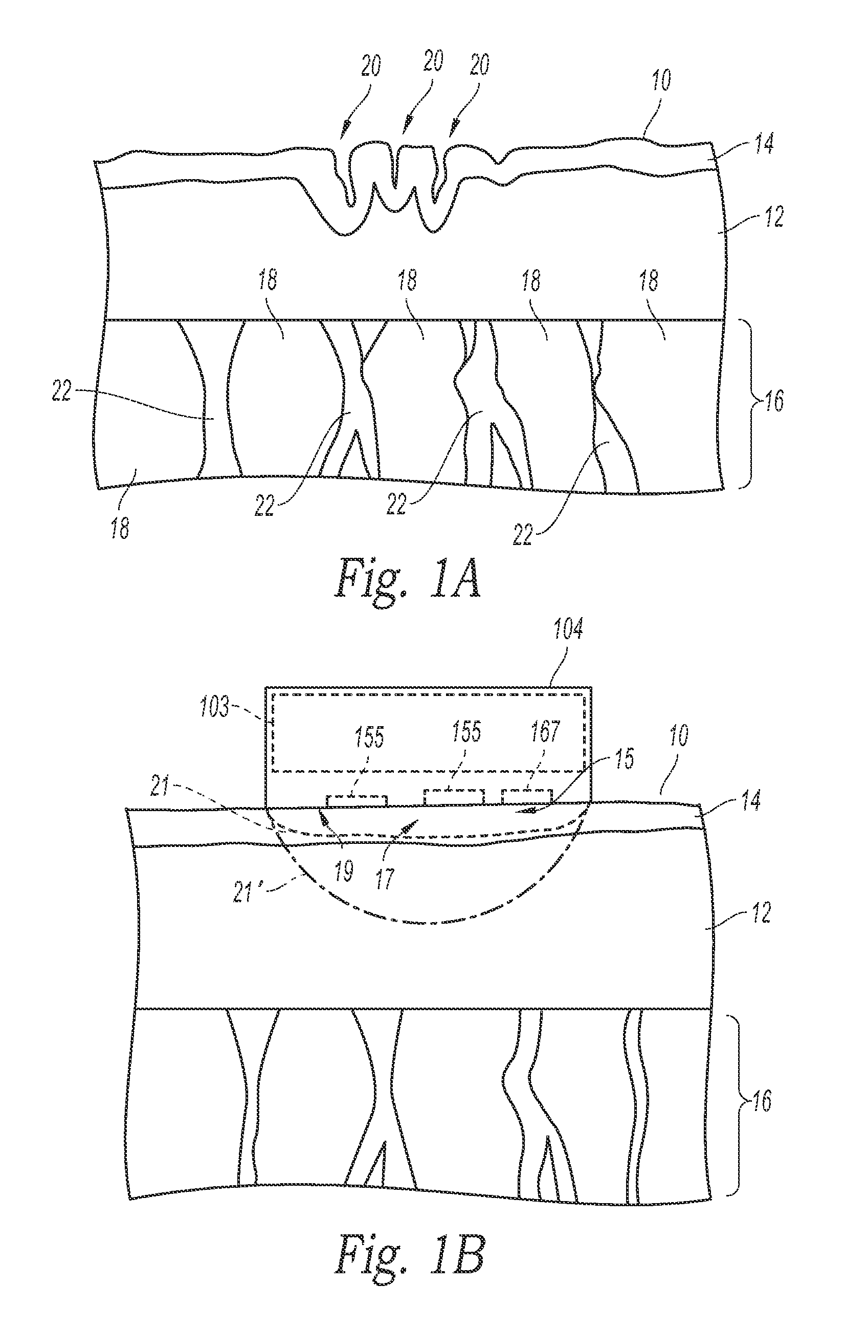
FIG. 1A is a schematic cross-sectional view of tissue with an undesirable appearance.
FIG. 1B is a schematic cross-sectional view of the tissue in FIG. 1A with an improved appearance. An applicator is in thermal contact with the surface of the skin.
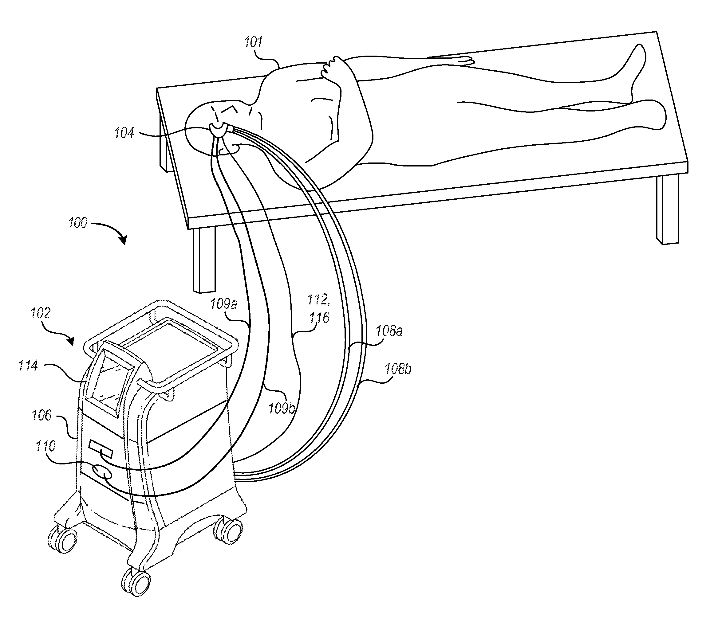
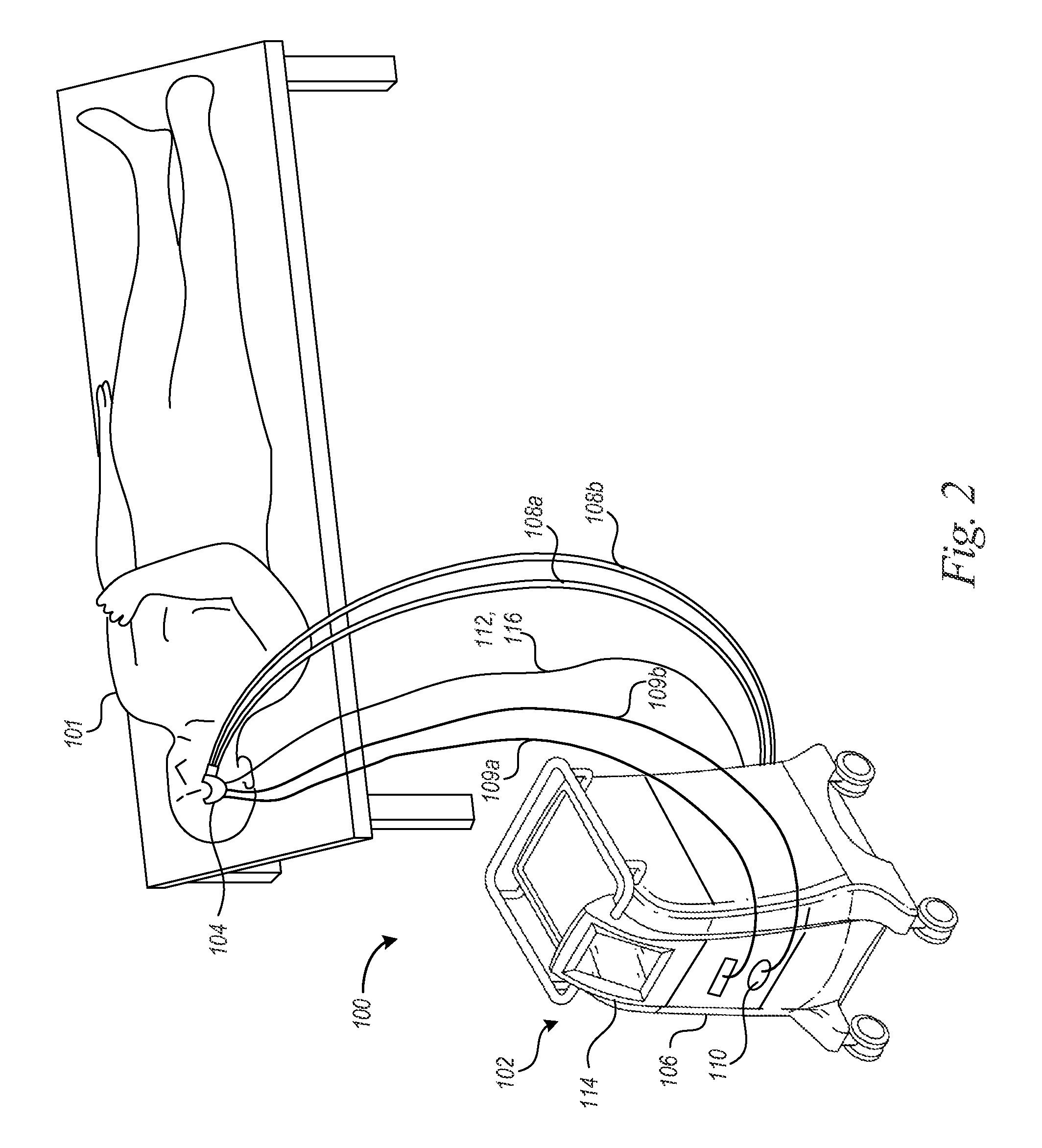
FIG. 2 is a partially schematic isometric view of a treatment system for improving the appearance of facial skin in accordance with an embodiment of the disclosure.
FIGS. 3 to 5B are flow diagrams illustrating methods for improving the appearance of skin in accordance with embodiments of the technology.
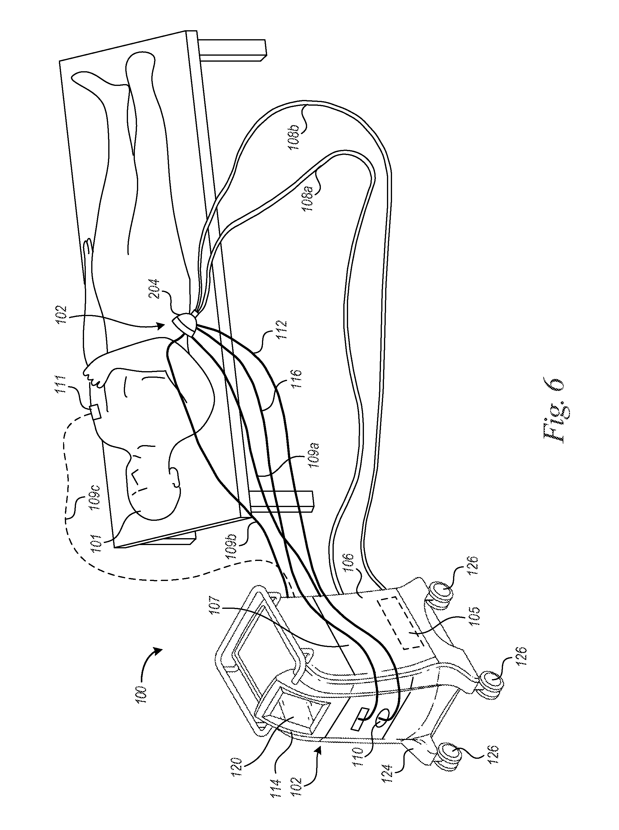
FIG. 6 is a partially schematic isometric view of a treatment system treating tissue located along a subject's torso in accordance with an embodiment of the disclosure.
FIG. 7 is a partial cross-sectional view illustrating a treatment device in accordance with embodiments of the technology.
FIGS. 8A to 8C are schematic cross-sectional views illustrating treatment devices in accordance with embodiments of the technology.
FIG. 9 is a partial cross-sectional view illustrating a vacuum treatment device in accordance with another embodiment of the technology.
FIG. 10 is a schematic block diagram illustrating computing system software modules and subcomponents of a computing device suitable to be used in treatment systems in accordance with an embodiment of the technology.
DETAILED DESCRIPTION
A. Overview
The present disclosure describes treatment systems and methods for improving the appearance of tissue and other treatments. Several of the details set forth below are provided to describe the following examples and methods in a manner sufficient to enable a person skilled in the relevant art to practice, make and use them. Several of the details and advantages described below, however, may not be necessary to practice certain examples and methods of the technology. Additionally, the technology may include other examples and methods that are within the scope of the technology but are not described in detail.
Reference throughout this specification to "one example," "an example," "one embodiment," or "an embodiment" means that a particular feature, structure, or characteristic described in connection with the example is included in at least one example of the present technology. Thus, the occurrences of the phrases "in one example," "in an example," "one embodiment," or "an embodiment" in various places throughout this specification are not necessarily all referring to the same example. The headings provided herein are for convenience only and are not intended to limit or interpret the scope or meaning of the technology.
At least some embodiments are directed to reducing or eliminating wrinkles, loose skin, sagging skin, poor skin tone or texture, and other skin irregularities often considered to be cosmetically unappealing. Some embodiments are directed to skin tightening and/or improving the appearance of cellulite. As used herein, the term "improving the appearance of skin" is intended to include any combination of skin tightening, improving skin tone or texture, thickening of the skin, elimination or reducing fine lines and wrinkles or deeper wrinkles, increasing skin smoothness, improving the appearance of cellulite, or other similar effects. What is not included in the term is treating the skin to such an extent as to cause hyperpigmentation (skin darkening) and/or hypopigmentation (skin lightening) either immediately after the treatment or hours or a day or days or weeks thereafter. Treatment systems disclosed herein can improve skin appearance by causing skin tightening, thickening of tissue (e.g., thickening of the epidermis, dermis, and/or subcutaneous tissue), and/or inducing a cold shock response at the cellular level so as to improve skin tone, skin texture, and/or skin smoothness. In one embodiment, a treatment system has an applicator configured to be applied to a subject's face to treat wrinkles around the eyes, mouth, forehead, etc. The applicator can cool facial tissue to reduce the number of visible wrinkles, reduce the size of wrinkles (e.g., depths, lengths, etc.), or the like. Conformable or contoured applicators can be applied to highly contoured regions around the eyes to reduce or eliminate, for example, crow's feet wrinkles. Treatment systems can also have applicators configured to be applied to other locations along the subject's body. The shape, configuration, mechanical properties, and cooling capabilities of the applicators can be selected based on the tissue characteristics at the treatment site.
Various aspects of the technology are directed to non-invasive applicators that cool the epidermis, dermis, and/or other tissue for a period of time selected to localize thermal effects in targeted tissue while preventing thermal effects in deeper non-targeted tissue. Oftentimes, but not always, target tissue can be intermediate (not surface and not deep) tissue. For example, when treating the face, it is often undesirable to injure the subcutaneous layer beneath the skin, which acts as a support layer for the skin. Additionally, when treating the face and other body areas, it is desirable to minimize or control injury to the epidermis. In an extreme case, if the epidermis is overly frozen, hyperpigmentation (skin darkening) or hypopigmentation (skin lightening) can result, which is often undesirable. At least some embodiments are methods and apparatuses for non-invasively cooling relatively shallow tissue located along the face, neck, hands, hips, buttock, thighs, etc. Targeted tissue be cooled to a temperature equal to or below about -40.degree. C., -35.degree. C., -30.degree. C., -25.degree. C., -20.degree. C., -10.degree. C., or -5.degree. C. for a treatment period equal to or longer than 1 second, 2 seconds, 3 seconds, 5 seconds, 30 seconds, 1 minute, a few minutes, or the like. In some embodiments, targeted epidermal and/or dermal tissue can be cooled to a temperature between about -40.degree. C. and about 0.degree. C., between about -35.degree. C. and about 0.degree. C., between about -30.degree. C. and about 0.degree. C., between about -25.degree. C. and about 0.degree. C., or between about -20.degree. C. and about 0.degree. C. In some embodiments, the treatment period can be exceed about 1 minute, 5 minutes, or 20 minutes, or other periods of time selected based on the treatment to be performed. In some embodiments, the treatment period can be shorter than about 1 minute, 5 minutes, 10 minutes, 20 minutes, 30 minutes, or 1 hour. In some procedures, the surface of the patient's skin is cooled to a temperature equal to or greater than about -40.degree. C., -35.degree. C., -30.degree. C. -25.degree. C., -20.degree. C., -10.degree. C., -5.degree. C., or 0.degree. C. Non-targeted tissue may be subcutaneous adipose tissue, epidermal tissue, or other non-targeted tissue that remains at a higher temperature or is otherwise protected, such as by use of one or more cryoprotectants.
In some embodiments, a thermoelectric applicator can cool the patient's skin to produce a cooling zone in the epidermal and/or dermal layers. As such, cooling can be localized in the epidermis and/or dermis. In some examples, the cooling zone can be at a maximum depth equal to or less than about 2.5 mm. In one procedure, a central region of the cooling zone (e.g., a zone where the most tissue injury is created) can be at a maximum depth of about 0.25 mm to about 1 mm, about 0.25 mm to about 1.5 mm, about 0.25 mm to about 2 mm, about 0.5 mm to about 1.5 mm, about 0.5 mm to about 2 mm, about 0.5 mm to about 2.5 mm, or about 0.5 mm to about 3 mm. In some procedures, the depth of the cooling zone and depth of the most significant tissue injury can be at a equal to or less than about 1 mm, 1.5 mm, 2 mm, 2.5 mm, or 3 mm. In some procedures for treating areas with thin skin (e.g., facial skin around the eyes), the cooling zone can be located at a depth equal to or less than about 1 mm, 0.25 mm, 0.5 mm, 1 mm, or 1.5 mm.
Various aspects of the technology are directed to improving the appearance of skin by cooling a surface of a patient's skin to produce at least a cooling event (e.g., a partial freeze event, total freeze event, etc.). The cooling event can be detected, and a cooling device can be controlled to continue cooling the patient's skin so as to maintain a frozen state of targeted tissue for a desired period of time. In one procedure, the tissue can be kept in a partially or totally frozen state for longer than about, for example, about 1 second, 5 seconds, 10 seconds, 20 seconds, 30 seconds, 1 minute, several minutes, or other time period selected to reduce or limit frostbite or necrosis.
In certain embodiments, methods for affecting skin of a human subject's body include positioning an applicator of a cooling apparatus on the subject and removing heat from a treatment site to affect the appearance of the subject's skin without causing an appreciable reduction of subcutaneous adipose tissue. A sufficient amount of thermal energy can be removed from the site so as to reduce wrinkles by, for example, reducing the number of visible wrinkles and/or sizes of the wrinkles. In other embodiments, a sufficient amount of thermal energy is removed from the treatment site so as to tighten skin at the treatment site, or in further embodiments, to alter the tissue between a surface of the skin and subcutaneous lipid-rich cells of the subject's body. In a further embodiment, tissue is cooled to induce fibrosis that increases the firmness of tissue at the treatment site. Fibrosis can be induced in the epidermis, dermis, and/or subcutaneous tissue. Vacuum applicators can stretch or otherwise mechanically alter skin to increase damage and fibrosis in the skin.
At least some aspects of the technology are directed to treatment methods for affecting a target region of a human subject's body to alter an appearance of a treatment site by removing heat using a cooling apparatus to alter at least one of skin tightness, smoothness, or skin surface irregularities. The methods can include removing a sufficient amount of heat to produce fibrosis that alters the subject's skin. In certain embodiments, the fibrosis increases the tightness of the skin and/or increases the smoothness of the surface of the skin. Other embodiments of the technology are directed to methods of cooling tissue using a cooling apparatus to produce a cold shock response for affecting proteins that alter the appearance of the subject's skin. The proteins can be heat shock proteins, cold shock proteins, and/or stress response proteins. In one embodiment, tissue can be cooled to a temperature for increasing a protein synthesis rate of one or more of the proteins.
At least some embodiments of the technology are directed to freezing skin to induce injury to the skin. An applicator can be placed at a treatment site on the subject and can remove heat from the subject's skin to controllably freeze the skin to control the freeze injury (or trauma) to the skin. The freeze injury can improve tissue appearance by, for example, tightening of the skin, thickening of the skin, and/or inducing a cold shock response at the cellular level. In some procedures, most of the skin located between the applicator and subcutaneous tissue can experience at least partial freezing. With or without freezing, at least some embodiments of the technology are directed to controlling the cooling device or providing other means for sufficiently protecting the epidermis from injury to an extent that causes hyperpigmentation (skin darkening) or hypopigmentation (skin lightening). The other means can include heating the epidermis to a non-freezing temperature while deeper tissue remains cold to induce injury thereto, and/or applying a cryoprotectant to a surface of the skin to provide freeze protection to the epidermis while allowing deeper tissue to be more affected by the cooling/cold treatment.
In further embodiments of the technology, damage to tissue (e.g., dermis and/or subcutaneous tissue) can be reduced or eliminated by applying a substance to the subject's skin. In other embodiments, tissue damage can be limited by applying energy to non-targeted tissue while cooling the treatment site. For example, electromagnetic energy, infrared energy, microwave energy, radiofrequency energy, ultrasound energy, electrical energy (e.g., AC or DC electric fields), and/or light can be delivered to the subject's skin to supply energy thereto. In certain arrangements, the applied energy can inhibit the reduction of subcutaneous lipid-rich cells while the treatment site is cooled by the applicator.
Some aspects of the technology are directed to treatment methods for affecting tissue of a human subject's body by cooling tissue to produce a freeze event that affects at least one of skin tone, thickness of the tissue layers (e.g., dermal layer and/or epidermal layer), and/or tissue elasticity. In certain embodiments, the method also includes inhibiting damage to non-targeted tissue of the subject's skin while producing the freeze event. The freeze event can include injury to at least some of the subject's skin (e.g., epidermis, dermis, etc.), subcutaneous adipose tissue, or other targeted tissue.
Devices and systems that enable target tissue supercooling are also described. A freezing point of a material is most reliably ascertained by warming frozen material slowly and measuring a temperature at which melting begins to occur. This temperature is generally not ambiguous if the material is slowly warmed. Partial melting will begin to occur at the freezing/melting point. Conversely, if a non-frozen material is cooled, its freezing/melting point is harder to ascertain since it is known that many materials can simply "supercool," that is they can be cooled to a bulk temperature below their freezing/melting point and still remain in a non-frozen state. As used herein, "supercooling," "supercooled," "supercool," etc., refers to a condition in which a material is at a temperature below its freezing/melting point but is still in an unfrozen or mostly unfrozen state.
Tissue can be supercooled by controllably cooling the surface of the skin for altering and reducing adipose tissue, body contouring and augmentation, treating of acne, treating hyperhidrosis, or other cryotherapy applications. Aspects of the disclosure are further directed to methods and devices that provide protection of non-targeted cells, such as non-lipid-rich cells (e.g., in the dermal and/or epidermal skin layers), by preventing or limiting freeze damage during dermatological and related aesthetic procedures that require sustained exposure to cold temperatures. For example, treatment systems and devices for performing cryotherapy methods can be configured to control thermal parameters such that body fluids within the treatment site are supercooled to temperatures below the freezing point without forming or nucleating ice crystals. The supercooled body fluids can then be intentionally nucleated to damage, reduce, disrupt, or otherwise affect the targeted cells. Nucleation can be induced by delivering an alternating current to the tissue, applying a nucleating solution onto the surface of the skin (for example one that includes bacteria which initiate nucleation), and/or by creating a mechanical perturbation to the tissue, such as by use of vibration, ultrasound energy, etc.
Some of the embodiments disclosed herein can be for cosmetically beneficial alterations of a variety of body regions. As such, some treatment procedures may be for the sole purpose of altering the body region to conform to a cosmetically desirable look, feel, size, shape or other desirable cosmetic characteristic or feature. Accordingly, at least some embodiments of the cosmetic procedures can be performed without providing any, or in another embodiment, providing minimal therapeutic effect. For example, some treatment procedures may be directed to treatment goals that do not include restoration of health, physical integrity, or the physical well-being of a subject. In other embodiments, however, the cosmetically desirable treatments may have therapeutic outcomes (whether intended or not), such as, psychological benefits, alteration of body hormones levels (by the reduction of adipose tissue), etc. The cosmetic methods can target regions of a subject's skin to change a subject's appearance. In another embodiment, the methods can target skin irregularities, wrinkles, sebaceous glands to treat acne, sweat glands to treat hyperhidrosis, hair follicles to injure and remove hair, or other targeted cells to change a subject's appearance or address a therapeutic condition.
B. Treatment Sites
FIG. 1A is a schematic cross-sectional view of tissue with skin 10 having wrinkles 20 (e.g., folds, ridges, or creases) that may be located, for example, along the face, legs (e.g., thighs, buttock, etc.), or other locations. The dimensions (e.g., depths, lengths, etc.) of the wrinkles 20 can vary by body location and typically increase over time. Wrinkles 20 typically affect the epidermis 14 and dermis 12 layers of the skin; however, the subdermal adipose and connective tissue can also play a role in the appearance of skin irregularities. For example, loss of adipose or fat cells 18 and/or weakened connective tissue 22 in the subcutaneous layers (e.g., subdermal tissue 16) can increase the appearance of wrinkles 20 in the skin 10.
FIG. 1B is a schematic cross-sectional view of the skin 10 of the subject in FIG. 1A with improved appearance. An illustrated treatment device in the form of a thermoelectric applicator 104 ("applicator 104") has affected tissue (e.g., skin 10, epidermis 14, dermis 12, or other targeted tissue) to reduce or eliminate the wrinkles, improve skin tone and/or texture, increase skin smoothness, and/or improve the appearance of cellulite. The applicator 104 can perform different cryotherapy procedures designed to make the skin 10 substantially free of visible irregularities.
In one example, a heat-exchanging surface 19 of the applicator 104 can be in thermal contact with a surface of the skin 10. A cooling device 103 of the applicator 104 can cool a treatment site 15 and affect tissue at a cooling zone 21 (shown in phantom line). A central region 17 of the cooling zone 21 can be at a maximum depth of, for example, about 0.25 mm to about 2 mm, about 0.25 mm to about 1 mm, about 0.5 mm to about 1 mm, or about 0.5 mm to about 2 mm. The depth of the cooling zone 21 can be selected to avoid injuring deeper subcutaneous tissue (e.g., subdermal tissue 16). In one procedure, the cooling zone 21 comprises mostly epidermal tissue. In another procedure, the cooling device 103 cools and affects tissue in the cooling zone 21' (shown in dashed-dot line) which comprises mostly epidermal and dermal tissue. Adjacent tissue (e.g., subcutaneous tissue) may also be cooled but can be at a sufficiently high temperature to avoid thermal injury. In some procedures, the cooling zone 21' can comprise most of the tissue located directly between the cooled heat-exchanging surface 19 and the subcutaneous tissue (e.g., dermal tissue 16). For example, at least about 60%, 70%, 80%, 90%, or 95% of the tissue directly between the heat-exchanging surface 19 and the subcutaneous tissue can be located within the cooling zone 21'.
C. Cryotherapy
FIG. 2 and the following discussion provide a general description of an example of a suitable non-invasive treatment system 100 in which aspects of the technology can be implemented. The treatment system 100 can be a temperature-controlled cooling apparatus for cooling tissue at a targeted treatment site to perform cryotherapy. Tissue characteristics affected by cryotherapy can include, without limitation, tissue strength, tissue elasticity, cell size, cell number, and/or tissue layer thickness. For example, the treatment system 100 can cool the epidermis, dermis, or other targeted tissue to reduce or eliminate skin irregularities. Non-targeted tissue, such as subdermal tissue, can remain generally unaffected. In one embodiment, cryotherapy can increase the thicknesses of multiple tissue layers. In one example, cooling can produce a cold shock response to increase the thicknesses of the epidermis and/or dermis by affecting protein proliferation and other cellular functions. Those skilled in the relevant art will appreciate that other examples of the disclosure can be practiced with other treatment systems and treatment protocols, including invasive, minimally invasive, and other non-invasive medical treatments.
The applicator 104 is suitable for altering a human subject's skin without affecting subcutaneous tissue (e.g., subcutaneous adipose tissue and lipid-rich cells, etc.). The applicator 104 can be suitable for reducing wrinkles (e.g., wrinkles 20 of FIG. 1A), loose skin, sagging skin, or other skin surface irregularities by cooling the skin without permanently altering cells of non-targeted tissue (e.g., deep dermal tissue, subdermal tissue, etc.). Without being bound by theory, the effect of cooling selected cells (e.g., cells of the skin, layers of epidermis, etc.) is believed to result in, for example, protein alteration (e.g., synthesis of heat shock proteins, stress proteins, etc.), cell size alteration, cell division, wound remodeling (e.g., thickening of the epidermis, contraction of the epidermis, etc.), fibrosis, and so forth. By cooling the skin to a sufficient low temperature, target cells that contribute to the presence of undesired features can be selectively affected while non-targeted tissue can be unaffected.
The applicator 104 can be used to perform a wide range of different cryotherapy procedures. One cryotherapy procedure involves at least partially or totally freezing tissue to form crystals that alter targeted cells to cause skin tightening, skin thickening, fibrosis, etc. without destroying a significant amount of cells in the skin. To avoid destroying skin cells, the surface of the patient's skin can be cooled to temperatures no lower than, for example, 40.degree. C. for a duration short enough to avoid, for example, excessive ice formation, permanent thermal damage, or significant hyperpigmentation or hypopigmentation (including long-lasting or permanent hyperpigmentation or hypopigmentation). In another embodiment, destruction of skin cells can be avoided by applying heat to the surface of the patient's skin to heat the skin cells above their freezing temperature. The patient's skin can be warmed to at least about -30.degree. C., -25.degree. C., -20.degree. C., -15.degree. C., -10.degree. C., 0.degree. C., 10.degree. C., 20.degree. C., 30.degree. C., or other temperature sufficient to avoid, for example, excessive ice formation, permanent thermal damage, or significant hyperpigmentation or hypopigmentation of the non-targeted and/or epidermal tissue. In some treatments, skin can be cooled to produce partial freeze events that cause apoptotic damage to skin tissue without causing significant damage to adjacent subcutaneous tissue. Apoptosis, also referred to as "programmed cell death", of the skin tissue can be a genetically-induced death mechanism by which cells slowly self-destruct without incurring damage to surrounding tissues. Other cryotherapy procedures may cause non-apoptotic responses.
In some tissue-freezing procedures, the applicator 104 can controllably freeze tissue and can optionally detect the freeze event (or other event). After detecting the freeze event, the applicator 104 can periodically or continuously remove heat from the target tissue to keep a volume of target tissue frozen or partially frozen for a suitable length of time to elicit a desired response. The detected freeze event can be a partial freeze event, a complete freeze event, etc. In some embodiments, the controlled freezing causes tightening of the skin, thickening of the skin, and/or a cold shock response at the cellular level in the skin. In one tissue-freezing treatment, the applicator 104 can produce a partial or total freeze event that includes, without limitation, partial or full thickness freezing of the patient's skin for a relatively short time limit to avoid cooling the adjacent subcutaneous tissue to a low enough temperature for subcutaneous cell death or undue injury to the subcutaneous layer. The freezing process can include forming ice crystals in intracellular and/or extracellular fluids, and the ice crystals can be small enough to avoid disrupting membranes so as to prevent significant permanent tissue damage, such as necrosis. Some partial freeze events can include freezing mostly extracellular material without freezing a substantial amount of intercellular material. In other procedures, partial freeze events can include freezing mostly intercellular material without freezing a substantial amount of extracellular material. The frozen target tissue can remain in the frozen state long enough to affect the target tissue but short enough to avoid damaging non-targeted tissue. For example, the duration of the freeze event can be shorter than about 20 seconds, 30 seconds, or 45 seconds or about 1, 2, 3, 4, 5 or 10 minutes. The frozen tissue can be thawed to prevent necrosis and, in some embodiments, can be thawed within about 20 seconds, 30 seconds, or 45 seconds or about 1, 2, 3, 4, 5, or 10 minutes after initiation of the freeze event.
In several embodiments, tissue can be cooled to induce cold shock cellular responses in the region of the subject being treated are a desirable outcome for beneficially alter (e.g., smoothing and/or tightening) the skin. Without being bound by theory, it is believed that exposure to cold induces a stress response cascade in the interrogated cells that results in the immediate synthesis of cytoprotective genes. Among these are genes that code for heat shock proteins and/or chaperone proteins involved in protein folding. Heat shock proteins can help alter a cell's phenotype by either impeding, protecting or promoting protein function both during the acute response to stress as well as to subsequent stresses. Cold shock proteins (e.g., cold-inducible RNA-binding protein (CIRP) that may have roles in cellular proliferation and inflammation) have also been identified in mammalian cells. Induction of cold shock cellular responses can dramatically change a cell's proteome to promote cellular survival under environmental interrogation and/or following cold-induced tissue injury. It has been observed in mammalian cells that cold stress can alter the lipid composition of the cellular membranes, as well as change rates of protein synthesis and cell proliferation. Without being bound by theory, the selective effect of cooling on target cells (e.g., epidermal and/or dermal cells) is believed to result in, for example, changes in cellular metabolism, proliferation, survivability, wound healing, wound contraction and other cellular responses that can improve tissue characteristics at the treatment site.
The mechanisms of cold-induced tissue injury in cryotherapy can also involve direct cellular injury (e.g., damage to the cellular machinery) and/or vascular injury. For example, cell injury can be controlled by adjusting thermal parameters, including (1) cooling rate, (2) end (or minimum) temperature, (3) time held at the minimum temperature (or hold time), (4) temperature profile, and (5) thawing rate. In one example, increasing the hold time can allow the intracellular compartments to equilibrate with the extracellular space, thereby increasing cellular dehydration. Another mechanism of cold-induced injury is cold and/or freeze-stimulated immunologic injury. Without being bound by theory, it is believed that after cryotherapy, the immune system of the host is sensitized to the disrupted tissue (e.g., lethally damaged tissue, undamaged tissue, or sublethally injured tissue), which can be subsequently destroyed by the immune system.
During an inflammatory phase of healing following cold-induced injury, platelets are among the first cells to appear at the treatment site. Platelets release platelet derived growth factor (PDGF), which upregulates soluble fibrinogen production. Fibrinogen is converted to insoluble strands of fibrin which form a matrix for the influx of monocytes and fibroblasts. During a proliferative phase of healing, cellular activity promotes epithelialization and fibroplasia. Fibronectin, produced initially from plasma, promotes epidermal migration by providing its own lattice. In freeze wounds, basal keratinocytes secrete collagenase-1 when in contact with fibrillar collagen, and collagenase-1 disrupts attachment to fibrillar collagen which allows for continued migration of keratinocytes into the treatment site. It is during the proliferative phase that a healing process following injury can result in a thicker epidermal layer with increased cellular activity.
In additional embodiments, induction of fibrosis (e.g., the formation of fibrous connective tissue) in the region of the subject being treated is a desirable outcome for beneficially altering the skin. For example, fibrosis can increase the amount of connective tissue in a desired tissue layer (e.g., epidermis, dermis, and/or subcutaneous tissue) to increase a firmness of the tissue. In some embodiments, increased firmness of the tissue can increase the tightness and/or the smoothness of the surface of the skin. Without being bound by theory, cooling temperatures can result in inflammation of the interrogated tissue. Immune cells, such as macrophages, release transforming growth factor beta (TGF-.beta.) which stimulates the proliferation and activation of fibroblast cells. Migrating and activated fibroblast cells deposit connective tissue, including collagen and glycosaminoglycans that can thicken and/or strengthen the affected tissue. Such thickening and/or firming of the affected tissue can beneficially alter skin characteristics by reducing the appearance of wrinkles, fine lines, loose skin, etc.
The system 100 can also perform treatments to affect subcutaneous tissue by cooling the subject's skin for a period of time long enough so that lipid-rich cells in a subcutaneous layer are substantially affected. This can be desired particularly when treating non-facial areas, such as the thighs, arms, abdomen etc., where a skin tightening effect is desired as well as a volumetric reduction, which is aided by altering subcutaneous lipid rich cells. The term "subcutaneous tissue" means tissue lying beneath the dermis and includes subcutaneous fat, or adipose tissue, which is composed primarily of lipid-rich cells, or adipocytes. It is believed that shallow tissue can be cooled to improve skin appearance while also cooling subcutaneous tissue to cause, for example, apoptosis. Apoptosis of subcutaneous lipid-rich cells may be a desirable outcome for beneficially altering (e.g., sculpting and/or reducing) adipose tissue that may contribute to an undesirable appearance. Apoptosis of subcutaneous lipid-rich cells can involve ordered series of biochemical events that induce cells to morphologically change. These changes include cellular blebbing, loss of cell membrane asymmetry and attachment, cell shrinkage, chromatin condensation, and chromosomal DNA fragmentation. Injury via an external stimulus, such as cold exposure, is one mechanism that can induce apoptosis in cells. Nagle, W. A., Soloff, B. L., Moss, A. J. Jr., Henle, K. J. "Cultured Chinese Hamster Cells Undergo Apoptosis After Exposure to Cold but Nonfreezing Temperatures" Cryobiology 27, 439-451 (1990). One aspect of apoptosis, in contrast to cellular necrosis (a traumatic form of cell death causing, and sometimes induced by, local inflammation), is that apoptotic cells express and display phagocytic markers on the surface of the cell membrane, thus marking the cells for phagocytosis by, for example, macrophages. As a result, phagocytes can engulf and remove the dying cells (e.g., the lipid-rich cells) without eliciting an immune response.
Without being bound by theory, one mechanism of apoptotic lipid-rich cell death by cooling is believed to involve localized crystallization of lipids within the adipocytes at temperatures that may or may not induce crystallization in non-lipid-rich cells. The crystallized lipids may selectively injure these cells, inducing apoptosis (and may also induce necrotic death if the crystallized lipids damage or rupture the bilayer lipid membrane of the adipocyte). Another mechanism of injury involves the lipid phase transition of those lipids within the cell's bilayer lipid membrane, which results in membrane disruption, thereby inducing apoptosis. This mechanism is well documented for many cell types and may be active when adipocytes, or lipid-rich cells, are cooled. Mazur, P., "Cryobiology: the Freezing of Biological Systems" Science, 68: 939-949 (1970); Quinn, P. J., "A Lipid Phase Separation Model of Low Temperature Damage to Biological Membranes" Cryobiology, 22: 128-147 (1985); Rubinsky, B., "Principles of Low Temperature Preservation" Heart Failure Reviews, 8, 277-284 (2003). Other possible mechanisms of adipocyte damage, described in U.S. Pat. No. 8,192,474, relates to ischemia/reperfusion injury that may occur under certain conditions when such cells are cooled as described herein. For instance, during treatment by cooling as described herein, targeted adipose tissue may experience a restriction in blood supply and thus be starved of oxygen due to isolation while pulled into, e.g., a vacuum cup, or simply as a result of the cooling which may affect vasoconstriction in the cooled tissue. In addition to the ischemic damage caused by oxygen starvation and the build-up of metabolic waste products in the tissue during the period of restricted blood flow, restoration of blood flow after cooling treatment may additionally produce reperfusion injury to the adipocytes due to inflammation and oxidative damage that is known to occur when oxygenated blood is restored to tissue that has undergone a period of ischemia. This type of injury may be accelerated by exposing the adipocytes to an energy source (via, e.g., electromagnetic, thermal, electrical, chemical, mechanical, acoustic or other means) or otherwise increasing the blood flow rate in connection with or after cooling treatment as described herein. Increasing vasoconstriction in such adipose tissue by, e.g., various mechanical means (e.g., application of pressure or massage), chemical means or certain cooling conditions, as well as the local introduction of oxygen radical-forming compounds to stimulate inflammation and/or leukocyte activity in adipose tissue may also contribute to accelerating injury to such cells. Other yet-to-be understood mechanisms of injury may also exist.
In addition to the apoptotic mechanisms involved in lipid-rich cell death, local cold exposure may induce lipolysis (i.e., fat metabolism) of lipid-rich cells. For example, cold stress has been shown to enhance rates of lipolysis from that observed under normal conditions which serves to further increase the volumetric reduction of subcutaneous lipid-rich cells. Vallerand, A. L., Zamecnik. J., Jones, P. J. H., Jacobs, I. "Cold Stress Increases Lipolysis, FFA Ra and TG/FFA Cycling in Humans" Aviation, Space and Environmental Medicine 70, 42-50 (1999).
Without being bound by theory, the effect of cooling on lipid-rich cells is believed to result in, for example, membrane disruption, shrinkage, disabling, destroying, removing, killing, or another method of lipid-rich cell alteration. For example, when cooling the subcutaneous tissues to a temperature lower than 37.degree. C., subcutaneous lipid-rich cells can selectively be affected. In general, the cells in the epidermis and dermis of the subject 101 have lower amounts of lipids compared to the underlying lipid-rich cells forming the subcutaneous tissues. Since lipid-rich cells are more sensitive to cold-induced damage than non-lipid-rich epidermal or dermal cells, it is possible to use non-invasive or minimally invasive cooling to destroy lipid-rich cells without destroying the overlying skin cells. In some embodiments, lipid-rich cells are destroyed while the appearance of overlying skin is improved.
Deep hypodermal fat cells are more easily damaged by low temperatures than the overlying dermal and epidermal layers of skin, and as such, thermal conduction can be used to cool the desired layers of skin to a temperature above the freezing point of water, but below the freezing point of fat. It is believed that the temperatures can be controlled to manage damage in the epidermis and/or dermis via, for example, intracellular and/or extracellular ice formation. Excessive ice formation may rupture the cell wall and may also form sharp crystals that locally pierce the cell wall as well as vital internal organelles. Ice crystal initiation and growth can be managed to avoid cell death in the skin. When extracellular water freezes to form ice, the remaining extracellular fluid becomes progressively more concentrated with solutes. The high solute concentration of the extracellular fluid may cause intracellular fluid to be driven through the semi-permeable cellular wall by osmosis resulting in cell dehydration. The applicator 104 can reduce the temperature of the deep lipid-rich cells such that the deep lipid rich cells are destroyed while the temperature of the upper and surface skin cells are maintained at a high enough temperature to produce non-destructive freeze events in the skin. Cryoprotectants and/or thermal cycling can prevent destructive freeze events in the skin and limit injury to the skin cells.
D. Treatment Systems and Methods of Treatment
FIG. 2 is a partially schematic isometric view of the non-invasively treatment system 100 for performing cryotherapy procedures disclosed herein. The term "treatment system", as used generally herein, refers to cosmetic or medical treatment systems. The components of the treatment system 100 can be selected and implemented in various embodiments to apply selected treatment profiles to the subject 101 (e.g., a human or an animal) for improving the appearance of the treatment site. The treatment system 100 can include a treatment unit or tower 102 ("treatment tower 102") connected to the applicator 104 by supply and return fluid lines 108a-b and power-lines 109a-b.
The applicator 104 can have one or more cooling devices powered by electrical energy delivered via the power-lines 109a-b. A control line 116 can provide communication between electrical components of the applicator 104 and a controller 114 of the treatment tower 102. The cooling devices of the applicator 104 can be cooled using coolant that flows between the applicator 104 and the treatment tower 102 via the supply and return fluid lines 108a-b. In one example, the applicator 104 has a cooling device (e.g., cooling device 103 of FIG. 1B) with one or more thermoelectric cooling elements and fluid channels through which the coolant flows to cool the thermoelectric cooling elements. The thermoelectric cooling elements can include heat-exchanging plates, Peltier devices, or the like. In one embodiment, the applicator 104 can be a non-thermoelectric device that is heated/cooled using only coolant. The applicator 104 can include sensors configured to measure tissue impedance, treatment application force, and/or tissue contact. As described herein, sensors can be used to monitor tissue and, in some embodiments, detect freeze events. Applicators configured to be applied to facial tissue can have pressure sensors to monitor applied pressures to maintain a desired level of comfort. The number and types of sensors can be selected based on the treatment to be performed.
The treatment tower 102 can include a chiller unit or module 106 ("chiller unit 106") capable of removing heat from the coolant. The chiller unit 106 can include one or more refrigeration units, thermoelectric chillers, or any other cooling devices and, in one embodiment, includes a fluid chamber configured to house the coolant that is delivered to the applicator 104 via the fluid lines 108a-b. In some procedures, the chiller unit 106 can circulate warm coolant to the applicator 104 during periods of warming. In certain procedures, the chiller unit 106 can alternatingly provide heated and chilled coolant. The circulating coolant can include water, glycol, synthetic heat transfer fluid, oil, a refrigerant, or any other suitable heat conducting fluid. Alternatively, a municipal water supply (e.g., tap water) can be used in place of or in conjunction with the treatment tower 102. The fluid lines 108a-b can be hoses or other conduits made of polyethylene, polyvinyl chloride, polyurethane, and/or other materials that can accommodate the particular coolant. One skilled in the art will recognize that there are a number of other cooling technologies that could be used such that the treatment unit, chiller unit, and/or applicator(s) need not be limited to those described herein. Additional features, components, and operation of the treatment tower 102 are discussed in connection with FIG. 6.
FIG. 2 shows the applicator 104 positioned to treat crow's feet wrinkles near the patient's right eye. Feedback data from sensors of the applicator 104 can be collected in real-time because real-time processing of such feedback data can help correctly and efficaciously administer treatment. In one example, real-time data processing is used to detect freeze events and to control the applicator 104 to continue cooling the patient's skin after the partial or total freeze event is detected. Tissue can be monitored to keep the tissue in the frozen state (e.g., at least partially or totally frozen state) for a period of time. The period of time can be selected by the treatment tower 102 or an operator and can be longer than about, for example, 10 seconds, 30 seconds, 1 minute, or a few minutes. Other periods of time can be selected if needed or desired. The applicator 104 can include sensors configured to measure tissue impedance, force/pressure applied to the subject 101, optical characteristics of tissue, and/or tissue contact temperatures. As described herein, sensors can be used to monitor tissue and, in some embodiments, to detect freeze events. The number and types of sensors can be selected based on the treatment to be performed.
The applicator 104 can be used at other treatment sites and can be replaced with other applicators. Applicators can be configured to treat identified portions of the patient's body, such as the face, neck, chin, cheeks, arms, pectoral areas, thighs, calves, buttocks, abdomen, "love handles", back, hands, and so forth. For example, mask applicators can be used to cover the subject's face. Conformable applicators can be applied along the face, neck, or other highly contoured regions. By way of another example, vacuum applicators may be applied at the back region, and belt applicators can be applied around the thigh region, either with or without massage or vibration as discussed in connection with FIG. 9. Exemplary applicators and their configurations usable or adaptable for use with the treatment system 100 variously are described in, e.g., U.S. Pat. No. 8,834,547 and commonly assigned U.S. Pat. No. 7,854,754 and U.S. Patent Publication Nos. 2008/0077201, 2008/0077211, and 2008/0287839, which are incorporated by reference in their entireties.
In further embodiments, the system 100 of FIG. 2 may also include a patient protection device (not shown) incorporated into or configured for use with the applicator 104 that prevents the applicator from directly contacting a patient's skin and thereby reduces the likelihood of cross-contamination between patients and minimizes cleaning requirements for the applicator. The patient protection device may also include or incorporate various storage, computing, and communications devices, such as a radio frequency identification (RFID) component, to monitor and/or meter use. Exemplary patient protection devices are described in commonly assigned U.S. Patent Publication No. 2008/0077201.
In operation, and upon receiving input to start a treatment protocol, the controller 114 can cycle through each segment of a prescribed treatment plan. In so doing, power supply 110 and chiller unit 106 can provide power and coolant to one or more functional components of the applicator 104, such as thermoelectric coolers (e.g., TEC "zones"), to begin a cooling cycle and, in some embodiments, activate features or modes such as vibration, massage, vacuum, etc. The controller 114 can monitor treatment by receiving temperature readings from temperature sensors that are part of the applicator 104 or proximate to the applicator 104, the patient's skin, a patient protection device, etc. It will be appreciated that while a target region of the body has been cooled or heated to the target temperature, in actuality that region of the body may be close but not equal to the target temperature, e.g., because of the body's natural heating and cooling variations. Thus, although the system 100 may attempt to heat or cool the tissue to the target temperature or to provide a target heat flux, a sensor may measure a sufficiently close temperature or heat flux. If the target temperature has not been reached, power can be increased or decreased to change heat flux to maintain the target temperature or "set-point" selectively to affect targeted tissue. The system 100 can thus monitor the treatment site while accurately cooling/heating tissue to perform the methods discussed herein.
FIG. 3 is a flow diagram illustrating a method 140 for improving the appearance of a subject's skin in accordance with embodiments of the disclosure. An early stage of the method 140 can include coupling a heat-exchanging surface of a treatment device with the surface of the subject's skin at a target region (block 142). FIG. 1B shows the heat-exchanging surface 19 in the form of an exposed surface of a heat-exchanging plate of the applicator 104. In another embodiment, the heat-exchanging surface 19 can be the surface of an interface layer or a dielectric layer. Coupling of the heat-exchanging surfaces to the skin can be facilitated by using restraining means, such as a belt or strap. In other embodiments, a vacuum or suction force can be used to positively couple the treatment device to the patient's skin. In some methods, a thermally conductive substance can couple the heat-exchanging surface 19 to the patient's skin and can optionally be a cryoprotectant. Suitable and preferred cryoprotectants are described in commonly assigned U.S. Patent Publication No. 2007/0255362.
At block 144, heat is removed from tissue for a period of time that may vary depending on the location of the treatment site and may induce cold shock, freeze tissue, injure tissue, etc. Thermal injuries can induce fibrosis that increases the firmness and/or tone of the tissue. In some cold shock procedures, the subject's skin can be cooled to produce a cold shock response which affects proteins, such as heat shock proteins, cold shock proteins, and/or stress response proteins. In some embodiments, the subject's skin can be cooled to a temperature no lower than about -40.degree. C., -30.degree. C., -20.degree. C., -10.degree. C. to produce the cold shock response. Additionally or alternatively, the treatment site can be cooled to a temperature selected to increase a protein synthesis rate of one or more of the proteins. Some treatment protocols can include two or more segments each designed to produce cold shocks responses, freeze tissue, or injure tissue. For example, a treatment protocol may alternate between tissue-freeze segments and tissue-thaw segments. In another example, one treatment protocol can be designed to reduce wrinkles (e.g., age-related wrinkles) via shock proteins and another treatment protocol can be designed to tighten loose tissue via fibrosis. Accordingly, different treatment protocols can be used on different parts of a patient's body. Although the method 140 is described with reference to the treatment system 100 of FIG. 2, the method 140 may also be performed using other treatment systems with additional or different applicators, hardware, and/or software components.
FIG. 4 is a flow diagram illustrating a method 150 for improving the appearance of skin in accordance with embodiments of the disclosure. The method 150 can include coupling a heat-exchanging surface of a treatment device with the surface of the subject's skin at a target region (block 152). At block 154, the method 150 includes cooling the subject's skin to affect tissue at the target region. In one embodiment, the skin's smoothness, thickness, texture, tone, and/or elasticity is improved. In one embodiment, the skin is cooled to induce a freeze-related injury, and in another embodiment, the skin is cooled to induce a cold shock response.
FIG. 5A is a flow diagram illustrating a method 160 for improving the appearance of skin by producing a freeze event in accordance with embodiments of the disclosure. Generally, a treatment device can be applied to a subject and can cool a surface of a patient's skin to produce and detect a freeze event. After detecting the freeze event (or events), operation of the treatment device can be controlled to keep at least a portion of the subject's tissue frozen for a sufficient length of time to improve the appearance of the skin but not so long as to create undue injury to tissue. Details of method 160 are discussed below.
At block 162, the treatment device is applied to a subject by placing a heat-exchanging surface in thermal contact with the subject's skin. In some embodiments, a substance is applied to the subject's skin before applying the treatment device. A substance can be used to (a) provide thermal coupling between the subject's skin and cooling devices (e.g., cooling plates of cooling devices) to improve heat transfer therebetween, (b) selectively protect non-target tissues from freeze damage (e.g., damage due to crystallization), and/or (c) promote freeze events by increasing nucleation sites. The substance may be a fluid, a gel, or a paste and may be hygroscopic, thermally conductive, and biocompatible. In some embodiments, the substance can be a cryoprotectant that reduces or inhibits cell destruction. As used herein, "cryoprotectant," "cryoprotectant agent," and "composition" mean substances (e.g., compositions, formulations, compounds, etc.) that assist in preventing freezing of tissue compared to an absence of the substances(s). In one embodiment, the cryoprotectant allows, for example, the treatment device to be pre-cooled prior to being applied to the subject for more efficient treatment. Further, the cryoprotectant can also enable the treatment device to be maintained at a desired low temperature while preventing ice from forming on a surface (e.g., heat-exchanging surface), and thus reduces the delay in reapplying the treatment device to the subject. Yet another aspect of the technology is that the cryoprotectant may prevent the treatment device from freezing to the skin of the patient or subject. Additionally or alternatively, the cryoprotectant can allow microscopic crystals to form in the tissue but can limit crystal growth that would cause cell destruction and, in some embodiments, allows for enhanced uptake or absorption and/or retention in target tissue prior to and during the introduction of cooling.
Some embodiments according to the present technology may use a cryoprotectant with a freezing point depressant that can assist in preventing freeze damage that would destroy cells. Suitable cryoprotectants and processes for implementing cryoprotectants are described in commonly-assigned U.S. Patent Publication No. 2007/0255362. The cryoprotectant may additionally include a thickening agent, a pH buffer, a humectant, a surfactant, and/or other additives and adjuvants as described herein. Freezing point depressants may include, for example, propylene glycol (PG), polyethylene glycol (PEG), dimethyl sulfoxide (DMSO), or other suitable alcohol compounds. In a particular embodiment, a cryoprotectant may include about 30% propylene glycol, about 30% glycerin (a humectant), and about 40% ethanol. In another embodiment, a cryoprotectant may include about 40% propylene glycol, about 0.8% hydroxyethyl cellulose (a thickening agent), and about 59.2% water. In a further embodiment, a cryoprotectant may include about 50% polypropylene glycol, about 40% glycerin, and about 10% ethanol. The freezing point depressant may also include ethanol, propanol, iso-propanol, butanol, and/or other suitable alcohol compounds. Certain freezing point depressants (e.g., PG, PPG, PEG, etc.) may also be used to improve spreadability of the cryoprotectant and to provide lubrication. The freezing point depressant may lower the freezing point of body liquids/lipids to about 0.degree. C. to -50.degree. C., about 0.degree. C. to -50.degree. C., or about 0.degree. C. to -30.degree. C. In other embodiments the freezing point of the liquids/lipids can be lowered to about -10.degree. C. to about -40.degree. C., about -10.degree. C. to about -30.degree. C., or to about -10.degree. C. to about -20.degree. C. In certain embodiments, the freezing point of the liquids/lipids can be lowered to a temperature below about 0.degree. C., below about -5.degree. C., below about -10.degree. C., below about -12.degree. C., below about -15.degree. C., below about -20.degree. C., below about -30.degree. C., or below about -35.degree. C. For example, the freezing point depressant may lower the freezing point of the liquids/lipids to a temperature of about -1.degree. C. to about -40.degree. C., about -5.degree. C. to about -40.degree. C., or about -10 to about -40.degree. C.
Cryoprotectant can be intermittently or continuously delivered to the surface of the patient's skin for a period of time which is short enough to not significantly inhibit the initiation of the partial or total freeze event in dermal tissue but is long enough to provide substantial protection to non-targeted tissue (e.g., subcutaneous adipose tissue). The rate of cryoprotectant delivery can be selected based on the characteristics of the cryoprotectant and the desired amount of tissue protection. In one specific treatment process, an interface member is placed directly over the target area, and the treatment device with a disposable sleeve or liner is placed in contact with the interface member. The interface member can be a cotton pad, gauze pad, a pouch, or a container with a reservoir containing a volume of cryoprotectant or other flowable conductive substance. The interface member can include, for example, a non-woven cotton fabric pad saturated with the substance that delivers cryoprotectant at a desired delivery rate. Suitable pads include Webril.TM. pads manufactured by Covidien of Mansfield, Mass. Further details regarding the interface member and associated systems and methods are described in commonly-assigned U.S. Patent Publication No. 2010/0280582.
In a certain embodiment, the system 100 (FIG. 2) can be used to perform several treatment methods without using a chemical cryoprotectant. FIG. 5B is a flow diagram illustrating a method 500 for improving the appearance of skin without any chemical composition in accordance with embodiments of the disclosure. As shown in FIG. 5B, an early stage of the method 500 can include cooling a surface of a human subject's skin to a first temperature (block 502). The first temperature can be, for example, between about -5.degree. C. and -40.degree. C. such that a portion of tissue below the surface is in a supercooled state. The supercooled tissue can include epidermal tissue, dermal tissue, subcutaneous tissue, other tissue, and combinations thereof.
The method 500 can also include heating the surface of the human subject's skin in an amount sufficient to raise the temperature of the surface or upper layer of tissue from the first temperature to a second temperature that is a non-supercooled temperature. Deeper tissue below the surface can remain in the supercooled state (block 504). For example, the treatment system can be used to heat the surface (e.g., an upper portion of the epidermis) of the skin to a temperature greater than about 0.degree. C. while underlying tissue remains supercooled. In some embodiments, tissue of the skin at a depth of less than about 0.2 mm, 0.5 mm, or 1 mm are warmed to a non-supercooled state. The temperature of the skin surface can be increased about 40.degree. C., 30.degree. C. 20.degree. C., 10.degree. C. during the warming period (e.g., 0.5 second, 1 second, 2 seconds, 5 seconds, etc.). The surface of the skin can be periodically heated to minimize or limit thermal damage while deeper tissue is at or below the treatment temperature (e.g., a temperature for supercooling).
In block 506, the method 500 can further include nucleating the supercooled portion of tissue below warmed tissue to cause at least some cells in the supercooled tissue to at least partially freeze. In one embodiment, nucleation of the supercooled tissue is caused by a mechanical perturbation (e.g., vibration, ultrasound pulses, etc.) while warmed cells residing at the surface of the human subject's skin do not freeze. This allows for localized nucleation and protection of cells at the surface can be accomplished without the use of a chemical cryoprotectant. Optionally, a cryoprotectant may also be used to provide further protection for epidermal tissue to minimize any undue damage thereto which might result in a hyperpigmentation or hypopigmentation response sometime after completing treatment. In various embodiments, the supercooled tissue can comprise some portion of the epidermal tissue (e.g., a lower region of epidermal tissue), dermal tissue, connective tissue, subcutaneous tissue, or other tissue targeted to experience a freeze injury.
In certain embodiments, the method 500 may further include maintaining the supercooled tissue in at least a partially or totally frozen state for a predetermined period of time (block 508). For example, the supercooled tissue can be maintained in the at least partially or totally frozen state for longer than about 2 seconds, 5 seconds, 10 seconds, 12 seconds, 15 seconds, or 20 seconds. In various arrangements, the supercooled tissue can be maintained in the at least partially or totally frozen state for a duration of time sufficient to improve an appearance of skin or provide for other treatments (e.g., tightening the skin, increasing skin smoothness, thickening the skin, improving the appearance of cellulite, improving acne, improving a quality of hair, improving a condition associated with hyperhidrosis, etc.). In certain embodiments, the maintaining the freeze event can include detecting a temperature of the portion of tissue and controlling the cooling and heating to maintain at least a portion of the tissue in at least a partially or totally frozen state for the predetermined time (e.g., greater than about 10 seconds, greater than about 12 seconds, greater than about 15 seconds, or greater than about 20 seconds).
Referring back to FIG. 5A, and at block 164, the treatment device can rapidly cool the surface of the patient's skin to a sufficiently low temperature to cause a partial or total freeze event in targeted tissue. The rapid cooling can create a thermal gradient with the coldest temperatures near the applicator (e.g., the upper layers of skin). The resulting thermal gradient causes the temperature of the upper layer(s) of the skin to be lower than that of the targeted deeper cells. This allows the skin to be frozen for a short enough duration so that temperature equilibrium is not established across the skin and adjacent subcutaneous tissue, typically adipose tissue. A cryoprotectant and/or warming cycle can be used to inhibit freezing the uppermost non-targeted layer, or layers, of skin, particularly epidermal tissue, so as to prevent or minimize any chance of creating hyperpigmentation or hypopigmentation.
A partial freeze event can include at least some crystallization (e.g., formation of microscopic ice crystals) in intercellular material (e.g., fluid, cell components, etc.) and/or extracellular fluid. By avoiding extensive ice crystal formation that would cause frostbite or necrosis, partial freeze events can occur without undesired tissue damage. In addition, total freeze events can be created which are maintained for a period of time which is kept short enough so as to not cause an undesired amount of tissue damage. In some embodiments, the surface of the patient's skin can be cooled to a temperature no lower than about -40.degree. C., -30.degree. C., -20.degree. C., -10.degree. C., or -5.degree. C. to produce a partial or total freeze event in the skin without causing irreversible skin damage. For example, the treatment system 100 of FIG. 2 can cool the skin to a temperature in a range from about -40.degree. C. to about 0.degree. C. In another example, the surface of the patient's skin can be cooled to from about -40.degree. C. to about 0.degree. C., from about -30.degree. C. to about 0.degree. C., from about -20.degree. C. to about -5.degree. C., or from about -15.degree. C. to about -5.degree. C. In further examples, the surface of the patient's skin can be cooled to below about -10.degree. C., or in yet another embodiment, from about -25.degree. C. to about -15.degree. C. It will be appreciated that the skin surface can be cooled to other temperatures based on the mechanism of action.
To perform cryotherapy, the cooling period can be sufficiently short to minimize, limit, or substantially eliminate necrosis, or other unwanted thermal damage, due to the freeze event. In one procedure, the applicator (e.g., applicator 104 of FIGS. 1B and 2) can produce a freeze event that begins within a predetermined period of time after the applicator begins cooling the patient's skin or after the freeze event begins. The predetermined period of time can be equal to or less than about 30, 60, 90, 120, or 150 seconds and, in some embodiments, the predetermined period of time can be from about 30 seconds to about 150 seconds. A controller (e.g., controller 114 of FIG. 2) can select the predetermined period of time based on the treatment temperatures, treatment sites, and/or cryotherapy to be performed. Alternatively, an operator can select the period of time for cooling and can enter it into the controller 114.
To help initiate the freeze event (e.g., the partial or total freeze event), a substance, energy, and/or pressure can be used to aid in the formation of nucleation sites for crystallization. Substances that promote nucleation can be applied topically before and/or during skin cooling. The energy for promoting nucleation can include, without limitation, acoustic energy (e.g., ultrasound energy), mechanical energy (e.g., vibratory motion, massaging, and/or pulsatile forces), or other energy. The energy can also comprise alternating current electrical energy. A nucleating substance can also optionally be applied to the skin. The applicators disclosed herein can include one or more actuators (e.g., motors with eccentric weights), vibratory motors, hydraulic motors, electric motors, pneumatic motors, solenoids, piezoelectric shakers, and so on for providing mechanical energy, pressure, etc. Pressure for promoting nucleation can be applied uniformly or non-uniformly across the treatment site. The applicators can also include AC electrodes. For example, the applicator 104 of FIG. 1B can include one or more elements 155 in the form of actuators, motors, solenoids, piezoelectric shakers, transducers (e.g., ultrasound transducers), electrodes (e.g., electrical electrodes, RF electrodes, etc.), or combinations thereof. Substances that promote nucleation can be applied topically before and/or during skin cooling.
At block 166, the treatment device can detect the partial or total freeze event in the patient's skin using one or more electrical components. FIG. 1B shows the applicator 104 with an electronic component in the form of a sensor 167 that can identify positive (increase) or negative (decrease) temperature changes. During cooling, targeted tissue can reach a temperature below the freezing point of its biological tissue and fluids (e.g., approximately -1.8.degree. C.). As tissue, lipids, and fluids freeze, crystals can form and energy associated with the latent heat of crystallization of the tissue is released. A relatively small positive change in tissue temperature can indicate a partial freeze event whereas a relatively large positive change in tissue temperature can indicate a complete freeze event. The sensor 167 (FIG. 1B) can detect the positive change in tissue temperature, and the treatment system can identify it as a freeze event. The treatment system can be programmed to prevent small variations in temperature from causing false alarms with respect to false treatment events. Additionally or alternatively, the treatment system disclosed herein may detect changes in the temperature of its components or changes in power supplied to the treatment device (e.g., treatment devices receive more power from the system to provide additional cooling). For example, the sensor 167 can detect changes in temperature of the applicator 104 as the applicator gets colder in order to cool the tissue warmed by crystallization.
Referring now to FIG. 2, the system 100 can monitor the location and/or movement of the applicator 104 and may prevent false or inaccurate determinations of treatment events based on such monitoring. The applicator 104 may move during treatment which may cause the applicator 104 to contact a warmer area of skin, to no longer contact the skin, and so on. This may cause the system 100 to register a difference in temperature that is inconsistent with a normal treatment. The controller 114 may be programmed to differentiate between these types of temperature increases and a temperature increase associated with a treatment event. U.S. Pat. No. 8,285,390 discloses techniques for monitoring and detecting freeze events and applicator movement and is incorporated by reference in its entirety. Additionally, the treatment system 100 can provide an indication or alarm to alert the operator to the source of this temperature increase. In the case of a temperature increase not associated with a treatment event, the system 100 may also suppress false indications, while in the case of a temperature increase associated with freezing, the system 100 take any number of actions based on that detection as described elsewhere herein.
The system 100 can use optical techniques to detect events at block 166 of FIG. 5A. For example, the sensor 167 of FIG. 1B can be an optical sensor capable of detecting changes in the optical characteristics of tissue caused by freezing. The optical sensor can include one or more energy emitters (e.g., light sources, light emitting diodes, etc.), detector elements (e.g., light detectors), or other components for non-invasively monitoring optical characteristics of tissue. In place of or in conjunction with monitoring using optical techniques, tissue can be monitored using electrical and/or mechanical techniques because changes in electrical impedance and/or mechanical properties of the tissue can be detected and may indicate freezing of tissue. In embodiments for measuring electrical impedance, the sensor 167 (FIG. 1B) can include two electrodes that can be placed in electrical communication with the skin for monitoring electrical energy traveling between the electrodes via the tissue. In embodiments for measuring mechanical properties, the sensor 167 can comprise one or more mechanical sensors which can include, without limitation, force sensors, pressure sensors, and so on.
At block 168, the partial or total freeze event can be maintained by continuously or periodically cooling the patient's tissue to keep a target volume of skin frozen for a period of time, which can be long enough to affect the skin and thereby improve skin appearance. In short treatments, the period of time can be equal to or shorter than about 5, 10, 15, 20, or 25 seconds. In longer treatments, the period of time can be equal to or longer than about 25 seconds, 30 seconds, 45 seconds or 1, 2, 3, 4, 5, or 10 minutes. In some procedures, the applicator 104 of FIGS. 1B and 2 can be controlled so that the skin is partially or completely frozen for no longer than, for example, 5 minutes, 10 minutes, 20 minutes, 30 minutes, 45 minutes, or 1 hour. In some examples, the skin is frozen for about 1 minute to about 5 minutes, about 5 minutes to about 10 minutes, about 10 minutes to about 20 minutes, or about 20 minutes to about 30 minutes. The length of time the skin is kept frozen can be selected based on severity of the freeze injury.
At block 168 of FIG. 5A, the treatment system can control the applicator so that the partial or total freeze event causes apoptotic damage to the target tissue and does not cause necrotic damage to non-targeted tissue. The cooling period can be sufficiently short to avoid or limit permanent tissue damage and, in some embodiments, can be less than about, for example, 5 minutes, 10 minutes, 15 minutes, 20 minutes, 25 minutes, 30 minutes, 35 minutes, 40 minutes, 45 minutes, 50 minutes, 55 minutes, or 1 hour. In one example, the applicator produces a partial freeze event short enough to prevent establishing equilibrium temperature gradients in the patient's skin. This allows freezing of shallow targeted tissue without substantially affecting deeper non-targeted tissue. Moreover, cells in the dermal tissue can be affected to a greater extent than the cells in the subcutaneous layer. For example, skin cells can be reduced in size or number to a greater extent than subcutaneous cells, including lipid-rich cells. In some procedures, the subcutaneous layer can be kept at a sufficiently high temperature (e.g., at or above 0.degree. C.) to prevent any freeze event in the subcutaneous layer while the shallower skin tissue experiences the partial or total freeze event. Cryoprotectant can be used to protect the subcutaneous layer. Topical cryoprotectants can be absorbed by the skin and can ultimately reach the subcutaneous layer. Cryoprotectant can be injected directly into the subcutaneous layer before performing the cooling cycle.
In some embodiments, the freeze event can occur in the epidermal layer to injure, reduce, or disrupt the epidermal cells without substantially injuring, reducing, or disrupting dermal cells and/or subcutaneous cells. In other embodiments, the freeze event can occur in the dermal layer to injure, reduce, or disrupt dermal cells without substantially injuring, reducing, or disrupting epidermal cells and/or subcutaneous cells. A cryoprotectant can be used to protect the epidermal layer to avoid causing long-lasting or permanent hyperpigmentation or hypopigmentation. For example, a cryoprotectant can be delivered to the surface of the patient's skin for a period of time which is short enough to not significantly inhibit the initiation of the partial or total freeze event in dermal tissue, but the period of time can be long enough to provide substantial protection to epidermal tissue. The cryoprotectant can prevent permanent hyperpigmentation or hypopigmentation of epidermal tissue due to tissue cooling, and the cryoprotectant delivery time and rate can be selected based on the cryoprotectant's ability to protect tissue. In one embodiment, the cryoprotectant prevents hyperpigmentation or hypopigmentation of the skin surface and also prevents damage of the epidermal tissue due to tissue cooling. In yet other embodiments, the freeze event can occur in the epidermal and dermal layers to injure, reduce, or disrupt the epidermal and dermal cells without substantially injuring, reducing, or disrupting subcutaneous tissue to avoid body contouring. Such treatments are well suited for improving the appearance of skin along the face, including wrinkled, loose, and/or sagging skin around the eyes and mouth.
Certain treatment protocols can include sequentially targeting different layers. A first treatment session can target the epidermal layer and a subsequent additional treatment sessions can target the other layers. Different layers can be targeted in a different protocol.
The treatment system can also control operation of the applicator to thermally injure the patient's skin to cause fibrosis, which increases the amount of connective tissue in a desired tissue layer (e.g., epidermis and/or dermis) to increase the firmness and appearance of the skin. In other treatments, the treatment system controls the applicator to supercool at least a portion of tissue below the skin layer. A perturbation (e.g., a mechanical perturbation) can be used to nucleate the supercooled tissue to at least partially or totally freeze the tissue. Alternating electric fields can be used to create nucleation perturbations. Various substances can be applied to the treatment site to facilitate nucleation of supercooled tissue.
At block 169, the patient's partially or completely frozen skin can be thawed by heating it in order to minimize, reduce, or limit tissue damage. The applicator (e.g., applicator 104 of FIG. 2) can thaw the patient's skin, or other frozen tissue, after the freeze event occurs and after a period of time has transpired. The period of time can be equal to or shorter than about 5, 10, 15, 20, or 25 seconds or about 1, 2, 3, 4, 5, or 10 minutes. In one example, the uppermost skin layer(s) can be periodically heated to a temperature above the skin's freezing point to provide freeze protection thereto. The thermal elements can be resistive heaters, electromagnetic energy emitters, Peltier devices, etc. In some embodiments, the applicator 104 of FIGS. 1B and 2 can have cooling elements and separate heating elements that can cooperate to provide precise temperature control of freezing and thawing/warming cycles. Alternatively, the applicator 104 may stop or reduce tissue cooling to allow cooled tissue to naturally warm and thaw. Thus, tissue can be actively or passively thawed.
The applicator 104 (FIG. 2) can thaw the patient's skin, or other frozen tissue, after the freeze event occurs and after the period of time has transpired to reduce and control freeze damage. In one example, the uppermost skin layer can be periodically heated to a temperature above the skin's freezing point to provide freeze protection thereto. In some procedures, the applicator 104 repeatedly freezes and thaws tissue to control thermal injuries to that tissue.
The system 100 can be used to perform several different cryotherapy procedures. Although specific methods are described in connection with FIGS. 3-5B, one skilled in the art is capable of identifying other methods that the system 100 could perform. Additionally, the treatment system 100 of FIG. 6 can perform the methods described in connection with FIGS. 3-5B. Applicators are discussed in connection with FIGS. 7-9 and can be used with the system 100 (FIGS. 2 and 6) or different treatment systems to perform the procedures disclosed herein.
FIG. 6 is a partially schematic isometric view of the system 100 with a multi-modality applicator 204 positioned along the subject's waist. The power supply 110 can provide a direct current voltage to the applicator 204 to remove heat from the subject 101. The controller 114 can monitor process parameters via sensors (e.g., sensors of the applicator 204 and/or sensors placed proximate to the applicator 204) via the control line 116 to, among other things, adjust the heat removal rate and/or energy delivery rate based on a custom treatment profile or patient-specific treatment plan, such as those described, for example, in commonly assigned U.S. Pat. No. 8,275,442.
The controller 114 can exchange data with the applicator 204 via an electrical line 112 or, alternatively, via a wireless or an optical communication link. The control line 116 and electrical line 112 are shown without any support structure. Alternatively, control line 116 and electrical line 112 (and other lines including, but not limited to fluid lines 108a-b and power lines 109a-b) may be bundled into or otherwise accompanied by a conduit or the like to protect such lines, enhance ergonomic comfort, minimize unwanted motion (and thus potential inefficient removal of heat from and/or delivery of energy to subject 101), and to provide an aesthetic appearance to the system 100. Examples of such a conduit include a flexible polymeric, fabric, composite sheath, an adjustable arm, etc. Such a conduit (not shown) may be designed (via adjustable joints, etc.) to "set" the conduit in place for the treatment of the subject 101.
The controller 114 can receive data from an input/output device 120, transmit data to a remote output device (e.g., a computer), and/or exchange data with another device. The input/output device 120 can include a display or touch screen (shown), a printer, video monitor, a medium reader, an audio device such as a speaker, any combination thereof, and any other device or devices suitable for providing user feedback. In the embodiment of FIG. 6, the input/output device 120 can be a touch screen that provides both an input and output functionality. The treatment tower 102 can include visual indicator devices or controls (e.g., indicator lights, numerical displays, etc.) and/or audio indicator devices or controls. These features can be part of a control panel that may be separate from the input/output device 120, may be integrated with one or more of the devices, may be partially integrated with one or more of the devices, may be in another location, and so on. In alternative examples, input/output device 120 or parts thereof (described herein) may be contained in, attached to, or integrated with the applicator 204.
The controller 114, power supply 110, chiller unit 106 with a reservoir 105, and input/output device 120 are carried by a rack 124 with wheels 126 for portability. In alternative embodiments, the controller 114 can be contained in, attached to, or integrated with the multi-modality applicator 204 and/or a patient protection device. In yet other embodiments, the various components can be fixedly installed at a treatment site. Further details with respect to components and/or operation of applicators, treatment tower, and other components may be found in commonly-assigned U.S. Patent Publication No. 2008/0287839.
The system 100 can include an energy-generating unit 107 for applying energy to the target region, for example, to further interrogate cooled or heated cells via power-lines 109a-b. In one embodiment, the energy-generating unit 107 can be a pulse generator, such as a high voltage or low voltage pulse generator, capable of generating and delivering a high or low voltage current, respectively, through the power lines 109a, 109b to one or more electrodes (e.g., cathode, anode, etc.) in the applicator 204. In other embodiments, the energy-generating unit 107 can include a variable powered RF generator capable of generating and delivering RF energy, such as RF pulses, through the power lines 109a, 109b or to other power lines (not shown). RF energy can be directed to non-targeted tissue to help isolate cooling. For example, RF energy can be delivered to non-targeted tissue, such as subdermal tissue, to inhibit or prevent damage to such non-targeted tissue. In a further embodiment, the energy-generating unit 107 can include a microwave pulse generator, an ultrasound pulse laser generator, or high frequency ultrasound (HIFU) phased signal generator, or other energy generator suitable for applying energy. In additional embodiments, the system 100 can include more than one energy-generator unit 107 such as any one of a combination of the energy modality generating units described herein. Systems having energy-generating units and applicators having one or more electrodes are described in commonly assigned U.S. Patent Publication No. 2012/0022518 and U.S. patent application Ser. No. 13/830,413.
The applicator 204 can include one or more heat-exchanging units. Each heat-exchanging unit can include or be associated with one or more Peltier-type thermoelectric elements, and the applicator 204 can have multiple individually controlled heat-exchanging zones (e.g., between 1 and 50, between 10 and 45; between 15 and 21, etc.) to create a custom spatial cooling profile and/or a time-varying cooling profile. Each custom treatment profile can include one or more segments, and each segment can include a specified duration, a target temperature, and control parameters for features such as vibration, massage, vacuum, and other treatment modes. Applicators having multiple individually controlled heat-exchanging units are described in commonly assigned U.S. Patent Publication Nos. 2008/0077211 and 2011/0238051.
The applicator 204 can be applied with pressure or with a vacuum type force to the subject's skin. Pressing against the skin can be advantageous to achieve efficient treatment. In general, the subject 101 has an internal body temperature of about 37.degree. C., and the blood circulation is one mechanism for maintaining a constant body temperature. As a result, blood flow through the tissue to be treated can be viewed as a heat source that counteracts the cooling of the desired targeted tissue. As such, cooling the tissue of interest requires not only removing the heat from such tissue but also that of the blood circulating through this tissue. Thus, temporarily reducing or eliminating blood flow through the treatment region, by means such as, e.g., applying the applicator with pressure, can improve the efficiency of tissue cooling (e.g., tissue cooling to reduce cellulite, wrinkles, sagging skin, loose skin, etc.), and avoid excessive heat loss. Additionally, a vacuum can pull tissue away from the body which can assist in cooling targeted tissue.
FIG. 7 is a schematic cross-sectional view illustrating a treatment device in the form an applicator 200 for non-invasively removing heat from target tissue in accordance with an embodiment of the present technology. The applicator 200 can include a cooling device 210 and an interface layer 220. In one embodiment, the cooling device 210 includes one or more thermoelectric elements 213 (e.g., Peltier-type TEC elements) powered by the treatment tower (e.g., treatment tower 102 of FIGS. 2 and 6).
The applicator 200 can contain a communication component 215 that communicates with the controller 114 to provide a first sensor reading 242, and a sensor 217 that measures, e.g., temperature of the cooling device 210, heat flux across a surface of or plane within the cooling device 210, tissue impedance, application force, tissue characteristics (e.g., optical characteristics), etc. The interface layer 220 can be a plate, a film, a covering, a sleeve, a substance reservoir or other suitable element described herein and, in some embodiments, may serve as the patient protection device described herein.
The interface layer 220 can also contain a similar communication component 225 that communicates with the controller 114 to provide a second sensor reading 244 and a sensor 227 that measures, e.g., the skin temperature, temperature of the interface layer 220, heat flux across a surface of or plane within the interface layer 220, contact pressure with the skin 230 of the patient, etc. For example, one or both of the communication components 215, 225 can receive and transmit information from the controller 114, such as temperature and/or heat flux information as determined by one or both of sensors 217, 227. The sensors 217, 227 are configured to measure a parameter of the interface without substantially impeding heat transfer between the heat-exchanging plate 210 and the patient's skin 230. The applicator 200 can also contain components described in connection with FIGS. 2 and 6.
In certain embodiments, the applicator 200 can include a sleeve or liner 250 (shown schematically in phantom line) for contacting the patient's skin 230, for example, to prevent direct contact between the applicator 200 and the patient's skin 230, and thereby reduce the likelihood of cross-contamination between patients, minimize cleaning requirements for the applicator 200, etc. The sleeve 250 can include a first sleeve portion 252 and a second sleeve portion 254 extending from the first sleeve portion. The first sleeve portion 252 can contact and/or facilitate the contact of the applicator 200 with the patient's skin 230, while the second sleeve portion 254 can be an isolation layer extending from the first sleeve portion 252. The second sleeve portion 254 can be constructed from latex, rubber, nylon, Kevlar.RTM., or other substantially impermeable or semi-permeable material. The second sleeve portion 254 can prevent contact between the patient's skin 230 and the heat-exchanging plates 210, among other things. Further details regarding a patient protection device may be found in U.S. Patent Publication No. 2008/0077201.
A device (not shown) can assists in maintaining contact between the applicator 200 (such as via an interface layer 220) and the patient's skin 230. The applicator 200 can include a belt or other retention devices (not shown) for holding the applicator 200 against the skin 230. The belt may be rotatably connected to the applicator 200 by a plurality of coupling elements that can be, for example, pins, ball joints, bearings, or other type of rotatable joints. Alternatively, retention devices can be rigidly affixed to the end portions of the interface layer 220. Further details regarding a suitable belt devices or retention devices may be found in U.S. Patent Publication No. 2008/0077211.
A vacuum can assist in forming a contact between the applicator 200 (such as via the interface layer 220 or sleeve 250) and the patient's skin 230. The applicator 200 can provide mechanical energy to a treatment region using the vacuum. Imparting mechanical vibratory energy to the patient's tissue by repeatedly applying and releasing (or reducing) the vacuum, for instance, creates a massage action during treatment. Further details regarding vacuums and vacuum type devices may be found in U.S. Patent Application Publication No. 2008/0287839.
FIGS. 8A to 8C illustrate treatment devices suitable for use with the system 100 of FIGS. 2 and 6 in accordance with embodiments of the technology. FIG. 8A is a schematic, cross-sectional view illustrating an applicator 260 for non-invasively removing heat from target areas of a subject 262. The applicator 260 can include a heat-exchanging unit or cooling device, such as a heat-exchanging plate 264 (shown in phantom line) and an interface layer 265 (shown in phantom line). The interface layer 265 can have a rigid or compliant concave surface 267. When the applicator 260 is held against the subject, the subject's tissue can be pressed against the curved surface 267. One or more vacuum ports can be positioned along the surface 267 to draw the skin 262 against the surface 267. The configuration (e.g., dimensions, curvature, etc.) of the applicator 260 can be selected based on the treatment site.
FIG. 8B is a schematic, cross-sectional view illustrating an applicator 270 that can include a heat-exchanging unit 274 having a rigid or compliant convex surface 276 configured to be applied to concave regions of the subject 272. Advantageously, the convex surface 276 can spread tissue to reduce the distance between the convex surface 276 and targeted tissue under the convex surface 276.
FIG. 8C is a schematic, cross-sectional view illustrating an applicator 280 including a surface 282 movable between a planar configuration 284 and a non-planar configuration 285 (shown in phantom). The surface 282 is capable of conforming to the treatment site to provide a large contact area. In some embodiments, the surface 282 can be sufficiently compliant to conform to highly contoured regions of a subject's face when the applicator 280 is pressed against facial tissue. In other embodiments, the applicator 280 can include actuators or other devices configured to move the surface 282 to a concave configuration, a convex configuration, or the like. The surface 282 can be reconfigured to treat different treatment sites of the same subject or multiple subjects.
FIG. 9 is a schematic, cross-sectional view of an applicator 300 for non-invasively removing heat from target areas in accordance with another embodiment of the technology. The applicator 300 includes a housing 301 having a vacuum cup 302 with a vacuum port 304 disposed in the vacuum cup 302. The housing 301 is coupled to or otherwise supports a first applicator unit 310a on one side of the cup 302, and a second applicator unit 310b on an opposing side of the cup 302. Each of the first and second applicator units 310a, 310b can include a heat-exchanging unit (e.g., a cooling unit, heating/cooling device, etc.) with a heat-exchanging plate 312 (shown individually as 312a and 312b), and an interface layer 314 (shown individually as 314a and 314b). In one embodiment, the heat-exchanging plate 312 is associated with one or more Peltier-type TEC elements supplied with coolant and power from the treatment tower 102 (FIGS. 2 and 6). As such, the heat-exchanging plates 312a, 312b can be similar to the heat-exchanging plate 210 described above with reference to FIG. 7.
The interface layers 314a and 314b are adjacent to the heat-exchanging plates 312a and 312b, respectively. Similar to the interface layer 220 illustrated in FIG. 7, the interface layers 314a and 314b can be plates, films, a covering, a sleeve, a reservoir or other suitable element located between the heat-exchanging plates 312a and 312b and the skin (not shown) of a subject. In one embodiment, the interface layers 314a and 314b can serve as patient protection devices and can include communication components (not shown) and sensors (not shown) similar to those described with respect to the interface layer 220 of FIG. 7 for communicating with the controller 114. In other embodiments, the interface layers 314 can be eliminated.
In operation, a rim 316 of the vacuum cup 302 is placed against the skin of a subject and a vacuum is drawn within the cup 302. The vacuum pulls the tissue of the subject into the cup 302 and coats the target area with the interface layers 314a and 314b of the corresponding first and second applicator units 310a, 310b. One suitable vacuum cup 302 with cooling units is described in U.S. Pat. No. 7,367,341. The vacuum can stretch or otherwise mechanically challenge skin. Applying the applicator 300 with pressure or with a vacuum type force to the subject's skin or pressing against the skin can be advantageous to achieve efficient treatment. The vacuum can be used to damage (e.g., via mechanically massage) and/or stretch connective tissue, thereby lengthen the connective tissue. In general, the subject has an internal body temperature of about 37.degree. C., and the blood circulation is one mechanism for maintaining a constant body temperature. As a result, blood flow through the skin and subcutaneous layer of the region to be treated can be viewed as a heat source that counteracts the cooling of the desired targeted tissue. As such, cooling the tissue of interest requires not only removing the heat from such tissue but also that of the blood circulating through this tissue. Temporarily reducing or eliminating blood flow through the treatment region, by means such as, e.g., applying the applicator with pressure, can improve the efficiency of tissue cooling and avoid excessive heat loss through the dermis and epidermis. Additionally, a vacuum can pull skin away from the body which can assist in cooling targeted tissue.
The units 310a and 310b can be in communication with a controller (e.g., the controller 114 of FIGS. 2 and 6), and a supply such that the heat-exchanging plates 312a, 312b can provide cooling or energy to the target region based on a predetermined or real-time determined treatment protocol. For example, the heat-exchanging plates 312a, 312b can first be cooled to cool the adjacent tissue of the target region to a temperature below 37.degree. C. (e.g., to a temperature in the range of between about -40.degree. C. to about 20.degree. C.). The heat-exchanging plates 312a, 312b can be cooled using Peltier devices, cooling channels (e.g., channels through which a chilled fluid flows), cryogenic fluids, or other similar cooling techniques. In one embodiment, the heat-exchanging plates 312a, 312b are cooled to a desired treatment temperature (e.g., -40.degree. C., -30.degree. C., -25.degree. C., -20.degree. C., -18.degree. C., -15.degree. C., -10.degree. C., -5.degree. C., 0.degree. C., or 5.degree. C.). In this example, the lipid-rich cells can be maintained at a sufficiently low temperature to be damaged or destroyed.
E. Suitable Computing Environments
FIG. 10 is a schematic block diagram illustrating subcomponents of a computing device 700 suitable for the system 100 of FIGS. 2 and 6 in accordance with an embodiment of the disclosure. The computing device 700 can include a processor 701, a memory 702 (e.g., SRAM, DRAM, flash, or other memory devices), input/output devices 703, and/or subsystems and other components 704. The computing device 700 can perform any of a wide variety of computing processing, storage, sensing, imaging, and/or other functions. Components of the computing device 700 may be housed in a single unit or distributed over multiple, interconnected units (e.g., though a communications network). The components of the computing device 700 can accordingly include local and/or remote memory storage devices and any of a wide variety of computer-readable media. In some embodiments, the input/output device 703 can be the input/output device 120 of FIG. 6.
As illustrated in FIG. 10, the processor 701 can include a plurality of functional modules 706, such as software modules, for execution by the processor 701. The various implementations of source code (i.e., in a conventional programming language) can be stored on a computer-readable storage medium or can be embodied on a transmission medium in a carrier wave. The modules 706 of the processor can include an input module 708, a database module 710, a process module 712, an output module 714, and, optionally, a display module 716.
In operation, the input module 708 accepts an operator input 719 via the one or more input/output devices described above with respect to FIG. 6, and communicates the accepted information or selections to other components for further processing. The database module 710 organizes records, including patient records, treatment data sets, treatment profiles and operating records and other operator activities, and facilitates storing and retrieving of these records to and from a data storage device (e.g., internal memory 702, an external database, etc.). Any type of database organization can be utilized, including a flat file system, hierarchical database, relational database, distributed database, etc.
In the illustrated example, the process module 712 can generate control variables based on sensor readings 718 from sensors (e.g., sensor 167 of FIG. 1B, the temperature measurement components 217 and 227 of FIG. 6, etc.) and/or other data sources, and the output module 714 can communicate operator input to external computing devices and control variables to the controller 114 (FIGS. 2 and 6). The display module 816 can be configured to convert and transmit processing parameters, sensor readings 818, output signals 720, input data, treatment profiles and prescribed operational parameters through one or more connected display devices, such as a display screen, printer, speaker system, etc. A suitable display module 716 may include a video driver that enables the controller 114 to display the sensor readings 718 or other status of treatment progression on the input/output device 120 (FIG. 6).
In various embodiments, the processor 701 can be a standard central processing unit or a secure processor. Secure processors can be special-purpose processors (e.g., reduced instruction set processor) that can withstand sophisticated attacks that attempt to extract data or programming logic. The secure processors may not have debugging pins that enable an external debugger to monitor the secure processor's execution or registers. In other embodiments, the system may employ a secure field programmable gate array, a smartcard, or other secure devices.
The memory 702 can be standard memory, secure memory, or a combination of both memory types. By employing a secure processor and/or secure memory, the system can ensure that data and instructions are both highly secure and sensitive operations such as decryption are shielded from observation.
Suitable computing environments and other computing devices and user interfaces are described in commonly assigned U.S. Pat. No. 8,275,442, entitled "TREATMENT PLANNING SYSTEMS AND METHODS FOR BODY CONTOURING APPLICATIONS," which is incorporated herein in its entirety by reference.
F. Conclusion
It will be appreciated that some well-known structures or functions may not be shown or described in detail, so as to avoid unnecessarily obscuring the relevant description of the various embodiments. Although some embodiments may be within the scope of the technology, they may not be described in detail with respect to the Figures. Furthermore, features, structures, or characteristics of various embodiments may be combined in any suitable manner. The technology disclosed herein can be used to perform the procedures disclosed in U.S. Provisional Application Ser. Nos. 61/943,257 and 61/943,251, both filed Feb. 21, 2014, U.S. Pat. No. 7,367,341 entitled "METHODS AND DEVICES FOR SELECTIVE DISRUPTION OF FATTY TISSUE BY CONTROLLED COOLING" to Anderson et al., and U.S. Patent Publication No. US 2005/0251120 entitled "METHODS AND DEVICES FOR DETECTION AND CONTROL OF SELECTIVE DISRUPTION OF FATTY TISSUE BY CONTROLLED COOLING" to Anderson et al., the disclosures of which are incorporated herein by reference in their entireties. The technology disclosed herein can target tissue for tightening the skin, improving skin tone or texture, eliminating or reducing wrinkles, increasing skin smoothness as disclosed in U.S. Provisional Application Ser. No. 61/943,250.
Unless the context clearly requires otherwise, throughout the description, the words "comprise," "comprising," and the like are to be construed in an inclusive sense as opposed to an exclusive or exhaustive sense; that is to say, in a sense of "including, but not limited to." Words using the singular or plural number also include the plural or singular number, respectively. Use of the word "or" in reference to a list of two or more items covers all of the following interpretations of the word: any of the items in the list, all of the items in the list, and any combination of the items in the list. In those instances where a convention analogous to "at least one of A, B, and C, etc." is used, in general such a construction is intended in the sense of the convention (e.g., "a system having at least one of A, B, and C" would include but not be limited to systems that have A alone, B alone, C alone, A and B together, A and C together, B and C together, and/or A, B, and C together, etc.). In those instances where a convention analogous to "at least one of A, B, or C, etc." is used, in general such a construction is intended in the sense of the convention (e.g., "a system having at least one of A, B, or C" would include but not be limited to systems that have A alone, B alone, C alone, A and B together, A and C together, B and C together, and/or A, B, and C together, etc.).
Any patents, applications and other references, including any that may be listed in accompanying filing papers, are incorporated herein by reference. Aspects of the described technology can be modified, if necessary, to employ the systems, functions, and concepts of the various references described above to provide yet further embodiments. While the above description details certain embodiments and describes the best mode contemplated, no matter how detailed, various changes can be made. Implementation details may vary considerably, while still being encompassed by the technology disclosed herein. The various aspects and embodiments disclosed herein are for purposes of illustration and are not intended to be limiting, with the true scope and spirit being indicated by the following claims.
* * * * *
References
D00000
D00001
D00002
D00003
D00004
D00005
D00006
D00007
D00008
D00009
D00010
XML
uspto.report is an independent third-party trademark research tool that is not affiliated, endorsed, or sponsored by the United States Patent and Trademark Office (USPTO) or any other governmental organization. The information provided by uspto.report is based on publicly available data at the time of writing and is intended for informational purposes only.
While we strive to provide accurate and up-to-date information, we do not guarantee the accuracy, completeness, reliability, or suitability of the information displayed on this site. The use of this site is at your own risk. Any reliance you place on such information is therefore strictly at your own risk.
All official trademark data, including owner information, should be verified by visiting the official USPTO website at www.uspto.gov. This site is not intended to replace professional legal advice and should not be used as a substitute for consulting with a legal professional who is knowledgeable about trademark law.