Methods and compositions for direct chemical lysis
Yang , et al. Ja
U.S. patent number 10,190,152 [Application Number 12/874,602] was granted by the patent office on 2019-01-29 for methods and compositions for direct chemical lysis. This patent grant is currently assigned to Becton, Dickinson and Company. The grantee listed for this patent is Michael Porter, Elaine Rose, Laurence Michael Vaughan, Sha-Sha Wang, Feng Yang. Invention is credited to Michael Porter, Elaine Rose, Laurence Michael Vaughan, Sha-Sha Wang, Feng Yang.
| United States Patent | 10,190,152 |
| Yang , et al. | January 29, 2019 |
Methods and compositions for direct chemical lysis
Abstract
A direct chemical lysis composition includes an assay compatible buffer composition and an assay compatible surfactant. When combined with a specimen storage composition, such compositions prevent undesired modifications to nucleic acid and proteins lysed from cells in the biological sample. Assays of samples from such compositions do not require expensive and time-consuming steps such as centrifugation and prolonged high temperature processing. The direct chemical lysis composition of the present invention permits direct nucleic acid extraction from the cells in the biological sample without the need to decant off the transport media or otherwise exchange the transport media with assay compatible buffers. There is no need to combine the sample with proteinase K or another enzyme to extract nucleic acids from the cells. A method for lysing cells to obtain target nucleic acid for assay and a kit for combining the direct chemical lysis composition with a sample are also contemplated.
| Inventors: | Yang; Feng (Acton, MA), Wang; Sha-Sha (Wellesley, MA), Vaughan; Laurence Michael (Cockeysville, MD), Porter; Michael (Baltimore, MD), Rose; Elaine (Raleigh, NC) | ||||||||||
|---|---|---|---|---|---|---|---|---|---|---|---|
| Applicant: |
|
||||||||||
| Assignee: | Becton, Dickinson and Company
(Franklin Lakes, NJ) |
||||||||||
| Family ID: | 43648071 | ||||||||||
| Appl. No.: | 12/874,602 | ||||||||||
| Filed: | September 2, 2010 |
Prior Publication Data
| Document Identifier | Publication Date | |
|---|---|---|
| US 20110059455 A1 | Mar 10, 2011 | |
Related U.S. Patent Documents
| Application Number | Filing Date | Patent Number | Issue Date | ||
|---|---|---|---|---|---|
| 61239553 | Sep 3, 2009 | ||||
| Current U.S. Class: | 1/1 |
| Current CPC Class: | G01N 33/56983 (20130101); C12Q 1/708 (20130101); C12Q 1/6806 (20130101); C12N 1/06 (20130101); C12Q 1/6806 (20130101); C12Q 2527/137 (20130101); C12Q 2527/125 (20130101); G01N 2333/025 (20130101) |
| Current International Class: | C12Q 1/68 (20180101); C12N 1/06 (20060101); C12Q 1/70 (20060101); C12Q 1/6806 (20180101); G01N 33/569 (20060101) |
| Field of Search: | ;435/270 ;536/25.4 |
References Cited [Referenced By]
U.S. Patent Documents
| 3689633 | September 1972 | Matsushima et al. |
| 4061537 | December 1977 | Seiler et al. |
| 4333847 | June 1982 | Tran et al. |
| 4997932 | March 1991 | Reardon et al. |
| 5136025 | August 1992 | Scheuermann et al. |
| 5137710 | August 1992 | Smalley et al. |
| 5231015 | July 1993 | Cummins et al. |
| 5248506 | September 1993 | Holme et al. |
| 5256571 | October 1993 | Hurley et al. |
| 5376527 | December 1994 | Robson et al. |
| 5538871 | July 1996 | Nuovo et al. |
| 5545555 | August 1996 | Racioppi et al. |
| 5620869 | April 1997 | Woodard et al. |
| 5700636 | December 1997 | Sheiness et al. |
| 5766852 | June 1998 | Down et al. |
| 5837452 | November 1998 | Clark et al. |
| 5958300 | September 1999 | Chan |
| 2003/0170664 | September 2003 | Mori et al. |
| 2004/0023207 | February 2004 | Polansky |
| 2004/0101947 | May 2004 | Engel et al. |
| 2005/0032105 | February 2005 | Bair et al. |
| 2005/0033205 | February 2005 | Komkin et al. |
| 2005/0164260 | July 2005 | Chen |
| 2006/0105372 | May 2006 | Bair et al. |
| 2007/0015185 | January 2007 | Basehore et al. |
| 2009/0123910 | May 2009 | Malick et al. |
| 2010/0063268 | March 2010 | Kanehara et al. |
| 2011/0065906 | March 2011 | Liu et al. |
| 2011/0223606 | September 2011 | McMaster et al. |
| 1243955 | Feb 2000 | CN | |||
| 0511430 | Nov 1992 | EP | |||
| 0559208 | Sep 1993 | EP | |||
| 1085098 | Mar 2001 | EP | |||
| 1506995 | Feb 2005 | EP | |||
| 07-250674 | Oct 1995 | JP | |||
| H09505479 | Jun 1997 | JP | |||
| H11332562 | Dec 1999 | JP | |||
| 2002541456 | Dec 2002 | JP | |||
| 2007117084 | May 2007 | JP | |||
| 2007516729 | Jun 2007 | JP | |||
| 2008043332 | Feb 2008 | JP | |||
| 94/26867 | Nov 1994 | WO | |||
| 97/11196 | Mar 1997 | WO | |||
| WO199711196 | Mar 1997 | WO | |||
| 98/16261 | Apr 1998 | WO | |||
| 99/29890 | Jun 1999 | WO | |||
| 99/31273 | Jun 1999 | WO | |||
| WO200057906 | Oct 2000 | WO | |||
| 03/023062 | Mar 2003 | WO | |||
| 03097816 | Nov 2003 | WO | |||
| 2005121373 | Dec 2005 | WO | |||
| 2006023471 | Mar 2006 | WO | |||
| 2006028616 | Mar 2006 | WO | |||
| 2006053629 | May 2006 | WO | |||
| 2006110591 | Oct 2006 | WO | |||
| WO2009144182 | Dec 2009 | WO | |||
| WO2011028887 | Mar 2011 | WO | |||
Other References
|
Duval K, Aubin RA, Elliott J, Gorn-Hondermann I, Birnboim HC, Jonker D, Fourney RM, Fregeau CJ. Optimized manual and automated recovery of amplifiable DNA from tissues preserved in buffered formalin and alcohol-based fixative. Forensic Sci Int Genet. Feb. 2010;4(2):80-8. Epub Jul. 8, 2009. cited by examiner . Santos S, Sa D, Bastos E, Guedes-Pinto H, Gut I, Gartner F, Chaves R. An efficient protocol for genomic DNA extraction from formalin-fixed paraffin-embedded tissues. Res Vet Sci. Jun. 2009; 86(3):421-6. Epub Oct. 15, 2008. cited by examiner . Shin JH, Nolte FS, Morrison CJ. Rapid identification of Candida species in blood cultures by a clinically useful PCR method. J Clin Microbiol. Jun. 1997; 35(6):1454-9. cited by examiner . Legrand B, Mazancourt Pd, Durigon M, Khalifat V, Crainic K. DNA genotyping of unbuffered formalin fixed paraffin embedded tissues. Forensic Sci Int. Feb. 18, 2002; 125(2-3):205-11. cited by examiner . Gilbert MT, Haselkorn T, Bunce M, Sanchez JJ, Lucas SB, Jewell LD, Van Marck E, Worobey M. The isolation of nucleic acids from fixed, paraffin-embedded tissues--which methods are useful when? PLoS One. Jun. 20, 2007; 2(6):e537: p. 1-12. cited by examiner . Molina-Vila MA, Bertran-Alamillo J, Reguart N, Taron M, Castella E, Llatjos M, Costa C, Mayo C, Pradas A, Queralt C, Botia M, Perez-Cano M, Carrasco E, Tomas M, Mate JL, Moran T, Rosell R. A sensitive method for detecting EGFR mutations in non-small cell lung cancer samples with few tumor cells. J Thorac Oncol. Nov. 2008; 3(11):1224-35. cited by examiner . (2008), HiPi Eco Taq polymerase manual manufactured by Elpis Biotech. cited by examiner . (2013) BD FACS Lysing solution manual (Catalog No. 349202, Reference #23-1358,07, Feb. 2013). cited by examiner . Masuda N, Ohnishi T, Kawamoto S, Monden M, Okubo K. Analysis of chemical modification of RNA from formalin-fixed samples and optimization of molecular biology applications for such samples. Nucleic Acids Res. Nov. 15, 1999; 27(22):4436-43. (Year: 1999). cited by examiner . Thorpe TC, Wilson ML, Turner JE, DiGuiseppi JL, Willert M, Mirrett S, Reller LB. BacT/Alert: an automated colorimetric microbial detection system. J Clin Microbiol. Jul. 1990; 28(7):1608-12. cited by examiner . Ubukata K, Nakagami S, Nitta A, Yamane A, Kawakami S, Sugiura M, Konno M. Rapid detection of the mecA gene in methicillin-resistant staphylococci by enzymatic detection of polymerase chain reaction products. J Clin Microbiol. Jul. 30, 1992(7):1728-33. cited by examiner . International Search Report, PCT/US2010/047653, dated May 30, 2011. cited by applicant . Tang et al., Journal of Clinical Virology, 45: S25-S28 (2009). cited by applicant . de Roda Husman et al., Journal of General Virology, 76; 1057-1062 (1995). cited by applicant . van den Brule, Journal of Clinical Microbioloogy, 28(12); 2739-2743 (1990). cited by applicant . Draft Guidance for Industry and FDA Staff: Establishing the Performance Characteristics of in Vitro Diagnostic Devices for the Detection or Detention and Differentiation of Human Papillomaviruses, 27 pages (2009). cited by applicant . Solomon, M.J., "Formaldehyde-mediated DNA-protein crosslinking: A probe for in vivo chromatin structures," Proc. Natl. Acad. Sci. vol. 82, pp. 6470-6474 (Oct. 1983). cited by applicant . Sepp, R., et al. "Rapid techniques for DNA extraction from routinely processed archival tissue for use in PCR," J. Clin. Pathol. vol. 47:318-323 (1994). cited by applicant . Rait, V.K., et al., "Modeling formalin fixation and antigen retrieval with bovine pancreatic ribonuclease A:I-Structural and functional alterations," Lab. Invest. vol. 84(3):292-299 (Mar. 2004). cited by applicant . European Examination Report for Application No. 10814484.1 dated Aug. 1, 2013. cited by applicant . Hutchison et al., "Macrocolecule synthesis in yeast spheroplasts", Journal of Bacteriology, Nov. 1967, vol. 94, No. 5, pp. 1697-1705. cited by applicant . Englen et al., "A rapid DNA isolation procedure for the indetification of Campylobacter jejuni by the polymerase chain reaction", XP-002688373, Letters in Applied Microbiology 2000, 31, 421-426. cited by applicant . Journal of Inner Mongolia Agricultural University, vol. 21, No. 1, Mar. 2000. cited by applicant . Extended European Search Report for Application No. EP10814484 dated Dec. 13, 2012. cited by applicant . Zeile W Let al: "Vaccinia locomotion in host cells: Evidence for the universal involvement of actin-based motility sequences ABM-1 and AMB-2", Proceedings of the National Academy of Sciences, National Academy of Sciences, US, val. 95, No. 23, Nov. 10, 1998 (Nov. 10, 1998), pp. 13917-13922, XP002176723. cited by applicant . European Examination Report for Application No. EP10814484.1 dated Mar. 24, 2014. cited by applicant . Akane, Hydrogen Peroxide Decomposes the Heme Compound in Forensic Specimens and Improves the Efficiency of PCR, BioTechniques 1996; 21:392-4. cited by applicant . BD, Material Safety Data Sheet for BD SurePath.TM. Preservative Fluid, 2011. cited by applicant . Bessetti, J. "An introduction to PCR inhibitors." Profiles in DNA, Promega Corporation, Mar. 2007, vol. 10, pp. 9-10, [online], [retrieved from internet on Apr. 10, 2015] <URL:http://www.promega.com/.about./media/files/resources/profiles%20i- n%20dna/1001/an%20introduction%20to%20pcr%20inhibitors.pdf >. cited by applicant . Burckhardt, J. "Amplification of DNA from whole blood." Genome Research, 1994, vol. 3, pp. 239-243. cited by applicant . Cattoreti G et al:Antigen Unmasking on Formalin-Fixed, Paraffin-Embedded Tissue Sections Journal of Pathology, Chichesier, Sussex,GB,vol. 171, 1993, pp. 83-98. cited by applicant . Chan K L et al: "Revisiting ischemia and reperfusion injury as a possible cause of necrotizing enterocolitis: Role of nitric oxide and superoxide dismutase." Journal of Pediatric Surgery Jun. 2002, vol. 37, No. 6, Jun. 2002 (Jun. 2002), pp. 828-834. cited by applicant . Dimulescu et al. Characterization of RNA in cytologic samples preserved in a methanol-based collection solution. Molecular Diagnosis (1998) 3(2): 67-72. cited by applicant . Dorris, Mark PhD Thesis 1999: Reversing the Effects of Formalin Fixation, 9 pages. cited by applicant . Eisenach et al., Am. Rev. Respir. Dis., 144; 1160-1163 (1991). cited by applicant . Koumans et al. Comparison of Methods for Detection of Chlamydia trachomatis and Neisseria gonorrhoeae Using Commercially Available Nucleic Acid Amplification Tests and a Liquid Pap Smear Medium. Journal of Clinical Microbiology (2003) 41 (4 ): 1507-1511. cited by applicant . Pasternack et al. Comparison of Manual Amplicor PCR, Cobas Amplicor PCR, and LCx Assays for Detection of Chlamydia Trachomatis Infection in Women by Using Urine Specimens. J Clin Microbiol 1997;35(2): 402-5. cited by applicant . Persing et al., Diagnostic Molecular Microbiology; Principles and Applications, Mayo Foundation, Rochester, MN, Chapter 6 (1993). cited by applicant . Reizenstein et al., Diagn. Microbiol. Infect. Disease, 17: 185-191 (1993). cited by applicant . Shima et al., "Poly-L-lysine produced by streptomyces. Part II. Taxonomy and fermentation studies", Agricultural and Biological Chemistry, 45 (11), pp. 2497-2502, 1981. cited by applicant . TriPath Imaging. SurePath.RTM. Collection Product Insert, 2009. cited by applicant . Verkooyen, R.P. et al. "Detection of PCR inhibitors in cervical specimens by using the Amplicor Chlamydia trachomatis assay." Journal of Clinical Microbiology, 1996, vol. 34, No. 12, pp. 3072-3074. cited by applicant . Berri, Mustapha, et al., "PCR-Based Detection of Coxilla burnetii from Clinical Samples", Methods in Molecular Biology vol. 216, (2003), pp. 153-161. cited by applicant . Howe, J. R., et al., "DNA extraction from paraffin-embedded tissues using a salting-out procedure: a reliable method for PCR amlification of archival material", Histol. and Histopahtol vol. 12, (1997), pp. 595-601. cited by applicant . Keegan, Helen, et al., "Comparison of DNA Extraction from Cervical Cells Collecte in PreservCyt Solution for the Amplification of Chlamydia Trachomatis", Cytology 16, (2005), pp. 82-87. cited by applicant . Korbler, Tajana, et al., "A simple method fo RNA isolation from formalin-fixed and paraffin-embedded lymphatic tissues", Experimental and Mol. Path. vol. 74, (2003), pp. 336-340. cited by applicant . Legrand, Beatrice, et al., "DNA genotyping of unbuffered formalin fixed paraffin embedded tissues", Forensic Science International vol. 125, (2002), pp. 205-211. cited by applicant . Poljak, M, et al., Isolation of DNA from archival Papanicolaou stained cytological smears using a simple salting-out procedure. Clin Mol Pathol.; 48(1), Feb. 1995, M55-6. cited by applicant . Rivero, Elena R.C., et al., Simple salting-out method for DNA extraction from formalin-fixed, paraffin-embedded tissues.Pathol Res Pract.; 202(7); Epub May 24, 2006, 2006, pp. 523-529. cited by applicant . Santos, Sara, et al., An efficient protocol for genomic DNA extraction from formalin-fixed paraffin-embedded tissues. Res Vet Sci.; 86(3); Epub Oct. 15, 2008, Jun. 2009, pp. 421-426. cited by applicant . Satiroglu-Tufan, N., et al., "Rapid and effective DNA amplification by polymerase chain reaction directly from paraffin-embedded tissue", Aegean Path. J. vol. 1, (2004), pp. 33-38. cited by applicant . Yursis, D, et al., Performance of the BD ProbeTec.TM. CT Qx and GC Qx Amplified DNA Assays with PreservCyt Specimens on the BD Viper.TM. System in Extracted Mode. Clinical Virology Symposium, (Year: 2008), Apr. 27, 2008, pp. 1-4. cited by applicant . Zsikla, V., et al., "Effect of buffered formalin on amplifcation of DNA from paraffin was embedded smal biopsies using real-time PCR", J. Clin. Pathol. 2004 vol. 57, (Sep. 20, 2015), pp. 654-656. cited by applicant. |
Primary Examiner: Woolwine; Samuel C
Assistant Examiner: Oyeyemi; Olayinka A
Attorney, Agent or Firm: Botos Churchill IP Law LLP
Parent Case Text
CROSS REFERENCE TO RELATED APPLICATIONS
This application claims the benefit of the filing date of U.S. Provisional Patent Application No. 61/239,553 filed Sep. 3, 2009, the disclosure of which is hereby incorporated herein by reference.
Claims
What is claimed is:
1. A composition consisting of a direct chemical lysis composition in combination with a liquid-based cytology sample or a formalin-fixed, paraffin embedded sample, the direct chemical lysis composition consisting of: a) a buffer composition comprising a buffer component and a metal salt component, wherein the metal salt component is selected from the group consisting of sodium chloride (NaCl), potassium chloride (KCl), sodium acetate (C.sub.2H.sub.3NaO.sub.2) and ammonium sulfate ((NH.sub.4).sub.2SO.sub.4), wherein a concentration of the buffer component is in a range of 0.2 M to 2 M and a concentration of the metal salt component is in a range of 0.01 M to 1 M; and b) a non-ionic surfactant wherein a concentration of the non-ionic surfactant is in a range of 0.01 to 2 percent (v/v).
2. The direct chemical lysis composition of claim 1 wherein the direct chemical lysis composition has a pH of about 6.6 to about 10.
3. The direct chemical lysis composition of claim 1 wherein the metal salt component is NaCl and the NaCl concentration is in the range of 0.01 M to 1 M.
4. The direct chemical lysis composition of claim 3 wherein the buffer component is selected from the group consisting of tris(hydroxymethyl)amino methane and an acid salt of tris(hydroxymethyl)amino methane.
5. The direct chemical lysis composition of claim 4 wherein the non-ionic surfactant is a polyethylene glycol based non-ionic surfactant.
6. The direct chemical lysis composition of claim 1 wherein the non-ionic surfactant is selected from the group consisting of polyoxyethylene (20) sorbitol monolaurate and polyethylene glycol octyphenyl ether.
7. The direct chemical lysis composition of claim 6 wherein the buffer component is an acid salt of tris(hydroxymethyl)amino methane and the buffer component concentration is 0.75 M, the metal salt component is NaCl and the NaCl concentration is 0.19 M and the non-ionic surfactant is polyethylene glycol octylphenyl ether and the polyethylene glycol octylphenyl ether concentration is about 0.75 percent (v/v).
Description
BACKGROUND OF THE INVENTION
The invention relates generally to methods and compositions for conducting diagnostic testing on preserved biological samples, and, in particular to performing nucleic acid extraction and amplification methods on such preserved samples.
In the fields of medical diagnosis and medical research, samples (e.g., tissues) are taken from a patient or subject (e.g. a human patient, a human subject, an experimental animal model of a human disease) to determine the condition of the subject in support of the research, determine the current condition of the patient for making a medical diagnosis, determine the response of the patient to a current course of therapy or treatment, etc.
Samples that are obtained for analysis, either for performing medical diagnosis or for use in scientific research, are often placed in a special transport/preservative medium to keep the sample from degrading or decomposing when removed from the subject. Thus, the sample will as closely as possible be in the exact condition it was in when removed from the subject. This ensures that the sample accurately reflects the state of the patient or subject at the time of sampling and will therefore yield the most accurate result in any subsequent studies of the sample. Some of these studies will involve nucleic acid (for example deoxyribonucleic acid (DNA) or ribonucleic acid (RNA)) extraction and amplification.
Extracting DNA and/or RNA from biological samples requires, among other steps, lysis of the cell wall (in the case of prokaryotic cells), lysis of the cell membrane (in the case of certain eukaryotic cells) or lysis of the viral capsid (in the case of viruses). Subsequent amplification requires, in part, the attachment of primers to specific sites within the target nucleic acid.
There are a number of protocols available for the extraction and subsequent amplification of nucleic acid. Some of these protocols utilize high throughput devices. High throughput devices are automatic in the sense that a sample is placed within the device together with appropriate chemicals and the extraction and amplification steps take place without further input from the operator (e.g. Viper.TM. XTR System by Becton Dickinson.RTM. BD ProbeTec Q.sup.x Amplified DNA Assay Package Insert, Becton Dickinson 2008). Other protocols utilize less sophisticated equipment and are typically referred to as manual procedures. However, regardless of the protocol, the need to lyse the cells or viral capsids in order to release the nucleic acid, the need to attach the nucleic acid to particles such as ferric oxide in order to extract the nucleic acid from the rest of the sample, and the need to attach primers to the target nucleic acid in order to amplify the nucleic acid remain.
Transport media (e.g. liquid cytology media) typically contains one or more constituents that preserve certain cells in one or more ways (e.g., prevent the breakdown of the cell wall or the cell membrane by cell lysis). In addition, some of these constituents serve the dual purpose of preservation and decontamination of the sample. These constituents are known to interfere with the ability to lyse cell membranes and walls, the ability for the extracted nucleic acids to attach to particles utilized in nucleic acid extraction, and the ability to amplify target nucleic acid.
Liquid based cytology compositions such as SurePath.RTM. (Tripath Imaging, Inc., N.C.) solution or ThinPrep.RTM. PreservCyt.RTM. solution (Hologic Inc., MA), adversely affect the ability to extract amplifiable target nucleic acid from samples exposed thereto. Many reasons have been suggested to explain this observed adverse effect including: 1) degradation of the nucleic acids by constituents in the media; 2) chemical alteration of the nucleic acids by constituents in the media; and 3) inhibition of the cell lysing mechanism in the tissue, which inhibits the release of the nucleic acids for extraction and amplification. In order to avoid the adverse effects of the liquid cytology compositions on extracted DNA, cells are extracted from the compositions prior to lysis. Typically, the extraction requires centrifugation to decant the liquid cytology composition from the cells. The cells are then resuspended in a buffer and lysed with an enzyme. Such extra steps are not typically compatible with many high throughput automatic devices such as, for example, the aforementioned Viper.TM. System. Even in situations where automation is not involved, such steps are nevertheless time-consuming. The additional time required by these steps can delay obtaining the test results and is preferably avoided.
One example of a media kit for purification of nucleic acids is the QIAamp MinElute Media Kit from Qiagen. This media kit is described in the QIAmp MinElute Media Handbook dated February, 2004. The QIAamp procedure is described as having 4 steps: lyse, bind, wash and elute. In this procedure, the samples are lysed using proteinase K followed by binding the nucleic acids to the QIAmp MinElute column by absorption onto the silica-gel lysate. Although the QIAamp procedure is a proven method, it is optimized for the purification of only 250 .mu.l of liquid cytology media and is both labor intensive and time consuming (i.e. it requires 18 steps that includes 65 minutes of different temperature incubations, 5 centrifugation steps, 2 vacuum filtration steps and several mixing steps). The use of such multi-step methods has heretofore been required to successfully purify nucleic acids from fixed samples such as liquid cytology media and paraffin embedded tissue. It is well known that purification of nucleic acids from fixed media is more difficult than from fresh tissue because the fixatives in the media introduce undesirable chemical modifications of the target molecules in the sample. The reactions can occur to crosslink or otherwise modify nucleic acids in a sample. Other additives in the transport media can also cause undesired cross-linking. For example, cross-linking due to the presence of formalin in the transport media is described in Rai, V. K., et al., "Modeling formalin fixation and antigen retrieval with bovine pancreatic ribonuclease A:I-Structural and functional alterations," Lab. Invest. Vol. 84(3):292-299 (March 2004). Formaldehyde also produces cross-linking between nucleic acids and proteins as described in Solomon, M. J., "Formaldehyde-mediated DNA-protein crosslinking: A probe for in vivo chromatin structures," Proc. Natl. Acad. Sci. Vol. 82, pp. 6470-6474 (October 1983). As described in Sepp, R., et al. "Rapid techniques for DNA extraction from routinely processed archival tissue for use in PCR," J. Clin. Pathol. Vol. 47:318-323 (1994), DNA extraction from cells taken from formalin-fixed paraffin wax typically requires processing steps (e.g. prolonged boiling) that can adversely affect the amount of DNA for amplification. According to Sepp, R., et al. boiling the sample is needed to, among other things, inactivate the proteinase K. Furthermore, proteinase K is inhibited by the constituents in many fixatives making its direct use of limited effectiveness in breaking down protein cross-links.
Therefore, methods and compositions for extracting DNA from tissues and other cells and cell components that overcome the problems of cross-linking and other undesired modifications to nucleic acid yet do not require prolonged high temperature processing that can adversely affect the sample or make the process more expensive and time consuming continue to be sought.
SUMMARY OF THE INVENTION
A direct chemical lysis composition for combination with a specimen storage composition includes an assay compatible buffer composition and an assay compatible surfactant. Such compositions prevent undesired modifications to nucleic acid lysed from cells in the biological sample and assays of samples from such compositions do not require expensive and time-consuming steps such as centrifugation and prolonged high temperature processing.
The direct chemical lysis composition of the present invention permits direct nucleic acid extraction from the cells in the biological sample without the need to decant off the cytology media or otherwise exchange the cytology media with assay compatible buffers. Cell lysis can occur directly when the sample is still combined with the direct chemical lysis composition. There is no need to combine the sample with proteinase K or some other enzyme to extract nucleic acids from the cells.
In one embodiment, the direct chemical lysis composition is deployed as an additive to a liquid based cytology (LBC) composition that is used for sample dilution, storage, transportation, etc. The direct chemical lysis composition allows for sample lysis as the sample is being pre-warmed or incubated prior to NA extraction from the sample. Examples of LBC include, but are not limited to, SurePath LBC and ThinPrep PreservCyt LBC.
In one embodiment, the direct chemical lysis composition for combination with a specimen storage composition has an assay compatible buffer composition and an assay compatible surfactant. In preferred embodiments, the assay compatible buffer composition has a buffer component and a metal salt component. In a preferred embodiment, the pH of the direct chemical lysis composition is in the range of about 6.6 to about 10.
Examples of suitable metal salts include sodium chloride (NaCl), potassium chloride (KCl), sodium acetate (C.sub.2H.sub.3NaO.sub.2) and ammonium sulfate ((NH.sub.4).sub.2SO.sub.4). In the described embodiment, the concentration of the metal salt in the direct chemical lysis composition is at least about 0.01 M. Preferably the salt is NaCl and the salt concentration is in the range of about 0.01 M to about 1 M.
In preferred embodiments, the buffer component concentration is in the range of about 0.2 M to about 2M. Examples of suitable buffer components include tris(hydroxymethyl)amino methane and the acid salt of tris(hydroxymethyl)amino methane.
In certain embodiments, the direct chemical lysis composition also contains a non-ionic surfactant. Examples of suitable surfactants include polyethylene glycol based non-ionic surfactants such as polyethylene glycol octylphenyl ether (commercially available as Triton.RTM. x-100). Additional examples of suitable non-ionic surfactants include polysorbate surfactants such as polyoxyethylene (20) sorbitan monolaurate (known commercially as polysorbate 20 or Tween.RTM. 20). Both Tween.RTM. 20 and Triton.RTM. x-100 are commercially available. In preferred embodiments, the concentration of the non-ionic surfactant is in the range of about 0.01 to about 2 percent (v/v).
In certain embodiments, the directed chemical lysis composition also includes glycine.
In one preferred embodiment the direct chemical lysis composition of claim 10 wherein the buffer component is the acid salt of tris(hydroxymethyl)amino methane and the buffer component concentration is about 0.75 M, the NaCl concentration is about 0.19 M and the polyethylene glycol octylphenyl ether concentration is about 0.75 percent (v/v).
The present invention also contemplates a method for analyzing samples stored in a specimen storage composition. In the method, a sample is combined with a direct chemical lysis composition as described above. At least a portion of the sample is removed from the specimen storage composition along with at least some of the specimen storage composition. The sample combined with the specimen storage composition is then incubated at a temperature of at least 80.degree. C. for a time sufficient to lyse at least a portion of the cells in the removed portion of the sample. The nucleic acid is extracted from the sample and then amplified. Extraction can occur using either an automated or a manual process. In certain embodiments the target nucleic acid is either RNA or DNA.
In one embodiment the sample is combined with a direct chemical lysis composition comprising a) an assay compatible buffer composition; and b) an assay compatible surfactant. At least a portion of the sample is removed from the specimen storage composition wherein the removed portion also includes the specimen storage composition. The removed portion of the sample is incubated at a temperature that is at least 80.degree. C. for a time sufficient to lyse at least a portion of the cells in the removed portion of the sample. The nucleic acid is extracted and amplified as described above. In this embodiment the removed portion of the sample is not further separated from the specimen storage medium prior to the steps of lysing and extraction. The extraction steps can be either manual or automated.
Other embodiments of the present invention include a diagnostic kit for extracting, by direct chemical lysis, nucleic acids from a specimen removed from a specimen storage composition. The diagnostic kit includes the direct chemical lysis composition described above. The specimen storage composition is provided to preserve a tissue sample.
In one embodiment, the diagnostic kit for extracting nucleic acids includes a specimen storage composition with a direct chemical lysis composition having: a) an assay compatible buffer component; and b) a non-ionic surfactant, wherein the specimen storage is provided to preserve a tissue sample. The assay compatible buffer composition has a buffer component and a metal salt. In one embodiment, the metal salt is NaCl and the concentration of NaCl in the direct chemical lysis composition is at least about 0.01 M and the buffer component concentration is in the range of about 0.2 M to about 2M. The pH of the direct chemical lysis composition is in the range of about 7 to about 9 and the concentration of the non-ionic surfactant is in the range of about 0.01 to about 2 percent (v/v).
BRIEF DESCRIPTION OF THE DRAWINGS
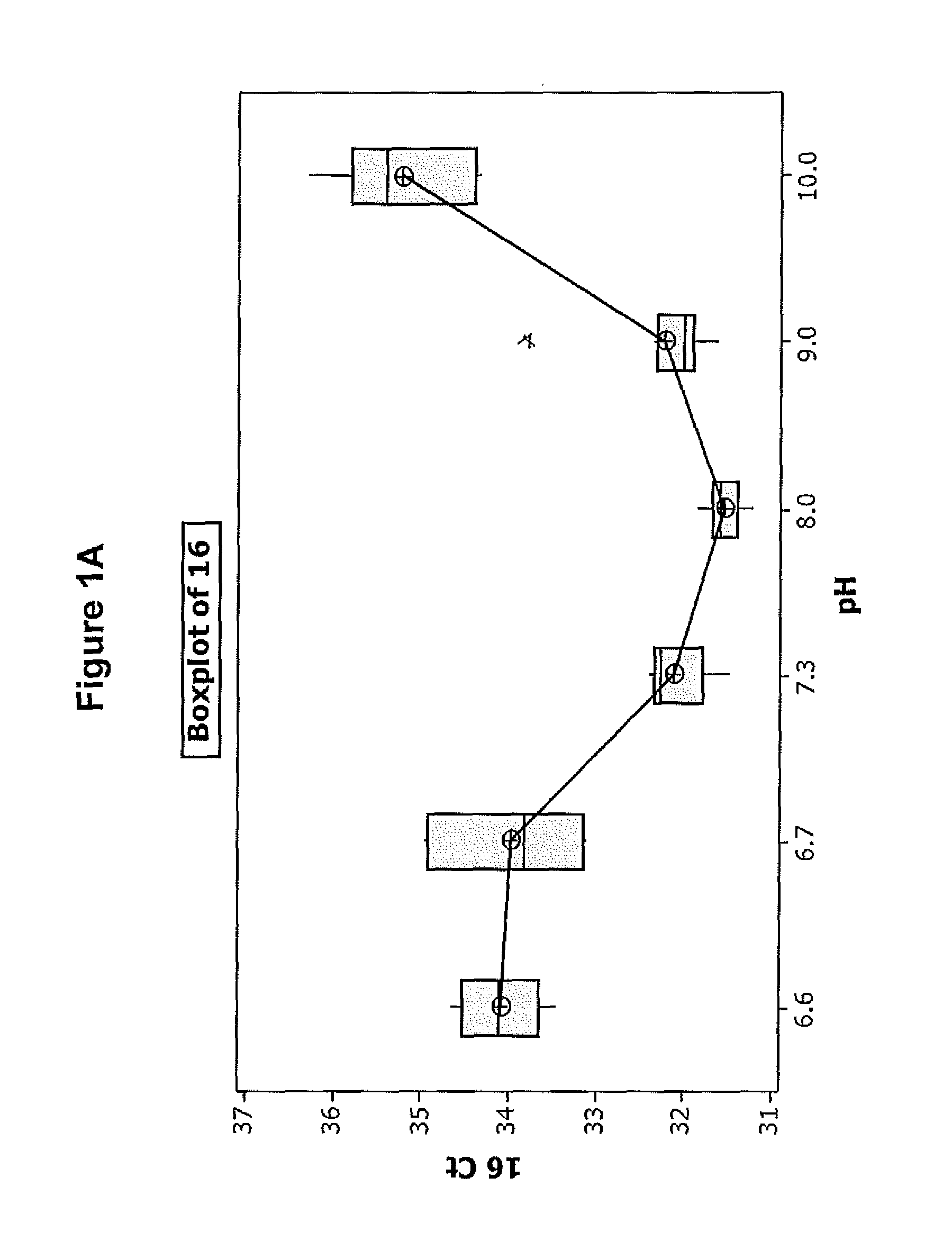
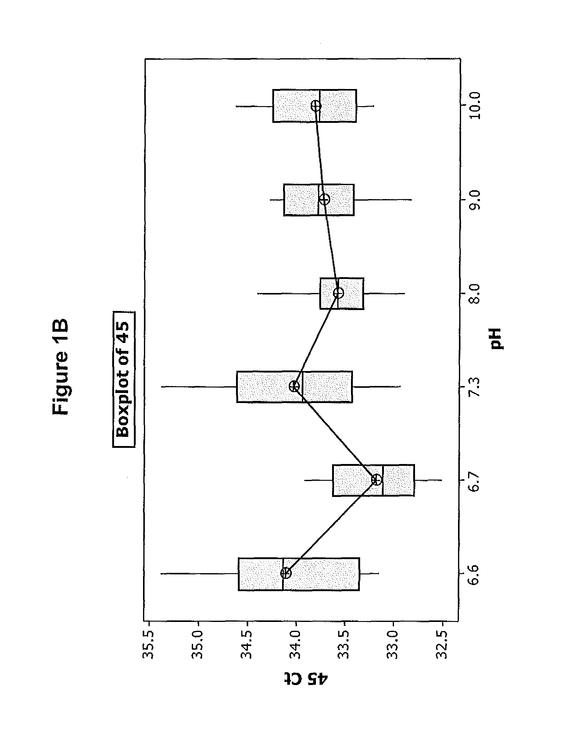
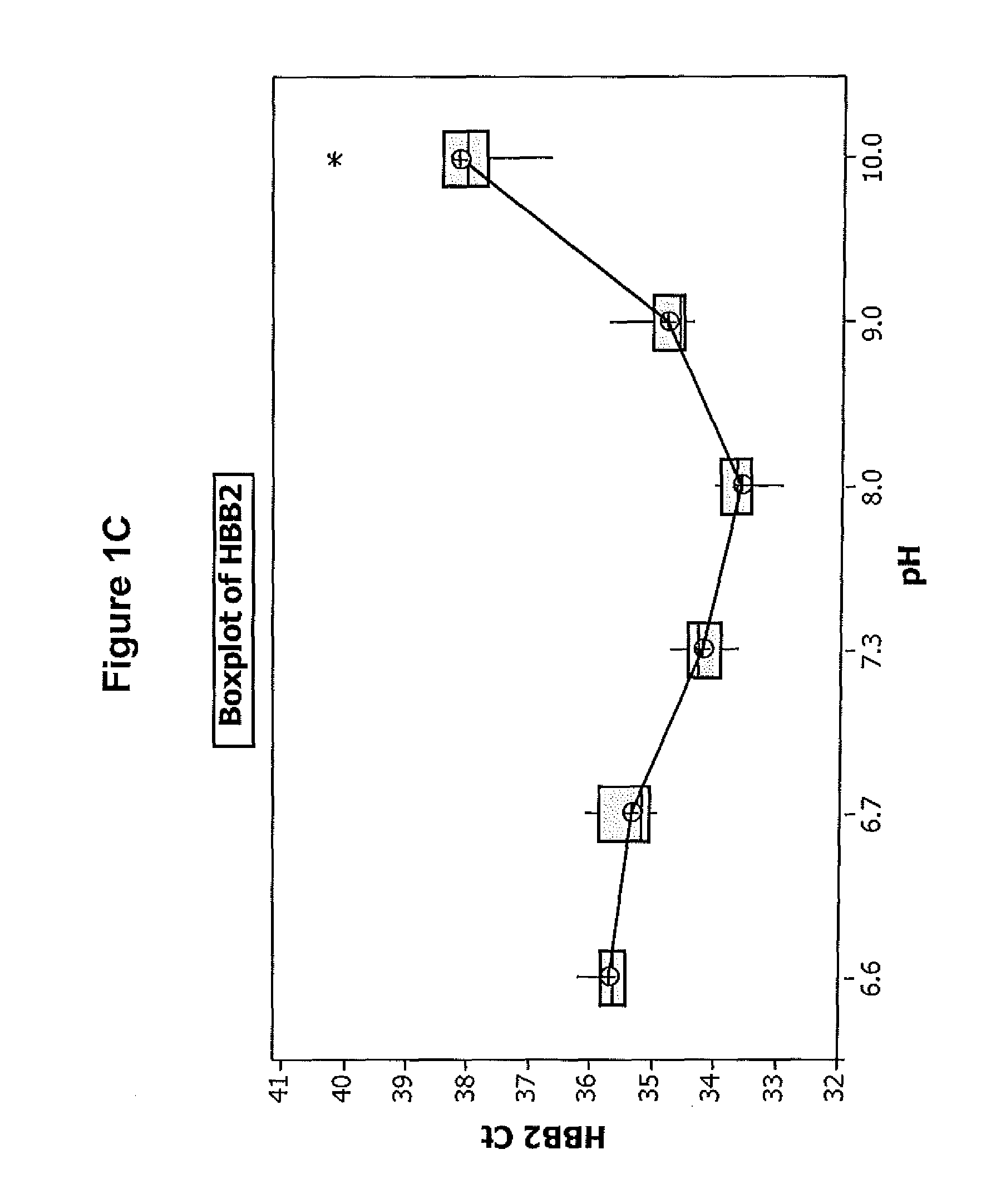
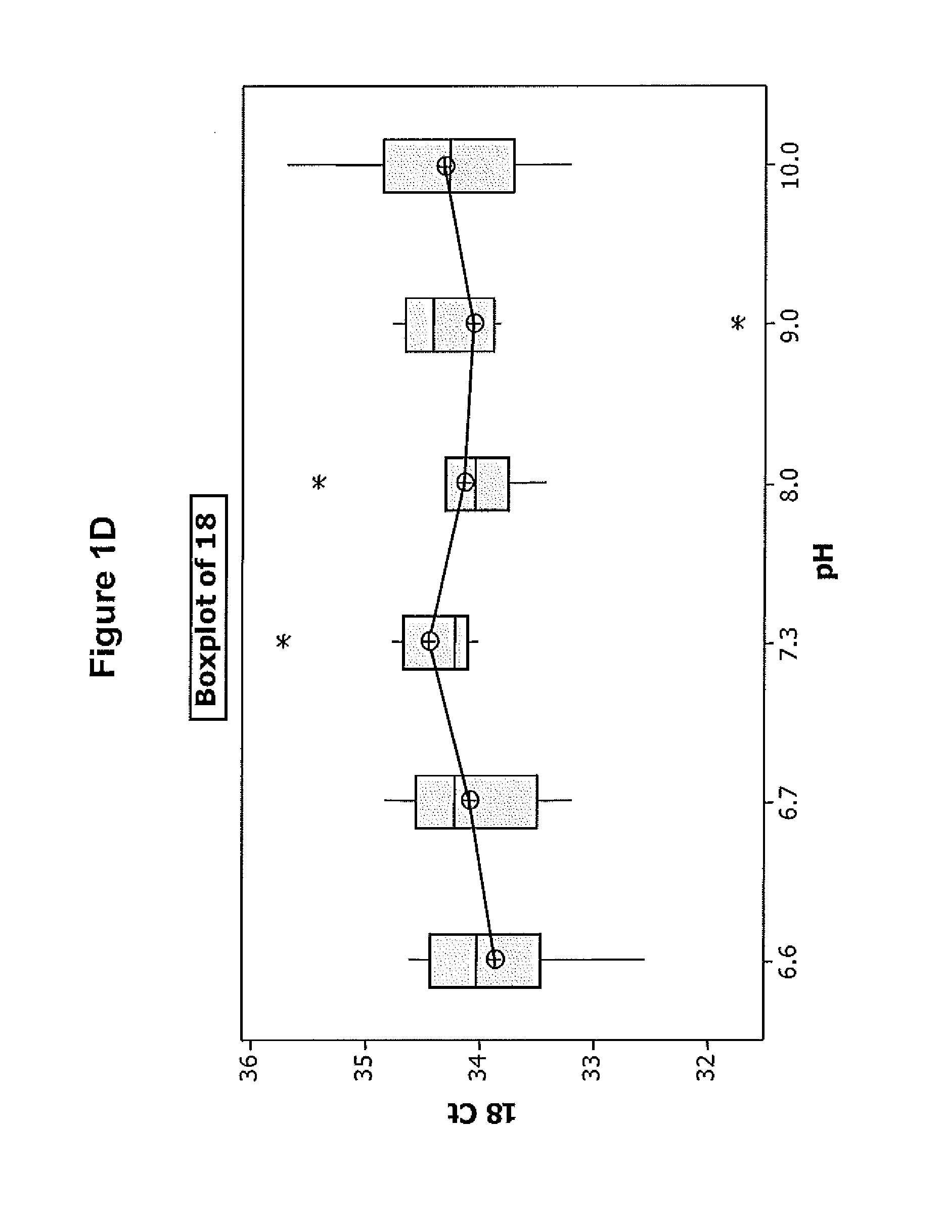
FIGS. 1A-D illustrate the effect of pH on the efficiency of one embodiment of the direct chemical lysis composition.
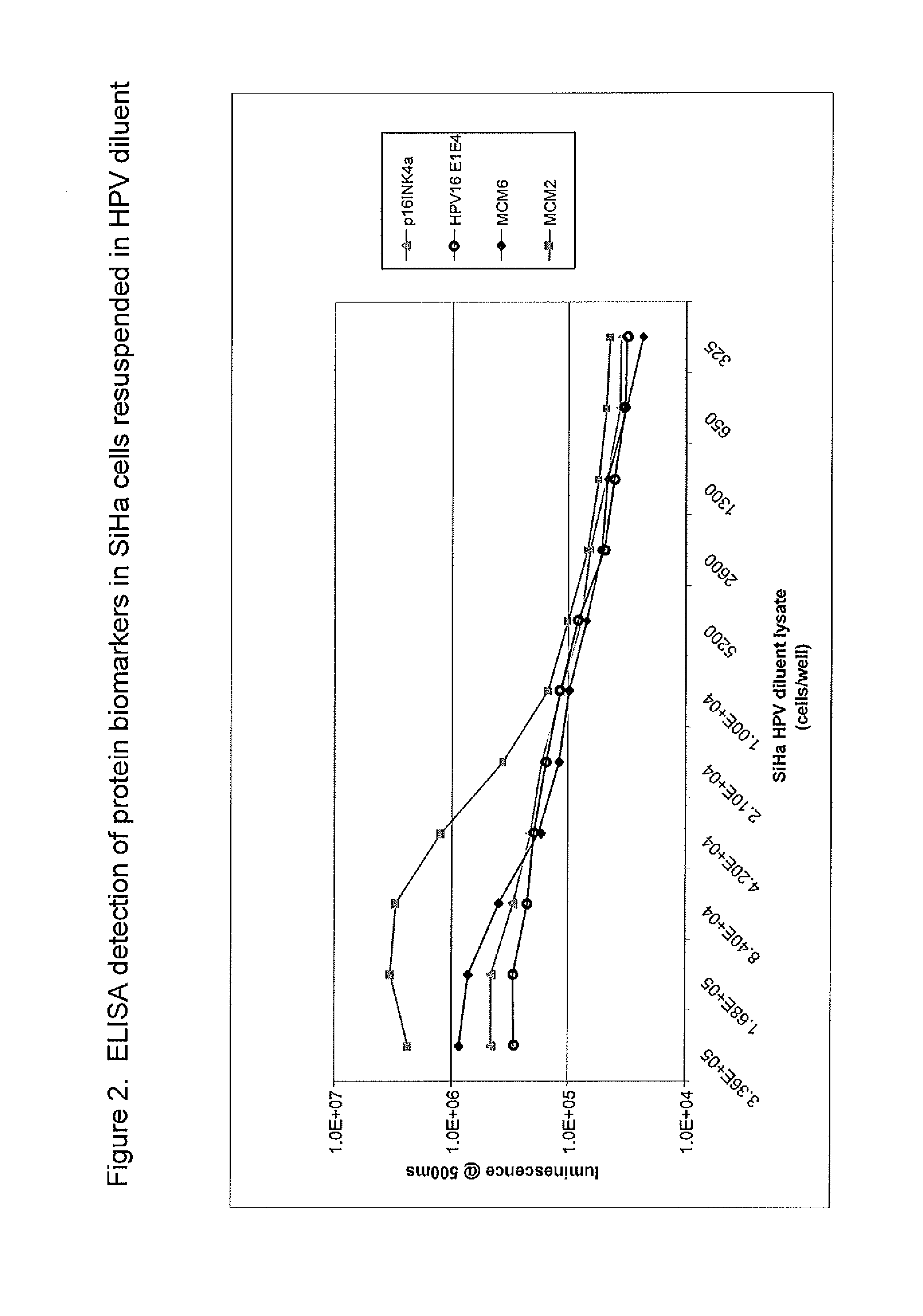
FIG. 2 illustrates protein biomarker detection from samples lysed using one embodiment of the direct chemical lysis composition and method disclosed herein.
DETAILED DESCRIPTION
Before explaining at least one embodiment of the invention in detail, it is to be understood that the invention is not limited in its application to the details set forth in the following description or exemplified by the examples. The invention is capable of other embodiments or of being practiced or carried out in various ways. Also, it is to be understood that the phrasing and terminology employed herein is for the purpose of description and should not be regarded as limiting. The following are definitions of certain terms used in this specification.
A direct chemical lysis composition is a composition that allows for lysis of the cellular components in the sample for nucleic acid (NA) extraction thereof without the need for intervening steps to separate the chemical lysis composition from the sample. The direct chemical lysis composition also allows for the assay/detection of other sample constituents (e.g. protein biomarkers) without the need to separate the chemical lysis composition from the sample prior to assay/detection of those constituents.
Examples of NA are single or double-stranded DNA and RNA. Further examples of NA that can be extracted by the method include not only genomic DNA or RNA from animals, plants, bacteria, viruses, fungi and parasitic organisms, but also the DNA or RNA of mitochondria or chloroplasts. Examples of other classes of NA that can be extracted by the method include not only mRNA, but also transfer RNA, ribosomal RNA, and small nuclear RNA as well as plasmid DNA. DNA and RNA extracted by the method of the invention may also be either wholly or partially single-stranded or possess other tertiary or quaternary structure. A sample containing NAs is exemplified by viable samples such as leukocyte cells, the culture of host cells containing vectors or the like that are typically prepared by gene recombinant technology, cells infected with viruses or phages, viruses in blood, and the culture of a sample microorganism. The culture may contain microorganisms but its supernatant alone is sufficient. Not only an artificial culture but also a naturally occurring culture is applicable. In case of a sample containing lumps of microorganism, homogenization or sonication may be performed as required to achieve good efficiency of extraction.
Alternative sample types include but are not limited to biological specimens for the diagnosis of infectious or non-infectious diseases, environmental specimens, or samples of food or water. The target NA may be a particular sequence or it may be a class of NA. A class of nucleic acid is, for a particular assay method, those molecules of NA whose chemical, physical or biological properties are such that they can be expected to be extracted effectively in methods used for NA extraction. Typically, but not necessarily, the NA of a class are all DNA or DNA analogs or all RNA or RNA analogs. Other targets may be protein sequences such a protein biomarkers.
Targeted organisms can include but are not limited to Chlamydia trachomatis, Neisseria gonorrhoeae, Human Pappilloma Virus, Human Immunodeficiency Virus 1/2 , Hepatitis C Virus, Hepatitis B Virus, Severe Acute Respiratory Syndrome Virus, Influenza A/B, Herpes Simplex Viruses 1-6, Enteroviruses, West Nile Virus, Parainfluenza viruses, Adenoviruses, Respiratory Syncytial Virus A/B, Mycobacterium paratuberculosis, Mycobacterium avium-intracellulare complex, Mycobacterium tuberculosis complex, Cytomegalovirus, Group B Streptococcus, Bordetella pertussis, and Bordetella parapertussis.
In one aspect of the invention, the target nucleic acid is a particular RNA or cDNA from one or more of the following sources: bacterial pathogens, bacterial non-pathogens, viral pathogens, viral non-pathogens, fungal pathogens, fungal non-pathogens, yeast pathogens, yeast non-pathogens, parasitic pathogens, parasitic non-pathogens, plants, animal products, food, total RNA or cDNA within the sample matrix, total prokaryotic RNA or cDNA, total eukaryotic RNA or cDNA, or total viral RNA or cDNA.
In another aspect of the invention, the target nucleic acid sought is DNA from one or more of the following sources: bacterial pathogens, bacterial non-pathogens, viral pathogens, viral non-pathogens, fungal pathogens, fungal non-pathogens, yeast pathogens, yeast non-pathogens, parasitic pathogens, parasitic non-pathogens, plants, animal products, food, total DNA within the sample matrix, total genomic prokaryotic DNA, total genomic eukaryotic DNA, or total viral DNA.
In another aspect of the invention, the target is a protein biomarker.
A specimen storage composition is any composition that is used to store and or transport a biological sample prior to the extraction of nucleic acid from the cellular components in the sample. As noted above, specimen storage compositions include LBC media, paraffin media, etc.
Assay compatible as used herein is any composition that will not significantly adversely affect that assay to which the samples (e.g., extracted nucleic acid/proteins) are subjected. For example, an assay compatible composition will not modify the extracted nucleic acid in the sample in a manner that will adversely affect the ability of the downstream assay to detect the nucleic acid or other sample constituents such as protein biomarkers.
In one embodiment, the direct chemical lysis composition described herein has a two component assay compatible buffer composition. The assay compatible buffer composition will not modify the extracted nucleic acid in the sample in a manner that will adversely affect the ability of the downstream assay to detect the nucleic acid either by itself or when combined with other constituents of the chemical lysis composition or with constituents of the extracted nucleic acid (e.g. remnants of the specimen storage composition). The first component of the assay compatible buffer composition is an assay compatible buffer. Such buffers are well known to one skilled in the art and are not listed exhaustively herein. Examples of suitable buffers include tris(hydroxymethyl)amino methane and the acid salt of tris(hydroxymethyl)amino methane (Tris HCl herein). The final working concentration of the buffer in the direct chemical lysis composition is in range of about 0.2 M to about 2 M. Preferably the concentration of the buffer component is about 0.5 M to about 1 M. All concentrations herein are expressed as final working concentrations (i.e. concentrations at which the direct chemical lysis composition is combined with the sample).
The second component of the assay compatible buffer is a salt. Examples of suitable salts include NaCl, KCl, C.sub.2H.sub.3NaO.sub.2 (sodium acetate) and (NH.sub.4).sub.2SO.sub.4 (ammonium sulfate). In preferred embodiments the salt is a metal salt and, most preferably, NaCl. The concentration of the salt in the direct chemical lysis composition is in the range of about 0.01 M to about 1 M. In preferred embodiments, the concentration of the metal salt is about 0.1 M to about 0.5 M and most preferably about 0.1 M to about 0.4 M. The salt concentration in a particular direct chemical lysis composition depends upon the buffer, pH and surfactants (if any) in the composition.
The direct chemical lysis composition also contains an assay compatible surfactant. Examples of suitable surfactants include the above-described Triton.RTM. X-100 and Tween.RTM. 20 (polysorbate 20). Assay compatible is as defined above. In a preferred embodiment, the assay compatible surfactant is a polyethylene glycol based non-ionic surfactant. The concentration of the non-ionic surfactant is in the range of about 0.01 to about 2 percent (v/v). In preferred embodiments, the non-ionic surfactant is polyethylene glycol octylphenyl ether. Assay compatible surfactants are well known to those skilled in the art and are not discussed in detail here.
The direct chemical lysis composition has a pH in the range of about 6.6 to about 10. In preferred embodiments the pH is in the range of about 7 to about 9.
The direct chemical lysis composition described herein facilitates nucleic acid extraction from media-fixed samples by reducing the cross-linking that typically occurs between the NA and the media constituents to provide NA that is both extractable and amplifiable. In one embodiment, a sample is combined with the direct chemical lysis and incubated at a temperature in the range of about 100.degree. C. to about 130.degree. C. and for a time in the range of about 10 minutes to about 30 minutes to chemically lyse at least a portion of the cells and, to the extent NA cross-linking has occurred, de-crosslink the NA. This is followed by extraction of the NA from the sample. In preferred embodiments, the NA is DNA and DNA extraction is accomplished via magnetic separation of the DNA using ferric oxide particles. In preferred embodiments, the extraction protocol is compatible with the BD Viper XTR.TM. platform. Such a protocol provides for binding the DNA to the ferric oxide particles, washing the rest of the sample from the particles and eluting the DNA from the particles. The extracted DNA is the subjected to a suitable amplification and detection assay (e.g. real time PCR).
In another embodiment, the sample is analyzed for target antigens (e.g. protein biomarkers). Conventional assays for detecting antigens extracted from a sample are well known to one skilled in the art and are not described in detailed herein. In one example, antibody capture molecules are used to bind and assay target antigens. Antibody methods for such capture are routinely used in a variety of different formats. One of the most commonly practiced techniques is the Enzyme Linked Immunosorbent Assay (ELISA) which is routinely used in protein diagnostic applications. A desired characteristic of protein detection methods is the ability to both preserve and retrieve antigens from a cell such that they can be exposed to the capture antibody and also in some cases retain their native shape and allow conformationally-dependent antibodies to specifically bind to the target antigen. Another desired characteristic is the ability to permit the selective binding of the correct antibody molecules to the target and allow non-specific interactions to be removed during the wash steps. Protein biomarker detection therefore requires that the assay compatible buffer be compatible with and facilitate specific protein binding interactions. The direct chemical lysis composition described herein is sufficiently strong to lyse the cells, thereby liberating the protein contents of those cells. The direct chemical lysis composition does not adversely affect the liberated proteins, and protects the proteins from proteolytic enzymes. Finally, the assay compatible buffer is compatible with specific antibody binding and does not interfere with this process due to denaturing effects, ionic interactions etc.
The following are provided some examples to specifically illustrate the concepts described above. The following examples are highly specific embodiments of the present invention and are not to be construed as limiting the invention in any way, except in a manner consistent with the appended claims.
EXAMPLE 1
Extraction of HPV DNA from a Patient Sample Stored in SurePath LBC
Two solutions were compared to determine their efficacy as direct chemical lysis compositions. The first was a diluent that contained 1M Tris-HCl, 0.5M NaCl, 1% Triton X-100 and had a pH of 9.0 (Diluent 1 herein). The second diluent contained 330 mM Tris-HCl, 16.67 mM NaCl and had a pH of 7.8 (Diluent 2 herein). Stock patient-derived cells were harvested and stored in SurePath LBC for 21 days. Both Diluent 1 and Diluent 2 are embodiments of the direct chemical lysis composition of the present invention.
Specifically, stock patient-derived cells were diluted in SurePath LBC at a concentration of about 12,500 cells/ml. Forty-eight sample tubes were prepared for assay on the Becton Dickinson (BD) Viper.TM. tool. Twenty-four tubes had 0.85 ml of Diluent 1 and twenty-four had the same amount of Diluent 2. Diluted patient-derived cells (0.25 ml) were added into each of the 48 sample tubes. After combining with the samples, the final working buffer concentrations were as follows: i) 0.77M Tris-HCl, 0.386M NaCl, 0.77% Triton X-100 (with a pH of approximately 9.0) in Diluent 1; and ii) 0.255M Tris-HCl, 0.0129M NaCl (with a pH of approximately 7.8) in Diluent 2.
Eight samples from each group of twenty four were incubated either at i) room temperature, ii) 80.degree. C., or iii) 120.degree. C., all for twenty minutes. The samples were then cooled for twenty-five minutes at room temperature. The samples were then subjected to a modified Viper.TM. XTR DNA extraction protocol using iron oxide (FEO) particles and 0.8 ml of the pre-warmed or incubated sample was used for extraction. The extraction protocol for Viper.TM. XTR is commercially available and not described in detail herein. To the extent DNA was extracted, DNA for the sample was eluted in 400 .mu.l elution/neutralization buffer. The eluate (20 .mu.l) was mixed with 5 .mu.l of PCR master mix, and 20 copies/reaction of HPV 18 and HPV 45 plasmid DNA targets were post-spiked into each reaction for test of PCR inhibition. The real-time PCR assays were used for detection of HPV 16, 18 and 45, and human DNA endogenous control gene HBB. DNA detection was determined by the resulting cycle threshold (Ct) values. The Ct values less than about 30 represent strong positive reactions indicative of abundant target nucleic acid in the samples. Ct values of 30-35 represent moderate to low positive reactions indicative of moderate to low amounts of target nucleic acid in the sample. Ct values of 35-45 represent weak reactions that indicate minimal amounts of target nucleic acid in the sample. Ct values above about 45 are expressed as "No Ct", indicating that no DNA was being detected.
TABLE-US-00001 TABLE 1 Extraction of HPV DNA from Patient Samples stored in SurePath media Diluent 2 Diluent 1 RT 80.degree. C. 120.degree. C. RT 80.degree. C. 120.degree. C. HPV16 1 No Ct No Ct 32.57 No Ct 36.97 31.20 2 No Ct No Ct 32.49 No Ct No Ct 30.56 3 No Ct No Ct 32.33 No Ct No Ct 30.40 4 No Ct No Ct 32.55 No Ct No Ct 30.34 5 No Ct No Ct 32.88 No Ct No Ct 30.65 6 No Ct No Ct 32.57 No Ct 36.99 30.54 7 36.73 No Ct 32.35 No Ct No Ct 30.94 8 No Ct No Ct 32.64 No Ct No Ct 30.56 AVG. 32.55 30.65 HPV45 1 33.88 33.08 32.87 32.90 33.49 33.37 2 33.61 33.34 32.67 33.86 29.31 33.09 3 33.85 34.38 33.92 33.39 32.96 32.80 4 34.14 33.54 33.47 33.46 35.02 33.14 5 34.26 34.11 33.60 32.10 34.39 33.64 6 33.18 33.58 33.64 32.98 33.73 33.71 7 33.39 33.35 34.08 33.11 32.92 33.18 8 33.44 33.86 34.04 33.60 33.92 32.69 AVG. 33.72 33.66 33.54 33.18 33.22 33.20 HBB 1 No Ct No Ct 37.91 No Ct 42.51 34.85 2 No Ct No Ct 38.68 No Ct 41.67 35.48 3 No Ct No Ct 37.48 No Ct No Ct 34.34 4 No Ct No Ct 37.25 No Ct No Ct 34.95 5 No Ct No Ct 37.16 No Ct No Ct 34.9 6 No Ct No Ct 37.88 No Ct No Ct 35.48 7 No Ct No Ct 39.14 No Ct No Ct 35.22 8 No Ct No Ct 36.99 No Ct No Ct 34.99 AVG. 37.80 35.03 HPV18 1 34.55 34.77 34.78 34.37 34.08 34.03 2 33.73 34.12 34.25 34.60 34.33 33.82 3 34.59 33.97 34.00 34.05 33.77 33.44 4 34.47 34.53 33.78 33.78 34.68 33.52 5 34.84 34.87 33.86 34.36 34.17 33.41 6 33.99 33.63 34.82 33.61 33.86 34.74 7 33.77 34.68 34.00 34.05 33.47 33.42 8 33.84 34.24 34.11 34.57 34.79 33.99 AVG. 34.22 34.35 34.20 34.17 34.14 33.80
Incubation in Diluent 1 at 120.degree. C. reduced the Ct value of HPV 16 and HBB by 1.90 and 2.77 respectively when compared with incubation in Diluent 2 at 120.degree. C. Incubation at room temperature or 80.degree. C. in both diluents gave no amplification for HPV16 and HBB. The detection of spiked HPV 45 and HPV 18 confirmed that the assay itself worked but that HPV16 and HBB were not detected at lower incubation temperatures, indicating that lysis, extraction or detection did not occur in these samples.
EXAMPLE 2
Extraction of HPV DNA from SurePath Using Different Diluents and Individual Diluent Components
Stock patient-derived cells were diluted in SurePath LBC at 12500 cells/ml. Forty-eight BD Viper sample tubes were prepared. There were eight tubes in each group, with six groups (a-f) total. In each group 0.85 ml of one of the following buffers were added:
a. 1M Tris, (pH 9.0) only;
b. 0.5M NaCl;
c. 1% Triton X-100;
d. a+b+c;
Diluted patient cells (0.25 ml) were added to each of the 48 sample tubes. After combining with the samples, the final working buffer concentrations were as follows: 0.77M Tris-HCl in group a; 0.386M NaCl for group b; 0.77% Triton X-100 in group c; and 0.77M Tris-HCl, 0.386M NaCl, and 0.77% Triton X-100 in group d (the combination of buffers of a, b, and c). All sample tubes were incubated at 120.degree. C. for 20 min. The samples were then cooled for 25 minutes to room temperature. The samples were then loaded onto the BD Viper.TM. XTR tool and 0.8 ml was used for extraction as describe above. DNA was eluted from the samples in 400 .mu.l elution/neutralization buffer. Eluate (20 .mu.l) was mixed with 5 .mu.l of PCR master mix. Twenty copies/reaction of HPV 18 and HPV 45 plasmid DNA targets were post-spiked into each reaction as a control to test of PCR inhibition. The real-time PCR assays were used for detection of HPV 16, 18 and 45, and human DNA endogenous control gene HBB.
TABLE-US-00002 TABLE 2 Extraction of HPV DNA from SurePath LBC media using HPV diluents and individual component buffer 1M Tris (a) 0.5M NaCl (b) 1% Triton (c) a + b + c HPV16 1 35.27 35.86 34.56 32.43 2 37.07 36.62 36.55 31.03 3 34.27 No Ct 36.68 31.30 4 34.52 No Ct 35.65 30.95 5 No Ct No Ct 34.37 31.62 6 35.90 No Ct 33.71 31.21 7 34.26 No Ct 34.46 31.20 8 34.09 35.66 35.12 30.80 AVG. 35.05 36.05 35.14 31.33 HPV45 1 33.47 34.61 34.19 35.22 2 33.76 34.17 33.90 33.39 3 34.01 33.56 34.01 33.69 4 33.80 31.61 34.29 33.98 5 33.01 32.86 33.23 33.57 6 32.59 33.49 33.44 33.43 7 33.37 32.56 33.44 33.05 8 32.75 33.25 35.33 33.92 AVG. 33.35 33.26 33.98 33.78 HBB 1 No Ct No Ct No Ct 42.35 2 No Ct No Ct 44.25 41.29 3 No Ct No Ct 43.81 40.94 4 No Ct No Ct No Ct 40.38 5 No Ct No Ct No Ct 41.67 6 43.34 No Ct 42.17 40.63 7 44.52 No Ct 44.87 41.02 8 No Ct No Ct No Ct 40.70 AVG. 43.93 43.78 41.12 HPV18 1 34.99 34.94 34.05 34.34 2 33.94 34.56 33.84 34.18 3 34.80 34.68 34.65 34.27 4 33.97 34.52 33.80 33.74 5 33.95 35.75 33.89 34.25 6 35.17 33.90 34.27 34.35 7 34.39 34.56 34.41 33.64 8 33.87 34.94 34.03 34.32 AVG. 34.38 34.73 34.12 34.14
HPV types 16, 18 and 45 were detected using d) above with incubation at 120.degree. C. The other individual diluent components (1M Tris, 0.5 M NaCl and 1% Triton) did not provide for consistent DNA detection, regardless of HPV Type. Each individual component contributed to improve DNA extraction. The best result was achieved when components a, b and c were combined together.
EXAMPLE 3
Comparison of DNA Extraction from SurePath LBC Using Diluent 1 at 120.degree. C. and Diluent 2 with Room Temperature Incubation
Thirty-six SurePath samples were collected and extracted according to the following two protocols. 1) The no heat experiment. Thirty-six BD Viper.TM. sample tubes were prepared by adding 0.85 ml of the Diluent 2 described in Example 1. SurePath clinical sample (0.25 ml) was added into each tube. After combining with the samples, the final working buffer concentrations were as follows: 0.255M Tris-HCl, 0.0129M NaCl (with a pH of approximately 7.8) for Diluent 2. The thirty-six tubes were loaded onto the BD Viper.TM. and 0.8 ml of the sample was used for extraction. DNA was eluted from the sample in 400 .mu.l elution/neutralization buffer. The eluate (20 .mu.l) was mixed with 5 .mu.l of PCR master mix. A real-time PCR assay was used to detect for HPV 16, 18 and 45, and human DNA endogenous control gene HBB. 2) The heat experiment. Thirty-six BD Viper.TM. sample tubes were prepared to which were added 0.85 ml of the Diluent 1 described in Example 1. SurePath clinical sample (0.25 ml) was added into each tube. After combining the diluent with the samples, the final working buffer concentrations were as follows: 0.77M Tris-HCl, 0.386M NaCl, and 0.77% Triton X-100 (with a pH of approximately 9.0) in Diluent 1. All sample tubes were incubated at 120.degree. C. for 20 min and then cooled for 25 min to room temperature. The thirty-six tubes were loaded onto the BD Viper.TM. and 0.8 ml of the sample was used for extraction. DNA was eluted from the sample in 400 .mu.l elution/neutralization buffer. The eluate (20 .mu.l) was mixed with 5 .mu.l of PCR master mix. A real-time PCR HPV assays was used to detect for HPV 16, 18 and 45, and human DNA endogenous control gene HBB. HPV 45 and HPV 18 were not detected.
TABLE-US-00003 TABLE 3 Comparison of DNA Extraction: i) using Diluent 1 with 120.degree. C. incubation; and ii) Diluent 2 at room temperature incubation. HPV16 HBB No heat Heat No heat Heat .DELTA.Ct 1 No Ct No Ct 25.28 23.97 1.31 2 No Ct No Ct 32.25 28.30 3.95 3 No Ct No Ct 31.78 27.92 3.86 4 No Ct 41.33 34.78 29.83 4.95 5 No Ct No Ct 34.69 29.74 4.95 6 No Ct 41.09 37.51 31.31 6.20 7 No Ct No Ct 32.24 26.82 5.42 8 No Ct No Ct 31.93 26.88 5.05 9 No Ct No Ct 31.74 28.88 2.86 10 No Ct No Ct 28.88 25.59 3.29 11 No Ct No Ct 29.41 24.93 4.48 12 No Ct No Ct 35.21 29.66 5.55 13 No Ct No Ct 28.54 25.23 3.31 14 No Ct No Ct 30.66 27.54 3.12 15 No Ct 35.16 32.31 26.86 5.45 16 No Ct No Ct 36.29 29.39 6.90 17 25 21.07 32.54 28.00 4.54 18 No Ct No Ct 35.46 29.89 5.57 19 No Ct 34.90 32.18 29.19 2.99 20 No Ct No Ct 28.83 24.56 4.27 21 No Ct No Ct 31.64 26.72 4.92 22 No Ct No Ct 28.14 24.40 3.74 23 No Ct No Ct 26.67 25.68 0.99 24 No Ct 31.33 29.38 25.09 4.29 25 No Ct No Ct 32.74 31.56 1.18 26 No Ct No Ct 32.91 26.57 6.34 27 No Ct No Ct 32.96 28.30 4.66 28 No Ct No Ct 35.79 29.64 6.15 29 No Ct No Ct 29.96 27.84 2.12 30 No Ct No Ct 32.76 28.77 3.99 31 No Ct No Ct 31.37 24.60 6.77 32 No Ct No Ct 32.79 29.07 3.72 33 No Ct 42.28 30.9 27.83 3.07 34 No Ct No Ct 34.58 30.51 4.07 35 No Ct No Ct 36.73 28.06 8.67 36 36.36 32.38 22.78 21.22 1.56
On average, the heated incubation/Diluent 1 protocol shortened, by 4.29 Ct, the detection of HBB compared to the protocol using Diluent 2 at room temperature incubation. Samples that had the Type 16 HPV DNA (all samples are presumed to have the HBB control DNA whereas not all samples will have HPV of one or more types) exhibited detection with shortened Ct values and therefore became more detectable when using the protocol with heated incubation and Diluent 1 compared with using room temperature incubation and Diluent 2.
EXAMPLE 4
Comparison of DNA Extraction from SurePath and ThinPrep PreservCyt LBC with or without Heat
Stock patient-derived cells were diluted in one of either SurePath or ThinPrep negative clinical specimens at 12500 cells/ml. Thirty-two BD Viper.TM. sample tubes were prepared to which were added 0.85 ml of Diluent 1. SurePath spiked with patient cells (0.25 ml) was added to 16 sample tubes and ThinPrep spiked with patient cells (0.25 ml) was added to the other 16 sample tubes. After combining the diluent with the samples, the final working buffer concentrations were as stated in Example 1. Eight sample tubes from each group were incubated at room temperature and eight were incubated at 120.degree. C. for 20 minutes. The heated tubes were cooled for 25 minutes to room temperature. The sample tubes were then loaded on the BD Viper.TM. platform and 0.8 ml of the samples were used for DNA extraction. The DNA was then eluted in 400 .mu.l elution/neutralization buffer. The eluate (20 .mu.l) was mixed with 5 .mu.l of PCR master mix. Twenty copies/reaction of HPV 18 and HPV 45 plasmid DNA targets were post-spiked into each sample for testing the PCR inhibition. The real-time PCR assays were used for detection of HPV 16, 18 and 45, and human DNA endogenous control gene HBB.
TABLE-US-00004 TABLE 4 DNA Extraction from SurePath and ThinPrep PreservCyt LBC with or without heat ThinPrep SurePath No Heat Heat No Heat Heat HPV16 Rep 1 31.24 31.99 No Ct 31.64 2 30.89 31.37 No Ct 31.63 3 30.98 31.66 No Ct 32.16 4 31.39 31.58 No Ct 31.71 5 31.02 31.15 No Ct 31.16 6 31.26 31.44 No Ct 31.16 7 31.44 31.16 No Ct 31.48 8 31.85 31.33 No Ct 31.58 Avg. 31.26 31.46 31.57 HPV45 Rep 1 32.25 32.22 32.67 32.56 2 32.42 32.24 32.06 32.89 3 33.2 33.15 32.54 31.91 4 32.13 32.06 32.44 31.97 5 32.48 33.03 32.19 32.85 6 31.63 33.36 32.47 32.07 7 33.65 32.17 32.69 31.53 8 32.43 32.96 32.6 32.2 Avg. 32.52 32.65 32.46 32.25 HBB Rep 1 27.73 27.52 33.33 29.02 2 27.99 27.09 33.63 28.88 3 28.08 27.17 33.43 29.25 4 28.21 26.89 33.28 28.71 5 28.12 26.98 33.38 28.37 6 27.98 27.04 34.3 28.36 7 27.99 27.02 34.07 28.29 8 28.43 27.38 33.96 27.84 Avg. 28.07 27.14 33.67 28.59 HPV18 Rep 1 33.68 33.94 34.97 33.88 2 33.38 33.86 33.87 33.88 3 34.32 34.72 34.84 33.58 4 33.35 34.11 34.22 33.81 5 33.89 34.16 34.55 33.97 6 33.4 32.88 32.96 33.68 7 34.44 34.2 34.02 33.61 8 33.82 34.1 33.96 34.83 Avg. 33.79 34.00 34.17 33.91
Incubating the specimens at 120.degree. C. in Diluent 1 significantly shortened HBB Ct value for both the ThinPrep and SurePath samples. The reduction (compared to the room temperature incubation) was 0.93 Ct for the ThinPrep samples and 5.08 Ct for the SurePath samples. Heat had no statistically significant effect on the detection of HPV16 from ThinPrep, but significantly improved the detection of HPV16 from SurePath.
EXAMPLE 5
DNA Extraction from SurePath Using Diluent 1 at Varied pH, Temperature and Time
The incubation protocol at 114.degree. C. for 10 minutes was used in conjunction with Diluent 1 described in Example 1 (i.e. 1M Tris, 0.5M NaCl and 1% Triton x100). Samples with different pH's (9.0 and 7.8) were used and tested at varied incubation temperatures and times. The different pHs were also evaluated against Diluent 2 of Example 1 (16.67 mM NaCl and 330 mM Tris).
Stock patient-derived cells were diluted in SurePath at a concentration of 12500 cells/ml. Fifty-six sample tubes were prepared for use in BD Viper XTR.TM. Platform. There were eight samples in each of seven groups. Each of the following buffers (0.85 ml) was added into one of the seven groups group:
a. Diluent 1, pH 9.0 (incubation at 120.degree. C., 20 min)
b. Diluent 1, pH 9.0 (incubation at 120.degree. C., 10 min)
c. Diluent 1, pH 9.0 (incubation at 114.degree. C., 20 min)
d. Diluent 1, pH 9.0 (incubation at 114.degree. C., 10 min)
e. Diluent 1, pH 7.8 (incubation at 120.degree. C., 20 min)
f. Diluent 2, pH 7.8 (incubation at 120.degree. C., 20 min)
g. Diluent 2, pH 9.0 (incubation at 120.degree. C., 20 min)
Aliquots (0.25 ml) of the diluted patient samples were added into each tube of each group. After combining the diluent with the samples, the final working buffer concentrations are as set forth in Example 1. Each group was incubated at its specified temperature and time above. All tubes were then cooled for 25 minutes to room temperature. The tubes were then loaded onto the BD Viper XTR.TM. and 0.8 ml was used for DNA extraction from the samples. The DNA was eluted in 400 .mu.l elution/neutralization buffer. The eluate (20 .mu.l) of each was mixed with 5 .mu.l of PCR master mix. Twenty copies/reaction of HPV 18 and HPV 45 plasmid DNA targets were post-spiked into each reaction to test for PCR inhibition. The real-time PCR assays were used for detection of HPV 16, 18 and 45, and human DNA endogenous control gene HBB.
TABLE-US-00005 TABLE 5 DNA Extraction from SurePath using Diluent 1 with different pH and at different temperature and time Diluent 1 Diluent 1 Diluent 2 120.degree. C. 114.degree. C. 120.degree. C. 120.degree. C. pH 9,20' pH 9,10' pH, 9,20' pH 9,10' pH 7.8,20' pH 7.8,20' pH 9,20' HPV16 1 32.36 33.57 33.4 No Ct 30.79 33.5 35.21 2 31.29 33.24 33.11 No Ct 30.56 32.79 33.11 3 31.18 33.23 34.02 34.84 30.55 33.05 37.78 4 31.31 32.75 32.73 No Ct 30.96 33.48 34.43 5 32.09 31.98 33.92 34.84 30.58 34.9 35.15 6 31.37 32.68 32.69 No Ct 30.67 34.53 34.32 7 30.74 32.31 32.4 No Ct 30.31 34.43 35.81 8 31.91 33.02 32.65 36.58 30.85 33.12 No Ct Avg. 31.53 32.85 33.12 35.42 30.66 33.73 35.12 HPV45 1 33.2 34.66 34.08 34.1 33.71 34.41 33.97 2 33.35 34.31 33.56 33.71 34.03 34.18 34.5 3 34.32 33.32 33.05 34.5 34.47 33.42 34.92 4 34.56 32.84 33.71 34.28 32.21 33.81 34.09 5 33.22 34.63 34.48 33.43 34.61 33.89 33.72 6 34.11 34.8 34.11 33.98 33.47 34.15 33.99 7 34.65 31 34.45 34.53 34.12 34.47 33.59 8 34.8 34.38 33.98 33.47 32.24 34.23 34.09 Avg. 34.03 33.87 33.93 34.00 33.61 34.07 34.11 HBB 1 32.52 36.54 33.5 37.33 31.38 33.61 36.89 2 32.27 34.4 34.54 No Ct 31.23 32.89 35.48 3 31.97 33.51 33.66 36.54 31.23 33.39 36 4 32.42 32.79 33.87 No Ct 31.83 34.63 35.84 5 33.12 32.97 32.88 35.12 31.64 35.88 36.94 6 32.86 34.05 34.01 No Ct 31.42 36.71 36.63 7 31.79 33.53 33.41 37.14 31.33 34.26 No Ct 8 32.79 34.22 34.13 31.23 31.8 34.41 No Ct Avg. 32.47 34.00 33.75 35.47 31.48 34.47 36.30 HPV18 1 32.2 34.35 35.13 34.31 33.57 33.16 34.19 2 33.16 33.21 32.69 33.33 33.08 34.32 33.96 3 34.08 33.4 33.22 33.1 33.66 33.29 34.91 4 31.86 33.99 33.92 33.15 34.12 33.72 34.48 5 33.14 33.91 33.53 33.36 33.35 32.72 33.18 6 33.46 34.18 33.28 33.27 33.3 33.3 34.49 7 33.37 33.5 33.39 33.42 33.3 34.92 33.51 8 33.46 34.47 35.07 32.68 33.71 34.84 33.35 Avg. 33.09 33.88 33.78 33.33 33.51 33.78 34.01
The incubation protocol (114.degree. C., 10 minutes) did not yield the degree of detection for HPV 16 that was obtained when the higher heat protocol was used. However, the same degree of detection was obtained for the spiked HPV 45 and HPV 18, irrespective of the incubation protocol that was used. From this it was concluded that the 114.degree. C. protocol provided a lower degree of DNA extraction than the 120.degree. C. incubation protocol. Similarly, incubation at 120.degree. C. for 10 minutes was not as effective for extracting HPV 16 DNA as incubation at 120.degree. C. for 20 minutes. Extraction and detection of HPV 16 DNA using Diluent 1 at pH 7.8 showed a near 1 Ct improvement compared to extraction and detection using Diluent 1 with a pH 9.0 (for incubation at 120.degree. C. for 20 minutes). Extraction following incubation at 120.degree. C. in Diluent 1 resulted in a significant reduction in assay variability as measured by the standard deviation in detected PCR replicates.
EXAMPLE 6
Effect of pH on Efficacy of Diluent
Stock patient-derived cells were diluted in SurePath LBC media at 12500 cells/ml. Forty sample tubes were prepared for the BD Viper XTR.TM. platform. They were separated into five groups. Diluent 1 as described in Example 1 (0.85 ml) with a different pH was added to the tubes in each group. The diluents used were:
a. Diluent 1, pH 6.6;
b. Diluent 1, pH 6.7;
c. Diluent 1, pH 7.3;
d. Diluent 1, pH 8.0;
e. Diluent 1, pH 9.0; and
f. Diluent 1, pH 10.0.
The diluted stock patient cells (0.25 ml) were added to each tube of each group. Each group was incubated at 120.degree. C. for 20 minutes, and then cooled for 25 minutes to room temperature. The samples were then loaded on the BD Viper XTR.TM. and 0.8 ml of the samples was used for extraction. DNA was eluted from the samples in 400 .mu.l elution/neutralization buffer and the eluate 20 .mu.l was mixed with 5 .mu.l of PCR master mix. Twenty copies of HPV 18 and HPV 45 plasmid DNA targets were post-spiked into each sample to test for PCR inhibition. The real-time PCR assays were used for detecting HPV 16, 18 and 45, and human DNA endogenous control gene HBB.
TABLE-US-00006 TABLE 6 Effect of pH on efficacy of Diluent 1 pH 6.6 pH 6.7 pH 7.3 pH 8.0 pH 9.0 pH 10 HPV16 Rep 1 33.64 33.46 31.47 31.21 33.79 35.68 2 34.06 34.96 31.67 31.36 31.86 34.32 3 34.58 33.10 32.33 31.70 31.99 36.30 4 33.66 33.15 32.40 31.49 31.61 35.73 5 33.46 34.90 32.32 31.85 32.00 34.36 6 34.41 33.14 32.12 31.61 31.96 34.51 7 34.66 34.93 32.25 31.58 32.12 35.15 8 34.15 34.18 32.27 31.57 32.36 35.83 Avg. 34.08 33.98 32.10 31.55 32.21 35.24 HPV45 Rep 1 33.78 32.88 33.38 33.28 34.09 33.35 2 34.56 32.95 34.40 33.41 32.83 33.56 3 34.08 33.69 34.69 32.9 33.41 33.84 4 33.15 33.45 33.65 34.41 34.11 34.64 5 35.39 33.27 33.59 33.74 33.45 33.70 6 33.21 32.51 35.39 33.77 34.14 34.38 7 34.19 32.77 32.93 33.59 33.49 33.21 8 34.6 33.92 34.24 33.58 34.29 33.89 Avg. 34.12 33.18 34.03 33.59 33.73 33.82 HBB Rep 1 33.21 35.08 33.56 32.84 35.69 38.28 2 35.82 35.28 33.79 33.93 34.51 No Ct 3 35.37 34.87 34.25 33.41 34.29 36.61 4 35.38 35.00 34.23 33.61 34.47 37.64 5 35.64 35.12 34.21 33.96 34.45 37.68 6 35.50 36.04 34.15 33.70 34.80 37.98 7 35.58 35.16 34.70 33.34 34.53 38.37 8 35.80 36.08 34.42 33.56 35.01 40.15 Avg. 35.66 35.33 34.16 33.54 34.72 38.10 HPV18 Rep 1 34.18 33.19 34.10 33.69 34.70 33.59 2 33.87 33.36 34.28 34.07 34.78 34.36 3 32.54 34.17 34.01 33.42 34.05 34.17 4 33.42 34.36 34.77 34.02 31.75 34.96 5 34.5 34.64 34.37 33.96 34.51 34.08 6 33.58 33.89 35.73 34.28 33.82 35.70 7 34.22 34.27 34.16 34.31 34.49 33.20 8 34.63 34.83 34.11 35.42 34.33 34.55 Avg. 33.87 34.09 34.44 34.15 34.05 34.33
Referring to the results in FIGS. 1A-D, the Diluent 1 with a pH in the range of about 7.3-9 provides the best results in terms of the degree of HPV detection from the sample. This indicates that extraction of DNA from the sample is best when the diluent having a pH in this range is used. Since the detection of spiked HPV 18 and 45 was basically the same at all pH's the improvement in detection of HPV16 and HBB is attributed to better DNA extraction obtained at pH's in range of 7.3 and 9.0.
EXAMPLE 7
Comparison of DNA Extraction Protocols
Patient-derived cells were harvested and separated into two equal groups. One-half were stored in ThinPrep and the other half SurePath LBC. Both groups had a concentration of 1.14.times.10.sup.7 cells/ml. A stock with a cell concentration 2.5.times.10.sup.4 cells/ml was prepared from the 1.14.times.10.sup.7 cells/ml stock in SurePath media. This stock was used for the following extraction protocols. All extraction protocols were followed by DNA extraction using the BD Viper XTR.TM. platform.
In the first protocol, eight 226 .mu.l samples of the SurePath stock were spun for 5 minutes at a force of 13,000 g. The resulting supernatant was decanted and resuspended in 1 ml of the Diluent 2 (described in Example 1) that also contained a proteinase K in a concentration of 2 mg/ml. The eight samples were transferred into sample tubes for use with the BD Viper XTR.TM. platform and incubated at 70.degree. C. for 1 hour. This was followed by extraction on the BD Viper XTR.TM. platform as described below.
In the second protocol, eight samples (226 .mu.l) of the SurePath stock were diluted (1:4) by adding to the samples 774 .mu.l of Diluent 2 that also contained a proteinase K in a concentration of 2 mg/ml. The eight samples were transferred into sample tubes for use with the BD Viper XTR.TM. platform and incubated at 70.degree. C. for 1 hour. This was followed by extraction on the BD Viper XTR.TM. platform as described below.
In the third protocol, eight samples (250 .mu.l) of the SurePath stock were combined with 850 .mu.l of the diluent (described in Example 1). The eight samples were transferred into sample tubes for use with the BD Viper XTR.TM. platform and incubated at 120.degree. C. for 20 minutes. This was followed by extraction on the BD Viper XTR.TM. platform as described below.
In the fourth protocol, eight samples (250 .mu.l) of the SurePath stock were combined with 850 .mu.l of Diluent 1 (described in Example 1). The eight samples were transferred into sample tubes for use with the BD Viper XTR.TM. platform and incubated at 120.degree. C. for 20 minutes. This was followed by extraction on the BD Viper XTR.TM. platform as described below.
For a negative control, eight 850 .mu.l aliquots of Diluent 2 were combined with 250 .mu.l of clean SurePath media. This was followed by extraction on the BD Viper XTR.TM. platform as described below
All samples were loaded on the BD Viper XTR.TM. and 0.8 ml of the samples was used for extraction. DNA was eluted from the samples in 400 .mu.l elution/neutralization buffer and the eluate 20 .mu.l was mixed with 5 .mu.l of PCR master mix. The real-time PCR assays were used for detecting HPV 16 and human DNA endogenous control gene HBB.
TABLE-US-00007 TABLE 7 Comparison of the heat treatment and Proteinase K (PK) treatment Protocol Protocol 1 2 3 4 3 Condition Spin, PK, No Spin, 70.degree. C. in PK, 70.degree. C. in 120.degree. C. in 120.degree. C. in Non-target Diluent 2 Diluent 2 Diluent 2 Diluent 1 controls Target HBB HPV16 HBB HPV16 HBB HPV16 HBB HPV16 HBB HPV16 Rep 1 31.38 29.49 No Ct 32.85 32.19 29.97 29.94 28.01 No No Ct Ct Rep 2 31.38 29.48 32.85 31.21 31.30 29.35 29.45 27.75 No No Ct Ct Rep 3 31.00 28.92 32.26 30.40 31.62 29.88 29.83 27.96 No No Ct Ct Rep 4 30.55 28.66 32.39 30.66 31.01 29.02 29.72 27.56 No No Ct Ct Rep 5 30.12 28.37 32.16 30.06 31.88 29.61 30.16 28.26 No No Ct Ct Rep 6 30.68 28.86 32.15 30.36 31.62 29.49 29.79 27.83 No No Ct Ct Rep 7 31.09 28.9 32.26 30.15 31.56 29.51 29.95 28.22 No No Ct Ct Rep 8 30.55 28.66 32.26 30.23 31.61 29.32 30.19 28.57 No No Ct Ct Avg. 30.84 28.92 32.33 30.74 31.60 29.52 29.88 28.02
Incubation in Diluent 1 at 120.degree. C. yielded the lowest CT values on average for HPV 16. This indicates that, of the protocols tested, the incubation in Diluent 1 provided for the best extraction of DNA from the sample. The protocol that used Diluent 2 with incubation at 120.degree. C. yielded a better result than the result from proteinase K incubation at 70.degree. C. without spin down. Specifically, Diluent 1 reduced the average Ct value of HPV 16 by 0.90, 2.72 and 1.5, respectively, and reduced the average Ct value of HBB by 0.97, 2.45 and 1.72, respectively, when compared with i) proteinase K treatment (following spin down) at 70.degree. C.; ii) proteinase K treatment (No spin down); and iii) incubation at 120.degree. C. in Diluent 2.
EXAMPLE 8
Extraction from Samples Spiked with Blood
Pooled SurePath and ThinPrep clinical negative specimens were combined with whole blood. The samples had 1%, 2%, 5% and 10% (volume of blood per volume of sample). All samples were incubated at 120.degree. C. in Diluent 1 described in Example 1 for 20 minutes. This was followed by extraction on the BD Viper XTR.TM. platform using the protocol described in the previous examples. All samples were loaded on the BD Viper XTR.TM. and 0.8 ml of the samples was used for extraction. DNA was eluted from the samples in 400 .mu.l elution/neutralization buffer and the eluate 20 .mu.l was mixed with 5 .mu.l of PCR master mix. The real-time PCR HPV assay was used for detecting human DNA endogenous control gene HBB.
TABLE-US-00008 TABLE 8A Effect of Blood on Assay (SurePath) Media SurePath Blood (%) 0 1 2 5 10 1 28.62 28.66 28.97 38.66 No Ct 2 30.02 28.99 29.34 29.76 28.78 3 28.63 28.65 28.63 28.73 28.52 4 28.34 28.43 28.81 28.61 28.28 5 27.92 28.69 28.90 29.10 28.06 6 28.24 28.97 28.67 28.98 28.64 7 28.08 29.13 28.09 28.61 28.76 8 28.48 28.92 29.46 28.15 28.73 Avg. 28.54 28.81 28.86 28.83 28.54
TABLE-US-00009 TABLE 8B Effect of Blood on Assay (ThinPrep PreservCyt) Media ThinPrep PreservCyt Blood (%) 0 1 2 5 10 1 31.99 30.99 31.16 30.75 30.38 2 32.08 31.67 30.28 30.40 29.98 3 31.92 31.62 31.02 30.19 30.34 4 31.65 31.31 31.12 30.20 30.16 5 31.55 31.43 30.79 30.10 30.06 6 31.90 31.04 30.63 30.14 30.49 7 31.31 31.40 30.53 30.29 29.96 8 31.45 31.34 30.85 29.84 29.88 Avg. 31.73 31.35 30.80 30.24 30.16
The assay was not affected by high concentration of whole blood, a substance often found in LBC specimens and known inhibitor of PCR reactions. This DNA extraction method delivers suitable DNA for PCR amplification even with high concentration of whole blood present in the clinical samples.
EXAMPLE 9
DNA Extraction from Formalin-Fixed, Paraffin-Embedded Tissue Slice
Formalin-fixed, paraffin-embedded cervical biopsy tissue slices were incubated at 120.degree. C. in 2 ml of Diluent 1 described in Example 1 for 25 minutes. The samples were then cooled to room temperature. After cooling, the samples were subjected to the previously described extraction protocol on the BD Viper XTR.TM. Platform. All samples were loaded on the BD Viper XTR.TM. and 0.8 ml of the samples was used for extraction. DNA was eluted from the samples in 400 .mu.l elution/neutralization buffer and the eluate 50 .mu.l was added into PCR master dry mix. The real-time PCR assays were used for detecting HPV subtypes 16, 18, 45, 31, 51, 52, 59, (33,58,56,66), (39,68,35) and human DNA endogenous control gene HBB.
TABLE-US-00010 TABLE 9 Viper DNA extraction from Formalin-Fixed, Paraffin-Embedded Tissue slice Pre-Cancer/Cancer Beta Globin Sample ID Pathology Grade Result HPV Result DH0727 Squamous Cell Carcinoma Positive HPV 16 DH0847 Squamous Cell Carcinoma Positive HPV 16 DH0848 Poorly differentiated Carcinoma Positive HPV 18, 52 DH0856 Squamous Cell Carcinoma Positive HPV 18, 31 DH0867 Cervical Intraepithelial Neoplasia Positive HPV 16 (CIN) III DH0869 Cervical Intraepithelial Neoplasia Positive High Risk HPV (CIN) III Negative DH0870 Cervical Intraepithelial Neoplasia Negative HPV Indeterminate (CIN) III DH0873 Cervical Intraepithelial Neoplasia Positive HPV 16 (CIN) III DH0874 Cervical Intraepithelial Neoplasia Positive HPV 16 (CIN) III DH0880 Cervical Intraepithelial Neoplasia Positive HPV 16 (CIN) III DH0892 Squamous Cell Carcinoma Positive HPV 16 DH0896 Squamous Cell Carcinoma Positive HPV (33, 58, 56, 66) DH0898 Squamous Cell Carcinoma Positive HPV 16, 45 DH0900 Squamous Cell Carcinoma Positive HPV 45 DH0902 Squamous Cell Carcinoma Positive HPV 16 DH0903 Squamous Cell Carcinoma Positive HPV 16 DH0904 Squamous Cell Carcinoma Positive HPV 16 DH0905 Squamous Cell Carcinoma Positive HPV 16, 59 DH0910 Cervical Intraepithelial Neoplasia Positive HPV 16 (CIN) III DH0911 Cervical Intraepithelial Neoplasia Positive HPV 16 (CIN) III DH0914 Cervical Intraepithelial Neoplasia Positive HPV 16 (CIN) III DH1408 Cervical Intraepithelial Neoplasia Positive HPV 16, 59 (CIN) III
In 20 of 22 individual samples, some subtype(s) of HPV DNA were successfully detected. The human endogenous beta-globin gene was detected in 21 of 22 samples. Real-time PCR and HPV subtype specific primers and beta-globin specific primers for HBB were used. The beta globin results indicate that there was no obvious PCR inhibition associated with the current DNA extraction method. The failure to detect beta globin signal in one sample and both beta globin and HPV signal in a second may have been due to lack of sufficient target cells in the section processed for DNA extraction.
EXAMPLE 10
Extraction of HPV DNA from Patient-derived Cells Stored in SurePath and ThinPrep LBC Media Using Diluent 1 for Direct Chemical Lysis
Stock patient-derived cells were diluted in SurePath and Thinprep LBC at 5000 cells/ml. Sixteen tubes of each sample type had 0.50 ml of the following diluent: 1.5M Tris, 0.386M NaCl and 1.5% Triton X-100(v/v). The diluent had a pH of 7.9. Diluted patient-derived cells (0.5 ml) in SurePath and ThinPrep were added into each of the sample tubes with diluent. After combining with the samples, the final working buffer concentrations were as follows: 0.75M Tris-HCl, 0.193M NaCl, and 0.75% Triton X-100(v/v) (with a pH of approximately 7.9). The combined solution was incubated at 120.degree. C. for 20 minutes as direct chemical lysis. Following incubation, the samples were prepared for extraction on the BD Viper.TM. XTR platform. The extraction protocol previously describe was used. That is, all samples were loaded on the BD Viper XTR.TM. and 0.8 ml of the samples was used for extraction. Half of the samples were with lysis step during extraction and half of the samples were not. DNA was eluted from the samples in 400 .mu.l elution/neutralization buffer and the eluate 20 .mu.l was mixed with 5 .mu.l of PCR master mix. Twenty copies of HPV 18 and HPV 45 plasmid DNA targets were post-spiked into each sample to test for PCR inhibition. The real-time PCR assays were used for detecting HPV 16, 18 and 45, and human DNA endogenous control gene HBB. The results of the assays are provided below.
TABLE-US-00011 TABLE 10 Using pre-warm in diluent as direct chemical lysis followed by DNA Extraction with and without lysis SurePath ThinPrep No Lysis Lysis No Lysis Lysis HPV16 Rep 1 31.77 32.15 31.67 32.06 2 31.83 31.43 32.04 31.68 3 31.55 31.95 31.76 31.93 4 31.76 32.35 31.86 32.00 5 32.05 32.19 31.67 32.36 6 31.73 32.02 31.46 31.55 7 31.95 31.96 31.64 32.28 8 32.18 32.30 31.85 31.41 Avg. 31.85 32.04 31.74 31.94 HPV45 Rep 1.00 33.26 32.82 33.12 32.83 2.00 33.10 33.03 32.92 32.61 3.00 32.45 31.90 32.49 33.06 4.00 31.89 32.99 32.40 33.17 5.00 33.47 31.92 32.63 32.13 6.00 33.38 32.66 32.89 31.66 7.00 32.53 32.32 32.04 32.48 8.00 32.98 32.95 32.23 33.47 Avg. 32.88 32.57 32.59 32.69 HBB Rep 1 31.63 32.10 31.50 32.11 2 30.37 31.64 31.16 31.67 3 31.57 31.75 31.24 31.93 4 31.69 31.68 31.48 31.77 5 31.49 31.99 31.06 31.87 6 32.08 31.15 31.45 31.45 7 31.95 31.37 31.50 31.02 8 32.05 32.06 31.94 31.38 Avg. 31.60 31.72 31.42 31.65 HPV18 Rep 1.00 33.93 34.21 34.17 33.91 2.00 34.06 33.53 33.41 33.20 3.00 33.00 33.05 33.71 33.47 4.00 33.99 33.40 33.64 33.15 5.00 33.77 33.45 30.55 33.40 6.00 33.69 29.50 33.40 32.60 7.00 33.23 32.77 33.65 33.62 8.00 33.57 33.43 33.79 33.58 Avg. 33.66 32.92 33.29 33.37
The DNA yield for both the extracted DNA (i.e. HPV 16 and HBB) and the spiked DNA (i.e. HPV 45 and HPV 18) is about the same for both the lysis and the no lysis during NA extraction (i.e. direct chemical lysis using one example of the composition and method described herein. This held true for both ThinPrep (TP) and SurePath (SP) media. This indicates that the composition and method described herein serves as a direct chemical lysis of the cells in the sample without need of other enzymatic or chemical lysis steps.
EXAMPLE 11
Viper DNA Extraction Capability from Patient-derived Cells Stored in SurePath LBC Media Using Diluent for Direct Chemical Lysis
C33A cells that had been stored in SurePath LBC for a month were diluted to 10.sup.8, 10.sup.7, 10.sup.6, 10.sup.5, 10.sup.4, 10.sup.3, 500, 250, 125, 62.5, 31.25 cells/ml. Four (4) replicates were included in each concentration. 0.25 ml of each concentration of SurePath cell stocks (0.25 ml of each concentration) were mixed with 0.75 ml of an HPV diluent (1.0M Tris, 0.257M NaCl, and 1.0% Triton X-100(v/v); pH of 7.9). Triton X-100(v/v); pH of 7.9). After the diluent was combined with the samples, the final working concentration of the direct chemical lysis composition was 0.75M Tris-HCl, 0.193M NaCl, and 0.75% Triton X-100(v/v) (with a pH of approximately 7.9). The samples in diluent were pre-warmed at 120.degree. C. for 20 minutes, and then cooled to room temperature. These samples (0.8 ml) were extracted using the Viper XTR instrument and eluted in 400 .mu.L final volume. DNA eluate (20 .mu.L) was mixed with PCR master-mix (5 .mu.L) in real-time PCR to quantitate the copy number of extracted HBB DNA. Purified human genomic DNA was added to PCR at 100,000, 10,000, 1000, 100, 10, 1 copy/reaction and used for sample DNA quantification. Extraction efficiency was calculated from the ratio of extracted HBB copies quantitated by real-time PCR and total HBB copies based on the input cell number.
TABLE-US-00012 TABLE 11 Viper DNA extraction capability utilizing direct chemical lysis followed by DNA Extraction Input Cell Count Extraction Efficiency 2 .times. 10.sup.7 0.440 2 .times. 10.sup.6 0.322 2 .times. 10.sup.5 0.526 2 .times. 10.sup.4 0.809 2 .times. 10.sup.3 0.731 200 0.967 100 0.499 50 0.656 25 0.614 12.5 0.511 6.25 0.345 Avg. 0.584
The extraction capability from the SurePath Media is illustrated in Table 11 using the diluent described above. Table 11 indicates that direct chemical lysis followed by DNA extraction yields greater than 6 logs of linear dynamic range with an average efficiency of 58% using human cervical carcinoma cell line in SurePath media as a model system.
EXAMPLE 12
Compatibility of Direct Chemical Lysis HPV Diluent with Enzyme Linked Immunosorbent Assay(ELISA)Protein Bio-Marker Detection
SiHa cells were resuspended at room temperature in HPV diluent at a working concentration of 6.7.times.10.sup.6 cells/ml and were used undiluted and serially two-fold in 1% Bovine Serum Albumin (BSA) in phosphate buffered saline 0.1% Tween. Target antigen was detected in a standard sandwich enzyme linked immunosorbent assay (ELISA). Target antigen was bound to the surface of microwell plates using a primary antibody before being detected using a secondary antibody conjugated with Streptavidin and horseradish peroxidase and a chemiluminescent substrate. FIG. 2 shows results for each of four target analytes (p16INK4a, HPV16 E1E4, MCM2 and MCM6), where the target antigen was readily detected. Antigen integrity was confirmed by Western immunoblotting for two of the target antigens (MCM2 and MCM6) where both the target proteins were found to be full length (approximately 100 Kilodaltons) with no significant degradation products (data not shown). These results demonstrate that HPV diluent buffer is compatible with the recovery and detection of protein bio-markers and could be used for primary nucleic acid detection followed by or preceded with protein bio-marker detection to further improve or refine disease detection.
All references cited herein are incorporated herein by reference in their entirety and for purposes to the same extent as if each individual publication or patent or patent application was specifically and individually indicated to be incorporated by reference in its entirety for all purposes.
Many modifications and variations of this invention can be made without departing from its spirit and scope, as will be apparent to those skilled in the art. The specific embodiments described herein are offered by way of example only, and the invention is to be limited only by the terms of the appended claims along with the full scope of equivalents to which such claims are entitled.
* * * * *
References
D00001
D00002
D00003
D00004
D00005
XML
uspto.report is an independent third-party trademark research tool that is not affiliated, endorsed, or sponsored by the United States Patent and Trademark Office (USPTO) or any other governmental organization. The information provided by uspto.report is based on publicly available data at the time of writing and is intended for informational purposes only.
While we strive to provide accurate and up-to-date information, we do not guarantee the accuracy, completeness, reliability, or suitability of the information displayed on this site. The use of this site is at your own risk. Any reliance you place on such information is therefore strictly at your own risk.
All official trademark data, including owner information, should be verified by visiting the official USPTO website at www.uspto.gov. This site is not intended to replace professional legal advice and should not be used as a substitute for consulting with a legal professional who is knowledgeable about trademark law.