Recombinant K2 bacteriophages and uses thereof
McBrine , et al. J
U.S. patent number 10,174,294 [Application Number 15/908,162] was granted by the patent office on 2019-01-08 for recombinant k2 bacteriophages and uses thereof. This patent grant is currently assigned to THE CHARLES STARK DRAPER LABORATORY, INC.. The grantee listed for this patent is Charles Stark Draper Laboratory, Inc.. Invention is credited to Jason Holder, Georgiana Kourepenos, Connor McBrine.


View All Diagrams
| United States Patent | 10,174,294 |
| McBrine , et al. | January 8, 2019 |
Recombinant K2 bacteriophages and uses thereof
Abstract
The present disclosure provides compositions including recombinant K2 bacteriophages, methods for making the same, and uses thereof. The recombinant K2 bacteriophages disclosed herein are useful for the identification and/or antibiotic susceptibility profiling of specific bacterial strains/species present in a sample.
| Inventors: | McBrine; Connor (Somerville, MA), Kourepenos; Georgiana (Acton, MA), Holder; Jason (Swampscott, MA) | ||||||||||
|---|---|---|---|---|---|---|---|---|---|---|---|
| Applicant: |
|
||||||||||
| Assignee: | THE CHARLES STARK DRAPER
LABORATORY, INC. (Cambridge, MA) |
||||||||||
| Family ID: | 64872696 | ||||||||||
| Appl. No.: | 15/908,162 | ||||||||||
| Filed: | February 28, 2018 |
| Current U.S. Class: | 1/1 |
| Current CPC Class: | C12N 7/00 (20130101); C12N 15/74 (20130101); C12Q 1/025 (20130101); C12Q 1/18 (20130101); C12Q 1/04 (20130101); C12N 2795/00022 (20130101); C12N 2795/00043 (20130101); C12N 2795/00021 (20130101) |
| Current International Class: | C12Q 1/68 (20180101); C12Q 1/02 (20060101); C12N 15/74 (20060101); C12N 7/00 (20060101); C12N 9/22 (20060101); C07H 21/02 (20060101); C07H 21/04 (20060101); C12N 15/00 (20060101); C12Q 1/04 (20060101) |
Attorney, Agent or Firm: McKenna; Christopher J. Foley & Lardner LLP
Claims
The invention claimed is:
1. A recombinant K2 bacteriophage nucleic acid sequence, wherein a heterologous nucleic acid sequence is inserted into the nucleic acid sequence between (a) position 25,443 and 25,444 of SEQ ID NO: 1 or (b) position 75,922 and 75,923 of SEQ ID NO: 1 wherein the heterologous nucleic acid sequence comprises an open reading frame that encodes a reporter protein, wherein the reporter protein is a bioluminescent protein, a fluorescent protein, a chemiluminescent protein, or any combination thereof.
2. The recombinant K2 bacteriophage nucleic acid sequence of claim 1, wherein the open reading frame of the heterologous nucleic acid sequence is operably linked to an expression control sequence that is capable of directing expression of the reporter protein.
3. The recombinant K2 bacteriophage nucleic acid sequence of claim 2, wherein the expression control sequence is an inducible promoter or a constitutive promoter.
4. The recombinant K2 bacteriophage nucleic acid sequence of claim 1, wherein the fluorescent protein is selected from the group consisting of TagBFP, Azurite, EBFP2, mKalamal, Sirius, Sapphire, T-Sapphire, ECFP, Cerulean, SCFP3A, mTurquoise, monomeric Midoriishi-Cyan, TagCFP, mTFP1, EGFP, Emerald, Superfolder GFP, Monomeric Azami Green, TagGFP2, mUKG, mWasabi, EYFP, Citrine, Venus, SYFP2, TagYFP, Monomeric Kusabira-Orange, mKOK, mKO2, mOrange, mOrange2, mRaspberry, mCherry, dsRed, mStrawberry, mTangerine, tdTomato, TagRFP, TagRFP-T, mApple, mRuby, mPlum, HcRed-Tandem, mKate2, mNeptune, NirFP, TagRFP657, IFP1.4, iRFP, mKeima Red, LSS-mKate1, LSS-mKate2, PA-GFP, PAmCherryl, PATagRFP, Kaede (green), Kaede (red), KikGR1 (green), KikGR1 (red), PS-CFP2, PS-CFP2, mEos2 (green), mEos2 (red), PSmOrange, and Dronpa.
5. The recombinant K2 bacteriophage nucleic acid sequence of claim 1, wherein the chemiluminescent protein is .beta.-galactosidase, horseradish peroxidase (HRP), or alkaline phosphatase.
6. The recombinant K2 bacteriophage nucleic acid sequence of claim 1, wherein the bioluminescent protein is Aequorin, firefly luciferase, Renilla luciferase, red luciferase, luxAB, or nanoluciferase.
7. The recombinant K2 bacteriophage nucleic acid sequence of claim 6, wherein the bioluminescent protein is nanoluciferase.
8. A recombinant K2 bacteriophage comprising the recombinant K2 bacteriophage nucleic acid sequence of claim 1.
9. A recombinant K2 bacteriophage comprising a nucleic acid sequence selected from the group consisting of SEQ ID NO: 2, SEQ ID NO: 3, SEQ ID NO: 4, SEQ ID NO: 5, SEQ ID NO: 6, SEQ ID NO: 7, SEQ ID NO: 8, SEQ ID NO: 23 and SEQ ID NO: 24.
10. The recombinant K2 bacteriophage of claim 8, wherein the bacteriophage specifically infects E. coli strains that express K2 capsule genes.
11. A bacterial host cell comprising the recombinant K2 bacteriophage of claim 8.
12. A vector comprising the recombinant K2 bacteriophage nucleic acid sequence of claim 1.
13. A bacterial host cell comprising the vector of claim 12.
14. The bacterial host cell of claim 13, wherein the host cell expresses K2 capsule genes.
15. The bacterial host cell of claim 13, wherein the host cell is a natural or non-natural host for K2 bacteriophage.
16. A kit comprising one or more coded/labeled vials that contain the recombinant K2 bacteriophage of claim 8, instructions for use, and optionally at least one antibiotic.
17. A method for identifying at least one bacterial strain or species that expresses K2 capsule genes in a test sample obtained from a subject comprising (a) infecting the test sample comprising bacterial cells with the recombinant K2 bacteriophage of claim 8; and (b) detecting the expression of the reporter protein of the recombinant K2 bacteriophage, wherein detection of the reporter protein indicates the presence of at least one bacterial strain or species that expresses K2 capsule genes in the test sample.
18. The method of claim 17, wherein the expression of the reporter protein is measured in about 5, 10, 15, 20, 25, 30, 35, 40, 45, 50, 55, 60, 65, 70, 75, 80, 85, 90, or 120 minutes after infecting the test sample comprising bacterial cells with the recombinant K2 bacteriophage.
19. A method for determining the antibiotic susceptibility of a bacterial strain or species in a test sample obtained from a subject comprising (a) infecting a plurality of test samples comprising bacterial cells with the recombinant K2 bacteriophage of claim 8 and an antibiotic, wherein the plurality of test samples is derived from the subject; (b) detecting the expression of the reporter protein of the recombinant K2 bacteriophage in the plurality of test samples; and (c) determining that the antibiotic is effective in inhibiting the bacterial strain or species in a test sample when the reporter protein expression levels of the recombinant K2 phage infected bacterial cells in the test sample are reduced relative to that observed in an untreated control sample comprising bacterial cells, wherein the untreated control sample is derived from the subject and is infected with the recombinant K2 phage.
20. The method of claim 19, wherein the antibiotic is selected from the group consisting of rifampicin, tetracycline, ampicillin, penicillin G, methicillin, oxacillin, amoxicillin, cefadroxil, ceforanid, cefotaxime, ceftriaxone, doxycycline, minocycline, amikacin, gentamicin, levofloxacin, kanamycin, neomycin, streptomycin, tobramycin, azithromycin, clarithromycin, erythromycin, ciprofloxacin, lomefloxacin, norfloxacin, chloramphenicol, clindamycin, cycloserine, isoniazid, rifampin, teicoplanin, quinupristin/dalfopristin, linezolid, pristinamycin, ceftobiprole, ceftaroline, dalbavancin, daptomycin, mupirocin, oritavancin, tedizolid, telavancin, tigecycline, ceftazidime, cefepime, piperacillin, ticarcillin, virginiamycin, netilmicin, paromomycin, spectinomycin, geldanamycin, herbimycin, rifaximin, loracarbef, ertapenem, doripenem, imipenem/cilastatin, meropenem, cefazolin, cefalotin, cephalexin, cefaclor, cefamandole, cefoxitin, cefprozil, cefuroxime, cefixime, cefdinir, cefditoren, cefoperazone, cefpodoxime, ceftibuten, ceftizoxime, lincomycin, dirithromycin, roxithromycin, troleandomycin, telithromycin, spiramycin, aztreonam, furazolidone, nitrofurantoin, posizolid, radezolid, torezolid, azlocillin, carbenicillin, cloxacillin, dicloxacillin, flucloxacillin, mezlocillin, nafcillin, penicillin V, temocillin, bacitracin, colistin, polymyxin B, enoxacin, gatifloxacin, gemifloxacin, moxifloxacin, nalidixic acid, ofloxacin, trovafloxacin, grepafloxacin, sparfloxacin, temafloxacin, mafenide, sulfacetamide, sulfadiazine, silver sulfadiazine, sulfadimethoxine, sulfamethizole, sulfamethoxazole, sulfanilimide, sulfasalazine, sulfisoxazole, trimethoprim-sulfamethoxazole (Co-trimoxazole) (TMP-SMX), sulfonamidochrysoidine, demeclocycline, oxytetracycline, clofazimine, dapsone, capreomycin, ethambutol, ethionamide, pyrazinamide, rifabutin, rifapentine, arsphenamine, fosfomycin, fusidic acid, metronidazole, platensimycin, thiamphenicol, tinidazole, trimethoprim(Bs) and vancomycin.
21. The method of claim 19, wherein the bacterial strain or species in the test sample expresses K2 capsule genes.
22. The method of claim 19, wherein the expression of the reporter protein is measured in about 5, 10, 15, 20, 25, 30, 35, 40, 45, 50, 55, 60, 65, 70, 75, 80, 85, 90, or 120 minutes after infecting the plurality of test samples comprising bacterial cells with the recombinant K2 bacteriophage.
23. The method of claim 17, wherein the test sample is blood, sputum, mucus, lavage, saliva, or a swab obtained from the subject.
24. The method of claim 23, wherein the subject is human.
25. A method for making a recombinant K2 bacteriophage in a bacterial host cell comprising (a) contacting a first K2 bacteriophage genome with a first sgRNA-CRISPR enzyme complex and a second sgRNA-CRISPR enzyme complex in vivo under conditions where (i) the first sgRNA-CRISPR enzyme complex cleaves a first protospacer sequence TABLE-US-00012 (SEQ ID NO: 9) 5' CCATAGTCATAACCACCACC 3'
within the first K2 bacteriophage genome; and (ii) the second sgRNA-CRISPR enzyme complex cleaves a second protospacer sequence TABLE-US-00013 (SEQ ID NO: 10) 5' ACACCAAACAGAATATCAGA 3'
within the first K2 bacteriophage genome to produce a cleaved first K2 bacteriophage genome; and (b) recombining in vivo the cleaved first K2 bacteriophage genome with a heterologous nucleic acid sequence in the presence of a recombination system under conditions to produce the recombinant K2 bacteriophage genome, wherein the bacterial host cell is infected with the first K2 bacteriophage genome, and wherein the heterologous nucleic acid sequence comprises an open reading frame that encodes a bioluminescent protein, a fluorescent protein, a chemiluminescent protein, or any combination thereof.
26. A method for making a recombinant K2 bacteriophage in a bacterial host cell comprising (a) contacting a first K2 bacteriophage genome with a first sgRNA-CRISPR enzyme complex and a second sgRNA-CRISPR enzyme complex in vivo under conditions where (i) the first sgRNA-CRISPR enzyme complex cleaves a first protospacer sequence TABLE-US-00014 (SEQ ID NO: 13) 5' ATGCTTGGTTAATCAATTAA 3'
within the first K2 bacteriophage genome; and (ii) the second sgRNA-CRISPR enzyme complex cleaves a second protospacer sequence TABLE-US-00015 (SEQ ID NO: 14) 5' GACTTTGGTCAAATACCGAT 3'
within the first K2 bacteriophage genome to produce a cleaved first K2 bacteriophage genome; (b) recombining in vivo the cleaved first K2 bacteriophage genome with a heterologous nucleic acid sequence in the presence of a recombination system under conditions to produce the recombinant K2 bacteriophage genome, wherein the bacterial host cell is infected with the first K2 bacteriophage genome, and wherein the heterologous nucleic acid sequence comprises an open reading frame that encodes a bioluminescent protein, a fluorescent protein, a chemiluminescent protein, or any combination thereof.
Description
SEQUENCE LISTING
The instant application contains a Sequence Listing which has been submitted electronically in ASCII format and is hereby incorporated by reference in its entirety. Said ASCII copy, created on Feb. 27, 2018, is named 102590-0609_SL.txt and is 625,157 bytes in size.
TECHNICAL FIELD
The present technology relates generally to compositions including recombinant K2 bacteriophages, methods for making the same, and uses thereof. The recombinant K2 bacteriophages disclosed herein are useful for the identification and/or antibiotic susceptibility profiling of specific bacterial strains/species present in a sample.
BACKGROUND
The following description of the background of the present technology is provided simply as an aid in understanding the present technology and is not admitted to describe or constitute prior art to the present technology.
Bacterial infections may complicate a patient's existing medical condition, and in some cases, may lead to death. Patients suffering from various bacterial infections often present with similar symptoms, thus making it difficult to accurately identify and characterize the bacterial species or strain responsible for the infection. Accurate identification of the bacteria through conventional lab tests can be challenging and may require incubation periods of up to several days. Additionally, some bacterial strains are not amenable to culturing and in vitro analysis in light of their fastidious nature. In other situations, the observable behavior of some bacterial strains is not readily distinguishable from others. Moreover, individual strains of a particular bacterial species may exhibit resistance to otherwise effective antibiotics.
Early and accurate identification of the bacterial strain(s) responsible for a patient's illness and determining its susceptibility to various antibiotics is an important aspect of the treatment selection decision process.
SUMMARY OF THE PRESENT TECHNOLOGY
In one aspect, the present disclosure provides a recombinant K2 bacteriophage nucleic acid sequence, wherein the nucleic acid sequence between (a) position 25,443 and 25,444 of SEQ ID NO: 1 or (b) position 75,922 and 75,923 of SEQ ID NO: 1 is replaced with a heterologous nucleic acid sequence comprising an open reading frame that encodes a reporter protein, wherein the reporter protein is a bioluminescent protein, a fluorescent protein, a chemiluminescent protein, or any combination thereof. In certain embodiments, the open reading frame of the heterologous nucleic acid sequence is operably linked to an expression control sequence that is capable of directing expression of the reporter protein. The expression control sequence may be an inducible promoter or a constitutive promoter. Additionally or alternatively, in some embodiments, the recombinant K2 bacteriophage nucleic acid sequence comprises a sequence selected from the group consisting of SEQ ID NO: 2, SEQ ID NO: 3, SEQ ID NO: 4, SEQ ID NO: 5, SEQ ID NO: 6, SEQ ID NO: 7, SEQ ID NO: 8, SEQ ID NO: 23, and SEQ ID NO: 24.
Examples of fluorescent protein include, but are not limited to, TagBFP, Azurite, EBFP2, mKalamal, Sirius, Sapphire, T-Sapphire, ECFP, Cerulean, SCFP3A, mTurquoise, monomeric Midoriishi-Cyan, TagCFP, mTFP1, EGFP, Emerald, Superfolder GFP, Monomeric Azami Green, TagGFP2, mUKG, mWasabi, EYFP, Citrine, Venus, SYFP2, TagYFP, Monomeric Kusabira-Orange, mKOK, mKO2, mOrange, mOrange2, mRaspberry, mCherry, dsRed, mStrawberry, mTangerine, tdTomato, TagRFP, TagRFP-T, mApple, mRuby, mPlum, HcRed-Tandem, mKate2, mNeptune, NirFP, TagRFP657, IFP1.4, iRFP, mKeima Red, LSS-mKate1, LSS-mKate2, PA-GFP, PAmCherryl, PATagRFP, Kaede (green), Kaede (red), KikGR1 (green), KikGR1 (red), PS-CFP2, PS-CFP2, mEos2 (green), mEos2 (red), PSmOrange, or Dronpa. Examples of chemiluminescent protein include, but are not limited to, .beta.-galactosidase, horseradish peroxidase (HRP), or alkaline phosphatase. Examples of bioluminescent protein include, but are not limited to, Aequorin, firefly luciferase, Renilla luciferase, red luciferase, luxAB, or nanoluciferase. In some embodiments, the bioluminescent protein is nanoluciferase.
In one aspect, the present disclosure provides a vector comprising any of the recombinant K2 bacteriophage nucleic acid sequences disclosed herein, as well as bacterial host cells comprising the vectors of the present technology. The bacterial host cell may be a natural or non-natural host for K2 bacteriophage. In some embodiments, the bacterial host cell expresses K2 capsule genes.
In another aspect, the present disclosure provides a recombinant K2 bacteriophage comprising any of the recombinant K2 bacteriophage nucleic acid sequences of the present technology. Also provided herein are recombinant K2 bacteriophages comprising a nucleic acid sequence selected from the group consisting of SEQ ID NO: 2, SEQ ID NO: 3, SEQ ID NO: 4, SEQ ID NO: 5, SEQ ID NO: 6, SEQ ID NO: 7, SEQ ID NO: 8, SEQ ID NO: 23 and SEQ ID NO: 24. In some embodiments, the recombinant K2 bacteriophage specifically infects E. coli strains that express K2 capsule genes. In some embodiments, the E. coli strain that expresses K2 capsule genes is CFT073 (Buckles et al., J Infect Dis. 199(11): 10 (2009).
In one aspect, the present disclosure provides a bacterial host cell comprising a recombinant K2 bacteriophage disclosed herein. The bacterial host cell may be a natural or non-natural host for K2 bacteriophage. In some embodiments, the bacterial host cell expresses K2 capsule genes.
In one aspect, the present disclosure provides a method for identifying at least one bacterial strain or species that expresses K2 capsule genes in a test sample obtained from a subject comprising (a) infecting the test sample comprising bacterial cells with a recombinant K2 bacteriophage of the present technology; and (b) detecting the expression of the reporter protein of the recombinant K2 bacteriophage, wherein detection of the reporter protein indicates the presence of at least one bacterial strain or species that expresses K2 capsule genes in the test sample. In some embodiments of the method, the expression of the reporter protein is measured in about 5, 10, 15, 20, 25, 30, 35, 40, 45, 50, 55, 60, 65, 70, 75, 80, 85, 90, or 120 minutes after infecting the test sample comprising bacterial cells with the recombinant K2 bacteriophage.
In another aspect, the present disclosure provides a method for determining the antibiotic susceptibility of a bacterial strain or species in a test sample obtained from a subject comprising (a) infecting a plurality of test samples comprising bacterial cells with a recombinant K2 bacteriophage of the present technology and an antibiotic, wherein the plurality of test samples is derived from the subject; (b) detecting the expression of the reporter protein of the recombinant K2 bacteriophage in the plurality of test samples; and (c) determining that the antibiotic is effective in inhibiting the bacterial strain or species in a test sample when the reporter protein expression levels of the recombinant K2 phage infected bacterial cells in the test sample are reduced relative to that observed in an untreated control sample comprising bacterial cells, wherein the untreated control sample is derived from the subject and is infected with the recombinant K2 phage of the present technology. In some embodiments, the bacterial strain or species in the test sample expresses K2 capsule genes. The expression of the reporter protein may be measured in about 5, 10, 15, 20, 25, 30, 35, 40, 45, 50, 55, 60, 65, 70, 75, 80, 85, 90, or 120 minutes after infecting the plurality of test samples comprising bacterial cells with the recombinant K2 bacteriophage.
Additionally or alternatively, in some embodiments of the methods disclosed herein, the test sample is blood, sputum, mucus, lavage, saliva, or a swab obtained from the subject. In some embodiments, the subject is human.
In certain embodiments of the method, the antibiotic is selected from the group consisting of rifampicin, tetracycline, ampicillin, penicillin G, methicillin, oxacillin, amoxicillin, cefadroxil, ceforanid, cefotaxime, ceftriaxone, doxycycline, minocycline, amikacin, gentamicin, levofloxacin, kanamycin, neomycin, streptomycin, tobramycin, azithromycin, clarithromycin, erythromycin, ciprofloxacin, lomefloxacin, norfloxacin, chloramphenicol, clindamycin, cycloserine, isoniazid, rifampin, teicoplanin, quinupristin/dalfopristin, linezolid, pristinamycin, ceftobiprole, ceftaroline, dalbavancin, daptomycin, mupirocin, oritavancin, tedizolid, telavancin, tigecycline, ceftazidime, cefepime, piperacillin, ticarcillin, virginiamycin, netilmicin, paromomycin, spectinomycin, geldanamycin, herbimycin, rifaximin, loracarbef, ertapenem, doripenem, imipenem/cilastatin, meropenem, cefazolin, cefalotin, cephalexin, cefaclor, cefamandole, cefoxitin, cefprozil, cefuroxime, cefixime, cefdinir, cefditoren, cefoperazone, cefpodoxime, ceftibuten, ceftizoxime, lincomycin, dirithromycin, roxithromycin, troleandomycin, telithromycin, spiramycin, aztreonam, furazolidone, nitrofurantoin, posizolid, radezolid, torezolid, azlocillin, carbenicillin, cloxacillin, dicloxacillin, flucloxacillin, mezlocillin, nafcillin, penicillin V, temocillin, bacitracin, colistin, polymyxin B, enoxacin, gatifloxacin, gemifloxacin, moxifloxacin, nalidixic acid, ofloxacin, trovafloxacin, grepafloxacin, sparfloxacin, temafloxacin, mafenide, sulfacetamide, sulfadiazine, silver sulfadiazine, sulfadimethoxine, sulfamethizole, sulfamethoxazole, sulfanilimide, sulfasalazine, sulfisoxazole, trimethoprim-sulfamethoxazole (Co-trimoxazole) (TMP-SMX), sulfonamidochrysoidine, demeclocycline, oxytetracycline, clofazimine, dapsone, capreomycin, ethambutol, ethionamide, pyrazinamide, rifabutin, rifapentine, arsphenamine, fosfomycin, fusidic acid, metronidazole, platensimycin, thiamphenicol, tinidazole, trimethoprim(Bs) and vancomycin.
In one aspect, the present disclosure provides methods for making a recombinant K2 bacteriophage of the present technology in a bacterial host cell. In some embodiments, the method comprises (a) contacting a first K2 bacteriophage genome with a first sgRNA-CRISPR enzyme complex and a second sgRNA-CRISPR enzyme complex in vivo under conditions where (i) the first sgRNA-CRISPR enzyme complex cleaves a first protospacer sequence 5' CCATAGTCATAACCACCACC 3' (SEQ ID NO: 9) within the first K2 bacteriophage genome; and (ii) the second sgRNA-CRISPR enzyme complex cleaves a second protospacer sequence 5' ACACCAAACAGAATATCAGA 3' (SEQ ID NO: 10) within the first K2 bacteriophage genome to produce a cleaved first K2 bacteriophage genome; and (b) recombining in vivo the cleaved first K2 bacteriophage genome with a heterologous nucleic acid sequence in the presence of a recombination system under conditions to produce the recombinant K2 bacteriophage genome, wherein the bacterial host cell is infected with the first K2 bacteriophage genome, and wherein the heterologous nucleic acid sequence comprises an open reading frame that encodes a bioluminescent protein, a fluorescent protein, a chemiluminescent protein, or any combination thereof. In some embodiments, the first sgRNA-CRISPR enzyme complex comprises Cas9 and a first sgRNA having the sequence of
TABLE-US-00001 (SEQ ID NO: 11) 5' CCAUAGUCAUAACCACCACCGUUUUAGAGCUAGAAAUAGCAAGUUAA AAUAAGGCUAGUCCGUUAUCAACUUGAAAAAGUGGCACCGAGUCGGUGCU UUUUUU 3'.
Additionally or alternatively, in certain embodiments, the second sgRNA-CRISPR enzyme complex comprises Cas9 and a second sgRNA having the sequence of
TABLE-US-00002 (SEQ ID NO: 12) 5' ACACCAAACAGAAUAUCAGAGUUUUAGAGCUAGAAAUAGCAAGUUAA AAUAAGGCUAGUCCGUUAUCAACUUGAAAAAGUGGCACCGAGUCGGUGCU UUUUUU 3'.
The recombination system may be endogenous or non-endogenous. The first K2 bacteriophage genome may be recombinant or non-recombinant.
In other embodiments, the method comprises (a) contacting a first K2 bacteriophage genome with a first sgRNA-CRISPR enzyme complex and a second sgRNA-CRISPR enzyme complex in vivo under conditions where (i) the first sgRNA-CRISPR enzyme complex cleaves a first protospacer sequence 5' ATGCTTGGTTAATCAATTAA 3' (SEQ ID NO: 13) within the first K2 bacteriophage genome; and (ii) the second sgRNA-CRISPR enzyme complex cleaves a second protospacer sequence 5' GACTTTGGTCAAATACCGAT 3' (SEQ ID NO: 14) within the first K2 bacteriophage genome to produce a cleaved first K2 bacteriophage genome; and (b) recombining in vivo the cleaved first K2 bacteriophage genome with a heterologous nucleic acid sequence in the presence of a recombination system under conditions to produce the recombinant K2 bacteriophage genome, wherein the bacterial host cell is infected with the first K2 bacteriophage genome, and wherein the heterologous nucleic acid sequence comprises an open reading frame that encodes a bioluminescent protein, a fluorescent protein, a chemiluminescent protein, or any combination thereof. In some embodiments, the first sgRNA-CRISPR enzyme complex comprises Cas9 and a first sgRNA having the sequence of
TABLE-US-00003 (SEQ ID NO: 15) 5' AUGCUUGGUUAAUCAAUUAAGUUUUAGAGCUAGAAAUAGCAAGUUAA AAUAAGGCUAGUCCGUUAUCAACUUGAAAAAGUGGCACCGAGUCGGUGCU UUUUUU 3'.
Additionally or alternatively, in certain embodiments, the second sgRNA-CRISPR enzyme complex comprises Cas9 and a second sgRNA having the sequence of
TABLE-US-00004 (SEQ ID NO: 16) 5' GACUUUGGUCAAAUACCGAUGUUUUAGAGCUAGAAAUAGCAAGUUAA AAUAAGGCUAGUCCGUUAUCAACUUGAAAAAGUGGCACCGAGUCGGUGCU UUUUUU 3'.
The recombination system may be endogenous or non-endogenous. The first K2 bacteriophage genome may be recombinant or non-recombinant.
Additionally or alternatively, in some embodiments of the methods disclosed herein, the cleaved first K2 bacteriophage genome comprises a first cleaved bacteriophage genomic fragment and a second cleaved bacteriophage genomic fragment. In certain embodiments of the methods disclosed herein, the heterologous nucleic acid sequence comprises a 5' flanking region that is homologous to the 3' end of the first cleaved bacteriophage genomic fragment, and a 3' flanking region that is homologous to the 5' end of the second cleaved bacteriophage genomic fragment.
Additionally or alternatively, in some embodiments of the methods disclosed herein, the recombination system is a non-endogenous recombination system that is induced in the bacterial host cell. The non-endogenous recombination system may comprise lambda Red proteins Gam, Exo, and Beta operably linked to an inducible promoter. In some embodiments of the methods disclosed herein, the inducible promoter is araB and the non-endogenous recombination system is induced by the addition of arabinose.
Additionally or alternatively, in some embodiments of the methods disclosed herein, the bacterial host cell comprises a non-endogenous CRISPR expression vector comprising a nucleic acid sequence that encodes a first sgRNA, a second sgRNA, and a CRISPR enzyme. In some embodiments, the first sgRNA and the second sgRNA are operably linked to a constitutive promoter. In certain embodiments, the CRISPR enzyme is a Cas protein selected from the group consisting of Cas1, Cas1B, Cas2, Cas3, Cas4, Cas5, Cash, Cas7, Cas8, Cas9, Cas10, Csy1, Csy2, Csy3, Cse1, Cse2, Csc1, Csc2, Csa5, Csn2, Csm2, Csm3, Csm4, Csm5, Csm6, Cmr1, Cmr3, Cmr4, Cmr5, Cmr6, Csb1, Csb2, Csb3, Csx17, Csx14, Csx10, Csx16, CsaX, Csx3, Csx1, Csx15, Csf1, Csf2, Csf3, and Csf4. The CRISPR enzyme may be operably linked to an inducible promoter, such as a tetracycline-inducible promoter.
In another aspect, the present disclosure provides sgRNAs that are useful for making the recombinant K2 bacteriophages disclosed herein. In some embodiments, the sgRNA sequence is selected from the group consisting of SEQ ID NO: 11, SEQ ID NO: 12, SEQ ID NO: 15 and SEQ ID NO: 16.
Also disclosed herein are kits comprising one or more coded/labeled vials that contain the recombinant K2 bacteriophage of the present technology, instructions for use, and optionally at least one antibiotic.
BRIEF DESCRIPTION OF THE DRAWINGS
FIG. 1 shows the genotypic analysis of K2 phage infecting an E. coli UPEC strain harboring (A) crP-A (site A cutting plasmid) and pBBR1-K2 A (site A donor plasmid) or (B) crP-B (site B cutting plasmid) and pBBR1-K2 B (site B donor plasmid).
FIG. 2(A) shows the luminescence activity profile of individual plaques that were generated from the recombination experiments in E. coli UPEC strain harboring crP-A (site A cutting plasmid) and pBBR1-K2 A (site A donor plasmid).
FIG. 2(B) shows the luminescence activity profile of individual plaques that were generated from the recombination experiments in E. coli UPEC strain harboring crP-B (site B cutting plasmid) and pBBR1-K2 B (site B donor plasmid).
FIG. 3(A) shows flanking PCR assays that tested for the presence of recombinant NanoLuc.RTM. K2 bacteriophage using primer sets that flank site A.
FIG. 3(B) shows flanking PCR assays that tested for the presence of recombinant NanoLuc.RTM. K2 bacteriophage using primer sets that flank site B.
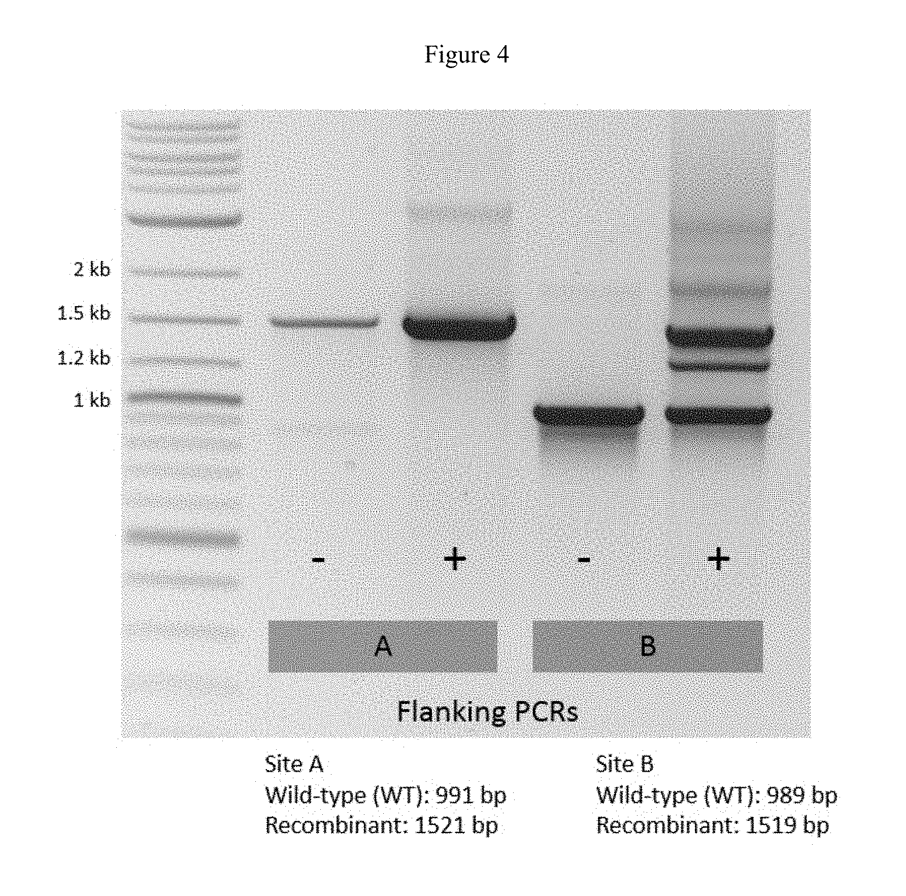
FIG. 4 shows flanking PCR assays that tested for the presence of recombinant NanoLuc.RTM. K2 bacteriophage in sgRNAs B1+B2 bacterial strain under different induction conditions. An overlay of E. coli UPEC harboring crP-B (site B cutting plasmid) and pBBR1-K2 B (site B donor plasmid) was infected with `K2A`, a recombinant K2 phage with nanoluciferase already inserted at site A. This was performed with and without tetracycline induction (100 ng/mL). Plate lysates were collected and analyzed via PCR using primer sets that flank site A and site B.
FIG. 5 shows flanking PCR assays that tested for the presence of recombinant NanoLuc.RTM. K2 bacteriophage using primer sets that flank site B. Recombinant K2 phage containing a nanoluciferase insertion at site A (`K2A`) was used for these recombination experiments.
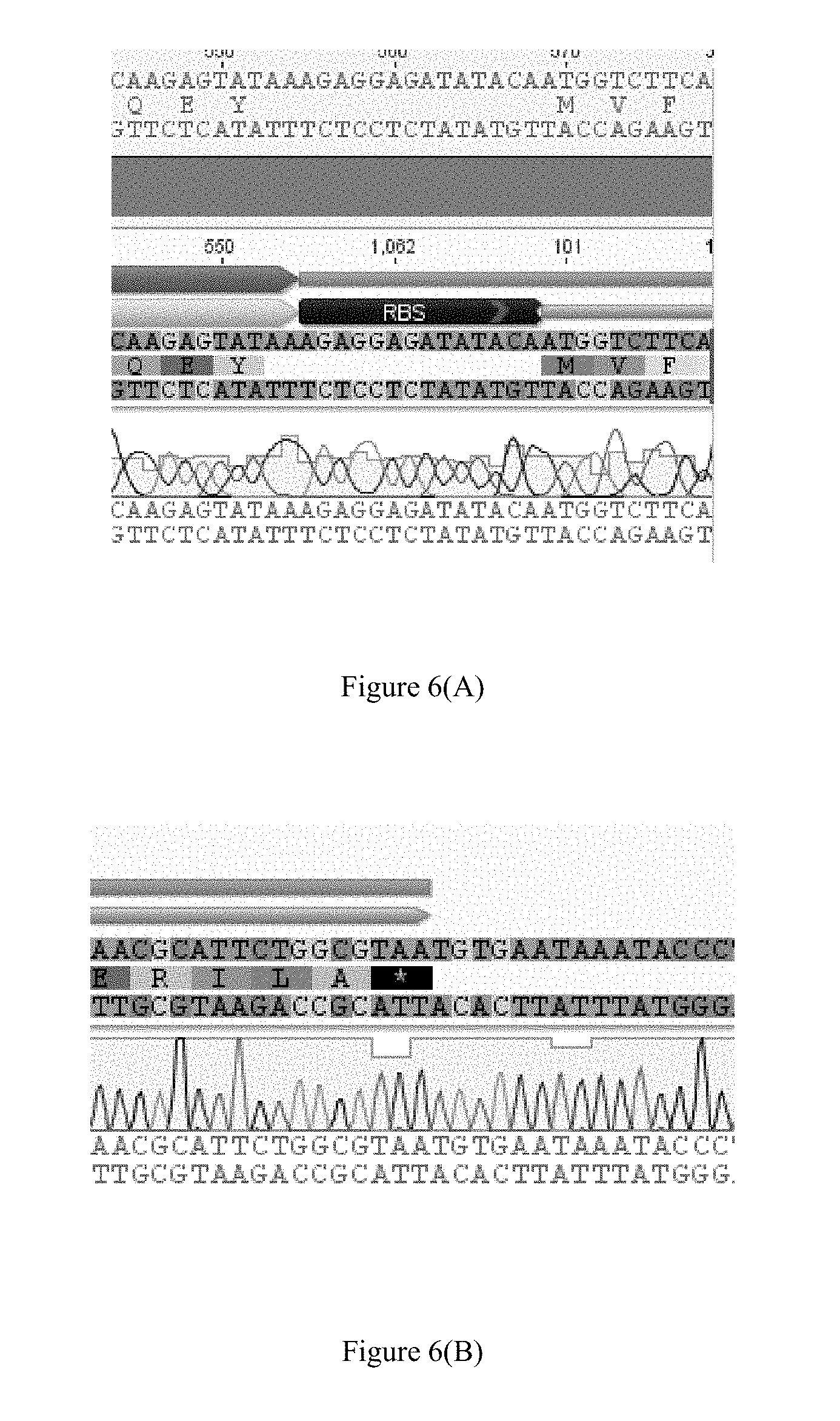
FIG. 6(A) shows the upstream junction sequence of the nanoluciferase insertion in the recombinant K2 phage genome cleaved by sgRNAs A1 and A2: 5' CAAGAGTATAAAGAGGAGATATACAATGGTCTTCA 3' (SEQ ID NO: 5).
FIG. 6(B) shows the downstream junction sequence of the nanoluciferase insertion in the recombinant K2 phage genome cleaved by sgRNAs A1 and A2: 5' AACGCATTCTGGCGTAATGTGAATAAATACCC 3' (SEQ ID NO: 6). FIG. 6(B) discloses the protein sequence as SEQ ID NO: 27.
FIG. 6(C) shows the upstream junction sequence of the nanoluciferase insertion in the recombinant K2 phage genome cleaved by sgRNAs B1 and B2: 5'AAGACCATTGTATATCTCCTCTTTAATCATGTTT 3' (SEQ ID NO: 7).). FIG. 6(C) also discloses SEQ ID NO: 28. FIG. 6(D) discloses the protein sequence as SEQ ID NO: 29.
FIG. 6(D) shows the downstream junction sequence of the nanoluciferase insertion in the recombinant K2 phage genome cleaved by sgRNAs B1 and B2: 5' ATAATGTAATTCAATTTACGCCAGAATGC 3' (SEQ ID NO: 8).
FIG. 7(A) shows a comparison of the host ranges of recombinant NanoLuc.RTM. K2 phage, recombinant NanoLuc.RTM. K1-5 phage, and recombinant NanoLuc.RTM. K1E phage. Grey means the tested phage infects the corresponding strain (positive) whereas black means the tested phage does not infect the corresponding strain (negative).
FIG. 7(B) shows the luminescence activity profile of a recombinant K2 phage of the present technology (SEQ ID NO: 4).
FIG. 8(A) shows a general schematic of a first K2 bacteriophage genome (i.e., intact K2 bacteriophage genome) and a cleaved first K2 bacteriophage genome. The cleaved first K2 bacteriophage genome comprises a first cleaved bacteriophage genomic fragment and a second cleaved bacteriophage genomic fragment.
FIG. 8(B) shows a general schematic of the donor template design and recombination between a cleaved phage genome and the donor template. Two double-stranded breaks are generated by Cas9 at sites specified by the two sgRNAs. In some instances, Cas9 cleavage excises a phage DNA sequence that is important for phage viability. The donor template contains any exogenous reporter gene inserts like nanoluciferase, but must also restore the function of excised phage sequences. The 5' and 3' flanking regions of the donor template are homologous to the DNA sequences immediately adjacent to the two cleavage sites in the phage genome, and are necessary for repairing double-stranded breaks via homologous recombination.
FIG. 9 shows the heterologous nucleic acid sequence that was inserted into K2 phage genomic DNA that was cleaved between position 25,443 and 25,444 of SEQ ID NO: 1 using single guide RNAs (sgRNAs) sgRNA A1 and sgRNA A2 (SEQ ID NO: 2; donor sequence A). The underlined sequences represent the homologous 5' and 3' flanking regions of the heterologous nucleic acid sequence.
FIG. 10 shows the heterologous nucleic acid sequence that was inserted into K2 phage genomic DNA that was cleaved between position 75,922 and position 75,923 of SEQ ID NO: 1 using single guide RNAs (sgRNAs) sgRNA B1 and sgRNA B2 (SEQ ID NO: 3; donor sequence B). The underlined sequences represent the homologous 5' and 3' flanking regions of the heterologous nucleic acid sequence.
FIGS. 11(A)-11(S) show the partial genome sequence (i.e., contig-1) of the recombinant NanoLuc.RTM. K2 phage that was cleaved with sgRNA A1, sgRNA A2, sgRNA B1 and sgRNA B2 and recombined with donor sequence A and donor sequence B (SEQ ID NO: 4).
FIGS. 12(A)-12(S) show the partial genome sequence (i.e., contig-1) of the recombinant NanoLuc.RTM. K2 phage that was cleaved with sgRNA A1 and sgRNA A2 only and recombined with donor sequence A (SEQ ID NO: 23).
FIGS. 13(A)-13(S) show the partial genome sequence (i.e., contig-1) of the recombinant NanoLuc.RTM. K2 phage that was cleaved with sgRNA B1 and sgRNA B2 only and recombined with donor sequence B (SEQ ID NO: 24).
FIGS. 14(A)-14(S) show the partial genome sequence (i.e., contig-1) of non-recombinant K2 phage (SEQ ID NO: 1).
FIG. 15(A) shows the antibiotic susceptibility profile of an E. coli strain to amikacin using the recombinant K2 phages of the present technology. FIG. 15(B) shows the antibiotic susceptibility profile of an E. coli strain to gentamicin using the recombinant K2 phages of the present technology. FIG. 15(C) shows the antibiotic susceptibility profile of an E. coli strain to levofloxacin using the recombinant K2 phages of the present technology. FIG. 15(D) shows the antibiotic susceptibility profile of an E. coli strain to ceftazidime using the recombinant K2 phages of the present technology.
DETAILED DESCRIPTION
It is to be appreciated that certain aspects, modes, embodiments, variations and features of the present methods are described below in various levels of detail in order to provide a substantial understanding of the present technology.
In practicing the present methods, many conventional techniques in molecular biology, protein biochemistry, cell biology, microbiology and recombinant DNA are used. See, e.g., Sambrook and Russell eds. (2001) Molecular Cloning: A Laboratory Manual, 3rd edition; the series Ausubel et al. eds. (2007) Current Protocols in Molecular Biology; the series Methods in Enzymology (Academic Press, Inc., N.Y.); MacPherson et al. (1991) PCR 1: A Practical Approach (IRL Press at Oxford University Press); MacPherson et al. (1995) PCR 2: A Practical Approach; Harlow and Lane eds. (1999) Antibodies, A Laboratory Manual; Freshney (2005) Culture of Animal Cells: A Manual of Basic Technique, 5th edition; Gait ed. (1984) Oligonucleotide Synthesis; U.S. Pat. No. 4,683,195; Hames and Higgins eds. (1984) Nucleic Acid Hybridization; Anderson (1999) Nucleic Acid Hybridization; Hames and Higgins eds. (1984) Transcription and Translation; Immobilized Cells and Enzymes (IRL Press (1986)); Perbal (1984) A Practical Guide to Molecular Cloning; Miller and Calos eds. (1987) Gene Transfer Vectors for Mammalian Cells (Cold Spring Harbor Laboratory); Makrides ed. (2003) Gene Transfer and Expression in Mammalian Cells; Mayer and Walker eds. (1987) Immunochemical Methods in Cell and Molecular Biology (Academic Press, London); and Herzenberg et al. eds (1996) Weir's Handbook of Experimental Immunology.
Definitions
Unless defined otherwise, all technical and scientific terms used herein generally have the same meaning as commonly understood by one of ordinary skill in the art to which this technology belongs. As used in this specification and the appended claims, the singular forms "a", "an" and "the" include plural referents unless the content clearly dictates otherwise. For example, reference to "a cell" includes a combination of two or more cells, and the like. Generally, the nomenclature used herein and the laboratory procedures in cell culture, molecular genetics, organic chemistry, analytical chemistry and nucleic acid chemistry and hybridization described below are those well-known and commonly employed in the art.
As used herein, the term "about" in reference to a number is generally taken to include numbers that fall within a range of 1%, 5%, or 10% in either direction (greater than or less than) of the number unless otherwise stated or otherwise evident from the context (except where such number would be less than 0% or exceed 100% of a possible value).
As used herein, "bacteriophage" or "phage" refers to a virus that infects bacteria. Bacteriophages are obligate intracellular parasites that multiply inside bacteria by co-opting some or all of the host biosynthetic machinery (i.e., viruses that infect bacteria). Though different bacteriophages may contain different materials, they all contain nucleic acid and protein, and can under certain circumstances be encapsulated in a lipid membrane. Depending upon the phage, the nucleic acid can be either DNA or RNA (but not both).
As used herein, a "cleaved first K2 bacteriophage genome" refers to the K2 bacteriophage genome fragments that are formed after a first K2 bacteriophage genome has undergone enzymatic cleavage with one or two sgRNA-CRISPR enzyme complexes. When a first K2 bacteriophage genome is cleaved with two sgRNA-CRISPR enzyme complexes, the "cleaved first K2 bacteriophage genome" excludes the shortest nucleic acid sequence that lies between the cleavage site of the first sgRNA-CRISPR enzyme complex and the second sgRNA-CRISPR enzyme complex. See FIG. 8(A).
As used herein, a "control" is an alternative sample used in an experiment for comparison purpose. A control can be "positive" or "negative." For example, where the purpose of the experiment is to determine a correlation of the efficacy of a therapeutic agent for the treatment for a particular type of disease or condition, a positive control (a compound or composition known to exhibit the desired therapeutic effect) and a negative control (a subject or a sample that does not receive the therapy or receives a placebo) are typically employed.
As used herein, "expression" includes one or more of the following: transcription of the gene into precursor mRNA; splicing and other processing of the precursor mRNA to produce mature mRNA; mRNA stability; translation of the mature mRNA into protein (including codon usage and tRNA availability); and glycosylation and/or other modifications of the translation product, if required for proper expression and function.
As used herein, an "expression control sequence" refers to polynucleotide sequences which are necessary to affect the expression of coding sequences to which they are operably linked. Expression control sequences are sequences which control the transcription, post-transcriptional events and translation of nucleic acid sequences. Expression control sequences include appropriate transcription initiation, termination, promoter and enhancer sequences; efficient RNA processing signals such as splicing and polyadenylation signals; sequences that stabilize cytoplasmic mRNA; sequences that enhance translation efficiency (e.g., ribosome binding sites); sequences that enhance protein stability; and when desired, sequences that enhance protein secretion. The nature of such control sequences differs depending upon the host organism; in prokaryotes, such control sequences generally include promoter, ribosomal binding site, and transcription termination sequence. The term "control sequences" is intended to encompass, at a minimum, any component whose presence is essential for expression, and can also encompass an additional component whose presence is advantageous, for example, leader sequences.
As used herein, a "heterologous nucleic acid sequence" is any sequence placed at a location in the genome where it does not normally occur. A heterologous nucleic acid sequence may comprise a sequence that does not naturally occur in a bacteriophage, or it may comprise only sequences naturally found in the bacteriophage, but placed at a non-normally occurring location in the genome. In some embodiments, the heterologous nucleic acid sequence is not a natural phage sequence. In certain embodiments, the heterologous nucleic acid sequence is a natural phage sequence that is derived from a different phage. In other embodiments, the heterologous nucleic acid sequence is a sequence that occurs naturally in the genome of a wild-type phage but is then relocated to another site where it does not naturally occur, rendering it a heterologous sequence at that new site.
"Homology" or "identity" or "similarity" refers to sequence similarity between two peptides or between two nucleic acid molecules. Homology can be determined by comparing a position in each sequence which may be aligned for purposes of comparison. When a position in the compared sequence is occupied by the same nucleobase or amino acid, then the molecules are homologous at that position. A degree of homology between sequences is a function of the number of matching or homologous positions shared by the sequences. A polynucleotide or polynucleotide region (or a polypeptide or polypeptide region) has a certain percentage (for example, at least 60%, 65%, 70%, 75%, 80%, 85%, 90%, 95%, 98% or 99%) of "sequence identity" to another sequence means that, when aligned, that percentage of bases (or amino acids) are the same in comparing the two sequences. This alignment and the percent homology or sequence identity can be determined using software programs known in the art. In some embodiments, default parameters are used for alignment. One alignment program is BLAST, using default parameters. In particular, programs are BLASTN and BLASTP, using the following default parameters: Genetic code=standard; filter=none; strand=both; cutoff=60; expect=10; Matrix=BLOSUM62; Descriptions=50 sequences; sort by =HIGH SCORE; Databases=non-redundant, GenBank+EMBL+DDBJ+PDB+GenBank CDS translations+SwissProtein+SPupdate+PIR. Details of these programs can be found at the National Center for Biotechnology Information. Biologically equivalent polynucleotides are those having the specified percent homology and encoding a polypeptide having the same or similar biological activity. Two sequences are deemed "unrelated" or "non-homologous" if they share less than 40% identity, or less than 25% identity, with each other.
As used herein, a "host cell" is a bacterial cell that can be infected by a phage to yield progeny phage particles. A host cell can form phage particles from a particular type of phage genomic DNA. In some embodiments, the phage genomic DNA is introduced into the host cell by infecting the host cell with a phage. In some embodiments, the phage genomic DNA is introduced into the host cell using transformation, electroporation, or any other suitable technique. In some embodiments, the phage genomic DNA is substantially pure when introduced into the host cell. In some embodiments, the phage genomic DNA is present in a vector when introduced into the host cell. The definition of host cell can vary from one phage to another. For example, E. coli may be the natural host cell for a particular type of phage, but Klebsiella pneumoniae is not.
As used herein, the term "isolated" refers to a substance or entity that has been separated from at least some of the components with which it was associated when initially produced (whether in nature or in an experimental setting). Isolated substances and/or entities may be separated from at least about 10%, about 20%, about 30%, about 40%, about 50%, about 60%, about 70%, about 80%, about 90%, or more of the other components with which they were initially associated. In some embodiments, isolated substances and/or entities are more than about 80%, about 85%, about 90%, about 91%, about 92%, about 93%, about 94%, about 95%, about 96%, about 97%, about 98%, about 99%, or more than about 99% pure. As used herein, a substance is "pure" if it is substantially free of other components.
As used herein, "operably linked" means that expression control sequences are positioned relative to a nucleic acid of interest to initiate, regulate or otherwise control transcription of the nucleic acid of interest.
As used herein, a "phage genome" or "bacteriophage genome" includes naturally occurring phage genomes and derivatives thereof. Generally, the derivatives possess the ability to propagate in the same hosts as the naturally occurring phage. In some embodiments, the only difference between a naturally occurring phage genome and a derivative phage genome is at least one of a deletion or an addition of nucleotides from at least one end of the phage genome (if the genome is linear) or at least one point in the genome (if the genome is circular).
As used herein, the term "polynucleotide" or "nucleic acid" means any RNA or DNA, which may be unmodified or modified RNA or DNA. Polynucleotides include, without limitation, single- and double-stranded DNA, DNA that is a mixture of single- and double-stranded regions, single- and double-stranded RNA, RNA that is mixture of single- and double-stranded regions, and hybrid molecules comprising DNA and RNA that may be single-stranded or, more typically, double-stranded or a mixture of single- and double-stranded regions. In addition, polynucleotide refers to triple-stranded regions comprising RNA or DNA or both RNA and DNA. The term polynucleotide also includes DNAs or RNAs containing one or more modified bases and DNAs or RNAs with backbones modified for stability or for other reasons.
As used herein, the term "recombinant" when used with reference, e.g., to a cell, or nucleic acid, protein, or vector, indicates that the cell, nucleic acid, protein or vector, has been modified by the introduction of a heterologous nucleic acid or protein or the alteration of a native nucleic acid or protein, or that the material is derived from a cell so modified. Thus, for example, recombinant cells express genes that are not found within the native (non-recombinant) form of the cell or express native genes that are otherwise abnormally expressed, under expressed or not expressed at all.
As used herein, an endogenous nucleic acid sequence in the genome of an organism (or the encoded protein product of that sequence) is deemed "recombinant" herein if a heterologous sequence is placed adjacent to the endogenous nucleic acid sequence, such that the expression of this endogenous nucleic acid sequence is altered. In this context, a heterologous sequence is a sequence that is not naturally adjacent to the endogenous nucleic acid sequence, whether or not the heterologous sequence is itself endogenous to the organism (originating from the same organism or progeny thereof) or exogenous (originating from a different organism or progeny thereof). By way of example, a promoter sequence can be substituted (e.g., by homologous recombination) for the native promoter of a gene in the genome of an organism, such that this gene has an altered expression pattern. This gene would be "recombinant" because it is separated from at least some of the sequences that naturally flank it. A nucleic acid is also considered "recombinant" if it contains any modifications that do not naturally occur in the corresponding nucleic acid in a genome. For instance, an endogenous coding sequence is considered "recombinant" if it contains an insertion, deletion or a point mutation introduced artificially, e.g., by human intervention. A "recombinant nucleic acid" also includes a nucleic acid integrated into a host cell chromosome at a heterologous site and a nucleic acid construct present as an episome.
As used herein, a "recombinant bacteriophage genome" is a bacteriophage genome that has been genetically modified by the insertion of a heterologous nucleic acid sequence into the bacteriophage genome. A "recombinant bacteriophage" means a bacteriophage that comprises a recombinant bacteriophage genome. In some embodiments, the bacteriophage genome is modified by recombinant DNA technology to introduce a heterologous nucleic acid sequence into the genome at a defined site. In some embodiments, the heterologous nucleic acid sequence is introduced with no corresponding loss of endogenous phage genomic nucleotides. In other words, if bases N1 and N2 are adjacent in the wild-type bacteriophage genome, the heterologous nucleic acid sequence is inserted between N1 and N2. Thus, in the resulting recombinant bacteriophage genome, the heterologous nucleic acid sequence is flanked by nucleotides N1 and N2. In some embodiments, endogenous phage nucleotides are removed or replaced during the insertion of the heterologous nucleic acid sequence. For example, in some embodiments, the heterologous nucleic acid sequence is inserted in place of some or all of the endogenous phage sequence which is removed. In some embodiments, endogenous phage sequences are removed from a position in the phage genome distant from the site(s) of insertion of the heterologous nucleic acid sequences.
As used herein, a "recombinant K2 bacteriophage" or "recombinant K2 phage" means a K2 bacteriophage whose genomic DNA comprises a heterologous nucleic acid sequence that encodes a bioluminescent protein, a fluorescent protein, a chromogenic protein, or any combination thereof.
As used herein, the term "sample" refers to clinical samples obtained from a subject or isolated microorganisms. In certain embodiments, a sample is obtained from a biological source (i.e., a "biological sample"), such as tissue, bodily fluid, or microorganisms collected from a subject. Sample sources include, but are not limited to, mucus, sputum, bronchial alveolar lavage (BAL), bronchial wash (BW), whole blood, bodily fluids, cerebrospinal fluid (CSF), urine, plasma, serum, or tissue.
As used herein, "a sub-sample" refers to one or more samples containing bacterial cells that are derived from a test sample obtained from a subject. In some embodiments, the sub-sample is void of non-bacterial cells (e.g., human cells). In some embodiments, the sub-sample contains lysed human cells.
As used herein, "test sample" refers to a sample taken from a subject that is to be assayed for the presence of bacteria and/or for the antibiotic susceptibility of bacteria present in the sample. In some embodiments, the test sample is blood, sputum, mucus, lavage, or saliva. In certain embodiments, the test sample is a swab from a subject.
As used herein, the terms "subject," "individual," or "patient" are used interchangeably and refer to an individual organism, a vertebrate, a mammal, or a human. In certain embodiments, the individual, patient or subject is a human.
As used herein, a "vector" refers to a nucleic acid molecule capable of transporting another nucleic acid to which it has been linked. One type of vector is a "plasmid," which generally refers to a circular double stranded DNA loop into which additional DNA segments may be ligated, but also includes linear double-stranded molecules such as those resulting from amplification by the polymerase chain reaction (PCR) or from treatment of a circular plasmid with a restriction enzyme. Other vectors include cosmids, bacterial artificial chromosomes (BAC) and yeast artificial chromosomes (YAC). Another type of vector is a viral vector, wherein additional DNA segments may be ligated into the viral genome (discussed in more detail below). Certain vectors are capable of autonomous replication in a host cell into which they are introduced (e.g., vectors having an origin of replication which functions in the host cell). Other vectors can be integrated into the genome of a host cell upon introduction into the host cell, and are thereby replicated along with the host genome. Moreover, certain vectors are capable of directing the expression of genes to which they are operatively linked. Such vectors are referred to herein as "recombinant expression vectors" (or simply "expression vectors").
Bacteriophage
Bacteriophage are obligate intracellular parasites that multiply inside bacteria by co-opting some or all of the host biosynthetic machinery. Phages contain nucleic acid and protein, and may be enveloped by a lipid membrane. Depending upon the phage, the nucleic acid genome can be either DNA or RNA but not both, and can exist in either circular or linear forms. The size of the phage genome varies depending upon the phage. The simplest phages have genomes that are only a few thousand nucleotides in size, while the more complex phages may contain more than 100,000 nucleotides in their genome, and in rare instances no more than 500,000 bp. The number and amount of individual types of protein in phage particles will vary depending upon the phage. The proteins function in infection and to protect the nucleic acid genome from environmental nucleases.
Phage genomes come in a variety of sizes and shapes (e.g., linear or circular). Most phages range in size from 24-200 nm in diameter. The capsid is composed of many copies of one or more phage proteins, and acts as a protective envelope around the phage genome. Many phages have tails attached to the phage capsid. The tail is a hollow tube through which the phage nucleic acid passes during infection. The size of the tail can vary and some phages do not even have a tail structure. In the more complex phages, the tail is surrounded by a contractile sheath which contracts during infection of the bacterial host cell. At the end of the tail, phages have a base plate and one or more tail fibers attached to it. The base plate and tail fibers are involved in the binding of the phage to the host cell.
Lytic or virulent phages are phages which can only multiply in bacteria and lyse the bacterial host cell at the end of the life cycle of the phage. The lifecycle of a lytic phage begins with an eclipse period. During the eclipse phase, no infectious phage particles can be found either inside or outside the host cell. The phage nucleic acid takes over the host biosynthetic machinery and phage specific mRNAs and proteins are produced. Early phage mRNAs code for early proteins that are needed for phage DNA synthesis and for shutting off host DNA, RNA and protein biosynthesis. In some cases, the early proteins actually degrade the host chromosome. After phage DNA is made late mRNAs and late proteins are made. The late proteins are the structural proteins that comprise the phage as well as the proteins needed for lysis of the bacterial cell. In the next phase, the phage nucleic acid and structural proteins are assembled and infectious phage particles accumulate within the cell. The bacteria begin to lyse due to the accumulation of the phage lysis protein, leading to the release of intracellular phage particles. The number of particles released per infected cell can be as high as 1000 or more. Lytic phage may be enumerated by a plaque assay. The assay is performed at a low enough concentration of phage such that each plaque arises from a single infectious phage. The infectious particle that gives rise to a plaque is called a PFU (plaque forming unit).
Lysogenic phages are those that can either multiply via the lytic cycle or enter a quiescent state in the host cell. In the quiescent state, the phage genome exists as a prophage (i.e., it has the potential to produce phage). In most cases, the phage DNA actually integrates into the host chromosome and is replicated along with the host chromosome and passed on to the daughter cells. The host cell harboring a prophage is not adversely affected by the presence of the prophage and the lysogenic state may persist indefinitely. The lysogenic state can be terminated upon exposure to adverse conditions. Conditions which favor the termination of the lysogenic state include: desiccation, exposure to UV or ionizing radiation, exposure to mutagenic chemicals, etc. Adverse conditions lead to the production of proteases (rec A protein), the expression of the phage genes, reversal of the integration process, and lytic multiplication.
Recombinant K2 Phage Compositions of the Present Technology
The recombinant K2 bacteriophage of the present technology specifically infects E. coli strains that express K2 capsule genes. In some embodiments, the E. coli strain that expresses K2 capsule genes is CFT073.
In one aspect, the present disclosure provides a recombinant K2 bacteriophage nucleic acid sequence, wherein the nucleic acid sequence between (a) position 25,443 and 25,444 of SEQ ID NO: 1 or (b) position 75,922 and 75,923 of SEQ ID NO: 1 is replaced with a heterologous nucleic acid sequence comprising an open reading frame that encodes a reporter protein, wherein the reporter protein is a bioluminescent protein, a fluorescent protein, a chemiluminescent protein, or any combination thereof. In some embodiments, the heterologous nucleic acid sequence further comprises at least one segment that corresponds to at least part of the excised endogenous phage genome sequence between (a) position 25,443 and 25,444 of SEQ ID NO: 1 or (b) position 75,922 and 75,923 of SEQ ID NO: 1.
The present disclosure also provides a recombinant K2 bacteriophage nucleic acid sequence, wherein the nucleic acid sequence between (a) position 24,610 and 24,611 of SEQ ID NO: 1, (b) position 26,130 and 26,1310f SEQ ID NO: 1, (c) position 75,590 and 75,591 of SEQ ID NO: 1 or (d) position 76,394 and 76,395 is replaced with a heterologous nucleic acid sequence comprising an open reading frame that encodes a reporter protein, wherein the reporter protein is a bioluminescent protein, a fluorescent protein, a chemiluminescent protein, or any combination thereof.
Also disclosed herein are recombinant K2 bacteriophages that comprise any recombinant K2 bacteriophage nucleic acid sequence disclosed herein. In some embodiments, the reporter protein(s) encoded by the heterologous nucleic acid sequence produces a detectable signal upon exposure to the appropriate stimuli, and the resulting signal permits detection of bacterial host cells infected by a recombinant K2 phage of the present technology.
In certain embodiments, the open reading frame encodes a reporter protein that serves as a marker that can be identified by screening bacterial host cells infected by a recombinant K2 phage of the present technology. Examples of such markers include by way of example and without limitation: a fluorescent label, a luminescent label, a chemiluminescence label, or an enzymatic label. In some embodiments, the heterologous nucleic acid sequence further comprises sequences naturally found in the bacteriophage, but placed at a non-normally occurring location in the genome.
In some embodiments, the length of the heterologous nucleic acid sequence is at least 100 bases, at least 200 bases, at least 300 bases, at least 400 bases, at least 500 bases, at least 600 bases, at least 700 bases, at least 800 bases, at least 900 bases, at least 1 kilobase (kb), at least 1.1 kb, at least 1.2 kb, at least 1.3 kb, at least 1.4 kb, at least 1.5 kb, at least 1.6 kb, at least 1.7 kb, at least 1.8 kb, at least 1.9 kb, at least 2.0 kb, at least 2.1 kb, at least 2.2 kb, at least 2.3 kb, at least 2.4 kb, at least 2.5 kb, at least 2.6 kb, at least 2.7 kb, at least 2.8 kb, at least 2.9 kb, at least 3.0 kb, at least 3.1 kb, at least 3.2 kb, at least 3.3 kb, at least 3.4 kb, at least 3.5 kb, at least 3.6 kb, at least 3.7 kb, at least 3.8 kb, at least 3.9 kb, at least 4.0 kb, at least 4.5 kb, at least 5.0 kb, at least 5.5 kb, at least 6.0 kb, at least 6.5 kb, at least 7.0 kb, at least 7.5 kb, at least 8.0 kb, at least 8.5 kb, at least 9.0 kb, at least 9.5 kb, at least 10 kb, or more. In certain embodiments, the heterologous nucleic acid sequence comprises a length that is less than or equal to a length selected from the group consisting of 1 kb, 2 kb, 3 kb, 4 kb, 5 kb, 6 kb, 7 kb, 8 kb, 9 kb, and 10 kb. In some embodiments, the heterologous nucleic acid sequence comprises a length that is less than or equal to the maximum length of heterologous nucleic acid sequence that can be packaged into a phage particle comprising the phage genome.
In some embodiments, the length of the heterologous nucleic acid sequence is from 100 to 500 bases, from 200 to 1,000 bases, from 500 to 1,000 bases, from 500 to 1,500 bases, from 1 kb to 2 kb, from 1.5 kb to 2.5 kb, from 2.0 kb to 3.0 kb, from 2.5 kb to 3.5 kb, from 3.0 kb to 4.0 kb, from 3.5 kb to 4.5 kb, from 4.0 kb to 5.0 kb, from 4.5 kb to 5.5 kb, from 5.0 kb to 6.0 kb, from 5.5 kb to 6.5 kb, from 6.0 kb to 7.0 kb, from 6.5 kb to 7.5 kb, from 7.0 kb to 8.0 kb, from 7.5 kb to 8.5 kb, from 8.0 kb to 9.0 kb, from 8.5 kb to 9.5 kb, or from 9.0 kb to 10.0 kb.
In some embodiments, the heterologous nucleic acid sequence is inserted into the K2 phage genome with no loss of endogenous K2 phage genomic sequence. In some embodiments, the heterologous nucleic acid sequence replaces an endogenous K2 phage genomic sequence. In some embodiments, the heterologous nucleic acid sequence includes an endogenous K2 phage genomic sequence that was previously excised from the phage genome.
In certain embodiments, the heterologous nucleic acid sequence replaces an endogenous K2 phage genomic sequence that is less than the length of the heterologous nucleic acid sequence. Accordingly, in some embodiments, the length of the recombinant K2 phage genome is longer than the length of the wild-type K2 phage genome. In some embodiments, the heterologous nucleic acid sequence replaces an endogenous K2 phage genomic sequence that is greater than the length of the heterologous nucleic acid sequence. Thus, in some embodiments, the length of the recombinant K2 phage genome is shorter than the length of the wild-type K2 phage genome. In certain embodiments, the heterologous nucleic acid sequence replaces an endogenous K2 phage genomic sequence that is equal to the length of the heterologous nucleic acid sequence.
In certain embodiments, the open reading frame of the heterologous nucleic acid sequence encodes a reporter protein that confers a phenotype of interest on a host cell infected by a recombinant K2 phage of the present technology. In some embodiments, the phenotype of interest is the expression of the gene product encoded by the open reading frame of the heterologous nucleic acid sequence.
In certain embodiments, the open reading frame of the heterologous nucleic acid sequence is operably linked to an expression control sequence that is capable of directing expression of the open reading frame, wherein the open reading frame encodes a reporter protein (e.g., a bioluminescent protein, a fluorescent protein, a chemiluminescent protein, or any combination thereof). In some embodiments, the expression control sequence is located within the heterologous nucleic acid sequence. In other embodiments, the expression control sequence is located in the endogenous K2 phage genome sequence. For example, the open reading frame may be inserted into the K2 phage genome downstream of or in the place of an endogenous K2 phage open reading frame sequence. In some embodiments, the expression control sequence is an inducible promoter or a constitutive promoter (e.g., sarA promoter or 1pp promoter). See e.g., Djordjevic & Klaenhammer, Methods in Cell Science 20(1): 119-126 (1998). The inducible promoter or constitutive promoter may be an endogenous K2 phage promoter sequence, a phage promoter sequence that is non-endogenous to K2 phage, or a bacterial host promoter sequence. Additionally or alternatively, in some embodiments, the inducible promoter is a pH-sensitive promoter, or a temperature sensitive promoter.
In some embodiments, the heterologous nucleic acid sequence comprises a first open reading frame and at least one supplemental open reading frame. In certain embodiments, the first and the at least one supplemental open reading frames are operably linked to the same expression control sequences. In some embodiments, the first and the at least one supplemental open reading frames are operably linked to different expression control sequences.
Fluorescent proteins include, but are not limited to, blue/UV fluorescent proteins (for example, TagBFP, Azurite, EBFP2, mKalamal, Sirius, Sapphire, and T-Sapphire), cyan fluorescent proteins (for example, ECFP, Cerulean, SCFP3A, mTurquoise, monomeric Midoriishi-Cyan, TagCFP, and mTFP1), green fluorescent proteins (for example, EGFP, Emerald, Superfolder GFP, Monomeric Azami Green, TagGFP2, mUKG, and mWasabi), yellow fluorescent proteins (for example, EYFP, Citrine, Venus, SYFP2, and TagYFP), orange fluorescent proteins (for example, Monomeric Kusabira-Orange, mKOK, mKO2, mOrange, and mOrange2), red fluorescent proteins (for example, mRaspberry, mCherry, dsRed, mStrawberry, mTangerine, tdTomato, TagRFP, TagRFP-T, mApple, and mRuby), far-red fluorescent proteins (for example, mPlum, HcRed-Tandem, mKate2, mNeptune, and NirFP), near-IR fluorescent proteins (for example, TagRFP657, IFP1.4, and iRFP), long stokes-shift proteins (for example, mKeima Red, LS S-mKate1, and LSS-mKate2), photoactivatable fluorescent proteins (for example, PA-GFP, PAmCherryl, and PATagRFP), photoconvertible fluorescent proteins (for example, Kaede (green), Kaede (red), KikGR1 (green), KikGR1 (red), PS-CFP2, PS-CFP2, mEos2 (green), mEos2 (red), PSmOrange, and PSmOrange), fluorescein, rhodamine, and photoswitchable fluorescent proteins (for example, Dronpa).
Examples of bioluminescent proteins are aequorin (derived from the jellyfish Aequorea victoria) and luciferases (including luciferases derived from firefly and Renilla, nanoluciferase, red luciferase, luxAB, and the like). These proteins have also been genetically separated into two distinct functional domains that will generate light only when the protein domains are closely co-localized. A variety of emission spectrum-shifted mutant derivatives of both of these proteins have been generated over the past decade and have been used for multi-color imaging and co-localization within a living cell.
Examples of chemiluminescent protein include .beta.-galactosidase, horseradish peroxidase (HRP), and alkaline phosphatase. Peroxidases generate peroxide that oxidizes luminol in a reaction that generates light, whereas alkaline phosphatases remove a phosphate from a substrate molecule, destabilizing it and initiating a cascade that results in the emission of light.
In some embodiments, the open reading frame of the heterologous nucleic acid sequence comprises an epitope that can be detected with an antibody or other binding molecule. For example, an antibody that recognizes the epitope may be directly linked to a signal generating moiety (such as by covalent attachment of a chemiluminescent or fluorescent protein), or can be detected using at least one additional binding reagent such as a secondary antibody, directly linked to a signal generating moiety. In some embodiments, the epitope is absent in wild-type K2 bacteriophage and the bacterial host cell. Accordingly, detection of the epitope in a sample demonstrates the presence of a bacterial host cell infected by a recombinant K2 phage comprising a heterologous nucleic acid sequence, wherein the open reading frame of the heterologous nucleic acid sequence comprises the epitope. In other embodiments, the open reading frame of the heterologous nucleic acid sequence comprises a polypeptide tag sequence, such that the expression product of the open reading frame comprises the tag fused to a polypeptide or protein encoded by the open reading frame (e.g., poly-histidine, FLAG, Glutathione S-transferase (GST) etc.).
In some embodiments, the open reading frame of the heterologous nucleic acid sequence comprises a biotin binding protein such as avidin, streptavidin, or neutrAvidin that can be detected with a biotin molecule conjugated to an enzyme (e.g., .beta.-galactosidase, horseradish peroxidase (HRP), and alkaline phosphatase) or an antibody. In some embodiments, an antibody conjugated to a biotin molecule may be directly linked to a signal generating moiety (such as by covalent attachment of a chemiluminescent or fluorescent protein), or can be detected using at least one additional binding reagent such as a secondary antibody, directly linked to a signal generating moiety.
Also disclosed herein are recombinant K2 bacteriophages comprising any of the recombinant K2 bacteriophage nucleic acid sequences disclosed herein. In some embodiments, the recombinant K2 bacteriophages comprise a nucleic acid sequence selected from the group consisting of SEQ ID NO: 2, SEQ ID NO: 3, SEQ ID NO: 4, SEQ ID NO: 5, SEQ ID NO: 6, SEQ ID NO: 7, SEQ ID NO: 8, SEQ ID NO: 23 and SEQ ID NO: 24.
In another aspect, the present disclosure provides a vector comprising any of the recombinant K2 bacteriophage nucleic acid sequences disclosed herein, as well as bacterial host cells comprising the vectors of the present technology. In some embodiments, the bacterial host cell expresses K2 capsule genes. The bacterial host cell may be a natural or non-natural host for K2 bacteriophage.
The present disclosure also provides a bacterial host cell comprising a recombinant K2 bacteriophage disclosed herein. In some embodiments, the bacterial host cell expresses K2 capsule genes. The bacterial host cell may be a natural or non-natural host for K2 bacteriophage.
Methods of Making Recombinant K2 Bacteriophage of the Present Technology
In one aspect, the present disclosure provides methods for making a recombinant K2 bacteriophage of the present technology in a bacterial host cell. The bacterial host cell may be a non-natural bacterial host cell or a natural bacterial host cell for K2 bacteriophage.
In some embodiments, the method comprises (a) contacting a first K2 bacteriophage genome with a first sgRNA-CRISPR enzyme complex and a second sgRNA-CRISPR enzyme complex in vivo under conditions where (i) the first sgRNA-CRISPR enzyme complex cleaves a first protospacer sequence 5'CCATAGTCATAACCACCACC 3' (SEQ ID NO: 9) within the first K2 bacteriophage genome; and (ii) the second sgRNA-CRISPR enzyme complex cleaves a second protospacer sequence 5' ACACCAAACAGAATATCAGA 3' (SEQ ID NO: 10) within the first K2 bacteriophage genome to produce a cleaved first K2 bacteriophage genome; and (b) recombining in vivo the cleaved first K2 bacteriophage genome with a heterologous nucleic acid sequence in the presence of a recombination system under conditions to produce the recombinant K2 bacteriophage genome, wherein the bacterial host cell is infected with the first K2 bacteriophage genome, and wherein the heterologous nucleic acid sequence comprises an open reading frame that encodes a bioluminescent protein, a fluorescent protein, a chemiluminescent protein, or any combination thereof. In some embodiments, the first sgRNA-CRISPR enzyme complex comprises Cas9 and a first sgRNA having the sequence of
TABLE-US-00005 (SEQ ID NO: 11) 5' CCAUAGUCAUAACCACCACCGUUUUAGAGCUAGAAAUAGCAAGUUAA AAUAAGGCUAGUCCGUUAUCAACUUGAAAAAGUGGCACCGAGUCGGUGCU UUUUUU 3'.
Additionally or alternatively, in certain embodiments, the second sgRNA-CRISPR enzyme complex comprises Cas9 and a second sgRNA having the sequence of
TABLE-US-00006 (SEQ ID NO: 12) 5' ACACCAAACAGAAUAUCAGAGUUUUAGAGCUAGAAAUAGCAAGUUAA AAUAAGGCUAGUCCGUUAUCAACUUGAAAAAGUGGCACCGAGUCGGUGCU UUUUUU 3'.
The recombination system may be endogenous or non-endogenous. The first K2 bacteriophage genome may be recombinant or non-recombinant.
In other embodiments, the method comprises (a) contacting a first K2 bacteriophage genome with a first sgRNA-CRISPR enzyme complex and a second sgRNA-CRISPR enzyme complex in vivo under conditions where (i) the first sgRNA-CRISPR enzyme complex cleaves a first protospacer sequence 5' ATGCTTGGTTAATCAATTAA 3' (SEQ ID NO: 13) within the first K2 bacteriophage genome; and (ii) the second sgRNA-CRISPR enzyme complex cleaves a second protospacer sequence 5' GACTTTGGTCAAATACCGAT 3' (SEQ ID NO: 14) within the first K2 bacteriophage genome to produce a cleaved first K2 bacteriophage genome; and (b) recombining in vivo the cleaved first K2 bacteriophage genome with a heterologous nucleic acid sequence in the presence of a recombination system under conditions to produce the recombinant K2 bacteriophage genome, wherein the bacterial host cell is infected with the first K2 bacteriophage genome, and wherein the heterologous nucleic acid sequence comprises an open reading frame that encodes a bioluminescent protein, a fluorescent protein, a chemiluminescent protein, or any combination thereof. In some embodiments, the first sgRNA-CRISPR enzyme complex comprises Cas9 and a first sgRNA having the sequence of
TABLE-US-00007 (SEQ ID NO: 15) 5' AUGCUUGGUUAAUCAAUUAAGUUUUAGAGCUAGAAAUAGCAAGUUAA AAUAAGGCUAGUCCGUUAUCAACUUGAAAAAGUGGCACCGAGUCGGUGCU UUUUUU 3'.
Additionally or alternatively, in certain embodiments, the second sgRNA-CRISPR enzyme complex comprises Cas9 and a second sgRNA having the sequence of
TABLE-US-00008 (SEQ ID NO: 16) 5' GACUUUGGUCAAAUACCGAUGUUUUAGAGCUAGAAAUAGCAAGUUAA AAUAAGGCUAGUCCGUUAUCAACUUGAAAAAGUGGCACCGAGUCGGUGCU UUUUUU 3'.
The recombination system may be endogenous or non-endogenous. The first K2 bacteriophage genome may be recombinant or non-recombinant.
The cleaved first K2 bacteriophage genome comprises a first cleaved bacteriophage genomic fragment and a second cleaved bacteriophage genomic fragment. In certain embodiments of the methods disclosed herein, the heterologous nucleic acid sequence comprises a 5' flanking region that is homologous to the 3' end of the first cleaved bacteriophage genomic fragment, and a 3' flanking region that is homologous to the 5' end of the second cleaved bacteriophage genomic fragment.
In some embodiments of the methods disclosed herein, the homologous 5' flanking region of the heterologous nucleic acid sequence has a length of about 20-30 base pairs (bps), 30-40 bps, 40-50 bps, 50-60 bps, 60-70 bps, 70-80 bps, 80-90 bps, 90-100 bps, 100-110 bps, 110-120 bps, 120-130 bps, 130-140 bps, 140-150 bps, 150-160 bps, 160-170 bps, 170-180 bps, 180-190 bps, 190-200 bps, 200-210 bps, 210-220 bps, 220-230 bps, 230-240 bps, 240-250 bps, 250-260 bps, 260-270 bps, 270-280 bps, 280-290 bps, 290-300 bps, 300-310 bps, 310-320 bps, 320-330 bps, 330-340 bps, 340-350 bps, 350-360 bps, 360-370 bps, 370-380 bps, 380-390 bps, 390-400 bps, 400-410 bps, 410-420 bps, 420-430 bps, 430-440 bps, 440-450 bps, 450-460 bps, 460-470 bps, 470-480 bps, 480-490 bps, 490-500 bps, 500-510 bps, 510-520 bps, 520-530 bps, 530-540 bps, 540-550 bps, 550-560 bps, 560-570 bps, 570-580 bps, 580-590 bps, or 590-600 bps.
Additionally or alternatively, in some embodiments of the methods disclosed herein, the homologous 3' flanking region of the heterologous nucleic acid sequence has a length of about 20-30 base pairs (bps), 30-40 bps, 40-50 bps, 50-60 bps, 60-70 bps, 70-80 bps, 80-90 bps, 90-100 bps, 100-110 bps, 110-120 bps, 120-130 bps, 130-140 bps, 140-150 bps, 150-160 bps, 160-170 bps, 170-180 bps, 180-190 bps, 190-200 bps, 200-210 bps, 210-220 bps, 220-230 bps, 230-240 bps, 240-250 bps, 250-260 bps, 260-270 bps, 270-280 bps, 280-290 bps, 290-300 bps, 300-310 bps, 310-320 bps, 320-330 bps, 330-340 bps, 340-350 bps, 350-360 bps, 360-370 bps, 370-380 bps, 380-390 bps, 390-400 bps, 400-410 bps, 410-420 bps, 420-430 bps, 430-440 bps, 440-450 bps, 450-460 bps, 460-470 bps, 470-480 bps, 480-490 bps, 490-500 bps, 500-510 bps, 510-520 bps, 520-530 bps, 530-540 bps, 540-550 bps, 550-560 bps, 560-570 bps, 570-580 bps, 580-590 bps, or 590-600 bps.
Additionally or alternatively, in some embodiments of the methods disclosed herein, the recombination system is a non-endogenous recombination system that is induced in the bacterial host cell. The non-endogenous recombination system may include a recombination expression vector that comprises lambda Red proteins Gam, Exo, and Beta operably linked to an inducible promoter. In some embodiments of the methods disclosed herein, the inducible promoter is araB and the non-endogenous recombination system is induced by the addition of arabinose. In some embodiments, the recombination expression vector further comprises the heterologous nucleic acid sequence. In other embodiments, the expression vector comprising the heterologous nucleic acid sequence is separate and distinct from the recombination expression vector comprising lambda Red proteins.
In other embodiments of the methods disclosed herein, the recombination system is a non-endogenous recombination system that includes a recombination expression vector comprising RecET (RecE, RecT) operons operably linked to an inducible promoter, and optionally the heterologous nucleic acid sequence. In some embodiments, the inducible promoter is araB and the non-endogenous recombination system is induced by the addition of arabinose. In other embodiments, the expression vector comprising the heterologous nucleic acid sequence is separate and distinct from the recombination expression vector comprising RecET.
In another embodiment of the methods disclosed herein, the recombination system is a non-endogenous recombination system that includes a recombination expression vector comprising RecA recombinase or a RecA gain-of-function variant operably linked to an inducible promoter and optionally the heterologous nucleic acid sequence. In some embodiments, the inducible promoter is araB and the non-endogenous recombination system is induced by the addition of arabinose. In other embodiments, the expression vector comprising the heterologous nucleic acid sequence is separate and distinct from the recombination expression vector comprising RecA recombinase or the RecA gain-of-function variant.
Additionally or alternatively, in some embodiments of the methods disclosed herein, the bacterial host cell comprises a non-endogenous CRISPR expression vector comprising a nucleic acid sequence that encodes a first sgRNA, a second sgRNA, and a CRISPR enzyme. In some embodiments, the first sgRNA and the second sgRNA are operably linked to a constitutive promoter. In some embodiments, the sequence of the first sgRNA and the second sgRNA is SEQ ID NO: 11 and SEQ ID NO: 12, respectively. In other embodiments, the sequence of the first sgRNA and the second sgRNA is SEQ ID NO: 15 and SEQ ID NO: 16, respectively.
A variety of CRISPR enzymes are available for use in conjunction with the disclosed methods of the present disclosure. In some embodiments, the CRISPR enzyme is a Type II CRISPR enzyme. In some embodiments, the CRISPR enzyme catalyzes DNA cleavage. In some embodiments, the CRISPR enzyme catalyzes RNA cleavage. In some embodiments, the CRISPR enzyme is any Cas9 protein, for instance any naturally-occurring bacterial Cas9 as well as any variants, homologs or orthologs thereof. Non-limiting examples of Cas proteins include Cas1, Cas1B, Cas2, Cas3, Cas4, Cas5, Cas6, Cas7, Cas8, Cas9 (also known as Csn1 and Csx12), Cas10, Csy1, Csy2, Csy3, Cse1, Cse2, Csc1, Csc2, Csa5, Csn2, Csm2, Csm3, Csm4, Csm5, Csm6, Cmr1, Cmr3, Cmr4, Cmr5, Cmr6, Csb1, Csb2, Csb3, Csx17, Csx14, Csx10, Csx16, CsaX, Csx3, Csx1, Csx15, Csf1, Csf2, Csf3, Csf4, homologues thereof, or variants thereof. The CRISPR enzyme may be operably linked to an inducible promoter, such as a tetracycline-inducible promoter. In some embodiments, the CRISPR enzyme cleaves both strands of the target nucleic acid at the Protospacer Adjacent Motif (PAM) site.
In another aspect, the present disclosure provides sgRNAs that are useful for making the recombinant K2 bacteriophages disclosed herein. In some embodiments, the sgRNA sequence is selected from the group consisting of SEQ ID NO: 11, SEQ ID NO: 12, SEQ ID NO: 15 and SEQ ID NO: 16. The design of sgRNAs that are capable of cleaving at the other K2 genomic positions described herein, is within the scope of one of ordinary skill in the art.
Bacterial Identification and Antibiotic Susceptibility Profiling Methods of the Present Technology
Accurate identification of bacterial species within a biological sample informs the selection of suitable therapies for treating bacterial infections. The recombinant K2 bacteriophages disclosed herein may be used to identify bacteria present within a biological sample (e.g., whole blood, plasma, serum). Such methods entail contacting the biological sample with a recombinant K2 bacteriophage disclosed herein, and detecting the presence of bacterial host cells infected by the recombinant K2 phage, wherein the recombinant K2 phage comprises a heterologous nucleic acid sequence that encodes a detectable gene product, thereby leading to the identification of bacteria present within the biological sample.
Additionally or alternatively, the recombinant K2 bacteriophages disclosed herein, may be used in methods for profiling antibiotic susceptibility of bacteria present within a biological sample (e.g., whole blood, plasma, serum). These methods include (a) infecting the biological sample with an antibiotic and a recombinant K2 bacteriophage disclosed herein, (b) detecting the presence of bacterial host cells infected by the recombinant K2 phage, wherein the recombinant K2 phage comprises a heterologous nucleic acid sequence that encodes a detectable gene product, and (c) determining that the antibiotic is effective in inhibiting the bacteria present in the biological sample when the levels of recombinant K2 phage infected bacterial host cells is reduced relative to that observed in an untreated control sample.
In one aspect, the present disclosure provides a method for identifying at least one bacterial strain or species in a test sample obtained from a subject comprising (a) separating bacterial cells isolated from the test sample into one or more sub-samples, (b) infecting each sub-sample with at least one recombinant K2 bacteriophage disclosed herein, wherein each recombinant K2 bacteriophage comprises a heterologous nucleic acid sequence encoding one or more reporter genes, and (c) identifying at least one bacterial strain or species in the test sample by detecting the expression of the one or more reporter genes of the at least one recombinant K2 bacteriophage. In certain embodiments, the at least one K2 bacteriophage comprises a nucleic acid sequence selected from the group consisting of SEQ ID NO: 2, SEQ ID NO: 3, SEQ ID NO: 4, SEQ ID NO: 5, SEQ ID NO: 6, SEQ ID NO: 7, SEQ ID NO: 8, SEQ ID NO: 23 and SEQ ID NO: 24. In certain embodiments, the method for identifying at least one bacterial strain or species in a test sample does not require the culturing of bacterial cells from the test sample or a sub-sample.
In some embodiments, identification of at least one bacterial strain or species includes detecting the expression of the one or more reporter genes of the at least one recombinant K2 bacteriophage, e.g., detectable expression of green fluorescence indicates the presence of bacterial species A in a test sample or sub-sample. In some embodiments, the absence of at least one bacterial strain or species is identified by the lack of detectable expression of the one or more reporter genes of the at least one recombinant K2 bacteriophage, e.g., undetectable expression of green fluorescence indicates the lack of bacterial species A in a test sample or sub-sample.
In some embodiments, the at least one recombinant K2 bacteriophage infects a single species of bacteria. In certain embodiments, the at least one recombinant K2 bacteriophage infects two or more species of bacteria. By way of example, but not by way of limitation, in some embodiments, the species of bacteria that are infected include E. coli strains that express K2 capsule genes. In some embodiments, the E. coli strain that expresses K2 capsule genes is CFT073.
In some embodiments, detection of the expression of the reporter gene is detection of the gene product itself, e.g., a fluorescent protein. In some embodiments, detection of the expression of the reporter gene is detection of an enzymatic reaction requiring the expression of the reporter gene, e.g., expression of luciferase to catalyze luciferin to produce light.
In some embodiments, the expression of the one or more reporter genes is detected in about 5, 10, 15, 20, 25, 30, 35, 40, 45, 50, 55, 60, 65, 70, 75, 80, 85, 90, 120 minutes or any time between any two of the preceding values after infecting a sub-sample with the at least one recombinant K2 bacteriophage disclosed herein.
The present disclosure also provides a method for identifying at least one bacterial strain or species that expresses K2 capsule genes in a test sample obtained from a subject comprising (a) infecting the test sample comprising bacterial cells with a recombinant K2 bacteriophage of the present technology; and (b) detecting the expression of the reporter protein of the recombinant K2 bacteriophage, wherein detection of the reporter protein indicates the presence of at least one bacterial strain or species that expresses K2 capsule genes in the test sample. In some embodiments of the method, the expression of the reporter protein is measured in about 5, 10, 15, 20, 25, 30, 35, 40, 45, 50, 55, 60, 65, 70, 75, 80, 85, 90 or 120 minutes after infecting the test sample comprising bacterial cells with the recombinant K2 bacteriophage.
Additionally or alternatively, in some embodiments of the methods disclosed herein, the test sample is blood, sputum, mucus, lavage, saliva, or a swab obtained from the subject. In some embodiments, the subject is human.
In another aspect, the present disclosure provides a method for determining the antibiotic susceptibility of a bacterial strain or species in a test sample obtained from a subject comprising (a) separating bacterial cells isolated from the test sample into a plurality of sub-samples, (b) infecting the plurality of sub-samples with a recombinant K2 bacteriophage disclosed herein and at least one antibiotic, wherein the recombinant K2 bacteriophage comprises a heterologous nucleic acid sequence encoding a reporter gene, and (c) detecting the expression of the reporter gene of the recombinant K2 bacteriophage in the presence of each antibiotic. In some embodiments, the method further comprises determining that the bacterial strain or species in the test sample is susceptible to an antibiotic if the reporter gene expression of the recombinant K2 bacteriophage in the antibiotic treated sub-sample is decreased relative to that observed in a control sub-sample that is not treated with the antibiotic. In other embodiments, the method further comprises determining that the bacterial strain or species in the test sample is resistant to an antibiotic if the reporter gene expression of the recombinant K2 bacteriophage in the antibiotic treated sub-sample is comparable to that observed in a control sub-sample that is not treated with the antibiotic. In certain embodiments, the method for determining the antibiotic susceptibility of a bacterial strain or species in a test sample does not require the culturing of bacterial cells from a test sample or a sub-sample.
Additionally or alternatively, in some embodiments of the recombinant K2 bacteriophages of the present technology, the reporter gene is nanoluciferase. In certain embodiments, recombinant K2 bacteriophage comprises a nucleic acid sequence selected from the group consisting of SEQ ID NO: 2, SEQ ID NO: 3, SEQ ID NO: 4, SEQ ID NO: 5, SEQ ID NO: 6, SEQ ID NO: 7, SEQ ID NO: 8, SEQ ID NO: 23 and SEQ ID NO: 24.
Examples of antibiotics include one or more of rifampicin, tetracycline, ampicillin, penicillin G, methicillin, oxacillin, amoxicillin, cefadroxil, ceforanid, cefotaxime, ceftriaxone, doxycycline, minocycline, amikacin, gentamicin, levofloxacin, kanamycin, neomycin, streptomycin, tobramycin, azithromycin, clarithromycin, erythromycin, ciprofloxacin, lomefloxacin, norfloxacin, chloramphenicol, clindamycin, cycloserine, isoniazid, rifampin, teicoplanin, quinupristin/dalfopristin, linezolid, pristinamycin, ceftobiprole, ceftaroline, dalbavancin, daptomycin, mupirocin, oritavancin, tedizolid, telavancin, tigecycline, ceftazidime, cefepime, piperacillin, ticarcillin, virginiamycin, netilmicin, paromomycin, spectinomycin, geldanamycin, herbimycin, rifaximin, loracarbef, ertapenem, doripenem, imipenem/cilastatin, meropenem, cefazolin, cefalotin, cephalexin, cefaclor, cefamandole, cefoxitin, cefprozil, cefuroxime, cefixime, cefdinir, cefditoren, cefoperazone, cefpodoxime, ceftibuten, ceftizoxime, lincomycin, dirithromycin, roxithromycin, troleandomycin, telithromycin, spiramycin, aztreonam, furazolidone, nitrofurantoin, posizolid, radezolid, torezolid, azlocillin, carbenicillin, cloxacillin, dicloxacillin, flucloxacillin, mezlocillin, nafcillin, penicillin V, temocillin, bacitracin, colistin, polymyxin B, enoxacin, gatifloxacin, gemifloxacin, moxifloxacin, nalidixic acid, ofloxacin, trovafloxacin, grepafloxacin, sparfloxacin, temafloxacin, mafenide, sulfacetamide, sulfadiazine, silver sulfadiazine, sulfadimethoxine, sulfamethizole, sulfamethoxazole, sulfanilimide, sulfasalazine, sulfisoxazole, trimethoprim-sulfamethoxazole (Co-trimoxazole) (TMP-SMX), sulfonamidochrysoidine, demeclocycline, oxytetracycline, clofazimine, dapsone, capreomycin, ethambutol, ethionamide, pyrazinamide, rifabutin, rifapentine, arsphenamine, fosfomycin, fusidic acid, metronidazole, platensimycin, thiamphenicol, tinidazole, trimethoprim(Bs) and vancomycin.
In some embodiments of the method, the differences in the reporter gene expression of the recombinant K2 bacteriophage observed in the antibiotic treated sub-sample and the untreated control sub-sample is defined as .mu..
Additionally or alternatively, in some embodiments of the method, the expression of the reporter gene is detected in about 5, 10, 15, 20, 25, 30, 35, 40, 45, 50, 55, 60, 65, 70, 75, 80, 85, 90, 120 minutes or any time between any two of the preceding values after infecting a sub-sample with a recombinant K2 bacteriophage disclosed herein.
In some embodiments, two or more sub-samples are tested for antibiotic susceptibility in series. In some embodiments, two or more sub-samples are tested for antibiotic susceptibility in parallel. In some embodiments, one or more sub-samples are tested for antibiotic susceptibility in a running assay (where resistance or sensitivity to one antibiotic is determined and the resistance or sensitivity to a second, third, fourth, fifth, etc., antibiotic is being assayed).
In some embodiments of the methods disclosed herein, isolating bacterial cells from a test sample includes incubating the test sample with distilled water to form a mixture, centrifuging the mixture to form a pellet that includes bacterial cells, and re-suspending the pellet to form a bacterial suspension comprising isolated bacterial cells after discarding the supernatant. The pellet may be re-suspended in a phosphate buffer. In some embodiments, the bacterial suspension is divided into one or more sub-samples.
In certain embodiments of the methods disclosed herein, mixing the test sample with distilled water will lead to the lysis of cells that lack cell walls (e.g., mammalian cells and red blood cells) while leaving cells with cell walls (e.g., bacteria) intact. Without wishing to be bound by theory, in some embodiments, the removal of cells that lack cell walls enhances the detection of reporter gene expression in bacterial cells infected with a recombinant K2 bacteriophage, as intact non-bacterial cells (e.g., red blood cells) may quench reporter gene expression. In some embodiments of the methods of the present technology, the mixture is about 90% distilled water and 10% test sample, about 80% distilled water and 20% test sample, about 70% distilled water and 30% test sample, about 60% distilled water and 40% test sample, about 50% distilled water and 50% test sample, about 40% distilled water and 60% test sample, about 30% distilled water and 70% test sample, about 20% distilled water and 80% sample, or about 10% distilled water and 90% test sample. In some embodiments of the methods disclosed herein, the mixture is incubated for 1, 2, 3, 4, 5, 6, 7, 8, 9, 10, 11, 12, 13, 14, or 15 minutes, or any time between two of the previously listed time points. Additionally or alternatively, in certain embodiments of the methods disclosed herein, the mixture is centrifuged for 1, 2, 3, 4, 5, 6, 7, 8, 9, 10, 11, 12, 13, 14, or 15 minutes, or any time between two of the previously listed time points.
Additionally or alternatively, in certain embodiments of the methods disclosed herein, each of the one or more sub-samples comprise between about 5 to 500, about 10 to 400, about 20 to 300, about 30 to 300, about 40 to 200 or about 50 to 100 bacterial cells. In some embodiments of the methods disclosed herein, each of the one or more sub-samples comprises between about 100 to 10,000, about 200 to 9,000, about 300 to 8,000, about 400 to 7,000, about 500 to 6,000, about 600 to 5,000, about 700 to 4,000, about 800 to 3,000, about 900 to 2,000, or about 1,000 to 1,500 bacterial cells.
In another aspect, the present disclosure provides a method for determining the antibiotic susceptibility of a bacterial strain or species in a test sample obtained from a subject comprising (a) infecting a plurality of test samples comprising bacterial cells with a recombinant K2 bacteriophage of the present technology and an antibiotic, wherein the plurality of test samples is derived from the subject; (b) detecting the expression of the reporter protein of the recombinant K2 bacteriophage in the plurality of test samples; and (c) determining that the antibiotic is effective in inhibiting the bacterial strain or species in a test sample when the reporter protein expression levels of the recombinant K2 phage infected bacterial cells in the test sample are reduced relative to that observed in an untreated control sample comprising bacterial cells, wherein the untreated control sample is derived from the subject and is infected with the recombinant K2 phage of the present technology. In some embodiments, the bacterial strain or species in the test sample expresses K2 capsule genes. The expression of the reporter protein may be measured in about 5, 10, 15, 20, 25, 30, 35, 40, 45, 50, 55, 60, 65, 70, 75, 80, 85, 90, or 120 minutes after infecting the plurality of test samples comprising bacterial cells with the recombinant K2 bacteriophage. In other embodiments, the method further comprises determining that the bacterial strain or species in the test sample is resistant to the antibiotic when the reporter protein expression levels of the recombinant K2 bacteriophage infected bacterial cells in the test sample are comparable to that observed in an untreated control sample comprising bacterial cells, wherein the untreated control sample is derived from the subject and is infected with recombinant K2 phage of the present technology.
In any of the above embodiments of the methods of the present technology, the test sample is blood, sputum, mucus, lavage, saliva, or a swab obtained from the subject. In some embodiments of the methods disclosed herein, the test sample is obtained from a mammalian subject, including, for example, farm animals, such as sheep, pigs, cows, and horses; pet animals, such as dogs and cats; and laboratory animals, such as rats, mice and rabbits. In one embodiment, the mammal subject is a human.
Kits
The present technology provides kits including the recombinant K2 bacteriophages disclosed herein for bacteria identification and antibiotic susceptibility profiling.
In one aspect, the kits of the present technology comprise one or more coded/labeled vials that contain a plurality of the recombinant K2 bacteriophages disclosed herein, and instructions for use. In some embodiments, each coded/labeled vial corresponds to a different recombinant K2 bacteriophage. In other embodiments, each coded/labeled vial corresponds to the same recombinant K2 bacteriophage. In some embodiments, the kits of the present technology comprise one or more coded/labeled vials that contain at least one recombinant K2 bacteriophage comprising a nucleic acid sequence selected from the group consisting of SEQ ID NO: 2, SEQ ID NO: 3, SEQ ID NO: 4, SEQ ID NO: 5, SEQ ID NO: 6, SEQ ID NO: 7, SEQ ID NO: 8, SEQ ID NO: 23 and SEQ ID NO: 24.
In some embodiments, each phage vial is assigned a unique code that identifies the bacteriophage in the phage vial, or the types of bacteria that the bacteriophage strain infects. The unique code can be encoded by a machine discernible pattern, such as a bar code, a QR code, an alphanumeric string, or any other pattern that can be discerned by a reader. Each unique code may be shown as, for example, a bar code sticker on a vial or container storing a corresponding phage sample. In some embodiments, the kit is stored under conditions that permit the preservation of the bacteriophage genomes for extended periods, such as under bacteriophage-specific, controlled temperature, moisture, and pH conditions.
Additionally or alternatively, in some embodiments, the kits further comprise vials containing natural or non-natural bacterial host cells. In some embodiments, the bacterial host cells are E. coli. In certain embodiments, the bacterial host cells are E. coli strain UPEC.
The kits may also comprise instructions for use, software for automated analysis, containers, packages such as packaging intended for commercial sale and the like.
The kit may further comprise one or more of: wash buffers and/or reagents, hybridization buffers and/or reagents, labeling buffers and/or reagents, and detection means. The buffers and/or reagents are usually optimized for the particular detection technique for which the kit is intended. Protocols for using these buffers and reagents for performing different steps of the procedure may also be included in the kit. Further optional components of the kits may include expression media for gene products encoded by the heterologous nucleic acids of the recombinant K2 bacteriophages disclosed herein, such as a medium containing nutrients and cofactors for bioluminescence, devices such as a lamp configured to illuminate at specific wavelengths of light to detect biofluorescence, and devices for measuring the extent of heterologous nucleic acid expression, such as a photometer or photodetector.
Additionally or alternatively, the kits disclosed herein may also include coded and labeled vials that contain a plurality of antibiotics. In some embodiments, the plurality of antibiotics comprises one or more of rifampicin, tetracycline, levofloxacin, and ampicillin. Other examples of antibiotics include penicillin G, methicillin, oxacillin, amoxicillin, cefadroxil, ceforanid, cefotaxime, ceftriaxone, doxycycline, minocycline, amikacin, gentamicin, levofloxacin, kanamycin, neomycin, streptomycin, tobramycin, azithromycin, clarithromycin, erythromycin, ciprofloxacin, lomefloxacin, norfloxacin, chloramphenicol, clindamycin, cycloserine, isoniazid, rifampin, teicoplanin, quinupristin/dalfopristin, linezolid, pristinamycin, ceftobiprole, ceftaroline, dalbavancin, daptomycin, mupirocin, oritavancin, tedizolid, telavancin, tigecycline, ceftazidime, cefepime, piperacillin, ticarcillin, virginiamycin, netilmicin, paromomycin, spectinomycin, geldanamycin, herbimycin, rifaximin, loracarbef, ertapenem, doripenem, imipenem/cilastatin, meropenem, cefazolin, cefalotin, cephalexin, cefaclor, cefamandole, cefoxitin, cefprozil, cefuroxime, cefixime, cefdinir, cefditoren, cefoperazone, cefpodoxime, ceftibuten, ceftizoxime, lincomycin, dirithromycin, roxithromycin, troleandomycin, telithromycin, spiramycin, aztreonam, furazolidone, nitrofurantoin, posizolid, radezolid, torezolid, azlocillin, carbenicillin, cloxacillin, dicloxacillin, flucloxacillin, mezlocillin, nafcillin, penicillin V, temocillin, bacitracin, colistin, polymyxin B, enoxacin, gatifloxacin, gemifloxacin, moxifloxacin, nalidixic acid, ofloxacin, trovafloxacin, grepafloxacin, sparfloxacin, temafloxacin, mafenide, sulfacetamide, sulfadiazine, silver sulfadiazine, sulfadimethoxine, sulfamethizole, sulfamethoxazole, sulfanilimide, sulfasalazine, sulfisoxazole, trimethoprim-sulfamethoxazole (Co-trimoxazole) (TMP-SMX), sulfonamidochrysoidine, demeclocycline, oxytetracycline, clofazimine, dapsone, capreomycin, ethambutol, ethionamide, pyrazinamide, rifabutin, rifapentine, arsphenamine, fosfomycin, fusidic acid, metronidazole, platensimycin, thiamphenicol, tinidazole, trimethoprim(Bs) and vancomycin.
Additionally or alternatively, in some embodiments, the kits comprise one or more sgRNA sequences selected from the group consisting of SEQ ID NO: 11, SEQ ID NO: 12, SEQ ID NO: 15 and SEQ ID NO: 16.
EXAMPLES
Example 1: Design and Methods for Generating the Recombinant K2 Bacteriophages of the Present Technology
This Example demonstrates that the methods of the present technology are useful for making the recombinant K2 bacteriophages disclosed herein in a bacterial host cell.
The recombinant E. coli K2 bacteriophage of the present technology were engineered using a `break and recombine` (BAR) phage engineering method. The BAR method relies on (1) cleaving a phage genome in vivo at one or two locations using an RNA-guided endonuclease (e.g., Cas9)-sgRNA complex, and (2) providing a heterologous nucleic acid sequence comprising the nanoluciferase gene with an upstream ribosome binding site as well as 5' and 3' flanking regions that are homologous to a portion of the initial K2 phage genome (collectively, referred to as the donor template region). The donor template region also contains sequences that restore the function of any K2 phage DNA that was excised by the sgRNA-CRISPR enzyme complexes. The 5' and 3' flanking regions (about several hundred base pairs in length) are homologous to the DNA sequences immediately adjacent to the two cleavage sites in the phage genome (FIGS. 8(A)-8(B)), and are necessary for repairing double-stranded breaks via homologous recombination.
Two insertion sites (site A' and `site B`) were selected for inserting the nanoluciferase reporter gene into a portion of the initial K2 phage genome (i.e., K2 contig-1). The sequence of the initial K2 contig-1 region is shown in FIGS. 14(A)-14(S) and is represented by SEQ ID NO: 1. 20 bp protospacer sites, along with the accompanying 3 bp protospacer adjacent motif (PAM) were identified near `site A` and `site B.` The K2 protospacer sequences along with their adjacent PAM sites (PAM site underlined) are provided below:
TABLE-US-00009 Protospacer A1 5' CCATAGTCATAACCACC/ACCCGG 3' (SEQ ID NO: 17) Protospacer A2 5' ACACCAAACAGAATATC/AGAGGG 3' (SEQ ID NO: 18) Protospacer B1 5' ATGCTTGGTTAATCAAT/TAAGGG 3' (SEQ ID NO: 19) Protospacer B2 5' GACTTTGGTCAAATACC/GATGGG 3' (SEQ ID NO: 20) cleavage sites marked with a `/`
The complete sequences of sgRNA A1, sgRNA A2, sgRNA B1, and sgRNA B2 are provided below:
TABLE-US-00010 sgRNA A1 RNA sequence: (SEQ ID NO: 11) CCAUAGUCAUAACCACCACCGUUUUAGAGCUAGAAAUAGCAAGUUAAAAU AAGGCUAGUCCGUUAUCAACUUGAAAAAGUGGCACCGAGUCGGUGCUUUU UUU sgRNA A2 RNA sequence: (SEQ ID NO: 12) ACACCAAACAGAAUAUCAGAGUUUUAGAGCUAGAAAUAGCAAGUUAAAAU AAGGCUAGUCCGUUAUCAACUUGAAAAAGUGGCACCGAGUCGGUGCUUUU UUU sgRNA B1 RNA sequence: (SEQ ID NO: 15) AUGCUUGGUUAAUCAAUUAAGUUUUAGAGCUAGAAAUAGCAAGUUAAAAU AAGGCUAGUCCGUUAUCAACUUGAAAAAGUGGCACCGAGUCGGUGCUUUU UUU sgRNA B2 RNA sequence: (SEQ ID NO: 16) GACUUUGGUCAAAUACCGAUGUUUUAGAGCUAGAAAUAGCAAGUUAAAAU AAGGCUAGUCCGUUAUCAACUUGAAAAAGUGGCACCGAGUCGGUGCUUUU UUU
The CRISPR expression vector (crP) was previously assembled for CRISPR-Cas9 mediated cleavage in Gram-negative bacteria. The crP plasmid is a shuttle vector containing a pUC origin of replication, a pRO1600 origin of replication, a kanamycin resistance cassette, and an S. pyogenes Cas9 gene operably linked to a pTet (tetracycline) promoter. The crP plasmid also contains a scaffold into which a segment of synthetic DNA can be inserted so as to permit the transcription of two sgRNAs (e.g., using a dual-guide cassette) by the synthetic J23119 promoter. The nucleic acid sequences of the `dual-guide cassettes` for sgRNAs A1 and A2 as well as sgRNAs B1 and B2 are provided below:
TABLE-US-00011 Dual-guide cassette (sgRNAs A1 and A2): (SEQ ID NO: 21) CAGACCTAAGGTCTCTTAGCCCATAGTCATAACCACCACCGTTTTAGAGC TAGAAATAGCAAGTTAAAATAAGGCTAGTCCGTTATCAACTTGAAAAAGT GGCACCGAGTCGGTGCTTTTTTTGCTAACTGATACCGACTACGCCTGAAC AGTCGAATCTTCACCTCGTCTGGTACCGACGCGGTCCCTTGACAGCTAGC TCAGTCCTAGGTATAATGCTAGCACACCAAACAGAATATCAGAGTTTTCG AGACCAGCTCGTAGGCTAA Dual-guide cassette (sgRNAs B1 and B2): (SEQ ID NO: 22) CAGACCTAAGGTCTCTTAGCATGCTTGGTTAATCAATTAAGTTTTAGAGC TAGAAATAGCAAGTTAAAATAAGGCTAGTCCGTTATCAACTTGAAAAAGT GGCACCGAGTCGGTGCTTTTTTTGCTAACTGATACCGACTACGCCTGAAC AGTCGAATCTTCACCTCGTCTGGTACCGACGCGGTCCCTTGACAGCTAGC TCAGTCCTAGGTATAATGCTAGCGACTTTGGTCAAATACCGATGTTTTCG AGACCAGCTCGTAGGCTAA
The `dual-guide cassette` for sgRNAs A1 and A2 was PCR amplified, digested with the restriction enzyme BsaI, and ligated into a BsaI-digested crP plasmid to create the `crP-A` CRISPR expression vector. The `dual-guide cassette` for sgRNAs B1 and B2 was also PCR amplified, digested with the restriction enzyme BsaI, and ligated into a BsaI-digested crP plasmid to create the `crP-B` CRISPR expression vector.
The donor template vectors for each site were designed as follows. FIG. 9 shows the donor template sequence for K2 phage genomic DNA that was cleaved with sgRNA A1 and sgRNA A2 (SEQ ID NO: 2). FIG. 10 shows the donor template sequence for K2 phage genomic DNA that was cleaved with sgRNA B1 and sgRNA B2 (SEQ ID NO: 3). The donor template sequences contain the nanoluciferase reporter gene with an upstream ribosome binding site (RBS), as well as the region of the K2 phage genome between the two CRISPR cleavage sites that is to be excised, such that the original sequence and function is restored. Specifically, the donor template sequences included 5' and 3' flanking regions (about 300 bp in length) that were perfectly homologous to the cleaved ends of the wild-type K2 phage genome so as to facilitate strand invasion and homologous recombination between the cleaved phage genome and the donor sequence, thereby repairing the double-stranded breaks and incorporating the nanoluciferase reporter gene. The protospacers/PAM sequences within the donor templates were modified such that the CRISPR system would not recognize and cleave the donor template, and would only target the wild-type phage genome. For example, the regions internal to the two cleavage sites (i.e., right of the upstream cut site and left of the downstream cut site) were either codon-reassigned or otherwise changed by single-base pair substitutions that were not expected to detrimentally affect phage function.
The recombination plasmids for each site were assembled by PCR amplification of the plasmid `pBBR1-Gent` with primers `CPM 123` (5' CAGGTTCATCATGCCGTTTGTG 3' (SEQ ID NO: 25)) and `CPM 124` (5' TATTTGCCCATGGACGCACAC 3' (SEQ ID NO: 26)) followed by a Gibson assembly reaction between the amplified plasmid and the donor templates. The assembled recombination plasmids for site A and site B were designated as `pBBR1-K2 A` and `pBBR1 K2 B` respectively.
E. coli UPEC was transformed twice to make strains housing either `crP-A and pBBR1-K2 A` plasmids or `crP-B and pBBR1-K2 B` plasmids. These strains were maintained with kanamycin at 50 .mu.g/mL and gentamicin at 10 .mu.g/mL. Cultures of these strains harboring the specific combination of CRISPR expression vector and recombination plasmid were grown to mid-log phase, and either not induced or induced with 100 ng/mL anhydrotetracycline (aTc) to express the Cas9 gene. A 100 .mu.l aliquot of the cultures was plated into a top agar bacterial overlay with wild-type K2 phage. After an overnight incubation, plate lysates were collected and analyzed genotypically.
Results. The phage present in the bacterial overlay (plate lysate) were obtained by applying .about.3 mL LB broth to the plate, allowing the phage to diffuse into the broth over time, and collecting the broth. PCR reactions with primer sets that flanked the intended Nanoluc insertion sites were run using these plate lysates as templates. For both site A and site B, induction of the Cas9 gene stimulated recombination between the wild-type K2 genome and the donor template plasmid. For site A, the phage population appeared to be completely recombinant, whereas for site B a mixed population was recovered. See FIG. 1.
The plate lysates from the recombination experiment were then serially diluted and plated on a wild-type E. coli UPEC overlay such that single plaques could be recovered. Single plaques were picked into 25 .mu.l of Tris buffer, and 5 .mu.l of each `pickate` was used to infect 150 .mu.l of mid-log E. coli UPEC for 2 hours. FIGS. 2(A)-2(B) show the relative luminescence reading (RLU) of each infection, demonstrating that recombinant nanoluciferase containing K2 phage were successfully recovered.
Two hour infections with each pickate shown in FIGS. 2(A)-2(B) were then used as templates for PCR reactions using the appropriate primer sets that flank the nanoluciferase insertion at either site A or site B. With the exception of K2 site B, pickate 8, all pickates appeared to be pure populations of recombinant K2 phage. See FIGS. 3(A)-3(B). For K2 site A, pickate 1 was selected for sequencing and further characterization. FIGS. 12(A)-12(S) show the partial genome sequence (i.e., contig-1) of the recombinant NanoLuc.RTM. K2 phage that was cleaved with sgRNA A1 and sgRNA A2 only (SEQ ID NO: 23). FIGS. 13(A)-13(S) show the partial genome sequence (i.e., contig-1) of the recombinant NanoLuc.RTM. K2 phage that was cleaved with sgRNA B1 and sgRNA B2 only (SEQ ID NO: 24).
K2 site A, pickate 1 (K2A) was used in follow-up experiments to make a recombinant K2 phage having nanoluciferase insertions at both site A and site B. An overlay of E. coli UPEC harboring the crP-B and pBBR1-K2 B plasmids was infected with K2 site A, pickate 1 with or without tetracycline induction. Plate lysates were collected and analyzed via PCR using flanking primer pairs for site A and flanking primer pairs for site B. A mixed population of phage containing a nanoluciferase insertion at site B was observed (FIG. 4). The plate lysate for this phage population was subsequently diluted to single plaques and individual pickates were analyzed via PCR using primers that flank site B. As shown in FIG. 5, pickates 22 and 24 appear to be pure populations of recombinant K2 phage containing a nanoluciferase insertion at site B. Because `K2A` phage was used in these experiments, these pickates were then presumed to contain nanoluciferase insertions at both site A and site B. FIGS. 11(A)-11(S) show the partial genome sequence (i.e., contig-1) of the recombinant NanoLuc.RTM. K2 phage that was cleaved with sgRNA A1, sgRNA A2, sgRNA B1 and sgRNA B2 (SEQ ID NO: 4). FIGS. 6(A)-6(D) show the upstream and downstream junction sequences of the nanoluciferase insertions at site A and site B within the recombinant K2 phage.
These results demonstrate that the methods of the present technology are useful for making the recombinant K2 bacteriophages disclosed herein in a bacterial host cell. Accordingly, the methods disclosed herein are useful for generating recombinant K2 bacteriophages that can be used in the identification and/or antibiotic susceptibility profiling of specific bacterial strains/species (e.g., bacterial strains/species that express K2 capsule genes) present in a sample.
Example 2: Functional Activity of the Recombinant K2 Bacteriophages of the Present Technology
This Example demonstrates that the recombinant K2 bacteriophages of the present technology are useful for the identification and/or antibiotic susceptibility profiling of specific bacterial strains/species (e.g., bacterial strains/species that express K2 capsule genes) present in a sample.
Approximately 1.times.10.sup.5 Pfu of recombinant K2 phage having nanoluciferase insertions at both site A and site B were added to 100 .mu.l cultures of E. coli UPEC strain at an OD600 .about.0.1. The infection proceeded for 1 hour, after which time RLU was recorded. Each data point is the average of five replicates. The background lower limit of detection was determined by recording RLU of phage added to media alone (12 replicates). As shown in FIG. 7(B), the lower limit of detection for the E. coli UPEC strain is 2800 Cfu/mL using 1.times.10.sup.6 PFu/mL of the recombinant K2 phage of the present technology.
E. coli clinical isolates (designated as strains 1-3) were infected with the recombinant NanoLuc.RTM. K2 phages disclosed herein, a recombinant NanoLuc.RTM. K1E phage, and a recombinant NanoLuc.RTM. K1-5 phage for 1 hour. FIG. 7(A) shows that the recombinant NanoLuc.RTM. K2 phages of the present technology successfully infected an E. coli clinical isolate (strain 2) that was incapable of being infected with a recombinant nanoluciferase expressing K1E phage or a recombinant nanoluciferase expressing K1-5 phage.
These results demonstrate that the recombinant K2 bacteriophages of the present technology are useful for detecting target bacterial strains/species present in a sample. Accordingly, the recombinant K2 bacteriophages disclosed herein are useful for the identification and/or antibiotic susceptibility profiling of specific bacterial strains/species (e.g., bacterial strains/species that express K2 capsule genes) present in a sample.
Example 3: Antibiotic Susceptibility Profiling Using the Recombinant K2 Bacteriophages of the Present Technology
Antibiotics were prepared by performing eleven 2-fold serial dilutions in Mueller Hinton Broth (Sigma, St. Louis, Mo.) in 96 well microtiter plates at a final volume of 100 .mu.l. One column contained broth only and served as a no drug control.
Cells from an overnight growth blood culture in 25% human blood and 75% Tryptic Soy Broth TSB were diluted 1:10 in Mueller Hinton Broth. From this dilution, 5 .mu.l of cells was added to each well of the antibiotic plate. Cells were pretreated with antibiotics (Ceftazidime, Gentamicin, Amikacin, and Levofloxacin) for 120 minutes at 37.degree. C. After the 120 minute pretreatment, 10 .mu.l of phage suspension comprising the recombinant K2 phage of the present technology (1E6 pfu/reaction well) was added to each well and incubated at 37.degree. C. for 45 minutes. After infection with the phage, 50 .mu.l of the reaction was added to 50 .mu.l Nano Glo Luciferase Substrate (Promega, Madison, Wis.) in a luminescent plate and read in a luminometer. The minimal inhibitory concentration (MIC) of each sample was determined using the ETEST.RTM. method (Biomerieux, St. Louis, Mo.) according to the manufacturer's instructions. The differences in the reporter gene expression of the recombinant K2 bacteriophage observed in the antibiotic treated samples and the untreated control samples is defined as .mu..
FIGS. 15(A)-15(D) demonstrate that the recombinant K2 bacteriophages of the present technology were effective in determining the antibiotic susceptibility profile of two different E. coli strains SS11 DL21 and SS3 DL05.
These results demonstrate that the recombinant K2 bacteriophages of the present technology are useful for determining the antibiotic susceptibility of a bacterial strain or species in a test sample. Accordingly, the recombinant K2 bacteriophages disclosed herein are useful for the identification and/or antibiotic susceptibility profiling of specific bacterial strains/species (e.g., bacterial strains/species that express K2 capsule genes) present in a sample.
EQUIVALENTS
The present technology is not to be limited in terms of the particular embodiments described in this application, which are intended as single illustrations of individual aspects of the present technology. Many modifications and variations of this present technology can be made without departing from its spirit and scope, as will be apparent to those skilled in the art. Functionally equivalent methods and apparatuses within the scope of the present technology, in addition to those enumerated herein, will be apparent to those skilled in the art from the foregoing descriptions. Such modifications and variations are intended to fall within the scope of the present technology. It is to be understood that this present technology is not limited to particular methods, reagents, compounds compositions or biological systems, which can, of course, vary. It is also to be understood that the terminology used herein is for the purpose of describing particular embodiments only, and is not intended to be limiting.
In addition, where features or aspects of the disclosure are described in terms of Markush groups, those skilled in the art will recognize that the disclosure is also thereby described in terms of any individual member or subgroup of members of the Markush group.
As will be understood by one skilled in the art, for any and all purposes, particularly in terms of providing a written description, all ranges disclosed herein also encompass any and all possible subranges and combinations of subranges thereof. Any listed range can be easily recognized as sufficiently describing and enabling the same range being broken down into at least equal halves, thirds, quarters, fifths, tenths, etc. As a non-limiting example, each range discussed herein can be readily broken down into a lower third, middle third and upper third, etc. As will also be understood by one skilled in the art all language such as "up to," "at least," "greater than," "less than," and the like, include the number recited and refer to ranges which can be subsequently broken down into subranges as discussed above. Finally, as will be understood by one skilled in the art, a range includes each individual member. Thus, for example, a group having 1-3 cells refers to groups having 1, 2, or 3 cells. Similarly, a group having 1-5 cells refers to groups having 1, 2, 3, 4, or 5 cells, and so forth.
All patents, patent applications, provisional applications, and publications referred to or cited herein are incorporated by reference in their entirety, including all figures and tables, to the extent they are not inconsistent with the explicit teachings of this specification.
SEQUENCE LISTINGS
0 SQTB SEQUENCE LISTING The patent contains a lengthy "Sequence Listing" section. A copy of the "Sequence Listing" is available in electronic form from the USPTO web site (http://seqdata.uspto.gov/?pageRequest=docDetail&DocID=US10174294B1)- . An electronic copy of the "Sequence Listing" will also be available from the USPTO upon request and payment of the fee set forth in 37 CFR 1.19(b)(3).
* * * * *
References
D00001
D00002
D00003
D00004
D00005
D00006
D00007
D00008
D00009
D00010
D00011
D00012
D00013

D00014

D00015

D00016

D00017

D00018

D00019

D00020

D00021

D00022

D00023

D00024

D00025

D00026

D00027

D00028

D00029

D00030

D00031

D00032

D00033

D00034

D00035

D00036

D00037

D00038

D00039

D00040

D00041

D00042

D00043

D00044

D00045

D00046

D00047

D00048

D00049

D00050

D00051

D00052

D00053

D00054

D00055

D00056

D00057

D00058

D00059

D00060

D00061

D00062

D00063

D00064

D00065
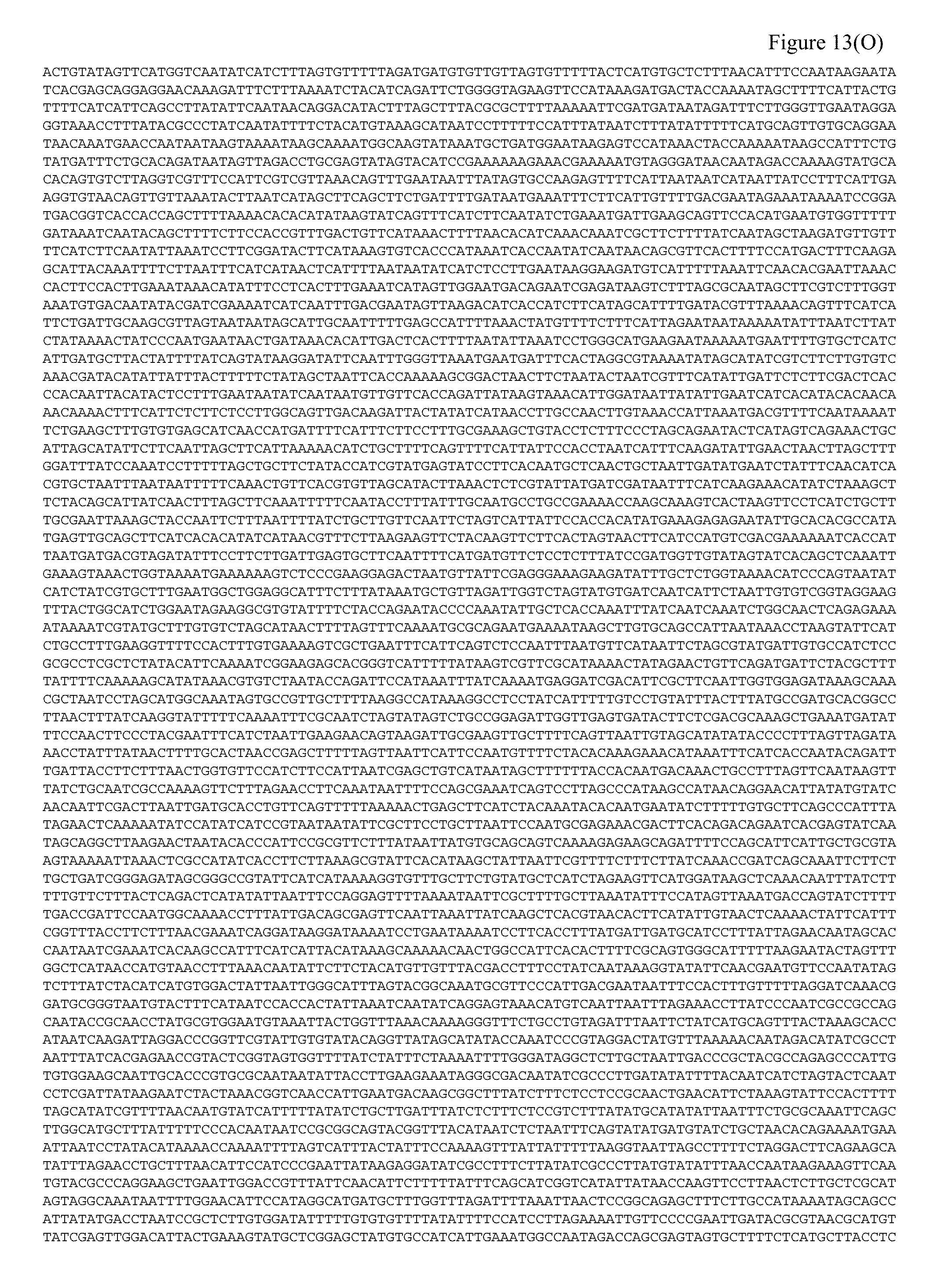
D00066

D00067

D00068

D00069

D00070

D00071

D00072

D00073

D00074

D00075

D00076

D00077

D00078

D00079

D00080

D00081

D00082

D00083

D00084

D00085

D00086

D00087

D00088
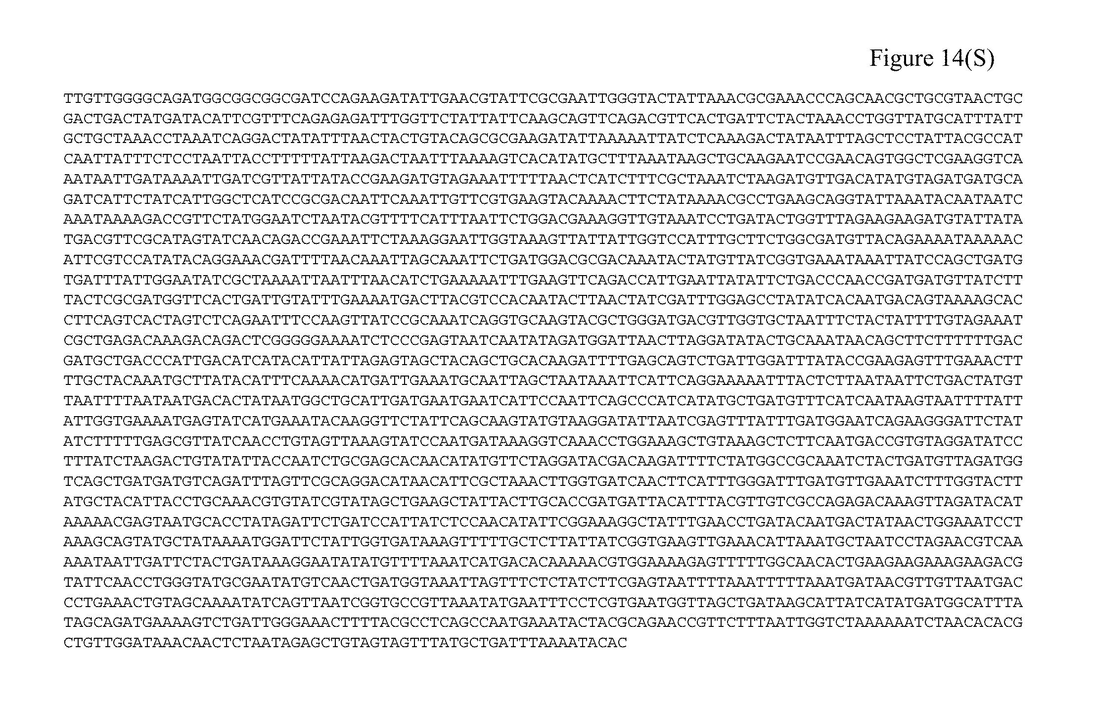
D00089

D00090

S00001
XML
uspto.report is an independent third-party trademark research tool that is not affiliated, endorsed, or sponsored by the United States Patent and Trademark Office (USPTO) or any other governmental organization. The information provided by uspto.report is based on publicly available data at the time of writing and is intended for informational purposes only.
While we strive to provide accurate and up-to-date information, we do not guarantee the accuracy, completeness, reliability, or suitability of the information displayed on this site. The use of this site is at your own risk. Any reliance you place on such information is therefore strictly at your own risk.
All official trademark data, including owner information, should be verified by visiting the official USPTO website at www.uspto.gov. This site is not intended to replace professional legal advice and should not be used as a substitute for consulting with a legal professional who is knowledgeable about trademark law.