Identification of cancer protein biomarkers using proteomic techniques
Mor , et al. J
U.S. patent number 10,168,334 [Application Number 15/268,134] was granted by the patent office on 2019-01-01 for identification of cancer protein biomarkers using proteomic techniques. This patent grant is currently assigned to Yale University. The grantee listed for this patent is Yale University. Invention is credited to Patricia Bray-Ward, Gil G. Mor, David C. Ward.


View All Diagrams
| United States Patent | 10,168,334 |
| Mor , et al. | January 1, 2019 |
Identification of cancer protein biomarkers using proteomic techniques
Abstract
The claimed invention describes methods to diagnose or aid in the diagnosis of cancer. The claimed methods are based on the identification of biomarkers which are particularly well suited to discriminate between cancer subjects and healthy subjects. These biomarkers were identified using a unique and novel screening method described herein. The biomarkers identified herein can also be used in the prognosis and monitoring of cancer. The invention comprises the use of leptin, prolactin, OPN and IGF-II for diagnosing, prognosis and monitoring of ovarian cancer.
| Inventors: | Mor; Gil G. (Cheshire, CT), Ward; David C. (Las Vegas, NV), Bray-Ward; Patricia (Las Vegas, NV) | ||||||||||
|---|---|---|---|---|---|---|---|---|---|---|---|
| Applicant: |
|
||||||||||
| Assignee: | Yale University (New Haven,
CT) |
||||||||||
| Family ID: | 34915557 | ||||||||||
| Appl. No.: | 15/268,134 | ||||||||||
| Filed: | September 16, 2016 |
Prior Publication Data
| Document Identifier | Publication Date | |
|---|---|---|
| US 20170138950 A1 | May 18, 2017 | |
Related U.S. Patent Documents
| Application Number | Filing Date | Patent Number | Issue Date | ||
|---|---|---|---|---|---|
| 14602916 | Jan 22, 2015 | 9470688 | |||
| 12644677 | Dec 22, 2009 | 8975379 | |||
| 11037889 | Jan 18, 2005 | 7666583 | |||
| 60545900 | Feb 20, 2004 | ||||
| 60545581 | Feb 19, 2004 | ||||
| Current U.S. Class: | 1/1 |
| Current CPC Class: | G01N 33/57484 (20130101); G01N 33/57419 (20130101); G01N 33/57415 (20130101); G06F 19/00 (20130101); C12Q 1/6886 (20130101); G01N 33/57449 (20130101); G01N 2500/04 (20130101); G01N 2333/5756 (20130101); G01N 2333/99 (20130101); C12Q 2600/112 (20130101); G01N 2800/52 (20130101); G01N 2333/4703 (20130101); G01N 2333/65 (20130101); C12Q 2600/158 (20130101); G01N 2333/575 (20130101); C12Q 2600/136 (20130101) |
| Current International Class: | G01N 33/00 (20060101); G01N 33/574 (20060101); C12Q 1/6886 (20180101) |
References Cited [Referenced By]
U.S. Patent Documents
| 5583110 | December 1996 | Altchek et al. |
| 6071914 | June 2000 | Cincotta et al. |
| 6235474 | May 2001 | Feinberg |
| 6335170 | January 2002 | Orntoft |
| 6642009 | November 2003 | Hung |
| 6905827 | June 2005 | Wohlgemuth |
| 6936417 | August 2005 | Orntoft |
| 7026121 | April 2006 | Wohlgemuth |
| 7235358 | June 2007 | Wohlgemuth |
| 7666583 | February 2010 | Mor et al. |
| 8163896 | April 2012 | Bentwich |
| 8975379 | March 2015 | Mor et al. |
| 9470688 | October 2016 | Mor et al. |
| 2002/0127580 | September 2002 | Quay |
| 2002/0137100 | September 2002 | Illmensee et al. |
| 2003/0017513 | January 2003 | Khosravi et al. |
| 2003/0044862 | March 2003 | Giaccia et al. |
| 2003/0154032 | August 2003 | Pittman |
| 2003/0180747 | September 2003 | Hruban et al. |
| 2003/0198970 | October 2003 | Roberts |
| 2003/0219812 | November 2003 | Quay et al. |
| 2003/0228570 | December 2003 | Yat Wah Tom |
| 2003/0232398 | December 2003 | MacMurray et al. |
| 2004/0018546 | January 2004 | Hung |
| 2004/0023288 | February 2004 | Ridder et al. |
| 2004/0029114 | February 2004 | Mack |
| 2004/0072189 | April 2004 | Smith et al. |
| 2004/0209282 | October 2004 | Ault-Riche |
| 2005/0069963 | March 2005 | Lokshin et al. |
| 2015/0233929 | January 2015 | Mor et al. |
| 1256354 | Nov 2002 | EP | |||
| 01-123153 | May 1989 | JP | |||
| 02-083337 | Mar 1990 | JP | |||
| 2003-528564 | Sep 2003 | JP | |||
| 2004-033210 | Feb 2004 | JP | |||
| 2005-508488 | Mar 2005 | JP | |||
| 2005-527793 | Sep 2005 | JP | |||
| WO 94/19004 | Sep 1994 | WO | |||
| WO 98/31809 | Jul 1998 | WO | |||
| WO 99/64626 | Dec 1999 | WO | |||
| WO 00/55629 | Sep 2000 | WO | |||
| WO 01/40271 | Jun 2001 | WO | |||
| WO 01/53837 | Jul 2001 | WO | |||
| WO 01/54713 | Aug 2001 | WO | |||
| WO 01/70985 | Sep 2001 | WO | |||
| WO 02/062852 | Aug 2002 | WO | |||
| WO 03/051917 | Jun 2003 | WO | |||
| WO 03/102148 | Dec 2003 | WO | |||
| WO 04/013609 | Feb 2004 | WO | |||
Other References
|
Mukherjea et al., 1999, Elevated leptin concentrations in pregnancy and lactation: possible role as a modulator of substrate utilization, Life Sciences, 65(11): 1183-1193. cited by examiner . Coskun et al., 2003, Serum leptin, prolactin and vascular endothelial growth factor (VEGF) levels in patients with breast cancer, Neoplasma, 50(1): 41-46. cited by examiner . Erbagci et al., 1999, Menstral cycle dependent variability for serum tumor markers CEA, AFP, CA19-9, CA125 and CA15-3 in healthy women, Disease Markers, 15: 259-267. cited by examiner . Markert et al., 2001, Differential gene expression profiling in human brain tumors, Physiol Genomics, 5: 21-33. cited by examiner . Panidis et al., 2003, Serum leptin levels in normal-weight and overweight women with polycystic ovary syndrome, Clin Exp Obst & Gyn, 30(4): 207-210. cited by examiner . Lai et al., 2003, Clinical usefulness of tumour markers, Malaysian J Pathol, 25(2): 83-105. cited by examiner . Baron-Hay et al., Elevated serum insulin-like growth factor binding protein-2 as a prognostic marker in patients with ovarian cancer. Clin Cancer Res. Mar. 1, 2004;10(5):1796-806. cited by applicant . Bowen et al., Downregulation of long-form prolactin receptor mRNA during prolactin-induced luteal regression. Eur J Endocrinol. Aug. 2000;143(2):285-92. cited by applicant . Brakora et al., Utility of osteopontin as a biomarker in recurrent epithelial ovarian cancer. Gynecol Oncol. May 2004;93(2):361-5. cited by applicant . Choi et al., Expression of leptin receptors and potential effects of leptin on the cell growth and activation of mitogen-activated protein kinases in ovarian cancer cells. J Clin Endocrinol Metab. Jan. 2005;9003:207-10. Epub Nov. 2, 2004. cited by applicant . Crump et al., Ovarian cancer tumor marker behavior in asymptomatic healthy women: implications for screening. Cancer Epidemiol Biomarkers Prev. Oct. 2000;9(10):1107-11. cited by applicant . De Souza et al., Fasting ghrelin levels in physically active women: relationship with menstrual disturbances and metabolic hormones. J Clin Endocrinol Metab. Jul. 2004;89(7):3536-42. cited by applicant . Dixit et al., Ghrelin inhibits leptin- and activation-induced proinflammatory cytokine expression by human monocytes and T cells. J Clin Invest. Jul. 2004;114(1):57-66. cited by applicant . Furger et al., The functional and clinical roles of osteopontin in cancer and metastasis. Curr Mol Med. Nov. 2001;1(5):621-32. cited by applicant . Gaja et al., [The importance of leptin in oncology--hypothesis or facts?] Vnitr Lek. Apr. 2001;47(4):245-9. Review. Czech. Medline Abstract. Accession No. 15635891. cited by applicant . Gorelik et al., Multiplexed immunobead-based cytokine profiling for early detection of ovarian cancer. Cancer Epidemiol Biomarkers Prev. Apr. 2005;14(4):981-7. cited by applicant . Gutzman et al., Multiple kinase cascades mediate prolactin signals to activating protein-1 in breast cancer cells. Mol Endocrinol. Dec. 2004;18(12):3064-75. Epub Aug. 19, 2004. cited by applicant . Ho, Estrogen, progesterone and epithelial ovarian cancer. Reprod Biol Endocrinol. Oct. 7, 2003;1:73. cited by applicant . Ishikawa et al., Enhanced expression of leptin and leptin receptor (OB-R) in human breast cancer. Clin Cancer Res. Jul. 1, 2004;10(13):4325-31. cited by applicant . Jacobs et al., Progress and challenges in screening for early detection of ovarian cancer. Mol Cell Proteomics. Apr. 2004;3(4):355-66. Epub Feb. 5, 2004. cited by applicant . Jha et al., Use of serum prolactin for monitoring the therapeutic response in ovarian malignancy. Int J Gynaecol Obstet. Sep. 1991;36(1):33-8. cited by applicant . Kim et al., Osteopontin as a potential diagnostic biomarker for ovarian cancer. JAMA. Apr. 3, 2002;287(13):1671-9. cited by applicant . Kitawaki et al., Leptin directly stimulates aromatase activity in human luteinized granulosa cells. Mol Hum Reprod. Aug. 1999;5(8):708-13. cited by applicant . Koopmann et al., Evaluation of osteopontin as biomarker for pancreatic adenocarcinoma. Cancer Epidemiol Biomarkers Prev. Mar. 2004;13(3):487-91. cited by applicant . Lahousen et al., [Tumor marker combination versus second-look operation in ovarian cancer]. Zentralbl Gynakol. 1990;112(9):561-6. German. cited by applicant . Lahousen et al., [Tumor marker combination versus second-look operation in ovarian cancer]. Zentralbl Gynakol. 1990;112(9):561-6. (English abstract). cited by applicant . Lebrecht et al., Serum vascular endothelial growth factor and serum leptin in patients with cervical cancer. Gynecol Oncol. Apr. 2002;85(1):32-5. cited by applicant . Lu et al., Selection of potential markers for epithelial ovarian cancer with gene expression arrays and recursive descent partition analysis. Clin Cancer Res. May 15, 2004;10(10):3291-300. cited by applicant . Malaguarnera et al., Prolactin increases HO-1 expression and induces VEGF production in human macrophages. J Cell Biochem. Sep. 1, 2004;93(1):197-206. cited by applicant . Mathur et al., Circulating levels of insulin-like growth factor-II and IGF-binding protein 3 in cervical cancer. Gynecol Oncol. Dec. 2003;91(3):486-93. cited by applicant . McIntosh et al., Combining CA 125 and SMR serum markers for diagnosis and early detection of ovarian carcinoma. Gynecol Oncol. Oct. 2004;95(1):9-15. cited by applicant . Menon, Ovarian cancer screening. CMAJ. Aug. 17, 2004;171(4):323-4. cited by applicant . Mor et al., Serum protein markers for early detection of ovarian cancer. Proc Natl Acad Sci U S A. May 24, 2005;102(21):7677-82. Epub May 12, 2005. cited by applicant . Motta et al., Leptin and prolactin modulate the expression of SOCS-1 in association with interleukin-6 and tumor necrosis factor-alpha in mammary cells: a role in differentiated secretory epithelium. Regul Pept. Sep. 15, 2004;121(1-3):163-70. cited by applicant . Mujagi et al., Importance of serum prolactin determination in metastatic breast cancer patients. Croat Med J. Apr. 2004;45(2):176-80. cited by applicant . Oberbeck, Therapeutic implications of immune-endocrine interactions in the critically ill patients. Curr Drug Targets Immune Endocr Metabol Disord. Jun. 2004;4(2):129-39. cited by applicant . O'Regan et al., Osteopontin as a biomarker for ovarian cancer. JAMA. Jun. 26, 2002;287(24):3208-10. cited by applicant . Perks et al., Prolactin acts as a potent survival factor for human breast cancer cell lines. Br J Cancer. Jul. 19, 2004;91(2):305-11. cited by applicant . Perumal et al., Prolactin as a tumour marker in cancer of the cervix. J Obstet Gynaecol. May 1998;18(3):260-2. cited by applicant . Petricoin et al., Use of proteomic patterns in serum to identify ovarian cancer. Lancet. Feb. 16, 2002;359(9306):572-7. cited by applicant . Plebani et al., Serum tumor markers in colorectal cancer staging, grading, and follow-up. J Surg Oncol. Aug. 1996;62(4):239-44. cited by applicant . Rittling et al., Role of osteopontin in tumour progression. Br J Cancer. May 17, 2004;90(10):1877-81. cited by applicant . Rose et al., Obesity, adipocytokines, and insulin resistance in breast cancer. Obes Rev. Aug. 2004;5(3):153-65. Review. cited by applicant . Sancak et al., No association between serum levels of insulin-like growth factor-I, vascular endothelial growth factor, prolactin and clinicopathological characteristics of breast carcinoma after surgery. Intern Med J. Jun. 2004;34(6):310-5. cited by applicant . Santin et al., Gene expression profiles in primary ovarian serous papillary tumors and normal ovarian epithelium: identification of candidate molecular markers for ovarian cancer diagnosis and therapy. Int J Cancer. Oct. 20, 2004;112(1):14-25. cited by applicant . Sawiris et al., Development of a highly specialized cDNA array for the study and diagnosis of epithelial ovarian cancer. Cancer Res. May 15, 2002;62(10):2923-8. cited by applicant . Schorge et al., Osteopontin as an adjunct to CA125 in detecting recurrent ovarian cancer. Clin Cancer Res. May 15, 2004;10(10):3474-8. cited by applicant . Schweitzer et al., Multiplexed protein profiling on microarrays by rolling-circle amplification. Nat Biotechnol. Apr. 2002;20(4):359-65. cited by applicant . Singer et al., Insulin-like growth factor (IGF)-I and IGF-II serum concentrations in patients with benign and malignant breast lesions: free IGF-II is correlated with breast cancer size. Clin Cancer Res. Jun. 15, 2004;10(12 Pt 1):4003-9. cited by applicant . Skates et al., Preoperative sensitivity and specificity for early-stage ovarian cancer when combining cancer antigen CA-125II, CA 15-3, CA 72-4, and macrophage colony-stimulating factor using mixtures of multivariate normal distributions. J Clin Oncol. Oct. 15, 2004;22(20):4059-66. Epub Sep. 20, 2004. cited by applicant . Taylor et al., Cancer screening in a high risk population: a clinical trial. Ultrasound Med Biol. Apr. 2001;27(4):461-6. cited by applicant . Tessitore et al., Leptin expression in colorectal and breast cancer patients. Int J Mol Med. Apr. 2000;5(4):421-6. cited by applicant . Tworoger et al., Plasma prolactin concentrations and risk of postmenopausal breast cancer. Cancer Res. Sep. 15, 2004;64(18):6814-9. cited by applicant . Unni et al., Osteopontin is a potential target gene in mouse mammary cancer chemoprevention by Se-methylselenocysteine. Breast Cancer Res. 2004;6(5):R586-92. Epub Jul. 29, 2004. cited by applicant . Urban et al., Ovarian cancer screening. Hematol Oncol Clin North Am. Aug. 2003;17(4):989-1005. cited by applicant . Visintin et al., Diagnostic markers for early detection of ovarian cancer. Clin Cancer Res. Feb. 15, 2008;14(4):1065-72. Epub Feb. 7, 2008. cited by applicant . Wai et al., The role of Osteopontin in tumor metastasis. J Surg Res. Oct. 2004;121(2):228-41. cited by applicant . Weber, The metastasis gene osteopontin: a candidate target for cancer therapy. Biochim Biophys Acta. Dec. 28, 2001;1552(2):61-85. cited by applicant . Woolas et al., Elevation of multiple serum markers in patients with stage I ovarian cancer. J Natl Cancer Inst. Nov. 3, 1993;85(21):1748-51. cited by applicant . Yao et al., Differential gene expression in chemically induced mouse lung adenomas. Neoplasia. Jan.-Feb. 2003;5(1):41-52. cited by applicant . Yoshida et al., Ovarian dysgerminoma showing high serum levels and positive immunostaining of placental alkaline phosphatase and neuron-specific enolase associated with elevation of serum prolactin level. Eur J Obstet Gynecol Reprod Biol. Oct. 1998;81(1):123-8. cited by applicant . Zhang et al., Growth factor signaling induces metastasis genes in transformed cells: molecular connection between Akt kinase and osteopontin in breast cancer. Mol Cell Biol. Sep. 2003;23(18):6507-19. cited by applicant . Zhang et al., Three biomarkers identified from serum proteomic analysis for the detection of early stage ovarian cancer. Cancer Res. Aug. 15, 2004;64(16):5882-90. cited by applicant. |
Primary Examiner: Steele; Amber D
Attorney, Agent or Firm: Wolf, Greenfield & Sacks, P.C.
Government Interests
FUNDING
This invention was made with government support under grant number DE-FG02-ER63462 awarded by the United States Department of Energy. The government has certain rights in the invention.
Parent Case Text
CROSS-REFERENCE TO RELATED APPLICATIONS
This application is a continuation of U.S. application Ser. No. 14/602,916, filed Jan. 22, 2015, which is a continuation of U.S. application Ser. No. 12/644,677, filed Dec. 22, 2009, now U.S. Pat. No. 8,975,379, which is a continuation of U.S. application Ser. No. 11/037,889, filed Jan. 18, 2005, now U.S. Pat. No. 7,666,583, which claims the benefit of U.S. Provisional Application No. 60/545,581, filed Feb. 19, 2004, and U.S. Provisional Application No. 60/545,900, filed Feb. 20, 2004. The teachings of each referenced application are incorporated by reference herein.
Claims
What is claimed as new and desired to be protected by Letters Patent of the United States is:
1. A method comprising: (a) measuring the expression level of proteins that consist essentially of prolactin, osteopontin (OPN), leptin, and at least one additional protein in a sample from a female subject, wherein the at least one additional protein is selected from macrophage migration inhibitory factor (MIF), insulin-like growth factor (IGF-II), 6Ckine, angiotensin converting enzyme (ACE), brain-derived neurotrophic factor (BDNF), E-Selectin, epidermal growth factor (EGF), eotaxin-2 (Eot-2), epidermal growth factor receptor 1 (ErbB 1), follistatin, hemofiltrate CC chemokine 4 (HCC4), herpes virus entry mediator (HVEM), insulin-like growth factor binding protein 2 (IGFBP-2), interleukin-17 (IL-17), interleukin 1 soluble receptor II (IL-1sRII), interleukin 2 soluble receptor alpha (IL-2 sR.alpha.), macrophage colony stimulating factor receptor (M-CSF R), macrophage inflammatory protein 1alpha (MIP-1.alpha.), macrophage inflammatory protein 3 beta (MIP3.beta.), matrix Metalloproteinase-8(MMP-8), matrix metalloproteinase 7 (MMP-7), myeloid progenitor inhibitory factor 1 (MPIF-1), pulmonary and activation-regulated chemokine (PARC), platelet-derived growth factor receptor beta (PDGF R .beta.), protein C, tumor necrosis factor receptor 1 (TNF-RI), tumor necrosis factor alpha (TNF-.alpha.), soluble Vascular Adhesion Protein-1(sVAP-1), vascular endothelial growth factor receptor 2 (VEGF R2), VEGF receptor 3 (VEGF R3), human stratum corneum chymotryptic enzyme (HSCCE), kallikrein 4, kallikrein 5, kallikrein 6 (protease M), kallikrein 8, kallikrein 9, kallikrein 10, cancer antigen 125 (CA 125), CA15-3, CA19-9, OVX1, lysophosphatidic acid (LPA), carcinoembryonic antigen (CEA), macrophage colony-stimulating factor (M-CSF), prostasin, CA54-61, CA72, HMFG2, interleukin-6 (IL-6), interleukin-10 (IL-10), LSA, NB70K, PLAP, TAG72, TPA, UGTF, WAP four-disulfide core domain 2 (HE4), matrix metalloprotease 2, tetranectin, inhibin, mesothelin, MUC1, vascular endothelial growth factor (VEGF), NOTCH3, E2F transcription factor 3 (E2F3), GTPase activating protein (RACGAP1), hemotological and neurological expressed 1 (HN1), apolipoprotein A1, laminin, claudin 3 (CLDN3), claudin 4, tumor-associated calcium signal transducer 1 (TROP-1/Ep-CAM), tumor-associated calcium signal transducer 2 (TROP-2), ladinin 1, S100A2, SERPIN2 (PAI-2), CD24, lipocalin 2, matriptase (TADG-15), stratifin, transforming growth factor-beta receptor III (TGF-.beta.-RIII), platelet-derived growth factor receptor alpha, SEMACAP3, ras homology gene family member I (ARHI), thrombospondin 2, disabled-2/differentially expressed in ovarian carcinoma 2 (Dab2/DOC2), and haptoglobin-alpha subunit in the sample from the subject; (b) comparing the expression level of each of the measured proteins to a reference sample for each of the measured proteins; and (c) determining if there is a significant difference in the expression level of each of the measured proteins in the sample as compared to the reference sample.
2. The method of claim 1, wherein the method comprises measuring the expression level of prolactin, OPN, leptin, and MIF.
3. The method of claim 2 , further comprising determining if the significant difference in the expression level for prolactin, MIF and/or OPN is an increase in the expression level of prolactin, MIF and/or OPN compared to the reference sample for prolactin, MIF and/or OPN, respectively.
4. The method of claim 2, further comprising determining if the significant difference in the expression level for leptin is a decrease in the expression level of leptin compared to the reference sample for leptin.
5. The method of claim 2, wherein the reference sample corresponds to: (a) the expression level of prolactin, osteopontin (OPN), leptin, and/or MIF in healthy subjects, or (b) the expression level of prolactin, osteopontin (OPN), leptin, and/or MIF in non-cancerous tissue from the same subject.
6. The method of claim 1, wherein each protein is assigned a score of 0 or 1, wherein a protein is assigned a score of 0 if the expression level of the protein is not significantly different from the expression level of the protein in a reference sample and wherein a protein is assigned a score of 1 if the expression level of the protein is significantly different from the expression level of the protein in a reference sample; wherein the subject is assigned an overall score that corresponds to the sum of the assigned scores from at least four different protein; and wherein a given threshold (t) is used.
7. The method of claim 1, further comprising measuring an expression level of CA 125 in the sample from the subject.
8. The method of claim 1, wherein the sample is a body fluid, cell or tissue sample.
9. The method of claim 8, wherein the body fluid sample is blood or serum.
Description
BACKGROUND OF THE INVENTION
Epithelial Ovarian Cancer (EOC) is the fourth leading cause of cancer-related death in women in the United States and the leading cause of gynecologic cancer death. EOC is characterized by few early symptoms, presentation at an advanced stage, and poor survival. This year approximately 25,000 women will be newly diagnosed with ovarian cancer and 13,500 will die from the disease. The major limitations in the treatment of ovarian cancer are: i) the lack of an early detection tumor marker, ii) the resistance to chemotherapeutic agents, and iii) the lack of obvious early warning symptoms. The high mortality rate is related to the inability to detect early disease, as approximately 70% of patients are diagnosed at an advanced stage. In patients diagnosed with early (stage I or II) disease, the five-year survival rate ranges from 60 to 90% depending on the degree of tumor differentiation. Although the clinical presentation of heritable cancer is similar to the high-risk population, the onset of ovarian cancer in this group tends to occur 10-15 years earlier than that of the general population (early 40's rather than 60's). One of the most promising approaches to management of EOC is early detection. The most commonly used test, CA125 identifies a group of cell surface glycoproteins that have uncertain biological behavior and very limited clinical application for the detection of early stage disease. As a single marker, CA125 has a predictive value of less than 10% in Stage I. Even the addition of ultrasound screening to CA125 measurement improves the positive prediction value to only about 20%. The lack of specific markers for ovarian cancer makes it difficult to achieve the clinical objective of screening and early detection.
Presently there is no commercially available test that can be used to diagnose either early or advanced stage ovarian cancer. Thus, the identification of a test that can be used to diagnose early or advance stage ovarian cancer is required.
BRIEF SUMMARY OF THE INVENTION
The invention comprises a method for diagnosing or aiding in the diagnosis of cancer in a subject comprising comparing the expression of one or more biomarkers in a sample from a subject to a predetermined standard for each said one or more biomarkers; wherein said one or more biomarkers are selected from the group consisting of: 6Ckine, ACE, BDNF, CA125, E-Selectin, EGF, Eot2, ErbB1, follistatin, HCC4, HVEM, IGF-II, IGFBP-1, IL-17, IL-1srII, IL-2sRa, leptin, M-CSF R, MIF, MIP-1a, MIP3b, MMP-8, MMP7, MPIF-1, OPN, PARC, PDGF Rb, prolactin, Protein C, TGF-b RIII, TNF-R1, TNF-a, VAP-1, VEGF R2 and VEGF R3; and wherein a significant difference in the expression of said one or more biomarkers in said sample as compared to a predetermined standard of each said one or more biomarkers diagnoses or aids in the diagnosis of cancer.
In one embodiment, the predetermined standard corresponds to: (a) the expression level of said biomarker in healthy subjects, or (b) the expression level of said biomarker in non-cancerous tissue from the same subject.
In one embodiment, the method further comprises comparing the expression of two or more biomarkers, wherein the diagnosis of cancer is based on a score-based classification method. In one embodiment, the method comprises comparing the expression of m different biomarkers; wherein each biomarker is assigned a score of 0 or 1, wherein a biomarker is assigned a score of 0 if the expression of said biomarker is not significantly different from the expression of said biomarker in a predetermined standard and wherein a biomarker is assigned a score of 1 if the expression of said biomarker is significantly different from the expression of said biomarker in a predetermined standard; wherein the subject is assigned an overall score which corresponds to the sum of the assigned scores from m different markers; and wherein a given threshold (t) is used to diagnose or aid in the diagnosis of cancer.
In another embodiment, the method comprises comparing the expression of two or more biomarkers, wherein the diagnosis of cancer is made by comparing the expression profile of said two or more biomarkers to a predetermined standard profile for said biomarkers, and wherein a difference in the profiles diagnoses or aids in the diagnosis of cancer. In one embodiment, the predetermined standard profile is determined by comparing the expression of said two or more biomarkers in cancer subjects to the expression of said two or more biomarkers in healthy subjects using a machine learning technique. In one embodiment, the predetermined standard profile is determined by comparing the expression of said two or more biomarkers in cancer subjects and in healthy subjects using support vector machines, K-nearest neighbor classifier, or classification tree analysis.
In one embodiment, the method is for the diagnosis for ovarian cancer, and the method further comprises detecting an additional biomarker for ovarian cancer which is not identified in Table 2. In one embodiment, the additional biomarker for ovarian cancer may be selected from the group consisting of: human stratum corneum chymotryptic enzyme (HSCCE), kallikrein 4, kallikrein 5, kallikrein 6 (protease M), kallikrein 8, kallikrein 9, kallikrein 10, CA125, CA15-3, CA19-9, OVX1, lysophosphatidic acid (LPA), carcinoebryonic antigen (CEA), macrophage colony-stimulating factor (M-CSF), prostasin, CA54-61, CA72, HMFG2, IL-6, IL-10, LSA, M-CSF, NB70K, PLAP, TAG72, TNF, TPA, UGTF, WAP four-disulfide core domain 2 (HE4), matrix metalloprotease 2, tetranectin, inhibin, mesothelyn, MUC1, VEGF, CLDN3, NOTCH3, E2F transcription factor 3 (E2F3), GTPase activating protein (RACGAP1), hemotological and neurological expressed 1 (HN1), apolipoprotein A1, laminin, claudin 3, claudin 4, tumor-associated calcium signal transducer 1 (TROP-1/Ep-CAM), tumor-associated calcium signal transducer 2 (TROP-2), ladinin 1, S100A2, SERPIN2 (PAI-2), CD24, lipocalin 2, matriptase (TADG-15), stratifin, transforming growth factor-beta receptor III, platelet-derived growth factor receptor alpha, SEMACAP3, ras homology gene family member I (ARHI), thrombospondin 2, disabled-2/differentially expressed in ovarian carcinoma 2 (Dab2/DOC2), and haptoglobin-alpha subunit. In another embodiment, the additional biomarker for ovarian cancer is the truncated form of transthyretin or the cleavage fragment of inter-alpha-trypsin inhibitor heavy chain H4 identified by Zhang et al., Cancer Res. 64(16):5882-90 (2004). In one embodiment, the additional biomarker for ovarian cancer is CA125.
The above described methods of diagnosing or aiding in the diagnosis of cancer can be applied to diagnose or aid in the diagnosis of any cancer or tumor. In one embodiment, the method is for the diagnosis of breast cancer. In one embodiment, the method is for the diagnosis of colon cancer. In another embodiment, the method is for the diagnosis of cervical cancer.
The invention also comprises a method for diagnosing or aiding in the diagnosis of cancer in a subject comprising comparing the expression of one or more biomarkers in a sample from a subject to a predetermined standard for each said one or more biomarkers; wherein said one or more biomarkers are selected from the group consisting of: prolactin, MIF, OPN, IGF-II, E-Selectin, leptin, EGF, IL-17, MPIF-1, and IL-2sRa; and wherein a significant difference in the expression of said one or more biomarkers in said sample as compared to a predetermined standard of each said one or more biomarkers diagnoses or aids in the diagnosis of cancer.
The invention also comprises a method for diagnosing or aiding in the diagnosis of cancer in a subject comprising comparing the expression of one or more biomarkers in a sample from a subject to a predetermined standard for each said one or more biomarkers; wherein said one or more biomarkers are selected from the group consisting of: leptin, prolactin, OPN and IGF-II; and wherein a significant difference in the expression of one or more biomarkers in said sample as compared to a predetermined standard of each said one or more biomarkers diagnoses or aids in the diagnosis of cancer.
The invention also comprises a method for diagnosing or aiding in the diagnosis of cancer in a subject comprising comparing the expression of the following four biomarkers: leptin, prolactin, OPN and IGF-II, in a sample from a subject to a predetermined standard for each said biomarkers; wherein a significant difference in the expression of two or more of said biomarkers in said sample as compared to a predetermined standard of each said one or more biomarkers diagnoses or aids in the diagnosis of cancer.
The invention also comprises a method for diagnosing or aiding in the diagnosis of ovarian cancer in a subject comprising comparing the expression of the following four biomarkers: leptin, prolactin, OPN and IGF-II, in a sample from a subject to a predetermined standard for each said biomarkers; wherein a significant difference in the expression of two or more of said biomarkers in said sample as compared to a predetermined standard of each said one or more biomarkers diagnoses or aids in the diagnosis of cancer.
The invention also comprises a method for diagnosing or aiding in the diagnosis of breast cancer in a subject comprising comparing the expression of the following four biomarkers: leptin, prolactin, OPN and IGF-II, in a sample from a subject to a predetermined standard for each said biomarkers; wherein a significant difference in the expression of two or more of said biomarkers in said sample as compared to a predetermined standard of each said one or more biomarkers diagnoses or aids in the diagnosis of cancer.
The invention also comprises a method for diagnosing or aiding in the diagnosis of colon cancer in a subject comprising comparing the expression of the following four biomarkers: leptin, prolactin, OPN and IGF-II, in a sample from a subject to a predetermined standard for each said biomarkers; wherein a significant difference in the expression of two or more of said biomarkers in said sample as compared to a predetermined standard of each said one or more biomarkers diagnoses or aids in the diagnosis of cancer.
In one embodiment, the above described methods comprise comparing the expression of prolactin and/or OPN to a predetermined standard of said biomarker, wherein an increase in the expression of said biomarker as compared to the predetermined standard for said biomarker diagnoses or aids in the diagnosis of cancer.
In one embodiment, the above described methods comprise comparing the expression of leptin and/or IGF-II to a predetermined standard of said biomarker, and wherein a decrease in the expression of said biomarker as compared to the predetermined standard for said biomarker diagnoses or aids in the diagnosis of cancer.
In one embodiment, the above described methods of diagnosing or aiding in the diagnosis of cancer comprises detecting the expression of two or more biomarkers. In one embodiment, said two or more biomarkers are selected from the group consisting of: prolactin, MIF, OPN, IGF-II, E-Selectin, leptin, EGF, IL-17, MPIF-1, and IL-2sRa. In one embodiment, said two or more biomarkers are selected from the group consisting of: leptin, prolactin, OPN and IGF-II. In one embodiment, a significant difference in the expression of at least two or said two or more biomarkers diagnoses or aids in the diagnosis of cancer.
In one embodiment, the above described methods of diagnosing or aiding in the diagnosis of cancer comprises comparing the expression of three or more biomarkers. In one embodiment, said three or more biomarkers are selected from the group consisting of: leptin, prolactin, OPN and IGF-II. In one embodiment, a significant difference in the expression of said three or more biomarkers diagnoses or aids in the diagnosis of cancer.
In one embodiment, the above described methods of diagnosing or aiding in the diagnosis of cancer comprises comparing the expression of four or more biomarkers. In one embodiment, said four or more biomarkers include leptin, prolactin, OPN and IGF-II. In one embodiment, a significant difference in the expression of four or more biomarkers diagnoses or aids in the diagnosis of cancer.
In one embodiment, the expression of a biomarker is detected or measured using a reagent that detects said one or more biomarkers. In one embodiment, the reagent is an antibody or fragment thereof specific for said one or more biomarkers. In one embodiment, the reagent is directly or indirectly labeled with a detectable substance. In another embodiment, the expression of said one or more biomarker is detected using mass spectroscopy. In another embodiment, the expression of said one or more biomarker is detected by measuring the mRNA transcription levels of the gene encoding said one or more biomarker.
In another embodiment, the expression of said one or more biomarker is detected by: (a) detecting the expression of a polypeptide which is regulated by said one or more biomarker; (b) detecting the expression of a polypeptide which regulates said biomarker; or (c) detecting the expression of a metabolite of said biomarker.
In one embodiment, the sample used in the above described methods is a body fluid sample. In one embodiment, the body fluid sample is blood or serum.
The invention also comprises methods for monitoring the progression of cancer in a subject. In one embodiment, the invention comprises a method of monitoring the progression of cancer in a subject comprising comparing the expression of one or more biomarkers in a sample from a subject to the expression of said one or more biomarkers in a sample obtained from the subject at a subsequent point in time; wherein said one or more biomarkers are selected from the group consisting of: 6Ckine, ACE, BDNF, CA125, E-Selectin, EGF, Eot2, ErbB1, follistatin, HCC4, HVEM, IGF-II, IGFBP-1, IL-17, IL-1srII, IL-2sRa, leptin, M-CSF R, MIF, MIP-1a, MIP3b, MMP-8, MMP7, MPIF-1, OPN, PARC, PDGF Rb, prolactin, Protein C, TGF-b RIII, TNF-R1, TNF-a, VAP-1, VEGF R2 and VEGF R3; and wherein a difference in the expression of said one or more biomarker diagnoses or aids in the diagnosis of the progression of the cancer in the subject. In one embodiment, said one or more biomarkers are selected from the group consisting of: prolactin, MIF, OPN, IGF-II, E-Selectin, leptin, EGF, IL-17, MPIF-1, and IL-2sRa. In one embodiment, said one or more biomarkers are selected from the group consisting of: leptin, prolactin, OPN and IGF-II.
In one embodiment, the above described methods of monitoring the progression of cancer comprises comparing the expression of two or more biomarkers. In one embodiment, said two or more biomarkers are selected from the group consisting of: prolactin, MIF, OPN, IGF-II, E-Selectin, leptin, EGF, IL-17, MPIF-1, and IL-2sRa. In another embodiment, said two or more biomarkers are selected from the group consisting of: leptin, prolactin, OPN and IGF-II.
In one embodiment, the above described methods of monitoring the progression of cancer comprises comparing the expression of three or more biomarkers. In one embodiment, the above described methods of monitoring the progression of cancer comprises comparing the expression of four or more biomarkers. In one embodiment, the above described methods of monitoring the progression of cancer comprises comparing the expression of four or more biomarkers, wherein said four or more biomarkers include leptin, prolactin, OPN and IGF-II. In another embodiment, the above described method of monitoring the progression of cancer comprises comparing the expression of four biomarkers, wherein the four biomarkers are leptin, prolactin, OPN and IGF-II.
The invention also comprises methods for monitoring the effectiveness of a treatment against cancer. In one embodiment, the invention comprise a method for monitoring the effectiveness of a treatment against cancer comprising comparing the expression of one or more biomarkers in a sample from a subject prior to providing at least a portion of a treatment to the expression of said one or more biomarkers in a sample obtained from the subject after the subject has received at least a portion of the treatment; wherein said one or more biomarkers are selected from the group consisting of: 6Ckine, ACE, BDNF, CA125, E-Selectin, EGF, Eot2, ErbB1, follistatin, HCC4, HVEM, IGF-II, IGFBP-1, IL-17, IL-1srII, IL-2sRa, leptin, M-CSF R, MIF, MIP-1a, MIP3b, MMP-8, MMP7, MPIF-1, OPN, PARC, PDGF Rb, prolactin, Protein C, TGF-b RIII, TNF-R1, TNF-a, VAP-1, VEGF R2 and VEGF R3; and wherein a difference in the expression of said one or more biomarker diagnoses or aids in the diagnosis of the efficacy of the treatment. In one embodiment, said one or more biomarkers are selected from the group consisting of: prolactin, MIF, OPN, IGF-II, E-Selectin, leptin, EGF, IL-17, MPIF-1, and IL-2sRa. In one embodiment, said one or more biomarkers are selected from the group consisting of: leptin, prolactin, OPN and IGF-II.
In one embodiment, the above described methods of monitoring the effectiveness of a treatment against cancer comprises comparing the expression of two or more biomarkers. In one embodiment, said two or more biomarkers are selected from the group consisting of: prolactin, MIF, OPN, IGF-II, E-Selectin, leptin, EGF, IL-17, MPIF-1, and IL-2sRa. In another embodiment, said two or more biomarkers are selected from the group consisting of: leptin, prolactin, OPN and IGF-II.
In one embodiment, the above described methods of monitoring the effectiveness of a treatment against cancer comprises comparing the expression of three or more biomarkers. In one embodiment, the above described methods of monitoring the effectiveness of a treatment against cancer comprises comparing the expression of four or more biomarkers. In one embodiment, the above described methods of monitoring the effectiveness of a treatment against cancer comprises comparing the expression of four or more biomarkers, wherein said four or more biomarkers include leptin, prolactin, OPN and IGF-II. In another embodiment, the above described method of monitoring the effectiveness of a treatment against cancer comprises comparing the expression of four biomarkers, wherein the four biomarkers are leptin, prolactin, OPN and IGF-II.
The invention also comprises kits for diagnosing or aiding in the diagnosis of cancer and kits for monitoring cancer. In one embodiment, the kit comprises: (i) a receptacle for receiving a sample; (ii) one or more reagents for detecting one or more biomarkers selected from the group consisting of: 6Ckine, ACE, BDNF, CA125, E-Selectin, EGF, Eot2, ErbB1, follistatin, HCC4, HVEM, IGF-II, IGFBP-1, IL-17, IL-1srII, IL-2sRa, leptin, M-CSF R, MIF, MIP-1a, MIP3b, MMP-8, MMP7, MPIF-1, OPN, PARC, PDGF Rb, prolactin, Protein C, TGF-b RIII, TNF-R1, TNF-a, VAP-1, VEGF R2 and VEGF R3; and (iii) a reference sample. In one embodiment, the kit comprises one or more reagents for the detection of leptin, prolactin, OPN and IGF-II.
The invention also comprises a method to screen for a candidate compound useful to treat cancer. In one embodiment, the invention comprises a method to screen for a candidate compound useful to treat cancer comprising: (i) identifying a candidate compound which regulates the expression of at least one biomarker selected from the group consisting of: 6Ckine, ACE, BDNF, CA125, E-Selectin, EGF, Eot2, ErbB1, follistatin, HCC4, HVEM, IGF-II, IGFBP-1, IL-17, IL-1srII, IL-2sRa, leptin, M-CSF R, MIF, MIP-1a, MIP3b, MMP-8, MMP7, MPIF-1, OPN, PARC, PDGF Rb, prolactin, Protein C, TGF-b RIII, TNF-R1, TNF-a, VAP-1, VEGF R2 and VEGF R3; and (ii) determining whether such candidate compound is effective to treat cancer. In one embodiment, the method comprises identifying a candidate compound which regulates the expression of at least one biomarkers selected from the group consisting of: prolactin, MIF, OPN, IGF-II, E-Selectin, leptin, EGF, IL-17, MPIF-1, and IL-2sRa. In one embodiment, the method comprises identifying a candidate compound which regulates the expression of at least one biomarkers selected from the group consisting of leptin, prolactin, OPN and IGF-II.
The invention also comprises a method of conducting a business. In one embodiment, the method of conducting a business comprises: (i) obtaining a sample; (ii) detecting the expression of one or more biomarker in the sample, wherein said one or more biomarkers are selected from the group consisting of: 6Ckine, ACE, BDNF, CA125, E-Selectin, EGF, Eot2, ErbB1, follistatin, HCC4, HVEM, IGF-II, IGFBP-1, IL-17, IL-1srII, IL-2sRa, leptin, M-CSF R, MIF, MIP-1a, MIP3b, MMP-8, MMP7, MPIF-1, OPN, PARC, PDGF Rb, prolactin, Protein C, TGF-b RIII, TNF-R1, TNF-a, VAP-1, VEGF R2 and VEGF R3; and (iii) reporting the results of such detection. In one embodiment, said one or more biomarkers are selected from the group consisting of: prolactin, MIF, OPN, IGF-II, E-Selectin, leptin, EGF, IL-17, MPIF-1, and IL-2sRa. In another embodiment, said one or more biomarkers are selected from the group consisting of: leptin, prolactin, OPN and IGF-II.
In one embodiment, the invention comprises a method of conducting a business comprising: (i) obtaining a sample; (ii) detecting the expression of four biomarkers in the sample, wherein said four biomarkers leptin, prolactin, OPN and IGF-II; and (iii) reporting the results of such detection.
The invention also comprises methods to screen for candidate cancer biomarkers. In one embodiment, the invention comprises a method to screen for candidate cancer biomarkers comprising: (i) identifying a group of biomarkers that are potentially associated with cancer; (ii) comparing the level of expression of the biomarkers identified in step (i) in a first population of cancer subjects and in healthy subjects; (iii) selecting biomarkers exhibiting a significant difference in expression in said first population of cancer subjects; (iv) comparing the level of expression of the biomarkers identified in step (iii) in a second population of cancer subjects and in healthy subjects; and (v) selecting biomarkers exhibiting a significant difference in expression in said second population of cancer subjects; wherein the biomarkers identified in step (v) are candidate cancer biomarkers. In one embodiment, said first population of cancer subjects have newly diagnosed cancer, and said second population of cancer subjects have recurrent cancer. In one embodiment, said first population of cancer subjects have recurrent cancer and said second population of cancer subjects have newly diagnosed cancer. In another embodiment, wherein said first population of cancer subjects have late stage cancer and said second population of cancer subjects have early stage cancer. In another embodiment, said first population of cancer patients have early stage cancer and said second population of cancer subjects have later stage cancer. In another embodiment, said method further comprises: (vi) comparing the level of expression of the biomarkers identified in step (v) in a third population of cancer subjects and in healthy subjects, wherein the expression of said biomarkers is detected by using a different assay format; and (vii) selecting biomarkers exhibiting a significant different in expression in said third population of cancer subjects; wherein the biomarkers identified in step (vii) are candidate biomarkers for cancer. In one embodiment, said method further comprises determining whether the biomarkers identified in step (v) or (vii) could distinguish between cancer and healthy subjects in a blind study.
The invention also comprises a method to screen for candidate cancer biomarkers comprising: (i) identifying a cancer biomarker; (ii) selecting polypeptides which regulate or are regulated by the biomarker identified in step (i); and (iii) measuring the expression of the polypeptides identified in step (ii) in cancer subjects and in healthy subjects, wherein a polypeptide which is differentially expressed in cancer subjects and in healthy subjects is a candidate cancer biomarker.
BRIEF DESCRIPTION OF THE DRAWINGS
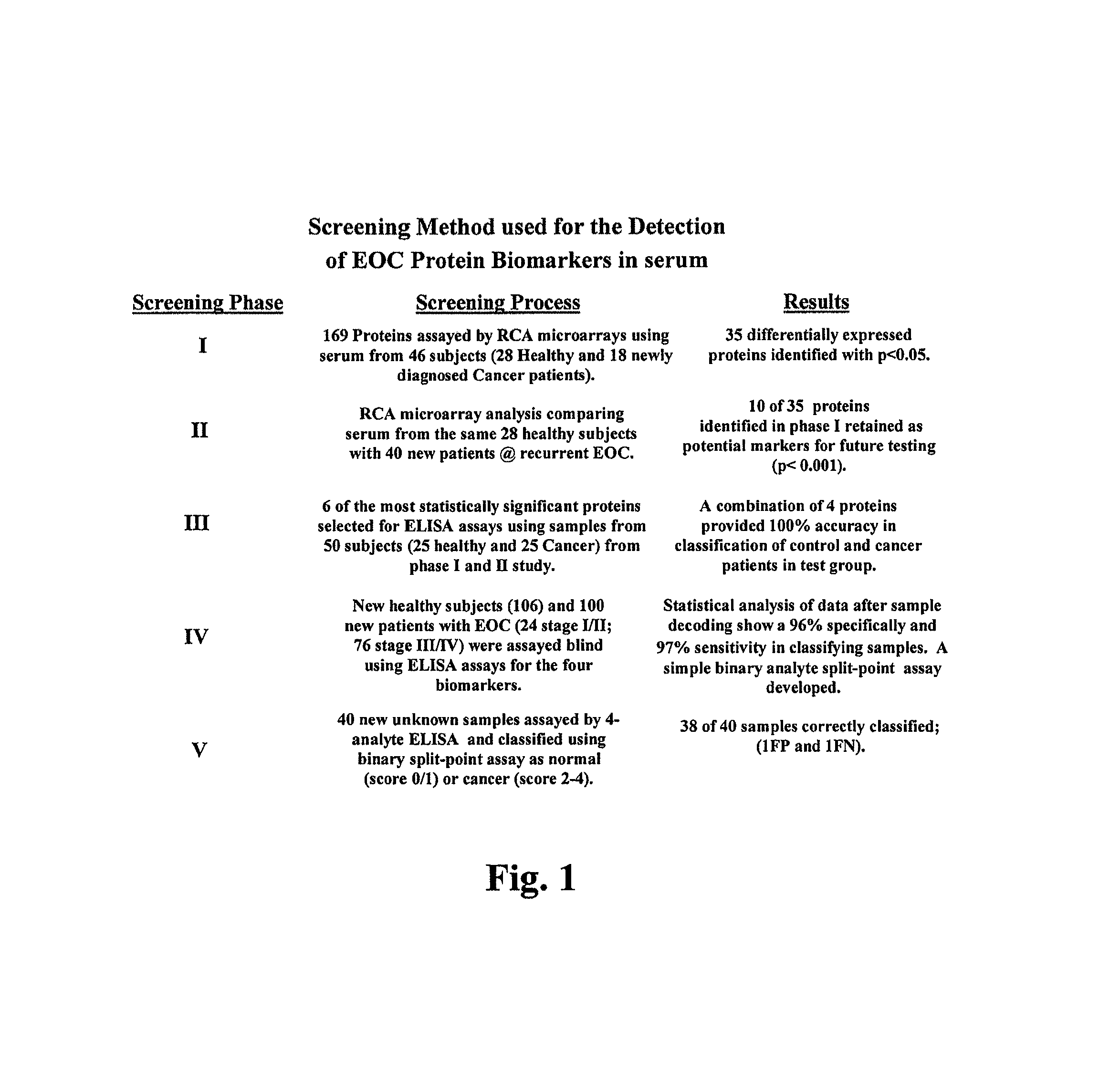
FIG. 1 is a schematic representation of the screening process used to identify biomarkers which could discriminate between subjects with cancer and healthy subjects.
FIG. 2 is a schematic representation of a sample protein microarray slide with 16 subarrays. Subarrays refer to the 16 wells, or circular analysis sites, on the slide. Array refers to the antibody content printed in a well. Each microarray slide contains only one type of array.
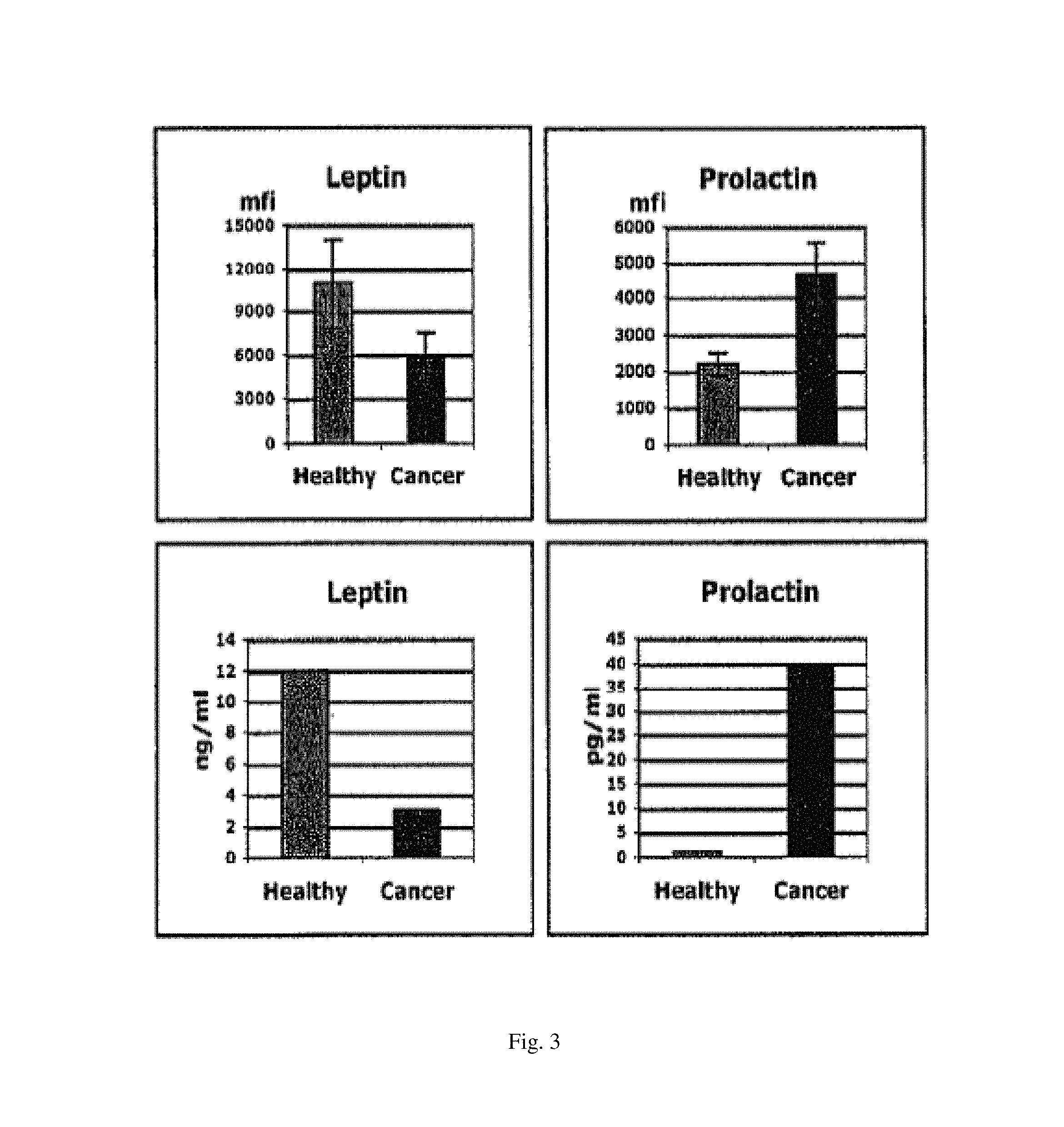
FIG. 3 shows the difference in expression of four proteins (leptin, prolactin, OPN and IGF-II) in subjects with ovarian cancer and in healthy subjects using two different assays: RCA microarray immunoassay and ELISA.
FIG. 4 shows results of analysis of the expression data of four proteins (leptin (identified as "1"), prolactin (identified as "2"), OPN (identified as "3") and IGF-II (identified as "4")) in 206 subjects, using the least square fit in a traditional binary data set analysis. The protein levels of healthy subjects are shown in black (left) and those for subjects with ovarian cancer are shown in gray (right)
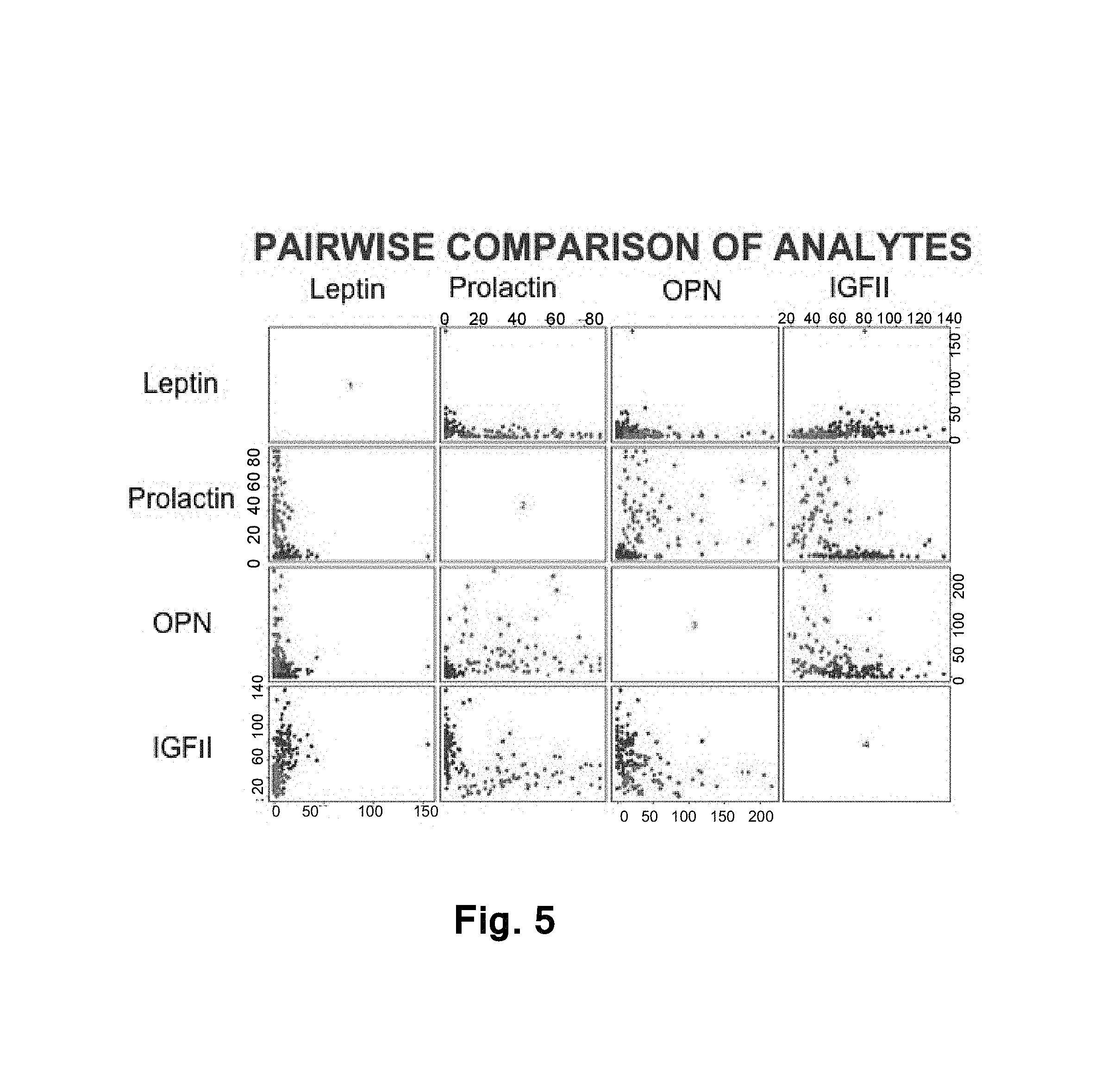
FIG. 5 shows results of analysis of the expression data of four proteins (leptin (identified as "1"), prolactin (identified as "2"), OPN (identified as "3") and IGF-II (identified as "4")) in 206 subjects, using pair plots. The data points derived from healthy subjects are in black and the data points derived from subjects with ovarian cancer are in gray.
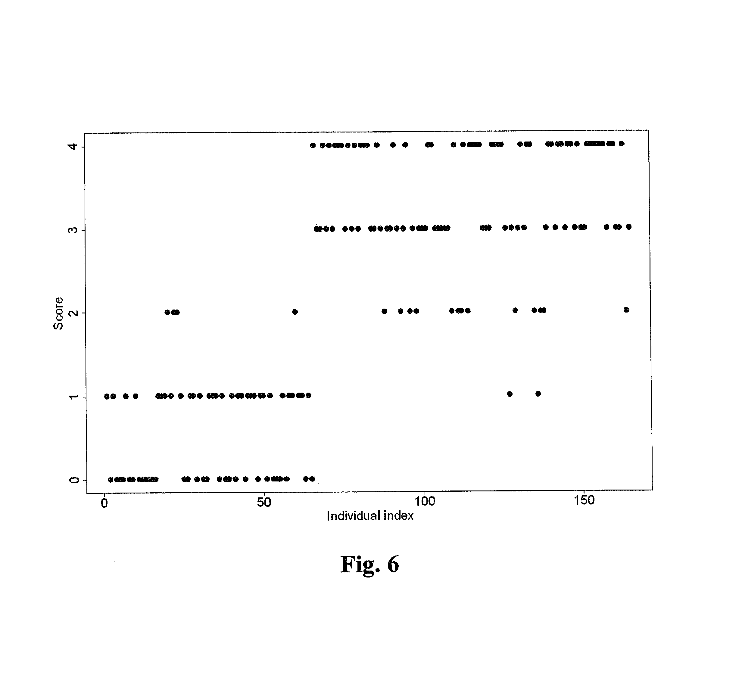
FIG. 6 shows the scores assigned to 206 subjects including 106 healthy subjects and 100 subjects with ovarian cancer based on the score-based classification system described herein. Subjects having a score greater than or equal to 2 can be diagnosed with ovarian cancer, while subjects with score less than or equal to 1 can be diagnosed as free of ovarian cancer. The data points derived from healthy subjects are in light gray and the data points derived from subjects with ovarian cancer are in dark gray.
DETAILED DESCRIPTION OF THE INVENTION
I. Overview
Described herein is a method which can be used to discriminate between cancer subjects (including subjects diagnosed with early stage (stage I-II) disease) and healthy subjects. This method is based on the identification of biomarkers which are particularly well suited to discriminate between cancer subjects and healthy subjects. These biomarkers were identified using a unique and novel screening method described herein involving several different screening steps using samples from different subjects in each step and validation with different techniques. The biomarkers disclosed herein can be used in the diagnosis, prognosis and monitoring of cancer.
In one particular embodiment, the invention disclosed herein refers to a new test based on four biomarkers: leptin, prolactin, ORN and IGF II, which discriminate between cancer subjects and healthy subjects, particularly between ovarian cancer subjects and healthy subjects. In one embodiment, these four biomarkers can be used in a blood test for the diagnosis, prognosis and monitoring of ovarian cancer.
These biomarkers identified herein can be used in combination with additional known biomarkers. For example, a known biomarker of ovarian cancer is CA125. The use of CA125 in conjunction with the biomarkers identified herein presents a novel approach for the early detection of ovarian cancer and may significantly improve our ability to accurately detect pre-malignant change or early stage ovarian cancer in asymptomatic women at increased risk for the development of ovarian cancer. Further, the biomarkers identified in this application can be used in conjunction with other diagnostic techniques. For example, for the diagnosis of ovarian cancer, the biomarkers identified in this application can be used in conjunction with vaginal examination, ultrasound or MRI to diagnose ovarian cancer.
The articles "a," "an" and "the" are used herein to refer to one or to more than one (i.e., to at least one) of the grammatical object of the article.
The term "including" is used herein to mean, and is used interchangeably with, the phrase "including but not limited to".
The term "or" is used herein to mean, and is used interchangeably with, the term "and/or," unless context clearly indicates otherwise.
The term "such as" is used herein to mean, and is used interchangeably with, the phrase "such as but not limited to".
II. Methods of Diagnosis
In one embodiment, the invention refers to a method for diagnosing or aiding in the diagnosis of cancer in a subject. In one embodiment, the method comprises comparing the expression of one or more biomarkers selected from the group consisting of the biomarkers identified in Table 2 in a sample from a subject to a predetermined standard for each said one or more biomarkers, wherein a significant difference in the expression of said one or more biomarkers in said sample as compared to a predetermined standard of each said one or more biomarkers diagnoses or aids in the diagnosis of cancer. In one embodiment, said one or more biomarkers are selected from the group consisting of the biomarkers identified in Table 3. In another embodiment, said one or more biomarkers are selected from the group consisting of: leptin, prolactin, OPN and IGF-II.
When the biomarkers are prolactin and/or OPN, an increase in the expression of said biomarkers as compared to the predetermined standard for said biomarker diagnoses or aids in the diagnosis of cancer. When the biomarkers is leptin and/or IGF-II, a decrease in the expression of said biomarker as compared to the predetermined standard for said biomarker diagnoses or aids in the diagnoses of cancer. As used herein, an increase or decrease in expression refers to the fact that level of a gene expression product is made higher or lower, or to the fact that the activity of the gene expression product is enhanced or lowered.
The above described methods can be used to diagnose any cancer or tumor. In one embodiment, the cancer is ovarian cancer. In another embodiment, the cancer is breast cancer. In another embodiment, the cancer is colon cancer. In another embodiment, the cancer is prostate cancer. In another embodiment, the cancer is cervical cancer.
As used herein, the term "biomarker" refers to one or more polypeptides that can be used to: diagnose, or to aid in the diagnosis or prognosis of, cancer either alone or as combination of multiple polypeptides; monitor the progression of cancer; and/or monitor the effectiveness of a cancer treatment. As used herein, the term "polypeptide" refers to a polymer of amino acids, and not to a specific length. Thus, peptides, oligopeptides and proteins are included within the definition of polypeptide.
As used herein, the term "leptin" includes all homologs, naturally occurring allelic variants, isoforms and precursors of leptin. Leptin is also known as HGNC:6553, OB, OBS, obesity, or murine obesity homolog. In one embodiment, leptin comprises the amino acid sequence of GenBank Accession No. NP_000221.
As used herein, the term "prolactin" includes all homologs, naturally occurring allelic variants, isoforms and precursors of prolactin. Prolactin is also known as PRL or HGNC:9445. In one embodiment, prolactin comprises the amino acid sequence of GenBank Accession No. NP_000939.
As used herein, the term "OPN" includes all homologs, naturally occurring allelic variants, isoforms and precursors of OPN. OPN is also known as HGNC:11255, BNSP, BSPI, ETA-1, secreted phosphoprotein-1 or osteopontin. In one embodiment, OPN comprises the amino acid sequence of GenBank Accession No. NP_000573.
As used herein, the term "IGF-II" includes all homologs, naturally occurring allelic variants, isoforms and precursors of IGF-II. IGF-II is also known as HGNC:5466, insulin-like growth factor 2, insulin-like growth factor II or somatomedin A. In one embodiment, IGF-II comprises the amino acid sequence of GenBank Accession No. NP_000603.
As used herein, the term "subject" or "patient" includes all warm-blooded animals. In one embodiment the subject is a human. In one embodiment, the subject is a subject with an enhanced risk of developing cancer.
In one embodiment, when the method relates to ovarian cancer, the subject is a female (such as a woman) suspected of having or known to have ovarian cancer, or with an enhanced risk of developing ovarian cancer. For example, for ovarian cancer subjects having a familial history of ovarian cancer, subjects identified as having a mutant oncogene, and subjects at least about 50 years of age have an enhanced risk of developing ovarian cancer.
As used herein, the term "sample" refers to a material obtained from a subject. The sample can be derived from any biological source, including all body fluids (such as, for example, whole blood, plasma, serum, saliva, ocular lens fluid, sweat, urine, milk, etc.), tissue or extracts, cells, etc. Examples of ovary-associated body fluids include blood fluids (e.g. whole blood, blood serum, blood having platelets removed therefrom, etc.), lymph, ascitic fluids, gynecological fluids (e.g. ovarian, fallopian, and uterine secretions, menses, vaginal douching fluids, fluids used to rinse ovarian cell samples, etc.), cystic fluid, urine, and fluids collected by peritoneal rinsing (e.g. fluids applied and collected during laparoscopy or fluids instilled into and withdrawn from the peritoneal cavity of a human patient).
The term "expression" is used herein to mean the process by which a polypeptide is produced from DNA. The process involves the transcription of the gene into mRNA and the translation of this mRNA into a polypeptide. Depending on the context in which used, "expression" may refer to the production of RNA, protein or both.
Expression of a biomarker of the invention may be assessed by any of a wide variety of well known methods for detecting expression of a transcribed molecule or its corresponding protein. Non-limiting examples of such methods include immunological methods for detection of secreted proteins, protein purification methods, protein function or activity assays, nucleic acid hybridization methods, nucleic acid reverse transcription methods, and nucleic acid amplification methods. In a preferred embodiment, expression of a marker gene is assessed using an antibody (e.g. a radio-labeled, chromophore-labeled, fluorophore-labeled, or enzyme-labeled antibody), an antibody derivative (e.g. an antibody conjugated with a substrate or with the protein or ligand of a protein-ligand pair {e.g. biotin-streptavidin}), or an antibody fragment (e.g. a single-chain antibody, an isolated antibody hypervariable domain, etc.) which binds specifically with a protein corresponding to the marker gene, such as the protein encoded by the open reading frame corresponding to the marker gene or such a protein which has undergone all or a portion of its normal post-translational modification. In another preferred embodiment, expression of a marker gene is assessed by preparing mRNA/cDNA (i.e. a transcribed polynucleotide) from cells in a patient sample, and by hybridizing the mRNA/cDNA with a reference polynucleotide which is a complement of a polynucleotide comprising the marker gene, and fragments thereof. cDNA can, optionally, be amplified using any of a variety of polymerase chain reaction methods prior to hybridization with the reference polynucleotide; preferably, it is not amplified.
As used herein, a "predetermined standard" for a biomarker refers to the levels of expression of said biomarker in healthy subjects or the expression levels of said biomarker in non-cancerous tissue from the same subject. The predetermined standard expression levels for a given biomarker can be established by prospective and/or retrospective statistical studies using only routine experimentation. Said predetermined standard expression levels can be determined by a person having ordinary skill in the art using well known methods.
The term "healthy subject" refers to a subject has not been diagnosed with cancer or who has not been diagnosed with cancer of the type which is being analyzed. Thus, for example, in a method to diagnose ovarian cancer, a "healthy subject" refers to a subject who does cancer or who does not have ovarian cancer.
As used herein, the term "significant difference" is well within the knowledge of a skilled artisan and will be determined empirically with reference to each particular biomarker. For example, a significant difference in the expression of a biomarker in a subject with cancer as compared to a healthy subject is any difference in expression which is statistically significant.
In one embodiment, the method comprises comparing the expression of two or more biomarkers and the diagnosis of cancer is based on a score-based classification method. In one embodiment, the score-based classification system is a based on binary numbers. In one embodiment, the score-based classification system comprises determining the expression of m different biomarkers; wherein each biomarker is assigned a score of 0 or 1, wherein a biomarker is assigned a score of 0 if the expression of said biomarker is not significantly different from the expression of said biomarker in a predetermined standard and wherein a biomarker is assigned a score of 1 if the expression of said biomarker is significantly different from the expression of said biomarker in a predetermined standard; wherein the subject is assigned an overall score which corresponds to the sum of the assigned scores from m different markers; and wherein a given threshold (t) is used to diagnose or aid in the diagnosis of cancer.
In one embodiment, the score-based classification system comprises comparing the expression of four (4) different biomarkers; wherein each biomarker is assigned a score of 0 or 1, wherein a biomarker is assigned a score of 0 if the expression of said biomarker is not significantly different from the expression of said biomarker in a predetermined standard and wherein a biomarker is assigned a score of 1 if the expression of said biomarker is significantly different from the expression of said biomarker in a predetermined standard; wherein the subject is assigned an overall score which corresponds to the sum of the assigned scores from four (4) different markers; and wherein a score or 2 or more diagnoses or aids in the diagnosis of cancer. In one embodiment, the four biomarkers are leptin, prolactin, OPN and IGF-II.
In one embodiment, the method comprises comparing the expression of two or more biomarkers, wherein the diagnosis of cancer is made by comparing the expression profile of said two or more biomarkers to a predetermined standard profile for said biomarkers, and wherein a difference in the profiles diagnoses or aids in the diagnosis of cancer. As used herein, an "expression profile" is a representation of the levels of expression of one or more biomarkers in a given sample.
In one embodiment, the predetermined standard profile is determined by comparing the expression of said two or more biomarkers in cancer subjects to the expression of said two or more biomarkers in healthy subjects using a machine learning technique. In one embodiment, the predetermined standard profile is determined by comparing the expression of said two or more biomarkers in cancer subjects and in healthy subjects using support vector machines, K-nearest neighbor classifier, or classification tree analysis.
In one embodiment, the method comprises detecting an additional known biomarker which is not identified in Table 2 and comparing the expression of said additional known biomarker to a predetermined standard for said additional known biomarker. Additional biomarkers for cancer can be identified by a person having ordinary skill in the art by reference to the published literature. In one embodiment, the cancer is ovarian cancer, and the additional biomarker for ovarian cancer is selected from the group consisting of: human stratum corneum chymotryptic enzyme (HSCCE), kallikrein 4, kallikrein 5, kallikrein 6 (protease M), kallikrein 8, kallikrein 9, kallikrein 10, CA125, CA15-3, CA19-9, OVX1, lysophosphatidic acid (LPA), carcinoebryonic antigen (CEA), macrophage colony-stimulating factor (M-CSF), prostasin, CA54-61, CA72, HMFG2, IL-6, IL-10, LSA, M-CSF, NB70K, PLAP, TAG72, TNF, TPA, UGTF, WAP four-disulfide core domain 2 (HE4), matrix metalloprotease 2, tetranectin, inhibin, mesothelyn, MUC1, VEGF, CLDN3, NOTCH3, E2F transcription factor 3 (E2F3), GTPase activating protein (RACGAP1), hemotological and neurological expressed 1 (HN1), apolipoprotein A1, laminin, claudin 3, claudin 4, tumor-associated calcium signal transducer 1 (TROP-1/Ep-CAM), tumor-associated calcium signal transducer 2 (TROP-2), ladinin 1, S100A2, SERPIN2 (PAI-2), CD24, lipocalin 2, matriptase (TADG-15), stratifin, transforming growth factor-beta receptor III, platelet-derived growth factor receptor alpha, SEMACAP3, ras homology gene family member I (ARHI), thrombospondin 2, disabled-2/differentially expressed in ovarian carcinoma 2 (Dab2/DOC2), and haptoglobin-alpha subunit. In another embodiment, the additional biomarker for ovarian cancer is the truncated form of transthyretin or the cleavage fragment of inter-alpha-trypsin inhibitor heavy chain H4 identified by Zhang et al., Cancer Res. 64(16):5882-90 (2004). In a preferred embodiment, the additional biomarker for ovarian cancer is CA125.
In one embodiment, the invention refers to a method for diagnosing or aiding in the diagnosis of cancer in a subject comprising comparing the expression of two or more biomarkers selected from the group consisting of the biomarkers identified in Table 2 in a sample from a subject to a predetermined standard for each said biomarker, wherein a significant difference in the expression of one or more biomarkers in said sample as compared to a predetermined standard of each biomarker diagnoses or aids in the diagnosis of cancer. In one embodiment, said two or more biomarkers are selected from the group consisting of the biomarkers identified in Table 3. In another embodiment, said two or more biomarkers are selected from the group consisting of: leptin, prolactin, OPN and IGF-II. In one embodiment, a significant difference in the expression of at least two of said two or more biomarkers diagnoses or aids in the diagnosis of cancer.
In one embodiment, the invention comprises to a method for diagnosing or aiding in the diagnosis of cancer in a subject comprising comparing the expression of three or more biomarkers selected from the group consisting of the biomarkers identified in Table 2 in a sample from a subject to a predetermined standard for each biomarker, wherein a significant difference in the expression of one or more biomarkers in said sample as compared to a predetermined standard of each said biomarker diagnoses or aids in the diagnosis of cancer. In one embodiment, said three or more biomarkers are selected from the group consisting of the biomarkers identified in Table 3. In another embodiment, said three or more biomarkers are selected from the group consisting of: leptin, prolactin, OPN and IGF-II. In one embodiment, a significant difference in the expression of at least two or said two or more biomarkers diagnoses or aids in the diagnosis of cancer.
In one embodiment, the invention refers to a method for diagnosing or aiding in the diagnosis of cancer in a subject comprising comparing the expression of four or more biomarkers selected from the group consisting of the biomarkers identified in Table 2 in a sample from a subject to a predetermined standard for each biomarker, wherein a significant difference in the expression of one or more biomarkers in said sample as compared to a predetermined standard of each said biomarker diagnoses or aids in the diagnosis of cancer. In one embodiment, said four or more biomarkers are selected from the group consisting of the biomarkers identified in Table 3. In another embodiment, said four or more biomarkers are selected from the group consisting of: leptin, prolactin, OPN and IGF-II. In one embodiment, a significant difference in the expression of at least two of said two or more biomarkers diagnoses or aids in the diagnosis of cancer.
The expression of said one or more biomarkers can be detected using any method known to a person having ordinary skill in the art. In one embodiment, the expression of said one or more biomarkers can be detected using a reagent that detects said one or more biomarkers. Said reagent can be any reagent that specifically detects said one or more biomarkers. Said reagent can be an antibody (natural or synthetic) or a fragment thereof specific for the biomarker, a peptide, a nucleic acid, or any other reagent that can specifically detect a biomarker. As used herein, the term "antibody" includes chimeric and synthetic antibodies, e.g., generated by combinatorial mutagenesis and phage display. The term "antibody" includes mimetics or peptidomimetics of antibodies. Peptidomimetics are compounds based on, or derived from, peptides and proteins. The peptidomimetics of the present invention typically can be obtained by structural modification of a known peptide sequence using unnatural amino acids, conformational restraints, isosteric replacement, and the like. The subject peptidomimetics constitute the continuum of structural space between peptides and non-peptide synthetic structures; peptidomimetics may be useful, therefore, in delineating pharmacophores and in helping to translate peptides into non-peptide compounds with the activity of the parent peptides. For illustrative purposes, peptide analogs of the antibodies can be generated using, for example, benzodiazepines (e.g., see Freidinger et al. in Peptides: Chemistry and Biology, G. R. Marshall ed., ESCOM Publisher: Leiden, Netherlands, 1988), substituted gamma lactam rings (Garvey et al. in Peptides: Chemistry and Biology, G. R. Marshall ed., ESCOM Publisher: Leiden, Netherlands, 1988, p 123), C-7 mimics (Huffman et al. in Peptides: Chemistry and Biology, G. R. Marshall ed., ESCOM Publisher: Leiden, Netherlands, 1988, p. 105), keto-methylene pseudopeptides (Ewenson et al. (1986) J Med Chem 29:295; and Ewenson et al. in Peptides: Structure and Function (Proceedings of the 9.sup.th American Peptide Symposium) Pierce Chemical Co. Rockland, Ill., 1985), .beta.-turn dipeptide cores (Nagai et al. (1985) Tetrahedron Lett 26:647; and Sato et al. (1986) J Chem Soc Perkin Trans 1:1231), .beta.-aminoalcohols (Gordon et al. (1985) Biochem Biophys Res Commun 126:419; and Dann et al. (1986) Biochem Biophys Res Commun 134:71), diaminoketones (Natarajan et al. (1984) Biochem Biophys Res Commun 124:141), and methyleneamino-modified (Roark et al. in Peptides: Chemistry and Biology, G. R. Marshall ed., ESCOM Publisher: Leiden, Netherlands, 1988, p 134). Also, see generally, Session III: Analytic and synthetic methods, in Peptides: Chemistry and Biology, G. R. Marshall ed., ESCOM Publisher: Leiden, Netherlands, 1988).
In another embodiment, said reagent is directly or indirectly labeled with a detectable substance. The detectable substance may be, for example, selected, e.g., from a group consisting of radioisotopes, fluorescent compounds, enzymes, and enzyme co-factor. Methods of labeling antibodies are well known in the art.
As used herein, the term "detect", "detected" or "detecting" includes measure, measured or measuring.
The above described methods can be performed using any sample. In one embodiment, the sample is a body fluid sample. In one embodiment, the body fluid sample is blood or serum.
In another embodiment, the expression of said one or more biomarkers are detected using mass spectroscopy.
In yet another embodiment, the expression of said one or more biomarkers is detected by detecting the mRNA transcription levels of the gene encoding said at one or more biomarker.
In yet another embodiment, the expression of said one or more biomarkers can be detected by ELISA, RCA immunoassay, chemiluminescence, thin-film optical biosensor, proton resonance technology, protein microarray assay or any other detection method known in the art.
In one embodiment, the expression of said one or more biomarkers are detected by: (a) detecting the expression of a polypeptide which is regulated by said one or more biomarker; (b) detecting the expression of a polypeptide which regulates said biomarker; or (c) detecting the expression of a metabolite of said biomarker. A person of skill in the art would be able to identify polypeptides which regulate or are regulated by a biomarker, and metabolites of a biomarker, using only routine experimentation.
The above described methods to diagnose or aid in the diagnosis of cancer may be used in conjunction with other methods to validate the results (i.e. to more conclusively determine whether a subject has cancer). In one embodiment, the cancer is ovarian cancer and the above described methods further comprise: physical examination, ultrasound examination, x-ray examination, MRI examination, laparotomy and/or hematological tests. Hematological tests which may be indicative of ovarian cancer in a patient include analyses of serum levels of additional biomarkers of ovarian cancer.
III. Methods of Monitoring
In one embodiment, the invention comprises a method of monitoring the progression of cancer in a subject comprising comparing the expression of one or more biomarkers selected from the group consisting of the biomarkers identified in Table 2 in a sample from a subject; to the expression of said one or more biomarkers in a sample obtained from the subject at a subsequent point in time, wherein a difference in the expression of said one or more biomarkers are indicative of the progression of the cancer in the subject. In one embodiment, said one or more biomarkers are selected from the group consisting of the biomarkers identified in Table 3. In another embodiment, said one or more biomarkers are selected from the group consisting of leptin, prolactin, OPN and IGF-II.
In one embodiment, the method comprises comparing the expression of two or more biomarkers. In another embodiment, the method comprises comparing the expression of three or more biomarkers. In another embodiment, the method comprises comparing the expression of four or more biomarkers. In one embodiment, the method comprises comparing the expression of four or more biomarkers, wherein said four or more biomarkers include leptin, prolactin, OPN and IGF-II. In yet another embodiment, the method comprises comparing the expression of four biomarkers: leptin, prolactin, OPN and IGF-II.
In one embodiment, the method is used to monitor the progression of cancer after the subject has received a treatment for cancer.
The invention also comprises a method for monitoring the effectiveness of a treatment against cancer, comprising comparing the expression of one or more biomarkers selected from the group consisting of the biomarkers identified in Table 3 in a sample from a subject prior to providing at least a portion of a treatment to the expression of said one or more biomarkers in a sample obtained from the subject after the subject has received at least a portion of the treatment, wherein a difference in the expression of said one or more biomarkers are indicative of the efficacy of the treatment.
In one embodiment, said one or more biomarkers are selected from the group consisting of the biomarkers identified in Table 3. In another embodiment, said one or more biomarkers are selected from the group consisting of leptin, prolactin, OPN and IGF-II.
In one embodiment, the method comprises comparing the expression of two or more biomarkers. In another embodiment, the method comprises comparing the expression or three or more biomarkers. In another embodiment, the method comprises comparing the expression of four or more biomarkers. In one embodiment, the method comprises comparing the expression of four or more biomarkers, wherein said four or more biomarkers include leptin, prolactin, OPN and IGF-II. In yet another embodiment, the method comprises comparing the expression of four biomarkers: leptin, prolactin, OPN and IGF-II.
It will be appreciated that as used herein, the "treatment" may be any treatment for treating ovarian cancer including, but not limited to, chemotherapy, immunotherapy, gene therapy, radiation therapy and surgical removal of tissue. As used herein, "a portion of a treatment" refers to any portion of a treatment for cancer, such as a dose of a compound used to treat cancer, or a portion of a treatment such as chemotherapy.
The above described methods of monitoring cancer are applicable to any cancer or tumor. In one embodiment, the method is for monitoring ovarian cancer. In one embodiment, the method is for the monitoring breast cancer. In one embodiment, the method is for monitoring colon cancer. In another embodiment, the method is for monitoring cervical cancer.
IV. Kits
The invention also comprises kits for diagnosing or aiding in the diagnosis of cancer or for monitoring cancer. The kits can be used to diagnose or monitor any cancer. In one embodiment, the kit is for the diagnosis or monitoring of ovarian cancer. In one embodiment, the kit is for the diagnosis or monitoring of breast cancer. In one embodiment, the kit is for the diagnosis or monitoring of colon cancer. In one embodiment, the kit is for the diagnosis or monitoring of cervical cancer.
In one embodiment, the kit comprises: (i) a receptacle for receiving a sample; (ii) one or more reagents for detecting one or more biomarkers selected from the group consisting of the biomarkers identified in Table 2; and (iii) a reference sample. In one embodiment, the kit comprises one or more reagents for detecting one or more biomarkers selected from the group consisting of the biomarkers identified in Table 3. In one embodiment, the kit comprises one or more reagents for detecting one or more biomarkers selected from the group consisting of leptin, prolactin, OPN and IGF-II.
In one embodiment, the kit comprises reagents for detecting two or more biomarkers. In one embodiment, said two or more biomarkers are selected from the group consisting of: leptin, prolactin, OPN and IGF-II.
In another embodiment, said kit comprises reagents for detecting three or more biomarkers.
In one embodiment, the kit comprises reagents for detecting four or more biomarkers. In one embodiment, said four or more biomarkers include leptin, prolactin, OPN and IGF-II.
The reagents may be labeled compounds or agents capable of detecting a polypeptide or an mRNA encoding a polypeptide corresponding to a marker gene of the invention in a biological sample and means for determining the amount of the polypeptide or mRNA in the sample (e.g., an antibody which binds the polypeptide or an oligonucleotide probe which binds to DNA or mRNA encoding the polypeptide). Suitable reagents for binding with a polypeptide corresponding to a marker gene of the invention include antibodies, antibody derivatives, antibody fragments, and the like. Suitable reagents for binding with a nucleic acid (e.g. a genomic DNA, an mRNA, a spliced mRNA, a cDNA, or the like) include complementary nucleic acids.
For antibody-based kits, the kit can comprise, for example: (1) a first antibody (e.g., attached to a solid support) which binds to a polypeptide corresponding to a marker gene of the invention; and, optionally, (2) a second, different antibody which binds to either the polypeptide or the first antibody and is conjugated to a detectable label.
For oligonucleotide-based kits, the kit can comprise, for example: (1) an oligonucleotide, e.g., a detectably labeled oligonucleotide, which hybridizes to a nucleic acid sequence encoding a polypeptide corresponding to a marker gene of the invention or (2) a pair of primers useful for amplifying a nucleic acid molecule corresponding to a marker gene of the invention.
The reference sample is used to compare the results obtained from the sample being tested.
The kit can also comprise other components such as a buffering agent, a preservative, or a protein stabilizing agent. The kit can further comprise components necessary for detecting the detectable label (e.g., an enzyme or a substrate).
Each component of the kit can be enclosed within an individual container and all of the various containers can be within a single package, along with instructions for interpreting the results of the assays performed using the kit.
V. Screening Methods
The present invention also comprises methods to screen for candidate compounds useful to treat cancer. In one embodiment, the invention comprises a method to screen for a candidate compound useful to treat cancer comprising: (i) identifying a candidate compound which regulates the expression of one or more biomarkers selected from the group consisting of the biomarkers identified in Table 2; and (ii) determining whether such candidate compound is effective to treat cancer. In one embodiment, said one or more biomarkers are selected from the group consisting of the biomarkers identified in Table 3. In another embodiment, said one or more biomarkers are selected from the group consisting of leptin, prolactin, OPN and IGF-II.
In one embodiment, the invention comprises a method to screen for a candidate compound useful to treat cancer comprising: (i) identifying a candidate compound which regulates the expression of two or more biomarkers selected from the group consisting of the biomarkers identified in Table 2; and (ii) determining whether such candidate compound is effective to treat cancer. In one embodiment, said two or more biomarkers are selected from the group consisting of the biomarkers identified in Table 3. In another embodiment, said two or more biomarkers are selected from the group consisting of leptin, prolactin, OPN and IGF-II.
The present invention also comprises methods to screen for candidate compounds useful to treat cancer. In one embodiment, the invention comprises a method to screen for a candidate compound useful to treat cancer comprising: (i) identifying a candidate compound which regulates the expression of three or more biomarkers selected from the group consisting of the biomarkers identified in Table 2; and (ii) determining whether such candidate compound is effective to treat cancer. In one embodiment, said three or more biomarkers are selected from the group consisting of the biomarkers identified in Table 3. In another embodiment, said three or more biomarkers are selected from the group consisting of leptin, prolactin, OPN and IGF-II.
The present invention also comprises methods to screen for candidate compounds useful to treat cancer. In one embodiment, the invention comprises a method to screen for a candidate compound useful to treat cancer comprising: (i) identifying a candidate compound which regulates the expression of four or more biomarkers selected from the group consisting of the biomarkers identified in Table 2; and (ii) determining whether such candidate compound is effective to treat cancer. In one embodiment, said four or more biomarkers are selected from the group consisting of the biomarkers identified in Table 3. In another embodiment, said four or more biomarkers include leptin, prolactin, OPN and IGF-II.
As used herein, the term "compound" refers to any chemical entity, pharmaceutical, drug, and the like that can be used to treat or prevent a disease, illness, conditions, or disorder of bodily function. Compounds comprise both known and potential therapeutic compounds. A compound can be determined to be therapeutic by screening using the screening methods of the present invention. Examples of test compounds include, but are not limited to peptides, polypeptides, synthetic organic molecules, naturally occurring organic molecules, nucleic acid molecules, and combinations thereof.
The above described screening methods can be used to screen for candidate compounds useful to treat any cancer. In one embodiment, the method is to screen for candidate compounds useful to treat ovarian cancer. In another embodiment, the method is to screen for candidate compounds useful to treat breast cancer. In another embodiment, the method is to screen for candidate compounds useful to treat colon cancer. In another embodiment, the method is to screen for candidate compounds useful to treat cervical cancer.
VI. Business Methods
The invention further comprises a method of conducting a business comprising: (i) obtaining a sample; (ii) detecting the expression of at least one biomarker in the sample, wherein said one or more biomarker is selected from the group consisting of the biomarkers identified in Table 2; and (iii) reporting the results of such detection. In one embodiment, said one or more biomarkers are selected from the group consisting of the biomarkers identified in Table 3. In another embodiment, said one or more biomarkers are selected from the group consisting of leptin, prolactin, OPN and IGF-II.
The invention further comprises a method of conducting a business comprising: (i) obtaining a sample; (ii) detecting the expression of leptin, prolactin, OPN and IGF-II; and (iii) reporting the results of such detection.
VII. General Screening Methods
The invention also comprises a method to screen for candidate cancer biomarkers comprising: (i) identifying a group of biomarkers that are potentially associated with cancer (such as oncogenes, tumor suppressor genes, growth factor-like genes, protease-like genes, and protein kinase-like genes); (ii) comparing the level of expression of the biomarkers identified in step (i) in a first population of cancer subjects and in healthy subjects; (iii) selecting biomarkers exhibiting a significant difference in expression in said first population of cancer subjects; (iv) comparing the level of expression of the biomarkers identified in step (iii) in a second population of cancer subjects and in healthy subjects; and (v) selecting biomarkers exhibiting a significant difference in expression in said second population of cancer subjects; wherein the biomarkers identified in step (v) are candidate cancer biomarkers. The first population of cancer subjects and the second population of cancer subjects may be any two cancer populations so long as the two populations are different. In one embodiment, said first population of cancer subjects consists of subjects newly diagnosed with cancer, and said second population of cancer subjects consists of subjects having recurrent cancer. In another embodiment, said first population of cancer subjects consists of subjects having later stage cancer and said second population of cancer subjects consists of subjects having early stage cancer; or where said first population of cancer patients consists of subjects having early stage cancer and said second population of cancer subjects consists of subjects having later stage cancer.
A person of skill in the art would be able to identify biomarkers which are potentially associated with cancer. Such biomarkers can be selected from the group consisting of as oncogenes, tumor suppressor genes, growth factor-like genes, protease-like genes, and protein kinase-like genes.
In one embodiment, the method further comprises: (vi) comparing the level of expression of the biomarkers identified in step (v) in a third population of cancer subjects and in healthy subjects, wherein the expression of said biomarkers is detected by using a different assay format; and (vi) selecting biomarkers exhibiting a significant different in expression in said third population of cancer patients; wherein the biomarkers identified in step (vii) are candidate biomarkers for cancer. Thus, for example, in one embodiment, the expression of said biomarker is first detected using a high throughput assay, and then detected using an assay that is specific for the protein in question. For example, in one embodiment, the expression of said biomarker is first detected by using RCA microarray immunoassay and then detected by ELISA assay. The third population of cancer subjects may be the same or different from the first and second population of cancer subjects.
In one embodiment, the method further comprises determining whether the biomarkers identified in step (v) or (vii) could distinguish between cancer and healthy subjects in a blind study. The results of the blind assay can be analyzed using well known statistical methods.
The expression of said biomarkers can be compared using any method known in the art. In one embodiment, the expression of the biomarkers is detected using protein array, mass spectroscopy, gel electrophoresis or an immunoassay. In one embodiment, the expression of the biomarkers is detected using RCA microarray immunoassay. In another embodiment, the expression of the biomarkers is measured using ELISA. These methods are well known in the art.
The invention also comprise a method to screen for candidate cancer biomarkers comprising: (i) identifying a cancer biomarker; (ii) selecting polypeptides which regulate or are regulated by the biomarker identified in step (i); and (iii) measuring the expression of the polypeptides identified in step (ii) in cancer subjects and in healthy subjects, wherein a polypeptide which is differentially expressed in cancer subjects and in healthy subjects is a candidate cancer biomarker.
The above described screening methods can be used to screen for candidate biomarkers of any cancer. In one embodiment, the method is to screen for candidate compounds useful to treat ovarian cancer. In another embodiment, the method is to screen for candidate biomarkers of breast cancer. In another embodiment, the method is to screen for candidate biomarkers of colon cancer. In another embodiment, the method is to screen for candidate biomarkers of cervical cancer.
EXEMPLIFICATION
Example 1
The Identification of Biomarkers of Ovarian Cancer
FIG. 1 is a schematic representation of the novel screening assay used to identify biomarkers of ovarian cancer which can be used to distinguish subjects with ovarian cancer and healthy subjects. As shown in FIG. 1, during Phase I of the screening method, the levels of expression of 169 proteins were measured in 46 serum samples (18 samples were obtained from subjects with ovarian cancer and 28 samples were obtained from healthy, age-matched controls) via RCA immunoassay microarray in order to identify proteins that are differentially expressed in subjects with ovarian cancer and in healthy subjects.
TABLE-US-00001 TABLE 1 Proteins (analytes) used to screen for biomarkers of ovarian cancer. (As used herein, the tern "analyte" refers to a molecule or compound, such as a polypeptide or nucleic acid, whose presence is to be identified in a sample.) Protein (abbr.) Protein (full name) Array 1 analytes 1 ANG Angiogenin 2 BLC (BCA-1) B-lymphocyte chemoattractant 3 EGF Epidermal growth factor 4 ENA-78 Epithelial cell-derived neutrophil- activating peptide 5 Eot Eotaxin 6 Eot-2 Eotaxin-2 7 Fas Fas (CD95) 8 FGF-7 Fibroblast growth factor-7 9 FGF-9 Fibroblast growth factor-9 10 GDNF Glial cell line derived neurotrophic factor 11 GM-CSF Granulocyte macrophage colony stimulating factor 12 IL-1ra Interleukin 1 receptor antagonist 13 IL-2 sR.alpha. Interleukin 2 soluble receptor alpha 14 IL-3 Interleukin 3 15 IL-4 Interleukin 4 16 IL-5 Interleukin 5 17 IL-6 Interleukin 6 18 IL-7 Interleukin 7 19 IL-8 Interleukin 8 20 IL-13 Interleukin 13 21 IL-15 Interleukin 15 22 MCP-2 Monocyte chemotactic protein 2 23 MCP-3 Monocyte chemotactic protein 3 24 MIP-1.alpha. Macrophage inflammatory protein 1 alpha 25 MPIF Myeloid progenitor inhibitory factor 1 26 OSM Oncostatin M 27 PlGF Placental growth factor Array 2 analytes 1 AR Amphiregulin 2 BDNF Brain-derived neurotrophic factor 3 Flt-3 Lig fms-like tyrosine kinase-3 ligand 4 GCP-2 Granulocyte chemotactic protein 2 5 HCC4 (NCC4) Hemofiltrate CC chemokine 4 6 I-309 I-309 7 IL-1.alpha. Interleukin 1 alpha 8 IL-1.beta. Interleukin 1 beta 9 IL-2 Interleukin 2 10 IL-17 Interleukin 17 11 MCP-1 Monocyte chemotactic protein 1 12 M-CSF Macrophage colony stimulating factor 13 MIG Monokine induced by interferon gamma 14 MIP-1.beta. Macrophage inflammatory protein 1 beta 15 MIP-1.delta. Macrophage inflammatory protein 1 delta 16 NT-3 Neurotrophin 3 17 NT-4 Neurotrophin 4 18 PARC Pulmonary and activation-regulated chemokine 19 RANTES Regulated upon activation, normal T expressed and presumably secreted 20 SCF Stem cell factor 21 sgp130 Soluble glycoprotein 130 22 TARC Thymus and activation regulated chemokine 23 TNF-RI Tumor necrosis factor receptor I 24 TNF-.alpha. Tumor necrosis factor alpha 25 TNF-.beta. Tumor necrosis factor beta 26 VEGF Vascular endothelial growth factor Array 3 analytes 1 BTC Betacellulin 2 DR6 Death receptor 6 3 Fas Lig Fas ligand 4 FGF acid (FGF-1) Fibroblast growth factor acidic 5 Fractalkine Fractalkine 6 GRO-.beta. Growth related oncogene beta 7 HCC-1 Hemofiltrate CC chemokine 1 8 HGF Hepatocyte growth factor 9 HVEM Herpes virus entry mediator 10 ICAM-3 (CD50) Intercellular adhesion molecule 3 11 IGFBP-2 Insulin-like growth factor binding protein 2 12 IL-2 R.gamma. Interleukin 2 receptor gamma 13 IL-5 R.alpha. (CD125) Interleukin 5 receptor alpha 14 IL-9 Interleukin 9 15 Leptin/OB Leptin 16 L-Selectin (CD62L) Leukocyte selectin 17 MCP-4 Monocyte chemotactic protein 4 18 MIP-3.beta. Macrophage inflammatory protein 3 beta 19 MMP-7 (total) Matrix metalloproteinase 7 20 MMP-9 Matrix metalloproteinase 9 21 PECAM-1 (CD31) Platelet endothelial cell adhesion molecule-1 22 RANK Receptor activator of NF-kappa-B 23 SCF R Stem cell factor receptor 24 TIMP-1 Tissue inhibitors of metalloproteinases 1 25 TRAIL R4 TNF-related apoptosis-inducing ligand receptor 4 26 VEGF-R2 Vascular endothelial growth factor receptor 2 (Flk-1/KDR) 27 ST2 Interleukin 1 receptor 4 Array 4 analytes 1 ALCAM Activated leukocyte cell adhesion molecule 2 .beta.-NGF beta-nerve growth factor 3 CD27 CD27 4 CTACK Cutaneous T-cell attracting chemokine 5 CD30 CD30 6 Eot-3 Eotaxin-3 7 FGF-2 Fibroblast growth factor-2 (FGF-basic) 8 FGF-4 Fibroblast growth factor-4 9 Follistatin Follistatin 10 GRO-.gamma. Growth related oncogene gamma 11 ICAM-1 Intercellular adhesion molecule 1 12 IFN-.gamma. Interferon gamma 13 IFN-.omega. Interferon omega 14 IGF-1R Insulin-like growth factor I receptor 15 IGFBP-1 Insulin-like growth factor binding protein 1 16 IGFBP-3 Insulin-like growth factor binding protein 3 17 IGFBP-4 Insulin-like growth factor binding protein 4 18 IGF-II Insulin-like growth factor II 19 IL-1 sR1 Interleukin 1 soluble receptor I 20 IL-1 sRII Interleukin 1 soluble receptor II 21 IL-10 R.beta. Interleukin 10 receptor beta 22 IL-16 Interleukin 16 23 IL-2 R.beta. Interleukin 2 receptor beta 24 I-TAC Interferon gamma-inducible T cell alpha chemoattractant 25 Lptn Lymphotactin 26 LT .beta.R lymphotoxin-beta receptor 27 M-CSF R Macrophage colony stimulating factor receptor 28 MIP-3.alpha. Macrophage inflammatory protein 3 alpha 29 MMP-10 Matrix metalloproteinase 10 30 PDGF R.alpha. Platelet-derived growth factor receptor alpha 31 PF4 Platelet factor-4 32 sVAP-1 Soluble Vascular Adhesion Protein-1 33 TGF-.alpha. Transforming growth factor alpha 34 TIMP-2 Tissue inhibitors of metalloproteinases 2 35 TRAIL R1 TNF-related apoptosis-inducing ligand receptor 1 36 VE-cadherin Vascular Endothelial Cadherin 37 VEGF-D Vascular endothelial growth factor-D Array 5 analytes 1 4-1BB (CD137) 4-1BB 2 ACE-2 Angiotensin I converting enzyme-2 3 AFP Alpha fetoprotein 4 AgRP Agouti-related protein 5 CD141 Thrombomodulin/CD141 6 CD40 CD40 7 CNTF R.alpha. Ciliary neurotrophic factor receptor alpha 8 CRP C-reactive protein 9 D-Dimer D-Dimer 10 E-Selectin E-selectin 11 HCG Human chorionic gonadotrophin 12 IGFBP-6 Insulin-like Growth Factor Binding Protein 6 13 IL-12 (p40) Interleukin 12 p40 14 IL-18 Interleukin 18 15 LIF R.alpha. (gp190) Leukemia inhibitory factor soluble receptor alpha 16 MIF Macrophage migration inhibitory factor 17 MMP-8 (total) Matrix Metalloproteinase-8 18 NAP-2 Neutrophil Activating Peptide 2 19 Neutrophil elastase Neutrophil elastase 20 PAI-II Plasminogen activator inhibitor-II 21 Prolactin Prolactin 22 Protein C Human Protein C 23 Protein S Human Protein S 24 P-Selectin P-Selectin 25 TSH Thyroid stimulating hormone Array 6 analytes 1 6Ckine 6Ckine 2 ACE Angiotensin converting enzyme 3 CA 125 Cancer antigen 125 4 CNTF Ciliary neurotrophic factor 5 Endostatin Endostatin 6 Endothelin 3 Endothelin 3 7 ErbB1 Epidermal growth factor receptor 1 8 ErbB2 Epidermal growth factor receptor 2 9 FGF R3 (IIIc) Fibroblast growth factor receptor 3 IIIc isoform 10 FGF-6 Fibroblast growth factor-6 11 FGF-R3 (IIIb) Fibroblast growth factor receptor 3 IIIb isoform 12 G-CSF Granulocyte colony stimulating factor 13 HB-EGF Heparin-Binding EGF-like Growth Factor 14 IFN-a Interferon alpha 15 LIF Leukemia inhibitory factor 16 MMP-1 Matrix metalloproteinase 1 17 MMP-2 Matrix metalloproteinase 2 18 Osteopontin Osteopontin 19 PAI-1 Plasminogen activator inhibitor type 1 20 PDGF Rb Platelet-derived growth factor receptor beta 21 PEDF Pigment epithelium-derived factor 22 sVCAM-1 Soluble VCAM-1 23 TGF-b RIII Transforming growth factor beta receptor III 24 Tie-2 Tyrosine kinase with Ig and EGF homology domains 2 25 uPA Urokinase plasminogen activator 26 uPAR Urokinase plasminogen activator receptor 27 VEGF R3 VEGF receptor 3
From this group of 169 proteins, 35 proteins were identified which were differentially expressed between healthy subjects and subjects with ovarian cancer (p-values less than 0.05 based on analysis of variance tests (ANOVA)). This data is identified in Table 2.
TABLE-US-00002 TABLE 2 Proteins showing a significant (p < 0.05) difference in expression between subjects with ovarian cancer and healthy subjects. Healthy Subjects Ovarian Cancer Healthy-Ovarian Cancer Protein Mean Std Dev N Mean Std Dev N Mean Std Dev EffSize p-value 6Ckine 9.18 0.52 28 9.67 0.69 51 -0.49 0.64 -0.76 0.001813 ACE 12.09 0.43 28 11.67 0.61 51 0.42 0.56 0.76 0.001763 BDNF 13.7 0.92 28 12.82 1.21 51 0.88 1.12 0.79 0.001293 CA125 7.05 0.42 28 11.3 2.45 51 -4.25 1.99 -2.13 <.000001 E-Selectin 13.83 0.62 28 13.4 0.76 51 0.44 0.71 0.61 0.011176 EGF 8.41 1.63 28 10.14 1.57 51 -1.73 1.59 -1.09 0.000015 Eot2 13.12 1.11 28 12.55 1.2 51 0.57 1.17 0.49 0.04228 ErbB1 11.79 0.39 28 11.36 0.55 51 0.44 0.5 0.87 0.000383 Follistatin 10.26 0.63 28 10.76 1.02 51 -0.49 0.9 -0.55 0.0225 HCC4 13.93 0.59 28 14.17 0.45 51 -0.25 0.5 -0.49 0.04178 HVEM 8.33 0.67 28 8.75 0.7 51 -0.42 0.69 -0.61 0.011777 IGF-II 13.53 0.46 28 13.04 0.53 51 0.49 0.51 0.97 0.000094 IGFBP-1 13.24 1.58 28 13.97 1.34 51 -0.73 1.43 -0.51 0.033016 IL-17 8.78 0.56 28 8.24 0.55 51 0.53 0.55 0.96 0.000105 IL-1srII 9.96 0.6 28 9.48 0.69 51 0.48 0.66 0.72 0.002983 IL-2sRa 13.14 0.67 27 13.77 0.57 51 -0.63 0.6 -1.04 0.00004 Leptin 12.77 1.62 27 10.83 2.78 51 1.94 2.44 0.79 0.00134 M-CSF R 12.98 0.35 28 12.78 0.37 51 0.19 0.37 0.53 0.027136 MIF 10.75 0.75 28 11.82 0.75 51 -1.07 0.75 -1.42 <.000001 MIP-1a 6.85 0.69 28 6.45 0.73 51 0.4 0.71 0.56 0.020757 MIP3b 7.55 0.73 28 7.92 0.8 51 -0.37 0.77 -0.48 0.043303 MMP-8 13.92 1.03 28 14.53 0.82 51 -0.61 0.9 -0.68 0.004956 MMP7 11.57 0.48 28 12 0.58 51 -0.43 0.55 -0.79 0.001262 MPIF-1 9.27 0.6 28 9.9 0.7 51 -0.63 0.67 -0.94 0.000155 OPN 12.62 0.79 28 13.81 0.69 51 -1.2 0.73 -1.64 <.000001 PARC 14.21 0.2 28 14.38 0.23 51 -0.17 0.22 -0.78 0.001318 PDGF Rb 10.74 0.97 28 10.13 1.13 50 0.61 1.08 0.56 0.019795 Prolactin 11.01 0.51 28 11.75 1.12 51 -0.74 0.95 -0.78 0.001445 ProteinC 13.59 0.31 28 13.24 0.38 51 0.35 0.36 0.97 0.000089 TGF-b RIII 10.46 1.15 28 11.46 1.12 51 -1 1.13 -0.88 0.000344 TNF-R1 10.14 1.23 28 10.73 1.18 50 -0.59 1.2 -0.5 0.039197 TNF-a 7.06 0.97 28 6.3 0.7 51 0.75 0.8 0.94 0.000152 VAP-1 14.06 0.28 24 13.78 0.65 44 0.29 0.55 0.52 0.042888 VEGF R2 8.84 0.38 28 8.59 0.49 51 0.26 0.46 0.56 0.0189 VEGF R3 10 0.55 28 9.51 0.67 51 0.49 0.63 0.78 0.001388
The protein (or analytes) identified in Table 2 are also known by other names, which can be identified by reference to the full name of the protein as described in Table 1 and by reference to the published literature. One way of identifying other names for the proteins identified in Table 2 is by reference to the various NCBI databases, which include GenBank.
These 35 proteins were selected for further characterization with 40 serum samples obtained from subjects with recurrent ovarian cancer. From this group of 35 proteins, ten (10) biomarkers exhibited the greatest differences in protein expression between subjects with recurrent ovarian cancer and healthy subjects. These 10 biomarkers are identified in Table 3.
TABLE-US-00003 TABLE 3 Proteins showing significant difference in expression between subjects with recurrent ovarian cancer and healthy subjects. Protein Bonferroni adjusted p-value Prolactin 3.69E-13 MIF 3.61E-06 OPN 0.00001 IGF-II 0.00009 E-Selectin 0.00155 Leptin 0.00249 EGF 0.00382 IL-17 0.00313 MPIF.1 0.00839 IL.2sRa 0.49340
Of these 10 proteins, some of the proteins that showed the most potential for differentiating between not only healthy subjects and subjects newly diagnosed ovarian cancer, but also between healthy subjects and subjects with recurrent disease, were assayed using sandwich Enzyme Linked ImmunoSorbent Assay (ELISA) on a small cohort of 50 subjects (25 cancer subjects with Stage III/IV ovarian cancer and having an average age of 63.4 years and 25 healthy subjects having an average age of 57 years). Based on ELISA testing of the original sample set, EGF, TNFa, and IL-17 did not provide consistent differentiation between the cancer and control serum samples. MIF-1 was a promising marker but ELISA kits were not reliably available to continue testing. As shown in FIG. 3, four proteins showed perfect correlation between the RCA microarray immunoassays and the ELISA assays. The average concentrations of the four biomarkers determined for these samples are shown below in Table 4.
TABLE-US-00004 TABLE 4 Average protein levels for each of the four biomarkers (specific for the ELISA tests used). Comparison of Average Protein Levels in Sera of Healthy vs. Ovarian Cancer Patients Leptin IGF-II (ng/ml) Prolactin (ng/ml) OPN (ng/ml) (ng/ml) Normal Range [7-50] [0-25] [0-19] [450-2500] Healthy 12 1 11 716 Ovarian Cancer 3 40 49 350
As determined experimentally above using a specific ELISA test, the predetermined standard of leptin is 7-50 ng/ml; the predetermined standard of prolactin is 0-10 pg/ml; the predetermined standard of OPN is 0.5-19 pg/ml; and the predetermined standard of IGF-II is 450-2500 ng/ml. A person of skill in the art would understand that the predetermined standard concentration of a biomarker may vary from assay to assay depending on various factors.
A final panel of four biomarkers (leptin, prolactin, OPN and IGF-II) were selected for assay in a blind study consisting of 206 serum samples which included samples from 106 healthy subjects and 100 ovarian cancer subjects Stages I-IV. The characteristics of the subjects used in this blind study are described in Table 5. The expression of these four biomarkers was determined by ELISA.
TABLE-US-00005 TABLE 5 Disease Status and Ages Of Patient Population Participating in Blind Study Masked Group (n = 206) Average Age Healthy Women Healthy 66 58.4 High-risk 40 57.6 Women with Ovarian Cancer Stage I/II 24 59.5 Stage III/IV 76 63
To differentiate between subjects with ovarian cancer and healthy subjects, statistical cluster analysis was performed. Although none of the four markers could reliably separate the normal and cancer groups using the least squares fit in a traditional binary data set analysis (FIG. 4), pair plots of the four markers showed better separation between subjects these groups (FIG. 5).
The combined data for the four biomarkers was analyzed by four different classifiers: support vector machines (SVM), k-nearest neighbor classifiers (k-NN), classification trees, and a score-based classification system to classify samples from healthy subjects and samples from subjects with ovarian cancer. Support vector machines (SVM), k-nearest neighbor classifiers (k-NN), classification trees are hierarchical clustering models.
FIG. 6 shows the result of the score based classification system. Particularly, FIG. 6 shows the scores assigned to the 206 subjects who participated in the phase of the screening assay. The scores were assigned using the following method: For each marker, the best split point to minimize the number of misclassified subjects was found. The split point divides the sample space into two intervals: one for healthy and another for cancer. A score 0 is assigned to a subject if its related observation falls in the normal interval; otherwise, a score 1 is assigned. Table 6 shows the split point for each of the four biomarkers described above. Overall, an individual is assigned a score as the sum of these assigned scores from 4 different markers. Thus, in this instance the range of such score is [0, 4]. FIG. 6 illustrates that subjects having a score greater than or equal to 2 are likely to have cancer; and subjects with a score less than or equal to 1 are likely to be healthy.
TABLE-US-00006 TABLE 6 Scoring Criteria for Biomarkers Biomarker Split point Left interval Right interval Leptin (1) 2.5 ng/ml Cancer (1) Normal (0) Prolactin (2) 10 pg/ml Normal (0) Cancer (1) OPN (3) 21 pg/ml Normal (0) Cancer (1) IGF-II (4) 491 ng/ml Cancer (1) Normal (0)
Table 7 gives classification results based on 10-fold cross-validation for all four classification methods considered. The results indicated that all the classification methods can well distinguish normal and cancer groups. The proposed score based classification method performed better than the nearest neighbor and classification tree methods. The results from the scoring method are comparable to those of SVM. The sensitivity of the test is 96%, specificity 97%, PPV 97% and NPV 96%. The "sensitivity" of an assay refers to the probability that the test will yield a positive result in an individual afflicted with ovarian cancer. The "specificity" of an assay refers to the probability that the test will yield a negative result in an individual not afflicted with ovarian cancer. The "positive predictive value" (PPV) of an assay is the ratio of true positive results (i.e. positive assay results for patients afflicted with ovarian cancer) to all positive results (i.e. positive assay results for patients afflicted with ovarian cancer+positive assay results for patients not afflicted with ovarian cancer).
TABLE-US-00007 TABLE 7 Classification results based on 10-fold cross-validation. Classification Method False Positive False Negative SVM 3/106 4/100 TREE 10/106 7/100 k-NN 6/106 10/100 Score-based 6/106 4/100
Finally, an additional validation blind study was performed on forty (40) samples using the score-based classification system discussed above. This method was able to accurately classify 38 out of the 40 subjects as having ovarian cancer or not (one sample was classified as a false positive and another sample was classified as a false negative).
Table 8 summarizes the level of four biomarkers identified herein (leptin, prolactin, ODN and IGF-II) and biomarker CA125 in subjects having stage I and stage II ovarian cancer who participated in the screening assays described above (phase IV and V), as determined by the ELISA assays described herein. (The patients in bold/italics participated in phase V of the screening assay described herein.)
TABLE-US-00008 TABLE 8 Expression levels of biomarkers in patients with Stage I and Stage II ovarian cancer. Patient Code Sample Description Leptin Prolactin OPN IGF-II CA125 Stage I C4 Stage IC Rec granulosa cell OVCA 11 45 10 547 14 C69 Stage I OVCA (Cellular fibroma) 0.2 35 23 484 ND C113 Stage IA OVCA/endo CA 9 70 32 475 6 C114 Stage IA 2 24 22 319 ND Stage II C6 Stage II Bilateral dysgerminoma(germ-cell 11 63 38 638 51.2 OVCA) C8 Stage II A/11C OVCA 3 30 28 513 57.3 C9 Stage II C clear cell OVCA 3 81 40 553 981 C16 Stage II C malig steroid cell OVCA 1 36 106 381 15 C19 Stage II C Papillary serous OVCA 2 10 86 193 C24 Stage II C Papillary serous OVCA 0 37 21 364 122 C328 Stage II C Papillary serous OVCA 1 48 10 302 977 C48 Stage II Bilateral dysgerminoma(germ-cell 10 37 42 894 125 OVCA) C59 Stage II A/II C OVCA 1 12 16 421 57.3 C62 Stage II C OVCA 0.2 35 23 484 ND C63 Stage II C OVCA 3 81 40 553 981 C70 Stage II C borderline OVCA 1 84 21 543 119 C71 Stage II Small cell OVCA 0 28 217 303 ND C77 Stage II B serous cell OVCA 5 77 81 230 99.2 C89 Stage II A OVCA 12 16 13 431 405 C102 Stage II C OV Adenocarcinoma 9 1 32 634 C103 Stage II osteogenic sarcoma OVCA 9 27 51 371 46.9 C117 Stage II B/L Stage 11C Papillary serous 0 119 48 260 634 OVCA C120 Stage II C OVCA 0 57 41 124 99 C135 Stage II C OVCA 0 78 29 501 173
Materials and Methods Used in Example 1
Microarray Manufacture: Microarrays were prepared according to Schweitzer et al., Nat Biotech (2002) 20:359. In short, glass slides were cleaned and derivatized with 3-cyanopropyltriethoxysilane. The slides were equipped with a Teflon mask, which divided the slide into sixteen 0.65 cm diameter wells or circular analysis sites called subarrays (FIG. 2). Printing was performed with a Perkin-Elmer SpotArray Enterprise non-contact arrayer equipped with piezoelectric tips, which dispense a droplet (.about.350 pL) for each microarray spot. Antibodies were applied at a concentration of 0.5 mg/mL at defined positions. Each chip was printed with sixteen copies of one type of array, either array 1, 2, 3, 4, 5 or 6. (See Table 1.) A set of antibodies was printed with quadruplicate spots in each subarray. After printing, chips were inspected using light microscopy. If the percentage of missing spots observed was greater than 5%, then the batch failed and the slides were discarded immediately. For all print runs described herein, 100% of the antibody features and >95% of the biotin calibrators were printed. Microarray chips were validated in concert with a set of qualified reagents in two ways. First, mixtures of 1-3 different cytokines were prepared so as to provide a high intensity signal and applied to 14 wells on a chip (each well treated with a different mixture up to the total complement of detector antibodies) and two arrays were used as blank controls. The chips were developed and scanned and the resulting signals were compared to the positional map of the particular array. Second, a titration QC for all analytes of a specified array using known sample matrices was performed. Normal human serum and heparinized plasma were assayed neat or spiked with purified recombinant cytokines representing all analytes in the array. Spiked mixtures were then titrated down the subarrays of a slide from 9,000 pg/mL to 37 pg/mL of spiked cytokine concentrations along with two subarrays for each un-spiked control sample. The data was quantified and for every analyte in the array a titration curve was generated to examine feature intensity behavior as a function of concentration. Taken together, this data was used to confirm the activity of array features and reagent sets.
RCA Microarrray Immunoassay: Prior to assay, the slides were removed from storage at room temperature in sealed containers and opened in a humidity controlled chamber (45-55%). Slides were blocked with Seablock (Pierce Chemical Co.), diluted 1:1 with PBS for 1 h at 37.degree. C. in a humidified chamber. Following removal of the blocking solution, they were washed twice with 1.times.PBS/0.5% Brij 35 prior to application of sample. Four controls were included on each sample slide with feature concentrations corresponding to four anchor points on the full titration curve. The test samples were assayed on the remaining 12 subarrays. Twenty .mu.L of the treated sample were then applied to each subarray. The basics of performing immunoassays with RCA signal amplification has been described (Schweitzer et al., Nat. Biotechnol. (2002) 20:359-65) and we are using SOPs derived from the protocols used in that study. Slides were scanned using a LS200 scanner (TECAN). Scanned images were analyzed using proprietary software. The fluorescence intensity of microarray spots was analyzed for each feature and sample, and the resulting mean intensity values were determined. Dose-response curves for selected cytokines were examined, ensuring that feature intensity is above background and exhibiting increasing intensity with increasing analyte concentration.
Subject Population for RCA Microarray Immunoassay: For the RCA microarray immunoassays the serum from 86 subjects was assayed. Of the 86 subjects, 28 were healthy and had an average age of 60.8 years, 58 had Stage III/IV ovarian cancer and had an average age of 57.1. Of the 58 patients with Stage III/IV ovarian cancer, 18 were newly diagnosed and the remaining 40 were subjects with recurrent disease.
Subject Population for Blind Study (ELISA): For the panel of ELISA, serum samples were collected from 100 subjects with ovarian cancer and 106 healthy/disease-free or high-risk subjects (as part of the Yale New Haven Hospital Early Detection program (HIC10425). The normal group consisted of 66 healthy/disease-free (including the 28 healthy samples sent for the arrays) and 40 women considered to be at high-risk. Of the 100 ovarian cancer patients, 24 women were diagnosed with Stage I/II and 76 women with Stage III/IV EOC. Included in this group were the 18 newly diagnosed OVCA samples. Serum from the healthy/disease-free group served as baseline values or "normal range" values for the presence of carcinoma.
Sera Collection: 10 ml of blood was collected, centrifuged at 1500 rpm for 10 min. and the serum stored at -80.degree. C. in the OB/GYN Tissue bank until further use. Collection, preparation and storage of the blood samples were done utilizing guidelines set by the NCI Intergroup Specimen Banking Committee. Consent for participation in this study was obtained by qualified personnel. Before analyses, the sera was thawed once and 8 (25-50 ul) aliquots stored at -80.degree. C. to ensure unnecessary freezing and thawing.
ELISA Assay: The leptin, prolactin and IGF-II kits were purchased from Diagnostic Systems Laboratories, Inc. Webster, Tex. and the OPN (Osteopontin) from Assay Designs, Inc. (Ann Arbor, Mich.). Assays were performed following kit instructions, and the results were read on a Spectra Max M2 Microplate Reader (Molecular Devices, Sunnyvale Calif.) set to a dual wavelength of 405 nM with the appropriate correction for each assay. Three classifiers: support vector machines (SVM), K-nearest neighbor classifiers (k-NN) and classification trees were used to analyze results (distinguish the healthy/disease/free from the ovarian cancer patients).
Statistical Analysis: Analysis of variance (ANOVA) was used to test the significance of the protein expression differences detected by RCA microarray immunoassays between subjects with ovarian cancer and healthy subjects, using the GLM procedure of SAS. Reported effect size measures the difference in mean between two groups, normalized by within group standard deviation, and is independent of the sample size: Effect Size=(Mean_Group1-Mean_Group2)/Std_Group1_Group2
Effect size has a direct association with the predictive ability of a particular variable. Table 9 shows conversions of effect sizes (column 1) to probability (column 2). The example presented in Table 9 is intended to demonstrate the relationship between effect size and predictive ability. For example, with an effect size of 0.3 observed between the two groups, the probability of correctly identifying the groups is 0.56. With an effect size of 1, the probability increases to 0.69.
TABLE-US-00009 TABLE 9 Effect size as the measure of predictive ability. Probability that grouping could be correctly Effect Size assigned based on protein expression 0 0.5 0.1 0.52 0.2 0.54 0.3 0.56 0.4 0.58 0.5 0.6 0.6 0.62 0.7 0.64 0.8 0.66 0.9 0.67 1 0.69 1.2 0.73 1.4 0.76 1.6 0.79 1.8 0.82 2 0.84 2.5 0.89 3 0.93
To differentiate between normal/high risk and ovarian cancer patients, statistical cluster analysis was performed on 4 protein expression markers obtained from patient serum. Three commonly used classification methods were used: support vector machine (SVM), k-nearest neighbors (kNN), and classification trees (Hastie, et al. 2001). We used 10-fold cross validation to evaluate the classification accuracy.
In addition to these three classification methods, we used a score-based classification method that is more biologically interpretable. The score-based classification system can be carried out as follows: (i) For each marker, find the best split point to minimize the number of misclassified subjects. The split point defines two intervals: one for normal and another for cancer. A score of 0 is assigned to a subject if its related observation falls in the normal interval; otherwise, a score of 1 is assigned. (ii) Overall, a subject is assigned a score as the sum of these assigned scores from m different markers. Therefore, the range of such score is between 0 and m. (iii) A given threshold (t) is used to predict the disease status for a given subject, e.g. a given subject with a total score equal or less than t is predicted to have normal status, whereas a subject with a score higher than t will be diagnosed to have disease.
The "split point" described above in connection to the described score based classification system may be identified as follows: Suppose there are n samples classified into two groups. For each marker X, let x_1, x_2, . . . , x_n be the observed measurements. We screen (n-1) split points y_1, y_2, . . . , y_(n-1), where y_k=0.5*(x_k+x_(k+1)) for k=1, 2, . . . , n-1. For each split point y_k, there are a_1 and a_2 observed measurements less than y_k in the first and the second groups, respectively; and there are b_1 and b_2 observed measurements greater than y_k in the first and the second groups, respectively. If the left and the right sides of y_k are assigned to the first and the second groups, respectively, then there are a_2 and b_1 misclassified samples. If the left and the right sides of y_k are assigned to the second and the first groups, respectively, then there are a_1 and b_2 misclassified samples. We choose the assignment that minimizes the number of misclassified samples.
Discussion
Ovarian cancer is a "relatively silent" disease with intra-abdominal inaccessibility which makes the monitoring and early detection of the disease utilizing a non-invasive approach such as serum tumor markers an attractive idea. A simple, reliable, reproducible and rapid screening strategy with adequate sensitivity for early detection is needed to improve our ability to accurately detect pre-malignant change or early stage ovarian cancer in asymptomatic women at increased risk for the development of ovarian cancer. It has been suggested that, in order to be acceptable, any screening strategy for early detection must achieve a minimum of 99.6% specificity, hence the need for a combined regimen of tests since it is unlikely that any single analyte test will be that specific. In fact, given the rarity of ovarian cancer, very low levels of false positive classification will result in a large number of women being incorrectly classified as potentially ovarian cancer patients if biomarker screening tests are used as the only means of classification. We assert that initial serum screening for a combination of analytes, followed by transvaginal ultrasound and mammography or thermal breast imaging, should provide a sufficiently low false positive rate to justify subsequent laparoscopic surgery on individuals with detectable pelvic masses to validate the results of the diagnostic assay. This approach is supported by results of studies in which the combination of CA125 and transvaginal ultrasound detected a significant proportion of preclinical ovarian cancers (Jacobs, Mol Cell Proteomics 3(4):355-366 (2004).
Our approach to identify serum biomarkers was based on a strategy of screening multiple serum proteins by high-throughput microarray analysis to identify biomarkers that had the potential to accurately discriminate between healthy/high-risk and cancer and still have the sensitivity to detect early stage I and II ovarian cancers. Based on the microarray results, a promising subset of biomarkers were selected for further analysis by ELISA. Four of the biomarkers selected based on the microarray data were confirmed as useful for early detection and a high level of sensitivity/specificity using ELISA analysis. Initial confirmation of the utility of biomarkers does not require many analyses if high specificity and sensitivity is sought. Biomarkers can be eliminated based on analysis of 15-20 normal and patient samples once quality control of the ELISA in the hands of the technician performing the assay is established. Analysis of a larger number of samples is required for application of statistical techniques (such as SVM, k-NN and Tree), to determine sensitivity/specificity, split-points and combinatorial strategies. After validating the contribution that each biomarker may contribute to a combined assay, split points for each biomarker are defined and the utility of combinations of two or more markers are explored statistically. Using split points the status of each biomarker is assigned as a binary result (normal versus abnormal levels). The number of biomarkers classified as abnormal is used to define an individual as having or not having cancer; in this case, individuals with three or four biomarkers that are abnormal are classified as having ovarian cancer while subjects with two or fewer biomarkers that are abnormal are classified as not having cancer. The biomarkers as individual analytes may not have sufficient sensitivity and specificity, rather the combination of biomarkers may be required for a diagnostic test.
Example 2
The Used of the Biomarkers Identified in Ovarian Cancer Patients to Diagnose Breast Cancer and Colon Cancer
Certain samples were analyzed to determined whether the biomarkers identified above (leptin, prolactin, OPN and IGF-II) were differentially expressed in other types of cancer. The results, shown in Table 9, indicate that the biomarkers identified above can be used to diagnose other types of cancer including breast cancer and colon cancer.
As shown in Table 10, samples corresponding to subjects with cancer could be distinguished from samples from healthy subjects by the differential expression of two or more biomarkers as compared to their predetermined standard. In Table 10, the expression levels indicated in italics and bold corresponds to expression levels outside of the predetermined standard for said biomarker.
TABLE-US-00010 TABLE 10 Analysis of the expression of leptin, prolactin, OPN and IGF-II in breast cancer and colon cancer. Patient Leptin Prolactin OPN IGF-II Code Sample Description (ng/ml) (pg/ml) (pg/ml) (ng/ml) CA125 Breast Cancer C51 Stage I Breast Cancer 2/8/00 @ Age 42 540 ND C57 Fibrocystic breast mass (No Breast Cancer) 3 0 770 8.3 C66 Mesothelial cysts/Breast Cancer (ductal) 2001/Tamox 460 ND C92 Stage I Breast Cancer 2/8/00 @ Age 42 1 780 ND C29 Stage IV Breast Cancer ND Colon Cancer C107 Stage 1V Colon Cancer/OVCA ND C52 Stage 111 Colon Cancer/OV Cysts 592 ND
Incorporation by Reference: All of the publications cited herein are hereby incorporated by reference in their entirety to describe more fully the art to which the application pertains.
Equivalents: Those skilled in the art will recognize, or be able to ascertain using no more than routine experimentation, many equivalents to the specific embodiments of the invention described herein. Such equivalents are intended to be encompassed by the following claims.
* * * * *
D00001
D00002
D00003
D00004
D00005
D00006
D00007
P00001
P00002
P00003
P00004
P00005
P00006

P00007

P00008

P00009

P00010

P00011

P00012

P00013

P00014

P00015

P00016

P00017

P00018

P00019

P00020

P00021

P00022

P00023

P00024

P00025

P00026

P00027

P00028

P00029

P00030

P00031

P00032

P00033

P00034

P00035

P00036

P00037

P00038

XML
uspto.report is an independent third-party trademark research tool that is not affiliated, endorsed, or sponsored by the United States Patent and Trademark Office (USPTO) or any other governmental organization. The information provided by uspto.report is based on publicly available data at the time of writing and is intended for informational purposes only.
While we strive to provide accurate and up-to-date information, we do not guarantee the accuracy, completeness, reliability, or suitability of the information displayed on this site. The use of this site is at your own risk. Any reliance you place on such information is therefore strictly at your own risk.
All official trademark data, including owner information, should be verified by visiting the official USPTO website at www.uspto.gov. This site is not intended to replace professional legal advice and should not be used as a substitute for consulting with a legal professional who is knowledgeable about trademark law.