Lyophilized therapeutic peptibody formulations
Callahan , et al. J
U.S. patent number 10,166,189 [Application Number 15/011,229] was granted by the patent office on 2019-01-01 for lyophilized therapeutic peptibody formulations. This patent grant is currently assigned to AMGEN INC.. The grantee listed for this patent is AMGEN INC.. Invention is credited to William J. Callahan, Ramil F. Latypov, Dingjiang Liu, Gayathri Ratnaswamy, Richard L. Remmele, Jr..


View All Diagrams
| United States Patent | 10,166,189 |
| Callahan , et al. | January 1, 2019 |
Lyophilized therapeutic peptibody formulations
Abstract
The present invention provides long-term stable formulations of a lyophilized therapeutic peptibody and methods for making a lyophilized composition comprising a therapeutic peptibody.
| Inventors: | Callahan; William J. (Thousand Oaks, CA), Remmele, Jr.; Richard L. (Clarksburg, MD), Ratnaswamy; Gayathri (Encino, CA), Latypov; Ramil F. (Wellesley, MA), Liu; Dingjiang (Oak Park, CA) | ||||||||||
|---|---|---|---|---|---|---|---|---|---|---|---|
| Applicant: |
|
||||||||||
| Assignee: | AMGEN INC. (Thousand Oaks,
CA) |
||||||||||
| Family ID: | 38625630 | ||||||||||
| Appl. No.: | 15/011,229 | ||||||||||
| Filed: | January 29, 2016 |
Prior Publication Data
| Document Identifier | Publication Date | |
|---|---|---|
| US 20160143852 A1 | May 26, 2016 | |
Related U.S. Patent Documents
| Application Number | Filing Date | Patent Number | Issue Date | ||
|---|---|---|---|---|---|
| 11788697 | Apr 19, 2007 | 9283260 | |||
| 60793997 | Apr 21, 2006 | ||||
| Current U.S. Class: | 1/1 |
| Current CPC Class: | A61K 38/10 (20130101); A61P 17/02 (20180101); A61P 21/00 (20180101); A61P 17/06 (20180101); A61P 19/02 (20180101); A61P 7/04 (20180101); A61P 19/10 (20180101); A61K 47/22 (20130101); A61P 25/28 (20180101); A61P 35/00 (20180101); A61P 7/02 (20180101); A61K 47/26 (20130101); A61P 37/08 (20180101); A61P 7/00 (20180101); A61P 27/02 (20180101); A61K 38/16 (20130101); A61K 9/19 (20130101); A61P 25/20 (20180101); A61P 35/04 (20180101); A61P 31/20 (20180101); A61P 9/10 (20180101); A61P 31/12 (20180101); A61P 29/00 (20180101); A61P 37/02 (20180101); A61P 43/00 (20180101); A61P 3/04 (20180101); A61P 15/00 (20180101); C07K 2319/30 (20130101) |
| Current International Class: | A61K 39/395 (20060101); A61K 9/19 (20060101); A61K 38/10 (20060101); A61K 38/16 (20060101); A61K 47/22 (20060101); A61K 47/26 (20060101) |
References Cited [Referenced By]
U.S. Patent Documents
| 3691016 | September 1972 | Patel |
| 3941763 | March 1976 | Sarantakis |
| 3969287 | July 1976 | Jaworek et al. |
| 4002531 | January 1977 | Royer |
| 4179337 | December 1979 | Davis et al. |
| 4195128 | March 1980 | Hildebrand et al. |
| 4229537 | October 1980 | Hodgins et al. |
| 4247642 | January 1981 | Hirohara et al. |
| 4330440 | May 1982 | Ayers et al. |
| 4904584 | February 1990 | Shaw |
| 5089261 | February 1992 | Nitecki et al. |
| 5223409 | June 1993 | Ladner et al. |
| 5252714 | October 1993 | Harris et al. |
| 5281698 | January 1994 | Nitecki |
| 5338665 | August 1994 | Schatz et al. |
| 5362852 | November 1994 | Geoghegan |
| 5428130 | June 1995 | Capon et al. |
| 5432018 | July 1995 | Dower et al. |
| 5498530 | March 1996 | Schatz et al. |
| 5608035 | March 1997 | Yanofsky et al. |
| 5733731 | March 1998 | Schatz et al. |
| 5739277 | April 1998 | Presta et al. |
| 5773569 | June 1998 | Wrighton et al. |
| 5786331 | July 1998 | Barrett et al. |
| 5824784 | October 1998 | Kinstler et al. |
| 5834594 | November 1998 | Hakimi et al. |
| 5869451 | February 1999 | Dower et al. |
| 5880096 | March 1999 | Barrett et al. |
| 5922545 | July 1999 | Mattheakis et al. |
| 5932946 | August 1999 | Miyasaka et al. |
| 5985265 | November 1999 | Kinstler et al. |
| 6171586 | January 2001 | Lam et al. |
| 6423685 | July 2002 | Drummond et al. |
| 6433135 | August 2002 | El-Tayar et al. |
| 6586398 | July 2003 | Kinstler et al. |
| 6635646 | October 2003 | Laughlin |
| 6660843 | December 2003 | Feige et al. |
| 6800735 | October 2004 | Whitty et al. |
| 6853809 | February 2005 | Pelletier |
| 7166707 | January 2007 | Feige |
| 7169905 | January 2007 | Feige |
| 7186810 | March 2007 | Feige |
| 7189827 | March 2007 | Feige |
| 7442778 | October 2008 | Gegg et al. |
| 7488590 | February 2009 | Feige et al. |
| 7994117 | August 2011 | Liu et al. |
| 8044174 | October 2011 | Liu et al. |
| 8618044 | December 2013 | Liu et al. |
| 8748571 | June 2014 | Liu et al. |
| 2003/0096400 | May 2003 | Kinstler |
| 2003/0176352 | September 2003 | Min et al. |
| 2003/0195156 | October 2003 | Min et al. |
| 2003/0229023 | December 2003 | Oliner et al. |
| 2003/0236193 | December 2003 | Oliner et al. |
| 2004/0071712 | April 2004 | Feige et al. |
| 2004/0077022 | April 2004 | Feige et al. |
| 2004/0181033 | September 2004 | Han et al. |
| 2006/0140934 | June 2006 | Gegg et al. |
| 2006/0189531 | August 2006 | Liu et al. |
| 2007/0142295 | June 2007 | Liu et al. |
| 2015/0024431 | January 2015 | Liu et al. |
| 1596266 | Mar 2005 | CN | |||
| 0124961 | Nov 1984 | EP | |||
| 0154316 | Sep 1985 | EP | |||
| 0315456 | May 1989 | EP | |||
| 0401384 | Dec 1990 | EP | |||
| 0442724 | Aug 1991 | EP | |||
| 0539167 | Apr 1993 | EP | |||
| 0911393 | Apr 1999 | EP | |||
| 2180233 | Mar 2002 | RU | |||
| 90/04606 | May 1990 | WO | |||
| 90/07938 | Jul 1990 | WO | |||
| 92/16221 | Oct 1992 | WO | |||
| 94/13322 | Jun 1994 | WO | |||
| 96/05309 | Feb 1996 | WO | |||
| 96/11953 | Apr 1996 | WO | |||
| 96/24369 | Aug 1996 | WO | |||
| 96/32478 | Oct 1996 | WO | |||
| 96/40772 | Dec 1996 | WO | |||
| 96/40987 | Dec 1996 | WO | |||
| 97/04801 | Feb 1997 | WO | |||
| 97/08203 | Mar 1997 | WO | |||
| 97/23614 | Jul 1997 | WO | |||
| 97/34631 | Sep 1997 | WO | |||
| 97/40070 | Oct 1997 | WO | |||
| 98/09985 | Mar 1998 | WO | |||
| 98/14476 | Apr 1998 | WO | |||
| 98/15833 | Apr 1998 | WO | |||
| 98/46751 | Oct 1998 | WO | |||
| 98/53842 | Dec 1998 | WO | |||
| 99/47151 | Sep 1999 | WO | |||
| 00/24770 | May 2000 | WO | |||
| 00/24782 | May 2000 | WO | |||
| 00/47740 | Aug 2000 | WO | |||
| 01/83525 | Nov 2001 | WO | |||
| 03/057134 | Jul 2003 | WO | |||
| 2004/019860 | Mar 2004 | WO | |||
| 2004/026329 | Apr 2004 | WO | |||
| 2004/039337 | May 2004 | WO | |||
| 2004/058988 | Jul 2004 | WO | |||
| 2004/092215 | Oct 2004 | WO | |||
| 2006/010057 | Jan 2006 | WO | |||
| 2006036834 | Jun 2006 | WO | |||
Other References
|
French et al., "The molecular and biochemical characterization of mutant monoclonal antibodies with increased antigen binding"J. Immunol., 146(6):2010-2016 (1991). cited by applicant . Liao et al., "Influence of the Active Pharmaceutical Ingredient Concentration on the Physical State of Mannitol--Implications in Freeze-Drying," Pharm. Res., 22(11):1978-1985 (2005). cited by applicant . Abuchowski et al., "Effect of Covalent Attachment of Polyethylene Glycol on Immunogenicity and Circulating Life of Bovine Liver Catalase," J. Biol. Chem, 252(11):3582-3586 (1977). cited by applicant . Akers et al., "Peptides and Proteins as Parenteral Solutions, Pharmaceutical Formulation Development of Peptides and Proteins," Sven Frokjaer, Lars Hovgaard, eds, Pharmaceutical Science, Taylor and Francis, UK, 8:145-177 (1999). cited by applicant . Alberts et al., "Synthesis of a Novel Hematopoietic Peptide, SK&F 107647," Thirteenth Am. Pep. Symp., 367-369 (1993). cited by applicant . Beauchamp et al, "A New Procedure for the Synthesis of Polyethylene Glycol-Protein Adducts ; Effects on Function, Receptor Recognition, and Clearance of Superoxide Dismutase, Lactoferrin, and .alpha.2-Macroglobulin," Anal. Biochem., 131:25-33 (1983). cited by applicant . Bhatnagar et al., Structure-Activity Relationships of Novel Hematoregulatory Peptides, J. Med. Chem., 39:3814-3819 (1996). cited by applicant . Cacace et al., "The Hofmeister Series: Salt and Solvent Effects on Interfacial Phenomena," Quarterly Reviews of Biophysics, 30(3) : 241-277 (1997). cited by applicant . Capon et al., "Designing CD4 Immunoadhesins for AIDS Therapy," Nature, 337:525-531 (1989). cited by applicant . Carpenter et al., "Interactions of Stabilizing Additives with Proteins During Freeze-Thawing and Freeze-Drying," Develop. Biol. Standard, 74:225-239 (1991). cited by applicant . Chang et al., "Development of a Stable Freeze-dried Formulation of Recombinant Human Interleukin-1 Receptor Antagonist," Pharm Res., 13(2):243-249 (1996). cited by applicant . Chang et al., "Surface-Induced Denaturation of Proteins during Freezing and Its Inhibition by Surfactants," J., Pharm. Sci., 85(12):1325-1330 (1996). cited by applicant . Chen et al., "Influence of Calcium Ions on the Structure and Stability of Recombinant Human Deoxyribonuclease I in the Aqueous and Lyophilized States," J. Pharm Sci., 88(4) : 477-482 (1999). cited by applicant . Chen et al., "Influence of Histidine on the Stability and Physical Properties of a Fully Human Antibody in Aqueous and Solid Forms," Pharm Res., 20(12) : 1952-1960 (2003). cited by applicant . Chen, Tracy, "Formulation Concerns of Protein Drugs," Drug Development and Industrial Pharmacy, 18:1311-1354 (1992). cited by applicant . Chevalier F. et al., "Maillard Glycation of .beta.-lactoglobulin Induces Conformation Changes," Nahrung/Food, 46(2): 58-63 (2002). cited by applicant . Clackson et al., "A Hot Spot of Binding Energy in a Hormone-Receptor Interface," Science, 267: 383-386 (1995). cited by applicant . Conforti et al., "PEG Superoxide Dismutase Derivatives: Anti-Inflammatory Activity in Carrageenan Pelurisy in Rats," Pharm. Research Commun., vol. 19(4):287-294 (1987). cited by applicant . Cortese et al., "Selection of Biologically Active Peptides by Phage Display of Random Peptide Libraries," Curr. Opin. Biotech., 7:616-621 (1996). cited by applicant . Cuthbertson et al., "Design of Low Molecular Weight Hematoregulatory Agents from the Structure-Activity Relationship of a Dimeric Pentapeptide," J. Med. Chem., 40:2876-2882 (1997). cited by applicant . Cwirla et al., "Peptide Agonist of the Thrombopoietin Receptor as Potent as the Natural Cytokine," Science, 276:1696-1699 (1997). cited by applicant . Davis et al., "Preparation and Characterization of Antibodies with Specificity for the Amino-Terminal Tetrapeptide Sequence of the Platelet-Derived Connective Tissue Activating Peptide-III," Biochem. Intl., 10(3):394-404 (1985). cited by applicant . Dedman et al., "Selection of Targeted Biological Modifiers from a Bacteriophage Library of Random Peptides," J. Biol. Chem., 268(31): 23025-23030 (1993). cited by applicant . Delgado et al., "Coupling of PEG to Protein by Activation With Tresyl Chloride, Applications in Immunoaffinity Cell Partitioning," Fisher et al., eds., Separations Using Aqueous Phase Systems, Applications in Cell Biology and Biotechnology, Plenum Press, N.Y. N.Y., 211-213 (1989). cited by applicant . Derrick et al., "Effect of Metal Cations on the Conformation and Inactivation of Recombinant Human Factor VIII," J. Pharm. Sci., 93(10) : 2549-2557 (2004). cited by applicant . Devlin et al., "Random Peptide Libraries: A Source of Specific Protein Binding Molecules," Science, 249:404-406 (1990). cited by applicant . Ellison et al., "The Nucleotide Sequence of a Human Immunoglobulin C.sub..gamma.1 gene," Nucleic Acids Res., 10(13): 4071-4079 (1982). cited by applicant . Erickson et al., "Solid-Phase Peptide Synthesis," The Proteins, 3rd Ed., 2:257-517 (1976). cited by applicant . Fairbrother et al., "Novel Peptides Selected to Bind Vascular Endothelial Growth Factor Target the Receptor-Binding Site," Biochem., 37:17754-17764 (1998). cited by applicant . Fatouros et al., "Recombinant Factor VIII SQ-Influence of Oxygen, Metal Ions, pH and Ionic Strength on its Aqueous Solution," Int. J. Pharm., 155:121-131 (1997). cited by applicant . Fields et al., "A Spectrophotometric Method for the Microdetermination of Periodate," Biochem J., 108:883-887 (1968). cited by applicant . Finn et al., "The Synthesis of Peptides by Solution Methods with Emphasis on Peptide Hormones," The Proteins, 3rd Ed., 2:105-253 (1976). cited by applicant . Francis, Gillian E., "Protein Modification and Fusion Proteins," Focus on Growth Factors, 3:4-10, (1992). cited by applicant . Francis et al., "PEG-Modified Proteins," Stability of protein pharmaceuticals: Part B in vivo pathways of degradation and strategies for protein stabilization, Eds. Ahern., T. Manning, M.C., Plenum, N.Y., pp. 235-263 (1991). cited by applicant . Fransson, J.R., "Oxidation of Human Insulin-Like Growth Factor I in Formulation Studies. 3. Factorial Experiments of the Effects of Ferric Ions, EDTA, and Visible Light on Methionine Oxidation and Covalent Aggregation in Aqueous Solution," J. Pharm. Sci., 86(9):1046-1050 (1997). cited by applicant . Fukumoto et al., "Peptide Mimics of the CTLA4-binding Domain Stimulate T-cell Proliferation," Nature Biotech., 16:267-270 (1998). cited by applicant . Gaertner et al., "Construction of Protein Analogues by Site-Specific Condensation of Unprotected Fragments," Bioconjugate Chem., 3:262-268 (1992). cited by applicant . Gaertner et al., "Chemo-enzymic Backbone Engineering of Proteins," J. Biol. Chem., 269(10):7224-7230 (1994). cited by applicant . Geoghegan et al., "Site-Directed Conjugation of Nonpeptide Groups to Peptides and Proteins via Periodate Oxidation of a 2-Amino Alcohol. Application to Modification at N-Terminal Serine," Bioconjugate Chem., 3:138-146 (1992). cited by applicant . Greenwald et al., "Poly(ethylene glycol) Conjugated Drugs and Prodrugs: A Comprehensive Review," Crit Rev Therap Drug Carrier Syst, 17(2):101-161 (2000). cited by applicant . Harris et al., "Pegylation, A Novel Process for Modifying Pharmacokinetics," Clin Pharmacokinet, 40(7): 539-551 (2001). cited by applicant . Hollander-Rodriguez et al., "Hyperkalemia," Am. Fam. Physician., 73(2): 283-290 (2006). cited by applicant . Humeny A. et al., "Qualitative Determination of Specific Protein Glycation Products by Matrix-Assisted Laser Desorption/Ionization Mass Spectrometry Peptide Mapping," J. Agric Food Chem., 50: 2153-2160 (2002). cited by applicant . Inglot, Anna D., "Classification of Cytokines According to the Receptor Code," Archivum Immunologiae et Therapiae Experimentalis, 45:353-357 (1997). cited by applicant . Ishikawa et al., "Gd1.alpha.-Replica Peptides Functionally Mimic Gd1.alpha., An Adhesion Molecule of Metastic Tumor Cells, and Suppress the Tumor Metastasis," FEBS Lett., 441: 20-24 (1998). cited by applicant . Kappelgaard et al., "Liquid Growth Hormone: Preservatives and Buffers," Horm Res., 62(Suppl 3):98-103 (2004). cited by applicant . Katre et al., "Chemical Modification of Recombinant Interleukin 2 by Polyethylene Glycol Increases its Potency in the Murine Meth A Sarcoma Model," Proc. Natl. Acad. Sci. U.S.A., vol. 84: 1487-1491 (1987). cited by applicant . Kautz et al., "The Hydrolsis of Sucrose by Hydrochloric Acid in the Presence of Alkali and Alkaline earth Chlorides," JACS, 50(4), 1022-1030 (1928). cited by applicant . Kay et al., "From Peptides to Drugs via Phage Display," Drug Disc. Today, 3(8):370-378 (1998). cited by applicant . Koivunen et al., "Tumor Targeting with a Selective Gelatinase Inhibitor," Nature Biotech., 17:768-774 (1999). cited by applicant . Kope{hacek over (c)}ek et al., "Water Soluble Polymers in Tumor Targeted Delivery," J. Controlled Release, 74:147-158 (2001). cited by applicant . Kreeger, Karen Y., "Immunological Applications Top List of Peptide-Synthesis Services," The Scientist, 10(13): 18-20 (1996). cited by applicant . Lam et al., "Antioxidants for Prevention of Methionine Oxidation in Recombinant Monoclonal Antibody HER2," J. Pharm Sci., 86(11):1250-1255 (1997). cited by applicant . Laursen et al., "Pain Perception after Subcutaneous Injections of Media Containing Different Buffers," Basic Clin Pharmacol Toxicol, 98: 218-21 (2006). cited by applicant . Lee et al., "Thermal Stability of Proteins in the Presence of Poly(ethylene glycols)," Biochemistry, 26: 7813-7819 (1987). cited by applicant . Lehninger, Albert L., "The Molecular Basis of Cell Structure and Function," Biochemistry, 2nd Edition, Worth Publishers, Inc., New York, 71-77 (1975). cited by applicant . Liu et al., "Reversible Self-Association Increases the Viscosity of a Concentrated Monoclonal Antibody in Aqueous Solution," J. Pharm Sci., 94(9) : 1928-1940 (2005). cited by applicant . Liu et al., "Reversible Self-Association Increases the Viscosity of a Concentrated Monoclonal Antibody in Aqueous Solution," J. Pharm Sci., 95(1) : 234-235 (2006). cited by applicant . Lowman, H.B., "Bacteriophage Display and Discovery of Peptide Leads for Drug Development," Ann. Rev. Biophys. Biomol. Struct., 26: 401-424 (1997). cited by applicant . MacKenzie et al., "Non-Equilibrium Freezing Behaviour of Aqueous Systems [and Discussion]," Phil Trans R Soc London, Ser B, Biol, 278:167-189 (1977). cited by applicant . Merrifield, R.B., "Solid-Phase Peptide Synthesis," Chem Polypeptides, Katsoyannis and Panayotis eds., pp. 335-361 (1973). cited by applicant . Merrifield, R.B., "Solid Phase Peptide Synthesis. I. The Synthesis of a Tetrapeptide," J. Am. Chem, Soc., 85: 2149-2154 (1963). cited by applicant . Minogue et al., "Bacteriostatic Saline Containing Benzyl Alcohol Decreases the Pain Associated with the Injection of Propofol," Anesth Analg., 100:683-686 (2005). cited by applicant . Naranda et al., "Activation of Erythropoietin Receptor in the Absence of Hormone by a Peptide that Binds to a Domain Different from the Hormone Binding Site," Proc. Natl. Acad. Sci. USA, 96:7569-7574 (1999). cited by applicant . Nathan et al., "Copolymers of Lysine and Polyethylene Glycol: A New Family of Functionalized Drug Carriers," Bioconj Chem., 4:54-62 (1993). cited by applicant . Nathan et al., "Hydrogels Based on Water-Soluble Poly(ether urethanes) Derived from L-Lysine and Poly(ethylene glycol)," Macromolecules, 25:4476-4484 (1992). cited by applicant . Paukovits et al., "Structural Investigations on a Peptide Regulating Hemopoiesis in vitro and in vivo," Hoppe-Seyler's Z. Physiol. Chem., 365:303-311 (1984). cited by applicant . Powell et al., "Compendium of Excipients for Parenteral Formulations," PDA J. Pharm. Sci. Technology, 52:238-311 (1998). cited by applicant . Randolph et al., "Surfactant-Protein Interactions," Pharm Biotechnol., 13:159-175 (2002). cited by applicant . Ravin et al., "Preformulation," Remington's Pharmaceutical Sciences, 18th Ed., Mack Publishing Co., Easton, PA 18042, 75:1435-1712 (1990). cited by applicant . Remmele et al., "Minimization of Recombinant Human Flt3 Ligand Aggregation at the T.sub.m Plateau: A Matter of Thermal Reversibility," Biochemistry, 38:5241-5247 (1999). cited by applicant . Remmele et al., "Interleukin-1 Receptor(IL-1R) Liquid Formulation Development Using Differential Scanning Calorimetry," Pharm. Res. 15(2) : 200-208 (1998). cited by applicant . Roberts et al., "RNA-Peptide Fusions for the in vitro Selection of Peptides and Proteins," Proc. Natl. Acad. Sci. USA, 94:12297-12302 (1997). cited by applicant . Roy et al., "Effects of Benzyl Alcohol on Aggregation of recombinant Human Interleukin-1-Receptor Antagonist in Reconstituted Lyophilized Formulations," J. Pharm Sci., 94(2) : 382-396 (2005). cited by applicant . Sarmay et al., "Mapping and Comparison of the Interaction Sites on the Fc Region of IgG Responsible for Triggering Antibody Dependent Cellular Cytotoxicity (ADCC) Through Different Types of Human Fcy Receptor," Molecular Immunology, 29(5):633-639 (1992). cited by applicant . Scott et al., "Searching for Peptide Ligands with an Epitope Library," Science, 249:386-390 (1990). cited by applicant . Smith et al., "Isolation of Glucagon Antagonists by Random Molecular Mutagenesis and Screening," Mol. Pharmacol. 43: 741-748 (1993). cited by applicant . Sparks et al., "Distinct Ligand Preferences of Src Homology 3 Domains from Src, Yes, Abl, Cortactin, p53bp2, PLC.gamma., Crk, and Grb2," Proc. Natl. Acad. Sci., 93:1540-1544 (1996). cited by applicant . Suzuki et al., "Physicochemical and Biological Properties of Poly(ethylene Glycol)-Coupled Immunoglobulin G," Biochem. Biophys. Acta, vol. 788:248-255 (1984). cited by applicant . Takasaki et al., "Structure-based Design and Characterization of Exocyclic Peptidomimetics that Inhibit TNF.alpha.Binding to its Receptor," Nature Biotech., 15:1266-1270 (1997). cited by applicant . Tang et al., "Design of Freeze-Drying Processes for Pharmaceuticlas:Practical Advice," Pharm Res., 21(2):191-200 (2004). cited by applicant . Tomita et al., "Sensitized Photooxidation of Histidine and Its Derivatives. Products and Mechanism of the Reaction," Biochemistry, 8(12) 5149-5160 (1969). cited by applicant . Uto, I., et al. "Determination of Urinary Tamm-Horsfall Protein by ELISA using a Maleimide Method for Enzyme-Antibody Conjugation," J. Immunol. Methods 138, 87-94 (1991). cited by applicant . Veronese et al., "Surface Modification of Proteins, Activation of Monomethoxy-Polyethylene Glycols by Phenylchloroformates and Modification of Ribonuclease and Superoxide Dismutase," Appll. Biochem. and Biotech., 11:141-152 (1985). cited by applicant . Wells et al., "Rapid Evolution of Peptide and Protein Binding Properties in vitro," Curr. Opin. Biotechnol., 3:355-362 (1992). cited by applicant . Wilson et al., "Phage Display: Applications, Innovations, and Issues in Phage and Host Biology," Can. J. Microbiol., 44:313-329 (1998). cited by applicant . Wrighton et al., "Small Peptides as Potent Mimetics of the Protein Hormone Erythropoietin," Science, 273:458-463 (1996). cited by applicant . Yin et al., "Effects of Antioxidants on the Hydrogen Peroxide-Mediated Oxidation of Methionine Residues in Granulocyte Colony-Stimulating Factor and Human Parathyroid Hormone Fragment 13-34," Pharm Res., 21(12):2377-2383 (2004). cited by applicant . Zalipsky et al., "Poly(ethylene glycol)-Grafted Liposomes with Oligopeptide or Oligosaccharide Ligands Appended to the Termini of the Polymer Chains," Bioconjug Chem., 8:111-118 (1997). cited by applicant. |
Primary Examiner: Kim; Yunsoo
Attorney, Agent or Firm: Gaul; Timothy J.
Parent Case Text
CROSS-REFERENCE TO RELATED APPLICATIONS
This application is a divisional of U.S. Ser. No. 11/788,697, filed Apr. 19, 2007 granted on Mar. 15, 2016 as U.S. Pat. No. 9,283,260, which claims the benefit of U.S. Provisional Application No. 60/793,997 filed Apr. 21, 2006, which are hereby incorporated by reference.
Claims
What is claimed is:
1. A method for making a lyophilized therapeutic peptibody composition comprising the steps of: a) preparing a solution of a buffer, a bulking agent, a stabilizing agent, and optionally a surfactant; wherein said buffer is comprised of 10 mM histidine and wherein the pH is about 5.0; wherein said bulking agent is about 4% w/v mannitol; wherein said stabilizing agent is about 2% w/v sucrose; wherein said surfactant is about 0.004% w/v polysorbate-20; and b) lyophilizing said therapeutic peptibody; wherein said therapeutic peptibody comprises a structure of the formula F.sup.1-(L.sup.1).sub.e-P.sup.1-(L.sup.2).sub.f-P.sup.2 Wherein the therapeutic peptibody is a dimer and wherein: F.sup.1 is an Fc domain; P.sup.1 and P.sup.2 each has the amino acid sequence of SEQ ID NO: 459; L.sup.1 and L.sup.2 are each independently linkers; e and f are each independently 0 or 1.
2. The method of claim 1 wherein the Fc domain is set out in SEQ ID NO:1.
3. The method of claim 1 wherein the therapeutic peptibody concentration is between about 0.25 mg/mL and 250 mg/mL.
4. The method of claim 1 further comprising, prior to lyophilization, the steps of: b) adjusting the pH of the solution to a pH between about 4.0 and about 8.0; c) preparing a solution containing said therapeutic peptibody; d) buffer exchanging the solution of step (c) into the solution of step (b); e) adding an appropriate amount of a surfactant; and f) lyophilizing the mixture from step (e).
5. The method of claim 1 wherein the therapeutic peptibody comprises the amino acid sequence of SEQ ID NO: 1017.
6. The method of claim 1 wherein the therapeutic peptibody concentration is 0.5 mg/mL.
Description
REFERENCE TO THE SEQUENCE LISTING
The present application contains a Sequence Listing which has been submitted electronically in ASCII format and is hereby incorporated by reference in its entirety. Said ASCII copy, created on Jan. 26, 2016, is named A-1122-US-DIV SeqListST25.txt and is 534,768 bytes in size.
FIELD OF THE INVENTION
Generally, the invention relates to formulations of lyophilized therapeutic peptibodies and methods for making a lyophilized composition comprising therapeutic peptibodies.
BACKGROUND OF THE INVENTION
Recombinant proteins are an emerging class of therapeutic agents. Such recombinant therapeutics have engendered advances in protein formulation and chemical modification. Modifications have been identified that can protect therapeutic proteins, primarily by blocking their exposure to proteolytic enzymes. Protein modifications may also increase the therapeutic protein's stability, circulation time, and biological activity. A review article describing protein modification and fusion proteins is Francis (1992), Focus on Growth Factors 3:4-10 (Mediscript, London), which is hereby incorporated by reference.
One useful modification is combination of a polypeptide with an "Fc" domain of an antibody. Antibodies comprise two functionally independent parts, a variable domain known as "Fab," which binds antigen, and a constant domain known as "Fc," which links to such effector functions as complement activation and attack by phagocytic cells. An Fc has a long serum half-life, whereas an Fab is short-lived. Capon et al. (1989), Nature 337: 525-31. See also, e.g., U.S. Pat. No. 5,428,130. When constructed together with a therapeutic peptibody or protein, an Fc domain can provide longer half-life or incorporate such functions as Fc receptor binding, protein A binding, complement fixation and perhaps even placental transfer. Id. Table 1 summarizes use of Fc fusions with therapeutic proteins known in the art.
TABLE-US-00001 TABLE 1 Fc fusion with therapeutic proteins Fusion Therapeutic Form of Fc partner implications Reference IgG1 N-terminus Hodgkin's U.S. Pat. No. 5,480,981 of CD30-L disease; anaplastic lymphoma; T-cell leukemia Murine Fc.gamma.2a IL-10 anti- Zheng et al. (1995), J. inflammatory; Immunol. 154: 5590-600 transplant rejection IgG1 TNF septic shock Fisher et al. (1996), N. receptor Engl. J. Med. 334: 1697- 1702; Van Zee, K. et al. (1996), J. Immunol. 156: 2221-30 IgG, IgA, IgM, TNF inflammation, U.S. Pat. No. 5,808,029, or IgE receptor autoimmune issued Sep. 15, 1998 (excluding the disorders first domain) IgG1 CD4 AIDS Capon et al. (1989), receptor Nature 337: 525-31 IgG1, N-terminus anti-cancer, Harvill et al. (1995), IgG3 of IL-2 antiviral Immunotech. 1: 95-105 IgG1 C-terminus osteoarthritis; WO 97/23614, published of OPG bone density Jul. 3, 1997 IgG1 N-terminus anti-obesity PCT/US 97/23183, filed of leptin Dec. 11, 1997 Human Ig C.gamma.1 CTLA-4 autoimmune Linsley (1991), J. Exp. disorders Med. 174:561-9
Polyethylene glycol ("PEG") conjugated or fusion proteins and peptides have also been studied for use in pharmaceuticals, on artificial implants, and other applications where biocompatibility is of importance. Various derivatives of PEG have been proposed that have an active moiety for permitting PEG to be attached to pharmaceuticals and implants and to molecules and surfaces generally. For example, PEG derivatives have been proposed for coupling PEG to surfaces to control wetting, static buildup, and attachment of other types of molecules to the surface, including proteins or protein residues.
In other studies, coupling of PEG ("PEGylation") has been shown to be desirable in overcoming obstacles encountered in clinical use of biologically active molecules. Published PCT Publication No. WO 92/16221 states, for example, that many potentially therapeutic proteins have been found to have a short half life in blood serum.
PEGylation decreases the rate of clearance from the bloodstream by increasing the apparent molecular weight of the molecule. Up to a certain size, the rate of glomerular filtration of proteins is inversely proportional to the size of the protein. The ability of PEGylation to decrease clearance, therefore, is generally not a function of how many PEG groups are attached to the protein, but the overall molecular weight of the conjugated protein. Decreased clearance can lead to increased efficacy over the non-PEGylated material. See, for example, Conforti et al., Pharm. Research Commun. vol. 19, pg. 287 (1987) and Katre et., Proc. Natl. Acad. Sci. U.S.A. vol. 84, pg. 1487 (1987).
In addition, PEGylation can decrease protein aggregation, (Suzuki et al., Biochem. Biophys. Acta vol. 788, pg. 248 (1984)), alter (i.e.,) protein immunogenicity (Abuchowski et al., J. Biol. Chem. vol. 252 pg. 3582 (1977)), and increase protein solubility as described, for example, in PCT Publication No. WO 92/16221.
In general, the interaction of a protein ligand with its receptor often takes place at a relatively large interface. However, as demonstrated in the case of human growth hormone bound to its receptor, only a few key residues at the interface actually contribute to most of the binding energy. Clackson, T. et al., Science 267:383-386 (1995). This observation and the fact that the bulk of the remaining protein ligand serves only to display the binding epitopes in the right topology makes it possible to find active ligands of much smaller size. Thus, molecules of only "peptide" length as defined herein can bind to the receptor protein of a given large protein ligand. Such peptides may mimic the bioactivity of the large protein ligand ("peptide agonists") or, through competitive binding, inhibit the bioactivity of the large protein ligand ("peptide antagonists").
Phage display peptide libraries have emerged as a powerful method in identifying such peptide agonists and antagonists. See, for example, Scott et al. (1990), Science 249: 386; Devlin et al. (1990), Science 249: 404; U.S. Pat. No. 5,223,409, issued Jun. 29, 1993; U.S. Pat. No. 5,733,731, issued Mar. 31, 1998; U.S. Pat. No. 5,498,530, issued Mar. 12, 1996; U.S. Pat. No. 5,432,018, issued Jul. 11, 1995; U.S. Pat. No. 5,338,665, issued Aug. 16, 1994; U.S. Pat. No. 5,922,545, issued Jul. 13, 1999; WO 96/40987, published Dec. 19, 1996; and WO 98/15833, published Apr. 16, 1998, each of which is incorporated by reference. In such libraries, random peptide sequences are displayed by fusion with coat proteins of filamentous phage. Typically, the displayed peptides are affinity-eluted against an antibody-immobilized extracellular domain of a receptor. The retained phages may be enriched by successive rounds of affinity purification and repropagation, and the best binding peptides are sequenced to identify key residues within one or more structurally related families of peptides. See, e.g., Cwirla et al. (1997), Science 276: 1696-9, in which two distinct families were identified. The peptide sequences may also suggest which residues may be safely replaced by alanine scanning or by mutagenesis at the DNA level. Mutagenesis libraries may be created and screened to further optimize the sequence of the best binders. Lowman (1997), Ann. Rev. Biophys. Biomol. Struct. 26: 401-24.
Other methods compete with phage display in peptide research. A peptide library can be fused to the carboxyl terminus of the lac repressor and expressed in E. coli. Another E. coli-based method allows display on the cell's outer membrane by fusion with a peptidoglycan-associated lipoprotein (PAL). These and related methods are collectively referred to as "E. coli display." Another biological approach to screening soluble peptide mixtures uses yeast for expression and secretion. See Smith et al. (1993), Mol. Pharmacol. 43: 741-8. The method of Smith et al. and related methods are referred to as "yeast-based screening." In another method, translation of random RNA is halted prior to ribosome release, resulting in a library of polypeptides with their associated RNA still attached. This and related methods are collectively referred to as "ribosome display." Other methods employ chemical linkage of peptides to RNA; see, for example, Roberts & Szostak (1997), Proc. Natl. Acad. Sci. USA, 94: 12297-303. This and related methods are collectively referred to as "RNA-peptide screening." Chemically derived peptide libraries have been developed in which peptides are immobilized on stable, non-biological materials, such as polyethylene rods or solvent-permeable resins. Another chemically derived peptide library uses photolithography to scan peptides immobilized on glass slides. These and related methods are collectively referred to as "chemical-peptide screening." Chemical-peptide screening may be advantageous in that it allows use of D-amino acids and other unnatural analogues, as well as non-peptide elements. Both biological and chemical methods are reviewed in Wells & Lowman (1992), Curr. Opin. Biotechnol. 3: 355-62.
In the case of known bioactive peptides, rational design of peptide ligands with favorable therapeutic properties can be carried out. In such an approach, stepwise changes are made to a peptide sequence and the effect of the substitution upon bioactivity or a predictive biophysical property of the peptide (e.g., solution structure) is determined. These techniques are collectively referred to as "rational design." In one such technique, a series of peptides is made in which a single residue at a time is replaced with alanine. This technique is commonly referred to as an "alanine walk" or an "alanine scan." When two residues (contiguous or spaced apart) are replaced, it is referred to as a "double alanine walk." The resultant amino acid substitutions can be used alone or in combination to result in a new peptide entity with favorable therapeutic properties.
Structural analysis of protein-protein interaction may also be used to suggest peptides that mimic the binding activity of large protein ligands. In such an analysis, the crystal structure may suggest the identity and relative orientation of critical residues of the large protein ligand, from which a peptide may be designed. See, e.g., Takasaki et al. (1997), Nature Biotech. 15: 1266-70. These and related methods are referred to as "protein structural analysis." These analytical methods may also be used to investigate the interaction between a receptor protein and peptides selected by phage display, which may suggest further modification of the peptides to increase binding affinity.
Conceptually, peptide mimetics of any protein can be identified using phage display and the other methods mentioned above. These methods have also been used for epitope mapping, for identification of critical amino acids in protein-protein interactions, and as leads for the discovery of new therapeutic agents. E.g., Cortese et al. (1996), Curr. Opin. Biotech. 7: 616-21. Peptide libraries are now being used most often in immunological studies, such as epitope mapping. Kreeger (1996), The Scientist 10(13): 19-20.
Of particular interest is use of peptide libraries and other techniques in the discovery of pharmacologically active peptides. A number of such peptides identified in the art are summarized in Table 2. The peptides are described in the listed publications, each of which is hereby incorporated by reference. The pharmacologic activity of the peptides is described, and in many instances is followed by a shorthand term therefor in parentheses. Some of these peptides have been modified (e.g., to form C-terminally cross-linked dimers). Typically, peptide libraries were screened for binding to a receptor for a pharmacologically active protein (e.g., EPO receptor). In at least one instance (CTLA4), the peptide library was screened for binding to a monoclonal antibody.
TABLE-US-00002 TABLE 2 Pharmacologically active peptides Binding partner/ Form of protein of Pharmacologic peptide interest.sup.1 activity Reference intrapeptide EPO receptor EPO-mimetic Wrighton et al. (1996), disulfide- Science 273: 458-63; bonded U.S. Pat. No. 5,773,569, issued Jun. 30, 1998 to Wrighton et al. C-terminally EPO receptor EPO-mimetic Livnah et al. (1996), cross-linked Science 273: 464-71; dimer Wrighton et al. (1997), Nature Biotechnology 15: 1261-5; International patent application WO 96/40772, published Dec. 19, 1996 linear EPO receptor EPO-mimetic Naranda et al. (1999), Proc. Natl. Acad. Sci. USA, 96: 7569-74; WO 99/47151, published Sep. 23, 1999 linear c-Mpl TPO-mimetic Cwirla et al.(1997) Science 276: 1696-9; U.S. Pat. No. 5,869,451, issued Feb. 9, 1999; U.S. Pat. No. 5,932,946, issued Aug. 3, 1999 C-terminally c-Mpl TPO-mimetic Cwirla et al. (1997), cross-linked Science 276: 1696-9 dimer disulfide- stimulation of hematopoiesis Paukovits et al. (1984), linked dimer ("G-CSF-mimetic") Hoppe-Seylers Z. Physiol. Chem. 365: 303-11; Laerum et al. (1988), Exp. Hemat. 16: 274-80 alkylene- G-CSF-mimetic Bhatnagar et al. (1996), J. linked dimer Med. Chem. 39: 3814-9; Cuthbertson et al. (1997), J. Med. Chem. 40: 2876-82; King et al. (1991), Exp. Hematol. 19:481; King et al. (1995), Blood 86 (Suppl. 1): 309a linear IL-1 receptor inflammatory and U.S. Pat. No. 5,608,035; autoimmune diseases U.S. Pat. No. 5,786,331; ("IL-1 antagonist" or U.S. Pat. No. 5,880,096; "IL -1ra-mimetic") Yanofsky et al. (1996), Proc. Natl. Acad. Sci. 93: 7381-6; Akeson et al. (1996), J. Biol. Chem. 271: 30517-23; Wiekzorek et al. (1997), Pol. J. Pharmacol. 49: 107-17; Yanofsky (1996), PNAs, 93:7381- 7386. linear Facteur thymique stimulation of lymphocytes Inagaki-Ohara et al. (1996), serique (FTS) ("FTS-mimetic") Cellular Immunol. 171: 30- 40; Yoshida (1984), Int. J. Immunopharmacol, 6:141-6. intrapeptide CTLA4 MAb CTLA4-mimetic Fukumoto et al. (1998), disulfide Nature Biotech. 16: 267-70 bonded exocyclic TNF-.alpha. receptor TNF-.alpha. antagonist Takasaki et al. (1997), Nature Biotech. 15:1266- 70; WO 98/53842, published Dec. 3, 1998 linear TNF-.alpha. receptor TNF-.alpha..quadrature. antagonist Chirinos-Rojas (1998), J. Imm., 5621-5626. intrapeptide C3b inhibition of complement Sahu et al. (1996), J. disulfide activation; autoimmune Immunol. 157: 884-91; bonded diseases Morikis et al. (1998), ("C3b-antagonist") Protein Sci. 7: 619-27 linear vinculin cell adhesion processes-- Adey et al. (1997), cell growth, differentiation, Biochem. J. 324: 523-8 wound healing, tumor metastasis ("vinculin binding") linear C4 binding anti-thrombotic Linse et al. (1997), J. Biol. protein (C4BP) Chem. 272: 14658-65 linear urokinase receptor processes associated with Goodson et al. (1994), urokinase interaction with its Proc. Natl. Acad. Sci. 91: receptor (e.g., angiogenesis, 7129-33; International tumor cell invasion and application WO 97/35969, metastasis); ("UKR published Oct. 2, 1997 antagonist") linear Mdm2, Hdm2 Inhibition of inactivation of Picksley et al. (1994), p53 mediated by Mdm2 or Oncogene 9: 2523-9; hdm2; anti-tumor Bottger et al. (1997) J. Mol. ("Mdm/hdm antagonist") Biol. 269: 744-56; Bottger et al. (1996), Oncogene 13: 2141-7 linear p21 .sup.WAF1 anti-tumor by mimicking the Ball et al. (1997), Curr. activity of p21.sup.WAF1 Biol. 7: 71-80 linear farnesyl anti-cancer by preventing Gibbs et al. (1994), Cell transferase activation of ras oncogene 77:175-178 linear Ras effector anti-cancer by inhibiting Moodie et al. (1994), domain biological function of the ras Trends Genet 10: 44-48 oncogene Rodriguez et al. (1994), Nature 370:527-532 linear SH2/SH3 anti-cancer by inhibiting Pawson et al (1993), Curr. domains tumor growth with activated Biol. 3:434-432 tyrosine kinases; treatment Yu et al. (1994), Cell of SH3-mediated disease 76:933-945; Rickles et al. states ("SH3 antagonist") (1994), EMBO J. 13: 5598- 5604; Sparks et al. (1994), J. Biol. Chem. 269: 23853- 6; Sparks et al. (1996), Proc. Natl. Acad. Sci. 93: 1540-4; U.S. Pat. No. 5,886,150, issued Mar. 23, 1999; U.S. Pat. No. 5,888,763, issued Mar. 30, 1999 linear p16.sup.INK4 anti-cancer by mimicking Fahraeus et al. (1996), activity of p16; e.g., Curr. Biol. 6:84-91 inhibiting cyclin D-Cdk complex ("p16-mimetic") linear Src, Lyn inhibition of Mast cell Stauffer et al. (1997), activation, IgE-related Biochem. 36: 9388-94 conditions, type I hypersensitivity ("Mast cell antagonist") linear Mast cell protease treatment of inflammatory International application disorders mediated by WO 98/33812, published release of tryptase-6 Aug. 6, 1998 ("Mast cell protease inhibitors") linear HBV core antigen treatment of HBV viral Dyson & Muray (1995), (HBcAg) infections ("anti-HBV") Proc. Natl. Acad. Sci. 92: 2194-8 linear selectins neutrophil adhesion; Martens et al. (1995), J. inflammatory diseases Biol. Chem. 270: 21129- ("selectin antagonist") 36; European patent application EP 0 714 912, published Jun. 5, 1996 linear, cyclized calmodulin calmodulin antagonist Pierce et al. (1995), Molec. Diversity 1: 259-65; Dedman et al. (1993), J. Biol. Chem. 268: 23025- 30; Adey & Kay (1996), Gene 169: 133-4 linear, integrins tumor-homing; treatment for International applications cyclized- conditions related to WO 95/14714, published integrin-mediated cellular Jun. 1, 1995; WO events, including platelet 97/08203, published aggregation, thrombosis, Mar. 6, 1997; wound healing, WO 98/10795, published osteoporosis, tissue repair, Mar. 19, 1998; angiogenesis (e.g., for WO 99/24462, treatment of cancer), and published May 20, 1999; tumor invasion Kraft et al. (1999), J. Biol. ("integrin-binding") Chem. 274: 1979-1985 cyclic, linear fibronectin and treatment of inflammatory WO 98/09985, published extracellular and autoimmune conditions Mar. 12, 1998 matrix components of T cells and macrophages linear somatostatin and treatment or prevention of European patent application cortistatin hormone-producing tumors, 0 911 393, published acromegaly, giantism, Apr. 28, 1999 dementia, gastric ulcer, tumor growth, inhibition of hormone secretion, modulation of sleep or neural activity linear bacterial antibiotic; septic shock; U.S. Pat. No. 5,877,151, lipopolysac- disorders modulatable by issued Mar. 2, 1999 charide CAP37 linear or pardaxin, mellitin antipathogenic WO 97/31019, published cyclic, 28 Aug. 1997 including D- amino acids linear, cyclic VIP impotence, WO 97/40070, published neurodegenerative disorders Oct. 30, 1997 linear CTLs cancer EP 0 770 624, published May 2, 1997 linear THF-gamma2 Burnstein (1988), Biochem., 27:4066-71. linear Amylin Cooper (1987), Proc. Natl. Acad. Sci., 84:8628-32. linear Adrenomedullin Kitamura (1993), BBRC, 192:553-60. cyclic, linear VEGF anti-angiogenic; cancer, Fairbrother (1998), rheumatoid arthritis, diabetic Biochem., 37:17754-17764. retinopathy, psoriasis ("VEGF antagonist") cyclic MMP inflammation and Koivunen (1999), Nature autoimmune disorders; Biotech., 17:768-774. tumor growth ("MMP inhibitor") HGH fragment treatment of obesity U.S. Pat. No. 5,869,452 Echistatin inhibition of platelet Gan (1988), J. Biol. Chem., aggregation 263:19827-32. linear SLE autoantibody SLE WO 96/30057, published Oct. 3, 1996 GD1alpha suppression of tumor Ishikawa et al. (1998), metastasis FEES Lett. 441 (1): 20-4 antiphospholipid endothelial cell activation, Blank et al. (1999), Proc. beta-2- antiphospholipid syndrome Natl. Acad. Sci. USA 96: glycoprotein-I (APS), thromboembolic 5164-8 (.beta.2GPI) phenomena, antibodies thrombocytopenia, and recurrent fetal loss linear T Cell Receptor diabetes WO 96/11214, published beta chain Apr. 18, 1996. Antiproliferative, antiviral WO 00/01402, published Jan. 13, 2000. anti-ischemic, growth WO 99/62539, published hormone-liberating Dec. 9, 1999. anti-angiogenic WO 99/61476, published Dec. 2, 1999. linear Apoptosis agonist; treatment WO 99/38526, published of T cell-associated Aug. 5, 1999. disorders (e.g., autoimmune diseases, viral infection, T cell leukemia, T cell lymphoma) linear MHC class II treatment of autoimmune U.S. Pat. No. 5,880,103, diseases issued Mar. 9, 1999. linear androgen R, p75, proapoptotic, useful in WO 99/45944, published MJD, DCC, treating cancer Sep. 16, 1999. huntingtin linear von Willebrand inhibition of Factor VIII WO 97/41220, published Factor; Factor interaction; anticoagulants Apr. 29, 1997. VIII linear lentivirus LLP1 antimicrobial U.S. Pat. No. 5,945,507, issued Aug. 31, 1999. linear Delta-Sleep sleep disorders Graf (1986), Peptides Inducing Peptide 7:1165. linear C-Reactive inflammation and cancer Barna (1994), Cancer Protein (CRP) Immunol. Immunother. 38:38 (1994). linear Sperm-Activating infertility Suzuki (1992), Comp. Peptides Biochem. Physiol. 102B:679. linear angiotensins hematopoietic factors for Lundergan (1999), J. hematocytopenic conditions Periodontal Res. 34(4):223-
from cancer, AIDS, etc. 228. linear HIV-1 gp41 anti-AIDS Chan (1998), Cell 93:681- 684. linear PKC inhibition of bone resorption Moonga (1998), Exp. Physiol. 83:717-725. linear defensins (HNP- antimicrobial Harvig (1994), Methods 1, -2, -3, -4) Enz. 236:160-172. linear p185.sup.HER2/neu, AHNP-mimetic:anti-tumor Park (2000), Nat. C-erbB-2 Biotechnol. 18:194-198. linear gp130 IL-6 antagonist WO 99/60013, published Nov. 25, 1999. linear collagen, other autoimmune diseases WO 99/50282, published joint, cartilage, Oct. 7, 1999. arthritis-related proteins linear HIV-1 envelope treatment of neurological WO 99/51254, published protein degenerative diseases Oct. 14, 1999. linear IL-2 autoimmune disorders (e.g., WO 00/04048, published graft rejection, rheumatoid Jan. 27, 2000; WO arthritis) 00/11028, published Mar. 2, 2000. .sup.1The protein listed in this column may be bound by the associated peptide (e.g., EPO receptor, IL-1 receptor) or mimicked by the associated peptide. The references listed for each clarify whether the molecule is bound by or mimicked by the peptides.
Peptides identified by peptide library screening have been regarded as "leads" in development of therapeutic agents rather than being used as therapeutic agents themselves. Like other proteins and peptides, they would be rapidly removed in vivo either by renal filtration, cellular clearance mechanisms in the reticuloendothelial system, or proteolytic degradation. (Francis (1992), Focus on Growth Factors 3: 4-11.) As a result, the art presently uses the identified peptides to validate drug targets or as scaffolds for design of organic compounds that might not have been as easily or as quickly identified through chemical library screening. Lowman (1997), Ann. Rev. Biophys. Biomol. Struct. 26: 401-24; Kay et al. (1998), Drug Disc. Today 3: 370-8.
Typically, purified peptides are only marginally stable in an aqueous state and undergo chemical and physical degradation resulting in a loss of biological activity during processing and storage. Additionally, peptide compositions in aqueous solution undergo hydrolysis, such as deamidation and peptide bond cleavage. These effects represent a serious problem for therapeutically active peptides which are intended to be administered to humans within a defined dosage range based on biological activity.
Administration of purified peptides remains a promising treatment strategy for many diseases that affect the human population. However, the ability of the therapeutic peptibody to remain a stable pharmaceutical composition over time in a variety of storage conditions and then be effective for patients in vivo has not been addressed. Thus, there remains a need in the art to provide therapeutic peptibodies in stable formulations that are useful as therapeutic agents for the treatment of diseases and disorders.
SUMMARY OF THE INVENTION
The present invention provides formulations useful for lyophilization of therapeutic peptibodies, resulting in a highly stable therapeutic peptibody product. The stable therapeutic peptibody product is useful as a therapeutic agent in the treatment of individuals suffering from disorders or conditions that can benefit from the administration of the therapeutic peptibody.
In one aspect, the invention provides a lyophilized therapeutic peptibody composition comprising a buffer, a bulking agent, a stabilizing agent, and optionally a surfactant; wherein the buffer is comprised of a pH buffering agent in a range of about 5 mM to about 20 mM and wherein the pH is in a range of about 3.0 to about 8.0; wherein the bulking agent is at a concentration of about 0% to about 4.5% w/v; wherein the stabilizing agent is at a concentration of about 0.1% to about 20% w/v; wherein the surfactant is at a concentration of about 0.004% to about 0.4% w/v; and wherein the therapeutic peptibody comprises a structure set out in Formula I, [(X.sup.1).sub.a--F.sup.1--(X.sup.2).sub.b]-(L.sup.1).sub.c-WSP.sub.d Formula I: wherein:
F.sup.1 is an Fc domain;
X.sup.1 is selected from P.sup.1-(L.sup.2).sub.e- P.sup.2-(L.sup.3).sub.f-P.sup.1-(L.sup.2).sub.e- P.sup.3-(L.sup.4).sub.g-P.sup.2-(L.sup.3).sub.f-P.sup.1-(L.sup.2).sub.e- and P.sup.4-(L.sup.5).sub.h-P.sup.3-(L.sup.4).sub.g-P.sup.2-(L.sup.3).sub- .f-P.sup.1-(L.sup.2).sub.e-
X.sup.2 is selected from: -(L.sup.2).sub.e-P.sup.1, -(L.sup.2).sub.e-P.sup.1-(L.sup.3).sub.f-P.sup.2, -(L.sup.2).sub.e-P.sup.1-(L.sup.3).sub.f-P.sup.2-(L.sup.4).sub.g-P.sup.3, and -(L.sup.2).sub.e-P.sup.1-(L.sup.3).sub.f-P.sup.2-(L.sup.4).sub.g-P.su- p.3-(L.sup.5).sub.h-P.sup.4
wherein P.sup.1, P.sup.2, P.sup.3, and P.sup.4 are each independently sequences of pharmacologically active peptides;
L.sup.1, L.sup.2, L.sup.3, L.sup.4, and L.sup.5 are each independently linkers;
a, b, c, e, f, g, and h are each independently 0 or 1, provided that at least one of a and b is 1;
d is 0, 1, or greater than 1; and
WSP is a water soluble polymer, the attachment of which is effected at any reactive moiety in F.sup.1.
In another embodiment, the therapeutic peptibody comprises a structure set out in Formula II [X.sup.1--F.sup.1]-(L.sup.1).sub.c-WSP.sub.d Formula II: wherein the Fc domain is attached at the C-terminus of X.sup.1, and zero, one or more WSP is attached to the Fc domain, optionally through linker L.sup.1.
In still another embodiment, the therapeutic peptibody comprises a structure set out in Formula III [F.sup.1--X.sup.2]-(L.sup.1).sub.c-WSP.sub.d Formula III: wherein the Fc domain is attached at the N-terminus of X.sup.2, and zero, one or more WSP is attached to the Fc domain, optionally through linker L.sup.1.
In yet another embodiment, the therapeutic peptibody comprises a structure set out in Formula IV [F.sup.1-(L.sup.1).sub.e-P.sup.1]-(L.sup.1).sub.c-WSP.sub.d Formula IV: wherein the Fc domain is attached at the N-terminus of -(L.sup.1).sub.c-P.sup.1 and zero, one or more WSP is attached to the Fc domain, optionally through linker L.sup.1.
In another embodiment, the the therapeutic peptibody comprises a structure set out in Formula V [F.sup.1-(L.sup.1).sub.e-P.sup.1-(L.sup.2).sub.f-P.sup.2]-(L.sup.1).sub.c- -WSP.sub.d Formula V: wherein the Fc domain is attached at the N-terminus of -L.sup.1-P.sup.1-L.sup.2-P.sup.2 and zero, one or more WSP is attached to the Fc domain, optionally through linker L.sup.1.
In another embodiment, the therapeutic peptibody is a multimer or dimer. In another embodiment, an aforementioned composition is provided wherein P.sup.1, P.sup.2, P.sup.3 and/or P.sup.4 are independently selected from a peptide set out in any one of Tables 4 through 38. In a related embodiment, P.sup.1, P.sup.3 and/or P.sup.4 have the same amino acid sequence. In another embodiment, the Fc domain is set out in SEQ ID NO:1. In another embodiment, WSP is PEG. In still another embodiment, the Fc domain is et out in SEQ ID NO:1 and WSP is PEG. In another embodiment, the PEG has a molecular weight of between about 2 kDa and 100 kDa, or between 6 kDa and 25 kDa. In another embodiment, the composition comprises at least 50%, 75%, 85%, 90%, or 95% PEGylated therapeutic peptibody.
In yet another embodiment of the invention, an aforementioned composition is provided wherein the pH buffering agent is selected from the group consisting of glycine, histidine, glutamate, succinate, phosphate, acetate, and aspartate. In yet another embodiment of the invention, an aforementioned composition is provided wherein the bulking agent selected from the group consisting of mannitol, glycine, sucrose, dextran, polyvinylpyrolidone, carboxymethylcellulose, lactose, sorbitol, trehalose, or xylitol.
In yet another embodiment of the invention, an aforementioned composition is provided wherein the stabilizing agent selected from the group consisting of sucrose, trehalose, mannose, maltose, lactose, glucose, raffinose, cellobiose, gentiobiose, isomaltose, arabinose, glucosamine, fructose, mannitol, sorbitol, glycine, arginine HCL, poly-hydroxy compounds, including polysaccharides such as dextran, starch, hydroxyethyl starch, cyclodextrins, N-methyl pyrollidene, cellulose and hyaluronic acid, sodium chloride.
In yet another embodiment of the invention, an aforementioned composition is provided wherein the surfactant selected from the group consisting of sodium lauryl sulfate, dioctyl sodium sulfosuccinate, dioctyl sodium sulfonate, chenodeoxycholic acid, N-lauroylsarcosine sodium salt, lithium dodecyl sulfate, 1-octanesulfonic acid sodium salt, sodium cholate hydrate, sodium deoxycholate, glycodeoxycholic acid sodium salt, benzalkonium chloride or benzethonium chloride, cetylpyridinium chloride monohydrate, hexadecyltrimethylammonium bromide, CHAPS, CHAPSO, SB3-10, SB3-12, digitonin, Triton X-100, Triton X-114, lauromacrogol 400, polyoxyl 40 stearate, polyoxyethylene hydrogenated castor oil 10, 40, 50 and 60, glycerol monostearate, polysorbate 20, 40, 60, 65 and 80, soy lecithin, DOPC, DMPG, DMPC, and DOPG; sucrose fatty acid ester, methyl cellulose and carboxymethyl cellulose. In yet another embodiment of the invention, an aforementioned composition is provided wherein the therapeutic peptibody concentration is between about 0.25 mg/mL and 250 mg/mL.
In another embodiment of the invention, an aforementioned composition is provided wherein the pH buffering agent is 10 mM histidine and wherein the pH is 5.0; wherein the bulking agent is 4% w/v mannitol; wherein the stabilizing agent is 2% w/v sucrose; and wherein the surfactant is 0.004% w/v polysorbate-20. In another embodiment, the aforementioned composition is provided wherein P.sup.1 comprises a sequence set forth in Table 6. In yet another embodiment of the invention, an aforementioned composition is provided wherein the therapeutic peptibody concentration is 0.5 mg/mL. In another embodiment, the therapeutic peptibody is any one of SEQ ID NO:993, SEQ ID NO:994, SEQ ID NO:995, SEQ ID NO:996, SEQ ID NO:997, SEQ ID NO:998, SEQ ID NO:999, SEQ ID NO:1000, SEQ ID NO:1001, SEQ ID NO:1002, SEQ ID NO:1003, SEQ ID NO:1004, SEQ ID NO:1005, SEQ ID NO:1006, SEQ ID NO:1007, SEQ ID NO:1008, SEQ ID NO:1009, SEQ ID NO:1010, SEQ ID NO:1011, SEQ ID NO:1012, SEQ ID NO:1013, SEQ ID NO:1014, SEQ ID NO:1015, SEQ ID NO:1016, or SEQ ID NO:1017.
In yet another embodiment of the invention, an aforementioned composition is provided wherein the pH buffering agent is 10 mM histidine and wherein the pH is 7.0; wherein the bulking agent is 4% w/v mannitol; wherein the stabilizing agent is 2% w/v sucrose; and wherein the surfactant is 0.01% w/v polysorbate-20. In another embodiment, the aforementioned composition is provided wherein P.sup.1 comprises a sequence set forth in Table 32. In yet another embodiment of the invention, an aforementioned composition is provided wherein the therapeutic peptibody concentration is 30 mg/mL.
In still another embodiment of the invention, an aforementioned composition is provided wherein the pH buffering agent is 20 mM histidine and wherein the pH is 5.0; wherein the bulking agent is 3.3% w/v mannitol; wherein the stabilizing agent is 2% w/v sucrose; and wherein the surfactant is 0.01% w/v polysorbate-20. In another embodiment, the aforementioned composition is provided wherein P.sup.1 comprises a sequence set forth in Table 4. In yet another embodiment of the invention, an aforementioned composition is provided wherein the therapeutic peptibody concentration is 100 mg/mL.
In still another embodiment of the invention, an aforementioned composition is provided wherein the pH buffering agent is 10 mM histidine and wherein the pH is 5.0; wherein the bulking agent is 2.5% w/v mannitol; and wherein the stabilizing agent is 3.5% w/v sucrose. In another embodiment, the aforementioned composition is provided wherein P.sup.1 comprises a sequence set forth in Table 31. In yet another embodiment of the invention, an aforementioned composition is provided wherein the therapeutic peptibody concentration is 30 mg/mL.
In another embodiment of the invention, an aforementioned composition is provided wherein the composition is selected from the group consisting of: a) 10 mM histidine, pH 4.7, 4% mannitol and 2% sucrose, with and without 0.004% polysorbate-20; b) 10 mM histidine, pH 5, 4% mannitol and 2% sucrose, with and without 0.004% polysorbate-20; c) 10 mM glutamate, pH 4.5, 4% mannitol and 2% sucrose with and without 0.004% polysorbate-20; d) 10 mM succinate, pH 4.5, 4% mannitol and 2% sucrose, 0.004% polysorbate-20; e) 10 mM glutamate, pH 4.5, 9% sucrose, 0.004% polysorbate-20; f) 10 mM glutamate, pH 4.5, 4% mannitol, 2% sucrose, 1% hydroxyethyl starch, 0.004% polysorbate-20; g) 5 mM glutamate, pH 4.5, 2% mannitol, 1% sucrose, 0.004% polysorbate-20; and h) 10 mM glutamate, pH 4.5, 4% mannitol, 2% trehalose, 0.004% polysorbate-20. In another embodiment, the aforementioned composition is provided wherein P.sup.1 comprises a sequence set forth in Tables 21-24. In still another embodiment, the aforementioned composition is provided wherein the therapeutic peptibody concentration is selected from the group consisting of 1, 30, 85, and 100 mg/mL.
The present invention also contemplates methods of making lyophilized therapeutic peptibodies of the present invention. In one embodiment, the invention provides a method for making a lyophilized therapeutic peptibody comprising the steps of: a) preparing a solution of a buffer, a bulking agent, a stabilizing agent, and a optionally surfactant; wherein the buffer is comprised of a pH buffering agent in a range of about 5 mM to about 20 mM and wherein the pH is in a range of about 3.0 to about 8.0; wherein the bulking agent is at a concentration of about 2.5% to about 4% w/v; wherein the stabilizing agent is at a concentration of about 0.1% to about 5% w/v; wherein the surfactant is at a concentration of about 0.004% to about 0.04% w/v; and b) lyophilizing the therapeutic peptibody; wherein the therapeutic peptibody comprises a structure set out in Formula I, [(X.sup.1).sub.a--F.sup.1--(X.sup.2).sub.b]-(L.sup.1).sub.c-WSP.sub.d Formula I: wherein:
F.sup.1 is an Fc domain;
X.sup.1 is selected from P.sup.1-(L.sup.2).sub.e- P.sup.2-(L.sup.3).sub.f-P.sup.1-(L.sup.2).sub.e- P.sup.3-(L.sup.4).sub.g-P.sup.2-(L.sup.3).sub.f-P.sup.1-(L.sup.2).sub.e- and P.sup.4-(L.sup.5).sub.h-P.sup.3-(L.sup.4).sub.g-P.sup.2-(L.sup.3).sub- .f-P.sup.1-(L.sup.2).sub.e-
X.sup.2 is selected from: -(L.sup.2).sub.e-P.sup.1, -(L.sup.2).sub.e-P.sup.1-(L.sup.3).sub.f-P.sup.2, -(L.sup.2).sub.e-P.sup.1-(L.sup.3).sub.f-P.sup.2-(L.sup.4).sub.g-P.sup.3, and -(L.sup.2).sub.e-P.sup.1-(L.sup.3).sub.f-P.sup.2-(L.sup.4).sub.g-P.su- p.3-(L.sup.5).sub.h-P.sup.4
wherein P.sup.1, P.sup.2, P.sup.3, and P.sup.4 are each independently sequences of pharmacologically active peptides;
L.sup.1, L.sup.2, L.sup.3, L.sup.4, and L.sup.5 are each independently linkers;
a, b, c, e, f, g, and h are each independently 0 or 1, provided that at least one of a and b is 1;
d is 0, 1, or greater than 1; and
WSP is a water soluble polymer, the attachment of which is effected at any reactive moiety in F.sup.1.
In another embodiment, the aforementioned method is provided wherein the therapeutic peptibody comprises a structure set out in Formula II [X.sup.1--F.sup.1]-(L.sup.1).sub.c-WSP.sub.d Formula II: wherein the Fc domain is attached at the C-terminus of X.sup.1, and zero, one or more WSP is attached to the Fc domain, optionally through linker L.sup.1.
In another embodiment, the aforementioned method is provided wherein the therapeutic peptibody comprises a structure set out in Formula III [F.sup.1--X.sup.2]-(L.sup.1).sub.c-WSP.sub.d Formula III: wherein the Fc domain is attached at the N-terminus of X.sup.2, and zero, one or more WSP is attached to the Fc domain, optionally through linker L.sup.1.
In another embodiment, the aforementioned method is provided wherein the therapeutic peptibody comprises a structure set out in Formula IV [F.sup.1-(L.sup.1).sub.e-P.sup.1]-(L.sup.1).sub.c-WSP.sub.d Formula IV: wherein the Fc domain is attached at the N-terminus of -(L.sup.1).sub.c-P.sup.1 and zero, one or more WSP is attached to the Fc domain, optionally through linker L.sup.1.
In another embodiment, the aforementioned method is provided wherein the therapeutic peptibody comprises a structure set out in Formula V [F.sup.1-(L.sup.1).sub.e-P.sup.1-(L.sup.2).sub.f-P.sup.2]-(L.sup.1).sub.c- -WSP.sub.d Formula V: wherein the Fc domain is attached at the N-terminus of -L.sup.1-P.sup.1-L.sup.2-P.sup.2 and zero, one or more WSP is attached to the Fc domain, optionally through linker L.sup.1.
In another embodiment, the aforementioned method is provided wherein the therapeutic peptibody is a multimer or dimer. In another embodiment, the P.sup.1, P.sup.2, P.sup.3 and/or P.sup.4 are independently selected from a peptide set out in any one of Tables 4 through 38. In another embodiment, the P.sup.1, P.sup.2, P.sup.3 and/or P.sup.4 have the same amino acid sequence. In another embodiment, the Fc domain is set out in SEQ ID NO:1. In another embodiment, WSP is PEG. In another embodiment, the Fc domain is set out in SEQ ID NO:1 and WSP is PEG. In another embodiment, PEG has a molecular weight of between about 2 kDa and 100 kDa or between about 6 kDa and 25 kDa. In another embodiment, the aforementioned method is provided wherein the composition comprises at least 50%, 75%, 85%, 90%, or 95% PEGylated therapeutic peptibody.
In another embodiment, the aforementioned method is provided wherein the pH buffering agent is selected from the group consisting of glycine, histidine, glutamate, succinate, phosphate, acetate, and aspartate. In another embodiment, the aforementioned method is provided wherein the bulking agent selected from the group consisting of mannitol, glycine, sucrose, dextran, polyvinylpyrolidone, carboxymethylcellulose, lactose, sorbitol, trehalose, or xylitol. In another embodiment, the aforementioned method is provided wherein the stabilizing agent selected from the group consisting of sucrose, trehalose, mannose, maltose, lactose, glucose, raffinose, cellobiose, gentiobiose, isomaltose, arabinose, glucosamine, fructose, mannitol, sorbitol, glycine, arginine HCL, poly-hydroxy compounds, including polysaccharides such as dextran, starch, hydroxyethyl starch, cyclodextrins, N-methyl pyrollidene, cellulose and hyaluronic acid, sodium chloride.
In another embodiment, the aforementioned method is provided wherein the surfactant selected from the group consisting of sodium lauryl sulfate, dioctyl sodium sulfosuccinate, dioctyl sodium sulfonate, chenodeoxycholic acid, N-lauroylsarcosine sodium salt, lithium dodecyl sulfate, 1-octanesulfonic acid sodium salt, sodium cholate hydrate, sodium deoxycholate, glycodeoxycholic acid sodium salt, benzalkonium chloride or benzethonium chloride, cetylpyridinium chloride monohydrate, hexadecyltrimethylammonium bromide, CHAPS, CHAPSO, SB3-10, SB3-12, digitonin, Triton X-100, Triton X-114, lauromacrogol 400, polyoxyl 40 stearate, polyoxyethylene hydrogenated castor oil 10, 40, 50 and 60, glycerol monostearate, polysorbate 20, 40, 60, 65 and 80, soy lecithin, DOPC, DMPG, DMPC, and DOPG; sucrose fatty acid ester, methyl cellulose and carboxymethyl cellulose. In another embodiment, the aforementioned method is provided wherein the therapeutic peptibody concentration is between about 0.25 mg/mL and 250 mg/mL.
In another embodiment, an aforementioned method is provided wherein the pH buffering agent is 10 mM histidine and wherein the pH is 5.0; wherein the bulking agent is 4% w/v mannitol; wherein the stabilizing agent is 2% w/v sucrose; and wherein the surfactant is 0.004% w/v polysorbate-20. In another embodiment, the aforementioned method is provided wherein P.sup.1 comprises a sequence set forth in Table 6. In another embodiment, the aforementioned method is provided wherein the therapeutic peptibody concentration is 0.5 mg/mL.
In another embodiment, an aforementioned method is provided wherein the pH buffering agent is 10 mM histidine and wherein the pH is 7.0; wherein the bulking agent is 4% w/v mannitol; wherein the stabilizing agent is 2% w/v sucrose; and wherein the surfactant is 0.01% w/v polysorbate-20. In another embodiment, the aforementioned method is provided wherein P.sup.1 comprises a sequence set forth in Table 32. In another embodiment, the aforementioned method is provided wherein the therapeutic peptibody concentration is 30 mg/mL.
In another embodiment, an aforementioned method is provided wherein the pH buffering agent is 20 mM histidine and wherein the pH is 5.0; wherein the bulking agent is 3.3% w/v mannitol; wherein the stabilizing agent is 2% w/v sucrose; and wherein the surfactant is 0.01% w/v polysorbate-20. In another embodiment, the aforementioned method is provided wherein P.sup.1 comprises a sequence set forth in Table 4. In another embodiment, the aforementioned method is provided wherein the therapeutic peptibody concentration is 100 mg/mL.
In another embodiment, an aforementioned method is provided wherein the pH buffering agent is 10 mM histidine and wherein the pH is 5.0; wherein the bulking agent is 2.5% w/v mannitol; and wherein the stabilizing agent is 3.5% w/v sucrose. In another embodiment, the aforementioned method is provided wherein P.sup.1 comprises a sequence set forth in Table 31. In another embodiment, the aforementioned method is provided wherein the therapeutic peptibody concentration is 30 mg/mL.
In another embodiment of the invention, an aforementioned method is provided wherein the composition is selected from the group consisting of: a) 10 mM histidine, pH 4.7, 4% mannitol and 2% sucrose, with and without 0.004% polysorbate-20; b) 10 mM histidine, pH 5, 4% mannitol and 2% sucrose, with and without 0.004% polysorbate-20; c) 10 mM glutamate, pH 4.5, 4% mannitol and 2% sucrose with and without 0.004% polysorbate-20; d) 10 mM succinate, pH 4.5, 4% mannitol and 2% sucrose, 0.004% polysorbate-20; e) 10 mM glutamate, pH 4.5, 9% sucrose, 0.004% polysorbate-20; f) 10 mM glutamate, pH 4.5, 4% mannitol, 2% sucrose, 1% hydroxyethyl starch, 0.004% polysorbate-20; g) 5 mM glutamate, pH 4.5, 2% mannitol, 1% sucrose, 0.004% polysorbate-20; and h) 10 mM glutamate, pH 4.5, 4% mannitol, 2% trehalose, 0.004% polysorbate-20. In another embodiment, the aforementioned method is provided wherein P.sup.1 comprises a sequence set forth in Tables 21-24. In still another embodiment, the aforementioned method is provided wherein the therapeutic peptibody concentration is selected from the group consisting of 1, 30, 85, and 100 mg/mL.
In another embodiment, an aforementioned method is provided further comprising, prior to lyophilization, the steps of: b) adjusting the pH of the solution to a pH between about 4.0 and about 8.0; c) preparing a solution containing the therapeutic peptibody; d) buffer exchanging the solution of step (c) into the solution of step (b); e) adding an appropriate amount of a surfactant; and f) lyophilizing the mixture from step (e).
In another embodiment, the aforementioned method is provided wherein a method for preparing a reconstituted therapeutic peptibody composition is provided comprising the steps of: a) lyophilizing an aforementioned therapeutic peptibody composition; and b) reconstituting the lyophilized therapeutic peptibody composition.
BRIEF DESCRIPTION OF THE DRAWING
In another embodiment, a kit for preparing an aqueous pharmaceutical composition is provided comprising a first container having an aforementioned lyophilized therapeutic peptibody composition, and a second container having a physiologically acceptable solvent for the lyophilized composition.
FIG. 1 shows the Hofmeister series of salts.
DETAILED DESCRIPTION OF THE INVENTION
Definition of Terms
The term "comprising," with respect to a peptide compound, means that a compound may include additional amino acids on either or both of the amino or carboxy termini of the given sequence. Of course, these additional amino acids should not significantly interfere with the activity of the compound. With respect to a composition of the instant invention, the term "comprising" means that a composition may include additional components. These additional components should not significantly interfere with the activity of the composition.
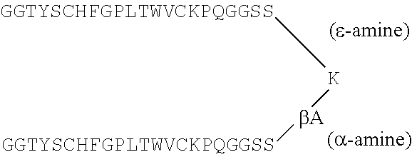
The term "peptibody" refers to a molecule comprising peptide(s) fused either directly or indirectly to other molecules such as an Fc domain of an antibody, where the peptide moiety specifically binds to a desired target. The peptide(s) may be fused to either an Fc region or inserted into an Fc-Loop, a modified Fc molecule. Fc-Loops are described in U.S. Patent Application Publication No. US2006/0140934 incorporated herein by reference in its entirety. The invention includes such molecules comprising an Fc domain modified to comprise a peptide as an internal sequence (preferably in a loop region) of the Fc domain. The Fc internal peptide molecules may include more than one peptide sequence in tandem in a particular internal region, and they may include further peptides in other internal regions. While the putative loop regions are exemplified, insertions in any other non-terminal domains of the Fc are also considered part of this invention. The term "peptibody" does not include Fc-fusion proteins (e.g., full length proteins fused to an Fc domain).
The term "vehicle" refers to a molecule that prevents degradation and/or increases half-life, reduces toxicity, reduces immunogenicity, or increases biological activity of a therapeutic protein. Exemplary vehicles include an Fc domain as described in U.S. Pat. No. 5,428,130 to Capon et al., issued Jun. 27, 1995.
The term "native Fc" refers to molecule or sequence comprising the sequence of a non-antigen-binding fragment resulting from digestion of whole antibody, whether in monomeric or multimeric form. Typically, a native Fc comprises a CH2 and CH3 domain. The original immunoglobulin source of the native Fc is in one aspect of human origin and may be any of the immunoglobulins. A native Fc is a monomeric polypeptide that may be linked into dimeric or multimeric forms by covalent association (i.e., disulfide bonds), non-covalent association or a combination of both. The number of intermolecular disulfide bonds between monomeric subunits of native Fc molecules ranges from one to four depending on class (e.g., IgG, IgA, IgE) or subclass (e.g., IgG1, IgG2, IgG3, IgA1, IgGA2). One example of a native Fc is a disulfide-bonded dimer resulting from papain digestion of an IgG. Ellison et al. (1982), Nucleic Acids Res. 10: 4071-9. The term "native Fc" as used herein is generic to the monomeric, dimeric, and multimeric forms.
The term "Fc variant" refers to a molecule or sequence that is modified from a native Fc, but still comprises a binding site for the salvage receptor, FcRn. International applications WO 97/34631 (published 25 Sep. 1997) and WO 96/32478 describe exemplary Fc variants, as well as interaction with the salvage receptor, and are hereby incorporated by reference. In one aspect, the term "Fc variant" comprises a molecule or sequence that is humanized from a non-human native Fc. In another aspect, a native Fc comprises sites that may be removed because they provide structural features or biological activity that are not required for the fusion molecules of the present invention. Thus, the term "Fc variant" comprises a molecule or sequence that lacks one or more native Fc sites or residues that affect or are involved in (1) disulfide bond formation, (2) incompatibility with a selected host cell (3) N-terminal heterogeneity upon expression in a selected host cell, (4) glycosylation, (5) interaction with complement, (6) binding to an Fc receptor other than a salvage receptor, or (7) antibody-dependent cellular cytotoxicity (ADCC). Fc variants are described in further detail hereinafter.
The term "Fc domain" encompasses native Fc and Fc variant molecules and sequences as defined above. As with Fc variants and native Fcs, the term "Fc domain" includes molecules in monomeric or multimeric form, whether digested from whole antibody or produced by other means. In one embodiment, for example, the Fc region can be:
TABLE-US-00003 (SEQ ID NO: 1) DKTHTCPPCPAPELLGGPSVFLFPPKPKDTLMISRTPEVTCVVVDVSH EDPEVKFNWYVDGVEVHNAKTKPREEQYNSTYRVVSVLTVLHQDWLN GKEYKCKVSNKALPAPIEKTISKAKGQPREPQVYTLPPSRDELTKNQV SLTCLVKGFYPSDIAVEWESNGQPENNYKTTPPVLDSDGSFFLYSKLT VDKSRWQQGNVFSCSVMHEALHNHYTQKSLSLSPGK.
Additional Fc sequences are known in the art and are contemplated for use in the invention. For example, Fc IgG1 (GenBank Accession No. P01857), Fc IgG2 (GenBank Accession No. P01859), Fc IgG3 (GenBank Accession No. P01860), Fc IgG4 (GenBank Accession No. P01861), Fc IgA1 (GenBank Accession No. P01876), Fc IgA2 (GenBank Accession No. P01877), Fc IgD (GenBank Accession No. P01880), Fc IgM (GenBank Accession No. P01871), and Fc IgE (GenBank Accession No. P01854) are some additional Fc sequences contemplated for use herein.
Optionally, an N-terminal amino acid sequence may be added to the above sequences (e.g., where expressed in bacteria).
The term "multimer" as applied to Fc domains or molecules comprising Fc domains refers to molecules having two or more polypeptide chains associated covalently, noncovalently, or by both covalent and non-covalent interactions. IgG molecules typically form dimers; IgM, pentamers; IgD, dimers; and IgA, monomers, dimers, trimers, or tetramers. Multimers may be formed by exploiting the sequence and resulting activity of the native Ig source of the Fc or by derivatizing (as defined below) such a native Fc.
The terms "derivatizing," "derivative" or "derivatized" mean processes and resulting compounds in which, for example and without limitation, (1) the compound has a cyclic portion; for example, cross-linking between cysteinyl residues within the compound; (2) the compound is cross-linked or has a cross-linking site; for example, the compound has a cysteinyl residue and thus forms cross-linked dimers in culture or in vivo; (3) one or more peptidyl linkage is replaced by a non-peptidyl linkage; (4) the N-terminus is replaced by --NRR.sub.1, NRC(O)R.sub.1, --NRC(O)OR.sub.1, --NRS(O).sub.2R.sub.1, --NHC(O)NHR, a succinimide group, or substituted or unsubstituted benzyloxycarbonyl-NH--, wherein R and R.sub.1 and the ring substituents are as defined hereinafter; (5) the C-terminus is replaced by --C(O)R.sub.2 or --NR.sub.3R.sub.4 wherein R.sub.2, R.sub.3 and R.sub.4 are as defined hereinafter; and (6) compounds in which individual amino acid moieties are modified through treatment with agents capable of reacting with selected side chains or terminal residues. Derivatives are further described hereinafter.
As used herein the term "peptide" refers to molecules of 2, 3, 4, 5, 6, 7, 8, 9, 10, 11, 12, 13, 14, 15, 16, 17, 18, 19, 20, 21, 22, 23, 24, 25, 26, 27, 28, 29, 30, 31, 32, 33, 34, 35, 36, 37, 38, 39, 40, 41, 42, 43, 44, 45, 46, 47, 48, 49, 50, 51, 52, 53, 54, 55, 56, 57, 58, 59, 60, 61, 62, 63, 64, 65, 66, 67, 68, 69, 70, 71, 72, 73, 74, 75, 76, 77, 78, 79, 80, 81, 82, 83, 84, 85, 86, 87, 88, 89, 90, 91, 92, 93, 94, 95, 96, 97, 98, 99, 100 or more amino acids linked by peptide bonds. Peptides typically contain random and/or flexible conformations, such as random coils; and typically lack stable conformations, such as those observed in larger proteins/polypeptides, typically via secondary and tertiary structures. In particular embodiments, numerous size ranges of peptides are contemplated herein, such from about: 3-90, 3-80, 3-70, 3-60, 3-50; 5-90, 5-80, 5-70, 5-60, 5-50, 5-40, 5-30; 10-90, 10-80, 10-70, 10-60, 10-50, 10-40, 10-30; 10-20 amino acids in length, and the like. In further embodiments, the peptides used herein are no more than 100, 90, 80, 70, 60, 50, 40, 30, or 20 amino acids in length. Exemplary peptides may be generated by any of the methods set forth herein, such as carried in a peptide library (e.g., a phage display library), generated by chemical synthesis, derived by digestion of proteins, or generated using recombinant DNA techniques. Peptides include D and L form, either purified or in a mixture of the two forms.
Additionally, physiologically acceptable salts of the compounds of this invention are also contemplated. By "physiologically acceptable salts" is meant any salts that are known or later discovered to be pharmaceutically acceptable. Some specific examples are: acetate; trifluoroacetate; hydrohalides, such as hydrochloride and hydrobromide; sulfate; citrate; tartrate; glycolate; and oxalate.
The term "randomized" as used to refer to peptide sequences refers to fully random sequences (e.g., selected by phage display methods) and sequences in which one or more residues of a naturally occurring molecule is replaced by an amino acid residue not appearing in that position in the naturally occurring molecule. Exemplary methods for identifying peptide sequences include phage display, E. coli display, ribosome display, yeast-based screening, RNA-peptide screening, chemical screening, rational design, protein structural analysis, and the like.
The term "pharmacologically active" means that a substance so described is determined to have activity that affects a medical parameter (e.g., but not limited to blood pressure, blood cell count, cholesterol level) or disease state (e.g., but not limited to cancer, autoimmune disorders). Thus, pharmacologically active peptides comprise agonistic or mimetic and antagonistic peptides as defined below.
The terms "-mimetic peptide" and "-agonist peptide" refer to a peptide having biological activity comparable to a protein (e.g., but not limited to EPO, TPO, G-CSF and other proteins described herein) that interacts with a protein of interest. These terms further include peptides that indirectly mimic the activity of a protein of interest, such as by potentiating the effects of the natural ligand of the protein of interest; see, for example, the G-CSF-mimetic peptides listed in Tables 2 and 7. As an example, the term "EPO-mimetic peptide" comprises any peptides that can be identified or derived as described in Wrighton et al. (1996), Science 273: 458-63, Naranda et al. (1999), Proc. Natl. Acad. Sci. USA 96: 7569-74, or any other reference in Table 2 identified as having EPO-mimetic subject matter. Those of ordinary skill in the art appreciate that each of these references enables one to select different peptides than actually disclosed therein by following the disclosed procedures with different peptide libraries.
As another example, the term "TPO-mimetic peptide" or "TMP" refers to peptides that can be identified or derived as described in Cwirla et al. (1997), Science 276: 1696-9, U.S. Pat. Nos. 5,869,451 and 5,932,946 and any other reference in Table 2 identified as having TPO-mimetic subject matter, as well as International application WO 00/24770 published May 4, 2000, hereby incorporated by reference. Those of ordinary skill in the art appreciate that each of these references enables one to select different peptides than actually disclosed therein by following the disclosed procedures with different peptide libraries.
As another example, the term "G-CSF-mimetic peptide" refers to any peptides that can be identified or described in Paukovits et al. (1984), Hoppe-Seylers Z. Physiol. Chem. 365: 303-11 or any of the references in Table 2 identified as having G-CSF-mimetic subject matter. Those of ordinary skill in the art appreciate that each of these references enables one to select different peptides than actually disclosed therein by following the disclosed procedures with different peptide libraries.
The term "CTLA4-mimetic peptide" refers to any peptides that can be identified or derived as described in Fukumoto et al. (1998), Nature Biotech. 16: 267-70. Those of ordinary skill in the art appreciate that each of these references enables one to select different peptides than actually disclosed therein by following the disclosed procedures with different peptide libraries.
The term "-antagonist peptide" or "inhibitor peptide" refers to a peptide that blocks or in some way interferes with the biological activity of the associated protein of interest, or has biological activity comparable to a known antagonist or inhibitor of the associated protein of interest. Thus, the term "TNF-antagonist peptide" comprises peptides that can be identified or derived as described in Takasaki et al. (1997), Nature Biotech. 15: 1266-70 or any of the references in Table 2 identified as having TNF-antagonistic subject matter. Those of ordinary skill in the art appreciate that each of these references enables one to select different peptides than actually disclosed therein by following the disclosed procedures with different peptide libraries.
The terms "IL-1 antagonist" and "IL-1ra-mimetic peptide" refers to peptides that inhibit or down-regulate activation of the IL-1 receptor by IL-1. IL-1 receptor activation results from formation of a complex among IL-1, IL-1 receptor, and IL-1 receptor accessory protein. IL-1 antagonist or IL-1ra-mimetic peptides bind to IL-1, IL-1 receptor, or IL-1 receptor accessory protein and obstruct complex formation among any two or three components of the complex. Exemplary IL-1 antagonist or IL-1ra-mimetic peptides can be identified or derived as described in U.S. Pat. Nos. 5,608,035; 5,786,331, 5,880,096; or any of the references in Table 2 identified as having IL-1ra-mimetic or IL-1 antagonistic subject matter. Those of ordinary skill in the art appreciate that each of these references enables one to select different peptides than actually disclosed therein by following the disclosed procedures with different peptide libraries.
The term "VEGF-antagonist peptide" refers to peptides that can be identified or derived as described in Fairbrother (1998), Biochem. 37: 17754-64, and in any of the references in Table 2 identified as having VEGF-antagonistic subject matter. Those of ordinary skill in the art appreciate that each of these references enables one to select different peptides than actually disclosed therein by following the disclosed procedures with different peptide libraries.
The term "MMP inhibitor peptide" refers to peptides that can be identified or derived as described in Koivunen (1999), Nature Biotech. 17: 768-74 and in any of the references in Table 2 identified as having MMP inhibitory subject matter. Those of ordinary skill in the art appreciate that each of these references enables one to select different peptides than actually disclosed therein by following the disclosed procedures with different peptide libraries.
The term "myostatin inhibitor peptide" refers to peptides that can be identified by their ability to reduce or block myostatin activity or signaling as demonstrated in in vitro assays such as, for example the pMARE C2C12 cell-based myostatin activity assay or by in vivo animal testing as described in U.S. patent application Publication No US20040181033A1 and PCT application publication No. WO2004/058988. Exemplary myostatin inhibitor peptides are set out in Tables 21-24.
The term "integrin/adhesion antagonist" refers to peptides that inhibit or down-regulate the activity of integrins, selectins, cell adhesion molecules, integrin receptors, selectin receptors, or cell adhesion molecule receptors. Exemplary integrin/adhesion antagonists comprise laminin, echistatin, the peptides described in Tables 25-28.
The term "bone resorption inhibitor" refers to such molecules as determined by the assays of Examples 4 and 11 of WO 97/23614: which is hereby incorporated by reference in its entirety. Exemplary bone resorption inhibitors include OPG and OPG-L antibody, which are described in WO 97/23614 and WO98/46751, respectively, which are hereby incorporated by reference in their entirety.
The term "nerve growth factor inhibitor" or "nerve growth factor agonist" refers to a peptide that binds to and inhibits nerve growth factor (NGF) activity or signaling. Exemplary peptides of this type are set out in Table 29.
The term "TALL-1 modulating domain" refers to any amino acid sequence that binds to the TALL-1 and comprises naturally occurring sequences or randomized sequences. Exemplary TALL-1 modulating domains can be identified or derived by phage display or other methods mentioned herein. Exemplary peptides of this type are set out in Tables 30 and 31.
The term "TALL-1 antagonist" refers to a molecule that binds to the TALL-1 and increases or decreases one or more assay parameters opposite from the effect on those parameters by full length native TALL-1. Such activity can be determined, for example, by such assays as described in the subsection entitled "Biological activity of AGP-3" in the Materials & Methods section of the patent application entitled, "TNF-RELATED PROTEINS", WO 00/47740, published Aug. 17, 2000.
The term "Ang 2-antagonist peptide" refers to peptides that can be identified or derived as having Ang-2-antagonistic characteristics. Exemplary peptides of this type are set out in Tables 32-38.
The term "WSP" refers to a water soluble polymer which prevents a peptide, protein or other compound to which it is attached from precipitating in an aqueous environment, such as, by way of example, a physiological environment. A more detailed description of various WSP embodiments contemplated by the invention follows.
Lyophilization and Administration
Therapeutic peptibodies are useful in pharmaceutical formulations in order to treat human diseases as described herein. In one embodiment the therapeutic peptibody compositions are lyophilized. Lyophilization is carried out using techniques common in the art and should be optimized for the composition being developed [Tang et al., Pharm Res. 21:191-200, (2004) and Chang et al., Pharm Res. 13:243-9 (1996)].
A lyophilization cycle is, in one aspect, composed of three steps: freezing, primary drying, and secondary drying [A. P. Mackenzie, Phil Trans R Soc London, Ser B, Biol 278:167 (1977)]. In the freezing step, the solution is cooled to initiate ice formation. Furthermore, this step induces the crystallization of the bulking agent. The ice sublimes in the primary drying stage, which is conducted by reducing chamber pressure below the vapor pressure of the ice, using a vacuum and introducing heat to promote sublimation. Finally, adsorbed or bound water is removed at the secondary drying stage under reduced chamber pressure and at an elevated shelf temperature. The process produces a material known as a lyophilized cake. Thereafter the cake can be reconstituted with either sterile water or suitable diluent for injection.
The lyophilization cycle not only determines the final physical state of the excipients but also affects other parameters such as reconstitution time, appearance, stability and final moisture content. The composition structure in the frozen state proceeds through several transitions (e.g., glass transitions, wettings, and crystallizations) that occur at specific temperatures and can be used to understand and optimize the lyophilization process. The glass transition temperature (Tg and/or Tg') can provide information about the physical state of a solute and can be determined by differential scanning calorimetry (DSC). Tg and Tg' are an important parameter that must be taken into account when designing the lyophilization cycle. For example, Tg' is important for primary drying. Furthermore, in the dried state, the glass transition temperature provides information on the storage temperature of the final product.
Excipients in General
Excipients are additives that are included in a formulation because they either impart or enhance the stability, delivery and manufacturability of a drug product. Regardless of the reason for their inclusion, excipients are an integral component of a drug product and therefore need to be safe and well tolerated by patients. For protein drugs, the choice of excipients is particularly important because they can affect both efficacy and immunogenicity of the drug. Hence, protein formulations need to be developed with appropriate selection of excipients that afford suitable stability, safety, and marketability.
A lyophilized formulation is usually comprised of a buffer, a bulking agent, and a stabilizer. The utility of a surfactant may be evaluated and selected in cases where aggregation during the lyophilization step or during reconstitution becomes an issue. An appropriate buffering agent is included to maintain the formulation within stable zones of pH during lyophilization. A comparison of the excipient components in liquid and lyophilized protein formulations is provided in Table A.
TABLE-US-00004 TABLE A Excipient components of lyophilized protein formulations Function in lyophilized Excipient Component formulation Buffer Maintain pH of formulation during lyophilization and upon reconstitution Tonicity agent/ stabilizer Stabilizers include cryo and lyoprotectants Examples include Polyols, sugars and polymers Cryoprotectants protect proteins from freezing stresses Lyoprotectants stabilize proteins in the freeze-dried state Bulking agent Used to enhance product elegance and to prevent blowout Provides structural strength to the lyo cake Examples include mannitol and glycine Surfactant Employed if aggregation during the lyophilization process is an issue May serve to reduce reconstitution times Examples include polysorbate 20 and 80 Anti-oxidant Usually not employed, molecular reactions in the lyo cake are greatly retarded Metal ions/chelating agent May be included if a specific metal ion is included only as a co- factor or where the metal is required for protease activity Chelating agents are generally not needed in lyo formulations Preservative For multi-dose formulations only Provides protection against microbial growth in formulation Is usually included in the reconstitution diluent (e.g. bWFI)
The principal challenge in developing formulations for therapeutic proteins is stabilizing the product against the stresses of manufacturing, shipping and storage. The role of formulation excipients is to provide stabilization against these stresses. Excipients may also be employed to reduce viscosity of high concentration protein formulations in order to enable their delivery and enhance patient convenience. In general, excipients can be classified on the basis of the mechanisms by which they stabilize proteins against various chemical and physical stresses. Some excipients are used to alleviate the effects of a specific stress or to regulate a particular susceptibility of a specific protein. Other excipients have more general effects on the physical and covalent stabilities of proteins. The excipients described herein are organized either by their chemical type or their functional role in formulations. Brief descriptions of the modes of stabilization are provided when discussing each excipient type.
Given the teachings and guidance provided herein, those skilled in the art will know what amount or range of excipient can be included in any particular formulation to achieve a biopharmaceutical formulation of the invention that promotes retention in stability of the biopharmaceutical. For example, the amount and type of a salt to be included in a biopharmaceutical formulation of the invention can be selected based on to the desired osmolality (i.e., isotonic, hypotonic or hypertonic) of the final solution as well as the amounts and osmolality of other components to be included in the formulation. Similarly, by exemplification with reference to the type of polyol or sugar included in a formulation, the amount of such an excipient will depend on its osmolality.
By way of example, inclusion of about 5% sorbitol can achieve isotonicity while about 9% of a sucrose excipient is needed to achieve isotonicity. Selection of the amount or range of concentrations of one or more excipients that can be included within a biopharmaceutical formulation of the invention has been exemplified above by reference to salts, polyols and sugars. However, those skilled in the art will understand that the considerations described herein and further exemplified by reference to specific excipients are equally applicable to all types and combinations of excipients including, for example, salts, amino acids, other tonicity agents, surfactants, stabilizers, bulking agents, cryoprotectants, lyoprotectants, anti-oxidants, metal ions, chelating agents and/or preservatives.
Further, where a particular excipient is reported in a formulation by, e.g., percent (%) w/v, those skilled in the art will recognize that the equivalent molar concentration of that excipient is also contemplated.
Of course, a person having ordinary skill in the art would recognize that the concentrations of the aforementioned excipients share an interdependency within a particular formulation. By way of example, the concentration of a bulking agent may be lowered where, e.g., there is a high protein/peptide concentration or where, e.g., there is a high stabilizing agent concentration. In addition, a person having ordinary skill in the art would recognize that, in order to maintain the isotonicity of a particular formulation in which there is no bulking agent, the concentration of a stabilizing agent would be adjusted accordingly (i.e., a "tonicifying" amount of stabilizer would be used). Other excipients are known in the art and can be found in Powell et al., Compendium of Excipients fir Parenteral Formulations (1998), PDA J. Pharm. Sci. Technology, 52:238-311.
Buffers
The stability of a protein drug is usually observed to be maximal in a narrow pH range. This pH range of optimal stability needs to be identified early during pre-formulation studies. Several approaches such as accelerated stability studies and calorimetric screening studies have been demonstrated to be useful in this endeavor (Remmele R. L. Jr., et al., Biochemistry, 38(16): 5241-7 (1999)). Once a formulation is finalized, the drug product must be manufactured and maintained within a predefined specification throughout its shelf-life. Hence, buffering agents are almost always employed to control pH in the formulation.
Organic acids, phosphates and Tris have been employed routinely as buffers in protein formulations (Table B). The buffer capacity of the buffering species is maximal at a pH equal to the pKa and decreases as pH increases or decreases away from this value. Ninety percent of the buffering capacity exists within one pH unit of its pKa. Buffer capacity also increases proportionally with increasing buffer concentration.
Several factors need to be considered when choosing a buffer. First and foremost, the buffer species and its concentration need to be defined based on its pKa and the desired formulation pH. Equally important is to ensure that the buffer is compatible with the protein drug, other formulation excipients, and does not catalyze any degradation reactions. Recently, polyanionic carboxylate buffers such as citrate and succinate have been shown to form covalent adducts with the side chain residues of proteins. A third important aspect to be considered is the sensation of stinging and irritation the buffer may induce. For example, citrate is known to cause stinging upon injection (Laursen T, et al., Basic Clin Pharmacol Toxicol., 98(2): 218-21 (2006)). The potential for stinging and irritation is greater for drugs that are administered via the SC or IM routes, where the drug solution remains at the site for a relatively longer period of time than when administered by the IV route where the formulation gets diluted rapidly into the blood upon administration. For formulations that are administered by direct IV infusion, the total amount of buffer (and any other formulation component) needs to be monitored. One has to be particularly careful about potassium ions administered in the form of the potassium phosphate buffer, which can induce cardiovascular effects in a patient (Hollander-Rodriguez J C, et al., Am. Fam. Physician., 73(2): 283-90 (2006)).
Buffers for lyophilized formulations need additional consideration. Some buffers like sodium phosphate can crystallize out of the protein amorphous phase during freezing resulting in rather large shifts in pH. Other common buffers such as acetate and imidazole should be avoided since they may sublime or evaporate during the lyophilization process, thereby shifting the pH of formulation during lyophilization or after reconstitution.
TABLE-US-00005 TABLE B Commonly used buffering agents and their pK.sub.a values Buffer pK.sub.a Example drug product Acetate 4.8 Neupogen, Neulasta Succinate pK.sub.a1 = 4.8, pK.sub.a2 = 5.5 Actimmune Citrate pK.sub.a1 = 3.1, pK.sub.a2 = 4.8, Humira pK.sub.a3 = 6.4 Histidine 6.0 Xolair (imidazole) phosphate pK.sub.a1 = 2.15, pK.sub.a2 = 7.2, Enbrel (liquid formulation) pK.sub.a3 =12.3 Tris 8.1 Leukine
The buffer system present in the compositions is selected to be physiologically compatible and to maintain a desired pH in the reconstituted solution as well as in the solution before lyophilization. In one embodiment, the pH of the solution prior to lyophilization is between pH 2.0 and pH 12.0. For example, in one embodiment the pH of the solution prior to lyophilization is 2.0, 2.3., 2.5., 2.7, 3.0, 3.3, 3.5, 3.7, 4.0, 4.3, 4.5, 4.7, 5.0, 5.3, 5.5, 5.7, 6.0, 6.3, 6.5, 6.7, 7.0, 7.3, 7.5, 7.7, 8.0, 8.3, 8.5, 8.7, 9.0, 9.3, 9.5, 9.7, 10.0, 10.3, 10.5, 10.7, 11.0, 11.3, 11.5, 11.7, or 12.0. In another embodiment, the pH of the reconstituted solution is between 4.5 and 9.0. In one embodiment, the pH in the reconstituted solution is 4.5, 4.7, 5.0, 5.3, 5.5, 5.7, 6.0, 6.3, 6.5, 6.7, 7.0, 7.3, 7.5, 7.7, 8.0, 8.3, 8.5, 8.7, or 9.0.
In one embodiment, the pH buffering agent used in the formulation is an amino acid or mixture of amino acids. In one aspect, the pH buffering agent is histidine or a mixture of amino acids one of which is histidine.
The pH buffering compound may be present in any amount suitable to maintain the pH of the formulation at a predetermined level. In one embodiment, when the pH buffering agent is an amino acid, the concentration of the amino acid is between 0.1 mM and 1000 mM (1 M). In one embodiment, the pH buffering agent is at least 0.1, 0.5, 0.7, 0.8 0.9, 1.0, 1.2, 1.5, 1.7, 2, 3, 4, 5, 6, 7, 8, 9, 10, 11, 12, 13, 14, 15, 16, 17, 18, 19, 20, 30, 40, 50, 60, 70, 80, 90, 100, 200, 500, 700, or 900 mM. In another embodiment, the concentration of the pH buffering agent is between 1, 1.2, 1.5, 1.7, 2, 3, 4, 5, 6, 7, 8, 9, 10, 11, 12, 13, 14, 15, 16, 17, 18, 19, 20, 30, 40, 50, 60, 70, 80, or 90 mM and 100 mM. In still another embodiment, the concentration of the pH buffering agent is between 5, 6, 7, 8, 9, 10, 11, 12, 13, 14, 15, 16, 17, 18, 19, 20, 30, or 40 mM and 50 mM. In yet another embodiment, the concentration of the pH buffering agent is 10 mM.
Other exemplary pH buffering agents used to buffer the formulation as set out herein include, but are not limited to glycine, histidine, glutamate, succinate, phosphate, acetate, and aspartate.
Stabilizers and Bulking Agents
Bulking agents are typically used in lyophilized formulations to enhance product elegance and to prevent blowout. Conditions in the formulation are generally designed so that the bulking agent crystallizes out of the frozen amorphous phase (either during freezing or annealing above the Tg') giving the cake structure and bulk. Mannitol and glycine are examples of commonly used bulking agents.
Stabilizers include a class of compounds that can serve as cryoprotectants, lyoprotectants, and glass forming agents. Cryoprotectants act to stabilize proteins during freezing or in the frozen state at low temperatures (P. Cameron, ed., Good Pharmaceutical Freeze-Drying Practice, Interpharm Press, Inc., Buffalo Grove, Ill., (1997)). Lyoprotectants stabilize proteins in the freeze-dried solid dosage form by preserving the native-like conformational properties of the protein during dehydration stages of freeze-drying. Glassy state properties have been classified as "strong" or "fragile" depending on their relaxation properties as a function of temperature. It is important that cryoprotectants, lyoprotectants, and glass forming agents remain in the same phase with the protein in order to impart stability. Sugars, polymers, and polyols fall into this category and can sometimes serve all three roles.
Polyols encompass a class of excipients that includes sugars, (e.g. mannitol, sucrose, sorbitol), and other polyhydric alcohols (e.g., glycerol and propylene glycol). The polymer polyethylene glycol (PEG) is included in this category. Polyols are commonly used as stabilizing excipients and/or isotonicity agents in both liquid and lyophilized parenteral protein formulations. With respect to the Hofmeister series, the polyols are kosmotropic and are preferentially excluded from the protein surface. Polyols can protect proteins from both physical and chemical degradation pathways. Preferentially excluded co-solvents increase the effective surface tension of solvent at the protein interface whereby the most energetically favorable protein conformations are those with the smallest surface areas.
Mannitol is a popular bulking agent in lyophilized formulations because it crystallizes out of the amorphous protein phase during freeze-drying lending structural stability to the cake (e.g. Leukine.RTM., Enbrel.RTM.-Lyo, Betaseron.RTM.). It is generally used in combination with a cryo and/or lyoprotectant like sucrose. Because of the propensity of mannitol to crystallize under frozen conditions, sorbitol and sucrose are the preferred tonicity agents/stabilizers in liquid formulations to protect the product against freeze-thaw stresses encountered during transport or when freezing bulk prior to manufacturing. Sorbitol and sucrose are far more resistant to crystallization and therefore less likely to phase separate from the protein. It is interesting to note that while mannitol has been used in tonicifying amounts in several marketed liquid formulations such as Actimmune.RTM., Forteo.RTM., and Rebif.RTM., the product labels of these drugs carry a `Do Not Freeze` warning. The use of reducing sugars (containing free aldehyde or ketone groups) such as glucose and lactose should be avoided because they can react and glycate surface lysine and arginine residues of proteins via the Maillard reaction of aldehydes and primary amines (Chevalier F, et al., Nahrung, 46(2): 58-63 (2002); Humeny A, et al., J Agric Food Chem. 50(7): 2153-60 (2002)). Sucrose can hydrolyze to fructose and glucose under acidic conditions (Kautz C. F. and Robinson A. L., JACS, 50(4) 1022-30 (1928)), and consequently may cause glycation.
The polymer polyethylene glycol (PEG) could stabilize proteins by two different temperature dependent mechanisms. At lower temperatures, it is preferentially excluded from the protein surface but has been shown to interact with the unfolded form of the protein at higher temperature given its amphipathic nature (Lee L. L., and Lee J. C., Biochemistry, 26(24): 7813-9 (1987)). Thus at lower temperatures it may protect proteins via the mechanism of preferential exclusion, but at higher temperatures possibly by reducing the number of productive collisions between unfolded molecules. PEG is also a cryoprotectant and has been employed in Recombinate.RTM., a lyophilized formulation of recombinant Antihemophilic Factor, which utilizes PEG 3350 at a concentration of 1.5 mg/mL. The low molecular weight liquid PEGs (PEG 300-600) can be contaminated with peroxides and cause protein oxidation. If used, the peroxide content in the raw material must be minimized and controlled throughout its shelf-life. The same holds true for polysorbates.
In a particular embodiment of the present compositions, a stabilizer (or a combination of stabilizers) is added to the lyophilization formulation to prevent or reduce lyophilization-induced or storage-induced aggregation and chemical degradation. A hazy or turbid solution upon reconstitution indicates that the protein has precipitated. The term "stabilizer" means an excipient capable of preventing aggregation or other physical degradation, as well as chemical degradation (for example, autolysis, deamidation, oxidation, etc.) in an aqueous and solid state. Stabilizers that are conventionally employed in pharmaceutical compositions include, but are not limited to, sucrose, trehalose, mannose, maltose, lactose, glucose, raffinose, cellobiose, gentiobiose, isomaltose, arabinose, glucosamine, fructose, mannitol, sorbitol, glycine, arginine HCL, poly-hydroxy compounds, including polysaccharides such as dextran, starch, hydroxyethyl starch, cyclodextrins, N-methyl pyrollidene, cellulose and hyaluronic acid, sodium chloride, [Carpenter et al., Develop. Biol. Standard 74:225, (1991)]. In one embodiment, the stabilizer is incorporated in a concentration of about 0% to about 40% w/v. In another embodiment, the stabilizer is incorporated in a concentration of at least 0.5, 1, 2, 3, 4, 5, 6, 7, 8, 9, 10, 11, 12, 13, 14, 15, 16, 17, 18, 19, 20, 30, or 40% w/v. In another embodiment, the stabilizer is incorporated in a concentration of about 1, 2, 3, 4, 5, 6, 7, 8, 9% to about 10% w/v. In still another embodiment, the stabilizer is incorporated in a concentration of about 2% to about 4% w/v. In yet another embodiment, the stabilizer is incorporated in a concentration of about 2% w/v.
If desired, the lyophilized compositions also include appropriate amounts of bulking and osmolarity regulating agents suitable for forming a lyophilized "cake". Bulking agents may be either crystalline (for example, mannitol, glycine) or amorphous (for example, sucrose, polymers such as dextran, polyvinylpyrolidone, carboxymethylcellulose). Other exemplary bulking agents include lactose, sorbitol, trehalose, or xylitol. In one embodiment, the bulking agent is mannitol. In a further embodiment, the bulking agent is incorporated in a concentration of about 0% to about 10% w/v. In another embodiment, the bulking agent is incorporated in a concentration of at least 0.2, 0.5, 0.7, 1.0, 1.5, 2.0, 2.5, 3.0, 3.5, 4.0, 4.5, 5.0, 5.5, 6.0, 6.5, 7.0, 7.5, 8.0, 8.5, 9.0, or 9.5% w/v. In a yet further embodiment in a concentration of about 1, 1.5, 2.0, 2.5, 3.0, 3.5, 4.0, 4.5% to 5.0% w/v, to produce a mechanically and pharmaceutically stable and elegant cake. In another embodiment, the mannitol concentration is 4% w/v.
Surfactants
Protein molecules have a high propensity to interact with surfaces making them susceptible to adsorption and denaturation at air-liquid, vial-liquid, and liquid-liquid (silicone oil) interfaces. This degradation pathway has been observed to be inversely dependent on protein concentration and result in either the formation of soluble and insoluble protein aggregates or the loss of protein from solution via adsorption to surfaces. In addition to container surface adsorption, surface-induced degradation is exacerbated with physical agitation, as would be experienced during shipping and handling of the product.
Surfactants are commonly used in protein formulations to prevent surface-induced degradation. Surfactants are amphipathic molecules with the capability of out-competing proteins for interfacial positions. Hydrophobic portions of the surfactant molecules occupy interfacial positions (e.g., air/liquid), while hydrophilic portions of the molecules remain oriented towards the bulk solvent. At sufficient concentrations (typically around the detergent's critical micellar concentration), a surface layer of surfactant molecules serve to prevent protein molecules from adsorbing at the interface. Thereby, surface-induced degradation is minimized. The most commonly used surfactants are fatty acid esters of sorbitan polyethoxylates, i.e. polysorbate 20 and polysorbate 80 (e.g., Avonex.RTM., Neupogen.RTM., Neulasta.RTM.). The two differ only in the length of the aliphatic chain that imparts hydrophobic character to the molecules, C-12 and C-18, respectively. Accordingly, polysorbate-80 is more surface-active and has a lower critical micellar concentration than polysorbate-20. The surfactant poloxamer 188 has also been used in several marketed liquid products such Gonal-F.RTM., Norditropin.RTM., and Ovidrel.RTM..
Detergents can also affect the thermodynamic conformational stability of proteins. Here again, the effects of a given excipient will be protein specific. For example, polysorbates have been shown to reduce the stability of some proteins and increase the stability of others. Detergent destabilization of proteins can be rationalized in terms of the hydrophobic tails of the detergent molecules that can engage in specific binding with partially or wholly unfolded protein states. These types of interactions could cause a shift in the conformational equilibrium towards the more expanded protein states (i.e. increasing the exposure of hydrophobic portions of the protein molecule in complement to binding polysorbate). Alternatively, if the protein native state exhibits some hydrophobic surfaces, detergent binding to the native state may stabilize that conformation.
Another aspect of polysorbates is that they are inherently susceptible to oxidative degradation. Often, as raw materials, they contain sufficient quantities of peroxides to cause oxidation of protein residue side-chains, especially methionine. The potential for oxidative damage arising from the addition of stabilizer emphasizes the point that the lowest effective concentrations of excipients should be used in formulations. For surfactants, the effective concentration for a given protein will depend on the mechanism of stabilization. It has been postulated that if the mechanism of surfactant stabilization is related to preventing surface-denaturation the effective concentration will be around the detergent's critical micellar concentration. Conversely, if the mechanism of stabilization is associated with specific protein-detergent interactions, the effective surfactant concentration will be related to the protein concentration and the stoichiometry of the interaction (Randolph T. W., et al., Pharm Biotechnol., 13:159-75 (2002)).
Surfactants may also be added in appropriate amounts to prevent surface related aggregation phenomenon during freezing and drying [Chang, B, J. Pharm. Sci. 85:1325, (1996)]. Exemplary surfactants include anionic, cationic, nonionic, zwitterionic, and amphoteric surfactants including surfactants derived from naturally-occurring amino acids. Anionic surfactants include, but are not limited to, sodium lauryl sulfate, dioctyl sodium sulfosuccinate and dioctyl sodium sulfonate, chenodeoxycholic acid, N-lauroylsarcosine sodium salt, lithium dodecyl sulfate, 1-octanesulfonic acid sodium salt, sodium cholate hydrate, sodium deoxycholate, and glycodeoxycholic acid sodium salt. Cationic surfactants include, but are not limited to, benzalkonium chloride or benzethonium chloride, cetylpyridinium chloride monohydrate, and hexadecyltrimethylammonium bromide. Zwitterionic surfactants include, but are not limited to, CHAPS, CHAPSO, SB3-10, and SB3-12. Non-ionic surfactants include, but are not limited to, digitonin, Triton X-100, Triton X-114, TWEEN-20, and TWEEN-80. In another embodiment, surfactants include lauromacrogol 400, polyoxyl 40 stearate, polyoxyethylene hydrogenated castor oil 10, 40, 50 and 60, glycerol monostearate, polysorbate 40, 60, 65 and 80, soy lecithin and other phospholipids such as DOPC, DMPG, DMPC, and DOPG; sucrose fatty acid ester, methyl cellulose and carboxymethyl cellulose. Compositions comprising these surfactants, either individually or as a mixture in different ratios, are therefore further provided. In one embodiment, the surfactant is incorporated in a concentration of about 0% to about 5% w/v. In another embodiment, the surfactant is incorporated in a concentration of at least 0.001, 0.002, 0.005, 0.007, 0.01, 0.05, 0.1, 0.2, 0.3, 0.4, 0.5, 0.6, 0.7, 0.7, 0.8, 0.9, 1.0, 1.5, 2.0, 2.5, 3.0, 3.5, 4.0, or 4.5% w/v. In another embodiment, the surfactant is incorporated in a concentration of about 0.001% to about 0.5% w/v. In still another embodiment, the surfactant is incorporated in a concentration of about 0.004, 0.005, 0.007, 0.01, 0.05, or 0.1% w/v to about 0.2% w/v. In yet another embodiment, the surfactant is incorporated in a concentration of about 0.01% to about 0.1% w/v.
Salts
Salts are often added to increase the ionic strength of the formulation, which can be important for protein solubility, physical stability, and isotonicity. Salts can affect the physical stability of proteins in a variety of ways. Ions can stabilize the native state of proteins by binding to charged residues on the protein's surface. Alternatively, they can stabilize the denatured state by binding to the peptide groups along the protein backbone (--CONH--). Salts can also stabilize the protein native conformation by shielding repulsive electrostatic interactions between residues within a protein molecule. Electrolytes in protein formulations can also shield attractive electrostatic interactions between protein molecules that can lead to protein aggregation and insolubility.
The effect of salt on the stability and solubility of proteins varies significantly with the characteristics of the ionic species. The Hofmeister series originated in the 1880's as a way to rank order electrolytes based on their ability to precipitate proteins (Cacace M. G., et al., Quarterly Reviews of Biophysics., 30(3): 241-277 (1997)). In this report, the Hofmeister series is used to illustrate a scale of protein stabilization effects by ionic and non-ionic co-solutes. In FIG. 1, co-solutes are ordered with respect to their general effects on solution state proteins, from stabilizing (kosmotropic) to destabilizing (chaotropic). In general, the differences in effects across the anions are far greater than that observed for the cations, and, for both types, the effects are most apparent at higher concentrations than are acceptable in parenteral formulations. High concentrations of kosmotropes (e.g., >1 molar ammonium sulfate) are commonly used to precipitate proteins from solution by a process called `salting-out` where the kosmotrope is preferentially excluded from the protein surface reducing the solubility of the protein in it's native (folded) conformation. Removal or dilution of the salt will return the protein to solution. The term `salting-in` refers to the use of destabilizing ions (e.g., like guanidine and chloride) that increase the solubility of proteins by solvating the peptide bonds of the protein backbone. Increasing concentrations of the chaotrope will favor the denatured (unfolded) state conformation of the protein as the solubility of the peptide chain increases. The relative effectiveness of ions to `salt-in` and `salt-out` defines their position in the Hofmeister series.
In order to maintain isotonicity in a parenteral formulation, salt concentrations are generally limited to less than 150 mM for monovalent ion combinations. In this concentration range, the mechanism of salt stabilization is probably due to screening of electrostatic repulsive intramolecular forces or attractive intermolecular forces (Debye-Huckel screening). Interestingly, chaotropic salts have been shown to be more effective at stabilizing the protein structure than similar concentrations of kosmotropes by this mechanism. The chaotropic anions are believed to bind more strongly than the kosmotropic ions. With respect to covalent protein degradation, differential effects of ionic strength on this mechanism are expected through Debye-Huckel theory. Accordingly, published reports of protein stabilization by sodium chloride are accompanied by those where sodium chloride accelerated covalent degradation. The mechanisms by which salts affect protein stability are protein specific and may vary significantly as a function of solution pH. An example where an excipient can be useful in enabling the delivery of a protein drug is that of some high concentration antibody formulations. Recently, salts have been shown to be effective in reducing the viscosity of such formulations (Liu J., et al., J. Pharm Sci., 94(9): 1928-40 (2005); Erratum in: J Pharm Sci., 95(1): 234-5. (2006)).
Amino Acids
Amino acids have found versatile use in protein formulations as buffers, bulking agents, stabilizers and antioxidants. Histidine and glutamic acid are employed to buffer protein formulations in the pH range of 5.5-6.5 and 4.0-5.5 respectively. The imidazole group of histidine has a pKa=6.0 and the carboxyl group of glutamic acid side chain has a pKa of 4.3 which makes them suitable for buffering in their respective pH ranges. Acetate, the most commonly used buffer in the acidic pH range of 4.0-5.5, sublimates during lyophilization and hence should not be used in freeze-dried formulations. Glutamic acid is particularly useful in such cases (e.g., Stemgen.RTM.). Histidine is commonly found in marketed protein formulations (e.g., Xolair.RTM., Herceptin.RTM., Recombinate.RTM.). It provides a good alternative to citrate, a buffer known to sting upon injection. Interestingly, histidine has also been reported to have a stabilizing effect on ABX-IL8 (an IgG2 antibody) with respect to aggregation when used at high concentrations in both liquid and lyophilized presentations (Chen B, et al., Pharm Res., 20(12): 1952-60 (2003)). Histidine (up to 60 mM) was also observed to reduce the viscosity of a high concentration formulation of this antibody. However, in the same study, the authors observed increased aggregation and discoloration in histidine containing formulations during freeze-thaw studies of the antibody in stainless steel containers. The authors attributed this to an effect of iron ions leached from corrosion of steel containers. Another note of caution with histidine is that it undergoes photo-oxidation in the presence of metal ions (Tomita M, et al., Biochemistry, 8(12): 5149-60 (1969)). The use of methionine as an antioxidant in formulations appears promising; it has been observed to be effective against a number of oxidative stresses (Lam X M, et al., J Pharm Sci., 86(11): 1250-5 (1997)).
The amino acids glycine, proline, serine and alanine have been shown to stabilize proteins by the mechanism of preferential exclusion. Glycine is also a commonly used bulking agent in lyophilized formulations (e.g., Neumega.RTM., Genotropin.RTM., Humatrope.RTM.). It crystallizes out of the frozen amorphous phase giving the cake structure and bulk. Arginine has been shown to be an effective agent in inhibiting aggregation and has been used in both liquid and lyophilized formulations (e.g., Activase.RTM., Avonex.RTM., Enbrel.RTM. liquid). Furthermore, the enhanced efficiency of refolding of certain proteins in the presence of arginine has been attributed to its suppression of the competing aggregation reaction during refolding.
Antioxidants
Oxidation of protein residues arises from a number of different sources. Beyond the addition of specific antioxidants, the prevention of oxidative protein damage involves the careful control of a number of factors throughout the manufacturing process and storage of the product such as atmospheric oxygen, temperature, light exposure, and chemical contamination. The most commonly used pharmaceutical antioxidants are reducing agents, oxygen/free-radical scavengers, or chelating agents. Antioxidants in therapeutic protein formulations must be water-soluble and remain active throughout the product shelf-life. Reducing agents and oxygen/free-radical scavengers work by ablating active oxygen species in solution. Chelating agents such as EDTA can be effective by binding trace metal contaminants that promote free-radical formation. For example, EDTA was utilized in the liquid formulation of acidic fibroblast growth factor to inhibit the metal ion catalyzed oxidation of cysteine residues. EDTA has been used in marketed products like Kineret.RTM. and Ontak.RTM..
In addition to evaluating the effectiveness of various excipients at preventing protein oxidation, formulation scientists must be aware of the potential for the antioxidants themselves to induce other covalent or physical changes to the protein. A number of such cases have been reported in the literature. Reducing agents (like glutathione) can cause disruption of intramolecular disulfide linkages, which can lead to disulfide shuffling. In the presence of transition metal ions, ascorbic acid and EDTA have been shown to promote methionine oxidation in a number of proteins and peptides (Akers M J, and Defelippis M R. Peptides and Proteins as Parenteral Solutions. In: Pharmaceutical Formulation Development of Peptides and Proteins. Sven Frokjaer, Lars Hovgaard, editors. Pharmaceutical Science. Taylor and Francis, U K (1999)); Fransson J. R., J. Pharm. Sci. 86(9): 4046-1050 (1997); Yin J, et al., Pharm Res., 21(12): 2377-83 (2004)). Sodium thiosulfate has been reported to reduce the levels of light and temperature induced methionine-oxidation in rhuMab HER2; however, the formation of a thiosulfate-protein adduct was also reported in this study (Lam X M, Yang J Y, et al., J Pharm Sci. 86(11): 1250-5 (1997)). Selection of an appropriate antioxidant is made according to the specific stresses and sensitivities of the protein.
Metal Ions
In general, transition metal ions are undesired in protein formulations because they can catalyze physical and chemical degradation reactions in proteins. However, specific metal ions are included in formulations when they are co-factors to proteins and in suspension formulations of proteins where they form coordination complexes (e.g., zinc suspension of insulin). Recently, the use of magnesium ions (10-120 mM) has been proposed to inhibit the isomerization of aspartic acid to isoaspartic acid (WO 2004039337).
Two examples where metal ions confer stability or increased activity in proteins are human deoxyribonuclease (rhDNase, Pulmozyme.RTM.), and Factor VIII. In the case of rhDNase, Ca.sup.+2 ions (up to 100 mM) increased the stability of the enzyme through a specific binding site (Chen B, et al., J Pharm Sci., 88(4): 477-82 (1999)). In fact, removal of calcium ions from the solution with EGTA caused an increase in deamidation and aggregation. However, this effect was observed only with Ca.sup.+2 ions; other divalent cations --Mg.sup.+2, Mn.sup.+2 and Zn.sup.+2 were observed to destabilize rhDNase. Similar effects were observed in Factor VIII. Ca.sup.+2 and Sr.sup.+2 ions stabilized the protein while others like Mg.sup.+2, Mn.sup.+2 and Zn.sup.+2, Cu.sup.+2 and Fe.sup.+2 destabilized the enzyme (Fatouros, A., et al., Int. J. Pharm., 155, 121-131 (1997). In a separate study with Factor VIII, a significant increase in aggregation rate was observed in the presence of Al.sup.+3 ions (Derrick T S, et al., J Pharm. Sci., 93(10): 2549-57 (2004)). The authors note that other excipients like buffer salts are often contaminated with Al.sup.+3 ions and illustrate the need to use excipients of appropriate quality in formulated products.
Preservatives
Preservatives are necessary when developing multi-use parenteral formulations that involve more than one extraction from the same container. Their primary function is to inhibit microbial growth and ensure product sterility throughout the shelf-life or term of use of the drug product. Commonly used preservatives include benzyl alcohol, phenol and m-cresol. Although preservatives have a long history of use, the development of protein formulations that includes preservatives can be challenging. Preservatives almost always have a destabilizing effect (aggregation) on proteins, and this has become a major factor in limiting their use in multi-dose protein formulations (Roy S, et al., J Pharm Sci., 94(2): 382-96 (2005)).
To date, most protein drugs have been formulated for single-use only. However, when multi-dose formulations are possible, they have the added advantage of enabling patient convenience, and increased marketability. A good example is that of human growth hormone (hGH) where the development of preserved formulations has led to commercialization of more convenient, multi-use injection pen presentations. At least four such pen devices containing preserved formulations of hGH are currently available on the market. Norditropin.RTM. (liquid, Novo Nordisk), Nutropin AQ.RTM. (liquid, Genentech) & Genotropin (lyophilized--dual chamber cartridge, Pharmacia & Upjohn) contain phenol while Somatrope.RTM. (Eli Lilly) is formulated with m-cresol.
Several aspects need to be considered during the formulation development of preserved dosage forms. The effective preservative concentration in the drug product must be optimized. This requires testing a given preservative in the dosage form with concentration ranges that confer anti-microbial effectiveness without compromising protein stability. For example, three preservatives were successfully screened in the development of a liquid formulation for interleukin-1 receptor (Type I), using differential scanning calorimetry (DSC). The preservatives were rank ordered based on their impact on stability at concentrations commonly used in marketed products (Remmele R L Jr., et al., Pharm Res., 15(2): 200-8 (1998)).
As might be expected, development of liquid formulations containing preservatives are more challenging than lyophilized formulations. Freeze-dried products can be lyophilized without the preservative and reconstituted with a preservative containing diluent at the time of use. This shortens the time for which a preservative is in contact with the protein significantly minimizing the associated stability risks. With liquid formulations, preservative effectiveness and stability have to be maintained over the entire product shelf-life (.about.18-24 months). An important point to note is that preservative effectiveness has to be demonstrated in the final formulation containing the active drug and all excipient components.
Some preservatives can cause injection site reactions, which is another factor that needs consideration when choosing a preservative. In clinical trials that focused on the evaluation of preservatives and buffers in Norditropin, pain perception was observed to be lower in formulations containing phenol and benzyl alcohol as compared to a formulation containing m-cresol (Kappelgaard A. M., Horm Res. 62 Suppl 3:98-103 (2004)). Interestingly, among the commonly used preservative, benzyl alcohol possesses anesthetic properties (Minogue S C, and Sun D A., Anesth Analg., 100(3): 683-6 (2005)).
Given the teachings and guidance provided herein, those skilled in the art will know what amount or range of excipient can be included in any particular formulation to achieve a biopharmaceutical formulation of the invention that promotes retention in stability of the biopharmaceutical. For example, the amount and type of a salt to be included in a biopharmaceutical formulation of the invention can be selected based on to the desired osmolality (i.e., isotonic, hypotonic or hypertonic) of the final solution as well as the amounts and osmolality of other components to be included in the formulation. Similarly, by exemplification with reference to the type of polyol or sugar included in a formulation, the amount of such an excipient will depend on its osmolality.
By way of example, inclusion of about 5% sorbitol can achieve isotonicity while about 9% of a sucrose excipient is needed to achieve isotonicity. Selection of the amount or range of concentrations of one or more excipients that can be included within a biopharmaceutical formulation of the invention has been exemplified above by reference to salts, polyols and sugars. However, those skilled in the art will understand that the considerations described herein and further exemplified by reference to specific excipients are equally applicable to all types and combinations of excipients including, for example, salts, amino acids, other tonicity agents, surfactants, stabilizers, bulking agents, cryoprotectants, lyoprotectants, anti-oxidants, metal ions, chelating agents and/or preservatives.
Further, where a particular excipient is reported in a formulation by, e.g., percent (%) w/v, those skilled in the art will recognize that the equivalent molar concentration of that excipient is also contemplated.
Of course, a person having ordinary skill in the art would recognize that the concentrations of the aforementioned excipients share an interdependency within a particular formulation. By way of example, the concentration of a bulking agent may be lowered where, e.g., there is a high protein/peptide concentration or where, e.g., there is a high stabilizing agent concentration. In addition, a person having ordinary skill in the art would recognize that, in order to maintain the isotonicity of a particular formulation in which there is no bulking agent, the concentration of a stabilizing agent would be adjusted accordingly (i.e., a "tonicifying" amount of stabilizer would be used).
The compositions are stable for at least two years at 2.degree. C. to 8.degree. C. in the lyophilized state. This long-term stability is beneficial for extending the shelf life of the pharmaceutical product.
Methods of Preparation
The present invention further contemplates methods for the preparation of therapeutic protein formulations. In one aspect, methods for preparing a lyophilized therapeutic peptibody formulation comprising the step of lyophilizing a therapeutic peptibody composition in a buffer comprising a buffering agent, a bulking agent, a stabilizing agent and a surfactant;
The present methods further comprise one or more of the following steps: adding a stabilizing agent to said mixture prior to lyophilizing, adding at least one agent selected from a bulking agent, an osmolarity regulating agent, and a surfactant to said mixture prior to lyophilization. The bulking agent may be any bulking agent described herein. In one aspect, the bulking agent is mannitol. In another embodiment, the stabilizing agent is sucrose. The surfactant may be any surfactant described herein. In one embodiment, the surfactant is polysorbate-20.
The standard reconstitution practice for lyophilized material is to add back a volume of pure water or sterile water for injection (WFI) (typically equivalent to the volume removed during lyophilization), although dilute solutions of antibacterial agents are sometimes used in the production of pharmaceuticals for parenteral administration [Chen, Drug Development and Industrial Pharmacy, 18:1311-1354 (1992)]. Accordingly, methods are provided for preparation of reconstituted therapeutic peptibodies comprising the step of adding a diluent to a lyophilized therapeutic peptibody composition of the invention.
The lyophilized therapeutic peptibody composition may be reconstituted as an aqueous solution. A variety of aqueous carriers, e.g., sterile water for injection, water with preservatives for multi dose use, or water with appropriate amounts of surfactants (for example, polysorbate-20), 0.4% saline, 0.3% glycine, or aqueous suspensions may contain the active compound in admixture with excipients suitable for the manufacture of aqueous suspensions. In various aspects, such excipients are suspending agents, for example sodium carboxymethylcellulose, methylcellulose, hydroxypropylmethylcellulose, sodium alginate, polyvinylpyrrolidone, gum tragacanth and gum acacia; dispersing or wetting agents may be a naturally-occurring phosphatide, for example lecithin, or condensation products of an alkylene oxide with fatty acids, for example polyoxyethylene stearate, or condensation products of ethylene oxide with long chain aliphatic alcohols, for example heptadecaethyl-eneoxycetanol, or condensation products of ethylene oxide with partial esters derived from fatty acids and a hexitol such as polyoxyethylene sorbitol monooleate, or condensation products of ethylene oxide with partial esters derived from fatty acids and hexitol anhydrides, for example polyethylene sorbitan monooleate. The aqueous suspensions may also contain one or more preservatives, for example ethyl, or n-propyl, p-hydroxybenzoate.
To administer compositions to human or test animals, in one aspect, the compositions comprises one or more pharmaceutically acceptable carriers. The phrases "pharmaceutically" or "pharmacologically acceptable" refer to molecular entities and compositions that are stable, inhibit protein degradation such as aggregation and cleavage products, and in addition do not produce allergic, or other adverse reactions when administered using routes well-known in the art, as described below. "Pharmaceutically acceptable carriers" include any and all clinically useful solvents, dispersion media, coatings, antibacterial and antifungal agents, isotonic and absorption delaying agents and the like, including those agents disclosed above.
The therapeutic peptibody compositions may be administered orally, topically, transdermally, parenterally, by inhalation spray, vaginally, rectally, or by intracranial injection. The term parenteral as used herein includes subcutaneous injections, intravenous, intramuscular, intracisternal injection, or infusion techniques. Administration by intravenous, intradermal, intramuscular, intramammary, intraperitoneal, intrathecal, retrobulbar, intrapulmonary injection and or surgical implantation at a particular site is contemplated as well. Generally, compositions are essentially free of pyrogens, as well as other impurities that could be harmful to the recipient.
Single or multiple administrations of the compositions can be carried out with the dose levels and pattern being selected by the treating physician. For the prevention or treatment of disease, the appropriate dosage will depend on the type of disease to be treated, as defined above, the severity and course of the disease, whether drug is administered for preventive or therapeutic purposes, previous therapy, the patient's clinical history and response to the drug, and the discretion of the attending physician.
Kits
As an additional aspect, the invention includes kits which comprise one or more lyophilized compounds or compositions packaged in a manner which facilitates their use for administration to subjects. In one embodiment, such a kit includes a compound or composition described herein (e.g., a composition comprising a therapeutic protein or peptide), packaged in a container such as a sealed bottle or vessel, with a label affixed to the container or included in the package that describes use of the compound or composition in practicing the method. In one embodiment, the kit contains a first container having a lyophilized therapeutic protein or peptide composition and a second container having a physiologically acceptable reconstitution solution for the lyophilized composition. In one aspect, the compound or composition is packaged in a unit dosage form. The kit may further include a device suitable for administering the composition according to a specific route of administration. Preferably, the kit contains a label that describes use of the therapeutic protein or peptide composition.
Dosages
The dosage regimen involved in a method for treating a condition described herein will be determined by the attending physician, considering various factors which modify the action of drugs, e.g. the age, condition, body weight, sex and diet of the patient, the severity of any infection, time of administration and other clinical factors. In various aspects, the daily regimen is in the range of 0.1-1000 .mu.g of a preparation per kilogram of body weight (calculating the mass of the protein alone, without chemical modification) or 0.1-150 .mu.g/kg. In some embodiments of the invention, the dose may exceed 1 mg/kg, 3 mg/kg, or 10 mg/kg.
Preparations of the invention may be administered by an initial bolus followed by a continuous infusion to maintain therapeutic circulating levels of drug product. As another example, the inventive compound may be administered as a one-time dose. Those of ordinary skill in the art will readily optimize effective dosages and administration regimens as determined by good medical practice and the clinical condition of the individual patient. The frequency of dosing will depend on the pharmacokinetic parameters of the agents and the route of administration. The optimal pharmaceutical formulation will be determined by one skilled in the art depending upon the route of administration and desired dosage. See for example, Remington's Pharmaceutical Sciences, 18th Ed. (1990, Mack Publishing Co., Easton, Pa. 18042) pages 1435-1712, the disclosure of which is hereby incorporated by reference. Such formulations may influence the physical state, stability, rate of in vivo release, and rate of in vivo clearance of the administered agents. Depending on the route of administration, a suitable dose may be calculated according to body weight, body surface area or organ size. Further refinement of the calculations necessary to determine the appropriate dosage for treatment involving each of the above mentioned formulations is routinely made by those of ordinary skill in the art without undue experimentation, especially in light of the dosage information and assays disclosed herein, as well as the pharmacokinetic data observed in the human clinical trials discussed above. Appropriate dosages may be ascertained through use of established assays for determining blood level dosages in conjunction with appropriate dose-response data. The final dosage regimen will be determined by the attending physician, considering various factors which modify the action of drugs, e.g. the drug's specific activity, the severity of the damage and the responsiveness of the patient, the age, condition, body weight, sex and diet of the patient, the severity of any infection, time of administration and other clinical factors. As studies are conducted, further information will emerge regarding the appropriate dosage levels and duration of treatment for various diseases and conditions.
Structure of Compounds
In General.
In preparations in accordance with the invention, a peptide is attached to a vehicle through the N-terminus of the peptide, C-terminus of the peptide, or both, and the resulting structure may be further modified with a covalently attached WSP which is attached to the vehicle moiety in the vehicle-peptide product. Thus, the therapeutic peptibody molecules of this invention may be described by the following formula I: [(X.sup.1).sub.a--F.sup.1--(X.sup.2).sub.b]-(L.sup.1).sub.c-WSP.sub.d I wherein:
F.sup.1 is a vehicle;
X.sup.1 is selected from P.sup.1-(L.sup.2).sub.e- P.sup.2-(L.sup.3).sub.f-P.sup.1-(L.sup.2).sub.e- P.sup.3-(L.sup.4).sub.g-P.sup.2-(L.sup.3).sub.f-P.sup.1-(L.sup.2).sub.e- and P.sup.4-(L.sup.5).sub.h-P.sup.3-(L.sup.4).sub.g-P.sup.2-(L.sup.3).sub- .f-P.sup.1-(L.sup.2).sub.e-
X.sup.2 is selected from: -(L.sup.2).sub.e-P.sup.1, -(L.sup.2).sub.e-P.sup.1-(L.sup.3).sub.f-P.sup.2, -(L.sup.2).sub.e-P.sup.1-(L.sup.3).sub.f-P.sup.2-(L.sup.4).sub.g-P.sup.3, and -(L.sup.2).sub.e-P.sup.1-(L.sup.3).sub.f-P.sup.2-(L.sup.4).sub.g-P.su- p.3-(L.sup.5).sub.h-P.sup.4
wherein P.sup.1, P.sup.2, P.sup.3, and P.sup.4 are each independent sequences of pharmacologically active peptides;
L.sup.1, L.sup.2, L.sup.3, L.sup.4, and L.sup.5 are each independently linkers;
a, b, c, e, f, g, and h are each independently 0 or 1, provided that at least one of a and b is 1;
d is 0, 1, or greater than 1; and
WSP is a water soluble polymer, the attachment of which is effected at any reactive moiety in F.sup.1.
Thus, compound I comprises compounds of the formulae [X.sup.1--F.sup.1]-(L.sup.1).sub.c-WSP.sub.d II including multimers thereof, wherein F.sup.1 is an Fc domain and is attached at the C-terminus of X.sup.1, and one or more WSP is attached to the Fc domain, optionally through linker L.sup.1; [F.sup.1--X.sup.2]-(L.sup.1).sub.c-WSP.sub.d III including multimers thereof, wherein F.sup.1 is an Fc domain and is attached at the N-terminus of X.sup.2, and one or more WSP is attached to the Fc domain, optionally through linker L.sup.1; [F.sup.1-(L.sup.1).sub.e-P.sup.1]-(L.sup.1).sub.c-WSP.sub.d IV including multimers thereof, wherein F.sup.1 is an Fc domain and is attached at the N-terminus of -(L.sup.1).sub.c-P.sup.1 and one or more WSP is attached to the Fc domain, optionally through linker L.sup.1; and [F.sup.1-(L.sup.1).sub.e-P.sup.1-(L.sup.2).sub.f-P.sup.2]-(L.sup.1).sub.c- -WSP.sub.d V including multimers thereof, wherein F.sup.1 is an Fc domain and is attached at the N-terminus of -L.sup.1-P.sup.1-L.sup.2-P.sup.2 and one or more WSP is attached to the Fc domain, optionally through linker L.sup.1.
In one embodiment, F.sup.1 is an Fc domain and is attached to either the N-terminus or C-terminus of a peptide. In a related embodiment, the Fc is linked into a dimeric form as described herein to which 2 (or more) peptides are attached. The peptides may be homodimeric (i.e., the same amino acid sequence), or heterodynamic (i.e., different amino acid sequences that bind the same target or that bind different targets).
In another embodiment, Fc-Loops comprising a peptide(s) are provided. Fc-Loops comprising a peptide(s) are prepared in a process in which at least one biologically active peptide is incorporated as an internal sequence into an Fc domain. Such an internal sequence may be added by insertion (i.e., between amino acids in the previously existing Fc domain) or by replacement of amino acids in the previously existing Fc domain (i.e., removing amino acids in the previously existing Fc domain and adding peptide amino acids). In the latter case, the number of peptide amino acids added need not correspond to the number of amino acids removed from the previously existing Fc domain. For example, in one aspect, a molecule in which 10 amino acids are removed and 15 amino acids are added is provided. Pharmacologically active compounds provided are prepared by a process comprising: a) selecting at least one peptide that modulates the activity of a protein of interest; and b) preparing a pharmacologic agent comprising an amino acid sequence of the selected peptide as an internal sequence of an Fc domain. This process may be employed to modify an Fc domain that is already linked through an N- or C-terminus or sidechain to a peptide, e.g., as described in U.S. Pat. App. Nos. 2003/0195156, 2003/0176352, 2003/0229023, and 2003/0236193, and international publication numbers WO 00/24770 and WO 04/026329. The process described in U.S. Patent Application Publication No. US2006/0140934 may also be employed to modify an Fc domain that is part of an antibody. In this way, different molecules can be produced that have additional functionalities, such as a binding domain to a different epitope or an additional binding domain to the precursor molecule's existing epitope. Molecules comprising an internal peptide sequence are also referred to as "Fc internal peptibodies" or "Fc internal peptide molecules."
The Fc internal peptide molecules may include more than one peptide sequence in tandem in a particular internal region, and they may include further peptides in other internal regions. While the putative loop regions are preferred, insertions in any other non-terminal domains of the Fc are also considered part of this invention. Variants and derivatives of the above compounds (described below) are also encompassed by this invention.
The compounds of this invention may be prepared by standard synthetic methods, recombinant DNA techniques, or any other methods of preparing peptides and fusion proteins.
A use contemplated for Fc internal peptide molecules is as a therapeutic or a prophylactic agent. A selected peptide may have activity comparable to--or even greater than--the natural ligand mimicked by the peptide. In addition, certain natural ligand-based therapeutic agents might induce antibodies against the patient's own endogenous ligand. In contrast, the unique sequence of the vehicle-linked peptide avoids this pitfall by having little or typically no sequence identity with the natural ligand. Furthermore, the Fc internal peptibodies may have advantages in refolding and purification over N- or C-terminally linked Fc molecules. Further still, Fc internal peptibodies may be more stable in both thermodynamically, due to the stabilization of chimeric domains, and chemically, due to increased resistance to proteolytic degradation from amino- and carboxy-peptidases. Fc internal peptibodies may also exhibit improved pharmacokinetic properties.
Peptides.
Any number of peptides may be used in conjunction with the present invention. Of particular interest are peptides that mimic the activity of EPO, TPO, growth hormone, G-CSF, GM-CSF, IL-1ra, CTLA4, TRAIL, TNF, VEGF, MMP, myostatin, integrin, OPG, OPG-L, NGF, TALL-1, Ang-2 binding partner(s), TGF-.alpha., and TGF-.beta.. Peptide antagonists are also of interest, particularly those antagonistic to the activity of TNF, any of the interleukins (IL-1, 2, 3, . . . ), and proteins involved in complement activation (e.g., C3b). Targeting peptides are also of interest, including tumor-homing peptides, membrane-transporting peptides, and the like. All of these classes of peptides may be discovered by methods described in the references cited in this specification and other references.
Phage display, in particular, is useful in generating peptides for use in the present invention. It has been stated that affinity selection from libraries of random peptides can be used to identify peptide ligands for any site of any gene product. Dedman et al. (1993), J. Biol. Chem. 268: 23025-30. Phage display is particularly well suited for identifying peptides that bind to such proteins of interest as cell surface receptors or any proteins having linear epitopes. Wilson et al. (1998), Can. J. Microbiol. 44: 313-29; Kay et al. (1998), Drug Disc. Today 3: 370-8. Such proteins are extensively reviewed in Herz et al. (1997), J. Receptor & Signal Transduction Res. 17(5): 671-776, which is hereby incorporated by reference. Such proteins of interest are preferred for use in this invention.
By way of example and without limitation, a group of peptides that bind to cytokine receptors are provided. Cytokines have recently been classified according to their receptor code. See Inglot (1997), Archivum Immunologiae et Therapiae Experimentalis 45: 353-7, which is hereby incorporated by reference. Among these receptors are the CKRs (family I in Table 3). The receptor classification appears in Table 3.
TABLE-US-00006 TABLE 3 Cytokine Receptors Classified by Receptor Code Cytokines (ligands) Receptor Type family subfamily family subfamily I. Hema- 1. IL-2, IL-4, IL-7, I. Cytokine 1. shared .gamma.Cr, IL- topoietic IL-9, IL-13, IL-15 R (CKR) 9R, IL-4R cytokines 2. IL-3, IL-5, GM- 2. shared GP 140 CSF .beta.R 3. IL-6, IL-11, IL- 3. 3.shared RP 130, 12, LIF, OSM, IL-6 R, Leptin R CNTF, Leptin (OB) 4. "single chain" R, 4. G-CSF, EPO, GCSF-R, TPO-R, TPO, PRL, GH GH-R 5. IL-17, HVS-IL-17 5. other R.sup.2 II. IL-10 IL-10, BCRF-1, II. IL-10 R ligands HSV-IL-10 III. Interferons 1. IFN-.alpha.1, .alpha.2, .alpha.4, III. Interferon 1. IFNAR m, t, IFN-.beta..sup.3 R 2. IFNGR 2. IFN-.gamma. IV. IL-1 1. IL-1.alpha., IL-1.beta., IL- IV. IL-1R 1. IL-1R, IL- and IL-1 1Ra 1RAcP like ligands 2. IL-18, IL-18BP 2. IL-18R, IL- 18RAcP V. TNF family 1. TNF-.alpha., TNF-.beta. 3. NGF/ TNF-RI, AGP-3R, (LT), FASL, CD40 TNF R.sup.4 DR4, DR5, OX40, L, CD30L, CD27 L, OPG, TACI, CD40, OX40L, OPGL, FAS, ODR TRAIL, APRIL, AGP-3, BLys, TL5, Ntn-2, KAY, Nentrokine-.alpha. VI. Chemo- 1. .alpha. chemokines: 4. Chemo- 1. CXCR kines IL-8, GRO .alpha., .beta., .gamma., kine R 2. CCR IF-10, PF-4, SDF-1 3. CR 2. .beta. chemokines: 4. DARC.sup.5 MIP1.alpha., MIP1.beta., MCP-1,2,3,4, RANTES, eotaxin 3. .gamma. chemokines: lymphotactin VII. Growth 1.1 SCF, M-CSF, VII. RKF 1. TK sub-family factors PDGF-AA, AB, BB, 1.1 IgTK III R, KDR, FLT-1, FLT- VEGF-RI, 3L, VEGF, SSV- VEGF-RII PDGF, HGF, SF 1.2 IgTK IV R 1.2 FGF.alpha., FGF.beta. 1.3 Cysteine-rich 1.3 EGF, TGF-.alpha., TK-I VV-F19 (EGF-like) 1.4 Cysteine rich 1.4 IGF-I, IGF-II, TK-II, IGF-RI Insulin 1.5 Cysteine knot 1.5 NGF, BDNF, TK V NT-3, NT-4.sup.6 2. Serine-threonine 2. TGF-.beta.1,.beta.2,.beta.3 kinase subfamily (STKS).sup.7 .sup.1IL-17R--belongs to CKR family but is unassigned to 4 indicated subjamilies. .sup.2Other IFN type I subtypes remain unassigned. Hematopoietic cytokines, IL-10 ligands and interferons do not possess functional intrinsic protein kinases. The signaling molecules for the cytokines are JAK's, STATs and related non-receptor molecules. IL-14, IL-16 and IL-18 have been cloned but according to the receptor code they remain unassigned. .sup.3TNF receptors use multiple, distinct intracellular molecules for signal transduction including "death domain" of FAS R and 55 kDa TNF-.quadrature.R that participates in their cytotoxic effects. NGF/TNF R can bind both NGF and related factors as well as TNF ligands. Chemokine receptors are seven transmembrane (7TM, serpentine) domain receptors. They are G protein-coupled. .sup.4The Duffy blood group antigen (DARC) is an erythrocyte receptor that can bind several different chemokines. IL-1R belongs to the immunoglobulin superfamily but their signal transduction events characteristics remain unclear. .sup.5The neurotrophic cytokines can associate with NGF/TNF receptors also. .sup.6STKS may encompass many other TGF-.beta.-related factors that remain unassigned. The protein kinases are intrinsic part of the intracellular domain of receptor kinase family (RKF). The enzymes participate in the signals transmission via the receptors.
Other proteins of interest as targets for peptide generation in the present invention include the following:
.alpha.v.beta.3
.alpha.V.beta.1
Ang-2
B7
B7RP1
CRP1
Calcitonin
CD28
CETP
cMet
Complement factor B
C4b
CTLA4
Glucagon
Glucagon Receptor
LIPG
MPL
splice variants of molecules preferentially expressed on tumor cells; e.g., CD44, CD30 unglycosylated variants of mucin and Lewis Y surface glycoproteins
CD19, CD20, CD33, CD45
prostate specific membrane antigen and prostate specific cell antigen
matrix metalloproteinases (MMPs), both secreted and membrane-bound (e.g., MMP-9)
Cathepsins
TIE-2 receptor
heparanase
urokinase plasminogen activator (UPA), UPA receptor
parathyroid hormone (PTH), parathyroid hormone-related protein (PTHrP), PTH-RI, PTH-RII
Her2
Her3
Insulin
Myostatin
TALL-1
Nerve growth factor
Integrins and receptors
Selectins and receptors thereof
Cell adhesion molecules and receptors thereof
Exemplary peptides appear in Tables 4 through 38 below. These peptides may be prepared by any methods disclosed in the art, many of which are discussed herein. In most tables that follow, single letter amino acid abbreviations are used. The "X" in these sequences (and throughout this specification, unless specified otherwise in a particular instance) means that any of the 20 naturally occurring amino acid residues may be present. Any of these peptides may be linked in tandem (i.e., sequentially), with or without linkers, and a few tandem-linked examples are provided in the table. Linkers are listed as ".LAMBDA." and may be any of the linkers described herein. Tandem repeats and linkers are shown separated by dashes for clarity. Any peptide containing a cysteinyl residue may be cross-linked with another Cys-containing peptide, either or both of which may be linked to a vehicle. A few cross-linked examples are provided in the table. Any peptide having more than one Cys residue may form an intrapeptide disulfide bond, as well; see, for example, EPO-mimetic peptides in Table 5. A few examples of intrapeptide disulfide-bonded peptides are specified in the table. Any of these peptides may be derivatized as described herein, and a few derivatized examples are provided in the table. Derivatized peptides in the tables are exemplary rather than limiting, as the associated underivatized peptides may be employed in this invention, as well. For derivatives in which the carboxyl terminus may be capped with an amino group, the capping amino group is shown as --NH.sub.2. For derivatives in which amino acid residues are substituted by moieties other than amino acid residues, the substitutions are denoted by .sigma., which signifies any of the moieties described in Bhatnagar et al. (1996), J. Med. Chem. 39: 3814-9 and Cuthbertson et al. (1997), J. Med. Chem. 40: 2876-82, which are incorporated by reference. The J substituent and the Z substituents (Z.sub.5, Z.sub.6, . . . Z.sub.40) are as defined in U.S. Pat. Nos. 5,608,035, 5,786,331, and 5,880,096, which are incorporated by reference. For the EPO-mimetic sequences (Table 5), the substituents X.sub.2 through X.sub.11 and the integer "n" are as defined in WO 96/40772, which is incorporated by reference. Also for the EPO-mimetic sequences, the substituents X.sub.na, X.sub.1a, X.sub.2a, X.sub.3a, X.sub.4a, X.sub.5a and X.sub.ca follow the definitions of X.sub.n, X.sub.1, X.sub.2, X.sub.3, X.sub.4, X.sub.5, and respectively, of WO 99/47151, which is also incorporated by reference. The substituents ".PSI.," ".THETA.," and "+" are as defined in Sparks et al. (1996), Proc. Natl. Acad. Sci. 93: 1540-4, which is hereby incorporated by reference. X.sub.4, X.sub.5, X.sub.6, and X.sub.7 are as defined in U.S. Pat. No. 5,773,569, which is hereby incorporated by reference, except that: for integrin-binding peptides, X.sub.1, X.sub.2, X.sub.3, X.sub.4, X.sub.5, X.sub.6, X.sub.7, and X.sub.8 are as defined in International applications WO 95/14714, published Jun. 1, 1995 and WO 97/08203, published Mar. 6, 1997, which are also incorporated by reference; and for VIP-mimetic peptides, X.sub.1, X.sub.1', X.sub.1'', X.sub.2, X.sub.3, X.sub.4, X.sub.5, X.sub.6 and Z and the integers m and n are as defined in WO 97/40070, published Oct. 30, 1997, which is also incorporated by reference. Xaa and Yaa below are as defined in WO 98/09985, published Mar. 12, 1998, which is incorporated by reference. AA.sub.1, AA.sub.2, AB.sub.1, AB.sub.2, and AC are as defined in International application WO 98/53842, published Dec. 3, 1998, which is incorporated by reference. X.sup.1, X.sup.2, X.sup.3, and X.sup.4 in Table 17 only are as defined in European application EP 0 911 393, published Apr. 28, 1999. Residues appearing in boldface are D-amino acids. All peptides are linked through peptide bonds unless otherwise noted. Abbreviations are listed at the end of this specification. In the "SEQ ID NO." column, "NR" means that no sequence listing is required for the given sequence.
TABLE-US-00007 TABLE 4 IL-1 antagonist peptide sequences SEQ Sequence/structure ID NO: Z.sub.11Z.sub.7Z.sub.8QZ.sub.5YZ.sub.6Z.sub.9Z.sub.10 3 XXQZ.sub.5YZ.sub.6XX 4 Z.sub.7XQZ.sub.5YZ.sub.6XX 5 Z.sub.7Z.sub.8QZ.sub.5YZ.sub.6Z.sub.9Z.sub.10 6 Z.sub.11Z.sub.7Z.sub.8QZ.sub.5YZ.sub.6Z.sub.9Z.sub.10 7 Z.sub.12Z.sub.13Z.sub.14Z.sub.15Z.sub.16Z.sub.17Z.sub.18Z.sub.19Z.sub.20Z.- sub.21Z.sub.22Z.sub.11Z.sub.7Z.sub.8QZ.sub.5 8 YZ.sub.6Z.sub.9Z.sub.10L Z.sub.23NZ.sub.24Z.sub.39Z.sub.25Z.sub.26Z.sub.27Z.sub.28Z.sub.29Z.sub.30Z- .sub.40 9 TANVSSFEWTPYYWQPYALPL 10 SWTDYGYWQPYALPISGL 11 ETPFTWEESNAYYWQPYALPL 12 ENTYSPNWADSMYWQPYALPL 13 SVGEDHNFWTSEYWQPYALPL 14 DGYDRWRQSGERYWQPYALPL 15 FEWTPGYWQPY 16 FEWTPGYWQHY 17 FEWTPGWYQJY 18 AcFEWTPGWYQJY 19 FEWTPGWpYQJY 20 FAWTPGYWQJY 21 FEWAPGYWQJY 22 FEWVPGYWQJY 23 FEWTPGYWQJY 24 AcFEWTPGYWQJY 25 FEWTPaWYQJY 26 FEWTPSarWYQJY 27 FEWTPGYYQPY 28 FEWTPGWWQPY 29 FEWTPNYWQPY 30 FEWTPvYWQJY 31 FEWTPecGYWQJY 32 FEWTPAibYWQJY 33 FEWTSarGYWQJY 34 FEWTPGYWQPY 35 FEWTPGYWQHY 36 FEWTPGWYQJY 37 AcFEWTPGWYQJY 38 FEWTPGW-pY-QJY 39 FAWTPGYWQJY 40 FEWAPGYWQJY 41 FEWVPGYWQJY 42 FEWTPGYWQJY 43 AcFEWTPGYWQJY 44 FEWTPAWYQJY 45 FEWTPSarWYQJY 46 FEWTPGYYQPY 47 FEWTPGWWQPY 48 FEWTPNYWQPY 49 FEWTPVYWQJY 50 FEWTPecGYWQJY 51 FEWTPAibYWQJY 52 FEWTSarGYWQJY 53 FEWTPGYWQPYALPL 54 lNapEWTPGYYQJY 55 YEWTPGYYQJY 56 FEWVPGYYQJY 57 FEWTPSYYQJY 58 FEWTPNYYQJY 59 TKPR 60 RKSSK 61 RKQDK 62 NRKQDK 63 RKQDKR 64 ENRKQDKRF 65 VTKFYF 66 VTKFY 67 VTDFY 68 SHLYWQPYSVQ 69 TLVYWQPYSLQT 70 RGDYWQPYSVQS 71 VHVYWQPYSVQT 72 RLVYWQPYSVQT 73 SRVWFQPYSLQS 74 NMVYWQPYSIQT 75 SVVFWQPYSVQT 76 TFVYWQPYALPL 77 TLVYWQPYSIQR 78 RLVYWQPYSVQR 79 SPVFWQPYSIQI 80 WIEWWQPYSVQS 81 SLIYWQPYSLQM 82 TRLYWQPYSVQR 83 RCDYWQPYSVQT 84 MRVFWQPYSVQN 85 KIVYWQPYSVQT 86 RHLYWQPYSVQR 87 ALVWWQPYSEQI 88 SRVWFQPYSLQS 89 WEQPYALPLE 90 QLVWWQPYSVQR 91 DLRYWQPYSVQV 92 ELVWWQPYSLQL 93 DLVWWQPYSVQW 94 NGNYWQPYSFQV 95 ELVYWQPYSIQR 96 ELMYWQPYSVQE 97 NLLYWQPYSMQD 98 GYEWYQPYSVQR 99 SRVWYQPYSVQR 100 LSEQYQPYSVQR 101 GGGWWQPYSVQR 102 VGRWYQPYSVQR 103 VHVYWQPYSVQR 104 QARWYQPYSVQR 105 VHVYWQPYSVQT 106 RSVYWQPYSVQR 107 TRVWFQPYSVQR 108 GRIWFQPYSVQR 109 GRVWFQPYSVQR 110 ARTWYQPYSVQR 111 ARVWWQPYSVQM 112 RLMFYQPYSVQR 113 ESMWYQPYSVQR 114 HFGWWQPYSVHM 115 ARFWWQPYSVQR 116 RLVYWQ PYAPIY 117 RLVYWQ PYSYQT 118 RLVYWQ PYSLPI 119 RLVYWQ PYSVQA 120 SRVWYQ PYAKGL 121 SRVWYQ PYAQGL 122 SRVWYQ PYAMPL 123
SRVWYQ PYSVQA 124 SRVWYQ PYSLGL 125 SRVWYQ PYAREL 126 SRVWYQ PYSRQP 127 SRVWYQ PYFVQP 128 EYEWYQ PYALPL 129 IPEYWQ PYALPL 130 SRIWWQ PYALPL 131 DPLFWQ PYALPL 132 SRQWVQ PYALPL 133 IRSWWQ PYALPL 134 RGYWQ PYALPL 135 RLLWVQ PYALPL 136 EYRWFQ PYALPL 137 DAYWVQ PYALPL 138 WSGYFQ PYALPL 139 NIEFWQ PYALPL 140 TRDWVQ PYALPL 141 DSSWYQ PYALPL 142 IGNWYQ PYALPL 143 NLRWDQ PYALPL 144 LPEFWQ PYALPL 145 DSYWWQ PYALPL 146 RSQYYQ PYALPL 147 ARFWLQ PYALPL 148 NSYFWQ PYALPL 149 RFMYWQPYSVQR 150 AHLFWQPYSVQR 151 WWQPYALPL 152 YYQPYALPL 153 YFQPYALGL 154 YWYQPYALPL 155 RWWQPYATPL 156 GWYQPYALGF 157 YWYQPYALGL 158 IWYQPYAMPL 159 SNMQPYQRLS 160 TFVYWQPY AVGLPAAETACN 161 TFVYWQPY SVQMTITGKVTM 162 TFVYWQPY SSHXXVPXGFPL 163 TFVYWQPY YGNPQWAIHVRH 164 TFVYWQPY VLLELPEGAVRA 165 TFVYWQPY VDYVWPIPIAQV 166 GWYQPYVDGWR 167 RWEQPYVKDGWS 168 EWYQPYALGWAR 169 GWWQPYARGL 170 LFEQPYAKALGL 171 GWEQPYARGLAG 172 AWVQPYATPLDE 173 MWYQPYSSQPAE 174 GWTQPYSQQGEV 175 DWFQPYSIQSDE 176 PWIQPYARGFG 177 RPLYWQPYSVQV 178 TLIYWQPYSVQI 179 RFDYWQPYSDQT 180 WHQFVQPYALPL 181 EWDS VYWQPYSVQ TLLR 182 WEQN VYWQPYSVQ SFAD 183 SDV VYWQPYSVQ SLEM 184 YYDG VYWQPYSVQ VMPA 185 SDIWYQ PYALPL 186 QRIWWQ PYALPL 187 SRIWWQ PYALPL 188 RSLYWQ PYALPL 189 TIIWEQ PYALPL 190 WETWYQ PYALPL 191 SYDWEQ PYALPL 192 SRIWCQ PYALPL 193 EIMFWQ PYALPL 194 DYVWQQ PYALPL 195 MDLLVQ WYQPYALPL 196 GSKVIL WYQPYALPL 197 RQGANI WYQPYALPL 198 GGGDEP WYQPYALPL 199 SQLERT WYQPYALPL 200 ETWVRE WYQPYALPL 201 KKGSTQ WYQPYALPL 202 LQARMN WYQPYALPL 203 EPRSQK WYQPYALPL 204 VKQKWR WYQPYALPL 205 LRRHDV WYQPYALPL 206 RSTASI WYQPYALPL 207 ESKEDQ WYQPYALPL 208 EGLTMK WYQPYALPL 209 EGSREG WYQPYALPL 210 VIEWWQ PYALPL 211 VWYWEQ PYALPL 212 ASEWWQ PYALPL 213 FYEWWQ PYALPL 214 EGWWVQ PYALPL 215 WGEWLQ PYALPL 216 DYVWEQ PYALPL 217 AHTWWQ PYALPL 218 FIEWFQ PYALPL 219 WLAWEQ PYALPL 220 VMEWWQ PYALPL 221 ERMWQ PYALPL 222 NXXWXX PYALPL 223 WGNWYQ PYALPL 224 TLYWEQ PYALPL 225 VWRWEQ PYALPL 226 LLWTQ PYALPL 227 SRIWXX PYALPL 228 SDIWYQ PYALPL 229 WGYYXX PYALPL 230 TSGWYQ PYALPL 231 VHPYXX PYALPL 232 EHSYFQ PYALPL 233 XXIWYQ PYALPL 234 AQLHSQ PYALPL 235 WANWFQ PYALPL 236 SRLYSQ PYALPL 237 GVTFSQ PYALPL 238 SIVWSQ PYALPL 239 SRDLVQ PYALPL 240 HWGH VYWQPYSVQ DDLG 241 SWHS VYWQPYSVQ SVPE 242 WRDS VYWQPYSVQ PESA 243 TWDA VYWQPYSVQ KWLD 244 TPPW VYWQPYSVQ SLDP 245 YWSS VYWQPYSVQ SVHS 246 YWY QPY ALGL 247 YWY QPY ALPL 248
EWI QPY ATGL 249 NWE QPY AKPL 250 AFY QPY ALPL 251 FLY QPY ALPL 252 VCK QPY LEWC 253 ETPFTWEESNAYYWQPYALPL 254 QGWLTWQDSVDMYWQPYALPL 255 FSEAGYTWPENTYWQPYALPL 256 TESPGGLDWAKIYWQPYALPL 257 DGYDRWRQSGERYWQPYALPL 258 TANVSSFEWTPGYWQPYALPL 259 SVGEDHNFWTSE YWQPYALPL 260 MNDQTSEVSTFP YWQPYALPL 261 SWSEAFEQPRNL YWQPYALPL 262 QYAEPSALNDWG YWQPYALPL 263 NGDWATADWSNY YWQPYALPL 264 THDEHI YWQPYALPL 265 MLEKTYTTWTPG YWQPYALPL 266 WSDPLTRDADL YWQPYALPL 267 SDAFTTQDSQAM YWQPYALPL 268 GDDAAWRTDSLT YWQPYALPL 269 AIIRQLYRWSEM YWQPYALPL 270 ENTYSPNWADSM YWQPYALPL 271 MNDQTSEVSTFP YWQPYALPL 272 SVGEDHNFWTSE YWQPYALPL 273 QTPFTWEESNAY YWQPYALPL 274 ENPFTWQESNAY YWQPYALPL 275 VTPFTWEDSNVF YWQPYALPL 276 QIPFTWEQSNAY YWQPYALPL 277 QAPLTWQESAAY YWQPYALPL 278 EPTFTWEESKAT YWQPYALPL 279 TTTLTWEESNAY YWQPYALPL 280 ESPLTWEESSAL YWQPYALPL 281 ETPLTWEESNAY YWQPYALPL 282 EATFTWAESNAY YWQPYALPL 283 EALFTWKESTAY YWQPYALPL 284 STP-TWEESNAY YWQPYALPL 285 ETPFTWEESNAY YWQPYALPL 286 KAPFTWEESQAY YWQPYALPL 287 STSFTWEESNAY YWQPYALPL 288 DSTFTWEESNAY YWQPYALPL 289 YIPFTWEESNAY YWQPYALPL 290 QTAFTWEESNAY YWQPYALPL 291 ETLFTWEESNAT YWQPYALPL 292 VSSFTWEESNAY YWQPYALPL 293 QPYALPL 294 Py-1-NapPYQJYALPL 295 TANVSSFEWTPG YWQPYALPL 296 FEWTPGYWQPYALPL 297 FEWTPGYWQJYALPL 298 FEWTPGYYQJYALPL 299 ETPFTWEESNAYYWQPYALPL 300 FTWEESNAYYWQJYALPL 301 ADVL YWQPYA PVTLWV 302 GDVAE YWQPYA LPLTSL 303 SWTDYG YWQPYA LPISGL 304 FEWTPGYWQPYALPL 305 FEWTPGYWQJYALPL 306 FEWTPGWYQPYALPL 307 FEWTPGWYQJYALPL 308 FEWTPGYYQPYALPL 309 FEWTPGYYQJYALPL 310 TANVSSFEWTPGYWQPYALPL 311 SWTDYGYWQPYALPISGL 312 ETPFTWEESNAYYWQPYALPL 313 ENTYSPNWADSMYWQPYALPL 314 SVGEDHNFWTSEYWQPYALPL 315 DGYDRWRQSGERYWQPYALPL 316 FEWTPGYWQPYALPL 317 FEWTPGYWQPY 318 FEWTPGYWQJY 319 EWTPGYWQPY 320 FEWTPGWYQJY 321 AEWTPGYWQJY 322 FAWTPGYWQJY 323 FEATPGYWQJY 324 FEWAPGYWQJY 325 FEWTAGYWQJY 326 FEWTPAYWQJY 327 FEWTPGAWQJY 328 FEWTPGYAQJY 329 FEWTPGYWQJA 330 FEWTGGYWQJY 331 FEWTPGYWQJY 332 FEWTJGYWQJY 333 FEWTPecGYWQJY 334 FEWTPAibYWQJY 335 FEWTPSarWYQJY 336 FEWTSarGYWQJY 337 FEWTPNYWQJY 338 FEWTPVYWQJY 339 FEWTVPYWQJY 340 AcFEWTPGWYQJY 341 AcFEWTPGYWQJY 342 INap-EWTPGYYQJY 343 YEWTPGYYQJY 344 FEWVPGYYQJY 345 FEWTPGYYQJY 346 FEWTPsYYQJY 347 FEWTPnYYQJY 348 SHLY-Nap-QPYSVQM 349 TLVY-Nap-QPYSLQT 350 RGDY-Nap-QPYSVQS 351 NMVY-Nap-QPYSIQT 352 VYWQPYSVQ 353 VY-Nap-QPYSVQ 354 TFVYWQJYALPL 355 FEWTPGYYQJ-Bpa 356 XaaFEWTPGYYQJ-Bpa 357 FEWTPGY-Bpa-QJY 358 AcFEWTPGY-Bpa-QJY 359 FEWTPG-Bpa-YQJY 360 AcFEWTPG-Bpa-YQJY 361 AcFE-Bpa-TPGYYQJY 362 AcFE-Bpa-TPGYYQJY 363 Bpa-EWTPGYYQJY 364 AcBpa-EWTPGYYQJY 365 VYWQPYSVQ 366 RLVYWQPYSVQR 367 RLVY-Nap-QPYSVQR 368 RLDYWQPYSVQR 369 RLVWFQPYSVQR 370 RLVYWQPYSIQR 371 DNSSWYDSFLL 372 DNTAWYESFLA 373 DNTAWYENFLL 374
PARE DNTAWYDSFLI WC 375 TSEY DNTTWYEKFLA SQ 376 SQIP DNTAWYQSFLL HG 377 SPFI DNTAWYENFLL TY 378 EQIY DNTAWYDHFLL SY 379 TPFI DNTAWYENFLL TY 380 TYTY DNTAWYERFLM SY 381 TMTQ DNTAWYENFLL SY 382 TI DNTAWYANLVQ TYPQ 383 TI DNTAWYERFLA QYPD 384 HI DNTAWYENFLL TYTP 385 SQ DNTAWYENFLL SYKA 386 QI DNTAWYERFLL QYNA 387 NQ DNTAWYESFLL QYNT 388 TI DNTAWYENFLL NHNL 389 HY DNTAWYERFLQ QGWH 390 ETPFTWEESNAYYWQPYALPL 391 YIPFTWEESNAYYWQPYALPL 392 DGYDRWRQSGERYWQPYALPL 393 pY-lNap-pY-QJYALPL 394 TANVSSFEWTPGYWQPYALPL 395 FEWTPGYWQJYALPL 396 FEWTPGYWQPYALPLSD 397 FEWTPGYYQJYALPL 398 FEWTPGYWQJY 399 AcFEWTPGYWQJY 400 AcFEWTPGWYQJY 401 AcFEWTPGYYQJY 402 AcFEWTPaYWQJY 403 AcFEWTPaWYQJY 404 AcFEWTPaYYQJY 405 FEWTPGYYQJYALPL 406 FEWTPGYWQJYALPL 407 FEWTPGWYQJYALPL 408 TANVS SFEWTPGYWQPYALPL 409 AcFEWTPGYWQJY 410 AcFEWTPGWYQJY 411 AcFEWTPGYYQJY 412 AcFEWTPAYWQJY 413 AcFEWTPAWYQJY 414 AcFEWTPAYYQJY 415
TABLE-US-00008 TABLE 5 EPO-mimetic peptide sequences SEQ ID Sequence/structure NO: YXCXXGPXTWXCXP 416 YXCXXGPXTWXCXP-YXCXXGPXTWXCXP 417 YXCXXGPXTWXCXP-.LAMBDA.-YXCXXGPXTWXCXP 418 ##STR00001## GGTYSCHFGPLTWVCKPQGG 420 GGDYHCRMGPLTWVCKPLGG 421 GGVYACRMGPITWVCSPLGG 422 VGNYMCHFGPITWVCRPGGG 423 GGLYLCRFGPVTWDCGYKGG 424 GGTYSCHFGPLTWVCKPQGG- 425 GGTYSCHFGPLTWVCKPQGG GGTYSCHFGPLTWVCKPQGG-.LAMBDA.- 426 GGTYSCHFGPLTWVCKPQGG GGTYSCHFGPLTWVCKPQGGSSK 427 GGTYSCHFGPLTWVCKPQGGSSK- 428 GGTYSCHFGPLTWVCKPQGGSSK GGTYSCHFGPLTWVCKPQGGSSK-.LAMBDA.- 429 GGTYSCHFGPLTWVCKPQGGSSK ##STR00002## 430 GGTYSCHFGPLTWVCKPQGGSSK( .LAMBDA.-biotin) 431 CX.sub.4X.sub.5GPX.sub.6TWX.sub.7C 432 GGTYSCHGPLTWVCKPQGG 433 VGNYMAHMGPITWVCRPGG 434 GGPHHVYACRMGPLTWIC 435 GGTYSCHFGPLTWVCKPQ 436 GGLYACHMGPMTWVCQPLRG 437 TIAQYICYMGPETWECRPSPKA 438 YSCHFGPLTWVCK 439 YCHFGPLTWVC 440 X.sub.3X.sub.4X.sub.5GPX.sub.6TWX.sub.7X.sub.8 441 YX.sub.2X.sub.3X.sub.4X.sub.5GPX.sub.6TWX.sub.7X.sub.8 442 X.sub.1YX.sub.2X.sub.3X.sub.4X.sub.5GPX.sub.6TWX.sub.7X.sub.8X.sub.9X.sub.- 10X.sub.11 443 X.sub.1YX.sub.2CX.sub.4X.sub.5GPX.sub.6TWX.sub.7CX.sub.9X.sub.10X.sub.11 4- 44 GGLYLCRFGPVTWDCGYKGG 445 GGTYSCHFGPLTWVCKPQGG 446 GGDYHCRMGPLTWVCKPLGG 447 VGNYMCHFGPITWVCRPGGG 448 GGVYACRMGPITWVCSPLGG 449 VGNYMAHMGPITWVCRPGG 450 GGTYSCHFGPLTWVCKPQ 451 GGLYACHMGPMTWVCQPLRG 452 TIAQYICYMGPETWECRPSPKA 453 YSCHFGPLTWVCK 454 YCHFGPLTWVC 455 SCHFGPLTWVCK 456 (AX.sub.2).sub.nX.sub.3X.sub.4X.sub.5GPX.sub.6TWX.sub.7X.sub.8 457 X.sub.nCX.sub.1X.sub.2GWVGX.sub.3CX.sub.4X.sub.5WX.sub.c 458
TABLE-US-00009 TABLE 6 TPO-mimetic peptide sequences SEQ ID Sequence/structure NO: IEGPTLRQWLAARA 459 IEGPTLRQWLAAKA 460 IEGPTLREWLAARA 461 IEGPTLRQWLAARA-.LAMBDA.-IEGPTLRQWLAARA 462 IEGPTLRQWLAAKA-.LAMBDA.-IEGPTLRQWLAAKA 463 ##STR00003## 464 IEGPTLRQWLAARA-.LAMBDA.-K(BrAc)-.LAMBDA.-IEGPTLRQWLAARA 465 IEGPTLRQWLAARA-.LAMBDA.-K(PEG)-.LAMBDA.-IEGPTLRQWLAARA 466 ##STR00004## 467 ##STR00005## 468 VRDQIXXXL 469 TLREWL 470 GRVRDQVAGW 471 GRVKDQIAQL 472 GVRDQVSWAL 473 ESVREQVMKY 474 SVRSQISASL 475 GVRETVYRHM 476 GVREVIVMHML 477 GRVRDQIWAAL 478 AGVRDQILIWL 479 GRVRDQIMLSL 480 GRVRDQI(X).sub.3L 481 CTLRQWLQGC 482 CTLQEFLEGC 483 CTRTEWLHGC 484 CTLREWLHGGFC 485 CTLREWVFAGLC 486 CTLRQWLILLGMC 487 CTLAEFLASGVEQC 488 CSLQEFLSHGGYVC 489 CTLREFLDPTTAVC 490 CTLKEWLVSHEVWC 491 CTLREWL(X).sub.2-6C 492 REGPTLRQWM 493 EGPTLRQWLA 494 ERGPFWAKAC 495 REGPRCVMWM 496 CGTEGPTLSTWLDC 497 CEQDGPTLLEWLKC 498 CELVGPSLMSWLTC 499 CLTGPFVTQWLYEC 500 CRAGPTLLEWLTLC 501 CADGPTLREWISFC 502 C(X).sub.1-2EGPTLREWL(X).sub.1-2C 503 GGCTLREWLHGGFCGG 504 GGCADGPTLREWISFCGG 505 GNADGPTLRQWLEGRRPKN 506 LAIEGPTLRQWLHGNGRDT 507 HGRVGPTLREWKTQVATKK 508 TIKGPTLRQWLKSREHTS 509 ISDGPTLKEWLSVTRGAS 510 SIEGPTLREWLTSRTPHS 511
TABLE-US-00010 TABLE 7 G-CSF-mimetic peptide sequences SEQ ID Sequence/structure NO: EEDCK 512 ##STR00006## 513 EED.sigma.K 514 ##STR00007## 515 pGluED.sigma.K 516 ##STR00008## 517 PicSD.sigma.K 518 ##STR00009## 519 EEDCK-.LAMBDA.-EEDCK 520 EEDXK-.LAMBDA.- EEDXK 521
TABLE-US-00011 TABLE 8 TNF-antagonist peptide sequences SEQ ID Sequence/structure NO: YCFTASENHCY 522 YCFTNSENHCY 523 YCFTRSENHCY 524 FCASENHCY 525 YCASENHCY 526 FCNSENHCY 527 FCNSENRCY 528 FCNSVENRCY 529 YCSQSVSNDCF 530 FCVSNDRCY 531 YCRKELGQVCY 532 YCKEPGQCY 533 YCRKEMGCY 534 FCRKEMGCY 535 YCWSQNLCY 536 YCELSQYLCY 537 YCWSQNYCY 538 YCWSQYLCY 539 DFLPHYKNTSLGHRP 540 ##STR00010## NR
TABLE-US-00012 TABLE 9 Integrin-binding peptide sequences SEQ Sequence/structure ID NO: RX.sub.1ETX.sub.2WX.sub.3 541 RX.sub.1ETX.sub.2WX.sub.3 542 RGDGX 543 CRGDGXC 544 CX.sub.1X.sub.2RLDX.sub.3X.sub.4C 545 CARRLDAPC 546 CPSRLDSPC 547 X.sub.1X.sub.2X.sub.3RGDX.sub.4X.sub.5X.sub.6 548 CX.sub.2CRGDCX.sub.5C 549 CDCRGDCFC 550 CDCRGDCLC 551 CLCRGDCIC 552 X.sub.1X.sub.2DDX.sub.4X.sub.5X.sub.7X.sub.8 553 X.sub.1X.sub.2X.sub.3DDX.sub.4X.sub.5X.sub.6X.sub.7X.sub.8 554 CWDDGWLC 555 CWDDLWWLC 556 CWDDGLMC 557 CWDDGWMC 558 CSWDDGWLC 559 CPDDLWWLC 560 NGR NR GSL NR RGD NR CGRECPRLCQSSC 561 CNGRCVSGCAGRC 562 CLSGSLSC 563 RGD NR NGR NR GSL NR NGRAHA 564 CNGRC 565 CDCRGDCFC 566 CGSLVRC 567 DLXXL 568 RTDLDSLRTYTL 569 RTDLDSLRTY 570 RTDLDSLRT 571 RTDLDSLR 572 GDLDLLKLRLTL 573 GDLHSLRQLLSR 574 RDDLHMLRLQLW 575 SSDLHALKKRYG 576 RGDLKQLSELTW 577 RGDLAALSAPPV 578
TABLE-US-00013 TABLE 10 Selectin antagonist peptide sequences SEQ Sequence/structure ID NO: DITWDQLWDLMK 579 DITWDELWKIMN 580 DYTWFELWDMMQ 581 QITWAQLWNMMK 582 DMTWHDLWTLMS 583 DYSWHDLWEMMS 584 EITWDQLWEVMN 585 HVSWEQLWDIMN 586 HITWDQLWRIMT 587 RNMSWLELWEHMK 588 AEWTWDQLWHVMNPAESQ 589 HRAEWLALWEQMSP 590 KKEDWLALWRIMSV 591 ITWDQLWDLMK 592 DITWDQLWDLMK 593 DITWDQLWDLMK 594 DITWDQLWDLMK 595 CQNRYTDLVAIQNKNE 596 AENWADNEPNNKRNNED 597 RKNNKTWTWVGTKKALTNE 598 KKALTNEAENWAD 599 CQXRYTDLVAIQNKXE 600 RKXNXXWTWVGTXKXLTEE 601 AENWADGEPNNKXNXED 602 CXXXYTXLVAIQNKXE 603 RKXXXXWXWVGTXKXLTXE 604 AXNWXXXEPNNXXXED 605 XKXKTXEAXNWXX 606
TABLE-US-00014 TABLE 11 Antipathogenic peptide sequences SEQ Sequence/structure ID NO: GFFALIPKIISSPLFKTLLSAVGSALSSSGGQQ 607 GFFALIPKIISSPLFKTLLSAVGSALSSSGGQE 608 GFFALIPKIISSPLFKTLLSAV 609 GFFALIPKIISSPLFKTLLSAV 610 KGFFALIPKIISSPLFKTLLSAV 611 KKGFFALIPKIISSPLFKTLLSAV 612 KKGFFALIPKIISSPLFKTLLSAV 613 GFFALIPKIIS 614 GIGAVLKVLTTGLPALISWIKRKRQQ 615 GIGAVLKVLTTGLPALISWIKRKRQQ 616 GIGAVLKVLTTGLPALISWIKRKRQQ 617 GIGAVLKVLTTGLPALISWIKR 618 AVLKVLTTGLPALISWIKR 619 KLLLLLKLLLLK 620 KLLLKLLLKLLK 621 KLLLKLKLKLLK 622 KKLLKLKLKLKK 623 KLLLKLLLKLLK 624 KLLLKLKLKLLK 625 KLLLLK 626 KLLLKLLK 627 KLLLKLKLKLLK 628 KLLLKLKLKLLK 629 KLLLKLKLKLLK 630 KAAAKAAAKAAK 631 KVVVKVVVKVVK 632 KVVVKVKVKVVK 633 KVVVKVKVKVK 634 KVVVKVKVKVVK 635 KLILKL 636 KVLHLL 637 LKLRLL 638 KPLHLL 639 KLILKLVR 640 KVFHLLHL 641 HKFRILKL 642 KPFHILHL 643 KIIIKIKIKIIK 644 KIIIKIKIKIIK 645 KIIIKIKIKIIK 646 KIPIKIKIKIPK 647 KIPIKIKIKIVK 648 RIIIRIRIRIIR 649 RIIIRIRIRIIR 650 RIIIRIRIRIIR 651 RIVIRIRIRLIR 652 RIIVRIRLRIIR 653 RIGIRLRVRIIR 654 KIVIRIRIRLIR 655 RIAVKWRLRFIK 656 KIGWKLRVRIIR 657 KKIGWLIIRVRR 658 RIVIRIRIRLIRIR 659 RIIVRIRLRIIRVR 660 RIGIRLRVRIIRRV 661 KIVIRIRARLIRIRIR 662 RIIVKIRLRIIKKIRL 663 KIGIKARVRIIRVKII 664 RIIVHIRLRIIHHIRL 665 HIGIKAHVRIIRVHII 666 RIYVKIHLRYIKKIRL 667 KIGHKARVHIIRYKII 668 RIYVKPHPRYIKKIRL 669 KPGHKARPHIIRYKII 670 KIVIRIRIRLIRIRIRKIV 671 RIIVKIRLRIIKKIRLIKK 672 KIGWKLRVRIIRVKIGRLR 673 KIVIRIRIRLIRIRIRKIVKVKRIR 674 RFAVKIRLRIIKKIRLIKKIRKRVIK 675 KAGWKLRVRIIRVKIGRLRKIGWKKRVRIK 676 RIYVKPHPRYIKKIRL 677 KPGHKARPHIIRYKII 678 KIVIRIRIRLIRIRIRKIV 679 RIIVKIRLRIIKKIRLIKK 680 RIYVSKISIYIKKIRL 681 KIVIFTRIRLTSIRIRSIV 682 KPIHKARPTIIRYKMI 683 cyclicCKGFFALIPKIISSPLFKTLLSAVC 684 CKKGFFALIPKIISSPLFKTLLSAVC 685 CKKKGFFALIPKIISSPLFKTLLSAVC 686 CyclicCRIVIRIRIRLIRIRC 687 CyclicCKPGHKARPHIIRYKIIC 688 CyclicCRFAVKIRLRIIKKIRLIKKIRKRVIKC 689 KLLLKLLL KLLKC 690 KLLLKLLLKLLK 691 KLLLKLKLKLLKC 692 KLLLKLLLKLLK 693
TABLE-US-00015 TABLE 12 VIP-mimetic peptide sequences SEQ ID Sequence/structure NO: HSDAVFYDNYTR LRKQMAVKKYLN SILN 694 Nle HSDAVFYDNYTR LRKQMAVKKYLN SILN 695 X.sub.1 X.sub.1' X.sub.1'' X.sub.2 696 X.sub.3 S X.sub.4 LN 697 ##STR00011## 698 KKYL 699 NSILN 700 KKYL 701 KKYA 702 AVKKYL 703 NSILN 704 KKYV 705 SILauN 706 KKYLNle 707 NSYLN 708 NSIYN 709 KKYLPPNSILN 710 LauKKYL 711 CapKKYL 712 KYL 713 KKYNle 714 VKKYL 715 LNSILN 716 YLNSILN 717 KKYLN 718 KKYLNS 719 KKYLNSI 720 KKYLNSIL 721 KKYL 722 KKYDA 723 AVKKYL 724 NSILN 725 KKYV 726 SILauN 727 NSYLN 728 NSIYN 729 KKYLNle 730 KKYLPPNSILN 731 KKYL 732 KKYDA 733 AVKKYL 734 NSILN 735 KKYV 736 SILauN 737 LauKKYL 738 CapKKYL 739 KYL 740 KYL 741 KKYNlle 742 VKKYL 743 LNSILN 744 YLNSILN 745 KKYLNle 746 KKYLN 747 KKYLNS 748 KKYLNSI 749 KKYLNSIL 750 KKKYLD 751 cyclicCKKYL C 752 ##STR00012## 753 KKYA 754 WWTDTGLW 755 WWTDDGLW 756 WWDTRGLWVWTI 757 FWGNDGIWLESG 758 DWDQFGLWRGAA 759 RWDDNGLWVVVL 760 SGMWSHYGIWMG 761 GGRWDQAGLWVA 762 KLWSEQGIWMGE 763 CWSMHGLWLC 764 GCWDNTGIWVPC 765 DWDTRGLWVY 766 SLWDENGAWI 767 KWDDRGLWMH 768 QAWNERGLWT 769 QWDTRGLWVA 770 WNVHGIWQE 771 SWDTRGLWVE 772 DWDTRGLWVA 773 SWGRDGLWIE 774 EWTDNGLWAL 775 SWDEKGLWSA 776 SWDSSGLWMD 777
TABLE-US-00016 TABLE 13 Mdm/hdm antagonist peptide sequences SEQ Sequence/structure ID NO: TFSDLW 778 QETFSDLWKLLP 779 QPTFSDLWKLLP 780 QETFSDYWKLLP 781 QPTFSDYWKLLP 782 MPRFMDYWEGLN 783 VQNFIDYWTQQF 784 TGPAFTHYWATF 785 IDRAPTFRDHWFALV 786 PRPALVFADYWETLY 787 PAFSRFWSDLSAGAH 788 PAFSRFWSKLSAGAH 789 PXFXDYWXXL 790 QETFSDLWKLLP 791 QPTFSDLWKLLP 792 QETFSDYWKLLP 793 QPTFSDYWKLLP 794
TABLE-US-00017 TABLE 14 Calmodulin antagonist peptide sequences SEQ Sequence/structure ID NO: SCVKWGKKEFCGS 795 SCWKYWGKECGS 796 SCYEWGKLRWCGS 797 SCLRWGKWSNCGS 798 SCWRWGKYQICGS 799 SCVSWGALKLCGS 800 SCIRWGQNTFCGS 801 SCWQWGNLKICGS 802 SCVRWGQLSICGS 803 LKKFNARRKLKGAILTTMLAK 804 RRWKKNFIAVSAANRFKK 805 RKWQKTGHAVRAIGRLSS 806 INLKALAALAKKIL 807 KIWSILAPLGTTLVKLVA 808 LKKLLKLLKKLLKL 809 LKWKKLLKLLKKLLKKLL 810 AEWPSLTEIKTLSHFSV 811 AEWPSPTRVISTTYFGS 812 AELAHWPPVKTVLRSFT 813 AEGSWLQLLNLMKQMNN 814 AEWPSLTEIK 815
TABLE-US-00018 TABLE 15 Mast cell antagonists/Mast cell protease inhibitor peptide sequences SEQ Sequence/structure ID NO: SGSGVLKRPLPILPVTR 816 RWLSSRPLPPLPLPPRT 817 GSGSYDTLALPSLPLHPMSS 818 GSGSYDTRALPSLPLHPMSS 819 GSGSSGVTMYPKLPPHWSMA 820 GSGSSGVRMYPKLPPHWSMA 821 GSGSSSMRMVPTIPGSAKHG 822 RNR NR QT NR RQK NR NRQ NR RQK NR RNRQKT 823 RNRQ 824 RNRQK 825 NRQKT 826 RQKT 827
TABLE-US-00019 TABLE 16 SH3 antagonist peptide sequences SEQ Sequence/structure ID NO: RPLPPLP 828 RELPPLP 829 SPLPPLP 830 GPLPPLP 831 RPLPIPP 832 RPLPIPP 833 RRLPPTP 834 RQLPPTP 835 RPLPSRP 836 RPLPTRP 837 SRLPPLP 838 RALPSPP 839 RRLPRTP 840 RPVPPIT 841 ILAPPVP 842 RPLPMLP 843 RPLPILP 844 RPLPSLP 845 RPLPSLP 846 RPLPMIP 847 RPLPLIP 848 RPLPPTP 849 RSLPPLP 850 RPQPPPP 851 RQLPIPP 852 XXXRPLPPLPXP 853 XXXRPLPPIPXX 854 XXXRPLPPLPXX 855 RXXRPLPPLPXP 856 RXXRPLPPLPPP 857 PPPYPPPPIPXX 858 PPPYPPPPVPXX 859 LXXRPLPX.psi.P 860 .psi.XXRPLPXLP 861 PPX.THETA.XPPP.psi.P 862 +PP.psi.PXKPXWL 863 RPX.psi.P.psi.R+SXP 864 PPVPPRPXXTL 865 .psi.P.psi.LP.psi.K 866 +.THETA.DXPLPXLP 867
TABLE-US-00020 TABLE 17 Somatostatin or cortistatin mimetic peptide sequences SEQ Sequence/structure ID NO: X.sup.1-X.sup.2-Asn-Phe-Phe-Trp-Lys-Thr-Phe-X.sup.3-Ser-X.sup.4 868 Asp Arg Met Pro Cys Arg Asn Phe Phe Trp Lys 869 Thr Phe Ser Ser Cys Lys Met Pro Cys Arg Asn Phe Phe Trp Lys Thr Phe 870 Ser Ser Cys Lys Cys Arg Asn Phe Phe Trp Lys Thr Phe Ser Ser 871 Cys Lys Asp Arg Met Pro Cys Arg Asn Phe Phe Trp Lys 872 Thr Phe Ser Ser Cys Met Pro Cys Arg Asn Phe Phe Trp Lys Thr Phe 873 Ser Ser Cys Cys Arg Asn Phe Phe Trp Lys Thr Phe Ser Ser 874 Cys Asp Arg Met Pro Cys Lys Asn Phe Phe Trp Lys 875 Thr Phe Ser Ser Cys Met Pro Cys Lys Asn Phe Phe Trp Lys Thr Phe 876 Ser Ser Cys Lys Cys Lys Asn Phe Phe Trp Lys Thr Phe Ser Ser 877 Cys Lys Asp Arg Met Pro Cys Lys Asn Phe Phe Tip Lys 878 Thr Phe Ser Ser Cys Met Pro Cys Lys Asn Phe Phe Trp Lys Thr Phe 879 Ser Ser Cys Cys Lys Asn Phe Phe Trp Lys Thr Phe Ser Ser 880 Cys Asp Arg Met Pro Cys Arg Asn Phe Phe Trp Lys 881 Thr Phe Thr Ser Cys Lys Met Pro Cys Arg Asn Phe Phe Trp Lys Thr Phe 882 Thr Ser Cys Lys Cys Arg Asn Phe Phe Trp Lys Thr Phe Thr Ser 883 Cys Lys Asp Arg Met Pro Cys Arg Asn Phe Phe Trp Lys 884 Thr Phe Thr Ser Cys Met Pro Cys Arg Asn Phe Phe Trp Lys Thr Phe 885 Thr Ser Cys Cys Arg Asn Phe Phe Trp Lys Thr Phe Thr Ser 886 Cys Asp Arg Met Pro Cys Lys Asn Phe Phe Tip Lys 887 Thr Phe Thr Ser Cys Lys Met Pro Cys Lys Asn Phe Phe Trp Lys Thr Phe 888 Thr Ser Cys Lys Cys Lys Asn Phe Phe Trp Lys Thr Phe Thr Ser 889 Cys Lys Asp Arg Met Pro Cys Lys Asn Phe Phe Tip Lys 890 Thr Phe Thr Ser Cys Met Pro Cys Lys Asn Phe Phe Trp Lys Thr Phe 891 Thr Ser Cys Cys Lys Asn Phe Phe Trp Lys Thr Phe Thr Ser 892 Cys
TABLE-US-00021 TABLE 18 UKR antagonist peptide sequences SEQ Sequence/structure ID NO: AEPMPHSLNFSQYLWYT 893 AEHTYSSLWDTYSPLAF 894 AELDLWMRHYPLSFSNR 895 AESSLWTRYAWPSMPSY 896 AEWHPGLSFGSYLWSKT 897 AEPALLNWSFFFNPGLH 898 AEWSFYNLHLPEPQTIF 899 AEPLDLWSLYSLPPLAM 900 AEPTLWQLYQFPLRLSG 901 AEISFSELMWLRSTPAF 902 AELSEADLWTTWFGMGS 903 AESSLWRIFSPSALMMS 904 AESLPTLTSILWGKESV 905 AETLFMDLWHDKHILLT 906 AEILNFPLWHEPLWSTE 907 AESQTGTLNTLFWNTLR 908 AEPVYQYELDSYLRSYY 909 AELDLSTFYDIQYLLRT 910 AEFFKLGPNGYVYLHSA 911 FKLXXXGYVYL 912 AESTYHHLSLGYMYTLN 913 YHXLXXGYMYT 914
TABLE-US-00022 TABLE 19 Macrophage and/or T-cell inhibiting peptide sequences SEQ Sequence/structure ID NO: Xaa-Yaa-Arg NR Arg-Yaa-Xaa NR Xaa-Arg-Yaa NR Yaa-Arg-Xaa NR Ala-Arg NR Arg-Arg NR Asn-Arg NR Asp-Arg NR Cys-Arg NR Gln-Arg NR Glu-Arg NR Gly-Arg NR His-arg NR Ile-Arg NR Leu-Arg NR Lys-Arg NR Met-Arg NR Phe-Arg NR Ser-Arg NR Thr-Arg NR Trp-Arg NR Tyr-Arg NR Val-Arg NR Ala-Glu-Arg NR Arg-Glu-Arg NR Asn-Glu-Arg NR Asp-Glu-Arg NR Cys-Glu-Arg NR Gln-Glu-Arg NR Glu-Glu-Arg NR Gly-Glu-Arg NR His-Glu-Arg NR Ile-Glu-Arg NR Leu-Glu-Arg NR Lys-Glu-Arg NR Met-Glu-Arg NR Phe-Glu-Arg NR Pro-Glu-Arg NR Ser-Glu-Arg NR Thr-Glu-Arg NR Trp-Glu-Arg NR Tyr-Glu-Arg NR Val-Glu-Arg NR Arg-Ala NR Arg-Asp NR Arg-Cys NR Arg-Gln NR Arg-Glu NR Arg-Gly NR Arg-His NR Arg-Ile NR Arg-Leu NR Arg-Lys NR Arg-Met NR Arg-Phe NR Arg-Pro NR Arg-Ser NR Arg-Thr NR Arg-Trp NR Arg-Tyr NR Arg-Val NR Arg-Glu-Ala NR Arg-Glu-Asn NR Arg-Glu-Asp NR Arg-Glu-Cys NR Arg-Glu-Gln NR Arg-Glu-Glu NR Arg-Glu-Gly NR Arg-Glu-His NR Arg-Glu-Ile NR Arg-Glu-Leu NR Arg-Glu-Lys NR Arg-Glu-Met NR Arg-Glu-Phe NR Arg-Glu-Pro NR Arg-Glu-Ser NR Arg-Glu-Thr NR Arg-Glu-Trp NR Arg-Glu-Tyr NR Arg-Glu-Val NR Ala-Arg-Glu NR Arg-Arg-Glu NR Asn-Arg-Glu NR Asp-Arg-Glu NR Cys-Arg-Glu NR Gln-Arg-Glu NR Glu-Arg-Glu NR Gly-Arg-Glu NR His-Arg-Glu NR Ile-Arg-Glu NR Leu-Arg-Glu NR Lys-Arg-Glu NR Met-Arg-Glu NR Phe-Arg-Glu NR Pro-Arg-Glu NR Ser-Arg-Glu NR Thr-Arg-Glu NR Trp-Arg-Glu NR Tyr-Arg-Glu NR Val-Arg-Glu NR Glu-Arg-Ala, NR Glu-Arg-Arg NR Glu-Arg-Asn NR Glu-Arg-Asp NR Glu-Arg-Cys NR Glu-rg-Gln NR Glu-Arg-Gly NR Glu-Arg-His NR Glu-Arg-Ile NR Glu-Arg-Leu NR Glu-Arg-Lys NR Glu-Arg-Met NR Glu-Arg-Phe NR Glu-Arg-Pro NR Glu-Arg-Ser NR Glu-Arg-Thr NR Glu-Arg-Trp NR Glu-Arg-Tyr NR Glu-Arg-Val NR
TABLE-US-00023 TABLE 20 Additional Exemplary Pharmacologically Active Peptides SEQ ID Sequence/structure NO: Activity VEPNCDIHVMWEWECFERL 915 VEGF-antagonist GERWCFDGPLTWVCGEES 916 VEGF-antagonist RGWVEICVADDNGMCVTEAQ 917 VEGF-antagonist GWDECDVARMWEWECFAGV 918 VEGF-antagonist GERWCFDGPRAWVCGWEI 919 VEGF-antagonist EELWCFDGPRAWVCGYVK 920 VEGF-antagonist RGWVEICAADDYGRCLTEAQ 921 VEGF-antagonist RGWVEICESDVWGRCL 922 VEGF-antagonist RGWVEICESDVWGRCL 923 VEGF-antagonist GGNECDIARMWEWECFERL 924 VEGF-antagonist RGWVEICAADDYGRCL 925 VEGF-antagonist CTTHWGFTLC 926 MMP inhibitor CLRSGXGC 927 MMP inhibitor CXXHWGFXXC 928 MMP inhibitor CXPXC 929 MMP inhibitor CRRHWGFEFC 930 MMP inhibitor STTHWGFTLS 931 MMP inhibitor CSLHWGFWWC 932 CTLA4-mimetic GFVCSGIFAVGVGRC 933 CTLA4-mimetic APGVRLGCAVLGRYC 934 CTLA4-mimetic LLGRMK 935 Antiviral (HBV) ICVVQDWGHHRCTAGHMANLTSHASAI 936 C3b antagonist ICVVQDWGHHRCT 937 C3b antagonist CVVQDWGHHAC 938 C3b antagonist STGGFDDVYDWARGVSSALTTTLVATR 939 Vinculin-binding STGGFDDVYDWARRVSSALTTTLVATR 940 Vinculin-binding SRGVNFSEWLYDMSAAMKEASNVFPSRRSR 941 Vinculin-binding SSQNWDMEAGVEDLTAAMLGLLSTIHSSSR 942 Vinculin-binding SSPSLYTQFLVNYESAATRIQDLLIASRPSR 943 Vinculin-binding SSTGWVDLLGALQRAADATRTSIPPSLQNSR 944 Vinculin-binding DVYTKKELIECARRVSEK 945 Vinculin-binding EKGSYYPGSGIAQFHIDYNNVS 946 C4BP-binding SGIAQFHIDYNNVSSAEGWHVN 947 C4BP-binding LVTVEKGSYYPGSGIAQFHIDYNNVSSAEGWHVN 948 C4BP-binding SGIAQFHIDYNNVS 949 C4BP-binding LLGRMK 950 anti-HBV ALLGRMKG 951 anti-HBV LDPAFR 952 anti-HBV CXXRGDC 953 Inhibition of platelet aggregation RPLPPLP 954 Src antagonist PPVPPR 955 Src antagonist XFXDXWXXLXX 956 Anti-cancer (particularly for sarcomas) KACRRLFGPVDSEQLSRDCD 957 p16-mimetic RERWNFDFVTETPLEGDFAW 958 p16-mimetic KRRQTSMTDFYHSKRRLIFS 959 p16-mimetic TSMTDFYHSKRRLIFSKRKP 960 p16-mimetic RRLIF 961 p16-mimetic KRRQTSATDFYHSKRRLIFSRQIKIWFQNRRMKWKK 962 p16-mimetic KRRLIFSKRQIKIWFQNRRMKWKK 963 p16-mimetic Asn Gln Gly Arg His Phe Cys Gly Gly Ala Len Ile His Ala Arg 964 CAP37 mimetic/LPS Phe Val Met Thr Ala Ala Ser Cys Phe Gln binding Arg His Phe Cys Gly Gly Ala Leu Ile His Ala Arg Phe Val Met 965 CAP37 mimetic/LPS Thr Ala Ala Ser Cys binding Gly Thr Arg Cys Gln Val Ala Gly Trp Gly Ser Gln Arg Ser Gly 966 CAP37 mimetic/LPS Gly Arg Leu Ser Arg Phe Pro Arg Phe Val Asn Val binding WHWRHRTPLQLAAGR 967 carbohydrate (GD1 alpha) mimetic LKTPRV 968 .beta.2GPI Ab binding NTLKTPRV 969 .beta.2GPI Ab binding NTLKTPRVGGC 970 .beta.2GPI Ab binding KDKATF 971 .beta.2GPI Ab binding KDKATFGCHD 972 .beta.2GPI Ab binding KDKATFGCHDGC 973 .beta.2GPI Ab binding TLRVYK 974 .beta.2GPI Ab binding ATLRVYKGG 975 .beta.2GPI Ab binding CATLRVYKGG 976 .beta.2GPI Ab binding INLKALAALAKKIL 977 Membrane- transporting GWT NR Membrane- transporting GWTLNSAGYLLG 978 Membrane- transporting GWTLNSAGYLLGKINLKALAALAKKIL 979 Membrane- transporting CVHAYRS 980 Antiproliferative, antiviral CVHAYRA 981 Antiproliferative, antiviral CVHAPRS 982 Antiproliferative, antiviral CVHAPRA 983 Antiproliferative, antiviral CVHSYRS 984 Antiproliferative, antiviral CVHSYRA 985 Antiproliferative, antiviral CVHSPRS 986 Antiproliferative, antiviral CVHSPRA 987 Antiproliferative, antiviral CVHTYRS 988 Antiproliferative, antiviral CVHTYRA 989 Antiproliferative, antiviral CVHTPRS 990 Antiproliferative, antiviral CVHTPRA 991 Antiproliferative, antiviral HWAWFK 992 anti-ischemic, growth hormone-liberating
TABLE-US-00024 TABLE 21 MYOSTATIN INHIBITOR PEPTIDES PEPTIBODY NAME SEQ ID PEPTIDE SEQUENCE Myostatin-TN8-Con1 1036 KDKCKMWHWMCKPP Myostatin-TN8-Con2 1037 KDLCAMWHWMCKPP Myostatin-TN8-Con3 1038 KDLCKMWKWMCKPP Myostatin-TN8-Con4 1039 KDLCKMWHWMCKPK Myostatin-TN8-Con5 1040 WYPCYEFHFWCYDL Myostatin-TN8-Con6 1041 WYPCYEGHFWCYDL Myostatin-TN8-Con7 1042 IFGCKWWDVQCYQF Myostatin-TN8-Con8 1043 IFGCKWWDVDCYQF Myostatin-TN8-Con9 1044 ADWCVSPNWFCMVM Myostatin-TN8-Con10 1045 HKFCPWWALFCWDF Myostatin-TN8-1 1046 KDLCKMWHWMCKPP Myostatin-TN8-2 1047 IDKCAIWGWMCPPL Myostatin-TN8-3 1048 WYPCGEFGMWCLNV Myostatin-TN8-4 1049 WFTCLWNCDNE Myostatin-TN8-5 1050 HTPCPWFAPLCVEW Myostatin-TN8-6 1051 KEWCWRWKWMCKPE Myostatin-TN8-7 1052 FETCPSWAYFCLDI Myostatin-TN8-8 1053 AYKCEANDWGCWWL Myostatin-TN8-9 1054 NSWCEDQWHRCWWL Myostatin-TN8-10 1055 WSACYAGHFWCYDL Myostatin-TN8-11 1056 ANWCVSPNWFCMVM Myostatin-TN8-12 1057 WTECYQQEFWCWNL Myostatin-TN8-13 1058 ENTCERWKWMCPPK Myostatin-TN8-14 1059 WLPCHQEGFWCMNF Myostatin-TN8-15 1060 STMCSQWHWMCNPF Myostatin-TN8-16 1061 IFGCHWWDVDCYQF Myostatin-TN8-17 1062 IYGCKWWDIQCYDI Myostatin-TN8-18 1063 PDWCIDPDWWCKFW Myostatin-TN8-19 1064 QGHCTRWPWMCPPY Myostatin-TN8-20 1065 WQECYREGFWCLQT Myostatin-TN8-21 1066 WFDCYGPGFKCWSP Myostatin-TN8-22 1067 GVRCPKGHLWCLYP Myostatin-TN8-23 1068 HWACGYWPWSCKWV Myostatin-TN8-24 1069 GPACHSPWWWCVFG Myostatin-TN8-25 1070 TTWCISPMWFCSQQ Myostatin-TN8-26 1071 HKFCPPWAIFCWDF Myostatin-TN8-27 1072 PDWCVSPRWYCNMW Myostatin-TN8-28 1073 VWKCHWFGMDCEPT Myostatin-TN8-29 1074 KKHCQIWTWMCAPK Myostatin-TN8-30 1075 WFQCGSTLFWCYNL Myostatin-TN8-31 1076 WSPCYDHYFYCYTI Myostatin-TN8-32 1077 SWMCGFFKEVCMWV Myostatin-TN8-33 1078 EMLCMIHPVFCNPH Myostatin-TN8-34 1079 LKTCNLWPWMCPPL Myostatin-TN8-35 1080 VVGCKWYEAWCYNK Myostatin-TN8-36 1081 PIHCTQWAWMCPPT Myostatin-TN8-37 1082 DSNCPWYFLSCVIF Myostatin-TN8-38 1083 HIWCNLAMMKCVEM Myostatin-TN8-39 1084 NLQCIYFLGKCIYF Myostatin-TN8-40 1085 AWRCMWFSDVCTPG Myostatin-TN8-41 1086 WFRCFLDADWCTSV Myostatin-TN8-42 1087 EKICQMWSWMCAPP Myostatin-TN8-43 1088 WFYCHLNKSECTEP Myostatin-TN8-44 1089 FWRCAIGIDKCKRV Myostatin-TN8-45 1090 NLGCKWYEVWCFTY Myostatin-TN8-46 1091 IDLCNMWDGMCYPP Myostatin-TN8-47 1092 EMPCNIWGWMCPPV Myostatin-TN12-1 1093 WFRCVLTGIVDWSECFGL Myostatin-TN12-2 1094 GFSCTFGLDEFYVDCSPF Myostatin-TN12-3 1095 LPWCHDQVNADWGFCMLW Myostatin-TN12-4 1096 YPTCSEKFWIYGQTCVLW Myostatin-TN12-5 1097 LGPCPIHHGPWPQYCVYW Myostatin-TN12-6 1098 PFPCETHQISWLGHCLSF Myostatin-TN12-7 1099 HWGCEDLMWSWHPLCRRP Myostatin-TN12-8 1100 LPLCDADMMPTIGFCVAY Myostatin-TN12-9 1101 SHWCETTFWMNYAKCVHA Myostatin-TN12-10 1102 LPKCTHVPFDQGGFCLWY Myostatin-TN12-11 1103 FSSCWSPVSRQDMFCVFY Myostatin-TN12-13 1104 SHKCEYSGWLQPLCYRP Myostatin-TN12-14 1105 PWWCQDNYVQHMLHCDSP Myostatin-TN12-15 1106 WFRCMLMNSFDAFQCVSY Myostatin-TN12-16 1107 PDACRDQPWYMFMGCMLG Myostatin-TN12-17 1108 FLACFVEFELCFDS Myostatin-TN12-18 1109 SAYCIITESDPYVLCVPL Myostatin-TN12-19 1110 PSICESYSTMWLPMCQHN Myostatin-TN12-20 1111 WLDCHDDSWAWTKMCRSH Myostatin-TN12-21 1112 YLNCVMMNTSPFVECVFN Myostatin-TN12-22 1113 YPWCDGFMIQQGITCMFY Myostatin-TN12-23 1114 FDYCTWLNGFKDWKCWSR Myostatin-TN12-24 1115 LPLCNLKEISHVQACVLF Myostatin-TN12-25 1116 SPECAFARWLGIEQCQRD Myostatin-TN12-26 1117 YPQCFNLHLLEWTECDWF Myostatin-TN12-27 1118 RWRCEIYDSEFLPKCWFF Myostatin-TN12-28 1119 LVGCDNVWHRCKLF Myostatin-TN12-29 1120 AGWCHVWGEMFGMGCSAL Myostatin-TN12-30 1121 HHECEWMARWMSLDCVGL Myostatin-TN12-31 1122 FPMCGIAGMKDFDFCVWY Myostatin-TN12-32 1123 RDDCTFWPEWLWKLCERP Myostatin-TN12-33 1124 YNFCSYLFGVSKEACQLP Myostatin-TN12-34 1125 AHWCEQGPWRYGNICMAY Myostatin-TN12-35 1126 NLVCGKISAWGDEACARA Myostatin-TN12-36 1127 HNVCTIMGPSMKWFCWND Myostatin-TN12-37 1128 NDLCAMWGWRNTIWCQNS Myostatin-TN12-38 1129 PPFCQNDNDMLQ SLCKLL Myostatin-TN12-39 1130 WYDCNVPNELLSGLCRLF Myostatin-TN12-40 1131 YGDCDQNHWMWPFTCLSL Myostatin-TN12-41 1132 GWMCHFDLHDWGATCQPD Myostatin-TN12-42 1133 YFHCMFGGHEFEVHCESF Myostatin-TN12-43 1134 AYWCWHGQCVRF Myostatin-Linear-1 1135 SEHWTFTDWDGNEWWVRPF Myostatin-Linear-2 1136 MEMLDSLFELLKDMVPISKA Myostatin-Linear-3 1137 SPPEEALMEWLGWQYGKFT Myostatin-Linear-4 1138 SPENLLNDLYILMTKQEWYG Myostatin-Linear-5 1139 FHWEEGIPFHVVTPYSYDRM Myostatin-Linear-6 1140 KRLLEQFMNDLAELVSGHS Myostatin-Linear-7 1141 DTRDALFQEFYEFVRSRLVI Myostatin-Linear-8 1142 RMSAAPRPLTYRDIMDQYWH Myostatin-Linear-9 1143 NDKAHFFEMFMFDVHNFVES Myostatin-Linear-10 1144 QTQAQKIDGLWELLQSIRNQ Myostatin-Linear-11 1145 MLSEFEEFLGNLVHRQEA Myostatin-Linear-12 1146 YTPKMGSEWTSFWHNRIHYL Myostatin-Linear-13 1147 LNDTLLRELKMVLNSLSDMK Myostatin-Linear-14 1148 FDVERDLMRWLEGFMQSAAT Myostatin-Linear-15 1149 HHGWNYLRKGSAPQWFEAWV Myostatin-Linear-16 1150 VESLHQLQMWLDQKLASGPH Myostatin-Linear-17 1151 RATLLKDFWQLVEGYGDN Myostatin-Linear-18 1152 EELLREFYRFVSAFDY Myostatin-Linear-19 1153 GLLDEFSHFIAEQFYQMPGG Myostatin-Linear-20 1154 YREMSMLEGLLDVLERLQHY Myostatin-Linear-21 1155 HNSSQMLLSELIMLVGSMMQ Myostatin-Linear-22 1156 WREHFLNSDYIRDKLIAIDG Myostatin-Linear-23 1157 QFPFYVFDDLPAQLEYWIA Myostatin-Linear-24 1158 EFFHWLHNHRSEVNHWLDMN
Myostatin-Linear-25 1159 EALFQNFFRDVLTLSEREY Myostatin-Linear-26 1160 QYWEQQWMTYFRENGLHVQY Myostatin-Linear-27 1161 NQRMMLEDLWRIMTPMFGRS Myostatin-Linear-29 1162 FLDELKAELSRHYALDDLDE Myostatin-Linear-30 1163 GKLIEGLLNELMQLETFMPD Myo statin-Linear-31 1164 ILLLDEYKKDWKSWF Myostatin-2xTN8-19 kc 1165 QGHCTRWPWMCPPYGSGSATGGSGSTASSGSGSATG QGHCTRWPWMCPPY Myostatin-2xTN8-con6 1166 WYPCYEGHFWCYDLGSGSTASSGSGSATGWYPCYEG HFWCYDL Myostatin-2xTN8-5 kc 1167 HTPCPWFAPLCVEWGSGSATGGSGSTASSGSGSATGH TPCPWFAPLCVEW Myostatin-2xTN8-18 kc 1168 PDWCIDPDWWCKFWGSGSATGGSGSTASSGSGSATG PDWCIDPDWWCKFW Myostatin-2xTN8-11 kc 1169 ANWCVSPNWFCMVMGSGSATGGSGSTASSGSGSAT GANWCVSPNWFCMVM Myostatin-2xTN8-25 kc 1170 PDWCIDPDWWCKFWGSGSATGGSGSTASSGSGSATG PDWCIDPDWWCKFW Myostatin-2xTN8-23 kc 1171 HWACGYWPWSCKWVGSGSATGGSGSTASSGSGSAT GHWACGYWPWSCKWV Myostatin-TN8-29-19 kc 1172 KKHCQIWTWMCAPKGSGSATGGSGSTASSGSGSATG QGHCTRWPWMCPPY Myostatin-TN8-19-29 kc 1173 QGHCTRWPWMCPPYGSGSATGGSGSTASSGSGSATG KKHCQIWTWMCAPK Myostatin-TN8-29-19 kn 1174 KKHCQIWTWMCAPKGSGSATGGSGSTASSGSGSATG QGHCTRWPWMCPPY Myostatin-TN8-29-19-8g 1175 KKHCQIWTWMCAPKGGGGGGGGQGHCTRWPWMCP PY Myostatin-TN8-19-29-6gc 1176 QGHCTRWPWMCPPYGGGGGGKKHCQIWTWMCAPK
TABLE-US-00025 TABLE 22 MYOSTATIN INHIBITOR PEPTIDES Affinity-matured SEQ ID peptibody NO: Peptide sequence mTN8-19-1 1177 VALHGQCTRWPWMCPPQREG mTN8-19-2 1178 YPEQGLCTRWPWMCPPQTLA mTN8-19-3 1179 GLNQGHCTRWPWMCPPQDSN mTN8-19-4 1180 MITQGQCTRWPWMCPPQPSG mTN8-19-5 1181 AGAQEHCTRWPWMCAPNDWI mTN8-19-6 1182 GVNQGQCTRWRWMCPPNGWE mTN8-19-7 1183 LADHGQCIRWPWMCPPEGWE mTN8-19-8 1184 ILEQAQCTRWPWMCPPQRGG mTN8-19-9 1185 TQTHAQCTRWPWMCPPQWEG mTN8-19-10 1186 VVTQGHCTLWPWMCPPQRWR mTN8-19-11 1187 IYPHDQCTRWPWMCPPQPYP mTN8-19-12 1188 SYWQGQCTRWPWMCPPQWRG mTN8-19-13 1189 MWQQGHCTRWPWMCPPQGWG mTN8-19-14 1190 EFTQWHCTRWPWMCPPQRSQ mTN8-19-15 1191 LDDQWQCTRWPWMCPPQGFS mTN8-19-16 1192 YQTQGLCTRWPWMCPPQSQR mTN8-19-17 1193 ESNQGQCTRWPWMCPPQGGW mTN8-19-18 1194 WTDRGPCTRWPWMCPPQANG mTN8-19-19 1195 VGTQGQCTRWPWMCPPYETG mTN8-19-20 1196 PYEQGKCTRWPWMCPPYEVE mTN8-19-21 1197 SEYQGLCTRWPWMCPPQGWK mTN8-19-22 1198 TFSQGHCTRWPWMCPPQGWG mTN8-19-23 1199 PGAHDHCTRWPWMCPPQSRY mTN8-19-24 1200 VAEEWHCRRWPWMCPPQDWR mTN8-19-25 1201 VGTQGHCTRWPWMCPPQPAG mTN8-19-26 1202 EEDQAHCRSWPWMCPPQGWV mTN8-19-27 1203 ADTQGHCTRWPWMCPPQHWF mTN8-19-28 1204 SGPQGHCTRWPWMCAPQGWF mTN8-19-29 1205 TLVQGHCTRWPWMCPPQRWV mTN8-19-30 1206 GMAHGKCTRWAWMCPPQSWK mTN8-19-31 1207 ELYHGQCTRWPWMCPPQSWA mTN8-19-32 1208 VADHGHCTRWPWMCPPQGWG mTN8-19-33 1209 PESQGHCTRWPWMCPPQGWG mTN8-19-34 1210 IPAHGHCTRWPWMCPPQRWR mTN8-19-35 1211 FTVHGHCTRWPWMCPPYGWV mTN8-19-36 1212 PDFPGHCTRWRWMCPPQGWE mTN8-19-37 1213 QLWQGPCTQWPWMCPPKGRY mTN8-19-38 1214 HANDGHCTRWQWMCPPQWGG mTN8-19-39 1215 ETDHGLCTRWPWMCPPYGAR mTN8-19-40 1216 GTWQGLCTRWPWMCPPQGWQ mTN8-19 con1 1217 VATQGQCTRWPWMCPPQGWG mTN8-19 con2 1218 VATQGQCTRWPWMCPPQRWG mTN8 con6-1 1219 QREWYPCYGGHLWCYDLHKA mTN8 con6-2 1220 ISAWYSCYAGHFWCWDLKQK mTN8 con6-3 1221 WTGWYQCYGGHLWCYDLRRK mTN8 con6-4 1222 KTFWYPCYDGHFWCYNLKSS mTN8 con6-5 1223 ESRWYPCYEGHLWCFDLTET
TABLE-US-00026 TABLE 23 MYOSTATIN INHIBITOR PEPTIDES Affinity matured SEQ ID peptibody NO: Peptide Sequence L2 1224 MEMLDSLFELLKDMVPISKA mL2-Con1 1225 RMEMLESLLELLKEIVPMSKAG mL2-Con2 1226 RMEMLESLLELLKEIVPMSKAR mL2-1 1227 RMEMLESLLELLKDIVPMSKPS mL2-2 1228 GMEMLESLFELLQEIVPMSKAP mL2-3 1229 RMEMLESLLELLKDIVPISNPP mL2-4 1230 RIEMLESLLELLQEIVPISKAE mL2-5 1231 RMEMLQSLLELLKDIVPMSNAR mL2-6 1232 RMEMLESLLELLKEIVPTSNGT mL2-7 1233 RMEMLESLFELLKEIVPMSKAG mL2-8 1234 RMEMLGSLLELLKEIVPMSKAR mL2-9 1235 QMELLDSLFELLKEIVPKSQPA mL2-10 1236 RMEMLDSLLELLKEIVPMSNAR mL2-11 1237 RMEMLESLLELLHEIVPMSQAG mL2-12 1238 QMEMLESLLQLLKEIVPMSKAS mL2-13 1239 RMEMLDSLLELLKDMVPMTTGA mL2-14 1240 RIEMLESLLELLKDMVPMANAS mL2-15 1241 RMEMLESLLQLLNEIVPMSRAR mL2-16 1242 RMEMLESLFDLLKELVPMSKGV mL2-17 1243 RIEMLESLLELLKDIVPIQKAR mL2-18 1244 RMELLESLFELLKDMVPMSDSS mL2-19 1245 RMEMLESLLEVLQEIVPRAKGA mL2-20 1246 RMEMLDSLLQLLNEIVPMSHAR mL2-21 1247 RMEMLESLLELLKDIVPMSNAG mL2-22 1248 RMEMLQSLFELLKGMVPISKAG mL2-23 1249 RMEMLESLLELLKEIVPNSTAA mL2-24 1250 RMEMLQSLLELLKEIVPISKAG mL2-25 1251 RIEMLDSLLELLNELVPMSKAR L-15 1252 HHGWNYLRKGSAPQWFEAWV mL15-con1 1253 QVESLQQLLMWLDQKLASGPQG mL15-1 1254 RMELLESLFELLKEMVPRSKAV mL15-2 1255 QAVSLQHLLMWLDQKLASGPQH mL15-3 1256 DEDSLQQLLMWLDQKLASGPQL mL15-4 1257 PVASLQQLLIWLDQKLAQGPHA mL15-5 1258 EVDELQQLLNWLDHKLASGPLQ mL15-6 1259 DVESLEQLLMWLDHQLASGPHG mL15-7 1260 QVDSLQQVLLWLEHKLALGPQV mL15-8 1261 GDESLQHLLMWLEQKLALGPHG mL15-9 1262 QIEMLESLLDLLRDMVPMSNAF mL15-10 1263 EVDSLQQLLMWLDQKLASGPQA mL15-11 1264 EDESLQQLLIYLDKMLSSGPQV mL15-12 1265 AMDQLHQLLIWLDHKLASGPQA mL15-13 1266 RIEMLESLLELLDEIALIPKAW mL15-14 1267 EVVSLQHLLMWLEHKLASGPDG mL15-15 1268 GGESLQQLLMWLDQQLASGPQR mL15-16 1269 GVESLQQLLIFLDHMLVSGPHD mL15-17 1270 NVESLEHLMMWLERLLASGPYA mL15-18 1271 QVDSLQQLLIWLDHQLASGPKR mL15-19 1272 EVESLQQLLMWLEHKLAQGPQG mL15-20 1273 EVDSLQQLLMWLDQKLASGPHA mL15-21 1274 EVDSLQQLLMWLDQQLASGPQK mL15-22 1275 GVEQLPQLLMWLEQKLASGPQR mL15-23 1276 GEDSLQQLLMWLDQQLAAGPQV mL15-24 1277 ADDSLQQLLMWLDRKLASGPHV mL15-25 1278 PVDSLQQLLIWLDQKLASGPQG L-17 1279 RATLLKDFWQLVEGYGDN mL17-con1 1280 DWRATLLKEFWQLVEGLGDNLV mL17-con2 1281 QSRATLLKEFWQLVEGLGDKQA mL17-1 1282 DGRATLLTEFWQLVQGLGQKEA mL17-2 1283 LARATLLKEFWQLVEGLGEKVV mL17-3 1284 GSRDTLLKEFWQLVVGLGDMQT mL17-4 1285 DARATLLKEFWQLVDAYGDRMV mL17-5 1286 NDRAQLLRDFWQLVDGLGVKSW mL17-6 1287 GVRETLLYELWYLLKGLGANQG mL17-7 1288 QARATLLKEFCQLVGCQGDKLS mL17-8 1289 QERATLLKEFWQLVAGLGQNMR mL17-9 1290 SGRATLLKEFWQLVQGLGEYRW mL17-10 1291 TMRATLLKEFWLFVDGQREMQW mL17-11 1292 GERATLLNDFWQLVDGQGDNTG mL17-12 1293 DERETLLKEFWQLVHGWGDNVA mL17-13 1294 GGRATLLKELWQLLEGQGANLV mL17-14 1295 TARATLLNELVQLVKGYGDKLV mL17-15 1295 GMRATLLQEFWQLVGGQGDNWM mL17-16 1297 STRATLLNDLWQLMKGWAEDRG mL17-17 1298 SERATLLKELWQLVGGWGDNFG mL17-18 1299 VGRATLLKEFWQLVEGLVGQSR mL17-19 1300 EIRATLLKEFWQLVDEWREQPN mL17-20 1301 QLRATLLKEFLQLVHGLGETDS mL17-21 1302 TQRATLLKEFWQLIEGLGGKHV mL17-22 1303 HYRATLLKEFWQLVDGLREQGV mL17-23 1304 QSRVTLLREFWQLVESYRPIVN mL17-24 1305 LSRATLLNEFWQFVDGQRDKRM mL17-25 1306 WDRATLLNDFWHLMEELSQKPG mL17-26 1307 QERATLLKEFWRMVEGLGKNRG mL17-27 1308 NERATLLREFWQLVGGYGVNQR L-20 1309 YREMSMLEGLLDVLERLQHY mL20-1 1310 HQRDMSMLWELLDVLDGLRQYS mL20-2 1311 TQRDMSMLDGLLEVLDQLRQQR mL20-3 1312 TSRDMSLLWELLEELDRLGHQR mL20-4 1313 MQHDMSMLYGLVELLESLGHQI mL20-5 1314 WNRDMRMLESLFEVLDGLRQQV mL20-6 1315 GYRDMSMLEGLLAVLDRLGPQL mL20 con1 1316 TQRDMSMLEGLLEVLDRLGQQR mL20 con2 1317 WYRDMSMLEGLLEVLDRLGQQR L-21 1318 HNSSQMLLSELIMLVGSMMQ mL21-1 1319 TQNSRQMLLSDFMMLVGSMIQG mL21-2 1320 MQTSRHILLSEFMMLVGSIMHG mL21-3 1321 HDNSRQMLLSDLLHLVGTMIQG mL21-4 1322 MENSRQNLLRELIMLVGNMSHQ mL21-5 1323 QDTSRHMLLREFMMLVGEMIQG mL21 con1 1324 DQNSRQMLLSDLMILVGSMIQG L-24 1325 EFFHWLHNHRSEVNHWLDMN mL24-1 1326 NVFFQWVQKHGRVVYQWLDINV mL24-2 1327 FDFLQWLQNHRSEVEHWLVMDV
TABLE-US-00027 TABLE 24 MYOSTATIN INHIBITOR PEPTIDES Peptibody Name Peptide 2x mTN8-Con6- M-GAQ-WYPCYEGHFWCYDL- (N)-1K GSGSATGGSGSTASSGSGSATG-WYPCYEGHFWCYDL- LE-5G-FC (SEQ ID NO: 1328) 2x mTN8-Con6- FC-5G-AQ-WYPCYEGHFWCYDL- (C)-1K GSGSATGGSGSTASSGSGSATG-WYPCYEGHFWCYDL- LE (SEQ ID NO: 1329) 2x mTN8-Con7- M-GAQ-IFGCKWWDVQCYQF- (N)-1K GSGSATGGSGSTASSGSGSATG-IFGCKWWDVQCYQF- LE-5G-FC (SEQ ID NO: 1330) 2x mTN8-Con7- FC-5G-AQ-IFGCKWWDVQCYQF- (C)-1K GSGSATGGSGSTASSGSGSATG-IFGCKWWDVQCYQF- LE (SEQ ID NO: 1331) 2x mTN8-Con8- M-GAQ-IFGCKWWDVDCYQF- (N)-1K GSGSATGGSGSTASSGSGSATG-IFGCKWWDVDCYQF- LE-5G-FC (SEQ ID NO: 1332) 2x mTN8-Con8- FC-5G-AQ-IFGCKWWDVDCYQF- (C)-1K GSGSATGGSGSTASSGSGSATG-IFGCKWWDVDCYQF- LE (SEQ ID NO: 1333) 2X mTN8-19-7 FC-5G-AQ- LADHGQCIRWPWMCPPEGWELEGSGSATGGSGSTASSG SGSATGLADHGQCIRWPWMCPPEGWE-LE (SEQ ID NO: 1334) 2X mTN8-19-7 FC-5G-AQ- ST-GG del2x LADHGQCIRWPWMCPPEGWEGSGSATGGSGGGASSGSG LE SATGLADHGQCIRWPWMCPPEGWE (SEQ ID NO: 1335) 2X mTN8-19-21 FC-5G-AQ- SEYQGLCTRWPWMCPPQGWKLEGSGSATGGSGSTASSG SGSATGSEYQGLCTRWPWMCPPQGWK-LE (SEQ ID NO: 1336) 2X mTN8-19-21 FC-5G-AQ- ST-GG del2x SEYQGLCTRWPWMCPPQGWKGSGSATGGSGGGASSGS LE GSATGSEYQGLCTRWPWMCPPQGWK (SEQ ID NO: 1337) 2X mTN8-19-22 FC-5G-AQ- TFSQGHCTRWPWMCPPQGWGLEGSGSATGGSGSTASSG SGSATGTFSQGHCTRWPWMCPPQGWG-L E (SEQ ID NO: 1338) 2X mTN8-19-32 FC-5G-AQ- VADHGHCTRWPWMCPPQGWGLEGSGSATGGSGSTASS GSGSATGVADHGHCTRWPWMCPPQGWG-LE (SEQ ID NO: 1339) 2X mTN8-19-32 FC-5G-AQ- ST-GG del2x VADHGHCTRWPWMCPPQGWGGSGSATGGSGGGASSGS LE GSATGVADHGHCTRWPWVCPPQGWG (SEQ ID NO: 1340) 2X mTN8-19-33 FC-5G-AQ- PESQGHCTRWPWMCPPQGWGLEGSGSATGGSGSTASSG SGSATGPESQGHCTRWPWMCPPQGWGLE (SEQ ID NO: 1341) 2X mTN8-19-33 FC-5G-AQ- ST-GG del2x PESQGHCTRWPWMCPPQGWGGSGSATGGSGGGASSGS LE GSATGPESQGHCTRWPWMCP PQGWG (SEQ ID NO: 1342)
TABLE-US-00028 TABLE 25 Integrin-antagonist peptide sequences SEQ. ID Sequence/structure NO: CLCRGDCIC 1344 CWDDGWLC 1345 CWDDLWWLC 1346 CWDDGLMC 1347 CWDDGWMC 1348 CSWDDGWLC 1349 CPDDLWWLC 1350 NGR 1351 GSL 1352 RGD 1353 CGRECPRLCQSSC 1354 CNGRCVSGCAGRC 1355 CLSGSLSC 1356 GSL 1357 NGRAHA 1358 CNGRC 1359 CDCRGDCFC 1360 CGSLVRC 1361 DLXXL 1362 RTDLDSLRTYTL 1363 RTDLDSLRTY 1364 RTDLDSLRT 1365 RTDLDSLR 1366 GDLDLLKLRLTL 1367 GDLHSLRQLLSR 1368 RDDLHMLRLQLW 1369 SSDLHALKKRYG 1370 RGDLKQLSELTW 1371 CXXRGDC 1372 STGGFDDVYDWARGVSSALTTTLVATR 1373 STGGFDDVYDWARRVSSALTTTLVATR 1374 SRGVNFSEWLYDMSAAMKEASNVFPSRRSR 1375 SSQNWDMEAGVEDLTAAMLGLLSTIHSSSR 1376 SSPSLYTQFLVNYESAATRIQDLLIASRPSR 1377 SSTGWVDLLGALQRAADATRTSIPPSLQNSR 1378 DVYTKKELIECARRVSEK 1379 RGDGX 1380 CRGDGXC 1381 CARRLDAPC 1382 CPSRLDSPC 1383 CDCRGDCFC 1384 CDCRGDCLC 1385 RGDLAALSAPPV 1386
TABLE-US-00029 TABLE 26 Selectin antagonist peptide sequences SEQ Sequence/structure ID NO: DITWDQLWDLMK 1387 DITWDELWKIMN 1388 DYTWFELWDMMQ 1389 QITWAQLWNMMK 1390 DMTWHDLWTLMS 1391 DYSWHDLWEMMS 1392 EITWDQLWEVMN 1393 HVSWEQLWDIMN 1394 HITWDQLWRIMT 1395 RNMSWLELWEHMK 1396 AEWTWDQLWHVMNPAESQ 1397 HRAEWLALWEQMSP 1398 KKEDWLALWRIMSV 1399 ITWDQLWDLMK 1400 DITWDQLWDLMK 1401 DITWDQLWDLMK 1402 DITWDQLWDLMK 1403 CQNRYTDLVAIQNKNE 1404 AENWADNEPNNKRNNED 1405 RKNNKTWTWVGTKKALTNE 1406 KKALTNEAENWAD 1407 CQXRYTDLVAIQNKXE 1408 AENWADGEPNNKXNXED 1409
TABLE-US-00030 TABLE 27 Vinculin binding peptides SEQ Sequence/structure ID NO: SSQNWDMEAGVEDLTAAMLGLLSTIHSSSR 1410 SSPSLYTQFLVNYESAATRIQDLLIASRPSR 1411 SSTGWVDLLGALQRAADATRTSIPPSLQNSR 1412 DVYTKKELIECARRVSEK 1413 STGGFDDVYDWARGVSSALTTTLVATR 1414 STGGFDDVYDWARRVSSALTTTLVATR 1415 SRGVNFSEWLYDMSAAMKEASNVFPSRRSR 1416
TABLE-US-00031 TABLE 28 Laminin-related peptide sequences SEQ Sequence/structure ID NO: YIGSRYIGSR [i.e., (YIGSR).sub.2] 1417 YIGSRYIGSRYIGSR [i.e., (YIGSR).sub.3] 1418 YIGSRYIGSRYIGSRYIGSR [i.e., (YIGSR).sub.4] 1419 YIGSRYIGSRYIGSRYIGSRYIGSR [i.e., (YIGSR).sub.5] 1420 IPCNNKGAHSVGLMWWMLAR 1421 YIGSRREDVEILDVPDSGR 1422 RGDRGDYIGSRRGD 1423 YIGSRYIGSRYIGSRYIGSRYIGSR 1424 REDVEILDVYIGSRPDSGR 1425 YIGSRREDVEILDVPDSGR 1426
TABLE-US-00032 TABLE 29 NGF Modulating Peptides Sequence of Peptide Portion of Fc-Peptide Fusion SEQ ID NO: Product 1427 TGYTEYTEEWPMGFGYQWSF 1428 TDWLSDFPFYEQYFGLMPPG 1429 FMRFPNPWKLVEPPQGWYYG 1430 VVKAPHFEFLAPPHFHEFPF 1431 FSYIWIDETPSNIDRYMLWL 1432 VNFPKVPEDVEPWPWSLKLY 1433 TWHPKTYEEFALPFFVPEAP 1434 WHFGTPYIQQQPGVYWLQAP 1435 VWNYGPFFMNFPDSTYFLHE 1436 WRIHSKPLDYSHVWFFPADF 1437 FWDGNQPPDILVDWPWNPPV 1438 FYSLEWLKDHSEFFQTVTEW 1439 QFMELLKFFNSPGDSSHHFL 1440 TNVDWISNNWEHMKSFFTED 1441 PNEKPYQMQSWFPPDWPVPY 1442 WSHTEWVPQVWWKPPNHFYV 1443 WGEWINDAQVHMHEGFISES 1444 VPWEHDHDLWEIISQDWHIA 1445 VLHLQDPRGWSNFPPGVLEL 1446 IHGCWFTEEGCVWQ 1447 YMQCQFARDGCPQW 1448 KLQCQYSESGCPTI 1449 FLQCEISGGACPAP 1450 KLQCEFSTSGCPDL 1451 KLQCEFSTQGCPDL 1452 KLQCEFSTSGCPWL 1453 IQGCWFTEEGCPWQ 1454 SFDCDNPWGHVLQSCFGF 1455 SFDCDNPWGHKLQSCFGF
TABLE-US-00033 TABLE 30 TALL MODULATING PEPTIDES SEQ ID Sequence/structure NO: LPGCKWDLLIKQWVCDPL-.LAMBDA.-V.sup.1 1456 V.sup.1-.LAMBDA.-LPGCKWDLLIKQWVCDPL 1457 LPGCKWDLLIKQWVCDPL-.LAMBDA.- 1458 LPGCKWDLLIKQWVCDPL-.LAMBDA.-V.sup.1 V.sup.1-.LAMBDA.-LPGCKWDLLIKQWVCDPL-.LAMBDA.- 1459 LPGCKWDLLIKQWVCDPL SADCYFDILTKSDVCTSS-.LAMBDA.-V.sup.1 1460 V.sup.1-.LAMBDA.-SADCYFDILTKSDVCTSS 1461 SADCYFDILTKSDVTSS-.LAMBDA.- 1462 SADCYFDILTKSDVTSS-.LAMBDA.-V.sup.1 V.sup.1-.LAMBDA.-SADCYFDILTKSDVTSS-.LAMBDA.- 1463 SADCYFDILTKSDVTSS FHDCKWDLLTKQWVCHGL-.LAMBDA.-V.sup.1 1464 V.sup.1-.LAMBDA.-FHDCKWDLLTKQWVCHGL 1465 FHDCKWDLLTKQWVCHGL-.LAMBDA.- 1466 FHDCKWDLLTKQWVCHGL-.LAMBDA.-V.sup.1 V.sup.1-.LAMBDA.-FHDCKWDLLTKQWVCHGL-.LAMBDA.- 1467 FHDCKWDLLTKQWVCHGL
TABLE-US-00034 TABLE 31 TALL-1 inhibitory peptibodies. Peptibody SEQ ID Peptibody NO Peptide Sequence TALL-1-8-1-a 1468 MPGTCFPFPW ECTHAGGGGG VDKTHTCPPC PAPELLGGPS VFLFPPKPKD TLMISRTPEV TCVVVDVSHE DPEVKFNWYV DGVEVHNAKT KPREEQYNST YRVVSVLTVL HQDWLNGKEY KCKVSNKALP APIEKTISKA KGQPREPQVY TLPPSRDELT KNQVSLTCLV KGFYPSDIAV EWESNGQPEN NYKTTPPVLD SDGSFFLYSK LTVDKSRWQQ GNVFSCSVMH EALHNHYTQK SLSLSPGK TALL-1-8-2-a 1469 MWGACWPFPW ECFKEGGGGG VDKTHTCPPC PAPELLGGPS VFLFPPKPKD TLMISRTPEV TCVVVDVSHE DPEVKFNWYV DGVEVHNAKT KPREEQYNST YRVVSVLTVL HQDWLNGKEY KCKVSNKALP APIEKTISKA KGQPREPQVY TLPPSRDELT KNQVSLTCLV KGFYPSDIAV EWESNGQPEN NYKTTPPVLD SDGSFFLYSK LTVDKSRWQQ GNVFSCSVMH EALHNHYTQK SLSLSPGK TALL-1-8-4-a 1470 MVPFCDLLTK HCFEAGGGGG VDKTHTCPPC PAPELLGGPS VFLFPPKPKD TLMISRTPEV TCVVVDVSHE DPEVKFNWYV DGVEVHNAKT KPREEQYNST YRVVSVLTVL HQDWLNGKEY KCKVSNKALP APIEKTISKA KGQPREPQVY TLPPSRDELT KNQVSLTCLV KGFYPSDIAV EWESNGQPEN NYKTTPPVLD SDGSFFLYSK LTVDKSRWQQ GNVFSCSVMH EALHNHYTQK SLSLSPGK TALL-1-12-4-a 1471 MGSRCKYKWD VLTKQCFHHG GGGGVDKTHT CPPCPAPELL GGPSVFLFPP KPKDTLMISR TPEVTCVVVD VSHEDPEVKF NWYVDGVEVH NAKTKPREEQ YNSTYRVVSV LTVLHQDWLN GKEYKCKVSN KALPAPIEKT ISKAKGQPRE PQVYTLPPSR DELTKNQVSL TCLVKGFYPS DIAVEWESNG QPENNYKTTP PVLDSDGSFF LYSKLTVDKS RWQQGNVFSC SVMHEALHNH YTQKSLSLSP GK TALL-1-12-3-a 1472 MLPGCKWDLL IKQWVCDPLG GGGGVDKTHT CPPCPAPELL GGPSVFLFPP KPKDTLMISR TPEVTCVVVD VSHEDPEVKF NWYVDGVEVH NAKTKPREEQ YNSTYRVVSV LTVLHQDWLN GKEYKCKVSN KALPAPIEKT ISKAKGQPRE PQVYTLPPSR DELTKNQVSL TCLVKGFYPS DIAVEWESNG QPENNYKTTP PVLDSDGSFF LYSKLTVDKS RWQQGNVFSC SVMHEALHNH YTQKSLSLSP GK TALL-1-12-5-a 1473 MSADCYFDIL TKSDVCTSSG GGGG VDKTHT CPPCPAPELL GGPSVFLFPP KPKDTLMISR TPEVTCVVVD VSHEDPEVKF NWYVDGVEVH NAKTKPREEQ YNSTYRVVSV LTVLHQDWLN GKEYKCKVSN KALPAPIEKT ISKAKGQPRE PQVYTLPPSR DELTKNQVSL TCLVKGFYPS DIAVEWESNG QPENNYKTTP PVLDSDGSFF LYSKLTVDKS RWQQGNVFSC SVMHEALHNH YTQKSLSLSP GK TALL-1-12-8-a 1474 MSDDCMYDQL TRMFICSNLG GGGGVDKTHT CPPCPAPELL GGPSVFLFPP KPKDTLMISR TPEVTCVVVD VSHEDPEVKF NWYVDGVEVH NAKTKPREEQ YNSTYRVVSV LTVLHQDWLN GKEYKCKVSN KALPAPIEKT ISKAKGQPRE PQVYTLPPSR DELTKNQVSL TCLVKGFYPS DIAVEWESNG QPENNYKTTP PVLDSDGSFF LYSKLTVDKS RWQQGNVFSC SVMHEALHNH YTQKSLSLSP GK TALL-1-12-9-a 1475 MDLNCKYDEL TYKEWCQFNG GGGGVDKTHT CPPCPAPELL GGPSVFLFPP KPKDTLMISR TPEVTCVVVD VSHEDPEVKF NWYVDGVEVH NAKTKPREEQ YNSTYRVVSV LTVLHQDWLN GKEYKCKVSN KALPAPIEKT ISKAKGQPRE PQVYTLPPSR DELTKNQVSL TCLVKGFYPS DIAVEWESNG QPENNYKTTP PVLDSDGSFF LYSKLTVDKS RWQQGNVFSC SVMHEALHNH YTQKSLSLSP GK TALL-1-12- 1476 MFHDCKYDLL TRQMVCHGLG GGGGVDKTHT CPPCPAPELL 10-a GGPSVFLFPP KPKDTLMISR TPEVTCVVVD VSHEDPEVKF NWYVDGVEVH NAKTKPREEQ YNSTYRVVSV LTVLHQDWLN GKEYKCKVSN KALPAPIEKT ISKAKGQPRE PQVYTLPPSR DELTKNQVSL TCLVKGFYPS DIAVEWESNG QPENNYKTTP PVLDSDGSFF LYSKLTVDKS RWQQGNVFSC SVMHEALHNH YTQKSLSLSP GK TALL-1-12- 1477 MRNHCFWDHL LKQDICPSPG GGGGVDKTHT CPPCPAPELL 11-a GGPSVFLFPP KPKDTLMISR TPEVTCVVVD VSHEDPEVKF NWYVDGVEVH NAKTKPREEQ YNSTYRVVSV LTVLHQDWLN GKEYKCKVSN KALPAPIEKT ISKAKGQPRE PQVYTLPPSR DELTKNQVSL TCLVKGFYPS DIAVEWESNG QPENNYKTTP PVLDSDGSFF LYSKLTVDKS RWQQGNVFSC SVMHEALHNH YTQKSLSLSP GK TALL-1-12- 1478 MANQCWWDSL TKKNVCEFFG GGGGVDKTHT CPPCPAPELL 14-a GGPSVFLFPP KPKDTLMISR TPEVTCVVVD VSHEDPEVKF NWYVDGVEVH NAKTKPREEQ YNSTYRVVSV LTVLHQDWLN GKEYKCKVSN KALPAPIEKT ISKAKGQPRE PQVYTLPPSR DELTKNQVSL TCLVKGFYPS DIAVEWESNG QPENNYKTTP PVLDSDGSFF LYSKLTVDKS RWQQGNVFSC SVMHEALHNH YTQKSLSLSP GK TALL-1- 1479 MFHDCKWDLL TKQWVCHGLG GGGGVDKTHT CPPCPAPELL consensus GGPSVFLFPP KPKDTLMISR TPEVTCVVVD VSHEDPEVKF NWYVDGVEVH NAKTKPREEQ YNSTYRVVSV LTVLHQDWLN GKEYKCKVSN KALPAPIEKT ISKAKGQPRE PQVYTLPPSR DELTKNQVSL TCLVKGFYPS DIAVEWESNG QPENNYKTTP PVLDSDGSFF LYSKLTVDKS RWQQGNVFSC SVMHEALHNH YTQKSLSLSP GK TALL-1 12-3 1480 MLPGCKWDLL IKQWVCDPLG SGSATGGSGS TASSGSGSAT tandem dimer HMLPGCKWDL LIKQWVCDPL GGGGGVDKTH TCPPCPAPEL LGGPSVFLFP PKPKDTLMIS RTPEVTCVVV DVSHEDPEVK FNWYVDGVEV HNAKTKPREE QYNSTYRVVS VLTVLHQDWL NGKEYKCKVS NKALPAPIEK TISKAKGQPR EPQVYTLPPS RDELTKNQVS LTCLVKGFYP SDIAVEWESN GQPENNYKTT PPVLDSDGSF FLYSKLTVDK SRWQQGNVFS CSVMHEALHN HYTQKSLSLS PGK TALL-1 1481 MFHDCKWDLL TKQWVCHGLG SGSATGGSGS TASSGSGSAT consensus HMFHDCKWDL LTKQWVCHGL GGGGGVDKTH TCPPCPAPEL tandem dimer LGGPSVFLFP PKPKDTLMIS RTPEVTCVVV DVSHEDPEVK FNWYVDGVEV HNAKTKPREE QYNSTYRVVS VLTVLHQDWL NGKEYKCKVS NKALPAPIEK TISKAKGQPR EPQVYTLPPS RDELTKNQVS LTCLVKGFYP SDIAVEWESN GQPENNYKTT PPVLDSDGSF FLYSKLTVDK SRWQQGNVFS CSVMHEALHN HYTQKSLSLS PGK
TABLE-US-00035 TABLE 32 ANG-2 INHIBITOR PEPTIDES PEPTIDE SEQ ID NO. PEPTIDE SEQUENCE Con4-44 1482 PIRQEECDWDPWTCEHMWEV Con4-40 1483 TNIQEECEWDPWTCDHMPGK Con4-4 1484 WYEQDACEWDPWTCEHMAEV Con4-31 1485 NRLQEVCEWDPWTCEHMENV Con4-C5 1486 AATQEECEWDPWTCEHMPRS Con4-42 1487 LRHQEGCEWDPWTCEHMFDW Con4-35 1488 VPRQKDCEWDPWTCEHMYVG Con4-43 1489 SISHEECEWDPWTCEHMQVG Con4-49 1490 WAAQEECEWDPWTCEHMGRM Con4-27 1491 TWPQDKCEWDPWTCEHMGST Con4-48 1492 GHSQEECGWDPWTCEHMGTS Con4-46 1493 QHWQEECEWDPWTCDHMPSK Con4-41 1494 NVRQEKCEWDPWTCEHMPVR Con4-36 1495 KSGQVECNWDPWTCEHMPRN Con4-34 1496 VKTQEHCDWDPWTCEHMREW Con4-28 1497 AWGQEGCDWDPWTCEHMLPM Con4-39 1498 PVNQEDCEWDPWTCEHMPPM Con4-25 1499 RAPQEDCEWDPWTCAHMDIK Con4-50 1500 HGQNMECEWDPWTCEHMFRY Con4-38 1501 PRLQEECVWDPWTCEHMPLR Con4-29 1502 RTTQEKCEWDPWTCEHMESQ Con4-47 1503 QTSQEDCVWDPWTCDHMVSS Con4-20 1504 QVIGRPCEWDPWTCEHLEGL Con4-45 1505 WAQQEECAWDPWTCDHMVGL Con4-37 1506 LPGQEDCEWDPWTCEHMVRS Con4-33 1507 PMNQVECDWDPWTCEHMPRS AC2-Con4 1508 FGWSHGCEWDPWTCEHMGST Con4-32 1509 KSTQDDCDWDPWTCEHMVGP Con4-17 1510 GPRISTCQWDPWTCEHMDQL Con4-8 1511 STIGDMCEWDPWTCAHMQVD AC4-Con4 1512 VLGGQGCEWDPWTCRLLQGW Con4-1 1513 VLGGQGCQWDPWTCSHLEDG Con4-C1 1514 TTIGSMCEWDPWTCAHMQGG Con4-21 1515 TKGKSVCQWDPWTCSHMQSG Con4-C2 1516 TTIGSMCQWDPWTCAHMQGG Con4-18 1517 WVNEVVCEWDPWTCNHWDTP Con4-19 1518 VVQVGMCQWDPWTCKHMRLQ Con4-16 1519 AVGSQTCEWDPWTCAHLVEV Con4-11 1520 QGMKMFCEWDPWTCAHIVYR Con4-C4 1521 TTIGSMCQWDPWTCEHMQGG Con4-23 1522 TSQRVGCEWDPWTCQHLTYT Con4-15 1523 QWSWPPCEWDPWTCQTVWPS Con4-9 1524 GTSPSFCQWDPWTCSHMVQG TN8-Con4* 1525 QEECEWDPWTCEHM
TABLE-US-00036 TABLE 33 ANG-2 INHIBITOR PEPTIDES Peptide SEQ ID NO. Peptide Sequence L1-1 1526 QNYKPLDELDATLYEHFIFHYT L1-2 1527 LNFTPLDELEQTLYEQWTLQQS L1-3 1528 TKFNPLDELEQTLYEQWTLQHQ L1-4 1529 VKFKPLDALEQTLYEHWMFQQA L1-5 1530 VKYKPLDELDEILYEQQTFQER L1-7 1531 TNFMPMDDLEQRLYEQFILQQG L1-9 1532 SKFKPLDELEQTLYEQWTLQHA L1-10 1533 QKFQPLDELEQTLYEQFMLQQA L1-11 1534 QNFKPMDELEDTLYKQFLFQHS L1-12 1535 YKFTPLDDLEQTLYEQWTLQHV L1-13 1536 QEYEPLDELDETLYNQWMFHQR L1-14 1537 SNFMPLDELEQTLYEQFMLQHQ L1-15 1538 QKYQPLDELDKTLYDQFMLQQG L1-16 1539 QKFQPLDELEETLYKQWTLQQR L1-17 1540 VKYKPLDELDEWLYHQFTLHHQ L1-18 1541 QKFMPLDELDEILYEQFMFQQS L1-19 1542 QTFQPLDDLEEYLYEQWIRRYH L1-20 1543 EDYMPLDALDAQLYEQFILLHG L1-21 1544 HTFQPLDELEETLYYQWLYDQL L1-22 1545 YKFNPMDELEQTLYEEFLFQHA AC6-L1 1546 TNYKPLDELDATLYEHWILQHS L1-C1 1547 QKFKPLDELEQTLYEQWTLQQR L1-C2 1548 TKFQPLDELDQTLYEQWTLQQR L1-C3 1549 TNFQPLDELDQTLYEQWTLQQR L1 1550 KFNPLDELEETLYEQFTFQQ
TABLE-US-00037 TABLE 34 ANG-2 INHIBITOR PEPTIDES Peptide SEQ ID NO. Sequence Con1-1 1551 AGGMRPYDGMLGWPNYDVQA Con1-2 1552 QTWDDPCMHILGPVTWRRCI Con1-3 1553 APGQRPYDGMLGWPTYQRIV Con1-4 1554 SGQLRPCEEIFGCGTQNLAL Con1-5 1555 FGDKRPLECMFGGPIQLCPR Con1-6 1556 GQDLRPCEDMFGCGTKDWYG Con1 1557 KRPCEEIFGGCTYQ
TABLE-US-00038 TABLE 35 ANG-2 INHIBITOR PEPTIDES Peptide SEQ ID NO: Sequence 12-9-1 1558 GFEYCDGMEDPFTFGCDKQT 12-9-2 1559 KLEYCDGMEDPFTQGCDNQS 12-9-3 1560 LQEWCEGVEDPFTFGCEKQR 12-9-4 1561 AQDYCEGMEDPFTFGCEMQK 12-9-5 1562 LLDYCEGVQDPFTFGCENLD 12-9-6 1563 HQEYCEGMEDPFTFGCEYQG 12-9-7 1564 MLDYCEGMDDPFTFGCDKQM 12-9-C2 1565 LQDYCEGVEDPFTFGCENQR 12-9-C1 1566 LQDYCEGVEDPFTFGCEKQR 12-9 1567 FDYCEGVEDPFTFGCDNH
TABLE-US-00039 TABLE 36 Ang-2 Binding Peptides Peptide Seq Id No. Sequence TN8-8 1568 KRPCEEMWGGCNYD TN8-14 1569 HQICKWDPWTCKHW TN8-Con1 1570 KRPCEEIFGGCTYQ TN8-Con4 1571 QEECEWDPWTCEHM TN12-9 1572 FDYCEGVEDPFTFGCDNH L1 1573 KFNPLDELEETLYEQFTFQQ C17 1574 QYGCDGFLYGCMIN
TABLE-US-00040 TABLE 37 Ang-2 Binding Peptides Peptibody Peptibody Sequence L1 (N) MGAQKFNPLDELEETLYEQFTFQQLEGGGGG-Fc (SEQ ID NO: 1575) L1 (N) WT MKFNPLDELEETLYEQFTFQQLEGGGGG-Fc (SEQ ID NO: 1576) L1 (N) 1K WT MKFNPLDELEETLYEQFTFQQGSGSATGGSGSTASSGSGSAT HLEGGGGG-Fc (SEQ ID NO: 1577) 2xL1 (N) MGAQKFNPLDELEETLYEQFTFQQGGGGGGGGKFNPLDELE ETLYEQFTFQQLEGGGGG-Fc (SEQ ID NO: 1578) 2xL1 (N) WT MKFNPLDELEETLYEQFTFQQGGGGGGGKFNPLDELEETLYE QFTFQQLEGGGGG-Fc (SEQ ID NO: 1579) Con4 (N) MGAQQEECEWDPWTCEHMLEGGGGG-Fc (SEQ ID NO: 1580) Con4 (N) 1K-WT MQEECEWDPWTCEHMGSGSATGGSGSTASSGSGSATHLEGG GGG-Fc (SEQ ID NO: 1581) 2xCon4 (N) 1K MGAQQEECEWDPWTCEHMGSGSATGGSGSTASSGSGSATH QEECEWDPWTCEHMLEGGGGG-Fc (SEQ ID NO: 1582) L1 (C) M-Fc-GGGGGAQKFNPLDELEETLYEQFTFQQLE (SEQ ID NO: 1583) L1 (C) 1K M-Fc- GGGGGAQGSGSATGGSGSTASSGSGSATHKFNPLDELEETLY EQFTFQQLE (SEQ ID NO: 1584) 2xL1 (C) M-Fc- GGGGGAQKFNPLDELEETLYEQFTFQQGGGGGGGGKFNPLD ELEETLYEQFTFQQLE (SEQ ID NO: 1585) Con4 (C) M-Fc-GGGGGAQQEECEWDPWTCEHMLE (SEQ ID NO: 1586) Con4 (C) 1K M-Fc- GGGGGAQGSGSATGGSGSTASSGSGSATHQEECEWDPWTCE HMLE (SEQ ID NO: 1587) 2xCon4 (C) 1K M-Fc- GGGGGAQQEECEWDPWTCEHMGSGSATGGSGSTASSGSGS ATHQEECEWDPWTCEHMLE (SEQ ID NO: 1588) Con4-L1 (N) MGAQEECEWDPWTCEHMGGGGGGGGKFNPLDELEETLYEQ FTFQQGSGSATGGSGSTASSGSGSATHLEGGGGG-Fc (SEQ ID NO: 1589) Con4-L1 (C) M-Fc- GGGGGAQGSGSATGGSGSTASSGSGSATHKFNPLDELEETLY EQFTFQQGGGGGQEECEWDPWTCEHMLE (SEQ ID NO: 1590) TN-12-9 (N) MGAQ-FDYCEGVEDPFTFGCDNHLE-GGGGG-Fc (SEQ ID NO: 1591) C17 (N) MGAQ-QYGCDGFLYGCMINLE-GGGGG-Fc (SEQ ID NO: 1592) TN8-8 (N) MGAQ-KRPCEEMWGGCNYDLEGGGGG-Fc (SEQ ID NO: 1593) TN8-14 (N) MGAQ-HQICKWDPWTCKHWLEGGGGG-Fc (SEQ ID NO: 1594) Con1 (N) MGAQ-KRPCEEIFGGCTYQLEGGGGG-Fc (SEQ ID NO: 1595)
TABLE-US-00041 TABLE 38 Ang-2 Binding Peptides Peptibody Sequence (Seq Id No:) Con4 Derived Affinity-Matured Pbs Con4-44 (C) M-Fc-GGGGGAQ-PIRQEECDWDPWTCEHMWEV-LE (SEQ ID NO: 1596) Con4-40 (C) M-Fc-GGGGGAQ-TNIQEECEWDPWTCDHMPGK-LE (SEQ ID NO: 1597) Con4-4 (C) M-Fc-GGGGGAQ-WYEQDACEWDPWTCEHMAEV-LE (SEQ ID NO: 1598) Con4-31 (C) M-Fc-GGGGGAQ-NRLQEVCEWDPWTCEHMENV-LE (SEQ ID NO: 1599) Con4-C5 (C) M-Fc-GGGGGAQ-AATQEECEWDPWTCEHMPRS-LE (SEQ ID NO: 1600) Con4-42 (C) M-Fc-GGGGGAQ-LRHQEGCEWDPWTCEHMFDW-LE (SEQ ID NO: 1602) Con4-35 (C) M-Fc-GGGGGAQ-VPRQKDCEWDPWTCEHMYVG-LE (SEQ ID NO: 1602) Con4-43 (C) M-Fc-GGGGGAQ-SISHEECEWDPWTCEHMQVG-LE (SEQ ID NO: 1603) Con4-49 (C) M-Fc-GGGGGAQ-WAAQEECEWDPWTCEHMGRM-LE (SEQ ID NO: 1604) Con4-27 (C) M-Fc-GGGGGAQ-TWPQDKCEWDPWTCEHMGST-LE (SEQ ID NO: 1605) Con4-48 (C) M-Fc-GGGGGAQ-GHSQEECGWDPWTCEHMGTS-LE (SEQ ID NO: 1606) Con4-46 (C) M-Fc-GGGGGAQ-QHWQEECEWDPWTCDHMPSK-LE (SEQ ID NO: 1607) Con4-41 (C) M-Fc-GGGGGAQ-NVRQEKCEWDPWTCEHMPVR-LE (SEQ ID NO: 1608) Con4-36 (C) M-Fc-GGGGGAQ-KSGQVECNWDPWTCEHMPRN-LE (SEQ ID NO: 1609) Con4-34 (C) M-Fc-GGGGGAQ-VKTQEHCDWDPWTCEHMREW-LE (SEQ ID NO: 1610) Con4-28 (C) M-Fc-GGGGGAQ-AWGQEGCDWDPWTCEHMLPM-LE (SEQ ID NO: 1611) Con4-39 (C) M-Fc-GGGGGAQ-PVNQEDCEWDPWTCEHMPPM-LE (SEQ ID NO: 1612) Con4-25 (C) M-Fc-GGGGGAQ-RAPQEDCEWDPWTCAHMDIK-LE (SEQ ID NO: 1613) Con4-50 (C) M-Fc-GGGGGAQ-HGQNMECEWDPWTCEHMFRY-LE (SEQ ID NO: 1614) Con4-38 (C) M-Fc-GGGGGAQ-PRLQEECVWDPWTCEHMPLR-LE (SEQ ID NO: 1615) Con4-29 (C) M-Fc-GGGGGAQ-RTTQEKCEWDPWTCEHMESQ-LE (SEQ ID NO: 1616) Con4-47 (C) M-Fc-GGGGGAQ-QTSQEDCVWDPWTCDHMVSS-LE (SEQ ID NO: 1617) Con4-20 (C) M-Fc-GGGGGAQ-QVIGRPCEWDPWTCEHLEGL-LE (SEQ ID NO: 1618) Con4-45 (C) M-Fc-GGGGGAQ-WAQQEECAWDPWTCDHMVGL-LE (SEQ ID NO: 1619) Con4-37 (C) M-Fc-GGGGGAQ-LPGQEDCEWDPWTCEHMVRS-LE (SEQ ID NO: 1620) Con4-33 (C) M-Fc-GGGGGAQ-PMNQVECDWDPWTCEHMPRS-LE (SEQ ID NO: 1621) AC2-Con4 (C) M-Fc-GGGGGAQ-FGWSHGCEWDPWTCEHMGST-LE (SEQ ID NO: 1622) Con4-32 (C) M-Fc-GGGGGAQ-KSTQDDCDWDPWTCEHMVGP-LE (SEQ ID NO: 1623) Con4-17 (C) M-Fc-GGGGGAQ-GPRISTCQWDPWTCEHMDQL-LE (SEQ ID NO: 1624) Con4-8 (C) M-Fc-GGGGGAQ-STIGDMCEWDPWTCAHMQVD-LE (SEQ ID NO: 1625) AC4-Con4 (C) M-Fc-GGGGGAQ-VLGGQGCEWDPWTCRLLQGW-LE (SEQ ID NO: 1626) Con4-1 (C) M-Fc-GGGGGAQ-VLGGQGCQWDPWTCSHLEDG-LE (SEQ ID NO: 1627) Con4-C1 (C) M-Fc-GGGGGAQ-TTIGSMCEWDPWTCAHMQGG-LE (SEQ ID NO: 1628) Con4-21 (C) M-Fc-GGGGGAQ-TKGKSVCQWDPWTCSHMQSG-LE (SEQ ID NO: 1629) Con4-C2 (C) M-Fc-GGGGGAQ-TTIGSMCQWDPWTCAHMQGG-LE (SEQ ID NO: 1630) Con4-18 (C) M-Fc-GGGGGAQ-WVNEVVCEWDPWTCNHWDTP-LE (SEQ ID NO: 1631) Con4-19 (C) M-Fc-GGGGGAQ-VVQVGMCQWDPWTCKHMRLQ-LE (SEQ ID NO: 1632) Con4-16 (C) M-Fc-GGGGGAQ-AVGSQTCEWDPWTCAHLVEV-LE (SEQ ID NO: 1633) Con4-11 (C) M-Fc-GGGGGAQ-QGMKMFCEWDPWTCAHIVYR-LE (SEQ ID NO: 1634) Con4-C4 (C) M-Fc-GGGGGAQ-TTIGSMCQWDPWTCEHMQGG-LE (SEQ ID NO: 1635) Con4-23 (C) M-Fc-GGGGGAQ-TSQRVGCEWDPWTCQHLTYT-LE (SEQ ID NO: 1636) Con4-15 (C) M-Fc-GGGGGAQ-QWSWPPCEWDPWTCQTVWPS-LE (SEQ ID NO: 1637) Con4-9 (C) M-Fc-GGGGGAQ-GTSPSFCQWDPWTCSHMVQG-LE (SEQ ID NO: 1638) Con4-10 (C) M-Fc-GGGGGAQ-TQGLHQCEWDPWTCKVLWPS-LE (SEQ ID NO: 1639) Con4-22 (C) M-Fc-GGGGGAQ-VWRSQVCQWDPWTCNLGGDW-LE (SEQ ID NO: 1640) Con4-3 (C) M-Fc-GGGGGAQ-DKILEECQWDPWTCQFFYGA-LE (SEQ ID NO: 1641) Con4-5 (C) M-Fc-GGGGGAQ-ATFARQCQWDPWTCALGGNW-LE (SEQ ID NO: 1642) Con4-30 (C) M-Fc-GGGGGAQ-GPAQEECEWDPWTCEPLPLM-LE (SEQ ID NO: 1643) Con4-26 (C) M-Fc-GGGGGAQ-RPEDMCSQWDPWTWHLQGYC-LE (SEQ ID NO: 1644) Con4-7 (C) M-Fc-GGGGGAQ-LWQLAVCQWDPQTCDHMGAL-LE (SEQ ID NO: 1645) Con4-12 (C) M-Fc-GGGGGAQ-TQLVSLCEWDPWTCRLLDGW-LE (SEQ ID NO: 1646) Con4-13 (C) M-Fc-GGGGGAQ-MGGAGRCEWDPWTCQLLQGW-LE (SEQ ID NO: 1647) Con4-14 (C) M-Fc-GGGGGAQ-MFLPNECQWDPWTCSNLPEA-LE (SEQ ID NO: 1648) Con4-2 (C) M-Fc-GGGGGAQ-FGWSHGCEWDPWTCRLLQGW-LE (SEQ ID NO: 1649) Con4-6 (C) M-Fc-GGGGGAQ-WPQTEGCQWDPWTCRLLHGW-LE (SEQ ID NO: 1650) Con4-24 (C) M-Fc-GGGGGAQ-PDTRQGCQWDPWTCRLYGMW-LE (SEQ ID NO: 1651) AC1-Con4 (C) M-Fc-GGGGGAQ-TWPQDKCEWDPWTCRLLQGW-LE (SEQ ID NO: 1652) AC3-Con4 (C) M-Fc-GGGGGAQ-DKILEECEWDPWTCRLLQGW-LE (SEQ ID NO: 1653) AC5-Con4 (C) M-Fc-GGGGGAQ-AATQEECEWDPWTCRLLQGW-LE (SEQ ID NO: 1654) L1 Derived Affinity- Matured Pbs L1-7 (N) MGAQ-TNFMPMDDLEQRLYEQFILQQG-LEGGGGG-Fc (SEQ ID NO: 1655) AC6-L1 (N) MGAQ-TNYKPLDELDATLYEHWILQHS LEGGGGG-Fc (SEQ ID NO: 1656) L1-15 (N) MGAQ-QKYQPLDELDKTLYDQFMLQQG LEGGGGG-Fc (SEQ ID NO: 1657) L1-2 (N) MGAQ-LNFTPLDELEQTLYEQWTLQQS LEGGGGG-Fc (SEQ ID NO: 1658) L1-10 (N) MGAQ-QKFQPLDELEQTLYEQFMLQQA LEGGGGG-Fc (SEQ ID NO: 1659) L1-13 (N) MGAQ-QEYEPLDELDETLYNQWMFHQR LEGGGGG-Fc (SEQ ID NO: 1660) L1-5 (N) MGAQ-VKYKPLDELDEILYEQQTFQER LEGGGGG-Fc (SEQ ID NO: 1661) L1-C2 (N) MGAQ-TKFQPLDELDQTLYEQWTLQQR LEGGGGG-Fc (SEQ ID NO: 1662) L1-C3 (N) MGAQ-TNFQPLDELDQTLYEQWTLQQR LEGGGGG-Fc (SEQ ID NO: 1663) L1-11 (N) MGAQ-QNFKPMDELEDTLYKQFLFQHS LEGGGGG-Fc (SEQ ID NO: 1664) L1-17 (N) MGAQ-VKYKPLDELDEWLYHQFTLHHQ LEGGGGG-Fc (SEQ ID NO: 1665) L1-12 (N) MGAQ-YKFTPLDDLEQTLYEQWTLQHV LEGGGGG-Fc (SEQ ID NO: 1666) L1-1 (N) MGAQ-QNYKPLDELDATLYEHFIFHYT LEGGGGG-Fc (SEQ ID NO: 1667) L1-4 (N) MGAQ-VKFKPLDALEQTLYEHWMFQQA LEGGGGG-Fc (SEQ ID NO: 1668) L1-20 (N) MGAQ-EDYMPLDALDAQLYEQFILLHG LEGGGGG-Fc (SEQ ID NO: 1669) L1-22 (N) MGAQ-YKFNPMDELEQTLYEEFLFQHA LEGGGGG-Fc (SEQ ID NO: 1670) L1-14 (N) MGAQ-SNFMPLDELEQTLYEQFMLQHQ LEGGGGG-Fc (SEQ ID NO: 1671) L1-16 (N) MGAQ-QKFQPLDELEETLYKQWTLQQR LEGGGGG-Fc (SEQ ID NO: 1672) L1-18 (N) MGAQ-QKFMPLDELDEILYEQFMFQQS LEGGGGG-Fc (SEQ ID NO: 1673) L1-3 (N) MGAQ-TKFNPLDELEQTLYEQWTLQHQ LEGGGGG-Fc (SEQ ID NO: 1674) L1-21 (N) MGAQ-HTFQPLDELEETLYYQWLYDQL LEGGGGG-Fc (SEQ ID NO: 1675)
L1-C1 (N) MGAQ-QKFKPLDELEQTLYEQWTLQQR LEGGGGG-Fc (SEQ ID NO: 1676) L1-19 (N) MGAQ-QTFQPLDDLEEYLYEQWIRRYH LEGGGGG-Fc (SEQ ID NO: 1677) L1-9 (N) MGAQ-SKFKPLDELEQTLYEQWTLQHA LEGGGGG-Fc (SEQ ID NO: 1678) Con1 Derived Affinity- Matured Pbs Con1-4 (C) M-Fc-GGGGGAQ-SGQLRPCEEIFGCGTQNLAL-LE (SEQ ID NO: 1679) Con1-1 (C) M-Fc-GGGGGAQ-AGGMRPYDGMLGWPNYDVQA-LE (SEQ ID NO: 1680) Con1-6 (C) M-Fc-GGGGGAQ-GQDLRPCEDMFGCGTKDWYG-LE (SEQ ID NO: 1681) Con1-3 (C) M-Fc-GGGGGAQ-APGQRPYDGMLGWPTYQRIV-LE (SEQ ID NO: 1682) Con1-2 (C) M-Fc-GGGGGAQ-QTWDDPCMHILGPVTWRRCI-LE (SEQ ID NO: 1683) Con1-5 (C) M-Fc-GGGGGAQ-FGDKRPLECMFGGPIQLCPR-LE (SEQ ID NO: 1684) Parent: Con1 (C) M-Fc-GGGGGAQ-KRPCEEIFGGCTYQ-LE (SEQ ID NO: 1685) 12-9 Derived Affinity- Matured Pbs 12-9-3 (C) M-Fc-GGGGGAQ-LQEWCEGVEDPFTFGCEKQR-LE (SEQ ID NO: 1686) 12-9-7 (C) M-Fc-GGGGGAQ-MLDYCEGMDDPFTFGCDKQM-LE (SEQ ID NO: 1687) 12-9-6 (C) M-Fc-GGGGGAQ-HQEYCEGMEDPFTFGCEYQG-LE (SEQ ID NO: 1688) 12-9-C2 (C) M-Fc-GGGGGAQ-LQDYCEGVEDPFTFGCENQR-LE (SEQ ID NO: 1689) 12-9-5 (C) M-Fc-GGGGGAQ-LLDYCEGVQDPFTFGCENLD-LE (SEQ ID NO: 1690) 12-9-1 (C) M-Fc-GGGGGAQ-GFEYCDGMEDPFTFGCDKQT-LE (SEQ ID NO: 1691) 12-9-4 (C) M-Fc-GGGGGAQ-AQDYCEGMEDPFTFGCEMQK-LE (SEQ ID NO: 1692) 12-9-C1 (C) M-Fc-GGGGGAQ-LQDYCEGVEDPFTFGCEKQR-LE (SEQ ID NO: 1693) 12-9-2 (C) M-Fc-GGGGGAQ-KLEYCDGMEDPFTQGCDNQS-LE (SEQ ID NO: 1694) Parent: 12-9 (C) M-Fc-GGGGGAQ-FDYCEGVEDPFTFGCDNH-LE (SEQ ID NO: 1695)
In addition to the TMP compounds set out in Table 6, the invention provides numerous other TMP compounds. In one aspect, TMP compounds comprise the following general structure: TMP.sub.1-(L.sub.1).sub.n-TMP.sub.2 wherein TMP.sub.1 and TMP.sub.2 are each independently selected from the group of compounds comprising the core structure: X.sub.2-X.sub.3-X.sub.4-X.sub.5-X.sub.6-X.sub.7-X.sub.8-X.sub.9-X.sub.10, wherein,
X.sub.2 is selected from the group consisting of Glu, Asp, Lys, and Val;
X.sub.3 is selected from the group consisting of Gly and Ala;
X.sub.4 is Pro;
X.sub.5 is selected from the group consisting of Thr and Ser;
X.sub.6 is selected from the group consisting of Leu, Ile, Val, Ala, and Phe;
X.sub.7 is selected from the group consisting of Arg and Lys;
X.sub.8 is selected from the group consisting of Gln, Asn, and Glu;
X.sub.9 is selected from the group consisting of Trp, Tyr, and Phe;
X.sub.10 is selected from the group consisting of Leu, Ile, Val, Ala, Phe, Met, and Lys;
L.sub.1 is a linker as described herein; and
n is 0 or 1;
and physiologically acceptable salts thereof.
In one embodiment, L.sub.1 comprises (Gly).sub.n, wherein n is 1 through 20, and when n is greater than 1, up to half of the Gly residues may be substituted by another amino acid selected from the remaining 19 natural amino acids or a stereoisomer thereof.
In addition to the core structure X.sub.2-X.sub.10 set forth above for TMP.sub.1 and TMP.sub.2, other related structures are also possible wherein one or more of the following is added to the TMP.sub.1 and/or TMP.sub.2 core structure: X.sub.1 is attached to the N-terminus and/or X.sub.11, X.sub.12, X.sub.13, and/or X.sub.14 are attached to the C-terminus, wherein X.sub.1, X.sub.12, X.sub.13, and X.sub.14 are as follows:
X.sub.1 is selected from the group consisting of Ile, Ala, Val, Leu, Ser, and Arg;
X.sub.11 is selected from the group consisting of Ala, Ile, Val, Leu, Phe, Ser, Thr, Lys, His, and Glu;
X.sub.12 is selected from the group consisting of Ala, Ile, Val, Leu, Phe, Gly, Ser, and Gln;
X.sub.13 is selected from the group consisting of Arg, Lys, Thr, Val, Asn, Gln, and Gly; and
X.sub.14 is selected from the group consisting of Ala, Ile, Val, Leu, Phe, Thr, Arg, Glu, and Gly.
TMP compounds of the invention are made up of, i.e., comprising, at least 9 subunits (X.sub.2-X.sub.10), wherein X.sub.2-X.sub.10 comprise the core structure. The X.sub.2-X.sub.14 subunits are amino acids independently selected from among the 20 naturally-occurring amino acids, however, the invention embraces compounds where X.sub.2-X.sub.14 are independently selected from the group of atypical, non-naturally occurring amino acids well known in the art. Specific amino acids are identified for each position. For example, X.sub.2 may be Glu, Asp, Lys, or Val. Both three-letter and single letter abbreviations for amino acids are used herein; in each case, the abbreviations are the standard ones used for the 20 naturally-occurring amino acids or well-known variations thereof. These amino acids may have either L or D stereochemistry (except for Gly, which is neither L nor D), and the TMPs (as well as all other compounds of the invention) may comprise a combination of stereochemistries. The invention also provides reverse TMP molecules (as well as for all other peptides disclosed herein) wherein the amino terminal to carboxy terminal sequence of the amino acids is reversed. For example, the reverse of a molecule having the normal sequence X.sub.1-X.sub.2-X.sub.3 would be X.sub.3-X.sub.2-X.sub.1. The invention also provides retro-reverse TMP molecules (as well as for all other molecules of the invention described herein) wherein, like a reverse TMP, the amino terminal to carboxy terminal sequence of amino acids is reversed and residues that are normally "L" enatiomers in TMP are altered to the "D" stereoisomer form.
Exemplary TMP compounds of the invention therefore include without limitation the following compounds:
##STR00013##
Derivatives
The invention also contemplates derivatizing the peptide and/or vehicle portion (as discussed below) of the compounds. Such derivatives may improve the solubility, absorption, biological half life, and the like of the compounds. The moieties may alternatively eliminate or attenuate any undesirable side-effect of the compounds and the like. Exemplary derivatives include compounds in which:
1. The compound or some portion thereof is cyclic. For example, the peptide portion may be modified to contain two or more Cys residues (e.g., in the linker), which could cyclize by disulfide bond formation. For citations to references on preparation of cyclized derivatives, see Table 2.
2. The compound is cross-linked or is rendered capable of cross-linking between molecules. For example, the peptide portion may be modified to contain one Cys residue and thereby be able to form an intermolecular disulfide bond with a like molecule. The compound may also be cross-linked through its C-terminus, as in the molecule shown below.
##STR00014##
3. One or more peptidyl [--C(O)NR--] linkages (bonds) is replaced by a non-peptidyl linkage. Exemplary non-peptidyl linkages are --CH2-carbamate [--CH2-OC(O)NR--], phosphonate, --CH2-sulfonamide [--CH2-S(O)2NR--], urea [--NHC(O)NH--], --CH2-secondary amine, and alkylated peptide [--C(O)NR6- wherein R6 is lower alkyl].
4. The N-terminus is derivatized. Typically, the N-terminus may be acylated or modified to a substituted amine. Exemplary N-terminal derivative groups include --NRR1 (other than --NH2), --NRC(O)R1, --NRC(O)OR1, --NRS(O)2R1, --NHC(O)NHR1, succinimide, or benzyloxycarbonyl-NH-- (CBZ--NH--), wherein R and R1 are each independently hydrogen or lower alkyl and wherein the phenyl ring may be substituted with 1 to 3 substituents selected from the group consisting of C1-C4 alkyl, C1-C4 alkoxy, chloro, and bromo.
5. The free C-terminus is derivatized. Typically, the C-terminus is esterified or amidated. For example, one may use methods described in the art to add (NH--CH2-CH2-NH2)2 to compounds of this invention. Likewise, one may use methods described in the art to add --NH2 to compounds of this invention. Exemplary C-terminal derivative groups include, for example, --C(O)R2 wherein R2 is lower alkoxy or --NR3R4 wherein R3 and R4 are independently hydrogen or C1-C8 alkyl (preferably C1-C4 alkyl).
6. A disulfide bond is replaced with another, preferably more stable, cross-linking moiety (e.g., an alkylene). See, e.g., Bhatnagar et al. (1996), J. Med. Chem. 39: 3814-9; Alberts et al. (1993) Thirteenth Am. Pep. Symp., 357-9.
7. One or more individual amino acid residues is modified. Various derivatizing agents are known to react specifically with selected sidechains or terminal residues, as described in detail below.
8. Heterobifunctional polymers are typically used to link proteins. An example is SMCC, or Succinimidyl-4-(N-maleimidomethyl)cyclohexane-1-carboxylate. The NHS (N-Hyroxylsuccinimide) end reacts with primary amines, which upon conjugation at pH.about.7 is optimal. Once the complex is formed, reaction of the maleimide portion of SMCC can proceed with another protein/peptide containing a free sulfhydryl group, which occurs at a much faster rate than the formation of the amide in the initial reaction. The result is a link between two proteins, for example, antibody-enzyme conjugates. An application is illustrated by the preparation of crosslinked Fab' fragments to horseradish peroxidase (Ishikwa, et. al., 1983a,b; Yoshitake et al., 1982a,b; Imagawa et al, 1982; Uto et al., 1991). The use of Sulfo SMCC (Sulfosuccinimidyl-4-(N-maleimidomethyl)cyclohexane-1-carboxylate) allows for water solubility so that an organic solubilization step is not needed, allowing for greater flexibility and less disruption of activity in reacting with proteins.
Lysinyl residues and amino terminal residues may be reacted with succinic or other carboxylic acid anhydrides, which reverse the charge of the lysinyl residues. Other suitable reagents for derivatizing alpha-amino-containing residues include imidoesters such as methyl picolinimidate; pyridoxal phosphate; pyridoxal; chloroborohydride; trinitrobenzenesulfonic acid; O-methylisourea; 2,4 pentanedione; and transaminase-catalyzed reaction with glyoxylate.
Arginyl residues may be modified by reaction with any one or combination of several conventional reagents, including phenylglyoxal, 2,3-butanedione, 1,2-cyclohexanedione, and ninhydrin. Derivatization of arginyl residues requires that the reaction be performed in alkaline conditions because of the high pKa of the guanidine functional group. Furthermore, these reagents may react with the groups of lysine as well as the arginine epsilon-amino group.
Specific modification of tyrosyl residues has been studied extensively, with particular interest in introducing spectral labels into tyrosyl residues by reaction with aromatic diazonium compounds or tetranitromethane. Most commonly, N-acetylimidizole and tetranitromethane are used to form O-acetyl tyrosyl species and 3-nitro derivatives, respectively.
Carboxyl sidechain groups (aspartyl or glutamyl) may be selectively modified by reaction with carbodiimides (R'--N.dbd.C.dbd.N--R') such as 1-cyclohexyl-3-(2-morpholinyl-(4-ethyl) carbodiimide or 1-ethyl-3-(4-azonia-4,4-dimethylpentyl) carbodiimide. Furthermore, aspartyl and glutamyl residues may be converted to asparaginyl and glutaminyl residues by reaction with ammonium ions.
Glutaminyl and asparaginyl residues may be deamidated to the corresponding glutamyl and aspartyl residues. Alternatively, these residues are deamidated under mildly acidic conditions. Either form of these residues falls within the scope of this invention.
Cysteinyl residues can be replaced by amino acid residues or other moieties either to eliminate disulfide bonding or, conversely, to stabilize cross-linking. See, e.g., Bhatnagar et al. (1996), J. Med. Chem. 39: 3814-9.
Derivatization with bifunctional agents is useful for cross-linking the peptides or their functional derivatives to a water-insoluble support matrix or to other macromolecular vehicles. Commonly used cross-linking agents include, e.g., 1,1-bis(diazoacetyl)-2-phenylethane, glutaraldehyde, N-hydroxysuccinimide esters, for example, esters with 4-azidosalicylic acid, homobifunctional imidoesters, including disuccinimidyl esters such as 3,3'-dithiobis(succinimidylpropionate), and bifunctional maleimides such as bis-N-maleimido-1,8-octane. Derivatizing agents such as methyl-3-[(p-azidophenyl)dithio]propioimidate yield photoactivatable intermediates that are capable of forming crosslinks in the presence of light. Alternatively, reactive water-insoluble matrices such as cyanogen bromide-activated carbohydrates and the reactive substrates described in U.S. Pat. Nos. 3,969,287; 3,691,016; 4,195,128; 4,247,642; 4,229,537; and 4,330,440 are employed for protein immobilization.
Carbohydrate (oligosaccharide) groups may conveniently be attached to sites that are known to be glycosylation sites in proteins. Generally, O-linked oligosaccharides are attached to serine (Ser) or threonine (Thr) residues while N-linked oligosaccharides are attached to asparagine (Asn) residues when they are part of the sequence Asn-X-Ser/Thr, where X can be any amino acid except proline. X is preferably one of the 19 naturally occurring amino acids other than proline. The structures of N-linked and O-linked oligosaccharides and the sugar residues found in each type are different. One type of sugar that is commonly found on both is N-acetylneuraminic acid (referred to as sialic acid). Sialic acid is usually the terminal residue of both N-linked and O-linked oligosaccharides and, by virtue of its negative charge, may confer acidic properties to the glycosylated compound. Such site(s) may be incorporated in the linker of the compounds of this invention and are preferably glycosylated by a cell during recombinant production of the polypeptide compounds (e.g., in mammalian cells such as CHO, BHK, COS). However, such sites may further be glycosylated by synthetic or semi-synthetic procedures known in the art.
Other possible modifications include hydroxylation of proline and lysine, phosphorylation of hydroxyl groups of seryl or threonyl residues, oxidation of the sulfur atom in Cys, methylation of the alpha-amino groups of lysine, arginine, and histidine side chains. Creighton, Proteins: Structure and Molecule Properties (W. H. Freeman & Co., San Francisco), pp. 79-86 (1983).
Compounds of the present invention may be changed at the DNA level, as well. The DNA sequence of any portion of the compound may be changed to codons more compatible with the chosen host cell. For E. coli, which is the preferred host cell, optimized codons are known in the art. Codons may be substituted to eliminate restriction sites or to include silent restriction sites, which may aid in processing of the DNA in the selected host cell. The vehicle, linker and peptide DNA sequences may be modified to include any of the foregoing sequence changes.
Isotope- and toxin-conjugated derivatives. Another set of useful derivatives are the above-described molecules conjugated to toxins, tracers, or radioisotopes. Such conjugation is especially useful for molecules comprising peptide sequences that bind to tumor cells or pathogens. Such molecules may be used as therapeutic agents or as an aid to surgery (e.g., radioimmunoguided surgery or RIGS) or as diagnostic agents (e.g., radioimmunodiagnostics or RID).
As therapeutic agents, these conjugated derivatives possess a number of advantages. They facilitate use of toxins and radioisotopes that would be toxic if administered without the specific binding provided by the peptide sequence. They also can reduce the side-effects that attend the use of radiation and chemotherapy by facilitating lower effective doses of the conjugation partner.
Useful conjugation partners include:
radioisotopes, such as 90Yttrium, 131Iodine, 225Actinium, and 213Bismuth;
ricin A toxin, microbially derived toxins such as Pseudomonas endotoxin (e.g., PE38, PE40), and the like;
partner molecules in capture systems (see below);
biotin, streptavidin (useful as either partner molecules in capture systems or as tracers, especially for diagnostic use); and
cytotoxic agents (e.g., doxorubicin).
One useful adaptation of these conjugated derivatives is use in a capture system. In such a system, the molecule of the present invention would comprise a benign capture molecule. This capture molecule would be able to specifically bind to a separate effector molecule comprising, for example, a toxin or radioisotope. Both the vehicle-conjugated molecule and the effector molecule would be administered to the patient. In such a system, the effector molecule would have a short half-life except when bound to the vehicle-conjugated capture molecule, thus minimizing any toxic side-effects. The vehicle-conjugated molecule would have a relatively long half-life but would be benign and non-toxic. The specific binding portions of both molecules can be part of a known specific binding pair (e.g., biotin, streptavidin) or can result from peptide generation methods such as those described herein.
Such conjugated derivatives may be prepared by methods known in the art. In the case of protein effector molecules (e.g., Pseudomonas endotoxin), such molecules can be expressed as fusion proteins from correlative DNA constructs. Radioisotope conjugated derivatives may be prepared, for example, as described for the BEXA antibody (Coulter). Derivatives comprising cytotoxic agents or microbial toxins may be prepared, for example, as described for the BR96 antibody (Bristol-Myers Squibb). Molecules employed in capture systems may be prepared, for example, as described by the patents, patent applications, and publications from NeoRx. Molecules employed for RIGS and RID may be prepared, for example, by the patents, patent applications, and publications from NeoProbe.
Vehicles
The invention requires the presence of at least one vehicle attached to a peptide through the N-terminus, C-terminus or a sidechain of one of the amino acid residues. Multiple vehicles may also be used. In one aspect, an Fc domain is the vehicle. The Fc domain may be fused to the N or C termini of the peptides or at both the N and C termini.
In various embodiments of the invention, the Fc component is either a native Fc or an Fc variant. The immunoglobulin source of the native Fc is, in one aspect, of human origin and may, in alternative embodiments, be of any class of immunoglobulin. Native Fc domains are made up of monomeric polypeptides that may be linked into dimeric or multimeric forms by covalent (i.e., disulfide bonds) and/or non-covalent association. The number of intermolecular disulfide bonds between monomeric subunits of native Fc molecules ranges from one to four depending on class (e.g., IgG, IgA, IgE) or subclass (e.g., IgG1, IgG2, IgG3, IgA1, IgGA2). One example of a native Fc is a disulfide-bonded dimer resulting from papain digestion of an IgG (see Ellison et al. (1982), Nucleic Acids Res. 10: 4071-9).
It should be noted that Fc monomers will spontaneously dimerize when the appropriate cysteine residues are present, unless particular conditions are present that prevent dimerization through disulfide bond formation. Even if the cysteine residues that normally form disulfide bonds in the Fc dimer are removed or replaced by other residues, the monomeric chains will generally form a dimer through non-covalent interactions. The term "Fc" herein is used to mean any of these forms: the native monomer, the native dimer (disulfide bond linked), modified dimers (disulfide and/or non-covalently linked), and modified monomers (i.e., derivatives).
As noted, Fc variants are suitable vehicles within the scope of this invention. A native Fc may be extensively modified to form an Fc variant, provided binding to the salvage receptor is maintained; see, for example WO 97/34631 and WO 96/32478. In such Fc variants, one may remove one or more sites of a native Fc that provide structural features or functional activity not required by the fusion molecules of this invention. One may remove these sites by, for example, substituting or deleting residues, inserting residues into the site, or truncating portions containing the site. The inserted or substituted residues may also be altered amino acids, such as peptidomimetics or D-amino acids. Fc variants may be desirable for a number of reasons, several of which are described herein. Exemplary Fc variants include molecules and sequences in which:
1. Sites involved in disulfide bond formation are removed. Such removal may avoid reaction with other cysteine-containing proteins present in the host cell used to produce the molecules of the invention. For this purpose, the cysteine-containing segment at the N-terminus may be truncated or cysteine residues may be deleted or substituted with other amino acids (e.g., alanyl, seryl). Even when cysteine residues are removed, the single chain Fc domains can still form a dimeric Fc domain that is held together non-covalently.
2. A native Fc is modified to make it more compatible with a selected host cell. For example, one may remove the PA sequence near the N-terminus of a typical native Fc, which may be recognized by a digestive enzyme in E. coli such as proline iminopeptidase. One may also add an N-terminal methionine residue, especially when the molecule is expressed recombinantly in a bacterial cell such as E. coli.
3. A portion of the N-terminus of a native Fc is removed to prevent N-terminal heterogeneity when expressed in a selected host cell. For this purpose, one may delete any of the first 20 amino acid residues at the N-terminus, particularly those at positions 1, 2, 3, 4 and 5.
4. One or more glycosylation sites are removed. Residues that are typically glycosylated (e.g., asparagine) may confer cytolytic response. Such residues may be deleted or substituted with unglycosylated residues (e.g., alanine).
5. Sites involved in interaction with complement, such as the C1q binding site, are removed. For example, one may delete or substitute the EKK sequence of human IgG1. Complement recruitment may not be advantageous for the molecules of this invention and so may be avoided with such an Fc variant.
6. Sites are removed that affect binding to Fc receptors other than a salvage receptor. A native Fc may have sites for interaction with certain white blood cells that are not required for the fusion molecules of the present invention and so may be removed.
7. The ADCC site is removed. ADCC sites are known in the art; see, for example, Molec. Immunol. 29 (5): 633-9 (1992) with regard to ADCC sites in IgG1. These sites, as well, are not required for the fusion molecules of the present invention and so may be removed.
8. When the native Fc is derived from a non-human antibody, the native Fc may be humanized. Typically, to humanize a native Fc, one will substitute selected residues in the non-human native Fc with residues that are normally found in human native Fc. Techniques for antibody humanization are well known in the art.
An alternative vehicle would be a protein, polypeptide, peptide, antibody, antibody fragment, or small molecule (e.g., a peptidomimetic compound) capable of binding to a salvage receptor. For example, one could use as a vehicle a polypeptide as described in U.S. Pat. No. 5,739,277, issued Apr. 14, 1998 to Presta et al. Peptides could also be selected by phage display for binding to the FcRn salvage receptor. Such salvage receptor-binding compounds are also included within the meaning of "vehicle" and are within the scope of this invention. Such vehicles should be selected for increased half-life (e.g., by avoiding sequences recognized by proteases) and decreased immunogenicity (e.g., by favoring non-immunogenic sequences, as discovered in antibody humanization).
Variants, analogs or derivatives of the Fc portion may be constructed by, for example, making various substitutions of residues or sequences.
Variant (or analog) polypeptides include insertion variants, wherein one or more amino acid residues supplement an Fc amino acid sequence. Insertions may be located at either or both termini of the protein, or may be positioned within internal regions of the Fc amino acid sequence. Insertion variants, with additional residues at either or both termini, can include for example, fusion proteins and proteins including amino acid tags or labels. For example, the Fc molecule may optionally contain an N-terminal Met, especially when the molecule is expressed recombinantly in a bacterial cell such as E. coli.
In Fc deletion variants, one or more amino acid residues in an Fc polypeptide are removed. Deletions can be effected at one or both termini of the Fc polypeptide, or with removal of one or more residues within the Fc amino acid sequence. Deletion variants, therefore, include all fragments of an Fc polypeptide sequence.
In Fc substitution variants, one or more amino acid residues of an Fc polypeptide are removed and replaced with alternative residues. In one aspect, the substitutions are conservative in nature and conservative substitutions of this type are well known in the art. Alternatively, the invention embraces substitutions that are also non-conservative. Exemplary conservative substitutions are described in Lehninger, [Biochemistry, 2nd Edition; Worth Publishers, Inc. New York (1975), pp. 71-77] and set out immediately below.
TABLE-US-00042 CONSERVATIVE SUBSTITUTIONS I SIDE CHAIN CHARACTERISTIC AMINO ACID Non-polar (hydrophobic): A. Aliphatic A L I V P B. Aromatic F W C. Sulfur-containing M D. Borderline G Uncharged-polar: A. Hydroxyl S T Y B. Amides N Q C. Sulfhydryl C D. Borderline G Positively charged (basic) K R H Negatively charged (acidic) D E
Alternative, exemplary conservative substitutions are set out immediately below.
TABLE-US-00043 CONSERVATIVE SUBSTITUTIONS II ORIGINAL RESIDUE EXEMPLARY SUBSTITUTION Ala (A) Val, Leu, Ile Arg (R) Lys, Gln, Asn Asn (N) Gln, His, Lys, Arg Asp (D) Glu Cys (C) Ser Gln (Q) Asn Glu (E) Asp His (H) Asn, Gln, Lys, Arg Ile (I) Leu, Val, Met, Ala, Phe, Leu (L) Ile, Val, Met, Ala, Phe Lys (K) Arg, Gln, Asn Met (M) Leu, Phe, Ile Phe (F) Leu, Val, Ile, Ala Pro (P) Gly Ser (S) Thr Thr (T) Ser Trp (W) Tyr Tyr (Y) Trp, Phe, Thr, Ser Val (V) Ile, Leu, Met, Phe, Ala
For example, cysteine residues can be deleted or replaced with other amino acids to prevent formation of some or all disulfide crosslinks of the Fc sequences. Each cysteine residue can be removed and/or substituted with other amino acids, such as Ala or Ser. As another example, modifications may also be made to introduce amino acid substitutions to (1) ablate the Fc receptor binding site; (2) ablate the complement (C1q) binding site; and/or to (3) ablate the antibody dependent cell-mediated cytotoxicity (ADCC) site. Such sites are known in the art, and any known substitutions are within the scope of Fc as used herein. For example, see Molecular Immunology, Vol. 29, No. 5, 633-639 (1992) with regard to ADCC sites in IgG1.
Likewise, one or more tyrosine residues can be replaced by phenylalanine residues. In addition, other variant amino acid insertions, deletions and/or substitutions are also contemplated and are within the scope of the present invention. Conservative amino acid substitutions will generally be preferred. Furthermore, alterations may be in the form of altered amino acids, such as peptidomimetics or D-amino acids.
Fc sequences of the compound may also be derivatized as described herein for peptides, i.e., bearing modifications other than insertion, deletion, or substitution of amino acid residues. Preferably, the modifications are covalent in nature, and include for example, chemical bonding with polymers, lipids, other organic, and inorganic moieties. Derivatives of the invention may be prepared to increase circulating half-life, or may be designed to improve targeting capacity for the polypeptide to desired cells, tissues, or organs.
It is also possible to use the salvage receptor binding domain of the intact Fc molecule as the Fc part of a compound of the invention, such as described in WO 96/32478, entitled "Altered Polypeptides with Increased Half-Life." Additional members of the class of molecules designated as Fc herein are those that are described in WO 97/34631, entitled "Immunoglobulin-Like Domains with Increased Half-Lives." Both of the published PCT applications cited in this paragraph are hereby incorporated by reference.
WSP Components
Compounds of the invention may further include at least one WSP. The WPS moiety of the molecule may be branched or unbranched. For therapeutic use of the end-product preparation, the polymer is pharmaceutically acceptable. In general, a desired polymer is selected based on such considerations as whether the polymer conjugate will be used therapeutically, and if so, the desired dosage, circulation time, resistance to proteolysis, and other considerations. In various aspects, the average molecular weight of each water soluble polymer is between about 2 kDa and about 100 kDa, between about 5 kDa and about 50 kDa, between about 12 kDa and about 40 kDa and between about 20 kDa and about 35 kDa. In yet another aspect the molecular weight of each polymer is between about 6 kDa and about 25 kDa. The term "about" as used herein and throughout, indicates that in preparations of a water soluble polymer, some molecules will weigh more, some less, than the stated molecular weight. Generally, the higher the molecular weight or the more branches, the higher the polymer/protein ratio. Other sizes may be used, depending on the desired therapeutic profile including for example, the duration of sustained release; the effects, if any, on biological activity; the ease in handling; the degree or lack of antigenicity and other known effects of a water soluble polymer on a therapeutic protein.
The WSP should be attached to a polypeptide or peptide with consideration given to effects on functional or antigenic domains of the polypeptide or peptide. In general, chemical derivatization may be performed under any suitable condition used to react a protein with an activated polymer molecule. Activating groups which can be used to link the water soluble polymer to one or more proteins include without limitation sulfone, maleimide, sulfhydryl, thiol, triflate, tresylate, azidirine, oxirane and 5-pyridyl. If attached to the peptide by reductive alkylation, the polymer selected should have a single reactive aldehyde so that the degree of polymerization is controlled.
Suitable, clinically acceptable, water soluble polymers include without limitation, PEG, polyethylene glycol propionaldehyde, copolymers of ethylene glycol/propylene glycol, monomethoxy-polyethylene glycol, carboxymethylcellulose, polyacetals, polyvinyl alcohol (PVA), polyvinyl pyrrolidone, poly-1,3-dioxolane, poly-1,3,6-trioxane, ethylene/maleic anhydride copolymer, poly(.beta-amino acids) (either homopolymers or random copolymers), poly(n-vinyl pyrrolidone)polyethylene glycol, propropylene glycol homopolymers (PPG) and other polyakylene oxides, polypropylene oxide/ethylene oxide copolymers, polyoxyethylated polyols (POG) (e.g., glycerol) and other polyoxyethylated polyols, polyoxyethylated sorbitol, or polyoxyethylated glucose, colonic acids or other carbohydrate polymers, Ficoll or dextran and mixtures thereof.
Water Soluble Polymers can also be made to be thermally sensitive, as in the formation of reverse thermal gels. Examples include Tetronics, with tetra armed backbones, and PEG-PLGA copolymers. The hydrophilicity of polymers can be varied by substituting hydrophobic portions into the polymer chain. An example of this is in the manufacture of PLGA, in which the ratio of lactic acid to glycolic acid can be increased to allow for lower water solubility. Lower water soluble polymers may be desired in certain applications, for example in increasing the potential to interact with cell membranes. Upon reconstitution, an appropriate ratio of phospholipids may be used to induce the formation of micelles or liposomes in solution. The advantage of such a system may be in the ability to incorporate some of the protein within the micelle, with the potential benefit of prolonging delivery. Phospholipids capable of forming liposomes or micelles include DMPG, DMPC, DOPC, DOPG and appropriate secondary liposome strengthening components such as cholesterol. Certain excipients, such as DEA oleth-10 phosphate and oleth 10-phosphate, are capable of forming micelles in solution.
Polysaccharide polymers are another type of water soluble polymer which may be used for protein or peptide modification. Modifying proteins or peptides by adding polysaccharide(s), e.g., glycosylation, may increase half-life, decrease antigenicity, increase stability and decrease proteolysis. Dextrans are polysaccharide polymers comprised of individual subunits of glucose predominantly linked by al-6 linkages. The dextran itself is available in many molecular weight ranges, and is readily available in molecular weights from about 1 kD to about 70 kD. Dextran is a suitable water soluble polymer for use in the present invention as a vehicle by itself or in combination with another vehicle (e.g., Fc). See, for example, WO 96/11953 and WO 96/05309. The use of dextran conjugated to therapeutic or diagnostic immunoglobulins has been reported; see, for example, European Patent Publication No. 0 315 456, which is hereby incorporated by reference. Dextran of about 1 kD to about 20 kD is preferred when dextran is used as a vehicle in accordance with the present invention.
In one embodiment, the WSP is PEG and the invention contemplates preparations wherein a compound is modified to include any of the forms of PEG that have been used to derivatize other proteins, such as and without limitation mono-(C1-C10) alkoxy- or aryloxy-polyethylene glycol. Polyethylene glycol propionaldehyde may have advantages in manufacturing due to its stability in water. The PEG group may be of any convenient molecular weight and may be linear or branched. The average molecular weight of PEG contemplated for use in the invention ranges from about 2 kDa to about 100 kDa, from about 5 kDa to about 50 kDa, from about 5 kDa to about 10 kDa. In another aspect, the PEG moiety has a molecular weight from about 6 kDa to about 25 kDa. PEG groups generally are attached to peptides or proteins via acylation or reductive alkylation through a reactive group on the PEG moiety (e.g., an aldehyde, amino, thiol, or ester group) to a reactive group on the target peptide or protein (e.g., an aldehyde, amino, or ester group). Using methods described herein, a mixture of polymer/peptide conjugate molecules can be prepared, and the advantage provided herein is the ability to select the proportion of polymer/peptide conjugate to include in the mixture. Thus, if desired, a mixture of peptides with various numbers of polymer moieties attached (i.e., zero, one or two) can be prepared with a predetermined proportion of polymer/protein conjugate.
A useful strategy for the PEGylation (other methods are discussed in more detail herein) of synthetic peptides consists of combining, through forming a conjugate linkage in solution, a peptide and a PEG moiety, each bearing a special functionality that is mutually reactive toward the other. The peptides can be easily prepared with conventional solid phase synthesis. The peptides are "preactivated" with an appropriate functional group at a specific site. The precursors are purified and fully characterized prior to reacting with the PEG moiety. Ligation of the peptide with PEG usually takes place in aqueous phase and can be easily monitored by reverse phase analytical HPLC. The PEGylated peptides can be easily purified by preparative HPLC and characterized by analytical HPLC, amino acid analysis and laser desorption mass spectrometry.
Linkers
Any "linker" group is optional, whether positioned between peptides, peptide and vehicle or vehicle and WSP. When present, its chemical structure is not critical, since it serves primarily as a spacer. The linker is preferably made up of amino acids linked together by peptide bonds. Thus, in preferred embodiments, the linker is made up of from 1 to 20 amino acids linked by peptide bonds, wherein the amino acids are selected from the 20 naturally occurring amino acids. Some of these amino acids may be glycosylated, as is well understood by those in the art. In a more preferred embodiment, the 1 to 20 amino acids are selected from glycine, alanine, proline, asparagine, glutamine, and lysine. Even more preferably, a linker is made up of a majority of amino acids that are sterically unhindered, such as glycine and alanine. Thus, preferred linkers are polyglycines (particularly (Gly)4, (Gly)5, (Gly)8, poly(Gly-Ala), and polyalanines. Other specific examples of linkers are:
TABLE-US-00044 (SEQ ID NO: 1018) (Gly)3Lys(Gly)4; (SEQ ID NO: 1019) (Gly)3AsnGlySer(Gly)2; (SEQ ID NO: 1020) (Gly)3Cys(Gly)4; and (SEQ ID NO: 1021) GlyProAsnGlyGly.
To explain the above nomenclature, for example, (Gly)3Lys(Gly)4 means Gly-Gly-Gly-Lys-Gly-Gly-Gly-Gly. Combinations of Gly and Ala are also preferred. The linkers shown here are exemplary; linkers within the scope of this invention may be much longer and may include other residues.
Non-peptide linkers are also possible. For example, alkyl linkers such as --NH--(CH2)s-C(O)--, wherein s=2-20 could be used. These alkyl linkers may further be substituted by any non-sterically hindering group such as lower alkyl (e.g., C1-C6) lower acyl, halogen (e.g., Cl, Br), CN, NH2, phenyl, etc. An exemplary non-peptide linker is a PEG linker,
##STR00015## wherein n is such that the linker has a molecular weight of 100 to 5000 kD, preferably 100 to 500 kD. The peptide linkers may be altered to form derivatives in the same manner as described above.
Polypeptide and Peptide Production
A peptide having been identified may be made in transformed host cells using recombinant DNA techniques. If the vehicle component is a polypeptide, the polypeptide- or peptide-vehicle fusion product may be expressed as one. To do so, a recombinant DNA molecule encoding the peptide is first prepared using methods well known in the art. For instance, sequences coding for the peptides could be excised from DNA using suitable restriction enzymes. Alternatively, the DNA molecule could be synthesized using chemical synthesis techniques, such as the phosphoramidate method. Also, a combination of these techniques could be used. The invention therefore provides polynucleotides encoding a compound of the invention.
The invention also provides vectors encoding compounds of the invention in an appropriate host. The vector comprises the polynucleotide that encodes the compound operatively linked to appropriate expression control sequences. Methods of effecting this operative linking, either before or after the polynucleotide is inserted into the vector, are well known. Expression control sequences include promoters, activators, enhancers, operators, ribosomal binding sites, start signals, stop signals, cap signals, polyadenylation signals, and other signals involved with the control of transcription or translation.
The resulting vector having the polynucleotide therein is used to transform an appropriate host. This transformation may be performed using methods well known in the art.
Any of a large number of available and well-known host cells may be used in the practice of this invention. The selection of a particular host is dependent upon a number of factors recognized by the art. These include, for example, compatibility with the chosen expression vector, toxicity of the peptides encoded by the DNA molecule, rate of transformation, ease of recovery of the peptides, expression characteristics, bio-safety and costs. A balance of these factors must be struck with the understanding that not all hosts may be equally effective for the expression of a particular DNA sequence. Within these general guidelines, useful microbial hosts include bacteria (such as E. coli), yeast (such as Saccharomyces) and other fungi, insects, plants, mammalian (including human) cells in culture, or other hosts known in the art.
Next, the transformed host is cultured and purified. Host cells may be cultured under conventional fermentation conditions so that the desired compounds are expressed. Such fermentation conditions are well known in the art. Finally, the peptides are purified from culture by methods well known in the art.
Depending on the host cell utilized to express a compound of the invention, carbohydrate (oligosaccharide) groups may conveniently be attached to sites that are known to be glycosylation sites in proteins. Generally, O-linked oligosaccharides are attached to serine (Ser) or threonine (Thr) residues while N-linked oligosaccharides are attached to asparagine (Asn) residues when they are part of the sequence Asn-X-Ser/Thr, where X can be any amino acid except proline. X is preferably one of the 19 naturally occurring amino acids not counting proline. The structures of N-linked and O-linked oligosaccharides and the sugar residues found in each type are different. One type of sugar that is commonly found on both is N-acetylneuraminic acid (referred to as sialic acid). Sialic acid is usually the terminal residue of both N-linked and O-linked oligosaccharides and, by virtue of its negative charge, may confer acidic properties to the glycosylated compound. Such site(s) may be incorporated in the linker of the compounds of this invention and are preferably glycosylated by a cell during recombinant production of the polypeptide compounds (e.g., in mammalian cells such as CHO, BHK, COS). However, such sites may further be glycosylated by synthetic or semi-synthetic procedures known in the art.
Alternatively, the compounds may be made by synthetic methods. For example, solid phase synthesis techniques may be used. Suitable techniques are well known in the art, and include those described in Merrifield (1973), Chem. Polypeptides, pp. 335-61 (Katsoyannis and Panayotis eds.); Merrifield (1963), J. Am. Chem. Soc. 85: 2149; Davis et al. (1985), Biochem. Intl. 10: 394-414; Stewart and Young (1969), Solid Phase Peptide Synthesis; U.S. Pat. No. 3,941,763; Finn et al. (1976), The Proteins (3rd ed.) 2: 105-253; and Erickson et al. (1976), The Proteins (3rd ed.) 2: 257-527. Solid phase synthesis is the preferred technique of making individual peptides since it is the most cost-effective method of making small peptides.
Compounds that contain derivatized peptides or which contain non-peptide groups are particularly amendable to synthesis by well-known organic chemistry techniques.
WSP Modification
For obtaining a compound covalently attached to a WSP, any method described herein or otherwise known in the art is employed. Methods for preparing chemical derivatives of polypeptides or peptides will generally comprise the steps of (a) reacting the peptide with the activated polymer molecule (such as a reactive ester or aldehyde derivative of the polymer molecule) under conditions whereby the polypeptide becomes attached to one or more polymer molecules, and (b) obtaining the reaction product(s). The optimal reaction conditions will be determined based on known parameters and the desired result. For example, the larger the ratio of polymer molecules:protein, the greater the percentage of attached polymer molecule.
A biologically active molecule can be linked to a polymer through any available functional group using standard methods well known in the art. Examples of functional groups on either the polymer or biologically active molecule which can be used to form such linkages include amine and carboxy groups, thiol groups such as in cysteine resides, aldehydes and ketones, and hydroxy groups as can be found in serine, threonine, tyrosine, hydroxyproline and hydroxylysine residues.
The polymer can be activated by coupling a reactive group such as trichloro-s-triazine [Abuchowski, et al., (1977), J. Biol. Chem. 252:3582-3586, incorporated herein by reference in its entirety], carbonylimidazole [Beauchamp, et al., (1983), Anal. Biochem. 131:25-33, incorporated herein by reference in its entirety], or succinimidyl succinate [Abuchowski, et al., (1984), Cancer Biochem. Biophys. 7:175-186, incorporated herein by reference in its entirety] in order to react with an amine functionality on the biologically active molecule. Another coupling method involves formation of a glyoxylyl group on one molecule and an arninooxy, hydrazide or semicarbazide group on the other molecule to be conjugated [Fields and Dixon, (1968), Biochem. J. 108:883-887; Gaertner, et al., (1992), Bioconjugate Chem. 3:262-268; Geoghegan and Stroh, (1992), Bioconjugate Chem. 3:138-146; Gaertner, et al., (1994), J. Biol. Chem. 269:7224-7230, each of which is incorporated herein by reference in its entirety]. Other methods involve formation of an active ester at a free alcohol group of the first molecule to be conjugated using chloroformate or disuccinimidylcarbonate, which can then be conjugated to an amine group on the other molecule to be coupled [Veronese, et al., (1985), Biochem. and Biotech. 11:141-152; Nitecki, et al., U.S. Pat. No. 5,089,261; Nitecki, U.S. Pat. No. 5,281,698, each of which is incorporated herein by reference in its entirety]. Other reactive groups which may be attached via free alcohol groups are set forth in Wright, EP 0539167A2 (incorporated herein by reference in its entirety), which also describes the use of imidates for coupling via free amine groups.
Another chemistry involves acylation of the primary amines of a target using the NHS-ester of methoxy-PEG (O--[(N-succinimidyloxycarbonyl)-methyl]-O'-methylpolyethylene glycol). Acylation with methoxy-PEG-NHS results in an amide linkage which will eliminate the charge from the original primary amine. Other methods utilize mild oxidation of a target under conditions selected to target the pendant diol of the penultimate glycosyl unit sialic acid for oxidation to an aldehyde. The resultant glycoaldehyde was then reacted with a methoxy-PEG-hydrazide (O-(Hydrazinocarbonylmethyl)-O'-methylpolyethylene glycol) to form a semi-stable hydrazone between PEG and target. The hydrazone is subsequently reduced by sodium cyanoborohydride to produce a stable PEG conjugate. See for example, U.S. Pat. No. 6,586,398 (Kinstler, et al., Jul. 1, 2003), incorporated herein by reference in its entirety.
In specific applications of techniques for chemical modification, for example, U.S. Pat. No. 4,002,531 (incorporated herein by reference in its entirety) states that reductive alkylation was used for attachment of polyethylene glycol molecules to an enzyme. U.S. Pat. No. 4,179,337 (incorporated herein by reference in its entirety) discloses PEG:protein conjugates involving, for example, enzymes and insulin. U.S. Pat. No. 4,904,584 (incorporated herein by reference in its entirety) discloses the modification of the number of lysine residues in proteins for the attachment of polyethylene glycol molecules via reactive amine groups. U.S. Pat. No. 5,834,594 (incorporated herein by reference in its entirety) discloses substantially non-immunogenic water soluble PEG:protein conjugates, involving for example, the proteins IL-2, interferon alpha, and IL-1ra. The methods of Hakimi et al. involve the utilization of unique linkers to connect the various free amino groups in the protein to PEG. U.S. Pat. Nos. 5,824,784 and 5,985,265 (each of which is incorporated herein by reference in its entirety) teach methods allowing for selectively N-terminally chemically modified proteins and analogs thereof, including G-CSF and consensus interferon. Importantly, these modified proteins have advantages as relates to protein stability, as well as providing for processing advantages.
WSP modification is also described in Francis et al., In: Stability of protein pharmaceuticals: in vivo pathways of degradation and strategies for protein stabilization (Eds. Ahern., T. and Manning, M. C.) Plenum, N. Y., 1991 (incorporated herein by reference in its entirety), is used. In still another aspect, the method described in Delgado et al., "Coupling of PEG to Protein By Activation With Tresyl Chloride, Applications In Immunoaffinity Cell Preparation", In: Fisher et al., eds., Separations Using Aqueous Phase Systems, Applications In Cell Biology and Biotechnology, Plenum Press, N.Y., N.Y., 1989 pp. 211-213 (incorporated herein by reference in its entirety), which involves the use of tresyl chloride, which results in no linkage group between the WSP moiety and the polypeptide moiety. In other aspects, attachment of a WSP is effected through use of N-hydroxy succinimidyl esters of carboxymethyl methoxy polyethylene glycol, as well known in the art.
For other descriptions of modification of a target with a WSP, see, for example, U.S patent application No. 20030096400; EP 0 442724A2; EP 0154316; EP 0401384; WO 94/13322; U.S. Pat. Nos. 5,362,852; 5,089,261; 5,281,698; 6,423,685; 6,635,646; 6,433,135; International application WO 90/07938; Gaertner and Offord, (1996), Bioconjugate Chem. 7:38-44; Greenwald et al., Crit Rev Therap Drug Carrier Syst. 2000; 17:101-161; Kopecek et al., J Controlled Release., 74:147-158, 2001; Harris et al., Clin Pharmacokinet. 2001; 40(7):539-51; Zalipsky et al., Bioconjug Chem. 1997; 8:111-118; Nathan et al., Macromolecules. 1992; 25:4476-4484; Nathan et al., Bioconj Chem. 1993; 4:54-62; and Francis et al., Focus on Growth Factors, 3:4-10 (1992), the disclosures of which are incorporated herein by reference in their entirety.
Reductive Alkylation
In one aspect, covalent attachment of a WSP is carried out by reductive alkylation chemical modification procedures as provided herein to selectively modify the N-terminal .alpha.-amino group, and testing the resultant product for the desired biological characteristic, such as the biological activity assays provided herein.
Reductive alkylation for attachment of a WSP to a protein or peptide exploits differential reactivity of different types of primary amino groups (e.g., lysine versus the N-terminal) available for derivatization in a particular protein. Under the appropriate reaction conditions, substantially selective derivatization of the protein at the N-terminus with a carbonyl group containing polymer is achieved.
For reductive alkylation, the polymer(s) selected could have a single reactive aldehyde group. A reactive aldehyde is, for example, polyethylene glycol propionaldehyde, which is water stable, or mono C.sub.1-C.sub.10 alkoxy or aryloxy derivatives thereof (see U.S. Pat. No. 5,252,714, incorporated herein by reference in its entirety). In one approach, reductive alkylation is employed to conjugate a PEG-aldehyde (O-(3-Oxopropyl)-O'-methylpolyethylene glycol) to a primary amine. Under appropriate conditions, this approach has been demonstrated to yield PEG conjugates predominately modified through the .alpha.-amine at the protein N-terminus.
An aldehyde functionality useful for conjugating the biologically active molecule can be generated from a functionality having adjacent amino and alcohol groups. In a polypeptide, for example, an N-terminal serine, threonine or hydroxylysine can be used to generate an aldehyde functionality via oxidative cleavage under mild conditions using periodate. These residues, or their equivalents, can be normally present, for example at the N-terminus of a polypeptide, may be exposed via chemical or enzymatic digestion, or may be introduced via recombinant or chemical methods. The reaction conditions for generating the aldehyde typically involve addition of a molar excess of sodium meta periodate and under mild conditions to avoid oxidation at other positions in the protein. The pH is preferably about 7.0. A typical reaction involves the addition of a 1.5 fold molar excess of sodium meta periodate, followed by incubation for 10 minutes at room temperature in the dark.
The aldehyde functional group can be coupled to an activated polymer containing a hydrazide or semicarbazide functionality to form a hydrazone or semicarbazone linkage. Hydrazide-containing polymers are commercially available, and can be synthesized, if necessary, using standard techniques. PEG hydrazides for use in the invention can be obtained from Shearwater Polymers, Inc., 2307 Spring Branch Road, Huntsville, Ala. 35801 (now part of Nektar Therapeutics, 150 Industrial Road, San Carlos, Calif. 94070-6256). The aldehyde is coupled to the polymer by mixing the solution of the two components together and heating to about 37.degree. C. until the reaction is substantially complete. An excess of the polymer hydrazide is typically used to increase the amount of conjugate obtained. A typical reaction time is 26 hours. Depending on the thermal stability of the reactants, the reaction temperature and time can be altered to provide suitable results. Detailed determination of reaction conditions for both oxidation and coupling is set forth in Geoghegan and Stroh, (1992), Bioconjugate Chem. 3:138-146, and in Geoghegan, U.S. Pat. No. 5,362,852, each of which is incorporated herein by reference in its entirety.
Using reductive alkylation, the reducing agent should be stable in aqueous solution and preferably be able to reduce only the Schiff base formed in the initial process of reductive alkylation. Reducing agents are selected from, and without limitation, sodium borohydride, sodium cyanoborohydride, dimethylamine borate, trimethylamine borate and pyridine borate.
The reaction pH affects the ratio of polymer to protein to be used. In general, if the reaction pH is lower than the pK.sub.a of a target reactive group, a larger excess of polymer to protein will be desired. If the pH is higher than the target pK.sub.a, the polymer:protein ratio need not be as large (i.e., more reactive groups are available, so fewer polymer molecules are needed).
Accordingly, the reaction is performed in one aspect at a pH which allows one to take advantage of the pK.sub.a differences between the .epsilon.-amino groups of the lysine residues and that of the .alpha.-amino group of the N-terminal residue of the protein. By such selective derivatization, attachment of a water soluble polymer to a protein is controlled; the conjugation with the polymer takes place predominantly at the N-terminus of the protein and no significant modification of other reactive groups, such as the lysine side chain amino groups, occurs.
In one aspect, therefore, methods are provided for covalent attachment of a WSP to a target compound and which provide a substantially homogenous preparation of WSP/protein conjugate molecules, in the absence of further extensive purification as is required using other chemical modification chemistries. More specifically, if polyethylene glycol is used, methods described allow for production of an N-terminally PEGylated protein lacking possibly antigenic linkage groups, i.e., the polyethylene glycol moiety is directly coupled to the protein moiety without potentially toxic by-products.
Depending on the method of WSP attachment chosen, the proportion of WSP molecules attached to the target peptide or protein molecule will vary, as will their concentrations in the reaction mixture. In general, the optimum ratio (in terms of efficiency of reaction in that there is no excess unreacted protein or polymer) is determined by the molecular weight of the WSP selected. In addition, when using methods that involve non-specific attachment and later purification of a desired species, the ratio may depend on the number of reactive groups (typically amino groups) available.
Purification
The method of obtaining a substantially homogeneous WSP-modified preparation is, in one aspect, by purification of a predominantly single species of modified compound from a mixture of species. By way of example, a substantially homogeneous species is first separated by ion exchange chromatography to obtain material having a charge characteristic of a single species (even though other species having the same apparent charge may be present), and then the desired species is separated using size exclusion chromatography. Other methods are reported and contemplated by the invention, includes for example, PCT WO 90/04606, published May 3, 1990, which describes a process for fractionating a mixture of PEG-protein adducts comprising partitioning the PEG/protein adducts in a PEG-containing aqueous biphasic system.
Thus, one aspect of the present invention is a method for preparing a WSP-modified compound conjugate comprised of (a) reacting a compound having more than one amino group with a water soluble polymer moiety under reducing alkylation conditions, at a pH suitable to selectively activate the .alpha.-amino group at the amino terminus of the protein moiety so that said water soluble polymer selectively attaches to said .alpha.-amino group; and (b) obtaining the reaction product. Optionally, and particularly for a therapeutic product, the reaction products are separated from unreacted moieties.
As ascertained by peptide mapping and N-terminal sequencing, a preparation is provided which comprises at least 50% PEGylated peptide in a mixture of PEGylated peptide and unreacted peptide. In other embodiments, preparations are provided which comprises at least 75% PEGylated peptide in a mixture of PEGylated peptide and unreacted peptide; at least 85% PEGylated peptide in a mixture of PEGylated peptide and unreacted peptide; at least 90% PEGylated peptide in a mixture of PEGylated peptide and unreacted peptide; at least 95% PEGylated peptide in a mixture of PEGylated peptide and unreacted peptide; and at least 99% PEGylated peptide in a mixture of PEGylated peptide and unreacted peptide.
The following examples are not intended to be limiting but only exemplary of specific embodiments of the invention.
Example 1
mFc-TMP Expression Construct Assembly
A polynucleotide encoding a TMP fusion protein comprising a murine Fc region (mFc-TMP) was constructed by combining nucleotide sequences individually encoding murine Fc and a TMP (described in EP01124961A2). In the first round of PCR, the murine Fc-encoding component was amplified with PCR primers 3155-58 (SEQ ID NO: 1022) and 1388-00 (SEQ ID NO: 1023).
TABLE-US-00045 (SEQ ID NO: 1024) 3155-58: CCGGGTAAAGGTGGAGGTGGTGGTATCGA (SEQ ID NO: 1025) 3155-59: CCACCTCCACCTTTACCCGGAGAGTGGGAG
In a separate reaction, a TMP-encoding polynucleotide was amplified with primers 1209-85 (SEQ ID NO: 1026) and 3155-59 (SEQ ID NO: 1027).
TABLE-US-00046 (SEQ ID NO: 1028) 1209-85: CGTACAGGTTTACGCAAGAAAATGG (SEQ ID NO: 1029) 1388-00: CTAGTTATTGCTCAGCGG
The resulting PCR fragments were gel purified and combined in a single tube for a second round of PCR with primers 1209-85 (SEQ ID NO: 1030) and 1388-00 (SEQ ID NO: 1031). The PCR product from this second round of amplification was gel purified and digested with restriction enzymes NdeI and XhoI. The digestion fragment was purified and ligated into the vector pAMG21, previously digested with the same enzymes. This ligation mix was transformed via electroporation into E. coli and plated onto LB+Kanamycin media. Colonies were screened via PCR and DNA sequencing. A positive clone with a nucleotide sequence (SEQ ID NO: 1032) encoding the mFc-TMP fusion protein (SEQ ID NO: 1033) was identified and designated 6397.
TABLE-US-00047 Murine Fc-TMP fusion protein-encoding polynucleotide (SEQ ID NO: 1034) 1 GATTTGATTC TAGATTTGTT TTAACTAATT AAAGGAGGAA TAACAT Open RF: ATGGTCGACGGTTG TAAGCCATGC ATTTGTACAG TCCCAGAAGT ATCATCTGTC 101 TTCATCTTCC CCCCAAAGCC CAAGGATGTG CTCACCATTA CTCTGACTCC 151 TAAGGTCACG TGTGTTGTGG TAGACATCAG CAAGGATGAT CCCGAGGTCC 201 AGTTCAGCTG GTTTGTAGAT GATGTGGAGG TGCACACAGC TCAGACGCAA 251 CCCCGGGAGG AGCAGTTCAA CAGCACTTTC CGCTCAGTCA GTGAACTTCC 301 CATCATGCAC CAGGACTGGC TCAATGGCAA GGAGTTCAAA TGCAGGGTCA 351 ACAGTGCAGC TTTCCCTGCC CCCATCGAGA AAACCATCTC CAAAACCAAA 401 GGCAGACCGA AGGCTCCACA GGTGTACACC ATTCCACCTC CCAAGGAGCA 451 GATGGCCAAG GATAAAGTCA GTCTGACCTG CATGATAACA GACTTCTTCC 501 CTGAAGACAT TACTGTGGAG TGGCAGTGGA ATGGGCAGCC AGCGGAGAAC 551 TACAAGAACA CTCAGCCCAT CATGGACACA GATGGCTCTT ACTTCGTCTA 601 CAGCAAGCTC AATGTGCAGA AGAGCAACTG GGAGGCAGGA AATACTTTCA 651 CCTGCTCTGT GTTACATGAG GGCCTGCACA ACCACCATAC TGAGAAGAGC 701 CTCTCCCACT CTCCGGGTAA AGGTGGAGGT GGTGGTATCG AAGGTCCGAC 751 TCTGCGTCAG TGGCTGGCTG CTCGTGCTGG TGGTGGAGGT GGCGGCGGAG 801 GTATTGAGGG CCCAACCCTT CGCCAATGGC TTGCAGCACG CGCATAA 3' Sequence: TCTCGAGGATCCG CGGAAAGAAG AAGAAGAAGA AGAAAGCCCG AAAGG Murine Fc-TMP protein sequence (SEQ ID NO: 1035) 1 MVDGCKPCIC TVPEVSSVFI FPPKPKDVLT ITLTPKVTCV VVDISKDDPE 51 VQFSWFVDDV EVHTAQTQPR EEQFNSTFRS VSELPIMHQD WLNGKEFKCR 101 VNSAAFPAPI EKTISKTKGR PKAPQVYTIP PPKEQMAKDK VSLTCMITDF 151 FPEDITVEWQ WNGQPAENYK NTQPIMDTDG SYFVYSKLNV QKSNWEAGNT 201 FTCSVLHEGL HNHHTEKSLS HSPGKGGGGG IEGPTLRQWL AARAGGGGGG 251 GGIEGPTLRQ WLAARA*
Example 2
Fermentation of Strain 6397
Fermentation of strain 6397 was initiated by inoculation of 500 mL of sterilized Luria broth with a seed culture of the strain in a shake flask. When cell density reached 0.9 at 600 nm, the contents were used to inoculate a 15 L fermentor containing 10 L of complex based growth medium (800 g glycerol, 500 g trypticase, 3 g sodium citrate, 40 g KH.sub.2PO.sub.4, 20 g (NH.sub.4).sub.2SO.sub.4, 5 ml Fluka P-2000 antifoam, 10 ml trace metals (ferric chloride 27.0 g/L, zinc chloride 2.00 g/L, cobalt chloride 2.00 g/L, sodium molybdate 2.00 g/L, calcium chloride 1.00 g/L, cupric sulfate 1.90 g/L, boric acid 0.50 g/L, manganese chloride 1.60 g/L, sodium citrate dihydrate 73.5 g/L), 10 ml vitamins (biotin 0.060 g/L, folic acid 0.040 g/L, riboflavin 0.42 g/L, pyridoxine HCl 1.40 g/L, niacin 6.10 g/L, pantothenic acid 5.40 g/L, sodium hydroxide 5.30 ml/L), add water to bring to 10 L). The fermenter was maintained at 37.degree. C. and pH 7 with dissolved oxygen levels kept at a minimum of 30% saturation. When the cell density reached 13.1 OD units at 600 nm, the culture was induced by the addition 10 ml of 0.5 mg/ml N-(3-oxo-hexanoyl) homoserine lactone. At 6 hours post induction, the broth was chilled to 10.degree. C., and the cells were harvested by centrifugation at 4550 g for 60 min at 5.degree. C. The cell paste was then stored at -80.degree. C.
Example 3
Protein Refolding
E. coli paste (300 g) from strain 6397 expressing mFc-TMP was dissolved in 2250 ml lysis buffer (50 mM Tris HCl, 5 mM EDTA, pH 8.0) and passed through a chilled microfluidizer two times at 13,000 PSI. The homogenate was then centrifuged at 11,300 g for 60 minutes at 4.degree. C. The supernatant was discarded, and the pellet was resuspended in 2400 ml of water using a tissue grinder. The homogenate was then centrifuged at 11,300 g for 60 minutes at 4.degree. C. The supernatant was discarded, and the pellet was resuspended in 200 ml volumes of water using a tissue grinder. The homogenate was centrifuged at 27,200 g for 30 minutes at 4.degree. C., and the supernatant was discarded. About 12.5% of the pellet was resuspended in 28 ml 20 mM Tris HCl, pH 8.0, with 35 mg hen egg white lysozyme (Sigma, St Louis, Mo.) using a tissue grinder and incubated at 37.degree. C. for 20 min. Following incubation, the suspension was centrifuged at 27,200 g for 30 minutes at 22.degree. C., and the supernatant was discarded. The pellet was resuspended in 35 ml 8 M guanidine HCl, 50 mM Tris HCl, pH 8.0, after which 350 .mu.l 1 M DTT (Sigma, St Louis, Mo.) was added and material was incubated at 37.degree. C. for 30 minutes. The solution was then centrifuged at 27,200 g for 30 minutes at 22.degree. C. The supernatant was then transferred to 3.5 L of refolding buffer (50 mM Tris base, 160 mM arginine HCl, 3 M urea, 20% glycerol, pH 9.5, 1 mM cysteine, 1 mM cystamine HCl) at 1 ml/min with gentle stirring at 4.degree. C.
Example 4
Construct Purification
After about 40 hours incubation at 4.degree. C. with gentle agitation, the refold solution described in Example 3 was concentrated to 500 .mu.l using a tangential flow ultrafiltration apparatus with a 30 kDa cartridge (Satorius, Goettingen, Germany) followed by diafiltration against 3 L of Q-Buffer A (20 mM Tris HCl, pH 8.0). The concentrated material was filtered through a Whatman GF/A filter and loaded on to an 86 ml Q-Sepharose fast flow column (2.6 cm ID) (Amersham Biosciences, Piscataway, N.J.) at 15 ml/min. After washing the resin with several column volumes of Q-Buffer A, the protein was eluted using a 20 column volume linear gradient to 60% Q-Buffer B (20 mM Tris HCl, 1 M NaCl, pH 8.0) at 10 ml/min. The peak fractions were pooled, and the pool was passed through a Mustang E syringe filter (Pall Corporation, East Hills, N.Y.) at 1 ml/min. The filtered material was filtered a second time through a 0.22 .mu.m cellulose acetate filter and stored at -80.degree. C.
Example 5
Protein PEGylation
To a cooled (4.degree. C.), stirred solution of mFc-TMP (3.5 ml, 0.8 mg/ml) in a 100 mM sodium acetate buffer, pH 5, containing 20 mM NaCNBH.sub.3, was added a 3.8-fold molar excess of methoxypolyethylene glycol aldehyde (MPEG) (average molecular weight, 20 kDa) (Nektar). The stirring of the reaction mixture was continued at the same temperature. The extent of the protein modification during the course of the reaction was monitored by SEC HPLC using a Superose 6 HR 10/30 column (Amersham Biosciences) eluted with a 0.05 M phosphate buffer with 0.15 M NaCl, pH 7.0 at 0.4 ml/min. After 16 hours the SEC HPLC analysis indicated that the majority of the protein has been conjugated to MPEG. At this time the reaction mixture was buffer-exchanged into a 20 mM Tris/HCl buffer, pH 8.12. The MPEG-mFc-AMP2 conjugates were isolated by ion exchange chromatography using a 1 ml Hi Trap HP Q column (Amersham Biosciences) equilibrated with a 20 mM Tris/HCl buffer, pH 8.12. The reaction mixture was loaded on the column at a flow rate of 0.5 ml/min and the unreacted MPEG aldehyde was eluted with three column volumes of the starting buffer. A linear 20-column-volume gradient from 0% to 100% 20 mM Tris/HCl buffer, pH 8.12, containing 0.5 M NaCl was used to the elute the protein-polymer conjugates. Fractions (2 ml) collected during ion exchange chromatography separation were analyzed by HPLC SEC as described above. A fraction containing the mono- and di-MPEG-mFc-TMP conjugates in an approximate ratio of 2.3 to 1 (as determined by SEC HPLC) was concentrated, and sterile filtered.
Example 6
In Vivo Testing
BDF1 mice (Charles River Laboratories, Wilmington, Mass.) were divided into groups of 10 and injected on days 0, 21, and 42 subcutaneously with either diluting agent (Dulbecco's PBS with 0.1% bovine serum albumin) or diluting agent with 50 .mu.g test mono- and di-MPEG-mFc-TMP conjugate protein (as described above) per kg animal. Each group was divided in half and bled (140 .mu.l) from the retro-orbital sinus on alternate time points (days 0, 3, 5, 7, 10, 12, 14, 19, 24, 26, 28, 31, 33, 40, 45, 47, 49, 52 and 59). On day 59, mice were anesthetized with isoflurane prior to bleeding. The collected blood was analyzed for a complete and differential count using an ADVIA 120 automated blood analyzer with murine software (Bayer Diagnostics, New York, N.Y.).
Example 7
Lyophilized Human Fc-TMP
Initial Lyophilized Formulation Screening Studies
The human Fc-TMP peptibody described herein in Example 7 corresponds to a dimeric form of SEQ ID NO:1017, wherein the human Fc is SEQ ID NO:1, having an initiator methionine at the N-terminus.
The stability of Fc-TMP was assessed by several chromatographic techniques: Reversed-Phase HPLC, Cation-Exchange HPLC, Size-Exclusion HPLC and SDS-PAGE, all of which were stability-indicating at elevated temperature. Formulations ranging in concentration from 0.1 to 40 mg/ml were examined for both chemical and physical degradation at accelerated, refrigerated and frozen temperature. Fc-TMP stability was evaluated with respect to varying pH and the inclusion of mannitol or glycine as cake-forming agents and sucrose as a lyoprotectant. Mannitol and sucrose were eventually chosen for further optimization after the other candidate (glycine) showed no improvement in protein stability. Tween-20 (polysorbate-20) was also shown to inhibit aggregation upon lyophilization over a concentration range of 0.002 to 0.1%. The following buffers were examined in screening studies over a pH range of 4-8: glycine, succinate, histidine, phosphate, and Tris. From these screening studies, Fc-TMP formulated in histidine buffer at pH 5 with a small amount of Tween-20 (0.004%) added was shown to be more optimal for stability.
Validation of Sucrose and Tween-20 in the Fc-TMP Formulation
Subsequent development efforts were focused on validating the level of sucrose, mannitol and Tween-20 in the formulation at a protein concentration of approximately 0.5 mg/ml (to accommodate the anticipated dosing requirements in the clinic). The effect of sucrose, mannitol and Tween-20 in optimizing stability was demonstrated in these studies. Follow-up studies were also initiated for the purpose of anticipating manufacturing issues and concerns.
Sucrose is Beneficial in Minimizing Chemical Degradation at Elevated Temperature
The effect of varying sucrose and mannitol concentrations on the stability of Fc-TMP was tested. The protein was formulated at 0.3 and 2 mg/ml in order to bracket the anticipated concentration range in the clinic. In addition, samples were prepared with and without 0.004% Tween-20. The ratio of sucrose:mannitol was changed by varying the amount of sucrose and adjusting the mannitol level at each sucrose concentration to maintain isotonicity. The following ratios of sucrose: mannitol were examined (expressed as percent weight per volume): 0.2:5.1; 0.5:4.8; 1:4.5; 1.5:4.3 and 2:4.
Higher ratios of sucrose:mannitol are shown to minimize chemical degradation as monitored by Cation-Exchange and Reversed-Phase HPLC. As is shown in Table 39, the percent main peak is compared initially and after elevated temperature storage of Fc-TMP for 18 weeks at 37.degree. C. The greatest loss of cation-exchange main peak occurs in the liquid formulation (Fc-TMP formulated in 10 mM acetate, 5% sorbitol at pH 5), followed by the lyophilized formulations with 0.2, 0.5 and 1.0% sucrose, respectively. The protective effect of sucrose in minimizing chemical degradation was also observed by Reversed-Phase HPLC analysis of samples after elevated temperature storage (Table 39). The percent main peak (determined from reversed-phase HPLC analysis of Fc-TMP) drops significantly at the low sucrose levels, but does not appear to change meaningfully in formulations with sucrose concentrations of greater than 1%. Interpreted together, these results indicate that maintaining sucrose levels at 1.5% or higher is critical for the stability of Fc-TMP upon lyophilization.
TABLE-US-00048 TABLE 39 Fc-TMP in 10 mM Histidine, buffered at pH 5 with Tween-20 Loss of RP and CEX-HPLC Main Peak After 18 Weeks at 37.degree. C. RP-HPLC CEX-HPLC Time 18 Time 18 Formulation Zero Weeks Zero Weeks 0.2% Sucrose, 5.1% Mannitol 78.6 72.2 79.8 62.6 0.5% Sucrose, 4.8% Mannitol 77.3 73.1 78.9 71.6 1% Sucrose, 4.5% Mannitol 78.4 78.0 80.5 73.9 1.5% Sucrose, 4.3% Mannitol 73.2 79.8 80.5 78.7 2% Sucrose, 4% Mannitol 79.2 81.3 78.6 78.9 10 mM Acetate, 5% Sorbitol, 74.7 42.8 75.5 34.1 pH 5 (liquid control)
Whereas Fc-TMP in the liquid control (the 10 mM acetate, 5% sorbitol, pH 5 formulation) has significant growth in the pre- and post-main peak area, the protein shows more degradation in the post-main peak region upon analysis of lyophilized samples with lower amounts of sucrose. Previous work with liquid stability samples (after elevated temperature storage) has shown that deamidation arises from glutamine and arginine in the protein, which contributes to the growth in the pre-peak region in liquid samples.
At refrigerated temperature, chemical degradation is not observed by cation-exchange and reversed-phase HPLC in the lyophilized formulation after long term storage. For example, cation-exchange chromatograms did not show apparent changes under varied temperatures (-80.degree. C., 4.degree. C. and controlled room temperature for 6 months). Due to the lack of chemical degradation in the lyophilized formulation over time at controlled room temperature and lower, much of the formulation development work centered on minimizing the physical aggregation associated with freeze-drying.
Tween-20 Minimizes Aggregation Induced by Lyophilization
The inclusion of Tween-20 at a low concentration (0.004%) is needed to minimize a small amount of aggregation which is apparent following lyophilization. This can be demonstrated by examining the relevant results from several stability studies in which samples are evaluated for stability with and without the addition of Tween-20.
A higher protein concentration of Fc-TMP was first used to explore a wide range of Tween-20 in order to investigate the amount needed to minimize aggregation. The Fc-TMP concentration in this study was 20 mg/ml, with the Tween-20 levels set at 0.002, 0.004, 0.006 and 0.01%. After storage for one year at 4.degree. C., aggregation is limited to <0.1% in all formulations with Tween-20. Six month results also showed no meaningful aggregation. Tween-20 at 0.004% was chosen for further consideration in the formulation studies, as discussed herein, designed for Fc-TMP at 0.5 mg/ml.
Table 40 shows the amount of aggregation in Fc-TMP monitored at time zero, 3 and 11 months after storage at 4.degree. C. In this study, Fc-TMP was lyophilized at 0.5 mg/ml in the aforementioned formulation and in formulations with varying sucrose:mannitol ratios without Tween-20 added. In addition, stability was followed in the current formulation without Tween-20 and buffered at pH 4.5, 5 and 5.5. Results show that only the aforementioned formulation has minimal aggregation at pH 5. In formulations without Tween-20, aggregation varies from 0.5% to around 5%. Aggregation is also higher at pH 4.5 and 5.5 compared to that detected at pH 5. Lower sucrose:mannitol ratios (0.2, 0.5 and 1% sucrose formulations) have higher aggregation, as levels are typically around 5%. Over time, the level of aggregation remains consistent in the aforementioned formulation and in the formulation without Tween-20 through the 1 year timepoint.
TABLE-US-00049 TABLE 40 Fc-TMP in 10 mM Histidine, varied formulations, 0.5 mg/ml SEC-HPLC Measured Percent Aggregation Time 3 11 Formulation Zero Months Months.sup.1 2% Sucrose, 4% Mannitol, pH 5.0 <0.1 <0.1 <0.1 (with 0.004% Tween-20) 2% Sucrose, 4% Mannitol, pH 4.5 2.4 2.9 -- 2% Sucrose, 4% Mannitol, pH 5.0 0.5 1.6 0.8 2% Sucrose, 4% Mannitol, pH 5.5 2.3 2.5 -- 0.2% Sucrose, 5.1 % Mannitol, pH 5.0 4.8 7.3 -- 0.5% Sucrose, 4.8 % Mannitol, pH 5.0 5.1 4.4 -- 1% Sucrose, 4.5% Mannitol, pH 5.0 4.5 4.3 -- .sup.1The optimized formulation samples were selected for evaluation at the 11 month timepoint.
Additional formulation studies were designed to confirm the beneficial effect of Tween-20 in minimizing aggregation. All of the samples in these studies were formulated at pH 5 with and without 0.004% Tween-20. Table 41 lists the percent aggregation after storage at 4.degree. C. for time intervals of zero, 18 weeks and 1 year in stability. At time zero, immediately following lyophilization, aggregation is minimized in all of the samples with 0.004% Tween-20. Small amounts of aggregation are observed in samples without Tween-20, with the highest amount found in the formulation with 0.2% sucrose. The effectiveness of Tween-20 in minimizing aggregation also extends to the 18 week timepoint, with higher percent aggregation found in samples lacking Tween-20 and at low sucrose: mannitol ratios. After storage for one year at 4.degree. C., aggregation is also consistently low in the samples containing Tween-20.
TABLE-US-00050 TABLE 41 Fc-TMP in 10 mM Histidine, varied formulations, 0.3 mg/ml, pH 5 SEC-HPLC Measured Percent Aggregation after Storage at 4.degree. C. Formulation Time Zero 4 Months 1 Year .sup.1 2% Sucrose, 4 % Mannitol <0.1 0.2 <0.1 (with 0.004% Tween-20) 2% Sucrose, 4 % Mannitol 0.2 0.2 -- 0.2% Sucrose, 5.1 % Mannitol <0.1 <0.1 <0.1 (with 0.004% Tween-20) 0.2% Sucrose, 5.1 % Mannitol 1.1 1.3 -- 0.5% Sucrose, 4.8 % Mannitol <0.1 0.2 <0.1 (with 0.004% Tween-20) 0.5% Sucrose, 4.8 % Mannitol 0.2 0.8 -- 1% Sucrose, 4.5% Mannitol <0.1 <0.1 <0.1 (with 0.004% Tween-20) 1% Sucrose, 4.5% Mannitol 0.1 0.3 -- 1.5% Sucrose, 4.8% Mannitol <0.1 <0.1 <0.1 (with 0.004% Tween-20) 1.5% Sucrose, 4.8% Mannitol 0.1 0.3 -- .sup.1 Samples with Tween-20 were selected for evaluation at the 1 year timepoint.
Another stability study, designed to test the effectiveness of antioxidants in minimizing chemical degradation, reinforced the protective effect of Tween-20. Antioxidants did not have an impact in minimizing chemical degradation upon elevated temperature storage. However, the aforementioned formulation, with 0.004% Tween-20 and at an Fc-TMP concentration of 0.2 mg/ml, had <0.1% aggregation at time zero and 0.1% after 5 months storage at 4.degree. C. The same formulation without Tween-20 had 0.4% aggregation at time zero and 1% aggregation after storage for 5 months.
The stability study results presented in Tables II, III and the aforementioned studies illustrate the protective effect of Tween-20 in minimizing aggregation upon lyophilization. The growth of aggregation over time is minimal, as timepoints extending to 1 year at 4.degree. C. show no meaningful increases in aggregation in samples formulated with 0.004% Tween-20. Based on these stability study results which show that the addition of Tween-20 minimizes aggregation upon lyophilization, scale-up work was initiated with the recommended formulation.
Scale-Up Studies
Aggregation is Concentration Dependent
An initial scale-up study was designed to simulate manufacturing conditions and examine the robustness of the formulation with respect to shipping stress and stability upon reconstitution. Fc-TMP was buffer exchanged into the formulation buffer using a tangential flow filtration device, similar to larger scale processes. The protein was subsequently diluted to concentrations of 0.5 and 0.1 mg/ml with Tween-20 also added prior to the final filtration step. After samples were filled, lyophilization was performed. An exemplary lyophilzation process is set forth below:
TABLE-US-00051 Thermal Treatment Steps Temp Time Ramp/Hold Step #1 -50 120 R Step #2 -50 120 H Step #3 -13 60 R Step #4 -13 360 H Step #5 -50 60 R Step #6 -50 60 H Freeze Temp -50 C. Additional Freeze 0 min Condenser Setpoint -60 C. Vacuum Setpoint 100 mTorr Primary Drying Steps temp time Vac Ramp/Hold Step #1 -50 15 100 H Step #2 -25 120 100 R Step #3 -25 600 100 H Step #4 -25 600 100 H Step #5 0 800 100 R Step #6 25 800 100 R Step #7 25 800 100 H Step #8 25 800 100 H Step #9 25 0 100 H Step #10 25 0 100 H Step #11 25 0 100 H Step #12 25 0 100 H Step #13 25 0 100 H Step #14 25 0 100 H Step #15 25 0 100 H Step #16 25 0 100 H Post Heat 25 100 100 H Secondary Temperature 28 C.
Passive storage at 4.degree. C. resulted in more aggregation at the low Fc-TMP concentration (0.1 mg/ml). At time zero, aggregation was determined by SEC-HPLC to be 0.4% in the 0.1 mg/ml formulation, whereas 0.1% aggregation was detected in the protein formulated at 0.5 mg/ml. After six months storage at refrigerated temperature, the aggregation remained at the same levels as observed for the time zero samples for both concentrations of Fc-TMP, in agreement with results from previous stability studies. Due to the higher amount of aggregation observed at the lowest concentration (0.1 mg/ml) it was decided that 0.5 mg/ml would be best suited as the concentration of choice for additional scale-up work.
Aggregation does not Increase Upon Simulated Shear Stress
Lyophilized samples (not reconstituted) from the initial scale-up study were also subjected to simulated ground and air transportation with the aid of stress simulation equipment. Briefly, the protocol as outlined in the ASTM (American Society of Testing Methods), Method # D-4728, was followed. Simulated ground and air transportation was accomplished using an Electrodynamic Vibration Table, Model S202, and a Power Amplifier, Model # TA240 (Unholtz-Dickie Corporation, Wallingford, Conn.). Following the transportation stress, the physical appearance of the lyophilized cakes was compared to passive controls, with the result that no morphological changes of the cake were obvious. Both chemical and physical stability was acceptable, with aggregation consistent in the stressed samples and passive controls (<0.1% in the 0.5 mg/ml samples compared to 0.4% in the 0.1 mg/ml samples).
Stability upon reconstitution was examined in this study by preparing freshly reconstituted samples and incubating for 3, 7 and 14 days either passively, with slow tumbling, or vigorous shaking. Table 42 shows the results for the 0.1 and 0.5 mg/ml formulations. As is expected, the amount of aggregation is minimized in the formulations at 0.5 mg/ml. Compared to the slow tumbling over time vs. the non-tumbled samples, no meaningful increase in aggregation is apparent over the 14 day period. The amount of dimerization in these formulations (Non-tumbling and Tumbling) is also consistent. Interestingly, the shaking results appear to display a trend; i.e. the aggregation drops to less than detectable levels after time zero in both the 0.1 and 0.5 mg/ml samples. Meanwhile, there is a corresponding increase in the amount of dimerization observed in most shaken samples at each timepoint, suggesting that there is some reversibility in going from the aggregate to the dimer state upon shear stressing Fc-TMP.
TABLE-US-00052 TABLE 42 Fc-TMP in 10 mM Histidine, with 2% Sucrose, 4% Mannitol and 0.004% Tween-20, pH 5 SEGHPLC Measured Percent Aggregation and Dimerization after Storage at 4.degree. C. Time Zero 3 Days 7 Days 14 Days agg, dimer agg, dimer agg, dimer agg, dimer 0.1 mg/ml Non-Tumbling 0.4, 0.3 1.2, 0.6 1.1, 0.4 1.0, 0.4 Tumbling 0.4, 0.3 0.7, 0.4 0.7, 0.3 1.1, 0.3 Shaking 0.4, 0.3 <0.1, 0.9 <0.1, 0.1 <0.1, 1.6 0.5 mg/ml Non-Tumbling 0.1, 0.5 0.2, 0.5 0.2, 0.5 0.2, 0.6 Tumbling 0.1, 0.5 0.2, 0.6 0.1, 0.6 0.1, 0.6 Shaking 0.1, 0.5 <0.1, 0.9 <0.1, 1.0 <0.1, 2.2
Secondary Drying for 12 Hours in the Lyophilization Cycle is Sufficient for Minimizing Residual Moisture
A second scale-up study was performed in which Fc-TMP was buffer-exchanged and diluted to 0.5 mg/ml in the recommended formulation. Lyophilization was achieved with a cycle consisting of an initial freezing step at -50.degree. C., followed by annealing at -13.degree. C. The temperature was then ramped down to -50.degree. C., held for an hour, and primary drying initiated at -50.degree. C. with a vacuum setpoint of 100 mTorr. Following a brief hold period at -50 C, the temperature was ramped down to -25.degree. C. over a two hour period, maintained at -25.degree. C. for 20 hours, and then gradually raised to 25.degree. C. after around 27 hours for the beginning of secondary drying. Secondary drying was continued for a minimum of 12 hours at 25.degree. C. During the lyophilization cycle, samples were pulled after 12, 18 and 24 hours of secondary drying in order to check the stability and compare the level of residual moisture. Results show that residual moisture, as measured by Karl Fisher Titration, is around 0.6% or less (Table 43) in all samples examined. Moisture is similarly low in the buffer placebo cakes. Based on this work, the secondary drying time of the lyophilization cycle can be shortened to a range between 12-18 hours.
TABLE-US-00053 TABLE 43 Residual Moisture in Fc-TMP Secondary Drying Times Held at 12, 18 and 24 Hours Secondary Drying Time Karl Fisher Percent Moisture 12 hours 0.23 12 hours 0.38 Buffer Placebo 0.43 18 hours 0.63 18 hours 0.28 Buffer Placebo 0.3 24 hours 0.46 24 hours 0.37 Buffer Placebo 0.31
Stability results are also comparable over this secondary drying time range, as samples do not have differences with respect to chemical or physical stability. For example, the amount of aggregation is <0.1% for all samples examined, while the percent dimer is consistently at 0.1%. These results confirm that the secondary drying time can be shortened to less than 24 hours without affecting the initial stability of the protein.
Additional work was performed to evaluate the robustness of the formulation with respect to varying excipient concentrations. Since previous stability work had shown that sucrose, for example, can have an impact on stability, it was necessary to examine the robustness of Fc-TMP upon minor changes in excipient levels.
Statistical Study of Fc-TMP
Robustness of Formulation
An initial statistical study had been designed to examine minor changes in formulation variables such as the formulation pH, histidine buffer strength and the sucrose:mannitol ratio, using an E-chip software package. These samples were lyophilized but a non-optimal freeze-dry cycle was used which resulted in higher aggregation than typically observed. The study was a screening study, assuming a linear response surface. Results of the stability assessment showed that the pH (4.7 to 5.3) and histidine buffer strength, (varied from 5 to 15 mM), had little impact on the stability of Fc-TMP. In order to verify the contribution of Tween-20 and the sucrose:mannitol ratio in the overall stability of Fc-TMP, a follow-up statistical design study was initiated using the more optimal lyophilization cycle.
Quadratic Statistical Stability Study
The second statistical design study examined variations in Tween-20 (0.001%, 0.0045% and 0.008%), the sucrose:mannitol ratio (1.7:4.2, 2:4 and 2.3:3.8), and variations in the protein concentration (0.3, 0.65 and 1 mg/ml). The pH of the formulations were also adjusted to 4.7, 5 and 5.3, and the Histidine buffer strength varied from 7, 10 and 13 mM. Samples were prepared and lyophilized using a Virtis lyophilizer (SP Industries, Inc., Gardiner, N.Y.), with an optimized conservative cycle used for the previous stability studies. Stability results were interpreted using E-chip (statistical design software package, Hockessin, Del.) in two ways: an assessment of the impact of formulation variables on the amount of aggregation and dimerization observed at time zero, and the effect of formulation variables in affecting elevated temperature (37.degree. C.) storage stability as measured by rates of change from time zero.
Time Zero Formulation Results from Quadratic Statistical Study
Results from the time zero assessment showed that, as expected, Tween-20 minimizes aggregation, however the protein concentration was also significant in reducing the tendency to aggregate upon freeze-drying. The E-chip software program assesses the effects that different input variables (the formulation conditions) have on Fc-TMP aggregation and dimerization during freeze-drying. With respect to Fc-TMP dimerization at time zero, not one excipient of the formulation was considered (based on E-chip results) to have a significant effect. Several formulation variables impacted the amount of aggregation observed upon freeze-drying Fc-TMP. Based on the summary results provided by E-chip, the Fc-TMP concentration had the greatest effect on the degree of aggregation observed at time zero, followed by the Tween-20 level. Aggregation is observed to be the highest, at time zero, in the low concentration samples. Likewise, higher amounts of Tween-20 have more of a protective effect in minimizing aggregation at time zero, although this trend is not as meaningful as the protein concentration effect. Higher amounts of aggregation were observed in this study compared to previous study results, and around 0.5% aggregation was observed in some samples at the lower protein concentrations with Tween-20.
It was observed that the variability in aggregation drops as the protein concentration increases. At 0.5 mg/ml and higher concentrations, Fc-TMP has average lower aggregation than in samples formulated at or below 0.3 mg/ml.
Tween-20 at 0.004% is Effective in Minimizing Aggregation upon Elevated Temperature Storage
The Statistical Design study was also used to assess changes upon elevated temperature storage. Rates of aggregation were compared in the varied formulation conditions after 16 days at 37.degree. C. by subtracting the elevated temperature results from the initial (time zero) results and normalizing to one month's time. Rates which are negative thus refer to an apparent loss of the measured property over time. Response variables (corresponding to results from assays) were determined through RP-HPLC (percent main-peak purity and pre-peak percent), cation-exchange HPLC (percent main-peak purity), size-exclusion HPLC (aggregation and dimer formation) and NIR-water (residual moisture by infra-red spectroscopy, which was correlated with Karl Fisher Titration results in some samples to verify accuracy).
Results from a comparison of the rates of change obtained from each assay technique show that changes in aggregation and RP-HPLC oxidation were statistically significant with respect to the Fc-TMP concentration and Tween-20 squared concentration, to within two standard deviations. The Tween-20 squared term most likely arises from the quadratic nature of the study which assumes a curved response surface. In this case, the squared Tween-20 term fits the model better, in addition to possibly suggesting that there is an interactive effect with itself which affects stability. The other measured responses, such as cation-exchange HPLC main-peak purity, or the varied pH conditions, for example, did not exhibit any significant responses in affecting protein stability.
As is the case with the initial scale-up study previously discussed (tumbled, non-tumbled and shaken samples), the amount of aggregation is lower after high temperature storage. The rate of change in these samples was used to make predictions based on the statistical model (quadratic study) to anticipate the protective effect of Tween-20. Table 44 shows predictions for the amount of aggregation expected, based on the statistical design model, as the Tween-20 concentration is raised from 0 to 0.008%. As is shown, the rate of aggregation, normalized to one month at 37.degree. C., is negative (indicating the loss of aggregate in these conditions) with the exception of 0% Tween-20, in which case aggregation would be predicted to grow. The rate of loss of aggregate appears to plateau at Tween-20 concentrations of 0.002% and higher, suggesting that Tween-20 at low levels (0.002-0.006%) is sufficient in minimizing physical degradation. The rate of growth of dimer is correspondingly similar under these conditions and was not statistically correlated with any of the formulation excipients, as previously mentioned.
TABLE-US-00054 TABLE 44 Statistical Quadratic Model Predictions Varied Tween-20 and Fc-TMP Concentration Based on 16 Days Incubation at 37.degree. C. (normalized to one month) % % SEC % RP- Tween Fc-TMP Aggre- prediction HPLC prediction 20 (mg/ml) gation limits Oxidation limits 0 0.5 0.09 (-0.32, 0.50) 0.47 (-2.59, 1.65) 0.002 0.5 -0.35 (-0.69, -0.0) -0.68 (-2.44, 1.08) 0.004 0.5 -0.53 (-0.87, -0.19) -0.58 (-2.32, 1.17) 0.006 0.5 -0.45 (-0.78, -0.12) -0.17 (-1.86, 1.52) 0.008 0.5 -0.12 (-0.46, 0.21) 0.54 (-1.18, 2.26) 0.004 0.3 -0.59 (-0.94, -0.25) 0.58 (-1.18, 2.34) 0.004 0.1 -0.63 (-1.10, -0.16) 2.69 (0.28, 5.09)
The rate of change in RP-HPLC measured oxidation at 0.5 mg/ml, assuming that this corresponds to changes in the pre-peak region of each chromatogram, is not statistically significant with respect to the Tween-20 squared term. In this case, the model (as shown in Table 44) predicts that as the Tween-20 concentration is varied the rate of oxidation is consistent with the limits of the prediction intervals. The protein concentration affects oxidation, as the rate of growth increases from 0.58 to 2.69 as the protein concentration drops from 0.3 to 0.1 mg/ml (while keeping the Tween-20 constant at 0.004%.).
These results suggest that maintaining the Tween-20 concentration between 0.002 to 0.006% is desirable from a stability standpoint. The amount of protein is also important, as the stability is worse at concentrations below 0.5 mg/ml.
Refrigerated Temperature Stability
Considering the above, the following formulation was used for assessing the refrigerated temperature stability of lyophilized Fc-TMP: 0.5 mg/ml Fc-TMP in 10 mM Histidine buffered at pH 5, 2% Sucrose, 4% Mannitol, and 0.004% Tween-20.
The formulation was monitored for refrigerated temperature stability for a period of one year. Table 45 shows the stability results from this study, with results from time zero, 3 months and 1 year listed. As is shown, the percent main peak purity, as measured by Reversed-Phase and Cation-Exchange HPLC, does not appear to decrease with time. The minor differences in main-peak purity over time are typical of the normal variation in resolution of the chromatographic columns and are also observed in the frozen standard. The percent aggregation is consistent and does not appear to grow after one year.
TABLE-US-00055 TABLE 45 Timepoint Concentration 0 3 months 1 Year Reversed-Phase HPLC Percent Main Peak 0.3 81.0 82.5 82.4 0.5 69.0 82.7 83.4 1.0 80.6 82.4 83.2 Frozen Starting Material 82.4 84.0 84.5 Cation-Exchange HPLC Percent Main Peak 0.3 67.0 71.8 81.8 0.5 73.5 83.1 80.0 1.0 74.2 79.7 80.5 Frozen Starting Material 75.3 77.0 83.6 Size-Exclusion HPLC Percent Aggregation 0.3 <0.1 0.1 0.1 0.5 <0.1 <0.1 0.1 1.0 <0.1 <0.1 <0.1 Frozen Starting Material 0.1 0.1 <0.1
Fc-TMP is formulated in 10 mM Histidine, buffered at pH 5.0 with 2% sucrose, 4% mannitol and 0.004% Tween-20. It has been shown that pH 5 is more optimal for stability, and that the sucrose: mannitol ratio is critical in minimizing chemical degradation upon storage at elevated temperature for this protein system. Tween-20 is needed at a low concentration in order to minimize the amount of aggregation which occurs as a result of the lyophilization process. Stability studies for scale-up applications support these conclusions. Statistical studies have also been designed which validate the level of each excipient in the formulation, including the need for the protein concentration to be at 0.5 mg/ml. Refrigerated temperature stability of the recommended formulation does not show meaningful degradation after storage for one year at 4.degree. C.
Example 8
Lyophilized Fc-Ang-2 Binding Peptide
In order to determine the optimal formulation for lyophilized Fc-Ang-2 binding peptide, analyses were carried out that assessed Fc-Ang-2 binding peptide aggregation and stability at various pH values, excipient concentrations, and protein concentrations.
Fc-Ang-2 binding peptide consists of two pharmaceutically active polypeptide molecules linked to the C-terminus of the Fc portion of an IgG1 antibody molecule. The molecule is comprised of 574 amino acid residues with a total molecular weight of 63,511 Daltons. The pI of the molecule is 5.45. There are two disulfide bonds on each of the active polypeptides. There are a total of 20 cysteine residues throughout the molecule, most of which are oxidized in disulfide bridges. The sequence of Fc-Ang-2 binding peptide is as follows:
TABLE-US-00056 (SEQ ID NO: 2) MDKTHTCPPCPAPELLGGPSVFLFPPKPKDTLMISRTPEVTCVVVDVSHE DPEVKFNWYVDGVEVHNAKTKPREEQYNSTYRVVSVLTVLHQDWLNGKEY KCKVSNKALPAPIEKTISKAKGQPREPQVYTLPPSRDELTKNQVSLTCLV KGFYPSDIAVEWESNGQPENNYKTTPPVLDSDGSFFLYSKLTVDKSRWQQ NGVFSCSVMHEALHNHYTQKSLSLSPGKGGGGGAQQEECEWDPWTCEHMG SGSATGGSGSTASSGSGSATHQEECEWDPWTCEHMLE
The Fc portion of the IgG1 terminates at K228. G229-G233 composes a linker sequence. The active polypeptide begins at A234 and extends to the rest of the sequence.
Lyophilized Fc-Ang-2 Binding Peptide pH Screen
The pH screen tested the stability of an Fc-Ang-2 binding peptide at pH 4.0, 7.0, and 8.0. At pH 4.0, the screened buffers included glutamic acid, sodium citrate, and sodium succinate, each at a concentration of 10 mM. At pH 7.0, the screened buffers were histidine and tris, both at 10 mM concentration. Hisitidine and Tris were also screened at pH 8.0, each at 10 mM. Each of the buffers contained 4% mannitol as a lyophilization caking agent and 2% sucrose as an excipient. In addition, the histidine buffer at pH 7.0 was examined with and without the presence of a surfactant, 0.01% Tween 20 (w/w). The protein was diluted to 5 mg/ml with each of the formulation buffers. This solution was then dialyzed into each of the formulation buffers using dialysis tubing with a 10,000 Da molecular weight exclusion limit, where a total of 6 exchanges were performed with a minimum of 4 hours of equilibration between exchanges. After dialysis, the protein was aliquoted into 3 cc glass vials with a 1 ml fill volume. These vials were then lyophilized using a lab-scale freeze-drying instrument. After lyophilization, the vials were sealed and stored for incubation at 4.degree. C., 29.degree. C., and 37.degree. C., where individual vials were pulled and analyzed at various points over time, beginning immediately after lyophilization and extending out to a period of 24 months. The samples were reconstituted with the appropriate volume of water and analyzed for protein stability using size exclusion liquid chromatography and gel electrophoresis (which detect aggregation, dimerization and proteolytic cleavage), anion exchange liquid chromatography (which detects oxidation). In addition, the properties of the cake such as reconstitution times and moisture and properties of the reconstituted liquid solution were analyzed (such as pH).
Lyophilized Fc-Ang-2 Binding Peptide Excipient Study
The excipient screen was performed in a single buffer, 10 mM hisitidine at pH 7.0. The two excipients compared in this study were 0.85% arginine and 1% sucrose. The caking agent used with arginine was 4% mannitol, and the caking agent used with sucrose was 2% glycine. Each of the formulations was tested at protein concentrations of 1 mg/ml, 30 mg/ml, and 60 mg/ml. In addition, one formulation containing a manntiol sucrose combination was tested at 30 mg/ml. Each of the formulations contained 0.01% Tween 20. The protein was first concentrated to 70 mg/ml and dialyzed into the appropriate formulation using a lab-scale ultrafiltration/diafiltration device. The protein was then diluted to each of the three concentrations with the appropriate formulation buffer. The protein was then aliquoted into 3 cc glass vials with a fill volume of 1 ml. The vials were then lyophilized using a lab-scale freeze-drying instrument. After lyophilization, the vials were sealed and stored for incubation at 4.degree. C., 29.degree. C., 37.degree. C., and 52.degree. C., where individual vials were pulled and analyzed at various points over time, beginning immediately after lyophilization and extending out to a period of 24 months. The samples were analyzed for protein stability using size exclusion chromatography, anion exchange chromatography and SDS-PAGE. The properties of the lyophilized cake and reconstituted liquid solutions were also analyzed.
Lyophilized Fc-Ang-2 Binding Peptide Concentration Screen
The concentration screen was performed in 10 mM histidine at pH 7.2, with 4% mannitol as the caking agent and 2% sucrose as an excipient. The protein was concentrated to approximately 140 mg/ml and dialyzed into the formulation using a lab-scale ultrafiltration/diafiltration device. The dialyzed protein was then diluted to 30 mg/ml, 60 mg/ml, and 120 mg/ml in the formulation buffer. The solutions were then aliquoted into 3 cc glass vials at a fill volume of 1 ml. The vials were lyophilized using a lab-scale freeze-drying instrument. After lyophilization, the vials were sealed and stored for incubation at 4.degree. C., 29.degree. C., 37.degree. C., and 52.degree. C., where individual vials were pulled and analyzed at various points over time, beginning immediately after lyophilization and extending out to a period of 24 months. The samples were analyzed for protein stability using size exclusion chromatography, anion exchange chromatography and SDS-PAGE. The properties of the lyophilized cake and reconstituted liquid solutions were also analyzed.
Conclusion
Considering the above, the optimal formulation(s) comprises 10 mM histidine, 4% mannitol, 2% sucrose, 0.01% Tween-20, pH 7.0.
Example 9
Lyophilized Fc-Agp-3 Binding Peptide
In order to determine the optimal formulation for lyophilized Fc-Agp-3 binding peptide, analyses were carried out that assessed Fc-Agp-3 binding peptide aggregation and stability at various pH values, excipient concentrations, and protein concentrations.
Fc-Agp-3 binding peptide is a N-linked peptibody against the B cell activation factor (BAFF) aimed against B cell-related diseases. The peptibody is constructed of two non-glycosylated disulfide-linked polypeptides with a total mass of .about.63.6 kD. The isoelectric point for this peptibody has been estimated to be pH 7.36.
TABLE-US-00057 Fc SEQUENCE (SEQ ID NO: 1696): V D K T H T C P P C P A P E L L G G P S V F L F P P K P K D T L M I S R T P E V T C V V V D V S H E D P E V K F N W Y V D G V E V H N A K T K P R E E Q Y N S T Y R V V S V L T V L H Q D W L N G K E Y K C K V S N K A L P A P I E K T I S K A K G Q P R E P Q V Y T L P P S R D E L T K N Q V S L T C L V K G F Y P S D I A V E W E S N G Q P E N N Y K T T P P V L D S D G S F F L Y S K L T V D K S R W Q Q G N V F S C S V M H E A L H N H Y T Q K S L S L S P G K Agp-3 BINDING PEPTIDE SEQUENCE (SEQ ID NO: 1697): G C K W D L L I K Q W V C D P L G S G S A T G G S G S T A S S G S G S A T H M L P G C K W D L L I K Q W V C D P L G G G G G
Thus, the sequence of Fc-Agp-3 binding peptide binding peptide is as follows:
TABLE-US-00058 (SEQ ID NO: 1698) G C K W D L L I K Q W V C D P L G S G S A T G G S G S T A S S G S G S A T H M L P G C K W D L L I K Q W V C D P L G G G G G V D K T H T C P P C P A P E L L G G P S V F L F P P K P K D T L M I S R T P E V T C V V V D V S H E D P E V K F N W Y V D G V E V H N A K T K P R E E Q Y N S T Y R V V S V L T V L H Q D W L N G K E Y K C K V S N K A L P A P I E K T I S K A K G Q P R E P Q V Y T L P P S R D E L T K N Q V S L T C L V K G F Y P S D I A V E W E S N G Q P E N N Y K T T P P V L D S D G S F F L Y S K L T V D K S R W Q Q G N V F S C S V M H E A L H N H Y T Q K S L S L S P G K
Lyophilized Fc-Agp-3 Binding Peptide Broad pH Screen at 10 mg/ml
The stability was assessed primarily by size-exclusion HPLC (SE-HPLC), which was stability-indicating at elevated temperatures. To assess the stability and reconstitution properties of lyophilized Fc-Agp-3 binding peptide over the pH range of 3.85-7.6, 10 mg/mL Fc-Agp-3 binding peptide was formulated in various 10 mM buffers in the presence of 2.5% mannitol and 2.0% sucrose. The following buffers were tested at approximately 0.5 pH unit increments: acetate, succinate, histidine, pyrophosphate, phosphate and Tris.
For the formulation development work, purified bulk material was obtained in the 30 mg/mL frozen liquid formulation. The material was dialyzed into the appropriate formulation buffers and lyophilized in a Virtis lyophilizer using a conservative cycle. The annealing step was performed at -20.degree. C. and lasted for 4 hours to allow for mannitol crystallization. The primary drying was performed at the shelf temperature of -25.degree. C. for 20 hours. The primary drying reached completion at -25.degree. C. since no spike in the vacuum was observed as the shelf temperature was increased up to 0.degree. C. No major collapse was observed and the samples proceeded successfully through the secondary drying first at 0.degree. C., and then at 25.degree. C. Upon reconstitution the formulations with pH at or above 7 were slightly turbid, while all the other formulations were clear. This was explained by the proximity of the high pH formulations to the isoelectric point of Fc-Agp-3 binding peptide (pI=7.36). SE-HPLC analysis revealed a dimer as the main high molecular weight specie. It was observed that relative % dimer was strongly pH dependent with the lowest accumulation at pH 5 and below. The amount of soluble aggregates also showed some pH dependence. No soluble aggregates were observed in the samples prior to lyophilization. In contrast, a small amount of dimer was present in all formulations prior to lyophilization, and it increased further in the reconstituted samples, post lyophilization. No significant clipping was observed in all formulations. The highest amount of the intact monomer was in the case of acetate, succinate and histidine formulations at pH 5 and below.
Lyophilized Fc-Agp-3 Binding Peptide Broad pH Screen at 30 mg/ml: Stabilizing Effect of Sucrose and Mannitol
The stability was assessed primarily by size-exclusion HPLC (SE-HPLC), which was stability-indicating. To evaluate the effect of the presence of 2.5% mannitol and 3.5% sucrose on stability and reconstitution properties of lyophilized Fc-Agp-3 binding peptide over the pH range of 4.5-7.5, Fc-Agp-3 binding peptide was formulated at 30 mg/mL in 10 mM succinate, histidine and phosphate buffers. Pyrophosphate and Tris buffers were excluded from this study due to poor performance in the previous broad pH screen. Acetate buffer was excluded due to possibility of pH changes in the reconstituted samples as a result of acetate sublimation during freeze-drying.
For the formulation development work, purified bulk material was obtained in the 30 mg/mL frozen liquid formulation. The material was dialyzed into the appropriate formulation buffers and lyophilized in a Virtis lyophilizer using a conservative cycle. The annealing step was performed at -20.degree. C. and lasted for 5 hours to allow for more complete mannitol crystallization. The primary drying was performed at the shelf temperature of -25.degree. C. for 20 hours. The primary drying not quite reached completion at -25.degree. C., and some spike in the vacuum was observed as the shelf temperature was increased up to 0.degree. C. No major collapse was observed and the samples proceeded successfully through the secondary drying first at 0.degree. C., and then at 25.degree. C. Upon reconstitution formulations with pH around 7 were slightly turbid, while all other formulations were clear. As it was mentioned previously, this could be explained by the proximity of the high pH formulations to the isoelectric point of Fc-Agp-3 binding peptide. SE-HPLC analysis revealed dimer as the main high molecular weight specie. Again, it was observed that relative % dimer was strongly pH dependent with the lowest accumulation at pH 5 and below. The amount of soluble aggregates did not show clear pH dependence. No soluble aggregates were observed in the samples prior to lyophilization. .about.0.6% dimer was observed in the non-GMP bulk prior to formulating and it increased up to 3.0% for high pH formulations prior to lyophilization as a result of buffer exchange/concentration.
The pH dependence of the dimer accumulation was also confirmed by close correspondence of the relative amount dimer at a given pH irrespective of the type of buffer. % dimer did not increase significantly post-lyophilization as evidenced by T=0 samples. The only exception were the samples non-containing both, sucrose and mannitol, which showed noticeable, .about.0.25-0.5% increase in % dimer. In the presence of sucrose the main peak loss was minimal even after buffer exchange/lyophilization for succinate and histidine formulations with pH 4.1 and 4.7, respectively. Compared to them, all phosphate formulations showed .about.3% loss of the main peak prior to lyophilization. Although, the cake was formed even in the absence of both sucrose and mannitol, the corresponding main peak dropped by 0.5-0.7% compared to the sugar-containing formulations. In addition, reconstitution of the non-sugar formulations was much longer (>2 min) and required some agitation. In order to ensure robustness of the cake mannitol or glycine were included as bulking agents in all subsequent formulations even though sucrose alone was shown to confer sufficient protein stability.
Lyophilized Fc-Agp-3 Binding Peptide Narrow pH Screen at 30 mg/ml
The stability was assessed primarily by size-exclusion HPLC (SE-HPLC) and reversed-phase HPLC (RP-HPLC), which were stability-indicating at elevated temperatures. Since % recovery of the main peak was higher at pH 5 and below, the phosphate buffer was omitted from the narrow pH screen. In addition to the succinate and histidine buffers, which performed well in the broad pH screens, the narrow pH screen included 10 mM glutamate, pH 4-6. The formulations were tested at 0.2 pH unit increments. The sucrose and mannitol content was kept constant at 3.5% and 2.5%, respectively, except for two succinate formulations at pH 4.5 with 2.0% and 5.0% sucrose. Also, six potential generic lyo formulations were tested at every pH unit increments, such as:
1) 20 mM histidine, 2.0% glycine, 1.0% sucrose at pH 5.0
2) 20 mM histidine, 2.0% glycine, 1.0% sucrose at pH 6.0
3) 20 mM histidine, 2.0% glycine, 1.0% sucrose at pH 7.0
4) 20 mM histidine, 4.0% mannitol, 2.0% sucrose at pH 5.0
5) 20 mM histidine, 4.0% mannitol, 2.0% sucrose at pH 6.0
6) 20 mM histidine, 4.0% mannitol, 2.0% sucrose at pH 7.0
For the formulation development work, purified bulk material was obtained in the 30 mg/mL frozen liquid formulation. The material was dialyzed into the appropriate formulation buffers and lyophilized in a Virtis lyophilizer using a conservative cycle. The lyophilization cycle was further modified. The annealing step was performed at -15.degree. C. to allow for glycine crystallization and lasted for 5 hours. The primary drying was performed initially at -30.degree. C. for a short period of time (4 h). Then the shelf temperature was raised to -25.degree. C. and kept constant for 24 hours. However, primary drying did not quite reach completion at -25.degree. C. as evidenced by a small spike in the vacuum as the shelf temperature was further increased up to 0.degree. C. Nevertheless, no major collapse was observed and the samples proceeded successfully through the secondary drying first at 0.degree. C., and then at 25.degree. C. Up to 6 months lyophilized state stability data was generated at 37.degree. C. to assess long-term stability of Fc-Agp-3 binding peptide. Increase in pH results in the loss of the main peak for all histidine formulations. Only generic formulations were monitored up to 6 months, but the advantage of the pH near 5.0 was already obvious for the 1- and 3-month timepoints. Moreover, 6-month data for the generic formulations suggests that mannitol+sucrose formulations are more stable than glycine+sucrose, especially at pH 6 and higher.
In the case of glutamate and succinate formulations there was also a clear pH-dependent increase in the amount of dimer, the main degradation product, as pH becomes less acidic. Similar pH dependence is seen for the aggregates. The highest recovery of the main peak was observed for pH 5 and below. Initial loss of the main peak for T=0 can be explained by protein degradation during buffer exchange and concentrating as evidenced by a similar loss for the formulations prior to lyophilization. However, 3-month stability data suggest that glutamate formulations had higher physical stability than their succinate counterparts (with the same pH). It has to be noted that in this study we also tested effect of increased sucrose concentrations on the stability of succinate formulations. In addition to 3.5% sucrose, we compared 2.0% and 5.0% sucrose formulations in succinate, pH 4.5. The increase in sucrose decreased high molecular weight species to some extent, but it did not significantly affect the amount of the main peak. Therefore, 3.5% sucrose was considered optimal since such formulations most closely matched with physiological tonicity.
One of clip specie was observed growing at low pH by RP-HPLC. A significant portion of the pH-dependent clipping in the lyophilized formulations occurred prior to lyophilization as a result of time-consuming buffer exchange and protein concentrating steps. After lyophilization no significant increase in the amount of clips was observed even after 3- to 6-month storage at 37.degree. C. In general the data suggests using higher pH formulations to mitigate the clipping since it is unobservable at pH 6 and above. However, the amount of the dimer is significant at higher pH and may be as high as 2.5-4.5% at pH 6 and above. Therefore, a compromise can be found in formulating Fc-Agp-3 binding peptide at pH 5, where clipping is moderate, especially in the glutamate and histidine buffers, and when the dimer formation is still sufficiently suppressed.
Conclusion
2.5% mannitol and 3.5% sucrose have provided sufficient cake and protein stability as confirmed by 6-month shelf study at 37.degree. C. Therefore, 10 mM histidine, 2.5% mannitol, 3.5% sucrose at pH 5.0 and 10 mM glutamate, 2.5% mannitol, 3.5% sucrose at pH 5.0 can be used for formulating 30 mg/mL Fc-Agp-3 binding peptide. In addition, this study shows that 20 mM histidine, 4.0% mannitol, 2.0% sucrose at pH 5.0 also performs well and can be considered as a possible generic lyo formulation for peptibodies.
Example 10
Lyophilized Fc-Myo Binding Peptide
In order to determine the optimal formulation for lyophilized Fc-Myo binding peptide, analyses were carried out that assessed Fc-Myo binding peptide aggregation and stability at various pH values, excipient concentrations, and protein concentrations.
Fc-Myo binding peptide is a C-linked peptibody against the myostatin protein aimed against muscle wasting-related diseases. The peptibody is constructed of two non-glycosylated disulfide-linked polypeptides with a total mass of .about.59.1 kD. The isoelectric point for this peptibody has been estimated to be pH 6.88.
TABLE-US-00059 Fc SEQUENCE (SEQ ID NO: 1699): M D K T H T C P P C P A P E L L G G P S V F L F P P K P K D T L M I S R T P E V T C V V V D V S H E D P E V K F N W Y V D G V E V H N A K T K P R E E Q Y N S T Y R V V S V L T V L H Q D W L N G K E Y K C K V S N K A L P A P I E K T I S K A K G Q P R E P Q V Y T L P P S R D E L T K N Q V S L T C L V K G F Y P S D I A V E W E S N G Q P E N N Y K T T P P V L D S D G S F F L Y S K L T V D K S R W Q Q G N V F S C S V M H E A L H N H Y T Q K S L S L S P G K MYOSTATIN BINDING PEPTIDE SEQUENCE (SEQ ID NO: 1700): G G G G G A Q L A D H G Q C I R W P W M C P P E G W E
Thus, the sequence of Fc-Myo binding peptide binding peptide is as follows:
TABLE-US-00060 (SEQ ID NO: 1701) M D K T H T C P P C P A P E L L G G P S V F L F P P K P K D T L M I S R T P E V T C V V V D V S H E D P E V K F N W Y V D G V E V H N A K T K P R E E Q Y N S T Y R V V S V L T V L H Q D W L N G K E Y K C K V S N K A L P A P I E K T I S K A K G Q P R E P Q V Y T L P P S R D E L T K N Q V S L T C L V K G F Y P S D I A V E W E S N G Q P E N N Y K T T P P V L D S D G S F F L Y S K L T V D K S R W Q Q G N V F S C S V M H E A L H N H Y T Q K S L S L S P G K G G G G G A Q L A D H G Q C I R W P W M C P P E G W E
Determination of pH Condition for Lyophilized Fc-Myo Binding Peptide
The stability was assessed primarily by size-exclusion HPLC (SE-HPLC), which was stability-indicating at elevated temperatures. A pH screen study was designed and performed to determine the optimal formulation pH in the liquid state prior to the lyophilization. Protein was formulated in pH 4.5, 4.75, 5.0, 5.5 and 6.0 with buffer agents of acetate and histidine and sucrose as the stabilizer (or lyo protectant). The formulated vials were stored at 29 C for up to 1 year. Stability was monitored using SE-HPLC. The aggregation rate constants were calculated for each of the formulation conditions. The aggregation rate at pH 4.5 was found to be the minimum, therefore pH 4.5 was selected as the preferred formulation pH condition.
Lyophilized Fc-Myo Binding Peptide Buffer Agent Study at 30 mg/mL.
The stability was assessed primarily by size-exclusion HPLC (SE-HPLC), which was stability-indicating. A 30 mg/mL dosage form as investigated using three different buffering agent: 10 mM glutamate, 10 mM histidine and 10 mM succinate at pH 4.5. All formulations contain 0.004% polysorbate 20.
For the formulation development work, purified bulk material was obtained in the 30 mg/mL frozen liquid formulation. The material was dialyzed into the appropriate formulation buffers and lyophilized in a Virtis lyophilizer using a conservative cycle. Lyophilized protein formulation displayed acceptable cake elegance. Upon reconstitution formulations were clear Lyophilized Fc-Myo binding peptide was stored at 4, 29, 37 and 52.degree. C. The real time stability studies were carried out at 4.degree. C. and found to be comparable for these formulations for up to 3 months. However at 52.degree. C. storage 3 months, the histidine containing formulation was slightly better than the glutamate containing formulation. The succinate containing formulation was significantly less stable than the other two formulations. Based on these results, histidine and glutamate were considered as the preferred buffer agents for the final Fc-Myo binding peptide formulation.
Lyophilized Fc-Myo Binding Peptide Excipient Study at 30 mg/ml: Stabilizing Effect of Sucrose, Trehalose and Hydroxyethyl Starch
The stability was assessed primarily by size-exclusion HPLC (SE-HPLC), which was stability-indicating. To evaluate the effect of the presence of trehalose, hydroxyethyl starch and sucrose on stability of lyophilized Fc-Myo binding peptide, Fc-Myo binding peptide was formulated at 30 mg/mL in 10 mM glutamate buffer with 4% mannitol. The concentration of trehalose and sucrose used was 2.0%, 1% hydroxyethyl starch was added to the sucrose formulation to make a final formulation of 10 mM glutamate, 4% mannitol, 2% sucrose, 1% hydroxyethyl starch. All formulations contain 0.004% polysorbate 20.
For the formulation development work, purified bulk material was obtained in the 30 mg/mL frozen liquid formulation. The material was dialyzed into the appropriate formulation buffers and lyophilized in a Virtis lyophilizer using a conservative cycle. Lyophilized protein formulation displayed acceptable cake elegance. Upon reconstitution formulations were clear.
The stability of these formulations was monitored using SE-HPLC.
Lyophilized Fc-Myo binding peptide was stored at 4, 29, 37 and 52.degree. C. The real time shelf time condition stability (4.degree. C.) was found comparable between these formulations for up to 3 months. However under 52.degree. C. storage condition for 3 months, the sucrose containing formulation was slightly better than the trehalose containing formulation. Addition of hydroxyethyl starch did not display any negative impact on the stability. Based on these results, sucrose was considered as the preferred stabilizer for the final Fc-Myo binding peptide formulation.
Lyophilized Fc-Myo Binding Peptide Excipient Study at 30 mg/ml: Stabilizing Effect of Sucrose and Mannitol
The stability was assessed primarily by size-exclusion HPLC (SE-HPLC), which was stability-indicating. To evaluate the effect of the presence of variable amount of mannitol and sucrose on stability and reconstitution properties of lyophilized Fc-Myo binding peptide over the mannitol range of 4.0 to 8% and the sucrose range of 1.0% to 4.0%, Fc-Myo binding peptide was formulated at 30 mg/mL in 10 mM glutamate buffer. All formulations contain 0.004% polysorbate 20.
For the formulation development work, purified bulk material was obtained in the 30 mg/mL frozen liquid formulation. The material was dialyzed into the appropriate formulation buffers and lyophilized in a Virtis lyophilizer using a conservative cycle. Lyophilized protein formulation displayed acceptable cake elegance. Upon reconstitution formulations were clear.
The stability of these formulations was monitored using SE-HPLC method. Lyophilized Fc-Myo binding peptide was stored at 4, 29, 37 and 52.degree. C. The real time shelf time condition stability (4.degree. C.) was found comparable between these formulations for up to 3 months. However when stored at 52.degree. C. for 3 months, an increasing amount of sucrose was found contributing to the increase in stability against aggregation. Due to a concern to maintain the isotonic condition for the final formulation which limits the total amount of disaccharides and to maintain a proper ratio of mannitol and sucrose to preserve the lyophilized cake property, 4.0% mannitol and 2.0% sucrose were the preferred excipients for the final formulation.
Lyophilized Fc-Myo Binding Peptide Excipient Study at 1, 30, 85 mg/mL
The stability was assessed primarily by size-exclusion HPLC (SE-HPLC), which was stability-indicating. To evaluate the effect of the protein concentration on stability and reconstitution properties of lyophilized Fc-Myo binding peptide, Fc-Myo binding peptide was formulated at 1, 30, 85 mg/mL in 10 mM glutamate buffer with 4% mannitol and 2% sucrose. All formulations contain 0.004% polysorbate 20.
For the formulation development work, purified bulk material was obtained in the 30 mg/mL frozen liquid formulation. The material was buffer exchanged into the appropriate formulation buffers using UF/DF and lyophilized in a Virtis lyophilizer using a conservative cycle. Lyophilized protein formulation displayed acceptable cake elegance. Upon reconstitution formulations were clear.
The stability of these formulations was monitored using SE-HPLC method. Lyophilized Fc-Myo binding peptide was stored at 4, 29, 37.degree. C. The real time shelf time condition stability (4.degree. C.) was found comparable between these formulations for up to 6 months. The stability is likely acceptable for all the concentrations studied as the commercial product formulation.
Conclusion
4.0% mannitol and 2.0% sucrose have provided sufficient cake and protein stability as confirmed by 12-month shelf study at 4.degree. C. Therefore, 10 mM histidine, 4.0% mannitol, 2.0% sucrose at pH 4.5 and 10 mM glutamate, 4.0% mannitol, 2.0% sucrose at pH 4.5 can be used for formulating 1 to 100 mg/mL Fc-Myo binding peptide.
The present invention has been described in terms of particular embodiments found or proposed to comprise preferred modes for the practice of then invention. It will be appreciated by those of ordinary skill in the art that, in light of the present disclosure, numerous modifications and changes can be made in the particular embodiments exemplified without departing from the intended scope of the invention.
SEQUENCE LISTINGS
0 SQTB SEQUENCE LISTING The patent contains a lengthy "Sequence Listing" section. A copy of the "Sequence Listing" is available in electronic form from the USPTO web site (http://seqdata.uspto.gov/?pageRequest=docDetail&DocID=US10166189B2)- . An electronic copy of the "Sequence Listing" will also be available from the USPTO upon request and payment of the fee set forth in 37 CFR 1.19(b)(3).
* * * * *
References
C00001
C00002
C00003
C00004
C00005
C00006
C00007
C00008
C00009
C00010
C00011
C00012
C00013
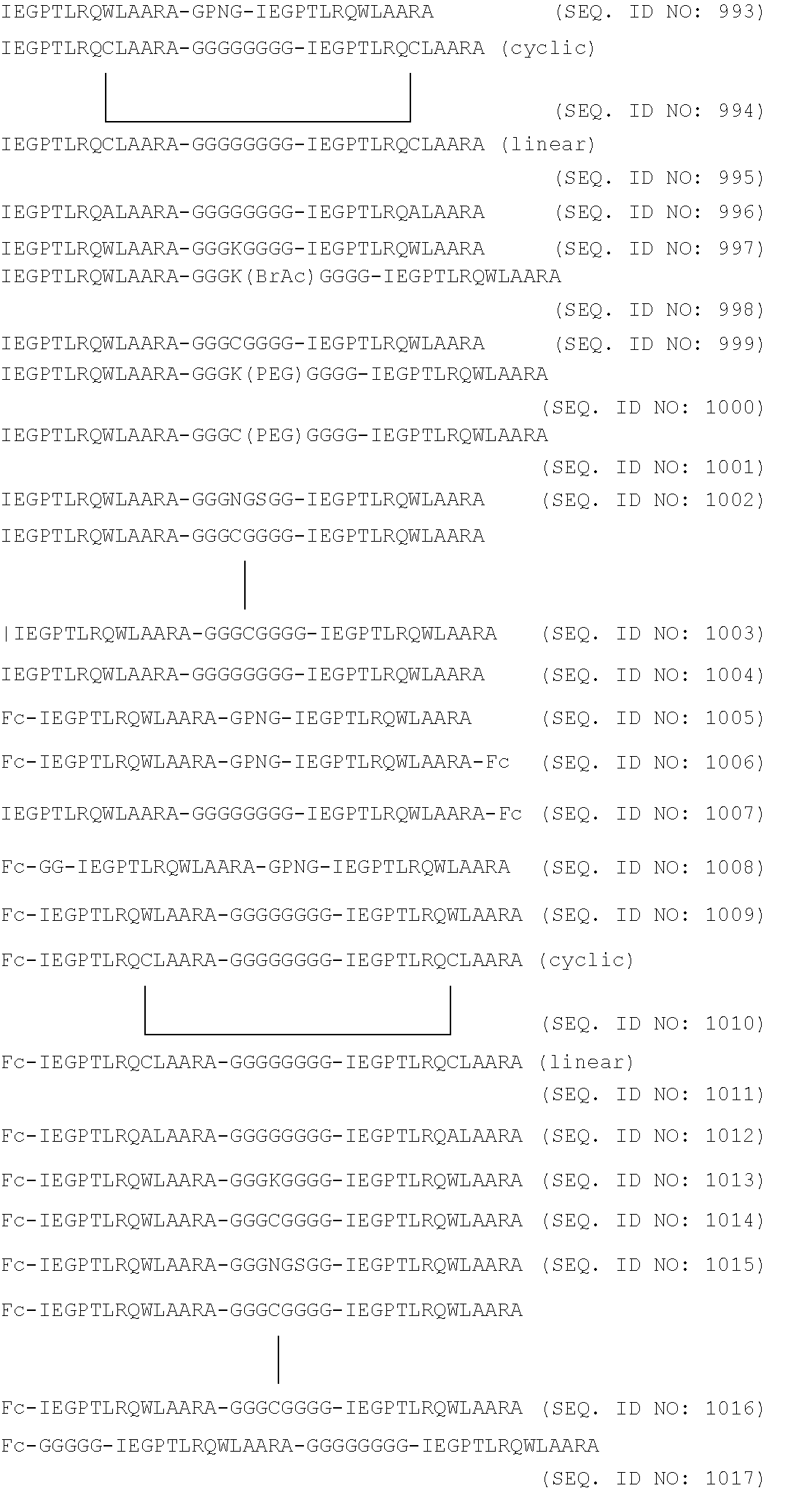
C00014
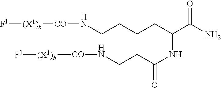
C00015

D00001
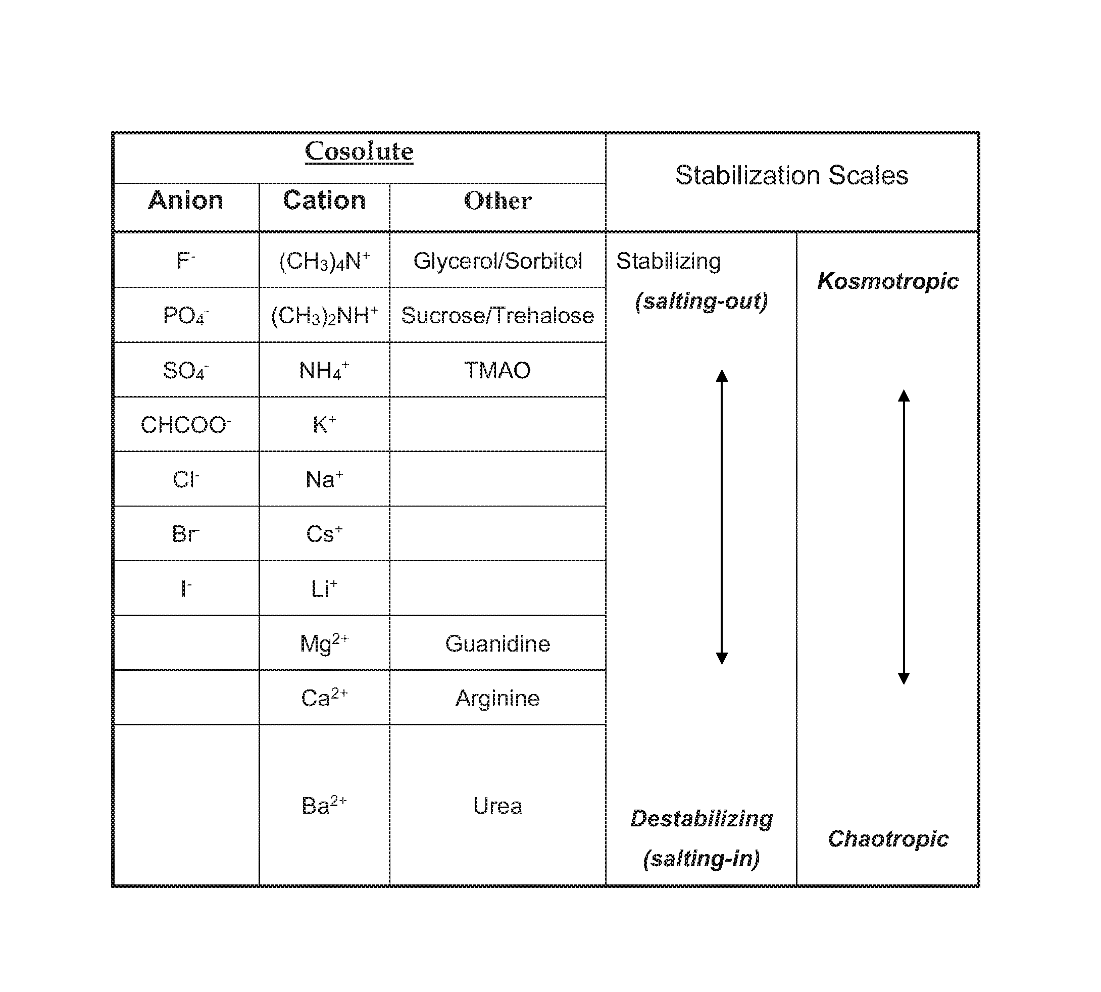
P00001

S00001
XML
uspto.report is an independent third-party trademark research tool that is not affiliated, endorsed, or sponsored by the United States Patent and Trademark Office (USPTO) or any other governmental organization. The information provided by uspto.report is based on publicly available data at the time of writing and is intended for informational purposes only.
While we strive to provide accurate and up-to-date information, we do not guarantee the accuracy, completeness, reliability, or suitability of the information displayed on this site. The use of this site is at your own risk. Any reliance you place on such information is therefore strictly at your own risk.
All official trademark data, including owner information, should be verified by visiting the official USPTO website at www.uspto.gov. This site is not intended to replace professional legal advice and should not be used as a substitute for consulting with a legal professional who is knowledgeable about trademark law.