Intranasal stimulation for treatment of meibomian gland disease and blepharitis
Franke , et al. February 16, 2
U.S. patent number 10,918,864 [Application Number 15/584,956] was granted by the patent office on 2021-02-16 for intranasal stimulation for treatment of meibomian gland disease and blepharitis. This patent grant is currently assigned to Oculeve, Inc.. The grantee listed for this patent is Oculeve, Inc.. Invention is credited to Manfred Franke, Daniel N. Hamilton, Diane Michelle Senchyna.


View All Diagrams
| United States Patent | 10,918,864 |
| Franke , et al. | February 16, 2021 |
Intranasal stimulation for treatment of meibomian gland disease and blepharitis
Abstract
Described here are devices, systems, and methods for treating meibomian gland disease and/or blepharitis by providing intranasal stimulation. Generally, the devices may deliver electrical stimulation to the nasal mucosa. Intranasal stimulation may unblock obstructed meibomian glands and increase the lipid content of tears, both acutely during stimulation and subsequent to intranasal stimulation. The methods may include an initial round of stimulation to unblock obstructed meibomian glands, and subsequent shorter rounds of stimulation to cause meibum secretion and maintain the open glands.
| Inventors: | Franke; Manfred (Valencia, CA), Senchyna; Diane Michelle (Corona del Mar, CA), Hamilton; Daniel N. (Napa, CA) | ||||||||||
|---|---|---|---|---|---|---|---|---|---|---|---|
| Applicant: |
|
||||||||||
| Assignee: | Oculeve, Inc. (South San
Francisco, CA) |
||||||||||
| Family ID: | 60157123 | ||||||||||
| Appl. No.: | 15/584,956 | ||||||||||
| Filed: | May 2, 2017 |
Prior Publication Data
| Document Identifier | Publication Date | |
|---|---|---|
| US 20170312521 A1 | Nov 2, 2017 | |
Related U.S. Patent Documents
| Application Number | Filing Date | Patent Number | Issue Date | ||
|---|---|---|---|---|---|
| 62429059 | Dec 1, 2016 | ||||
| 62330763 | May 2, 2016 | ||||
| Current U.S. Class: | 1/1 |
| Current CPC Class: | A61N 1/3603 (20170801); A61N 1/36014 (20130101); A61N 1/36046 (20130101); A61N 1/0546 (20130101); A61N 1/36034 (20170801) |
| Current International Class: | A61N 1/00 (20060101); A61N 1/36 (20060101); A61N 1/05 (20060101) |
References Cited [Referenced By]
U.S. Patent Documents
| 2512882 | June 1950 | Truesdale |
| 2525381 | October 1950 | Tower |
| 3620219 | November 1971 | Barker |
| 3709228 | January 1973 | Barker et al. |
| 3885550 | May 1975 | MacLeod |
| D257495 | November 1980 | Bros et al. |
| 4495676 | January 1985 | Hartmetz |
| 4520825 | June 1985 | Thompson et al. |
| 4539988 | September 1985 | Shirley et al. |
| 4590942 | May 1986 | Brenman et al. |
| 4628933 | December 1986 | Michelson |
| 4681121 | July 1987 | Kobal |
| 4684362 | August 1987 | Holt |
| 4706680 | November 1987 | Keusch et al. |
| 4735207 | April 1988 | Nambu et al. |
| 4777954 | October 1988 | Keusch et al. |
| 4780932 | November 1988 | Bowman et al. |
| 4868154 | September 1989 | Gilbard et al. |
| 4926880 | May 1990 | Claude et al. |
| 4957480 | September 1990 | Morenings |
| 4988358 | January 1991 | Eppley et al. |
| 5025807 | June 1991 | Zabara |
| 5072724 | December 1991 | Marcus |
| 5078733 | January 1992 | Eveleigh et al. |
| 5090422 | February 1992 | Dahl et al. |
| 5099829 | March 1992 | Wu |
| 5147284 | September 1992 | Fedorov et al. |
| 5324316 | June 1994 | Schulman et al. |
| 5342410 | August 1994 | Braverman |
| 5345948 | September 1994 | O'Donnell, Jr. |
| 5352445 | October 1994 | Lavaux |
| 5360438 | November 1994 | Fisher |
| 5498681 | March 1996 | Askari et al. |
| 5514131 | May 1996 | Edwards et al. |
| 5533470 | July 1996 | Rose |
| 5545617 | August 1996 | Dartt et al. |
| 5571101 | November 1996 | Ellman et al. |
| 5607461 | March 1997 | Lathrop |
| 5611970 | March 1997 | Apollonio et al. |
| 5640978 | June 1997 | Wong |
| 5683436 | November 1997 | Mendes et al. |
| 5697957 | December 1997 | Noren et al. |
| 5707400 | January 1998 | Terry et al. |
| 5713833 | February 1998 | Milligan |
| 5720773 | February 1998 | Lopez-Claros |
| 5733282 | March 1998 | Ellman et al. |
| 5735817 | April 1998 | Shantha |
| 5792100 | August 1998 | Shantha |
| 5794614 | August 1998 | Gruenke et al. |
| 5800685 | September 1998 | Perrault |
| 5843140 | December 1998 | Strojnik |
| 5900407 | May 1999 | Yerxa et al. |
| 5904658 | May 1999 | Niederauer et al. |
| 5935155 | August 1999 | Humayun et al. |
| 5948006 | September 1999 | Mann |
| 6001088 | December 1999 | Roberts et al. |
| 6020445 | February 2000 | Vanderlaan et al. |
| 6035236 | March 2000 | Jarding et al. |
| 6050999 | April 2000 | Paraschac et al. |
| 6051017 | April 2000 | Loeb et al. |
| 6083251 | July 2000 | Shindo |
| 6102847 | August 2000 | Stielau |
| 6152916 | November 2000 | Bige |
| 6200626 | March 2001 | Grobe, III et al. |
| 6205359 | March 2001 | Boveja |
| 6208902 | March 2001 | Boveja |
| 6240316 | May 2001 | Richmond et al. |
| 6246911 | June 2001 | Seligman |
| 6270796 | August 2001 | Weinstein |
| 6272382 | August 2001 | Faltys et al. |
| 6275737 | August 2001 | Mann |
| 6277855 | August 2001 | Yerxa |
| 6284765 | September 2001 | Caffrey |
| 6324429 | November 2001 | Shire et al. |
| 6327504 | December 2001 | Dolgin et al. |
| 6366814 | April 2002 | Boveja et al. |
| 6405079 | June 2002 | Ansarinia et al. |
| 6438398 | August 2002 | Pflugfelder et al. |
| 6458157 | October 2002 | Suaning |
| 6505077 | January 2003 | Kast et al. |
| 6526318 | February 2003 | Ansarinia et al. |
| 6535766 | March 2003 | Thompson et al. |
| 6537265 | March 2003 | Thanavala et al. |
| 6539253 | March 2003 | Thompson et al. |
| 6562036 | May 2003 | Ellman et al. |
| 6564102 | May 2003 | Boveja |
| 6578579 | June 2003 | Burnside et al. |
| 6604528 | August 2003 | Duncan |
| 6641799 | November 2003 | Goldberg |
| 6658301 | December 2003 | Loeb et al. |
| 6662052 | December 2003 | Sarwal et al. |
| 6684879 | February 2004 | Coffee et al. |
| 6701189 | March 2004 | Fang et al. |
| 6748951 | June 2004 | Schmidt |
| 6792314 | September 2004 | Byers et al. |
| 6829508 | December 2004 | Schulman et al. |
| 6853858 | February 2005 | Shalev |
| 6871099 | March 2005 | Whitehurst et al. |
| 6879859 | April 2005 | Boveja |
| 6885888 | April 2005 | Rezai |
| 6895279 | May 2005 | Loeb et al. |
| 7024241 | April 2006 | Bornzin et al. |
| 7054692 | May 2006 | Whitehurst et al. |
| 7067307 | June 2006 | Hochleitner et al. |
| 7069084 | June 2006 | Yee |
| 7117033 | October 2006 | Shalev et al. |
| 7142909 | November 2006 | Greenberg et al. |
| 7146209 | December 2006 | Gross et al. |
| 7169163 | January 2007 | Becker |
| 7190998 | March 2007 | Shalev et al. |
| 7225032 | May 2007 | Schmeling et al. |
| 7228184 | June 2007 | Heath |
| 7247692 | July 2007 | Laredo |
| 7317947 | January 2008 | Wahlstrand et al. |
| 7330762 | February 2008 | Boveja et al. |
| 7346389 | March 2008 | Newsome |
| 7346398 | March 2008 | Gross et al. |
| 7369897 | May 2008 | Boveja et al. |
| 7442191 | October 2008 | Hovda et al. |
| 7460911 | December 2008 | Cosendai et al. |
| 7477947 | January 2009 | Pines et al. |
| 7502652 | March 2009 | Gaunt et al. |
| 7547447 | June 2009 | Yiu et al. |
| 7565204 | July 2009 | Matei et al. |
| 7599737 | October 2009 | Yomtov et al. |
| 7636597 | December 2009 | Gross et al. |
| 7650186 | January 2010 | Hastings et al. |
| D613408 | April 2010 | Gausmann et al. |
| D614303 | April 2010 | Gausmann et al. |
| D614774 | April 2010 | Gausmann et al. |
| 7725176 | May 2010 | Schuler et al. |
| 7725195 | May 2010 | Lima et al. |
| D617443 | June 2010 | Grenon et al. |
| 7758190 | July 2010 | Korb et al. |
| 7778703 | August 2010 | Gross et al. |
| 7778711 | August 2010 | Ben-David et al. |
| 7792591 | September 2010 | Rooney et al. |
| 7805200 | September 2010 | Kast et al. |
| 7805202 | September 2010 | Kuzma et al. |
| 7805203 | September 2010 | Ben-David et al. |
| 7809442 | October 2010 | Bolea et al. |
| 7835794 | November 2010 | Greenberg et al. |
| 7846124 | December 2010 | Becker |
| 7860570 | December 2010 | Whitehurst et al. |
| 7873421 | January 2011 | Karell |
| 7879032 | February 2011 | Garito et al. |
| 7879079 | February 2011 | Tu et al. |
| D638128 | May 2011 | Prokop et al. |
| 7981095 | July 2011 | Grenon |
| 7993381 | August 2011 | Mac et al. |
| 7998202 | August 2011 | Lesh |
| 8002783 | August 2011 | Vercellotti et al. |
| 8019419 | September 2011 | Panescu et al. |
| 8019441 | September 2011 | Wallace et al. |
| 8080047 | December 2011 | Yu |
| 8083787 | December 2011 | Korb |
| 8145322 | March 2012 | Yao et al. |
| 8155746 | April 2012 | Maltan et al. |
| 8165680 | April 2012 | Greenberg et al. |
| 8204591 | June 2012 | Ben-David et al. |
| 8231218 | July 2012 | Hong et al. |
| 8251983 | August 2012 | Larson et al. |
| 8295529 | October 2012 | Petersen et al. |
| 8318070 | November 2012 | Shiah et al. |
| D681839 | May 2013 | Nathanson |
| 8489189 | July 2013 | Tronnes |
| 8494641 | July 2013 | Boling et al. |
| 8521292 | August 2013 | Wei et al. |
| 8626298 | January 2014 | Simon |
| 8676324 | March 2014 | Simon et al. |
| 8728136 | May 2014 | Feldman |
| 8918181 | December 2014 | Ackermann et al. |
| 8936594 | January 2015 | Wolf et al. |
| 8986301 | March 2015 | Wolf et al. |
| 8996137 | March 2015 | Ackermann et al. |
| 9079042 | July 2015 | Tiedtke et al. |
| 9095723 | August 2015 | Ackermann et al. |
| 9265956 | February 2016 | Ackermann et al. |
| 9440065 | September 2016 | Ackermann et al. |
| 9687652 | June 2017 | Franke et al. |
| 9717627 | August 2017 | Kuzma et al. |
| 9737702 | August 2017 | Ackermann et al. |
| 9737712 | August 2017 | Franke et al. |
| 9764150 | September 2017 | Loudin et al. |
| 9770583 | September 2017 | Gupta et al. |
| 9821159 | November 2017 | Ackermann et al. |
| 9956397 | May 2018 | Loudin et al. |
| D826420 | August 2018 | Ackermann et al. |
| 10143846 | December 2018 | Ackermann et al. |
| D837396 | January 2019 | Ackermann et al. |
| 10207108 | February 2019 | Franke et al. |
| 2001/0018918 | September 2001 | Burnside et al. |
| 2001/0020177 | September 2001 | Gruzdowich et al. |
| 2002/0013594 | January 2002 | Dinger et al. |
| 2002/0035358 | March 2002 | Wang |
| 2002/0049290 | April 2002 | Vanderbilt et al. |
| 2002/0161416 | October 2002 | Huang |
| 2002/0188331 | December 2002 | Fang et al. |
| 2003/0014089 | January 2003 | Chow et al. |
| 2003/0045909 | March 2003 | Gross et al. |
| 2003/0045911 | March 2003 | Bruchmann et al. |
| 2003/0114899 | June 2003 | Woods et al. |
| 2003/0120323 | June 2003 | Meadows et al. |
| 2003/0130809 | July 2003 | Cohen et al. |
| 2003/0133877 | July 2003 | Levin |
| 2003/0139784 | July 2003 | Morimoto et al. |
| 2003/0176892 | September 2003 | Shalev |
| 2003/0176898 | September 2003 | Gross et al. |
| 2003/0192784 | October 2003 | Zhou et al. |
| 2003/0229381 | December 2003 | Hochmair et al. |
| 2003/0233134 | December 2003 | Greenberg et al. |
| 2003/0233135 | December 2003 | Yee |
| 2004/0050392 | March 2004 | Tu et al. |
| 2004/0059466 | March 2004 | Block et al. |
| 2004/0092992 | May 2004 | Adams et al. |
| 2004/0098036 | May 2004 | Bergersen |
| 2004/0098067 | May 2004 | Ohta et al. |
| 2004/0127942 | July 2004 | Yomtov et al. |
| 2004/0138646 | July 2004 | Walla |
| 2004/0147973 | July 2004 | Hauser et al. |
| 2004/0151930 | August 2004 | Rouns et al. |
| 2004/0220644 | November 2004 | Shalev et al. |
| 2005/0004621 | January 2005 | Boveja et al. |
| 2005/0004625 | January 2005 | Chow |
| 2005/0010250 | January 2005 | Schuler et al. |
| 2005/0010266 | January 2005 | Bogdanowicz |
| 2005/0101967 | May 2005 | Weber et al. |
| 2005/0101994 | May 2005 | Yamazaki et al. |
| 2005/0105046 | May 2005 | Tung |
| 2005/0137276 | June 2005 | Yahiaoui et al. |
| 2005/0159790 | July 2005 | Shalev et al. |
| 2005/0165458 | July 2005 | Boveja et al. |
| 2005/0197675 | September 2005 | David et al. |
| 2005/0251061 | November 2005 | Schuler et al. |
| 2005/0256570 | November 2005 | Azar |
| 2005/0267542 | December 2005 | David et al. |
| 2005/0268472 | December 2005 | Bourilkov et al. |
| 2006/0004423 | January 2006 | Boveja et al. |
| 2006/0018872 | January 2006 | Tew et al. |
| 2006/0036296 | February 2006 | Greenberg et al. |
| 2006/0074450 | April 2006 | Boveja et al. |
| 2006/0089673 | April 2006 | Schultheiss et al. |
| 2006/0095077 | May 2006 | Tronnes et al. |
| 2006/0095108 | May 2006 | Chowdhury et al. |
| 2006/0100668 | May 2006 | Ben-David et al. |
| 2006/0107958 | May 2006 | Sleeper |
| 2006/0142822 | June 2006 | Tulgar |
| 2006/0161225 | July 2006 | Sormann et al. |
| 2006/0195169 | August 2006 | Gross et al. |
| 2006/0206155 | September 2006 | Ben-David et al. |
| 2006/0206162 | September 2006 | Wahlstrand et al. |
| 2006/0216317 | September 2006 | Reinhard et al. |
| 2006/0235430 | October 2006 | Le et al. |
| 2006/0239482 | October 2006 | Hatoum et al. |
| 2006/0259098 | November 2006 | Erickson |
| 2006/0271024 | November 2006 | Gertner et al. |
| 2006/0271108 | November 2006 | Libbus et al. |
| 2006/0276738 | December 2006 | Becker |
| 2007/0031341 | February 2007 | DiMauro et al. |
| 2007/0038250 | February 2007 | He et al. |
| 2007/0038267 | February 2007 | Shodo et al. |
| 2007/0060815 | March 2007 | Martin et al. |
| 2007/0060954 | March 2007 | Cameron et al. |
| 2007/0083245 | April 2007 | Lamensdorf et al. |
| 2007/0112404 | May 2007 | Mann et al. |
| 2007/0123938 | May 2007 | Haller et al. |
| 2007/0135868 | June 2007 | Shi et al. |
| 2007/0150034 | June 2007 | Rooney et al. |
| 2007/0219600 | September 2007 | Gertner et al. |
| 2007/0237797 | October 2007 | Peyman |
| 2007/0237825 | October 2007 | Levy et al. |
| 2007/0248930 | October 2007 | Brawn |
| 2007/0250119 | October 2007 | Tyler et al. |
| 2007/0250135 | October 2007 | Bartz-Schmidt et al. |
| 2007/0255333 | November 2007 | Giftakis et al. |
| 2007/0276314 | November 2007 | Becker |
| 2007/0276451 | November 2007 | Rigaux |
| 2007/0295327 | December 2007 | Bottomley |
| 2007/0299420 | December 2007 | Peyman |
| 2007/0299462 | December 2007 | Becker |
| 2008/0009897 | January 2008 | Duran Von Arx |
| 2008/0021515 | January 2008 | Horsager et al. |
| 2008/0082057 | April 2008 | Korb et al. |
| 2008/0082131 | April 2008 | Llanos |
| 2008/0109046 | May 2008 | Lima et al. |
| 2008/0109054 | May 2008 | Hastings et al. |
| 2008/0114424 | May 2008 | Grenon et al. |
| 2008/0132933 | June 2008 | Gerber |
| 2008/0132969 | June 2008 | Bennett et al. |
| 2008/0140141 | June 2008 | Ben-David et al. |
| 2008/0183242 | July 2008 | Tano et al. |
| 2008/0183243 | July 2008 | Shodo et al. |
| 2008/0208287 | August 2008 | Palermo et al. |
| 2008/0208335 | August 2008 | Blum et al. |
| 2008/0221642 | September 2008 | Humayun et al. |
| 2008/0269648 | October 2008 | Bock |
| 2008/0288036 | November 2008 | Greenberg et al. |
| 2008/0294066 | November 2008 | Hetling et al. |
| 2009/0005835 | January 2009 | Greenberg et al. |
| 2009/0012573 | January 2009 | Karell et al. |
| 2009/0018582 | January 2009 | Ishikawa et al. |
| 2009/0024187 | January 2009 | Erickson et al. |
| 2009/0024189 | January 2009 | Lee et al. |
| 2009/0036945 | February 2009 | Chancellor et al. |
| 2009/0043185 | February 2009 | McAdams et al. |
| 2009/0056709 | March 2009 | Worsoff |
| 2009/0099600 | April 2009 | Moore et al. |
| 2009/0099623 | April 2009 | Bentwich et al. |
| 2009/0099626 | April 2009 | de Juan, Jr. et al. |
| 2009/0101139 | April 2009 | Karell |
| 2009/0124965 | May 2009 | Greenberg et al. |
| 2009/0138061 | May 2009 | Stephens et al. |
| 2009/0156581 | June 2009 | Dillon et al. |
| 2009/0157142 | June 2009 | Cauller et al. |
| 2009/0157145 | June 2009 | Cauller |
| 2009/0157147 | June 2009 | Cauller et al. |
| 2009/0192571 | July 2009 | Stett et al. |
| 2009/0192575 | July 2009 | Carbunaru et al. |
| 2009/0204142 | August 2009 | Becker |
| 2009/0239235 | September 2009 | DeMaria et al. |
| 2009/0241840 | October 2009 | Mills |
| 2009/0264966 | October 2009 | Blum et al. |
| 2009/0281594 | November 2009 | King et al. |
| 2009/0281596 | November 2009 | King et al. |
| 2009/0299418 | December 2009 | Shalev et al. |
| 2009/0306738 | December 2009 | Weiss et al. |
| 2009/0312818 | December 2009 | Horsager et al. |
| 2010/0030150 | February 2010 | Paques et al. |
| 2010/0076423 | March 2010 | Muller |
| 2010/0087896 | April 2010 | McCreery |
| 2010/0094280 | April 2010 | Muller |
| 2010/0100165 | April 2010 | Swanson et al. |
| 2010/0139002 | June 2010 | Walker et al. |
| 2010/0152708 | June 2010 | Li et al. |
| 2010/0161004 | June 2010 | Najafi et al. |
| 2010/0168513 | July 2010 | Pless et al. |
| 2010/0179468 | July 2010 | Becker |
| 2010/0211132 | August 2010 | Nimmagadda et al. |
| 2010/0241195 | September 2010 | Meadows et al. |
| 2010/0256609 | October 2010 | Hillis et al. |
| 2010/0274164 | October 2010 | Juto |
| 2010/0274224 | October 2010 | Jain et al. |
| 2010/0274313 | October 2010 | Boling et al. |
| 2010/0280509 | November 2010 | Muller et al. |
| 2010/0288275 | November 2010 | Djupesland et al. |
| 2010/0311688 | December 2010 | Chapin et al. |
| 2010/0318159 | December 2010 | Aghassian et al. |
| 2011/0021975 | January 2011 | Covello |
| 2011/0028807 | February 2011 | Abreu |
| 2011/0028883 | February 2011 | Juan, Jr. et al. |
| 2011/0076775 | March 2011 | Stewart et al. |
| 2011/0077551 | March 2011 | Videbaek |
| 2011/0077698 | March 2011 | Tsampazis et al. |
| 2011/0081333 | April 2011 | Shantha et al. |
| 2011/0082518 | April 2011 | Filippello |
| 2011/0093043 | April 2011 | Torgerson et al. |
| 2011/0151393 | June 2011 | Frey, II et al. |
| 2011/0152969 | June 2011 | Zehnder et al. |
| 2011/0184490 | July 2011 | Horsager et al. |
| 2011/0202121 | August 2011 | Wen |
| 2011/0218590 | September 2011 | Degiorgio et al. |
| 2011/0234971 | September 2011 | Yeh |
| 2011/0270067 | November 2011 | Faraji et al. |
| 2011/0270348 | November 2011 | Goetz |
| 2011/0275734 | November 2011 | Scales et al. |
| 2011/0276107 | November 2011 | Simon et al. |
| 2011/0282251 | November 2011 | Baker et al. |
| 2011/0295336 | December 2011 | Sharma et al. |
| 2011/0313330 | December 2011 | Loushin et al. |
| 2011/0313480 | December 2011 | De Vos |
| 2011/0313481 | December 2011 | De Vos |
| 2011/0313488 | December 2011 | Hincapie Ordonez et al. |
| 2012/0053648 | March 2012 | Neher et al. |
| 2012/0112903 | May 2012 | Kaib et al. |
| 2012/0130398 | May 2012 | Ackermann et al. |
| 2012/0133887 | May 2012 | Huang |
| 2012/0197338 | August 2012 | Su et al. |
| 2012/0232615 | September 2012 | Barolat et al. |
| 2012/0232618 | September 2012 | Feldman |
| 2012/0234332 | September 2012 | Shantha |
| 2012/0253249 | October 2012 | Wilson et al. |
| 2012/0298105 | November 2012 | Osorio et al. |
| 2012/0315329 | December 2012 | Ahn et al. |
| 2012/0316557 | December 2012 | Sartor et al. |
| 2012/0323214 | December 2012 | Shantha |
| 2012/0323227 | December 2012 | Wolf et al. |
| 2012/0323232 | December 2012 | Wolf et al. |
| 2012/0330376 | December 2012 | Flynn et al. |
| 2013/0006095 | January 2013 | Jenkins et al. |
| 2013/0006326 | January 2013 | Ackermann et al. |
| 2013/0053733 | February 2013 | Korb et al. |
| 2013/0053737 | February 2013 | Scerbo |
| 2013/0065765 | March 2013 | Selifonov et al. |
| 2013/0072755 | March 2013 | Papania et al. |
| 2013/0085551 | April 2013 | Bachinski et al. |
| 2013/0158451 | June 2013 | Juto et al. |
| 2013/0158626 | June 2013 | DeGiorgio et al. |
| 2013/0172790 | July 2013 | Badawi |
| 2013/0178937 | July 2013 | Vassallo et al. |
| 2013/0197321 | August 2013 | Wilson |
| 2013/0253387 | September 2013 | Bonutti et al. |
| 2013/0261706 | October 2013 | Mirro et al. |
| 2013/0270491 | October 2013 | Park et al. |
| 2013/0274824 | October 2013 | Otto et al. |
| 2013/0274831 | October 2013 | Otto et al. |
| 2013/0282070 | October 2013 | Cowan et al. |
| 2013/0304154 | November 2013 | Goodman et al. |
| 2013/0310887 | November 2013 | Curtis |
| 2013/0336557 | December 2013 | Cruzat et al. |
| 2014/0012182 | January 2014 | Shantha et al. |
| 2014/0056815 | February 2014 | Peyman |
| 2014/0081353 | March 2014 | Cook et al. |
| 2014/0088463 | March 2014 | Wolf et al. |
| 2014/0148872 | May 2014 | Goldwasser et al. |
| 2014/0156000 | June 2014 | Campin et al. |
| 2014/0163580 | June 2014 | Tischendorf et al. |
| 2014/0214115 | July 2014 | Greiner et al. |
| 2014/0214118 | July 2014 | Greiner et al. |
| 2014/0214120 | July 2014 | Simon et al. |
| 2014/0214124 | July 2014 | Greiner et al. |
| 2014/0214125 | July 2014 | Greiner et al. |
| 2014/0257205 | September 2014 | Schaller |
| 2014/0257433 | September 2014 | Ackermann et al. |
| 2014/0277429 | September 2014 | Kuzma et al. |
| 2014/0316310 | October 2014 | Ackermann et al. |
| 2014/0316396 | October 2014 | Wolf et al. |
| 2014/0316485 | October 2014 | Ackermann et al. |
| 2014/0324147 | October 2014 | Wagner |
| 2014/0362339 | December 2014 | Imafuku |
| 2014/0371565 | December 2014 | Glasser |
| 2014/0371812 | December 2014 | Ackermann et al. |
| 2015/0088156 | March 2015 | Ackermann et al. |
| 2015/0182145 | July 2015 | Gazdzinski |
| 2015/0238754 | August 2015 | Loudin et al. |
| 2015/0335900 | November 2015 | Ackermann et al. |
| 2015/0343202 | December 2015 | Picaud et al. |
| 2015/0362755 | December 2015 | Lee et al. |
| 2016/0022992 | January 2016 | Franke et al. |
| 2016/0058615 | March 2016 | Camras et al. |
| 2016/0080720 | March 2016 | Fullam |
| 2016/0114163 | April 2016 | Franke et al. |
| 2016/0114172 | April 2016 | Loudin et al. |
| 2016/0121118 | May 2016 | Franke et al. |
| 2016/0158548 | June 2016 | Ackermann et al. |
| 2016/0270656 | September 2016 | Samec et al. |
| 2016/0367795 | December 2016 | Ackermann et al. |
| 2016/0367806 | December 2016 | Kahook |
| 2017/0049619 | February 2017 | Kahook |
| 2017/0157401 | June 2017 | Loudin et al. |
| 2017/0188947 | July 2017 | Connor |
| 2017/0239459 | August 2017 | Loudin et al. |
| 2017/0252563 | September 2017 | Franke et al. |
| 2017/0340884 | November 2017 | Franke et al. |
| 2017/0354536 | December 2017 | Kuzma et al. |
| 2017/0368332 | December 2017 | Ackermann et al. |
| 2017/0368333 | December 2017 | Loudin et al. |
| 2017/0368359 | December 2017 | Loudin et al. |
| 2018/0064940 | March 2018 | Ackermann et al. |
| 2018/0064941 | March 2018 | Ackermann et al. |
| 2018/0064942 | March 2018 | Franke et al. |
| 2018/0153394 | June 2018 | Franke et al. |
| 2018/0154137 | June 2018 | Ackermann et al. |
| 2018/0154161 | June 2018 | Ackermann et al. |
| 2018/0161579 | June 2018 | Franke et al. |
| 2018/0280688 | October 2018 | Loudin et al. |
| 2019/0022392 | January 2019 | Franke et al. |
| 2019/0167978 | June 2019 | Ackermann et al. |
| 2019/0217095 | July 2019 | Franke et al. |
| 2019/0282804 | September 2019 | Ackermann et al. |
| 2019/0290922 | September 2019 | Ackermann et al. |
| 2019/0308009 | October 2019 | Loudin et al. |
| 2019/0344077 | November 2019 | Ackermann et al. |
| 2020/0030615 | January 2020 | Loudin et al. |
| 2020/0038238 | February 2020 | Kuzma et al. |
| 1488331 | Apr 2004 | CN | |||
| 2693275 | Apr 2005 | CN | |||
| 101087822 | Dec 2007 | CN | |||
| 101503491 | Aug 2009 | CN | |||
| 101589085 | Nov 2009 | CN | |||
| 101616663 | Dec 2009 | CN | |||
| 101829120 | Sep 2010 | CN | |||
| 101939043 | Jan 2011 | CN | |||
| 102266592 | Dec 2011 | CN | |||
| 103467652 | Dec 2013 | CN | |||
| 102006048819 | Apr 2008 | DE | |||
| 2102681-0001 | Oct 2012 | EM | |||
| 2199000-0001 | Mar 2013 | EM | |||
| 0 109 935 | May 1984 | EP | |||
| 1 497 483 | Jan 2005 | EP | |||
| 1 651 307 | May 2006 | EP | |||
| 1 919 553 | May 2008 | EP | |||
| 1 958 661 | Aug 2008 | EP | |||
| 2 205 193 | Jul 2010 | EP | |||
| 2 205 314 | Jul 2010 | EP | |||
| 3263175 | Jan 2018 | EP | |||
| 2 129 690 | Mar 1987 | GB | |||
| 2 456 002 | Jul 2009 | GB | |||
| S60500241 | Feb 1985 | JP | |||
| S60502192 | Dec 1985 | JP | |||
| 8-500995 | Feb 1996 | JP | |||
| 2002-325851 | Nov 2002 | JP | |||
| 2002-539859 | Nov 2002 | JP | |||
| 2004-508847 | Mar 2004 | JP | |||
| 2004526510 | Sep 2004 | JP | |||
| 2005-502409 | Jan 2005 | JP | |||
| 2005-052461 | Mar 2005 | JP | |||
| 2005-144178 | Jun 2005 | JP | |||
| 2005521489 | Jul 2005 | JP | |||
| 2005-528169 | Sep 2005 | JP | |||
| 2006-515900 | Jun 2006 | JP | |||
| 2006-311917 | Nov 2006 | JP | |||
| 2007-044323 | Feb 2007 | JP | |||
| 2007-528751 | Oct 2007 | JP | |||
| 2008-55000 | Mar 2008 | JP | |||
| 2008-183248 | Aug 2008 | JP | |||
| 2008-541850 | Nov 2008 | JP | |||
| 2009-506836 | Feb 2009 | JP | |||
| 2009-523503 | Jun 2009 | JP | |||
| 2010-505563 | Feb 2010 | JP | |||
| 2010-051562 | Mar 2010 | JP | |||
| 2010506654 | Mar 2010 | JP | |||
| 2010-537777 | Dec 2010 | JP | |||
| 2011-030734 | Feb 2011 | JP | |||
| 2011-524780 | Sep 2011 | JP | |||
| 2012-100708 | May 2012 | JP | |||
| 2012-115545 | Jun 2012 | JP | |||
| 2012-200558 | Oct 2012 | JP | |||
| 2013-528416 | Jul 2013 | JP | |||
| 2338492 | Nov 2008 | RU | |||
| WO-85/01213 | Mar 1985 | WO | |||
| WO-94/00188 | Jan 1994 | WO | |||
| WO-00/01320 | Jan 2000 | WO | |||
| WO-00/56393 | Sep 2000 | WO | |||
| WO-00/62672 | Oct 2000 | WO | |||
| WO-01/05388 | Jan 2001 | WO | |||
| WO-01/85094 | Nov 2001 | WO | |||
| WO-02/078592 | Oct 2002 | WO | |||
| WO-03/023907 | Mar 2003 | WO | |||
| WO-03/082080 | Oct 2003 | WO | |||
| WO-2003/087433 | Oct 2003 | WO | |||
| WO-03/101535 | Dec 2003 | WO | |||
| WO-2004/026106 | Apr 2004 | WO | |||
| WO-2004/026106 | Apr 2004 | WO | |||
| WO-2004/043217 | May 2004 | WO | |||
| WO-2004/043217 | May 2004 | WO | |||
| WO-2004/091453 | Oct 2004 | WO | |||
| WO-2004/112893 | Dec 2004 | WO | |||
| WO-2004/112893 | Dec 2004 | WO | |||
| WO-2005/007234 | Jan 2005 | WO | |||
| WO-2005/007234 | Jan 2005 | WO | |||
| WO-2005/030025 | Apr 2005 | WO | |||
| WO-2005/030025 | Apr 2005 | WO | |||
| WO-2005/060984 | Jul 2005 | WO | |||
| WO-2006/127366 | Nov 2006 | WO | |||
| WO-2007/028003 | Mar 2007 | WO | |||
| WO-2007/079543 | Jul 2007 | WO | |||
| WO-2008/048321 | Apr 2008 | WO | |||
| WO-2008/156501 | Dec 2008 | WO | |||
| WO-2008/156501 | Dec 2008 | WO | |||
| WO-2009/035571 | Mar 2009 | WO | |||
| WO-2009/035571 | Mar 2009 | WO | |||
| WO-2009/048580 | Apr 2009 | WO | |||
| WO-2009/070709 | Jun 2009 | WO | |||
| WO-2009/154457 | Dec 2009 | WO | |||
| WO-2010/003011 | Jan 2010 | WO | |||
| WO-2010/027743 | Mar 2010 | WO | |||
| WO-2010/069317 | Jun 2010 | WO | |||
| WO-2010/076904 | Jul 2010 | WO | |||
| WO-2010/099818 | Sep 2010 | WO | |||
| WO-2010/123704 | Oct 2010 | WO | |||
| WO-2011/011373 | Jan 2011 | WO | |||
| WO-2012/068247 | May 2012 | WO | |||
| WO-2012/139063 | Oct 2012 | WO | |||
| WO-2012/139063 | Oct 2012 | WO | |||
| WO-2012/155188 | Nov 2012 | WO | |||
| WO-2012/174161 | Dec 2012 | WO | |||
| WO-2013/055940 | Apr 2013 | WO | |||
| WO-2013/055940 | Apr 2013 | WO | |||
| WO-2013/157320 | Oct 2013 | WO | |||
| WO-2013/162793 | Oct 2013 | WO | |||
| WO-2013/165697 | Nov 2013 | WO | |||
| WO-2013/166353 | Nov 2013 | WO | |||
| WO-2014/138709 | Sep 2014 | WO | |||
| WO-2014/153218 | Sep 2014 | WO | |||
| WO-2014/165124 | Oct 2014 | WO | |||
| WO-2014/172693 | Oct 2014 | WO | |||
| WO-2014/172693 | Oct 2014 | WO | |||
| WO-2015/130707 | Sep 2015 | WO | |||
| WO-2015/130707 | Sep 2015 | WO | |||
| WO-2016/015025 | Jan 2016 | WO | |||
| WO-2016/025323 | Feb 2016 | WO | |||
| WO-2016/065211 | Apr 2016 | WO | |||
| WO-2016/065213 | Apr 2016 | WO | |||
| WO-2016/065215 | Apr 2016 | WO | |||
| WO-2017/192572 | Nov 2017 | WO | |||
Other References
|
Acar, M. et al. (2013). "Ocular surface assessment in patients with obstructive sleep apnea-hypopnea syndrome," Sleep Breath 17(2):583-588. cited by applicant . Amparo (2013). "Topical Interleukin 1 Receptor Antagonist for Treatment of Dry Eye Disease," JAMA Ophth. 131(6):E1-E9. cited by applicant . Anonymous (2007). "The epidemiology of dry eye disease: report of the Epidemiology Subcommittee of the International Dry Eye WorkShop (2007)," Ocul. Surf. 5(2):93-107. cited by applicant . Bajpai et al. (2012). "Preparation, Characterization and Water Uptake Behavior of Polysaccharide Based Nanoparticles," Prog. Nanotech. Nanomat. 1(1):9-17. cited by applicant . Baraniuk et al. (2007). "Nasonasal Reflexes, the Nasal Cycle, and Sneeze," Curr. Allergy and Asthma Reports 7:105-111. cited by applicant . Baroody FM, Foster KA, Markaryan A, et al. Nasal ocular reflexes and eye symptoms in patients with allergic rhinitis. Ann Allergy Asthma Immunol 2008;100:194-199. cited by applicant . Baroody FM, Shenaq D, DeTineo M, et al. Fluticasone furoate nasal spray reduces the nasal-ocular reflex: a mechanism for the efficacy of topical steroids in controlling allergic eye symptoms. J Allergy. Clin Immunol 2009; 123:1342-1348. cited by applicant . Boberg-Ans J. (1955). "Experience in clinical examination of corneal sensitivity: corneal sensitivity and the naso-lacrimal reflex after retrobulbar anaesthesia," Br. J. Ophthalmol. 39(12):705-726. cited by applicant . Calonge (2001). "The Treatment of Dry Eye," Survey Ophth. 45(2):S227-S239. cited by applicant . Cipriano et al. (2014). "Superabsorbent Hydrogels That Are Robust and Highly Stretchable," Am. Chem Soc. 47(13):4445-4452. cited by applicant . Corrected Notice of Allowance dated Feb. 23, 2015, for U.S. Appl. No. 14/256,915, filed Apr. 18, 2014, 2 pages. cited by applicant . Corrected Notice of Allowance dated Jun. 9, 2017, for U.S. Appl. No. 14/920,860, filed Oct. 22, 2015, 2 pages. cited by applicant . Dart et al. (2002). "Effects of 25% Propylene Glycol Hydrogel (Solugel) on Second Intention Wound Healing in Horses," Vet. Surg. 31(4):309-313. cited by applicant . Drummond PD. Lacrimation and cutaneous vasodilatation in the face induced by painful stimulation of the nasal ala and upper lip. J Auton Nerv Syst 1995;51:109-16. cited by applicant . Elsby et al. (1967). "Lacrimal Secretion in the Cat," Br. J. Pharm. Chemother. 29(1):1-7. cited by applicant . Extended European Search Report dated Nov. 18, 2016, for EP Application No. 14 785 631.4, filed on Apr. 18, 2014, 7 pages. cited by applicant . Final Office Action received for U.S. Appl. No. 14/256,916, dated Apr. 8, 2015, 16 pages. cited by applicant . Final Office Action received for U.S. Appl. No. 14/313,937 dated Apr. 29, 2015, 13 pages. cited by applicant . Final Office Action received for U.S. Appl. No. 14/630,471, dated Sep. 26, 2016, 22 pages. cited by applicant . Final Office Action received for U.S. Appl. No. 14/256,916, dated Aug. 19, 2016, 19 pages. cited by applicant . Final Office Action dated Sep. 23, 2016, for U.S. Appl. No. 14/809,109, filed Jul. 24, 2015, 10 pages. cited by applicant . Final Office Action dated Feb. 1, 2017, for U.S. Appl. No. 14/920,852, filed Oct. 22, 2015, 20 pages. cited by applicant . Fujisawa et al. (2002). "The Effect of Nasal Mucosal Stimulation on Schirmer Tests in Sjogren's Syndrome and Dry Eye Patients," Lac. Gland Tear Film Dry Eye Syndrome 3 506:1221-1226. cited by applicant . Gupta et al. (1997). "Nasolacrimal Stimulation of Aqueous Tear Production," Cornea 16(6):645-648. cited by applicant . Heigle TJ, Pflugfelder SC. Aqueous tear production in patients with neurotrophic keratitis. Cornea 1996;15:135-8. cited by applicant . Holzer P. Capsaicin: cellular targets, mechanisms of action, and selectivity for thin sensory neurons. Pharmacol Rev 1991:43:143-201. cited by applicant . Ikemura et al. (2008). "UV-VIS Spectra and Photoinitiation Behaviors of Acylphosphine Oxide and Bisacylphosphine Oxide Derivatives in unfilled, Light-Cured Dental Resins," Dental Mat. J. 27(6):765-774. cited by applicant . International Preliminary Report on Patentability received for PCT Patent Application No. PCT/US2014/034733, dated Oct. 29, 2015. cited by applicant . International Search Report and Written Opinion received for PCT Application No. PCT/US2015/042130, dated Oct. 28, 2015. cited by applicant . International Search Report and Written Opinion received for PCT Patent Application No. PCT/US2014/034733, dated Dec. 5, 2014. cited by applicant . International Search Report and Written Opinion received for PCT Patent Application No. PCT/US2015/017379, dated Jul. 24, 2015. cited by applicant . International Search Report and Written Opinion received for PCT Patent Application No. PCT/US2015/057023, dated Mar. 4, 2016. cited by applicant . International Search Report dated Feb. 10, 2016, for PCT Patent Application No. PCT/US2015/57021, filed on Oct. 22, 2015, 4 pages. cited by applicant . Krupin T, Cross DA, Becker B. Decreased basal tear production associated with general anesthesia. Arch Ophthalmol 1977;95:107-108. cited by applicant . Lora et al. (2009). "Lacrimal Nerve Stimulation by a Neurostimulator for Tear Production," Invest. Ophth. Vis. Science 50(13):172. cited by applicant . Loth S, Bende M. Effect of nasal anaesthesia on lacrimal function after nasal allergen challenge. Clin Exp Allergy 1994;24:375-376. cited by applicant . Meng, I.D. et al. (2013). "The role of corneal afferent neurons in regulating tears under normal and dry eye conditions," Exp. Eye Res. 117:79-87. cited by applicant . Mallepally et al. (2013). "Superabsorbent Alginate Aerogels," J. Supercritical Fluids 79:1-5. cited by applicant . Non-Final Office Action received for U.S. Appl. No. 14/256,915, dated Aug. 13, 2014, 11 pages. cited by applicant . Non-Final Office Action received for U.S. Appl. No. 14/256,916, dated Sep. 12, 2014, 24 pages. cited by applicant . Non-Final Office Action received for U.S. Appl. No. 14/313,937, dated Nov. 19, 2014, 12 pages. cited by applicant . Non-Final Office Action dated Jun. 14, 2016, for U.S. Appl. No. 14/630,471, filed Feb. 24, 2015, 24 pages. cited by applicant . Non-Final Office Action received for U.S. Appl. No. 14/809,109, dated Apr. 8, 2016, 8 pages. cited by applicant . Non-Final Office Action Received for U.S. Appl. No. 14/920,860, dated Aug. 17, 2016, 11 pages. cited by applicant . Non-Final Office Action received for U.S. Appl. No. 14/256,916, dated Nov. 19, 2015, 20 pages. cited by applicant . Non-Final Office Action Received for U.S Appl. No. 14/313,937, dated Oct. 6, 2015, 7 pages. cited by applicant . Non-Final Office Action Received for U.S Appl. No. 14/920,852, dated Aug. 1, 2016, 20 pages. cited by applicant . Non-Final Office Action dated Sep. 30, 2016, for U.S. Appl. No. 15/256,392, filed Sep. 2, 2016, 14 pages. cited by applicant . Non-Final Office Action dated Feb. 14, 2017, for U.S. Appl. No. 14/630,471, filed Feb. 24, 2015, 23 pages. cited by applicant . Non-Final Office Action dated Apr. 19, 2017, for U.S. Appl. No. 14/256,916, filed Apr. 18, 2014, 19 pages. cited by applicant . Notice of Allowance received for U.S. Appl. No. 14/256,915, dated Nov. 26, 2014, 7 pages. cited by applicant . Notice of Allowance received for U.S. Appl. No. 14/313,937, dated Feb. 19, 2016, 8 pages. cited by applicant . Notice of Allowance received for U.S. Appl. No. 14/313,937, dated May 2, 2016, 7 pages. cited by applicant . Notice of Allowability dated Dec. 19, 2016, for U.S. Appl. No. 14/809,109, filed Jul. 24, 2015, 8 pages. cited by applicant . Notice of Allowance dated Jan. 19, 2017, for U.S. Appl. No. 14/920,860, filed Oct. 22, 2015, 5 pages. cited by applicant . Notice of Allowance dated Mar. 21, 2017, for U.S. Appl. No. 14/809,109, filed Jul. 24, 2015, 8 pages. cited by applicant . Notice of Allowance dated Apr. 17, 2017, for U.S. Appl. No. 15/256,392, filed Sep. 2, 2016, 10 pages. cited by applicant . Notice of Allowance dated Apr. 20, 2017, for U.S. Appl. No. 14/920,860, filed Oct. 22, 2015, 5 pages. cited by applicant . Notice of Allowance dated May 26, 2017, for U.S. Appl. No. 14/630,471, filed Feb. 24, 2015, 5 pages. cited by applicant . Pasqui et al. (2012). "Polysaccharide-Based Hydrogels: The Key Role of Water in Affecting Mechanical Properties," Polymers 4(3):1517-1534. cited by applicant . Philip G, Baroody FM, Proud D, et al. The human nasal response to capsaicin. J Allergy Clin Immunol 1994;94:1035-1045. cited by applicant . Roessler et al. (2009). "Implantation and Explantation of a Wireless Epiretinal Retina Implant Device: Observations During the EPIRET3 Prospective Clinical Trial," Invest. Ophthal. Visual Science 50(6):3003-3008. cited by applicant . Ruskell (2004). "Distribution of Pterygopalatine Ganglion Efferents to the Lacrimal Gland in Man," Exp. Eye Res. 78(3):329-335. cited by applicant . Sall et al. (2000). "Two Multicenter, Randomized Studies of the Efficacy and Safety of Cyclosporine Ophthalmic Emulsion in Moderate to Severe Dry Eye Disease," Ophth. 107(4):631-639. cited by applicant . Shaari et al. (1995). "Rhinorrhea is decreased in dogs after nasal application of botulinum toxin," Oto. Head Neck Surg. 112(4):566-571. cited by applicant . Stjernschantz et al. (1979). "Electrical Stimulation of the Fifth Cranial Nerve in Rabbits: Effects on Ocular Blood Flow, Extravascular Albumin Content and Intraocular Pressure," Exp. Eye Res. 28(2):229-238. cited by applicant . Stjernschantz et al. (1980). "Vasomotor effects of Facial Nerve Stimulation: Noncholinergic Vasodilation in the eye," Acta Phys. Scand. 109(1):45-50. cited by applicant . Tsubota (1991). "The Importance of the Schirmer Test with Nasal Stimulation," Am. J. Ophth. 111:106-108. cited by applicant . Velikay-Parel et al. (2011). "Perceptual Threshold and Neuronal Excitability as Long-Term Safety Evaluation in Retinal Implants," Invest. Opht. Visual Science E-Abstract 2590, 2 pages. cited by applicant . Written Opinion received for PCT Patent Application No. PCT/US2015/57021, dated Feb. 10, 2016, 5 pages. cited by applicant . Zilstorff-Pedersen (1965). "Quantitative Measurements of the Nasolacrimal Reflex," Arch. Oto. 81:457-462. cited by applicant . Eye Health (2014). "Watery eyes in cold weather," Oregon Eye Specialists, PC, located at http://www.oregoneyes.net/watery-eyes-in-cold-weather/, 3 total pages. cited by applicant . Friedman, N. J. (2010) "Impact of Dry Eye Disease and Impact on Quality of Life." Current Opinion in Ophthalmology. 21:310-316. cited by applicant . Friedman et al. (2016). "A nonrandomized, open-label study to evaluate the effect of nasal stimulation on tear production in subjects with dry eye disease," Clin. Ophthal. 10:795-804. cited by applicant . Galor, A. et al. (2014). "Environmental factors affect the risk of dry eye syndrome in a United States veteran population," Opth. 121:972-973. cited by applicant . Harvard Health Publishing (2010). "Dry eyes and what you can try," Harvard Medical School, 2 total pages. cited by applicant . McDonald, MD, Marguerite et al. "Hydroxpropyl Cellulose Ophthalmic Inserts (Lacrisert) Reduce the Signs and Symptoms of Dry Eye Syndrome and Improve Patient Quality of Life." Transactions of the American Ophthalmological Society, (2009), 107:214-222. cited by applicant . Petrov, A. et al. (2016). "SkQ1 Ophthalmic Solution for Dry Eye Treatment: Results of a Phase 2 Safety and Efficacy Clinical Study in the Environment and During Challenge in the Controlled Adverse Environment Model," Adv. Ther. 33:96-115. cited by applicant . van Setten, G. et al. (2016). "Evidence of seasonality and effects of psychrometry in dry eye disease," Acta Opth. 94:499-506. cited by applicant . Vapor Pressure Data for H2O (2012). Handbook of Chemistry and Physics, 73rd edition, 1 total page. cited by applicant . Yu, PhD, Junhua, et al. "The Economic Burden of Dry Eye Disease in the United States: a Decision Tree Analysis." Cornea, (Apr. 2011), 30(4):379-387. cited by applicant . Ahmed, E. M. et al. (2013, e-published Jul. 18, 2013). "Hydrogel: Preparation, characterization, and applcations: A review," Cairo University, Journal of Advanced Research (2015) 6, 105-121. cited by applicant . Olsen et al. (1998) "Human Sclera: Thickness and Surface Area." American Journal of Ophthalmology. Feb. 1998, vol. 125, Issue 2, pp. 237-241. cited by applicant. |
Primary Examiner: Getzow; Scott M.
Attorney, Agent or Firm: Mintz Levin Cohn Ferris Glovsky and Popeo, P.C.
Parent Case Text
CROSS-REFERENCE TO RELATED APPLICATIONS
This application claims priority to U.S. Provisional Application No. 62/330,763, filed May 2, 2016, and titled "INTRANASAL STIMULATION FOR TREATMENT OF MEIBOMIAN GLAND DYSFUNCTION AND BLEPHARITIS," and to U.S. Provisional Application No. 62/429,059, filed Dec. 1, 2016, and titled "INTRANASAL STIMULATION FOR TREATMENT OF MEIBOMIAN GLAND DYSFUNCTION AND BLEPHARITIS," each of which is hereby incorporated by reference in its entirety.
Claims
The invention claimed is:
1. A method for treating a subject, comprising: applying or instructing the subject to apply a heat source to an eyelid of the subject prior to delivering or instructing the subject to deliver an intranasal electrical stimulation to the subject; delivering or instructing the subject to deliver the intranasal electrical stimulation to the subject in conjunction with and after applying or instructing the subject to apply the heat source to the eyelid of the subject; measuring or instructing the subject to measure, during the delivery of the intranasal electrical stimulation, an impedance or an electromyogram signal from a facial muscle of the subject; providing, based on the impedance or the electromyogram signal, feedback relating to an efficacy of at least the delivered intranasal electrical stimulation; visualizing an eyelid margin of the subject during the intranasal electrical stimulation of the subject; and stopping or instructing the subject to stop the delivery of the intranasal electrical stimulation after observing secretion of meibum onto the eyelid margin, wherein the subject has meibomian gland disease.
2. The method of claim 1, wherein the eyelid margin is visualized using a slit lamp.
3. The method of claim 1, further comprising adjusting at least one parameter of the intranasal electrical stimulation during the visualization of the eyelid margin.
4. The method of claim 1, wherein the intranasal electrical stimulation is delivered by a stimulator comprising a nasal insertion prong, wherein a portion of the nasal insertion prong is in contact with nasal mucosa of the subject.
5. The method of claim 4, wherein the stimulator comprises a user interface configured to adjust at least one parameter of the intranasal electrical stimulation.
6. The method of claim 4, wherein the stimulator comprises a remote interface configured to adjust at least one parameter of the intranasal electrical stimulation.
7. The method of claim 6, further comprising adjusting the at least one parameter of the intranasal electrical stimulation using the remote interface while visualizing the eyelid margin.
8. A method for treating a subject, comprising: applying or instructing the subject to apply a heat source to an eyelid of the subject, in conjunction with and before delivering or instructing the subject to deliver intranasal electrical stimulation; measuring, during the intranasal electrical stimulation, an impedance or an electromyogram signal from a facial muscle of the subject to provide feedback relating to an efficacy of at least the intranasal electrical stimulation; visualizing an eyelid margin of the subject during the intranasal electrical stimulation of the subject; and adjusting at least one parameter of the intranasal electrical stimulation based on the visualization and the feedback, wherein the subject has meibomian gland disease.
9. The method of claim 8, wherein a first number of meibomian glands of the subject secreting meibum during the intranasal electrical stimulation is greater than a second number of meibomian glands of the subject secreting meibum before the intranasal electrical stimulation.
10. The method of claim 8, wherein the adjusting of the at least one parameter of the intranasal electrical stimulation comprises changing a first stimulation waveform to a second stimulation waveform, and wherein a first difference between a first number of meibomian glands of a first eye of the subject secreting meibum and a second number of meibomian glands of a second eye of the subject secreting meibum during intranasal electrical stimulation with the first stimulation waveform is larger than a second difference between a third number of meibomian glands of the first eye of the subject secreting meibum and a fourth number of meibomian glands of the second eye of the subject secreting meibum during intranasal electrical stimulation with the second stimulation waveform.
11. The method of claim 8, wherein the intranasal electrical stimulation is delivered by a stimulator comprising a nasal insertion prong, wherein a portion of the nasal insertion prong is in contact with nasal mucosa of the subject.
12. The method of claim 11, wherein the stimulator comprises a remote interface configured to adjust the at least one parameter of the intranasal electrical stimulation.
13. The method of claim 12, further comprising adjusting the at least one parameter of the intranasal electrical stimulation using the remote interface while visualizing the eyelid margin.
14. A method of treating dry eye, comprising: applying a heat source to an eyelid of the subject prior to intranasally delivering an electrical stimulation to a nasal cavity of the subject; intranasally delivering the electrical stimulation to the nasal cavity of the subject in conjunction with and after applying the heat source to the eyelid of the subject; and measuring, during the intranasal delivery of the electrical stimulation, an impedance or an electromyogram signal from a facial muscle of the subject, wherein the applied heat source and intranasally delivered electrical stimulation is effective to increase meibum secretion, and wherein the measured impedance or the measured electromyogram signal indicate an efficacy of at least the intranasally delivered electrical stimulation for increasing the meibum secretion.
15. The method of claim 14, wherein the duration of the intranasally delivered electrical stimulation is between about three minutes and about five minutes.
16. The method of claim 15, wherein the duration of the intranasally delivered electrical stimulation is verified by the measured impedance or the measured electromyogram signal.
17. The method of claim 14, wherein the heat source comprises a nasal stimulator configured to deliver the electrical stimulation to the nasal cavity of the subject, the electrical stimulation configured to cause increased muscle activity of an orbicularis muscle of the subject, thereby warming meibum of the subject and increasing the meibum secretion of the subject.
18. The method of claim 14, wherein the eyelid of the subject comprises: a first temperature before the electrical stimulation is intranasally delivered to the subject; and a second temperature when the electrical stimulation is intranasally delivered to the subject after a period of time, wherein the second temperature is higher than the first temperature.
19. The method of claim 18, wherein the electrical stimulation is intranasally delivered to the nasal cavity of the subject when the second temperature is higher than the first temperature.
20. The method of claim 14, wherein the electrical stimulation is intranasally delivered to the subject at a first location in the nasal cavity of the subject, thereby indirectly activating an eyelid muscle at a second location to increase meibum secretion, the second location located away from the first location.
21. A method of treating dry eye, comprising: repeatedly applying a heat source to an eyelid of a subject in conjunction with and prior to delivering intranasal electrical stimulation to the subject over a period of time; measuring an impedance or an electromyogram signal from a facial muscle of the subject, wherein the subject has meibomian gland disease, wherein the applied heat source and the delivered intranasal electrical stimulation is effective to improve secreted meibum quality, and wherein the measured impedance or the measured electromyogram signal indicate the improvement in the secreted meibum quality.
22. The method of claim 21, wherein the period of time is at least 30 days.
23. The method of claim 22, wherein the intranasal electrical stimulation is delivered at least once daily over the at least 30 days.
24. The method of claim 22, wherein the improvement in secreted meibum quality comprises an increased number of lower lid meibomian glands secreting clear meibum at 30 days as compared to at 0 days.
Description
FIELD
Described here are devices, systems, and methods for treating meibomian gland disease/dysfunction and/or blepharitis using intranasal stimulation.
BACKGROUND
The meibomian glands are large sebaceous glands located in the tarsal plates of the upper and lower eyelids. Typically, the glands generate lipids (meibum), which are excreted onto the eyelid margin through excretory ducts during muscular contractions from eyelid movement. In patients with meibomian gland disease/dysfunction, abnormalities of the meibomian glands may result in reduced or no glandular secretions, and/or the meibum that is secreted may have an altered chemical composition. As a result of these changes, a reduction in quantity and/or quality of meibum in the tear film layer may result in instability of the tear film, increased tear evaporation, hyperosmolarity of the tears, and/or alteration of the ocular surface. Patients with meibomian gland disease/dysfunction may experience symptoms such as eye irritation, ocular discomfort, itching, and/or photophobia, and the like, as well as signs described as inflammation, reduced tear film volume, and ocular surface disease. The risk of meibomian gland disease/dysfunction may increase with certain risk factors, including age, reduced local androgen concentration, certain racial backgrounds, use of certain medications, wearing of contact lenses, and/or wearing of eye makeup.
Currently, common treatment approaches for meibomian gland disease/dysfunction include eyelid hygiene, warm compresses, massaging of the eyelid, lubricants, topical or oral antibiotics, meibomian gland probing, meibomian gland expression gland probing, and simultaneous application of heat and pulsatile pressure to the eyelids. Despite the prevalence of meibomian gland disease/dysfunction, however, these treatment approaches have had limited success. It would therefore be desirable to have an improved treatment for meibomian gland disease/dysfunction.
BRIEF SUMMARY
Described here are devices, systems, and methods for treating meibomian gland disease/dysfunction and/or blepharitis using intranasal stimulation. In general, the methods described herein may increase meibum secretion in a subject. The methods may comprise delivering intranasal electrical stimulation to a subject having meibomian gland disease and/or blepharitis for at least three minutes. The stimulation may cause the number of meibomian glands of the subject that are secreting meibum to increase during intranasal electrical stimulation, as compared to the number of meibomian glands secreting meibum before the intranasal electrical stimulation. In some variations, the intranasal electrical stimulation may be delivered for at least five minutes. In some variations, the intranasal electrical stimulation may be delivered for at least eight minutes. The stimulus may comprise a waveform having one or more off periods. The off periods may be between about 1 second and about 30 seconds, and may result in a pumping action of the orbicularis muscles to expel meibum. The methods may also comprise delivering intranasal electrical stimulation a second time. The duration of the second stimulation delivery may be shorter than the duration of the first stimulation delivery. In some instances the second duration may be at least three minutes. Repeated delivery of intranasal electrical stimulation for sufficient durations (e.g., at least about three minutes) may result in strengthening of the orbicularis muscle. In some variations, the duration of stimulus delivery may be verified by measuring impedance or an electromyogram (EMG) signal. The EMG signal may be measured from a facial muscle. The facial muscle may be near the nose, cheeks, or around the eyes of the subject.
In the methods described herein, the intranasal electrical stimulation may be delivered using a stimulator comprising at least one nasal insertion prong. In some variations the stimulator may comprise two nasal insertion prongs. A portion of a nasal insertion prong may be placed in contact with the nasal mucosa. In some variations, the portion of the nasal insertion prong placed in contact with the nasal mucosa may comprise a hydrogel electrode. The stimulator may in some variations have features to assist with hands-free or reduced-handling use. For example, the stimulator may comprise a strap to hold the nasal insertion prong in the nose. In some variations in which the stimulator comprises two nasal insertion prongs, the nasal insertion prongs may be biased toward each other. This bias may hold the stimulator in the nose during delivery of the intranasal electrical stimulation. In some variations, the intranasal electrical stimulation may be delivered using a stimulator that is temporarily attached to the nasal septum of the subject during delivery. In yet other variations, the intranasal electrical stimulation may be delivered by an implanted microstimulator. The implanted microstimulator may be implanted within a layer of submucosa.
One of a plurality of electrical stimuli may be selected for intranasal delivery. In some variations the selection may be based on perceived paresthesia, and/or it may be based on meibum expression during delivery (e.g., by visualization of the lower eyelid margin during intranasal electrical stimulation).
Intranasal electrical stimulation may be used in conjunction with other treatments for meibomian gland disease and/or blepharitis. In some variations, the methods may comprise administering an additional treatment for meibomian gland disease. The additional treatment may comprise application of a warm compress, eyelid massage, and/or use of eyelid wipes. In some variations, the method may comprise administering additional treatment for blepharitis.
Methods described herein may also treat a subject having meibomian gland disease and/or blepharitis by visualizing an eyelid margin of the subject during intranasal electrical stimulation of the subject. The stimulation may be stopped, or the subject may be instructed to stop stimulation, after the observation of meibum secretion onto the eyelid margin. The visualization may be performed with any suitable modality, such as a slit lamp or video capture. One or more parameters of the electrical stimulation may be adjusted during visualization. The stimulation may be delivered by a stimulator. In some variations the stimulator may comprise a user interface configured to adjust at least one parameter of the electrical stimulation. Additionally or alternatively, the stimulator may comprise a remote interface configured to adjust at least one parameter of the electrical stimulation. The remote interface may be used to adjust at least one parameter of the electrical stimulation while visualizing the eyelid margin.
Methods described herein may also treat a subject having meibomian gland disease and/or blepharitis by visualizing an eyelid margin of the subject during intranasal electrical stimulation of the subject, and adjusting at least one parameter of the intranasal electrical stimulation based on the visualization. The visualization may be performed with any suitable modality, such as a slit lamp or video capture. The intranasal electrical stimulation may increase secretion of meibum from the meibomian glands, such that the number of meibomian glands of the subject secreting meibum during the intranasal electrical stimulation is greater than the number of meibomian glands of the subject secreting meibum before the intranasal electrical stimulation. Adjusting at least one parameter of the intranasal electrical stimulation may comprise changing a first stimulation waveform to a second stimulation waveform. Changing from the first waveform to the second waveform may result in more uniform meibum expression between the two eyes of the subject. That is, the difference between the number of meibomian glands of the first eye of the subject secreting meibum and the number of meibomian glands of the second eye of the subject secreting meibum during intranasal electrical stimulation with the first stimulation waveform may be larger than the difference between the number of meibomian glands of the first eye secreting meibum and the number of meibomian glands of the second eye secreting meibum during intranasal electrical stimulation with the second stimulation waveform.
The methods described herein may also include treating dry eye by intranasally delivering electrical stimulation to a subject afflicted with dry eye, obtaining feedback relating to the efficacy of the delivered electrical stimulation by measuring impedance or an electromyogram (EMG) signal from a facial muscle near the nose, cheeks, or around the eyes of the subject, formulating a treatment plan based on the feedback, and continuing intranasal delivery of the electrical stimulation according to the treatment plan, wherein the delivered electrical stimulation increases the release of meibum to treat the dry eye of the subject. The impedance or the EMG signal may be measured intranasally by a device that delivers the electrical stimulation, or the impedance or the EMG signal may be measured using an extranasal device. In some variations the extranasal device may comprise a nose strip. The nose strip may comprise detection electrodes. In some variations, formulating the treatment plan may further comprise optimizing one or more parameters of the electrical stimulation. The one or more parameters may be selected from the group consisting of duration of stimulation, type of stimulation waveform, frequency of the stimulation waveform, amplitude of the stimulation waveform, pulse width of the stimulation waveform, and combinations thereof.
The methods described herein may also include treating dry eye by determining that a subject afflicted with dry eye is deficient in meibum secretion, and intranasally delivering an electrical stimulation to the subject, where the intranasally delivered electrical stimulation is effective to increase the secretion of meibum. In some variations, the duration of stimulation may be between about three minutes and about five minutes. The duration of stimulation may in some instances be verified by measuring impedance or an electromyogram signal from a facial muscle near the nose, cheeks, or around the eyes of the subject.
The methods described herein may also include treating dry eye by repeatedly delivering intranasal electrical stimulation to a subject over a period of time, wherein the subject has meibomian gland disease, and wherein the intranasal electrical stimulation is effective to improve secreted meibum quality. In some variations, the period of time is at least 30 days. In other variations, the period of time is at least 45 days, at least 60 days, or at least 90 days. The intranasal stimulation may be delivered consistently and repeatedly over the period of time, such as at least once daily over the period of time. In some variations, the improvement in secreted meibum quality comprises an increased number of lower lid meibomian glands secreting clear meibum at the end of the period of time (e.g., at 30 days) as compared to the beginning of the period of time (e.g., at 0 days).
BRIEF DESCRIPTION OF THE DRAWINGS
FIG. 1 shows a sagittal cross-section through an upper eyelid.
FIGS. 2A-2B show temperature maps of the eye and surrounding area of the face of a subject before and during intranasal stimulation, respectively.
FIG. 3 shows a temperature map of the eye and surrounding area of the face of a different subject during intranasal stimulation.
FIGS. 4A, 4B, 4C, 4D, and 4E show perspective, front, back, cut-away back, and cut-away side views, respectively, of an illustrative variation of a stimulator.
FIG. 5 shows a block diagram schematically representing a variation of a stimulator.
FIGS. 6A, 6B, and 6C depict back, front, and perspective views, respectively, of a stimulator probe suitable for the stimulators described here.
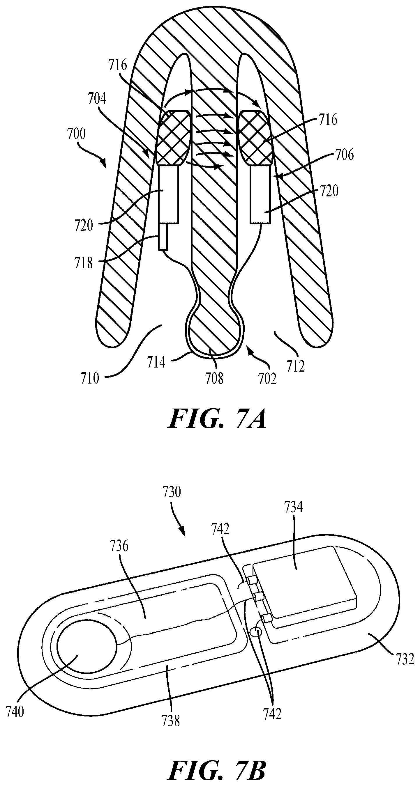
FIG. 7A shows a cross-sectional view of a stimulator positioned in the nasal cavities. FIG. 7B shows a perspective view of a variation of an implantable microstimulator.
FIGS. 8A and 8B show perspective and front views, respectively, of the stimulator of FIGS. 4A-4E with an attached cap.
FIGS. 9A-9D depict portions of a stimulator system comprising a stimulator and a base station. FIG. 9A shows a front view of the stimulator body docked in the base station, while FIGS. 9B, 9C, and 9D depict side, back, and top views, respectively, of the base station.
FIGS. 10A-10D illustrate exemplary amplitude modulation of waveform parameters.
FIGS. 11A-11D and 12 illustrate exemplary pulse width modulations.
FIG. 13A shows a handheld stimulator inserted into a subject's nose; FIG. 13B shows a handheld stimulator held in position for extranasal (control) stimulation.
FIGS. 14A-14B show images of the lower eyelid of a subject before and after 1 minute of intranasal stimulation, respectively. FIGS. 14C-14D show images of the lower eyelid of another subject before and after intranasal stimulation, respectively.
FIG. 15 is a graph of tear meniscus height over time before, during, and after intranasal stimulation.
FIGS. 16A-16E depict exemplary waveforms showing multiple parameters that are concurrently modulated over time.
FIG. 17 illustrates exemplary shape modulation.
FIGS. 18A-18C are graphs of mean temperatures for the lower lid, lower central meibomian glands, and tear film during intranasal stimulation and extranasal (control) stimulation.
DETAILED DESCRIPTION
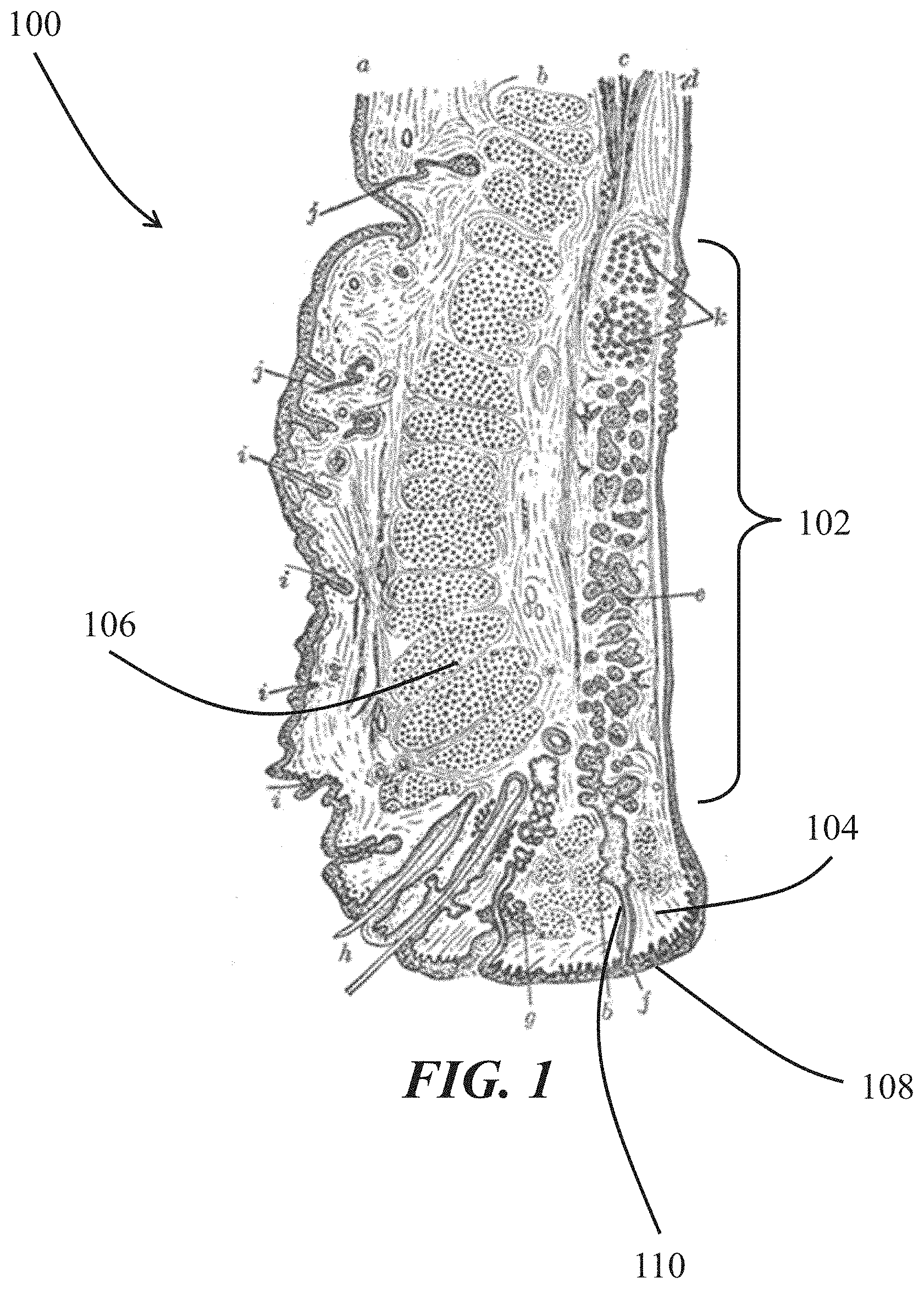
Described here are devices, systems, and methods for treating meibomian gland disease/dysfunction and/or blepharitis using intranasal stimulation. More particularly, the devices, systems, and methods may be configured to deliver an electrical stimulus to the nasal mucosa to cause meibum secretion or otherwise treat meibomian gland disease/dysfunction. The meibomian glands are large sebaceous glands located in the tarsal plates of the upper and lower eyelids. FIG. 1 is a sagittal cross-section through an upper eyelid 100, showing a meibomian gland 102, the Riolan's muscle 104, and the orbicularis muscle 106. Typically, the meibomian glands 102 generate lipids (meibum), which are excreted onto the lid margin 108 through excretory ducts 110. As the orbicularis muscle 106 contracts, it squeezes the meibomian gland 102. If the Riolan's muscle 104, a sphincter muscle at the opening of the meibomian gland, is relaxed during contraction of the orbicularis muscle 106, meibum may be expressed from the gland.
In patients with meibomian gland disease/dysfunction/posterior blepharitis, however, abnormalities of the meibomian glands may result in reduced or no glandular secretions, and/or the meibum that is secreted may have an altered chemical composition. As a result of these changes, there may be a reduction in quantity and/or quality of meibum in the tear film layer. In some variations, the methods described herein may use application of intranasal stimulation to unblock the openings of the meibomian glands, and/or to keep the openings from becoming obstructed. Additionally or alternatively, the methods described herein may use intranasal stimulation to expel meibum from the meibomian glands and onto the eyelid margin. Additionally or alternatively, the methods described herein may use intranasal stimulation to generate sustained increases in quantity of meibum secretion (i.e., increased meibum secretion not during stimulation), and thus sustained changes in tear composition such that the tears have higher lipid content. Additionally or alternatively, the methods described herein may use intranasal stimulation to improve the quality of meibum secretions.
Intranasal stimulation may in part treat meibomian gland disease/dysfunction through generating coordinated muscle contraction and relaxation. More particularly, afferent neurostimulation with particular waveforms (described in detail elsewhere herein) may cause the orbicularis muscle to contract to generate pressure around the meibomian glands. This pressure, when the Riolan's muscle remains relaxed, may result in meibum expression and/or may help to unblock obstructed portions of the glands.
It should be appreciated that direct electrical muscle activation of muscles in the eyelids, in contrast to afferent neurostimulation, may result in contraction of both the orbicularis muscle and Riolan's muscle. As such, unlike afferent neurostimulation achievable with intranasal stimulation, direct muscle activation may not have the desired effect of contraction of the orbicularis muscle without contraction of the Riolan's muscle. Moreover, afferent neurostimulation with intranasal stimulation may result in activation of both large and small muscle fibers in the orbicularis muscle, resulting in less fatigue than direct muscle activation, since direct muscle activation may activate only large muscle fibers. Repeated and/or sustained intranasal stimulation over a period of time may also strengthen the orbicularis muscle, resulting in increased pressure generated around the meibomian gland upon contraction. This may result in an increased ability to eject meibum plugs that may be occluding the opening of the gland, and/or may result in greater ejection of meibum upon contraction. This effect may be seen both during acute intranasal stimulation and when no stimulation is applied. Furthermore, repeated intranasal stimulation over a period of time may also result in improved quality of secreted meibum.
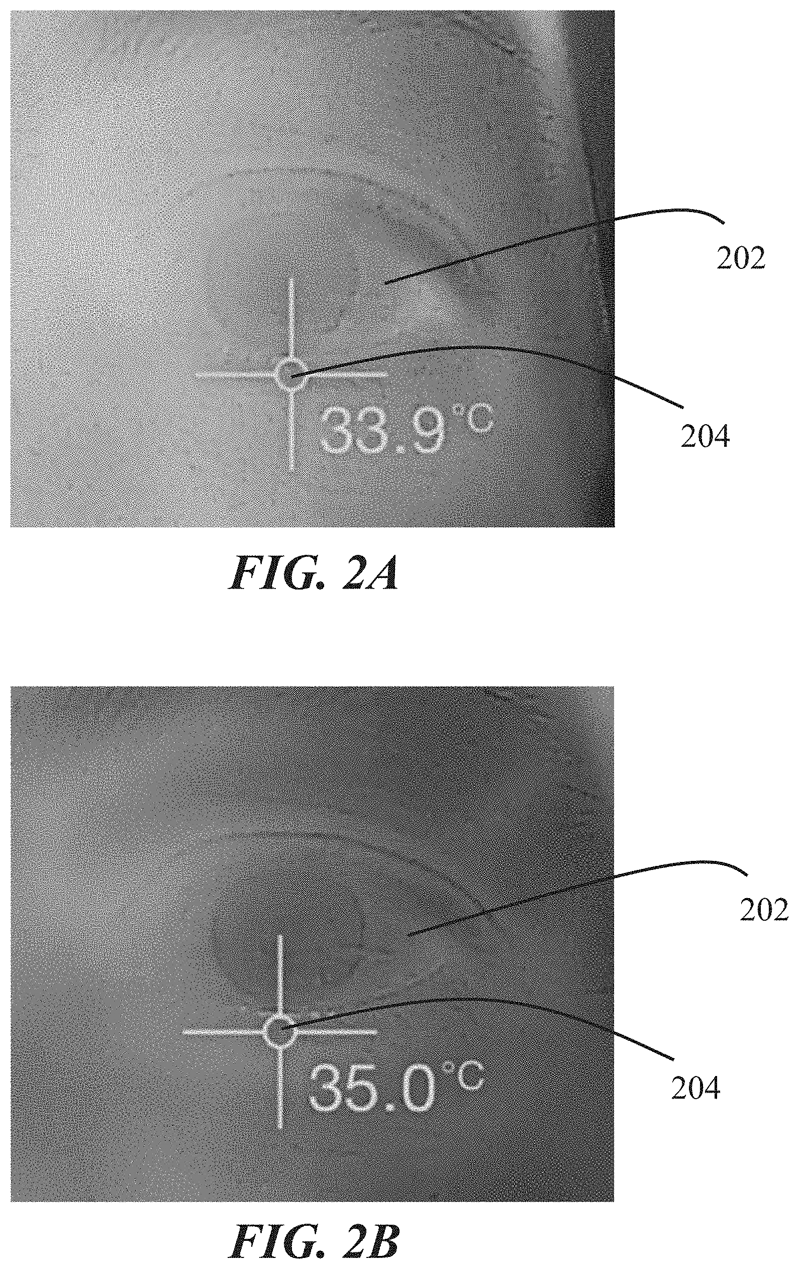
Intranasal stimulation may also in part treat meibomian gland disease/dysfunction through selectively heating the meibomian glands and surrounding tissue (e.g., the eyelids). FIGS. 2A-2B show temperature maps of the eye 202 and surrounding area of a subject before and during intranasal stimulation, respectively. As shown there, before intranasal stimulation, the temperature of the lower eyelid 204 is about 33.9.degree. C. After about 30 seconds of intranasal stimulation, the temperature of the lower eyelid 204 is about 35.0.degree. C. Similarly, FIG. 3 shows a temperature map of an eye 302 and surrounding area of the face of a different subject during intranasal stimulation. There, the temperature of the lower eyelid 304 is about 34.1.degree. C. after about 2 minutes of intranasal stimulation. This heating may be due to muscle activity of the orbicularis muscle. However, unlike application of an external heat source, heating of the eyelid from muscle activity may specifically heat the area where meibum is located, without exposing the cornea, conjunctiva, eye, or skin of the eyelid to excessive heat.
Heating of the meibomian glands may cause meibum inside the glands to warm, thereby lowering its viscosity and causing it to expand. These effects may contribute, along with contraction of the orbicularis muscle, to loosening and expelling of meibum plugs from the glands and unblocking of the opening of the gland, as well as to increased ejection of meibum upon contraction of the orbicularis muscle. Loosening and expelling of meibum from within the glands may also in some instances remove bacteria from the glands and thus provide treatment for posterior blepharitis. These effects may be achieved without risk of corneal abrasion (e.g., without a device in contact with any portion of the cornea or conjunctiva) and without the discomfort associated with physical manipulation of the eyelids.
In addition to causing acute secretion of meibum during intranasal stimulation, delivery of intranasal stimulation over one or more treatment periods may have a sustained effect on meibum secretion onto the eyelid margin. That is, even after cessation of intranasal stimulation, a subject having had intranasal stimulation delivered may experience an increase in quantity and/or quality of meibum secretion, and in turn, increased lipid content in the tear film. This effect may be observed for an extended period of time after a single intranasal stimulation session or after a treatment regimen.
Devices and Systems
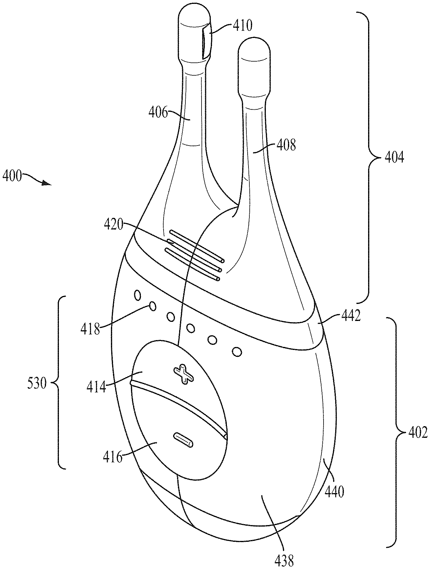
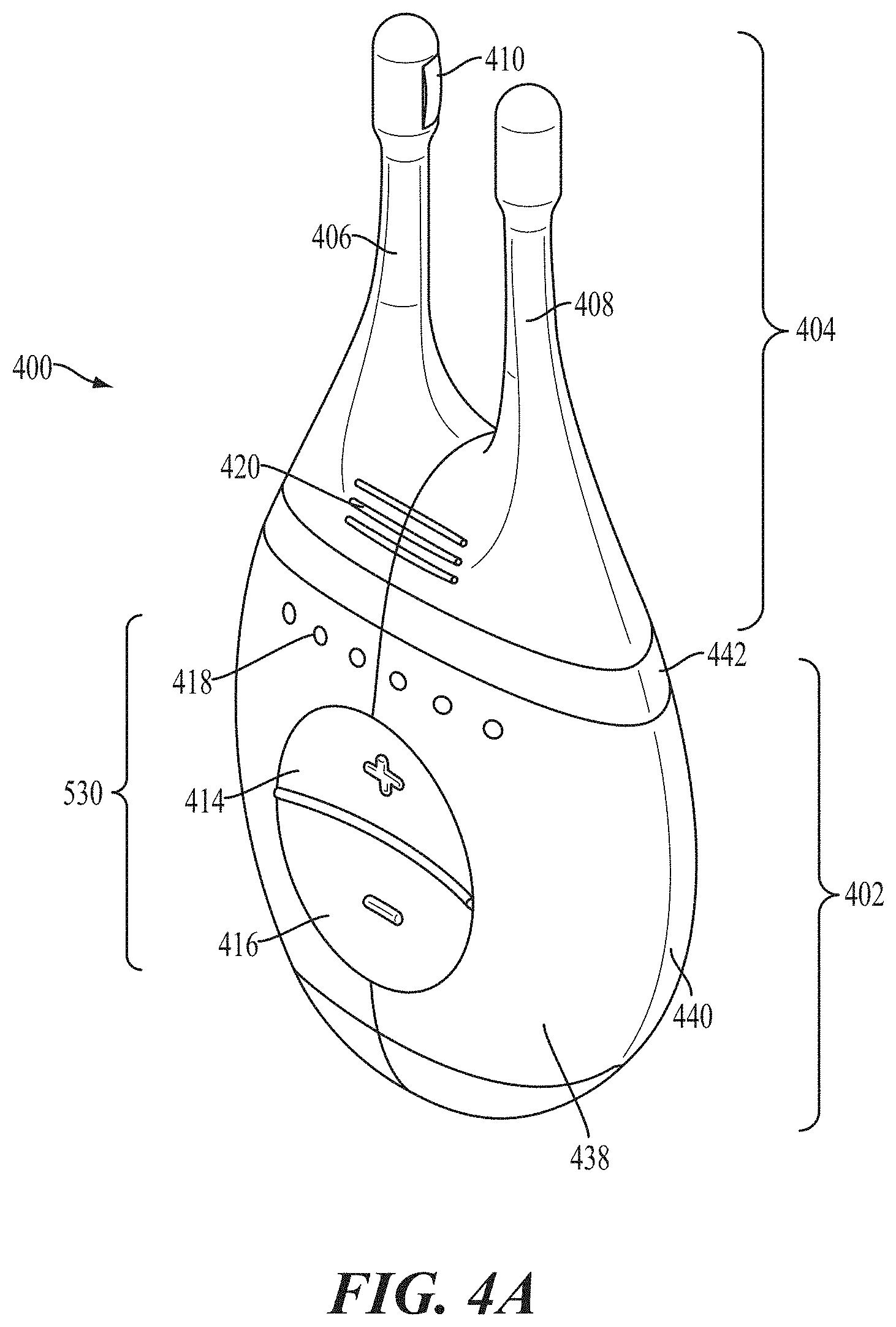
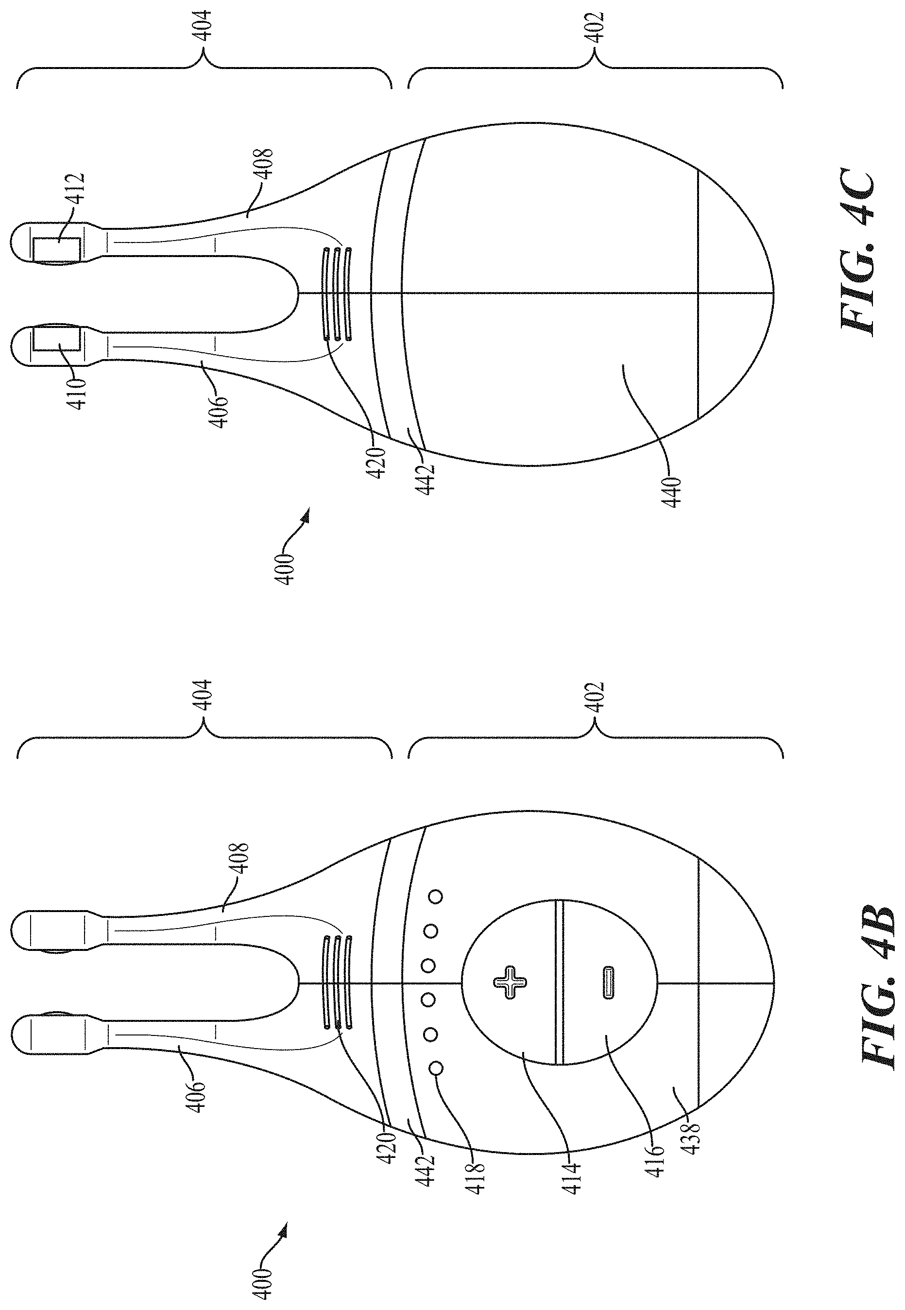
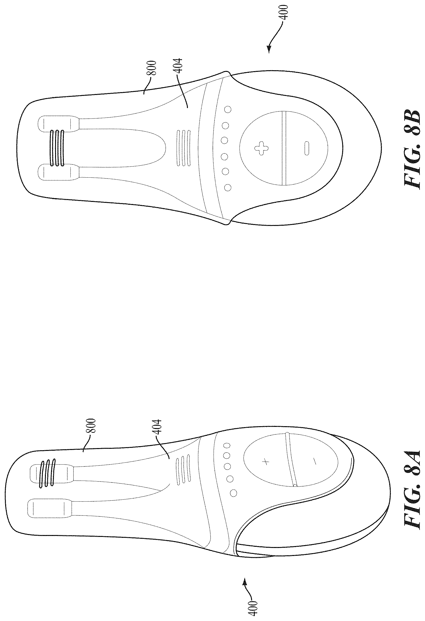
Generally, the systems used in the methods described herein may comprise a stimulator configured to deliver an electrical stimulus to the inner cavity of the nose. Some variations of the stimulation systems described here may comprise a handheld stimulator. FIGS. 4A, 4B, 4C, 4D, 4E show perspective, front, back, cut-away back, and cut-away side views, respectively, of an illustrative variation of a handheld stimulator 400, respectively. FIG. 5 shows a block diagram schematically representing the stimulator 400. As shown in FIGS. 4A-4E, the stimulator 400 may comprise a stimulator body 402 and a stimulator probe 404. Generally, the stimulator body 402 may be configured to generate a stimulus that may be delivered to the subject. The stimulator body 402 may comprise a front housing 438, back housing 440, and proximal housing 442, which may fit together to define a body cavity 454. The body cavity 454 may contain a control subsystem 436 and a power source 452, which together may generate and control the stimulus.
The stimulator body 402 may comprise a user interface 530 comprising one or more operating mechanisms to adjust one or more parameters of the stimulus. The operating mechanisms may provide information to the control subsystem 436, which may comprise a processor 532, memory 534, and/or stimulation subsystem 536. In some variations, the operating mechanisms may comprise first and second buttons 414 and 416. In some variations, pressing the first button 414 may turn on the stimulator and/or change one or more parameters of the stimulus (e.g., increase the intensity of the stimulus, change the stimulation pattern, or the like), while pressing the second button 416 may turn off the stimulator and/or change one or more parameters of the stimulus (e.g., decrease the intensity of the stimulus, change the stimulation pattern, or the like). Additionally or alternatively, the user interface may comprise one or more feedback elements (e.g., based on light, sound, vibration, or the like). As shown, the user feedback elements may comprise light-based indicators 418, which may provide information to the user.
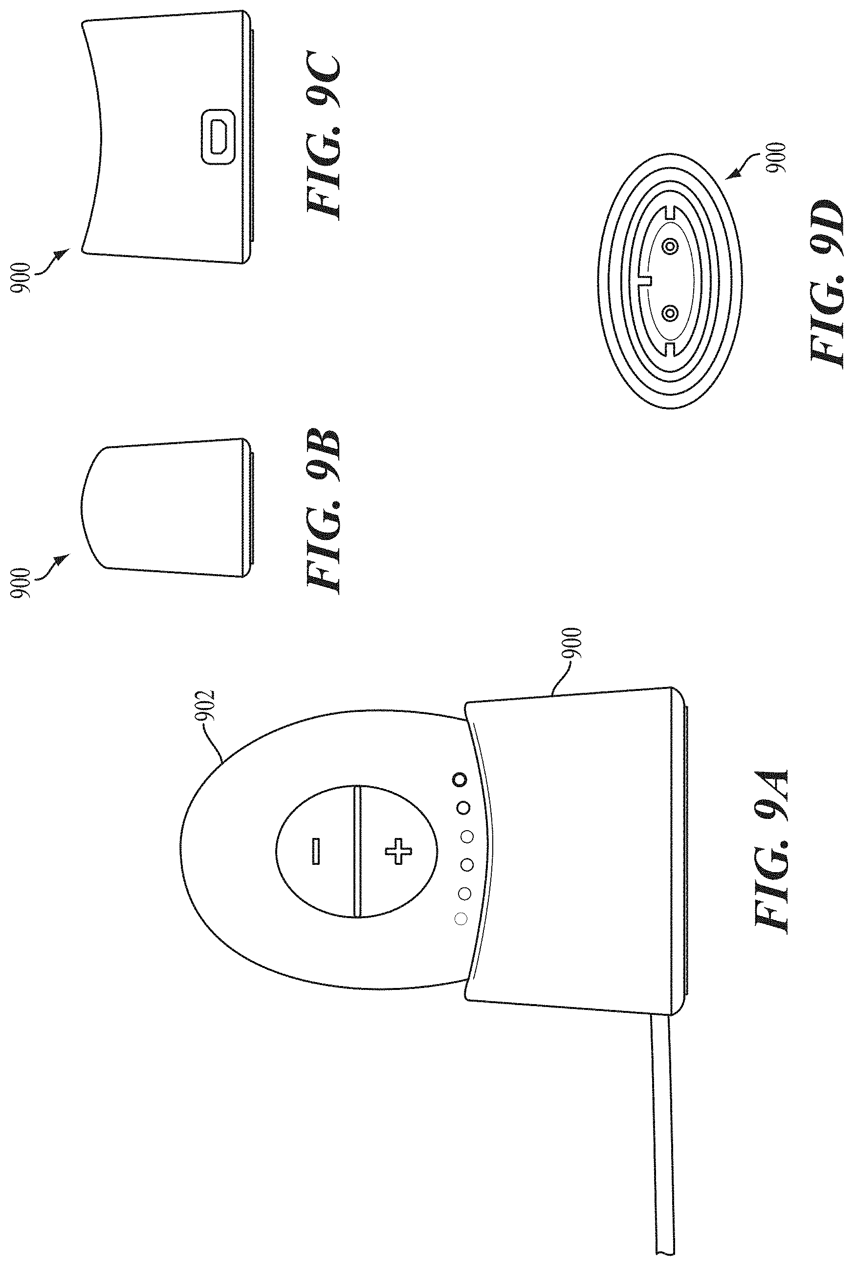
In the variation shown in FIGS. 4A-4E, the user interface may be configured for use by the subject receiving the intranasal stimulation. However, in other variations, the stimulators may comprise an interface configured for use by a person other than the subject receiving the intranasal stimulation, such as a medical professional. For example, a stimulator may comprise a remote interface operable at a distance from the stimulator body. The remote interface may be connected to the stimulator body wirelessly or in a wired manner (e.g., via a cable). The remote interface may allow the stimulator to be turned on/off and or may be used to change one or more parameters of the stimulation. A remote interface may be desirable, for example, so a medical professional can adjust the stimulus parameters when intranasal stimulation is delivered at the medical professional's office. In some variations, the stimulators may be configured to be controlled by both the subject receiving the intranasal stimulation and another person (e.g., a medical professional). For example, a stimulator may comprise a user interface on the stimulator body, as shown in FIGS. 4A-4E, and a remote interface.
The stimulus may be delivered to a subject via the stimulator probe 404. In some variations the stimulator body 402 and stimulator probe 404 may be reversibly attachable. Some or all of the stimulator 400 may be disposable. For example, in some variations the stimulator body may be permanently attached to the stimulator probe, and the entire stimulator may be disposable. In other variations, one or more portions of the stimulator 400 may be reusable. For example, in variations where the stimulator probe 404 is releasably connected to the stimulator body 402, the stimulator body 402 may be reusable, and the stimulator probe 404 may be disposable and periodically replaced.
The stimulator probe 404 may comprise at least one nasal insertion prong, which may be configured to be at least partially inserted into the nasal cavity of a subject. In the handheld stimulator variation shown in FIGS. 4A-4E, the stimulator probe 404 may comprise two nasal insertion prongs 406 and 408. The stimulator probe 404 may further comprise ridges 420, which may allow the subject to more easily grip the probe 404. Each nasal insertion prong may comprise at least one electrode. As shown, the probe 404 may comprise a first electrode 410 on nasal insertion prong 406 and a second electrode 412 on nasal insertion prong 408. As shown in the cut-away view of the stimulator 400 in FIG. 4D, the electrodes 410 and 412 may be connected to leads 430 and 432 located within prongs 406 and 408, respectively. The leads 430 and 432 may in turn be connected to connectors 422 and 424, respectively. Connectors 422 and 424 may extend through lumens in the proximal housing 442, and may connect directly or indirectly to the control subsystem 436 and power source 452. As such, the electrical stimulus may travel from the control subsystem 436 through the connectors 422 and 424, through the leads 430 and 432, and through the electrodes 410 and 412.
In some variations, the nasal insertion prongs may be parallel when not inserted into the nose of a subject, but may have sufficient flexibility to allow the prongs to self-align to the desired stimulation location when inserted into a user's nasal cavities. In other variations of stimulators comprising two nasal insertion prongs, however, the nasal insertion prongs may not be parallel to each other when not inserted into the nose of a subject; that is, the nasal insertion prongs may be positioned at an angle relative to each other. For example, FIGS. 6A-6C show a stimulator probe 600 comprising first and second nasal insertion prongs 602 and 604, respectively, connected to a base member 606. The nasal insertion prongs 602 and 604 may be connected to the base member 606 such that they are angled toward each other. In some variations, the angle between the nasal insertion prongs may be adjustable. For example, the nasal insertion prongs may be biased toward a configuration in which the nasal insertion prongs are at an angle relative to each other, but may be movable to a parallel configuration. When the prongs are inserted into nasal cavities to position tissue (e.g., a nasal septum) between the prongs, the prongs may be rotated away from each other (e.g., by application of a force) prior to insertion into the nasal cavities. Once positioned in the nasal cavities, the return bias may rotate the prongs toward each other (e.g., upon removal of the force), and the return bias of the stimulator probe may press the distal ends of the nasal insertion prongs against tissue. This may help to increase electrode apposition with tissue, and in some instances may act to hold the stimulator probe in place relative to tissue. To remove the stimulator probe from tissue, the prongs may again be rotated away from each other to release the tissue positioned between the prongs.
In some variations in which the stimulator comprises two nasal insertion prongs biased toward each other, the return bias may be sufficient to hold the stimulator in place. In these variations, once positioned in the nasal cavities, the distal ends of the nasal insertion prongs may press against tissue with sufficient force to allow for hands-free use--that is, the user may not need to hold the stimulator in order for the electrodes to remain in apposition with the target tissue. This may improve ease of use, particularly when stimulation is delivered for longer periods of time, as described in more detail herein. In other variations, the return bias may be sufficient to reduce unintended movement of the nasal insertion prongs while inserted, while still requiring the stimulator to be held.
The systems described herein may additionally or alternatively also comprise other features to allow hands-free or reduced-handling use. For example, the system may comprise a strap configured to be placed around a user's head and attached to the stimulator to hold the stimulator in place during stimulation. The strap may be removably attachable to the stimulator, or in other variations may be permanently attached to the stimulator. In some variations of stimulators configured for hands-free use, the stimulators may be configured to be inserted and/or removed by a user. In other variations, the stimulators may be configured to be inserted/removed by a medical professional.
For example, FIG. 7 depicts a cross-sectional view of a subject's nose having a septum 708 and nostrils 710 and 712 having a variation of a hands-free stimulator 700 located therein. As shown there, the stimulator 700 may comprise a clip 702, a first stimulator unit 704 attached to a first end of the clip 702, and a second stimulator unit 706 attached to a second end of the clip 702. Generally, the clip 702 may be configured to temporarily connect the stimulator 700 to a nasal septum 708 of a subject, which may position the first stimulator unit 704 in the first nostril 710 and the second stimulator unit 706 in the second nostril 712.
In some variations, the clip 702 may comprise a u-shaped portion 714 configured to receive and clamp to a portion of the nasal septum 708. This engagement between the clip 702 and the nasal septum 708 may limit advancement of the stimulator 700 into the nose (e.g., to prevent over-insertion of the stimulator 700). The clip 702 may exert sufficient pressure on the septum 708 so as to resist removal of the stimulator 700 from the nose. Accordingly, the clip 702 may allow the stimulator to be positioned in the nose of a subject, and the subject may wear the stimulator without needing to actively hold the stimulator in the nose. The clip 702 may be removed by flexing the clip 702 to disengage it from the septum. As such, the subject may be able to insert and remove the stimulator 700 him- or herself. In some variations, the clip 702 may be at least partially formed from one or more shape memory materials (e.g., a nickel-titanium alloy), such that the clip 702 may be deformed to disengage the clip 702 from the septum 708 and may return to its original shape. In some variations an exterior portion of the clip 702 may be formed from one or more insulating materials (e.g., PTFE, silicone, combinations thereof, or the like), and an interior portion may include an electrically conductive core (e.g., a wire of any suitable metal, such as silver, stainless steel, platinum, alloys thereof, or the like) electrically connecting the first stimulator unit 704 to the second stimulator unit 706. In these variations, the insulating outer portion of the clip 702 may prevent inadvertent electrical stimulation between the clip 702 and the subject.
Generally, each stimulator unit may comprise one or more electrodes 716. In some variations, it may be desirable for the stimulator units to comprise a radially expandable structure that may expand to contact the nasal mucosa when inserted into the nostrils. While shown in FIG. 7 as being formed from an expandable wire mesh/braid electrode, each electrode 716 may be configured in any suitable manner. For example, in some variations the electrodes may comprise a hydrogel. Additionally or alternatively, it may be desirable for the stimulator units to comprise a smooth surface to prevent tissue abrasion. The stimulator may be configured such that the electrodes 716 are placed in contact with any suitable tissue structure or structures (e.g., the nasal mucosa above the columella, such as the nasal mucosa superior to the columella (e.g., the nasal mucosa near the interface between the nasal bone and the upper lateral cartilage) when the clip 702 is connected to the nasal septum.
Generally, the first 704 and/or second 706 stimulator units may comprise a housing 720, which may comprise a control subsystem having a processor, a stimulation subsystem, and a memory. In some variations the control subsystem may have a detection subsystem. Additionally or alternatively, the stimulator may comprise a communication subsystem. The stimulator circuitry may be housed in a single housing 720 (e.g., a housing 720 of the first stimulator unit 704 or a housing 720 of the second stimulator unit 706), or may be divided between multiple housings (e.g., a housing 720 of the first stimulator unit 704 and a housing 720 of the second stimulator unit 706). In some variations, the stimulator 700 may comprise a power source (e.g., a battery) (not shown). In other variations, the stimulator 700 may be powered wirelessly (e.g., via power received from a coil 718 or other antenna).
In some variations, the stimulators described here may comprise a cap to protect the stimulator probe. For example, FIGS. 8A and 8B show perspective and front views, respectively, of stimulator 400 with an attached cap 800. As shown there, the cap 800 may fit over the stimulator probe 404, which may protect the probe from contamination. More particularly, it may be desirable for the cap to protect the nasal insertion prongs, and especially the electrodes, from contamination. The systems described here may additionally or alternatively comprise a base station. The base station may be configured to releasably connect to one or more portions of the stimulator, and may be configured to perform one or more functions when connected to the stimulator. FIGS. 9A-9D depict a portion of a stimulator system comprising a base station 900 as described here. FIG. 9A shows a front view a stimulator body 902 docked in the base station 900, while FIGS. 9B, 9C, and 9D depict side, back, and top views of the base station 900, respectively. In variations where the stimulator body 902 comprises a rechargeable power source (such as a rechargeable battery, capacitor, or the like), the base station 900 may be configured to recharge the rechargeable power source.
In other variations of stimulator systems suitable for use in the methods described herein, all or a portion of the systems may be surgically implantable. For example, FIG. 7B shows an implantable microstimulator 730. As shown, the microstimulator 730 may comprise a housing and an extension 736 connected to the housing. Generally, the microstimulator may be inserted into a tissue pocket within the nasal submucosal layer. The shape and size of the microstimulator may be flat and thin and may aid in atraumatic insertion of the device into nasal tissue. One or more portions of the microstimulator (e.g., an extension) may be formed from a flexible material such as silicone and/or may be a molded component, such as a molded silicone, which may allow the microstimulator to conform to one or more portions of the anatomy (e.g., the nasal septum) and/or prevent trauma during implantation. In some variations, the microstimulator may be encapsulated in a coating. A coating, such as silicone, may provide electrical insulation, waterproofing, biocompatibility, and/or safety (e.g., rounded edges, lubricious surface that slides easily over the nasal septum). FIG. 7B shows a variation of a microstimulator 730 encapsulated in a coating 732.
The microstimulator may be small enough to be inserted through a nostril and implanted within a layer of submucosa adjacent to the nasal septum or a turbinate without significantly interfering with the passage of air or fluid through the nasal cavity. In some variations the dimensions may be less than about 30 mm by about 10 mm by about 5 mm (L.times.W.times.H). In some of these variations, the dimensions may be about 15 mm-25 mm by about 3 mm-7 mm by about 1 mm-3 mm (L.times.W.times.H). In some of these variations, the dimensions may be about 17 mm by about 5 mm by about 2 mm (L.times.W.times.H).
The housing of the microstimulator 730 may comprise a housing case 734 containing some or all of a stimulation circuit. The housing case may be hermetically sealed and may be formed from one or more metals (e.g., titanium) or other biocompatible materials. In a microstimulator that comprises a passive stimulation circuit without an internal power source, the microstimulator may comprise one or more elements to receive power from an external source. For example, a controller may generate and transmit power wirelessly via an output signal (e.g., magnetic field). The microstimulator may comprise one or more energy-receiving units that receive the output signal from the controller to power the microstimulator. In some variations, the energy-receiving unit may be located in the extension of the microstimulator. The energy-receiving unit may be a coil, which may be formed from a wire having a length turned into a plurality of windings.
The extension of the microstimulator may comprise one or more electrodes, which may deliver an electrical stimulus to tissue. In FIG. 7B, the extension 736 comprises one electrode 740. However, it should be appreciated that the extension may comprise any suitable number of electrodes (e.g., one, two, three, or four or more electrodes) positioned on any suitable portion or portions of the extension. The microstimulator may comprise one or more feedthroughs that extend between and electrically connect the housing and the extension. For example, FIG. 7B illustrates feedthroughs 742, which extend between the housing case 734 and the extension 736, which may electrically connect the circuitry within the housing to the coil 738 and/or electrode 740 on the extension 736.
Systems comprising an implantable stimulator may comprise a controller, which may communicate with an implanted microstimulator to transmit and/or receive power, information, or the like. The controller may remain external to the body and communicate wirelessly with the microstimulator. In some variations, the output signal produced by the controller may provide power to the microstimulator. The controller may be brought into the vicinity of an implanted microstimulator, and may produce an output signal received by the implanted microstimulator. The implanted microstimulator may in turn generate a stimulation signal used to stimulate an anatomical target, as described in more detail herein. It should be appreciated that the controller may comprise any suitable form. For example, the controller may comprise a hand-held device. In other variations, the controller may comprise a wearable device (e.g., glasses, wristwatch), a key fob, or the like.
Other variations and features of stimulator systems and components thereof suitable for use in the methods described herein are described in U.S. application Ser. No. 14/256,915, filed Apr. 18, 2014, and titled "NASAL STIMULATION DEVICES AND METHODS"; in U.S. patent application Ser. No. 14/630,471, filed on Feb. 24, 2015, and titled "POLYMER FORMULATIONS FOR NASOLACRIMAL STIMULATION"; in U.S. patent application Ser. No. 14/920,860, filed Oct. 22, 2015, and titled "STIMULATION DEVICES AND METHODS FOR TREATING DRY EYE"; in U.S. patent application Ser. No. 14/920,852, filed Oct. 22, 2015, and titled "IMPLANTABLE NASAL STIMULATOR SYSTEMS AND METHODS"; and in U.S. patent application Ser. No. 14/809,109, filed Jul. 24, 2015, and titled "STIMULATION PATTERNS FOR TREATING DRY EYE," each of which is hereby incorporated by reference in its entirety.
In general, the stimulators may be programmed to deliver waveforms configured to generate rhythmical contraction and relaxation of the orbicularis muscle of the eyelid. In some variations, the waveforms may be pulse-based. The waveforms may be configured to stimulate tissue on both sides of the nose, or the waveforms may be configured to stimulate tissue on a single side of the nose. In some variations, the waveform may comprise symmetric biphasic pulses. The waveform may have any suitable parameters (e.g., frequency, pulse width, amplitude) in order to cause meibum expression or to otherwise cause rhythmical contraction and relaxation of the orbicularis muscle.
Generally, the stimulus may comprise a waveform of less than about 200 Hz. In some of these variations, the frequency is preferably between about 10 Hz and about 200 Hz. In some of these variations, the frequency is preferably between about 30 Hz and about 150 Hz. In others of these variations, the frequency is preferably between about 50 Hz and about 80 Hz. In others of these variations, the frequency is preferably between about 30 Hz and about 60 Hz. In others of these variations, the frequency is preferably between about 30 Hz and about 80 Hz. In some variations, the frequency may be about 1.5 Hz, about 10.25 Hz, about 70 Hz, about 150 Hz, about 25 Hz, about 27.5 Hz, about 30 Hz, about 32.5 Hz, about 35 Hz, about 37.5 Hz, about 40 Hz, about 42.5 Hz, about 45 Hz, about 47.5 Hz, about 50 Hz, about 52.5 Hz, about 55 Hz, about 57.5 Hz, about 60 Hz, about 62.5 Hz, or about 65 Hz. In some variations, the frequency of the waveform may vary over time.
Generally, when the stimulus comprises a biphasic pulse and the first phase of the biphasic pulse is current-controlled, the first phase may preferably have an amplitude between about 1.0 mA and about 10 mA. Amplitudes within these ranges may be high enough to stimulate targeted tissue, but sufficiently low as to avoid any significant heating of tissue, ablation of tissue, or the like. In some variations the amplitude may be between about 1.0 mA and about 5.0 mA. In other variations, the first phase may have an amplitude of about 0.1 mA, about 0.2 mA, about 0.3 mA, about 0.4 mA, about 0.5 mA, about 0.6 mA, about 0.7 mA, about 0.8 mA, about 0.9 mA, or about 1.0 mA. In some variations, the amplitude may be variable. For example, the amplitude may vary between about 1.3 mA and about 1.5 mA, about 2.2 mA and about 2.5 mA, about 3.2 mA and about 3.7 mA, or about 4.3 mA and about 5.0 mA. When the first phase of a biphasic pulse is voltage-controlled, the first phase may preferably have an amplitude between about 10 mV and about 100 V.
When a stimulator is configured to deliver a pulse-based waveform, in some variations, the amplitude of the pulses may be constant over time. In other variations, the amplitude of the pulses may vary over time. This may reduce subject accommodation. In some variations, the amplitude of pulses may increase (linearly, exponentially, etc.) from a minimum value to a maximum value, drop to the minimum value, and repeat as necessary. In some variations, the amplitude of the pulses may vary according to a sinusoidal profile. In another variation, as illustrated in FIG. 10A, the amplitude may periodically increase from a baseline amplitude (A) to a higher amplitude (B) for a single pulse. In yet another variation, as illustrated in FIGS. 10B-10C, the amplitude of the pulses may follow a periodically increasing and decreasing pattern between two lower amplitudes (A, B), and periodically increase to a higher amplitude (C) for a single pulse (FIG. 10B) or for a plurality of pulses (e.g., two pulses) (FIG. 10C). In yet another variation, as illustrated in FIG. 10D, a higher amplitude pulse (or pulses) may be preceded by a brief pause (i.e., no current delivery). Each of these types of amplitude modulation may be implemented alone or in combination with any other type of amplitude modulation, and may reduce subject accommodation.
In some variations in which the amplitude varies over time, the amplitude may vary at a frequency suitable for reducing subject accommodation or increasing subject comfort, such as between about 0.1 Hz and about 5 Hz, between about 1 Hz and about 5 Hz, between about 1 Hz and about 2 Hz, between about 2 Hz and about 3 Hz, between about 3 Hz and about 4 Hz, or between about 4 Hz and about 5 Hz. In some variations, the amplitude may vary at a frequency of about 1.0 Hz, about 1.1 Hz, about 1.2 Hz, about 1.3 Hz, about 1.4 Hz, about 1.5 Hz, about 1.6 Hz, about 1.7 Hz, about 1.8 Hz, about 1.9 Hz, about 2.0 Hz, about 2.1 Hz, about 2.2 Hz, about 2.3 Hz, about 2.4 Hz, about 2.5 Hz, about 2.6 Hz, about 2.7 Hz, about 2.8 Hz, about 2.9 Hz, about 3.0 Hz, about 3.1 Hz, about 3.2 Hz, about 3.3 Hz about 3.4 Hz, about 3.5 Hz, about 3.6 Hz, about 3.7 Hz, about 3.8 Hz, about 3.9 Hz, or about 4.0 Hz. In some variations, it may be desirable for the amplitude to vary at a frequency suitable for achieving a "pumping" action that may help to express meibum and open blocked meibomian glands. In other variations, the stimulation waveform may be a modulated high frequency signal (e.g., sinusoidal), which may be modulated at a beat frequency of the ranges described above. In such variations, the carrier frequency may be between about 100 Hz and about 100 kHz.
When the stimulus comprises a biphasic pulse, the first phase may preferably have a pulse width between about 1 .mu.s and about 10 ms. In some of these variations, the pulse width may be between about 10 .mu.s and about 100 .mu.s. In other variations, the pulse width may be between about 100 .mu.s and about 1 ms. In yet other variations, the pulse width may be between about 0 .mu.s and about 300 .mu.s. In yet other variations, the pulse width may be between about 0 .mu.s and 500 .mu.s. It may be desirable to have a longer pulse width to achieve a "pumping" action that may help to express meibum and open blocked meibomian glands.
In some variations, the pulse width may be constant over time. In other variations, the pulse width may vary over time. Pulse width modulation over time may increase the efficacy and/or comfort of the stimulation. In some variations, the pulse width may increase (linearly, exponentially, etc.) from a minimum value to a maximum value, drop to the minimum value, and repeat as necessary. In some variations, the pulse width may vary according to a sinusoidal profile. In another variation, as illustrated in FIG. 11A, the pulse width may periodically increase from a baseline pulse width (A) to a longer pulse width (B) for a single pulse. In yet another variation, as illustrated in FIGS. 11B-11C, the pulse width may follow a periodically increasing and decreasing pattern between two shorter pulse widths (A, B), and periodically lengthen to a longer pulse width (C) for a single pulse (FIG. 11B) or for a plurality of pulses (e.g., two pulses) (FIG. 11C). In yet another variation, as illustrated in FIG. 11D, a longer pulse width pulse (or pulses) may be preceded by a brief pause (i.e., no current delivery). Each of these types of pulse width modulation may be implemented alone or in combination with any other type of pulse width modulation. In any form of pulse width modulation, the pulse width may vary at any suitable frequency. In some variations the pulse width may vary at about 0.1 Hz, about 0.2 Hz, about 0.3 Hz, about 0.4 Hz, about 0.5 Hz, about 0.6 Hz, about 0.7 Hz, about 0.8 Hz, about 0.9 Hz, about 1 Hz, about 1.1 Hz, about 1.2 Hz, about 1.3 Hz, about 1.4 Hz, or about 1.5 Hz. In some variations, modulation of the pulse width at a rate between about 0.5 Hz and 1 Hz may be desirable to increase subject comfort during stimulation.
In some variations, the increase and decrease of pulse width may be defined by a function implemented by the stimulator. For example, the pulse width may be defined by a function such that the pulse width varies exponentially. In one variation, the function defining pulse width may comprise two phases--a first phase during which the pulse width of the leading pulse increases over time, and a second phase during which the pulse width of the leading pulse decreases over time. During the first phase, the pulse width of the leading pulse approaches the maximum pulse width according to an exponential function, where at time t, PW{t} is defined by the equation
.times..times..tau. ##EQU00001## where PW.sub.max is the maximum allowed pulse width, PW.sub.min is the minimum allowed pulse width, and .tau. is a time constant.
Once a predetermined amount of time has elapsed (a multiple of time constant .tau.), the pulse width modulation may enter the second phase. During the second phase, the pulse width of the leading pulse exponentially decays from its maximum value to a minimum value following the exponential equation
.times..times..tau. ##EQU00002##
After a predetermined amount of time has elapsed (a multiple of time constant .tau.), the pulse width modulation may re-enter the first phase, and the cycle may repeat. The pulse width of the secondary (charge balancing) pulse is increased and decreased accordingly to retain charge full balancing. PW.sub.max, PW.sub.min, and .tau. may have any suitable values to achieve the pulse widths described herein, but in one example the waveform may have a PW.sub.max of 300 .mu.s, PW.sub.min of 0 .mu.s, and .tau. of 1/5 .mu.s. In other variations, for example, PW.sub.max may be about 100 .mu.s, about 200 .mu.s, about 300 .mu.s, about 400 .mu.s, or about 500 .mu.s; PW.sub.min may be about 0 .mu.s, about 10 .mu.s, about 50 .mu.s, or about 100 .mu.s; and .tau. may be about 1/3 .mu.s, about 1/4 .mu.s, about 1/5 .mu.s, or about 1/6 .mu.s. An exemplary function defining exponentially increasing and decaying pulse widths is shown in FIG. 12.
In some instances, the waveforms may be delivered in a continuous fashion, while in other instances, the waveforms may be delivered in a non-continuous fashion having on periods and off periods. Exemplary on/off durations include without limitation, 0.5 seconds on/0.5 seconds off, 1 second on/1 second off, 1 second on/2 seconds off, 2 seconds on/1 seconds off, 2 seconds on/2 seconds off, 5 seconds on/5 seconds off, 0.2 seconds on/0.8 seconds off, and less than 1 second on/less than 10 seconds off. Non-continuous delivery with on and off periods may help to reduce subject accommodation, may allow for rest periods for eyelid muscles caused to contract by afferent neurostimulation, and/or may be more effective than continuous delivery in causing meibum secretion onto the eyelids or dislocating meibum plugs at the gland openings.
In one exemplary waveform, illustrated in FIG. 16A, the waveform may have a stimulation frequency of 30 Hz; a minimum stimulation current amplitude of 0.7 mA, a maximum stimulation current amplitude of 0.7 mA, and thus no variation in maximum stimulation current amplitude; a minimum pulse width of 0 .mu.s; a maximum pulse width of 300 .mu.s; a pulse width modulation frequency of 1 Hz (rising and falling according to an exponential function, as shown in FIG. 12); a minimum charge injection per phase (at 0 .mu.s pulse width) of 0 .mu.C; and a maximum charge injection per phase (at 0.7 mA and 300 .mu.s) of 0.21 .mu.C.
The waveform illustrated in FIG. 16A may have a pulse shape that is cycled between four periods, as shown in FIG. 17. The first period may comprise a two-phase current-controlled waveform with symmetrical phases. The second period may comprise a current-controlled first phase, followed by a voltage-controlled second phase. The first phase may have a current sourced by a first electrode and sunk by a second electrode, while the second phase may have a current sourced by the second electrode and sunk by the first electrode. The third period may comprise a two-phase current-controlled waveform with symmetrical phases (i.e., the third period may be the same as the first period). The fourth period may comprise a current-controlled first phase, followed by a voltage-controlled second phase. The first phase may have a current sourced by the second electrode and sunk by the first electrode, while the second phase may have a current sourced by the first electrode and sunk by the second electrode. In each period, the pulses may be charged-balanced.
The cycling of the waveform shape through different periods may sequentially preferentially stimulate a location closer to one electrode or the other. By doing so, the stimulation may favor the ipsilateral reflex pathway over the contralateral reflex pathway, thus having a greater effect on the orbicularis muscle on the ipsilateral side, while allowing the orbicularis muscle on the contralateral side to relax. The result may be a "pumping" action that may help to express meibum and open blocked meibomian glands. It may be desirable to have a sufficiently long duration for each period to cause this pumping action, such as but not limited to about 2 seconds, about 3 seconds, about 5 seconds, or about 10 seconds.
In another exemplary waveform, illustrated in FIG. 16B, the waveform may have a stimulation frequency of 37.5 Hz; a minimum stimulation current amplitude of 1.33 mA, a maximum stimulation current amplitude of 1.5 mA, a variation in maximum stimulation current amplitude of 0.17 mA, and an amplitude modulation frequency of 2.1 Hz; a minimum pulse width of 0 .mu.s; a maximum pulse width of 300 .mu.s; a pulse width modulation frequency of 1 Hz (rising and falling according to an exponential function, as shown in FIG. 12); a minimum charge injection per phase (at 0 .mu.s pulse width) of 0 .mu.C; a maximum charge injection per phase (at 1.5 mA and 300 .mu.s) of 0.45 .mu.C; and a pulse shape that is modulated as described above and shown in FIG. 17.
In another exemplary waveform, illustrated in FIG. 16C, the waveform may have a stimulation frequency of 45 Hz; a minimum stimulation current amplitude of 2.17 mA, a maximum stimulation current amplitude of 2.5 mA, a variation in maximum stimulation current amplitude of 0.33 mA, and an amplitude modulation frequency of 2.6 Hz; a minimum pulse width of 0 .mu.s; a maximum pulse width of 300 .mu.s; a pulse width modulation frequency of 1 Hz (rising and falling according to an exponential function, as shown in FIG. 12); a minimum charge injection per phase (at 0 .mu.s pulse width) of 0 .mu.C; a maximum charge injection per phase (at 2.5 mA and 300 .mu.s) of 0.75 .mu.C; and a pulse shape that is modulated as described above and shown in FIG. 17.
In another exemplary waveform, illustrated in FIG. 16D, the waveform may have a stimulation frequency of 52.5 Hz; a minimum stimulation current amplitude of 3.2 mA, a maximum stimulation current amplitude of 3.7 mA, a variation in maximum stimulation current amplitude of 0.5 mA, and an amplitude modulation frequency of 2.8 Hz; a minimum pulse width of 0 .mu.s; a maximum pulse width of 300 .mu.s; a pulse width modulation frequency of 1 Hz (rising and falling according to an exponential function, as shown in FIG. 12); a minimum charge injection per phase (at 0 .mu.s pulse width) of 0 .mu.C; a maximum charge injection per phase (at 3.7 mA and 300 .mu.s) of 1.11 .mu.C; and a pulse shape that is modulated as described above and shown in FIG. 17.
In another exemplary waveform, illustrated in FIG. 16E, the waveform may have a stimulation frequency of 60 Hz; a minimum stimulation current amplitude of 4.3 mA, a maximum stimulation current amplitude of 5.0 mA, a variation in maximum stimulation current amplitude of 0.67 mA, and an amplitude modulation frequency of 2.5 Hz; a minimum pulse width of 0 .mu.s; a maximum pulse width of 300 .mu.s; a pulse width modulation frequency of 1 Hz (rising and falling according to an exponential function, as shown in FIG. 12); a minimum charge injection per phase (at 0 .mu.s pulse width) of 0 .mu.C; a maximum charge injection per phase (at 5.0 mA and 300 .mu.s) of 1.5 .mu.C; and a pulse shape that is modulated as described above and shown in FIG. 17.
In some variations, the stimulators described herein may be configured to deliver current between different three or more stimulation contacts (e.g., electrodes) in order to allow for spatial patterning of the current delivery. In these cases, it may be desirable to apply two or more different waveforms between different stimulation contacts. Generally, it may be desirable to temporally or spatially pattern the stimulation in order to reduce accommodation over the longer stimulation times that may be used to treat meibomian gland disease/dysfunction and/or blepharitis. Exemplary waveforms and methods for reducing accommodation are described in U.S. application Ser. No. 14/920,860, filed Oct. 22, 2015, and titled "STIMULATION DEVICES AND METHODS FOR TREATING DRY EYE" and in U.S. patent application Ser. No. 14/809,109, filed Jul. 24, 2015, and titled "STIMULATION PATTERNS FOR TREATING DRY EYE," each of which was previously incorporated by reference in its entirety.
It may also be desirable to use waveforms that reduce sneezing during delivery. Sneezing may be particularly uncomfortable for a subject when intranasal electrical stimulation is delivered for longer durations, as it may be for treatment of meibomian gland disease/dysfunction and/or blepharitis. Certain waveform parameters that may reduce sneezing may include, for example, amplitude or pulse width ramps, pauses between portions of the waveform configured to preferentially stimulate the left and right sides of the nose, and/or using automated feedback (e.g., using an electromyogram (EMG) signal, as described in more detail herein) to determine when a patient may be about to sneeze. Just before a patient is about to sneeze, the waveform amplitude may be significantly reduced (unilaterally or bilaterally), or the amplitude or pulse width may be significantly increased to interrupt the sensation. Other techniques may include more pauses within the waveform, random pauses within the waveform, and/or amplitude or pulse width spikes.
Some variations of the methods described herein may comprise adjusting the waveform parameters based on feedback. In some variations the feedback may help to adjust the stimulus to be maximally effective in causing meibum secretion. Additionally or alternatively, the feedback may help to adjust the stimulus to prevent accommodation while limiting unpleasant sensations, such as a feeling of needing to sneeze. Such adjustment may make stimulation for a longer duration (e.g., five to ten minutes) more comfortable for a subject without resulting in subject accommodation.
In variations of methods comprising an in-office treatment component and an at-home treatment component, as described in more detail herein, different waveforms may be used for in-office and at-home stimulation. When different stimulators are used for in-office and at-home stimulation, a stimulator for in-office use may be programmed with a first set of waveforms, and a stimulator for at-home stimulation may be programmed with a second set of waveforms. Generally, the waveforms used for in-office stimulation may generate a stronger sensation than the waveforms use for at-home stimulation. For example, the waveforms used for in-office stimulation may have higher maximum amplitudes and longer on/off periods.
Methods
Generally, the stimulators described herein may be used to deliver intranasal stimulation to treat meibomian gland disease/dysfunction. The methods may comprise delivering intranasal stimulation to open obstructed portions of the meibomian glands or openings, causing acute expression of meibum from the glands, contributing to sustained increases in the quantity of meibum secretion (and thus sustained increases in the lipid content of tears), and/or contributing to sustained improvements in the quality of secreted meibum. It should also be appreciated that the methods described herein may be used as a prophylactic measure to treat users who may be at an increased risk of developing meibomian gland disease/dysfunction.
The stimulation may be delivered to the nasal mucosa. In some instances, the targeted area may comprise tissue innervated by the anterior ethmoidal branch of the nasociliary nerve. In some instances, the targeted area of the nasal mucosa may be superior to the columella. It may in some instances be near the inferior end of the nasal bone (i.e., near the interface between the nasal bone and the upper lateral cartilage). As such, the stimulus may be delivered between about 20 mm and about 35 mm into the nasal cavity of the patient, in some cases via an electrode between about 25 mm and about 35 mm into the nasal cavity of the patient. In other instances, the targeted area may be the columella. It may be desirable that the stimulus be delivered in the anterior portion of the nasal cavity, within the nostrils and anterior to the turbinates, and in some instances, at a location anterior to the middle turbinate, or at a location anterior to the inferior turbinate. The stimulus may be delivered at least partially through tissue of or near the septum, and it may in some instances be desirable to direct the stimulus such that a portion is directed toward the front of the nose. This may allow for selective activation of nerves in the front of the septum (e.g., the ophthalmic branch of the trigeminal nerve) while minimizing activation of nerves toward the rear of the nasal septum, which may reduce negative side effects that may occur from stimulation of nerves that innervate the teeth, and which may reduce rhinorrhea. It may also in some instances be desirable to direct the stimulus so as to reduce negative side effects that may occur from stimulation of the olfactory area.
The methods described herein may comprise delivering intranasal stimulation according to one or more treatment regimens. The methods described herein may also include determining a type of dry eye in a subject. The one or more treatment regimens may be selected based on the determined type of dry eye. For example, the methods may comprise determining that a subject afflicted with dry eye has meibomian gland disease/dysfunction. The methods may further comprise intranasally delivering or instructing a subject to deliver an electrical stimulation to the subject, where the intranasally delivered electrical stimulation is effective to treat meibomian gland disease/dysfunction. For example, to treat meibomian gland disease/dysfunction, stimulation may be delivered to a subject as needed and/or according to a predetermined plan. In some variations, it may be desirable that each round of stimulation is long enough to result in acute meibum secretion. It may in some instances take several or more minutes of intranasal stimulation to achieve meibum secretion, depending on the severity of meibomian gland disease/dysfunction. For example, in a patient with severe meibomian gland disease/dysfunction, it may take about 5 to 10 minutes to achieve meibum secretion, while in a patient with moderate meibomian gland disease/dysfunction, it may take about 3 to 5 minutes to achieve meibum secretion, while in a patient without meibomian gland disease/dysfunction, it may take only about 1 minute to achieve meibum secretion with particular stimulus parameters.
In some variations, the methods described herein may comprise repeated delivery of intranasal electrical stimulation over an extended period of time, such as but not limited to at least 30 days, at least 45 days, at least 60 days, or at least 90 days. For example, the method may comprise daily delivery of intranasal electrical stimulation over an extended period of time, such as but not limited to at least 30 days, at least 45 days, at least 60 days, or at least 90 days.
In some variations, the methods described herein may comprise an initial round of stimulation that is longer than subsequent rounds of stimulation. The initial round of stimulation may in some variations be of a length sufficient to achieve meibum secretion in a patient with meibomian gland disease/dysfunction who has not previously been treated with electrical intranasal stimulation. Because the openings or other portions of these patients' meibomian glands may be significantly obstructed, a longer stimulation duration may be needed to achieve meibum expression. As described in more detail herein, intranasal stimulation may cause massaging of the meibomian gland due to contractions of eyelid muscles, and may cause warming of the meibomian glands, which may loosen and reduce the viscosity of meibum and ultimately allow meibum to be secreted onto the eyelid margin; however, in a patient with significantly obstructed meibomian glands, longer stimulation may be needed to achieve a noticeable effect. For example, an initial round of stimulation may have a duration of between about 5 and 10 minutes. In some of these variations, an initial round of stimulation may have a duration of between about 5 and 7 minutes, between about 7 and 10 minutes, or between about 7 and 8 minutes.
Subsequent rounds of stimulation after an initial round of stimulation may be shorter, for example, about 1 to 3 minutes. These subsequent rounds may not need to be as long as an initial round of stimulation because a patient's meibomian glands may have been previously unblocked during the initial stimulation round. In some variations, subsequent rounds of stimulation may be delivered repeatedly over an extended period of time, such as but not limited to at least 30 days, at least 45 days, at least 60 days, or at least 90 days. For example, subsequent rounds of stimulation may be delivered at least once daily over the extended period of time.
The subject may optionally be instructed to blink during intranasal stimulation. Blinking while the intranasal stimulation is delivered may result in greater meibum secretion. In some variations, the subject may be instructed to blink forcefully during intranasal stimulation. The subject may be instructed, for example, to blink forcefully about every 1 second, about every 2 seconds, about every 3 seconds, about every 5 seconds, about every 10 seconds, about every 15 seconds, about every 20 seconds, about every 30 seconds, about every 1 minute, or the like.
The initial round of stimulation may in some methods be performed under the supervision of a medical professional, while subsequent rounds of stimulation may be performed without supervision of a medical professional (e.g., at home). Generally, as described herein, the waveforms used for the initial round of intranasal stimulation may be configured to generate a stronger sensation than the waveforms used for at-home stimulation. In some variations, the initial and subsequent rounds of stimulation may be delivered using different stimulators. These stimulators may be configured with different waveforms, and/or may have different physical configurations. For example, the initial round of stimulation may be delivered using a stimulator configured for hands-free use (e.g., may comprise nasal insertion prongs biased toward each other and/or a strap to hold the nasal insertion prongs in a subject's nose), while subsequent rounds of stimulation may be delivered using a stimulator not configured for hands-free use (e.g., a stimulator having parallel nasal insertion prongs and not comprising a strap). As another example, a stimulator used under supervision of a medical professional (e.g., for an initial round of stimulation) may be fully disposable, while a stimulator used without such supervision (e.g., for subsequent stimulation rounds) may be at least partially reusable.
In general, whereas aqueous tear production may be readily sensed by a subject, the subject may not be able to identify when meibum is being secreted. Accordingly, the methods may comprise obtaining feedback relating to the efficacy of the delivered stimulation. This feedback may provide information confirming that the stimulation is having a desired effect. For example, in some variations, the methods may comprise visual feedback, such as by visualizing the eyelid margin during intranasal stimulation. The eyelid margin may be visualized to assess whether meibum has begun to be secreted out of the meibomian gland openings. For example, the initial round of stimulation may be delivered in a medical professional's office, and the medical professional may visualize the eyelid margin during stimulation to confirm expression of meibum onto the eyelid margin. In some of these methods, for example, the medical professional may visualize the eyelid margin using a slit lamp. As another example, the medical professional may visualize the eyelid margin using an infrared camera (i.e., to determine heating). As yet another example, the medical professional may use a visualization technique capable of assessing the amount or thickness of lipid in the tear film. The medical professional may observe the eyelid margin during stimulation, and stimulus delivery may be continued until meibum expression is seen. After meibum begins to be secreted, the stimulation may be discontinued after a delay period (e.g., the medical professional may discontinue stimulus delivery, or the medical professional may instruct the patient to discontinue stimulus delivery). More specifically, stimulation may be discontinued after the medical professional determines that no new meibum is appearing at the orifice of the meibomian glands. In some variations the stimulus delivery may be continued for at least about 5 minutes, and up to about 10 minutes total.
As another example, the methods may comprise feedback relating to the efficacy of the delivered electrical stimulation by measuring impedance or an electromyogram (EMG) signal. Impedance may reflect the efficacy of the delivered electrical stimulation by indicating that sufficient current is able to flow from the stimulator into tissue. An EMG signal may reflect the efficacy of the delivered electrical stimulation because in some instances, neural stimulation of the anterior ethmoidal nerve may trigger muscle contractions of the orbicularis oculi and/or muscles on the cheek and of the nose as the patient controls an urge to sneeze. As such, EMG signals measured from facial muscles of the nose, cheeks, and/or around the eyes (e.g., the orbicularis oculi) may provide feedback relating to the efficacy of the stimulation for meibum secretion.
These forms of feedback, such as impedance or an EMG signal may be used, for example, to confirm that a stimulus is being effectively delivered for the intended stimulation duration. For example, this feedback may be used to confirm that the stimulus is being effectively delivered at the beginning of a round of stimulation and/or at one or more time points during the round of stimulation. When a round of stimulation has a particular length (e.g., at least 1 minute, at least 3 minutes, at least 5 minutes, at least 10 minutes), these forms of feedback may be used to confirm that the full duration of effective stimulation is delivered. As described herein, intranasal stimulation may cause massaging of the meibomian glands due to contractions of eyelid muscles, and may cause warming of the meibomian glands, which may loosen and reduce the viscosity of meibum and ultimately allow meibum to be secreted onto the eyelid margin; however, in a patient with significantly obstructed meibomian glands, longer stimulation may be needed to achieve a noticeable effect. For example, an initial round of stimulation may have a duration of between about 5 and 10 minutes. In some of these variations, an initial round of stimulation may have a duration of between about 5 and 7 minutes, between about 7 and 10 minutes, or between about 7 and 8 minutes. Subsequent rounds of stimulation after an initial stimulation may be shorter, for example, about 1 to 3 minutes. These subsequent rounds may not need to be as long as an initial round of stimulation because a patient's meibomian glands may have been previously unblocked during the initial stimulation round. As such, it may be desirable to confirm that stimulation is effectively delivered for the full desired duration.
In some instances, feedback, such as an impedance and/or EMG signal, may be measured using the same device that delivers the electrical stimulation. For example, the stimulators described herein may be configured to measure impedance and/or an EMG signal at or near the stimulus delivery electrodes. In other instances, the impedance and/or EMG signal may be measured using a separate device (i.e., not the same device as the device that delivers the electrical stimulation). For example, the impedance and/or EMG signal may be measured by a separate intranasal or extranasal device. In some variations, the extranasal device may comprise a nose strip comprising detection electrodes placed on the outside of the nose.
In some variations, the methods may comprise modifying the stimulation location and/or stimulus parameters based on feedback. For example, in variations using visual feedback, a medical professional may choose to modify the stimulus parameters during stimulation based on the visualization. When the stimulator comprises a remote interface, the medical professional may adjust the stimulus parameters during stimulation him/herself. When the stimulator does not comprise a remote interface and comprises only a user interface on the stimulator body, the medical professional may instruct the subject to adjust the stimulation parameters during stimulation. The stimulus parameters may be adjusted, for example, to achieve more meibum expression and/or more uniform meibum expression between the left and right eyes, and/or to adjust the sensation perceived by the subject. Stimulus parameters that may be adjusted include stimulus waveform parameters and/or location of stimulus delivery (including via modulating the electrodes between which current travels). For instance, if the medical professional observes that the intranasal stimulation is resulting in more expression from meibomian glands of one eye as compared to the other, and/or that the intranasal stimulation is resulting in more heating of tissue near one eye as compared to the other, the medical professional may adjust or instruct the subject to adjust the parameters to achieve more uniform expression (e.g., modify the waveform such that the side of the nasal cavity corresponding to the eye with less meibum expression or lower temperature is stimulated more than the other side of the nasal cavity).
As another example, in variations using an impedance and/or EMG signal, the stimulation location and/or stimulus parameters may be modified based on the impedance and/or EMG signal. In some variations, the duration of a round of stimulation may be increased to account for a detected period of inadequately effective stimulation delivery, such that the full intended duration of effective stimulation is delivered. In other variations, the impedance and/or EMG signal may be used to adjust stimulation parameters such as stimulus waveform parameters and/or location of stimulus delivery (including via modulating the electrodes between which current travels).
Intranasal stimulation may optionally be used in conjunction with other therapies for treatment of meibomian gland disease/dysfunction and/or for treatment of blepharitis. For example, patients receiving intranasal stimulation may also be treated with eyelid hygiene (e.g., warming, massaging, heated compresses, and/or cleaning of eyelids), topical treatments (e.g., artificial lubricants (e.g., lipid-based artificial tears), topical steroids, topical cyclosporine, topical azithromycin, topical antibiotics (e.g., bacitracin)), oral antibiotics (e.g., oral tetracycline, oral doxycycline), nutritional supplements (e.g., fish oil, gamma-linolenic acid), meibomian gland probing by a medical professional, environmental changes (e.g., increased humidity), simultaneous application of heat and pulsatile pressure to the eyelids, or the like. For example, in some variations lid hygiene techniques may be used after delivery of intranasal stimulation to remove any meibum plugs expelled during stimulation. Intranasal stimulation may then be delivered again after lid hygiene techniques to help reestablish a full tear film.
EXAMPLES
Example #1
Acute Meibum Secretion
A study was conducted to evaluate the effect of intranasal stimulation on meibum secretion in subjects with dry eye disease (DED) by visual assessment of various measures of meibomian gland function and assessment of tear meniscus height and tear meniscus area.
Twenty-five DED subjects were enrolled. Subjects were selected to have a baseline Ocular Surface Disease Index score of at least 13, with no more than three responses of "not applicable"; in at least one eye, a baseline Schirmer's test with anesthetic of less than or equal to 10 mm over 5 minutes, and in the same eye, a Schirmer's test that was at least 7 mm higher with cotton swab nasal stimulation; in at least one eye, a lower eyelid margin meibum quality global assessment score of greater than or equal to 1 and not non-expressible, where the score was based on the summed score for each of eight glands in the central third of the lower lid, where each gland was given a score of 0-3, with 0 for clear meibum, 1 for cloudy meibum, 2 for cloudy meibum with debris, and 3 for thick meibum; in at least one eye, a number of lower eyelid margin meibomian glands expressible by digital or cotton swab palpitation greater than or equal to 3; and use of an artificial tear product or lid hygiene protocol (warm compress, lid massage, and/or lid scrub) for the treatment of dry eye disease or meibomian gland disease/dysfunction within 1 year.
Subjects received either extranasal (control) stimulation or intranasal stimulation for a duration of eight minutes. Stimulation was delivered using a handheld stimulator similar to the stimulator 400 shown in FIGS. 4A-4E. More particularly, each subject was provided with a system comprising a reusable stimulator body configured to produce an electrical stimulation waveform; a disposable stimulator probe configured to be inserted into the subject's nasal cavity and to stimulate the target intranasal tissue, a reusable cap configured to protect the stimulator probe; and a charger configured to recharge a battery within the stimulator body. When activated, the stimulator body provided an electrical stimulus to the stimulator probe. Different electrical stimuli such as the stimuli described herein were available on the stimulator, and the stimulus being delivered was indicated by the number of illuminated light emitting diodes on the stimulator body. The stimulator body was configured such that the electrical stimulus could only be delivered when a stimulator probe was attached to the stimulator body. The stimulator body was configured to record the usage of the stimulator, including the stimulus used and duration, in an internal memory. The stimulator probe comprised a hydrogel configured to contact the inside of the nose to deliver the electrical stimulus. The disposable stimulator probes were configured to be removably attached to the stimulator body and to be replaced periodically (e.g., at least once daily). The stimulator body was rechargeable by removing a stimulator probe from the stimulator body and placing the stimulator body onto the base station. Charging status of the device was indicated by illumination of a light emitting diode.
For subjects receiving intranasal stimulation, each subject was instructed to hold the stimulator body and insert the stimulator probe into his or her nostrils simultaneously, as shown in FIG. 13A. For subjects receiving extranasal (control) stimulation, each subject was instructed to place the tips of the stimulator probe on the lower part of the nose (not within the nostrils), with one tip on each side of the nose, as shown in FIG. 13B. Each subject used plus (+) and minus (-) buttons on the stimulator body to turn on and control an electrical stimulus between the available stimuli. Subjects were instructed to adjust the stimulus and location of stimulus delivery until paresthesia was perceived. Each subject applied stimulation for eight minutes. During the first two minutes of stimulation, the subject was positioned at a slit lamp for imaging of the subject's eyelids for the number of meibomian glands secreting. During the next one minute, the subject was positioned at an optical coherence tomograph to measure tear meniscus height and tear meniscus area. During the remaining five minutes, the subject was repositioned at the slit lamp for imaging of the subject's eyelids.
Various assessments were made before and during administration of intranasal stimulation. More particularly, lid margin assessments were made of the lower eyelid, lower central meibomian gland, as well as tear film temperature through thermal video capture. Lipid layer thickness was also measured prior to and immediately after intranasal stimulation. Eyelid videography was used to assess secretion viscosity, secretion color, the number of secreting glands, eyelid margin redness, hyperkeratinization, meibum on posterior eyelid edge, the tear film, perigland redness, lash loss, mucocutaneous junction, and meibomian gland alignment, geometry, and height. The central 15 meibomian glands of each eye were assessed.
It was expected that the number of secreting glands is greater during administration of intranasal stimulation as compared to before intranasal stimulation. Initial results show that the number of secreting glands increases during intranasal stimulation: FIGS. 14A-14B show images of the lower eyelid 1400 of a subject before and after 1 minute of intranasal stimulation, respectively. As can be seen there, secreted meibum 1402 is visible during intranasal stimulation. Similarly, FIGS. 14C-14D show images of the lower eyelid 1450 of a subject before and after intranasal stimulation, respectively. As can be seen there, the portion 1452 of the lower eyelid 1450 shows secreted, thick meibum (grade 2 or 3 on the scale described above); the portion 1454 of the lower eyelid 1450 shows floaters (ejected difficult-to-express meibum); and the portion 1456 of the lower eyelid 1450 shows plugged meibomian glands beginning to express meibum.
The lower lid temperature, lower central meibomian gland temperature, and tear film temperature were all found to have a statistically significant increase with intranasal stimulation as compared to the extranasal control (during the last minute of stimulation as compared to immediately prior to stimulation). For the lower lid, the change in temperature was 0.61.degree. C..+-.0.33.degree. C. with intranasal stimulation and -0.07.degree. C..+-.0.63.degree. C. with the control, with a paired difference of change of 0.69.degree. C..+-.1.17.degree. C. (p=0.016). For the lower central meibomian glands, the change in temperature was 0.57.degree. C..+-.0.82.degree. C. with intranasal stimulation and -0.32.degree. C..+-.0.56.degree. C. with the control, with a paired difference of change of 0.88.degree. C..+-.0.91.degree. C. (p<0.001). For the tear film, the change in temperature was 0.49.degree. C..+-.0.93.degree. C. with intranasal stimulation and -0.32.degree. C..+-.0.67.degree. C. with the control, with a paired difference of change of 0.80.degree. C..+-.1.09.degree. C. (p=0.004). Mean temperatures for the lower lid, lower central meibomian glands, and tear film during intranasal stimulation and extranasal (control) stimulation are shown in FIGS. 18A-18C, respectively. The difference between the mean temperature with intranasal stimulation ("ITN") and extranasal (control) stimulation is indicated as "ns" (not significant), "*" (p<0.05), "*" (p<0.01), or "***" (p<0.001). Significance was reached at the 3-4 minute time point and onward for tear film temperature and lower lid temperature during stimulation, and the greatest separation between treatment groups occurred at the 8-minute mark for all three of the lower lid, lower central meibomian gland, and tear film temperatures. Additionally, when comparing the mean change in temperature from immediately prior to stimulation to during the last minute of stimulation, all three of the lower lid, lower central meibomian gland, and tear film temperatures increased for the group receiving intranasal stimulation, whereas the temperatures decreased for the group receiving extranasal (control) stimulation.
It was further expected that meibum content of the tear film would increase during administration of intranasal stimulation as compared to before intranasal stimulation. Measurement of the lipid layer thickness was found to increase with intranasal stimulation. The change in thickness (after stimulation as compared to immediately prior to stimulation) was found to be 0.91.+-.0.73 with intranasal stimulation and 0.35.+-.0.65 in the control, with a paired difference of change of 0.57.+-.1.16 (p=0.029).
Thus, the increase in temperature of the lower eyelid, lower central meibomian gland, and tear film, as well as the increase in lipid layer thickness, demonstrated that intranasal stimulation results in both lacrimal and meibomian gland secretion. It is also expected that the non-invasive tear film breakup time will improve (i.e., increase) during administration of intranasal stimulation as compared to before intranasal stimulation, and optical coherence tomography will demonstrate increased tear meniscus height and area during intranasal stimulation.
Example #2
Meibomian Gland Morphology
A study was conducted explore the effect of intranasal stimulation on meibomian gland morphology. Anterior segment optical coherence tomography (AS-OCT) meibography was performed on the lower eyelids of each of twelve dry eye subjects before and after three minutes of intranasal stimulation. Intranasal stimulation was delivered using a handheld stimulator similar to the stimulator 400 shown in FIGS. 4A-4E and described in more detail with respect to Example #1. The area and perimeter of selected meibomian glands were quantified, and the effect of intranasal stimulation on meibomian glands was determined by morphological changes after stimulation. Within the selected images, three to four glands were analyzed. Central meibomian glands clearly shown in both sets of AS-OCT images were selected for analysis, according to the accuracy and quality of the image, and the same meibomian glands were selected before and after stimulation. The area and perimeter of selected meibomian glands were analyzed by two masked observers using Image.TM. software.
The intranasal stimulation was found to have a significant effect on meibomian gland morphology. After intranasal stimulation, meibomian glands were measurably reduced in area and perimeter, suggesting that intranasal stimulation causes release of meibum from within the glands. Mean pre- and post-stimulation meibomian gland areas were 2,184.76.+-.135.51 .mu.m.sup.2 and 1,933.2.+-.114.82 .mu.m.sup.2, respectively. The mean change in area, 251.56.+-.20.69 .mu.m.sup.2, representing an 11.6% reduction following intranasal stimulation, was statistically significant (p<0.001). Mean pre- and post-stimulation meibomian gland perimeters were 235.9.+-.10.95 .mu.m and 222.2.+-.10.14 respectively. The mean change in perimeter, 13.7.+-.0.81 .mu.m, representing a 5.81% reduction following intranasal stimulation, was statistically significant (p<0.012). The images after intranasal stimulation showed an increase in brightness at the conjunctival surface, and hyporeflective areas at orifices of the meibomian glands suggesting release of meibum.
Example #3
Tear Composition
A study was conducted on subjects with dry eye disease to evaluate acute tear production and the relative concentration of total lipid in tears collected pre- and post- a single treatment period with intranasal stimulation. This study thus indirectly tested for increased meibum secretion after intranasal stimulation, since an increase in acute tear production with a sustained or increased concentration of lipids indicates increased absolute meibum secretion.
Subjects were selected to have a baseline Ocular Surface Disease Index score of at least 13, with no more than three responses of "not applicable"; and in at least one eye, a baseline Schirmer's test with anesthetic of less than or equal to 10 mm over 5 minutes, and in the same eye, a Schirmer's test that was at least 7 mm higher with cotton swab nasal stimulation. Fifty-five subjects were enrolled in the study, and forty-one subjects (26 females and 15 males; mean age of 61.6.+-.15.9 years (26 to 86); mean OSDI score of 43.0.+-.19.8 (16.7 to 83)) provided sufficient baseline (pre-) and post-stimulation tear samples for analysis.
Prior to intranasal stimulation, each subject had, for each eye, tear volume assessed and tear samples collected. Tear volume was assessed using tear meniscus height. Tear meniscus height was captured using optical coherence tomography. Tear samples of up to 10 .mu.L were collected (up to 5 .mu.L from each of the left and right eyes, pooled). Tear samples were collected using a microcapillary tube from the tear lake near the temporal canthus without touching the globe. Total lipid concentration in the tear samples was determined using a modified spectrophotometric sulfo-phospho-vanillin reaction.
Subjects received approximately three minutes of intranasal stimulation delivered using a handheld stimulator similar to the stimulator 400 shown in FIGS. 4A-4E and described in more detail with respect to Example #1. Approximately five minutes after the approximately three minutes of stimulation, tear meniscus height was again recorded for each eye using optical coherence tomography, and up to 10 .mu.L of tear samples were again collected (up to 5 .mu.L from each of the left and right eyes, pooled).
Tear samples were stored in a in a -80.degree. C. freezer. Tear lipids were extracted using chloroform:methanol (2:1) extraction before performing a sulpho-phospho-vanillin assay (SPVA). An aliquot of tear sample was transferred to a glass vial and water was added to make up the total volume to 10 .mu.L. Chloroform:methanol (2:1) was added 20.times. the volume of the tear aliquot. Samples were let to stand at room temperature for 10 min. 15 .mu.L of water was then added, vortexed, and let to stand for 5 min. The samples were then centrifuged to separate the organic and aqueous layers. The organic layer was then carefully transferred to a 96-well plate to perform the SVPA.
To perform the SVPA, a 5 mg/mL stock of each lipid standard was prepared in chloroform. A calibration curve consisting of a mixture of lipids to mimic lipids found in the tear film was prepared as shown in Table 1.
TABLE-US-00001 TABLE 1 List of lipids and ratio by weight for the calibration curve standard. Lipid Ratio by Weight Behenyl oleate 0.41 Behenyl stearate 0.08 Cholesteryl stearate 0.40 Cholesteryl oleate 0.10 Free cholesterol 0.01
The calibration curve concentrations ranged from 0 to 4 mg/mL. Defined aliquots of calibration curve standards, in triplicate, were transferred to a glass-coated 96-well plate and then evaporated to dryness. The lipids extracted from tears were added to the glass-coated 96-well plate. 100 .mu.L sulphuric acid (95%) was added to the wells and heated at 95.degree. C. for 20 min. Absorbance at 540 nm (pre-vanillin absorbance) was measured in triplicate. 50 .mu.L of vanillin solution prepared in 60% phosphoric acid was added to the wells. The samples were incubated in the dark for 10 minutes, and the absorbance at 540 nm was measured again in triplicate. The final results were determined by the difference between the post-vanillin and pre-vanillin average absorbance readings.
Mean pre- and post-stimulation tear meniscus heights were 238.4.+-.131.9 .mu.m and 634.9.+-.471.2 .mu.m, respectively. The mean tear meniscus height increased following stimulation, representing an increase in tear production. More specifically, the mean difference in tear meniscus height was 396.5.+-.459.0 .mu.m, representing a 166% increase in tear production following stimulation, and was statistically significant (p<0.001). Mean pre- and post-stimulation lipid concentrations were 0.391.+-.0.30 m/.mu.L and 0.364.+-.0.25 m/.mu.L, respectively. An equivalence margin of .+-.0.078 .mu.g/.mu.L (20% of the pre-stimulation mean) was used to evaluate the equivalence of the pre- and post-stimulation total lipid concentration. The 95% confidence interval of the mean difference in total lipid concentration (-0.016.+-.0.18 .mu.g/.mu.L) fell within the equivalence margin [95% CI: -0.074, 0.042].
The results indicated that the intranasal stimulation resulted in a significant increase in tear volume with an equivalent concentration of total lipid as compared to subjects' basal tears, suggesting that the stimulation resulted in increased lipid content in addition to aqueous tear production. That is, the relative concentration of total lipid in tears collected after intranasal stimulation was the same as in tears collected before intranasal stimulation, thus indicating an increase in meibum secretion after intranasal stimulation.
The results also indicated that in some instances tear meniscus height was elevated by a factor of about two during intranasal stimulation, and elevated by about 25% in the period after intranasal stimulation (15 minutes after stimulation). FIG. 15 is a graph of tear meniscus height over time before, during, and after intranasal stimulation for one subject. As shown there, the tear meniscus height on the right eye increased by about 25% after stimulation, and the tear meniscus height on the left eye increased by about 24% after stimulation.
Example #4
Treatment Protocol
In one exemplary treatment protocol, a patient having meibomian gland disease/dysfunction may initially receive in-office treatment. The patient may be placed in front of a slit lamp or other device for visualizing the lower eyelid and/or tear film. A stimulator as described herein (e.g., stimulator 400 shown in FIGS. 4A-4E) may be placed in the patient's nose. The stimulator may be configured for handheld or hands-free use. Electrical stimulation may be delivered to the nasal mucosa using the stimulator for at least 3 minutes, and up to a maximum of 10 minutes.
While intranasal stimulation is delivered, the medical professional may observe the patient's eyes and note when tear meniscus height increases. In some instances, this may occur after approximately 10 to 30 seconds of stimulation. The medical professional may also observe the patient's meibomian glands during stimulation by observing the lower eyelid margin and/or upper eyelid margin, and/or may observe the tear film thickness. The medical professional may note meibum secretion and/or increased meibum secretion from the meibomian glands onto the eyelid margin. This effect will generally occur after an increase in tear meniscus height (i.e., after secretion of aqueous tears). In some instances, this may occur after approximately 1 to 5 minutes. The medical professional may also note release of meibum plugs from the openings of the glands. After approximately 5 to 8 minutes of stimulation after meibum secretion and/or increased meibum secretion is observed, the stimulation may be stopped.
Following the initial in-office treatment, the patient may continue to self-administer at-home stimulation. The patient may self-administer intranasal stimulation using the same stimulator used for the in-office treatment, or may use a different stimulator. The patient may be instructed to apply intranasal stimulation at least twice daily, and more as needed. During these subsequent rounds of stimulation, the patient may stimulate for up to 3 minutes. The stimulus parameters used for continuing at-home treatment may be different from the parameters used for in-office treatment. For example, the stimulus parameters used for continuing at-home treatment may have lower maximum amplitude and shorter on/off periods.
After a period of in-home stimulator use (e.g., about 1 month, 2 months, 3 months, etc.), the patient may optionally receive a second in-office treatment as described above. Additionally or alternatively, after a period of in-home stimulator use, a medical professional may observe the patient's lower and/or upper eyelid margin during delivery of the same stimulation waveforms as used for in-home stimulation. The medical professional may observe the duration of stimulation at which meibum begins to be secreted onto the eyelid margins. The patient may be instructed to deliver intranasal stimulation for at least this duration during each round of stimulation in subsequent in-home use.
Example #5
Chronic Intranasal Stimulation
A study will be conducted to evaluate the efficacy of intranasal stimulation over a period of 30 days for the treatment of meibomian gland disease/dysfunction. Subjects will be randomized into treatment groups receiving (1) intranasal stimulation or (2) moist heat compress treatment (control). Subjects will apply either intranasal stimulation or moist heat compress daily for a period of approximately 30 days.
For subjects in the intranasal stimulation treatment group, intranasal stimulation will be delivered using a handheld stimulator similar to the stimulator 400 shown in FIGS. 4A-4E and described in more detail with respect to Example #1. At an initial in-office treatment, each subject will have intranasal stimulation delivered for approximately eight minutes. For subsequent at-home treatment, subjects will be instructed to deliver intranasal stimulation at least twice daily. During these at-home rounds of stimulation, the patient may stimulate for up to 3 minutes, not to exceed 30 total minutes per 24-hour period.
Study measures will include questionnaires related to frequency and severity of dry eye symptoms and symptoms associated with meibomian gland disease/dysfunction; lipid layer thickness; temperatures of the lower lid margin, tear film, and lower central meibomian glands; lower lid meibomian gland assessment, including number of secreting glands and meibum quality; lower lid meibography to assess numbers of full glands in lower lid margin, partial glands in lower lid margin, and missing glands in lower lid margin; and lid appearance. Meibum quality will be assessed based on a score of 0-3, with 0 for clear meibum, 1 for cloudy meibum, 2 for cloudy meibum with debris, and 3 for thick meibum. Study measures will be assessed at various time points throughout the 30 day period.
It is expected that intranasal stimulation over the 30-day period will provide efficacious treatment of meibomian gland disease/dysfunction, as compared to the control group, as measured by signs and/or symptoms of meibomian gland disease/dysfunction. It is expected that at the completion of the 30-day period, patients in the intranasal stimulation treatment group will have greater lipid layer thickness, as compared to the control group. It is further expected that at the completion n of the 30-day period, patients in the intranasal stimulation treatment group will have a higher number of secreting lower lid meibomian glands, as compared to the control group. It is further expected that at the completion of the 30-day period, patients in the intranasal stimulation treatment group will have improved meibum quality, as compared to the control group. It is further expected that at the completion of the 30-day period, patients in the intranasal stimulation treatment group will have improved lower eyelid appearance, as compared to the control group. It is further expected that at the completion of the 30-day period, patients in the intranasal stimulation treatment group will have improved symptoms, as compared to the control group.
* * * * *
References
D00000
D00001
D00002
D00003
D00004
D00005
D00006
D00007
D00008
D00009
D00010
D00011
D00012
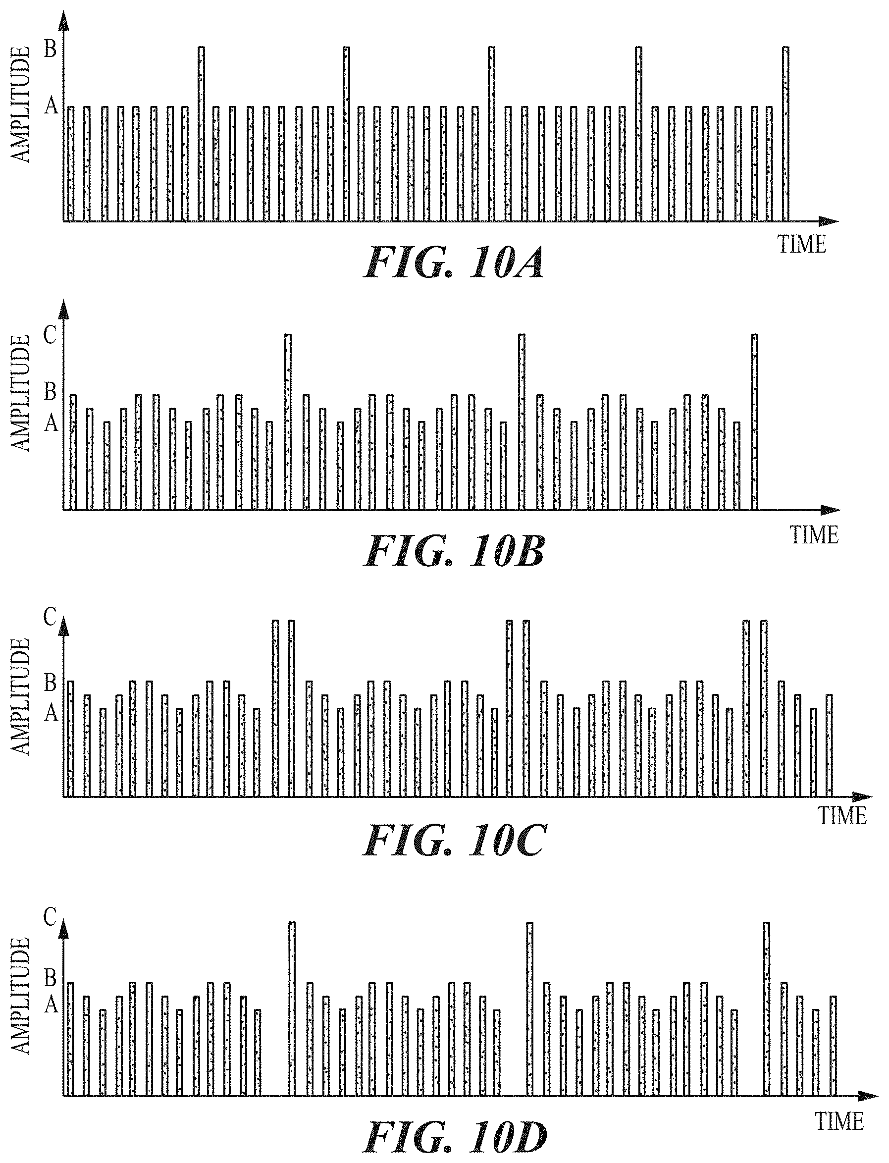
D00013
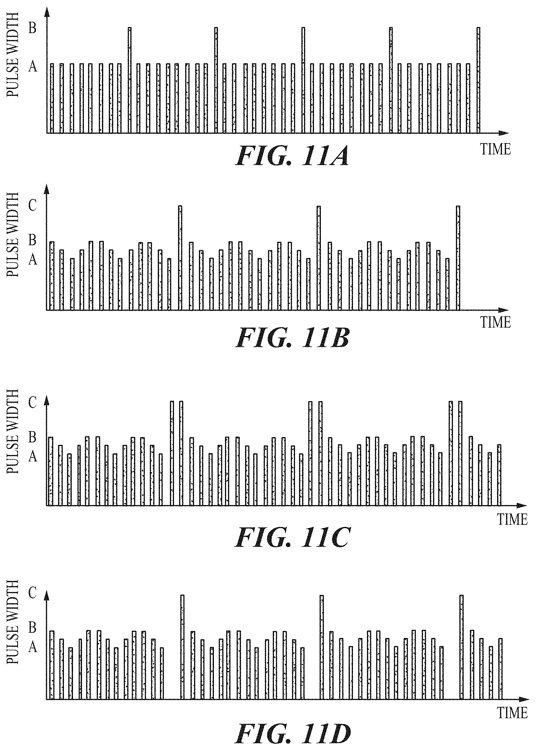
D00014
D00015
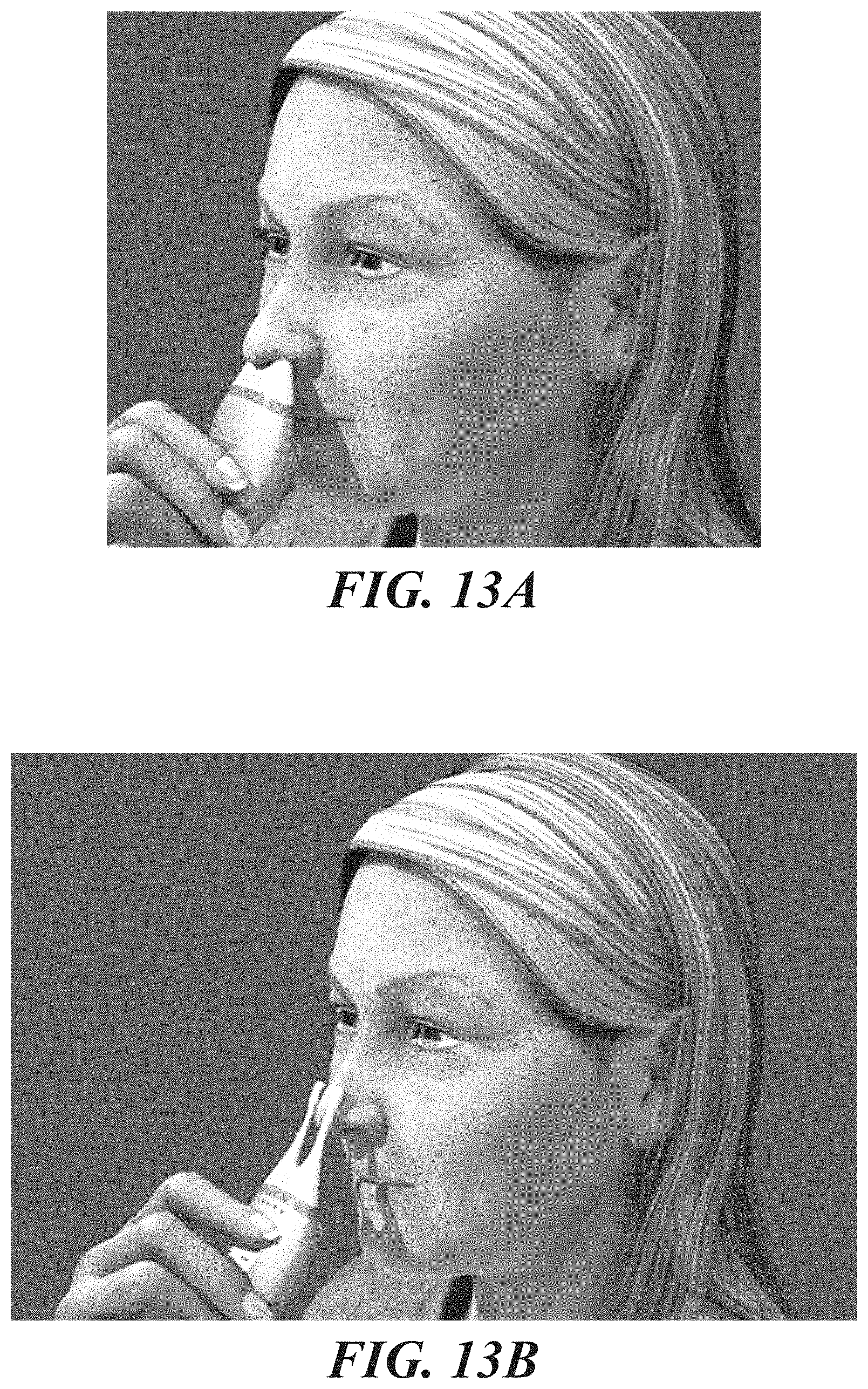
D00016

D00017
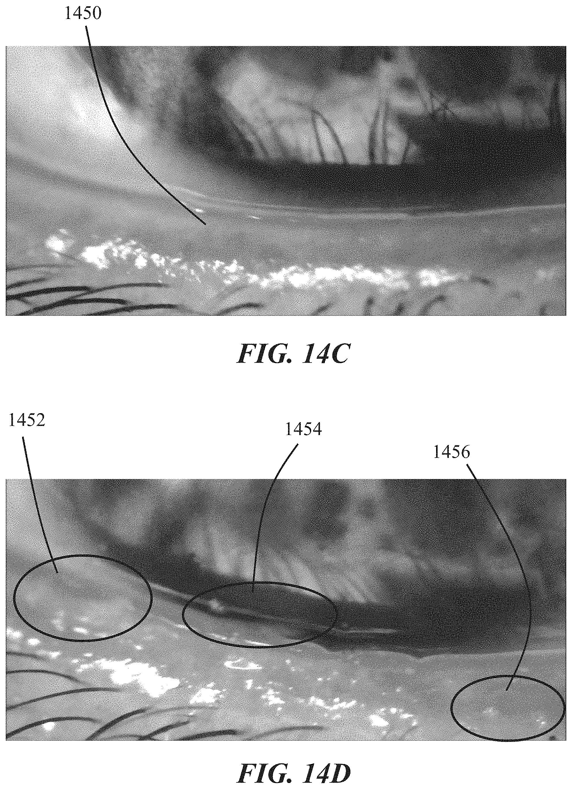
D00018
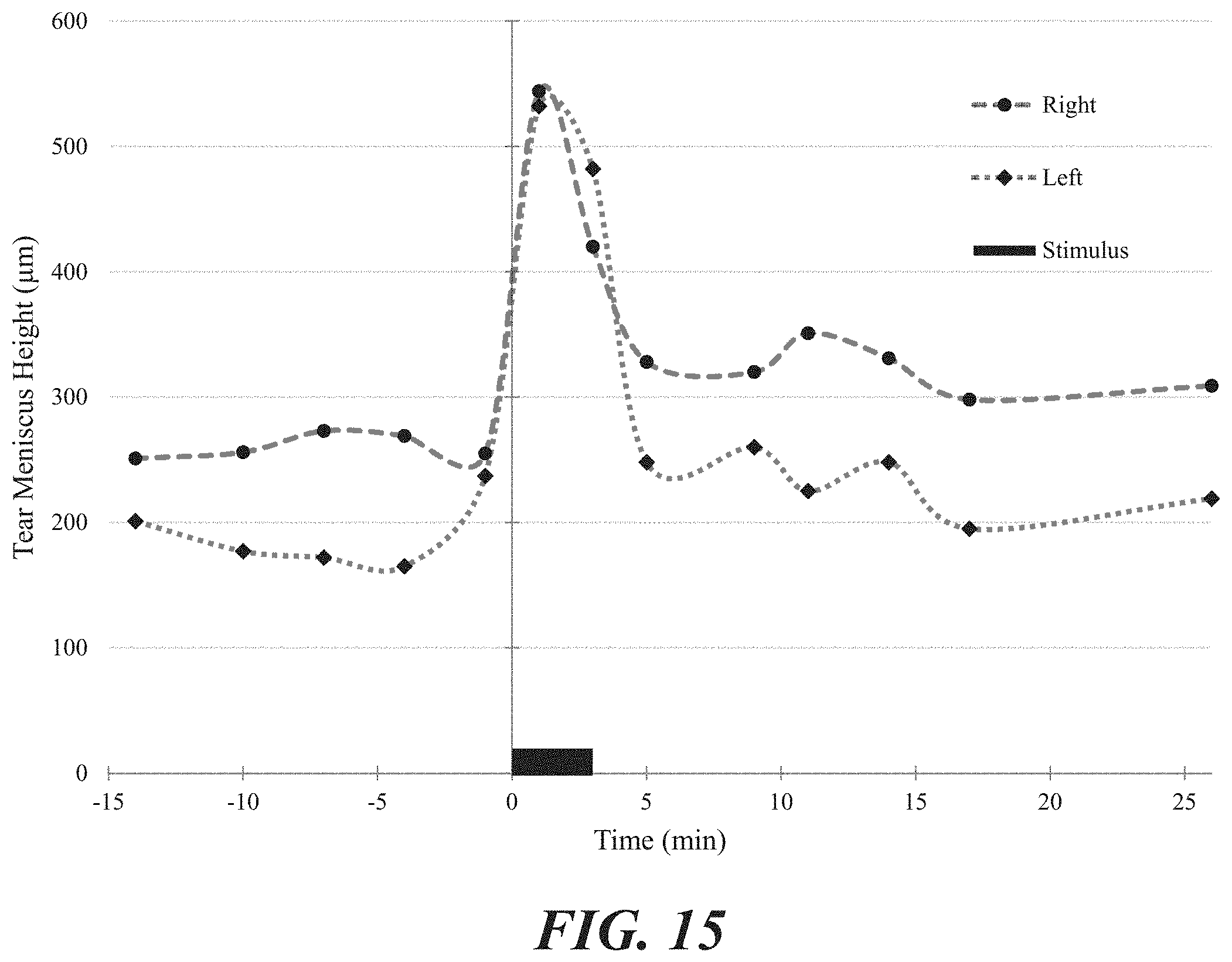
D00019
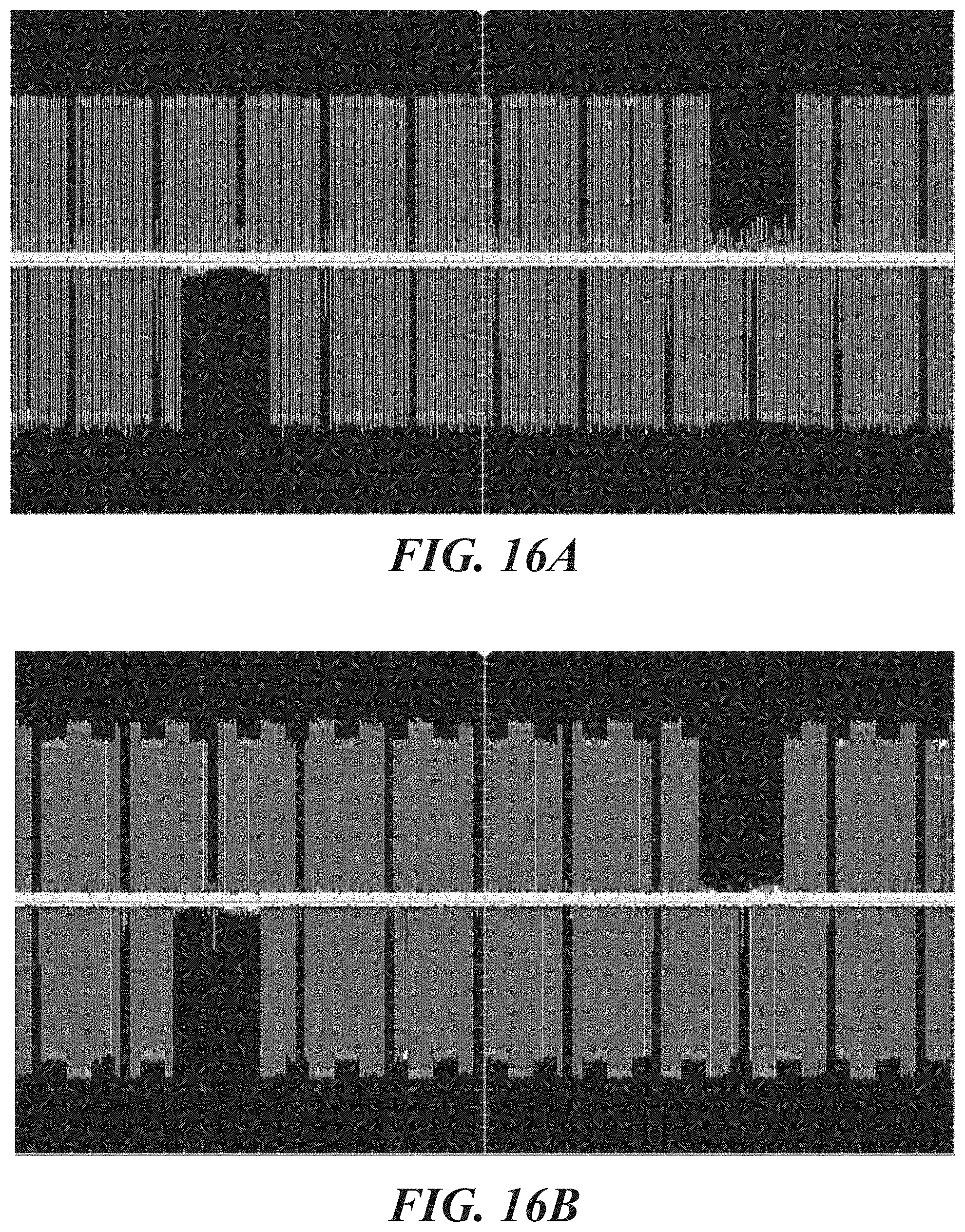
D00020

D00021
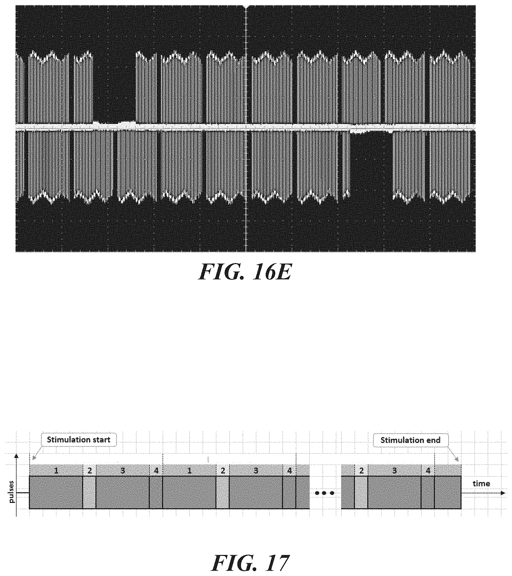
D00022
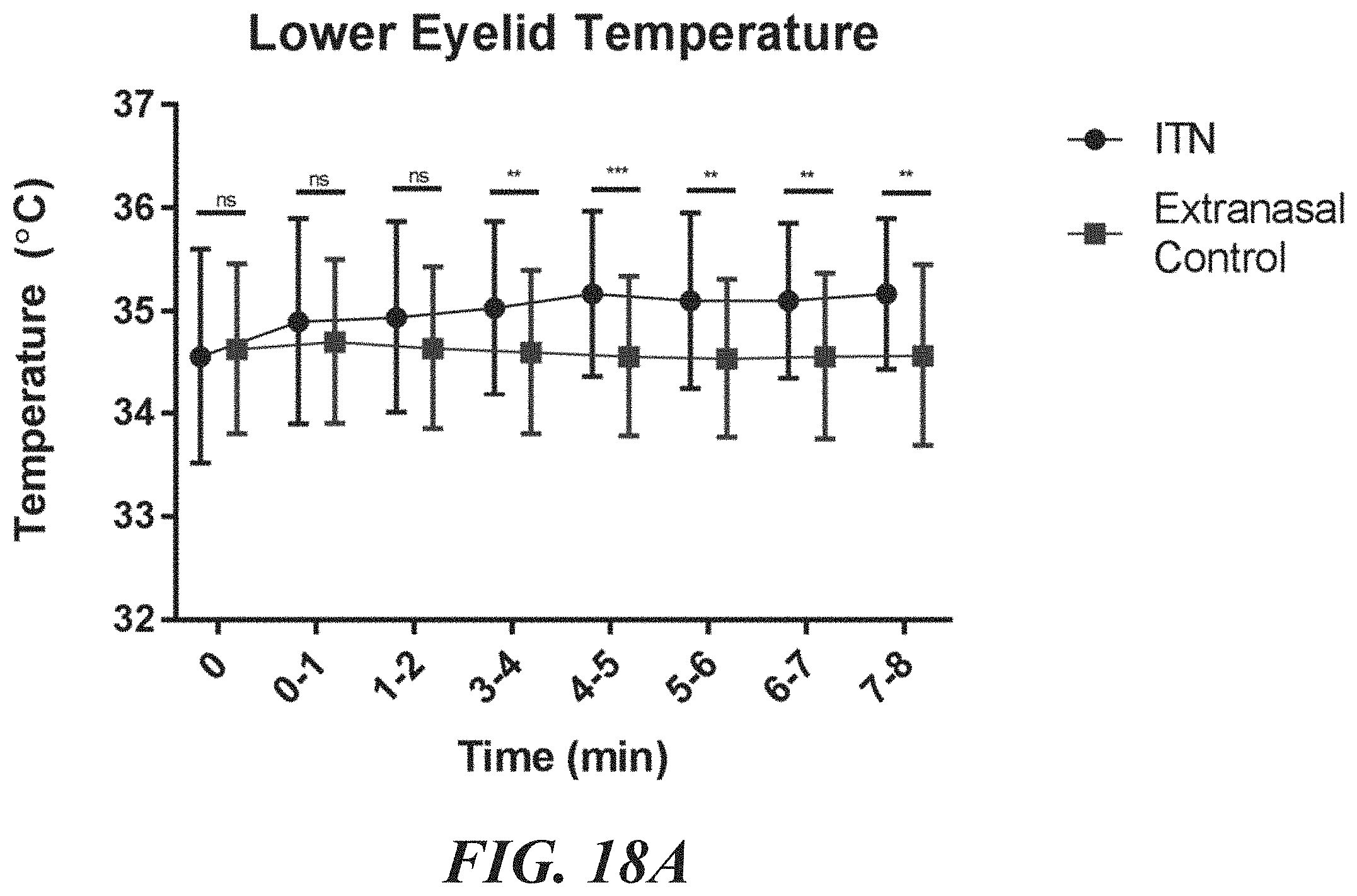
D00023
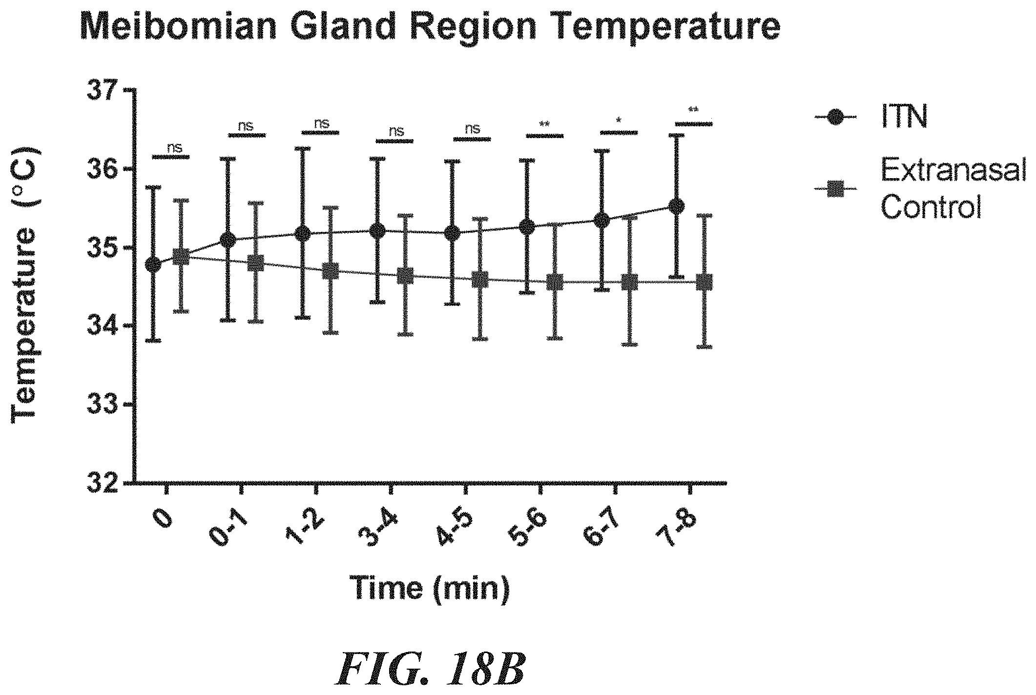
D00024

M00001

M00002

XML
uspto.report is an independent third-party trademark research tool that is not affiliated, endorsed, or sponsored by the United States Patent and Trademark Office (USPTO) or any other governmental organization. The information provided by uspto.report is based on publicly available data at the time of writing and is intended for informational purposes only.
While we strive to provide accurate and up-to-date information, we do not guarantee the accuracy, completeness, reliability, or suitability of the information displayed on this site. The use of this site is at your own risk. Any reliance you place on such information is therefore strictly at your own risk.
All official trademark data, including owner information, should be verified by visiting the official USPTO website at www.uspto.gov. This site is not intended to replace professional legal advice and should not be used as a substitute for consulting with a legal professional who is knowledgeable about trademark law.