Prostate specific membrane antigen binding protein
Dubridge , et al. December 1, 2
U.S. patent number 10,849,973 [Application Number 15/821,498] was granted by the patent office on 2020-12-01 for prostate specific membrane antigen binding protein. This patent grant is currently assigned to HARPOON THERAPEUTICS, INC.. The grantee listed for this patent is Harpoon Therapeutics, Inc.. Invention is credited to Richard J. Austin, Patrick Baeuerle, Robert Dubridge, Jeanmarie Guenot, Bryan D. Lemon, Pui Seto, Holger Wesche.


View All Diagrams
| United States Patent | 10,849,973 |
| Dubridge , et al. | December 1, 2020 |
Prostate specific membrane antigen binding protein
Abstract
Disclosed herein are PSMA binding proteins with improved binding affinities, and robust aggregation profiles. Also described are multispecific binding proteins comprising a PSMA binding protein according to the instant disclosure. Pharmaceutical compositions comprising the binding proteins disclosed herein and methods of using such formulations are further provided.
| Inventors: | Dubridge; Robert (Belmont, CA), Seto; Pui (San Carlos, CA), Baeuerle; Patrick (Gauting, DE), Guenot; Jeanmarie (San Francisco, CA), Wesche; Holger (San Francisco, CA), Lemon; Bryan D. (Mountain View, CA), Austin; Richard J. (San Francisco, CA) | ||||||||||
|---|---|---|---|---|---|---|---|---|---|---|---|
| Applicant: |
|
||||||||||
| Assignee: | HARPOON THERAPEUTICS, INC.
(South San Francisco, CA) |
||||||||||
| Family ID: | 1000005218872 | ||||||||||
| Appl. No.: | 15/821,498 | ||||||||||
| Filed: | November 22, 2017 |
Prior Publication Data
| Document Identifier | Publication Date | |
|---|---|---|
| US 20180161428 A1 | Jun 14, 2018 | |
Related U.S. Patent Documents
| Application Number | Filing Date | Patent Number | Issue Date | ||
|---|---|---|---|---|---|
| 62426086 | Nov 23, 2016 | ||||
| Current U.S. Class: | 1/1 |
| Current CPC Class: | C07K 16/2809 (20130101); C07K 16/18 (20130101); A61P 35/00 (20180101); C07K 16/3069 (20130101); A61K 39/39558 (20130101); A61K 39/001195 (20180801); C07K 16/2863 (20130101); A61K 2039/505 (20130101); C07K 2317/94 (20130101); C07K 2317/569 (20130101); A61K 38/00 (20130101); C07K 2317/92 (20130101); C07K 2317/622 (20130101); C07K 2317/31 (20130101); C07K 2317/565 (20130101); C07K 2317/73 (20130101) |
| Current International Class: | A61K 39/00 (20060101); C07K 16/28 (20060101); A61K 38/17 (20060101); C07K 16/30 (20060101); C07K 16/18 (20060101); A61P 35/00 (20060101); A61K 39/395 (20060101); A61K 38/00 (20060101) |
References Cited [Referenced By]
U.S. Patent Documents
| 4816567 | March 1989 | Cabilly et al. |
| 5061620 | October 1991 | Tsukamoto et al. |
| 5199942 | April 1993 | Gillis |
| 5225539 | July 1993 | Winter |
| 5350674 | September 1994 | Boenisch et al. |
| 5399346 | March 1995 | Anderson et al. |
| 5530101 | June 1996 | Queen et al. |
| 5565332 | October 1996 | Hoogenboom et al. |
| 5580859 | December 1996 | Felgner et al. |
| 5585089 | December 1996 | Queen et al. |
| 5585362 | December 1996 | Wilson et al. |
| 5589466 | December 1996 | Felgner et al. |
| 5759808 | June 1998 | Casterman et al. |
| 5766886 | June 1998 | Studnicka et al. |
| 5773292 | June 1998 | Bander |
| 5800988 | September 1998 | Casterman et al. |
| 5840526 | November 1998 | Casterman et al. |
| 5858358 | January 1999 | June et al. |
| 5859205 | January 1999 | Adair et al. |
| 5874541 | February 1999 | Casterman et al. |
| 5883223 | March 1999 | Gray |
| 6005079 | December 1999 | Casterman et al. |
| 6015695 | January 2000 | Casterman et al. |
| 6107090 | August 2000 | Bander |
| 6120766 | September 2000 | Hale et al. |
| 6136311 | October 2000 | Bander |
| 6326193 | December 2001 | Liu et al. |
| 6331415 | December 2001 | Cabilly et al. |
| 6352694 | March 2002 | June et al. |
| 6407213 | June 2002 | Carter et al. |
| 6534055 | March 2003 | June et al. |
| 6548640 | April 2003 | Winter |
| 6670453 | December 2003 | Frenken et al. |
| 6692964 | February 2004 | June et al. |
| 6759518 | July 2004 | Kontermann et al. |
| 6767711 | July 2004 | Bander |
| 6797514 | September 2004 | Berenson et al. |
| 6867041 | March 2005 | Berenson et al. |
| 6887466 | May 2005 | June et al. |
| 6905680 | June 2005 | June et al. |
| 6905681 | June 2005 | June et al. |
| 6905874 | June 2005 | Berenson et al. |
| 7067318 | June 2006 | June et al. |
| 7144575 | December 2006 | June et al. |
| 7163680 | January 2007 | Bander |
| 7172869 | February 2007 | June et al. |
| 7175843 | February 2007 | June et al. |
| 7232566 | June 2007 | June et al. |
| 7262276 | August 2007 | Huang et al. |
| 7666414 | February 2010 | Bander |
| 7807162 | October 2010 | Silence |
| 7850971 | December 2010 | Maddon et al. |
| 8114965 | February 2012 | Maddon et al. |
| 8188223 | May 2012 | Beirnaert et al. |
| 8236308 | August 2012 | Kischel et al. |
| 8470330 | June 2013 | Schuelke et al. |
| 8623356 | January 2014 | Christopherson et al. |
| 8629244 | January 2014 | Kolkman et al. |
| 8703135 | April 2014 | Beste et al. |
| 8784821 | July 2014 | Kufer et al. |
| 8846042 | September 2014 | Zhou |
| 8907071 | December 2014 | Sullivan et al. |
| 8937164 | January 2015 | Descamps et al. |
| 9169316 | October 2015 | Baty et al. |
| 9309327 | April 2016 | Humphreys et al. |
| 9327022 | May 2016 | Zhang et al. |
| 9340621 | May 2016 | Kufer et al. |
| 9708412 | July 2017 | Baeuerle |
| 10428120 | October 2019 | Kontermann et al. |
| 2005/0042664 | February 2005 | Wu et al. |
| 2005/0048617 | March 2005 | Wu et al. |
| 2005/0100543 | May 2005 | Hansen et al. |
| 2005/0175606 | August 2005 | Huang et al. |
| 2006/0046971 | March 2006 | Stuhler et al. |
| 2006/0121005 | June 2006 | Berenson et al. |
| 2006/0228364 | October 2006 | Dennis et al. |
| 2006/0252096 | November 2006 | Zha et al. |
| 2007/0014794 | January 2007 | Carter et al. |
| 2007/0178082 | August 2007 | Silence et al. |
| 2007/0269422 | November 2007 | Beirnaert et al. |
| 2008/0069772 | March 2008 | Stuhler et al. |
| 2008/0260757 | October 2008 | Holt et al. |
| 2009/0259026 | October 2009 | Tomlinson et al. |
| 2010/0122358 | May 2010 | Brueggemann et al. |
| 2010/0150918 | June 2010 | Kufer et al. |
| 2010/0166734 | July 2010 | Dolk |
| 2010/0189727 | July 2010 | Rodeck et al. |
| 2010/0266531 | October 2010 | Hsieh et al. |
| 2010/0291112 | November 2010 | Kellner et al. |
| 2011/0129458 | June 2011 | Dolk et al. |
| 2011/0165621 | July 2011 | Dreier et al. |
| 2011/0262439 | October 2011 | Kufer et al. |
| 2011/0275787 | November 2011 | Kufer et al. |
| 2011/0313135 | December 2011 | Vanhove et al. |
| 2012/0231024 | September 2012 | Elsaesser-Beile et al. |
| 2012/0328619 | December 2012 | Fey et al. |
| 2013/0017200 | January 2013 | Scheer et al. |
| 2013/0136744 | May 2013 | Bouche et al. |
| 2013/0266568 | October 2013 | Brinkmann et al. |
| 2013/0267686 | October 2013 | Brinkmann et al. |
| 2013/0273055 | October 2013 | Borges et al. |
| 2013/0330335 | December 2013 | Bremel et al. |
| 2014/0004121 | January 2014 | Fanslow, III et al. |
| 2014/0023664 | January 2014 | Lowman et al. |
| 2014/0045195 | February 2014 | Daugherty et al. |
| 2014/0073767 | March 2014 | Lee et al. |
| 2014/0088295 | March 2014 | Smith et al. |
| 2014/0205601 | July 2014 | Beirnaert et al. |
| 2014/0242075 | August 2014 | Parren et al. |
| 2014/0302037 | October 2014 | Borges et al. |
| 2014/0322218 | October 2014 | Xiao et al. |
| 2015/0037334 | February 2015 | Kufer et al. |
| 2015/0056206 | February 2015 | Zhou |
| 2015/0064169 | March 2015 | Wang et al. |
| 2015/0079088 | March 2015 | Lowman et al. |
| 2015/0079093 | March 2015 | Stuhler |
| 2015/0093336 | April 2015 | Van Ginderachter et al. |
| 2015/0174268 | June 2015 | Li et al. |
| 2015/0183875 | July 2015 | Cobbold et al. |
| 2015/0232557 | August 2015 | Tan et al. |
| 2015/0274836 | October 2015 | Ho et al. |
| 2015/0274844 | October 2015 | Blankenship et al. |
| 2016/0024174 | January 2016 | Odunsi et al. |
| 2016/0032019 | February 2016 | Xiao et al. |
| 2016/0039942 | February 2016 | Cobbold et al. |
| 2016/0068605 | March 2016 | Nemeth et al. |
| 2016/0130331 | May 2016 | Stull et al. |
| 2016/0215063 | July 2016 | Bernett et al. |
| 2016/0251440 | September 2016 | Roobrouck et al. |
| 2016/0257721 | September 2016 | Lieber et al. |
| 2016/0319040 | November 2016 | Dreier et al. |
| 2016/0340444 | November 2016 | Baeuerle |
| 2016/0355842 | December 2016 | Parks et al. |
| 2017/0029502 | February 2017 | Raum et al. |
| 2017/0152316 | June 2017 | Cobbold et al. |
| 2017/0204164 | July 2017 | Himmler et al. |
| 2017/0275373 | September 2017 | Kufer et al. |
| 2017/0298149 | October 2017 | Baeuerle et al. |
| 2017/0334979 | November 2017 | Dubridge et al. |
| 2017/0334997 | November 2017 | Dubridge et al. |
| 2017/0369563 | December 2017 | Dubridge et al. |
| 2017/0369575 | December 2017 | Dubridge et al. |
| 2018/0016323 | January 2018 | Brandenburg et al. |
| 2018/0134789 | May 2018 | Baeuerle et al. |
| 2018/0148508 | May 2018 | Wang et al. |
| 2019/0225702 | July 2019 | Baeuerle et al. |
| 2020/0095340 | March 2020 | Wesche et al. |
| 2020/0115461 | April 2020 | Evnin et al. |
| 2020/0148771 | May 2020 | Baeuerle et al. |
| 1563092 | Jan 2005 | CN | |||
| 101646689 | Feb 2010 | CN | |||
| 0239400 | Sep 1987 | EP | |||
| 0519596 | Dec 1992 | EP | |||
| 0592106 | Apr 1994 | EP | |||
| 1378520 | Jan 2004 | EP | |||
| 1736484 | Dec 2006 | EP | |||
| 2336179 | Jun 2011 | EP | |||
| 901228 | Jul 1945 | FR | |||
| 2005501517 | Jan 2005 | JP | |||
| WO-9109967 | Jul 1991 | WO | |||
| WO-9307105 | Apr 1993 | WO | |||
| WO-9404678 | Mar 1994 | WO | |||
| WO-9937681 | Jul 1999 | WO | |||
| WO-0043507 | Jul 2000 | WO | |||
| WO-0190190 | Nov 2001 | WO | |||
| WO-0196584 | Dec 2001 | WO | |||
| WO-02085945 | Oct 2002 | WO | |||
| WO-03025020 | Mar 2003 | WO | |||
| WO-03035694 | May 2003 | WO | |||
| WO-03064606 | Aug 2003 | WO | |||
| WO-2004003019 | Jan 2004 | WO | |||
| WO-2004041867 | May 2004 | WO | |||
| WO-2004042404 | May 2004 | WO | |||
| WO-2004049794 | Jun 2004 | WO | |||
| WO-2006020258 | Feb 2006 | WO | |||
| WO-2006122787 | Nov 2006 | WO | |||
| WO-2007024715 | Mar 2007 | WO | |||
| WO-2007042261 | Apr 2007 | WO | |||
| WO-2007062466 | Jun 2007 | WO | |||
| WO-2007115230 | Oct 2007 | WO | |||
| WO-2008028977 | Mar 2008 | WO | |||
| WO-2009025846 | Feb 2009 | WO | |||
| WO-2009030285 | Mar 2009 | WO | |||
| WO-2009147248 | Dec 2009 | WO | |||
| WO-2010003118 | Jan 2010 | WO | |||
| WO-2010037836 | Apr 2010 | WO | |||
| WO-2010037837 | Apr 2010 | WO | |||
| WO-2011039368 | Apr 2011 | WO | |||
| WO-2011051327 | May 2011 | WO | |||
| WO-2012131053 | Oct 2012 | WO | |||
| WO-2012138475 | Oct 2012 | WO | |||
| WO-2012158818 | Nov 2012 | WO | |||
| WO-2013036130 | Mar 2013 | WO | |||
| WO-2013104804 | Jul 2013 | WO | |||
| WO-2013110531 | Aug 2013 | WO | |||
| WO-2013128027 | Sep 2013 | WO | |||
| WO-2014033304 | Mar 2014 | WO | |||
| WO-2014138306 | Sep 2014 | WO | |||
| WO-2014140358 | Sep 2014 | WO | |||
| WO-2014151910 | Sep 2014 | WO | |||
| WO-2015103072 | Jul 2015 | WO | |||
| WO-2015150447 | Oct 2015 | WO | |||
| WO-2015184207 | Dec 2015 | WO | |||
| WO-2016009029 | Jan 2016 | WO | |||
| WO-2016034044 | Mar 2016 | WO | |||
| WO-2016046778 | Mar 2016 | WO | |||
| WO-2016055551 | Apr 2016 | WO | |||
| WO-2016105450 | Jun 2016 | WO | |||
| WO-2016130819 | Aug 2016 | WO | |||
| WO-2016171999 | Oct 2016 | WO | |||
| WO-2016179003 | Nov 2016 | WO | |||
| WO-2016187101 | Nov 2016 | WO | |||
| WO-2016187594 | Nov 2016 | WO | |||
| WO-2016210447 | Dec 2016 | WO | |||
| WO-2017025698 | Feb 2017 | WO | |||
| WO-2017027392 | Feb 2017 | WO | |||
| WO-2017041749 | Mar 2017 | WO | |||
| WO-2017079528 | May 2017 | WO | |||
| WO-2017136549 | Aug 2017 | WO | |||
| WO-2017156178 | Sep 2017 | WO | |||
| WO-2017201488 | Nov 2017 | WO | |||
| WO-2017201493 | Nov 2017 | WO | |||
| WO-2018017863 | Jan 2018 | WO | |||
| WO-2018071777 | Apr 2018 | WO | |||
| WO-2018098354 | May 2018 | WO | |||
| WO-2018098356 | May 2018 | WO | |||
| WO-2018136725 | Jul 2018 | WO | |||
| WO-2018160671 | Sep 2018 | WO | |||
| WO-2018160754 | Sep 2018 | WO | |||
| WO-2018165619 | Sep 2018 | WO | |||
| WO-2018204717 | Nov 2018 | WO | |||
| WO-2018209298 | Nov 2018 | WO | |||
| WO-2018209304 | Nov 2018 | WO | |||
| WO-2019075359 | Apr 2019 | WO | |||
| WO-2019075378 | Apr 2019 | WO | |||
| WO-2019222278 | Nov 2019 | WO | |||
| WO-2019222282 | Nov 2019 | WO | |||
| WO-2019222283 | Nov 2019 | WO | |||
| WO-2020060593 | Mar 2020 | WO | |||
| WO-2020061482 | Mar 2020 | WO | |||
| WO-2020061526 | Mar 2020 | WO | |||
| WO-2020069028 | Apr 2020 | WO | |||
Other References
|
Foote et al. (J. Mol. Biol. Mar. 20, 1992; 224 (2): 487-99). cited by examiner . Mariuzza et al. (Annu. Rev. Biophys. Biophys. Chem. 1987; 16: 139-159). cited by examiner . Rudikoff et al. (Proc. Natl. Acad. Sci. USA. 1982; 79: 1979-1983). cited by examiner . Winkler et al. (J. Immunol. Oct. 15, 2000; 165 (8): 4505-4514). cited by examiner . Gussow et al. (Methods in Enzymology. 1991; 203: 99-121). cited by examiner . Giusti et al. (Proc. Natl. Acad. Sci. USA. May 1987; 84 (9): 2926-2930). cited by examiner . Chien et al. (Proc. Natl. Acad. Sci. USA. Jul. 1989; 86 (14): 5532-5536). cited by examiner . Caldas et al. (Mol. Immunol. May 2003; 39 (15): 941-952). cited by examiner . Vajdos et al. (J. Mol. Biol. Jul. 5, 2002; 320 (2): 415-428). cited by examiner . De Pascalis et al. (J. Immunol. 2002; 169 (6): 3076-3084). cited by examiner . Wu et al. (J. Mol. Biol. Nov. 19, 1999; 294 (1): 151-162). cited by examiner . Casset et al. (Biochem. Biophys. Res. Connnnun. Jul. 18, 2003; 307 (1): 198-205). cited by examiner . MacCallum et al. (J. Mol. Biol. Oct. 11, 1996; 262 (5): 732-745). cited by examiner . Holm et al. (Mol. Immunol. Feb. 2007; 44 (6): 1075-1084). cited by examiner . Yu et al. (PLoS One. 2012; 7 (3): e33340; pp. 1-15). cited by examiner . Chang et al. (Structure. Jan. 7, 2014; 22 (1): 9-21). cited by examiner . Saerens et al. (J. Mol. Biol. Sep. 23, 2005; 352 (3): 597-607). cited by examiner . Vincke et al. (J. Biol. Chem. Jan. 30, 2009; 284 (5): 3273-84). cited by examiner . Goldman et al. (Front. Immunol. Jul. 25, 2017; 8: 865; pp. 1-11). cited by examiner . Tiller et al. (Front. Immunol. 2017; 8: 986; pp. 1-16). cited by examiner . Su et al. (Cancer Lett. Sep. 28, 2013; 338 (2): 282-91). cited by examiner . Zare et al. (Int. J. Biol. Markers. Jun. 25, 2014; 29 (2): e169-79; pp. 1-11). cited by examiner . Schmidt et al. (Prostate. May 2013; 73 (6): 642-500). cited by examiner . Schmittgen et al. (Int. J. Cancer. Nov. 1, 2003; 107 (2): 323-9). cited by examiner . Argani et al. Mesothelin is overexpressed in the vast majority of ductal adenocarcinomas of the pancreas: identification of a new pancreatic cancer marker by serial analysis of gene expression (SAGE). Clin Cancer Res 7(12):3862-3868 (2001). cited by applicant . Bortoletto et al. Optimizing anti-CD3 affinity for effective T cell targeting against tumor cells. Eur J Immunol 32:3102-3107 (2002). cited by applicant . Bracci et al. Cyclophosphamide enhances the antitumor efficacy of adoptively transferred immune cells through the induction of cytokine expression, B-cell and T-cell homeostatic proliferation, and specific tumor infiltration. Clin Cancer Res 13(2 Pt 1):644-653 (2007). cited by applicant . Chang et al. Molecular cloning of mesothelin, a differentiation antigen present on mesothelium, mesotheliomas, and ovarian cancers. PNAS USA 93:136-140 (1996). cited by applicant . Co-pending U.S. Appl. No. 15/977,968, filed May 11, 2018. cited by applicant . Co-pending U.S. Appl. No. 15/977,988, filed May 11, 2018. cited by applicant . Corso et al. Real-time detection of mesothelin in pancreatic cancer cell line supernatant using an acoustic wave immunosensor. Cancer Detect Prey 30:180-187 (2006). cited by applicant . Creaney et al. Detection of malignant mesothelioma in asbestos-exposed individuals: the potential role of soluble mesothelin-related protein. Hematol. Oncol. Clin. North Am. 19:1025-1040 (2005). cited by applicant . Cristaudo et al. Clinical significance of serum mesothelin in patients with mesothelioma and lung cancer. Clin. Cancer Res. 13:5076-5081 (2007). cited by applicant . Document D28--Investigation of human CD3.epsilon. variants binding to monoclonal antibodies. Submitted by Pfizer to the European Patent Register on Apr. 30, 2014 in connection with their opposition to the EP2155783 patent. (3 pages) (2014). cited by applicant . Document D78--CD3.epsilon. N-terminal peptide bound to the CDRs of SP24. Submitted by Janssen Biotech to the European Patent Register on Mar. 18, 2016 in connection with their opposition to the EP2155783 patent (1 page) (2016). cited by applicant . Document D79--Interactions between CD3.epsilon. and SP34 CDR residues. CD3.epsilon. residues are in ellipses, SP34 CDR residues are in boxes. Submitted by Janssen Biotech to the European Patent Register on Mar. 18, 2016 in connection with their opposition to the EP2155783 patent (1 page) (2016). cited by applicant . Document D83--Alignment of variable domains from the prior art and the patent. Submitted by Janssen Biotech to the European Patent Register on Mar. 18, 2016 in connection with their opposition to the EP2155783 patent (1 page) (2016). cited by applicant . Gross et al. Endowing T cells with antibody specificity using chimeric T cell receptors. FASEB J. 6(15):3370-3378 (1992). cited by applicant . Gubbels et al. Mesothelin-MUC16 binding is a high affinity, N-glycan dependent interaction that facilitates peritoneal metastasis of ovarian tumors. Mol Cancer 5:50 (2006). cited by applicant . Hassan et al. Detection and quantitation of serum mesothelin, a tumor marker for patients with mesothelioma and ovarian cancer. Clin Cancer Res 12:447-453 (2006). cited by applicant . Hassan et al. Mesothelin: a new target for immunotherapy. Clin Cancer Res 10:3937-3942 (2004). cited by applicant . Hassan et al. Mesothelin targeted cancer immunotherapy. Eur J Cancer 44:46-53 (2008). cited by applicant . Hassan et al. Phase I study of SS1P, a recombinant anti-mesothelin immunotoxin given as a bolus I.V. infusion to patients with mesothelin-expressing mesothelioma, ovarian, and pancreatic cancers. Clin Cancer Res 13(17):5144-5149 (2007). cited by applicant . Hassan et al. Preclinical evaluation of MORAb-009, a chimeric antibody targeting tumor-associated mesothelin. Cancer Immun. 7:20 (2007). cited by applicant . Hellstrom et al. Mesothelin variant 1 is released from tumor cells as a diagnostic marker. Cancer Epidemiol Biomarkers Prey 15:1014-1020 (2006). cited by applicant . Ho et al. A novel high-affinity human monoclonal antibody to mesothelin. Int J Cancer 128:2020-2030 (2011). cited by applicant . Ho et al. Mesothelin expression in human lung cancer. Clin Cancer Res 13:1571-1575 (2007). cited by applicant . Janssen letter--Submission under Rule 116 EPC. Submitted by Janssen Biotech to the European Patent Register on Mar. 18, 2016 in connection with their opposition to the EP2155783 patent (6 pages) (2016). cited by applicant . Kojima et al. Molecular cloning and expression of megakaryocyte potentiating factor cDNA. J Biol Chem 270:21984-21990 (1995). cited by applicant . Li et al. Development of novel tetravalent anti-CD20 antibodies with potent antitumor activity. Cancer Res 68:2400-2408 (2008). cited by applicant . Mirsky et al. Antibody-Specific Model of Amino Acid Substitution for Immunological Inferences from Alignments of Antibody Sequences. Mol. Biol. Evol. 32(3):806-819 (2014). cited by applicant . Morea et al. Antibody modeling: implications for engineering and design. Methods 20(3):267-279 (2000). cited by applicant . Moschella et al. Unraveling cancer chemoimmunotherapy mechanisms by gene and protein expression profiling of responses to cyclophosphamide. Cancer Res 71(10):3528-3539 (2011). cited by applicant . Muul et al. Persistence and expression of the adenosine deaminase gene for 12 years and immune reaction to gene transfer components: long-term results of the first clinical gene therapy trial. Blood 101(7):2563-2569 (2003). cited by applicant . Ordonez. Application of mesothelin immunostaining in tumor diagnosis. Am J Surg Pathol 27:1418-1428 (2003). cited by applicant . Pawluczkowycz et al. Binding of submaximal C1q promotes complement-dependent cytotoxicity (CDC) of B cells opsonized with anti-CD20 mAbs ofatumumab (OFA) or rituximab (RTX): considerably higher levels of CDC are induced by OFA than by RTX. J Immunol 183:749-758 (2009). cited by applicant . PCT/US2018/020185 International Search Report and Written Opinion dated Jun. 15, 2018. cited by applicant . PCT/US2018/020307 International Search Report and Written Opinion dated Aug. 24, 2018. cited by applicant . PCT/US2018/030983 Invitation to Pay Additional Fees dated Jul. 31, 2018. cited by applicant . PCT/US2018/032427 International Search Report and Written Opinion dated Sep. 13, 2018. cited by applicant . PCT/US2018/32418 Invitation to Pay Additional Fees dated Jul. 23, 2018. cited by applicant . PCT/US2018/32427 Invitation to Pay Additional Fees dated Jul. 24, 2018. cited by applicant . Pfizer letter--Opposition to European Patent EP2155783 (Application 08735001.3). Submitted by Pfizer to the European Patent Register on Apr. 30, 2014 in connection with their opposition to the EP2155783 patent. (pp. 1-23 and Appendix 1 on pp. 24-26) (2014). cited by applicant . Rump et al. Binding of ovarian cancer antigen CA125/MUC16 to mesothelin mediates cell adhesion. J Biol Chem 279:9190-9198 (2004). cited by applicant . Sadelain et al. Targeting tumours with genetically enhanced T lymphocytes. Nat Rev Cancer 3(1):35-45 (2003). cited by applicant . Schmittgen et al. Expression of prostate specific membrane antigen and three alternatively spliced variants of PSMA in prostate cancer patients. Int J Cancer 107:323-329 (2003). cited by applicant . Tang et al. A human single-domain antibody elicits potent antitumor activity by targeting an epitope in mesothelin close to the cancer cell surface. Mol. Cancer Thera 12(4):416-426 (2013). cited by applicant . Thomas et al. Mesothelin-specific CD8(+) T cell responses provide evidence of in vivo cross-priming by antigen-presenting cells in vaccinated pancreatic cancer patients. J Exp Med 200:297-306 (2004). cited by applicant . U.S. Appl. No. 15/600,264 Office Action dated Apr. 26, 2018. cited by applicant . U.S. Appl. No. 15/821,530 Office Action dated Sep. 25, 2018. cited by applicant . Yee et al. Adoptive T cell therapy using antigen-specific CD8+ T cell clones for the treatment of patients with metastatic melanoma: in vivo persistence, migration, and antitumor effect of transferred T cells. PNAS USA 99(25):16168-16173 (2002). cited by applicant . Holt et al. Anti-serum albumin domain antibodies for extending the half-lives of short lived drugs. Protien Eng Des Sel 21(5):283-288 (2008). cited by applicant . Liu et al. MGD011, a CD19 x CD3 Dual Affinity Re-Targeting Bi-specific Molecule Incorporating Extended Circulating Half-life for the Treatment of B-cell Malignancies. Clin Cancer Res 23(6):1506-1518 (epub 2016) (2017). cited by applicant . Nelson et al. Antibody fragments Hope and Hype. mAbs 2(1):77-83 (2010). cited by applicant . PCT/US2017/063121 International Search Report and Written Opinion dated Mar. 26, 2018. cited by applicant . PCT/US2017/063126 International Search Report and Written Opinion dated Apr. 5, 2018. cited by applicant . PCT/US2017056530 International Search Report and Written Opinion dated Jan. 23, 2018. cited by applicant . PCT/US2017/063121 Invitation to Pay Additional Fees dated Feb. 1, 2018. cited by applicant . PCT/US2017/063126 Invitation to Pay Additional Fees dated Feb. 1, 2018. cited by applicant . Almagro et al. Humanization of antibodies. Front Biosci 13:1619-1633 (2008). cited by applicant . Baca et al. Antibody humanization using monovalent phage display. J Biol Chem 272(16):10678-10684 (1997). cited by applicant . Baeuerle et al. Bispecific T-cell engaging antibodies for cancer therapy. Cancer Res 69:4941-4944 (2009). cited by applicant . Bedouelle et al. Diversity and junction residues as hotspots of binding energy in an antibody neutralizing the dengue virus. FEBS J 273(1):34-46 (2006). cited by applicant . Brown et al. Tolerance of single, but not multiple, amino acid replacements in antibody VH CDR 2: a means of minimizing B cell wastage from somatic hypermutation? J Immunol 156(9):3285-3291 (1996). cited by applicant . Carter et al. Humanization of an anti-p185HER2 antibody for human cancer therapy. PNAS USA 89(10):4285-4289 (1992). cited by applicant . Casset et al. A peptide mimetic of an anti-CD4 monoclonal antibody by rational design. Biochemical and Biophysical Research Communication 307:198-205 (2003). cited by applicant . Chatalic et al. A Novel 111 In-labeled Anti-PSMA Nanobody for Targeted SPECT/CT Imaging of Prostate Cancer. J Nucl Med 56(7):1094-1099 and Supplemental Data (2015). cited by applicant . Chen et al. Selection and analysis of an optimized anti-VEGF antibody: Crystal structure of an affinity-matured Fab in complex with antigen. J Mol Bio 293:865-881 (1999). cited by applicant . Chothia et al. Canonical structures for the hypervariable regions of immunoglobulins. J Mol Biol 196(4):901-917 (1987). cited by applicant . Co-pending U.S. Appl. No. 15/821,530, filed Nov. 22, 2017. cited by applicant . De Pascalis et al. Grafting of "abbreviated" complementarity-determining regions containing specificity-determining residues essential for ligand contact to engineer a less immunogenic humanized monoclonal antibody. J Immunol. 169(6):3076-3084 (2002). cited by applicant . Frankel et al. Targeting T cells to tumor cells using bispecific antibodies. Curr Opin Chem Biol 17(3):385-392 (2013). cited by applicant . Goodman et al. The Pharmaceutical Basis of Therapeutics. 6th ed. pp. 21-25 (1980). cited by applicant . Goswami et al. Developments and Challenges for mAb-Based Therapeutics. Antibodies 2:452-500 (2013). cited by applicant . Harding et al. The immunogenicity of humanized and fully human antibodies: residual immunogenicity resides in the CDR regions. MAbs 2(3):256-265 (2010). cited by applicant . Hutchinson et al. Mutagenesis at a specific position in a DNA sequence. J Biol Chem 253:6551-6560 (1978). cited by applicant . Kabat et al. Identical V region amino acid sequences and segments of sequences in antibodies of different specificities. Relative contributions of VH and VL genes, minigenes, and complementarity-determining regions to binding of antibody-combining sites. J Immunol 147:1709-1719 (1991). cited by applicant . Le Gall et al. Immunosuppressive properties of anti-CD3 single-chain Fv and diabody. J Immunol Methods 285(1):111-127 (2004). cited by applicant . Lutterbuese et al. T cell-engaging BiTE antibodies specific for EGFR potently eliminate KRAS- and BRAF-mutated colorectal cancer cells. PNAS 107:12605-12610 (2007). cited by applicant . Maccallum et al. Antibody-antigen interactions: contact analysis and binding site topography. J Mol Biol. 262(5):732-745 (1996). cited by applicant . Muller et al. Improving the pharmacokinetic properties of biologics by fusion to an anti-HSA shark VNAR domain. MAbs 4(6):673-685 (2012). cited by applicant . Nazarian et al. Characterization of bispecific T-cell Engager (BiTE) antibodies with a high-capacity T-cell dependent cellular cytotoxicity (TDCC) assay. J Biomol Screen 20:519-527 (2015). cited by applicant . Ohiro et al. A homogeneous and noncompetitive immunoassay based on the enhanced fluorescence resonance energy transfer by leucine zipper interaction. Anal Chem 74(22):5786-5792 (2002). cited by applicant . O'Keefe et al. Chapter 18: Prostate specific membrane antigen. In: Chung L.W.K., Isaacs W.B., Simons J.W. (eds) Prostate Cancer. Contemporary Cancer Research. Humana Press, Totowa, NJ (pp. 307-326) (2001). cited by applicant . Padlan. Anatomy Of The Antibody Molecule. Mol Immunol 31(3):169-217 (1994). cited by applicant . Padlan et al. Structure of an antibody-antigen complex: Crystal structure of the HyHEL-10 Fab-lysozyme complex. PNAS USA 86:5938-5942 (1989). cited by applicant . PCT/US2016/033644 International Preliminary Report on Patentability dated Nov. 30, 2017. cited by applicant . PCT/US2016/33644 International Search Report and Written Opinion dated Sep. 6, 2016. cited by applicant . PCT/US2017/033665 International Search Report and Written Opinion dated Oct. 18, 2017. cited by applicant . PCT/US2017/033673 International Search Report and Written Opinion dated Oct. 18, 2017. cited by applicant . Presta et al. Humanization of an antibody directed against IgE. J Immunol 151:2623-2632 (1993). cited by applicant . Riechmann et al. Single domain antibodies: comparison of camel VH and camelised human VH domains. J Immunol Methods 231(1-2):25-38 (1999). cited by applicant . Rosok et al. A Combinatorial Library Strategy for the Rapid Humanization of Anticarcinoma BR96 Fab. J Biol Chem 271:22611-22618 (1996). cited by applicant . Rudikoff et al. Single amino acid substitution altering antigen-binding Specificity. PNAS USA 79:1979-1983 (1982). cited by applicant . Sims et al. A humanized CD18 antibody can block function without cell destruction. J Immunol 151:2296-2308 (1993). cited by applicant . U.S. Appl. No. 15/160,984 Office Action dated Feb. 24, 2017. cited by applicant . U.S. Appl. No. 15/160,984 Office Action dated Sep. 22, 2016. cited by applicant . U.S. Appl. No. 15/600,264 Office Action dated Oct. 3, 2017. cited by applicant . U.S. Appl. No. 15/600,582 Office Action dated Nov. 16, 2017. cited by applicant . U.S. Appl. No. 15/704,620 Office Action dated Oct. 26, 2017. cited by applicant . Vajdos et al. Comprehensive functional maps of the antigen-binding site of an anti-ErbB2 antibody obtained with shotgun scanning mutagenesis. J Mol Biol 320:415-428 (2002). cited by applicant . Van Den Beuchken et al. Building novel binding ligands to B7.1 and B7.2 based on human antibody single variable light chain domains. J Mol Biol 310:591-601 (2001). cited by applicant . Vaughan et al. Human antibodies by design. Nature Biotech 16:535-539 (1998). cited by applicant . Wu et al. Humanization of a murine monoclonal antibody by simultaneous optimization of framework and CDR residues. J. Mol. Biol. 294:151-162 (1999). cited by applicant . Cho et al. Targeting B Cell Maturation Antigen (BCMA) in Multiple Myeloma: Potential Uses of BCMA-Based Immunotherapy. Front Immunol 9:1821 (2018). cited by applicant . PCT/US2018/055659 International Search Report and Written Opinion dated Feb. 21, 2019. cited by applicant . PCT/US2018/055682 International Search Report and Written Opinion dated Mar. 1, 2019. cited by applicant . U.S. Appl. No. 15/977,968 Office Action dated Feb. 21, 2019. cited by applicant . Austin et al. Cancer Research (Jul. 2018) vol. 78, No. 13, Supp. Supplement 1. Abstract No. 1781. Meeting Info: 2018 Annual Meeting of the American Association for Cancer Research, AACR 2018. Chicago, IL, United States. Apr. 14-Apr. 18, 2018). cited by applicant . Co-pending U.S. Appl. No. 16/159,545, filed Oct. 12, 2018. cited by applicant . Co-pending U.S. Appl. No. 16/159,554, filed Oct. 12, 2018. cited by applicant . Co-pending U.S. Appl. No. 16/161,986, filed Oct. 16, 2018. cited by applicant . Liu et al. A New Format of Single Chain Tri-specific Antibody with Diminished Molecular Size Efficiently Induces Ovarian Tumor Cell Killing. Biotechnology Letters 27(22):1821-1827 (2005). cited by applicant . Lu et al. In vitro and in vivo antitumor effect of a trivalent bispecific antibody targeting ErbB2 and CD16. Cancer Biol Ther. 7(11):1744-1750 (2008). cited by applicant . Mueller et al. Improved pharmacokinetics of recombinant bispecific antibody molecules by fusion to human serum albumin. J Bio Chem 282(17):12650-12660 (2007). cited by applicant . Nunez-Prado et al. The coming of age of engineered multivalent antibodies. Drug Discovery Today 20(5):588-594 (2015). cited by applicant . PCT/US2018/014396 International Search Report and Written Opinion dated Jun. 14, 2018. cited by applicant . PCT/US2018/030983 International Search Report and Written Opinion dated Sep. 25, 2018. cited by applicant . PCT/US2018/032418 International Search Report and Written Opinion dated Sep. 24, 2018. cited by applicant . PCT/US2018/055659 Invitation to Pay Additional Fees dated Dec. 19, 2018. cited by applicant . PCT/US2018/055682 Invitation to Pay Additional Fees dated Jan. 8, 2019. cited by applicant . Running Deer et al. High-level expression of proteins in mammalian cells using transcription regulatory sequences from the Chinese hamster EF-1alpha gene. Biotechnol Prog. 20:880-889 (2004). cited by applicant . Sternjak et al. Cancer Research, (Jul. 2017) vol. 77, No. 13, Supp. Supplement 1. Abstract No. 3630. Meeting Info: American Association for Cancer Research Annual Meeting 2017. Washington, DC, United States. Apr. 1-Apr. 5, 2017. cited by applicant . Tutt et al. Trispecific F(ab')3 derivatives that use cooperative signaling via the TCR/CD3 complex and CD2 to activate and redirect resting cytotoxic T cells. J Immunol. 147(1):60-69 (Jul. 1, 1991). cited by applicant . U.S. Appl. No. 15/600,264 Office Action dated Nov. 27, 2018. cited by applicant . U.S. Appl. No. 15/977,988 Preinterview First Office Action dated Jan. 25, 2019. cited by applicant . Wang et al. A New Recombinant Single Chain Trispecific Antibody Recruits T Lymphocytes to Kill CEA (Carcinoma Embryonic Antigen) Positive Tumor Cells In Vitro Efficiently. Journal Of Biochemistry 135(4):555-565 (2004). cited by applicant . Zhu et al. Combody: one-domain antibody multimer with improved avidity. Immunology And Cell Biology 88(6):667-675 (2010). cited by applicant . Rozan et al. Single-domain antibody-based and linker-free bispecific antibodies targeting Fc.gamma.RIII induce potent antitumor activity without recruiting regulatory T cells. Mol Cancer Ther 12(8):1481-1491 (2013). cited by applicant . Schmidt et al. Cloning and Characterization of Canine Prostate-Specific Membrane Antigen. The Prostate 73:642-650 (2013). cited by applicant . U.S. Appl. No. 15/600,264 Office Action dated Apr. 25, 2019. cited by applicant . U.S. Appl. No. 15/821,530 Office Action dated Apr. 3, 2019. cited by applicant . U.S. Appl. No. 15/977,988 Office Action dated Mar. 26, 2019. cited by applicant . Muyldermans. Nanobodies: natural single-domain antibodies. Annu Rev Biochem 82:775-797 (2013). cited by applicant . U.S. Appl. No. 16/159,545 Office Action dated Aug. 6, 2019. cited by applicant . PCT/US2018/014396 International Preliminary Report on Patentability dated Aug. 1, 2019. cited by applicant . PCT/US2019/032224 International Search Report and Written Opinion dated Aug. 28, 2019. cited by applicant . PCT/US2019/032302 International Search Report and Written Opinion dated Aug. 22, 2019. cited by applicant . PCT/US2019/032306 International Search Report and Written Opinion dated Aug. 22, 2019. cited by applicant . PCT/US2019/032307 International Search Report and Written Opinion dated Aug. 22, 2019. cited by applicant . U.S. Appl. No. 15/977,988 Office Action dated Aug. 20, 2019. cited by applicant . Chen, Xiaoying et al. Fusion protein linkers: Property, design and functionality. Advanced Drug Delivery Reviews 65:1357-1369 (2013). cited by applicant . Dennis et al. Imaging Tumors with an Albumin-Binding Fab, a Novel Tumor-Targeting Agent. Cancer Res 67(1):254-61 (2007). cited by applicant . Hipp et al. A novel BCMA/CD3 bispecific T-cell engager for the treatment of multiple myeloma induces selective lysis in vitro and in vivo. Leukemia 31(8):1743-1751 (2017). cited by applicant . Hopp et al. The effects of affinity and valency of an albumin-binding domain (ABD) on the half-life of a single-chain diabody-ABD fusion protein. Protein Eng. Des. Sel. 23(11):827-34 (2010). cited by applicant . Laabi et al. The BCMA gene, preferentially expressed during B lymphoid maturation, is bidirectionally transcribed. Nucleic Acids Res 22(7):1147-1154 (1994). cited by applicant . Muller et al. Improved Pharmacokinetics of Recombinant Bispecific Antibody Molecules by Fusion to Human Serum Albumin. J. Biol. Chem. 282(17):12650-60 (2007). cited by applicant . Ramadoss et al. An Anti-B Cell Maturation Antigen bispecific Antibody for Multiple Myeloma. J. Ann. Chem. Soc. 137(16):5288-91 (2015). cited by applicant . Smirnova et al. Identification of new splice variants of the genes BAFF and BCMA. Mol. Immunol. 45 (4):1179-83 (2008). cited by applicant . Spiess et al. Alternative molecular formats and therapeutic applications for bispecific antibodies. Mol. Immunol. 67(2 Pt A):95-106 (2015). cited by applicant . Stork et al. A novel tri-functional antibody fusion protein with improved pharmacokinetic properties generated by fusing a bispecific single-chain diabody with an albumin-binding domain from streptococcal protein G. Protein Eng. Des. Sel. 20(11):569-76 (2007). cited by applicant . Tijink et al. Improved tumor targeting of anti-epidermal growth factor receptor nanobodies through albumin binding: taking advantage of modular Nanobody technology. Mol. Cancer Ther. 7(8):2288-97 (2008). cited by applicant . U.S. Appl. No. 16/159,554 Office Action dated Jun. 7, 2019. cited by applicant . Agata et al. Expression of the PD-1 antigen on the surface of stimulated mouse T and B lymphocytes. Int. Immunol 8:765-75 (1996). cited by applicant . Al-Lazikani et al. Standard conformations for the canonical structures of immunoglobulins. J. Mol Biology 273(4):927-948 (1997). cited by applicant . Altschul et al. Basic local alignment search tool. J Mol Biol 215(3):403-410 (1990). cited by applicant . Altschul, et al. Gapped BLAST and PSI-BLAST: a new generation of protein database search programs. Nucleic Acids Res 25:3389-3402 (1977). cited by applicant . Barrett et al. Treatment of advanced leukemia in mice with mRNA engineered T cells. Hum Gene Ther 22:1575-1586 (2011). cited by applicant . Batzer et al. Enhanced evolutionary PCR using oligonucleotides with inosine at the 3'- terminus. Nucleic Acids Res. 19(18):5081 (1991) . cited by applicant . Bird et al. Single-chain antigen-binding proteins. Science 242(4877):423-426 (1988). cited by applicant . Blank et al. Interaction of PD-L1 on tumor cells with PD-1 on tumor-specific T cells as a mechanism of immune evasion: implications for tumor immunotherapy. Cancer Immunol Immunother 54:307-314 (2005). cited by applicant . Caldas et al. Design and synthesis of germline-based hemi-humanized single-chain Fv against the CD18 surface antigen. Protein Eng 13(5):353-360 (2000). cited by applicant . Carter et al. PD-1: PD-L inhibitory pathway affects both CD4(+) and CD8(+) T cells and is overcome by IL-2. Eur J Immunol 32:634-643 (2002). cited by applicant . Choi et al. Engineering of Immunoglobulin Fc heterodimers using yeast surface-displayed combinatorial Fc library screening. PLOS One 10(12):e0145349 (2015). cited by applicant . Chothia, et al. Conformations of immunoglobulin hypervariable regions. Nature 342(6252):877-83 (1989). cited by applicant . Cougot et al. `Cap-tabolism`. Trends in Biochem Sci 29:436-444 (2001). cited by applicant . Couto et al. Anti-BA46 monoclonal antibody Mc3: humanization using a novel positional consensus and in vivo and in vitro characterization. Cancer Res 55(8):1717-1722 (1995). cited by applicant . Couto et al. Designing human consensus antibodies with minimal positional templates. Cancer Res 55(23 Supp):5973s-5977s (1995). cited by applicant . Dao et al. Targeting the intracellular WT1 oncogene product with a therapeutic human antibody. Sci Transi Med 5(176):176ra33 (2013). cited by applicant . De Genst et al. Antibody repertoire development in camelids. Dev Comp Immunol 30(1-2):187-198 (2006). cited by applicant . Dong et al. B7-H1 pathway and its role in the evasion of tumor immunity. J Mol Med 81:281-287 (2003). cited by applicant . Elango et al. Optimized transfection of mRNA transcribed from a d(A/T)100 tail-containing vector. Biochim Biophys Res Commun 330:958-966 (2005). cited by applicant . Freeman et al. Engagement of the PD-1 immunoinhibitory receptor by a novel B7 family member leads to negative regulation of lymphocyte activation. J Exp Med 192:1027-1034 (2000) . cited by applicant . Garland et al. The use of Teflon cell culture bags to expand functionally active CD8+ cytotoxic T lymphocytes. J Immunol Meth 227(1-2):53-63 (1999). cited by applicant . Grupp et al. Chimeric antigen receptor-modified T cells for acute lymphoid leukemia. NEJM 368:1509-1518 (2013). cited by applicant . Haanen et al. Selective expansion of cross-reactive CD8(+) memory T cells by viral variants. J Exp Med 190(9):1319-1328 (1999). cited by applicant . Ho et al. Mesothelin is shed from tumor cells. Cancer Epidemiol Biomarkers Prey 15:1751 (2006). cited by applicant . Hollinger et al. "Diabodies": Small bivalent and bispecific antibody fragments . PNAS USA 90:6444 6448 (1993). cited by applicant . Huston et al. Protein engineering of antibody binding sites: recovery of specific activity in an anti-digoxin single-chain Fv analogue produced in Escherichia coli. PNAS USA 85(16):5879-5883 (1988). cited by applicant . Izumoto et al. Phase II clinical trial of Wilms tumor 1 peptide vaccination for patients with recurrent glioblastoma multiforme. J Neurosurg 108:963-971 (2008). cited by applicant . Jones et al. Replacing the complementarity-determining regions in a human antibody with those from a mouse. Nature 321:522-525 (1986). cited by applicant . Kabat et al. Sequences of proteins of immunological interest. NIH Publ. No. 91-3242 1:647-669 (1991). cited by applicant . Kalos et al. T cells with chimeric antigen receptors have potent antitumor effects and can establish memory in patients with advanced leukemia. Sci Transl Med 3(95):95ra73 (2011). cited by applicant . Konishi et al. B7-H1 expression on non-small cell lung cancer cells and its relationship with tumor-infiltrating lymphocytes and their PD-1 expression. Clin Cancer Res 10:5094-5100 (2004). cited by applicant . Latchman et al. PD-L2 is a second ligand for PD-1 and inhibits T cell activation. Nat Immunol 2:261-268 (2001). cited by applicant . Lowman et al. Monovalent phage display: A method for selecting variant proteins from random libraries. Methods 3:205-216 (1991). cited by applicant . Milone et al. Chimeric receptors containing CD137 signal transduction domains mediate enhanced survival of T cells and increased antileukemic efficacy in vivo. Mol Ther 17(8):1453-1464 (2009). cited by applicant . Mumtaz et al. Design of liposomes for circumventing the reticuloendothelial cells. Glycobiology 5:505-10 (1991). cited by applicant . Nacheva et al. Preventing nondesired RNA-primed RNA extension catalyzed by T7 RNA polymerase. Eur J Biochem 270:1458-1465 (2003). cited by applicant . Needleman et al. A general method applicable to the search for similarities in the amino acid sequence of two proteins. J. Mol. Biol. 48:443-453 (1970). cited by applicant . Nicholson et al. Construction and characterisation of a functional CD19 specific single chain Fv fragment for immunotherapy of B lineage leukaemia and lymphoma. Mol Immun 34(16-17):1157-1165 (1997). cited by applicant . Nishikawa et al. Nonviral vectors in the new millennium: delivery barriers in gene transfer. Human Gene Therapy. 12:861-870 (2001). cited by applicant . Ohtsuka et al. An alternative approach to deoxyoligonucleotides as hybridization probes by insertion of Deoxyinosine at Ambiguous Codon Positions. J Biol Chem 260(5):2605-2608 (Mar. 10, 1985). cited by applicant . Padlan. A possible procedure for reducing the immunogenicity of antibody variable domains while preserving their ligand-binding properties. Mol Immunol 28(4-5):489-498 (1991). cited by applicant . PCT/US2019/052206 International Search Report and Written Opinion dated Feb. 14, 2020. cited by applicant . PCT/US2019/052206 Invitation to Pay Additional Fees dated Dec. 23, 2019. cited by applicant . PCT/US2019/052270 Invitation to Pay Additional Fees dated Jan. 9, 2020. cited by applicant . PCT/US2019/053017 International Search Report and Written Opinion dated Jan. 31, 2020. cited by applicant . PCT/US2019/053017 Invitation to Pay Additional Fees dated Nov. 27, 2019. cited by applicant . Pearson, et al. Improved Tools for Biological Sequence Comparison. Proc. Nat'l Acad. Sci. USA. 85 (1988): 2444-48. cited by applicant . Pedersen et al. Comparison of surface accessible residues in human and murine immunoglobulin Fv domains. Implication for humanization of murine antibodies. J Mol Biol 235(3):959-973 (1994). cited by applicant . Porter et al. Chimeric antigen receptor T cells persist and induce sustained remissions in relapsed refractory chronic lymphocytic leukemia. Sci Trans Med 7(303):303ra319 (2015). cited by applicant . Porter et al. Chimeric antigen receptor-modified T cells in chronic lymphoid leukemia. NEJM 365:725-733 (2011). cited by applicant . Presta. Antibody Engineering. Curr Op Struct Biol 2:593-596 (1992). cited by applicant . Riechmann et al. Reshaping human antibodies for therapy. Nature, 332.6162:323-7 (1988). cited by applicant . Roguska et al. A comparison of two murine monoclonal antibodies humanized by CDR-grafting and variable domain resurfacing. Protein Eng 9(10):895-904 (1996). cited by applicant . Roguska et al. Humanization of murine monoclonal antibodies through variable domain resurfacing. PNAS 91:969-973 (1994). cited by applicant . Rosenberg et al. Use of tumor-infiltrating lymphocytes and interleukin-2 in the immunotherapy of patients with metastatic melanoma. A preliminary report. NEJM 319:1676 (1988). cited by applicant . Rossolini et al. Use of deoxyinosine-containing primers vs degenerate primers for polymerase chain reaction based on ambiguous sequence information. Mol Cell Probes 8(2):91-98 (1994). cited by applicant . Sadelain et al. The basic principles of chimeric antigen receptor design. Cancer Discov. 3(4):388-98 (2013). cited by applicant . Sandhu. A rapid procedure for the humanization of monoclonal antibodies. Gene 150(2):409-410 (1994). cited by applicant . Sastry et al. Targeting hepatitis B virus-infected cells with a T-cell receptor-like antibody. J Virol 85(5):1935-1942 (2011). cited by applicant . Schenborn et al. A novel transcription property of SP6 and T7 RNA polymerases: dependence on template structure. Nuc Acids Res 13:6223-6236 (1985). cited by applicant . Scheraga. Predicting three-dimensional structures of oligopeptides. Rev Computational Chem 3:73-142 (1992). cited by applicant . Sergeeva et al. An anti-PR1/HLA-A2 T-cell receptor-like antibody mediates complement-dependent cytotoxicity against acute myeloid leukemia progenitor cells. Blood 117(16):4262-4272 (2011). cited by applicant . Smith et al. Comparison of Biosequences. Advances in Applied Mathematics. 2:482-489 (1981). cited by applicant . Song et al. CD27 costimulation augments the survival and antitumor activity of redirected human T cells in vivo. Blood 119(3):696-706 (2012). cited by applicant . Stepinski et al. Synthesis and properties of mRNAs containing the novel `anti-reverse` cap analogs 7-methyl(3'0-methyl)GpppG and 7-methyl(e'-deoxy)GpppG. RNA 7:1486-1495 (2001). cited by applicant . Strop. Veracity of microbial transglutaminase. Bioconjugate Chem. 25(5):855-862 (2014). cited by applicant . Studnicka et al. Human-engineered monoclonal antibodies retain full specific binding activity by preserving non-CDR complementarity-modulating residues. Pro Eng 7(6):805-814 (1994). cited by applicant . Tan et al. "Superhumanized" antibodies: reduction of immunogenic potential by complementarity-determining region grafting with human germline sequences: application to an anti-CD28. J Immunol 169:1119-1125 (2002). cited by applicant . Tassev et al. Retargeting NK92 cells using an HLA-A2-restricted, EBNA3C-specific chimeric antigen receptor. Cancer Gene Ther 19(2):84-100 (2012). cited by applicant . Ten Berg et al. Selective expansion of a peripheral blood CD8+ memory T cell subset expressing both granzyme B and L-selectin during primary viral infection in renal allograft recipients. Transplant Proc 30(8):3975-3977 (1998). cited by applicant . Ui-Tei et al. Sensitive assay of RNA interference in Drosophila and Chinese hamster cultured cells using firefly luciferase gene as target. FEBS Letters 479: 79-82 (2000). cited by applicant . U.S. Appl. No. 15/630,259 Office Action dated Dec. 30, 2019. cited by applicant . U.S. Appl. No. 16/159,545 Office Action dated Dec. 2, 2019. cited by applicant . U.S. Appl. No. 16/159,554 Office Action dated Oct. 1, 2019. cited by applicant . Van Der Linden et al. Induction of immune responses and molecular cloning of the heavy chain antibody repertoire of Lama glama. J Immunol Methods 240:185-195 (2000) . cited by applicant . Verhoeyen et al. Reshaping human antibodies: Grafting an antilysozyme activity. Science 239:1534-1536 (1988). cited by applicant . Verma et al. TCR mimic monoclonal antibody targets a specific peptide/HLA class I complex and significantly impedes tumor growth in vivo using breast cancer models. J Immunol 184(4):2156-2165 (2010). cited by applicant . Willemsen et al. A phage display selected fab fragment with MHC class I-restricted specificity for MAGE-A1 allows for retargeting of primary human T lymphocytes. Gene Ther 8(21):1601-1608 (2001). cited by applicant . Yan et al. Engineering upper hinge improves stability and effector function of a human IgG1. J. Biol. Chem. 287:5891 (2012). cited by applicant . Yoshinaga et al. Ig L-chain shuffling for affinity maturation of phage library-derived human anti-human MCP-1 antibody blocking its chemotactic activity. J Biochem 143(5):593-601 (2008). cited by applicant . Baum et al. Antitumor activities of PSMA.times.CD3 diabodies by redirected T-cell lysis of prostate cancer cells. Immunotherapy 5(1):27-38 (2013). cited by applicant . Co-pending U.S. Appl. No. 16/773,806, filed Jan. 27, 2020. cited by applicant . Co-pending U.S. Appl. No. 16/773,843, filed Jan. 27, 2020. cited by applicant . Co-pending U.S. Appl. No. 16/802,007, filed Feb. 26, 2020. cited by applicant . Harmsen et al. Properties, production, and applications of camelid single-domain antibody fragments. Appl. Microbiol. Biotechnol. 77:13-22 (2007). cited by applicant . U.S. Appl. No. 15/821,530 Office Action dated Apr. 22, 2020. cited by applicant . U.S. Appl. No. 16/583,070 Office Action dated Mar. 3, 2020. cited by applicant . Zabetakis et al. Contributions of the complementarity determining regions to the thermal stability of a single-domain antibody. PLoS One 8(10):e77678 (2013). cited by applicant . Zhang et al. New High Affinity Monoclonal Antibodies Recognize Non-Overlapping Epitopes On Mesothelin For Monitoring And Treating Mesothelioma. Sci Rep 5:9928 (2015). cited by applicant. |
Primary Examiner: Rawlings; Stephen L
Attorney, Agent or Firm: Wilson Sonsini Goodrich & Rosati
Parent Case Text
CROSS-REFERENCE
This application claims the benefit of U.S. Provisional Application No. 62/426,086 filed Nov. 23, 2016, which is incorporated by reference herein in its entirety.
Claims
What is claimed is:
1. An isolated single domain antibody that is capable of specifically binding to a PSMA polypeptide comprising SEQ ID No. 20, the single domain antibody comprising complementarity determining regions (CDRs) as follows: (i) a CDR1 comprising the sequence of SEQ ID No. 5, (ii) a CDR2 comprising the sequence of SEQ ID No. 17, and (iii) a CDR3 comprising the sequence of SEQ ID No. 15.
2. The isolated single domain antibody of claim 1, wherein the single domain antibody has the amino acid sequence of SEQ ID No. 32.
3. The isolated single domain antibody of claim 2, wherein said single domain antibody is part of a trispecific protein.
4. The isolated single domain antibody of claim 3, wherein said trispecific protein has the amino acid sequence of SEQ ID No. 153.
5. An isolated polynucleotide encoding the single domain antibody according to claim 1.
6. An isolated vector comprising the polynucleotide of claim 5.
7. An isolated host cell transformed with the vector according to claim 6.
8. A pharmaceutical composition comprising (i) a single domain antibody according to claim 1, and (ii) a pharmaceutically acceptable carrier.
Description
SEQUENCE LISTING
The instant application contains a Sequence Listing which has been submitted electronically in ASCII format and is hereby incorporated by reference in its entirety. Said ASCII copy, created on Nov. 22, 2017, is named 47517-707_201_SL.txt and is 148,650 bytes in size.
INCORPORATION BY REFERENCE
All publications, patents, and patent applications mentioned in this specification are herein incorporated by reference to the same extent as if each individual publication, patent, or patent application was specifically and individually indicated to be incorporated by reference, and as if set forth in their entireties.
BACKGROUND OF THE INVENTION
The present disclosure provides a prostate specific membrane antigen (PSMA) binding protein which can be used for diagnosing and treating prostate conditions and other indications correlated to expression of PSMA.
SUMMARY OF THE INVENTION
Provided herein in one embodiment is a prostate specific membrane antigen (PSMA) binding protein, comprising complementarity determining regions CDR1, CDR2, and CDR3, wherein (a) the amino acid sequence of CDR1 is as set forth in RFMISX1YX2MH (SEQ ID No. 1); (b) the amino acid sequence of CDR2 is as set forth in X.sub.3INPAX.sub.4X.sub.5TDYAEX.sub.6VKG (SEQ ID No. 2); and (c) the amino acid sequence of CDR3 is as set forth in DX.sub.7YGY (SEQ ID No. 3). In some embodiments, the prostate specific membrane antigen binding protein comprises the following formula f1-r1-f2-r2-f3-r3-f4, wherein, r1 is SEQ ID NO. 1; r2 is SEQ ID NO. 2; and r3 is SEQ ID No. 3; and wherein f.sub.1, f.sub.2, f.sub.3 and f.sub.4 are framework residues selected so that said protein is at least eighty percent identical to the amino acid sequence set forth in SEQ ID No. 4. In some embodiments, X.sub.1 is proline. In some embodiments, X.sub.2 is histidine. In some embodiments, X.sub.3 is aspartic acid. In some embodiments, X.sub.4 is lysine. In some embodiments, X.sub.5 is glutamine. In some embodiments, X.sub.6 is tyrosine. In some embodiments, X.sub.7 is serine. In some embodiments, the prostate specific membrane antigen binding protein has a higher affinity towards a human prostate specific membrane antigen than that of a binding protein which has the sequence set forth as SEQ ID NO. 4. In some embodiments, X.sub.1 is proline. In some embodiments, X.sub.5 is glutamine. In some embodiments, X.sub.6 is tyrosine. In some embodiments, X.sub.4 is lysine, and X.sub.7 is serine. In some embodiments, X.sub.2 is histidine, X.sub.3 is aspartic acid, X.sub.4 is lysine, and X.sub.7 is serine. In some embodiments, X.sub.1 is proline, X.sub.2 is histidine, X.sub.3 is aspartic acid, and X.sub.7 is serine. In some embodiments, X.sub.2 is histidine, X.sub.3 is aspartic acid, X.sub.5 is glutamine, and X.sub.7 is serine. In some embodiments, X.sub.2 is histidine, X.sub.3 is aspartic acid, X.sub.6 is tyrosine, and X.sub.7 is serine. In some embodiments, X.sub.2 is histidine, and X.sub.7 is serine. In some embodiments, X.sub.2 is histidine, X.sub.3 is aspartic acid, and X.sub.7 is serine. In some embodiments, the prostate specific membrane antigen binding protein has a higher affinity towards a human prostate specific membrane antigen than that of a binding protein which has the sequence set forth in SEQ ID NO. 4. In some embodiments, the prostate specific membrane antigen binding protein further has a higher affinity towards a cynomolgus prostate specific membrane antigen than that of a binding protein which has the sequence set forth in SEQ ID NO. 4. In some embodiments, r1 comprises SEQ ID No. 5, SEQ ID No. 6, or SEQ ID No. 7. In some embodiments, r2 comprises SEQ ID No. 8, SEQ ID NO. 9, SEQ ID No. 10, SEQ ID No. 11, SEQ ID No. 12, SEQ ID No. 13, or SEQ ID No. 14. In some embodiments, r3 comprises SEQ ID No. 15.
Another embodiment of the invention provides a prostate specific membrane antigen binding protein comprising CDR1, CDR2, and CDR3, comprising the sequence set forth as SEQ ID No. 4 wherein one or more amino acid residues selected from amino acid positions 31, 33, 50, 55, 56, 62, and 97 are substituted. In some embodiments, the binding protein comprises one or more additional substitutions at amino acid positions other than positions 31, 33, 50, 55, 56, 62, and 97. In some embodiments, the binding protein comprises substitution at position 31. In some embodiments, the binding protein comprises substitution at position 33. In some embodiments, the binding protein comprises substitution at position 50. In some embodiments, the binding protein comprises substitution at position 55. In some embodiments, the binding protein comprises substitution at position 56. In some embodiments, the binding protein comprises substitution at position 62. In some embodiments, the binding protein comprises substitution at position 97. In some embodiments, the binding protein comprises substitutions at amino acid positions 55 and 97. In some embodiments, the prostate specific membrane antigen binding protein has a higher affinity towards human prostate specific membrane antigen than that of a binding protein which has the sequence set forth in SEQ ID No. 4. In some embodiments, the binding protein comprises substitutions at amino acid positions 33 and 97. In some embodiments, the binding protein comprises substitutions at amino acid positions 33, 50, and 97. In some embodiments, the prostate specific membrane antigen binding protein has a higher affinity towards human prostate specific membrane antigen than that of a binding protein which has the sequence set forth as SEQ ID No. 4. In some embodiments, the prostate specific membrane antigen binding protein has a higher affinity towards cynomolgus prostate specific membrane antigen than that of a binding protein which has the sequence set forth in SEQ ID No. 4. In some embodiments, the binding protein comprises substitutions at amino acid positions 31, 33, 50, and 97. In some embodiments, the binding protein comprises substitutions at amino acid positions 33, 50, 55, and 97. In some embodiments, the binding protein comprises substitutions in amino acid positions 33, 50, 56, and 97. In some embodiments, comprises substitutions at amino acid positions 33, 50, 62, and 97.
A further embodiment provides a prostate specific membrane antigen binding protein comprising a CDR1, CDR2 and CDR3, wherein CDR1 comprises the sequence as set forth is SEQ ID No. 16. One embodiment provides a prostate specific membrane antigen binding protein comprising a CDR1, CDR2 and CDR3, wherein CDR2 comprises the sequence as set forth in SEQ ID No. 17. An additional embodiment provides a prostate specific membrane antigen binding protein comprising a CDR1, CDR2 and CDR3, wherein CDR3 comprises the sequence as set forth in SEQ ID No. 18. In one embodiment is provided a prostate specific membrane antigen binding protein comprising a sequence that is at least 80% identical to the sequence set forth in SEQ ID No. 4. In one embodiment is provided a prostate specific membrane antigen binding protein comprising a CDR1, CDR2 and CDR3, wherein CDR1 has at least 80% identity to SEQ ID No. 16, CDR2 has at least 85% identity to SEQ ID No. 17, and CDR3 has at least 80% identity to SEQ ID No. 18.
Another embodiment provides a prostate specific membrane antigen binding protein comprising a CDR1, CDR2 and CDR3, wherein CDR1 comprises the sequence set forth in SEQ ID No. 16, CDR2 comprises the sequence set forth in SEQ ID No. 17, and CDR3 comprises the sequence set forth in SEQ ID No. 18. In some embodiments, the prostate specific membrane antigen binding protein binds to one or both of human prostate specific membrane antigen and cynomolgus prostate specific membrane antigen. In some embodiments, the binding protein binds to human prostate specific membrane antigen and cynomolgus prostate specific membrane antigen with comparable binding affinities. In some embodiments, the binding protein binds to human prostate specific membrane antigen with a higher binding affinity than cynomolgus prostate specific membrane antigen.
Another embodiment provides a polynucleotide encoding a PSMA binding protein according to the present disclosure. A further embodiment provides a vector comprising the polynucleotide encoding a PSMA binding protein according to the present disclosure. In another embodiment is provided a host cell is transformed with the vector. In another embodiment is provided a pharmaceutical composition comprising (i) a PSMA binding protein according to the present disclosure, the polynucleotide according to the present disclosure, the vector according to the present disclosure or the host cell according to the present disclosure, and (ii) a pharmaceutically acceptable carrier. Another embodiment provides a process for the production of a PSMA binding protein according to the present disclosure, said process comprising culturing a host transformed or transfected with a vector comprising a nucleic acid sequence encoding a PSMA albumin binding protein according to the present disclosure under conditions allowing the expression of the PSMA binding protein and recovering and purifying the produced protein from the culture. In one embodiment is provided a method for the treatment or amelioration of a proliferative disease, a tumorous disease, an inflammatory disease, an immunological disorder, an autoimmune disease, an infectious disease, a viral disease, an allergic reaction, a parasitic reaction, a graft-versus-host disease or a host-versus-graft disease comprising the administration of the PSMA binding protein according to the present disclosure, to a subject in need thereof. In some embodiments, the subject is human. In some embodiments, the method further comprises administration of an agent in combination with the PSMA binding protein according to the present disclosure.
One embodiment provides a multispecific binding protein comprising the PSMA binding protein according to the present disclosure. A further embodiment provides an antibody comprising the PSMA binding protein according to the present disclosure. In one embodiment is provided a multispecific antibody, a bispecific antibody, an sdAb, a variable heavy domain, a peptide, or a ligand, comprising the PSMA binding protein according to the present disclosure. In one embodiment is provided an antibody comprising the PSMA binding protein according to the present disclosure, wherein said antibody is a single domain antibody. In some embodiments, the single domain antibody is derived from a heavy chain variable region of IgG. A further embodiment provides a multispecific binding protein or antibody comprising the PSMA binding protein according to the present disclosure. In one embodiment is provided a method for the treatment or amelioration of a proliferative disease, a tumorous disease, an inflammatory disease, an immunological disorder, an autoimmune disease, an infectious disease, a viral disease, an allergic reaction, a parasitic reaction, a graft-versus-host disease or a host-versus-graft disease comprising administration of the multispecific antibody according to the present disclosure, to a subject in need thereof. In a further embodiment is provided a method for the treatment or amelioration of a prostate condition comprising administration of the multispecific antibody according to the present disclosure, to a subject in need thereof. Another embodiment provides a method for the treatment or amelioration of a prostate condition comprising administration of the PSMA binding protein according to any of the above embodiments, to a subject in need thereof. A further embodiment provides a method for the treatment or amelioration of a prostate condition comprising administration of the PSMA binding protein according to the present disclosure, to a subject in need thereof.
In some embodiments, the prostate specific membrane antigen binding protein comprises any combination of the following: (i) wherein X.sub.1 is proline; (ii) wherein X2 is histidine; (iii) wherein X3 is aspartic acid; (iv) wherein X4 is lysine; (v) wherein X5 is glutamine; (vi) wherein X6 is tyrosine; and (vii) wherein X7 is serine. In some embodiments, the prostate specific membrane antigen binding protein of the above embodiment has a higher affinity towards a human prostate specific membrane antigen than that of a binding protein which has the sequence set forth as SEQ ID NO. 4. In some embodiments, the prostate specific membrane antigen binding comprises any combination of the following: (i) wherein X.sub.1 is proline; wherein X.sub.5 is glutamine; (ii) wherein X.sub.6 is tyrosine; wherein X.sub.4 is lysine and X.sub.7 is serine; (iii) wherein X.sub.2 is histidine, X.sub.3 is aspartic acid, X.sub.4 is lysine, and X.sub.7 is serine; (iv) wherein X.sub.1 is proline, X.sub.2 is histidine, X.sub.3 is aspartic acid, and X.sub.7 is serine; (v) wherein X.sub.2 is histidine, X.sub.3 is aspartic acid, X.sub.5 is glutamine, and X.sub.7 is serine; (vi) wherein X.sub.2 is histidine, X.sub.3 is aspartic acid, X.sub.4 is lysine, and X.sub.7 is serine; (vii) wherein X.sub.1 is proline, X.sub.2 is histidine, X.sub.3 is aspartic acid, and X.sub.7 is serine; (viii) wherein X.sub.2 is histidine, X.sub.3 is aspartic acid, X.sub.5 is glutamine, and X.sub.7 is serine; (ix) wherein X.sub.2 is histidine, X.sub.3 is aspartic acid, X.sub.6 is tyrosine, and X.sub.7 is serine; and (x) wherein X.sub.2 is histidine, X.sub.3 is aspartic acid, and X.sub.7 is serine. In some cases, the prostate specific membrane antigen binding protein of the above embodiment has a higher affinity towards a human prostate specific membrane antigen than that of a binding protein which has the sequence set forth in SEQ ID NO. 4. In some cases, the prostate specific membrane antigen binding protein of the above embodiment further has a higher affinity towards a cynomolgus prostate specific membrane antigen than that of a binding protein which has the sequence set forth in SEQ ID NO. 4. In some embodiments, the prostate specific membrane antigen binding protein comprises any combination of the following: (i) substitution at position 31; (ii) substitution at position 50; (iii) substitution at position 55; substitution at position 56; (iv) substitution at position 62; (v) substitution at position 97; (vi) substitutions at positions 55 and 97; (vii) substitutions at positions 33 and 97; (viii) substitutions at 33, 50, and 97; (ix) substitutions at positions 31, 33, 50, and 97; (x) substitutions at positions 33, 50, 55, and 97; (xi) substitutions at positions 33, 50, 56, and 97; and (xiii) substitutions at positions 33, 50, 62, and 97. In some cases, the prostate specific membrane antigen binding protein of the above embodiment has a higher affinity towards human prostate specific membrane antigen than that of a binding protein which has the sequence set forth in SEQ ID No. 4. In some cases, the prostate specific membrane antigen binding protein of the above embodiment further has a higher affinity towards cynomolgus prostate specific membrane antigen than that of a binding protein which has the sequence set forth in SEQ ID No. 4.
One embodiment provides a method for the treatment or amelioration of prostate cancer, the method comprising administration of the PSMA binding protein comprising complementarity determining regions CDR1, CDR2, and CDR3, wherein (a) the amino acid sequence of CDR1 is as set forth in RFMISX.sub.1YX.sub.2MH (SEQ ID No. 1); (b) the amino acid sequence of CDR2 is as set forth in X.sub.3INPAX.sub.4X.sub.5TDYAEX.sub.6VKG (SEQ ID No. 2); and (c) the amino acid sequence of CDR3 is as set forth in DX.sub.7YGY (SEQ ID No. 3), to a subject in need thereof.
In some embodiments the PSMA binding protein is a single domain antibody. In some embodiments, said single domain antibody is part of a trispecific antibody.
BRIEF DESCRIPTION OF THE DRAWINGS
The novel features of the invention are set forth with particularity in the appended claims. A better understanding of the features and advantages of the present invention will be obtained by reference to the following detailed description that sets forth illustrative embodiments, in which the principles of the invention are utilized, and the accompanying drawings of which:
FIG. 1 is schematic representation of an exemplary PSMA targeting trispecific antigen-binding protein where the protein has an constant core element comprising an anti-CD3.epsilon. single chain variable fragment (scFv) and an anti-HSA variable heavy chain region; and a PSMA binding domain that can be a VH, scFv, a non-Ig binder, or ligand.
FIGS. 2A-B compare the ability of exemplary PSMA targeting trispecific proteins (PSMA targeting TRITAC.TM. molecules) with different affinities for CD3 to induce T cells to kill human prostate cancer cells. FIG. 2A shows killing by different PMSA targeting TRITAC.TM. molecules in prostate cancer model LNCaP. FIG. 2B shows killing by different PMSA targeting TRITAC.TM. molecules in prostate cancer model 22Rv1. FIG. 2C shows EC50 values for PSMA targeting TRITAC.TM. in LNCaP and 22Rv1 prostate cancer models.
FIG. 3 shows the serum concentration of PSMA targeting TRITAC.TM. C236 in Cynomolgus monkeys after i.v. administration (100 .mu.g/kg) over three weeks.
FIG. 4 shows the serum concentration of PSMA targeting TRITAC.TM. molecules with different CD3 affinities in Cynomolgus monkeys after i.v. administration (100 .mu.g/kg) over three weeks.
FIGS. 5A-C show the ability of PSMA targeting TRITAC.TM. molecules with different affinities for PSMA to induce T cells to kill the human prostate cancer cell line LNCaP. FIG. 5A shows the experiment performed in the absence of human serum albumin with a PSMA targeting BiTE as positive control. FIG. 5B shows the experiment performed in the presence of human serum albumin with a PSMA targeting BiTE as positive control. FIG. 5C shows EC50 values for PMSA targeting TRITAC.TM. in the presence or absence of HSA with a PSMA targeting BiTE as a positive control in LNCaP prostate cancer models.
FIG. 6 demonstrates the ability of PSMA targeting TRITAC.TM. molecules to inhibit tumor growth of human prostate cancer cells in a mouse xenograft experiment.
FIGS. 7A-D illustrates the specificity of TRITAC.TM. molecules in cell killing assays with target cell lines that do or do not express the target protein. FIG. 7A shows EGFR and PSMA expression in LNCaP, KMS12BM, and OVCAR8 cell lines. FIG. 7B shows killing of LNCaP tumor cells by PSMA, EGFR, and negative control TRITAC.TM. molecules. FIG. 7C shows killing of KMS12BM tumor cells by PSMA, EGFR, and negative control TRITAC.TM. molecules. FIG. 7D shows killing of OVCAR8 cells by PSMA, EGFR, and negative control TRITAC.TM. molecules.
FIGS. 8A-D depict the impact of pre-incubation at 37.degree. C. and freeze/thaw cycles on TRITAC.TM. activity. FIG. 8A shows PSMA TRITAC.TM. C235 activity after pre-incubation at 37.degree. C. or freeze/thaw cycles. FIG. 8B shows PSMA TRITAC.TM. C359 activity after pre-incubation at 37.degree. C. or freeze/thaw cycles. FIG. 8C shows PSMA TRITAC.TM. C360 activity after pre-incubation at 37.degree. C. or freeze/thaw cycles. FIG. 8D shows PSMA TRITAC.TM. C361 activity after pre-incubation at 37.degree. C. or freeze/thaw cycles.
FIGS. 9A-B depict the activity of a PSMA targeting TRITAC.TM. molecule of this disclosure in redirected T cell killing in T cell dependent cellular cytotoxicity assays (TDCC). FIG. 9A shows the impact of the PSMA targeting TRITAC.TM. molecule in redirecting cynomolgus peripheral blood mononuclear cells (PBMCs), from cynomolgus monkey donor G322, in killing LNCaP cells. FIG. 9B shows the impact of the PSMA targeting TRITAC.TM. molecule in redirecting cynomolgus PBMCs, from cynomolgus monkey donor D173, to kill MDAPCa2b cells.
FIG. 10 depicts the impact of a PSMA targeting TRITAC.TM. molecule of this disclosure on expression of T cell activation markers CD25 and CD69.
FIG. 11 depicts the ability of a PSMA targeting TRITAC.TM. molecule of this disclosure to stimulate T cell proliferation in the presence of PSMA expressing target cells.
FIG. 12 depicts redirected T cell killing of LnCaP cells by PSMA targeting TRITAC.TM. molecule PSMA Z2 TRITAC.TM. (SEQ ID NO: 156).
DETAILED DESCRIPTION OF THE INVENTION
While preferred embodiments of the present invention have been shown and described herein, it will be obvious to those skilled in the art that such embodiments are provided by way of example only. Numerous variations, changes, and substitutions will now occur to those skilled in the art without departing from the invention. It should be understood that various alternatives to the embodiments of the invention described herein may be employed in practicing the invention. It is intended that the following claims define the scope of the invention and that methods and structures within the scope of these claims and their equivalents be covered thereby
Certain Definitions
The terminology used herein is for the purpose of describing particular cases only and is not intended to be limiting. As used herein, the singular forms "a", "an" and "the" are intended to include the plural forms as well, unless the context clearly indicates otherwise. Furthermore, to the extent that the terms "including", "includes", "having", "has", "with", or variants thereof are used in either the detailed description and/or the claims, such terms are intended to be inclusive in a manner similar to the term "comprising."
The term "about" or "approximately" means within an acceptable error range for the particular value as determined by one of ordinary skill in the art, which will depend in part on how the value is measured or determined, e.g., the limitations of the measurement system. For example, "about" can mean within 1 or more than 1 standard deviation, per the practice in the given value. Where particular values are described in the application and claims, unless otherwise stated the term "about" should be assumed to mean an acceptable error range for the particular value.
The terms "individual," "patient," or "subject" are used interchangeably. None of the terms require or are limited to situation characterized by the supervision (e.g. constant or intermittent) of a health care worker (e.g. a doctor, a registered nurse, a nurse practitioner, a physician's assistant, an orderly, or a hospice worker).
The term "Framework" or "FR" residues (or regions) refer to variable domain residues other than the CDR or hypervariable region residues as herein defined. A "human consensus framework" is a framework which represents the most commonly occurring amino acid residue in a selection of human immunoglobulin VL or VH framework sequences.
As used herein, "Variable region" or "variable domain" refers to the fact that certain portions of the variable domains differ extensively in sequence among antibodies and are used in the binding and specificity of each particular antibody for its particular antigen. However, the variability is not evenly distributed throughout the variable domains of antibodies. It is concentrated in three segments called complementarity-determining regions (CDRs) or hypervariable regions both in the light-chain and the heavy-chain variable domains. The more highly conserved portions of variable domains are called the framework (FR). The variable domains of native heavy and light chains each comprise four FR regions, largely adopting a .beta.-sheet configuration, connected by three CDRs, which form loops connecting, and in some cases forming part of, the .beta.sheet structure. The CDRs in each chain are held together in close proximity by the FR regions and, with the CDRs from the other chain, contribute to the formation of the antigen-binding site of antibodies (see Kabat et al., Sequences of Proteins of Immunological Interest, Fifth Edition, National Institute of Health, Bethesda, Md. (1991)). The constant domains are not involved directly in binding an antibody to an antigen, but exhibit various effector functions, such as participation of the antibody in antibody-dependent cellular toxicity. "Variable domain residue numbering as in Kabat" or "amino acid position numbering as in Kabat," and variations thereof, refers to the numbering system used for heavy chain variable domains or light chain variable domains of the compilation of antibodies in Kabat et al., Sequences of Proteins of Immunological Interest, 5th Ed. Public Health Service, National Institutes of Health, Bethesda, Md. (1991). Using this numbering system, the actual linear amino acid sequence may contain fewer or additional amino acids corresponding to a shortening of, or insertion into, a FR or CDR of the variable domain. For example, a heavy chain variable domain may include a single amino acid insert (residue 52a according to Kabat) after residue 52 of H2 and inserted residues (e.g., residues 82a, 82b, and 82c, etc according to Kabat) after heavy chain FR residue 82. The Kabat numbering of residues may be determined for a given antibody by alignment at regions of homology of the sequence of the antibody with a "standard" Kabat numbered sequence. It is not intended that CDRs of the present disclosure necessarily correspond to the Kabat numbering convention.
As used herein, the term "Percent (%) amino acid sequence identity" with respect to a sequence is defined as the percentage of amino acid residues in a candidate sequence that are identical with the amino acid residues in the specific sequence, after aligning the sequences and introducing gaps, if necessary, to achieve the maximum percent sequence identity, and not considering any conservative substitutions as part of the sequence identity. Alignment for purposes of determining percent amino acid sequence identity can be achieved in various ways that are within the skill in the art, for instance, using publicly available computer softwares such as EMBOSS MATCHER, EMBOSS WATER, EMBOSS STRETCHER, EMBOSS NEEDLE, EMBOSS LALIGN, BLAST, BLAST-2, ALIGN or Megalign (DNASTAR) software. Those skilled in the art can determine appropriate parameters for measuring alignment, including any algorithms needed to achieve maximal alignment over the full length of the sequences being compared.
As used herein, "elimination half-time" is used in its ordinary sense, as is described in Goodman and Gillman's The Pharmaceutical Basis of Therapeutics 21-25 (Alfred Goodman Gilman, Louis S. Goodman, and Alfred Gilman, eds., 6th ed. 1980). Briefly, the term is meant to encompass a quantitative measure of the time course of drug elimination. The elimination of most drugs is exponential (i.e., follows first-order kinetics), since drug concentrations usually do not approach those required for saturation of the elimination process. The rate of an exponential process may be expressed by its rate constant, k, which expresses the fractional change per unit of time, or by its half-time, t.sub.1/2 the time required for 50% completion of the process. The units of these two constants are time.sup.-1 and time, respectively. A first-order rate constant and the half-time of the reaction are simply related (k.times.t.sub.1/2=0.693) and may be interchanged accordingly. Since first-order elimination kinetics dictates that a constant fraction of drug is lost per unit time, a plot of the log of drug concentration versus time is linear at all times following the initial distribution phase (i.e. after drug absorption and distribution are complete). The half-time for drug elimination can be accurately determined from such a graph.
As used herein, the term "binding affinity" refers to the affinity of the proteins described in the disclosure to their binding targets, and is expressed numerically using "Kd" values. If two or more proteins are indicated to have comparable binding affinities towards their binding targets, then the Kd values for binding of the respective proteins towards their binding targets, are within .+-.2-fold of each other. If two or more proteins are indicated to have comparable binding affinities towards single binding target, then the Kd values for binding of the respective proteins towards said single binding target, are within .+-.2-fold of each other. If a protein is indicated to bind two or more targets with comparable binding affinities, then the Kd values for binding of said protein to the two or more targets are within .+-.2-fold of each other. In general, a higher Kd value corresponds to a weaker binding. In some embodiments, the "Kd" is measured by a radiolabeled antigen binding assay (MA) or surface plasmon resonance assays using a BIAcore.TM.-2000 or a BIAcore.TM.-3000 (BIAcore, Inc., Piscataway, N.J.). In certain embodiments, an "on-rate" or "rate of association" or "association rate" or "kon" and an "off-rate" or "rate of dissociation" or "dissociation rate" or "koff" are also determined with the surface plasmon resonance technique using a BIAcore.TM.-2000 or a BIAcore.TM.-3000 (BIAcore, Inc., Piscataway, N.J.). In additional embodiments, the "Kd", "kon", and "koff" are measured using the OCTET.RTM. Systems (Pall Life Sciences). In an exemplary method for measuring binding affinity using the OCTET.RTM. Systems, the ligand, e.g., biotinylated human or cynomolgus PSMA, is immobilized on the OCTET.RTM. streptavidin capillary sensor tip surface which streptavidin tips are then activated according to manufacturer's instructions using about 20-50 .mu.g/ml human or cynomolgus PSMA protein. A solution of PBS/Casein is also introduced as a blocking agent. For association kinetic measurements, PSMA binding protein variants are introduced at a concentration ranging from about 10 .mu.g/ml to about 1000 .mu.g/ml. Complete dissociation is observed in case of the negative control, assay buffer without the binding proteins. The kinetic parameters of the binding reactions are then determined using an appropriate tool, e.g., ForteBio software.
Described herein are PSMA binding proteins, pharmaceutical compositions as well as nucleic acids, recombinant expression vectors, and host cells for making such PSMA binding proteins. Also provided are methods of using the disclosed PSMA binding proteins in the prevention, and/or treatment of diseases, conditions and disorders. The PSMA binding proteins are capable specifically binding to PSMA. In some embodiments, the PSMA binding proteins include additional domains, such as a CD3 binding domain.
Prostate Specific Membrane Antigen (PSMA) and its Role in Prostate Conditions
Contemplated herein are prostate specific membrane antigen binding proteins. Prostate-specific membrane antigen (PSMA), also known as glutamate carboxypeptidase II, N-acetyl-.alpha.-linked acidic dipeptidase I [Naaladase (NLD) I], or folate hydrolase, is a 750-residue type II transmembrane glycoprotein that has been found to be highly expressed in prostate cancer cells and in nonprostatic solid tumor neovasculature and expressed at lower levels in other tissues including healthy prostate, kidney, liver, small intestine, small bowel, salivary gland, duodenal mucosa, proximal renal tubules, and brain. PSMA is a member of a superfamily of zinc-dependent exopeptidases, which include carboxypeptidases with a mononuclear zinc active site (e.g., carboxypeptidase A) and carboxy- and aminopeptidases with a binuclear zinc active site [e.g., carboxypeptidase G2 (CPG2), peptidases T and V (PepT and PepV), Streptomyces griseus aminopeptidase (Sgap), and Aeromonas proteolytica aminopeptidase (AAP)]. In addition to a limited region of homology with these soluble single-domain (e.g., AAP), or double-domain (e.g., CPG2) zinc-dependent exopeptidases, the entire sequence of PSMA is homologous to at least four other human proteins: NLDL (expressed in ileum; 35% identity), NLD2 (expressed in ovary, testis, and brain; 67% identity), transferrin receptor (TfR) 1 (TfR1; expressed in most cell types; 26% identity), and TfR2 (expressed predominantly in liver; 28% identity).
The crystal structure of PSMA has been shown to comprise a symmetric dimer with each polypeptide chain containing three domains analogous to the three TfR1 domains: a protease domain, an apical domain, and a helical domain. A large cavity (.apprxeq.1,100 .ANG.2) at the interface between the three domains includes a binuclear zinc site and predominantly polar residues (66% of 70 residues). The observation of two zinc ions and conservation of many of the cavity-forming residues among PSMA orthologs and homologs identify the cavity as the probable substrate-binding site.
Typically, PSMA expression is found to increase with prostate disease progression and metastasis. The expression of PSMA is increased in prostate cancer, especially in poorly differentiated, metastatic, and hormone refractory carcinomas. PSMA is also expressed in endothelial cells of capillary vessels in peritumoral and endotumoral areas of certain malignancies, including renal cell carcinomas, and colon carcinomas, but not in blood vessels from normal tissues. In addition, PSMA is reported to be related to tumor angiogenesis. PSMA has been demonstrated to be expressed in endothelial cells of tumor-associated neovasculature in carcinomas of the colon, breast, bladder, pancreas, kidney, and melanoma.
In addition to its role as a tumor marker, PSMA contains a binuclear zinc site and is active as a glutamate carboxypeptidase, catalyzing the hydrolytic cleavage of .alpha.- or .gamma.-linked glutamates from peptides or small molecules. Its substrates include poly-.gamma.-glutamated folates, which are essential nutrients, and the poly-.gamma.-glutamated form of the anticancer drug methotrexate, in which case cleavage renders it less efficacious. The enzymatic activity of PSMA can be exploited for the design of prodrugs, in which an inactive glutamated form of the drug is selectively cleaved and thereby activated only at cells that express PSMA. PSMA also cleaves and inactivates the abundant neuropeptide N-acetyl-1-aspartyl-1-glutamate (.alpha.-NAAG), which is an inhibitor of the NMDA ionotropic receptor and an agonist of the type II metabotropic glutamate receptor subtype 3. A breakdown of the regulation of glutamatergic neurotransmission by .alpha.-NAAG is implicated in schizophrenia, seizure disorders, Alzheimer's disease, Huntington's disease, and amyotrophic lateral sclerosis. Thus, inhibition of PSMA potentially confers neuroprotection both by reducing glutamate and increasing .alpha.-NAAG. For example, the subnanomolar inhibitor 2-(phosphonomethyl) pentanedioc acid has been shown to provide neuroprotection in cell culture and/or animal models of ischemia, diabetic neuropathy, drug abuse, chronic pain, and amyotrophic lateral sclerosis.
Prostate cancer is the most prevalent type of cancer and one of the leading causes of death from cancer in American men. The number of men diagnosed with prostate cancer has steadily increasing as a result of the increasing population of older men as well as a greater awareness of the disease leading to its earlier diagnosis. The life time risk for men developing prostate cancer is about 1 in 5 for Caucasians, 1 in 6 for African Americans. High risk groups are represented by those with a positive family history of prostate cancer or African Americans. Over a lifetime, more than two-thirds of the men diagnosed with prostate cancer die of the disease. Moreover, many patients who do not succumb to prostate cancer require continuous treatment to ameliorate symptoms such as pain, bleeding and urinary obstruction. Thus, prostate cancer also represents a major cause of suffering and increased health care expenditures. Where prostate cancer is localized and the patient's life expectancy is 10 years or more, radical prostatectomy offers the best chance for eradication of the disease. Historically, the drawback of this procedure is that most cancers had spread beyond the bounds of the operation by the time they were detected. Patients with bulky, high-grade tumors are less likely to be successfully treated by radical prostatectomy. Radiation therapy has also been widely used as an alternative to radical prostatectomy. Patients generally treated by radiation therapy are those who are older and less healthy and those with higher-grade, more clinically advanced tumors. Particularly preferred procedures are external-beam therapy which involves three dimensional, confocal radiation therapy where the field of radiation is designed to conform to the volume of tissue treated; interstitial-radiation therapy where seeds of radioactive compounds are implanted using ultrasound guidance; and a combination of external-beam therapy and interstitial-radiation therapy. For treatment of patients with locally advanced disease, hormonal therapy before or following radical prostatectomy or radiation therapy has been utilized. Hormonal therapy is the main form of treating men with disseminated prostate cancer. Orchiectomy reduces serum testosterone concentrations, while estrogen treatment is similarly beneficial. Diethylstilbestrol from estrogen is another useful hormonal therapy which has a disadvantage of causing cardiovascular toxicity. When gonadotropin-releasing hormone agonists are administered testosterone concentrations are ultimately reduced. Flutamide and other nonsteroidal, anti-androgen agents block binding of testosterone to its intracellular receptors. As a result, it blocks the effect of testosterone, increasing serum testosterone concentrations and allows patients to remain potent--a significant problem after radical prostatectomy and radiation treatments. Cytotoxic chemotherapy is largely ineffective in treating prostate cancer. Its toxicity makes such therapy unsuitable for elderly patients. In addition, prostate cancer is relatively resistant to cytotoxic agents. Relapsed or more advanced disease is also treated with anti-androgen therapy. Unfortunately, almost all tumors become hormone-resistant and progress rapidly in the absence of any effective therapy. Accordingly, there is a need for effective therapeutics for prostate cancer which are not overwhelmingly toxic to normal tissues of a patient, and which are effective in selectively eliminating prostate cancer cells. The present disclosure provides, in certain embodiments, PSMA binding proteins that are useful in treating prostate cancer. In additional embodiments, the disclosure provides a method of treating prostate cancer by immunotherapy using the PSMA binding proteins described herein.
Prostate cancer is also difficult to diagnose because the prostate specific membrane antigen screening method is associated with many false positives. Accordingly, in some embodiments, the present disclosure provides an improved method of detecting prostate cancer using the PSMA binding proteins described herein.
PSMA Binding Proteins
Provided herein in certain embodiments are binding proteins, such as anti-PSMA antibodies or antibody variants, which bind to a PSMA protein. The PSMA protein, in some embodiments, is a multimer. A PSMA protein multimer, as used herein, is a protein complex of at least two PSMA proteins or fragments thereof. The PSMA protein multimers can be composed of various combinations of full-length PSMA proteins (e.g., SEQ ID No. 20), recombinant soluble PSMA (rsPSMA, e.g., amino acids 44-750 of SEQ ID No. 20) and fragments of the foregoing that form multimers (i.e., that retain the protein domain required for forming dimers and/or higher order multimers of PSMA). In some embodiments, at least one of the PSMA proteins forming the multimer is a recombinant, soluble PSMA (rsPSMA) polypeptide. In some embodiments, PSMA protein multimers are dimers, such as those formed from recombinant soluble PSMA protein. In some embodiments, rsPSMA is a homodimer. While not being bound by any particular theory, the PSMA protein multimers referred to herein are believed to assume a native conformation and preferably have such a conformation. The PSMA proteins in certain embodiments are noncovalently bound together to form the PSMA protein multimer. For example, it has been discovered that PSMA protein noncovalently associates to form dimers under non-denaturing conditions. The PSMA protein multimers can, and preferably do, retain the activities of PSMA. The activity of a PSMA protein is, in certain embodiments, an enzymatic activity, such as folate hydrolase activity, NAALADase activity, dipeptidyl peptidase IV activity and .gamma.-glutamyl hydrolase activity. Methods for testing the PSMA activity of multimers are known in the field (e.g., reviewed by O'Keefe et al. in: Prostate Cancer: Biology, Genetics, and the New Therapeutics, L. W. K. Chung, W. B. Isaacs and J. W. Simons (eds.) Humana Press, Totowa, N.J., 2000, pp. 307-326).
In some embodiments, the binding proteins of the present disclosure that bind a PSMA protein or a PSMA protein multimer modulate enzymatic activity of the PSMA protein or the PSMA protein multimer. In some embodiments, the PSMA binding protein inhibits at least one enzymatic activity such as NAALADase activity, folate hydrolase activity, dipeptidyl dipeptidase IV activity, .gamma.-glutamyl hydrolase activity, or combinations thereof. In other embodiments, the PSMA binding protein enhances at least one enzymatic activity such as NAALADase activity, folate hydrolase activity, dipeptidyl dipeptidase IV activity, .gamma.-glutamyl hydrolase activity, or combinations thereof.
As used herein, the term "antibody variants" refers to variants and derivatives of an antibody described herein. In certain embodiments, amino acid sequence variants of the anti-PSMA antibodies described herein are contemplated. For example, in certain embodiments amino acid sequence variants of anti-PSMA antibodies described herein are contemplated to improve the binding affinity and/or other biological properties of the antibodies. Exemplary method for preparing amino acid variants include, but are not limited to, introducing appropriate modifications into the nucleotide sequence encoding the antibody, or by peptide synthesis. Such modifications include, for example, deletions from, and/or insertions into and/or substitutions of residues within the amino acid sequences of the antibody.
Any combination of deletion, insertion, and substitution can be made to arrive at the final construct, provided that the final construct possesses the desired characteristics, e.g., antigen-binding. In certain embodiments, antibody variants having one or more amino acid substitutions are provided. Sites of interest for substitution mutagenesis include the CDRs and framework regions. Examples of such substitutions are described below. Amino acid substitutions may be introduced into an antibody of interest and the products screened for a desired activity, e.g., retained/improved antigen binding, decreased immunogenicity, or improved antibody-dependent cell mediated cytotoxicity (ADCC) or complement dependent cytotoxicity (CDC). Both conservative and non-conservative amino acid substitutions are contemplated for preparing the antibody variants.
In another example of a substitution to create a variant anti-PSMA antibody, one or more hypervariable region residues of a parent antibody are substituted. In general, variants are then selected based on improvements in desired properties compared to a parent antibody, for example, increased affinity, reduced affinity, reduced immunogenicity, increased pH dependence of binding. For example, an affinity matured variant antibody can be generated, e.g., using phage display-based affinity maturation techniques such as those described herein and known in the field.
Substitutions can be made in hypervariable regions (HVR) of a parent anti-PSMA antibody to generate variants and variants are then selected based on binding affinity, i.e., by affinity maturation. In some embodiments of affinity maturation, diversity is introduced into the variable genes chosen for maturation by any of a variety of methods (e.g., error-prone PCR, chain shuffling, or oligonucleotide-directed mutagenesis). A secondary library is then created. The library is then screened to identify any antibody variants with the desired affinity. Another method to introduce diversity involves HVR-directed approaches, in which several HVR residues (e.g., 4-6 residues at a time) are randomized. HVR residues involved in antigen binding may be specifically identified, e.g., using alanine scanning mutagenesis or modeling. Substitutions can be in one, two, three, four, or more sites within a parent antibody sequence.
In some embodiments, the PSMA binding protein described herein is a single domain antibody such as a heavy chain variable domain (VH), a variable domain (VHH) of camelid derived sdAb, peptide, ligand or small molecule entity specific for PSMA. In some embodiments, the PSMA binding domain of the PSMA binding protein described herein is any domain that binds to PSMA including but not limited to domains from a monoclonal antibody, a polyclonal antibody, a recombinant antibody, a human antibody, a humanized antibody. In certain embodiments, the PSMA binding protein is a single-domain antibody. In other embodiments, the PSMA binding protein is a peptide. In further embodiments, the PSMA binding protein is a small molecule.
Generally, it should be noted that the term single domain antibody as used herein in its broadest sense is not limited to a specific biological source or to a specific method of preparation. For example, in some embodiments, the single domain antibodies of the disclosure are obtained: (1) by isolating the VHH domain of a naturally occurring heavy chain antibody; (2) by expression of a nucleotide sequence encoding a naturally occurring VHH domain; (3) by "humanization" of a naturally occurring VHH domain or by expression of a nucleic acid encoding a such humanized VHH domain; (4) by "camelization" of a naturally occurring VH domain from any animal species, and in particular from a species of mammal, such as from a human being, or by expression of a nucleic acid encoding such a camelized VH domain; (5) by "camelisation" of a "domain antibody" or "Dab", or by expression of a nucleic acid encoding such a camelized VH domain; (6) by using synthetic or semi-synthetic techniques for preparing proteins, polypeptides or other amino acid sequences; (7) by preparing a nucleic acid encoding a single domain antibody using techniques for nucleic acid synthesis known in the field, followed by expression of the nucleic acid thus obtained; and/or (8) by any combination of one or more of the foregoing.
In one embodiment, a single domain antibody corresponds to the VHH domains of naturally occurring heavy chain antibodies directed against PSMA. As further described herein, such VHH sequences can generally be generated or obtained by suitably immunizing a species of Camelid with PSMA, (i.e., so as to raise an immune response and/or heavy chain antibodies directed against PSMA), by obtaining a suitable biological sample from said Camelid (such as a blood sample, serum sample or sample of B-cells), and by generating VHH sequences directed against PSMA, starting from said sample, using any suitable technique known in the field.
In another embodiment, such naturally occurring VHH domains against PSMA, are obtained from naive libraries of Camelid VHH sequences, for example by screening such a library using PSMA, or at least one part, fragment, antigenic determinant or epitope thereof using one or more screening techniques known in the field. Such libraries and techniques are for example described in WO 99/37681, WO 01/90190, WO 03/025020 and WO 03/035694. Alternatively, improved synthetic or semi-synthetic libraries derived from naive VHH libraries are used, such as VHH libraries obtained from naive VHH libraries by techniques such as random mutagenesis and/or CDR shuffling, as for example described in WO 00/43507.
In a further embodiment, yet another technique for obtaining VHH sequences directed against PSMA, involves suitably immunizing a transgenic mammal that is capable of expressing heavy chain antibodies (i.e., so as to raise an immune response and/or heavy chain antibodies directed against PSMA), obtaining a suitable biological sample from said transgenic mammal (such as a blood sample, serum sample or sample of B-cells), and then generating VHH sequences directed against PSMA, starting from said sample, using any suitable technique known in the field. For example, for this purpose, the heavy chain antibody-expressing rats or mice and the further methods and techniques described in WO 02/085945 and in WO 04/049794 can be used.
In some embodiments, a single domain PSMA antibody, as described herein comprises single domain antibody with an amino acid sequence that corresponds to the amino acid sequence of a naturally occurring VHH domain, but that has been "humanized", i.e., by replacing one or more amino acid residues in the amino acid sequence of said naturally occurring VHH sequence (and in particular in the framework sequences) by one or more of the amino acid residues that occur at the corresponding position(s) in a VH domain from a conventional 4-chain antibody from a human being (e.g., as indicated above). This can be performed in a manner known in the field, which will be clear to the skilled person, for example on the basis of the further description herein. Again, it should be noted that such humanized anti-PSMA single domain antibodies of the disclosure are obtained in any suitable manner known per se (i.e., as indicated under points (1)-(8) above) and thus are not strictly limited to polypeptides that have been obtained using a polypeptide that comprises a naturally occurring VHH domain as a starting material. In some additional embodiments, a single domain PSMA antibody, as described herein, comprises a single domain antibody with an amino acid sequence that corresponds to the amino acid sequence of a naturally occurring VH domain, but that has been "camelized", i.e., by replacing one or more amino acid residues in the amino acid sequence of a naturally occurring VH domain from a conventional 4-chain antibody by one or more of the amino acid residues that occur at the corresponding position(s) in a VHH domain of a heavy chain antibody. Such "camelizing" substitutions are preferably inserted at amino acid positions that form and/or are present at the VH-VL interface, and/or at the so-called Camelidae hallmark residues (see for example WO 94/04678 and Davies and Riechmann (1994 and 1996)). Preferably, the VH sequence that is used as a starting material or starting point for generating or designing the camelized single domain is preferably a VH sequence from a mammal, more preferably the VH sequence of a human being, such as a VH3 sequence. However, it should be noted that such camelized anti-PSMA single domain antibodies of the disclosure, in certain embodiments, is obtained in any suitable manner known in the field (i.e., as indicated under points (1)-(8) above) and thus are not strictly limited to polypeptides that have been obtained using a polypeptide that comprises a naturally occurring VH domain as a starting material. For example, as further described herein, both "humanization" and "camelization" is performed by providing a nucleotide sequence that encodes a naturally occurring VHH domain or VH domain, respectively, and then changing, one or more codons in said nucleotide sequence in such a way that the new nucleotide sequence encodes a "humanized" or "camelized" single domain antibody, respectively. This nucleic acid can then be expressed, so as to provide the desired anti-PSMA single domain antibody of the disclosure. Alternatively, in other embodiments, based on the amino acid sequence of a naturally occurring VHH domain or VH domain, respectively, the amino acid sequence of the desired humanized or camelized anti-PSMA single domain antibody of the disclosure, respectively, are designed and then synthesized de novo using known techniques for peptide synthesis. In some embodiments, based on the amino acid sequence or nucleotide sequence of a naturally occurring VHH domain or VH domain, respectively, a nucleotide sequence encoding the desired humanized or camelized anti-PSMA single domain antibody of the disclosure, respectively, is designed and then synthesized de novo using known techniques for nucleic acid synthesis, after which the nucleic acid thus obtained is expressed in using known expression techniques, so as to provide the desired anti-PSMA single domain antibody of the disclosure.
Other suitable methods and techniques for obtaining the anti-PSMA single domain antibody of the disclosure and/or nucleic acids encoding the same, starting from naturally occurring VH sequences or VHH sequences for example comprises combining one or more parts of one or more naturally occurring VH sequences (such as one or more framework (FR) sequences and/or complementarity determining region (CDR) sequences), one or more parts of one or more naturally occurring VHH sequences (such as one or more FR sequences or CDR sequences), and/or one or more synthetic or semi-synthetic sequences, in a suitable manner, so as to provide an anti-PSMA single domain antibody of the disclosure or a nucleotide sequence or nucleic acid encoding the same.
It is contemplated that in some embodiments the PSMA binding protein is fairly small and no more than 25 kD, no more than 20 kD, no more than 15 kD, or no more than 10 kD in some embodiments. In certain instances, the PSMA binding protein is 5 kD or less if it is a peptide or small molecule entity.
In some embodiments, the PSMA binding protein is an anti-PSMA specific antibody comprising a heavy chain variable complementarity determining regions (CDR), CDR1, a heavy chain variable CDR2, a heavy chain variable CDR3, a light chain variable CDR1, a light chain variable CDR2, and a light chain variable CDR3. In some embodiments, the PSMA binding protein comprises any domain that binds to PSMA including but not limited to domains from a monoclonal antibody, a polyclonal antibody, a recombinant antibody, a human antibody, a humanized antibody, or antigen binding fragments such as single domain antibodies (sdAb), Fab, Fab', F(ab)2, and Fv fragments, fragments comprised of one or more CDRs, single-chain antibodies (e.g., single chain Fv fragments (scFv)), disulfide stabilized (dsFv) Fv fragments, heteroconjugate antibodies (e.g., bispecific antibodies), pFv fragments, heavy chain monomers or dimers, light chain monomers or dimers, and dimers consisting of one heavy chain and one light chain. In some instances, it is beneficial for the PSMA binding domain to be derived from the same species in which the PSMA binding protein described herein will ultimately be used in. For example, for use in humans, it may be beneficial for the PSMA binding domain of the PSMA binding protein to comprise human or humanized residues from the antigen binding domain of an antibody or antibody fragment. In some embodiments, the PSMA binding protein is an anti-PSMA specific binding protein comprising a heavy chain variable CDR1, a heavy chain variable CDR2, and a heavy chain variable CDR3. In some embodiments, the PSMA binding protein is an anti-PSMA single domain antibody comprising a heavy chain variable CDR1, a heavy chain variable CDR2, and a heavy chain variable CDR3.
In some embodiments, the PSMA binding protein of the present disclosure is a polypeptide comprising an amino acid sequence that is comprised of four framework regions/sequences (f1-f4) interrupted by three complementarity determining regions/sequences, as represented by the formula: f1-r1-f2-r2-f3-r3-f4, wherein r1, r2, and r3 are complementarity determining regions CDR1, CDR2, and CDR3, respectively, and f1, f2, f3, and f4 are framework residues. The framework residues of the PSMA binding protein of the present disclosure comprise, for example, 75, 76, 77, 78, 79, 80, 81 amino acid residues, and the complementarity determining regions comprise, for example, 30, 31, 32, 33, 34, 35, 36 amino acid residues. In some embodiments, the PSMA binding protein comprises an amino acid sequence as set forth in SEQ ID No. 4 comprising framework residues and CDR1, a CDR2, and a CDR3, wherein (a) the CDR1 comprises the amino acid sequence as set forth in SEQ ID No. 16 or a variant having one, two, three, or four amino acid substitutions in SEQ ID No. 16, (b) the CDR2 comprises a sequence as set forth in SEQ ID No. 17 or a variant having one, two, three, or four amino acid substitutions in SEQ ID No. 17, and (c) the CDR3 comprises a sequence as set forth in SEQ ID No. 18 or a variant having one, two, three, or four amino acid substitutions in SEQ ID No. 18.
In some embodiments, the PSMA binding protein comprises an amino acid sequence as set forth in SEQ ID No. 19 comprising framework residues and CDR1, a CDR2, and a CDR3, wherein (a) the CDR1 comprises the amino acid sequence as set forth in SEQ ID No. 16 or a variant having one, two, three, or four amino acid substitutions in SEQ ID No. 16, (b) the CDR2 comprises a sequence as set forth in SEQ ID No. 17 or a variant having one, two, three, or four amino acid substitutions in SEQ ID No. 17, and (c) the CDR3 comprises a sequence as set forth in SEQ ID No. 18 or a variant having one, two, three, or four amino acid substitutions in SEQ ID No. 18.
In embodiments wherein the CDR1 of the PSMA binding protein comprises the amino acid sequence as set forth in SEQ ID No. 16 or a variant having one, two, three, or four amino acid substitutions in SEQ ID No. 16, such substitutions include, for example, proline, histidine. In embodiments wherein the CDR2 of the PSMA binding protein comprises the amino acid sequence as set forth in SEQ ID No. 17 or a variant having one, two, three, or four amino acid substitutions in SEQ ID No. 17, such substitutions include, for example, aspartic acid, lysine, glutamine, tyrosine.
In embodiments wherein the CDR3 of the PSMA binding protein comprises the amino acid sequence as set forth in SEQ ID No. 18 or a variant having one, two, three, or four amino acid substitutions in SEQ ID No. 18, such substitutions include, for example, serine.
In some embodiments, the PSMA binding protein of the present disclosure comprises the following formula: f1-r1-f2-r2-f3-r3-f4, wherein r1, r2, and r3 are complementarity determining regions CDR1, CDR2, and CDR3, respectively, and f1, f2, f3, and f4 are framework residues, and wherein r1 comprises SEQ ID No. 5, SEQ ID No. 6, or SEQ ID No. 7, r2 comprises SEQ ID No. 8, SEQ ID NO. 9, SEQ ID No. 10, SEQ ID No. 11, SEQ ID No. 12, SEQ ID No. 13, or SEQ ID No. 14, and r3 comprises SEQ ID No. 15. In some embodiments, the PSMA binding protein of the present disclosure is a single domain antibody comprising the following formula: f1-r1-f2-r2-f3-r3-f4, wherein r1, r2, and r3 are complementarity determining regions CDR1, CDR2, and CDR3, respectively, and f1, f2, f3, and f4 are framework residues, and wherein r1 is SEQ ID No. 5, SEQ ID No. 6, or SEQ ID No. 7, r2 is SEQ ID No. 8, SEQ ID NO. 9, SEQ ID No. 10, SEQ ID No. 11, SEQ ID No. 12, SEQ ID No. 13, or SEQ ID No. 14, and r3 is SEQ ID No. 15.
In some embodiments, the PSMA binding protein comprises a CDR1, CDR2, and CDR3, wherein (a) the amino acid sequence of CDR1 is as set forth in SEQ ID No. 1 (RFMISX.sub.1YX.sub.2MH), (b) the amino acid sequence of CDR2 is as set forth in SEQ ID No. 2 (X.sub.3INPAX4X.sub.5TDYAEX6VKG), and (c) the amino acid sequence of CDR3 is as set forth in SEQ ID No. 3 (DX.sub.7YGY). In some embodiments, the PSMA binding protein comprises a CDR1, CDR2, and CDR3, wherein (a) the amino acid sequence of CDR1 is as set forth in SEQ ID No. 1 (RFMISX.sub.1YX.sub.2MH), (b) the amino acid sequence of CDR2 is as set forth in SEQ ID No. 17, and (c) the amino acid sequence of CDR3 is as set forth in SEQ ID No. 18. In some embodiments, the PSMA binding protein comprises a CDR1, CDR2, and CDR3, wherein (a) the amino acid sequence of CDR1 is as set forth in SEQ ID No. 16, (b) the amino acid sequence of CDR2 is as set forth in SEQ ID No. 2 (X.sub.3INPAX.sub.4X.sub.5TDYAEX6VKG), and (c) the amino acid sequence of CDR3 is as set forth in SEQ ID No. 18. In some embodiments, the PSMA binding protein comprises a CDR1, CDR2, and CDR3, wherein (a) the amino acid sequence of CDR1 is as set forth in SEQ ID No. 16, (b) the amino acid sequence of CDR2 is as set forth in SEQ ID No. 17, and (c) the amino acid sequence of CDR3 is as set forth in SEQ ID No. 3 (DX.sub.7YGY). In some embodiments, the PSMA binding protein comprises a CDR1, CDR2, and CDR3, wherein (a) the amino acid sequence of CDR1 is as set forth in SEQ ID No. 1 (RFMISX.sub.1YX.sub.2MH), (b) the amino acid sequence of CDR2 is as set forth in SEQ ID No. 2 (X.sub.3INPAX.sub.4X.sub.5TDYAEX.sub.6VKG), and (c) the amino acid sequence of CDR3 is as set forth in SEQ ID No. 18. In some embodiments, the PSMA binding protein comprises a CDR1, CDR2, and CDR3, wherein (a) the amino acid sequence of CDR1 is as set forth in SEQ ID No. 1 (RFMISX.sub.1YX.sub.2MH), (b) the amino acid sequence of CDR2 is as set forth in SEQ ID No. 17, and (c) the amino acid sequence of CDR3 is as set forth in SEQ ID No. 3 (DX.sub.7YGY). In some embodiments, the PSMA binding protein comprises a CDR1, CDR2, and CDR3, wherein (a) the amino acid sequence of CDR1 is as set forth in SEQ ID No. 16, (b) the amino acid sequence of CDR2 is as set forth in SEQ ID No. 2 (X.sub.3INPAX.sub.4X.sub.5TDYAEX.sub.6VKG), and (c) the amino acid sequence of CDR3 is as set forth in SEQ ID No. 3 (DX.sub.7YGY).
In some embodiments, the amino acid residues X.sub.1, X.sub.2, X.sub.3, X.sub.4, X.sub.5, X.sub.6, and X.sub.7 are independently selected from glutamic acid, proline, serine, histidine, threonine, aspartic acid, glycine, lysine, threonine, glutamine, and tyrosine. In some embodiments, X.sub.1 is proline. In some embodiments, X.sub.2 is histidine. In some embodiments, X.sub.3 is aspartic acid. In some embodiments, X.sub.4 is lysine. In some embodiments, X.sub.5 is glutamine. In some embodiments, X.sub.6 is tyrosine. In some embodiments, X.sub.7 is serine. The PSMA binding protein of the present disclosure may in some embodiments comprise CDR1, CDR2, and CDR3 sequences wherein X.sub.1 is glutamic acid, X.sub.2 is histidine, X.sub.3 is aspartic acid, X.sub.4 is glycine, X.sub.5 is threonine, X.sub.6 is serine, and X.sub.7 is serine.
In some embodiments, the PSMA binding protein comprises a CDR1, CDR2, and CDR3, wherein (a) the amino acid sequence of CDR1 is as set forth in SEQ ID No. 1 (RFMISX.sub.1YX.sub.2MH), (b) the amino acid sequence of CDR2 is as set forth in SEQ ID No. 2 (X.sub.3INPAX.sub.4X.sub.5TDYAEX.sub.6VKG), and (c) the amino acid sequence of CDR3 is as set forth in SEQ ID No. 3 (DX.sub.7YGY), wherein X1 is proline. In some embodiments, the PSMA binding protein comprises a CDR1, CDR2, and CDR3, wherein (a) the amino acid sequence of CDR1 is as set forth in SEQ ID No. 1 (RFMISX.sub.1YX.sub.2MH), (b) the amino acid sequence of CDR2 is as set forth in SEQ ID No. 2 (X.sub.3INPAX.sub.4X.sub.5TDYAEX.sub.6VKG), and (c) the amino acid sequence of CDR3 is as set forth in SEQ ID No. 3 (DX.sub.7YGY), wherein X5 is glutamine. In some embodiments, the PSMA binding protein comprises a CDR1, CDR2, and CDR3, wherein (a) the amino acid sequence of CDR1 is as set forth in SEQ ID No. 1 (RFMISX.sub.1YX.sub.2MH), (b) the amino acid sequence of CDR2 is as set forth in SEQ ID No. 2 (X.sub.3INPAX.sub.4X.sub.5TDYAEX.sub.6VKG), and (c) the amino acid sequence of CDR3 is as set forth in SEQ ID No. 3 (DX.sub.7YGY), wherein X.sub.6 is tyrosine. In some embodiments, the PSMA binding protein comprises a CDR1, CDR2, and CDR3, wherein (a) the amino acid sequence of CDR1 is as set forth in SEQ ID No. 1 (RFMISX.sub.1YX.sub.2MH), (b) the amino acid sequence of CDR2 is as set forth in SEQ ID No. 2 (X.sub.3INPAX.sub.4X.sub.5TDYAEX.sub.6VKG), and (c) the amino acid sequence of CDR3 is as set forth in SEQ ID No. 3 (DX.sub.7YGY), wherein X.sub.4 is lysine, and X.sub.7 is serine. In some embodiments, the PSMA binding protein comprises a CDR1, CDR2, and CDR3, wherein (a) the amino acid sequence of CDR1 is as set forth in SEQ ID No. 1 (RFMISX.sub.1YX.sub.2MH), (b) the amino acid sequence of CDR2 is as set forth in SEQ ID No. 2 (X3INPAX.sub.4X.sub.5TDYAEX.sub.6VKG), and (c) the amino acid sequence of CDR3 is as set forth in SEQ ID No. 3 (DX.sub.7YGY), wherein X.sub.2 is histidine, X.sub.3 is aspartic acid, X.sub.4 is lysine, and X.sub.7 is serine. In some embodiments, the PSMA binding protein comprises a CDR1, CDR2, and CDR3, wherein (a) the amino acid sequence of CDR1 is as set forth in SEQ ID No. 1 (RFMISX.sub.1YX.sub.2MH), (b) the amino acid sequence of CDR2 is as set forth in SEQ ID No. 2 (X.sub.3INPAX.sub.4X.sub.5TDYAEX.sub.6VKG), and (c) the amino acid sequence of CDR3 is as set forth in SEQ ID No. 3 (DX.sub.7YGY), wherein X.sub.1 is proline, X.sub.2 is histidine, X.sub.3 is aspartic acid, and X.sub.7 is serine.
In some embodiments, the PSMA binding protein comprises a CDR1, CDR2, and CDR3, wherein (a) the amino acid sequence of CDR1 is as set forth in SEQ ID No. 1 (RFMISX.sub.1YX.sub.2MH), (b) the amino acid sequence of CDR2 is as set forth in SEQ ID No. 2 (X.sub.3INPAX.sub.4X.sub.5TDYAEX.sub.6VKG), and (c) the amino acid sequence of CDR3 is as set forth in SEQ ID No. 3 (DX.sub.7YGY), wherein X.sub.2 is histidine, X.sub.3 is aspartic acid, X.sub.5 is glutamine, and X.sub.7 is serine. In some embodiments, the PSMA binding protein comprises a CDR1, CDR2, and CDR3, wherein (a) the amino acid sequence of CDR1 is as set forth in SEQ ID No. 1 (RFMISX.sub.1YX.sub.2MH), (b) the amino acid sequence of CDR2 is as set forth in SEQ ID No. 2 (X.sub.3INPAX.sub.4X.sub.5TDYAEX.sub.6VKG), and (c) the amino acid sequence of CDR3 is as set forth in SEQ ID No. 3 (DX.sub.7YGY), wherein X.sub.2 is histidine, X.sub.3 is aspartic acid, X.sub.6 is tyrosine, and X.sub.7 is serine. In some embodiments, the PSMA binding protein comprises a CDR1, CDR2, and CDR3, wherein (a) the amino acid sequence of CDR1 is as set forth in SEQ ID No. 1 (RFMISX.sub.1YX.sub.2MH), (b) the amino acid sequence of CDR2 is as set forth in SEQ ID No. 2 (X.sub.3INPAX.sub.4X.sub.5TDYAEX.sub.6VKG), and (c) the amino acid sequence of CDR3 is as set forth in SEQ ID No. 3 (DX.sub.7YGY), wherein X.sub.2 is histidine, X.sub.3 is aspartic acid, and X.sub.7 is serine.
The PSMA binding protein of the present disclosure may in some embodiments comprise CDR1, CDR2, and CDR3 sequences wherein X.sub.1 is glutamic acid, X.sub.2 is histidine, X.sub.3 is threonine, X.sub.4 is glycine, X.sub.5 is threonine, X.sub.6 is serine, and X.sub.7 is serine. The PSMA binding protein of the present disclosure may in some embodiments comprise CDR1, CDR2, and CDR3 sequences wherein X.sub.1 is glutamic acid, X.sub.2 is histidine, X.sub.3 is threonine, X.sub.4 is glycine, X.sub.5 is threonine, X.sub.6 is serine, and X.sub.7 is serine. The PSMA binding protein of the present disclosure may in some embodiments comprise CDR1, CDR2, and CDR3 sequences wherein X.sub.1 is glutamic acid, X.sub.2 is serine, X.sub.3 is threonine, X.sub.4 is lysine, X.sub.5 is threonine, X.sub.6 is serine, and X.sub.7 is serine. The PSMA binding protein of the present disclosure may in some embodiments comprise CDR1, CDR2, and CDR3 sequences wherein X.sub.1 is proline, X.sub.2 is serine, X.sub.3 is threonine, X.sub.4 is glycine, X.sub.5 is threonine, X6 is serine, and X7 is glycine. The PSMA binding protein of the present disclosure may in some embodiments comprise CDR1, CDR2, and CDR3 sequences wherein X.sub.1 is glutamic acid, X.sub.2 is serine, X.sub.3 is threonine, X.sub.4 is glycine, X.sub.5 is glutamine, X.sub.6 is serine, and X.sub.7 is glycine. The PSMA binding protein of the present disclosure may in some embodiments comprise CDR1, CDR2, and CDR3 sequences wherein X.sub.1 is glutamic acid, X.sub.2 is serine, X.sub.3 is threonine, X.sub.4 is glycine, X.sub.5 is threonine, X.sub.6 is tyrosine, and X.sub.7 is glycine. The PSMA binding protein of the present disclosure may in some embodiments comprise CDR1, CDR2, and CDR3 sequences wherein X.sub.1 is glutamic acid, X.sub.2 is histidine, X.sub.3 is aspartic acid, X.sub.4 is lysine, X.sub.5 is threonine, X.sub.6 is serine, and X.sub.7 is serine. The PSMA binding protein of the present disclosure may in some embodiments comprise CDR1, CDR2, and CDR3 sequences wherein X.sub.1 is proline, X.sub.2 is histidine, X.sub.3 is aspartic acid, X.sub.4 is glycine, X.sub.5 is threonine, X.sub.6 is serine, and X.sub.7 is serine. The PSMA binding protein of the present disclosure may in some embodiments comprise CDR1, CDR2, and CDR3 sequences wherein X.sub.1 is glutamic acid, X.sub.2 is histidine, X.sub.3 is aspartic acid, X.sub.4 is glutamine, X.sub.5 is threonine, X.sub.6 is serine, and X.sub.7 is serine. The PSMA binding protein of the present disclosure may in some embodiments comprise CDR1, CDR2, and CDR3 sequences wherein X.sub.1 is glutamic acid, X.sub.2 is histidine, X.sub.3 is aspartic acid, X.sub.4 is glycine, X.sub.5 is threonine, X.sub.6 is tyrosine, and X.sub.7 is serine. The PSMA binding protein of the present disclosure may in some embodiments comprise CDR1, CDR2, and CDR3 sequences wherein X.sub.2 is histidine, and X.sub.7 is serine. Exemplary framework sequences are disclosed as SEQ ID NO: 165-168.
In some embodiments, the prostate specific membrane antigen binding protein comprises any combination of the following: (i) wherein X.sub.1 is proline; (ii) wherein X2 is histidine; (iii) wherein X3 is aspartic acid; (iv) wherein X4 is lysine; (v) wherein X5 is glutamine; (vi) wherein X6 is tyrosine; and (vii) wherein X7 is serine. In some embodiments, the prostate specific membrane antigen binding protein of the above embodiment has a higher affinity towards a human prostate specific membrane antigen than that of a binding protein which has the sequence set forth as SEQ ID NO. 4. In some embodiments, the prostate specific membrane antigen binding comprises any combination of the following: (i) wherein X.sub.1 is proline; wherein X.sub.5 is glutamine; (ii) wherein X.sub.6 is tyrosine; wherein X.sub.4 is lysine and X.sub.7 is serine; (iii) wherein X.sub.2 is histidine, X.sub.3 is aspartic acid, X.sub.4 is lysine, and X.sub.7 is serine; (iv) wherein X.sub.1 is proline, X.sub.2 is histidine, X.sub.3 is aspartic acid, and X.sub.7 is serine; (v) wherein X.sub.2 is histidine, X.sub.3 is aspartic acid, X.sub.5 is glutamine, and X.sub.7 is serine; (vi) wherein X.sub.2 is histidine, X.sub.3 is aspartic acid, X.sub.4 is lysine, and X.sub.7 is serine; (vii) wherein X.sub.1 is proline, X.sub.2 is histidine, X.sub.3 is aspartic acid, and X.sub.7 is serine; (viii) wherein X.sub.2 is histidine, X.sub.3 is aspartic acid, X.sub.5 is glutamine, and X.sub.7 is serine; (ix) wherein X.sub.2 is histidine, X.sub.3 is aspartic acid, X.sub.6 is tyrosine, and X.sub.7 is serine; and (x) wherein X.sub.2 is histidine, X.sub.3 is aspartic acid, and X.sub.7 is serine.
In some embodiments, the PSMA binding protein has an amino acid sequence as set forth in SEQ ID No. 4. In some embodiments, the PSMA binding protein has an amino acid sequence as set forth in SEQ ID No. 4 wherein one or more amino acid positions are substituted. In some embodiments, one or more of amino acid positions 19, 86, 87, and 106 of SEQ ID No. 4 are substituted. Exemplary substitutions in amino acid positions 19, 86, 87, and 106, include but are not limited to T19R, K86R, P87A, and Q106L. In some embodiments, one or more of amino acid positions 31, 33, 50, 55, 56, 62, and 97 of SEQ ID No. 4 are substituted. In some embodiments, amino acid position 31 of SEQ ID No.4 is substituted as E31P. In some embodiments, amino acid position 33 of SEQ ID No.4 is substituted as S33H. In some embodiments, amino acid position 50 of SEQ ID No.4 is substituted as T50D. In some embodiments, amino acid position 55 of SEQ ID No.4 is substituted as G55K. In some embodiments, amino acid position 56 of SEQ ID No.4 is substituted as T56Q. In some embodiments, amino acid position 62 of SEQ ID No.4 is substituted as S62Y. In some embodiments, amino acid position 97 of SEQ ID No.4 is substituted as G97S. In some embodiments, amino acid positions 33 and of SEQ ID No.4 is substituted as S33H. In some embodiments, the substitution of SEQ ID No. 4 at position 31 is combined with substitutions at positions 50 and 97. In some embodiments, the amino acid positions 31, 50, and 97 of SEQ ID No. 4 are respectively substituted as E31P, T50D, and G97S. In some embodiments, the substitution of SEQ ID No. 4 at position 33 is combined with substitutions at position 97. In some embodiments, the amino acid positions 33 and 97 of SEQ ID No. 4 are respectively substituted as S33H and G97S. In some embodiments, the substitution of SEQ ID No. 4 at position 33 is combined with substitutions at positions 50 and 97. In some embodiments, the amino acid positions 33, 50, and 97 of SEQ ID No. 4 are respectively substituted as S33H, T50D, and G97S. In some embodiments, the substitution of SEQ ID No. 4 at position 33 is combined with substitutions at positions 50, 55 and 97. In some embodiments, the amino acid positions 33, 50, 55 and 97 of SEQ ID No. 4 are respectively substituted as S33H, T50D, G55K, and G97S. In some embodiments, the substitution of SEQ ID No. 4 at position 33 is combined with substitutions at positions 31, 50, and 97. In some embodiments, the amino acid positions 31, 33, 50, and 97 of SEQ ID No. 4 are respectively substituted as E31P, S33H, T50D, and G97S. In some embodiments, the substitution of SEQ ID No. 4 at position 33 is combined with substitutions at positions 50, 56, and 97. In some embodiments, the amino acid positions 33, 50, 56, and 97 of SEQ ID No. 4 are respectively substituted as S33H, T50D, T56Q, and G97S. In some embodiments, the substitution of SEQ ID No. 4 at position 33 is combined with substitutions at positions 50, 62, and 97. In some embodiments, the amino acid positions 33, 50, 62, and 97 of SEQ ID No. 4 are respectively substituted as S33H, T50D, S62Y, and G97S.
In some embodiments, the PSMA binding protein has an amino acid sequence as set forth in SEQ ID No. 19 In some embodiments, the PSMA binding protein has an amino acid sequence as set forth in SEQ ID No. 19 wherein one or more amino acid positions are substituted. In some embodiments, one or more of amino acid positions 31, 33, 50, 55, 56, 62, and 97 of SEQ ID No. 19 are substituted. In some embodiments, amino acid position 31 of SEQ ID No.4 is substituted as E31P. In some embodiments, amino acid position 33 of SEQ ID No. 19 is substituted as S33H. In some embodiments, amino acid position 50 of SEQ ID No. 19 is substituted as T50D. In some embodiments, amino acid position 55 of SEQ ID No. 19 is substituted as G55K. In some embodiments, amino acid position 56 of SEQ ID No. 19 is substituted as T56Q. In some embodiments, amino acid position 62 of SEQ ID No. 19 is substituted as S62Y. In some embodiments, amino acid position 97 of SEQ ID No. 19 is substituted as G97S. In some embodiments, amino acid positions 33 and of SEQ ID No. 19 is substituted as S33H. In some embodiments, the substitution of SEQ ID No. 19 at position 31 is combined with substitutions at positions 50 and 97. In some embodiments, the amino acid positions 31, 50, and 97 of SEQ ID No. 19 are respectively substituted as E31P, T50D, and G97S. In some embodiments, the substitution of SEQ ID No. 19 at position 33 is combined with substitutions at position 97. In some embodiments, the amino acid positions 33 and 97 of SEQ ID No. 19 are respectively substituted as S33H and G97S. In some embodiments, the substitution of SEQ ID No. 19 at position 33 is combined with substitutions at positions 50 and 97. In some embodiments, the amino acid positions 33, 50, and 97 of SEQ ID No. 19 are respectively substituted as S33H, T50D, and G97S. In some embodiments, the substitution of SEQ ID No. 19 at position 33 is combined with substitutions at positions 50, 55 and 97. In some embodiments, the amino acid positions 33, 50, 55 and 97 of SEQ ID No. 19 are respectively substituted as S33H, T50D, G55K, and G97S. In some embodiments, the substitution of SEQ ID No. 19 at position 33 is combined with substitutions at positions 31, 50, and 97. In some embodiments, the amino acid positions 31, 33, 50, and 97 of SEQ ID No. 19 are respectively substituted as E31P, S33H, T50D, and G97S. In some embodiments, the substitution of SEQ ID No. 19 at position 33 is combined with substitutions at positions 50, 56, and 97. In some embodiments, the amino acid positions 33, 50, 56, and 97 of SEQ ID No. 19 are respectively substituted as S33H, T50D, T56Q, and G97S. In some embodiments, the substitution of SEQ ID No. 4 at position 33 is combined with substitutions at positions 50, 62, and 97. In some embodiments, the amino acid positions 33, 50, 62, and 97 of SEQ ID No. 4 are respectively substituted as S33H, T50D, S62Y, and G97S.
In some embodiments, the prostate specific membrane antigen binding protein comprises any combination of the following: (i) substitution at position 31; (ii) substitution at position 50; (iii) substitution at position 55; substitution at position 56; (iv) substitution at position 62; (v) substitution at position 97; (vi) substitutions at positions 55 and 97; (vii) substitutions at positions 33 and 97; (viii) substitutions at 33, 50, and 97; (ix) substitutions at positions 31, 33, 50, and 97; (x) substitutions at positions 33, 50, 55, and 97; (xi) substitutions at positions 33, 50, 56, and 97; and (xiii) substitutions at positions 33, 50, 62, and 97.
In some embodiments, the PSMA binding protein is cross-reactive with human and cynomolgus PSMA. In some embodiments, the PSMA binding protein is specific for human PSMA. In various embodiments, the PSMA binding protein of the present disclosure is at least about 75%, about 76%, about 77%, about 78%, about 79%, about 80%, about 81%, about 82%, about 83%, about 84%, about 85%, about 86%, about 87%, about 88%, about 89%, about 90%, about 91%, about 92%, about 93%, about 94%, about 95%, about 96%, about 97%, about 98%, about 99%, or about 100% identical to the amino acid sequence set forth in SEQ ID No. 4.
In various embodiments, the PSMA binding protein of the present disclosure is at least about 75%, about 76%, about 77%, about 78%, about 79%, about 80%, about 81%, about 82%, about 83%, about 84%, about 85%, about 86%, about 87%, about 88%, about 89%, about 90%, about 91%, about 92%, about 93%, about 94%, about 95%, about 96%, about 97%, about 98%, about 99%, or about 100% identical to the amino acid sequence set forth in SEQ ID No. 19.
In various embodiments, a complementarity determining region of the PSMA binding protein of the present disclosure is at least about 80%, about 81%, about 82%, about 83%, about 84%, about 85%, about 86%, about 87%, about 88%, about 89%, about 90%, about 91%, about 92%, about 93%, about 94%, about 95%, about 96%, about 97%, about 98%, about 99%, or about 100% identical to the amino acid sequence set forth in SEQ ID No. 16.
In various embodiments, a complementarity determining region of the PSMA binding protein of the present disclosure is at least about 75%, about 76%, about 77%, about 78%, about 79%, about 80%, about 81%, about 82%, about 83%, about 84%, about 85%, about 86%, about 87%, about 88%, about 89%, about 90%, about 91%, about 92%, about 93%, about 94%, about 95%, about 96%, about 97%, about 98%, about 99%, or about 100% identical to the amino acid sequence set forth in SEQ ID No. 17.
In various embodiments, a complementarity determining region of the PSMA binding protein PSMA binding protein of the present disclosure is at least about 85%, about 86%, about 87%, about 88%, about 89%, about 90%, about 91%, about 92%, about 93%, about 94%, about 95%, about 96%, about 97%, about 98%, about 99%, or about 100% identical to the amino acid sequence set forth in SEQ ID No. 18.
Humanization and Affinity Maturation
In designing binding proteins for therapeutic applications, it is desirable to create proteins that, for example, modulate a functional activity of a target, and/or improved binding proteins such as binding proteins with higher specificity and/or affinity and/or and binding proteins that are more bioavailable, or stable or soluble in particular cellular or tissue environments.
The PSMA binding proteins described in the present disclosure exhibit improved the binding affinities towards the target binding domain, which is PSMA. The present disclosure identifies amino acid substitutions in the complementarity determining regions (CDRs) of the PSMA binding proteins described herein which lead to higher binding affinity towards one or both of human and cyno PSMA. In some embodiments, the PSMA binding protein is an antibody. In certain embodiments, the PSMA binding protein is a humanized antibody. Generally, a humanized antibody comprises one or more variable domains in which CDRs or portions of CDRs are derived from a non-human antibody, and framework regions or portions of framework regions are derived from human antibody sequences. Optionally, a humanized antibody also comprises at least a portion of a human constant region. In some embodiments, selected framework residues are substituted with corresponding residues from a non-human antibody (e.g., the antibody from which the CDRs are derived), e.g., to restore or improve antibody specificity, affinity, or pH dependence. Human framework regions that can be used for humanization include but are not limited to framework regions selected using a best-fit method (e.g., Sims et al. J Immunol 151:2296, 1993); framework regions derived from the consensus sequence of human antibodies of a particular subgroup of light or heavy chain variable regions (e.g., Carter et al. Proc Natl Acad Sci USA, 89:4285, 1992; and Presta et al., J Immunol, 151:2623, 1993); human mature (somatically mutated) framework regions or human germline framework regions (e.g., Almagro and Fransson, Front Biosci 13:1619-1633, 2008); and framework regions derived from screening framework libraries (e.g., Baca et al., J Biol Chem 272:10678-10684, 1997; and Rosok et al., J Biol Chem 271:22611-22618, 1996)). Thus, in one aspect, the PSMA binding protein comprises a humanized or human antibody or an antibody fragment. In one embodiment, the humanized or human anti-PSMA binding protein comprises one or more (e.g., all three) light chain complementary determining region 1 (LC CDR1), light chain complementary determining region 2 (LC CDR2), and light chain complementary determining region 3 (LC CDR3) of a humanized or human anti-PSMA binding domain described herein, and/or one or more (e.g., all three) heavy chain complementary determining region 1 (HC CDR1), heavy chain complementary determining region 2 (HC CDR2), and heavy chain complementary determining region 3 (HC CDR3) of a humanized or human anti-PSMA binding domain described herein, e.g., a humanized or human anti-PSMA binding domain comprising one or more, e.g., all three, LC CDRs and one or more, e.g., all three, HC CDRs. In some embodiments, the humanized or human anti-PSMA binding domain comprises a humanized or human light chain variable region specific to PSMA where the light chain variable region specific to PSMA comprises human or non-human light chain CDRs in a human light chain framework region. In certain instances, the light chain framework region is a .lamda. (lambda) light chain framework. In other instances, the light chain framework region is a .kappa. (kappa) light chain framework. In some embodiments, the humanized or human anti-PSMA binding domain comprises a humanized or human heavy chain variable region specific to PSMA where the heavy chain variable region specific to PSMA comprises human or non-human heavy chain CDRs in a human heavy chain framework region. In certain instances, the complementary determining regions of the heavy chain and/or the light chain are derived from known anti-PSMA antibodies, such as, for example, 7E11, EPR6253, 107.1A4, GCP-05, EP3253, BV9, SP29, human PSMA/FOLH1/NAALADase I antibody.
The PSMA binding proteins of the present disclosure is, in some embodiments, affinity matured to increase its binding affinity to the target binding domain. Where it is desired to improve the affinity of the PSMA binding proteins of the disclosure, such as anti-PSMA antibodies, containing one or more of the above-mentioned CDRs, such antibodies with improved affinity may be obtained by a number of affinity maturation protocols, including but not limited to maintaining the CDRs, chain shuffling, use of mutation strains of E. coli, DNA shuffling, phage display and sexual. Above exemplary methods of affinity maturation are discussed by Vaughan et al. (Nature Biotechnology, 16, 535-539, 1998). Thus, in addition to the PSMA binding protein variants discussed in the foregoing sections, the disclosure provides further sequence variants which improve the affinity of the binding protein towards its target, i.e., PSMA. In certain embodiments, such sequence variants comprise one or more semi-conservative or conservative substitutions within the PSMA binding protein sequences and such substitutions preferably do not significantly affect the desired activity of the binding protein. Substitutions may be naturally occurring or may be introduced for example using mutagenesis (e.g., Hutchinson et al., 1978, J. Biol. Chem. 253:6551). For example, the amino acids glycine, alanine, valine, leucine and isoleucine can often be substituted for one another (amino acids having aliphatic side chains). Of these possible substitutions, it is typically glycine and alanine are used to substitute for one another since they have relatively short side chains and valine, leucine, and isoleucine are used to substitute for one another since they have larger aliphatic side chains which are hydrophobic. Other amino acids which may often be substituted for one another include but are not limited to: phenylalanine, tyrosine and tryptophan (amino acids having aromatic side chains); lysine, arginine and histidine (amino acids having basic side chains); aspartate and glutamate (amino acids having acidic side chains); asparagine and glutamine (amino acids having amide side chains); and cysteine and methionine (amino acids having sulphur-containing side chains).
In some embodiments, the PSMA binding proteins are isolated by screening combinatorial libraries, for example, by generating phage display libraries and screening such libraries for antibodies possessing the desired binding characteristics. Further, the binding affinity of the PSMA binding protein towards its binding target can be selected so as to target a specific elimination half-time in a particular PSMA albumin binding protein. Thus, in some embodiments, the PSMA binding protein has a high binding affinity towards its binding target. In other embodiments, the PSMA binding protein has a medium binding affinity towards its binding target. In yet other embodiments, the PSMA binding protein has a low or marginal binding affinity towards its binding target. Exemplary binding affinities include Kd of 10 nM or less (high), between 10 nM and 100 nM (medium), and greater than 100 nM (low). The affinity to bind to PSMA can be determined, for example, by the ability of binding protein itself or its PSMA binding domain to bind to PSMA coated on an assay plate; displayed on a microbial cell surface; in solution; etc. The binding activity of the protein of the present disclosure to PSMA can also be assayed by immobilizing the ligand (e.g., PSMA) or said binding protein itself or its PSMA binding domain, to a bead, substrate, cell, etc. In some embodiments, binding between the PSMA binding protein itself, or its PSMA binding domain, and a target ligand (such as PSMA) is determined, for example, by a binding kinetics assay. The binding kinetics assay, in certain embodiments, is carried out using an OCTET.RTM. system. In such embodiments, a first step comprises immobilizing a ligand (e.g., biotinylated PSMA) onto the surface of a biosensor (e.g., a streptavidin biosensor) at an optimal loading density, followed by a wash with an assay buffer to remove unbound ligands, which is followed by association of the analyte, i.e., the PSMA binding protein itself or its PSMA binding domain with the ligand, which is followed by exposing the biosensor to a buffer that does not contain the analyte, thereby resulting in dissociation of the PSMA binding protein itself or its PSMA binding domain from the ligand. Suitable blocking agents, such as BSA, Casein, Tween-20, PEG, gelatin, are used to block the non-specific binding sites on the bio-sensor. The binding kinetics data is subsequently analyzed using an appropriate software (e.g., ForteBio's Octet software) to determine the association and dissociation rate constants for binding interaction between the PSMA binding protein itself or its PSMA binding domain and a ligand.
In certain embodiments, the PSMA binding protein disclosed herein binds to human PSMA with a human Kd (hKd). In certain embodiments, the PSMA binding protein disclosed herein binds to cynomolgus PSMA with a cyno Kd (cKd). In certain embodiments, the PSMA binding protein disclosed herein binds to cynomolgus PSMA with a cyno Kd (cKd) and to human PSMA with a human Kd (hKd). In some embodiments, the hKd and the cKd range from about 0.1 nM to about 500 nM. In some embodiments, the hKd and the cKd range from about 0.1 nM to about 450 nM. In some embodiments, the hKd and the cKd range from about 0.1 nM to about 400 nM. In some embodiments, the hKd and the cKd range from about 0.1 nM to about 350 nM. In some embodiments, the hKd and the cKd range from about 0.1 nM to about 300 nM. In some embodiments, the hKd and the cKd range from about 0.1 nM to about 250 nM. In some embodiments, the hKd and the cKd range from about 0.1 nM to about 200 nM. In some embodiments, the hKd and the cKd range from about 0.1 nM to about 150 nM. In some embodiments, the hKd and the cKd range from about 0.1 nM to about 100 nM. In some embodiments, the hKd and the cKd range from about 0.1 nM to about 90 nM. In some embodiments, the hKd and the cKd range from about 0.2 nM to about 80 nM. In some embodiments, the hKd and the cKd range from about 0.3 nM to about 70 nM. In some embodiments, the hKd and the cKd range from about 0.4 nM to about 50 nM. In some embodiments, the hKd and the cKd range from about 0.5 nM to about 30 nM. In some embodiments, the hKd and the cKd range from about 0.6 nM to about 10 nM. In some embodiments, the hKd and the cKd range from about 0.7 nM to about 8 nM. In some embodiments, the hKd and the cKd range from about 0.8 nM to about 6 nM. In some embodiments, the hKd and the cKd range from about 0.9 nM to about 4 nM. In some embodiments, the hKd and the cKd range from about 1 nM to about 2 nM. In some embodiments, the PSMA binding protein binds to human and cynomolgus PSMA with comparable binding affinity (Kd).
In some embodiments, the PSMA binding protein of the present disclosure comprises the sequence as set forth in SEQ ID No. 4 and has an hKd of about 10 nM to about 20 nM. In some embodiments, the PSMA binding protein of the present disclosure comprises a glutamic acid to proline mutation in amino acid position 31 of SEQ ID No. 4 and has an hKd of about 5 nM to about 10 nM. In some embodiments, the PSMA binding protein of the present disclosure comprises a threonine to glutamine mutation in amino acid position 56 of SEQ ID No. 4 and has a hKd of about 1 nM to about 7 nM. In some embodiments, the PSMA binding protein of the present disclosure comprises a glycine to lysine mutation in amino acid position 55 of SEQ ID No. 4 and has a hKd of about 0.5 nM to about 5 nM. In some embodiments, the PSMA binding protein of the present disclosure comprises a serine to histidine mutation in amino acid position 33, threonine to aspartic acid in amino acid position 50, and glycine to serine substitution in amino acid position 97 of SEQ ID No. 4 and has an hKd of about 5 nM to about 10 nM. In some embodiments, the PSMA binding protein of the present disclosure comprises a serine to histidine mutation in amino acid position 33, and glycine to serine substitution in amino acid position 97 of SEQ ID No. 4 and has an hKd of about 0.05 nM to about 2 nM. Thus, in various embodiments, the PSMA binding proteins comprising one or more substitutions compared to the sequence as set forth in SEQ ID No. 4 have binding affinities towards human PSMA that are 1.5 times to about 300 times higher that of a protein comprising the sequence of SEQ ID No. 4 without any substitutions. For example, the binding affinity is about 1.5 times to about 3 times higher when the substitution(s) of SEQ ID No. 4 comprises E31P; about 2 times to about 15 times higher when the substitution(s) of SEQ ID No. 4 comprises T56Q; about 3 times to about 30 times the substitution(s) of SEQ ID No. 4 comprises G55K; about 2 times to about 3 times the substitution(s) of SEQ ID No. 4 comprises S33H T50D G97S; and about 5 times to about 300 times the substitution(s) of SEQ ID No. 4 comprises S33H G97S. In some embodiments, the one or more amino acid substitutions of SEQ ID No. 4, as described above, leads to enhanced binding affinity towards both human and cynomolgus PSMA, for example, a PSMA binding protein of the present disclosure comprising amino acid substitutions S33H and G97S in SEQ ID No. 4, shows increased affinity towards human and cynomolgus PSMA compared to a protein that comprises the sequence of SEQ ID No. 4 without any substitutions. A further example of such dual affinity enhancement is seen in case of a PSMA binding protein comprising amino acid substitutions S33H, T50D, and G97S in SEQ ID No. 4. In some embodiments, any of the foregoing PSMA binding proteins (e.g., anti-PSMA single domain antibodies of SEQ ID Nos. 21-32) are affinity peptide tagged for ease of purification. In some embodiments, the affinity peptide tag is six consecutive histidine residues, also referred to as 6his (SEQ ID NO: 33).
The binding affinity of PSMA binding proteins, e.g., an anti-PSMA single domain antibody, of the present disclosure may also be described in relative terms or as compared to the binding affinity of a second binding protein that also specifically binds to PSMA (e.g., a second anti-PSMA single domain antibody that is PSMA-specific, which may be referred to herein as a "second PSMA-specific antibody". In some embodiments, the second PSMA-specific antibody is any of the PSMA binding protein variants described herein, such as binding proteins defined by SEQ ID Nos. 21-32. Accordingly, certain embodiments of the present disclosure relate to an anti-PSMA single domain antibody that binds to human PSMA and/or cynomolgus PSMA with greater affinity than the binding protein of SEQ ID No. 4, or with a Kd that is lower than the Kd of the binding protein of SEQ ID No. 4. Further, additional embodiments of the present disclosure relate to an anti-PSMA single domain antibody that binds to human PSMA and/or cynomolgus PSMA with greater affinity than the binding protein of SEQ ID No. 19, or with a Kd that is lower than the Kd of the binding protein of SEQ ID No. 19.
CD3 Binding Domain
The specificity of the response of T cells is mediated by the recognition of antigen (displayed in context of a major histocompatibility complex, MHC) by the T cell receptor complex. As part of the T cell receptor complex, CD3 is a protein complex that includes a CD3.gamma. (gamma) chain, a CD3.delta. (delta) chain, and two CD3.epsilon. (epsilon) chains which are present on the cell surface. CD3 associates with the .alpha. (alpha) and .beta. (beta) chains of the T cell receptor (TCR) as well as and CD3 .zeta. (zeta) altogether to comprise the T cell receptor complex. Clustering of CD3 on T cells, such as by immobilized anti-CD3 antibodies leads to T cell activation similar to the engagement of the T cell receptor but independent of its clone-typical specificity.
In one aspect is described herein a multispecific protein comprising a PSMA binding protein according to the present disclosure. In some embodiments, the multispecific protein further comprises a domain which specifically binds to CD3. In some embodiments, the multispecific protein further comprises a domain which specifically binds to CD3.gamma.. In some embodiments, the multispecific protein further comprises a domain which specifically binds to CD3.delta.. In some embodiments, the multispecific protein further comprises a domain which specifically binds to CD3.epsilon..
In additional embodiments, the multispecific protein further comprises a domain which specifically binds to the T cell receptor (TCR). In some embodiments, the multispecific protein further comprises a domain which specifically binds the .alpha. chain of the TCR. In some embodiments, the multispecific protein further comprises a domain which specifically binds the .beta. chain of the TCR.
In some embodiments, the multispecific protein further comprises a domain which specifically binds to a bulk serum protein, such as human serum albumin (HSA). In some embodiments, the HSA binding domain comprises a sequence selected from the group consisting of SEQ ID NO. 123-146.
In some embodiments, the multispecific protein is a PSMA targeting trispecific antigen-binding protein, also referred to herein as a PSMA targeting TRITAC.TM. molecule or PSMA trispecific molecule or trispecific molecule.
In certain embodiments, the CD3 binding domain of the multispecific protein comprising a PSMA binding protein described herein exhibits not only potent CD3 binding affinities with human CD3, but show also excellent crossreactivity with the respective cynomolgus monkey CD3 proteins. In some instances, the CD3 binding domain of the multispecific proteins are cross-reactive with CD3 from cynomolgus monkey.
In some embodiments, the CD3 binding domain of the multispecific protein comprising a PSMA binding protein described herein can be any domain that binds to CD3 including but not limited to domains from a monoclonal antibody, a polyclonal antibody, a recombinant antibody, a human antibody, a humanized antibody, or antigen binding fragments of the CD3 binding antibodies, such as single domain antibodies (sdAb), Fab, Fab', F(ab)2, and Fv fragments, fragments comprised of one or more CDRs, single-chain antibodies (e.g., single chain Fv fragments (scFv)), disulfide stabilized (dsFv) Fv fragments, heteroconjugate antibodies (e.g., bispecific antibodies), pFv fragments, heavy chain monomers or dimers, light chain monomers or dimers, and dimers consisting of one heavy chain and one light chain. In some instances, it is beneficial for the CD3 binding domain to be derived from the same species in which the multispecific protein comprising a single PSMA binding protein described herein will ultimately be used in. For example, for use in humans, it may be beneficial for the CD3 binding domain of the multispecific protein comprising a PSMA binding protein described herein to comprise human or humanized residues from the PSMA binding domain of an antibody or antibody fragment.
Thus, in one aspect, the CD3 binding domain of the multispecific protein comprising a PSMA binding protein comprises a humanized or human antibody or an antibody fragment, or a murine antibody or antibody fragment. In one embodiment, the humanized or human anti-CD3 binding domain comprises one or more (e.g., all three) light chain complementary determining regions, light chain complementary determining region 1 (LC CDR1), light chain complementary determining region 2 (LC CDR2), and light chain complementary determining region 3 (LC CDR3) of a humanized or human anti-CD3 binding domain described herein, and/or one or more (e.g., all three) heavy chain complementary determining regions, heavy chain complementary determining region 1 (HC CDR1), heavy chain complementary determining region 2 (HC CDR2), and heavy chain complementary determining region 3 (HC CDR3) of a humanized or human anti-CD3 binding domain described herein, e.g., a humanized or human anti-CD3 binding domain comprising one or more, e.g., all three, LC CDRs and one or more, e.g., all three, HC CDRs.
In some embodiments, the humanized or human anti-CD3 binding domain comprises a humanized or human light chain variable region specific to CD3 where the light chain variable region specific to CD3 comprises human or non-human light chain CDRs in a human light chain framework region. In certain instances, the light chain framework region is a .lamda. (lambda) light chain framework. In other instances, the light chain framework region is a .kappa. (kappa) light chain framework.
In some embodiments, the humanized or human anti-CD3 binding domain comprises a humanized or human heavy chain variable region specific to CD3 where the heavy chain variable region specific to CD3 comprises human or non-human heavy chain CDRs in a human heavy chain framework region.
In certain instances, the complementary determining regions of the heavy chain and/or the light chain are derived from known anti-CD3 antibodies, such as, for example, muromonab-CD3 (OKT3), otelixizumab (TRX4), teplizumab (MGA031), visilizumab (Nuvion), SP34, TR-66 or X35-3, VIT3, BMA030 (BW264/56), CLB-T3/3, CRIS7, YTH12.5, F111-409, CLB-T3.4.2, TR-66, WT32, SPv-T3b, 11D8, XIII-141, XIII-46, XIII-87, 12F6, T3/RW2-8C8, T3/RW2-4B6, OKT3D, M-T301, SMC2, F101.01, UCHT-1 and WT-31.
In one embodiment, the anti-CD3 binding domain is a single chain variable fragment (scFv) comprising a light chain and a heavy chain of an amino acid sequence provided herein. As used herein, "single chain variable fragment" or "scFv" refers to an antibody fragment comprising a variable region of a light chain and at least one antibody fragment comprising a variable region of a heavy chain, wherein the light and heavy chain variable regions are contiguously linked via a short flexible polypeptide linker, and capable of being expressed as a single polypeptide chain, and wherein the scFv retains the specificity of the intact antibody from which it is derived. In an embodiment, the anti-CD3 binding domain comprises: a light chain variable region comprising an amino acid sequence having at least one, two or three modifications (e.g., substitutions) but not more than 30, 20 or 10 modifications (e.g., substitutions) of an amino acid sequence of a light chain variable region provided herein, or a sequence with 95-99% identity with an amino acid sequence provided herein; and/or a heavy chain variable region comprising an amino acid sequence having at least one, two or three modifications (e.g., substitutions) but not more than 30, 20 or 10 modifications (e.g., substitutions) of an amino acid sequence of a heavy chain variable region provided herein, or a sequence with 95-99% identity to an amino acid sequence provided herein. In one embodiment, the humanized or human anti-CD3 binding domain is a scFv, and a light chain variable region comprising an amino acid sequence described herein, is attached to a heavy chain variable region comprising an amino acid sequence described herein, via a scFv linker. The light chain variable region and heavy chain variable region of a scFv can be, e.g., in any of the following orientations: light chain variable region-scFv linker-heavy chain variable region or heavy chain variable region-scFv linker-light chain variable region.
In some instances, scFvs which bind to CD3 are prepared according to known methods. For example, scFv molecules can be produced by linking VH and VL regions together using flexible polypeptide linkers. The scFv molecules comprise a scFv linker (e.g., a Ser-Gly linker) with an optimized length and/or amino acid composition. Accordingly, in some embodiments, the length of the scFv linker is such that the VH or VL domain can associate intermolecularly with the other variable domain to form the CD3 binding site. In certain embodiments, such scFv linkers are "short", i.e. consist of 0, 1, 2, 3, 4, 5, 6, 7, 8, 9, 10, 11 or 12 amino acid residues. Thus, in certain instances, the scFv linkers consist of about 12 or less amino acid residues. In the case of 0 amino acid residues, the scFv linker is a peptide bond. In some embodiments, these scFv linkers consist of about 3 to about 15, for example 8, 9 or 10 contiguous amino acid residues. Regarding the amino acid composition of the scFv linkers, peptides are selected that confer flexibility, do not interfere with the variable domains as well as allow inter-chain folding to bring the two variable domains together to form a functional CD3 binding site. For example, scFv linkers comprising glycine and serine residues generally provide protease resistance. In some embodiments, linkers in a scFv comprise glycine and serine residues. The amino acid sequence of the scFv linkers can be optimized, for example, by phage-display methods to improve the CD3 binding and production yield of the scFv. Examples of peptide scFv linkers suitable for linking a variable light chain domain and a variable heavy chain domain in a scFv include but are not limited to (GS).sub.n (SEQ ID NO: 157), (GGS).sub.n (SEQ ID NO: 158), (GGGS).sub.n (SEQ ID NO: 159), (GGSG).sub.n (SEQ ID NO: 160), (GGSGG).sub.n (SEQ ID NO: 161), or (GGGGS).sub.n (SEQ ID NO: 162), wherein n is 1, 2, 3, 4, 5, 6, 7, 8, 9, or 10. In one embodiment, the scFv linker can be (GGGGS).sub.4 (SEQ ID NO: 163) or (GGGGS).sub.3 (SEQ ID NO: 164). Variation in the linker length may retain or enhance activity, giving rise to superior efficacy in activity studies.
In some embodiments, CD3 binding domain of PSMA trispecific antigen-binding protein has an affinity to CD3 on CD3 expressing cells with a K.sub.D of 1000 nM or less, 500 nM or less, 200 nM or less, 100 nM or less, 80 nM or less, 50 nM or less, 20 nM or less, 10 nM or less, 5 nM or less, 1 nM or less, or 0.5 nM or less. In some embodiments, the CD3 binding domain of PSMA trispecific antigen-binding protein has an affinity to CD3.epsilon., .gamma., or .delta. with a K.sub.D of 1000 nM or less, 500 nM or less, 200 nM or less, 100 nM or less, 80 nM or less, 50 nM or less, 20 nM or less, 10 nM or less, 5 nM or less, 1 nM or less, or 0.5 nM or less. In further embodiments, CD3 binding domain of PSMA trispecific antigen-binding protein has low affinity to CD3, i.e., about 100 nM or greater.
The affinity to bind to CD3 can be determined, for example, by the ability of the PSMA trispecific antigen-binding protein itself or its CD3 binding domain to bind to CD3 coated on an assay plate; displayed on a microbial cell surface; in solution; etc. The binding activity of the PSMA trispecific antigen-binding protein itself or its CD3 binding domain of the present disclosure to CD3 can be assayed by immobilizing the ligand (e.g., CD3) or the PSMA trispecific antigen-binding protein itself or its CD3 binding domain, to a bead, substrate, cell, etc. Agents can be added in an appropriate buffer and the binding partners incubated for a period of time at a given temperature. After washes to remove unbound material, the bound protein can be released with, for example, SDS, buffers with a high pH, and the like and analyzed, for example, by Surface Plasmon Resonance (SPR).
In some embodiments, CD3 binding domains described herein comprise a polypeptide having a sequence described in Table 7 (SEQ ID NO: 34-88) and subsequences thereof. In some embodiments, the CD3 binding domain comprises a polypeptide having at least 70%-95% or more homology to a sequence described in Table 7 (SEQ ID NO: 34-122). In some embodiments, the CD3 binding domain comprises a polypeptide having at least 70%, 75%, 80%, 85%, 90%, 95%, or more homology to a sequence described in Table 7 (SEQ ID NO: 34-122). In some embodiments, the CD3 binding domain has a sequence comprising at least a portion of a sequence described in Table 7 (SEQ ID NO: 34-122). In some embodiments, the CD3 binding domain comprises a polypeptide comprising one or more of the sequences described in Table 7 (SEQ ID NO: 34-122).
In certain embodiments, CD3 binding domain comprises an scFv with a heavy chain CDR1 comprising SEQ ID NO: 49, and 56-67. In certain embodiments, CD3 binding domain comprises an scFv with a heavy chain CDR2 comprising SEQ ID NO: 50, and 68-77. In certain embodiments, CD3 binding domain comprises an scFv with a heavy chain CDR3 comprising SEQ ID NO: 51, and 78-87. In certain embodiments, CD3 binding domain comprises an scFv with a light chain CDR1 comprising SEQ ID NO: 53, and 88-100. In certain embodiments, CD3 binding domain comprises an scFv with a light chain CDR2 comprising SEQ ID NO: 54, and 101-113. In certain embodiments, CD3 binding domain comprises an scFv with a light chain CDR3 comprising SEQ ID NO: 55, and 114-120.
The affinity to bind to CD3 can be determined, for example, by the ability of the multispecific protein comprising a PSMA binding protein itself or its CD3 binding domain to bind to CD3 coated on an assay plate; displayed on a microbial cell surface; in solution; etc. The binding activity of multispecific protein comprising a PSMA binding protein itself or its CD3 binding domain according to the present disclosure to CD3 can be assayed by immobilizing the ligand (e.g., CD3) or said multispecific protein itself or its CD3 binding domain, to a bead, substrate, cell, etc. The binding activity of the multispecific protein comprising a PSMA binding protein itself or its CD3 binding domain to bind to CD3 can be determined by immobilizing the ligand (e.g., CD3) or said multispecific protein itself or its PSMA binding domain, to a bead, substrate, cell, etc. In some embodiments, binding between the multispecific protein comprising a PSMA binding protein, and a target ligand (such as CD3) is determined, for example, by a binding kinetics assay. The binding kinetics assay, in certain embodiments, is carried out using an OCTET.RTM. system. In such embodiments, a first step comprises immobilizing a ligand (e.g., biotinylated CD3) onto the surface of a biosensor (e.g., a streptavidin biosensor) at an optimal loading density, followed by a wash with an assay buffer to remove unbound ligands; which is followed by association of the analyte, e.g., the the multispecific protein comprising a PSMA binding protein with the ligand; which is followed by exposing the biosensor to a buffer that does not contain the analyte, thereby resulting in dissociation of the the multispecific protein comprising a PSMA binding protein from the ligand. Suitable blocking agents, such as BSA, Casein, Tween-20, PEG, gelatin, are used to block the non-specific binding sites on the bio-sensor, during the kinetic assay. The binding kinetics data is subsequently analyzed using an appropriate software (e.g., ForteBio's Octet software) to determine the association and dissociation rate constants for binding interaction between the the multispecific protein comprising a PSMA binding protein and a ligand.
In one aspect, the PSMA targeting trispecific proteins comprise a domain (A) which specifically binds to CD3, a domain (B) which specifically binds to human serum albumin (HSA), and a domain (C) which specifically binds to PSMA. The three domains in PSMA targeting trispecific proteins are arranged in any order. Thus, it is contemplated that the domain order of the PSMA targeting trispecific proteins are: H.sub.2N-(A)-(B)-(C)-COOH, H.sub.2N-(A)-(C)-(B)-COOH, H.sub.2N-(B)-(A)-(C)-COOH, H.sub.2N-(B)-(C)-(A)-COOH, H.sub.2N-(C)-(B)-(A)-COOH, or H.sub.2N-(C)-(A)-(B)-COOH.
In some embodiments, the PSMA targeting trispecific proteins have a domain order of H.sub.2N-(A)-(B)-(C)-COOH. In some embodiments, the PSMA targeting trispecific proteins have a domain order of H.sub.2N-(A)-(C)-(B)-COOH. In some embodiments, the PSMA targeting trispecific proteins have a domain order of H.sub.2N-(B)-(A)-(C)-COOH. In some embodiments, the PSMA targeting trispecific proteins have a domain order of H.sub.2N-(B)-(C)-(A)-COOH. In some embodiments, the PSMA targeting trispecific proteins have a domain order of H.sub.2N-(C)-(B)-(A)-COOH. In some embodiments, the PSMA targeting trispecific proteins have a domain order of H.sub.2N-(C)-(A)-(B)-COOH.
In some embodiments, the PSMA targeting trispecific proteins have the HSA binding domain as the middle domain, such that the domain order is H.sub.2N-(A)-(B)-(C)-COOH or H.sub.2N-(C)-(B)-(A)-COOH. It is contemplated that in such embodiments where the HSA binding domain as the middle domain, the CD3 and PSMA binding domains are afforded additional flexibility to bind to their respective targets.
In some embodiments, the PSMA targeting trispecific proteins described herein comprise a polypeptide having a sequence described in Table 10 (SEQ ID NO: 147-156) and subsequences thereof. In some embodiments, the trispecific antigen binding protein comprises a polypeptide having at least 70%-95% or more homology to a sequence described in Table 10 (SEQ ID NO: 147-156). In some embodiments, the trispecific antigen binding protein comprises a polypeptide having at least 70%, 75%, 80%, 85%, 90%, 95%, or more homology to a sequence described in Table 10 (SEQ ID NO: 1470-156). In some embodiments, the trispecific antigen binding protein has a sequence comprising at least a portion of a sequence described in Table 10 (SEQ ID NO: 147-156). In some embodiments, the PSMA trispecific antigen-binding protein comprises a polypeptide comprising one or more of the sequences described in Table 10 (SEQ ID NO: 147-156). In further embodiments, the PSMA trispecific antigen-binding protein comprises one or more CDRs as described in the sequences in Table 10 (SEQ ID NO: 147-156).
The PSMA targeting trispecific proteins described herein are designed to allow specific targeting of cells expressing PSMA by recruiting cytotoxic T cells. This improves efficacy compared to ADCC (antibody dependent cell-mediated cytotoxicity), which is using full length antibodies directed to a sole antigen and is not capable of directly recruiting cytotoxic T cells. In contrast, by engaging CD3 molecules expressed specifically on these cells, the PSMA targeting trispecific proteins can crosslink cytotoxic T cells with cells expressing PSMA in a highly specific fashion, thereby directing the cytotoxic potential of the T cell towards the target cell. The PSMA targeting trispecific proteins described herein engage cytotoxic T cells via binding to the surface-expressed CD3 proteins, which form part of the TCR. Simultaneous binding of several PSMA trispecific antigen-binding protein to CD3 and to PSMA expressed on the surface of particular cells causes T cell activation and mediates the subsequent lysis of the particular PSMA expressing cell. Thus, PSMA targeting trispecific proteins are contemplated to display strong, specific and efficient target cell killing. In some embodiments, the PSMA targeting trispecific proteins described herein stimulate target cell killing by cytotoxic T cells to eliminate pathogenic cells (e.g., tumor cells expressing PSMA). In some of such embodiments, cells are eliminated selectively, thereby reducing the potential for toxic side effects.
The PSMA targeting trispecific proteins described herein confer further therapeutic advantages over traditional monoclonal antibodies and other smaller bispecific molecules. Generally, the effectiveness of recombinant protein pharmaceuticals depends heavily on the intrinsic pharmacokinetics of the protein itself. One such benefit here is that the PSMA targeting trispecific proteins described herein have extended pharmacokinetic elimination half-time due to having a half-life extension domain such as a domain specific to HSA. In this respect, the PSMA targeting trispecific proteins described herein have an extended serum elimination half-time of about two, three, about five, about seven, about 10, about 12, or about 14 days in some embodiments. This contrasts to other binding proteins such as BiTE or DART molecules which have relatively much shorter elimination half-times. For example, the BiTE CD19.times.CD3 bispecific scFv-scFv fusion molecule requires continuous intravenous infusion (i.v.) drug delivery due to its short elimination half-time. The longer intrinsic half-times of the PSMA targeting trispecific proteins solve this issue thereby allowing for increased therapeutic potential such as low-dose pharmaceutical formulations, decreased periodic administration and/or novel pharmaceutical compositions.
The PSMA targeting trispecific proteins described herein also have an optimal size for enhanced tissue penetration and tissue distribution. Larger sizes limit or prevent penetration or distribution of the protein in the target tissues. The PSMA targeting trispecific proteins described herein avoid this by having a small size that allows enhanced tissue penetration and distribution. Accordingly, the PSMA targeting trispecific proteins described herein, in some embodiments have a size of about 50 kD to about 80 kD, about 50 kD to about 75 kD, about 50 kD to about 70 kD, or about 50 kD to about 65 kD. Thus, the size of the PSMA targeting trispecific proteins is advantageous over IgG antibodies which are about 150 kD and the BiTE and DART diabody molecules which are about 55 kD but are not half-life extended and therefore cleared quickly through the kidney.
In further embodiments, the PSMA targeting trispecific proteins described herein have an optimal size for enhanced tissue penetration and distribution. In these embodiments, the PSMA targeting trispecific proteins are constructed to be as small as possible, while retaining specificity toward its targets. Accordingly, in these embodiments, the PSMA targeting trispecific proteins described herein have a size of about 20 kD to about 40 kD or about 25 kD to about 35 kD to about 40 kD, to about 45 kD, to about 50 kD, to about 55 kD, to about 60 kD, to about 65 kD. In some embodiments, the PSMA targeting trispecific proteins described herein have a size of about 50 kD, 49, kD, 48 kD, 47 kD, 46 kD, 45 kD, 44 kD, 43 kD, 42 kD, 41 kD, 40 kD, about 39 kD, about 38 kD, about 37 kD, about 36 kD, about 35 kD, about 34 kD, about 33 kD, about 32 kD, about 31 kD, about 30 kD, about 29 kD, about 28 kD, about 27 kD, about 26 kD, about 25 kD, about 24 kD, about 23 kD, about 22 kD, about 21 kD, or about 20 kD. An exemplary approach to the small size is through the use of single domain antibody (sdAb) fragments for each of the domains. For example, a particular PSMA trispecific antigen-binding protein has an anti-CD3 sdAb, anti-HSA sdAb and an sdAb for PSMA. This reduces the size of the exemplary PSMA trispecific antigen-binding protein to under 40 kD. Thus in some embodiments, the domains of the PSMA targeting trispecific proteins are all single domain antibody (sdAb) fragments. In other embodiments, the PSMA targeting trispecific proteins described herein comprise small molecule entity (SME) binders for HSA and/or the PSMA. SME binders are small molecules averaging about 500 to 2000 Da in size and are attached to the PSMA targeting trispecific proteins by known methods, such as sortase ligation or conjugation. In these instances, one of the domains of PSMA trispecific antigen-binding protein is a sortase recognition sequence, e.g., LPETG (SEQ ID NO: 57). To attach a SME binder to PSMA trispecific antigen-binding protein with a sortase recognition sequence, the protein is incubated with a sortase and a SME binder whereby the sortase attaches the SME binder to the recognition sequence. Known SME binders include MIP-1072 and MIP-1095 which bind to prostate-specific membrane antigen (PSMA). In yet other embodiments, the domain which binds to PSMA of PSMA targeting trispecific proteins described herein comprise a knottin peptide for binding PSMA. Knottins are disufide-stabilized peptides with a cysteine knot scaffold and have average sizes about 3.5 kD. Knottins have been contemplated for binding to certain tumor molecules such as PSMA. In further embodiments, domain which binds to PSMA of PSMA targeting trispecific proteins described herein comprise a natural PSMA ligand.
Another feature of the PSMA targeting trispecific proteins described herein is that they are of a single-polypeptide design with flexible linkage of their domains. This allows for facile production and manufacturing of the PSMA targeting trispecific proteins as they can be encoded by single cDNA molecule to be easily incorporated into a vector. Further, because the PSMA targeting trispecific proteins described herein are a monomeric single polypeptide chain, there are no chain pairing issues or a requirement for dimerization. It is contemplated that the PSMA targeting trispecific proteins described herein have a reduced tendency to aggregate unlike other reported molecules such as bispecific proteins with Fc-gamma immunoglobulin domains.
In the PSMA targeting trispecific proteins described herein, the domains are linked by internal linkers L1 and L2, where L1 links the first and second domain of the PSMA targeting trispecific proteins and L2 links the second and third domains of the PSMA targeting trispecific proteins. Linkers L1 and L2 have an optimized length and/or amino acid composition. In some embodiments, linkers L1 and L2 are the same length and amino acid composition. In other embodiments, L1 and L2 are different. In certain embodiments, internal linkers L1 and/or L2 are "short", i.e., consist of 0, 1, 2, 3, 4, 5, 6, 7, 8, 9, 10, 11 or 12 amino acid residues. Thus, in certain instances, the internal linkers consist of about 12 or less amino acid residues. In the case of 0 amino acid residues, the internal linker is a peptide bond. In certain embodiments, internal linkers L1 and/or L2 are "long", i.e., consist of 15, 20 or 25 amino acid residues. In some embodiments, these internal linkers consist of about 3 to about 15, for example 8, 9 or 10 contiguous amino acid residues. Regarding the amino acid composition of the internal linkers L1 and L2, peptides are selected with properties that confer flexibility to the PSMA targeting trispecific proteins, do not interfere with the binding domains as well as resist cleavage from proteases. For example, glycine and serine residues generally provide protease resistance. Examples of internal linkers suitable for linking the domains in the PSMA targeting trispecific proteins include but are not limited to (GS).sub.n (SEQ ID NO: 157), (GGS).sub.n (SEQ ID NO: 158), (GGGS).sub.n (SEQ ID NO: 159), (GGSG).sub.n (SEQ ID NO: 160), (GGSGG).sub.n (SEQ ID NO: 161), or (GGGGS).sub.n (SEQ ID NO: 162), wherein n is 1, 2, 3, 4, 5, 6, 7, 8, 9, or 10. In one embodiment, internal linker L1 and/or L2 is (GGGGS).sub.4 (SEQ ID NO: 163) or (GGGGS).sub.3 (SEQ ID NO: 164).
PSMA Binding Protein Modifications
The PSMA binding proteins described herein encompass derivatives or analogs in which (i) an amino acid is substituted with an amino acid residue that is not one encoded by the genetic code, (ii) the mature polypeptide is fused with another compound such as polyethylene glycol, or (iii) additional amino acids are fused to the protein, such as a leader or secretory sequence or a sequence to block an immunogenic domain and/or for purification of the protein.
Typical modifications include, but are not limited to, acetylation, acylation, ADP-ribosylation, amidation, covalent attachment of flavin, covalent attachment of a heme moiety, covalent attachment of a nucleotide or nucleotide derivative, covalent attachment of a lipid or lipid derivative, covalent attachment of phosphatidylinositol, cross-linking, cyclization, disulfide bond formation, demethylation, formation of covalent crosslinks, formation of cystine, formation of pyroglutamate, formylation, gamma carboxylation, glycosylation, GPI anchor formation, hydroxylation, iodination, methylation, myristylation, oxidation, proteolytic processing, phosphorylation, prenylation, racemization, selenoylation, sulfation, transfer-RNA mediated addition of amino acids to proteins such as arginylation, and ubiquitination.
Modifications are made anywhere in PSMA binding proteins described herein, including the peptide backbone, the amino acid side-chains, and the amino or carboxyl termini. Certain common peptide modifications that are useful for modification of PSMA binding proteins include glycosylation, lipid attachment, sulfation, gamma-carboxylation of glutamic acid residues, hydroxylation, blockage of the amino or carboxyl group in a polypeptide, or both, by a covalent modification, and ADP-ribosylation.
Polynucleotides Encoding PSMA Binding Proteins
Also provided, in some embodiments, are polynucleotide molecules encoding a PSMA binding protein as described herein. In some embodiments, the polynucleotide molecules are provided as a DNA construct. In other embodiments, the polynucleotide molecules are provided as a messenger RNA transcript.
The polynucleotide molecules are constructed by known methods such as by combining the genes encoding the anti-PSMA binding protein, operably linked to a suitable promoter, and optionally a suitable transcription terminator, and expressing it in bacteria or other appropriate expression system such as, for example CHO cells.
In some embodiments, the polynucleotide is inserted into a vector, preferably an expression vector, which represents a further embodiment. This recombinant vector can be constructed according to known methods. Vectors of particular interest include plasmids, phagemids, phage derivatives, virii (e.g., retroviruses, adenoviruses, adeno-associated viruses, herpes viruses, lentiviruses, and the like), and cosmids.
A variety of expression vector/host systems may be utilized to contain and express the polynucleotide encoding the polypeptide of the described PSMA binding protein. Examples of expression vectors for expression in E. coli are pSKK (Le Gall et al., J Immunol Methods. (2004) 285(1):111-27), pcDNA5 (Invitrogen) for expression in mammalian cells, PICHIAPINK.TM. Yeast Expression Systems (Invitrogen), BACUVANCE.TM. Baculovirus Expression System (GenScript).
Thus, the PSMA albumin binding proteins as described herein, in some embodiments, are produced by introducing a vector encoding the protein as described above into a host cell and culturing said host cell under conditions whereby the protein domains are expressed, may be isolated and, optionally, further purified.
Production of PSMA Binding Proteins
Disclosed herein, in some embodiments, is a process for the production of a PSMA binding protein. In some embodiments, the process comprises culturing a host transformed or transfected with a vector comprising a nucleic acid sequence encoding a PSMA binding protein under conditions allowing the expression of the PSMA binding protein and recovering and purifying the produced protein from the culture.
In an additional embodiment is provided a process directed to improving one or more properties, e.g., affinity, stability, heat tolerance, cross-reactivity, etc., of the PSMA binding proteins and/or the multispecific binding proteins comprising a PSMA binding protein described herein, compared to a reference binding compound. In some embodiments, a plurality of single-substitution libraries is provided each corresponding to a different domain, or amino acid segment of the PSMA binding protein or reference binding compound such that each member of the single-sub stitution library encodes only a single amino acid change in its corresponding domain, or amino acid segment. Typically, this allows all of the potential substitutions in a large protein or protein binding site to be probed with a few small libraries. In some embodiments, the plurality of domains forms or covers a contiguous sequence of amino acids of the PSMA binding protein or a reference binding compound. Nucleotide sequences of different single-substitution libraries overlap with the nucleotide sequences of at least one other single-substitution library. In some embodiments, a plurality of single-substitution libraries are designed so that every member overlaps every member of each single-substitution library encoding an adjacent domain.
Binding compounds expressed from such single-substitution libraries are separately selected to obtain a subset of variants in each library which has properties at least as good as those of the reference binding compound and whose resultant library is reduced in size. Generally, the number of nucleic acids encoding the selected set of binding compounds is smaller than the number of nucleic acids encoding members of the original single-substitution library. Such properties include, but are not limited to, affinity to a target compound, stability with respect to various conditions such as heat, high or low pH, enzymatic degradation, cross-reactivity to other proteins and the like. The selected compounds from each single-substitution library are referred to herein interchangeably as "pre-candidate compounds," or "pre-candidate proteins." Nucleic acid sequences encoding the pre-candidate compounds from the separate single-substitution libraries are then shuffled in a PCR to generate a shuffled library, using PCR-based gene shuffling techniques.
An exemplary work flow of the screening process is described herein. Libraries of pre-candidate compounds are generated from single substitution libraries and selected for binding to the target protein(s), after which the pre-candidate libraries are shuffled to produce a library of nucleic acids encoding candidate compounds which, in turn, are cloned into a convenient expression vector, such as a phagemid expression system. Phage expressing candidate compounds then undergo one or more rounds of selection for improvements in desired properties, such as binding affinity to a target molecule. Target molecules may be adsorbed or otherwise attached to a surface of a well or other reaction container, or target molecules may be derivatized with a binding moiety, such as biotin, which after incubation with candidate binding compounds may be captured with a complementary moiety, such as streptavidin, bound to beads, such as magnetic beads, for washing. In exemplary selection regimens, the candidate binding compounds undergo a wash step so that only candidate compounds with very low dissociation rates from a target molecule are selected. Exemplary wash times for such embodiments are about 10 minutes, about 15 minutes, about 20 minutes, about 20 minutes, about 30 minutes, about 35 minutes, about 40 minutes, about 45 minutes, about 50 minutes, about 55 mins, about 1 hour, about 2 hours, about 3 hours, about 4 hours, about 5 hours, about 6 hours, about 7 hours, about 8 hours; or in other embodiments, about 24 hours; or in other embodiments, about 48 hours; or in other embodiments, about 72 hours. Isolated clones after selection are amplified and subjected to an additional cycle of selection or analyzed, for example by sequencing and by making comparative measurements of binding affinity, for example, by ELISA, surface plasmon resonance (SPR), bio-layer interferometry (e.g., OCTET.RTM. system, Pall Life Sciences, ForteBio, Menlo Park, Calif.) or the like.
In some embodiments, the above process is implemented to identify one or more PSMA binding proteins with improved binding affinity, improved cross reactivity to a selected set of binding targets compared to that of a reference PSMA binding protein. In some embodiments, the reference binding protein is a protein having the amino acid sequence as set forth in SEQ ID No. 4. In some embodiments, the reference binding protein is a protein having the amino acid sequence as set forth in SEQ ID No. 19. In certain embodiments, single substitution libraries are prepared by varying codons in the VH region of the reference PSMA binding protein, including codons in framework regions and in the CDRs. In another embodiment, the locations where codons are varied comprise the CDRs of the heavy chain of the reference PSMA binding protein, or a subset of such CDRs, such as solely CDR1, solely CDR2, solely CDR3, or pairs thereof. In another embodiment, locations where codons are varied occur solely in framework regions. In some embodiments, a library comprises single codon changes solely from a reference PSMA binding protein solely in framework regions of VH numbering in the range of from 10 to 111. In another embodiment, the locations where codons are varied comprise the CDR3s of the heavy chain of the reference PSMA binding protein, or a subset of such CDR3s. In another embodiment, the number of locations where codons of VH encoding regions are varied are in the range of from 10 to 111, such that up to 80 locations are in framework region. After preparation of the single substitution library, as outlined above, the following steps are carried out: (a) expressing separately each member of each single substitution library as a pre-candidate protein; (b) selecting members of each single substitution library which encode pre-candidate proteins which bind to a binding partner that may or may not differ from the original binding target [e.g., a desired cross-reaction target(s)]; (c) shuffling members of the selected libraries in a PCR to produce a combinatorial shuffled library; (d) expressing members of the shuffled library as candidate PSMA binding proteins; and (e) selecting members of the shuffled library one or more times for candidate PSMA binding proteins which bind the original binding partner and potentially (f) further selecting the candidate proteins for binding to the desired cross-reactive target(s) thereby providing a nucleic acid encoded PSMA binding protein with increased cross reactivity for the one or more substances with respect to the reference PSMA binding protein without loss of affinity for the original ligand. In additional embodiments, the method may be implemented for obtaining a PSMA binding protein with decreased reactivity to a selected cross-reactive substance(s) or compound(s) or epitope(s) by substituting step (f) with the following step: depleting candidate binding compounds one or more times from the subset of candidate PSMA binding protein which bind to the undesired cross-reactive compound.
Pharmaceutical Compositions
Also provided, in some embodiments, are pharmaceutical compositions comprising a PSMA binding protein described herein, a vector comprising the polynucleotide encoding the polypeptide of the PSMA binding proteins or a host cell transformed by this vector and at least one pharmaceutically acceptable carrier. The term "pharmaceutically acceptable carrier" includes, but is not limited to, any carrier that does not interfere with the effectiveness of the biological activity of the ingredients and that is not toxic to the patient to whom it is administered. Examples of suitable pharmaceutical carriers are well known in the art and include phosphate buffered saline solutions, water, emulsions, such as oil/water emulsions, various types of wetting agents, sterile solutions etc. Such carriers can be formulated by conventional methods and can be administered to the subject at a suitable dose. Preferably, the compositions are sterile. These compositions may also contain adjuvants such as preservative, emulsifying agents and dispersing agents. Prevention of the action of microorganisms may be ensured by the inclusion of various antibacterial and antifungal agents.
In some embodiments of the pharmaceutical compositions, the PSMA binding protein described herein is encapsulated in nanoparticles. In some embodiments, the nanoparticles are fullerenes, liquid crystals, liposome, quantum dots, superparamagnetic nanoparticles, dendrimers, or nanorods. In other embodiments of the pharmaceutical compositions, the PSMA binding protein is attached to liposomes. In some instances, the PSMA binding protein is conjugated to the surface of liposomes. In some instances, the PSMA binding protein is encapsulated within the shell of a liposome. In some instances, the liposome is a cationic liposome.
The PSMA binding proteins described herein are contemplated for use as a medicament. Administration is effected by different ways, e.g., by intravenous, intraperitoneal, subcutaneous, intramuscular, topical or intradermal administration. In some embodiments, the route of administration depends on the kind of therapy and the kind of compound contained in the pharmaceutical composition. The dosage regimen will be determined by the attending physician and other clinical factors. Dosages for any one patient depends on many factors, including the patient's size, body surface area, age, sex, the particular compound to be administered, time and route of administration, the kind of therapy, general health and other drugs being administered concurrently. An "effective dose" refers to amounts of the active ingredient that are sufficient to affect the course and the severity of the disease, leading to the reduction or remission of such pathology and may be determined using known methods.
Methods of Treatment
Also provided herein, in some embodiments, are methods and uses for stimulating the immune system of an individual in need thereof comprising administration of a PSMA binding protein or a multispecific binding protein comprising the PSMA binding protein described herein. In some instances, the administration of a PSMA binding protein described herein induces and/or sustains cytotoxicity towards a cell expressing a target antigen. In some instances, the cell expressing a target antigen is a cancer or tumor cell, a virally infected cell, a bacterially infected cell, an autoreactive T or B cell, damaged red blood cells, arterial plaques, or fibrotic tissue.
Also provided herein are methods and uses for a treatment of a disease, disorder or condition associated with a target antigen comprising administering to an individual in need thereof a PSMA binding protein or a multispecific binding protein comprising the PSMA binding protein described herein. Diseases, disorders or conditions associated with a target antigen include, but are not limited to, viral infection, bacterial infection, auto-immune disease, transplant rejection, atherosclerosis, or fibrosis. In other embodiments, the disease, disorder or condition associated with a target antigen is a proliferative disease, a tumorous disease, an inflammatory disease, an immunological disorder, an autoimmune disease, an infectious disease, a viral disease, an allergic reaction, a parasitic reaction, a graft-versus-host disease or a host-versus-graft disease. In one embodiment, the disease, disorder or condition associated with a target antigen is cancer. In one instance, the cancer is a hematological cancer. In another instance, the cancer is a prostate cancer.
In some embodiments, the prostate cancer is an advanced stage prostate cancer. In some embodiments, the prostate cancer is drug resistant. In some embodiments, the prostate cancer is anti-androgen drug resistant. In some embodiments, the prostate cancer is metastatic. In some embodiments, the prostate cancer is metastatic and drug resistant (e.g., anti-androgen drug resistant). In some embodiments, the prostate cancer is castration resistant. In some embodiments, the prostate cancer is metastatic and castration resistant. In some embodiments, the prostate cancer is enzalutamide resistant. In some embodiments, the prostate cancer is enzalutamide and arbiraterone resistant. In some embodiments, the prostate cancer is enzalutamide, arbiraterone, and bicalutamide resistant. In some embodiments, the prostate cancer is docetaxel resistant. In some of these embodiments, the prostate cancer is enzalutamide, arbiraterone, bicalutamide, and docetaxel resistant.
In some embodiments, administering an anti-PSMA single domain antibody described herein or a PSMA targeting trispecific protein described herein inhibits prostate cancer cell growth; inhibits prostate cancer cell migration; inhibits prostate cancer cell invasion; ameliorates the symptoms of prostate cancer; reduces the size of a prostate cancer tumor; reduces the number of prostate cancer tumors; reduces the number of prostate cancer cells; induces prostate cancer cell necrosis, pyroptosis, oncosis, apoptosis, autophagy, or other cell death; or enhances the therapeutic effects of a compound selected from the group consisting of enzalutamide, abiraterone, docetaxel, bicalutamide, and any combinations thereof.
In some embodiments, the method comprises inhibiting prostate cancer cell growth by administering an anti-PSMA single domain antibody described herein or a PSMA targeting trispecific protein described herein. In some embodiments, the method comprises inhibiting prostate cancer cell migration by administering an anti-PSMA single domain antibody described herein or a PSMA targeting trispecific protein described herein. In some embodiments, the method comprises inhibiting prostate cancer cell invasion by administering an anti-PSMA single domain antibody described herein or a PSMA targeting trispecific protein described herein. In some embodiments, the method comprises ameliorating the symptoms of prostate cancer by administering an anti-PSMA single domain antibody described herein or a PSMA targeting trispecific protein described herein. In some embodiments, the method comprises reducing the size of a prostate cancer tumor by administering an anti-PSMA single domain antibody described herein or a PSMA targeting trispecific protein described herein. In some embodiments, the method comprises reducing the number of prostate cancer tumors by administering an anti-PSMA single domain antibody described herein or a PSMA targeting trispecific protein described herein. In some embodiments, the method comprises reducing the number of prostate cancer cells by administering an anti-PSMA single domain antibody described herein or a PSMA targeting trispecific protein described herein. In some embodiments, the method comprises inducing prostate cancer cell necrosis, pyroptosis, oncosis, apoptosis, autophagy, or other cell death by administering a PSMA targeting trispecific protein described herein.
As used herein, in some embodiments, "treatment" or "treating" or "treated" refers to therapeutic treatment wherein the object is to slow (lessen) an undesired physiological condition, disorder or disease, or to obtain beneficial or desired clinical results. For the purposes described herein, beneficial or desired clinical results include, but are not limited to, alleviation of symptoms; diminishment of the extent of the condition, disorder or disease; stabilization (i.e., not worsening) of the state of the condition, disorder or disease; delay in onset or slowing of the progression of the condition, disorder or disease; amelioration of the condition, disorder or disease state; and remission (whether partial or total), whether detectable or undetectable, or enhancement or improvement of the condition, disorder or disease. Treatment includes eliciting a clinically significant response without excessive levels of side effects. Treatment also includes prolonging survival as compared to expected survival if not receiving treatment. In other embodiments, "treatment" or "treating" or "treated" refers to prophylactic measures, wherein the object is to delay onset of or reduce severity of an undesired physiological condition, disorder or disease, such as, for example is a person who is predisposed to a disease (e.g., an individual who carries a genetic marker for a disease such as breast cancer).
In some embodiments of the methods described herein, the PSMA binding proteins or a multispecific binding protein comprising the PSMA binding protein described herein are administered in combination with an agent for treatment of the particular disease, disorder or condition. Agents include but are not limited to, therapies involving antibodies, small molecules (e.g., chemotherapeutics), hormones (steroidal, peptide, and the like), radiotherapies (.gamma.-rays, X-rays, and/or the directed delivery of radioisotopes, microwaves, UV radiation and the like), gene therapies (e.g., antisense, retroviral therapy and the like) and other immunotherapies. In some embodiments, the PSMA binding protein or a multispecific binding protein comprising the PSMA binding protein described herein are administered in combination with anti-diarrheal agents, anti-emetic agents, analgesics, opioids and/or non-steroidal anti-inflammatory agents. In some embodiments, the PSMA binding proteins or a multispecific binding protein comprising a PSMA binding protein as described herein are administered before, during, or after surgery. According to another embodiment of the invention, kits for detecting prostate cancer for diagnosis, prognosis or monitoring are provided. The kits include the foregoing PSMA binding proteins (e.g., labeled anti-PSMA single domain antibodies or antigen binding fragments thereof), and one or more compounds for detecting the label. In some embodiments, the label is selected from the group consisting of a fluorescent label, an enzyme label, a radioactive label, a nuclear magnetic resonance active label, a luminescent label, and a chromophore label.
A further embodiment provides one or more of the above described binding proteins, such as anti-PSMA single domain antibodies or antigen-binding fragments thereof packaged in lyophilized form, or packaged in an aqueous medium. In another aspect of the disclosure, methods for detecting the presence of PSMA, or a cell expressing PSMA, in a sample are provided. Such methods include contacting the sample with any of the foregoing PSMA binding proteins (such as anti-PSMA single domain antibodies or antigen-binding fragments thereof) which specifically bind to an extracellular domain of PSMA, for a time sufficient to allow the formation of a complex between the antibody or antigen-binding fragment thereof and PSMA, and detecting the PSMA-antibody complex or PSMA-antigen-binding fragment complex. In some embodiments, the presence of a complex in the sample is indicative of the presence in the sample of PSMA or a cell expressing PSMA. In another aspect, the disclosure provides other methods for diagnosing a PSMA-mediated disease in a subject. Such methods include administering to a subject suspected of having or previously diagnosed with PSMA-mediated disease an amount of any of the foregoing PSMA binding proteins (such as anti-PSMA single domain antibodies or antigen-binding fragments thereof) which specifically bind to an extracellular domain of prostate specific membrane antigen. The method also includes allowing the formation of a complex between the antibody or antigen-binding fragment thereof and PSMA, and detecting the formation of the PSMA-antibody complex or PSMA-antigen-binding fragment antibody complex to the target epitope. The presence of a complex in the subject suspected of having or previously diagnosed with prostate cancer is indicative of the presence of a PSMA-mediated disease.
In certain embodiments of the methods, the PSMA-mediated disease is prostate cancer. In other embodiments, the PSMA-mediated disease is a non-prostate cancer, such as those selected from the group consisting of bladder cancer including transitional cell carcinoma; pancreatic cancer including pancreatic duct carcinoma; lung cancer including non-small cell lung carcinoma; kidney cancer including conventional renal cell carcinoma; sarcoma including soft tissue sarcoma; breast cancer including breast carcinoma; brain cancer including glioblastoma multiforme; neuroendocrine carcinoma; colon cancer including colonic carcinoma; testicular cancer including testicular embryonal carcinoma; and melanoma including malignant melanoma.
In some embodiments of the foregoing methods, the PSMA binding proteins (such as anti-PSMA single domain antibodies or antigen-binding fragments thereof is labeled. In other embodiments of the foregoing methods, a second antibody is administered to detect the first antibody or antigen-binding fragment thereof. In a further aspect of the disclosure, methods for assessing the prognosis of a subject with a PSMA-mediated disease are provided. Such methods include administering to a subject suspected of having or previously diagnosed with PSMA-mediated disease an effective amount of any of the foregoing PSMA binding proteins (such as anti-PSMA single domain antibodies or antigen-binding fragments thereof, allowing the formation of a complex between the antibody or antigen-binding fragment thereof and PSMA, and detecting the formation of the complex to the target epitope. The amount of the complex in the subject suspected of having or previously diagnosed with PSMA-mediated disease is indicative of the prognosis.
In another aspect of the disclosure, methods for assessing the effectiveness of a treatment of a subject with a PSMA-mediated disease are provided. Such methods include administering to a subject of having or previously diagnosed with PSMA-mediated disease an effective amount of any of the foregoing PSMA binding proteins, such as anti-PSMA single domain antibodies or antigen-binding fragments thereof, allowing the formation of a complex between the antibody or antigen-binding fragment thereof and PSMA, and detecting the formation of the complex to the target epitope. The amount of the complex in the subject suspected of having or previously diagnosed with PSMA-mediated disease is indicative of the effectiveness of the treatment. In certain embodiments, the PSMA-mediated disease is prostate cancer. In other embodiments, the PSMA-mediated disease is a non-prostate cancer. In those embodiments, the non-prostate cancer preferably is selected from the group consisting of bladder cancer including transitional cell carcinoma; pancreatic cancer including pancreatic duct carcinoma; lung cancer including non-small cell lung carcinoma; kidney cancer including conventional renal cell carcinoma; sarcoma including soft tissue sarcoma; breast cancer including breast carcinoma; brain cancer including glioblastoma multiforme; neuroendocrine carcinoma; colon cancer including colonic carcinoma; testicular cancer including testicular embryonal carcinoma; and melanoma including malignant melanoma. In still other embodiments, the antibody or antigen-binding fragment thereof is labeled. In further embodiments, a second antibody is administered to detect the first antibody or antigen-binding fragment thereof.
According to yet another aspect of the disclosure, methods for inhibiting the growth of a cell expressing PSMA are provided. Such methods include contacting a cell expressing PSMA with an amount of at least one of the foregoing antibodies or antigen-binding fragments thereof which specifically binds to an extracellular domain of PSMA effective to inhibit the growth of the cell expressing PSMA.
EXAMPLES
The examples below further illustrate the described embodiments without limiting the scope of the invention.
Example 1: Generation of Anti-PSMA Single Domain Antibody Variants with Equivalent or Improved Binding Properties to a Parental Anti-PSMA Single Domain Antibody
Characterization of Parental Anti-PSMA Phage
Specific binding of the parental anti-PSMA phage to an PSMA antigen was determined, (Table 1)
Single Substitution PSMA sdAb Phage Libraries Selected on Cyno PSMA
A single substitution library was provided for each of the three CDR domains. Single substitution libraries were bound to cynomolgus PSMA and then washed in buffer for 30 minutes. Phages bound at 0 and 30 minutes were rescued and counted. Phages selected using a 30 minute wash in the buffer were used to create two independent combinatorial phage libraries.
Combinatorial Anti PSMA Libraries
Cynomolgus PSMA was used as the selection target for three rounds of selection. Wells were washed for 2 to 4 hours after combinatorial phage binding from two independent libraries for three rounds of selection. Inserts PCRed from the third round of selection were subcloned into the p34 expression vector. 96 clones were picked, DNA was purified, sequenced, and transfected into Expi293 cells.
Single Substitution PSMA sdAb Phage Libraries Selected on huPSMA
A single substitution library was provided for each of the three CDR domains. Single substitution libraries were bound to human PSMA and then washed in buffer containing 30 .mu.g/ml h PSMA-Fc for 24 hours. Phages bound at 0 and 24 hours were rescued and counted. Phages selected using the 24 hour competitive wash were used to create a combinatorial phage library.
Combinatorial Anti PSMA Libraries
Human PSMA was used as the selection target for three rounds of selection. Wells were washed in buffer containing 30 .mu.g/ml-850 .mu.g/ml human PSMA-Fc for 24-96 hours after combinatorial phage binding for three rounds of selection. Inserts PCRed from the third round of selection were subcloned into the p34 expression vector. 96 clones were picked, DNA was purified, sequenced, and transfected into Expi293 cells.
Binding Affinity Measurement
Supernatants were used to estimate Kd, kon, and koff (or kdis) to human and cynomolgus PSMA using the OCTET.RTM. system. Several clones were selected for further characterization (Table 1), based on their binding affinities, and association and dissociation rate constants for interaction with human PSMA, compared to the parental sdAb as well as robust production, aggregation and stability profiles. The parental sdAb is listed as Anti-PSMA wt sdAb.6his in Table 1.
TABLE-US-00001 TABLE 1 Binding Affinity (Kd) of several PSMA binding proteins to human PSMA Kd (hFc.cndot. flag.cndot.hPSMA) kon (1/Ms) kdis (1/s) Anti-PSMA wt sdAb.6his 15.0 nM 8.77E+05 1.32E-02 anti-PSMA E31P sdAb.6his 9.5 nM 3.83E+05 3.66E-03 anti-PSMA T56Q sdAb.6his 5.6 nM 8.22E+05 4.61E-03 anti-PSMA G55K sdAb.6his 4.5 nM 5.56E+05 2.48E-03 anti-PSMA S33H T50D 6.7 nM 8.00E+05 5.38E-03 G97SsdAb.6his anti-PSMA S33H 0.21 nM 9.36E+05 1.97E-05 G97SsdAb.6his
Example 2: Methods to Assess Binding and Cytotoxic Activities of an Exemplary PSMA Targeting Trispecific Antigen-Binding Molecules
Protein Production
Sequences of trispecific molecules were cloned into mammalian expression vector pcDNA 3.4 (Invitrogen) preceded by a leader sequence and followed by a 6.times. Histidine Tag (SEQ ID NO: 33). Expi293F cells (Life Technologies A14527) were maintained in suspension in Optimum Growth Flasks (Thomson) between 0.2 to 8.times.1e6 cells/ml in Expi293 media. Purified plasmid DNA was transfected into Expi293 cells in accordance with Expi293 Expression System Kit (Life Technologies, A14635) protocols, and maintained for 4-6 days post transfection. Conditioned media was partially purified by affinity and desalting chromatography. Trispecific proteins were subsequently polished by ion exchange or, alternatively, concentrated with AMICON.TM. Ultra centrifugal filtration units (EMD Millipore), applied to SUPERDEX.TM. 200 size exclusion media (GE Healthcare) and resolved in a neutral buffer containing excipients. Fraction pooling and final purity were assessed by SDS-PAGE and analytical SEC.
Affinity Measurements
The affinities of the all binding domains molecules were measured by biolayer inferometry using an Octet instrument.
PSMA affinities were measured by loading human PSMA-Fc protein (100 nM) onto anti-human IgG Fc biosensors for 120 seconds, followed by a 60 second baseline, after which associations were measured by incubating the sensor tip in a dilution series of the trispecific molecules for 180 seconds, followed by dissociation for 50 seconds. EGFR and CD3 affinities were measured by loading human EGFR-Fc protein or human CD3-Flag-Fc protein, respectively, (100 nM) onto anti-human IgG Fc biosensors for 120 seconds, followed by a 60 second baseline, after which associations were measured by incubating the sensor tip in a dilution series of the trispecific molecules for 180 seconds, followed by dissociation for 300 seconds. Affinities to human serum albumin (HSA) were measured by loading biotinylated albumin onto streptavidin biosensors, then following the same kinetic parameters as for CD3 affinity measurements. All steps were performed at 30.degree. C. in 0.25% casein in phosphate-buffered saline.
Cytotoxicity Assays
A human T-cell dependent cellular cytotoxicity (TDCC) assay was used to measure the ability of T cell engagers, including trispecific molecules, to direct T cells to kill tumor cells (Nazarian et al. 2015. J Biomol Screen. 20:519-27). In this assay, T cells and target cancer cell line cells are mixed together at a 10:1 ratio in a 384 wells plate, and varying amounts of T cell engager are added. After 48 hours, the T cells are washed away leaving attached to the plate target cells that were not killed by the T cells. To quantitate the remaining viable cells, CELLTITER-GLO.RTM. Luminescent Cell Viability Assay (Promega) is used. In some cases, the target cells are engineered to express luciferase. In these cases, viability of the target cells is assessed by performing a luminescent luciferase assay with STEADYGLO.RTM. reagent (Promega), where viability is directly proportional to the amount of luciferase activity.
Stability Assays
The stability of the trispecific binding proteins was assessed at low concentrations in the presence of non-human primate serum. TRITAC.TM. molecules were diluted to 33 .mu.g/ml in Cynomolgus serum (BioReclamationIVT) and either incubated for 2 d at 37.degree. C. or subjected to five freeze/thaw cycles. Following the treatment, the samples were assessed in cytotoxicity (TDCC) assays and their remaining activity was compared to untreated stock solutions.
Xenograft Assays
The in vivo efficacy of trispecific binding proteins was assessed in xenograft experiments (Crown Bioscience, Taicang). NOD/SCID mice deficient in the common gamma chain (NCG, Model Animal Research Center of Nanjing University) were inoculated on day 0 with a mixture of 5e6 22Rv1 human prostate cancer cells and 5e6 resting, human T cells that were isolated from a healthy, human donor. The mice were randomized into three groups, and treated with vehicle, 0.5 mg/kg PSMA TRITAC.TM. C324 or 0.5 mg/kg PSMA BiTE. Treatments were administered daily for 10 days via i.v. bolus injection. Animals were checked daily for morbidity and mortality. Tumor volumes were determined twice weekly with a caliper. The study was terminated after 30 days.
PK Assays
The purpose of this study was to evaluate the single dose pharmacokinetics of trispecific binding proteins following intravenous injection. 2 experimentally naive cynomolgus monkeys per group (1 male and 1 female) were given compound via a slow IV bolus injection administered over approximately 1 minute. Following dose administration, cage side observations were performed once daily and body weights were recorded weekly. Blood samples were collected and processed to serum for pharmacokinetic analysis through 21 days post dose administration.
Concentrations of test articles were determined from monkey serum with an electroluminescent readout (Meso Scale Diagnostics, Rockville). 96 well plates with immobilized, recombinant CD3 were used to capture the analyte. Detection was performed with sulfo-tagged, recombinant PSMA on a MSD reader according to the manufacturer's instructions.
Example 3: Assessing the Impact of CD3 Affinity on the Properties of Exemplary PSMA Targeting Trispecific Molecules
PSMA targeting trispecific molecules with distinct CD3 binding domains were studied to demonstrate the effects of altering CD3 affinity. An exemplary PSMA targeting trispecific molecule is illustrated in FIG. 1. Table 2 lists the affinity of each molecule for the three binding partners (PSMA, CD3, HSA). Affinities were measured by biolayer interferometry using an Octet instrument (Pall Forte Bio). Reduced CD3 affinity leads to a loss in potency in terms of T cell mediated cellular toxicity (FIGS. 2A-C). The pharmacokinetic properties of these trispecific molecules were assessed in cynomolgus monkeys. Molecules with high affinity for CD3 like TRITAC.TM. C236 have a terminal half-life of approx. 90 h (FIG. 3). Despite the altered ability to bind CD3 on T cells, the terminal half-life of two molecules with different CD3 affinities shown in FIG. 4 is very similar. However, the reduced CD3 affinity appears to lead to a larger volume of distribution, which is consistent with reduced sequestration of trispecific molecule by T cells. There were no adverse clinical observations or body weight changes noted during the study period.
TABLE-US-00002 TABLE 2 Binding Affinities for Human and Cynomolgus Antigens anti-PSMA anti-Albumin anti-CD3e KD value (nM) KD value (nM) K.sub.D value (nM) ratio ratio ratio human cyno cyno/hum pHSA CSA cyno/hum human cyno cyno/hum Tool TriTAC high 16.3 0 0 22.7 25.4 1.1 6.0 4.7 0.8 aff. - C236 TriTAC CD3 high 17.9 0 0 9.8 9.7 1 7.4 5.8 0.8 aff. - C324 TriTAC CD3 med 13.6 0 0 8.8 8.3 0.9 40.6 33.6 0.8 aff. - C339 TriTAC CD3 low 15.3 0 0 10.1 9.7 1 217 160 0.7 aff - C325
Example 4: Assessing the Impact of PSMA Affinity on the Properties of Exemplary PSMA Targeting Trispecific Molecules
PSMA targeting trispecific molecules with distinct PSMA binding domains were studied to demonstrate the effects of altering PSMA affinity. Table 3 lists the affinity of each molecule for the three binding partners (PSMA, CD3, HSA). Reduced PSMA affinity leads to a loss in potency in terms of T cell mediated cellular toxicity (FIGS. 5A-C).
TABLE-US-00003 TABLE 3 Binding Affinities for Human and Cynomolgus Antigens anti-PSMA anti-Albumin anti-CD3e KD value (nM) KD value (nM) K.sub.D value (nM) ratio ratio ratio human cyno cyno/hum pHSA CSA cyno/hum human cyno cyno/hum PSMA-TriTAC 22.0 0 n/a 6.6 6.6 1.0 8.3 4.3 0.52 (p8)-C362 PSMA TriTAC 3.7 540 146 7.6 8.4 1.1 8.0 5.2 0.65 (HDS)-C363 PSMA TriTAC 0.15 663 4423 8.4 8.6 1.0 7.7 3.8 0.49 (HTS)-C364
Example 5: In Vivo Efficacy of Exemplary PSMA Targeting Trispecific Molecules
The PSMA targeting trispecific molecule C324 was assessed for its ability to inhibit the growth of tumors in mice. For this experiment, immunocompromised mice reconstituted with human T cells were subcutaneously inoculated with PSMA expressing human prostate tumor cells (22Rv1) and treated daily for 10 days with 0.5 mg/kg i.v. of either PSMA targeting BiTE or TRITAC.TM. molecules. Tumor growth was measured for 30. Over the course of the experiment, the trispecific molecule was able to inhibit tumor growth with an efficacy comparable to a BiTE molecule (FIG. 6).
Example 6: Specificity of Exemplary PSMA Targeting Trispecific Molecules
In order to assess the specificity of PSMA targeting TRITAC.TM. molecules, their ability to induce T cells to kill tumor cells was tested with tumor cells that are negative for PSMA (FIG. 7A). An EGFR targeting TRITAC.TM. molecule served as positive control, a GFP targeting TRITAC.TM. molecule as negative control. All three TRITAC.TM. molecules with distinct PSMA binding domains showed the expected activity against the PSMA positive cell line LNCaP (FIG. 7B), but did not reach EC50s in the PSMA negative tumor cell lines KMS12BM and OVCAR8 (FIGS. 7C and 7D). The EC50s are summarized in Table 4. At very high TRITAC.TM. concentrations (>1 nM), some limited off-target cell killing could be observed for TRITAC.TM. molecules C362 and C363, while C364 did not show significant cell killing under any of the tested conditions.
TABLE-US-00004 TABLE 4 Cell killing activity of TRITAC .TM. molecules in with antigen positive and negative tumor cell lines (EC50 [pM]) TRITAC .TM. LNCaP KMS12BM OVCAR8 PSMA p8 TRITAC .TM. C362 13.0 >10,000 >10,000 PSMA HDS TRITAC .TM. C363 6.2 >10,000 >10,000 PSMA HTS TRITAC .TM. C364 0.8 >10,000 >10,000 EGFR TRITAC .TM. C131 9.4 >10,000 6 GFP TRITAC .TM. C >10,000 >10,000 >10,000
Example 7: Stress Tests and Protein Stability
Four PSMA targeting trispecific molecules were either incubated for 48 h in Cynomolgus serum at low concentrations (33.3 .mu.g/ml) or subjected to five freeze thaw cycles in Cynomolgus serum. After the treatment, the bio-activity of the TRITAC.TM. molecules was assessed in cell killing assays and compared to unstressed samples ("positive control", FIG. 8A-D). All molecules maintained the majority of their cell killing activity. TRITAC.TM. C362 was the most stress resistant and did not appear to lose any activity under the conditions tested here.
Example 8: Xenograft Tumor Model
The PSMA targeting trispecific proteins of the previous examples are evaluated in a xenograft model.
Male immune-deficient NCG mice are subcutaneously inoculated with 5.times.10.sup.6 22Rv1 cells into their right dorsal flank. When tumors reach 100 to 200 mm.sup.3, animals are allocated into 3 treatment groups. Groups 2 and 3 (8 animals each) are intraperitoneally injected with 1.5.times.10.sup.7 activated human T-cells. Three days later, animals from Group 3 are subsequently treated with a total of 9 intravenous doses of 50 .mu.g of the exemplary PSMA trispecific antigen-binding protein (qdx9d). Groups 1 and 2 are only treated with vehicle. Body weight and tumor volume are determined for 30 days. It is expected that tumor growth in mice treated with the PSMA trispecific antigen-binding protein is significantly reduced in comparison to the tumor growth in respective vehicle-treated control group.
Example 9: Proof-of-Concept Clinical Trial Protocol for Administration of the Exemplary PSMA Trispecific Antigen-Binding Protein to Prostate Cancer Patients
This is a Phase I/II clinical trial for studying the PSMA trispecific antigen-binding protein of Example 1 as a treatment for Prostate Cancer.
Study Outcomes:
Primary: Maximum tolerated dose of PSMA targeting trispecific proteins of the previous examples
Secondary: To determine whether in vitro response of PSMA targeting trispecific proteins of is the previous examples are associated with clinical response
Phase I
The maximum tolerated dose (MTD) will be determined in the phase I section of the trial. 1.1 The maximum tolerated dose (MTD) will be determined in the phase I section of the trial. 1.2 Patients who fulfill eligibility criteria will be entered into the trial to PSMA targeting trispecific proteins of the previous examples. 1.3 The goal is to identify the highest dose of PSMA targeting trispecific proteins of the previous examples that can be administered safely without severe or unmanageable side effects in participants. The dose given will depend on the number of participants who have been enrolled in the study prior and how well the dose was tolerated. Not all participants will receive the same dose.
Phase II 2.1 A subsequent phase II section will be treated at the MTD with a goal of determining if therapy with therapy of PSMA targeting trispecific proteins of the previous examples results in at least a 20% response rate. Primary Outcome for the Phase II--To determine if therapy of PSMA targeting trispecific proteins of the previous examples results in at least 20% of patients achieving a clinical response (blast response, minor response, partial response, or complete response)
Eligibility: Histologically confirmed newly diagnosed aggressive prostate cancer according to the current World Health Organisation Classification, from 2001 to 2007 Any stage of disease. Treatment with docetaxel and prednisone (+/-surgery). Age .gtoreq.18 years Karnofsky performance status .gtoreq.50% or ECOG performance status 0-2 Life expectancy .gtoreq.6 weeks
Example 10: Activity of an Exemplary PSMA Antigen-Binding Protein (PSMA Targeting TRITAC.TM. Molecule) in Redirected T Cell Killing Assays Using a Panel of PSMA Expressing Cell Lines and T Cells from Different Donors
This study was carried out to demonstrate that the activity of the exemplary PSMA trispecific antigen-binding protein is not limited to LNCaP cells or a single cell donor.
Redirected T cell killing assays were performed using T cells from four different donors and the human PSMA-expressing prostate cancer cell lines VCaP, LNCaP, MDAPCa2b, and 22Rv1. With one exception, the PSMA trispecific antigen-binding protein was able to direct killing of these cancer cell lines using T cells from all donors with EC.sub.50 values of 0.2 to 1.5 pM, as shown in Table 5. With the prostate cancer cell line 22 Rv1 and Donor 24, little to no killing was observed (data not shown). Donor 24 also only resulted approximately 50% killing of the MDAPCa2b cell line whereas T cells from the other 3 donors resulted in almost complete killing of this cell line (data not shown). Control assays demonstrated that killing by the PSMA trispecific antigen-binding protein was PSMA specific. No killing was observed when PSMA-expressing cells were treated with a control trispecific protein targeting green fluorescent protein (GFP) instead of PSMA (data not shown). Similarly, the PSMA trispecific antigen-binding protein was inactive with cell lines that lack PSMA expression, NCI-1563 and HCT116, also shown in Table 5.
TABLE-US-00005 TABLE 5 EC.sub.50 Values from TDCC Assays with Six Human Cancer Cell Lines and Four Different T Cell Donors TDCC EC.sub.50 Values (M) Cell Line Donor 24 Donor 8144 Donor 72 Donor 41 LNCaP 1.5E-12 2.2E-13 3.6E-13 4.3E-13 MDAPCa2b 4.8E-12 4.1E-13 4.9E-13 6.5E-13 VCaP 6.4E-13 1.6E-13 2.0E-13 3.5E-13 22Rv1 n/a 7.2E-13 1.4E-12 1.3E-12 HCT116 >1.0E-8 >1.0E-8 >1.0E-8 >1.0E-8 NCI-1563 >1.0E-8 >1.0E-8 >1.0E-8 >1.0E-8
Example 11: Stimulation of Cytokine Expression in by an Exemplary PSMA Trispecific Antigen-Binding Protein (PSMA Targeting TRITAC.TM. Molecule) in Redirected T Cell Killing Assays
This study was carried out to demonstrate activation of T cells by the exemplary PSMA trispecific antigen-binding protein during redirected T cell killing assays by measuring secretion of cytokine into the assay medium by activated T cells.
Conditioned media collected from redirected T cell killing assays, as described above in Example 9, were analyzed for expression of the cytokines TNF.alpha. and IFN.gamma.. Cytokines were measured using AlphaLISA assays (Perkin-Elmer). Adding a titration of the PSMA antigen-binding protein to T cells from four different donors and four PSMA-expressing cell lines, LNCaP, VCaP, MDAPCa2b, and 22Rv1 resulted in increased levels of TNF.alpha.. The results for TNF.alpha. expression and IFN .gamma. expression levels in the conditioned media are shown in Tables 6 and 7, respectively. The EC.sub.50 values for the PSMA antigen-binding protein induced expression of these cytokines ranged from 3 to 15 pM. Increased cytokine levels were not observed with a control trispecific protein targeting GFP. Similarly, when assays were performed with two cell lines that lack PSMA expression, HCT116 and NCI-H1563, PSMA HTS TRITAC.TM. also did not increase TNF.alpha. or IFN.gamma. expression.
TABLE-US-00006 TABLE 6 EC.sub.50 Values for TNF.alpha. Expression in Media from PSMA Trispecific Antigen-Binding Protein TDCC Assays with Six Human Cancer Cell Lines and T Cells from Four Different Donors Cell Line Donor 24 Donor 8144 Donor 41 Donor72 LNCaP 4.9E-12 2.8E-12 4.0E-12 3.2E-12 VCaP 3.2E-12 2.9E-12 2.9E-12 2.9E-12 MDAPCa2b 2.1E-11 4.0E-12 5.5E-12 3.6E-12 22Rv1 8.9E-12 2.5E-12 4.0E-12 3.3E-12 HCT116 >1E-8 >1E-8 >1E-8 >1E-8 NCI-H1563 >1E-8 >1E-8 >1E-8 >1E-8
TABLE-US-00007 TABLE 7 EC.sub.50 Values for IFN.gamma. Expression in Media from PSMA Trispecific Antigen-Binding Protein TDCC Assays with Six Human Cancer Cell Lines and T Cells from Four Different Donors Cell Line Donor 24 Donor 8144 Donor 41 Donor72 LNCaP 4.2E-12 4.2E-12 4.2E-12 2.8E-12 VCaP 5.1E-12 1.5E-11 3.4E-12 4.9E-12 MDAPCa2b 1.5E-11 5.8E-12 9.7E-12 3.5E-12 22Rv1 7.8E-12 3.0E-12 9.1E-12 3.0E-12 HCT116 >1E-8 >1E-8 >1E-8 >1E-8 NCI-H1563 >1E-8 >1E-8 >1E-8 >1E-8
Example 12: Activity of an Exemplary PSMA Trispecific Antigen-Binding Protein (PSMA Targeting TRITAC.TM.) in Redirected T Cell Killing Assay (TDCC) Using T Cells from Cynomolgus Monkeys
This study was carried out to test the ability of the exemplary PSMA trispecific antigen-binding protein to direct T cells from cynomolgus monkeys to kill PSMA-expressing cell lines.
TDCC assays were set up using peripheral blood mononuclear cells (PBMCs) from cynomolgus monkeys. Cyno PBMCs were added to LNCaP cells at a 10:1 ratio. It was observed that the PSMA trispecific antigen-binding protein redirected killing of LNCaP by the cyno PBMCs with an EC.sub.50 value of 11 pM. The result is shown in FIG. 9A. To confirm these results, a second cell line was used, MDAPCa2b, and PBMCs from a second cynomolgus monkey donor were tested. Redirected killing of the target cells was observed with an EC.sub.50 value of 2.2 pM. The result is shown in FIG. 9B. Killing was specific to the anti-PMSA arm of the PSMA trispecific antigen-binding protein as killing was not observed with a negative control trispecific protein targeting GFP. These data demonstrate that the PSMA antigen-binding trispecific protein can direct cynomolgus T cells to kill target cells expressing human PSMA.
Example 13: Expression of Markers of T Cell Activation in Redirect T Cell Killing Assays with an Exemplary PSMA Trispecific Antigen-Binding Protein (PSMA Targeting TRITAC.TM. Molecule)
This study was performed to assess whether T cells were activated when the exemplary PSMA trispecific antigen-binding protein directed the T cells to kill target cells.
The assays were set up using conditions for the redirected T cell killings assays described in the above example. T cell activation was assessed by measuring expression of CD25 and CD69 on the surface of the T cells using flow cytometry. The PSMA trispecific antigen-binding protein was added to a 10:1 mixture of purified human T cells and the prostate cancer cell line VCaP. Upon addition of increasing amounts of the PSMA trispecific antigen-binding protein, increased CD69 expression and CD25 expression was observed, as shown in FIG. 10. EC.sub.50 value was 0.3 pM for CD69 and 0.2 pM for CD25. A trispecific protein targeting GFP was included in these assays as negative control, and little to no increase in CD69 or CD25 expression is observed with the GFP targeting trispecific protein, also shown in FIG. 10.
Example 14: Stimulation of T Cell Proliferation by an Exemplary PSMA Trispecific Antigen-Binding Protein (PSMA Targeting TRITAC.TM. Molecule) in the Presence of PSMA Expressing Target Cells
This study was used as an additional method to demonstrate that the exemplary PSMA trispecific antigen-binding protein was able to activate T cells when it redirects them to kill target cells.
T cell proliferation assays were set up using the conditions of the T cell redirected killing assay using LNCaP target cells, as described above, and measuring the number of T cells present at 72 hours. The exemplary PSMA trispecific antigen-binding protein stimulated proliferation with an EC.sub.50 value of 0.5 pM. As negative control, a trispecific protein targeting GFP was included in the assay, and no increased proliferation was observed with this protein. The results for the T cell proliferation assay are illustrated in FIG. 11.
Example 15: Redirected T Cell Killing of LNCaP Cells by an Exemplary PSMA Trispecific Antigen-Binding Proteins (PSMA Targeting TRITAC.TM. Molecule Z2)
This study was carried out to test the ability of an exemplary PSMA trispecific antigen-binding protein, having the sequence as set forth in SEQ ID No: 156, to redirect T cells to kill the LNCaP cell line.
In TDCC assays, set up as described in above examples, the PSMA Z2 TRITAC.TM. (SEQ ID NO: 156) protein directed killing with an EC.sub.50 value of 0.8 pM, as shown in FIG. 12.
TABLE-US-00008 TABLE 8 SEQ ID Nos. Sequence SEQ ID No. 1 RFMISX.sub.1YX.sub.2MH SEQ ID No. 2 X.sub.3INPAX.sub.4X.sub.5TDYAEX.sub.6VKG SEQ ID No. 3 DX.sub.7YGY SEQ ID No. 4 EVQLVESGGGLVQPGGSLTLSCAASRFMISEYSMHWVRQAPGKGLEWVSTINPAGTTDYAE- SV KGRFTISRDNAKNTLYLQMNSLKPEDTAVYYCDGYGYRGQGTQVTVSS SEQ ID No. 5 RFMISEYHMH SEQ ID No. 6 RFMISPYSMH SEQ ID No. 7 RFMISPYHMH SEQ ID No. 8 DINPAGTTDYAESVKG SEQ ID No. 9 TINPAKTTDYAESVKG SEQ ID No. 10 TINPAGQTDYAESVKG SEQ ID No. 11 TINPAGTTDYAEYVKG SEQ ID No. 12 DINPAKTTDYAESVKG SEQ ID No. 13 DINPAGQTDYAESVKG SEQ ID No. 14 DINPAGTTDYAEYVKG SEQ ID No. 15 DSYGY SEQ ID No. 16 RFMISEYSMH SEQ ID No. 17 TINPAGTTDYAESVKG SEQ ID No. 18 DGYGY SEQ ID No. 19 EVQLVESGGGLVQPGGSLRLSCAASRFMISEYSMHWVRQAPGKGLEWVSTINPAGTTDYA- ESV KGRFTISRDNAKNTLYLQMNSLRAEDTAVYYCDGYGYRGQGTLVTVSS SEQ ID No. 20 MWNLLHETDSAVATARRPRWLCAGALVLAGGFFLLGFLFGWFIKSSNEATNITPKHNMKA- FLD ELKAENIKKFLYNFTQIPHLAGTEQNFQLAKQIQSQWKEFGLDSVELAHYDVLLSYPNKTHPN YISIINEDGNEIFNTSLFEPPPPGYENVSDIVPPFSAFSPQGMPEGDLVYVNYARTEDFFKLE RDMKINCSGKIVIARYGKVFRGNKVKNAQLAGAKGVILYSDPADYFAPGVKSYPDGWNLPGGG VQRGNILNLNGAGDPLTPGYPANEYAYRRGIAEAVGLPSIPVHPIGYYDAQKLLEKMGGSAPP DSSWRGSLKVPYNVGPGFTGNFSTQKVKMHIHSTNEVTRIYNVIGTLRGAVEPDRYVILGGHR DSWVFGGIDPQSGAAVVHEIVRSFGTLKKEGWRPRRTILFASWDAEEFGLLGSTEWAEENSRL LQERGVAYINADSSIEGNYTLRVDCTPLMYSLVHNLTKELKSPDEGFEGKSLYESWTKKSPSP EFSGMPRISKLGSGNDFEVFFQRLGIASGRARYTKNWETNKFSGYPLYHSVYETYELVEKFYD PMFKYHLTVAQVRGGMVFELANSIVLPFDCRDYAVVLRKYADKIYSISMKHPQEMKTYSVSFD SLFSAVKNFTEIASKFSERLQDFDKSNPIVLRMMNDQLMFLERAFIDPLGLPDRPFYRHVIYA PSSHNKYAGESFPGIYDALFDIESKVDPSKAWGEVKRQIYVAAFTVQAAAETLSEVA SEQ ID No. 21 EVQLVESGGGLVQPGGSLRLSCAASRFMISEYHMHWVRQAPGKGLEWVSDINPAGTTDYA- ESV KGRFTISRDNAKNTLYLQMNSLRAEDTAVYYCDSYGYRGQGTLVTVSS SEQ ID No. 22 EVQLVESGGGLVQPGGSLRLSCAASRFMISEYHMHWVRQAPGKGLEWVSTINPAGTTDYA- ESV KGRFTISRDNAKNTLYLQMNSLRAEDTAVYYCDSYGYRGQGTLVTVSS SEQ ID No. 23 EVQLVESGGGLVQPGGSLRLSCAASRFMISEYSMHWVRQAPGKGLEWVSTINPAKTTDYA- ESV KGRFTISRDNAKNTLYLQMNSLRAEDTAVYYCDSYGYRGQGTLVTVSS SEQ ID No. 24 EVQLVESGGGLVQPGGSLRLSCAASRFMISPYSMHWVRQAPGKGLEWVSTINPAGTTDYA- ESV KGRFTISRDNAKNTLYLQMNSLRAEDTAVYYCDGYGYRGQGTLVTVSS SEQ ID No. 25 EVQLVESGGGLVQPGGSLRLSCAASRFMISEYSMHWVRQAPGKGLEWVSTINPAGQTDYA- ESV KGRFTISRDNAKNTLYLQMNSLRAEDTAVYYCDGYGYRGQGTLVTVSS SEQ ID No. 26 EVQLVESGGGLVQPGGSLRLSCAASRFMISEYSMHWVRQAPGKGLEWVSTINPAGTTDYA- EYV KGRFTISRDNAKNTLYLQMNSLRAEDTAVYYCDGYGYRGQGTLVTVSS SEQ ID No. 27 EVQLVESGGGLVQPGGSLRLSCAASRFMISEYHMHWVRQAPGKGLEWVSDINPAKTTDYA- ESV KGRFTISRDNAKNTLYLQMNSLRAEDTAVYYCDSYGYRGQGTLVTVSS SEQ ID No. 28 EVQLVESGGGLVQPGGSLRLSCAASRFMISPYHMHWVRQAPGKGLEWVSDINPAGTTDYA- ESV KGRFTISRDNAKNTLYLQMNSLRAEDTAVYYCDSYGYRGQGTLVTVSS SEQ ID No. 29 EVQLVESGGGLVQPGGSLRLSCAASRFMISEYHMHWVRQAPGKGLEWVSDINPAGQTDYA- ESV KGRFTISRDNAKNTLYLQMNSLRAEDTAVYYCDSYGYRGQGTLVTVSS SEQ ID No. 30 EVQLVESGGGLVQPGGSLRLSCAASRFMISEYHMHWVRQAPGKGLEWVSDINPAGTTDYA- EYV KGRFTISRDNAKNTLYLQMNSLRAEDTAVYYCDSYGYRGQGTLVTVSS SEQ ID No. 31 EVQLVESGGGLVQPGGSLTLSCAASRFMISEYHMHWVRQAPGKGLEWVSDINPAGTTDYA- ESV KGRFTISRDNAKNTLYLQMNSLKPEDTAVYYCDSYGYRGQGTQVTVSS SEQ ID No. 32 EVQLVESGGGLVQPGGSLTLSCAASRFMISEYHMHWVRQAPGKGLEWVSTINPAGTTDYA- ESV KGRFTISRDNAKNTLYLQMNSLKPEDTAVYYCDSYGYRGQGTQVTVSS
TABLE-US-00009 TABLE 9 CD3 Binding Domain Sequences SEQ ID NO: Description AA Sequence 34 Anti-CD3, clone 2B2 EVQLVESGGGLVQPGGSLKLSCAASGFTFNKYAINWVRQAPGKGL EWVARIRSKYNNYATYYADQVKDRFTISRDDSKNTAYLQMNNLKT EDTAVYYCVRHANFGNSYISYWAYWGQGTLVTVSSGGGGSGGGG SGGGGSQTVVTQEPSLTVSPGGTVTLTCASSTGAVTSGNYPNWVQQ KPGQAPRGLIGGTKFLVPGTPARFSGSLLGGKAALTLSGVQPEDEAE YYCTLWYSNRWVFGGGTKLTVL 35 Anti-CD3, clone 9F2 EVQLVESGGGLVQPGGSLKLSCAASGFEFNKYAMNWVRQAPGKG LEWVARIRSKYNKYATYYADSVKDRFTISRDDSKNTAYLQMNNLK TEDTAVYYCVRHGNFGNSYISYWAYWGQGTLVTVSSGGGGSGGG GSGGGGSQTVVTQEPSLTVSPGGTVTLTCGSSFGAVTSGNYPNWVQ QKPGQAPRGLIGGTKFLAPGTPARFSGSLLGGKAALTLSGVQPEDE AEYYCVLWYDNRWVFGGGTKLTVL 36 Anti-CD3, clone 5A2 EVQLVESGGGLVQPGGSLKLSCAASGFTFNKYAMNWVRQAPGKG LEWVARIRSKYNNYATYYADSVKDRFTISRDDSKNTAYLQMNNLK TEDTAVYYCVRHGNFGNSHISYWAYWGQGTLVTVSSGGGGSGGG GSGGGGSQTVVTQEPSLTVSPGGTVTLTCGSSTGYVTSGNYPNWVQ QKPGQAPRGLIGGTSFLAPGTPARFSGSLLGGKAALTLSGVQPEDEA EYYCVLWYSNRWIFGGGTKLTVL 37 Anti-CD3, clone 6A2 EVQLVESGGGLVQPGGSLKLSCAASGFMFNKYAMNWVRQAPGKG LEWVARIRSKSNNYATYYADSVKDRFTISRDDSKNTAYLQMNNLK TEDTAVYYCVRHGNFGNSYISYWATWGQGTLVTVSSGGGGSGGG GSGGGGSQTVVTQEPSLTVSPGGTVTLTCGSSFGAVTSGNYPNWVQ QKPGQAPRGLIGGTKLLAPGTPARFSGSLLGGKAALTLSGVQPEDE AEYYCVLWYSNSWVFGGGTKLTVL 38 Anti-CD3, clone 2D2 EVQLVESGGGLVQPGGSLKLSCAASGFTFNTYAMNWVRQAPGKGL EWVARIRSKYNNYATYYKDSVKDRFTISRDDSKNTAYLQMNNLKT EDTAVYYCVRHGNFGNSPISYWAYWGQGTLVTVSSGGGGSGGGG SGGGGSQTVVTQEPSLTVSPGGTVTLTCGSSTGAVVSGNYPNWVQ QKPGQAPRGLIGGTEFLAPGTPARFSGSLLGGKAALTLSGVQPEDEA EYYCVLWYSNRWVFGGGTKLTVL 39 Anti-CD3, clone 3F2 EVQLVESGGGLVQPGGSLKLSCAASGFTYNKYAMNWVRQAPGKG LEWVARIRSKYNNYATYYADEVKDRFTISRDDSKNTAYLQMNNLK TEDTAVYYCVRHGNFGNSPISYWAYWGQGTLVTVSSGGGGSGGG GSGGGGSQTVVTQEPSLTVSPGGTVTLTCGSSKGAVTSGNYPNWV QQKPGQAPRGLIGGTKELAPGTPARFSGSLLGGKAALTLSGVQPED EAEYYCTLWYSNRWVFGGGTKLTVL 40 Anti-CD3, clone 1A2 EVQLVESGGGLVQPGGSLKLSCAASGNTFNKYAMNWVRQAPGKG LEWVARIRSKYNNYETYYADSVKDRFTISRDDSKNTAYLQMNNLK TEDTAVYYCVRHTNFGNSYISYWAYWGQGTLVTVSSGGGGSGGG GSGGGGSQTVVTQEPSLTVSPGGTVTLTCGSSTGAVTSGYYPNWVQ QKPGQAPRGLIGGTYFLAPGTPARFSGSLLGGKAALTLSGVQPEDE AEYYCVLWYSNRWVFGGGTKLTVL 41 Anti-CD3, clone 1C2 EVQLVESGGGLVQPGGSLKLSCAASGFTFNNYAMNWVRQAPGKG LEWVARIRSKYNNYATYYADAVKDRFTISRDDSKNTAYLQMNNLK TEDTAVYYCVRHGNFGNSQISYWAYWGQGTLVTVSSGGGGSGGG GSGGGGSQTVVTQEPSLTVSPGGTVTLTCGSSTGAVTDGNYPNWV QQKPGQAPRGLIGGIKFLAPGTPARFSGSLLGGKAALTLSGVQPEDE AEYYCVLWYSNRWVFGGGTKLTVL 42 Anti-CD3, clone 2E4 EVQLVESGGGLVQPGGSLKLSCAASGFTFNKYAVNWVRQAPGKGL EWVARIRSKYNNYATYYADSVKDRFTISRDDSKNTAYLQMNNLKT EDTAVYYCVRHGNFGNSYISYWAYWGQGTLVTVSSGGGGSGGGG SGGGGSQTVVTQEPSLTVSPGGTVTLTCGESTGAVTSGNYPNWVQ QKPGQAPRGLIGGTKILAPGTPARFSGSLLGGKAALTLSGVQPEDEA EYYCVLWYSNRWVFGGGTKLTVL 43 Anti-CD3, clone 10E4 EVQLVESGGGLVQPGGSLKLSCAASGFTFNKYPMNWVRQAPGKGL EWVARIRSKYNNYATYYADSVKDRFTISRDDSKNTAYLQMNNLKN EDTAVYYCVRHGNFNNSYISYWAYWGQGTLVTVSSGGGGSGGGG SGGGGSQTVVTQEPSLTVSPGGTVTLTCGSSTGAVTKGNYPNWVQ QKPGQAPRGLIGGTKMLAPGTPARFSGSLLGGKAALTLSGVQPEDE AEYYCALWYSNRWVFGGGTKLTVL 44 Anti-CD3, clone 2H2 EVQLVESGGGLVQPGGSLKLSCAASGFTFNGYAMNWVRQAPGKG LEWVARIRSKYNNYATYYADEVKDRFTISRDDSKNTAYLQMNNLK TEDTAVYYCVRHGNFGNSPISYWAYWGQGTLVTVSSGGGGSGGG GSGGGGSQTVVTQEPSLTVSPGGTVTLTCGSSTGAVVSGNYPNWV QQKPGQAPRGLIGGTEFLAPGTPARFSGSLLGGKAALTLSGVQPEDE AEYYCVLWYSNRWVFGGGTKLTVL 45 Anti-CD3, clone 2A4 EVQLVESGGGLVQPGGSLKLSCAASGNTFNKYAMNWVRQAPGKG LEWVARIRSKYNNYATYYADSVKDRFTISRDDSKNTAYLQMNNLK TEDTAVYYCVRHGNFGDSYISYWAYWGQGTLVTVSSGGGGSGGG GSGGGGSQTVVTQEPSLTVSPGGTVTLTCGSSTGAVTHGNYPNWV QQKPGQAPRGLIGGTKVLAPGTPARFSGSLLGGKAALTLSGVQPED EAEYYCVLWYSNRWVFGGGTKLTVL 46 Anti-CD3, clone 10B2 EVQLVESGGGLVQPGGSLKLSCAASGFTFNNYAMNWVRQAPGKG LEWVARIRSGYNNYATYYADSVKDRFTISRDDSKNTAYLQMNNLK TEDTAVYYCVRHGNFGNSYISYWAYWGQGTLVTVSSGGGGSGGG GSGGGGSQTVVTQEPSLTVSPGGTVTLTCGSYTGAVTSGNYPNWV QQKPGQAPRGLIGGTKFNAPGTPARFSGSLLGGKAALTLSGVQPED EAEYYCVLWYANRWVFGGGTKLTVL 47 Anti-CD3, clone 1G4 EVQLVESGGGLVQPGGSLKLSCAASGFEFNKYAMNWVRQAPGKG LEWVARIRSKYNNYETYYADSVKDRFTISRDDSKNTAYLQMNNLK TEDTAVYYCVRHGNFGNSLISYWAYWGQGTLVTVSSGGGGSGGG GSGGGGSQTVVTQEPSLTVSPGGTVTLTCGSSSGAVTSGNYPNWVQ QKPGQAPRGLIGGTKFGAPGTPARFSGSLLGGKAALTLSGVQPEDE AEYYCVLWYSNRWVFGGGTKLTVL 48 wt anti-CD3 EVQLVESGGGLVQPGGSLKLSCAASGFTFNKYAMNWVRQAPGKG LEWVARIRSKYNNYATYYADSVKDRFTISRDDSKNTAYLQMNNLK TEDTAVYYCVRHGNFGNSYISYWAYWGQGTLVTVSSGGGGSGGG GSGGGGSQTVVTQEPSLTVSPGGTVTLTCGSSTGAVTSGNYPNWVQ QKPGQAPRGLIGGTKFLAPGTPARFSGSLLGGKAALTLSGVQPEDE AEYYCVLWYSNRWVFGGGTKLTVL 49 wt anti-CD3 HC CDR1 GFTFNKYAMN 50 wt anti-CD3 HC CDR2 RIRSKYNNYATYYADSVK 51 wt anti-CD3 HC CDR3 HGNFGNSYISYWAY 53 wt anti-CD3 LC CDR1 GSSTGAVTSGNYPN 54 wt anti-CD3 LC CDR2 GTKFLAP 55 wt anti-CD3 LC CDR3 VLWYSNRWV 56 HC CDR1 variant 1 GNTFNKYAMN 57 HC CDR1 variant 2 GFEFNKYAMN 58 HC CDR1 variant 3 GFMFNKYAMN 59 HC CDR1 variant 4 GFTYNKYAMN 60 HC CDR1 variant 5 GFTFNNYAMN 61 HC CDR1 variant 6 GFTFNGYAMN 62 HC CDR1 variant 7 GFTFNTYAMN 63 HC CDR1 variant 8 GFTFNEYAMN 64 HC CDR1 variant 9 GFTFNKYPMN 65 HC CDR1 variant 10 GFTFNKYAVN 66 HC CDR1 variant 11 GFTFNKYAIN 67 HC CDR1 variant 12 GFTFNKYALN 68 HC CDR2 variant 1 RIRSGYNNYATYYADSVK 69 HC CDR2 variant 2 RIRSKSNNYATYYADSVK 70 HC CDR2 variant 3 RIRSKYNKYATYYADSVK 71 HC CDR2 variant 4 RIRSKYNNYETYYADSVK 72 HC CDR2 variant 5 RIRSKYNNYATEYADSVK 73 HC CDR2 variant 6 RIRSKYNNYATYYKDSVK 74 HC CDR2 variant 7 RIRSKYNNYATYYADEVK 75 HC CDR2 variant 8 RIRSKYNNYATYYADAVK 76 HC CDR2 variant 9 RIRSKYNNYATYYADQVK 77 HC CDR2 variant 10 RIRSKYNNYATYYADDVK 78 HC CDR3 variant 1 HANFGNSYISYWAY 79 HC CDR3 variant 2 HTNFGNSYISYWAY 80 HC CDR3 variant 3 HGNFNNSYISYWAY 81 HC CDR3 variant 4 HGNFGDSYISYWAY 82 HC CDR3 variant 5 HGNFGNSHISYWAY 83 HC CDR3 variant 6 HGNFGNSPISYWAY 84 HC CDR3 variant 7 HGNFGNSQISYWAY 85 HC CDR3 variant 8 HGNFGNSLISYWAY 86 HC CDR3 variant 9 HGNFGNSGISYWAY 87 HC CDR3 variant 10 HGNFGNSYISYWAT 88 LC CDR1 variant 1 ASSTGAVTSGNYPN 89 LC CDR1 variant 2 GESTGAVTSGNYPN 90 LC CDR1 variant 3 GSYTGAVTSGNYPN 91 LC CDR1 variant 4 GSSFGAVTSGNYPN 92 LC CDR1 variant 5 GSSKGAVTSGNYPN 93 LC CDR1 variant 6 GSSSGAVTSGNYPN 94 LC CDR1 variant 7 GSSTGYVTSGNYPN 95 LC CDR1 variant 8 GSSTGAVVSGNYPN 96 LC CDR1 variant 9 GSSTGAVTDGNYPN 97 LC CDR1 variant 10 GSSTGAVTKGNYPN 98 LC CDR1 variant 11 GSSTGAVTHGNYPN 99 LC CDR1 variant 12 GSSTGAVTVGNYPN 100 LC CDR1 variant 13 GSSTGAVTSGYYPN 101 LC CDR2 variant 1 GIKFLAP 102 LC CDR2 variant 2 GTEFLAP 103 LC CDR2 variant 3 GTYFLAP 104 LC CDR2 variant 4 GTSFLAP 105 LC CDR2 variant 5 GTNFLAP 106 LC CDR2 variant 6 GTKLLAP 107 LC CDR2 variant 7 GTKELAP 108 LC CDR2 variant 8 GTKILAP 109 LC CDR2 variant 9 GTKMLAP 110 LC CDR2 variant 10 GTKVLAP 111 LC CDR2 variant 11 GTKFNAP 112 LC CDR2 variant 12 GTKFGAP 113 LC CDR2 variant 13 GTKFLVP 114 LC CDR3 variant 1 TLWYSNRWV 115 LC CDR3 variant 2 ALWYSNRWV 116 LC CDR3 variant 3 VLWYDNRWV 117 LC CDR3 variant 4 VLWYANRWV 118 LC CDR3 variant 5 VLWYSNSWV
119 LC CDR3 variant 6 VLWYSNRWI 120 LC CDR3 variant 7 VLWYSNRWA 121 Anti-CD3, clone 2G5 EVQLVESGGGLVQPGGSLKLSCAASGFTFNKYALNWVRQAPGKGL EWVARIRSKYNNYATEYADSVKDRFTISRDDSKNTAYLQMNNLKT EDTAVYYCVRHGNFGNSPISYWAYWGQGTLVTVSSGGGGSGGGG SGGGGSQTVVTQEPSLTVSPGGTVTLTCGSSTGAVTSGNYPNWVQQ KPGQAPRGLIGGTNFLAPGTPERFSGSLLGGKAALTLSGVQPEDEAE YYCVLWYSNRWAFGGGTKLTVL 122 Anti-CD3, clone 8A5 EVQLVESGGGLVQPGGSLKLSCAASGFTFNEYAMNWVRQAPGKGL EWVARIRSKYNNYATYYADDVKDRFTISRDDSKNTAYLQMNNLKT EDTAVYYCVRHGNFGNSGISYWAYWGQGTLVTVSSGGGGSGGGG SGGGGSQTVVTQEPSLTVSPGGTVTLTCGSSTGAVTVGNYPNWVQ QKPGQAPRGLIGGTEFLAPGTPARFSGSLLGGKAALTLSGVQPEDEA EYYCVLWYSNRWVFGGGTKLTVL
TABLE-US-00010 TABLE 10 HSA Binding Domain Sequences SEQ ID NO: Description AA Sequence 123 Anti-HSA sdAb clone 6C EVQLVESGGGLVQPGNSLRLSCAASGFTFSRFGMSWVRQAPGKGL EWVSSISGSGSDTLYADSVKGRFTISRDNAKTTLYLQMNSLRPEDTA VYYCTIGGSLSRSSQGTLVTVSS 124 Anti-HSA sdAb clone 7A EVQLVESGGGLVQPGNSLRLSCAASGFTFSKFGMSWVRQAPGKGL EWVSSISGSGADTLYADSLKGRFTISRDNAKTTLYLQMNSLRPEDT AVYYCTIGGSLSKSSQGTLVTVSS 125 Anti-HSA sdAb clone 7G EVQLVESGGGLVQPGNSLRLSCAASGFTYSSFGMSWVRQAPGKGL EWVSSISGSGSDTLYADSVKGRFTISRDNAKTTLYLQMNSLRPEDTA VYYCTIGGSLSKSSQGTLVTVSS 126 Anti-HSA sdAb clone 8H EVQLVESGGGLVQPGNSLRLSCAASGFTFSKFGMSWVRQAPGKGL EWVSSISGSGTDTLYADSVKGRFTISRDNAKTTLYLQMNSLRPEDT AVYYCTIGGSLSRSSQGTLVTVSS 127 Anti-HSA sdAb clone 9A EVQLVESGGGLVQPGNSLRLSCAASGFTFSRFGMSWVRQAPGKGL EWVSSISGSGSDTLYADSVKGRFTISRDNAKTTLYLQMNSLRPEDTA VYYCTIGGSLSKSSQGTLVTVSS 128 Anti-HSA sdAb clone 10G EVQLVESGGGLVQPGNSLRLSCAASGFTFSKFGMSWVRQAPGKGL EWVSSISGSGRDTLYADSVKGRFTISRDNAKTTLYLQMNSLRPEDT AVYYCTIGGSLSVSSQGTLVTVSS 129 wt anti-HSA EVQLVESGGGLVQPGNSLRLSCAASGFTFSSFGMSWVRQAPGKGLE WVSSISGSGSDTLYADSVKGRFTISRDNAKTTLYLQMNSLRPEDTA VYYCTIGGSLSRSSQGTLVTVSS 130 wt anti-HSA CDR1 GFTFSSFGMS 131 wt anti-HSA CDR2 SISGSGSDTLYADSVK 132 wt anti-HSACDR3 GGSLSR 133 CDR1 variant 1 GFTFSRFGMS 134 CDR1 variant 2 GFTFSKFGMS 135 CDR1 variant 3 GFTYSSFGMS 136 CDR2 variant 1 SISGSGADTLYADSLK 137 CDR2 variant 2 SISGSGTDTLYADSVK 138 CDR2 variant 3 SISGSGRDTLYADSVK 139 CDR2 variant 4 SISGSGSDTLYAESVK 140 CDR2 variant 5 SISGSGTDTLYAESVK 141 CDR2 variant 6 SISGSGRDTLYAESVK 142 CDR3 variant 1 GGSLSK 143 CDR3 variant 2 GGSLSV 144 Anti-HSA sdAb clone 6CE EVQLVESGGGLVQPGNSLRLSCAASGFTFSRFGMSWVRQAPGKGL EWVSSISGSGSDTLYAESVKGRFTISRDNAKTTLYLQMNSLRPEDTA VYYCTIGGSLSRSSQGTLVTVSS 145 Anti-HSA sdAb clone 8HE EVQLVESGGGLVQPGNSLRLSCAASGFTFSKFGMSWVRQAPGKGL EWVSSISGSGTDTLYAESVKGRFTISRDNAKTTLYLQMNSLRPEDTA VYYCTIGGSLSRSSQGTLVTVSS 146 Anti-HSA sdAb clone 10GE EVQLVESGGGLVQPGNSLRLSCAASGFTFSKFGMSWVRQAPGKGL EWVSSISGSGRDTLYAESVKGRFTISRDNAKTTLYLQMNSLRPEDTA VYYCTIGGSLSVSSQGTLVTVSS
TABLE-US-00011 TABLE 11 PSMA Targeting Trispecific Protein Sequences SEQ ID C- NO: Number Construct Sequence 147 C00324 PSMA EVQLVESGGGLVQPGGSLTLSCAASRFMISEYSMHWVRQAPGK TriTAC .TM. CD3 GLEWVSTINPAGTTDYAESVKGRFTISRDNAKNTLYLQMNSLKP high aff. EDTAVYYCDGYGYRGQGTQVTVSSGGGGSGGGSEVQLVESGG GLVQPGNSLRLSCAASGFTFSKFGMSWVRQAPGKGLEWVSSISG SGRDTLYADSVKGRFTISRDNAKTTLYLQMNSLRPEDTAVYYCT IGGSLSVSSQGTLVTVSSGGGGSGGGSEVQLVESGGGLVQPGGS LKLSCAASGFTFNKYAINWVRQAPGKGLEWVARIRSKYNNYAT YYADQVKDRFTISRDDSKNTAYLQMNNLKTEDTAVYYCVRHA NFGNSYISYWAYWGQGTLVTVSSGGGGSGGGGSGGGGSQTVV TQEPSLTVSPGGTVTLTCASSTGAVTSGNYPNWVQQKPGQAPRG LIGGTKFLVPGTPARFSGSLLGGKAALTLSGVQPEDEAEYYCTL WYSNRWVFGGGTKLTVLHHHHHH 148 C00339 PSMA EVQLVESGGGLVQPGGSLTLSCAASRFMISEYSMHWVRQAPGK TriTAC .TM. CD3 GLEWVSTINPAGTTDYAESVKGRFTISRDNAKNTLYLQMNSLKP med. aff. EDTAVYYCDGYGYRGQGTQVTVSSGGGGSGGGSEVQLVESGG GLVQPGNSLRLSCAASGFTFSKFGMSWVRQAPGKGLEWVSSISG SGRDTLYADSVKGRFTISRDNAKTTLYLQMNSLRPEDTAVYYCT IGGSLSVSSQGTLVTVSSGGGGSGGGSEVQLVESGGGLVQPGGS LKLSCAASGFTFNNYAMNWVRQAPGKGLEWVARIRSGYNNYA TYYADSVKDRFTISRDDSKNTAYLQMNNLKTEDTAVYYCVRHG NFGNSYISYWAYWGQGTLVTVSSGGGGSGGGGSGGGGSQTVV TQEPSLTVSPGGTVTLTCGSYTGAVTSGNYPNWVQQKPGQAPR GLIGGTKFNAPGTPARFSGSLLGGKAALTLSGVQPEDEAEYYCV LWYANRWVFGGGTKLTVLHHHHHH 149 C00325 PSMA EVQLVESGGGLVQPGGSLTLSCAASRFMISEYSMHWVRQAPGK TriTAC .TM. CD3 GLEWVSTINPAGTTDYAESVKGRFTISRDNAKNTLYLQMNSLKP low aff. EDTAVYYCDGYGYRGQGTQVTVSSGGGGSGGGSEVQLVESGG GLVQPGNSLRLSCAASGFTFSKFGMSWVRQAPGKGLEWVSSISG SGRDTLYADSVKGRFTISRDNAKTTLYLQMNSLRPEDTAVYYCT IGGSLSVSSQGTLVTVSSGGGGSGGGSEVQLVESGGGLVQPGGS LKLSCAASGFEFNKYAMNWVRQAPGKGLEWVARIRSKYNNYE TYYADSVKDRFTISRDDSKNTAYLQMNNLKTEDTAVYYCVRHG NFGNSLISYWAYWGQGTLVTVSSGGGGSGGGGSGGGGSQTVVT QEPSLTVSPGGTVTLTCGSSSGAVTSGNYPNWVQQKPGQAPRGL IGGTKFGAPGTPARFSGSLLGGKAALTLSGVQPEDEAEYYCVLW YSNRWVFGGGTKLTVLHHHHHH 150 C00236 Tool PSMA EVQLVESGGGLVQPGGSLTLSCAASRFMISEYSMHWVRQAPGK TriTAC .TM. GLEWVSTINPAGTTDYAESVKGRFTISRDNAKNTLYLQMNSLKP EDTAVYYCDGYGYRGQGTQVTVSSGGGGSGGGSEVQLVESGG GLVQPGNSLRLSCAASGFTFSSFGMSWVRQAPGKGLEWVSSISG SGSDTLYADSVKGRFTISRDNAKTTLYLQMNSLRPEDTAVYYCT IGGSLSRSSQGTLVTVSSGGGGSGGGSEVQLVESGGGLVQPGGS LKLSCAASGFTFNKYAMNWVRQAPGKGLEWVARIRSKYNNYA TYYADSVKDRFTISRDDSKNTAYLQMNNLKTEDTAVYYCVRHG NFGNSYISYWAYWGQGTLVTVSSGGGGSGGGGSGGGGSQTVV TQEPSLTVSPGGTVTLTCGSSTGAVTSGNYPNWVQQKPGQAPRG LIGGTKFLAPGTPARFSGSLLGGKAALTLSGVQPEDEAEYYCVL WYSNRWVFGGGTKLTVLHHHHHH 151 C00362 PSMA p8 EVQLVESGGGLVQPGGSLRLSCAASRFMISEYSMHWVRQAPGK TriTAC .TM. GLEWVSTINPAGTTDYAESVKGRFTISRDNAKNTLYLQMNSLRA EDTAVYYCDGYGYRGQGTLVTVSSGGGGSGGGSEVQLVESGG GLVQPGNSLRLSCAASGFTFSKFGMSWVRQAPGKGLEWVSSISG SGRDTLYADSVKGRFTISRDNAKTTLYLQMNSLRPEDTAVYYCT IGGSLSVSSQGTLVTVSSGGGGSGGGSEVQLVESGGGLVQPGGS LKLSCAASGFTFNKYAINWVRQAPGKGLEWVARIRSKYNNYAT YYADQVKDRFTISRDDSKNTAYLQMNNLKTEDTAVYYCVRHA NFGNSYISYWAYWGQGTLVTVSSGGGGSGGGGSGGGGSQTVV TQEPSLTVSPGGTVTLTCASSTGAVTSGNYPNWVQQKPGQAPRG LIGGTKFLVPGTPARFSGSLLGGKAALTLSGVQPEDEAEYYCTL WYSNRWVFGGGTKLTVLHHHHHH 152 C00363 PSMA HDS EVQLVESGGGLVQPGGSLTLSCAASRFMISEYHMHWVRQAPGK TriTAC .TM. GLEWVSDINPAGTTDYAESVKGRFTISRDNAKNTLYLQMNSLKP C363 EDTAVYYCDSYGYRGQGTQVTVSSGGGGSGGGSEVQLVESGGG LVQPGNSLRLSCAASGFTFSKFGMSWVRQAPGKGLEWVSSISGS GRDTLYADSVKGRFTISRDNAKTTLYLQMNSLRPEDTAVYYCTI GGSLSVSSQGTLVTVSSGGGGSGGGSEVQLVESGGGLVQPGGSL KLSCAASGFTFNKYAINWVRQAPGKGLEWVARIRSKYNNYATY YADQVKDRFTISRDDSKNTAYLQMNNLKTEDTAVYYCVRHANF GNSYISYWAYWGQGTLVTVSSGGGGSGGGGSGGGGSQTVVTQ EPSLTVSPGGTVTLTCASSTGAVTSGNYPNWVQQKPGQAPRGLI GGTKFLVPGTPARFSGSLLGGKAALTLSGVQPEDEAEYYCTLWY SNRWVFGGGTKLTVLHHHHHH 153 C00364 PSMA HTS EVQLVESGGGLVQPGGSLTLSCAASRFMISEYHMHWVRQAPGK TriTAC .TM. GLEWVSTINPAGTTDYAESVKGRFTISRDNAKNTLYLQMNSLKP C364 EDTAVYYCDSYGYRGQGTQVTVSSGGGGSGGGSEVQLVESGGG LVQPGNSLRLSCAASGFTFSKFGMSWVRQAPGKGLEWVSSISGS GRDTLYADSVKGRFTISRDNAKTTLYLQMNSLRPEDTAVYYCTI GGSLSVSSQGTLVTVSSGGGGSGGGSEVQLVESGGGLVQPGGSL KLSCAASGFTFNKYAINWVRQAPGKGLEWVARIRSKYNNYATY YADQVKDRFTISRDDSKNTAYLQMNNLKTEDTAVYYCVRHANF GNSYISYWAYWGQGTLVTVSSGGGGSGGGGSGGGGSQTVVTQ EPSLTVSPGGTVTLTCASSTGAVTSGNYPNWVQQKPGQAPRGLI GGTKFLVPGTPARFSGSLLGGKAALTLSGVQPEDEAEYYCTLWY SNRWVFGGGTKLTVLHHHHHH 154 C00298 PSMA BiTE QVQLVESGGGLVKPGESLRLSCAASGFTFSDYYMYWVRQAPGK GLEWVAIISDGGYYTYYSDIIKGRFTISRDNAKNSLYLQMNSLKA EDTAVYYCARGFPLLRHGAMDYWGQGTLVTVSSGGGGSGGGG SGGGGSDIQMTQSPSSLSASVGDRVTITCKASQNVDTNVAWYQ QKPGQAPKSLIYSASYRYSDVPSRFSGSASGTDFTLTISSVQSEDF ATYYCQQYDSYPYTFGGGTKLEIKSGGGGSEVQLVESGGGLVQP GGSLKLSCAASGFTFNKYAMNWVRQAPGKGLEWVARIRSKYN NYATYYADSVKDRFTISRDDSKNTAYLQMNNLKTEDTAVYYCV RHGNFGNSYISYWAYWGQGTLVTVSSGGGGSGGGGSGGGGSQ TVVTQEPSLTVSPGGTVTLTCGSSTGAVTSGNYPNWVQQKPGQ APRGLIGGTKFLAPGTPARFSGSLLGGKAALTLSGVQPEDEAEYY CVLWYSNRWVFGGGTKLTVLHHHHHH 155 C00131 EGFR QVKLEESGGGSVQTGGSLRLTCAASGRTSRSYGMGWFRQAPGK TriTAC .TM. EREFVSGISWRGDSTGYADSVKGRFTISRDNAKNTVDLQMNSLK PEDTAIYYCAAAAGSAWYGTLYEYDYWGQGTQVTVSSGGGGS GGGSEVQLVESGGGLVQPGNSLRLSCAASGFTFSSFGMSWVRQ APGKGLEWVSSISGSGSDTLYADSVKGRFTISRDNAKTTLYLQM NSLRPEDTAVYYCTIGGSLSRSSQGTLVTVSSGGGGSGGGSEVQL VESGGGLVQPGGSLKLSCAASGFTFNKYAMNWVRQAPGKGLE WVARIRSKYNNYATYYADSVKDRFTISRDDSKNTAYLQMNNLK TEDTAVYYCVRHGNFGNSYISYWAYWGQGTLVTVSSGGGGSG GGGSGGGGSQTVVTQEPSLTVSPGGTVTLTCGSSTGAVTSGNYP NWVQQKPGQAPRGLIGGTKFLAPGTPARFSGSLLGGKAALTLSG VQPEDEAEYYCVLWYSNRWVFGGGTKLTVLHHHHHH 156 C00410 PSMA Z2 EVQLVESGGGLVQPGGSLTLSCAASRFMISEYHMHWVRQAPGK TTriTAC .TM. GLEWVSTINPAGTTDYAESVKGRFTISRDNAKNTLYLQMNSLRA EDTAVYYCDSYGYRGQGTLVTVSSGGGGSGGGSEVQLVESGGG LVQPGNSLRLSCAASGFTFSKFGMSWVRQAPGKGLEWVSSISGS GRDTLYADSVKGRFTISRDNAKTTLYLQMNSLRPEDTAVYYCTI GGSLSVSSQGTLVTVSSGGGGSGGGSEVQLVESGGGLVQPGGSL KLSCAASGFTFNKYAINWVRQAPGKGLEWVARIRSKYNNYATY YADQVKDRFTISRDDSKNTAYLQMNNLKTEDTAVYYCVRHANF GNSYISYWAYWGQGTLVTVSSGGGGSGGGGSGGGGSQTVVTQ EPSLTVSPGGTVTLTCASSTGAVTSGNYPNWVQQKPGQAPRGLI GGTKFLVPGTPARFSGSLLGGKAALTLSGVQPEDEAEYYCTLWY SNRWVFGGGTKLTVLHHHHHH
TABLE-US-00012 TABLE 12 Exemplary Framework Sequences SEQ ID NO: Description Sequence 165 Framework (f1) EVQLVESGGGLVQPGGSLTLSCAAS 166 Framework (f2) WVRQAPGKGLEWVS 167 Framework (f3) RFTISRDNAKNTLYLQMNSLRAEDTAVYYC 168 Framework (f4) DGYGYRGQGTLVTVSS
SEQUENCE LISTINGS
1
168110PRTArtificial SequenceDescription of Artificial Sequence Synthetic peptideMOD_RES(6)..(6)Glu, Pro, Ser, His, Thr, Asp, Gly, Lys, Gln or TyrMOD_RES(8)..(8)Glu, Pro, Ser, His, Thr, Asp, Gly, Lys, Gln or Tyr 1Arg Phe Met Ile Ser Xaa Tyr Xaa Met His1 5 10216PRTArtificial SequenceDescription of Artificial Sequence Synthetic peptideMOD_RES(1)..(1)Glu, Pro, Ser, His, Thr, Asp, Gly, Lys, Gln or TyrMOD_RES(6)..(7)Glu, Pro, Ser, His, Thr, Asp, Gly, Lys, Gln or TyrMOD_RES(13)..(13)Glu, Pro, Ser, His, Thr, Asp, Gly, Lys, Gln or Tyr 2Xaa Ile Asn Pro Ala Xaa Xaa Thr Asp Tyr Ala Glu Xaa Val Lys Gly1 5 10 1535PRTArtificial SequenceDescription of Artificial Sequence Synthetic peptideMOD_RES(2)..(2)Glu, Pro, Ser, His, Thr, Asp, Gly, Lys, Gln or Tyr 3Asp Xaa Tyr Gly Tyr1 54111PRTArtificial SequenceDescription of Artificial Sequence Synthetic polypeptide 4Glu Val Gln Leu Val Glu Ser Gly Gly Gly Leu Val Gln Pro Gly Gly1 5 10 15Ser Leu Thr Leu Ser Cys Ala Ala Ser Arg Phe Met Ile Ser Glu Tyr 20 25 30Ser Met His Trp Val Arg Gln Ala Pro Gly Lys Gly Leu Glu Trp Val 35 40 45Ser Thr Ile Asn Pro Ala Gly Thr Thr Asp Tyr Ala Glu Ser Val Lys 50 55 60Gly Arg Phe Thr Ile Ser Arg Asp Asn Ala Lys Asn Thr Leu Tyr Leu65 70 75 80Gln Met Asn Ser Leu Lys Pro Glu Asp Thr Ala Val Tyr Tyr Cys Asp 85 90 95Gly Tyr Gly Tyr Arg Gly Gln Gly Thr Gln Val Thr Val Ser Ser 100 105 110510PRTArtificial SequenceDescription of Artificial Sequence Synthetic peptide 5Arg Phe Met Ile Ser Glu Tyr His Met His1 5 10610PRTArtificial SequenceDescription of Artificial Sequence Synthetic peptide 6Arg Phe Met Ile Ser Pro Tyr Ser Met His1 5 10710PRTArtificial SequenceDescription of Artificial Sequence Synthetic peptide 7Arg Phe Met Ile Ser Pro Tyr His Met His1 5 10816PRTArtificial SequenceDescription of Artificial Sequence Synthetic peptide 8Asp Ile Asn Pro Ala Gly Thr Thr Asp Tyr Ala Glu Ser Val Lys Gly1 5 10 15916PRTArtificial SequenceDescription of Artificial Sequence Synthetic peptide 9Thr Ile Asn Pro Ala Lys Thr Thr Asp Tyr Ala Glu Ser Val Lys Gly1 5 10 151016PRTArtificial SequenceDescription of Artificial Sequence Synthetic peptide 10Thr Ile Asn Pro Ala Gly Gln Thr Asp Tyr Ala Glu Ser Val Lys Gly1 5 10 151116PRTArtificial SequenceDescription of Artificial Sequence Synthetic peptide 11Thr Ile Asn Pro Ala Gly Thr Thr Asp Tyr Ala Glu Tyr Val Lys Gly1 5 10 151216PRTArtificial SequenceDescription of Artificial Sequence Synthetic peptide 12Asp Ile Asn Pro Ala Lys Thr Thr Asp Tyr Ala Glu Ser Val Lys Gly1 5 10 151316PRTArtificial SequenceDescription of Artificial Sequence Synthetic peptide 13Asp Ile Asn Pro Ala Gly Gln Thr Asp Tyr Ala Glu Ser Val Lys Gly1 5 10 151416PRTArtificial SequenceDescription of Artificial Sequence Synthetic peptide 14Asp Ile Asn Pro Ala Gly Thr Thr Asp Tyr Ala Glu Tyr Val Lys Gly1 5 10 15155PRTArtificial SequenceDescription of Artificial Sequence Synthetic peptide 15Asp Ser Tyr Gly Tyr1 51610PRTArtificial SequenceDescription of Artificial Sequence Synthetic peptide 16Arg Phe Met Ile Ser Glu Tyr Ser Met His1 5 101716PRTArtificial SequenceDescription of Artificial Sequence Synthetic peptide 17Thr Ile Asn Pro Ala Gly Thr Thr Asp Tyr Ala Glu Ser Val Lys Gly1 5 10 15185PRTArtificial SequenceDescription of Artificial Sequence Synthetic peptide 18Asp Gly Tyr Gly Tyr1 519111PRTArtificial SequenceDescription of Artificial Sequence Synthetic polypeptide 19Glu Val Gln Leu Val Glu Ser Gly Gly Gly Leu Val Gln Pro Gly Gly1 5 10 15Ser Leu Arg Leu Ser Cys Ala Ala Ser Arg Phe Met Ile Ser Glu Tyr 20 25 30Ser Met His Trp Val Arg Gln Ala Pro Gly Lys Gly Leu Glu Trp Val 35 40 45Ser Thr Ile Asn Pro Ala Gly Thr Thr Asp Tyr Ala Glu Ser Val Lys 50 55 60Gly Arg Phe Thr Ile Ser Arg Asp Asn Ala Lys Asn Thr Leu Tyr Leu65 70 75 80Gln Met Asn Ser Leu Arg Ala Glu Asp Thr Ala Val Tyr Tyr Cys Asp 85 90 95Gly Tyr Gly Tyr Arg Gly Gln Gly Thr Leu Val Thr Val Ser Ser 100 105 11020750PRTArtificial SequenceDescription of Artificial Sequence Synthetic polypeptide 20Met Trp Asn Leu Leu His Glu Thr Asp Ser Ala Val Ala Thr Ala Arg1 5 10 15Arg Pro Arg Trp Leu Cys Ala Gly Ala Leu Val Leu Ala Gly Gly Phe 20 25 30Phe Leu Leu Gly Phe Leu Phe Gly Trp Phe Ile Lys Ser Ser Asn Glu 35 40 45Ala Thr Asn Ile Thr Pro Lys His Asn Met Lys Ala Phe Leu Asp Glu 50 55 60Leu Lys Ala Glu Asn Ile Lys Lys Phe Leu Tyr Asn Phe Thr Gln Ile65 70 75 80Pro His Leu Ala Gly Thr Glu Gln Asn Phe Gln Leu Ala Lys Gln Ile 85 90 95Gln Ser Gln Trp Lys Glu Phe Gly Leu Asp Ser Val Glu Leu Ala His 100 105 110Tyr Asp Val Leu Leu Ser Tyr Pro Asn Lys Thr His Pro Asn Tyr Ile 115 120 125Ser Ile Ile Asn Glu Asp Gly Asn Glu Ile Phe Asn Thr Ser Leu Phe 130 135 140Glu Pro Pro Pro Pro Gly Tyr Glu Asn Val Ser Asp Ile Val Pro Pro145 150 155 160Phe Ser Ala Phe Ser Pro Gln Gly Met Pro Glu Gly Asp Leu Val Tyr 165 170 175Val Asn Tyr Ala Arg Thr Glu Asp Phe Phe Lys Leu Glu Arg Asp Met 180 185 190Lys Ile Asn Cys Ser Gly Lys Ile Val Ile Ala Arg Tyr Gly Lys Val 195 200 205Phe Arg Gly Asn Lys Val Lys Asn Ala Gln Leu Ala Gly Ala Lys Gly 210 215 220Val Ile Leu Tyr Ser Asp Pro Ala Asp Tyr Phe Ala Pro Gly Val Lys225 230 235 240Ser Tyr Pro Asp Gly Trp Asn Leu Pro Gly Gly Gly Val Gln Arg Gly 245 250 255Asn Ile Leu Asn Leu Asn Gly Ala Gly Asp Pro Leu Thr Pro Gly Tyr 260 265 270Pro Ala Asn Glu Tyr Ala Tyr Arg Arg Gly Ile Ala Glu Ala Val Gly 275 280 285Leu Pro Ser Ile Pro Val His Pro Ile Gly Tyr Tyr Asp Ala Gln Lys 290 295 300Leu Leu Glu Lys Met Gly Gly Ser Ala Pro Pro Asp Ser Ser Trp Arg305 310 315 320Gly Ser Leu Lys Val Pro Tyr Asn Val Gly Pro Gly Phe Thr Gly Asn 325 330 335Phe Ser Thr Gln Lys Val Lys Met His Ile His Ser Thr Asn Glu Val 340 345 350Thr Arg Ile Tyr Asn Val Ile Gly Thr Leu Arg Gly Ala Val Glu Pro 355 360 365Asp Arg Tyr Val Ile Leu Gly Gly His Arg Asp Ser Trp Val Phe Gly 370 375 380Gly Ile Asp Pro Gln Ser Gly Ala Ala Val Val His Glu Ile Val Arg385 390 395 400Ser Phe Gly Thr Leu Lys Lys Glu Gly Trp Arg Pro Arg Arg Thr Ile 405 410 415Leu Phe Ala Ser Trp Asp Ala Glu Glu Phe Gly Leu Leu Gly Ser Thr 420 425 430Glu Trp Ala Glu Glu Asn Ser Arg Leu Leu Gln Glu Arg Gly Val Ala 435 440 445Tyr Ile Asn Ala Asp Ser Ser Ile Glu Gly Asn Tyr Thr Leu Arg Val 450 455 460Asp Cys Thr Pro Leu Met Tyr Ser Leu Val His Asn Leu Thr Lys Glu465 470 475 480Leu Lys Ser Pro Asp Glu Gly Phe Glu Gly Lys Ser Leu Tyr Glu Ser 485 490 495Trp Thr Lys Lys Ser Pro Ser Pro Glu Phe Ser Gly Met Pro Arg Ile 500 505 510Ser Lys Leu Gly Ser Gly Asn Asp Phe Glu Val Phe Phe Gln Arg Leu 515 520 525Gly Ile Ala Ser Gly Arg Ala Arg Tyr Thr Lys Asn Trp Glu Thr Asn 530 535 540Lys Phe Ser Gly Tyr Pro Leu Tyr His Ser Val Tyr Glu Thr Tyr Glu545 550 555 560Leu Val Glu Lys Phe Tyr Asp Pro Met Phe Lys Tyr His Leu Thr Val 565 570 575Ala Gln Val Arg Gly Gly Met Val Phe Glu Leu Ala Asn Ser Ile Val 580 585 590Leu Pro Phe Asp Cys Arg Asp Tyr Ala Val Val Leu Arg Lys Tyr Ala 595 600 605Asp Lys Ile Tyr Ser Ile Ser Met Lys His Pro Gln Glu Met Lys Thr 610 615 620Tyr Ser Val Ser Phe Asp Ser Leu Phe Ser Ala Val Lys Asn Phe Thr625 630 635 640Glu Ile Ala Ser Lys Phe Ser Glu Arg Leu Gln Asp Phe Asp Lys Ser 645 650 655Asn Pro Ile Val Leu Arg Met Met Asn Asp Gln Leu Met Phe Leu Glu 660 665 670Arg Ala Phe Ile Asp Pro Leu Gly Leu Pro Asp Arg Pro Phe Tyr Arg 675 680 685His Val Ile Tyr Ala Pro Ser Ser His Asn Lys Tyr Ala Gly Glu Ser 690 695 700Phe Pro Gly Ile Tyr Asp Ala Leu Phe Asp Ile Glu Ser Lys Val Asp705 710 715 720Pro Ser Lys Ala Trp Gly Glu Val Lys Arg Gln Ile Tyr Val Ala Ala 725 730 735Phe Thr Val Gln Ala Ala Ala Glu Thr Leu Ser Glu Val Ala 740 745 75021111PRTArtificial SequenceDescription of Artificial Sequence Synthetic polypeptide 21Glu Val Gln Leu Val Glu Ser Gly Gly Gly Leu Val Gln Pro Gly Gly1 5 10 15Ser Leu Arg Leu Ser Cys Ala Ala Ser Arg Phe Met Ile Ser Glu Tyr 20 25 30His Met His Trp Val Arg Gln Ala Pro Gly Lys Gly Leu Glu Trp Val 35 40 45Ser Asp Ile Asn Pro Ala Gly Thr Thr Asp Tyr Ala Glu Ser Val Lys 50 55 60Gly Arg Phe Thr Ile Ser Arg Asp Asn Ala Lys Asn Thr Leu Tyr Leu65 70 75 80Gln Met Asn Ser Leu Arg Ala Glu Asp Thr Ala Val Tyr Tyr Cys Asp 85 90 95Ser Tyr Gly Tyr Arg Gly Gln Gly Thr Leu Val Thr Val Ser Ser 100 105 11022111PRTArtificial SequenceDescription of Artificial Sequence Synthetic polypeptide 22Glu Val Gln Leu Val Glu Ser Gly Gly Gly Leu Val Gln Pro Gly Gly1 5 10 15Ser Leu Arg Leu Ser Cys Ala Ala Ser Arg Phe Met Ile Ser Glu Tyr 20 25 30His Met His Trp Val Arg Gln Ala Pro Gly Lys Gly Leu Glu Trp Val 35 40 45Ser Thr Ile Asn Pro Ala Gly Thr Thr Asp Tyr Ala Glu Ser Val Lys 50 55 60Gly Arg Phe Thr Ile Ser Arg Asp Asn Ala Lys Asn Thr Leu Tyr Leu65 70 75 80Gln Met Asn Ser Leu Arg Ala Glu Asp Thr Ala Val Tyr Tyr Cys Asp 85 90 95Ser Tyr Gly Tyr Arg Gly Gln Gly Thr Leu Val Thr Val Ser Ser 100 105 11023111PRTArtificial SequenceDescription of Artificial Sequence Synthetic polypeptide 23Glu Val Gln Leu Val Glu Ser Gly Gly Gly Leu Val Gln Pro Gly Gly1 5 10 15Ser Leu Arg Leu Ser Cys Ala Ala Ser Arg Phe Met Ile Ser Glu Tyr 20 25 30Ser Met His Trp Val Arg Gln Ala Pro Gly Lys Gly Leu Glu Trp Val 35 40 45Ser Thr Ile Asn Pro Ala Lys Thr Thr Asp Tyr Ala Glu Ser Val Lys 50 55 60Gly Arg Phe Thr Ile Ser Arg Asp Asn Ala Lys Asn Thr Leu Tyr Leu65 70 75 80Gln Met Asn Ser Leu Arg Ala Glu Asp Thr Ala Val Tyr Tyr Cys Asp 85 90 95Ser Tyr Gly Tyr Arg Gly Gln Gly Thr Leu Val Thr Val Ser Ser 100 105 11024111PRTArtificial SequenceDescription of Artificial Sequence Synthetic polypeptide 24Glu Val Gln Leu Val Glu Ser Gly Gly Gly Leu Val Gln Pro Gly Gly1 5 10 15Ser Leu Arg Leu Ser Cys Ala Ala Ser Arg Phe Met Ile Ser Pro Tyr 20 25 30Ser Met His Trp Val Arg Gln Ala Pro Gly Lys Gly Leu Glu Trp Val 35 40 45Ser Thr Ile Asn Pro Ala Gly Thr Thr Asp Tyr Ala Glu Ser Val Lys 50 55 60Gly Arg Phe Thr Ile Ser Arg Asp Asn Ala Lys Asn Thr Leu Tyr Leu65 70 75 80Gln Met Asn Ser Leu Arg Ala Glu Asp Thr Ala Val Tyr Tyr Cys Asp 85 90 95Gly Tyr Gly Tyr Arg Gly Gln Gly Thr Leu Val Thr Val Ser Ser 100 105 11025111PRTArtificial SequenceDescription of Artificial Sequence Synthetic polypeptide 25Glu Val Gln Leu Val Glu Ser Gly Gly Gly Leu Val Gln Pro Gly Gly1 5 10 15Ser Leu Arg Leu Ser Cys Ala Ala Ser Arg Phe Met Ile Ser Glu Tyr 20 25 30Ser Met His Trp Val Arg Gln Ala Pro Gly Lys Gly Leu Glu Trp Val 35 40 45Ser Thr Ile Asn Pro Ala Gly Gln Thr Asp Tyr Ala Glu Ser Val Lys 50 55 60Gly Arg Phe Thr Ile Ser Arg Asp Asn Ala Lys Asn Thr Leu Tyr Leu65 70 75 80Gln Met Asn Ser Leu Arg Ala Glu Asp Thr Ala Val Tyr Tyr Cys Asp 85 90 95Gly Tyr Gly Tyr Arg Gly Gln Gly Thr Leu Val Thr Val Ser Ser 100 105 11026111PRTArtificial SequenceDescription of Artificial Sequence Synthetic polypeptide 26Glu Val Gln Leu Val Glu Ser Gly Gly Gly Leu Val Gln Pro Gly Gly1 5 10 15Ser Leu Arg Leu Ser Cys Ala Ala Ser Arg Phe Met Ile Ser Glu Tyr 20 25 30Ser Met His Trp Val Arg Gln Ala Pro Gly Lys Gly Leu Glu Trp Val 35 40 45Ser Thr Ile Asn Pro Ala Gly Thr Thr Asp Tyr Ala Glu Tyr Val Lys 50 55 60Gly Arg Phe Thr Ile Ser Arg Asp Asn Ala Lys Asn Thr Leu Tyr Leu65 70 75 80Gln Met Asn Ser Leu Arg Ala Glu Asp Thr Ala Val Tyr Tyr Cys Asp 85 90 95Gly Tyr Gly Tyr Arg Gly Gln Gly Thr Leu Val Thr Val Ser Ser 100 105 11027111PRTArtificial SequenceDescription of Artificial Sequence Synthetic polypeptide 27Glu Val Gln Leu Val Glu Ser Gly Gly Gly Leu Val Gln Pro Gly Gly1 5 10 15Ser Leu Arg Leu Ser Cys Ala Ala Ser Arg Phe Met Ile Ser Glu Tyr 20 25 30His Met His Trp Val Arg Gln Ala Pro Gly Lys Gly Leu Glu Trp Val 35 40 45Ser Asp Ile Asn Pro Ala Lys Thr Thr Asp Tyr Ala Glu Ser Val Lys 50 55 60Gly Arg Phe Thr Ile Ser Arg Asp Asn Ala Lys Asn Thr Leu Tyr Leu65 70 75 80Gln Met Asn Ser Leu Arg Ala Glu Asp Thr Ala Val Tyr Tyr Cys Asp 85 90 95Ser Tyr Gly Tyr Arg Gly Gln Gly Thr Leu Val Thr Val Ser Ser 100 105 11028111PRTArtificial SequenceDescription of Artificial Sequence Synthetic polypeptide 28Glu Val Gln Leu Val Glu Ser Gly Gly Gly Leu Val Gln Pro Gly Gly1 5 10 15Ser Leu Arg Leu Ser Cys Ala Ala Ser Arg Phe Met Ile Ser Pro Tyr 20 25 30His Met His Trp Val Arg Gln Ala Pro Gly Lys Gly Leu Glu Trp Val 35 40 45Ser Asp Ile Asn Pro Ala Gly Thr Thr Asp Tyr Ala Glu Ser Val Lys 50 55 60Gly Arg Phe Thr Ile Ser Arg Asp Asn Ala Lys Asn Thr Leu Tyr Leu65 70 75 80Gln Met Asn Ser Leu Arg Ala Glu Asp Thr Ala Val Tyr Tyr Cys Asp 85 90
95Ser Tyr Gly Tyr Arg Gly Gln Gly Thr Leu Val Thr Val Ser Ser 100 105 11029111PRTArtificial SequenceDescription of Artificial Sequence Synthetic polypeptide 29Glu Val Gln Leu Val Glu Ser Gly Gly Gly Leu Val Gln Pro Gly Gly1 5 10 15Ser Leu Arg Leu Ser Cys Ala Ala Ser Arg Phe Met Ile Ser Glu Tyr 20 25 30His Met His Trp Val Arg Gln Ala Pro Gly Lys Gly Leu Glu Trp Val 35 40 45Ser Asp Ile Asn Pro Ala Gly Gln Thr Asp Tyr Ala Glu Ser Val Lys 50 55 60Gly Arg Phe Thr Ile Ser Arg Asp Asn Ala Lys Asn Thr Leu Tyr Leu65 70 75 80Gln Met Asn Ser Leu Arg Ala Glu Asp Thr Ala Val Tyr Tyr Cys Asp 85 90 95Ser Tyr Gly Tyr Arg Gly Gln Gly Thr Leu Val Thr Val Ser Ser 100 105 11030111PRTArtificial SequenceDescription of Artificial Sequence Synthetic polypeptide 30Glu Val Gln Leu Val Glu Ser Gly Gly Gly Leu Val Gln Pro Gly Gly1 5 10 15Ser Leu Arg Leu Ser Cys Ala Ala Ser Arg Phe Met Ile Ser Glu Tyr 20 25 30His Met His Trp Val Arg Gln Ala Pro Gly Lys Gly Leu Glu Trp Val 35 40 45Ser Asp Ile Asn Pro Ala Gly Thr Thr Asp Tyr Ala Glu Tyr Val Lys 50 55 60Gly Arg Phe Thr Ile Ser Arg Asp Asn Ala Lys Asn Thr Leu Tyr Leu65 70 75 80Gln Met Asn Ser Leu Arg Ala Glu Asp Thr Ala Val Tyr Tyr Cys Asp 85 90 95Ser Tyr Gly Tyr Arg Gly Gln Gly Thr Leu Val Thr Val Ser Ser 100 105 11031111PRTArtificial SequenceDescription of Artificial Sequence Synthetic polypeptide 31Glu Val Gln Leu Val Glu Ser Gly Gly Gly Leu Val Gln Pro Gly Gly1 5 10 15Ser Leu Thr Leu Ser Cys Ala Ala Ser Arg Phe Met Ile Ser Glu Tyr 20 25 30His Met His Trp Val Arg Gln Ala Pro Gly Lys Gly Leu Glu Trp Val 35 40 45Ser Asp Ile Asn Pro Ala Gly Thr Thr Asp Tyr Ala Glu Ser Val Lys 50 55 60Gly Arg Phe Thr Ile Ser Arg Asp Asn Ala Lys Asn Thr Leu Tyr Leu65 70 75 80Gln Met Asn Ser Leu Lys Pro Glu Asp Thr Ala Val Tyr Tyr Cys Asp 85 90 95Ser Tyr Gly Tyr Arg Gly Gln Gly Thr Gln Val Thr Val Ser Ser 100 105 11032111PRTArtificial SequenceDescription of Artificial Sequence Synthetic polypeptide 32Glu Val Gln Leu Val Glu Ser Gly Gly Gly Leu Val Gln Pro Gly Gly1 5 10 15Ser Leu Thr Leu Ser Cys Ala Ala Ser Arg Phe Met Ile Ser Glu Tyr 20 25 30His Met His Trp Val Arg Gln Ala Pro Gly Lys Gly Leu Glu Trp Val 35 40 45Ser Thr Ile Asn Pro Ala Gly Thr Thr Asp Tyr Ala Glu Ser Val Lys 50 55 60Gly Arg Phe Thr Ile Ser Arg Asp Asn Ala Lys Asn Thr Leu Tyr Leu65 70 75 80Gln Met Asn Ser Leu Lys Pro Glu Asp Thr Ala Val Tyr Tyr Cys Asp 85 90 95Ser Tyr Gly Tyr Arg Gly Gln Gly Thr Gln Val Thr Val Ser Ser 100 105 110336PRTArtificial SequenceDescription of Artificial Sequence Synthetic 6xHis tag 33His His His His His His1 534249PRTArtificial SequenceDescription of Artificial Sequence Synthetic polypeptide 34Glu Val Gln Leu Val Glu Ser Gly Gly Gly Leu Val Gln Pro Gly Gly1 5 10 15Ser Leu Lys Leu Ser Cys Ala Ala Ser Gly Phe Thr Phe Asn Lys Tyr 20 25 30Ala Ile Asn Trp Val Arg Gln Ala Pro Gly Lys Gly Leu Glu Trp Val 35 40 45Ala Arg Ile Arg Ser Lys Tyr Asn Asn Tyr Ala Thr Tyr Tyr Ala Asp 50 55 60Gln Val Lys Asp Arg Phe Thr Ile Ser Arg Asp Asp Ser Lys Asn Thr65 70 75 80Ala Tyr Leu Gln Met Asn Asn Leu Lys Thr Glu Asp Thr Ala Val Tyr 85 90 95Tyr Cys Val Arg His Ala Asn Phe Gly Asn Ser Tyr Ile Ser Tyr Trp 100 105 110Ala Tyr Trp Gly Gln Gly Thr Leu Val Thr Val Ser Ser Gly Gly Gly 115 120 125Gly Ser Gly Gly Gly Gly Ser Gly Gly Gly Gly Ser Gln Thr Val Val 130 135 140Thr Gln Glu Pro Ser Leu Thr Val Ser Pro Gly Gly Thr Val Thr Leu145 150 155 160Thr Cys Ala Ser Ser Thr Gly Ala Val Thr Ser Gly Asn Tyr Pro Asn 165 170 175Trp Val Gln Gln Lys Pro Gly Gln Ala Pro Arg Gly Leu Ile Gly Gly 180 185 190Thr Lys Phe Leu Val Pro Gly Thr Pro Ala Arg Phe Ser Gly Ser Leu 195 200 205Leu Gly Gly Lys Ala Ala Leu Thr Leu Ser Gly Val Gln Pro Glu Asp 210 215 220Glu Ala Glu Tyr Tyr Cys Thr Leu Trp Tyr Ser Asn Arg Trp Val Phe225 230 235 240Gly Gly Gly Thr Lys Leu Thr Val Leu 24535249PRTArtificial SequenceDescription of Artificial Sequence Synthetic polypeptide 35Glu Val Gln Leu Val Glu Ser Gly Gly Gly Leu Val Gln Pro Gly Gly1 5 10 15Ser Leu Lys Leu Ser Cys Ala Ala Ser Gly Phe Glu Phe Asn Lys Tyr 20 25 30Ala Met Asn Trp Val Arg Gln Ala Pro Gly Lys Gly Leu Glu Trp Val 35 40 45Ala Arg Ile Arg Ser Lys Tyr Asn Lys Tyr Ala Thr Tyr Tyr Ala Asp 50 55 60Ser Val Lys Asp Arg Phe Thr Ile Ser Arg Asp Asp Ser Lys Asn Thr65 70 75 80Ala Tyr Leu Gln Met Asn Asn Leu Lys Thr Glu Asp Thr Ala Val Tyr 85 90 95Tyr Cys Val Arg His Gly Asn Phe Gly Asn Ser Tyr Ile Ser Tyr Trp 100 105 110Ala Tyr Trp Gly Gln Gly Thr Leu Val Thr Val Ser Ser Gly Gly Gly 115 120 125Gly Ser Gly Gly Gly Gly Ser Gly Gly Gly Gly Ser Gln Thr Val Val 130 135 140Thr Gln Glu Pro Ser Leu Thr Val Ser Pro Gly Gly Thr Val Thr Leu145 150 155 160Thr Cys Gly Ser Ser Phe Gly Ala Val Thr Ser Gly Asn Tyr Pro Asn 165 170 175Trp Val Gln Gln Lys Pro Gly Gln Ala Pro Arg Gly Leu Ile Gly Gly 180 185 190Thr Lys Phe Leu Ala Pro Gly Thr Pro Ala Arg Phe Ser Gly Ser Leu 195 200 205Leu Gly Gly Lys Ala Ala Leu Thr Leu Ser Gly Val Gln Pro Glu Asp 210 215 220Glu Ala Glu Tyr Tyr Cys Val Leu Trp Tyr Asp Asn Arg Trp Val Phe225 230 235 240Gly Gly Gly Thr Lys Leu Thr Val Leu 24536249PRTArtificial SequenceDescription of Artificial Sequence Synthetic polypeptide 36Glu Val Gln Leu Val Glu Ser Gly Gly Gly Leu Val Gln Pro Gly Gly1 5 10 15Ser Leu Lys Leu Ser Cys Ala Ala Ser Gly Phe Thr Phe Asn Lys Tyr 20 25 30Ala Met Asn Trp Val Arg Gln Ala Pro Gly Lys Gly Leu Glu Trp Val 35 40 45Ala Arg Ile Arg Ser Lys Tyr Asn Asn Tyr Ala Thr Tyr Tyr Ala Asp 50 55 60Ser Val Lys Asp Arg Phe Thr Ile Ser Arg Asp Asp Ser Lys Asn Thr65 70 75 80Ala Tyr Leu Gln Met Asn Asn Leu Lys Thr Glu Asp Thr Ala Val Tyr 85 90 95Tyr Cys Val Arg His Gly Asn Phe Gly Asn Ser His Ile Ser Tyr Trp 100 105 110Ala Tyr Trp Gly Gln Gly Thr Leu Val Thr Val Ser Ser Gly Gly Gly 115 120 125Gly Ser Gly Gly Gly Gly Ser Gly Gly Gly Gly Ser Gln Thr Val Val 130 135 140Thr Gln Glu Pro Ser Leu Thr Val Ser Pro Gly Gly Thr Val Thr Leu145 150 155 160Thr Cys Gly Ser Ser Thr Gly Tyr Val Thr Ser Gly Asn Tyr Pro Asn 165 170 175Trp Val Gln Gln Lys Pro Gly Gln Ala Pro Arg Gly Leu Ile Gly Gly 180 185 190Thr Ser Phe Leu Ala Pro Gly Thr Pro Ala Arg Phe Ser Gly Ser Leu 195 200 205Leu Gly Gly Lys Ala Ala Leu Thr Leu Ser Gly Val Gln Pro Glu Asp 210 215 220Glu Ala Glu Tyr Tyr Cys Val Leu Trp Tyr Ser Asn Arg Trp Ile Phe225 230 235 240Gly Gly Gly Thr Lys Leu Thr Val Leu 24537249PRTArtificial SequenceDescription of Artificial Sequence Synthetic polypeptide 37Glu Val Gln Leu Val Glu Ser Gly Gly Gly Leu Val Gln Pro Gly Gly1 5 10 15Ser Leu Lys Leu Ser Cys Ala Ala Ser Gly Phe Met Phe Asn Lys Tyr 20 25 30Ala Met Asn Trp Val Arg Gln Ala Pro Gly Lys Gly Leu Glu Trp Val 35 40 45Ala Arg Ile Arg Ser Lys Ser Asn Asn Tyr Ala Thr Tyr Tyr Ala Asp 50 55 60Ser Val Lys Asp Arg Phe Thr Ile Ser Arg Asp Asp Ser Lys Asn Thr65 70 75 80Ala Tyr Leu Gln Met Asn Asn Leu Lys Thr Glu Asp Thr Ala Val Tyr 85 90 95Tyr Cys Val Arg His Gly Asn Phe Gly Asn Ser Tyr Ile Ser Tyr Trp 100 105 110Ala Thr Trp Gly Gln Gly Thr Leu Val Thr Val Ser Ser Gly Gly Gly 115 120 125Gly Ser Gly Gly Gly Gly Ser Gly Gly Gly Gly Ser Gln Thr Val Val 130 135 140Thr Gln Glu Pro Ser Leu Thr Val Ser Pro Gly Gly Thr Val Thr Leu145 150 155 160Thr Cys Gly Ser Ser Phe Gly Ala Val Thr Ser Gly Asn Tyr Pro Asn 165 170 175Trp Val Gln Gln Lys Pro Gly Gln Ala Pro Arg Gly Leu Ile Gly Gly 180 185 190Thr Lys Leu Leu Ala Pro Gly Thr Pro Ala Arg Phe Ser Gly Ser Leu 195 200 205Leu Gly Gly Lys Ala Ala Leu Thr Leu Ser Gly Val Gln Pro Glu Asp 210 215 220Glu Ala Glu Tyr Tyr Cys Val Leu Trp Tyr Ser Asn Ser Trp Val Phe225 230 235 240Gly Gly Gly Thr Lys Leu Thr Val Leu 24538249PRTArtificial SequenceDescription of Artificial Sequence Synthetic polypeptide 38Glu Val Gln Leu Val Glu Ser Gly Gly Gly Leu Val Gln Pro Gly Gly1 5 10 15Ser Leu Lys Leu Ser Cys Ala Ala Ser Gly Phe Thr Phe Asn Thr Tyr 20 25 30Ala Met Asn Trp Val Arg Gln Ala Pro Gly Lys Gly Leu Glu Trp Val 35 40 45Ala Arg Ile Arg Ser Lys Tyr Asn Asn Tyr Ala Thr Tyr Tyr Lys Asp 50 55 60Ser Val Lys Asp Arg Phe Thr Ile Ser Arg Asp Asp Ser Lys Asn Thr65 70 75 80Ala Tyr Leu Gln Met Asn Asn Leu Lys Thr Glu Asp Thr Ala Val Tyr 85 90 95Tyr Cys Val Arg His Gly Asn Phe Gly Asn Ser Pro Ile Ser Tyr Trp 100 105 110Ala Tyr Trp Gly Gln Gly Thr Leu Val Thr Val Ser Ser Gly Gly Gly 115 120 125Gly Ser Gly Gly Gly Gly Ser Gly Gly Gly Gly Ser Gln Thr Val Val 130 135 140Thr Gln Glu Pro Ser Leu Thr Val Ser Pro Gly Gly Thr Val Thr Leu145 150 155 160Thr Cys Gly Ser Ser Thr Gly Ala Val Val Ser Gly Asn Tyr Pro Asn 165 170 175Trp Val Gln Gln Lys Pro Gly Gln Ala Pro Arg Gly Leu Ile Gly Gly 180 185 190Thr Glu Phe Leu Ala Pro Gly Thr Pro Ala Arg Phe Ser Gly Ser Leu 195 200 205Leu Gly Gly Lys Ala Ala Leu Thr Leu Ser Gly Val Gln Pro Glu Asp 210 215 220Glu Ala Glu Tyr Tyr Cys Val Leu Trp Tyr Ser Asn Arg Trp Val Phe225 230 235 240Gly Gly Gly Thr Lys Leu Thr Val Leu 24539249PRTArtificial SequenceDescription of Artificial Sequence Synthetic polypeptide 39Glu Val Gln Leu Val Glu Ser Gly Gly Gly Leu Val Gln Pro Gly Gly1 5 10 15Ser Leu Lys Leu Ser Cys Ala Ala Ser Gly Phe Thr Tyr Asn Lys Tyr 20 25 30Ala Met Asn Trp Val Arg Gln Ala Pro Gly Lys Gly Leu Glu Trp Val 35 40 45Ala Arg Ile Arg Ser Lys Tyr Asn Asn Tyr Ala Thr Tyr Tyr Ala Asp 50 55 60Glu Val Lys Asp Arg Phe Thr Ile Ser Arg Asp Asp Ser Lys Asn Thr65 70 75 80Ala Tyr Leu Gln Met Asn Asn Leu Lys Thr Glu Asp Thr Ala Val Tyr 85 90 95Tyr Cys Val Arg His Gly Asn Phe Gly Asn Ser Pro Ile Ser Tyr Trp 100 105 110Ala Tyr Trp Gly Gln Gly Thr Leu Val Thr Val Ser Ser Gly Gly Gly 115 120 125Gly Ser Gly Gly Gly Gly Ser Gly Gly Gly Gly Ser Gln Thr Val Val 130 135 140Thr Gln Glu Pro Ser Leu Thr Val Ser Pro Gly Gly Thr Val Thr Leu145 150 155 160Thr Cys Gly Ser Ser Lys Gly Ala Val Thr Ser Gly Asn Tyr Pro Asn 165 170 175Trp Val Gln Gln Lys Pro Gly Gln Ala Pro Arg Gly Leu Ile Gly Gly 180 185 190Thr Lys Glu Leu Ala Pro Gly Thr Pro Ala Arg Phe Ser Gly Ser Leu 195 200 205Leu Gly Gly Lys Ala Ala Leu Thr Leu Ser Gly Val Gln Pro Glu Asp 210 215 220Glu Ala Glu Tyr Tyr Cys Thr Leu Trp Tyr Ser Asn Arg Trp Val Phe225 230 235 240Gly Gly Gly Thr Lys Leu Thr Val Leu 24540249PRTArtificial SequenceDescription of Artificial Sequence Synthetic polypeptide 40Glu Val Gln Leu Val Glu Ser Gly Gly Gly Leu Val Gln Pro Gly Gly1 5 10 15Ser Leu Lys Leu Ser Cys Ala Ala Ser Gly Asn Thr Phe Asn Lys Tyr 20 25 30Ala Met Asn Trp Val Arg Gln Ala Pro Gly Lys Gly Leu Glu Trp Val 35 40 45Ala Arg Ile Arg Ser Lys Tyr Asn Asn Tyr Glu Thr Tyr Tyr Ala Asp 50 55 60Ser Val Lys Asp Arg Phe Thr Ile Ser Arg Asp Asp Ser Lys Asn Thr65 70 75 80Ala Tyr Leu Gln Met Asn Asn Leu Lys Thr Glu Asp Thr Ala Val Tyr 85 90 95Tyr Cys Val Arg His Thr Asn Phe Gly Asn Ser Tyr Ile Ser Tyr Trp 100 105 110Ala Tyr Trp Gly Gln Gly Thr Leu Val Thr Val Ser Ser Gly Gly Gly 115 120 125Gly Ser Gly Gly Gly Gly Ser Gly Gly Gly Gly Ser Gln Thr Val Val 130 135 140Thr Gln Glu Pro Ser Leu Thr Val Ser Pro Gly Gly Thr Val Thr Leu145 150 155 160Thr Cys Gly Ser Ser Thr Gly Ala Val Thr Ser Gly Tyr Tyr Pro Asn 165 170 175Trp Val Gln Gln Lys Pro Gly Gln Ala Pro Arg Gly Leu Ile Gly Gly 180 185 190Thr Tyr Phe Leu Ala Pro Gly Thr Pro Ala Arg Phe Ser Gly Ser Leu 195 200 205Leu Gly Gly Lys Ala Ala Leu Thr Leu Ser Gly Val Gln Pro Glu Asp 210 215 220Glu Ala Glu Tyr Tyr Cys Val Leu Trp Tyr Ser Asn Arg Trp Val Phe225 230 235 240Gly Gly Gly Thr Lys Leu Thr Val Leu 24541249PRTArtificial SequenceDescription of Artificial Sequence Synthetic polypeptide 41Glu Val Gln Leu Val Glu Ser Gly Gly Gly Leu Val Gln Pro Gly Gly1 5 10 15Ser Leu Lys Leu Ser Cys Ala Ala Ser Gly Phe Thr Phe Asn Asn Tyr 20 25 30Ala Met Asn Trp Val Arg Gln Ala Pro Gly Lys Gly Leu Glu Trp Val 35 40 45Ala Arg Ile Arg Ser Lys Tyr Asn Asn Tyr Ala Thr Tyr Tyr Ala Asp 50 55 60Ala Val Lys Asp Arg Phe Thr Ile Ser Arg Asp Asp Ser Lys Asn Thr65 70 75 80Ala Tyr Leu Gln Met Asn Asn Leu Lys Thr Glu Asp Thr Ala Val Tyr 85 90
95Tyr Cys Val Arg His Gly Asn Phe Gly Asn Ser Gln Ile Ser Tyr Trp 100 105 110Ala Tyr Trp Gly Gln Gly Thr Leu Val Thr Val Ser Ser Gly Gly Gly 115 120 125Gly Ser Gly Gly Gly Gly Ser Gly Gly Gly Gly Ser Gln Thr Val Val 130 135 140Thr Gln Glu Pro Ser Leu Thr Val Ser Pro Gly Gly Thr Val Thr Leu145 150 155 160Thr Cys Gly Ser Ser Thr Gly Ala Val Thr Asp Gly Asn Tyr Pro Asn 165 170 175Trp Val Gln Gln Lys Pro Gly Gln Ala Pro Arg Gly Leu Ile Gly Gly 180 185 190Ile Lys Phe Leu Ala Pro Gly Thr Pro Ala Arg Phe Ser Gly Ser Leu 195 200 205Leu Gly Gly Lys Ala Ala Leu Thr Leu Ser Gly Val Gln Pro Glu Asp 210 215 220Glu Ala Glu Tyr Tyr Cys Val Leu Trp Tyr Ser Asn Arg Trp Val Phe225 230 235 240Gly Gly Gly Thr Lys Leu Thr Val Leu 24542249PRTArtificial SequenceDescription of Artificial Sequence Synthetic polypeptide 42Glu Val Gln Leu Val Glu Ser Gly Gly Gly Leu Val Gln Pro Gly Gly1 5 10 15Ser Leu Lys Leu Ser Cys Ala Ala Ser Gly Phe Thr Phe Asn Lys Tyr 20 25 30Ala Val Asn Trp Val Arg Gln Ala Pro Gly Lys Gly Leu Glu Trp Val 35 40 45Ala Arg Ile Arg Ser Lys Tyr Asn Asn Tyr Ala Thr Tyr Tyr Ala Asp 50 55 60Ser Val Lys Asp Arg Phe Thr Ile Ser Arg Asp Asp Ser Lys Asn Thr65 70 75 80Ala Tyr Leu Gln Met Asn Asn Leu Lys Thr Glu Asp Thr Ala Val Tyr 85 90 95Tyr Cys Val Arg His Gly Asn Phe Gly Asn Ser Tyr Ile Ser Tyr Trp 100 105 110Ala Tyr Trp Gly Gln Gly Thr Leu Val Thr Val Ser Ser Gly Gly Gly 115 120 125Gly Ser Gly Gly Gly Gly Ser Gly Gly Gly Gly Ser Gln Thr Val Val 130 135 140Thr Gln Glu Pro Ser Leu Thr Val Ser Pro Gly Gly Thr Val Thr Leu145 150 155 160Thr Cys Gly Glu Ser Thr Gly Ala Val Thr Ser Gly Asn Tyr Pro Asn 165 170 175Trp Val Gln Gln Lys Pro Gly Gln Ala Pro Arg Gly Leu Ile Gly Gly 180 185 190Thr Lys Ile Leu Ala Pro Gly Thr Pro Ala Arg Phe Ser Gly Ser Leu 195 200 205Leu Gly Gly Lys Ala Ala Leu Thr Leu Ser Gly Val Gln Pro Glu Asp 210 215 220Glu Ala Glu Tyr Tyr Cys Val Leu Trp Tyr Ser Asn Arg Trp Val Phe225 230 235 240Gly Gly Gly Thr Lys Leu Thr Val Leu 24543249PRTArtificial SequenceDescription of Artificial Sequence Synthetic polypeptide 43Glu Val Gln Leu Val Glu Ser Gly Gly Gly Leu Val Gln Pro Gly Gly1 5 10 15Ser Leu Lys Leu Ser Cys Ala Ala Ser Gly Phe Thr Phe Asn Lys Tyr 20 25 30Pro Met Asn Trp Val Arg Gln Ala Pro Gly Lys Gly Leu Glu Trp Val 35 40 45Ala Arg Ile Arg Ser Lys Tyr Asn Asn Tyr Ala Thr Tyr Tyr Ala Asp 50 55 60Ser Val Lys Asp Arg Phe Thr Ile Ser Arg Asp Asp Ser Lys Asn Thr65 70 75 80Ala Tyr Leu Gln Met Asn Asn Leu Lys Asn Glu Asp Thr Ala Val Tyr 85 90 95Tyr Cys Val Arg His Gly Asn Phe Asn Asn Ser Tyr Ile Ser Tyr Trp 100 105 110Ala Tyr Trp Gly Gln Gly Thr Leu Val Thr Val Ser Ser Gly Gly Gly 115 120 125Gly Ser Gly Gly Gly Gly Ser Gly Gly Gly Gly Ser Gln Thr Val Val 130 135 140Thr Gln Glu Pro Ser Leu Thr Val Ser Pro Gly Gly Thr Val Thr Leu145 150 155 160Thr Cys Gly Ser Ser Thr Gly Ala Val Thr Lys Gly Asn Tyr Pro Asn 165 170 175Trp Val Gln Gln Lys Pro Gly Gln Ala Pro Arg Gly Leu Ile Gly Gly 180 185 190Thr Lys Met Leu Ala Pro Gly Thr Pro Ala Arg Phe Ser Gly Ser Leu 195 200 205Leu Gly Gly Lys Ala Ala Leu Thr Leu Ser Gly Val Gln Pro Glu Asp 210 215 220Glu Ala Glu Tyr Tyr Cys Ala Leu Trp Tyr Ser Asn Arg Trp Val Phe225 230 235 240Gly Gly Gly Thr Lys Leu Thr Val Leu 24544249PRTArtificial SequenceDescription of Artificial Sequence Synthetic polypeptide 44Glu Val Gln Leu Val Glu Ser Gly Gly Gly Leu Val Gln Pro Gly Gly1 5 10 15Ser Leu Lys Leu Ser Cys Ala Ala Ser Gly Phe Thr Phe Asn Gly Tyr 20 25 30Ala Met Asn Trp Val Arg Gln Ala Pro Gly Lys Gly Leu Glu Trp Val 35 40 45Ala Arg Ile Arg Ser Lys Tyr Asn Asn Tyr Ala Thr Tyr Tyr Ala Asp 50 55 60Glu Val Lys Asp Arg Phe Thr Ile Ser Arg Asp Asp Ser Lys Asn Thr65 70 75 80Ala Tyr Leu Gln Met Asn Asn Leu Lys Thr Glu Asp Thr Ala Val Tyr 85 90 95Tyr Cys Val Arg His Gly Asn Phe Gly Asn Ser Pro Ile Ser Tyr Trp 100 105 110Ala Tyr Trp Gly Gln Gly Thr Leu Val Thr Val Ser Ser Gly Gly Gly 115 120 125Gly Ser Gly Gly Gly Gly Ser Gly Gly Gly Gly Ser Gln Thr Val Val 130 135 140Thr Gln Glu Pro Ser Leu Thr Val Ser Pro Gly Gly Thr Val Thr Leu145 150 155 160Thr Cys Gly Ser Ser Thr Gly Ala Val Val Ser Gly Asn Tyr Pro Asn 165 170 175Trp Val Gln Gln Lys Pro Gly Gln Ala Pro Arg Gly Leu Ile Gly Gly 180 185 190Thr Glu Phe Leu Ala Pro Gly Thr Pro Ala Arg Phe Ser Gly Ser Leu 195 200 205Leu Gly Gly Lys Ala Ala Leu Thr Leu Ser Gly Val Gln Pro Glu Asp 210 215 220Glu Ala Glu Tyr Tyr Cys Val Leu Trp Tyr Ser Asn Arg Trp Val Phe225 230 235 240Gly Gly Gly Thr Lys Leu Thr Val Leu 24545249PRTArtificial SequenceDescription of Artificial Sequence Synthetic polypeptide 45Glu Val Gln Leu Val Glu Ser Gly Gly Gly Leu Val Gln Pro Gly Gly1 5 10 15Ser Leu Lys Leu Ser Cys Ala Ala Ser Gly Asn Thr Phe Asn Lys Tyr 20 25 30Ala Met Asn Trp Val Arg Gln Ala Pro Gly Lys Gly Leu Glu Trp Val 35 40 45Ala Arg Ile Arg Ser Lys Tyr Asn Asn Tyr Ala Thr Tyr Tyr Ala Asp 50 55 60Ser Val Lys Asp Arg Phe Thr Ile Ser Arg Asp Asp Ser Lys Asn Thr65 70 75 80Ala Tyr Leu Gln Met Asn Asn Leu Lys Thr Glu Asp Thr Ala Val Tyr 85 90 95Tyr Cys Val Arg His Gly Asn Phe Gly Asp Ser Tyr Ile Ser Tyr Trp 100 105 110Ala Tyr Trp Gly Gln Gly Thr Leu Val Thr Val Ser Ser Gly Gly Gly 115 120 125Gly Ser Gly Gly Gly Gly Ser Gly Gly Gly Gly Ser Gln Thr Val Val 130 135 140Thr Gln Glu Pro Ser Leu Thr Val Ser Pro Gly Gly Thr Val Thr Leu145 150 155 160Thr Cys Gly Ser Ser Thr Gly Ala Val Thr His Gly Asn Tyr Pro Asn 165 170 175Trp Val Gln Gln Lys Pro Gly Gln Ala Pro Arg Gly Leu Ile Gly Gly 180 185 190Thr Lys Val Leu Ala Pro Gly Thr Pro Ala Arg Phe Ser Gly Ser Leu 195 200 205Leu Gly Gly Lys Ala Ala Leu Thr Leu Ser Gly Val Gln Pro Glu Asp 210 215 220Glu Ala Glu Tyr Tyr Cys Val Leu Trp Tyr Ser Asn Arg Trp Val Phe225 230 235 240Gly Gly Gly Thr Lys Leu Thr Val Leu 24546249PRTArtificial SequenceDescription of Artificial Sequence Synthetic polypeptide 46Glu Val Gln Leu Val Glu Ser Gly Gly Gly Leu Val Gln Pro Gly Gly1 5 10 15Ser Leu Lys Leu Ser Cys Ala Ala Ser Gly Phe Thr Phe Asn Asn Tyr 20 25 30Ala Met Asn Trp Val Arg Gln Ala Pro Gly Lys Gly Leu Glu Trp Val 35 40 45Ala Arg Ile Arg Ser Gly Tyr Asn Asn Tyr Ala Thr Tyr Tyr Ala Asp 50 55 60Ser Val Lys Asp Arg Phe Thr Ile Ser Arg Asp Asp Ser Lys Asn Thr65 70 75 80Ala Tyr Leu Gln Met Asn Asn Leu Lys Thr Glu Asp Thr Ala Val Tyr 85 90 95Tyr Cys Val Arg His Gly Asn Phe Gly Asn Ser Tyr Ile Ser Tyr Trp 100 105 110Ala Tyr Trp Gly Gln Gly Thr Leu Val Thr Val Ser Ser Gly Gly Gly 115 120 125Gly Ser Gly Gly Gly Gly Ser Gly Gly Gly Gly Ser Gln Thr Val Val 130 135 140Thr Gln Glu Pro Ser Leu Thr Val Ser Pro Gly Gly Thr Val Thr Leu145 150 155 160Thr Cys Gly Ser Tyr Thr Gly Ala Val Thr Ser Gly Asn Tyr Pro Asn 165 170 175Trp Val Gln Gln Lys Pro Gly Gln Ala Pro Arg Gly Leu Ile Gly Gly 180 185 190Thr Lys Phe Asn Ala Pro Gly Thr Pro Ala Arg Phe Ser Gly Ser Leu 195 200 205Leu Gly Gly Lys Ala Ala Leu Thr Leu Ser Gly Val Gln Pro Glu Asp 210 215 220Glu Ala Glu Tyr Tyr Cys Val Leu Trp Tyr Ala Asn Arg Trp Val Phe225 230 235 240Gly Gly Gly Thr Lys Leu Thr Val Leu 24547249PRTArtificial SequenceDescription of Artificial Sequence Synthetic polypeptide 47Glu Val Gln Leu Val Glu Ser Gly Gly Gly Leu Val Gln Pro Gly Gly1 5 10 15Ser Leu Lys Leu Ser Cys Ala Ala Ser Gly Phe Glu Phe Asn Lys Tyr 20 25 30Ala Met Asn Trp Val Arg Gln Ala Pro Gly Lys Gly Leu Glu Trp Val 35 40 45Ala Arg Ile Arg Ser Lys Tyr Asn Asn Tyr Glu Thr Tyr Tyr Ala Asp 50 55 60Ser Val Lys Asp Arg Phe Thr Ile Ser Arg Asp Asp Ser Lys Asn Thr65 70 75 80Ala Tyr Leu Gln Met Asn Asn Leu Lys Thr Glu Asp Thr Ala Val Tyr 85 90 95Tyr Cys Val Arg His Gly Asn Phe Gly Asn Ser Leu Ile Ser Tyr Trp 100 105 110Ala Tyr Trp Gly Gln Gly Thr Leu Val Thr Val Ser Ser Gly Gly Gly 115 120 125Gly Ser Gly Gly Gly Gly Ser Gly Gly Gly Gly Ser Gln Thr Val Val 130 135 140Thr Gln Glu Pro Ser Leu Thr Val Ser Pro Gly Gly Thr Val Thr Leu145 150 155 160Thr Cys Gly Ser Ser Ser Gly Ala Val Thr Ser Gly Asn Tyr Pro Asn 165 170 175Trp Val Gln Gln Lys Pro Gly Gln Ala Pro Arg Gly Leu Ile Gly Gly 180 185 190Thr Lys Phe Gly Ala Pro Gly Thr Pro Ala Arg Phe Ser Gly Ser Leu 195 200 205Leu Gly Gly Lys Ala Ala Leu Thr Leu Ser Gly Val Gln Pro Glu Asp 210 215 220Glu Ala Glu Tyr Tyr Cys Val Leu Trp Tyr Ser Asn Arg Trp Val Phe225 230 235 240Gly Gly Gly Thr Lys Leu Thr Val Leu 24548249PRTArtificial SequenceDescription of Artificial Sequence Synthetic polypeptide 48Glu Val Gln Leu Val Glu Ser Gly Gly Gly Leu Val Gln Pro Gly Gly1 5 10 15Ser Leu Lys Leu Ser Cys Ala Ala Ser Gly Phe Thr Phe Asn Lys Tyr 20 25 30Ala Met Asn Trp Val Arg Gln Ala Pro Gly Lys Gly Leu Glu Trp Val 35 40 45Ala Arg Ile Arg Ser Lys Tyr Asn Asn Tyr Ala Thr Tyr Tyr Ala Asp 50 55 60Ser Val Lys Asp Arg Phe Thr Ile Ser Arg Asp Asp Ser Lys Asn Thr65 70 75 80Ala Tyr Leu Gln Met Asn Asn Leu Lys Thr Glu Asp Thr Ala Val Tyr 85 90 95Tyr Cys Val Arg His Gly Asn Phe Gly Asn Ser Tyr Ile Ser Tyr Trp 100 105 110Ala Tyr Trp Gly Gln Gly Thr Leu Val Thr Val Ser Ser Gly Gly Gly 115 120 125Gly Ser Gly Gly Gly Gly Ser Gly Gly Gly Gly Ser Gln Thr Val Val 130 135 140Thr Gln Glu Pro Ser Leu Thr Val Ser Pro Gly Gly Thr Val Thr Leu145 150 155 160Thr Cys Gly Ser Ser Thr Gly Ala Val Thr Ser Gly Asn Tyr Pro Asn 165 170 175Trp Val Gln Gln Lys Pro Gly Gln Ala Pro Arg Gly Leu Ile Gly Gly 180 185 190Thr Lys Phe Leu Ala Pro Gly Thr Pro Ala Arg Phe Ser Gly Ser Leu 195 200 205Leu Gly Gly Lys Ala Ala Leu Thr Leu Ser Gly Val Gln Pro Glu Asp 210 215 220Glu Ala Glu Tyr Tyr Cys Val Leu Trp Tyr Ser Asn Arg Trp Val Phe225 230 235 240Gly Gly Gly Thr Lys Leu Thr Val Leu 2454910PRTArtificial SequenceDescription of Artificial Sequence Synthetic peptide 49Gly Phe Thr Phe Asn Lys Tyr Ala Met Asn1 5 105018PRTArtificial SequenceDescription of Artificial Sequence Synthetic peptide 50Arg Ile Arg Ser Lys Tyr Asn Asn Tyr Ala Thr Tyr Tyr Ala Asp Ser1 5 10 15Val Lys5114PRTArtificial SequenceDescription of Artificial Sequence Synthetic peptide 51His Gly Asn Phe Gly Asn Ser Tyr Ile Ser Tyr Trp Ala Tyr1 5 10525PRTArtificial SequenceDescription of Artificial Sequence Synthetic peptide 52Leu Pro Glu Thr Gly1 55314PRTArtificial SequenceDescription of Artificial Sequence Synthetic peptide 53Gly Ser Ser Thr Gly Ala Val Thr Ser Gly Asn Tyr Pro Asn1 5 10547PRTArtificial SequenceDescription of Artificial Sequence Synthetic peptide 54Gly Thr Lys Phe Leu Ala Pro1 5559PRTArtificial SequenceDescription of Artificial Sequence Synthetic peptide 55Val Leu Trp Tyr Ser Asn Arg Trp Val1 55610PRTArtificial SequenceDescription of Artificial Sequence Synthetic peptide 56Gly Asn Thr Phe Asn Lys Tyr Ala Met Asn1 5 105710PRTArtificial SequenceDescription of Artificial Sequence Synthetic peptide 57Gly Phe Glu Phe Asn Lys Tyr Ala Met Asn1 5 105810PRTArtificial SequenceDescription of Artificial Sequence Synthetic peptide 58Gly Phe Met Phe Asn Lys Tyr Ala Met Asn1 5 105910PRTArtificial SequenceDescription of Artificial Sequence Synthetic peptide 59Gly Phe Thr Tyr Asn Lys Tyr Ala Met Asn1 5 106010PRTArtificial SequenceDescription of Artificial Sequence Synthetic peptide 60Gly Phe Thr Phe Asn Asn Tyr Ala Met Asn1 5 106110PRTArtificial SequenceDescription of Artificial Sequence Synthetic peptide 61Gly Phe Thr Phe Asn Gly Tyr Ala Met Asn1 5 106210PRTArtificial SequenceDescription of Artificial Sequence Synthetic peptide 62Gly Phe Thr Phe Asn Thr Tyr Ala Met Asn1 5 106310PRTArtificial SequenceDescription of Artificial Sequence Synthetic peptide 63Gly Phe Thr Phe Asn Glu Tyr Ala Met Asn1 5 106410PRTArtificial SequenceDescription of Artificial Sequence Synthetic peptide 64Gly Phe Thr Phe Asn Lys Tyr Pro Met Asn1 5 106510PRTArtificial SequenceDescription of Artificial Sequence Synthetic peptide 65Gly Phe Thr Phe Asn Lys Tyr Ala Val Asn1 5 106610PRTArtificial SequenceDescription of Artificial Sequence Synthetic peptide 66Gly Phe Thr Phe Asn Lys Tyr Ala Ile Asn1 5 106710PRTArtificial SequenceDescription of Artificial Sequence Synthetic peptide 67Gly Phe Thr Phe Asn Lys Tyr Ala Leu Asn1 5 106818PRTArtificial SequenceDescription of Artificial Sequence Synthetic peptide 68Arg Ile Arg Ser Gly Tyr Asn Asn Tyr Ala Thr Tyr Tyr Ala Asp Ser1 5 10 15Val Lys6918PRTArtificial SequenceDescription of Artificial Sequence Synthetic peptide 69Arg Ile Arg Ser Lys Ser Asn Asn Tyr Ala Thr Tyr Tyr Ala Asp Ser1 5 10 15Val Lys7018PRTArtificial
SequenceDescription of Artificial Sequence Synthetic peptide 70Arg Ile Arg Ser Lys Tyr Asn Lys Tyr Ala Thr Tyr Tyr Ala Asp Ser1 5 10 15Val Lys7118PRTArtificial SequenceDescription of Artificial Sequence Synthetic peptide 71Arg Ile Arg Ser Lys Tyr Asn Asn Tyr Glu Thr Tyr Tyr Ala Asp Ser1 5 10 15Val Lys7218PRTArtificial SequenceDescription of Artificial Sequence Synthetic peptide 72Arg Ile Arg Ser Lys Tyr Asn Asn Tyr Ala Thr Glu Tyr Ala Asp Ser1 5 10 15Val Lys7318PRTArtificial SequenceDescription of Artificial Sequence Synthetic peptide 73Arg Ile Arg Ser Lys Tyr Asn Asn Tyr Ala Thr Tyr Tyr Lys Asp Ser1 5 10 15Val Lys7418PRTArtificial SequenceDescription of Artificial Sequence Synthetic peptide 74Arg Ile Arg Ser Lys Tyr Asn Asn Tyr Ala Thr Tyr Tyr Ala Asp Glu1 5 10 15Val Lys7518PRTArtificial SequenceDescription of Artificial Sequence Synthetic peptide 75Arg Ile Arg Ser Lys Tyr Asn Asn Tyr Ala Thr Tyr Tyr Ala Asp Ala1 5 10 15Val Lys7618PRTArtificial SequenceDescription of Artificial Sequence Synthetic peptide 76Arg Ile Arg Ser Lys Tyr Asn Asn Tyr Ala Thr Tyr Tyr Ala Asp Gln1 5 10 15Val Lys7718PRTArtificial SequenceDescription of Artificial Sequence Synthetic peptide 77Arg Ile Arg Ser Lys Tyr Asn Asn Tyr Ala Thr Tyr Tyr Ala Asp Asp1 5 10 15Val Lys7814PRTArtificial SequenceDescription of Artificial Sequence Synthetic peptide 78His Ala Asn Phe Gly Asn Ser Tyr Ile Ser Tyr Trp Ala Tyr1 5 107914PRTArtificial SequenceDescription of Artificial Sequence Synthetic peptide 79His Thr Asn Phe Gly Asn Ser Tyr Ile Ser Tyr Trp Ala Tyr1 5 108014PRTArtificial SequenceDescription of Artificial Sequence Synthetic peptide 80His Gly Asn Phe Asn Asn Ser Tyr Ile Ser Tyr Trp Ala Tyr1 5 108114PRTArtificial SequenceDescription of Artificial Sequence Synthetic peptide 81His Gly Asn Phe Gly Asp Ser Tyr Ile Ser Tyr Trp Ala Tyr1 5 108214PRTArtificial SequenceDescription of Artificial Sequence Synthetic peptide 82His Gly Asn Phe Gly Asn Ser His Ile Ser Tyr Trp Ala Tyr1 5 108314PRTArtificial SequenceDescription of Artificial Sequence Synthetic peptide 83His Gly Asn Phe Gly Asn Ser Pro Ile Ser Tyr Trp Ala Tyr1 5 108414PRTArtificial SequenceDescription of Artificial Sequence Synthetic peptide 84His Gly Asn Phe Gly Asn Ser Gln Ile Ser Tyr Trp Ala Tyr1 5 108514PRTArtificial SequenceDescription of Artificial Sequence Synthetic peptide 85His Gly Asn Phe Gly Asn Ser Leu Ile Ser Tyr Trp Ala Tyr1 5 108614PRTArtificial SequenceDescription of Artificial Sequence Synthetic peptide 86His Gly Asn Phe Gly Asn Ser Gly Ile Ser Tyr Trp Ala Tyr1 5 108714PRTArtificial SequenceDescription of Artificial Sequence Synthetic peptide 87His Gly Asn Phe Gly Asn Ser Tyr Ile Ser Tyr Trp Ala Thr1 5 108814PRTArtificial SequenceDescription of Artificial Sequence Synthetic peptide 88Ala Ser Ser Thr Gly Ala Val Thr Ser Gly Asn Tyr Pro Asn1 5 108914PRTArtificial SequenceDescription of Artificial Sequence Synthetic peptide 89Gly Glu Ser Thr Gly Ala Val Thr Ser Gly Asn Tyr Pro Asn1 5 109014PRTArtificial SequenceDescription of Artificial Sequence Synthetic peptide 90Gly Ser Tyr Thr Gly Ala Val Thr Ser Gly Asn Tyr Pro Asn1 5 109114PRTArtificial SequenceDescription of Artificial Sequence Synthetic peptide 91Gly Ser Ser Phe Gly Ala Val Thr Ser Gly Asn Tyr Pro Asn1 5 109214PRTArtificial SequenceDescription of Artificial Sequence Synthetic peptide 92Gly Ser Ser Lys Gly Ala Val Thr Ser Gly Asn Tyr Pro Asn1 5 109314PRTArtificial SequenceDescription of Artificial Sequence Synthetic peptide 93Gly Ser Ser Ser Gly Ala Val Thr Ser Gly Asn Tyr Pro Asn1 5 109414PRTArtificial SequenceDescription of Artificial Sequence Synthetic peptide 94Gly Ser Ser Thr Gly Tyr Val Thr Ser Gly Asn Tyr Pro Asn1 5 109514PRTArtificial SequenceDescription of Artificial Sequence Synthetic peptide 95Gly Ser Ser Thr Gly Ala Val Val Ser Gly Asn Tyr Pro Asn1 5 109614PRTArtificial SequenceDescription of Artificial Sequence Synthetic peptide 96Gly Ser Ser Thr Gly Ala Val Thr Asp Gly Asn Tyr Pro Asn1 5 109714PRTArtificial SequenceDescription of Artificial Sequence Synthetic peptide 97Gly Ser Ser Thr Gly Ala Val Thr Lys Gly Asn Tyr Pro Asn1 5 109814PRTArtificial SequenceDescription of Artificial Sequence Synthetic peptide 98Gly Ser Ser Thr Gly Ala Val Thr His Gly Asn Tyr Pro Asn1 5 109914PRTArtificial SequenceDescription of Artificial Sequence Synthetic peptide 99Gly Ser Ser Thr Gly Ala Val Thr Val Gly Asn Tyr Pro Asn1 5 1010014PRTArtificial SequenceDescription of Artificial Sequence Synthetic peptide 100Gly Ser Ser Thr Gly Ala Val Thr Ser Gly Tyr Tyr Pro Asn1 5 101017PRTArtificial SequenceDescription of Artificial Sequence Synthetic peptide 101Gly Ile Lys Phe Leu Ala Pro1 51027PRTArtificial SequenceDescription of Artificial Sequence Synthetic peptide 102Gly Thr Glu Phe Leu Ala Pro1 51037PRTArtificial SequenceDescription of Artificial Sequence Synthetic peptide 103Gly Thr Tyr Phe Leu Ala Pro1 51047PRTArtificial SequenceDescription of Artificial Sequence Synthetic peptide 104Gly Thr Ser Phe Leu Ala Pro1 51057PRTArtificial SequenceDescription of Artificial Sequence Synthetic peptide 105Gly Thr Asn Phe Leu Ala Pro1 51067PRTArtificial SequenceDescription of Artificial Sequence Synthetic peptide 106Gly Thr Lys Leu Leu Ala Pro1 51077PRTArtificial SequenceDescription of Artificial Sequence Synthetic peptide 107Gly Thr Lys Glu Leu Ala Pro1 51087PRTArtificial SequenceDescription of Artificial Sequence Synthetic peptide 108Gly Thr Lys Ile Leu Ala Pro1 51097PRTArtificial SequenceDescription of Artificial Sequence Synthetic peptide 109Gly Thr Lys Met Leu Ala Pro1 51107PRTArtificial SequenceDescription of Artificial Sequence Synthetic peptide 110Gly Thr Lys Val Leu Ala Pro1 51117PRTArtificial SequenceDescription of Artificial Sequence Synthetic peptide 111Gly Thr Lys Phe Asn Ala Pro1 51127PRTArtificial SequenceDescription of Artificial Sequence Synthetic peptide 112Gly Thr Lys Phe Gly Ala Pro1 51137PRTArtificial SequenceDescription of Artificial Sequence Synthetic peptide 113Gly Thr Lys Phe Leu Val Pro1 51149PRTArtificial SequenceDescription of Artificial Sequence Synthetic peptide 114Thr Leu Trp Tyr Ser Asn Arg Trp Val1 51159PRTArtificial SequenceDescription of Artificial Sequence Synthetic peptide 115Ala Leu Trp Tyr Ser Asn Arg Trp Val1 51169PRTArtificial SequenceDescription of Artificial Sequence Synthetic peptide 116Val Leu Trp Tyr Asp Asn Arg Trp Val1 51179PRTArtificial SequenceDescription of Artificial Sequence Synthetic peptide 117Val Leu Trp Tyr Ala Asn Arg Trp Val1 51189PRTArtificial SequenceDescription of Artificial Sequence Synthetic peptide 118Val Leu Trp Tyr Ser Asn Ser Trp Val1 51199PRTArtificial SequenceDescription of Artificial Sequence Synthetic peptide 119Val Leu Trp Tyr Ser Asn Arg Trp Ile1 51209PRTArtificial SequenceDescription of Artificial Sequence Synthetic peptide 120Val Leu Trp Tyr Ser Asn Arg Trp Ala1 5121249PRTArtificial SequenceDescription of Artificial Sequence Synthetic polypeptide 121Glu Val Gln Leu Val Glu Ser Gly Gly Gly Leu Val Gln Pro Gly Gly1 5 10 15Ser Leu Lys Leu Ser Cys Ala Ala Ser Gly Phe Thr Phe Asn Lys Tyr 20 25 30Ala Leu Asn Trp Val Arg Gln Ala Pro Gly Lys Gly Leu Glu Trp Val 35 40 45Ala Arg Ile Arg Ser Lys Tyr Asn Asn Tyr Ala Thr Glu Tyr Ala Asp 50 55 60Ser Val Lys Asp Arg Phe Thr Ile Ser Arg Asp Asp Ser Lys Asn Thr65 70 75 80Ala Tyr Leu Gln Met Asn Asn Leu Lys Thr Glu Asp Thr Ala Val Tyr 85 90 95Tyr Cys Val Arg His Gly Asn Phe Gly Asn Ser Pro Ile Ser Tyr Trp 100 105 110Ala Tyr Trp Gly Gln Gly Thr Leu Val Thr Val Ser Ser Gly Gly Gly 115 120 125Gly Ser Gly Gly Gly Gly Ser Gly Gly Gly Gly Ser Gln Thr Val Val 130 135 140Thr Gln Glu Pro Ser Leu Thr Val Ser Pro Gly Gly Thr Val Thr Leu145 150 155 160Thr Cys Gly Ser Ser Thr Gly Ala Val Thr Ser Gly Asn Tyr Pro Asn 165 170 175Trp Val Gln Gln Lys Pro Gly Gln Ala Pro Arg Gly Leu Ile Gly Gly 180 185 190Thr Asn Phe Leu Ala Pro Gly Thr Pro Glu Arg Phe Ser Gly Ser Leu 195 200 205Leu Gly Gly Lys Ala Ala Leu Thr Leu Ser Gly Val Gln Pro Glu Asp 210 215 220Glu Ala Glu Tyr Tyr Cys Val Leu Trp Tyr Ser Asn Arg Trp Ala Phe225 230 235 240Gly Gly Gly Thr Lys Leu Thr Val Leu 245122249PRTArtificial SequenceDescription of Artificial Sequence Synthetic polypeptide 122Glu Val Gln Leu Val Glu Ser Gly Gly Gly Leu Val Gln Pro Gly Gly1 5 10 15Ser Leu Lys Leu Ser Cys Ala Ala Ser Gly Phe Thr Phe Asn Glu Tyr 20 25 30Ala Met Asn Trp Val Arg Gln Ala Pro Gly Lys Gly Leu Glu Trp Val 35 40 45Ala Arg Ile Arg Ser Lys Tyr Asn Asn Tyr Ala Thr Tyr Tyr Ala Asp 50 55 60Asp Val Lys Asp Arg Phe Thr Ile Ser Arg Asp Asp Ser Lys Asn Thr65 70 75 80Ala Tyr Leu Gln Met Asn Asn Leu Lys Thr Glu Asp Thr Ala Val Tyr 85 90 95Tyr Cys Val Arg His Gly Asn Phe Gly Asn Ser Gly Ile Ser Tyr Trp 100 105 110Ala Tyr Trp Gly Gln Gly Thr Leu Val Thr Val Ser Ser Gly Gly Gly 115 120 125Gly Ser Gly Gly Gly Gly Ser Gly Gly Gly Gly Ser Gln Thr Val Val 130 135 140Thr Gln Glu Pro Ser Leu Thr Val Ser Pro Gly Gly Thr Val Thr Leu145 150 155 160Thr Cys Gly Ser Ser Thr Gly Ala Val Thr Val Gly Asn Tyr Pro Asn 165 170 175Trp Val Gln Gln Lys Pro Gly Gln Ala Pro Arg Gly Leu Ile Gly Gly 180 185 190Thr Glu Phe Leu Ala Pro Gly Thr Pro Ala Arg Phe Ser Gly Ser Leu 195 200 205Leu Gly Gly Lys Ala Ala Leu Thr Leu Ser Gly Val Gln Pro Glu Asp 210 215 220Glu Ala Glu Tyr Tyr Cys Val Leu Trp Tyr Ser Asn Arg Trp Val Phe225 230 235 240Gly Gly Gly Thr Lys Leu Thr Val Leu 245123115PRTArtificial SequenceDescription of Artificial Sequence Synthetic polypeptide 123Glu Val Gln Leu Val Glu Ser Gly Gly Gly Leu Val Gln Pro Gly Asn1 5 10 15Ser Leu Arg Leu Ser Cys Ala Ala Ser Gly Phe Thr Phe Ser Arg Phe 20 25 30Gly Met Ser Trp Val Arg Gln Ala Pro Gly Lys Gly Leu Glu Trp Val 35 40 45Ser Ser Ile Ser Gly Ser Gly Ser Asp Thr Leu Tyr Ala Asp Ser Val 50 55 60Lys Gly Arg Phe Thr Ile Ser Arg Asp Asn Ala Lys Thr Thr Leu Tyr65 70 75 80Leu Gln Met Asn Ser Leu Arg Pro Glu Asp Thr Ala Val Tyr Tyr Cys 85 90 95Thr Ile Gly Gly Ser Leu Ser Arg Ser Ser Gln Gly Thr Leu Val Thr 100 105 110Val Ser Ser 115124115PRTArtificial SequenceDescription of Artificial Sequence Synthetic polypeptide 124Glu Val Gln Leu Val Glu Ser Gly Gly Gly Leu Val Gln Pro Gly Asn1 5 10 15Ser Leu Arg Leu Ser Cys Ala Ala Ser Gly Phe Thr Phe Ser Lys Phe 20 25 30Gly Met Ser Trp Val Arg Gln Ala Pro Gly Lys Gly Leu Glu Trp Val 35 40 45Ser Ser Ile Ser Gly Ser Gly Ala Asp Thr Leu Tyr Ala Asp Ser Leu 50 55 60Lys Gly Arg Phe Thr Ile Ser Arg Asp Asn Ala Lys Thr Thr Leu Tyr65 70 75 80Leu Gln Met Asn Ser Leu Arg Pro Glu Asp Thr Ala Val Tyr Tyr Cys 85 90 95Thr Ile Gly Gly Ser Leu Ser Lys Ser Ser Gln Gly Thr Leu Val Thr 100 105 110Val Ser Ser 115125115PRTArtificial SequenceDescription of Artificial Sequence Synthetic polypeptide 125Glu Val Gln Leu Val Glu Ser Gly Gly Gly Leu Val Gln Pro Gly Asn1 5 10 15Ser Leu Arg Leu Ser Cys Ala Ala Ser Gly Phe Thr Tyr Ser Ser Phe 20 25 30Gly Met Ser Trp Val Arg Gln Ala Pro Gly Lys Gly Leu Glu Trp Val 35 40 45Ser Ser Ile Ser Gly Ser Gly Ser Asp Thr Leu Tyr Ala Asp Ser Val 50 55 60Lys Gly Arg Phe Thr Ile Ser Arg Asp Asn Ala Lys Thr Thr Leu Tyr65 70 75 80Leu Gln Met Asn Ser Leu Arg Pro Glu Asp Thr Ala Val Tyr Tyr Cys 85 90 95Thr Ile Gly Gly Ser Leu Ser Lys Ser Ser Gln Gly Thr Leu Val Thr 100 105 110Val Ser Ser 115126115PRTArtificial SequenceDescription of Artificial Sequence Synthetic polypeptide 126Glu Val Gln Leu Val Glu Ser Gly Gly Gly Leu Val Gln Pro Gly Asn1 5 10 15Ser Leu Arg Leu Ser Cys Ala Ala Ser Gly Phe Thr Phe Ser Lys Phe 20 25 30Gly Met Ser Trp Val Arg Gln Ala Pro Gly Lys Gly Leu Glu Trp Val 35 40 45Ser Ser Ile Ser Gly Ser Gly Thr Asp Thr Leu Tyr Ala Asp Ser Val 50 55 60Lys Gly Arg Phe Thr Ile Ser Arg Asp Asn Ala Lys Thr Thr Leu Tyr65 70 75 80Leu Gln Met Asn Ser Leu Arg Pro Glu Asp Thr Ala Val Tyr Tyr Cys 85 90 95Thr Ile Gly Gly Ser Leu Ser Arg Ser Ser Gln Gly Thr Leu Val Thr 100 105 110Val Ser Ser 115127115PRTArtificial SequenceDescription of Artificial Sequence Synthetic polypeptide 127Glu Val Gln Leu Val Glu Ser Gly Gly Gly Leu Val Gln Pro Gly Asn1 5 10 15Ser Leu Arg Leu Ser Cys Ala Ala Ser Gly Phe Thr Phe Ser Arg Phe 20 25 30Gly Met Ser Trp Val Arg Gln Ala Pro Gly Lys Gly Leu Glu Trp Val 35 40 45Ser Ser Ile Ser Gly Ser Gly Ser Asp Thr Leu Tyr Ala Asp Ser Val 50 55 60Lys Gly Arg Phe Thr Ile Ser Arg Asp Asn Ala Lys Thr Thr Leu Tyr65 70 75 80Leu Gln Met Asn Ser Leu Arg Pro Glu Asp Thr Ala Val Tyr Tyr Cys 85 90 95Thr Ile Gly Gly Ser Leu Ser Lys Ser Ser Gln Gly Thr Leu Val Thr 100 105 110Val Ser Ser 115128115PRTArtificial SequenceDescription of Artificial Sequence Synthetic polypeptide 128Glu Val Gln Leu Val Glu Ser Gly Gly Gly Leu Val Gln Pro Gly Asn1 5 10 15Ser Leu Arg Leu Ser Cys Ala Ala Ser Gly Phe Thr Phe Ser Lys Phe 20 25 30Gly Met Ser Trp Val Arg Gln Ala Pro Gly Lys Gly Leu Glu Trp Val 35 40 45Ser Ser Ile Ser Gly Ser Gly Arg Asp Thr Leu Tyr Ala Asp Ser Val 50 55 60Lys Gly Arg Phe Thr Ile Ser Arg Asp Asn Ala Lys Thr Thr Leu Tyr65 70 75 80Leu Gln Met Asn Ser Leu Arg Pro Glu Asp Thr Ala Val Tyr Tyr Cys 85 90 95Thr Ile Gly Gly Ser Leu Ser Val Ser Ser Gln Gly Thr Leu Val Thr 100
105 110Val Ser Ser 115129115PRTArtificial SequenceDescription of Artificial Sequence Synthetic polypeptide 129Glu Val Gln Leu Val Glu Ser Gly Gly Gly Leu Val Gln Pro Gly Asn1 5 10 15Ser Leu Arg Leu Ser Cys Ala Ala Ser Gly Phe Thr Phe Ser Ser Phe 20 25 30Gly Met Ser Trp Val Arg Gln Ala Pro Gly Lys Gly Leu Glu Trp Val 35 40 45Ser Ser Ile Ser Gly Ser Gly Ser Asp Thr Leu Tyr Ala Asp Ser Val 50 55 60Lys Gly Arg Phe Thr Ile Ser Arg Asp Asn Ala Lys Thr Thr Leu Tyr65 70 75 80Leu Gln Met Asn Ser Leu Arg Pro Glu Asp Thr Ala Val Tyr Tyr Cys 85 90 95Thr Ile Gly Gly Ser Leu Ser Arg Ser Ser Gln Gly Thr Leu Val Thr 100 105 110Val Ser Ser 11513010PRTArtificial SequenceDescription of Artificial Sequence Synthetic peptide 130Gly Phe Thr Phe Ser Ser Phe Gly Met Ser1 5 1013116PRTArtificial SequenceDescription of Artificial Sequence Synthetic peptide 131Ser Ile Ser Gly Ser Gly Ser Asp Thr Leu Tyr Ala Asp Ser Val Lys1 5 10 151326PRTArtificial SequenceDescription of Artificial Sequence Synthetic peptide 132Gly Gly Ser Leu Ser Arg1 513310PRTArtificial SequenceDescription of Artificial Sequence Synthetic peptide 133Gly Phe Thr Phe Ser Arg Phe Gly Met Ser1 5 1013410PRTArtificial SequenceDescription of Artificial Sequence Synthetic peptide 134Gly Phe Thr Phe Ser Lys Phe Gly Met Ser1 5 1013510PRTArtificial SequenceDescription of Artificial Sequence Synthetic peptide 135Gly Phe Thr Tyr Ser Ser Phe Gly Met Ser1 5 1013616PRTArtificial SequenceDescription of Artificial Sequence Synthetic peptide 136Ser Ile Ser Gly Ser Gly Ala Asp Thr Leu Tyr Ala Asp Ser Leu Lys1 5 10 1513716PRTArtificial SequenceDescription of Artificial Sequence Synthetic peptide 137Ser Ile Ser Gly Ser Gly Thr Asp Thr Leu Tyr Ala Asp Ser Val Lys1 5 10 1513816PRTArtificial SequenceDescription of Artificial Sequence Synthetic peptide 138Ser Ile Ser Gly Ser Gly Arg Asp Thr Leu Tyr Ala Asp Ser Val Lys1 5 10 1513916PRTArtificial SequenceDescription of Artificial Sequence Synthetic peptide 139Ser Ile Ser Gly Ser Gly Ser Asp Thr Leu Tyr Ala Glu Ser Val Lys1 5 10 1514016PRTArtificial SequenceDescription of Artificial Sequence Synthetic peptide 140Ser Ile Ser Gly Ser Gly Thr Asp Thr Leu Tyr Ala Glu Ser Val Lys1 5 10 1514116PRTArtificial SequenceDescription of Artificial Sequence Synthetic peptide 141Ser Ile Ser Gly Ser Gly Arg Asp Thr Leu Tyr Ala Glu Ser Val Lys1 5 10 151426PRTArtificial SequenceDescription of Artificial Sequence Synthetic peptide 142Gly Gly Ser Leu Ser Lys1 51436PRTArtificial SequenceDescription of Artificial Sequence Synthetic peptide 143Gly Gly Ser Leu Ser Val1 5144115PRTArtificial SequenceDescription of Artificial Sequence Synthetic polypeptide 144Glu Val Gln Leu Val Glu Ser Gly Gly Gly Leu Val Gln Pro Gly Asn1 5 10 15Ser Leu Arg Leu Ser Cys Ala Ala Ser Gly Phe Thr Phe Ser Arg Phe 20 25 30Gly Met Ser Trp Val Arg Gln Ala Pro Gly Lys Gly Leu Glu Trp Val 35 40 45Ser Ser Ile Ser Gly Ser Gly Ser Asp Thr Leu Tyr Ala Glu Ser Val 50 55 60Lys Gly Arg Phe Thr Ile Ser Arg Asp Asn Ala Lys Thr Thr Leu Tyr65 70 75 80Leu Gln Met Asn Ser Leu Arg Pro Glu Asp Thr Ala Val Tyr Tyr Cys 85 90 95Thr Ile Gly Gly Ser Leu Ser Arg Ser Ser Gln Gly Thr Leu Val Thr 100 105 110Val Ser Ser 115145115PRTArtificial SequenceDescription of Artificial Sequence Synthetic polypeptide 145Glu Val Gln Leu Val Glu Ser Gly Gly Gly Leu Val Gln Pro Gly Asn1 5 10 15Ser Leu Arg Leu Ser Cys Ala Ala Ser Gly Phe Thr Phe Ser Lys Phe 20 25 30Gly Met Ser Trp Val Arg Gln Ala Pro Gly Lys Gly Leu Glu Trp Val 35 40 45Ser Ser Ile Ser Gly Ser Gly Thr Asp Thr Leu Tyr Ala Glu Ser Val 50 55 60Lys Gly Arg Phe Thr Ile Ser Arg Asp Asn Ala Lys Thr Thr Leu Tyr65 70 75 80Leu Gln Met Asn Ser Leu Arg Pro Glu Asp Thr Ala Val Tyr Tyr Cys 85 90 95Thr Ile Gly Gly Ser Leu Ser Arg Ser Ser Gln Gly Thr Leu Val Thr 100 105 110Val Ser Ser 115146115PRTArtificial SequenceDescription of Artificial Sequence Synthetic polypeptide 146Glu Val Gln Leu Val Glu Ser Gly Gly Gly Leu Val Gln Pro Gly Asn1 5 10 15Ser Leu Arg Leu Ser Cys Ala Ala Ser Gly Phe Thr Phe Ser Lys Phe 20 25 30Gly Met Ser Trp Val Arg Gln Ala Pro Gly Lys Gly Leu Glu Trp Val 35 40 45Ser Ser Ile Ser Gly Ser Gly Arg Asp Thr Leu Tyr Ala Glu Ser Val 50 55 60Lys Gly Arg Phe Thr Ile Ser Arg Asp Asn Ala Lys Thr Thr Leu Tyr65 70 75 80Leu Gln Met Asn Ser Leu Arg Pro Glu Asp Thr Ala Val Tyr Tyr Cys 85 90 95Thr Ile Gly Gly Ser Leu Ser Val Ser Ser Gln Gly Thr Leu Val Thr 100 105 110Val Ser Ser 115147499PRTArtificial SequenceDescription of Artificial Sequence Synthetic polypeptide 147Glu Val Gln Leu Val Glu Ser Gly Gly Gly Leu Val Gln Pro Gly Gly1 5 10 15Ser Leu Thr Leu Ser Cys Ala Ala Ser Arg Phe Met Ile Ser Glu Tyr 20 25 30Ser Met His Trp Val Arg Gln Ala Pro Gly Lys Gly Leu Glu Trp Val 35 40 45Ser Thr Ile Asn Pro Ala Gly Thr Thr Asp Tyr Ala Glu Ser Val Lys 50 55 60Gly Arg Phe Thr Ile Ser Arg Asp Asn Ala Lys Asn Thr Leu Tyr Leu65 70 75 80Gln Met Asn Ser Leu Lys Pro Glu Asp Thr Ala Val Tyr Tyr Cys Asp 85 90 95Gly Tyr Gly Tyr Arg Gly Gln Gly Thr Gln Val Thr Val Ser Ser Gly 100 105 110Gly Gly Gly Ser Gly Gly Gly Ser Glu Val Gln Leu Val Glu Ser Gly 115 120 125Gly Gly Leu Val Gln Pro Gly Asn Ser Leu Arg Leu Ser Cys Ala Ala 130 135 140Ser Gly Phe Thr Phe Ser Lys Phe Gly Met Ser Trp Val Arg Gln Ala145 150 155 160Pro Gly Lys Gly Leu Glu Trp Val Ser Ser Ile Ser Gly Ser Gly Arg 165 170 175Asp Thr Leu Tyr Ala Asp Ser Val Lys Gly Arg Phe Thr Ile Ser Arg 180 185 190Asp Asn Ala Lys Thr Thr Leu Tyr Leu Gln Met Asn Ser Leu Arg Pro 195 200 205Glu Asp Thr Ala Val Tyr Tyr Cys Thr Ile Gly Gly Ser Leu Ser Val 210 215 220Ser Ser Gln Gly Thr Leu Val Thr Val Ser Ser Gly Gly Gly Gly Ser225 230 235 240Gly Gly Gly Ser Glu Val Gln Leu Val Glu Ser Gly Gly Gly Leu Val 245 250 255Gln Pro Gly Gly Ser Leu Lys Leu Ser Cys Ala Ala Ser Gly Phe Thr 260 265 270Phe Asn Lys Tyr Ala Ile Asn Trp Val Arg Gln Ala Pro Gly Lys Gly 275 280 285Leu Glu Trp Val Ala Arg Ile Arg Ser Lys Tyr Asn Asn Tyr Ala Thr 290 295 300Tyr Tyr Ala Asp Gln Val Lys Asp Arg Phe Thr Ile Ser Arg Asp Asp305 310 315 320Ser Lys Asn Thr Ala Tyr Leu Gln Met Asn Asn Leu Lys Thr Glu Asp 325 330 335Thr Ala Val Tyr Tyr Cys Val Arg His Ala Asn Phe Gly Asn Ser Tyr 340 345 350Ile Ser Tyr Trp Ala Tyr Trp Gly Gln Gly Thr Leu Val Thr Val Ser 355 360 365Ser Gly Gly Gly Gly Ser Gly Gly Gly Gly Ser Gly Gly Gly Gly Ser 370 375 380Gln Thr Val Val Thr Gln Glu Pro Ser Leu Thr Val Ser Pro Gly Gly385 390 395 400Thr Val Thr Leu Thr Cys Ala Ser Ser Thr Gly Ala Val Thr Ser Gly 405 410 415Asn Tyr Pro Asn Trp Val Gln Gln Lys Pro Gly Gln Ala Pro Arg Gly 420 425 430Leu Ile Gly Gly Thr Lys Phe Leu Val Pro Gly Thr Pro Ala Arg Phe 435 440 445Ser Gly Ser Leu Leu Gly Gly Lys Ala Ala Leu Thr Leu Ser Gly Val 450 455 460Gln Pro Glu Asp Glu Ala Glu Tyr Tyr Cys Thr Leu Trp Tyr Ser Asn465 470 475 480Arg Trp Val Phe Gly Gly Gly Thr Lys Leu Thr Val Leu His His His 485 490 495His His His148499PRTArtificial SequenceDescription of Artificial Sequence Synthetic polypeptide 148Glu Val Gln Leu Val Glu Ser Gly Gly Gly Leu Val Gln Pro Gly Gly1 5 10 15Ser Leu Thr Leu Ser Cys Ala Ala Ser Arg Phe Met Ile Ser Glu Tyr 20 25 30Ser Met His Trp Val Arg Gln Ala Pro Gly Lys Gly Leu Glu Trp Val 35 40 45Ser Thr Ile Asn Pro Ala Gly Thr Thr Asp Tyr Ala Glu Ser Val Lys 50 55 60Gly Arg Phe Thr Ile Ser Arg Asp Asn Ala Lys Asn Thr Leu Tyr Leu65 70 75 80Gln Met Asn Ser Leu Lys Pro Glu Asp Thr Ala Val Tyr Tyr Cys Asp 85 90 95Gly Tyr Gly Tyr Arg Gly Gln Gly Thr Gln Val Thr Val Ser Ser Gly 100 105 110Gly Gly Gly Ser Gly Gly Gly Ser Glu Val Gln Leu Val Glu Ser Gly 115 120 125Gly Gly Leu Val Gln Pro Gly Asn Ser Leu Arg Leu Ser Cys Ala Ala 130 135 140Ser Gly Phe Thr Phe Ser Lys Phe Gly Met Ser Trp Val Arg Gln Ala145 150 155 160Pro Gly Lys Gly Leu Glu Trp Val Ser Ser Ile Ser Gly Ser Gly Arg 165 170 175Asp Thr Leu Tyr Ala Asp Ser Val Lys Gly Arg Phe Thr Ile Ser Arg 180 185 190Asp Asn Ala Lys Thr Thr Leu Tyr Leu Gln Met Asn Ser Leu Arg Pro 195 200 205Glu Asp Thr Ala Val Tyr Tyr Cys Thr Ile Gly Gly Ser Leu Ser Val 210 215 220Ser Ser Gln Gly Thr Leu Val Thr Val Ser Ser Gly Gly Gly Gly Ser225 230 235 240Gly Gly Gly Ser Glu Val Gln Leu Val Glu Ser Gly Gly Gly Leu Val 245 250 255Gln Pro Gly Gly Ser Leu Lys Leu Ser Cys Ala Ala Ser Gly Phe Thr 260 265 270Phe Asn Asn Tyr Ala Met Asn Trp Val Arg Gln Ala Pro Gly Lys Gly 275 280 285Leu Glu Trp Val Ala Arg Ile Arg Ser Gly Tyr Asn Asn Tyr Ala Thr 290 295 300Tyr Tyr Ala Asp Ser Val Lys Asp Arg Phe Thr Ile Ser Arg Asp Asp305 310 315 320Ser Lys Asn Thr Ala Tyr Leu Gln Met Asn Asn Leu Lys Thr Glu Asp 325 330 335Thr Ala Val Tyr Tyr Cys Val Arg His Gly Asn Phe Gly Asn Ser Tyr 340 345 350Ile Ser Tyr Trp Ala Tyr Trp Gly Gln Gly Thr Leu Val Thr Val Ser 355 360 365Ser Gly Gly Gly Gly Ser Gly Gly Gly Gly Ser Gly Gly Gly Gly Ser 370 375 380Gln Thr Val Val Thr Gln Glu Pro Ser Leu Thr Val Ser Pro Gly Gly385 390 395 400Thr Val Thr Leu Thr Cys Gly Ser Tyr Thr Gly Ala Val Thr Ser Gly 405 410 415Asn Tyr Pro Asn Trp Val Gln Gln Lys Pro Gly Gln Ala Pro Arg Gly 420 425 430Leu Ile Gly Gly Thr Lys Phe Asn Ala Pro Gly Thr Pro Ala Arg Phe 435 440 445Ser Gly Ser Leu Leu Gly Gly Lys Ala Ala Leu Thr Leu Ser Gly Val 450 455 460Gln Pro Glu Asp Glu Ala Glu Tyr Tyr Cys Val Leu Trp Tyr Ala Asn465 470 475 480Arg Trp Val Phe Gly Gly Gly Thr Lys Leu Thr Val Leu His His His 485 490 495His His His149499PRTArtificial SequenceDescription of Artificial Sequence Synthetic polypeptide 149Glu Val Gln Leu Val Glu Ser Gly Gly Gly Leu Val Gln Pro Gly Gly1 5 10 15Ser Leu Thr Leu Ser Cys Ala Ala Ser Arg Phe Met Ile Ser Glu Tyr 20 25 30Ser Met His Trp Val Arg Gln Ala Pro Gly Lys Gly Leu Glu Trp Val 35 40 45Ser Thr Ile Asn Pro Ala Gly Thr Thr Asp Tyr Ala Glu Ser Val Lys 50 55 60Gly Arg Phe Thr Ile Ser Arg Asp Asn Ala Lys Asn Thr Leu Tyr Leu65 70 75 80Gln Met Asn Ser Leu Lys Pro Glu Asp Thr Ala Val Tyr Tyr Cys Asp 85 90 95Gly Tyr Gly Tyr Arg Gly Gln Gly Thr Gln Val Thr Val Ser Ser Gly 100 105 110Gly Gly Gly Ser Gly Gly Gly Ser Glu Val Gln Leu Val Glu Ser Gly 115 120 125Gly Gly Leu Val Gln Pro Gly Asn Ser Leu Arg Leu Ser Cys Ala Ala 130 135 140Ser Gly Phe Thr Phe Ser Lys Phe Gly Met Ser Trp Val Arg Gln Ala145 150 155 160Pro Gly Lys Gly Leu Glu Trp Val Ser Ser Ile Ser Gly Ser Gly Arg 165 170 175Asp Thr Leu Tyr Ala Asp Ser Val Lys Gly Arg Phe Thr Ile Ser Arg 180 185 190Asp Asn Ala Lys Thr Thr Leu Tyr Leu Gln Met Asn Ser Leu Arg Pro 195 200 205Glu Asp Thr Ala Val Tyr Tyr Cys Thr Ile Gly Gly Ser Leu Ser Val 210 215 220Ser Ser Gln Gly Thr Leu Val Thr Val Ser Ser Gly Gly Gly Gly Ser225 230 235 240Gly Gly Gly Ser Glu Val Gln Leu Val Glu Ser Gly Gly Gly Leu Val 245 250 255Gln Pro Gly Gly Ser Leu Lys Leu Ser Cys Ala Ala Ser Gly Phe Glu 260 265 270Phe Asn Lys Tyr Ala Met Asn Trp Val Arg Gln Ala Pro Gly Lys Gly 275 280 285Leu Glu Trp Val Ala Arg Ile Arg Ser Lys Tyr Asn Asn Tyr Glu Thr 290 295 300Tyr Tyr Ala Asp Ser Val Lys Asp Arg Phe Thr Ile Ser Arg Asp Asp305 310 315 320Ser Lys Asn Thr Ala Tyr Leu Gln Met Asn Asn Leu Lys Thr Glu Asp 325 330 335Thr Ala Val Tyr Tyr Cys Val Arg His Gly Asn Phe Gly Asn Ser Leu 340 345 350Ile Ser Tyr Trp Ala Tyr Trp Gly Gln Gly Thr Leu Val Thr Val Ser 355 360 365Ser Gly Gly Gly Gly Ser Gly Gly Gly Gly Ser Gly Gly Gly Gly Ser 370 375 380Gln Thr Val Val Thr Gln Glu Pro Ser Leu Thr Val Ser Pro Gly Gly385 390 395 400Thr Val Thr Leu Thr Cys Gly Ser Ser Ser Gly Ala Val Thr Ser Gly 405 410 415Asn Tyr Pro Asn Trp Val Gln Gln Lys Pro Gly Gln Ala Pro Arg Gly 420 425 430Leu Ile Gly Gly Thr Lys Phe Gly Ala Pro Gly Thr Pro Ala Arg Phe 435 440 445Ser Gly Ser Leu Leu Gly Gly Lys Ala Ala Leu Thr Leu Ser Gly Val 450 455 460Gln Pro Glu Asp Glu Ala Glu Tyr Tyr Cys Val Leu Trp Tyr Ser Asn465 470 475 480Arg Trp Val Phe Gly Gly Gly Thr Lys Leu Thr Val Leu His His His 485 490 495His His His150499PRTArtificial SequenceDescription of Artificial Sequence Synthetic polypeptide 150Glu Val Gln Leu Val Glu Ser Gly Gly Gly Leu Val Gln Pro Gly Gly1 5 10 15Ser Leu Thr Leu Ser Cys Ala Ala Ser Arg Phe Met Ile Ser Glu Tyr 20 25 30Ser Met His Trp Val Arg Gln Ala Pro Gly Lys Gly Leu Glu Trp Val 35 40 45Ser Thr Ile Asn Pro Ala Gly Thr Thr Asp Tyr Ala Glu Ser Val
Lys 50 55 60Gly Arg Phe Thr Ile Ser Arg Asp Asn Ala Lys Asn Thr Leu Tyr Leu65 70 75 80Gln Met Asn Ser Leu Lys Pro Glu Asp Thr Ala Val Tyr Tyr Cys Asp 85 90 95Gly Tyr Gly Tyr Arg Gly Gln Gly Thr Gln Val Thr Val Ser Ser Gly 100 105 110Gly Gly Gly Ser Gly Gly Gly Ser Glu Val Gln Leu Val Glu Ser Gly 115 120 125Gly Gly Leu Val Gln Pro Gly Asn Ser Leu Arg Leu Ser Cys Ala Ala 130 135 140Ser Gly Phe Thr Phe Ser Ser Phe Gly Met Ser Trp Val Arg Gln Ala145 150 155 160Pro Gly Lys Gly Leu Glu Trp Val Ser Ser Ile Ser Gly Ser Gly Ser 165 170 175Asp Thr Leu Tyr Ala Asp Ser Val Lys Gly Arg Phe Thr Ile Ser Arg 180 185 190Asp Asn Ala Lys Thr Thr Leu Tyr Leu Gln Met Asn Ser Leu Arg Pro 195 200 205Glu Asp Thr Ala Val Tyr Tyr Cys Thr Ile Gly Gly Ser Leu Ser Arg 210 215 220Ser Ser Gln Gly Thr Leu Val Thr Val Ser Ser Gly Gly Gly Gly Ser225 230 235 240Gly Gly Gly Ser Glu Val Gln Leu Val Glu Ser Gly Gly Gly Leu Val 245 250 255Gln Pro Gly Gly Ser Leu Lys Leu Ser Cys Ala Ala Ser Gly Phe Thr 260 265 270Phe Asn Lys Tyr Ala Met Asn Trp Val Arg Gln Ala Pro Gly Lys Gly 275 280 285Leu Glu Trp Val Ala Arg Ile Arg Ser Lys Tyr Asn Asn Tyr Ala Thr 290 295 300Tyr Tyr Ala Asp Ser Val Lys Asp Arg Phe Thr Ile Ser Arg Asp Asp305 310 315 320Ser Lys Asn Thr Ala Tyr Leu Gln Met Asn Asn Leu Lys Thr Glu Asp 325 330 335Thr Ala Val Tyr Tyr Cys Val Arg His Gly Asn Phe Gly Asn Ser Tyr 340 345 350Ile Ser Tyr Trp Ala Tyr Trp Gly Gln Gly Thr Leu Val Thr Val Ser 355 360 365Ser Gly Gly Gly Gly Ser Gly Gly Gly Gly Ser Gly Gly Gly Gly Ser 370 375 380Gln Thr Val Val Thr Gln Glu Pro Ser Leu Thr Val Ser Pro Gly Gly385 390 395 400Thr Val Thr Leu Thr Cys Gly Ser Ser Thr Gly Ala Val Thr Ser Gly 405 410 415Asn Tyr Pro Asn Trp Val Gln Gln Lys Pro Gly Gln Ala Pro Arg Gly 420 425 430Leu Ile Gly Gly Thr Lys Phe Leu Ala Pro Gly Thr Pro Ala Arg Phe 435 440 445Ser Gly Ser Leu Leu Gly Gly Lys Ala Ala Leu Thr Leu Ser Gly Val 450 455 460Gln Pro Glu Asp Glu Ala Glu Tyr Tyr Cys Val Leu Trp Tyr Ser Asn465 470 475 480Arg Trp Val Phe Gly Gly Gly Thr Lys Leu Thr Val Leu His His His 485 490 495His His His151499PRTArtificial SequenceDescription of Artificial Sequence Synthetic polypeptide 151Glu Val Gln Leu Val Glu Ser Gly Gly Gly Leu Val Gln Pro Gly Gly1 5 10 15Ser Leu Arg Leu Ser Cys Ala Ala Ser Arg Phe Met Ile Ser Glu Tyr 20 25 30Ser Met His Trp Val Arg Gln Ala Pro Gly Lys Gly Leu Glu Trp Val 35 40 45Ser Thr Ile Asn Pro Ala Gly Thr Thr Asp Tyr Ala Glu Ser Val Lys 50 55 60Gly Arg Phe Thr Ile Ser Arg Asp Asn Ala Lys Asn Thr Leu Tyr Leu65 70 75 80Gln Met Asn Ser Leu Arg Ala Glu Asp Thr Ala Val Tyr Tyr Cys Asp 85 90 95Gly Tyr Gly Tyr Arg Gly Gln Gly Thr Leu Val Thr Val Ser Ser Gly 100 105 110Gly Gly Gly Ser Gly Gly Gly Ser Glu Val Gln Leu Val Glu Ser Gly 115 120 125Gly Gly Leu Val Gln Pro Gly Asn Ser Leu Arg Leu Ser Cys Ala Ala 130 135 140Ser Gly Phe Thr Phe Ser Lys Phe Gly Met Ser Trp Val Arg Gln Ala145 150 155 160Pro Gly Lys Gly Leu Glu Trp Val Ser Ser Ile Ser Gly Ser Gly Arg 165 170 175Asp Thr Leu Tyr Ala Asp Ser Val Lys Gly Arg Phe Thr Ile Ser Arg 180 185 190Asp Asn Ala Lys Thr Thr Leu Tyr Leu Gln Met Asn Ser Leu Arg Pro 195 200 205Glu Asp Thr Ala Val Tyr Tyr Cys Thr Ile Gly Gly Ser Leu Ser Val 210 215 220Ser Ser Gln Gly Thr Leu Val Thr Val Ser Ser Gly Gly Gly Gly Ser225 230 235 240Gly Gly Gly Ser Glu Val Gln Leu Val Glu Ser Gly Gly Gly Leu Val 245 250 255Gln Pro Gly Gly Ser Leu Lys Leu Ser Cys Ala Ala Ser Gly Phe Thr 260 265 270Phe Asn Lys Tyr Ala Ile Asn Trp Val Arg Gln Ala Pro Gly Lys Gly 275 280 285Leu Glu Trp Val Ala Arg Ile Arg Ser Lys Tyr Asn Asn Tyr Ala Thr 290 295 300Tyr Tyr Ala Asp Gln Val Lys Asp Arg Phe Thr Ile Ser Arg Asp Asp305 310 315 320Ser Lys Asn Thr Ala Tyr Leu Gln Met Asn Asn Leu Lys Thr Glu Asp 325 330 335Thr Ala Val Tyr Tyr Cys Val Arg His Ala Asn Phe Gly Asn Ser Tyr 340 345 350Ile Ser Tyr Trp Ala Tyr Trp Gly Gln Gly Thr Leu Val Thr Val Ser 355 360 365Ser Gly Gly Gly Gly Ser Gly Gly Gly Gly Ser Gly Gly Gly Gly Ser 370 375 380Gln Thr Val Val Thr Gln Glu Pro Ser Leu Thr Val Ser Pro Gly Gly385 390 395 400Thr Val Thr Leu Thr Cys Ala Ser Ser Thr Gly Ala Val Thr Ser Gly 405 410 415Asn Tyr Pro Asn Trp Val Gln Gln Lys Pro Gly Gln Ala Pro Arg Gly 420 425 430Leu Ile Gly Gly Thr Lys Phe Leu Val Pro Gly Thr Pro Ala Arg Phe 435 440 445Ser Gly Ser Leu Leu Gly Gly Lys Ala Ala Leu Thr Leu Ser Gly Val 450 455 460Gln Pro Glu Asp Glu Ala Glu Tyr Tyr Cys Thr Leu Trp Tyr Ser Asn465 470 475 480Arg Trp Val Phe Gly Gly Gly Thr Lys Leu Thr Val Leu His His His 485 490 495His His His152499PRTArtificial SequenceDescription of Artificial Sequence Synthetic polypeptide 152Glu Val Gln Leu Val Glu Ser Gly Gly Gly Leu Val Gln Pro Gly Gly1 5 10 15Ser Leu Thr Leu Ser Cys Ala Ala Ser Arg Phe Met Ile Ser Glu Tyr 20 25 30His Met His Trp Val Arg Gln Ala Pro Gly Lys Gly Leu Glu Trp Val 35 40 45Ser Asp Ile Asn Pro Ala Gly Thr Thr Asp Tyr Ala Glu Ser Val Lys 50 55 60Gly Arg Phe Thr Ile Ser Arg Asp Asn Ala Lys Asn Thr Leu Tyr Leu65 70 75 80Gln Met Asn Ser Leu Lys Pro Glu Asp Thr Ala Val Tyr Tyr Cys Asp 85 90 95Ser Tyr Gly Tyr Arg Gly Gln Gly Thr Gln Val Thr Val Ser Ser Gly 100 105 110Gly Gly Gly Ser Gly Gly Gly Ser Glu Val Gln Leu Val Glu Ser Gly 115 120 125Gly Gly Leu Val Gln Pro Gly Asn Ser Leu Arg Leu Ser Cys Ala Ala 130 135 140Ser Gly Phe Thr Phe Ser Lys Phe Gly Met Ser Trp Val Arg Gln Ala145 150 155 160Pro Gly Lys Gly Leu Glu Trp Val Ser Ser Ile Ser Gly Ser Gly Arg 165 170 175Asp Thr Leu Tyr Ala Asp Ser Val Lys Gly Arg Phe Thr Ile Ser Arg 180 185 190Asp Asn Ala Lys Thr Thr Leu Tyr Leu Gln Met Asn Ser Leu Arg Pro 195 200 205Glu Asp Thr Ala Val Tyr Tyr Cys Thr Ile Gly Gly Ser Leu Ser Val 210 215 220Ser Ser Gln Gly Thr Leu Val Thr Val Ser Ser Gly Gly Gly Gly Ser225 230 235 240Gly Gly Gly Ser Glu Val Gln Leu Val Glu Ser Gly Gly Gly Leu Val 245 250 255Gln Pro Gly Gly Ser Leu Lys Leu Ser Cys Ala Ala Ser Gly Phe Thr 260 265 270Phe Asn Lys Tyr Ala Ile Asn Trp Val Arg Gln Ala Pro Gly Lys Gly 275 280 285Leu Glu Trp Val Ala Arg Ile Arg Ser Lys Tyr Asn Asn Tyr Ala Thr 290 295 300Tyr Tyr Ala Asp Gln Val Lys Asp Arg Phe Thr Ile Ser Arg Asp Asp305 310 315 320Ser Lys Asn Thr Ala Tyr Leu Gln Met Asn Asn Leu Lys Thr Glu Asp 325 330 335Thr Ala Val Tyr Tyr Cys Val Arg His Ala Asn Phe Gly Asn Ser Tyr 340 345 350Ile Ser Tyr Trp Ala Tyr Trp Gly Gln Gly Thr Leu Val Thr Val Ser 355 360 365Ser Gly Gly Gly Gly Ser Gly Gly Gly Gly Ser Gly Gly Gly Gly Ser 370 375 380Gln Thr Val Val Thr Gln Glu Pro Ser Leu Thr Val Ser Pro Gly Gly385 390 395 400Thr Val Thr Leu Thr Cys Ala Ser Ser Thr Gly Ala Val Thr Ser Gly 405 410 415Asn Tyr Pro Asn Trp Val Gln Gln Lys Pro Gly Gln Ala Pro Arg Gly 420 425 430Leu Ile Gly Gly Thr Lys Phe Leu Val Pro Gly Thr Pro Ala Arg Phe 435 440 445Ser Gly Ser Leu Leu Gly Gly Lys Ala Ala Leu Thr Leu Ser Gly Val 450 455 460Gln Pro Glu Asp Glu Ala Glu Tyr Tyr Cys Thr Leu Trp Tyr Ser Asn465 470 475 480Arg Trp Val Phe Gly Gly Gly Thr Lys Leu Thr Val Leu His His His 485 490 495His His His153499PRTArtificial SequenceDescription of Artificial Sequence Synthetic polypeptide 153Glu Val Gln Leu Val Glu Ser Gly Gly Gly Leu Val Gln Pro Gly Gly1 5 10 15Ser Leu Thr Leu Ser Cys Ala Ala Ser Arg Phe Met Ile Ser Glu Tyr 20 25 30His Met His Trp Val Arg Gln Ala Pro Gly Lys Gly Leu Glu Trp Val 35 40 45Ser Thr Ile Asn Pro Ala Gly Thr Thr Asp Tyr Ala Glu Ser Val Lys 50 55 60Gly Arg Phe Thr Ile Ser Arg Asp Asn Ala Lys Asn Thr Leu Tyr Leu65 70 75 80Gln Met Asn Ser Leu Lys Pro Glu Asp Thr Ala Val Tyr Tyr Cys Asp 85 90 95Ser Tyr Gly Tyr Arg Gly Gln Gly Thr Gln Val Thr Val Ser Ser Gly 100 105 110Gly Gly Gly Ser Gly Gly Gly Ser Glu Val Gln Leu Val Glu Ser Gly 115 120 125Gly Gly Leu Val Gln Pro Gly Asn Ser Leu Arg Leu Ser Cys Ala Ala 130 135 140Ser Gly Phe Thr Phe Ser Lys Phe Gly Met Ser Trp Val Arg Gln Ala145 150 155 160Pro Gly Lys Gly Leu Glu Trp Val Ser Ser Ile Ser Gly Ser Gly Arg 165 170 175Asp Thr Leu Tyr Ala Asp Ser Val Lys Gly Arg Phe Thr Ile Ser Arg 180 185 190Asp Asn Ala Lys Thr Thr Leu Tyr Leu Gln Met Asn Ser Leu Arg Pro 195 200 205Glu Asp Thr Ala Val Tyr Tyr Cys Thr Ile Gly Gly Ser Leu Ser Val 210 215 220Ser Ser Gln Gly Thr Leu Val Thr Val Ser Ser Gly Gly Gly Gly Ser225 230 235 240Gly Gly Gly Ser Glu Val Gln Leu Val Glu Ser Gly Gly Gly Leu Val 245 250 255Gln Pro Gly Gly Ser Leu Lys Leu Ser Cys Ala Ala Ser Gly Phe Thr 260 265 270Phe Asn Lys Tyr Ala Ile Asn Trp Val Arg Gln Ala Pro Gly Lys Gly 275 280 285Leu Glu Trp Val Ala Arg Ile Arg Ser Lys Tyr Asn Asn Tyr Ala Thr 290 295 300Tyr Tyr Ala Asp Gln Val Lys Asp Arg Phe Thr Ile Ser Arg Asp Asp305 310 315 320Ser Lys Asn Thr Ala Tyr Leu Gln Met Asn Asn Leu Lys Thr Glu Asp 325 330 335Thr Ala Val Tyr Tyr Cys Val Arg His Ala Asn Phe Gly Asn Ser Tyr 340 345 350Ile Ser Tyr Trp Ala Tyr Trp Gly Gln Gly Thr Leu Val Thr Val Ser 355 360 365Ser Gly Gly Gly Gly Ser Gly Gly Gly Gly Ser Gly Gly Gly Gly Ser 370 375 380Gln Thr Val Val Thr Gln Glu Pro Ser Leu Thr Val Ser Pro Gly Gly385 390 395 400Thr Val Thr Leu Thr Cys Ala Ser Ser Thr Gly Ala Val Thr Ser Gly 405 410 415Asn Tyr Pro Asn Trp Val Gln Gln Lys Pro Gly Gln Ala Pro Arg Gly 420 425 430Leu Ile Gly Gly Thr Lys Phe Leu Val Pro Gly Thr Pro Ala Arg Phe 435 440 445Ser Gly Ser Leu Leu Gly Gly Lys Ala Ala Leu Thr Leu Ser Gly Val 450 455 460Gln Pro Glu Asp Glu Ala Glu Tyr Tyr Cys Thr Leu Trp Tyr Ser Asn465 470 475 480Arg Trp Val Phe Gly Gly Gly Thr Lys Leu Thr Val Leu His His His 485 490 495His His His154504PRTArtificial SequenceDescription of Artificial Sequence Synthetic polypeptide 154Gln Val Gln Leu Val Glu Ser Gly Gly Gly Leu Val Lys Pro Gly Glu1 5 10 15Ser Leu Arg Leu Ser Cys Ala Ala Ser Gly Phe Thr Phe Ser Asp Tyr 20 25 30Tyr Met Tyr Trp Val Arg Gln Ala Pro Gly Lys Gly Leu Glu Trp Val 35 40 45Ala Ile Ile Ser Asp Gly Gly Tyr Tyr Thr Tyr Tyr Ser Asp Ile Ile 50 55 60Lys Gly Arg Phe Thr Ile Ser Arg Asp Asn Ala Lys Asn Ser Leu Tyr65 70 75 80Leu Gln Met Asn Ser Leu Lys Ala Glu Asp Thr Ala Val Tyr Tyr Cys 85 90 95Ala Arg Gly Phe Pro Leu Leu Arg His Gly Ala Met Asp Tyr Trp Gly 100 105 110Gln Gly Thr Leu Val Thr Val Ser Ser Gly Gly Gly Gly Ser Gly Gly 115 120 125Gly Gly Ser Gly Gly Gly Gly Ser Asp Ile Gln Met Thr Gln Ser Pro 130 135 140Ser Ser Leu Ser Ala Ser Val Gly Asp Arg Val Thr Ile Thr Cys Lys145 150 155 160Ala Ser Gln Asn Val Asp Thr Asn Val Ala Trp Tyr Gln Gln Lys Pro 165 170 175Gly Gln Ala Pro Lys Ser Leu Ile Tyr Ser Ala Ser Tyr Arg Tyr Ser 180 185 190Asp Val Pro Ser Arg Phe Ser Gly Ser Ala Ser Gly Thr Asp Phe Thr 195 200 205Leu Thr Ile Ser Ser Val Gln Ser Glu Asp Phe Ala Thr Tyr Tyr Cys 210 215 220Gln Gln Tyr Asp Ser Tyr Pro Tyr Thr Phe Gly Gly Gly Thr Lys Leu225 230 235 240Glu Ile Lys Ser Gly Gly Gly Gly Ser Glu Val Gln Leu Val Glu Ser 245 250 255Gly Gly Gly Leu Val Gln Pro Gly Gly Ser Leu Lys Leu Ser Cys Ala 260 265 270Ala Ser Gly Phe Thr Phe Asn Lys Tyr Ala Met Asn Trp Val Arg Gln 275 280 285Ala Pro Gly Lys Gly Leu Glu Trp Val Ala Arg Ile Arg Ser Lys Tyr 290 295 300Asn Asn Tyr Ala Thr Tyr Tyr Ala Asp Ser Val Lys Asp Arg Phe Thr305 310 315 320Ile Ser Arg Asp Asp Ser Lys Asn Thr Ala Tyr Leu Gln Met Asn Asn 325 330 335Leu Lys Thr Glu Asp Thr Ala Val Tyr Tyr Cys Val Arg His Gly Asn 340 345 350Phe Gly Asn Ser Tyr Ile Ser Tyr Trp Ala Tyr Trp Gly Gln Gly Thr 355 360 365Leu Val Thr Val Ser Ser Gly Gly Gly Gly Ser Gly Gly Gly Gly Ser 370 375 380Gly Gly Gly Gly Ser Gln Thr Val Val Thr Gln Glu Pro Ser Leu Thr385 390 395 400Val Ser Pro Gly Gly Thr Val Thr Leu Thr Cys Gly Ser Ser Thr Gly 405 410 415Ala Val Thr Ser Gly Asn Tyr Pro Asn Trp Val Gln Gln Lys Pro Gly 420 425 430Gln Ala Pro Arg Gly Leu Ile Gly Gly Thr Lys Phe Leu Ala Pro Gly 435 440 445Thr Pro Ala Arg Phe Ser Gly Ser Leu Leu Gly Gly Lys Ala Ala Leu 450 455 460Thr Leu Ser Gly Val Gln Pro Glu Asp Glu Ala Glu Tyr Tyr Cys Val465 470 475
480Leu Trp Tyr Ser Asn Arg Trp Val Phe Gly Gly Gly Thr Lys Leu Thr 485 490 495Val Leu His His His His His His 500155512PRTArtificial SequenceDescription of Artificial Sequence Synthetic polypeptide 155Gln Val Lys Leu Glu Glu Ser Gly Gly Gly Ser Val Gln Thr Gly Gly1 5 10 15Ser Leu Arg Leu Thr Cys Ala Ala Ser Gly Arg Thr Ser Arg Ser Tyr 20 25 30Gly Met Gly Trp Phe Arg Gln Ala Pro Gly Lys Glu Arg Glu Phe Val 35 40 45Ser Gly Ile Ser Trp Arg Gly Asp Ser Thr Gly Tyr Ala Asp Ser Val 50 55 60Lys Gly Arg Phe Thr Ile Ser Arg Asp Asn Ala Lys Asn Thr Val Asp65 70 75 80Leu Gln Met Asn Ser Leu Lys Pro Glu Asp Thr Ala Ile Tyr Tyr Cys 85 90 95Ala Ala Ala Ala Gly Ser Ala Trp Tyr Gly Thr Leu Tyr Glu Tyr Asp 100 105 110Tyr Trp Gly Gln Gly Thr Gln Val Thr Val Ser Ser Gly Gly Gly Gly 115 120 125Ser Gly Gly Gly Ser Glu Val Gln Leu Val Glu Ser Gly Gly Gly Leu 130 135 140Val Gln Pro Gly Asn Ser Leu Arg Leu Ser Cys Ala Ala Ser Gly Phe145 150 155 160Thr Phe Ser Ser Phe Gly Met Ser Trp Val Arg Gln Ala Pro Gly Lys 165 170 175Gly Leu Glu Trp Val Ser Ser Ile Ser Gly Ser Gly Ser Asp Thr Leu 180 185 190Tyr Ala Asp Ser Val Lys Gly Arg Phe Thr Ile Ser Arg Asp Asn Ala 195 200 205Lys Thr Thr Leu Tyr Leu Gln Met Asn Ser Leu Arg Pro Glu Asp Thr 210 215 220Ala Val Tyr Tyr Cys Thr Ile Gly Gly Ser Leu Ser Arg Ser Ser Gln225 230 235 240Gly Thr Leu Val Thr Val Ser Ser Gly Gly Gly Gly Ser Gly Gly Gly 245 250 255Ser Glu Val Gln Leu Val Glu Ser Gly Gly Gly Leu Val Gln Pro Gly 260 265 270Gly Ser Leu Lys Leu Ser Cys Ala Ala Ser Gly Phe Thr Phe Asn Lys 275 280 285Tyr Ala Met Asn Trp Val Arg Gln Ala Pro Gly Lys Gly Leu Glu Trp 290 295 300Val Ala Arg Ile Arg Ser Lys Tyr Asn Asn Tyr Ala Thr Tyr Tyr Ala305 310 315 320Asp Ser Val Lys Asp Arg Phe Thr Ile Ser Arg Asp Asp Ser Lys Asn 325 330 335Thr Ala Tyr Leu Gln Met Asn Asn Leu Lys Thr Glu Asp Thr Ala Val 340 345 350Tyr Tyr Cys Val Arg His Gly Asn Phe Gly Asn Ser Tyr Ile Ser Tyr 355 360 365Trp Ala Tyr Trp Gly Gln Gly Thr Leu Val Thr Val Ser Ser Gly Gly 370 375 380Gly Gly Ser Gly Gly Gly Gly Ser Gly Gly Gly Gly Ser Gln Thr Val385 390 395 400Val Thr Gln Glu Pro Ser Leu Thr Val Ser Pro Gly Gly Thr Val Thr 405 410 415Leu Thr Cys Gly Ser Ser Thr Gly Ala Val Thr Ser Gly Asn Tyr Pro 420 425 430Asn Trp Val Gln Gln Lys Pro Gly Gln Ala Pro Arg Gly Leu Ile Gly 435 440 445Gly Thr Lys Phe Leu Ala Pro Gly Thr Pro Ala Arg Phe Ser Gly Ser 450 455 460Leu Leu Gly Gly Lys Ala Ala Leu Thr Leu Ser Gly Val Gln Pro Glu465 470 475 480Asp Glu Ala Glu Tyr Tyr Cys Val Leu Trp Tyr Ser Asn Arg Trp Val 485 490 495Phe Gly Gly Gly Thr Lys Leu Thr Val Leu His His His His His His 500 505 510156499PRTArtificial SequenceDescription of Artificial Sequence Synthetic polypeptide 156Glu Val Gln Leu Val Glu Ser Gly Gly Gly Leu Val Gln Pro Gly Gly1 5 10 15Ser Leu Thr Leu Ser Cys Ala Ala Ser Arg Phe Met Ile Ser Glu Tyr 20 25 30His Met His Trp Val Arg Gln Ala Pro Gly Lys Gly Leu Glu Trp Val 35 40 45Ser Thr Ile Asn Pro Ala Gly Thr Thr Asp Tyr Ala Glu Ser Val Lys 50 55 60Gly Arg Phe Thr Ile Ser Arg Asp Asn Ala Lys Asn Thr Leu Tyr Leu65 70 75 80Gln Met Asn Ser Leu Arg Ala Glu Asp Thr Ala Val Tyr Tyr Cys Asp 85 90 95Ser Tyr Gly Tyr Arg Gly Gln Gly Thr Leu Val Thr Val Ser Ser Gly 100 105 110Gly Gly Gly Ser Gly Gly Gly Ser Glu Val Gln Leu Val Glu Ser Gly 115 120 125Gly Gly Leu Val Gln Pro Gly Asn Ser Leu Arg Leu Ser Cys Ala Ala 130 135 140Ser Gly Phe Thr Phe Ser Lys Phe Gly Met Ser Trp Val Arg Gln Ala145 150 155 160Pro Gly Lys Gly Leu Glu Trp Val Ser Ser Ile Ser Gly Ser Gly Arg 165 170 175Asp Thr Leu Tyr Ala Asp Ser Val Lys Gly Arg Phe Thr Ile Ser Arg 180 185 190Asp Asn Ala Lys Thr Thr Leu Tyr Leu Gln Met Asn Ser Leu Arg Pro 195 200 205Glu Asp Thr Ala Val Tyr Tyr Cys Thr Ile Gly Gly Ser Leu Ser Val 210 215 220Ser Ser Gln Gly Thr Leu Val Thr Val Ser Ser Gly Gly Gly Gly Ser225 230 235 240Gly Gly Gly Ser Glu Val Gln Leu Val Glu Ser Gly Gly Gly Leu Val 245 250 255Gln Pro Gly Gly Ser Leu Lys Leu Ser Cys Ala Ala Ser Gly Phe Thr 260 265 270Phe Asn Lys Tyr Ala Ile Asn Trp Val Arg Gln Ala Pro Gly Lys Gly 275 280 285Leu Glu Trp Val Ala Arg Ile Arg Ser Lys Tyr Asn Asn Tyr Ala Thr 290 295 300Tyr Tyr Ala Asp Gln Val Lys Asp Arg Phe Thr Ile Ser Arg Asp Asp305 310 315 320Ser Lys Asn Thr Ala Tyr Leu Gln Met Asn Asn Leu Lys Thr Glu Asp 325 330 335Thr Ala Val Tyr Tyr Cys Val Arg His Ala Asn Phe Gly Asn Ser Tyr 340 345 350Ile Ser Tyr Trp Ala Tyr Trp Gly Gln Gly Thr Leu Val Thr Val Ser 355 360 365Ser Gly Gly Gly Gly Ser Gly Gly Gly Gly Ser Gly Gly Gly Gly Ser 370 375 380Gln Thr Val Val Thr Gln Glu Pro Ser Leu Thr Val Ser Pro Gly Gly385 390 395 400Thr Val Thr Leu Thr Cys Ala Ser Ser Thr Gly Ala Val Thr Ser Gly 405 410 415Asn Tyr Pro Asn Trp Val Gln Gln Lys Pro Gly Gln Ala Pro Arg Gly 420 425 430Leu Ile Gly Gly Thr Lys Phe Leu Val Pro Gly Thr Pro Ala Arg Phe 435 440 445Ser Gly Ser Leu Leu Gly Gly Lys Ala Ala Leu Thr Leu Ser Gly Val 450 455 460Gln Pro Glu Asp Glu Ala Glu Tyr Tyr Cys Thr Leu Trp Tyr Ser Asn465 470 475 480Arg Trp Val Phe Gly Gly Gly Thr Lys Leu Thr Val Leu His His His 485 490 495His His His15720PRTArtificial SequenceDescription of Artificial Sequence Synthetic peptideMISC_FEATURE(1)..(20)This sequence may encompass 1-10 "Gly Ser" repeating units 157Gly Ser Gly Ser Gly Ser Gly Ser Gly Ser Gly Ser Gly Ser Gly Ser1 5 10 15Gly Ser Gly Ser 2015830PRTArtificial SequenceDescription of Artificial Sequence Synthetic polypeptideMISC_FEATURE(1)..(30)This sequence may encompass 1-10 "Gly Gly Ser" repeating units 158Gly Gly Ser Gly Gly Ser Gly Gly Ser Gly Gly Ser Gly Gly Ser Gly1 5 10 15Gly Ser Gly Gly Ser Gly Gly Ser Gly Gly Ser Gly Gly Ser 20 25 3015940PRTArtificial SequenceDescription of Artificial Sequence Synthetic polypeptideMISC_FEATURE(1)..(40)This sequence may encompass 1-10 "Gly Gly Gly Ser" repeating units 159Gly Gly Gly Ser Gly Gly Gly Ser Gly Gly Gly Ser Gly Gly Gly Ser1 5 10 15Gly Gly Gly Ser Gly Gly Gly Ser Gly Gly Gly Ser Gly Gly Gly Ser 20 25 30Gly Gly Gly Ser Gly Gly Gly Ser 35 4016040PRTArtificial SequenceDescription of Artificial Sequence Synthetic polypeptideMISC_FEATURE(1)..(40)This sequence may encompass 1-10 "Gly Gly Ser Gly" repeating units 160Gly Gly Ser Gly Gly Gly Ser Gly Gly Gly Ser Gly Gly Gly Ser Gly1 5 10 15Gly Gly Ser Gly Gly Gly Ser Gly Gly Gly Ser Gly Gly Gly Ser Gly 20 25 30Gly Gly Ser Gly Gly Gly Ser Gly 35 4016150PRTArtificial SequenceDescription of Artificial Sequence Synthetic polypeptideMISC_FEATURE(1)..(50)This sequence may encompass 1-10 "Gly Gly Ser Gly Gly" repeating units 161Gly Gly Ser Gly Gly Gly Gly Ser Gly Gly Gly Gly Ser Gly Gly Gly1 5 10 15Gly Ser Gly Gly Gly Gly Ser Gly Gly Gly Gly Ser Gly Gly Gly Gly 20 25 30Ser Gly Gly Gly Gly Ser Gly Gly Gly Gly Ser Gly Gly Gly Gly Ser 35 40 45Gly Gly 5016250PRTArtificial SequenceDescription of Artificial Sequence Synthetic polypeptideMISC_FEATURE(1)..(50)This sequence may encompass 1-10 "Gly Gly Gly Gly Ser" repeating units 162Gly Gly Gly Gly Ser Gly Gly Gly Gly Ser Gly Gly Gly Gly Ser Gly1 5 10 15Gly Gly Gly Ser Gly Gly Gly Gly Ser Gly Gly Gly Gly Ser Gly Gly 20 25 30Gly Gly Ser Gly Gly Gly Gly Ser Gly Gly Gly Gly Ser Gly Gly Gly 35 40 45Gly Ser 5016320PRTArtificial SequenceDescription of Artificial Sequence Synthetic peptide 163Gly Gly Gly Gly Ser Gly Gly Gly Gly Ser Gly Gly Gly Gly Ser Gly1 5 10 15Gly Gly Gly Ser 2016415PRTArtificial SequenceDescription of Artificial Sequence Synthetic peptide 164Gly Gly Gly Gly Ser Gly Gly Gly Gly Ser Gly Gly Gly Gly Ser1 5 10 1516525PRTArtificial SequenceDescription of Artificial Sequence Synthetic peptide 165Glu Val Gln Leu Val Glu Ser Gly Gly Gly Leu Val Gln Pro Gly Gly1 5 10 15Ser Leu Thr Leu Ser Cys Ala Ala Ser 20 2516614PRTArtificial SequenceDescription of Artificial Sequence Synthetic peptide 166Trp Val Arg Gln Ala Pro Gly Lys Gly Leu Glu Trp Val Ser1 5 1016730PRTArtificial SequenceDescription of Artificial Sequence Synthetic polypeptide 167Arg Phe Thr Ile Ser Arg Asp Asn Ala Lys Asn Thr Leu Tyr Leu Gln1 5 10 15Met Asn Ser Leu Arg Ala Glu Asp Thr Ala Val Tyr Tyr Cys 20 25 3016816PRTArtificial SequenceDescription of Artificial Sequence Synthetic peptide 168Asp Gly Tyr Gly Tyr Arg Gly Gln Gly Thr Leu Val Thr Val Ser Ser1 5 10 15
* * * * *
D00001
D00002
D00003
D00004
D00005
D00006
D00007
D00008
D00009
D00010
D00011
D00012
D00013

D00014

D00015

D00016
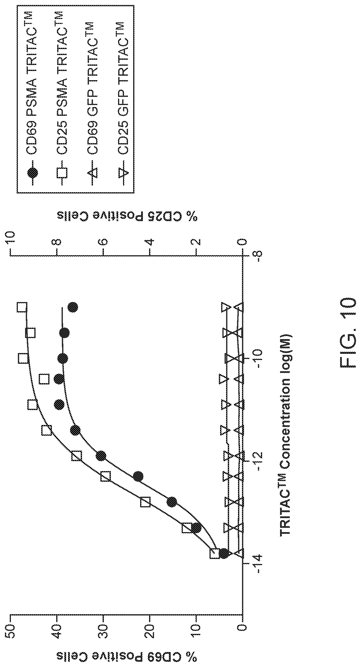
D00017

D00018

S00001
XML
uspto.report is an independent third-party trademark research tool that is not affiliated, endorsed, or sponsored by the United States Patent and Trademark Office (USPTO) or any other governmental organization. The information provided by uspto.report is based on publicly available data at the time of writing and is intended for informational purposes only.
While we strive to provide accurate and up-to-date information, we do not guarantee the accuracy, completeness, reliability, or suitability of the information displayed on this site. The use of this site is at your own risk. Any reliance you place on such information is therefore strictly at your own risk.
All official trademark data, including owner information, should be verified by visiting the official USPTO website at www.uspto.gov. This site is not intended to replace professional legal advice and should not be used as a substitute for consulting with a legal professional who is knowledgeable about trademark law.