Nucleic acid-polypeptide compositions and uses thereof
Geall , et al. October 13, 2
U.S. patent number 10,800,848 [Application Number 16/128,417] was granted by the patent office on 2020-10-13 for nucleic acid-polypeptide compositions and uses thereof. This patent grant is currently assigned to AVIDITY BIOSCIENCES, INC.. The grantee listed for this patent is AVIDITY BIOSCIENCES, INC.. Invention is credited to Palani Balu, Rob Burke, David Sai-Ho Chu, Michael Caramian Cochran, Beatrice Diana Darimont, Venkata Ramana Doppalapudi, Andrew John Geall, Rachel Elizabeth Johns.


View All Diagrams
| United States Patent | 10,800,848 |
| Geall , et al. | October 13, 2020 |
Nucleic acid-polypeptide compositions and uses thereof
Abstract
Disclosed herein are compositions and pharmaceutical formulations that comprise a binding moiety conjugated to a polynucleic acid molecule and a polymer. Also described herein include methods for treating a cancer which utilize a composition or a pharmaceutical formulation comprising a binding moiety conjugated to a polynucleic acid molecule and a polymer.
| Inventors: | Geall; Andrew John (Carlsbad, CA), Doppalapudi; Venkata Ramana (San Diego, CA), Chu; David Sai-Ho (La Jolla, CA), Cochran; Michael Caramian (La Jolla, CA), Johns; Rachel Elizabeth (San Diego, CA), Balu; Palani (Cupertino, CA), Burke; Rob (Encinitas, CA), Darimont; Beatrice Diana (San Diego, CA) | ||||||||||
|---|---|---|---|---|---|---|---|---|---|---|---|
| Applicant: |
|
||||||||||
| Assignee: | AVIDITY BIOSCIENCES, INC. (La
Jolla, CA) |
||||||||||
| Family ID: | 1000005111641 | ||||||||||
| Appl. No.: | 16/128,417 | ||||||||||
| Filed: | September 11, 2018 |
Prior Publication Data
| Document Identifier | Publication Date | |
|---|---|---|
| US 20190062436 A1 | Feb 28, 2019 | |
Related U.S. Patent Documents
| Application Number | Filing Date | Patent Number | Issue Date | ||
|---|---|---|---|---|---|
| 15476849 | Mar 31, 2017 | ||||
| 62316919 | Apr 1, 2016 | ||||
| Current U.S. Class: | 1/1 |
| Current CPC Class: | A61K 47/60 (20170801); C07K 16/2863 (20130101); A61P 35/02 (20180101); C07K 16/3061 (20130101); A61K 48/0033 (20130101); C07K 16/2851 (20130101); A61K 31/713 (20130101); A61K 48/0075 (20130101); C07K 16/30 (20130101); A61K 48/005 (20130101); A61K 31/7088 (20130101); C12N 15/113 (20130101); A61P 35/00 (20180101); C12N 15/1135 (20130101); A61K 47/6807 (20170801); A61K 47/6851 (20170801); C12N 15/1138 (20130101); C12N 15/88 (20130101); A61K 47/6889 (20170801); C12N 2310/3231 (20130101); C12N 2310/14 (20130101); C07K 2317/622 (20130101); C12N 2310/332 (20130101); A61K 2039/505 (20130101); C12N 2310/3515 (20130101); C07K 2317/24 (20130101); C12N 2310/3513 (20130101); C12N 2320/32 (20130101); C12N 2310/321 (20130101); C12N 2310/315 (20130101); C07K 2317/569 (20130101); C07K 2317/21 (20130101); C07K 2317/55 (20130101) |
| Current International Class: | C07K 16/28 (20060101); A61K 31/7088 (20060101); A61P 35/02 (20060101); C07K 16/30 (20060101); A61P 35/00 (20060101); A61K 47/60 (20170101); C12N 15/88 (20060101); A61K 47/68 (20170101); A61K 31/713 (20060101); C12N 15/113 (20100101); A61K 48/00 (20060101); A61K 39/00 (20060101) |
| Field of Search: | ;424/181.1 ;435/6.1,91.1,91.31,455 ;530/300,350 ;536/23.1,24.5 |
References Cited [Referenced By]
U.S. Patent Documents
| 4694778 | September 1987 | Learn et al. |
| 5142047 | August 1992 | Summerton et al. |
| 5185444 | February 1993 | Summerton et al. |
| 5736557 | April 1998 | Hofheinz et al. |
| 5889136 | March 1999 | Scaringe et al. |
| 5898031 | April 1999 | Crooke |
| 6008400 | December 1999 | Scaringe et al. |
| 6107094 | August 2000 | Crooke |
| 6111086 | August 2000 | Scaringe |
| 6194551 | February 2001 | Idusogie et al. |
| 6528624 | March 2003 | Idusogie et al. |
| 6538124 | March 2003 | Idusogie et al. |
| 6821783 | November 2004 | Comely et al. |
| 6849272 | February 2005 | Langer et al. |
| 6884869 | April 2005 | Senter et al. |
| 6942972 | September 2005 | Farooqui et al. |
| 7056704 | June 2006 | Tuschl et al. |
| 7078196 | July 2006 | Tuschl et al. |
| 7132519 | November 2006 | Monforte et al. |
| 7351855 | April 2008 | Coutts et al. |
| 7364731 | April 2008 | Idusogie et al. |
| 7432249 | October 2008 | Crooke |
| 7432250 | October 2008 | Crooke |
| 7452987 | November 2008 | Giese et al. |
| 7459547 | December 2008 | Zamore et al. |
| 7498298 | March 2009 | Doronina et al. |
| 7521232 | April 2009 | Moon |
| 7595387 | September 2009 | Leake et al. |
| 7629321 | December 2009 | Crooke |
| 7732593 | June 2010 | Zamore et al. |
| 7750144 | July 2010 | Zamore et al. |
| 7772203 | August 2010 | Zamore et al. |
| 7786290 | August 2010 | Woppmann et al. |
| 7829693 | November 2010 | Kreutzer et al. |
| 7833992 | November 2010 | Vargeese et al. |
| 7834171 | November 2010 | Khvorova et al. |
| 7850975 | December 2010 | Mullis |
| 7893245 | February 2011 | Giese et al. |
| 7923547 | April 2011 | McSwiggen et al. |
| 7928217 | April 2011 | Vornlocher et al. |
| 7943762 | May 2011 | Weller et al. |
| 8084582 | December 2011 | Dahiyat et al. |
| 8084598 | December 2011 | Bentwich |
| 8090542 | January 2012 | Khvorova et al. |
| 8137695 | March 2012 | Rozema et al. |
| 8202979 | June 2012 | McSwiggen et al. |
| 8268986 | September 2012 | Beigelman et al. |
| 8273866 | September 2012 | McSwiggen et al. |
| 8283329 | October 2012 | Fire et al. |
| 8288352 | October 2012 | Doronina et al. |
| 8304530 | November 2012 | Zamore et al. |
| 8309704 | November 2012 | Zamore et al. |
| 8309705 | November 2012 | Zamore et al. |
| 8324370 | December 2012 | Giese et al. |
| 8324371 | December 2012 | Popplewell et al. |
| 8329892 | December 2012 | Zamore et al. |
| 8334373 | December 2012 | Vornlocher et al. |
| 8361979 | January 2013 | Aartsma-Rus et al. |
| 8362231 | January 2013 | Tuschl et al. |
| 8372968 | February 2013 | Tuschl et al. |
| 8389710 | March 2013 | Bruno et al. |
| 8420391 | April 2013 | Tuschl et al. |
| 8445237 | May 2013 | Tuschl et al. |
| 8461325 | June 2013 | Popplewell et al. |
| 8481698 | July 2013 | Lieberman et al. |
| 8501703 | August 2013 | Bennett et al. |
| 8501930 | August 2013 | Rozema et al. |
| 8546143 | October 2013 | Kreutzer et al. |
| 8552171 | October 2013 | Tuschl et al. |
| 8557292 | October 2013 | Davis et al. |
| 8591910 | November 2013 | Mullis |
| 8604184 | December 2013 | Mullis et al. |
| 8609105 | December 2013 | Senter et al. |
| 8618277 | December 2013 | Beigelman et al. |
| 8632997 | January 2014 | Tuschl et al. |
| 8648185 | February 2014 | McSwigen et al. |
| 8691786 | April 2014 | Rossi et al. |
| 8697688 | April 2014 | Howard et al. |
| 8746999 | June 2014 | Davis et al. |
| 8765930 | July 2014 | Tuschl et al. |
| 8772469 | July 2014 | Uhlmann et al. |
| 8778902 | July 2014 | Tuschl et al. |
| 8790922 | July 2014 | Tuschl et al. |
| 8796016 | August 2014 | Tuschl et al. |
| 8796443 | August 2014 | Khvorova et al. |
| 8846875 | September 2014 | Schwartz et al. |
| 8846894 | September 2014 | McSwiggen et al. |
| 8853384 | October 2014 | Tuschl et al. |
| 8895718 | November 2014 | Tuschl et al. |
| 8895721 | November 2014 | Tuschl et al. |
| 8895722 | November 2014 | Iversen et al. |
| 8906847 | December 2014 | Cleemann et al. |
| 8933044 | January 2015 | Tuschl et al. |
| 8933215 | January 2015 | Giese et al. |
| 8936910 | January 2015 | Mitsch et al. |
| 8969526 | March 2015 | Baehner et al. |
| 8993745 | March 2015 | Tuschl et al. |
| 9012138 | April 2015 | Tuschl et al. |
| 9012621 | April 2015 | Tuschl et al. |
| 9078911 | July 2015 | Lu |
| 9089614 | July 2015 | Lin et al. |
| 9096636 | August 2015 | Baker et al. |
| 9096877 | August 2015 | Johnson et al. |
| 9139828 | September 2015 | Platenburg et al. |
| 9175286 | November 2015 | Wilton et al. |
| 9175289 | November 2015 | Khvorova et al. |
| 9181551 | November 2015 | McSwiggen et al. |
| 9193753 | November 2015 | Tuschl et al. |
| 9212364 | December 2015 | Sah et al. |
| 9222092 | December 2015 | Giese et al. |
| 9228187 | January 2016 | Wilton et al. |
| 9243251 | January 2016 | Popplewell et al. |
| 9243252 | January 2016 | Popplewell et al. |
| 9249416 | February 2016 | Wilton et al. |
| 9260471 | February 2016 | Cancilla et al. |
| 9284551 | March 2016 | Puri et al. |
| 9328345 | May 2016 | Li et al. |
| 9364553 | June 2016 | Lee |
| 9416361 | August 2016 | Iversen et al. |
| 9434948 | September 2016 | Sazani et al. |
| 9441229 | September 2016 | Wilton et al. |
| 9447415 | September 2016 | Wilton et al. |
| 9447417 | September 2016 | Sazani et al. |
| 9481905 | November 2016 | Chen et al. |
| 9499818 | November 2016 | Van Deutekom |
| 9528109 | December 2016 | De Kimpe et al. |
| 9657294 | May 2017 | Beigelman et al. |
| 9695423 | July 2017 | Giese et al. |
| 9732344 | August 2017 | Beigelman et al. |
| 9765338 | September 2017 | Bennett et al. |
| 9771588 | September 2017 | McSwiggen et al. |
| 9796974 | October 2017 | Rajeev et al. |
| 9890379 | February 2018 | De Kimpe et al. |
| 9926557 | March 2018 | De Kimpe et al. |
| 10000754 | June 2018 | Beigelman et al. |
| 2002/0142980 | October 2002 | Thompson et al. |
| 2003/0073207 | April 2003 | Akhtar |
| 2007/0004665 | January 2007 | McSwiggen |
| 2009/0149403 | June 2009 | MacLachlan et al. |
| 2011/0039914 | February 2011 | Pavco et al. |
| 2011/0081362 | April 2011 | Elledge et al. |
| 2011/0263680 | October 2011 | Khvorova et al. |
| 2011/0263686 | October 2011 | Wilton et al. |
| 2011/0294753 | December 2011 | De Kimpe et al. |
| 2011/0301218 | December 2011 | Bozzoni et al. |
| 2012/0065169 | March 2012 | Hanson et al. |
| 2012/0094299 | April 2012 | Ranum et al. |
| 2012/0122800 | May 2012 | Kadushin et al. |
| 2012/0172415 | July 2012 | Voit et al. |
| 2012/0270925 | October 2012 | Wilton et al. |
| 2013/0028919 | January 2013 | Howard et al. |
| 2013/0045520 | February 2013 | Woolf et al. |
| 2013/0052731 | February 2013 | Ma et al. |
| 2013/0164366 | June 2013 | Kreutzer et al. |
| 2013/0172238 | July 2013 | Mitsch et al. |
| 2013/0177579 | July 2013 | Lin |
| 2013/0177631 | July 2013 | Kreutzer et al. |
| 2013/0309256 | November 2013 | Lyon et al. |
| 2014/0127239 | May 2014 | Howard |
| 2014/0194610 | July 2014 | Verdine et al. |
| 2014/0286970 | September 2014 | Jeffrey et al. |
| 2014/0288158 | September 2014 | Rajeev et al. |
| 2014/0294851 | October 2014 | Nguyen |
| 2014/0296321 | October 2014 | Iversen |
| 2014/0357700 | December 2014 | Rossi et al. |
| 2015/0018540 | January 2015 | Prakash et al. |
| 2015/0037360 | February 2015 | Smith |
| 2015/0038554 | February 2015 | Brown |
| 2015/0038555 | February 2015 | Brown |
| 2015/0056220 | February 2015 | Chennamsetty et al. |
| 2015/0105539 | April 2015 | Miao et al. |
| 2015/0105540 | April 2015 | Miao et al. |
| 2015/0111954 | April 2015 | Sliz et al. |
| 2015/0141492 | May 2015 | Tuschl et al. |
| 2015/0211006 | July 2015 | Butler et al. |
| 2015/0238516 | August 2015 | Dowdy et al. |
| 2015/0275212 | October 2015 | Albaek et al. |
| 2015/0366987 | December 2015 | Bodyak et al. |
| 2016/0002637 | January 2016 | Sazani et al. |
| 2016/0030332 | February 2016 | Lee et al. |
| 2016/0032288 | February 2016 | Tuschl et al. |
| 2016/0053262 | February 2016 | Platenburg et al. |
| 2016/0102135 | April 2016 | Escobar-Cabrera |
| 2016/0102148 | April 2016 | Park et al. |
| 2016/0193354 | July 2016 | Noe et al. |
| 2016/0298111 | October 2016 | Bestwick et al. |
| 2016/0304864 | October 2016 | De Kimpe et al. |
| 2016/0304874 | October 2016 | Krauss |
| 2016/0304877 | October 2016 | Swayze et al. |
| 2016/0367687 | December 2016 | Manoharan et al. |
| 2017/0081425 | March 2017 | Colletti et al. |
| 2017/0107512 | April 2017 | De Kimpe et al. |
| 2017/0204410 | July 2017 | Watanabe et al. |
| 2017/0204414 | July 2017 | Van Deutekom et al. |
| 2017/0281795 | October 2017 | Geall et al. |
| 2017/0342416 | November 2017 | McSwiggen et al. |
| 2018/0016574 | January 2018 | Bestwick et al. |
| 2018/0044675 | February 2018 | Watanabe et al. |
| 2018/0112214 | April 2018 | De Kimpe et al. |
| 2018/0127758 | May 2018 | Bennett |
| 2018/0163209 | June 2018 | Bennett et al. |
| 2018/0305689 | October 2018 | S.ae butted.trom et al. |
| 2018/0369400 | December 2018 | Levin et al. |
| 2019/0000986 | January 2019 | Levin et al. |
| 2020/0123261 | April 2020 | Geall et al. |
| 0336675 | Oct 1989 | EP | |||
| 0334656 | Mar 1994 | EP | |||
| 1144623 | Aug 2002 | EP | |||
| 0928290 | Mar 2005 | EP | |||
| 1214945 | Jun 2005 | EP | |||
| 1579015 | Sep 2005 | EP | |||
| 1352061 | May 2006 | EP | |||
| 1742958 | Jan 2007 | EP | |||
| 1407044 | Sep 2007 | EP | |||
| 1068241 | Oct 2007 | EP | |||
| 1550719 | Dec 2008 | EP | |||
| 1349927 | Mar 2010 | EP | |||
| 1409670 | Oct 2010 | EP | |||
| 1633890 | Oct 2010 | EP | |||
| 2336317 | Jun 2011 | EP | |||
| 2361923 | Aug 2011 | EP | |||
| 2049664 | Sep 2011 | EP | |||
| 1608733 | Dec 2011 | EP | |||
| 1873259 | Jan 2012 | EP | |||
| 2278004 | Oct 2012 | EP | |||
| 2514758 | Oct 2012 | EP | |||
| 2580326 | Apr 2013 | EP | |||
| 2351852 | Oct 2013 | EP | |||
| 2195428 | Dec 2013 | EP | |||
| 2028278 | Mar 2014 | EP | |||
| 2348133 | Jul 2014 | EP | |||
| 2344637 | Dec 2014 | EP | |||
| 1633770 | Apr 2015 | EP | |||
| 2340310 | Jun 2015 | EP | |||
| 1423406 | Nov 2015 | EP | |||
| 2949752 | Dec 2015 | EP | |||
| 2548962 | Jan 2016 | EP | |||
| 3031920 | Jun 2016 | EP | |||
| 2421971 | Jul 2016 | EP | |||
| 2287306 | Oct 2016 | EP | |||
| 3030658 | Mar 2017 | EP | |||
| 2813582 | Apr 2017 | EP | |||
| 2287305 | Nov 2017 | EP | |||
| 2486141 | Jan 2018 | EP | |||
| 2902406 | Jan 2018 | EP | |||
| 2595664 | Oct 2018 | EP | |||
| WO-9207065 | Apr 1992 | WO | |||
| WO-9315187 | Aug 1993 | WO | |||
| WO-9726270 | Jul 1997 | WO | |||
| WO-9734631 | Sep 1997 | WO | |||
| WO-9813526 | Apr 1998 | WO | |||
| WO-0149698 | Jul 2001 | WO | |||
| WO-2006000057 | Jan 2006 | WO | |||
| WO-2006128138 | Nov 2006 | WO | |||
| WO-2007021142 | Feb 2007 | WO | |||
| WO-2008036127 | Mar 2008 | WO | |||
| WO-2009099942 | Aug 2009 | WO | |||
| WO-2009099991 | Aug 2009 | WO | |||
| WO-2009108217 | Sep 2009 | WO | |||
| WO-2009126933 | Oct 2009 | WO | |||
| WO-2009129281 | Oct 2009 | WO | |||
| WO-2011003557 | Jan 2011 | WO | |||
| WO-2011009624 | Jan 2011 | WO | |||
| WO-2012092373 | Jul 2012 | WO | |||
| WO-2013166004 | Nov 2013 | WO | |||
| WO-2013166155 | Nov 2013 | WO | |||
| WO-2014080251 | May 2014 | WO | |||
| WO-2014140317 | Sep 2014 | WO | |||
| WO-2014145090 | Sep 2014 | WO | |||
| WO-2014154835 | Oct 2014 | WO | |||
| WO-2014177042 | Nov 2014 | WO | |||
| WO-2014197854 | Dec 2014 | WO | |||
| WO-2015021457 | Feb 2015 | WO | |||
| WO-2015038426 | Mar 2015 | WO | |||
| WO-2015057699 | Apr 2015 | WO | |||
| WO-2015069587 | May 2015 | WO | |||
| WO-2015084846 | Jun 2015 | WO | |||
| WO-2015107425 | Jul 2015 | WO | |||
| WO-2015113922 | Aug 2015 | WO | |||
| WO-2015200223 | Dec 2015 | WO | |||
| WO-2016028649 | Feb 2016 | WO | |||
| WO-2016187425 | Nov 2016 | WO | |||
| WO-2017148879 | Sep 2017 | WO | |||
| WO-2017173408 | Oct 2017 | WO | |||
| WO-2017192679 | Nov 2017 | WO | |||
| WO-2017221883 | Dec 2017 | WO | |||
| WO-2018002812 | Jan 2018 | WO | |||
| WO-2018129384 | Jul 2018 | WO | |||
| WO-2019060775 | Mar 2019 | WO | |||
Other References
|
US. Appl. No. 15/476,849 (Year: 2016). cited by examiner . U.S. Appl. No. 16/128,440 (Year: 2016). cited by examiner . U.S. Appl. No. 16/128,428 (Year: 2016). cited by examiner . Beduneau et al. Design of targeted lipid nanocapsules by conjugation of whole antibodies and antibody Fab' fragments. Biomaterials 28(33):4978-4990 (2007). cited by applicant . Casi et al. Antibody-drug conjugates: basic concepts, examples and future perspectives. J Control Release 161:422-428 (201). cited by applicant . Debinski et al. Monovalent immunotoxin containing truncated form of Pseudomonas exotoxin as potent antitumor agent. Cancer Research 52(19):5379-5385 (1992). cited by applicant . Domingo et al. Transferrin receptor as a target for antibody--drug conjugates. Methods in Enzymology 112:238-247 (1985). cited by applicant . Feener et al. Alternative splicing of human dystrophin mRNA generates isoforms at the carboxy terminus. Nature 338:509-511 (Apr. 6, 1989). cited by applicant . Goldmacher et al. Antibody-drug conjugates: using monoclonal antibodies for delivery of cytotoxic payloads to cancer cells. Therapeutic Delivery 2:397-416 (2011). cited by applicant . Hudson et al. Cellular delivery of hammerhead ribozymes conjugated to a transferrin receptor antibody. Int J Pharmaceuticals 182(1):49-58 (1999). cited by applicant . Ishikawa et al. Preparation of monomeric Fab'--horseradish peroxidase conjugate using thiol groups in the hinge and its evaluation in enzyme immunoassay and immunohistochemical staining. Ann N Y Acad Sci. 420:74-89 (1983). cited by applicant . Miyata et al. Polymer nanotechnolgoy for nucleic acid delivery. Drug Delivery System 31(1):44-53 (2016) (English Abstract). cited by applicant . Normand-Sdiqui et al. Oligonucleotide delivery: Uptake of rat transferrin receptor antibody (OX / 26) conjugates into an in vitro immortalised cell line model of the blood, brain barrier. Int J Pharmaceut. Int J Pharmaceuticals 163:63-71 (1998). cited by applicant . PCT/US2017/025608 International Preliminary Report on Patentability dated Oct. 11, 2018. cited by applicant . Schnyder et al. Targeting of skeletal muscle in vitro using biotinylated immunoliposomes. Biochem J 377(Pt.1):61-67 (2004). cited by applicant . Sekyere et al. Examination of the distribution of the transferrinhomologue, melanotransferrin (tumour antigen p97), in mouse and human. Biochimica et Biophysica Acta 1722(2):131-142 (2005). cited by applicant . Summerton, et al. Morpholino antisense oligomers: design, preparation, and properties.Antisense Nucleic Acid Drug Dev. Jun. 1997;7(3):187-95. cited by applicant . U.S. Appl. No. 16/128,440 Office Action dated May 30, 2019. cited by applicant . U.S. Appl. No. 16/128,440 Office Action dated Nov. 9, 2018. cited by applicant . U.S. Appl. No. 16/128,450 Office Action dated Apr. 19, 2019. cited by applicant . U.S. Appl. No. 16/129,694 Office Action dated May 30, 2019. cited by applicant . U.S. Appl. No. 16/129,694 Office Action dated Nov. 19, 2018. cited by applicant . U.S. Appl. No. 16/129,696 Office Action dated Apr. 17, 2019. cited by applicant . Walker et al. Improved cellular delivery of antisense oligonucleotides using transferrin receptor antibody-oligonucleotide conjugates. Pharmaceutical research 12(10):1548-1553 (1995). cited by applicant . Abramova et al. Novel oligonucleotide analogues based on morpholino nucleoside subunits-antisense technologies: new chemical possibilities. Indian Journal of Chemistry 48B:1721-1726 (2009). cited by applicant . Agarwal et al. A Pictet-Spengler ligation for protein chemical modification. PNAS 110(1):46-51 (2013). cited by applicant . Albarran et al. Efficient intracellular delivery of a pro-apoptotic peptide with a pH-responsive carrier. React Funct Polym 71:261-265 (2011). cited by applicant . Axup et al. Synthesis of site-specific antibody-drug conjugates using unnatural amino acids. PNAS 109(40):16101-16106 (2012). cited by applicant . Baumer et al. Antibody-mediated delivery of anti-KRAS-siRNA in vivo overcomes therapy resistance in colon cancer. Clin Can Res 21(6):1383-1394 (2015). cited by applicant . Bell et al. Epidermal Growth Factor Receptor Mutations and Gene Amplification in Non-Small-Cell Lung Cancer: Molecular Analysis of the IDEAL/INTACT Gefitinib Trials. J Clin Oncol 23(31):8081-8092 (2005). cited by applicant . Bird et al. Single-chain antigen-binding proteins. Science 242:423-442 (1988). cited by applicant . Blaney et al. Traceless solid-phase organic synthesis. Chem. Rev. 102:2607-2024 (2002. cited by applicant . Bulmus et al. A new pH-responsive and glutathione-reactive, endosomal membrane-disruptive polymeric carrier for intracellular delivery of biomolecular drugs. J Controlled Release 93:105-120 (2003). cited by applicant . Burke et al. iRNA-mediated knockdown of P450 oxidoreductase in rats: a tool to reduce metabolism by CYPs and increase exposure of high clearance compounds. Pharm. Res. 31(12):3445-3460 (2014). cited by applicant . Casi et al. Site-specific traceless coupling of potent cytotoxic drugs to recombinant antibodies for pharmacodelivery. J Am Chem Soc 134(13):5887-5892 (2012). cited by applicant . Castaneda et al. Acid-cleavable thiomaleamic acid linker for homogeneous antibody-drug conjugation, Chem. Commun. 49:8187-8189 (2013). cited by applicant . Chen et al. Strand-specific 5'-O-methylation of siRNA duplexes controls guide strand selection and targeting specificity. RNA 14:263-274 (2008). cited by applicant . Clackson et al. Making antibody fragments using phage display libraries. Nature 352(6336):624-628 (1991). cited by applicant . Colberre-Garapin et al. A new dominant hybrid selective marker for higher eukaryotic cells. J Mol Biol 150:1-14 (1981). cited by applicant . Cole et al. The EBV-hybridoma technique and its application to human lung cancer. In, Monoclonal Antibodies and Cancer Therapy (vol. 27, UCLA Symposia on Molecular and Cellular Biology, New Series) (eds. R.A. Reisfeld and S.Sell), New York: Alan R. Liss, Inc. pp. 77-96 (1985). cited by applicant . Co-pending U.S. Appl. No. 16/128,393, filed Sep. 11, 2018. cited by applicant . Co-pending U.S. Appl. No. 16/128,428, filed Sep. 11, 2018. cited by applicant . Co-pending U.S. Appl. No. 16/128,440, filed Sep. 11, 2018. cited by applicant . Co-pending U.S. Appl. No. 16/129,694, filed Sep. 12, 2018. cited by applicant . Crouse et al. Expression and amplification of engineered mouse dihydrofolate reductase minigenes. Mol Cell Biol 3(2):257-266 (1983). cited by applicant . Cuellar et al. Systematic evaluation of antibody-mediated siRNA delivery using an industrial platform of THIOMAB--siRNA conjugates. Nucleic Acids Res 43(2):1189-1203 (2015). cited by applicant . Dawson et al. Modulation of Reactivity in Native Chemical Ligation through the Use of Thiol Additives. J. Am. Chem. Soc. 119:4325-4329 (1997). cited by applicant . Dawson et al. Synthesis of proteins by native chemical ligation. Science 266(5186):776-779 (1994). cited by applicant . Deleavey et al. Designing chemically modified oligonucleotides for targeted gene silencing. Chem Biol. 19(8):937-954 (2012). cited by applicant . Dietel et al. A 2015 update on predictive molecular pathology and its role in targeted cancer therapy: a review focussing on clinical relevance. Cancer Gene Ther 22(9):417-430 (2015). cited by applicant . Dimasi et al. Development of a trispecific antibody designed to simultaneously and efficiently target three different antigens on tumor cells. Mol Pharm 12(9):3490-3501 (2015). cited by applicant . Duncan et al. A polymer-Triton X-100 conjugate capable of pH-dependent red blood cell lysis: a model system illustrating the possibility of drug delivery within acidic intracellular compartments. J Drug Target 2:341-347 (1994). cited by applicant . El-Sayed et al. Rational design of composition and activity correlations for pH-responsive and glutathione-reactive polymer therapeutics. J Control Release 104:417-427 (2005). cited by applicant . Flanary et al. Antigen delivery with poly(propylacrylic acid) conjugation enhanced MHC-1 presentation and T-cell activation. Bioconjugate Chem. 20:241-248 (2009). cited by applicant . Gaziova et al. Chemically defined polyethylene glycol siRNA conjugates with enhanced gene silencing effect. Bioorg Med Chem 22(7):2320-2326 (2014). cited by applicant . Goldspiel et al. Human gene therapy. Clin Pharm 12:488-505 (1993). cited by applicant . Griffey et al. 2'-0-aminopropyl ribonucleotides: a zwitterionic modification that enhances the exonuclease resistance and biological activity of antisense oligonucleotides, J. Med. Chem. 39(26):5100-5109 (1997). cited by applicant . Hackeng et al. Protein synthesis by native chemical ligation: Expanded scope by using straightforward methodology. PNAS USA 96:10068-10073 (1999). cited by applicant . Hanes et al. In vitro selection and evolution of functional proteins by using ribosome display. PNAS USA 94:4937-4942 (1997). cited by applicant . Hejesen et al. A traceless aryl-triazene linker for DNA-directed chemistry. Org Biomol Chem 11(15):2493-2497 (2013). cited by applicant . Henry et al. pH-responsive poly(styrene-alt-maleic anhydride) alkylamide copolymers for intracellular drug delivery. Biomacromolecules 7:2407-2414 (2006). cited by applicant . Hu et al. Site-specific Antibody-polymer Conjugates for siRNA Delivery. J Am Chem Soc 135(37):13885-13891 (2013). cited by applicant . Huang et al. Mechanisms of resistance to EGFR tyrosine kinase inhibitors. Acta Pharma Sinica B 5(5):390-401 (2015). cited by applicant . Huse et al. Generation of a large combinatorial library of the immunoglobulin repertoire in phage lambda. Science 246(4935):1275-1281 (1989). cited by applicant . Huston et al. Protein engineering of antibody binding sites: recovery of specific activity in an anti-digoxin single-chain Fv analogue produced in Escherichia coli. PNAS USA 85(16):5879-5883 (1988). cited by applicant . Iversen et al. Optimized siRNA-PEG conjugates for extended blood circulation and reduced urine excretion in mice. Theranostics 3(3):201-209 (2013). cited by applicant . Jancik et al. Clinical relevance of KRAS in human cancers. J Biomed Biotechnol 2010:150960 (13 pgs.) (2010). cited by applicant . Jones et al. Poly(2-alkylacrylic acid) polymers deliver molecules to the cytosol by pH-sensitive disruption of endosomal vesicles. Biochem J 372:65-75 (2003). cited by applicant . Khormaee et al. Edosomolytic anionic polymer for the cytoplasmic delivery of siRNAs in localized in vivo applications. Adv Funct Mater 23:565-574 (2013). cited by applicant . Kim et al. PEG conjugated VEGF siRNA for anti-angiogenic gene therapy. J Cont Rel 116:123-129 (2006). cited by applicant . Kohler et al. Continuous cultures of fused cells secreting antibody of predefined specificity. Nature 256:495-497 (1975). cited by applicant . Koizumi. ENA oligonucleotides as therapeutics. Curr Opin Mol Ther 8(2):144-149 (2006). cited by applicant . Kontermann et al. Bispecific antibodies. Drug Discov Today 20(7):838-847 (2015). cited by applicant . Kozbor et al. The production of monoclonal antibodies from human lymphocytes. Immunology Today 4:72-79 (1983). cited by applicant . Kutmeier et al. Assembly of humanized antibody genes from synthetic oligonucleotides using a single-round PCR. BioTechniques 17:242 (1994). cited by applicant . Leigh et al. The Human Plasma Proteome: History, Character, and Diagnostic Prospects. Mol Cell Proteomics 1:845-867 (2002). cited by applicant . Loh et al. A Survey of siRNA Nanoscal Delivery Patents. 11 Nanotechnology Law & Bus. (pp. 29-37) (2014). cited by applicant . Lowy et al. Isolation of transforming DNA: Cloning the hamster aprt gene. Cell 22:817-823 (1980). cited by applicant . Lyon et al. Self-hydrolyzing maleimides improve the stability and pharmacological properties of antibody-drug conjugates. Nat. Biotechnol. 32(10):1059-1062 (2014). cited by applicant . McEnaney et al. Antibody-recruiting molecules: an emerging paradigm for engaging immune function in treating human disease. ACS Chem Biol. 7(7):1139-1151 (2012). cited by applicant . Morgan et al. Human gene therapy. Ann Rev Biochem 62:191-217 (1993). cited by applicant . Morrison et al. Chimeric human antibody molecules: mouse antigen-binding domains with human constant region domains. PNAS USA 81(21):6851-6855 (1984). cited by applicant . Mulligan. The basic science of gene therapy. Science 260(5110):926-932 (1993). cited by applicant . Naisbitt et al. Disposition of amodiaquine and related antimalarial agents in human neutrophils: implications for drug design. J Pharmacol Exp Ther 280:884-893 (1997). cited by applicant . Neuberger et al. Recombinant antibodies possessing novel effector functions. Nature 312(5995):604-608 (1984). cited by applicant . Obika et al. Synthesis of 2'-0,4'-C-methyleneuridine and -cytidine. Novel bicyclic nucleosides having a fixed C3'-endo sugar puckering. Tetrahedron Lett. 38(50):8735-8738 (1997). cited by applicant . O'Hare et al. Transformation of mouse fibroblasts to methotrexate resistance by a recombinant plasmid expressing a prokaryotic dihydrofolate reductase. PNAS USA 78:1527-1531 (1981). cited by applicant . PCT/US2017/025608 International Search Report and Written Opinion dated Jul. 10, 2017. cited by applicant . Phimister. Targeting Therapeutic Oligonucleotides. N Engl J Med 376:86-88 (2017). cited by applicant . Rozema et al. Dynamic PolyConjugates for targeted in vivo delivery of siRNA to hepatocytes. PNAS USA 104(32):12982-12987 (2007). cited by applicant . Santerre et al. Expression of prokaryotic genes for hygromycin B and G418 resistance as dominant-selection markers in mouse L cells. Gene 30(1-3):147-156 (1984). cited by applicant . Singh et al. Recent developments in oligonucleotide conjugation. Chem Soc Rev 39(6):2054-2070 (2010). cited by applicant . Skerra et al. Assembly of a functional Immunoglobulin Fv fragment in Escherichia coli. Science 240(4855):1038-1041 (1988). cited by applicant . Strop et al. Location matters: site of conjugation modulates stability and pharmacokinetics of antibody drug conjugates. Chem Biol 20(2):161-167 (2013). cited by applicant . Suriano et al. Beta-catenin (CTNNB1) gene amplification: a new mechanism of protein overexpression in cancer. Genes Chromosomes Cancer 42(3):238-246 (2005). cited by applicant . Szybalska et al. Genetics of human cell line. IV. DNA-mediated heritable transformation of a biochemical trait. PNAS USA 48:2026-2034 (1962). cited by applicant . Takeda et al. Construction of chimaeric processed immunoglobulin genes containing mouse variable and human constant region sequences. Nature 314(6010):452-454 (1985). cited by applicant . Talasila et al. EGFR Wild-type Amplification and Activation Promote Invasion and Development of Glioblastoma Independent of Angiogenesis. Acta Neuropathol. 125(5):683-698 (2013). cited by applicant . Tolstoshev. Gene Therapy, Concepts, Current Trials and Future Directions. Ann. Rev. Pharmacol. Toxicol. 32:573-596 (1993). cited by applicant . U.S. Appl. No. 15/473,849 1st Action Interview dated Nov. 24, 2017. cited by applicant . U.S. Appl. No. 15/476,849 1st Action Interview dated Sep. 11, 2017. cited by applicant . U.S. Appl. No. 15/476,849 Office Action dated Apr. 17, 2018. cited by applicant . U.S. Appl. No. 15/476,849 Office Action May 2, 2018. cited by applicant . Valtorta et al. KRAS gene amplification in colorectal cancer and impact on response to EGFR-targeted therapy. Int J Cancer 133:1259-1266 (2013). cited by applicant . Ward et al. Binding activities of a repertoire of single immunoglobulin variable domains secreted from Escherichia coli. Nature 341(6242):544-546 (1989). cited by applicant . Watts et al. Chemically modified siRNA: tools and applications. Drug Discov Today 13(19-20):842-855 (2008). cited by applicant . Wigler et al. Transfer of purified herpes virus thymidine kinase gene to cultured mouse cells. Cell 11:223-232 (1977). cited by applicant . Wigler et al. Transformation of mammalian cells with an amplifiable dominant-acting gene. PNAS USA 77:3567-3570 (1980). cited by applicant . Winkler. Oligonucleotide conjugates for therapeutic applications. Ther Del 4(7):791-809 (2013). cited by applicant . Wong et al. Co-injection of a targeted, reversibly masked endosomolytic polymer dramatically improves the efficacy of cholesterol-conjugated small interfering RNAs in vivo. Nucleic Acid Ther 22(6):380-390 (2012). cited by applicant . Wu et al. Building complex glycopeptides: Development of a cysteine-free native chemical ligation protocol. Angew. Chem. Int. Ed. 45:4116-4125 (2006). cited by applicant . Wu et al. Delivery systems for gene therapy. Biotherapy 3:87-95 (1991). cited by applicant . Wu et al. Site-specific chemical modification of recombinant proteins produced in mammalian cells by using the genetically encoded aldehyde tag. PNAS USA 106(9):3000-3005 (2009). cited by applicant . Xu et al. Delivery systems for siRNA drug development in cancer therapy. Asian Journal of Pharmaceutical Sciences 10(1):1-12 (2015). cited by applicant . Yessine et al. Characterization of the membrane-destabilizing properties of different pH-sensitive methacrylic acid copolymers. Biochimica et Biophysica Acta 1613:28-38 (2003). cited by applicant . Yuan et al. Development of siRNA payloads to target KRAS-mutant cancer. Cancer Discov 4(10):1182-1197 (2014). cited by applicant . Zhang et al. A remote arene-binding site on prostate specific membrane antigen revealed by antibody-recruiting small molecules. J Am Chem Soc. 132(36):12711-12716 (2010). cited by applicant . Aartsma-Rus et al. Progress in therapeutic antisense applications for neuromuscular disorders. Eur J Hum Genet 18(2):146-153 (2010). cited by applicant . Beigelman et al. Chemical modification of hammerhead ribozymes. Catalytic activity and nuclease resistance. J Biol Chem 270:25702-25708 (1995). cited by applicant . Burlina et al. Chemical engineering of RNase resistant and catalytically active hammerhead ribozymes. Bioorg Med Chem 5:1999-2010 (1997). cited by applicant . Carter et al. Antibody-drug conjugates for cancer therapy. Cancer J 14(3):154-69 (2008). cited by applicant . De Angelis et al. Chimeric snRNA molecules carrying antisense sequences against the splice junctions of exon 51 of the dystrophin pre-mRNA induce exon skipping and restoration of a dystrophin synthesis in Delta 48-50 DMD cells. PNAS USA 99:9456-9461 (2002). cited by applicant . Earnshaw et al. Modified oligoribonucleotides as site-specific probes of RNA structure and function. Biopolymers (Nucleic Acid Sciences) 48:39-55 (1998). cited by applicant . Echigoya et al. In Silico Screening Based on Predictive Algorithms as a Design Tool for Exon Skipping Oligonucleotides in Duchenne Muscular Dystrophy. PLoS One 10(3):e0120058 (2015). cited by applicant . Hoffman et al. Restoring Dystrophin Expression in Duchenne Muscular Dystrophy Muscle: Progress in Exon Skipping and Stop Codon Read Through. Am J Pathol 179(1):12-22 (2011). cited by applicant . Karpeisky et al. Highly efficient synthesis of 2'-O-amino nucleosides and their incorporation in hammerhead ribozymes. Tetrahedron Lett 39:1131-1134 (1998). cited by applicant . Langlois et al. Cytoplasmic and Nuclear Retained DMPK mRNAS Are Targets for RNA Interference in Myotonic Dystrophy Cells. J biol Chem 280(17):16949-16954 (2005). cited by applicant . Lee et al. Antisense PMO cocktails effectively skip dystrophin exons 45-55 in myotubes transdifferentiated from DMD patient fibroblasts. PLoS One 13(5):e0197084 (2018). cited by applicant . Loakes. Survey and summary: The applications of universal DNA base analogues. Nucleic Acids Research 29:2437-2447 (2001). cited by applicant . Martinez et al. Single-stranded antisense siRNAs guide target RNA cleavage in RNAi. Cell 110(5):563-574 (2002). cited by applicant . Mulders et al. Molecular therapy in myotonic dystrophy: focus on RNA gain-of-function. Hum Mol Gen 19(R1):R90-R97 (2010). cited by applicant . Mulders et al. Supporting Information for Triplet-repeat oligonucleotide-mediated reversal of RNA toxicity in myotonic dystrophy. PNAS 106(33):13915-13920 (2009) (13 pgs). cited by applicant . Mulders et al. Triplet-repeat oligonucleotide-mediated reversal of RNA toxicity in myotonic dystrophy. PNAS 106(33):13915-13920 (2009). cited by applicant . Mulligan et al. Selection for animal cells that express the Escherichia coli gene coding for xanthine-guanine phosphoribosyltransferase. PNAS USA 78(4):2072-2076 (1981). cited by applicant . Nielsen et al. Advances in targeted delivery of small interfering RNA using simple bioconjugates. Expert Opinion on Drug Delivery 11(5):791-822 (2014). cited by applicant . PCT/US2018/012672 International Search Report and Written Opinion dated May 24, 2018. cited by applicant . PCT/US2018/012672 Invitation to Pay Additional Fees dated Mar. 20, 2018. cited by applicant . PCT/US2018/052289 International Search Report and Written Opinion dated Jan. 11, 2019. cited by applicant . Perrault et al. Mixed deoxyribo- and ribo-oligonucleotides with catalytic activity. Nature 344:565-568 (1990). cited by applicant . Pieken et al. Kinetic characterization of ribonuclease-resistant 2'-modified hammerhead ribozymes. Science 253:314-317 (1991). cited by applicant . Schwarz et al. Evidence that siRNAs function as guides, not primers, in the Drosophila and human RNAi pathways. Molecular Cell 10:537-548 (2002). cited by applicant . Sugo et al. Development of antibody-siRNA conjugate targeted to cardiac and skeletal muscles. J Control release 237:1-13 (2016). cited by applicant . Sune-Pou et al. Targeting Splicing in the Treatment of Human Disease. Genes 8:E87 (2017). cited by applicant . Turner et al. The myotonic dystrophies: diagnosis and management. J Neurol Neurosurg Psychiatry 81:358-367 (2010). cited by applicant . U.S. Appl. No. 16/128,393 Office Action dated Aug. 26, 2019. cited by applicant . U.S. Appl. No. 16/128,428 Office Action dated Oct. 1, 2019. cited by applicant . U.S. Appl. No. 16/128,450 Miscellaneous Communication re: Third Party Submission dated Jul. 1, 2019. cited by applicant . U.S. Appl. No. 16/128,450 Office Action dated Sep. 19, 2019. cited by applicant . U.S. Appl. No. 16/129,696 Miscellaneous Communication re: Third Party Submission dated Jul. 3, 2019. cited by applicant . U.S. Appl. No. 16/129,696 Office Action dated Sep. 19, 2019. cited by applicant . Usman et al. Exploiting the chemical synthesis of RNA. Trends Biochem Sci 17:334-339 (1992). cited by applicant . Van Vliet et al. Assessment of the feasibility of exon 45-55 multiexon skipping for duchenne muscular dystrophy. BMC Medical Genetics 9:105 (2008). cited by applicant . Verma et al. Modified oligonucleotides: synthesis and strategy for users. Annu Rev Biochem 67:99-134 (1998). cited by applicant . Yazdi et al. Influence of cellular trafficking on protein synthesis inhibition of immunotoxins directed against the transferrin receptor. Cancer Res 55:3763-3771 (1995). cited by applicant . Aartsma-Rus et al. Guidelines for antisense oligonucleotide design and insight into splice-modulating mechanisms. Mol Ther 17(3):548-53 (2009). cited by applicant . Aartsma-Rus et al. Targeted exon skipping as a potential gene correction therapy for Duchenne muscular dystrophy. Neuromuscul Disord. 12 Suppl 1:S71-7 (2002). cited by applicant . Arechavala-Gomeza et al. Comparative analysis of antisense oligonucleotide sequences for targeted skipping of exon 51 during dystrophin pre-mRNA splicing in human muscle. Hum Gene Ther. 18(9):798-810 (2007). cited by applicant . U.S. Appl. No. 16/128,428 Office Action dated Mar. 27, 2020. cited by applicant . U.S. Appl. No. 16/129,696 Office Action dated Apr. 13, 2020. cited by applicant . Van Deutekom et al. Antisense-induced exon skipping restores dystrophin expression in DMD patient derived muscle cells. Hum Mol Genet. 10(15):1547-54 (2001). cited by applicant . U.S. Appl. No. 16/128,450 Office Action dated Apr. 30, 2020. cited by applicant. |
Primary Examiner: Zara; Jane J
Attorney, Agent or Firm: Wilson Sonsini Goodrich & Rosati
Parent Case Text
CROSS-REFERENCE
This application is a continuation of U.S. application Ser. No. 15/476,849, filed Mar. 31, 2017, which in turn claims the benefit of U.S. Provisional Application No. 62/316,919, filed Apr. 1, 2016, which application is incorporated herein by reference.
Claims
What is claimed is:
1. A pharmaceutical composition comprising: a molecule of Formula (I): A-X--B--Y--C Formula I wherein, A is an antibody or its binding fragments thereof; B consists of a double-stranded siRNA consisting of a passenger strand and a guide strand; C consists of a polymer; and X and Y are each independently a linker selected from the group consisting of a heterobifunctional linker, a homobifunctional linker, a maleimide group, a dipeptide moiety, a benzoic acid group or derivatives thereof, a C.sub.1-C.sub.6 alkyl group, and a combination thereof; and a pharmaceutically acceptable excipient; wherein: the double-stranded siRNA comprises at least one 2' modified nucleotide, at least one modified internucleotide linkage, or at least one inverted abasic moiety; A and C are attached in the same strand, but not at the same terminus; and A-X and Y--C are conjugated to the passenger strand.
2. The pharmaceutical composition of claim 1, wherein the guide strand hybridizes to a target region of a gene selected from the group consisting of KRAS, EGFR, AR, CTNNB1, PIK3CA, PIK3CB, MYC, and HPRT1.
3. The pharmaceutical composition of claim 1, wherein the pharmaceutical composition is formulated for the treatment of a solid tumor.
4. The pharmaceutical composition of claim 1, wherein the pharmaceutical composition is formulated for the treatment of breast cancer, lung cancer, ovarian cancer, prostate cancer, or skin cancer.
5. The pharmaceutical composition of claim 1, wherein the at least one 2' modified nucleotide comprises 2'-O-methyl, 2'-O-methoxyethyl (2'-O-MOE), 2'-O-aminopropyl, 2'-deoxy, 2'-deoxy-2'-fluoro, 2'-O-aminopropyl (2'-O-AP), 2'-O-dimethylaminoethyl (2'-O-DMAOE), 2'-O-dimethylaminopropyl (2'-O-DMAP), 2'-O-dimethylaminoethyloxyethyl (2'-O-DMAEOE), or 2'-O--N-methylacetamido (2'-O-NMA) modified nucleotide.
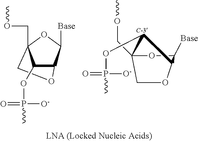
6. The pharmaceutical composition of claim 1, wherein the at least one 2' modified nucleotide comprises locked nucleic acid (LNA) or ethylene nucleic acid (ENA).
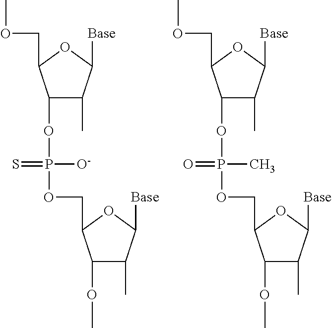
7. The pharmaceutical composition of claim 1, wherein the at least one modified internucleotide linkage comprises a phosphorothioate linkage or a phosphorodithioate linkage.
8. The pharmaceutical composition of claim 1, wherein the at least one inverted abasic moiety is at at least one terminus.
9. The pharmaceutical composition of claim 1, wherein the guide strand comprises a sequence having at least 80% sequence identity to SEQ ID NOs: 16-75, 452-1955, 1956-1962, 1967-2002, 2013-2032, 2082-2109, or 2117.
10. The pharmaceutical composition of claim 1, wherein the antibody or binding fragment thereof comprises a humanized antibody or binding fragment thereof, chimeric antibody or binding fragment thereof, monoclonal antibody or binding fragment thereof, monovalent Fab', divalent Fab2, single-chain variable fragment (scFv), diabody, minibody, nanobody, single-domain antibody (sdAb), or camelid antibody or binding fragment thereof.
11. The pharmaceutical composition of claim 1, wherein the antibody or binding fragment thereof is an anti-transferrin receptor antibody or binding fragment thereof.
12. The pharmaceutical composition of claim 1, wherein C is polyethylene glycol.
13. The pharmaceutical composition of claim 12, wherein C has a molecular weight from about 1000 Da to about 5000 Da.
14. The pharmaceutical composition of claim 12, wherein C has a molecular weight of about 1000 Da, 2000 Da, or 5000 Da.
15. The pharmaceutical composition of claim 1, wherein A-X is conjugated to the 5' end of the passenger strand and Y--C is conjugated to the 3' end of the passenger strand.
16. The pharmaceutical composition of claim 1, wherein Y--C is conjugated to the 5' end of the passenger strand and A-X is conjugated to the 3' end of the passenger strand.
Description
SEQUENCE LISTING
The instant application contains a Sequence Listing which has been submitted electronically in ASCII format and is hereby incorporated by reference in its entirety. Said ASCII copy, created on Sep. 11, 2018, is named 45532-707_303_SL.txt and is 615,705 bytes in size.
BACKGROUND OF THE DISCLOSURE
Gene suppression by RNA-induced gene silencing provides several levels of control: transcription inactivation, small interfering RNA (siRNA)-induced mRNA degradation, and siRNA-induced transcriptional attenuation. In some instances, RNA interference (RNAi) provides long lasting effect over multiple cell divisions. As such, RNAi represents a viable method useful for drug target validation, gene function analysis, pathway analysis, and disease therapeutics.
SUMMARY OF THE DISCLOSURE
Disclosed herein, in certain embodiments, are compositions and pharmaceutical formulations that comprise a binding moiety conjugated to a polynucleic acid molecule and a polymer. In some embodiments, also described herein include methods for treating a disease or condition (e.g., cancer) that utilize a composition or a pharmaceutical formulation comprising a binding moiety conjugated to a polynucleic acid molecule and a polymer.
Disclosed herein, in certain embodiments, is a molecule of Formula (I): A-X--B--Y--C Formula I wherein, A is a binding moiety; B is a polynucleotide; C is a polymer; X is a bond or first linker; and Y is a bond or second linker; and wherein the polynucleotide comprises at least one 2' modified nucleotide, at least one modified internucleotide linkage, or at least one inverted abasic moiety.
In some embodiments, the at least one 2' modified nucleotide comprises 2'-O-methyl, 2'-O-methoxyethyl (2'-O-MOE), 2'-O-aminopropyl, 2'-deoxy, T-deoxy-2'-fluoro, 2'-O-aminopropyl (2'-O-AP), 2'-O-dimethylaminoethyl (2'-O-DMAOE), 2'-O-dimethylaminopropyl (2'-O-DMAP), T-O-dimethylaminoethyloxyethyl (2'-O-DMAEOE), or 2'-O--N-methylacetamido (2'-O-NMA) modified nucleotide. In some embodiments, the at least one 2' modified nucleotide comprises locked nucleic acid (LNA) or ethylene nucleic acid (ENA). In some embodiments, the at least one modified internucleotide linkage comprises a phosphorothioate linkage or a phosphorodithioate linkage. In some embodiments, the at least one inverted abasic moiety is at at least one terminus.
In some embodiments, the polynucleotide comprises a single strand. In some embodiments, the polynucleotide comprises two or more strands. In some embodiments, the polynucleotide comprises a first polynucleotide and a second polynucleotide hybridized to the first polynucleotide to form a double-stranded polynucleic acid molecule. In some embodiments, the second polynucleotide comprises at least one modification.
In some embodiments, the first polynucleotide and the second polynucleotide are RNA molecules. In some embodiments, the first polynucleotide and the second polynucleotide are siRNA molecules.
In some embodiments, the first polynucleotide comprises a sequence having at least 60%, 65%, 70%, 75%, 80%, 85%, 90%, 95%, 96%, 97%, 98%, 99%, or 100% sequence identity to SEQ ID NOs: 16-75, 452-1955, 1956-1962, 1967-2002, 2013-2032, 2082-2109, or 2117. In some embodiments, the first polynucleotide consists of a sequence selected from SEQ ID NOs: 16-75, 452-1955, 1956-1962, 1967-2002, 2013-2032, 2082-2109, or 2117.
In some embodiments, the second polynucleotide comprises a sequence having at least 60%, 65%, 70%, 75%, 80%, 85%, 90%, 95%, 96%, 97%, 98%, 99%, or 100% sequence identity to SEQ ID NOs: 16-75, 452-1955, 1956-1962, 1967-2002, 2013-2032, 2082-2109, or 2117. In some embodiments, the second polynucleotide consists of a sequence selected from SEQ ID NOs: 16-75, 452-1955, 1956-1962, 1967-2002, 2013-2032, 2082-2109, or 2117.
In some embodiments, X and Y are independently a bond or a non-polymeric linker group. In some embodiments, X is a bond. In some embodiments, X is a C.sub.1-C.sub.6 alkyl group. In some embodiments, Y is a C.sub.1-C.sub.6 alkyl group. In some embodiments, X is a homobifuctional linker or a heterobifunctional linker, optionally conjugated to a C.sub.1-C.sub.6 alkyl group. In some embodiments, Y is a homobifuctional linker or a heterobifunctional linker.
In some embodiments, the binding moiety is an antibody or binding fragment thereof. In some embodiments, the antibody or binding fragment thereof comprises a humanized antibody or binding fragment thereof, chimeric antibody or binding fragment thereof, monoclonal antibody or binding fragment thereof, monovalent Fab', divalent Fab2, single-chain variable fragment (scFv), diabody, minibody, nanobody, single-domain antibody (sdAb), or camelid antibody or binding fragment thereof. In some embodiments, the antibody or binding fragment thereof is an anti-EGFR antibody or binding fragment thereof.
In some embodiments, C is polyethylene glycol. In some embodiments, C has a molecular weight of about 5000 Da.
In some embodiments, A-X is conjugated to the 5' end of B and Y--C is conjugated to the 3' end of B. In some embodiments, Y--C is conjugated to the 5' end of B and A-X is conjugated to the 3' end of B. In some embodiments, A-X, Y--C or a combination thereof is conjugated to an internucleotide linkage group.
In some embodiments, the molecule further comprises D. In some embodiments, D is conjugated to C or to A.
In some embodiments, D is conjugated to the molecule of Formula (I) according to Formula (II): (A-X--B--Y--C.sub.n)-L-D Formula II wherein, A is a binding moiety; B is a polynucleotide; C is a polymer; X is a bond or first linker; Y is a bond or second linker; L is a bond or third linker; D is an endosomolytic moiety; and n is an integer between 0 and 1; and wherein the polynucleotide comprises at least one 2' modified nucleotide, at least one modified internucleotide linkage, or at least one inverted abasic moiety; and D is conjugated anywhere on A, B, or C.
In some embodiments, D is INF7 or melittin.
In some embodiments, D is an endosomolytic polymer.
In some embodiments, L is a C.sub.1-C.sub.6 alkyl group. In some embodiments, L is a homobifuctional linker or a heterobifunctional linker.
In some embodiments, the molecule further comprises at least a second binding moiety A. In some embodiments, the at least second binding moiety A is conjugated to A, to B, or to C. In some embodiments, the at least second binding moiety A is cholesterol.
In some embodiments, the molecule further comprises at least an additional polynucleotide B. In some embodiments, the at least an additional polynucleotide B is conjugated to A, to B, or to C.
In some embodiments, the molecule further comprises at least an additional polymer C. In some embodiments, the at least an additional polymer C is conjugated to A, to B, or to C.
Disclosed herein, in certain embodiments, is a molecule of Formula (I): A-X--B--Y--C (Formula I), wherein A is an antibody or its binding fragments thereof; B is a polynucleotide; C is a polymer; X is a bond or first non-polymeric linker; and Y is a bond or second linker; wherein the polynucleotide comprises at least one 2' modified nucleotide, at least one modified internucleotide linkage, or at least one inverted abasic moiety; and wherein A and C are not attached to B at the same terminus. In some embodiments, the at least one 2' modified nucleotide comprises 2'-O-methyl, 2'-O-methoxyethyl (2'-O-MOE), 2'-O-aminopropyl, 2'-deoxy, T-deoxy-2'-fluoro, 2'-O-aminopropyl (2'-O-AP), 2'-O-dimethylaminoethyl (2'-O-DMAOE), 2'-O-dimethylaminopropyl (2'-O-DMAP), T-O-dimethylaminoethyloxyethyl (2'-O-DMAEOE), or 2'-O--N-methylacetamido (2'-O-NMA) modified nucleotide. In some embodiments, the at least one 2' modified nucleotide comprises locked nucleic acid (LNA) or ethylene nucleic acid (ENA). In some embodiments, the at least one modified internucleotide linkage comprises a phosphorothioate linkage or a phosphorodithioate linkage. In some embodiments, the at least one inverted abasic moiety is at at least one terminus. In some embodiments, the polynucleotide comprises a single strand. In some embodiments, the polynucleotide comprises a first polynucleotide and a second polynucleotide hybridized to the first polynucleotide to form a double-stranded polynucleic acid molecule. In some embodiments, the second polynucleotide comprises at least one modification. In some embodiments, the first polynucleotide and the second polynucleotide are RNA molecules. In some embodiments, the first polynucleotide comprises a sequence having at least 80%, 85%, 90%, 95%, 96%, 97%, 98%, 99%, or 100% sequence identity to SEQ ID NOs: 16-75, 452-1955, 1956-1962, 1967-2002, 2013-2032, 2082-2109, or 2117. In some embodiments, the second polynucleotide comprises a sequence having at least 80%, 85%, 90%, 95%, 96%, 97%, 98%, 99%, or 100% sequence identity to SEQ ID NOs: 16-75, 452-1955, 1956-1962, 1967-2002, 2013-2032, 2082-2109, or 2117. In some embodiments, Y is a non-polymeric linker group. In some embodiments, X is a bond. In some embodiments, X is a C.sub.1-C.sub.6 alkyl group. In some embodiments, Y is a C.sub.1-C.sub.6 alkyl group. In some embodiments, X is a homobifuctional linker or a heterobifunctional linker, optionally conjugated to a C.sub.1-C.sub.6 alkyl group. In some embodiments, Y is a homobifuctional linker or a heterobifunctional linker. In some embodiments, the antibody or binding fragment thereof comprises a humanized antibody or binding fragment thereof, chimeric antibody or binding fragment thereof, monoclonal antibody or binding fragment thereof, monovalent Fab', divalent Fab2, single-chain variable fragment (scFv), diabody, minibody, nanobody, single-domain antibody (sdAb), or camelid antibody or binding fragment thereof. In some embodiments, C is polyethylene glycol. In some embodiments, C has a molecular weight of about 1000 Da, 2000 Da, or 5000 Da. In some embodiments, A-X is conjugated to the 5' end of B and Y--C is conjugated to the 3' end of B. In some embodiments, Y--C is conjugated to the 5' end of B and A-X is conjugated to the 3' end of B. In some embodiments, the molecule further comprises D. In some embodiments, D is conjugated to C or to A. In some embodiments, D is conjugated to the molecule of Formula (I) according to Formula (II): (A-X--B--Y--C.sub.c)-L-D (Formula II), wherein A is an antibody or its binding fragments thereof; B is a polynucleotide; C is a polymer; X is a bond or first non-polymeric linker; Y is a bond or second linker; L is a bond or third linker; D is an endosomolytic moiety; and c is an integer between 0 and 1; wherein the polynucleotide comprises at least one 2' modified nucleotide, at least one modified internucleotide linkage, or at least one inverted abasic moiety; wherein A and C are not attached to B at the same terminus; and wherein D is conjugated anywhere on A or C or to a terminus of B. In some embodiments, D is INF7 or melittin. In some embodiments, D is an endosomolytic polymer. In some embodiments, L is a C.sub.1-C.sub.6 alkyl group. In some embodiments, L is a homobifuctional linker or a heterobifunctional linker. In some embodiments, the molecule further comprises at least a second binding moiety. In some embodiments, the at least second binding moiety is conjugated to A, to B, or to C. In some embodiments, the at least second binding moiety is cholesterol. In some embodiments, the molecule further comprises at least an additional polynucleotide B. In some embodiments, the at least an additional polynucleotide B is conjugated to A, to B, or to C. In some embodiments, the molecule further comprises at least an additional polymer C. In some embodiments, the at least an additional polymer C is conjugated to A, to B, or to C.
Disclosed herein, in certain embodiments, is a pharmaceutical composition comprising a molecule described above, and a pharmaceutically acceptable excipient. In some embodiments, the pharmaceutical composition is formulated as a nanoparticle formulation. In some embodiments, the pharmaceutical composition is formulated for parenteral, oral, intranasal, buccal, rectal, or transdermal administration.
Disclosed herein, in certain embodiments, is a method of treating a disease or disorder in a patient in need thereof, comprising administering to the patient a composition comprising a molecule described above. In some embodiments, the disease or disorder is a cancer. In some embodiments, the cancer is a solid tumor. In some embodiments, the cancer is a hematologic malignancy. In some embodiments, the cancer comprises a KRAS-associated, an EGFR-associated, an AR-associated cancer, a .beta.-catenin associated cancer, a PIK3C-associated cancer, or a MYC-associated cancer. In some embodiments, the cancer comprises bladder cancer, breast cancer, colorectal cancer, endometrial cancer, esophageal cancer, glioblastoma multiforme, head and neck cancer, kidney cancer, lung cancer, ovarian cancer, pancreatic cancer, prostate cancer, or thyroid cancer. In some embodiments, the cancer comprises acute myeloid leukemia, CLL, DLBCL, or multiple myeloma. In some embodiments, the method is an immuno-oncology therapy.
Disclosed herein, in certain embodiments, is a method of inhibiting the expression of a target gene in a primary cell of a patient, comprising administering a molecule described above to the primary cell. In some embodiments, the method is an in vivo method. In some embodiments, the patient is a human.
Disclosed herein, in certain embodiments, is an immuno-oncology therapy comprising a molecule described above for the treatment of a disease or disorder in a patient in need thereof.
Disclosed herein, in certain embodiments, is a kit comprising a molecule described above.
BRIEF DESCRIPTION OF THE DRAWINGS
Various aspects of the disclosure are set forth with particularity in the appended claims. A better understanding of the features and advantages of the present disclosure will be obtained by reference to the following detailed description that sets forth illustrative embodiments, in which the principles of the disclosure are utilized, and the accompanying drawings below. The patent application file contains at least one drawing executed in color. Copies of this patent application publication with color drawing(s) will be provided by the Office upon request and payment of the necessary fee.
FIG. 1A-FIG. 1V illustrate cartoon representations of molecules described herein.
FIG. 2 illustrates a structure of cholesterol conjugate passenger strand.
FIG. 3 shows an INF7 peptide sequence (SEQ ID NO: 2055) described herein.
FIG. 4 shows a melittin peptide sequence (SEQ ID NO: 2060) described herein.
FIG. 5 illustrates an analytical HPLC of EGFR antibody-PEG20kDa-EGFR.
FIG. 6 illustrates a SDS-PAGE analysis of EGFR antibody-PEG20kDa-EGFR conjugate.
FIG. 7 illustrates an analytical chromatogram of EGFR antibody-PEG10kDa-EGFR siRNA.
FIG. 8 shows an analytical chromatogram of EGFR antibody-PEG5kDa-EGFR siRNA.
FIG. 9 shows a SDS PAGE analysis of EGFR antibody-PEG10kDa-EGFR siRNA and EGFR antibody-PEG5kDa-EGFR siRNA conjugates.
FIG. 10 illustrates the overlay of EGFR antibody-PEG1kDa-EGFR siRNA conjugates with siRNA loading of 1, 2 and 3.
FIG. 11 shows a HPLC chromatogram of EGFR antibody-KRAS-PEG5kDa.
FIG. 12 shows a HPLC chromatogram of Panitumumab-KRAS-PEG5kDa.
FIG. 13 shows a HPLC chromatogram of EGFR antibody-S--S-siRNA-PEG5kDa (DAR=1).
FIG. 14 shows a HPLC chromatogram of EGFR antibody-PEG24-Melittin (loading=.about.0.1).
FIG. 15 illustrates a HPLC chromatogram of EGFR antibody-Melittin (n=.about.1).
FIG. 16 illustrates a mass spectrum of EGFR antibody-Melittin (n=1).
FIG. 17 shows a HIC chromatogram of EGFR antibody-PEG1kDa-INF7 (Peptide loading=.about.1).
FIG. 18 shows a HPLC chromatogram of EGFR antibody-INF7 (Peptide Loading=.about.1).
FIG. 19 shows INF7-PEG1kDa-(EGFR antibody-KRAS-PEG5kDa).
FIG. 20 illustrates Melittin-PEG1kDa-(EGFR antibody-KRAS-PEG5kDa).
FIG. 21 illustrates plasma concentration-time profiles out to 96 h post-dose with the siRNA concentration expressed as a percent of injected dose (% ID).
FIG. 22 shows plasma concentration-time profiles out to 96 h post-dose with the siRNA concentration expressed as a percent of injected dose (% ID).
FIG. 23 shows plasma concentration-time profiles out to 96 h post-dose with the siRNA concentration expressed as a percent of injected dose (% ID).
FIG. 24 illustrates plasma concentration-time profiles out to 96 h post-dose with the siRNA concentration expressed as a percent of injected dose (% ID).
FIG. 25 illustrates plasma concentration-time profiles out to 24 h post-dose with the siRNA concentration expressed as a percent of injected dose (% ID).
FIG. 26A and FIG. 26B illustrate tissue concentration-time profiles in tumor or normal livers of mice. FIG. 26A shows tissue concentration-time profiles out to 168 h post-dose measured in s.c. flank H358 tumors in a mice model. FIG. 26B shows tissue concentration-time profiles out to 168 h post-dose measured in normal livers of mice.
FIG. 27 shows tissue concentration-time profiles out to 168 h post-dose measured in s.c. flank H358 tumors and normal livers of mice.
FIG. 28 illustrates tissue concentration-time profiles out to 168 h post-dose measured in s.c. flank H358 tumors and normal livers of mice.
FIG. 29 illustrates tissue concentration-time profiles out to 168 h post-dose measured in s.c. flank H358 tumors and normal livers of mice.
FIG. 30 shows tissue concentration-time profiles out to 168 h post-dose measured in s.c. flank H358 tumors and normal livers of mice.
FIG. 31A and FIG. 31B illustrate siRNA-mediated mRNA knockdown of human KRAS in human s.c. flank H358 tumors (FIG. 31A) or mouse KRAS in normal mouse liver (FIG. 31B).
FIG. 32 illustrates siRNA-mediated mRNA knockdown of human EGFR in human s.c. flank H358 tumors.
FIG. 33 illustrates siRNA-mediated mRNA knockdown of human KRAS in human s.c. flank H358 tumors.
FIG. 34 illustrates siRNA-mediated mRNA knockdown of human EGFR in human s.c. flank H358 tumors.
FIG. 35 shows siRNA-mediated mRNA knockdown of mouse KRAS in mouse liver.
FIG. 36 illustrates plasma concentration-time profiles out to 96 h post-dose with the siRNA concentration expressed as a percent of injected dose (% ID).
FIG. 37 illustrates tissue concentration-time profiles out to 144 h post-dose measured in liver, kidneys, and lungs of wild-type CD-1 mice.
FIG. 38A and FIG. 38B illustrate tissue concentration-time profiles out to 144 h post-dose measured in human s.c. flank H358 tumors for chol-KRAS mixed with either chol-INF7 peptide (FIG. 38A) or chol-melittin peptide (FIG. 38B).
FIG. 39A and FIG. 39B illustrate tissue concentration-time profiles out to 144 h post-dose measured in mouse liver for chol-KRAS mixed with either chol-INF7 peptide (FIG. 39A) or chol-melittin peptide (FIG. 39B).
FIG. 40A and FIG. 40B illustrate tissue concentration-time profiles out to 144 h post-dose measured in mouse kidneys for chol-KRAS mixed with either chol-INF7 peptide (FIG. 40A) or chol-melittin peptide (FIG. 40B).
FIG. 41A and FIG. 41B illustrate tissue concentration-time profiles out to 144 h post-dose measured in mouse lungs for chol-KRAS mixed with either chol-INF7 peptide (FIG. 41A) or chol-melittin peptide (FIG. 41B).
FIG. 42 illustrates siRNA-mediated mRNA knockdown of mouse KRAS in mouse liver.
FIG. 43A and FIG. 43B illustrate tissue concentration-time profiles out to 96 h post-dose measured in human s.c. flank H358 tumors (FIG. 43A) or mouse liver (FIG. 43B).
FIG. 44A and FIG. 44B show tissue concentration-time profiles out to 96 h post-dose measured in mouse kidneys (FIG. 44A) or mouse lungs (FIG. 44B).
FIG. 45 shows siRNA-mediated mRNA knockdown of mouse KRAS in human s.c. flank H358 tumors.
FIG. 46 shows tissue concentrations of siRNA at 96 h post-dose measured in human s.c. flank H358 tumors and mouse liver, kidneys, and lungs.
FIG. 47A and FIG. 47B show siRNA-mediated mRNA knockdown in human s.c. flank H358 tumors of EGFR (FIG. 47A) or KRAS (FIG. 47B).
FIG. 48 shows siRNA-mediated mRNA knockdown of human CTNNB1 in Hep3B orthotopic liver tumors.
FIG. 49 shows human alpha-Fetoprotein in serum from mice with Hep3B orthotopic liver tumors.
FIG. 50A shows siRNA-mediated mRNA knockdown of human EGFR in LNCaP tumor.
FIG. 50B shows siRNA concentration in tumor or liver tissues at 96 hour post-dose.
FIG. 51A illustrates siRNA-mediated mRNA knockdown of human EGFR in LNCaP tumor at 96 hour.
FIG. 51B shows siRNA concentration in tumor or liver tissues at 96 hour post-dose.
FIG. 52 shows plasma siRNA concentration of exemplary molecules described herein.
FIG. 53A illustrates siRNA concentration of exemplary molecules described herein in HCC827 tumor or liver tissue.
FIG. 53B shows EGFR EGFR mRNA expression level of exemplary molecules described herein.
FIG. 54 illustrates exemplary As and Bs to generate molecules encompassed by Formula (I).
FIG. 55 illustrates EGFR mRNA expression level of exemplary molecules described herein.
FIG. 56A illustrates siRNA concentration of exemplary molecules described herein in HCC827 tumor or liver tissue.
FIG. 56B shows EGFR mRNA expression level of exemplary molecules described herein.
FIG. 57A-FIG. 57B illustrate siRNA concentration of exemplary molecules described herein in liver (FIG. 57A) and tumor (FIG. 57B).
FIG. 57C shows KRAS mRNA expression level of exemplary molecules described herein.
FIG. 58A illustrates plasma siRNA concentration of exemplary molecules described herein.
FIG. 58B shows plasma antibody concentration of exemplary molecules described herein.
FIG. 59A illustrates siRNA concentration of exemplary molecules described herein in tumor or liver tissue.
FIG. 59B shows mRNA expression level of exemplary molecules described herein in Hep3B tumor.
FIG. 60 shows CTNNB1 mRNA expression level of an exemplary molecule described herein in liver.
FIG. 61 shows KRAS mRNA expression level of an exemplary molecule described herein in liver.
FIG. 62 illustrates plasma siRNA or monoclonal antibody (mAb) concentration of exemplary molecules described herein.
FIG. 63A illustrates siRNA concentration of exemplary molecules described herein in tumor or liver tissue.
FIG. 63B shows EGFR mRNA expression level of exemplary molecules described herein in LNCaP tumor.
FIG. 64A-FIG. 64E illustrate HPRT mRNA expression level in heart (FIG. 64A), HPRT mRNA expression level in gastrointestinal tissue (FIG. 64B), HPRT mRNA expression level in liver (FIG. 64C), HPRT mRNA expression level in lung (FIG. 64D), and siRNA concentration in tissue (FIG. 64E) of exemplary molecules described herein.
FIG. 65A-FIG. 65E illustrate mRNA expression level in heart (FIG. 65A), mRNA expression level in gastrointestinal tissue (FIG. 65B), mRNA expression level in liver (FIG. 65C), mRNA expression level in lung (FIG. 65D), and siRNA concentration in tissue (FIG. 65E) of exemplary molecules described herein.
FIG. 66A-FIG. 66D illustrate siRNA concentration in heart (FIG. 66A), mRNA expression level in heart (FIG. 66B), mRNA expression level in gastrointestinal tissue (FIG. 66C), and siRNA concentration in muscle (FIG. 66D).
FIG. 67A illustrate mRNA expression level of exemplary molecules described herein.
FIG. 67B shows siRNA concentration of exemplary molecules described herein in tumor or liver tissues.
FIG. 68A-FIG. 68B illustrate anti-B cell antibody-siRNA conjugates which activate primary mouse B cells. FIG. 68A illustrates an anti-B cell Fab-siRNA conjugate. FIG. 68B shows an anti-B cell monoclonal antibody-siRNA conjugate.
FIG. 69A illustrates plasma siRNA concentration of exemplary molecules described herein.
FIG. 69B shows antibody zalutumumab concentration of exemplary molecules described herein in the plasma at a 5 mg/kg dose.
FIG. 70A shows mRNA expression level of exemplary molecules described herein.
FIG. 70B shows siRNA concentration of exemplary molecules described herein in tumor or liver tissues.
FIG. 70C shows plasma siRNA concentration of exemplary molecules described herein.
FIG. 71A illustrates siRNA concentration of exemplary molecules described herein in LNCaP tomor.
FIG. 71B-FIG. 71C illustrate mRNA expression level of exemplary molecules described herein in LNCaP tomor.
FIG. 72A illustrates siRNA concentration of exemplary molecules described herein in tissue.
FIG. 72B shows mRNA expression level of exemplary molecules described herein in HCC827 tumors at 96 h post-treatment.
FIG. 73A illustrates siRNA concentration of exemplary molecules described herein in the plasma at a 0.5 mg/kg dose.
FIG. 73B shows antibody zalutumumab concentration of exemplary molecules described herein in the plasma at a 5 mg/kg dose.
FIG. 74 illustrates plasma clearance of exemplary molecules encompassed by Formula (I) which contains different linkers.
FIG. 75A illustrates the mRNA expression level of exemplary molecules described herein in HCC827 tumor at a 0.5 mg/kg dose.
FIG. 75B-FIG. 75D illustrate siRNA concentration in tumor (FIG. 75B), liver (FIG. 75C), and plasma (FIG. 75D).
FIG. 76A-FIG. 76D illustrate mRNA expression levels of exemplary molecules described herein targeting HPRT. FIG. 76A shows the mRNA expression level in heart. FIG. 76B shows the mRNA expression level in muscle. FIG. 76C shows the mRNA expression level in liver. FIG. 76D shows the mRNA expression level in lung.
FIG. 77A-FIG. 77D illustrate siRNA concentrations of exemplary molecules encompassed by Formula (I) in muscle (FIG. 77A), heart (FIG. 77B), liver (FIG. 77C), and lung (FIG. 77D).
FIG. 78A-FIG. 78D illustrate mRNA expression levels of exemplary molecules encompassed by Formula (I) in heart (FIG. 78A), gastrointestinal tissue (FIG. 78B), liver (FIG. 78C), and lung (FIG. 78D) at 96 h post-treatment.
FIG. 79 illustrates plasma siRNA concentration of exemplary molecules encompassed by Formula (I).
FIG. 80A shows mRNA expression level of exemplary molecules encompassed by Formula (I) in LNCaP tumor at 96 h post-treatment.
FIG. 80B shows siRNA concentration of exemplary molecules encompassed by Formula (I) in LNCaP tumor, liver, kidney, lung, and spleen tissue samples.
FIG. 81A shows mRNA expression level of exemplary molecules encompassed by Formula (I) in HCC827 tumor at 96 h post-treatment.
FIG. 81B illustrates siRNA concentrations of exemplary molecules encompassed by Formula (I) in tumor, liver, kidney, lung, and spleen tissue samples.
FIG. 82 illustrates plasma siRNA concentration of exemplary molecules encompassed by Formula (I).
FIG. 83 illustrates plasma siRNA concentration of exemplary molecules encompassed by Formula (I).
FIG. 84 illustrates mRNA expression levels of exemplary molecules encompassed by Formula (I) in HCC827 tumor at 96 h post treatment.
FIG. 85 illustrates siRNA concentration in HCC827 tumor or liver tissues at 96 hour post-dose.
FIG. 86 illustrates the relative mRNA expression levels of exemplary molecules encompassed by Formula (I) in mouse splenic B cells 48 h post treatment. Each exemplary molecule is further denoted with a number.
FIG. 87 illustrates stability of exemplary molecules encompassed by Formula (I) (or ASCs) in mouse plasma.
FIG. 88 illustrates Conjugation scheme 1.
FIG. 89 illustrates Conjugation scheme 2.
FIG. 90 illustrates Conjugation scheme 3.
FIG. 91 illustrates Conjugation scheme 4.
FIG. 92 illustrates Conjugation scheme 5.
FIG. 93 illustrates Conjugation scheme 6.
FIG. 94 illustrates Conjugation scheme 7.
FIG. 95 illustrates Conjugation scheme 8.
FIG. 96 illustrates Conjugation scheme 9.
FIG. 97 illustrates Conjugation scheme 10.
FIG. 98 illustrates Conjugation scheme 11.
FIG. 99 illustrates Conjugation scheme 12.
FIG. 100 illustrates Conjugation scheme 13.
FIG. 101 illustrates Conjugation scheme 14.
FIG. 102 illustrates Conjugation scheme 15.
FIG. 103 illustrates Conjugation scheme 16.
FIG. 104 illustrates a representative structure of siRNA with C6-NH.sub.2 conjugation handle at the 5' end and C6-SH at 3'end of the passenger strand.
FIG. 105 illustrates Antibody-Lys-SMCC-S-3'-Passenger strand.
FIG. 106 illustrates Antibody-Cys-SMCC-3'-Passenger strand.
FIG. 107 illustrates Antibody-Lys-SMCC-S-5'-passenger strand.
FIG. 108 illustrates Antibody-Cys-SMCC-5'-passenger strand.
FIG. 109 illustrates Antibody-Lys-PEG-5'-passenger strand.
FIG. 110 illustrates Antibody-Lys-PEG-5'-passenger strand.
FIG. 111 illustrates Antibody-Cys-PEG-5'-passenger strand without inverted abasic at 5' end.
DETAILED DESCRIPTION OF THE DISCLOSURE
Nucleic acid (e.g., RNAi) therapy is a targeted therapy with high selectivity and specificity. However, in some instances, nucleic acid therapy is also hindered by poor intracellular uptake, limited blood stability and non-specific immune stimulation. To address these issues, various modifications of the nucleic acid composition are explored, such as for example, novel linkers for better stabilizing and/or lower toxicity, optimization of binding moiety for increased target specificity and/or target delivery, and nucleic acid polymer modifications for increased stability and/or reduced off-target effect.
In some embodiments, the arrangement or order of the different components that make-up the nucleic acid composition further effects intracellular uptake, stability, toxicity, efficacy, and/or non-specific immune stimulation. For example, if the nucleic acid component includes a binding moiety, a polymer, and a polynucleic acid molecule (or polynucleotide), the order or arrangement of the binding moiety, the polymer, and/or the polynucleic acid molecule (or polynucleotide) (e.g., binding moiety-polynucleic acid molecule-polymer, binding moiety-polymer-polynucleic acid molecule, or polymer-binding moiety-polynucleic acid molecule) further effects intracellular uptake, stability, toxicity, efficacy, and/or non-specific immune stimulation.
In some embodiments, described herein include a molecule those arrangement of the nucleic acid components effects intracellular uptake, stability, toxicity, efficacy, and/or non-specific immune stimulation. In some instances, the molecule comprises a binding moiety conjugated to a polynucleic acid molecule and a polymer. In some embodiments, the molecule comprises a molecule according to Formula (I): A-X--B--Y--C; in which A is a binding moiety, B is a polynucleotide, C is a polymer, X is a bond or first linker, and Y is a bond or second linker. In some instances, the polynucleotide comprises at least one 2' modified nucleotide, at least one modified internucleotide linkage, or at least one inverted abasic moiety. In some instances, the molecule of Formula (I) further comprises D, an endosomolytic moiety.
In some embodiments, a molecule comprising a binding moiety conjugated to a polynucleic acid molecule and a polymer arranged as described herein enhances intracellular uptake, stability, and/or efficacy. In some instances, a molecule comprising a binding moiety conjugated to a polynucleic acid molecule and a polymer arranged as described herein reduces toxicity and/or non-specific immune stimulation. In some cases, the molecule comprises a molecule according to Formula (I): A-X--B--Y--C; in which A is a binding moiety, B is a polynucleotide, C is a polymer, X is a bond or first linker, and Y is a bond or second linker. In some instances, the polynucleotide comprises at least one 2' modified nucleotide, at least one modified internucleotide linkage, or at least one inverted abasic moiety. In some instances, the molecule of Formula (I) further comprises D, an endosomolytic moiety.
In some embodiments, a molecule described herein is further used to treat a disease or disorder. In some instances, a molecule for the treatment of a disease or disorder is a molecule according to Formula (I): A-X--B--Y--C; in which A is a binding moiety, B is a polynucleotide, C is a polymer, X is a bond or first linker, and Y is a bond or second linker. In some instances, the polynucleotide comprises at least one 2' modified nucleotide, at least one modified internucleotide linkage, or at least one inverted abasic moiety. In some instances, the molecule of Formula (I) further comprises D, an endosomolytic moiety.
In some embodiments, a molecule described herein is also used for inhibiting the expression of a target gene in a primary cell of a patient in need thereof. In such instances, a molecule for such use is a molecule according to Formula (I): A-X--B--Y--C; in which A is a binding moiety, B is a polynucleotide, C is a polymer, X is a bond or first linker, and Y is a bond or second linker. In some instances, the polynucleotide comprises at least one 2' modified nucleotide, at least one modified internucleotide linkage, or at least one inverted abasic moiety. In some instances, the molecule of Formula (I) further comprises D, an endosomolytic moiety.
In some embodiments, a molecule described herein is additionally used as an immuno-oncology therapy for the treatment of a disease or disorder. In some instance, the molecule is a molecule according to Formula (I): A-X--B--Y--C; in which A is a binding moiety, B is a polynucleotide, C is a polymer, X is a bond or first linker, and Y is a bond or second linker. In some instances, the polynucleotide comprises at least one 2' modified nucleotide, at least one modified internucleotide linkage, or at least one inverted abasic moiety. In some instances, the molecule of Formula (I) further comprises D, an endosomolytic moiety.
In additional embodiments, described herein include a kit, which comprises one or more of the molecules described herein.
Therapeutic Molecule Platform
In some embodiments, a molecule (e.g., a therapeutic molecule) described herein comprises a binding moiety conjugated to a polynucleic acid molecule and a polymer. In some embodiments, a molecule (e.g., a therapeutic molecule) comprises a molecule according to Formula (I): A-X--B--Y--C Formula I wherein, A is a binding moiety; B is a polynucleotide; C is a polymer; X is a bond or first linker; and Y is a bond or second linker; and wherein the polynucleotide comprises at least one 2' modified nucleotide, at least one modified internucleotide linkage, or at least one inverted abasic moiety.
In some instances, the molecule of Formula (I) further comprises D, an endosomolytic moiety.
In some embodiments, at least one A and/or at least one C are conjugated to the 5' terminus of B, the 3' terminus of B, an internal site on B, or in any combinations thereof. In some instances, at least one A is conjugated at one terminus of B while at least one C is conjugated at the opposite terminus of B. In some instances, at least one of A is conjugated at one terminus of B while at least one of C is conjugated at an internal site on B.
In some cases, A and C are not conjugated or attached to B at the same terminus. In some cases, A is attached or conjugated to B at a first terminus of B. In some cases, C is attached or conjugated to B at a second terminus of B, and the second terminus of B is different than the first terminus. In some cases, A is attached or conjugated to B at the 5' terminus of B, and C is attached or conjugated to B at the 3' terminus of B. In other cases, A is attached or conjugated to B at the 3' terminus of B, and C is attached or conjugated to B at the 5' terminus of B.
In some embodiments, A is an antibody or binding fragment thereof. In some cases, C is a polymer. In some cases, A and C are not conjugated or attached to B at the same terminus. In some cases, A is attached or conjugated to B at a first terminus of B. In some cases, C is attached or conjugated to B at a second terminus of B, and the second terminus of B is different than the first terminus. In some cases, A is attached or conjugated to B at the 5' terminus of B, and C is attached or conjugated to B at the 3' terminus of B. In other cases, A is attached or conjugated to B at the 3' terminus of B, and C is attached or conjugated to B at the 5' terminus of B. In some cases, X which connects A to B is a bond or a non-polymeric linker. In some cases, X is a non-peptide linker (or a linker that does not comprise an amino acid residue). In some cases, Y which connects B to C is a bond or a second linker. In some instances, X connects A to the 5' terminus of B, and Y connects C to the 3' terminus of B. In other instances, X connects A to the 3' terminus of B, and Y connects C to the 5' terminus of B.
In some embodiments, X--B is conjugated or attached to the N-terminus, C-terminus, a constant region, a hinge region, or a Fc region of A. In some instances, X--B is conjugated or attached to the N-terminus of A. In some instances, X--B is conjugated or attached to the C-terminus of A. In some instances, X--B is conjugated or attached to a hinge region of A. In some instances, X--B is conjugated or attached to a constant region of A. In some instances, X--B is conjugated or attached to the Fc region of A.
In some instances, at least one B and/or at least one C, and optionally at least one D are conjugated to a first A. In some instances, the at least one B is conjugated at a terminus (e.g., a 5' terminus or a 3' terminus) to the first A or are conjugated via an internal site to the first A. In some cases, the at least one C is conjugated either directly to the first A or indirectly via the two or more Bs. If indirectly via the two or more Bs, the two or more Cs are conjugated either at the same terminus as the first A on B, at opposing terminus from the first A, or independently at an internal site. In some instances, at least one additional A is further conjugated to the first A, to B, or to C. In additional instances, the at least one D is optionally conjugated either directly or indirectly to the first A, to the at least one B, or to the at least one C. If directly to the first A, the at least one D is also optionally conjugated to the at least one B to form a A-D-B conjugate or is optionally conjugated to the at least one B and the at least one C to form a A-D-B--C conjugate. In some cases, the at least one additional A is different than the first A.
In some cases, two or more Bs and/or two or more Cs are conjugated to a first A. In some instances, the two or more Bs are conjugated at a terminus (e.g., a 5' terminus or a 3' terminus) to the first A or are conjugated via an internal site to the first A. In some instances, the two or more Cs are conjugated either directly to the first A or indirectly via the two or more Bs. If indirectly via the two or more Bs, the two or more Cs are conjugated either at the same terminus as the first A on B, at opposing terminus from the first A, or independently at an internal site. In some instances, at least one additional A is further conjugated to the first A, to two or more Bs, or to two or more Cs. In additional instances, at least one D is optionally conjugated either directly or indirectly to the first A, to the two or more Bs, or to the two or more Cs. If indirectly to the first A, the at least one D is conjugated to the first A through the two or more Bs, through the two or more Cs, through a B--C orientation to form a A-B--C-D type conjugate, or through a C--B orientation to form a A-C--B-D type conjugate. In some cases, the at least one additional A is different than the first A. In some cases, the two or more Bs are different. In other cases, the two or more Bs are the same. In some instances, the two or more Cs are different. In other instances, the two or more Cs are the same. In additional instances, the two or more Ds are different. In additional instances, the two or more Ds are the same.
In other cases, two or more Bs and/or two or more Ds, optionally two or more Cs are conjugated to a first A. In some instances, the two or more Bs are conjugated at a terminus (e.g., a 5' terminus or a 3' terminus) to the first A or are conjugated via an internal site to the first A. In some instances, the two or more Ds are conjugated either directly to the first A or indirectly via the two or more Bs. If indirectly via the two or more Bs, the two or more Ds are conjugated either at the same terminus as the first A on B, at opposing terminus from the first A, or independently at an internal site. In some instances, at least one additional A is further conjugated to the first A, to the two or more Bs, or to the two or more Ds. In additional instances, the two or more Cs are optionally conjugated either directly or indirectly to the first A, to the two or more Bs, or to the two or more Ds. In some cases, the at least one additional A is different than the first A. In some cases, the two or more Bs are different. In other cases, the two or more Bs are the same. In some instances, the two or more Cs are different. In other instances, the two or more Cs are the same. In additional instances, the two or more Ds are different. In additional instances, the two or more Ds are the same.
In some embodiments, a molecule (e.g., a therapeutic molecule) described herein comprises a molecule according to Formula (II): (A-X--B--Y--C.sub.c)-L-D Formula II wherein, A is a binding moiety; B is a polynucleotide; C is a polymer; X is a bond or first linker; Y is a bond or second linker; L is a bond or third linker; D is an endosomolytic moiety; and c is an integer between 0 and 1; and wherein the polynucleotide comprises at least one 2' modified nucleotide, at least one modified internucleotide linkage, or at least one inverted abasic moiety; and D is conjugated anywhere on A, B, or C.
In some embodiments, a molecule (e.g., a therapeutic molecule) described herein comprises a molecule according to Formula (III): A.sub.a-X--B.sub.b--Y--C.sub.c-L-D.sub.n Formula III wherein, A is a binding moiety; B is a polynucleotide; C is a polymer; D is an endosomolytic moiety; X is a bond or first linker; Y is a bond or second linker; L is a bond or third linker; a and b are independently an integer between 1-3; c is an integer between 0 and 3; and n is an integer between 0 and 10; and wherein the polynucleotide comprises at least one 2' modified nucleotide, at least one modified internucleotide linkage, or at least one inverted abasic moiety; A is conjugated anywhere on B, C, or D; B is conjugated anywhere on A, C, or D; C is conjugated anywhere on A, B, or D; and D is conjugated anywhere on A, B, or C.
In some embodiments, a molecule (e.g., a therapeutic molecule) described herein comprises a molecule according to Formula (IIIa): A-X--B-L-D-Y--C.
In some embodiments, a molecule (e.g., a therapeutic molecule) described herein comprises a molecule according to Formula (Mb): A.sub.a-X--B.sub.b-L-D.sub.n.
In some embodiments, a molecule (e.g., a therapeutic molecule) described herein comprises a molecule according to Formula (IV): A-X--(B.sub.b--Y--C.sub.c-D.sub.n).sub.m wherein, A is a binding moiety; B is a polynucleotide; C is a polymer; D is an endosomolytic moiety; X is a bond or first linker; Y is a bond or second linker; L is a bond or third linker; a and b are independently an integer between 1-3; c is an integer between 0 and 3; n is an integer between 0 and 10; and m is an integer between 1-3; and wherein the polynucleotide comprises at least one 2' modified nucleotide, at least one modified internucleotide linkage, or at least one inverted abasic moiety; C is conjugated anywhere on B or D; and D is conjugated anywhere on B or C.
In some embodiments, a molecule (e.g., a therapeutic molecule) described herein comprises a molecule according to Formula (IVa): A-X--(B.sub.b-L-D.sub.n-Y--C.sub.c).sub.m.
In some embodiment, a molecule (e.g., a therapeutic molecule) described herein is a molecule as illustrated in FIGS. 1A-1T. In some instances, a molecule (e.g., a therapeutic molecule) described herein is a molecule as illustrated in FIGS. 1A-1G. In some cases, a molecule (e.g., a therapeutic molecule) described herein is a molecule as illustrated in FIGS. 1H-1N. In additional cases, a molecule (e.g., a therapeutic molecule) described herein is a molecule as illustrated in FIGS. 1O-1T.
In some embodiments, a molecule (e.g., a therapeutic molecule) is a molecule as illustrated in FIG. 1A.
In some embodiments, a molecule (e.g., a therapeutic molecule) is a molecule as illustrated in FIG. 1B.
In some embodiments, a molecule (e.g., a therapeutic molecule) is a molecule as illustrated in FIG. 1C.
In some embodiments, a molecule (e.g., a therapeutic molecule) is a molecule as illustrated in FIG. 1D.
In some embodiments, a molecule (e.g., a therapeutic molecule) is a molecule as illustrated in FIG. 1E.
In some embodiments, a molecule (e.g., a therapeutic molecule) is a molecule as illustrated in FIG. 1F.
In some embodiments, a molecule (e.g., a therapeutic molecule) is a molecule as illustrated in FIG. 1G.
In some embodiments, a molecule (e.g., a therapeutic molecule) is a molecule as illustrated in FIG. 1H.
In some embodiments, a molecule (e.g., a therapeutic molecule) is a molecule as illustrated in FIG. 1I.
In some embodiments, a molecule (e.g., a therapeutic molecule) is a molecule as illustrated in FIG. 1J.
In some embodiments, a molecule (e.g., a therapeutic molecule) is a molecule as illustrated in FIG. 1K.
In some embodiments, a molecule (e.g., a therapeutic molecule) is a molecule as illustrated in FIG. 1L.
In some embodiments, a molecule (e.g., a therapeutic molecule) is a molecule as illustrated in FIG. 1M.
In some embodiments, a molecule (e.g., a therapeutic molecule) is a molecule as illustrated in FIG. 1N.
In some embodiments, a molecule (e.g., a therapeutic molecule) is a molecule as illustrated in FIG. 1O.
In some embodiments, a molecule (e.g., a therapeutic molecule) is a molecule as illustrated in FIG. 1P.
In some embodiments, a molecule (e.g., a therapeutic molecule) is a molecule as illustrated in FIG. 1Q.
In some embodiments, a molecule (e.g., a therapeutic molecule) is a molecule as illustrated in FIG. 1R.
The antibody as illustrated in FIGS. 1A-1R is for representation purposes only and encompasses a humanized antibody or binding fragment thereof, chimeric antibody or binding fragment thereof, monoclonal antibody or binding fragment thereof, monovalent Fab', divalent Fab2, single-chain variable fragment (scFv), diabody, minibody, nanobody, single-domain antibody (sdAb), or camelid antibody or binding fragment thereof.
Polynucleic Acid Molecule Targets
In some embodiments, the polynucleic acid molecule B is a polynucleic acid molecule (or polynucleotide) that hybridizes to a target region on an oncogene. In some instances, oncogenes are further classified into several categories: growth factors or mitogens, receptor tyrosine kinases, cytoplasmic tyrosine kinases, cytoplasmic serine/threonine kinases, regulatory GTPases, and transcription factors. Exemplary growth factors include c-Sis. Exemplary receptor tyrosine kinases include epidermal growth factor receptor (EGFR), platelet-derived growth factor receptor (PDGFR), vascular endothelial growth factor receptor (VEGFR), and HER2/neu. Exemplary cytoplasmic tyrosine kinases include Src-family tyrosine kinases, Syk-ZAP-70 family of tyrosine kinases, BTK family of tyrosine kinases, and Abl gene in CML. Exemplary cytoplasmic serine/threonine kinases include Raf kinase and cyclin-dependent kinases. Exemplary regulatory GTPases include Ras family of proteins such as KRAS. Exemplary transcription factors include MYC gene. In some instances, an oncogene described herein comprises an oncogene selected from growth factors or mitogens, receptor tyrosine kinases, cytoplasmic tyrosine kinases, cytoplasmic serine/threonine kinases, regulatory GTPases, or transcription factors. In some embodiments, the polynucleic acid molecule is a polynucleic acid molecule that hybridizes to a target region of an oncogene selected from growth factors or mitogens, receptor tyrosine kinases, cytoplasmic tyrosine kinases, cytoplasmic serine/threonine kinases, regulatory GTPases, or transcription factors.
In some embodiments, an oncogene described herein comprises Abl, AKT-2, ALK, AML1 (or RUNX1), AR, AXL, BCL-2, 3, 6, BRAF, c-MYC, EGFR, ErbB-2 (Her2, Neu), Fms, FOS, GLI1, HPRT1, IL-3, INTS2, JUN, KIT, KS3, K-sam, LBC (AKAP13), LCK, LMO1, LMO2, LYL1, MAS1, MDM2, MET, MLL (KMT2A), MOS, MYB, MYH11/CBFB, NOTCH1 (TAN1), NTRK1 (TRK), OST (SLC51B), PAX5, PIM1, PRAD-1, RAF, RAR/PML, HRAS, KRAS, NRAS, REL/NRG, RET, ROS, SKI, SRC, TIAM1, or TSC2. In some embodiments, the polynucleic acid molecule is a polynucleic acid molecule that hybridizes to a target region of Abl, AKT-2, ALK, AML1 (or RUNX1), AR, AXL, BCL-2, 3, 6, BRAF, c-MYC, EGFR, ErbB-2 (Her2, Neu), Fms, FOS, GLI1, HPRT1, IL-3, INTS2, JUN, KIT, KS3, K-sam, LBC (AKAP13), LCK, LMO1, LMO2, LYL1, MAS1, MDM2, MET, MLL (KMT2A), MOS, MYB, MYH11/CBFB, NOTCH1 (TAN1), NTRK1 (TRK), OST (SLC51B), PAX5, PIM1, PRAD-1, RAF, RAR/PML, HRAS, KRAS, NRAS, REL/NRG, RET, ROS, SKI, SRC, TIAM1, or TSC2.
In some embodiments, an oncogene described herein comprises KRAS, EGFR, AR, HPRT1, CNNTB1 (.beta.-catenin), or .beta.-catenin associated genes. In some embodiments, the polynucleic acid molecule B is a polynucleic acid molecule that hybridizes to a target region of KRAS, EGFR, AR, HPRT1, CNNTB1 (.beta.-catenin), or .beta.-catenin associated genes. In some embodiments, the polynucleic acid molecule B is a polynucleic acid molecule that hybridizes to a target region of KRAS. In some embodiments, the polynucleic acid molecule B is a polynucleic acid molecule that hybridizes to a target region of EGFR. In some embodiments, the polynucleic acid molecule B is a polynucleic acid molecule that hybridizes to a target region of AR. In some embodiments, the polynucleic acid molecule B is a polynucleic acid molecule that hybridizes to a target region of CNNTB1 (.beta.-catenin). In some embodiments, the polynucleic acid molecule B is a polynucleic acid molecule that hybridizes to a target region of CNNTB1 (.beta.-catenin) associated genes. In some instances, the .beta.-catenin associated genes comprise PIK3CA, PIK3CB, and Myc. In some instances, the polynucleic acid molecule B is a polynucleic acid molecule that hybridizes to a target region of HPRT1.
Polynucleic Acid Molecules that Target Kirsten Rat Sarcoma Viral Oncogene Homolog (KRAS)
Kirsten Rat Sarcoma Viral Oncogene Homolog (also known as GTPase KRas, V-Ki-ras2 Kirsten rat sarcoma viral oncogene homolog, or KRAS) is involved in regulating cell division. The K-Ras protein is a GTPase belonging to the Ras superfamily. In some instances, K-Ras modulates cell cycle progression, as well as induces growth arrest, apoptosis, and replicative senescence under different environmental triggers (e.g., cellular stress, ultraviolet, heat shock, or ionizing irradiation). In some cases, wild type KRAS gene has been shown to be frequently lost during tumor progression in different types of cancer, while mutations of KRAS gene have been linked to cancer development. In some instances, KRAS amplification has also been implicated in cancer development (see, for example, Valtorta et al. "KRAS gene amplification in colorectal cancer and impact on response to EGFR-targeted therapy," Int. J. Cancer 133: 1259-1266 (2013)). In such cases, the cancer pertains to a refractory cancer in which the patient has acquired resistance to a particular inhibitor or class of inhibitors.
In some embodiments, the KRAS gene is wild type or comprises a mutation. In some instances, KRAS mRNA is wild type or comprises a mutation. In some instances, the polynucleic acid molecule is a polynucleic acid molecule that hybridizes to a target region of wild type KRAS DNA or RNA. In some instances, the polynucleic acid molecule is a polynucleic acid molecule that hybridizes to a target region of KRAS DNA or RNA comprising a mutation (e.g., a substitution, a deletion, or an addition).
In some embodiments, KRAS DNA or RNA comprises one or more mutations. In some embodiments, KRAS DNA or RNA comprises one or more mutations at codons 12 or 13 in exon 1. In some instances, KRAS DNA or RNA comprises one or more mutations at codons 61, 63, 117, 119, or 146. In some instances, KRAS DNA or RNA comprises one or more mutations at positions corresponding to amino acid residues 12, 13, 18, 19, 20, 22, 24, 26, 36, 59, 61, 63, 64, 68, 110, 116, 117, 119, 146, 147, 158, 164, 176, or a combination thereof of the KRAS polypeptide. In some embodiments, KRAS DNA or RNA comprises one or more mutations at positions corresponding to amino acid residues selected from G12V, G12D, G12C, G12A, G12S, G12F, G13C, G13D, G13V, A18D, L19F, T20R, Q22K, I24N, N26K, I36L, I36M, A59G, A59E, Q61K, Q61H, Q61L, Q61R, E63K, Y64D, Y64N, R68S, P110S, K117N, C118S, A146T, A146P, A146V, K147N, T158A, R164Q, K176Q, or a combination thereof of the KRAS polypeptide.
In some embodiments, the polynucleic acid molecule hybridizes to a target region of KRAS DNA or RNA comprising one or more mutations. In some embodiments, the polynucleic acid molecule hybridizes to a target region of KRAS DNA or RNA comprising one or more mutations at codons 12 or 13 in exon 1. In some embodiments, the polynucleic acid molecule hybridizes to a target region of KRAS DNA or RNA comprising one or more mutations at codons 61, 63, 117, 119, or 146. In some embodiments, the polynucleic acid molecule hybridizes to a target region of KRAS DNA or RNA comprising one or more mutations at positions corresponding to amino acid residues 12, 13, 18, 19, 20, 22, 24, 26, 36, 59, 61, 63, 64, 68, 110, 116, 117, 119, 146, 147, 158, 164, 176, or a combination thereof of the KRAS polypeptide. In some embodiments, the polynucleic acid molecule hybridizes to a target region of KRAS DNA or RNA comprising one or more mutations corresponding to amino acid residues selected from G12V, G12D, G12C, G12A, G12S, G12F, G13C, G13D, G13V, A18D, L19F, T20R, Q22K, I24N, N26K, I36L, I36M, A59G, A59E, Q61K, Q61H, Q61L, Q61R, E63K, Y64D, Y64N, R68S, P110S, K117N, C118S, A146T, A146P, A146V, K147N, T158A, R164Q, K176Q, or a combination thereof of the KRAS polypeptide.
Polynucleic Acid Molecules that Target Epidermal Growth Factor Receptor (EGFR)
Epidermal growth factor receptor (EGFR, ErbB-1, or HER1) is a transmembrane tyrosine kinase receptor and a member of the ErbB family of receptors, which also include HER2/c-neu (ErbB-2), Her3 (ErbB-3) and Her4 (ErbB-4). In some instances, EGFR mutations drive the downstream activation of RAS/RAF/MAPK, PI3K/AKT, and/or JAK/STAT pathways, leading to mitosis, cell proliferation, and suppression of apoptosis. In addition, amplification of wild-type EGFR gene has been implicated in the development of cancers such as glioblastomas and non-small cell lung cancer (Talasila, et al., "EGFR Wild-type Amplification and Activation Promote Invasion and Development of Glioblastoma Independent of Angiogenesis," Acta Neuropathol. 125(5): 683-698 (2013); Bell et al., "Epidermal Growth Factor Receptor Mutations and Gene Amplification in Non-Small-Cell Lung Cancer: Molecular Analysis of the IDEAL/INTACT Gefitinib Trials," J. Clinical Oncology 23(31): 8081-8092 (2005)).
In some embodiments, EGFR DNA or RNA is wild type EGFR or EGFR comprising a mutation. In some instances, EGFR is wild type EGFR. In some instances, EGFR DNA or RNA comprises a mutation. In some instances, the polynucleic acid molecule hybridizes to a target region of wild type EGFR DNA or RNA. In some instances, the polynucleic acid molecule hybridizes to a target region of EGFR DNA or RNA comprising a mutation (e.g., a substitution, a deletion, or an addition).
In some instances, EGFR DNA or RNA comprises one or more mutations. In some embodiments, EGFR DNA or RNA comprises one or more mutations within one or more exons. In some instances, the one or more exons comprise exon 18, exon 19, exon 20, exon 21 or exon 22. In some instances, EGFR DNA or RNA comprises one or more mutations in exon 18, exon 19, exon 20, exon 21, exon 22 or a combination thereof.
In some instances, EGFR DNA or RNA comprises one or more mutations at positions corresponding to amino acid residues 34, 38, 45, 62, 63, 77, 78, 108, 114, 120, 140, 148, 149, 160, 177, 178, 189, 191, 198, 220, 222, 223, 229, 237, 240, 244, 252, 254, 255, 256, 263, 270, 273, 276, 282, 288, 289, 301, 303, 304, 309, 314, 326, 331, 354, 363, 373, 337, 380, 384, 393, 427, 428, 437, 441, 447, 465, 475, 515, 526, 527, 531, 536, 541, 546, 571, 588, 589, 596, 596, 598, 602, 614, 620, 628, 636, 641, 645, 651, 671, 689, 694, 700, 709, 712, 714, 715, 716, 719, 720, 721, 731, 733, 739-744, 742, 746-750, 746-752, 746, 747, 747-749, 747-751, 747-753, 751, 752, 754, 752-759, 750, 761-762, 761, 763, 765, 767-768, 767-769, 768, 769, 769-770, 770-771, 772, 773-774, 773, 774, 774-775, 776, 779, 783, 784, 786, 790, 792, 794, 798, 803, 805, 807, 810, 826, 827, 831, 832, 833, 835, 837, 838, 839, 842, 843, 847, 850, 851, 853, 854, 856, 858, 861, 863, 894, 917, 967, 1006, 1019, 1042, 1100, 1129, 1141, 1153, 1164, 1167, or a combination thereof of the EGFR polypeptide. In some embodiments, EGFR DNA or RNA comprises one or more mutations at positions corresponding to amino acid residues 747, 761, 790, 854, 858, or a combination thereof of the EGFR polypeptide. In some embodiments, EGFR DNA or RNA comprises one or more mutations at positions corresponding to amino acid residues 761, 790, 858, or a combination thereof of the EGFR polypeptide. In some embodiments, EGFR DNA or RNA comprises a mutation at a position corresponding to amino acid residue 747 of the EGFR polypeptide. In some embodiments, EGFR DNA or RNA comprises a mutation at a position corresponding to amino acid residue 761 of the EGFR polypeptide. In some embodiments, EGFR DNA or RNA comprises a mutation at a position corresponding to amino acid residue 790 of the EGFR polypeptide. In some embodiments, EGFR DNA or RNA comprises a mutation at a position corresponding to amino acid residue 854 of the EGFR polypeptide. In some embodiments, EGFR DNA or RNA comprises a mutation at a position corresponding to amino acid residue 858 of the EGFR polypeptide.
In some embodiments, EGFR DNA or RNA comprises one or more mutations selected from T34M, L38V, E45Q, L62R, G63R, G63K, S77F, F78L, R108K, R108G, E114K, A120P, L140V, V148M, R149W, E160K, S177P, M178I, K189T, D191N, S198R, S220P, R222L, R222C, S223Y, S229C, A237Y, C240Y, R244G, R252C, R252P, F254I, R255 (nonsense mutation), D256Y, T263P, Y270C, T273A, Q276 (nonsense), E282K, G288 (frame shift), A289D, A289V, A289T, A289N, A289D, V301 (deletion), D303H, H304Y, R309Q, D314N, C326R, G331R, T354M, T363I, P373Q, R337S, S380 (frame shift), T384S, D393Y, R427L, G428S, S437Y, V441I, S447Y, G465R, I475V, C515S, C526S, R527L, R531 (nonsense), V536M, L541I, P546Q, C571S, G588S, P589L, P596L, P596S, P596R, P596L, G598V, G598A, E602G, G614D, C620Y, C620W, C628Y, C628F, C636Y, T638M, P641H, S645C, V651M, R671C, V689M, P694S, N700D, E709A, E709K, E709Q, E709K, F712L, K714N, I715S, K716R, G719A, G719C, G719D, G719S, S720C, S720F, G721V, W731Stop, P733L, K739-1744 (insertion), V742I, V742A, E746-A750 (deletion), E746K, L747S, L747-E749 (deletion), L747-T751 (deletion), L747-P753 (deletion), G746-S752 (deletion), T751I, S752Y, K754 (deletion), S752-1759 (deletion), A750P, D761-E762 (e.g., residues EAFQ insertion (SEQ ID NO: 2110)), D761N, D761Y, A763V, V765A, A767-S768 (e.g., residues TLA insertion), A767-V769 (e.g., residues ASV insertion), S768I, S768T, V769L, V769M, V769-D770 (e.g., residue Y insertion), 770-771 (e.g., residues GL insertion), 770-771 (e.g., residue G insertion), 770-771 (e.g., residues CV insertion), 770-771 (e.g., residues SVD insertion), P772R, 773-774 (e.g., residues NPH insertion), H773R, H773L, V774M, 774-775 (e.g., residues HV insertion), R776H, R776C, G779F, T783A, T784F, T854A, V786L, T790M, L792P, P794H, L798F, R803W, H805R, D807H, G810S, N826S, Y827 (nonsense), R831H, R832C, R832H, L833F, L833V, H835L, D837V, L838M, L838P, A839V, N842H, V843L, T847K, T847I, H850N, V851A, I853T, F856L, L858R, L858M, L861Q, L861R, G863D, Q894L, G917A, E967A, D1006Y, P1019L, S1042N, R1100S, H1129Y, T1141 S, S1153I, Q1164R, L1167M, or a combination thereof of the EGFR polypeptide.
In some instances, the polynucleic acid molecule hybridizes to a target region of EGFR DNA or RNA comprising one or more mutations. In some embodiments, the polynucleic acid molecule hybridizes to a target region of EGFR DNA or RNA comprising one or more mutations in exon 18, exon 19, exon 20, exon 21, exon 22 or a combination thereof.
In some embodiments, the polynucleic acid molecule hybridizes to a target region of EGFR DNA or RNA comprising one or more mutations at positions corresponding to amino acid residues 34, 38, 45, 62, 63, 77, 78, 108, 114, 120, 140, 148, 149, 160, 177, 178, 189, 191, 198, 220, 222, 223, 229, 237, 240, 244, 252, 254, 255, 256, 263, 270, 273, 276, 282, 288, 289, 301, 303, 304, 309, 314, 326, 331, 354, 363, 373, 337, 380, 384, 393, 427, 428, 437, 441, 447, 465, 475, 515, 526, 527, 531, 536, 541, 546, 571, 588, 589, 596, 596, 598, 602, 614, 620, 628, 636, 641, 645, 651, 671, 689, 694, 700, 709, 712, 714, 715, 716, 719, 720, 721, 731, 733, 739-744, 742, 746-750, 746-752, 746, 747, 747-749, 747-751, 747-753, 751, 752, 754, 752-759, 750, 761-762, 761, 763, 765, 767-768, 767-769, 768, 769, 769-770, 770-771, 772, 773-774, 773, 774, 774-775, 776, 779, 783, 784, 786, 790, 792, 794, 798, 803, 805, 807, 810, 826, 827, 831, 832, 833, 835, 837, 838, 839, 842, 843, 847, 850, 851, 853, 854, 856, 858, 861, 863, 894, 917, 967, 1006, 1019, 1042, 1100, 1129, 1141, 1153, 1164, 1167, or a combination thereof of the EGFR polypeptide. In some embodiments, the polynucleic acid molecule hybridizes to a target region of EGFR DNA or RNA comprising one or more mutations at positions corresponding to amino acid residues 747, 761, 790, 854, 858, or a combination thereof of the EGFR polypeptide. In some embodiments, the polynucleic acid molecule hybridizes to a target region of EGFR DNA or RNA comprising one or more mutations at positions corresponding to amino acid residues 761, 790, 858, or a combination thereof of the EGFR polypeptide. In some embodiments, the polynucleic acid molecule hybridizes to a target region of EGFR DNA or RNA comprising a mutation at a position corresponding to amino acid residue 747 of the EGFR polypeptide. In some embodiments, the polynucleic acid molecule hybridizes to a target region of EGFR DNA or RNA comprising a mutation at a position corresponding to amino acid residue 761 of the EGFR polypeptide. In some embodiments, the polynucleic acid molecule hybridizes to a target region of EGFR DNA or RNA comprising a mutation at a position corresponding to amino acid residue 790 of the EGFR polypeptide. In some embodiments, the polynucleic acid molecule hybridizes to a target region of EGFR DNA or RNA comprising a mutation at a position corresponding to amino acid residue 854 of the EGFR polypeptide. In some embodiments, the polynucleic acid molecule hybridizes to a target region of EGFR DNA or RNA comprising a mutation at a position corresponding to amino acid residue 858 of the EGFR polypeptide.
In some embodiments, the polynucleic acid molecule hybridizes to a target region of EGFR DNA or RNA comprising one or more mutations selected from T34M, L38V, E45Q, L62R, G63R, G63K, S77F, F78L, R108K, R108G, E114K, A120P, L140V, V148M, R149W, E160K, S177P, M178I, K189T, D191N, S198R, S220P, R222L, R222C, S223Y, S229C, A237Y, C240Y, R244G, R252C, R252P, F254I, R255 (nonsense mutation), D256Y, T263P, Y270C, T273A, Q276 (nonsense), E282K, G288 (frame shift), A289D, A289V, A289T, A289N, A289D, V301 (deletion), D303H, H304Y, R309Q, D314N, C326R, G331R, T354M, T363I, P373Q, R337S, S380 (frame shift), I384S, D393Y, R427L, G428S, S437Y, V441I, S447Y, G465R, I475V, C515S, C526S, R527L, R531 (nonsense), V536M, L541I, P546Q, C571S, G588S, P589L, P596L, P596S, P596R, P596L, G598V, G598A, E602G, G614D, C620Y, C620W, C628Y, C628F, C636Y, T638M, P641H, S645C, V651M, R671C, V689M, P694S, N700D, E709A, E709K, E709Q, E709K, F712L, K714N, I715S, K716R, G719A. G719C, G719D, G719S, S720C, S720F, G721V, W731Stop, P733L, K739-1744 (insertion), V742I, V742A, E746-A750 (deletion), E746K, L747S, L747-E749 (deletion), L747-T751 (deletion), L747-P753 (deletion), G746-S752 (deletion), T751I, S752Y, K754 (deletion), S752-1759 (deletion), A750P, D761-E762 (e.g., residues EAFQ insertion (SEQ ID NO: 2110)), D761N, D761Y, A763V, V765A, A767-5768 (e.g., residues TLA insertion), A767-V769 (e.g., residues ASV insertion), S768I, S768T, V769L, V769M, V769-D770 (e.g., residue Y insertion), 770-771 (e.g., residues GL insertion), 770-771 (e.g., residue G insertion), 770-771 (e.g., residues CV insertion), 770-771 (e.g., residues SVD insertion), P772R, 773-774 (e.g., residues NPH insertion), H773R, H773L, V774M, 774-775 (e.g., residues HV insertion), R776H, R776C, G779F, T783A, T784F, T854A, V786L, T790M, L792P, P794H, L798F, R803W, H805R, D807H, G810S, N826S, Y827 (nonsense), R831H, R832C, R832H, L833F, L833V, H835L, D837V, L838M, L838P, A839V, N842H, V843L, T847K, T847I, H850N, V851A, I853T, F856L, L858R, L858M, L861Q, L861R, G863D, Q894L, G917A, E967A, D1006Y, P1019L, S1042N, R1100S, H1129Y, T1141S, S1153I, Q1164R, L1167M, or a combination thereof of the EGFR polypeptide. In some embodiments, the polynucleic acid molecule hybridizes to a target region of EGFR DNA or RNA comprising one or more mutations selected from L747S, D761Y, T790M, T854A, L858R, or a combination thereof of the EGFR polypeptide. In some embodiments, the polynucleic acid molecule hybridizes to a target region of EGFR DNA or RNA comprising one or more mutations selected from D761Y, T790M, L858R, or a combination thereof of the EGFR polypeptide. In some embodiments, the polynucleic acid molecule hybridizes to a target region of EGFR DNA or RNA comprising mutation L747S of the EGFR polypeptide. In some embodiments, the polynucleic acid molecule hybridizes to a target region of EGFR DNA or RNA comprising mutation D761Y of the EGFR polypeptide. In some embodiments, the polynucleic acid molecule hybridizes to a target region of EGFR DNA or RNA comprising mutation T790M of the EGFR polypeptide. In some embodiments, the polynucleic acid molecule hybridizes to a target region of EGFR DNA or RNA comprising mutation T854A of the EGFR polypeptide. In some embodiments, the polynucleic acid molecule hybridizes to a target region of EGFR DNA or RNA comprising mutation L858R of the EGFR polypeptide.
Polynucleic Acid Molecules that Target Androgen Receptor (AR)
Androgen receptor (AR) (also known as NR3C4, nuclear receptor subfamily 3, group C, gene 4) belongs to the steroid hormone group of nuclear receptor superfamily along with related members: estrogen receptor (ER), glucocorticoid receptor (GR), progesterone receptor (PR), and mineralocorticoid receptor (MR). Androgens, or steroid hormones, modulate protein synthesis and tissue remodeling through the androgen receptor. The AR protein is a ligand-inducible zinc finger transcription factor that regulates target gene expression. The presence of mutations in the AR gene has been observed in several types of cancers (e.g., prostate cancer, breast cancer, bladder cancer, or esophageal cancer), and in some instances, has been linked to metastatic progression.
In some embodiments, AR DNA or RNA is wild type or comprises one or more mutations and/or splice variants. In some instances, AR DNA or RNA comprises one or more mutations. In some instances, AR DNA or RNA comprises one or more splice variants selected from AR splice variants including but not limited to AR1/2/2b, ARV2, ARV3, ARV4, AR1/2/3/2b, ARV5, ARV6, ARV7, ARV9, ARV10, ARV11, ARV12, ARV13, ARV14, ARV15, ARV16, and ARV(v567es). In some instances, the polynucleic acid molecule hybridizes to a target region of AR DNA or RNA comprising a mutation (e.g., a substitution, a deletion, or an addition) or a splice variant.
In some embodiments, AR DNA or RNA comprises one or more mutations. In some embodiments, AR DNA or RNA comprises one or more mutations within one or more exons. In some instances, the one or more exons comprise exon 1, exon 2, exon 3, exon 4, exon 5, exon 6, exon 7, or exon 8. In some embodiments, AR DNA or RNA comprises one or more mutations within exon 1, exon 2, exon 3, exon 4, exon 5, exon 6, exon 7, exon 8 or a combination thereof. In some instances, AR DNA or RNA comprises one or more mutations at positions corresponding to amino acid residues 2, 14, 16, 29, 45, 54, 57, 64, 106, 112, 176, 180, 184, 194, 198, 204, 214, 221, 222, 233, 243, 252, 255, 266, 269, 287, 288, 334, 335, 340, 363, 368, 369, 390, 403, 443, 491, 505, 513, 524, 524, 528, 533, 547, 548, 564, 567, 568, 574, 547, 559, 568, 571, 573, 575, 576, 577, 578, 579, 580, 581, 582, 585, 586, 587, 596, 597, 599, 601, 604, 607, 608, 609, 610, 611, 615, 616, 617, 619, 622, 629, 630, 638, 645, 647, 653, 662, 664, 670, 671, 672, 674, 677, 681, 682, 683, 684, 687, 688, 689, 690, 695, 700, 701, 702, 703, 705, 706, 707, 708, 710, 711, 712, 715, 717, 720, 721, 722, 723, 724, 725, 726, 727, 728, 730, 732, 733, 737, 739, 741, 742, 743, 744, 745, 746, 748, 749, 750, 751, 752, 754, 755, 756, 757, 758, 759, 762, 763, 764, 765, 766, 767, 768, 771, 772, 774, 777, 779, 786, 795, 780, 782, 784, 787, 788, 790, 791, 793, 794, 798, 802, 803, 804, 806, 807, 812, 813, 814, 819, 820, 821, 824, 827, 828, 830, 831, 834, 840, 841, 842, 846, 854, 855, 856, 863, 864, 866, 869, 870, 871, 874, 875, 877, 879, 880, 881, 886, 888, 889, 891, 892, 895, 896, 897, 898, 902, 903, 904, 907, 909, 910, 911, 913, 916, 919, or a combination thereof of the AR polypeptide. In some embodiments, AR DNA or RNA comprises one or more mutations at positions corresponding to amino acid residues selected from E2K, P14Q, K16N, V29M, S45T, L54S, L57Q, Q64R, Y106C, Q112H, S176S, K180R, L184P, Q194R, E198G, G204S, G214R, K221N, N222D, D233K, S243L, A252V, L255P, M266T, P269S, A287D, E288K, S334P, S335T, P340L, Y363N, L368V, A369P, P390R, P390S, P390L, A403V, Q443R, G491S, G505D, P513S, G524D, G524S, D528G, P533S, L547F, P548S, D564Y, S567F, G568W, L574P, L547F, C559Y, G568W, G568V, Y571C, Y571H, A573D, T575A, C576R, C576F, G577R, S578T, C579Y, C579F, K580R, V581F, F582Y, F582S, R585K, A586V, A587S, A596T, A596S, S597G, S597I, N599Y, C601F, D604Y, R607Q, R608K, K609N, D610T, C611Y, R615H, R615P, R615G, R616C, L616R, L616P, R617P, C619Y, A622V, R629W, R629Q, K630T, L638M, A645D, S647N, E653K, S662 (nonsense), I664N, Q670L, Q670R, P671H, I672T, L674P, L677P, E681L, P682T, G683A, V684I, V684A, A687V, G688Q, H689P, D690V, D695N, D695V, D695H, L700M, L701P, L7011, H701H, S702A, S703G, N705S, N705Y, E706 (nonsense), L707R, G708A, R710T, Q711E, L712F, V715M, K717Q, K720E, A721T, L722F, P723S, G724S, G724D, G724N, F725L, R726L, N727K, L728S, L728I, V730M, D732N, D732Y, D732E, Q733H, I737T, Y739D, W741R, M742V, M742I, G743R, G743V, L744F, M745T, V746M, A748D, A748V, A748T, M749V, M749I, G750S, G750D, W751R, R752Q, F754V, F754L, T755A, N756S, N756D, V757A, N758T, S759F, S759P, L762F, Y763H, Y763C, F764L, A765T, A765V, P766A, P766S, D767E, L768P, L768M, N771H, E772G, E772A, R774H, R774C, K777T, R779W, R786Q, G795V, M780I, S782N, C784Y, M787V, R788S, L790F, S791P, E793D, F794S, Q798E, Q802R, G803L, F804L, C806Y, M807V, M807R, M8071, L812P, F813V, S814N, N819Q, G820A, L821V, Q824L, Q824R, F827L, F827V, D828H, L830V, L830P, R831Q, R831L, Y834C, R840C, R840H, I841S, I842T, R846G, R854K, R855C, R855H, F856L, L863R, D864N, D864E, D864G, V866L, V866M, V866E, I869M, A870G, A870V, R871G, H874Y, H874R, Q875K, T877S, T877A, D879T, D879G, L880Q, L881V, M886V, S888L, V889M, F891L, P892L, M895T, A896T, E897D, I898T, Q902R, V903M, P904S, P904H, L907F, G909R, G909E, K910R, V911L, P913S, F916L, Q919R, or a combination thereof of the AR polypeptide.
In some embodiments, the polynucleic acid molecule hybridizes to a target region of AR DNA or RNA comprising one or more mutations. In some embodiments the polynucleic acid hybridizes to one or more AR splice variants. In some embodiments the polynucleic acid hybridizes to AR DNA or RNA comprising one or more AR splice variants including but not limited to AR1/2/2b, ARV2, ARV3, ARV4, AR1/2/3/2b, ARV5, ARV6, ARV7, ARV9, ARV10, ARV11, ARV12, ARV13, ARV14, ARV15, ARV16, and ARV(v567es). In some embodiments, the polynucleic acid molecule hybridizes to a target region of AR DNA or RNA comprising one or more mutations within exon 1, exon 2, exon 3, exon 4, exon 5, exon 6, exon 7, exon 8 or a combination thereof. In some embodiments, the polynucleic acid molecule hybridizes to a target region of AR DNA or RNA comprising one or more mutations at positions corresponding to amino acid residues 2, 14, 16, 29, 45, 54, 57, 64, 106, 112, 176, 180, 184, 194, 198, 204, 214, 221, 222, 233, 243, 252, 255, 266, 269, 287, 288, 334, 335, 340, 363, 368, 369, 390, 403, 443, 491, 505, 513, 524, 524, 528, 533, 547, 548, 564, 567, 568, 574, 547, 559, 568, 571, 573, 575, 576, 577, 578, 579, 580, 581, 582, 585, 586, 587, 596, 597, 599, 601, 604, 607, 608, 609, 610, 611, 615, 616, 617, 619, 622, 629, 630, 638, 645, 647, 653, 662, 664, 670, 671, 672, 674, 677, 681, 682, 683, 684, 687, 688, 689, 690, 695, 700, 701, 702, 703, 705, 706, 707, 708, 710, 711, 712, 715, 717, 720, 721, 722, 723, 724, 725, 726, 727, 728, 730, 732, 733, 737, 739, 741, 742, 743, 744, 745, 746, 748, 749, 750, 751, 752, 754, 755, 756, 757, 758, 759, 762, 763, 764, 765, 766, 767, 768, 771, 772, 774, 777, 779, 786, 795, 780, 782, 784, 787, 788, 790, 791, 793, 794, 798, 802, 803, 804, 806, 807, 812, 813, 814, 819, 820, 821, 824, 827, 828, 830, 831, 834, 840, 841, 842, 846, 854, 855, 856, 863, 864, 866, 869, 870, 871, 874, 875, 877, 879, 880, 881, 886, 888, 889, 891, 892, 895, 896, 897, 898, 902, 903, 904, 907, 909, 910, 911, 913, 916, 919, or a combination thereof of the AR polypeptide. In some embodiments, the polynucleic acid molecule hybridizes to a target region of AR DNA or RNA comprising one or more mutations selected from E2K, P14Q, K16N, V29M, S45T, L54S, L57Q, Q64R, Y106C, Q112H, S176S, K180R, L184P, Q194R, E198G, G204S, G214R, K221N, N222D, D233K, S243L, A252V, L255P, M266T, P269S, A287D, E288K, S334P, S335T, P340L, Y363N, L368V, A369P, P390R, P390S, P390L, A403V, Q443R, G491S, G505D, P513S, G524D, G524S, D528G, P533S, L547F, P548S, D564Y, S567F, G568W, L574P, L547F, C559Y, G568W, G568V, Y571C, Y571H, A573D, T575A, C576R, C576F, G577R, S578T, C579Y, C579F, K580R, V581F, F582Y, F582S, R585K, A586V, A587S, A596T, A596S, S597G, S597I, N599Y, C601F, D604Y, R607Q, R608K, K609N, D610T, C611Y, R615H, R615P, R615G, R616C, L616R, L616P, R617P, C619Y, A622V, R629W, R629Q, K630T, L638M, A645D, S647N, E653K, S662 (nonsense), I664N, Q670L, Q670R, P671H, I672T, L674P, L677P, E681L, P682T, G683A, V684I, V684A, A687V, G688Q, H689P, D690V, D695N, D695V, D695H, L700M, L701P, L7011, H701H, S702A, S703G, N705S, N705Y, E706 (nonsense), L707R, G708A, R710T, Q711E, L712F, V715M, K717Q, K720E, A721T, L722F, P723S, G724S, G724D, G724N, F725L, R726L, N727K, L728S, L728I, V730M, D732N, D732Y, D732E, Q733H, I737T, Y739D, W741R, M742V, M742I, G743R, G743V, L744F, M745T, V746M, A748D, A748V, A748T, M749V, M749I, G750S, G750D, W751R, R752Q, F754V, F754L, T755A, N756S, N756D, V757A, N758T, S759F, S759P, L762F, Y763H, Y763C, F764L, A765T, A765V, P766A, P766S, D767E, L768P, L768M, N771H, E772G, E772A, R774H, R774C, K777T, R779W, R786Q, G795V, M780I, S782N, C784Y, M787V, R788S, L790F, S791P, E793D, F794S, Q798E, Q802R, G803L, F804L, C806Y, M807V, M807R, M8071, L812P, F813V, S814N, N819Q, G820A, L821V, Q824L, Q824R, F827L, F827V, D828H, L830V, L830P, R831Q, R831L, Y834C, R840C, R840H, I841S, I842T, R846G, R854K, R855C, R855H, F856L, L863R, D864N, D864E, D864G, V866L, V866M, V866E, I869M, A870G, A870V, R871G, H874Y, H874R, Q875K, T877S, T877A, D879T, D879G, L880Q, L881V, M886V, S888L, V889M, F891L, P892L, M895T, A896T, E897D, I898T, Q902R, V903M, P904S, P904H, L907F, G909R, G909E, K910R, V911L, P913S, F916L, Q919R, or a combination thereof of the AR polypeptide.
Polynucleic Acid Molecules that Target B-Catenin and B-Catenin-Associated Genes
Catenin beta-1 (also known as CTNNB1, .beta.-catenin, or beta-catenin) is a member of the catenin protein family. In humans, it is encoded by the CTNNB1 gene and is known for its dual functions--cell-cell adhesion and gene transcription. Beta-catenin is an integral structural component of cadherin-based adherens junctions and regulates cell growth and adhesion between cells and anchors the actin cytoskeleton. In some instance, beta-catenin is responsible for transmitting the contact inhibition signal that causes the cells to stop dividing once the epithelial sheet is complete. Beta-catenin is also a key nuclear effector of the Wnt signaling pathway. In some instances, imbalance in the structural and signaling properties of beta-catenin results in diseases and deregulated growth connected to malignancies such as cancer. For example, overexpression of beta-catenin has been linked to cancers such as gastric cancer (Suriano, et al., "Beta-catenin (CTNNB1) gene amplification: a new mechanism of protein overexpression in cancer," Genes Chromosomes Cancer 42(3): 238-246 (2005)). In some cases, mutations in CTNNB1 gene have been linked to cancer development (e.g., colon cancer, melanoma, hepatocellular carcinoma, ovarian cancer, endometrial cancer, medulloblastoma pilomatricomas, or prostrate cancer), and in some instances, has been linked to metastatic progression. In additional cases, mutations in the CTNNB1 gene cause beta-catenin to translocate to the nucleus without any external stimulus and drive the transcription of its target genes continuously. In some cases, the potential of beta-catenin to change the previously epithelial phenotype of affected cells into an invasive, mesenchyme-like type contributes to metastasis formation.
In some embodiments, CTNNB1 gene is wild type CTNNB1 or CTNNB1 comprising one or more mutations. In some instances, CTNNB1 is wild type CTNNB1. In some instances, CTNNB1 is CTNNB1 comprising one or more mutations. In some instances, the polynucleic acid molecule is a polynucleic acid molecule that hybridizes to a target region of wild type CTNNB1. In some instances, the polynucleic acid molecule is a polynucleic acid molecule that hybridizes to a target region of CTNNB1 comprising a mutation (e.g., a substitution, a deletion, or an addition).
In some embodiments, CTNNB1 DNA or RNA comprises one or more mutations. In some embodiments, CTNNB1 DNA or RNA comprises one or more mutations within one or more exons. In some instances, the one or more exons comprise exon 3. In some instances, CTNNB1 DNA or RNA comprises one or more mutations at codons 32, 33, 34, 37, 41, 45, 183, 245, 287 or a combination thereof. In some instances, CTNNB1 DNA or RNA comprises one or more mutations at positions corresponding to amino acid residues 25, 31, 32, 33, 34, 35, 36, 37, 41, 45, 140, 162, 170, 199, 213, 215, 257, 303, 322, 334, 354, 367, 373, 383, 387, 402, 426, 453, 474, 486, 515, 517, 535, 553, 555, 582, 587, 619, 623, 641, 646, 688, 703, 710, 712, 714, 724, 738, 777, or a combination thereof of the CTNNB1 polypeptide. In some embodiments, CTNNB1 DNA or RNA comprises one or more mutations at positions corresponding to amino acid residues selected from W25 (nonsense mutation), L31M, D32A, D32N, D32Y, D32G, D32H, S33C, S33Y, S33F, S33P, G34R, G34E, G34V, I35S, H36Y, S37F, S37P, S37C, S37A, T41N, T41A, T41I, S45Y, S45F, S45C, I140T, D162E, K170M, V1991, C213F, A215T, T257I, I303M, Q322K, E334K, K354T, G367V, P373S, W383G, N387K, L402F, N426D, R453L, R453Q, 8474 (nonsense mutation), R486C, R515Q, L517F, R535 (nonsense mutation), R535Q, M553V, G555A, R582Q, R587Q, C619Y, Q623E, T641 (frame shift), S646F, M688T, Q703H, R710H, D712N, P714R, Y724H, E738K, F777S, or a combination thereof of the CTNNB1 polypeptide.
In some embodiments, the polynucleic acid molecule hybridizes to a target region of CTNNB1 DNA or RNA comprising one or more mutations. In some embodiments, the polynucleic acid molecule hybridizes to a target region of CTNNB1 DNA or RNA comprising one or more mutations within exon 3. In some embodiments, the polynucleic acid molecule hybridizes to a target region of CTNNB1 DNA or RNA comprising one or more mutations at codons 32, 33, 34, 37, 41, 45, 183, 245, 287 or a combination thereof. In some embodiments, the polynucleic acid molecule hybridizes to a target region of CTNNB1 DNA or RNA comprising one or more mutations at positions corresponding to amino acid residues 25, 31, 32, 33, 34, 35, 36, 37, 41, 45, 140, 162, 170, 199, 213, 215, 257, 303, 322, 334, 354, 367, 373, 383, 387, 402, 426, 453, 474, 486, 515, 517, 535, 553, 555, 582, 587, 619, 623, 641, 646, 688, 703, 710, 712, 714, 724, 738, 777, or a combination thereof of the CTNNB1 polypeptide. In some embodiments, the polynucleic acid molecule hybridizes to a target region of CTNNB1 DNA or RNA comprising one or more mutations selected from W25 (nonsense mutation), L31M, D32A, D32N, D32Y, D32G, D32H, S33C, S33Y, S33F, S33P, G34R, G34E, G34V, I35S, H36Y, S37F, S37P, S37C, S37A, T41N, T41A, T41I, S45Y, S45F, S45C, I140T, D162E, K170M, V1991, C213F, A215T, T257I, I303M, Q322K, E334K, K354T, G367V, P373S, W383G, N387K, L402F, N426D, R453L, R453Q, R474 (nonsense mutation), R486C, R515Q, L517F, R535 (nonsense mutation), R535Q, M553V, G555A, R582Q, R587Q, C619Y, Q623E, T641 (frame shift), S646F, M688T, Q703H, R710H, D712N, P714R, Y724H, E738K, F777S, or a combination thereof of the CTNNB1 polypeptide.
In some embodiments, beta-catenin associated genes further comprise PIK3CA, PIK3CB, and MYC. In some embodiments, beta-catenin associated genes further comprise PIK3CA DNA or RNA. PIK3CA (phosphatidylinositol-4,5-bisphosphate 3-kinase, catalytic subunit alpha or p110.alpha. protein) is a class i PI 3-kinase catalytic subunit that uses ATP to phosphorylate phosphatidylinositols. In some embodiments, PIK3CA gene is wild type PIK3CA or PIK3CA comprising one or more mutations. In some instances, PIK3CA DNA or RNA is wild type PIK3CA. In some instances, PIK3CA DNA or RNA comprises one or more mutations. In some instances, the polynucleic acid molecule hybridizes to a target region of wild type PIK3CA DNA or RNA. In some instances, the polynucleic acid molecule hybridizes to a target region of PIK3CA DNA or RNA comprising a mutation (e.g., a substitution, a deletion, or an addition).
In some embodiments, PIK3CA DNA or RNA comprises one or more mutations. In some embodiments, PIK3CA DNA or RNA comprises one or more mutation within one or more exons. In some instances, PIK3CA DNA or RNA comprises one or more mutation within exons 9 and/or 20. In some instances, PIK3CA DNA or RNA comprises one or more mutations at positions corresponding to amino acid residues 1, 4, 10-16, 11-18, 11, 12, 38, 39, 65, 72, 75, 79, 81, 83, 88, 90, 93, 102, 103, 103-104, 103-106, 104, 105-108, 106, 106-107, 106-108, 107, 108, 109-112, 110, 111, 113, 115, 137, 170, 258, 272, 279, 320, 328, 335, 342, 344, 345, 350, 357, 359, 363, 364, 365, 366, 378, 398, 401, 417, 420, 447-455, 449, 449-457, 451, 453, 454, 455, 455-460, 463-465, 471, 495, 522, 538, 539, 542, 545, 546, 547, 576, 604, 614, 617, 629, 643, 663, 682, 725, 726, 777, 791, 818, 866, 901, 909, 939, 951, 958, 970, 971, 975, 992, 1004, 1007, 1016, 1017, 1021, 1025, 1029, 1037, 1040, 1043, 1044, 1045, 1047, 1048, 1049, 1052, 1065, 1069, or a combination thereof of the PIK3CA polypeptide. In some embodiments, PIK3CA DNA or RNA comprises one or more mutations at positions corresponding to amino acid residues selected from M1V, R4 (nonsense mutation), L10-M16 (deletion), W11-P18 (deletion), W11L, G12D, R38L, R38H, R38C, R38S, E39K, E39G, E65K, S72G, Q75E, R79M, E81K, E81 (deletion), F83Y, R88Q, C90Y, C90G, R93Q, R93W, I102 (deletion), E103G, E103-P104 (deletion), E103-G106 (deletion), P104L, V105-R108 (deletion), G106V, G106-N107 (deletion), G106-R108 (deletion), G106R, N107S, R108L, R108H, E109-I112 (deletion), E110 (deletion), K111E, K111R, K111N, K111 (deletion), L113 (deletion), R115L, Q137L, N170S, D258N, Y272 (nonsense mutation), L2791, G320V, W328S, R335G, T342S, V344G, V344M, V344A, N345K, N345I, N345T, D350N, D350G, R357Q, G359R, G363A, G364R, E365K, E365V, P366R, C378R, C378Y, R398H, R401Q, E417K, C420R, C420G, P447-L455 (deletion), P449L, P449-N457 (deletion), G451R, G451V, E453K, E453Q, E453D, D454Y, L455 (frame shift insertion), L455-G460 (deletion), G463-N465 (deletion), P471L, P471A, H495L, H495Y, E522A, D538N, P539R, E542K, E542V, E542G, E542Q, E542A, E545K, E545A, E545G, E545Q, E545D, Q546K, Q546R, Q546P, E547D, S576Y, C604R, F614I, A617W, S629C, Q643H, I663S, Q682 (deletion), D725N, W726K, R777M, E791Q, R818C, L866W, C901F, F909L, D939G, R951C, Q958R, E970K, C971R, R975S, R992P, M1004I, G1007R, F1016C, D1017H, Y1021H, Y1021C, T1025A, T1025S, D1029H, E1037K, M1040V, M1043V, M1043I, N1044K, N1044Y, D1045V, H1047R, H1047L, H1047Y, H1047Q, H1048R, G1049R, T1052K, H1065L, 1069W (nonstop mutation), or a combination thereof of the PIK3CA polypeptide.
In some embodiments, the polynucleic acid molecule hybridizes to a target region of PIK3CA DNA or RNA comprising one or more mutations. In some embodiments, the polynucleic acid molecule hybridizes to a target region of PIK3CA DNA or RNA comprising one or more mutations within an exon. In some embodiments, the polynucleic acid molecule hybridizes to a target region of PIK3CA DNA or RNA comprising one or more mutations within exon 9 or exon 20. In some embodiments, the polynucleic acid molecule hybridizes to a target region of PIK3CA DNA or RNA comprising one or more mutations at positions corresponding to amino acid residues 1, 4, 10-16, 11-18, 11, 12, 38, 39, 65, 72, 75, 79, 81, 83, 88, 90, 93, 102, 103, 103-104, 103-106, 104, 105-108, 106, 106-107, 106-108, 107, 108, 109-112, 110, 111, 113, 115, 137, 170, 258, 272, 279, 320, 328, 335, 342, 344, 345, 350, 357, 359, 363, 364, 365, 366, 378, 398, 401, 417, 420, 447-455, 449, 449-457, 451, 453, 454, 455, 455-460, 463-465, 471, 495, 522, 538, 539, 542, 545, 546, 547, 576, 604, 614, 617, 629, 643, 663, 682, 725, 726, 777, 791, 818, 866, 901, 909, 939, 951, 958, 970, 971, 975, 992, 1004, 1007, 1016, 1017, 1021, 1025, 1029, 1037, 1040, 1043, 1044, 1045, 1047, 1048, 1049, 1052, 1065, 1069, or a combination thereof of the PIK3CA polypeptide. In some embodiments, the polynucleic acid molecule is a polynucleic acid molecule that hybridizes to a target region of PIK3CA DNA or RNA comprising one or more mutations at positions corresponding to amino acid residues selected from M1V, R4 (nonsense mutation), L10-M16 (deletion), W11-P18 (deletion), W11L, G12D, R38L, R38H, R38C, R38S, E39K, E39G, E65K, S72G, Q75E, R79M, E81K, E81 (deletion), F83Y, R88Q, C90Y, C90G, R93Q, R93W, I102 (deletion), E103G, E103-P104 (deletion), E103-G106 (deletion), P104L, V105-R108 (deletion), G106V, G106-N107 (deletion), G106-R108 (deletion), G106R, N107S, R108L, R108H, E109-I112 (deletion), E110 (deletion), K111E, K111R, K111N, K111 (deletion), L113 (deletion), R115L, Q137L, N170S, D258N, Y272 (nonsense mutation), L2791, G320V, W328S, R335G, T342S, V344G, V344M, V344A, N345K, N345I, N345T, D350N, D350G, R357Q, G359R, G363A, G364R, E365K, E365V, P366R, C378R, C378Y, R398H, R401Q, E417K, C420R, C420G, P447-L455 (deletion), P449L, P449-N457 (deletion), G451R, G451V, E453K, E453Q, E453D, D454Y, L455 (frame shift insertion), L455-G460 (deletion), G463-N465 (deletion), P471L, P471A, H495L, H495Y, E522A, D538N, P539R, E542K, E542V, E542G, E542Q, E542A, E545K, E545A, E545G, E545Q, E545D, Q546K, Q546R, Q546P, E547D, S576Y, C604R, F614I, A617W, S629C, Q643H, I663S, Q682 (deletion), D725N, W726K, R777M, E791Q, R818C, L866W, C901F, F909L, D939G, R951C, Q958R, E970K, C971R, R975S, R992P, M1004I, G1007R, F1016C, D1017H, Y1021H, Y1021C, T1025A, T1025S, D1029H, E1037K, M1040V, M1043V, M1043I, N1044K, N1044Y, D1045V, H1047R, H1047L, H1047Y, H1047Q, H1048R, G1049R, T1052K, H1065L, 1069W (nonstop mutation), or a combination thereof of the PIK3CB polypeptide.
In some embodiments, beta-catenin associated genes further comprise PIK3CB. In some embodiments, PIK3CB gene is wild type or comprises one or more mutations. In some instances, PIK3CB DNA or RNA is wild type PIK3CB DNA or RNA. In some instances, PIK3CB DNA or RNA comprises one or more mutations. In some instances, the polynucleic acid molecule hybridizes to a target region of wild type PIK3CB DNA or RNA. In some instances, the polynucleic acid molecule hybridizes to a target region of PIK3CB DNA or RNA comprising a mutation (e.g., a substitution, a deletion, or an addition).
In some embodiments, PIK3CB DNA or RNA comprises one or more mutations. In some embodiments, PIK3CB DNA or RNA comprises one or more mutations within one or more exons. In some instances, PIK3CB DNA or RNA comprises one or more mutations at positions corresponding to amino acid residues 18, 19, 21, 28, 50, 61, 68, 103, 135, 140, 167, 252, 270, 290, 301, 304, 321, 369, 417, 442, 470, 497, 507, 512, 540, 551, 552, 554, 562, 567, 593, 595, 619, 628, 668, 768, 805, 824, 830, 887, 967, 992, 1005, 1020, 1036, 1046, 1047, 1048, 1049, 1051, 1055, 1067, or a combination thereof of the PIK3CB polypeptide. In some embodiments, PIK3CB DNA or RNA comprises one or more mutations at positions corresponding to amino acid residues selected from W18 (nonsense mutation), A19V, D21H, G28S, A50P, K61T, M68I, R103K, H135N, L140S, S167C, G252W, R270W, K290N, E301V, I304R, R321Q, V369I, T417M, N442K, E470K, E497D, P507S, I512M, E540 (nonsense mutation), C551R, E552K, E554K, R562 (nonsense mutation), E567D, A593V, L595P, V619A, R628 (nonsense mutation), R668W, L768F, K805E, D824E, A830T, E887 (nonsense mutation), V967A, I992T, A1005V, D1020H, E1036K, D1046N, E1047K, A1048V, L1049R, E1051K, T1055A, D1067V, D1067A, or a combination thereof of the PIK3CB polypeptide.
In some embodiments, the polynucleic acid molecule hybridizes to a target region of PIK3CB DNA or RNA comprising one or more mutations. In some embodiments, the polynucleic acid molecule hybridizes to a target region of PIK3CB DNA or RNA comprising one or more mutations within an exon. In some embodiments, the polynucleic acid molecule hybridizes to a target region of PIK3CB DNA or RNA comprising one or more mutations at positions corresponding to amino acid residues 18, 19, 21, 28, 50, 61, 68, 103, 135, 140, 167, 252, 270, 290, 301, 304, 321, 369, 417, 442, 470, 497, 507, 512, 540, 551, 552, 554, 562, 567, 593, 595, 619, 628, 668, 768, 805, 824, 830, 887, 967, 992, 1005, 1020, 1036, 1046, 1047, 1048, 1049, 1051, 1055, 1067, or a combination thereof of the PIK3CB polypeptide. In some embodiments, the polynucleic acid molecule hybridizes to a target region of PIK3CB DNA or RNA comprising one or more mutations at positions corresponding to amino acid residues selected from W18 (nonsense mutation), A19V, D21H, G28S, A50P, K61T, M68I, R103K, H135N, L140S, S167C, G252W, R270W, K290N, E301V, I304R, R321Q, V369I, T417M, N442K, E470K, E497D, P507S, I512M, E540 (nonsense mutation), C551R, E552K, E554K, R562 (nonsense mutation), E567D, A593V, L595P, V619A, R628 (nonsense mutation), R668W, L768F, K805E, D824E, A830T, E887 (nonsense mutation), V967A, I992T, A1005V, D1020H, E1036K, D1046N, E1047K, A1048V, L1049R, E1051K, T1055A, D1067V, D1067A, or a combination thereof of the PIK3CB polypeptide.
In some embodiments, beta-catenin associated genes further comprise MYC. In some embodiments, MYC gene is wild type MYC or MYC comprising one or more mutations. In some instances, MYC is wild type MYC DNA or RNA. In some instances, MYC DNA or RNA comprises one or more mutations. In some instances, the polynucleic acid molecule hybridizes to a target region of wild type MYC DNA or RNA. In some instances, the polynucleic acid molecule is a polynucleic acid molecule that hybridizes to a target region of MYC DNA or RNA comprising a mutation (e.g., a substitution, a deletion, or an addition).
In some embodiments, MYC DNA or RNA comprises one or more mutations. In some embodiments, MYC DNA or RNA comprises one or more mutation within one or more exons. In some instances, MYC DNA or RNA comprises one or more mutations within exon 2 or exon 3. In some instances, MYC DNA or RNA comprises one or more mutations at positions corresponding to amino acid residues 2, 7, 17, 20, 32, 44, 58, 59, 76, 115, 138, 141, 145, 146, 169, 175, 188, 200, 202, 203, 248, 251, 298, 321, 340, 369, 373, 374, 389, 395, 404, 419, 431, 439, or a combination thereof. In some embodiments, MYC DNA or RNA comprises one or more mutations at positions corresponding to amino acid residues selected from P2L, F7L, D17N, Q20E, Y32N, A44V, A44T, T581, P59L, A76V, F115L, F138S, A141S, V1451, S146L, S169C, S175N, C188F, N200S, S202N, S203T, T248S, D251E, S298Y, Q321E, V340D, V369D, T373K, H374R, F389L, Q395H, K404N, L419M, E431K, R439Q, or a combination thereof of the MYC polypeptide.
In some embodiments, the polynucleic acid molecule hybridizes to a target region of MYC DNA or RNA comprising one or more mutations. In some embodiments, the polynucleic acid molecule hybridizes to a target region of MYC DNA or RNA comprising one or more mutations within an exon. In some embodiments, the polynucleic acid molecule hybridizes to a target region of MYC DNA or RNA comprising one or more mutations within exon 2 or exon 3. In some embodiments, the polynucleic acid molecule hybridizes to a target region of MYC DNA or RNA comprising one or more mutations at positions corresponding to amino acid residues 2, 7, 17, 20, 32, 44, 58, 59, 76, 115, 138, 141, 145, 146, 169, 175, 188, 200, 202, 203, 248, 251, 298, 321, 340, 369, 373, 374, 389, 395, 404, 419, 431, 439, or a combination thereof of the MYC polypeptide. In some embodiments, the polynucleic acid molecule hybridizes to a target region of MYC DNA or RNA comprising one or more mutations at positions corresponding to amino acid residues selected from P2L, F7L, D17N, Q20E, Y32N, A44V, A44T, T58I, P59L, A76V, F115L, F138S, A141S, V1451, S146L, S169C, S175N, C188F, N200S, S202N, S203T, T248S, D251E, S298Y, Q321E, V340D, V369D, T373K, H374R, F389L, Q395H, K404N, L419M, E431K, R439Q, or a combination thereof of the MYC polypeptide.
Polynucleic Acid Molecules that Target Hypoxanthine Phosphoribosyltransferase 1 (HPRT1)
Hypoxanthine-guanine phosphoribosyltransferase (HGPRT) is a transferase that catalyzes the conversion of hypoxanthine to inosine monophosphate and guanine to guanosine monophosphate. HGPRT is encoded by the hypoxanthine Phosphoribosyltransferase 1 (HPRT1) gene.
In some embodiments, HPRT1 DNA or RNA is wild type or comprises one or more mutations. In some instances, HPRT1 DNA or RNA comprises one or more mutations within one or more exons. In some instances, the one or more exons comprise exon 2, exon 3, exon 4, exon 6, exon 8, or exon 9. In some instances, HPRT1 DNA or RNA comprises one or more mutations at positions corresponding to amino acid residues 35, 48, 56, 74, 87, 129, 154, 162, 195, 200, 210, or a combination thereof of the HPRT1 polypeptide. In some embodiments, the polynucleic acid molecule hybridizes to a target region of HPRT1 DNA or RNA comprising one or more mutations selected from V35M, R48H, E56D, F74L, R87I, N129 (splice-site mutation), N154H, S162 (splice-site mutation), Y195C, Y195N, R200M, E210K, or a combination thereof of the HPRT1 polypeptide.
In some embodiments, the polynucleic acid molecule hybridizes to a target region of HPRT1 DNA or RNA comprising one or more mutations. In some embodiments, the polynucleic acid molecule hybridizes to a target region of HPRT1 DNA or RNA comprising one or more mutations within exon 2, exon 3, exon 4, exon 6, exon 8, or exon 9. In some embodiments, the polynucleic acid molecule hybridizes to a target region of HPRT1 DNA or RNA comprising one or more mutations at positions corresponding to amino acid residues 35, 48, 56, 74, 87, 129, 154, 162, 195, 200, 210, or a combination thereof of the HPRT1 polypeptide. In some embodiments, the polynucleic acid molecule hybridizes to a target region of HPRT1 DNA or RNA comprising one or more mutations selected from V35M, R48H, E56D, F74L, R87I, N129 (splice-site mutation), N154H, S162 (splice-site mutation), Y195C, Y195N, R200M, E210K, or a combination thereof of the HPRT1 polypeptide.
Polynucleic Acid Molecule Sequences
In some embodiments, the polynucleic acid molecule comprises a sequence that hybridizes to a target sequence illustrated in Tables 1, 4, 7, 8, or 10. In some instances, the polynucleic acid molecule is B. In some instances, the polynucleic acid molecule B comprises a sequence that hybridizes to a target sequence illustrated in Table 1 (KRAS target sequences). In some instances, the polynucleic acid molecule B comprises a sequence that hybridizes to a target sequence illustrated in Table 4 (EGFR target sequences). In some cases, the polynucleic acid molecule B comprises a sequence that hybridizes to a target sequence illustrated in Table 7 (AR target sequences). In some cases, the polynucleic acid molecule B comprises a sequence that hybridizes to a target sequence illustrated in Table 8 (.beta.-catenin target sequences). In additional cases, the polynucleic acid molecule B comprises a sequence that hybridizes to a target sequence illustrated in Table 10 (PIK3CA and PIK3CB target sequences).
In some embodiments, the polynucleic acid molecule B comprises a sequence having at least 50%, 55%, 60%, 65%, 70%, 75%, 80%, 85%, 90%, 95%, 96%, 97%, 98%, 99%, or 100% sequence identity to a sequence listed in Table 2 or Table 3. In some embodiments, the polynucleic acid molecule comprises a sequence having at least 50%, 55%, 60%, 65%, 70%, 75%, 80%, 85%, 90%, 95%, 96%, 97%, 98%, 99%, or 100% sequence identity to SEQ ID NOs: 16-75. In some embodiments, the polynucleic acid molecule comprises a sequence having at least 50% sequence identity to SEQ ID NOs: 16-75. In some embodiments, the polynucleic acid molecule comprises a sequence having at least 60% sequence identity to SEQ ID NOs: 16-75. In some embodiments, the polynucleic acid molecule comprises a sequence having at least 70% sequence identity to SEQ ID NOs: 16-75. In some embodiments, the polynucleic acid molecule comprises a sequence having at least 75% sequence identity to SEQ ID NOs: 16-75. In some embodiments, the polynucleic acid molecule comprises a sequence having at least 80% sequence identity to SEQ ID NOs: 16-75. In some embodiments, the polynucleic acid molecule comprises a sequence having at least 85% sequence identity to SEQ ID NOs: 16-75. In some embodiments, the polynucleic acid molecule comprises a sequence having at least 90% sequence identity to SEQ ID NOs: 16-75. In some embodiments, the polynucleic acid molecule comprises a sequence having at least 95% sequence identity to SEQ ID NOs: 16-75. In some embodiments, the polynucleic acid molecule comprises a sequence having at least 96% sequence identity to SEQ ID NOs: 16-75. In some embodiments, the polynucleic acid molecule comprises a sequence having at least 97% sequence identity to SEQ ID NOs: 16-75. In some embodiments, the polynucleic acid molecule comprises a sequence having at least 98% sequence identity to SEQ ID NOs: 16-75. In some embodiments, the polynucleic acid molecule comprises a sequence having at least 99% sequence identity to SEQ ID NOs: 16-75. In some embodiments, the polynucleic acid molecule consists of SEQ ID NOs: 16-75.
In some embodiments, the polynucleic acid molecule B comprises a first polynucleotide and a second polynucleotide. In some instances, the first polynucleotide comprises a sequence having at least 50%, 55%, 60%, 65%, 70%, 75%, 80%, 85%, 90%, 95%, 96%, 97%, 98%, 99%, or 100% sequence identity to SEQ ID NOs: 16-75. In some cases, the second polynucleotide comprises a sequence having at least 50%, 55%, 60%, 65%, 70%, 75%, 80%, 85%, 90%, 95%, 96%, 97%, 98%, 99%, or 100% sequence identity to SEQ ID NOs: 16-75. In some cases, the polynucleic acid molecule comprises a first polynucleotide having at least 50%, 55%, 60%, 65%, 70%, 75%, 80%, 85%, 90%, 95%, 96%, 97%, 98%, 99%, or 100% sequence identity to SEQ ID NOs: 16-75 and a second polynucleotide having at least 50%, 55%, 60%, 65%, 70%, 75%, 80%, 85%, 90%, 95%, 96%, 97%, 98%, 99%, or 100% sequence identity to SEQ ID NOs: 16-75.
In some embodiments, the polynucleic acid molecule B comprises a sequence having at least 50%, 55%, 60%, 65%, 70%, 75%, 80%, 85%, 90%, 95%, 96%, 97%, 98%, 99%, or 100% sequence identity to a sequence listed in Table 5 or Table 6. In some embodiments, the polynucleic acid molecule comprises a sequence having at least 50%, 55%, 60%, 65%, 70%, 75%, 80%, 85%, 90%, 95%, 96%, 97%, 98%, 99%, or 100% sequence identity to SEQ ID NOs: 452-1955. In some embodiments, the polynucleic acid molecule comprises a sequence having at least 50% sequence identity to SEQ ID NOs: 452-1955. In some embodiments, the polynucleic acid molecule comprises a sequence having at least 60% sequence identity to SEQ ID NOs: 452-1955. In some embodiments, the polynucleic acid molecule comprises a sequence having at least 70% sequence identity to SEQ ID NOs: 452-1955. In some embodiments, the polynucleic acid molecule comprises a sequence having at least 75% sequence identity to SEQ ID NOs: 452-1955. In some embodiments, the polynucleic acid molecule comprises a sequence having at least 80% sequence identity to SEQ ID NOs: 452-1955. In some embodiments, the polynucleic acid molecule comprises a sequence having at least 85% sequence identity to SEQ ID NOs: 452-1955. In some embodiments, the polynucleic acid molecule comprises a sequence having at least 90% sequence identity to SEQ ID NOs: 452-1955. In some embodiments, the polynucleic acid molecule comprises a sequence having at least 95% sequence identity to SEQ ID NOs: 452-1955. In some embodiments, the polynucleic acid molecule comprises a sequence having at least 96% sequence identity to SEQ ID NOs: 452-1955. In some embodiments, the polynucleic acid molecule comprises a sequence having at least 97% sequence identity to SEQ ID NOs: 452-1955. In some embodiments, the polynucleic acid molecule comprises a sequence having at least 98% sequence identity to SEQ ID NOs: 452-1955. In some embodiments, the polynucleic acid molecule comprises a sequence having at least 99% sequence identity to SEQ ID NOs: 452-1955. In some embodiments, the polynucleic acid molecule consists of SEQ ID NOs: 452-1955.
In some embodiments, the polynucleic acid molecule B comprises a first polynucleotide and a second polynucleotide. In some instances, the first polynucleotide comprises a sequence having at least 50%, 55%, 60%, 65%, 70%, 75%, 80%, 85%, 90%, 95%, 96%, 97%, 98%, 99%, or 100% sequence identity to SEQ ID NOs: 452-1955. In some cases, the second polynucleotide comprises a sequence having at least 50%, 55%, 60%, 65%, 70%, 75%, 80%, 85%, 90%, 95%, 96%, 97%, 98%, 99%, or 100% sequence identity to SEQ ID NOs: 452-1955. In some cases, the polynucleic acid molecule comprises a first polynucleotide having at least 50%, 55%, 60%, 65%, 70%, 75%, 80%, 85%, 90%, 95%, 96%, 97%, 98%, 99%, or 100% sequence identity to SEQ ID NOs: 452-1955 and a second polynucleotide having at least 50%, 55%, 60%, 65%, 70%, 75%, 80%, 85%, 90%, 95%, 96%, 97%, 98%, 99%, or 100% sequence identity to SEQ ID NOs: 452-1955.
In some embodiments, the polynucleic acid molecule B comprises a sequence having at least 50%, 55%, 60%, 65%, 70%, 75%, 80%, 85%, 90%, 95%, 96%, 97%, 98%, 99%, or 100% sequence identity to a sequence listed in Table 7. In some embodiments, the polynucleic acid molecule comprises a sequence having at least 50%, 55%, 60%, 65%, 70%, 75%, 80%, 85%, 90%, 95%, 96%, 97%, 98%, 99%, or 100% sequence identity to SEQ ID NOs: 1956-1962. In some embodiments, the polynucleic acid molecule comprises a sequence having at least 50% sequence identity to SEQ ID NOs: 1956-1962. In some embodiments, the polynucleic acid molecule comprises a sequence having at least 60% sequence identity to SEQ ID NOs: 1956-1962. In some embodiments, the polynucleic acid molecule comprises a sequence having at least 70% sequence identity to SEQ ID NOs: 1956-1962. In some embodiments, the polynucleic acid molecule comprises a sequence having at least 75% sequence identity to SEQ ID NOs: 1956-1962. In some embodiments, the polynucleic acid molecule comprises a sequence having at least 80% sequence identity to SEQ ID NOs: 1956-1962. In some embodiments, the polynucleic acid molecule comprises a sequence having at least 85% sequence identity to SEQ ID NOs: 1956-1962. In some embodiments, the polynucleic acid molecule comprises a sequence having at least 90% sequence identity to SEQ ID NOs: 1956-1962. In some embodiments, the polynucleic acid molecule comprises a sequence having at least 95% sequence identity to SEQ ID NOs: 1956-1962. In some embodiments, the polynucleic acid molecule comprises a sequence having at least 96% sequence identity to SEQ ID NOs: 1956-1962. In some embodiments, the polynucleic acid molecule comprises a sequence having at least 97% sequence identity to SEQ ID NOs: 1956-1962. In some embodiments, the polynucleic acid molecule comprises a sequence having at least 98% sequence identity to SEQ ID NOs: 1956-1962. In some embodiments, the polynucleic acid molecule comprises a sequence having at least 99% sequence identity to SEQ ID NOs: 1956-1962. In some embodiments, the polynucleic acid molecule consists of SEQ ID NOs: 1956-1962.
In some embodiments, the polynucleic acid molecule B comprises a first polynucleotide and a second polynucleotide. In some instances, the first polynucleotide comprises a sequence having at least 50%, 55%, 60%, 65%, 70%, 75%, 80%, 85%, 90%, 95%, 96%, 97%, 98%, 99%, or 100% sequence identity to SEQ ID NOs: 1956-1962. In some cases, the second polynucleotide comprises a sequence that is complementary to a sequence having at least 50%, 55%, 60%, 65%, 70%, 75%, 80%, 85%, 90%, 95%, 96%, 97%, 98%, 99%, or 100% sequence identity to SEQ ID NOs: 1956-1962. In some instances, the polynucleic acid molecule comprises a first polynucleotide having at least 50%, 55%, 60%, 65%, 70%, 75%, 80%, 85%, 90%, 95%, 96%, 97%, 98%, 99%, or 100% sequence identity to SEQ ID NOs: 1956-1962, and a second polynucleotide that is complementary to a sequence having at least 50%, 55%, 60%, 65%, 70%, 75%, 80%, 85%, 90%, 95%, 96%, 97%, 98%, 99%, or 100% sequence identity to SEQ ID NOs: 1956-1962.
In some embodiments, the polynucleic acid molecule B comprises a sequence having at least 50%, 55%, 60%, 65%, 70%, 75%, 80%, 85%, 90%, 95%, 96%, 97%, 98%, 99%, or 100% sequence identity to a sequence listed in Table 9. In some embodiments, the polynucleic acid molecule comprises a sequence having at least 50%, 55%, 60%, 65%, 70%, 75%, 80%, 85%, 90%, 95%, 96%, 97%, 98%, 99%, or 100% sequence identity to SEQ ID NOs: 1967-2002. In some embodiments, the polynucleic acid molecule comprises a sequence having at least 50% sequence identity to SEQ ID NOs: 1967-2002. In some embodiments, the polynucleic acid molecule comprises a sequence having at least 60% sequence identity to SEQ ID NOs: 1967-2002. In some embodiments, the polynucleic acid molecule comprises a sequence having at least 70% sequence identity to SEQ ID NOs: 1967-2002. In some embodiments, the polynucleic acid molecule comprises a sequence having at least 75% sequence identity to SEQ ID NOs: 1967-2002. In some embodiments, the polynucleic acid molecule comprises a sequence having at least 80% sequence identity to SEQ ID NOs: 1967-2002. In some embodiments, the polynucleic acid molecule comprises a sequence having at least 85% sequence identity to SEQ ID NOs: 1967-2002. In some embodiments, the polynucleic acid molecule comprises a sequence having at least 90% sequence identity to SEQ ID NOs: 1967-2002. In some embodiments, the polynucleic acid molecule comprises a sequence having at least 95% sequence identity to SEQ ID NOs: 1967-2002. In some embodiments, the polynucleic acid molecule comprises a sequence having at least 96% sequence identity to SEQ ID NOs: 1967-2002. In some embodiments, the polynucleic acid molecule comprises a sequence having at least 97% sequence identity to SEQ ID NOs: 1967-2002. In some embodiments, the polynucleic acid molecule comprises a sequence having at least 98% sequence identity to SEQ ID NOs: 1967-2002. In some embodiments, the polynucleic acid molecule comprises a sequence having at least 99% sequence identity to SEQ ID NOs: 1967-2002. In some embodiments, the polynucleic acid molecule consists of SEQ ID NOs: 1967-2002.
In some embodiments, the polynucleic acid molecule B comprises a first polynucleotide and a second polynucleotide. In some instances, the first polynucleotide comprises a sequence having at least 50%, 55%, 60%, 65%, 70%, 75%, 80%, 85%, 90%, 95%, 96%, 97%, 98%, 99%, or 100% sequence identity to SEQ ID NOs: 1967-2002. In some cases, the second polynucleotide comprises a sequence having at least 50%, 55%, 60%, 65%, 70%, 75%, 80%, 85%, 90%, 95%, 96%, 97%, 98%, 99%, or 100% sequence identity to SEQ ID NOs: 1967-2002. In some cases, the polynucleic acid molecule comprises a first polynucleotide having at least 50%, 55%, 60%, 65%, 70%, 75%, 80%, 85%, 90%, 95%, 96%, 97%, 98%, 99%, or 100% sequence identity to SEQ ID NOs: 1967-2002 and a second polynucleotide having at least 50%, 55%, 60%, 65%, 70%, 75%, 80%, 85%, 90%, 95%, 96%, 97%, 98%, 99%, or 100% sequence identity to SEQ ID NOs: 1967-2002.
In some embodiments, the polynucleic acid molecule B comprises a sequence having at least 50%, 55%, 60%, 65%, 70%, 75%, 80%, 85%, 90%, 95%, 96%, 97%, 98%, 99%, or 100% sequence identity to a sequence listed in Table 11. In some embodiments, the polynucleic acid molecule comprises a sequence having at least 50%, 55%, 60%, 65%, 70%, 75%, 80%, 85%, 90%, 95%, 96%, 97%, 98%, 99%, or 100% sequence identity to SEQ ID NOs: 2013-2032. In some embodiments, the polynucleic acid molecule comprises a sequence having at least 50% sequence identity to SEQ ID NOs: 2013-2032. In some embodiments, the polynucleic acid molecule comprises a sequence having at least 60% sequence identity to SEQ ID NOs: 2013-2032. In some embodiments, the polynucleic acid molecule comprises a sequence having at least 70% sequence identity to SEQ ID NOs: 2013-2032. In some embodiments, the polynucleic acid molecule comprises a sequence having at least 75% sequence identity to SEQ ID NOs: 2013-2032. In some embodiments, the polynucleic acid molecule comprises a sequence having at least 80% sequence identity to SEQ ID NOs: 2013-2032. In some embodiments, the polynucleic acid molecule comprises a sequence having at least 85% sequence identity to SEQ ID NOs: 2013-2032. In some embodiments, the polynucleic acid molecule comprises a sequence having at least 90% sequence identity to SEQ ID NOs: 2013-2032. In some embodiments, the polynucleic acid molecule comprises a sequence having at least 95% sequence identity to SEQ ID NOs: 2013-2032. In some embodiments, the polynucleic acid molecule comprises a sequence having at least 96% sequence identity to SEQ ID NOs: 2013-2032. In some embodiments, the polynucleic acid molecule comprises a sequence having at least 97% sequence identity to SEQ ID NOs: 2013-2032. In some embodiments, the polynucleic acid molecule comprises a sequence having at least 98% sequence identity to SEQ ID NOs: 2013-2032. In some embodiments, the polynucleic acid molecule comprises a sequence having at least 99% sequence identity to SEQ ID NOs: 2013-2032. In some embodiments, the polynucleic acid molecule consists of SEQ ID NOs: 2013-2032.
In some embodiments, the polynucleic acid molecule B comprises a first polynucleotide and a second polynucleotide. In some instances, the first polynucleotide comprises a sequence having at least 50%, 55%, 60%, 65%, 70%, 75%, 80%, 85%, 90%, 95%, 96%, 97%, 98%, 99%, or 100% sequence identity to SEQ ID NOs: 2013-2032. In some cases, the second polynucleotide comprises a sequence having at least 50%, 55%, 60%, 65%, 70%, 75%, 80%, 85%, 90%, 95%, 96%, 97%, 98%, 99%, or 100% sequence identity to SEQ ID NOs: 2013-2032. In some cases, the polynucleic acid molecule comprises a first polynucleotide having at least 50%, 55%, 60%, 65%, 70%, 75%, 80%, 85%, 90%, 95%, 96%, 97%, 98%, 99%, or 100% sequence identity to SEQ ID NOs: 2013-2032 and a second polynucleotide having at least 50%, 55%, 60%, 65%, 70%, 75%, 80%, 85%, 90%, 95%, 96%, 97%, 98%, 99%, or 100% sequence identity to SEQ ID NOs: 2013-2032.
In some embodiments, the polynucleic acid molecule B comprises a sequence having at least 50%, 55%, 60%, 65%, 70%, 75%, 80%, 85%, 90%, 95%, 96%, 97%, 98%, 99%, or 100% sequence identity to a sequence listed in Table 12.
In some embodiments, the polynucleic acid molecule comprises a sequence having at least 50%, 55%, 60%, 65%, 70%, 75%, 80%, 85%, 90%, 95%, 96%, 97%, 98%, 99%, or 100% sequence identity to SEQ ID NOs: 2082-2109 or 2117. In some embodiments, the polynucleic acid molecule comprises a sequence having at least 50% sequence identity to SEQ ID NOs: 2082-2109 or 2117. In some embodiments, the polynucleic acid molecule comprises a sequence having at least 60% sequence identity to SEQ ID NOs: 2082-2109 or 2117. In some embodiments, the polynucleic acid molecule comprises a sequence having at least 70% sequence identity to SEQ ID NOs: 2082-2109 or 2117. In some embodiments, the polynucleic acid molecule comprises a sequence having at least 75% sequence identity to SEQ ID NOs: 2082-2109 or 2117. In some embodiments, the polynucleic acid molecule comprises a sequence having at least 80% sequence identity to SEQ ID NOs: 2082-2109 or 2117. In some embodiments, the polynucleic acid molecule comprises a sequence having at least 85% sequence identity to SEQ ID NOs: 2082-2109 or 2117. In some embodiments, the polynucleic acid molecule comprises a sequence having at least 90% sequence identity to SEQ ID NOs: 2082-2109 or 2117. In some embodiments, the polynucleic acid molecule comprises a sequence having at least 95% sequence identity to SEQ ID NOs: 2082-2109 or 2117. In some embodiments, the polynucleic acid molecule comprises a sequence having at least 96% sequence identity to SEQ ID NOs: 2082-2109 or 2117. In some embodiments, the polynucleic acid molecule comprises a sequence having at least 97% sequence identity to SEQ ID NOs: 2082-2109 or 2117. In some embodiments, the polynucleic acid molecule comprises a sequence having at least 98% sequence identity to SEQ ID NOs: 2082-2109 or 2117. In some embodiments, the polynucleic acid molecule comprises a sequence having at least 99% sequence identity to SEQ ID NOs: 2082-2109 or 2117. In some embodiments, the polynucleic acid molecule consists of SEQ ID NOs: 2082-2109 or 2117.
In some embodiments, the polynucleic acid molecule B comprises a first polynucleotide and a second polynucleotide. In some instances, the first polynucleotide comprises a sequence having at least 50%, 55%, 60%, 65%, 70%, 75%, 80%, 85%, 90%, 95%, 96%, 97%, 98%, 99%, or 100% sequence identity to SEQ ID NOs: 2082-2109 or 2117. In some cases, the second polynucleotide comprises a sequence having at least 50%, 55%, 60%, 65%, 70%, 75%, 80%, 85%, 90%, 95%, 96%, 97%, 98%, 99%, or 100% sequence identity to SEQ ID NOs: 2082-2109 or 2117. In some cases, the polynucleic acid molecule comprises a first polynucleotide having at least 50%, 55%, 60%, 65%, 70%, 75%, 80%, 85%, 90%, 95%, 96%, 97%, 98%, 99%, or 100% sequence identity to SEQ ID NOs: 2082-2109 or 2117 and a second polynucleotide having at least 50%, 55%, 60%, 65%, 70%, 75%, 80%, 85%, 90%, 95%, 96%, 97%, 98%, 99%, or 100% sequence identity to SEQ ID NOs: 2082-2109 or 2117.
Polynucleic Acid Molecules
In some embodiments, the polynucleic acid molecule described herein comprises RNA or DNA. In some cases, the polynucleic acid molecule comprises RNA. In some instances, RNA comprises short interfering RNA (siRNA), short hairpin RNA (shRNA), microRNA (miRNA), double-stranded RNA (dsRNA), transfer RNA (tRNA), ribosomal RNA (rRNA), or heterogeneous nuclear RNA (hnRNA). In some instances, RNA comprises shRNA. In some instances, RNA comprises miRNA. In some instances, RNA comprises dsRNA. In some instances, RNA comprises tRNA. In some instances, RNA comprises rRNA. In some instances, RNA comprises hnRNA. In some instances, the RNA comprises siRNA. In some instances, the polynucleic acid molecule comprises siRNA. In some cases, B comprises siRNA.
In some embodiments, the polynucleic acid molecule is from about 10 to about 50 nucleotides in length. In some instances, the polynucleic acid molecule is from about 10 to about 30, from about 15 to about 30, from about 18 to about 25, from about 18 to about 24, from about 19 to about 23, or from about 20 to about 22 nucleotides in length.
In some embodiments, the polynucleic acid molecule is about 50 nucleotides in length. In some instances, the polynucleic acid molecule is about 45 nucleotides in length. In some instances, the polynucleic acid molecule is about 40 nucleotides in length. In some instances, the polynucleic acid molecule is about 35 nucleotides in length. In some instances, the polynucleic acid molecule is about 30 nucleotides in length. In some instances, the polynucleic acid molecule is about 25 nucleotides in length. In some instances, the polynucleic acid molecule is about 20 nucleotides in length. In some instances, the polynucleic acid molecule is about 19 nucleotides in length. In some instances, the polynucleic acid molecule is about 18 nucleotides in length. In some instances, the polynucleic acid molecule is about 17 nucleotides in length. In some instances, the polynucleic acid molecule is about 16 nucleotides in length. In some instances, the polynucleic acid molecule is about 15 nucleotides in length. In some instances, the polynucleic acid molecule is about 14 nucleotides in length. In some instances, the polynucleic acid molecule is about 13 nucleotides in length. In some instances, the polynucleic acid molecule is about 12 nucleotides in length. In some instances, the polynucleic acid molecule is about 11 nucleotides in length. In some instances, the polynucleic acid molecule is about 10 nucleotides in length. In some instances, the polynucleic acid molecule is from about 10 to about 50 nucleotides in length. In some instances, the polynucleic acid molecule is from about 10 to about 45 nucleotides in length. In some instances, the polynucleic acid molecule is from about 10 to about 40 nucleotides in length. In some instances, the polynucleic acid molecule is from about 10 to about 35 nucleotides in length. In some instances, the polynucleic acid molecule is from about 10 to about 30 nucleotides in length. In some instances, the polynucleic acid molecule is from about 10 to about 25 nucleotides in length. In some instances, the polynucleic acid molecule is from about 10 to about 20 nucleotides in length. In some instances, the polynucleic acid molecule is from about 15 to about 25 nucleotides in length. In some instances, the polynucleic acid molecule is from about 15 to about 30 nucleotides in length. In some instances, the polynucleic acid molecule is from about 12 to about 30 nucleotides in length.
In some embodiments, the polynucleic acid molecule comprises a first polynucleotide. In some instances, the polynucleic acid molecule comprises a second polynucleotide. In some instances, the polynucleic acid molecule comprises a first polynucleotide and a second polynucleotide. In some instances, the first polynucleotide is a sense strand or passenger strand. In some instances, the second polynucleotide is an antisense strand or guide strand.
In some embodiments, the polynucleic acid molecule is a first polynucleotide. In some embodiments, the first polynucleotide is from about 10 to about 50 nucleotides in length. In some instances, the first polynucleotide is from about 10 to about 30, from about 15 to about 30, from about 18 to about 25, from about 18 to about 24, from about 19 to about 23, or from about 20 to about 22 nucleotides in length.
In some instances, the first polynucleotide is about 50 nucleotides in length. In some instances, the first polynucleotide is about 45 nucleotides in length. In some instances, the first polynucleotide is about 40 nucleotides in length. In some instances, the first polynucleotide is about 35 nucleotides in length. In some instances, the first polynucleotide is about 30 nucleotides in length. In some instances, the first polynucleotide is about 25 nucleotides in length. In some instances, the first polynucleotide is about 20 nucleotides in length. In some instances, the first polynucleotide is about 19 nucleotides in length. In some instances, the first polynucleotide is about 18 nucleotides in length. In some instances, the first polynucleotide is about 17 nucleotides in length. In some instances, the first polynucleotide is about 16 nucleotides in length. In some instances, the first polynucleotide is about 15 nucleotides in length. In some instances, the first polynucleotide is about 14 nucleotides in length. In some instances, the first polynucleotide is about 13 nucleotides in length. In some instances, the first polynucleotide is about 12 nucleotides in length. In some instances, the first polynucleotide is about 11 nucleotides in length. In some instances, the first polynucleotide is about 10 nucleotides in length. In some instances, the first polynucleotide is from about 10 to about 50 nucleotides in length. In some instances, the first polynucleotide is from about 10 to about 45 nucleotides in length. In some instances, the first polynucleotide is from about 10 to about 40 nucleotides in length. In some instances, the first polynucleotide is from about 10 to about 35 nucleotides in length. In some instances, the first polynucleotide is from about 10 to about 30 nucleotides in length. In some instances, the first polynucleotide is from about 10 to about 25 nucleotides in length. In some instances, the first polynucleotide is from about 10 to about 20 nucleotides in length. In some instances, the first polynucleotide is from about 15 to about 25 nucleotides in length. In some instances, the first polynucleotide is from about 15 to about 30 nucleotides in length. In some instances, the first polynucleotide is from about 12 to about 30 nucleotides in length.
In some embodiments, the polynucleic acid molecule is a second polynucleotide. In some embodiments, the second polynucleotide is from about 10 to about 50 nucleotides in length. In some instances, the second polynucleotide is from about 10 to about 30, from about 15 to about 30, from about 18 to about 25, from about 18 to about 24, from about 19 to about 23, or from about 20 to about 22 nucleotides in length.
In some instances, the second polynucleotide is about 50 nucleotides in length. In some instances, the second polynucleotide is about 45 nucleotides in length. In some instances, the second polynucleotide is about 40 nucleotides in length. In some instances, the second polynucleotide is about 35 nucleotides in length. In some instances, the second polynucleotide is about 30 nucleotides in length. In some instances, the second polynucleotide is about 25 nucleotides in length. In some instances, the second polynucleotide is about 20 nucleotides in length. In some instances, the second polynucleotide is about 19 nucleotides in length. In some instances, the second polynucleotide is about 18 nucleotides in length. In some instances, the second polynucleotide is about 17 nucleotides in length. In some instances, the second polynucleotide is about 16 nucleotides in length. In some instances, the second polynucleotide is about 15 nucleotides in length. In some instances, the second polynucleotide is about 14 nucleotides in length. In some instances, the second polynucleotide is about 13 nucleotides in length. In some instances, the second polynucleotide is about 12 nucleotides in length. In some instances, the second polynucleotide is about 11 nucleotides in length. In some instances, the second polynucleotide is about 10 nucleotides in length. In some instances, the second polynucleotide is from about 10 to about 50 nucleotides in length. In some instances, the second polynucleotide is from about 10 to about 45 nucleotides in length. In some instances, the second polynucleotide is from about 10 to about 40 nucleotides in length. In some instances, the second polynucleotide is from about 10 to about 35 nucleotides in length. In some instances, the second polynucleotide is from about 10 to about 30 nucleotides in length. In some instances, the second polynucleotide is from about 10 to about 25 nucleotides in length. In some instances, the second polynucleotide is from about 10 to about 20 nucleotides in length. In some instances, the second polynucleotide is from about 15 to about 25 nucleotides in length. In some instances, the second polynucleotide is from about 15 to about 30 nucleotides in length. In some instances, the second polynucleotide is from about 12 to about 30 nucleotides in length.
In some embodiments, the polynucleic acid molecule comprises a first polynucleotide and a second polynucleotide. In some instances, the polynucleic acid molecule further comprises a blunt terminus, an overhang, or a combination thereof. In some instances, the blunt terminus is a 5' blunt terminus, a 3' blunt terminus, or both. In some cases, the overhang is a 5' overhang, 3' overhang, or both. In some cases, the overhang comprises 1, 2, 3, 4, 5, 6, 7, 8, 9, or 10 non-base pairing nucleotides. In some cases, the overhang comprises 1, 2, 3, 4, 5, or 6 non-base pairing nucleotides. In some cases, the overhang comprises 1, 2, 3, or 4 non-base pairing nucleotides. In some cases, the overhang comprises 1 non-base pairing nucleotide. In some cases, the overhang comprises 2 non-base pairing nucleotides. In some cases, the overhang comprises 3 non-base pairing nucleotides. In some cases, the overhang comprises 4 non-base pairing nucleotides.
In some embodiments, the sequence of the polynucleic acid molecule is at least 40%, 50%, 55%, 60%, 65%, 70%, 75%, 80%, 85%, 90%, 95%, 98%, 99%, or 99.5% complementary to a target sequence described herein. In some embodiments, the sequence of the polynucleic acid molecule is at least 50% complementary to a target sequence described herein. In some embodiments, the sequence of the polynucleic acid molecule is at least 60% complementary to a target sequence described herein. In some embodiments, the sequence of the polynucleic acid molecule is at least 70% complementary to a target sequence described herein. In some embodiments, the sequence of the polynucleic acid molecule is at least 80% complementary to a target sequence described herein. In some embodiments, the sequence of the polynucleic acid molecule is at least 90% complementary to a target sequence described herein. In some embodiments, the sequence of the polynucleic acid molecule is at least 95% complementary to a target sequence described herein. In some embodiments, the sequence of the polynucleic acid molecule is at least 99% complementary to a target sequence described herein. In some instances, the sequence of the polynucleic acid molecule is 100% complementary to a target sequence described herein.
In some embodiments, the sequence of the polynucleic acid molecule has 5 or less mismatches to a target sequence described herein. In some embodiments, the sequence of the polynucleic acid molecule has 4 or less mismatches to a target sequence described herein. In some instances, the sequence of the polynucleic acid molecule may has 3 or less mismatches to a target sequence described herein. In some cases, the sequence of the polynucleic acid molecule may has 2 or less mismatches to a target sequence described herein. In some cases, the sequence of the polynucleic acid molecule may has 1 or less mismatches to a target sequence described herein.
In some embodiments, the specificity of the polynucleic acid molecule that hybridizes to a target sequence described herein is a 95%, 98%, 99%, 99.5%, or 100% sequence complementarity of the polynucleic acid molecule to a target sequence. In some instances, the hybridization is a high stringent hybridization condition.
In some embodiments, the polynucleic acid molecule hybridizes to at least 8, 9, 10, 11, 12, 13, 14, 15, 16, 17, 18, 19, 20, or more contiguous bases of a target sequence described herein. In some embodiments, the polynucleic acid molecule hybridizes to at least 8 contiguous bases of a target sequence described herein. In some embodiments, the polynucleic acid molecule hybridizes to at least 9 contiguous bases of a target sequence described herein. In some embodiments, the polynucleic acid molecule hybridizes to at least 10 contiguous bases of a target sequence described herein. In some embodiments, the polynucleic acid molecule hybridizes to at least 11 contiguous bases of a target sequence described herein. In some embodiments, the polynucleic acid molecule hybridizes to at least 12 contiguous bases of a target sequence described herein. In some embodiments, the polynucleic acid molecule hybridizes to at least 13 contiguous bases of a target sequence described herein. In some embodiments, the polynucleic acid molecule hybridizes to at least 14 contiguous bases of a target sequence described herein. In some embodiments, the polynucleic acid molecule hybridizes to at least 15 contiguous bases of a target sequence described herein. In some embodiments, the polynucleic acid molecule hybridizes to at least 16 contiguous bases of a target sequence described herein. In some embodiments, the polynucleic acid molecule hybridizes to at least 17 contiguous bases of a target sequence described herein. In some embodiments, the polynucleic acid molecule hybridizes to at least 18 contiguous bases of a target sequence described herein. In some embodiments, the polynucleic acid molecule hybridizes to at least 19 contiguous bases of a target sequence described herein. In some embodiments, the polynucleic acid molecule hybridizes to at least 20 contiguous bases of a target sequence described herein.
In some embodiments, the polynucleic acid molecule has reduced off-target effect. In some instances, "off-target" or "off-target effects" refer to any instance in which a polynucleic acid polymer directed against a given target causes an unintended effect by interacting either directly or indirectly with another mRNA sequence, a DNA sequence or a cellular protein or other moiety. In some instances, an "off-target effect" occurs when there is a simultaneous degradation of other transcripts due to partial homology or complementarity between that other transcript and the sense and/or antisense strand of the polynucleic acid molecule.
In some embodiments, the polynucleic acid molecule comprises natural, synthetic, or artificial nucleotide analogues or bases. In some cases, the polynucleic acid molecule comprises combinations of DNA, RNA and/or nucleotide analogues. In some instances, the synthetic or artificial nucleotide analogues or bases comprise modifications at one or more of ribose moiety, phosphate moiety, nucleoside moiety, or a combination thereof.
In some embodiments, a nucleotide analogue or artificial nucleotide base described above comprises a nucleic acid with a modification at a 2' hydroxyl group of the ribose moiety. In some instances, the modification includes an H, OR, R, halo, SH, SR, NH2, NHR, NR2, or CN, wherein R is an alkyl moiety. Exemplary alkyl moiety includes, but is not limited to, halogens, sulfurs, thiols, thioethers, thioesters, amines (primary, secondary, or tertiary), amides, ethers, esters, alcohols and oxygen. In some instances, the alkyl moiety further comprises a modification. In some instances, the modification comprises an azo group, a keto group, an aldehyde group, a carboxyl group, a nitro group, a nitroso, group, a nitrile group, a heterocycle (e.g., imidazole, hydrazino or hydroxylamino) group, an isocyanate or cyanate group, or a sulfur containing group (e.g., sulfoxide, sulfone, sulfide, or disulfide). In some instances, the alkyl moiety further comprises a hetero substitution. In some instances, the carbon of the heterocyclic group is substituted by a nitrogen, oxygen or sulfur. In some instances, the heterocyclic substitution includes but is not limited to, morpholino, imidazole, and pyrrolidino.
In some instances, the modification at the 2' hydroxyl group is a 2'-O-methyl modification or a 2'-O-methoxyethyl (2'-O-MOE) modification. In some cases, the 2'-O-methyl modification adds a methyl group to the 2' hydroxyl group of the ribose moiety whereas the 2'O-methoxyethyl modification adds a methoxyethyl group to the 2' hydroxyl group of the ribose moiety. Exemplary chemical structures of a 2'-O-methyl modification of an adenosine molecule and 2'O-methoxyethyl modification of an uridine are illustrated below.
##STR00001##
In some instances, the modification at the 2' hydroxyl group is a 2'-O-aminopropyl modification in which an extended amine group comprising a propyl linker binds the amine group to the 2' oxygen. In some instances, this modification neutralizes the phosphate-derived overall negative charge of the oligonucleotide molecule by introducing one positive charge from the amine group per sugar and thereby improves cellular uptake properties due to its zwitterionic properties. An exemplary chemical structure of a 2'-O-aminopropyl nucleoside phosphoramidite is illustrated below.
##STR00002##
In some instances, the modification at the 2' hydroxyl group is a locked or bridged ribose modification (e.g., locked nucleic acid or LNA) in which the oxygen molecule bound at the 2' carbon is linked to the 4' carbon by a methylene group, thus forming a 2'-C,4'-C-oxy-methylene-linked bicyclic ribonucleotide monomer. Exemplary representations of the chemical structure of LNA are illustrated below. The representation shown to the left highlights the chemical connectivities of an LNA monomer. The representation shown to the right highlights the locked 3'-endo (.sup.3E) conformation of the furanose ring of an LNA monomer.
##STR00003##
In some instances, the modification at the 2' hydroxyl group comprises ethylene nucleic acids (ENA) such as for example 2'-4'-ethylene-bridged nucleic acid, which locks the sugar conformation into a C.sub.3'-endo sugar puckering conformation. ENA are part of the bridged nucleic acids class of modified nucleic acids that also comprises LNA. Exemplary chemical structures of the ENA and bridged nucleic acids are illustrated below.
##STR00004##
In some embodiments, additional modifications at the 2' hydroxyl group include 2'-deoxy, T-deoxy-2'-fluoro, 2'-O-aminopropyl (2'-O-AP), 2'-O-dimethylaminoethyl (2'-O-DMAOE), 2'-O-dimethylaminopropyl (2'-O-DMAP), T-O-dimethylaminoethyloxyethyl (2'-O-DMAEOE), or 2'-O--N-methylacetamido (2'-O-NMA).
In some embodiments, a nucleotide analogue comprises a modified base such as, but not limited to, 5-propynyluridine, 5-propynylcytidine, 6-methyladenine, 6-methylguanine, N, N, -dimethyladenine, 2-propyladenine, 2propylguanine, 2-aminoadenine, 1-methylinosine, 3-methyluridine, 5-methylcytidine, 5-methyluridine and other nucleotides having a modification at the 5 position, 5-(2-amino) propyl uridine, 5-halocytidine, 5-halouridine, 4-acetylcytidine, 1-methyladenosine, 2-methyladenosine, 3-methylcytidine, 6-methyluridine, 2-methylguanosine, 7-methylguanosine, 2, 2-dimethylguanosine, 5-methylaminoethyluridine, 5-methyloxyuridine, deazanucleotides (such as 7-deaza-adenosine, 6-azouridine, 6-azocytidine, or 6-azothymidine), 5-methyl-2-thiouridine, other thio bases (such as 2-thiouridine, 4-thiouridine, and 2-thiocytidine), dihydrouridine, pseudouridine, queuosine, archaeosine, naphthyl and substituted naphthyl groups, any O- and N-alkylated purines and pyrimidines (such as N6-methyladenosine, 5-methylcarbonylmethyluridine, uridine 5-oxyacetic acid, pyridine-4-one, or pyridine-2-one), phenyl and modified phenyl groups such as aminophenol or 2,4, 6-trimethoxy benzene, modified cytosines that act as G-clamp nucleotides, 8-substituted adenines and guanines, 5-substituted uracils and thymines, azapyrimidines, carboxyhydroxyalkyl nucleotides, carboxyalkylaminoalkyi nucleotides, and alkylcarbonylalkylated nucleotides. Modified nucleotides also include those nucleotides that are modified with respect to the sugar moiety, as well as nucleotides having sugars or analogs thereof that are not ribosyl. For example, the sugar moieties, in some cases are or are based on, mannoses, arabinoses, glucopyranoses, galactopyranoses, 4'-thioribose, and other sugars, heterocycles, or carbocycles. The term nucleotide also includes what are known in the art as universal bases. By way of example, universal bases include but are not limited to 3-nitropyrrole, 5-nitroindole, or nebularine.
In some embodiments, a nucleotide analogue further comprises a morpholino, a peptide nucleic acid (PNA), a methylphosphonate nucleotide, a thiolphosphonate nucleotide, a 2'-fluoro N3-P5'-phosphoramidite, or a 1', 5'-anhydrohexitol nucleic acid (HNA). Morpholino or phosphorodiamidate morpholino oligo (PMO) comprises synthetic molecules whose structure mimics natural nucleic acid structure but deviates from the normal sugar and phosphate structures. In some instances, the five member ribose ring is substituted with a six member morpholino ring containing four carbons, one nitrogen, and one oxygen. In some cases, the ribose monomers are linked by a phosphordiamidate group instead of a phosphate group. In such cases, the backbone alterations remove all positive and negative charges making morpholinos neutral molecules capable of crossing cellular membranes without the aid of cellular delivery agents such as those used by charged oligonucleotides.
##STR00005##
In some embodiments, a morpholino or PMO described above is a PMO comprising a positive or cationic charge. In some instances, the PMO is PMOplus (Sarepta). PMOplus refers to phosphorodiamidate morpholino oligomers comprising any number of (1-piperazino)phosphinylideneoxy, (1-(4-(omega-guanidino-alkanoyl))-piperazino)phosphinylideneoxy linkages (e.g., as such those described in PCT Publication No. WO2008/036127. In some cases, the PMO is a PMO described in U.S. Pat. No. 7,943,762.
In some embodiments, a morpholino or PMO described above is a PMO-X (Sarepta). In some cases, PMO-X refers to phosphorodiamidate morpholino oligomers comprising at least one linkage or at least one of the disclosed terminal modifications, such as those disclosed in PCT Publication No. WO2011/150408 and U.S. Publication No. 2012/0065169.
In some embodiments, a morpholino or PMO described above is a PMO as described in Table 5 of U.S. Publication No. 2014/0296321.
In some embodiments, peptide nucleic acid (PNA) does not contain sugar ring or phosphate linkage and the bases are attached and appropriately spaced by oligoglycine-like molecules, therefore, eliminating a backbone charge.
##STR00006##
In some embodiments, one or more modifications optionally occur at the internucleotide linkage. In some instances, modified internucleotide linkage includes, but is not limited to, phosphorothioates; phosphorodithioates; methylphosphonates; 5'-alkylenephosphonates; 5'-methylphosphonate; 3'-alkylene phosphonates; borontrifluoridates; borano phosphate esters and selenophosphates of 3'-5'linkage or 2'-5'linkage; phosphotriesters; thionoalkylphosphotriesters; hydrogen phosphonate linkages; alkyl phosphonates; alkylphosphonothioates; arylphosphonothioates; phosphoroselenoates; phosphorodiselenoates; phosphinates; phosphoramidates; 3'-alkylphosphoramidates; aminoalkylphosphoramidates; thionophosphoramidates; phosphoropiperazidates; phosphoroanilothioates; phosphoroanilidates; ketones; sulfones; sulfonamides; carbonates; carbamates; methylenehydrazos; methylenedimethylhydrazos; formacetals; thioformacetals; oximes; methyleneiminos; methylenemethyliminos; thioamidates; linkages with riboacetyl groups; aminoethyl glycine; silyl or siloxane linkages; alkyl or cycloalkyl linkages with or without heteroatoms of, for example, 1 to 10 carbons that are saturated or unsaturated and/or substituted and/or contain heteroatoms; linkages with morpholino structures, amides, or polyamides wherein the bases are attached to the aza nitrogens of the backbone directly or indirectly; and combinations thereof.
In some instances, the modification is a methyl or thiol modification such as methylphosphonate or thiolphosphonate modification. Exemplary thiolphosphonate nucleotide (left) and methylphosphonate nucleotide (right) are illustrated below.
##STR00007##
In some instances, a modified nucleotide includes, but is not limited to, 2'-fluoro N3-P5'-phosphoramidites illustrated as:
##STR00008##
In some instances, a modified nucleotide includes, but is not limited to, hexitol nucleic acid (or 1', 5'-anhydrohexitol nucleic acids (HNA)) illustrated as:
##STR00009##
In some embodiments, one or more modifications comprise a modified phosphate backbone in which the modification generates a neutral or uncharged backbone. In some instances, the phosphate backbone is modified by alkylation to generate an uncharged or neutral phosphate backbone. As used herein, alkylation includes methylation, ethylation, and propylation. In some cases, an alkyl group, as used herein in the context of alkylation, refers to a linear or branched saturated hydrocarbon group containing from 1 to 6 carbon atoms. In some instances, exemplary alkyl groups include, but are not limited to, methyl, ethyl, n-propyl, isopropyl, n-butyl, isobutyl, sec-butyl, tert-butyl, n-pentyl, isopentyl, neopentyl, hexyl, isohexyl, 1, 1-dimethylbutyl, 2,2-dimethylbutyl, 3.3-dimethylbutyl, and 2-ethylbutyl groups. In some cases, a modified phosphate is a phosphate group as described in U.S. Pat. No. 9,481,905.
In some embodiments, additional modified phosphate backbones comprise methylphosphonate, ethylphosphonate, methylthiophosphonate, or methoxyphosphonate. In some cases, the modified phosphate is methylphosphonate. In some cases, the modified phosphate is ethylphosphonate. In some cases, the modified phosphate is methylthiophosphonate. In some cases, the modified phosphate is methoxyphosphonate.
In some embodiments, one or more modifications further optionally include modifications of the ribose moiety, phosphate backbone and the nucleoside, or modifications of the nucleotide analogues at the 3' or the 5' terminus. For example, the 3' terminus optionally include a 3' cationic group, or by inverting the nucleoside at the 3'-terminus with a 3'-3' linkage. In another alternative, the 3'-terminus is optionally conjugated with an aminoalkyl group, e.g., a 3' C5-aminoalkyl dT. In an additional alternative, the 3'-terminus is optionally conjugated with an abasic site, e.g., with an apurinic or apyrimidinic site. In some instances, the 5'-terminus is conjugated with an aminoalkyl group, e.g., a 5'-O-alkylamino substituent. In some cases, the 5'-terminus is conjugated with an abasic site, e.g., with an apurinic or apyrimidinic site.
In some embodiments, the polynucleic acid molecule comprises one or more of the artificial nucleotide analogues described herein. In some instances, the polynucleic acid molecule comprises 1, 2, 3, 4, 5, 6, 7, 8, 9, 10, 11, 12, 13, 14, 15, 16, 17, 18, 20, 25, or more of the artificial nucleotide analogues described herein. In some embodiments, the artificial nucleotide analogues include 2'-O-methyl, 2'-O-methoxyethyl (2'-O-MOE), 2'-O-aminopropyl, 2'-deoxy, T-deoxy-2'-fluoro, 2'-O-aminopropyl (2'-O-AP), 2'-O-dimethylaminoethyl (2'-O-DMAOE), 2'-O-dimethylaminopropyl (2'-O-DMAP), T-O-dimethylaminoethyloxyethyl (2'-O-DMAEOE), or 2'-O--N-methylacetamido (2'-O-NMA) modified, LNA, ENA, PNA, HNA, morpholino, methylphosphonate nucleotides, thiolphosphonate nucleotides, 2'-fluoro N3-P5'-phosphoramidites, or a combination thereof. In some instances, the polynucleic acid molecule comprises 1, 2, 3, 4, 5, 6, 7, 8, 9, 10, 11, 12, 13, 14, 15, 16, 17, 18, 20, 25, or more of the artificial nucleotide analogues selected from 2'-O-methyl, 2'-O-methoxyethyl (2'-O-MOE), 2'-O-aminopropyl, 2'-deoxy, T-deoxy-2'-fluoro, 2'-O-aminopropyl (2'-O-AP), 2'-O-dimethylaminoethyl (2'-O-DMAOE), 2'-O-dimethylaminopropyl (2'-O-DMAP), T-O-dimethylaminoethyloxyethyl (2'-O-DMAEOE), or 2'-O--N-methylacetamido (2'-O-NMA) modified, LNA, ENA, PNA, HNA, morpholino, methylphosphonate nucleotides, thiolphosphonate nucleotides, 2'-fluoro N3-P5'-phosphoramidites, or a combination thereof. In some instances, the polynucleic acid molecule comprises 1, 2, 3, 4, 5, 6, 7, 8, 9, 10, 11, 12, 13, 14, 15, 16, 17, 18, 20, 25, or more of 2'-O-methyl modified nucleotides. In some instances, the polynucleic acid molecule comprises 1, 2, 3, 4, 5, 6, 7, 8, 9, 10, 11, 12, 13, 14, 15, 16, 17, 18, 20, 25, or more of 2'-O-methoxyethyl (2'-O-MOE) modified nucleotides. In some instances, the polynucleic acid molecule comprises 1, 2, 3, 4, 5, 6, 7, 8, 9, 10, 11, 12, 13, 14, 15, 16, 17, 18, 20, 25, or more of thiolphosphonate nucleotides.
In some embodiments, the polynucleic acid molecule comprises at least about 1, about 2, about 3, about 4, about 5, about 6, about 7, about 8, about 9, about 10, about 11, about 12, about 13, about 14, about 15, about 16, about 17, about 18, about 19, about 20, about 21, about 22, or more modifications. In some instances, the polynucleic acid molecule is a polynucleic acid molecule of SEQ ID NOs: 16-75, 452-1955, 1956-1962, 1967-2002, 2013-2032, 2082-2109, or 2117.
In some instances, the polynucleic acid molecule comprises at least about 1, about 2, about 3, about 4, about 5, about 6, about 7, about 8, about 9, about 10, about 11, about 12, about 13, about 14, about 15, about 16, about 17, about 18, about 19, about 20, about 21, about 22, or more modified nucleotides. In some instances, the polynucleic acid molecule is a polynucleic acid molecule of SEQ ID NOs: 16-75, 452-1955, 1956-1962, 1967-2002, 2013-2032, 2082-2109, or 2117.
In some instances, the polynucleic acid molecule comprises at least one of: from about 5% to about 100% modification, from about 10% to about 100% modification, from about 20% to about 100% modification, from about 30% to about 100% modification, from about 40% to about 100% modification, from about 50% to about 100% modification, from about 60% to about 100% modification, from about 70% to about 100% modification, from about 80% to about 100% modification, and from about 90% to about 100% modification. In some instances, the polynucleic acid molecule is a polynucleic acid molecule of SEQ ID NOs: 16-75, 452-1955, 1956-1962, 1967-2002, 2013-2032, 2082-2109, or 2117.
In some instances, about 5 to about 100% of the polynucleic acid molecule comprise the artificial nucleotide analogues described herein. In some instances, about 5%, 10%, 15%, 20%, 25%, 30%, 35%, 40%, 45%, 50%, 55%, 60%, 65%, 70%, 75%, 80%, 85%, 90%, 95%, or 100% of the polynucleic acid molecule comprise the artificial nucleotide analogues described herein. In some instances, about 5%, 10%, 15%, 20%, 25%, 30%, 35%, 40%, 45%, 50%, 55%, 60%, 65%, 70%, 75%, 80%, 85%, 90%, 95%, or 100% of a polynucleic acid molecule of SEQ ID NOs: 16-75, 452-1955, 1956-1962, 1967-2002, 2013-2032, 2082-2109, or 2117 comprise the artificial nucleotide analogues described herein. In some instances, about 5%, 10%, 15%, 20%, 25%, 30%, 35%, 40%, 45%, 50%, 55%, 60%, 65%, 70%, 75%, 80%, 85%, 90%, 95%, or 100% of a polynucleic acid molecule of SEQ ID NOs: 16-45 comprise the artificial nucleotide analogues described herein. In some instances, about 5%, 10%, 15%, 20%, 25%, 30%, 35%, 40%, 45%, 50%, 55%, 60%, 65%, 70%, 75%, 80%, 85%, 90%, 95%, or 100% of a polynucleic acid molecule of SEQ ID NOs: 452-1203 comprise the artificial nucleotide analogues described herein. In some instances, about 5%, 10%, 15%, 20%, 25%, 30%, 35%, 40%, 45%, 50%, 55%, 60%, 65%, 70%, 75%, 80%, 85%, 90%, 95%, or 100% of a polynucleic acid molecule of SEQ ID NOs: 1956-1962 comprise the artificial nucleotide analogues described herein. In some instances, about 5%, 10%, 15%, 20%, 25%, 30%, 35%, 40%, 45%, 50%, 55%, 60%, 65%, 70%, 75%, 80%, 85%, 90%, 95%, or 100% of a polynucleic acid molecule of SEQ ID NOs: 1967-2002 comprise the artificial nucleotide analogues described herein. In some instances, about 5%, 10%, 15%, 20%, 25%, 30%, 35%, 40%, 45%, 50%, 55%, 60%, 65%, 70%, 75%, 80%, 85%, 90%, 95%, or 100% of a polynucleic acid molecule of SEQ ID NOs: 2013-2032 comprise the artificial nucleotide analogues described herein. In some instances, about 5%, 10%, 15%, 20%, 25%, 30%, 35%, 40%, 45%, 50%, 55%, 60%, 65%, 70%, 75%, 80%, 85%, 90%, 95%, or 100% of a polynucleic acid molecule of SEQ ID NOs: 2082-2109 or 2117 comprise the artificial nucleotide analogues described herein. In some embodiments, the artificial nucleotide analogues include 2'-O-methyl, 2'-O-methoxyethyl (2'-O-MOE), 2'-O-aminopropyl, 2'-deoxy, T-deoxy-2'-fluoro, 2'-O-aminopropyl (2'-O-AP), 2'-O-dimethylaminoethyl (2'-O-DMAOE), 2'-O-dimethylaminopropyl (T-O-DMAP), T-O-dimethylaminoethyloxyethyl (2'-O-DMAEOE), or 2'-O--N-methylacetamido (2'-O-NMA) modified, LNA, ENA, PNA, HNA, morpholino, methylphosphonate nucleotides, thiolphosphonate nucleotides, 2'-fluoro N3-P5'-phosphoramidites, or a combination thereof.
In some instances, the polynucleic acid molecule that comprises an artificial nucleotide analogue comprises SEQ ID NOs: 46-75. In some instances, the polynucleic acid molecule that comprises an artificial nucleotide analogue comprises SEQ ID NOs: 1204-1955. In some instances, the polynucleic acid molecule that comprises an artificial nucleotide analogue comprises SEQ ID NOs: 1967-2002. In some instances, the polynucleic acid molecule that comprises an artificial nucleotide analogue comprises SEQ ID NOs: 2013-2032. In some instances, the polynucleic acid molecule that comprises an artificial nucleotide analogue comprises SEQ ID NOs: 2082-2109 or 2117.
In some cases, one or more of the artificial nucleotide analogues described herein are resistant toward nucleases such as for example ribonuclease such as RNase H, deoxyribunuclease such as DNase, or exonuclease such as 5'-3' exonuclease and 3'-5' exonuclease when compared to natural polynucleic acid molecules. In some instances, artificial nucleotide analogues comprising 2'-O-methyl, 2'-O-methoxyethyl (2'-O-MOE), 2'-O-aminopropyl, 2'-deoxy, T-deoxy-2'-fluoro, 2'-O-aminopropyl (2'-O-AP), 2'-O-dimethylaminoethyl (2'-O-DMAOE), 2'-O-dimethylaminopropyl (2'-O-DMAP), T-O-dimethylaminoethyloxyethyl (2'-O-DMAEOE), or 2'-O--N-methylacetamido (2'-O-NMA) modified, LNA, ENA, PNA, HNA, morpholino, methylphosphonate nucleotides, thiolphosphonate nucleotides, 2'-fluoro N3-P5'-phosphoramidites, or combinations thereof are resistant toward nucleases such as for example ribonuclease such as RNase H, deoxyribunuclease such as DNase, or exonuclease such as 5'-3' exonuclease and 3'-5' exonuclease. In some instances, 2'-O-methyl modified polynucleic acid molecule is nuclease resistant (e.g., RNase H, DNase, 5'-3' exonuclease or 3'-5' exonuclease resistant). In some instances, 2'O-methoxyethyl (2'-O-MOE) modified polynucleic acid molecule is nuclease resistant (e.g., RNase H, DNase, 5'-3' exonuclease or 3'-5' exonuclease resistant). In some instances, 2'-O-aminopropyl modified polynucleic acid molecule is nuclease resistant (e.g., RNase H, DNase, 5'-3' exonuclease or 3'-5' exonuclease resistant). In some instances, 2'-deoxy modified polynucleic acid molecule is nuclease resistant (e.g., RNase H, DNase, 5'-3' exonuclease or 3'-5' exonuclease resistant). In some instances, T-deoxy-2'-fluoro modified polynucleic acid molecule is nuclease resistant (e.g., RNase H, DNase, 5'-3' exonuclease or 3'-5' exonuclease resistant). In some instances, 2'-O-aminopropyl (2'-O-AP) modified polynucleic acid molecule is nuclease resistant (e.g., RNase H, DNase, 5'-3' exonuclease or 3'-5' exonuclease resistant). In some instances, 2'-O-dimethylaminoethyl (2'-O-DMAOE) modified polynucleic acid molecule is nuclease resistant (e.g., RNase H, DNase, 5'-3' exonuclease or 3'-5' exonuclease resistant). In some instances, 2'-O-dimethylaminopropyl (2'-O-DMAP) modified polynucleic acid molecule is nuclease resistant (e.g., RNase H, DNase, 5'-3' exonuclease or 3'-5' exonuclease resistant). In some instances, T-O-dimethylaminoethyloxyethyl (2'-O-DMAEOE) modified polynucleic acid molecule is nuclease resistant (e.g., RNase H, DNase, 5'-3' exonuclease or 3'-5' exonuclease resistant). In some instances, 2'-O--N-methylacetamido (2'-O-NMA) modified polynucleic acid molecule is nuclease resistant (e.g., RNase H, DNase, 5'-3' exonuclease or 3'-5' exonuclease resistant). In some instances, LNA-modified polynucleic acid molecule is nuclease resistant (e.g., RNase H, DNase, 5'-3' exonuclease or 3'-5' exonuclease resistant). In some instances, ENA-modified polynucleic acid molecule is nuclease resistant (e.g., RNase H, DNase, 5'-3' exonuclease or 3'-5' exonuclease resistant). In some instances, HNA-modified polynucleic acid molecule is nuclease resistant (e.g., RNase H, DNase, 5'-3' exonuclease or 3'-5' exonuclease resistant). Morpholinos may be nuclease resistant (e.g., RNase H, DNase, 5'-3' exonuclease or 3'-5' exonuclease resistant). In some instances, PNA-modified polynucleic acid molecule is resistant to nucleases (e.g., RNase H, DNase, 5'-3' exonuclease or 3'-5' exonuclease resistant). In some instances, methylphosphonate nucleotide-modified polynucleic acid molecule is nuclease resistant (e.g., RNase H, DNase, 5'-3' exonuclease or 3'-5' exonuclease resistant). In some instances, thiolphosphonate nucleotide-modified polynucleic acid molecule is nuclease resistant (e.g., RNase H, DNase, 5'-3' exonuclease or 3'-5' exonuclease resistant). In some instances, polynucleic acid molecule comprising 2'-fluoro N3-P5'-phosphoramidites is nuclease resistant (e.g., RNase H, DNase, 5'-3' exonuclease or 3'-5' exonuclease resistant). In some instances, the 5' conjugates described herein inhibit 5'-3' exonucleolytic cleavage. In some instances, the 3' conjugates described herein inhibit 3'-5' exonucleolytic cleavage.
In some embodiments, one or more of the artificial nucleotide analogues described herein have increased binding affinity toward their mRNA target relative to an equivalent natural polynucleic acid molecule. The one or more of the artificial nucleotide analogues comprising 2'-O-methyl, 2'-O-methoxyethyl (2'-O-MOE), 2'-O-aminopropyl, 2'-deoxy, T-deoxy-2'-fluoro, 2'-O-aminopropyl (2'-O-AP), 2'-O-dimethylaminoethyl (2'-O-DMAOE), 2'-O-dimethylaminopropyl (2'-O-DMAP), T-O-dimethylaminoethyloxyethyl (2'-O-DMAEOE), or 2'-O--N-methylacetamido (2'-O-NMA) modified, LNA, ENA, PNA, HNA, morpholino, methylphosphonate nucleotides, thiolphosphonate nucleotides, or 2% fluoro N3-P5'-phosphoramidites can have increased binding affinity toward their mRNA target relative to an equivalent natural polynucleic acid molecule. In some instances, 2'-O-methyl modified polynucleic acid molecule has increased binding affinity toward their mRNA target relative to an equivalent natural polynucleic acid molecule. In some instances, 2'-O-methoxyethyl (2'-O-MOE) modified polynucleic acid molecule has increased binding affinity toward their mRNA target relative to an equivalent natural polynucleic acid molecule. In some instances, 2'-O-aminopropyl modified polynucleic acid molecule has increased binding affinity toward their mRNA target relative to an equivalent natural polynucleic acid molecule. In some instances, 2'-deoxy modified polynucleic acid molecule has increased binding affinity toward their mRNA target relative to an equivalent natural polynucleic acid molecule. In some instances, T-deoxy-2'-fluoro modified polynucleic acid molecule has increased binding affinity toward their mRNA target relative to an equivalent natural polynucleic acid molecule. In some instances, 2'-O-aminopropyl (2'-O-AP) modified polynucleic acid molecule has increased binding affinity toward their mRNA target relative to an equivalent natural polynucleic acid molecule. In some instances, 2'-O-dimethylaminoethyl (2'-O-DMAOE) modified polynucleic acid molecule has increased binding affinity toward their mRNA target relative to an equivalent natural polynucleic acid molecule. In some instances, 2'-O-dimethylaminopropyl (2'-O-DMAP) modified polynucleic acid molecule has increased binding affinity toward their mRNA target relative to an equivalent natural polynucleic acid molecule. In some instances, T-O-dimethylaminoethyloxyethyl (2'-O-DMAEOE) modified polynucleic acid molecule has increased binding affinity toward their mRNA target relative to an equivalent natural polynucleic acid molecule. In some instances, 2'-O--N-methylacetamido (2'-O-NMA) modified polynucleic acid molecule has increased binding affinity toward their mRNA target relative to an equivalent natural polynucleic acid molecule. In some instances, LNA-modified polynucleic acid molecule has increased binding affinity toward their mRNA target relative to an equivalent natural polynucleic acid molecule. In some instances, ENA-modified polynucleic acid molecule has increased binding affinity toward their mRNA target relative to an equivalent natural polynucleic acid molecule. In some instances, PNA-modified polynucleic acid molecule has increased binding affinity toward their mRNA target relative to an equivalent natural polynucleic acid molecule. In some instances, HNA-modified polynucleic acid molecule has increased binding affinity toward their mRNA target relative to an equivalent natural polynucleic acid molecule. In some instances, morpholino-modified polynucleic acid molecule has increased binding affinity toward their mRNA target relative to an equivalent natural polynucleic acid molecule. In some instances, methylphosphonate nucleotide-modified polynucleic acid molecule has increased binding affinity toward their mRNA target relative to an equivalent natural polynucleic acid molecule. In some instances, thiolphosphonate nucleotide-modified polynucleic acid molecule has increased binding affinity toward their mRNA target relative to an equivalent natural polynucleic acid molecule. In some instances, polynucleic acid molecule comprising 2'-fluoro N3-P5'-phosphoramidites has increased binding affinity toward their mRNA target relative to an equivalent natural polynucleic acid molecule. In some cases, the increased affinity is illustrated with a lower Kd, a higher melt temperature (Tm), or a combination thereof.
In some embodiments, a polynucleic acid molecule described herein is a chirally pure (or stereo pure) polynucleic acid molecule, or a polynucleic acid molecule comprising a single enantiomer. In some instances, the polynucleic acid molecule comprises L-nucleotide. In some instances, the polynucleic acid molecule comprises D-nucleotides. In some instance, a polynucleic acid molecule composition comprises less than 30%, 25%, 20%, 15%, 10%, 5%, 4%, 3%, 2%, 1%, or less of its mirror enantiomer. In some cases, a polynucleic acid molecule composition comprises less than 30%, 25%, 20%, 15%, 10%, 5%, 4%, 3%, 2%, 1%, or less of a racemic mixture. In some instances, the polynucleic acid molecule is a polynucleic acid molecule described in: U.S. Patent Publication Nos: 2014/194610 and 2015/211006; and PCT Publication No.: WO2015107425.
In some embodiments, a polynucleic acid molecule described herein is further modified to include an aptamer-conjugating moiety. In some instances, the aptamer conjugating moiety is a DNA aptamer-conjugating moiety. In some instances, the aptamer-conjugating moiety is Alphamer (Centauri Therapeutics), which comprises an aptamer portion that recognizes a specific cell-surface target and a portion that presents a specific epitopes for attaching to circulating antibodies. In some instance, a polynucleic acid molecule described herein is further modified to include an aptamer-conjugating moiety as described in: U.S. Pat. Nos. 8,604,184, 8,591,910, and 7,850,975.
In additional embodiments, a polynucleic acid molecule described herein is modified to increase its stability. In some embodiment, the polynucleic acid molecule is RNA (e.g., siRNA), the polynucleic acid molecule is modified to increase its stability. In some instances, the polynucleic acid molecule is modified by one or more of the modifications described above to increase its stability. In some cases, the polynucleic acid molecule is modified at the 2' hydroxyl position, such as by 2'-O-methyl, 2'-O-methoxyethyl (2'-O-MOE), 2'-O-aminopropyl, 2'-deoxy, T-deoxy-2'-fluoro, 2'-O-aminopropyl (2'-O-AP), 2'-O-dimethylaminoethyl (2'-O-DMAOE), 2'-O-dimethylaminopropyl (2'-O-DMAP), T-O-dimethylaminoethyloxyethyl (2'-O-DMAEOE), or 2'-O--N-methylacetamido (2'-O-NMA) modification or by a locked or bridged ribose conformation (e.g., LNA or ENA). In some cases, the polynucleic acid molecule is modified by 2'-O-methyl and/or 2'-O-methoxyethyl ribose. In some cases, the polynucleic acid molecule also includes morpholinos, PNAs, HNA, methylphosphonate nucleotides, thiolphosphonate nucleotides, and/or 2'-fluoro N3-P5'-phosphoramidites to increase its stability. In some instances, the polynucleic acid molecule is a chirally pure (or stereo pure) polynucleic acid molecule. In some instances, the chirally pure (or stereo pure) polynucleic acid molecule is modified to increase its stability. Suitable modifications to the RNA to increase stability for delivery will be apparent to the skilled person.
In some embodiments, a polynucleic acid molecule described herein has RNAi activity that modulates expression of RNA encoded by a gene described supra. In some instances, a polynucleic acid molecule described herein is a double-stranded siRNA molecule that down-regulates expression of a gene, wherein one of the strands of the double-stranded siRNA molecule comprises a nucleotide sequence that is complementary to a nucleotide sequence of the gene or RNA encoded by the gene or a portion thereof, and wherein the second strand of the double-stranded siRNA molecule comprises a nucleotide sequence substantially similar to the nucleotide sequence of the gene or RNA encoded by the gene or a portion thereof. In some cases, a polynucleic acid molecule described herein is a double-stranded siRNA molecule that down-regulates expression of a gene, wherein each strand of the siRNA molecule comprises about 15 to 25, 18 to 24, or 19 to about 23 nucleotides, and wherein each strand comprises at least about 14, 17, or 19 nucleotides that are complementary to the nucleotides of the other strand. In some cases, a polynucleic acid molecule described herein is a double-stranded siRNA molecule that down-regulates expression of a gene, wherein each strand of the siRNA molecule comprises about 19 to about 23 nucleotides, and wherein each strand comprises at least about 19 nucleotides that are complementary to the nucleotides of the other strand. In some instances, the gene is KRAS, EGFR, AR, HPRT1, CNNTB1 (.beta.-catenin), or .beta.-catenin associated genes.
In some embodiments, a polynucleic acid molecule described herein is constructed using chemical synthesis and/or enzymatic ligation reactions using procedures known in the art. For example, a polynucleic acid molecule is chemically synthesized using naturally occurring nucleotides or variously modified nucleotides designed to increase the biological stability of the molecules or to increase the physical stability of the duplex formed between the polynucleic acid molecule and target nucleic acids. Exemplary methods include those described in: U.S. Pat. Nos. 5,142,047; 5,185,444; 5,889,136; 6,008,400; and 6,111,086; PCT Publication No. WO2009099942; or European Publication No. 1579015. Additional exemplary methods include those described in: Griffey et al., "2'-O-aminopropyl ribonucleotides: a zwitterionic modification that enhances the exonuclease resistance and biological activity of antisense oligonucleotides," J. Med. Chem. 39(26):5100-5109 (1997)); Obika, et al. "Synthesis of 2'-O,4'-C-methyleneuridine and -cytidine. Novel bicyclic nucleosides having a fixed C3, -endo sugar puckering". Tetrahedron Letters 38 (50): 8735 (1997); Koizumi, M. "ENA oligonucleotides as therapeutics". Current opinion in molecular therapeutics 8 (2): 144-149 (2006); and Abramova et al., "Novel oligonucleotide analogues based on morpholino nucleoside subunits-antisense technologies: new chemical possibilities," Indian Journal of Chemistry 48B:1721-1726 (2009). Alternatively, the polynucleic acid molecule is produced biologically using an expression vector into which a polynucleic acid molecule has been subcloned in an antisense orientation (i.e., RNA transcribed from the inserted polynucleic acid molecule will be of an antisense orientation to a target polynucleic acid molecule of interest).
Conjugation Chemistry
In some embodiments, a polynucleic acid molecule is conjugated to a binding moiety. In some instances, the binding moiety comprises amino acids, peptides, polypeptides, proteins, antibodies, antigens, toxins, hormones, lipids, nucleotides, nucleosides, sugars, carbohydrates, polymers such as polyethylene glycol and polypropylene glycol, as well as analogs or derivatives of all of these classes of substances. Additional examples of binding moiety also include steroids, such as cholesterol, phospholipids, di- and triacylglycerols, fatty acids, hydrocarbons (e.g., saturated, unsaturated, or contains substitutions), enzyme substrates, biotin, digoxigenin, and polysaccharides. In some instances, the binding moiety is an antibody or binding fragment thereof. In some instances, the polynucleic acid molecule is further conjugated to a polymer, and optionally an endosomolytic moiety.
In some embodiments, the polynucleic acid molecule is conjugated to the binding moiety by a chemical ligation process. In some instances, the polynucleic acid molecule is conjugated to the binding moiety by a native ligation. In some instances, the conjugation is as described in: Dawson, et al. "Synthesis of proteins by native chemical ligation," Science 1994, 266, 776-779; Dawson, et al. "Modulation of Reactivity in Native Chemical Ligation through the Use of Thiol Additives," J. Am. Chem. Soc. 1997, 119, 4325-4329; Hackeng, et al. "Protein synthesis by native chemical ligation: Expanded scope by using straightforward methodology.," Proc. Natl. Acad. Sci. USA 1999, 96, 10068-10073; or Wu, et al. "Building complex glycopeptides: Development of a cysteine-free native chemical ligation protocol," Angew. Chem. Int. Ed. 2006, 45, 4116-4125. In some instances, the conjugation is as described in U.S. Pat. No. 8,936,910. In some embodiments, the polynucleic acid molecule is conjugated to the binding moiety either site-specifically or non-specifically via native ligation chemistry.
In some instances, the polynucleic acid molecule is conjugated to the binding moiety by a site-directed method utilizing a "traceless" coupling technology (Philochem). In some instances, the "traceless" coupling technology utilizes an N-terminal 1,2-aminothiol group on the binding moiety which is then conjugate with a polynucleic acid molecule containing an aldehyde group. (see Casi et al., "Site-specific traceless coupling of potent cytotoxic drugs to recombinant antibodies for pharmacodelivery," JACS 134(13): 5887-5892 (2012))
In some instances, the polynucleic acid molecule is conjugated to the binding moiety by a site-directed method utilizing an unnatural amino acid incorporated into the binding moiety. In some instances, the unnatural amino acid comprises p-acetylphenylalanine (pAcPhe). In some instances, the keto group of pAcPhe is selectively coupled to an alkoxy-amine derivatived conjugating moiety to form an oxime bond. (see Axup et al., "Synthesis of site-specific antibody-drug conjugates using unnatural amino acids," PNAS 109(40): 16101-16106 (2012)).
In some instances, the polynucleic acid molecule is conjugated to the binding moiety by a site-directed method utilizing an enzyme-catalyzed process. In some instances, the site-directed method utilizes SMARTag.TM. technology (Redwood). In some instances, the SMARTag.TM. technology comprises generation of a formylglycine (FGly) residue from cysteine by formylglycine-generating enzyme (FGE) through an oxidation process under the presence of an aldehyde tag and the subsequent conjugation of FGly to an alkylhydraine-functionalized polynucleic acid molecule via hydrazino-Pictet-Spengler (HIPS) ligation. (see Wu et al., "Site-specific chemical modification of recombinant proteins produced in mammalian cells by using the genetically encoded aldehyde tag," PNAS 106(9): 3000-3005 (2009); Agarwal, et al., "A Pictet-Spengler ligation for protein chemical modification," PNAS 110(1): 46-51 (2013))
In some instances, the enzyme-catalyzed process comprises microbial transglutaminase (mTG). In some cases, the polynucleic acid molecule is conjugated to the binding moiety utilizing a microbial transglutaminze catalyzed process. In some instances, mTG catalyzes the formation of a covalent bond between the amide side chain of a glutamine within the recognition sequence and a primary amine of a functionalized polynucleic acid molecule. In some instances, mTG is produced from Streptomyces mobarensis. (see Strop et al., "Location matters: site of conjugation modulates stability and pharmacokinetics of antibody drug conjugates," Chemistry and Biology 20(2) 161-167 (2013))
In some instances, the polynucleic acid molecule is conjugated to the binding moiety by a method as described in PCT Publication No. WO2014/140317, which utilizes a sequence-specific transpeptidase.
In some instances, the polynucleic acid molecule is conjugated to the binding moiety by a method as described in U.S. Patent Publication Nos. 2015/0105539 and 2015/0105540.
Binding Moiety
In some embodiments, the binding moiety A is a polypeptide. In some instances, the polypeptide is an antibody or its fragment thereof. In some cases, the fragment is a binding fragment. In some instances, the antibody or binding fragment thereof comprises a humanized antibody or binding fragment thereof, murine antibody or binding fragment thereof, chimeric antibody or binding fragment thereof, monoclonal antibody or binding fragment thereof, monovalent Fab', divalent Fab.sub.2, F(ab)'.sub.3 fragments, single-chain variable fragment (scFv), bis-scFv, (scFv).sub.2, diabody, minibody, nanobody, triabody, tetrabody, disulfide stabilized Fv protein (dsFv), single-domain antibody (sdAb), Ig NAR, camelid antibody or binding fragment thereof, bispecific antibody or biding fragment thereof, or a chemically modified derivative thereof.
In some instances, A is an antibody or binding fragment thereof. In some instances, A is a humanized antibody or binding fragment thereof, murine antibody or binding fragment thereof, chimeric antibody or binding fragment thereof, monoclonal antibody or binding fragment thereof, monovalent Fab', divalent Fab.sub.2, F(ab)'.sub.3 fragments, single-chain variable fragment (scFv), bis-scFv, (scFv).sub.2, diabody, minibody, nanobody, triabody, tetrabody, disulfide stabilized Fv protein ("dsFv"), single-domain antibody (sdAb), Ig NAR, camelid antibody or binding fragment thereof, bispecific antibody or biding fragment thereof, or a chemically modified derivative thereof. In some instances, A is a humanized antibody or binding fragment thereof. In some instances, A is a murine antibody or binding fragment thereof. In some instances, A is a chimeric antibody or binding fragment thereof. In some instances, A is a monoclonal antibody or binding fragment thereof. In some instances, A is a monovalent Fab'. In some instances, A is a diavalent Fab.sub.2. In some instances, A is a single-chain variable fragment (scFv).
In some embodiments, the binding moiety A is a bispecific antibody or binding fragment thereof. In some instances, the bispecific antibody is a trifunctional antibody or a bispecific mini-antibody. In some cases, the bispecific antibody is a trifunctional antibody. In some instances, the trifunctional antibody is a full length monoclonal antibody comprising binding sites for two different antigens. Exemplary trifunctional antibodies include catumaxomab (which targets EpCAM and CD3; Fresenius Biotech/Trion Pharma), ertumaxomab (targets HER2/neu/CD3; Fresenius Biotech/Trion Pharma), lymphomun FBTA05 (targets CD20/CD3; Fresenius Biotech/Trion Pharma), RG7221 (RO5520985; targets Angiopoietin 2/VEGF; Roche), RG7597 (targets Her1/Her3; Genentech/Roche), MM141 (targets IGF1R/Her3; Merrimack), ABT122 (targets TNF.alpha./IL17; Abbvie), ABT981 (targets IL1.alpha./IL1.beta.; Abbott), LY3164530 (targets Her1/cMET; Eli Lilly), and TRBS07 (Ektomab; targets GD2/CD3; Trion Research Gmbh). Additional exemplary trifunctional antibodies include mAb.sup.2 from F-star Biotechnology Ltd. In some instances, A is a bispecific trifunctional antibody. In some embodiments, A is a bispecific trifunctional antibody selected from: catumaxomab (which targets EpCAM and CD3; Fresenius Biotech/Trion Pharma), ertumaxomab (targets HER2/neu/CD3; Fresenius Biotech/Trion Pharma), lymphomun FBTA05 (targets CD20/CD3; Fresenius Biotech/Trion Pharma), RG7221 (RO5520985; targets Angiopoietin 2/VEGF; Roche), RG7597 (targets Her1/Her3; Genentech/Roche), MM141 (targets IGF1R/Her3; Merrimack), ABT122 (targets TNF.alpha./IL17; Abbvie), ABT981 (targets IL1.alpha./IL1.beta.; Abbott), LY3164530 (targets Her1/cMET; Eli Lilly), TRBS07 (Ektomab; targets GD2/CD3; Trion Research Gmbh), and a mAb.sup.2 from F-star Biotechnology Ltd.
In some cases, the bispecific antibody is a bispecific mini-antibody. In some instances, the bispecific mini-antibody comprises divalent Fab.sub.2, F(ab)'.sub.3 fragments, bis-scFv, (scFv).sub.2, diabody, minibody, triabody, tetrabody or a bi-specific T-cell engager (BiTE). In some embodiments, the bi-specific T-cell engager is a fusion protein that contains two single-chain variable fragments (scFvs) in which the two scFvs target epitopes of two different antigens. Exemplary bispecific mini-antibodies include, but are not limited to, DART (dual-affinity re-targeting platform; MacroGenics), blinatumomab (MT103 or AMG103; which targets CD19/CD3; Micromet), MT111 (targets CEA/CD3; Micromet/Amegen), MT112 (BAY2010112; targets PSMA/CD3; Micromet/Bayer), MT110 (AMG 110; targets EPCAM/CD3; Amgen/Micromet), MGD006 (targets CD123/CD3; MacroGenics), MGD007 (targets GPA33/CD3; MacroGenics), BI1034020 (targets two different epitopes on .beta.-amyloid; Ablynx), ALX0761 (targets IL17A/IL17F; Ablynx), TF2 (targets CEA/hepten; Immunomedics), IL-17/IL-34 biAb (BMS), AFM13 (targets CD30/CD16; Affimed), AFM11 (targets CD19/CD3; Affimed), and domain antibodies (dAbs from Domantis/GSK).
In some embodiments, the binding moiety A is a bispecific mini-antibody. In some instances, A is a bispecific Fab.sub.2. In some instances, A is a bispecific F(ab)'.sub.3 fragment. In some cases, A is a bispecific bis-scFv. In some cases, A is a bispecific (scFv).sub.2. In some embodiments, A is a bispecific diabody. In some embodiments, A is a bispecific minibody. In some embodiments, A is a bispecific triabody. In other embodiments, A is a bispecific tetrabody. In other embodiments, A is a bi-specific T-cell engager (BiTE). In additional embodiments, A is a bispecific mini-antibody selected from: DART (dual-affinity re-targeting platform; MacroGenics), blinatumomab (MT103 or AMG103; which targets CD19/CD3; Micromet), MT111 (targets CEA/CD3; Micromet/Amegen), MT112 (BAY2010112; targets PSMA/CD3; Micromet/Bayer), MT110 (AMG 110; targets EPCAM/CD3; Amgen/Micromet), MGD006 (targets CD123/CD3; MacroGenics), MGD007 (targets GPA33/CD3; MacroGenics), BI1034020 (targets two different epitopes on .beta.-amyloid; Ablynx), ALX0761 (targets IL17A/IL17F; Ablynx), TF2 (targets CEA/hepten; Immunomedics), IL-17/IL-34 biAb (BMS), AFM13 (targets CD30/CD16; Affimed), AFM11 (targets CD19/CD3; Affimed), and domain antibodies (dAbs from Domantis/GSK).
In some embodiments, the binding moiety A is a trispecific antibody. In some instances, the trispecific antibody comprises F(ab)'.sub.3 fragments or a triabody. In some instances, A is a trispecific F(ab)'.sub.3 fragment. In some cases, A is a triabody. In some embodiments, A is a trispecific antibody as described in Dimas, et al., "Development of a trispecific antibody designed to simultaneously and efficiently target three different antigens on tumor cells," Mol. Pharmaceutics, 12(9): 3490-3501 (2015).
In some embodiments, the binding moiety A is an antibody or binding fragment thereof that recognizes a cell surface protein. In some instances, the cell surface protein is an antigen expressed by a cancerous cell. Exemplary cancer antigens include, but are not limited to, alpha fetoprotein, ASLG659, B7-H3, BAFF-R, Brevican, CA125 (MUC16), CA15-3, CA19-9, carcinoembryonic antigen (CEA), CA242, CRIPTO (CR, CR1, CRGF, CRIPTO, TDGF1, teratocarcinoma-derived growth factor), CTLA-4, CXCR5, E16 (LAT1, SLC7A5), FcRH2 (IFGP4, IRTA4, SPAP1A (SH2 domain containing phosphatase anchor protein 1a), SPAP1B, SPAP1C), epidermal growth factor, ETBR, Fc receptor-like protein 1 (FCRH1), GEDA, HLA-DOB (Beta subunit of MHC class II molecule (Ia antigen), human chorionic gonadotropin, ICOS, IL-2 receptor, IL20R.alpha., Immunoglobulin superfamily receptor translocation associated 2 (IRTA2), L6, Lewis Y, Lewis X, MAGE-1, MAGE-2, MAGE-3, MAGE 4, MART1, mesothelin, MDP, MPF (SMR, MSLN), MCP1 (CCL2), macrophage inhibitory factor (MIF), MPG, MSG783, mucin, MUC1-KLH, Napi3b (SLC34A2), nectin-4, Neu oncogene product, NCA, placental alkaline phosphatase, prostate specific membrane antigen (PMSA), prostatic acid phosphatase, PSCA hlg, p97, Purinergic receptor P2X ligand-gated ion channel 5 (P2X5), LY64 (Lymphocyte antigen 64 (RP105), gp100, P21, six transmembrane epithelial antigen of prostate (STEAP1), STEAP2, Sema 5b, tumor-associated glycoprotein 72 (TAG-72), TrpM4 (BR22450, FLJ20041, TRPM4, TRPM4B, transient receptor potential cation channel, subfamily M, member 4) and the like.
In some instances, the cell surface protein comprises clusters of differentiation (CD) cell surface markers. Exemplary CD cell surface markers include, but are not limited to, CD1, CD2, CD3, CD4, CD5, CD6, CD7, CD8, CD9, CD10, CD11a, CD11b, CD11c, CD11d, CDw12, CD13, CD14, CD15, CD15s, CD16, CDw17, CD18, CD19, CD20, CD21, CD22, CD23, CD24, CD25, CD26, CD27, CD28, CD29, CD30, CD31, CD32, CD33, CD34, CD35, CD36, CD37, CD38, CD39, CD40, CD41, CD42, CD43, CD44, CD45, CD45RO, CD45RA, CD45RB, CD46, CD47, CD48, CD49a, CD49b, CD49c, CD49d, CD49e, CD49f, CD50, CD51, CD52, CD53, CD54, CD55, CD56, CD57, CD58, CD59, CDw60, CD61, CD62E, CD62L (L-selectin), CD62P, CD63, CD64, CD65, CD66a, CD66b, CD66c, CD66d, CD66e, CD79 (e.g., CD79a, CD79b), CD90, CD95 (Fas), CD103, CD104, CD125 (IL5RA), CD134 (OX40), CD137 (4-1BB), CD152 (CTLA-4), CD221, CD274, CD279 (PD-1), CD319 (SLAMF7), CD326 (EpCAM), and the like.
In some instances, the binding moiety A is an antibody or binding fragment thereof that recognizes a cancer antigen. In some instances, the binding moiety A is an antibody or binding fragment thereof that recognizes alpha fetoprotein, ASLG659, B7-H3, BAFF-R, Brevican, CA125 (MUC16), CA15-3, CA19-9, carcinoembryonic antigen (CEA), CA242, CRIPTO (CR, CR1, CRGF, CRIPTO, TDGF1, teratocarcinoma-derived growth factor), CTLA-4, CXCR5, E16 (LAT1, SLC7A5), FcRH2 (IFGP4, IRTA4, SPAP1A (SH2 domain containing phosphatase anchor protein 1a), SPAP1B, SPAP1C), epidermal growth factor, ETBR, Fc receptor-like protein 1 (FCRH1), GEDA, HLA-DOB (Beta subunit of MHC class II molecule (Ia antigen), human chorionic gonadotropin, ICOS, IL-2 receptor, IL20R.alpha., Immunoglobulin superfamily receptor translocation associated 2 (IRTA2), L6, Lewis Y, Lewis X, MAGE-1, MAGE-2, MAGE-3, MAGE 4, MART1, mesothelin, MCP1 (CCL2), MDP, macrophage inhibitory factor (MIF), MPF (SMR, MSLN), MPG, MSG783, mucin, MUC1-KLH, Napi3b (SLC34A2), nectin-4, Neu oncogene product, NCA, placental alkaline phosphatase, prostate specific membrane antigen (PMSA), prostatic acid phosphatase, PSCA hlg, p97, Purinergic receptor P2X ligand-gated ion channel 5 (P2X5), LY64 (Lymphocyte antigen 64 (RP105), gp100, P21, six transmembrane epithelial antigen of prostate (STEAP1), STEAP2, Sema 5b, tumor-associated glycoprotein 72 (TAG-72), TrpM4 (BR22450, FLJ20041, TRPM4, TRPM4B, transient receptor potential cation channel, subfamily M, member 4) or a combination thereof.
In some instances, the binding moiety A is an antibody or binding fragment thereof that recognizes a CD cell surface marker. In some instances, the binding moiety A is an antibody or binding fragment thereof that recognizes CD1, CD2, CD3, CD4, CD5, CD6, CD7, CD8, CD9, CD10, CD11a, CD11b, CD11c, CD11d, CDw12, CD13, CD14, CD15, CD15s, CD16, CDw17, CD18, CD19, CD20, CD21, CD22, CD23, CD24, CD25, CD26, CD27, CD28, CD29, CD30, CD31, CD32, CD33, CD34, CD35, CD36, CD37, CD38, CD39, CD40, CD41, CD42, CD43, CD44, CD45, CD45RO, CD45RA, CD45RB, CD46, CD47, CD48, CD49a, CD49b, CD49c, CD49d, CD49e, CD49f, CD50, CD51, CD52, CD53, CD54, CD55, CD56, CD57, CD58, CD59, CDw60, CD61, CD62E, CD62L (L-selectin), CD62P, CD63, CD64, CD65, CD66a, CD66b, CD66c, CD66d, CD66e, CD79 (e.g., CD79a, CD79b), CD90, CD95 (Fas), CD103, CD104, CD125 (IL5RA), CD134 (OX40), CD137 (4-1BB), CD152 (CTLA-4), CD221, CD274, CD279 (PD-1), CD319 (SLAMF7), CD326 (EpCAM), or a combination thereof.
In some embodiments, the antibody or binding fragment thereof comprises zalutumumab (HuMax-EFGr, Genmab), abagovomab (Menarini), abituzumab (Merck), adecatumumab (MT201), alacizumab pegol, alemtuzumab (Campath.RTM., MabCampath, or Campath-1H; Leukosite), AlloMune (BioTransplant), amatuximab (Morphotek, Inc.), anti-VEGF (Genetech), anatumomab mafenatox, apolizumab (hu1D10), ascrinvacumab (Pfizer Inc.), atezolizumab (MPDL3280A; Genentech/Roche), B43.13 (OvaRex, AltaRex Corporation), basiliximab (Simulect.RTM., Novartis), belimumab (Benlysta.RTM., GlaxoSmithKline), bevacizumab (Avastin.RTM., Genentech), blinatumomab (Blincyto, AMG103; Amgen), BEC2 (ImGlone Systems Inc.), carlumab (Janssen Biotech), catumaxomab (Removab, Trion Pharma), CEAcide (Immunomedics), Cetuximab (Erbitux.RTM., ImClone), citatuzumab bogatox (VB6-845), cixutumumab (IMC-A12, ImClone Systems Inc.), conatumumab (AMG 655, Amgen), dacetuzumab (SGN-40, huS2C6; Seattle Genetics, Inc.), daratumumab (Darzalex.RTM., Janssen Biotech), detumomab, drozitumab (Genentech), durvalumab (MedImmune), dusigitumab (MedImmune), edrecolomab (MAb17-1A, Panorex, Glaxo Wellcome), elotuzumab (Empliciti.TM., Bristol-Myers Squibb), emibetuzumab (Eli Lilly), enavatuzumab (Facet Biotech Corp.), enfortumab vedotin (Seattle Genetics, Inc.), enoblituzumab (MGA271, MacroGenics, Inc.), ensituxumab (Neogenix Oncology, Inc.), epratuzumab (LymphoCide, Immunomedics, Inc.), ertumaxomab (Rexomun.RTM., Trion Pharma), etaracizumab (Abegrin, MedImmune), farletuzumab (MORAb-003, Morphotek, Inc), FBTA05 (Lymphomun, Trion Pharma), ficlatuzumab (AVEO Pharmaceuticals), figitumumab (CP-751871, Pfizer), flanvotumab (ImClone Systems), fresolimumab (GC1008, Aanofi-Aventis), futuximab, glaximab, ganitumab (Amgen), girentuximab (Rencarex.RTM., Wilex AG), IMAB362 (Claudiximab, Ganymed Pharmaceuticals AG), imalumab (Baxalta), IMC-1C11 (ImClone Systems), IMC-C225 (Imclone Systems Inc.), imgatuzumab (Genentech/Roche), intetumumab (Centocor, Inc.), ipilimumab (Yervoy.RTM., Bristol-Myers Squibb), iratumumab (Medarex, Inc.), isatuximab (SAR650984, Sanofi-Aventis), labetuzumab (CEA-CIDE, Immunomedics), lexatumumab (ETR2-ST01, Cambridge Antibody Technology), lintuzumab (SGN-33, Seattle Genetics), lucatumumab (Novartis), lumiliximab, mapatumumab (HGS-ETR1, Human Genome Sciences), matuzumab (EMD 72000, Merck), milatuzumab (hLL1, Immunomedics, Inc.), mitumomab (BEC-2, ImClone Systems), narnatumab (ImClone Systems), necitumumab (Portrazza.TM., Eli Lilly), nesvacumab (Regeneron Pharmaceuticals), nimotuzumab (h-R3, BIOMAb EGFR, TheraCIM, Theraloc, or CIMAher; Biotech Pharmaceutical Co.), nivolumab (Opdivo.RTM., Bristol-Myers Squibb), obinutuzumab (Gazyva or Gazyvaro; Hoffmann-La Roche), ocaratuzumab (AME-133v, LY2469298; Mentrik Biotech, LLC), ofatumumab (Arzerra.RTM., Genmab), onartuzumab (Genentech), Ontuxizumab (Morphotek, Inc.), oregovomab (OvaRex.RTM., AltaRex Corp.), otlertuzumab (Emergent BioSolutions), panitumumab (ABX-EGF, Amgen), pankomab (Glycotope GMBH), parsatuzumab (Genentech), patritumab, pembrolizumab (Keytruda.RTM., Merck), pemtumomab (Theragyn, Antisoma), pertuzumab (Perjeta, Genentech), pidilizumab (CT-011, Medivation), polatuzumab vedotin (Genentech/Roche), pritumumab, racotumomab (Vaxira.RTM., Recombio), ramucirumab (Cyramza.RTM., ImClone Systems Inc.), rituximab (Rituxan.RTM., Genentech), robatumumab (Schering-Plough), Seribantumab (Sanofi/Merrimack Pharmaceuticals, Inc.), sibrotuzumab, siltuximab (Sylvant.TM., Janssen Biotech), Smart MI95 (Protein Design Labs, Inc.), Smart ID10 (Protein Design Labs, Inc.), tabalumab (LY2127399, Eli Lilly), taplitumomab paptox, tenatumomab, teprotumumab (Roche), tetulomab, TGN1412 (CD28-SuperMAB or TAB08), tigatuzumab (CD-1008, Daiichi Sankyo), tositumomab, trastuzumab (Herceptin.RTM.), tremelimumab (CP-672,206; Pfizer), tucotuzumab celmoleukin (EMD Pharmaceuticals), ublituximab, urelumab (BMS-663513, Bristol-Myers Squibb), volociximab (M200, Biogen Idec), zatuximab, and the like.
In some embodiments, the binding moiety A comprises zalutumumab (HuMax-EFGr, Genmab), abagovomab (Menarini), abituzumab (Merck), adecatumumab (MT201), alacizumab pegol, alemtuzumab (Campath.RTM., MabCampath, or Campath-1H; Leukosite), AlloMune (BioTransplant), amatuximab (Morphotek, Inc.), anti-VEGF (Genetech), anatumomab mafenatox, apolizumab (hu1D10), ascrinvacumab (Pfizer Inc.), atezolizumab (MPDL3280A; Genentech/Roche), B43.13 (OvaRex, AltaRex Corporation), basiliximab (Simulect.RTM., Novartis), belimumab (Benlysta.RTM., GlaxoSmithKline), bevacizumab (Avastin.RTM., Genentech), blinatumomab (Blincyto, AMG103; Amgen), BEC2 (ImGlone Systems Inc.), carlumab (Janssen Biotech), catumaxomab (Removab, Trion Pharma), CEAcide (Immunomedics), Cetuximab (Erbitux.RTM., ImClone), citatuzumab bogatox (VB6-845), cixutumumab (IMC-A12, ImClone Systems Inc.), conatumumab (AMG 655, Amgen), dacetuzumab (SGN-40, huS2C6; Seattle Genetics, Inc.), daratumumab (Darzalex.RTM., Janssen Biotech), detumomab, drozitumab (Genentech), durvalumab (MedImmune), dusigitumab (MedImmune), edrecolomab (MAb17-1A, Panorex, Glaxo Wellcome), elotuzumab (Empliciti.TM., Bristol-Myers Squibb), emibetuzumab (Eli Lilly), enavatuzumab (Facet Biotech Corp.), enfortumab vedotin (Seattle Genetics, Inc.), enoblituzumab (MGA271, MacroGenics, Inc.), ensituxumab (Neogenix Oncology, Inc.), epratuzumab (LymphoCide, Immunomedics, Inc.), ertumaxomab (Rexomun.RTM., Trion Pharma), etaracizumab (Abegrin, MedImmune), farletuzumab (MORAb-003, Morphotek, Inc), FBTA05 (Lymphomun, Trion Pharma), ficlatuzumab (AVEO Pharmaceuticals), figitumumab (CP-751871, Pfizer), flanvotumab (ImClone Systems), fresolimumab (GC1008, Aanofi-Aventis), futuximab, glaximab, ganitumab (Amgen), girentuximab (Rencarex.RTM., Wilex AG), IMAB362 (Claudiximab, Ganymed Pharmaceuticals AG), imalumab (Baxalta), IMC-1C11 (ImClone Systems), IMC-C225 (Imclone Systems Inc.), imgatuzumab (Genentech/Roche), intetumumab (Centocor, Inc.), ipilimumab (Yervoy.RTM., Bristol-Myers Squibb), iratumumab (Medarex, Inc.), isatuximab (SAR650984, Sanofi-Aventis), labetuzumab (CEA-CIDE, Immunomedics), lexatumumab (ETR2-ST01, Cambridge Antibody Technology), lintuzumab (SGN-33, Seattle Genetics), lucatumumab (Novartis), lumiliximab, mapatumumab (HGS-ETR1, Human Genome Sciences), matuzumab (EMD 72000, Merck), milatuzumab (hLL1, Immunomedics, Inc.), mitumomab (BEC-2, ImClone Systems), narnatumab (ImClone Systems), necitumumab (Portrazza.TM., Eli Lilly), nesvacumab (Regeneron Pharmaceuticals), nimotuzumab (h-R3, BIOMAb EGFR, TheraCIM, Theraloc, or CIMAher; Biotech Pharmaceutical Co.), nivolumab (Opdivo.RTM., Bristol-Myers Squibb), obinutuzumab (Gazyva or Gazyvaro; Hoffmann-La Roche), ocaratuzumab (AME-133v, LY2469298; Mentrik Biotech, LLC), ofatumumab (Arzerra.RTM., Genmab), onartuzumab (Genentech), Ontuxizumab (Morphotek, Inc.), oregovomab (OvaRex.RTM., AltaRex Corp.), otlertuzumab (Emergent BioSolutions), panitumumab (ABX-EGF, Amgen), pankomab (Glycotope GMBH), parsatuzumab (Genentech), patritumab, pembrolizumab (Keytruda.RTM., Merck), pemtumomab (Theragyn, Antisoma), pertuzumab (Perjeta, Genentech), pidilizumab (CT-011, Medivation), polatuzumab vedotin (Genentech/Roche), pritumumab, racotumomab (Vaxira.RTM., Recombio), ramucirumab (Cyramza.RTM., ImClone Systems Inc.), rituximab (Rituxan.RTM., Genentech), robatumumab (Schering-Plough), Seribantumab (Sanofi/Merrimack Pharmaceuticals, Inc.), sibrotuzumab, siltuximab (Sylvant.TM., Janssen Biotech), Smart MI95 (Protein Design Labs, Inc.), Smart ID10 (Protein Design Labs, Inc.), tabalumab (LY2127399, Eli Lilly), taplitumomab paptox, tenatumomab, teprotumumab (Roche), tetulomab, TGN1412 (CD28-SuperMAB or TAB08), tigatuzumab (CD-1008, Daiichi Sankyo), tositumomab, trastuzumab (Herceptin.RTM.), tremelimumab (CP-672,206; Pfizer), tucotuzumab celmoleukin (EMD Pharmaceuticals), ublituximab, urelumab (BMS-663513, Bristol-Myers Squibb), volociximab (M200, Biogen Idec), or zatuximab. In some embodiments, the binding moiety A is zalutumumab (HuMax-EFGr, by Genmab).
In some embodiments, the binding moiety A is conjugated according to Formula (I) to a polynucleic acid molecule (B), and a polymer (C), and optionally an endosomolytic moiety (D) according to Formula (II) described herein. In some instances, the polynucleic acid molecule comprises a sequence having at least 50%, 55%, 60%, 65%, 70%, 75%, 80%, 85%, 90%, 95%, 96%, 97%, 98%, 99%, or 100% sequence identity to a sequence listed in Tables 2, 3, 5, 6, 7, 9, or 11. In some embodiments, the polynucleic acid molecule comprises a sequence having at least 50%, 55%, 60%, 65%, 70%, 75%, 80%, 85%, 90%, 95%, 96%, 97%, 98%, 99%, or 100% sequence identity to SEQ ID NOs: 16-75, 452-1955, 1956-1962, 1967-2002, 2013-2032, 2082-2109, or 2117. In some instances, the polynucleic acid molecule comprises a sequence selected from SEQ ID NOs: 16-75, 452-1955, 1956-1962, 1967-2002, 2013-2032, 2082-2109, or 2117. In some instances, the polymer C comprises polyalkylen oxide (e.g., polyethylene glycol). In some embodiments, the endosomolytic moiety D comprises INF7 or melittin, or their respective derivatives.
In some embodiments, the binding moiety A is conjugated to a polynucleic acid molecule (B), and a polymer (C), and optionally an endosomolytic moiety (D) as illustrated in FIGS. 1A-IV. In some instances, the binding moiety A is an antibody or binding fragment thereof.
In some embodiments, the binding moiety A is conjugated to a polynucleic acid molecule (B) non-specifically. In some instances, the binding moiety A is conjugated to a polynucleic acid molecule (B) via a lysine residue or a cysteine residue, in a non-site specific manner. In some instances, the binding moiety A is conjugated to a polynucleic acid molecule (B) via a lysine residue in a non-site specific manner. In some cases, the binding moiety A is conjugated to a polynucleic acid molecule (B) via a cysteine residue in a non-site specific manner. In some instances, the binding moiety A is an antibody or binding fragment thereof.
In some embodiments, the binding moiety A is conjugated to a polynucleic acid molecule (B) in a site-specific manner. In some instances, the binding moiety A is conjugated to a polynucleic acid molecule (B) through a lysine residue, a cysteine residue, at the 5'-terminus, at the 3'-terminus, an unnatural amino acid, or an enzyme-modified or enzyme-catalyzed residue, via a site-specific manner. In some instances, the binding moiety A is conjugated to a polynucleic acid molecule (B) through a lysine residue via a site-specific manner. In some instances, the binding moiety A is conjugated to a polynucleic acid molecule (B) through a cysteine residue via a site-specific manner. In some instances, the binding moiety A is conjugated to a polynucleic acid molecule (B) at the 5'-terminus via a site-specific manner. In some instances, the binding moiety A is conjugated to a polynucleic acid molecule (B) at the 3'-terminus via a site-specific manner. In some instances, the binding moiety A is conjugated to a polynucleic acid molecule (B) through an unnatural amino acid via a site-specific manner. In some instances, the binding moiety A is conjugated to a polynucleic acid molecule (B) through an enzyme-modified or enzyme-catalyzed residue via a site-specific manner. In some instances, the binding moiety A is an antibody or binding fragment thereof.
In some embodiments, one or more regions of a binding moiety A (e.g., an antibody or binding fragment thereof) is conjugated to a polynucleic acid molecule (B). In some instances, the one or more regions of a binding moiety A comprise the N-terminus, the C-terminus, in the constant region, at the hinge region, or the Fc region of the binding moiety A. In some instances, the polynucleic acid molecule (B) is conjugated to the N-terminus of the binding moiety A (e.g., the N-terminus of an antibody or binding fragment thereof). In some instances, the polynucleic acid molecule (B) is conjugated to the C-terminus of the binding moiety A (e.g., the N-terminus of an antibody or binding fragment thereof). In some instances, the polynucleic acid molecule (B) is conjugated to the constant region of the binding moiety A (e.g., the constant region of an antibody or binding fragment thereof). In some instances, the polynucleic acid molecule (B) is conjugated to the hinge region of the binding moiety A (e.g., the constant region of an antibody or binding fragment thereof). In some instances, the polynucleic acid molecule (B) is conjugated to the Fc region of the binding moiety A (e.g., the constant region of an antibody or binding fragment thereof).
In some embodiments, one or more polynucleic acid molecule (B) is conjugated to a binding moiety A. In some instances, about 1, 2, 3, 4, 5, 6, 7, 8, 9, 10, 11, 12, 13, 14, 15, 16, or more polynucleic acid molecules are conjugated to one binding moiety A. In some instances, about 1 polynucleic acid molecule is conjugated to one binding moiety A. In some instances, about 2 polynucleic acid molecules are conjugated to one binding moiety A. In some instances, about 3 polynucleic acid molecules are conjugated to one binding moiety A. In some instances, about 4 polynucleic acid molecules are conjugated to one binding moiety A. In some instances, about 5 polynucleic acid molecules are conjugated to one binding moiety A. In some instances, about 6 polynucleic acid molecules are conjugated to one binding moiety A. In some instances, about 7 polynucleic acid molecules are conjugated to one binding moiety A. In some instances, about 8 polynucleic acid molecules are conjugated to one binding moiety A. In some instances, about 9 polynucleic acid molecules are conjugated to one binding moiety A. In some instances, about 10 polynucleic acid molecules are conjugated to one binding moiety A. In some instances, about 11 polynucleic acid molecules are conjugated to one binding moiety A. In some instances, about 12 polynucleic acid molecules are conjugated to one binding moiety A. In some instances, about 13 polynucleic acid molecules are conjugated to one binding moiety A. In some instances, about 14 polynucleic acid molecules are conjugated to one binding moiety A. In some instances, about 15 polynucleic acid molecules are conjugated to one binding moiety A. In some instances, about 16 polynucleic acid molecules are conjugated to one binding moiety A. In some cases, the one or more polynucleic acid molecules are the same. In other cases, the one or more polynucleic acid molecules are different. In some instances, the binding moiety A is an antibody or binding fragment thereof.
In some embodiments, the number of polynucleic acid molecule (B) conjugated to a binding moiety A (e.g., an antibody or binding fragment thereof) forms a ratio. In some instances, the ratio is referred to as a DAR (drug-to-antibody) ratio, in which the drug as referred to herein is the polynucleic acid molecule (B). In some instances, the DAR ratio of the polynucleic acid molecule (B) to binding moiety A is about 1, 2, 3, 4, 5, 6, 7, 8, 9, 10, 11, 12, 13, 14, 15, 16, or greater. In some instances, the DAR ratio of the polynucleic acid molecule (B) to binding moiety A is about 1 or greater. In some instances, the DAR ratio of the polynucleic acid molecule (B) to binding moiety A is about 2 or greater. In some instances, the DAR ratio of the polynucleic acid molecule (B) to binding moiety A is about 3 or greater. In some instances, the DAR ratio of the polynucleic acid molecule (B) to binding moiety A is about 4 or greater. In some instances, the DAR ratio of the polynucleic acid molecule (B) to binding moiety A is about 5 or greater. In some instances, the DAR ratio of the polynucleic acid molecule (B) to binding moiety A is about 6 or greater. In some instances, the DAR ratio of the polynucleic acid molecule (B) to binding moiety A is about 7 or greater. In some instances, the DAR ratio of the polynucleic acid molecule (B) to binding moiety A is about 8 or greater. In some instances, the DAR ratio of the polynucleic acid molecule (B) to binding moiety A is about 9 or greater. In some instances, the DAR ratio of the polynucleic acid molecule (B) to binding moiety A is about 10 or greater. In some instances, the DAR ratio of the polynucleic acid molecule (B) to binding moiety A is about 11 or greater. In some instances, the DAR ratio of the polynucleic acid molecule (B) to binding moiety A is about 12 or greater.
In some instances, the DAR ratio of the polynucleic acid molecule (B) to binding moiety A (e.g., an antibody or binding fragment thereof) is about 1, 2, 3, 4, 5, 6, 7, 8, 9, 10, 11, 12, 13, 14, 15, or 16. In some instances, the DAR ratio of the polynucleic acid molecule (B) to binding moiety A is about 1. In some instances, the DAR ratio of the polynucleic acid molecule (B) to binding moiety A is about 2. In some instances, the DAR ratio of the polynucleic acid molecule (B) to binding moiety A is about 3. In some instances, the DAR ratio of the polynucleic acid molecule (B) to binding moiety A is about 4. In some instances, the DAR ratio of the polynucleic acid molecule (B) to binding moiety A is about 5. In some instances, the DAR ratio of the polynucleic acid molecule (B) to binding moiety A is about 6. In some instances, the DAR ratio of the polynucleic acid molecule (B) to binding moiety A is about 7. In some instances, the DAR ratio of the polynucleic acid molecule (B) to binding moiety A is about 8. In some instances, the DAR ratio of the polynucleic acid molecule (B) to binding moiety A is about 9. In some instances, the DAR ratio of the polynucleic acid molecule (B) to binding moiety A is about 10. In some instances, the DAR ratio of the polynucleic acid molecule (B) to binding moiety A is about 11. In some instances, the DAR ratio of the polynucleic acid molecule (B) to binding moiety A is about 12. In some instances, the DAR ratio of the polynucleic acid molecule (B) to binding moiety A is about 13. In some instances, the DAR ratio of the polynucleic acid molecule (B) to binding moiety A is about 14. In some instances, the DAR ratio of the polynucleic acid molecule (B) to binding moiety A is about 15. In some instances, the DAR ratio of the polynucleic acid molecule (B) to binding moiety A is about 16.
In some instances, the DAR ratio of the polynucleic acid molecule (B) to binding moiety A is 1, 2, 3, 4, 5, 6, 7, 8, 9, 10, 11, 12, 13, 14, 15, or 16. In some instances, the DAR ratio of the polynucleic acid molecule (B) to binding moiety A is 1. In some instances, the DAR ratio of the polynucleic acid molecule (B) to binding moiety A is 2. In some instances, the DAR ratio of the polynucleic acid molecule (B) to binding moiety A is 4. In some instances, the DAR ratio of the polynucleic acid molecule (B) to binding moiety A is 6. In some instances, the DAR ratio of the polynucleic acid molecule (B) to binding moiety A is 8. In some instances, the DAR ratio of the polynucleic acid molecule (B) to binding moiety A is 12.
In some embodiments, an antibody or its binding fragment is further modified using conventional techniques known in the art, for example, by using amino acid deletion, insertion, substitution, addition, and/or by recombination and/or any other modification (e.g. posttranslational and chemical modifications, such as glycosylation and phosphorylation) known in the art either alone or in combination. In some instances, the modification further comprises a modification for modulating interaction with Fc receptors. In some instances, the one or more modifications include those described in, for example, International Publication No. WO97/34631, which discloses amino acid residues involved in the interaction between the Fc domain and the FcRn receptor. Methods for introducing such modifications in the nucleic acid sequence underlying the amino acid sequence of an antibody or its binding fragment is well known to the person skilled in the art.
In some instances, an antibody binding fragment further encompasses its derivatives and includes polypeptide sequences containing at least one CDR.
In some instances, the term "single-chain" as used herein means that the first and second domains of a bi-specific single chain construct are covalently linked, preferably in the form of a co-linear amino acid sequence encodable by a single nucleic acid molecule.
In some instances, a bispecific single chain antibody construct relates to a construct comprising two antibody derived binding domains. In such embodiments, bi-specific single chain antibody construct is tandem bi-scFv or diabody. In some instances, a scFv contains a VH and VL domain connected by a linker peptide. In some instances, linkers are of a length and sequence sufficient to ensure that each of the first and second domains can, independently from one another, retain their differential binding specificities.
In some embodiments, binding to or interacting with as used herein defines a binding/interaction of at least two antigen-interaction-sites with each other. In some instances, antigen-interaction-site defines a motif of a polypeptide that shows the capacity of specific interaction with a specific antigen or a specific group of antigens. In some cases, the binding/interaction is also understood to define a specific recognition. In such cases, specific recognition refers to that the antibody or its binding fragment is capable of specifically interacting with and/or binding to at least two amino acids of each of a target molecule. For example, specific recognition relates to the specificity of the antibody molecule, or to its ability to discriminate between the specific regions of a target molecule. In additional instances, the specific interaction of the antigen-interaction-site with its specific antigen results in an initiation of a signal, e.g. due to the induction of a change of the conformation of the antigen, an oligomerization of the antigen, etc. In further embodiments, the binding is exemplified by the specificity of a "key-lock-principle". Thus in some instances, specific motifs in the amino acid sequence of the antigen-interaction-site and the antigen bind to each other as a result of their primary, secondary or tertiary structure as well as the result of secondary modifications of said structure. In such cases, the specific interaction of the antigen-interaction-site with its specific antigen results as well in a simple binding of the site to the antigen.
In some instances, specific interaction further refers to a reduced cross-reactivity of the antibody or its binding fragment or a reduced off-target effect. For example, the antibody or its binding fragment that bind to the polypeptide/protein of interest but do not or do not essentially bind to any of the other polypeptides are considered as specific for the polypeptide/protein of interest. Examples for the specific interaction of an antigen-interaction-site with a specific antigen comprise the specificity of a ligand for its receptor, for example, the interaction of an antigenic determinant (epitope) with the antigenic binding site of an antibody.
Additional Binding Moieties
In some embodiments, the binding moiety is a plasma protein. In some instances, the plasma protein comprises albumin. In some instances, the binding moiety A is albumin. In some instances, albumin is conjugated by one or more of a conjugation chemistry described herein to a polynucleic acid molecule. In some instances, albumin is conjugated by native ligation chemistry to a polynucleic acid molecule. In some instances, albumin is conjugated by lysine conjugation to a polynucleic acid molecule.
In some instances, the binding moiety is a steroid. Exemplary steroids include cholesterol, phospholipids, di- and triacylglycerols, fatty acids, hydrocarbons that are saturated, unsaturated, comprise substitutions, or combinations thereof. In some instances, the steroid is cholesterol. In some instances, the binding moiety is cholesterol. In some instances, cholesterol is conjugated by one or more of a conjugation chemistry described herein to a polynucleic acid molecule. In some instances, cholesterol is conjugated by native ligation chemistry to a polynucleic acid molecule. In some instances, cholesterol is conjugated by lysine conjugation to a polynucleic acid molecule.
In some instances, the binding moiety is a polymer, including but not limited to poly nucleic acid molecule aptamers that bind to specific surface markers on cells. In this instance the binding moiety is a polynucleic acid that does not hybridize to a target gene or mRNA, but instead is capable of selectively binding to a cell surface marker similarly to an antibody binding to its specific epitope of a cell surface marker.
In some cases, the binding moiety is a peptide. In some cases, the peptide comprises between about 1 and about 3 kDa. In some cases, the peptide comprises between about 1.2 and about 2.8 kDa, about 1.5 and about 2.5 kDa, or about 1.5 and about 2 kDa. In some instances, the peptide is a bicyclic peptide. In some cases, the bicyclic peptide is a constrained bicyclic peptide. In some instances, the binding moiety is a bicyclic peptide (e.g., bicycles from Bicycle Therapeutics).
In additional cases, the binding moiety is a small molecule. In some instances, the small molecule is an antibody-recruiting small molecule. In some cases, the antibody-recruiting small molecule comprises a target-binding terminus and an antibody-binding terminus, in which the target-binding terminus is capable of recognizing and interacting with a cell surface receptor. For example, in some instances, the target-binding terminus comprising a glutamate urea compound enables interaction with PSMA, thereby, enhances an antibody interaction with a cell (e.g., a cancerous cell) that expresses PSMA. In some instances, a binding moiety is a small molecule described in Zhang et al., "A remote arene-binding site on prostate specific membrane antigen revealed by antibody-recruiting small molecules," J Am Chem Soc. 132(36): 12711-12716 (2010); or McEnaney, et al., "Antibody-recruiting molecules: an emerging paradigm for engaging immune function in treating human disease," ACS Chem Biol. 7(7): 1139-1151 (2012).
Production of Antibodies or Binding Fragments Thereof
In some embodiments, polypeptides described herein (e.g., antibodies and its binding fragments) are produced using any method known in the art to be useful for the synthesis of polypeptides (e.g., antibodies), in particular, by chemical synthesis or by recombinant expression, and are preferably produced by recombinant expression techniques.
In some instances, an antibody or its binding fragment thereof is expressed recombinantly, and the nucleic acid encoding the antibody or its binding fragment is assembled from chemically synthesized oligonucleotides (e.g., as described in Kutmeier et al., 1994, BioTechniques 17:242), which involves the synthesis of overlapping oligonucleotides containing portions of the sequence encoding the antibody, annealing and ligation of those oligonucleotides, and then amplification of the ligated oligonucleotides by PCR.
Alternatively, a nucleic acid molecule encoding an antibody is optionally generated from a suitable source (e.g., an antibody cDNA library, or cDNA library generated from any tissue or cells expressing the immunoglobulin) by PCR amplification using synthetic primers hybridizable to the 3' and 5' ends of the sequence or by cloning using an oligonucleotide probe specific for the particular gene sequence.
In some instances, an antibody or its binding is optionally generated by immunizing an animal, such as a rabbit, to generate polyclonal antibodies or, more preferably, by generating monoclonal antibodies, e.g., as described by Kohler and Milstein (1975, Nature 256:495-497) or, as described by Kozbor et al. (1983, Immunology Today 4:72) or Cole et al. (1985 in Monoclonal Antibodies and Cancer Therapy, Alan R. Liss, Inc., pp. 77-96). Alternatively, a clone encoding at least the Fab portion of the antibody is optionally obtained by screening Fab expression libraries (e.g., as described in Huse et al., 1989, Science 246:1275-1281) for clones of Fab fragments that bind the specific antigen or by screening antibody libraries (See, e.g., Clackson et al., 1991, Nature 352:624; Hane et al., 1997 Proc. Natl. Acad. Sci. USA 94:4937).
In some embodiments, techniques developed for the production of "chimeric antibodies" (Morrison et al., 1984, Proc. Natl. Acad. Sci. 81:851-855; Neuberger et al., 1984, Nature 312:604-608; Takeda et al., 1985, Nature 314:452-454) by splicing genes from a mouse antibody molecule of appropriate antigen specificity together with genes from a human antibody molecule of appropriate biological activity are used. A chimeric antibody is a molecule in which different portions are derived from different animal species, such as those having a variable region derived from a murine monoclonal antibody and a human immunoglobulin constant region, e.g., humanized antibodies.
In some embodiments, techniques described for the production of single chain antibodies (U.S. Pat. No. 4,694,778; Bird, 1988, Science 242:423-42; Huston et al., 1988, Proc. Natl. Acad. Sci. USA 85:5879-5883; and Ward et al., 1989, Nature 334:544-54) are adapted to produce single chain antibodies. Single chain antibodies are formed by linking the heavy and light chain fragments of the Fv region via an amino acid bridge, resulting in a single chain polypeptide. Techniques for the assembly of functional Fv fragments in E. coli are also optionally used (Skerra et al., 1988, Science 242:1038-1041).
In some embodiments, an expression vector comprising the nucleotide sequence of an antibody or the nucleotide sequence of an antibody is transferred to a host cell by conventional techniques (e.g., electroporation, liposomal transfection, and calcium phosphate precipitation), and the transfected cells are then cultured by conventional techniques to produce the antibody. In specific embodiments, the expression of the antibody is regulated by a constitutive, an inducible or a tissue, specific promoter.
In some embodiments, a variety of host-expression vector systems is utilized to express an antibody or its binding fragment described herein. Such host-expression systems represent vehicles by which the coding sequences of the antibody is produced and subsequently purified, but also represent cells that are, when transformed or transfected with the appropriate nucleotide coding sequences, express an antibody or its binding fragment in situ. These include, but are not limited to, microorganisms such as bacteria (e.g., E. coli and B. subtilis) transformed with recombinant bacteriophage DNA, plasmid DNA or cosmid DNA expression vectors containing an antibody or its binding fragment coding sequences; yeast (e.g., Saccharomyces Pichia) transformed with recombinant yeast expression vectors containing an antibody or its binding fragment coding sequences; insect cell systems infected with recombinant virus expression vectors (e.g., baculovirus) containing an antibody or its binding fragment coding sequences; plant cell systems infected with recombinant virus expression vectors (e.g., cauliflower mosaic virus (CaMV) and tobacco mosaic virus (TMV)) or transformed with recombinant plasmid expression vectors (e.g., Ti plasmid) containing an antibody or its binding fragment coding sequences; or mammalian cell systems (e.g., COS, CHO, BH, 293, 293T, 3T3 cells) harboring recombinant expression constructs containing promoters derived from the genome of mammalian cells (e.g., metallothionein promoter) or from mammalian viruses (e.g. the adenovirus late promoter; the vaccinia virus 7.5K promoter).
For long-term, high-yield production of recombinant proteins, stable expression is preferred. In some instances, cell lines that stably express an antibody are optionally engineered. Rather than using expression vectors that contain viral origins of replication, host cells are transformed with DNA controlled by appropriate expression control elements (e.g., promoter, enhancer, sequences, transcription terminators, polyadenylation sites, etc.), and a selectable marker. Following the introduction of the foreign DNA, engineered cells are then allowed to grow for 1-2 days in an enriched media, and then are switched to a selective media. The selectable marker in the recombinant plasmid confers resistance to the selection and allows cells to stably integrate the plasmid into their chromosomes and grow to form foci that in turn are cloned and expanded into cell lines. This method can advantageously be used to engineer cell lines which express the antibody or its binding fragments.
In some instances, a number of selection systems are used, including but not limited to the herpes simplex virus thymidine kinase (Wigler et al., 1977, Cell 11:223), hypoxanthine-guanine phosphoribosyltransferase (Szybalska & Szybalski, 192, Proc. Natl. Acad. Sci. USA 48:202), and adenine phosphoribosyltransferase (Lowy et al., 1980, Cell 22:817) genes are employed in tk-, hgprt- or aprt- cells, respectively. Also, antimetabolite resistance are used as the basis of selection for the following genes: dhfr, which confers resistance to methotrexate (Wigler et al., 1980, Proc. Natl. Acad. Sci. USA 77:357; O'Hare et al., 1981, Proc. Natl. Acad. Sci. USA 78:1527); gpt, which confers resistance to mycophenolic acid (Mulligan & Berg, 1981, Proc. Natl. Acad. Sci. USA 78:2072); neo, which confers resistance to the aminoglycoside G-418 (Clinical Pharmacy 12:488-505; Wu and Wu, 1991, Biotherapy 3:87-95; Tolstoshev, 1993, Ann. Rev. Pharmacol. Toxicol. 32:573-596; Mulligan, 1993, Science 260:926-932; and Morgan and Anderson, 1993, Ann. Rev. Biochem. 62:191-217; May, 1993, TIB TECH 11(5):155-215) and hygro, which confers resistance to hygromycin (Santerre et al., 1984, Gene 30:147). Methods commonly known in the art of recombinant DNA technology which can be used are described in Ausubel et al. (eds., 1993, Current Protocols in Molecular Biology, John Wiley & Sons, NY; Kriegler, 1990, Gene Transfer and Expression, A Laboratory Manual, Stockton Press, NY; and in Chapters 12 and 13, Dracopoli et al. (eds), 1994, Current Protocols in Human Genetics, John Wiley & Sons, NY.; Colberre-Garapin et al., 1981, J. Mol. Biol. 150:1).
In some instances, the expression levels of an antibody are increased by vector amplification (for a review, see Bebbington and Hentschel, The use of vectors based on gene amplification for the expression of cloned genes in mammalian cells in DNA cloning, Vol. 3. (Academic Press, New York, 1987)). When a marker in the vector system expressing an antibody is amplifiable, an increase in the level of inhibitor present in culture of host cell will increase the number of copies of the marker gene. Since the amplified region is associated with the nucleotide sequence of the antibody, production of the antibody will also increase (Crouse et al., 1983, Mol. Cell Biol. 3:257).
In some instances, any method known in the art for purification of an antibody is used, for example, by chromatography (e.g., ion exchange, affinity, particularly by affinity for the specific antigen after Protein A, and sizing column chromatography), centrifugation, differential solubility, or by any other standard technique for the purification of proteins.
Polymer Conjugating Moiety
In some embodiments, a polymer moiety C is further conjugated to a polynucleic acid molecule described herein, a binding moiety described herein, or in combinations thereof. In some instances, a polymer moiety C is conjugated a polynucleic acid molecule. In some cases, a polymer moiety C is conjugated to a binding moiety. In other cases, a polymer moiety C is conjugated to a polynucleic acid molecule-binding moiety molecule. In additional cases, a polymer moiety C is conjugated, as illustrated in FIG. 1, and as discussed under the Therapeutic Molecule Platform section.
In some instances, the polymer moiety C is a natural or synthetic polymer, consisting of long chains of branched or unbranched monomers, and/or cross-linked network of monomers in two or three dimensions. In some instances, the polymer moiety C includes a polysaccharide, lignin, rubber, or polyalkylen oxide (e.g., polyethylene glycol). In some instances, the at least one polymer moiety C includes, but is not limited to, alpha-, omega-dihydroxylpolyethyleneglycol, biodegradable lactone-based polymer, e.g. polyacrylic acid, polylactide acid (PLA), poly(glycolic acid) (PGA), polypropylene, polystyrene, polyolefin, polyamide, polycyanoacrylate, polyimide, polyethylenterephthalat (PET, PETG), polyethylene terephthalate (PETE), polytetramethylene glycol (PTG), or polyurethane as well as mixtures thereof. As used herein, a mixture refers to the use of different polymers within the same compound as well as in reference to block copolymers. In some cases, block copolymers are polymers wherein at least one section of a polymer is build up from monomers of another polymer. In some instances, the polymer moiety C comprises polyalkylene oxide. In some instances, the polymer moiety C comprises PEG. In some instances, the polymer moiety C comprises polyethylene imide (PEI) or hydroxy ethyl starch (HES).
In some instances, C is a PEG moiety. In some instances, the PEG moiety is conjugated at the 5' terminus of the polynucleic acid molecule while the binding moiety is conjugated at the 3' terminus of the polynucleic acid molecule. In some instances, the PEG moiety is conjugated at the 3' terminus of the polynucleic acid molecule while the binding moiety is conjugated at the 5' terminus of the polynucleic acid molecule. In some instances, the PEG moiety is conjugated to an internal site of the polynucleic acid molecule. In some instances, the PEG moiety, the binding moiety, or a combination thereof, are conjugated to an internal site of the polynucleic acid molecule. In some instances, the conjugation is a direct conjugation. In some instances, the conjugation is via native ligation.
In some embodiments, the polyalkylene oxide (e.g., PEG) is a polydispers or monodispers compound. In some instances, polydispers material comprises disperse distribution of different molecular weight of the material, characterized by mean weight (weight average) size and dispersity. In some instances, the monodisperse PEG comprises one size of molecules. In some embodiments, C is poly- or monodispersed polyalkylene oxide (e.g., PEG) and the indicated molecular weight represents an average of the molecular weight of the polyalkylene oxide, e.g., PEG, molecules.
In some embodiments, the molecular weight of the polyalkylene oxide (e.g., PEG) is about 200, 300, 400, 500, 600, 700, 800, 900, 1000, 1100, 1200, 1300, 1400, 1450, 1500, 1600, 1700, 1800, 1900, 2000, 2100, 2200, 2300, 2400, 2500, 2600, 2700, 2800, 2900, 3000, 3250, 3350, 3500, 3750, 4000, 4250, 4500, 4600, 4750, 5000, 5500, 6000, 6500, 7000, 7500, 8000, 10,000, 12,000, 20,000, 35,000, 40,000, 50,000, 60,000, or 100,000 Da.
In some embodiments, C is polyalkylene oxide (e.g., PEG) and has a molecular weight of about 200, 300, 400, 500, 600, 700, 800, 900, 1000, 1100, 1200, 1300, 1400, 1450, 1500, 1600, 1700, 1800, 1900, 2000, 2100, 2200, 2300, 2400, 2500, 2600, 2700, 2800, 2900, 3000, 3250, 3350, 3500, 3750, 4000, 4250, 4500, 4600, 4750, 5000, 5500, 6000, 6500, 7000, 7500, 8000, 10,000, 12,000, 20,000, 35,000, 40,000, 50,000, 60,000, or 100,000 Da. In some embodiments, C is PEG and has a molecular weight of about 200, 300, 400, 500, 600, 700, 800, 900, 1000, 1100, 1200, 1300, 1400, 1450, 1500, 1600, 1700, 1800, 1900, 2000, 2100, 2200, 2300, 2400, 2500, 2600, 2700, 2800, 2900, 3000, 3250, 3350, 3500, 3750, 4000, 4250, 4500, 4600, 4750, 5000, 5500, 6000, 6500, 7000, 7500, 8000, 10,000, 12,000, 20,000, 35,000, 40,000, 50,000, 60,000, or 100,000 Da. In some instances, the molecular weight of C is about 200 Da. In some instances, the molecular weight of C is about 300 Da. In some instances, the molecular weight of C is about 400 Da. In some instances, the molecular weight of C is about 500 Da. In some instances, the molecular weight of C is about 600 Da. In some instances, the molecular weight of C is about 700 Da. In some instances, the molecular weight of C is about 800 Da. In some instances, the molecular weight of C is about 900 Da. In some instances, the molecular weight of C is about 1000 Da. In some instances, the molecular weight of C is about 1100 Da. In some instances, the molecular weight of C is about 1200 Da. In some instances, the molecular weight of C is about 1300 Da. In some instances, the molecular weight of C is about 1400 Da. In some instances, the molecular weight of C is about 1450 Da. In some instances, the molecular weight of C is about 1500 Da. In some instances, the molecular weight of C is about 1600 Da. In some instances, the molecular weight of C is about 1700 Da. In some instances, the molecular weight of C is about 1800 Da. In some instances, the molecular weight of C is about 1900 Da. In some instances, the molecular weight of C is about 2000 Da. In some instances, the molecular weight of C is about 2100 Da. In some instances, the molecular weight of C is about 2200 Da. In some instances, the molecular weight of C is about 2300 Da. In some instances, the molecular weight of C is about 2400 Da. In some instances, the molecular weight of C is about 2500 Da. In some instances, the molecular weight of C is about 2600 Da. In some instances, the molecular weight of C is about 2700 Da. In some instances, the molecular weight of C is about 2800 Da. In some instances, the molecular weight of C is about 2900 Da. In some instances, the molecular weight of C is about 3000 Da. In some instances, the molecular weight of C is about 3250 Da. In some instances, the molecular weight of C is about 3350 Da. In some instances, the molecular weight of C is about 3500 Da. In some instances, the molecular weight of C is about 3750 Da. In some instances, the molecular weight of C is about 4000 Da. In some instances, the molecular weight of C is about 4250 Da. In some instances, the molecular weight of C is about 4500 Da. In some instances, the molecular weight of C is about 4600 Da. In some instances, the molecular weight of C is about 4750 Da. In some instances, the molecular weight of C is about 5000 Da. In some instances, the molecular weight of C is about 5500 Da. In some instances, the molecular weight of C is about 6000 Da. In some instances, the molecular weight of C is about 6500 Da. In some instances, the molecular weight of C is about 7000 Da. In some instances, the molecular weight of C is about 7500 Da. In some instances, the molecular weight of C is about 8000 Da. In some instances, the molecular weight of C is about 10,000 Da. In some instances, the molecular weight of C is about 12,000 Da. In some instances, the molecular weight of C is about 20,000 Da. In some instances, the molecular weight of C is about 35,000 Da. In some instances, the molecular weight of C is about 40,000 Da. In some instances, the molecular weight of C is about 50,000 Da. In some instances, the molecular weight of C is about 60,000 Da. In some instances, the molecular weight of C is about 100,000 Da.
In some embodiments, the polyalkylene oxide (e.g., PEG) is a discrete PEG, in which the discrete PEG is a polymeric PEG comprising more than one repeating ethylene oxide units. In some instances, a discrete PEG (dPEG) comprises from 2 to 60, from 2 to 50, or from 2 to 48 repeating ethylene oxide units. In some instances, a dPEG comprises about 2, 3, 4, 5, 6, 7, 8, 9, 10, 11, 12, 13, 14, 15, 16, 17, 18, 19, 20, 22, 24, 26, 28, 30, 35, 40, 42, 48, 50 or more repeating ethylene oxide units. In some instances, a dPEG comprises about 2 or more repeating ethylene oxide units. In some instances, a dPEG comprises about 3 or more repeating ethylene oxide units. In some instances, a dPEG comprises about 4 or more repeating ethylene oxide units. In some instances, a dPEG comprises about 5 or more repeating ethylene oxide units. In some instances, a dPEG comprises about 6 or more repeating ethylene oxide units. In some instances, a dPEG comprises about 7 or more repeating ethylene oxide units. In some instances, a dPEG comprises about 8 or more repeating ethylene oxide units. In some instances, a dPEG comprises about 9 or more repeating ethylene oxide units. In some instances, a dPEG comprises about 10 or more repeating ethylene oxide units. In some instances, a dPEG comprises about 11 or more repeating ethylene oxide units. In some instances, a dPEG comprises about 12 or more repeating ethylene oxide units. In some instances, a dPEG comprises about 13 or more repeating ethylene oxide units. In some instances, a dPEG comprises about 14 or more repeating ethylene oxide units. In some instances, a dPEG comprises about 15 or more repeating ethylene oxide units. In some instances, a dPEG comprises about 16 or more repeating ethylene oxide units. In some instances, a dPEG comprises about 17 or more repeating ethylene oxide units. In some instances, a dPEG comprises about 18 or more repeating ethylene oxide units. In some instances, a dPEG comprises about 19 or more repeating ethylene oxide units. In some instances, a dPEG comprises about 20 or more repeating ethylene oxide units. In some instances, a dPEG comprises about 22 or more repeating ethylene oxide units. In some instances, a dPEG comprises about 24 or more repeating ethylene oxide units. In some instances, a dPEG comprises about 26 or more repeating ethylene oxide units. In some instances, a dPEG comprises about 28 or more repeating ethylene oxide units. In some instances, a dPEG comprises about 30 or more repeating ethylene oxide units. In some instances, a dPEG comprises about 35 or more repeating ethylene oxide units. In some instances, a dPEG comprises about 40 or more repeating ethylene oxide units. In some instances, a dPEG comprises about 42 or more repeating ethylene oxide units. In some instances, a dPEG comprises about 48 or more repeating ethylene oxide units. In some instances, a dPEG comprises about 50 or more repeating ethylene oxide units. In some cases, a dPEG is synthesized as a single molecular weight compound from pure (e.g., about 95%, 98%, 99%, or 99.5%) staring material in a step-wise fashion. In some cases, a dPEG has a specific molecular weight, rather than an average molecular weight. In some cases, a dPEG described herein is a dPEG from Quanta Biodesign, LMD.
In some embodiments, the polymer moiety C comprises a cationic mucic acid-based polymer (cMAP). In some instances, cMPA comprises one or more subunit of at least one repeating subunit, and the subunit structure is represented as Formula (III):
##STR00010##
wherein m is independently at each occurrence 1, 2, 3, 4, 5, 6, 7, 8, 9, or 10, preferably 4-6 or 5; and n is independently at each occurrence 1, 2, 3, 4, or 5. In some embodiments, in and n are, for example, about 10.
In some instances, cMAP is further conjugated to a PEG moiety, generating a cMAP-PEG copolymer, an mPEG-cMAP-PEGm triblock polymer, or a cMAP-PEG-cMAP triblock polymer. In some instances, the PEG moiety is in a range of from about 500 Da to about 50,000 Da. In some instances, the PEG moiety is in a range of from about 500 Da to about 1000 Da, greater than 1000 Da to about 5000 Da, greater than 5000 Da to about 10,000 Da, greater than 10,000 to about 25,000 Da, greater than 25,000 Da to about 50,000 Da, or any combination of two or more of these ranges.
In some instances, the polymer moiety C is cMAP-PEG copolymer, an mPEG-cMAP-PEGm triblock polymer, or a cMAP-PEG-cMAP triblock polymer. In some cases, the polymer moiety C is cMAP-PEG copolymer. In other cases, the polymer moiety C is an mPEG-cMAP-PEGm triblock polymer. In additional cases, the polymer moiety C is a cMAP-PEG-cMAP triblock polymer.
In some embodiments, the polymer moiety C is conjugated to the polynucleic acid molecule, the binding moiety, and optionally to the endosomolytic moiety as illustrated in FIG. 1A-1V.
Endosomolytic Moiety
In some embodiments, a molecule of Formula (I): A-X--B--Y--C, further comprises an additional conjugating moiety. In some instances, the additional conjugating moiety is an endosomolytic moiety. In some cases, the endosomolytic moiety is a cellular compartmental release component, such as a compound capable of releasing from any of the cellular compartments known in the art, such as the endosome, lysosome, endoplasmic reticulum (ER), golgi apparatus, microtubule, peroxisome, or other vesicular bodies with the cell. In some cases, the endosomolytic moiety comprises an endosomolytic polypeptide, an endosomolytic polymer, an endosomolytic lipid, or an endosomolytic small molecule. In some cases, the endosomolytic moiety comprises an endosomolytic polypeptide. In other cases, the endosomolytic moiety comprises an endosomolytic polymer.
Endosomolytic Polypeptides
In some embodiments, a molecule of Formula (I): A-X--B--Y--C, is further conjugated with an endosomolytic polypeptide. In some cases, the endosomolytic polypeptide is a pH-dependent membrane active peptide. In some cases, the endosomolytic polypeptide is an amphipathic polypeptide. In additional cases, the endosomolytic polypeptide is a peptidomimetic. In some instances, the endosomolytic polypeptide comprises INF, melittin, meucin, or their respective derivatives thereof. In some instances, the endosomolytic polypeptide comprises INF or its derivatives thereof. In other cases, the endosomolytic polypeptide comprises melittin or its derivatives thereof. In additional cases, the endosomolytic polypeptide comprises meucin or its derivatives thereof.
In some instances, INF7 is a 24 residue polypeptide those sequence comprises CGIFGEIEELIEEGLENLIDWGNA (SEQ ID NO: 2055), or GLFEAIEGFIENGWEGMIDGWYGC (SEQ ID NO: 2056). In some instances, INF7 or its derivatives comprise a sequence of: GLFEAIEGFIENGWEGMIWDYGSGSCG (SEQ ID NO: 2057), GLFEAIEGFIENGWEGMIDG WYG-(PEG)6-NH2 (SEQ ID NO: 2058), or GLFEAIEGFIENGWEGMIWDYG-SGSC-K(GalNAc)2 (SEQ ID NO: 2059).
In some cases, melittin is a 26 residue polypeptide those sequence comprises CLIGAILKVLATGLPTLISWIKNKRKQ (SEQ ID NO: 2060), or GIGAVLKVLTTGLPALISWIKRKRQQ (SEQ ID NO: 2061). In some instances, melittin comprises a polypeptide sequence as described in U.S. Pat. No. 8,501,930.
In some instances, meucin is an antimicrobial peptide (AMP) derived from the venom gland of the scorpion Mesobuthus eupeus. In some instances, meucin comprises of meucin-13 those sequence comprises IFGAIAGLLKNIF-NH.sub.2 (SEQ ID NO: 2062) and meucin-18 those sequence comprises FFGHLFKLATKIIPSLFQ (SEQ ID NO: 2063).
In some instances, the endosomolytic polypeptide comprises a polypeptide in which its sequence is at least 50%, 60%, 70%, 80%, 90%, 95%, or 99% sequence identity to INF7 or its derivatives thereof, melittin or its derivatives thereof, or meucin or its derivatives thereof. In some instances, the endosomolytic moiety comprises INF7 or its derivatives thereof, melittin or its derivatives thereof, or meucin or its derivatives thereof.
In some instances, the endosomolytic moiety is INF7 or its derivatives thereof. In some cases, the endosomolytic moiety comprises a polypeptide having at least 50%, 55%, 60%, 65%, 70%, 75%, 80%, 85%, 90%, 95%, 96%, 97%, 98%, 99%, or 100% sequence identity to SEQ ID NOs: 2055-2059. In some cases, the endosomolytic moiety comprises a polypeptide having at least 50%, 55%, 60%, 65%, 70%, 75%, 80%, 85%, 90%, 95%, 96%, 97%, 98%, 99%, or 100% sequence identity to SEQ ID NO: 2055. In some cases, the endosomolytic moiety comprises a polypeptide having at least 50%, 55%, 60%, 65%, 70%, 75%, 80%, 85%, 90%, 95%, 96%, 97%, 98%, 99%, or 100% sequence identity to SEQ ID NO: 2056-2059. In some cases, the endosomolytic moiety comprises SEQ ID NO: 2055. In some cases, the endosomolytic moiety comprises SEQ ID NO: 2056-2059. In some cases, the endosomolytic moiety consists of SEQ ID NO: 2055. In some cases, the endosomolytic moiety consists of SEQ ID NO: 2056-2059.
In some instances, the endosomolytic moiety is melittin or its derivatives thereof. In some cases, the endosomolytic moiety comprises a polypeptide having at least 50%, 55%, 60%, 65%, 70%, 75%, 80%, 85%, 90%, 95%, 96%, 97%, 98%, 99%, or 100% sequence identity to SEQ ID NOs: 2060 or 2061. In some cases, the endosomolytic moiety comprises a polypeptide having at least 50%, 55%, 60%, 65%, 70%, 75%, 80%, 85%, 90%, 95%, 96%, 97%, 98%, 99%, or 100% sequence identity to SEQ ID NO: 2060. In some cases, the endosomolytic moiety comprises a polypeptide having at least 50%, 55%, 60%, 65%, 70%, 75%, 80%, 85%, 90%, 95%, 96%, 97%, 98%, 99%, or 100% sequence identity to SEQ ID NO: 2061. In some cases, the endosomolytic moiety comprises SEQ ID NO: 2060. In some cases, the endosomolytic moiety comprises SEQ ID NO: 2061. In some cases, the endosomolytic moiety consists of SEQ ID NO: 2060. In some cases, the endosomolytic moiety consists of SEQ ID NO: 2061.
In some instances, the endosomolytic moiety is meucin or its derivatives thereof. In some cases, the endosomolytic moiety comprises a polypeptide having at least 50%, 55%, 60%, 65%, 70%, 75%, 80%, 85%, 90%, 95%, 96%, 97%, 98%, 99%, or 100% sequence identity to SEQ ID NOs: 2062 or 2063. In some cases, the endosomolytic moiety comprises a polypeptide having at least 50%, 55%, 60%, 65%, 70%, 75%, 80%, 85%, 90%, 95%, 96%, 97%, 98%, 99%, or 100% sequence identity to SEQ ID NO: 2062. In some cases, the endosomolytic moiety comprises a polypeptide having at least 50%, 55%, 60%, 65%, 70%, 75%, 80%, 85%, 90%, 95%, 96%, 97%, 98%, 99%, or 100% sequence identity to SEQ ID NO: 2063. In some cases, the endosomolytic moiety comprises SEQ ID NO: 2062. In some cases, the endosomolytic moiety comprises SEQ ID NO: 2063. In some cases, the endosomolytic moiety consists of SEQ ID NO: 2062. In some cases, the endosomolytic moiety consists of SEQ ID NO: 2063.
In some instances, the endosomolytic moiety comprises a sequence as illustrated in Table 62.
TABLE-US-00001 TABLE 62 SEQ ID Name Origin Amino Acid Sequence NO: Type Pep-1 NLS from Simian Virus KETWWETWWTEWSQPKKKRKV 2064 Primary 40 large antigen and amphipathic Reverse transcriptase of HIV pVEC VE-cadherin LLIILRRRRIRKQAHAHSK 2065 Primary amphipathic VT5 Synthetic peptide DPKGDPKGVTVTVTVTVTGKGDP 2066 .beta.-sheet KPD amphipathic C105Y 1-antitrypsin CSIPPEVKFNKPFVYLI 2067 -- Transportan Galanin and mastoparan GWTLNSAGYLLGKINLKALAALA 2068 Primary KKIL amphipathic TP10 Galanin and mastoparan AGYLLGKINLKALAALAKKIL 2069 Primary amphipathic MPG A hydrofobic domain GALFLGFLGAAGSTMGA 2070 .beta.-sheet from the fusion amphipathic sequence of HIV gp41 and NLS of SV40 T antigen gH625 Glycoprotein gH of HGLASTLTRWAHYNALIRAF 2071 Secondary HSV type I amphipathic .alpha.-helical CADY PPTG1 peptide GLWRALWRLLRSLWRLLWRA 2072 Secondary amphipathic .alpha.-helical GALA Synthetic peptide WEAALAEALAEALAEHLAEALAE 2073 Secondary ALEALAA amphipathic .alpha.-helical INF Influenza HA2 fusion GLFEAIEGFIENGWEGMIDGWYGC 2074 Secondary peptide amphipathic .alpha.-helical/ pH- dependent membrane active peptide HA2E5- Influenza HA2 subunit GLFGAIAGFIENGWEGMIDGWYG 2075 Secondary TAT of influenza virus X31 amphipathic strain fusion peptide .alpha.-helical/ pH- dependent membrane active peptide HA2- Influenza HA2 subunit GLFGAIAGFIENGWEGMIDGRQIKI 2076 pH- penetratin of influenza virus X31 WFQNRRMKW dependent strain fusion peptide KK-amide membrane active peptide HA-K4 Influenza HA2 subunit GLFGAIAGFIENGWEGMIDG- 2077 pH- of influenza virus X31 SSKKKK dependent strain fusion peptide membrane active peptide HA2E4 Influenza HA2 subunit GLFEAIAGFIENGWEGMIDGGGYC 2078 pH- of influenza virus X31 dependent strain fusion peptide membrane active peptide H5WYG HA2 analogue GLFHAIAHFIHGGWH 2079 pH- GLIHGWYG dependent membrane active peptide GALA- INF3 fusion peptide GLFEAIEGFIENGWEGLAEALAEAL 2080 pH- INF3- EALAA- dependent (PEG)6-NH (PEG)6-NH2 membrane active peptide CM18- Cecropin-A-Melittin.sub.2-12 KWKLFKKIGAVLKVLTTG- 2081 pH- TAT11 (CM.sub.18) fusion peptide YGRKKRRQRRR dependent membrane active peptide
In some cases, the endosomolytic moiety comprises a Bak BH3 polypeptide which induces apoptosis through antagonization of suppressor targets such as Bcl-2 and/or Bcl-x.sub.L. In some instances, the endosomolytic moiety comprises a Bak BH3 polypeptide described in Albarran, et al., "Efficient intracellular delivery of a pro-apoptotic peptide with a pH-responsive carrier," Reactive & Functional Polymers 71: 261-265 (2011).
In some instances, the endosomolytic moiety comprises a polypeptide (e.g., a cell-penetrating polypeptide) as described in PCT Publication Nos. WO2013/166155 or WO2015/069587.
Endosomolytic Polymers
In some embodiments, a molecule of Formula (I): A-X--B--Y--C, is further conjugated with an endosomolytic polymer. As used herein, an endosomolytic polymer comprises a linear, a branched network, a star, a comb, or a ladder type of polymer. In some instances, an endosomolytic polymer is a homopolymer or a copolymer comprising two or more different types of monomers. In some cases, an endosomolytic polymer is a polycation polymer. In other cases, an endosomolytic polymer is a polyanion polymer.
In some instances, a polycation polymer comprises monomer units that are charge positive, charge neutral, or charge negative, with a net charge being positive. In other cases, a polycation polymer comprises a non-polymeric molecule that contains two or more positive charges. Exemplary cationic polymers include, but are not limited to, poly(L-lysine) (PLL), poly(L-arginine) (PLA), polyethyleneimine (PEI), poly[.alpha.-(4-aminobutyl)-L-glycolic acid] (PAGA), 2-(dimethylamino)ethyl methacrylate (DMAEMA), or N,N-Diethylaminoethyl Methacrylate (DEAEMA).
In some cases, a polyanion polymer comprises monomer units that are charge positive, charge neutral, or charge negative, with a net charge being negative. In other cases, a polyanion polymer comprises a non-polymeric molecule that contains two or more negative charges. Exemplary anionic polymers include p(alkylacrylates) (e.g., poly(propyl acrylic acid) (PPAA)) or poly(N-isopropylacrylamide) (NIPAM). Additional examples include PP75, a L-phenylalanine-poly(L-lysine isophthalamide) polymer described in Khormaee, et al., "Edosomolytic anionic polymer for the cytoplasmic delivery of siRNAs in localized in vivo applications," Advanced Functional Materials 23: 565-574 (2013).
In some embodiments, an endosomolytic polymer described herein is a pH-responsive endosomolytic polymer. A pH-responsive polymer comprises a polymer that increases in size (swell) or collapses depending on the pH of the environment. Polyacrylic acid and chitosan are examples of pH-responsive polymers.
In some instances, an endosomolytic moiety described herein is a membrane-disruptive polymer. In some cases, the membrane-disruptive polymer comprises a cationic polymer, a neutral or hydrophobic polymer, or an anionic polymer. In some instances, the membrane-disruptive polymer is a hydrophilic polymer.
In some instances, an endosomolytic moiety described herein is a pH-responsive membrane-disruptive polymer. Exemplary pH-responsive membrane-disruptive polymers include p(alkylacrylic acids), poly(N-isopropylacrylamide) (NIPAM) copolymers, succinylated p(glycidols), and p(.beta.-malic acid) polymers.
In some instances, p(alkylacrylic acids) include poly(propylacrylic acid) (polyPAA), poly(methacrylic acid) (PMAA), poly(ethylacrylic acid) (PEAA), and poly(propyl acrylic acid) (PPAA). In some instances, a p(alkylacrylic acid) include a p(alkylacrylic acid) described in Jones, et al., Biochemistry Journal 372: 65-75 (2003).
In some embodiments, a pH-responsive membrane-disruptive polymer comprises p(butyl acrylate-co-methacrylic acid). (see Bulmus, et al., Journal of Controlled Release 93: 105-120 (2003); and Yessine, et al., Biochimica et Biophysica Acta 1613: 28-38 (2003))
In some embodiments, a pH-responsive membrane-disruptive polymer comprises p(styrene-alt-maleic anhydride). (see Henry, et al., Biomacromolecules 7: 2407-2414 (2006))
In some embodiments, a pH-responsive membrane-disruptive polymer comprises pyridyldisulfide acrylate (PDSA) polymers such as poly(MAA-co-PDSA), poly(EAA-co-PDSA), poly(PAA-co-PDSA), poly(MAA-co-BA-co-PDSA), poly(EAA-co-BA-co-PDSA), or poly(PAA-co-BA-co-PDSA) polymers. (see El-Sayed, et al., "Rational design of composition and activity correlations for pH-responsive and glutathione-reactive polymer therapeutics," Journal of Controlled Release 104: 417-427 (2005); or Flanary et al., "Antigen delivery with poly(propylacrylic acid) conjugation enhanced MHC-1 presentation and T-cell activation," Bioconjugate Chem. 20: 241-248 (2009))
In some embodiments, a pH-responsive membrane-disruptive polymer comprises a lytic polymer comprising the base structure of:
##STR00011##
In some instances, an endosomolytic moiety described herein is further conjugated to an additional conjugate, e.g., a polymer (e.g., PEG), or a modified polymer (e.g., cholesterol-modified polymer).
In some instances, the additional conjugate comprises a detergent (e.g., Triton X-100). In some instances, an endosomolytic moiety described herein comprises a polymer (e.g., a poly(amidoamine)) conjugated with a detergent (e.g., Triton X-100). In some instances, an endosomolytic moiety described herein comprises poly(amidoamine)-Triton X-100 conjugate (Duncan, et al., "A polymer-Triton X-100 conjugate capable of pH-dependent red blood cell lysis: a model system illustrating the possibility of drug delivery within acidic intracellular compartments," Journal of Drug Targeting 2: 341-347 (1994)).
Endosomolytic Lipids
In some embodiments, the endosomolytic moiety is a lipid (e.g., a fusogenic lipid). In some embodiments, a molecule of Formula (I): A-X--B--Y--C, is further conjugated with an endosomolytic lipid (e.g., fusogenic lipid). Exemplary fusogenic lipids include 1,2-dileoyl-sn-3-phosphoethanolamine (DOPE), phosphatidylethanolamine (POPE), palmitoyloleoylphosphatidylcholine (POPC), (6Z,9Z,28Z,31Z)-heptatriaconta-6,9,28,31-tetraen-19-ol (Di-Lin), N-methyl(2,2-di((9Z,12Z)-octadeca-9,12-dienyl)-1,3-dioxolan-4-yl)methanam- ine (DLin-k-DMA) and N-methyl-2-(2,2-di((9Z,12Z)-octadeca-9,12-dienyl)-1,3-dioxolan-4-yl)ethan- amine (XTC).
In some instances, an endosomolytic moiety is a lipid (e.g., a fusogenic lipid) described in PCT Publication No. WO09/126,933.
Endosomolytic Small Molecules
In some embodiments, the endosomolytic moiety is a small molecule. In some embodiments, a molecule of Formula (I): A-X--B--Y--C, is further conjugated with an endosomolytic small molecule. Exemplary small molecules suitable as endosomolytic moieties include, but are not limited to, quinine, chloroquine, hydroxychloroquines, amodiaquine (carnoquines), amopyroquines, primaquines, mefloquines, nivaquines, halofantrines, quinone imines, or a combination thereof. In some instances, quinoline endosomolytic moieties include, but are not limited to, 7-chloro-4-(4-diethylamino-1-methylbutyl-amino)quinoline (chloroquine); 7-chloro-4-(4-ethyl-(2-hydroxyethyl)-amino-1-methylbutyl-amino)quinoline (hydroxychloroquine); 7-fluoro-4-(4-diethylamino-1-methylbutyl-amino)quinoline; 4-(4-diethylamino-1-methylbutylamino) quinoline; 7-hydroxy-4-(4-diethyl-amino-1-methylbutylamino)quinoline; 7-chloro-4-(4-diethylamino-1-butylamino)quinoline (desmethylchloroquine); 7-fluoro-4-(4-diethylamino-1-butylamino)quinoline); 4-(4-diethyl-amino-1-butylamino)quinoline; 7-hydroxy-4-(4-diethylamino-1-butylamino)quinoline; 7-chloro-4-(1-carboxy-4-diethylamino-1-butylamino)quinoline; 7-fluoro-4-(1-carboxy-4-diethyl-amino-1-butylamino)quinoline; 4-(1-carboxy-4-diethylamino-1-butylamino) quinoline; 7-hydroxy-4-(1-carboxy-4-diethylamino-1-butylamino)quinoline; 7-chloro-4-(1-carboxy-4-diethylamino-1-methylbutylamino)quinoline; 7-fluoro-4-(1-carboxy-4-diethyl-amino-1-methylbutylamino)quinoline; 4-(1-carboxy-4-diethylamino-1-methylbutylamino)quinoline; 7-hydroxy-4-(1-carboxy-4-diethylamino-1-methylbutylamino)quinoline; 7-fluoro-4-(4-ethyl-(2-hydroxyethyl)-amino-1-methylbutylamino)quinoline; 4-(4-ethyl-(2-hydroxy-ethyl)-amino-1-methylbutylamino-)quinoline; 7-hydroxy-4-(4-ethyl-(2-hydroxyethyl)-amino-1-methylbutylamino)quinoline; hydroxychloroquine phosphate; 7-chloro-4-(4-ethyl-(2-hydroxyethyl-1)-amino-1-butylamino)quinoline (desmethylhydroxychloroquine); 7-fluoro-4-(4-ethyl-(2-hydroxyethyl)-amino-1-butylamino)quinoline; 4-(4-ethyl-(2-hydroxyethyl)-amino-1-butylamino)quinoline; 7-hydroxy-4-(4-ethyl-(2-hydroxyethyl)-amino-1-butylamino) quinoline; 7-chloro-4-(1-carboxy-4-ethyl-(2-hydroxyethyl)-amino-1-butylamino)quinoli- ne; 7-fluoro-4-(1-carboxy-4-ethyl-(2-hydroxyethyl)-amino-1-butylamino)quin- oline; 4-(1-carboxy-4-ethyl-(2-hydroxyethyl)-amino-1-butylamino)quinoline; 7-hydroxy-4-(1-carboxy-4-ethyl-(2-hydroxyethyl)-amino-1-butylamino)quinol- ine; 7-chloro-4-(1-carboxy-4-ethyl-(2-hydroxyethyl)-amino-1-methylbutylami- no)quinoline; 7-fluoro-4-(1-carboxy-4-ethyl-(2-hydroxyethyl)-amino-1-methylbutylamino)q- uinoline; 4-(1-carboxy-4-ethyl-(2-hydroxyethyl)-amino-1-methylbutylamino)q- uinoline; 7-hydroxy-4-(1-carboxy-4-ethyl-(2-hydroxyethyl)-amino-1-methylbu- tylamino)quinoline; 8-[(4-aminopentyl)amino-6-methoxydihydrochloride quinoline; 1-acetyl-1,2,3,4-tetrahydroquinoline; 8-[(4-aminopentyl)amino]-6-methoxyquinoline dihydrochloride; 1-butyryl-1,2,3,4-tetrahydroquinoline; 3-chloro-4-(4-hydroxy-alpha,alpha'-bis(2-methyl-1-pyrrolidinyl)-2,5-xylid- inoquinoline, 4-[(4-diethyl-amino)-1-methylbutyl-amino]-6-methoxyquinoline; 3-fluoro-4-(4-hydroxy-alpha,alpha'-bis(2-methyl-1-pyrrolidinyl)-2,5-xylid- inoquinoline, 4-[(4-diethylamino)-1-methylbutyl-amino]-6-methoxyquinoline; 4-(4-hydroxy-alpha,alpha'-bis(2-methyl-1-pyrrolidinyl)-2,5-xylidinoquinol- ine; 4-[(4-diethylamino)-1-methylbutyl-amino]-6-methoxyquinoline; 3,4-dihydro-1-(2H)-quinolinecarboxyaldehyde; 1,1'-pentamethylene diquinoleinium diiodide; 8-quinolinol sulfate and amino, aldehyde, carboxylic, hydroxyl, halogen, keto, sulfhydryl and vinyl derivatives or analogs thereof. In some instances, an endosomolytic moiety is a small molecule described in Naisbitt et al (1997, J Pharmacol Exp Therapy 280:884-893) and in U.S. Pat. No. 5,736,557.
Formula (I) Molecule-Endosomolytic Moiety Conjugates
In some embodiments, one or more endosomolytic moieties are conjugated to a molecule comprising at least one binding moiety, at least one polynucleotide, at least one polymer, or any combinations thereof. In some instances, the endosomolytic moiety is conjugated according to Formula (II): (A-X--B--Y--C.sub.c)-L-D Formula II wherein,
A is a binding moiety;
B is a polynucleotide;
C is a polymer;
X is a bond or first linker;
Y is a bond or second linker;
L is a bond or third linker;
D is an endosomolytic moiety; and
c is an integer between 0 and 1; and
wherein the polynucleotide comprises at least one 2' modified nucleotide, at least one modified internucleotide linkage, or at least one inverted abasic moiety; and D is conjugated anywhere on A, B, or C.
In some embodiments, A and C are not attached to B at the same terminus.
In some embodiments, the at least one 2' modified nucleotide comprises 2'-O-methyl, 2'-O-methoxyethyl (2'-O-MOE), 2'-O-aminopropyl, 2'-deoxy, T-deoxy-2'-fluoro, 2'-O-aminopropyl (2'-O-AP), 2'-O-dimethylaminoethyl (2'-O-DMAOE), 2'-O-dimethylaminopropyl (2'-O-DMAP), T-O-dimethylaminoethyloxyethyl (2'-O-DMAEOE), or 2'-O--N-methylacetamido (2'-O-NMA) modified nucleotide. In some instances, the at least one 2' modified nucleotide comprises locked nucleic acid (LNA) or ethylene nucleic acid (ENA). In some cases, the at least one modified internucleotide linkage comprises a phosphorothioate linkage or a phosphorodithioate linkage. In some embodiments, the polynucleotide comprises a first polynucleotide and a second polynucleotide hybridized to the first polynucleotide to form a double-stranded polynucleic acid molecule. In some instances, the second polynucleotide comprises at least one modification. In some cases, the first polynucleotide and the second polynucleotide are RNA molecules. In some cases, the first polynucleotide and the second polynucleotide are siRNA molecules. In some embodiments, X, Y, and L are independently a bond or a non-polymeric linker group. In some instances, A is an antibody or binding fragment thereof. In some instances, the antibody or binding fragment thereof comprises a humanized antibody or binding fragment thereof, chimeric antibody or binding fragment thereof, monoclonal antibody or binding fragment thereof, monovalent Fab', divalent Fab2, single-chain variable fragment (scFv), diabody, minibody, nanobody, single-domain antibody (sdAb), or camelid antibody or binding fragment thereof. In some cases, C is polyethylene glycol.
In some instances, the endosomolytic moiety comprises a polypeptide, a polymer, a lipid, or a small molecule. In some instances, the endosomolytic moiety is an endosomolytic polypeptide. In some cases, the endosomolytic moiety is an endosomolytic polymer. In other cases, the endosomolytic moiety is an endosomolytic lipid. In additional cases, the endosomolytic moiety is an endosomolytic small molecule.
In some instances, the endosomolytic moiety is INF7 or its derivatives thereof. In some cases, the endosomolytic moiety comprises a polypeptide having at least 50%, 55%, 60%, 65%, 70%, 75%, 80%, 85%, 90%, 95%, 96%, 97%, 98%, 99%, or 100% sequence identity to SEQ 1D NOs: 2055. In some cases, the endosomolytic moiety comprises SEQ ID NO: 2055. In some cases, the endosomolytic moiety consists of SEQ ID NO: 2055.
In some instances, the endosomolytic moiety is melittin or its derivatives thereof. In some cases, the endosomolytic moiety comprises a polypeptide having at least 50%, 55%, 60%, 65%, 70%, 75%, 80%, 85%, 90%, 95%, 96%, 97%, 98%, 99%, or 100% sequence identity to SEQ ID NOs: 2060. In some cases, the endosomolytic moiety comprises SEQ ID NO: 2060. In some cases, the endosomolytic moiety consists of SEQ ID NO: 2060.
In some instances, the endosomolytic moiety is a sequence as illustrated in Table 62.
In additional cases, the endosomolytic moiety is an endosomolytic polymer, such as for example, a pH-responsive endosomolytic polymer, a membrane-disruptive polymer, a polycation polymer, a polyanion polymer, a pH-responsive membrane-disruptive polymer, or a combination thereof. In additional cases, the endosomolytic moiety comprises a p(alkylacrylic acid) polymer, a p(butyl acrylate-co-methacrylic acid) polymer, a p(styrene-alt-maleic anhydride) polymer, a pyridyldisulfide acrylate (PDSA) polymer, a polymer-PEG conjugate, a polymer-detergent conjugate, or a combination thereof.
In some embodiments, the endosomolytic moiety conjugate is according to Formula (IIa): D-L-A-X--B--Y--C.sub.c Formula IIa wherein,
A is a binding moiety;
B is a polynucleotide;
C is a polymer;
X is a bond or first linker;
Y is a bond or second linker;
L is a bond or third linker;
D is an endosomolytic moiety; and
c is an integer of 1; and
wherein the polynucleotide comprises at least one 2' modified nucleotide, at least one modified internucleotide linkage, or at least one inverted abasic moiety.
In some embodiments, A and C are not attached to B at the same terminus.
In some embodiments, the at least one 2' modified nucleotide comprises 2'-O-methyl, 2'-O-methoxyethyl (2'-O-MOE), 2'-O-aminopropyl, 2'-deoxy, T-deoxy-2'-fluoro, 2'-O-aminopropyl (2'-O-AP), 2'-O-dimethylaminoethyl (2'-O-DMAOE), 2'-O-dimethylaminopropyl (2'-O-DMAP), T-O-dimethylaminoethyloxyethyl (2'-O-DMAEOE), or 2'-O--N-methylacetamido (T-O-NMA) modified nucleotide. In some instances, the at least one 2' modified nucleotide comprises locked nucleic acid (LNA) or ethylene nucleic acid (ENA). In some cases, the at least one modified internucleotide linkage comprises a phosphorothioate linkage or a phosphorodithioate linkage. In some embodiments, the polynucleotide comprises a first polynucleotide and a second polynucleotide hybridized to the first polynucleotide to form a double-stranded polynucleic acid molecule. In some instances, the second polynucleotide comprises at least one modification. In some cases, the first polynucleotide and the second polynucleotide are RNA molecules. In some cases, the first polynucleotide and the second polynucleotide are siRNA molecules. In some embodiments, X, Y, and L are independently a bond or a non-polymeric linker group. In some instances, A is an antibody or binding fragment thereof. In some instances, the antibody or binding fragment thereof comprises a humanized antibody or binding fragment thereof, chimeric antibody or binding fragment thereof, monoclonal antibody or binding fragment thereof, monovalent Fab', divalent Fab2, single-chain variable fragment (scFv), diabody, minibody, nanobody, single-domain antibody (sdAb), or camelid antibody or binding fragment thereof. In some cases, C is polyethylene glycol.
In some instances, the endosomolytic moiety comprises a polypeptide, a polymer, a lipid, or a small molecule. In some instances, the endosomolytic moiety is an endosomolytic polypeptide. In some cases, the endosomolytic moiety is an endosomolytic polymer. In other cases, the endosomolytic moiety is an endosomolytic lipid. In additional cases, the endosomolytic moiety is an endosomolytic small molecule.
In some instances, the endosomolytic moiety is INF7 or its derivatives thereof. In some cases, the endosomolytic moiety comprises a polypeptide having at least 50%, 55%, 60%, 65%, 70%, 75%, 80%, 85%, 90%, 95%, 96%, 97%, 98%, 99%, or 100% sequence identity to SEQ ID NOs: 2055. In some cases, the endosomolytic moiety comprises SEQ ID NO: 2055. In some cases, the endosomolytic moiety consists of SEQ ID NO: 2055.
In some instances, the endosomolytic moiety is melittin or its derivatives thereof. In some cases, the endosomolytic moiety comprises a polypeptide having at least 50%, 55%, 60%, 65%, 70%, 75%, 80%, 85%, 90%, 95%, 96%, 97%, 98%, 99%, or 100% sequence identity to SEQ ID NOs: 2060. In some cases, the endosomolytic moiety comprises SEQ ID NO: 2060. In some cases, the endosomolytic moiety consists of SEQ ID NO: 2060.
In some instances, the endosomolytic moiety is a sequence as illustrated in Table 62.
In additional cases, the endosomolytic moiety is an endosomolytic polymer, such as for example, a pH-responsive endosomolytic polymer, a membrane-disruptive polymer, a polycation polymer, a polyanion polymer, a pH-responsive membrane-disruptive polymer, or a combination thereof. In additional cases, the endosomolytic moiety comprises a p(alkylacrylic acid) polymer, a p(butyl acrylate-co-methacrylic acid) polymer, a p(styrene-alt-maleic anhydride) polymer, a pyridyldisulfide acrylate (PDSA) polymer, a polymer-PEG conjugate, a polymer-detergent conjugate, or a combination thereof.
In some instances, the endosomolytic moiety conjugate is according to Formula (IIb): A-X--B-L-D Formula IIb wherein,
A is a binding moiety;
B is a polynucleotide;
X is a bond or first linker;
L is a bond or third linker; and
D is an endosomolytic moiety; and
wherein the polynucleotide comprises at least one 2' modified nucleotide, at least one modified internucleotide linkage, or at least one inverted abasic moiety.
In some embodiments, A and C are not attached to B at the same terminus.
In some embodiments, the at least one 2' modified nucleotide comprises 2'-O-methyl, 2'-O-methoxyethyl (2'-O-MOE), 2'-O-aminopropyl, 2'-deoxy, T-deoxy-2'-fluoro, 2'-O-aminopropyl (2'-O-AP), 2'-O-dimethylaminoethyl (2'-O-DMAOE), 2'-O-dimethylaminopropyl (2'-O-DMAP), T-O-dimethylaminoethyloxyethyl (2'-O-DMAEOE), or 2'-O--N-methylacetamido (2'-O-NMA) modified nucleotide. In some instances, the at least one 2' modified nucleotide comprises locked nucleic acid (LNA) or ethylene nucleic acid (ENA). In some cases, the at least one modified internucleotide linkage comprises a phosphorothioate linkage or a phosphorodithioate linkage. In some embodiments, the polynucleotide comprises a first polynucleotide and a second polynucleotide hybridized to the first polynucleotide to form a double-stranded polynucleic acid molecule. In some instances, the second polynucleotide comprises at least one modification. In some cases, the first polynucleotide and the second polynucleotide are RNA molecules. In some cases, the first polynucleotide and the second polynucleotide are siRNA molecules. In some embodiments, X and L are independently a bond or a non-polymeric linker group. In some instances, A is an antibody or binding fragment thereof. In some instances, the antibody or binding fragment thereof comprises a humanized antibody or binding fragment thereof, chimeric antibody or binding fragment thereof, monoclonal antibody or binding fragment thereof, monovalent Fab', divalent Fab2, single-chain variable fragment (scFv), diabody, minibody, nanobody, single-domain antibody (sdAb), or camelid antibody or binding fragment thereof. In some cases, C is polyethylene glycol.
In some instances, the endosomolytic moiety comprises a polypeptide, a polymer, a lipid, or a small molecule. In some instances, the endosomolytic moiety is an endosomolytic polypeptide. In some cases, the endosomolytic moiety is an endosomolytic polymer. In other cases, the endosomolytic moiety is an endosomolytic lipid. In additional cases, the endosomolytic moiety is an endosomolytic small molecule.
In some instances, the endosomolytic moiety is INF7 or its derivatives thereof. In some cases, the endosomolytic moiety comprises a polypeptide having at least 50%, 55%, 60%, 65%, 70%, 75%, 80%, 85%, 90%, 95%, 96%, 97%, 98%, 99%, or 100% sequence identity to SEQ ID NOs: 2055. In some cases, the endosomolytic moiety comprises SEQ ID NO: 2055. In some cases, the endosomolytic moiety consists of SEQ ID NO: 2055.
In some instances, the endosomolytic moiety is melittin or its derivatives thereof. In some cases, the endosomolytic moiety comprises a polypeptide having at least 50%, 55%, 60%, 65%, 70%, 75%, 80%, 85%, 90%, 95%, 96%, 97%, 98%, 99%, or 100% sequence identity to SEQ ID NOs: 2060. In some cases, the endosomolytic moiety comprises SEQ ID NO: 2060. In some cases, the endosomolytic moiety consists of SEQ ID NO: 2060.
In some instances, the endosomolytic moiety is a sequence as illustrated in Table 62.
In additional cases, the endosomolytic moiety is an endosomolytic polymer, such as for example, a pH-responsive endosomolytic polymer, a membrane-disruptive polymer, a polycation polymer, a polyanion polymer, a pH-responsive membrane-disruptive polymer, or a combination thereof. In additional cases, the endosomolytic moiety comprises a p(alkylacrylic acid) polymer, a p(butyl acrylate-co-methacrylic acid) polymer, a p(styrene-alt-maleic anhydride) polymer, a pyridyldisulfide acrylate (PDSA) polymer, a polymer-PEG conjugate, a polymer-detergent conjugate, or a combination thereof.
In some instances, the endosomolytic moiety conjugate is according to Formula (IIc): A-X--B--Y--C.sub.c-L-D Formula IIc wherein,
A is a binding moiety;
B is a polynucleotide;
C is a polymer;
X is a bond or first linker;
Y is a bond or second linker;
L is a bond or third linker;
D is an endosomolytic moiety; and
c is an integer of 1; and
wherein the polynucleotide comprises at least one 2' modified nucleotide, at least one modified internucleotide linkage, or at least one inverted abasic moiety.
In some embodiments, A and C are not attached to B at the same terminus.
In some embodiments, the at least one 2' modified nucleotide comprises 2'-O-methyl, 2'-O-methoxyethyl (2'-O-MOE), 2'-O-aminopropyl, 2'-deoxy, T-deoxy-2'-fluoro, 2'-O-aminopropyl (2'-O-AP), 2'-O-dimethylaminoethyl (2'-O-DMAOE), 2'-O-dimethylaminopropyl (2'-O-DMAP), T-O-dimethylaminoethyloxyethyl (2'-O-DMAEOE), or 2'-O--N-methylacetamido (2'-O-NMA) modified nucleotide. In some instances, the at least one 2' modified nucleotide comprises locked nucleic acid (LNA) or ethylene nucleic acid (ENA). In some cases, the at least one modified internucleotide linkage comprises a phosphorothioate linkage or a phosphorodithioate linkage. In some embodiments, the polynucleotide comprises a first polynucleotide and a second polynucleotide hybridized to the first polynucleotide to form a double-stranded polynucleic acid molecule. In some instances, the second polynucleotide comprises at least one modification. In some cases, the first polynucleotide and the second polynucleotide are RNA molecules. In some cases, the first polynucleotide and the second polynucleotide are siRNA molecules. In some embodiments, X, Y, and L are independently a bond or a non-polymeric linker group. In some instances, A is an antibody or binding fragment thereof. In some instances, the antibody or binding fragment thereof comprises a humanized antibody or binding fragment thereof, chimeric antibody or binding fragment thereof, monoclonal antibody or binding fragment thereof, monovalent Fab', divalent Fab2, single-chain variable fragment (scFv), diabody, minibody, nanobody, single-domain antibody (sdAb), or camelid antibody or binding fragment thereof. In some cases, C is polyethylene glycol.
In some instances, the endosomolytic moiety comprises a polypeptide, a polymer, a lipid, or a small molecule. In some instances, the endosomolytic moiety is an endosomolytic polypeptide. In some cases, the endosomolytic moiety is an endosomolytic polymer. In other cases, the endosomolytic moiety is an endosomolytic lipid. In additional cases, the endosomolytic moiety is an endosomolytic small molecule.
In some instances, the endosomolytic moiety is INF7 or its derivatives thereof. In some cases, the endosomolytic moiety comprises a polypeptide having at least 50%, 55%, 60%, 65%, 70%, 75%, 80%, 85%, 90%, 95%, 96%, 97%, 98%, 99%, or 100% sequence identity to SEQ ID NOs: 2055. In some cases, the endosomolytic moiety comprises SEQ ID NO: 2055. In some cases, the endosomolytic moiety consists of SEQ ID NO: 2055.
In some instances, the endosomolytic moiety is melittin or its derivatives thereof. In some cases, the endosomolytic moiety comprises a polypeptide having at least 50%, 55%, 60%, 65%, 70%, 75%, 80%, 85%, 90%, 95%, 96%, 97%, 98%, 99%, or 100% sequence identity to SEQ ID NOs: 2060. In some cases, the endosomolytic moiety comprises SEQ ID NO: 2060. In some cases, the endosomolytic moiety consists of SEQ ID NO: 2060.
In some instances, the endosomolytic moiety is a sequence as illustrated in Table 62.
In additional cases, the endosomolytic moiety is an endosomolytic polymer, such as for example, a pH-responsive endosomolytic polymer, a membrane-disruptive polymer, a polycation polymer, a polyanion polymer, a pH-responsive membrane-disruptive polymer, or a combination thereof. In additional cases, the endosomolytic moiety comprises a p(alkylacrylic acid) polymer, a p(butyl acrylate-co-methacrylic acid) polymer, a p(styrene-alt-maleic anhydride) polymer, a pyridyldisulfide acrylate (PDSA) polymer, a polymer-PEG conjugate, a polymer-detergent conjugate, or a combination thereof.
In some instances, the endosomolytic moiety conjugate is according to Formula (IId): A-L-D-X--B--Y--C.sub.c Formula IId wherein,
A is a binding moiety;
B is a polynucleotide;
C is a polymer;
X is a bond or first linker;
Y is a bond or second linker;
L is a bond or third linker;
D is an endosomolytic moiety; and
c is an integer of 1; and
wherein the polynucleotide comprises at least one 2' modified nucleotide, at least one modified internucleotide linkage, or at least one inverted abasic moiety.
In some embodiments, A and C are not attached to B at the same terminus.
In some embodiments, the at least one 2' modified nucleotide comprises 2'-O-methyl, 2'-O-methoxyethyl (2'-O-MOE), 2'-O-aminopropyl, 2'-deoxy, T-deoxy-2'-fluoro, 2'-O-aminopropyl (2'-O-AP), 2'-O-dimethylaminoethyl (2'-O-DMAOE), 2'-O-dimethylaminopropyl (2'-O-DMAP), T-O-dimethylaminoethyloxyethyl (2'-O-DMAEOE), or 2'-O--N-methylacetamido (2'-O-NMA) modified nucleotide. In some instances, the at least one 2' modified nucleotide comprises locked nucleic acid (LNA) or ethylene nucleic acid (ENA). In some cases, the at least one modified internucleotide linkage comprises a phosphorothioate linkage or a phosphorodithioate linkage. In some embodiments, the polynucleotide comprises a first polynucleotide and a second polynucleotide hybridized to the first polynucleotide to form a double-stranded polynucleic acid molecule. In some instances, the second polynucleotide comprises at least one modification. In some cases, the first polynucleotide and the second polynucleotide are RNA molecules. In some cases, the first polynucleotide and the second polynucleotide are siRNA molecules. In some embodiments, X, Y, and L are independently a bond or a non-polymeric linker group. In some instances, A is an antibody or binding fragment thereof. In some instances, the antibody or binding fragment thereof comprises a humanized antibody or binding fragment thereof, chimeric antibody or binding fragment thereof, monoclonal antibody or binding fragment thereof, monovalent Fab', divalent Fab2, single-chain variable fragment (scFv), diabody, minibody, nanobody, single-domain antibody (sdAb), or camelid antibody or binding fragment thereof. In some cases, C is polyethylene glycol.
In some instances, the endosomolytic moiety comprises a polypeptide, a polymer, a lipid, or a small molecule. In some instances, the endosomolytic moiety is an endosomolytic polypeptide. In some cases, the endosomolytic moiety is an endosomolytic polymer. In other cases, the endosomolytic moiety is an endosomolytic lipid. In additional cases, the endosomolytic moiety is an endosomolytic small molecule.
In some instances, the endosomolytic moiety is INF7 or its derivatives thereof. In some cases, the endosomolytic moiety comprises a polypeptide having at least 50%, 55%, 60%, 65%, 70%, 75%, 80%, 85%, 90%, 95%, 96%, 97%, 98%, 99%, or 100% sequence identity to SEQ ID NOs: 2055. In some cases, the endosomolytic moiety comprises SEQ ID NO: 2055. In some cases, the endosomolytic moiety consists of SEQ ID NO: 2055.
In some instances, the endosomolytic moiety is melittin or its derivatives thereof. In some cases, the endosomolytic moiety comprises a polypeptide having at least 50%, 55%, 60%, 65%, 70%, 75%, 80%, 85%, 90%, 95%, 96%, 97%, 98%, 99%, or 100% sequence identity to SEQ ID NOs: 2060. In some cases, the endosomolytic moiety comprises SEQ ID NO: 2060. In some cases, the endosomolytic moiety consists of SEQ ID NO: 2060.
In some instances, the endosomolytic moiety is a sequence as illustrated in Table 62.
In additional cases, the endosomolytic moiety is an endosomolytic polymer, such as for example, a pH-responsive endosomolytic polymer, a membrane-disruptive polymer, a polycation polymer, a polyanion polymer, a pH-responsive membrane-disruptive polymer, or a combination thereof. In additional cases, the endosomolytic moiety comprises a p(alkylacrylic acid) polymer, a p(butyl acrylate-co-methacrylic acid) polymer, a p(styrene-alt-maleic anhydride) polymer, a pyridyldisulfide acrylate (PDSA) polymer, a polymer-PEG conjugate, a polymer-detergent conjugate, or a combination thereof.
Linkers
In some embodiments, a linker described herein is a cleavable linker or a non-cleavable linker. In some instances, the linker is a cleavable linker. In some instances, the linker is an acid cleavable linker. In some instances, the linker is a non-cleavable linker. In some instances, the linker includes a C.sub.1-C.sub.6 alkyl group (e.g., a C.sub.5, C.sub.4, C.sub.3, C.sub.2, or C.sub.1 alkyl group). In some instances, the linker includes homobifunctional cross linkers, heterobifunctional cross linkers, and the like. In some instances, the linker is a traceless linker (or a zero-length linker). In some instances, the linker is a non-polymeric linker. In some cases, the linker is a non-peptide linker or a linker that does not contain an amino acid residue.
In some instances, the linker comprises a homobifuctional linker. Exemplary homobifuctional linkers include, but are not limited to, Lomant's reagent dithiobis (succinimidylpropionate) DSP, 3'3'-dithiobis(sulfosuccinimidyl proprionate (DTSSP), disuccinimidyl suberate (DSS), bis(sulfosuccinimidyl)suberate (BS), disuccinimidyl tartrate (DST), disulfosuccinimidyl tartrate (sulfo DST), ethylene glycobis(succinimidylsuccinate) (EGS), disuccinimidyl glutarate (DSG), N,N'-disuccinimidyl carbonate (DSC), dimethyl adipimidate (DMA), dimethyl pimelimidate (DMP), dimethyl suberimidate (DMS), dimethyl-3,3'-dithiobispropionimidate (DTBP), 1,4-di-3'-(2'-pyridyldithio)propionamido)butane (DPDPB), bismaleimidohexane (BMH), aryl halide-containing compound (DFDNB), such as e.g. 1,5-difluoro-2,4-dinitrobenzene or 1,3-difluoro-4,6-dinitrobenzene, 4,4'-difluoro-3,3'-dinitrophenylsulfone (DFDNPS), bis-[.beta.-(4-azidosalicylamido)ethyl]disulfide (BASED), formaldehyde, glutaraldehyde, 1,4-butanediol diglycidyl ether, adipic acid dihydrazide, carbohydrazide, o-toluidine, 3,3'-dimethylbenzidine, benzidine, .alpha.,.alpha.'-p-diaminodiphenyl, diiodo-p-xylene sulfonic acid, N,N'-ethylene-bis(iodoacetamide), or N,N'-hexamethylene-bis(iodoacetamide).
In some embodiments, the linker comprises a heterobifunctional linker. Exemplary heterobifunctional linker include, but are not limited to, amine-reactive and sulfhydryl cross-linkers such as N-succinimidyl 3-(2-pyridyldithio)propionate (sPDP), long-chain N-succinimidyl 3-(2-pyridyldithio)propionate (LC-sPDP), water-soluble-long-chain N-succinimidyl 3-(2-pyridyldithio) propionate (sulfo-LC-sPDP), succinimidyloxycarbonyl-.alpha.-methyl-.alpha.-(2-pyridyldithio)toluene (sMPT), sulfosuccinimidyl-6-[.alpha.-methyl-.alpha.-(2-pyridyldithio)tolu- amido]hexanoate (sulfo-LC-sMPT), succinimidyl-4-(N-maleimidomethyl)cyclohexane-1-carboxylate (sMCC), sulfosuccinimidyl-4-(N-maleimidomethyl)cyclohexane-1-carboxylate (sulfo-sMCC), m-maleimidobenzoyl-N-hydroxysuccinimide ester (MBs), m-maleimidobenzoyl-N-hydroxysulfosuccinimide ester (sulfo-MBs), N-succinimidyl(4-iodoacteyl)aminobenzoate (sIAB), sulfosuccinimidyl(4-iodoacteyl)aminobenzoate (sulfo-sIAB), succinimidyl-4-(p-maleimidophenyl)butyrate (sMPB), sulfosuccinimidyl-4-(p-maleimidophenyl)butyrate (sulfo-sMPB), N-(.gamma.-maleimidobutyryloxy)succinimide ester (GMBs), N-(.gamma.-maleimidobutyryloxy)sulfosuccinimide ester (sulfo-GMBs), succinimidyl 6-((iodoacetyl)amino)hexanoate (sIAX), succinimidyl 6-[6-(((iodoacetyl)amino)hexanoyl)amino]hexanoate (sIAXX), succinimidyl 4-(((iodoacetyl)amino)methyl)cyclohexane-1-carboxylate (sIAC), succinimidyl 6-((((4-iodoacetyl)amino)methy 1)cyclohexane-1-carbonyl)amino) hexanoate (sIACX), p-nitrophenyl iodoacetate (NPIA), carbonyl-reactive and sulfhydryl-reactive cross-linkers such as 4-(4-N-maleimidophenyl)butyric acid hydrazide (MPBH), 4-(N-maleimidomethyl)cyclohexane-1-carboxyl-hydrazide-8 (M.sub.2C.sub.2H), 3-(2-pyridyldithio)propionyl hydrazide (PDPH), amine-reactive and photoreactive cross-linkers such as N-hydroxysuccinimidyl-4-azidosalicylic acid (NHs-AsA), N-hydroxysulfosuccinimidyl-4-azidosalicylic acid (sulfo-NHs-AsA), sulfosuccinimidyl-(4-azidosalicylamido)hexanoate (sulfo-NHs-LC-AsA), sulfosuccinimidyl-2-(.rho.-azidosalicylamido)ethyl-1,3'-dithiopropionate (sAsD), N-hydroxysuccinimidyl-4-azidobenzoate (HsAB), N-hydroxysulfosuccinimidyl-4-azidobenzoate (sulfo-HsAB), N-succinimidyl-6-(4'-azido-2'-nitrophenylamino)hexanoate (sANPAH), sulfosuccinimidyl-6-(4'-azido-2'-nitrophenylamino)hexanoate (sulfo-sANPAH), N-5-azido-2-nitrobenzoyloxysuccinimide (ANB-NOs), sulfosuccinimidyl-2-(m-azido-o-nitrobenzamido)-ethyl-1,3'-dithiopropionat- e (sAND), N-succinimidyl-4(4-azidophenyl)1,3'-dithiopropionate (sADP), N-sulfosuccinimidyl(4-azidophenyl)-1,3'-dithiopropionate (sulfo-sADP), sulfosuccinimidyl 4-(.rho.-azidophenyl)butyrate (sulfo-sAPB), sulfosuccinimidyl 2-(7-azido-4-methylcoumarin-3-acetamide)ethyl-1,3'-dithiopropionate (sAED), sulfosuccinimidyl 7-azido-4-methylcoumain-3-acetate (sulfo-sAMCA), .rho.-nitrophenyl diazopyruvate (pNPDP), .rho.-nitrophenyl-2-diazo-3,3,3-trifluoropropionate (PNP-DTP), sulfhydryl-reactive and photoreactive cross-linkers such as 1-(.rho.-Azidosalicylamido)-4-(iodoacetamido)butane (AsIB), N-[4-(.rho.-azidosalicylamido)butyl]-3'-(2'-pyridyldithio)propionamide (APDP), benzophenone-4-iodoacetamide, benzophenone-4-maleimide carbonyl-reactive and photoreactive cross-linkers such as p-azidobenzoyl hydrazide (ABH), carboxylate-reactive and photoreactive cross-linkers such as 4-(.rho.-azidosalicylamido)butylamine (AsBA), and arginine-reactive and photoreactive cross-linkers such as p-azidophenyl glyoxal (APG).
In some instances, the linker comprises a reactive functional group. In some cases, the reactive functional group comprises a nucleophilic group that is reactive to an electrophilic group present on a binding moiety. Exemplary electrophilic groups include carbonyl groups--such as aldehyde, ketone, carboxylic acid, ester, amide, enone, acyl halide or acid anhydride. In some embodiments, the reactive functional group is aldehyde. Exemplary nucleophilic groups include hydrazide, oxime, amino, hydrazine, thiosemicarbazone, hydrazine carboxylate, and arylhydrazide.
In some embodiments, the linker comprises a maleimide group. In some instances, the maleimide group is also referred to as a maleimide spacer. In some instances, the maleimide group further encompasses a caproic acid, forming maleimidocaproyl (mc). In some cases, the linker comprises maleimidocaproyl (mc). In some cases, the linker is maleimidocaproyl (mc). In other instances, the maleimide group comprises a maleimidomethyl group, such as succinimidyl-4-(N-maleimidomethyl)cyclohexane-1-carboxylate (sMCC) or sulfosuccinimidyl-4-(N-maleimidomethyl)cyclohexane-1-carboxylate (sulfo-sMCC) described above.
In some embodiments, the maleimide group is a self-stabilizing maleimide. In some instances, the self-stabilizing maleimide utilizes diaminopropionic acid (DPR) to incorporate a basic amino group adjacent to the maleimide to provide intramolecular catalysis of tiosuccinimide ring hydrolysis, thereby eliminating maleimide from undergoing an elimination reaction through a retro-Michael reaction. In some instances, the self-stabilizing maleimide is a maleimide group described in Lyon, et al., "Self-hydrolyzing maleimides improve the stability and pharmacological properties of antibody-drug conjugates," Nat. Biotechnol. 32(10):1059-1062 (2014). In some instances, the linker comprises a self-stabilizing maleimide. In some instances, the linker is a self-stabilizing maleimide.
In some embodiments, the linker comprises a peptide moiety. In some instances, the peptide moiety comprises at least 2, 3, 4, 5, 6, 7, 8, or more amino acid residues. In some instances, the peptide moiety is a cleavable peptide moiety (e.g., either enzymatically or chemically). In some instances, the peptide moiety is a non-cleavable peptide moiety. In some instances, the peptide moiety comprises Val-Cit (valine-citrulline), Gly-Gly-Phe-Gly (SEQ ID NO: 2111), Phe-Lys, Val-Lys, Gly-Phe-Lys, Phe-Phe-Lys, Ala-Lys, Val-Arg, Phe-Cit, Phe-Arg, Leu-Cit, Ile-Cit, Trp-Cit, Phe-Ala, Ala-Leu-Ala-Leu (SEQ ID NO: 2112), or Gly-Phe-Leu-Gly (SEQ ID NO: 2113). In some instances, the linker comprises a peptide moiety such as: Val-Cit (valine-citrulline), Gly-Gly-Phe-Gly (SEQ ID NO: 2111), Phe-Lys, Val-Lys, Gly-Phe-Lys, Phe-Phe-Lys, Ala-Lys, Val-Arg, Phe-Cit, Phe-Arg, Leu-Cit, Ile-Cit, Trp-Cit, Phe-Ala, Ala-Leu-Ala-Leu (SEQ ID NO: 2112), or Gly-Phe-Leu-Gly (SEQ ID NO: 2113). In some cases, the linker comprises Val-Cit. In some cases, the linker is Val-Cit.
In some embodiments, the linker comprises a benzoic acid group, or its derivatives thereof. In some instances, the benzoic acid group or its derivatives thereof comprise paraaminobenzoic acid (PABA). In some instances, the benzoic acid group or its derivatives thereof comprise gamma-aminobutyric acid (GABA).
In some embodiments, the linker comprises one or more of a maleimide group, a peptide moiety, and/or a benzoic acid group, in any combination. In some embodiments, the linker comprises a combination of a maleimide group, a peptide moiety, and/or a benzoic acid group. In some instances, the maleimide group is maleimidocaproyl (mc). In some instances, the peptide group is val-cit. In some instances, the benzoic acid group is PABA. In some instances, the linker comprises a mc-val-cit group. In some cases, the linker comprises a val-cit-PABA group. In additional cases, the linker comprises a mc-val-cit-PABA group.
In some embodiments, the linker is a self-immolative linker or a self-elimination linker. In some cases, the linker is a self-immolative linker. In other cases, the linker is a self-elimination linker (e.g., a cyclization self-elimination linker). In some instances, the linker comprises a linker described in U.S. Pat. No. 9,089,614 or PCT Publication No. WO2015038426.
In some embodiments, the linker is a dendritic type linker. In some instances, the dendritic type linker comprises a branching, multifunctional linker moiety. In some instances, the dendritic type linker is used to increase the molar ratio of polynucleotide B to the binding moiety A. In some instances, the dendritic type linker comprises PAMAM dendrimers.
In some embodiments, the linker is a traceless linker or a linker in which after cleavage does not leave behind a linker moiety (e.g., an atom or a linker group) to a binding moiety A, a polynucleotide B, a polymer C, or an endosomolytic moiety D. Exemplary traceless linkers include, but are not limited to, germanium linkers, silicium linkers, sulfur linkers, selenium linkers, nitrogen linkers, phosphorus linkers, boron linkers, chromium linkers, or phenylhydrazide linker. In some cases, the linker is a traceless aryl-triazene linker as described in Hejesen, et al., "A traceless aryl-triazene linker for DNA-directed chemistry," Org Biomol Chem 11(15): 2493-2497 (2013). In some instances, the linker is a traceless linker described in Blaney, et al., "Traceless solid-phase organic synthesis," Chem. Rev. 102: 2607-2024 (2002). In some instances, a linker is a traceless linker as described in U.S. Pat. No. 6,821,783.
In some instances, the linker comprises a functional group that exerts steric hinderance at the site of bonding between the linker and a conjugating moiety (e.g., A, B, C, or D described herein). In some instances, the steric hinderance is a steric hindrance around a disulfide bond. Exemplary linkers that exhibit steric hinderance comprises a heterobifuctional linker, such as a heterobifuctional linker described above. In some cases, a linker that exhibits steric hinderance comprises SMCC and SPDB.
In some instances, the linker is an acid cleavable linker. In some instances, the acid cleavable linker comprises a hydrazone linkage, which is susceptible to hydrolytic cleavage. In some cases, the acid cleavable linker comprises a thiomaleamic acid linker. In some cases, the acid cleavable linker is a thiomaleamic acid linker as described in Castaneda, et al, "Acid-cleavable thiomaleamic acid linker for homogeneous antibody-drug conjugation," Chem. Commun. 49: 8187-8189 (2013).
In some instances, the linker is a linker described in U.S. Pat. Nos. 6,884,869; 7,498,298; 8,288,352; 8,609,105; or 8,697,688; U.S. Patent Publication Nos. 2014/0127239; 2013/028919; 2014/286970; 2013/0309256; 2015/037360; or 2014/0294851; or PCT Publication Nos. WO2015057699; WO2014080251; WO2014197854; WO2014145090; or WO2014177042.
In some embodiments, X, Y, and L are independently a bond or a linker. In some instances, X, Y, and L are independently a bond. In some cases, X, Y, and L are independently a linker.
In some instances, X is a bond or a linker. In some instances, X is a bond. In some instances, X is a linker. In some instances, the linker is a C.sub.1-C.sub.6 alkyl group. In some cases, X is a C.sub.1-C.sub.6 alkyl group, such as for example, a C.sub.5, C.sub.4, C.sub.3, C.sub.2, or C.sub.1 alkyl group. In some cases, the C.sub.1-C.sub.6 alkyl group is an unsubstituted C.sub.1-C.sub.6 alkyl group. As used in the context of a linker, and in particular in the context of X, alkyl means a saturated straight or branched hydrocarbon radical containing up to six carbon atoms. In some instances, X is a non-polymeric linker. In some instances, X includes a homobifuctional linker or a heterobifuctional linker described supra. In some cases, X includes a heterobifunctional linker. In some cases, X includes sMCC. In other instances, X includes a heterobifuctional linker optionally conjugated to a C.sub.1-C.sub.6 alkyl group. In other instances, X includes sMCC optionally conjugated to a C.sub.1-C.sub.6 alkyl group. In additional instances, X does not include a homobifuctional linker or a heterobifunctional linker described supra.
In some instances, Y is a bond or a linker. In some instances, Y is a bond. In other cases, Y is a linker. In some embodiments, Y is a C.sub.1-C.sub.6 alkyl group. In some instances, Y is a homobifuctional linker or a heterobifunctional linker described supra. In some instances, Y is a homobifuctional linker described supra. In some instances, Y is a heterobifunctional linker described supra. In some instances, Y comprises a maleimide group, such as maleimidocaproyl (mc) or a self-stabilizing maleimide group described above. In some instances, Y comprises a peptide moiety, such as Val-Cit. In some instances, Y comprises a benzoic acid group, such as PABA. In additional instances, Y comprises a combination of a maleimide group, a peptide moiety, and/or a benzoic acid group. In additional instances, Y comprises a mc group. In additional instances, Y comprises a mc-val-cit group. In additional instances, Y comprises a val-cit-PABA group. In additional instances, Y comprises a mc-val-cit-PABA group.
In some instances, L is a bond or a linker. In some cases, L is a bond. In other cases, L is a linker. In some embodiments, L is a C.sub.1-C.sub.6 alkyl group. In some instances, L is a homobifuctional linker or a heterobifunctional linker described supra. In some instances, L is a homobifuctional linker described supra. In some instances, L is a heterobifunctional linker described supra. In some instances, L comprises a maleimide group, such as maleimidocaproyl (mc) or a self-stabilizing maleimide group described above. In some instances, L comprises a peptide moiety, such as Val-Cit. In some instances, L comprises a benzoic acid group, such as PABA. In additional instances, L comprises a combination of a maleimide group, a peptide moiety, and/or a benzoic acid group. In additional instances, L comprises a mc group. In additional instances, L comprises a mc-val-cit group. In additional instances, L comprises a val-cit-PABA group. In additional instances, L comprises a mc-val-cit-PABA group.
Methods of Use
In some embodiments, a composition or a pharmaceutical formulation described herein comprising a binding moiety conjugated to a polynucleic acid molecule and a polymer is used for the treatment of a disease or disorder. In some instances, the disease or disorder is a cancer. In some embodiments, a composition or a pharmaceutical formulation described herein is used as an immunotherapy for the treatment of a disease or disorder. In some instances, the immunotherapy is an immuno-oncology therapy.
Cancer
In some embodiments, a composition or a pharmaceutical formulation described herein is used for the treatment of cancer. In some instances, the cancer is a solid tumor. In some instances, the cancer is a hematologic malignancy. In some instances, the cancer is a relapsed or refractory cancer, or a metastatic cancer. In some instances, the solid tumor is a relapsed or refractory solid tumor, or a metastatic solid tumor. In some cases, the hematologic malignancy is a relapsed or refractory hematologic malignancy, or a metastatic hematologic malignancy.
In some embodiments, the cancer is a solid tumor. Exemplary solid tumor includes, but is not limited to, anal cancer, appendix cancer, bile duct cancer (i.e., cholangiocarcinoma), bladder cancer, brain tumor, breast cancer, cervical cancer, colon cancer, cancer of Unknown Primary (CUP), esophageal cancer, eye cancer, fallopian tube cancer, gastroenterological cancer, kidney cancer, liver cancer, lung cancer, medulloblastoma, melanoma, oral cancer, ovarian cancer, pancreatic cancer, parathyroid disease, penile cancer, pituitary tumor, prostate cancer, rectal cancer, skin cancer, stomach cancer, testicular cancer, throat cancer, thyroid cancer, uterine cancer, vaginal cancer, or vulvar cancer.
In some instances, a composition or a pharmaceutical formulation described herein comprising a binding moiety conjugated to a polynucleic acid molecule and a polymer is used for the treatment of a solid tumor. In some instances, a composition or a pharmaceutical formulation described herein comprising a binding moiety conjugated to a polynucleic acid molecule and a polymer is used for the treatment of anal cancer, appendix cancer, bile duct cancer (i.e., cholangiocarcinoma), bladder cancer, brain tumor, breast cancer, cervical cancer, colon cancer, cancer of Unknown Primary (CUP), esophageal cancer, eye cancer, fallopian tube cancer, gastroenterological cancer, kidney cancer, liver cancer, lung cancer, medulloblastoma, melanoma, oral cancer, ovarian cancer, pancreatic cancer, parathyroid disease, penile cancer, pituitary tumor, prostate cancer, rectal cancer, skin cancer, stomach cancer, testicular cancer, throat cancer, thyroid cancer, uterine cancer, vaginal cancer, or vulvar cancer. In some instances, the solid tumor is a relapsed or refractory solid tumor, or a metastatic solid tumor.
In some instances, the cancer is a hematologic malignancy. In some instances, the hematologic malignancy is a leukemia, a lymphoma, a myeloma, a non-Hodgkin's lymphoma, or a Hodgkin's lymphoma. In some instances, the hematologic malignancy comprises chronic lymphocytic leukemia (CLL), small lymphocytic lymphoma (SLL), high risk CLL, a non-CLL/SLL lymphoma, prolymphocytic leukemia (PLL), follicular lymphoma (FL), diffuse large B-cell lymphoma (DLBCL), mantle cell lymphoma (MCL), Waldenstrom's macroglobulinemia, multiple myeloma, extranodal marginal zone B cell lymphoma, nodal marginal zone B cell lymphoma, Burkitt's lymphoma, non-Burkitt high grade B cell lymphoma, primary mediastinal B-cell lymphoma (PMBL), immunoblastic large cell lymphoma, precursor B-lymphoblastic lymphoma, B cell prolymphocytic leukemia, lymphoplasmacytic lymphoma, splenic marginal zone lymphoma, plasma cell myeloma, plasmacytoma, mediastinal (thymic) large B cell lymphoma, intravascular large B cell lymphoma, primary effusion lymphoma, or lymphomatoid granulomatosis.
In some instances, a composition or a pharmaceutical formulation described herein comprising a binding moiety conjugated to a polynucleic acid molecule and a polymer is used for the treatment of a hematologic malignancy. In some instances, a composition or a pharmaceutical formulation described herein comprising a binding moiety conjugated to a polynucleic acid molecule and a polymer is used for the treatment of a leukemia, a lymphoma, a myeloma, a non-Hodgkin's lymphoma, or a Hodgkin's lymphoma. In some instances, the hematologic malignancy comprises chronic lymphocytic leukemia (CLL), small lymphocytic lymphoma (SLL), high risk CLL, a non-CLL/SLL lymphoma, prolymphocytic leukemia (PLL), follicular lymphoma (FL), diffuse large B-cell lymphoma (DLBCL), mantle cell lymphoma (MCL), Waldenstrom's macroglobulinemia, multiple myeloma, extranodal marginal zone B cell lymphoma, nodal marginal zone B cell lymphoma, Burkitt's lymphoma, non-Burkitt high grade B cell lymphoma, primary mediastinal B-cell lymphoma (PMBL), immunoblastic large cell lymphoma, precursor B-lymphoblastic lymphoma, B cell prolymphocytic leukemia, lymphoplasmacytic lymphoma, splenic marginal zone lymphoma, plasma cell myeloma, plasmacytoma, mediastinal (thymic) large B cell lymphoma, intravascular large B cell lymphoma, primary effusion lymphoma, or lymphomatoid granulomatosis. In some cases, the hematologic malignancy is a relapsed or refractory hematologic malignancy, or a metastatic hematologic malignancy.
In some instances, the cancer is a KRAS-associated, EGFR-associated, AR-associated cancer, HPRT1-associated cancer, or .beta.-catenin associated cancer. In some instances, a composition or a pharmaceutical formulation described herein comprising a binding moiety conjugated to a polynucleic acid molecule and a polymer is used for the treatment of a KRAS-associated, EGFR-associated, AR-associated cancer, HPRT1-associated cancer, or .beta.-catenin associated cancer. In some instances, a composition or a pharmaceutical formulation described herein comprising a binding moiety conjugated to a polynucleic acid molecule and a polymer is used for the treatment of a KRAS-associated cancer. In some instances, a composition or a pharmaceutical formulation described herein comprising a binding moiety conjugated to a polynucleic acid molecule and a polymer is used for the treatment of an EGFR-associated cancer. In some instances, a composition or a pharmaceutical formulation described herein comprising a binding moiety conjugated to a polynucleic acid molecule and a polymer is used for the treatment of an AR-associated cancer. In some instances, a composition or a pharmaceutical formulation described herein comprising a binding moiety conjugated to a polynucleic acid molecule and a polymer is used for the treatment of an HPRT1-associated cancer. In some instances, a composition or a pharmaceutical formulation described herein comprising a binding moiety conjugated to a polynucleic acid molecule and a polymer is used for the treatment of a .beta.-catenin associated cancer. In some instances, the cancer is a solid tumor. In some instances, the cancer is a hematologic malignancy. In some instances, the solid tumor is a relapsed or refractory solid tumor, or a metastatic solid tumor. In some cases, the hematologic malignancy is a relapsed or refractory hematologic malignancy, or a metastatic hematologic malignancy. In some instances, the cancer comprises bladder cancer, breast cancer, colorectal cancer, endometrial cancer, esophageal cancer, glioblastoma multiforme, head and neck cancer, kidney cancer, lung cancer, ovarian cancer, pancreatic cancer, prostate cancer, thyroid cancer, acute myeloid leukemia, CLL, DLBCL, or multiple myeloma. In some instances, the .beta.-catenin associated cancer further comprises PIK3C-associated cancer and/or MYC-associated cancer.
Immunotherapy
In some embodiments, a composition or a pharmaceutical formulation described herein is used as an immunotherapy for the treatment of a disease or disorder. In some instances, the immunotherapy is an immuno-oncology therapy. In some instances, immuno-oncology therapy is categorized into active, passive, or combinatory (active and passive) methods. In active immuno-oncology therapy method, for example, tumor-associated antigens (TAAs) are presented to the immune system to trigger an attack on cancer cells presenting these TAAs. In some instances, the active immune-oncology therapy method includes tumor-targeting and/or immune-targeting agents (e.g., checkpoint inhibitor agents such as monoclonal antibodies), and/or vaccines, such as in situ vaccination and/or cell-based or non-cell based (e.g., dendritic cell-based, tumor cell-based, antigen, anti-idiotype, DNA, or vector-based) vaccines. In some instances, the cell-based vaccines are vaccines which are generated using activated immune cells obtained from a patient's own immune system which are then activated by the patient's own cancer. In some instances, the active immune-oncology therapy is further subdivided into non-specific active immunotherapy and specific active immunotherapy. In some instances, non-specific active immunotherapy utilizes cytokines and/or other cell signaling components to induce a general immune system response. In some cases, specific active immunotherapy utilizes specific TAAs to elicite an immune response.
In some embodiments, a composition or a pharmaceutical formulation described herein is used as an active immuno-oncology therapy method for the treatment of a disease or disorder (e.g., cancer). In some embodiments, the composition or a pharmaceutical formulation described herein comprises a tumor-targeting agent. In some instances, the tumor-targeting agent is encompassed by a binding moiety A. In other instances, the tumor-targeting agent is an additional agent used in combination with a molecule of Formula (I). In some instances, the tumor-targeting agent is a tumor-directed polypeptide (e.g., a tumor-directed antibody). In some instances, the tumor-targeting agent is a tumor-directed antibody, which exerts its antitumor activity through mechanisms such as direct killing (e.g., signaling-induced apoptosis), complement-dependent cytotoxicity (CDC), and/or antibody-dependent cell-mediated cytotoxicity (ADCC). In additional instances, the tumor-targeting agent elicits an adaptive immune response, with the induction of antitumor T cells.
In some embodiments, the binding moiety A is a tumor-directed polypeptide (e.g., a tumor-directed antibody). In some instances, the binding moiety A is a tumor-directed antibody, which exerts its antitumor activity through mechanisms such as direct killing (e.g., signaling-induced apoptosis), complement-dependent cytotoxicity (CDC), and/or antibody-dependent cell-mediated cytotoxicity (ADCC). In additional instances, the binding moiety A elicits an adaptive immune response, with the induction of antitumor T cells.
In some embodiments, the composition or a pharmaceutical formulation described herein comprises an immune-targeting agent. In some instances, the immune-targeting agent is encompassed by a binding moiety A. In other instances, the immune-targeting agent is an additional agent used in combination with a molecule of Formula (I). In some instances, the immune-targeting agent comprises cytokines, checkpoint inhibitors, or a combination thereof.
In some embodiments, the immune-targeting agent is a checkpoint inhibitor. In some cases, an immune checkpoint molecule is a molecule presented on the cell surface of CD4 and/or CD8 T cells. Exemplary immune checkpoint molecules include, but are not limited to, Programmed Death-Ligand 1 (PD-L1, also known as B7-H1, CD274), Programmed Death 1 (PD-1), CTLA-4, B7H1, B7H4, OX-40, CD137, CD40, 2B4, IDO1, IDO2, VISTA, CD27, CD28, PD-L2 (B7-DC, CD273), LAG3, CD80, CD86, PDL2, B7H3, HVEM, BTLA, KIR, GAL9, TIM3, A2aR, MARCO (macrophage receptor with collageneous structure), PS (phosphatidylserine), ICOS (inducible T cell costimulator), HAVCR2, CD276, VTCN1, CD70, and CD160.
In some instances, an immune checkpoint inhibitor refers to any molecule that modulates or inhibits the activity of an immune checkpoint molecule. In some instances, immune checkpoint inhibitors include antibodies, antibody-derivatives (e.g., Fab fragments, scFvs, minobodies, diabodies), antisense oligonucleotides, siRNA, aptamers, or peptides. In some embodiments, an immune checkpoint inhibitor is an inhibitor of Programmed Death-Ligand 1 (PD-L1, also known as B7-H1, CD274), Programmed Death 1 (PD-1), CTLA-4, PD-L2 (B7-DC, CD273), LAG3, TIM3, 2B4, A2aR, B7H1, B7H3, B7H4, BTLA, CD2, CD27, CD28, CD30, CD40, CD70, CD80, CD86, CD137, CD160, CD226, CD276, DR3, GAL9, GITR, HAVCR2, HVEM, IDO1, IDO2, ICOS (inducible T cell costimulator), KIR, LAIR1, LIGHT, MARCO (macrophage receptor with collageneous structure), PS (phosphatidylserine), OX-40, SLAM, TIGHT, VISTA, VTCN1, or any combinations thereof.
In some embodiments, exemplary checkpoint inhibitors include:
PD-L1 inhibitors such as Genentech's MPDL3280A (RG7446), Anti-mouse PD-L1 antibody Clone 10F.9G2 (Cat # BE0101) from BioXcell, anti-PD-L1 monoclonal antibody MDX-1105 (BMS-936559) and BMS-935559 from Bristol-Meyer's Squibb, MSB0010718C, mouse anti-PD-L1 Clone 29E.2A3, and AstraZeneca's MEDI4736;
PD-L2 inhibitors such as GlaxoSmithKline's AMP-224 (Amplimmune), and rHIgM1287;
PD-1 inhibitors such as anti-mouse PD-1 antibody Clone J43 (Cat # BE0033-2) from BioXcell, anti-mouse PD-1 antibody Clone RMP1-14 (Cat # BE0146) from BioXcell, mouse anti-PD-1 antibody Clone EH12, Merck's MK-3475 anti-mouse PD-1 antibody (Keytruda, pembrolizumab, lambrolizumab), AnaptysBio's anti-PD-1 antibody known as ANB011, antibody MDX-1 106 (ONO-4538), Bristol-Myers Squibb's human IgG4 monoclonal antibody nivolumab (Opdivo.RTM., BMS-936558, MDX1106), AstraZeneca's AMP-514 and AMP-224, and Pidilizumab (CT-011) from CureTech Ltd;
CTLA-4 inhibitors such as Bristol Meyers Squibb's anti-CTLA-4 antibody ipilimumab (also known as Yervoy.RTM., MDX-010, BMS-734016 and MDX-101), anti-CTLA4 Antibody, clone 9H10 from Millipore, Pfizer's tremelimumab (CP-675,206, ticilimumab), and anti-CTLA4 antibody clone BNI3 from Abcam;
LAG3 inhibitors such as anti-Lag-3 antibody clone eBioC9B7W (C9B7W) from eBioscience, anti-Lag3 antibody LS-B2237 from LifeSpan Biosciences, IMP321 (ImmuFact) from Immutep, anti-Lag3 antibody BMS-986016, and the LAG-3 chimeric antibody A9H12;
B7-H3 inhibitors such as MGA271;
KIR inhibitors such as Lirilumab (IPH2101);
CD137 (41BB) inhibitors such as urelumab (BMS-663513, Bristol-Myers Squibb), PF-05082566 (anti-4-1BB, PF-2566, Pfizer), or XmAb-5592 (Xencor);
PS inhibitors such as Bavituximab;
and inhibitors such as an antibody or fragments (e.g., a monoclonal antibody, a human, humanized, or chimeric antibody) thereof, RNAi molecules, or small molecules to TIM3, CD52, CD30, CD20, CD33, CD27, OX40 (CD134), GITR, ICOS, BTLA (CD272), CD160, 2B4, LAIR1, TIGHT, LIGHT, DR3, CD226, CD2, or SLAM.
In some embodiments, a binding moiety A comprising an immune checkpoint inhibitor is used for the treatment of a disease or disorder (e.g., cancer). In some instances, the binding moiety A is a bispecific antibody or a binding fragment thereof that comprises an immune checkpoint inhibitor. In some cases, a binding moiety A comprising an inhibitor of Programmed Death-Ligand 1 (PD-L1, also known as B7-H1, CD274), Programmed Death 1 (PD-1), CTLA-4, PD-L2 (B7-DC, CD273), LAG3, TIM3, 2B4, A2aR, B7H1, B7H3, B7H4, BTLA, CD2, CD27, CD28, CD30, CD40, CD70, CD80, CD86, CD137, CD160, CD226, CD276, DR3, GAL9, GITR, HAVCR2, HVEM, IDO1, IDO2, ICOS (inducible T cell costimulator), KIR, LAIR1, LIGHT, MARCO (macrophage receptor with collageneous structure), PS (phosphatidylserine), OX-40, SLAM, TIGHT, VISTA, VTCN1, or any combinations thereof, is used for the treatment of a disease or disorder (e.g., cancer).
In some embodiments, a molecule of Formula (I) in combination with an immune checkpoint inhibitor is used for the treatment of a disease or disorder (e.g., cancer). In some instances, the immune checkpoint inhibitor comprises an inhibitor of Programmed Death-Ligand 1 (PD-L1, also known as B7-H1, CD274), Programmed Death 1 (PD-1), CTLA-4, PD-L2 (B7-DC, CD273), LAG3, TIM3, 2B4, A2aR, B7H1, B7H3, B7H4, BTLA, CD2, CD27, CD28, CD30, CD40, CD70, CD80, CD86, CD137, CD160, CD226, CD276, DR3, GAL9, GITR, HAVCR2, HVEM, IDO1, IDO2, ICOS (inducible T cell costimulator), KIR, LAIR1, LIGHT, MARCO (macrophage receptor with collageneous structure), PS (phosphatidylserine), OX-40, SLAM, TIGHT, VISTA, VTCN1, or any combinations thereof. In some cases, a molecule of Formula (I) is used in combination with ipilimumab, tremelimumab, nivolumab, pemrolizumab, pidilizumab, MPDL3280A, MEDI4736, MSB0010718C, MK-3475, or BMS-936559, for the treatment of a disease or disorder (e.g., cancer).
In some embodiments, the immune-targeting agent is a cytokine. In some cases, cytokine is further subgrouped into chemokine, interferon, interleukin, and tumor necrosis factor. In some embodiments, chemokine plays a role as a chemoattractant to guide the migration of cells, and is classified into four subfamilies: CXC, CC, CX3C, and XC. Exemplary chemokines include chemokines from the CC subfamily: CCL1, CCL2 (MCP-1), CCL3, CCL4, CCL5 (RANTES), CCL6, CCL7, CCL8, CCL9 (or CCL10), CCL11, CCL12, CCL13, CCL14, CCL15, CCL16, CCL17, CCL18, CCL19, CCL20, CCL21, CCL22, CCL23, CCL24, CCL25, CCL26, CCL27, and CCL28; the CXC subfamily: CXCL1, CXCL2, CXCL3, CXCL4, CXCL5, CXCL6, CXCL7, CXCL8, CXCL9, CXCL10, CXCL11, CXCL12, CXCL13, CXCL14, CXCL15, CXCL16, and CXCL17; the XC subfamily: XCL1 and XCL2; and the CX3C subfamily CX3CL1.
Interferon (IFNs) comprises interferon type I (e.g. IFN-.alpha., IFN-.beta., IFN-.epsilon., IFN-.kappa., and IFN-.omega.), interferon type II (e.g. IFN-.gamma.), and interferon type III. In some embodiments, IFN-.alpha. is further classified into about 13 subtypes which include IFNA1, IFNA2, IFNA4, IFNA5, IFNA6, IFNA7, IFNA8, IFNA10, IFNA13, IFNA14, IFNA16, IFNA17, and IFNA21.
Interleukin is expressed by leukocyte or white blood cell and promote the development and differentiation of T and B lymphocytes and hematopoietic cells. Exemplary interleukins include IL-1, IL-2, IL-3, IL-4, IL-5, IL-6, IL-7, IL-8 (CXCL8), IL-9, IL-10, IL-11, IL-12, IL-13, IL-14, IL-15, IL-16, IL-17, IL-18, IL-19, IL-20, IL-21, IL-22, IL-23, IL-24, IL-25, IL-26, IL-27, IL-28, IL-29, IL-30, IL-31, IL-32, IL-33, IL-35, and IL-36.
Tumor necrosis factors (TNFs) are a group of cytokines that modulate apoptosis. In some instances, there are about 19 members within the TNF family, including, not limited to, TNF.alpha., lymphotoxin-alpha (LT-alpha), lymphotoxin-beta (LT-beta), T cell antigen gp39 (CD40L), CD27L, CD30L, FASL, 4-1BBL, OX40L, and TNF-related apoptosis inducing ligand (TRAIL).
In some embodiments, a molecule of Formula (I) in combination with a cytokine is used for the treatment of a disease or disorder (e.g., cancer). In some cases, a molecule of Formula (I) in combination with a chemokine is used for the treatment of a disease or disorder (e.g., cancer). In some cases, a molecule of Formula (I) in combination with an interferon is used for the treatment of a disease or disorder (e.g., cancer). In some cases, a molecule of Formula (I) in combination with an interleukin is used for the treatment of a disease or disorder (e.g., cancer). In some cases, a molecule of Formula (I) in combination with a tumor necrosis factor is used for the treatment of a disease or disorder (e.g., cancer). In some instances, a molecule of Formula (I) in combination with IL-1.beta., IL-2, IL-7, IL-8, IL-15, MCP-1 (CCL2), MIP-1.alpha., RANTES, MCP-3, MIP5, CCL19, CCL21, CXCL2, CXCL9, CXCL10, or CXCL11 is used for the treatment of a disease or disorder (e.g., cancer).
In some embodiments, the composition or a pharmaceutical formulation described herein comprises a vaccine. In some instances, the vaccine is an in situ vaccination. In some instances, the vaccine is a cell-based vaccine. In some instances, the vaccine is a non-cell based vaccine. In some instances, a molecule of Formula (I) in combination with dendritic cell-based vaccine is used for the treatment of a disease or disorder (e.g., cancer). In some instances, a molecule of Formula (I) in combination with tumor cell-based vaccine is used for the treatment of a disease or disorder (e.g., cancer). In some instances, a molecule of Formula (I) in combination with antigen vaccine is used for the treatment of a disease or disorder (e.g., cancer). In some instances, a molecule of Formula (I) in combination with anti-idiotype vaccine is used for the treatment of a disease or disorder (e.g., cancer). In some instances, a molecule of Formula (I) in combination with DNA vaccine is used for the treatment of a disease or disorder (e.g., cancer). In some instances, a molecule of Formula (I) in combination with vector-based vaccine is used for the treatment of a disease or disorder (e.g., cancer).
In some embodiments, a composition or a pharmaceutical formulation described herein is used as a passive immuno-oncology therapy method for the treatment of a disease or disorder (e.g., cancer). The passive method, in some instances, utilizes adoptive immune system components such as T cells, natural killer (NK) T cells, and/or chimeric antigen receptor (CAR) T cells generated exogenously to attack cancer cells.
In some embodiments, a molecule of Formula (I) in combination with a T-cell based therapeutic agent is used for the treatment of a disease or disorder (e.g., cancer). In some cases, the T-cell based therapeutic agent is an activated T-cell agent that recognizes one or more of a CD cell surface marker described above. In some instances, the T-cell based therapeutic agent comprises an activated T-cell agent that recognizes one or more of CD2, CD3, CD4, CD5, CD8, CD27, CD28, CD80, CD134, CD137, CD152, CD154, CD160, CD200R, CD223, CD226, CD244, CD258, CD267, CD272, CD274, CD278, CD279, or CD357. In some instances, a molecule of Formula (I) in combination with an activated T-cell agent recognizing one or more of CD2, CD3, CD4, CD5, CD8, CD27, CD28, CD80, CD134, CD137, CD152, CD154, CD160, CD200R, CD223, CD226, CD244, CD258, CD267, CD272, CD274, CD278, CD279, or CD357 is used for the treatment of a disease or disorder (e.g., cancer).
In some embodiments, a molecule of Formula (I) in combination with natural killer (NK) T cell-based therapeutic agent is used for the treatment of a disease or disorder (e.g., cancer). In some instances, the NK-based therapeutic agent is an activated NK agent that recognizes one or more of a CD cell surface marker described above. In some cases, the NK-based therapeutic agent is an activated NK agent that recognizes one or more of CD2, CD11a, CD11b, CD16, CD56, CD58, CD62L, CD85j, CD158a/b, CD158c, CD158e/f/k, CD158h/j, CD159a, CD162, CD226, CD314, CD335, CD337, CD244, or CD319. In some instances, a molecule of Formula (I) in combination with an activated NK agent recognizing one or more of CD2, CD11a, CD11b, CD16, CD56, CD58, CD62L, CD85j, CD158a/b, CD158c, CD158e/f/k, CD158h/j, CD159a, CD162, CD226, CD314, CD335, CD337, CD244, or CD319 is used for the treatment of a disease or disorder (e.g., cancer).
In some embodiments, a molecule of Formula (I) in combination with CAR-T cell-based therapeutic agent is used for the treatment of a disease or disorder (e.g., cancer).
In some embodiments, a molecule of Formula (I) in combination with an additional agent that destabilizes the endosomal membrane (or disrupts the endosomal-lysosomal membrane trafficking) is used for the treatment of a disease or disorder (e.g., cancer). In some instances, the additional agent comprises an antimitotic agent. Exemplary antimitotic agents include, but are not limited to, taxanes such as paclitaxel and docetaxel; vinca alkaloids such as vinblastine, vincristine, vindesine, and vinorelbine; cabazitaxel; colchicine; eribulin; estramustine; etoposide; ixabepilone; podophyllotoxin; teniposide; or griseofulvin. In some instances, the additional agent comprises paclitaxel, docetaxel, vinblastine, vincristine, vindesine, vinorelbine, cabazitaxel, colchicine, eribulin, estramustine, etoposide, ixabepilone, podophyllotoxin, teniposide, or griseofulvin. In some instances, the additional agent comprises taxol. In some instances, the additional agent comprises paclitaxel. In some instances, the additional agent comprises etoposide. In other instances, the additional agent comprises vitamin K3.
In some embodiments, a composition or a pharmaceutical formulation described herein is used as a combinatory method (including for both active and passive methods) in the treatment of a disease or disorder (e.g., cancer).
Pharmaceutical Formulation
In some embodiments, the pharmaceutical formulations described herein are administered to a subject by multiple administration routes, including but not limited to, parenteral (e.g., intravenous, subcutaneous, intramuscular), oral, intranasal, buccal, rectal, or transdermal administration routes. In some instances, the pharmaceutical composition describe herein is formulated for parenteral (e.g., intravenous, subcutaneous, intramuscular) administration. In other instances, the pharmaceutical composition describe herein is formulated for oral administration. In still other instances, the pharmaceutical composition describe herein is formulated for intranasal administration.
In some embodiments, the pharmaceutical formulations include, but are not limited to, aqueous liquid dispersions, self-emulsifying dispersions, solid solutions, liposomal dispersions, aerosols, solid dosage forms, powders, immediate-release formulations, controlled-release formulations, fast melt formulations, tablets, capsules, pills, delayed release formulations, extended release formulations, pulsatile release formulations, multiparticulate formulations (e.g., nanoparticle formulations), and mixed immediate and controlled release formulations.
In some instances, the pharmaceutical formulation includes multiparticulate formulations. In some instances, the pharmaceutical formulation includes nanoparticle formulations. In some instances, nanoparticles comprise cMAP, cyclodextrin, or lipids. In some cases, nanoparticles comprise solid lipid nanoparticles, polymeric nanoparticles, self-emulsifying nanoparticles, liposomes, microemulsions, or micellar solutions. Additional exemplary nanoparticles include, but are not limited to, paramagnetic nanoparticles, superparamagnetic nanoparticles, metal nanoparticles, fullerene-like materials, inorganic nanotubes, dendrimers (such as with covalently attached metal chelates), nanofibers, nanohorns, nano-onions, nanorods, nanoropes and quantum dots. In some instances, a nanoparticle is a metal nanoparticle, e.g., a nanoparticle of scandium, titanium, vanadium, chromium, manganese, iron, cobalt, nickel, copper, zinc, yttrium, zirconium, niobium, molybdenum, ruthenium, rhodium, palladium, silver, cadmium, hafnium, tantalum, tungsten, rhenium, osmium, iridium, platinum, gold, gadolinium, aluminum, gallium, indium, tin, thallium, lead, bismuth, magnesium, calcium, strontium, barium, lithium, sodium, potassium, boron, silicon, phosphorus, germanium, arsenic, antimony, and combinations, alloys or oxides thereof.
In some instances, a nanoparticle includes a core or a core and a shell, as in a core-shell nanoparticle.
In some instances, a nanoparticle is further coated with molecules for attachment of functional elements (e.g., with one or more of a polynucleic acid molecule or binding moiety described herein). In some instances, a coating comprises chondroitin sulfate, dextran sulfate, carboxymethyl dextran, alginic acid, pectin, carragheenan, fucoidan, agaropectin, porphyran, karaya gum, gellan gum, xanthan gum, hyaluronic acids, glucosamine, galactosamine, chitin (or chitosan), polyglutamic acid, polyaspartic acid, lysozyme, cytochrome C, ribonuclease, trypsinogen, chymotrypsinogen, .alpha.-chymotrypsin, polylysine, polyarginine, histone, protamine, ovalbumin, dextrin, or cyclodextrin. In some instances, a nanoparticle comprises a graphene-coated nanoparticle.
In some cases, a nanoparticle has at least one dimension of less than about 500 nm, 400 nm, 300 nm, 200 nm, or 100 nm.
In some instances, the nanoparticle formulation comprises paramagnetic nanoparticles, superparamagnetic nanoparticles, metal nanoparticles, fullerene-like materials, inorganic nanotubes, dendrimers (such as with covalently attached metal chelates), nanofibers, nanohorns, nano-onions, nanorods, nanoropes or quantum dots. In some instances, a polynucleic acid molecule or a binding moiety described herein is conjugated either directly or indirectly to the nanoparticle. In some instances, at least 1, 5, 10, 15, 20, 30, 40, 50, 60, 70, 80, 90, 100, or more polynucleic acid molecules or binding moieties described herein are conjugated either directly or indirectly to a nanoparticle.
In some embodiments, the pharmaceutical formulations include a carrier or carrier materials selected on the basis of compatibility with the composition disclosed herein, and the release profile properties of the desired dosage form. Exemplary carrier materials include, e.g., binders, suspending agents, disintegration agents, filling agents, surfactants, solubilizers, stabilizers, lubricants, wetting agents, diluents, and the like. Pharmaceutically compatible carrier materials include, but are not limited to, acacia, gelatin, colloidal silicon dioxide, calcium glycerophosphate, calcium lactate, maltodextrin, glycerine, magnesium silicate, polyvinylpyrrollidone (PVP), cholesterol, cholesterol esters, sodium caseinate, soy lecithin, taurocholic acid, phosphotidylcholine, sodium chloride, tricalcium phosphate, dipotassium phosphate, cellulose and cellulose conjugates, sugars sodium stearoyl lactylate, carrageenan, monoglyceride, diglyceride, pregelatinized starch, and the like. See, e.g., Remington: The Science and Practice of Pharmacy, Nineteenth Ed (Easton, Pa.: Mack Publishing Company, 1995); Hoover, John E., Remington's Pharmaceutical Sciences, Mack Publishing Co., Easton, Pa. 1975; Liberman, H. A. and Lachman, L., Eds., Pharmaceutical Dosage Forms, Marcel Decker, New York, N.Y., 1980; and Pharmaceutical Dosage Forms and Drug Delivery Systems, Seventh Ed. (Lippincott Williams & Wilkins 1999).
In some instances, the pharmaceutical formulations further include pH-adjusting agents or buffering agents which include acids such as acetic, boric, citric, lactic, phosphoric and hydrochloric acids; bases such as sodium hydroxide, sodium phosphate, sodium borate, sodium citrate, sodium acetate, sodium lactate and tris-hydroxymethylaminomethane; and buffers such as citrate/dextrose, sodium bicarbonate and ammonium chloride. Such acids, bases and buffers are included in an amount required to maintain pH of the composition in an acceptable range.
In some instances, the pharmaceutical formulation includes one or more salts in an amount required to bring osmolality of the composition into an acceptable range. Such salts include those having sodium, potassium or ammonium cations and chloride, citrate, ascorbate, borate, phosphate, bicarbonate, sulfate, thiosulfate or bisulfite anions; suitable salts include sodium chloride, potassium chloride, sodium thiosulfate, sodium bisulfite and ammonium sulfate.
In some instances, the pharmaceutical formulations further include diluent which are used to stabilize compounds because they can provide a more stable environment. Salts dissolved in buffered solutions (which also can provide pH control or maintenance) are utilized as diluents in the art, including, but not limited to a phosphate buffered saline solution. In certain instances, diluents increase bulk of the composition to facilitate compression or create sufficient bulk for homogenous blend for capsule filling. Such compounds can include e.g., lactose, starch, mannitol, sorbitol, dextrose, microcrystalline cellulose such as Avicel.RTM.; dibasic calcium phosphate, dicalcium phosphate dihydrate; tricalcium phosphate, calcium phosphate; anhydrous lactose, spray-dried lactose; pregelatinized starch, compressible sugar, such as Di-Pac.RTM. (Amstar); mannitol, hydroxypropylmethylcellulose, hydroxypropylmethylcellulose acetate stearate, sucrose-based diluents, confectioner's sugar; monobasic calcium sulfate monohydrate, calcium sulfate dihydrate; calcium lactate trihydrate, dextrates; hydrolyzed cereal solids, amylose; powdered cellulose, calcium carbonate; glycine, kaolin; mannitol, sodium chloride; inositol, bentonite, and the like.
In some cases, the pharmaceutical formulations include disintegration agents or disintegrants to facilitate the breakup or disintegration of a substance. The term "disintegrate" include both the dissolution and dispersion of the dosage form when contacted with gastrointestinal fluid. Examples of disintegration agents include a starch, e.g., a natural starch such as corn starch or potato starch, a pregelatinized starch such as National 1551 or Amijel.RTM., or sodium starch glycolate such as Promogel.RTM. or Explotab.RTM., a cellulose such as a wood product, methylcrystalline cellulose, e.g., Avicel.RTM., Avicel.RTM. PH101, Avicel.RTM.PH102, Avicel.RTM. PH105, Elcema.RTM. P100, Emcocel.RTM., Vivacel.RTM., Ming Tia.RTM., and Solka-Floc.RTM., methylcellulose, croscarmellose, or a cross-linked cellulose, such as cross-linked sodium carboxymethylcellulose (Ac-Di-Sol.RTM.), cross-linked carboxymethylcellulose, or cross-linked croscarmellose, a cross-linked starch such as sodium starch glycolate, a cross-linked polymer such as crospovidone, a cross-linked polyvinylpyrrolidone, alginate such as alginic acid or a salt of alginic acid such as sodium alginate, a clay such as Veegum.RTM. HV (magnesium aluminum silicate), a gum such as agar, guar, locust bean, Karaya, pectin, or tragacanth, sodium starch glycolate, bentonite, a natural sponge, a surfactant, a resin such as a cation-exchange resin, citrus pulp, sodium lauryl sulfate, sodium lauryl sulfate in combination starch, and the like.
In some instances, the pharmaceutical formulations include filling agents such as lactose, calcium carbonate, calcium phosphate, dibasic calcium phosphate, calcium sulfate, microcrystalline cellulose, cellulose powder, dextrose, dextrates, dextran, starches, pregelatinized starch, sucrose, xylitol, lactitol, mannitol, sorbitol, sodium chloride, polyethylene glycol, and the like.
Lubricants and glidants are also optionally included in the pharmaceutical formulations described herein for preventing, reducing or inhibiting adhesion or friction of materials. Exemplary lubricants include, e.g., stearic acid, calcium hydroxide, talc, sodium stearyl fumerate, a hydrocarbon such as mineral oil, or hydrogenated vegetable oil such as hydrogenated soybean oil (Sterotex.RTM.), higher fatty acids and their alkali-metal and alkaline earth metal salts, such as aluminum, calcium, magnesium, zinc, stearic acid, sodium stearates, glycerol, talc, waxes, Stearowet.RTM., boric acid, sodium benzoate, sodium acetate, sodium chloride, leucine, a polyethylene glycol (e.g., PEG-4000) or a methoxypolyethylene glycol such as Carbowax.TM., sodium oleate, sodium benzoate, glyceryl behenate, polyethylene glycol, magnesium or sodium lauryl sulfate, colloidal silica such as Syloid.TM., Cab-O-Sil.RTM., a starch such as corn starch, silicone oil, a surfactant, and the like.
Plasticizers include compounds used to soften the microencapsulation material or film coatings to make them less brittle. Suitable plasticizers include, e.g., polyethylene glycols such as PEG 300, PEG 400, PEG 600, PEG 1450, PEG 3350, and PEG 800, stearic acid, propylene glycol, oleic acid, triethyl cellulose and triacetin. Plasticizers can also function as dispersing agents or wetting agents.
Solubilizers include compounds such as triacetin, triethylcitrate, ethyl oleate, ethyl caprylate, sodium lauryl sulfate, sodium doccusate, vitamin E TPGS, dimethylacetamide, N-methylpyrrolidone, N-hydroxyethylpyrrolidone, polyvinylpyrrolidone, hydroxypropylmethyl cellulose, hydroxypropyl cyclodextrins, ethanol, n-butanol, isopropyl alcohol, cholesterol, bile salts, polyethylene glycol 200-600, glycofurol, transcutol, propylene glycol, dimethyl isosorbide, and the like.
Stabilizers include compounds such as any antioxidation agents, buffers, acids, preservatives and the like.
Suspending agents include compounds such as polyvinylpyrrolidone, e.g., polyvinylpyrrolidone K12, polyvinylpyrrolidone K17, polyvinylpyrrolidone K25, or polyvinylpyrrolidone K30, vinyl pyrrolidone/vinyl acetate copolymer (S630), polyethylene glycol, e.g., the polyethylene glycol can have a molecular weight of about 300 to about 6000, or about 3350 to about 4000, or about 7000 to about 5400, sodium carboxymethylcellulose, methylcellulose, hydroxypropylmethylcellulose, hydroxymethylcellulose acetate stearate, polysorbate-80, hydroxyethylcellulose, sodium alginate, gums, such as, e.g., gum tragacanth and gum acacia, guar gum, xanthans, including xanthan gum, sugars, cellulosics, such as, e.g., sodium carboxymethylcellulose, methylcellulose, sodium carboxymethylcellulose, hydroxypropylmethylcellulose, hydroxyethylcellulose, polysorbate-80, sodium alginate, polyethoxylated sorbitan monolaurate, polyethoxylated sorbitan monolaurate, povidone and the like.
Surfactants include compounds such as sodium lauryl sulfate, sodium docusate, Tween 60 or 80, triacetin, vitamin E TPGS, sorbitan monooleate, polyoxyethylene sorbitan monooleate, polysorbates, polaxomers, bile salts, glyceryl monostearate, copolymers of ethylene oxide and propylene oxide, e.g., Pluronic.RTM. (BASF), and the like. Additional surfactants include polyoxyethylene fatty acid glycerides and vegetable oils, e.g., polyoxyethylene (60) hydrogenated castor oil; and polyoxyethylene alkylethers and alkylphenyl ethers, e.g., octoxynol 10, octoxynol 40. Sometimes, surfactants is included to enhance physical stability or for other purposes.
Viscosity enhancing agents include, e.g., methyl cellulose, xanthan gum, carboxymethyl cellulose, hydroxypropyl cellulose, hydroxypropylmethyl cellulose, hydroxypropylmethyl cellulose acetate stearate, hydroxypropylmethyl cellulose phthalate, carbomer, polyvinyl alcohol, alginates, acacia, chitosans and combinations thereof.
Wetting agents include compounds such as oleic acid, glyceryl monostearate, sorbitan monooleate, sorbitan monolaurate, triethanolamine oleate, polyoxyethylene sorbitan monooleate, polyoxyethylene sorbitan monolaurate, sodium docusate, sodium oleate, sodium lauryl sulfate, sodium doccusate, triacetin, Tween 80, vitamin E TPGS, ammonium salts and the like.
Therapeutic Regimens
In some embodiments, the pharmaceutical compositions described herein are administered for therapeutic applications. In some embodiments, the pharmaceutical composition is administered once per day, twice per day, three times per day or more. The pharmaceutical composition is administered daily, every day, every alternate day, five days a week, once a week, every other week, two weeks per month, three weeks per month, once a month, twice a month, three times per month, or more. The pharmaceutical composition is administered for at least 1 month, 2 months, 3 months, 4 months, 5 months, 6 months, 7 months, 8 months, 9 months, 10 months, 11 months, 12 months, 18 months, 2 years, 3 years, or more.
In some embodiments, one or more pharmaceutical compositions are administered simultaneously, sequentially, or at an interval period of time. In some embodiments, one or more pharmaceutical compositions are administered simultaneously. In some cases, one or more pharmaceutical compositions are administered sequentially. In additional cases, one or more pharmaceutical compositions are administered at an interval period of time (e.g., the first administration of a first pharmaceutical composition is on day one followed by an interval of at least 1, 2, 3, 4, 5, or more days prior to the administration of at least a second pharmaceutical composition).
In some embodiments, two or more different pharmaceutical compositions are coadministered. In some instances, the two or more different pharmaceutical compositions are coadministered simultaneously. In some cases, the two or more different pharmaceutical compositions are coadministered sequentially without a gap of time between administrations. In other cases, the two or more different pharmaceutical compositions are coadministered sequentially with a gap of about 0.5 hour, 1 hour, 2 hour, 3 hour, 12 hours, 1 day, 2 days, or more between administrations.
In the case wherein the patient's status does improve, upon the doctor's discretion the administration of the composition is given continuously; alternatively, the dose of the composition being administered is temporarily reduced or temporarily suspended for a certain length of time (i.e., a "drug holiday"). In some instances, the length of the drug holiday varies between 2 days and 1 year, including by way of example only, 2 days, 3 days, 4 days, 5 days, 6 days, 7 days, 10 days, 12 days, 15 days, 20 days, 28 days, 35 days, 50 days, 70 days, 100 days, 120 days, 150 days, 180 days, 200 days, 250 days, 280 days, 300 days, 320 days, 350 days, or 365 days. The dose reduction during a drug holiday is from 10%-100%, including, by way of example only, 10%, 15%, 20%, 25%, 30%, 35%, 40%, 45%, 50%, 55%, 60%, 65%, 70%, 75%, 80%, 85%, 90%, 95%, or 100%.
Once improvement of the patient's conditions has occurred, a maintenance dose is administered if necessary. Subsequently, the dosage or the frequency of administration, or both, are optionally reduced, as a function of the symptoms, to a level at which the improved disease, disorder or condition is retained.
In some embodiments, the amount of a given agent that correspond to such an amount varies depending upon factors such as the particular compound, the severity of the disease, the identity (e.g., weight) of the subject or host in need of treatment, but nevertheless is routinely determined in a manner known in the art according to the particular circumstances surrounding the case, including, e.g., the specific agent being administered, the route of administration, and the subject or host being treated. In some instances, the desired dose is conveniently presented in a single dose or as divided doses administered simultaneously (or over a short period of time) or at appropriate intervals, for example as two, three, four or more sub-doses per day.
The foregoing ranges are merely suggestive, as the number of variables in regard to an individual treatment regime is large, and considerable excursions from these recommended values are not uncommon. Such dosages are altered depending on a number of variables, not limited to the activity of the compound used, the disease or condition to be treated, the mode of administration, the requirements of the individual subject, the severity of the disease or condition being treated, and the judgment of the practitioner.
In some embodiments, toxicity and therapeutic efficacy of such therapeutic regimens are determined by standard pharmaceutical procedures in cell cultures or experimental animals, including, but not limited to, the determination of the LD50 (the dose lethal to 50% of the population) and the ED50 (the dose therapeutically effective in 50% of the population). The dose ratio between the toxic and therapeutic effects is the therapeutic index and it is expressed as the ratio between LD50 and ED50. Compounds exhibiting high therapeutic indices are preferred. The data obtained from cell culture assays and animal studies are used in formulating a range of dosage for use in human. The dosage of such compounds lies preferably within a range of circulating concentrations that include the ED50 with minimal toxicity. The dosage varies within this range depending upon the dosage form employed and the route of administration utilized.
Kits/Article of Manufacture
Disclosed herein, in certain embodiments, are kits and articles of manufacture for use with one or more of the compositions and methods described herein. Such kits include a carrier, package, or container that is compartmentalized to receive one or more containers such as vials, tubes, and the like, each of the container(s) comprising one of the separate elements to be used in a method described herein. Suitable containers include, for example, bottles, vials, syringes, and test tubes. In one embodiment, the containers are formed from a variety of materials such as glass or plastic.
The articles of manufacture provided herein contain packaging materials. Examples of pharmaceutical packaging materials include, but are not limited to, blister packs, bottles, tubes, bags, containers, bottles, and any packaging material suitable for a selected formulation and intended mode of administration and treatment.
For example, the container(s) include a molecule of Formula (I): A-X--B--Y--C, optionally conjugated to an endosomolytic moiety D as disclosed herein. Such kits optionally include an identifying description or label or instructions relating to its use in the methods described herein.
A kit typically includes labels listing contents and/or instructions for use and package inserts with instructions for use. A set of instructions will also typically be included.
In one embodiment, a label is on or associated with the container. In one embodiment, a label is on a container when letters, numbers, or other characters forming the label are attached, molded or etched into the container itself; a label is associated with a container when it is present within a receptacle or carrier that also holds the container, e.g., as a package insert. In one embodiment, a label is used to indicate that the contents are to be used for a specific therapeutic application. The label also indicates directions for use of the contents, such as in the methods described herein.
In certain embodiments, the pharmaceutical compositions are presented in a pack or dispenser device which contains one or more unit dosage forms containing a compound provided herein. The pack, for example, contains metal or plastic foil, such as a blister pack. In one embodiment, the pack or dispenser device is accompanied by instructions for administration. In one embodiment, the pack or dispenser is also accompanied with a notice associated with the container in form prescribed by a governmental agency regulating the manufacture, use, or sale of pharmaceuticals, which notice is reflective of approval by the agency of the form of the drug for human or veterinary administration. Such notice, for example, is the labeling approved by the U.S. Food and Drug Administration for prescription drugs, or the approved product insert. In one embodiment, compositions containing a compound provided herein formulated in a compatible pharmaceutical carrier are also prepared, placed in an appropriate container, and labeled for treatment of an indicated condition.
Certain Terminology
Unless defined otherwise, all technical and scientific terms used herein have the same meaning as is commonly understood by one of skill in the art to which the claimed subject matter belongs. It is to be understood that the foregoing general description and the following detailed description are exemplary and explanatory only and are not restrictive of any subject matter claimed. In this application, the use of the singular includes the plural unless specifically stated otherwise. It must be noted that, as used in the specification and the appended claims, the singular forms "a," "an" and "the" include plural referents unless the context clearly dictates otherwise. In this application, the use of "or" means "and/or" unless stated otherwise. Furthermore, use of the term "including" as well as other forms, such as "include", "includes," and "included," is not limiting.
As used herein, ranges and amounts can be expressed as "about" a particular value or range. About also includes the exact amount. Hence "about 5 .mu.L" means "about 5 .mu.L" and also "5 .mu.L." Generally, the term "about" includes an amount that is expected to be within experimental error.
The section headings used herein are for organizational purposes only and are not to be construed as limiting the subject matter described.
As used herein, the terms "individual(s)", "subject(s)" and "patient(s)" mean any mammal. In some embodiments, the mammal is a human. In some embodiments, the mammal is a non-human. None of the terms require or are limited to situations characterized by the supervision (e.g. constant or intermittent) of a health care worker (e.g. a doctor, a registered nurse, a nurse practitioner, a physician's assistant, an orderly or a hospice worker).
EXAMPLES
These examples are provided for illustrative purposes only and not to limit the scope of the claims provided herein.
Example 1. Sequences
Tables 1, 4, 7, 8, and 10 illustrate target sequences described herein. Tables 2, 3, 5, 6, 9, 11, and 12 illustrate polynucleic acid molecule sequences described herein.
TABLE-US-00002 TABLE 1 KRAS Target Sequences sequence position in SEQ Id # NM_033360.2 target site in NM_033360.2 ID NO: 182 182-200 AAAUGACUGAAUAUAAACUUGUG 1 183 183-201 AAUGACUGAAUAUAAACUUGUGG 2 197 197-215 AACUUGUGGUAGUUGGAGCUGGU 3 224 224-242 UAGGCAAGAGUGCCUUGACGAUA 4 226 226-244 GGCAAGAGUGCCUUGACGAUACA 5 227 227-245 GCAAGAGUGCCUUGACGAUACAG 6 228 228-246 CAAGAGUGCCUUGACGAUACAGC 7 232 232-250 AGUGCCUUGACGAUACAGCUAAU 8 233 233-251 GUGCCUUGACGAUACAGCUAAUU 9 236 236-254 CCUUGACGAUACAGCUAAUUCAG 10 237 237-255 CUUGACGAUACAGCUAAUUCAGA 11 245 245-263 UACAGCUAAUUCAGAAUCAUUUU 12 266 266-284 UUGUGGACGAAUAUGAUCCAACA 13 269 269-287 UGGACGAAUAUGAUCCAACAAUA 14 270 270-288 GGACGAAUAUGAUCCAACAAUAG 15
TABLE-US-00003 TABLE 2 KRAS siRNA sequences sequence SEQ SEQ position in ID antisense strand sequence ID Id # NM_033360.2 sense strand sequence (5'-3') NO: (5'-3') NO: 182 182-200 AUGACUGAAUAUAAACU 16 CAAGUUUAUAUUCAG 17 UGTT UCAUTT 183 183-201 UGACUGAAUAUAAACUU 18 ACAAGUUUAUAUUCA 19 GUTT GUCATT 197 197-215 CUUGUGGUAGUUGGAGC 20 CAGCUCCAACUACCAC 21 UGTT AAGTT 224 224-242 GGCAAGAGUGCCUUGAC 22 UCGUCAAGGCACUCU 23 GATT UGCCTT 226 226-244 CAAGAGUGCCUUGACGA 24 UAUCGUCAAGGCACU 25 UATT CUUGTT 227 227-245 AAGAGUGCCUUGACGAU 26 GUAUCGUCAAGGCAC 27 ACTT UCUUTT 228 228-246 AGAGUGCCUUGACGAUA 28 UGUAUCGUCAAGGCA 29 CATT CUCUTT 232 232-250 UGCCUUGACGAUACAGC 30 UAGCUGUAUCGUCAA 31 UATT GGCATT 233 233-251 GCCUUGACGAUACAGCU 32 UUAGCUGUAUCGUCA 33 AATT AGGCTT 236 236-254 UUGACGAUACAGCUAAU 34 GAAUUAGCUGUAUCG 35 UCTT UCAATT 237 237-255 UGACGAUACAGCUAAUU 36 UGAAUUAGCUGUAUC 37 CATT GUCATT 245 245-263 CAGCUAAUUCAGAAUCA 38 AAUGAUUCUGAAUUA 39 UUTT GCUGTT 266 266-284 GUGGACGAAUAUGAUCC 40 UUGGAUCAUAUUCGU 41 AATT CCACTT 269 269-287 GACGAAUAUGAUCCAAC 42 UUGUUGGAUCAUAUU 43 AATT CGUCTT 270 270-288 ACGAAUAUGAUCCAACA 44 AUUGUUGGAUCAUAU 45 AUTT UCGUTT
TABLE-US-00004 TABLE 3 KRAS siRNA Sequences with Chemical Modification siRNA sequence with siRNA sequence with chemical modification SEQ chemical modification SEQ sequence position sense strand sequence ID antisense strand sequence (5'- ID Id # in NM_033360.2 (5'-3') NO: 3') NO: 182 182-200 auGfaCfuGfaAfuAfuAf 46 CfAfaGfuUfuAfuAfuUfcAfgU 47 aAfcUfuGfdTsdT fcAfudTsdT 183 183-201 ugAfcUfgAfaUfaUfaAf 48 AfCfaAfgUfuUfaUfaUfuCfaGf 49 aCfuUfgUfdTsdT uCfadTsdT 197 197-215 cuUfgUfgGfuAfgUfuGf 50 CfAfgCfuCfcAfaCfuAfcCfaCf 51 gAfgCfuGfdTsdT aAfgdTsdT 224 224-242 ggCfaAfgAfgUfgCfcUf 52 UfCfgUfcAfaGfgCfaCfuCfuUf 53 uGfaCfgAfdTsdT gCfcdTsdT 226 226-244 caAfgAfgUfgCfcUfuGf 54 UfAfuCfgUfcAfaGfgCfaCfuCf 55 aCfgAfuAfdTsdT uUfgdTsdT 227 227-245 aaGfaGfuGfcCfuUfgAf 56 GfUfaUfcGfuCfaAfgGfcAfcUf 57 cGfaUfaCfdTsdT cUfudTsdT 228 228-246 agAfgUfgCfcUfuGfaCf 58 UfGfuAfuCfgUfcAfaGfgCfaCf 59 gAfuAfcAfdTsdT uCfudTsdT 232 232-250 ugCfcUfuGfaCfgAfuAf 60 UfAfgCfuGfuAfuCfgUfcAfaGf 61 cAfgCfuAfdTsdT gCfadTsdT 233 233-251 gcCfuUfgAfcGfaUfaCf 62 UfUfaGfcUfgUfaUfcGfuCfaAf 63 aGfcUfaAfdTsdT gGfcdTsdT 236 236-254 uuGfaCfgAfuAfcAfgCf 64 GfAfaUfuAfgCfuGfuAfuCfgU 65 uAfaUfuCfdTsdT fcAfadTsdT 237 237-255 ugAfcGfaUfaCfaGfcUf 66 UfGfaAfuUfaGfcUfgUfaUfcGf 67 aAfuUfcAfdTsdT uCfadTsdT 245 245-263 caGfcUfaAfuUfcAfgAf 68 AfAfuGfaUfuCfuGfaAfuUfaGf 69 aUfcAfuUfdTsdT cUfgdTsdT 266 266-284 guGfgAfcGfaAfuAfuGf 70 UfUfgGfaUfcAfuAfuUfcGfuCf 71 aUfcCfaAfdTsdT cAfcdTsdT 269 269-287 gaCfgAfaUfaUfgAfuCf 72 UfUfgUfuGfgAfuCfaUfaUfuCf 73 cAfaCfaAfdTsdT gUfcdTsdT 270 270-288 acGfaAfuAfuGfaUfcCf 74 AfUfuGfuUfgGfaUfcAfuAfuU 75 aAfcAfaUfdTsdT fcGfudTsdT siRNA Sequence with Chemical Modification Info lower case (n) = 2'-O-Me; Nf = 2'-F; dT = deoxy-T residue; s = phosphorothioate backbone modification; iB = inverted abasic
TABLE-US-00005 TABLE 4 EGFR Target Sequences hs 19mer pos. in sequence of total 23mer SEQ Id # NM_005228.3 target site in NM_005228.3 ID NO: 68 68-86 GGCGGCCGGAGUCCCGAGCUAGC 76 71 71-89 GGCCGGAGUCCCGAGCUAGCCCC 77 72 72-90 GCCGGAGUCCCGAGCUAGCCCCG 78 73 73-91 CCGGAGUCCCGAGCUAGCCCCGG 79 74 74-92 CGGAGUCCCGAGCUAGCCCCGGC 80 75 75-93 GGAGUCCCGAGCUAGCCCCGGCG 81 76 76-94 GAGUCCCGAGCUAGCCCCGGCGG 82 78 78-96 GUCCCGAGCUAGCCCCGGCGGCC 83 114 114-132 CCGGACGACAGGCCACCUCGUCG 84 115 115-133 CGGACGACAGGCCACCUCGUCGG 85 116 116-134 GGACGACAGGCCACCUCGUCGGC 86 117 117-135 GACGACAGGCCACCUCGUCGGCG 87 118 118-136 ACGACAGGCCACCUCGUCGGCGU 88 120 120-138 GACAGGCCACCUCGUCGGCGUCC 89 121 121-139 ACAGGCCACCUCGUCGGCGUCCG 90 122 122-140 CAGGCCACCUCGUCGGCGUCCGC 91 123 123-141 AGGCCACCUCGUCGGCGUCCGCC 92 124 124-142 GGCCACCUCGUCGGCGUCCGCCC 93 125 125-143 GCCACCUCGUCGGCGUCCGCCCG 94 126 126-144 CCACCUCGUCGGCGUCCGCCCGA 95 127 127-145 CACCUCGUCGGCGUCCGCCCGAG 96 128 128-146 ACCUCGUCGGCGUCCGCCCGAGU 97 129 129-147 CCUCGUCGGCGUCCGCCCGAGUC 98 130 130-148 CUCGUCGGCGUCCGCCCGAGUCC 99 131 131-149 UCGUCGGCGUCCGCCCGAGUCCC 100 132 132-150 CGUCGGCGUCCGCCCGAGUCCCC 101 135 135-153 CGGCGUCCGCCCGAGUCCCCGCC 102 136 136-154 GGCGUCCGCCCGAGUCCCCGCCU 103 141 141-159 CCGCCCGAGUCCCCGCCUCGCCG 104 164 164-182 CCAACGCCACAACCACCGCGCAC 105 165 165-183 CAACGCCACAACCACCGCGCACG 106 166 166-184 AACGCCACAACCACCGCGCACGG 107 168 168-186 CGCCACAACCACCGCGCACGGCC 108 169 169-187 GCCACAACCACCGCGCACGGCCC 109 170 170-188 CCACAACCACCGCGCACGGCCCC 110 247 247-265 CGAUGCGACCCUCCGGGACGGCC 111 248 248-266 GAUGCGACCCUCCGGGACGGCCG 112 249 249-267 AUGCGACCCUCCGGGACGGCCGG 113 251 251-269 GCGACCCUCCGGGACGGCCGGGG 114 252 252-270 CGACCCUCCGGGACGGCCGGGGC 115 254 254-272 ACCCUCCGGGACGGCCGGGGCAG 116 329 329-347 AAAGAAAGUUUGCCAAGGCACGA 117 330 330-348 AAGAAAGUUUGCCAAGGCACGAG 118 332 332-350 GAAAGUUUGCCAAGGCACGAGUA 119 333 333-351 AAAGUUUGCCAAGGCACGAGUAA 120 334 334-352 AAGUUUGCCAAGGCACGAGUAAC 121 335 335-353 AGUUUGCCAAGGCACGAGUAACA 122 336 336-354 GUUUGCCAAGGCACGAGUAACAA 123 337 337-355 UUUGCCAAGGCACGAGUAACAAG 124 338 338-356 UUGCCAAGGCACGAGUAACAAGC 125 361 361-379 UCACGCAGUUGGGCACUUUUGAA 126 362 362-380 CACGCAGUUGGGCACUUUUGAAG 127 363 363-381 ACGCAGUUGGGCACUUUUGAAGA 128 364 364-382 CGCAGUUGGGCACUUUUGAAGAU 129 365 365-383 GCAGUUGGGCACUUUUGAAGAUC 130 366 366-384 CAGUUGGGCACUUUUGAAGAUCA 131 367 367-385 AGUUGGGCACUUUUGAAGAUCAU 132 368 368-386 GUUGGGCACUUUUGAAGAUCAUU 133 369 369-387 UUGGGCACUUUUGAAGAUCAUUU 134 377 377-395 UUUUGAAGAUCAUUUUCUCAGCC 135 379 379-397 UUGAAGAUCAUUUUCUCAGCCUC 136 380 380-398 UGAAGAUCAUUUUCUCAGCCUCC 137 385 385-403 AUCAUUUUCUCAGCCUCCAGAGG 138 394 394-412 UCAGCCUCCAGAGGAUGUUCAAU 139 396 396-414 AGCCUCCAGAGGAUGUUCAAUAA 140 397 397-415 GCCUCCAGAGGAUGUUCAAUAAC 141 401 401-419 CCAGAGGAUGUUCAAUAACUGUG 142 403 403-421 AGAGGAUGUUCAAUAACUGUGAG 143 407 407-425 GAUGUUCAAUAACUGUGAGGUGG 144 409 409-427 UGUUCAAUAACUGUGAGGUGGUC 145 410 410-428 GUUCAAUAACUGUGAGGUGGUCC 146 411 411-429 UUCAAUAACUGUGAGGUGGUCCU 147 412 412-430 UCAAUAACUGUGAGGUGGUCCUU 148 413 413-431 CAAUAACUGUGAGGUGGUCCUUG 149 414 414-432 AAUAACUGUGAGGUGGUCCUUGG 150 416 416-434 UAACUGUGAGGUGGUCCUUGGGA 151 418 418-436 ACUGUGAGGUGGUCCUUGGGAAU 152 419 419-437 CUGUGAGGUGGUCCUUGGGAAUU 153 425 425-443 GGUGGUCCUUGGGAAUUUGGAAA 154 431 431-449 CCUUGGGAAUUUGGAAAUUACCU 155 432 432-450 CUUGGGAAUUUGGAAAUUACCUA 156 433 433-451 UUGGGAAUUUGGAAAUUACCUAU 157 434 434-452 UGGGAAUUUGGAAAUUACCUAUG 158 458 458-476 GCAGAGGAAUUAUGAUCUUUCCU 159 459 459-477 CAGAGGAAUUAUGAUCUUUCCUU 160 463 463-481 GGAAUUAUGAUCUUUCCUUCUUA 161 464 464-482 GAAUUAUGAUCUUUCCUUCUUAA 162 466 466-484 AUUAUGAUCUUUCCUUCUUAAAG 163 468 468-486 UAUGAUCUUUCCUUCUUAAAGAC 164 471 471-489 GAUCUUUCCUUCUUAAAGACCAU 165 476 476-494 UUCCUUCUUAAAGACCAUCCAGG 166 477 477-495 UCCUUCUUAAAGACCAUCCAGGA 167 479 479-497 CUUCUUAAAGACCAUCCAGGAGG 168 481 481-499 UCUUAAAGACCAUCCAGGAGGUG 169 482 482-500 CUUAAAGACCAUCCAGGAGGUGG 170 492 492-510 AUCCAGGAGGUGGCUGGUUAUGU 171 493 493-511 UCCAGGAGGUGGCUGGUUAUGUC 172 494 494-512 CCAGGAGGUGGCUGGUUAUGUCC 173 495 495-513 CAGGAGGUGGCUGGUUAUGUCCU 174 496 496-514 AGGAGGUGGCUGGUUAUGUCCUC 175 497 497-515 GGAGGUGGCUGGUUAUGUCCUCA 176 499 499-517 AGGUGGCUGGUUAUGUCCUCAUU 177 520 520-538 UUGCCCUCAACACAGUGGAGCGA 178 542 542-560 AAUUCCUUUGGAAAACCUGCAGA 179 543 543-561 AUUCCUUUGGAAAACCUGCAGAU 180 550 550-568 UGGAAAACCUGCAGAUCAUCAGA 181 551 551-569 GGAAAACCUGCAGAUCAUCAGAG 182 553 553-571 AAAACCUGCAGAUCAUCAGAGGA 183 556 556-574 ACCUGCAGAUCAUCAGAGGAAAU 184 586 586-604 ACGAAAAUUCCUAUGCCUUAGCA 185 587 587-605 CGAAAAUUCCUAUGCCUUAGCAG 186 589 589-607 AAAAUUCCUAUGCCUUAGCAGUC 187 592 592-610 AUUCCUAUGCCUUAGCAGUCUUA 188 593 593-611 UUCCUAUGCCUUAGCAGUCUUAU 189 594 594-612 UCCUAUGCCUUAGCAGUCUUAUC 190 596 596-614 CUAUGCCUUAGCAGUCUUAUCUA 191 597 597-615 UAUGCCUUAGCAGUCUUAUCUAA 192 598 598-616 AUGCCUUAGCAGUCUUAUCUAAC 193 599 599-617 UGCCUUAGCAGUCUUAUCUAACU 194 600 600-618 GCCUUAGCAGUCUUAUCUAACUA 195 601 601-619 CCUUAGCAGUCUUAUCUAACUAU 196 602 602-620 CUUAGCAGUCUUAUCUAACUAUG 197
603 603-621 UUAGCAGUCUUAUCUAACUAUGA 198 604 604-622 UAGCAGUCUUAUCUAACUAUGAU 199 605 605-623 AGCAGUCUUAUCUAACUAUGAUG 200 608 608-626 AGUCUUAUCUAACUAUGAUGCAA 201 609 609-627 GUCUUAUCUAACUAUGAUGCAAA 202 610 610-628 UCUUAUCUAACUAUGAUGCAAAU 203 611 611-629 CUUAUCUAACUAUGAUGCAAAUA 204 612 612-630 UUAUCUAACUAUGAUGCAAAUAA 205 613 613-631 UAUCUAACUAUGAUGCAAAUAAA 206 614 614-632 AUCUAACUAUGAUGCAAAUAAAA 207 616 616-634 CUAACUAUGAUGCAAAUAAAACC 208 622 622-640 AUGAUGCAAAUAAAACCGGACUG 209 623 623-641 UGAUGCAAAUAAAACCGGACUGA 210 624 624-642 GAUGCAAAUAAAACCGGACUGAA 211 626 626-644 UGCAAAUAAAACCGGACUGAAGG 212 627 627-645 GCAAAUAAAACCGGACUGAAGGA 213 628 628-646 CAAAUAAAACCGGACUGAAGGAG 214 630 630-648 AAUAAAACCGGACUGAAGGAGCU 215 631 631-649 AUAAAACCGGACUGAAGGAGCUG 216 632 632-650 UAAAACCGGACUGAAGGAGCUGC 217 633 633-651 AAAACCGGACUGAAGGAGCUGCC 218 644 644-662 GAAGGAGCUGCCCAUGAGAAAUU 219 665 665-683 UUUACAGGAAAUCCUGCAUGGCG 220 668 668-686 ACAGGAAAUCCUGCAUGGCGCCG 221 669 669-687 CAGGAAAUCCUGCAUGGCGCCGU 222 670 670-688 AGGAAAUCCUGCAUGGCGCCGUG 223 671 671-689 GGAAAUCCUGCAUGGCGCCGUGC 224 672 672-690 GAAAUCCUGCAUGGCGCCGUGCG 225 674 674-692 AAUCCUGCAUGGCGCCGUGCGGU 226 676 676-694 UCCUGCAUGGCGCCGUGCGGUUC 227 677 677-695 CCUGCAUGGCGCCGUGCGGUUCA 228 678 678-696 CUGCAUGGCGCCGUGCGGUUCAG 229 680 680-698 GCAUGGCGCCGUGCGGUUCAGCA 230 681 681-699 CAUGGCGCCGUGCGGUUCAGCAA 231 682 682-700 AUGGCGCCGUGCGGUUCAGCAAC 232 683 683-701 UGGCGCCGUGCGGUUCAGCAACA 233 684 684-702 GGCGCCGUGCGGUUCAGCAACAA 234 685 685-703 GCGCCGUGCGGUUCAGCAACAAC 235 686 686-704 CGCCGUGCGGUUCAGCAACAACC 236 688 688-706 CCGUGCGGUUCAGCAACAACCCU 237 690 690-708 GUGCGGUUCAGCAACAACCCUGC 238 692 692-710 GCGGUUCAGCAACAACCCUGCCC 239 698 698-716 CAGCAACAACCCUGCCCUGUGCA 240 700 700-718 GCAACAACCCUGCCCUGUGCAAC 241 719 719-737 CAACGUGGAGAGCAUCCAGUGGC 242 720 720-738 AACGUGGAGAGCAUCCAGUGGCG 243 721 721-739 ACGUGGAGAGCAUCCAGUGGCGG 244 724 724-742 UGGAGAGCAUCCAGUGGCGGGAC 245 725 725-743 GGAGAGCAUCCAGUGGCGGGACA 246 726 726-744 GAGAGCAUCCAGUGGCGGGACAU 247 733 733-751 UCCAGUGGCGGGACAUAGUCAGC 248 734 734-752 CCAGUGGCGGGACAUAGUCAGCA 249 736 736-754 AGUGGCGGGACAUAGUCAGCAGU 250 737 737-755 GUGGCGGGACAUAGUCAGCAGUG 251 763 763-781 UUCUCAGCAACAUGUCGAUGGAC 252 765 765-783 CUCAGCAACAUGUCGAUGGACUU 253 766 766-784 UCAGCAACAUGUCGAUGGACUUC 254 767 767-785 CAGCAACAUGUCGAUGGACUUCC 255 769 769-787 GCAACAUGUCGAUGGACUUCCAG 256 770 770-788 CAACAUGUCGAUGGACUUCCAGA 257 771 771-789 AACAUGUCGAUGGACUUCCAGAA 258 772 772-790 ACAUGUCGAUGGACUUCCAGAAC 259 775 775-793 UGUCGAUGGACUUCCAGAACCAC 260 789 789-807 CAGAACCACCUGGGCAGCUGCCA 261 798 798-816 CUGGGCAGCUGCCAAAAGUGUGA 262 800 800-818 GGGCAGCUGCCAAAAGUGUGAUC 263 805 805-823 GCUGCCAAAAGUGUGAUCCAAGC 264 806 806-824 CUGCCAAAAGUGUGAUCCAAGCU 265 807 807-825 UGCCAAAAGUGUGAUCCAAGCUG 266 810 810-828 CAAAAGUGUGAUCCAAGCUGUCC 267 814 814-832 AGUGUGAUCCAAGCUGUCCCAAU 268 815 815-833 GUGUGAUCCAAGCUGUCCCAAUG 269 817 817-835 GUGAUCCAAGCUGUCCCAAUGGG 270 818 818-836 UGAUCCAAGCUGUCCCAAUGGGA 271 819 819-837 GAUCCAAGCUGUCCCAAUGGGAG 272 820 820-838 AUCCAAGCUGUCCCAAUGGGAGC 273 821 821-839 UCCAAGCUGUCCCAAUGGGAGCU 274 823 823-841 CAAGCUGUCCCAAUGGGAGCUGC 275 826 826-844 GCUGUCCCAAUGGGAGCUGCUGG 276 847 847-865 GGGGUGCAGGAGAGGAGAACUGC 277 871 871-889 AGAAACUGACCAAAAUCAUCUGU 278 872 872-890 GAAACUGACCAAAAUCAUCUGUG 279 873 873-891 AAACUGACCAAAAUCAUCUGUGC 280 877 877-895 UGACCAAAAUCAUCUGUGCCCAG 281 878 878-896 GACCAAAAUCAUCUGUGCCCAGC 282 881 881-899 CAAAAUCAUCUGUGCCCAGCAGU 283 890 890-908 CUGUGCCCAGCAGUGCUCCGGGC 284 892 892-910 GUGCCCAGCAGUGCUCCGGGCGC 285 929 929-947 CCCCAGUGACUGCUGCCACAACC 286 930 930-948 CCCAGUGACUGCUGCCACAACCA 287 979 979-997 GGGAGAGCGACUGCCUGGUCUGC 288 980 980-998 GGAGAGCGACUGCCUGGUCUGCC 289 981 981-999 GAGAGCGACUGCCUGGUCUGCCG 290 982 982-1000 AGAGCGACUGCCUGGUCUGCCGC 291 983 983-1001 GAGCGACUGCCUGGUCUGCCGCA 292 984 984-1002 AGCGACUGCCUGGUCUGCCGCAA 293 989 989-1007 CUGCCUGGUCUGCCGCAAAUUCC 294 990 990-1008 UGCCUGGUCUGCCGCAAAUUCCG 295 991 991-1009 GCCUGGUCUGCCGCAAAUUCCGA 296 992 992-1010 CCUGGUCUGCCGCAAAUUCCGAG 297 994 994-1012 UGGUCUGCCGCAAAUUCCGAGAC 298 995 995-1013 GGUCUGCCGCAAAUUCCGAGACG 299 996 996-1014 GUCUGCCGCAAAUUCCGAGACGA 300 997 997-1015 UCUGCCGCAAAUUCCGAGACGAA 301 999 999-1017 UGCCGCAAAUUCCGAGACGAAGC 302 1004 1004-1022 CAAAUUCCGAGACGAAGCCACGU 303 1005 1005-1023 AAAUUCCGAGACGAAGCCACGUG 304 1006 1006-1024 AAUUCCGAGACGAAGCCACGUGC 305 1007 1007-1025 AUUCCGAGACGAAGCCACGUGCA 306 1008 1008-1026 UUCCGAGACGAAGCCACGUGCAA 307 1010 1010-1028 CCGAGACGAAGCCACGUGCAAGG 308 1013 1013-1031 AGACGAAGCCACGUGCAAGGACA 309 1014 1014-1032 GACGAAGCCACGUGCAAGGACAC 310 1015 1015-1033 ACGAAGCCACGUGCAAGGACACC 311 1016 1016-1034 CGAAGCCACGUGCAAGGACACCU 312 1040 1040-1058 CCCCCCACUCAUGCUCUACAACC 313 1042 1042-1060 CCCCACUCAUGCUCUACAACCCC 314 1044 1044-1062 CCACUCAUGCUCUACAACCCCAC 315 1047 1047-1065 CUCAUGCUCUACAACCCCACCAC 316 1071 1071-1089 UACCAGAUGGAUGUGAACCCCGA 317 1073 1073-1091 CCAGAUGGAUGUGAACCCCGAGG 318 1074 1074-1092 CAGAUGGAUGUGAACCCCGAGGG 319 1075 1075-1093 AGAUGGAUGUGAACCCCGAGGGC 320 1077 1077-1095 AUGGAUGUGAACCCCGAGGGCAA 321 1078 1078-1096 UGGAUGUGAACCCCGAGGGCAAA 322 1080 1080-1098 GAUGUGAACCCCGAGGGCAAAUA 323
1084 1084-1102 UGAACCCCGAGGGCAAAUACAGC 324 1085 1085-1103 GAACCCCGAGGGCAAAUACAGCU 325 1087 1087-1105 ACCCCGAGGGCAAAUACAGCUUU 326 1088 1088-1106 CCCCGAGGGCAAAUACAGCUUUG 327 1089 1089-1107 CCCGAGGGCAAAUACAGCUUUGG 328 1096 1096-1114 GCAAAUACAGCUUUGGUGCCACC 329 1097 1097-1115 CAAAUACAGCUUUGGUGCCACCU 330 1098 1098-1116 AAAUACAGCUUUGGUGCCACCUG 331 1104 1104-1122 AGCUUUGGUGCCACCUGCGUGAA 332 1106 1106-1124 CUUUGGUGCCACCUGCGUGAAGA 333 1112 1112-1130 UGCCACCUGCGUGAAGAAGUGUC 334 1116 1116-1134 ACCUGCGUGAAGAAGUGUCCCCG 335 1117 1117-1135 CCUGCGUGAAGAAGUGUCCCCGU 336 1118 1118-1136 CUGCGUGAAGAAGUGUCCCCGUA 337 1119 1119-1137 UGCGUGAAGAAGUGUCCCCGUAA 338 1120 1120-1138 GCGUGAAGAAGUGUCCCCGUAAU 339 1121 1121-1139 CGUGAAGAAGUGUCCCCGUAAUU 340 1122 1122-1140 GUGAAGAAGUGUCCCCGUAAUUA 341 1123 1123-1141 UGAAGAAGUGUCCCCGUAAUUAU 342 1124 1124-1142 GAAGAAGUGUCCCCGUAAUUAUG 343 1125 1125-1143 AAGAAGUGUCCCCGUAAUUAUGU 344 1126 1126-1144 AGAAGUGUCCCCGUAAUUAUGUG 345 1127 1127-1145 GAAGUGUCCCCGUAAUUAUGUGG 346 1128 1128-1146 AAGUGUCCCCGUAAUUAUGUGGU 347 1129 1129-1147 AGUGUCCCCGUAAUUAUGUGGUG 348 1130 1130-1148 GUGUCCCCGUAAUUAUGUGGUGA 349 1132 1132-1150 GUCCCCGUAAUUAUGUGGUGACA 350 1134 1134-1152 CCCCGUAAUUAUGUGGUGACAGA 351 1136 1136-1154 CCGUAAUUAUGUGGUGACAGAUC 352 1137 1137-1155 CGUAAUUAUGUGGUGACAGAUCA 353 1138 1138-1156 GUAAUUAUGUGGUGACAGAUCAC 354 1139 1139-1157 UAAUUAUGUGGUGACAGAUCACG 355 1140 1140-1158 AAUUAUGUGGUGACAGAUCACGG 356 1142 1142-1160 UUAUGUGGUGACAGAUCACGGCU 357 1145 1145-1163 UGUGGUGACAGAUCACGGCUCGU 358 1147 1147-1165 UGGUGACAGAUCACGGCUCGUGC 359 1148 1148-1166 GGUGACAGAUCACGGCUCGUGCG 360 1149 1149-1167 GUGACAGAUCACGGCUCGUGCGU 361 1150 1150-1168 UGACAGAUCACGGCUCGUGCGUC 362 1151 1151-1169 GACAGAUCACGGCUCGUGCGUCC 363 1152 1152-1170 ACAGAUCACGGCUCGUGCGUCCG 364 1153 1153-1171 CAGAUCACGGCUCGUGCGUCCGA 365 1154 1154-1172 AGAUCACGGCUCGUGCGUCCGAG 366 1155 1155-1173 GAUCACGGCUCGUGCGUCCGAGC 367 1156 1156-1174 AUCACGGCUCGUGCGUCCGAGCC 368 1157 1157-1175 UCACGGCUCGUGCGUCCGAGCCU 369 1160 1160-1178 CGGCUCGUGCGUCCGAGCCUGUG 370 1200 1200-1218 AUGGAGGAAGACGGCGUCCGCAA 371 1201 1201-1219 UGGAGGAAGACGGCGUCCGCAAG 372 1203 1203-1221 GAGGAAGACGGCGUCCGCAAGUG 373 1204 1204-1222 AGGAAGACGGCGUCCGCAAGUGU 374 1205 1205-1223 GGAAGACGGCGUCCGCAAGUGUA 375 1207 1207-1225 AAGACGGCGUCCGCAAGUGUAAG 376 1208 1208-1226 AGACGGCGUCCGCAAGUGUAAGA 377 1211 1211-1229 CGGCGUCCGCAAGUGUAAGAAGU 378 1212 1212-1230 GGCGUCCGCAAGUGUAAGAAGUG 379 1213 1213-1231 GCGUCCGCAAGUGUAAGAAGUGC 380 1214 1214-1232 CGUCCGCAAGUGUAAGAAGUGCG 381 1215 1215-1233 GUCCGCAAGUGUAAGAAGUGCGA 382 1216 1216-1234 UCCGCAAGUGUAAGAAGUGCGAA 383 1217 1217-1235 CCGCAAGUGUAAGAAGUGCGAAG 384 1219 1219-1237 GCAAGUGUAAGAAGUGCGAAGGG 385 1220 1220-1238 CAAGUGUAAGAAGUGCGAAGGGC 386 1221 1221-1239 AAGUGUAAGAAGUGCGAAGGGCC 387 1222 1222-1240 AGUGUAAGAAGUGCGAAGGGCCU 388 1223 1223-1241 GUGUAAGAAGUGCGAAGGGCCUU 389 1224 1224-1242 UGUAAGAAGUGCGAAGGGCCUUG 390 1225 1225-1243 GUAAGAAGUGCGAAGGGCCUUGC 391 1226 1226-1244 UAAGAAGUGCGAAGGGCCUUGCC 392 1229 1229-1247 GAAGUGCGAAGGGCCUUGCCGCA 393 1230 1230-1248 AAGUGCGAAGGGCCUUGCCGCAA 394 1231 1231-1249 AGUGCGAAGGGCCUUGCCGCAAA 395 1232 1232-1250 GUGCGAAGGGCCUUGCCGCAAAG 396 1233 1233-1251 UGCGAAGGGCCUUGCCGCAAAGU 397 1235 1235-1253 CGAAGGGCCUUGCCGCAAAGUGU 398 1236 1236-1254 GAAGGGCCUUGCCGCAAAGUGUG 399 1237 1237-1255 AAGGGCCUUGCCGCAAAGUGUGU 400 1238 1238-1256 AGGGCCUUGCCGCAAAGUGUGUA 401 1239 1239-1257 GGGCCUUGCCGCAAAGUGUGUAA 402 1241 1241-1259 GCCUUGCCGCAAAGUGUGUAACG 403 1261 1261-1279 ACGGAAUAGGUAUUGGUGAAUUU 404 1262 1262-1280 CGGAAUAGGUAUUGGUGAAUUUA 405 1263 1263-1281 GGAAUAGGUAUUGGUGAAUUUAA 406 1264 1264-1282 GAAUAGGUAUUGGUGAAUUUAAA 407 1266 1266-1284 AUAGGUAUUGGUGAAUUUAAAGA 408 1267 1267-1285 UAGGUAUUGGUGAAUUUAAAGAC 409 1289 1289-1307 CUCACUCUCCAUAAAUGCUACGA 410 1313 1313-1331 UAUUAAACACUUCAAAAACUGCA 411 1320 1320-1338 CACUUCAAAAACUGCACCUCCAU 412 1321 1321-1339 ACUUCAAAAACUGCACCUCCAUC 413 1322 1322-1340 CUUCAAAAACUGCACCUCCAUCA 414 1323 1323-1341 UUCAAAAACUGCACCUCCAUCAG 415 1324 1324-1342 UCAAAAACUGCACCUCCAUCAGU 416 1328 1328-1346 AAACUGCACCUCCAUCAGUGGCG 417 1332 1332-1350 UGCACCUCCAUCAGUGGCGAUCU 418 1333 1333-1351 GCACCUCCAUCAGUGGCGAUCUC 419 1335 1335-1353 ACCUCCAUCAGUGGCGAUCUCCA 420 1338 1338-1356 UCCAUCAGUGGCGAUCUCCACAU 421 1344 1344-1362 AGUGGCGAUCUCCACAUCCUGCC 422 1345 1345-1363 GUGGCGAUCUCCACAUCCUGCCG 423 1346 1346-1364 UGGCGAUCUCCACAUCCUGCCGG 424 1347 1347-1365 GGCGAUCUCCACAUCCUGCCGGU 425 1348 1348-1366 GCGAUCUCCACAUCCUGCCGGUG 426 1353 1353-1371 CUCCACAUCCUGCCGGUGGCAUU 427 1354 1354-1372 UCCACAUCCUGCCGGUGGCAUUU 428 1355 1355-1373 CCACAUCCUGCCGGUGGCAUUUA 429 1357 1357-1375 ACAUCCUGCCGGUGGCAUUUAGG 430 1360 1360-1378 UCCUGCCGGUGGCAUUUAGGGGU 431 1361 1361-1379 CCUGCCGGUGGCAUUUAGGGGUG 432 1362 1362-1380 CUGCCGGUGGCAUUUAGGGGUGA 433 1363 1363-1381 UGCCGGUGGCAUUUAGGGGUGAC 434 1366 1366-1384 CGGUGGCAUUUAGGGGUGACUCC 435 1369 1369-1387 UGGCAUUUAGGGGUGACUCCUUC 436 1370 1370-1388 GGCAUUUAGGGGUGACUCCUUCA 437 1371 1371-1389 GCAUUUAGGGGUGACUCCUUCAC 438 1372 1372-1390 CAUUUAGGGGUGACUCCUUCACA 439 1373 1373-1391 AUUUAGGGGUGACUCCUUCACAC 440 1374 1374-1392 UUUAGGGGUGACUCCUUCACACA 441 1404 1404-1422 CCUCUGGAUCCACAGGAACUGGA 442 1408 1408-1426 UGGAUCCACAGGAACUGGAUAUU 443 1409 1409-1427 GGAUCCACAGGAACUGGAUAUUC 444 1411 1411-1429 AUCCACAGGAACUGGAUAUUCUG 445 1412 1412-1430 UCCACAGGAACUGGAUAUUCUGA 446 1419 1419-1437 GAACUGGAUAUUCUGAAAACCGU 447 1426 1426-1444 AUAUUCUGAAAACCGUAAAGGAA 448
1427 1427-1445 UAUUCUGAAAACCGUAAAGGAAA 449 1430 1430-1448 UCUGAAAACCGUAAAGGAAAUCA 450 1431 1431-1449 CUGAAAACCGUAAAGGAAAUCAC 451
TABLE-US-00006 TABLE 5 EGFR siRNA Sequences Sequence SEQ SEQ position in sense strand sequence ID antisense strand sequence ID hs Id # NM_005228.3 (5'-3') NO: (5'-3') NO: 68 68-86 CGGCCGGAGUCCCGAG 452 UAGCUCGGGACUCCGGC 453 CUATT CGTT 71 71-89 CCGGAGUCCCGAGCUA 454 GGCUAGCUCGGGACUCC 455 GCCTT GGTT 72 72-90 CGGAGUCCCGAGCUAG 456 GGGCUAGCUCGGGACUC 457 CCCTT CGTT 73 73-91 GGAGUCCCGAGCUAGC 458 GGGGCUAGCUCGGGACU 459 CCCTT CCTT 74 74-92 GAGUCCCGAGCUAGCC 460 CGGGGCUAGCUCGGGAC 461 CCGTT UCTT 75 75-93 AGUCCCGAGCUAGCCC 462 CCGGGGCUAGCUCGGGA 463 CGGTT CUTT 76 76-94 GUCCCGAGCUAGCCCC 464 GCCGGGGCUAGCUCGGG 465 GGCTT ACTT 78 78-96 CCCGAGCUAGCCCCGG 466 CCGCCGGGGCUAGCUCG 467 CGGTT GGTT 114 114-132 GGACGACAGGCCACCU 468 ACGAGGUGGCCUGUCGU 469 CGUTT CCTT 115 115-133 GACGACAGGCCACCUC 470 GACGAGGUGGCCUGUCG 471 GUCTT UCTT 116 116-134 ACGACAGGCCACCUCG 472 CGACGAGGUGGCCUGUC 473 UCGTT GUTT 117 117-135 CGACAGGCCACCUCGU 474 CCGACGAGGUGGCCUGU 475 CGGTT CGTT 118 118-136 GACAGGCCACCUCGUC 476 GCCGACGAGGUGGCCUG 477 GGCTT UCTT 120 120-138 CAGGCCACCUCGUCGG 478 ACGCCGACGAGGUGGCC 479 CGUTT UGTT 121 121-139 AGGCCACCUCGUCGGC 480 GACGCCGACGAGGUGGC 481 GUCTT CUTT 122 122-140 GGCCACCUCGUCGGCG 482 GGACGCCGACGAGGUGG 483 UCCTT CCTT 123 123-141 GCCACCUCGUCGGCGU 484 CGGACGCCGACGAGGUG 485 CCGTT GCTT 124 124-142 CCACCUCGUCGGCGUC 486 GCGGACGCCGACGAGGU 487 CGCTT GGTT 125 125-143 CACCUCGUCGGCGUCC 488 GGCGGACGCCGACGAGG 489 GCCTT UGTT 126 126-144 ACCUCGUCGGCGUCCG 490 GGGCGGACGCCGACGAG 491 CCCTT GUTT 127 127-145 CCUCGUCGGCGUCCGC 492 CGGGCGGACGCCGACGA 493 CCGTT GGTT 128 128-146 CUCGUCGGCGUCCGCC 494 UCGGGCGGACGCCGACG 495 CGATT AGTT 129 129-147 UCGUCGGCGUCCGCCC 496 CUCGGGCGGACGCCGAC 497 GAGTT GATT 130 130-148 CGUCGGCGUCCGCCCG 498 ACUCGGGCGGACGCCGA 499 AGUTT CGTT 131 131-149 GUCGGCGUCCGCCCGA 500 GACUCGGGCGGACGCCG 501 GUCTT ACTT 132 132-150 UCGGCGUCCGCCCGAG 502 GGACUCGGGCGGACGCC 503 UCCTT GATT 135 135-153 GCGUCCGCCCGAGUCC 504 CGGGGACUCGGGCGGAC 505 CCGTT GCTT 136 136-154 CGUCCGCCCGAGUCCC 506 GCGGGGACUCGGGCGGA 507 CGCTT CGTT 141 141-159 GCCCGAGUCCCCGCCU 508 GCGAGGCGGGGACUCGG 509 CGCTT GCTT 164 164-182 AACGCCACAACCACCG 510 GCGCGGUGGUUGUGGCG 511 CGCTT UUTT 165 165-183 ACGCCACAACCACCGC 512 UGCGCGGUGGUUGUGGC 513 GCATT GUTT 166 166-184 CGCCACAACCACCGCG 514 GUGCGCGGUGGUUGUGG 515 CACTT CGTT 168 168-186 CCACAACCACCGCGCA 516 CCGUGCGCGGUGGUUGU 517 CGGTT GGTT 169 169-187 CACAACCACCGCGCAC 518 GCCGUGCGCGGUGGUUG 519 GGCTT UGTT 170 170-188 ACAACCACCGCGCACG 520 GGCCGUGCGCGGUGGUU 521 GCCTT GUTT 247 247-265 AUGCGACCCUCCGGGA 522 CCGUCCCGGAGGGUCGC 523 CGGTT AUTT 248 248-266 UGCGACCCUCCGGGAC 524 GCCGUCCCGGAGGGUCG 525 GGCTT CATT 249 249-267 GCGACCCUCCGGGACG 526 GGCCGUCCCGGAGGGUC 527 GCCTT GCTT 251 251-269 GACCCUCCGGGACGGC 528 CCGGCCGUCCCGGAGGG 529 CGGTT UCTT 252 252-270 ACCCUCCGGGACGGCC 530 CCCGGCCGUCCCGGAGG 531 GGGTT GUTT 254 254-272 CCUCCGGGACGGCCGG 532 GCCCCGGCCGUCCCGGA 533 GGCTT GGTT 329 329-347 AGAAAGUUUGCCAAGG 534 GUGCCUUGGCAAACUUU 535 CACTT CUTT 330 330-348 GAAAGUUUGCCAAGGC 536 CGUGCCUUGGCAAACUU 537 ACGTT UCTT 332 332-350 AAGUUUGCCAAGGCAC 538 CUCGUGCCUUGGCAAAC 539 GAGTT UUTT 333 333-351 AGUUUGCCAAGGCACG 540 ACUCGUGCCUUGGCAAA 541 AGUTT CUTT 334 334-352 GUUUGCCAAGGCACGA 542 UACUCGUGCCUUGGCAA 543 GUATT ACTT 335 335-353 UUUGCCAAGGCACGAG 544 UUACUCGUGCCUUGGCA 545 UAATT AATT 336 336-354 UUGCCAAGGCACGAGU 546 GUUACUCGUGCCUUGGC 547 AACTT AATT 337 337-355 UGCCAAGGCACGAGUA 548 UGUUACUCGUGCCUUGG 549 ACATT CATT 338 338-356 GCCAAGGCACGAGUAA 550 UUGUUACUCGUGCCUUG 551 CAATT GCTT 361 361-379 ACGCAGUUGGGCACUU 552 CAAAAGUGCCCAACUGC 553 UUGTT GUTT 362 362-380 CGCAGUUGGGCACUUU 554 UCAAAAGUGCCCAACUG 555 UGATT CGTT 363 363-381 GCAGUUGGGCACUUUU 556 UUCAAAAGUGCCCAACU 557 GAATT GCTT 364 364-382 CAGUUGGGCACUUUUG 558 CUUCAAAAGUGCCCAAC 559 AAGTT UGTT 365 365-383 AGUUGGGCACUUUUGA 560 UCUUCAAAAGUGCCCAA 561 AGATT CUTT 366 366-384 GUUGGGCACUUUUGAA 562 AUCUUCAAAAGUGCCCA 563 GAUTT ACTT 367 367-385 UUGGGCACUUUUGAAG 564 GAUCUUCAAAAGUGCCC 565 AUCTT AATT 368 368-386 UGGGCACUUUUGAAGA 566 UGAUCUUCAAAAGUGCC 567 UCATT CATT 369 369-387 GGGCACUUUUGAAGAU 568 AUGAUCUUCAAAAGUGC 569 CAUTT CCTT 377 377-395 UUGAAGAUCAUUUUCU 570 CUGAGAAAAUGAUCUUC 571 CAGTT AATT 379 379-397 GAAGAUCAUUUUCUCA 572 GGCUGAGAAAAUGAUCU 573 GCCTT UCTT 380 380-398 AAGAUCAUUUUCUCAG 574 AGGCUGAGAAAAUGAUC 575 CCUTT UUTT 385 385-403 CAUUUUCUCAGCCUCC 576 UCUGGAGGCUGAGAAAA 577 AGATT UGTT 394 394-412 AGCCUCCAGAGGAUGU 578 UGAACAUCCUCUGGAGG 579 UCATT CUTT 396 396-414 CCUCCAGAGGAUGUUC 580 AUUGAACAUCCUCUGGA 581 AAUTT GGTT 397 397-415 CUCCAGAGGAUGUUCA 582 UAUUGAACAUCCUCUGG 583 AUATT AGTT 401 401-419 AGAGGAUGUUCAAUAA 584 CAGUUAUUGAACAUCCU 585 CUGTT CUTT 403 403-421 AGGAUGUUCAAUAACU 586 CACAGUUAUUGAACAUC 587 GUGTT CUTT 407 407-425 UGUUCAAUAACUGUGA 588 ACCUCACAGUUAUUGAA 589 GGUTT CATT 409 409-427 UUCAAUAACUGUGAGG 590 CCACCUCACAGUUAUUG 591 UGGTT AATT 410 410-428 UCAAUAACUGUGAGGU 592 ACCACCUCACAGUUAUU 593 GGUTT GATT 411 411-429 CAAUAACUGUGAGGUG 594 GACCACCUCACAGUUAU 595 GUCTT UGTT 412 412-430 AAUAACUGUGAGGUGG 596 GGACCACCUCACAGUUA 597 UCCTT UUTT 413 413-431 AUAACUGUGAGGUGGU 598 AGGACCACCUCACAGUU 599 CCUTT AUTT 414 414-432 UAACUGUGAGGUGGUC 600 AAGGACCACCUCACAGU 601 CUUTT UATT 416 416-434 ACUGUGAGGUGGUCCU 602 CCAAGGACCACCUCACA 603 UGGTT GUTT 418 418-436 UGUGAGGUGGUCCUUG 604 UCCCAAGGACCACCUCA 605 GGATT CATT 419 419-437 GUGAGGUGGUCCUUGG 606 UUCCCAAGGACCACCUC 607 GAATT ACTT 425 425-443 UGGUCCUUGGGAAUUU 608 UCCAAAUUCCCAAGGAC 609 GGATT CATT 431 431-449 UUGGGAAUUUGGAAAU 610 GUAAUUUCCAAAUUCCC 611 UACTT AATT 432 432-450 UGGGAAUUUGGAAAUU 612 GGUAAUUUCCAAAUUCC 613 ACCTT CATT
433 433-451 GGGAAUUUGGAAAUUA 614 AGGUAAUUUCCAAAUUC 615 CCUTT CCTT 434 434-452 GGAAUUUGGAAAUUAC 616 UAGGUAAUUUCCAAAUU 617 CUATT CCTT 458 458-476 AGAGGAAUUAUGAUCU 618 GAAAGAUCAUAAUUCCU 619 UUCTT CUTT 459 459-477 GAGGAAUUAUGAUCUU 620 GGAAAGAUCAUAAUUCC 621 UCCTT UCTT 463 463-481 AAUUAUGAUCUUUCCU 622 AGAAGGAAAGAUCAUAA 623 UCUTT UUTT 464 464-482 AUUAUGAUCUUUCCUU 624 AAGAAGGAAAGAUCAUA 625 CUUTT AUTT 466 466-484 UAUGAUCUUUCCUUCU 626 UUAAGAAGGAAAGAUCA 627 UAATT UATT 468 468-486 UGAUCUUUCCUUCUUA 628 CUUUAAGAAGGAAAGAU 629 AAGTT CATT 471 471-489 UCUUUCCUUCUUAAAG 630 GGUCUUUAAGAAGGAAA 631 ACCTT GATT 476 476-494 CCUUCUUAAAGACCAU 632 UGGAUGGUCUUUAAGAA 633 CCATT GGTT 477 477-495 CUUCUUAAAGACCAUC 634 CUGGAUGGUCUUUAAGA 635 CAGTT AGTT 479 479-497 UCUUAAAGACCAUCCA 636 UCCUGGAUGGUCUUUAA 637 GGATT GATT 481 481-499 UUAAAGACCAUCCAGG 638 CCUCCUGGAUGGUCUUU 639 AGGTT AATT 482 482-500 UAAAGACCAUCCAGGA 640 ACCUCCUGGAUGGUCUU 641 GGUTT UATT 492 492-510 CCAGGAGGUGGCUGGU 642 AUAACCAGCCACCUCCU 643 UAUTT GGTT 493 493-511 CAGGAGGUGGCUGGUU 644 CAUAACCAGCCACCUCC 645 AUGTT UGTT 494 494-512 AGGAGGUGGCUGGUUA 646 ACAUAACCAGCCACCUC 647 UGUTT CUTT 495 495-513 GGAGGUGGCUGGUUAU 648 GACAUAACCAGCCACCU 649 GUCTT CCTT 496 496-514 GAGGUGGCUGGUUAUG 650 GGACAUAACCAGCCACC 651 UCCTT UCTT 497 497-515 AGGUGGCUGGUUAUGU 652 AGGACAUAACCAGCCAC 653 CCUTT CUTT 499 499-517 GUGGCUGGUUAUGUCC 654 UGAGGACAUAACCAGCC 655 UCATT ACTT 520 520-538 GCCCUCAACACAGUGG 656 GCUCCACUGUGUUGAGG 657 AGCTT GCTT 542 542-560 UUCCUUUGGAAAACCU 658 UGCAGGUUUUCCAAAGG 659 GCATT AATT 543 543-561 UCCUUUGGAAAACCUG 660 CUGCAGGUUUUCCAAAG 661 CAGTT GATT 550 550-568 GAAAACCUGCAGAUCA 662 UGAUGAUCUGCAGGUUU 663 UCATT UCTT 551 551-569 AAAACCUGCAGAUCAU 664 CUGAUGAUCUGCAGGUU 665 CAGTT UUTT 553 553-571 AACCUGCAGAUCAUCA 666 CUCUGAUGAUCUGCAGG 667 GAGTT UUTT 556 556-574 CUGCAGAUCAUCAGAG 668 UUCCUCUGAUGAUCUGC 669 GAATT AGTT 586 586-604 GAAAAUUCCUAUGCCU 670 CUAAGGCAUAGGAAUUU 671 UAGTT UCTT 587 587-605 AAAAUUCCUAUGCCUU 672 GCUAAGGCAUAGGAAUU 673 AGCTT UUTT 589 589-607 AAUUCCUAUGCCUUAG 674 CUGCUAAGGCAUAGGAA 675 CAGTT UUTT 592 592-610 UCCUAUGCCUUAGCAG 676 AGACUGCUAAGGCAUAG 677 UCUTT GATT 593 593-611 CCUAUGCCUUAGCAGU 678 AAGACUGCUAAGGCAUA 679 CUUTT GGTT 594 594-612 CUAUGCCUUAGCAGUC 680 UAAGACUGCUAAGGCAU 681 UUATT AGTT 596 596-614 AUGCCUUAGCAGUCUU 682 GAUAAGACUGCUAAGGC 683 AUCTT AUTT 597 597-615 UGCCUUAGCAGUCUUA 684 AGAUAAGACUGCUAAGG 685 UCUTT CATT 598 598-616 GCCUUAGCAGUCUUAU 686 UAGAUAAGACUGCUAAG 687 CUATT GCTT 599 599-617 CCUUAGCAGUCUUAUC 688 UUAGAUAAGACUGCUAA 689 UAATT GGTT 600 600-618 CUUAGCAGUCUUAUCU 690 GUUAGAUAAGACUGCUA 691 AACTT AGTT 601 601-619 UUAGCAGUCUUAUCUA 692 AGUUAGAUAAGACUGCU 693 ACUTT AATT 602 602-620 UAGCAGUCUUAUCUAA 694 UAGUUAGAUAAGACUGC 695 CUATT UATT 603 603-621 AGCAGUCUUAUCUAAC 696 AUAGUUAGAUAAGACUG 697 UAUTT CUTT 604 604-622 GCAGUCUUAUCUAACU 698 CAUAGUUAGAUAAGACU 699 AUGTT GCTT 605 605-623 CAGUCUUAUCUAACUA 700 UCAUAGUUAGAUAAGAC 701 UGATT UGTT 608 608-626 UCUUAUCUAACUAUGA 702 GCAUCAUAGUUAGAUAA 703 UGCTT GATT 609 609-627 CUUAUCUAACUAUGAU 704 UGCAUCAUAGUUAGAUA 705 GCATT AGTT 610 610-628 UUAUCUAACUAUGAUG 706 UUGCAUCAUAGUUAGAU 707 CAATT AATT 611 611-629 UAUCUAACUAUGAUGC 708 UUUGCAUCAUAGUUAGA 709 AAATT UATT 612 612-630 AUCUAACUAUGAUGCA 710 AUUUGCAUCAUAGUUAG 711 AAUTT AUTT 613 613-631 UCUAACUAUGAUGCAA 712 UAUUUGCAUCAUAGUUA 713 AUATT GATT 614 614-632 CUAACUAUGAUGCAAA 714 UUAUUUGCAUCAUAGUU 715 UAATT AGTT 616 616-634 AACUAUGAUGCAAAUA 716 UUUUAUUUGCAUCAUAG 717 AAATT UUTT 622 622-640 GAUGCAAAUAAAACCG 718 GUCCGGUUUUAUUUGCA 719 GACTT UCTT 623 623-641 AUGCAAAUAAAACCGG 720 AGUCCGGUUUUAUUUGC 721 ACUTT AUTT 624 624-642 UGCAAAUAAAACCGGA 722 CAGUCCGGUUUUAUUUG 723 CUGTT CATT 626 626-644 CAAAUAAAACCGGACU 724 UUCAGUCCGGUUUUAUU 725 GAATT UGTT 627 627-645 AAAUAAAACCGGACUG 726 CUUCAGUCCGGUUUUAU 727 AAGTT UUTT 628 628-646 AAUAAAACCGGACUGA 728 CCUUCAGUCCGGUUUUA 729 AGGTT UUTT 630 630-648 UAAAACCGGACUGAAG 730 CUCCUUCAGUCCGGUUU 731 GAGTT UATT 631 631-649 AAAACCGGACUGAAGG 732 GCUCCUUCAGUCCGGUU 733 AGCTT UUTT 632 632-650 AAACCGGACUGAAGGA 734 AGCUCCUUCAGUCCGGU 735 GCUTT UUTT 633 633-651 AACCGGACUGAAGGAG 736 CAGCUCCUUCAGUCCGG 737 CUGTT UUTT 644 644-662 AGGAGCUGCCCAUGAG 738 UUUCUCAUGGGCAGCUC 739 AAATT CUTT 665 665-683 UACAGGAAAUCCUGCA 740 CCAUGCAGGAUUUCCUG 741 UGGTT UATT 668 668-686 AGGAAAUCCUGCAUGG 742 GCGCCAUGCAGGAUUUC 743 CGCTT CUTT 669 669-687 GGAAAUCCUGCAUGGC 744 GGCGCCAUGCAGGAUUU 745 GCCTT CCTT 670 670-688 GAAAUCCUGCAUGGCG 746 CGGCGCCAUGCAGGAUU 747 CCGTT UCTT 671 671-689 AAAUCCUGCAUGGCGC 748 ACGGCGCCAUGCAGGAU 749 CGUTT UUTT 672 672-690 AAUCCUGCAUGGCGCC 750 CACGGCGCCAUGCAGGA 751 GUGTT UUTT 674 674-692 UCCUGCAUGGCGCCGU 752 CGCACGGCGCCAUGCAG 753 GCGTT GATT 676 676-694 CUGCAUGGCGCCGUGC 754 ACCGCACGGCGCCAUGC 755 GGUTT AGTT 677 677-695 UGCAUGGCGCCGUGCG 756 AACCGCACGGCGCCAUG 757 GUUTT CATT 678 678-696 GCAUGGCGCCGUGCGG 758 GAACCGCACGGCGCCAU 759 UUCTT GCTT 680 680-698 AUGGCGCCGUGCGGUU 760 CUGAACCGCACGGCGCC 761 CAGTT AUTT 681 681-699 UGGCGCCGUGCGGUUC 762 GCUGAACCGCACGGCGC 763 AGCTT CATT 682 682-700 GGCGCCGUGCGGUUCA 764 UGCUGAACCGCACGGCG 765 GCATT CCTT 683 683-701 GCGCCGUGCGGUUCAG 766 UUGCUGAACCGCACGGC 767 CAATT GCTT 684 684-702 CGCCGUGCGGUUCAGC 768 GUUGCUGAACCGCACGG 769 AACTT CGTT 685 685-703 GCCGUGCGGUUCAGCA 770 UGUUGCUGAACCGCACG 771 ACATT GCTT 686 686-704 CCGUGCGGUUCAGCAA 772 UUGUUGCUGAACCGCAC 773 CAATT GGTT 688 688-706 GUGCGGUUCAGCAACA 774 GGUUGUUGCUGAACCGC 775 ACCTT ACTT 690 690-708 GCGGUUCAGCAACAAC 776 AGGGUUGUUGCUGAACC 777 CCUTT GCTT 692 692-710 GGUUCAGCAACAACCC 778 GCAGGGUUGUUGCUGAA 779 UGCTT CCTT 698 698-716 GCAACAACCCUGCCCU 780 CACAGGGCAGGGUUGUU 781 GUGTT GCTT
700 700-718 AACAACCCUGCCCUGU 782 UGCACAGGGCAGGGUUG 783 GCATT UUTT 719 719-737 ACGUGGAGAGCAUCCA 784 CACUGGAUGCUCUCCAC 785 GUGTT GUTT 720 720-738 CGUGGAGAGCAUCCAG 786 CCACUGGAUGCUCUCCA 787 UGGTT CGTT 721 721-739 GUGGAGAGCAUCCAGU 788 GCCACUGGAUGCUCUCC 789 GGCTT ACTT 724 724-742 GAGAGCAUCCAGUGGC 790 CCCGCCACUGGAUGCUC 791 GGGTT UCTT 725 725-743 AGAGCAUCCAGUGGCG 792 UCCCGCCACUGGAUGCU 793 GGATT CUTT 726 726-744 GAGCAUCCAGUGGCGG 794 GUCCCGCCACUGGAUGC 795 GACTT UCTT 733 733-751 CAGUGGCGGGACAUAG 796 UGACUAUGUCCCGCCAC 797 UCATT UGTT 734 734-752 AGUGGCGGGACAUAGU 798 CUGACUAUGUCCCGCCA 799 CAGTT CUTT 736 736-754 UGGCGGGACAUAGUCA 800 UGCUGACUAUGUCCCGC 801 GCATT CATT 737 737-755 GGCGGGACAUAGUCAG 802 CUGCUGACUAUGUCCCG 803 CAGTT CCTT 763 763-781 CUCAGCAACAUGUCGA 804 CCAUCGACAUGUUGCUG 805 UGGTT AGTT 765 765-783 CAGCAACAUGUCGAUG 806 GUCCAUCGACAUGUUGC 807 GACTT UGTT 766 766-784 AGCAACAUGUCGAUGG 808 AGUCCAUCGACAUGUUG 809 ACUTT CUTT 767 767-785 GCAACAUGUCGAUGGA 810 AAGUCCAUCGACAUGUU 811 CUUTT GCTT 769 769-787 AACAUGUCGAUGGACU 812 GGAAGUCCAUCGACAUG 813 UCCTT UUTT 770 770-788 ACAUGUCGAUGGACUU 814 UGGAAGUCCAUCGACAU 815 CCATT GUTT 771 771-789 CAUGUCGAUGGACUUC 816 CUGGAAGUCCAUCGACA 817 CAGTT UGTT 772 772-790 AUGUCGAUGGACUUCC 818 UCUGGAAGUCCAUCGAC 819 AGATT AUTT 775 775-793 UCGAUGGACUUCCAGA 820 GGUUCUGGAAGUCCAUC 821 ACCTT GATT 789 789-807 GAACCACCUGGGCAGC 822 GCAGCUGCCCAGGUGGU 823 UGCTT UCTT 798 798-816 GGGCAGCUGCCAAAAG 824 ACACUUUUGGCAGCUGC 825 UGUTT CCTT 800 800-818 GCAGCUGCCAAAAGUG 826 UCACACUUUUGGCAGCU 827 UGATT GCTT 805 805-823 UGCCAAAAGUGUGAUC 828 UUGGAUCACACUUUUGG 829 CAATT CATT 806 806-824 GCCAAAAGUGUGAUCC 830 CUUGGAUCACACUUUUG 831 AAGTT GCTT 807 807-825 CCAAAAGUGUGAUCCA 832 GCUUGGAUCACACUUUU 833 AGCTT GGTT 810 810-828 AAAGUGUGAUCCAAGC 834 ACAGCUUGGAUCACACU 835 UGUTT UUTT 814 814-832 UGUGAUCCAAGCUGUC 836 UGGGACAGCUUGGAUCA 837 CCATT CATT 815 815-833 GUGAUCCAAGCUGUCC 838 UUGGGACAGCUUGGAUC 839 CAATT ACTT 817 817-835 GAUCCAAGCUGUCCCA 840 CAUUGGGACAGCUUGGA 841 AUGTT UCTT 818 818-836 AUCCAAGCUGUCCCAA 842 CCAUUGGGACAGCUUGG 843 UGGTT AUTT 819 819-837 UCCAAGCUGUCCCAAU 844 CCCAUUGGGACAGCUUG 845 GGGTT GATT 820 820-838 CCAAGCUGUCCCAAUG 846 UCCCAUUGGGACAGCUU 847 GGATT GGTT 821 821-839 CAAGCUGUCCCAAUGG 848 CUCCCAUUGGGACAGCU 849 GAGTT UGTT 823 823-841 AGCUGUCCCAAUGGGA 850 AGCUCCCAUUGGGACAG 851 GCUTT CUTT 826 826-844 UGUCCCAAUGGGAGCU 852 AGCAGCUCCCAUUGGGA 853 GCUTT CATT 847 847-865 GGUGCAGGAGAGGAGA 854 AGUUCUCCUCUCCUGCA 855 ACUTT CCTT 871 871-889 AAACUGACCAAAAUCA 856 AGAUGAUUUUGGUCAGU 857 UCUTT UUTT 872 872-890 AACUGACCAAAAUCAU 858 CAGAUGAUUUUGGUCAG 859 CUGTT UUTT 873 873-891 ACUGACCAAAAUCAUC 860 ACAGAUGAUUUUGGUCA 861 UGUTT GUTT 877 877-895 ACCAAAAUCAUCUGUG 862 GGGCACAGAUGAUUUUG 863 CCCTT GUTT 878 878-896 CCAAAAUCAUCUGUGC 864 UGGGCACAGAUGAUUUU 865 CCATT GGTT 881 881-899 AAAUCAUCUGUGCCCA 866 UGCUGGGCACAGAUGAU 867 GCATT UUTT 890 890-908 GUGCCCAGCAGUGCUC 868 CCGGAGCACUGCUGGGC 869 CGGTT ACTT 892 892-910 GCCCAGCAGUGCUCCG 870 GCCCGGAGCACUGCUGG 871 GGCTT GCTT 929 929-947 CCAGUGACUGCUGCCA 872 UUGUGGCAGCAGUCACU 873 CAATT GGTT 930 930-948 CAGUGACUGCUGCCAC 874 GUUGUGGCAGCAGUCAC 875 AACTT UGTT 979 979-997 GAGAGCGACUGCCUGG 876 AGACCAGGCAGUCGCUC 877 UCUTT UCTT 980 980-998 AGAGCGACUGCCUGGU 878 CAGACCAGGCAGUCGCU 879 CUGTT CUTT 981 981-999 GAGCGACUGCCUGGUC 880 GCAGACCAGGCAGUCGC 881 UGCTT UCTT 982 982-1000 AGCGACUGCCUGGUCU 882 GGCAGACCAGGCAGUCG 883 GCCTT CUTT 983 983-1001 GCGACUGCCUGGUCUG 884 CGGCAGACCAGGCAGUC 885 CCGTT GCTT 984 984-1002 CGACUGCCUGGUCUGC 886 GCGGCAGACCAGGCAGU 887 CGCTT CGTT 989 989-1007 GCCUGGUCUGCCGCAA 888 AAUUUGCGGCAGACCAG 889 AUUTT GCTT 990 990-1008 CCUGGUCUGCCGCAAA 890 GAAUUUGCGGCAGACCA 891 UUCTT GGTT 991 991-1009 CUGGUCUGCCGCAAAU 892 GGAAUUUGCGGCAGACC 893 UCCTT AGTT 992 992-1010 UGGUCUGCCGCAAAUU 894 CGGAAUUUGCGGCAGAC 895 CCGTT CATT 994 994-1012 GUCUGCCGCAAAUUCC 896 CUCGGAAUUUGCGGCAG 897 GAGTT ACTT 995 995-1013 UCUGCCGCAAAUUCCG 898 UCUCGGAAUUUGCGGCA 899 AGATT GATT 996 996-1014 CUGCCGCAAAUUCCGA 900 GUCUCGGAAUUUGCGGC 901 GACTT AGTT 997 997-1015 UGCCGCAAAUUCCGAG 902 CGUCUCGGAAUUUGCGG 903 ACGTT CATT 999 999-1017 CCGCAAAUUCCGAGAC 904 UUCGUCUCGGAAUUUGC 905 GAATT GGTT 1004 1004-1022 AAUUCCGAGACGAAGC 906 GUGGCUUCGUCUCGGAA 907 CACTT UUTT 1005 1005-1023 AUUCCGAGACGAAGCC 908 CGUGGCUUCGUCUCGGA 909 ACGTT AUTT 1006 1006-1024 UUCCGAGACGAAGCCA 910 ACGUGGCUUCGUCUCGG 911 CGUTT AATT 1007 1007-1025 UCCGAGACGAAGCCAC 912 CACGUGGCUUCGUCUCG 913 GUGTT GATT 1008 1008-1026 CCGAGACGAAGCCACG 914 GCACGUGGCUUCGUCUC 915 UGCTT GGTT 1010 1010-1028 GAGACGAAGCCACGUG 916 UUGCACGUGGCUUCGUC 917 CAATT UCTT 1013 1013-1031 ACGAAGCCACGUGCAA 918 UCCUUGCACGUGGCUUC 919 GGATT GUTT 1014 1014-1032 CGAAGCCACGUGCAAG 920 GUCCUUGCACGUGGCUU 921 GACTT CGTT 1015 1015-1033 GAAGCCACGUGCAAGG 922 UGUCCUUGCACGUGGCU 923 ACATT UCTT 1016 1016-1034 AAGCCACGUGCAAGGA 924 GUGUCCUUGCACGUGGC 925 CACTT UUTT 1040 1040-1058 CCCCACUCAUGCUCUA 926 UUGUAGAGCAUGAGUGG 927 CAATT GGTT 1042 1042-1060 CCACUCAUGCUCUACA 928 GGUUGUAGAGCAUGAGU 929 ACCTT GGTT 1044 1044-1062 ACUCAUGCUCUACAAC 930 GGGGUUGUAGAGCAUGA 931 CCCTT GUTT 1047 1047-1065 CAUGCUCUACAACCCC 932 GGUGGGGUUGUAGAGCA 933 ACCTT UGTT 1071 1071-1089 CCAGAUGGAUGUGAAC 934 GGGGUUCACAUCCAUCU 935 CCCTT GGTT 1073 1073-1091 AGAUGGAUGUGAACCC 936 UCGGGGUUCACAUCCAU 937 CGATT CUTT 1074 1074-1092 GAUGGAUGUGAACCCC 938 CUCGGGGUUCACAUCCA 939 GAGTT UCTT 1075 1075-1093 AUGGAUGUGAACCCCG 940 CCUCGGGGUUCACAUCC 941 AGGTT AUTT 1077 1077-1095 GGAUGUGAACCCCGAG 942 GCCCUCGGGGUUCACAU 943 GGCTT CCTT 1078 1078-1096 GAUGUGAACCCCGAGG 944 UGCCCUCGGGGUUCACA 945 GCATT UCTT 1080 1080-1098 UGUGAACCCCGAGGGC 946 UUUGCCCUCGGGGUUCA 947 AAATT CATT 1084 1084-1102 AACCCCGAGGGCAAAU 948 UGUAUUUGCCCUCGGGG 949
ACATT UUTT 1085 1085-1103 ACCCCGAGGGCAAAUA 950 CUGUAUUUGCCCUCGGG 951 CAGTT GUTT 1087 1087-1105 CCCGAGGGCAAAUACA 952 AGCUGUAUUUGCCCUCG 953 GCUTT GGTT 1088 1088-1106 CCGAGGGCAAAUACAG 954 AAGCUGUAUUUGCCCUC 955 CUUTT GGTT 1089 1089-1107 CGAGGGCAAAUACAGC 956 AAAGCUGUAUUUGCCCU 957 UUUTT CGTT 1096 1096-1114 AAAUACAGCUUUGGUG 958 UGGCACCAAAGCUGUAU 959 CCATT UUTT 1097 1097-1115 AAUACAGCUUUGGUGC 960 GUGGCACCAAAGCUGUA 961 CACTT UUTT 1098 1098-1116 AUACAGCUUUGGUGCC 962 GGUGGCACCAAAGCUGU 963 ACCTT AUTT 1104 1104-1122 CUUUGGUGCCACCUGC 964 CACGCAGGUGGCACCAA 965 GUGTT AGTT 1106 1106-1124 UUGGUGCCACCUGCGU 966 UUCACGCAGGUGGCACC 967 GAATT AATT 1112 1112-1130 CCACCUGCGUGAAGAA 968 CACUUCUUCACGCAGGU 969 GUGTT GGTT 1116 1116-1134 CUGCGUGAAGAAGUGU 970 GGGACACUUCUUCACGC 971 CCCTT AGTT 1117 1117-1135 UGCGUGAAGAAGUGUC 972 GGGGACACUUCUUCACG 973 CCCTT CATT 1118 1118-1136 GCGUGAAGAAGUGUCC 974 CGGGGACACUUCUUCAC 975 CCGTT GCTT 1119 1119-1137 CGUGAAGAAGUGUCCC 976 ACGGGGACACUUCUUCA 977 CGUTT CGTT 1120 1120-1138 GUGAAGAAGUGUCCCC 978 UACGGGGACACUUCUUC 979 GUATT ACTT 1121 1121-1139 UGAAGAAGUGUCCCCG 980 UUACGGGGACACUUCUU 981 UAATT CATT 1122 1122-1140 GAAGAAGUGUCCCCGU 982 AUUACGGGGACACUUCU 983 AAUTT UCTT 1123 1123-1141 AAGAAGUGUCCCCGUA 984 AAUUACGGGGACACUUC 985 AUUTT UUTT 1124 1124-1142 AGAAGUGUCCCCGUAA 986 UAAUUACGGGGACACUU 987 UUATT CUTT 1125 1125-1143 GAAGUGUCCCCGUAAU 988 AUAAUUACGGGGACACU 989 UAUTT UCTT 1126 1126-1144 AAGUGUCCCCGUAAUU 990 CAUAAUUACGGGGACAC 991 AUGTT UUTT 1127 1127-1145 AGUGUCCCCGUAAUUA 992 ACAUAAUUACGGGGACA 993 UGUTT CUTT 1128 1128-1146 GUGUCCCCGUAAUUAU 994 CACAUAAUUACGGGGAC 995 GUGTT ACTT 1129 1129-1147 UGUCCCCGUAAUUAUG 996 CCACAUAAUUACGGGGA 997 UGGTT CATT 1130 1130-1148 GUCCCCGUAAUUAUGU 998 ACCACAUAAUUACGGGG 999 GGUTT ACTT 1132 1132-1150 CCCCGUAAUUAUGUGG 1000 UCACCACAUAAUUACGG 1001 UGATT GGTT 1134 1134-1152 CCGUAAUUAUGUGGUG 1002 UGUCACCACAUAAUUAC 1003 ACATT GGTT 1136 1136-1154 GUAAUUAUGUGGUGAC 1004 UCUGUCACCACAUAAUU 1005 AGATT ACTT 1137 1137-1155 UAAUUAUGUGGUGACA 1006 AUCUGUCACCACAUAAU 1007 GAUTT UATT 1138 1138-1156 AAUUAUGUGGUGACAG 1008 GAUCUGUCACCACAUAA 1009 AUCTT UUTT 1139 1139-1157 AUUAUGUGGUGACAGA 1010 UGAUCUGUCACCACAUA 1011 UCATT AUTT 1140 1140-1158 UUAUGUGGUGACAGAU 1012 GUGAUCUGUCACCACAU 1013 CACTT AATT 1142 1142-1160 AUGUGGUGACAGAUCA 1014 CCGUGAUCUGUCACCAC 1015 CGGTT AUTT 1145 1145-1163 UGGUGACAGAUCACGG 1016 GAGCCGUGAUCUGUCAC 1017 CUCTT CATT 1147 1147-1165 GUGACAGAUCACGGCU 1018 ACGAGCCGUGAUCUGUC 1019 CGUTT ACTT 1148 1148-1166 UGACAGAUCACGGCUC 1020 CACGAGCCGUGAUCUGU 1021 GUGTT CATT 1149 1149-1167 GACAGAUCACGGCUCG 1022 GCACGAGCCGUGAUCUG 1023 UGCTT UCTT 1150 1150-1168 ACAGAUCACGGCUCGU 1024 CGCACGAGCCGUGAUCU 1025 GCGTT GUTT 1151 1151-1169 CAGAUCACGGCUCGUG 1026 ACGCACGAGCCGUGAUC 1027 CGUTT UGTT 1152 1152-1170 AGAUCACGGCUCGUGC 1028 GACGCACGAGCCGUGAU 1029 GUCTT CUTT 1153 1153-1171 GAUCACGGCUCGUGCG 1030 GGACGCACGAGCCGUGA 1031 UCCTT UCTT 1154 1154-1172 AUCACGGCUCGUGCGU 1032 CGGACGCACGAGCCGUG 1033 CCGTT AUTT 1155 1155-1173 UCACGGCUCGUGCGUC 1034 UCGGACGCACGAGCCGU 1035 CGATT GATT 1156 1156-1174 CACGGCUCGUGCGUCC 1036 CUCGGACGCACGAGCCG 1037 GAGTT UGTT 1157 1157-1175 ACGGCUCGUGCGUCCG 1038 GCUCGGACGCACGAGCC 1039 AGCTT GUTT 1160 1160-1178 GCUCGUGCGUCCGAGC 1040 CAGGCUCGGACGCACGA 1041 CUGTT GCTT 1200 1200-1218 GGAGGAAGACGGCGUC 1042 GCGGACGCCGUCUUCCU 1043 CGCTT CCTT 1201 1201-1219 GAGGAAGACGGCGUCC 1044 UGCGGACGCCGUCUUCC 1045 GCATT UCTT 1203 1203-1221 GGAAGACGGCGUCCGC 1046 CUUGCGGACGCCGUCUU 1047 AAGTT CCTT 1204 1204-1222 GAAGACGGCGUCCGCA 1048 ACUUGCGGACGCCGUCU 1049 AGUTT UCTT 1205 1205-1223 AAGACGGCGUCCGCAA 1050 CACUUGCGGACGCCGUC 1051 GUGTT UUTT 1207 1207-1225 GACGGCGUCCGCAAGU 1052 UACACUUGCGGACGCCG 1053 GUATT UCTT 1208 1208-1226 ACGGCGUCCGCAAGUG 1054 UUACACUUGCGGACGCC 1055 UAATT GUTT 1211 1211-1229 GCGUCCGCAAGUGUAA 1056 UUCUUACACUUGCGGAC 1057 GAATT GCTT 1212 1212-1230 CGUCCGCAAGUGUAAG 1058 CUUCUUACACUUGCGGA 1059 AAGTT CGTT 1213 1213-1231 GUCCGCAAGUGUAAGA 1060 ACUUCUUACACUUGCGG 1061 AGUTT ACTT 1214 1214-1232 UCCGCAAGUGUAAGAA 1062 CACUUCUUACACUUGCG 1063 GUGTT GATT 1215 1215-1233 CCGCAAGUGUAAGAAG 1064 GCACUUCUUACACUUGC 1065 UGCTT GGTT 1216 1216-1234 CGCAAGUGUAAGAAGU 1066 CGCACUUCUUACACUUG 1067 GCGTT CGTT 1217 1217-1235 GCAAGUGUAAGAAGUG 1068 UCGCACUUCUUACACUU 1069 CGATT GCTT 1219 1219-1237 AAGUGUAAGAAGUGCG 1070 CUUCGCACUUCUUACAC 1071 AAGTT UUTT 1220 1220-1238 AGUGUAAGAAGUGCGA 1072 CCUUCGCACUUCUUACA 1073 AGGTT CUTT 1221 1221-1239 GUGUAAGAAGUGCGAA 1074 CCCUUCGCACUUCUUAC 1075 GGGTT ACTT 1222 1222-1240 UGUAAGAAGUGCGAAG 1076 GCCCUUCGCACUUCUUA 1077 GGCTT CATT 1223 1223-1241 GUAAGAAGUGCGAAGG 1078 GGCCCUUCGCACUUCUU 1079 GCCTT ACTT 1224 1224-1242 UAAGAAGUGCGAAGGG 1080 AGGCCCUUCGCACUUCU 1081 CCUTT UATT 1225 1225-1243 AAGAAGUGCGAAGGGC 1082 AAGGCCCUUCGCACUUC 1083 CUUTT UUTT 1226 1226-1244 AGAAGUGCGAAGGGCC 1084 CAAGGCCCUUCGCACUU 1085 UUGTT CUTT 1229 1229-1247 AGUGCGAAGGGCCUUG 1086 CGGCAAGGCCCUUCGCA 1087 CCGTT CUTT 1230 1230-1248 GUGCGAAGGGCCUUGC 1088 GCGGCAAGGCCCUUCGC 1089 CGCTT ACTT 1231 1231-1249 UGCGAAGGGCCUUGCC 1090 UGCGGCAAGGCCCUUCG 1091 GCATT CATT 1232 1232-1250 GCGAAGGGCCUUGCCG 1092 UUGCGGCAAGGCCCUUC 1093 CAATT GCTT 1233 1233-1251 CGAAGGGCCUUGCCGC 1094 UUUGCGGCAAGGCCCUU 1095 AAATT CGTT 1235 1235-1253 AAGGGCCUUGCCGCAA 1096 ACUUUGCGGCAAGGCCC 1097 AGUTT UUTT 1236 1236-1254 AGGGCCUUGCCGCAAA 1098 CACUUUGCGGCAAGGCC 1099 GUGTT CUTT 1237 1237-1255 GGGCCUUGCCGCAAAG 1100 ACACUUUGCGGCAAGGC 1101 UGUTT CCTT 1238 1238-1256 GGCCUUGCCGCAAAGU 1102 CACACUUUGCGGCAAGG 1103 GUGTT CCTT 1239 1239-1257 GCCUUGCCGCAAAGUG 1104 ACACACUUUGCGGCAAG 1105 UGUTT GCTT 1241 1241-1259 CUUGCCGCAAAGUGUG 1106 UUACACACUUUGCGGCA 1107 UAATT AGTT 1261 1261-1279 GGAAUAGGUAUUGGUG 1108 AUUCACCAAUACCUAUU 1109 AAUTT CCTT 1262 1262-1280 GAAUAGGUAUUGGUGA 1110 AAUUCACCAAUACCUAU 1111 AUUTT UCTT 1263 1263-1281 AAUAGGUAUUGGUGAA 1112 AAAUUCACCAAUACCUA 1113 UUUTT UUTT 1264 1264-1282 AUAGGUAUUGGUGAAU 1114 UAAAUUCACCAAUACCU 1115 UUATT AUTT
1266 1266-1284 AGGUAUUGGUGAAUUU 1116 UUUAAAUUCACCAAUAC 1117 AAATT CUTT 1267 1267-1285 GGUAUUGGUGAAUUUA 1118 CUUUAAAUUCACCAAUA 1119 AAGTT CCTT 1289 1289-1307 CACUCUCCAUAAAUGC 1120 GUAGCAUUUAUGGAGAG 1121 UACTT UGTT 1313 1313-1331 UUAAACACUUCAAAAA 1122 CAGUUUUUGAAGUGUUU 1123 CUGTT AATT 1320 1320-1338 CUUCAAAAACUGCACC 1124 GGAGGUGCAGUUUUUGA 1125 UCCTT AGTT 1321 1321-1339 UUCAAAAACUGCACCU 1126 UGGAGGUGCAGUUUUUG 1127 CCATT AATT 1322 1322-1340 UCAAAAACUGCACCUC 1128 AUGGAGGUGCAGUUUUU 1129 CAUTT GATT 1323 1323-1341 CAAAAACUGCACCUCC 1130 GAUGGAGGUGCAGUUUU 1131 AUCTT UGTT 1324 1324-1342 AAAAACUGCACCUCCA 1132 UGAUGGAGGUGCAGUUU 1133 UCATT UUTT 1328 1328-1346 ACUGCACCUCCAUCAG 1134 CCACUGAUGGAGGUGCA 1135 UGGTT GUTT 1332 1332-1350 CACCUCCAUCAGUGGC 1136 AUCGCCACUGAUGGAGG 1137 GAUTT UGTT 1333 1333-1351 ACCUCCAUCAGUGGCG 1138 GAUCGCCACUGAUGGAG 1139 AUCTT GUTT 1335 1335-1353 CUCCAUCAGUGGCGAU 1140 GAGAUCGCCACUGAUGG 1141 CUCTT AGTT 1338 1338-1356 CAUCAGUGGCGAUCUC 1142 GUGGAGAUCGCCACUGA 1143 CACTT UGTT 1344 1344-1362 UGGCGAUCUCCACAUC 1144 CAGGAUGUGGAGAUCGC 1145 CUGTT CATT 1345 1345-1363 GGCGAUCUCCACAUCC 1146 GCAGGAUGUGGAGAUCG 1147 UGCTT CCTT 1346 1346-1364 GCGAUCUCCACAUCCU 1148 GGCAGGAUGUGGAGAUC 1149 GCCTT GCTT 1347 1347-1365 CGAUCUCCACAUCCUG 1150 CGGCAGGAUGUGGAGAU 1151 CCGTT CGTT 1348 1348-1366 GAUCUCCACAUCCUGC 1152 CCGGCAGGAUGUGGAGA 1153 CGGTT UCTT 1353 1353-1371 CCACAUCCUGCCGGUG 1154 UGCCACCGGCAGGAUGU 1155 GCATT GGTT 1354 1354-1372 CACAUCCUGCCGGUGG 1156 AUGCCACCGGCAGGAUG 1157 CAUTT UGTT 1355 1355-1373 ACAUCCUGCCGGUGGC 1158 AAUGCCACCGGCAGGAU 1159 AUUTT GUTT 1357 1357-1375 AUCCUGCCGGUGGCAU 1160 UAAAUGCCACCGGCAGG 1161 UUATT AUTT 1360 1360-1378 CUGCCGGUGGCAUUUA 1162 CCCUAAAUGCCACCGGC 1163 GGGTT AGTT 1361 1361-1379 UGCCGGUGGCAUUUAG 1164 CCCCUAAAUGCCACCGG 1165 GGGTT CATT 1362 1362-1380 GCCGGUGGCAUUUAGG 1166 ACCCCUAAAUGCCACCG 1167 GGUTT GCTT 1363 1363-1381 CCGGUGGCAUUUAGGG 1168 CACCCCUAAAUGCCACC 1169 GUGTT GGTT 1366 1366-1384 GUGGCAUUUAGGGGUG 1170 AGUCACCCCUAAAUGCC 1171 ACUTT ACTT 1369 1369-1387 GCAUUUAGGGGUGACU 1172 AGGAGUCACCCCUAAAU 1173 CCUTT GCTT 1370 1370-1388 CAUUUAGGGGUGACUC 1174 AAGGAGUCACCCCUAAA 1175 CUUTT UGTT 1371 1371-1389 AUUUAGGGGUGACUCC 1176 GAAGGAGUCACCCCUAA 1177 UUCTT AUTT 1372 1372-1390 UUUAGGGGUGACUCCU 1178 UGAAGGAGUCACCCCUA 1179 UCATT AATT 1373 1373-1391 UUAGGGGUGACUCCUU 1180 GUGAAGGAGUCACCCCU 1181 CACTT AATT 1374 1374-1392 UAGGGGUGACUCCUUC 1182 UGUGAAGGAGUCACCCC 1183 ACATT UATT 1404 1404-1422 UCUGGAUCCACAGGAA 1184 CAGUUCCUGUGGAUCCA 1185 CUGTT GATT 1408 1408-1426 GAUCCACAGGAACUGG 1186 UAUCCAGUUCCUGUGGA 1187 AUATT UCTT 1409 1409-1427 AUCCACAGGAACUGGA 1188 AUAUCCAGUUCCUGUGG 1189 UAUTT AUTT 1411 1411-1429 CCACAGGAACUGGAUA 1190 GAAUAUCCAGUUCCUGU 1191 UUCTT GGTT 1412 1412-1430 CACAGGAACUGGAUAU 1192 AGAAUAUCCAGUUCCUG 1193 UCUTT UGTT 1419 1419-1437 ACUGGAUAUUCUGAAA 1194 GGUUUUCAGAAUAUCCA 1195 ACCTT GUTT 1426 1426-1444 AUUCUGAAAACCGUAA 1196 CCUUUACGGUUUUCAGA 1197 AGGTT AUTT 1427 1427-1445 UUCUGAAAACCGUAAA 1198 UCCUUUACGGUUUUCAG 1199 GGATT AATT 1430 1430-1448 UGAAAACCGUAAAGGA 1200 AUUUCCUUUACGGUUUU 1201 AAUTT CATT 1431 1431-1449 GAAAACCGUAAAGGAA 1202 GAUUUCCUUUACGGUUU 1203 AUCTT UCTT
TABLE-US-00007 TABLE 6 EGFR siRNA Sequences with Chemical Modifications Sequence SEQ SEQ position in sense strand sequence ID antisense strand sequence ID hs Id # NM_005228.3 (5'-3') NO: (5'-3') NO: 68 68-86 cgGfcCfgGfaGfuCfcCfgAfg 1204 UfAfgCfuCfgGfgAfcUfcCfgGf 1205 CfuAfdTsdT cCfgdTsdT 71 71-89 ccGfgAfgUfcCfcGfaGfcUfa 1206 GfGfcUfaGfcUfcGfgGfaCfuCf 1207 GfcCfdTsdT cGfgdTsdT 72 72-90 cgGfaGfuCfcCfgAfgCfuAfg 1208 GfGfgCfuAfgCfuCfgGfgAfcUf 1209 CfcCfdTsdT cCfgdTsdT 73 73-91 ggAfgUfcCfcGfaGfcUfaGfc 1210 GfGfgGfcUfaGfcUfcGfgGfaCf 1211 CfcCfdTsdT uCfcdTsdT 74 74-92 gaGfuCfcCfgAfgCfuAfgCfc 1212 CfGfgGfgCfuAfgCfuCfgGfgAf 1213 CfcGfdTsdT cUfcdTsdT 75 75-93 agUfcCfcGfaGfcUfaGfcCfcC 1214 CfCfgGfgGfcUfaGfcUfcGfgGf 1215 fgGfdTsdT aCfudTsdT 76 76-94 guCfcCfgAfgCfuAfgCfcCfc 1216 GfCfcGfgGfgCfuAfgCfuCfgGf 1217 GfgCfdTsdT gAfcdTsdT 78 78-96 ccCfgAfgCfuAfgCfcCfcGfg 1218 CfCfgCfcGfgGfgCfuAfgCfuCf 1219 CfgGfdTsdT gGfgdTsdT 114 114-132 ggAfcGfaCfaGfgCfcAfcCfu 1220 AfCfgAfgGfuGfgCfcUfgUfcGf 1221 CfgUfdTsdT uCfcdTsdT 115 115-133 gaCfgAfcAfgGfcCfaCfcUfc 1222 GfAfcGfaGfgUfgGfcCfuGfuCf 1223 GfuCfdTsdT gUfcdTsdT 116 116-134 acGfaCfaGfgCfcAfcCfuCfg 1224 CfGfaCfgAfgGfuGfgCfcUfgUf 1225 UfcGfdTsdT cGfudTsdT 117 117-135 cgAfcAfgGfcCfaCfcUfcGfu 1226 CfCfgAfcGfaGfgUfgGfcCfuGf 1227 CfgGfdTsdT uCfgdTsdT 118 118-136 gaCfaGfgCfcAfcCfuCfgUfc 1228 GfCfcGfaCfgAfgGfuGfgCfcUf 1229 GfgCfdTsdT gUfcdTsdT 120 120-138 caGfgCfcAfcCfuCfgUfcGfg 1230 AfCfgCfcGfaCfgAfgGfuGfgCf 1231 CfgUfdTsdT cUfgdTsdT 121 121-139 agGfcCfaCfcUfcGfuCfgGfc 1232 GfAfcGfcCfgAfcGfaGfgUfgGf 1233 GfuCfdTsdT cCfudTsdT 122 122-140 ggCfcAfcCfuCfgUfcGfgCfg 1234 GfGfaCfgCfcGfaCfgAfgGfuGf 1235 UfcCfdTsdT gCfcdTsdT 123 123-141 gcCfaCfcUfcGfuCfgGfcGfu 1236 CfGfgAfcGfcCfgAfcGfaGfgUf 1237 CfcGfdTsdT gGfcdTsdT 124 124-142 ccAfcCfuCfgUfcGfgCfgUfc 1238 GfCfgGfaCfgCfcGfaCfgAfgGf 1239 CfgCfdTsdT uGfgdTsdT 125 125-143 caCfcUfcGfuCfgGfcGfuCfc 1240 GfGfcGfgAfcGfcCfgAfcGfaGf 1241 GfcCfdTsdT gUfgdTsdT 126 126-144 acCfuCfgUfcGfgCfgUfcCfg 1242 GfGfgCfgGfaCfgCfcGfaCfgAf 1243 CfcCfdTsdT gGfudTsdT 127 127-145 ccUfcGfuCfgGfcGfuCfcGfc 1244 CfGfgGfcGfgAfcGfcCfgAfcGf 1245 CfcGfdTsdT aGfgdTsdT 128 128-146 cuCfgUfcGfgCfgUfcCfgCfc 1246 UfCfgGfgCfgGfaCfgCfcGfaCf 1247 CfgAfdTsdT gAfgdTsdT 129 129-147 ucGfuCfgGfcGfuCfcGfcCfc 1248 CfUfcGfgGfcGfgAfcGfcCfgAf 1249 GfaGfdTsdT cGfadTsdT 130 130-148 cgUfcGfgCfgUfcCfgCfcCfg 1250 AfCfuCfgGfgCfgGfaCfgCfcGf 1251 AfgUfdTsdT aCfgdTsdT 131 131-149 guCfgGfcGfuCfcGfcCfcGfa 1252 GfAfcUfcGfgGfcGfgAfcGfcCf 1253 GfuCfdTsdT gAfcdTsdT 132 132-150 ucGfgCfgUfcCfgCfcCfgAfg 1254 GfGfaCfuCfgGfgCfgGfaCfgCf 1255 UfcCfdTsdT cGfadTsdT 135 135-153 gcGfuCfcGfcCfcGfaGfuCfc 1256 CfGfgGfgAfcUfcGfgGfcGfgAf 1257 CfcGfdTsdT cGfcdTsdT 136 136-154 cgUfcCfgCfcCfgAfgUfcCfc 1258 GfCfgGfgGfaCfuCfgGfgCfgGf 1259 CfgCfdTsdT aCfgdTsdT 141 141-159 gcCfcGfaGfuCfcCfcGfcCfuC 1260 GfCfgAfgGfcGfgGfgAfcUfcGf 1261 fgCfdTsdT gGfcdTsdT 164 164-182 aaCfgCfcAfcAfaCfcAfcCfgC 1262 GfCfgCfgGfuGfgUfuGfuGfgC 1263 fgCfdTsdT fgUfudTsdT 165 165-183 acGfcCfaCfaAfcCfaCfcGfcG 1264 UfGfcGfcGfgUfgGfuUfgUfgG 1265 fcAfdTsdT fcGfudTsdT 166 166-184 cgCfcAfcAfaCfcAfcCfgCfgC 1266 GfUfgCfgCfgGfuGfgUfuGfuG 1267 faCfdTsdT fgCfgdTsdT 168 168-186 ccAfcAfaCfcAfcCfgCfgCfaC 1268 CfCfgUfgCfgCfgGfuGfgUfuGf 1269 fgGfdTsdT uGfgdTsdT 169 169-187 caCfaAfcCfaCfcGfcGfcAfcG 1270 GfCfcGfuGfcGfcGfgUfgGfuUf 1271 fgCfdTsdT gUfgdTsdT 170 170-188 acAfaCfcAfcCfgCfgCfaCfgG 1272 GfGfcCfgUfgCfgCfgGfuGfgUf 1273 fcCfdTsdT uGfudTsdT 247 247-265 auGfcGfaCfcCfuCfcGfgGfa 1274 CfCfgUfcCfcGfgAfgGfgUfcGf 1275 CfgGfdTsdT cAfudTsdT 248 248-266 ugCfgAfcCfcUfcCfgGfgAfc 1276 GfCfcGfuCfcCfgGfaGfgGfuCf 1277 GfgCfdTsdT gCfadTsdT 249 249-267 gcGfaCfcCfuCfcGfgGfaCfg 1278 GfGfcCfgUfcCfcGfgAfgGfgUf 1279 GfcCfdTsdT cGfcdTsdT 251 251-269 gaCfcCfuCfcGfgGfaCfgGfc 1280 CfCfgGfcCfgUfcCfcGfgAfgGf 1281 CfgGfdTsdT gUfcdTsdT 252 252-270 acCfcUfcCfgGfgAfcGfgCfc 1282 CfCfcGfgCfcGfuCfcCfgGfaGf 1283 GfgGfdTsdT gGfudTsdT 254 254-272 ccUfcCfgGfgAfcGfgCfcGfg 1284 GfCfcCfcGfgCfcGfuCfcCfgGf 1285 GfgCfdTsdT aGfgdTsdT 329 329-347 agAfaAfgUfuUfgCfcAfaGfg 1286 GfUfgCfcUfuGfgCfaAfaCfuUf 1287 CfaCfdTsdT uCfudTsdT 330 330-348 gaAfaGfuUfuGfcCfaAfgGfc 1288 CfGfuGfcCfuUfgGfcAfaAfcUf 1289 AfcGfdTsdT uUfcdTsdT 332 332-350 aaGfuUfuGfcCfaAfgGfcAfc 1290 CfUfcGfuGfcCfuUfgGfcAfaAf 1291 GfaGfdTsdT cUfudTsdT 333 333-351 agUfuUfgCfcAfaGfgCfaCfg 1292 AfCfuCfgUfgCfcUfuGfgCfaAf 1293 AfgUfdTsdT aCfudTsdT 334 334-352 guUfuGfcCfaAfgGfcAfcGfa 1294 UfAfcUfcGfuGfcCfuUfgGfcAf 1295 GfuAfdTsdT aAfcdTsdT 335 335-353 uuUfgCfcAfaGfgCfaCfgAfg 1296 UfUfaCfuCfgUfgCfcUfuGfgCf 1297 UfaAfdTsdT aAfadTsdT 336 336-354 uuGfcCfaAfgGfcAfcGfaGfu 1298 GfUfuAfcUfcGfuGfcCfuUfgGf 1299 AfaCfdTsdT cAfadTsdT 337 337-355 ugCfcAfaGfgCfaCfgAfgUfa 1300 UfGfuUfaCfuCfgUfgCfcUfuGf 1301 AfcAfdTsdT gCfadTsdT 338 338-356 gcCfaAfgGfcAfcGfaGfuAfa 1302 UfUfgUfuAfcUfcGfuGfcCfuUf 1303 CfaAfdTsdT gGfcdTsdT 361 361-379 acGfcAfgUfuGfgGfcAfcUfu 1304 CfAfaAfaGfuGfcCfcAfaCfuGf 1305 UfuGfdTsdT cGfudTsdT 362 362-380 cgCfaGfuUfgGfgCfaCfuUfu 1306 UfCfaAfaAfgUfgCfcCfaAfcUf 1307 UfgAfdTsdT gCfgdTsdT 363 363-381 gcAfgUfuGfgGfcAfcUfuUfu 1308 UfUfcAfaAfaGfuGfcCfcAfaCf 1309 GfaAfdTsdT uGfcdTsdT 364 364-382 caGfuUfgGfgCfaCfuUfuUfg 1310 CfUfuCfaAfaAfgUfgCfcCfaAf 1311 AfaGfdTsdT cUfgdTsdT 365 365-383 agUfuGfgGfcAfcUfuUfuGfa 1312 UfCfuUfcAfaAfaGfuGfcCfcAf 1313 AfgAfdTsdT aCfudTsdT 366 366-384 guUfgGfgCfaCfuUfuUfgAfa 1314 AfUfcUfuCfaAfaAfgUfgCfcCf 1315 GfaUfdTsdT aAfcdTsdT 367 367-385 uuGfgGfcAfcUfuUfuGfaAfg 1316 GfAfuCfuUfcAfaAfaGfuGfcCf 1317 AfuCfdTsdT cAfadTsdT 368 368-386 ugGfgCfaCfuUfuUfgAfaGfa 1318 UfGfaUfcUfuCfaAfaAfgUfgCf 1319 UfcAfdTsdT cCfadTsdT 369 369-387 ggGfcAfcUfuUfuGfaAfgAfu 1320 AfUfgAfuCfuUfcAfaAfaGfuGf 1321 CfaUfdTsdT cCfcdTsdT 377 377-395 uuGfaAfgAfuCfaUfuUfuCfu 1322 CfUfgAfgAfaAfaUfgAfuCfuUf 1323 CfaGfdTsdT cAfadTsdT 379 379-397 gaAfgAfuCfaUfuUfuCfuCfa 1324 GfGfcUfgAfgAfaAfaUfgAfuCf 1325 GfcCfdTsdT uUfcdTsdT 380 380-398 aaGfaUfcAfuUfuUfcUfcAfg 1326 AfGfgCfuGfaGfaAfaAfuGfaUf 1327 CfcUfdTsdT cUfudTsdT 385 385-403 caUfuUfuCfuCfaGfcCfuCfc 1328 UfCfuGfgAfgGfcUfgAfgAfaA 1329 AfgAfdTsdT faUfgdTsdT 394 394-412 agCfcUfcCfaGfaGfgAfuGfu 1330 UfGfaAfcAfuCfcUfcUfgGfaGf 1331 UfcAfdTsdT gCfudTsdT 396 396-414 ccUfcCfaGfaGfgAfuGfuUfc 1332 AfUfuGfaAfcAfuCfcUfcUfgGf 1333 AfaUfdTsdT aGfgdTsdT 397 397-415 cuCfcAfgAfgGfaUfgUfuCfa 1334 UfAfuUfgAfaCfaUfcCfuCfuGf 1335 AfuAfdTsdT gAfgdTsdT 401 401-419 agAfgGfaUfgUfuCfaAfuAfa 1336 CfAfgUfuAfuUfgAfaCfaUfcCf 1337 CfuGfdTsdT uCfudTsdT 403 403-421 agGfaUfgUfuCfaAfuAfaCfu 1338 CfAfcAfgUfuAfuUfgAfaCfaUf 1339 GfuGfdTsdT cCfudTsdT 407 407-425 ugUfuCfaAfuAfaCfuGfuGfa 1340 AfCfcUfcAfcAfgUfuAfuUfgAf 1341 GfgUfdTsdT aCfadTsdT 409 409-427 uuCfaAfuAfaCfuGfuGfaGfg 1342 CfCfaCfcUfcAfcAfgUfuAfuUf 1343 UfgGfdTsdT gAfadTsdT 410 410-428 ucAfaUfaAfcUfgUfgAfgGfu 1344 AfCfcAfcCfuCfaCfaGfuUfaUf 1345 GfgUfdTsdT uGfadTsdT 411 411-429 caAfuAfaCfuGfuGfaGfgUfg 1346 GfAfcCfaCfcUfcAfcAfgUfuAf 1347 GfuCfdTsdT uUfgdTsdT 412 412-430 aaUfaAfcUfgUfgAfgGfuGfg 1348 GfGfaCfcAfcCfuCfaCfaGfuUf 1349 UfcCfdTsdT aUfudTsdT 413 413-431 auAfaCfuGfuGfaGfgUfgGfu 1350 AfGfgAfcCfaCfcUfcAfcAfgUf 1351 CfcUfdTsdT uAfudTsdT 414 414-432 uaAfcUfgUfgAfgGfuGfgUfc 1352 AfAfgGfaCfcAfcCfuCfaCfaGf 1353 CfuUfdTsdT uUfadTsdT 416 416-434 acUfgUfgAfgGfuGfgUfcCfu 1354 CfCfaAfgGfaCfcAfcCfuCfaCf 1355 UfgGfdTsdT aGfudTsdT 418 418-436 ugUfgAfgGfuGfgUfcCfuUfg 1356 UfCfcCfaAfgGfaCfcAfcCfuCf 1357 GfgAfdTsdT aCfadTsdT 419 419-437 guGfaGfgUfgGfuCfcUfuGfg 1358 UfUfcCfcAfaGfgAfcCfaCfcUf 1359 GfaAfdTsdT cAfcdTsdT 425 425-443 ugGfuCfcUfuGfgGfaAfuUfu 1360 UfCfcAfaAfuUfcCfcAfaGfgAf 1361 GfgAfdTsdT cCfadTsdT 431 431-449 uuGfgGfaAfuUfuGfgAfaAfu 1362 GfUfaAfuUfuCfcAfaAfuUfcCf 1363 UfaCfdTsdT cAfadTsdT 432 432-450 ugGfgAfaUfuUfgGfaAfaUfu 1364 GfGfuAfaUfuUfcCfaAfaUfuCf 1365 AfcCfdTsdT cCfadTsdT
433 433-451 ggGfaAfuUfuGfgAfaAfuUfa 1366 AfGfgUfaAfuUfuCfcAfaAfuUf 1367 CfcUfdTsdT cCfcdTsdT 434 434-452 ggAfaUfuUfgGfaAfaUfuAfc 1368 UfAfgGfuAfaUfuUfcCfaAfaUf 1369 CfuAfdTsdT uCfcdTsdT 458 458-476 agAfgGfaAfuUfaUfgAfuCfu 1370 GfAfaAfgAfuCfaUfaAfuUfcCf 1371 UfuCfdTsdT uCfudTsdT 459 459-477 gaGfgAfaUfuAfuGfaUfcUfu 1372 GfGfaAfaGfaUfcAfuAfaUfuCf 1373 UfcCfdTsdT cUfcdTsdT 463 463-481 aaUfuAfuGfaUfcUfuUfcCfu 1374 AfGfaAfgGfaAfaGfaUfcAfuAf 1375 UfcUfdTsdT aUfudTsdT 464 464-482 auUfaUfgAfuCfuUfuCfcUfu 1376 AfAfgAfaGfgAfaAfgAfuCfaUf 1377 CfuUfdTsdT aAfudTsdT 466 466-484 uaUfgAfuCfuUfuCfcUfuCfu 1378 UfUfaAfgAfaGfgAfaAfgAfuCf 1379 UfaAfdTsdT aUfadTsdT 468 468-486 ugAfuCfuUfuCfcUfuCfuUfa 1380 CfUfuUfaAfgAfaGfgAfaAfgAf 1381 AfaGfdTsdT uCfadTsdT 471 471-489 ucUfuUfcCfuUfcUfuAfaAfg 1382 GfGfuCfuUfuAfaGfaAfgGfaAf 1383 AfcCfdTsdT aGfadTsdT 476 476-494 ccUfuCfuUfaAfaGfaCfcAfu 1384 UfGfgAfuGfgUfcUfuUfaAfgA 1385 CfcAfdTsdT faGfgdTsdT 477 477-495 cuUfcUfuAfaAfgAfcCfaUfc 1386 CfUfgGfaUfgGfuCfuUfuAfaGf 1387 CfaGfdTsdT aAfgdTsdT 479 479-497 ucUfuAfaAfgAfcCfaUfcCfa 1388 UfCfcUfgGfaUfgGfuCfuUfuAf 1389 GfgAfdTsdT aGfadTsdT 481 481-499 uuAfaAfgAfcCfaUfcCfaGfg 1390 CfCfuCfcUfgGfaUfgGfuCfuUf 1391 AfgGfdTsdT uAfadTsdT 482 482-500 uaAfaGfaCfcAfuCfcAfgGfa 1392 AfCfcUfcCfuGfgAfuGfgUfcUf 1393 GfgUfdTsdT uUfadTsdT 492 492-510 ccAfgGfaGfgUfgGfcUfgGfu 1394 AfUfaAfcCfaGfcCfaCfcUfcCf 1395 UfaUfdTsdT uGfgdTsdT 493 493-511 caGfgAfgGfuGfgCfuGfgUfu 1396 CfAfuAfaCfcAfgCfcAfcCfuCf 1397 AfuGfdTsdT cUfgdTsdT 494 494-512 agGfaGfgUfgGfcUfgGfuUfa 1398 AfCfaUfaAfcCfaGfcCfaCfcUfc 1399 UfgUfdTsdT CfudTsdT 495 495-513 ggAfgGfuGfgCfuGfgUfuAfu 1400 GfAfcAfuAfaCfcAfgCfcAfcCf 1401 GfuCfdTsdT uCfcdTsdT 496 496-514 gaGfgUfgGfcUfgGfuUfaUfg 1402 GfGfaCfaUfaAfcCfaGfcCfaCfc 1403 UfcCfdTsdT UfcdTsdT 497 497-515 agGfuGfgCfuGfgUfuAfuGfu 1404 AfGfgAfcAfuAfaCfcAfgCfcAf 1405 CfcUfdTsdT cCfudTsdT 499 499-517 guGfgCfuGfgUfuAfuGfuCfc 1406 UfGfaGfgAfcAfuAfaCfcAfgCf 1407 UfcAfdTsdT cAfcdTsdT 520 520-538 gcCfcUfcAfaCfaCfaGfuGfg 1408 GfCfuCfcAfcUfgUfgUfuGfaGf 1409 AfgCfdTsdT gGfcdTsdT 542 542-560 uuCfcUfuUfgGfaAfaAfcCfu 1410 UfGfcAfgGfuUfuUfcCfaAfaGf 1411 GfcAfdTsdT gAfadTsdT 543 543-561 ucCfuUfuGfgAfaAfaCfcUfg 1412 CfUfgCfaGfgUfuUfuCfcAfaAf 1413 CfaGfdTsdT gGfadTsdT 550 550-568 gaAfaAfcCfuGfcAfgAfuCfa 1414 UfGfaUfgAfuCfuGfcAfgGfuU 1415 UfcAfdTsdT fuUfcdTsdT 551 551-569 aaAfaCfcUfgCfaGfaUfcAfu 1416 CfUfgAfuGfaUfcUfgCfaGfgUf 1417 CfaGfdTsdT uUfudTsdT 553 553-571 aaCfcUfgCfaGfaUfcAfuCfa 1418 CfUfcUfgAfuGfaUfcUfgCfaGf 1419 GfaGfdTsdT gUfudTsdT 556 556-574 cuGfcAfgAfuCfaUfcAfgAfg 1420 UfUfcCfuCfuGfaUfgAfuCfuGf 1421 GfaAfdTsdT cAfgdTsdT 586 586-604 gaAfaAfuUfcCfuAfuGfcCfu 1422 CfUfaAfgGfcAfuAfgGfaAfuUf 1423 UfaGfdTsdT uUfcdTsdT 587 587-605 aaAfaUfuCfcUfaUfgCfcUfu 1424 GfCfuAfaGfgCfaUfaGfgAfaUf 1425 AfgCfdTsdT uUfudTsdT 589 589-607 aaUfuCfcUfaUfgCfcUfuAfg 1426 CfUfgCfuAfaGfgCfaUfaGfgAf 1427 CfaGfdTsdT aUfudTsdT 592 592-610 ucCfuAfuGfcCfuUfaGfcAfg 1428 AfGfaCfuGfcUfaAfgGfcAfuAf 1429 UfcUfdTsdT gGfadTsdT 593 593-611 ccUfaUfgCfcUfuAfgCfaGfu 1430 AfAfgAfcUfgCfuAfaGfgCfaUf 1431 CfuUfdTsdT aGfgdTsdT 594 594-612 cuAfuGfcCfuUfaGfcAfgUfc 1432 UfAfaGfaCfuGfcUfaAfgGfcAf 1433 UfuAfdTsdT uAfgdTsdT 596 596-614 auGfcCfuUfaGfcAfgUfcUfu 1434 GfAfuAfaGfaCfuGfcUfaAfgGf 1435 AfuCfdTsdT cAfudTsdT 597 597-615 ugCfcUfuAfgCfaGfuCfuUfa 1436 AfGfaUfaAfgAfcUfgCfuAfaGf 1437 UfcUfdTsdT gCfadTsdT 598 598-616 gcCfuUfaGfcAfgUfcUfuAfu 1438 UfAfgAfuAfaGfaCfuGfcUfaAf 1439 CfuAfdTsdT gGfcdTsdT 599 599-617 ccUfuAfgCfaGfuCfuUfaUfc 1440 UfUfaGfaUfaAfgAfcUfgCfuAf 1441 UfaAfdTsdT aGfgdTsdT 600 600-618 cuUfaGfcAfgUfcUfuAfuCfu 1442 GfUfuAfgAfuAfaGfaCfuGfcUf 1443 AfaCfdTsdT aAfgdTsdT 601 601-619 uuAfgCfaGfuCfuUfaUfcUfa 1444 AfGfuUfaGfaUfaAfgAfcUfgCf 1445 AfcUfdTsdT uAfadTsdT 602 602-620 uaGfcAfgUfcUfuAfuCfuAfa 1446 UfAfgUfuAfgAfuAfaGfaCfuG 1447 CfuAfdTsdT fcUfadTsdT 603 603-621 agCfaGfuCfuUfaUfcUfaAfc 1448 AfUfaGfuUfaGfaUfaAfgAfcUf 1449 UfaUfdTsdT gCfudTsdT 604 604-622 gcAfgUfcUfuAfuCfuAfaCfu 1450 CfAfuAfgUfuAfgAfuAfaGfaCf 1451 AfuGfdTsdT uGfcdTsdT 605 605-623 caGfuCfuUfaUfcUfaAfcUfa 1452 UfCfaUfaGfuUfaGfaUfaAfgAf 1453 UfgAfdTsdT cUfgdTsdT 608 608-626 ucUfuAfuCfuAfaCfuAfuGfa 1454 GfCfaUfcAfuAfgUfuAfgAfuA 1455 UfgCfdTsdT faGfadTsdT 609 609-627 cuUfaUfcUfaAfcUfaUfgAfu 1456 UfGfcAfuCfaUfaGfuUfaGfaUf 1457 GfcAfdTsdT aAfgdTsdT 610 610-628 uuAfuCfuAfaCfuAfuGfaUfg 1458 UfUfgCfaUfcAfuAfgUfuAfgA 1459 CfaAfdTsdT fuAfadTsdT 611 611-629 uaUfcUfaAfcUfaUfgAfuGfc 1460 UfUfuGfcAfuCfaUfaGfuUfaGf 1461 AfaAfdTsdT aUfadTsdT 612 612-630 auCfuAfaCfuAfuGfaUfgCfa 1462 AfUfuUfgCfaUfcAfuAfgUfuA 1463 AfaUfdTsdT fgAfudTsdT 613 613-631 ucUfaAfcUfaUfgAfuGfcAfa 1464 UfAfuUfuGfcAfuCfaUfaGfuUf 1465 AfuAfdTsdT aGfadTsdT 614 614-632 cuAfaCfuAfuGfaUfgCfaAfa 1466 UfUfaUfuUfgCfaUfcAfuAfgUf 1467 UfaAfdTsdT uAfgdTsdT 616 616-634 aaCfuAfuGfaUfgCfaAfaUfa 1468 UfUfuUfaUfuUfgCfaUfcAfuAf 1469 AfaAfdTsdT gUfudTsdT 622 622-640 gaUfgCfaAfaUfaAfaAfcCfg 1470 GfUfcCfgGfuUfuUfaUfuUfgCf 1471 GfaCfdTsdT aUfcdTsdT 623 623-641 auGfcAfaAfuAfaAfaCfcGfg 1472 AfGfuCfcGfgUfuUfuAfuUfuG 1473 AfcUfdTsdT fcAfudTsdT 624 624-642 ugCfaAfaUfaAfaAfcCfgGfa 1474 CfAfgUfcCfgGfuUfuUfaUfuUf 1475 CfuGfdTsdT gCfadTsdT 626 626-644 caAfaUfaAfaAfcCfgGfaCfu 1476 UfUfcAfgUfcCfgGfuUfuUfaUf 1477 GfaAfdTsdT uUfgdTsdT 627 627-645 aaAfuAfaAfaCfcGfgAfcUfg 1478 CfUfuCfaGfuCfcGfgUfuUfuAf 1479 AfaGfdTsdT uUfudTsdT 628 628-646 aaUfaAfaAfcCfgGfaCfuGfa 1480 CfCfuUfcAfgUfcCfgGfuUfuUf 1481 AfgGfdTsdT aUfudTsdT 630 630-648 uaAfaAfcCfgGfaCfuGfaAfg 1482 CfUfcCfuUfcAfgUfcCfgGfuUf 1483 GfaGfdTsdT uUfadTsdT 631 631-649 aaAfaCfcGfgAfcUfgAfaGfg 1484 GfCfuCfcUfuCfaGfuCfcGfgUf 1485 AfgCfdTsdT uUfudTsdT 632 632-650 aaAfcCfgGfaCfuGfaAfgGfa 1486 AfGfcUfcCfuUfcAfgUfcCfgGf 1487 GfcUfdTsdT uUfudTsdT 633 633-651 aaCfcGfgAfcUfgAfaGfgAfg 1488 CfAfgCfuCfcUfuCfaGfuCfcGf 1489 CfuGfdTsdT gUfudTsdT 644 644-662 agGfaGfcUfgCfcCfaUfgAfg 1490 UfUfuCfuCfaUfgGfgCfaGfcUf 1491 AfaAfdTsdT cCfudTsdT 665 665-683 uaCfaGfgAfaAfuCfcUfgCfa 1492 CfCfaUfgCfaGfgAfuUfuCfcUf 1493 UfgGfdTsdT gUfadTsdT 668 668-686 agGfaAfaUfcCfuGfcAfuGfg 1494 GfCfgCfcAfuGfcAfgGfaUfuUf 1495 CfgCfdTsdT cCfudTsdT 669 669-687 ggAfaAfuCfcUfgCfaUfgGfc 1496 GfGfcGfcCfaUfgCfaGfgAfuUf 1497 GfcCfdTsdT uCfcdTsdT 670 670-688 gaAfaUfcCfuGfcAfuGfgCfg 1498 CfGfgCfgCfcAfuGfcAfgGfaUf 1499 CfcGfdTsdT uUfcdTsdT 671 671-689 aaAfuCfcUfgCfaUfgGfcGfc 1500 AfCfgGfcGfcCfaUfgCfaGfgAf 1501 CfgUfdTsdT uUfudTsdT 672 672-690 aaUfcCfuGfcAfuGfgCfgCfc 1502 CfAfcGfgCfgCfcAfuGfcAfgGf 1503 GfuGfdTsdT aUfudTsdT 674 674-692 ucCfuGfcAfuGfgCfgCfcGfu 1504 CfGfcAfcGfgCfgCfcAfuGfcAf 1505 GfcGfdTsdT gGfadTsdT 676 676-694 cuGfcAfuGfgCfgCfcGfuGfc 1506 AfCfcGfcAfcGfgCfgCfcAfuGf 1507 GfgUfdTsdT cAfgdTsdT 677 677-695 ugCfaUfgGfcGfcCfgUfgCfg 1508 AfAfcCfgCfaCfgGfcGfcCfaUf 1509 GfuUfdTsdT gCfadTsdT 678 678-696 gcAfuGfgCfgCfcGfuGfcGfg 1510 GfAfaCfcGfcAfcGfgCfgCfcAf 1511 UfuCfdTsdT uGfcdTsdT 680 680-698 auGfgCfgCfcGfuGfcGfgUfu 1512 CfUfgAfaCfcGfcAfcGfgCfgCf 1513 CfaGfdTsdT cAfudTsdT 681 681-699 ugGfcGfcCfgUfgCfgGfuUfc 1514 GfCfuGfaAfcCfgCfaCfgGfcGf 1515 AfgCfdTsdT cCfadTsdT 682 682-700 ggCfgCfcGfuGfcGfgUfuCfa 1516 UfGfcUfgAfaCfcGfcAfcGfgCf 1517 GfcAfdTsdT gCfcdTsdT 683 683-701 gcGfcCfgUfgCfgGfuUfcAfg 1518 UfUfgCfuGfaAfcCfgCfaCfgGf 1519 CfaAfdTsdT cGfcdTsdT 684 684-702 cgCfcGfuGfcGfgUfuCfaGfc 1520 GfUfuGfcUfgAfaCfcGfcAfcGf 1521 AfaCfdTsdT gCfgdTsdT 685 685-703 gcCfgUfgCfgGfuUfcAfgCfa 1522 UfGfuUfgCfuGfaAfcCfgCfaCf 1523 AfcAfdTsdT gGfcdTsdT 686 686-704 ccGfuGfcGfgUfuCfaGfcAfa 1524 UfUfgUfuGfcUfgAfaCfcGfcAf 1525 CfaAfdTsdT cGfgdTsdT 688 688-706 guGfcGfgUfuCfaGfcAfaCfa 1526 GfGfuUfgUfuGfcUfgAfaCfcGf 1527 AfcCfdTsdT cAfcdTsdT 690 690-708 gcGfgUfuCfaGfcAfaCfaAfc 1528 AfGfgGfuUfgUfuGfcUfgAfaC 1529 CfcUfdTsdT fcGfcdTsdT 692 692-710 ggUfuCfaGfcAfaCfaAfcCfc 1530 GfCfaGfgGfuUfgUfuGfcUfgA 1531 UfgCfdTsdT faCfcdTsdT 698 698-716 gcAfaCfaAfcCfcUfgCfcCfu 1532 CfAfcAfgGfgCfaGfgGfuUfgUf 1533 GfuGfdTsdT uGfcdTsdT
700 700-718 aaCfaAfcCfcUfgCfcCfuGfu 1534 UfGfcAfcAfgGfgCfaGfgGfuUf 1535 GfcAfdTsdT gUfudTsdT 719 719-737 acGfuGfgAfgAfgCfaUfcCfa 1536 CfAfcUfgGfaUfgCfuCfuCfcAf 1537 GfuGfdTsdT cGfudTsdT 720 720-738 cgUfgGfaGfaGfcAfuCfcAfg 1538 CfCfaCfuGfgAfuGfcUfcUfcCf 1539 UfgGfdTsdT aCfgdTsdT 721 721-739 guGfgAfgAfgCfaUfcCfaGfu 1540 GfCfcAfcUfgGfaUfgCfuCfuCf 1541 GfgCfdTsdT cAfcdTsdT 724 724-742 gaGfaGfcAfuCfcAfgUfgGfc 1542 CfCfcGfcCfaCfuGfgAfuGfcUf 1543 GfgGfdTsdT cUfcdTsdT 725 725-743 agAfgCfaUfcCfaGfuGfgCfg 1544 UfCfcCfgCfcAfcUfgGfaUfgCf 1545 GfgAfdTsdT uCfudTsdT 726 726-744 gaGfcAfuCfcAfgUfgGfcGfg 1546 GfUfcCfcGfcCfaCfuGfgAfuGf 1547 GfaCfdTsdT cUfcdTsdT 733 733-751 caGfuGfgCfgGfgAfcAfuAfg 1548 UfGfaCfuAfuGfuCfcCfgCfcAf 1549 UfcAfdTsdT cUfgdTsdT 734 734-752 agUfgGfcGfgGfaCfaUfaGfu 1550 CfUfgAfcUfaUfgUfcCfcGfcCf 1551 CfaGfdTsdT aCfudTsdT 736 736-754 ugGfcGfgGfaCfaUfaGfuCfa 1552 UfGfcUfgAfcUfaUfgUfcCfcGf 1553 GfcAfdTsdT cCfadTsdT 737 737-755 ggCfgGfgAfcAfuAfgUfcAfg 1554 CfUfgCfuGfaCfuAfuGfuCfcCf 1555 CfaGfdTsdT gCfcdTsdT 763 763-781 cuCfaGfcAfaCfaUfgUfcGfa 1556 CfCfaUfcGfaCfaUfgUfuGfcUf 1557 UfgGfdTsdT gAfgdTsdT 765 765-783 caGfcAfaCfaUfgUfcGfaUfg 1558 GfUfcCfaUfcGfaCfaUfgUfuGf 1559 GfaCfdTsdT cUfgdTsdT 766 766-784 agCfaAfcAfuGfuCfgAfuGfg 1560 AfGfuCfcAfuCfgAfcAfuGfuUf 1561 AfcUfdTsdT gCfudTsdT 767 767-785 gcAfaCfaUfgUfcGfaUfgGfa 1562 AfAfgUfcCfaUfcGfaCfaUfgUf 1563 CfuUfdTsdT uGfcdTsdT 769 769-787 aaCfaUfgUfcGfaUfgGfaCfu 1564 GfGfaAfgUfcCfaUfcGfaCfaUf 1565 UfcCfdTsdT gUfudTsdT 770 770-788 acAfuGfuCfgAfuGfgAfcUfu 1566 UfGfgAfaGfuCfcAfuCfgAfcAf 1567 CfcAfdTsdT uGfudTsdT 771 771-789 caUfgUfcGfaUfgGfaCfuUfc 1568 CfUfgGfaAfgUfcCfaUfcGfaCf 1569 CfaGfdTsdT aUfgdTsdT 772 772-790 auGfuCfgAfuGfgAfcUfuCfc 1570 UfCfuGfgAfaGfuCfcAfuCfgAf 1571 AfgAfdTsdT cAfudTsdT 775 775-793 ucGfaUfgGfaCfuUfcCfaGfa 1572 GfGfuUfcUfgGfaAfgUfcCfaUf 1573 AfcCfdTsdT cGfadTsdT 789 789-807 gaAfcCfaCfcUfgGfgCfaGfc 1574 GfCfaGfcUfgCfcCfaGfgUfgGf 1575 UfgCfdTsdT uUfcdTsdT 798 798-816 ggGfcAfgCfuGfcCfaAfaAfg 1576 AfCfaCfuUfuUfgGfcAfgCfuGf 1577 UfgUfdTsdT cCfcdTsdT 800 800-818 gcAfgCfuGfcCfaAfaAfgUfg 1578 UfCfaCfaCfuUfuUfgGfcAfgCf 1579 UfgAfdTsdT uGfcdTsdT 805 805-823 ugCfcAfaAfaGfuGfuGfaUfc 1580 UfUfgGfaUfcAfcAfcUfuUfuGf 1581 CfaAfdTsdT gCfadTsdT 806 806-824 gcCfaAfaAfgUfgUfgAfuCfc 1582 CfUfuGfgAfuCfaCfaCfuUfuUf 1583 AfaGfdTsdT gGfcdTsdT 807 807-825 ccAfaAfaGfuGfuGfaUfcCfa 1584 GfCfuUfgGfaUfcAfcAfcUfuUf 1585 AfgCfdTsdT uGfgdTsdT 810 810-828 aaAfgUfgUfgAfuCfcAfaGfc 1586 AfCfaGfcUfuGfgAfuCfaCfaCf 1587 UfgUfdTsdT uUfudTsdT 814 814-832 ugUfgAfuCfcAfaGfcUfgUfc 1588 UfGfgGfaCfaGfcUfuGfgAfuCf 1589 CfcAfdTsdT aCfadTsdT 815 815-833 guGfaUfcCfaAfgCfuGfuCfc 1590 UfUfgGfgAfcAfgCfuUfgGfaU 1591 CfaAfdTsdT fcAfcdTsdT 817 817-835 gaUfcCfaAfgCfuGfuCfcCfa 1592 CfAfuUfgGfgAfcAfgCfuUfgG 1593 AfuGfdTsdT faUfcdTsdT 818 818-836 auCfcAfaGfcUfgUfcCfcAfa 1594 CfCfaUfuGfgGfaCfaGfcUfuGf 1595 UfgGfdTsdT gAfudTsdT 819 819-837 ucCfaAfgCfuGfuCfcCfaAfu 1596 CfCfcAfuUfgGfgAfcAfgCfuUf 1597 GfgGfdTsdT gGfadTsdT 820 820-838 ccAfaGfcUfgUfcCfcAfaUfg 1598 UfCfcCfaUfuGfgGfaCfaGfcUf 1599 GfgAfdTsdT uGfgdTsdT 821 821-839 caAfgCfuGfuCfcCfaAfuGfg 1600 CfUfcCfcAfuUfgGfgAfcAfgCf 1601 GfaGfdTsdT uUfgdTsdT 823 823-841 agCfuGfuCfcCfaAfuGfgGfa 1602 AfGfcUfcCfcAfuUfgGfgAfcAf 1603 GfcUfdTsdT gCfudTsdT 826 826-844 ugUfcCfcAfaUfgGfgAfgCfu 1604 AfGfcAfgCfuCfcCfaUfuGfgGf 1605 GfcUfdTsdT aCfadTsdT 847 847-865 ggUfgCfaGfgAfgAfgGfaGfa 1606 AfGfuUfcUfcCfuCfuCfcUfgCf 1607 AfcUfdTsdT aCfcdTsdT 871 871-889 aaAfcUfgAfcCfaAfaAfuCfa 1608 AfGfaUfgAfuUfuUfgGfuCfaG 1609 UfcUfdTsdT fuUfudTsdT 872 872-890 aaCfuGfaCfcAfaAfaUfcAfu 1610 CfAfgAfuGfaUfuUfuGfgUfcA 1611 CfuGfdTsdT fgUfudTsdT 873 873-891 acUfgAfcCfaAfaAfuCfaUfc 1612 AfCfaGfaUfgAfuUfuUfgGfuCf 1613 UfgUfdTsdT aGfudTsdT 877 877-895 acCfaAfaAfuCfaUfcUfgUfg 1614 GfGfgCfaCfaGfaUfgAfuUfuUf 1615 CfcCfdTsdT gGfudTsdT 878 878-896 ccAfaAfaUfcAfuCfuGfuGfc 1616 UfGfgGfcAfcAfgAfuGfaUfuU 1617 CfcAfdTsdT fuGfgdTsdT 881 881-899 aaAfuCfaUfcUfgUfgCfcCfa 1618 UfGfcUfgGfgCfaCfaGfaUfgAf 1619 GfcAfdTsdT uUfudTsdT 890 890-908 guGfcCfcAfgCfaGfuGfcUfc 1620 CfCfgGfaGfcAfcUfgCfuGfgGf 1621 CfgGfdTsdT cAfcdTsdT 892 892-910 gcCfcAfgCfaGfuGfcUfcCfg 1622 GfCfcCfgGfaGfcAfcUfgCfuGf 1623 GfgCfdTsdT gGfcdTsdT 929 929-947 ccAfgUfgAfcUfgCfuGfcCfa 1624 UfUfgUfgGfcAfgCfaGfuCfaCf 1625 CfaAfdTsdT uGfgdTsdT 930 930-948 caGfuGfaCfuGfcUfgCfcAfc 1626 GfUfuGfuGfgCfaGfcAfgUfcAf 1627 AfaCfdTsdT cUfgdTsdT 979 979-997 gaGfaGfcGfaCfuGfcCfuGfg 1628 AfGfaCfcAfgGfcAfgUfcGfcUf 1629 UfcUfdTsdT cUfcdTsdT 980 980-998 agAfgCfgAfcUfgCfcUfgGfu 1630 CfAfgAfcCfaGfgCfaGfuCfgCf 1631 CfuGfdTsdT uCfudTsdT 981 981-999 gaGfcGfaCfuGfcCfuGfgUfc 1632 GfCfaGfaCfcAfgGfcAfgUfcGf 1633 UfgCfdTsdT cUfcdTsdT 982 982-1000 agCfgAfcUfgCfcUfgGfuCfu 1634 GfGfcAfgAfcCfaGfgCfaGfuCf 1635 GfcCfdTsdT gCfudTsdT 983 983-1001 gcGfaCfuGfcCfuGfgUfcUfg 1636 CfGfgCfaGfaCfcAfgGfcAfgUf 1637 CfcGfdTsdT cGfcdTsdT 984 984-1002 cgAfcUfgCfcUfgGfuCfuGfc 1638 GfCfgGfcAfgAfcCfaGfgCfaGf 1639 CfgCfdTsdT uCfgdTsdT 989 989-1007 gcCfuGfgUfcUfgCfcGfcAfa 1640 AfAfuUfuGfcGfgCfaGfaCfcAf 1641 AfuUfdTsdT gGfcdTsdT 990 990-1008 ccUfgGfuCfuGfcCfgCfaAfa 1642 GfAfaUfuUfgCfgGfcAfgAfcCf 1643 UfuCfdTsdT aGfgdTsdT 991 991-1009 cuGfgUfcUfgCfcGfcAfaAfu 1644 GfGfaAfuUfuGfcGfgCfaGfaCf 1645 UfcCfdTsdT cAfgdTsdT 992 992-1010 ugGfuCfuGfcCfgCfaAfaUfu 1646 CfGfgAfaUfuUfgCfgGfcAfgAf 1647 CfcGfdTsdT cCfadTsdT 994 994-1012 guCfuGfcCfgCfaAfaUfuCfc 1648 CfUfcGfgAfaUfuUfgCfgGfcAf 1649 GfaGfdTsdT gAfcdTsdT 995 995-1013 ucUfgCfcGfcAfaAfuUfcCfg 1650 UfCfuCfgGfaAfuUfuGfcGfgCf 1651 AfgAfdTsdT aGfadTsdT 996 996-1014 cuGfcCfgCfaAfaUfuCfcGfa 1652 GfUfcUfcGfgAfaUfuUfgCfgGf 1653 GfaCfdTsdT cAfgdTsdT 997 997-1015 ugCfcGfcAfaAfuUfcCfgAfg 1654 CfGfuCfuCfgGfaAfuUfuGfcGf 1655 AfcGfdTsdT gCfadTsdT 999 999-1017 ccGfcAfaAfuUfcCfgAfgAfc 1656 UfUfcGfuCfuCfgGfaAfuUfuGf 1657 GfaAfdTsdT cGfgdTsdT 1004 1004-1022 aaUfuCfcGfaGfaCfgAfaGfc 1658 GfUfgGfcUfuCfgUfcUfcGfgAf 1659- CfaCfdTsdT aUfudTsdT 1005 1005-1023 auUfcCfgAfgAfcGfaAfgCfc 1660 CfGfuGfgCfuUfcGfuCfuCfgGf 1661- AfcGfdTsdT aAfudTsdT 1006 1006-1024 uuCfcGfaGfaCfgAfaGfcCfa 1662 AfCfgUfgGfcUfuCfgUfcUfcGf 1663- CfgUfdTsdT gAfadTsdT 1007 1007-1025 ucCfgAfgAfcGfaAfgCfcAfc 1664 CfAfcGfuGfgCfuUfcGfuCfuCf 1665- GfuGfdTsdT gGfadTsdT 1008 1008-1026 ccGfaGfaCfgAfaGfcCfaCfg 1666 GfCfaCfgUfgGfcUfuCfgUfcUf 1667- UfgCfdTsdT cGfgdTsdT 1010 1010-1028 gaGfaCfgAfaGfcCfaCfgUfg 1668 UfUfgCfaCfgUfgGfcUfuCfgUf 1669- CfaAfdTsdT cUfcdTsdT 1013 1013-1031 acGfaAfgCfcAfcGfuGfcAfa 1670 UfCfcUfuGfcAfcGfuGfgCfuUf 1671- GfgAfdTsdT cGfudTsdT 1014 1014-1032 cgAfaGfcCfaCfgUfgCfaAfg 1672 GfUfcCfuUfgCfaCfgUfgGfcUf 1673- GfaCfdTsdT uCfgdTsdT 1015 1015-1033 gaAfgCfcAfcGfuGfcAfaGfg 1674 UfGfuCfcUfuGfcAfcGfuGfgCf 1675- AfcAfdTsdT uUfcdTsdT 1016 1016-1034 aaGfcCfaCfgUfgCfaAfgGfa 1676 GfUfgUfcCfuUfgCfaCfgUfgGf 1677- CfaCfdTsdT cUfudTsdT 1040 1040-1058 ccCfcAfcUfcAfuGfcUfcUfa 1678 UfUfgUfaGfaGfcAfuGfaGfuGf 1679- CfaAfdTsdT gGfgdTsdT 1042 1042-1060 ccAfcUfcAfuGfcUfcUfaCfa 1680 GfGfuUfgUfaGfaGfcAfuGfaGf 1681- AfcCfdTsdT uGfgdTsdT 1044 1044-1062 acUfcAfuGfcUfcUfaCfaAfc 1682 GfGfgGfuUfgUfaGfaGfcAfuG 1683 CfcCfdTsdT faGfudTsdT 1047 1047-1065 caUfgCfuCfuAfcAfaCfcCfc 1684 GfGfuGfgGfgUfuGfuAfgAfgC 1685 AfcCfdTsdT faUfgdTsdT 1071 1071-1089 ccAfgAfuGfgAfuGfuGfaAfc 1686 GfGfgGfuUfcAfcAfuCfcAfuCf 1687- CfcCfdTsdT uGfgdTsdT 1073 1073-1091 agAfuGfgAfuGfuGfaAfcCfc 1688 UfCfgGfgGfuUfcAfcAfuCfcAf 1689- CfgAfdTsdT uCfudTsdT 1074 1074-1092 gaUfgGfaUfgUfgAfaCfcCfc 1690 CfUfcGfgGfgUfuCfaCfaUfcCf 1691-
GfaGfdTsdT aUfcdTsdT 1075 1075-1093 auGfgAfuGfuGfaAfcCfcCfg 1692 CfCfuCfgGfgGfuUfcAfcAfuCf 1693- AfgGfdTsdT cAfudTsdT 1077 1077-1095 ggAfuGfuGfaAfcCfcCfgAfg 1694 GfCfcCfuCfgGfgGfuUfcAfcAf 1695- GfgCfdTsdT uCfcdTsdT 1078 1078-1096 gaUfgUfgAfaCfcCfcGfaGfg 1696 UfGfcCfcUfcGfgGfgUfuCfaCf 1697- GfcAfdTsdT aUfcdTsdT 1080 1080-1098 ugUfgAfaCfcCfcGfaGfgGfc 1698 UfUfuGfcCfcUfcGfgGfgUfuCf 1699- AfaAfdTsdT aCfadTsdT 1084 1084-1102 aaCfcCfcGfaGfgGfcAfaAfu 1700 UfGfuAfuUfuGfcCfcUfcGfgGf 1701- AfcAfdTsdT gUfudTsdT 1085 1085-1103 acCfcCfgAfgGfgCfaAfaUfa 1702 CfUfgUfaUfuUfgCfcCfuCfgGf 1703- CfaGfdTsdT gGfudTsdT 1087 1087-1105 ccCfgAfgGfgCfaAfaUfaCfa 1704 AfGfcUfgUfaUfuUfgCfcCfuCf 1705- GfcUfdTsdT gGfgdTsdT 1088 1088-1106 ccGfaGfgGfcAfaAfuAfcAfg 1706 AfAfgCfuGfuAfuUfuGfcCfcUf 1707- CfuUfdTsdT cGfgdTsdT 1089 1089-1107 cgAfgGfgCfaAfaUfaCfaGfc 1708 AfAfaGfcUfgUfaUfuUfgCfcCf 1709- UfuUfdTsdT uCfgdTsdT 1096 1096-1114 aaAfuAfcAfgCfuUfuGfgUfg 1710 UfGfgCfaCfcAfaAfgCfuGfuAf 1711- CfcAfdTsdT uUfudTsdT 1097 1097-1115 aaUfaCfaGfcUfuUfgGfuGfc 1712 GfUfgGfcAfcCfaAfaGfcUfgUf 1713- CfaCfdTsdT aUfudTsdT 1098 1098-1116 auAfcAfgCfuUfuGfgUfgCfc 1714 GfGfuGfgCfaCfcAfaAfgCfuGf 1715- AfcCfdTsdT uAfudTsdT 1104 1104-1122 cuUfuGfgUfgCfcAfcCfuGfc 1716 CfAfcGfcAfgGfuGfgCfaCfcAf 1717- GfuGfdTsdT aAfgdTsdT 1106 1106-1124 uuGfgUfgCfcAfcCfuGfcGfu 1718 UfUfcAfcGfcAfgGfuGfgCfaCf 1719- GfaAfdTsdT cAfadTsdT 1112 1112-1130 ccAfcCfuGfcGfuGfaAfgAfa 1720 CfAfcUfuCfuUfcAfcGfcAfgGf 1721- GfuGfdTsdT uGfgdTsdT 1116 1116-1134 cuGfcGfuGfaAfgAfaGfuGfu 1722 GfGfgAfcAfcUfuCfuUfcAfcGf 1723- CfcCfdTsdT cAfgdTsdT 1117 1117-1135 ugCfgUfgAfaGfaAfgUfgUfc 1724 GfGfgGfaCfaCfuUfcUfuCfaCf 1725- CfcCfdTsdT gCfadTsdT 1118 1118-1136 gcGfuGfaAfgAfaGfuGfuCfc 1726 CfGfgGfgAfcAfcUfuCfuUfcAf 1727- CfcGfdTsdT cGfcdTsdT 1119 1119-1137 cgUfgAfaGfaAfgUfgUfcCfc 1728 AfCfgGfgGfaCfaCfuUfcUfuCf 1729- CfgUfdTsdT aCfgdTsdT 1120 1120-1138 guGfaAfgAfaGfuGfuCfcCfc 1730 UfAfcGfgGfgAfcAfcUfuCfuUf 1731- GfuAfdTsdT cAfcdTsdT 1121 1121-1139 ugAfaGfaAfgUfgUfcCfcCfg 1732 UfUfaCfgGfgGfaCfaCfuUfcUf 1733- UfaAfdTsdT uCfadTsdT 1122 1122-1140 gaAfgAfaGfuGfuCfcCfcGfu 1734 AfUfuAfcGfgGfgAfcAfcUfuCf 1735- AfaUfdTsdT uUfcdTsdT 1123 1123-1141 aaGfaAfgUfgUfcCfcCfgUfa 1736 AfAfuUfaCfgGfgGfaCfaCfuUf 1737- AfuUfdTsdT cUfudTsdT 1124 1124-1142 agAfaGfuGfuCfcCfcGfuAfa 1738 UfAfaUfuAfcGfgGfgAfcAfcUf 1739- UfuAfdTsdT uCfudTsdT 1125 1125-1143 gaAfgUfgUfcCfcCfgUfaAfu 1740 AfUfaAfuUfaCfgGfgGfaCfaCf 1741- UfaUfdTsdT uUfcdTsdT 1126 1126-1144 aaGfuGfuCfcCfcGfuAfaUfu 1742 CfAfuAfaUfuAfcGfgGfgAfcAf 1743- AfuGfdTsdT cUfudTsdT 1127 1127-1145 agUfgUfcCfcCfgUfaAfuUfa 1744 AfCfaUfaAfuUfaCfgGfgGfaCf 1745- UfgUfdTsdT aCfudTsdT 1128 1128-1146 guGfuCfcCfcGfuAfaUfuAfu 1746 CfAfcAfuAfaUfuAfcGfgGfgAf 1747- GfuGfdTsdT cAfcdTsdT 1129 1129-1147 ugUfcCfcCfgUfaAfuUfaUfg 1748 CfCfaCfaUfaAfuUfaCfgGfgGf 1749- UfgGfdTsdT aCfadTsdT 1130 1130-1148 guCfcCfcGfuAfaUfuAfuGfu 1750 AfCfcAfcAfuAfaUfuAfcGfgGf 1751- GfgUfdTsdT gAfcdTsdT 1132 1132-1150 ccCfcGfuAfaUfuAfuGfuGfg 1752 UfCfaCfcAfcAfuAfaUfuAfcGf 1753- UfgAfdTsdT gGfgdTsdT 1134 1134-1152 ccGfuAfaUfuAfuGfuGfgUfg 1754 UfGfuCfaCfcAfcAfuAfaUfuAf 1755- AfcAfdTsdT cGfgdTsdT 1136 1136-1154 guAfaUfuAfuGfuGfgUfgAfc 1756 UfCfuGfuCfaCfcAfcAfuAfaUf 1757- AfgAfdTsdT uAfcdTsdT 1137 1137-1155 uaAfuUfaUfgUfgGfuGfaCfa 1758 AfUfcUfgUfcAfcCfaCfaUfaAf 1759- GfaUfdTsdT uUfadTsdT 1138 1138-1156 aaUfuAfuGfuGfgUfgAfcAfg 1760 GfAfuCfuGfuCfaCfcAfcAfuAf 1761- AfuCfdTsdT aUfudTsdT 1139 1139-1157 auUfaUfgUfgGfuGfaCfaGfa 1762 UfGfaUfcUfgUfcAfcCfaCfaUf 1763- UfcAfdTsdT aAfudTsdT 1140 1140-1158 uuAfuGfuGfgUfgAfcAfgAfu 1764 GfUfgAfuCfuGfuCfaCfcAfcAf 1765- CfaCfdTsdT uAfadTsdT 1142 1142-1160 auGfuGfgUfgAfcAfgAfuCfa 1766 CfCfgUfgAfuCfuGfuCfaCfcAf 1767- CfgGfdTsdT cAfudTsdT 1145 1145-1163 ugGfuGfaCfaGfaUfcAfcGfg 1768 GfAfgCfcGfuGfaUfcUfgUfcAf 1769- CfuCfdTsdT cCfadTsdT 1147 1147-1165 guGfaCfaGfaUfcAfcGfgCfu 1770 AfCfgAfgCfcGfuGfaUfcUfgUf 1771- CfgUfdTsdT cAfcdTsdT 1148 1148-1166 ugAfcAfgAfuCfaCfgGfcUfc 1772 CfAfcGfaGfcCfgUfgAfuCfuGf 1773- GfuGfdTsdT uCfadTsdT 1149 1149-1167 gaCfaGfaUfcAfcGfgCfuCfg 1774 GfCfaCfgAfgCfcGfuGfaUfcUf 1775- UfgCfdTsdT gUfcdTsdT 1150 1150-1168 acAfgAfuCfaCfgGfcUfcGfu 1776 CfGfcAfcGfaGfcCfgUfgAfuCf 1777- GfcGfdTsdT uGfudTsdT 1151 1151-1169 caGfaUfcAfcGfgCfuCfgUfg 1778 AfCfgCfaCfgAfgCfcGfuGfaUf 1779- CfgUfdTsdT cUfgdTsdT 1152 1152-1170 agAfuCfaCfgGfcUfcGfuGfc 1780 GfAfcGfcAfcGfaGfcCfgUfgAf 1781- GfuCfdTsdT uCfudTsdT 1153 1153-1171 gaUfcAfcGfgCfuCfgUfgCfg 1782 GfGfaCfgCfaCfgAfgCfcGfuGf 1783- UfcCfdTsdT aUfcdTsdT 1154 1154-1172 auCfaCfgGfcUfcGfuGfcGfu 1784 CfGfgAfcGfcAfcGfaGfcCfgUf 1785- CfcGfdTsdT gAfudTsdT 1155 1155-1173 ucAfcGfgCfuCfgUfgCfgUfc 1786 UfCfgGfaCfgCfaCfgAfgCfcGf 1787- CfgAfdTsdT uGfadTsdT 1156 1156-1174 caCfgGfcUfcGfuGfcGfuCfc 1788 CfUfcGfgAfcGfcAfcGfaGfcCf 1789- GfaGfdTsdT gUfgdTsdT 1157 1157-1175 acGfgCfuCfgUfgCfgUfcCfg 1790 GfCfuCfgGfaCfgCfaCfgAfgCf 1791- AfgCfdTsdT cGfudTsdT 1160 1160-1178 gcUfcGfuGfcGfuCfcGfaGfc 1792 CfAfgGfcUfcGfgAfcGfcAfcGf 1793- CfuGfdTsdT aGfcdTsdT 1200 1200-1218 ggAfgGfaAfgAfcGfgCfgUfc 1794 GfCfgGfaCfgCfcGfuCfuUfcCf 1795- CfgCfdTsdT uCfcdTsdT 1201 1201-1219 gaGfgAfaGfaCfgGfcGfuCfc 1796 UfGfcGfgAfcGfcCfgUfcUfuCf 1797- GfcAfdTsdT cUfcdTsdT 1203 1203-1221 ggAfaGfaCfgGfcGfuCfcGfc 1798 CfUfuGfcGfgAfcGfcCfgUfcUf 1799- AfaGfdTsdT uCfcdTsdT 1204 1204-1222 gaAfgAfcGfgCfgUfcCfgCfa 1800 AfCfuUfgCfgGfaCfgCfcGfuCf 1801- AfgUfdTsdT uUfcdTsdT 1205 1205-1223 aaGfaCfgGfcGfuCfcGfcAfa 1802 CfAfcUfuGfcGfgAfcGfcCfgUf 1803- GfuGfdTsdT cUfudTsdT 1207 1207-1225 gaCfgGfcGfuCfcGfcAfaGfu 1804 UfAfcAfcUfuGfcGfgAfcGfcCf 1805- GfuAfdTsdT gUfcdTsdT 1208 1208-1226 acGfgCfgUfcCfgCfaAfgUfg 1806 UfUfaCfaCfuUfgCfgGfaCfgCf 1807- UfaAfdTsdT cGfudTsdT 1211 1211-1229 gcGfuCfcGfcAfaGfuGfuAfa 1808 UfUfcUfuAfcAfcUfuGfcGfgAf 1809- GfaAfdTsdT cGfcdTsdT 1212 1212-1230 cgUfcCfgCfaAfgUfgUfaAfg 1810 CfUfuCfuUfaCfaCfuUfgCfgGf 1811- AfaGfdTsdT aCfgdTsdT 1213 1213-1231 guCfcGfcAfaGfuGfuAfaGfa 1812 AfCfuUfcUfuAfcAfcUfuGfcGf 1813- AfgUfdTsdT gAfcdTsdT 1214 1214-1232 ucCfgCfaAfgUfgUfaAfgAfa 1814 CfAfcUfuCfuUfaCfaCfuUfgCf 1815- GfuGfdTsdT gGfadTsdT 1215 1215-1233 ccGfcAfaGfuGfuAfaGfaAfg 1816 GfCfaCfuUfcUfuAfcAfcUfuGf 1817-
UfgCfdTsdT cGfgdTsdT 1216 1216-1234 cgCfaAfgUfgUfaAfgAfaGfu 1818 CfGfcAfcUfuCfuUfaCfaCfuUf 1819- GfcGfdTsdT gCfgdTsdT 1217 1217-1235 gcAfaGfuGfuAfaGfaAfgUfg 1820 UfCfgCfaCfuUfcUfuAfcAfcUf 1821- CfgAfdTsdT uGfcdTsdT 1219 1219-1237 aaGfuGfuAfaGfaAfgUfgCfg 1822 CfUfuCfgCfaCfuUfcUfuAfcAf 1823- AfaGfdTsdT cUfudTsdT 1220 1220-1238 agUfgUfaAfgAfaGfuGfcGfa 1824 CfCfuUfcGfcAfcUfuCfuUfaCf 1825- AfgGfdTsdT aCfudTsdT 1221 1221-1239 guGfuAfaGfaAfgUfgCfgAfa 1826 CfCfcUfuCfgCfaCfuUfcUfuAf 1827- GfgGfdTsdT cAfcdTsdT 1222 1222-1240 ugUfaAfgAfaGfuGfcGfaAfg 1828 GfCfcCfuUfcGfcAfcUfuCfuUf 1829- GfgCfdTsdT aCfadTsdT 1223 1223-1241 guAfaGfaAfgUfgCfgAfaGfg 1830 GfGfcCfcUfuCfgCfaCfuUfcUf 1831- GfcCfdTsdT uAfcdTsdT 1224 1224-1242 uaAfgAfaGfuGfcGfaAfgGfg 1832 AfGfgCfcCfuUfcGfcAfcUfuCf 1833- CfcUfdTsdT uUfadTsdT 1225 1225-1243 aaGfaAfgUfgCfgAfaGfgGfc 1834 AfAfgGfcCfcUfuCfgCfaCfuUf 1835- CfuUfdTsdT cUfudTsdT 1226 1226-1244 agAfaGfuGfcGfaAfgGfgCfc 1836 CfAfaGfgCfcCfuUfcGfcAfcUf 1837- UfuGfdTsdT uCfudTsdT 1229 1229-1247 agUfgCfgAfaGfgGfcCfuUfg 1838 CfGfgCfaAfgGfcCfcUfuCfgCf 1839- CfcGfdTsdT aCfudTsdT 1230 1230-1248 guGfcGfaAfgGfgCfcUfuGfc 1840 GfCfgGfcAfaGfgCfcCfuUfcGf 1841- CfgCfdTsdT cAfcdTsdT 1231 1231-1249 ugCfgAfaGfgGfcCfuUfgCfc 1842 UfGfcGfgCfaAfgGfcCfcUfuCf 1843- GfcAfdTsdT gCfadTsdT 1232 1232-1250 gcGfaAfgGfgCfcUfuGfcCfg 1844 UfUfgCfgGfcAfaGfgCfcCfuUf 1845- CfaAfdTsdT cGfcdTsdT 1233 1233-1251 cgAfaGfgGfcCfuUfgCfcGfc 1846 UfUfuGfcGfgCfaAfgGfcCfcUf 1847- AfaAfdTsdT uCfgdTsdT 1235 1235-1253 aaGfgGfcCfuUfgCfcGfcAfa 1848 AfCfuUfuGfcGfgCfaAfgGfcCf 1849- AfgUfdTsdT cUfudTsdT 1236 1236-1254 agGfgCfcUfuGfcCfgCfaAfa 1850 CfAfcUfuUfgCfgGfcAfaGfgCf 1851- GfuGfdTsdT cCfudTsdT 1237 1237-1255 ggGfcCfuUfgCfcGfcAfaAfg 1852 AfCfaCfuUfuGfcGfgCfaAfgGf 1853- UfgUfdTsdT cCfcdTsdT 1238 1238-1256 ggCfcUfuGfcCfgCfaAfaGfu 1854 CfAfcAfcUfuUfgCfgGfcAfaGf 1855- GfuGfdTsdT gCfcdTsdT 1239 1239-1257 gcCfuUfgCfcGfcAfaAfgUfg 1856 AfCfaCfaCfuUfuGfcGfgCfaAf 1857- UfgUfdTsdT gGfcdTsdT 1241 1241-1259 cuUfgCfcGfcAfaAfgUfgUfg 1858 UfUfaCfaCfaCfuUfuGfcGfgCf 1859- UfaAfdTsdT aAfgdTsdT 1261 1261-1279 ggAfaUfaGfgUfaUfuGfgUfg 1860 AfUfuCfaCfcAfaUfaCfcUfaUf 1861- AfaUfdTsdT uCfcdTsdT 1262 1262-1280 gaAfuAfgGfuAfuUfgGfuGfa 1862 AfAfuUfcAfcCfaAfuAfcCfuAf 1863- AfuUfdTsdT uUfcdTsdT 1263 1263-1281 aaUfaGfgUfaUfuGfgUfgAfa 1864 AfAfaUfuCfaCfcAfaUfaCfcUf 1865- UfuUfdTsdT aUfudTsdT 1264 1264-1282 auAfgGfuAfuUfgGfuGfaAfu 1866 UfAfaAfuUfcAfcCfaAfuAfcCf 1867- UfuAfdTsdT uAfudTsdT 1266 1266-1284 agGfuAfuUfgGfuGfaAfuUfu 1868 UfUfuAfaAfuUfcAfcCfaAfuAf 1869- AfaAfdTsdT cCfudTsdT 1267 1267-1285 ggUfaUfuGfgUfgAfaUfuUfa 1870 CfUfuUfaAfaUfuCfaCfcAfaUf 1871- AfaGfdTsdT aCfcdTsdT 1289 1289-1307 caCfuCfuCfcAfuAfaAfuGfc 1872 GfUfaGfcAfuUfuAfuGfgAfgA 1873 UfaCfdTsdT fgUfgdTsdT 1313 1313-1331 uuAfaAfcAfcUfuCfaAfaAfa 1874 CfAfgUfuUfuUfgAfaGfuGfuU 1875 CfuGfdTsdT fuAfadTsdT 1320 1320-1338 cuUfcAfaAfaAfcUfgCfaCfc 1876 GfGfaGfgUfgCfaGfuUfuUfuG 1877 UfcCfdTsdT faAfgdTsdT 1321 1321-1339 uuCfaAfaAfaCfuGfcAfcCfu 1878 UfGfgAfgGfuGfcAfgUfuUfuU 1879 CfcAfdTsdT fgAfadTsdT 1322 1322-1340 ucAfaAfaAfcUfgCfaCfcUfc 1880 AfUfgGfaGfgUfgCfaGfuUfuU 1881 CfaUfdTsdT fuGfadTsdT 1323 1323-1341 caAfaAfaCfuGfcAfcCfuCfc 1882 GfAfuGfgAfgGfuGfcAfgUfuU 1883 AfuCfdTsdT fuUfgdTsdT 1324 1324-1342 aaAfaAfcUfgCfaCfcUfcCfaU 1884 UfGfaUfgGfaGfgUfgCfaGfuUf 188- 5 fcAfdTsdT uUfudTsdT 1328 1328-1346 acUfgCfaCfcUfcCfaUfcAfg 1886 CfCfaCfuGfaUfgGfaGfgUfgCf 1887- UfgGfdTsdT aGfudTsdT 1332 1332-1350 caCfcUfcCfaUfcAfgUfgGfc 1888 AfUfcGfcCfaCfuGfaUfgGfaGf 1889- GfaUfdTsdT gUfgdTsdT 1333 1333-1351 acCfuCfcAfuCfaGfuGfgCfg 1890 GfAfuCfgCfcAfcUfgAfuGfgAf 1891- AfuCfdTsdT gGfudTsdT 1335 1335-1353 cuCfcAfuCfaGfuGfgCfgAfu 1892 GfAfgAfuCfgCfcAfcUfgAfuGf 1893- CfuCfdTsdT gAfgdTsdT 1338 1338-1356 caUfcAfgUfgGfcGfaUfcUfc 1894 GfUfgGfaGfaUfcGfcCfaCfuGf 1895- CfaCfdTsdT aUfgdTsdT 1344 1344-1362 ugGfcGfaUfcUfcCfaCfaUfc 1896 CfAfgGfaUfgUfgGfaGfaUfcGf 1897- CfuGfdTsdT cCfadTsdT 1345 1345-1363 ggCfgAfuCfuCfcAfcAfuCfc 1898 GfCfaGfgAfuGfuGfgAfgAfuC 1899 UfgCfdTsdT fgCfcdTsdT 1346 1346-1364 gcGfaUfcUfcCfaCfaUfcCfu 1900 GfGfcAfgGfaUfgUfgGfaGfaUf 1901- GfcCfdTsdT cGfcdTsdT 1347 1347-1365 cgAfuCfuCfcAfcAfuCfcUfg 1902 CfGfgCfaGfgAfuGfuGfgAfgA 1903 CfcGfdTsdT fuCfgdTsdT 1348 1348-1366 gaUfcUfcCfaCfaUfcCfuGfcC 1904 CfCfgGfcAfgGfaUfgUfgGfaGf 190- 5 fgGfdTsdT aUfcdTsdT 1353 1353-1371 ccAfcAfuCfcUfgCfcGfgUfg 1906 UfGfcCfaCfcGfgCfaGfgAfuGf 1907- GfcAfdTsdT uGfgdTsdT 1354 1354-1372 caCfaUfcCfuGfcCfgGfuGfg 1908 AfUfgCfcAfcCfgGfcAfgGfaUf 1909- CfaUfdTsdT gUfgdTsdT 1355 1355-1373 acAfuCfcUfgCfcGfgUfgGfc 1910 AfAfuGfcCfaCfcGfgCfaGfgAf 1911- AfuUfdTsdT uGfudTsdT 1357 1357-1375 auCfcUfgCfcGfgUfgGfcAfu 1912 UfAfaAfuGfcCfaCfcGfgCfaGf 1913- UfuAfdTsdT gAfudTsdT 1360 1360-1378 cuGfcCfgGfuGfgCfaUfuUfa 1914 CfCfcUfaAfaUfgCfcAfcCfgGf 1915- GfgGfdTsdT cAfgdTsdT 1361 1361-1379 ugCfcGfgUfgGfcAfuUfuAfg 1916 CfCfcCfuAfaAfuGfcCfaCfcGf 1917- GfgGfdTsdT gCfadTsdT 1362 1362-1380 gcCfgGfuGfgCfaUfuUfaGfg 1918 AfCfcCfcUfaAfaUfgCfcAfcCf 1919- GfgUfdTsdT gGfcdTsdT 1363 1363-1381 ccGfgUfgGfcAfuUfuAfgGfg 1920 CfAfcCfcCfuAfaAfuGfcCfaCf 1921- GfuGfdTsdT cGfgdTsdT 1366 1366-1384 guGfgCfaUfuUfaGfgGfgUfg 1922 AfGfuCfaCfcCfcUfaAfaUfgCf 1923- AfcUfdTsdT cAfcdTsdT 1369 1369-1387 gcAfuUfuAfgGfgGfuGfaCfu 1924 AfGfgAfgUfcAfcCfcCfuAfaAf 1925- CfcUfdTsdT uGfcdTsdT 1370 1370-1388 caUfuUfaGfgGfgUfgAfcUfc 1926 AfAfgGfaGfuCfaCfcCfcUfaAf 1927- CfuUfdTsdT aUfgdTsdT 1371 1371-1389 auUfuAfgGfgGfuGfaCfuCfc 1928 GfAfaGfgAfgUfcAfcCfcCfuAf 1929- UfuCfdTsdT aAfudTsdT 1372 1372-1390 uuUfaGfgGfgUfgAfcUfcCfu 1930 UfGfaAfgGfaGfuCfaCfcCfcUf 1931- UfcAfdTsdT aAfadTsdT 1373 1373-1391 uuAfgGfgGfuGfaCfuCfcUfu 1932 GfUfgAfaGfgAfgUfcAfcCfcCf 1933- CfaCfdTsdT uAfadTsdT 1374 1374-1392 uaGfgGfgUfgAfcUfcCfuUfc 1934 UfGfuGfaAfgGfaGfuCfaCfcCf 1935- AfcAfdTsdT cUfadTsdT 1404 1404-1422 ucUfgGfaUfcCfaCfaGfgAfa 1936 CfAfgUfuCfcUfgUfgGfaUfcCf 1937- CfuGfdTsdT aGfadTsdT 1408 1408-1426 gaUfcCfaCfaGfgAfaCfuGfg 1938 UfAfuCfcAfgUfuCfcUfgUfgGf 1939- AfuAfdTsdT aUfcdTsdT 1409 1409-1427 auCfcAfcAfgGfaAfcUfgGfa 1940 AfUfaUfcCfaGfuUfcCfuGfuGf 1941- UfaUfdTsdT gAfudTsdT 1411 1411-1429 ccAfcAfgGfaAfcUfgGfaUfa 1942 GfAfaUfaUfcCfaGfuUfcCfuGf 1943- UfuCfdTsdT uGfgdTsdT 1412 1412-1430 caCfaGfgAfaCfuGfgAfuAfu 1944 AfGfaAfuAfuCfcAfgUfuCfcUf 1945- UfcUfdTsdT gUfgdTsdT
1419 1419-1437 acUfgGfaUfaUfuCfuGfaAfa 1946 GfGfuUfuUfcAfgAfaUfaUfcCf 1947- AfcCfdTsdT aGfudTsdT 1426 1426-1444 auUfcUfgAfaAfaCfcGfuAfa 1948 CfCfuUfuAfcGfgUfuUfuCfaGf 1949- AfgGfdTsdT aAfudTsdT 1427 1427-1445 uuCfuGfaAfaAfcCfgUfaAfa 1950 UfCfcUfuUfaCfgGfuUfuUfcAf 1951- GfgAfdTsdT gAfadTsdT 1430 1430-1448 ugAfaAfaCfcGfuAfaAfgGfa 1952 AfUfuUfcCfuUfuAfcGfgUfuU 1953 AfaUfdTsdT fuCfadTsdT 1431 1431-1449 gaAfaAfcCfgUfaAfaGfgAfa 1954 GfAfuUfuCfcUfuUfaCfgGfuUf 1955- AfuCfdTsdT uUfcdTsdT siRNA Sequence with Chemical Modification Info lower case (n) = 2'-O-Me; Nf = 2'-F; dT = deoxy-T residue; s = phosphorothioate backbone modification; iB = inverted abasic
TABLE-US-00008 TABLE 7 AR Target Sequences SEQ ID ID Code Target Sequence NO: NM_000044.3 Exon Species XD- 17 CAAAGGUUCUCUGCUAGAC 1956 1987-2005 1 h 01817K1 GACA XD- 27 UCUGGGUGUCACUAUGGAG 1957 2819-2837 2 h 01827K1 CUCU XD- 28 CUGGGUGUCACUAUGGAGC 1958 2820-2838 2 h 01828K1 UCUC XD- 29 GGGUGUCACUAUGGAGCUC 1959 2822-2840 2 h 01829K1 UCAC XD-01821K1 21 UACUACAACUUUCCACUGGC 1960 2207-2225 1 h UCU XD-01825K1 25 AAGCUUCUGGGUGUCACUA 1961 2814-2832 2 h, m UGGA XD-01826K1 26 CUUCUGGGUGUCACUAUGG 1962 2817-2835 2 h AGCU
TABLE-US-00009 TABLE 8 .beta.-catenin Target Sequences Generic R # name Gene Target sequences R- 1797mfm CTNNB1 CUGUUGGAUUGAU SEQ ID UUUCGAAUCAAUCCA SEQ ID 1146 UCGAAAUU NO. ACAGUU NO: 1963 1964 R- 1870mfm CTNNB1 ACGACUAGUUCAGU SEQ ID AAGCAACUGAACUAG SEQ ID 1147 UGCUUUU NO. UCGUUU NO: 1965 1966
TABLE-US-00010 TABLE 9 .beta.-catenin and .beta.-catenin associated siRNA Sequences Sense Strand Sequence SEQ Antisense Strand SEQ Generic (5'-3') ID Sequence (5'-3') ID R # name Gene Passenger Strand (PS)2 NO: Guide Strand (GS)3 NO: R- 1797mfm CTNNB1 iBcsuGfuUfgGfaUfuGfa 1967 usUfsusCfgAfaUfcAfaUfcCfa 1968- 1146 UfuCfgAfaAfusuiB AfcAfgusu R- 1870mfm CTNNB1 iBascGfaCfuAfgUfuCfa 1969 asAfsgsCfaAfcUfgAfaCfuA 1970 1147 GfuUfgCfuUfusuiB fgUfcGfuusu R- PA1746 1746 GCUCAAAGCAAUUU 1971 UGUAGAAAUUGCUUU 1972 1150 CUACAdTsdT GAGCdTsdT R- PA2328 2328 GGAUGAAACACAAA 1973 UACCUUUUGUGUUUCA 1974 1151 AGGUAdTsdT UCCdTsdT R- PA2522 2522 UGUCAGAGUUACUG 1975 UGAAACAGUAACUCUG 1976 1152 UUUCAdTsdT ACAdTsdT R- PA3484 3484 AGCAAGAACAGAAA 1977 UUUUAUUUCUGUUCU 1978 1153 UAAAAdTsdT UGCUdTsdT R- PA5018 5018 CUAGUUCAUUUCAA 1979 UAAUUUUGAAAUGAA 1980 1154 AAUUAdTsdT CUAGdTsdT R- PB183 183 CAAGUUCACAAUUA 1981 UUGGGUAAUUGUGAA 1982 1155 CCCAAdTsdT CUUGdTsdT R- PB272 272 GCUUGAAGAUGAAA 1983 UCGUGUUUCAUCUUCA 1984 1156 CACGAdTsdT AGCdTsdT R- PB862 862 AGAUCAAGAAAAUG 1985 UCAUACAUUUUCUUGA 1986 1157 UAUGAdTsdT UCUdTsdT R- PB948 948 CCAAAGAAAACACG 1987 UAAUUCGUGUUUUCU 1988 1158 AAUUAdTsdT UUGGdTsdT R- PB1520 1520 CUUCGAUAAGAUUA 1989 UUCAAUAAUCUUAUCG 1990 1159 UUGAAdTsdT AAGdTsdT R- Myc953U 953 AGGAACUAUGACCU 1991 AGUCGAGGUCAUAGU 1992 1160 CGACUdTsdT UCCUdTsdT R- Myc622U 622 ACGACGAGACCUUC 1993 UUGAUGAAGGUCUCG 1994 1161 AUCAAdTsdT UCGUdTsdT R- Myc1370U 1370 AAGAUGAGGAAGAA 1995 UCGAUUUCUUCCUCAU 1996 1162 AUCGAdTsdT CUUdTsdT R- Myc1364U 1364 AGGAAGAAAUCGAU 1997 ACAACAUCGAUUUCUU 1998 1163 GUUGUdTsdT CCUdTsdT R- Myc1711U 1711 AGCUUUUUUGCCCU 1999 CACGCAGGGCAAAAAA 2000 1164 GCGUGdTsdT GCUdTsdT R- Myc1769U 1769 AGGUAGUUAUCCUU 2001 UUUUUAAGGAUAACU 2002 1165 AAAAAdTsdT ACCUdTsdT siRNA Sequence with Chemical Modification Info lower case (n) = 2'-O-Me; Nf = 2'-F; dT = deoxy-T residue; s = phosphorothioate backbone modification; iB = inverted abasic
TABLE-US-00011 TABLE 10 PIK3CA* and PIK3CB* Target Sequences Gene Gene SEQ ID Symbol ID Name Target Sequences (97-mer) NO: PIK3CA 5290 PIK3CA_1746 TGCTGTTGACAGTGAGCGCCAGCTCAAAGCAATTT 2003 CTACATAGTGAAGCCACAGATGTATGTAGAAATTG CTTTGAGCTGTTGCCTACTGCCTCGGA PIK3CA 5290 PIK3CA_2328 TGCTGTTGACAGTGAGCGAAAGGATGAAACACAAA 2004 AGGTATAGTGAAGCCACAGATGTATACCTTTTGTGT TTCATCCTTCTGCCTACTGCCTCGGA PIK3CA 5290 PIK3CA_2522 TGCTGTTGACAGTGAGCGCCATGTCAGAGTTACTG 2005 TTTCATAGTGAAGCCACAGATGTATGAAACAGTAA CTCTGACATGATGCCTACTGCCTCGGA PIK3CA 5290 PIK3CA_3555 TGCTGTTGACAGTGAGCGCAACTAGTTCATTTCAA 2006 AATTATAGTGAAGCCACAGATGTATAATTTTGAAA TGAACTAGTTTTGCCTACTGCCTCGGA PIK3CA 5290 PIK3CA_3484 TGCTGTTGACAGTGAGCGCACAGCAAGAACAGAAA 2007 TAAAATAGTGAAGCCACAGATGTATTTTATTTCTGT TCTTGCTGTATGCCTACTGCCTCGGA PIK3CB 5291 PIK3CB_862 TGCTGTTGACAGTGAGCGACAAGATCAAGAAAATG 2008 TATGATAGTGAAGCCACAGATGTATCATACATTTTC TTGATCTTGCTGCCTACTGCCTCGGA PIK3CB 5291 PIK3CB_183 TGCTGTTGACAGTGAGCGCAGCAAGTTCACAATTA 2009 CCCAATAGTGAAGCCACAGATGTATTGGGTAATTG TGAACTTGCTTTGCCTACTGCCTCGGA PIK3CB 5291 PIK3CB_1520 TGCTGTTGACAGTGAGCGCCCCTTCGATAAGATTAT 2010 TGAATAGTGAAGCCACAGATGTATTCAATAATCTT ATCGAAGGGATGCCTACTGCCTCGGA PIK3CB 5291 PIK3CB_272 TGCTGTTGACAGTGAGCGAGAGCTTGAAGATGAAA 2011 CACGATAGTGAAGCCACAGATGTATCGTGTTTCAT CTTCAAGCTCCTGCCTACTGCCTCGGA PIK3CB 5291 PIK3CB_948 TGCTGTTGACAGTGAGCGACACCAAAGAAAACACG 2012 AATTATAGTGAAGCCACAGATGTATAATTCGTGTTT TCTTTGGTGGTGCCTACTGCCTCGGA *Species is Homo sapiens.
TABLE-US-00012 TABLE 11 PIK3CA and PIK3CB siRNA Sequences SEQ SEQ Gene Gene ID ID Symbol ID Name siRNA Guide NO: siRNA passenger NO: PIK3CA 5290 PIK3CA_1746 UGUAGAAAUUGCUU 2013 AGCUCAAAGCAAUUU 2014 UGAGCUGU CUACAUA PIK3CA 5290 PIK3CA_2328 UACCUUUUGUGUUU 2015 AGGAUGAAACACAAA 2016 CAUCCUUC AGGUAUA PIK3CA 5290 PIK3CA_2522 UGAAACAGUAACUC 2017 AUGUCAGAGUUACUG 2018 UGACAUGA UUUCAUA PIK3CA 5290 PIK3CA_3555 UAAUUUUGAAAUGA 2019 ACUAGUUCAUUUCAA 2020 ACUAGUUU AAUUAUA PIK3CA 5290 PIK3CA_3484 UUUUAUUUCUGUUC 2021 CAGCAAGAACAGAAA 2022 UUGCUGUA UAAAAUA PIK3CB 5291 PIK3CB_862 UCAUACAUUUUCUU 2023 AAGAUCAAGAAAAUG 2024 GAUCUUGC UAUGAUA PIK3CB 5291 PIK3CB_183 UUGGGUAAUUGUGA 2025 GCAAGUUCACAAUUA 2026 ACUUGCUU CCCAAUA PIK3CB 5291 PIK3CB_1520 UUCAAUAAUCUUAU 2027 CCUUCGAUAAGAUUA 2028 CGAAGGGA UUGAAUA PIK3CB 5291 PIK3CB_272 UCGUGUUUCAUCUU 2029 AGCUUGAAGAUGAAA 2030 CAAGCUCC CACGAUA PIK3CB 5291 PIK3CB_948 UAAUUCGUGUUUUC 2031 ACCAAAGAAAACACG 2032 UUUGGUGG AAUUAUA
TABLE-US-00013 TABLE 12 Additional polynucleic acid molecule sequences Base SEQ start ID SEQ ID position Guide strand NO: Passenger strand NO: EGFR 333 ACUCGUGCCUUGGCAA 2082 AGUUUGCCAAGGCACGAG 2083 R1246 ACUUU UUU EGFR 333 ACUCGUGCCUUGGCAA 2084 AGUUUGCCAAGGCACGAG 2085 R1195 ACUUU UUU EGFR 333 ACUCGUGCCUUGGCAA 2086 AGUUUGCCAAGGCACGAG 2087 R1449 ACUUU UUU KRAS 237 UGAAUUAGCUGUAUCG 2088 TGACGAUACAGCUAAUUC 2089 R1450 UCAUU AUU KRAS 237 UGAAUUAGCUGUAUCG 2090 UGACGAUACAGCUAAUUC 2091 R1443 UCAUU AUU KRAS 237 UGAAUUAGCUGUAUCG 2092 UGACGAUACAGCUAAUUC 2093 R1194 UCAUU AUU CTNNB1 1248 UAAGUAUAGGUCCUCA 2094 UAAUGAGGACCUAUACU 2095 R1442 UUAUU UAUU CTNNB1 1797 TUUCGAAUCAAUCCAA 2096 CUGUUGGAUUGAUUCGA 2097 R1404 CAGUU AAUU CTNNB1 1797 UUUCGAAUCAAUCCAA 2098 CUGUUGGAUUGAUUCGA 2099 R1441 CAGUU AAUU CTNNB1 1797 UUUCGAAUCAAUCCAA 2100 CUGUUGGAUUGAUUCGA 2101 R1523 CAGUU AAUU HPRT 425 AUAAAAUCUACAGUCA 2102 CUAUGACUGUAGAUUUU 2103 R1492 UAGUU AUUU HPRT 425 UUAAAAUCUACAGUCA 2104 CUAUGACUGUAGAUUUU 2105 R1526 UAGUU AAUU HPRT 425 UUAAAAUCUACAGUCA 2106 CUAUGACUGUAGAUUUU 2107 R1527 UAGUU AAUU AR 2822 GAGAGCUCCAUAGUGA 2108 GUGUCACUAUGGAGCUCU 2109 R1245 CACUU CUU
Example 2. General Experimental Protocol
Stem-Loop qPCR Assay for Quantification of siRNA
Plasma samples were directly diluted in TE buffer. 50 mg tissue pieces were homogenized in 1 mL of Trizol using a FastPrep-24 tissue homogenizer (MP Biomedicals) and then diluted in TE buffer. Standard curves were generated by spiking siRNA into plasma or homogenized tissue from untreated animals and then serially diluting with TE buffer. The antisense strand of the siRNA was reverse transcribed using a TaqMan MicroRNA reverse transcription kit (Applied Biosystems) with 25 nM of a sequence-specific stem-loop RT primer. The cDNA from the RT step was utilized for real-time PCR using TaqMan Fast Advanced Master Mix (Applied Biosystems) with 1.5 .mu.M of forward primer, 0.75 .mu.M of reverse primer, and 0.2 .mu.M of probe. The sequences of KRAS and EGFR siRNA antisense strands and all primers and probes used to measure them are shown in Table 13. Quantitative PCR reactions were performed using standard cycling conditions in a ViiA 7 Real-Time PCR System (Life Technologies). The Ct values were transformed into plasma or tissue concentrations using the linear equations derived from the standard curves.
TABLE-US-00014 TABLE 13 Sequences for all siRNA antisense strands, primers, and probes used in the stem-loop qPCR assay. Target Name Sequence (5'-3') SEQ ID NO: KRAS Anti sense UGAAUUAGCUGUAUCGUCAUU 2033 KRAS RT GTCGTATCCAGTGCAGGGTCCGAGGTATTCGCACTGGA 2034 TACGACAATGACG KRAS Forward GGCGGCTGAATTAGCTGTATCGT 2035 KRAS Reverse AGTGCAGGGTCCGAG 2036 KRAS Probe (6FAM)-TGGATACGACAATGAC-(NFQ-MGB) 2037 EGFR Antisense ACUCGUGCCUUGGCAAACUUU 2038 EGFR RT GTCGTATCCAGTGCAGGGTCCGAGGTATTCGCACTGGA 2039 TACGACAAAGTTTG EGFR Forward GGCGGCACTCGTGCCTTGGCA 2040 EGFR Reverse AGTGCAGGGTCCGAG 2041 EGFR Probe (6FAM)-TGGATACGACAAAGTT-(NFQ-MGB) 2042
Comparative qPCR Assay for Determination of mRNA Knockdown
Tissue samples were homogenized in Trizol as described above. Total RNA was isolated using RNeasy RNA isolation 96-well plates (Qiagen), then 500 ng RNA was reverse transcribed with a High Capacity RNA to cDNA kit (ThermoFisher). KRAS, EGFR, CTNNB1 and PPIB mRNA was quantified by TaqMan qPCR analysis performed with a ViiA 7 Real-Time PCR System. The TaqMan primers and probes for KRAS were designed and validated by Avidity and are shown in Table 14. The TaqMan primers and probes for EGFR and CTNNB1 were purchased from Applied Biosystems as pre-validated gene expression assays. PPIB (housekeeping gene) was used as an internal RNA loading control, with all TaqMan primers and probes for PPIB purchased from Applied Biosystems as pre-validated gene expression assays. Results are calculated by the comparative Ct method, where the difference between the target gene (KRAS, CTNNB1, or EGFR) Ct value and the PPIB Ct value (.DELTA.Ct) is calculated and then further normalized relative to the PBS control group by taking a second difference (.DELTA..DELTA.Ct).
TABLE-US-00015 TABLE 14 Sequences of primers and probes for KRAS mRNA detection using comparative qPCR assay. Target Species Name Sequence (5'-3') SEQ ID NO: KRAS Mouse Forward CGCCTTGACGATACAGCTAAT 2043 KRAS Mouse Reverse TGTTTCCTGTAGGAGTCCTCTAT 2044 KRAS Mouse Probe (6FAM)- 2045 and 2114 TCACTTTGT(Zen)GGATGAGTATGACCCTACG- (IABkFQ) KRAS Human Forward GTGCCTTGACGATACAGCTAAT 2046 KRAS Human Reverse CCAAGAGACAGGTTTCTCCATC 2047 KRAS Human Probe (6FAM)- 2048 and 2115 CCAACAATA(Zen)GAGGATTCCTACAGGAAGCA- (IABkFQ)
Animals
All animal studies were conducted following protocols in accordance with the Institutional Animal Care and Use Committee (IACUC) at Explora BioLabs, which adhere to the regulations outlined in the USDA Animal Welfare Act as well as the "Guide for the Care and Use of Laboratory Animals" (National Research Council publication, 8th Ed., revised in 2011). All mice were obtained from either Charles River Laboratories or Harlan Laboratories.
H358, HCC827, and Hep-3B2 1-7 Subcutaneous Flank Tumor Model
For the H358 subcutaneous flank tumor model, tumor cells were inoculated and tumors were established according to the following methods. Female NCr nu/nu mice were identified by ear-tag the day before cell injection. Mice were weighed prior to inoculation. H358 cells were cultured with 10% FBS/RPMI medium and harvested with 0.05% Trypsin and Cell Stripper (MediaTech). 5 million H358 cells in 0.05 ml Hank's Balanced Salt Solution (HBSS) with Matrigel (1:1) were injected subcutaneously (SC) into the upper right flank of each mouse. Tumor growth was monitored by tumor volume measurement using a digital caliper starting on day 7 after inoculation, and followed 2 times per week until average tumor volume reaches >100 & .ltoreq.300 mm.sup.3. Once tumors were staged to the desired volume (average from 100 to 300 mm.sup.3), animals were randomized and mice with very large or small tumors were culled. Mice were divided into the required groups and randomized by tumor volume. Mice were then treated as described in the individual experiments.
For the Hep3B orthotropic liver tumor model, tumor cells were inoculated and tumors were established according to the following methods. Female NCr nu/nu mice were identified by ear-tag the day before, mice will be anesthetized with isoflurane. The mice were then placed in a supine position on a water circulating heating pad to maintain body temperature. A small transverse incision below the sternum will be made to expose the liver. Cancer cells were slowly injected into the upper left lobe of the liver using a 28-gauge needle. The cells were injected at a 30-degree angle into the liver, so that a transparent bleb of cells can be seen through the liver capsule. Hep 3B2.1 7 cells were prepared by suspending in cold PBS (0.1-5.times.10.sup.6 cells) and mixing with diluted matrigel (30.times. in PBS). 30-50 ul of the cell/matrigel was inoculated. After injection, a small piece of sterile gauze was placed on the injection site, and light pressure was applied for 1 min to prevent bleeding. The abdomen was then closed with a 6-0 silk suture. After tumor cell implantation, animals were kept in a warm cage, observed for 1-2 h, and subsequently returned to the animal room after full recovery from the anesthesia. 7-10 days after tumor implantation animals were randomized, divided into the required groups and then treated as described in the individual experiments.
LNCap Subcutaneous Flank Tumor Model
LNCaP cells (ATCC.RTM.CRL-174.TM.) were grown in RPMI+10% FBS supplemented with non-essential amino acids and sodium pyruvate to a confluency of about 80%. Cells were mixed 1:1 with matrigel and 5-7*106 cells injected subcutaneously into male SCID mice (6-8 weeks). Tumors usually developed within 3-5 weeks to a size of 100-350 mm.sup.3. Animals bearing tumors within this range were randomized and treated with ASCs by injections into the tail vein. For PD studies animals were sacrificed 96 hours after injection and organ fragments harvested, weighted, and frozen in liquid nitrogen. For RNA isolation, organ samples were homogenized in Trizol and RNA prepared using a Qiagen RNeasy 96 Plus kit following the instructions by the manufacturer. RNA concentrations were determined spectroscopically. RNAs were converted into cDNAs by reverse transcription and expression of specific targets quantified by qPCR using the .DELTA..DELTA.CT method and validated Taqman assays (Thermofisher). Samples were standardize to the expression levels of PPIB.
Cholesterol siRNA Conjugate Synthesis
All siRNA single strands were fully assembled on solid phase using standard phospharamidite chemistry and purified over HPLC. Purified single strands were duplexed to get the double stranded siRNA. Structure of cholesterol conjugated to the passenger strand is illustrated in FIG. 2. Table 15 shows KRAS, EGFR, and CTNNB1 siRNA sequences.
TABLE-US-00016 TABLE 15 MW SEQ ID siRNA Strand Sequence (5'-3') observed NO: KRAS Passenger Chol-iBusgAfcGfaUfaCfaGfcUfaAfuUfcAfusuiB 7813.6 2049 KRAS Guide UfsGfsasAfuUfaGfcUfgUfaUfcGfuCfausu 6874.6 2050 EGFR Passenger Chol-iBasgUfuUfgCfcAfaGfgCfaCfgAfgUfusuiB 7884.6 2051 EGFR Guide asCfsusCfgUfgCfcUfuGfgCfaAfaCfuusu 6860.6 2052 CTNNB1 Passenger Chol-iBcsuGfuUfgGfaUfuGfaUfuCfgAfaAfusuiB 7847.5 2053 CTNNB1 Guide usUfsusCfgAfaUfcAfaUfcCfaAfcAfgusu 6852.6 2054
The siRNA chemical modifications include: upper case (N)=2'-OH (ribo); lower case (n)=2'-O-Me (methyl); dN=2'-H (deoxy); Nf=2'-F (fluoro); s=phosphorothioate backbone modification; iB=inverted abasic
Peptide Synthesis
Peptides were synthesized on solid phase using standard Fmoc chemistry. Both peptides have cysteine at the N-terminus and the cleaved peptides were purified by HPLC and confirmed by mass spectroscopy. INF7 peptide is as illustrated in FIG. 3 (SEQ ID NO: 2055). Melittin peptide is as illustrated in FIG. 4 (SEQ ID NO: 2060).
Anti-EGFR Antibody
Anti-EGFR antibody is a fully human IgG1.kappa. monoclonal antibody directed against the human epidermal growth factor receptor (EGFR). It is produced in the Chinese Hamster Ovary cell line DJT33, which has been derived from the CHO cell line CHO-K1SV by transfection with a GS vector carrying the antibody genes derived from a human anti-EGFR antibody producing hybridoma cell line (2F8). Standard mammalian cell culture and purification technologies are employed in the manufacturing of anti-EGFR antibody.
The theoretical molecular weight (MW) of anti-EGFR antibody without glycans is 146.6 kDa. The experimental MW of the major glycosylated isoform of the antibody is 149 kDa as determined by mass spectrometry. Using SDS-PAGE under reducing conditions the MW of the light chain was found to be approximately 25 kDa and the MW of the heavy chain to be approximately 50 kDa. The heavy chains are connected to each other by two inter-chain disulfide bonds, and one light chain is attached to each heavy chain by a single inter-chain disulfide bond. The light chain has two intra-chain disulfide bonds and the heavy chain has four intra-chain disulfide bonds. The antibody is N-linked glycosylated at Asn305 of the heavy chain with glycans composed of N-acetyl-glucosamine, mannose, fucose and galactose. The predominant glycans present are fucosylated bi-antennary structures containing zero or one terminal galactose residue.
The charged isoform pattern of the IgG1.kappa. antibody has been investigated using imaged capillary IEF, agarose IEF and analytical cation exchange HPLC. Multiple charged isoforms are found, with the main isoform having an isoelectric point of approximately 8.7.
The major mechanism of action of anti-EGFR antibody is a concentration dependent inhibition of EGF-induced EGFR phosphorylation in A431 cancer cells. Additionally, induction of antibody-dependent cell-mediated cytotoxicity (ADCC) at low antibody concentrations has been observed in pre-clinical cellular in vitro studies.
Example 3: Synthesis, Purification and Analysis of Antibody-PEG-EGFR and Antibody-EGFR Conjugates--Conjugation Scheme 1
Step 1: Antibody Conjugation with Maleimide-PEG-NHS Followed by SH-EGFR
Anti-EGFR antibody (EGFR-Ab) was exchanged with 1.times. Phosphate buffer (pH 7.4) and made up to 5 mg/ml concentration. To this solution, 2 equivalents of SMCC linker or maleimide-PEGxkDa-NHS (x=1, 5, 10, 20) was added and rotated for 4 hours at room temperature. Unreacted maleimide-PEG was removed by spin filtration using 50 kDa MWCO Amicon spin filters and PBS pH 7.4. The antibody-PEG-Mal conjugate was collected and transferred into a reaction vessel. SH-C6-EGFR (2 equivalents) was added at RT to the antibody-PEG-maleimide in PBS and rotated overnight, see FIG. 88. The reaction mixture was analyzed by analytical SAX column chromatography and conjugate along with unreacted antibody and siRNA was seen.
Step 2: Purification
The crude reaction mixture was purified by AKTA explorer FPLC using anion exchange chromatography method-1. Fractions containing the antibody-PEG-EGFR conjugate were pooled, concentrated and buffer exchanged with PBS, pH 7.4. Antibody siRNA conjugates with SMCC linker, PEG1kDa, PEG5kDa and PEG10kDa were separated based on the siRNA loading. Conjugates with PEG20kDa gave poor separation.
Step-3: Analysis of the Purified Conjugate
The isolated conjugate was characterized by either mass spec or SDS-PAGE. The purity of the conjugate was assessed by analytical HPLC using either anion exchange chromatography method-2 or anion exchange chromatography method-3. Examples of all the conjugates made using these methods are described in Table 16.
TABLE-US-00017 TABLE 16 List of AXCYB conjugates HPLC retention time (minutes) with Anion exchange chromatography method-2 Conjugate DAR = 1 DAR = 2 DAR => 2 EGFR-Ab-EGFR 9.0 9.9 10.4 EGFR-Ab-PEG1kDa-EGFR 9.2 10.0 10.6 EGFR-Ab-PEG5kDa-EGFR 8.7 9.3 ND EGFR-Ab-PEG10kDa-EGFR 8.6 8.8 to 10; mix of DAR 2-3 EGFR-Ab-PEG20kDa-EGFR 8.6; Mixture of DAR of 1-3 Holo-anti-B cell Ab- 9.2 9.5 PEG20kDa-EGFR
Anion Exchange Chromatography Method-1 1. Column: Tosoh Bioscience, TSKGel SuperQ-5PW, 21.5 mm ID.times.15 cm, 13 um 2. Solvent A: 20 mM TRIS buffer, pH 8.0; Solvent B: 20 mM TRIS, 1.5 M NaCl, pH 8.0; Flow Rate: 6.0 ml/min 3. Gradient:
TABLE-US-00018 a. % A % B Column Volume b. 100 0 1.00 c. 60 40 18.00 d. 40 60 2.00 e. 40 60 5.00 f. 0 100 2.00 g. 100 0 2.00
Anion Exchange Chromatography Method-2 1. Column: Thermo Scientific, ProPac.TM. SAX-10, Bio LC.TM., 4.times.250 mm 2. Solvent A: 80% 10 mM TRIS pH 8, 20% ethanol; Solvent B: 80% 10 mM TRIS pH 8, 20% ethanol, 1.5 M NaCl; Flow Rate: 1.0 ml/min 3. Gradient:
TABLE-US-00019 a. Time % A % B b. 0.0 90 10 c. 3.00 90 10 d. 11.00 40 60 e. 13.00 40 60 f. 15.00 90 10 g. 20.00 90 10
Anion Exchange Chromatography Method-3 1. Column: Thermo Scientific, ProPac.TM. SAX-10, Bio LC.TM., 4.times.250 mm 2. Solvent A: 80% 10 mM TRIS pH 8, 20% ethanol; Solvent B: 80% 10 mM TRIS pH 8, 20% ethanol, 1.5 M NaCl 3. Flow Rate: 0.75 ml/min 4. Gradient:
TABLE-US-00020 a. Time % A % B b. 0.0 90 10 c. 3.00 90 10 d. 11.00 40 60 e. 23.00 40 60 f. 25.00 90 10 g. 30.00 90 10
The analytical data for EGFR antibody-PEG20kDa-EGFR are illustrated in FIG. 5 and FIG. 6. FIG. 5 shows the analytical HPLC of EGFR antibody-PEG20kDa-EGFR. FIG. 6 shows a SDS-PAGE analysis of EGFR antibody-PEG20kDa-EGFR conjugate. The analytical chromatogram of EGFR antibody-PEG10kDa-EGFR is illustrated in FIG. 7. The analytical data for EGFR antibody-PEG5kDa-EGFR are illustrated in FIG. 8 and FIG. 9. FIG. 8 shows the analytical chromatogram of EGFR antibody-PEG5kDa-EGFR. FIG. 9 shows SDS PAGE analysis of EGFR antibody-PEG10kDa-EGFR and EGFR antibody-PEG5kDa-EGFR conjugates. The analytical data for EGFR antibody-PEG1kDa-EGFR conjugates with different siRNA loading is illustrated in FIG. 10.
Example 4: Synthesis, Purification and Analysis of Antibody-siRNA-PEG Conjugates--Conjugation Scheme-2
Step 1: Antibody Conjugation with SMCC Linker Followed by SH-KRAS-PEG5kDa
Anti-EGFR antibody was exchanged with 1.times. Phosphate buffer (pH 7.4) and made up to 5 mg/ml concentration. To this solution, 2 equivalents of SMCC linker (succinimidyl 4-(N-maleimidomethyl)cyclohexane-1-carboxylate) was added and rotated for 4 hours at room temperature, see FIG. 89. Unreacted SMCC linker was removed by spin filtration using 50 kDa MWCO Amicon spin filters and PBS buffer pH 7.4. The retentate was collected and 2 equivalents of SH-C6-KRAS-PEG5kDa was added at RT and rotated overnight. The reaction mixture was analyzed by analytical SAX column chromatography and the conjugate along with unreacted antibody and siRNA was observed.
Step 2: Purification
The crude reaction mixture was purified by AKTA explorer FPLC using anion exchange chromatography method-1. Fractions containing the antibody-KRAS-PEG conjugate were pooled, concentrated and buffer exchanged with PBS, pH 7.4.
Step-3: Analysis of the Purified Conjugate
The isolated conjugate was characterized by either mass spec or SDS-PAGE. The purity of the conjugate was assessed by analytical HPLC using anion exchange chromatography method-3 (described in example 1). Examples of the conjugates made using the methods described in Examples 4 and 5 are illustrated in Table 17.
TABLE-US-00021 TABLE 17 List of A-X-B-Y-C conjugates HPLC retention time (minutes) with Anion exchange chromatography method-3 Conjugate DAR = 1 DAR = 2 DAR => 2 EGFR-Ab-KRAS-PEG5kDa 9.2 EGFR-Ab-S-S-KRAS- 9.0 PEG5kDa Holo-anti-B cell Ab-KRAS- 9.2 9.7 10.1 PEG5kDa Panitumumab-KRAS- 9.2 9.7 10.2 PEG5kDa
The HPLC chromatogram of EGFR Antibody-KRAS-PEG5kDa is illustrated in FIG. 11. The HPLC chromatogram of Panitumumab-KRAS-PEG5kDa is as shown in FIG. 12.
Example 5: Synthesis, Purification and Analysis of Antibody-S--S-siRNA-PEG Conjugates--Conjugation Scheme-3
Step 1: Antibody Conjugation with SPDP Linker Followed by SH-siRNA-PEG5kDa
Anti-EGFR antibody was exchanged with 1.times. Phosphate buffer (pH 7.4) and made up to 5 mg/ml concentration. To this solution, 2 equivalents of SPDP linker (succinimidyl 3-(2-pyridyldithio)propionate) was added and rotated for 4 hours at room temperature. Unreacted SPDP linker was removed by spin filtration using 50 kDa MWCO Amicon spin filters and pH 7.4 PBS buffer. The retentate was collected and 2 equivalents of SH-C6-siRNA-PEG5kDa was added at room temperature and rotated overnight, see FIG. 90. The reaction mixture was analyzed by analytical SAX column chromatography and conjugate along with unreacted antibody and siRNA was seen.
Step 2: Purification
The crude reaction mixture was purified by AKTA explorer FPLC using anion exchange chromatography method-1. Fractions containing the antibody-PEG-siRNA conjugate were pooled, concentrated and buffer exchanged with PBS, pH 7.4.
Step-3: Analysis of the Purified Conjugate
The isolated conjugate was characterized by either mass spec or SDS-PAGE. The purity of the conjugate was assessed by analytical HPLC using anion exchange chromatography method-2. The HPLC chromatogram of EGFR Antibody-S--S-siRNA-PEG5kDa (DAR=1) is as shown in FIG. 13.
Example 6: Synthesis, Purification and Analysis of Antibody-SMCC-Endosomal Escape Peptide Conjugates--Conjugation Scheme-4
Step 1: Antibody Conjugation with SMCC Linker or Maleimide-PEG-NHS Followed by SH-Cys-Peptide-CONH.sub.2
Anti-EGFR antibody was exchanged with 1.times. Phosphate buffer (pH 7.4) and made up to 10 mg/ml concentration. To this solution, 3 equivalents of SMCC linker (succinimidyl 4-(N-maleimidomethyl)cyclohexane-1-carboxylate) or maleimide-PEG1kDa-NHS was added and rotated for 1.5 hours at room temperature. Unreacted SMCC linker or PEG linker was removed by spin filtration using 50 kDa MWCO Amicon spin filters and PBS buffer pH 7.4 (25 mM MES pH=6.1 for Melittin conjugates). The retentate was collected and 3 equivalents of SH-Cys-Peptide-CONH.sub.2 was added at RT and rotated overnight. See FIG. 91. The reaction mixture was then purified by either HIC chromatography or cation exchange chromatography to isolate the anti-EGFR antibody-Peptide or anti-EGFR antibody-PEG1k-Peptide.
Step 2: Purification
The crude reaction mixture was purified by AKTA explorer FPLC using either hydrophobic interaction chromatography (HIC) method-1 or cation exchange chromatography method-1. Fractions containing the antibody-peptide conjugates were pooled, concentrated and buffer exchanged with PBS, pH 7.4 (10 mM Acetate pH=6.0 for Melittin conjugates).
Step-3: Analysis of the Purified Conjugate
The isolated conjugate was characterized by either mass spec or SDS-PAGE. Purity and peptide loading was assessed by analytical HPLC using either HIC method-2 or cation exchange chromatography method-2. Examples of all the conjugates made using the method of Example 6 are described in Tables 18 and 19.
TABLE-US-00022 TABLE 18 List of AXYD conjugates HPLC retention time (minutes) with HIC method-2 Conjugate DAR = 1 DAR = 2 DAR => 2 EGFR-Ab-INF7 7.7 9.3 11.2 EGFR-Ab-PEG24-INF7 8.4 12.2 15.2
TABLE-US-00023 TABLE 19 List of AXYD conjugates HPLC retention time (minutes) with cation exchange chromatography method-2 Conjugate DAR = 1 DAR => 1 DAR => 2 EGFR-Ab-Melittin 40.9 54.8 EGFR-Ab-PEG1kDa- 48. 53.4 55.8 melittin
Cation Exchange Chromatography Method-1 1. Column: GE Healthcare HiPrep SP HP 16/10 2. Solvent A: 50 mM MES pH=6.0; Solvent B: 50 mM MES+0.5M NaCl pH=6.0; Flow Rate: 2.0 ml/min 3. Gradient:
TABLE-US-00024 a. % A % B Column Volume b. 100 0 0.1 c. 100 0 Flush loop 12 ml d. 100 0 2.5 e. 0 100 15 f. 0 100 5 g. 100 0 0.5 h. 100 0 5
Cation Exchange Chromatography Method-2 1. Column: Thermo Scientific, MAbPac.TM. SCX-10, Bio LC.TM., 4.times.250 mm (product #074625) 2. Solvent A: 20 mM MES pH=5.5; Solvent B: 20 mM MES+0.3 M NaCl pH=5.5; Flow Rate: 0.5 ml/min 3. Gradient:
TABLE-US-00025 a. Time % A % B b. 0.0 100 0 c. 5 100 0 d. 50 0 100 e. 80 0 100 f. 85 100 0 g. 90 100 0
Hydrophobic Interaction Chromatography Method-1 (HIC Method-1) 1. Column: GE Healthcare Butyl Sepharose High Performance (17-5432-02) 200 ml 2. Solvent A: 50 mM Sodium Phosphate+0.8M ammonium sulfate (pH=7.0); Solvent B: 80% 50 mM Sodium Phosphate (pH=7.0), 20% IPA; Flow Rate: 3.0 ml/min 3. Gradient:
TABLE-US-00026 a. % A % B Column Volume b. 100 0 0.1 c. 0 100 3 d. 0 100 1.35 e. 100 0 0.1 f. 100 0 0.5
Hydrophobic Interaction Chromatography Method-2 (HIC Method-2) 1. Column: Tosoh Bioscience TSKgel Butyl-NPR 4.6 mm ID.times.10 cm 2.5 .mu.m 2. Solvent A: 100 mM Sodium phosphate+1.8 M ammonium sulfate (pH=7.0); Solvent B: 80% 100 mM sodium phosphate (pH=7.0), 20% IPA; Flow Rate: 0.5 ml/min 3. Gradient:
TABLE-US-00027 a. Time % A % B b. 0 100 0 c. 3 50 50 d. 21 0 100 e. 23 0 100 f. 25 100 0
FIG. 14 illustrates the HPLC chromatogram of EGFR antibody-PEG24-Melittin (loading=.about.1). FIG. 15 illustrates the HPLC chromatogram of EGFR antibody-Melittin (n=.about.1). FIG. 16 shows the mass spectrum of EGFR antibody-Melittin (n=1). FIG. 17 shows the HIC chromatogram of EGFR antibody-PEG1kDa-INF7 (Peptide loading=.about.1). FIG. 18 shows the HPLC chromatogram of EGFR antibody-INF7 (Peptide Loading=.about.1).
Example 7: Synthesis, Purification and Analysis of EEP-Antibody-siRNA-PEG Conjugates--Conjugation Scheme-5
Step 1: Conjugation of PEG24 Linker Followed by SH-Cys-Peptide-CONH.sub.2 to EGFR-Ab-siRNA-PEG
EGFR-Ab-siRNA-PEG conjugate with a siRNA loading of 1 was conjugated with 4 equivalents of PEG1k linker (succinimidyl 4-(N-maleimidomethyl)cyclohexane-1-carboxylate) in PBS, pH 7.4 buffer and rotated for 1.5 hours at room temperature. See FIG. 92. Unreacted PEG1k linker was removed by spin filtration using 50 kDa MWCO Amicon spin filters and PBS buffer pH 7.4. The retentate was collected and 4 equivalents of SH-Cys-Peptide-CONH.sub.2 was added at RT and rotated overnight.
Step 2: Purification
The reaction mixture was then purified by repeated spin filtration using PBS buffer pH7.4 and 50 kDa Amicon spin filters until the unreacted peptide was removed as monitored by HPLC. The product contains a mixture of conjugates with 0, 1, 2, 3 or more peptides conjugated to the antibody backbone.
Step-3: Analysis of the Purified Conjugate
The isolated conjugate was characterized by either mass spec or SDS-PAGE. The purity and the peptide loading of the conjugate was assessed by analytical HPLC using either HIC method-2 or cation exchange chromatography method-2. Examples of the conjugates made using the method described in Example 7 are shown in Table 20.
TABLE-US-00028 TABLE 20 List of (A-X-B-Y-Cn)-L-D conjugates HPLC retention time (minutes) with cation exchange chromatography method-2 Conjugate DAR = 0 DAR = 1 DAR = 2 DAR = 3 (EGFR-Ab-siRNA- 24 38 27 9 PEG5kDa)-PEG1k- INF7 (EGFR-Ab-siRNA- 24 11.79 (broad peak) PEG5kDa)-PEG1k- melittin
FIG. 19 shows INF7-PEG1kDa-(EGFR antibody-KRAS-PEG5kDa). FIG. 20 shows Melittin-PEG1kDa-(EGFR antibody-KRAS-PEG5kDa).
Example 8: In Vivo Pharmacokinetics Study of a EGFR Antibody-siRNA-PEG Conjugate (PK-055)
Groups (n=3) of female NCr nu/nu mice bearing subcutaneous flank H358 tumors 100-150 mm.sup.3 in volume were treated with one intravenous (i.v.) tail vein injection of siRNA conjugate, while control groups (n=4) of the same mice received one i.v. injection of PBS as a vehicle control. Treatment groups that received EGFR antibody-siRNA-PEG conjugates were dosed at 0.5 mg/kg (based on the weight of siRNA) and groups that received cholesterol-siRNA conjugates were dosed at 15 mg/kg. All groups (treatments and controls) were administered a dose volume of 5 mL/kg. Non-terminal blood samples were collected at 2, 15, or 60 minutes post-dose via puncture of the retro-orbital plexus and centrifuged to generate plasma for PK analysis. Mice were sacrificed by CO.sub.2 asphyxiation at 24, 96, or 168 h post-dose. Table 21 describes the study design in more detail and provides a cross-reference to the conjugate synthesis and characterization. Terminal blood samples were collected via cardiac puncture and processed to generate plasma for PK analysis. 50 mg pieces of tumor, liver, kidney, and lung were collected and snap-frozen in liquid nitrogen. mRNA knockdown analysis and siRNA quantitation were performed as described in Examples 2-7.
TABLE-US-00029 TABLE 21 Study design for a EGFR antibody-siRNA-PEG Conjugate (PK-055) with a cross-reference to the synthesis and characterization of the conjugates tested. siRNA:EGFR- siRNA Ab melittin:siRNA Survival Terminal Harvest Cross-reference Test Dose Ratio Ratio Dose Bleed Bleed Time to synthesis and Group Article N (mg/kg) (mol/mol) (mol/mol) ROA Schedule (min) (h) (h) cha- racterization 4 EGFR- 3 0.5 1.4 -- IV t = 0 2 24 24 Example 3 5 Ab- 3 0.5 1.4 -- IV t = 0 15 96 96 6 PEG10k- 3 0.5 1.4 -- IV t = 0 60 168 168 EGFR 7 EGFR- 3 0.5 1.25 -- IV t = 0 2 24 24 Example 3 8 Ab- 3 0.5 1.25 -- IV t = 0 15 96 96 9 PEG5k- 3 0.5 1.25 -- IV t = 0 60 168 168 EGFR 10 EGFR- 3 0.5 1.25 -- IV t = 0 2 24 24 Example 3 11 Ab- 3 0.5 1.25 -- IV t = 0 15 96 96 12 PEG1k- 3 0.5 1.25 -- IV t = 0 60 168 168 EGFR 13 EGFR- 3 0.5 1.3 -- IV t = 0 2 24 24 Example 3 14 Ab- 3 0.5 1.3 -- IV t = 0 15 96 96 15 EGFR 3 0.5 1.3 -- IV t = 0 60 168 168 16 EGFR- 3 0.5 2.6 -- IV t = 0 2 24 24 Example 4 17 Ab- 3 0.5 2.6 -- IV t = 0 15 96 96 18 KRAS- 3 0.5 2.6 -- IV t = 0 60 168 168 PEG5k (n = 2 siRNAs per EGFR- Ab) 19 EGFR- 3 0.5 1.0 -- IV t = 0 2 24 24 Example 4 20 Ab- 3 0.5 1.0 -- IV t = 0 15 96 96 21 KRAS- 3 0.5 1.0 -- IV t = 0 60 168 168 PEG5k (n = 1 siRNA per EGFR- Ab) 22 EGFR- 3 0.5 1.0 1:1 IV t = 0 2 24 24 Example 4 and 6 23 Ab- 3 0.5 1.0 1:1 IV t = 0 15 96 96 24 KRAS- 3 0.5 1.0 1:1 IV t = 0 60 168 168 PEG5k (n = 1) + EGFR- Ab- melittin 25 Chol- 3 15 -- -- IV t = 0 2 24 24 General 26 EGFR- 3 15 -- -- IV t = 0 15 96 96 experimental 27 333mfm 3 15 -- -- IV t = 0 60 168 168 (Example 2) 28 Chol- 3 15 -- -- IV t = 0 2 24 24 General 29 KRAS- 3 15 -- -- IV t = 0 15 96 96 experimental 30 237ffm 3 15 -- -- IV t = 0 60 168 168 (Example 2) 31 Vehicle 4 -- -- -- IV t = 0 -- -- 24 32 4 -- -- -- IV t = 0 -- -- 96 33 4 -- -- -- IV t = 0 -- -- 168
PEG linkers of various molecular weights and a small molecule linker were used to attach EGFR siRNA to an EGFR antibody (EGFR-Ab) and the PK was assessed to determine the effect of the linker molecular weight on the behavior of the mAb-siRNA conjugate in plasma. As illustrated in FIG. 21, the molecular weight of the PEG linker does not have a large impact on the plasma PK, except for the 10 kDa PEG leads to a faster siRNA clearance (i.e. lower plasma concentrations at later times). The orientation of the siRNA and PEG relative to the EGFR-Ab was also explored. As illustrated in FIG. 22, having the siRNA in between the EGFR-Ab and the PEG5k (EGFR antibody-KRAS-PEG5k) results in significantly higher plasma concentrations than the alternative conjugate where PEG5k is in between the EGFR-Ab and the siRNA (EGFR antibody-PEG5k-EGFR). In some instances, the use of two different siRNAs on these conjugates does not impact the plasma kinetics.
The drug loading on the EGFR-Ab was also investigated, with n=1 and n=2 siRNAs per EGFR-Ab. As illustrated in FIG. 23, having only one siRNA per EGFR-Ab resulted in much higher plasma concentrations, whereas the higher loading of n=2 siRNA per EGFR-Ab resulted in faster clearance from plasma. The impact of adding an endosomal escape peptide (melittin) was assessed. EGFR antibody-KRAS-PEG5k and EGFR antibody-melittin were mixed together in solution and co-injected. As illustrated in FIG. 24, the presence of EGFR antibody-melittin increases the clearance from plasma of EGFR antibody-KRAS-PEG5k at later times.
The plasma PK of cholesterol-siRNA conjugates was next compared to the mAb-siRNA conjugates after intravenous administration via tail vein injection. As illustrated in FIG. 25, the chol-siRNA conjugates are cleared much faster from plasma than the mAb-siRNA conjugates. As illustrated from the PK profile, having either EGFR or KRAS siRNA on the conjugate did not affect the plasma kinetics.
In addition to the plasma PK analysis, siRNA concentrations were determined in tissues at various times post-dose to determine the tissue PK. Tissue concentrations were measured pmol/g and then converted to pmol/mL by assuming the density of tissue equals 1 g/mL. In FIG. 26, a concentration of 1 nM=1 nmol/L=1 pmol/mL=1 pmol/g tissue. As illustrated in FIG. 26A, a single i.v. dose of 0.5 mg/kg of EGFR antibody-siRNA resulted in approximately 100 nM concentrations of siRNA in tumor at 24 h post-dose for virtually all of the conjugates. In the case of these EGFR antibody-linker-siRNA conjugates, the molecular weight of the linker between the EGFR-Ab and the EGFR siRNA does not seem to alter the PK of these conjugates in the s.c. flank H358 tumors. As illustrated in FIG. 26B, the concentration of siRNA in liver following a single i.v. dose of 0.5 mg/kg of EGFR antibody-siRNA is approximately 100 nM at 24 h post-dose, similar to that seen in tumor. Only the small molecule linker at 24 h post-dose produces a siRNA concentration in liver approximately half of what is seen with longer PEG linkers. siRNA concentrations decrease over time in both tumor and liver tissue with these EGFR antibody-linker-siRNA conjugates.
The orientation of the siRNA and PEG relative to the EGFR-Ab was also explored relative to the tissue PK profiles. As illustrated in FIG. 27, both the EGFR antibody-KRAS-PEG5k and the EGFR antibody-PEG5k-EGFR conjugates deliver approximately 100 nM siRNA into both tumor and liver following a single i.v. dose of 0.5 mg/kg. However, while the EGFR antibody-KRAS-PEG5k maintains the siRNA concentration in tumor at approximately 100 nM until 168 h post-dose, the other 3 curves decline in concentration over time. Next, the tissue PK as a function of drug loading was assessed. As illustrated from FIG. 28, n=1 siRNA per EGFR-Ab delivered higher amounts of siRNA into tumor compared to liver. However, increasing the siRNA loading to n=2 siRNA per EGFR-Ab increased the amount of siRNA delivered to liver and decreased the amount of siRNA delivered to tumor. Additionally, EGFR antibody-melittin was mixed with some formulations in order to introduce endosomal escape functionality. As illustrated from FIG. 29, mixing and co-administering EGFR antibody-melittin with EGFR antibody-siRNA did not have a large impact on the tissue PK. The addition of melittin decreased uptake of siRNA in tumor and increased the uptake of siRNA in liver.
The tissue PK profiles of cholesterol-siRNA conjugates (using both EGFR and KRAS siRNA) in liver and in s.c. flank H358 tumors was also assessed. As illustrated from FIG. 30, both chol-siRNA conjugates delivered approximately 5 .mu.M concentrations of siRNA into liver 24 h following a single i.v. dose of 15 mg/kg. In liver, the chol-KRAS appears to clear slightly faster than the chol-EGFR on the 1-week time scale. The two different chol-siRNA conjugates further show different PK profiles in tumor. Both cholesterol conjugates deliver less siRNA into tumor compared to liver, but the chol-EGFR delivers more siRNA into tumor when compared to the chol-KRAS conjugate. Both chol-siRNA conjugates are cleared from tumor over time and with a similar slope.
A PD analysis followed the PK analysis. As illustrated in FIG. 31A, the chol-KRAS conjugate produced only marginal (.about.25%) mRNA knockdown of the KRAS target gene in tumor following a single i.v. dose of 15 mg/kg. However, as illustrated in FIG. 31B, the same 15 mg/kg dose of chol-KRAS was able to produce >50% mRNA knockdown in the mouse liver. The chol-EGFR conjugate was able to produce >50% mRNA knockdown in tumor, as illustrated in FIG. 32. In some instances, the higher knockdown with chol-EGFR in tumor compared to chol-KRAS is due to the higher siRNA concentrations observed in tumor with chol-EGFR compared to chol-KRAS (FIG. 30). Finally, as illustrated in FIGS. 33 and 34, most of the EGFR antibody-siRNA conjugates resulted in approximately 25-50% EGFR or KRAS mRNA knockdown in tumors after a single IV dose, but at a much lower dose (0.5 mg/kg) compared to the chol-siRNA conjugates.
Example 9: Synthesis, Purification and Analysis of Additional Antibody-siRNA Conjugates--Scheme-6: Antibody-Lys-siRNA-PEG Conjugates Via Antibody Lysine Conjugation of SMCC Linker
Step 1: Antibody Conjugation with SMCC Linker Followed by SH-siRNA
Antibody was buffer exchanged with 1.times. Phosphate buffer (pH 7.4) and made up to 10 mg/ml concentration. To this solution, 2 equivalents of SMCC linker dissolved in DMSO was added and rotated for 4 hours at room temperature. Unreacted SMCC linker was removed by spin filtration using 50 kDa MWCO Amicon spin filters and PBS pH 7.4. The antibody-maleimide conjugate was collected into a reaction vessel and SH-C6-siRNA or SH-C6-siRNA-C6-NHCO-PEG-XkDa (2 equivalents) (X=0.5 kDa to 10 kDa) was added at RT in pH 7.4 PBS with 5 mM EDTA and rotated overnight. See FIG. 93. Analysis of the reaction mixture by analytical SAX column chromatography method-2 showed antibody siRNA conjugate along with unreacted antibody and siRNA.
Step 2: Purification
The crude reaction mixture was purified by AKTA explorer FPLC using anion exchange chromatography method-1. Fractions containing DAR1 and DAR>2 antibody-siRNA-PEG conjugates were separated, concentrated and buffer exchanged with pH 7.4 PBS.
Step-3: Analysis of the Purified Conjugate
The isolated conjugates were characterized by SAX chromatography, SEC chromatography and SDS-PAGE analysis. The purity of the conjugate was assessed by analytical HPLC using either anion exchange chromatography method-2. All DAR1 conjugate generally eluted at 9.0.+-.0.4 minutes while the DAR2 and DAR3 conjugates generally eluted at 9.7.+-.0.2 minutes. Typical DAR1 conjugate is greater than 90% pure after purification while typical DAR>2 lysine conjugates contains 70-80% DAR2 and 20-30% DAR3.
Scheme-7: Antibody-Cys-siRNA-PEG Conjugates Via Antibody Cysteine Conjugation
Step 1: Antibody Interchain Disulfide Reduction with TCEP
Antibody was buffer exchanged with borax buffer (pH 8) and made up to 10 mg/ml concentration. To this solution, 2 equivalents of TCEP in water was added and rotated for 2 hours at RT. The resultant reaction mixture was buffer exchanged with pH 7.4 PBS containing 5 mM EDTA and added to a solution of SMCC-C6-siRNA or SMCC-C6-siRNA-C6-NHCO-PEG-XkDa (2 equivalents) (X=0.5 kDa to 10 kDa) in pH 7.4 PBS containing 5 mM EDTA at RT and rotated overnight. Analysis of the reaction mixture by analytical SAX column chromatography showed antibody siRNA conjugate along with unreacted antibody and siRNA. See FIG. 94.
Step 2: Purification
The crude reaction mixture was purified by AKTA explorer FPLC using anion exchange chromatography method-1. Fractions containing DAR1 and DAR>2 antibody-PEG-siRNA conjugates were separated, concentrated and buffer exchanged with pH 7.4 PBS.
Step-3: Analysis of the Purified Conjugate
The isolated conjugates were characterized by SEC, SAX chromatography and SDS-PAGE. The purity of the conjugate was assessed by analytical HPLC using either anion exchange chromatography method-2 or anion exchange chromatography method-3. Isolated DAR1 conjugates are typically eluted at 9.0+0.3 min on analytical SAX method-2 and are greater than 90% pure. The typical DAR>2 cysteine conjugate contains more than 85% DAR2 and less than 15% DAR3.
Scheme-8: Antibody siRNA Conjugates Via Antibody Inter-Chain Cysteine Conjugation
Step 1: Antibody Interchain Disulfide Reduction with TCEP
Antibody was buffer exchanged with borax buffer (pH 8) and made up to 10 mg/ml concentration. To this solution, 2 equivalents of TCEP in water was added and rotated for 2 hours at RT. The resultant reaction mixture was buffer exchanged with pH 7.4 PBS containing 5 mM EDTA and added to a solution of CBTF-C6-siRNA-C6-NHCO-PEG-5 kDa (2 equivalents) in pH 7.4 PBS containing 5 mM EDTA at RT and rotated overnight. Analysis of the reaction mixture by analytical SAX column chromatography showed antibody siRNA conjugate along with unreacted antibody and siRNA. See FIG. 95.
Step 2: Purification
The crude reaction mixture was purified by AKTA explorer FPLC using anion exchange chromatography method-1. Fractions containing DAR1 and DAR>2 antibody-siRNA conjugates were separated, concentrated and buffer exchanged with pH 7.4 PBS. Typical DAR>2 cysteine conjugate contains greater than 85% DAR2 and less than 15% DAR3 or higher.
Step-3: Analysis of the Purified Conjugate
The isolated conjugates were characterized by either mass spec or SDS-PAGE. The purity of the conjugate was assessed by analytical HPLC using either anion exchange chromatography method-2 or anion exchange chromatography method-3.
Scheme-9: Antibody siRNA Conjugates Via Antibody Inter-Chain Cysteine Conjugation
Step 1: Antibody Reduction with TCEP
Antibody was buffer exchanged with borax buffer (pH 8) and made up to 5 mg/ml concentration. To this solution, 2 equivalents of TCEP in water was added and rotated for 2 hours at RT. The resultant reaction mixture was exchanged with pH 7.4 PBS containing 5 mM EDTA and added to a solution of MBS-C6-siRNA-C6-NHCO-PEG-5 kDa (2 equivalents) in pH 7.4 PBS containing 5 mM EDTA at RT and rotated overnight. Analysis of the reaction mixture by analytical SAX column chromatography showed antibody siRNA conjugate along with unreacted antibody and siRNA. See FIG. 96.
Step 2: Purification
The crude reaction mixture was purified by AKTA explorer FPLC using anion exchange chromatography method-1. Fractions containing DAR1 and DAR>2 antibody-siRNA conjugates were separated, concentrated and buffer exchanged with pH 7.4 PBS. Typical DAR>2 cysteine conjugate contains greater than 85% DAR2 and less than 15% DAR3 or higher.
Step-3: Analysis of the Purified Conjugate
The isolated conjugates were characterized by either mass spec or SDS-PAGE. The purity of the conjugate was assessed by analytical HPLC using either anion exchange chromatography method-2 or anion exchange chromatography method-3.
Scheme-10: Antibody siRNA Conjugates Via Antibody Inter-Chain Cysteine Conjugation
Step 1: Antibody Reduction with TCEP
Antibody was buffer exchanged with borax buffer (pH 8) and made up to 5 mg/ml concentration. To this solution, 2 equivalents of TCEP in water was added and rotated for 2 hours at RT. The resultant reaction mixture was exchanged with pH 7.4 PBS containing 5 mM EDTA and added to a solution of MBS-C6-siRNA-C6-NHCO-PEG-5 kDa (2 equivalents) in pH 7.4 PBS containing 5 mM EDTA at RT and rotated overnight. Analysis of the reaction mixture by analytical SAX column chromatography showed antibody siRNA conjugate along with unreacted antibody and siRNA. See FIG. 97.
Step 2: Purification
The crude reaction mixture was purified by AKTA explorer FPLC using anion exchange chromatography method-1. Fractions containing DAR1 and DAR>2 antibody-siRNA conjugates were separated, concentrated and buffer exchanged with pH 7.4 PBS. Typical DAR>2 cysteine conjugate contains greater than 85% DAR2 and less than 15% DAR3 or higher.
Step-3: Analysis of the Purified Conjugate
The isolated conjugates were characterized by either mass spec or SDS-PAGE. The purity of the conjugate was assessed by analytical HPLC using either anion exchange chromatography method-2 or anion exchange chromatography method-3.
Scheme-11: Synthesis of Antibody-Lysine-S--S-siRNA-PEG Conjugates
Step 1: Antibody Conjugation with SPDP Linker Followed by SH-siRNA-PEG5kDa
Antibody was buffer exchanged with pH 7.4 1.times.PBS and made up to 10 mg/ml concentration. To this solution, 2 equivalents of SPDP linker [succinimidyl 3-(2-pyridyldithio)propionate] or its methylated version was added and rotated for 4 hours at room temperature. Unreacted SPDP linker was removed by spin filtration using 50 kDa MWCO Amicon spin filters and pH 7.4 PBS buffer. The retentate was collected and 2 equivalents of SH-C6-siRNA-PEG5kDa in pH 7.4 PBS was added at room temperature and rotated overnight. The reaction mixture was analyzed by analytical SAX column chromatography and the conjugate along with unreacted antibody and siRNA was seen. See FIG. 98.
Step 2: Purification
The crude reaction mixture was purified by AKTA explorer FPLC using anion exchange chromatography method-1. Fractions containing DAR1 and DAR>2 antibody-siRNA conjugates were separated, concentrated and buffer exchanged with pH 7.4 PBS. Typical DAR>2 lysine conjugate contains 70 to 80% DAR2 and 20 to 30% DAR3 or higher.
Step-3: Analysis of the Purified Conjugate
The isolated conjugate was characterized by either mass spec or SDS-PAGE. The purity of the conjugate was assessed by analytical HPLC using anion exchange chromatography method-2.
Scheme-12: Synthesis of Antibody-Cysteine-S--S-siRNA-PEG Conjugates
Step 1: Antibody Reduction and Conjugation with Pyridyldithio-siRNA-PEG5kDa
Antibody was buffer exchanged with pH 8.0 borax buffer and made up to 10 mg/ml concentration. To this solution, 1.5 equivalents of TCEP was added and the reaction mixture was rotated for 1 hour at room temperature. Unreacted TCEP was removed by spin filtration using 50 kDa MWCO Amicon spin filters and buffer exchanged with pH 7.4 PBS buffer. The retentate was collected and 2 equivalents of pyridyldithio-C6-siRNA-PEG5kDa in pH 7.4 PBS was added at room temperature and rotated overnight. The reaction mixture was analyzed by analytical SAX column chromatography and conjugate along with unreacted antibody and siRNA was seen. See FIG. 99.
Step 2: Purification
The crude reaction mixture was purified by AKTA explorer FPLC using anion exchange chromatography method-1. Fractions containing DAR1 and DAR>2 antibody-siRNA conjugates were separated, concentrated and buffer exchanged with pH 7.4 PBS.
Step-3: Analysis of the Purified Conjugate
The isolated conjugate was characterized by either mass spec or SDS-PAGE. The purity of the conjugate was assessed by analytical HPLC using anion exchange chromatography method-2. Typical DAR>2 cysteine conjugate contains 90% DAR2 and 10% DAR3 or higher.
Scheme-13: Synthesis of Antibody-Cysteine-ECL-siRNA-PEG Conjugates
Step 1: Antibody Reduction and Conjugation with Maleimide-ECL-siRNA-PEG5kDa
Antibody was buffer exchanged with pH 8.0 borax buffer and made up to 10 mg/ml concentration. To this solution, 1.5 equivalents of TCEP (Tris(2-carboxyethyl)phosphine hydrochloride) reagent was added and rotated for 1 hour at room temperature. Unreacted TCEP was removed by spin filtration using 50 kDa MWCO Amicon spin filters and pH 7.4 PBS buffer with 5 mM EDTA. The retentate was collected and 1.5 equivalents of maleimide-ECL-C6-siRNA-PEG5kDa in pH 7.4 PBS was added at room temperature and rotated overnight. The reaction mixture was analyzed by analytical SAX column chromatography and conjugate along with unreacted antibody and siRNA was seen. See FIG. 100.
Step 2: Purification
The crude reaction mixture was purified by AKTA explorer FPLC using anion exchange chromatography method-1. Fractions containing DAR1 and DAR>2 antibody-siRNA conjugates were separated, concentrated and buffer exchanged with pH 7.4 PBS.
Step-3: Analysis of the Purified Conjugate
The isolated conjugate was characterized by either mass spec or SDS-PAGE. The purity of the conjugate was assessed by analytical HPLC using anion exchange chromatography method-2. Typical DAR>2 lysine conjugate contains 70 to 80% DAR2 and 20 to 30% DAR3 or higher.
Scheme-14: Antibody Lysine Conjugation with TCO/Tetrazine Linker
Step 1: Antibody Conjugation with NHS-PEG4-TCO Followed by Methyltetrazine-PEG4-siRNA-PEG5kDa
Antibody was buffer exchanged with pH 7.4 PBS and made up to 5 mg/ml concentration. To this solution, 2 equivalents of NHS-PEG4-TCO linker was added and rotated for 4 hours at room temperature. Unreacted linker was removed by spin filtration using 50 kDa MWCO Amicon spin filters and pH 7.4 PBS. The retentate was collected and 2 equivalents of methyltetrazine-PEG4-siRNA-PEG5kDa in pH 7.4 PBS was added at room temperature. The reaction mixture was analyzed by analytical SAX column chromatography and the antibody-siRNA conjugate was seen along with the unreacted antibody and siRNA. See FIG. 101.
Step 2: Purification
The crude reaction mixture was purified by AKTA explorer FPLC using anion exchange chromatography method-1. Fractions containing DAR1 and DAR>2 antibody-siRNA conjugates were separated, concentrated and buffer exchanged with pH 7.4 PBS. Typical DAR>2 lysine conjugate contains 70-80% DAR2 and 20-30% DAR3 or higher.
Step-3: Analysis of the Purified Conjugate
The characterization and purity of the isolated conjugate was characterized by either mass spec or SDS-PAGE. The purity of the conjugate was assessed by analytical HPLC using anion exchange chromatography method-2.
Scheme-15: Site Specific Conjugation at Antibody Glycans
Step 1: Antibody Glycan Modification and Gal-N3 Addition
Antibody was buffer exchanged with pH 6.0, 50 mM sodium phosphate buffer and treated with EndoS2 at 37.degree. C. for 16 hrs. The reaction mixture was buffer exchanged into TBS buffer (20 mM Tris, 0.9% NaCl, pH 7.4) and UDP-GalNAz was added followed by MnCl.sub.2, and Gal-T(Y289L) in 50 mM Tris, 5 mM EDTA (pH 8). The final solution contained concentrations of 0.4 mg/mL antibody, 10 mM MnCl.sub.2, 1 mM UDP-GalNAz, and 0.2 mg/mL Gal-T(Y289L) and was incubated overnight at 30.degree. C. See FIG. 102.
Step 2: DIBO-PEG-TCO Conjugation to Azide Modified Antibody
The reaction mixture from step-1 was buffer exchanged with PBS and 2 equivalents of DIBO-PEG4-TCO linker was added and rotated for 6 hours at room temperature. Unreacted linker was removed by spin filtration using 50 kDa MWCO Amicon spin filters and pH 7.4 PBS. The retentate was collected and used as is in step-3.
Step 3: Methyl Tetrazine-siRNA Conjugation to TCO Labeled Antibody
2 equivalents of methyltetrazine-PEG4-siRNA-PEG5kDa in pH 7.4 PBS was added to the retentate from step-2 and rotated at room temperature for 1 hour. The reaction mixture was analyzed by analytical SAX column chromatography and the antibody-siRNA conjugate was seen along with the unreacted antibody and siRNA.
Step 4: Purification
The crude reaction mixture was purified by AKTA explorer FPLC using anion exchange chromatography method-1. Fractions containing DAR1 and DAR>2 antibody-siRNA conjugates were separated, concentrated and buffer exchanged with pH 7.4 PBS. Typical DAR>2 lysine conjugate contains 70-80% DAR2 and 20-30% DAR3 or higher.
Step-5: Analysis of the Purified Conjugate
The characterization and purity of the isolated conjugate was characterized by either mass spec or SDS-PAGE. The purity of the conjugate was assessed by analytical HPLC using anion exchange chromatography method-2.
Scheme-16: Fab-siRNA Conjugate Generation
Step 1: Antibody Digestion with Pepsin
Antibody was buffer exchanged with pH 4.0, 20 mM sodium acetate/acetic acid buffer and made up to 5 mg/ml concentration. Immobilized pepsin (Thermo Scientific, Prod #20343) was added and incubated for 3 hours at 37.degree. C. The reaction mixture was filtered using 30 kDa MWCO Amicon spin filters and pH 7.4 PBS. The retentate was collected and purified using size exclusion chromatography to isolate F(ab')2. The collected F(ab')2 was then reduced by 10 equivalents of TCEP and conjugated with SMCC-C6-siRNA-PEG5 at room temperature in pH 7.4 PBS. Analysis of reaction mixture on SAX chromatography showed Fab-siRNA conjugate along with unreacted Fab and siRNA-PEG. See FIG. 103.
Step 2: Purification
The crude reaction mixture was purified by AKTA explorer FPLC using anion exchange chromatography method-1. Fractions containing DAR1 and DAR2 Fab-siRNA conjugates were separated, concentrated and buffer exchanged with pH 7.4 PBS.
Step-3: Analysis of the Purified Conjugate
The characterization and purity of the isolated conjugate was assessed by SDS-PAGE and analytical HPLC using anion exchange chromatography method-2.
Purification and Analytical Methods
Anion Exchange Chromatography Method-1.
Column: Tosoh Bioscience, TSKGel SuperQ-5PW, 21.5 mm ID.times.15 cm, 13 um
Solvent A: 20 mM TRIS buffer, pH 8.0; Solvent B: 20 mM TRIS, 1.5 M NaCl, pH 8.0; Flow Rate: 6.0 ml/min
Gradient:
TABLE-US-00030 a. % A % B Column Volume b. 100 0 1.00 c. 60 40 18.00 d. 40 60 2.00 e. 40 60 5.00 f. 0 100 2.00 g. 100 0 2.00
Anion Exchange Chromatography Method-2
Column: Thermo Scientific, ProPac.TM. SAX-10, Bio LC.TM., 4.times.250 mm
Solvent A: 80% 10 mM TRIS pH 8, 20% ethanol; Solvent B: 80% 10 mM TRIS pH 8, 20% ethanol, 1.5 M NaCl; Flow Rate: 1.0 ml/min
Gradient:
TABLE-US-00031 a. Time % A % B b. 0.0 90 10 c. 3.00 90 10 d. 11.00 40 60 e. 13.00 40 60 f. 15.00 90 10 g. 20.00 90 10
Anion Exchange Chromatography Method-3
Column: Thermo Scientific, ProPac.TM. SAX-10, Bio LC.TM., 4.times.250 mm
Solvent A: 80% 10 mM TRIS pH 8, 20% ethanol; Solvent B: 80% 10 mM TRIS pH 8, 20% ethanol, 1.5 M NaCl
Flow Rate: 0.75 ml/min
Gradient:
TABLE-US-00032 a. Time % A % B b. 0.0 90 10 c. 3.00 90 10 d. 11.00 40 60 e. 23.00 40 60 f. 25.00 90 10 g. 30.00 90 10
Size Exclusion Chromatography Method-1
Column: TOSOH Biosciences, TSKgelG3000SW XL, 7.8.times.300 mm, 5 .mu.M
Mobile phase: 150 mM phosphate buffer
Flow Rate: 1.0 ml/min for 20 mins
siRNA Synthesis
All siRNA single strands were fully assembled on solid phase using standard phospharamidite chemistry and purified over HPLC. Purified single strands were duplexed to get the double stranded siRNA.
Each siRNA passenger strand contains two conjugation handles, C6-NH2 and C6-SH, one at each end of the strand. The passenger strand with C6-NH2 handle at 5' end contains C6-SH at its 3' end and the strand that contains C6-NH.sub.2 handle at 3' end contains C6-SH at its 5' end. Both conjugation handles are connected to siRNA passenger strand via inverted abasic phosphodiester or phosphorothioate.
A representative structure of siRNA with C6-NH.sub.2 conjugation handle at the 5' end and C6-SH at 3'end of the passenger strand is shown in FIG. 104.
ASC Architectures Described in Examples 10-41
ASC Architecture-1:
Antibody-Lys-SMCC-S-3'-Passenger strand. This conjugate was generated by antibody lysine-SMCC conjugation to thiol at the 3' end of passenger strand.
ASC Architecture-2:
Antibody-Cys-SMCC-3'-Passenger strand. This conjugate (see FIG. 106) was generated by antibody inter-chain cysteine conjugation to SMCC at the 3' end of passenger strand.
ASC Architecture-3:
Antibody-Lys-SMCC-S-5'-passenger strand. This conjugate (see FIG. 107) was generated by antibody lysine-SMCC conjugation to C6-thiol at the 5' end of passenger strand.
ASC Architecture-4:
Antibody-Cys-SMCC-5'-passenger strand. This conjugate (see FIG. 108) was generated by antibody inter-chain cysteine conjugation to SMCC at the 5' end of passenger strand.
ASC Architecture-5:
Antibody-Lys-PEG-5'-passenger strand. This conjugate (see FIG. 109) was generated by antibody PEG-TCO conjugation to tetrazine at the 5' end of passenger strand.
ASC Architecture-6:
Antibody-Lys-PEG-5'-passenger strand. This conjugate (see FIG. 110) was generated by antibody PEG-TCO conjugation to tetrazine at the 5' end of passenger strand.
ASC Architecture-7:
Antibody-Cys-PEG-5'-passenger strand without inverted abasic at 5' end. This conjugate (see FIG. 111) was generated using procedure similar to architecture-2. The antibody was conjugated directly to the amine on passenger strand 5' end sugar.
Zalutumumab (EGFR-Ab)
Zalutumumab is a fully human IgG1.kappa. monoclonal antibody directed against the human epidermal growth factor receptor (EGFR). It is produced in the Chinese Hamster Ovary cell line DJT33, which has been derived from the CHO cell line CHO-K1SV by transfection with a GS vector carrying the antibody genes derived from a human anti-EGFR antibody producing hybridoma cell line (2F8). Standard mammalian cell culture and purification technologies are employed in the manufacturing of zalutumumab.
The theoretical molecular weight (MW) of zalutumumab without glycans is 146.6 kDa. The experimental MW of the major glycosylated isoform of the antibody is 149 kDa as determined by mass spectrometry. Using SDS-PAGE under reducing conditions the MW of the light chain was found to be approximately 25 kDa and the MW of the heavy chain to be approximately 50 kDa. The heavy chains are connected to each other by two inter-chain disulfide bonds, and one light chain is attached to each heavy chain by a single inter-chain disulfide bond. The light chain has two intra-chain disulfide bonds and the heavy chain has four intra-chain disulfide bonds. The antibody is N-linked glycosylated at Asn305 of the heavy chain with glycans composed of N-acetyl-glucosamine, mannose, fucose and galactose. The predominant glycans present are fucosylated bi-antennary structures containing zero or one terminal galactose residue. The charged isoform pattern of the IgG1.kappa. antibody has been investigated using imaged capillary IEF, agarose IEF and analytical cation exchange HPLC. Multiple charged isoforms are found, with the main isoform having an isoelectric point of approximately 8.7.
The major mechanism of action of zalutumumab is a concentration dependent inhibition of EGF-induced EGFR phosphorylation in A431 cancer cells. Additionally, induction of antibody-dependent cell-mediated cytotoxicity (ADCC) at low antibody concentrations has been observed in pre-clinical cellular in vitro studies.
Panitumumab (EGFR2-Ab)
Panitumumab is a clinically approved, fully human IgG2 subclass monoclonal antibody specific to the epidermal growth factor receptor (EGFR). Panitumumab has two gamma heavy chains and two kappa light chains. Glycosylated panitumumab has a total molecular weight of approximately 147 kDa. Panitumumab is expressed as a glycoprotein with a single consensus N-linked glycosylation site located on the heavy chain. Panitumumab is produced from Chinese Hamster Ovary (CHO) cells and purified by a series of chromatography steps, viral inactivation step, viral filtration step and ultrafiltration/diafiltration steps.
Panitumumab acts as a competitive antagonist at the ligand binding site of EGFR to inhibit binding and signaling mediated by EGF and transforming growth factor .alpha., the natural ligands for this receptor. The affinity of binding panitumumab to the EGFR was determined be 3.5 and 5.7.times.10.sup.-12M in recombinant EGFR using BIAcore methods. Inhibition of binding of EGF was shown in A431 cells, a human epidermal carcinoma cell line that expresses EGFR. Intracellular acidification, phosphorylation and internalization of the EGFR were blocked in a dose-dependent manner by panitumumab in A431 cells. Panitumumab was also shown to inhibit cell growth in vitro and in vivo in the same cell line.
Herceptin (EGFR3-Ab)
Herceptin is a clinically approved, humanized IgG1 subclass monoclonal antibody specific to the epidermal growth factor receptor2 (EGFR2) also known as Her2. Herceptin has human Fc .gamma.1 isotype along with kappa light chains.
PSMA-Ab
PSMA-Ab is a humanized IgG1 subclass monoclonal antibody specific to prostate specific membrane antigen (PSMA).
ASGR1-Ab
ASGR mAb-Sino103 is a rabbit IgG monoclonal antibody that binds mouse asialoglycoprotein receptor1 (ASGPR1). It is supplied by Sino Biologicals Inc. (Cat #50083-R103).
ASGR2-Ab
ASGR mAb-R&D is a rat IgG.sub.2A subclass monoclonal antibody that binds mouse asialoglycoprotein receptor1 (ASGPR1). It is purified by protein A or G from hybridoma culture supernatant and supplied by R&D Systems (Cat # MAB2755)
siRNA-TriGalNAc Conjugate
The siRNA triGalNAc conjugate was synthesized using Lys-Lys dipeptide. Protected triGalNAc was coupled with dipeptide PEG linker and purified. After the removal of carboxylic acid protection group on the triGalNAc-dipeptide was conjugated to the 5' end of siRNA passenger strand.
##STR00012##
Example 10: 2016-PK-163-LNCap
siRNA Design and Synthesis
EFGR: A 2l mer duplex with 19 bases of complementarity and 3' dinucleotide overhangs was designed against human EGFR. The sequence of the guide/antisense strand was complementary to the gene sequence starting a base position 333 for the human mRNA transcript for EGFR (ACUCGUGCCUUGGCAAACUUU; SEQ ID NO: 2082). Base, sugar and phosphate modifications were used to optimize the potency of the duplex and reduce immunogenicity. All siRNA single strands were fully assembled on solid phase using standard phospharamidite chemistry and purified over HPLC. Purified single strands were duplexed to obtain the double stranded siRNA. The passenger strand contained two conjugation handles, a C6-NH.sub.2 at the 5' end and a C6-SH at the 3' end. Both conjugation handles were connected to siRNA passenger strand via phosphorothioate-inverted abasic-phosphorothioate linker, see Example 9 for the chemical structure.
Negative control siRNA sequence (scramble): A published (Burke et al. (2014) Pharm. Res., 31(12):3445-60) 21mer duplex with 19 bases of complementarity and 3' dinucleotide overhangs was used. The sequence (5' to 3') of the guide/antisense strand was UAUCGACGUGUCCAGCUAGUU (SEQ ID NO: 2116). Base, sugar and phosphate modifications were used to reduce immunogenicity and were comparable to those used in the active siRNA. All siRNA single strands were fully assembled on solid phase using standard phospharamidite chemistry and purified over HPLC. Purified single strands were duplexed to get the double stranded siRNA. The passenger strand contained two conjugation handles, a C6-NH.sub.2 at the 5' end and a C6-SH at the 3' end. Both conjugation handles were connected to siRNA passenger strand via a phosphodiester-inverted abasic-phosphodiester linker, see Example 9 for the chemical structure.
ASC Synthesis and Characterization
The AXBYC conjugate used in groups 3-4 were made and purified as a DAR1 (n=1) using ASC architecture-4, as described in Example 9. The AXB and AXCYB conjugates were made as described in Example 9.
In Vivo Study Design
Groups (n=5) of female SCID SHO mice bearing subcutaneous flank LNCaP tumors 100-350 mm.sup.3 in volume were treated with one intravenous (i.v.) tail vein injection of siRNA conjugate, while control groups (n=5) of the same mice received one i.v. injection of PBS as a vehicle control. Treatment groups 1-6 were dosed at 1.0 or 0.5 mg/kg (based on the weight of siRNA) as per the study design below. All groups (treatments and controls) were administered a dose volume of 5 mL/kg. Mice were sacrificed by CO.sub.2 asphyxiation at 96 hours post-dose. Table 22 describes the study design in more detail. 50 mg pieces of tumor and liver, were collected and snap-frozen in liquid nitrogen. mRNA knockdown in target tissue was determined using a comparative qPCR assay as described in Example 2. Total RNA was extracted from the tissue, reverse transcribed and mRNA levels were quantified using TaqMan qPCR, using the appropriately designed primers and probes. PPIB (housekeeping gene) was used as an internal RNA loading control, results were calculated by the comparative Ct method, where the difference between the target gene Ct value and the PPIB Ct value (.DELTA.Ct) is calculated and then further normalized relative to the PBS control group by taking a second difference (.DELTA..DELTA.Ct). Quantitation of tissue siRNA concentrations were determined using a stem-loop qPCR assay as described in Example 2. The antisense strand of the siRNA was reverse transcribed using a TaqMan MicroRNA reverse transcription kit using a sequence-specific stem-loop RT primer. The cDNA from the RT step was then utilized for real-time PCR and Ct values were transformed into plasma or tissue concentrations using the linear equations derived from the standard curves.
TABLE-US-00033 TABLE 22 siRNA Dose Harvest Dose Volume # of Time Group Test Article N (mg/kg) ROA (mL/kg) Doses (h) 1 PSMA-Ab(Cys)-EGFR (n = 1) 5 1 IV 5.0 1 96 2 PSMA-Ab(Cys)-EGFR (n = 1) 5 0.5 IV 5.0 1 96 3 PSMA-Ab(Cys)-EGFR-PEG5k (n = 1) 5 1 IV 5.0 1 96 4 PSMA-Ab(Cys)-EGFR-PEG5k (n = 1) 5 0.5 IV 5.0 1 96 5 PSMA-Ab(Cys)-PEG5k-EGFR (n = 1) 5 1 IV 5.0 1 96 6 PSMA-Ab(Cys)-PEG5k-EGFR (n = 1) 5 0.5 IV 5.0 1 96 7 PSMA-Ab(Cys)-scramble-PEG5k 5 1 IV 5.0 1 96 (n = 1) 8 PBS Control 5 -- IV 5.0 1 96 Total # of Animals: 40 SCID SHO mice with LNCaP tumors
The orientation of the siRNA and PEG relative to the PSMA-Ab was explored in an in vivo mouse tumor model. As illustrated in FIG. 50A, having the siRNA in between the PSMA-Ab and the PEG5k (PSMA-Ab(Cys)-EGFR-PEG5k or the AXBYC format) resulted in higher levels of EGFR mRNA knockdown in the tumor relative to the alternative conjugate where PEG5k is in between the PSMA-Ab and the siRNA (PSMA-Ab(Cys)-PEG5k-EGFR or AXCYB format). This approach (AXBYC) also resulted in higher levels of EGFR mRNA knockdown in the tumor relative to the conjugate without PEG5K (PSMA-Ab(Cys)-EGFR or AXB format).
The orientation of the siRNA and PEG relative to the PSMA-Ab was also explored relative to the tissue PK profiles. Tissue concentrations were measured pmol/g and then converted to pmol/mL by assuming the density of tissue equals 1 g/mL (a concentration of 1 nM=1 nmol/L=1 pmol/mL=1 pmol/g tissue). As illustrated in FIG. 50B, having the siRNA in between the PSMA-Ab and the PEG5k (AXBYC) resulted in higher levels of siRNA delivery to the tumor relative to the alternative conjugate where PEG5k is in between the PSMA-Ab and the siRNA (AXCYB). This approach (AXBYC) resulted in higher levels of EGFR siRNA delivery to the tumor relative to the conjugate without PEG5K (AXB).
In a mouse LNCaP subcutaneous xenograph model, it was demonstrated that the AXBYC format for the antibody siRNA conjugate resulted in higher levels of siRNA accumulation in the tumor tissue and a greater magnitude of EGFR mRNA knockdown, relative to the AXCYB and AXB formats. The LNCap tumor expresses human PSMA, resulting in tumor tissue specific accumulation of the PSMA targeted siRNA conjugates after i.v. administration, receptor mediate uptake and siRNA facilitated knockdown of the target gene.
Example 11: 2016-PK-202-LNCap
siRNA Design and Synthesis
EFGR: A 21mer duplex with 19 bases of complementarity and 3' dinucleotide overhangs was designed against human EGFR. The sequence of the guide/antisense strand was complementary to the gene sequence starting a base position 333 for the human mRNA transcript for EGFR (ACUCGUGCCUUGGCAAACUUU; SEQ ID NO: 2082). Base, sugar and phosphate modifications were used to optimize the potency of the duplex and reduce immunogenicity. All siRNA single strands were fully assembled on solid phase using standard phospharamidite chemistry and purified over HPLC. Purified single strands were duplexed to get the double stranded siRNA. The passenger strand contained two conjugation handles, a C6-NH.sub.2 at the 5' end and a C6-SH at the 3' end. Both conjugation handles were connected to siRNA passenger strand via phosphorothioate-inverted abasic-phosphorothioate linker, see Example 9 for the chemical structure.
Negative control siRNA sequence (scramble): A published (Burke et al. (2014) Pharm. Res., 31(12):3445-60) 21mer duplex with 19 bases of complementarity and 3' dinucleotide overhangs was used. The sequence (5' to 3') of the guide/antisense strand was UAUCGACGUGUCCAGCUAGUU (SEQ ID NO: 2116). Base, sugar and phosphate modifications were used to reduce immunogenicity and were comparable to those used in the active siRNA. All siRNA single strands were fully assembled on solid phase using standard phospharamidite chemistry and purified over HPLC. Purified single strands were duplexed to get the double stranded siRNA. The passenger strand contained two conjugation handles, a C6-NH2 at the 5' end and a C6-SH at the 3' end. Both conjugation handles were connected to siRNA passenger strand via a phosphodiester-inverted abasic-phosphodiester linker, see Example 9 for the chemical structure.
ASC Synthesis and Characterization
The AXBYC conjugate used in groups 3-5 and 7 was made and purified as a DAR1 (n=1) using ASC architecture-4, as described in Example 9. The AXB (groups 1-2) and AXCYB (group 6) conjugates were made as described in Example 9.
In Vivo Study Design
Groups (n=5) of female SCID SHO mice bearing subcutaneous flank LNCaP tumors 100-350 mm.sup.3 in volume were treated with one intravenous (i.v.) tail vein injection of siRNA conjugate, while control groups (n=5) of the same mice received one i.v. injection of PBS as a vehicle control. Treatment groups 1-6 were dosed at 1.0 or 0.5 mg/kg (based on the weight of siRNA) as per the study design below. All groups (treatments and controls) were administered a dose volume of 5 mL/kg. Mice were sacrificed by CO.sub.2 asphyxiation at 96 hours post-dose. Table 23 describes the study design in more detail. 50 mg pieces of tumor and liver, were collected and snap-frozen in liquid nitrogen. mRNA knockdown in target tissue was determined using a comparative qPCR assay as described in Example 2. Total RNA was extracted from the tissue, reverse transcribed and mRNA levels were quantified using TaqMan qPCR, using the appropriately designed primers and probes. PPIB (housekeeping gene) was used as an internal RNA loading control, results were calculated by the comparative Ct method, where the difference between the target gene Ct value and the PPIB Ct value (.DELTA.Ct) is calculated and then further normalized relative to the PBS control group by taking a second difference (.DELTA..DELTA.Ct). Quantitation of tissue siRNA concentrations were determined using a stem-loop qPCR assay as described in Example 2. The antisense strand of the siRNA was reverse transcribed using a TaqMan MicroRNA reverse transcription kit using a sequence-specific stem-loop RT primer. The cDNA from the RT step was then utilized for real-time PCR and Ct values were transformed into plasma or tissue concentrations using the linear equations derived from the standard curves.
TABLE-US-00034 TABLE 23 siRNA Dose Harvest Dose Volume # of Time Group Test Article N (mg/kg) ROA (mL/kg) Doses (h) 1 PSMA-Ab(Cys)-EGFR (n = 1) 5 1 IV 5.0 1 96 2 PSMA-Ab(Cys)-EGFR (n = 1) 5 0.5 IV 5.0 1 96 3 PSMA-Ab(Cys)-EGFR-PEG5k (n = 1) 5 1 IV 5.0 1 96 4 PSMA-Ab(Cys)-EGFR-PEG5k (n = 1) 5 0.5 IV 5.0 1 96 5 PSMA-Ab(Cys)-EGFR-PEG5k (n = 1) 5 0.25 IV 5.0 1 96 6 PSMA-Ab(Cys)-PEG5k-EGFR (n = 1) 5 0.5 IV 5.0 1 96 7 PSMA-Ab(Cys)-scramble-PEG5k (n = 1) 5 1 IV 5.0 1 96 8 PBS Control 5 -- IV 5.0 1 96 Total # of Animals: 40 SCID SHO mice with LNCaP tumors
The orientation of the siRNA and PEG relative to the PSMA-Ab was also explored in an in vivo mouse tumor model. As illustrated in FIG. 51A, having the siRNA in between the PSMA-Ab and the PEG5k (PSMA-Ab(Cys)-EGFR-PEG5k or AXBYC format)) resulted in higher levels of EGFR mRNA knockdown in the tumor relative to the alternative conjugate where PEG5k is in between the PSMA-Ab and the siRNA (PSMA-Ab(Cys)-PEG5k-EGFR or AXCYB format). This approach (AXBYC) also resulted in higher levels of EGFR mRNA knockdown in the tumor relative to the conjugate without PEG5K (PSMA-Ab(Cys)-EGFR or AXB format).
The orientation of the siRNA and PEG relative to the PSMA-Ab was also explored relative to the tissue PK profiles. Tissue concentrations were measured pmol/g and then converted to pmol/mL by assuming the density of tissue equals 1 g/mL (a concentration of 1 nM=1 nmol/L=1 pmol/mL=1 pmol/g tissue). As illustrated in FIG. 51B, having the siRNA in between the PSMA-Ab and the PEG5k (PSMA-Ab(Cys)-EGFR-PEG5k or AXBYC) resulted in higher levels of siRNA delivery to the tumor relative to the alternative conjugate where PEG5k is in between the PSMA-Ab and the siRNA (PSMA-Ab(Cys)-PEG5k-EGFR or AXCYB). This approach (AXBYC) also resulted in higher levels of EGFR siRNA delivery to the tumor relative to the conjugate without PEG5K (PSMA-Ab(Cys)-EGFR or AXB).
In a mouse LNCaP subcutaneous xenograph model, it was demonstrated that the AXBYC format for the antibody siRNA conjugate results in higher levels of siRNA accumulation in the tumor tissue and a greater magnitude of EGFR mRNA knockdown, relative to the AXCYB and AXB formats. The LNCap tumor expresses human PSMA, resulting in tumor tissue specific accumulation of the PSMA targeted siRNA conjugates after i.v. administration, receptor mediate uptake and siRNA facilitated knockdown of the target gene.
Example 12: 2016-PK-219-WT
siRNA Design and Synthesis
EFGR: A 21mer duplex with 19 bases of complementarity and 3' dinucleotide overhangs was designed against human EGFR. The sequence of the guide/antisense strand was complementary to the gene sequence starting a base position 333 for the human mRNA transcript for EGFR (ACUCGUGCCUUGGCAAACUUU; SEQ ID NO: 2082)). Base, sugar and phosphate modifications were used to optimize the potency of the duplex and reduce immunogenicity. All siRNA single strands were fully assembled on solid phase using standard phospharamidite chemistry and purified over HPLC. Purified single strands were duplexed to get the double stranded siRNA. The passenger strand contained two conjugation handles, a C6-NH.sub.2 at the 5' end and a C6-SH at the 3' end. Both conjugation handles were connected to siRNA passenger strand via phosphorothioate-inverted abasic-phosphorothioate linker, see Example 9 for the chemical structure.
Negative control siRNA sequence (scramble): A published (Burke et al. (2014) Pharm. Res., 31(12):3445-60) 21mer duplex with 19 bases of complementarity and 3' dinucleotide overhangs was used. The sequence (5' to 3') of the guide/antisense strand was UAUCGACGUGUCCAGCUAGUU (SEQ ID NO: 2116). Base, sugar and phosphate modifications were used to reduce immunogenicity and were comparable to those used in the active siRNA. All siRNA single strands were fully assembled on solid phase using standard phospharamidite chemistry and purified over HPLC. Purified single strands were duplexed to get the double stranded siRNA. The passenger strand contained two conjugation handles, a C6-NH.sub.2 at the 5' end and a C6-SH at the 3' end. Both conjugation handles were connected to siRNA passenger strand via a phosphodiester-inverted abasic-phosphodiester linker, see Example 9 for the chemical structure.
ASC Synthesis and Characterization
The AXBYC conjugate used in groups 4-6 was made and purified as a DAR1 (n=1) using ASC architecture-4, as described in Example 9. The AXB (groups 1-3) and AXCYB (groups 7-9) and BYC (groups 10-12) conjugates were made as described in Example 9.
In Vivo Study Design
Groups (n=4) of wild-type female CD-1 mice were treated with one intravenous (i.v.) tail vein injections of siRNA conjugates. Treatment groups received 0.5 mg/kg (based on the weight of siRNA) and all groups were administered a dose volume of 5.0 mL/kg. Table 24 illustrates the study design in more detail. Non-terminal blood samples were collected at 5, 30, and 180 minutes post-dose via puncture of the retro-orbital plexus and centrifuged to generate plasma for PK analysis. Mice were sacrificed by CO.sub.2 asphyxiation at 24, 96, or 168 h post-dose. Terminal blood samples were collected via cardiac puncture and processed to generate plasma for PK analysis. Quantitation of plasma siRNA concentrations were determined using a stem-loop qPCR assay as described in Example 2. The antisense strand of the siRNA was reverse transcribed using a TaqMan MicroRNA reverse transcription kit using a sequence-specific stem-loop RT primer. The cDNA from the RT step was then utilized for real-time PCR and Ct values were transformed into plasma or tissue concentrations using the linear equations derived from the standard curves.
TABLE-US-00035 TABLE 24 siRNA Survival Terminal Dose # of Dose Bleed Bleed Group Test Article N (mg/kg) ROA Doses Schedule (min) (h) 1 EGFR-Ab(Cys)-EGFR 4 0.5 IV 1 t = 0 5 24 2 (n = 1) 4 0.5 IV 1 t = 0 30 96 3 4 0.5 IV 1 t = 0 180 168 4 EGFR-Ab(Cys)-EGFR- 4 0.5 IV 1 t = 0 5 24 5 PEG5k (n = 1) 4 0.5 IV 1 t = 0 30 96 6 4 0.5 IV 1 t = 0 180 168 7 EGFR-Ab(Cys)-PEG5k- 4 0.5 IV 1 t = 0 5 24 8 EGFR (n = 1) 4 0.5 IV 1 t = 0 30 96 9 4 0.5 IV 1 t = 0 180 168 10 EGFR Alone (aka 4 0.5 IV 1 t = 0 5 24 11 EGFR-PEG5k) 4 0.5 IV 1 t = 0 30 96 12 4 0.5 IV 1 t = 0 180 168 Total # of Animals: 48 WT mice CD-1
In this in vivo PK experiment the orientation of the siRNA and PEG relative to the EGFR-Ab was explored to determine the behavior of the mAb-siRNA conjugate in plasma. As illustrated in FIG. 52, all the mAb-siRNA conjugates (AXB, AXBYC and AXCYB formats) had comparable plasma PK with approximately 10% of the siRNA remaining in the systemic circulation after 168 hours (7 days), compared to the siRNA-PEG5K (BYC format) which was rapidly cleared from the plasma.
The AXBYC format for the antibody siRNA conjugate has improved PK properties relative the siRNA-PEG conjugate (BYC) which was rapidly cleared from the plasma.
Example 13: 2016-PK-199-HCC827
siRNA Design and Synthesis
EFGR: A 21mer duplex with 19 bases of complementarity and 3' dinucleotide overhangs was designed against human EGFR. The sequence of the guide/antisense strand was complementary to the gene sequence starting a base position 333 for the human mRNA transcript for EGFR (ACUCGUGCCUUGGCAAACUUU; SEQ ID NO: 2082)). Base, sugar and phosphate modifications were used to optimize the potency of the duplex and reduce immunogenicity. All siRNA single strands were fully assembled on solid phase using standard phospharamidite chemistry and purified over HPLC. Purified single strands were duplexed to get the double stranded siRNA.
Negative control siRNA sequence (scramble): A published (Burke et al. (2014) Pharm. Res., 31(12):3445-60) 21mer duplex with 19 bases of complementarity and 3' dinucleotide overhangs was used. The sequence (5' to 3') of the guide/antisense strand was UAUCGACGUGUCCAGCUAGUU (SEQ ID NO: 2116). The same base, sugar and phosphate modifications that were used for the active EGFR siRNA duplex were used in the negative control siRNA. All siRNA single strands were fully assembled on solid phase using standard phospharamidite chemistry and purified over HPLC. Purified single strands were duplexed to get the double stranded siRNA.
ASC Synthesis and Characterization
All conjugates were made and purified as a DAR1 (n=1) using ASC architecture-4, as described in Example 9.
In Vivo Study Design
Groups (n=5) of female NCr nu/nu mice bearing subcutaneously (SC) flank HCC827 tumors 100-300 mm.sup.3 in volume were treated with one intravenous (i.v.) tail vein injection of siRNA conjugate, while control groups (n=5) of the same mice received one i.v. injection of PBS as a vehicle control. Treatment groups 1-3 and 4-6 were dosed at 1.0, 0.5 or 0.25 mg/kg (based on the weight of siRNA) as per the study design below. As described in Example 9, groups 1-3 contained the same targeting antibody, but groups 4-6 had a different EGFR targeting antibody, while the rest of the conjugate components (linker, siRNA and PEG) were identical. Group 7 received an antibody conjugate with a negative control siRNA sequence (scramble) as a control for groups 1. All groups (treatments and controls) were administered a dose volume of 5 mL/kg. Mice were sacrificed by CO.sub.2 asphyxiation at 96 hours post-dose. Table 25 describes the study design in more detail. 50 mg pieces of tumor and liver, were collected and snap-frozen in liquid nitrogen. mRNA knockdown in target tissue was determined using a comparative qPCR assay as described in Example 2. Total RNA was extracted from the tissue, reverse transcribed and mRNA levels were quantified using TaqMan qPCR, using the appropriately designed primers and probes. PPIB (housekeeping gene) was used as an internal RNA loading control, results were calculated by the comparative Ct method, where the difference between the target gene Ct value and the PPIB Ct value (.DELTA.Ct) is calculated and then further normalized relative to the PBS control group by taking a second difference (.DELTA..DELTA.Ct). Quantitation of tissue siRNA concentrations were determined using a stem-loop qPCR assay as described in Example 2. The antisense strand of the siRNA was reverse transcribed using a TaqMan MicroRNA reverse transcription kit using a sequence-specific stem-loop RT primer. The cDNA from the RT step was then utilized for real-time PCR and Ct values were transformed into plasma or tissue concentrations using the linear equations derived from the standard curves.
TABLE-US-00036 TABLE 25 siRNA Dose Harvest Dose Volume # of Time Group Test Article N (mg/kg) ROA (mL/kg) Doses (h) 1 EGFR-Ab(Cys)-EGFR-PEG5k (n = 1) 5 1 IV 5.0 1 96 2 EGFR-Ab(Cys)-EGFR-PEG5k (n = 1) 5 0.5 IV 5.0 1 96 3 EGFR-Ab(Cys)-EGFR-PEG5k (n = 1) 5 0.25 IV 5.0 1 96 4 EGFR2-Ab(Cys)-EGFR-PEG5k 5 1 IV 5.0 1 96 (n = 1) 5 EGFR2-Ab(Cys)-EGFR-PEG5k 5 0.5 IV 5.0 1 96 (n = 1) 6 EGFR2-Ab(Cys)-EGFR-PEG5k 5 0.25 IV 5.0 1 96 (n = 1) 7 EGFR-Ab(Cys)-scramble-PEG5k 5 1 IV 5.0 1 96 (n = 1) 8 PBS Control 5 -- IV 5.0 1 96 Total # of Animals: 40 nu/nu mice with HCC827 tumors
siRNA concentrations were determined 96 hours in the tumor and liver after a single i.v. injection at 1.0, 0.5 and 0.25 mg/kg. Tissue concentrations were measured pmol/g and then converted to pmol/mL by assuming the density of tissue equals 1 g/mL. In FIG. 53A, a concentration of 1 nM=1 nmol/L=1 pmol/mL=1 pmol/g tissue. As illustrated in FIG. 53A, both antibody conjugates were capable of delivering higher levels of siRNA to the tumor relative to the liver, and a dose response was observed. The EGFR antibody conjugate was capable of delivering more siRNA to the tumor tissue, at all the doses tested, relative to the EGFR2 antibody. See FIG. 53B. Both conjugates were capable of EGFR gene specific mRNA knockdown at 96 hours post-administration. The control conjugate that contained the scrambled siRNA and the PBS vehicle control did not produce significant EGFR gene specific mRNA knockdown.
As highlighted in FIG. 54, biological activity was demonstrated with the A-X--B--Y--C conjugate with a range of different antibodies and siRNA cargos that are capable of in vivo biological activity in a range of different tissue targets. In this example, it was demonstrated that tumor specific accumulation of 2 conjugates targeted with two different EGFR antibodies conjugated to an siRNA designed to down regulate EGFR mRNA. The HCC827 tumor expresses high levels of human EGFR and both conjugates have a human specific EGFR antibody to target the siRNA, resulting in tumor tissue specific accumulation of the conjugates. Receptor mediate uptake resulted in siRNA mediated knockdown of the target gene.
Example 14: 2016-PK-236-HCC827
siRNA Design and Synthesis
EFGR: A 21mer duplex with 19 bases of complementarity and 3' dinucleotide overhangs was designed against human EGFR. The sequence of the guide/antisense strand was complementary to the gene sequence starting a base position 333 for the human mRNA transcript for EGFR (ACUCGUGCCUUGGCAAACUUU; SEQ ID NO: 2082). Base, sugar and phosphate modifications were used to optimize the potency of the duplex and reduce immunogenicity. All siRNA single strands were fully assembled on solid phase using standard phospharamidite chemistry and purified over HPLC. Purified single strands were duplexed to get the double stranded siRNA. The passenger strand contained two conjugation handles, a C6-NH2 at the 5' end and a C6-SH at the 3' end. Both conjugation handles were connected to siRNA passenger strand via phosphorothioate-inverted abasic-phosphorothioate linker, see Example 9 for the chemical structure.
Negative control siRNA sequence (scramble): A published (Burke et al. (2014) Pharm. Res., 31(12):3445-60) 21mer duplex with 19 bases of complementarity and 3' dinucleotide overhangs was used. The sequence (5' to 3') of the guide/antisense strand was UAUCGACGUGUCCAGCUAGUU (SEQ ID NO: 2116). Base, sugar and phosphate modifications were used to reduce immunogenicity and were comparable to those used in the active siRNA. All siRNA single strands were fully assembled on solid phase using standard phospharamidite chemistry and purified over HPLC. Purified single strands were duplexed to get the double stranded siRNA. The passenger strand contained two conjugation handles, a C6-NH.sub.2 at the 5' end and a C6-SH at the 3' end. Both conjugation handles were connected to siRNA passenger strand via a phosphodiester-inverted abasic-phosphodiester linker, see Example 9 for the chemical structure.
ASC Synthesis and Characterization
All conjugates were made and purified as a DAR1 (n=1) using ASC architecture-4, as described in Example 9.
In Vivo Study Design
Groups (n=5) of female NCr nu/nu mice bearing subcutaneously (SC) flank HCC827 tumors 100-300 mm.sup.3 in volume were treated with one intravenous (i.v.) tail vein injection of siRNA conjugate, while control group 6 (n=5) of the same mice received one i.v. injection of PBS as a vehicle control. Treatment groups 1-3 were dosed at 1.0, 0.5 or 0.25 mg/kg (based on the weight of siRNA), groups 4 and 5 at 1.0 mg/kg, as per the study design below. As described in Example 9, groups 1-3 contained the same targeting antibody, but groups 4 had a different EGFR targeting antibody, while the rest of the conjugate components (linker, siRNA and PEG) were identical. Group 6 received an antibody conjugate with a negative control siRNA sequence (scramble) as a control for groups 5. All groups (treatments and controls) were administered a dose volume of 5 mL/kg. Mice were sacrificed by CO.sub.2 asphyxiation at 96 hours post-dose. Table 26 describes the study design in more detail. 50 mg pieces of tumor and liver, were collected and snap-frozen in liquid nitrogen. mRNA knockdown in target tissue was determined using a comparative qPCR assay as described in Example 2. Total RNA was extracted from the tissue, reverse transcribed and mRNA levels were quantified using TaqMan qPCR, using the appropriately designed primers and probes. PPIB (housekeeping gene) was used as an internal RNA loading control, results were calculated by the comparative Ct method, where the difference between the target gene Ct value and the PPIB Ct value (.DELTA.Ct) is calculated and then further normalized relative to the PBS control group by taking a second difference (.DELTA..DELTA.Ct).
TABLE-US-00037 TABLE 26 siRNA Dose Harvest Dose Volume # of Time Group Test Article N (mg/kg) ROA (mL/kg) Doses (h) 1 EGFR3-Ab(Cys)-EGFR- 5 1 IV 5.0 1 96 PEG5k (n = 1) 2 EGFR3-Ab(Cys)-EGFR- 5 0.5 IV 5.0 1 96 PEG5k (n = 1) 3 EGFR3-Ab(Cys)-EGFR- 5 0.25 IV 5.0 1 96 PEG5k (n = 1) 4 EGFR-Ab(Cys)-EGFR- 5 1 IV 5.0 1 96 PEG5k (n = 1) 5 EGFR-Ab(Cys)-scramble- 5 1 IV 5.0 1 96 PEG5k (n = 1) 6 PBS Control 5 -- IV 5.0 1 96 Total # of Animals: 30 nu/nu mice with HCC827 tumors
In this in vivo PD experiment, it was demonstrated that dose dependent EGFR gene specific mRNA knockdown (FIG. 55) at 96 hour's post-administration with a third example of an EGFR antibody targeting agent (EGFR3). The control conjugate that contained the scrambled siRNA and the PBS vehicle control did not produce significant EGFR gene specific mRNA knockdown.
As highlighted in FIG. 54, it was demonstrated that biological activity with the A-X--B--Y--C conjugate with a range of different antibodies and siRNA cargos that are capable of in vivo biological activity in a range of different tissue targets. In this example, it was demonstrated that tumor specific down regulation of EGFR mRNA using a third EGFR antibody targeting ligand. The HCC827 tumor expresses human EGFR and both conjugates have a human specific EGFR antibody (EGFR and EGFR3) to target the siRNA, resulting in tumor tissue specific accumulation of the conjugates. Receptor mediate uptake resulted in siRNA mediated knockdown of the target gene.
Example 15: 2016-PK-234-HCC827
siRNA Design and Synthesis
EFGR: A 21mer duplex with 19 bases of complementarity and 3' dinucleotide overhangs was designed against human EGFR. The sequence (5' to 3') of the guide/antisense strand was TCUCGUGCCUUGGCAAACUUU (SEQ ID NO: 2117) and it was design to be complementary to the gene sequence starting a base position 333 for the human mRNA transcript for EGFR. Base, sugar and phosphate modifications were used to optimize the potency of the duplex and reduce immunogenicity. All siRNA single strands were fully assembled on solid phase using standard phospharamidite chemistry and purified over HPLC. Purified single strands were duplexed to get the double stranded siRNA. The passenger strand contained two conjugation handles, a C6-NH.sub.2 at the 5' end and a C6-SH at the 3' end. Both conjugation handles were connected to siRNA passenger strand via phosphorothioate-inverted abasic-phosphorothioate linker, see Example 9 for the chemical structure.
Negative control siRNA sequence (scramble): A published (Burke et al. (2014) Pharm. Res., 31(12):3445-60) 21mer duplex with 19 bases of complementarity and 3' dinucleotide overhangs was used. The sequence (5' to 3') of the guide/antisense strand was UAUCGACGUGUCCAGCUAGUU (SEQ ID NO: 2116). Base, sugar and phosphate modifications were used to reduce immunogenicity and were comparable to those used in the active siRNA. All siRNA single strands were fully assembled on solid phase using standard phospharamidite chemistry and purified over HPLC. Purified single strands were duplexed to get the double stranded siRNA. The passenger strand contained two conjugation handles, a C6-NH2 at the 5' end and a C6-SH at the 3' end. Both conjugation handles were connected to siRNA passenger strand via a phosphodiester-inverted abasic-phosphodiester linker, see Example 9 for the chemical structure.
ASC Synthesis and Characterization
All conjugates were made and purified as a DAR1 (n=1) using ASC architecture-4, as described in Example 9.
In Vivo Study Design
Groups (n=5) of female NCr nu/nu mice bearing subcutaneously (SC) flank HCC827 tumors 100-300 mm.sup.3 in volume were treated with one intravenous (i.v.) tail vein injection of siRNA conjugate, while control group 10 (n=5) of the same mice received one i.v. injection of PBS as a vehicle control. Treatment groups 1-3, 4-6 and 7-9 were dosed at 1.0, 0.5 or 0.25 mg/kg (based on the weight of siRNA), as per the study design below. As described in Example 9, groups 1-3 contained the same targeting antibody (EGFR3) but groups 4-9 had a different EGFR targeting antibody, while the rest of the conjugate components (linker, siRNA and PEG) were identical. Group 7-9 received an antibody conjugate with a negative control siRNA sequence (scramble) as a control for groups 1-6. All groups (treatments and controls) were administered a dose volume of 5 mL/kg. Mice were sacrificed by CO.sub.2 asphyxiation at 96 hours post-dose. Table 27 describes the study design in more detail. 50 mg pieces of tumor and liver, were collected and snap-frozen in liquid nitrogen. mRNA knockdown in target tissue was determined using a comparative qPCR assay as described in Example 2. Total RNA was extracted from the tissue, reverse transcribed and mRNA levels were quantified using TaqMan qPCR, using the appropriately designed primers and probes. PPIB (housekeeping gene) was used as an internal RNA loading control, results were calculated by the comparative Ct method, where the difference between the target gene Ct value and the PPIB Ct value (.DELTA.Ct) is calculated and then further normalized relative to the PBS control group by taking a second difference (.DELTA..DELTA.Ct). Quantitation of tissue siRNA concentrations were determined using a stem-loop qPCR assay as described in Example 2. The antisense strand of the siRNA was reverse transcribed using a TaqMan MicroRNA reverse transcription kit using a sequence-specific stem-loop RT primer. The cDNA from the RT step was then utilized for real-time PCR and Ct values were transformed into plasma or tissue concentrations using the linear equations derived from the standard curves.
TABLE-US-00038 TABLE 27 siRNA Dose Harvest Dose Volume # of Time Group Test Article N (mg/kg) ROA (mL/kg) Doses (h) 1 EGFR3-Ab(Cys)-N3'-EGFR-5'S-PEG5k 5 1 IV 5.0 1 96 (n = 1) 2 EGFR3-Ab(Cys)-N3'-EGFR-5'S-PEG5k 5 0.5 IV 5.0 1 96 (n = 1) 3 EGFR3-Ab(Cys)-N3'-EGFR-5'S-PEG5k 5 0.25 IV 5.0 1 96 (n = 1) 4 EGFR-Ab(Cys)-N5'-EGFR-3'S-PEG5k 5 1 IV 5.0 1 96 (n = 1) 5 EGFR-Ab(Cys)-N5'-EGFR-3'S-PEG5k 5 0.5 IV 5.0 1 96 (n = 1) 6 EGFR-Ab(Cys)-N5'-EGFR-3'S-PEG5k 5 0.25 IV 5.0 1 96 (n = 1) 7 EGFR-Ab(Cys)-N5'-scramble-3'S- 5 1 IV 5.0 1 96 PEG5k (n = 1) 8 EGFR-Ab(Cys)-N5'-scramble-3'S- 5 0.5 IV 5.0 1 96 PEG5k (n = 1) 9 EGFR-Ab(Cys)-N5'-scramble-3'S- 5 0.25 IV 5.0 1 96 PEG5k (n = 1) 10 PBS Control 5 -- IV 5.0 1 96 Total # of Animals: 50 nu/nu mice with HCC827 tumors
siRNA concentrations were determined 96 hours in the tumor and liver after a single i.v. injection at 1.0, 0.5 and 0.25 mg/kg. Tissue concentrations were measured pmol/g and then converted to pmol/mL by assuming the density of tissue equals 1 g/mL. In FIG. 56A, a concentration of 1 nM=1 nmol/L=1 pmol/mL=1 pmol/g tissue. As illustrated in FIG. 56A, both antibody conjugates were capable of delivering higher levels of siRNA to the tumor relative to the liver, and a dose response was observed. Both conjugates were capable of EGFR gene specific mRNA knockdown at 96 hours post-administration relative to the scramble and vehicle control. See FIG. 56B.
As highlighted in FIG. 54, it was demonstrated biological activity with the A-X--B--Y--C conjugate with a range of different antibodies and siRNA cargos that are capable of in vivo biological activity in a range of different tissue targets. In this example, it was demonstrated tumor specific accumulation of 2 conjugates targeted with two different EGFR antibodies conjugated to an siRNA designed to down regulate EGFR mRNA. The HCC827 tumor expresses high levels of human EGFR and both conjugates have a human specific EGFR antibody to target the siRNA, resulting in tumor tissue specific accumulation of the conjugates. Receptor mediate uptake resulted in siRNA mediated knockdown of the target gene
Example 16: 2016-PK-237-HCC827
siRNA Design and Synthesis
KRAS: A 21mer duplex with 19 bases of complementarity and 3' dinucleotide overhangs was designed against human KRAS. The sequence of the guide/antisense strand was complementary to the gene sequence starting a base position 237 for the human mRNA transcript for KRAS (UGAAUUAGCUGUAUCGUCAUU; SEQ ID NO: 2088). Base, sugar and phosphate modifications were used to optimize the potency of the duplex and reduce immunogenicity. All siRNA single strands were fully assembled on solid phase using standard phospharamidite chemistry and purified over HPLC. Purified single strands were duplexed to get the double stranded siRNA. The passenger strand contained two conjugation handles, a C6-SH at the 3' end, which was connected to siRNA passenger strand via phosphodiester-inverted abasic-phosphorothioate linker. The C6-SH was connected through the phosphodiester, see Example 9 for the chemical structure. In addition, the 5' end of the passenger strand had the inverted abasic removed and the antibody was conjugated directly to the amine on passenger strand 5' end sugar on a T base using a procedure similar to architecture 2, see Example 9.
EFGR: A 21mer duplex with 19 bases of complementarity and 3' dinucleotide overhangs was designed against human EGFR. The sequence of the guide/antisense strand was complementary to the gene sequence starting a base position 333 for the human mRNA transcript for EGFR (ACUCGUGCCUUGGCAAACUUU; SEQ ID NO: 2082). Base, sugar and phosphate modifications were used to optimize the potency of the duplex and reduce immunogenicity. All siRNA single strands were fully assembled on solid phase using standard phospharamidite chemistry and purified over HPLC. Purified single strands were duplexed to get the double stranded siRNA. The passenger strand contained two conjugation handles, a C6-NH.sub.2 at the 5' end and a C6-SH at the 3' end. Both conjugation handles were connected to siRNA passenger strand via phosphorothioate-inverted abasic-phosphorothioate linker, see Example 9 for the chemical structure.
ASC Synthesis and Characterization
Conjugates in groups 1-3 were made and purified as a DAR1 (n=1) using ASC architecture-7, as described in Example 9.
Conjugates in groups 4-6 were made and purified as a DAR1 (n=1) using ASC architecture-4, as described in Example 9.
In Vivo Study Design
Groups (n=5) of female NCr nu/nu mice bearing subcutaneously (SC) flank HCC827 tumors 100-300 mm.sup.3 in volume were treated with one intravenous (i.v.) tail vein injection of siRNA conjugate, while control group 7 (n=5) of the same mice received one i.v. injection of PBS as a vehicle control. Treatment groups 1-3, 4-6 were dosed at 1.0, 0.5 or 0.25 mg/kg (based on the weight of siRNA), as per the study design below. As described in Example 9, groups 1-6 contained the same targeting antibody (EGFR) but groups 1-3 had an siRNA designed to downregulate KRAS and groups 4-6 had an siRNA designed to downregulate EGFR. All groups (treatments and controls) were administered a dose volume of 5 mL/kg. Mice were sacrificed by CO.sub.2 asphyxiation at 96 hours post-dose. Table 28 describes the study design in more detail. 50 mg pieces of tumor and liver, were collected and snap-frozen in liquid nitrogen. mRNA knockdown in target tissue was determined using a comparative qPCR assay as described in the methods section. Total RNA was extracted from the tissue, reverse transcribed and mRNA levels were quantified using TaqMan qPCR, using the appropriately designed primers and probes. PPIB (housekeeping gene) was used as an internal RNA loading control, results were calculated by the comparative Ct method, where the difference between the target gene Ct value and the PPIB Ct value (.DELTA.Ct) is calculated and then further normalized relative to the PBS control group by taking a second difference (.DELTA..DELTA.Ct). Quantitation of tissue siRNA concentrations were determined using a stem-loop qPCR assay as described in Example 2. The antisense strand of the siRNA was reverse transcribed using a TaqMan MicroRNA reverse transcription kit using a sequence-specific stem-loop RT primer. The cDNA from the RT step was then utilized for real-time PCR and Ct values were transformed into plasma or tissue concentrations using the linear equations derived from the standard curves. Plasma concentrations of the antibody component of the conjugate were determined using an ELISA assay.
TABLE-US-00039 TABLE 28 siRNA Dose Survival Terminal Harvest Dose Volume # of Bleed Bleed Time Group Test Article N (mg/kg) ROA (mL/kg) Doses (min) (h) (h) 1 EGFR-Ab(Cys)-KRAS-PEG5k 5 0.5 IV 5.0 1 0.25 72 72 (n = 1) 2 EGFR-Ab(Cys)-KRAS-PEG5k 5 0.5 IV 5.0 1 3 96 96 (n = 1) 3 EGFR-Ab(Cys)-KRAS-PEG5k 5 0.5 IV 5.0 1 24 168 168 (n = 1) 4 EGFR-Ab(Cys)-EGFR-PEG5k 5 0.5 IV 5.0 1 0.25 72 72 (n = 1) 5 EGFR-Ab(Cys)-EGFR-PEG5k 5 0.5 IV 5.0 1 3 96 96 (n = 1) 6 EGFR-Ab(Cys)-EGFR-PEG5k 5 0.5 IV 5.0 1 24 168 168 (n = 1) 7 PBS Control 5 -- IV 5.0 1 -- -- 96 Total # of Animals: 35 nu/nu mice with HCC827 tumors
siRNA concentrations were determined 96 hours in the tumor and liver after a single i.v. injection at 1.0, 0.5 and 0.25 mg/kg. Tissue concentrations were measured pmol/g and then converted to pmol/mL by assuming the density of tissue equals 1 g/mL. In FIG. 57A and FIG. 57B, a concentration of 1 nM=1 nmol/L=1 pmol/mL=1 pmol/g tissue. As illustrated in FIG. 57A and FIG. 57B, both antibody conjugates were capable of delivering higher levels of siRNA to the tumor relative to the liver. The conjugate that contained the siRNA designed to downregulate KRAS was capable of KRAS gene specific mRNA knockdown (FIG. 57C) at 96 hours post-administration relative to the conjugate that contained the siRNA designed to down regulate EGFR or the PBS vehicle control. Both antibody conjugate constructs had similar PK properties (see FIG. 58A and FIG. 58B) indicating the alternative conjugation strategy used on the 5' guide strand for the antibody had no impact on this biological parameter.
As highlighted in FIG. 54, it was demonstrated biological activity with the A-X--B--Y--C conjugate with a range of different antibodies and siRNA cargos that are capable of in vivo biological activity in a range of different tissue targets. In this example it was demonstrated tumor specific accumulation and siRNA mediated mRNA knockdown of a EGFR antibody conjugated to an siRNA designed to down regulate KRAS mRNA. The HCC827 tumor expresses high levels of human EGFR and the conjugate has a human specific EGFR antibody to target the siRNA, resulting in tumor tissue specific accumulation of the conjugates. Receptor mediate uptake resulted in siRNA mediated knockdown of the KRAS gene.
Example 17: 2016-PK-187-Hep3B
siRNA Design and Synthesis
EFGR: A 21mer duplex with 19 bases of complementarity and 3' dinucleotide overhangs was designed against human EGFR. The sequence of the guide/antisense strand was complementary to the gene sequence starting a base position 333 for the human mRNA transcript for EGFR (ACUCGUGCCUUGGCAAACUUU; SEQ ID NO: 2082). Base, sugar and phosphate modifications were used to optimize the potency of the duplex and reduce immunogenicity. All siRNA single strands were fully assembled on solid phase using standard phospharamidite chemistry and purified over HPLC. Purified single strands were duplexed to get the double stranded siRNA. The passenger strand contained two conjugation handles, a C6-NH.sub.2 at the 5' end and a C6-SH at the 3' end. Both conjugation handles were connected to siRNA passenger strand via phosphorothioate-inverted abasic-phosphorothioate linker, see Example 9 for the chemical structure.
Negative control siRNA sequence (scramble): A published (Burke et al. (2014) Pharm. Res., 31(12):3445-60) 21mer duplex with 19 bases of complementarity and 3' dinucleotide overhangs was used. The sequence (5' to 3') of the guide/antisense strand was UAUCGACGUGUCCAGCUAGUU (SEQ ID NO: 2116). Base, sugar and phosphate modifications were used to reduce immunogenicity and were comparable to those used in the active siRNA. All siRNA single strands were fully assembled on solid phase using standard phospharamidite chemistry and purified over HPLC. Purified single strands were duplexed to get the double stranded siRNA. The passenger strand contained two conjugation handles, a C6-NH2 at the 5' end and a C6-SH at the 3' end. Both conjugation handles were connected to siRNA passenger strand via a phosphodiester-inverted abasic-phosphodiester linker, see Example 9 for the chemical structure.
ASC Synthesis and Characterization
All conjugates were made and purified as a DAR1 (n=1) using ASC architecture-4, as described in Example 9.
In Vivo Study Design
Groups (n=5) of female NCr nu/nu mice bearing subcutaneously (SC) flank Hep-3B2 1-7 tumors 100-300 mm.sup.3 in volume were treated with one intravenous (i.v.) tail vein injection of siRNA conjugate, while control group 5 (n=5) of the same mice received one i.v. injection of PBS as a vehicle control. Treatment groups 1-3 were dosed at 1.0, 0.5 or 0.25 mg/kg (based on the weight of siRNA), group 4 (scramble control) was dosed at 1.0 mg/kg, as per the study design below. Group 4 received an antibody conjugate with a negative control siRNA sequence (scramble) as a control for group 1. All groups (treatments and controls) were administered a dose volume of 5 mL/kg. Mice were sacrificed by CO.sub.2 asphyxiation at 96 hours post-dose. Table 29 describes the study design in more detail. 50 mg pieces of tumor and liver, were collected and snap-frozen in liquid nitrogen. mRNA knockdown in target tissue was determined using a comparative qPCR assay as described in Example 2. Total RNA was extracted from the tissue, reverse transcribed and mRNA levels were quantified using TaqMan qPCR, using the appropriately designed primers and probes. PPIB (housekeeping gene) was used as an internal RNA loading control, results were calculated by the comparative Ct method, where the difference between the target gene Ct value and the PPIB Ct value (.DELTA.Ct) is calculated and then further normalized relative to the PBS control group by taking a second difference (.DELTA..DELTA.Ct). Quantitation of tissue siRNA concentrations were determined using a stem-loop qPCR assay as described in Example 2. The antisense strand of the siRNA was reverse transcribed using a TaqMan MicroRNA reverse transcription kit using a sequence-specific stem-loop RT primer. The cDNA from the RT step was then utilized for real-time PCR and Ct values were transformed into plasma or tissue concentrations using the linear equations derived from the standard curves.
TABLE-US-00040 TABLE 29 siRNA Dose Harvest Dose Volume # of Time Group Test Article N (mg/kg) ROA (mL/kg) Doses (h) 1 EGFR-Ab(Cys)-EGFR-PEG5k (n = 1) 5 1 IV 5.0 1 96 2 EGFR-Ab(Cys)-EGFR-PEG5k (n = 1) 5 0.5 IV 5.0 1 96 3 EGFR-Ab(Cys)-EGFR-PEG5k (n = 1) 5 0.25 IV 5.0 1 96 4 EGFR-Ab(Cys)-scramble-PEG5k 5 1 IV 5.0 1 96 (n = 1) 5 PBS Control 5 -- IV 5.0 1 96 Total # of Animals: 25 nu/nu mice with Hep3B tumors
siRNA concentrations were determined 96 hours in the tumor and liver after a single i.v. injection at 1.0, 0.5 and 0.25 mg/kg. Tissue concentrations were measured pmol/g and then converted to pmol/mL by assuming the density of tissue equals 1 g/mL. In FIG. 59A, a concentration of 1 nM=1 nmol/L=1 pmol/mL=1 pmol/g tissue. As illustrated in FIG. 59A, the antibody conjugate was capable of delivering siRNA to the tumor. The conjugate was capable of EGFR gene specific mRNA knockdown (FIG. 59B) at 96 hours post-administration relative to the conjugate that contained the negative control siRNA sequence or the PBS vehicle control.
As highlighted in FIG. 54, it was demonstrated biological activity with the A-X--B--Y--C conjugate with a range of different antibodies and siRNA cargos that are capable of in vivo biological activity in a range of different tissue targets. In this example it was demonstrated tumor specific accumulation and siRNA mediated mRNA knockdown of an EGFR antibody conjugated to an siRNA designed to down regulate EGFR mRNA. The Hep-3B2 1-7 tumor cells express human EGFR and the conjugate has a human specific EGFR antibody to target the siRNA, resulting in tumor tissue specific accumulation of the conjugates. Receptor mediate uptake resulted in siRNA mediated knockdown of the EGFR gene.
Example 18: 2016-PK-257-WT
siRNA Design and Synthesis
R1442: N5-CTNNB1-3'S
CTNNB1: A 21mer duplex with 19 bases of complementarity and 3' dinucleotide overhangs was designed against human CTNNB1. The sequence of the guide/antisense strand was complementary to the gene sequence starting a base position 1248 for the human mRNA transcript for CTNNB1 (UAAUGAGGACCUAUACUUAUU; SEQ ID NO: 2095). Base, sugar and phosphate modifications were used to optimize the potency of the duplex and reduce immunogenicity. All siRNA single strands were fully assembled on solid phase using standard phospharamidite chemistry and purified over HPLC. Purified single strands were duplexed to get the double stranded siRNA. The passenger strand contained two conjugation handles, a C6-NH2 at the 5' end and a C6-SH at the 3' end. Both conjugation handles were connected to the siRNA passenger strand via phosphodiester-inverted abasic-phosphorothioate linker. The C6-NH.sub.2 and C6-SH were connected through the phosphodiester, see Example 9 for the chemical structure.
ASC Synthesis and Characterization
The antibody conjugate was made and purified as a DAR1 (n=1) using ASC architecture-1, as described in Example 9. The tri-GalNAc-CTNNB1 conjugate was made as described in Example 9.
In Vivo Study Design
Groups 1-3 (n=4) of wild-type female CD-1 mice were treated with one intravenous (i.v.) tail vein injections of siRNA conjugates, the GalNAc targeted control was doses by subcutaneous injection. Treatment groups 1-3 received doses of 2.0 1.0 and 0.5 mg/kg (based on the weight of siRNA) and the GalNAc targeted control conjugate was doses at 2 mg/kg. All groups were administered a dose volume of 5.0 mL/kg. Table 30 illustrates the study design in more detail. 50 mg pieces of liver were collected and snap-frozen in liquid nitrogen. mRNA knockdown in target tissue was determined using a comparative qPCR assay as described in Example 2. Total RNA was extracted from the tissue, reverse transcribed and mRNA levels were quantified using TaqMan qPCR, using the appropriately designed primers and probes. PPIB (housekeeping gene) was used as an internal RNA loading control, results were calculated by the comparative Ct method, where the difference between the target gene Ct value and the PPIB Ct value (.DELTA.Ct) is calculated and then further normalized relative to the PBS control group by taking a second difference (.DELTA..DELTA.Ct).
TABLE-US-00041 TABLE 30 siRNA Dose Harvest Dose Volume # of Time Group Test Article N (mg/kg) ROA (mL/kg) Doses (h) 1 ASGR1-Ab(Lys)-CTNNB1- 4 2 IV 5.0 1 96 PEG5k (n = 1) 2 ASGR1-Ab(Lys)-CTNNB1- 4 1 IV 5.0 1 96 PEG5k (n = 1) 3 ASGR1-Ab(Lys)-CTNNB1- 4 0.5 IV 5.0 1 96 PEG5k (n = 1) 4 3Ga1NAc-CTNNB1 Control 5 2 s.c. 5.0 1 96 5 PBS Control 5 -- IV 5.0 1 96 Total # of Animals: 22 WT mice (CD-1)
CTNNB1 gene knockdown was determined 96 hours post administration. As illustrated in FIG. 60, the GalNac-conjugated siRNA was capable of gene specific knockdown after a single s.c injection, as has been well described by others in the field. The same siRNA conjugated to an ASGR antibody was also capable of CTNNB1 gene specific downregulation and in a dose dependent manner.
As highlighted in FIG. 54, it was demonstrated biological activity with the A-X--B--Y--C conjugate with a range of different antibodies and siRNA cargos that are capable of in vivo biological activity in a range of different tissue targets. In this example it was demonstrated liver delivery with an ASGR antibody conjugated to an siRNA designed to down regulate CTNNB1 mRNA. Mouse Liver cells express the asialoglycoprotein receptor (ASGR) and the conjugate has a mouse specific ASGR antibody to target the siRNA, resulting in siRNA mediated knockdown of the CTNNB1 in the liver.
Example 19: 2016-PK-253-WT
siRNA Design and Synthesis
KRAS: A 21mer duplex with 19 bases of complementarity and 3' dinucleotide overhangs was designed against human KRAS. The sequence of the guide/antisense strand was complementary to the gene sequence starting a base position 237 for the human mRNA transcript for KRAS (UGAAUUAGCUGUAUCGUCAUU; SEQ ID NO: 2088). Base, sugar and phosphate modifications were used to optimize the potency of the duplex and reduce immunogenicity. All siRNA single strands were fully assembled on solid phase using standard phospharamidite chemistry and purified over HPLC. Purified single strands were duplexed to get the double stranded siRNA. The passenger strand contained two conjugation handles, a C6-NH.sub.2 at the 5' end and a C6-SH at the 3' end. Both conjugation handles were connected to siRNA passenger strand via phosphodiester-inverted abasic-phosphorothioate linker. The C6-NH2 and C6-SH were connected through the phosphodiester, see Example 9 for the chemical structure.
ASC Synthesis and Characterization
The antibody conjugate was made and purified as a DAR1 (n=1) using ASC architecture-1, as described in Example 9. The tri-GalNAc-CTNNB1 conjugate was made as described in Example 9.
In Vivo Study Design
Groups 1-3 (n=4) of wild-type female CD-1 mice were treated with one intravenous (i.v.) tail vein injections of siRNA conjugates, the GalNAc targeted control was doses by subcutaneous injection. Treatment groups 1-3 received doses of 2.0 1.0 and 0.5 mg/kg (based on the weight of siRNA) and the GalNAc targeted control conjugate was doses at 2 mg/kg. All groups were administered a dose volume of 5.0 mL/kg. Table 31 illustrates the study design in more detail. 50 mg pieces of liver were collected and snap-frozen in liquid nitrogen. mRNA knockdown in target tissue was determined using a comparative qPCR assay as described in Example 2. Total RNA was extracted from the tissue, reverse transcribed and mRNA levels were quantified using TaqMan qPCR, using the appropriately designed primers and probes. PPIB (housekeeping gene) was used as an internal RNA loading control, results were calculated by the comparative Ct method, where the difference between the target gene Ct value and the PPIB Ct value (.DELTA.Ct) is calculated and then further normalized relative to the PBS control group by taking a second difference (.DELTA..DELTA.Ct).
TABLE-US-00042 TABLE 31 siRNA Harvest Dose Dose Volume # of Time Group Test Article N (mg/kg) ROA (mL/kg) Doses (h) 1 ASGR2-Ab(Lys)-KRAS- 4 2 IV 5.0 1 96 PEG5k (n = 1) 2 ASGR2-Ab(Lys)-KRAS- 4 1 IV 5.0 1 96 PEG5k (n = 1) 3 ASGR2-Ab(Lys)-KRAS- 4 0.5 IV 5.0 1 96 PEG5k (n = 1) 4 3Ga1NAc-KRAS Control 5 2 s.c. 5.0 1 96 5 PBS Control 5 -- IV 5.0 1 96 Total # of Animals: 22 WT mice (CD-1)
KRAS gene knockdown was determined 96 hours post administration. As illustrated in FIG. 61, the GalNac-conjugated siRNA was capable of gene specific knockdown after a single s.c injection, as has been well described by others in the field. The same siRNA conjugated to an ASGR antibody was also capable of KRAS gene specific downregulation and in a dose dependent manner.
As highlighted in FIG. 54, it was demonstrated biological activity with the A-X--B--Y--C conjugate with a range of different antibodies and siRNA cargos that are capable of in vivo biological activity in a range of different tissue targets. In this example it was demonstrated liver delivery with an ASGR antibody conjugated to an siRNA designed to down regulate KRAS mRNA. Mouse Liver cells express the asialoglycoprotein receptor (ASGR) and the conjugate has a mouse specific ASGR antibody to target the siRNA, resulting in siRNA mediated knockdown of the KRAS in the liver
Example 20: 2016-PK-129-WT-plasma
siRNA Design and Synthesis
KRAS: A 21mer duplex with 19 bases of complementarity and 3' dinucleotide overhangs was designed against human KRAS. The sequence of the guide/antisense strand was complementary to the gene sequence starting a base position 237 for the human mRNA transcript for KRAS (UGAAUUAGCUGUAUCGUCAUU; SEQ ID NO: 2088). Base, sugar and phosphate modifications were used to optimize the potency of the duplex and reduce immunogenicity. All siRNA single strands were fully assembled on solid phase using standard phospharamidite chemistry and purified over HPLC. Purified single strands were duplexed to get the double stranded siRNA. The passenger strand contained two conjugation handles, a C6-NH2 at the 5' end and a C6-SH at the 3' end. Both conjugation handles were connected to siRNA passenger strand via a phosphodiester-inverted abasic-phosphodiester linker, see Example 9 for the chemical structure.
EFGR: A 21mer duplex with 19 bases of complementarity and 3' dinucleotide overhangs was designed against human EGFR. The sequence of the guide/antisense strand was complementary to the gene sequence starting a base position 333 for the human mRNA transcript for EGFR (ACUCGUGCCUUGGCAAACUUU; SEQ ID NO: 2082). Base, sugar and phosphate modifications were used to optimize the potency of the duplex and reduce immunogenicity. All siRNA single strands were fully assembled on solid phase using standard phospharamidite chemistry and purified over HPLC. Purified single strands were duplexed to get the double stranded siRNA. The passenger strand contained two conjugation handles, a C6-NH2 at the 5' end and a C6-SH at the 3' end. Both conjugation handles were connected to siRNA passenger strand via phosphorothioate-inverted abasic-phosphorothioate linker, see Example 9 for the chemical structure.
ASC Synthesis and Characterization
All conjugates were made and purified as a DAR1 (n=1) using ASC architecture-1, as described in Example 9.
In Vivo Study Design
Groups (n=3) of wild-type female CD-1 mice were treated with one intravenous (i.v.) tail vein injections of siRNA conjugates. Treatment groups received 0.5 mg/kg (based on the weight of siRNA) and all groups were administered a dose volume of 5.0 mL/kg. Table 32 illustrates the study design in more detail. Non-terminal blood samples were collected at 5, 30, and 180 minutes post-dose via puncture of the retro-orbital plexus and centrifuged to generate plasma for PK analysis. Mice were sacrificed by CO.sub.2 asphyxiation at 24, 96, or 168 h post-dose. Terminal blood samples were collected via cardiac puncture and processed to generate plasma for PK analysis. Quantitation of plasma siRNA concentrations were determined using a stem-loop qPCR assay as described in Example 2. The antisense strand of the siRNA was reverse transcribed using a TaqMan MicroRNA reverse transcription kit using a sequence-specific stem-loop RT primer. The cDNA from the RT step was then utilized for real-time PCR and Ct values were transformed into plasma or tissue concentrations using the linear equations derived from the standard curves. Plasma concentrations of antibody were determined using an ELISA assay.
TABLE-US-00043 TABLE 32 Dose Survival Terminal siRNA Dose Volume # of Bleed Bleed Group Test Article N (mg/kg) ROA (mL/kg) Doses (min) (h) 1 EGFR2-Ab(Lys)- 3 0.5 IV 5.0 1 5 24 2 KRAS-PEG5k (N = 1) 3 0.5 IV 5.0 1 30 96 3 3 0.5 IV 5.0 1 180 168 4 PSMA-Ab(Lys)- 3 0.5 IV 5.0 1 5 24 5 EGFR-PEG5k (N = 1) 3 0.5 IV 5.0 1 30 96 6 3 0.5 IV 5.0 1 180 168 Total # of Animals: 18 WT mice CD-1
In this in vivo PK experiment the plasma clearance of two different conjugates was explored. As illustrated in FIG. 62, both the mAb-siRNA conjugates had comparable plasma PK when comparing the plasma levels of the siRNA (KRAS vs EGFR) or the antibody (EGFR2 vs PSMA).
As highlighted in FIG. 54, it was demonstrated biological activity with the A-X--B--Y--C conjugate with a range of different antibodies and siRNA cargos that are capable of in vivo biological activity in a range of different tissue targets. In this example it was demonstrated that two different conjugates with different antibody targeting ligands and different siRNA cargos have comparable plasma PK properties.
Example 21: 2016-PK-123-LNCaP
siRNA Design and Synthesis
EFGR: A 21mer duplex with 19 bases of complementarity and 3' dinucleotide overhangs was designed against human EGFR. The sequence of the guide/antisense strand was complementary to the gene sequence starting a base position 333 for the human mRNA transcript for EGFR (ACUCGUGCCUUGGCAAACUUU; SEQ ID NO: 2082). Base, sugar and phosphate modifications were used to optimize the potency of the duplex and reduce immunogenicity. All siRNA single strands were fully assembled on solid phase using standard phospharamidite chemistry and purified over HPLC. Purified single strands were duplexed to get the double stranded siRNA. The passenger strand contained two conjugation handles, a C6-NH2 at the 5' end and a C6-SH at the 3' end. Both conjugation handles were connected to siRNA passenger strand via phosphorothioate-inverted abasic-phosphorothioate linker, see Example 9 for the chemical structure.
ASC Synthesis and Characterization
All conjugates were made and purified as a DAR1 (n=1) or DAR2 (n=2) using ASC architecture-1, as described in Example 9.
In Vivo Study Design
Groups (n=5) of female SCID SHO mice bearing subcutaneous flank LNCaP tumors 100-350 mm.sup.3 in volume were treated with one intravenous (i.v.) tail vein injection of siRNA conjugate, while control groups (n=5) of the same mice received one i.v. injection of PBS as a vehicle control. Treatment groups were dosed as per the study design in Table 33. All groups (treatments and controls) were administered a dose volume of 5.71 mL/kg. Mice were sacrificed by CO.sub.2 asphyxiation at 72 hours post-dose. 50 mg pieces of tumor and liver, were collected and snap-frozen in liquid nitrogen. mRNA knockdown in target tissue was determined using a comparative qPCR assay as described in Example 2. Total RNA was extracted from the tissue, reverse transcribed and mRNA levels were quantified using TaqMan qPCR, using the appropriately designed primers and probes. PP1B (housekeeping gene) was used as an internal RNA loading control, results were calculated by the comparative Ct method, where the difference between the target gene Ct value and the PPIB Ct value (.DELTA.Ct) is calculated and then further normalized relative to the PBS control group by taking a second difference (.DELTA..DELTA.Ct). Quantitation of tissue siRNA concentrations were determined using a stem-loop qPCR assay as described in Example 2. The antisense strand of the siRNA was reverse transcribed using a TaqMan MicroRNA reverse transcription kit using a sequence-specific stem-loop RT primer. The cDNA from the RT step was then utilized for real-time PCR and Ct values were transformed into plasma or tissue concentrations using the linear equations derived from the standard curves.
TABLE-US-00044 TABLE 33 Dose siRNA Dose Volume # of Harvest Group Test Article N (mg/kg) ROA (mL/kg) Doses Time (h) 1 PSMA-Ab(Lys)-EGFR-PEG5k 5 2 IV 5.71 1 72 2 (n = 1) 5 1 IV 5.71 1 72 3 5 0.5 IV 5.71 1 72 4 PSMA-Ab(Lys)-EGFR-PEG5k 5 4 IV 5.71 1 72 5 (n = 2) 5 2 IV 5.71 1 72 6 5 1 IV 5.71 1 72 7 PSMA-Ab(Lys)-Scramble-PEG5k 5 2 IV 5.71 1 72 (n = 1) 8 EGFR siRNA Alone 5 2 IV 5.71 1 72 9 Vehicle 5 -- IV 5.71 1 72 Total # of Animals: 45 SCID SHO mice with LNCaP tumors
siRNA concentrations were determined 72 hours in the tumor and liver after a single i.v. injection, see FIG. 63A. As illustrated in FIG. 63A, the antibody conjugate with a drug to antibody ratio of 1 (n=1) was capable of delivering siRNA to the tumor in a dose dependent manner, at levels greater than measured in the liver and produced EGFR gene specific mRNA knockdown relative to the scrambled and PBS controls. This is in contrast to the antibody conjugate with a drug to antibody ratio of 2 (n=2), which achieved lower concentrations of siRNA in the tumor at an equivalent dose, liver and tumor concentrations which were of the same magnitude and a lower levels of EGFR knockdown. The unconjugated siRNA had poor tumor and liver accumulation and no measurable EGFR mRNA knockdown. FIG. 63B illustrates relative percentage of EGFR mRNA in LNCaP Tumor.
As highlighted in FIG. 54, it was demonstrated biological activity with the A-X--B--Y--C conjugate with a range of different antibodies and siRNA cargos that are capable of in vivo biological activity in a range of different tissue targets. In this example it was demonstrated that the DAR1 conjugate is able to achieve greater siRNA tumor concentrations, relative to the DAR 2 and unconjugated siRNA. In addition, the DAR1 conjugate is able to achieve greater levels of siRNA mediate knockdown of EGFR, relative to the DAR 2 and unconjugated siRNA.
Example 22: 2016-PK-258-WT
siRNA Design and Synthesis
HPRT: A 21mer duplex with 19 bases of complementarity and 3' dinucleotide overhangs was designed against human HPRT. The sequence of the guide/antisense strand was AUAAAAUCUACAGUCAUAGUU (SEQ ID NO: 2102) and design to be complementary to the gene sequence starting a base position 425 for the human mRNA transcript for HPRT. Base, sugar and phosphate modifications were used to optimize the potency of the duplex and reduce immunogenicity. All siRNA single strands were fully assembled on solid phase using standard phospharamidite chemistry and purified over HPLC. Purified single strands were duplexed to get the double stranded siRNA. The passenger strand contained two conjugation handles, a C6-NH2 at the 5' end and a C6-SH at the 3' end. Both conjugation handles were connected to siRNA passenger strand via phosphodiester-inverted abasic-phosphorothioate linker. The C6-NH.sub.2 and C6-SH were connected through the phosphodiester, see Example 9 for the chemical structure.
Negative control siRNA sequence (scramble): A published (Burke et al. (2014) Pharm. Res., 31(12):3445-60) 21mer duplex with 19 bases of complementarity and 3' dinucleotide overhangs was used. The sequence (5' to 3') of the guide/antisense strand was UAUCGACGUGUCCAGCUAGUU (SEQ ID NO: 2116). The same base, sugar and phosphate modifications that were used for the active EGFR siRNA duplex were used in the negative control siRNA. All siRNA single strands were fully assembled on solid phase using standard phospharamidite chemistry and purified over HPLC. Purified single strands were duplexed to get the double stranded siRNA. The passenger strand contained two conjugation handles, a C6-NH.sub.2 at the 5' end and a C6-SH at the 3' end. Both conjugation handles were connected to siRNA passenger strand via a phosphodiester-inverted abasic-phosphodiester linker, see Example 9 for the chemical structure.
ASC Synthesis and Characterization
Conjugates in groups 1-3 and 7-9 were made and purified as a DAR1 (n=1) using ASC architecture-4, as described in Example 9. Conjugates in groups 4-6 were made and purified as a DAR1 (n=1) using ASC architecture-1, as described in Example 9.
In Vivo Study Design
Groups (n=4) of wild-type female CD-1 mice were treated with one intravenous (i.v.) tail vein injections of siRNA conjugates, while the control group (n=4) of the same mice received one i.v. injection of PBS as a vehicle control. Table 34 illustrates the study design in more detail. 50 mg pieces of tissue, were collected and snap-frozen in liquid nitrogen. mRNA knockdown in target tissue was determined using a comparative qPCR assay as described in Example 2. Total RNA was extracted from the tissue, reverse transcribed and mRNA levels were quantified using TaqMan qPCR, using the appropriately designed primers and probes. PPIB (housekeeping gene) was used as an internal RNA loading control, results were calculated by the comparative Ct method, where the difference between the target gene Ct value and the PPIB Ct value (.DELTA.Ct) is calculated and then further normalized relative to the PBS control group by taking a second difference (.DELTA..DELTA.Ct). Quantitation of tissue siRNA concentrations were determined using a stem-loop qPCR assay as described in Example 2. The antisense strand of the siRNA was reverse transcribed using a TaqMan MicroRNA reverse transcription kit using a sequence-specific stem-loop RT primer. The cDNA from the RT step was then utilized for real-time PCR and Ct values were transformed into plasma or tissue concentrations using the linear equations derived from the standard curves.
TABLE-US-00045 TABLE 34 siRNA Dose Harvest Dose Volume # of Time Group Test Article N (mg/kg) ROA (mL/kg) Doses (h) 1 Anti-B cell Ab(Cys)- 4 3 IV 5.0 1 96 HPRT-PEG5k (n = 1) 2 Anti-B cell Ab(Cys)- 4 1 IV 5.0 1 96 HPRT-PEG5k (n = 1) 3 Anti-B cell Ab(Cys)- 4 0.3 IV 5.0 1 96 HPRT-PEG5k (n = 1) 4 Anti-B cell Ab(Lys)- 4 3 IV 5.0 1 96 HPRT-PEG5k (n = 1) 5 Anti-B cell Ab(Lys)- 4 1 IV 5.0 1 96 HPRT-PEG5k (n = 1) 6 Anti-B cell Ab(Lys)- 4 0.3 IV 5.0 1 96 HPRT-PEG5k (n = 1) 7 Anti-B cell Ab(Cys)- 4 3 IV 5.0 1 96 scramble-PEG5k (n = 1) 8 Anti-B cell Ab(Cys)- 4 1 IV 5.0 1 96 scramble-PEG5k (n = 1) 9 Anti-B cell Ab(Cys)- 4 0.3 IV 5.0 1 96 scramble-PEG5k (n = 1) 10 PBS Control 4 -- IV 5.0 1 96 Total # of Animals: 77 WT mice (CD-1)
As illustrated on FIG. 64A-FIG. 64C, after a single i.v. administration of an ASC dose dependent knockdown of HPRT in heart muscle, gastroc skeletal muscle and liver were measured. There was no measurable knockdown of HPRT in the lung tissue (FIG. 64D). In addition, dose dependent accumulation of the siRNA in all four tissue compartments was observed (FIG. 64E). There was no significant difference in the biological activity (KD and tissue concentration) between the lysine and cysteine conjugates.
As highlighted in FIG. 54, it was demonstrated biological activity with the A-X--B--Y--C conjugate with a range of different antibodies and siRNA cargos that are capable of in vivo biological activity in a range of different tissue targets. In this example it was demonstrated that an anti-B cell antibody can be used to target an siRNA to heart muscle, gastroc skeletal muscle and liver and achieve gene specific downregulation of the reporter gene HPRT. There was no measurable difference in the biological activity of the ASC constructs when a lysine or cysteine conjugation strategy was use to attach to the antibody.
Example 23: 2016-PK-254-WT
siRNA Design and Synthesis
HPRT: A 21mer duplex with 19 bases of complementarity and 3' dinucleotide overhangs was designed against human HPRT. The sequence of the guide/antisense strand was AUAAAAUCUACAGUCAUAGUU (SEQ ID NO: 2102) and design to be complementary to the gene sequence starting a base position 425 for the human mRNA transcript for HPRT. Base, sugar and phosphate modifications that are well described in the field of RNAi were used to optimize the potency of the duplex and reduce immunogenicity. All siRNA single strands were fully assembled on solid phase using standard phospharamidite chemistry and purified over HPLC. Purified single strands were duplexed to get the double stranded siRNA. The passenger strand contained two conjugation handles, a C6-NH.sub.2 at the 5' end and a C6-SH at the 3' end. Both conjugation handles were connected to siRNA passenger strand via phosphodiester-inverted abasic-phosphorothioate linker. The C6-NH2 and C6-SH were connected through the phosphodiester, see Example 9 for the chemical structure.
Negative control siRNA sequence (scramble): A published (Burke et al. (2014) Pharm. Res., 31(12):3445-60) 21mer duplex with 19 bases of complementarity and 3' dinucleotide overhangs was used. The sequence (5' to 3') of the guide/antisense strand was UAUCGACGUGUCCAGCUAGUU (SEQ ID NO: 2116). The same base, sugar and phosphate modifications that were used for the active EGFR siRNA duplex were used in the negative control siRNA. All siRNA single strands were fully assembled on solid phase using standard phospharamidite chemistry and purified over HPLC. Purified single strands were duplexed to get the double stranded siRNA. The passenger strand contained two conjugation handles, a C6-NH2 at the 5' end and a C6-SH at the 3' end. Both conjugation handles were connected to siRNA passenger strand via a phosphodiester-inverted abasic-phosphodiester linker, see Example 9 for the chemical structure.
ASC Synthesis and Characterization
All conjugates were made and purified as a DAR1 (n=1) using ASC architecture-4, as described in Example 9.
In Vivo Study Design
Groups (n=4) of wild-type female CD-1 mice were treated with one intravenous (i.v.) tail vein injections of siRNA conjugates, while the control group (n=5) of the same mice received one i.v. injection of PBS as a vehicle control. Table 35 illustrates the study design in more detail. 50 mg pieces of tissue, were collected and snap-frozen in liquid nitrogen. mRNA knockdown in target tissue was determined using a comparative qPCR assay as described in Example 2. Total RNA was extracted from the tissue, reverse transcribed and mRNA levels were quantified using TaqMan qPCR, using the appropriately designed primers and probes. PPIB (housekeeping gene) was used as an internal RNA loading control, results were calculated by the comparative Ct method, where the difference between the target gene Ct value and the PPIB Ct value (.DELTA.Ct) is calculated and then further normalized relative to the PBS control group by taking a second difference (.DELTA..DELTA.Ct). Quantitation of tissue siRNA concentrations were determined using a stem-loop qPCR assay as described in Example 2. The antisense strand of the siRNA was reverse transcribed using a TaqMan MicroRNA reverse transcription kit using a sequence-specific stem-loop RT primer. The cDNA from the RT step was then utilized for real-time PCR and Ct values were transformed into plasma or tissue concentrations using the linear equations derived from the standard curves.
TABLE-US-00046 TABLE 35 siRNA Dose Dose Volume Harvest Group Test Article N (mg/kg) ROA (mL/kg) # of Doses Time (h) 1 Anti-B cell Fab(Cys)- 4 10 IV 5.1 1 96 HPRT-PEG5k (n = 1) 2 Anti-B cell Fab(Cys)- 4 3 IV 5.1 1 96 HPRT-PEG5k (n = 1) 3 Anti-B cell Fab(Cys)- 4 1 IV 5.1 1 96 HPRT-PEG5k (n = 1) 4 Anti-B cell Fab(Cys)- 4 10 IV 5.1 1 96 scramble-PEG5k (n = 1) 5 Anti-B cell Fab(Cys)- 4 3 IV 5.1 1 96 scramble-PEG5k (n = 1) 6 Anti-B cell Fab(Cys)- 4 1 IV 5.1 1 96 scramble-PEG5k (n = 1) 7 PBS Control 5 -- IV 5.1 1 96 Total # of Animals: 29 WT mice (CD-1)
As illustrated on FIG. 65A-FIG. 65C, after a single i.v. administration of an ASC containing an anti-B cell Fab targeting ligand, dose dependent knockdown of HPRT in heart muscle, gastroc skeletal muscle and liver were measured. There was no measurable knockdown of HPRT in the lung tissue (FIG. 65D). In addition, dose dependent accumulation of the siRNA in all four tissue compartments was observed (FIG. 65E).
As highlighted in FIG. 54, biological activity was demonstrated with the A-X--B--Y--C conjugate with a range of different antibodies and siRNA cargos that are capable of in vivo biological activity in a range of different tissue targets. In this example it was demonstrated that an anti-B cell Fab is used to target an siRNA to heart muscle, gastroc skeletal muscle and liver and achieve gene specific downregulation of the reporter gene HPRT.
Example 24: 2016-PK-245-WT
siRNA Design and Synthesis
CTNNB1: A 21mer duplex with 19 bases of complementarity and 3' dinucleotide overhangs was designed against human CTNNB1. The sequence of the guide/antisense strand was TUUCGAAUCAAUCCAACAGUU (SEQ ID NO: 2096), design to target the gene sequence starting a base position 1797 for the human mRNA transcript for CTNNB1. Base, sugar and phosphate modifications were used to optimize the potency of the duplex and reduce immunogenicity. All siRNA single strands were fully assembled on solid phase using standard phospharamidite chemistry and purified over HPLC. Purified single strands were duplexed to get the double stranded siRNA. The passenger strand contained two conjugation handles, a C6-NH.sub.2 at the 5' end and a C6-SH at the 3' end. Both conjugation handles were connected to siRNA passenger strand via phosphodiester-inverted abasic-phosphorothioate linker. The C6-NH.sub.2 and C6-SH were connected through the phosphodiester, see Example 9 for the chemical structure.
ASC Synthesis and Characterization
Conjugates in groups 3-4 were made and purified as a DAR1 (n=1) using ASC architecture-4, as described in Example 9. Conjugates in groups 1-2 were made and purified as a DAR1 (n=1) using ASC architecture-1, as described in Example 9.
In Vivo Study Design
Groups (n=4) of wild-type female CD-1 mice were treated with one intravenous (i.v.) tail vein injections of siRNA conjugates, while the control group (n=5) of the same mice received one i.v. injection of PBS as a vehicle control. Table 36 illustrates the study design in more detail. 50 mg pieces of tissue, were collected and snap-frozen in liquid nitrogen. mRNA knockdown in target tissue was determined using a comparative qPCR assay as described in Example 2. Total RNA was extracted from the tissue, reverse transcribed and mRNA levels were quantified using TaqMan qPCR, using the appropriately designed primers and probes. PPIB (housekeeping gene) was used as an internal RNA loading control, results were calculated by the comparative Ct method, where the difference between the target gene Ct value and the PPIB Ct value (.DELTA.Ct) is calculated and then further normalized relative to the PBS control group by taking a second difference (.DELTA..DELTA.Ct). Quantitation of tissue siRNA concentrations were determined using a stem-loop qPCR assay as described in Example 2. The antisense strand of the siRNA was reverse transcribed using a TaqMan MicroRNA reverse transcription kit using a sequence-specific stem-loop RT primer. The cDNA from the RT step was then utilized for real-time PCR and Ct values were transformed into plasma or tissue concentrations using the linear equations derived from the standard curves.
TABLE-US-00047 TABLE 36 Dose siRNA Dose Volume # of Harvest Group Test Article N (mg/kg) ROA (mL/kg) Doses Time (h) 1 Anti-B cell Ab(Lys)-CTNNB1- 4 3 IV 5.0 1 96 PEG5k (n = 1) 2 Anti-B cell Ab(Lys)-CTNNB1- 4 1 IV 5.0 1 96 PEG5k (n = 1) 3 Anti-B cell Ab(Cys)-CTNNB1- 4 3 IV 5.0 1 96 PEG5k (n = 1) 4 Anti-B cell Ab(Cys)-CTNNB1- 4 1 IV 5.0 1 96 PEG5k (n = 1) 5 PBS Control 5 -- IV 5.0 1 96 Total # of Animals: 21 WT mice (CD-1)
As illustrated on FIG. 66A and FIG. 66B, after a single i.v. administration of an ASC containing an anti-B cell antibody targeting ligand (anti-B cell-Ab), HPRT knockdown and dose dependent tissue siRNA accumulation in heart muscle were elicited. As illustrated on FIG. 66C and FIG. 66D, after a single i.v. administration of an ASC containing an anti-B cell antibody targeting ligand, HPRT knockdown and dose dependent tissue siRNA accumulation in gastroc skeletal muscle were elicited. There was no significant difference in the biological activity (KD and tissue concentration) between the lysine and cysteine conjugates.
As highlighted in FIG. 54, it was demonstrated biological activity with the A-X--B--Y--C conjugate with a range of different antibodies and siRNA cargos that are capable of in vivo biological activity in a range of different tissue targets. In this example, it was demonstrated that an anti-B cell antibody is used to target an siRNA to heart muscle and gastroc skeletal muscle and achieve gene specific downregulation of CTNNB1 mRNA.
Example 25: 2016-PK-160-LNCaP
siRNA Design and Synthesis
AR: A 21mer duplex with 19 bases of complementarity and 3' dinucleotide overhangs was designed against human AR. The sequence of the guide/antisense strand was complementary to the gene sequence starting a base position 2822 for the human mRNA transcript for AR (Guide strand sequence: GAGAGCUCCAUAGUGACACUU; SEQ ID NO: 2108). Base, sugar and phosphate modifications were used to optimize the potency of the duplex and reduce immunogenicity. All siRNA single strands were fully assembled on solid phase using standard phospharamidite chemistry and purified over HPLC. Purified single strands were duplexed to get the double stranded siRNA. The passenger strand contained two conjugation handles, a C6-NH2 at the 5' end and a C6-SH at the 3' end. Both conjugation handles were connected to siRNA passenger strand via phosphorothioate-inverted abasic-phosphorothioate linker, see Example 9 for the chemical structure.
Negative control siRNA sequence (scramble): A published (Burke et al. (2014) Pharm. Res., 31(12):3445-60) 21mer duplex with 19 bases of complementarity and 3' dinucleotide overhangs was used. The sequence (5' to 3') of the guide/antisense strand was UAUCGACGUGUCCAGCUAGUU (SEQ ID NO: 2116). Base, sugar and phosphate modifications were used to reduce immunogenicity and were comparable to those used in the active siRNA. All siRNA single strands were fully assembled on solid phase using standard phospharamidite chemistry and purified over HPLC. Purified single strands were duplexed to get the double stranded siRNA. The passenger strand contained two conjugation handles, a C6-NH2 at the 5' end and a C6-SH at the 3' end. Both conjugation handles were connected to siRNA passenger strand via a phosphodiester-inverted abasic-phosphodiester linker, see Example 9 for the chemical structure.
ASC Synthesis and Characterization
All conjugates were made and purified as a DAR1 (n=1) using ASC architecture-1, as described in Example 9.
In Vivo Study Design
Groups (n=5) of female SCID SHO mice bearing subcutaneous flank LNCaP tumors 100-350 mm.sup.3 in volume were treated with one intravenous (i.v.) tail vein injection of siRNA conjugate, while control group (n=5) of the same mice received one i.v. injection of PBS as a vehicle control. The table below describes the study design. Mice were sacrificed by CO.sub.2 asphyxiation at 96 hours post-dose. 50 mg pieces of tumor and liver, were collected and snap-frozen in liquid nitrogen. mRNA knockdown in target tissue was determined using a comparative qPCR assay as described in Example 2. Total RNA was extracted from the tissue, reverse transcribed and mRNA levels were quantified using TaqMan qPCR, using the appropriately designed primers and probes. PPIB (housekeeping gene) was used as an internal RNA loading control, results were calculated by the comparative Ct method, where the difference between the target gene Ct value and the PPIB Ct value (.DELTA.Ct) is calculated and then further normalized relative to the PBS control group by taking a second difference (.DELTA..DELTA.Ct). Quantitation of tissue siRNA concentrations were determined using a stem-loop qPCR assay as described in Example 2. The antisense strand of the siRNA was reverse transcribed using a TaqMan MicroRNA reverse transcription kit using a sequence-specific stem-loop RT primer. The cDNA from the RT step was then utilized for real-time PCR and Ct values were transformed into plasma or tissue concentrations using the linear equations derived from the standard curves.
TABLE-US-00048 TABLE 37 siRNA Dose Harvest Dose Volume # of Time Group Test Article N (mg/kg) ROA (mL/kg) Doses (h) 1 ANT4044(Lys)-AR- 5 1 IV 5.0 1 96 PEG5k (n = 1) 2 ANT4044(Lys)-AR- 5 0.5 IV 5.0 1 96 PEG5k (n = 1) 3 ANT4044(Lys)-AR- 5 0.25 IV 5.0 1 96 PE5k (n = 1) 4 ANT4044(Lys)-scramble- 5 1 IV 5.0 1 96 PEG5k (n = 1) 5 PBS Control 5 -- IV 5.0 1 96 Total # of Animals: 30 castrated SCID SHO mice with LNCaP tumors
As illustrated in FIG. 67A, after a single i.v. administration of an ASC containing a PSMA antibody targeting ligand and siRNA designed to downregulate AR, AR knockdown in the LNCaP tumor tissue at all the doses tested relative to the scrambled control was elicited. As illustrated FIG. 67B, there was measurable accumulation of siRNA in the tumor tissue and at levels higher than those measured in the liver tissue.
As highlighted in FIG. 54, it was demonstrated biological activity with the A-X--B--Y--C conjugate with a range of different antibodies and siRNA cargos that are capable of in vivo biological activity in a range of different tissue targets. In this example, it was demonstrated delivery to an LNCaP prostate tumor with a PSMA antibody conjugated to an siRNA designed to down regulate AR mRNA. LNCaP cells express human PSMA on cell surface, the conjugate has a human specific PSMA antibody that binds to the antigen and allows internalization of the siRNA, resulting in siRNA mediated knockdown of AR in the tumor tissue.
Example 26: In Vitro Uptake and Knockdown in B Cells
siRNA Design and Synthesis
HPRT: A 21mer duplex with 19 bases of complementarity and 3' dinucleotide overhangs was designed against human HPRT. The sequence of the guide/antisense strand was AUAAAAUCUACAGUCAUAGUU (SEQ ID NO: 2102) and design to be complementary to the gene sequence starting a base position 425 for the human mRNA transcript for HPRT. Base, sugar and phosphate modifications were used to optimize the potency of the duplex and reduce immunogenicity. All siRNA single strands were fully assembled on solid phase using standard phospharamidite chemistry and purified over HPLC. Purified single strands were duplexed to get the double stranded siRNA. The passenger strand contained two conjugation handles, a C6-NH.sub.2 at the 5' end and a C6-SH at the 3' end. Both conjugation handles were connected to siRNA passenger strand via phosphodiester-inverted abasic-phosphorothioate linker. The C6-NH.sub.2 and C6-SH were connected through the phosphodiester, see Example 9 for the chemical structure.
ASC Synthesis and Characterization
All conjugates were made and purified as a DAR1 (n=1) using ASC architecture-4, as described in Example 9.
In Vitro Study Design
Mouse spleens were harvested and kept in PBS with 100 u/ml penicillin and streptomycin on ice. Spleens were smashed with clean glass slides, cut into small pieces, homogenized with 18G needles, and filtered (70 um nylon membrane). Dead cells were removed with the dead cell removal kit from Milteny biotec (Catalog #130-090101) according to manufacturer instruction. To isolate mouse B cells, B cell isolation kit Milteny biotec (Catalog #130-090-862) was used following manufacturer instruction. Briefly, live spleen cells were resuspended with 200 .mu.l of MACS buffer per mouse spleen. Non-B cells were depleted with biotin-conjugated monoclonal antibodies against CD43 (Ly48), CD4, and Ter-119, coupled with anti-biotin magnetic microbeads. From one mouse spleen, 30 million live B cells can be obtained. To activate isolated mouse B cells (2.times.10.sup.6/ml in 10% FBS RPMI-1640 with 100 u/ml penicillin and streptomycin), a cocktail of 10 .mu.g/ml LPS, 5n/ml anti-IgM, 1 .mu.g/ml anti-CD40, 0.05 mg/ml IL-4, and 0.05 .mu.g/ml INF.gamma. was added. After four hours of activation, ASCs (1 pM to 10 nM) were added to 10.sup.6 cells per well in 24 (0.5 ml media) or 12 (1 ml media) well plates. After 48 hours of ASC treatments, cells were harvested and isolated RNAs were analyzed for mRNA knockdown.
TABLE-US-00049 TABLE 38 Group Test Article 1 Anti-B cell Ab(Cys)-HPRT-PEG5k (n = 1) 2 Anti-B cell Ab (Cys)-scramble-PEG5k (n = 1) 3 Anti-B cell Fab(Cys)-HPRT-PEG5k (n = 1) 4 Anti-B cell Fab(Cys)-scramble-PEG5k (n = 1) 5 Anti-B cell Ab 6 Vehicle Control
In this in vitro experiment in activated primary mouse B cells, the ability of an anti-B cell antibody and Fab ASCs to deliver an siRNA design to downregulate Hypoxanthine-guanine phosphoribosyltransferase (HPRT) was measured. As illustrated in FIG. 68A, the Fab conjugate was able to downregulate HPRT relative to the vehicle or scramble controls. As illustrated in FIG. 68B, the antibody conjugate was able to downregulate HPRT relative to the antibody, vehicle, and scramble controls.
As highlighted in FIG. 54, it was demonstrated biological activity with the A-X--B--Y--C conjugate with a range of different antibodies and siRNA cargos that are capable of in vivo biological activity in a range of different tissue targets. In this example, it was demonstrated delivery to an activated mouse B cell with a mouse anti-B cell antibody or anti-B cell Fab conjugated to an siRNA designed to down regulate HPRT mRNA. Activated mouse B cells recognize and internalize the antibody-siRNA conjugate via surface receptors that recognize the anti-B cell antibody, resulting in siRNA mediated knockdown of HPRT.
Example 27: 2016-PK-249-WT
siRNA Design and Synthesis
EFGR: A 21mer duplex with 19 bases of complementarity and 3' dinucleotide overhangs was designed against human EGFR. The sequence of the guide/antisense strand was complementary to the gene sequence starting a base position 333 for the human mRNA transcript for EGFR (ACUCGUGCCUUGGCAAACUUU; SEQ ID NO: 2082). Base, sugar and phosphate modifications were used to optimize the potency of the duplex and reduce immunogenicity. All siRNA single strands were fully assembled on solid phase using standard phospharamidite chemistry and purified over HPLC. Purified single strands were duplexed to get the double stranded siRNA. The passenger strand contained two conjugation handles, a C6-NH2 at the 5' end and a C6-SH at the 3' end. Both conjugation handles were connected to siRNA passenger strand via phosphorothioate-inverted abasic-phosphorothioate linker, see Example 9 for the chemical structure.
KRAS: A 21mer duplex with 19 bases of complementarity and 3' dinucleotide overhangs was designed against human EGFR. The sequence of the guide/antisense strand was complementary to the gene sequence starting a base position 237 for the human mRNA transcript for KRAS (UGAAUUAGCUGUAUCGUCAUU; SEQ ID NO: 2088). Base, sugar and phosphate modifications were used to optimize the potency of the duplex and reduce immunogenicity. All siRNA single strands were fully assembled on solid phase using standard phospharamidite chemistry and purified over HPLC. Purified single strands were duplexed to get the double stranded siRNA. The passenger strand contained two conjugation handles, a C6-NH2 at the 5' end and a C6-SH at the 3' end. Both conjugation handles were connected to siRNA passenger strand via phosphorothioate-inverted abasic-phosphorothioate linker, see Example 9 for the chemical structure.
ASC Synthesis and Characterization
The conjugate for groups 1-2 were made and purified as a DAR1 (n=1) using ASC architecture-4, as described in Example 9. The conjugate for groups 3-4 were made and purified as a DAR2 (n=2) using ASC architecture-4, as described in Example 9. The conjugate for groups 5-6 were made and purified as a DAR1 (n=1) using ASC architecture-5, as described in Example 9. The conjugate for groups 7-8 were made and purified as a DAR2 (n=2) using ASC architecture-5, as described in Example 9. The conjugate for groups 9-10 were made and purified as a DAR1 (n=1) using ASC architecture-6, as described in Example 9. The conjugate for groups 11-12 were made and purified as a DAR2 (n=2) using ASC architecture-6, as described in Example 9.
In Vivo Study Design
Groups (n=4) of wild-type female CD-1 mice were treated with one intravenous (i.v.) tail vein injections of siRNA conjugates (groups 1-12) or antibody alone (groups 13-14). Table 39 illustrates the study design. Non-terminal blood samples were collected at 0.25, and 3 hours post-dose via puncture of the retro-orbital plexus and centrifuged to generate plasma for PK analysis. Mice were sacrificed by CO.sub.2 asphyxiation at 24 and 72 hours post-dose. Terminal blood samples were collected via cardiac puncture and processed to generate plasma for PK analysis. Quantitation of plasma siRNA concentrations were determined using a stem-loop qPCR assay as described in Example 2. The antisense strand of the siRNA was reverse transcribed using a TaqMan MicroRNA reverse transcription kit using a sequence-specific stem-loop RT primer. The cDNA from the RT step was then utilized for real-time PCR and Ct values were transformed into plasma or tissue concentrations using the linear equations derived from the standard curves. Plasma concentrations of antibody were determined using an ELISA assay.
TABLE-US-00050 TABLE 39 Dose siRNA Dose Volume # of Survival Terminal Gr Test Article N (mg/kg) (mL/kg) Doses Bleed (h) Bleed (h) 1 EGFR-Ab(Cys)-EGFR- 4 0.5 5.0 1 0.25 24 PEG5k (n = 1) 2 EGFR-Ab(Cys)-EGFR- 4 0.5 5.0 1 3 72 PEG5k (n = 1) 3 EGFR-Ab(Cys)-EGFR- 4 0.5 5.0 1 0.25 24 PEG5k (n = 2) 4 EGFR-Ab(Cys)-EGFR- 4 0.5 5.0 1 3 72 PEG5k (n = 2) 5 EGFR-Ab (Lys-DHPz)- 4 0.5 5.0 1 0.25 24 KRAS-PEG5k (n = 1) 6 EGFR-Ab (Lys-DHPz)- 4 0.5 5.0 1 3 72 KRAS-PEG5k (n = 1) 7 EGFR-Ab (Lys-DHPz)- 4 0.5 5.0 1 0.25 24 KRAS-PEG5k (n = 2) 8 EGFR-Ab (Lys-DHPz)- 4 0.5 5.0 1 3 72 KRAS-PEG5k (n = 2) 9 EGFR-Ab(Asn297-DHPz)- 4 0.125 5.0 1 0.25 24 KRAS-PEG5k (n = 1) 10 EGFR-Ab(Asn297-DHPz)- 4 0.125 5.0 1 3 72 KRAS-PEG5k (n = 1) 11 EGFR-Ab(Asn297-DHPz)- 4 0.125 5.0 1 0.25 24 KRAS-PEG5k (n = 2) 12 EGFR-Ab(Asn297-DHPz)- 4 0.125 5.0 1 3 72 KRAS-PEG5k (n = 2) 13 EGFR-Ab 4 0.5 5.0 1 0.25 24 14 EGFR-Ab 4 0.5 5.0 1 3 72 Total # of Animals: 56 WT mice CD -1
In this in vivo PK study it was demonstrated the utility of site specific conjugation. As illustrated in FIG. 69A, the DAR1 (n=1) test article (group 9) had a comparable siRNA plasma clearance profile to the two controls (groups 1 and 5), with approximately 10% of the siRNA remaining in the plasma after 168 hours. All the DAR2 (n=2) conjugates had much faster clearance of the siRNA from the plasma relative to the DAR1 conjugates. As illustrated in FIG. 69B, the DAR1 (n=1) test article (group 9) had a comparable EGFR-Ab plasma clearance profile to the two controls (groups 1 and 5). All the DAR2 (n=2) conjugates had much faster clearance of the antibody from the plasma relative to the DAR1 conjugates.
In the above Examples, it was demonstrated the use of lysine and cysteine conjugation strategies to attach the siRNA to the antibody. In this example, it was demonstrated the utility of a site specific conjugation strategy and demonstrate the conjugate has comparable PK properties to non-specific conjugation strategies.
Example 28: 2016-PK-180-HCC827
siRNA Design and Synthesis
EFGR: A 21mer duplex with 19 bases of complementarity and 3' dinucleotide overhangs was designed against human EGFR. The sequence of the guide/antisense strand was complementary to the gene sequence starting a base position 333 for the human mRNA transcript for EGFR (ACUCGUGCCUUGGCAAACUUU; SEQ ID NO: 2082). Base, sugar and phosphate modifications were used to optimize the potency of the duplex and reduce immunogenicity. All siRNA single strands were fully assembled on solid phase using standard phospharamidite chemistry and purified over HPLC. Purified single strands were duplexed to get the double stranded siRNA. The passenger strand contained two conjugation handles, a C6-NH2 at the 5' end and a C6-SH at the 3' end. Both conjugation handles were connected to siRNA passenger strand via phosphorothioate-inverted abasic-phosphorothioate linker, see Example 9 for the chemical structure.
Negative control siRNA sequence (scramble): A published (Burke et al. (2014) Pharm. Res., 31(12):3445-60) 21mer duplex with 19 bases of complementarity and 3' dinucleotide overhangs was used. The sequence (5' to 3') of the guide/antisense strand was UAUCGACGUGUCCAGCUAGUU (SEQ ID NO: 2116). Base, sugar and phosphate modifications were used to reduce immunogenicity and were comparable to those used in the active siRNA. All siRNA single strands were fully assembled on solid phase using standard phospharamidite chemistry and purified over HPLC. Purified single strands were duplexed to get the double stranded siRNA. The passenger strand contained two conjugation handles, a C6-NH2 at the 5' end and a C6-SH at the 3' end. Both conjugation handles were connected to siRNA passenger strand via a phosphodiester-inverted abasic-phosphodiester linker, see Example 9 for the chemical structure.
ASC Synthesis and Characterization
All conjugates were made and purified as a DAR1 (n=1) using ASC architecture-4, as described in Example 9.
In Vivo Study Design
Groups (n=5) of female NCr nu/nu mice bearing subcutaneously (SC) flank HCC827 tumors 100-300 mm.sup.3 in volume were treated with one intravenous (i.v.) tail vein injection of siRNA conjugate, while control group (n=5) of the same mice received one i.v. injection of PBS as a vehicle control. Table 40 describes the study design. Mice were sacrificed by CO.sub.2 asphyxiation at 96 hours post-dose. 50 mg pieces of tumor and liver, were collected and snap-frozen in liquid nitrogen. mRNA knockdown in target tissue was determined using a comparative qPCR assay as described in Example 2. Total RNA was extracted from the tissue, reverse transcribed and mRNA levels were quantified using TaqMan qPCR, using the appropriately designed primers and probes. PPIB (housekeeping gene) was used as an internal RNA loading control, results were calculated by the comparative Ct method, where the difference between the target gene Ct value and the PPIB Ct value (.DELTA.Ct) is calculated and then further normalized relative to the PBS control group by taking a second difference (.DELTA..DELTA.Ct). Quantitation of plasma and tissue siRNA concentrations were determined using a stem-loop qPCR assay as described in Example 2. The antisense strand of the siRNA was reverse transcribed using a TaqMan MicroRNA reverse transcription kit using a sequence-specific stem-loop RT primer. The cDNA from the RT step was then utilized for real-time PCR and Ct values were transformed into plasma and tissue concentrations using the linear equations derived from the standard curves.
TABLE-US-00051 TABLE 40 siRNA Dose Dose Volume # of Harvest Gr Test Article N (mg/kg) ROA (mL/kg) Doses Time (h) 1 EGFR-Ab(Cys)-EGFR- 5 1 IV 5.0 1 96 PEG5k (n = 1) 2 EGFR-Ab(Cys)-EGFR- 5 0.5 IV 5.0 1 96 PEG5k (n = 1) 3 EGFR-Ab(Cys)-EGFR- 5 0.25 IV 5.0 1 96 PEG5k (n = 1) 4 EGFR-Ab(Cys)-ECL- 5 1 IV 5.0 1 96 EGFR-PEG5k (n = 1) 5 EGFR-Ab(Cys)-ECL- 5 0.5 IV 5.0 1 96 EGFR-PEG5k (n = 1) 6 EGFR-Ab(Cys)-ECL- 5 0.25 IV 5.0 1 96 EGFR-PEG5k (n = 1) 7 EGFR-Ab(Cys)-EGFR- 5 1 IV 5.0 1 96 SS-PEG5k (n = 1) 8 EGFR-Ab(Cys)-EGFR- 5 0.5 IV 5.0 1 96 SS-PEG5k (n = 1) 9 EGFR-Ab(Cys)-EGFR- 5 0.25 IV 5.0 1 96 SS-PEG5k (n = 1) 10 EGFR-Ab(Cys)-ECL- 5 1 IV 5.0 1 96 EGFR-SS-PEG5k (n = 1) 11 EGFR-Ab(Cys)-ECL- 5 0.5 IV 5.0 1 96 EGFR-SS-PEG5k (n = 1) 12 EGFR-Ab(Cys)-ECL- 5 0.25 IV 5.0 1 96 EGFR-SS-PEG5k (n = 1) 15 EGFR-Ab(Cys)-scramble- 5 1 IV 5.0 1 96 PEG5k (n = 1) 16 PBS Control 5 -- IV 5.0 1 96 Total # of Animals: 80 nu/nu mice with HCC827 tumors
In this in vivo PK study, replacing the SMCC linker between the antibody and siRNA with an enzymatically cleavable linker and the introduction of a cleavable disulfide linker between the siRNA and PEG, or the combination of both were tested. As illustrated in FIG. 70A, all the linker combination were capable of EGFR mRNA knockdown in the HCC827 tumor cells relative to the scrambled control. As illustrated in FIG. 70B, all the linker combinations produced comparable siRNA tissue accumulation in the tumor and liver. As illustrated in FIG. 70C, all the conjugates were capable of maintaining high levels of siRNA in the plasma, with approximately 10% remaining in the plasma after 168 hours.
In this AXBYC example, it was demonstrated that different linker combinations ("X" and/or "Y") can be used to conjugate the siRNA to the antibody and PEG.
Example 29: 2016-PK-162-LNCaP
EFGR: A 21mer duplex with 19 bases of complementarity and 3' dinucleotide overhangs was designed against human EGFR. The sequence of the guide/antisense strand was complementary to the gene sequence starting a base position 333 for the human mRNA transcript for EGFR (ACUCGUGCCUUGGCAAACUUU; SEQ ID NO: 2082). Base, sugar and phosphate modifications were used to optimize the potency of the duplex and reduce immunogenicity. All siRNA single strands were fully assembled on solid phase using standard phospharamidite chemistry and purified over HPLC. Purified single strands were duplexed to get the double stranded siRNA. The passenger strand contained two conjugation handles, a C6-NH.sub.2 at the 5' end and a C6-SH at the 3' end. Both conjugation handles were connected to siRNA passenger strand via phosphorothioate-inverted abasic-phosphorothioate linker, see Example 9 for the chemical structure.
Negative control siRNA sequence (scramble): A published (Burke et al. (2014) Pharm. Res., 31(12):3445-60) 21mer duplex with 19 bases of complementarity and 3' dinucleotide overhangs was used. The sequence (5' to 3') of the guide/antisense strand was UAUCGACGUGUCCAGCUAGUU (SEQ ID NO: 2116). Base, sugar and phosphate modifications were used to reduce immunogenicity and were comparable to those used in the active siRNA. All siRNA single strands were fully assembled on solid phase using standard phospharamidite chemistry and purified over HPLC. Purified single strands were duplexed to get the double stranded siRNA. The passenger strand contained two conjugation handles, a C6-NH.sub.2 at the 5' end and a C6-SH at the 3' end. Both conjugation handles were connected to siRNA passenger strand via a phosphodiester-inverted abasic-phosphodiester linker, see Example 9 for the chemical structure.
ASC Synthesis and Characterization
All conjugates were made and purified as a DAR1 (n=1) using ASC architecture-4, as described in Example 9.
In Vivo Study Design
Groups 1-7 (n=5) of female SCID SHO mice bearing subcutaneous flank LNCaP tumors 100-350 mm.sup.3 in volume were treated with one intravenous (i.v.) tail vein injection of siRNA conjugate, while control group 8 (n=5) of the same mice received one i.v. injection of PBS as a vehicle control. The table below describes the study design. Mice were sacrificed by CO.sub.2 asphyxiation at 96 hours post-dose. 50 mg pieces of tumor and liver, were collected and snap-frozen in liquid nitrogen. mRNA knockdown in target tissue was determined using a comparative qPCR assay as described in Example 2. Total RNA was extracted from the tissue, reverse transcribed and mRNA levels were quantified using TaqMan qPCR, using the appropriately designed primers and probes. PPIB (housekeeping gene) was used as an internal RNA loading control, results were calculated by the comparative Ct method, where the difference between the target gene Ct value and the PPIB Ct value (.DELTA.Ct) is calculated and then further normalized relative to the PBS control group by taking a second difference (.DELTA..DELTA.Ct). Quantitation of tissue siRNA concentrations were determined using a stem-loop qPCR assay as described in Example 2. The antisense strand of the siRNA was reverse transcribed using a TaqMan MicroRNA reverse transcription kit using a sequence-specific stem-loop RT primer. The cDNA from the RT step was then utilized for real-time PCR and Ct values were transformed into plasma or tissue concentrations using the linear equations derived from the standard curves.
TABLE-US-00052 TABLE 41 siRNA Dose Harvest Dose Volume # of Time Group Test Article N (mg/kg) ROA (mL/kg) Doses (h) 1 PSMA-Ab(Lys)-SS- 5 1 IV 5.0 1 96 EGFR-PEG5k (n = 1) 2 PSMA-Ab(Lys)-SS- 5 0.5 IV 5.0 1 96 EGFR-PEG5k (n = 1) 3 PSMA-Ab(Cys)-ECL- 5 1 IV 5.0 1 96 EGFR-PEG5k (n = 1) 4 PSMA-Ab(Cys)-ECL- 5 0.5 IV 5.0 1 96 EGFR-PEG5k (n = 1) 5 PSMA-Ab(Cys)-EGFR- 5 1 IV 5.0 1 96 PEG5k (n = 1) FRESH 6 PSMA-Ab(Cys)-EGFR- 5 1 IV 5.0 1 96 PEG5k (n = 1) FROZEN 7 PSMA-Ab(Cys)-svcramble- 5 1 IV 5.0 1 96 PEG5k (n = 1) 8 PBS Control 5 -- IV 5.0 1 96 Total # of Animals: 40 SCID SHO mice with LNCaP tumors
In this in vivo PK study, a disulfide (SS), enzymatically cleavable (ECL) or SMCC linker was used between the antibody and siRNA. As illustrated in graph 1 on slide 42, the tumor tissue accumulation of the siRNA was reduced when the cleavable disulfide leaker was used instead of the ECL or SMCC linkers. As illustrated on graph 2 on slide 42, the ECL linker strategy produced EGFR mRNA knockdown in the LNCaP tumor cells relative to the scrambled control. However, the SS linker failed to produce EGFR mRNA knockdown in the LNCaP tumor cells relative to the scrambled control. In addition to these linker experiments, the feasibility of -80.degree. C. storage of the ASC was examined. The Formulation was snap-frozen in liquid nitrogen at 5 mg/ml antibody concentration, thawed at room temperature after 30 days storage at -80.degree. C. and diluted to the required dosing concentration prior to administration. As illustrated on graph 3 on slide 42, the construct stored at -80.degree. C., thawed prior to administration, retained its ability to produce EGFR mRNA knockdown in the LNCaP tumor cells relative to the scrambled control.
In this AXBYC example, it was demonstrated that an ECL linker ("X") can be used to conjugate the antibody to the siRNA and that an ASC can be stored at -80.degree. C. for 1 month and thawed prior to administration.
Example 30: 2016-PK-181-HCC827
siRNA Design and Synthesis
EFGR: A 21mer duplex with 19 bases of complementarity and 3' dinucleotide overhangs was designed against human EGFR. The sequence of the guide/antisense strand was complementary to the gene sequence starting a base position 333 for the human mRNA transcript for EGFR (ACUCGUGCCUUGGCAAACUUU; SEQ ID NO: 2082). Base, sugar and phosphate modifications that are well described in the field of RNAi were used to optimize the potency of the duplex and reduce immunogenicity. All siRNA single strands were fully assembled on solid phase using standard phospharamidite chemistry and purified over HPLC. Purified single strands were duplexed to get the double stranded siRNA. The passenger strand contained two conjugation handles, a C6-NH.sub.2 at the 5' end and a C6-SH at the 3' end. Both conjugation handles were connected to siRNA passenger strand via phosphorothioate-inverted abasic-phosphorothioate linker, see Example 9 for the chemical structure.
Negative control siRNA sequence (scramble): A published (Burke et al. (2014) Pharm. Res., 31(12):3445-60) 21mer duplex with 19 bases of complementarity and 3' dinucleotide overhangs was used. The sequence (5' to 3') of the guide/antisense strand was UAUCGACGUGUCCAGCUAGUU (SEQ ID NO: 2116). Base, sugar and phosphate modifications were used to reduce immunogenicity and were comparable to those used in the active siRNA. All siRNA single strands were fully assembled on solid phase using standard phospharamidite chemistry and purified over HPLC. Purified single strands were duplexed to get the double stranded siRNA. The passenger strand contained two conjugation handles, a C6-NH.sub.2 at the 5' end and a C6-SH at the 3' end. Both conjugation handles were connected to siRNA passenger strand via a phosphodiester-inverted abasic-phosphodiester linker, see Example 9 for the chemical structure.
ASC Synthesis and Characterization
All conjugates were made and purified as a DAR1 (n=1) using ASC architecture-4, as described in Example 9.
In Vivo Study Design
Groups (n=5) of female NCr nu/nu mice bearing subcutaneously (SC) flank HCC827 tumors 100-300 mm.sup.3 in volume were treated with one intravenous (i.v.) tail vein injection of siRNA conjugate, while control group (n=5) of the same mice received one i.v. injection of PBS as a vehicle control. Table 42 describes the study design. Mice were sacrificed by CO.sub.2 asphyxiation at 96 hours post-dose. 50 mg pieces of tumor and liver, were collected and snap-frozen in liquid nitrogen. mRNA knockdown in target tissue was determined using a comparative qPCR assay as described in Example 2. Total RNA was extracted from the tissue, reverse transcribed and mRNA levels were quantified using TaqMan qPCR, using the appropriately designed primers and probes. PPIB (housekeeping gene) was used as an internal RNA loading control, results were calculated by the comparative Ct method, where the difference between the target gene Ct value and the PPIB Ct value (.DELTA.Ct) is calculated and then further normalized relative to the PBS control group by taking a second difference (.DELTA..DELTA.Ct). Quantitation of tissue siRNA concentrations were determined using a stem-loop qPCR assay as described in the methods section. The antisense strand of the siRNA was reverse transcribed using a TaqMan MicroRNA reverse transcription kit using a sequence-specific stem-loop RT primer. The cDNA from the RT step was then utilized for real-time PCR and Ct values were transformed into tissue concentrations using the linear equations derived from the standard curves.
TABLE-US-00053 TABLE 42 Dose siRNA Volume # of Harvest Group Test Article N Dose (mg/kg) ROA (mL/kg) Doses Time (h) 1 EGFR-Ab(Cys)-EGFR- 5 1 IV 5.0 1 96 PEG5k (n = 1) 2 EGFR-Ab(Cys)-EGFR- 5 0.5 IV 5.0 1 96 PEG5k (n = 1) 3 EGFR-Ab(Cys)-SS-EGFR- 5 1 IV 5.0 1 96 PEG5k (n = 1) 4 EGFR-Ab(Cys)-SS-EGFR- 5 0.5 IV 5.0 1 96 PEG5k (n = 1) 6 EGFR-Ab(Cys)-scramble- 5 1 IV 5.0 1 96 PEG5k (n = 1) 7 EGFR-Ab(Cys)-scramble- 5 0.5 IV 5.0 1 96 PEG5k (n = 1) 8 PBS Control 5 -- IV 5.0 1 96 Total # of Animals: 80 nu/nu mice with HCC827 tumors
In this in vivo PK study, a disulfide or SMCC linker was used between the antibody and siRNA. As illustrated in FIG. 72A, the tumor tissue accumulation of the siRNA was reduced when the cleavable disulfide leaker was used instead of the more stable SMCC linker. As illustrated in FIG. 72B, both linker strategies were capable of producing EGFR mRNA knockdown in the HCC827 tumor cells relative to the scrambled control.
In this AXBYC example, it was demonstrated the use of a cleavable disulfide linker ("X") between the antibody and siRNA.
Example 31: 2016-PK-220-WT
siRNA Design and Synthesis
KRAS: A 2l mer duplex with 19 bases of complementarity and 3' dinucleotide overhangs was designed against human KRAS. The sequence of the guide/antisense strand was complementary to the gene sequence starting a base position 237 for the human mRNA transcript for KRAS (Guide strand sequence: UGAAUUAGCUGUAUCGUCAUU; SEQ ID NO: 2088). Base, sugar and phosphate modifications were used to optimize the potency of the duplex and reduce immunogenicity. All siRNA single strands were fully assembled on solid phase using standard phospharamidite chemistry and purified over HPLC. Purified single strands were duplexed to get the double stranded siRNA. The passenger strand contained two conjugation handles, a C6-NH.sub.2 at the 5' end and a C6-SH at the 3' end. Both conjugation handles were connected to siRNA passenger strand via phosphorothioate-inverted abasic-phosphorothioate linker, see Example 9 for the chemical structure.
ASC Synthesis and Characterization
All conjugates were made and purified as a DAR1 (n=1) using ASC architecture-4, as described in Example 9.
In Vivo Study Design
Groups (n=4) of wild-type female CD-1 mice were treated with one intravenous (i.v.) tail vein injections of siRNA conjugates. Treatment groups received 0.5 mg/kg (based on the weight of siRNA) and all groups were administered a dose volume of 5.0 mL/kg. Table 43 illustrates the study design in more detail. Non-terminal blood samples were collected at 5, 30, and 180 minutes post-dose via puncture of the retro-orbital plexus and centrifuged to generate plasma for PK analysis. Mice were sacrificed by CO.sub.2 asphyxiation at 24, 96, or 168 hours post-dose. Terminal blood samples were collected via cardiac puncture and processed to generate plasma for PK analysis. Quantitation of plasma siRNA concentrations were determined using a stem-loop qPCR assay as described in Example 2. The antisense strand of the siRNA was reverse transcribed using a TaqMan MicroRNA reverse transcription kit using a sequence-specific stem-loop RT primer. The cDNA from the RT step was then utilized for real-time PCR and Ct values were transformed into plasma or tissue concentrations using the linear equations derived from the standard curves. Plasma concentrations of antibody were determined using an ELISA assay.
TABLE-US-00054 TABLE 43 siRNA Dose Survival Terminal Dose Volume # of Bleed Bleed Group Test Article N (mg/kg) ROA (mL/kg) Doses (min) (h) 1 EGFR-Ab(Lys)-SPDP- 4 0.5 IV 5.0 1 5 24 2 KRAS-PEG5k (n = 1) 4 0.5 IV 5.0 1 30 96 3 4 0.5 IV 5.0 1 180 168 4 EGFR-Ab(Cys)-SPDP- 4 0.5 IV 5.0 1 5 24 5 KRAS-PEG5k (n-1) 4 0.5 IV 5.0 1 30 96 6 4 0.5 IV 5.0 1 180 168 7 EGFR-Ab(Cys)- 4 0.5 IV 5.0 1 5 24 8 SMPT-KRAS- 4 0.5 IV 5.0 1 30 96 9 PEG5k (n = 1) 4 0.5 IV 5.0 1 180 168 10 EGFR-Ab(Cys)- 4 0.5 IV 5.0 1 5 24 11 SS(methyl)- 4 0.5 IV 5.0 1 30 96 12 KRAS-PEG5k (n-1) 4 0.5 IV 5.0 1 180 168 13 EGFR-Ab(Cys)- 4 0.5 IV 5.0 1 5 24 14 SS(dimethyl)-KRAS- 4 0.5 IV 5.0 1 30 96 15 PEG5k (n = 1) 4 0.5 IV 5.0 1 180 168 Total # of Animals: 60 WT mice CD-1
In this in vivo PK study, different disulfide linkers were explored, with varying degrees of steric hindrance, to understand how the rate of disulfide cleavage impacts ASC plasma PK. As illustrated in FIG. 73A, the clearance of the siRNA from the plasma was modulated by varying the degree of steric hindrance of the disulfide linker. FIG. 73B illustrates the clearance of the antibody zalutumumab from the plasma.
In this example, it was demonstrated biological activity with a range of different AXBYC conjugates in which a range of different disulfide linkers ("X") can be used to conjugate the siRNA to the antibody.
Example 32: 2016-PK-256-WT
siRNA Design and Synthesis
KRAS: A 21mer duplex with 19 bases of complementarity and 3' dinucleotide overhangs was designed against human KRAS. The sequence of the guide/antisense strand was complementary to the gene sequence starting a base position 237 for the human mRNA transcript for KRAS (Guide strand sequence: UGAAUUAGCUGUAUCGUCAUU; SEQ ID NO: 2088). Base, sugar and phosphate modifications were used to optimize the potency of the duplex and reduce immunogenicity. All siRNA single strands were fully assembled on solid phase using standard phospharamidite chemistry and purified over HPLC. Purified single strands were duplexed to get the double stranded siRNA. The passenger strand contained two conjugation handles, a C6-NH.sub.2 at the 5' end and a C6-SH at the 3' end. Both conjugation handles were connected to siRNA passenger strand via phosphorothioate-inverted abasic-phosphorothioate linker, see Example 9 for the chemical structure.
ASC Synthesis and Characterization
All conjugates were made and purified as a DAR1 (n=1) using ASC architecture-4, as described in Example 9.
In Vivo Study Design
Groups (n=4) of wild-type female CD-1 mice were treated with one intravenous (i.v.) tail vein injections of siRNA conjugates. Treatment groups received 0.5 mg/kg (based on the weight of siRNA) and all groups were administered a dose volume of 5.0 mL/kg. Table 44 illustrates the study design in more detail. Non-terminal blood samples were collected at 0.25, 3, and 24 hours post-dose via puncture of the retro-orbital plexus and centrifuged to generate plasma for PK analysis. Mice were sacrificed by CO.sub.2 asphyxiation at 72, 96, or 168 h post-dose. Terminal blood samples were collected via cardiac puncture and processed to generate plasma for PK analysis. Quantitation of plasma siRNA concentrations were determined using a stem-loop qPCR assay as described in Example 2. The antisense strand of the siRNA was reverse transcribed using a TaqMan MicroRNA reverse transcription kit using a sequence-specific stem-loop RT primer. The cDNA from the RT step was then utilized for real-time PCR and Ct values were transformed into plasma or tissue concentrations using the linear equations derived from the standard curves. Plasma concentrations of antibody were determined using an ELISA assay.
TABLE-US-00055 TABLE 44 siRNA Dose Survival Terminal Dose Volume # of Bleed Bleed Group Test Article N (mg/kg) ROA (mL/kg) Doses (h) (h) 1 EGFR-Ab(Cys)-SMCC- 4 0.5 IV 5.0 1 0.25 72 KRAS-PEG5k (n = 1) 2 EGFR-Ab(Cys)-SMCC- 4 0.5 IV 5.0 1 3 96 KRAS-PEG5k (n = 1) 3 EGFR-Ab(Cys)-SMCC- 4 0.5 IV 5.0 1 24 168 KRAS-PEG5k (n = 1) 4 EGFR-Ab(Cys)-CBTF- 4 0.5 IV 5.0 1 0.25 72 KRAS-PEG5k (n = 1) 5 EGFR-Ab(Cys)-CBTF- 4 0.5 IV 5.0 1 3 96 KRAS-PEG5k (n = 1) 6 EGFR-Ab(Cys)-CBTF- 4 0.5 IV 5.0 1 24 168 KRAS-PEG5k (n = 1) 7 EGFR-Ab(Cys)-MBS- 4 0.5 IV 5.0 1 0.25 72 KRAS-PEG5k (n = 1) 8 EGFR-Ab(Cys)-MBS- 4 0.5 IV 5.0 1 3 96 KRAS-PEG5k (n = 1) 9 EGFR-Ab(Cys)-MBS- 4 0.5 IV 5.0 1 24 168 KRAS-PEG5k (n = 1) Total # of Animals: 60 WT mice CD-1
In this in vivo PK study a range of different linkers between the antibody and siRNA were tested to determine the effect on plasma clearance. As illustrated on the graph on slide 45, all the conjugates were capable of maintaining high levels of siRNA in the plasma, with greater than 10% remaining in the plasma after 168 hours.
In this example, it was demonstrated biological activity with a range of different AXBYC conjugates in which a range of different linkers ("Y") can be used to conjugate the siRNA to the antibody while maintaining the improved plasma kinetics over those historically observed for unconjugated siRNA.
Example 33: 2016-PK-237-HCC827
siRNA Design and Synthesis
EFGR: A 21mer duplex with 19 bases of complementarity and 3' dinucleotide overhangs was designed against human EGFR. The sequence of the guide/antisense strand was complementary to the gene sequence starting a base position 333 for the human mRNA transcript for EGFR (Guide strad sequence: ACUCGUGCCUUGGCAAACUUU; SEQ ID NO: 2082). Base, sugar and phosphate modifications were used to optimize the potency of the duplex and reduce immunogenicity. All siRNA single strands were fully assembled on solid phase using standard phospharamidite chemistry and purified over HPLC. Purified single strands were duplexed to get the double stranded siRNA.
Two different passenger strands were made containing two conjugation handles (C6-NH.sub.2 and C6-SH) in two different orientations (S5'-EGFR-3'N and N5'-EGFR-3'S). In the N5'-EGFR-3'S passenger strand both conjugation handles were connected to siRNA passenger strand via phosphorothioate-inverted abasic-phosphorothioate linker, see Example 9 for the chemical structure. In the S5'-EGFR-3'N passenger strand both conjugation handles were connected to siRNA passenger strand via phosphodiester-inverted abasic-phosphorothioate linker. The C6-NH2 and C6-SH were connected through the phosphodiester, see Example 9 for the chemical structure.
ASC Synthesis and Characterization
The conjugate for groups 1-3 was made and purified as a DAR1 (n=1) using ASC architecture-4, as described in Example 9. The conjugate for groups 4-6 was made and purified as a DAR1 (n=1) using ASC architecture-2, as described in Example 9.
In Vivo Study Design
Groups (n=5) of female NCr nu/nu mice bearing subcutaneously (SC) flank HCC827 tumors 100-300 mm.sup.3 in volume were treated with one intravenous (i.v.) tail vein injection of siRNA conjugate, while control group (n=5) of the same mice received one i.v. injection of PBS as a vehicle control. Table 45 describes the study design. Mice were sacrificed by CO.sub.2 asphyxiation at 72, 96, and 168 hours post-dose. 50 mg pieces of tumor and liver, were collected and snap-frozen in liquid nitrogen. mRNA knockdown in target tissue was determined using a comparative qPCR assay as described in Example 2. Total RNA was extracted from the tissue, reverse transcribed and mRNA levels were quantified using TaqMan qPCR, using the appropriately designed primers and probes. PPIB (housekeeping gene) was used as an internal RNA loading control, results were calculated by the comparative Ct method, where the difference between the target gene Ct value and the PPIB Ct value (.DELTA.Ct) is calculated and then further normalized relative to the PBS control group by taking a second difference (.DELTA..DELTA.Ct). Quantitation of tissue and plasma siRNA concentrations were determined using a stem-loop qPCR assay as described in Example 2. The antisense strand of the siRNA was reverse transcribed using a TaqMan MicroRNA reverse transcription kit using a sequence-specific stem-loop RT primer. The cDNA from the RT step was then utilized for real-time PCR and Ct values were transformed into plasma or tissue concentrations using the linear equations derived from the standard curves.
TABLE-US-00056 TABLE 45 siRNA Dose Survival Terminal Harvest Dose Volume # of Bleed Bleed Time Gr Test Article N (mg/kg) ROA (mL/kg) Doses (min) (h) (h) 1 EGFR-Ab(Cys)-S5`- 5 0.5 IV 5.0 1 0.25 72 72 EGFR-3`N-PEG5k (n = 1) 2 EGFR-Ab(Cys)-S5`- 5 0.5 IV 5.0 1 3 96 96 EGFR-3`N-PEG5k (n = 1) 3 EGFR-Ab(Cys)-S5`- 5 0.5 IV 5.0 1 24 168 168 EGFR-3`N-PEG5k (n = 1) 4 EGFR-Ab(Cys)-N5`- 5 0.5 IV 5.0 1 0.25 72 72 EGFR-3`S-PEG5k (n = 1) 5 EGFR-Ab(Cys)-N5`- 5 0.5 IV 5.0 1 3 96 96 EGFR-3'S-PEG5k (n = 1) 6 EGFR-Ab(Cys)-N5`- 5 0.5 IV 5.0 1 24 168 168 EGFR-3`S-PEG5k (n = 1) 7 PBS Control 5 -- IV 5.0 1 -- -- 96 Total # of Animals: 65 nu/nu mice with HCC827 tumors
In this in vivo PK study the biological outcome of changes in the orientation of the conjugation site of the antibody and PEG (5' or 3') onto the siRNA were evaluated. In addition, the biological outcome of using a lysine or cysteine to attach the linker to the antibody was evaluated As illustrated FIG. 75A, both orientations of siRNA produced comparable EGFR tumor knockdown. As illustrated FIG. 75B and FIG. 75C, both orientations produced comparable siRNA tissue accumulation in the tumor and liver. As illustrated in FIG. 75D, both orientations produce a comparable plasma clearance kinetics.
As highlighted in FIG. 54, it was demonstrated biological activity with the A-X--B--Y--C conjugate with a range of different antibodies and siRNA cargos that are capable of in vivo biological activity in a range of different tissue targets. In this example, it was demonstrated that the antibody can be conjugated onto the 5' and 3' ends of the passenger strand of the siRNA and while maintaining the biological activity of the EGFR siRNA and tissue distribution.
Example 34: 2016-PK-259-WT
siRNA Design and Synthesis
HPRT: A 21mer duplex with 19 bases of complementarity and 3' dinucleotide overhangs was designed against human HPRT. The sequence of the guide/antisense strand was complementary to the gene sequence starting a base position 425 for the human mRNA transcript for HPRT (guide strand sequence: UUAAAAUCUACAGUCAUAGUU; SEQ ID NO: 2104). Base, sugar and phosphate modifications were used to optimize the potency of the duplex and reduce immunogenicity. All siRNA single strands were fully assembled on solid phase using standard phospharamidite chemistry and purified over HPLC. Purified single strands were duplexed to get the double stranded siRNA. Two different passenger strands were made containing two conjugation handles (C6-NH2 and C6-SH) in two different orientations (S5'-HPRT-3'N and N5'-HPRT-3'S). Both conjugation handles were connected to siRNA passenger strand via phosphodiester-inverted abasic-phosphorothioate linker. The C6-NH2 and C6-SH were connected through the phosphodiester, see Example 9 for the chemical structure.
ASC Synthesis and Characterization
The conjugate for groups 1-3 was made and purified as a DAR1 (n=1) using ASC architecture-4, as described in Example 9. The conjugate for groups 4-6 was made and purified as a DAR1 (n=1) using ASC architecture-2, as described in Example 9. The conjugate for groups 7-9 was made and purified as a DAR1 (n=1) using ASC architecture-1, as described in Example 9. The conjugate for groups 10-12 was made and purified as a DAR1 (n=1) using ASC architecture-3, as described in Example 9.
In Vivo Study Design
Groups (n=4) of wild-type female CD-1 mice were treated with one intravenous (i.v.) tail vein injections of siRNA conjugates, while the control group (n=5) of the same mice received one i.v. injection of PBS as a vehicle control. Table 46 illustrates the study design in more detail. 50 mg pieces of tissue, were collected and snap-frozen in liquid nitrogen. mRNA knockdown in target tissue was determined using a comparative qPCR assay as described in Example 2. Total RNA was extracted from the tissue, reverse transcribed and mRNA levels were quantified using TaqMan qPCR, using the appropriately designed primers and probes. PPIB (housekeeping gene) was used as an internal RNA loading control, results were calculated by the comparative Ct method, where the difference between the target gene Ct value and the PPIB Ct value (.DELTA.Ct) is calculated and then further normalized relative to the PBS control group by taking a second difference (.DELTA..DELTA.Ct). Quantitation of tissue siRNA concentrations were determined using a stem-loop qPCR assay as described in Example 2. The antisense strand of the siRNA was reverse transcribed using a TaqMan MicroRNA reverse transcription kit using a sequence-specific stem-loop RT primer. The cDNA from the RT step was then utilized for real-time PCR and Ct values were transformed into plasma or tissue concentrations using the linear equations derived from the standard curves.
TABLE-US-00057 TABLE 46 siRNA Dose Harvest Dose Volume # of Time Group Test Article N (mg/kg) ROA (mL/kg) Doses (h) 1 Anti-B cell Ab(Cys)-N5'-HPRT-3'S- 4 3 IV 5.0 1 96 PEG5k (n = 1) 2 Anti-B cell Ab(Cys)-N5'-HPRT-3'S- 4 1 IV 5.0 1 96 PEG5k (n = 1) 3 Anti-B cell Ab(Cys)-N5'-HPRT-3'S- 4 0.3 IV 5.0 1 96 PEG5k (n = 1) 4 Anti-B cell Ab(Cys)-N3'-HPRT-5'S- 4 3 IV 5.0 1 96 PEG5k (n = 1) 5 Anti-B cell Ab(Cys)-N3'-HPRT-5'S- 4 1 IV 5.0 1 96 PEG5k (n = 1) 6 Anti-B cell Ab(Cys)-N3'-HPRT-5'S- 4 0.3 IV 5.0 1 96 PEG5k (n = 1) 7 Anti-B cell Ab(Lys)-S3'-HPRT-5'N- 4 2 IV 5.0 1 96 PEG5k (n = 1) 8 Anti-B cell Ab(Lys)-S3'-HPRT-5'N- 4 0.75 IV 5.0 1 96 PEG5k (n = 1) 9 Anti-B cell Ab(Lys)-S3'-HPRT-5'N- 4 0.25 IV 5.0 1 96 PEG5k (n = 1) 10 Anti-B cell Ab(Lys)-S5'-HPRT-3'N- 4 2 IV 5.0 1 96 PEG5k (n = 1) 11 Anti-B cell Ab(Lys)-S5'-HPRT-3'N- 4 0.75 IV 5.0 1 96 PEG5k (n = 1) 12 Anti-B cell Ab(Lys)-S5'-HPRT-3'N- 4 0.25 IV 5.0 1 96 PEG5k (n = 1) 13 PBSControl 5 -- IV 5.0 1 96 Total # of Animals: 53 WT mice (CD-1)
In the in vivo PK study the biological outcome of changes in the orientation of the conjugation site of the antibody and PEG (5' or 3') onto the siRNA were evaluated. In addition, the biological outcome of using a lysine or cysteine to attach the linker to the antibody was evaluated. As illustrated in FIG. 76A-FIG. 76D, all the combinations of making the antibody conjugates produced comparable HPRT knockdown in the four tissue compartments measured. As illustrated in FIG. 77A-FIG. 77D, all the combinations of making the antibody conjugates produced comparable siRNA tissue accumulation in the different compartments measured.
In this example, it was demonstrated that a variety of different conjugation strategies to the siRNA and antibody can be used in the A-X--B--Y--C format while maintaining the biological activity of the HPRT siRNA and tissue distribution.
Example 35: 2016-PK-267-WT
siRNA Design and Synthesis
CTNNB1: A 21mer duplex with 19 bases of complementarity and 3' dinucleotide overhangs was designed against human CTNNB1. The sequence of the guide/antisense strand was complementary to the gene sequence starting a base position 1797 for the human mRNA transcript for CTNNB1 (guide strand sequence: UUUCGAAUCAAUCCAACAGUU; SEQ ID NO: 2098). Base, sugar and phosphate modifications were used to optimize the potency of the duplex and reduce immunogenicity. All siRNA single strands were fully assembled on solid phase using standard phospharamidite chemistry and purified over HPLC. Purified single strands were duplexed to get the double stranded siRNA.
Two different passenger strands were made containing two conjugation handles (C6-NH2 and C6-SH) in two different orientations (S5'-CTNNB1-3'N and N5'-CTNNB1-3'S). Both conjugation handles were connected to siRNA passenger strand via phosphodiester-inverted abasic-phosphorothioate linker. The C6-NH2 and C6-SH were connected through the phosphodiester, see Example 9 for the chemical structure.
ASC Synthesis and Characterization
The conjugate for groups 1-3 was made and purified as a DAR1 (n=1) using ASC architecture-4, as described in Example 9. The conjugate for groups 4-6 was made and purified as a DAR1 (n=1) using ASC architecture-3, as described in Example 9. The conjugate for groups 7-9 was made and purified as a DAR1 (n=1) using ASC architecture-2, as described in Example 9. The conjugate for groups 10-12 was made and purified as a DAR1 (n=1) using ASC architecture-1, as described in Example 9.
In Vivo Study Design
Groups (n=4) of wild-type female CD-1 mice were treated with one intravenous (i.v.) tail vein injections of siRNA conjugates, while the control group (n=5) of the same mice received one i.v. injection of PBS as a vehicle control. Table 47 illustrates the study design in more detail. 50 mg pieces of tissue, were collected and snap-frozen in liquid nitrogen. mRNA knockdown in target tissue was determined using a comparative qPCR assay as described in Example 2. Total RNA was extracted from the tissue, reverse transcribed and mRNA levels were quantified using TaqMan qPCR, using the appropriately designed primers and probes. PPIB (housekeeping gene) was used as an internal RNA loading control, results were calculated by the comparative Ct method, where the difference between the target gene Ct value and the PPIB Ct value (.DELTA.Ct) is calculated and then further normalized relative to the PBS control group by taking a second difference (.DELTA..DELTA.Ct).
TABLE-US-00058 TABLE 47 siRNA Dose Harvest Dose Volume # of Time Group Test Article N (mg/kg) ROA (mL/kg) Doses (h) 1 Anti-B cell Ab(Cys)-N5'- 4 3 IV 5.0 1 96 CTNNB1-3'S-PEG5k (n = 1) 2 Anti-B cell Ab(Cys)-N5'- 4 1 IV 5.0 1 96 CTNNB1-3'S-PEG5k (n = 1) 3 Anti-B cell Ab(Cys)-N5'- 4 0.3 IV 5.0 1 96 CTNNB1-3'S-PEG5k (n = 1) 4 Anti-B cell Ab(Lys)-S5'- 4 3 IV 5.0 1 96 CTNNB1-3'N-PEG5k (n = 1) 5 Anti-B cell Ab(Lys)-S5'- 4 1 IV 5.0 1 96 CTNNB1-3'N-PEG5k (n = 1) 6 Anti-B cell Ab(Lys)-S5'- 4 0.3 IV 5.0 1 96 CTNNB1-3'N-PEG5k (n = 1) 7 Anti-B cell Ab(Cys)-N3'- 4 3 IV 5.0 1 96 CTNNB1-5'S-PEG5k (n = 1) 8 Anti-B cell Ab(Cys)-N3'- 4 1 IV 5.0 1 96 CTNNB1-5'S-PEG5k (n = 1) 9 Anti-B cell Ab(Cys)-N3'- 4 0.3 IV 5.0 1 96 CTNNB1-5'S-PEG5k (n = 1) 10 Anti-B cell Ab(Lys)-S3'- 4 3 IV 5.0 1 96 CTNNB1-5'N-PEG5k (n = 1) 11 Anti-B cell Ab(Lys)-S3'- 4 1 IV 5.0 1 96 CTNNB1-5'N-PEG5k (n = 1) 12 Anti-B cell Ab(Lys)-S3'- 4 0.3 IV 5.0 1 96 CTNNB1-5'N-PEG5k (n = 1) 13 PBS Control 5 -- IV 5.0 1 96 Total # of Animals: 53 WT mice (CD-1)
In this in vivo PK study, the biological outcome of changes in the orientation of the conjugation site of the antibody and PEG (5' or 3') onto the siRNA and the biological outcome of using a lysine or cysteine to attach the linker to the antibody were evaluated. As illustrated in FIG. 78A-FIG. 78D, all the combinations of making the antibody conjugates produced comparable CTNNB1 knockdown in the four tissue compartments measured.
In this example, it was demonstrated that a variety of different conjugation strategies to the siRNA and antibody can be used in the A-X--B--Y--C format while maintaining the biological activity of the CTNNB1 siRNA.
Example 36: 2016-PK-188-PK
EFGR: A 21mer duplex with 19 bases of complementarity and 3' dinucleotide overhangs was designed against human EGFR. The sequence of the guide/antisense strand was complementary to the gene sequence starting a base position 333 for the human mRNA transcript for EGFR (ACUCGUGCCUUGGCAAACUUU; SEQ ID NO: 2082). Base, sugar and phosphate modifications were used to optimize the potency of the duplex and reduce immunogenicity. All siRNA single strands were fully assembled on solid phase using standard phospharamidite chemistry and purified over HPLC. Purified single strands were duplexed to get the double stranded siRNA. The passenger strand contained two conjugation handles, a C6-NH.sub.2 at the 5' end and a C6-SH at the 3' end. Both conjugation handles were connected to siRNA passenger strand via phosphorothioate-inverted abasic-phosphorothioate linker, see Example 9 for the chemical structure.
ASC Synthesis and Characterization
All conjugates were made and purified as a DAR1 (n=1) using ASC architecture-4, as described in Example 9.
In Vivo Study Design
Groups (n=4) of wild-type female CD-1 mice were treated with one intravenous (i.v.) tail vein injections of siRNA conjugates. Treatment groups received 0.5 mg/kg (based on the weight of siRNA) and all groups were administered a dose volume of 5.0 mL/kg. Table 48 illustrates the study design in more detail. Non-terminal blood samples were collected at 5, 30, and 180 minutes post-dose via puncture of the retro-orbital plexus and centrifuged to generate plasma for PK analysis. Mice were sacrificed by CO.sub.2 asphyxiation at 24, 96, or 168 h post-dose. Terminal blood samples were collected via cardiac puncture and processed to generate plasma for PK analysis. Quantitation of plasma siRNA concentrations were determined using a stem-loop qPCR assay as described in Example 2. The antisense strand of the siRNA was reverse transcribed using a TaqMan MicroRNA reverse transcription kit using a sequence-specific stem-loop RT primer. The cDNA from the RT step was then utilized for real-time PCR and Ct values were transformed into plasma or tissue concentrations using the linear equations derived from the standard curves.
TABLE-US-00059 TABLE 48 Dose Survival Terminal Volume # of Bleed Bleed Gr Test Article N ROA (mL/kg) Doses (min) (h) 1 EGFR-Ab(Cys)-EGFR-PEG5k (n = 1) 4 IV 5.0 1 5 24 2 4 IV 5.0 1 30 96 3 4 IV 5.0 1 180 168 4 EGFR-Ab(Cys)-ECL-EGFR-PEG5k 4 IV 5.0 1 5 24 5 (n = 1) 4 IV 5.0 1 30 96 6 4 IV 5.0 1 180 168 7 EGFR-Ab(Cys)-EGFR-SS-PEG5k 4 IV 5.0 1 5 24 8 (n = 1) 4 IV 5.0 1 30 96 9 4 IV 5.0 1 180 168 10 EGFR-Ab(Cys)-ECL-EGFR-SS- 4 IV 5.0 1 5 24 11 PEG5k 4 IV 5.0 1 30 96 12 (n = 1) 4 IV 5.0 1 180 168 Total # of Animals: 48 WT mice CD-1
As illustrated in FIG. 79, all the ASC with the different cleavable linker configurations achieved equivalent plasma PK profiles, with approximately 10% of the siRNA remaining 168 hours after administration.
In this example, it was demonstrated biological activity with a range of A-X--B--Y--C conjugates in which a variety of different linker strategies (component X and Y) were used to conjugate the PEG and antibody to the siRNA passenger strand.
Example 37: 2016-PK-201-LNCaP
siRNA Design and Synthesis
EFGR: A 21mer duplex with 19 bases of complementarity and 3' dinucleotide overhangs was designed against human EGFR. The sequence of the guide/antisense strand was complementary to the gene sequence starting a base position 333 for the human mRNA transcript for EGFR (ACUCGUGCCUUGGCAAACUUU; SEQ ID NO: 2082). Base, sugar and phosphate modifications were used to optimize the potency of the duplex and reduce immunogenicity. All siRNA single strands were fully assembled on solid phase using standard phospharamidite chemistry and purified over HPLC. Purified single strands were duplexed to get the double stranded siRNA. The passenger strand contained two conjugation handles, a C6-NH2 at the 5' end and a C6-SH at the 3' end. Both conjugation handles were connected to siRNA passenger strand via phosphorothioate-inverted abasic-phosphorothioate linker, see Example 9 for the chemical structure.
Negative control siRNA sequence (scramble): A published (Burke et al. (2014) Pharm. Res., 31(12):3445-60) 21mer duplex with 19 bases of complementarity and 3' dinucleotide overhangs was used. The sequence (5' to 3') of the guide/antisense strand was UAUCGACGUGUCCAGCUAGUU (SEQ ID NO: 2116). Base, sugar and phosphate modifications were used to reduce immunogenicity and were comparable to those used in the active siRNA. All siRNA single strands were fully assembled on solid phase using standard phospharamidite chemistry and purified over HPLC. Purified single strands were duplexed to get the double stranded siRNA. The passenger strand contained two conjugation handles, a C6-NH.sub.2 at the 5' end and a C6-SH at the 3' end. Both conjugation handles were connected to siRNA passenger strand via a phosphodiester-inverted abasic-phosphodiester linker, see Example 9 for the chemical structure.
ASC Synthesis and Characterization
All conjugates were made and purified as a DAR1 (n=1) using ASC architecture-4, as described in Example 9.
In Vivo Study Design
Groups 1-7 (n=5) of female SCID SHO mice bearing subcutaneous flank LNCaP tumors 100-350 mm.sup.3 in volume were treated with one intravenous (i.v.) tail vein injection of siRNA conjugate, while control group 8 (n=5) of the same mice received one i.v. injection of PBS as a vehicle control. Table 49 describes the study design. Mice were sacrificed by CO.sub.2 asphyxiation at 96 hours post-dose. 50 mg pieces of tumor and liver, were collected and snap-frozen in liquid nitrogen. mRNA knockdown in target tissue was determined using a comparative qPCR assay as described in Example 2. Total RNA was extracted from the tissue, reverse transcribed and mRNA levels were quantified using TaqMan qPCR, using the appropriately designed primers and probes. PPIB (housekeeping gene) was used as an internal RNA loading control, results were calculated by the comparative Ct method, where the difference between the target gene Ct value and the PPIB Ct value (.DELTA.Ct) is calculated and then further normalized relative to the PBS control group by taking a second difference (.DELTA..DELTA.Ct). Quantitation of tissue siRNA concentrations were determined using a stem-loop qPCR assay as described in Example 2. The antisense strand of the siRNA was reverse transcribed using a TaqMan MicroRNA reverse transcription kit using a sequence-specific stem-loop RT primer. The cDNA from the RT step was then utilized for real-time PCR and Ct values were transformed into plasma or tissue concentrations using the linear equations derived from the standard curves.
TABLE-US-00060 TABLE 49 Har- siRNA Dose vest Dose Volume # of Time Group Test Article N (mg/kg) ROA (mL/kg) Doses (h) 1 PSMA-Ab(Cys)- 5 1 IV 5.0 1 96 EGFR-SS-PEG5k (n = 1) 2 PSMA-Ab(Cys)- 5 0.5 IV 5.0 1 96 EGFR-SS-PEG5k (n = 1) 3 PSMA-Ab(Cys)- 5 1 IV 5.0 1 96 EGFR-ECL- PEG5k (n = 1) 4 PSMA-Ab(Cys)- 5 0.5 IV 5.0 1 96 EGFR-ECL- PEG5k (n = 1) 5 PSAM-Ab(Cys)- 5 1 IV 5.0 1 96 EGFR-PEG5k (n = 1) 6 PSAM-Ab(Cys)- 5 0.5 IV 5.0 1 96 EGFR-PEG5k (n = 1) 7 PSMA-Ab(Cys)- 5 1 IV 5.0 1 96 scramble-PEG5k (n = 1) 8 PBS Control 5 -- IV 5.0 1 96 Total # of Animals: 40 SCID SHO mice with LNCaP tumors
As illustrated in FIG. 80A, a variety of different linkers were used between the siRNA and PEG, after i.v administration of a single dose of siRNA measurable tumor tissue EGFR downregulation was achieved relative to the negative control siRNA sequence or PBS controls. In addition, as illustrated in FIG. 80B, the different linker configurations resulted in tumor siRNA accumulation at higher levels than the other tissue samples measured (liver, spleen, lung and kidney).
In this example, it was demonstrated biological activity with a range of A-X--B--Y--C conjugates in which a variety of different linkers strategies (component Y) were used to conjugate the PEG to the siRNA passenger strand.
Example 38: 2016-PK-198-HCC827
siRNA Design and Synthesis
EFGR: A 2l mer duplex with 19 bases of complementarity and 3' dinucleotide overhangs was designed against human EGFR. The sequence of the guide/antisense strand was complementary to the gene sequence starting a base position 333 for the human mRNA transcript for EGFR (ACUCGUGCCUUGGCAAACUUU; SEQ ID NO: 2082). Base, sugar and phosphate modifications were used to optimize the potency of the duplex and reduce immunogenicity. All siRNA single strands were fully assembled on solid phase using standard phospharamidite chemistry and purified over HPLC. Purified single strands were duplexed to get the double stranded siRNA. The passenger strand contained two conjugation handles, a C6-NH.sub.2 at the 5' end and a C6-SH at the 3' end. Both conjugation handles were connected to siRNA passenger strand via phosphorothioate-inverted abasic-phosphorothioate linker, see Example 9 for the chemical structure.
Negative control siRNA sequence (scramble): A published (Burke et al. (2014) Pharm. Res., 31(12):3445-60) 21mer duplex with 19 bases of complementarity and 3' dinucleotide overhangs was used. The sequence (5' to 3') of the guide/antisense strand was UAUCGACGUGUCCAGCUAGUU (SEQ ID NO: 2116). Base, sugar and phosphate modifications were used to reduce immunogenicity and were comparable to those used in the active siRNA. All siRNA single strands were fully assembled on solid phase using standard phospharamidite chemistry and purified over HPLC. Purified single strands were duplexed to get the double stranded siRNA. The passenger strand contained two conjugation handles, a C6-NH2 at the 5' end and a C6-SH at the 3' end. Both conjugation handles were connected to siRNA passenger strand via a phosphodiester-inverted abasic-phosphodiester linker, see Example 9 for the chemical structure.
ASC Synthesis and Characterization
All conjugates were made and purified as a DAR1 (n=1) using ASC architecture-4, as described in Example 9.
In Vivo Study Design
Groups 1-15 (n=5) of female NCr nu/nu mice bearing subcutaneously (SC) flank HCC827 tumors 100-300 mm.sup.3 in volume were treated with one intravenous (i.v.) tail vein injection of siRNA conjugate, while control group 16 (n=5) of the same mice received one i.v. injection of PBS as a vehicle control. Table 50 describes the study design. Mice were sacrificed by CO.sub.2 asphyxiation at 96 hours post-dose. 50 mg pieces of tumor and liver, were collected and snap-frozen in liquid nitrogen. mRNA knockdown in target tissue was determined using a comparative qPCR assay as described in Example 2. Total RNA was extracted from the tissue, reverse transcribed and mRNA levels were quantified using TaqMan qPCR, using the appropriately designed primers and probes. PPIB (housekeeping gene) was used as an internal RNA loading control, results were calculated by the comparative Ct method, where the difference between the target gene Ct value and the PPIB Ct value (.DELTA.Ct) is calculated and then further normalized relative to the PBS control group by taking a second difference (.DELTA..DELTA.Ct). Quantitation of tissue siRNA concentrations were determined using a stem-loop qPCR assay as described in Example 2. The antisense strand of the siRNA was reverse transcribed using a TaqMan MicroRNA reverse transcription kit using a sequence-specific stem-loop RT primer. The cDNA from the RT step was then utilized for real-time PCR and Ct values were transformed into plasma or tissue concentrations using the linear equations derived from the standard curves.
TABLE-US-00061 TABLE 50 Har- siRNA Dose vest Dose Volume # of Time Group Test Article N (mg/kg) ROA (mL/kg) Doses (h) 1 EGFR-Ab(Cys)- 5 1 IV 5.0 1 96 EGFR-PEG2k (n = 1) 2 EGFR-Ab(Cys)- 5 0.5 IV 5.0 1 96 EGFR-PEG2k (n = 1) 3 EGFR-Ab(Cys)- 5 0.25 IV 5.0 1 96 EGFR-PEG2k (n = 1) 4 EGFR-Ab(Cys)- 5 1 IV 5.0 1 96 EGFR-dPEG.sub.48 (n = 1) 5 EGFR-Ab(Cys)- 5 0.5 IV 5.0 1 96 EGFR-dPEG.sub.48 (n = 1) 6 EGFR-Ab(Cys)- 5 0.25 IV 5.0 1 96 EGFR-dPEG.sub.48 (n = 1) 7 EGFR-Ab(Cys)- 5 1 IV 5.0 1 96 EGFR-dPEG.sub.24 (n = 1) 8 EGFR-Ab(Cys)- 5 0.5 IV 5.0 1 96 EGFR-dPEG.sub.24 (n = 1) 9 EGFR-Ab(Cys)- 5 0.25 IV 5.0 1 96 EGFR-dPEG.sub.24 (n = 1) 10 EGFR-Ab(Cys)- 5 1 IV 5.0 1 96 EGFR-dPEG.sub.12 (n = 1) 11 EGFR-Ab(Cys)- 5 0.5 IV 5.0 1 96 EGFR-dPEG.sub.12 (n = 1) 12 EGFR-Ab(Cys)- 5 0.25 IV 5.0 1 96 EGFR-dPEG.sub.12 (n = 1) 13 EGFR-Ab(Cys)- 5 1 IV 5.0 1 96 EGFR-PEG5k (n = 1) 14 PSMA-Ab(Cys)- 5 1 IV 5.0 1 96 EGFR-PEG5k (n = 1) 15 EGFR-Ab(Cys)- 5 1 IV 5.0 1 96 scramble- PEG5k (n = 1) 16 PBS Control 5 -- IV 5.0 1 96 Total # of Animals: 80 nu/nu mice with HCC827 tumors
As illustrated in FIG. 81A, all the ASC with the different configurations of linear PEG length achieved dose dependent EGFR mRNA knockdown in the HCC827 tumor cells, relative to the negative control siRNA sequence (scramble) and PBS controls. As illustrated in FIG. 81B, all the ASC with the different configurations in linear PEG length achieved equivalent dose dependent siRNA tumor tissue accumulation. In addition to low liver, lung, kidney and spleen accumulation relative to tumor.
In this example, it was demonstrated biological activity with a range of A-X--B--Y--C conjugates in which a variety of different PEG (component C) lengths were used.
Example 39: 2016-PK-194-WT
siRNA Design and Synthesis
EFGR: A 21mer duplex with 19 bases of complementarity and 3' dinucleotide overhangs was designed against human EGFR. The sequence of the guide/antisense strand was complementary to the gene sequence starting a base position 333 for the human mRNA transcript for EGFR (ACUCGUGCCUUGGCAAACUUU; SEQ ID NO: 2082). Base, sugar and phosphate modifications were used to optimize the potency of the duplex and reduce immunogenicity. All siRNA single strands were fully assembled on solid phase using standard phospharamidite chemistry and purified over HPLC. Purified single strands were duplexed to get the double stranded siRNA. The passenger strand contained two conjugation handles, a C6-NH.sub.2 at the 5' end and a C6-SH at the 3' end. Both conjugation handles were connected to siRNA passenger strand via phosphorothioate-inverted abasic-phosphorothioate linker, see Example 9 for the chemical structure.
ASC Synthesis and Characterization
All conjugates were made and purified as a DAR1 (n=1) using ASC architecture-4, as described in Example 9.
In Vivo Study Design
Groups (n=4) of wild-type female CD-1 mice were treated with one intravenous (i.v.) tail vein injections of siRNA conjugates. Treatment groups received 0.5 mg/kg (based on the weight of siRNA) and all groups were administered a dose volume of 5.0 mL/kg. Table 51 illustrates the study design in more detail. Non-terminal blood samples were collected at 5, 30, and 180 minutes post-dose via puncture of the retro-orbital plexus and centrifuged to generate plasma for PK analysis. Mice were sacrificed by CO.sub.2 asphyxiation at 24, 96, or 168 h post-dose. Terminal blood samples were collected via cardiac puncture and processed to generate plasma for PK analysis. Quantitation of plasma siRNA concentrations were determined using a stem-loop qPCR assay as described in Example 2. The antisense strand of the siRNA was reverse transcribed using a TaqMan MicroRNA reverse transcription kit using a sequence-specific stem-loop RT primer. The cDNA from the RT step was then utilized for real-time PCR and Ct values were transformed into plasma or tissue concentrations using the linear equations derived from the standard curves.
TABLE-US-00062 TABLE 51 Dose Survival Terminal siRNA Dose Volume # of Bleed Bleed Group Test Article N (mg/kg) ROA (mL/kg) Doses (min) (h) 1 EGFR-Ab(Cys)- 4 0.5 IV 5.0 1 5 24 2 EGFR-PEG2k (n = 1) 4 0.5 IV 5.0 1 30 96 3 4 0.5 IV 5.0 1 180 168 4 EGFR-Ab(Cys)- 4 0.5 IV 5.0 1 5 24 5 EGFR-dPEG.sub.48 (n = 1) 4 0.5 IV 5.0 1 30 96 6 4 0.5 IV 5.0 1 180 168 7 EGFR-Ab(Cys)- 4 0.5 IV 5.0 1 5 24 8 EGFR-dPEG.sub.24 (n = 1) 4 0.5 IV 5.0 1 30 96 9 4 0.5 IV 5.0 1 180 168 10 EGFR-Ab(Cys)- 4 0.5 IV 5.0 1 5 24 11 EGFR-dPEG.sub.12 (n = 1) 4 0.5 IV 5.0 1 30 96 12 4 0.5 IV 5.0 1 180 168 Total # of Animals: 48 WT mice CD-1
As illustrated on slide 54, all the ASC with the different linear PEG lengths achieved equivalent plasma PK profiles, with approximately 10% of the siRNA remaining 168 hours after administration.
In this example, it was demonstrated equivalent plasma PK properties with a range of A-X--B--Y--C conjugates in which a variety of different PEG (component C) lengths were used.
Example 40: 2016-PK-195-WT
siRNA Design and Synthesis
EFGR: A 21mer duplex with 19 bases of complementarity and 3' dinucleotide overhangs was designed against human EGFR. The sequence of the guide/antisense strand was complementary to the gene sequence starting a base position 333 for the human mRNA transcript for EGFR (ACUCGUGCCUUGGCAAACUUU; SEQ ID NO: 2082). Base, sugar and phosphate modifications were used to optimize the potency of the duplex and reduce immunogenicity. All siRNA single strands were fully assembled on solid phase using standard phospharamidite chemistry and purified over HPLC. Purified single strands were duplexed to get the double stranded siRNA. The passenger strand contained two conjugation handles, a C6-NH.sub.2 at the 5' end and a C6-SH at the 3' end. Both conjugation handles were connected to siRNA passenger strand via phosphorothioate-inverted abasic-phosphorothioate linker, see Example 9 for the chemical structure.
ASC Synthesis and Characterization
All conjugates were made and purified as a DAR1 (n=1) using ASC architecture-4, as described in Example 9.
In Vivo Study Design
Groups (n=4) of wild-type female CD-1 mice were treated with one intravenous (i.v.) tail vein injections of siRNA conjugates. Treatment groups received 0.5 mg/kg (based on the weight of siRNA) and all groups were administered a dose volume of 5.0 mL/kg. Table 52 illustrates the study design in more detail. Non-terminal blood samples were collected at 5, 30, and 180 minutes post-dose via puncture of the retro-orbital plexus and centrifuged to generate plasma for PK analysis. Mice were sacrificed by CO.sub.2 asphyxiation at 24, 96, or 168 h post-dose. Terminal blood samples were collected via cardiac puncture and processed to generate plasma for PK analysis. Quantitation of plasma siRNA concentrations were determined using a stern-loop qPCR assay as described in Example 2. The antisense strand of the siRNA was reverse transcribed using a TaqMan MicroRNA reverse transcription kit using a sequence-specific stem-loop RT primer. The cDNA from the RT step was then utilized for real-time PCR and Ct values were transformed into plasma or tissue concentrations using the linear equations derived from the standard curves.
TABLE-US-00063 TABLE 52 siRNA Dose Survival Terminal Dose Volume # of Bleed Bleed Gr Test Article N (mg/kg) ROA (mL/kg) Doses (min) (h) 1 EGFR-Ab(Cys)-EGFR-PEG10k 4 0.5 IV 5.0 1 5 24 2 (n = 1) 4 0.5 IV 5.0 1 30 96 3 4 0.5 IV 5.0 1 180 168 4 EGFR-Ab(Cys)-EGFR- 4 0.5 IV 5.0 1 5 24 5 (dPEG24).sub.3 (n = 1) 4 0.5 IV 5.0 1 30 96 6 4 0.5 IV 5.0 1 180 168 7 EGFR-Ab(Cys)-EGFR- 4 0.5 IV 5.0 1 5 24 8 (dPEG12).sub.3 (n = 1) 4 0.5 IV 5.0 1 30 96 9 4 0.5 IV 5.0 1 180 168 10 EGFR-Ab(Cys)-EGFR-(dPEG4).sub.3 4 0.5 IV 5.0 1 5 24 11 (n = 1) 4 0.5 IV 5.0 1 30 96 12 4 0.5 IV 5.0 1 180 168 Total #of Animals: 48 WT mice CD-1
As illustrated in FIG. 83, all the ASC with the different PEG configurations (length and branching) achieved equivalent plasma PK profiles, with approximately 10% of the siRNA remaining 168 hours after administration.
In this example, it was demonstrated equivalent plasma PK properties with a range of A-X--B--Y--C conjugates in which a variety of different PEG (component C) lengths and branching were used.
Example 41: 2016-PK-236-HCC827
siRNA Design and Synthesis
EFGR: A 21mer duplex with 19 bases of complementarity and 3' dinucleotide overhangs was designed against human EGFR. The sequence of the guide/antisense strand was complementary to the gene sequence starting a base position 333 for the human mRNA transcript for EGFR (ACUCGUGCCUUGGCAAACUUU; SEQ ID NO: 2082). Base, sugar and phosphate modifications were used to optimize the potency of the duplex and reduce immunogenicity. All siRNA single strands were fully assembled on solid phase using standard phospharamidite chemistry and purified over HPLC. Purified single strands were duplexed to get the double stranded siRNA. The passenger strand contained two conjugation handles, a C6-NH2 at the 5' end and a C6-SH at the 3' end. Both conjugation handles were connected to siRNA passenger strand via phosphorothioate-inverted abasic-phosphorothioate linker, see Example 9 for the chemical structure.
Negative control siRNA sequence (scramble): A published (Burke et al. (2014) Pharm. Res., 31(12):3445-60) 21mer duplex with 19 bases of complementarity and 3' dinucleotide overhangs was used. The sequence (5' to 3') of the guide/antisense strand was UAUCGACGUGUCCAGCUAGUU (SEQ ID NO: 2116). Base, sugar and phosphate modifications were used to reduce immunogenicity and were comparable to those used in the active siRNA. All siRNA single strands were fully assembled on solid phase using standard phospharamidite chemistry and purified over HPLC. Purified single strands were duplexed to get the double stranded siRNA. The passenger strand contained two conjugation handles, a C6-NH2 at the 5' end and a C6-SH at the 3' end. Both conjugation handles were connected to siRNA passenger strand via a phosphodiester-inverted abasic-phosphodiester linker, see Example 9 for the chemical structure.
ASC Synthesis and Characterization
All conjugates were made and purified as a DAR1 (n=1) using ASC architecture-4, as described in Example 9.
In Vivo Study Design
Groups 1-12 (n=5) of female NCr nu/nu mice bearing subcutaneously (SC) flank HCC827 tumors 100-300 mm.sup.3 in volume were treated with one intravenous (i.v.) tail vein injection of siRNA conjugate, while control group 13 (n=5) of the same mice received one i.v. injection of PBS as a vehicle control. Table 53 describes the study design. Mice were sacrificed by CO.sub.2 asphyxiation at 96 hours post-dose. 50 mg pieces of tumor and liver, were collected and snap-frozen in liquid nitrogen. mRNA knockdown in target tissue was determined using a comparative qPCR assay as described in Example 2. Total RNA was extracted from the tissue, reverse transcribed and mRNA levels were quantified using TaqMan qPCR, using the appropriately designed primers and probes. PPIB (housekeeping gene) was used as an internal RNA loading control, results were calculated by the comparative Ct method, where the difference between the target gene Ct value and the PPIB Ct value (.DELTA.Ct) is calculated and then further normalized relative to the PBS control group by taking a second difference (.DELTA..DELTA.Ct). Quantitation of tissue siRNA concentrations were determined using a stem-loop qPCR assay as described in Example 2. The antisense strand of the siRNA was reverse transcribed using a TaqMan MicroRNA reverse transcription kit using a sequence-specific stem-loop RT primer. The cDNA from the RT step was then utilized for real-time PCR and Ct values were transformed into plasma or tissue concentrations using the linear equations derived from the standard curves.
TABLE-US-00064 TABLE 53 siRNA Dose Harvest Dose Volume # of Time Group Test Article N (mg/kg) ROA (mL/kg) Doses (h) 1 EGFR-Ab(Cys)-EGFR-PEG10k (n = 1) 5 1 IV 5.0 1 96 2 EGFR-Ab(Cys)-EGFR-PEG10k (n = 1) 5 0.5 IV 5.0 1 96 3 EGFR-Ab(Cys)-EGFR-PEG10k (n = 1) 5 0.25 IV 5.0 1 96 4 EGFR-Ab(Cys)-EGFR-(dPEG24)3 (n = 1) 5 1 IV 5.0 1 96 5 EGFR-Ab(Cys)-EGFR-(dPEG24)3 (n = 1) 5 0.5 IV 5.0 1 96 6 EGFR-Ab(Cys)-EGFR-(dPEG24)3 (n = 1) 5 0.25 IV 5.0 1 96 7 EGFR-Ab(Cys)-EGFR-(dPEG12)3 (n = 1) 5 1 IV 5.0 1 96 8 EGFR-Ab(Cys)-EGFR-(dPEG12)3 (n = 1) 5 0.5 IV 5.0 1 96 9 EGFR-Ab(Cys)-EGFR-(dPEG12)3 (n = 1) 5 0.25 IV 5.0 1 96 10 EGFR-Ab(Cys)-EGFR-(dPEG4)3 (n = 1) 5 1 IV 5.0 1 96 11 EGFR-Ab(Cys)-EGFR-PEG5k (n = 1) 5 1 IV 5.0 1 96 12 EGFR-Ab(Cys)-scramble-PEG5k (n = 1) 5 1 IV 5.0 1 96 13 PBS Control 5 -- IV 5.0 1 96 Total # of Animals: 65 nu/nu mice with HCC827 tumors
As illustrated in FIG. 84, all the ASC with the different configurations of PEG (length and branching) achieved equivalent EGFR mRNA knockdown in the HCC827 tumor cells to the construct with the linear PEG5K at the 1 mg/kg dose. Those constructs tested in a dose response format, showed dose dependent knockdown of EGFR mRNA. As illustrated in FIG. 85, all the ASC with the different variations in linear PEG length and PEG branching achieved equivalent siRNA tumor tissue accumulation to the construct with the linear PEG5K at the 1 mg/kg dose. In addition to low liver accumulation relative to tumor, those constructs tested in a dose response format, showed dose dependent tumor tissue accumulation of siRNA.
In this example, it was demonstrated biological activity with a range of A-X--B--Y--C conjugates in which a variety of different PEG (component C) lengths and branching were used.
Example 42: In Vitro Knockdown with ASCs with PEG Polymers
siRNA Design and Synthesis
HPRT: A 21mer duplex with 19 bases of complementarity and 3' dinucleotide overhangs was designed against human HPRT. The sequence of the guide/antisense strand was AUAAAAUCUACAGUCAUAGUU (SEQ ID NO: 2082) and design to be complementary to the gene sequence starting a base position 425 for the human mRNA transcript for HPRT. Base, sugar and phosphate modifications were used to optimize the potency of the duplex and reduce immunogenicity. All siRNA single strands were fully assembled on solid phase using standard phospharamidite chemistry and purified over HPLC. Purified single strands were duplexed to get the double stranded siRNA. The passenger strand contained two conjugation handles, a C6-NH2 at the 5' end and a C6-SH at the 3' end. Both conjugation handles were connected to siRNA passenger strand via phosphodiester-inverted abasic-phosphorothioate linker. The C6-NH.sub.2 and C6-SH were connected through the phosphodiester, see Example 9 for the chemical structure.
Negative control siRNA sequence (scramble): A published (Burke et al. (2014) Pharm. Res., 31(12):3445-60) 21mer duplex with 19 bases of complementarity and 3' dinucleotide overhangs was used. The sequence (5' to 3') of the guide/antisense strand was UAUCGACGUGUCCAGCUAGUU (SEQ ID NO: 2116). The same base, sugar and phosphate modifications that were used for the active EGFR siRNA duplex were used in the negative control siRNA. All siRNA single strands were fully assembled on solid phase using standard phospharamidite chemistry and purified over HPLC. Purified single strands were duplexed to get the double stranded siRNA. The passenger strand contained two conjugation handles, a C6-NH2 at the 5' end and a C6-SH at the 3' end. Both conjugation handles were connected to siRNA passenger strand via a phosphodiester-inverted abasic-phosphodiester linker, see Example 9 for the chemical structure.
ASC Synthesis and Characterization
Conjugates in groups 1-3 made and purified as a DAR1 (n=1) using ASC architecture-1, as described in Example 9. Conjugates in groups 4-6 were made and purified as a DAR1 (n=1) using ASC architecture-4, as described in Example 9.
In Vitro Study Design
Mouse spleens were harvested and kept in PBS with 100 u/ml penicillin and streptomycin on ice. Spleens were smashed with clean glass slides, cut into small pieces, homogenized with 18G needles, and filtered (70 um nylon membrane). Dead cells were removed with the dead cell removal kit from Milteny biotec (Catalog #130-090101) according to manufacturer instruction. To isolate mouse B cells, B cell isolation kit Milteny biotec (Catalog #130-090-862) was used following manufacturer instruction. Briefly, live spleen cells were resuspended with 200 .mu.l of MACS buffer per mouse spleen. Non-B cells were depleted with biotin-conjugated monoclonal antibodies against CD43 (Ly48), CD4, and Ter-119, coupled with anti-biotin magnetic microbeads. From one mouse spleen, 30 million live B cells can be obtained. To activate isolated mouse B cells (2.times.10.sup.6/ml in 10% FBS RPMI-1640 with 100 u/ml penicillin and streptomycin), a cocktail of 10 .mu.g/ml LPS, 5 .mu.g/ml anti-IgM, 1 .mu.g/ml anti-CD40, 0.05 .mu.g/ml IL-4, and 0.05 .mu.g/ml INF.gamma. was added. After four hours of activation, ASCs (1 pM to 10 nM) were added to 10.sup.6 cells per well in 24 (0.5 ml media) or 12 (1 ml media) well plates. After 48 hours of ASC treatments, cells were harvested and isolated RNAs were analyzed for mRNA knockdown. See Table 54 for the study design.
TABLE-US-00065 TABLE 54 Group Test Article 1 Anti-B cell Ab(Lys)-S3'-HPRT-5'N-pOEGMA8K 2 Anti-B cell Ab(Lys)-S3'-HPRT-5'N-pHPMA5K 3 Anti-B cell Ab(Lys)-S3'-HPRT-5'N-pHPMA10K 4 Anti-B cell Ab(Cys)-N5'-HPRT-3'S-pMAA10K 5 Anti-B cell Ab(Cys)-N5'-HPRT-3'S-PEG5K 6 Anti-B cell Ab(Cys)-N5'-scramble-3'S-PEG5K
In this in vitro experiment in activated primary mouse B cells, the ability of an anti-B cell antibody ASCs to deliver an siRNA design to downregulate Hypoxanthine-guanine phosphoribosyltransferase (HPRT) with a range of alternative PEG polymers were measured. As illustrated in FIG. 86, the range of ASC with alternative PEGs were able to downregulate HPRT relative to the scramble control.
In this example, the biological activity was demonstrated with a range of A-X--B--Y--C conjugates in which a variety of polymer alternatives to PEG (component C) were used.
Example 43: PK-236-WT
siRNA Design and Synthesis
KRAS: A 21mer duplex with 19 bases of complementarity and 3' dinucleotide overhangs was designed against human KRAS. The sequence of the guide/antisense strand was complementary to the gene sequence starting a base position 237 for the human mRNA transcript for KRAS (Guide strand sequence: UGAAUUAGCUGUAUCGUCAUU; SEQ ID NO: 2088). Base, sugar and phosphate modifications that are well described in the field of RNAi were used to optimize the potency of the duplex and reduce immunogenicity. All siRNA single strands were fully assembled on solid phase using standard phospharamidite chemistry and purified over HPLC. The base at position 11 on the passenger strand had a Cy5 fluorescent label attached, as described in Example 9. Purified single strands were duplexed to get the double stranded siRNA. The passenger strand contained two conjugation handles, a C6-NH.sub.2 at the 5' end and a C6-SH at the 3' end. Both conjugation handles were connected to siRNA passenger strand via a phosphodiester-inverted abasic-phosphodiester linker, see Example 9 for the chemical structure.
ASC Synthesis and Characterization
Conjugates in groups 1-3 were made and purified as a DAR1 (n=1) using ASC architecture-4, as described in Example 9. Conjugates in groups 4-6 were made and purified as a DAR1 (n=1) using ASC architecture-4, but there was no PEG on the 3' end of the passenger strand. Prior to conjugation, the 3'thiol was end-capped using N-ethylmaleimide. Conjugates in groups 7-9 were made and purified as a DAR1 (n=1) using ASC architecture-1, as described in Example 9. Conjugates in groups 10-12 made and purified as a DAR1 (n=1) using ASC architecture-1, but there was no PEG on the 5' end of the passenger strand.
In Vivo Study Design
Groups (n=4) of wild-type female CD-1 mice were treated with one intravenous (i.v.) tail vein injections of siRNA conjugates. Treatment groups received 0.5 mg/kg (based on the weight of siRNA) and all groups were administered a dose volume of 5.0 mL/kg. Table 55 illustrates the study design in more detail. Non-terminal blood samples were collected at 0.25, 1, and 4 hours post-dose via puncture of the retro-orbital plexus and centrifuged to generate plasma for PK analysis. Mice were sacrificed by CO.sub.2 asphyxiation at 24, 48, or 72 h post-dose. Terminal blood samples were collected via cardiac puncture and processed to generate plasma for PK analysis.
Plasma samples (K2 EDTA) were processed within 4 hours after harvesting. Plasma samples were diluted with matching mouse plasma (Bioreclamation) (2-400 fold) and the concentration of CY5-siRNA in these plasma samples quantified spectroscopically using a TECAN Infinite M200 Pro (Excitation 635 nm; Emission 675 nm). To release macromolecular interactions that might quench the CY5 fluorescence, all samples were diluted 2-fold into water containing 0.01% Tween 20 and 100 ug/ml heparin prior to quantification. To determine the amount of intact ASCs in these plasma samples, plasma samples were diluted with mouse plasma to 2-50 nM CY5-siRNA and incubated with Protein G Dynabeads (Thermofisher) loaded with 150 nM of a purified EGFR-Fc protein (Sino Biological). These binding reactions were incubated at RT for 1 hour. Beads were washed twice with PBS containing 0.01% Tween 20 and 0.05% BSA before ASCs bound to EGFR were eluted by incubation in 0.1 M citric acid (pH 2.7). The amount of CY5-siRNA contained in the input, unbound fraction, washes and bead eluate was quantified by fluorescence as stated above.
TABLE-US-00066 TABLE 55 siRNA Dose Survival Terminal Dose Volume # of Bleed Bleed Gr Test Article N (mg/kg) ROA (mL/kg) Doses (h) (h) 1 EGFR-Ab(Cys)-N5'-Cy5.KRAS- 4 0.5 IV 5.0 1 0.25 24 3'S-PEG5k (n = 1) 2 EGFR-Ab(Cys)-N5'-Cy5.KRAS- 4 0.5 IV 5.0 1 1 48 3'S-PEG5k (n = 1) 3 EGFR-Ab(Cys)-N5'-Cy5.KRAS- 4 0.5 IV 5.0 1 4 72 3'S-PEG5k (n = 1) 4 EGFR-Ab(Cys)-N5'-Cy5.KRAS- 4 0.5 IV 5.0 1 0.25 24 3'S-NEM (n = 1) 5 EGFR-Ab(Cys)-N5'-Cy5.KRAS- 4 0.5 IV 5.0 1 1 48 3'S-NEM (n = 1) 6 EGFR-Ab(Cys)-N5'-Cy5.KRAS- 4 0.5 IV 5.0 1 4 72 3'S-NEM (n = 1) 7 EGFR-Ab(Lys)-S3'-Cy5.KRAS- 4 0.5 IV 5.0 1 0.25 24 5'N-PEG5k (n = 1) 8 EGFR-Ab(Lys)-S3'-Cy5.KRAS- 4 0.5 IV 5.0 1 1 48 5'N-PEG5k (n = 1) 9 EGFR-Ab(Lys)-S3'-Cy5.KRAS- 4 0.5 IV 5.0 1 4 72 5'N-PEG5k (n = 1) 10 EGFR-Ab(Lys)-S3'-Cy5.KRAS- 4 0.5 IV 5.0 1 0.25 24 5'NH.sub.2 (n = 1) 11 EGFR-Ab(Lys)-S3'-Cy5.KRAS- 4 0.5 IV 5.0 1 1 48 5'NH.sub.2 (n = 1) 12 EGFR-Ab(Lys)-S3'-Cy5.KRAS- 4 0.5 IV 5.0 1 4 72 5'NH.sub.2 (n = 1) Total # of Animals: 96 WT mice CD-1
In this in vivo PK study, the in vivo plasma stability of two AXBYC conjugates (cysteine and lysine conjugation to the EGFR-Ab) relative to two AXB conjugates were compared. As illustrated in FIG. 87, the concentration of the siRNA was determined using two methods. The fluorescence of the plasma was measured directly and the siRNA concentration determined using a standard curve. Or a magnetic bead decorated with EGFR was used to bind the antibody conjugates and then the fluorescence of the sample was measured and the siRNA concentration determined using a standard curve. All data were plotted as a percentage of the injected dose. In both examples of the AXBYC conjugates (cysteine and lysine conjugation to the EGFR-Ab) improved plasma PK were observed relative to the corresponding AXB conjugate.
In this example, in vivo plasma PK for the Cys and Lys AXBYC conjugates compared to the matching control AXB conjugates was demonstrate.
Example 44: In Vivo Pharmacodynamics Study of a Cholesterol-KRAS Conjugate (PD-058)
Groups (n=5) of female NCr nu/nu mice bearing intrahepatic Hep 3B tumors one week after inoculation were treated with three intravenous (i.v.) tail vein injections (separated by 48 h) of cholesterol-siRNA conjugate, while control groups (n=5) of the same mice received three i.v. injections of PBS as a vehicle control on the same dosing schedule. Treatment groups that received chol-KRAS were dosed at 10, 4, or 2 mg/kg. All groups (treatments and controls) were administered a dose volume of 6.25 mL/kg. Table 56 describes the study design in more detail and gives a cross-reference to the conjugate synthesis and characterization. Mice were sacrificed by CO.sub.2 asphyxiation at 72 h post-final dose. 50 mg pieces of tumor-bearing liver were collected and snap-frozen in liquid nitrogen. mRNA knockdown analysis and siRNA quantitation were performed as described in Examples 2-7.
TABLE-US-00067 TABLE 56 Study design for a Cholesterol-KRAS Conjugate (PD-058) with a cross-reference to the synthesis and characterization of the conjugates tested. siRNA Dose Cross-reference for synthesis and Group Test Article N (mg/kg) ROA # of Doses characterization 1 Chol-KRAS 5 10 iv 3 General experimental (Example 2) 2 Chol-KRAS 5 4 iv 3 General experimental (Example 2) 3 Chol-KRAS 5 2 iv 3 General experimental (Example 2) 4 Vehicle (PBS) 5 iv 3
The chol-KRAS conjugate was assessed for mRNA knockdown in a 3-dose study with a dose response. As illustrated in FIG. 35, within the mouse liver tissue there was a clear dose-response for mouse KRAS mRNA knockdown. The lowest dose of 2 mg/kg resulted in 45% knockdown of mouse KRAS, while the highest dose of 10 mg/kg resulted in 65% knockdown of mouse KRAS in this 3-dose format. However, there were not enough human tumor cells in the mouse liver at the time of harvest to detect a signal from human KRAS (potentially due to model development issues, it appeared that not enough human cells were inoculated to produce fast-growing tumors). As such, it was not possible to measure the knockdown in tumor.
Example 45: In Vivo Pharmacokinetics Study of a Cholesterol-siRNA Conjugate (PK-063)
Groups (n=3) of wild-type female CD-1 mice were treated with either one or two intravenous (i.v.) tail vein injections of chol-siRNA conjugate. Treatment groups received chol-KRAS at 10 mg/kg (based on the weight of siRNA) and the 2-dose groups received the second dose 48 h after the first dose. All groups were administered a dose volume of 6.25 mL/kg. Table 57 illustrates the study design in more detail and gives a cross-reference to the conjugate synthesis and characterization. Non-terminal blood samples were collected at 2, 15, 60 or 120 minutes post-final dose via puncture of the retro-orbital plexus and centrifuged to generate plasma for PK analysis. Mice were sacrificed by CO.sub.2 asphyxiation at 4, 24, 96, or 144 h post-final dose. Terminal blood samples were collected via cardiac puncture and processed to generate plasma for PK analysis. 50 mg pieces of tumor, liver, kidney, and lung were collected and snap-frozen in liquid nitrogen. mRNA knockdown analysis and siRNA quantitation were performed as described in Examples 2-7.
TABLE-US-00068 TABLE 57 Study design for a Cholesterol-siRNA Conjugate (PK-063) with a cross-reference to the synthesis and characterization of the conjugates tested. siRNA Survival Terminal Harvest Cross-reference Test Dose # of Bleed Bleed Time to synthesis and Group Article N (mg/kg) ROA Doses (min) (h) (h) characterization 1 Chol- 3 10 IV 1 2 4 4 General 2 KRAS 3 10 IV 1 15 24 24 experimental 3 3 10 IV 1 60 96 96 (Example 2) 4 3 10 IV 1 120 144 144 5 Chol- 3 10 IV 2 2 4 4 General 6 KRAS 3 10 IV 2 15 24 24 experimental 7 3 10 IV 2 60 96 96 (Example 2) 8 3 10 IV 2 120 144 144
The pharmacokinetic behavior of chol-siRNA was assessed in a single-dose format compared to a 2-dose format. As illustrated from FIG. 36, the plasma PK profiles for the first dose and a second dose following 48 h later are nearly identical. The mechanism for clearance from plasma has not saturated from the first dose and the second dose behaves similarly. The tissue PK for 3 major tissues (the liver, kidneys, and lungs) was similarly assessed. As illustrated from FIG. 37, chol-KRAS was delivered to liver in the highest concentrations, with kidneys and lungs having approximately 10-fold lower siRNA concentrations compared to liver. For all three tissues, the siRNA concentrations following the second doses were higher than the siRNA concentrations following the first dose, demonstrating that there is accumulation of siRNA in tissues when doses of chol-siRNA are spaced by 48 h.
In Vivo Study a Cholesterol-siRNA Conjugate (PK-067).
Groups (n=3) of female NCr nu/nu mice bearing subcutaneous flank H358 tumors 100-150 mm.sup.3 in volume were treated with one intravenous (i.v.) tail vein injection of siRNA conjugate, while control groups (n=4) of the same mice received one i.v. injection of PBS as a vehicle control. Treatment groups that received cholesterol-siRNA conjugates were dosed at 5 mg/kg (based on the weight of siRNA). Some treatment groups also received cholesterol-peptide conjugates at specified molar peptide:siRNA ratios, where all chol-siRNA and chol-peptide conjugates were mixed together in solution and co-injected. All groups (treatments and controls) were administered a dose volume of 5 mL/kg. Table 58 shows the study design in more detail and gives a cross-reference to the conjugate synthesis and characterization. Mice were sacrificed by CO.sub.2 asphyxiation at 24, 72, or 144 h post-dose. 50 mg pieces of tumor, liver, kidneys, and lungs were collected and snap-frozen in liquid nitrogen. mRNA knockdown analysis and siRNA quantitation were performed as described in Examples 2-7.
TABLE-US-00069 TABLE 58 Study design for a Cholesterol-siRNA Conjugate (PK-067) with a cross-reference to the conjugate synthesis and characterization mol EEP/ siRNA mol Harvest Cross-reference Dose siRNA # of Time to synthesis and Group Test Article N (mg/kg) Ratio ROA Doses (h) characterization 1 chol-KRAS 3 5 -- IV 1 24 General 2 3 5 -- IV 1 72 experimental 3 3 5 -- IV 1 144 (Example 2) 4 chol-KRAS + 3 5 1 IV 1 24 General 5 chol-Melittin 3 5 1 IV 1 72 experimental 6 3 5 1 IV 1 144 (Example 2) 7 chol-KRAS + 3 5 3 IV 1 24 General 8 chol-Melittin 3 5 3 IV 1 72 experimental 9 3 5 3 IV 1 144 (Example 2) 10 chol-KRAS + 3 5 10 IV 1 24 General 11 chol-Melittin 3 5 10 IV 1 72 experimental 12 3 5 10 IV 1 144 (Example 2) 13 chol-KRAS + 3 5 1 IV 1 24 General 14 chol-INF7 3 5 1 IV 1 72 experimental 15 3 5 1 IV 1 144 (Example 2) 16 chol-KRAS + 3 5 3 IV 1 24 General 17 chol-INF7 3 5 3 IV 1 72 experimental 18 3 5 3 IV 1 144 (Example 2) 19 chol-KRAS + 3 5 10 IV 1 24 General 20 chol-INF7 3 5 10 IV 1 72 experimental 21 3 5 10 IV 1 144 (Example 2) 22 Vehicle (PBS) 4 -- -- IV 1 24 General 23 4 -- -- IV 1 72 experimental 24 4 -- -- IV 1 144 (Example 2) Total # of 75 nu/nu mice with H358 Animals: tumors
Endosomolytic moieties (EEPs) such as INF7 and melittin were conjugated to cholesterol, mixed with chol-siRNA, and then co-injected into mice to demonstrate an increase in siRNA potency due to the improved endosomal escape. First, the effect of adding the EEPs on the siRNA concentration in various tissues was assessed. As illustrated in FIG. 38A, the addition of chol-INF7 at any of the molar ratios of EEP:siRNA did not affect the siRNA tumor PK. However, as illustrated in FIG. 38B, the addition of chol-melittin at a 1:1 ratio did not affect the tumor PK but the addition of chol-melittin at a 3:1 EEP:siRNA ratio decreased the amount of siRNA in tumor. As illustrated in FIG. 39, neither chol-INF7 nor chol-melittin had much of an impact on the liver PK. Similarly, as illustrated in FIGS. 40 and 41, the chol-INF7 and chol-melittin also did not have much of an impact on the PK profile in kidneys and lungs. Finally, the effect of the chol-EEP conjugates on mRNA KD was assessed and, as shown in FIG. 42, the baseline level of knockdown for chol-KRAS alone was approximately 50%. The addition of 1:1 chol-melittin or 3:1 chol-INF7 improves the knockdown at each time point, due to improved endosomal escape.
In Vivo Study a Cholesterol-siRNA Conjugate (PK-076).
Groups (n=5) of female NCr nu/nu mice bearing subcutaneous flank H358 tumors 100-150 mm.sup.3 in volume were treated with three intravenous (i.v.) tail vein injections of siRNA conjugate separated by 48 h, while control groups (n=5) of the same mice received three i.v. injections of PBS as a vehicle control on the same dosing schedule. Treatment groups that received cholesterol-siRNA conjugates were dosed at 5 mg/kg (based on the weight of siRNA). Some treatment groups also received cholesterol-peptide conjugates at specified molar peptide:siRNA ratios, where all chol-siRNA and chol-peptide conjugates were mixed together in solution and co-injected. All groups (treatments and controls) were administered a dose volume of 5 mL/kg. Table 59 describes the study design in more detail and gives a cross-reference to the conjugate synthesis and characterization. Mice were sacrificed by CO.sub.2 asphyxiation at 24 or 96 h post-dose. 50 mg pieces of tumor, liver, kidneys, and lungs were collected and snap-frozen in liquid nitrogen. mRNA knockdown analysis and siRNA quantitation were performed as described in Examples 2-7.
TABLE-US-00070 TABLE 59 Study design for a Cholesterol-siRNA Conjugate (PK-076) with a cross-reference to the synthesis and characterization of the conjugates tested. EEP/ siRNA siRNA Harvest Cross-reference to Dose Ratio # of Time synthesis and Group Test Article N (mg/kg) (mol/mol) ROA Doses (h) characterization 1 chol-KRAS 5 5 -- IV 3 24 General 2 5 5 -- IV 3 96 experimental (Example 2) 3 chol-KRAS + 5 5 1 IV 3 24 General 4 chol-melittin (1:1) 5 5 1 IV 3 96 experimental (Example 2) 5 chol-KRAS + 5 5 3 IV 3 24 General 6 chol-INF7 (3:1) 5 5 3 IV 3 96 experimental (Example 2) 7 Vehicle 5 -- -- IV 3 24 8 5 -- -- IV 3 96
The activity seen in the single-dose study with chol-siRNA and chol-EEP was followed up with a three dose study. The 3:1 ratio of EEP:siRNA was selected for INF7, and the 1:1 ratio was selected for melittin. As illustrated in FIG. 43 and FIG. 44, the addition of either chol-EEP to the chol-siRNA does not seem to greatly affect the tissue PK following three doses. As for the knockdown, FIG. 45 shows that addition of chol-melittin clearly improves tumor knockdown 24 h post-dose. It also shows that chol-melittin improves tumor knockdown at 96 h post-dose.
In Vivo Study a Cholesterol-siRNA Conjugate (PK-079).
Groups (n=5) of female NCr nu/nu mice bearing subcutaneous flank H358 tumors 100-150 mm.sup.3 in volume were treated with one intravenous (i.v.) tail vein injection of siRNA conjugate, while control groups (n=5) of the same mice received one i.v. injection of PBS as a vehicle control. Treatment groups that received EGFR antibody-siRNA-PEG conjugates were dosed at 0.5 mg/kg (based on the weight of siRNA) and groups that also received EGFR antibody-melittin had the dose of EGFR-Ab matched between EGFR antibody-siRNA and EGFR antibody-melittin. All groups (treatments and controls) were administered a dose volume of 5 mL/kg. Table 60 describes the study design in more detail and gives a cross-reference to the conjugate synthesis and characterization. Mice were sacrificed by CO.sub.2 asphyxiation at 96 h post-dose. 50 mg pieces of tumor, liver, kidney, and lung were collected and snap-frozen in liquid nitrogen. mRNA knockdown analysis and siRNA quantitation were performed as described in Examples 2-7.
TABLE-US-00071 TABLE 60 Study design for a Cholesterol-siRNA Conjugate (PK-079) with a cross-reference to the synthesis and characterization of the conjugates tested. siRNA: melittin: siRNA EGFR- siRNA Harvest Cross-reference Dose Ab Ratio Ratio # of Time to synthesis and Group Test Article N (mg/kg) (mol/mol) (mol/mol) ROA Doses (h) characteriz- ation 1 EGFR-Ab- 5 0.5 1 -- IV 1 96 Example 4 PEG5k-EGFR 2 EGFR-Ab- 5 0.5 1 1:1 IV 1 96 Example 3 and 6 PEG5k-EGFR + EGFR-Ab- melittin 3 EGFR-Ab-KRAS- 5 0.5 1 1:1 IV 1 96 Example 3 and 6 PEG5k + EGFR-Ab- melittin 4 EGFR antibody 5 -- -- -- IV 1 96 General Alone experimental (Example 2) 5 Vehicle 5 -- -- -- IV 1 96
The PK/PD relationship for EGFR antibody-siRNA conjugates to deliver siRNA to tumor and produce mRNA knockdown in tumor was evaluated for reproducibility. As illustrated in FIG. 46, once again a single i.v. dose of 0.5 mg/kg of EGFR antibody-siRNA conjugate was able to deliver approximate 100 nM concentrations of siRNA into tumor with both configurations of the conjugate. The addition of EGFR antibody-melittin did not appear to impact the tissue PK. Out of the four tissues analyzed, tumor had the highest concentration and liver the second highest, with kidneys and lungs showing low uptake of siRNA. As illustrated in FIG. 47, the strong siRNA delivery to tumor once again translated into approximately 50% knockdown of EGFR or KRAS in the tumors. Free EGFR-Ab, run as a control group, showed no mRNA knockdown as did the PBS control.
In Vivo Study a Cholesterol-siRNA Conjugate (PD-077).
Groups (n=11) of female NCr nu/nu mice bearing intrahepatic Hep3B tumors one week after inoculation were treated with nine intravenous (i.v.) or subcutaneous (s.c.) injections (TIW) of cholesterol-siRNA conjugate, while control groups (n=11) of the same mice received nine i.v. tail vein injections of PBS as a vehicle control (also dosed TIW). Treatment groups that received chol-CTNNB1 were dosed at 5 mg/kg. All groups (treatments and controls) were administered a dose volume of 6.25 mL/kg. Table 61 describes the study design in more detail and gives a cross-reference to the conjugate synthesis and characterization. Non-terminal blood samples were collected once per week via puncture of the retro-orbital plexus and processed to generate serum for alpha-Fetoprotein (AFP) measurement. Mice were sacrificed by CO.sub.2 asphyxiation at 24 h post-final dose. 50 mg pieces of tumor-bearing liver were collected and snap-frozen in liquid nitrogen. mRNA knockdown analysis was performed as described above. AFP was quantified using the Human alpha-Fetoprotein DuoSet ELISA kit (R&D Systems) according to the manufacturer's instructions.
TABLE-US-00072 TABLE 61 Study design for a Cholesterol-siRNA Conjugate (PK-077) with a cross-reference to the synthesis and characterization of the conjugates tested. siRNA Terminal Cross-reference Dose # of Survival Bleed to synthesis and Group Test Article N (mg/kg) ROA Doses Bleed (h) characterization 5 Chol-CTNNB1 11 5 IV 9 Weekly 24 General experimental (Example 2) 8 Chol-CTNNB1 11 5 SC 9 Weekly 24 General experimental (Example 2) 11 Vehicle 11 IV 9 Weekly 24 Total # of Animals: 33
Since earlier studies demonstrated that it was possible for a single dose of chol-siRNA to generate knockdown in normal liver, it was hypothesized that knockdown could be achieved in orthotopic liver tumors as well. Mice were inoculated with intrahepatic Hep3B tumors that were allowed to grow for one week post-inoculation, and then these mice were administered 5 mg/kg doses of chol-CTNNB1 (either i.v. or s.c.) three times a week for three weeks (9 total doses). As illustrated in FIG. 48, the chol-CTNNB1 dosed s.c. was able to produce >50% mRNA knockdown at the harvest time point of 24 h post-final dose. In contrast, the chol-CTNNB1 siRNA that was dosed i.v. does not seem to show any mRNA knockdown at this time point (although some mice did not have any measurable human CTNNB1 signal, it was hard to determine if the loss of signal was related to knockdown or low tumor burden). The human Hep3B cells are also known to secrete human alpha-Fetoprotein (AFP), and it is known that the amount of secreted AFP correlates with the number of Hep3B cells. Thus, the concentration of AFP in serum is taken as a marker of tumor load in the mouse, and the increase in AFP over time correlates with tumor growth. As illustrated in FIG. 49, the chol-CTNNB1 dosed s.c. markedly reduced the AFP levels in those mice, which provides evidence that the CTNNB1 mRNA knockdown led to the inhibition of tumor growth.
Example 46. Liver PK/PD Study
Female wild-type CD-1 mice will be dosed with chol-siRNA-EEP conjugates at 5 mg/kg (based on the weight of siRNA). In these studies the siRNA used will be against the mouse Factor VII (FVII) such that FVII knockdown can be determined by measuring the FVII protein levels in plasma. Multiple EEPs (endosomolytic moieties) will be used to determine the peptide sequence that demonstrates optimal endosomal escape, resulting in the best knockdown of the FVII target gene relative to the control.
Example 47. Tumor PK/PD Study
Female NCr nu/nu mice bearing subcutaneous flank H358 tumors will be dosed with EGFR antibody-siRNA-EEP conjugates at 0.5 mg/kg (based on siRNA). Multiple EEPs (endosomolytic moieties) will be used to determine the peptide sequence that demonstrates optimal endosomal escape, resulting in the best knockdown of the target gene relative to the control.
Example 48. Formulation of an ABC Conjugate with Nanoparticles
An exemplary ABC conjugate is packaged into self-assembled nanoparticles using cyclodextrin polymers (10 kDa) and an excess of non-conjugated siRNAs (ED 40-60 nm, PDI 0.1-0.2). In these particles, the exemplary ABC conjugate maintains its ability to interact with the antibody target. The stability and target binding competency of the particles in circulation in vivo is regulated through modifications of the packaging siRNAs.
Nanoparticle Formation
Nanoparticles are prepared at a final siRNA concentration of 1.6 mg/mL. siRNA containing CY5-siRNA at a ratio of 1:20 is first diluted to 2.times. final concentration in water. Cyclodextrin polymer (CDP) is diluted to 2.times. final concentration necessary to achieve a nitrogen to phosphorus ratio (N:P) of 3:1 in 10 mM phosphate buffer at neutral pH. CDP is added quickly to siRNA and is further mixed by pipetting. Particles are incubated for at least 15 minutes before dosing or analysis.
In Vitro EGFR Binding
Nanoparticles containing various amount of the exemplary ABC conjugate are diluted into Fetal calf serum to a final concentration of 10 nM and are incubated for 1 h at RT with Protein G Dynabeads (Thermofisher) loaded with 150 nM of a purified EGFR-Fc protein (Sino Biological). Beads are washed twice with PBS containing 0.01% Tween 20 and 0.05% BSA before bead-bound nanoparticles are disrupted with water containing 0.01% Tween 20 and 100 ug/ml heparin. The amount of CY5-siRNA contained in the input, unbound fraction, washes and bead eluate is quantified by fluorescence using a TECAN Infinite M200 Pro (Excitation 635 nm; Emission 675 nm).
CY5-ASC Plasma Quantification
Quantification of nanoparticles in mouse plasma is performed as illustrated in Example 43. The CY5-siRNAs bound to EGFR beads are released by using heparin to compete the electrostatic interactions between CDP and siRNAs.
While preferred embodiments of the present disclosure have been shown and described herein, it will be obvious to those skilled in the art that such embodiments are provided by way of example only. Numerous variations, changes, and substitutions will now occur to those skilled in the art without departing from the disclosure. It should be understood that various alternatives to the embodiments of the disclosure described herein may be employed in practicing the disclosure. It is intended that the following claims define the scope of the disclosure and that methods and structures within the scope of these claims and their equivalents be covered thereby.
SEQUENCE LISTINGS
0 SQTB SEQUENCE LISTING The patent contains a lengthy "Sequence Listing" section. A copy of the "Sequence Listing" is available in electronic form from the USPTO web site (https://seqdata.uspto.gov/?pageRequest=docDetail&DocID=US10800848B2- ). An electronic copy of the "Sequence Listing" will also be available from the USPTO upon request and payment of the fee set forth in 37 CFR 1.19(b)(3).
* * * * *
References
C00001
C00002
C00003
C00004
C00005
C00006
C00007
C00008
C00009
C00010
C00011
C00012
D00001
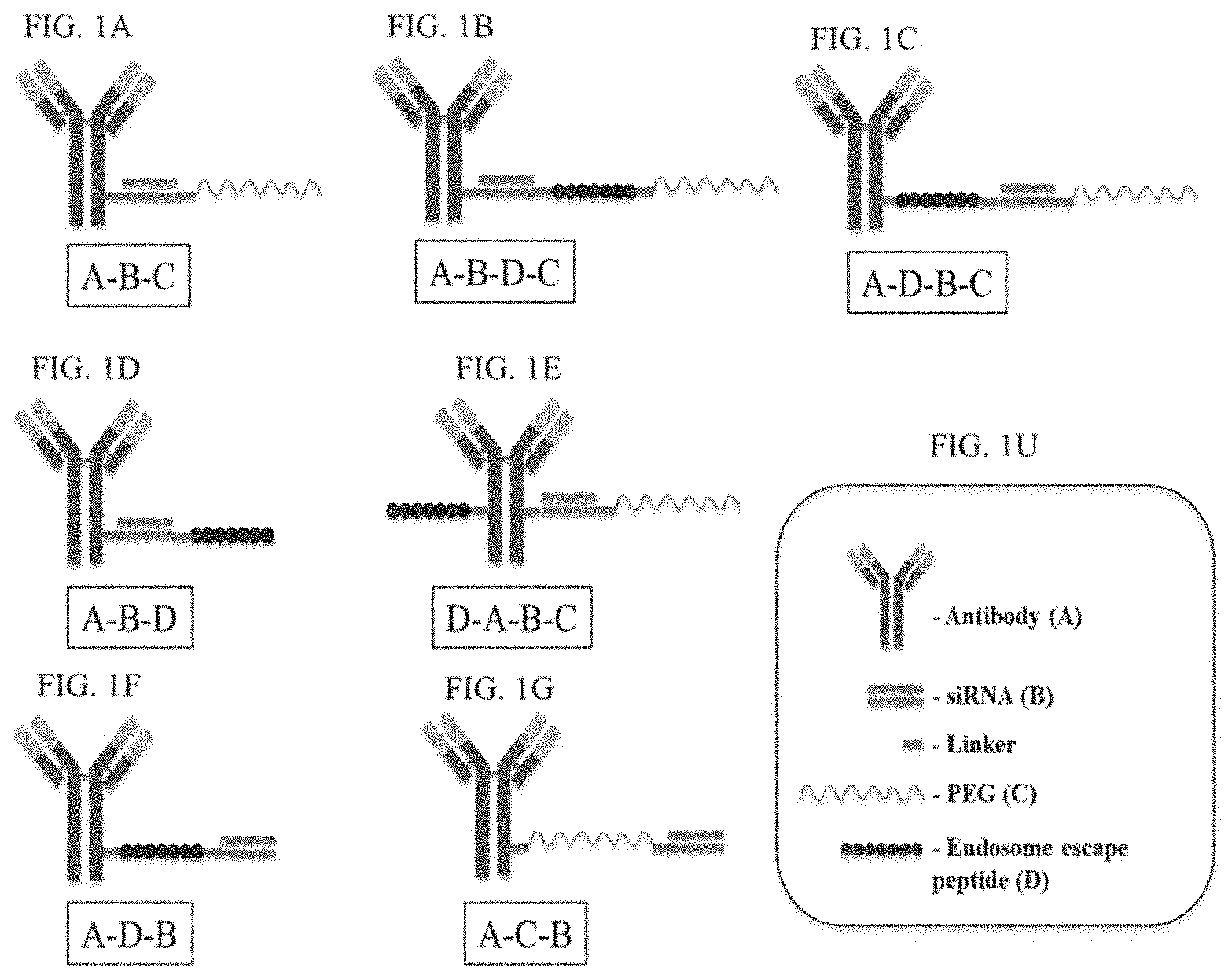
D00002

D00003
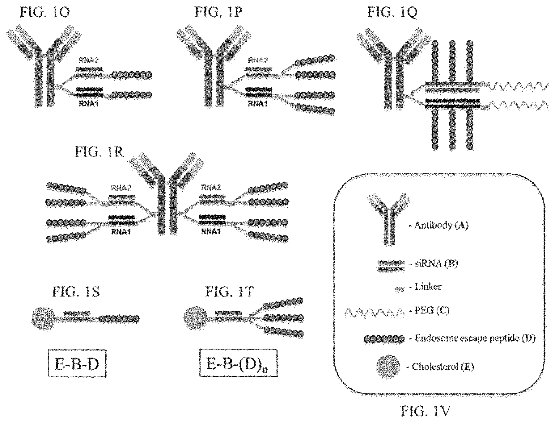
D00004
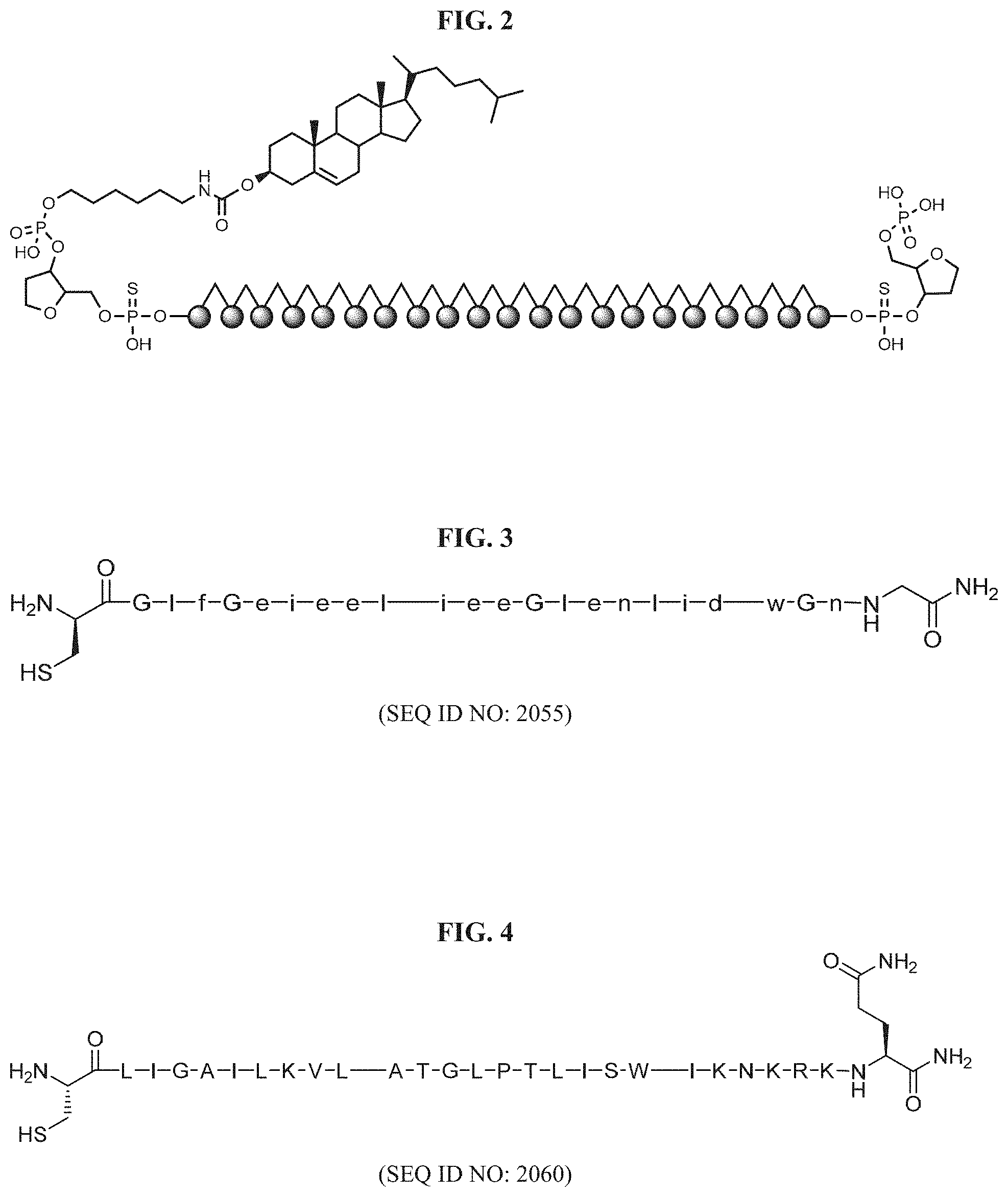
D00005
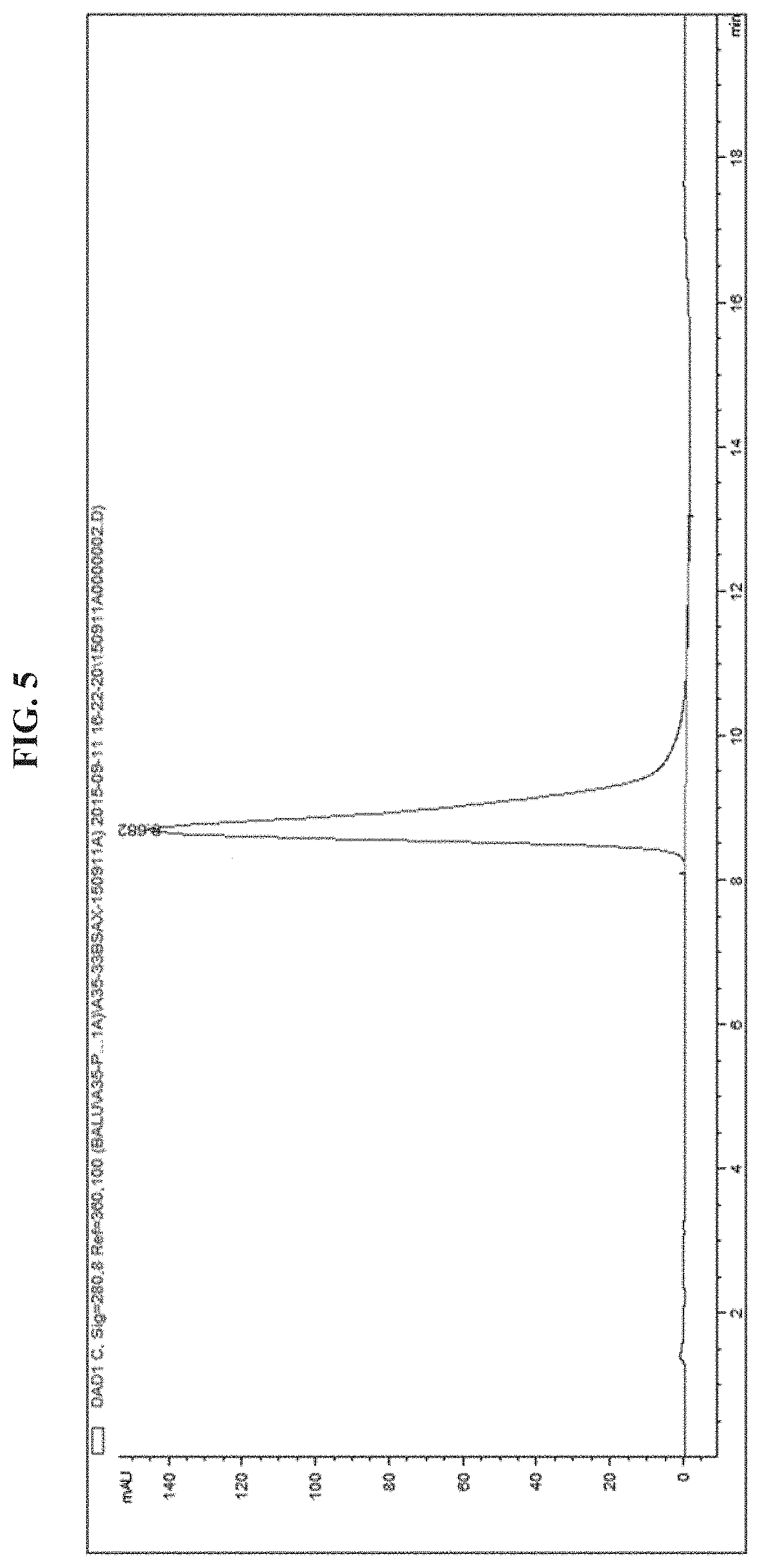
D00006
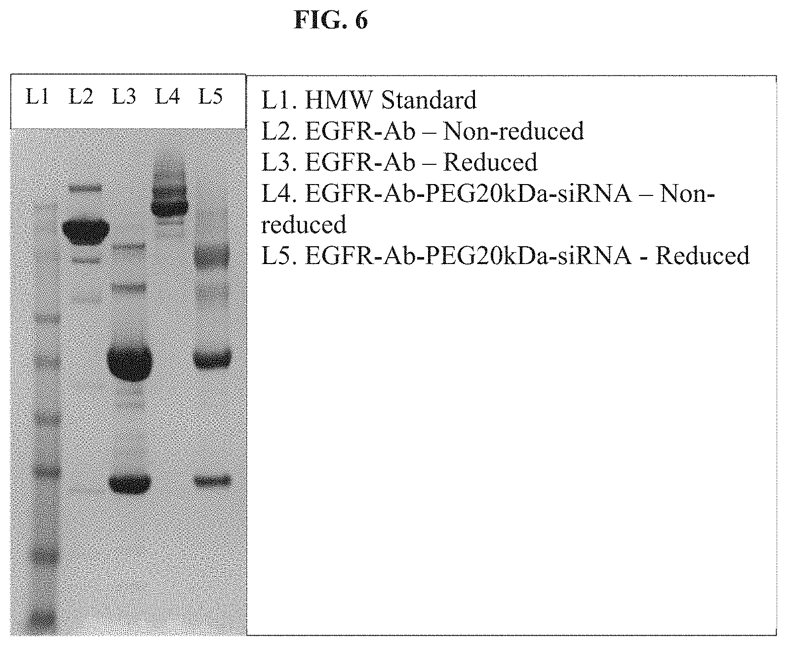
D00007
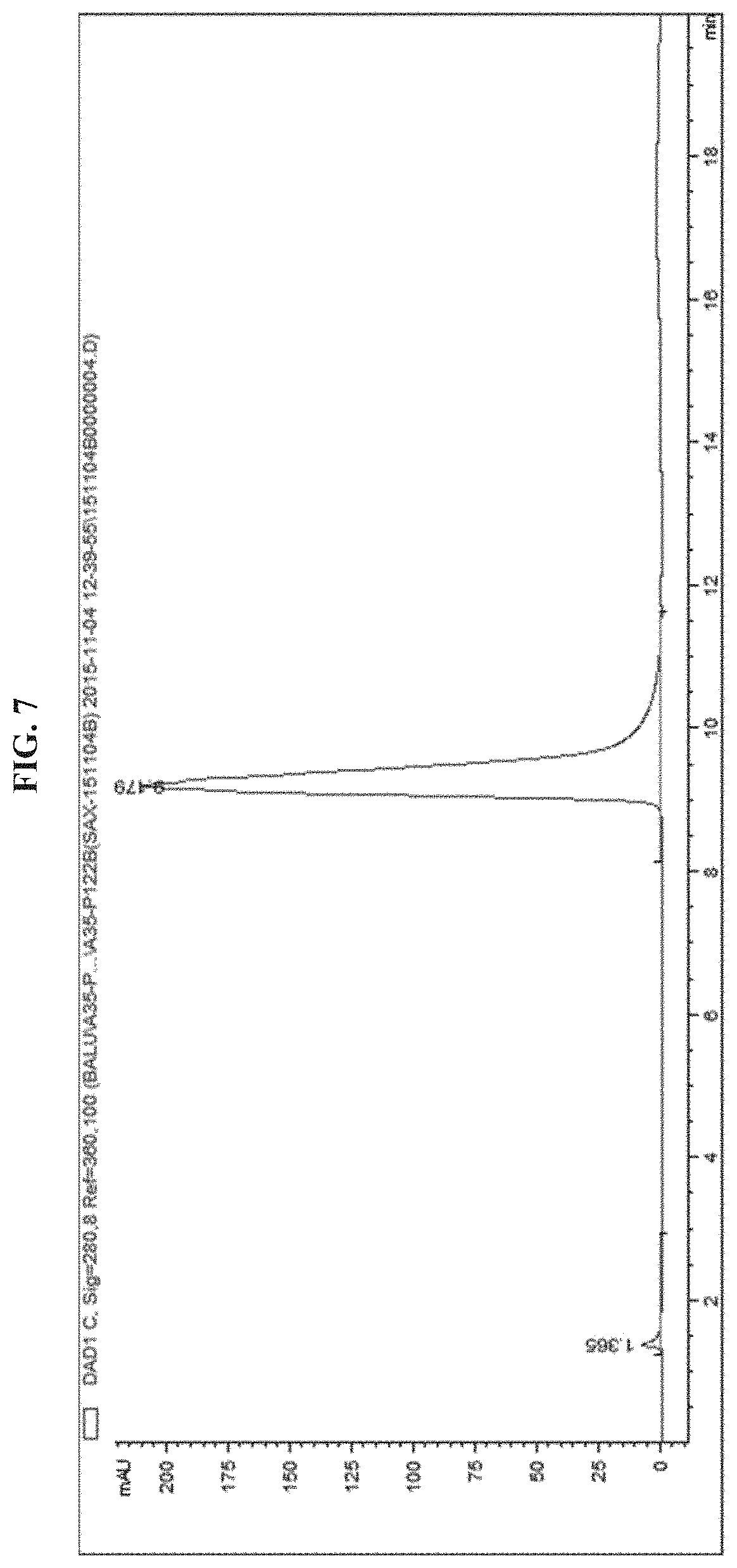
D00008
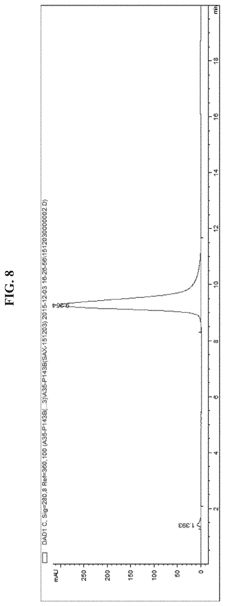
D00009
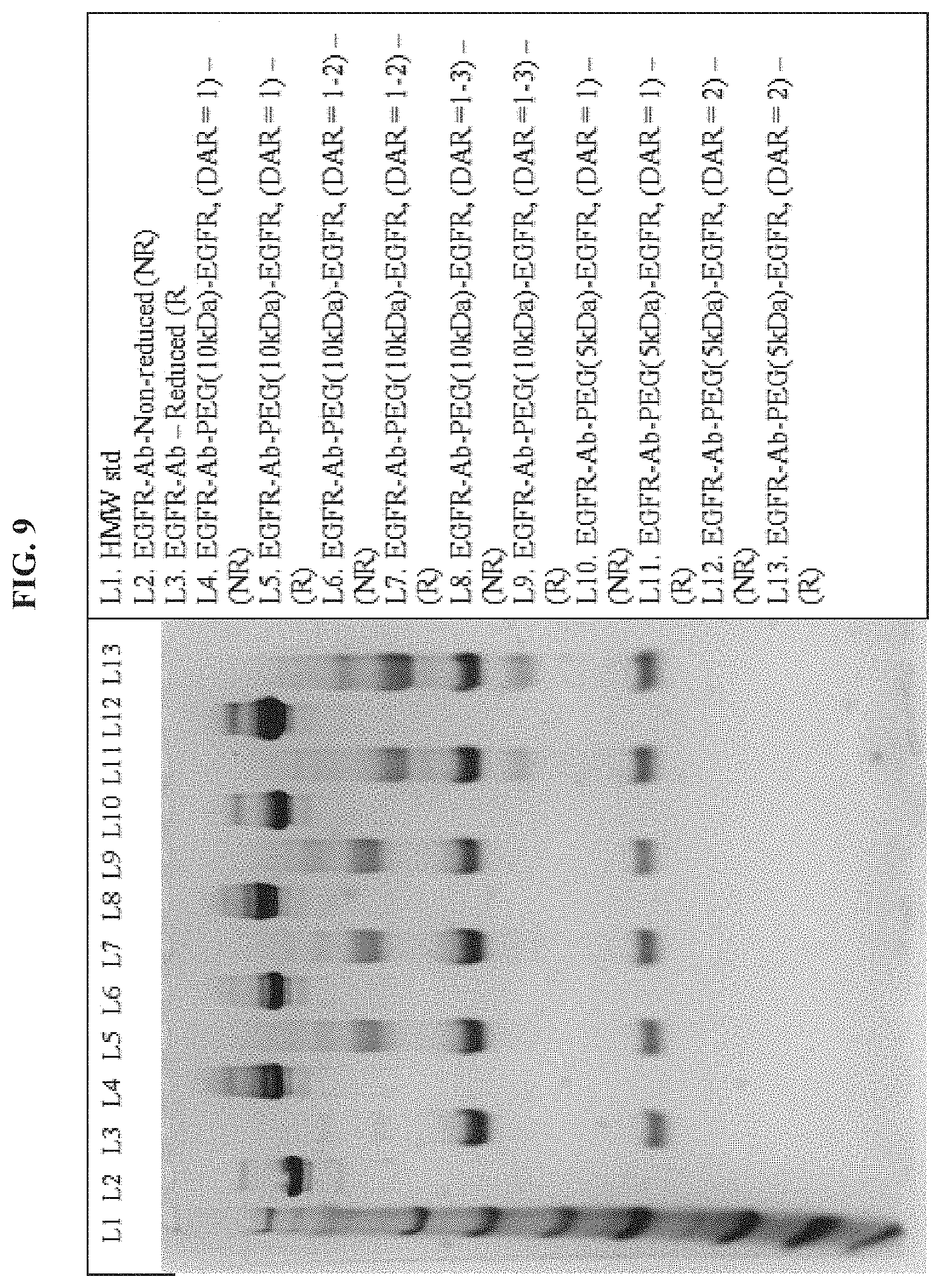
D00010

D00011

D00012

D00013
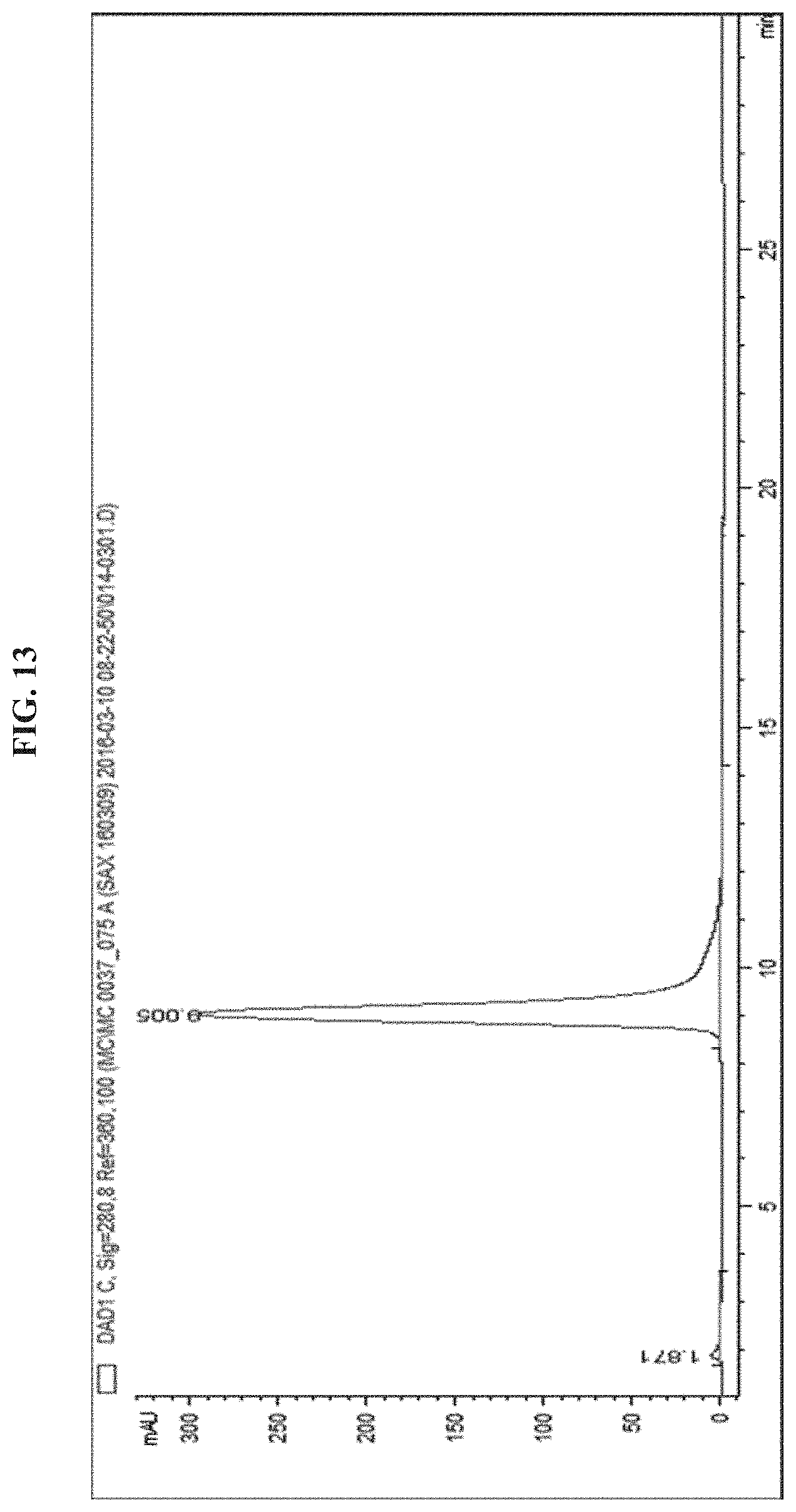
D00014
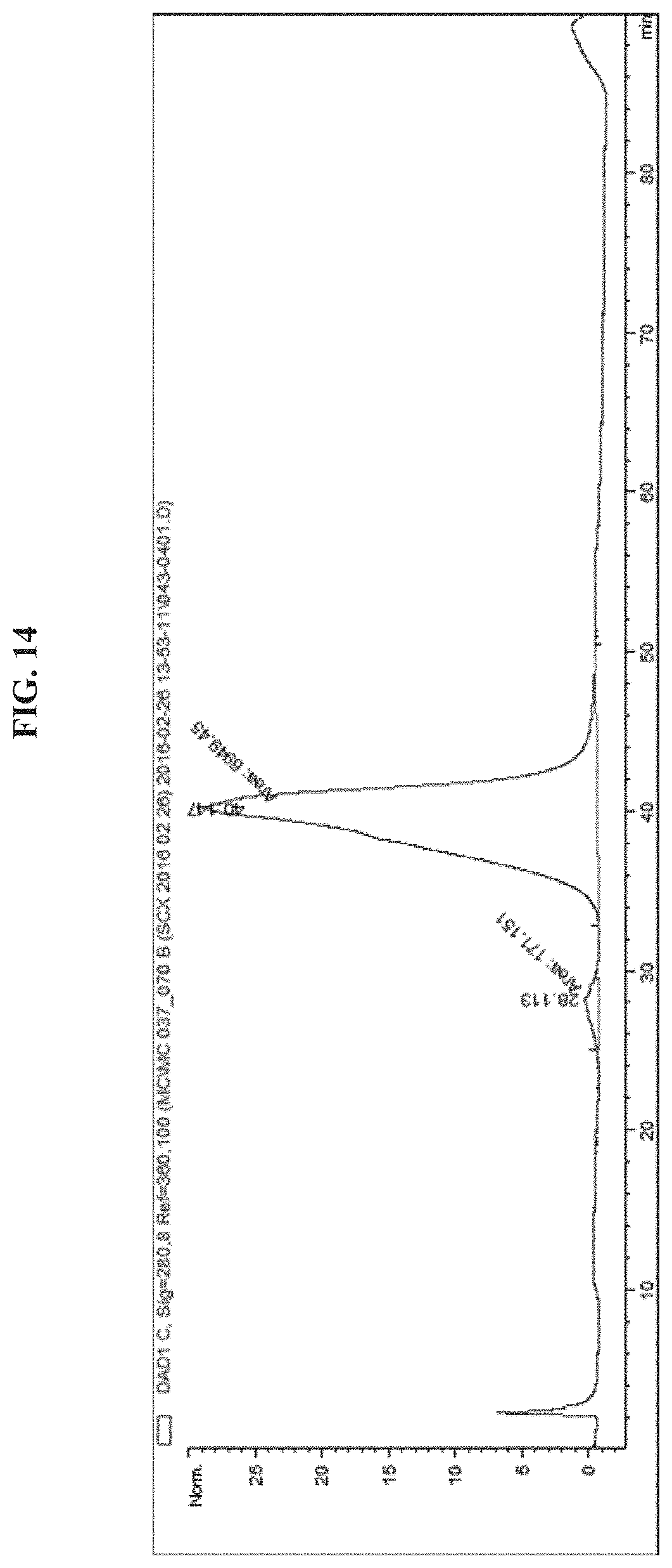
D00015
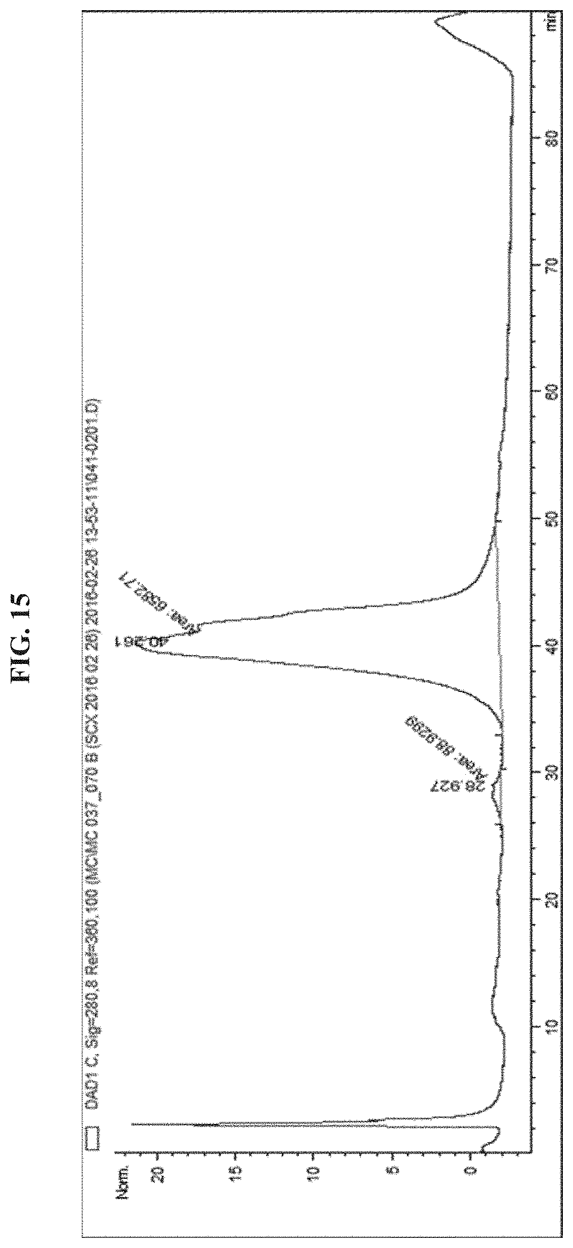
D00016

D00017
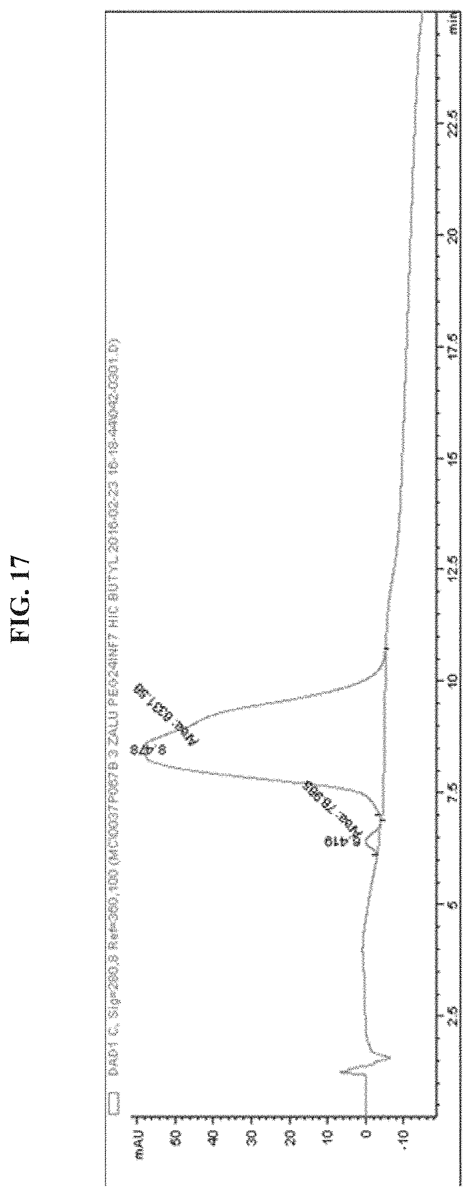
D00018
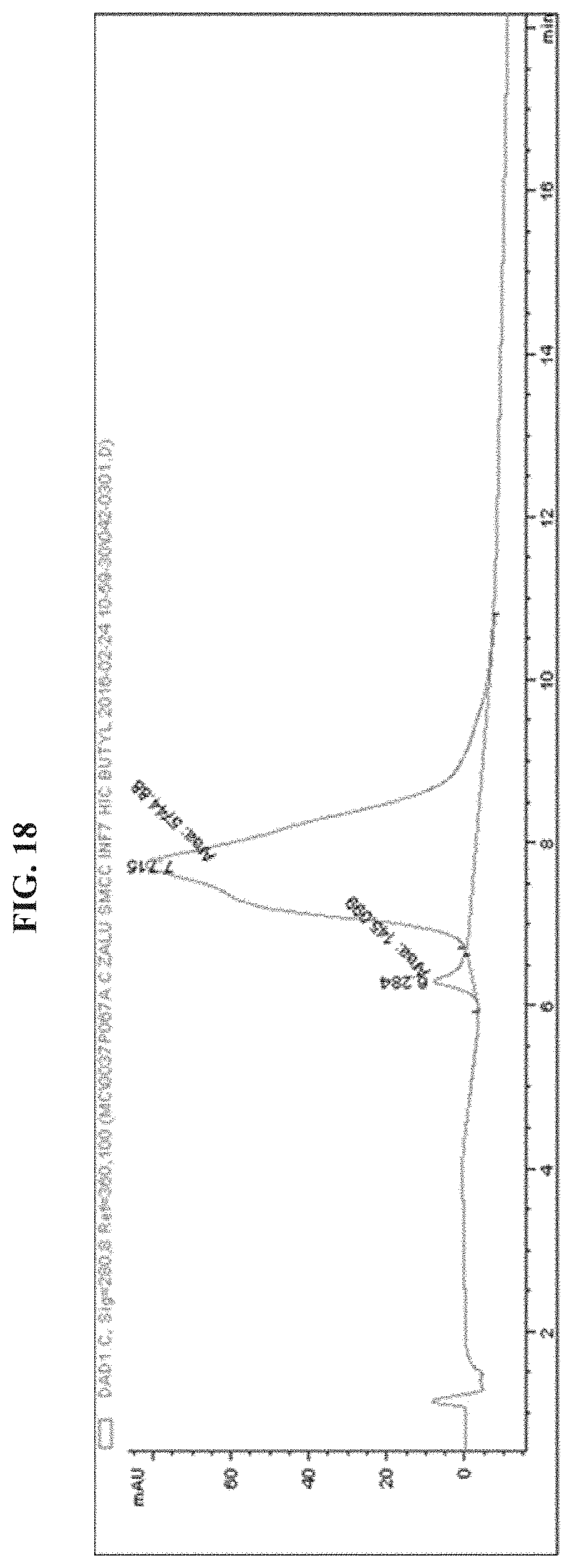
D00019

D00020
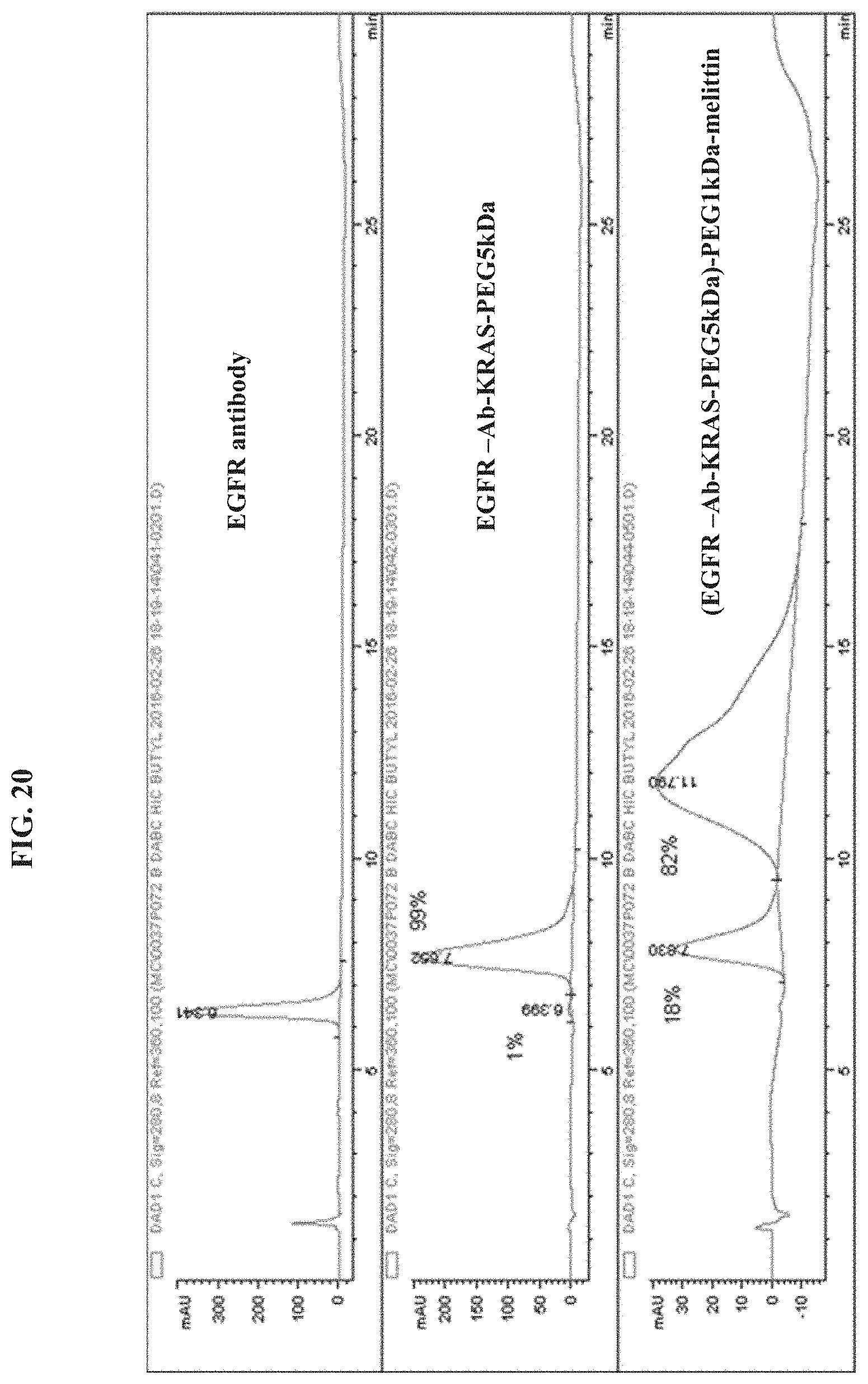
D00021

D00022
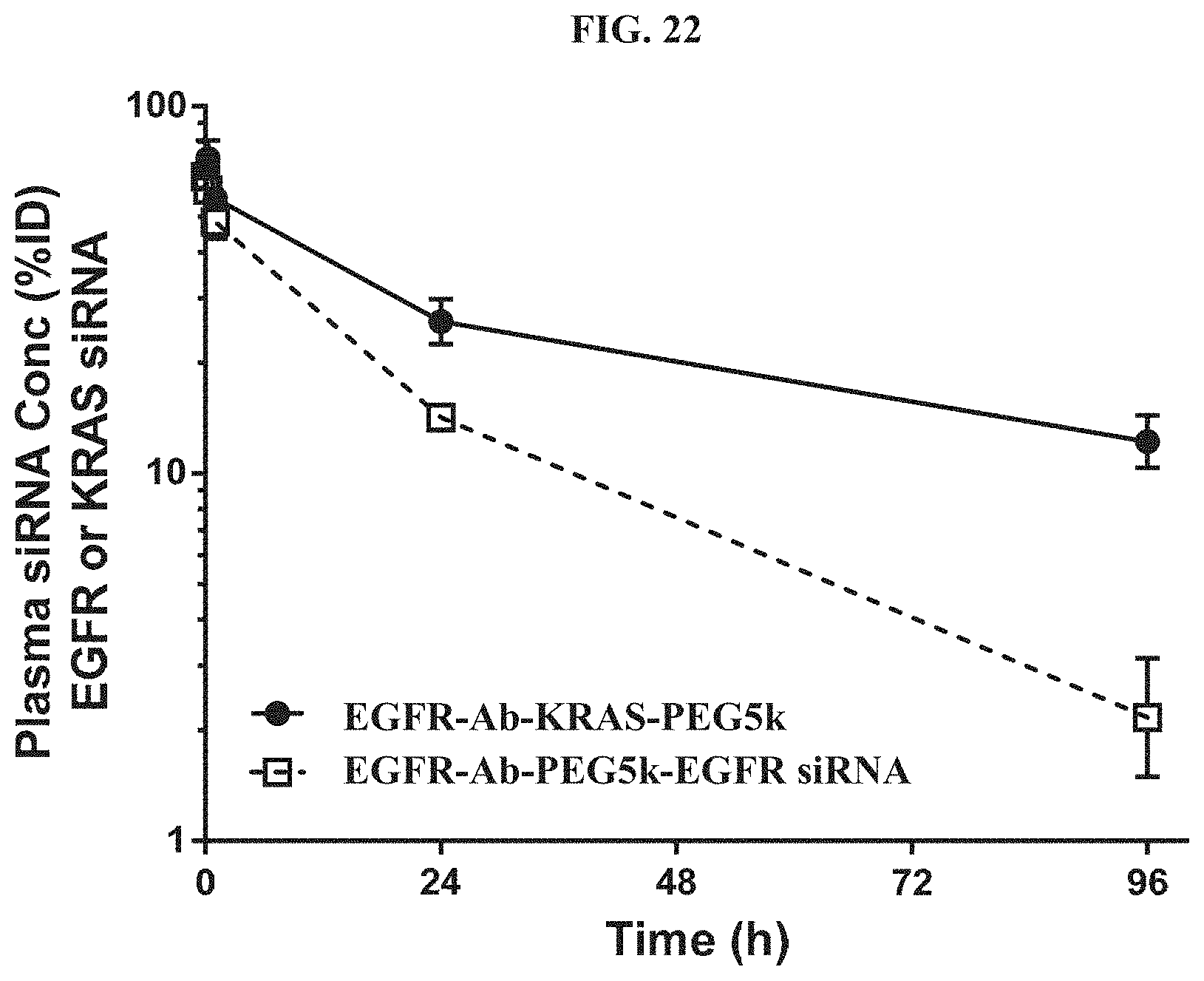
D00023
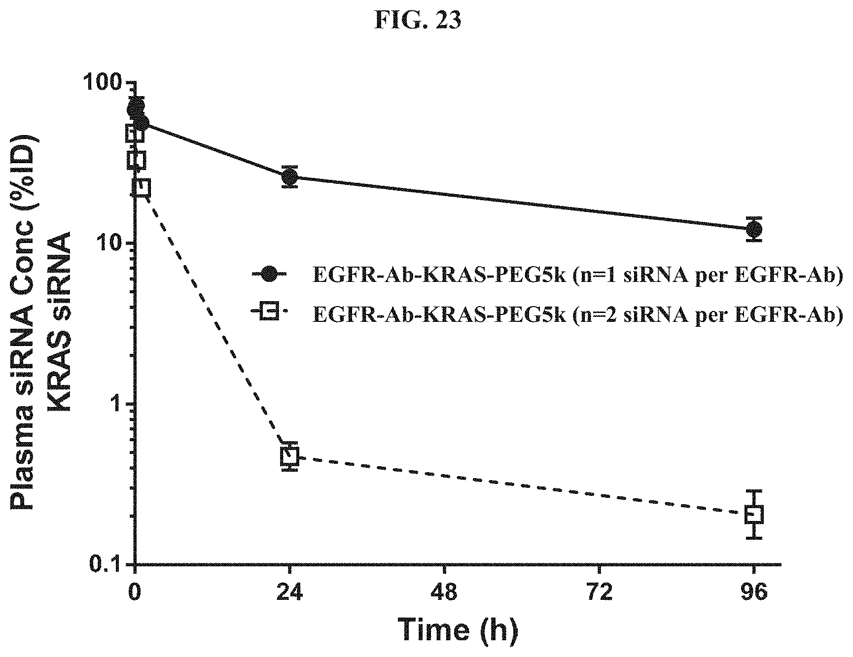
D00024
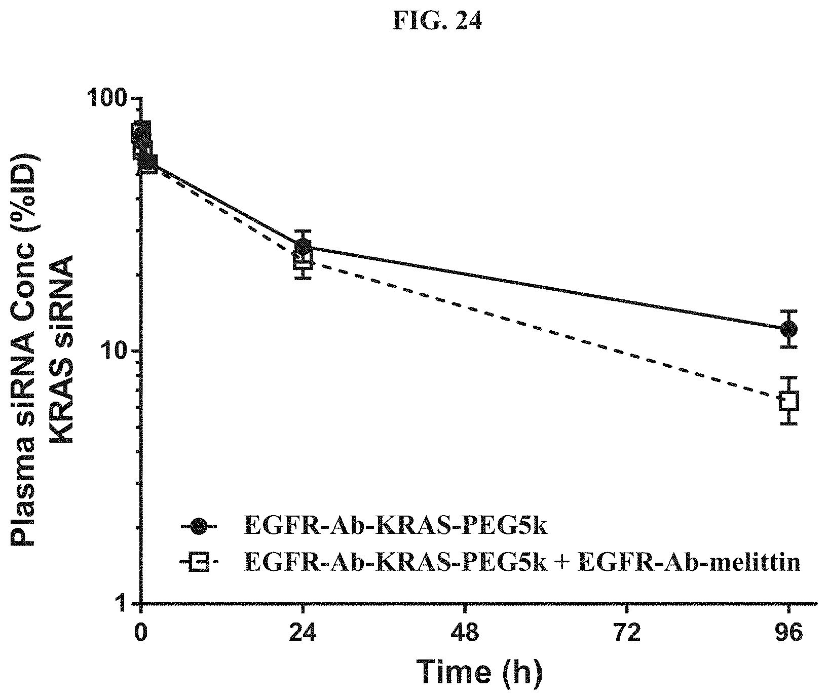
D00025

D00026
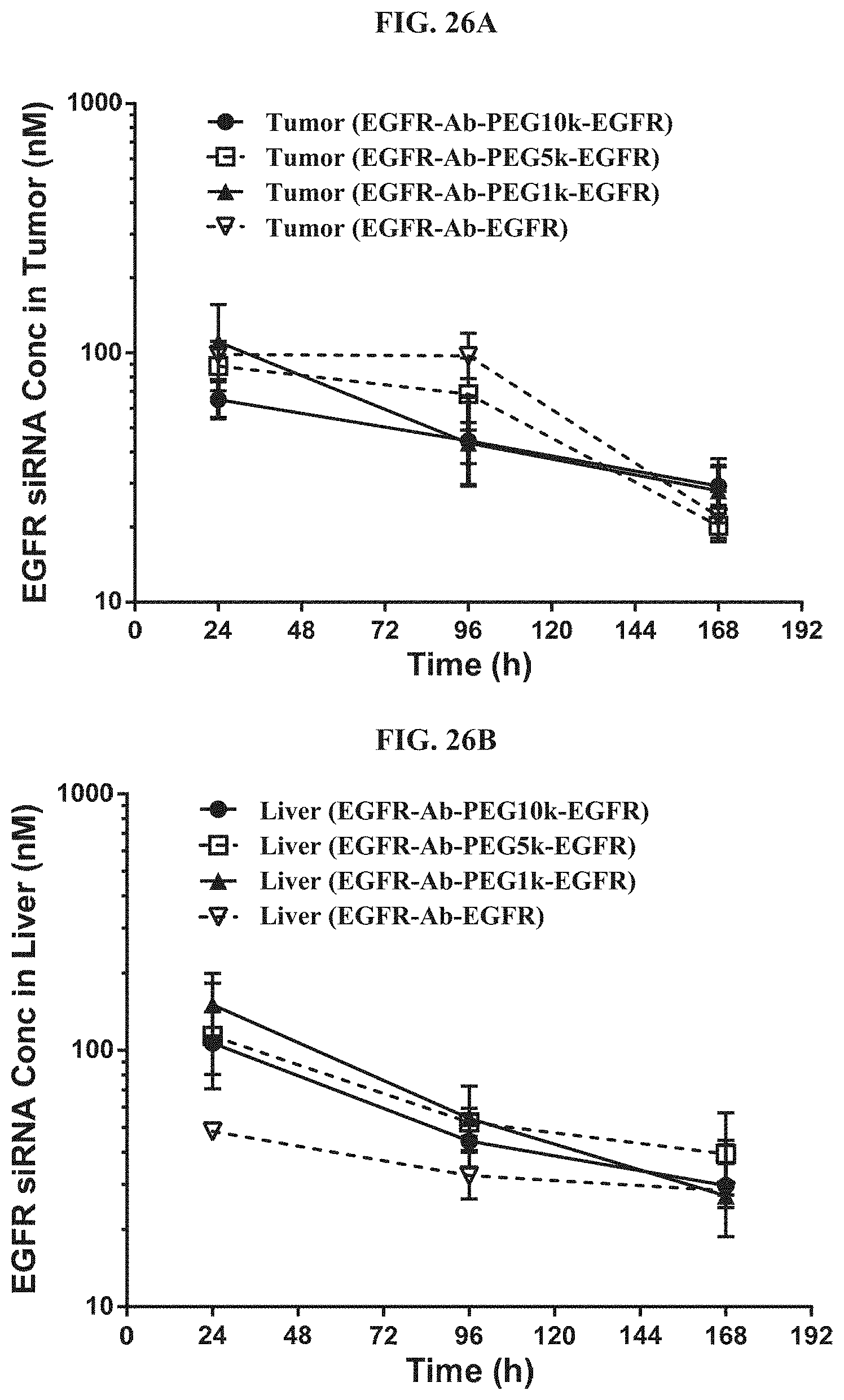
D00027
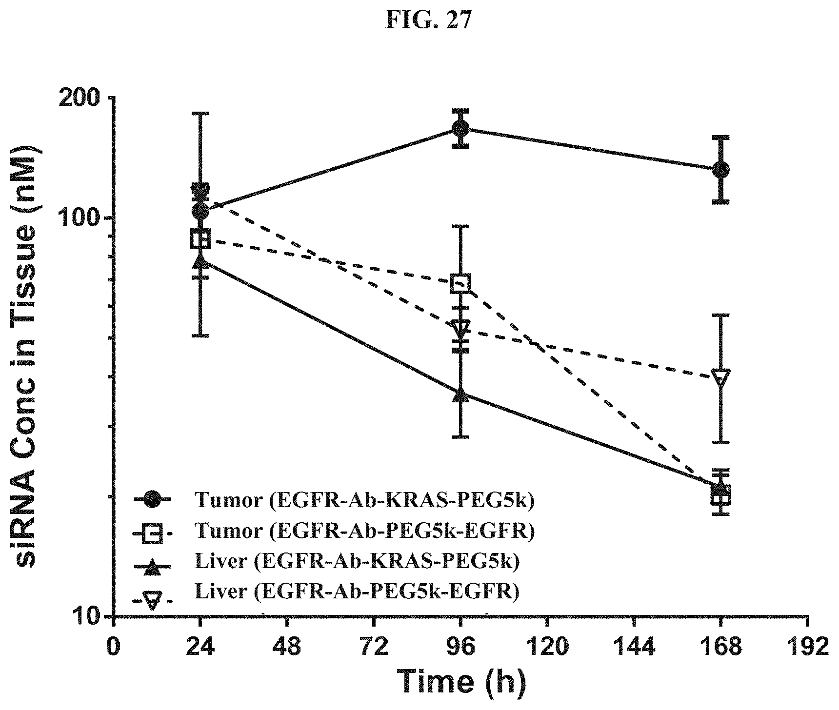
D00028
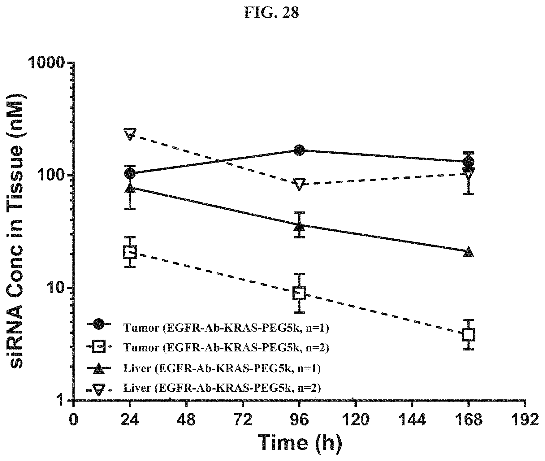
D00029
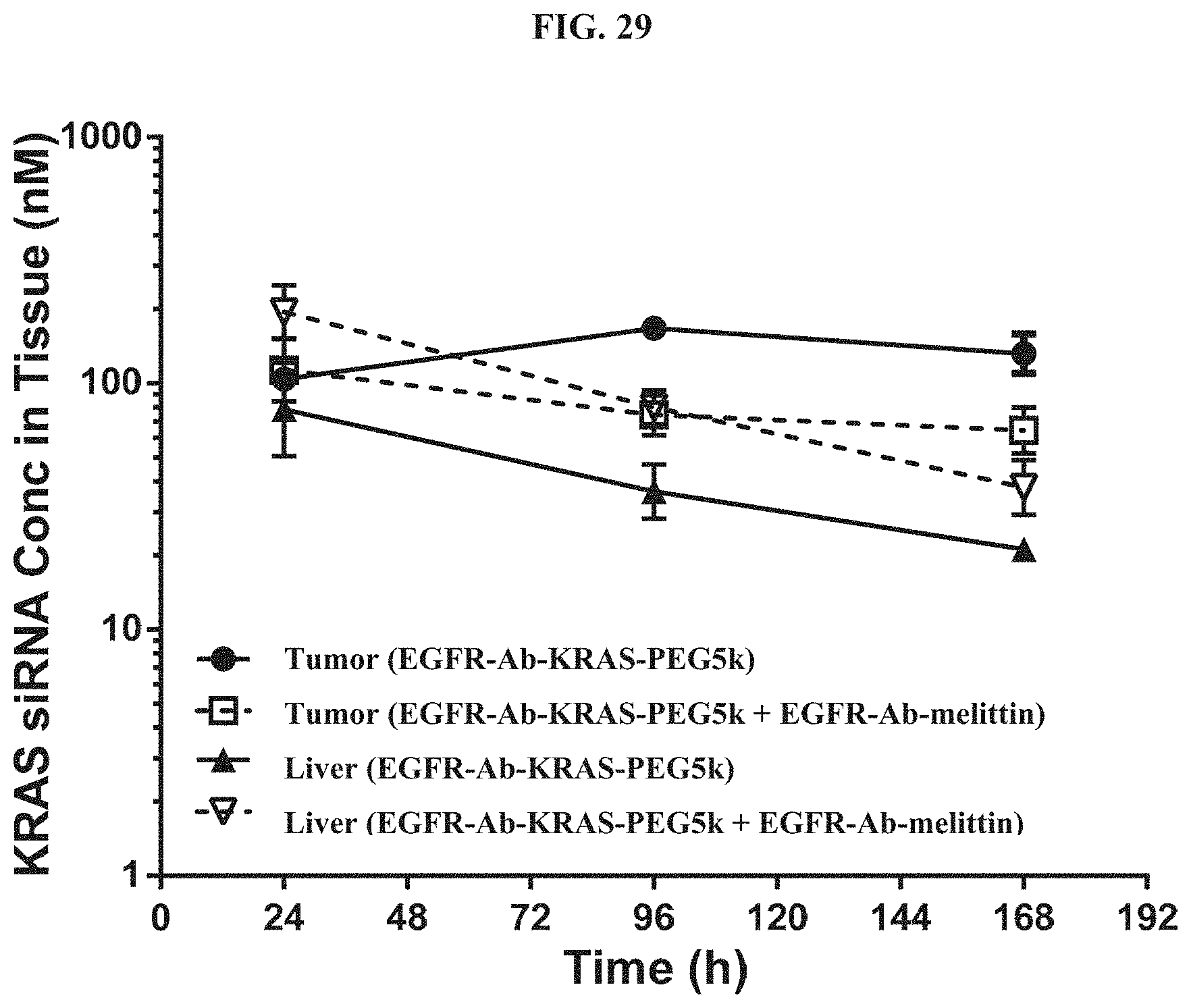
D00030

D00031

D00032
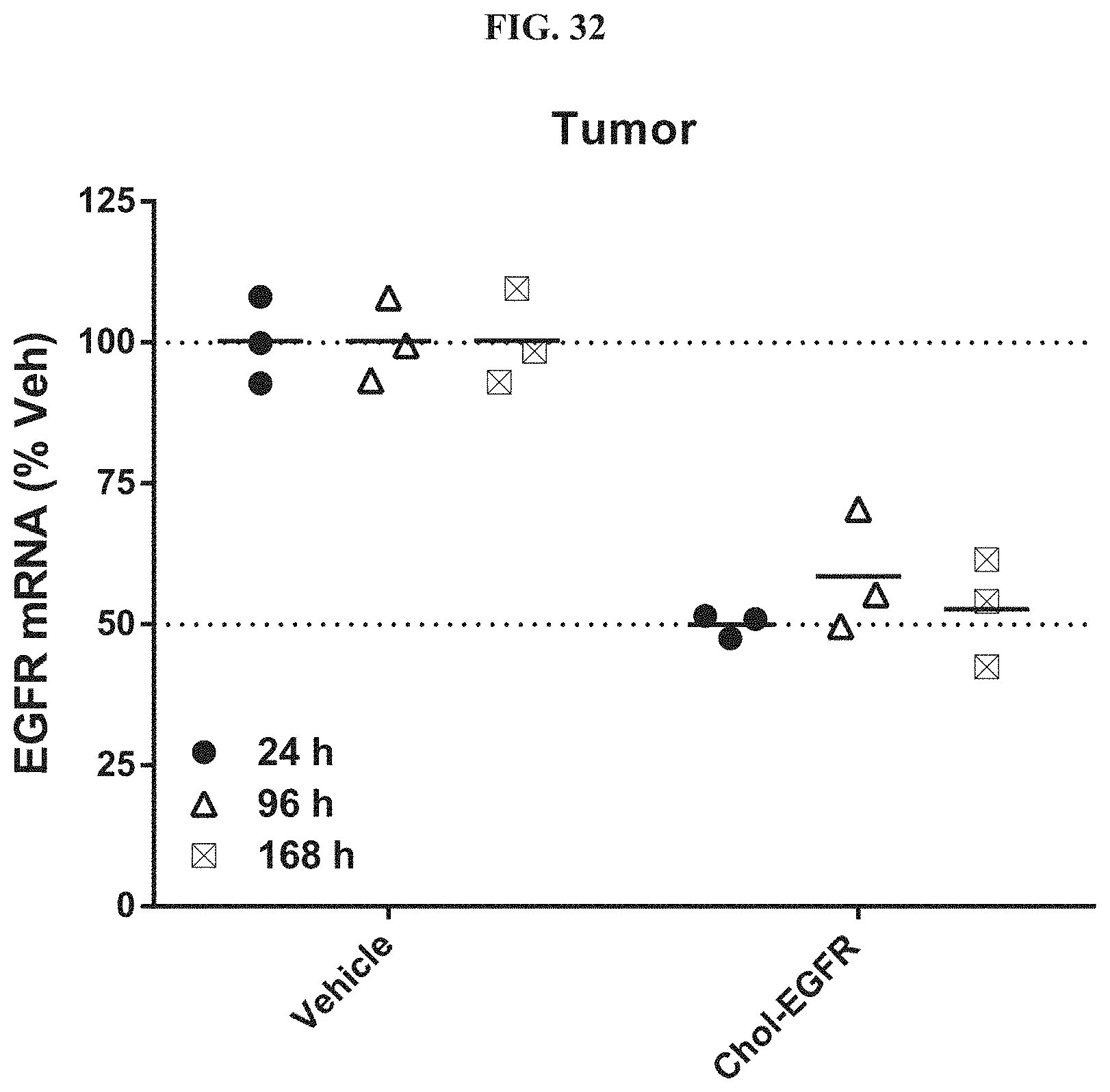
D00033

D00034
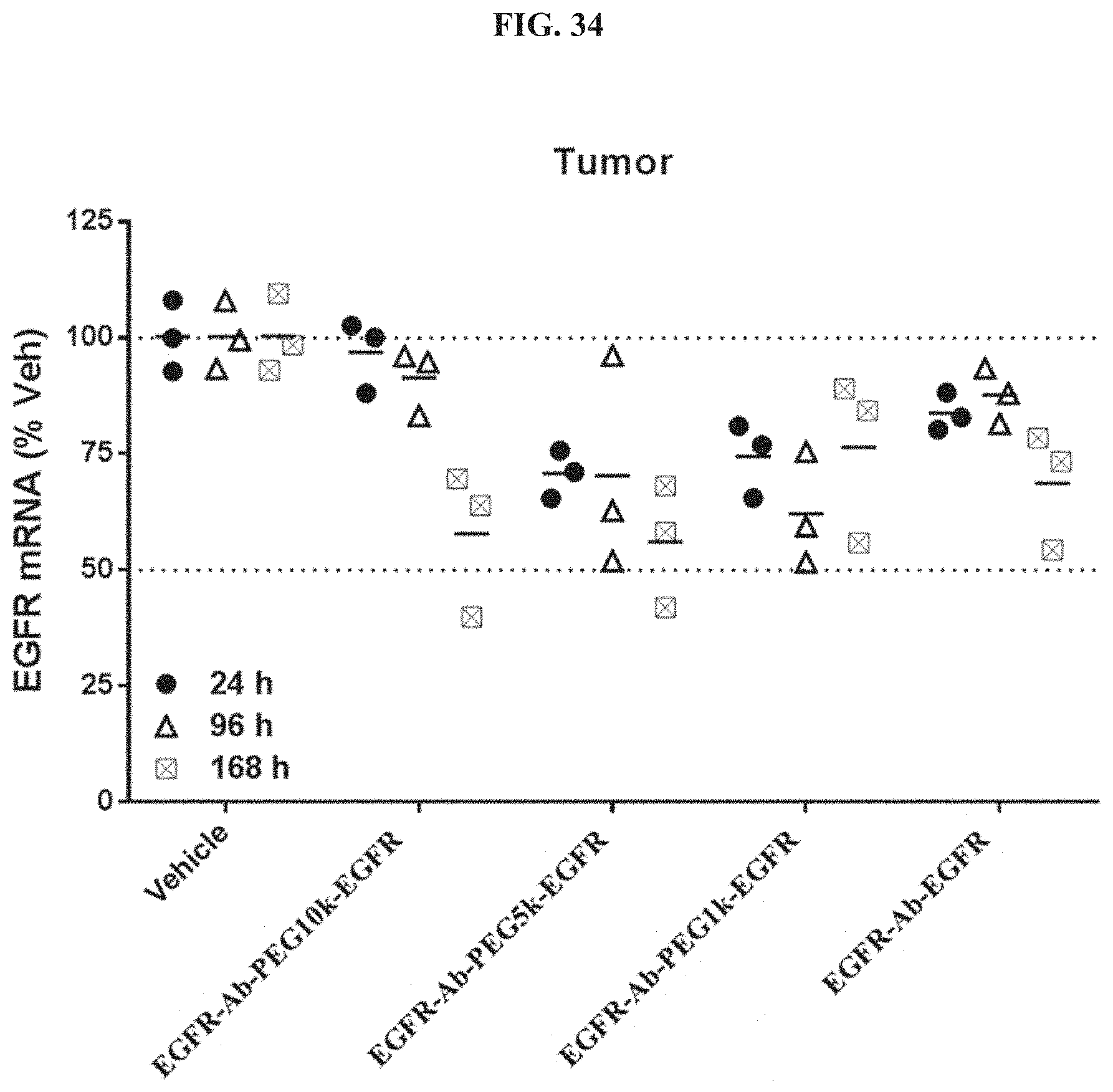
D00035
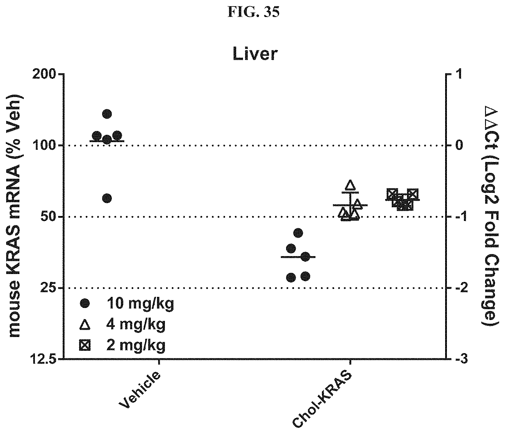
D00036

D00037
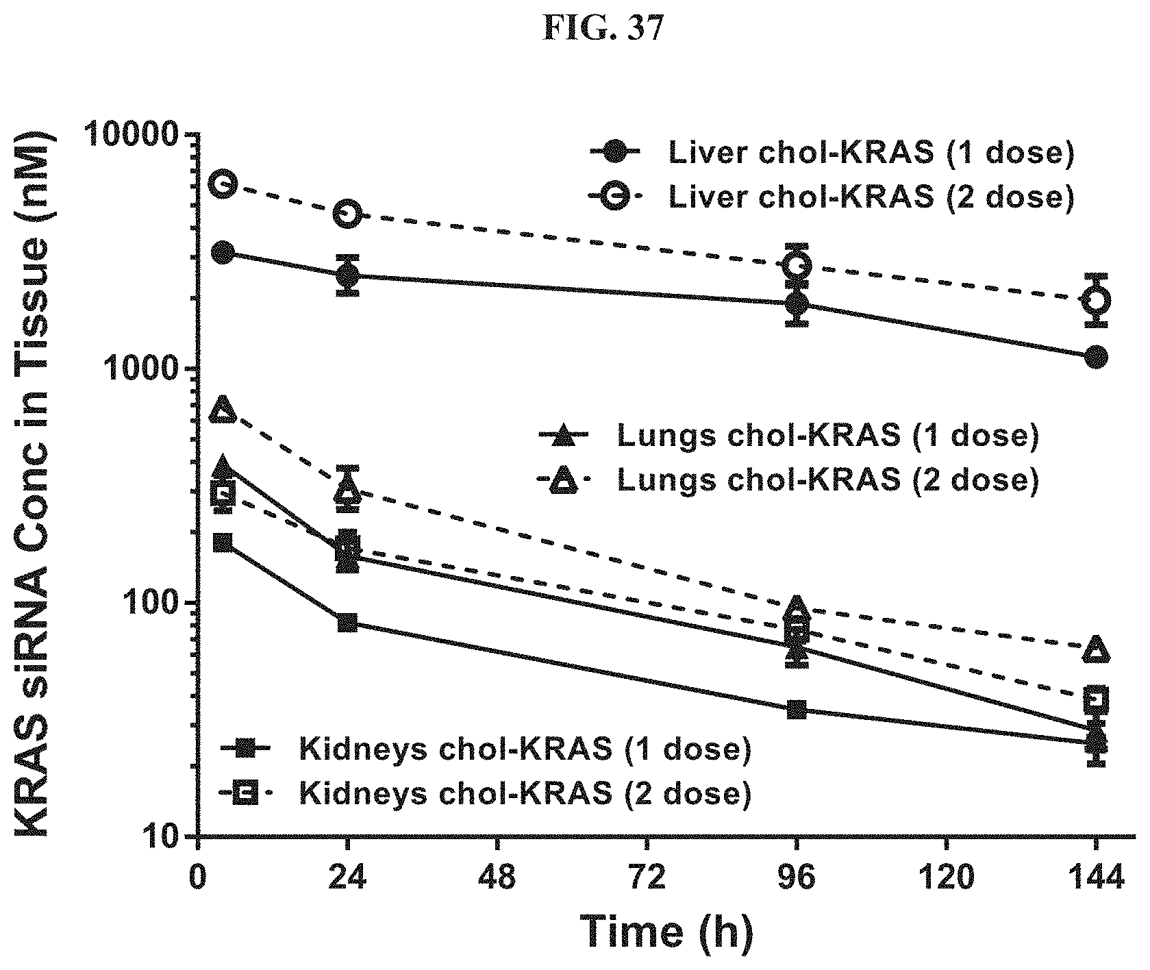
D00038
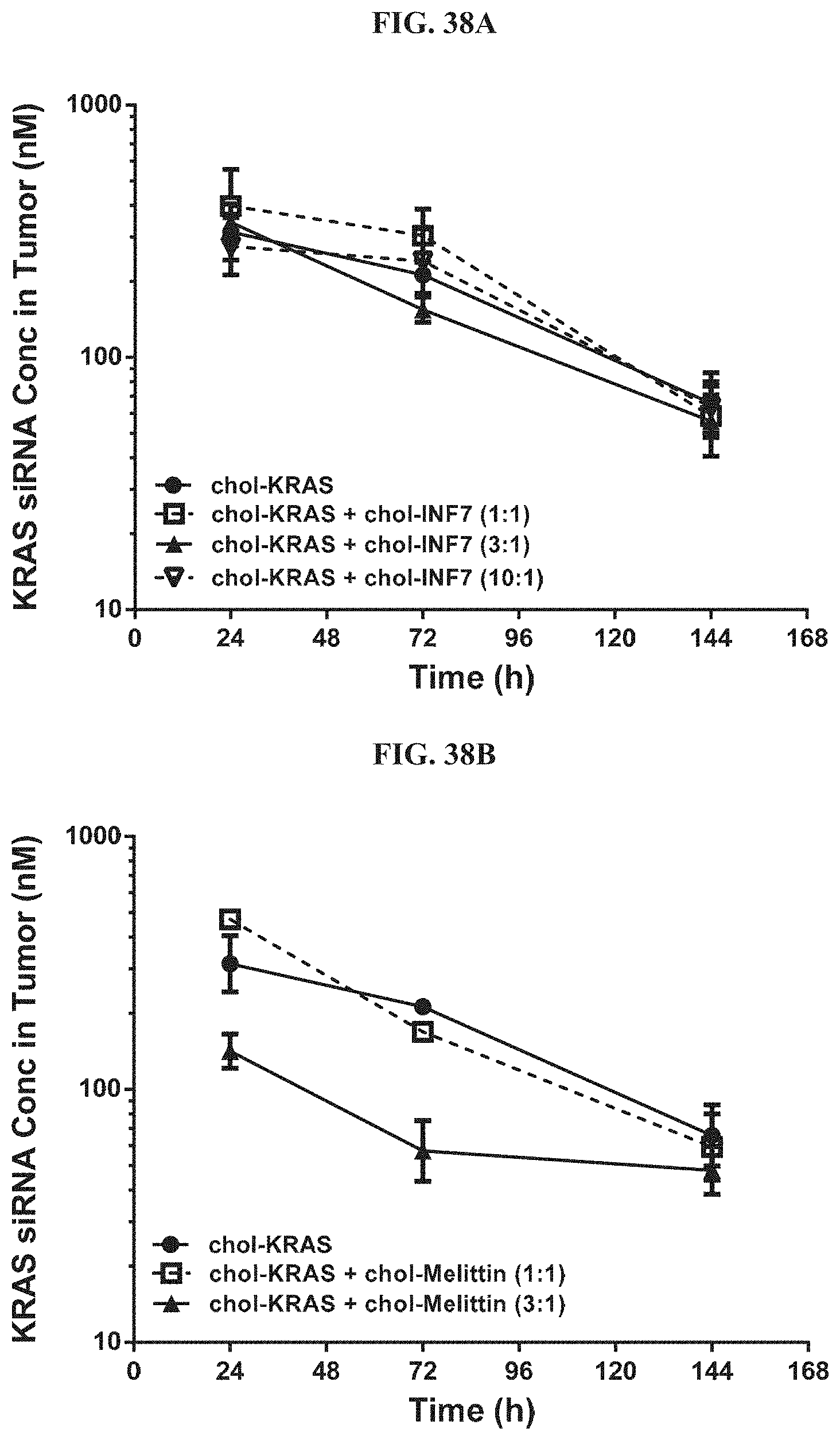
D00039
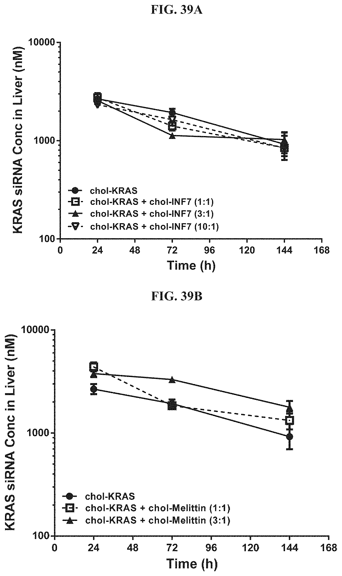
D00040

D00041
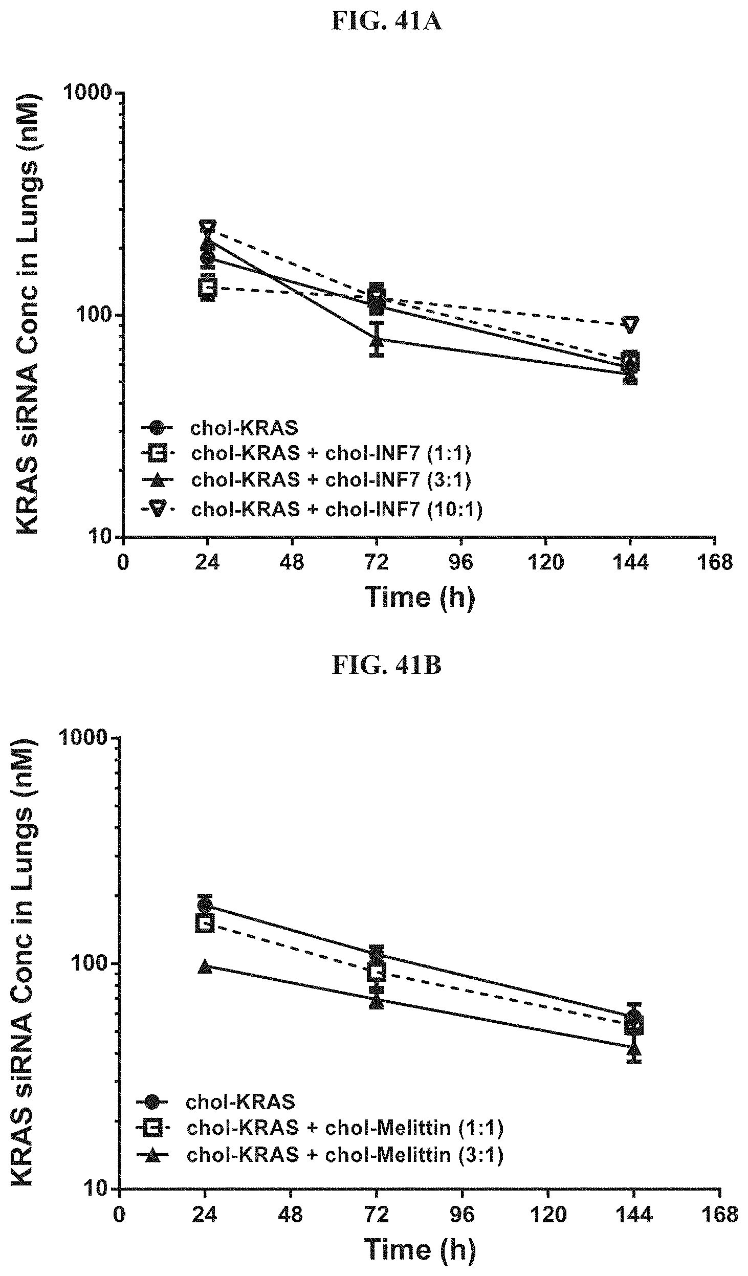
D00042
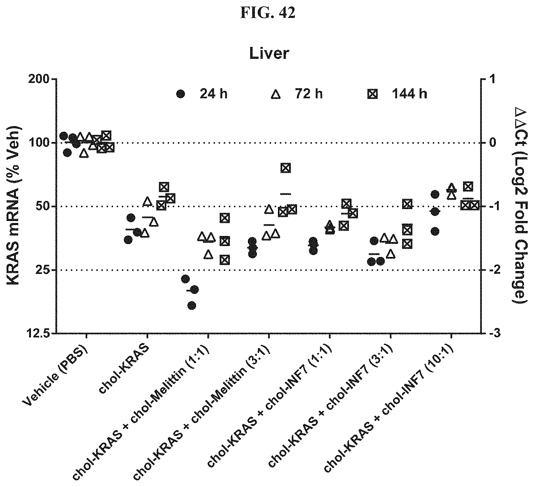
D00043
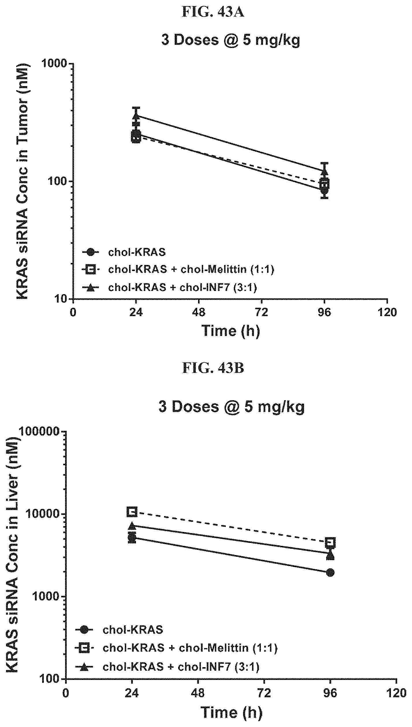
D00044
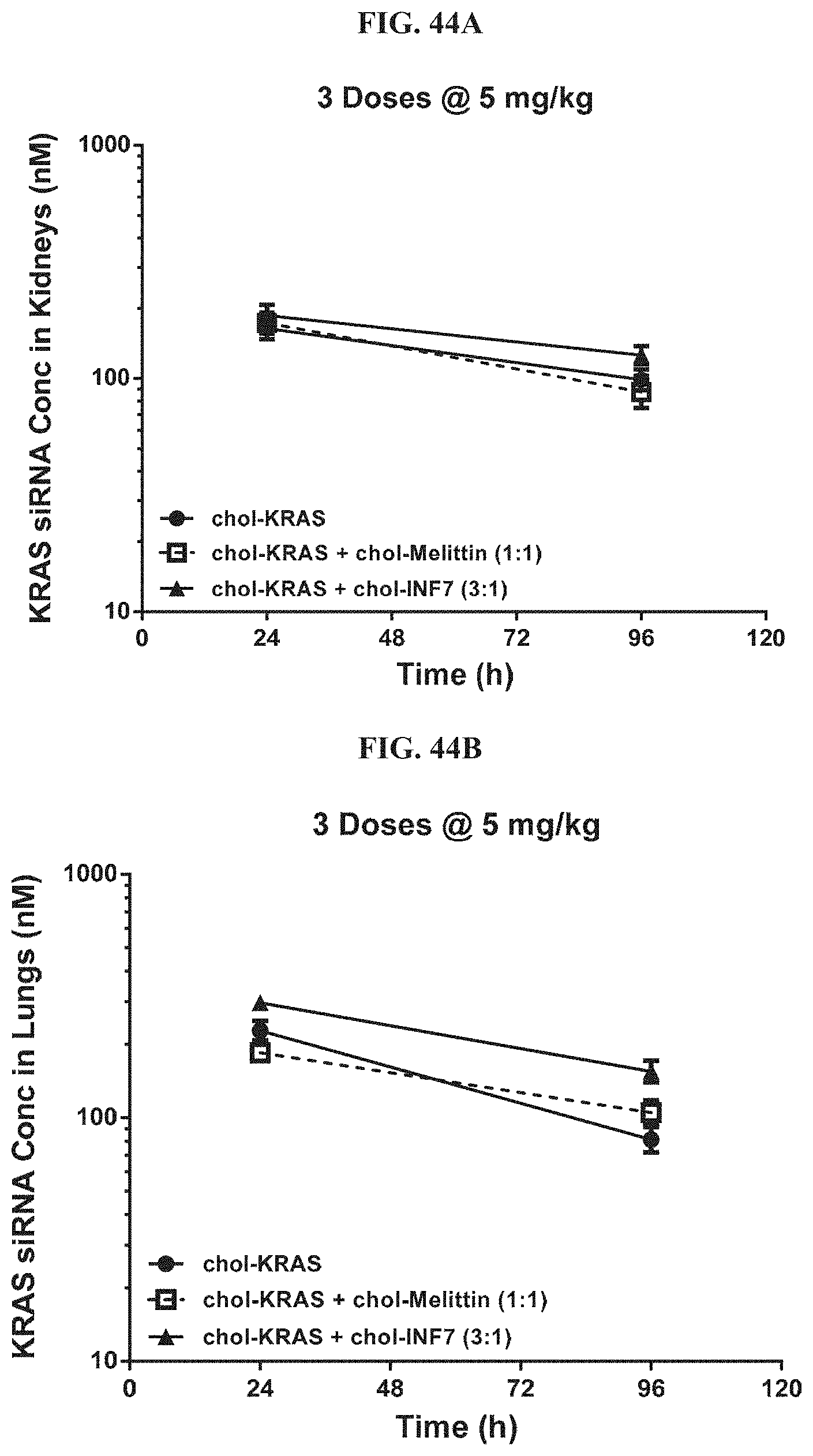
D00045
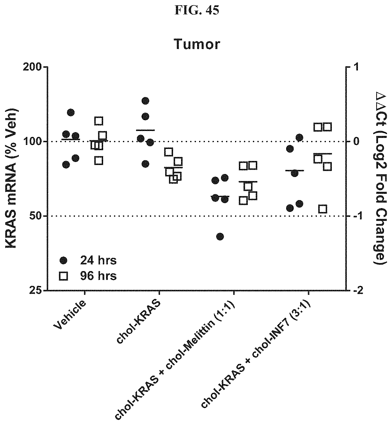
D00046
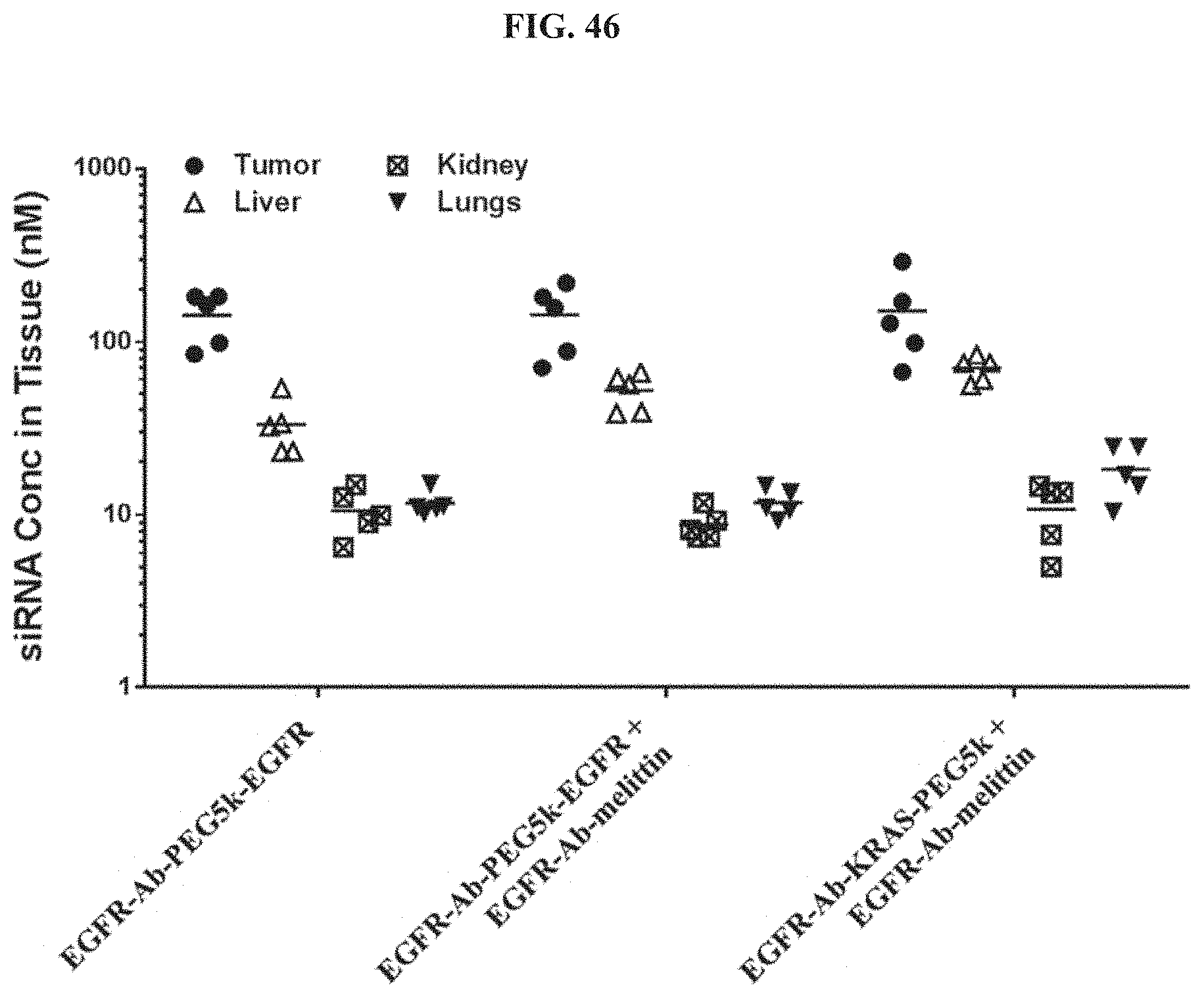
D00047

D00048
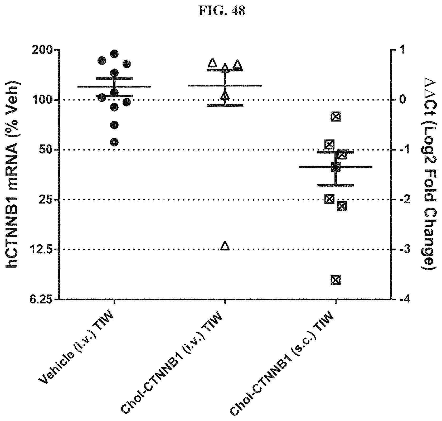
D00049
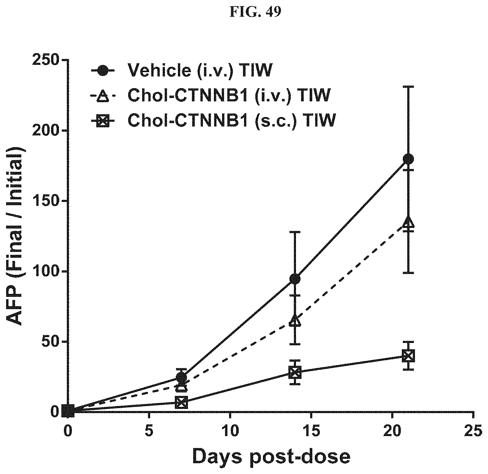
D00050

D00051
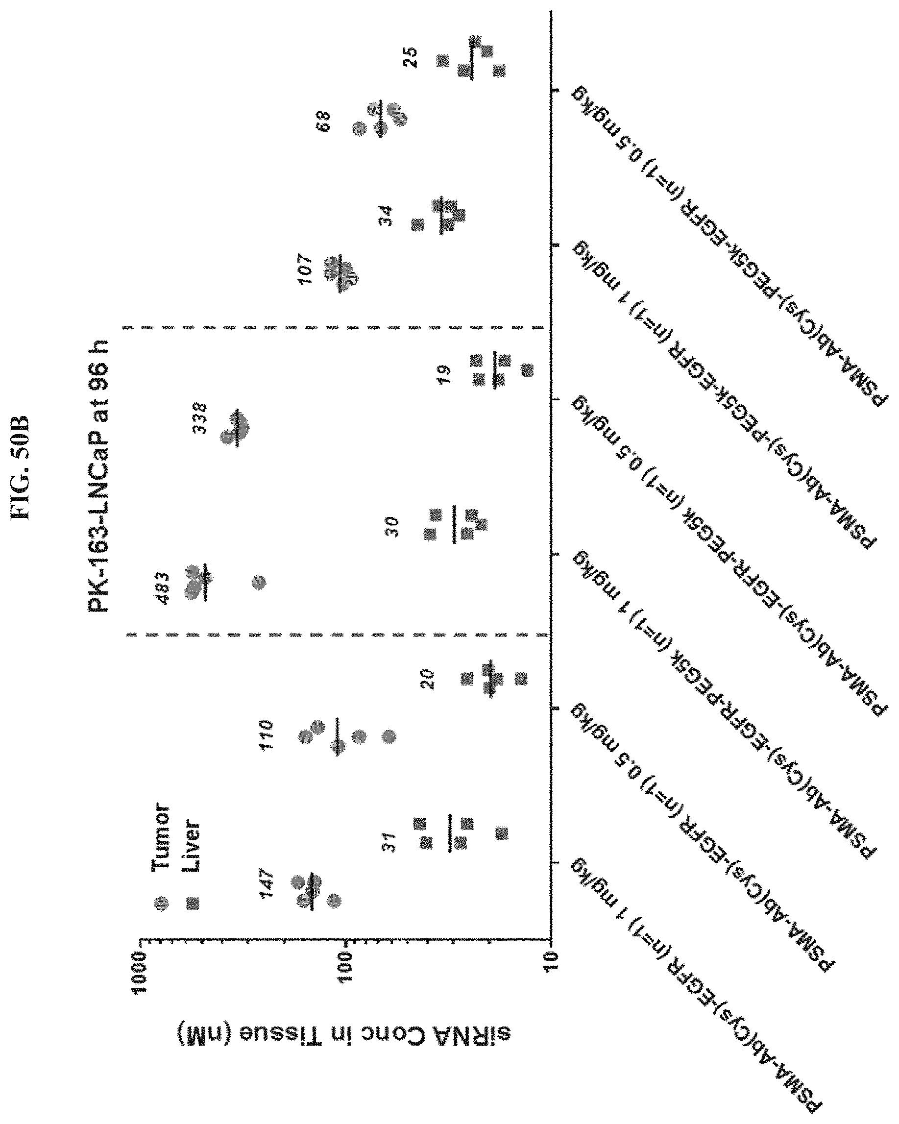
D00052

D00053

D00054
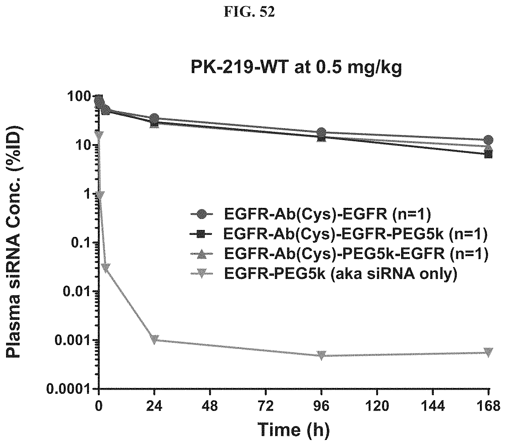
D00055
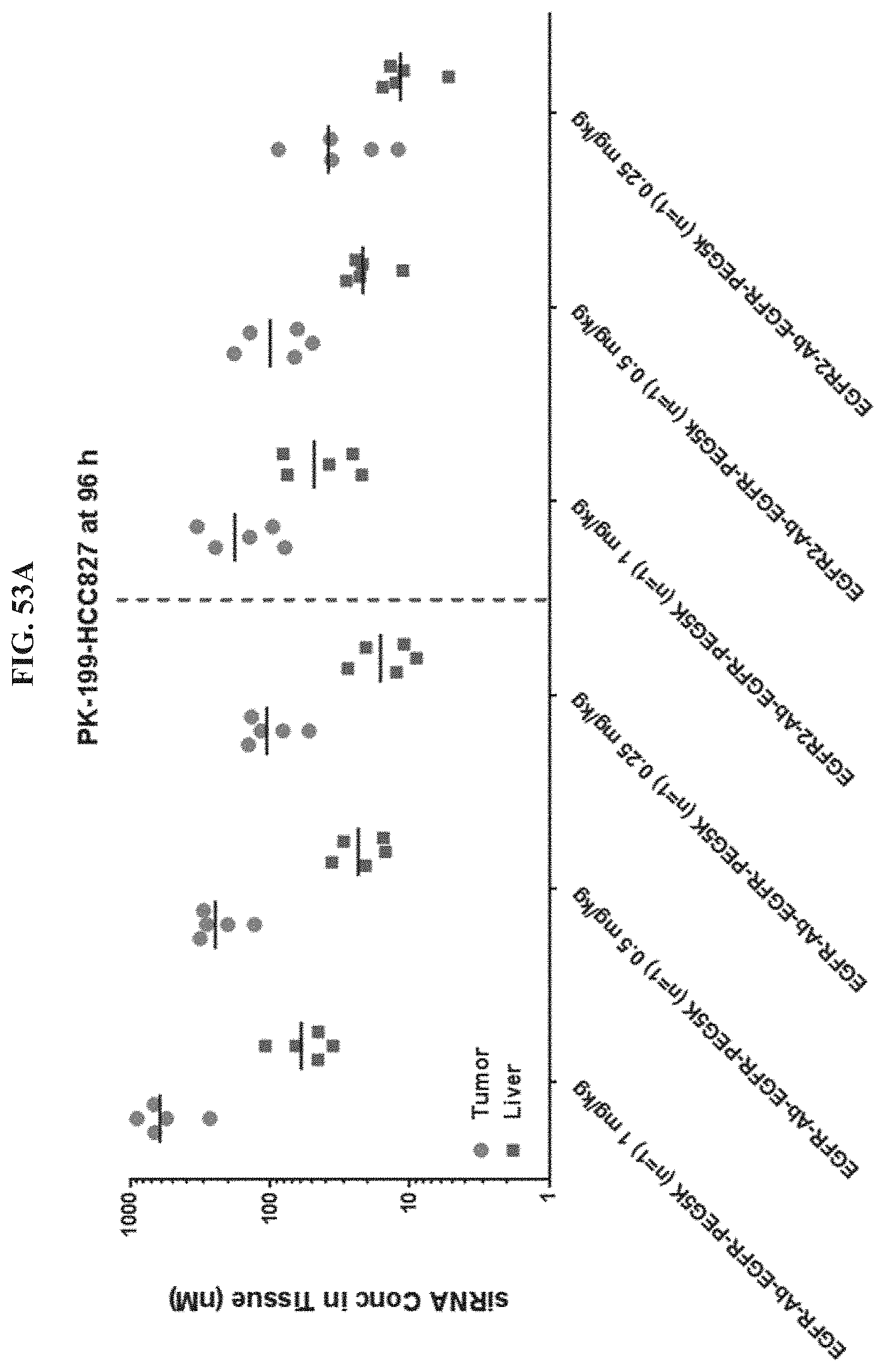
D00056

D00057

D00058

D00059
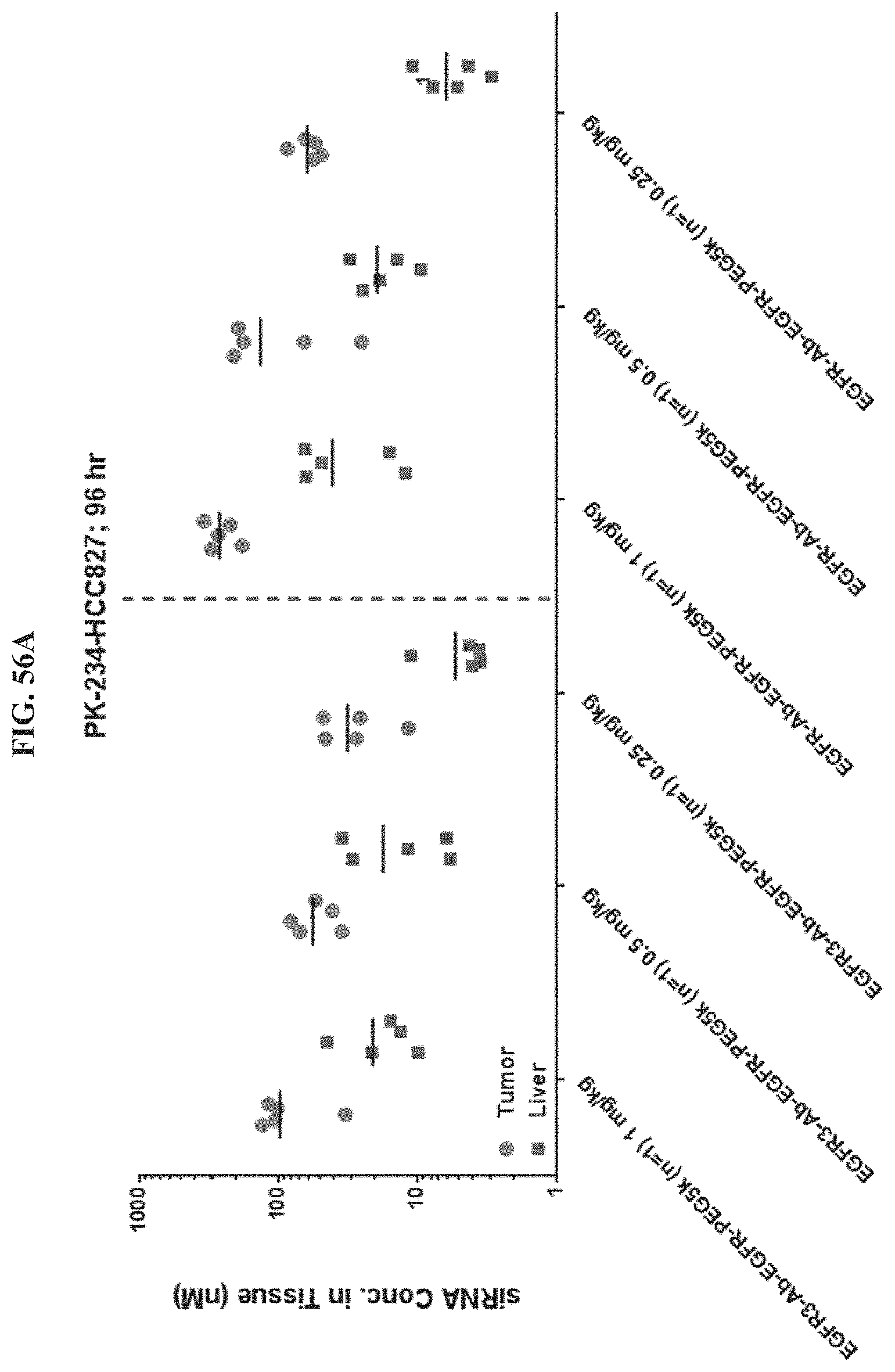
D00060
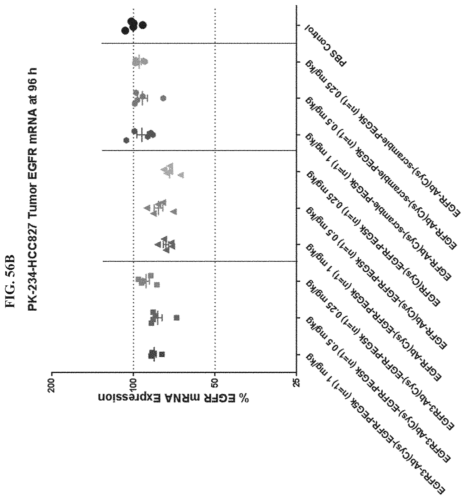
D00061
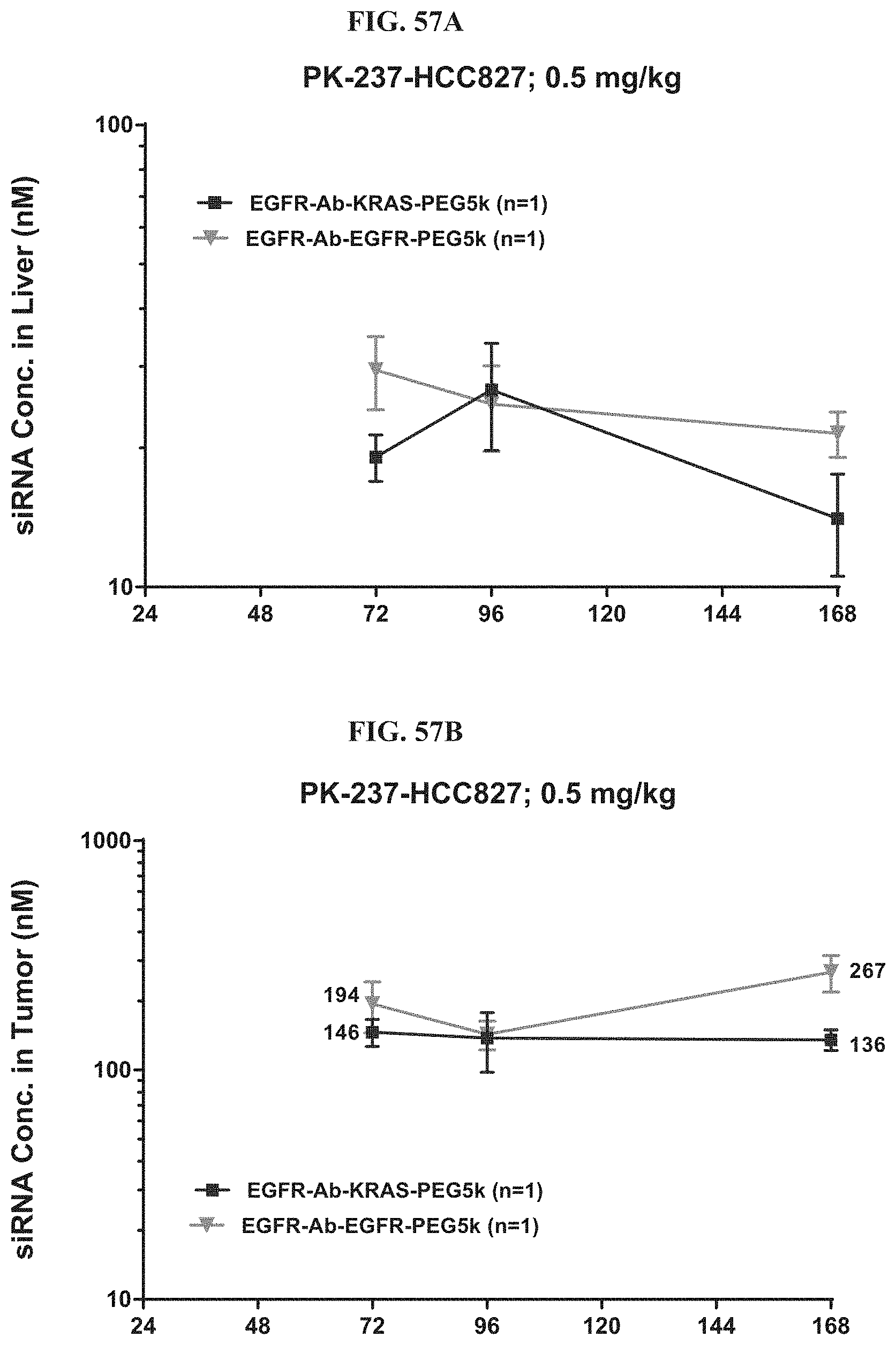
D00062
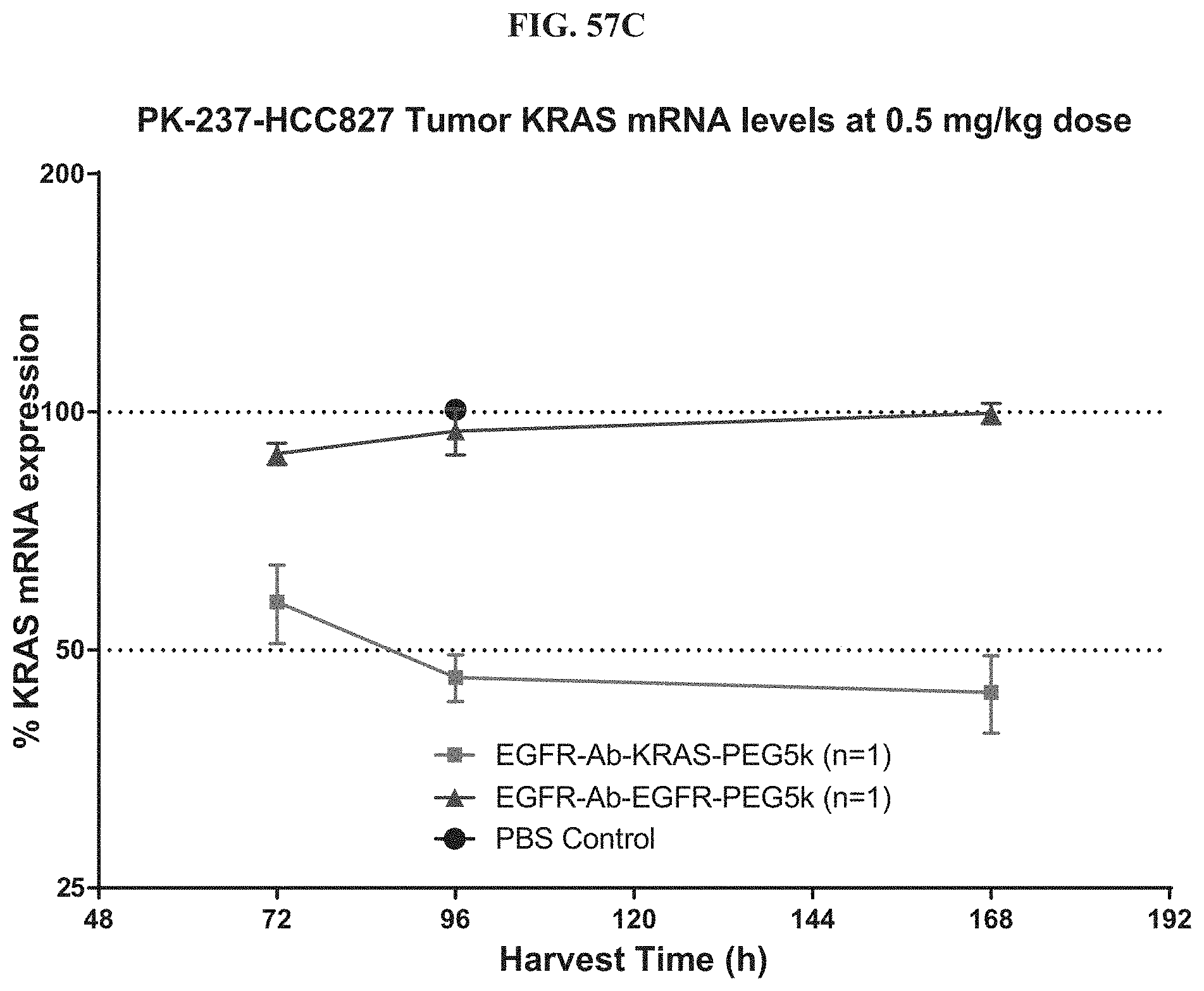
D00063
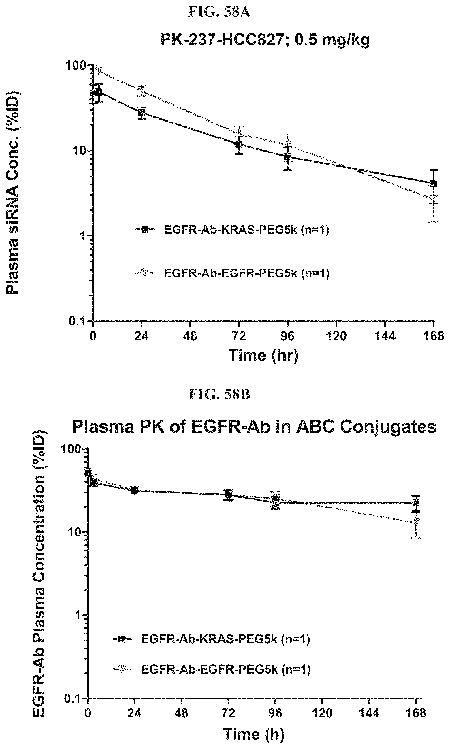
D00064

D00065
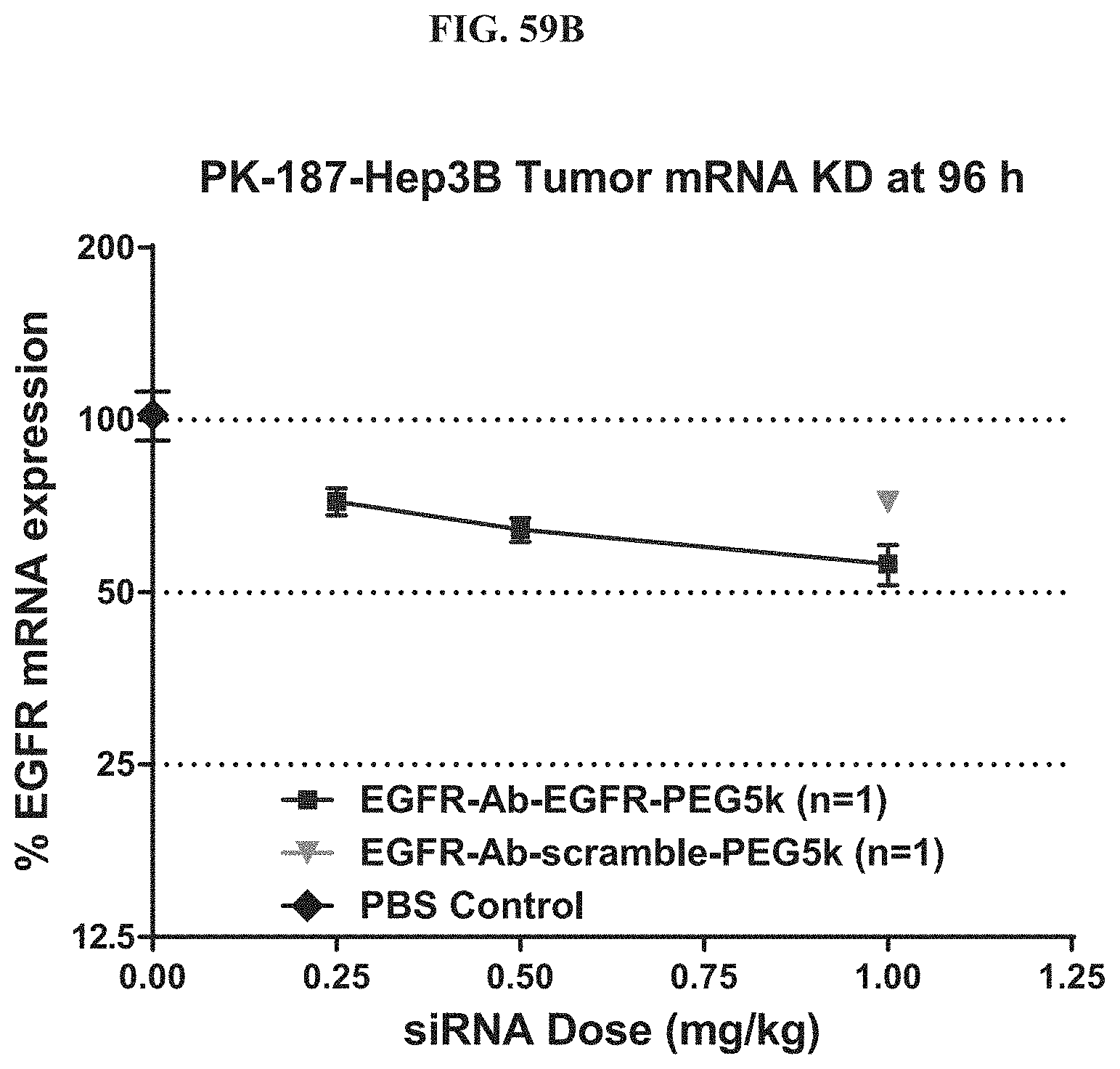
D00066
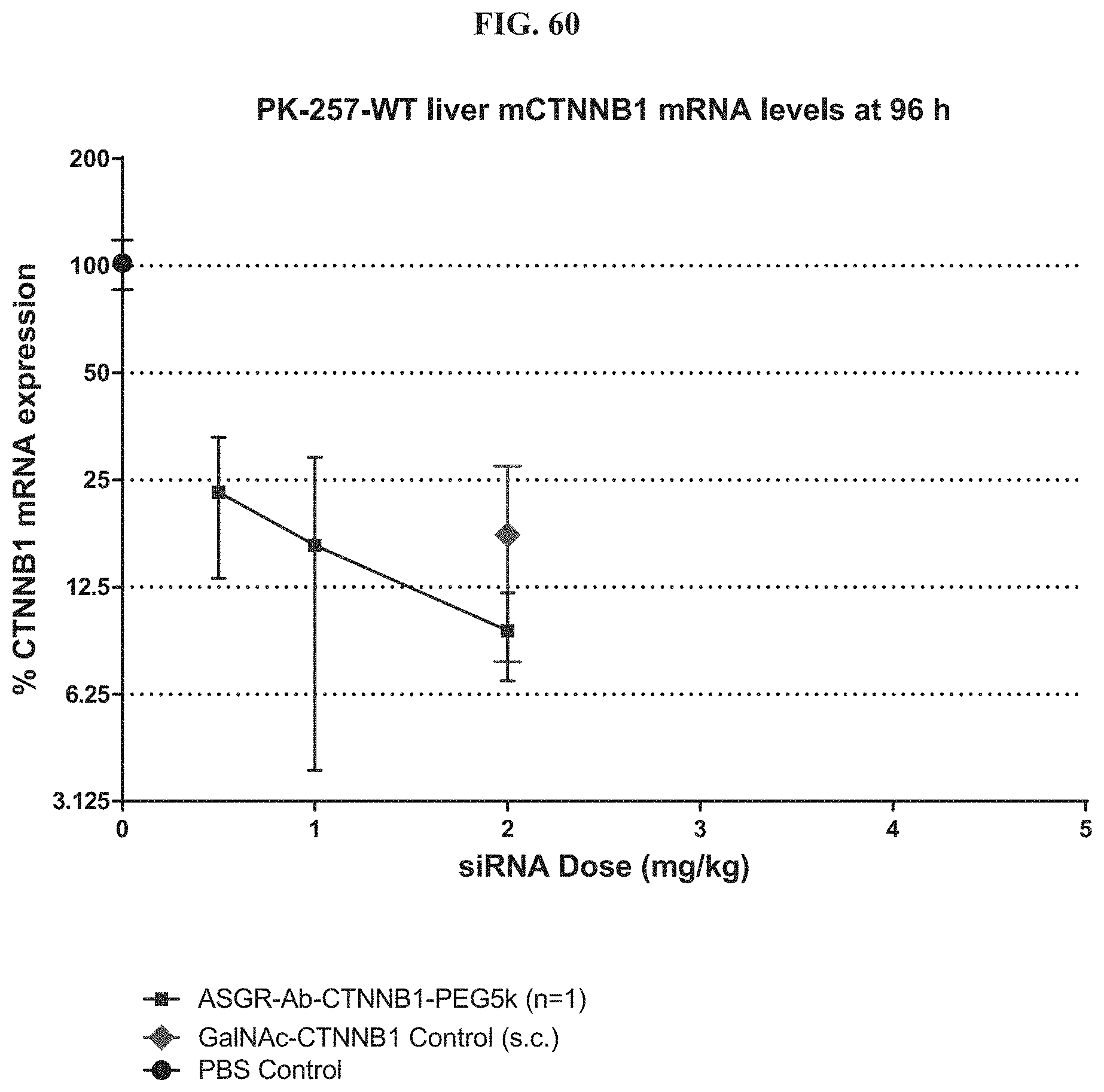
D00067

D00068
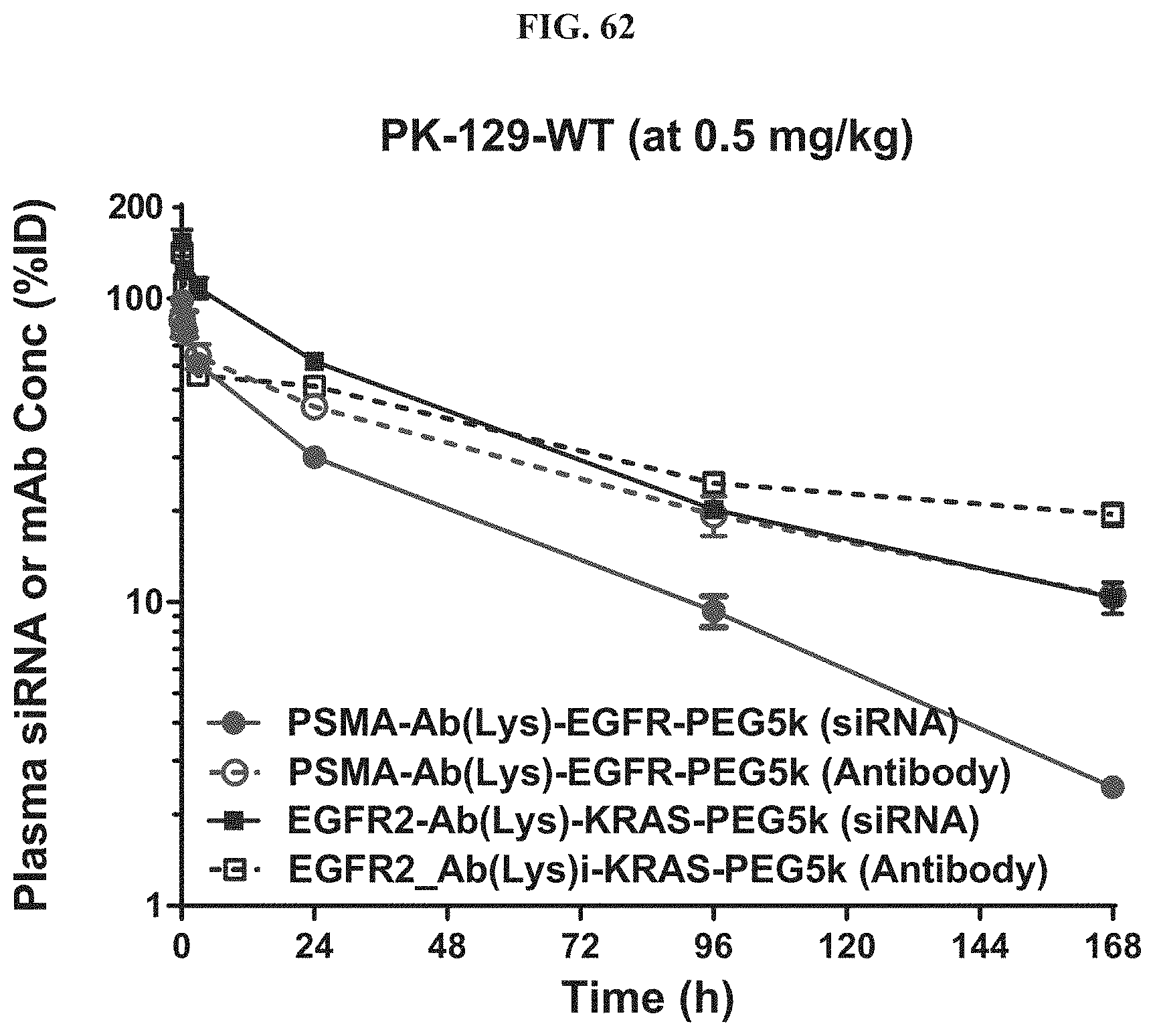
D00069
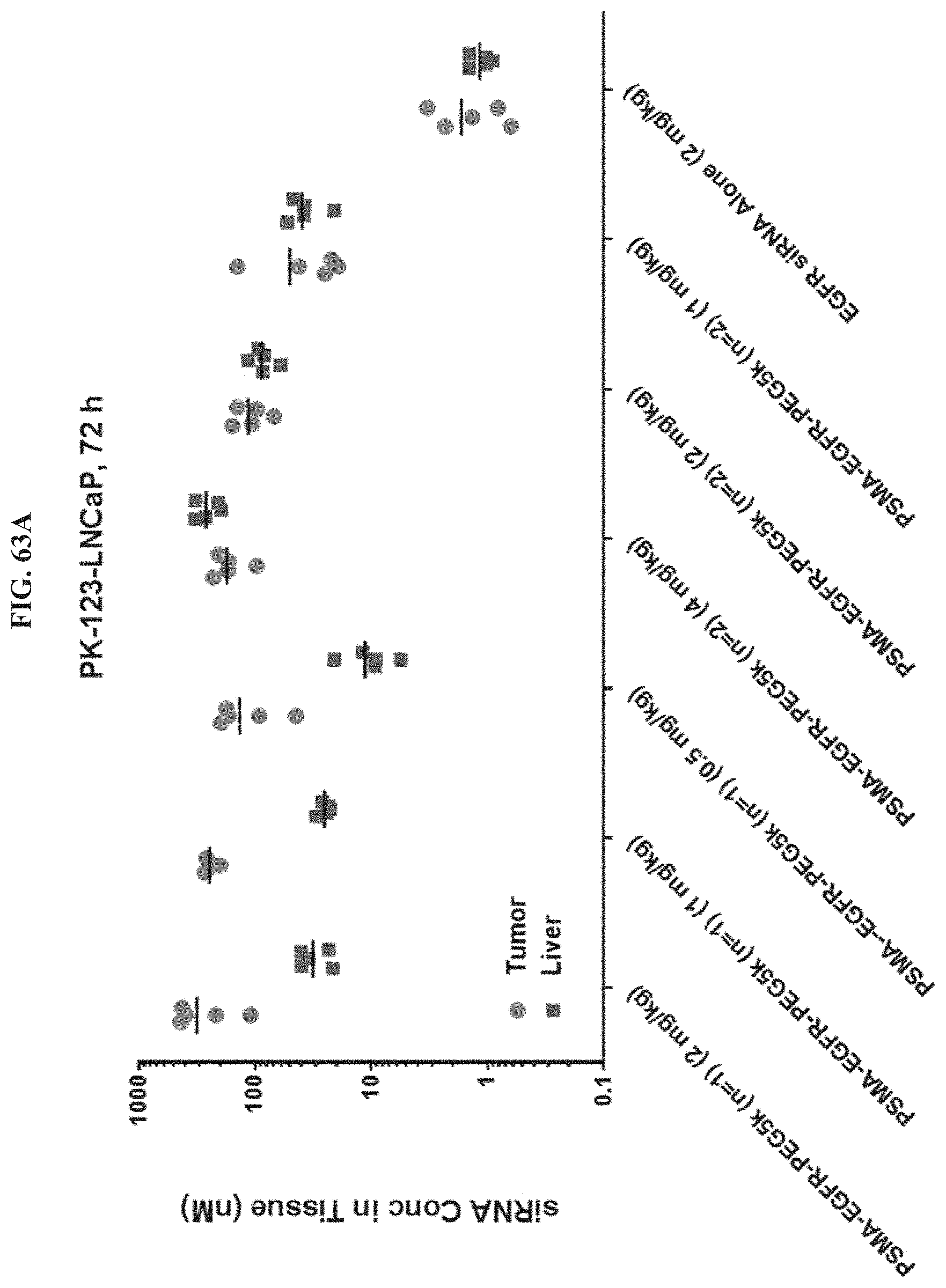
D00070
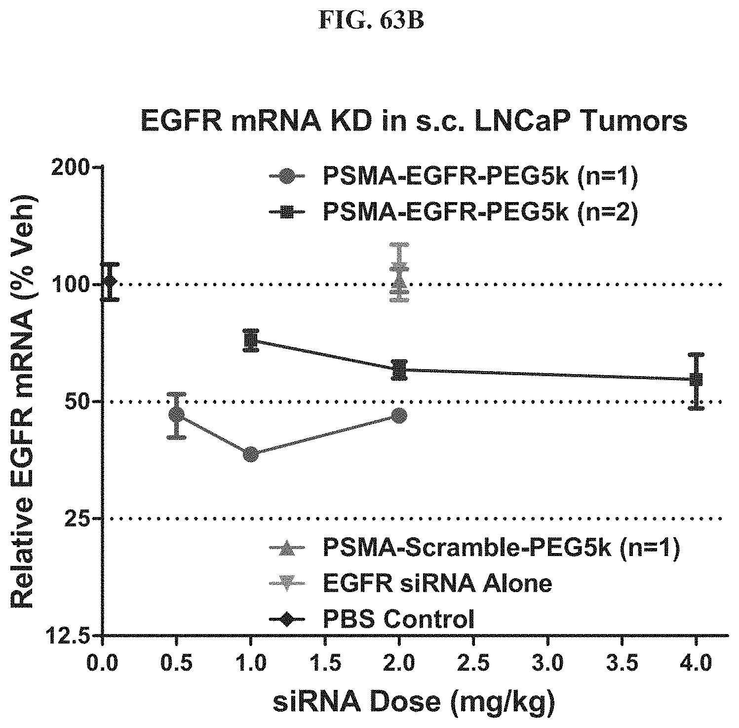
D00071

D00072

D00073
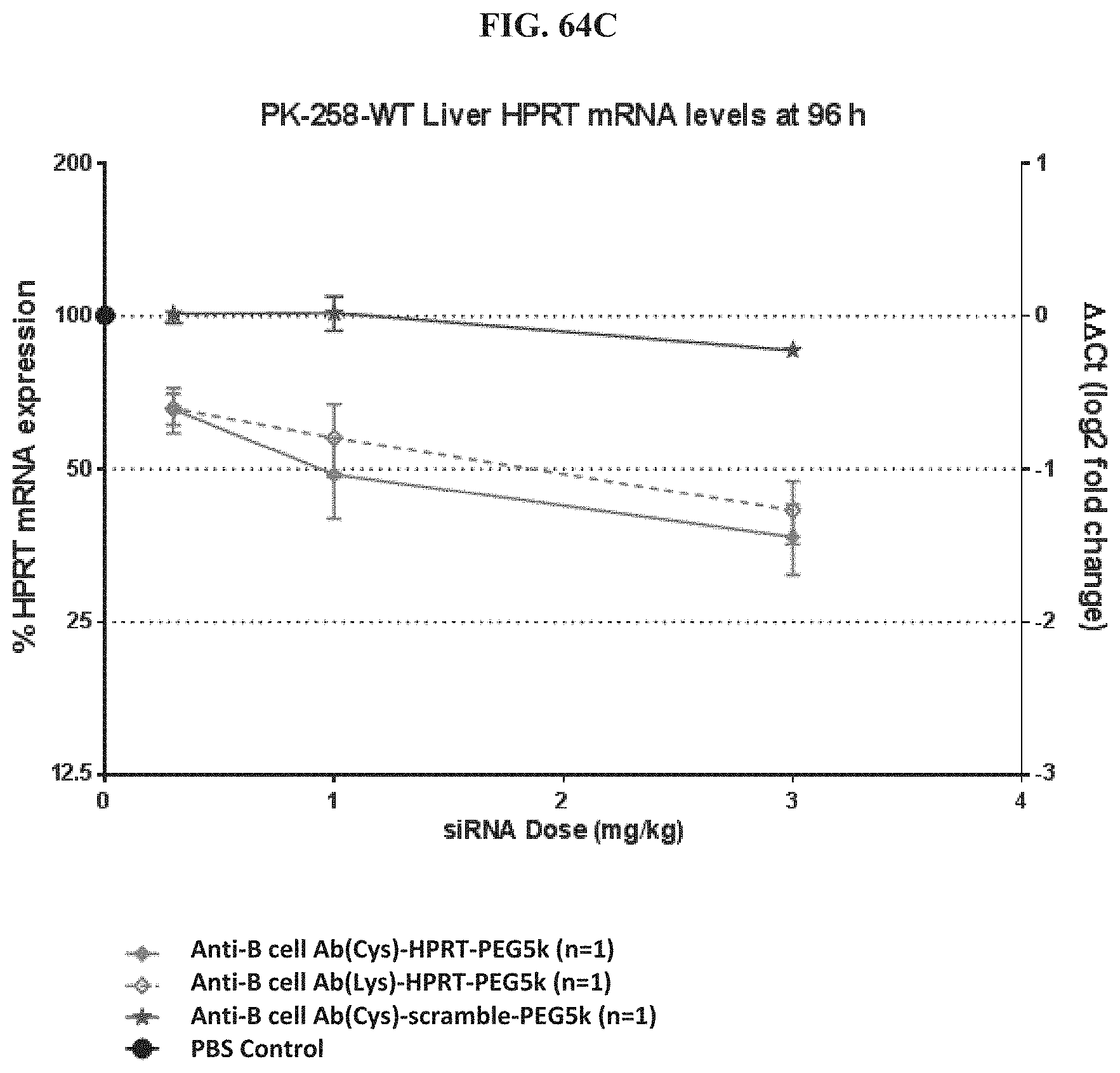
D00074
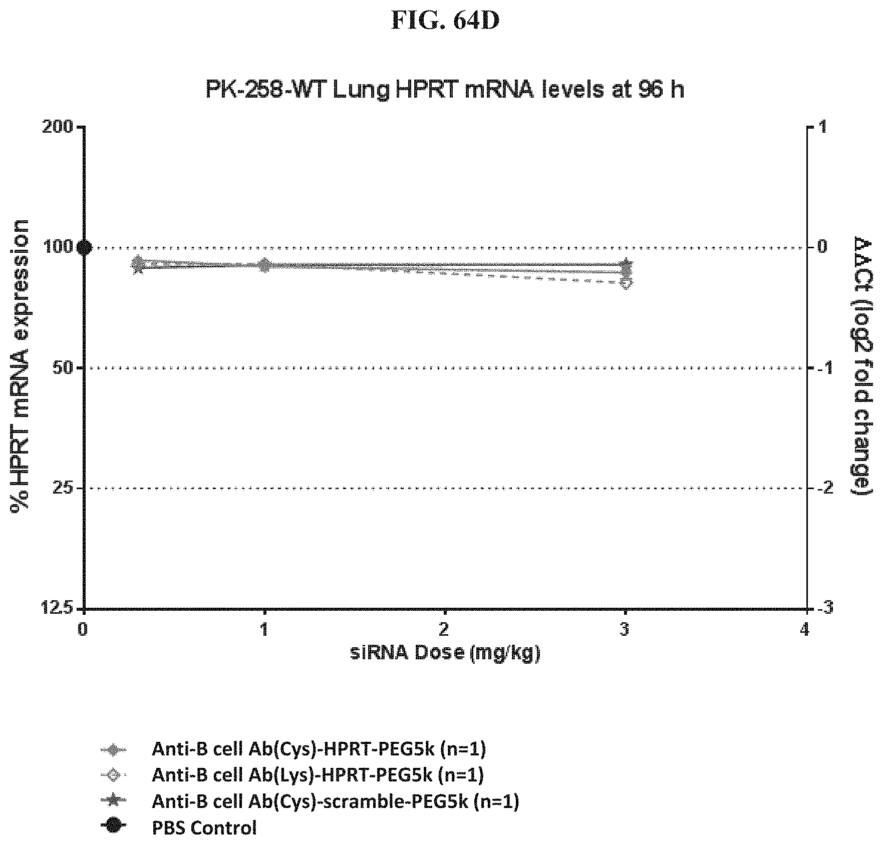
D00075
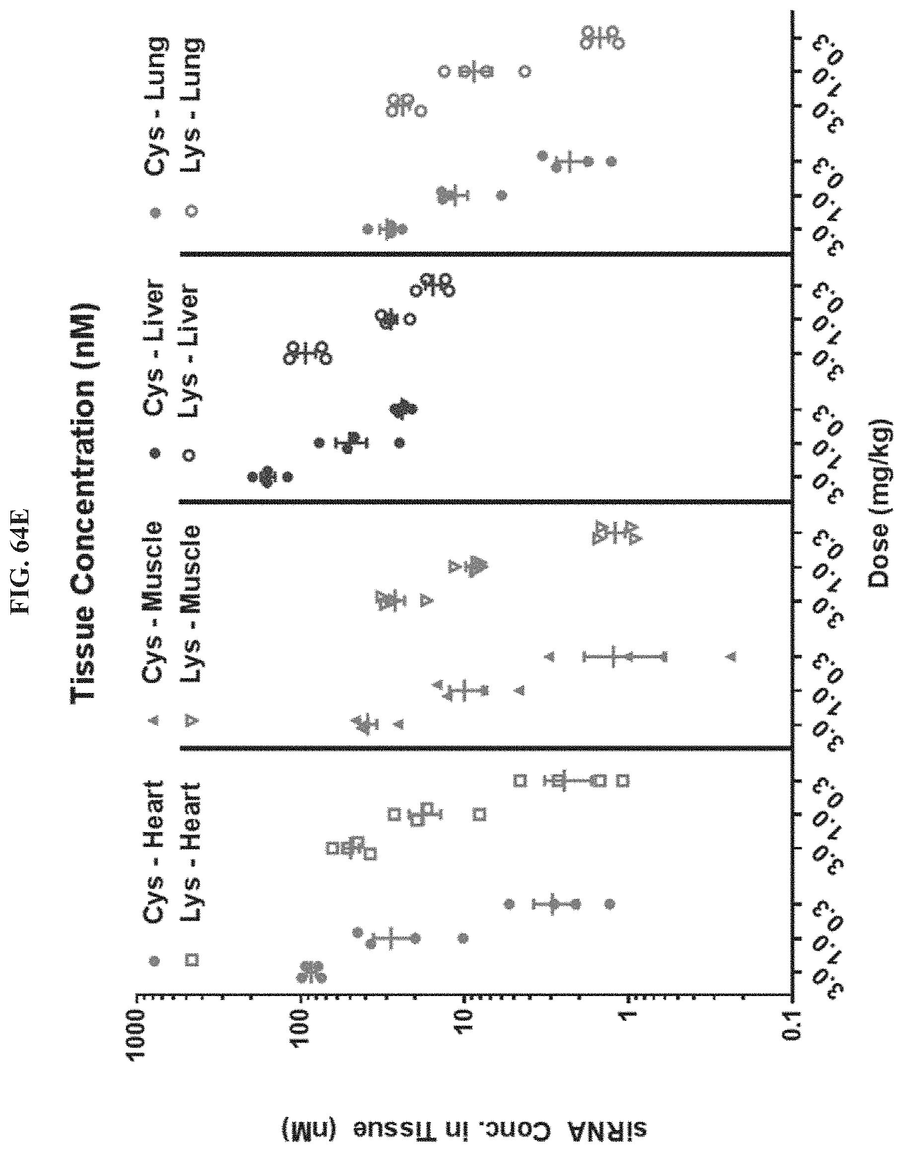
D00076
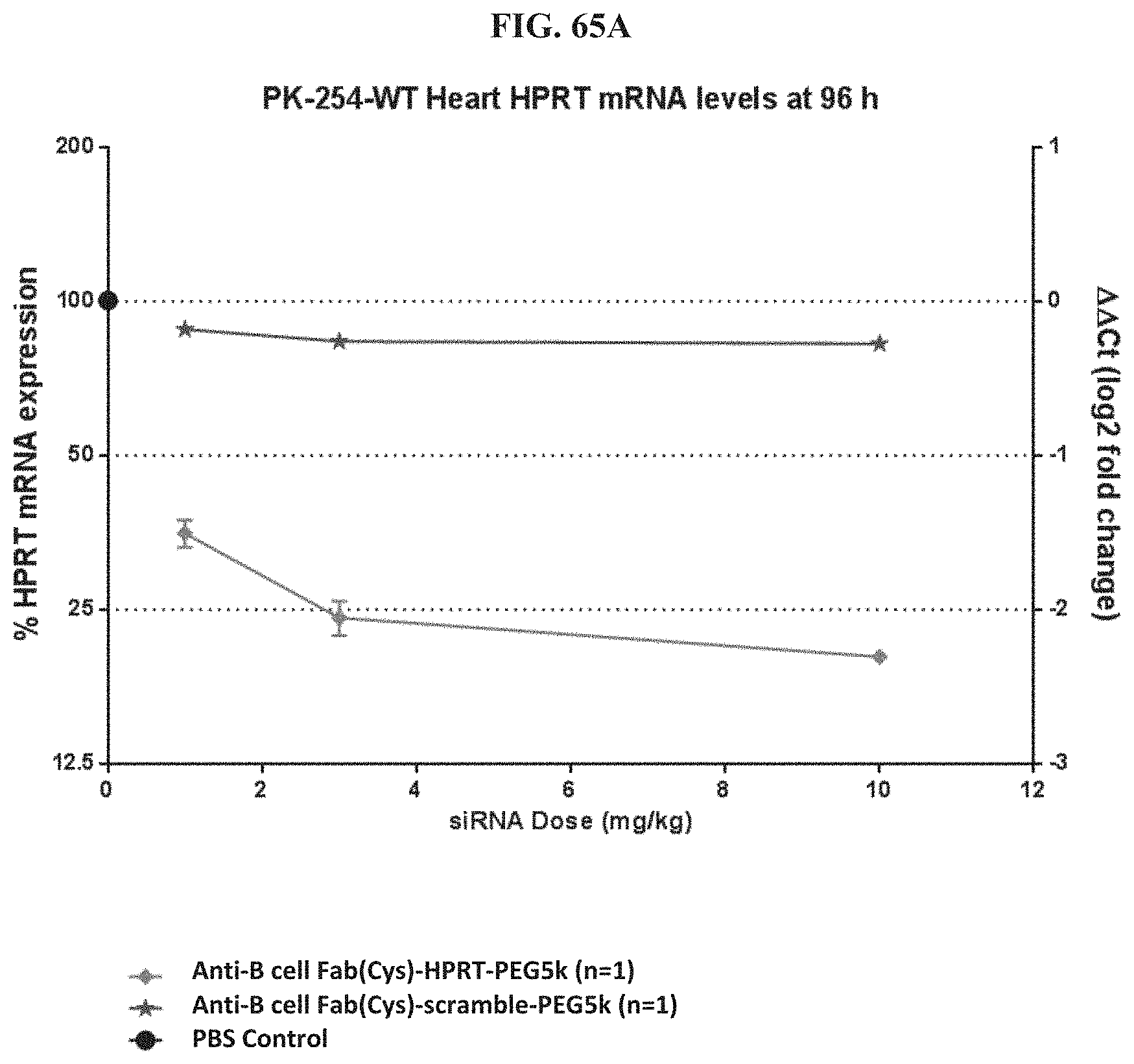
D00077

D00078
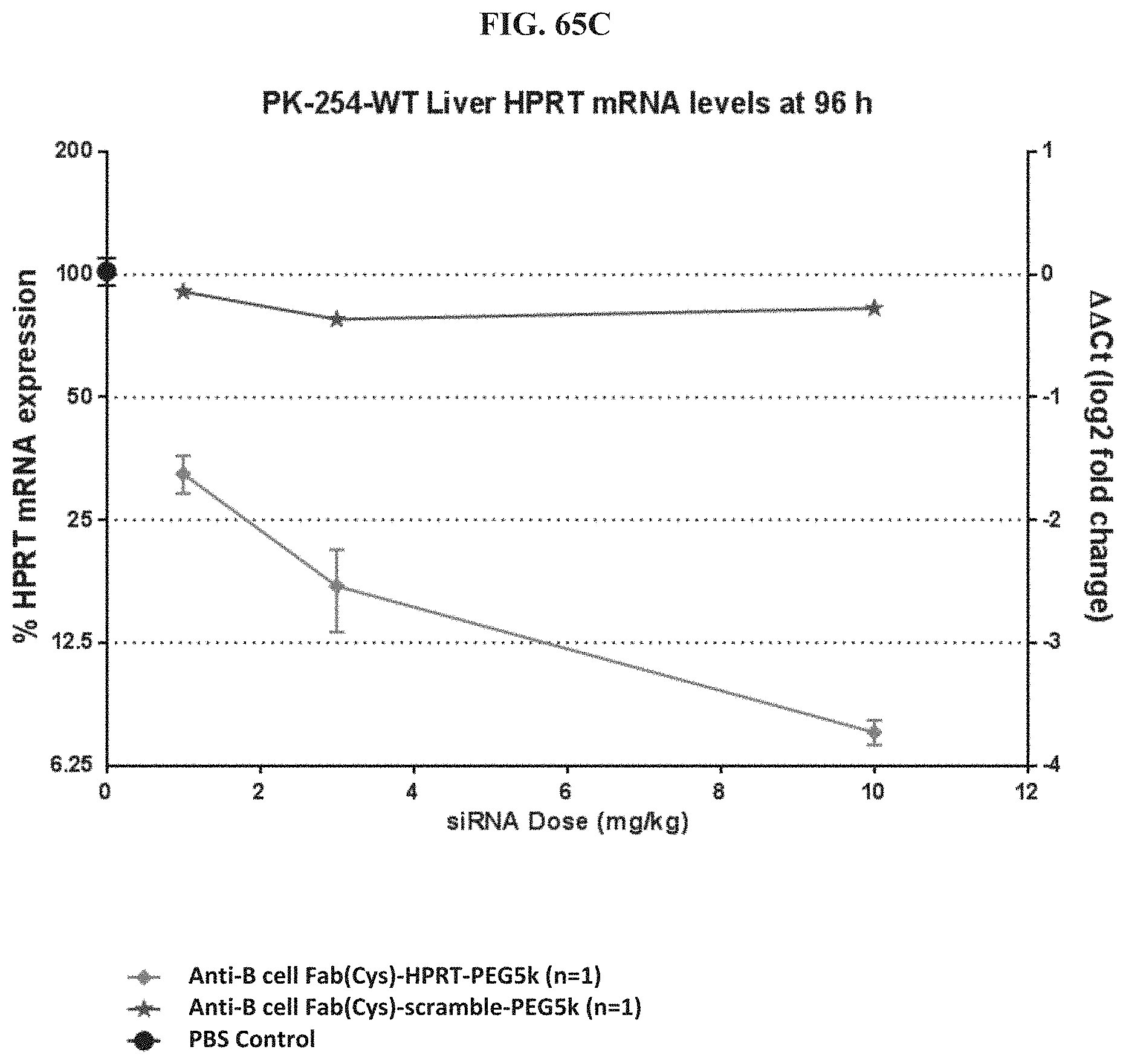
D00079
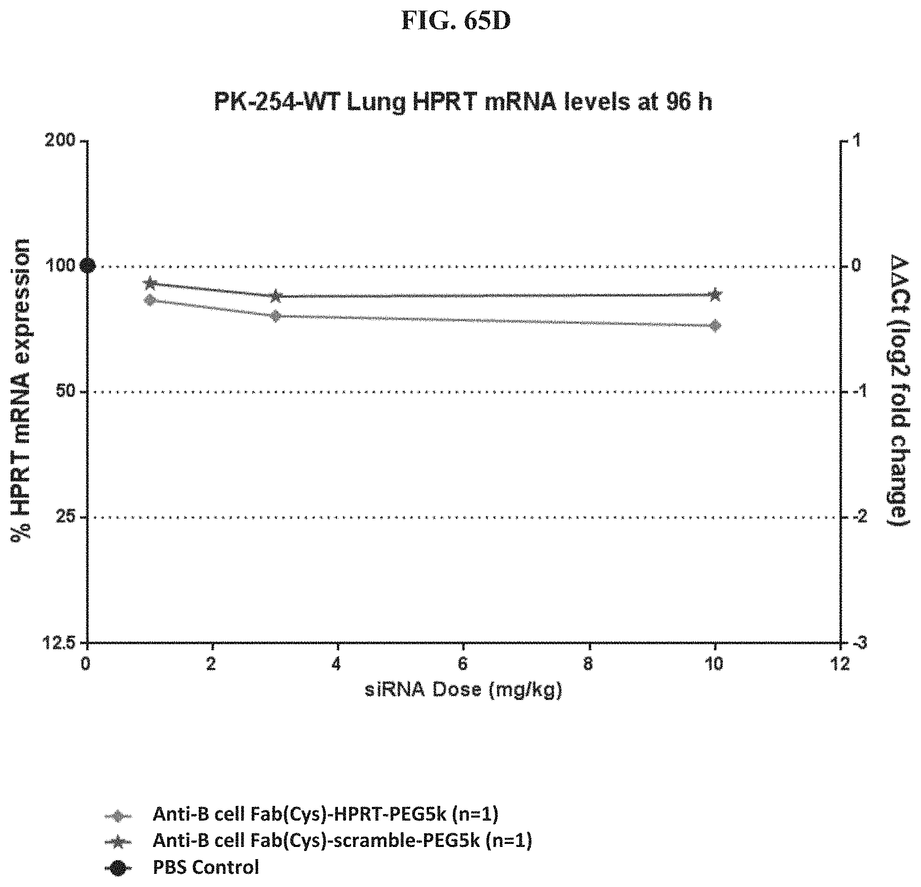
D00080
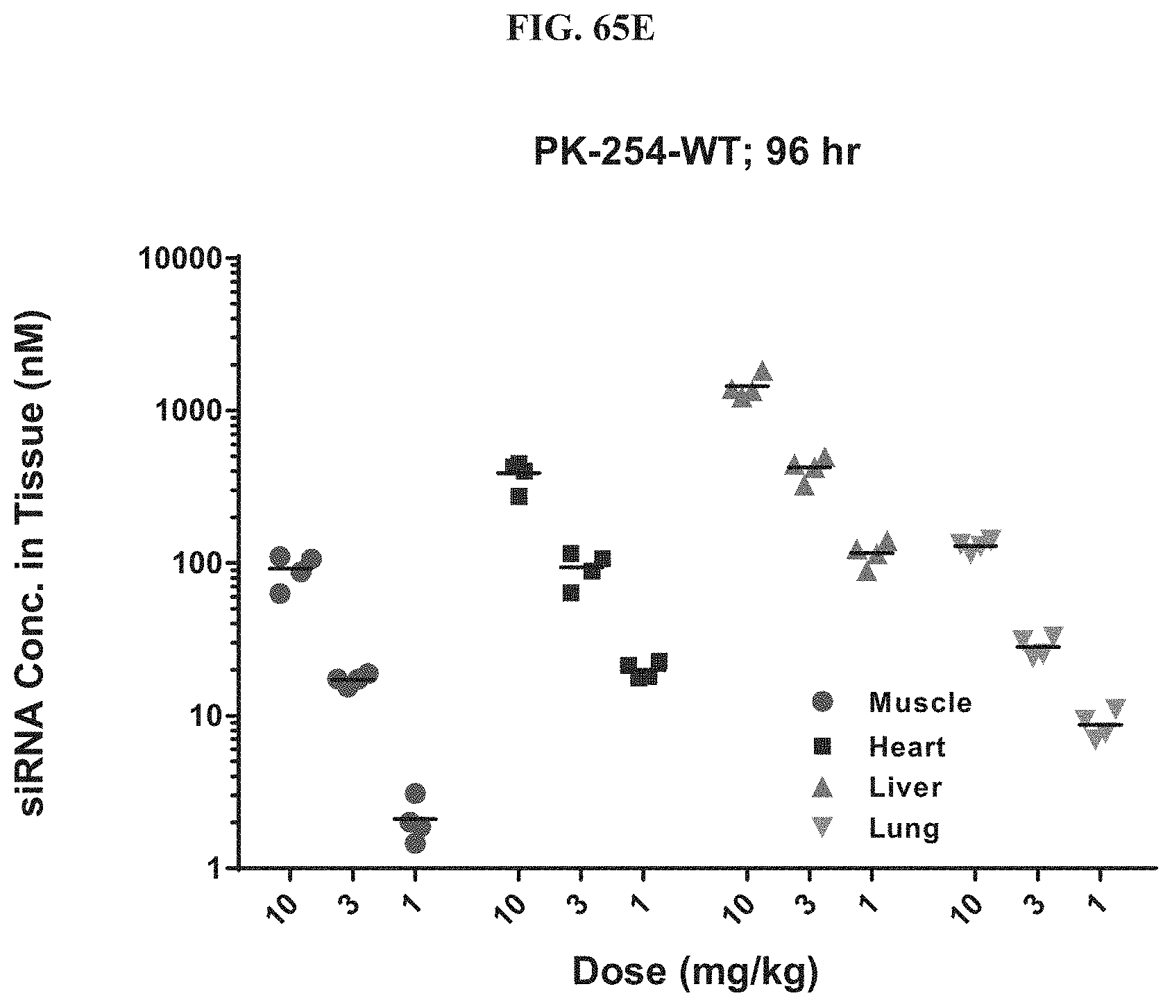
D00081
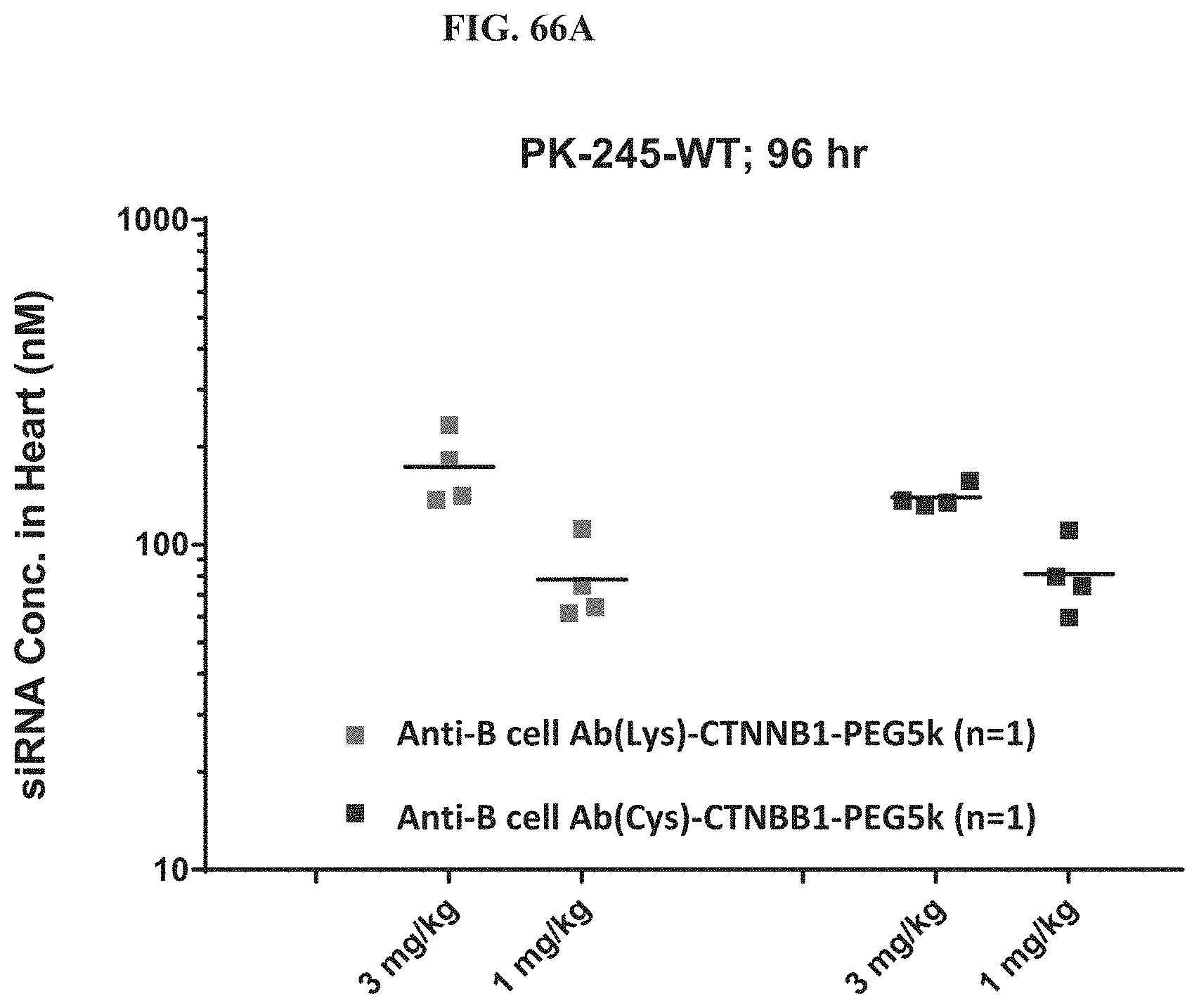
D00082
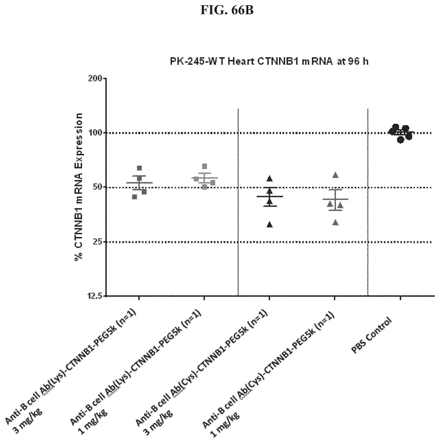
D00083

D00084
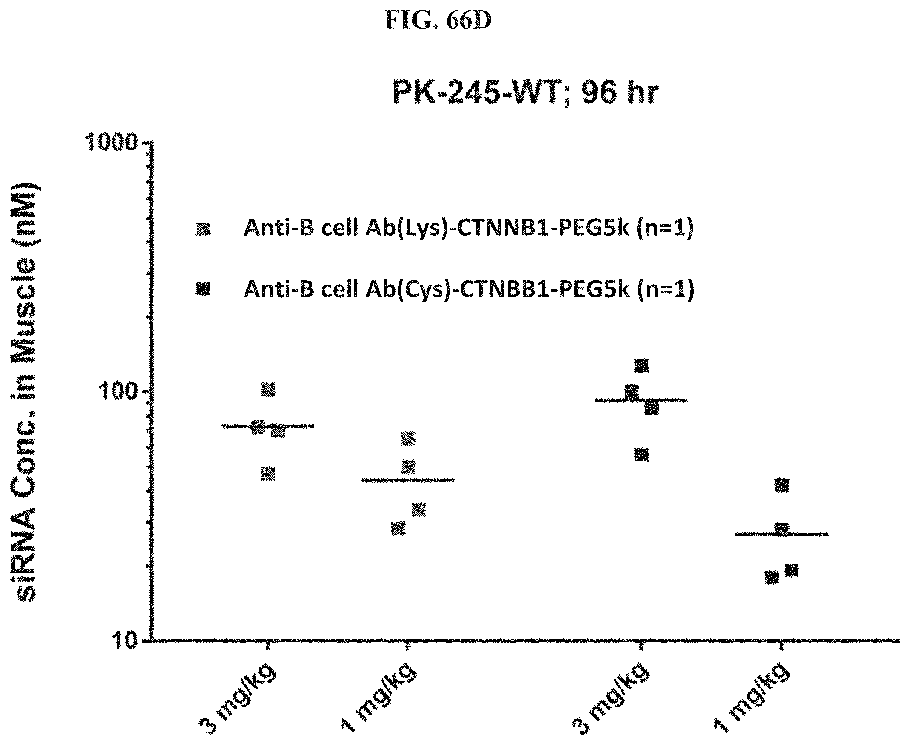
D00085
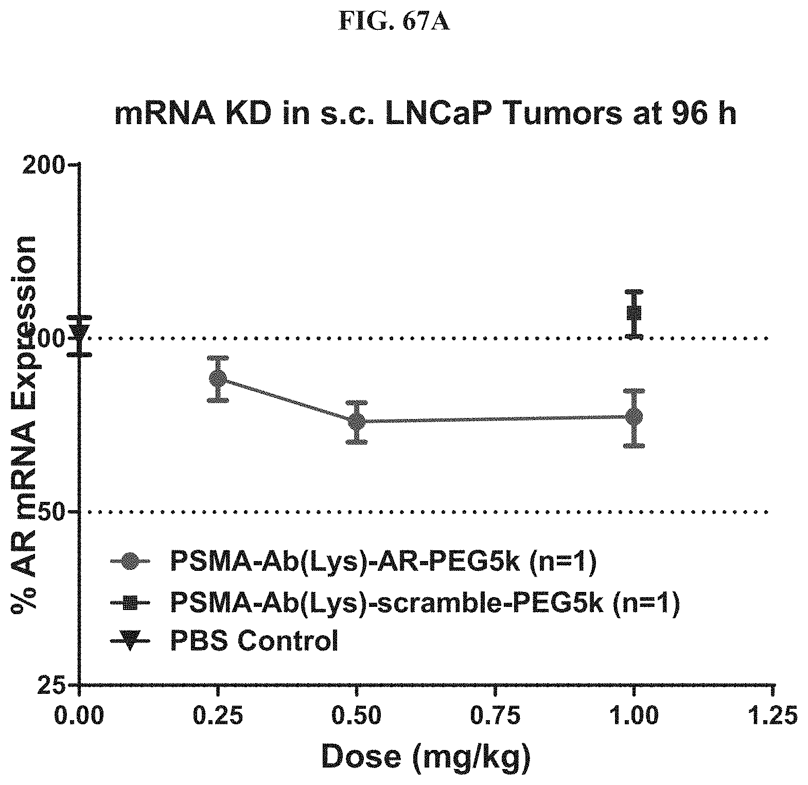
D00086
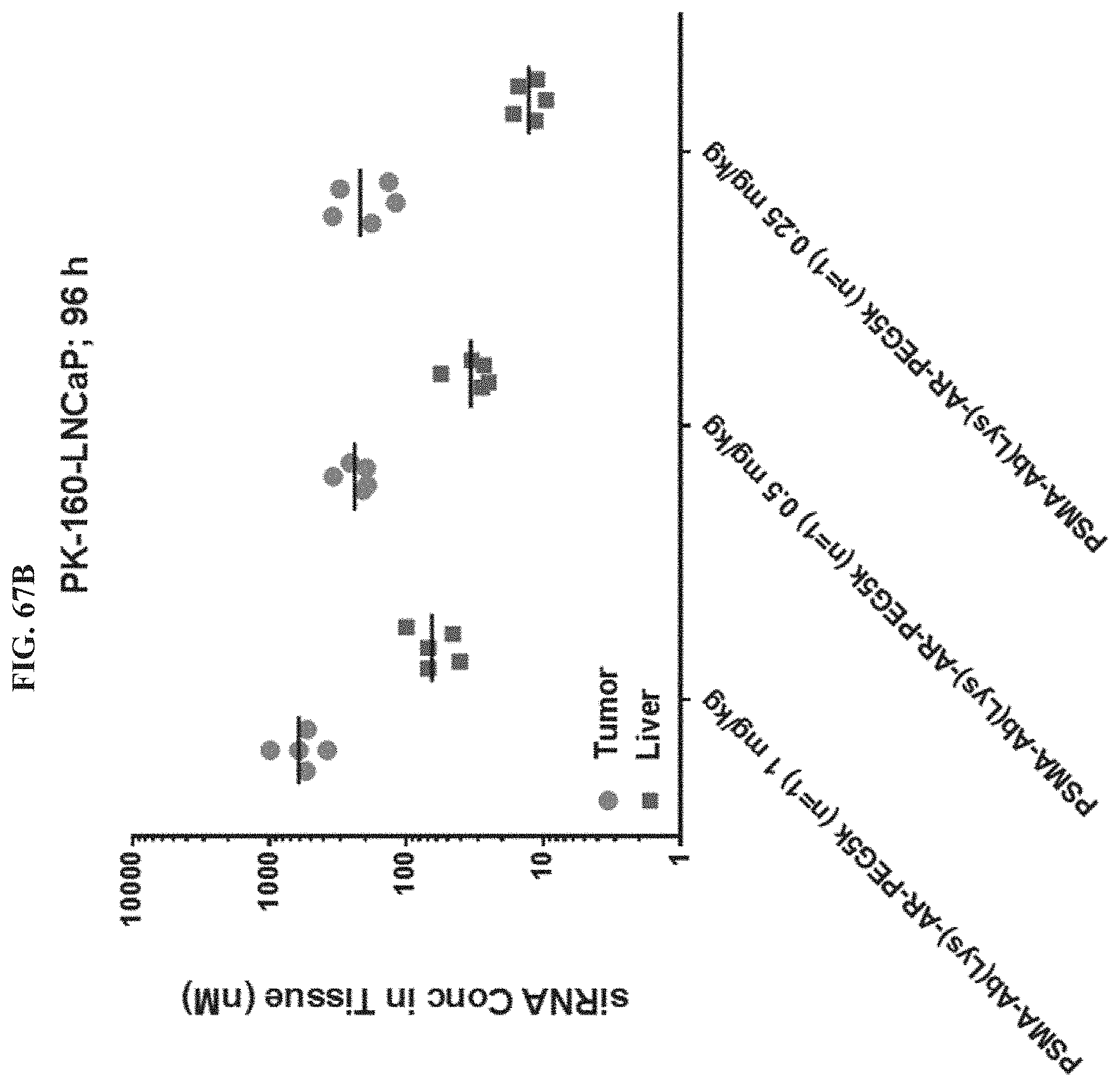
D00087
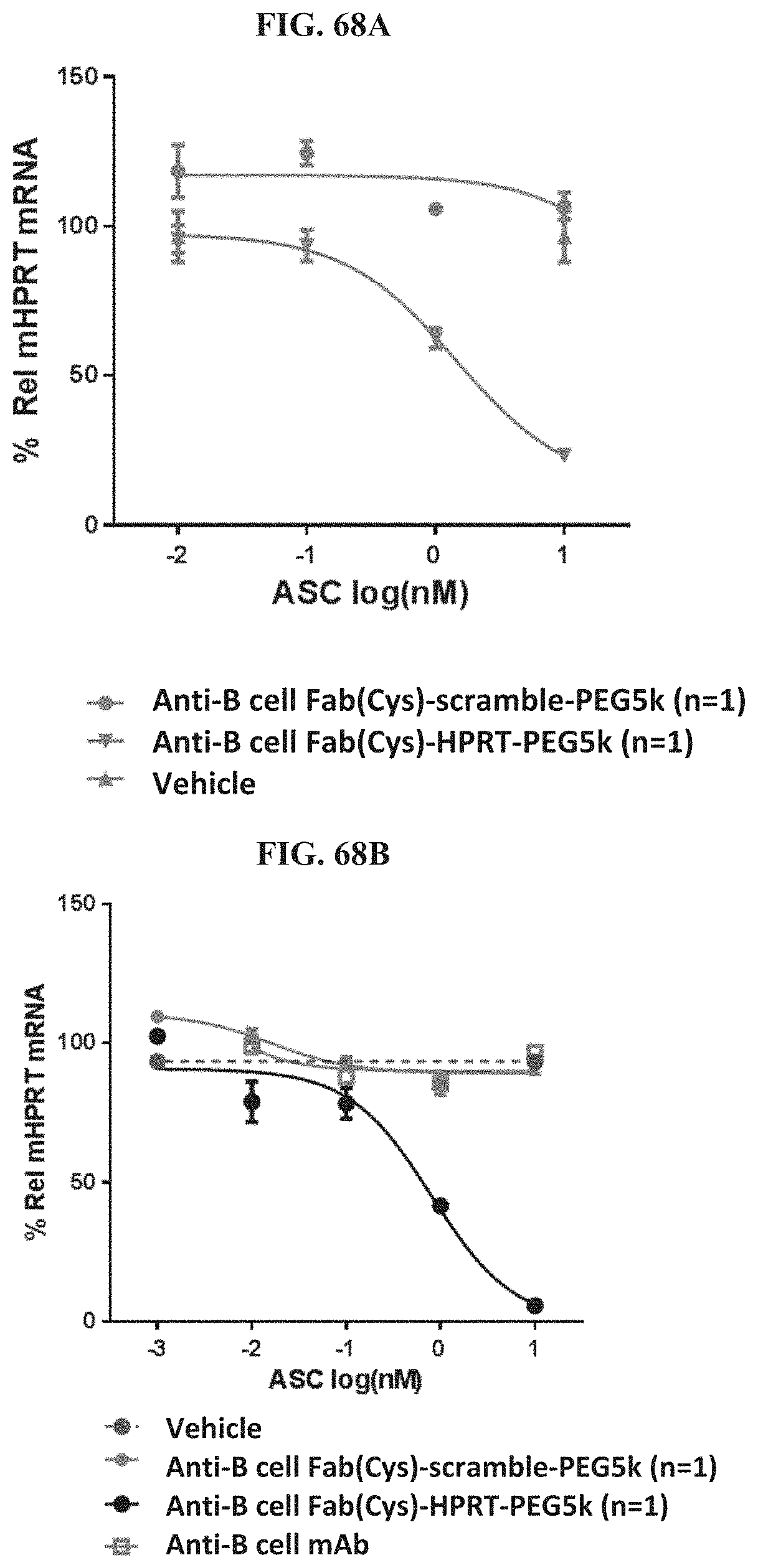
D00088
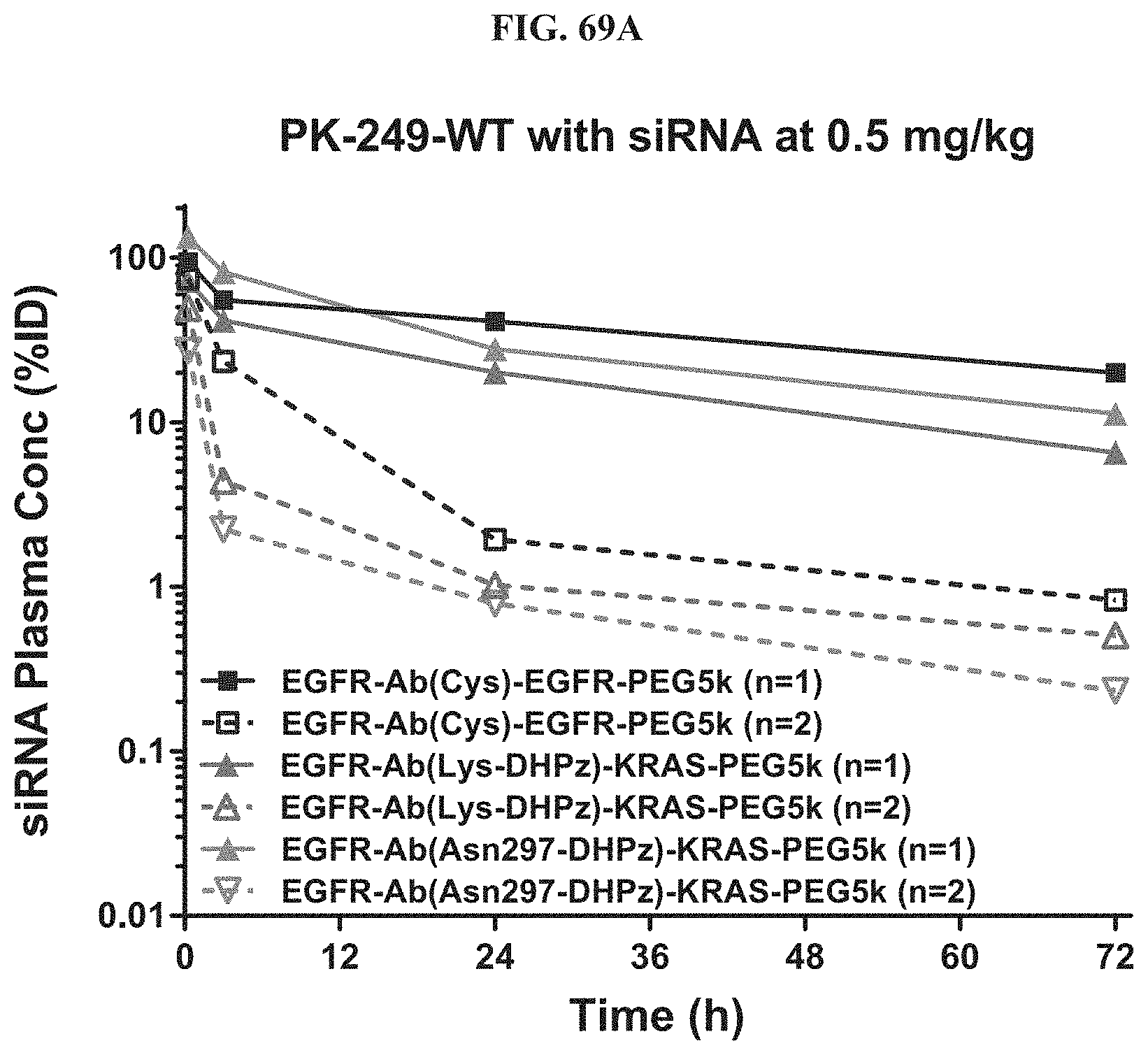
D00089

D00090
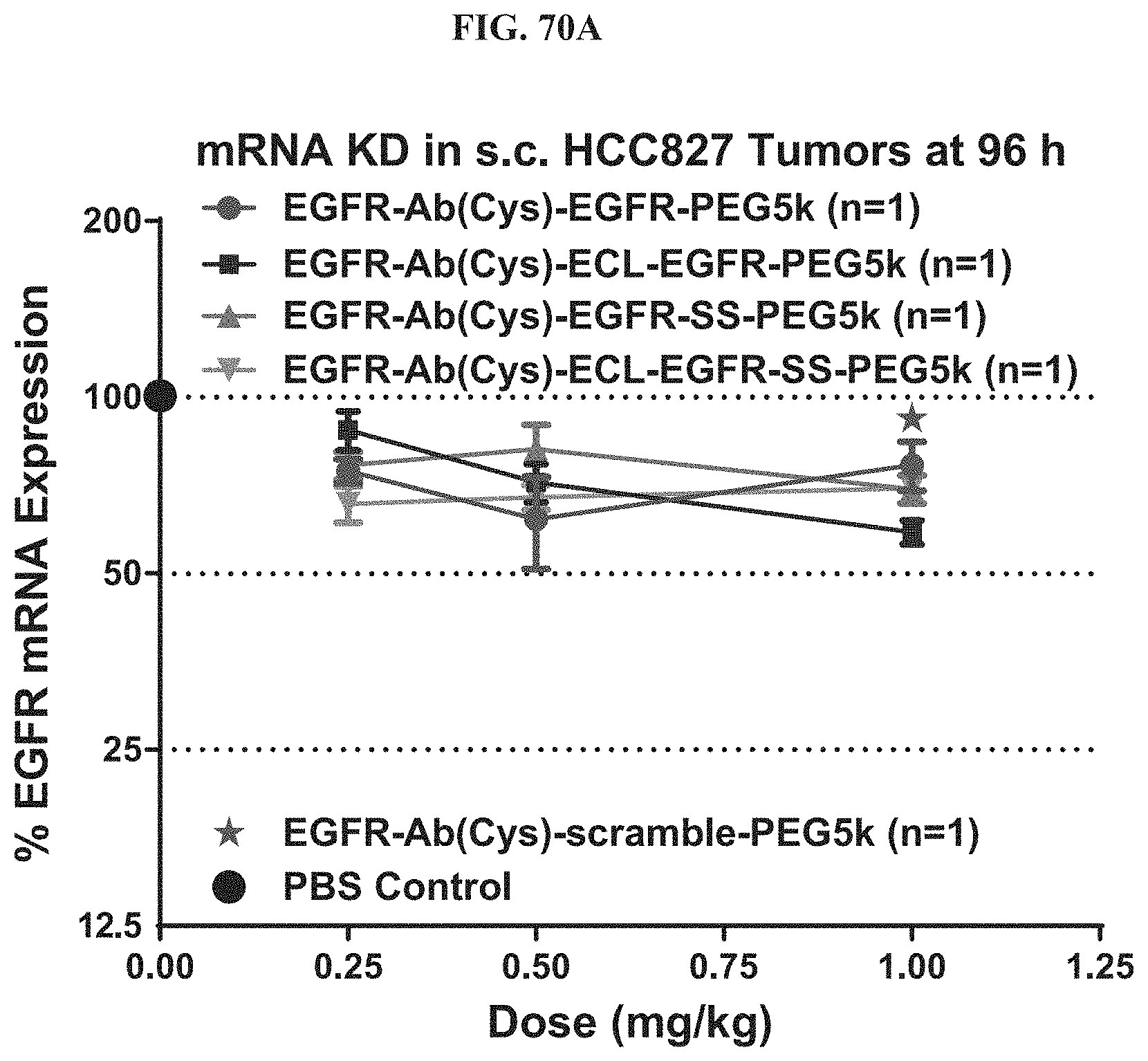
D00091
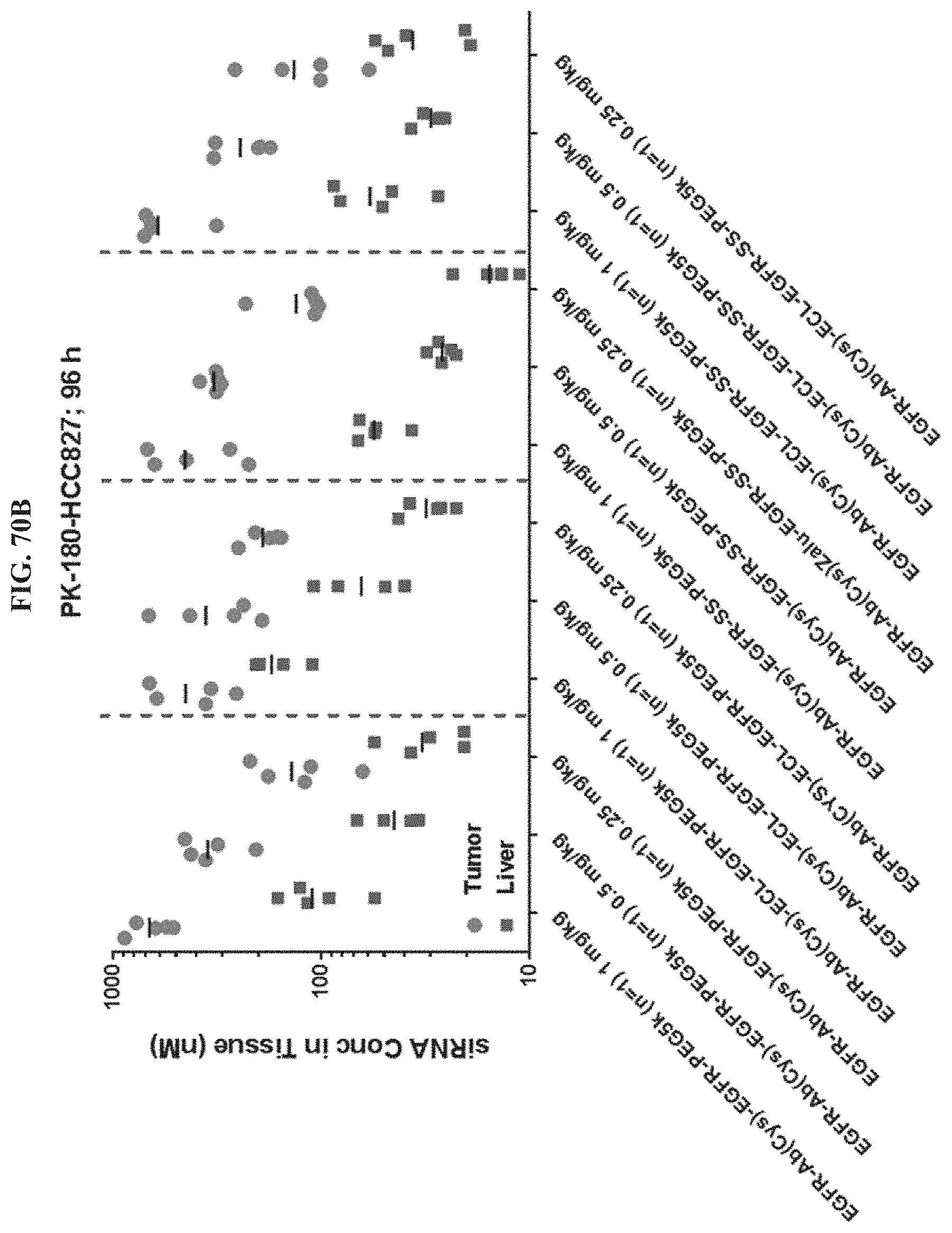
D00092
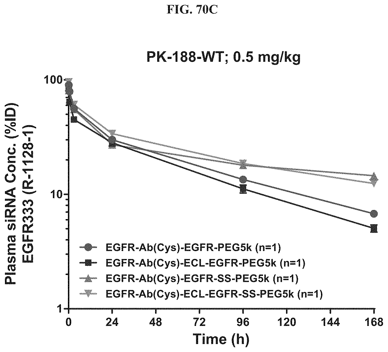
D00093
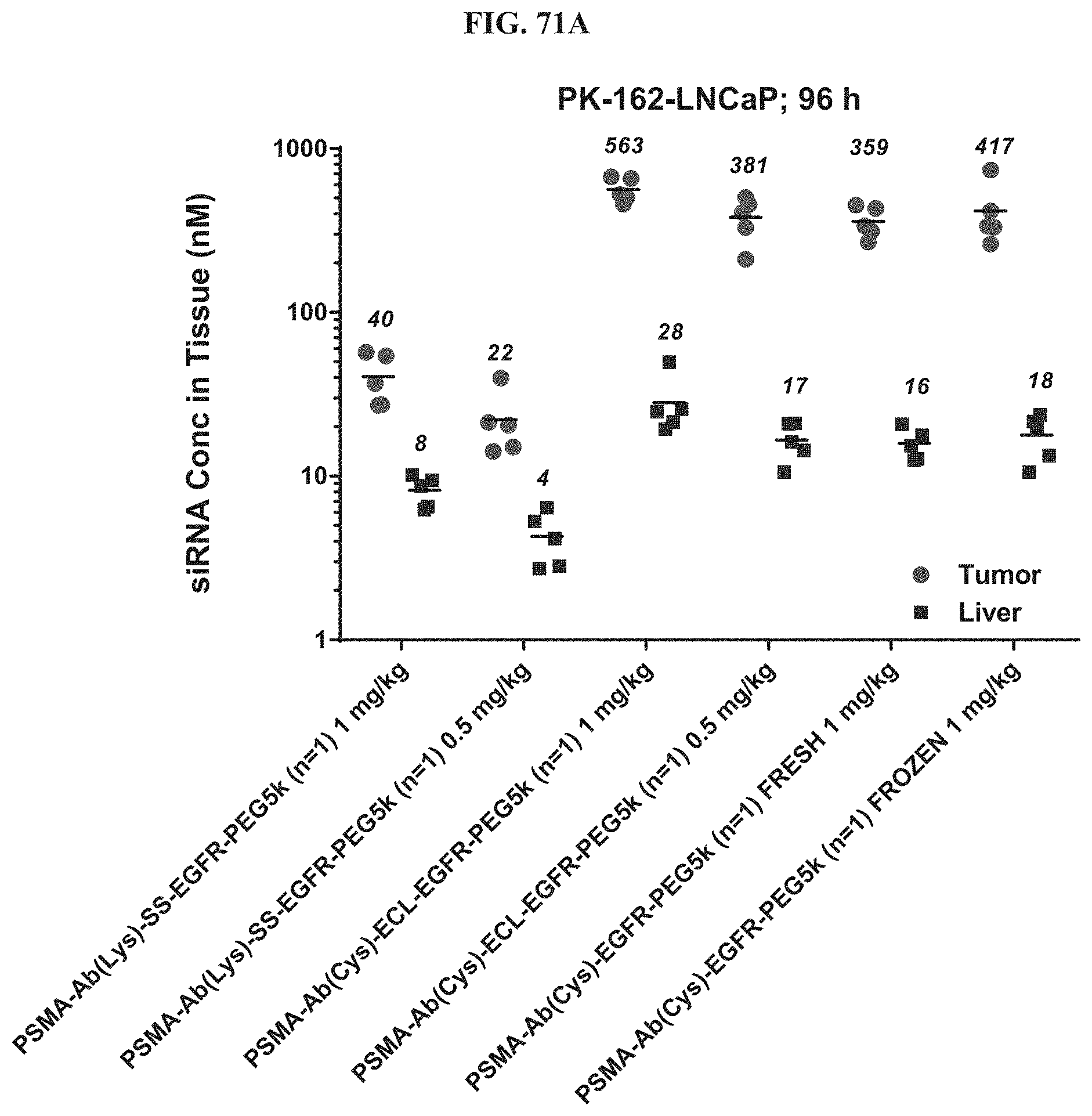
D00094
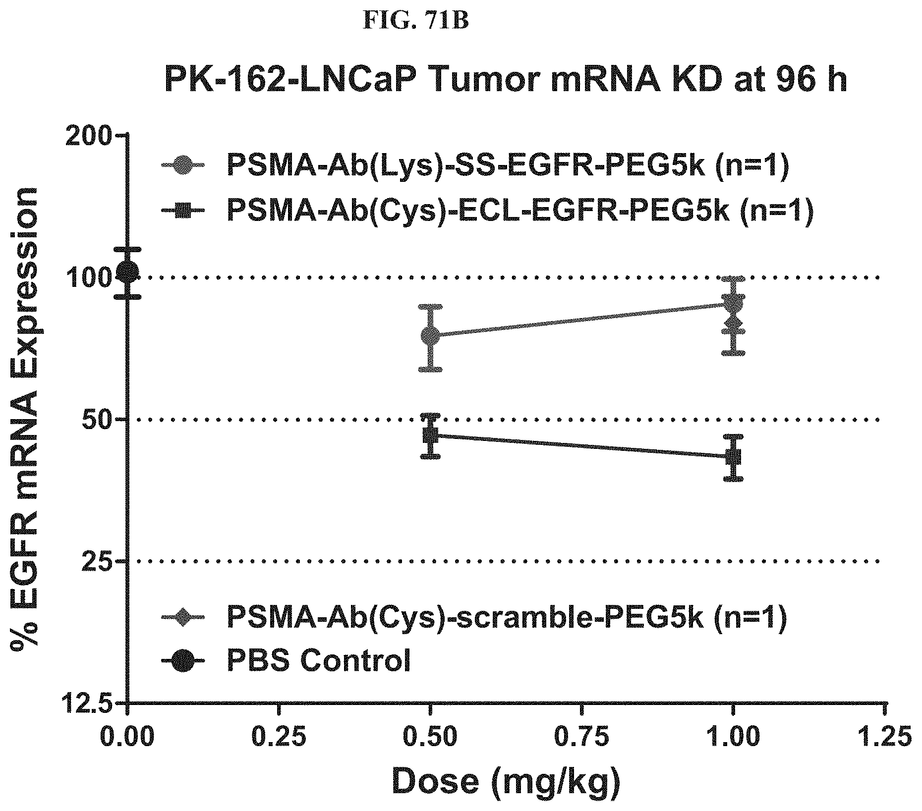
D00095

D00096

D00097
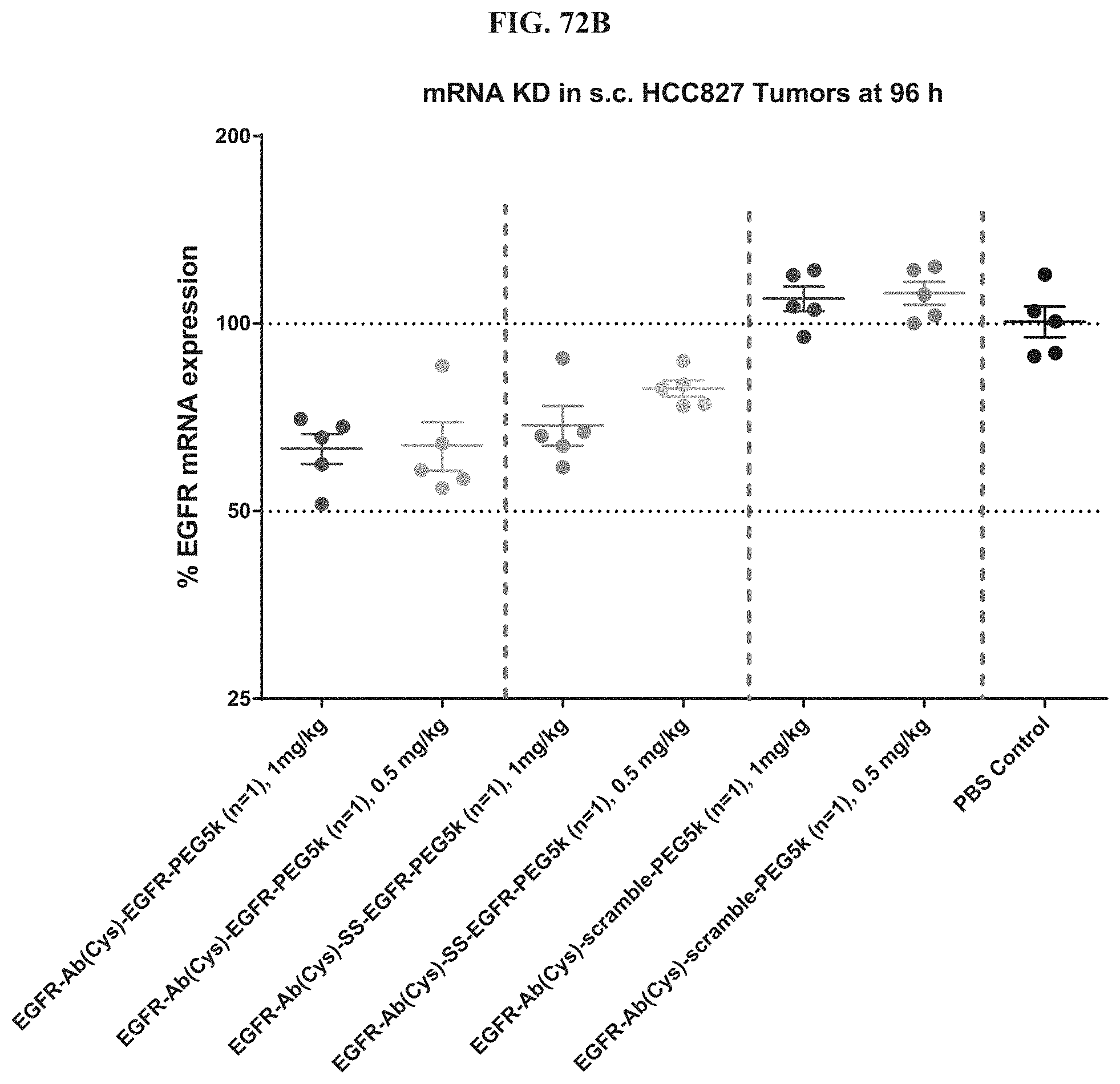
D00098
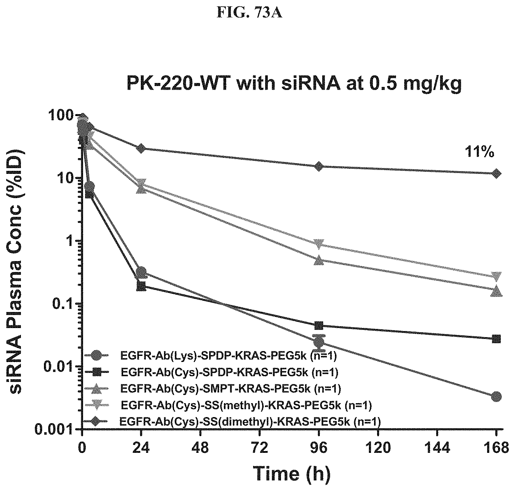
D00099
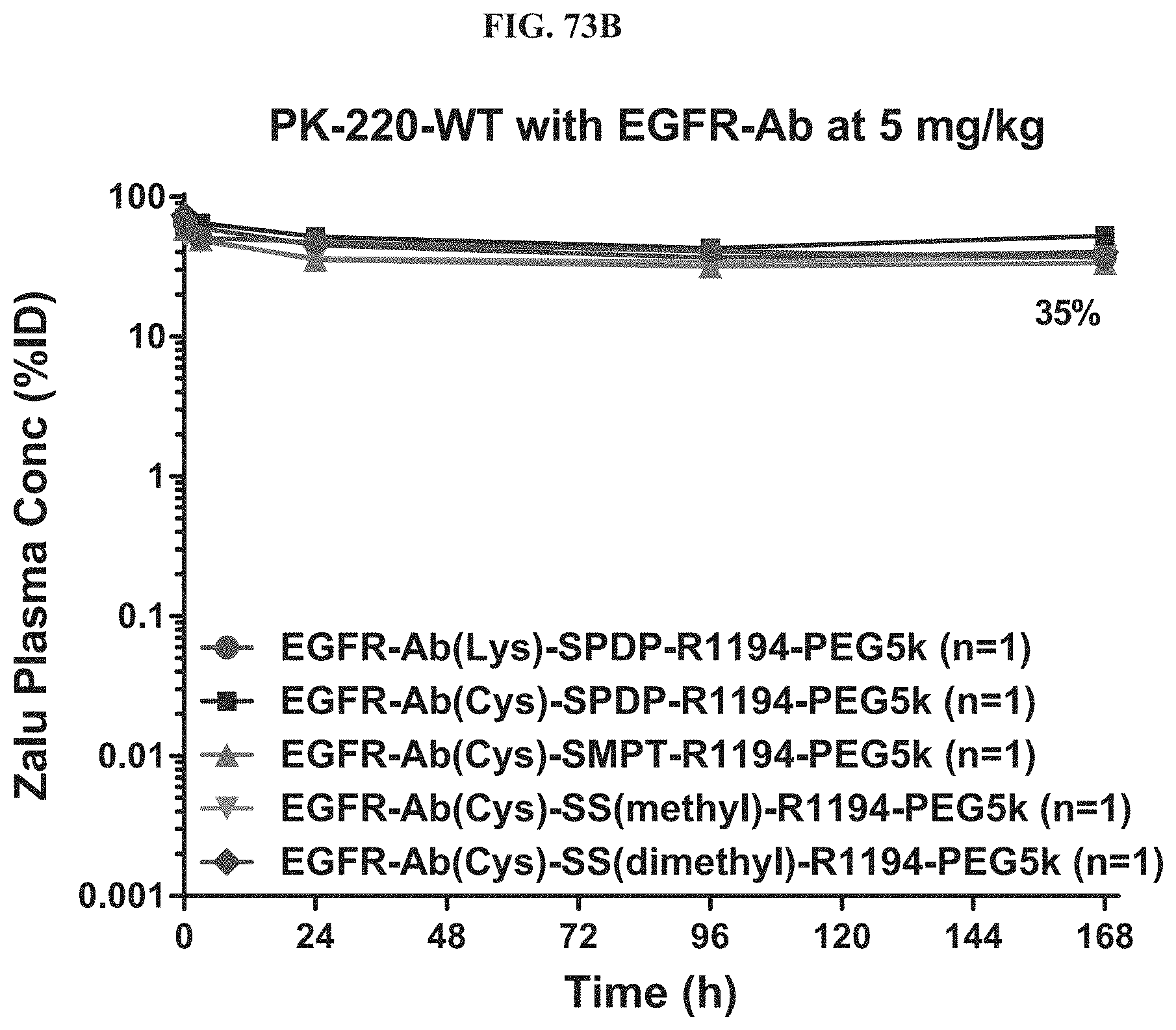
D00100

D00101
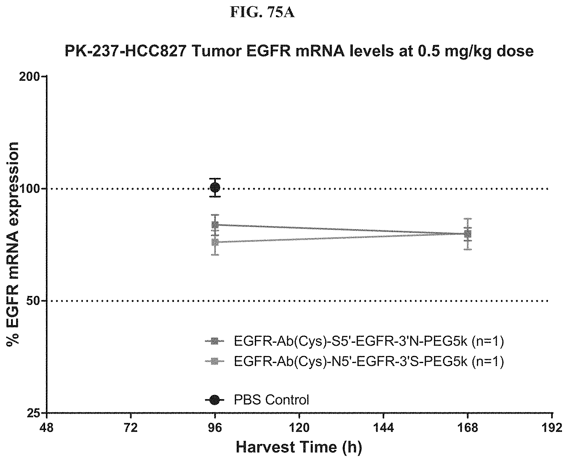
D00102
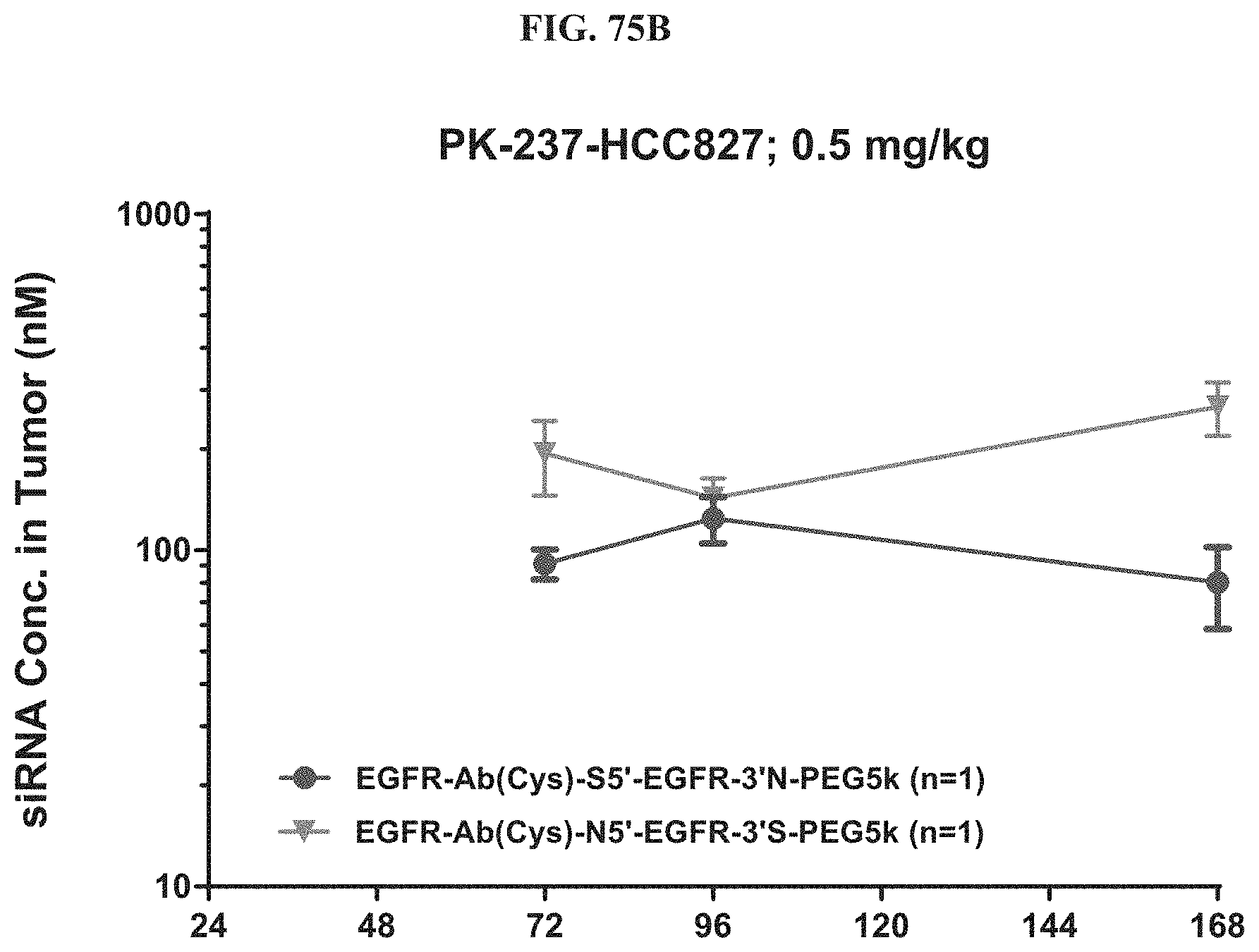
D00103

D00104
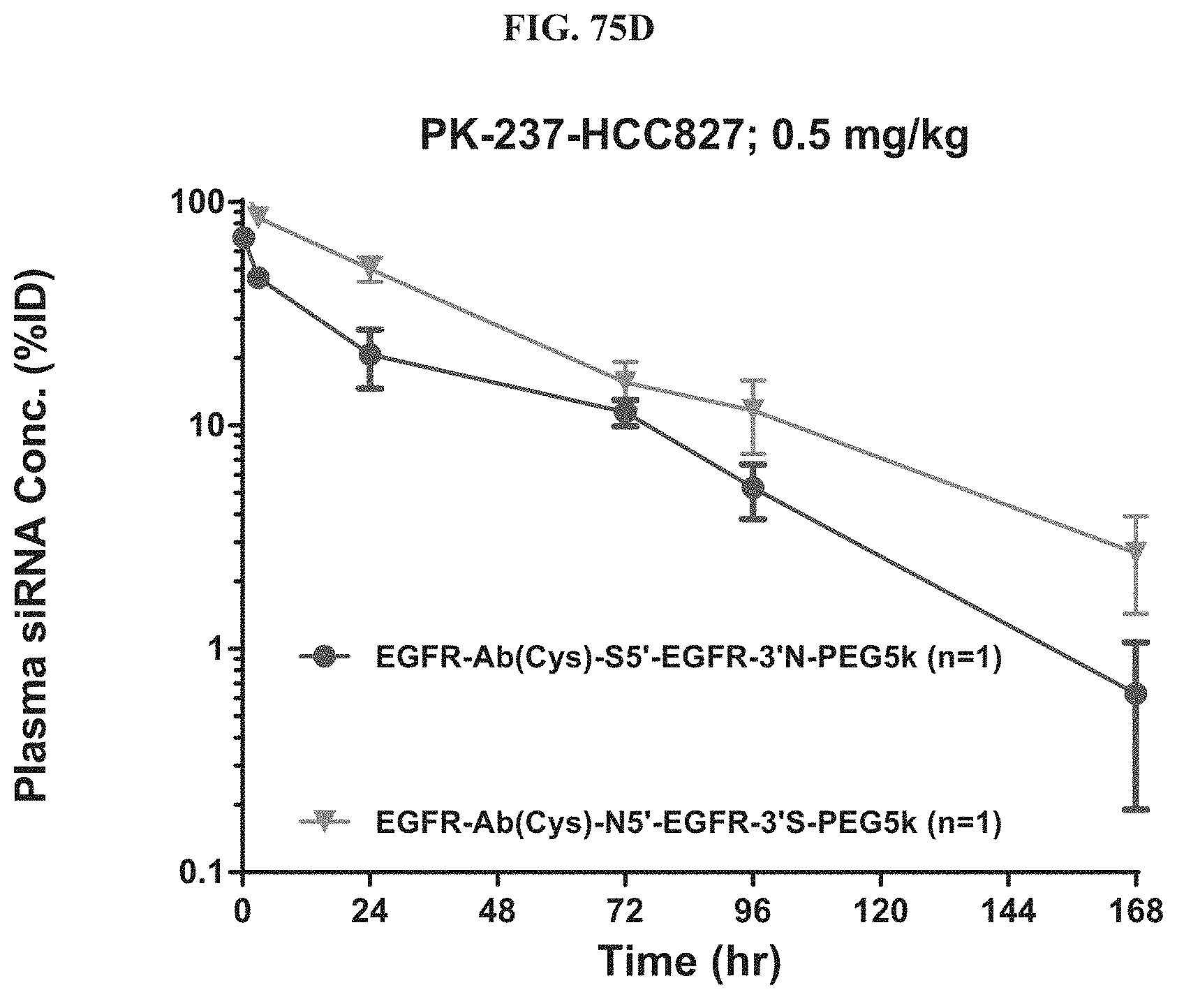
D00105
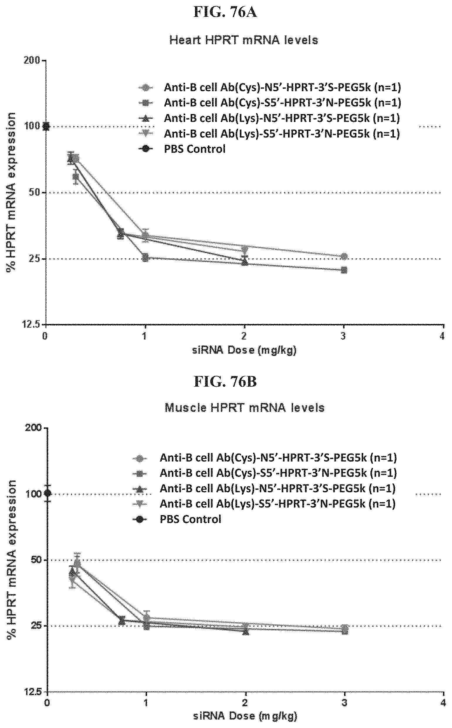
D00106

D00107

D00108

D00109
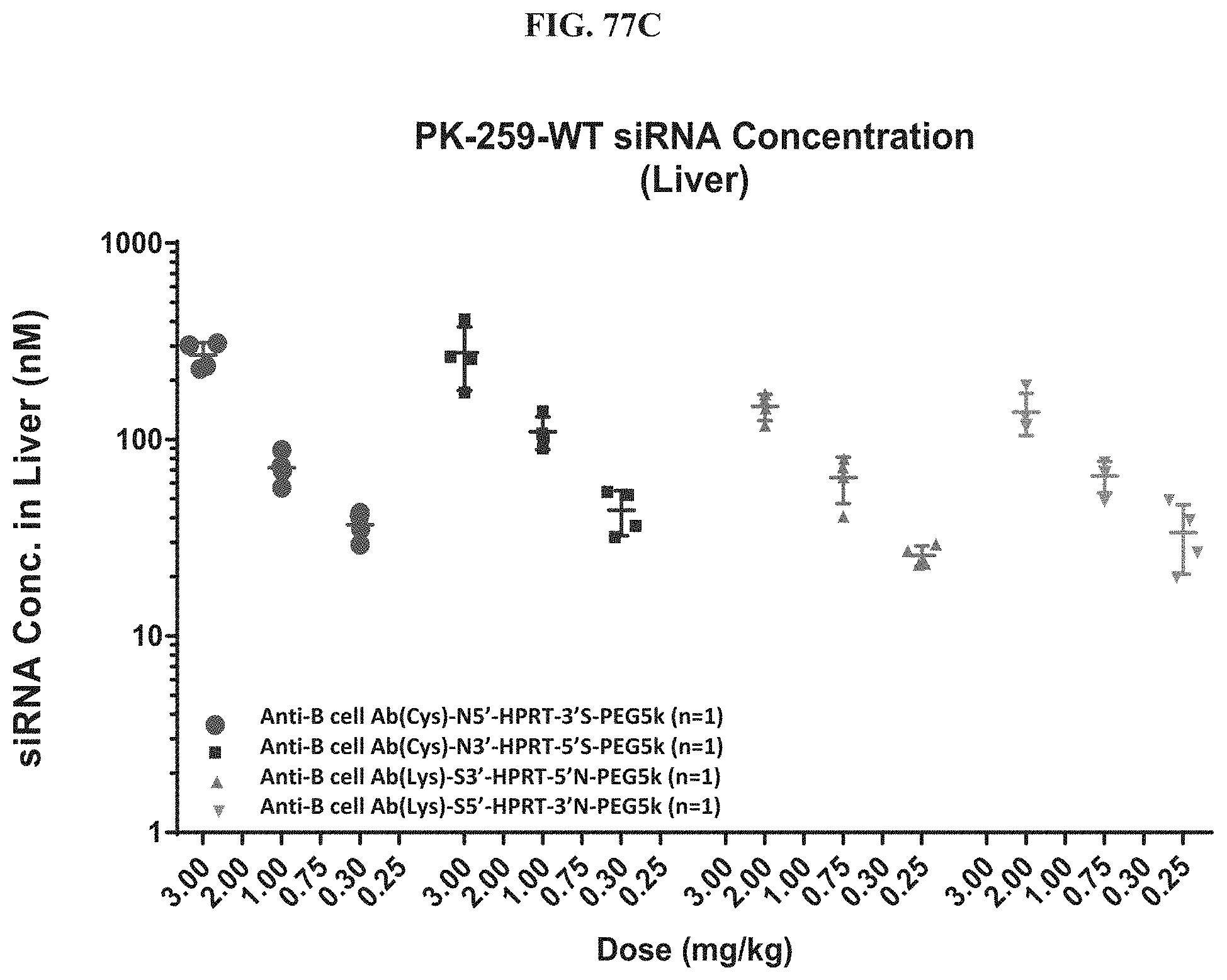
D00110

D00111

D00112

D00113
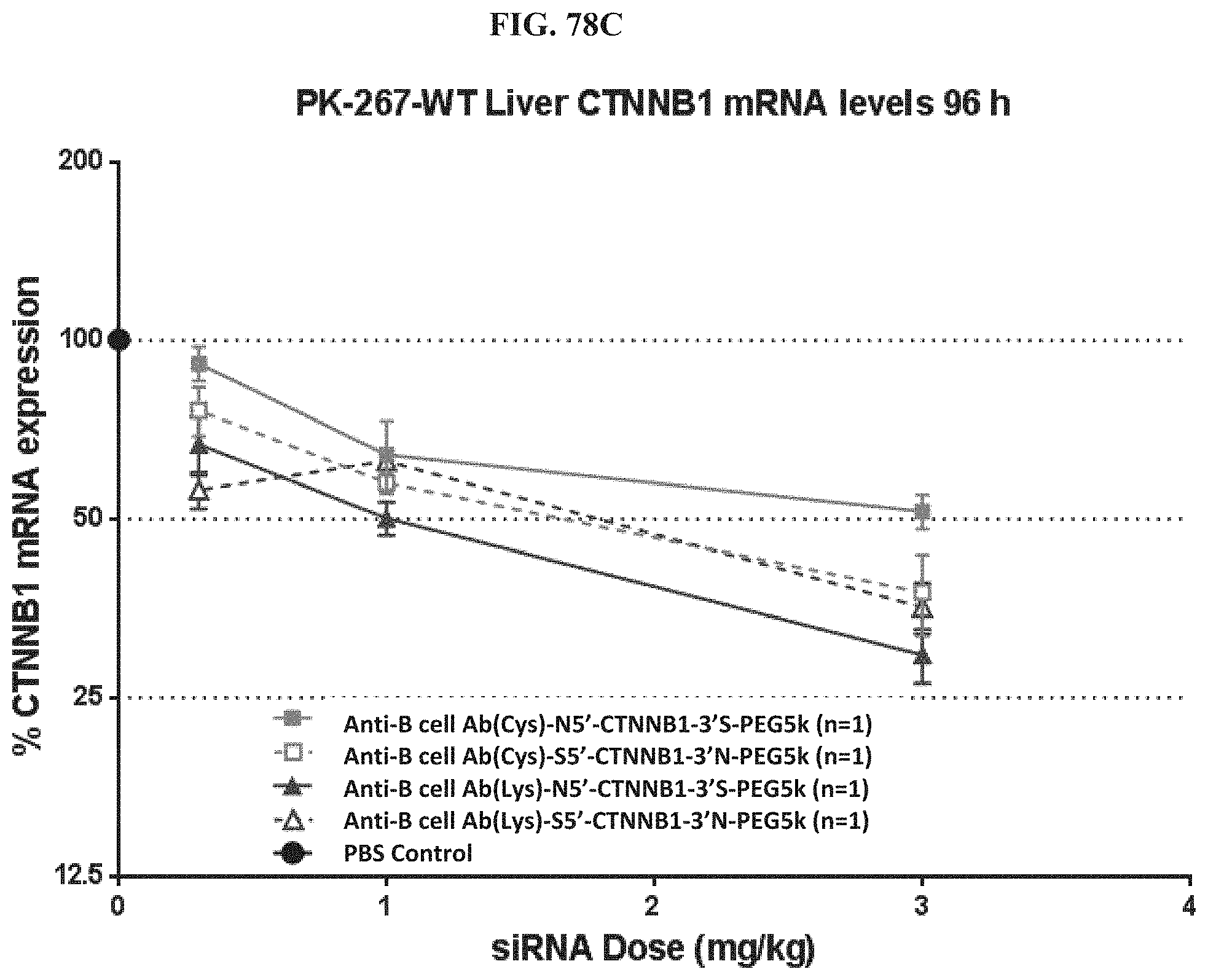
D00114
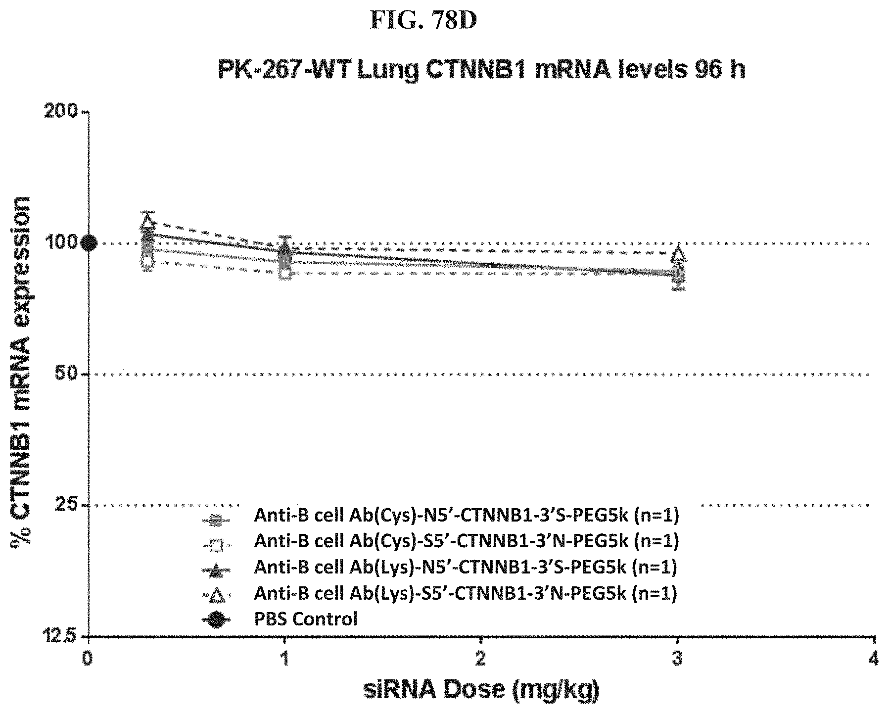
D00115
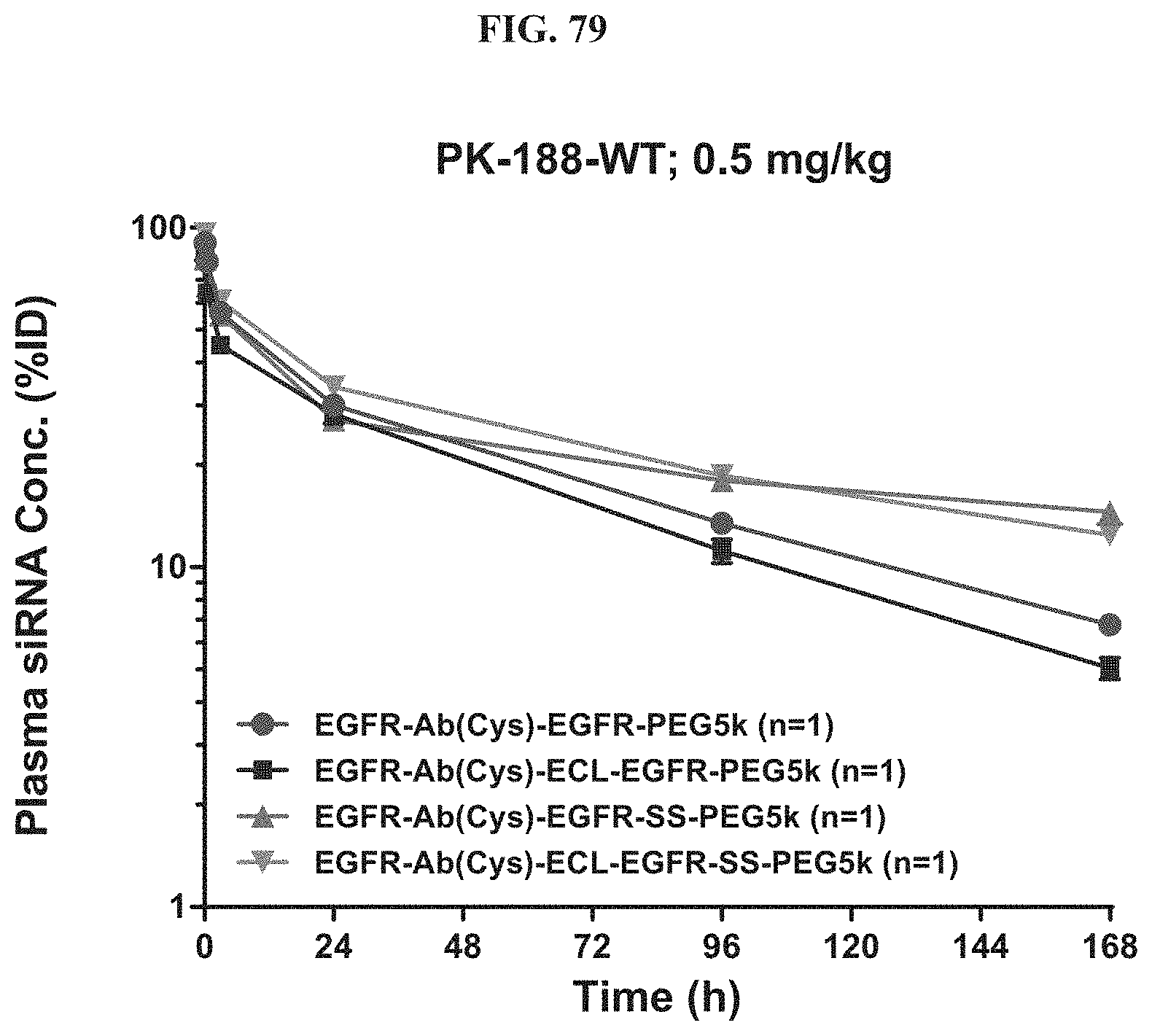
D00116

D00117
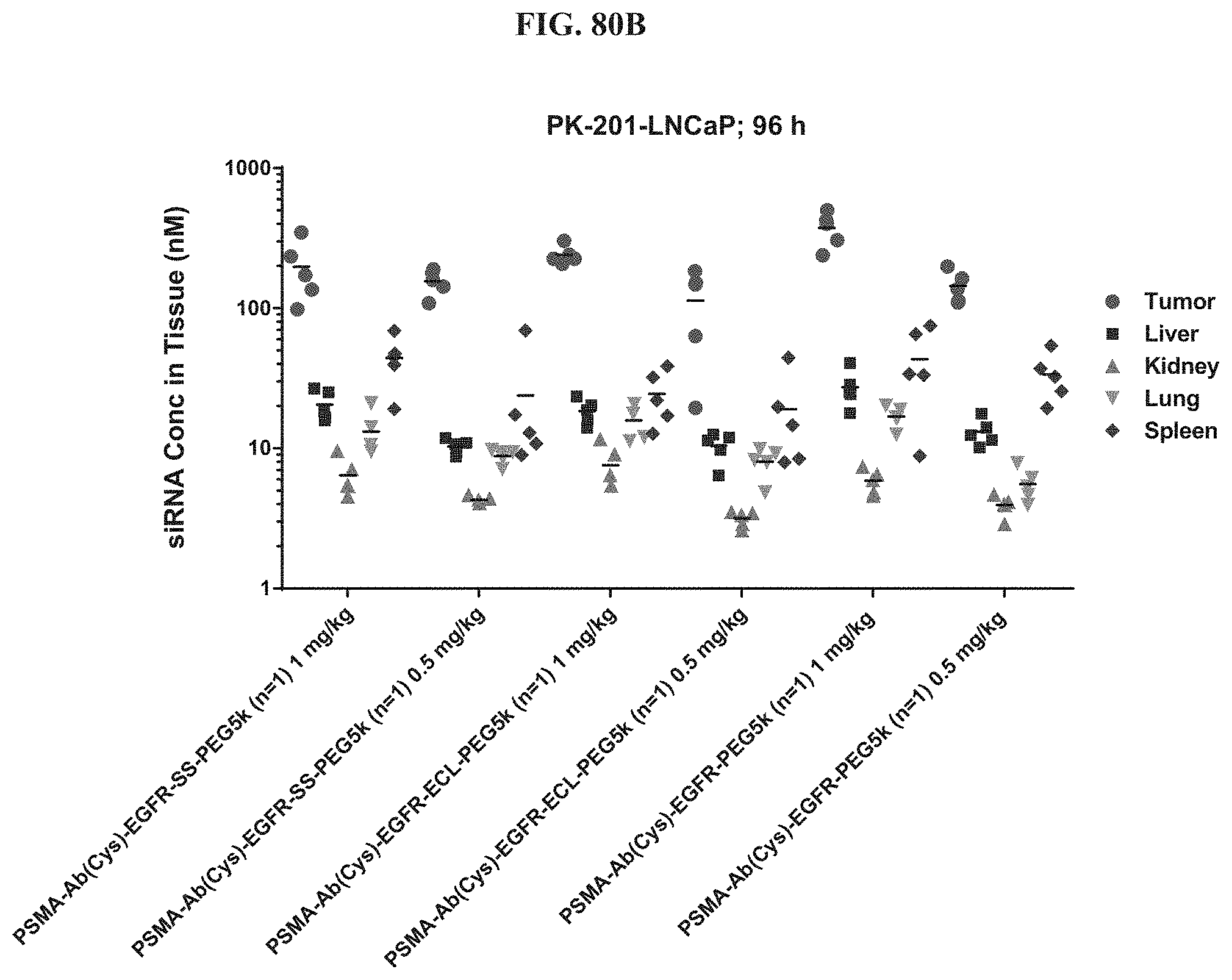
D00118
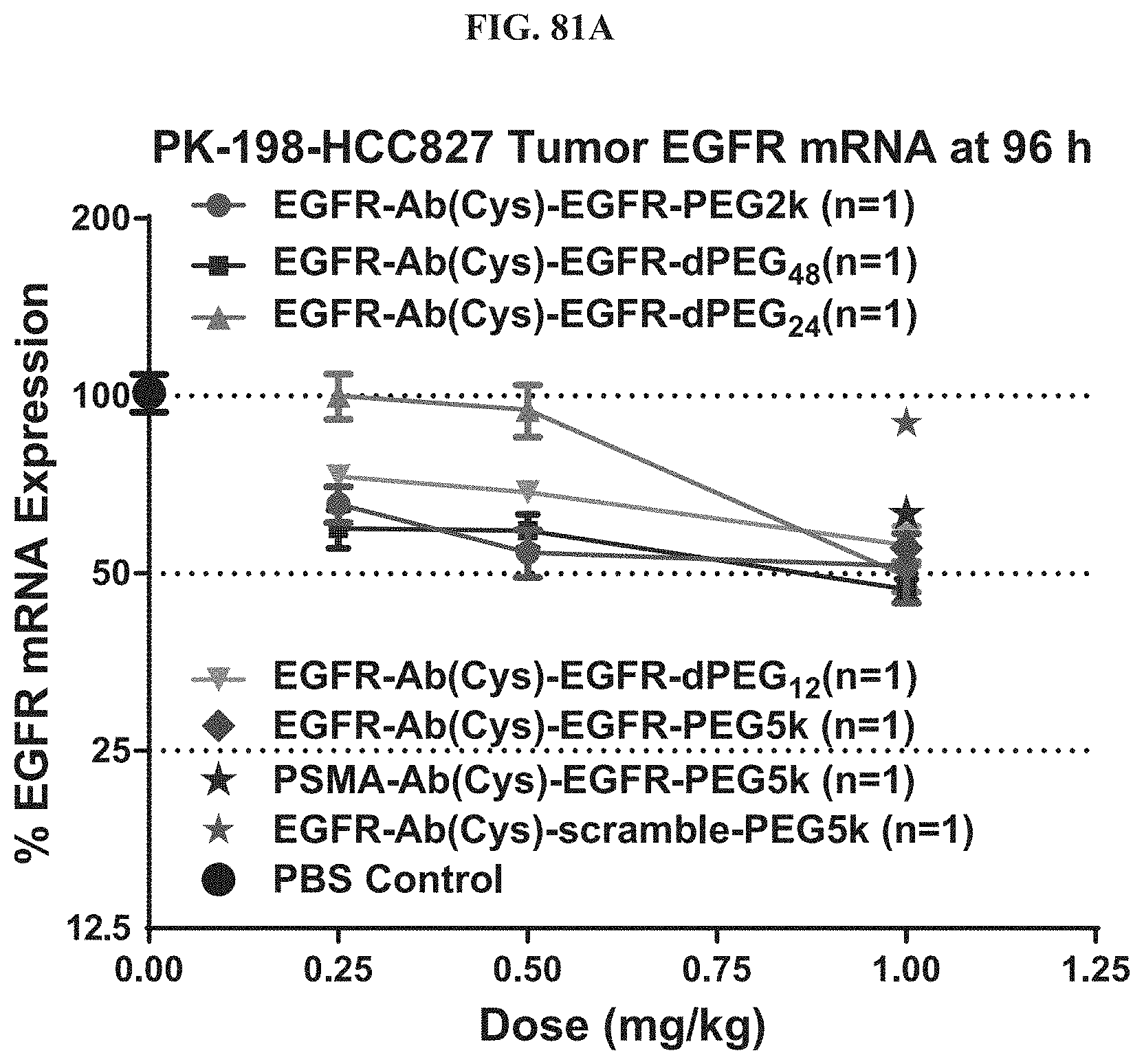
D00119

D00120
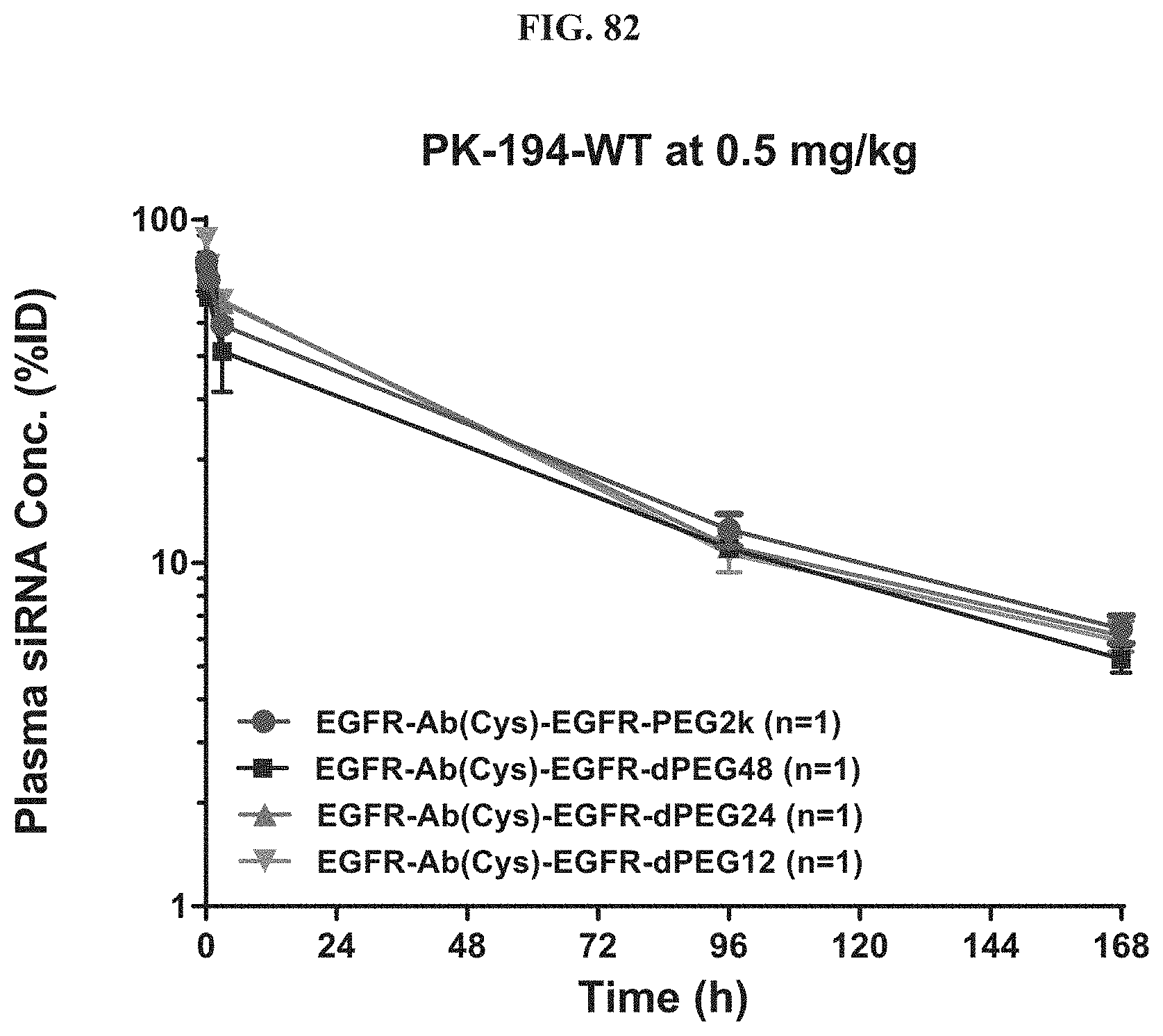
D00121

D00122
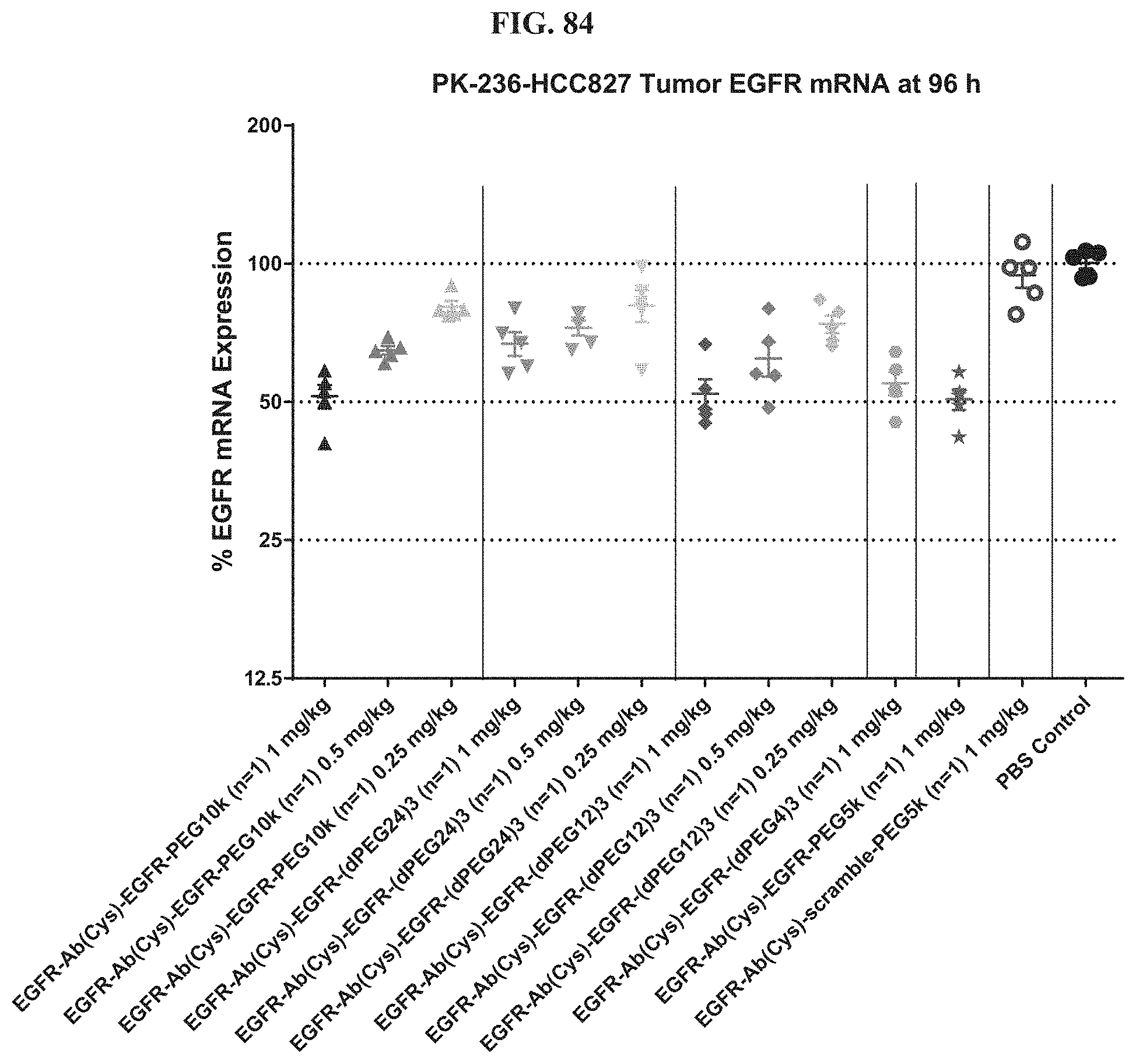
D00123

D00124
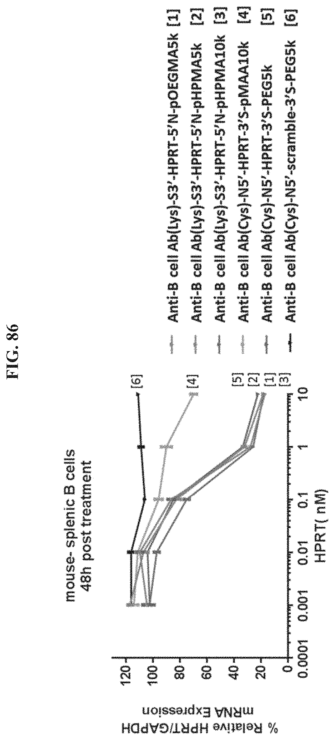
D00125
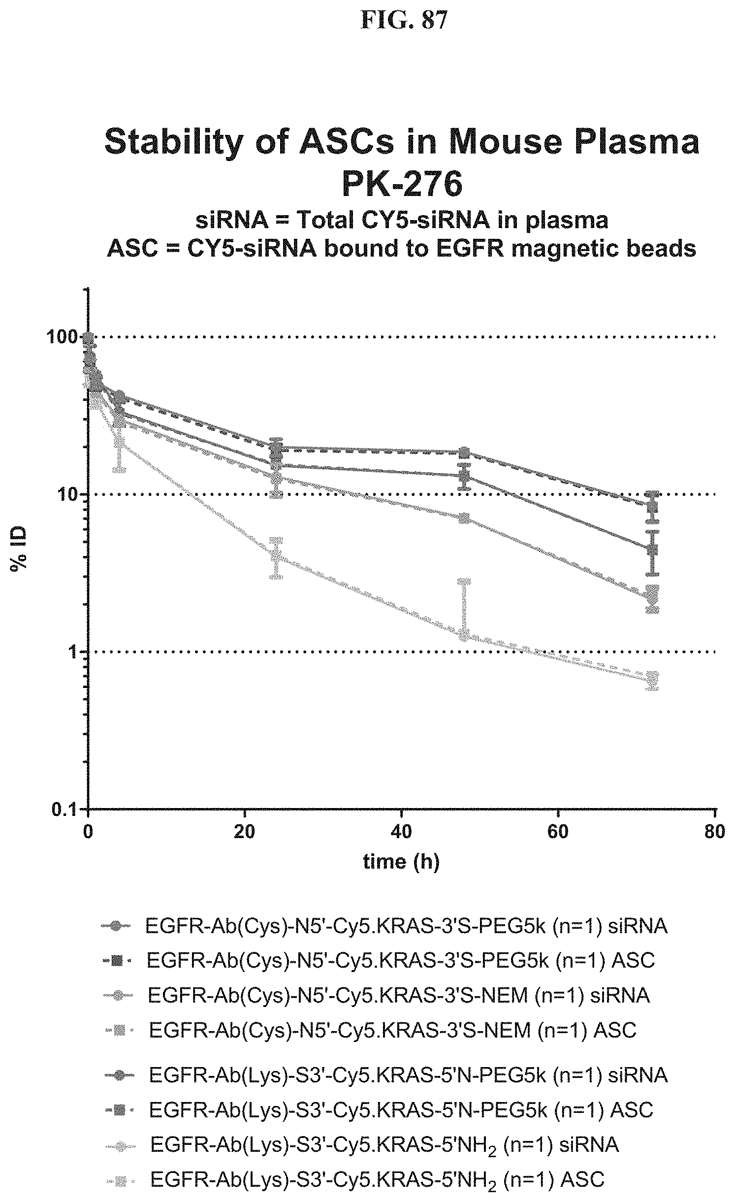
D00126
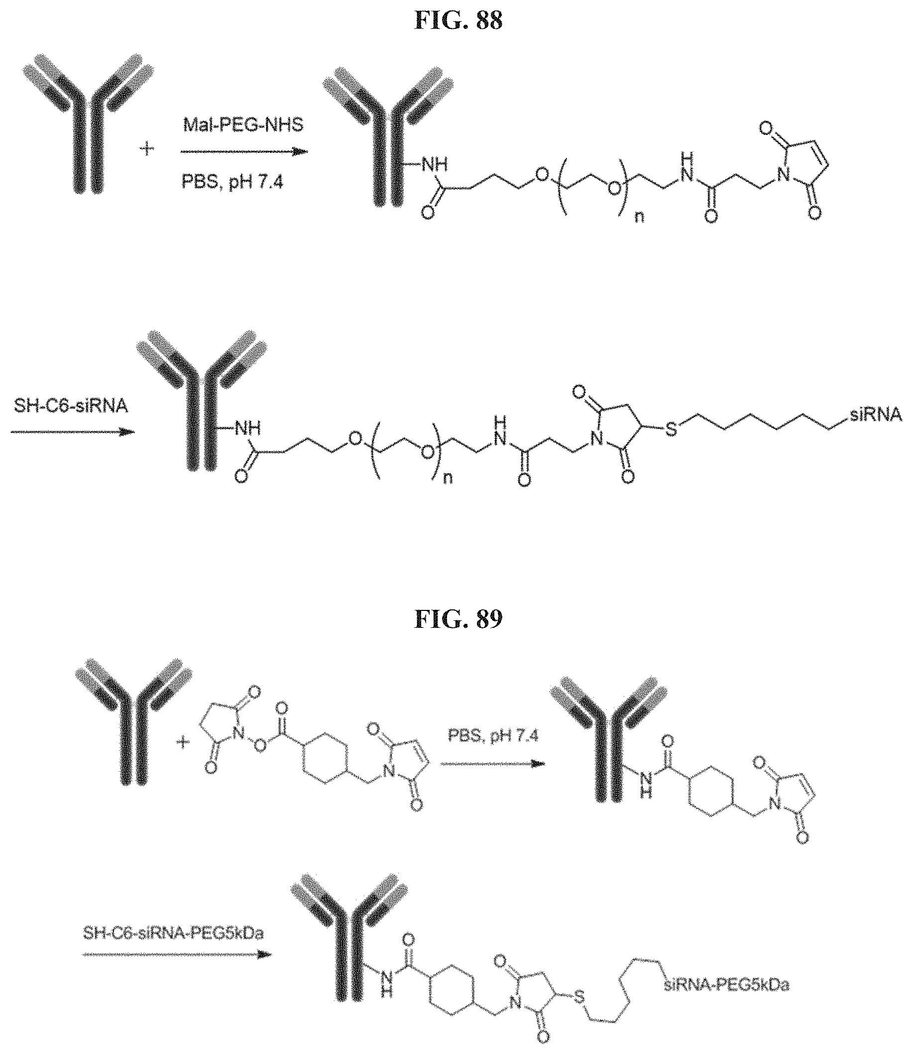
D00127

D00128

D00129
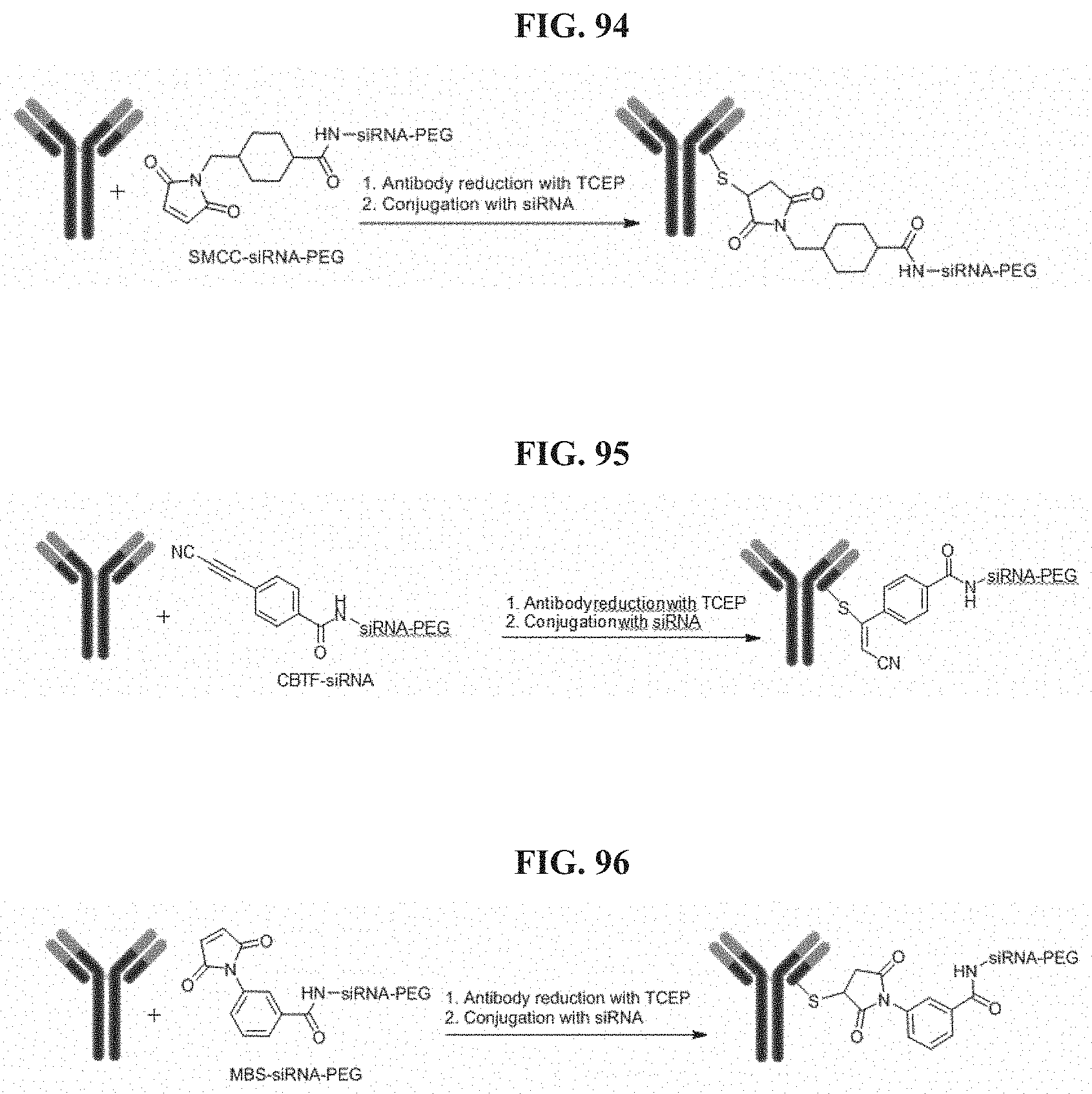
D00130
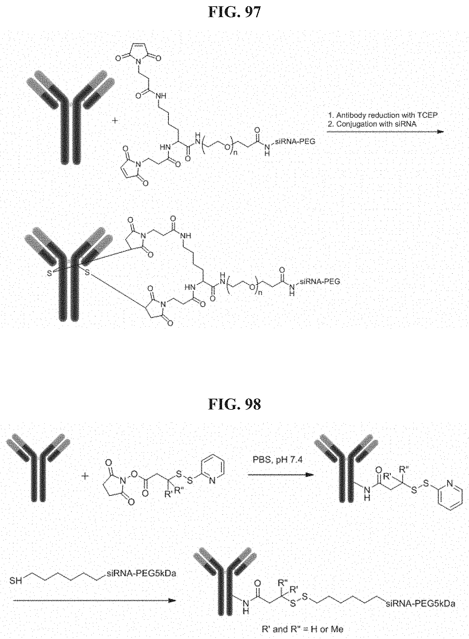
D00131
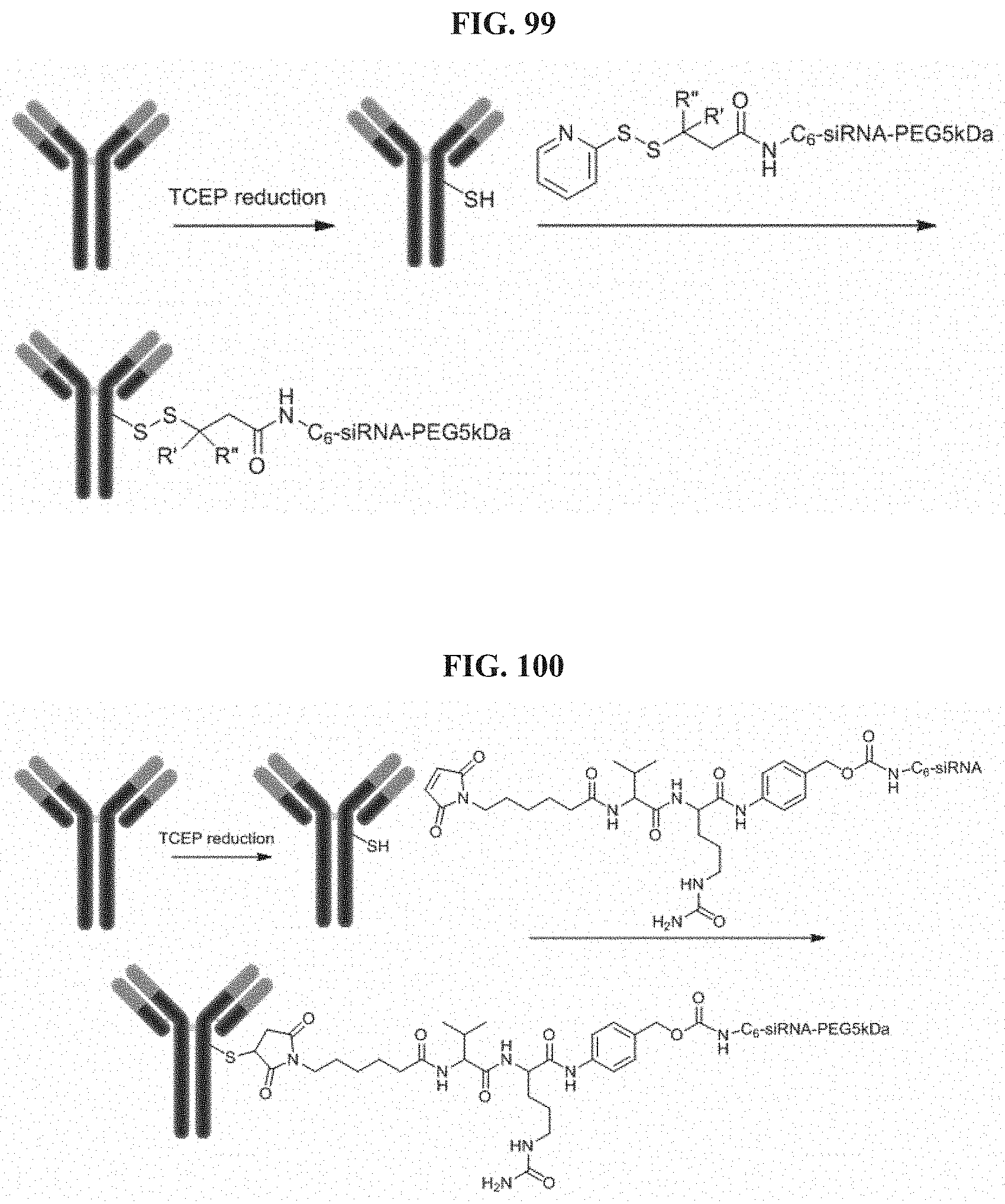
D00132
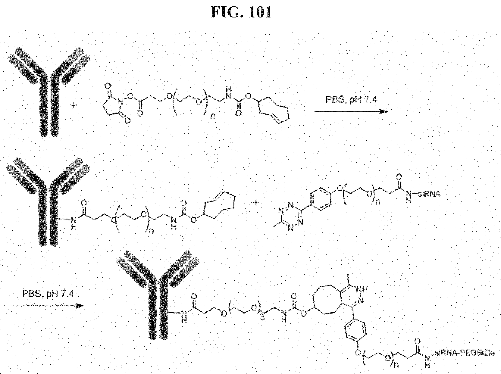
D00133
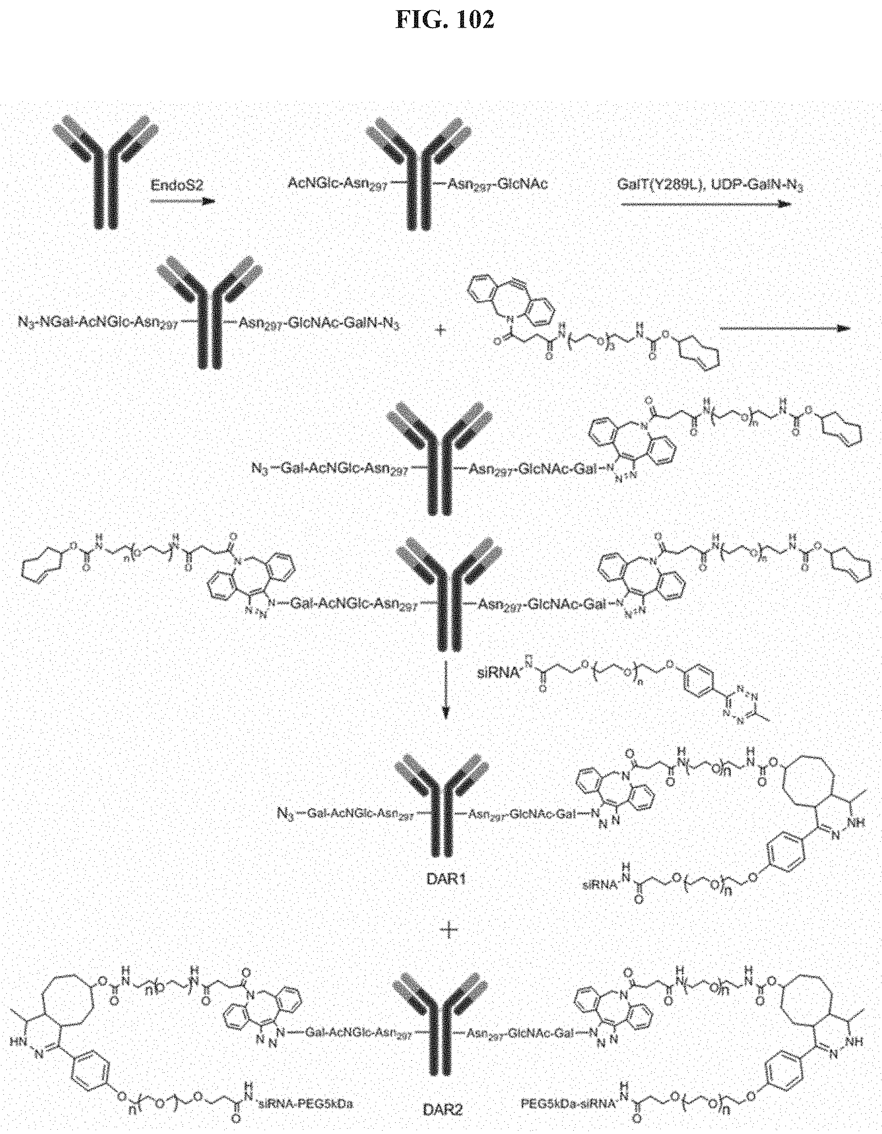
D00134
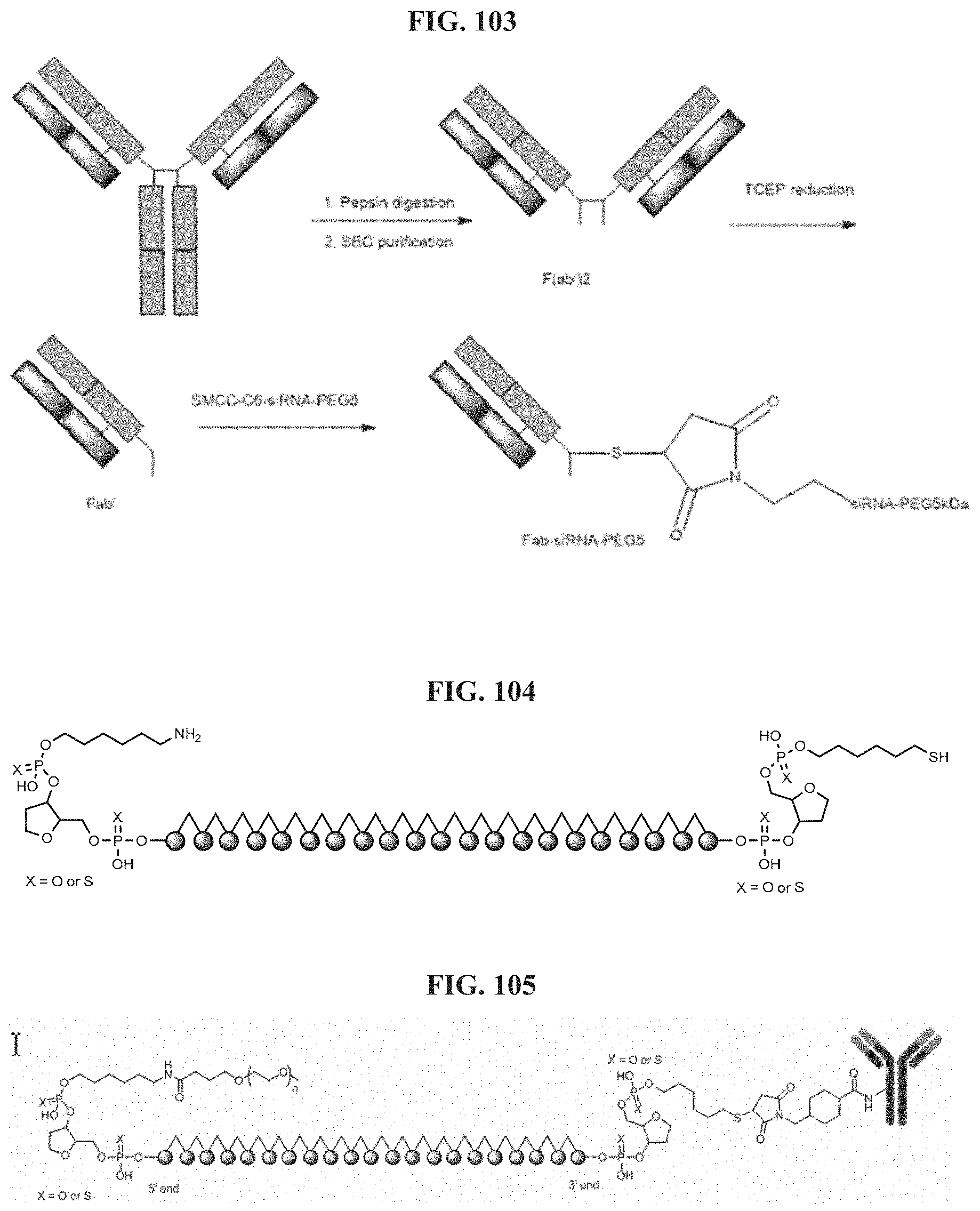
D00135

D00136

S00001
XML
uspto.report is an independent third-party trademark research tool that is not affiliated, endorsed, or sponsored by the United States Patent and Trademark Office (USPTO) or any other governmental organization. The information provided by uspto.report is based on publicly available data at the time of writing and is intended for informational purposes only.
While we strive to provide accurate and up-to-date information, we do not guarantee the accuracy, completeness, reliability, or suitability of the information displayed on this site. The use of this site is at your own risk. Any reliance you place on such information is therefore strictly at your own risk.
All official trademark data, including owner information, should be verified by visiting the official USPTO website at www.uspto.gov. This site is not intended to replace professional legal advice and should not be used as a substitute for consulting with a legal professional who is knowledgeable about trademark law.