Translation catheters, systems, and methods of use thereof
Goodwin , et al.
U.S. patent number 10,682,232 [Application Number 15/642,963] was granted by the patent office on 2020-06-16 for translation catheters, systems, and methods of use thereof. This patent grant is currently assigned to Edwards Lifesciences Corporation. The grantee listed for this patent is Edwards Lifesciences Corporation. Invention is credited to Kate Cutuli, Nareak Douk, Jonathan Goodwin, Matt Guimond, Morgan House, Christopher Lee, Richard Morrill, Michael Sutherland.


View All Diagrams
| United States Patent | 10,682,232 |
| Goodwin , et al. | June 16, 2020 |
Translation catheters, systems, and methods of use thereof
Abstract
The present teachings generally relate to translation catheter systems and method of using thereof in treating defective mitral valves. Specifically, a translation catheter system includes a catheter configured to move substantially laterally along the mitral annulus and the lateral movement of the catheter is substantially continuous and substantially adjustable. Accordingly, a method of using such a translation catheter includes advancing a first wire across the mitral annulus at a first treatment location, delivering a catheter at or near the first treatment location, moving the catheter substantially laterally to a second treatment location, and advancing a second wire across the mitral annulus, where the distance between the first and second treatment locations is substantially adjustable and optionally visualized.
| Inventors: | Goodwin; Jonathan (Nashua, NH), Sutherland; Michael (Pelham, NH), House; Morgan (Newfields, NH), Morrill; Richard (North Billerica, MA), Guimond; Matt (Salem, NH), Lee; Christopher (Tewksbury, MA), Cutuli; Kate (Salem, NH), Douk; Nareak (Lowell, MA) | ||||||||||
|---|---|---|---|---|---|---|---|---|---|---|---|
| Applicant: |
|
||||||||||
| Assignee: | Edwards Lifesciences
Corporation (Irvine, CA) |
||||||||||
| Family ID: | 51530274 | ||||||||||
| Appl. No.: | 15/642,963 | ||||||||||
| Filed: | July 6, 2017 |
Prior Publication Data
| Document Identifier | Publication Date | |
|---|---|---|
| US 20180125659 A1 | May 10, 2018 | |
Related U.S. Patent Documents
| Application Number | Filing Date | Patent Number | Issue Date | ||
|---|---|---|---|---|---|
| 14210967 | Mar 14, 2014 | 9724195 | |||
| 61786373 | Mar 15, 2013 | ||||
| Current U.S. Class: | 1/1 |
| Current CPC Class: | A61F 2/2466 (20130101); A61M 25/09041 (20130101); A61M 25/0147 (20130101); A61M 25/0133 (20130101); A61M 2025/018 (20130101); A61M 2025/015 (20130101) |
| Current International Class: | A61M 25/01 (20060101); A61F 2/24 (20060101); A61M 25/09 (20060101) |
References Cited [Referenced By]
U.S. Patent Documents
| 3604488 | September 1971 | Wishart et al. |
| 3656185 | April 1972 | Carpentier |
| 3840018 | October 1974 | Heifetz |
| 3881366 | May 1975 | Bradley et al. |
| 3898701 | August 1975 | La Russa |
| 4042979 | August 1977 | Angell |
| 4118805 | October 1978 | Reimels |
| 4214349 | July 1980 | Munch |
| 4245624 | January 1981 | Komiya |
| 4261342 | April 1981 | Aranguren Duo |
| 4290151 | September 1981 | Massana |
| 4434828 | March 1984 | Trincia |
| 4473928 | October 1984 | Johnson |
| 4602911 | July 1986 | Ahmadi et al. |
| 4625727 | December 1986 | Leiboff |
| 4712549 | December 1987 | Peters et al. |
| 4769005 | September 1988 | Ginsburg et al. |
| 4778468 | October 1988 | Hunt et al. |
| 4881524 | November 1989 | Boebel |
| 4917698 | April 1990 | Carpentier et al. |
| 4961738 | October 1990 | Mackin |
| 5042707 | August 1991 | Taheri |
| 5061277 | October 1991 | Carpentier et al. |
| 5064431 | November 1991 | Gilbertson et al. |
| 5104407 | April 1992 | Lam et al. |
| 5108420 | April 1992 | Marks |
| 5201880 | April 1993 | Wright et al. |
| 5258008 | November 1993 | Wilk |
| 5300034 | April 1994 | Behnke et al. |
| 5325845 | July 1994 | Adair |
| 5346498 | September 1994 | Greelis et al. |
| 5383852 | January 1995 | Stevens-Wright |
| 5415656 | May 1995 | Tihon |
| 5449368 | September 1995 | Kuzmak |
| 5450860 | September 1995 | O'Connor |
| 5464404 | November 1995 | Abela et al. |
| 5474518 | December 1995 | Farrer Velazquez |
| 5477856 | December 1995 | Lundquist |
| 5593424 | January 1997 | Northrup, III |
| 5601572 | February 1997 | Middleman et al. |
| 5626609 | May 1997 | Zvenyatsky et al. |
| 5643317 | July 1997 | Pavcnik et al. |
| 5649908 | July 1997 | Itoh |
| 5669919 | September 1997 | Sanders et al. |
| 5676653 | October 1997 | Taylor et al. |
| 5683402 | November 1997 | Cosgrove et al. |
| 5687723 | November 1997 | Avitall |
| 5693059 | December 1997 | Yoon |
| 5702397 | December 1997 | Goble et al. |
| 5702398 | December 1997 | Tarabishy |
| 5709695 | January 1998 | Northrup, III |
| 5716370 | February 1998 | Williamson, IV et al. |
| 5716397 | February 1998 | Myers |
| 5728116 | March 1998 | Rosenman |
| 5730150 | March 1998 | Peppel et al. |
| 5749371 | May 1998 | Zadini et al. |
| 5782844 | July 1998 | Yoon et al. |
| 5810882 | September 1998 | Bolduc et al. |
| 5824066 | October 1998 | Gross |
| 5830221 | November 1998 | Stein et al. |
| 5843120 | December 1998 | Israel et al. |
| 5855614 | January 1999 | Stevens et al. |
| 5876373 | March 1999 | Giba et al. |
| 5879295 | March 1999 | Li |
| 5935098 | August 1999 | Blaisdell et al. |
| 5957953 | September 1999 | DiPoto et al. |
| 5961440 | October 1999 | Schweich, Jr. et al. |
| 5961539 | October 1999 | Northrup, III et al. |
| 5984959 | November 1999 | Robertson et al. |
| 6042554 | March 2000 | Rosenman et al. |
| 6045497 | April 2000 | Schweich, Jr. et al. |
| 6048329 | April 2000 | Thompson |
| 6050936 | April 2000 | Schweich, Jr. et al. |
| 6059715 | May 2000 | Schweich, Jr. et al. |
| 6071279 | June 2000 | Whayne |
| 6074341 | June 2000 | Anderson et al. |
| 6074401 | June 2000 | Gardiner et al. |
| 6074417 | June 2000 | Peredo |
| 6086582 | July 2000 | Altman et al. |
| 6102945 | August 2000 | Campbell |
| 6106550 | August 2000 | Magovern et al. |
| 6110200 | August 2000 | Hinnenkamp |
| 6132390 | October 2000 | Cookston et al. |
| 6143024 | November 2000 | Campbell et al. |
| 6159240 | December 2000 | Sparer et al. |
| 6165119 | December 2000 | Schweich, Jr. et al. |
| 6174332 | January 2001 | Loch et al. |
| 6183411 | February 2001 | Mortier et al. |
| 6187040 | February 2001 | Wright |
| 6203525 | March 2001 | Whayne |
| 6210347 | April 2001 | Forsell |
| 6217528 | April 2001 | Koblish |
| 6217610 | April 2001 | Carpentier et al. |
| 6231602 | May 2001 | Carpentier et al. |
| 6251092 | June 2001 | Qin et al. |
| 6296656 | October 2001 | Bolduc et al. |
| 6315784 | November 2001 | Djurovic |
| 6319281 | November 2001 | Patel |
| 6328746 | December 2001 | Gambale |
| 6332880 | December 2001 | Yang |
| 6332893 | December 2001 | Mortier et al. |
| 6355030 | March 2002 | Aldrich et al. |
| 6361559 | March 2002 | Houser et al. |
| 6368348 | April 2002 | Gabbay |
| 6402780 | June 2002 | Williamson, IV et al. |
| 6406420 | June 2002 | McCarthy et al. |
| 6406493 | June 2002 | Tu et al. |
| 6419696 | July 2002 | Ortiz et al. |
| 6451054 | September 2002 | Stevens |
| 6458076 | October 2002 | Pruitt |
| 6461366 | October 2002 | Seguin |
| 6470892 | October 2002 | Forsell |
| 6503274 | January 2003 | Howanec, Jr. et al. |
| 6524338 | February 2003 | Gundry |
| 6530952 | March 2003 | Veseiy |
| 6533772 | March 2003 | Sherts et al. |
| 6537314 | March 2003 | Langberg et al. |
| 6542781 | April 2003 | Koblish |
| 6547801 | April 2003 | Dargent et al. |
| 6554845 | April 2003 | Fleenor et al. |
| 6564805 | May 2003 | Garrison et al. |
| 6565603 | May 2003 | Cox |
| 6569198 | May 2003 | Wilson et al. |
| 6579297 | June 2003 | Bicek et al. |
| 6589160 | July 2003 | Schweich, Jr. et al. |
| 6592593 | July 2003 | Parodi et al. |
| 6602288 | August 2003 | Cosgrove et al. |
| 6602289 | August 2003 | Colvin et al. |
| 6613046 | September 2003 | Jenkins |
| 6613078 | September 2003 | Barone |
| 6613079 | September 2003 | Wolinsky et al. |
| 6619291 | September 2003 | Hlavka et al. |
| 6626899 | September 2003 | Houser et al. |
| 6626917 | September 2003 | Craig |
| 6626930 | September 2003 | Allen et al. |
| 6629534 | October 2003 | St. Goar et al. |
| 6629921 | October 2003 | Schweich, Jr. et al. |
| 6632184 | October 2003 | Truwit |
| 6645195 | November 2003 | Bhat |
| 6651671 | November 2003 | Donlon et al. |
| 6652556 | November 2003 | VanTassel et al. |
| 6682558 | January 2004 | Tu et al. |
| 6689125 | February 2004 | Keith et al. |
| 6689164 | February 2004 | Seguin |
| 6695866 | February 2004 | Kuehn et al. |
| 6702826 | March 2004 | Liddicoat et al. |
| 6702846 | March 2004 | Mikus et al. |
| 6706065 | March 2004 | Langberg et al. |
| 6709385 | March 2004 | Forsell |
| 6709456 | March 2004 | Langberg et al. |
| 6711444 | March 2004 | Koblish |
| 6719786 | April 2004 | Ryan et al. |
| 6723038 | April 2004 | Schroeder et al. |
| 6726716 | April 2004 | Marquez |
| 6726717 | April 2004 | Alfieri et al. |
| 6730121 | May 2004 | Ortiz et al. |
| 6749630 | June 2004 | McCarthy et al. |
| 6752813 | June 2004 | Goldfarb et al. |
| 6764310 | July 2004 | Ichihashi et al. |
| 6764510 | July 2004 | Vidlund et al. |
| 6764810 | July 2004 | Ma et al. |
| 6770083 | August 2004 | Seguin |
| 6786924 | September 2004 | Ryan et al. |
| 6786925 | September 2004 | Schoon et al. |
| 6790231 | September 2004 | Liddicoat et al. |
| 6797001 | September 2004 | Mathis et al. |
| 6797002 | September 2004 | Spence et al. |
| 6802319 | October 2004 | Stevens et al. |
| 6805710 | October 2004 | Bolling et al. |
| 6805711 | October 2004 | Quijano et al. |
| 6855126 | February 2005 | Flinchbaugh |
| 6858039 | February 2005 | McCarthy |
| 6884250 | April 2005 | Monassevitch et al. |
| 6893459 | May 2005 | Macoviak |
| 6908478 | June 2005 | Alferness et al. |
| 6908482 | June 2005 | McCarthy et al. |
| 6916306 | July 2005 | Jenkins |
| 6918917 | July 2005 | Nguyen et al. |
| 6926730 | August 2005 | Nguyen et al. |
| 6960217 | November 2005 | Bolduc |
| 6964684 | November 2005 | Ortiz et al. |
| 6964686 | November 2005 | Gordon |
| 6976995 | December 2005 | Mathis et al. |
| 6986775 | January 2006 | Morales et al. |
| 6989028 | January 2006 | Lashinski et al. |
| 6997951 | February 2006 | Solem et al. |
| 7004176 | February 2006 | Lau |
| 7007798 | March 2006 | Happonen et al. |
| 7011669 | March 2006 | Kimblad |
| 7011682 | March 2006 | Lashinski et al. |
| 7018406 | March 2006 | Seguin et al. |
| 7037334 | May 2006 | Hlavka et al. |
| 7087064 | June 2006 | Hyde |
| 7077850 | July 2006 | Kortenbach |
| 7077862 | July 2006 | Vidlund et al. |
| 7101395 | September 2006 | Tremulis et al. |
| 7101396 | September 2006 | Artof et al. |
| 7112207 | September 2006 | Allen et al. |
| 7118595 | October 2006 | Ryan et al. |
| 7125421 | October 2006 | Tremulis et al. |
| 7150737 | December 2006 | Purdy et al. |
| 7159593 | January 2007 | McCarthy et al. |
| 7166127 | January 2007 | Spence et al. |
| 7169187 | January 2007 | Datta et al. |
| 7172625 | February 2007 | Shu et al. |
| 7175660 | February 2007 | Cartledge et al. |
| 7186262 | March 2007 | Saadat |
| 7186264 | March 2007 | Liddicoat et al. |
| 7189199 | March 2007 | McCarthy et al. |
| 7192443 | March 2007 | Solem et al. |
| 7220277 | May 2007 | Arru et al. |
| 7226467 | June 2007 | Lucatero et al. |
| 7226477 | June 2007 | Cox |
| 7226647 | June 2007 | Kasperchik et al. |
| 7229452 | June 2007 | Kayan |
| 7238191 | July 2007 | Bachmann |
| 7288097 | October 2007 | Seguin |
| 7294148 | November 2007 | McCarthy |
| 7311728 | December 2007 | Solem et al. |
| 7311729 | December 2007 | Mathis et al. |
| 7314485 | January 2008 | Mathis |
| 7316710 | January 2008 | Cheng et al. |
| 7329279 | February 2008 | Haug et al. |
| 7329280 | February 2008 | Bolling et al. |
| 7335213 | February 2008 | Hyde et al. |
| 7361190 | April 2008 | Shaoulian et al. |
| 7364588 | April 2008 | Mathis et al. |
| 7377941 | May 2008 | Rhee et al. |
| 7390329 | June 2008 | Westra et al. |
| 7404824 | July 2008 | Webler et al. |
| 7431692 | October 2008 | Zollinger et al. |
| 7442207 | October 2008 | Rafiee |
| 7452376 | November 2008 | Lim et al. |
| 7455690 | November 2008 | Cartledge et al. |
| 7485142 | February 2009 | Milo |
| 7485143 | February 2009 | Webler et al. |
| 7500989 | March 2009 | Solem et al. |
| 7507252 | March 2009 | Lashinski et al. |
| 7510575 | March 2009 | Spenser et al. |
| 7510577 | March 2009 | Moaddeb et al. |
| 7527647 | May 2009 | Spence |
| 7530995 | May 2009 | Quijano et al. |
| 7549983 | June 2009 | Roue et al. |
| 7559936 | July 2009 | Levine |
| 7562660 | July 2009 | Saadat |
| 7563267 | July 2009 | Goldfarb et al. |
| 7563273 | July 2009 | Goldfarb et al. |
| 7569062 | August 2009 | Kuehn et al. |
| 7585321 | September 2009 | Cribier |
| 7588562 | September 2009 | Starksen et al. |
| 7591326 | September 2009 | Alferness et al. |
| 7604646 | October 2009 | Goldfarb et al. |
| 7608091 | October 2009 | Goldfarb et al. |
| 7608103 | October 2009 | McCarthy |
| 7625403 | December 2009 | Krivoruchko |
| 7632303 | December 2009 | Stalker et al. |
| 7635329 | December 2009 | Goldfarb et al. |
| 7635386 | December 2009 | Gammie |
| 7655015 | February 2010 | Goldfarb et al. |
| 7666204 | February 2010 | Thornton et al. |
| 7682319 | March 2010 | Martin et al. |
| 7682369 | March 2010 | Seguin |
| 7686822 | March 2010 | Shayani |
| 7699892 | April 2010 | Rafiee et al. |
| 7704269 | April 2010 | St. Goar et al. |
| 7704277 | April 2010 | Zakay et al. |
| 7713278 | May 2010 | Hess et al. |
| 7722666 | May 2010 | Lafontaine |
| 7736388 | June 2010 | Goldfarb et al. |
| 7748389 | July 2010 | Salahieh et al. |
| 7753924 | July 2010 | Starksen et al. |
| 7758632 | July 2010 | Hojeibane et al. |
| 7780726 | August 2010 | Seguin |
| 7824443 | November 2010 | Salahieh et al. |
| 7871368 | January 2011 | Zollinger et al. |
| 7871433 | January 2011 | Lattouf |
| 7883475 | February 2011 | Dupont et al. |
| 7883538 | February 2011 | To et al. |
| 7892281 | February 2011 | Seguin et al. |
| 7927370 | April 2011 | Webier et al. |
| 7927371 | April 2011 | Navia et al. |
| 7942927 | May 2011 | Kaye et al. |
| 7947056 | May 2011 | Griego et al. |
| 7955315 | June 2011 | Feinberg et al. |
| 7955377 | June 2011 | Melsheirner |
| 7992567 | August 2011 | Hirotsuka et al. |
| 7993368 | August 2011 | Gambale et al. |
| 7993397 | August 2011 | Lashinski et al. |
| 8012201 | September 2011 | Lashinski et al. |
| 8034103 | October 2011 | Burriesci et al. |
| 8052592 | November 2011 | Goldfarb et al. |
| 8057493 | November 2011 | Goldfarb et al. |
| 8062355 | November 2011 | Figulla et al. |
| 8070804 | December 2011 | Hyde et al. |
| 8070805 | December 2011 | Vidlund et al. |
| 8075616 | December 2011 | Solem et al. |
| 8100964 | January 2012 | Spence |
| 8123801 | February 2012 | Milo |
| 8142493 | March 2012 | Spence et al. |
| 8142495 | March 2012 | Hasenkam et al. |
| 8142496 | March 2012 | Berreklouw |
| 8147542 | April 2012 | Maisano et al. |
| 8152844 | April 2012 | Rao et al. |
| 8163013 | April 2012 | Machold et al. |
| 8187299 | May 2012 | Goldfarb et al. |
| 8187324 | May 2012 | Webler et al. |
| 8202315 | June 2012 | Hlavka et al. |
| 8206439 | June 2012 | Gomez Duran |
| 8216302 | July 2012 | Wilson et al. |
| 8231671 | July 2012 | Kim |
| 8262725 | September 2012 | Subramanian |
| 8265758 | September 2012 | Policker et al. |
| 8277502 | October 2012 | Miller et al. |
| 8287584 | October 2012 | Salahieh et al. |
| 8287591 | October 2012 | Keidar et al. |
| 8292884 | October 2012 | Levine et al. |
| 8303608 | November 2012 | Goldfarb et al. |
| 8323334 | December 2012 | Deem et al. |
| 8328868 | December 2012 | Paul et al. |
| 8333777 | December 2012 | Schaller et al. |
| 8343173 | January 2013 | Starksen et al. |
| 8343174 | January 2013 | Goldfarb et al. |
| 8343213 | January 2013 | Salahieh et al. |
| 8349002 | January 2013 | Milo |
| 8353956 | January 2013 | Miller et al. |
| 8357195 | January 2013 | Kuehn |
| 8382829 | February 2013 | Call et al. |
| 8388680 | March 2013 | Starksen et al. |
| 8393517 | March 2013 | Milo |
| 8419825 | April 2013 | Burgler et al. |
| 8430926 | April 2013 | Kirson |
| 8449573 | May 2013 | Chu |
| 8449599 | May 2013 | Chau et al. |
| 8454686 | June 2013 | Alkhatib |
| 8460370 | June 2013 | Zakay |
| 8460371 | June 2013 | Hlavka et al. |
| 8475491 | July 2013 | Milo |
| 8475525 | July 2013 | Maisano et al. |
| 8480732 | July 2013 | Subramanian |
| 8518107 | August 2013 | Tsukashima et al. |
| 8523940 | September 2013 | Richardson et al. |
| 8551161 | October 2013 | Dolan |
| 8585755 | November 2013 | Chau et al. |
| 8591576 | November 2013 | Hasenkam et al. |
| 8602970 | December 2013 | Muyari |
| 8608797 | December 2013 | Gross et al. |
| 8628569 | January 2014 | Benichou et al. |
| 8628571 | January 2014 | Hacohen et al. |
| 8641727 | February 2014 | Starksen et al. |
| 8652202 | February 2014 | Alon et al. |
| 8652203 | February 2014 | Quadri et al. |
| 8679174 | March 2014 | Ottma et al. |
| 8685086 | April 2014 | Navia et al. |
| 8728097 | May 2014 | Sugimoto et al. |
| 8728155 | May 2014 | Montorfano et al. |
| 8734467 | May 2014 | Miller et al. |
| 8734699 | May 2014 | Heideman et al. |
| 8740920 | June 2014 | Goldfarb et al. |
| 8747463 | June 2014 | Fogarty et al. |
| 8778021 | July 2014 | Cartledge |
| 8784481 | July 2014 | Alkhatib et al. |
| 8790367 | July 2014 | Nguyen et al. |
| 8790394 | July 2014 | Miller et al. |
| 8795298 | August 2014 | Hernlund et al. |
| 8795355 | August 2014 | Alkhatib |
| 8795356 | August 2014 | Quadri et al. |
| 8795357 | August 2014 | Yohanan et al. |
| 8808366 | August 2014 | Braido et al. |
| 8808368 | August 2014 | Maisano et al. |
| 8845717 | September 2014 | Khairkhahan et al. |
| 8845723 | September 2014 | Spence et al. |
| 8852261 | October 2014 | White |
| 8852272 | October 2014 | Gross et al. |
| 8858623 | October 2014 | Miller et al. |
| 8864822 | October 2014 | Spence et al. |
| 8870948 | October 2014 | Erzberger et al. |
| 8870949 | October 2014 | Rowe |
| 8888843 | November 2014 | Khairkhahan et al. |
| 8889861 | November 2014 | Skead et al. |
| 8894702 | November 2014 | Quadri et al. |
| 8911461 | December 2014 | Traynor et al. |
| 8911494 | December 2014 | Hammer et al. |
| 8926696 | January 2015 | Cabiri et al. |
| 8926697 | January 2015 | Gross et al. |
| 8932343 | January 2015 | Alkhatib et al. |
| 8932348 | January 2015 | Solem et al. |
| 8940044 | January 2015 | Hammer et al. |
| 8945211 | February 2015 | Sugimoto |
| 8951285 | February 2015 | Sugimoto et al. |
| 8951286 | February 2015 | Sugimoto et al. |
| 8961595 | February 2015 | Alkhatib |
| 8961602 | February 2015 | Kovach et al. |
| 8979922 | March 2015 | Jayasinghe et al. |
| 8992420 | March 2015 | Maahs |
| 8992604 | March 2015 | Gross et al. |
| 9005273 | April 2015 | Salahieh et al. |
| 9011520 | April 2015 | Miller et al. |
| 9011530 | April 2015 | Reich et al. |
| 9023100 | May 2015 | Quadri et al. |
| 9072603 | July 2015 | Tuval et al. |
| 9107749 | August 2015 | Bobo et al. |
| 9119719 | September 2015 | Zipory et al. |
| 9125632 | September 2015 | Loulmet et al. |
| 9125742 | September 2015 | Yoganathan et al. |
| 9138316 | September 2015 | Bielefeld |
| 9173646 | November 2015 | Fabro |
| 9180005 | November 2015 | Lashinski et al. |
| 9180007 | November 2015 | Reich et al. |
| 9192472 | November 2015 | Gross et al. |
| 9198756 | December 2015 | Aklog et al. |
| 9226825 | January 2016 | Starksen et al. |
| 9265608 | February 2016 | Miller et al. |
| 9326857 | May 2016 | Cartledge et al. |
| 9414921 | August 2016 | Miller et al. |
| 9427316 | August 2016 | Schweich, Jr. et al. |
| 9474606 | October 2016 | Zipory et al. |
| 9526613 | December 2016 | Gross et al. |
| 9561104 | February 2017 | Miller et al. |
| 9693865 | July 2017 | Gilmore et al. |
| 9724195 | August 2017 | Goodwin |
| 9730793 | August 2017 | Reich et al. |
| 9788941 | October 2017 | Hacohen |
| 9801720 | October 2017 | Gilmore et al. |
| 9907547 | March 2018 | Gilmore et al. |
| 10368852 | August 2019 | Gerhardt et al. |
| 2001/0021874 | September 2001 | Carpentier et al. |
| 2002/0022862 | February 2002 | Grafton et al. |
| 2002/0082525 | June 2002 | Oslund et al. |
| 2002/0087048 | July 2002 | Brock et al. |
| 2002/0103532 | August 2002 | Langberg et al. |
| 2002/0151916 | October 2002 | Muramatsu et al. |
| 2002/0151970 | October 2002 | Garrison et al. |
| 2002/0169358 | November 2002 | Mortier et al. |
| 2002/0177904 | November 2002 | Huxel et al. |
| 2002/0188301 | December 2002 | Dallara et al. |
| 2002/0188350 | December 2002 | Arru et al. |
| 2002/0198586 | December 2002 | Inoue |
| 2003/0050693 | March 2003 | Quijano et al. |
| 2003/0078465 | April 2003 | Pai et al. |
| 2003/0078653 | April 2003 | Vesely et al. |
| 2003/0105519 | June 2003 | Fasol et al. |
| 2003/0114901 | June 2003 | Loeb et al. |
| 2003/0120340 | June 2003 | Liska et al. |
| 2003/0144657 | July 2003 | Bowe et al. |
| 2003/0171760 | September 2003 | Gambale |
| 2003/0199974 | October 2003 | Lee et al. |
| 2003/0204195 | October 2003 | Keane et al. |
| 2003/0229350 | December 2003 | Kay |
| 2003/0229395 | December 2003 | Cox |
| 2004/0010287 | January 2004 | Bonutti |
| 2004/0019359 | January 2004 | Worley et al. |
| 2004/0019377 | January 2004 | Taylor et al. |
| 2004/0024451 | February 2004 | Johnson et al. |
| 2004/0039442 | February 2004 | St. Goar et al. |
| 2004/0059413 | March 2004 | Argento |
| 2004/0122514 | June 2004 | Fogarty et al. |
| 2004/0127982 | July 2004 | Machold et al. |
| 2004/0133274 | July 2004 | Webler et al. |
| 2004/0133374 | July 2004 | Kattan |
| 2004/0138744 | July 2004 | Lashinski et al. |
| 2004/0138745 | July 2004 | Macoviak et al. |
| 2004/0148019 | July 2004 | Vidlund et al. |
| 2004/0148020 | July 2004 | Vidlund et al. |
| 2004/0148021 | July 2004 | Cartledge et al. |
| 2004/0162568 | August 2004 | Saadat |
| 2004/0176788 | September 2004 | Opolski |
| 2004/0181287 | September 2004 | Gellman |
| 2004/0186566 | September 2004 | Hindrichs et al. |
| 2004/0193191 | September 2004 | Starksen et al. |
| 2004/0243227 | December 2004 | Starksen et al. |
| 2004/0260317 | December 2004 | Bloom et al. |
| 2004/0260393 | December 2004 | Rahdert et al. |
| 2004/0260394 | December 2004 | Douk et al. |
| 2004/0267358 | December 2004 | Reitan |
| 2005/0004668 | January 2005 | Aklog et al. |
| 2005/0010287 | January 2005 | Macoviak et al. |
| 2005/0010787 | January 2005 | Tarbouriech |
| 2005/0016560 | January 2005 | Voughlohn |
| 2005/0049692 | March 2005 | Numamoto et al. |
| 2005/0055038 | March 2005 | Kelleher et al. |
| 2005/0055087 | March 2005 | Starksen |
| 2005/0060030 | March 2005 | Lashinski et al. |
| 2005/0065601 | March 2005 | Lee et al. |
| 2005/0070999 | March 2005 | Spence |
| 2005/0075727 | April 2005 | Wheatley |
| 2005/0090827 | April 2005 | Gedebou |
| 2005/0090834 | April 2005 | Chiang et al. |
| 2005/0096740 | May 2005 | Langberg et al. |
| 2005/0107871 | May 2005 | Realyvasquez et al. |
| 2005/0119734 | June 2005 | Spence et al. |
| 2005/0125002 | June 2005 | Baran et al. |
| 2005/0125011 | June 2005 | Spence et al. |
| 2005/0131533 | June 2005 | Alfieri et al. |
| 2005/0137686 | June 2005 | Salahieh et al. |
| 2005/0137688 | June 2005 | Salahieh et al. |
| 2005/0137695 | June 2005 | Salahieh et al. |
| 2005/0137700 | June 2005 | Spence |
| 2005/0159728 | July 2005 | Armour et al. |
| 2005/0171601 | August 2005 | Cosgrove et al. |
| 2005/0177180 | August 2005 | Kaganov et al. |
| 2005/0177228 | August 2005 | Solem et al. |
| 2005/0187568 | August 2005 | Klenk et al. |
| 2005/0192596 | September 2005 | Jugenheimer et al. |
| 2005/0203549 | September 2005 | Realyvasquez |
| 2005/0203606 | September 2005 | VanCamp |
| 2005/0216039 | September 2005 | Lederman |
| 2005/0216079 | September 2005 | MaCoviak |
| 2005/0222665 | October 2005 | Aranyi |
| 2005/0234296 | October 2005 | Saadat |
| 2005/0256532 | November 2005 | Nayak et al. |
| 2005/0267478 | December 2005 | Corradi et al. |
| 2005/0272977 | December 2005 | Saadat |
| 2005/0273138 | December 2005 | To et al. |
| 2005/0288778 | December 2005 | Shaoulian et al. |
| 2006/0004442 | January 2006 | Spenser et al. |
| 2006/0004443 | January 2006 | Liddicoat et al. |
| 2006/0020326 | January 2006 | Bolduc et al. |
| 2006/0020327 | January 2006 | Lashinski et al. |
| 2006/0020333 | January 2006 | Lashinski et al. |
| 2006/0020336 | January 2006 | Liddicoat |
| 2006/0025787 | February 2006 | Morales et al. |
| 2006/0025858 | February 2006 | Alameddine |
| 2006/0030885 | February 2006 | Hyde |
| 2006/0041319 | February 2006 | Taylor et al. |
| 2006/0069429 | March 2006 | Spence et al. |
| 2006/0074486 | April 2006 | Liddicoat et al. |
| 2006/0085012 | April 2006 | Dolan |
| 2006/0095009 | May 2006 | Lampropoulos et al. |
| 2006/0106423 | May 2006 | Weisel et al. |
| 2006/0116757 | June 2006 | Lashinski et al. |
| 2006/0122633 | June 2006 | To et al. |
| 2006/0129166 | June 2006 | Lavelle |
| 2006/0149280 | July 2006 | Harvie et al. |
| 2006/0149368 | July 2006 | Spence |
| 2006/0161265 | July 2006 | Levine et al. |
| 2006/0184240 | August 2006 | Jimenez et al. |
| 2006/0184242 | August 2006 | Lichtenstein |
| 2006/0195134 | August 2006 | Crittenden |
| 2006/0206203 | September 2006 | Yang et al. |
| 2006/0241622 | October 2006 | Zergiebel |
| 2006/0241656 | October 2006 | Starksen et al. |
| 2006/0241748 | October 2006 | Lee et al. |
| 2006/0247763 | November 2006 | Slater |
| 2006/0259135 | November 2006 | Navia et al. |
| 2006/0271175 | November 2006 | Woolfson et al. |
| 2006/0282161 | December 2006 | Huynh et al. |
| 2006/0287661 | December 2006 | Bolduc et al. |
| 2006/0287716 | December 2006 | Banbury et al. |
| 2007/0001627 | January 2007 | Lin et al. |
| 2007/0016287 | January 2007 | Cartledge et al. |
| 2007/0016288 | January 2007 | Gurskis et al. |
| 2007/0021781 | January 2007 | Jervis et al. |
| 2007/0027533 | February 2007 | Douk |
| 2007/0027536 | February 2007 | Mihaljevic et al. |
| 2007/0038221 | February 2007 | Fine et al. |
| 2007/0038293 | February 2007 | St Goar et al. |
| 2007/0038296 | February 2007 | Navia et al. |
| 2007/0039425 | February 2007 | Wang |
| 2007/0049942 | March 2007 | Hindrichs et al. |
| 2007/0049970 | March 2007 | Belef et al. |
| 2007/0051377 | March 2007 | Douk et al. |
| 2007/0055206 | March 2007 | To et al. |
| 2007/0061010 | March 2007 | Hauser et al. |
| 2007/0066863 | March 2007 | Rafiee et al. |
| 2007/0078297 | April 2007 | Rafiee et al. |
| 2007/0080188 | April 2007 | Spence et al. |
| 2007/0083168 | April 2007 | Whiting et al. |
| 2007/0100427 | May 2007 | Perouse |
| 2007/0106328 | May 2007 | Wardle et al. |
| 2007/0112359 | May 2007 | Kimura et al. |
| 2007/0112422 | May 2007 | Dehdashtian |
| 2007/0118151 | May 2007 | Davidson |
| 2007/0118154 | May 2007 | Crabtree |
| 2007/0118213 | May 2007 | Loulmet |
| 2007/0118215 | May 2007 | Moaddeb |
| 2007/0142907 | June 2007 | Moaddeb et al. |
| 2007/0162111 | July 2007 | Fukamachi et al. |
| 2007/0198082 | August 2007 | Kapadia et al. |
| 2007/0219558 | September 2007 | Deutsch |
| 2007/0239208 | October 2007 | Crawford |
| 2007/0255397 | November 2007 | Ryan et al. |
| 2007/0255400 | November 2007 | Parravicini et al. |
| 2007/0270755 | November 2007 | Von Oepen et al. |
| 2007/0276437 | November 2007 | Call et al. |
| 2007/0282375 | December 2007 | Hindrichs et al. |
| 2007/0282429 | December 2007 | Hauser et al. |
| 2007/0295172 | December 2007 | Swartz |
| 2007/0299387 | December 2007 | Williams |
| 2008/0004697 | January 2008 | Lichtenstein et al. |
| 2008/0027483 | January 2008 | Cartledge et al. |
| 2008/0027555 | January 2008 | Hawkins |
| 2008/0035160 | February 2008 | Woodson et al. |
| 2008/0039935 | February 2008 | Buch et al. |
| 2008/0051703 | February 2008 | Thornton et al. |
| 2008/0058595 | March 2008 | Snoke et al. |
| 2008/0065011 | March 2008 | Marchand et al. |
| 2008/0065204 | March 2008 | Macoviak et al. |
| 2008/0071366 | March 2008 | Tuval et al. |
| 2008/0086138 | April 2008 | Stone et al. |
| 2008/0086203 | April 2008 | Roberts |
| 2008/0091169 | April 2008 | Heideman et al. |
| 2008/0091257 | April 2008 | Andreas et al. |
| 2008/0097523 | April 2008 | Bolduc et al. |
| 2008/0103572 | May 2008 | Gerber |
| 2008/0140116 | June 2008 | Bonutti |
| 2008/0167713 | July 2008 | Bolling |
| 2008/0167714 | July 2008 | St. Goar et al. |
| 2008/0195126 | August 2008 | Solem |
| 2008/0195200 | August 2008 | Vidlund et al. |
| 2008/0208265 | August 2008 | Frazier et al. |
| 2008/0221672 | September 2008 | Lamphere et al. |
| 2008/0228165 | September 2008 | Spence et al. |
| 2008/0228267 | September 2008 | Spence et al. |
| 2008/0262480 | October 2008 | Stahler et al. |
| 2008/0262609 | October 2008 | Gross et al. |
| 2008/0275300 | November 2008 | Rothe et al. |
| 2008/0275469 | November 2008 | Fanton et al. |
| 2008/0275551 | November 2008 | Alfieri |
| 2008/0281353 | November 2008 | Aranyi et al. |
| 2008/0281411 | November 2008 | Berreklouw |
| 2008/0288044 | November 2008 | Osborne |
| 2008/0288062 | November 2008 | Andrieu et al. |
| 2008/0300537 | December 2008 | Bowman |
| 2008/0300629 | December 2008 | Surti |
| 2009/0028670 | January 2009 | Garcia et al. |
| 2009/0043381 | February 2009 | Macoviak et al. |
| 2009/0054969 | February 2009 | Salahieh et al. |
| 2009/0062866 | March 2009 | Jackson |
| 2009/0076586 | March 2009 | Hauser et al. |
| 2009/0076600 | March 2009 | Quinn |
| 2009/0088837 | April 2009 | Gillinov et al. |
| 2009/0093877 | April 2009 | Keidar et al. |
| 2009/0099650 | April 2009 | Bolduc et al. |
| 2009/0105816 | April 2009 | Olsen et al. |
| 2009/0125102 | May 2009 | Cartledge et al. |
| 2009/0163934 | June 2009 | Raschdorf et al. |
| 2009/0171439 | July 2009 | Nissl |
| 2009/0177266 | July 2009 | Powell et al. |
| 2009/0177274 | July 2009 | Scorsin et al. |
| 2009/0182268 | July 2009 | Thielen |
| 2009/0204083 | August 2009 | O'Donnell |
| 2009/0240206 | September 2009 | Lunn et al. |
| 2009/0248148 | October 2009 | Shaolian et al. |
| 2009/0254103 | October 2009 | Deutsch |
| 2009/0264994 | October 2009 | Saadat |
| 2009/0287231 | November 2009 | Brooks et al. |
| 2009/0287304 | November 2009 | Dahlgren et al. |
| 2009/0299409 | December 2009 | Coe et al. |
| 2009/0306757 | December 2009 | Meyer |
| 2009/0326648 | December 2009 | Machold et al. |
| 2010/0001038 | January 2010 | Levin et al. |
| 2010/0010538 | January 2010 | Juravic et al. |
| 2010/0016655 | January 2010 | Annest et al. |
| 2010/0023118 | January 2010 | Medlock et al. |
| 2010/0030014 | February 2010 | Ferrazzi |
| 2010/0030328 | February 2010 | Seguin et al. |
| 2010/0042147 | February 2010 | Janovsky et al. |
| 2010/0056985 | March 2010 | Weber |
| 2010/0063542 | March 2010 | van der Burg et al. |
| 2010/0063550 | March 2010 | Felix et al. |
| 2010/0076499 | March 2010 | McNamara et al. |
| 2010/0094248 | April 2010 | Nguyen et al. |
| 2010/0114180 | May 2010 | Rock et al. |
| 2010/0121349 | May 2010 | Meier et al. |
| 2010/0121435 | May 2010 | Subramanian et al. |
| 2010/0121437 | May 2010 | Subramanian et al. |
| 2010/0130992 | May 2010 | Machold et al. |
| 2010/0152845 | June 2010 | Bloom et al. |
| 2010/0161043 | June 2010 | Maisano et al. |
| 2010/0168845 | July 2010 | Wright |
| 2010/0174358 | July 2010 | Rabkin et al. |
| 2010/0179574 | July 2010 | Longoria et al. |
| 2010/0217184 | August 2010 | Koblish et al. |
| 2010/0217382 | August 2010 | Chau et al. |
| 2010/0234935 | September 2010 | Bashiri et al. |
| 2010/0249908 | September 2010 | Chau et al. |
| 2010/0249915 | September 2010 | Zhang |
| 2010/0249920 | September 2010 | Bolling et al. |
| 2010/0262232 | October 2010 | Annest |
| 2010/0262233 | October 2010 | He |
| 2010/0280316 | November 2010 | Dietz |
| 2010/0286628 | November 2010 | Gross |
| 2010/0292614 | November 2010 | Delaney |
| 2010/0305475 | December 2010 | Hinchliffe et al. |
| 2010/0324598 | December 2010 | Anderson |
| 2011/0004210 | January 2011 | Johnson et al. |
| 2011/0004298 | January 2011 | Lee et al. |
| 2011/0009956 | January 2011 | Cartledge et al. |
| 2011/0011917 | January 2011 | Loulmet |
| 2011/0026208 | February 2011 | Utsuro et al. |
| 2011/0029066 | February 2011 | Gilad et al. |
| 2011/0035000 | February 2011 | Nieminen et al. |
| 2011/0066231 | March 2011 | Cartledge et al. |
| 2011/0067770 | March 2011 | Pederson et al. |
| 2011/0071626 | March 2011 | Wright et al. |
| 2011/0082538 | April 2011 | Dahlgren et al. |
| 2011/0087146 | April 2011 | Ryan et al. |
| 2011/0093002 | April 2011 | Rucker et al. |
| 2011/0118832 | May 2011 | Punjabi |
| 2011/0137410 | June 2011 | Hacohen |
| 2011/0144703 | June 2011 | Krause et al. |
| 2011/0202130 | August 2011 | Cartledge et al. |
| 2011/0208283 | August 2011 | Rust |
| 2011/0230941 | September 2011 | Markus |
| 2011/0230961 | September 2011 | Langer et al. |
| 2011/0238088 | September 2011 | Bolduc et al. |
| 2011/0257433 | October 2011 | Walker |
| 2011/0257633 | October 2011 | Cartledge et al. |
| 2011/0264208 | October 2011 | Duffy et al. |
| 2011/0276062 | November 2011 | Bolduc |
| 2011/0288435 | November 2011 | Christy et al. |
| 2011/0301498 | December 2011 | Maenhout et al. |
| 2012/0078355 | March 2012 | Zipory et al. |
| 2012/0078359 | March 2012 | Li et al. |
| 2012/0089022 | April 2012 | House et al. |
| 2012/0095552 | April 2012 | Spence et al. |
| 2012/0109155 | May 2012 | Robinson et al. |
| 2012/0150290 | June 2012 | Gabbay |
| 2012/0158021 | June 2012 | Morrill |
| 2012/0179086 | July 2012 | Shank et al. |
| 2012/0191182 | July 2012 | Hauser et al. |
| 2012/0226349 | September 2012 | Tuval et al. |
| 2012/0232346 | September 2012 | Suda |
| 2012/0239142 | September 2012 | Liu et al. |
| 2012/0245604 | September 2012 | Tegzes |
| 2012/0271198 | October 2012 | Whittaker et al. |
| 2012/0296349 | November 2012 | Smith et al. |
| 2012/0296417 | November 2012 | Hill et al. |
| 2012/0310330 | December 2012 | Buchbinder et al. |
| 2012/0323313 | December 2012 | Seguin |
| 2013/0030522 | January 2013 | Rowe et al. |
| 2013/0046373 | February 2013 | Cartledge et al. |
| 2013/0079873 | March 2013 | Migliazza et al. |
| 2013/0085529 | April 2013 | Housman |
| 2013/0090631 | April 2013 | Anderson |
| 2013/0090724 | April 2013 | Subramanian et al. |
| 2013/0096673 | April 2013 | Hill et al. |
| 2013/0116776 | May 2013 | Gross et al. |
| 2013/0123910 | May 2013 | Cartledge et al. |
| 2013/0131791 | May 2013 | Hlavka et al. |
| 2013/0166017 | June 2013 | Cartledge et al. |
| 2013/0184528 | July 2013 | Onuki |
| 2013/0190863 | July 2013 | Call et al. |
| 2013/0204361 | August 2013 | Adams et al. |
| 2013/0226289 | August 2013 | Shaolian et al. |
| 2013/0226290 | August 2013 | Yellin et al. |
| 2013/0268069 | October 2013 | Zakai et al. |
| 2013/0289718 | October 2013 | Tsukashima et al. |
| 2013/0297013 | November 2013 | Klima et al. |
| 2013/0304093 | November 2013 | Serina et al. |
| 2014/0081394 | March 2014 | Keranen et al. |
| 2014/0088368 | March 2014 | Park |
| 2014/0094826 | April 2014 | Sutherland et al. |
| 2014/0094903 | April 2014 | Miller et al. |
| 2014/0094906 | April 2014 | Spence et al. |
| 2014/0114390 | April 2014 | Tobis et al. |
| 2014/0135799 | May 2014 | Henderson |
| 2014/0142619 | May 2014 | Serina et al. |
| 2014/0142695 | May 2014 | Gross et al. |
| 2014/0148849 | May 2014 | Serina et al. |
| 2014/0155783 | June 2014 | Starksen et al. |
| 2014/0163670 | June 2014 | Alon et al. |
| 2014/0163690 | June 2014 | White |
| 2014/0188108 | July 2014 | Goodine et al. |
| 2014/0188140 | July 2014 | Meter et al. |
| 2014/0188215 | July 2014 | Hlavka et al. |
| 2014/0194976 | July 2014 | Starksen et al. |
| 2014/0207231 | July 2014 | Hacohen et al. |
| 2014/0243859 | August 2014 | Robinson |
| 2014/0243894 | August 2014 | Groothuis et al. |
| 2014/0243963 | August 2014 | Sheps et al. |
| 2014/0275757 | September 2014 | Goodwin et al. |
| 2014/0276648 | September 2014 | Hammer et al. |
| 2014/0296962 | October 2014 | Cartledge et al. |
| 2014/0303649 | October 2014 | Nguyen et al. |
| 2014/0303720 | October 2014 | Sugimoto et al. |
| 2014/0309661 | October 2014 | Sheps et al. |
| 2014/0309730 | October 2014 | Alon et al. |
| 2014/0343668 | November 2014 | Zipory et al. |
| 2014/0350660 | November 2014 | Cocks et al. |
| 2014/0379006 | December 2014 | Sutherland et al. |
| 2015/0018940 | January 2015 | Quill et al. |
| 2015/0051697 | February 2015 | Spence et al. |
| 2015/0081014 | March 2015 | Gross et al. |
| 2015/0112432 | April 2015 | Reich et al. |
| 2015/0127097 | May 2015 | Neumann et al. |
| 2015/0182336 | July 2015 | Zipory et al. |
| 2015/0272586 | October 2015 | Herman et al. |
| 2015/0272734 | October 2015 | Sheps et al. |
| 2015/0282931 | October 2015 | Brunnett et al. |
| 2015/0351910 | December 2015 | Gilmore et al. |
| 2016/0008132 | January 2016 | Cabiri et al. |
| 2016/0058557 | March 2016 | Reich et al. |
| 2016/0113767 | April 2016 | Miller et al. |
| 2016/0120645 | May 2016 | Alon |
| 2016/0158008 | June 2016 | Miller et al. |
| 2016/0242762 | August 2016 | Gilmore et al. |
| 2016/0262755 | September 2016 | Zipory et al. |
| 2016/0302917 | October 2016 | Schewel |
| 2016/0317302 | November 2016 | Madjarov et al. |
| 2016/0361058 | December 2016 | Bolduc et al. |
| 2016/0361168 | December 2016 | Gross et al. |
| 2016/0361169 | December 2016 | Gross et al. |
| 2017/0000609 | January 2017 | Gross et al. |
| 2017/0224489 | August 2017 | Starksen et al. |
| 2017/0245993 | August 2017 | Gross et al. |
| 2018/0049875 | February 2018 | Iflah et al. |
| 2018/0318080 | November 2018 | Quill et al. |
| 2019/0038411 | February 2019 | Alon |
| 1034753 | Sep 2000 | EP | |||
| 3531975 | Sep 2019 | EP | |||
| 9205093 | Apr 1992 | WO | |||
| 9846149 | Oct 1998 | WO | |||
| 02085250 | Feb 2003 | WO | |||
| 03047467 | Jun 2003 | WO | |||
| WO2004/098701 | Nov 2004 | WO | |||
| 2010000454 | Jan 2010 | WO | |||
| WO 2012/111761 | Aug 2012 | WO | |||
| 2012176195 | Mar 2013 | WO | |||
| 2014064964 | May 2014 | WO | |||
Other References
|
Agarwal et al. International Cardiology Perspective Functional Tricuspid Regurgitation, Circ Cardiovasc Interv 2009;2;2;565-573 (2009). cited by applicant . Ahmadi, A., G. Spillner, and Th Johannesson. "Hemodynamic changes following experimental production and correction of acute mitral regurgitation with an adjustable ring prosthesis," The Thoracic and cardiovascular surgeon36.06 (1988): 313-319. cited by applicant . Ahmadi, Ali et al. "Percutaneously adjustable pulmonary artery band." The Annals of thoracic surgery 60 (1995): S520-S522. cited by applicant . Alfieri et al,"Novel Suture Device for Beating-Heart Mitral Leaflet Approximation", Ann Thorac Surg. 2002, 74:1488-1493. cited by applicant . Alfieri et al., "An effective technique to correct anterior mitral leaflet prolapse," J Card 14(6):468-470 (1999). cited by applicant . Alfieri et al., "The double orifice technique in mitral valve repair: a simple solution for complex problems," Journal of Thoracic Cardiovascular Surgery 122:674-681 (2001). cited by applicant . Alfieri et al., "The edge to edge technique," The European Association for Cardio-Thoracic Surgery 14th Annual Meeting Oct. 7-11, Book of Procees. (2000). cited by applicant . Alfieri, "The edge-to-edge repair of the mitral valve," [Abstract] 6th Annual NewEra Cardiac Care: Innovation & Technology, Heart Surgery Forum pp. 103. (2000). cited by applicant . Amplatzer Cardiac Plug brochure (English pages); AGA Medical Corporation (Plymouth, MN) (copyright 2008-2010, downloaded Jan. 11, 2011). cited by applicant . AMPLATZER.RTM. Cribriform Occluder. A patient guide to Percutaneous, Transcatheter, Atrial Septal Defect Closuer, AGA Medical Corporation, Apr. 2008. cited by applicant . AMPLATZER.RTM. Septal Occluder. A patient guide to the Non-Surgical Closuer of the Atrial Septal Defect Using the AMPLATZER Septal Occluder System, AGA Medical Corporation, Apr. 2008. cited by applicant . Assad, Renato S. "Adjustable Pulmonary Artery Banding," (2014). cited by applicant . Brennan, Jennifer, 510(k) Summary of safety and effectiveness, Jan. 2008. cited by applicant . Daebritz, S. et al. "Experience with an adjustable pulmonary artery banding device in two cases: initial success-midterm failure." The Thoracic and cardiovascular surgeon 47.01 (1999): 51-52. cited by applicant . Dang NC et al. "Simplified Placement of Multiple Artificial Mitral Valve Chords," The Heart Surgery Forum #2005-1005, 8(3) (2005). cited by applicant . Dictionary,com definition of "lock", Jul. 29, 2013. cited by applicant . Dieter RS, "Percutaneous valve repair: Update on mitral regurgitation and endovascular approaches to the mitral valve," Applications in Imaging, Cardiac Interventions, Supported by an educational grant from Amersham Health pp. 11-14 (2003). cited by applicant . Elliott, Daniel S., Gerald W. Timm, and David M. Barrett. "An implantable mechanical urinary sphincter: a new nonhydraulic design concept." Urology52.6 (1998): 1151-1154. cited by applicant . Langer et al. Ring plus String: Papillary muscle repositioning as an adjunctive repair technique for ischemic mitral regurgitation, The Journal of Thoracic Cardiovascular surgery vol. 133 No. 1, Jan. 2007. cited by applicant . Langer et al. RING+STRING, Successful Repair technique for ischemic mitral regurgitation with severe leaflet Tethering, The Department of Thoracic Cardiovascular surgery, Hamburg, Germany, Nov. 2008. cited by applicant . Maisano, "The double-orifice technique as a standardized approach to treat mitral," European Journal of Cardio-thoracic Surgery 17 (2000) 201-205. cited by applicant . O'Reilly S et al., "Heart valve surgery pushes the envelope," Medtech Insight 8(3): 73, 99-108 (2006). cited by applicant . Odell JA et al., "Early Results o4yf a Simplified Method of Mitral Valve Annuloplasty," Circulation 92:150-154 (1995). cited by applicant . Park, Sang C. et al. "A percutaneously adjustable device for banding of the pulmonary trunk." International journal of cardiology 9.4 (1965): 477-484. cited by applicant . Swain CP et al., "An endoscopically deliverable tissue-transfixing device for securing biosensors in the gastrointestinal tract," Gastrointestinal Endoscopy 40(6): 730-734 (1994). cited by applicant . Swenson, O. An experimental implantable urinary sphincter. Invest Urol. Sep. 1976;14(2):100-3. cited by applicant . Swenson, O. and Malinin, T.I., 1978. An improved mechanical device for control of urinary incontinence. Investigative urology, 15(5), pp. 389-391. cited by applicant . Swenson, Orvar. "Internal device for control of urinary incontinence." Journal of pediatric surgery 7.5 (1972): 542-545. cited by applicant . Tajik, Abdul, "Two dimensional real-time ultrasonic imaging of the heart and great vessels", Mayo Clin Proc. vol. 53:271-303, 1978. cited by applicant. |
Primary Examiner: Matthews; Christine H
Attorney, Agent or Firm: Richardson; Thomas C.
Parent Case Text
CROSS REFERENCE TO RELATED APPLICATION
The present application is a divisional of U.S. patent application Ser. No. 14/210,967, filed Mar. 14, 2014 (now U.S. Pat. No. 9,724,195), which claims the benefit of U.S. provisional patent application Ser. No. 61/786,373, filed Mar. 15, 2013, which are hereby incorporated by reference as if expressly set forth in their respective entireties herein.
Claims
We claim:
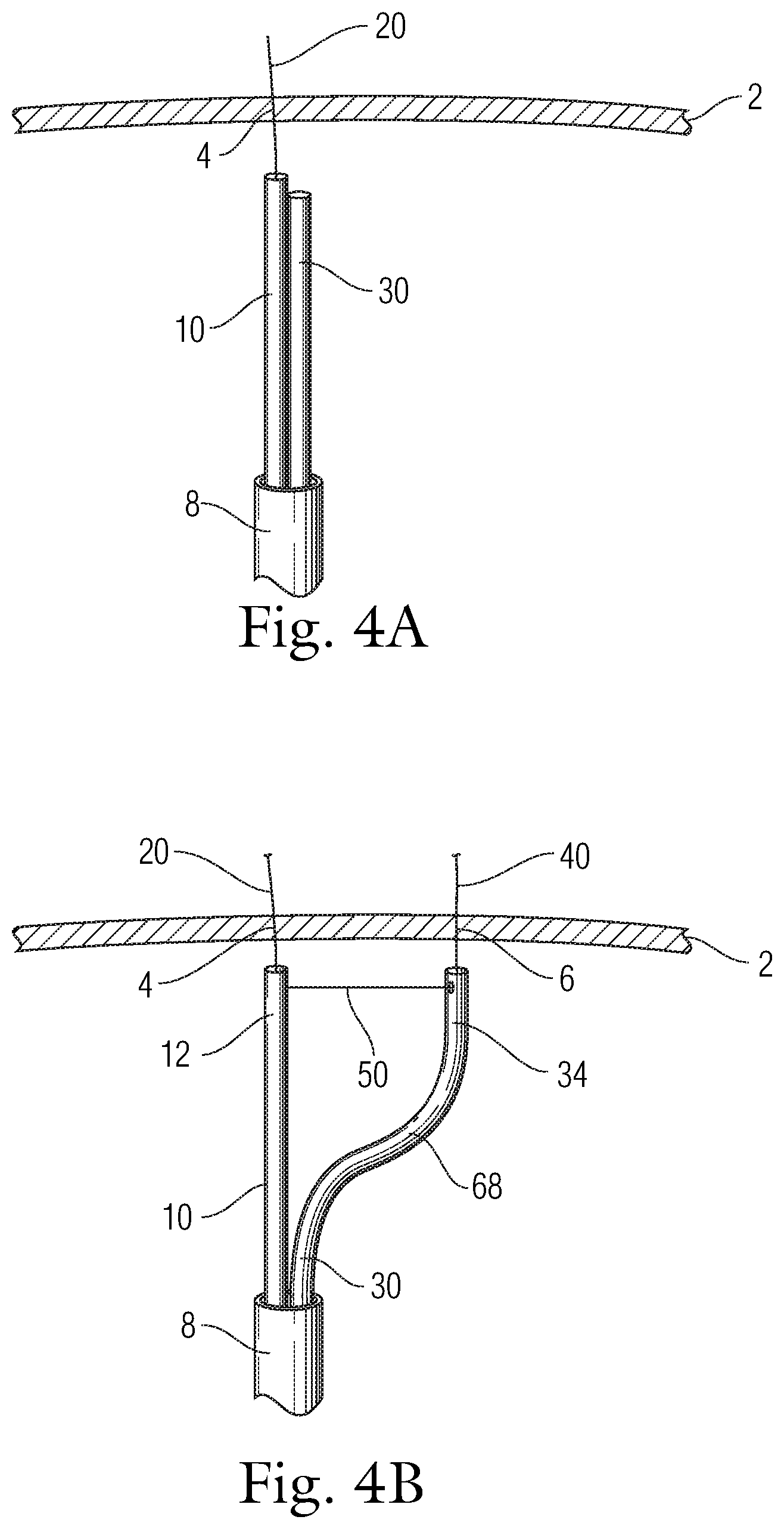
1. A catheter system comprising: a first wire configured to be positioned at a first location; a translatable catheter configured to be positioned at a second location and be movable between a first position at which the translatable catheter is at or proximate the first location and a second position at which the translatable catheter is at the second location; and a translation element configured to move the translatable catheter from the first position to the second position in a substantially linear fashion, while the first wire is maintained at the first location, wherein: the translation element comprises a tether that is coupled at one end to the first wire and passes through the translatable catheter and is accessible to a user to permit manipulation of the tether to cause the translatable catheter to move between the first and second positions, and the translatable catheter has a preformed curved portion proximate a distal end thereof, and a second tether is attached to the preformed curved portion and is manipulable to cause actuation of the preformed curved portion resulting in the preformed curved portion assuming a preformed curved shape.
2. The catheter system of claim 1, wherein the first wire is disposed within an other catheter that is separate from the translatable catheter, wherein both the first wire and the translatable catheter are slidingly contained within lumens formed in a delivery sheath.
3. The catheter system of claim 2, wherein the translation element operably connects the other catheter and the translatable catheter and permits the translatable catheter to move away from the other catheter from the first position to the second position.
4. The catheter system of claim 2, wherein the one end of the tether is a free end that wraps around the other catheter.
5. The catheter system of claim 2, wherein the one end of the tether is a fixed end that is connected with the other catheter.
6. The catheter system of claim 5, wherein the tether further comprises a free end and the free end extends into a lumen of the translatable catheter and is accessible by the user.
7. The catheter system of claim 6, wherein the translatable catheter includes a side opening formed therealong, the free end of the tether passing through the side opening into the lumen of the translatable catheter.
8. The catheter system of claim 6, wherein the translatable catheter includes a side opening formed therealong and spaced from the distal end of the translatable catheter, wherein the distal end is an open distal end, the free end of the tether passing through the open distal end of the translatable catheter and along the lumen thereof before exiting through the side opening and being routed longitudinally along an outer surface of the translatable catheter.
9. The catheter system of claim 2, wherein the preformed curved portion is formed of a memory material such that the preformed curved portion is elastically recoverable to assume the preformed curved shape.
10. The catheter system of claim 1, further comprising a tracking element that is configured to track over and move longitudinally along the first wire, the translation element being coupled to the tracking element.
11. The catheter system of claim 1, wherein a distance between the first location and the second location is adjustable.
12. A catheter system comprising: a first catheter configured to be positioned at a first treatment location; a second catheter configured to be positioned at a second treatment location; a sheath that has a first lumen in which the first catheter is disposed and a second lumen in which the second catheter is disposed; and a translation mechanism operably connecting the first and second catheters, the translation mechanism being configured and actuatable to allow the second catheter to move laterally away from the first catheter for a prescribed distance so as to space the second catheter from the first catheter and position the second catheter at the second treatment location, wherein: the sheath is formed such that a distal end portion thereof includes a side slot that extends longitudinally and defines an entrance into the second lumen and allows the second catheter, upon actuation of the translation mechanism, to pass therethrough and extend radially outward from the distal end portion and be positioned at the second treatment location, the sheath further includes first and second steering wire lumens that receive first and second steering wires, respectively, each of the first and second steering wires including a longitudinal wire with a wire coil being wrapped around the longitudinal wire, and distal ends of the longitudinal wire and the wire coil of each of the first and second steering wires are attached to one another and are attached to a distal end of the sheath.
13. The catheter system of claim 12, further comprising a control handle that is coupled to the sheath, and wherein, for each of the first and second steering wires, a proximal end of the wire coil is fixed to the control handle and a proximal end of the longitudinal wire is configured to be retracted inside a helix defined by the wire coil.
14. The catheter system of claim 12, wherein the translation mechanism comprises a pusher assembly which travels longitudinally and causes the second catheter to pass through the side slot and extend radially outward from the distal end portion and be positioned at the second treatment location.
15. A catheter system comprising: a first catheter configured to be positioned at a first treatment location; a second catheter configured to be positioned at a second treatment location; a sheath that has a first lumen in which the first catheter is disposed and a second lumen in which the second catheter is disposed; and a translation mechanism operably connecting the first and second catheters, the translation mechanism being configured and actuatable to allow the second catheter to move laterally away from the first catheter for a prescribed distance so as to space the second catheter from the first catheter and position the second catheter at the second treatment location, wherein: the sheath is formed such that a distal end portion thereof includes a side slot that extends longitudinally and defines an entrance into the second lumen and allows the second catheter, upon actuation of the translation mechanism, to pass therethrough and extend radially outward from the distal end portion and be positioned at the second treatment location, and the translation mechanism comprises a pusher assembly which travels longitudinally and causes the second catheter to pass through the side slot and extend radially outward from the distal end portion and be positioned at the second treatment location.
16. The catheter system of claim 15, wherein the sheath further includes first and second steering wire lumens that receive first and second steering wires, respectively, each of the first and second steering wires including a longitudinal wire with a wire coil being wrapped around the longitudinal wire, wherein distal ends of the longitudinal wire and the wire coil of each of the first and second steering wires are attached to one another and are attached to a distal end of the sheath.
17. The catheter system of claim 16, further comprising a control handle that is coupled to the sheath, and wherein, for each of the first and second steering wires, a proximal end of the wire coil is fixed to the control handle and a proximal end of the longitudinal wire is configured to be retracted inside a helix defined by the wire coil.
18. A catheter system comprising: a delivery sheath; a first wire configured to be positioned at a first location; a translatable catheter configured to be positioned at a second location and be movable between a first position at which the translatable catheter is at or proximate the first location and a second position at which the translatable catheter is at the second location; an other catheter that is separate from the translatable catheter, the first wire being disposed within the other catheter; and a translation element configured to move the translatable catheter from the first position to the second position in a substantially linear fashion, while the first wire is maintained at the first location, the translation element comprising a tether having: a fixed end that is connected with a catheter selected from the group consisting of the translatable catheter and the other catheter, and a free end that extends into a lumen of the translatable catheter and is accessible by a user, wherein both the first wire and the translatable catheter are slidingly contained within lumens formed in the delivery sheath.
19. The catheter system of claim 18, wherein the translation element operably connects the other catheter and the translatable catheter and permits the translatable catheter to move away from the other catheter from the first position to the second position.
20. The catheter system of claim 18, wherein the translatable catheter includes a side opening formed therealong, the free end of the tether passing through the side opening into the lumen of the translatable catheter.
21. The catheter system of claim 18, wherein the translatable catheter includes a side opening formed therealong and spaced from an open distal end of the translatable catheter, the free end of the tether passing through the open distal end of the translatable catheter and along the lumen thereof before exiting through the side opening and being routed longitudinally along an outer surface of the translatable catheter.
22. The catheter system of claim 18, wherein the translatable catheter has a preformed curved portion proximate a distal end thereof, the preformed curved portion being formed of a memory material such that the curved portion is elastically recoverable to assume a preformed curved shape.
23. The catheter system of claim 18, wherein the translatable catheter has a preformed curved portion proximate a distal end thereof, and a second tether is attached to the preformed curved portion and is manipulable to cause actuation of the preformed curved portion resulting in the preformed curved portion assuming a preformed curved shape.
24. The catheter system of claim 18, wherein a distal end of the translatable catheter is attached to a distal end of the other catheter and the translatable catheter has a plurality of side openings formed along a length thereof which permit the translatable catheter to bend, wherein a second wire passes through one of the side openings to the second location.
25. The catheter system of claim 18, further comprising a tracking element that is configured to track over and move longitudinally along the first wire, the translation element being coupled to the tracking element.
26. The catheter system of claim 18, wherein a distance between the first location and the second location is adjustable.
27. The catheter system of claim 18, wherein the selected catheter is the other catheter, and the fixed end of the tether is connected to the other catheter.
Description
TECHNICAL FIELD
The present invention relates generally to a delivery catheter system that is adapted to deliver multiple devices, such as guide wires, with definite spacing between each of them. The present invention further relates to a translation mechanism of the delivery catheter system which allows a delivery catheter to move to a definite distance.
BACKGROUND
The left side of a human heart includes the left atrium (LA) and the left ventricle (LV). An aorta receives oxygenated blood from the left ventricle through an aortic valve, which serves to prevent regurgitation of blood back into the left ventricle. A mitral valve is positioned between the left atrium and the left ventricle and allows one-way flow of the oxygenated blood from the left atrium to the left ventricle.
Mitral valve, which will be described below in more detail, includes an anterior leaflet and a posterior leaflet that are coupled to chordae tendineae. Chordae tendineae serve as "tension members" that prevent the leaflets of the mitral valve from moving past their closing point and prolapsing back into the left atrium. When the left ventricle contracts during systole, chordae tendineae limit the upward motion (toward the left atrium) of the anterior and posterior leaflets past the point at which the anterior and posterior leaflets meet and seal to prevent backflow from the left ventricle to the left atrium ("mitral regurgitation" or "mitral insufficiency"). Chordae tendineae arise from a columnae carnae or, more specifically, a musculi papillares (papillary muscles) of the columnae carnae. In various figures herein, some anatomical features have been deleted solely for clarity.
The anterior leaflet and the posterior leaflet of the mitral valve are generally thin, flexible membranes. When the mitral valve is closed, the anterior leaflet and the posterior leaflet are generally aligned and contact one another along a "line of coaptation" several millimeters back from their free edges, to create a seal that prevents mitral regurgitation. Alternatively, when the mitral valve is opened, blood flows downwardly through an opening created between the anterior leaflet and the posterior leaflet into left ventricle.
Many problems relating to the mitral valve may occur and may cause many types of ailments. Such problems include, but are not limited to, mitral regurgitation. Mitral regurgitation, or leakage, is the backflow of blood from the left ventricle into the left atrium due to an imperfect closure of the mitral valve. That is, leakage often occurs when the anterior and posterior leaflets do not seal against each other, resulting in a gap between the anterior leaflet and the posterior leaflet when the leaflets are supposed to be fully coapted during systole.
In general, a relatively significant systolic gap may exist between the anterior leaflet and the posterior leaflet for a variety of different reasons. For example, a gap may exist due to congenital malformations, because of ischemic disease, or because the heart has been damaged by a previous heart attack. Such a gap may also be created when congestive heart failure, e.g., cardiomyopathy, or some other type of distress which causes a heart to be enlarged. Enlargement of the heart can result in dilation (stretching) of the mitral annulus. This enlargement is usually limited to the posterior valve annulus and is associated with the posterior leaflet, because the anterior annulus is a relatively rigid fibrous structure. When the posterior annulus enlarges, it causes the posterior leaflet to move away from the anterior leaflet, causing a gap during systole because the two leaflets no longer form proper coaptation. This results in leakage of blood through the valve or regurgitation.
Blood leakage through the mitral valve generally causes a heart to operate less efficiently, as the heart pumps blood both out to the body via the aorta, and also back (in the form of mitral regurgitation) into the left atrium. Leakage through the mitral valve, or general mitral insufficiency, is thus often considered to be a precursor to congestive heart failure (CHF) or a cause of progressive worsening of heart failure. There are generally different levels of symptoms associated with heart failure. These levels are classified by the New York Heart Association (NYHA) functional classification system. The levels range from a Class 1 level which is associated with an asymptomatic patient who has substantially no physical limitations to a Class 4 level which is associated with a patient who is unable to carry out any physical activity without discomfort and has symptoms of cardiac insufficiency even at rest. In general, correcting or reducing the degree of mitral valve leakage may be successful in allowing the NYHA classification grade of a patient to be reduced. For instance, a patient with a Class 4 classification may have his classification reduced to Class 3 or Class 2 and, hence, be relatively comfortable at rest or even during mild physical exertion. By eliminating the flow of blood backwards into the left atrium, therapies that reduce mitral insufficiency reduce the workload of the heart and may prevent or slow the degradation of heart function and congestive heart failure symptoms that is common when a significant degree of mitral insufficiency remains uncorrected.
Treatments used to correct for mitral valve leakage or, more generally, CHF, are typically highly invasive, open-heart surgical procedures. In extreme cases, this may include implantation of a ventricular assist device such as an artificial heart in a patient with a failing heart. The implantation of a ventricular assist device is often expensive, and a patient with a ventricular assist device must be placed on extended anti-coagulant therapy. Anti-coagulant therapy reduces the risk of blood clot formation for example, within the ventricular assist device. Reducing the risks of blood clots associated with the ventricular assist device is desirable, but anti-coagulant therapies may increase the risk of uncontrollable bleeding in a patient, e.g., as a result of a fall.
Rather than implanting a ventricular assist device, bi-ventricular pacing devices similar to pacemakers may be implanted in some cases, e.g., cases in which a heart beats inefficiently in a particular asynchronous manner. While the implantation of a bi-ventricular pacing device may be effective, not all heart patients are suitable for receiving a bi-ventricular pacing device. Further, the implantation of a bi-ventricular pacing device is expensive, and is generally not effective in significantly reducing or eliminating the degree of mitral regurgitation.
Open-heart surgical procedures that are intended to correct for mitral valve leakage, specifically, can involve the implantation of a replacement valve. Valves from animals, e.g., pigs, may be used to replace a mitral valve in a human. While a pig valve as a replacement for the mitral valve may be relatively successful, such replacement valves generally wear out, thereby requiring additional open surgery at a later date. Mechanical valves, which are less likely to wear out, may also be used to replace a leaking mitral valve. However, when a mechanical valve is implanted, there is an increased risk of thromboembolism, and a patient is generally required to undergo extended anti-coagulant therapies.
A less invasive surgical procedure involves heart bypass surgery associated with a port access procedure. For a port access procedure, the heart may be accessed by cutting between ribs or sometimes removing parts of one or more ribs, as opposed to dividing the sternum to open the entire chest of a patient.
One open-heart surgical procedure that is particularly successful in correcting a mitral valve leakage and, in addition, mitral regurgitation, is an annuloplasty procedure. During an annuloplasty procedure, a medical device such as an annuloplasty ring may be implanted surgically on the left atrial side of mitral annulus (i.e., generally the attachment location of the base of the mitral valve to the heart). The device reduces a dilated mitral valve annulus to a relatively normal size and, specifically, moves the posterior leaflet closer to the anterior leaflet to aid anterior-posterior leaflet coaptation and thus improve the quality of mitral valve closure during systole. Annuloplasty rings are often shaped substantially like the letter "D" to correspond to the natural shape of the mitral annulus as viewed from above. Typically, the rings are formed from a rod or tube of biocompatible material, e.g., plastic, that has a DACRON mesh covering.
In order for an annuloplasty ring to be implanted, a surgeon surgically attaches the annuloplasty ring to the mitral valve on the atrial side of the mitral valve. Conventional methods for installing a ring require open-heart surgery which involves opening a patient's sternum and placing the patient on a heart bypass machine. The annuloplasty ring is sewn on a top portion of the mitral valve. In sewing the annuloplasty ring onto the mitral valve, a surgeon generally sews the straight side of the "D" to the fibrous tissue located at the junction between the posterior wall of the aorta and the base of the anterior mitral valve leaflet. As the curved part of the ring is sewn to the posterior aspect of the annulus, the surgeon alternately acquires a relatively larger amount of tissue from the mitral annulus, e.g., a one-eighth inch bite of tissue, using a needle and thread, compared to a relatively smaller bite taken of the fabric covering of the annuloplasty ring. Once the thread has loosely coupled the annuloplasty ring to the mitral valve annulus tissue, the annuloplasty ring is slid into contact with the mitral annulus. The tissue of the posterior mitral annulus that was previously stretched out, e.g., due to an enlarged heart, is effectively reduced in circumference and pulled forwards towards the anterior mitral leaflet by the tension applied by annuloplasty ring with the suture or thread. As a result, a gap between the anterior leaflet and the posterior leaflet during ventricular contraction or systole may be reduced and even substantially closed off in many cases thereby significantly reducing or even eliminating mitral insufficiency. After the mitral valve is shaped by the ring, the anterior and posterior leaflets will reform typically by pulling the posterior leaflet forward to properly meet the anterior leaflet and create a new contact line that will enable the mitral valve to appear and to function properly.
Although a patient that receives an annuloplasty ring may be subjected to anti-coagulant therapies, therapies are not extensive, as a patient is only subjected to therapies for a matter of weeks, e.g., until tissue grows over the annuloplasty ring.
Another type of procedure that is generally effective in reducing mitral valve leakage associated with prolapse of the valve leaflets involves placing a single edge-to-edge suture in the mitral valve that opposes the mid-portions of anterior and posterior leaflets. For example, in an Alfieri stitch or a bow-tie repair procedure, an edge-to-edge stitch is made at approximately the center of the gap between an anterior leaflet and a posterior leaflet of a mitral valve. Once the stitch is in place between the anterior and posterior leaflets, it is pulled in to form a suture which holds the anterior leaflet against the posterior leaflet.
Another surgical procedure that reduces mitral valve leakage involves placing sutures along a mitral valve annulus around the posterior leaflet. These sutures may be formed as a double track, e.g., in two "rows" from a single strand of suture material. The sutures are tied off at approximately a central point of posterior leaflet. Pledgets are often positioned under selected sutures to prevent the sutures from tearing through annulus. When the sutures are tightened and tied off, the circumference of the annulus may effectively be reduced to a desired size such that the size of a systolic gap between posterior leaflet and an anterior leaflet may be reduced.
While invasive surgical procedures have proven to be effective in the treatment of mitral valve leakage, invasive surgical procedures often have significant drawbacks. Any time a patient undergoes open-heart surgery, there is a risk of infection. Opening the sternum and using a cardiopulmonary bypass machine has also been shown to result in a significant incidence of both short and long term neurological deficits. Further, given the complexity of open-heart surgery, and the significant associated recovery time, people that are not greatly inconvenienced by CHF symptoms, e.g., people at a Class 1 classification, may choose not to have corrective surgery. In addition, people that need open heart surgery the most, e.g., people at a Class 4 classification, may either be too frail or too weak to undergo the surgery. Hence, many people that may benefit from a surgically repaired mitral valve may not undergo surgery.
In another method, a cinching device is placed within the coronary sinus (CS) using a catheter system, with distal, mid, and proximal anchors within the lumen of the CS to allow plication of the annulus via the CS. In practice, these anchors are cinched together and the distance between them is shortened by pulling a flexible tensile member such as a cable or suture with the intent being to shorten the valve annulus and pull the posterior leaflet closer to the anterior leaflet in a manner similar to an annuloplasty procedure. Unfortunately, since the tissue that forms the CS is relatively delicate, the anchors are prone to tear the tissue during the cinching procedure. In addition, the effect on the mitral annulus may be reduced when the CS of a particular patient is not directly aligned with the mitral annulus. Other minimally invasive techniques have been proposed but have various drawbacks related to such factors as effectiveness and/or accuracy of catheter-based implementation.
Catheter-based surgical procedures have been used to repair a defective mitral valve. Specifically, anchors are secured at a plurality of locations distributed around the annulus near the posterior leaflet of a mitral valve. Each anchor has a suture coupled thereto. The sutures are collectively gathered and pulled tight. As the sutures are pulled, the tissue between each pair of adjacent anchors is plicated, thereby shortening the length of the annulus and drawing the posterior leaflet toward the anterior leaflet. Similar techniques can also be used to repair a defective tricuspid valve.
During a surgical procedure, anchors are usually introduced and secured sequentially. A typical repair by using the catheter based surgical procedure involves a sequence that includes introducing a catheter to a proximity of the annulus, making an incision at the annulus, introducing a guide wire through the incision site, withdrawing the catheter, introducing an anchor by tracking a second catheter through the guide wire, securing the anchor in the annulus, and withdrawing the second catheter. This sequence is repeated to secure a second anchor.
Catheters capable of delivering multiple guide wires or anchors have been disclosed. Without claiming to have exhaustively examined prior art references and without attempting to characterizing any prior art reference, U.S. Patent Application Publication No. 2008-0228265 discloses a triple lumen catheter. However, distances between two of the three lumens are usually fixed. In addition, during a deployment, the two outer catheters are generally advanced lengthwise as well as laterally. In certain instances, one or both of the two outer catheters are caught by chordae tendineae during the deployment.
There is generally a need for an improved catheter to simplify the catheter-based mitral valve correction.
SUMMARY
One aspect of the present teachings generally relates to translation catheter systems. The catheter system comprises a first wire configured to be positioned at a first location, a second catheter configured to be positioned at a second location, and a translation element configured to move the second catheter from the first location to the second location in a substantially linear fashion. A first wire is positioned with the first catheter. The translation element operably connects the first and second catheters.
In one aspect of the present teachings, the translation element of the catheter system is a tether. The tether comprises a free end and the free end wraps around one of the first and second catheter. In one aspect of the present teachings, the tether comprises a fixed end and the fixed end. The fixed end is connected with one of the first and second catheter, and the free end extends into a lumen of the other catheter.
In another aspect of the present teachings, the translation element of the catheter system is a bar formed of at least two segments. In another aspect of the present teachings, the translation element of the catheter system comprises an anchor configured to track over the first wire, and a tether. The tether has a proximal end and a distal end, wherein the distal end of the tether connects the anchor.
In one aspect of the present teachings, the distal portions of the first and the second catheters are connected. In one aspect of the present teachings, the second catheter is flexible. In one aspect of the present teachings, the second catheter comprises at least one side opening. In one aspect of the present teachings, the distance between the first location and the second location is adjustable.
Another aspect of the present teachings generally relates to translation catheter systems where translation catheter systems have a first catheter configured to be positioned at a first treatment location; a second catheter configured to be positioned at a second treatment location; and a translation mechanism operably connecting the first and second catheters. In one aspect of the present teachings, the translation mechanism is configured to allow the second catheter to move laterally away from the first catheter to any distance.
Another aspect of the present teachings generally relates to translation catheter systems where translation catheter systems have a first guide wire positioned at a first treatment location; a tracking anchor configured to track over the first guide wire; a second catheter configured to be positioned at a second treatment location; and a tether having a proximal end and a distal end. The distal end of the tether connects to the tracking anchor and the proximal end of the tether extends through a central lumen of the second catheter.
Another aspect of the present teachings generally relates to translation catheter systems where translation catheter systems have a first catheter configured to be positioned at a first treatment location; a second catheter configured to be positioned at a second treatment location; and a tether having a proximal end and a distal end. The distal end of the tether connects to the first catheter and the proximal end of the tether extends through a central lumen of the second catheter.
Another aspect of the present teachings generally relates to translation catheter systems where translation catheter systems have a first catheter comprising a distal end configured to be positioned at a first treatment location; a second catheter comprising a distal end configured to be positioned at a second treatment location; and a connecting bar connecting the distal ends of the first and second catheters. The connecting bar has at least two segments with at least one pivot between the segments.
Another aspect of the present teachings generally relates to translation catheter systems where translation catheter systems have a first catheter comprising a distal end configured to be positioned at a first treatment location; and a second catheter comprising a distal end configured to be positioned at a second treatment location. The second catheter is configured to be bendable in one direction and the distal ends of the first and second catheters are joined to each other.
BRIEF DESCRIPTION OF THE DRAWINGS
FIG. 1 is a perspective view of an exemplary wire delivery catheter in accordance with the present teachings;
FIG. 2 is a perspective view of an exemplary wire deploying across a tissue in accordance with the present teachings;
FIGS. 3A-D are perspective views of a translation catheter system in accordance with the present teachings;
FIGS. 4A-D are perspective views of a translation catheter system in accordance with the present teachings;
FIGS. 5A-5B are perspective views of a translation catheter system in accordance with the present teachings;
FIG. 5C is a close-up side elevation view of a portion of a catheter showing side openings;
FIGS. 6A-6B are perspective views of a translation catheter system in accordance with the present teachings;
FIGS. 7A-7B are perspective views of a translation catheter system in accordance with the present teachings;
FIG. 8 is a perspective view of a translation catheter system in accordance with the present teachings;
FIG. 9 is a perspective view of a translation catheter system in accordance with the present teachings;
FIG. 10A is a perspective view of a distal end of a translation catheter system in accordance with the present teachings;
FIG. 10B is a perspective view of a pair of steerable wires for use in the translation catheter system;
FIG. 10C is a perspective view showing a translation rod assembly for use in the translation catheter system; and
FIG. 10D is a perspective view of a distal end of a translation catheter system.
DETAILED DESCRIPTION OF CERTAIN EMBODIMENTS
Certain specific details are set forth in the following description and Figures to provide an understanding of various embodiments of the present teachings. Those of ordinary skill in the relevant art will understand that they can practice other embodiments of the present teachings without one or more of the details described herein. Thus, it is not the intention of the Applicants to restrict or in any way limit the scope of the appended claims to such details. While various processes are described with reference to steps and sequences in the following disclosure, the steps and sequences of steps should not be taken as required to practice all embodiments of the present teachings.
As used herein, the terms "subject" and "patient" refer to an animal, such as a mammal, such as livestock, pets, and preferably a human. Specific examples of "subjects" and "patients" include, but are not limited to, individuals requiring medical assistance and, in particular, requiring treatment for symptoms of a heart failure.
As used herein, the term "lumen" means a canal, duct, generally tubular space or cavity in the body of a subject, including veins, arteries, blood vessels, capillaries, intestines, and the like. The term "lumen" can also refer to a tubular space in a catheter, a sheath, or the like in a device.
As used herein, the term "proximal" means close to the operator (less into the body) and "distal" shall mean away from the operator (further into the body). In positioning a medical device from a downstream access point, distal is more upstream and proximal is more downstream.
While the description above refers to and the term "tether" means a tensioning member which can take forms of a suture, a wire, a strand, a cord, a fiber, a yarn, a filament, a cable, a thread, or the like. Thus, all these terms are essentially interchangeable and further include embodiments in which the wire, string, suture or filament is a hollow tube or conduit to allow another wire, as needed, to pass through its longitudinal axis. Each tether, wire, string, suture and filament can comprise one or more tethers, wires, strings, sutures and filaments. Material used to make tether could be flexible, semi-rigid, or rigid material having a suitably high tensile strength for the intended use.
As used herein, the term "catheter" or "sheath" encompasses any hollow instrument capable of penetrating body tissue or interstitial cavities and providing a conduit for selectively injecting a solution or gas. The term "catheter" or "sheath" is also intended to encompass any elongate body capable of serving as a conduit for one or more of the ablation, expandable or sensing elements. Specifically, in the context of coaxial instruments, the term "catheter" or "sheath" can encompass either the outer catheter body or sheath or other instruments that can be introduced through such a sheath. The use of the term "catheter" should not be construed as meaning only a single instrument but rather is used to encompass both singular and plural instruments, including coaxial, nested, and other tandem arrangements. Moreover, the terms "sheath" or "catheter" are sometime used interchangeably to describe catheters having at least one lumen through which instruments or treatment modalities can pass.
Unless otherwise specified, all numbers expressing quantities, measurements, and other properties or parameters used in the specification and claims are to be understood as being modified in all instances by the term "about." Accordingly, unless otherwise indicated, it should be understood that the numerical parameters set forth in the following specification and attached claims are approximations. At the very least, and not as an attempt to limit the application of the doctrine of equivalents to the scope of the claims, numerical parameters should be read in light of the number of reported significant digits and the application of ordinary rounding techniques.
An aspect of the present teachings provides a delivery catheter system for delivering multiple guide wires across mitral annulus at a controlled spacing between each of them. In various embodiments, the delivery catheter system has one catheter and a translation mechanism allowing the catheter to move to a second location, i.e. translate, from its first location by a distance, pre-defined or determined by a clinician during procedure. In some embodiments, the delivery catheter system includes two catheters and a translation mechanism allowing the at least one catheter to move away from the other catheter by a distance, pre-defined or determined by a clinician during procedure. In some embodiment, the distance between the first and second locations, or the distance between the first and second catheter after translation, are within the range of 1-40 mm. In some embodiment, the translation mechanism of the delivery catheter system includes a tether, a shuttle, a tracking element, a wire, a coil, a connecting bar with a pivot, or a combination thereof. In some embodiments, the translation is lateral. In other embodiments, the translation is distal-lateral. In some embodiments, the translation is continuous. In other embodiments, the translation is step-by-step. In some embodiments, the distance of the translation is adjustable. In other embodiments, the distance of the translation is pre-defined.
Another aspect of the present teachings provides method of delivering a translation catheter system to a first location at or approximate to the mitral annulus, translating the translation catheter system to a second location at or approximate to the mitral annulus. In various embodiments, the distance between the two locations is controlled by a clinician. In various embodiments, the method includes advancing a first delivery catheter to a first location at or approximate to the mitral annulus, placing a wire across the mitral annulus at this first location, translating the first catheter to a second location at or approximate to the mitral annulus, placing a second wire across the mitral annulus at the second location. In various embodiments, the method includes advancing a first delivery catheter to a first location at or approximate to the mitral annulus, placing a wire across the mitral annulus at this first location, translating the second catheter to a second location at or approximate to the mitral annulus, placing a second wire across the mitral annulus at the second location.
The following description refers to FIGS. 1 to 9. A person with ordinary skill in the art would understand that the figures and description thereto refer to various embodiments of the present teachings and, unless indicated otherwise by their contexts, do not limit the scope of the attached claims.
FIGS. 1A-B illustrate a guide wire being delivered across a mitral annulus. According to one embodiment of the present teachings, a delivery sheath (not shown) is directed into the aorta, through the aortic valve, and into the left ventricle and between the chordae tendonae. The delivery sheath (not shown) is used as a conduit to deliver a first wire delivery catheter (10) to a treatment site. One ordinarily skilled in the art would understand that the first wire delivery catheter (10) can be advanced to a treatment location without a delivery sheath.
FIG. 1 illustrates an exemplary delivery of a first wire delivery catheter (10) according to the present teachings to a selected location adjacent to or near the mitral annulus (2). In some embodiments, the first catheter (10) advances through the longitudinal lumen of the delivery sheath (not shown) and is placed below the mitral annulus (2). In certain embodiments, the first catheter (10) has a proximal portion (not shown) remaining outside of the body, a distal end (12), an elongated body (14) between the proximal portion and the distal portion, and a central lumen (16) within the elongated body. In some embodiments, the distal end of the first catheter (10) can be turned, rotated, or deflected. The deflectability or steerability of the first catheter (10) allows a clinician to manipulate the distal end (12) of the first catheter (10) from outside of the body and advance it to a first location (4) near the annulus (2). Design and construction of a steerable and deflectable catheter are well known to those with ordinary skill in the art.
In some embodiments, the distal end (12) of the first catheter (10) includes a radio-opaque and/or echogenic marker so that the device can be visualized by using a radiographic imaging equipment, for example, x-ray, magnetic resonance, ultrasound, or fluoroscopic techniques.
FIG. 2 illustrates an example of the present teachings where a first wire (20) extends from a lumen (16) of a first catheter (10) and pierces the annulus (2). In various embodiments, the first wire (20) also includes a proximal end (not shown), a distal end (22), and an elongated body (24). In some embodiments, the first wire (20) is configured to slide within the lumen (16) of the first catheter (10). In some embodiments, the first wire (20) is configured to rotate within the lumen (16).
In various embodiments, the first wire (20) has a delivery profile where the distal end (22) of the first wire (20) is disposed inside the lumen (16) of the first catheter (10). In some embodiments, when the distal end (22) of the first wire (20) is disposed within the lumen (16) of the catheter (12), the entire first wire (20) is substantially straight and parallel with the long axis of the first catheter (10). In some embodiments, the first wire (20) has a deployed profiled where the distal end (22) of the first wire (20) extends distally outside the lumen (16) of the first catheter (10), as illustrated in FIG. 2. In certain embodiments, as the distal portion of the first wire (20) extends outside of the first catheter (10), the distal portion (22) transitions to a curved profile. Without intending to limit the scope of the present teachings, such a curved profile can prevent the first wire from causing unnecessary tissue damage inside the left atrium.
In various embodiments, the first wire (20) is pre-loaded within the lumen (16) of the first catheter (10). In various other embodiments, the first wire (20) is advanced after the first catheter (10) is placed at a treatment location.
In various embodiments, the first wire delivery catheter (10) is generally perpendicular to the annulus (2) before the first wire (20) pierces and crosses the annulus (2). Once the first catheter (10) is properly positioned, a clinician extends the first wire (20) distally so that the distal tip (26) of the first wire (20) pierces and crosses the annulus (2) at the first location (4). In some embodiments, a radio frequency energy is applied to assist the distal tip (26) of the first tissue piercing wire (20) to pierce and cross the annulus (2) and reach the left atrium. To determine when to stop the distal extension force to the wire (20), in some embodiments, a change of the counter force on the first wire (20) indicates that the annulus (2) is pierced or the distal tip (26) of the first wire (20) reaches a side wall of the left atrium. Alternatively, visualization techniques, including three-dimensional echocardiogram, fluoroscopy, or magnetic resonance imaging techniques, can be used.
FIGS. 3A-3D illustrate various embodiments of the translatable catheter system according to the present teachings. FIG. 3A illustrates an embodiment of the present teachings where a second wire (40) pierce the annulus (2). In various embodiments, when the first wire (20) is placed across the annulus (2), the first catheter (10) is retracted proximally and removed, a tracking element (52) is slid over the first wire (20) from its proximal end. In some embodiments, the tracking element (52) has the form of a loop, shuttle, or another shape (for example, including a lumen) so that the tracking element (52) can slide along the first wire (20) freely or in a controlled manner from the proximal end to the distal end (22, in FIG. 2) or from the distal end (22, in FIG. 2) to the proximal end (not shown).
As illustrated in FIG. 3A, the tracking element (52) is tethered to a second wire delivery catheter (30). Similar to the first wire delivery catheter (10), the second wire delivery catheter (30) includes a proximal portion (remaining outside of the body, not shown), a distal end (34) at or near the annulus (2), an elongated body (36) between the proximal end and the distal end, and a central lumen (32) extending throughout the elongated body (36). Similarly, a clinician can manipulate the second wire delivery catheter (30) to a second location near the annulus (2).
Further referring to FIG. 3A, a second wire (40) extends from the lumen (32) of the second catheter (30) and across the annulus (2). In various embodiments, the second wire (40) includes a proximal end (not shown), a distal end (42), and an elongated body (44). One ordinarily skilled in the art would understand that the first and second wires can have the same shape, size, and/or construct or different shapes, sizes, and/or constructs. Thus, what has been described herein should not be construed as limiting to the scope of the present teachings.
In an exemplary embodiment as shown in FIG. 3A, the second catheter (30) includes a side opening (38). The tether (50) extends from the tracking element (52), enters the side opening (38), extends proximally inside the lumen (32) of the second catheter (30), and exits outside of the body. In various embodiments, a clinician controls the proximal end of the tether. For example, a clinician can tighten the tether (50), for example, by pulling the tether (50) proximally, so that the distal end (34) of the second catheter (30) is pulled adjacent to the first wire (20). Or, for example, a clinician can loosen the tether (50), for example, by extending the tether (50) distally, so that the distal end (34) of the second catheter (30) is away from the first wire (20), as illustrated in FIG. 3A. The length of the tether (50) controls the distance between the distal end (34) of the second catheter (30) and the first wire (20).
To deliver the second wire (40) across the annulus (2), a clinician slides the tracking element (52) over the proximal end of the first wire (20) and affixes the tether (50) to the tracking element. The tether (50) and the tracking element (52) are loaded inside the second wire delivery catheter (30). By tightening the tether (50), the distal end (34) of the second catheter (30) maintains a close proximity to the first wire (20). The tracking element (52) and the second catheter (30), optionally disposed inside a delivery sheath (8), tracks along the first wire (20) and extends distally. Once the distal end (34) of the second catheter (30) arrives at or near to the first location (4) of the annulus (2), the tether (50) is loosened and the second catheter (30) is steered away from the first location (4) of the annulus (2) to the second location (6) on the annulus (2). In some embodiments, the translation of the second catheter (30) from the first location (4) is lateral. In other embodiments, the translation of the second catheter (30) from the first location (4) is continuous. In yet another embodiment, the translation distance of the second catheter (30) from the first location is adjustable.
In various embodiments, the second wire delivery catheter (30) is generally perpendicular to the annulus (2) before the second wire (40) pierces the annulus (2). The second wire (40) is advanced distally to pierce and cross the annulus (2) at the second location (6) in a manner similar to those described herein in connection with FIG. 2.
In some embodiments, the distal end (34) of the second catheter (30) remains close to the annulus (2) the entire time when the second catheter is steered from the first location to the second location. Without intending to limit the scope of the present teachings, such a lateral movement avoids the distal end of the second catheter from being caught, snag, or hung up by tissues in the left ventricle, and allows the second catheter (30) pushing away any anatomy in the path of the translation. In some embodiments, the distal end (34) of the second catheter (30) remains close to the annulus (2) the entire time when it is steered from the first location to the second location.
In one embodiment, a clinician controls the distance between the first and second locations (4, 6) by controlling the length of the tether (50) between the distal end of the second catheter and the first wire. According to some embodiments, such the mechanism as shown in FIG. 3A allows a continuous translation of the second catheter (30) and an adjustable distance between the two treatment locations (4, 6). Similar to what is described in connection with FIG. 2, a second wire 40 can be advanced distally across the mitral annulus (2).
In some embodiments, during the delivery of the second catheter (30), a delivery sheath (8) is used, as seen in FIG. 3A. In one embodiment, the delivery sheath (8) has a single lumen where encloses the first wire (20), the tracking member (52), the second catheter (30), and the tether (50).
In one embodiment, the tether (50) extends from the tracking element (52), enters the side opening (38) of the second catheter (30), extends proximally along the center lumen (32) of the second catheter (30), and exits the body, as illustrated in FIG. 3A. In another embodiment, the tether (50) extends from the tracking element (52), enters the distal end (34) of the second catheter (30), extends proximally along the center lumen (32) of the second catheter (30), and exits the body, as illustrated in FIG. 3B. In yet another embodiment, the tether (50) extends from the tracking element (52), enters the distal end (34) of the second catheter (30), extends proximally along the center lumen (32) of the second catheter (30), exits a side opening (38) of the second catheter (30), and further extends proximally along the exterior of the second catheter (30), as illustrated in FIG. 3C.
In one embodiment, the first wire delivery catheter (10) and the second wire delivery catheter (30) are different, where the first catheter (10) does not have any attachment mechanism with a tether (50) but the second catheter (30) has. In another embodiment, the first wire delivery catheter (10) and the second wire delivery catheter (30) are the same. In the later embodiment, an attachable/detachable tracking system (60) is used for converting the first catheter to a tethered second catheter. As illustrated in FIG. 3D, the attachable/detachable tracking system (60) includes two tracking anchors (62, 64). The tracking anchors (62, 64) have axial lumens for sliding over the first wire (20) and guiding the second wire (40), respectively. In one embodiment, when the tracking anchor (64) joins the distal end (34) of the second catheter (30), the lumen of the tracking anchor (64) and the lumen (32) of the second catheter (30) forms a continuous conduit for passing the second wire (40).
In one embodiment, as shown in FIG. 3D, at least one tether (66) joins the two tracking anchors (62, 64). In one embodiment, the tether (66) joining two tracking anchors (62, 64) has a fixed length with one end of the tether (66) fixed to one tracking anchor (62) and the other end of the tether (66) fixed to another tracking anchor (64). Thus, the translation distance of the second catheter (30) is pre-determined by the length of the tether (66) and so is the distance between the two treatment locations (4, 6).
In another embodiment, one end of the tether (66) is fixed to one tracking anchor (62), riding over the first wire (20), and the other end of the tether (66) enters the other tracking anchor (64) in a manner similar to those described in connection with FIGS. 3A-3C. Thus, the translation of the second catheter (30) can be continuous and adjustable, similar to those described herein.
In various embodiments, at least one of the second catheter and the tether includes a radio-opaque marker or is made in part or its entirety of a radio-opaque material. By using a visualization technique, including various ultrasound, x-ray, fluoroscopic, or magnetic resonance imaging techniques, a clinician can use the marker to visualize where the first catheter, the second catheter, and/or the tether are located in the anatomy. A clinician can also use the marker to determine the translation distance of the second catheter or the distance between the two treatment locations. In one exemplary embodiment, the tether could have multiple markers which to indicate the translation distance, and allow a clinician to control the distance between the two treatment locations.
FIGS. 4A-4D illustrate various embodiments of the translatable catheter system according to the present teachings. In some embodiments, as illustrated in FIG. 4B, the first and second catheters (10, 30) are operably joined together at their distal ends (12, 34) by a tether (50) in a manner same as or similar to those described in connection with FIGS. 3A-3C. Optionally, the first and second catheters (10, 30) are operably joined to each other at other places along the catheter bodies.
Referring to FIG. 4A, in some embodiments, the first catheter (10) and the second catheter (30) are delivered together to the mitral annulus (2) while the tether (50) is tightened and the distal ends (12, 34) of the two catheters (10, 30) are held next to each other. In certain embodiments, one of the two catheters (10, 30) is steerable and deflectable, and the steerability and/or deflectability of one of the first and second catheters (10, 30) allows a clinician to advance the distal end (12) of the first catheter (10) to a first location (4) on the annulus (2). In particular embodiments, the second catheter (30) is steerable and deflectable and both the catheters (10, 30) are joined in such way that both the catheters (10, 30) are advanced to the first treatment location (4) by steering the second catheter (30). While the first wire delivery catheter is generally maintained perpendicular to the annulus (2) at the first treatment location (4), a first wire (20) is advanced across the annulus (2) in a manner similar to those described in connection with FIG. 2.
In various embodiments, a second catheter (30) continuously and adjustably translates away from a first catheter (10) via a tethering mechanism. Now referring to FIG. 4B, while holding the first wire (20) in place, a clinician loosens the tether (50), for example, by extending the tether (50) distally, to allow the distal end (34) of the second catheter (30) to be steered away from the first catheter (10), such as illustrated in FIG. 4B. Without intending to limit the scope of the present teachings, such a lateral movement avoids the distal end of the second catheter from being caught, snag, or hung up by tissues in the left ventricle, and allows the second catheter (30) pushing away any anatomy in the path of the translation. In some embodiments, the distal end (34) of the second catheter (30) remains close to the annulus (2) the entire time when it is steered from the first location to the second location. In some embodiments, the translation of the second catheter (30) from the first location (4) is lateral. In other embodiments, the translation of the second catheter (30) from the first location (4) is continuous. In yet another embodiment, the translation distance of the second catheter (30) from the first location is adjustable.
Similarly, the length of the loosened tether (50) determines the distance between the second catheter (30) and the first catheter (10). According to some embodiments, such the mechanism as shown in FIGS. 4A-4B allows a continuous translation of the second catheter (30) and an adjustable distance between the two treatment locations (4, 6). While the second wire delivery catheter is maintained generally perpendicular to the annulus (2) at its second treatment location (6), a second tissue piercing wire (40) is advanced across the annulus (2) in a manner similar to those described in connection with FIG. 2.
In some embodiments, in order to maintain a generally perpendicular position between the distal end (34) of the second catheter (30) and the annulus (2), the second catheter (30) includes a curved portion (68) near its distal end, as shown in FIG. 4B. In one embodiment, such a curved portion (68) is pre-formed and elastically recoverable to this pre-formed curve when the curved portion (68) of the second catheter (30) is free from the constraint of the tether (50) and/or sheath (8). In another embodiment, such a curved portion (68) is actuated by a mechanical means, for example, by shortening another tether (70) fixed to a portion of the second catheter (30), illustrated in FIG. 4C. One ordinarily skilled in the art would appreciate that other means can also be incorporated to achieve such a configuration. Thus, the specific embodiments disclosed herein should not be construed as limiting.
FIG. 4D illustrates another embodiment of the translatable catheter system where the second catheter (30) is configured to roll along the bottom surface of the mitral annulus (2). In such an embodiment, a distal end (58) of the tether (56) fixes to the distal end (34) of the second catheter (30) and the other end of the tether (56) enters the distal end (12) of the first catheter (10), travels proximally along the lumen (not shown) of the first catheter (10), and exits from the body. In some embodiments, the tether (56) is configured in such a way that when it is pushed by a clinician at its proximal end, the distal end (58) of the tether (56) extends laterally away from the first treatment location (4), carrying the distal end (34) of the second catheter (30) with it, as illustrated in FIG. 4D. In some embodiments, a sheath (8) is used to constrain the translation profile of the second catheter (30), as shown in FIG. 4D, so that the distal end (34) of the second catheter (30) and the portions of the second catheter (30) outside of the sheath (8) travels along the bottom surface of the annulus (2) and substantially free from tissues under the annulus, as shown in FIG. 4D. Without intending to limit the scope of the present teachings, such a lateral rolling movement of the distal portion of the second catheter (30) avoids the second catheter from being caught, snag, or hung up by tissues in the left ventricle. In some embodiments, the translation of the second catheter (30) from the first location (4) is lateral. In other embodiments, the translation of the second catheter (30) from the first location (4) is continuous. In yet another embodiment, the translation distance of the second catheter (30) from the first location is adjustable. In other embodiments, the second catheter (30) is made of a flexible tubing so that it can bend and deflect easily without restraint. In some embodiments, after the distal end (34) of the second catheter (30) reaches the second treatment location (6), the sheath (8) is retracted proximally to allow the second catheter (30) to form a more gentle bend, similar to the curve portion (68), as illustrated in FIG. 4B, thereby allowing the second wire (40) to pierce and cross the annulus. According to some embodiment, as the distal portion of the second catheter (30) assumes the more gentle end, the second catheter (30) pushes away any anatomy in the path of the translation.
According to some embodiments, a piece of rigid material could be incorporated to the distal portion of the tether (50) in between of the two catheters (10,30) to ensure a minimal translation distance. In one embodiment, either one of the catheters (10,30) is configured to be articulated. In another embodiment, both of the catheters (10,30) are configured to be articulated.
In many embodiments, the design, material, and/or construct of the first catheter and the second catheter are interchangeable. Specifically, for example, in many of the embodiments described herein, the first catheter maintains its substantially straight configuration while the second catheter is steered away towards the second treatment location. One of ordinary skill in the art would understand that it is equally feasible to have a translatable deliver catheter system where the second catheter maintain its straight profile while the first catheter is steered to push the second catheter to the second treatment site. Thus, parts or portions in connection with the first and second catheters are interchangeable without departing from the letter or the spirit of the present teachings. In various embodiments, at least one of the first catheter, the second catheter, and the tether includes a radio-opaque marker or is made in part or its entirety of a radio-opaque material. By using a visualization technique, including various ultrasound, x-ray, fluoroscopic, or magnetic resonance imaging techniques, a clinician can use the marker to visualize where the first catheter, the second catheter, and/or the tether are located in the anatomy. A clinician can also use the marker to determine the translation distance of the second catheter or the distance between the two treatment locations. In one exemplary embodiment, the tether could have multiple markers which to indicate the translation distance, and allow a clinician to control the distance between the two treatment locations.
FIGS. 5A-5B illustrates another embodiment of the translatable catheter system according to the present teachings. In the embodiment illustrated in FIG. 5B, the first and second catheters (10, 80) are joined together at their distal ends (12, 82), for example mechanically, thermally, or chemically. The first wire delivery catheter (10) has a configuration same as or similar to those described here. The second wire delivery catheter (80) includes a proximal end (not shown), a distal end (82), and an elongated body (84) with a central lumen (86) therein, where the elongated body extends from the proximal end to the distal end. In some embodiments, the second catheter (80) include a plurality of first side openings (78) located along the longitudinal surface of the catheter and generally facing the first catheter (10) and a plurality of second side openings (88) along the longitudinal surface of the catheter generally across the longitudinal axis of the second catheter (80) from the first side openings (78), as shown in FIGS. 5A-5B. In some embodiments, such a configuration (i.e., having the plurality of first and second side openings (78, 88)) results in a bendable second catheter (80), as shown in FIG. 5B. An embodiment of the second catheter, including openings 78 and 88, is illustrated in FIG. 5C. One ordinarily skilled in the art would understand that other designs can also be used to create such a "hinge" effect, thus the disclosure herein should not be construed as limiting.
As shown in FIG. 5A, a first catheter (10) and a second catheter (80) are delivered together with their distal ends (12, 82) attached to each other and their elongated body (14, 84) side-by-side through the longitudinal lumen of the delivery sheath (8) to a location near the mitral annulus (2). In certain embodiments, one of the two catheters (10, 80) is steerable and deflectable, and the steerability and/or deflectability of one of the first and second catheters (10, 80) allows a clinician to advance the distal end (12) of the first catheter (10) to a first location (4) on the annulus (2). In some embodiments, the translation of the second catheter (80) from the first location (4) is lateral. In other embodiments, the translation of the second catheter (80) from the first location (4) is continuous. In yet another embodiment, the translation distance of the second catheter (80) from the first location is adjustable. In some embodiments, the first catheter (10) is steerable and/or deflectable, so that a clinician can accurately and easily manipulate the distal end (12) of the first catheter (10) to a first location (4) on the annulus (2). A first wire (20) is advanced distally across the annulus (2), similar to what is described in connection with FIG. 2.
FIG. 5B illustrates an exemplary translation of the second catheter (80) according to the present teachings. While maintaining the first wire (20) and the first catheter (10) in place, a clinician extends the second catheter (80) distally. Because the distal end (82) of the second catheter (80) is attached to the distal end (12) of the first catheter (10) and because of the cutting pattern along the elongated body (84) of the second catheter (80), the distal portion (76) of the second catheter (80) rolls laterally outward along the bottom surface of the annulus (2) as shown in FIG. 5B. Without intending to limit the scope of the present teachings, such a lateral rolling/bending movement of the distal portion of the second catheter (80) avoids the second catheter from being caught, snag, or hung up by tissues in the left ventricle. In addition, according to some embodiment, as the distal portion of the second catheter (80) translates to the second treatment location, the second catheter (80) pushes away any anatomy in the path of the translation. The multiple side openings (88) form openings for the second wire (40). A second wire (40) is advanced distally along the lumen (86) of the second catheter (80), exits a side opening (88) of the second catheter (80), and pierces and crosses the mitral annulus (2) at the second treatment location (6). Similarly, one or both of the first catheter (10) and second catheter (80) can include a radio-opaque marker or be made in part or its entirety of a radio-opaque material. In some embodiments, the distal portion (76) of the second catheter (80) has multiple radio-opaque markers to indicate the translation distance and allow a clinician to control the distance between the two treatment locations.
According to another embodiment, instead of extending a second wire (40) directly inside the second catheter (80), a third wire delivery catheter is used to ride inside the lumen of the second catheter (80).
FIGS. 6A-6B illustrate additional exemplary translatable catheter system according to the present teachings. In various embodiments, the first and second catheters (10, 30) are joined together by at least one flexible hinge (90). In some embodiments, one end of the hinge (90) is fixed to one catheter, for example, the first catheter (10), and the other end of the hinge (90) wraps around the distal end of the other catheter, for example, the second catheter (30). In some embodiments, the hinge has a first profile where the hinge winds tightly and holds the first and second catheters (10, 30) side-by-side. In some embodiments, the hinge has a second profile where the hinge (90) loosens and allows the second catheter to translate outward laterally.
In some embodiments, the hinge (90) is in the shape of a coil, a roll, a reel, a spool or alike. In some embodiments, the hinge (90) is closed, open, or relatively neutral at its normal state. In other embodiments, by torqueing the distal end of one catheter, the hinge changes from one profile to another. In some embodiments, the hinge (90) has a contoured cross section such that it lies flat against the longitudinal surface of the catheters when wrapped around the distal end portions of the first and second catheters (10, 30). In other embodiments, when the cross section of the hinge straightens, the hinge becomes somewhat rigid, similar to the behavior of a stainless steel tape measure. In some embodiments, the hinge (90) includes multiple radio-opaque markers so that a clinician can visualize the translation distance and control the distance between the two treatment locations.
Similar to those described herein, FIG. 6A illustrates an embodiment of the present teachings where the first catheter (10) and the second catheter (30) are delivered through the longitudinal lumen of the delivery sheath (8) toward the mitral annulus (2), where the distal ends (12,34) are joined together by a hinge (90) and their elongated bodies (14, 36) are aligned side-by-side. Similarly, the steerability and/or deflectability of one of the first and second catheters (10, 30) allows a clinician to advance the distal end (12) of the first catheter (10) to a first location (4) on the annulus (2). A first wire (20) is then advanced distally across the annulus (2), similar to what is described herein.
FIG. 6B illustrates an exemplary translation of a second catheter according to the present teaching. While holding the first wire (20) in place and the first catheter (10) steady, a clinician torques the second catheter (30) to loosen the hinge (90) and translate the second catheter outward laterally. In some embodiments, this lateral translation motion of the second catheter allows the second catheter (30) to push away or ride over any tissues that might be between the two treatment locations (4, 6). In some embodiments, the translation of the second catheter (30) from the first location (4) is lateral. In other embodiments, the translation of the second catheter (30) from the first location (4) is continuous. In yet another embodiment, the translation distance of the second catheter (30) from the first location is adjustable.
In various embodiments, at least one of the first catheter, the second catheter, and the flexible hinge includes a radio-opaque marker or is made in part or its entirety of a radio-opaque material. By using a visualization technique, including various ultrasound, x-ray, fluoroscopic, or magnetic resonance imaging techniques, a clinician can use the marker to visualize where the first catheter, the second catheter, and/or the hinge are located in the anatomy. A clinician can also use the marker to determine the translation distance of the second catheter or the distance between the two treatment locations. In one exemplary embodiment, the hinge could have multiple markers which to indicate the translation distance, and allow a clinician to control the distance between the two treatment locations.
FIGS. 7A-7B illustrate additional exemplary translatable catheter systems. In the embodiments shown in FIGS. 7A-7B, a translatable catheter system includes a trident catheter with a first catheter (100), a second catheter (120), and a third catheter (130). The first catheter (100) includes an inner wire delivery catheter (102) and an outer translation catheter (104). The inner wire delivery catheter (102) is slidably disposed within an axial lumen of the outer translation catheter (104). In this particular embodiment, the inner wire delivery catheter (102) and the outer translation catheter (104) are configured to slide against each other.
In some embodiments, the second catheter (120) is operably joined to the outer translation catheter (104) of the first catheter (100) at their distal end by at least one connecting bar (140). Optionally, a third catheter (130) is operably joined to the outer translation catheter (104) of the first catheter (100) at their distal end by at least one connecting bar (140). In some embodiments, while the inner wire delivery catheter (102) is maintained steady, advancing the outer translation catheter (104) distally causes the connecting bars (140) to push the second and third catheters (120, 130) laterally outward and increase the distance among the distal ends of the catheters, as shown in FIG. 7A. In some embodiments, while the inner wire delivery catheter (102) is maintained steady, retracting the outer translation catheter (104) proximally causes the connecting bars (140) to draw the second and third catheters (120, 130) laterally inward and reduce the distance among the distal ends of the catheters, as shown in FIG. 7A.
Without intending to limit the scope of the present teachings, such a lateral movement of the distal portion of the second and third catheters (120, 130) avoids the second and third catheters (120, 130) from being caught, snag, or hung up by tissues in the left ventricle. In addition, according to some embodiment, as the distal portion of the second and third catheters (120, 130) translates to the second and third treatment location, the second and third catheters (120, 130) pushes away any anatomy in the path of the translation. In some embodiments, the translation of the second and third catheters (120, 130) from the first location (4) is lateral. In other embodiments, the translation of the second and third catheters (120, 130) from the first location (4) is continuous. In yet another embodiment, the translation distance of the second and third catheters (120, 130) from the first location is adjustable.
In some embodiments, the second catheter (120) is at one side of the first catheter (100) and the third catheter (130) is at the other side of the first catheter (100). In some embodiments, the first catheter (100) delivers a first wire (142) across the mitral annulus (2) at a first location (4), the second catheter (120) delivers a second wire (144) across the mitral annulus (2) at a second location (6), and the third catheter (130) delivers a third wire (146) across the mitral annulus (2) at a third location (5). In some embodiments, all three wires (142, 144, 146) are delivered across the annulus (2) simultaneously. In other embodiments, the second and third wires (144, 146) are delivered across the annulus (2) simultaneously. In yet other embodiments, all three wires (142, 144, 146) are delivered across the annulus (2) sequentially. In certain embodiments, the first wire (142) advances across the annulus (2) first, the second wire (144) advances across the annulus (2) second, and the third wire (146) advances across the annulus last.
In some embodiments, the first wire (142) is delivered across the annulus (2) by a single lumen wire delivery catheter (not shown) in a manner similar to those described in connection with FIGS. 1-2. In certain embodiments, when the first wire (142) is advanced across the annulus (2), the single lumen wire delivery catheter (not shown) is retracted proximally and removed from the body. In certain embodiments, the proximal end of the first wire (142) is loaded into the first catheter (100) of the translatable catheter systems, extends from the distal end of the inner wire delivery catheter (102), travels proximally along the axial lumen of the inner wire delivery catheter (102), and exits from the proximal end. Retracting the outer translation catheter (104) proximally to its limits causes the distal ends of the second and third catheters (120, 130) to move close to the distal end of the first catheter (100). This trident translatable catheter system, i.e. all three catheters (100, 120, 130), is advanced distally toward the annulus (2) by tracking the first wire and optionally within a lumen of a delivery sheath (8). In other embodiments, the first wire (142) is placed at the first treatment location (4) on the annulus (2) by the first catheter (100) of the trident catheter system.
FIG. 7A illustrates an exemplary translation of the second and third catheters (120, 130) according to the present teaching. When the distal end of the trident translatable catheter system is located adjacent to the first treatment location (4), the sheath is withdrawn proximally to expose the distal portions of the trident translatable catheter system. While holding the first wire (20) in place and the inner wire delivery catheter (102) steady, a clinician pushes the outer translation catheter (104) distally to cause the connecting bars (140) to expand and push the second and third catheters (120, 130) laterally outward as shown in FIG. 7A. In one embodiment, the further the outer translation catheter (104) is pushed, the further apart the second and third catheters (120, 130) are from the first catheter (100). When the second and third catheters (120, 130) reach the second and third treatment location (5, 6), respectively, the second and third wires (144, 146) are advanced across the annulus (2), as illustrated in FIG. 7B.
In some embodiments, the distal ends of the second and third catheters (120, 130) are always leveled with the distal end of the first catheter (100) during translation. That is, the distal ends of all three catheters are within a same proximity to the mitral annulus (2). Thus, when the second and third catheters (120, 130) translate laterally outward, they push away any tissue in their paths.
According to one embodiment of the present teachings, by visualizing the angle ".theta." between the connecting bars (140) and the inner wire delivery catheter (102), as illustrated in FIG. 7A, a clinician can visualize and control the actual translation distance. Similar to various embodiments disclosed in the present teachings, the embodiments described in connection with FIGS. 7A-7B also provide a clinician with a continuous and adjustable catheter translation. Similarly, the lateral translation motions of the second and third catheters allows the catheters (120, 130) to push away or ride over any anatomy that might be between the first treatment location (4) and the second treatment location (6), and the first treatment location (4) and the third treatment location (5). One ordinarily skilled in the art would understand that although a trident catheter system is illustrated and described herein, the same or a similar mechanism can be applied to a bident translatable catheter system, including a dual catheter system. Thus, the specific embodiments described herein should not be construed as limiting.
FIG. 8 illustrates another embodiment of the translation catheter system (200) according to the present teachings, where a first and a second catheter are connected by a segmented connecting bar. As seen in FIG. 8, the first catheter (10) connects to the second catheter (30) by at least one connecting bar (110). The at least one connecting bar (110) includes at least two segments (112) and at least one pivot (114).
In some embodiments, the at least one connecting bar (110) is made of a wire and the at least one pivot (114) is made of a single or double coil in the bar. In other embodiments, the at least one pivot (114) is achieved by breaking or disconnecting at least half of the cross section of the connecting bar (110), as shown in FIG. 8. One ordinarily skilled in the art would understand that other methods can also be used to create the at least one pivot on the connecting bar without departing from the letter and spirit of the present teachings. Thus the specific embodiments described herein should not be construed as limiting to the scope of the present teachings.
In some embodiments, as shown in FIG. 8, the at least one pivot (114) on the connecting bar (110) allows the adjacent segments to turn proximally so that the at least one pivot (114) is distal to at least one segment of the connecting bar (110). In other embodiments, the at least one pivot (114) on the connecting bar (110) allows the adjacent segments to turn distally so that the at least one pivot (114) is proximal to at least one segment of the connecting bar (110).
In various embodiments, the segmented connecting bar (110) reduces the space required for the distal-lateral movement of the second catheter (30) during translation, comparing to a non-segmented connecting bar. In other embodiments, the at least one segmented connecting bar (110) each includes two or more segments. In yet other embodiments, the at least one segmented connecting bar (110) each includes three or more segments (112) and two or more pivots (114) between each pair of segments.
FIG. 8 illustrates an exemplary translation of an embodiment of the present teachings. In various embodiments, the translation includes at least one step. In some embodiments, with the first wire (20) in place and the first catheter (10) steady, the second catheter (30) is pushed distally so that the segment (112a) of the connecting bar (110) that is connected with the first catheter swings distally first, as shown in FIG. 8. When the second catheter (30) is pushed further distally, the segment (112b) of the connecting bar (110) that is connected with the second catheter (30) swings distally so that the second catheter (30) reaches the second treatment location. In some embodiments, the translation of the second catheter (30) from the first location (4) is distal-lateral. In other embodiments, the translation of the second catheter (30) from the first location (4) is step-by-step.
FIG. 9 illustrates another exemplary translation catheter system. In the embodiments shown in FIG. 9, a translatable catheter system includes a bident catheter with a first catheter (10), a second catheter (30) operably connected by a segmented connecting bar (150) at their distal end. Unlike the embodiments described in connection with FIG. 8, the pivot (154) is proximal to all segments (152a, 152b) of the connecting bar (150). In this particular embodiment, the distal ends of the second catheter (30) is always leveled with the distal end of the first catheter (10) during translation, and the distal ends of the first and second catheter (10, 30) are at a substantially same proximity to the annulus.
In some embodiment, after the first wire (20) is put in place and the first catheter (10) is held steady, a clinician steers the distal end of the second catheter (30) laterally away from the distal end of the first catheter (10) causing the connecting bar (150) to unbend. Such a lateral translation of the second catheter (30) pushes away or rides over any tissues in its path and reduces the possibility of the second catheter being caught by the tissues.
According to one embodiment of the present teachings, by visualizing the angle formed between the two segments, a clinician can visualize and control the actual translation distance. Similar to various embodiments disclosed in the present teachings, the embodiments described in connection with FIG. 9 also provide a clinician with a continuous and adjustable catheter translation up to a maximum translation distance. In some embodiments, the translation of the second catheter (30) from the first location (4) is lateral. In other embodiments, the translation of the second catheter (30) from the first location (4) is continuous. In yet another embodiment, the translation distance of the second catheter (30) from the first location is adjustable. One ordinarily skilled in the art would understand that although a bident catheter system is illustrated and described herein, the same or a similar mechanism can be applied to a trident translatable catheter system. Thus, the specific embodiments described herein should not be construed as limiting. The second catheter (30) delivers a second wire (40) across the mitral annulus (2) at a second location (6). In other embodiments, the first and second wires (20, 40) are delivered across the annulus (2) simultaneously. In yet other embodiments, the first and second wires (20, 40) are delivered across the annulus (2) sequentially.
FIGS. 10A-10D illustrates another embodiment of the present teaching where a bi-lumen translation catheter system (200) is also configured to be steerable. According to some embodiments, the steerable bi-lumen translation catheter system (200) includes a steerable sheath body (202) with a distal portion (210) and a proximal end (not shown). The steerable sheath body (202) has two wire delivery catheter lumens (204, 206) extends from its proximal end (not shown) to its distal end (212) of the steerable sheath body (202). FIG. 10A illustrates a distal portion (210) of the steerable sheath body (202) having a side slot (214) extending longitudinally from one position (216) of the sheath body (202) all the way to its distal end (212). The slot (214) also extends radially from the second wire delivery lumen (206) all the way to the outer tubular surface of the steerable sheath body (202). The slot (214) is sized and configured to allow a second wire delivery catheter (240) to pivot radially outward to the outside of the steerable sheath body (202). According to some embodiments, the two wire delivery lumens (204, 206) are symmetrical to each other on each side of the longitudinal axis of the steerable sheath body (202). In another embodiment, the two wire delivery lumens (204, 206) can be arranged any other ways suitable for the function of the system.
According to some embodiments of the present teaching, the steerable sheath body (202) is configured to be steerable. As illustrated in FIG. 10A, the steerable sheath body (202) also has two steering wire lumens (208, 218) configured to housing two steering wires (216, 220). Similar to wire delivery catheter lumens (204, 206), the steerable wire lumens (208, 218) also extend from the proximal end of the steerable sheath body (202) longitudinally to the distal end (212). In some embodiments, the two steerable wire lumens (208, 218) are arranged symmetrically on each side of the longitudinal axis of the steerable sheath body (202). One skilled in the art should understand that the two steerable wire lumens (208, 218) can be arranged in other ways suitable for the purpose of the design.
FIG. 10B illustrates an exposed view of the steerable wires (216, 220) of the steerable bi-lumen translation catheter system (200). According to one embodiment, each of the steerable wires (216, 220) has a construct of a single longitudinal wire (222a, 222b) with a wire coil (224a, 224b) wrapping around. The distal ends of the wire coil (224a, 224b) and the single longitudinal wire (222a, 222b) are fixed together, and they are also fixed to the distal end (212) of the steerable sheath body (202). The proximal end of the wire coil (224a, 224b) is fixed to the control handle, and the proximal end of the single longitudinal wire (222a, 222b) is configured to be retracted inside the helix of the wire coil (224a, 224b).
According to one embodiment, as the proximal end of a first longitudinal wire (222a) is retracted, the first wire coil (224a) is shortened, and distal portion (210) of the steerable sheath body (202) is steered toward the direction where the first steering wire (216) resides. In another embodiment, as the proximal end of a second longitudinal wire (222b) is retracted, the second wire coil (224b) is shortened, and distal portion (210) of the steerable sheath body (202) is steered toward the direction where the second steering wire (220) resides. Thus, by manipulating either the first or the second longitudinal wires (216, 220), the distal end (212) of the steerable sheath body (202) can be steered to the direction as desired.
According to some embodiments, the steerable bi-lumen translation catheter system (200) further includes two wire delivery catheters (230, 240) as illustrated in FIG. 10A. The first wire delivery catheter (230) is configured to be slidably disposed within the first longitudinal wire delivery catheter lumen (204). The second delivery catheter (240) is configured to be slidably disposed within the second longitudinal wire delivery catheter lumen (206). According to some embodiments, each of the wire delivery catheters (230, 240) also has a tubular body extending from its proximal end to its distal end. The longitudinal lumens of the wire delivery catheters are configured to house a tissue piercing wires similar to what has been described above.
According to some embodiments, the second wire delivery catheter (240) is configured to translate laterally to a first distance from sheath body (202). As described above, the second wire delivery catheter (240) is slidably disposed within the second longitudinal wire delivery catheter lumen (206). The distal portion (244) of the second wire delivery catheter (240) resides within the slot (214) at the distal portion (210) of the steerable sheath body (202) as shown in FIG. 10A.
FIG. 10C illustrates an exposed view of the distal portion (244) of the second wire delivery catheter (240) and its translation mechanism. In some embodiments, the distal end (242) of the second wire delivery catheter (240) connects to a translation rod (250). FIG. 10C illustrates two translation rods (250a, 250b) that are connected to the distal end portion of the second wire delivery catheter (240) at two locations. One skilled in the art should understand, one translation rod, or more than two translation rods could be used for the purpose. Thus the exemplary embodiments shown here should not be viewed as limiting.
FIG. 10C further illustrates a translation rod lumen (260) extending from the distal portion (210) of the steerable sheath body (202) to its proximal end. The translation rod (250) is configured to be slidably disposed within the translation rod lumen (260). The proximal end of the translation rod (250) connects to the handle and being manipulated by a clinician outside of the body. As illustrated in FIG. 10C, the distal end (262) of the translation rod lumen (260) stop proximal to the distal end (252) of the shortest translation rod (250), and therefore not necessarily at the distal end (212) of the steerable sheath body (202).
According to some embodiments of the present teaching, a clinician pushes the proximal end of the translation rod (250), the distal end (252) of the translation rod (250) then pushes on the distal end (242) of the second wire delivery catheter (240). The distal portion (244) of the second wire delivery catheter (240) is then pushed outside of the slot (214) of the steerable sheath body (202), and continue moving laterally to a first distance as illustrated in FIG. 10D According to some embodiments, the translation distance is controlled by the amount of the translation rod (250) pushed by the clinician.
According to some embodiment, as illustrated in FIG. 10C, a distal portion (254) of the translation rod (250) has a pre-bent, which is configured to making sure the translation rod (250) remains a straight line as the distal portion (244) of the second wire delivery catheter (240) moves laterally. In another embodiment, the translation rod (250) is a flat wire band as illustrated in FIG. 10C, which also is configured to ensure that the translation rod (250) remains a straight line as the distal portion (244) of the second wire delivery catheter (240) moves laterally. One skilled in the art should understand that the translation rod could have other profile, such as stepped or smooth exterior, polygon and/or round cross section etc. Thus, the exemplary embodiment in FIG. 10C should not be viewed as limiting.
In some embodiments, the distal end (252) of the translation rod (250) connects to the distal end (242) of the second wire delivery catheter (240). In another embodiment, the distal end (252) of the translation rod (250) connects to a location along the distal portion (244) of the second wire delivery catheter (240). In some embodiment, the translation rod has a cross section size of 2-25 mm, and made of nitinol. Alternatively, the translation rod could be made of any suitable material known to those skilled in the art.
According to some embodiments, the steerable bi-lumen translation catheter system (200) with the second wire delivery catheter (240) collapsed inside the slot (214) of the steerable sheath body (202) is delivered together to the mitral annulus (2). When needed, the distal end of the steerable bi-lumen translation catheter system (200) is steered to the desired location by a clinician pulling on one or both of the steering wires (216, 220). While the first wire delivery catheter (230) is generally maintained perpendicular to the annulus (2) at the first treatment location (4), a first wire is advanced across the annulus (2) in a manner similar to those described in connection with FIG. 2.
In various embodiments, a second wire delivery catheter (240) continuously and adjustably translates away from a wire delivery catheter (230) and the distal portion (210) of the steerable bi-lumen translation catheter system (200) by pushing the translation rod (250) by a clinician. While holding the first wire in place, a clinician pushes the translation rod (250) to allow the distal end (242) of the second wire delivery catheter (240) to be steered away laterally outside of the slot (214) of the steerable bi-lumen translation catheter system (200). Similar to what has been described above, without intending to limit the scope of the present teachings, such a lateral movement avoids the distal end (242) of the second wire delivery catheter (240) from being caught, snag, or hung up by tissues in the left ventricle, and allows the second wire delivery catheter (240) pushing away any anatomy in the path of the translation.
Similarly, the length of the translation rod (250) determines the distance between the second wire delivery catheter (240) and the first wire delivery catheter (230). According to some embodiments, while the second wire delivery catheter (240) is maintained generally perpendicular to the annulus (2) at its second treatment location, a second tissue piercing wire is advanced across the annulus (2) in a manner similar to those described in connection with FIG. 2. FIG. 10D illustrates two wires piercing through the annulus (2). According to some embodiments, the tissue piercing wires are extended inside the wire delivery lumen to and cross annulus after the wire delivery catheter positioned at treatment location. According to some embodiments, the tissue piercing wires are preload inside the wire delivery lumen, and together the wire delivery catheter holding the tissue piercing wires inside are then positioned at treatment location. In some embodiments, when multiple wire delivery catheters presents at treatment location, the multiple tissue piercing wires extends and crosses the annulus simultaneously. In other embodiments, when multiple wire delivery catheters presents at treatment location, the multiple tissue piercing wires extends and crosses the annulus sequentially.
The translation catheter system (200) disclosed above are useful for delivering multiple wires across mitral annulus. One skilled in the art will further recognize that the translation catheter system (200) according to the present teachings could be used to deliver multiple wires across tricuspid annulus, or other heart tissue. In addition, the translation catheter system (200) according to the present teachings could be used to deliver tissue anchors, or other medical implants across a heart tissue.
Various embodiments have been illustrated and described herein by way of examples, and one of ordinary skill in the art will appreciate that variations can be made without departing from the spirit and scope of the present teachings. The present teachings are capable of other embodiments or of being practiced or carried out in various other ways, for example in combinations. Also, it is to be understood that the phraseology and terminology employed herein is for the purpose of description and should not be regarded as limiting.
Unless otherwise defined, all technical and scientific terms used herein have the same meaning as commonly understood by one of ordinary skill in the art to which this present teachings belong. Methods and materials similar or equivalent to those described herein can be used in the practice or testing of the present teachings. In case of conflict, the patent specification, including definitions, will control. In addition, the materials, methods, and examples are illustrative only and not intended to be limiting.
* * * * *
D00000
D00001
D00002
D00003
D00004
D00005
D00006
D00007
D00008
D00009
D00010
D00011
XML
uspto.report is an independent third-party trademark research tool that is not affiliated, endorsed, or sponsored by the United States Patent and Trademark Office (USPTO) or any other governmental organization. The information provided by uspto.report is based on publicly available data at the time of writing and is intended for informational purposes only.
While we strive to provide accurate and up-to-date information, we do not guarantee the accuracy, completeness, reliability, or suitability of the information displayed on this site. The use of this site is at your own risk. Any reliance you place on such information is therefore strictly at your own risk.
All official trademark data, including owner information, should be verified by visiting the official USPTO website at www.uspto.gov. This site is not intended to replace professional legal advice and should not be used as a substitute for consulting with a legal professional who is knowledgeable about trademark law.