Antibody adjuvant conjugates
Alonso , et al.
U.S. patent number 10,675,358 [Application Number 16/140,309] was granted by the patent office on 2020-06-09 for antibody adjuvant conjugates. This patent grant is currently assigned to The Board of Trustees of the Leland Stanford Junior University, Bolt Biotherapeutics, Inc.. The grantee listed for this patent is The Board of Trustees of the Leland Stanford Junior University, Bolt Biotherapeutics, Inc.. Invention is credited to Shelley Erin Ackerman, Michael Nathaniel Alonso, Edgar George Engleman, David Y. Jackson, Justin Kenkel, Arthur Lee.


View All Diagrams
| United States Patent | 10,675,358 |
| Alonso , et al. | June 9, 2020 |
Antibody adjuvant conjugates
Abstract
The invention provides an immunoconjugate comprising an antibody construct which includes an antigen binding domain and an Fc domain, an adjuvant moiety, and a linker, wherein each adjuvant moiety is covalently bonded to the antibody via the linker. Methods for treating cancer with the immunoconjugates of the invention are also described.
| Inventors: | Alonso; Michael Nathaniel (Santa Clara, CA), Engleman; Edgar George (Atherton, CA), Ackerman; Shelley Erin (Mountain View, CA), Kenkel; Justin (Mountain View, CA), Lee; Arthur (San Jose, CA), Jackson; David Y. (Belmont, CA) | ||||||||||
|---|---|---|---|---|---|---|---|---|---|---|---|
| Applicant: |
|
||||||||||
| Assignee: | The Board of Trustees of the Leland
Stanford Junior University (Stanford, CA) Bolt Biotherapeutics, Inc. (Redwood City, CA) |
||||||||||
| Family ID: | 60912313 | ||||||||||
| Appl. No.: | 16/140,309 | ||||||||||
| Filed: | September 24, 2018 |
Prior Publication Data
| Document Identifier | Publication Date | |
|---|---|---|
| US 20190076547 A1 | Mar 14, 2019 | |
Related U.S. Patent Documents
| Application Number | Filing Date | Patent Number | Issue Date | ||
|---|---|---|---|---|---|
| PCT/US2017/041268 | Jul 7, 2017 | ||||
| 62359626 | Jul 7, 2016 | ||||
| 62359627 | Jul 7, 2016 | ||||
| 62432530 | Dec 9, 2016 | ||||
| 62433742 | Dec 13, 2016 | ||||
| 62522623 | Jun 20, 2017 | ||||
| 62526306 | Jun 28, 2017 | ||||
| Current U.S. Class: | 1/1 |
| Current CPC Class: | A61K 47/6851 (20170801); A61K 47/6855 (20170801); A61K 47/6849 (20170801); A61P 35/00 (20180101); C07K 16/2818 (20130101); A61K 47/6801 (20170801); C07K 16/28 (20130101); C07K 16/30 (20130101) |
| Current International Class: | A61K 47/68 (20170101); A61P 35/00 (20060101); C07K 16/28 (20060101); C07K 16/30 (20060101) |
References Cited [Referenced By]
U.S. Patent Documents
| 6451810 | September 2002 | Coleman et al. |
| 6573273 | June 2003 | Crooks et al. |
| 6677349 | January 2004 | Griesgraber |
| 6756382 | June 2004 | Coleman et al. |
| 7375180 | May 2008 | Gorden et al. |
| 7393859 | July 2008 | Coleman et al. |
| 7427629 | September 2008 | Kedl et al. |
| 7700321 | April 2010 | McPherson et al. |
| 7923560 | April 2011 | Wightman et al. |
| 7943609 | May 2011 | Griesgraber et al. |
| 8017779 | September 2011 | Merrill et al. |
| 8071336 | December 2011 | McPherson et al. |
| 8124085 | February 2012 | Nielsen et al. |
| 8198020 | June 2012 | Francois et al. |
| 8207162 | June 2012 | Griesgraber et al. |
| 8277810 | October 2012 | Long et al. |
| 8440192 | May 2013 | Nielsen et al. |
| 8470980 | June 2013 | Hutchinson et al. |
| 8481029 | June 2013 | Glennie et al. |
| 8518405 | August 2013 | Mukherjee |
| 8546383 | October 2013 | Griesgraber et al. |
| 8637032 | January 2014 | Long et al. |
| 8658666 | February 2014 | Rice et al. |
| 8709418 | April 2014 | Okano et al. |
| 8728486 | May 2014 | David et al. |
| 8741291 | June 2014 | Bhat et al. |
| 8828398 | September 2014 | Kobayashi et al. |
| 8841417 | September 2014 | Wu et al. |
| 8911740 | December 2014 | Saito et al. |
| 8937160 | January 2015 | Kobayashi et al. |
| 8951528 | February 2015 | Stoermer et al. |
| 8993524 | March 2015 | Bedi et al. |
| 9115200 | August 2015 | Okano et al. |
| 9169325 | October 2015 | Keler et al. |
| 9175074 | November 2015 | Okano et al. |
| 9180188 | November 2015 | Kobayashi et al. |
| 9181334 | November 2015 | Kobayashi et al. |
| 9181348 | November 2015 | Kobayashi et al. |
| 9192667 | November 2015 | Hoves et al. |
| 9205148 | December 2015 | Langermann et al. |
| 9248127 | February 2016 | Perman et al. |
| 9260513 | February 2016 | Kobayashi et al. |
| 9266958 | February 2016 | Kobayashi et al. |
| 9273128 | March 2016 | Okano et al. |
| 9273130 | March 2016 | Kobayashi et al. |
| 9308253 | April 2016 | Kim et al. |
| 9314521 | April 2016 | Ossendorp et al. |
| 9358307 | June 2016 | Pitcovski et al. |
| 9364554 | June 2016 | Hutchinson et al. |
| 9409993 | August 2016 | Minamida et al. |
| 9416191 | August 2016 | Kobayashi et al. |
| 9416192 | August 2016 | Okano et al. |
| 9416193 | August 2016 | Saito et al. |
| 9428581 | August 2016 | Saito et al. |
| 9441005 | September 2016 | David et al. |
| 9441044 | September 2016 | Bedi et al. |
| 9475804 | October 2016 | Wightman |
| 9498541 | November 2016 | Chari et al. |
| 9522958 | December 2016 | Epstein et al. |
| 9556167 | January 2017 | Spiegel et al. |
| 9573993 | February 2017 | Okano et al. |
| 9617336 | April 2017 | Cojocaru et al. |
| 9623118 | April 2017 | Chang et al. |
| 9676849 | June 2017 | Farrington et al. |
| 9676854 | June 2017 | Liu et al. |
| 9724426 | August 2017 | Graversen et al. |
| 9751945 | September 2017 | Ploegh et al. |
| 9770506 | September 2017 | Ossendorp et al. |
| 9827329 | November 2017 | Li |
| 9878052 | January 2018 | Li |
| 9902724 | February 2018 | Wightman |
| 9926374 | March 2018 | Glennie et al. |
| 9926380 | March 2018 | Molldrem et al. |
| 10000539 | June 2018 | Mahr et al. |
| 10005772 | June 2018 | Stoermer et al. |
| 10105426 | October 2018 | Noelle et al. |
| 10188741 | January 2019 | Pitcovski et al. |
| 10208037 | February 2019 | David et al. |
| 10328158 | June 2019 | Li |
| 10428045 | October 2019 | Coburn et al. |
| 10434183 | October 2019 | Georges |
| 10472420 | November 2019 | Stoermer et al. |
| 2002/0146388 | October 2002 | Gillies |
| 2002/0155108 | October 2002 | Barbera-Guillem |
| 2003/0144283 | July 2003 | Coleman et al. |
| 2004/0091491 | May 2004 | Kedl et al. |
| 2004/0171086 | September 2004 | Fink et al. |
| 2004/0191833 | September 2004 | Fink et al. |
| 2004/0202720 | October 2004 | Wightman et al. |
| 2004/0214851 | October 2004 | Birmachu et al. |
| 2004/0258698 | December 2004 | Wightman et al. |
| 2004/0265351 | December 2004 | Miller et al. |
| 2005/0113297 | May 2005 | Francois et al. |
| 2005/0158325 | July 2005 | Hammerbeck et al. |
| 2006/0018911 | January 2006 | Ault-Riche et al. |
| 2006/0142202 | June 2006 | Alkan et al. |
| 2007/0092521 | April 2007 | McPherson et al. |
| 2007/0166384 | July 2007 | Zarraga |
| 2008/0233140 | September 2008 | Banchereau et al. |
| 2008/0254047 | October 2008 | Banchereau et al. |
| 2009/0004192 | January 2009 | Pedersen et al. |
| 2009/0004194 | January 2009 | Kedl |
| 2009/0035323 | February 2009 | Stoermer et al. |
| 2009/0123467 | May 2009 | Bedi et al. |
| 2009/0155289 | June 2009 | Roberts et al. |
| 2009/0181022 | July 2009 | Nielsen et al. |
| 2010/0004156 | January 2010 | Kaushal et al. |
| 2010/0158928 | June 2010 | Stoermer et al. |
| 2010/0291109 | November 2010 | Kedl |
| 2010/0317111 | December 2010 | Kedl et al. |
| 2011/0064752 | March 2011 | Hutchinson et al. |
| 2011/0182847 | July 2011 | Noelle et al. |
| 2011/0274653 | November 2011 | Banchereau et al. |
| 2011/0274685 | November 2011 | Keler et al. |
| 2011/0274692 | November 2011 | White et al. |
| 2012/0034213 | February 2012 | Hellmann |
| 2012/0039916 | February 2012 | Zurawski et al. |
| 2012/0045414 | February 2012 | Delucia |
| 2012/0064593 | March 2012 | Kohler et al. |
| 2012/0177652 | July 2012 | Nielsen et al. |
| 2012/0213771 | August 2012 | Keler et al. |
| 2012/0219615 | August 2012 | Hershberg et al. |
| 2012/0231023 | September 2012 | Zurawski et al. |
| 2012/0301465 | November 2012 | Dutartre et al. |
| 2012/0328605 | December 2012 | Larocque et al. |
| 2013/0039911 | February 2013 | Bedi et al. |
| 2013/0165455 | June 2013 | Carson et al. |
| 2013/0183311 | July 2013 | Nielsen et al. |
| 2013/0195794 | August 2013 | Heath et al. |
| 2013/0202596 | August 2013 | Salas et al. |
| 2013/0330350 | December 2013 | Dimasi |
| 2013/0336994 | December 2013 | Hutchinson et al. |
| 2014/0065096 | March 2014 | Ichim et al. |
| 2014/0179558 | June 2014 | Ido et al. |
| 2014/0199293 | July 2014 | Sabbadini et al. |
| 2014/0199763 | July 2014 | Dutartre et al. |
| 2014/0205602 | July 2014 | Long et al. |
| 2014/0294849 | October 2014 | Larocque et al. |
| 2014/0341978 | November 2014 | Kim et al. |
| 2014/0363461 | December 2014 | Bagnoli et al. |
| 2015/0044279 | February 2015 | Miller et al. |
| 2015/0071948 | March 2015 | Lazar et al. |
| 2015/0110742 | April 2015 | Spiegel et al. |
| 2015/0141625 | May 2015 | Stoermer et al. |
| 2015/0158947 | June 2015 | Cojocaru et al. |
| 2015/0174268 | June 2015 | Li |
| 2015/0183881 | July 2015 | Bedi et al. |
| 2015/0191546 | July 2015 | Molldrem et al. |
| 2015/0284416 | October 2015 | Zhao |
| 2015/0299194 | October 2015 | Hoves et al. |
| 2015/0322155 | November 2015 | Zhao |
| 2016/0008485 | January 2016 | Marquette et al. |
| 2016/0015803 | January 2016 | Kedl |
| 2016/0015821 | January 2016 | Hubbell et al. |
| 2016/0067351 | March 2016 | Geierstanger et al. |
| 2016/0068533 | March 2016 | Ferguson et al. |
| 2016/0108123 | April 2016 | Freeman et al. |
| 2016/0112466 | April 2016 | Gunnalan et al. |
| 2016/0130348 | May 2016 | Langermann et al. |
| 2016/0145350 | May 2016 | Lonberg et al. |
| 2016/0159901 | June 2016 | Sahin et al. |
| 2016/0194399 | July 2016 | Irving et al. |
| 2016/0206754 | July 2016 | Chang et al. |
| 2016/0208020 | July 2016 | Chang et al. |
| 2016/0208021 | July 2016 | Chang et al. |
| 2016/0215056 | July 2016 | Glennie et al. |
| 2016/0279248 | September 2016 | Hutchinson et al. |
| 2016/0297889 | October 2016 | Okano et al. |
| 2016/0311903 | October 2016 | West et al. |
| 2016/0324981 | November 2016 | Pinkerton et al. |
| 2016/0324983 | November 2016 | Li |
| 2016/0339109 | November 2016 | Chang et al. |
| 2016/0339110 | November 2016 | Chang et al. |
| 2016/0339111 | November 2016 | Chang et al. |
| 2016/0339115 | November 2016 | Chang et al. |
| 2016/0339116 | November 2016 | Chang et al. |
| 2016/0340427 | November 2016 | Chang et al. |
| 2016/0340430 | November 2016 | Bedi et al. |
| 2016/0340435 | November 2016 | Chang et al. |
| 2016/0347849 | December 2016 | Cai et al. |
| 2016/0355587 | December 2016 | West et al. |
| 2016/0355592 | December 2016 | Sagert et al. |
| 2016/0355599 | December 2016 | Sagert et al. |
| 2016/0375148 | December 2016 | Li |
| 2017/0021033 | January 2017 | Geierstanger et al. |
| 2017/0028079 | February 2017 | Li |
| 2017/0044259 | February 2017 | Tipton et al. |
| 2017/0056391 | March 2017 | Li |
| 2017/0056518 | March 2017 | Chang et al. |
| 2017/0056519 | March 2017 | Chang et al. |
| 2017/0073343 | March 2017 | Galatsis et al. |
| 2017/0073415 | March 2017 | Urech et al. |
| 2017/0081416 | March 2017 | Long et al. |
| 2017/0087148 | March 2017 | Spiegel et al. |
| 2017/0095573 | April 2017 | Oh et al. |
| 2017/0119790 | May 2017 | Graversen et al. |
| 2017/0121421 | May 2017 | Cortez et al. |
| 2017/0145104 | May 2017 | Wang et al. |
| 2017/0152323 | June 2017 | Chang et al. |
| 2017/0158770 | June 2017 | Bedi et al. |
| 2017/0158772 | June 2017 | Thompson et al. |
| 2017/0173164 | June 2017 | Wightman |
| 2017/0183408 | June 2017 | Dimasi |
| 2017/0209574 | July 2017 | Cao et al. |
| 2017/0216452 | August 2017 | Ma et al. |
| 2017/0233745 | August 2017 | Dankers et al. |
| 2017/0290923 | October 2017 | Li et al. |
| 2017/0298139 | October 2017 | Thompson et al. |
| 2017/0306038 | October 2017 | Brogdon et al. |
| 2017/0319712 | November 2017 | Miller et al. |
| 2017/0368169 | December 2017 | Loew et al. |
| 2018/0037581 | February 2018 | McDonald et al. |
| 2018/0044429 | February 2018 | Keler et al. |
| 2018/0066053 | March 2018 | Keler et al. |
| 2018/0110874 | April 2018 | Li |
| 2018/0177887 | June 2018 | Li |
| 2018/0177888 | June 2018 | Li |
| 2018/0186792 | July 2018 | Wightman |
| 2018/0207273 | July 2018 | Dranoff et al. |
| 2018/0221503 | August 2018 | Kadiyala et al. |
| 2018/0222982 | August 2018 | Dranoff et al. |
| 2018/0258048 | September 2018 | Coburn et al. |
| 2018/0264133 | September 2018 | Chang et al. |
| 2018/0273948 | September 2018 | Kadiyala et al. |
| 2018/0296685 | October 2018 | Wooster et al. |
| 2018/0303936 | October 2018 | Cheung et al. |
| 2018/0305357 | October 2018 | Stoermer et al. |
| 2018/0346572 | December 2018 | Li |
| 2019/0002583 | January 2019 | Li |
| 2019/0016808 | January 2019 | Li |
| 2019/0016819 | January 2019 | Li |
| 2019/0030182 | January 2019 | Riggs-Sauthier et al. |
| 2019/0048084 | February 2019 | Li |
| 2019/0055243 | February 2019 | Poudel et al. |
| 2019/0055244 | February 2019 | Young et al. |
| 2019/0055245 | February 2019 | Poudel et al. |
| 2019/0055246 | February 2019 | He et al. |
| 2019/0055247 | February 2019 | He et al. |
| 2019/0062306 | February 2019 | Coburn et al. |
| 2019/0099415 | April 2019 | Li |
| 2019/0151462 | May 2019 | Coffman et al. |
| 2019/0169164 | June 2019 | Coburn et al. |
| 2019/0169165 | June 2019 | Coburn et al. |
| 2019/0202925 | June 2019 | Thompson |
| 2019/0201334 | July 2019 | Hakim et al. |
| 2019/0269789 | September 2019 | Li |
| 2019/0269790 | September 2019 | Li |
| 2019/0336615 | November 2019 | Thompson et al. |
| 2019/0352468 | November 2019 | Andrianov et al. |
| WO 00/75605 | Dec 2000 | WO | |||
| WO 00/75618 | Dec 2000 | WO | |||
| WO 00/75619 | Dec 2000 | WO | |||
| WO 01/97843 | Dec 2001 | WO | |||
| WO 02/46749 | Jun 2002 | WO | |||
| WO 03/043572 | May 2003 | WO | |||
| WO 2004/028539 | Apr 2004 | WO | |||
| WO 2004/053057 | Jun 2004 | WO | |||
| WO 2004/053452 | Jun 2004 | WO | |||
| WO 2005/003064 | Jan 2005 | WO | |||
| WO 2005/003065 | Jan 2005 | WO | |||
| WO 2005/016275 | Feb 2005 | WO | |||
| WO 2005/018555 | Mar 2005 | WO | |||
| WO 2005/020999 | Mar 2005 | WO | |||
| WO 2005/051324 | Jun 2005 | WO | |||
| WO 2005/094531 | Oct 2005 | WO | |||
| WO 2005/110013 | Nov 2005 | WO | |||
| WO 2005/123080 | Dec 2005 | WO | |||
| WO 2007/048122 | Apr 2007 | WO | |||
| 2007/100634 | Sep 2007 | WO | |||
| WO 2007/100634 | Sep 2007 | WO | |||
| WO 2009/085262 | Jul 2009 | WO | |||
| WO 2013/166110 | Nov 2013 | WO | |||
| WO 2014/012479 | Jan 2014 | WO | |||
| WO 2015/082905 | Jun 2015 | WO | |||
| WO 2015/103987 | Jul 2015 | WO | |||
| WO 2015/103989 | Jul 2015 | WO | |||
| WO 2015/103990 | Jul 2015 | WO | |||
| WO 2015/112749 | Jul 2015 | WO | |||
| WO 2015/143091 | Sep 2015 | WO | |||
| WO 2015/151078 | Oct 2015 | WO | |||
| WO 2015/151080 | Oct 2015 | WO | |||
| WO 2015/151081 | Oct 2015 | WO | |||
| WO 2015/155753 | Oct 2015 | WO | |||
| WO 2015/187637 | Dec 2015 | WO | |||
| WO 2016/004875 | Jan 2016 | WO | |||
| WO 2016/004876 | Jan 2016 | WO | |||
| WO 2016/034085 | Mar 2016 | WO | |||
| WO 2016/055812 | Apr 2016 | WO | |||
| WO 2016/057618 | Apr 2016 | WO | |||
| WO 2016/059622 | Apr 2016 | WO | |||
| WO 2016/064749 | Apr 2016 | WO | |||
| WO 2016/085967 | Jun 2016 | WO | |||
| WO 2016/085967 | Jun 2016 | WO | |||
| WO 2016/112870 | Jul 2016 | WO | |||
| WO 2016/118754 | Jul 2016 | WO | |||
| WO 2016/141890 | Sep 2016 | WO | |||
| WO 2016/149201 | Sep 2016 | WO | |||
| WO 2016/150899 | Sep 2016 | WO | |||
| WO 2016/187122 | Nov 2016 | WO | |||
| WO 2016/187656 | Dec 2016 | WO | |||
| WO 2016/198470 | Dec 2016 | WO | |||
| WO 2016/203025 | Dec 2016 | WO | |||
| WO 2016/205551 | Dec 2016 | WO | |||
| WO 2016/205566 | Dec 2016 | WO | |||
| WO 2017/019894 | Feb 2017 | WO | |||
| WO 2017/019896 | Feb 2017 | WO | |||
| WO 2017/019897 | Feb 2017 | WO | |||
| WO 2017/021791 | Feb 2017 | WO | |||
| WO 2017/023779 | Feb 2017 | WO | |||
| WO 2017/024296 | Feb 2017 | WO | |||
| WO 2017/040233 | Mar 2017 | WO | |||
| WO 2017/040234 | Mar 2017 | WO | |||
| WO 2017/044803 | Mar 2017 | WO | |||
| WO 2017/058996 | Apr 2017 | WO | |||
| WO 2017/072662 | May 2017 | WO | |||
| WO 2017/083525 | May 2017 | WO | |||
| WO 2017/100305 | Jun 2017 | WO | |||
| WO 2017/106656 | Jun 2017 | WO | |||
| WO 2017/180834 | Oct 2017 | WO | |||
| WO 2017/184746 | Oct 2017 | WO | |||
| WO 2017/210246 | Dec 2017 | WO | |||
| WO 2018/009916 | Jan 2018 | WO | |||
| WO 2018/078620 | May 2018 | WO | |||
| WO 2018/112018 | Jun 2018 | WO | |||
| WO 2018/119474 | Jun 2018 | WO | |||
| WO 2018/119475 | Jun 2018 | WO | |||
| WO 2018/132496 | Jul 2018 | WO | |||
| WO 2018/140831 | Aug 2018 | WO | |||
| WO 2018/144955 | Aug 2018 | WO | |||
| WO 2018/156617 | Aug 2018 | WO | |||
| WO 2018/166529 | Sep 2018 | WO | |||
| WO 2018/170179 | Sep 2018 | WO | |||
| WO 2018/175854 | Sep 2018 | WO | |||
| WO 2018/176159 | Oct 2018 | WO | |||
| WO 2018/187515 | Oct 2018 | WO | |||
| WO 2018/195283 | Oct 2018 | WO | |||
| WO 2018/198091 | Nov 2018 | WO | |||
| WO 2018/218215 | Nov 2018 | WO | |||
| WO 2018/227018 | Dec 2018 | WO | |||
| WO 2018/227023 | Dec 2018 | WO | |||
| WO 2019/023622 | Jan 2019 | WO | |||
| WO 2019/084060 | May 2019 | WO | |||
| WO 2019/099412 | May 2019 | WO | |||
| WO 2019/118884 | Jun 2019 | WO | |||
Other References
|
Altin et al., "Targeting dentritic cells with antigen-containing liposomes: antitumor immunity," Expert Opinion on Biological Therapy, 4(11): 1735-47 (2004). cited by applicant . Andreu et al., "FcR.gamma. Activation Regulates Inflammation-Associated Squamous Carcinogenesis," Cancer Cell, 17(2): 121-134 (2017). cited by applicant . Barbuto et al., "Induction of innate and adaptive immunity by delivery of poly dA:dT to dentritic cells," Nature Chemical Biology, 9: 250-56 (2013). cited by applicant . Beesu et al., "Identification of High-Potency Human TLR8 and Dual TLR7/TLR8 Agonists in Pyrimidine-2,4-diamines," Journal of Medicinal Chemistry, 60(5): 2084-98 (2017). cited by applicant . Bensinger et al., "A phase 1 study of lucatumumab, a fully human anti-CD40 antagonist monoclonal antibody administered intravenously to patients with relapsed or refractory multiple myeloma," British Journal of Haematology, 159: 58-66 (2012). cited by applicant . Borghaei et al., "Immunotherapy of cancer," European Journal of Pharmacology, 625: 41-54 (2009). cited by applicant . Brunswick et al., "Surface immunoglobulin crosslinking activates a tyrosine kinase pathway in B cells that is independent of protein kinase C," Proc. Natl. Acad. Sci. USA, 88: 1311-14 (1991). cited by applicant . Chabre et al., "Design and creativity in synthesis of multivalent neoglycoconjugates", Advances in carbohydrate chemistry and biochemistry, 2010, pp. 165-393, vol. 63, Elsevier, New York City, NY. cited by applicant . Cross et al., "Gene Therapy for Cancer Treatment: Past, Present and Future," Clinical Medicine and Research, 4(3): 218-27 (2006). cited by applicant . DeVisser et al., "De novo carcinogenesis promoted by chronic inflammation is B lymphocyte dependent," Cancer Cell, 7: 411-23 (2005). cited by applicant . Ducancel et al., "Molecular engineering of antibodies for therapeutic and diagnostic purposes," mAbs, 4(4):445-57 (2012). cited by applicant . Elluru et al., "Regulation of Human Dendritic Cell Functions by Natural Anti-CD40 Antibodies," Methods in Molecular Biology (The TNF Superfamily: Methods and Protocols), 1155: 47-54 (2014). cited by applicant . Esteva et al., "CD40 signaling predicts response to preoperative trastuzumab and concomitant paclitaxel followed by 5-fluorouracil, epirubicin, and cyclophosphamide in HER-2-overexpressing breast cancer," Breast Cancer Research, 9: R87 (2007). cited by applicant . European Patent Office, Partial Supplementary European Search Report in European Patent Application No. 15740815.4 (dated Sep. 13, 2017). cited by applicant . European Patent Office, Extended European Search Report in European Patent Application No. 15740815.4 (dated Jan. 9, 2018). cited by applicant . European Patent Office, International Search Report in International Patent Application No. PCT/US2017/066220 (dated Mar. 9, 2018). cited by applicant . European Patent Office, Written Opinion in International Patent Application No. PCT/US2017/066220 (dated Mar. 9, 2018). cited by applicant . Fiaux, "Development of New Anticancer Agents based on a--Mannosida se Inhibition", These No. 3793, Jun. 1, 2007, 213 Pages. English abstract on pp. 9-10. cited by applicant . Fong et al., "Dendritic Cells in Cancer Immunotherapy," Annu. Rev. Immunol., 18:245-73 (2000). cited by applicant . Fong et al., "Dendritic Cells Injected Via Different Routes Induce Immunity in Cancer Patients," J. Immunol., 166(6): 4254-59 (2001). cited by applicant . Friedberg et al., "Combination immunotherapy with a CpG oligonucleotide (1018 ISS) and rituximab in patients with non-Hodgkin lymphoma: increased interferon-.alpha./.beta.-inducable gene expression, without significant toxicity," Blood, 105(2): 489-95 (2005). cited by applicant . Gadd et al., "Targeted Activation of Toll-Like Receptors: Conjugation of a Toll-Like Receptor 7 Agonist to a Monoclonal Antibody Maintains Antigen Binding and Specificity," Bioconjugate Chemistry, 26: 1743-52 (2015). cited by applicant . Gavino et al., "Identification and expression profiling of a human C-type lectin, structurally homologous to mouse dectin-2", Experimental dermatology, Apr. 2005, pp. 281-288, vol. 14, Issue 4, Wiley, Hoboken, NJ. cited by applicant . Gerber-Lemaire et al., "Studies toward new anti-cancer strategies based on alpha-mannosidase inhibition",CHIMIA International Journal for Chemistry, Sep. 2010, pp. 634-639, vol. 64, No. 9, Ingenta, London, United Kingdom. cited by applicant . Gilboa, "DC-based cancer vaccines," The Journal of Clinical Investigation, 117(5): 1195-1203 (2007). cited by applicant . Gladue et al., "The CD40 agonist antibody of CP-870,893 enhances dendritic cell and B-cell activity and promotes anti-tumor efficacy in SCID-hu mice," Cancer Immunol. Immunother., 60(7): 1009-17 (2011). cited by applicant . Goldwater et al., "A Phase 1, Randomized Ascending Single-Dose Study of Antagonist Anti-Human CD40 ASKP1240 in Healthy Subjects," American Journal of Transplantation, 13: 1040-46 (2013). cited by applicant . Hamblett et al., "Altering Antibody-Drug Conjugate Binding to the Neonatal Fc Receptor Impacts Efficacy and Tolerability," Molecular Pharmaceutics, 13: 2387-96 (2016). cited by applicant . Houot et al., "Targeting immune effector cells to promote antibody-induced cytotoxicity in cancer immunotherapy," Trends in Immunology, 32(11):510-16 (2011). cited by applicant . Hsu et al., "Vaccination of patients with B-cell lymphoma using autologous antigen-pulsed dendritic cells," Nat. Med., 2(1): 52-8 (1996). cited by applicant . Ishikawa et al., "Identification of Distinct Ligands for the C-type Lectin Receptors Mincle and Dectin-2 in the Pathogenic Fungus Malassezia," Cell Host & Microbe, 13: 477-88 (2013). cited by applicant . Jain et al., "Engineering antibodies for clinical applications," Trends in Biotechnology, 25(7): 307-16 (2007). cited by applicant . Jefferis et al., "Human immunoglobulin allotypes," mAbs, 1(4): 332-38 (2009). cited by applicant . Jiang et al., "A Novel Peptide Isolated from a Phage Display Peptide Library with Trastuzumab Can Mimic Antigen Epitope of HER-2," The Journal of Biological Chemistry, 280(6): 4656-62 (2005). cited by applicant . Kalinski et al., "Dentritic cells in cancer immunotherapy: vaccines and combination immunotherapies," Expert Rev. Vaccines, 12(3): 285-95 (2013). cited by applicant . Kato et al., "Selective and Efficient Gene Delivery of CD40-Ligand by a Fiber-Modified Adenovirus Vector with Specific Antibody to Human Leukemia and Myeloma," Blood, 104(11): 5263 (2004). cited by applicant . Kerscher et al., "The Dectin-2 family of C-type lectin-like receptors: an update," Intemationl Immunology, 25(5): 271-77 (2013). cited by applicant . Khalil et al., "Anti-CD40 agonist antibodies: preclinical and clinical experience," Update Cancer Ther., 2(2): 61-65 (2007). cited by applicant . Khong et al., "The Use of Agonistic Anti-CD40 Therapy in Treatments for Cancer," International Reviews of Immunology, 31: 246-66 (2012). cited by applicant . Khong et al., "Agonistic Anti-CD40 Antibody Therapy is Effective Against Postoperative Cancer Recurrence and Metastasis in a Murine Tumor Model," J. Immunother., 36(7): 365-72 (2013). cited by applicant . Kim et al., "Anti-cancer effect and structural characterization of endo-polysaccharide from cultivated mycelia of Inonotus obliquus," Life Sciences, 79: 72-80 (2006). cited by applicant . Kim et al., "Fc.gamma. receptors enable anticancer action of proapoptotic and immune-modulatory antibodies," J. Exp. Med., 210(9): 1647-51 (2013). cited by applicant . Kimura et al., "The Innate Immune Receptor Dectin-2 Mediates the Phagocytosis of Cancer Cells by Kupffer Cells for the Suppression of Liver Metastasis," PNAS, 113:49, 14097-14102 (2016). cited by applicant . Kokatla et al., "Structure-Based Design of Novel Human Toll-Like Receptor 8 Agonists," ChemMedChem, 9: 719-723 (2014). cited by applicant . Kramer et al., "Chemically tunable mucin chimeras assembled on living cells", Proc Natl Acad Sci U S A.. Oct. 13, 2018, pp. 12574-12579, 112(41), National Academy of Sciences, Washington, DC. cited by applicant . Krieg, "Toll-like receptor 9 (TLR9) agonists in the treatment of cancer," Oncogene, 27: 161-67 (2008). cited by applicant . Kurts et al., "Cross-priming in health and disease," Nat. Rev. Immunol., 10(6):403-14 (2010). cited by applicant . Kwekkeboom, "Modulation of Dentritic Cells and Regulatory T Cells by Naturally Occurring Antibodies," Naturally Occurring Antibodies (Nabs), Chapter 10, Landes Bioscience and Springer Science+Business Media, pp. 133-144 (2012). cited by applicant . Li et al., "Generation of tumor-targeted antibody-CpG conjugates," Journal of Immunological Methods, 389: 45-51 (2013). cited by applicant . Lim et al., "TLR3 agonists improve the immunostimulatory potential of cetuximab against EGFR+ head and neck cancer cells," Oncolmmunology, 2(6): e24677-3-e24677-10 (2013). cited by applicant . Loskog et al., "CD40L--A Multipotent Molecule for Tumor Therapy," Endocrine, Metabolic & Immune Disorders--Drug Targets, 7: 23-28 (2007). cited by applicant . Lu et al., "1-Deoxymannojirimycin, the .alpha.1,2-mannosidase inhibitor, induced cellular endoplasmic reticulum stress in human hepatocarcinoma cell 7721," Biochemical and Biophysical Research Communications, 344: 221-25 (2006). cited by applicant . Lu et al., "Site-specific Antibody-polymer Conjugates for siRNA Delivery," J. Am. Chem. Soc., 135(37): 13885-891 (2013). cited by applicant . Mantovani et al., "Macrophages, innate immunity and cancer: balance, tolerance, and diversity", Current opinion in immunology, Apr. 2010, pp. 231-237, vol. 22, Issue 2, Elsevier, New York City, NY. cited by applicant . McDonagh et al., "Engineered anti-CD70 antibody-drug conjugate with increased therapeutic index," Mol. Cancer. Ther., 7(9): 2913-23 (2008). cited by applicant . McGreal et al., "The carbohydrate-recognition domain of Dectin-2 is a C-type lectin with specificity for high mannose," Glycobiology, 16(5): 422-30 (2006). cited by applicant . Melief, "Cancer Immunotherapy by Dendritic Cells," Immunity, 29: 372-83 (2008). cited by applicant . Moga et al., "NK cells stimulated with IL-15 or CpG ODN enhance rituximab-dependent cellular cytotoxicity against B-cell lymphoma," Experimental Hematology, 36: 69-77 (2008). cited by applicant . Mohrbacher et al., "Synergy of CD40L and IL2 Fusion Antibodies in Killing of Malignant B Cells," Blood, p. 55b, Abstract #3398 (1999). cited by applicant . Muller et al., "Trastuzumab emtansine (T-DM1) renders HER2+ breast cancer highly susceptible to CTLA-4/PD-1 blockade," Science Translational Medicine, 7(315): 5-13 (2015). cited by applicant . Nunez-Prado et al., "The coming of age of engineered multivalent antibodies", Drug discovery today, May 2015, pp. 588-594, vol. 20, Issue 5, Elsevier, New York City, NY. cited by applicant . Palucka et al., "Dendritic-Cell-Based Therapeutic Cancer Vaccines," Immunity, 39: 38-48 (2013). cited by applicant . Park et al., "The Therapeutic Effect of Anti-HER2/neu Antibody Depends on Both Innate and Adaptive Immunity," Cancer Cell, 18(2): 160-170 (2010). cited by applicant . Pincetic et al., "Type I and type II Fc receptors regulate innate and adaptive immunity", Nature immunology, Jul. 21, 2014, pp. 707-716, 15, Macmillan Publishers Limited, Basingstoke, United Kingdom. cited by applicant . Presta, "Engineering of therapeutic antibodies to minimize immunogenicity and optimize function," Advanced Drug Delivery Reviews, 58: 640-56 (2006). cited by applicant . Rafiq et al., "Immune complex-mediated antigen presentation induces tumor immunity," The Journal of Clinical Investigation, 110(1): 71-79 (2002). cited by applicant . Riemer et al., "Matching of trastuzumab (Herceptin.RTM.) epitope mimics onto the surface of Her-2/neu--a new method of epitope definition," Mol. Immunol. 42: 1121-24 (2005). cited by applicant . Rycyzyn et al., "The Use of an Anti-CD40 Agonist Monoclonal Antibody During Immunizations Enhances Hybridoma Generation," Hybridoma, 27(1): 25-30 (2008). cited by applicant . Sato et al., "Dectin-2 Is a Pattern Recognition Receptor for Fungi That Couples with the Fc Receptor .gamma. Chain to Induce Innate Immune Responses," The Journal of Biological Chemistry, 281(50): 38854-66 (2006). cited by applicant . Schuurhuis et al., "Immune Complex-Loaded Dendritic Cells Are Superior to Soluble Immune Complexes as Antitumor Vaccine," J. Immunol., 176(8): 4573-80 (2006). cited by applicant . Shoenfeld et al., "Gamma-globulin inhibits tumor spread in mice," International Immunology, 11(8): 1247-51 (1999). cited by applicant . Spitzer et al., "Systemic Immunity Is Required for Effective Cancer Immunotherapy," Cell, 168: 1-16 (2017). cited by applicant . Steinman et al., "Taking dendritic cells into medicine," Nature, 449(7161): 419-26 (2007). cited by applicant . Suzuki et al., "Antitumor Activity of Polysaccharides. II. Growth-Inhibitory Activity of Mannan Fractins Isolated From Several Species of Yeasts Against Sarcoma-180 Solid Tumor," GANN, 60: 65-69 (1969). cited by applicant . Tham et al., "Melanoma-initiating cells exploit M2 macrophage TGF.beta. and arginase pathway for survival and proliferation," Oncotarget, 5(23): 12027-42 (2014). cited by applicant . Trombetta et al., "Cell Biology of Antigen Processing In Vitro and In Vivo," Annu. Rev. Immunol., 23: 975-1028 (2005). cited by applicant . Tseng et al., "Anti-CD47 antibody-mediated phagocytosis of cancer by macrophages primes an effective antitumor T-cell response," Proc. Natl. Acad. Sci. U.S.A., 110(27): 11103-08 (2013). cited by applicant . Vacchelli et al., "Toll-like receptor agonists for cancer therapy," Oncolmmunology, 2(8): e25238-1-e25238-14 (2013). cited by applicant . Van Berkel et al., "Rapid production of recombinant human IgG With improved ADCC effector function in a transient expression system", Biotechnology and bioengineering, Feb. 1, 2010, pp. 350-357, vol. 105, Issue 2, Wiley, Hoboken, NJ. cited by applicant . Vonderheide et al., "Agonistic CD40 antibodies and cancer therapy," Clin. Cancer Res., 19(5): 1035-43 (2013). cited by applicant . Wakim et al., "High dose intravenous immunoglobulin in atopic dermatitis and hyper-IgE syndrome," Ann. Allergy Asthma Immunol., 81: 153-8 (1998). cited by applicant . Wang et al., "Effective antibody therapy induces host-protective antitumor immunity that is augmented by TLR4 agonist treatment," Cancer Immunol. Immunother., 61: 49-61 (2012). cited by applicant . Wang et al., "Antigen targeting to dendritic cells with bispecific antibodies," Journal of Immunological Methods, 306:80-92 (2005). cited by applicant . Warren et al., "Synergism Between Cytosine-Guanine Oligodeoxynucleotides and Monoclonal Antibody in the Treatment of Lymphoma," Seminars in Oncology, 29(1, Suppl. 2): 93-97 (2002). cited by applicant . Willimsky et al., "Immunogenicity of premalignant lesions is the primary cause of general cytotoxic T lymphocyte unresponsiveness," The Journal of Experimental Medicine, 205(7): 1687-1700 (2008). cited by applicant . Wooldridge et al., "T-cell activation induced by anti-CD3 x anti-B-cell lymphoma monoclonal antibody is enhanced by pretreatment of lymphoma cells with soluble CD40 ligand," Cancer Immunol. Immunother., 45(3-4): 174-9 (1997). cited by applicant . Yan et al., "Targeting C-type lectin receptors for cancer immunity," Frontiers in Immunology, 6(408): 1-9 (2015). cited by applicant . Yang et al., "M-CSF cooperating with NF.kappa.B induces macrophage transformation from M1 to M2 by upregulating c-Jun," Cancer Biology & Therapy, 15(1): 99-107 (2014). cited by applicant . Zhou et al., "Development of a simple and rapid method for producing non-fucosylated oligomannose containing antibodies with increased effector function", Biotechnology and bioengineering, Feb. 15, 2008, pp. 652-665, vol. 99, Issue 3, Wiley, Hoboken, NJ. cited by applicant . Zhou et al., "N-Carboxyanhydride Polymerization of Glycopolypeptides That Activate Antigen-Presenting Cells through Dectin-1 and Dectin-2," Angew. Chem. Int. Ed. 57, 3137-3142 (2018). cited by applicant . Adams et al., "Toll-like receptor agonists in cancer therapy," Immunotherapy, 1(6): 949-964 (2009). cited by applicant . Krieg, "Toll-like receptor 9 (TLR9) agonists in the treatment of cancer," Oncogene, 27: 161-167 (2008). cited by applicant . Lim et al., "TLR3 agonists improve the immunostimulatory potential of cetuximab against EGFR+ head and neck cancer cells," OncoImmunology, 2(6): e24677 (2013). cited by applicant . U.S. Appl. No. 16/723,276, filed Dec. 20, 2019, David Y. Jackson (first named inventor). cited by applicant . Eurasian Patent Application No. 201990092 filed Jul. 7, 2017, Official Notification dated Dec. 9, 2019 (English translation included). cited by applicant. |
Primary Examiner: Duffy; Patricia
Attorney, Agent or Firm: Gurley; Kyle A. Bozicevic, Field & Francis LLP
Parent Case Text
CROSS-REFERENCE TO RELATED APPLICATIONS
This patent application is a continuation of International Patent Application No. PCT/US2017/041268, filed on Jul. 7, 2017, which claims the benefit of U.S. Provisional Application 62/359,626, filed on Jul. 7, 2016, U.S. Provisional Application 62/359,627, filed on Jul. 7, 2016, U.S. Provisional Application 62/432,530, filed on Dec. 9, 2016, U.S. Provisional Application 62/433,742, filed on Dec. 13, 2016, U.S. Provisional Application 62/522,623, filed on Jun. 20, 2017, and U.S. Provisional Application 62/526,306, filed on Jun. 28, 2017, the disclosures of which are incorporated herein by reference in their entireties for all purposes.
Claims
The invention claimed is:
1. An immunoconjugate according to Formula IVb: ##STR00163## or a pharmaceutically acceptable salt thereof, wherein Ab is an antibody comprising (i) an antigen binding domain and (ii) an Fc domain, Adj is an adjuvant moiety of formula: ##STR00164## wherein R.sup.4 is an alkyl, heteroalkyl, cycloalkyl, heterocycloalkyl, aryl, heteroaryl, arylalkyl, or heteroarylalkyl group comprising from 1 to 8 carbons, each J is hydrogen, each U is N, each t is 2, Q is not present, the dashed line ("") represents a point of attachment of the adjuvant to G.sub.1, and G.sub.1 is a bond; subscript a is an integer from 1 to 40; and subscript r is an integer from 1 to 10.
2. The immunoconjugate of claim 1, wherein the antibody comprises a modified Fc region.
3. The immunoconjugate of claim 2, wherein the modified Fc region contains at least one amino acid insertion, deletion, or substitution.
4. The immunoconjugate of claim 2, wherein the modified Fc region is deglycosylated or afucosylated.
5. The immunoconjugate of claim 1, wherein a is an integer from 1 to 20.
6. The immunoconjugate of claim 1, wherein a is an integer from 1 to 10.
7. The immunoconjugate of claim 1, wherein R.sup.4 is a heteroalkyl group comprising from 1 to 8 carbons.
8. The immunoconjugate of claim 1, wherein R.sup.4 is an alkyl group comprising from 1 to 8 carbons.
9. The immunoconjugate of claim 1, wherein R.sup.4 is butyl.
10. The immunoconjugate of claim 1, wherein r is an integer from 1 to 4.
11. The immunoconjugate of claim 1, wherein the antigen binding domain binds to an antigen selected from the group consisting of CDH1, CD19, CD20, CD29, CD30, CD38, CD40, CD47, EpCAM, MUC1, MUC16, EGFR, VEGF, HER2, SLAMF7, PDGFRa, gp75, CTLA4, PD-1, PD-L1, PD-L2, LAG-3, B7-H4, KIR, TNFRSF4, OX40L, IDO-1, IDO-2, CEACAM1, BTLA, TIM3, A2Ar, VISTA, CLEC4C (BDCA-2, DLEC, CD303, CLECSF7), CLEC4D (MCL, CLECSF8), CLEC4E (Mincle), CLEC6A (Dectin-2), CLEC5A (MDL-1, CLECSF5), CLEC1B (CLEC-2), CLEC9A (DNGR-1), and CLEC7A (Dectin-1).
12. The immunoconjugate of claim 1, wherein the antigen binding domain binds to HER2.
13. The immunoconjugate of claim 8, wherein the antigen binding domain binds to HER2.
14. The immunoconjugate of claim 1, wherein the antigen binding domain binds to PD-L1.
15. The immunoconjugate of claim 5, wherein the antigen binding domain binds to PD-L1.
16. The immunoconjugate of claim 1, wherein the antibody is selected from the group consisting of pembrolizumab, nivolumab, atezolizumab, avelumab, ipilimumab, obinutuzumab, trastuzumab, cetuximab, rituximab, pertuzumab, bevacizumab, daratumumab, etanercept, olaratumab, elotuzumab, margetuximab, and a biosimilar thereof.
17. The immunoconjugate of claim 8, wherein the antibody is selected from the group consisting of pembrolizumab, nivolumab, atezolizumab, avelumab, ipilimumab, obinutuzumab, trastuzumab, cetuximab, rituximab, pertuzumab, bevacizumab, daratumumab, etanercept, olaratumab, elotuzumab, margetuximab, and a biosimilar thereof.
18. The immunoconjugate of claim 1, wherein the antibody is trastuzumab.
19. The immunoconjugate of claim 8, wherein the antibody is trastuzumab.
20. The immunoconjugate of claim 1, wherein the antibody is a biosimilar of trastuzumab.
21. The immunoconjugate of claim 8, wherein the antibody is a biosimilar of trastuzumab.
22. A composition comprising a plurality of immunoconjugates according to claim 1.
23. The composition of claim 22, wherein the composition further comprises one or more pharmaceutically acceptable excipients.
24. An immunoconjugate according to Formula IVb: ##STR00165## or a pharmaceutically acceptable salt thereof, wherein Ab is trastuzumab, Adj is an adjuvant moiety of formula: ##STR00166## wherein R.sup.4 is butyl, each J is hydrogen, each U is N, each t is 2, Q is not present, the dashed line ("") represents a point of attachment of the adjuvant to G.sub.1, and G.sub.1 is a bond; subscript a is an integer from 1 to 40; and subscript r is an integer from 1 to 4.
25. The immunoconjugate of claim 24, wherein the antibody comprises a modified Fc region.
26. The immunoconjugate of claim 25, wherein the modified Fc region (1) contains at least one amino acid insertion, deletion, or substitution or (2) is deglycosylated or afucosylated.
27. The immunoconjugate of claim 24, wherein a is an integer from 1 to 20.
28. The immunoconjugate of claim 24, wherein a is an integer from 1 to 10.
29. A composition comprising a plurality of immunoconjugates according to claim 24.
30. The composition of claim 29, wherein the composition further comprises one or more pharmaceutically acceptable excipients.
Description
INCORPORATION-BY-REFERENCE OF MATERIAL ELECTRONICALLY SUBMITTED
Incorporated by reference in its entirety herein is a computer-readable nucleotide/amino acid sequence listing submitted concurrently herewith and identified as follows: one 13 KB ASCII (Text) file named "STAN-1340-SeqList-ST25.txt," created Sep. 24, 2018.
BACKGROUND OF THE INVENTION
It is now well appreciated that tumor growth necessitates the acquisition of mutations that facilitate immune evasion. Even so, tumorigenesis results in the accumulation of mutated antigens, or neoantigens, that are readily recognized by the host immune system following ex vivo stimulation. Why and how the immune system fails to recognize neoantigens are beginning to be elucidated. Groundbreaking studies by Carmi et al. (Nature, 521: 99-104 (2015)) have indicated that immune ignorance can be overcome by delivering neoantigens to activated dendritic cells via antibody-tumor immune complexes. In these studies, simultaneous delivery of tumor binding antibodies and dendritic cell adjuvants via intratumoral injections resulted in robust anti-tumor immunity. New compositions and methods for the delivery of antibodies and dendritic cell adjuvants are needed in order to reach inaccessible tumors and to expand treatment options for cancer patients and other subjects
BRIEF SUMMARY OF THE INVENTION
In a first aspect, the invention provides an immunoconjugate comprising (a) an antibody construct comprising (i) an antigen binding domain and (ii) an Fc domain, (b) an adjuvant moiety, and (c) a linker, wherein each adjuvant moiety is covalently bonded to the antibody construct via the linker.
In some embodiments, the immunoconjugate has a structure according to Formula I:
##STR00001## or a pharmaceutically acceptable salt thereof, wherein Ab is an antibody construct; A is an unmodified amino acid sidechain in the antibody construct or a modified amino acid sidechain in the antibody construct; Z is a linking moiety; Adj is an adjuvant moiety; and subscript r is an integer from 1 to 10.
In a related aspect, the invention provides a composition comprising a plurality of immunoconjugates as described herein.
In another aspect, the invention provides a method for treating cancer. The method includes administering a therapeutically effective amount of an immunoconjugate according to the invention to a subject in need thereof.
BRIEF DESCRIPTION OF THE DRAWINGS
The invention is best understood from the following detailed description when read in conjunction with the accompanying drawings. According to common practice, the various features of the drawings are not to-scale. On the contrary, the dimensions of the various features are arbitrarily expanded or reduced for clarity.
FIG. 1 shows that functionalized adjuvant is a potent inducer of myeloid cell activation. Peripheral blood antigen presenting cells (APCs) were stimulated with 10-fold serial dilutions of R848, Compound 2 or a control TLR agonist at 37.degree. C. After 18 hours, cells were analyzed via flow cytometry. Data are presented as median fluorescence intensity of each indicated marker; n=3.
FIG. 2 shows that functionalized adjuvants maintain TLR agonist activity. HEK293 cells were co-transfected with human TLR7 or TLR8 (top two panels) or murine TLR7 (bottom panel) and an inducible secreted embryonic alkaline phosphatase reporter gene under the control of the IFN-.beta. minimal promoter fused to NF-.kappa.B and AP-1 binding sites. Cells were subsequently incubated with 2-fold serial dilutions of the indicated adjuvant for 12 hours at 37.degree. C. Activity was measured by spectrophotometry (OD 650 nm) following addition of alkaline phosphatase substrate.
FIG. 3 shows the analysis of adjuvant linker compounds via liquid chromatography-mass spectrometry (LC-MS).
FIG. 4 shows that antibody-adjuvant conjugates are superior at eliciting APC activation, compared to unconjugated antibody and adjuvant, as indicated by expression of CD40, CD86 and HLA-DR. Human APCs were stimulated with Rituximab-SATA-SMCC-Compound 1 (conjugated), Rituximab alone (Ab), Compound 1 alone or Rituximab+Compound 1 (Mixture) in the presence of CFSE-labeled CD19+ tumor cells. After 18 hours, CD19-human APCs were analyzed via flow cytometry; n=3. P-values .ltoreq.0.05 depicted by *, P-values .ltoreq.0.01 depicted by **, P-values .ltoreq.0.001 depicted by ***, P-values .ltoreq.0.0001 depicted by ****.
FIG. 5 shows that antibody-adjuvant conjugates induce lower levels of PD-L1 expression on human APCs, compared to unconjugated antibody and adjuvant. Human APCs were stimulated with Rituximab-SATA-SMCC-Compound 1 (conjugated), Rituximab alone (Ab), Compound 1 alone or Rituximab+Compound 1 (Mixture) in the presence of CFSE-labeled CD19.sup.+ tumor cells. After 18 hours, CD19-human APCs were analyzed via flow cytometry; n=3. P-values .ltoreq.0.05 depicted by *, P-values .ltoreq.0.01 depicted by **, P-values .ltoreq.0.001 depicted by ***, P-values .ltoreq.0.0001 depicted by ****.
FIG. 6 shows that antibody-adjuvant conjugates elicit DC differentiation. Human APCs that were .about.95% monocytes were stimulated with 2-fold serial dilutions of Rituximab-SATA-SMCC-Compound 1 (conjugated), Rituximab alone (Ab), Compound 1 alone or Rituximab+Compound 1 (Mixture) in the presence of CFSE-labeled tumor cells. After 18 hours, CDI9.sup.- human APCs were analyzed via flow cytometry; n=3. P-values .ltoreq.0.05 depicted by *, P-values .ltoreq.0.01 depicted by **, P-values .ltoreq.0.001 depicted by ***, P-values .ltoreq.0.0001 depicted by ****.
FIG. 7 shows that antibody-adjuvant conjugates are superior to mixtures of unconjugated antibody and adjuvant for eliciting the secretion of proinflammatory cytokines from human APCs. Human APCs were stimulated with 2-fold serial dilutions of Rituximab-SATA-SMCC-Compound 1 (conjugated), Rituximab alone (Ab), Compound 1 alone or Rituximab+Compound 1 (Mixture) in the presence of fixed, CFSE-labeled tumor cells. After 18 hours, cell free supernatants were analyzed for cytokine secretion via cytokine bead arrays; n=3. P-values .ltoreq.0.05 depicted by *, P-values .ltoreq.0.01 depicted by **, P-values .ltoreq.0.001 depicted by ***, P-values .ltoreq.0.0001 depicted by ****.
FIG. 8A shows that immunoconjugates with cleavable linkers elicit APC activation and DC differentiation. Human APCs that were .about.95% monocytes were stimulated with 2-fold serial dilutions of Rituximab-SATA-SPDP-Compound 1 (Conjugated, cleavable), Rituximab alone (Ab), Compound 1 alone or Rituximab+Compound 1 (Mixture) in the presence of CFSE-labeled tumor cells. The immunoconjugate (AAC--cleavable) had a drug to antibody ratio (DAR) of 1.4 as confirmed by MALDI-TOF. After 18 hours, CD19-human APCs (CD14 and CD123) were analyzed via flow cytometry; n=3.
FIG. 8B shows that immunoconjugates (AACs) with cleavable linkers elicit APC activation and DC differentiation. Human APCs that were .about.95% monocytes were stimulated with 2-fold serial dilutions of Rituximab-SATA-SPDP-Compound 1 (Conjugated, cleavable), Rituximab alone (Ab), Compound 1 alone or Rituximab+Compound 1 (Mixture) in the presence of CFSE-labeled tumor cells. The immunoconjugates (AAC--Cleavable) had a drug to antibody ratio (DAR) of 1.4 as confirmed by MALDI-TOF. After 18 hours, CD19-human APCs (CDI6 and CD163) were analyzed via flow cytometry; n=3.
FIG. 8C shows that immunoconjugates with cleavable linkers elicit APC activation and DC differentiation. Human APCs that were .about.95% monocytes were stimulated with 2-fold serial dilutions of Rituximab-SATA-SPDP-Compound 1 (Conjugated, cleavable), Rituximab alone (Ab), Compound 1 alone or Rituximab+Compound 1 (Mixture) in the presence of CFSE-labeled tumor cells. Immunoconjugates (AAC--Cleavable) had a drug to antibody ratio (DAR) of 1.4 as confirmed by MALDI-TOF. After 18 hours, CD19-human APCs (CD40 and PDL1) were analyzed via flow cytometry; n=3.
FIG. 9A shows that antibody-adjuvant conjugates reduce tumors in vivo. C57BL/6 mice with B16F10 tumors in the right flank were injected intratumorally with PBS (Untreated), .alpha.GP75+Compound 1 (Mixture) or .alpha.GP75-SATA-SMCC-Compound 1 (.alpha.GP75-immunoconjugate).
FIG. 9B shows that .alpha.GP75-immunoconjugate reduces tumors in vivo when administered via intratumoral (IT) or intravenous (IV) injection.
FIG. 10A shows the analysis of ipilimumab via LC-MS.
FIG. 10B shows that ipilimumab-adjuvant (Ipilimumab Boltbody) conjugates are superior at eliciting APC activation, compared to unconjugated ipilimumab, as indicated by expression of HLA-DR.
FIG. 10C shows that ipilimumab-adjuvant (Ipilimumab Boltbody) conjugates are superior at eliciting APC activation, compared to unconjugated ipilimumab, as indicated by expression of CD14.
FIG. 10D shows that ipilimumab-adjuvant (Ipilimumab Boltbody) conjugates are superior at eliciting APC activation, compared to unconjugated ipilimumab, as indicated by expression of CD40.
FIG. 10E shows that ipilimumab-adjuvant (Ipilimumab Boltbody) conjugates are superior at eliciting APC activation, compared to unconjugated ipilimumab, as indicated by expression of CD86.
FIG. 11A shows the analysis of pembrolizumab via LC-MS.
FIG. 11B shows that pembrolizumab-adjuvant (Pembrolizumab Boltbody) conjugates are superior at eliciting APC activation, compared to unconjugated pembrolizumab, as indicated by expression of HLA-DR.
FIG. 11C shows that pembrolizumab-adjuvant (Pembrolizumab Boltbody) conjugates are superior at eliciting APC activation, compared to unconjugated pembrolizumab, as indicated by expression of CD14.
FIG. 11D shows that pembrolizumab-adjuvant (Pembrolizumab Boltbody) conjugates are superior at eliciting APC activation, compared to unconjugated pembrolizumab, as indicated by expression of CD40.
FIG. 11E shows that pembrolizumab-adjuvant (Pembrolizumab Boltbody) conjugates are superior at eliciting APC activation, compared to unconjugated pembrolizumab, as indicated by expression of CD86.
FIG. 12A shows the analysis of nivolumab via LC-MS.
FIG. 12B shows that nivolumab-adjuvant (Nivolumab Boltbody) conjugates are superior at eliciting APC activation, compared to unconjugated nivolumab, as indicated by expression of HLA-DR.
FIG. 12C shows that nivolumab-adjuvant (Nivolumab Boltbody) conjugates are superior at eliciting APC activation, compared to unconjugated nivolumab, as indicated by expression of CD14.
FIG. 12D shows that nivolumab-adjuvant (Nivolumab Boltbody) conjugates are superior at eliciting APC activation, compared to unconjugated nivolumab, as indicated by expression of CD40.
FIG. 12E shows that nivolumab-adjuvant (Nivolumab Boltbody) conjugates are superior at eliciting APC activation, compared to unconjugated nivolumab, as indicated by expression of CD86.
FIG. 13A shows the analysis of atezolizumab via LC-MS.
FIG. 13B shows that atezolizumab-adjuvant (Atezolizumab Boltbody) conjugates are superior at eliciting APC activation, compared to unconjugated atezolizumab, as indicated by expression of HLA-DR.
FIG. 13C shows that atezolizumab-adjuvant (Atezolizumab Boltbody) conjugates are superior at eliciting APC activation, compared to unconjugated atezolizumab, as indicated by expression of CD14.
FIG. 13D shows that atezolizumab-adjuvant (Atezolizumab Boltbody) conjugates are superior at eliciting APC activation, compared to unconjugated atezolizumab, as indicated by expression of CD40.
FIG. 13E shows that the level of activation of atezolizumab-adjuvant (Atezolizumab Boltbody) conjugates, as indicated by expression of CD86.
FIG. 14A shows that atezolizumab immunoconjugate (Atezolizumab IgG1 NQ Boltbody)-differentiated cells secrete higher amounts of TNF.alpha. than atezolizumab-differentiated cells.
FIG. 14B shows that atezolizumab immunoconjugate (Atezolizumab IgG1 NQ Boltbody)-differentiated cells secrete higher amounts of IL-1.beta. than atezolizumab-differentiated cells.
FIG. 15A shows that nivolumab immunoconjugate (Nivolumab IgG4 Boltbody)-differentiated cells secrete higher amounts of TNF.alpha. than nivolumab-differentiated cells.
FIG. 15B shows that nivolumab immunoconjugate (Nivolumab IgG4 Boltbody)-differentiated cells secrete higher amounts of IL-1.beta. than nivolumab-differentiated cells.
FIG. 16A shows that pembrolizumab immunoconjugate (Pembrolizumab Boltbody)-differentiated cells secrete higher amounts of TNF.alpha. than pembrolizumab-differentiated cells.
FIG. 16B shows that pembrolizumab immunoconjugate (Pembrolizurnab Boltbody)-differentiated cells secrete higher amounts of IL-1.beta. than pembrolizumab-differentiated cells.
FIG. 17 shows the analysis of pembrolizumab-adjuvant conjugates via LC-MS.
FIG. 18 shows the analysis of nivolumab-adjuvant conjugates via LC-MS.
FIG. 19 shows the analysis of atezolizumab-adjuvant conjugates via LC-MS.
FIG. 20 shows that ipilimumab immunoconjugate (Ipilimumab Boltbody)-differentiated cells secret higher amounts of TNF.alpha. than ipilimumab-differentiated cells.
FIG. 21 shows that Dectin-2 immunoconjugate-differentiated cells secrete higher amounts of TNF.alpha., IL-6, and IL-12p70 than cells exposed to equivalent amounts of the unconjugated components. The line that is significantly higher than the x-axis for each cytokine is the anti-Dectin-2 immunoconjugate (anti-Dectin-2-Cmpd1 (antibody conjugated with adjuvant Compound 1)). There are three lines along the x-axis, which are not visible, which show that the anti-Dectin-2 antibody alone, and the adjuvant Compound 1 alone, and the anti-Dectin 2 antibody and adjuvant Compound 1 mixture (anti-Dectin-2+Cmpd1 mixture, unconjugated), failed to produce any cytokine response. The line which is barely above the x-axis for IL-6 represents a control antibody, an ACC with Compound 1 as the adjuvant and a rat IgG2a isotype control antibody (labeled "Iso-Cmpd1" in FIG. 21). In the TNF.alpha. and IL-12p70 graphs, the Iso-Cmpd1 line is not visible as it is along the x-axis.
FIG. 22A shows the structure of adjuvant CL264 and the circle indicates a position on the adjuvant where it could be conjugated to the linker, specifically, the terminal carboxylic acid of the adjuvant.
FIG. 22B shows the structure of adjuvant CL401 and the circle indicates a position on the adjuvant where it could be conjugated to the linker, specifically, the primary amine of the adjuvant.
FIG. 22C shows the structure of adjuvant CL413 and the circle indicates a position on the adjuvant where it could be conjugated to the linker, specifically, the first lysine residue of the adjuvant.
FIG. 22D shows the structure of adjuvant CL413 and the circle indicates a position on the adjuvant where it could be conjugated to the linker, specifically, the second lysine residue of the adjuvant.
FIG. 22E shows the structure of adjuvant CL413 and the circle indicates a position on the adjuvant where it could be conjugated to the linker, specifically, the third lysine residue of the adjuvant.
FIG. 22F shows the structure of adjuvant CL413 and the circle indicates a position on the adjuvant where it could be conjugated to the linker, specifically, the fourth lysine residue of the adjuvant.
FIG. 22G shows the structure of adjuvant CL413 and the circle indicates a position on the adjuvant where it could be conjugated to the linker, specifically, the primary amine of the adjuvant.
FIG. 22H shows the structure of adjuvant CL419 and the circles indicate positions on the adjuvant where it could be conjugated to the linker, specifically, the amines of the adjuvant (terminal amine in the top part of FIG. 22H and secondary amine in the bottom part of FIG. 22H).
FIG. 22I shows the structure of adjuvant CL553 and the circle indicates a position on the adjuvant where it could be conjugated to the linker, specifically, a secondary amine of the adjuvant.
FIG. 22J shows the structure of adjuvant CL553 and the circle indicates a position on the adjuvant where it could be conjugated to the linker, specifically, another secondary amine of the adjuvant.
FIG. 22K shows the structure of adjuvant CL553 and the circle indicates a position on the adjuvant where it could be conjugated to the linker, specifically, a primary amine of the adjuvant.
FIG. 22L shows the structure of adjuvant CL553 and the circle indicates a position on the adjuvant where it could be conjugated to the linker, specifically, another secondary amine of the adjuvant.
FIG. 22M shows the structure of adjuvant CL553 and the circle indicates a position on the adjuvant where it could be conjugated to the linker, specifically, another secondary amine of the adjuvant.
FIG. 22N shows the structure of adjuvant CL553 and the circle indicates a position on the adjuvant where it could be conjugated to the linker, specifically, another secondary amine of the adjuvant.
FIG. 22O shows the structure of adjuvant CL572 and the circles indicate positions on the adjuvant where it could be conjugated to the linker, specifically, the primary amine (top part of FIG. 22O) and the carbonyl (bottom part of FIG. 22O).
FIG. 22P shows the structure of adjuvant Pam2CSK4 and the circle indicates a position on the adjuvant where it could be conjugated to the linker, specifically, the terminal carboxylic acid of the adjuvant.
FIG. 22Q shows the structure of adjuvant Pam2CSK4 and the circle indicates a position on the adjuvant where it could be conjugated to the linker, specifically, the terminal thiol of the adjuvant.
FIG. 22R shows the structure of adjuvant Pam2CSK4 and the circle indicates a position on the adjuvant where it could be conjugated to the linker, specifically, the second lysine residue of the adjuvant.
FIG. 22S shows the structure of adjuvant Pam2CSK4 and the circle indicates a position on the adjuvant where it could be conjugated to the linker, specifically, the third lysine residue of the adjuvant.
FIG. 22T shows the structure of adjuvant Pam2CSK4 and the circle indicates a position on the adjuvant where it could be conjugated to the linker, specifically, the terminal lysine residue of the adjuvant.
FIG. 22U shows the structure of adjuvant Pam3CSK4 and the circle indicates a position on the adjuvant where it could be conjugated to the linker, specifically, the terminal carboxylic acid of the adjuvant.
FIG. 22V shows the structure of adjuvant Pam3CSK4 and the circle indicates a position on the adjuvant where it could be conjugated to the linker, specifically, the terminal thiol of the adjuvant.
FIG. 22W shows the structure of adjuvant Pam3CSK4 and the circle indicates a position on the adjuvant where it could be conjugated to the linker, specifically, the second lysine residue of the adjuvant.
FIG. 22X shows the structure of adjuvant Pam3CSK4 and the circle indicates a position on the adjuvant where it could be conjugated to the linker, specifically, the third lysine residue of the adjuvant.
FIG. 23A shows .alpha.CLEC5A immunoconjugate-differentiated cells secrete higher amounts of IL-6 than cells exposed to equivalent amounts of the unconjugated components. The line that is significantly higher than the x-axis for each cytokine is .alpha.CLEC5A immunoconjugate (.alpha.CLEC5A antibody conjugated with adjuvant Compound 1). The line that is along the x-axis shows that a mixture of .alpha.CLEC5A antibody and adjuvant Compound 1 (unconjugated) failed to produce any cytokine response. The line between the x-axis and the .alpha.CLEC5A immunoconjugate line represents a control conjugate, rat IgG2a isotype conjugated to Compound 1.
FIG. 23B shows .alpha.CLEC5A immunoconjugate-differentiated cells secrete higher amounts of IL-12p40 than cells exposed to equivalent amounts of the unconjugated components. The line that is significantly higher than the x-axis for each cytokine is .alpha.CLEC5A immunoconjugate (.alpha.CLEC5A-Cmpd1 AAC). The line that is along the x-axis shows that a mixture of .alpha.CLEC5A antibody and adjuvant Compound 1 (unconjugated; .alpha.CLEC5A-Cmpd1 Mixture) failed to produce any cytokine response. The line between the x-axis and the .alpha.CLEC5A immunoconjugate line represents a control conjugate, rat IgG2a isotype conjugated to Compound 1.
FIG. 23C shows .alpha.CLEC5A immunoconjugate-differentiated cells secrete higher amounts of IL-12p70 than cells exposed to equivalent amounts of the unconjugated components. The line that is significantly higher than the x-axis for each cytokine is .alpha.CLEC5A immunoconjugate (.alpha.CLEC5A antibody conjugated with adjuvant Compound 1). The line that is along the x-axis shows that a mixture of .alpha.CLEC5A antibody and adjuvant Compound 1 (unconjugated) failed to produce any cytokine response. The line between the x-axis and the .alpha.CLEC5A immunoconjugate line represents a control conjugate, rat IgG2a isotype conjugated to Compound 1.
FIG. 23D shows .alpha.CLEC5A immunoconjugate-differentiated cells secrete higher amounts of TNF.alpha. than cells exposed to equivalent amounts of the unconjugated components. The line that is significantly higher than the x-axis for each cytokine is .alpha.CLEC5A immunoconjugate (.alpha.CLEC5A antibody conjugated with adjuvant Compound 1). The line that is along the x-axis shows that a mixture of .alpha.CLEC5A antibody and adjuvant Compound 1 (unconjugated) failed to produce any cytokine response. The line between the x-axis and the .alpha.CLEC5A immunoconjugate line represents a control conjugate, rat IgG2a isotype conjugated to Compound 1.
FIG. 23E shows the analysis of .alpha.CLEC5A immunoconjugate via LC-MS.
FIG. 24 shows increased dendritic cell differentiation with an anti-Her2 antibody adjuvant conjugate (.alpha.Her2 immunoconjugate, closed circles) linked to the TLR 7/8 agonist Compound 1 as compared to when the same antibody and adjuvant components (.alpha.Her2 and Compound 1, closed squares) are delivered as an unlinked mixture.
FIG. 25 shows increased dendritic cell differentiation with an anti-EGFR antibody adjuvant conjugate (.alpha.EGFR immunoconjugate, closed circles) linked to the TLR 7/8 agonist Compound 1 as compared to when the same components (.alpha.EGFR and Compound 1, closed squares) are delivered as an unlinked mixture.
FIG. 26 shows that an anti-CD20 antibody conjugated to TLR 7/8 agonist exhibits robust dendritic cell activation, while activation is significantly reduced in the deglycosylated conjugate.
FIG. 27 compares rituximab and obinutuzumab antibodies conjugated to Compound 1. Obinutuzumab has reduced fucose content as compared to rituximab and exhibits increased CD40 upregulation.
FIG. 28 illustrates NK cell activation using an .alpha.EGFR immunoconjugate linked to the TLR 7/8 agonist Compound 1. The immunoconjugate exhibits significantly greater NK cell activation as compared to the unconjugated mixture of .alpha.EGFR and Compound 1.
FIG. 29 illustrates robust activation of dendritic cell populations from peripheral blood mononuclear cells isolated from human subjects with an .alpha.EGFR immunoconjugate.
FIG. 30A shows a liquid chromatography-mass spectrometry analysis of immunoconjugate BB-01 synthesized using the SATA method.
FIG. 30B shows a liquid chromatography-mass spectrometry analysis of immunoconjugate BB-01 synthesized using the ester method.
FIG. 31A shows a size-exclusion chromatography analysis of immunoconjugate BB01 synthesized using the SATA method.
FIG. 31B shows a size-exclusion chromatography analysis of immunoconjugate BB-01 synthesized using the ester method.
FIG. 32 shows a liquid chromatography-mass spectrometry analysis of immunoconjugate BB-14 synthesized using the ester method.
FIG. 33 shows a size-exclusion chromatography analysis of immunoconjugate BB-14 synthesized using the ester method.
FIG. 34 shows a liquid chromatography-mass spectrometry analysis of immunoconjugate BB-15 synthesized using the ester method.
FIG. 35 shows a size-exclusion chromatography analysis of immunoconjugate BB-15 synthesized using the ester method.
FIG. 36 shows a liquid chromatography-mass spectrometry analysis of immunoconjugate synthesized using the ester method.
FIG. 37 shows a size-exclusion chromatography analysis of immunoconjugate synthesized using the ester method.
FIG. 38A shows BB-01 and BB-17 synthesized using the ester method elicits myeloid activation as indicated by CD14 downregulation while the control does not. CD20 is the unconjugated monoclonal antibody used as a control.
FIG. 38B shows BB-01 and BB-17 synthesized using the ester method elicits myeloid activation as indicated by CD16 downregulation while the control does not. CD20 is the unconjugated monoclonal antibody used as a control.
FIG. 38C shows BB-01 and BB-17 synthesized using the ester method elicits myeloid activation as indicated by CD40 upregulation while the control does not. CD20 is the unconjugated monoclonal antibody used as a control.
FIG. 38D shows BB-01 and BB-17 synthesized using the ester method elicits myeloid activation as indicated by CD86 upregulation while the control does not. CD20 is the unconjugated monoclonal antibody used as a control.
FIG. 38E shows BB-01 and BB-17 synthesized using the ester method elicits myeloid activation as indicated by CD123 upregulation while the control does not. CD20 is the unconjugated monoclonal antibody used as a control.
FIG. 38F shows BB-01 and BB-17 synthesized using the ester method elicits myeloid activation as indicated by Human Leukocyte Antigen-antigen D Related or "HLA-DR" while the control does not. CD20 is the unconjugated monoclonal antibody used as a control.
FIG. 39A shows that BB-01 elicits myeloid activation as indicated by CD14 downregulation while comparative IRM1 and IRM2 immunoconjugates do not. CD20 is the unconjugated monoclonal antibody used as a control.
FIG. 39B shows that BB-01 elicits myeloid activation as indicated by CD16 downregulation while comparative IRM1 and IRM2 immunoconjugates do not. CD20 is the unconjugated monoclonal antibody used as a control.
FIG. 39C shows that BB-01 elicits myeloid activation as indicated by CD40 upregulation while comparative IRM1 and IRM2 immunoconjugates do not. CD20 is the unconjugated monoclonal antibody used as a control.
FIG. 39D shows that BB-01 elicits myeloid activation as indicated by CD86 upregulation while comparative IRM1 and IRM2 immunoconjugates do not. CD20 is the unconjugated monoclonal antibody used as a control.
FIG. 39E shows that BB-01 elicits myeloid activation as indicated by CD123 upregulation while comparative IRM1 and IRM2 immunoconjugates do not. CD20 is the unconjugated monoclonal antibody used as a control.
FIG. 39F shows that BB-01 elicits myeloid activation as indicated by HLA-DR upregulation while comparative IRM1 and IRM2 immunoconjugates do not. CD20 is the unconjugated monoclonal antibody used as a control.
FIG. 40A shows that BB-01 elicits cytokine secretion (IL-13) while comparative IRM1 and IRM2 immunoconjugates do not. CD20 is the unconjugated monoclonal antibody used as a control.
FIG. 40B shows that BB-01 elicits cytokine secretion (IL-6) while comparative IRM1 and IRM2 immunoconjugates do not. CD20 is the unconjugated monoclonal antibody used as a control.
FIG. 40C shows that BB-01 elicits cytokine secretion (TNF.alpha.) while comparative IRM1 and IRM2 immunoconjugates do not. CD20 is the unconjugated monoclonal antibody used as a control.
FIG. 41A shows a size-exclusion chromatography analysis of immunoconjugate BB-26 synthesized using the ester method.
FIG. 41B shows a liquid chromatography-mass spectrometry analysis of immunoconjugate BB-26 synthesized using the ester method.
FIG. 42A shows a size-exclusion chromatography analysis of immunoconjugate BB-27 synthesized using the ester method.
FIG. 42B shows a liquid chromatography-mass spectrometry analysis of immunoconjugate BB-27 synthesized using the ester method.
FIG. 43A shows a size-exclusion chromatography analysis of immunoconjugate BB-36 synthesized using the ester method.
FIG. 43B shows a liquid chromatography-mass spectrometry analysis of immunoconjugate BB-36 synthesized using the ester method.
FIG. 44A shows a size-exclusion chromatography analysis of Comparative Conjugate IRM1.
FIG. 44B shows a size-exclusion chromatography analysis of Comparative Conjugate IRM2.
FIG. 44C shows a size-exclusion chromatography analysis of BB-01.
FIG. 45A shows a liquid chromatography-mass spectrometry analysis of IRM1 conjugate following overnight deglycosylation with PNGase F.
FIG. 45B show a liquid chromatography-mass spectrometry analysis of BB-01 conjugate following overnight deglycosylation with PNGase F.
FIG. 46A shows a size-exclusion chromatography analysis of immunoconjugate BB-45 synthesized using the ester method.
FIG. 46B shows a liquid chromatography-mass spectrometry analysis of immunoconjugate BB-45 synthesized using the ester method.
FIG. 47A shows a size-exclusion chromatography analysis of immunoconjugate BB-24 synthesized using the ester method.
FIG. 47B shows a liquid chromatography-mass spectrometry analysis of immunoconjugate BB-24 synthesized using the ester method.
FIG. 48A shows a size-exclusion chromatography analysis of immunoconjugate BB-37 synthesized using the ester method.
FIG. 48B shows a liquid chromatography-mass spectrometry analysis of immunoconjugate BB-37 synthesized using the ester method.
FIG. 49A shows a size-exclusion chromatography analysis of immunoconjugate BB-42 synthesized using the ester method.
FIG. 49B shows a liquid chromatography-mass spectrometry analysis of immunoconjugate BB-42 synthesized using the ester method.
FIG. 50 shows a liquid chromatography-mass spectrometry analysis of immunoconjugate BB-43 synthesized using the ester method.
FIG. 51 shows a liquid chromatography-mass spectrometry analysis of immunoconjugate BB-44 synthesized using the ester method.
FIG. 52A shows that BB-14 elicits myeloid activation as indicated by CD14 downregulation while the control does not. CD20 is the unconjugated monoclonal antibody used as a control.
FIG. 52B shows that BB-14 elicits myeloid activation as indicated by CD40 upregulation while the control does not. CD20 is the unconjugated monoclonal antibody used as a control.
FIG. 52C shows that BB-14 elicits myeloid activation as indicated by CD86 upregulation while the control does not. CD20 is the unconjugated monoclonal antibody used as a control.
FIG. 52D shows that BB-14 elicits myeloid activation as indicated by HLA-DR upregulation while the control does do not. CD20 is the unconjugated monoclonal antibody used as a control.
FIG. 53A shows that BB-15 elicits myeloid activation as indicated by CD14 downregulation while the control does not. CD20 is the unconjugated monoclonal antibody used as a control.
FIG. 53B shows that BB-15 elicits myeloid activation as indicated by CD40 upregulation while the control does not. CD20 is the unconjugated monoclonal antibody used as a control.
FIG. 53C shows that BB-15 elicits myeloid activation as indicated by CD86 upregulation while the control does not. CD20 is the unconjugated monoclonal antibody used as a control.
FIG. 53D shows that BB-27 elicits myeloid activation as indicated by HLA-DR upregulation while the control does do not. CD20 is the unconjugated monoclonal antibody used as a control.
FIG. 54A shows that BB-27 elicits myeloid activation as indicated by CD14 downregulation while the control does not. CD20 is the unconjugated monoclonal antibody used as a control.
FIG. 54B shows that BB-27 elicits myeloid activation as indicated by CD40 upregulation while the control does not. CD20 is the unconjugated monoclonal antibody used as a control.
FIG. 54C shows that BB-27 elicits myeloid activation as indicated by CD86 upregulation while the control does not. CD20 is the unconjugated monoclonal antibody used as a control.
FIG. 54D shows that BB-27 elicits myeloid activation as indicated by HLA-DR upregulation while the control does do not. CD20 is the unconjugated monoclonal antibody used as a control.
FIG. 55A shows that BB-45 elicits myeloid activation as indicated by CD14 downregulation while the control does not. CD20 is the unconjugated monoclonal antibody used as a control.
FIG. 55B shows that BB-45 elicits myeloid activation as indicated by CD40 upregulation while the control does not. CD20 is the unconjugated monoclonal antibody used as a control.
FIG. 55C shows that BB-45 elicits myeloid activation as indicated by CD86 upregulation while the control does not. CD20 is the unconjugated monoclonal antibody used as a control.
FIG. 55D shows that BB-45 elicits myeloid activation as indicated by HLA-DR upregulation while the control does do not. CD20 is the unconjugated monoclonal antibody used as a control.
FIG. 56A shows that BB-24 elicits myeloid activation as indicated by CD14 downregulation while the control does not. CD20 is the unconjugated monoclonal antibody used as a control.
FIG. 56B shows that BB-24 elicits myeloid activation as indicated by CD40 upregulation while the control does not. CD20 is the unconjugated monoclonal antibody used as a control.
FIG. 56C shows that BB-24 elicits myeloid activation as indicated by CD86 upregulation while the control does not. CD20 is the unconjugated monoclonal antibody used as a control.
FIG. 56D shows that BB-24 elicits myeloid activation as indicated by HLA-DR upregulation while the control does do not. CD20 is the unconjugated monoclonal antibody used as a control.
FIG. 57 shows BB-01 binding to CD20 Toledo cells, which are a cell line used as a model system for studying non-Hodgkin lymphomas. BB-01 had stronger binding than the antibodies rituximab or cetuximab.
FIG. 58 shows .alpha.EGFR immunoconjugate with Compound 1 (.alpha.EGFR Boltbody) was more effective than the mixture of antibody and adjuvant at activating NK cells. PBMCs were activated with the immunoconjugate or the mixture for 18 hours. NK cells were gated according to lineage negative (CD3, CD19, CD14 negative) and CD56 positive.
FIG. 59 shows the analysis of a comparative immunoconjugate via LC-MS (DG). This comparative conjugate was prepared with trastuzumab and a noncleavable maleimide-PEG4 linker containing a pentafluorophenyl group with gardiquimod (see US 2017/0158772, paragraph 0275, description of immunoconjugate ATAC3).
FIG. 60 shows the analysis of a comparative immunoconjugate via LC-MS (heavy chain). This comparative conjugate was prepared with trastuzumab and a noncleavable maleimide-PEG4 linker containing a pentafluorophenyl group with gardiquimod (see US 2017/0158772, paragraph 0275, description of immunoconjugate ATAC3).
FIG. 61 shows the analysis of a comparative immunoconjugate via LC-MS. This comparative conjugate was prepared with trastuzumab and a noncleavable maleimide-PEG4 linker containing a pentafluorophenyl group with gardiquimod (see US 2017/0158772, paragraph 0275, description of immunoconjugate ATAC3).
FIG. 62 shows the analysis of a comparative immunoconjugate via LC-MS (light chain). This comparative conjugate was prepared with trastuzumab and a noncleavable maleimide-PEG4 linker containing a pentafluorophenyl group with gardiquimod (see US 2017/0158772, paragraph 0275, description of immunoconjugate ATAC3).
FIG. 63 shows the analysis of a comparative immunoconjugate via LC-MS (DG, heavy chain). This comparative conjugate was prepared with trastuzumab and a noncleavable maleimide-PEG4 linker containing a pentafluorophenyl group with gardiquimod (see US 2017/0158772, paragraph 0275, description of immunoconjugate ATAC3).
FIG. 64 shows the analysis of a comparative immunoconjugate via LC-MS (DG, light chain). This comparative conjugate was prepared with trastuzumab and a noncleavable maleimide-PEG4 linker containing a pentafluorophenyl group with gardiquimod (see US 2017/0158772, paragraph 0275, description of immunoconjugate ATAC3).
FIG. 65 shows the analysis of a comparative immunoconjugate via LC-MS (DG). This comparative conjugate was prepared with trastuzumab and a cleavable valine-citrulline linker containing a PABA group with succinamide (see US 2017/0158772, paragraph 0275, description of immunoconjugate ATAC2).
FIG. 66 shows the analysis of a comparative immunoconjugate via LC-MS. This comparative conjugate was prepared with trastuzumab and a cleavable valine-citrulline linker containing a PABA group with succinamide (see US 2017/0158772, paragraph 0275, description of immunoconjugate ATAC2).
FIG. 67A shows that the rituximab immunoconjugate produced according to the BB-01 SATA method (Rituximab Boltbody) elicits superior IL-1.beta. secretion from myeloid cells as compared to unconjugated rituximab (Roche) as well as equimolar concentrations of comparative conjugates prepared with rituximab and either a valine-citruline-PABC or a maleimide-PEG4 linker, both containing a pentafluorophenyl group with gardiquimod (Rituximab-ATAC2, Rituximab-ATAC3 respectively; US 2017/0158772) following 18 hours of stimulation.
FIG. 67B shows that the rituximab immunoconjugate produced according to the BB-01 SATA method (Rituximab Boltbody) elicits superior TNF.alpha. secretion from myeloid cells as compared to unconjugated rituximab (Roche) as well as equimolar concentrations of comparative conjugates prepared with rituximab and either a valine-citruline-PABC or a maleimide-PEG4 linker, both containing a pentafluorophenyl group with gardiquimod (Rituximab-ATAC2, Rituximab-ATAC3 respectively; US 2017/0158772) following 18 hours of stimulation.
FIG. 67C shows a liquid chromatography-mass spectrometry analysis of unconjugated rituximab (Roche) that was utilized to produce the rituximab immunoconjugate according to the BB-01 conjugation method or the rituximab immunoconjugates according to the methods described in US 2017/0158772 following overnight deglycosylation with PNGase F.
FIG. 67D shows a liquid chromatography-mass spectrometry analysis of unconjugated rituximab (Roche) that was utilized to produce the rituximab immunoconjugate according to the BB-01 conjugation method or the rituximab immunoconjugates according to the methods described in US 2017/0158772.
FIG. 67E shows a liquid chromatography-mass spectrometry analysis of the ipsilateral heavy-light chain of the rituximab immunoconjugate produced using a valine-citruline-PABC linker as described in US 2017/0158772 following overnight deglycosylation with PNGase F.
FIG. 67F shows a liquid chromatography-mass spectrometry analysis of the light chain of the rituximab immunoconjugate produced using a valine-citruline-PABC linker as described in US 2017/0158772 following overnight deglycosylation with PNGase F.
FIG. 67G shows that the rituximab with valine-citruline-PABC linker immunoconjugate produced according to the methods described in US 2017/0158772 (Ritux-ATAC2) fails to elicit CD123 upregulation on myeloid cells following 18 hours of stimulation. FIG. 67G also shows that the BB-01 immunoconjugate produced according to the SATA method [Rituximab Boltbody (BB-01)] is superior at eliciting CD123 upregulation as compared to Ritux-ATAC2 and equimolar concentrations of unconjugated rituximab (Roche).
FIG. 67H shows that the rituximab with valine-citruline-PABC linker immunoconjugate produced according to the methods described in US 2017/0158772 (Ritux-ATAC2) fails to elicit CD14 downregulation on myeloid cells following 18 hours of stimulation. FIG. 67H also shows that the BB-01 immunoconjugate produced according to the SATA method [Rituximab Boltbody (BB-01)] is superior at eliciting CD14 downregulation as compared to Ritux-ATAC2 and equimolar concentrations of unconjugated rituximab (Roche).
FIG. 67I shows that the BB-01 immunoconjugate produced according to the SATA method [Rituximab Boltbody (BB-01)] is superior at eliciting CDI6 downregulation on myeloid cells as compared to the rituximab with valine-citruline-PABC linker immunoconjugate produced according to the methods described in US 2017/0158772 (Ritux-ATAC2) and equimolar concentrations of unconjugated rituximab (Roche).
FIG. 67J shows that the rituximab with valine-citruline-PABC linker immunoconjugate produced according to the methods described in US 2017/0158772 (Ritux-ATAC2) fails to elicit CD40 upregulation on myeloid cells following 18 hours of stimulation. FIG. 67J also shows that the BB-01 immunoconjugate produced according to the SATA method [Rituximab Boltbody (BB-01)] is superior at eliciting CD40 upregulation as compared to Ritux-ATAC2 and equimolar concentrations of unconjugated rituximab (Roche).
FIG. 67K shows that the BB-01 immunoconjugate produced according to the SATA method [Rituximab Boltbody (BB-01)] is superior at eliciting CD86 upregulation on myeloid cells as compared to the rituximab with valine-citruline-PABC linker immunoconjugate produced according to the methods described in US 2017/0158772 (Ritux-ATAC2) and equimolar concentrations of unconjugated rituximab (Roche).
FIG. 67L shows CD123 expression on myeloid cells following 18 hours of stimulation with the rituximab with valine-citruline-PABC linker immunoconjugate produced according to the methods described in US 2017/0158772 (Ritux-ATAC2) as compared to unconjugated rituximab (Roche).
FIG. 67M shows CD14 expression on myeloid cells following 18 hours of stimulation with the rituximab with valine-citruline-PABC linker immunoconjugate produced according to the methods described in US 2017/0158772 (Ritux-ATAC2) as compared to unconjugated rituximab (Roche).
FIG. 67N shows CD16 expression on myeloid cells following 18 hours of stimulation with the rituximab with valine-citruline-PABC linker immunoconjugate produced according to the methods described in US 2017/0158772 (Ritux-ATAC2) as compared to unconjugated rituximab (Roche).
FIG. 67O shows CD40 expression on myeloid cells following 18 hours of stimulation with the rituximab with valine-citruline-PABC linker immunoconjugate produced according to the methods described in US 2017/0158772 (Ritux-ATAC2) as compared to unconjugated rituximab (Roche).
FIG. 67P shows CD86 expression on myeloid cells following 18 hours of stimulation with the rituximab with valine-citruline-PABC linker immunoconjugate produced according to the methods described in US 2017/0158772 (Ritux-ATAC2) as compared to unconjugated rituximab (Roche).
FIG. 68A shows that the rituximab immunoconjugate produced according to the BB-01 SATA method (Rituximab Boltbody) elicits superior IL-1.beta. secretion from myeloid cells as compared to unconjugated rituximab (Roche) as well as equimolar concentrations of comparative conjugates prepared with rituximab and either a valine-citruline-PABC or a maleimide-PEG4 linker, both containing a pentafluorophenyl group with gardiquimod (Rituximab-ATAC2, Rituximab-ATAC3 respectively; US 2017/0158772) following 18 hours of stimulation.
FIG. 68B shows that the rituximab immunoconjugate produced according to the BB-01 SATA method (Rituximab Boltbody) elicits superior TNF.alpha. secretion from myeloid cells as compared to unconjugated rituximab (Roche) as well as equimolar concentrations of comparative conjugates prepared with rituximab and either a valine-citruline-PABC or a maleimide-PEG4 linker, both containing a pentafluorophenyl group with gardiquimod (Rituximab-ATAC2, Rituximab-ATAC3 respectively; US 2017/0158772) following 18 hours of stimulation.
FIG. 68C shows a liquid chromatography-mass spectrometry analysis of unconjugated rituximab (Roche) that was utilized to produce the rituximab immunoconjugate according to the BB-01 conjugation method or the rituximab immunoconjugates according to the methods described in US 2017/0158772 following overnight deglycosylation with PNGase F.
FIG. 68D shows a liquid chromatography-mass spectrometry analysis of unconjugated rituximab (Roche) that was utilized to produce the rituximab immunoconjugate according to the BB-01 conjugation method or the rituximab immunoconjugates according to the methods described in US 2017/0158772.
FIG. 68E shows a liquid chromatography-mass spectrometry analysis of the ipsilateral heavy-light chain of the rituximab immunoconjugate produced using a maleimide-PEG4 linker as described in US 2017/0158772 following overnight deglycosylation with PNGase F.
FIG. 68F shows a liquid chromatography-mass spectrometry analysis of the light chain of the rituximab immunoconjugate produced using a maleimide-PEG4 linker as described in US 2017/0158772 following overnight deglycosylation with PNGase F.
FIG. 68G shows that the rituximab with maleimide-PEG4 linker immunoconjugate produced according to the methods described in US 2017/0158772 (Ritux-ATAC3) fails to elicit CD123 upregulation on myeloid cells following 18 hours of stimulation. FIG. 68G also shows that the BB-01 immunoconjugate produced according to the SATA method [Rituximab Boltbody (BB-01)] is superior at eliciting CD123 upregulation as compared to Ritux-ATAC3 and equimolar concentrations of unconjugated rituximab (Roche).
FIG. 68H shows that the BB-01 immunoconjugate produced according to the SATA method [Rituximab Boltbody (BB-01)] is superior at eliciting CD14 downregulation on myeloid cells as compared to the rituximab with maleimide-PEG4 linker immunoconjugate produced according to the methods described in US 2017/0158772 (Ritux-ATAC3) and equimolar concentrations of unconjugated rituximab (Roche) following 18 hours of stimulation.
FIG. 68I shows that the rituximab with maleimide-PEG4 linker immunoconjugate produced according to the methods described in US 2017/0158772 (Ritux-ATAC3) fails to elicit CD16 downregulation on myeloid cells following 18 hours of stimulation. FIG. 68I also shows that the BB-01 immunoconjugate produced according to the SATA method [Rituximab Boltbody (BB-01)] is superior at eliciting CD40 upregulation as compared to Ritux-ATAC2 and equimolar concentrations of unconjugated rituximab (Roche).
FIG. 68J shows that the rituximab with maleimide-PEG4 linker immunoconjugate produced according to the methods described in US 2017/0158772 (Ritux-ATAC3) fails to elicit CD40 upregulation on myeloid cells following 18 hours of stimulation. FIG. 68J also shows that the BB-01 immunoconjugate produced according to the SATA method [Rituximab Boltbody (BB-01)] is superior at eliciting CD40 upregulation as compared to Ritux-ATAC2 and equimolar concentrations of unconjugated rituximab (Roche).
FIG. 68K shows that the rituximab with maleimide-PEG4 linker immunoconjugate produced according to the methods described in US 2017/0158772 (Ritux-ATAC3) fails to elicit CD86 upregulation on myeloid cells following 18 hours of stimulation. FIG. 68J also shows that the BB-01 immunoconjugate produced according to the SATA method [Rituximab Boltbody (BB-01)] is superior at eliciting CD86 upregulation as compared to Ritux-ATAC2 and equimolar concentrations of unconjugated rituximab (Roche).
FIG. 68L shows CD123 expression on myeloid cells following 18 hours of stimulation with the rituximab with maleimide-PEG4 linker immunoconjugate produced according to the methods described in US 2017/0158772 (Ritux-ATAC3) as compared to unconjugated rituximab (Roche).
FIG. 68M shows HLA-DR expression on myeloid cells following 18 hours of stimulation with the rituximab with maleimide-PEG4 linker immunoconjugate produced according to the methods described in US 2017/0158772 (Ritux-ATAC3) as compared to unconjugated rituximab (Roche).
FIG. 68N shows CD14 expression on myeloid cells following 18 hours of stimulation with the rituximab with maleimide-PEG4 linker immunoconjugate produced according to the methods described in US 2017/0158772 (Ritux-ATAC3) as compared to unconjugated rituximab (Roche)
FIG. 68O shows CD16 expression on myeloid cells following 18 hours of stimulation with the rituximab with maleimide-PEG4 linker immunoconjugate produced according to the methods described in US 2017/0158772 (Ritux-ATAC3) as compared to unconjugated rituximab (Roche).
FIG. 68P shows CD40 expression on myeloid cells following 18 hours of stimulation with the rituximab with maleimide-PEG4 linker immunoconjugate produced according to the methods described in US 2017/0158772 (Ritux-ATAC3) as compared to unconjugated rituximab (Roche).
FIG. 68Q shows CD86 expression on myeloid cells following 18 hours of stimulation with the rituximab with maleimide-PEG4 linker immunoconjugate produced according to the methods described in US 2017/0158772 (Ritux-ATAC3) as compared to unconjugated rituximab (Roche).
FIG. 69A shows that the atezolizumab immunoconjugate produced according to the BB-01 method (Atezolizumab IgG1 NQ Boltbody) elicits superior IL-1.beta. secretion from myeloid cells as compared to equimolar concentrations of unconjugated atezolizumab (Roche) following 18 hours of stimulation.
FIG. 69B shows that the atezolizumab immunoconjugate produced according to the BB-01 method (Atezolizumab IgG1 NQ Boltbody) elicits superior TNF.alpha. secretion from myeloid cells as compared to equimolar concentrations of unconjugated atezolizumab (Roche) following 18 hours of stimulation.
FIG. 69C shows a liquid chromatography-mass spectrometry analysis of the atezolizumab immunoconjugate produced according to the BB-01 conjugation method following overnight deglycosylation with PNGase F.
FIG. 69D shows a liquid chromatography-mass spectrometry analysis of unconjugated atezolizumab (Roche) that was utilized to produce the atezolizumab immunoconjugate according to the BB-01 conjugation method following overnight deglycosylation with PNGase F.
FIG. 69E shows a liquid chromatography-mass spectrometry analysis of unconjugated atezolizumab (Roche) that was utilized to produce the atezolizumab immunoconjugate according to the BB-01 method.
FIG. 69F shows CD123 expression on myeloid cells following 18 hours of stimulation with the atezolizumab immunoconjugate produced according to the BB-01 method (Atezolizumab Boltbody) as compared to unconjugated atezolizumab (Roche).
FIG. 69G shows HLA-DR expression on myeloid cells following 18 hours of stimulation with the atezolizumab immunoconjugate produced according to the BB-01 method (Atezolizumab Boltbody) as compared to unconjugated atezolizumab (Roche).
FIG. 69H shows that the atezolizumab immunoconjugate produced according to the BB-01 method (Atezolizumab Boltbody) is superior at eliciting CD14 downregulation on myeloid cells as compared to the unconjugated atezolizumab (Roche) following 18 hours of stimulation.
FIG. 69I shows that the atezolizumab immunoconjugate produced according to the BB-01 method (Atezolizumab Boltbody) is superior at eliciting CD16 downregulation on myeloid cells as compared to the unconjugated atezolizumab (Roche) following 18 hours of stimulation.
FIG. 69J shows that the atezolizumab immunoconjugate produced according to the BB-01 method (Atezolizumab Boltbody) is superior at eliciting CD40 upregulation on myeloid cells as compared to the unconjugated atezolizumab (Roche) following 18 hours of stimulation.
FIG. 69K shows CD86 expression on myeloid cells following 18 hours of stimulation with the atezolizumab immunoconjugate produced according to the BB-01 method (Atezolizumab Boltbody) as compared to unconjugated atezolizumab (Roche).
FIG. 70A shows that the bevacizumab immunoconjugate produced according to the BB-01 method (Bevacizumab Boltbody) elicits superior IL-1.beta. secretion from myeloid cells as compared to equimolar concentrations of unconjugated bevacizumab (Roche) following 18 hours of stimulation.
FIG. 70B shows that the bevacizumab immunoconjugate produced according to the BB-01 method (Bevacizumab Boltbody) elicits superior TNF.alpha. secretion from myeloid cells as compared to equimolar concentrations of unconjugated bevacizumab (Roche) following 18 hours of stimulation.
FIG. 70C shows that the bevacizumab immunoconjugate produced according to the BB-01 method (Bevacizumab Boltbody) elicits superior IL-1.beta. secretion from myeloid cells as compared to equimolar concentrations of unconjugated bevacizumab (Roche) following 36 hours of stimulation.
FIG. 70D shows that the bevacizumab immunoconjugate produced according to the BB-01 method (Bevacizumab Boltbody) elicits superior TNF.alpha. secretion from myeloid cells as compared to equimolar concentrations of unconjugated bevacizumab (Roche) following 36 hours of stimulation.
FIG. 70E shows a liquid chromatography-mass spectrometry analysis of the bevacizumab immunoconjugate produced according to the BB-01 conjugation method following overnight deglycosylation with PNGase F.
FIG. 70F shows a liquid chromatography-mass spectrometry analysis of unconjugated bevacizumab (Roche) that was utilized to produce the bevacizumab immunoconjugate according to the BB-01 conjugation method following overnight deglycosylation with PNGase F.
FIG. 70G shows a liquid chromatography-mass spectrometry analysis of unconjugated bevacizumab (Roche) that was utilized to produce the bevacizumab immunoconjugate according to the BB-01 method.
FIG. 70H shows that the bevacizumab immunoconjugate produced according to the BB-01 method (Bevacizumab Boltbody) is superior at eliciting CD123 upregulation on myeloid cells as compared to the unconjugated bevacizumab (Roche) following 18 hours of stimulation.
FIG. 70I shows HLA-DR expression on myeloid cells following 18 hours of stimulation with the bevacizumab immunoconjugate produced according to the BB-01 method (Bevacizumab Boltbody) as compared to unconjugated bevacizumab (Roche).
FIG. 70J shows that the bevacizumab immunoconjugate produced according to the BB-01 method (Bevacizumab Boltbody) is superior at eliciting CD14 downregulation on myeloid cells as compared to the unconjugated bevacizumab (Roche) following 18 hours of stimulation.
FIG. 70K shows that the bevacizumab immunoconjugate produced according to the BB-01 method (Bevacizumab Boltbody) is superior at eliciting CD16 downregulation on myeloid cells as compared to the unconjugated bevacizumab (Roche) following 18 hours of stimulation.
FIG. 70L shows that the bevacizumab immunoconjugate produced according to the BB-01 method (Bevacizumab Boltbody) is superior at eliciting CD40 upregulation on myeloid cells as compared to the unconjugated bevacizumab (Roche) following 18 hours of stimulation.
FIG. 70M shows that the bevacizumab immunoconjugate produced according to the BB-01 method (Bevacizumab Boltbody) is superior at eliciting CD86 upregulation on myeloid cells as compared to the unconjugated bevacizumab (Roche) following 18 hours of stimulation.
FIG. 71A shows a liquid chromatography-mass spectrometry analysis of the cetuximab immunoconjugate produced according to the BB-01 conjugation method from the cetuximab biosimilar (Alphamab) following overnight deglycosylation with PNGase F.
FIG. 71B shows a liquid chromatography-mass spectrometry analysis of the cetuximab immunoconjugate produced according to the BB-01 conjugation method.
FIG. 71C shows a liquid chromatography-mass spectrometry analysis of unconjugated cetuximab biosimilar (Alphamab) that was utilized to produce the cetuximab immunoconjugate according to the BB-01 method following overnight deglycosylation with PNGase F.
FIG. 71D shows a liquid chromatography-mass spectrometry analysis of unconjugated cetuximab biosimilar (Alphamab) that was utilized to produce the cetuximab immunoconjugate according to the BB-01 conjugation method.
FIG. 71E shows that the rituximab immunoconjugates produced according to the BB-01 method from rituximab biosimilars (AmAb, Alphamab; JHL, JHL Biotech; LGM, LGM Pharma) elicit comparable CD123 upregulation on myeloid cells following 18 hours of stimulation. The dashed line indicates the level of expression on unstimulated myeloid cells cultured for 18 hours.
FIG. 71F shows that the rituximab immunoconjugates produced according to the BB-01 method from rituximab biosimilars (AmAb, Alphamab; JHL, JHL Biotech; LGM, LGM Pharma) elicit comparable HLA-DR expression on myeloid cells following 18 hours of stimulation. The dashed line indicates the level of expression on unstimulated myeloid cells cultured for 18 hours.
FIG. 71G shows that the rituximab immunoconjugates produced according to the BB-01 method from rituximab biosimilars (biosimilar 1, Alphamab; biosimilar 2, JHL Biotech; biosimilar 3, LGM Pharma) elicit comparable CD14 downregulation on myeloid cells following 18 hours of stimulation. The dashed line indicates the level of expression on unstimulated myeloid cells cultured for 18 hours.
FIG. 71H shows that the rituximab immunoconjugates produced according to the BB-01 method from rituximab biosimilars (AmAb, Alphamab; JHL, JHL Biotech; LGM, LGM Pharma) elicit comparable CD16 downregulation on myeloid cells following 18 hours of stimulation. The dashed line indicates the level of expression on unstimulated myeloid cells cultured for 18 hours.
FIG. 71I shows that the rituximab immunoconjugates produced according to the BB-01 method from rituximab biosimilars (AmAb, Alphamab; JHL, JHL Biotech; LGM, LGM Pharma) elicit comparable CD40 upregulation on myeloid cells following 18 hours of stimulation. The dashed line indicates the level of expression on unstimulated myeloid cells cultured for 18 hours.
FIG. 71J shows that the rituximab immunoconjugates produced according to the BB-01 method from rituximab biosimilars (AmAb, Alphamab; JHL, JHL Biotech; LGM, LGM Pharma) elicit comparable CD86 upregulation on myeloid cells following 18 hours of stimulation. The dashed line indicates the level of expression on unstimulated myeloid cells cultured for 18 hours.
FIG. 71K shows a liquid chromatography-mass spectrometry analysis of the rituximab immunoconjugate produced according to the BB-01 conjugation method from the rituximab biosimilar (Alphamab) following overnight deglycosylation with PNGase F.
FIG. 71L shows a liquid chromatography-mass spectrometry analysis of the rituximab immunoconjugate produced according to the BB-01 conjugation method from the rituximab biosimilar (Alphamab).
FIG. 71M shows a liquid chromatography-mass spectrometry analysis of an unconjugated rituximab biosimilar (Alphamab) that was utilized to produce the rituximab biosimilar immunoconjugate according to the BB-01 method following overnight deglycosylation with PNGase F.
FIG. 71N shows a liquid chromatography-mass spectrometry analysis of an unconjugated rituximab biosimilar (Alphamab) that was utilized to produce the rituximab biosimilar immunoconjugate according to the BB-01 method.
FIG. 71O shows that the rituximab biosimilar immunoconjugate produced according to the BB-01 method from a rituximab biosimilar [BB-01 (biosimilar 1)] is superior at eliciting CD123 upregulation on myeloid cells as compared to the corresponding unconjugated rituximab biosimilar [(CD20 (biosimilar 1), Alphamab] following 18 hours of stimulation.
FIG. 71P shows that the rituximab biosimilar immunoconjugate produced according to the BB-01 method from a rituximab biosimilar [BB-01 (biosimilar 1)] is superior at eliciting HLA-DR upregulation on myeloid cells as compared to the corresponding unconjugated rituximab biosimilar [(CD20 (biosimilar 1), Alphamab] following 18 hours of stimulation.
FIG. 71Q shows that the rituximab biosimilar immunoconjugate produced according to the BB-01 method from a rituximab biosimilar [BB-01 (biosimilar 1)] is superior at eliciting CD14 downregulation on myeloid cells as compared to the corresponding unconjugated rituximab biosimilar [(CD20 (biosimilar 1), Alphamab] following 18 hours of stimulation.
FIG. 71R shows that the rituximab biosimilar immunoconjugate produced according to the BB-01 method from a rituximab biosimilar [BB-01 (biosimilar 1)] is superior at eliciting CD16 downregulation on myeloid cells as compared to the corresponding unconjugated rituximab biosimilar [(CD20 (biosimilar 1), Alphamab] following 18 hours of stimulation.
FIG. 71S shows that the rituximab biosimilar immunoconjugate produced according to the BB-01 method from a rituximab biosimilar [BB-01 (biosimilar 1)] is superior at eliciting CD40 upregulation on myeloid cells as compared to the corresponding unconjugated rituximab biosimilar [(CD20 (biosimilar 1), Alphamab] following 18 hours of stimulation.
FIG. 71T shows that the rituximab biosimilar immunoconjugate produced according to the BB-01 method from a rituximab biosimilar [BB-01 (biosimilar 1)] is superior at eliciting CD86 upregulation on myeloid cells as compared to the corresponding unconjugated rituximab biosimilar [(CD20 (biosimilar 1), Alphamab] following 18 hours of stimulation.
FIG. 71U shows a liquid chromatography-mass spectrometry analysis of the rituximab immunoconjugate produced according to the BB-01 conjugation method from the rituximab biosimilar (LGM Pharma) following overnight deglycosylation with PNGase F.
FIG. 71V shows a liquid chromatography-mass spectrometry analysis of the rituximab immunoconjugate produced according to the BB-01 conjugation method from the rituximab biosimilar (LGM Pharma).
FIG. 71W shows a liquid chromatography-mass spectrometry analysis of an unconjugated rituximab biosimilar (LGM Pharma) that was utilized to produce the rituximab biosimilar immunoconjugate according to the BB-01 method following overnight deglycosylation with PNGase F.
FIG. 71X shows a liquid chromatography-mass spectrometry analysis of an unconjugated rituximab biosimilar (LGM Pharma) that was utilized to produce the rituximab biosimilar immunoconjugate according to the BB-01 method.
FIG. 71Y shows that the rituximab biosimilar immunoconjugate produced according to the BB-01 method from a rituximab biosimilar (BB-01) is superior at eliciting CD123 upregulation on myeloid cells as compared to the corresponding unconjugated rituximab biosimilar (CD20, LGM Pharma) following 18 hours of stimulation.
FIG. 71Z shows that the rituximab biosimilar immunoconjugate produced according to the BB-01 method from a rituximab biosimilar (BB-01) is superior at eliciting HLA-DR upregulation on myeloid cells as compared to the corresponding unconjugated rituximab biosimilar (CD20, LGM Pharma) following 18 hours of stimulation.
FIG. 71AA shows that the rituximab biosimilar immunoconjugate produced according to the BB-01 method from a rituximab biosimilar (BB-01) is superior at eliciting CD14 downregulation on myeloid cells as compared to the corresponding unconjugated rituximab biosimilar (CD20, LGM Pharma) following 18 hours of stimulation.
FIG. 71AB shows that the rituximab biosimilar immunoconjugate produced according to the BB-01 method from a rituximab biosimilar (BB-01) is superior at eliciting CD16 downregulation on myeloid cells as compared to the corresponding unconjugated rituximab biosimilar (CD20, LGM Pharma) following 18 hours of stimulation.
FIG. 71AC shows that the rituximab biosimilar immunoconjugate produced according to the BB-01 method from a rituximab biosimilar (BB-01) is superior at eliciting CD40 upregulation on myeloid cells as compared to the corresponding unconjugated rituximab biosimilar (CD20, LGM Pharma) following 18 hours of stimulation.
FIG. 71AD shows that the rituximab biosimilar immunoconjugate produced according to the BB-01 method from a rituximab biosimilar (BB-01) is superior at eliciting CD86 upregulation on myeloid cells as compared to the corresponding unconjugated rituximab biosimilar (CD20, LGM Pharma) following 18 hours of stimulation.
FIG. 71AE shows a liquid chromatography-mass spectrometry analysis of the rituximab immunoconjugate produced according to the BB-01 conjugation method from the rituximab biosimilar (JHL Biotech) following overnight deglycosylation with PNGase F.
FIG. 71AF shows a liquid chromatography-mass spectrometry analysis of the rituximab immunoconjugate produced according to the BB-01 conjugation method from the rituximab biosimilar (JHL Biotech).
FIG. 71AG shows a liquid chromatography-mass spectrometry analysis of an unconjugated rituximab biosimilar (JHL Biotech) that was utilized to produce the rituximab biosimilar immunoconjugate according to the BB-01 method following overnight deglycosylation with PNGase F.
FIG. 71AH shows a liquid chromatography-mass spectrometry analysis of an unconjugated rituximab biosimilar (JHL Biotech) that was utilized to produce the rituximab biosimilar immunoconjugate according to the BB-01 method.
FIG. 71AI shows that the rituximab biosimilar immunoconjugate produced according to the BB-01 method from a rituximab biosimilar [BB-01 (biosimilar 2)] is superior at eliciting CD123 upregulation on myeloid cells as compared to the corresponding unconjugated rituximab biosimilar [(CD20 (biosimilar 2), JHL Biotech] following 18 hours of stimulation.
FIG. 71AJ shows that the rituximab biosimilar immunoconjugate produced according to the BB-01 method from a rituximab biosimilar [BB-01 (biosimilar 2)] is superior at eliciting HLA-DR upregulation on myeloid cells as compared to the corresponding unconjugated rituximab biosimilar [(CD20 (biosimilar 2), JHL Biotech] following 18 hours of stimulation.
FIG. 71AK shows that the rituximab biosimilar immunoconjugate produced according to the BB-01 method from a rituximab biosimilar [BB-01 (biosimilar 2)] is superior at eliciting CD40 upregulation on myeloid cells as compared to the corresponding unconjugated rituximab biosimilar [(CD20 (biosimilar 2), JHL Biotech] following 18 hours of stimulation.
FIG. 71AL shows that the rituximab biosimilar immunoconjugate produced according to the BB-01 method from a rituximab biosimilar [BB-01 (biosimilar 2)] is superior at eliciting CD86 upregulation on myeloid cells as compared to the corresponding unconjugated rituximab biosimilar [(CD20 (biosimilar 2), JHL Biotech] following 18 hours of stimulation.
FIG. 71AM shows a liquid chromatography-mass spectrometry analysis of unconjugated rituximab (Roche).
FIG. 71AN shows a liquid chromatography-mass spectrometry analysis of the trastuzumab immunoconjugate produced according to the BB-01 conjugation method from the trastuzumab biosimilar (JHL Biotech) following overnight deglycosylation with PNGase F.
FIG. 71AO shows a liquid chromatography-mass spectrometry analysis of the trastuzumab immunoconjugate produced according to the BB-01 conjugation method from the trastuzumab biosimilar (JHL Biotech).
FIG. 71AP shows a liquid chromatography-mass spectrometry analysis of an unconjugated trastuzumab biosimilar (JHL Biotech) that was utilized to produce the trastuzumab biosimilar immunoconjugate according to the BB-01 method following overnight deglycosylation with PNGase F.
FIG. 71AQ shows a liquid chromatography-mass spectrometry analysis of an unconjugated trastuzumab biosimilar (JHL Biotech) that was utilized to produce the trastuzumab biosimilar immunoconjugate according to the BB-01 method.
FIG. 71AR shows that the trastuzumab biosimilar immunoconjugate produced according to the BB-01 method from a trastuzumab biosimilar (BB-40) is superior at eliciting CD123 upregulation on myeloid cells as compared to the corresponding unconjugated trastuzumab biosimilar [Trastuzumab (JHL), JHL Biotech) following 18 hours of stimulation.
FIG. 71AS shows that the trastuzumab biosimilar immunoconjugate produced according to the BB-01 method from a trastuzumab biosimilar (BB-40) is superior at eliciting HLA-DR upregulation on myeloid cells as compared to the corresponding unconjugated trastuzumab biosimilar [Trastuzumab (JHL), JHL Biotech) following 18 hours of stimulation.
FIG. 71AT shows that the trastuzumab biosimilar immunoconjugate produced according to the BB-01 method from a trastuzumab biosimilar (BB-40) is superior at eliciting CD14 downregulation on myeloid cells as compared to the corresponding unconjugated trastuzumab biosimilar [Trastuzumab (JHL), JHL Biotech) following 18 hours of stimulation.
FIG. 71AU shows that the trastuzumab biosimilar immunoconjugate produced according to the BB-01 method from a trastuzumab biosimilar (BB-40) is superior at eliciting CD16 downregulation on myeloid cells as compared to the corresponding unconjugated trastuzumab biosimilar [Trastuzumab (JHL), JHL Biotech) following 18 hours of stimulation.
FIG. 71AV shows that the trastuzumab biosimilar immunoconjugate produced according to the BB-01 method from a trastuzumab biosimilar (BB-40) is superior at eliciting CD40 upregulation on myeloid cells as compared to the corresponding unconjugated trastuzumab biosimilar [Trastuzumab (JHL), JHL Biotech) following 18 hours of stimulation.
FIG. 71AW shows that the trastuzumab biosimilar immunoconjugate produced according to the BB-01 method from a trastuzumab biosimilar (BB-40) is superior at eliciting CD86 upregulation on myeloid cells as compared to the corresponding unconjugated trastuzumab biosimilar [Trastuzumab (JHL), JHL Biotech) following 18 hours of stimulation.
FIG. 72A shows that the cetuximab immunoconjugate produced according to the BB-01 method (Cetuximab Boltbody) elicits superior IL-1.beta. secretion from myeloid cells as compared to equimolar concentrations of unconjugated cetuximab (Imclone/Lilly) following 18 hours of stimulation.
FIG. 72B shows that the cetuximab immunoconjugate produced according to the BB-01 method (Cetuximab Boltbody) elicits superior TNF.alpha. secretion from myeloid cells as compared to equimolar concentrations of unconjugated cetuximab (Imclone/Lilly) following 18 hours of stimulation.
FIG. 72C shows a liquid chromatography-mass spectrometry analysis of unconjugated cetuximab (Imclone/Lilly) that was utilized to produce the cetuximab immunoconjugate according to the BB-01 conjugation method following overnight deglycosylation with PNGase F.
FIG. 72D shows a liquid chromatography-mass spectrometry analysis of unconjugated cetuximab (Imclone/Lilly) that was utilized to produce the cetuximab immunoconjugate according to the BB-01 conjugation method.
FIG. 72E shows that the cetuximab immunoconjugate produced according to the BB-01 method (closed circles, red) is superior at eliciting CD123 upregulation on myeloid cells as compared to the unconjugated cetuximab (closed squares, black; Imclone/Lilly) following 18 hours of stimulation.
FIG. 72F shows that the cetuximab immunoconjugate produced according to the BB-01 method (closed circles, red) is superior at eliciting HLA-DR upregulation on myeloid cells as compared to the unconjugated cetuximab (closed squares, black; Imclone/Lilly) following 18 hours of stimulation.
FIG. 72G shows that the cetuximab immunoconjugate produced according to the BB-01 method (closed circles, red) is superior at eliciting CD14 downregulation on myeloid cells as compared to the unconjugated cetuximab (closed squares, black; Imclone/Lilly) following 18 hours of stimulation.
FIG. 72H shows that the cetuximab immunoconjugate produced according to the BB-01 method (closed circles, red) is superior at eliciting CD16 downregulation on myeloid cells as compared to the unconjugated cetuximab (closed squares, black; Imclone/Lilly) following 18 hours of stimulation.
FIG. 72I shows that the cetuximab immunoconjugate produced according to the BB-01 method (closed circles, red) is superior at eliciting CD40 upregulation on myeloid cells as compared to the unconjugated cetuximab (closed squares, black; Imclone/Lilly) following 18 hours of stimulation.
FIG. 72J shows that the cetuximab immunoconjugate produced according to the BB-01 method (closed circles, red) is superior at eliciting CD86 upregulation on myeloid cells as compared to the unconjugated cetuximab (closed squares, black; Imclone/Lilly) following 18 hours of stimulation.
FIG. 73A shows that the daratumumab immunoconjugate produced according to the BB-01 method (Daratumumab Boltbody) elicits superior IL-1.beta. secretion from myeloid cells as compared to equimolar concentrations of unconjugated daratumumab (Genmab/Janssen Biotech) following 18 hours of stimulation.
FIG. 73B shows that the daratumumab immunoconjugate produced according to the BB-01 method (Daratumumab Boltbody) elicits superior TNF.alpha. secretion from myeloid cells as compared to equimolar concentrations of unconjugated daratumumab (Genmab/Janssen Biotech) following 18 hours of stimulation.
FIG. 73C shows that the daratumumab immunoconjugate produced according to the BB-01 method (Daratuzumab [sic] Boltbody) elicits superior TNF.alpha. secretion from myeloid cells as compared to equimolar concentrations of unconjugated daratumumab (Daratuzumab [sic], Genmab/Janssen Biotech) following 36 hours of stimulation.
FIG. 73D shows that the daratumumab immunoconjugate produced according to the BB-01 method (Daratuzumab [sic] Boltbody) elicits superior IL-1.beta. secretion from myeloid cells as compared to equimolar concentrations of unconjugated daratumumab (Daratuzumab [sic], Genmab/Janssen Biotech) following 36 hours of stimulation.
FIG. 73E shows a liquid chromatography-mass spectrometry analysis of unconjugated daratumumab (Genmab/Janssen Biotech) that was utilized to produce the daratumumab immunoconjugate according to the BB-01 method following overnight deglycosylation with PNGase F.
FIG. 73F shows a liquid chromatography-mass spectrometry analysis of the daratumumab immunoconjugate produced according to the BB-01 method following overnight deglycosylation with PNGase F.
FIG. 73G shows a liquid chromatography-mass spectrometry analysis of unconjugated daratumumab (Genmab/Janssen Biotech) that was utilized to produce the daratumumab immunoconjugate according to the BB-01 method.
FIG. 73H shows that the daratumumab immunoconjugate produced according to the BB-01 method (Daratuzumab [sic] Boltbody) is superior at eliciting CD123 upregulation on myeloid cells as compared to the unconjugated daratumumab (Daratuzumab [sic], Genmab/Janssen Biotech) following 18 hours of stimulation.
FIG. 73I shows HLA-DR expression on myeloid cells following 18 hours of stimulation with the daratumumab immunoconjugate produced according to the BB-01 method (Daratumumab Boltbody) as compared to unconjugated daratumumab (Genmab/Janssen Biotech).
FIG. 73J shows that the daratumumab immunoconjugate produced according to the BB-01 method (Daratuzumab [sic] Boltbody) is superior at eliciting CD14 downregulation on myeloid cells as compared to the unconjugated daratumumab (Daratuzumab [sic], Genmab/Janssen Biotech) following 18 hours of stimulation.
FIG. 73K shows that the daratumumab immunoconjugate produced according to the BB-01 method (Daratuzumab [sic] Boltbody) is superior at eliciting CD16 downregulation on myeloid cells as compared to the unconjugated daratumumab (Daratuzumab [sic], Genmab/Janssen Biotech) following 18 hours of stimulation.
FIG. 73L shows that the daratumumab immunoconjugate produced according to the BB-01 method (Daratuzumab [sic] Boltbody) is superior at eliciting CD40 upregulation on myeloid cells as compared to the unconjugated daratumumab (Daratuzumab [sic], Genmab/Janssen Biotech) following 18 hours of stimulation.
FIG. 73M shows that the daratumumab immunoconjugate produced according to the BB-01 method (Daratuzumab [sic] Boltbody) is superior at eliciting CD86 upregulation on myeloid cells as compared to the unconjugated daratumumab (Daratuzumab [sic], Genmab/Janssen Biotech) following 18 hours of stimulation.
FIG. 74A shows that the elotuzumab immunoconjugate produced according to the BB-01 method (Elotuzumab Boltbody) elicits superior IL-1.beta. secretion from myeloid cells as compared to equimolar concentrations of unconjugated elotuzumab (BMS) following 36 hours of stimulation.
FIG. 74B shows that the elotuzumab immunoconjugate produced according to the BB-01 method (Elotuzumab Boltbody) elicits superior TNF.alpha. secretion from myeloid cells as compared to equimolar concentrations of unconjugated elotuzumab (BMS) following 36 hours of stimulation.
FIG. 74C shows a liquid chromatography-mass spectrometry analysis of the elotuzumab immunoconjugate produced according to the BB-01 conjugation method following overnight deglycosylation with PNGase F.
FIG. 74D shows a liquid chromatography-mass spectrometry analysis of unconjugated elotuzumab (BMS) that was utilized to produce the elotuzumab immunoconjugate according to the BB-01 conjugation method following overnight deglycosylation with PNGase F.
FIG. 74E shows a liquid chromatography-mass spectrometry analysis of unconjugated elotuzumab (BMS) that was utilized to produce the elotuzumab immunoconjugate according to the BB-01 method.
FIG. 74F shows that the elotuzumab immunoconjugate produced according to the BB-01 method (Elotuzumab Boltbody) is superior at eliciting CD123 upregulation on myeloid cells as compared to the unconjugated elotuzumab (BMS) following 18 hours of stimulation.
FIG. 74G shows that the elotuzumab immunoconjugate produced according to the BB-01 method (Elotuzumab Boltbody) is superior at eliciting HLA-DR upregulation on myeloid cells as compared to the unconjugated elotuzumab (BMS) following 18 hours of stimulation.
FIG. 74H shows that the elotuzumab immunoconjugate produced according to the BB-01 method (Elotuzumab Boltbody) is superior at eliciting CD14 downregulation on myeloid cells as compared to the unconjugated elotuzumab (BMS) following 18 hours of stimulation.
FIG. 74I shows that the elotuzumab immunoconjugate produced according to the BB-01 method (Elotuzumab Boltbody) is superior at eliciting CD16 downregulation on myeloid cells as compared to the unconjugated elotuzumab (BMS) following 18 hours of stimulation.
FIG. 74J shows that the elotuzumab immunoconjugate produced according to the BB-01 method (Elotuzumab Boltbody) is superior at eliciting CD40 upregulation on myeloid cells as compared to the unconjugated elotuzumab (BMS) following 18 hours of stimulation.
FIG. 74K shows that the elotuzumab immunoconjugate produced according to the BB-01 method (Elotuzumab Boltbody) is superior at eliciting CD86 upregulation on myeloid cells as compared to the unconjugated elotuzumab (BMS) following 18 hours of stimulation.
FIG. 75A shows that the ipilimumab immunoconjugate produced according to the BB-01 method (Ipilimumab Boltbody) elicits superior TNF.alpha. secretion from myeloid cells as compared to equimolar concentrations of unconjugated ipilimumab (BMS) following 36 hours of stimulation.
FIG. 75B shows that the ipilimumab immunoconjugate produced according to the BB-01 method (Ipilimumab Boltbody) elicits superior IL-1.beta. secretion from myeloid cells as compared to equimolar concentrations of unconjugated ipilimumab (BMS) following 18 hours of stimulation.
FIG. 75C shows that the ipilimumab immunoconjugate produced according to the BB-01 method (Ipilimumab Boltbody) elicits superior TNF.alpha. secretion from myeloid cells as compared to equimolar concentrations of unconjugated ipilimumab (BMS) following 18 hours of stimulation.
FIG. 75D shows a liquid chromatography-mass spectrometry analysis of unconjugated ipilimumab (BMS) that was utilized to produce the ipilimumab immunoconjugate according to the BB-01 conjugation method following overnight deglycosylation with PNGase F.
FIG. 75E shows a liquid chromatography-mass spectrometry analysis of unconjugated ipilimumab (BMS) that was utilized to produce the ipilimumab immunoconjugate according to the BB-01 method.
FIG. 75F shows that the ipilimumab immunoconjugate produced according to the BB-01 method (Ipilimumab Boltbody) is superior at eliciting CD123 upregulation on myeloid cells as compared to the unconjugated ipilimumab (BMS) following 18 hours of stimulation.
FIG. 75G shows that the ipilimumab immunoconjugate produced according to the BB-01 method (Ipilimumab Boltbody) is superior at eliciting HLA-DR upregulation on myeloid cells as compared to the unconjugated ipilimumab (BMS) following 18 hours of stimulation.
FIG. 75H shows that the ipilimumab immunoconjugate produced according to the BB-01 method (Ipilimumab Boltbody) is superior at eliciting CD14 downregulation on myeloid cells as compared to the unconjugated ipilimumab (BMS) following 18 hours of stimulation.
FIG. 75I shows that the ipilimumab immunoconjugate produced according to the BB-01 method (Ipilimumab Boltbody) is superior at eliciting CD16 downregulation on myeloid cells as compared to the unconjugated ipilimumab (BMS) following 18 hours of stimulation.
FIG. 75J shows that the ipilimumab immunoconjugate produced according to the BB-01 method (Ipilimumab Boltbody) is superior at eliciting CD40 upregulation on myeloid cells as compared to the unconjugated ipilimumab (BMS) following 18 hours of stimulation.
FIG. 75K shows that the ipilimumab immunoconjugate produced according to the BB-01 method (Ipilimumab Boltbody) is superior at eliciting CD86 upregulation on myeloid cells as compared to the unconjugated ipilimumab (BMS) following 18 hours of stimulation.
FIG. 76A shows that the nivolumab immunoconjugate produced according to the BB-01 method (Nivolumab IgG4 Boltbody) elicits superior IL-1.beta. secretion from myeloid cells as compared to equimolar concentrations of unconjugated nivolumab (Nivolumab IgG4, BMS) following 18 hours of stimulation.
FIG. 76B shows that the nivolumab immunoconjugate produced according to the BB-01 method (Nivolumab IgG4 Boltbody) elicits superior TNF.alpha. secretion from myeloid cells as compared to equimolar concentrations of unconjugated nivolumab (Nivolumab IgG4, BMS) following 18 hours of stimulation.
FIG. 76C shows that the nivolumab immunoconjugate produced according to the BB-01 method (Nivolumab IgG4 Boltbody) elicits superior IL-1.beta. secretion from myeloid cells as compared to equimolar concentrations of unconjugated nivolumab (Nivolumab IgG4, BMS) following 36 hours of stimulation.
FIG. 76D shows that the nivolumab immunoconjugate produced according to the BB-01 method (Nivolumab IgG4 Boltbody) elicits superior TNF.alpha. secretion from myeloid cells as compared to equimolar concentrations of unconjugated nivolumab (Nivolumab IgG4, BMS) following 36 hours of stimulation.
FIG. 76E shows a liquid chromatography-mass spectrometry analysis of the nivolumab immunoconjugate produced according to the BB-01 conjugation method following overnight deglycosylation with PNGase F.
FIG. 76F shows a liquid chromatography-mass spectrometry analysis of unconjugated nivolumab (BMS) that was utilized to produce the nivolumab immunoconjugate according to the BB-01 conjugation method following overnight deglycosylation with PNGase F.
FIG. 76G shows a liquid chromatography-mass spectrometry analysis of unconjugated nivolumab (BMS) that was utilized to produce the nivolumab immunoconjugate according to the BB-01 method.
FIG. 76H shows that the nivolumab immunoconjugate produced according to the BB-01 method (Nivolumab Boltbody) is superior at eliciting CD123 upregulation on myeloid cells as compared to the unconjugated nivolumab (BMS) following 18 hours of stimulation.
FIG. 76I shows HLA-DR expression on myeloid cells following 18 hours of stimulation with the nivolumab immunoconjugate produced according to the BB-01 method (Nivolumab Boltbody) as compared to unconjugated nivolumab (BMS).
FIG. 76J shows that the nivolumab immunoconjugate produced according to the BB-01 method (Nivolumab Boltbody) is superior at eliciting CD14 downregulation on myeloid cells as compared to the unconjugated nivolumab (BMS) following 18 hours of stimulation.
FIG. 76K shows that the nivolumab immunoconjugate produced according to the BB-01 method (Nivolumab Boltbody) is superior at eliciting CD16 downregulation on myeloid cells as compared to the unconjugated nivolumab (BMS) following 18 hours of stimulation.
FIG. 76L shows that the nivolumab immunoconjugate produced according to the BB-01 method (Nivolumab Boltbody) is superior at eliciting CD40 upregulation on myeloid cells as compared to the unconjugated nivolumab (BMS) following 18 hours of stimulation.
FIG. 76M shows that the nivolumab immunoconjugate produced according to the BB-01 method (Nivolumab Boltbody) is superior at eliciting CD86 upregulation on myeloid cells as compared to the unconjugated nivolumab (BMS) following 18 hours of stimulation.
FIG. 77A shows that the obinutuzumab immunoconjugate produced according to the BB-01 method (Obinutuzumab Boltbody) elicits superior IL-1.beta. secretion from myeloid cells as compared to equimolar concentrations of unconjugated obinutuzumab (Roche) following 36 hours of stimulation.
FIG. 77B shows that the obinutuzumab immunoconjugate produced according to the BB-01 method (Obinutuzumab Boltbody) elicits superior TNF.alpha. secretion from myeloid cells as compared to equimolar concentrations of unconjugated obinutuzumab (Roche) following 36 hours of stimulation.
FIG. 77C shows a liquid chromatography-mass spectrometry analysis of unconjugated obinutuzumab (Roche) that was utilized to produce the obinutuzumab immunoconjugate according to the BB-01 conjugation method following overnight deglycosylation with PNGase F.
FIG. 77D shows a liquid chromatography-mass spectrometry analysis of unconjugated obinutuzumab (Roche) that was utilized to produce the obinutuzumab immunoconjugate according to the BB-01 conjugation method.
FIG. 77E shows that the obinutuzumab immunoconjugate produced according to the BB-01 method (Obinutuzumab Boltbody) is superior at eliciting CD123 upregulation on myeloid cells as compared to the unconjugated CD20 mAb (Roche) following 18 hours of stimulation.
FIG. 77F shows HLA-DR expression on myeloid cells following 18 hours of stimulation with the obinutuzumab immunoconjugate produced according to the BB-01 method (Obinutuzumab Boltbody) as compared to unconjugated CD20 mAb (Roche).
FIG. 77G shows that the obinutuzumab immunoconjugate produced according to the BB-01 method (Obinutuzumab Boltbody) is superior at eliciting CD14 downregulation on myeloid cells as compared to the unconjugated CD20 mAb (Roche) following 18 hours of stimulation.
FIG. 77H shows that the obinutuzumab immunoconjugate produced according to the BB-01 method (Obinutuzumab Boltbody) is superior at eliciting CD16 downregulation on myeloid cells as compared to the unconjugated CD20 mAb (Roche) following 18 hours of stimulation.
FIG. 77I shows that the obinutuzumab immunoconjugate produced according to the BB-01 method (Obinutuzumab Boltbody) is superior at eliciting CD40 upregulation on myeloid cells as compared to the unconjugated CD20 mAb (Roche) following 18 hours of stimulation.
FIG. 77J shows that the obinutuzumab immunoconjugate produced according to the BB-01 method (Obinutuzumab Boltbody) is superior at eliciting CD86 upregulation on myeloid cells as compared to the unconjugated CD20 mAb (Roche) following 18 hours of stimulation.
FIG. 78A shows that the olaratumab immunoconjugate produced according to the BB-01 method (Olaratumab Boltbody) elicits superior IL-1.beta. secretion from myeloid cells as compared to equimolar concentrations of unconjugated olaratumab (Lilly) following 36 hours of stimulation.
FIG. 78B shows that the olaratumab immunoconjugate produced according to the BB-01 method (Olaratumab Boltbody) elicits superior TNF.alpha. secretion from myeloid cells as compared to equimolar concentrations of unconjugated olaratumab (Lilly) following 36 hours of stimulation.
FIG. 78C shows a liquid chromatography-mass spectrometry analysis of the olaratumab immunoconjugate produced according to the BB-01 conjugation method following overnight deglycosylation with PNGase F.
FIG. 78D shows a liquid chromatography-mass spectrometry analysis of unconjugated olaratumab (Lilly) that was utilized to produce the olaratumab immunoconjugate according to the BB-01 conjugation method following overnight deglycosylation with PNGase F.
FIG. 78E shows a liquid chromatography-mass spectrometry analysis of unconjugated olaratumab (Lilly) that was utilized to produce the olaratumab immunoconjugate according to the BB-01 conjugation method.
FIG. 78F shows that the olaratumab immunoconjugate produced according to the BB-01 method (Olaratumab Boltbody) is superior at eliciting CD123 upregulation on myeloid cells as compared to the unconjugated olaratumab (Lilly) following 18 hours of stimulation.
FIG. 78G shows that the olaratumab immunoconjugate produced according to the BB-01 method (Olaratumab Boltbody) is superior at eliciting HLA-DR upregulation on myeloid cells as compared to the unconjugated olaratumab (Lilly) following 18 hours of stimulation.
FIG. 78H shows that the olaratumab immunoconjugate produced according to the BB-01 method (Olaratumab Boltbody) is superior at eliciting CD14 downregulation on myeloid cells as compared to the unconjugated olaratumab (Lilly) following 18 hours of stimulation.
FIG. 78I shows that the olaratumab immunoconjugate produced according to the BB-01 method (Olaratumab Boltbody) is superior at eliciting CD16 downregulation on myeloid cells as compared to the unconjugated olaratumab (Lilly) following 18 hours of stimulation.
FIG. 78J shows that the olaratumab immunoconjugate produced according to the BB-01 method (Olaratumab Boltbody) is superior at eliciting CD40 upregulation on myeloid cells as compared to the unconjugated olaratumab (Lilly) following 18 hours of stimulation.
FIG. 78K shows that the olaratumab immunoconjugate produced according to the BB-01 method (Olaratumab Boltbody) is superior at eliciting CD86 upregulation on myeloid cells as compared to the unconjugated olaratumab (Lilly) following 18 hours of stimulation.
FIG. 79A shows that the pembrolizumab immunoconjugate produced according to the BB-01 method (Pembrolizumab Boltbody) elicits superior IL-1f secretion from myeloid cells as compared to equimolar concentrations of unconjugated pembrolizumab (Merck) following 36 hours of stimulation.
FIG. 79B shows that the pembrolizumab immunoconjugate produced according to the BB-01 method (Pembrolizumab Boltbody) elicits superior TNF.alpha. secretion from myeloid cells as compared to equimolar concentrations of unconjugated pembrolizumab (Merck) following 36 hours of stimulation.
FIG. 79C shows a liquid chromatography-mass spectrometry analysis of the pembrolizumab immunoconjugate produced according to the BB-01 conjugation method following overnight deglycosylation with PNGase F.
FIG. 79D shows a liquid chromatography-mass spectrometry analysis of unconjugated pembrolizumab (Merck) that was utilized to produce the pembrolizumab immunoconjugate according to the BB-01 conjugation method following overnight deglycosylation with PNGase F.
FIG. 79E shows a liquid chromatography-mass spectrometry analysis of unconjugated pembrolizumab (Merck) that was utilized to produce the pembrolizumab immunoconjugate according to the BB-01 conjugation method.
FIG. 79F shows that the pembrolizumab immunoconjugate produced according to the BB-01 method (Pembrolizumab Boltbody) is superior at eliciting CD123 upregulation on myeloid cells as compared to the unconjugated pembrolizumab (Merck) following 18 hours of stimulation.
FIG. 79G shows that the pembrolizumab immunoconjugate produced according to the BB-01 method (Pembrolizumab Boltbody) is superior at eliciting HLA-DR upregulation on myeloid cells as compared to the unconjugated pembrolizumab (Merck) following 18 hours of stimulation.
FIG. 79H shows that the pembrolizumab immunoconjugate produced according to the BB-01 method (Pembrolizumab Boltbody) is superior at eliciting CD14 downregulation on myeloid cells as compared to the unconjugated pembrolizumab (Merck) following 18 hours of stimulation.
FIG. 79I shows that the pembrolizumab immunoconjugate produced according to the BB-01 method (Pembrolizumab Boltbody) is superior at eliciting CD16 downregulation on myeloid cells as compared to the unconjugated pembrolizumab (Merck) following 18 hours of stimulation.
FIG. 79J shows that the pembrolizumab immunoconjugate produced according to the BB-01 method (Pembrolizumab Boltbody) is superior at eliciting CD40 upregulation on myeloid cells as compared to the unconjugated pembrolizumab (Merck) following 18 hours of stimulation.
FIG. 79K shows that the pembrolizumab immunoconjugate produced according to the BB-01 method (Pembrolizumab Boltbody) is superior at eliciting CD86 upregulation on myeloid cells as compared to the unconjugated pembrolizumab (Merck) following 18 hours of stimulation.
FIG. 80A shows that the pertuzumab immunoconjugate produced according to the BB-01 method (Pertuzumab Boltbody) elicits superior IL-1.beta. secretion from myeloid cells as compared to equimolar concentrations of unconjugated pertuzumab (Roche) following 18 hours of stimulation.
FIG. 80B shows that the pertuzumab immunoconjugate produced according to the BB-01 method (Pertuzumab Boltbody) elicits superior TNF.alpha. secretion from myeloid cells as compared to equimolar concentrations of unconjugated pertuzumab (Roche) following 18 hours of stimulation.
FIG. 80C shows that the pertuzumab immunoconjugate produced according to the BB-01 method (Pertuzumab Boltbody) elicits superior IL-1.beta. secretion from myeloid cells as compared to equimolar concentrations of unconjugated pertuzumab (Roche) following 36 hours of stimulation.
FIG. 80D shows that the pertuzumab immunoconjugate produced according to the BB-01 method (Pertuzumab Boltbody) elicits superior TNF.alpha. secretion from myeloid cells as compared to equimolar concentrations of unconjugated pertuzumab (Roche) following 36 hours of stimulation.
FIG. 80E shows a liquid chromatography-mass spectrometry analysis of the pertuzumab immunoconjugate produced according to the BB-01 conjugation method following overnight deglycosylation with PNGase F.
FIG. 80F shows a liquid chromatography-mass spectrometry analysis of unconjugated pertuzumab (Roche) that was utilized to produce the pertuzumab immunoconjugate according to the BB-01 conjugation method following overnight deglycosylation with PNGase F.
FIG. 80G shows a liquid chromatography-mass spectrometry analysis of unconjugated pertuzumab (Roche) that was utilized to produce the pertuzumab immunoconjugate according to the BB-01 conjugation method.
FIG. 80H shows that the pertuzumab immunoconjugate produced according to the BB-01 method (Pertuzumab Boltbody) is superior at eliciting CD123 upregulation on myeloid cells as compared to the unconjugated pertuzumab (Roche) following 18 hours of stimulation.
FIG. 80I shows that the pertuzumab immunoconjugate produced according to the BB-01 method (Pertuzumab Boltbody) is superior at eliciting HLA-DR upregulation on myeloid cells as compared to the unconjugated pertuzumab (Roche) following 18 hours of stimulation.
FIG. 80J shows that the pertuzumab immunoconjugate produced according to the BB-01 method (Pertuzumab Boltbody) is superior at eliciting CD14 downregulation on myeloid cells as compared to the unconjugated pertuzumab (Roche) following 18 hours of stimulation.
FIG. 80K shows that the pertuzumab immunoconjugate produced according to the BB-01 method (Pertuzumab Boltbody) is superior at eliciting CD16 downregulation on myeloid cells as compared to the unconjugated pertuzumab (Roche) following 18 hours of stimulation.
FIG. 80L shows that the pertuzumab immunoconjugate produced according to the BB-01 method (Pertuzumab Boltbody) is superior at eliciting CD40 upregulation on myeloid cells as compared to the unconjugated pertuzumab (Roche) following 18 hours of stimulation.
FIG. 80M shows that the pertuzumab immunoconjugate produced according to the BB-01 method (Pertuzumab Boltbody) is superior at eliciting CD86 upregulation on myeloid cells as compared to the unconjugated pertuzumab (Roche) following 18 hours of stimulation.
FIG. 81A shows that the rituximab immunoconjugate produced according to the BB-01 method (Rituximab Boltbody) elicits superior IL-1.beta. secretion from myeloid cells as compared to equimolar concentrations of unconjugated rituximab (Roche) following 36 hours of stimulation.
FIG. 81B shows that the rituximab immunoconjugate produced according to the BB-01 method (Rituximab Boltbody) elicits superior TNF.alpha. secretion from myeloid cells as compared to equimolar concentrations of unconjugated rituximab (Roche) following 36 hours of stimulation.
FIG. 81C shows a liquid chromatography-mass spectrometry analysis of the rituximab immunoconjugate produced according to the BB-01 conjugation method following overnight deglycosylation with PNGase F.
FIG. 81D shows a liquid chromatography-mass spectrometry analysis of the rituximab immunoconjugate produced according to the BB-01 conjugation method following overnight deglycosylation with PNGase F.
FIG. 81E shows a liquid chromatography-mass spectrometry analysis of the rituximab immunoconjugate produced according to the BB-01 conjugation method.
FIG. 81F shows a liquid chromatography-mass spectrometry analysis of unconjugated rituximab (Roche) that was utilized to produce the rituximab immunoconjugate according to the BB-01 conjugation method following overnight deglycosylation with PNGase F.
FIG. 81G shows a liquid chromatography-mass spectrometry analysis of unconjugated rituximab (Roche) that was utilized to produce the rituximab immunoconjugate according to the BB-01 conjugation method.
FIG. 81H shows that the rituximab immunoconjugate produced according to the BB-01 method (BB-01) is superior at eliciting CD123 upregulation on myeloid cells as compared to the unconjugated rituximab (CD20, Roche) following 18 hours of stimulation.
FIG. 81I shows that the rituximab immunoconjugate produced according to the BB-01 method (BB-01) is superior at eliciting HLA-DR upregulation on myeloid cells as compared to the unconjugated rituximab (CD20, Roche) following 18 hours of stimulation.
FIG. 81J shows that the rituximab immunoconjugate produced according to the BB-01 method (BB-01) is superior at eliciting CD14 downregulation on myeloid cells as compared to the unconjugated rituximab (CD20, Roche) following 18 hours of stimulation.
FIG. 81K shows that the rituximab immunoconjugate produced according to the BB-01 method (BB-01) is superior at eliciting CDI6 downregulation on myeloid cells as compared to the unconjugated rituximab (CD20, Roche) following 18 hours of stimulation.
FIG. 81L shows that the rituximab immunoconjugate produced according to the BB-01 method (BB-01) is superior at eliciting CD40 upregulation on myeloid cells as compared to the unconjugated rituximab (CD20, Roche) following 18 hours of stimulation.
FIG. 81M shows that the rituximab immunoconjugate produced according to the BB-01 method (BB-01) is superior at eliciting CD86 upregulation on myeloid cells as compared to the unconjugated rituximab (CD20, Roche) following 18 hours of stimulation.
FIG. 82A shows that the trastuzumab immunoconjugate produced according to the BB-01 method (Trastuzumab Boltbody) elicits superior IL-1.beta. secretion from myeloid cells as compared to equimolar concentrations of unconjugated trastuzumab (Roche) following 36 hours of stimulation.
FIG. 82B shows that the trastuzumab immunoconjugate produced according to the BB-01 method (Trastuzumab Boltbody) elicits superior TNF.alpha. secretion from myeloid cells as compared to equimolar concentrations of unconjugated trastuzumab (Roche) following 36 hours of stimulation.
FIG. 82C shows a liquid chromatography-mass spectrometry analysis of the trastuzumab immunoconjugate produced according to the BB-01 conjugation method following overnight deglycosylation with PNGase F.
FIG. 82D shows a liquid chromatography-mass spectrometry analysis of unconjugated trastuzumab (Roche) that was utilized to produce the trastuzumab immunoconjugate according to the BB-01 conjugation method following overnight deglycosylation with PNGase F.
FIG. 82E shows a liquid chromatography-mass spectrometry analysis of unconjugated trastuzumab (Roche) that was utilized to produce the trastuzumab immunoconjugate according to the BB-01 conjugation method.
FIG. 82F shows that the trastuzumab immunoconjugate produced according to the BB-01 method (closed circles, red) is superior at eliciting CD123 upregulation on myeloid cells as compared to the unconjugated trastuzumab (closed squares, black; Roche) following 18 hours of stimulation.
FIG. 82G shows that the trastuzumab immunoconjugate produced according to the BB-01 method (closed circles, red) is superior at eliciting HLA-DR upregulation on myeloid cells as compared to the unconjugated trastuzumab (closed squares, black; Roche) following 18 hours of stimulation.
FIG. 82H shows that the trastuzumab immunoconjugate produced according to the BB-01 method (closed circles, red) is superior at eliciting CD14 downregulation on myeloid cells as compared to the unconjugated trastuzumab (closed squares, black; Roche) following 18 hours of stimulation.
FIG. 82I shows that the trastuzumab immunoconjugate produced according to the BB-01 method (closed circles, red is superior at eliciting CD16 downregulation on myeloid cells as compared to the unconjugated trastuzumab (closed squares, black; Roche) following 18 hours of stimulation.
FIG. 82J shows that the trastuzumab immunoconjugate produced according to the BB-01 method (closed circles, red) is superior at eliciting CD40 upregulation on myeloid cells as compared to the unconjugated trastuzumab (closed squares, black; Roche) following 18 hours of stimulation.
FIG. 82K shows that the trastuzumab immunoconjugate produced according to the BB-01 method (closed circles, red) is superior at eliciting CD86 upregulation on myeloid cells as compared to the unconjugated trastuzumab (closed squares, black; Roche) following 18 hours of stimulation.
FIG. 83A shows that the etanercept immunoconjugate produced according to the BB-01 method (Etanercept Boltbody) elicits superior IL-1.beta. secretion from myeloid cells as compared to equimolar concentrations of unconjugated etanercept (Amgen) following 36 hours of stimulation.
FIG. 83B shows that the etanercept immunoconjugate produced according to the BB-01 method (Etanercept Boltbody) elicits superior TNF.alpha. secretion from myeloid cells as compared to equimolar concentrations of unconjugated etanercept (Amgen) following 36 hours of stimulation.
FIG. 83C shows that the etanercept immunoconjugate produced according to the BB-01 method (Etanercept Boltbody) is superior at eliciting CD14 downregulation on myeloid cells as compared to the unconjugated etanercept (Amgen) following 18 hours of stimulation.
FIG. 83D shows that the etanercept immunoconjugate produced according to the BB-01 method (Etanercept Boltbody is superior at eliciting CD16 downregulation on myeloid cells as compared to the unconjugated etanercept (Amgen) following 18 hours of stimulation.
FIG. 83E shows that the etanercept immunoconjugate produced according to the BB-01 method (Etanercept Boltbody) is superior at eliciting CD40 upregulation on myeloid cells as compared to the unconjugated etanercept (Amgen) following 18 hours of stimulation.
FIG. 83F shows that the etanercept immunoconjugate produced according to the BB-01 method (Etanercept Boltbody) is superior at eliciting CD86 upregulation on myeloid cells as compared to the unconjugated etanercept (Amgen) following 18 hours of stimulation.
FIG. 83G shows that the etanercept immunoconjugate produced according to the BB-01 method (Etanercept Boltbody) is superior at eliciting CD123 upregulation on myeloid cells as compared to the unconjugated etanercept (Amgen) following 18 hours of stimulation.
FIG. 83H shows that the etanercept immunoconjugate produced according to the BB-01 method (Etanercept Boltbody) is superior at eliciting HLA-DR upregulation on myeloid cells as compared to the unconjugated etanercept (Amgen) following 18 hours of stimulation.
FIG. 84A shows a liquid chromatography-mass spectrometry analysis of the rituximab immunoconjugate produced according to the BB-01 conjugation method from the rituximab biosimilar (LGM Pharma). The calculated DAR is 0.7.
FIG. 84B shows a liquid chromatography-mass spectrometry analysis of unconjugated rituximab biosimilar (LGM Pharma) that was utilized to produce the rituximab immunoconjugate according to the BB-01 conjugation method following overnight deglycosylation with PNGase F.
FIG. 84C shows a liquid chromatography-mass spectrometry analysis of unconjugated rituximab (LGM Pharma) that was utilized to produce the rituximab immunoconjugate according to the BB-01 conjugation method.
FIG. 84D shows CD14 expression on myeloid cells following 18 hours of stimulation with the rituximab immunoconjugate produced according to the BB-01 method [BB-01 (DAR 0.7)]. The dashed line indicates the level of expression on unstimulated myeloid cells cultured for 18 hours.
FIG. 84E shows CD16 expression on myeloid cells following 18 hours of stimulation with the rituximab immunoconjugate produced according to the BB-01 method [BB-01 (DAR 0.7)]. The dashed line indicates the level of expression on unstimulated myeloid cells cultured for 18 hours.
FIG. 84F shows CD40 expression on myeloid cells following 18 hours of stimulation with the rituximab immunoconjugate produced according to the BB-01 method [BB-01 (DAR 0.7)]. The dashed line indicates the level of expression on unstimulated myeloid cells cultured for 18 hours.
FIG. 84G shows CD86 expression on myeloid cells following 18 hours of stimulation with the rituximab immunoconjugate produced according to the BB-01 method [BB-01 (DAR 0.7)]. The dashed line indicates the level of expression on unstimulated myeloid cells cultured for 18 hours.
FIG. 84H shows CD123 expression on myeloid cells following 18 hours of stimulation with the rituximab immunoconjugate produced according to the BB-01 method [BB-01 (DAR 0.7)]. The dashed line indicates the level of expression on unstimulated myeloid cells cultured for 18 hours.
FIG. 84I shows HLA-DR expression on myeloid cells following 18 hours of stimulation with the rituximab immunoconjugate produced according to the BB-01 method [BB-01 (DAR 0.7)]. The dashed line indicates the level of expression on unstimulated myeloid cells cultured for 18 hours.
FIG. 85A shows a liquid chromatography-mass spectrometry analysis of the rituximab immunoconjugate produced according to the BB-01 conjugation method from the rituximab biosimilar (LGM Pharma). The calculated DAR is 1.6.
FIG. 85B shows a liquid chromatography-mass spectrometry analysis of unconjugated rituximab biosimilar (LGM Pharma) that was utilized to produce the rituximab immunoconjugate according to the BB-01 conjugation method following overnight deglycosylation with PNGase F.
FIG. 85C shows a liquid chromatography-mass spectrometry analysis of unconjugated rituximab (LGM Pharma) that was utilized to produce the rituximab immunoconjugate according to the BB-01 conjugation method.
FIG. 85D shows a liquid chromatography-mass spectrometry analysis of unconjugated rituximab (LGM Pharma) that was utilized to produce the rituximab immunoconjugate according to the BB-01 conjugation method.
FIG. 85E shows CD14 expression on myeloid cells following 18 hours of stimulation with the rituximab immunoconjugate produced according to the BB-01 method [BB-01 (DAR 1.6)]. The dashed line indicates the level of expression on unstimulated myeloid cells cultured for 18 hours.
FIG. 85F shows CD16 expression on myeloid cells following 18 hours of stimulation with the rituximab immunoconjugate produced according to the BB-01 method [BB-01 (DAR 1.6)]. The dashed line indicates the level of expression on unstimulated myeloid cells cultured for 18 hours.
FIG. 85G shows CD40 expression on myeloid cells following 18 hours of stimulation with the rituximab immunoconjugate produced according to the BB-01 method [BB-01 (DAR 1.6)]. The dashed line indicates the level of expression on unstimulated myeloid cells cultured for 18 hours.
FIG. 85H shows CD86 expression on myeloid cells following 18 hours of stimulation with the rituximab immunoconjugate produced according to the BB-01 method [BB-01 (DAR 1.6)]. The dashed line indicates the level of expression on unstimulated myeloid cells cultured for 18 hours.
FIG. 85I shows CD123 expression on myeloid cells following 18 hours of stimulation with the rituximab immunoconjugate produced according to the BB-01 method [BB-01 (DAR 1.6)]. The dashed line indicates the level of expression on unstimulated myeloid cells cultured for 18 hours.
FIG. 85J shows HLA-DR expression on myeloid cells following 18 hours of stimulation with the rituximab immunoconjugate produced according to the BB-01 method [BB-01 (DAR 1.6)]. The dashed line indicates the level of expression on unstimulated myeloid cells cultured for 18 hours.
FIG. 86A shows a liquid chromatography-mass spectrometry analysis of the rituximab immunoconjugate produced according to the BB-01 conjugation method from the rituximab biosimilar (LGM Pharma). The calculated DAR is 2.5.
FIG. 86B shows a liquid chromatography-mass spectrometry analysis of unconjugated rituximab biosimilar (LGM Pharma) that was utilized to produce the rituximab immunoconjugate according to the BB-01 conjugation method following overnight deglycosylation with PNGase F.
FIG. 86C shows a liquid chromatography-mass spectrometry analysis of unconjugated rituximab (LGM Pharma) that was utilized to produce the rituximab immunoconjugate according to the BB-01 conjugation method.
FIG. 86D shows a liquid chromatography-mass spectrometry analysis of the rituximab immunoconjugate produced according to the BB-01 conjugation method from the rituximab biosimilar (LGM Pharma). The calculated DAR is 2.5.
FIG. 86E shows CD14 expression on myeloid cells following 18 hours of stimulation with the rituximab immunoconjugate produced according to the BB-01 method [BB-01 (DAR 2.5)]. The dashed line indicates the level of expression on unstimulated myeloid cells cultured for 18 hours.
FIG. 86F shows CD16 expression on myeloid cells following 18 hours of stimulation with the rituximab immunoconjugate produced according to the BB-01 method [BB-01 (DAR 2.5)]. The dashed line indicates the level of expression on unstimulated myeloid cells cultured for 18 hours.
FIG. 86G shows CD40 expression on myeloid cells following 18 hours of stimulation with the rituximab immunoconjugate produced according to the BB-01 method [BB-01 (DAR 2.5)]. The dashed line indicates the level of expression on unstimulated myeloid cells cultured for 18 hours.
FIG. 86H shows CD86 expression on myeloid cells following 18 hours of stimulation with the rituximab immunoconjugate produced according to the BB-01 method [BB-01 (DAR 2.5)]. The dashed line indicates the level of expression on unstimulated myeloid cells cultured for 18 hours.
FIG. 86I shows CD123 expression on myeloid cells following 18 hours of stimulation with the rituximab immunoconjugate produced according to the BB-01 method [BB-01 (DAR 1.6)]. The dashed line indicates the level of expression on unstimulated myeloid cells cultured for 18 hours.
FIG. 86J shows HLA-DR expression on myeloid cells following 18 hours of stimulation with the rituximab immunoconjugate produced according to the BB-01 method [BB-01 (DAR 1.6)]. The dashed line indicates the level of expression on unstimulated myeloid cells cultured for 18 hours.
FIG. 87A shows that the rituximab immunoconjugates of varying DAR, all produced according to the BB-01 method from the rituximab biosimilar (LGM Pharma) elicit comparable CD14 downregulation on myeloid cells following 18 hours of stimulation. The dashed line indicates the level of expression on unstimulated myeloid cells cultured for 18 hours.
FIG. 87B shows that the rituximab immunoconjugates of varying DAR, all produced according to the BB-01 method from the rituximab biosimilar (LGM Pharma) elicit comparable CD16 downregulation on myeloid cells following 18 hours of stimulation. The dashed line indicates the level of expression on unstimulated myeloid cells cultured for 18 hours.
FIG. 87C shows that the rituximab immunoconjugates of varying DAR, all produced according to the BB-01 method from the rituximab biosimilar (LGM Pharma) elicit comparable CD40 upregulation on myeloid cells following 18 hours of stimulation. The dashed line indicates the level of expression on unstimulated myeloid cells cultured for 18 hours.
FIG. 87D shows that the rituximab immunoconjugates of varying DAR, all produced according to the BB-01 method from the rituximab biosimilar (LGM Pharma) elicit comparable CD86 upregulation on myeloid cells following 18 hours of stimulation. The dashed line indicates the level of expression on unstimulated myeloid cells cultured for 18 hours.
FIG. 87E shows that the rituximab immunoconjugates of varying DAR, all produced according to the BB-01 method from the rituximab biosimilar (LGM Pharma) elicit comparable CD123 upregulation on myeloid cells following 18 hours of stimulation. The dashed line indicates the level of expression on unstimulated myeloid cells cultured for 18 hours.
FIG. 87F shows that the rituximab immunoconjugates of varying DAR, all produced according to the BB-01 method from the rituximab biosimilar (LGM Pharma) elicit comparable HLA-DR upregulation on myeloid cells following 18 hours of stimulation. The dashed line indicates the level of expression on unstimulated myeloid cells cultured for 18 hours.
FIG. 88A shows a liquid chromatography-mass spectrometry analysis of unconjugated rituximab IgA2 (Invivogen, hcd20-mab7) that was utilized to produce the rituximab immunoconjugate according to the BB-01 conjugation method following overnight deglycosylation with PNGase F.
FIG. 88B shows a liquid chromatography-mass spectrometry analysis of the rituximab IgA2 immunoconjugate produced according to the BB-01 conjugation method following overnight deglycosylation with PNGase F.
FIG. 88C shows that the rituximab IgA2 immunoconjugate produced according to the BB-01 method (CD20 IgA2 Boltbody) is superior at eliciting CD14 downregulation on myeloid cells as compared to the unconjugated rituximab (CD20 IgA2; Invivogen, hcd20-mac7) following 18 hours of stimulation.
FIG. 88D shows that the rituximab IgA2 immunoconjugate produced according to the BB-01 method (CD20 IgA2 Boltbody) is superior at eliciting CD16 downregulation on myeloid cells as compared to the unconjugated rituximab (CD20 IgA2; Invivogen, hcd20-mac7) following 18 hours of stimulation.
FIG. 88E shows that the rituximab IgA2 immunoconjugate produced according to the BB-01 method (CD20 IgA2 Boltbody) is superior at eliciting CD40 upregulation on myeloid cells as compared to the unconjugated rituximab (CD20 IgA2; Invivogen, hcd20-mac7) following 18 hours of stimulation.
FIG. 88F shows that the rituximab IgA2 immunoconjugate produced according to the BB-01 method (CD20 IgA2 Boltbody) is superior at eliciting CD86 upregulation on myeloid cells as compared to the unconjugated rituximab (CD20 IgA2; Invivogen, hcd20-mac7) following 18 hours of stimulation.
FIG. 88G shows that the rituximab IgA2 immunoconjugate produced according to the BB-01 method (CD20 IgA2 Boltbody) is superior at eliciting CD123 upregulation on myeloid cells as compared to the unconjugated rituximab (CD20 IgA2; Invivogen, hcd20-mac7) following 18 hours of stimulation.
FIG. 88H shows that the rituximab IgA2 immunoconjugate produced according to the BB-01 method (CD20 IgA2 Boltbody) is superior at eliciting HLA-DR upregulation on myeloid cells as compared to the unconjugated rituximab (CD20 IgA2; Invivogen, hcd20-mac7) following 18 hours of stimulation.
FIG. 89A shows that the rituximab IgG1 immunoconjugate produced according to the BB-01 method (IgG1 Boltbody) elicits superior TNF.alpha. secretion from myeloid cells as compared to equimolar concentrations (0.2 .mu.M) of unconjugated rituximab (IgG1; Invivogen, hcd20-mab1) following 36 hours of stimulation.
FIG. 89B shows a liquid chromatography-mass spectrometry analysis of unconjugated rituximab IgG1 (Invivogen, hcd20-mab1) that was utilized to produce the rituximab immunoconjugate according to the BB-01 conjugation method.
FIG. 89C shows a liquid chromatography-mass spectrometry analysis of the rituximab IgG1 immunoconjugate produced according to the BB-01 conjugation method.
FIG. 89D shows that the rituximab IgG1 immunoconjugate produced according to the BB-01 method (CD20 IgG1 Boltbody) is superior at eliciting CD14 downregulation on myeloid cells as compared to the unconjugated rituximab (CD20 IgG1; Invivogen, hcd20-mab1) following 18 hours of stimulation.
FIG. 89E shows that the rituximab IgG1 immunoconjugate produced according to the BB-01 method (CD20 IgG1 Boltbody) is superior at eliciting CD16 downregulation on myeloid cells as compared to the unconjugated rituximab (CD20 IgG1; Invivogen, hcd20-mab1) following 18 hours of stimulation.
FIG. 89F shows that the rituximab IgG1 immunoconjugate produced according to the BB-01 method (CD20 IgG1 Boltbody) is superior at eliciting CD40 upregulation on myeloid cells as compared to the unconjugated rituximab (CD20 IgG1; Invivogen, hcd20-mab1) following 18 hours of stimulation.
FIG. 89G shows that the rituximab IgG1 immunoconjugate produced according to the BB-01 method (CD20 IgG1 Boltbody) is superior at eliciting CD86 upregulation on myeloid cells as compared to the unconjugated rituximab (CD20 IgG1; Invivogen, hcd20-mab1) following 18 hours of stimulation.
FIG. 89H shows that the rituximab IgG1 immunoconjugate produced according to the BB-01 method (CD20 IgG1 Boltbody) is superior at eliciting CD123 upregulation on myeloid cells as compared to the unconjugated rituximab (CD20 IgG1; Invivogen, hcd20-mab1) following 18 hours of stimulation.
FIG. 89I shows that the rituximab IgG1 immunoconjugate produced according to the BB-01 method (CD20 IgG1 Boltbody) is superior at eliciting HLA-DR upregulation on myeloid cells as compared to the unconjugated rituximab (CD20 IgG1; Invivogen, hcd20-mab1) following 18 hours of stimulation.
FIG. 90A shows that the rituximab afucosylated IgG1 immunoconjugate produced according to the BB-01 method (IgG1 AF Boltbody) elicits superior TNF.alpha. secretion from myeloid cells as compared to equimolar concentrations (0.2 .mu.M) of unconjugated rituximab (IgG1 AF; Invivogen, hcd20-mab13) following 18 hours of stimulation.
FIG. 90B shows a liquid chromatography-mass spectrometry analysis of unconjugated rituximab IgG1 (Invivogen, hcd20-mab13) that was utilized to produce the rituximab immunoconjugate according to the BB-01 conjugation method.
FIG. 90C shows a liquid chromatography-mass spectrometry analysis of the rituximab IgG1 immunoconjugate produced according to the BB-01 conjugation method.
FIG. 90D shows that the rituximab IgG1 AF immunoconjugate produced according to the BB-01 method (IgG1 AF Boltbody) is superior at eliciting CD14 downregulation on myeloid cells as compared to the unconjugated rituximab (IgG1 AF; Invivogen, hcd20-mab13) following 18 hours of stimulation.
FIG. 90E shows that the rituximab IgG1 immunoconjugate produced according to the BB-01 method (IgG1 AF Boltbody) is superior at eliciting CD16 downregulation on myeloid cells as compared to the unconjugated rituximab (IgG1 AF; Invivogen, hcd20-mab13) following 18 hours of stimulation.
FIG. 90F shows that the rituximab IgG1 immunoconjugate produced according to the BB-01 method (IgG1 AF Boltbody) is superior at eliciting CD40 upregulation on myeloid cells as compared to the unconjugated rituximab (IgG1 AF; Invivogen, hcd20-mab13) following 18 hours of stimulation.
FIG. 90G shows that the rituximab IgG1 immunoconjugate produced according to the BB-01 method (IgG1 AF Boltbody) is superior at eliciting CD86 upregulation on myeloid cells as compared to the unconjugated rituximab (IgG1 AF; Invivogen, hcd20-mab13) following 18 hours of stimulation.
FIG. 90H shows that the rituximab IgG1 immunoconjugate produced according to the BB-01 method (IgG1 AF Boltbody) is superior at eliciting CD123 upregulation on myeloid cells as compared to the unconjugated rituximab (IgG1 AF; Invivogen, hcd20-mab13) following 18 hours of stimulation.
FIG. 90I shows that the rituximab IgG1 immunoconjugate produced according to the BB-01 method (IgG1 AF Boltbody) is superior at eliciting HLA-DR upregulation on myeloid cells as compared to the unconjugated rituximab (IgG1 AF; Invivogen, hcd20-mab13) following 18 hours of stimulation.
FIG. 91A shows that the rituximab N297Q mutant IgG1 immunoconjugate produced according to the BB-01 method (IgG1 NQ Boltbody) elicits superior TNF.alpha. secretion from myeloid cells as compared to equimolar concentrations (0. .mu.M) of unconjugated rituximab (IgG1 NQ; Invivogen, hcd20-mab12) following 36 hours of stimulation.
FIG. 91B shows a liquid chromatography-mass spectrometry analysis of unconjugated rituximab IgG1 (Invivogen, hcd20-mab12) that was utilized to produce the rituximab immunoconjugate according to the BB-01 conjugation method.
FIG. 91C shows a liquid chromatography-mass spectrometry analysis of the rituximab IgG1 immunoconjugate produced according to the BB-01 conjugation method.
FIG. 91D shows CD14 expression on myeloid cells following 18 hours of stimulation with the rituximab N297Q mutant IgG1 immunoconjugate produced according to the BB-01 method (IgG1 NQ Boltbody) as compared to unconjugated rituximab IgG1 (Invivogen, hcd20-mab12).
FIG. 91E shows CD16 expression on myeloid cells following 18 hours of stimulation with the rituximab N297Q mutant IgG1 immunoconjugate produced according to the BB-01 method (IgG1 NQ Boltbody) as compared to unconjugated rituximab IgG1 (Invivogen, hcd20-mab12).
FIG. 91F shows CD40 expression on myeloid cells following 18 hours of stimulation with the rituximab N297Q mutant IgG1 immunoconjugate produced according to the BB-01 method (IgG1 NQ Boltbody) as compared to unconjugated rituximab IgG1 (Invivogen, hcd20-mab12).
FIG. 91G shows CD86 expression on myeloid cells following 18 hours of stimulation with the rituximab N297Q mutant IgG1 immunoconjugate produced according to the BB-01 method (IgG1 NQ Boltbody) as compared to unconjugated rituximab IgG1 (Invivogen, hcd20-mab12).
FIG. 91H shows CD123 expression on myeloid cells following 18 hours of stimulation with the rituximab N297Q mutant IgG1 immunoconjugate produced according to the BB-01 method (IgG1 NQ Boltbody) as compared to unconjugated rituximab IgG1 (Invivogen, hcd20-mab12).
FIG. 91I shows HLA-DR expression on myeloid cells following 18 hours of stimulation with the rituximab N297Q mutant IgG1 immunoconjugate produced according to the BB-01 method (IgG1 NQ Boltbody) as compared to unconjugated rituximab IgG1 (Invivogen, hcd20-mab12).
FIG. 92A shows that the rituximab IgG2 immunoconjugate produced according to the BB-01 method (IgG2 Boltbody) elicits superior TNF.alpha. secretion from myeloid cells as compared to equimolar concentrations (0.2 .mu.M) of unconjugated rituximab (IgG2; Invivogen, hcd20-mab2) following 18 hours of stimulation.
FIG. 92B shows a liquid chromatography-mass spectrometry analysis of unconjugated rituximab IgG2 (Invivogen, hcd20-mab2) that was utilized to produce the rituximab immunoconjugate according to the BB-01 conjugation method.
FIG. 92C shows a liquid chromatography-mass spectrometry analysis of the rituximab IgG2 immunoconjugate produced according to the BB-01 conjugation method.
FIG. 92D shows that the rituximab IgG2 immunoconjugate produced according to the BB-01 method (IgG2 Boltbody) is superior at eliciting CD14 downregulation on myeloid cells as compared to the unconjugated rituximab (IgG2; Invivogen, hcd20-mab2) following 18 hours of stimulation.
FIG. 92E shows that the rituximab IgG2 immunoconjugate produced according to the BB-01 method (IgG2 Boltbody) is superior at eliciting CD16 downregulation on myeloid cells as compared to the unconjugated rituximab (IgG2; Invivogen, hcd20-mab2) following 18 hours of stimulation.
FIG. 92F shows that the rituximab IgG2 immunoconjugate produced according to the BB-01 method (IgG2 Boltbody) is superior at eliciting CD40 upregulation on myeloid cells as compared to the unconjugated rituximab (IgG2; Invivogen, hcd20-mab2) following 18 hours of stimulation.
FIG. 92G shows that the rituximab IgG2 immunoconjugate produced according to the BB-01 method (IgG2 Boltbody) is superior at eliciting CD86 upregulation on myeloid cells as compared to the unconjugated rituximab (IgG2; Invivogen, hcd20-mab2) following 18 hours of stimulation.
FIG. 92H shows that the rituximab IgG2 immunoconjugate produced according to the BB-01 method (IgG2 Boltbody) is superior at eliciting CD123 upregulation on myeloid cells as compared to the unconjugated rituximab (IgG2; Invivogen, hcd20-mab2) following 18 hours of stimulation.
FIG. 92I shows that the rituximab IgG2 immunoconjugate produced according to the BB-01 method (IgG2 Boltbody) is superior at eliciting HLA-DR upregulation on myeloid cells as compared to the unconjugated rituximab (IgG2; Invivogen, hcd20-mab2) following 18 hours of stimulation.
FIG. 93A shows that the rituximab IgG3 immunoconjugate produced according to the BB-01 method (IgG3 Boltbody) elicits superior TNF.alpha. secretion from myeloid cells as compared to equimolar concentrations (0.2 .mu.M) of unconjugated rituximab (IgG3; Invivogen, hcd20-mab3) following 18 hours of stimulation.
FIG. 93B shows a liquid chromatography-mass spectrometry analysis of unconjugated rituximab IgG3 (Invivogen, hcd20-mab3) that was utilized to produce the rituximab immunoconjugate according to the BB-01 conjugation method.
FIG. 93C shows a liquid chromatography-mass spectrometry analysis of the rituximab IgG3 immunoconjugate produced according to the BB-01 conjugation method.
FIG. 93D shows that the rituximab IgG3 immunoconjugate produced according to the BB-01 method (IgG3 Boltbody) is superior at eliciting CD14 downregulation on myeloid cells as compared to the unconjugated rituximab (IgG3; Invivogen, hcd20-mab3) following 18 hours of stimulation.
FIG. 93E shows that the rituximab IgG3 immunoconjugate produced according to the BB-01 method (IgG3 Boltbody) is superior at eliciting CD16 downregulation on myeloid cells as compared to the unconjugated rituximab (IgG3; Invivogen, hcd20-mab3) following 18 hours of stimulation.
FIG. 93F shows that the rituximab IgG3 immunoconjugate produced according to the BB-01 method (IgG3 Boltbody) is superior at eliciting CD40 upregulation on myeloid cells as compared to the unconjugated rituximab (IgG3; Invivogen, hcd20-mab3) following 18 hours of stimulation.
FIG. 93G shows that the rituximab IgG3 immunoconjugate produced according to the BB-01 method (IgG3 Boltbody) is superior at eliciting CD86 upregulation on myeloid cells as compared to the unconjugated rituximab (IgG3; Invivogen, hcd20-mab3) following 18 hours of stimulation.
FIG. 93H shows that the rituximab IgG3 immunoconjugate produced according to the BB-01 method (IgG3 Boltbody) is superior at eliciting CD123 upregulation on myeloid cells as compared to the unconjugated rituximab (IgG3; Invivogen, hcd20-mab3) following 18 hours of stimulation.
FIG. 93I shows that the rituximab IgG3 immunoconjugate produced according to the BB-01 method (IgG3 Boltbody) is superior at eliciting HLA-DR upregulation on myeloid cells as compared to the unconjugated rituximab (IgG3; Invivogen, hcd20-mab3) following 18 hours of stimulation.
FIG. 94A shows that the rituximab IgG4 immunoconjugate produced according to the BB-01 method (IgG4 Boltbody) elicits superior TNF.alpha. secretion from myeloid cells as compared to equimolar concentrations (0.2 .mu.M) of unconjugated rituximab (IgG4; Invivogen, hcd20-mab4) following 18 hours of stimulation.
FIG. 94B shows a liquid chromatography-mass spectrometry analysis of unconjugated rituximab IgG4 (Invivogen, hcd20-mab4) that was utilized to produce the rituximab immunoconjugate according to the BB-01 conjugation method.
FIG. 94C shows a liquid chromatography-mass spectrometry analysis of the rituximab IgG4 immunoconjugate produced according to the BB-01 conjugation method.
FIG. 94D shows that the rituximab IgG4 immunoconjugate produced according to the BB-01 method (IgG4 Boltbody) is superior at eliciting CD14 downregulation on myeloid cells as compared to the unconjugated rituximab (IgG4; Invivogen, hcd20-mab4) following 18 hours of stimulation.
FIG. 94E shows that the rituximab IgG4 immunoconjugate produced according to the BB-01 method (IgG4 Boltbody) is superior at eliciting CD16 downregulation on myeloid cells as compared to the unconjugated rituximab (IgG4; Invivogen, hcd20-mab4) following 18 hours of stimulation.
FIG. 94F shows that the rituximab IgG4 immunoconjugate produced according to the BB-01 method (IgG4 Boltbody) is superior at eliciting CD40 upregulation on myeloid cells as compared to the unconjugated rituximab (IgG4; Invivogen, hcd20-mab4) following 18 hours of stimulation.
FIG. 94G shows that the rituximab IgG4 immunoconjugate produced according to the BB-01 method (IgG4 Boltbody) is superior at eliciting CD86 upregulation on myeloid cells as compared to the unconjugated rituximab (IgG4; Invivogen, hcd20-mab4) following 18 hours of stimulation.
FIG. 94H shows that the rituximab IgG4 immunoconjugate produced according to the BB-01 method (IgG4 Boltbody) is superior at eliciting CD123 upregulation on myeloid cells as compared to the unconjugated rituximab (IgG4; Invivogen, hcd20-mab4) following 18 hours of stimulation.
FIG. 94I shows that the rituximab IgG4 immunoconjugate produced according to the BB-01 method (IgG4 Boltbody) is superior at eliciting HLA-DR upregulation on myeloid cells as compared to the unconjugated rituximab (IgG4; Invivogen, hcd20-mab4) following 18 hours of stimulation.
FIG. 95 is a table that lists the EC50 values and fold-changes of CD14, CD40, and CD86 expression for IgG1 Boltbody (BB-IgG1), IgG1 AF Boltbody (BB-IgG1 AF), IgG2 Boltbody (BB-IgG2), IgG3 Boltbody (BB-IgG3), and IgG4 Boltbody (BB-IgG4) referenced in FIGS. 89, 90, 92, 93, and 94 respectively. EC50 values were computed based on dose-response curves generated from 5-fold serial dilutions. All fold-changes were calculated relative to the respective naked antibody at the indicated concentration.
FIG. 96A shows a liquid chromatography-mass spectrometry analysis of unconjugated atezolizumab IgG1 isotype variant (Invivogen, hpd11-mab1) that was utilized to produce the atezolizumab immunoconjugate according to the BB-01 conjugation method.
FIG. 96B shows a liquid chromatography-mass spectrometry analysis of the atezolizumab IgG1 isotype variant immunoconjugate produced according to the BB-01 conjugation method.
FIG. 96C shows CD14 expression on myeloid cells following 18 hours of stimulation with the atezolizumab IgG1 isotype variant immunoconjugate produced according to the BB-01 method (Atezolizumab--IgG1 Boltbody) as compared to unconjugated atezolizumab (Atezolizumab--IgG1; Invivogen, hpd11-mab1).
FIG. 96D shows CD16 expression on myeloid cells following 18 hours of stimulation with the atezolizumab IgG1 isotype variant immunoconjugate produced according to the BB-01 method (Atezolizumab--IgG1 Boltbody) as compared to unconjugated atezolizumab (Atezolizumab--IgG1; Invivogen, hpd11-mab1).
FIG. 96E shows CD40 expression on myeloid cells following 18 hours of stimulation with the atezolizumab IgG1 isotype variant immunoconjugate produced according to the BB-01 method (Atezolizumab--IgG1 Boltbody) as compared to unconjugated atezolizumab (Atezolizumab--IgG1; Invivogen, hpd11-mab1).
FIG. 96F shows CD86 expression on myeloid cells following 18 hours of stimulation with the atezolizumab IgG1 isotype variant immunoconjugate produced according to the BB-01 method (Atezolizumab--IgG1 Boltbody) as compared to unconjugated atezolizumab (Atezolizumab--IgG1; Invivogen, hpd11-mab1).
FIG. 96G shows CD123 expression on myeloid cells following 18 hours of stimulation with the atezolizumab IgG1 isotype variant immunoconjugate produced according to the BB-01 method (Atezolizumab--IgG1 Boltbody) as compared to unconjugated atezolizumab (Atezolizumab--IgG1; Invivogen, hpd11-mab1).
FIG. 96H shows HLA-DR expression on myeloid cells following 18 hours of stimulation with the atezolizumab IgG1 isotype variant immunoconjugate produced according to the BB-01 method (Atezolizumab--IgG1 Boltbody) as compared to unconjugated atezolizumab (Atezolizumab--IgG1; Invivogen, hpd11-mab1).
FIG. 97A shows a liquid chromatography-mass spectrometry analysis of unconjugated nivolumab IgG1 isotype variant (Invivogen, hpd1ni-mab1) that was utilized to produce the nivolumab immunoconjugate according to the BB-01 conjugation method.
FIG. 97B shows a liquid chromatography-mass spectrometry analysis of the nivolumab IgG1 isotype variant immunoconjugate produced according to the BB-01 conjugation method.
FIG. 97C shows CD14 expression on myeloid cells following 18 hours of stimulation with the nivolumab IgG1 isotype variant immunoconjugate produced according to the BB-01 method (Nivolumab--IgG1 Boltbody) as compared to unconjugated nivolumab (Nivolumab--IgG1; Invivogen, hpd1ni-mab1).
FIG. 97D shows CD16 expression on myeloid cells following 18 hours of stimulation with the nivolumab IgG1 isotype variant immunoconjugate produced according to the BB-01 method (Nivolumab--IgG1 Boltbody) as compared to unconjugated nivolumab (Nivolumab--IgG1; Invivogen, hpd1ni-mab1).
FIG. 97E shows CD40 expression on myeloid cells following 18 hours of stimulation with the nivolumab IgG1 isotype variant immunoconjugate produced according to the BB-01 method (Nivolumab--IgG1 Boltbody) as compared to unconjugated nivolumab (Nivolumab--IgG1; Invivogen, hpd1ni-mab1).
FIG. 97F shows CD86 expression on myeloid cells following 18 hours of stimulation with the nivolumab IgG1 isotype variant immunoconjugate produced according to the BB-01 method (Nivolumab--IgG1 Boltbody) as compared to unconjugated nivolumab (Nivolumab--IgG1; Invivogen, hpd1ni-mab1).
FIG. 97G shows CD123 expression on myeloid cells following 18 hours of stimulation with the nivolumab IgG1 isotype variant immunoconjugate produced according to the BB-01 method (Nivolumab--IgG1 Boltbody) as compared to unconjugated nivolumab (Nivolumab--IgG1; Invivogen, hpd1ni-mab1).
FIG. 97H shows HLA-DR expression on myeloid cells following 18 hours of stimulation with the nivolumab IgG1 isotype variant immunoconjugate produced according to the BB-01 method (Nivolumab--IgG1 Boltbody) as compared to unconjugated nivolumab (Nivolumab--IgG1; Invivogen, hpd1ni-mab1).
FIG. 98A shows a liquid chromatography-mass spectrometry analysis of unconjugated anti-gp75 mAb (BioXcell, TA99-BE0151) that was utilized to produce the anti-gp75 mAb immunoconjugate according to the BB-01 conjugation method following overnight deglycosylation with PNGase F.
FIG. 98B shows a liquid chromatography-mass spectrometry analysis of the anti-gp75 mAb immunoconjugate produced according to the BB-01 conjugation method following overnight deglycosylation with PNGase F.
FIG. 98C shows CD14 expression on myeloid cells following 18 hours of stimulation with the anti-gp75 mAb immunoconjugate produced according to the BB-01 method (GP75 Boltbody.
FIG. 98D shows CD16 expression on myeloid cells following 18 hours of stimulation with the anti-gp75 mAb immunoconjugate produced according to the BB-01 method (GP75 Boltbody).
FIG. 98E shows CD40 expression on myeloid cells following 18 hours of stimulation with the anti-gp75 mAb immunoconjugate produced according to the BB-01 method (GP75 Boltbody.
FIG. 98F shows CD86 expression on myeloid cells following 18 hours of stimulation with the anti-gp75 mAb immunoconjugate produced according to the BB-01 method (GP75 Boltbody).
FIG. 98G shows HLA-DR expression on myeloid cells following 18 hours of stimulation with the anti-gp75 mAb immunoconjugate produced according to the BB-01 method (GP75 Boltbody).
FIG. 99A shows a liquid chromatography-mass spectrometry analysis of the unconjugated rituximab biosimilar (CD20, LGM Pharma) that was utilized to produce the rituximab immunoconjugate according to the BB-03 conjugation method following overnight deglycosylation with PNGase F.
FIG. 99B shows a liquid chromatography-mass spectrometry analysis of the unconjugated rituximab biosimilar (CD20, LGM Pharma) that was utilized to produce the rituximab immunoconjugate according to the BB-03 conjugation method.
FIG. 99C shows a liquid chromatography-mass spectrometry analysis of the BB-03 immunoconjugate produced according to the BB-03 conjugation method.
FIG. 99D shows that the BB-03 immunoconjugate produced according to the BB-03 method (BB-03) is superior at eliciting CD123 upregulation on myeloid cells as compared to the unconjugated rituximab biosimilar (CD20; LGM Pharma) following 18 hours of stimulation.
FIG. 99E shows that the BB-03 immunoconjugate produced according to the BB-03 method (BB-03) is superior at eliciting HLA-DR upregulation on myeloid cells as compared to the unconjugated rituximab biosimilar (CD20; LGM Pharma) following 18 hours of stimulation.
FIG. 99F shows that the BB-03 immunoconjugate produced according to the BB-03 method (BB-03) is superior at eliciting CD14 downregulation on myeloid cells as compared to the unconjugated rituximab biosimilar (CD20; LGM Pharma) following 18 hours of stimulation.
FIG. 99G shows that the BB-03 immunoconjugate produced according to the BB-03 method (BB-03) is superior at eliciting CD16 downregulation on myeloid cells as compared to the unconjugated rituximab biosimilar (CD20; LGM Pharma) following 18 hours of stimulation.
FIG. 99H shows that the BB-03 immunoconjugate produced according to the BB-03 method (BB-03) is superior at eliciting CD40 upregulation on myeloid cells as compared to the unconjugated rituximab biosimilar (CD20; LGM Pharma) following 18 hours of stimulation.
FIG. 99I shows that the BB-03 immunoconjugate produced according to the BB-03 method (BB-03) is superior at eliciting CD86 upregulation on myeloid cells as compared to the unconjugated rituximab biosimilar (CD20; LGM Pharma) following 18 hours of stimulation.
FIG. 100A shows a liquid chromatography-mass spectrometry analysis of the unconjugated rituximab biosimilar (CD20, LGM Pharma) that was utilized to produce the rituximab immunoconjugate according to the BB-05 conjugation method following overnight deglycosylation with PNGase F.
FIG. 100B shows a liquid chromatography-mass spectrometry analysis of the BB-05 immunoconjugate produced according to the BB-05 conjugation method following overnight deglycosylation with PNGase F.
FIG. 100C shows a liquid chromatography-mass spectrometry analysis of the unconjugated rituximab biosimilar (CD20, LGM Pharma) that was utilized to produce the rituximab immunoconjugate according to the BB-05 conjugation method.
FIG. 100D shows CD123 expression on myeloid cells following 18 hours of stimulation with the BB-05 immunoconjugate produced according to the BB-05 method (BB-05). The dashed line indicates the level of expression on unstimulated myeloid cells cultured for 18 hours.
FIG. 100E shows HLA-DR expression on myeloid cells following 18 hours of stimulation with the BB-05 immunoconjugate produced according to the BB-05 method (BB-05). The dashed line indicates the level of expression on unstimulated myeloid cells cultured for 18 hours.
FIG. 100F shows CD14 expression on myeloid cells following 18 hours of stimulation with the BB-05 immunoconjugate produced according to the BB-05 method (BB-05). The dashed line indicates the level of expression on unstimulated myeloid cells cultured for 18 hours.
FIG. 100G shows CDI6 expression on myeloid cells following 18 hours of stimulation with the BB-05 immunoconjugate produced according to the BB-05 method (BB-05). The dashed line indicates the level of expression on unstimulated myeloid cells cultured for 18 hours.
FIG. 100H shows CD40 expression on myeloid cells following 18 hours of stimulation with the BB-05 immunoconjugate produced according to the BB-05 method (BB-05). The dashed line indicates the level of expression on unstimulated myeloid cells cultured for 18 hours.
FIG. 100I shows CD86 expression on myeloid cells following 18 hours of stimulation with the BB-05 immunoconjugate produced according to the BB-05 method (BB-05). The dashed line indicates the level of expression on unstimulated myeloid cells cultured for 18 hours.
FIG. 101A shows a liquid chromatography-mass spectrometry analysis of the unconjugated rituximab biosimilar (CD20, LGM Pharma) that was utilized to produce the rituximab immunoconjugate according to the BB-06 conjugation method following overnight deglycosylation with PNGase F.
FIG. 101B shows a liquid chromatography-mass spectrometry analysis of the unconjugated rituximab biosimilar (CD20, LGM Pharma) that was utilized to produce the rituximab immunoconjugate according to the BB-06 conjugation method.
FIG. 101C shows a liquid chromatography-mass spectrometry analysis of the BB-06 immunoconjugate produced according to the BB-06 conjugation method.
FIG. 102A shows a liquid chromatography-mass spectrometry analysis of the BB-07 immunoconjugate produced according to the BB-07 conjugation method following overnight deglycosylation with PNGase F.
FIG. 102B shows a liquid chromatography-mass spectrometry analysis of the unconjugated rituximab biosimilar (CD20, LGM Pharma) that was utilized to produce the rituximab immunoconjugate according to the BB-07 conjugation method following overnight deglycosylation with PNGase F.
FIG. 102C shows a liquid chromatography-mass spectrometry analysis of the unconjugated rituximab biosimilar (CD20, LGM Pharma) that was utilized to produce the rituximab immunoconjugate according to the BB-07 conjugation method.
FIG. 102D shows CD123 expression on myeloid cells following 18 hours of stimulation with the BB-07 immunoconjugate produced according to the BB-07 method (BB-07). The dashed line indicates the level of expression on unstimulated myeloid cells cultured for 18 hours. FIG. 102D also compares BB-07 to the BB-01 immunoconjugate produced according to the BB-01 conjugation method (BB-01).
FIG. 102E shows HLA-DR expression on myeloid cells following 18 hours of stimulation with the BB-07 immunoconjugate produced according to the BB-07 method (BB-07). The dashed line indicates the level of expression on unstimulated myeloid cells cultured for 18 hours. FIG. 102E also compares BB-07 to the BB-01 immunoconjugate produced according to the BB-01 conjugation method (BB-01).
FIG. 102F shows CD14 expression on myeloid cells following 18 hours of stimulation with the BB-07 immunoconjugate produced according to the BB-07 method (BB-07). The dashed line indicates the level of expression on unstimulated myeloid cells cultured for 18 hours. FIG. 102F also compares BB-07 to the BB-01 immunoconjugate produced according to the BB-01 conjugation method (BB-01).
FIG. 102G shows CD16 expression on myeloid cells following 18 hours of stimulation with the BB-07 immunoconjugate produced according to the BB-07 method (BB-07). The dashed line indicates the level of expression on unstimulated myeloid cells cultured for 18 hours. FIG. 102G also compares BB-07 to the BB-01 immunoconjugate produced according to the BB-01 conjugation method (BB-01).
FIG. 102H shows CD40 expression on myeloid cells following 18 hours of stimulation with the BB-07 immunoconjugate produced according to the BB-07 method (BB-07). The dashed line indicates the level of expression on unstimulated myeloid cells cultured for 18 hours. FIG. 102H also compares BB-07 to the BB-01 immunoconjugate produced according to the BB-01 conjugation method (BB-01).
FIG. 102I shows CD86 expression on myeloid cells following 18 hours of stimulation with the BB-07 immunoconjugate produced according to the BB-07 method (BB-07). The dashed line indicates the level of expression on unstimulated myeloid cells cultured for 18 hours. FIG. 102I also compares BB-07 to the BB-01 immunoconjugate produced according to the BB-01 conjugation method (BB-01).
FIG. 102J shows CD123 expression on myeloid cells following 18 hours of stimulation with the BB-07 immunoconjugate produced according to the BB-07 method (BB-07). The dashed line indicates the level of expression on unstimulated myeloid cells cultured for 18 hours.
FIG. 102K shows CD14 expression on myeloid cells following 18 hours of stimulation with the BB-07 immunoconjugate produced according to the BB-07 method (BB-07). The dashed line indicates the level of expression on unstimulated myeloid cells cultured for 18 hours.
FIG. 102L shows CDI6 expression on myeloid cells following 18 hours of stimulation with the BB-07 immunoconjugate produced according to the BB-07 method (BB-07). The dashed line indicates the level of expression on unstimulated myeloid cells cultured for 18 hours.
FIG. 102M shows CD40 expression on myeloid cells following 18 hours of stimulation with the BB-07 immunoconjugate produced according to the BB-07 method (BB-07). The dashed line indicates the level of expression on unstimulated myeloid cells cultured for 18 hours. FIG. 102M also compares BB-07 to the BB-01 immunoconjugate produced according to the BB-01 conjugation method (BB-01).
FIG. 102N shows CD86 expression on myeloid cells following 18 hours of stimulation with the BB-07 immunoconjugate produced according to the BB-07 method (BB-07). The dashed line indicates the level of expression on unstimulated myeloid cells cultured for 18 hours.
FIG. 103A shows a liquid chromatography-mass spectrometry analysis of the unconjugated rituximab biosimilar (CD20, LGM Pharma) that was utilized to produce the rituximab immunoconjugate according to the BB-11 conjugation method following overnight deglycosylation with PNGase F.
FIG. 103B shows a liquid chromatography-mass spectrometry analysis of the unconjugated rituximab biosimilar (CD20, LGM Pharma) that was utilized to produce the rituximab immunoconjugate according to the BB-11 conjugation method.
FIG. 103C shows a liquid chromatography-mass spectrometry analysis of the BB-11 immunoconjugate produced according to the BB-11 conjugation method following overnight deglycosylation with PNGase F.
FIG. 103D shows CD123 expression on myeloid cells following 18 hours of stimulation with the BB-11 immunoconjugate produced according to the BB-11 method (BB-11). The dashed line indicates the level of expression on unstimulated myeloid cells cultured for 18 hours.
FIG. 103E shows HLA-DR expression on myeloid cells following 18 hours of stimulation with the BB-11 immunoconjugate produced according to the BB-11 method (BB-11). The dashed line indicates the level of expression on unstimulated myeloid cells cultured for 18 hours.
FIG. 103F shows CD14 expression on myeloid cells following 18 hours of stimulation with the BB-11 immunoconjugate produced according to the BB-11 method (BB-11). The dashed line indicates the level of expression on unstimulated myeloid cells cultured for 18 hours.
FIG. 103G shows CD16 expression on myeloid cells following 18 hours of stimulation with the BB-11 immunoconjugate produced according to the BB-11 method (BB-11). The dashed line indicates the level of expression on unstimulated myeloid cells cultured for 18 hours.
FIG. 103H shows CD40 expression on myeloid cells following 18 hours of stimulation with the BB-11 immunoconjugate produced according to the BB-11 method (BB-11). The dashed line indicates the level of expression on unstimulated myeloid cells cultured for 18 hours.
FIG. 103I shows CD86 expression on myeloid cells following 18 hours of stimulation with the BB-11 immunoconjugate produced according to the BB-11 method (BB-11). The dashed line indicates the level of expression on unstimulated myeloid cells cultured for 18 hours.
FIG. 104A shows a liquid chromatography-mass spectrometry analysis of the unconjugated rituximab biosimilar (CD20, LGM Pharma) that was utilized to produce the rituximab immunoconjugate according to the BB-14 PFP conjugation method.
FIG. 104B shows a liquid chromatography-mass spectrometry analysis of the unconjugated rituximab biosimilar (CD20, LGM Pharma) that was utilized to produce the rituximab immunoconjugate according to the BB-14 PFP conjugation method following overnight deglycosylation with PNGase F.
FIG. 104C shows a liquid chromatography-mass spectrometry analysis of the BB-14 immunoconjugate produced according to the BB-14 PFP conjugation method.
FIG. 104D shows that the rituximab immunoconjugate produced according to the BB-14 PFP conjugation method (BB-14) is superior at eliciting CD123 upregulation on myeloid cells as compared to the unconjugated rituximab (CD20, LGM Pharma) following 18 hours of stimulation.
FIG. 104E shows that the rituximab immunoconjugate produced according to the BB-14 PFP conjugation method (BB-14) is superior at eliciting HLA-DR upregulation on myeloid cells as compared to the unconjugated rituximab (CD20, LGM Pharma) following 18 hours of stimulation.
FIG. 104F shows that the rituximab immunoconjugate produced according to the BB-14 PFP conjugation method (BB-14) is superior at eliciting CD14 upregulation on myeloid cells as compared to the unconjugated rituximab (CD20, LGM Pharma) following 18 hours of stimulation.
FIG. 104G shows that the rituximab immunoconjugate produced according to the BB-14 PFP conjugation method (BB-14) is superior at eliciting CD16 upregulation on myeloid cells as compared to the unconjugated rituximab (CD20, LGM Pharma) following 18 hours of stimulation.
FIG. 104H shows that the rituximab immunoconjugate produced according to the BB-14 PFP conjugation method (BB-14) is superior at eliciting CD40 upregulation on myeloid cells as compared to the unconjugated rituximab (CD20, LGM Pharma) following 18 hours of stimulation.
FIG. 104I shows that the rituximab immunoconjugate produced according to the BB-14 PFP conjugation method (BB-14) is superior at eliciting CD86 upregulation on myeloid cells as compared to the unconjugated rituximab (CD20, LGM Pharma) following 18 hours of stimulation.
FIG. 105A shows a liquid chromatography-mass spectrometry analysis of the unconjugated rituximab biosimilar (CD20, LGM Pharma) that was utilized to produce the rituximab immunoconjugate according to the BB-15 NHS conjugation method following overnight deglycosylation with PNGase F.
FIG. 105B shows a liquid chromatography-mass spectrometry analysis of the unconjugated rituximab biosimilar (CD20, LGM Pharma) that was utilized to produce the rituximab immunoconjugate according to the BB-15 NHS conjugation method.
FIG. 105C shows a liquid chromatography-mass spectrometry analysis of the BB-15 immunoconjugate produced according to the BB-15 NHS conjugation method.
FIG. 105D shows that the rituximab immunoconjugate produced according to the BB-15 NHS conjugation method (BB-15) is superior at eliciting CD123 upregulation on myeloid cells as compared to the unconjugated rituximab (CD20, LGM Pharma) following 18 hours of stimulation.
FIG. 105E shows that the rituximab immunoconjugate produced according to the BB-15 NHS conjugation method (BB-15) is superior at eliciting HLA-DR upregulation on myeloid cells as compared to the unconjugated rituximab (CD20, LGM Pharma) following 18 hours of stimulation.
FIG. 105F shows that the rituximab immunoconjugate produced according to the BB-15 NHS conjugation method (BB-15) is superior at eliciting CD14 upregulation on myeloid cells as compared to the unconjugated rituximab (CD20, LGM Pharma) following 18 hours of stimulation.
FIG. 105G shows that the rituximab immunoconjugate produced according to the BB-15 NHS conjugation method (BB-15) is superior at eliciting CD16 upregulation on myeloid cells as compared to the unconjugated rituximab (CD20, LGM Pharma) following 18 hours of stimulation.
FIG. 105H shows that the rituximab immunoconjugate produced according to the BB-15 NHS conjugation method (BB-15) is superior at eliciting CD40 upregulation on myeloid cells as compared to the unconjugated rituximab (CD20, LGM Pharma) following 18 hours of stimulation.
FIG. 105I shows that the rituximab immunoconjugate produced according to the BB-15 NHS conjugation method (BB-15) is superior at eliciting CD86 upregulation on myeloid cells as compared to the unconjugated rituximab (CD20, LGM Pharma) following 18 hours of stimulation.
FIG. 106A shows a liquid chromatography-mass spectrometry analysis of the BB-17 immunoconjugate produced according to the BB-17 TFP conjugation method following overnight deglycosylation with PNGase F.
FIG. 106B shows a liquid chromatography-mass spectrometry analysis of the unconjugated rituximab biosimilar (CD20, LGM Pharma) that was utilized to produce the rituximab immunoconjugate according to the BB-17 TFP conjugation method following overnight deglycosylation with PNGase F.
FIG. 106C shows a liquid chromatography-mass spectrometry analysis of the unconjugated rituximab biosimilar (CD20, LGM Pharma) that was utilized to produce the rituximab immunoconjugate according to the BB-17 TFP conjugation method.
FIG. 106D shows that the rituximab immunoconjugate produced according to the BB-17 TFP conjugation method (BB-17) is superior at eliciting CD123 upregulation on myeloid cells as compared to the unconjugated rituximab (CD20, LGM Pharma) following 18 hours of stimulation.
FIG. 106E shows that the rituximab immunoconjugate produced according to the BB-17 TFP conjugation method (BB-17) is superior at eliciting HLA-DR upregulation on myeloid cells as compared to the unconjugated rituximab (CD20, LGM Pharma) following 18 hours of stimulation.
FIG. 106F shows that the rituximab immunoconjugate produced according to the BB-17 TFP conjugation method (BB-17) is superior at eliciting CD14 upregulation on myeloid cells as compared to the unconjugated rituximab (CD20, LGM Pharma) following 18 hours of stimulation.
FIG. 106G shows that the rituximab immunoconjugate produced according to the BB-17 TFP conjugation method (BB-17) is superior at eliciting CD16 upregulation on myeloid cells as compared to the unconjugated rituximab (CD20, LGM Pharma) following 18 hours of stimulation.
FIG. 106H shows that the rituximab immunoconjugate produced according to the BB-17 TFP conjugation method (BB-17) is superior at eliciting CD40 upregulation on myeloid cells as compared to the unconjugated rituximab (CD20, LGM Pharma) following 18 hours of stimulation.
FIG. 106I shows that the rituximab immunoconjugate produced according to the BB-17 TFP conjugation method (BB-17) is superior at eliciting CD86 upregulation on myeloid cells as compared to the unconjugated rituximab (CD20, LGM Pharma) following 18 hours of stimulation.
FIG. 107A shows a liquid chromatography-mass spectrometry analysis of the unconjugated rituximab biosimilar (CD20, LGM Pharma) that was utilized to produce the rituximab immunoconjugate according to the BB-22 SATA conjugation method following overnight deglycosylation with PNGase F.
FIG. 107B shows a liquid chromatography-mass spectrometry analysis of the unconjugated rituximab biosimilar (CD20, LGM Pharma) that was utilized to produce the rituximab immunoconjugate according to the BB-22 SATA conjugation method.
FIG. 107C shows a liquid chromatography-mass spectrometry analysis of the BB-22 immunoconjugate produced according to the BB-22 SATA conjugation method.
FIG. 108A shows a liquid chromatography-mass spectrometry analysis of the BB-24 immunoconjugate produced according to the BB-24 TFP conjugation method.
FIG. 108B shows a liquid chromatography-mass spectrometry analysis of the unconjugated rituximab biosimilar (CD20, Alphamab) that was utilized to produce the rituximab immunoconjugate according to the BB-24 TFP conjugation method.
FIG. 108C shows a liquid chromatography-mass spectrometry analysis of the unconjugated rituximab biosimilar (CD20, Alphamab) that was utilized to produce the rituximab immunoconjugate according to the BB-24 TFP conjugation method following overnight deglycosylation with PNGase F.
FIG. 108D shows that the rituximab immunoconjugate produced according to the BB-24 TFP conjugation method (BB-24) is superior at eliciting CD123 upregulation on myeloid cells as compared to the unconjugated rituximab (CD20, Alphamab) following 18 hours of stimulation.
FIG. 108E shows that the rituximab immunoconjugate produced according to the BB-24 TFP conjugation method (BB-24) is superior at eliciting HLA-DR upregulation on myeloid cells as compared to the unconjugated rituximab (CD20, Alphamab) following 18 hours of stimulation.
FIG. 108F shows that the rituximab immunoconjugate produced according to the BB-24 TFP conjugation method (BB-24) is superior at eliciting CD14 upregulation on myeloid cells as compared to the unconjugated rituximab (CD20, Alphamab) following 18 hours of stimulation.
FIG. 108G shows that the rituximab immunoconjugate produced according to the BB-24 TFP conjugation method (BB-24) is superior at eliciting CD16 upregulation on myeloid cells as compared to the unconjugated rituximab (CD20, Alphamab) following 18 hours of stimulation.
FIG. 108H shows that the rituximab immunoconjugate produced according to the BB-24 TFP conjugation method (BB-24) is superior at eliciting CD40 upregulation on myeloid cells as compared to the unconjugated rituximab (CD20, Alphamab) following 18 hours of stimulation.
FIG. 108I shows that the rituximab immunoconjugate produced according to the BB-24 TFP conjugation method (BB-24) is superior at eliciting CD86 upregulation on myeloid cells as compared to the unconjugated rituximab (CD20, Alphamab) following 18 hours of stimulation.
FIG. 109A shows a liquid chromatography-mass spectrometry analysis of the BB-26 immunoconjugate produced according to the BB-26 TFP conjugation method following overnight deglycosylation with PNGase F.
FIG. 109B shows a liquid chromatography-mass spectrometry analysis of the unconjugated rituximab biosimilar (CD20, LGM Pharma) that was utilized to produce the rituximab immunoconjugate according to the BB-26 TFP conjugation method following overnight deglycosylation with PNGase F.
FIG. 109C shows a liquid chromatography-mass spectrometry analysis of the unconjugated rituximab biosimilar (CD20, LGM Pharma) that was utilized to produce the rituximab immunoconjugate according to the BB-26 TFP conjugation method.
FIG. 109D shows that the rituximab immunoconjugate produced according to the BB-26 TFP conjugation method (BB-26) is superior at eliciting CD123 upregulation on myeloid cells as compared to the unconjugated rituximab (CD20, LGM Pharma) following 18 hours of stimulation.
FIG. 109E shows that the rituximab immunoconjugate produced according to the BB-26 TFP conjugation method (BB-26) is superior at eliciting HLA-DR upregulation on myeloid cells as compared to the unconjugated rituximab (CD20, LGM Pharma) following 18 hours of stimulation.
FIG. 109F shows that the rituximab immunoconjugate produced according to the BB-26 TFP conjugation method (BB-26) is superior at eliciting CD40 upregulation on myeloid cells as compared to the unconjugated rituximab (CD20, LGM Pharma) following 18 hours of stimulation.
FIG. 109G shows that the rituximab immunoconjugate produced according to the BB-26 TFP conjugation method (BB-26) is superior at eliciting CD86 upregulation on myeloid cells as compared to the unconjugated rituximab (CD20, LGM Pharma) following 18 hours of stimulation.
FIG. 110A shows that the rituximab immunoconjugate produced according to the BB-27 TFP conjugation method (BB-27) is superior at eliciting CD123 upregulation on myeloid cells as compared to the unconjugated rituximab (CD20, LGM Pharma) following 18 hours of stimulation.
FIG. 110B shows that the rituximab immunoconjugate produced according to the BB-27 TFP conjugation method (BB-27) is superior at eliciting HLA-DR upregulation on myeloid cells as compared to the unconjugated rituximab (CD20, LGM Pharma) following 18 hours of stimulation.
FIG. 110C shows that the rituximab immunoconjugate produced according to the BB-27 TFP conjugation method (BB-27) is superior at eliciting CD14 upregulation on myeloid cells as compared to the unconjugated rituximab (CD20, LGM Pharma) following 18 hours of stimulation.
FIG. 110D shows that the rituximab immunoconjugate produced according to the BB-27 TFP conjugation method (BB-27) is superior at eliciting CD16 upregulation on myeloid cells as compared to the unconjugated rituximab (CD20, LGM Pharma) following 18 hours of stimulation.
FIG. 110E shows that the rituximab immunoconjugate produced according to the BB-27 TFP conjugation method (BB-27) is superior at eliciting CD40 upregulation on myeloid cells as compared to the unconjugated rituximab (CD20, LGM Pharma) following 18 hours of stimulation.
FIG. 110F shows that the rituximab immunoconjugate produced according to the BB-27 TFP conjugation method (BB-27) is superior at eliciting CD86 upregulation on myeloid cells as compared to the unconjugated rituximab (CD20, LGM Pharma) following 18 hours of stimulation.
FIG. 110G shows a liquid chromatography-mass spectrometry analysis of the BB-27 immunoconjugate produced according to the BB-27 TFP conjugation method following overnight deglycosylation with PNGase F.
FIG. 110H shows a liquid chromatography-mass spectrometry analysis of the unconjugated rituximab biosimilar (CD20, LGM Pharma) that was utilized to produce the rituximab immunoconjugate according to the BB-27 TFP conjugation method following overnight deglycosylation with PNGase F.
FIG. 110I shows a liquid chromatography-mass spectrometry analysis of the unconjugated rituximab biosimilar (CD20, LGM Pharma) that was utilized to produce the rituximab immunoconjugate according to the BB-27 TFP conjugation method.
FIG. 111A shows a liquid chromatography-mass spectrometry analysis of the BB-36 immunoconjugate produced according to the BB-36 TFP conjugation method following overnight deglycosylation with PNGase F.
FIG. 111B shows a liquid chromatography-mass spectrometry analysis of the unconjugated rituximab biosimilar (CD20, Alphamab) that was utilized to produce the rituximab immunoconjugate according to the BB-36 TFP conjugation method.
FIG. 111C shows a liquid chromatography-mass spectrometry analysis of the unconjugated rituximab biosimilar (CD20, Alphamab) that was utilized to produce the rituximab immunoconjugate according to the BB-36 TFP conjugation method following overnight deglycosylation with PNGase F.
FIG. 111D shows that the rituximab immunoconjugate produced according to the BB-36 TFP conjugation method (BB-36) is superior at eliciting CD123 upregulation on myeloid cells as compared to the unconjugated rituximab (CD20, Alphamab) following 18 hours of stimulation.
FIG. 111E shows that the rituximab immunoconjugate produced according to the BB-36 TFP conjugation method (BB-36) is superior at eliciting HLA-DR upregulation on myeloid cells as compared to the unconjugated rituximab (CD20, Alphamab) following 18 hours of stimulation.
FIG. 111F shows that the rituximab immunoconjugate produced according to the BB-36 TFP conjugation method (BB-36) is superior at eliciting CD14 upregulation on myeloid cells as compared to the unconjugated rituximab (CD20, Alphamab) following 18 hours of stimulation.
FIG. 111G shows that the rituximab immunoconjugate produced according to the BB-36 TFP conjugation method (BB-36) is superior at eliciting CD16 upregulation on myeloid cells as compared to the unconjugated rituximab (CD20, Alphamab) following 18 hours of stimulation.
FIG. 111H shows that the rituximab immunoconjugate produced according to the BB-36 TFP conjugation method (BB-36) is superior at eliciting CD40 upregulation on myeloid cells as compared to the unconjugated rituximab (CD20, Alphamab) following 18 hours of stimulation.
FIG. 111I shows that the rituximab immunoconjugate produced according to the BB-36 TFP conjugation method (BB-36) is superior at eliciting CD86 upregulation on myeloid cells as compared to the unconjugated rituximab (CD20, Alphamab) following 18 hours of stimulation.
FIG. 112A shows a liquid chromatography-mass spectrometry analysis of the BB-37 immunoconjugate produced according to the BB-37 TFP conjugation method.
FIG. 112B shows a liquid chromatography-mass spectrometry analysis of the unconjugated rituximab biosimilar (CD20, Alphamab) that was utilized to produce the rituximab immunoconjugate according to the BB-37 TFP conjugation method.
FIG. 112C shows a liquid chromatography-mass spectrometry analysis of the unconjugated rituximab biosimilar (CD20, Alphamab) that was utilized to produce the rituximab immunoconjugate according to the BB-37 TFP conjugation method following overnight deglycosylation with PNGase F.
FIG. 112D shows that the rituximab immunoconjugate produced according to the BB-37 TFP conjugation method (BB-37) is superior at eliciting CD123 upregulation on myeloid cells as compared to the unconjugated rituximab (CD20, Alphamab) following 18 hours of stimulation.
FIG. 112E shows that the rituximab immunoconjugate produced according to the BB-37 TFP conjugation method (BB-37) is superior at eliciting HLA-DR upregulation on myeloid cells as compared to the unconjugated rituximab (CD20, Alphamab) following 18 hours of stimulation.
FIG. 112F shows that the rituximab immunoconjugate produced according to the BB-37 TFP conjugation method (BB-37) is superior at eliciting CD14 upregulation on myeloid cells as compared to the unconjugated rituximab (CD20, Alphamab) following 18 hours of stimulation.
FIG. 112G shows that the rituximab immunoconjugate produced according to the BB-37 TFP conjugation method (BB-37) is superior at eliciting CD16 upregulation on myeloid cells as compared to the unconjugated rituximab (CD20, Alphamab) following 18 hours of stimulation.
FIG. 112H shows that the rituximab immunoconjugate produced according to the BB-37 TFP conjugation method (BB-37) is superior at eliciting CD40 upregulation on myeloid cells as compared to the unconjugated rituximab (CD20, Alphamab) following 18 hours of stimulation.
FIG. 112I shows that the rituximab immunoconjugate produced according to the BB-37 TFP conjugation method (BB-37) is superior at eliciting CD86 upregulation on myeloid cells as compared to the unconjugated rituximab (CD20, Alphamab) following 18 hours of stimulation.
FIG. 113A shows a liquid chromatography-mass spectrometry analysis of the BB-45 immunoconjugate produced according to the BB-45 TFP conjugation method.
FIG. 113B shows a liquid chromatography-mass spectrometry analysis of the unconjugated rituximab biosimilar (CD20, Alphamab) that was utilized to produce the rituximab immunoconjugate according to the BB-45 TFP conjugation method.
FIG. 113C shows a liquid chromatography-mass spectrometry analysis of the unconjugated rituximab biosimilar (CD20, Alphamab) that was utilized to produce the rituximab immunoconjugate according to the BB-45 TFP conjugation method following overnight deglycosylation with PNGase F.
FIG. 113D shows that the rituximab immunoconjugate produced according to the BB-45 TFP conjugation method (BB-45) is superior at eliciting CD123 upregulation on myeloid cells as compared to the unconjugated rituximab (CD20, Alphamab) following 18 hours of stimulation.
FIG. 113E shows that the rituximab immunoconjugate produced according to the BB-45 TFP conjugation method (BB-45) is superior at eliciting HLA-DR upregulation on myeloid cells as compared to the unconjugated rituximab (CD20, Alphamab) following 18 hours of stimulation.
FIG. 113F shows that the rituximab immunoconjugate produced according to the BB-45 TFP conjugation method (BB-45) is superior at eliciting CD14 upregulation on myeloid cells as compared to the unconjugated rituximab (CD20, Alphamab) following 18 hours of stimulation.
FIG. 113G shows that the rituximab immunoconjugate produced according to the BB-45 TFP conjugation method (BB-45) is superior at eliciting CD16 upregulation on myeloid cells as compared to the unconjugated rituximab (CD20, Alphamab) following 18 hours of stimulation.
FIG. 113H shows that the rituximab immunoconjugate produced according to the BB-45 TFP conjugation method (BB-45) is superior at eliciting CD40 upregulation on myeloid cells as compared to the unconjugated rituximab (CD20, Alphamab) following 18 hours of stimulation.
FIG. 113I shows that the rituximab immunoconjugate produced according to the BB-45 TFP conjugation method (BB-45) is superior at eliciting CD86 upregulation on myeloid cells as compared to the unconjugated rituximab (CD20, Alphamab) following 18 hours of stimulation.
FIG. 114A shows a liquid chromatography-mass spectrometry analysis of the heavy chain of an unconjugated CD40 monoclonal antibody (Bioxcell, BE0016-2)
FIG. 114B shows a liquid chromatography-mass spectrometry analysis of the light chain of an unconjugated CD40 monoclonal antibody (Bioxcell, BE0016-2).
FIG. 114C shows a liquid chromatography-mass spectrometry analysis of the heavy chain of a CD40 immunoconjugate produced according to US 2017/0158772.
FIG. 114D shows a liquid chromatography-mass spectrometry analysis of the light chain of a CD40 immunoconjugate produced according to US 2017/0158772.
FIG. 115A shows a liquid chromatography-mass spectrometry analysis of an unconjugated CD40 monoclonal antibody (Bioxcell, BE0016-2).
FIG. 115B shows a liquid chromatography-mass spectrometry analysis of a CD40 immunoconjugate produced according to the BB-01 conjugation method.
FIG. 116A shows a liquid chromatography-mass spectrometry analysis of an unconjugated CLEC5a monoclonal antibody (R&D Systems, mab1639).
FIG. 116B shows a liquid chromatography-mass spectrometry analysis of a CLEC5a immunoconjugate produced according to the BB-01 conjugation method.
FIG. 117A shows a schematic for a CL264 immunoconjugate produced according to BB-01 conjugation method.
FIG. 117B shows a schematic for a CL264 immunoconjugate produced according to the ester synthesis method.
FIG. 118A shows that the Rituximab-CL307 immunoconjugate produced according to the BB-01 conjugation method upregulates CD123 on myeloid cells in a dose-dependent manner following 18 hours of stimulation. The dashed line indicates the level of expression on unstimulated cells cultured for 18 hours.
FIG. 118B shows that the Rituximab-CL307 immunoconjugate produced according to the BB-01 conjugation method upregulates HLA-DR on myeloid cells in a dose-dependent manner following 18 hours of stimulation. The dashed line indicates the level of expression on unstimulated cells cultured for 18 hours.
FIG. 118C shows that the Rituximab-CL307 immunoconjugate produced according to the BB-01 conjugation method downregulates CD14 on myeloid cells in a dose-dependent manner following 18 hours of stimulation. The dashed line indicates the level of expression on unstimulated cells cultured for 18 hours.
FIG. 118D shows that the Rituximab-CL307 immunoconjugate produced according to the BB-01 conjugation method downregulates CD16 on myeloid cells in a dose-dependent manner following 18 hours of stimulation. The dashed line indicates the level of expression on unstimulated cells cultured for 18 hours.
FIG. 118E shows that the Rituximab-CL307 immunoconjugate produced according to the BB-01 conjugation method upregulates CD40 on myeloid cells in a dose-dependent manner following 18 hours of stimulation. The dashed line indicates the level of expression on unstimulated cells cultured for 18 hours.
FIG. 118F shows that the Rituximab-CL307 immunoconjugate produced according to the BB-01 conjugation method upregulates CD86 on myeloid cells in a dose-dependent manner following 18 hours of stimulation. The dashed line indicates the level of expression on unstimulated cells cultured for 18 hours.
FIG. 118G shows a liquid chromatography-mass spectrometry analysis of unconjugated Rituximab (Roche) that was utilized to produce Rituximab-CL307.
FIG. 118H shows a liquid chromatography-mass spectrometry analysis of unconjugated Rituximab (Roche) that was utilized to produce Rituximab-CL307 following overnight deglycosylation with PNGase F.
FIG. 118I shows that the Rituximab-CL307 immunoconjugate produced according to the BB-01 conjugation method elicits TNF.alpha. secretion in a dose-dependent manner following 18 hours of stimulation.
FIG. 118J shows a liquid chromatography-mass spectrometry analysis of the Rituximab-CL307 immunoconjugate produced according to the BB-01 conjugation method.
FIG. 119A shows that the Rituximab-CL419 immunoconjugate produced according to the BB-01 method (Rituximab-CL419 Boltbody) is superior at eliciting IL-1.beta. secretion from myeloid cells as compared to unconjugated Rituximab (Roche) following 36 hours of stimulation.
FIG. 119B shows that the Rituximab-CL419 immunoconjugate produced according to the BB-01 method (Rituximab-CL419 Boltbody) is superior at eliciting TNF.alpha. secretion from myeloid cells as compared to unconjugated Rituximab (Roche) following 36 hours of stimulation.
FIG. 119C shows a liquid chromatography-mass spectrometry analysis of unconjugated Rituximab (Roche) that was utilized to produce Rituximab-CL419 following overnight deglycosylation with PNGase F.
FIG. 119D shows a liquid chromatography-mass spectrometry analysis of unconjugated Rituximab (Roche) that was utilized to produce Rituximab-CL419.
FIG. 119E shows a liquid chromatography-mass spectrometry analysis of the Rituximab-CL419 immunoconjugate produced according to the BB-01 conjugation method following overnight deglycosylation with PNGase F.
FIG. 119F shows that the Rituximab-CL419 immunoconjugate produced according to the BB-01 method (CL419 Boltbody) is superior at eliciting CD16 downregulation on myeloid cells as compared to unconjugated Rituximab (CD20; Roche) following 18 hours of stimulation.
FIG. 119G shows that the Rituximab-CL419 immunoconjugate produced according to the BB-01 method (CL419 Boltbody) is superior at eliciting CD40 upregulation on myeloid cells as compared to unconjugated Rituximab (CD20; Roche) following 18 hours of stimulation.
FIG. 119H shows that the Rituximab-CL419 immunoconjugate produced according to the BB-01 method (CL419 Boltbody) is superior at eliciting CD86 upregulation on myeloid cells as compared to unconjugated Rituximab (CD20; Roche) following 18 hours of stimulation.
FIG. 119I shows that the Rituximab-CL419 immunoconjugate produced according to the BB-01 method (CL419 Boltbody) is superior at eliciting CD123 upregulation on myeloid cells as compared to unconjugated Rituximab (CD20; Roche) following 18 hours of stimulation.
FIG. 120A shows that the Rituximab-CL572 immunoconjugate produced according to the BB-01 method (Rituximab-CL572 Boltbody) is superior at eliciting IL-1.beta. secretion from myeloid cells as compared to unconjugated Rituximab (Roche) following 36 hours of stimulation.
FIG. 120B shows that the Rituximab-CL572 immunoconjugate produced according to the BB-01 method (Rituximab-CL572 Boltbody) is superior at eliciting TNF.alpha. secretion from myeloid cells as compared to unconjugated Rituximab (Roche) following 36 hours of stimulation.
FIG. 120C shows a liquid chromatography-mass spectrometry analysis of unconjugated Rituximab (Roche) that was utilized to produce Rituximab-CL572 following overnight deglycosylation with PNGase F.
FIG. 120D shows a liquid chromatography-mass spectrometry analysis of unconjugated Rituximab (Roche) that was utilized to produce Rituximab-CL572.
FIG. 120E shows a liquid chromatography-mass spectrometry analysis of the Rituximab-CL572 immunoconjugate produced according to the BB-01 conjugation method following overnight deglycosylation with PNGase F.
FIG. 120F shows that the Rituximab-CL572 immunoconjugate produced according to the BB-01 method (CL572 Boltbody) is superior at eliciting CD123 upregulation on myeloid cells as compared to unconjugated Rituximab (CD20; Roche) following 18 hours of stimulation.
FIG. 120G shows that the Rituximab-CL572 immunoconjugate produced according to the BB-01 method (CL572 Boltbody) is superior at eliciting HLA-DR upregulation on myeloid cells as compared to unconjugated Rituximab (CD20; Roche) following 18 hours of stimulation.
FIG. 120H shows that the Rituximab-CL572 immunoconjugate produced according to the BB-01 method (CL572 Boltbody) is superior at eliciting CD16 downregulation on myeloid cells as compared to unconjugated Rituximab (CD20; Roche) following 18 hours of stimulation.
FIG. 120I shows that the Rituximab-CL572 immunoconjugate produced according to the BB-01 method (CL572 Boltbody) is superior at eliciting CD40 upregulation on myeloid cells as compared to unconjugated Rituximab (CD20; Roche) following 18 hours of stimulation.
FIG. 121A shows that the Rituximab-Pam2CSK4 immunoconjugate produced according to the BB-01 method (Rituximab-Pam2CSK4 Boltbody) is superior at eliciting IL-1.beta. secretion from myeloid cells as compared to unconjugated Rituximab (Roche) following 36 hours of stimulation.
FIG. 121B shows that the Rituximab-Pam2CSK4 immunoconjugate produced according to the BB-01 method (Rituximab-Pam2CSK4 Boltbody) is superior at eliciting TNF.alpha. secretion from myeloid cells as compared to unconjugated Rituximab (Roche) following 36 hours of stimulation.
FIG. 121C shows a liquid chromatography-mass spectrometry analysis of unconjugated Rituximab (Roche) that was utilized to produce Rituximab-Pam2CSK4 following overnight deglycosylation with PNGase F.
FIG. 121D shows a liquid chromatography-mass spectrometry analysis of unconjugated Rituximab (Roche) that was utilized to produce Rituximab-Pam2CSK4.
FIG. 121E shows a liquid chromatography-mass spectrometry analysis of the Rituximab-Pam2CSK4 immunoconjugate produced according to the BB-01 conjugation method following overnight deglycosylation with PNGase F.
FIG. 121F shows that the Rituximab-Pam2CSK4 immunoconjugate produced according to the BB-01 method (Pam2CSK4 Boltbody) is superior at eliciting CD16 downregulation on myeloid cells as compared to unconjugated Rituximab (CD20; Roche) following 18 hours of stimulation.
FIG. 121G shows that the Rituximab-Pam2CSK4 immunoconjugate produced according to the BB-01 method (Pam2CSK4 Boltbody) is superior at eliciting CD40 upregulation on myeloid cells as compared to unconjugated Rituximab (CD20; Roche) following 18 hours of stimulation.
FIG. 121H shows that the Rituximab-Pam2CSK4 immunoconjugate produced according to the BB-01 method (Pam2CSK4 Boltbody) is superior at eliciting CD86 upregulation on myeloid cells as compared to unconjugated Rituximab (CD20; Roche) following 18 hours of stimulation.
FIG. 122A shows that the Rituximab-Pam3CSK4 immunoconjugate produced according to the BB-01 method (Rituximab-Pam3CSK4 Boltbody) is superior at eliciting IL-1.beta. secretion from myeloid cells as compared to unconjugated Rituximab (Roche) following 36 hours of stimulation.
FIG. 122B shows that the Rituximab-Pam3CSK4 immunoconjugate produced according to the BB-01 method (Rituximab-Pam3CSK4 Boltbody) is superior at eliciting TNF.alpha. secretion from myeloid cells as compared to unconjugated Rituximab (Roche) following 36 hours of stimulation.
FIG. 122C shows a liquid chromatography-mass spectrometry analysis of unconjugated Rituximab (Roche) that was utilized to produce Rituximab-Pam3CSK4 following overnight deglycosylation with PNGase F.
FIG. 122D shows a liquid chromatography-mass spectrometry analysis of unconjugated Rituximab (Roche) that was utilized to produce Rituximab-Pam3CSK4.
FIG. 122E shows a liquid chromatography-mass spectrometry analysis of the Rituximab-Pam3CSK4 immunoconjugate produced according to the BB-01 conjugation method following overnight deglycosylation with PNGase F.
FIG. 122F shows that the Rituximab-Pam3CSK4 immunoconjugate produced according to the BB-01 method (Pam3CSK4 Boltbody) is superior at eliciting CD16 downregulation on myeloid cells as compared to unconjugated Rituximab (CD20; Roche) following 18 hours of stimulation.
FIG. 122G shows that the Rituximab-Pam3CSK4 immunoconjugate produced according to the BB-01 method (Pam3CSK4 Boltbody) is superior at eliciting CD40 upregulation on myeloid cells as compared to unconjugated Rituximab (CD20; Roche) following 18 hours of stimulation.
FIG. 122H shows that the Rituximab-Pam3CSK4 immunoconjugate produced according to the BB-01 method (Pam3CSK4 Boltbody) is superior at eliciting CD86 upregulation on myeloid cells as compared to unconjugated Rituximab (CD20; Roche) following 18 hours of stimulation.
FIG. 122I shows that the Rituximab-Pam3CSK4 immunoconjugate produced according to the BB-01 method (Pam3CSK4 Boltbody) is superior at eliciting CD123 upregulation on myeloid cells as compared to unconjugated Rituximab (CD20; Roche) following 18 hours of stimulation.
FIG. 123A shows a liquid chromatography-mass spectrometry analysis of the BB-43 immunoconjugate produced according to the TFP conjugation method.
FIG. 123B shows a liquid chromatography-mass spectrometry analysis of an unconjugated Rituximab biosimilar (LGM Pharma) that was utilized to produce BB-43 following overnight deglycosylation with PNGase F.
FIG. 123C shows a liquid chromatography-mass spectrometry analysis of an unconjugated Rituximab biosimilar (LGM Pharma) that was utilized to produce BB-43.
FIG. 123D shows that the BB-43 immunoconjugate produced according to the TFP method is superior at eliciting CD123 upregulation on myeloid cells as compared to an unconjugated Rituximab biosimilar (LGM Pharma) following 18 hours of stimulation.
FIG. 123E shows that the BB-43 immunoconjugate produced according to the TFP method is superior at eliciting HLA-DR upregulation on myeloid cells as compared to an unconjugated Rituximab biosimilar (LGM Pharma) following 18 hours of stimulation.
FIG. 123F shows that the BB-43 immunoconjugate produced according to the TFP method is superior at eliciting CD14 downregulation on myeloid cells as compared to an unconjugated Rituximab biosimilar (LGM Pharma) following 18 hours of stimulation.
FIG. 123G shows that the BB-43 immunoconjugate produced according to the TFP method is superior at eliciting CD16 downregulation on myeloid cells as compared to an unconjugated Rituximab biosimilar (LGM Pharma) following 18 hours of stimulation.
FIG. 123H shows that the BB-43 immunoconjugate produced according to the TFP method is superior at eliciting CD40 upregulation on myeloid cells as compared to an unconjugated Rituximab biosimilar (LGM Pharma) following 18 hours of stimulation.
FIG. 123I shows that the BB-43 immunoconjugate produced according to the TFP method is superior at eliciting CD86 upregulation on myeloid cells as compared to an unconjugated Rituximab biosimilar (LGM Pharma) following 18 hours of stimulation.
FIG. 124A shows that the Rituximab-SATA-T782 immunoconjugate produced according to the BB-01 method (Rituximab-SATA-T782 Boltbody) elicits TNF.alpha. secretion from myeloid cells in a dose-dependent manner following 18 hours of stimulation.
FIG. 124B shows a liquid chromatography-mass spectrometry analysis of unconjugated Rituximab (Roche) that was utilized to produce Rituximab-SATA-T782 following overnight deglycosylation with PNGase F.
FIG. 124C shows a liquid chromatography-mass spectrometry analysis of unconjugated Rituximab (Roche) that was utilized to produce Rituximab-SATA-T782.
FIG. 124D shows a liquid chromatography-mass spectrometry analysis of Rituximab-SATA-T782 produced according to the BB-01 method.
FIG. 124E shows that the Rituximab-SATA-T782 immunoconjugate produced according to the BB-01 conjugation method (Rituximab-SATA-T782) upregulates CD123 on myeloid cells in a dose-dependent manner following 18 hours of stimulation. The dashed line indicates the level of expression on unstimulated myeloid cells cultured for 18 hours.
FIG. 124F shows that the Rituximab-SATA-T782 immunoconjugate produced according to the BB-01 conjugation method (Rituximab-SATA-T782) upregulates HLA-DR on myeloid cells in a dose-dependent manner following 18 hours of stimulation. The dashed line indicates the level of expression on unstimulated myeloid cells cultured for 18 hours.
FIG. 124G shows that the Rituximab-SATA-T782 immunoconjugate produced according to the BB-01 conjugation method (Rituximab-SATA-T782) downregulates CD14 on myeloid cells in a dose-dependent manner following 18 hours of stimulation. The dashed line indicates the level of expression on unstimulated myeloid cells cultured for 18 hours.
FIG. 124H shows that the Rituximab-SATA-T782 immunoconjugate produced according to the BB-01 conjugation method (Rituximab-SATA-T782) downregulates CD16 on myeloid cells in a dose-dependent manner following 18 hours of stimulation. The dashed line indicates the level of expression on unstimulated myeloid cells cultured for 18 hours.
FIG. 124I shows that the Rituximab-SATA-T782 immunoconjugate produced according to the BB-01 conjugation method (Rituximab-SATA-T782) upregulates CD40 on myeloid cells in a dose-dependent manner following 18 hours of stimulation. The dashed line indicates the level of expression on unstimulated myeloid cells cultured for 18 hours.
FIG. 124J shows that the Rituximab-SATA-T782 immunoconjugate produced according to the BB-01 conjugation method (Rituximab-SATA-T782) upregulates CD86 on myeloid cells in a dose-dependent manner following 18 hours of stimulation. The dashed line indicates the level of expression on unstimulated myeloid cells cultured for 18 hours.
FIG. 125A shows that the Rituximab-SATP-T782 immunoconjugate produced according to the BB-01 method (Rituximab-SATP-T782 Boltbody) elicits TNF.alpha. secretion from myeloid cells in a dose-dependent manner following 18 hours of stimulation.
FIG. 125B shows a liquid chromatography-mass spectrometry analysis of unconjugated Rituximab (Roche) that was utilized to produce Rituximab-SATP-T782 following overnight deglycosylation with PNGase F.
FIG. 125C shows a liquid chromatography-mass spectrometry analysis of unconjugated Rituximab (Roche) that was utilized to produce Rituximab-SATP-T782.
FIG. 125D shows a liquid chromatography-mass spectrometry analysis of Rituximab-SATP-T782 produced according to the BB-01 method.
FIG. 125E shows that the Rituximab-SATP-T782 immunoconjugate produced according to the BB-01 conjugation method (Rituximab-SATP-T782) upregulates CD123 on myeloid cells in a dose-dependent manner following 18 hours of stimulation. The dashed line indicates the level of expression on unstimulated myeloid cells cultured for 18 hours.
FIG. 125F shows that the Rituximab-SATP-T782 immunoconjugate produced according to the BB-01 conjugation method (Rituximab-SATP-T782) upregulates HLA-DR on myeloid cells in a dose-dependent manner following 18 hours of stimulation. The dashed line indicates the level of expression on unstimulated myeloid cells cultured for 18 hours.
FIG. 125G shows that the Rituximab-SATP-T782 immunoconjugate produced according to the BB-01 conjugation method (Rituximab-SATP-T782) downregulates CD14 on myeloid cells in a dose-dependent manner following 18 hours of stimulation. The dashed line indicates the level of expression on unstimulated myeloid cells cultured for 18 hours.
FIG. 125H shows that the Rituximab-SATP-T782 immunoconjugate produced according to the BB-01 conjugation method (Rituximab-SATP-T782) downregulates CD16 on myeloid cells in a dose-dependent manner following 18 hours of stimulation. The dashed line indicates the level of expression on unstimulated myeloid cells cultured for 18 hours.
FIG. 125I shows that the Rituximab-SATP-T782 immunoconjugate produced according to the BB-01 conjugation method (Rituximab-SATP-T782) upregulates CD40 on myeloid cells in a dose-dependent manner following 18 hours of stimulation. The dashed line indicates the level of expression on unstimulated myeloid cells cultured for 18 hours.
FIG. 125J shows that the Rituximab-SATP-T782 immunoconjugate produced according to the BB-01 conjugation method (Rituximab-SATP-T782) upregulates CD86 on myeloid cells in a dose-dependent manner following 18 hours of stimulation. The dashed line indicates the level of expression on unstimulated myeloid cells cultured for 18 hours.
FIG. 126A shows a liquid chromatography-mass spectrometry analysis of the BB-12 immunoconjugate produced according to the SATA conjugation method following overnight conjugation with PNGase F.
FIG. 126B shows a liquid chromatography-mass spectrometry analysis of an unconjugated Rituximab biosimilar (LGM Pharma) that was utilized to produce BB-12 following overnight deglycosylation with PNGase F.
FIG. 126C shows a liquid chromatography-mass spectrometry analysis of an unconjugated Rituximab biosimilar (LGM Pharma) that was utilized to produce BB-12.
FIG. 126D shows that the BB-12 immunoconjugate produced according to the SATA method fails to elicit CD123 upregulation following 18 hours of stimulation. FIG. 126D also shows that the BB-11 immunoconjugate produced according to the SATA method is superior at eliciting CD123 upregulation as compared to BB-12 and equimolar concentrations of the mixture (CD20+IRM1). It should be noted that BB-11 and BB-12 are constructed with identical linkers, but have distinct adjuvants.
FIG. 126E shows that the BB-12 immunoconjugate produced according to the SATA method fails to elicit HLA-DR upregulation following 18 hours of stimulation. FIG. 126E also shows that the BB-11 immunoconjugate produced according to the SATA method is superior at eliciting HLA-DR upregulation as compared to BB-12 and equimolar concentrations of the mixture (CD20+IRM1). It should be noted that BB-11 and BB-12 are constructed with identical linkers, but have distinct adjuvants.
FIG. 126F shows that the BB-12 immunoconjugate produced according to the SATA method fails to elicit CD14 downregulation following 18 hours of stimulation as compared to equimolar concentrations of the mixture (CD20+IRM1). FIG. 126F also shows that the BB-11 immunoconjugate produced according to the SATA method is superior at eliciting CD14 downregulation as compared to BB-12 and equimolar concentrations of the mixture (CD20+IRM1). It should be noted that BB-11 and BB-12 are constructed with identical linkers, but have distinct adjuvants.
FIG. 126G also shows that the BB-11 immunoconjugate produced according to the SATA method is superior at eliciting CD16 downregulation as compared to BB-12 and equimolar concentrations of the mixture (CD20+IRM1). It should be noted that BB-11 and BB-12 are constructed with identical linkers, but have distinct adjuvants.
FIG. 126H shows that the BB-12 immunoconjugate produced according to the SATA method fails to elicit CD40 upregulation following 18 hours of stimulation as compared to equimolar concentrations of the mixture (CD20+IRM1). FIG. 126H also shows that the BB-11 immunoconjugate produced according to the SATA method is superior at eliciting CD40 upregulation as compared to BB-12 and equimolar concentrations of the mixture (CD20+IRM1). It should be noted that BB-11 and BB-12 are constructed with identical linkers, but have distinct adjuvants.
FIG. 126I shows that the BB-12 immunoconjugate produced according to the SATA method fails to elicit CD86 upregulation following 18 hours of stimulation as compared to equimolar concentrations of the mixture (CD20+IRM1). FIG. 126I also shows that the BB-11 immunoconjugate produced according to the SATA method is superior at eliciting CD86 upregulation as compared to BB-12 and equimolar concentrations of the mixture (CD20+IRM1). It should be noted that BB-11 and BB-12 are constructed with identical linkers, but have distinct adjuvants.
FIG. 126J shows CD123 expression following 18 hours of stimulation with BB-12. The dashed line indicates the level of CD123 expression on unstimulated cells following 18 hours of incubation.
FIG. 126K shows HLA-DR expression following 18 hours of stimulation with BB-12. The dashed line indicates the level of HLA-DR expression on unstimulated cells following 18 hours of incubation.
FIG. 126L shows CD14 expression following 18 hours of stimulation with BB-12. The dashed line indicates the level of CD14 expression on unstimulated cells following 18 hours of incubation.
FIG. 126M shows CD16 expression following 18 hours of stimulation with BB-12. The dashed line indicates the level of CD16 expression on unstimulated cells following 18 hours of incubation.
FIG. 126N shows CD40 expression following 18 hours of stimulation with BB-12. The dashed line indicates the level of CD40 expression on unstimulated cells following 18 hours of incubation.
FIG. 126O shows CD86 expression following 18 hours of stimulation with BB-12. The dashed line indicates the level of CD86 expression on unstimulated cells following 18 hours of incubation.
FIG. 127A shows a liquid chromatography-mass spectrometry analysis of the BB-10 immunoconjugate produced according to the SATA conjugation method following overnight deglycosylation with PNGase.
FIG. 127B shows a liquid chromatography-mass spectrometry analysis of an unconjugated Rituximab biosimilar (LGM Pharma) that was utilized to produce BB-10.
FIG. 127C shows a liquid chromatography-mass spectrometry analysis of an unconjugated Rituximab biosimilar (LGM Pharma) that was utilized to produce BB-10 following overnight deglycosylation with PNGase F.
FIG. 127D shows that the BB-10 immunoconjugate produced according to the SATA method fails to elicit CD123 upregulation following 18 hours of stimulation. FIG. 127D also shows that the BB-05 immunoconjugate produced according to the SATA method is superior at eliciting CD123 upregulation as compared to BB-10 and equimolar concentrations of the mixture (CD20+IRM1). It should be noted that BB-05 and BB-10 are constructed with identical linkers, but have distinct adjuvants.
FIG. 127E shows that the BB-10 immunoconjugate produced according to the SATA method fails to elicit HLA-DR upregulation following 18 hours of stimulation. FIG. 127E also shows that the BB-05 immunoconjugate produced according to the SATA method is superior at eliciting HLA-DR upregulation as compared to BB-10 and equimolar concentrations of the mixture (CD20+IRM1). It should be noted that BB-05 and BB-10 are constructed with identical linkers, but have distinct adjuvants.
FIG. 127F shows that the BB-10 immunoconjugate produced according to the SATA method fails to elicit CD14 downregulation following 18 hours of stimulation as compared to equimolar concentrations of the mixture (CD20+IRM1). FIG. 127F also shows that the BB-05 immunoconjugate produced according to the SATA method is superior at eliciting CD14 downregulation as compared to BB-10 and equimolar concentrations of the mixture (CD20+IRM1). It should be noted that BB-05 and BB-10 are constructed with identical linkers, but have distinct adjuvants.
FIG. 127G shows that the BB-05 immunoconjugate produced according to the SATA method is superior at eliciting CD16 downregulation as compared to BB-10 and equimolar concentrations of the mixture (CD20+IRM1). It should be noted that BB-05 and BB-10 are constructed with identical linkers, but have distinct adjuvants.
FIG. 127H shows that the BB-10 immunoconjugate produced according to the SATA method fails to elicit CD40 upregulation following 18 hours of stimulation as compared to equimolar concentrations of the mixture (CD20+IRM1). FIG. 127H also shows that the BB-05 immunoconjugate produced according to the SATA method is superior at eliciting CD40 upregulation as compared to BB-10 and equimolar concentrations of the mixture (CD20+IRM1). It should be noted that BB-05 and BB-10 are constructed with identical linkers, but have distinct adjuvants.
FIG. 127I shows that the BB-10 immunoconjugate produced according to the SATA method fails to elicit CD86 upregulation following 18 hours of stimulation as compared to equimolar concentrations of the mixture (CD20+IRM1). FIG. 127I also shows that the BB-05 immunoconjugate produced according to the SATA method is superior at eliciting CD86 upregulation as compared to BB-10 and equimolar concentrations of the mixture (CD20+IRM1). It should be noted that BB-05 and BB-10 are constructed with identical linkers, but have distinct adjuvants.
FIG. 127J shows CD123 expression following 18 hours of stimulation with BB-10. The dashed line indicates the level of CD123 expression on unstimulated cells following 18 hours of incubation.
FIG. 127K shows HLA-DR expression following 18 hours of stimulation with BB-10. The dashed line indicates the level of HLA-DR expression on unstimulated cells following 18 hours of incubation.
FIG. 127L shows CD14 expression following 18 hours of stimulation with BB-10. The dashed line indicates the level of CD14 expression on unstimulated cells following 18 hours of incubation.
FIG. 127M shows CD16 expression following 18 hours of stimulation with BB-10. The dashed line indicates the level of CD16 expression on unstimulated cells following 18 hours of incubation.
FIG. 127N shows CD40 expression following 18 hours of stimulation with BB-10. The dashed line indicates the level of CD40 expression on unstimulated cells following 18 hours of incubation.
FIG. 127O shows CD86 expression following 18 hours of stimulation with BB-10. The dashed line indicates the level of CD40 expression on unstimulated cells following 18 hours of incubation.
FIG. 128A shows that the BB-01 immunoconjugate produced according to the BB-01 method elicits superior IL-1.beta. secretion from myeloid cells as compared to equimolar concentrations of BB-19 produced according to methods disclosed in U.S. Pat. No. 8,951,528 and unconjugated Rituximab biosimilar (LGM Pharma) following 18 hours of stimulation.
FIG. 128B shows that the BB-01 immunoconjugate produced according to the BB-01 method elicits superior TNF.alpha. secretion from myeloid cells as compared to equimolar concentrations of BB-19 produced according to methods disclosed in U.S. Pat. No. 8,951,528 and unconjugated Rituximab biosimilar (LGM Pharma) following 18 hours of stimulation.
FIG. 128C shows IL-1.beta. secretion from myeloid cells following an 18 hour incubation with equimolar concentrations of unconjugated Rituximab biosimilar (LGM Pharma) or BB-19 produced according to methods disclosed in U.S. Pat. No. 8,951,528.
FIG. 128D shows TNF.alpha. secretion from myeloid cells following an 18 hour incubation with equimolar concentrations of unconjugated Rituximab biosimilar (LGM Pharma) or BB-19 produced according to methods disclosed in U.S. Pat. No. 8,951,528.
FIG. 128E shows a liquid chromatography-mass spectrometry analysis of the BB-19 produced according to methods disclosed in U.S. Pat. No. 8,951,528 following overnight deglycosylation with PNGase.
FIG. 128F shows a liquid chromatography-mass spectrometry analysis of an unconjugated Rituximab biosimilar (LGM Pharma) that was utilized to produce BB-19 following overnight deglycosylation with PNGase.
FIG. 128G shows a liquid chromatography-mass spectrometry analysis of an unconjugated Rituximab biosimilar (LGM Pharma) that was utilized to produce BB-19
FIG. 128H shows that BB-01 produced according to the BB-01 method is superior at eliciting CD123 upregulation on myeloid cells as compared to the BB-19 immunoconjugate produced according to methods disclosed in U.S. Pat. No. 8,951,528 and the unconjugated Rituximab biosimilar (CD20; LGM Pharma) following 18 hours of stimulation.
FIG. 128I shows that BB-01 produced according to the BB-01 method is superior at eliciting HLA-DR upregulation on myeloid cells as compared to the BB-19 immunoconjugate produced according to methods disclosed in U.S. Pat. No. 8,951,528 and the unconjugated Rituximab biosimilar (CD20; LGM Pharma) following 18 hours of stimulation.
FIG. 128J shows that BB-01 produced according to the BB-01 method is superior at eliciting CD14 downregulation on myeloid cells as compared to the BB-19 immunoconjugate produced according to methods disclosed in U.S. Pat. No. 8,951,528 and the unconjugated Rituximab biosimilar (CD20; LGM Pharma) following 18 hours of stimulation.
FIG. 128K shows that BB-01 produced according to the BB-01 method is superior at eliciting CD16 downregulation on myeloid cells as compared to the BB-19 immunoconjugate produced according to methods disclosed in U.S. Pat. No. 8,951,528 and the unconjugated Rituximab biosimilar (CD20; LGM Pharma) following 18 hours of stimulation.
FIG. 128L shows that the BB-19 immunoconjugate produced according to methods disclosed in U.S. Pat. No. 8,951,528 fails to elicit CD40 upregulation following 18 hours of stimulation. FIG. 128L also shows that the BB-01 immunoconjugate produced according to the BB-01 method is superior at eliciting CD40 upregulation as compared to BB-19 and the unconjugated Rituximab biosimilar (CD20; LGM Pharma).
FIG. 128M shows that BB-01 produced according to the BB-01 method is superior at eliciting CD86 upregulation on myeloid cells as compared to the BB-19 immunoconjugate produced according to methods disclosed in U.S. Pat. No. 8,951,528 and the unconjugated Rituximab biosimilar (CD20; LGM Pharma) following 18 hours of stimulation.
FIG. 128N shows CD123 expression on myeloid cells following 18 hours of stimulation with the BB-19 immunoconjugate produced according to methods disclosed in U.S. Pat. No. 8,951,528 or the unconjugated Rituximab biosimilar (CD20; LGM Pharma).
FIG. 128O shows HLA-DR expression on myeloid cells following 18 hours of stimulation with the BB-19 immunoconjugate produced according to methods disclosed in U.S. Pat. No. 8,951,528 or the unconjugated Rituximab biosimilar (CD20; LGM Pharma).
FIG. 128P shows CD14 expression on myeloid cells following 18 hours of stimulation with the BB-19 immunoconjugate produced according to the methods disclosed in U.S. Pat. No. 8,951,528 or the unconjugated Rituximab biosimilar (CD20; LGM Pharma).
FIG. 128Q shows CD16 expression on myeloid cells following 18 hours of stimulation with the BB-19 immunoconjugate produced according to the methods disclosed in U.S. Pat. No. 8,951,528 or the unconjugated Rituximab biosimilar (CD20; LGM Pharma).
FIG. 128R shows CD40 expression on myeloid cells following 18 hours of stimulation with the BB-19 immunoconjugate produced according to the methods disclosed in U.S. Pat. No. 8,951,528 or the unconjugated Rituximab biosimilar (CD20; LGM Pharma).
FIG. 128S shows CD86 expression on myeloid cells following 18 hours of stimulation with the BB-19 immunoconjugate produced according to methods disclosed in U.S. Pat. No. 8,951,528 or the unconjugated Rituximab biosimilar (CD20; LGM Pharma).
FIG. 129A shows that the BB-01 immunoconjugate produced according to the BB-01 method elicits superior IL-1.beta. secretion from myeloid cells as compared to equimolar concentrations of BB-20 produced according to the methods disclosed in U.S. Pat. No. 8,951,528 and unconjugated Rituximab biosimilar (LGM Pharma) following 18 hours of stimulation.
FIG. 129B shows that the BB-01 immunoconjugate produced according to the BB-01 method elicits superior TNF.alpha. secretion from myeloid cells as compared to equimolar concentrations of BB-20 produced according to methods disclosed in U.S. Pat. No. 8,951,528 and unconjugated Rituximab biosimilar (LGM Pharma) following 18 hours of stimulation.
FIG. 129C shows IL-1.beta. secretion from myeloid cells following an 18 hour incubation with equimolar concentrations of unconjugated Rituximab biosimilar (LGM Pharma) or BB-20 produced according to the methods disclosed in U.S. Pat. No. 8,951,528.
FIG. 129D shows TNF.alpha. secretion from myeloid cells following an 18 hour incubation with equimolar concentrations of unconjugated Rituximab biosimilar (LGM Pharma) or BB-20 produced according to the methods disclosed in U.S. Pat. No. 8,951,528.
FIG. 129E shows a liquid chromatography-mass spectrometry analysis of the BB-20 produced according to methods disclosed in U.S. Pat. No. 8,951,528 following overnight deglycosylation with PNGase.
FIG. 129F shows a liquid chromatography-mass spectrometry analysis of an unconjugated Rituximab biosimilar (LGM Pharma) that was utilized to produce BB-20 following overnight deglycosylation with PNGase.
FIG. 129G shows a liquid chromatography-mass spectrometry analysis of an unconjugated Rituximab biosimilar (LGM Pharma) that was utilized to produce BB-20
FIG. 129H shows that BB-01 produced according to the BB-01 method is superior at eliciting CD123 upregulation on myeloid cells as compared to the BB-20 immunoconjugate produced according to methods disclosed in U.S. Pat. No. 8,951,528 and the unconjugated Rituximab biosimilar (CD20; LGM Pharma) following 18 hours of stimulation.
FIG. 129I shows that BB-01 produced according to the BB-01 method is superior at eliciting HLA-DR upregulation on myeloid cells as compared to the BB-20 immunoconjugate produced according to methods disclosed in U.S. Pat. No. 8,951,528 and the unconjugated Rituximab biosimilar (CD20; LGM Pharma) following 18 hours of stimulation.
FIG. 129J shows that BB-01 produced according to the BB-01 method is superior at eliciting CD14 downregulation on myeloid cells as compared to the BB-20 immunoconjugate produced according to methods disclosed in U.S. Pat. No. 8,951,528 and the unconjugated Rituximab biosimilar (CD20; LGM Pharma) following 18 hours of stimulation.
FIG. 129K shows that BB-01 produced according to the BB-01 method is superior at eliciting CD16 downregulation on myeloid cells as compared to the BB-20 immunoconjugate produced according to methods disclosed in U.S. Pat. No. 8,951,528 and the unconjugated Rituximab biosimilar (CD20; LGM Pharma) following 18 hours of stimulation.
FIG. 129L shows that the BB-20 immunoconjugate produced according to methods disclosed in U.S. Pat. No. 8,951,528 fails to elicit CD40 upregulation following 18 hours of stimulation. FIG. 129L also shows that the BB-01 immunoconjugate produced according to the BB-01 method is superior at eliciting CD40 upregulation as compared to BB-20 and the unconjugated Rituximab biosimilar (CD20; LGM Pharma).
FIG. 129M shows that the BB-20 immunoconjugate produced according to methods disclosed in U.S. Pat. No. 8,951,528 fails to elicit CD86 upregulation following 18 hours of stimulation. FIG. 129M also shows that the BB-01 immunoconjugate produced according to the BB-01 method is superior at eliciting CD86 upregulation as compared to BB-20 and the unconjugated Rituximab biosimilar (CD20; LGM Pharma).
FIG. 129N shows CD123 expression on myeloid cells following 18 hours of stimulation with the BB-20 immunoconjugate produced according to methods disclosed in U.S. Pat. No. 8,951,528 or the unconjugated Rituximab biosimilar (CD20; LGM Pharma).
FIG. 129O shows HLA-DR expression on myeloid cells following 18 hours of stimulation with the BB-20 immunoconjugate produced according to methods disclosed in U.S. Pat. No. 8,951,528 or the unconjugated Rituximab biosimilar (CD20; LGM Pharma).
FIG. 129P shows CD14 expression on myeloid cells following 18 hours of stimulation with the BB-20 immunoconjugate produced according to methods disclosed in U.S. Pat. No. 8,951,528 or the unconjugated Rituximab biosimilar (CD20; LGM Pharma).
FIG. 129Q shows CD16 expression on myeloid cells following 18 hours of stimulation with the BB-20 immunoconjugate produced according to methods disclosed in U.S. Pat. No. 8,951,528 or the unconjugated Rituximab biosimilar (CD20; LGM Pharma).
FIG. 129R shows CD40 expression on myeloid cells following 18 hours of stimulation with the BB-20 immunoconjugate produced according to methods disclosed in U.S. Pat. No. 8,951,528 or the unconjugated Rituximab biosimilar (CD20; LGM Pharma).
FIG. 129S shows CD86 expression on myeloid cells following 18 hours of stimulation with the BB-20 immunoconjugate produced according to methods disclosed in U.S. Pat. No. 8,951,528 or the unconjugated Rituximab biosimilar (CD20; LGM Pharma).
FIG. 130A shows CD14 expression on myeloid cells following 18 hours of stimulation with equimolar concentrations of bovine serum albumin (BSA) or BSA immunoconjugate (BSA-Compound 1) produced according the BB-37 TFP method.
FIG. 130B shows CD16 expression on myeloid cells following 18 hours of stimulation with equimolar concentrations of bovine serum albumin (BSA) or BSA immunoconjugate (BSA-Compound 1) produced according the BB-37 TFP method.
FIG. 130C shows CD123 expression on myeloid cells following 18 hours of stimulation with equimolar concentrations of bovine serum albumin (BSA) or BSA immunoconjugate (BSA-Compound 1) produced according the BB-37 TFP method.
FIG. 130D shows CD40 expression on myeloid cells following 18 hours of stimulation with equimolar concentrations of bovine serum albumin (BSA) or BSA immunoconjugate (BSA-Compound 1) produced according the BB-37 TFP method.
FIG. 130E shows CD86 expression on myeloid cells following 18 hours of stimulation with equimolar concentrations of bovine serum albumin (BSA) or BSA immunoconjugate (BSA-Compound 1) produced according the BB-37 TFP method.
FIG. 130F shows HLR-DR expression on myeloid cells following 18 hours of stimulation with equimolar concentrations of bovine serum albumin (BSA) or BSA immunoconjugate (BSA-Compound 1) produced according the BB-37 TFP method.
FIG. 130G shows HLA-DR expression on myeloid cells following 18 hours of stimulation with equimolar concentrations of bovine serum albumin (BSA) or BSA immunoconjugate (BSA-Compound 1) produced according the BB-37 TFP method.
FIG. 130H shows LC-MS of naked BSA-M.
FIG. 131A shows CD14 expression on myeloid cells following 18 hours of stimulation with equimolar concentrations of keyhole limpet hemocyanin (KLH) or KLH immunoconjugate (KLH-Compound 1) produced according the BB-17 TFP method.
FIG. 131B shows CD16 expression on myeloid cells following 18 hours of stimulation with equimolar concentrations of keyhole limpet hemocyanin (KLH) or KLH immunoconjugate (KLH-Compound 1) produced according the BB-17 TFP method.
FIG. 131C shows CD123 expression on myeloid cells following 18 hours of stimulation with equimolar concentrations of keyhole limpet hemocyanin (KLH) or KLH immunoconjugate (KLH-Compound 1) produced according the BB-17 TFP method.
FIG. 131D shows CD40 expression on myeloid cells following 18 hours of stimulation with equimolar concentrations of keyhole limpet hemocyanin (KLH) or KLH immunoconjugate (KLH-Compound 1) produced according the BB-17 TFP method.
FIG. 131E shows CD86 expression on myeloid cells following 18 hours of stimulation with equimolar concentrations keyhole limpet hemocyanin (KLH) or KLH immunoconjugate (KLH-Compound 1) produced according the BB-17 TFP method.
FIG. 131F shows HLA-DR expression on myeloid cells following 18 hours of stimulation with equimolar concentrations keyhole limpet hemocyanin (KLH) or KLH immunoconjugate (KLH-Compound 1) produced according the BB-17 TFP method.
FIG. 132A shows that Enbrel (Amgen) and Enbrel immunoconjugate (BB-01 Enbrel), produced using the BB-01 conjugation method, show comparable reactivity with anti-human IgG detection antibody when captured on anti-human IgG coated ELISA plates.
FIG. 132B shows that Enbrel immunoconjugate (BB-01 Enbrel), produced using the BB-01 conjugation method, but not Enbrel (Amgen), shows strong reactivity with the anti-Compound 1 antibody following capture on an anti-human IgG coated ELISA plate.
FIG. 132C shows that Cetuximab (Imclone/Lilly) and Cetuximab immunoconjugate (BB-01 Cetuximab), produced using the BB-01 conjugation method, show comparable reactivity with anti-human IgG detection antibody when captured on anti-human IgG coated ELISA plates.
FIG. 132D shows that Cetuximab immunoconjugate (BB-01 Cetuximab), produced using the BB-01 conjugation method, but not Cetuximab (Imclone/Lilly), shows strong reactivity with the anti-Compound 1 antibody following capture on an anti-human IgG coated ELISA plate.
FIG. 132E shows that Ipilimumab (BMS) and Ipilimumab immunoconjugate (BB-01 Ipilimumab), produced using the BB-01 conjugation method, show comparable reactivity with anti-human IgG detection antibody when captured on anti-human IgG coated ELISA plates.
FIG. 132F shows that Ipilimumab immunoconjugate (BB-01 Ipilimumab), produced using the BB-01 conjugation method, but not Ipilimumab (BMS), shows strong reactivity with the anti-Compound 1 antibody following capture on an anti-human IgG coated ELISA plate.
FIG. 132G shows that Obinutuzumab (Roche) and Obinutuzumab immunoconjugate (BB-01 Obinutuzumab), produced using the BB-01 conjugation method, show comparable reactivity with anti-human IgG detection antibody when captured on anti-human IgG coated ELISA plates.
FIG. 132H shows that Obinutuzumab immunoconjugate (BB-01 Obinutuzumab), produced using the BB-01 conjugation method, but not Obinutuzumab (Roche), shows strong reactivity with the anti-Compound 1 antibody following capture on an anti-human IgG coated ELISA plate.
FIG. 132I shows that Rituximab (Roche) and Rituximab immunoconjugate (BB-01 Rituximab), produced using the BB-01 conjugation method, show comparable reactivity with anti-human IgG detection antibody when captured on anti-human IgG coated ELISA plates.
FIG. 132J shows that Rituximab immunoconjugate (BB-01 Rituximab), produced using the BB-01 conjugation method, but not Rituximab (Roche), shows strong reactivity with the anti-Compound 1 antibody following capture on an anti-human IgG coated ELISA plate.
FIG. 132K shows that Anti-Dectin 2 Antibody (Biorad MCA2415) and Anti-Dectin 2 immunoconjugate (BB-01 Anti-Dectin 2), produced using the BB-01 conjugation method, show comparable reactivity with IgG detection antibody when coated onto an ELISA plate.
FIG. 132L shows that Anti-Dectin 2 immunoconjugate (BB-01 Anti-Dectin 2), produced using the BB-01 conjugation method, but not Anti-Dectin 2 Antibody (Biorad MCA2415), shows strong reactivity with the anti-Compound 1 antibody by ELISA assay.
FIG. 133A shows that Rituximab immunoconjugates (Rituximab BB-01) retain binding activity for CD16a. Binding was assayed by ELISA as described in Example 29. Compounds show are Rituximab, aglycosyl Rituximab, (Invivogen hcd20-mab12), or Rituximab immunoconjugates (Rituximab BB-01). DAR levels on Rituximab conjugates were as follows: BB-01, 1.1; BB-14, 2.0; BB-36 low DAR, 1.4; BB36 high DAR, 2.8; BB37 low DAR, 1.7; BB-37 high DAR, 2.6. The Y axis shows the fraction of maximal OD signal at highest concentration for each sample. The aglycosyl mutant of Rituximab shows diminished binding, consistent with the role of glycosylation in effector function.
FIG. 133B shows that Rituximab (Roche) and Rituximab immunoconjugates (BB-01 Rituximab), produced using the BB-01 conjugation method, show comparable binding to CD64 immobilized on ELISA plates. Rituximab had been deglycosylated used PNGase F shows impaired binding to CD64.
FIG. 133C shows that Rituximab and a Rituximab immunoconjugate (Rituximab BB-37) bind to protein A. Duplicate samples were subjected to pull down using protein A sepharose. No unbound Rituximab or Rituximab BB-37 was detected in the pull down supernatants. There is considerable overlap of protein A and FcRN binding sites on IgG. Therefore, preservation of protein A binding in Rituximab BB-37 suggests preservation of FcRN binding.
FIG. 134A shows CD123 expression on myeloid cells following 18 hours of stimulation with the BB-48 immunoconjugate produced according to the BB-48 method or the unconjugated Rituximab biosimilar (CD20; Alphamab).
FIG. 134B shows HLA-DR expression on myeloid cells following 18 hours of stimulation with the BB-48 immunoconjugate produced according to the BB-48 method or the unconjugated Rituximab biosimilar (CD20; Alphamab).
FIG. 134C shows CD14 expression on myeloid cells following 18 hours of stimulation with the BB-48 immunoconjugate produced according to the BB-48 method or the unconjugated Rituximab biosimilar (CD20; Alphamab).
FIG. 134D shows CD16 expression on myeloid cells following 18 hours of stimulation with the BB-48 immunoconjugate produced according to the BB-48 method or the unconjugated Rituximab biosimilar (CD20; Alphamab).
FIG. 134E shows CD40 expression on myeloid cells following 18 hours of stimulation with the BB-48 immunoconjugate produced according to the BB-48 method or the unconjugated Rituximab biosimilar (CD20; Alphamab).
FIG. 134F shows CD86 expression on myeloid cells following 18 hours of stimulation with the BB-48 immunoconjugate produced according to the BB-48 method or the unconjugated Rituximab biosimilar (CD20; Alphamab).
FIG. 134G shows LC-MS for BB-48 immunoconjugate produced according to the BB-48 method.
FIG. 135A shows CD123 expression on myeloid cells following 18 hours of stimulation with the BB-49 immunoconjugate produced according to the BB-49 method or the unconjugated Rituximab biosimilar (CD20; Alphamab).
FIG. 135B shows HLA-DR expression on myeloid cells following 18 hours of stimulation with the BB-49 immunoconjugate produced according to the BB-49 method or the unconjugated Rituximab biosimilar (CD20; Alphamab).
FIG. 135C shows CD14 expression on myeloid cells following 18 hours of stimulation with the BB-49 immunoconjugate produced according to the BB-49 method or the unconjugated Rituximab biosimilar (CD20; Alphamab).
FIG. 135D shows CD16 expression on myeloid cells following 18 hours of stimulation with the BB-49 immunoconjugate produced according to the BB-49 method or the unconjugated Rituximab biosimilar (CD20; Alphamab).
FIG. 135E shows CD40 expression on myeloid cells following 18 hours of stimulation with the BB-49 immunoconjugate produced according to the BB-49 method or the unconjugated Rituximab biosimilar (CD20; Alphamab).
FIG. 135F shows CD86 expression on myeloid cells following 18 hours of stimulation with the BB-49 immunoconjugate produced according to the BB-49 method or the unconjugated Rituximab biosimilar (CD20; Alphamab).
FIG. 135G shows LC-MS for BB-49 immunoconjugate produced according to the BB-49 method.
FIG. 136A shows CD123 expression on myeloid cells following 18 hours of stimulation with the BB-50 immunoconjugate produced according to the BB-50 method or the unconjugated Rituximab biosimilar (CD20; Alphamab).
FIG. 136B shows HLA-DR expression on myeloid cells following 18 hours of stimulation with the BB-50 immunoconjugate produced according to the BB-50 method or the unconjugated Rituximab biosimilar (CD20; Alphamab).
FIG. 136C shows CD14 expression on myeloid cells following 18 hours of stimulation with the BB-50 immunoconjugate produced according to the BB-50 method or the unconjugated Rituximab biosimilar (CD20; Alphamab).
FIG. 136D shows CD16 expression on myeloid cells following 18 hours of stimulation with the BB-50 immunoconjugate produced according to the BB-50 method or the unconjugated Rituximab biosimilar (CD20; Alphamab).
FIG. 136E shows CD40 expression on myeloid cells following 18 hours of stimulation with the BB-50 immunoconjugate produced according to the BB-50 method or the unconjugated Rituximab biosimilar (CD20; Alphamab).
FIG. 136F shows CD86 expression on myeloid cells following 18 hours of stimulation with the BB-50 immunoconjugate produced according to the BB-50 method or the unconjugated Rituximab biosimilar (CD20; Alphamab).
FIG. 136G shows LC-MS for BB-50 immunoconjugate produced according to the BB-50 method.
FIG. 137A shows that the BB-01 immunoconjugate produced according to the BB-01 SATA method (Rituximab Boltbody) is superior at eliciting CD14 downregulation on myeloid cells as compared to the rituximab immunoconjugate conjugated through the interchain disulfides residues following TCEP reduction via SMCC-Cmpd1 (Rituximab-Cys-Cmpd1). Data were obtained following 18-hour incubation with either Rituximab Boltbody or Rituximab-Cys-Cmpd1.
FIG. 137B shows that the BB-01 immunoconjugate produced according to the BB-01 SATA method (Rituximab Boltbody) is superior at eliciting CD16 downregulation on myeloid cells as compared to the rituximab immunoconjugate conjugated through the interchain disulfides residues following TCEP reduction via SMCC-Cmpd1 (Rituximab-Cys-Cmpd1). Data were obtained following 18-hour incubation with either Rituximab Boltbody or Rituximab-Cys-Cmpd1.
FIG. 137C shows that the BB-01 immunoconjugate produced according to the BB-01 SATA method (Rituximab Boltbody) is superior at eliciting CD40 upregulation on myeloid cells as compared to the rituximab immunoconjugate conjugated through the interchain disulfides residues following TCEP reduction via SMCC-Cmpd1 (Rituximab-Cys-Cmpd1). Data were obtained following 18-hour incubation with either Rituximab Boltbody or Rituximab-Cys-Cmpd1.
FIG. 137D shows that the BB-01 immunoconjugate produced according to the BB-01 SATA method (Rituximab Boltbody) is superior at eliciting CD86 upregulation on myeloid cells as compared to the rituximab immunoconjugate conjugated through the interchain disulfides residues following TCEP reduction via SMCC-Cmpd1 (Rituximab-Cys-Cmpd1). Data were obtained following 18-hour incubation with either Rituximab Boltbody or Rituximab-Cys-Cmpd1.
FIG. 137E shows that the BB-01 immunoconjugate produced according to the BB-01 SATA method (Rituximab Boltbody) is superior at eliciting CD123 upregulation on myeloid cells as compared to the rituximab immunoconjugate conjugated through the interchain disulfides residues following TCEP reduction via SMCC-Cmpd1 (Rituximab-Cys-Cmpd1). Data were obtained following 18-hour incubation with either Rituximab Boltbody or Rituximab-Cys-Cmpd1.
FIG. 137F shows a liquid chromatography-mass spectrometry analysis of unconjugated rituximab (Roche) following reduction with TCEP that was utilized to produce the rituximab-cys-cmpd1 immunoconjugate.
FIG. 137G shows a liquid chromatography-mass spectrometry analysis of the light chain of unconjugated rituximab (Roche) following reduction with TCEP that was utilized to produce the rituximab-cys-cmpd1 immunoconjugate.
FIG. 137H shows a liquid chromatography-mass spectrometry analysis of the heavy chain of the rituximab-cys-cmpd1l immunoconjugate.
FIG. 137I shows a liquid chromatography-mass spectrometry analysis of the heavy chain of the rituximab-cys-cmpd1 immunoconjugate.
FIG. 138A shows the DNA sequence for the vector encoding the heavy chain of wildtype rituximab.
FIG. 138B shows the DNA sequence for the vector encoding the kappa light chain of rituximab with the V205C mutation (denoted using kabat numbering).
FIG. 138C shows the vector map for the pFUSE-CHIg-hG1 cloning plasmid (Invivogen, pfuse-hchg1) encoding for the wildtype rituximab IgG1 heavy chain.
FIG. 138D shows the vector map for the pFUSE2-CLIg-hK cloning plasmid (Invivogen, pfuse2-hclk) engineered to encode the V205C mutation in the constant region of the rituximab Ig kappa light chain.
FIG. 138E shows the structure of the rituximab-V205C immunoconjugate produced by direct linkage of compound 1-SMCC to the engineered cysteine residues as described in FIG. 138D.
FIG. 138F shows a liquid chromatography-mass spectrometry analysis of unconjugated rituximab containing the V205C mutation that was utilized to produce the rituximab-V205C immunoconjugate.
FIG. 138G shows a liquid chromatography-mass spectrometry analysis of the rituximab-V205C immunoconjugate produced by direct linkage of compound 1-SMCC to the engineered cysteine residues as described in FIG. 138D.
FIG. 139 shows synthetic Scheme 3 of Example 2.
FIG. 140 shows synthetic Scheme 14 of Example 8.
FIG. 141 shows synthetic Scheme 15 of Example 9.
FIG. 142 shows synthetic Scheme 16 of Example 10.
FIG. 143 shows synthetic Scheme 17 of Example 11.
FIG. 144 shows synthetic Scheme 20 of Example 13.
FIG. 145 shows synthetic Scheme 21 of Example 14.
FIG. 146 shows synthetic Scheme 22 of Example 15.
FIG. 147 shows synthetic Scheme 23 of Example 16.
FIG. 148 shows synthetic Scheme 24 of Example 17.
FIG. 149 shows synthetic Scheme 25 of Example 18.
FIG. 150 shows synthetic Scheme 27 of Example 20.
FIG. 151 shows synthetic Scheme 28 of Example 21.
DETAILED DESCRIPTION OF THE INVENTION
General
The invention provides antibody-adjuvant immunoconjugates having a number of advantages including: antibodies that promote antibody-dependent cellular cytotoxicity, antibody-dependent cellular phagocytosis and antibodies that block the actions of cancer produced proteins that act as immune checkpoint molecules, adjuvants that promote dendritic cell activation and T cell proliferation, and covalent linkages between antibody and adjuvant that promote anti-tumor efficacy. For example, in some cases human monocytes undergo DC differentiation following overnight stimulation with immunoconjugates of the invention, whereas DC differentiation protocols with known stimulants (e.g., GM-CSF and IL-4) require much longer periods. Immunoconjugate-activated cells express higher amounts (e.g., in some cases several fold higher amounts) of co-stimulatory molecules and inflammatory cytokines than is achievable with known stimulants.
As demonstrated herein, immunoconjugates are quantitatively and qualitatively more effective at eliciting immune activation than non-covalently attached antibody-adjuvant mixtures. Further, as demonstrated herein, antibody-adjuvant immunoconjugates linked according to the present invention are much more effective than other known immunoconjugates. For example, immunoconjugates are disclosed in U.S. Pat. No. 8,951,528. However, these immunoconjugates fail to effectively activate myeloid cells (see for example, FIGS. 128A-129S). Another publication, US Patent Application Publication 2017/0158772 discloses immunoconjugates, as well. The immunoconjugates disclosed therein also do not effectively activate myeloid cells as seen in FIGS. 67A-68P. International Patent Application Publication WO 2015/103987 A1 shows in claim 1 an immunoconjugate attachment site to an adjuvant (resiquimod) in a location that, through experimentation, inactivates the adjuvant and results in negligible myeloid activation. The publication also indicates that conjugation of the linker-adjuvant to the antibody occurs through cysteine hinge residues (thioether linkages) (WO 2015/103987, paragraphs 0273-0273) following reduction of the antibody with an excess of DTT. Through experimentation, this mode of conjugation prevents the immunoconjugate from effectively activating myeloid cells (see FIGS. 67A-68P and 137A-137I). In contrast, the immunoconjugates of the invention provide superior biological activity as seen, for example, in FIGS. 67G-K, 128A-12M, 129E-129, and 137A-137I.
Finally, systemic administration of the adjuvant-antibody conjugates allows for the simultaneous targeting of the primary tumor and associated metastases without the need for intra-tumoral injections and surgical resection.
As demonstrated by FIGS. 1-138G, numerous immunoconjugates were created and assayed in accordance with the invention and other sources.
Definitions
As used herein, the term "immunoconjugate" refers to an antibody construct, or antibody, that is covalently bonded to a non-naturally occurring chemical moiety as described herein. The terms "immunoconjugate," "antibody-adjuvant immunoconjugate," "AAC," and "Boltbody" are used interchangeably herein.
As used herein, the phrase "antibody construct" refers to polypeptide comprising an antigen binding domain and an Fc domain. An antibody construct can comprise an antibody.
As used herein, the phrase "antigen binding domain" refers to a protein, or a portion of a protein, that specifically binds a specified antigen (e.g., a paratope). For example, that portion of an antigen-binding protein that contains the amino acid residues that interact with an antigen and confer on the antigen-binding protein its specificity and affinity for the antigen.
As used herein, the phrase "Fc domain" refers to the fragment crystallizable region, or the tail region of an antibody. The Fc domain interacts with Fc receptors on cells' surfaces.
As used herein, the phrase "targeting binding domain" refers to a protein, or a portion of a protein, that specifically binds a second antigen that is distinct from the antigen bound by the antigen binding domain of the immunoconjugates. The targeting binding domain can be conjugated to the antibody construct at a C-terminal end of the Fc domain.
As used herein, the term "antibody" refers to a polypeptide comprising an antigen binding region (including the complementarity determining region (CDRs)) from an immunoglobulin gene or fragments thereof that specifically binds and recognizes an antigen. The recognized immunoglobulin genes include the kappa, lambda, alpha, gamma, delta, epsilon, and mu constant region genes, as well as numerous immunoglobulin variable region genes.
An exemplary immunoglobulin (antibody) structural unit comprises a tetramer. Each tetramer is composed of two identical pairs of polypeptide chains, each pair having one "light" (about 25 kD) and one "heavy" chain (about 50-70 kD). The N-terminus of each chain defines a variable region of about 100 to 110 or more amino acids primarily responsible for antigen recognition. The terms variable light chain (V.sub.L) and variable heavy chain (V.sub.H) refer to these light and heavy chains respectively. Light chains are classified as either kappa or lambda. Heavy chains are classified as gamma, mu, alpha, delta, or epsilon, which in turn define the immunoglobulin classes, IgG, IgM, IgA, IgD and IgE, respectively.
IgG antibodies are large molecules of about 150 kDa composed of four peptide chains. IgG antibodies contain two identical class .gamma. heavy chains of about 50 kDa and two identical light chains of about 25 kDa, thus a tetrameric quaternary structure. The two heavy chains are linked to each other and to a light chain each by disulfide bonds. The resulting tetramer has two identical halves, which together form the Y-like shape. Each end of the fork contains an identical antigen binding site. There are four IgG subclasses (IgG1, 2, 3, and 4) in humans, named in order of their abundance in serum (IgG1 being the most abundant). Typically, the antigen-binding region of an antibody will be most critical in specificity and affinity of binding.
Dimeric IgA antibodies are around 320 kDa. IgA has two subclasses (IgA1 and IgA2) and can be produced as a monomeric as well as a dimeric form. The IgA dimeric form (secretory or sIgA) is the most abundant.
Antibodies exist, e.g., as intact immunoglobulins or as a number of well-characterized fragments produced by digestion with various peptidases. Thus, for example, pepsin digests an antibody below the disulfide linkages in the hinge region to produce F(ab)'2, a dimer of Fab which itself is a light chain joined to V.sub.H-C.sub.H1 by a disulfide bond. The F(ab)'2 may be reduced under mild conditions to break the disulfide linkage in the hinge region, thereby converting the F(ab)'2 dimer into a Fab' monomer. The Fab' monomer is essentially Fab with part of the hinge region (see, Fundamental Immunology (Paul ed., 7e ed. 2012). While various antibody fragments are defined in terms of the digestion of an intact antibody, one of skill will appreciate that such fragments may be synthesized de novo either chemically or by using recombinant DNA methodology. Thus, the term antibody, as used herein, also includes antibody fragments either produced by the modification of whole antibodies, or those synthesized de novo using recombinant DNA methodologies (e.g., single chain Fv) or those identified using phage display libraries (see, e.g., McCafferty et al., Nature, 348: 552-554 (1990)).
The term "antibody" is used in the broadest sense and specifically covers monoclonal antibodies (including full length monoclonal antibodies), polyclonal antibodies, multispecific antibodies (e.g., bispecific antibodies), and antibody fragments so long as they exhibit the desired biological activity. "Antibody fragment," and all grammatical variants thereof, as used herein are defined as a portion of an intact antibody comprising the antigen binding site or variable region of the intact antibody, wherein the portion is free of the constant heavy chain domains (i.e. CH2, CH3, and CH4, depending on antibody isotype) of the Fc region of the intact antibody. Examples of antibody fragments include Fab, Fab', Fab'-SH, F(ab').sub.2, and Fv fragments; diabodies; any antibody fragment that is a polypeptide having a primary structure consisting of one uninterrupted sequence of contiguous amino acid residues (referred to herein as a "single-chain antibody fragment" or "single chain polypeptide"), including without limitation (1) single-chain Fv (scFv) molecules; (2) single chain polypeptides containing only one light chain variable domain, or a fragment thereof that contains the three CDRs of the light chain variable domain, without an associated heavy chain moiety; (3) single chain polypeptides containing only one heavy chain variable region, or a fragment thereof containing the three CDRs of the heavy chain variable region, without an associated light chain moiety; (4) nanobodies comprising single Ig domains from non-human species or other specific single-domain binding modules; and (5) multispecific or multivalent structures formed from antibody fragments. In an antibody fragment comprising one or more heavy chains, the heavy chain(s) can contain any constant domain sequence (e.g. CH1 in the IgG isotype) found in a non-Fc region of an intact antibody, and/or can contain any hinge region sequence found in an intact antibody, and/or can contain a leucine zipper sequence fused to or situated in the hinge region sequence or the constant domain sequence of the heavy chain(s).
As used herein, the term "biosimilar" in reference to a biological product, means that the biological product is highly similar to the reference product notwithstanding minor differences in clinically inactive components, and there are no clinically meaningful differences between the biological product and the reference product in terms of the safety, purity, and potency of the product.
As used herein, the term "epitope" means any antigenic determinant on an antigen to which the antigen-binding site, also referred to as the paratope, of an antibody binds. Epitopic determinants usually consist of chemically active surface groupings of molecules such as amino acids or sugar side chains and usually have specific three dimensional structural characteristics, as well as specific charge characteristics.
The terms "polypeptide," "peptide," and "protein" are used interchangeably herein to refer to a polymer of amino acid residues. The terms also apply to amino acid polymers in which one or more amino acid residue is an artificial chemical mimetic of a corresponding naturally occurring amino acid, as well as to naturally occurring amino acid polymers and non-naturally occurring amino acid polymer.
As used herein, the term "adjuvant" refers to a substance capable of eliciting an immune response in a subject exposed to the adjuvant.
As used herein, the term "adjuvant moiety" refers to an adjuvant that is covalently bonded to an antibody as described herein. The adjuvant moiety can elicit the immune response while bonded to the antibody, or after cleavage (e.g., enzymatic cleavage) from the antibody following administration of an immunoconjugate to the subject.
As used herein, the terms "Pattern recognition receptor" and "PRR" refer to any member of a class of conserved mammalian proteins which recognize pathogen-associated molecular patterns (PAMPs) or damage-associated molecular patterns (DAMPs), and act as key signaling elements in innate immunity. Pattern recognition receptors are divided into membrane-bound PRRs, cytoplasmic PRRs, and secreted PRRs. Examples of membrane-bound PRRs include Toll-like receptors (TLRs) and C-type lectin receptors (CLRs). Examples of cytoplasmic PRRs include NOD-like receptors (NLRs) and Rig-I-like receptors (RLRs).
As used herein, the terms "Toll-like receptor" and "TLR" refer to any member of a family of highly-conserved mammalian proteins which recognize pathogen-associated molecular patterns and act as key signaling elements in innate immunity. TLR polypeptides share a characteristic structure that includes an extracellular domain that has leucine-rich repeats, a transmembrane domain, and an intracellular domain that is involved in TLR signaling.
The terms "Toll-like receptor 1" and "TLR1" refer to nucleic acids or polypeptides sharing at least 70%; 80%, 90%, 95%, 96%, 97%, 98%, 99%, or more sequence identity to a publicly-available TLR1 sequence, e.g., GenBank accession number AAY85643 for human TLR1 polypeptide, or GenBank accession number AAG37302 for murine TLR1 polypeptide.
The terms "Toll-like receptor 2" and "TLR2" refer to nucleic acids or polypeptides sharing at least 70%; 80%, 90%, 95%, 96%, 97%, 98%, 99%, or more sequence identity to a publicly-available TLR2 sequence, e.g., GenBank accession number AAY85648 for human TLR2 polypeptide, or GenBank accession number AAD49335 for murine TLR2 polypeptide.
The terms "Toll-like receptor 3" and "TLR3" refer to nucleic acids or polypeptides sharing at least 70%; 80%, 90%, 95%, 96%, 97%, 98%, 99%, or more sequence identity to a publicly-available TLR3 sequence, e.g., GenBank accession number AAC34134 for human TLR3 polypeptide, or GenBank accession number AAK26117 for murine TLR3 polypeptide.
The terms "Toll-like receptor 4" and "TLR4" refer to nucleic acids or polypeptides sharing at least 70%; 80%, 90%, 95%, 96%, 97%, 98%, 99%, or more sequence identity to a publicly-available TLR4 sequence, e.g., GenBank accession number AAY82270 for human TLR4 polypeptide, or GenBank accession number AAD29272 for murine TLR4 polypeptide.
The terms "Toll-like receptor 5" and "TLR5" refer to nucleic acids or polypeptides sharing at least 70%; 80%, 90%, 95%, 96%, 97%, 98%, 99%, or more sequence identity to a publicly-available TLR5 sequence, e.g., GenBank accession number ACM69034 for human TLR5 polypeptide, or GenBank accession number AAF65625 for murine TLR5 polypeptide.
The terms "Toll-like receptor 6" and "TLR6" refer to nucleic acids or polypeptides sharing at least 70%; 80%, 90%, 95%, 96%, 97%, 98%, 99%, or more sequence identity to a publicly-available TLR6 sequence, e.g., GenBank accession number ABY67133 for human TLR6 polypeptide, or GenBank accession number AAG38563 for murine TLR6 polypeptide.
The terms "Toll-like receptor 7" and "TLR7" refer to nucleic acids or polypeptides sharing at least 70%; 80%, 90%, 95%, 96%, 97%, 98%, 99%, or more sequence identity to a publicly-available TLR7 sequence, e.g., GenBank accession number AAZ99026 for human TLR7 polypeptide, or GenBank accession number AAK62676 for murine TLR7 polypeptide.
The terms "Toll-like receptor 8" and "TLR8" refer to nucleic acids or polypeptides sharing at least 70%; 80%, 90%, 95%, 96%, 97%, 98%, 99%, or more sequence identity to a publicly-available TLR8 sequence, e.g., GenBank accession number AAZ95441 for human TLR8 polypeptide, or GenBank accession number AAK62677 for murine TLR8 polypeptide.
The terms "Toll-like receptor 7/8" and "TLR7/8" refer to nucleic acids or polypeptides that are both TLR7 agonists and TLR8 agonists.
The terms "Toll-like receptor 9" and "TLR9" refer to nucleic acids or polypeptides sharing at least 70%; 80%, 90%, 95%, 96%, 97%, 98%, 99%, or more sequence identity to a publicly-available TLR9 sequence, e.g., GenBank accession number AAF78037 for human TLR9 polypeptide, or GenBank accession number AAK28488 for murine TLR9 polypeptide.
The terms "Toll-like receptor 10" and "TLR10" refer to nucleic acids or polypeptides sharing at least 70%; 80%, 90%, 95%, 96%, 97%, 98%, 99%, or more sequence identity to a publicly-available TLR10 sequence, e.g., GenBank accession number AAK26744 for human TLR10 polypeptide.
The terms "Toll-like receptor 11" and "TLR11" refer to nucleic acids or polypeptides sharing at least 70%; 80%, 90%, 95%, 96%, 97%, 98%, 99%, or more sequence identity to a publicly-available TLR11 sequence, e.g., GenBank accession number AAS83531 for murine TLR11 polypeptide.
A "TLR agonist" is a substance that binds, directly or indirectly, to a TLR (e.g., TLR7 and/or TLR8) to induce TLR signaling. Any detectable difference in TLR signaling can indicate that an agonist stimulates or activates a TLR. Signaling differences can be manifested, for example, as changes in the expression of target genes, in the phosphorylation of signal transduction components, in the intracellular localization of downstream elements such as NK-.kappa.B, in the association of certain components (such as IRAK) with other proteins or intracellular structures, or in the biochemical activity of components such as kinases (such as MAPK).
As used herein, the term "amino acid" refers to any monomeric unit that can be incorporated into a peptide, polypeptide, or protein. Amino acids include naturally-occurring .alpha.-amino acids and their stereoisomers, as well as unnatural (non-naturally occurring) amino acids and their stereoisomers. "Stereoisomers" of a given amino acid refer to isomers having the same molecular formula and intramolecular bonds but different three-dimensional arrangements of bonds and atoms (e.g., an L-amino acid and the corresponding D-amino acid).
Naturally-occurring amino acids are those encoded by the genetic code, as well as those amino acids that are later modified, e.g., hydroxyproline, .alpha.-carboxyglutamate, and O-phosphoserine. Naturally-occurring .alpha.-amino acids include, without limitation, alanine (Ala), cysteine (Cys), aspartic acid (Asp), glutamic acid (Glu), phenylalanine (Phe), glycine (Gly), histidine (His), isoleucine (Ile), arginine (Arg), lysine (Lys), leucine (Leu), methionine (Met), asparagine (Asn), proline (Pro), glutamine (Gln), serine (Ser), threonine (Thr), valine (Val), tryptophan (Trp), tyrosine (Tyr), and combinations thereof. Stereoisomers of a naturally-occurring .alpha.-amino acids include, without limitation, D-alanine (D-Ala), D-cysteine (D-Cys), D-aspartic acid (D-Asp), D-glutamic acid (D-Glu), D-phenylalanine (D-Phe), D-histidine (D-His), D-isoleucine (D-Ile), D-arginine (D-Arg), D-lysine (D-Lys), D-leucine (D-Leu), D-methionine (D-Met), D-asparagine (D-Asn), D-proline (D-Pro), D-glutamine (D-Gln), D-serine (D-Ser), D-threonine (D-Thr), D-valine (D-Val), D-tryptophan (D-Trp), D-tyrosine (D-Tyr), and combinations thereof.
Unnatural (non-naturally occurring) amino acids include, without limitation, amino acid analogs, amino acid mimetics, synthetic amino acids, N-substituted glycines, and N-methyl amino acids in either the L- or D-configuration that function in a manner similar to the naturally-occurring amino acids. For example, "amino acid analogs" can be unnatural amino acids that have the same basic chemical structure as naturally-occurring amino acids (i.e., a carbon that is bonded to a hydrogen, a carboxyl group, an amino group) but have modified side-chain groups or modified peptide backbones, e.g., homoserine, norleucine, methionine sulfoxide, methionine methyl sulfonium. "Amino acid mimetics" refer to chemical compounds that have a structure that is different from the general chemical structure of an amino acid, but that functions in a manner similar to a naturally-occurring amino acid. Amino acids may be referred to herein by either the commonly known three letter symbols or by the one-letter symbols recommended by the IUPAC-IUB Biochemical Nomenclature Commission.
As used herein, the term "immune checkpoint inhibitors" refers to any modulator that inhibits the activity of the immune checkpoint molecule. Immune checkpoint inhibitors can include, but are not limited to, immune checkpoint molecule binding proteins, small molecule inhibitors, antibodies, antibody-derivatives (including Fc fusions, Fab fragments and scFvs), antibody-drug conjugates, antisense oligonucleotides, siRNA, aptamers, peptides and peptide mimetics.
As used herein, the term "linking moiety" refers to a functional group that covalently bonds two or more moieties in a compound or material. For example, the linking moiety can serve to covalently bond an adjuvant moiety to an antibody in an immunoconjugate.
Useful bonds for connecting linking moieties to proteins and other materials include, but are not limited to, amides, amines, esters, carbamates, ureas, thioethers, thiocarbamates, thiocarbonates, and thioureas. A "divalent" linking moiety contains two points of attachment for linking two functional groups; polyvalent linking moieties can have additional points of attachment for linking further functional groups. For example, divalent linking moieties include divalent polymer moieties such as divalent poly(ethylene glycol), divalent poly(propylene glycol), and divalent poly(vinyl alcohol).
As used herein, when the term "optionally present" is used to refer to a chemical structure (e.g., "R" or "Q"), if that chemical structure is not present, the bond originally made to the chemical structure is made directly to the adjacent atom.
As used herein, the term "linker" refers to a functional group that covalently bonds two or more moieties in a compound or material. For example, the linker can serve to covalently bond an adjuvant moiety to an antibody construct in an immunoconjugate.
As used herein, the term "alkyl" refers to a straight or branched, saturated, aliphatic radical having the number of carbon atoms indicated. Alkyl can include any number of carbons, such as C.sub.1-2, C.sub.1-3, C.sub.1-4, C.sub.1-5, C.sub.1-6, C.sub.1-7, C.sub.1-8, C.sub.1-9, C.sub.1-10, C.sub.2-3, C.sub.2-4, C.sub.2-5, C.sub.2-6, C.sub.3-4, C.sub.3-5, C.sub.3-6, C.sub.4-5, C.sub.4-6 and C.sub.5-6. For example, C.sub.1-6 alkyl includes, but is not limited to, methyl, ethyl, propyl, isopropyl, butyl, isobutyl, sec-butyl, tert-butyl, pentyl, isopentyl, hexyl, etc. Alkyl can also refer to alkyl groups having up to 30 carbons atoms, such as, but not limited to heptyl, octyl, nonyl, decyl, etc. Alkyl groups can be substituted or unsubstituted. "Substituted alkyl" groups can be substituted with one or more groups selected from halo, hydroxy, amino, oxo (.dbd.O), alkylamino, amido, acyl, nitro, cyano, and alkoxy. The term "alkylene" refers to a divalent alkyl radical.
As used herein, the term "heteroalkyl" refers to an alkyl group as described herein, wherein one or more carbon atoms are optionally and independently replaced with heteroatom selected from N, O, and S. The term "heteroalkylene" refers to a divalent heteroalkyl radical.
As used herein, the term "carbocycle" refers to a saturated or partially unsaturated, monocyclic, fused bicyclic, or bridged polycyclic ring assembly containing from 3 to 12 ring atoms, or the number of atoms indicated. Carbocycles can include any number of carbons, such as C.sub.3-6, C.sub.4-6, C.sub.5-6, C.sub.3-8, C.sub.4-8, C.sub.5-8, C.sub.6-8, C.sub.3-9, C.sub.3-10, C.sub.3-11, and C.sub.3-12. Saturated monocyclic carbocyclic rings include, for example, cyclopropyl, cyclobutyl, cyclopentyl, cyclohexyl, and cyclooctyl. Saturated bicyclic and polycyclic carbocyclic rings include, for example, norbornane, [2.2.2] bicyclooctane, decahydronaphthalene and adamantane. Carbocyclic groups can also be partially unsaturated, having one or more double or triple bonds in the ring. Representative carbocyclic groups that are partially unsaturated include, but are not limited to, cyclobutene, cyclopentene, cyclohexene, cyclohexadiene (1,3- and 1,4-isomers), cycloheptene, cycloheptadiene, cyclooctene, cyclooctadiene (1,3-, 1,4- and 1,5-isomers), norbornene, and norbornadiene.
Unsaturated carbocyclic groups also include aryl groups. The term "aryl" refers to an aromatic ring system having any suitable number of ring atoms and any suitable number of rings. Aryl groups can include any suitable number of ring atoms, such as, 6, 7, 8, 9, 10, 11, 12, 13, 14, 15 or 16 ring atoms, as well as from 6 to 10, 6 to 12, or 6 to 14 ring members. Aryl groups can be monocyclic, fused to form bicyclic or tricyclic groups, or linked by a bond to form a biaryl group. Representative aryl groups include phenyl, naphthyl and biphenyl. Other aryl groups include benzyl, having a methylene linking group. Some aryl groups have from 6 to 12 ring members, such as phenyl, naphthyl or biphenyl. Other aryl groups have from 6 to 10 ring members, such as phenyl or naphthyl.
A "divalent" carbocycle refers to a carbocyclic group having two points of attachment for covalently linking two moieties in a molecule or material. Carbocycles can be substituted or unsubstituted. "Substituted carbocycle" groups can be substituted with one or more groups selected from halo, hydroxy, amino, alkylamino, amido, acyl, nitro, cyano, and alkoxy.
As used herein, the term "heterocycle" refers to heterocycloalkyl groups and heteroaryl groups. "Heteroaryl," by itself or as part of another substituent, refers to a monocyclic or fused bicyclic or tricyclic aromatic ring assembly containing 5 to 16 ring atoms, where from 1 to 5 of the ring atoms are a heteroatom such as N, O or S. Additional heteroatoms can also be useful, including, but not limited to, B, Al, Si and P. The heteroatoms can be oxidized to form moieties such as, but not limited to, --S(O)-- and --S(O).sub.2--. Heteroaryl groups can include any number of ring atoms, such as 3 to 6, 4 to 6, 5 to 6, 3 to 8, 4 to 8, 5 to 8, 6 to 8, 3 to 9, 3 to 10, 3 to 11, or 3 to 12 ring members. Any suitable number of heteroatoms can be included in the heteroaryl groups, such as 1, 2, 3, 4, or 5, or 1 to 2, 1 to 3, 1 to 4, 1 to 5, 2 to 3, 2 to 4, 2 to 5, 3 to 4, or 3 to 5. The heteroaryl group can include groups such as pyrrole, pyridine, imidazole, pyrazole, triazole, tetrazole, pyrazine, pyrimidine, pyridazine, triazine (1,2,3-, 1,2,4- and 1,3,5-isomers), thiophene, furan, thiazole, isothiazole, oxazole, and isoxazole. The heteroaryl groups can also be fused to aromatic ring systems, such as a phenyl ring, to form members including, but not limited to, benzopyrroles such as indole and isoindole, benzopyridines such as quinoline and isoquinoline, benzopyrazine (quinoxaline), benzopyrimidine (quinazoline), benzopyridazines such as phthalazine and cinnoline, benzothiophene, and benzofuran. Other heteroaryl groups include heteroaryl rings linked by a bond, such as bipyridine. Heteroaryl groups can be substituted or unsubstituted. "Substituted heteroaryl" groups can be substituted with one or more groups selected from halo, hydroxy, amino, oxo (.dbd.O), alkylamino, amido, acyl, nitro, cyano, and alkoxy.
Heteroaryl groups can be linked via any position on the ring. For example, pyrrole includes 1-, 2- and 3-pyrrole, pyridine includes 2-, 3- and 4-pyridine, imidazole includes 1-, 2-, 4- and 5-imidazole, pyrazole includes 1-, 3-, 4- and 5-pyrazole, triazole includes 1-, 4- and 5-triazole, tetrazole includes 1- and 5-tetrazole, pyrimidine includes 2-, 4-, 5- and 6-pyrimidine, pyridazine includes 3- and 4-pyridazine, 1,2,3-triazine includes 4- and 5-triazine, 1,2,4-triazine includes 3-, 5- and 6-triazine, 1,3,5-triazine includes 2-triazine, thiophene includes 2- and 3-thiophene, furan includes 2- and 3-furan, thiazole includes 2-, 4- and 5-thiazole, isothiazole includes 3-, 4- and 5-isothiazole, oxazole includes 2-, 4- and 5-oxazole, isoxazole includes 3-, 4- and 5-isoxazole, indole includes 1-, 2- and 3-indole, isoindole includes 1- and 2-isoindole, quinoline includes 2-, 3- and 4-quinoline, isoquinoline includes 1-, 3- and 4-isoquinoline, quinazoline includes 2- and 4-quinazoline, cinnoline includes 3- and 4-cinnoline, benzothiophene includes 2- and 3-benzothiophene, and benzofuran includes 2- and 3-benzofuran.
"Heterocyclyl," by itself or as part of another substituent, refers to a saturated ring system having from 3 to 12 ring members and from 1 to 4 heteroatoms of N, O and S. Additional heteroatoms can also be useful, including, but not limited to, B, Al, Si and P. The heteroatoms can be oxidized to form moieties such as, but not limited to, --S(O)-- and --S(O).sub.2--. Heterocyclyl groups can include any number of ring atoms, such as, 3 to 6, 4 to 6, 5 to 6, 3 to 8, 4 to 8, 5 to 8, 6 to 8, 3 to 9, 3 to 10, 3 to 11, or 3 to 12 ring members. Any suitable number of heteroatoms can be included in the heterocyclyl groups, such as 1, 2, 3, or 4, or 1 to 2, 1 to 3, 1 to 4, 2 to 3, 2 to 4, or 3 to 4. The heterocyclyl group can include groups such as aziridine, azetidine, pyrrolidine, piperidine, azepane, azocane, quinuclidine, pyrazolidine, imidazolidine, piperazine (1,2-, 1,3- and 1,4-isomers), oxirane, oxetane, tetrahydrofuran, oxane (tetrahydropyran), oxepane, thiirane, thietane, thiolane (tetrahydrothiophene), thiane (tetrahydrothiopyran), oxazolidine, isoxazolidine, thiazolidine, isothiazolidine, dioxolane, dithiolane, morpholine, thiomorpholine, dioxane, or dithiane. The heterocyclyl groups can also be fused to aromatic or non-aromatic ring systems to form members including, but not limited to, indoline. Heterocyclyl groups can be unsubstituted or substituted. "Substituted heterocyclyl" groups can be substituted with one or more groups selected from halo, hydroxy, amino, oxo (.dbd.O), alkylamino, amido, acyl, nitro, cyano, and alkoxy.
Heterocyclyl groups can be linked via any position on the ring. For example, aziridine can be 1- or 2-aziridine, azetidine can be 1- or 2-azetidine, pyrrolidine can be 1-, 2- or 3-pyrrolidine, piperidine can be 1-, 2-, 3- or 4-piperidine, pyrazolidine can be 1-, 2-, 3-, or 4-pyrazolidine, imidazolidine can be 1-, 2-, 3- or 4-imidazolidine, piperazine can be 1-, 2-, 3- or 4-piperazine, tetrahydrofuran can be 1- or 2-tetrahydrofuran, oxazolidine can be 2-, 3-, 4- or 5-oxazolidine, isoxazolidine can be 2-, 3-, 4- or 5-isoxazolidine, thiazolidine can be 2-, 3-, 4- or 5-thiazolidine, isothiazolidine can be 2-, 3-, 4- or 5-isothiazolidine, and morpholine can be 2-, 3- or 4-morpholine.
As used herein, the terms "halo" and "halogen," by themselves or as part of another substituent, refer to a fluorine, chlorine, bromine, or iodine atom.
As used herein, the term "carbonyl," by itself or as part of another substituent, refers to --C(O)--, i.e., a carbon atom double-bonded to oxygen and bound to two other groups in the moiety having the carbonyl.
As used herein, the term "amino" refers to a moiety --NR.sub.3, wherein each R group is H or alkyl. An amino moiety can be ionized to form the corresponding ammonium cation.
As used herein, the term "hydroxy" refers to the moiety --OH.
As used herein, the term "cyano" refers to a carbon atom triple-bonded to a nitrogen atom (i.e., the moiety --C.ident.N).
As used herein, the term "carboxy" refers to the moiety --C(O)OH. A carboxy moiety can be ionized to form the corresponding carboxylate anion.
As used herein, the term "amido" refers to a moiety --NRC(O)R or --C(O)NR.sub.2, wherein each R group is H or alkyl.
As used herein, the term "nitro" refers to the moiety --NO.sub.2.
As used herein, the term "oxo" refers to an oxygen atom that is double-bonded to a compound (i.e., O.dbd.).
As used herein, the terms "treat," "treatment," and "treating" refer to any indicia of success in the treatment or amelioration of an injury, pathology, condition, or symptom (e.g., cognitive impairment), including any objective or subjective parameter such as abatement; remission; diminishing of symptoms or making the symptom, injury, pathology or condition more tolerable to the patient; reduction in the rate of symptom progression; decreasing the frequency or duration of the symptom or condition; or, in some situations, preventing the onset of the symptom. The treatment or amelioration of symptoms can be based on any objective or subjective parameter; including, e.g., the result of a physical examination.
As used herein, the term "cancer" refers to conditions including solid cancers, lymphomas, and leukemias. Examples of different types of cancer include, but are not limited to, lung cancer (e.g., non-small cell lung cancer or NSCLC), ovarian cancer, prostate cancer, colorectal cancer, liver cancer (i.e., hepatocarcinoma), renal cancer (i.e., renal cell carcinoma), bladder cancer, breast cancer, thyroid cancer, pleural cancer, pancreatic cancer, uterine cancer, cervical cancer, testicular cancer, anal cancer, bile duct cancer, gastrointestinal carcinoid tumors, esophageal cancer, gall bladder cancer, appendix cancer, small intestine cancer, stomach (gastric) cancer, cancer of the central nervous system, skin cancer (e.g., melanoma), choriocarcinoma, head and neck cancer, blood cancer, osteogenic sarcoma, fibrosarcoma, neuroblastoma, glioma, melanoma, B-cell lymphoma, non-Hodgkin's lymphoma, Burkitt's lymphoma, Small Cell lymphoma, Large Cell lymphoma, monocytic leukemia, myelogenous leukemia, acute lymphocytic leukemia, acute myelocytic leukemia, and multiple myeloma.
As used herein the terms "effective amount" and "therapeutically effective amount" refer to a dose of a substance such as an immunoconjugate that produces therapeutic effects for which it is administered. The exact dose will depend on the purpose of the treatment, and will be ascertainable by one skilled in the art using known techniques (see, e.g., Lieberman, Pharmaceutical Dosage Forms (vols. 1-3, 1992); Lloyd, The Art, Science and Technology of Pharmaceutical Compounding (1999); Pickar, Dosage Calculations (1999); Goodman & Gilman's The Pharmacological Basis of Therapeutics, 11.sup.th Edition, 2006, Brunton, Ed., McGraw-Hill; and Remington: The Science and Practice of Pharmacy, 21S.sup.t Edition, 2005, Hendrickson, Ed., Lippincott, Williams & Wilkins).
As used herein, the term "subject" refers to animals such as mammals, including, but not limited to, primates (e.g., humans), cows, sheep, goats, horses, dogs, cats, rabbits, rats, mice and the like. In certain embodiments, the subject is a human.
As used herein, the term "administering" refers to parenteral, intravenous, intraperitoneal, intramuscular, intratumoral, intralesional, intranasal or subcutaneous administration, oral administration, administration as a suppository, topical contact, intrathecal administration, or the implantation of a slow-release device, e.g., a mini-osmotic pump, to the subject.
The terms "about" and "around," as used herein to modify a numerical value, indicate a close range surrounding that explicit value. If "X" were the value, "about X" or "around X" would indicate a value from 0.9X to 1.1X, e.g., from 0.95X to 1.05X or from 0.99X to 1.01X. Any reference to "about X" or "around X" specifically indicates at least the values X, 0.95X, 0.96X, 0.97X, 0.98X, 0.99X, 1.01X, 1.02X, 1.03X, 1.04X, and 1.05X. Thus, "about X" and "around X" are intended to teach and provide written description support for a claim limitation of, e.g., "0.98X."
Antibody Adjuvant Immunoconjugates
The invention provides immunoconjugates containing an antibody construct comprising an antigen binding domain and an Fc domain, an adjuvant moiety, and a linker, wherein each adjuvant moiety is covalently bonded to the antibody via the linker.
Immunoconjugates as described herein can provide an unexpectedly increased activation response of an antigen presenting cell (APC). This increased activation can be detected in vitro or in vivo. In some cases, increased APC activation can be detected in the form of a reduced time to achieve a specified level of APC activation. For example, in an in vitro assay, % APC activation can be achieved at an equivalent dose with an immunoconjugate within 1%, 10%, or 50% of the time required to receive the same or similar percentage of APC activation with a mixture of unconjugated antibody and TLR agonist. In some cases, an immunoconjugate can activate APCs (e.g., dendritic cells) and/or NK cells in a reduced amount of time. For example, in some cases, an antibody TLR agonist mixture can activate APCs (e.g., dendritic cells) and/or NK cells and/or induce dendritic cell differentiation after incubation with the mixture for 2, 3, 4, 5, 1-5, 2-5, 3-5, or 4-7 days; while, in contrast immunoconjugates described herein can activate and/or induce differentiation within 4 hours, 8 hours, 12 hours, 16 hours, or 1 day. Alternatively, the increased APC activation can be detected in the form of a reduced concentration of immunoconjugate required to achieve an amount (e.g., percent APCs), level (e.g., as measured by a level of upregulation of a suitable marker), or rate (e.g., as detected by a time of incubation required to activate) of APC activation.
Immunoconjugates of the present invention must include an Fc region. As FIGS. 130A-131E illustrate, non-FcR binding proteins do not activate myeloid cells when conjugated to Compound 1.
In one embodiment, the immunoconjugates of the present invention provide more than a 5% increase in activity compared to the immunoconjugates of the prior art (for example, the immunoconjugates disclosed in the '528 patent). In another embodiment, the immunoconjugates of the present invention provide more than a 10%, 15%, 20%, 25%, 30%, 35%, 40%, 45%, 50%, 55%, 60%, 65%, or 70% increase in activity compared to the immunoconjugates of the prior art. The increase in activity can be assessed by any means known by one of skill in the art and can include myeloid activation or assessment by cytokine secretion.
In one embodiment, the immunoconjugates of the present invention provide an improved drug to adjuvant ratio. In some embodiments, the average number of adjuvant moieties per immunoconjugate ranges from about 1 to about 10. The desirable drug to adjuvant ratio can be determined by one of skill in the depending on the desired effect of the treatment. For example, a drug to adjuvant ratio of greater than 1.2 may be desired. In an embodiment, a drug to adjuvant ratio of greater than 0.2, 0.4, 0.6, 0.8, 1, 1.2, 1.4, 1.6. 1.8, 2.0, 2.2, 2.4, 2.6, 2.8, 3.0, 3.2, 3.4, 3.6, 3.8, 4.0, 5.0, 6.0, 7.0, 8.0, or 9.0 may be desired. In another embodiment, a drug to adjuvant ratio of less than 10.0, 9.0, 8.0, 7.0, 6.0, 5.0, 4.0, 3.8, 3.6, 3.4, 3.2, 3.0, 2.8, 2.6, 2.4, 2.2, 2.0, 1.8, 1.6, 1.4, 1.2, 0.8, 0.6, 0.4 or 0.2 may be desirable. The drug to adjuvant ratio can be assessed by any means known by one of skill in the art.
The immunoconjugates of the invention contain linking moieties that covalently bond the adjuvant moieties to the antibodies. In some embodiments, the immunoconjugate has a structure according to Formula I:
##STR00002## or a pharmaceutically acceptable salt thereof, wherein Ab is an antibody; A is an unmodified amino acid sidechain in the antibody or a modified amino acid sidechain in the antibody; Z is a linking moiety; Adj is an adjuvant moiety; and subscript r is an integer from 1 to 10 (i.e., 1, 2, 3, 4, 5, 6, 7, 8, 9, or 10).
In some embodiments, the immunoconjugate has a structure according to Formula Ia:
##STR00003## or a pharmaceutically acceptable salt thereof, wherein Ab is an antibody; A is an unmodified amino acid sidechain in the antibody or a modified amino acid sidechain in the antibody; Z is a linking moiety; R.sup.1 is selected from H and C.sub.1-4 alkyl; or Z, R.sup.1, and the nitrogen atom to which they are attached form a linking moiety comprising a 5- to 8-membered heterocycle; each Y is independently CHR.sup.2, wherein R.sup.2 is selected from H, OH, and NH.sub.2, R.sup.3 is selected from C.sub.1-6 alkyl and 2- to 6-membered heteroalkyl, each of which is optionally substituted with one or more members selected from the group consisting of halo, hydroxy, amino, oxo (.dbd.O), alkylamino, amido, acyl, nitro, cyano, and alkoxy; X is selected from O and CH.sub.2; subscript n is an integer from 1 to 12 (i.e., 1, 2, 3, 4, 5, 6, 7, 8, 9, 10, 11, or 12); and subscript r is an integer from 1 to 10 (i.e., 1, 2, 3, 4, 5, 6, 7, 8, 9, or 10). In certain embodiments of the immunoconjugate of Formula Ia, subscript n is an integer from 1 to 6 (i.e., 1, 2, 3, 4, 5, or 6).
In some embodiments, the immunoconjugate has a structure according to Formula Ib:
##STR00004## or a pharmaceutically acceptable salt thereof, wherein Ab is an antibody; A is an unmodified amino acid sidechain in the antibody or a modified amino acid sidechain in the antibody; Z is a linking moiety; R.sup.1 is selected from H and C.sub.1-4 alkyl; or Z, R.sup.1, and the nitrogen atom to which they are attached form a linking moiety comprising a 5- to 8-membered heterocycle; each Y is independently CHR.sup.2, wherein R.sup.2 is selected from H, OH, and NH.sub.2; X is selected from O and CH.sub.2; subscript n is an integer from 1 to 12 (i.e., 1, 2, 3, 4, 5, 6, 7, 8, 9, 10, 11, or 12); and W is selected from the group consisting of O and CH.sub.2.
In some embodiments, the immunoconjugate has a structure according to Formula Ic:
##STR00005## or a pharmaceutically acceptable salt thereof, wherein Ab is an antibody; subscript r is an integer from 1 to 10 (i.e., 1, 2, 3, 4, 5, 6, 7, 8, 9, or 10); A is an unmodified amino acid sidechain in the antibody or a modified amino acid sidechain in the antibody; Z is a linking moiety; and R1 is selected from H and C.sub.1-4 alkyl; or Z, R.sup.1, and the nitrogen atom to which they are attached form a linking moiety comprising a 5- to 8-membered heterocycle; and R.sup.2 is selected from H, OH, and NH.sub.2.
In some embodiments, the immunoconjugate has a structure according to Formula Id:
##STR00006##
or a pharmaceutically acceptable salt thereof, wherein Ab is an antibody; A is an unmodified amino acid sidechain in the antibody or a modified amino acid sidechain in the antibody; R.sup.2 is selected from H, OH, and NH.sub.2, and subscript r is an integer from 1 to 10 (i.e., 1, 2, 3, 4, 5, 6, 7, 8, 9, or 10). In certain embodiments, subscript r is an integer from 1 to 4 (i.e., 1, 2, 3, or 4). In certain embodiments of the immunoconjugates of Formula I and Formulae Ia-Id, A is a thiol-modified lysine sidechain. In some embodiments of the immunoconjugates of Formula I and Formulae Ia-Id, A is a cysteine sidechain.
In some embodiments, Z is selected from:
##STR00007## ##STR00008## wherein subscript x is an integer from 1 to 12 (i.e., 1, 2, 3, 4, 5, 6, 7, 8, 9, 10, 11, or 12); subscript y is an integer from 1 to 30 (i.e., 1, 2, 3, 4, 5, 6, 7, 8, 9, 10, 11, 12, 13, 14, 15, 16, 17, 18, 19, 20, 21, 22, 23, 24, 25, 26, 27, 28, 29, or 30); the dashed line ("") represents the point of attachment to the adjuvant moiety; and the wavy line ("") represents the point of attachment to an amino acid sidechain in the antibody.
In some embodiments, the immunoconjugate has a structure according to Formula II:
##STR00009## or a pharmaceutically acceptable salt thereof, wherein Ab is an antibody; wherein A is an unmodified amino acid sidechain in the antibody or a modified amino acid sidechain in the antibody; wherein Adj is an adjuvant moiety; wherein subscript r is an integer 1 to 10 (i.e., 1, 2, 3, 4, 5, 6, 7, 8, 9, or 10); and wherein: Z.sup.1 is selected from --C(O)--, --C(O)NH--, --CH.sub.2--; Z.sup.2 and Z.sup.4 are independently selected from a bond, C.sub.1-30 alkylene, and 3- to 30-membered heteroalkylene, wherein: one or more groupings of adjacent atoms in the C.sub.1-30 alkylene and 3- to 30-membered heteroalkylene are optionally and independently replaced by --C(O)--, --NR.sup.aC(O)--, or --C(O)NR.sup.a--, one or more groupings of adjacent atoms in the C.sub.1-30 alkylene and 3- to 30-membered heteroalkylene are optionally and independently replaced by a 4- to 8-membered, divalent carbocycle, one or more groupings of adjacent atoms in the C.sub.1-30 alkylene and 3- to 30-membered heteroalkylene are optionally and independently replaced by a 4- to 8-membered, divalent heterocycle having one to four heteroatoms selected from O, S, and N, and each R.sup.a is independently selected from H and C.sub.1-6 alkyl; Z.sup.3 is selected from a bond, a divalent peptide moiety, and a divalent polymer moiety; and Z.sup.5 is bonded to the sidechain of an amino acid sidechain in the antibody.
In some embodiments, the immunoconjugate has a structure according to Formula IIa:
##STR00010## or a pharmaceutically acceptable salt thereof, wherein Z.sup.1 is selected from --C(O)--, --C(O)NH--, --CH.sub.2--; Z.sup.2 and Z.sup.4 are independently selected from a bond, C.sub.1-30 alkylene, and 3- to 30-membered heteroalkylene, wherein: one or more groupings of adjacent atoms in the C.sub.1-30 alkyl and 3- to 30-membered heteroalkylene are optionally and independently replaced by --C(O)--, --NR.sup.aC(O)--, or --C(O)NR.sup.a--; one or more groupings of adjacent atoms in the C.sub.1-30 alkylene and 3- to 30-membered heteroalkylene are optionally and independently replaced by a 4- to 8-membered, divalent carbocycle, one or more groupings of adjacent atoms in the C.sub.1-30 alkylene and 3- to 30-membered heteroalkylene are optionally and independently replaced by a 4- to 8-membered, divalent heterocycle having one to four heteroatoms selected from O, S, and N, and each R.sup.a is independently selected from H and C.sub.1-6 alkyl; Z.sup.3 is selected from a bond, a divalent peptide moiety, and a divalent polymer moiety; and Z.sup.5 is selected from an amine-bonded moiety and a thiol-bonded moiety.
In certain embodiments of the immunoconjugates of Formula II and Formula IIa, Z.sup.5 is a thiol-bonded moiety. In certain embodiments of the immunoconjugates of Formula II and Formula IIa, Z.sup.5 is a thiol-bonded moiety and A is a thiol-modified lysine sidechain. In certain embodiments of the immunoconjugates of Formula II and Formula IIa, Z.sup.5 is a thiol-bonded moiety and A is a cysteine sidechain.
In certain embodiments of the immunoconjugates of Formula II and Formula IIa, the linking moiety (i.e., the structural components between the adjuvant ("Adj") and the amino acid ("A")) includes a structure selected from:
##STR00011## ##STR00012## wherein Z.sup.1, Z.sup.2, Z.sup.3, and Z.sup.4 are described as above; the dashed line ("") represents the point of attachment to the adjuvant moiety; and the wavy line ("") represents the point of attachment to an amino acid sidechain in an antibody.
In some embodiments, Z.sup.3 is a divalent peptide moiety. In some embodiments, the peptide includes a first residue selected from an alanine residue, a valine residue, a leucine residue, an isoleucine residue, a methionine residue, a phenylalanine residue, a tryptophan residue, and a proline residue. In some such embodiments, the peptide includes a second amino acid selected from an unprotected lysine residue, a protected lysine residue, an unprotected arginine residue, a protected arginine residue, a histidine residue, an unprotected ornithine residue, a protected ornithine residue, a lysine residue, and a citrulline. In some embodiments, the peptide includes a first residue selected from a phenylalanine residue and a valine residue. In some such embodiments, the peptide includes a second residue selected from a lysine residue and a citrulline residue. Typically, the peptide moiety will contain about 2-12 amino acid residues. For example, the peptide can contain from 2 to 8 amino acid residues, or from 2 to 4 amino acid residues. In some embodiments, the peptide is dipeptide. In some embodiments, the peptide is a tetrapeptide.
In some embodiments, the peptide is selected from Phe-Lys; Val-Lys; Phe-Phe-Lys; D-Phe-Phe-Lys; Gly-Phe-Lys; Ala-Lys; Val-Cit; Val-Ala; Phe-Cit; Leu-Cit; Ile-Cit; Trp-Cit; Phe-Ala; Gly-Phe-Leu-Gly; Ala-Leu-Ala-Leu; Phe-N.sup.9-tosyl-Arg; and Phe-N.sup.9-nitro-Arg. In some embodiments, the peptide can be cleaved by a protease such as cathepsin B, cathepsin C, or cathepsin D. Cathepsin B-sensitive peptides can be particularly useful as linker components, because cathepsin B is implicated in a number of pathologies and oncogenic processes. While expression and activity of cathepsin B is tightly regulated in healthy tissues and organs, regulation can be altered at multiple levels in tumors and other malignancies. Overexpression of cathepsin B has been observed in various cancers, including brain, lung, prostate, breast, and colorectal cancer. See, e.g., Gondi et al., Expert Opin. Ther. Targets, 2013; 17(3): 281-291. Linkers containing cathepsin B-sensitive peptide components, such as Phe-Lys and Val-Cit dipeptides, can therefore be cleaved when an immunoconjugate reaches a malignant target such as a tumor in a subject. Because these peptide components are generally insensitive to enzymes in the circulatory system and healthy tissues, the adjuvant moieties are not released before the immunoconjugate reaches the target in the subject.
In some embodiments, Z.sup.2 is selected from the group consisting C.sub.1-30 alkylene and 3- to 30-membered heteroalkylene, wherein one or more groupings of adjacent atoms are optionally and independently replaced by --C(O)--, --NHC(O)--, or --C(O)NH--; and one or more groupings of adjacent atoms are optionally and independently replaced by a 4- to 8-membered, divalent carbocycle. In some embodiments, Z.sup.2 is selected from:
##STR00013## wherein the dashed line ("") represents the point of attachment to Z.sup.1, and the wavy line ("") represents the point of attachment to Z.sup.3.
In certain embodiments, --Z.sup.1--Z.sup.2-- is:
##STR00014## wherein the dashed line ("") represents the point of attachment to the adjuvant moiety and the wavy line ("") represents the point of attachment to Z.sup.3. In some such embodiments, Z.sup.3 is a divalent peptide moiety selected from Phe-Lys and Val-Cit.
In some embodiments, Z.sup.4 is C.sub.1-30 alkylene, wherein one or more groupings of adjacent atoms are optionally and independently replaced by --C(O)--, --NHC(O)--, or --C(O)NH--; and one or more groupings of adjacent atoms are optionally and independently replaced by a 4- to 8-membered, divalent carbocycle. In some embodiments, Z.sup.4 is selected from:
##STR00015## wherein the dashed line ("") represents the point of attachment to Z.sup.3, and the wavy line ("") represents the point of attachment to Z.sup.5. In some such embodiments, Z.sup.3 is a divalent peptide moiety selected from Phe-Lys and Val-Cit.
One of skill in the art will appreciate that the adjuvant moieties in the conjugates can be covalently bonded to the antibodies using various chemistries for protein modification, and that the linking moieties described above result from the reaction of protein functional groups (i.e., amino acid side chains), with reagents having reactive linker groups. A wide variety of such reagents are known in the art. Examples of such reagents include, but are not limited to, N-hydroxysuccinimidyl (NHS) esters and N-hydroxysulfosuccinimidyl (sulfo-NHS) esters (amine reactive); carbodiimides (amine and carboxyl reactive); hydroxymethyl phosphines (amine reactive); maleimides (thiol reactive); halogenated acetamides such as N-iodoacetamides (thiol reactive); aryl azides (primary amine reactive); fluorinated aryl azides (reactive via carbon-hydrogen (C--H) insertion); pentafluorophenyl (PFP) esters (amine reactive); tetrafluorophenyl (TFP) esters (amine reactive); imidoesters (amine reactive); isocyanates (hydroxyl reactive); vinyl sulfones (thiol, amine, and hydroxyl reactive); pyridyl disulfides (thiol reactive); and benzophenone derivatives (reactive via C--H bond insertion). Further reagents include but are not limited to those described in Hermanson, Bioconjugate Techniques 2nd Edition, Academic Press, 2008.
Linkers containing maleimide groups, vinyl sulfone groups, pyridyl disulfide groups, and halogenated acetamide groups are particularly useful for covalent bonding to thiol groups in an antibody. Thiol groups in an antibody are generally located in cysteine sidechains. Free thiol groups may be present in naturally-occurring, solvent-accessible cysteine residues in the antibody. Free thiols can also be present in engineered cysteine residues, as described below. In addition, thiol groups can be generated via full or partial reduction of disulfide linkages between cysteine sidechains in an antibody. Thiol groups can be also appended to lysine sidechains using known methods with reagents including, but not limited to, 2-iminothiolane (Traut's reagent), N-succinimidyl-S-acetylthioacetate (SATA), and SATP (N-succinimidyl-S-acetylthiopropionate). When the antibody is modified with acetylated reagents like SATA and SATP, acetyl groups can be removed via hydrolysis with hydroxylamine or similar reagents in order to generate free thiol groups for further conjugation. See, e.g., Traut et al. (Biochem., 12(17): 3266-3273 (1973)) and Duncan et al. (Anal. Biochem., 132(1): 68-73 (1983)).
The linker can have any suitable length such that when the linker is covalently bound to the antibody construct and the adjuvant moiety, the function of the antibody construct and the adjuvant moiety is maintained. The linker can have a length of about 3 .ANG. or more, for example, about 4 .ANG. or more, about 5 .ANG. or more, about 6 .ANG. or more, about 7 .ANG. or more, about 8 .ANG. or more, about 9 .ANG. or more, or about 10 .ANG. or more. Alternatively, or in addition to, the linker can have a length of about 50 .ANG. or less, for example, about 45 .ANG. or less, about 40 .ANG. or less, about 35 .ANG. or less, about 30 .ANG. or less, about 25 .ANG. or less, about 20 .ANG. or less, or about 15 .ANG. or less. Thus, the linker can have a length bounded by any two of the aforementioned endpoints. The linker can have a length from about 3 .ANG. to about 50 .ANG., for example, from about 3 .ANG. to about 45 .ANG., from about 3 .ANG. to about 40 .ANG., from about 3 .ANG. to about 35 .ANG., from about 3 .ANG. to about 30 .ANG., from about 3 .ANG. to about 25 .ANG., from about 3 .ANG. to about 20 .ANG., from about 3 .ANG. to about 15 .ANG., from about 5 .ANG. to about 50 .ANG., from about 5 .ANG. to about 25 .ANG., from about 5 .ANG. to about 20 .ANG., from about 10 .ANG. to about 50 .ANG., from about 10 .ANG. to about 20 .ANG., from about 5 .ANG. to about 30 .ANG., or from about 5 .ANG. to about 15 .ANG.. In preferred embodiments, the linker has a length from about 3 .ANG. to about 20 .ANG..
Accordingly, the invention provides embodiments wherein the adjuvant moieties are covalently bonded to the antibody using a reagent (or covalent bonding reagent ("CBR")) selected from:
##STR00016## ##STR00017## wherein X is halogen (e.g., iodo, bromo, or chloro); R' is H or sulfo; R'' is optionally substituted aryl (e.g., 3-carboxy-4-nitrophenyl) or optionally substituted heteroaryl (e.g., pyridin-2-yl); R''' is optionally substituted alkyl (e.g., methoxy); Z.sup.1, Z.sup.2, Z.sup.3, and Z.sup.4 are as described above; and the dashed line ("") represents the point of attachment to the adjuvant moiety.
In some embodiments, the linker moiety --Z.sup.1--Z.sup.2--Z.sup.3--Z.sup.4--Z.sup.5-- is:
##STR00018## wherein the dashed line ("") represents the point of attachment to the adjuvant moiety, and the wavy line ("") represents the point of attachment to an amino acid sidechain the antibody. In some such embodiments, the amino acid sidechain is a cysteine sidechain or a modified lysine sidechain containing a thiol group.
In some embodiments, the immunoconjugate has a structure according to Formula III:
##STR00019## or a pharmaceutically acceptable salt thereof, wherein Ab is an antibody with at least one lysine side chain, Adj is an adjuvant, G is CH.sub.2, C.dbd.O, or a bond, L is a linker, and subscript r is an integer from 1 to 10 (i.e., 1, 2, 3, 4, 5, 6, 7, 8, 9, or 10). In certain embodiments of the immunoconjugate of Formula III, that antibody does not contain a thiol-modified lysine sidechain.
In some embodiments, L is selected from:
##STR00020## wherein R is optionally present and is a linear or branched, cyclic or straight, saturated or unsaturated alkyl, heteroalkyl, aryl, or heteroaryl chain comprising from 1 to 8 carbon units; a is an integer from 1 to 40; each A is independently selected from any amino acid; subscript c is an integer from 1 to 20; the dashed line ("") represents the point of attachment to
##STR00021## and the wavy line ("") represents the point of attachment to
##STR00022##
In some embodiments, the immunoconjugate has a structure according to Formula IIIa:
##STR00023## or a pharmaceutically acceptable salt thereof, wherein Ab is an antibody with at least one lysine side chain; Adj is an adjuvant; G is CH.sub.2, C.dbd.O, or a bond; R is optionally present and is a linear or branched, cyclic or straight, saturated or unsaturated alkyl, heteroalkyl, aryl, or heteroaryl chain comprising from 1 to 8 carbon units; subscript a is an integer from 1 to 40; and subscript r is an integer from 1 to 10 (i.e., 1, 2, 3, 4, 5, 6, 7, 8, 9, or 10).
In some embodiments, the immunoconjugate has a structure according to Formula IIIb:
##STR00024## or a pharmaceutically acceptable salt thereof, wherein Ab is an antibody with at least one lysine side chain; Adj is an adjuvant; G is CH.sub.2, C.dbd.O, or a bond; subscript a is an integer from 1 to 40; and subscript r is an integer from 1 to 10 (i.e., 1, 2, 3, 4, 5, 6, 7, 8, 9, or 10).
In some embodiments, the immunoconjugate has a structure according to Formula IIIc:
##STR00025## or a pharmaceutically acceptable salt thereof, wherein Ab is an antibody with at least one lysine side chain; Adj is an adjuvant; G is CH.sub.2, C.dbd.O, or a bond; R is optionally present and is a linear or branched, cyclic or straight, saturated or unsaturated alkyl, heteroalkyl, aryl, or heteroaryl chain comprising from 1 to 8 carbon units; each A is independently selected from any amino acid; subscript c is an integer from 1 to 20; and subscript r is an integer from 1 to 10 (i.e., 1, 2, 3, 4, 5, 6, 7, 8, 9, or 10).
In some embodiments, the immunoconjugate has a structure according to Formula IIId:
##STR00026## or a pharmaceutically acceptable salt thereof, wherein Ab is an antibody with at least one lysine side chain; Adj is an adjuvant; G is CH.sub.2, C.dbd.O, or a bond; R is optionally present and is a linear or branched, cyclic or straight, saturated or unsaturated alkyl, heteroalkyl, aryl, or heteroaryl chain comprising from 1 to 8 carbon units; subscript c is an integer from 1 to 20; and subscript r is an integer from 1 to 10 (i.e., 1, 2, 3, 4, 5, 6, 7, 8, 9, or 10).
In some embodiments, the immunoconjugate has a structure according to Formula IIIe:
##STR00027## or a pharmaceutically acceptable salt thereof, wherein Ab is an antibody with at least one lysine side chain; Adj is an adjuvant; G is CH.sub.2, C.dbd.O, or a bond; R is optionally present and is a linear or branched, cyclic or straight, saturated or unsaturated alkyl, heteroalkyl, aryl, or heteroaryl chain comprising from 1 to 8 carbon units; and subscript r is an integer from 1 to 10 (i.e., 1, 2, 3, 4, 5, 6, 7, 8, 9, or 10).
In some embodiments, the immunoconjugate has a structure according to Formula IIIf:
##STR00028## or a pharmaceutically acceptable salt thereof, wherein Ab is an antibody with at least one lysine side chain; Adj is an adjuvant; G is CH.sub.2, C.dbd.O, or a bond; R is optionally present and is a linear or branched, cyclic or straight, saturated or unsaturated alkyl, heteroalkyl, aryl, or heteroaryl chain comprising from 1 to 8 carbon units; and subscript r is an integer from 1 to 10 (i.e., 1, 2, 3, 4, 5, 6, 7, 8, 9, or 10).
In some embodiments, the immunoconjugate has a structure according to Formula IIIg:
##STR00029## or a pharmaceutically acceptable salt thereof, wherein Ab is an antibody with at least one lysine side chain; Adj is an adjuvant; G is CH.sub.2, C.dbd.O, or a bond; R is optionally present and is a linear or branched, cyclic or straight, saturated or unsaturated alkyl, heteroalkyl, aryl, or heteroaryl chain comprising from 1 to 8 carbon units; and subscript r is an integer from 1 to 10 (i.e., 1, 2, 3, 4, 5, 6, 7, 8, 9, or 10).
Accordingly, the immunoconjugate can have a structure according to Formula IVa-Formula IVk:
##STR00030## ##STR00031## or a pharmaceutically acceptable salt thereof, wherein Ab is an antibody with at least one lysine side chain; Adj is an adjuvant; and subscript r is an integer from 1 to 10 (i.e., 1, 2, 3, 4, 5, 6, 7, 8, 9, or 10). In certain embodiments, subscript r is an integer from 1 to 4 (i.e., 1, 2, 3, or 4).
In certain embodiments, the immunoconjugate has a structure selected from:
##STR00032## ##STR00033## ##STR00034## ##STR00035## ##STR00036## ##STR00037## ##STR00038## ##STR00039## ##STR00040## ##STR00041## ##STR00042## ##STR00043## or a pharmaceutically acceptable salt thereof, wherein Ab is an antibody with at least one lysine side chain and subscript r is an integer from 1 to 10 (i.e., 1, 2, 3, 4, 5, 6, 7, 8, 9, or 10). In certain embodiments, subscript r is an integer from 1 to 4 (i.e., 1, 2, 3, or 4).
In certain embodiments, the immunoconjugate has a structure selected from:
##STR00044## ##STR00045## ##STR00046## or a pharmaceutically acceptable salt thereof, wherein Ab is an antibody with at least one lysine side chain and subscript r is an integer from 1 to 10 (i.e., 1, 2, 3, 4, 5, 6, 7, 8, 9, or 10). In certain embodiments, subscript r is an integer from 1 to 4 (i.e., 1, 2, 3, or 4).
In a second aspect, the invention provides an improved method for producing an immunoconjugate of Formula III from one or more compounds of Formula V and an antibody of Formula VI, the method comprising the step of:
##STR00047## wherein Adj is an adjuvant; G is CH.sub.2, C.dbd.O, or a bond; L is a linker; E is an ester; Formula VI is an antibody with at least one lysine side chain; and subscript r is an integer from 1 to 10 (i.e., 1, 2, 3, 4, 5, 6, 7, 8, 9, or 10). In certain embodiments, the adjuvant ("Adj") is a TLR agonist.
Any suitable linker can be used provided it can be bound to the antibody (compound of Formula VI) through an ester. For example, the linker ("L") can have the following formula
##STR00048## wherein R is optionally present and is a linear or branched, cyclic or straight, saturated or unsaturated alkyl, heteroalkyl, aryl, or heteroaryl chain comprising from 1 to 8 (i.e., 1, 2, 3, 4, 5, 6, 7, or 8) carbon units; subscript a is an integer from 1 to 40; the dashed line ("") represents the point of attachment to
##STR00049## and the wavy line ("") represents the point of attachment to E. In some embodiments, subscript a is an integer from 1 to 20. In some embodiments, subscript a is an integer from 1 to 10. In some embodiments, subscript a is an integer from 1 to 5. In some embodiments, subscript a is an integer from 1 to 3. In certain embodiments, R is present and is a linear or branched, cyclic or straight, saturated or unsaturated alkyl, heteroalkyl, aryl, or heteroaryl chain comprising from 1 to 8 (i.e., 1, 2, 3, 4, 5, 6, 7, or 8) carbon units.
The linker ("L") can have the following formula
##STR00050## wherein subscript a is an integer from 1 to 40; the dashed line ("") represents the point of attachment to
##STR00051## and the wavy line ("") represents the point of attachment to E.
In some embodiments, subscript a is an integer from 1 to 20. In some embodiments, subscript a is an integer from 1 to 10. In some embodiments, subscript a is an integer from 1 to 5. In some embodiments, subscript a is an integer from 1 to 3.
The linker ("L") can also have the following formula
##STR00052## wherein R is optionally present and is a linear or branched, cyclic or straight, saturated or unsaturated alkyl, heteroalkyl, aryl, or heteroaryl chain comprising from 1 to 8 (i.e., 1, 2, 3, 4, 5, 6, 7, or 8) carbon units; each A is independently selected from any amino acid; subscript c is an integer from 1 to 20; the dashed line ("") represents the point of attachment to
##STR00053## and the wavy line ("") represents the point of attachment to E. In some embodiments, subscript c is an integer from 1 to 10. In some embodiments, subscript c is an integer from 1 to 5. In some embodiments, subscript c is an integer from 1 to 2. In certain embodiments, R is present and is a linear or branched, cyclic or straight, saturated or unsaturated alkyl, heteroalkyl, aryl, or heteroaryl chain comprising from 1 to 8 (i.e., 1, 2, 3, 4, 5, 6, 7, or 8) carbon units.
The linker ("L") can also have the following formula
##STR00054## wherein R is optionally present and is a linear or branched, cyclic or straight, saturated or unsaturated alkyl, heteroalkyl, aryl, or heteroaryl chain comprising from 1 to 8 (i.e., 1, 2, 3, 4, 5, 6, 7, or 8) carbon units; subscript c is an integer from 1 to 20; the dashed line ("") represents the point of attachment to
##STR00055## and the wavy line ("") represents the point of attachment to E. In some embodiments, subscript c is an integer from 1 to 10. In some embodiments, c is an integer from 1 to 5. In certain embodiments, R is present and is a linear or branched, cyclic or straight, saturated or unsaturated alkyl, heteroalkyl, aryl, or heteroaryl chain comprising from 1 to 8 (i.e., 1, 2, 3, 4, 5, 6, 7, or 8) carbon units.
The linker ("L") can also have the following formula
##STR00056##
wherein R is optionally present and is a linear or branched, cyclic or straight, saturated or unsaturated alkyl, heteroalkyl, aryl, or heteroaryl chain comprising from 1 to 8 (i.e., 1, 2, 3, 4, 5, 6, 7, or 8) carbon units; the dashed line ("") represents the point of attachment to
##STR00057## and the wavy line ("") represents the point of attachment to E. In certain embodiments, R is present and is a linear or branched, cyclic or straight, saturated or unsaturated alkyl, heteroalkyl, aryl, or heteroaryl chain comprising from 1 to 8 (i.e., 1, 2, 3, 4, 5, 6, 7, or 8) carbon units.
The linker ("L") can also have the following formula
##STR00058## wherein R is optionally present and is a linear or branched, cyclic or straight, saturated or unsaturated alkyl, heteroalkyl, aryl, or heteroaryl chain comprising from 1 to 8 (i.e., 1, 2, 3, 4, 5, 6, 7, or 8) carbon units; the dashed line ("") represents the point of attachment to
##STR00059## and the wavy line ("") represents the point of attachment to E. In certain embodiments, R is present and is a linear or branched, cyclic or straight, saturated or unsaturated alkyl, heteroalkyl, aryl, or heteroaryl chain comprising from 1 to 8 (i.e., 1, 2, 3, 4, 5, 6, 7, or 8) carbon units.
The linker ("L") can also have the following formula
##STR00060## wherein R is optionally present and is a linear or branched, cyclic or straight, saturated or unsaturated alkyl, heteroalkyl, aryl, or heteroaryl chain comprising from 1 to 8 (i.e., 1, 2, 3, 4, 5, 6, 7, or 8) carbon units; the dashed line ("") represents the point of attachment to
##STR00061## and the wavy line ("") represents the point of attachment to E. In certain embodiments, R is present and is a linear or branched, cyclic or straight, saturated or unsaturated alkyl, heteroalkyl, aryl, or heteroaryl chain comprising from 1 to 8 (i.e., 1, 2, 3, 4, 5, 6, 7, or 8) carbon units.
In some embodiments, the compound of Formula V is selected from:
##STR00062## wherein G is CH.sub.2, C.dbd.O, or a bond; R is optionally present and is a linear or branched, cyclic or straight, saturated or unsaturated alkyl, heteroalkyl, aryl, or heteroaryl chain comprising from 1 to 8 carbon units; subscript a is an integer from 1 to 40; each A is independently selected from any amino acid; subscript c is an integer from 1 to 20, and E is an ester.
As previously discussed, there are many ways of forming an immunoconjugate. Each of the prior art methods suffers from downsides. The present method includes a one-step process which conjugates an adjuvant, modified to include a linker, to the lysine side chain of an antibody (compound of Formula VI). This process is possible by using an ester. The ester can be any suitable ester capable of linking the compound of Formula V to a lysine side chain of an antibody (compound of Formula VI).
For example, the ester of Formula V can be an N-hydroxysuccinimide ("NHS") ester of the formula:
##STR00063## wherein the wavy line ("") represents the point of attachment to the linker ("L").
The ester of Formula V can also be a sulfo-N-hydroxysuccinimide ester of the formula:
##STR00064## wherein M is any cation and the wavy line ("") represents the point of attachment to the linker ("L"). For example, the cation counter ion ("M") can be a proton, ammonium, a quaternary amine, a cation of an alkali metal, a cation of an alkaline earth metal, a cation of a transition metal, a cation of a rare-earth metal, a main group element cation, or a combination thereof.
The ester of Formula V can also be a phenol ester of the formula:
##STR00065## wherein each R2 is independently selected from hydrogen or fluorine and the wavy line ("") represents the point of attachment to the linker ("L").
The ester of Formula V can also be a phenol ester of the formula:
##STR00066## wherein the wavy line ("") represents the point of attachment to the linker ("L").
In some embodiments, the antibody of Formula VI and the ester of Formula V are combined in any suitable aqueous buffer. An exemplary list of suitable aqueous buffers is phosphate buffered saline, borate buffered saline, and tris buffered saline.
Using a tetrafluorophenyl ("TFP") or pentafluorophenyl ("PFP") is especially effective in synthesizing the immunoconjugates of the present invention.
Accordingly, an exemplary, but non-limiting, list of compounds of Formula V is:
##STR00067## ##STR00068## ##STR00069## ##STR00070## ##STR00071## ##STR00072## ##STR00073## ##STR00074## ##STR00075## ##STR00076## ##STR00077## wherein Adj is an adjuvant and M is any cation. For example, the cation counter ion ("M") can be a proton, ammonium, a quaternary amine, a cation of an alkali metal, a cation of an alkaline earth metal, a cation of a transition metal, a cation of a rare-earth metal, a main group element cation, or a combination thereof.
Accordingly, the one or more compounds of Formula V and an antibody of Formula VI can be combined to form an immunoconjugate of Formula III. An exemplary, but non-limiting list of immunoconjugates of Formula III is:
##STR00078## or a pharmaceutically acceptable salt thereof, wherein Ab is an antibody with at least one lysine side chain; Adj is an adjuvant; G is CH.sub.2, C.dbd.O, or a bond; R is optionally present and is a linear or branched, cyclic or straight, saturated or unsaturated alkyl, heteroalkyl, aryl, or heteroaryl chain comprising from 1 to 8 carbon units; subscript a is an integer from 1 to 40; each A is independently selected from any amino acid; subscript c is an integer from 1 to 20; and subscript r is an integer from 1 to 10 (i.e., 1, 2, 3, 4, 5, 6, 7, 8, 9, or 10).
In a third aspect, the invention provides an immunoconjugate contain linking moieties that covalently bond the adjuvant moieties, comprising an oligonucleotide, to the antibodies. In certain embodiments, the immunoconjugate is an A-type CPG oligonucleotide immunoconjugate selected from an immunoconjugate of Formula VIIa:
##STR00079## or a pharmaceutically acceptable salt thereof, wherein Ab is an antibody bound at an unmodified amino acid sidechain in the antibody or a modified amino acid sidechain in the antibody; Z is a linking moiety; subscript r is an integer from 1 to 10 (i.e., 1, 2, 3, 4, 5, 6, 7, 8, 9, or 10); lowercase nucleotides signify a phosphorothioate linkage; and uppercase nucleotides signify a phosphodiester linkage. In certain embodiments, the linking moiety ("Z") is as defined above and herein.
In certain embodiments, the immunoconjugate is an B-type CPG oligonucleotide immunoconjugate selected from an immunoconjugate of Formula VIIb:
##STR00080## or a pharmaceutically acceptable salt thereof, wherein Ab is an antibody bound at an unmodified amino acid sidechain in the antibody or a modified amino acid sidechain in the antibody; Z is a linking moiety; subscript r is an integer from 1 to 10 (i.e., 1, 2, 3, 4, 5, 6, 7, 8, 9, or 10); and uppercase nucleotides signify a phosphorothioate linkage. In certain embodiments, the linking moiety ("Z") is as defined above and herein.
In certain embodiments, the immunoconjugate is an C-type CPG oligonucleotide immunoconjugate selected from an immunoconjugate of Formula VIIc:
##STR00081## or a pharmaceutically acceptable salt thereof, wherein Ab is an antibody bound at an unmodified amino acid sidechain in the antibody or a modified amino acid sidechain in the antibody; Z is a linking moiety; subscript r is an integer from 1 to 10 (i.e., 1, 2, 3, 4, 5, 6, 7, 8, 9, or 10); and uppercase nucleotides signify a phosphorothioate linkage. In certain embodiments, the linking moiety ("Z") is as defined above and herein.
In certain embodiments, the immunoconjugate is an PolyI:C oligonucleotide immunoconjugate selected from an immunoconjugate of Formula VIId:
##STR00082## or a pharmaceutically acceptable salt thereof, wherein Ab is an antibody bound at an unmodified amino acid sidechain in the antibody or a modified amino acid sidechain in the antibody; Z is a linking moiety; and subscript r is an integer from 1 to 10 (i.e., 1, 2, 3, 4, 5, 6, 7, 8, 9, or 10). In certain embodiments, the linking moiety ("Z") is as defined above and herein.
Adjuvants
In some embodiments, the adjuvant moiety is a compound that elicits an immune response. In some embodiments, the adjuvant moiety is a pattern recognition receptor ("PRR") agonist. Any adjuvant capable of activating a pattern recognition receptor (PRR) can be installed in the immunoconjugates of the invention. As used herein, the terms "Pattern recognition receptor" and "PRR" refer to any member of a class of conserved mammalian proteins which recognize pathogen-associated molecular patterns ("PAMPs") or damage-associated molecular patterns ("DAMPs"), and act as key signaling elements in innate immunity. Pattern recognition receptors are divided into membrane-bound PRRs, cytoplasmic PRRs, and secreted PRRs. Examples of membrane-bound PRRs include Toll-like receptors ("TLRs") and C-type lectin receptors ("CLRs"). Examples of cytoplasmic PRRs include NOD-like receptors ("NLRs") and Rig-I-like receptors ("RLRs"). In some embodiments, the immunoconjugate can have more than one distinct PRR adjuvant moiety.
In certain embodiments, the adjuvant moiety in an immunoconjugate of the invention is a Toll-like receptor (TLR) agonist. Suitable TLR agonists include TLR1, TLR2, TLR3, TLR4, TLR5, TLR6, TLR7, TLR8, TLR9, TLR10, TLR11, or any combination thereof (e.g., TLR7/8 agonists). Any adjuvant capable of activating a Toll-like receptor (TLR) can be installed in the immunoconjugates of the invention. Toll-like receptors (TLRs) are type-I transmembrane proteins that are responsible for initiation of innate immune responses in vertebrates. TLRs recognize a variety of pathogen-associated molecular patterns from bacteria, viruses, and fungi and act as a first line of defense against invading pathogens. TLRs elicit overlapping yet distinct biological responses due to differences in cellular expression and in the signaling pathways that they initiate. Once engaged (e.g., by a natural stimulus or a synthetic TLR agonist) TLRs initiate a signal transduction cascade leading to activation of NF-.kappa.B via the adapter protein myeloid differentiation primary response gene 88 (MyD88) and recruitment of the IL-1 receptor associated kinase (IRAK). Phosphorylation of IRAK then leads to recruitment of TNF-receptor associated factor 6 (TRAF6), which results in the phosphorylation of the NF-.kappa.B inhibitor I-.kappa.B. As a result, NF-.kappa.B enters the cell nucleus and initiates transcription of genes whose promoters contain NF-.kappa.B binding sites, such as cytokines. Additional modes of regulation for TLR signaling include TIR-domain containing adapter-inducing interferon-3 (TRIF)-dependent induction of TRAF6 and activation of MyD88 independent pathways via TRIF and TRAF3, leading to the phosphorylation of interferon response factor three (IRF3). Similarly, the MyD88 dependent pathway also activates several IRF family members, including IRF5 and IRF7 whereas the TRIF dependent pathway also activates the NF-.kappa.B pathway.
Examples of TLR3 agonists include Polyinosine-polycytidylic acid (poly (I:C)), Polyadenylic-polyuridylic acid (poly (A:U), and poly(I)-poly(C12U).
Examples of TLR4 agonists include Lipopolysaccharide (LPS) and Monophosphoryl lipid A (MPLA).
An example of a TLR5 agonist includes Flagellin.
Examples of TLR9 agonists include single strand CpG oligodeoxynucleotides (CpG ODN). Three major classes of stimulatory CpG ODNs have been identified based on structural characteristics and activity on human peripheral blood mononuclear cells (PBMCs), in particular B cells and plasmacytoid dendritic cells (pDCs). These three classes are Class A (Type D), Class B (Type K) and Class C.
Examples of Nod Like Receptor (NLR) agonists include acylated derivative of iE-DAP, D-gamma-Glu-mDAP, L-Ala-gamma-D-Glu-mDAP, Muramyldipeptide with a C18 fatty acid chain, Muramyldipeptide, muramyl tripeptide, and N-glycolylated muramyldipeptide.
Examples of RIG-I-Like receptor (RLR) agonists include 5'ppp-dsrna (5'-pppGCAUGCGACCUCUGUUUGA-3' (SEQ ID NO: 3): 3'-CGUACGCUGGAGACAAACU-5' (SEQ ID NO: 4)), and Poly(deoxyadenylic-deoxythymidylic) acid (Poly(dA:dT))
Additional immune-stimulatory compounds, such as cytosolic DNA and unique bacterial nucleic acids called cyclic dinucleotides, can be recognized by stimulator of interferon genes ("STING"), which can act a cytosolic DNA sensor. ADU-SlOO can be a STING agonist. Non-limiting examples of STING agonists include: Cyclic [G(2',5')pA(2',5')p] (2'2'-cGAMP), cyclic [G(2',5')pA(3',5')p] (2'3'-cGAMP), cyclic [G(3',5')pA(3',5')p] (3'3'-cGAMP), Cyclic di-adenylate monophosphate (c-di-AMP), 2',5'-3',5'-c-diAMP (2'3'-c-di-AMP), Cyclic di-guanylate monophosphate (c-di-GMP), 2',5'-3',5'-c-diGMP (2'3'-c-di-GMP), Cyclic di-inosine monophosphate (c-di-IMP), Cyclic di-uridine monophosphate (c-di-UMP), KIN700, KIN1148, KIN600, KIN500, KINlOO, KIN101, KIN400, KIN2000, or SB-9200 can be recognized.
Any adjuvant capable of activating TLR7 and/or TLR8 can be installed in the immunoconjugates of the invention. Examples of TLR7 agonists and TLR8 agonists are described, e.g., by Vacchelli et al. (Oncolmmunology, 2: 8, e25238, DOI: 10.4161/onci.25238 (2013)) and Carson et al. (U.S. Patent Application Publication 2013/0165455, which is hereby incorporated by reference in its entirety). TLR7 and TLR8 are both expressed in monocytes and dendritic cells. In humans, TLR7 is also expressed in plasmacytoid dendritic cells (pDCs) and B cells. TLR8 is expressed mostly in cells of myeloid origin, i.e., monocytes, granulocytes, and myeloid dendritic cells. TLR7 and TLR8 are capable of detecting the presence of "foreign" single-stranded RNA within a cell, as a means to respond to viral invasion. Treatment of TLR8-expressing cells, with TLR8 agonists can result in production of high levels of IL-12, IFN-.gamma., IL-1, TNF-.alpha., IL-6, and other inflammatory cytokines. Similarly, stimulation of TLR7-expressing cells, such as pDCs, with TLR7 agonists can result in production of high levels of IFN-.alpha. and other inflammatory cytokines. TLR7/TLR8 engagement and resulting cytokine production can activate dendritic cells and other antigen-presenting cells, driving diverse innate and acquired immune response mechanisms leading to tumor destruction.
Examples of TLR7, TLR8 or TLR7/8 agonists include but are not limited to: Gardiquimod (1-(4-amino-2-ethylaminomethylimidazo[4,5-c]quinolin-1-yl)-2-methylpropan- -2-ol), Imiquimod (R837) (agonist for TLR7), loxoribine (agonist for TLR7), IRM1 (1-(2-amino-2-methylpropyl)-2-(ethoxymethyl)-1H-imidazo-[4,5-c]quinolin-4- -amine), IRM2 (2-methyl-1-[2-(3-pyridin-3-ylpropoxy)ethyl]-1H-imidazo[4,5-c]quinolin-4-- amine) (agonist for TLR8), IRM3 (N-(2-[2-[4-amino-2-(2-methoxyethyl)-1H-imidazo[4,5-c]quinolin-1-yl]ethox- y]ethyl)-N-methylcyclohexanecarboxamide) (agonist for TLR8), CL097 (2-(ethoxymethyl)-1H-imidazo[4,5-c]quinolin-4-amine) (agonist for TLR7/8), CL307 (agonist for TLR7), CL264 (agonist for TLR7), Resiquimod (agonist for TLR7/8), 3M-052/MEDI9197 (agonist for TLR7/8), SD-101 (N-[(4S)-2,5-dioxo-4-imidazolidinyl]-urea) (agonist for TLR7/8), motolimod (2-amino-N,N-dipropyl-8-[4-(pyrrolidine-1-carbonyl)phenyl]-3H-1- -benzazepine-4-carboxamide) (agonist for TLR8), CL075 (3M002, 2-propylthiazolo[4,5-c]quinolin-4-amine) (agonist for TLR7/8), and TL8-506 (3H-1-benzazepine-4-carboxylic acid, 2-amino-8-(3-cyanophenyl)-, ethyl ester) (agonist for TLR8).
Examples of TLR2 agonists include but are not limited to an agent comprising N-a-palmitoyl-S-[2,3-bis(palmitoyloxy)-(2RS)-propyl]-L-cysteine, palmitoyl-Cys((RS)-2,3-di(palmitoyloxy)-propyl) ("Pam3Cys"), e.g., Pam3Cys, Pam3Cys-Ser-(Lys)4 (also known as "Pam3Cys-SKKKK" and "Pam3CSK4"), Triacyl lipid A ("OM-174"), Lipoteichoic acid ("LTA"), peptidoglycan, and CL419 (S-(2,3-bis(palmitoyloxy)-(2RS)propyl)-(R)-cysteinyl spermine).
An example of a TLR2/6 agonist is Pam2CSK4 (S-[2,3-bis(palmitoyloxy)-(2RS)-propyl]-[R]-cysteinyl-[S]-seryl-[S]-lysyl- -[S]-lysyl-[S]-lysyl-[S]-lysine.times.3 CF3COOH).
Examples of TLR2/7 agonist include CL572 (S-(2-myristoyloxy ethyl)-(R)-cysteinyl 4-((6-amino-2-(butylamino)-8-hydroxy-9H-purin-9-yl)methyl) aniline), CL413 (S-(2,3-bis(palmitoyloxy)-(2RS)propyl)-(R)-cysteinyl-(S)-seryl-(S)-- lysyl-(S)-lysyl-(S)-lysyl-(S)-lysyl 4-((6-amino-2-(butylamino)-8-hydroxy-9H-purin-9-yl)methyl)aniline), and CL401 (S-(2,3-bis(palmitoyloxy)-(2RS)propyl)-(R)-cysteinyl 4-((6-amino-2(butyl amino)-8-hydroxy-9H-purin-9-yl)methyl) aniline).
FIGS. 22A-22X shows where TLR agonists CL264, CL401, CL413, CL419, CL553, CL572, Pam3CSK4, and Pam2CSK4 could be linked to immunoconjugates of the present invention while maintaining their adjuvant activity. Specifically, the location where the linker should be attached to the adjuvant is circled.
In some embodiments, the adjuvant moiety is an imidazoquinoline compound. Examples of useful imidazoquinoline compounds include those described in U.S. Pat. Nos. 5,389,640; 6,069,149; and 7,968,562, which are hereby incorporated by reference in their entirety.
In some embodiments, the adjuvant ("Adj") is of formula:
##STR00083## wherein each J independently is hydrogen, OR.sup.4, or R.sup.4; each R.sup.4 independently is hydrogen, or an alkyl, heteroalkyl, cycloalkyl, heterocycloalkyl, aryl, heteroaryl, arylalkyl, or heteroarylalkyl group comprising from 1 to 8 (i.e., 1, 2, 3, 4, 5, 6, 7, or 8) carbon units; Q is optionally present and is an alkyl, heteroalkyl, cycloalkyl, heterocycloalkyl, aryl, heteroaryl, arylalkyl, or heteroarylalkyl group comprising from 1 to 8 (i.e., 1, 2, 3, 4, 5, 6, 7, or 8) carbon units; and the dashed line ("") represents the point of attachment of the adjuvant. In certain embodiments, Q is present. In certain embodiments, the adjuvant ("Adj") is of formula:
##STR00084## wherein each R.sup.4 independently is selected from the group consisting of hydrogen, or alkyl, heteroalkyl, cycloalkyl, heterocycloalkyl, aryl, heteroaryl, arylalkyl, and heteroarylalkyl group comprising from 1 to 8 (i.e., 1, 2, 3, 4, 5, 6, 7, or 8) carbon units and the dashed line ("") represents the point of attachment of the adjuvant.
In some embodiments, the adjuvant ("Adj") is of formula:
##STR00085## wherein J is hydrogen, OR.sup.4, or R.sup.4; each R.sup.4 independently is hydrogen, or alkyl, heteroalkyl, cycloalkyl, heterocycloalkyl, aryl, heteroaryl, arylalkyl, and heteroarylalkyl group comprising from 1 to 8 (i.e., 1, 2, 3, 4, 5, 6, 7, or 8) carbon units; Q is selected from the group consisting of alkyl, or heteroalkyl, cycloalkyl, heterocycloalkyl, aryl, heteroaryl, arylalkyl, and heteroarylalkyl group comprising from 1 to 8 (i.e., 1, 2, 3, 4, 5, 6, 7, or 8) carbon units; and the dashed line ("") represents the point of attachment of the adjuvant. In certain embodiments, the adjuvant ("Adj") is of formula:
##STR00086## wherein each R.sup.4 independently is selected from the group consisting of hydrogen, or alkyl, heteroalkyl, cycloalkyl, heterocycloalkyl, aryl, heteroaryl, arylalkyl, and heteroarylalkyl group comprising from 1 to 8 (i.e., 1, 2, 3, 4, 5, 6, 7, or 8) carbon units and the dashed line ("") represents the point of attachment of the adjuvant.
In some embodiments, the adjuvant ("Adj") is of formula:
##STR00087## wherein each R.sup.4 independently is hydrogen, or alkyl, heteroalkyl, cycloalkyl, heterocycloalkyl, aryl, heteroaryl, arylalkyl, or heteroarylalkyl group comprising from 1 to 8 (i.e., 1, 2, 3, 4, 5, 6, 7, or 8) carbon units; Q is alkyl, heteroalkyl, cycloalkyl, heterocycloalkyl, aryl, heteroaryl, arylalkyl, or heteroarylalkyl group comprising from 1 to 8 (i.e., 1, 2, 3, 4, 5, 6, 7, or 8) carbon units; and the dashed line ("") represents the point of attachment of the adjuvant.
In some embodiments, the adjuvant ("Adj") is of formula:
##STR00088## wherein each J independently is hydrogen, OR.sup.4, or R.sup.4; each R.sup.4 independently is hydrogen, or an alkyl, heteroalkyl, cycloalkyl, heterocycloalkyl, aryl, heteroaryl, arylalkyl, or heteroarylalkyl group comprising from 1 to 8 (i.e., 1, 2, 3, 4, 5, 6, 7, or 8) carbon units; each U independently is CH or N wherein at least one U is N; each subscript t independently is an integer from 1 to 3 (i.e., 1, 2, or 3); Q is optionally present and is an alkyl, heteroalkyl, cycloalkyl, heterocycloalkyl, aryl, heteroaryl, arylalkyl, or heteroarylalkyl group comprising from 1 to 8 (i.e., 1, 2, 3, 4, 5, 6, 7, or 8) carbon units; and the dashed line ("") represents the point of attachment of the adjuvant. In certain embodiments, Q is present. In certain embodiments, the adjuvant ("Adj") is of formula:
##STR00089## wherein R.sup.4 is selected from the group consisting of hydrogen, or alkyl, heteroalkyl, cycloalkyl, heterocycloalkyl, aryl, heteroaryl, arylalkyl, and heteroarylalkyl group comprising from 1 to 8 (i.e., 1, 2, 3, 4, 5, 6, 7, or 8) carbon units Q is an alkyl, heteroalkyl, cycloalkyl, heterocycloalkyl, aryl, heteroaryl, arylalkyl, or heteroarylalkyl group comprising from 1 to 8 (i.e., 1, 2, 3, 4, 5, 6, 7, or 8) carbon units; and the dashed line ("") represents the point of attachment of the adjuvant.
In some embodiments, the adjuvant ("Adj") is of formula:
##STR00090## wherein J is hydrogen, OR.sup.4, or R.sup.4; each R.sup.4 independently is hydrogen, or an alkyl, heteroalkyl, cycloalkyl, heterocycloalkyl, aryl, heteroaryl, arylalkyl, or heteroarylalkyl group comprising from 1 to 8 (i.e., 1, 2, 3, 4, 5, 6, 7, or 8) carbon units; R.sup.5 is hydrogen, or an alkyl, heteroalkyl, cycloalkyl, heterocycloalkyl, aryl, heteroaryl, arylalkyl, or heteroarylalkyl group comprising from 1 to 10 (i.e., 1, 2, 3, 4, 5, 6, 7, 8, 9, or 10) carbon units; Q is an alkyl, heteroalkyl, cycloalkyl, heterocycloalkyl, aryl, heteroaryl, arylalkyl, or heteroarylalkyl group comprising from 1 to 8 (i.e., 1, 2, 3, 4, 5, 6, 7, or 8) carbon units; and the dashed line ("") represents the point of attachment of the adjuvant. In certain embodiments, the adjuvant ("Adj") is of formula:
##STR00091## wherein J is hydrogen, OR.sup.4, or R.sup.4; each R.sup.4 independently is selected from the group consisting of hydrogen, or alkyl, heteroalkyl, cycloalkyl, heterocycloalkyl, aryl, heteroaryl, arylalkyl, and heteroarylalkyl group comprising from 1 to 8 (i.e., 1, 2, 3, 4, 5, 6, 7, or 8) carbon units; U is CH or N; V is CH.sub.2, O, or NH; each subscript t independently is an integer from 1 to 3 (i.e., 1, 2, or 3); and the dashed line ("") represents the point of attachment of the adjuvant.
In some embodiments, the adjuvant ("Adj") is of formula:
##STR00092## wherein R.sup.1 is selected from H and C.sub.1-4 alkyl; R.sup.3 is selected from C.sub.1-6 alkyl and 2- to 6-membered heteroalkyl, each of which is optionally substituted with one or more members selected from the group consisting of halo, hydroxy, amino, oxo (.dbd.O), alkylamino, amido, acyl, nitro, cyano, and alkoxy; X is selected from O and CH.sub.2; each Y is independently CHR.sup.2, wherein R.sup.2 is selected from H, OH, and NH.sub.2, subscript n is an integer from 1 to 12 (i.e., 1, 2, 3, 4, 5, 6, 7, 8, 9, 10, 11, or 12); and the dashed line ("") represents the point of attachment of the adjuvant. Alternatively, R.sup.1 and the nitrogen atom to which it is attached can form a linking moiety comprising a 5- to 8-membered heterocycle. In some embodiments, subscript n is an integer from 1 to 6 (i.e., 1, 2, 3, 4, 5, or 6). In certain embodiments, subscript n is an integer from 1 to 3 (i.e., 1, 2, or 3).
In some embodiments, the adjuvant ("Adj") is of formula:
##STR00093## wherein W is selected from the group consisting of O and CH.sub.2; R.sup.1 is selected from H and C-4 alkyl; each Y is independently CHR.sup.2, wherein R.sup.2 is selected from H, OH, and NH.sub.2; subscript n is an integer from 1 to 12 (i.e., 1, 2, 3, 4, 5, 6, 7, 8, 9, 10, 11, or 12); and the dashed line ("") represents the point of attachment of the adjuvant. Alternatively, R.sup.1 and the nitrogen atom to which it is attached can form a linking moiety comprising a 5- to 8-membered heterocycle. In some embodiments, subscript n is an integer from 1 to 6 (i.e., 1, 2, 3, 4, 5, or 6). In certain embodiments, subscript n is an integer from 1 to 3 (i.e., 1, 2, or 3).
In some embodiments, the adjuvant ("Adj") is of formula:
##STR00094## wherein W is selected from the group consisting of O and CH.sub.2; R.sup.1 is selected from H and C.sub.1-4 alkyl; each Y is independently CHR.sup.2, wherein R.sup.2 is selected from H, OH, and NH.sub.2; subscript n is an integer from 1 to 12 (i.e., 1, 2, 3, 4, 5, 6, 7, 8, 9, 10, 11, or 12); and the dashed line ("") represents the point of attachment of the adjuvant. Alternatively, R.sup.1 and the nitrogen atom to which it is attached can form a linking moiety comprising a 5- to 8-membered heterocycle. In some embodiments, subscript n is an integer from 1 to 6 (i.e., 1, 2, 3, 4, 5, or 6). In certain embodiments, subscript n is an integer from 1 to 3 (i.e., 1, 2, or 3).
In some embodiments, the adjuvant ("Adj") is of formula:
##STR00095## wherein W is selected from the group consisting of O and CH.sub.2; X is selected from O and CH.sub.2; each Y is independently CHR.sup.2, wherein R.sup.2 is selected from H, OH, and NH.sub.2; subscript n is an integer from 1 to 12 (i.e., 1, 2, 3, 4, 5, 6, 7, 8, 9, 10, 11, or 12); and the dashed line ("") represents the point of attachment of the adjuvant. In some embodiments, subscript n is an integer from 1 to 6 (i.e., 1, 2, 3, 4, 5, or 6). In certain embodiments, subscript n is an integer from 1 to 3 (i.e., 1, 2, or 3).
In some embodiments, the adjuvant ("Adj") is of formula:
##STR00096## wherein R.sup.1 is selected from H and C.sub.1-4 alkyl; R.sup.2 is selected from H, OH, and NH.sub.2; and the dashed line ("") represents the point of attachment of the adjuvant.
In some embodiments, the adjuvant ("Adj") is of formula:
##STR00097## wherein R.sup.1 is selected from H and C.sub.1-4 alkyl; R.sup.2 is selected from H, OH, and NH.sub.2; and the dashed line ("") represents the point of attachment of the adjuvant.
In certain embodiments, the adjuvant ("Adj") is:
##STR00098## ##STR00099## wherein the dashed line ("") represents the point of attachment of the adjuvant.
In some embodiments, the adjuvant is not a fluorophore. In some embodiments, the adjuvant is not a radiodiagnostic compound. In some embodiments, the adjuvant is not a radiotherapeutic compound. In some embodiments, the adjuvant is not a tubulin inhibitor. In some embodiments, the adjuvant is not a DNA crosslinker/alkylator. In some embodiments, the adjuvant is not a topoisomerase inhibitor.
Antibodies
The antibodies in the immunoconjugates can be allogeneic antibodies. The terms "allogeneic antibody" or "alloantibody" refer to an antibody that is not from the individual in question (e.g., an individual with a tumor and seeking treatment), but is from the same species, or is from a different species, but has been engineered to reduce, mitigate, or avoid recognition as a xeno-antibody (e.g., non-self). For example, the "allogeneic antibody" can be a humanized antibody. Unless specifically stated otherwise, "antibody" and "allogeneic antibodies" as used herein refer to immunoglobulin G (IgG) or immunoglobulin A (IgA).
If a cancer cell of a human individual is contacted with an antibody that was not generated by that same person (e.g., the antibody was generated by a second human individual, the antibody was generated by another species such as a mouse, the antibody is a humanized antibody that was generated by another species, etc.), then the antibody is considered to be allogeneic (relative to the first individual). A humanized mouse monoclonal antibody that recognizes a human antigen (e.g., a cancer-specific antigen, an antigen that is enriched in and/or on cancer cells, etc.) is considered to be an "alloantibody" (an allogeneic antibody).
In some embodiments, the antibody is a polyclonal allogeneic IgG antibody. In some embodiments, the antibody is present in a mixture of polyclonal IgG antibodies with a plurality of binding specificities. In some cases, the antibodies of the mixture specifically bind to different target molecules, and in some cases the antibodies of the mixture specifically bind to different epitopes of the same target molecule. Thus, a mixture of antibodies can in some cases include more than one immunoconjugate of the invention (e.g., adjuvant moieties can be covalently bonded to antibodies of a mixture, e.g., a mixture of polyclonal IgG antibodies, resulting in a mixture of antibody-adjuvant conjugates of the invention). A mixture of antibodies can be pooled from 2 or more individuals (e.g., 3 or more individuals, 4 or more individuals, 5 or more individuals, 6 or more individuals, 7 or more individuals, 8 or more individuals, 9 or more individuals, 10 or more individuals, etc.). In some cases, pooled serum is used as a source of alloantibody, where the serum can come from any number of individuals, none of whom are the first individual (e.g., the serum can be pooled from 2 or more individuals, 3 or more individuals, 4 or more individuals, 5 or more individuals, 6 or more individuals, 7 or more individuals, 8 or more individuals, 9 or more individuals, 10 or more individuals, etc.). In some cases, the antibodies are isolated or purified from serum prior to use. The purification can be conducted before or after pooling the antibodies from different individuals.
In some cases where the antibodies in the immunoconjugates comprise IgGs from serum, the target antigens for some (e.g., greater than 0% but less than 50%), half, most (greater than 50% but less than 100%), or even all of the antibodies (i.e., IgGs from the serum) will be unknown. However, the chances are high that at least one antibody in the mixture will recognize the target antigen of interest because such a mixture contains a wide variety of antibodies specific for a wide variety of target antigens.
In some embodiments, the antibody is a polyclonal allogeneic IgA antibody. In some embodiments, the antibody is present in a mixture of polyclonal IgA antibodies with a plurality of binding specificities. In some cases, the antibodies of the mixture specifically bind to different target molecules, and in some cases the antibodies of the mixture specifically bind to different epitopes of the same target molecule. Thus, a mixture of antibodies can in some cases include more than one immunoconjugate of the invention (e.g., adjuvant moieties can be covalently bonded to antibodies of a mixture, e.g., a mixture of polyclonal IgA antibodies, resulting in a mixture of antibody-adjuvant conjugates of the invention). A mixture of antibodies can be pooled from 2 or more individuals (e.g., 3 or more individuals, 4 or more individuals, 5 or more individuals, 6 or more individuals, 7 or more individuals, 8 or more individuals, 9 or more individuals, 10 or more individuals, etc.). In some cases, pooled serum is used as a source of alloantibody, where the serum can come from any number of individuals, none of whom are the first individual (e.g., the serum can be pooled from 2 or more individuals, 3 or more individuals, 4 or more individuals, 5 or more individuals, 6 or more individuals, 7 or more individuals, 8 or more individuals, 9 or more individuals, 10 or more individuals, etc.). In some cases, the antibodies are isolated or purified from serum prior to use. The purification can be conducted before or after pooling the antibodies from different individuals.
In some cases where the antibodies in the immunoconjugates comprise IgAs from serum, the target antigens for some (e.g., greater than 0% but less than 50%), half, most (greater than 50% but less than 100%), or even all of the antibodies (i.e., IgAs from the serum) will be unknown. However, the chances are high that at least one antibody in the mixture will recognize the target antigen of interest because such a mixture contains a wide variety of antibodies specific for a wide variety of target antigens.
In some cases, the antibody in the immunoconjugates includes intravenous immunoglobulin (IVIG) and/or antibodies from (e.g., enriched from, purified from, e.g., affinity purified from) IVIG. IVIG is a blood product that contains IgG (immunoglobulin G) pooled from the plasma (e.g., in some cases without any other proteins) from many (e.g., sometimes over 1,000 to 60,000) normal and healthy blood donors. IVIG is commercially available. IVIG contains a high percentage of native human monomeric IVIG, and has low IgA content. When administered intravenously, IVIG ameliorates several disease conditions. Therefore, the United States Food and Drug Administration (FDA) has approved the use of IVIG for a number of diseases including (1) Kawasaki disease; (2) immune-mediated thrombocytopenia; (3) primary immunodeficiencies; (4) hematopoietic stem cell transplantation (for those older than 20 years); (5) chronic B-cell lymphocytic leukemia; and (6) pediatric HIV type 1 infection. In 2004, the FDA approved the Cedars-Sinai IVIG Protocol for kidney transplant recipients so that such recipients could accept a living donor kidney from any healthy donor, regardless of blood type (ABO incompatible) or tissue match. These and other aspects of IVIG are described, for example, in US Patent Application Publications 2010/0150942; 2004/0101909; 2013/0177574; 2013/0108619; and 2013/0011388; which are hereby incorporated by reference in their entirety.
In some cases, the antibody is a monoclonal antibody of a defined sub-class (e.g., IgG.sub.1, IgG.sub.2, IgG.sub.3, IgG.sub.4, IgA.sub.1, or IgA.sub.2). If combinations of antibodies are used, the antibodies can be from the same subclass or from different subclasses. For example, the antibodies can be IgG.sub.1 antibodies. Various combinations of different subclasses, in different relative proportions, can be obtained by those of skill in the art. In some cases, a specific subclass, or a specific combination of different subclasses can be particularly effective at cancer treatment or tumor size reduction. Accordingly, some embodiments of the invention provide immunoconjugates wherein the antibody is a monoclonal antibody. In some embodiments, the monoclonal antibody is humanized.
In some embodiments, the antibody binds to an antigen of a cancer cell. For example, the antibody can bind to a target antigen that is present at an amount of at least 10; 100; 1,000; 10,000; 100,000; 1,000,000; 2.5.times.10.sup.6; 5.times.10.sup.6; or 1.times.10.sup.7 copies or more on the surface of a cancer cell.
In some embodiments, the antibody binds to an antigen on a cancer or immune cell at a higher affinity than a corresponding antigen on a non-cancer cell. For example, the antibody may preferentially recognize an antigen containing a polymorphism that is found on a cancer or immune cell as compared to recognition of a corresponding wild-type antigen on the non-cancer or non-immune cell. In some cases, the antibody binds a cancer or immune cell with greater avidity than a non-cancer or non-immune cell. For example, the cancer or immune cell can express a higher density of an antigen, thus providing for a higher affinity binding of a multivalent antibody to the cancer or immune cell.
In some cases, the antibody does not significantly bind non-cancer antigens (e.g., the antibody binds one or more non-cancer antigens with at least 10; 100; 1,000; 10,000; 100,000; or 1,000,000-fold lower affinity (higher Kd) than the target cancer antigen). In some cases, the target cancer antigen to which the antibody binds is enriched on the cancer cell. For example, the target cancer antigen can be present on the surface of the cancer cell at a level that is at least 2, 5, 10; 100; 1,000; 10,000; 100,000; or 1,000,000-fold higher than a corresponding non-cancer cell. In some cases, the corresponding non-cancer cell is a cell of the same tissue or origin that is not hyperproliferative or otherwise cancerous. In general, a subject IgG antibody that specifically binds to an antigen (a target antigen) of a cancer cell preferentially binds to that particular antigen relative to other available antigens. However, the target antigen need not be specific to the cancer cell or even enriched in cancer cells relative to other cells (e.g., the target antigen can be expressed by other cells). Thus, in the phrase "an antibody that specifically binds to an antigen of a cancer cell," the term "specifically" refers to the specificity of the antibody and not to the uniqueness of the antigen in that particular cell type.
Modified Fc Region
In some embodiments, the antibodies in the immunoconjugates contain a modified Fc region, wherein the modification modulates the binding of the Fc region to one or more Fc receptors.
The terms "Fc receptor" or "FcR" refer to a receptor that binds to the Fc region of an antibody. There are three main classes of Fc receptors: Fc.gamma.R which bind to IgG, Fc.alpha.R which binds to IgA, and Fc.epsilon.R which binds to IgE. The Fc.gamma.R family includes several members, such as Fc.gamma.I (CD64), Fc.gamma.RIIA (CD32A), Fc.gamma.RIIB (CD32B), Fc.gamma.RIIIA (CD16A), Fc.gamma.RIIIB (CD16B). The Fc.gamma. receptors differ in their affinity for IgG and also have different affinities for the IgG subclasses (e.g., IgG1, IgG2, IgG3, IgG4).
In some embodiments, the antibodies in the immunoconjugates (e.g., antibodies conjugated to a TLR agonist such as a TLR7/8 agonist via a linker) contain one or more modifications (e.g., amino acid insertion, deletion, and/or substitution) in the Fc region that results in modulated binding (e.g., increased binding or decreased binding) to one or more Fc receptors (e.g., Fc.gamma.RI (CD64), Fc.gamma.RIIA (CD32A), Fc.gamma.RIIB (CD32B), Fc.gamma.RIIIA (CD16a), and/or Fc.gamma.RIIIB (CD16b)) as compared to the native antibody lacking the mutation in the Fc region. In some embodiments, the antibodies in the immunoconjugates contain one or more modifications (e.g., amino acid insertion, deletion, and/or substitution) in the Fc region that reduce the binding of the Fc region of the antibody to Fc.gamma.RIIB. In some embodiments, the antibodies in the immunoconjugates contain one or more modifications (e.g., amino acid insertion, deletion, and/or substitution) in the Fc region of the antibody that reduce the binding of the antibody to Fc.gamma.RIIB while maintaining the same binding or having increased binding to Fc.gamma.RI (CD64), Fc.gamma.RIIA (CD32A), and/or FcR.gamma.IIIA (CD16a) as compared to the native antibody lacking the mutation in the Fc region. In some embodiments, the antibodies in the immunoconjugates contain one of more modifications in the Fc region that increase the binding of the Fc region of the antibody to Fc.gamma.RIIB.
In some cases, the modulated binding is provided by mutations in the Fc region of the antibody relative to the native Fc region of the antibody. The mutations can be in a CH.sub.2 domain, a C.sub.H3 domain, or a combination thereof. A "native Fc region" is synonymous with a "wild-type Fc region" and comprises an amino acid sequence that is identical to the amino acid sequence of an Fc region found in nature or identical to the amino acid sequence of the Fc region found in the native antibody (e.g., rituximab). Native sequence human Fc regions include a native sequence human IgG1 Fc region; native sequence human IgG2 Fc region; native sequence human IgG3 Fc region; and native sequence human IgG4 Fc region as well as naturally occurring variants thereof. Native sequence Fc includes the various allotypes of Fcs (see, e.g., Jefferis et al., mAbs, 1(4): 332-338 (2009)).
In some embodiments, the mutations in the Fc region that result in modulated binding to one or more Fc receptors can include one or more of the following mutations: SD (S239D), SDIE (S239D/I332E), SE (S267E), SELF (S267E/L328F), SDIE (S239D/I332E), SDIEAL (S239D/I332E/A330L), GA (G236A), ALIE (A330L/I332E), GASDALIE (G236A/S239D/A330L/I332E), V9 (G237D/P238D/P271G/A330R), and V11 (G237D/P238D/H268D/P271G/A330R) and/or one or more mutations at the following amino acids: E233, G237, P238, H268, P271, L328 and A330. Additional Fc region modifications for modulating Fc receptor binding are described, e.g., in US Patent Application Publication 2016/0145350, and U.S. Pat. Nos. 7,416,726 and 5,624,821.
In some embodiments, the Fc region of the antibodies of the immunoconjugates are modified to have an altered glycosylation pattern of the Fc region compared to the native non-modified Fc region.
Human immunoglobulin is glycosylated at the Asn297 residue in the C.gamma.2 domain of each heavy chain. This N-linked oligosaccharide is composed of a core heptasaccharide, N-acetylglucosamine4Mannose3 (GlcNAc4Man3). Removal of the heptasaccharide with endoglycosidase or PNGase F is known to lead to conformational changes in the antibody Fc region, which can significantly reduce antibody-binding affinity to activating Fc.gamma.R and lead to decreased effector function. The core heptasaccharide is often decorated with galactose, bisecting GlcNAc, fucose or sialic acid, which differentially impacts Fc binding to activating and inhibitory Fc.gamma.R. Additionally, it has been demonstrated that .alpha.2,6-sialyation enhances anti-inflammatory activity in vivo while defucosylation leads to improved Fc.gamma.RIIIa binding and a 10-fold increase in antibody-dependent cellular cytotoxicity and antibody-dependent phagocytosis. Specific glycosylation patterns can therefore be used to control inflammatory effector functions.
In some embodiments, the modification to alter the glycosylation pattern is a mutation. For example, a substitution at Asn297. In some embodiments, Asn297 is mutated to glutamine (N297Q). Methods for controlling immune response with antibodies that modulate Fc.gamma.R-regulated signaling are described, for example, in U.S. Pat. No. 7,416,726, as well as US 2007/0014795 and US 2008/0286819.
In some embodiments, the antibodies of the immunoconjugates are modified to contain an engineered Fab region with a non-naturally occurring glycosylation pattern. For example, hybridomas can be genetically engineered to secrete afucosylated mAb, desialylated mAb or deglycosylated Fc with specific mutations that enable increased FcR.gamma.IIIa binding and effector function. In some embodiments, the antibodies of the immunoconjugates are engineered to be afucosylated (e.g., afucosylated rituximab, available from Invivogen, hcd20-mab13).
In some embodiments, the entire Fc region of an antibody in the immunoconjugates is exchanged with a different Fc region, so that the Fab region of the antibody is conjugated to a non-native Fc region. For example, the Fab region of rituximab, which normally comprises an IgG1 Fc region, can be conjugated to IgG2, IgG3, IgG4, or IgA, or the Fab region of nivolumab, which normally comprises an IgG4 Fc region, can be conjugated to IgG1, IgG2, IgG3, IgA1 or IgG2. In some embodiments, the Fc modified antibody with a non-native Fc domain also comprises one or more amino acid modification, such as the S228P mutation within the IgG4 Fc, that modulate the stability of the Fc domain described. In some embodiments, the Fc modified antibody with a non-native Fc domain also comprises one or more amino acid modifications described herein that modulate Fc binding to FcR.
In some embodiments, the modifications that modulate the binding of the Fc region to FcR do not alter the binding of the Fab region of the antibody to its antigen when compared to the native non-modified antibody. In other embodiments, the modifications that modulate the binding of the Fc region to FcR also increase the binding of the Fab region of the antibody to its antigen when compared to the native non-modified antibody.
Antibody Targets
In some embodiments, the antibody is capable of binding one or more targets selected from (e.g., specifically binds to a target selected from) 5T4, ABL, ABCF1, ACVR1, ACVR1B, ACVR2, ACVR2B, ACVRL1, ADORA2A, Aggrecan, AGR2, AICDA, AIF1, AIGI, AKAP1, AKAP2, AMH, AMHR2, ANGPT1, ANGPT2, ANGPTL3, ANGPTL4, ANPEP, APC, APOCI, AR, aromatase, ATX, AX1, AZGP1 (zinc-a-glycoprotein), B7.1, B7.2, B7-H1, BAD, BAFF, BAG1, BAI1, BCR, BCL2, BCL6, BDNF, BLNK, BLR1 (MDR15), BIyS, BMP1, BMP2, BMP3B (GDFIO), BMP4, BMP6, BMP8, BMPR1A, BMPR1B, BMPR2, BPAG1 (plectin), BRCA1, Cl9orflO (IL27w), C3, C4A, C5, C5R1, CANT1, CAPRIN-1, CASP1, CASP4, CAV1, CCBP2 (D6/JAB61), CCL1 (1-309), CCLI1 (eotaxin), CCLI3 (MCP-4), CCLI5 (MIP-Id), CCL16 (HCC-4), CCL17 (TARC), CCL18 (PARC), CCL19 (MIP-3b), CCL2 (MCP-1), MCAF, CCL20 (MIP-3a), CCL21 (MEP-2), SLC, exodus-2, CCL22 (MDC/STC-I), CCL23 (MPIF-I), CCL24 (MPIF-2/eotaxin-2), CCL25 (TECK), CCL26 (eotaxin-3), CCL27 (CTACK/ILC), CCL28, CCL3 (MIP-Ia), CCL4 (MIPIb), CCL5 (RANTES), CCL7 (MCP-3), CCL8 (mcp-2), CCNA1, CCNA2, CCND1, CCNE1, CCNE2, CCR1 (CKR1/HM145), CCR2 (mcp-IRB/RA), CCR3 (CKR3/CMKBR3), CCR4, CCR5 (CMKBR5/ChemR13), CCR6 (CMKBR6/CKR-L3/STRL22/DRY6), CCR7 (CKR7/EBI1), CCR8 (CMKBR8/TERI/CKR-L1), CCR9 (GPR-9-6), CCRL1 (VSHK1), CCRL2 (L-CCR), CD164, CD19, CDIC, CD2, CD20, CD21, CD200, CD-22, CD24, CD27, CD28, CD3, CD33, CD35, CD37, CD38, CD3E, CD3G, CD3Z, CD4, CD38, CD40, CD40L, CD44, CD45RB, CD47, CD52, CD69, CD72, CD74, CD79A, CD79B, CD8, CD80, CD81, CD83, CD86, CD137, CD152, CD274, CDH1 (Ecadherin), CDH10, CDH12, CDH13, CDH18, CDH19, CDH2O, CDH5, CDH7, CDH8, CDH9, CDK2, CDK3, CDK4, CDK5, CDK6, CDK7, CDK9, CDKNIA (p21Wapl/Cip1), CDKN1B (p27Kip1), CDKN1C, CDKN2A (p16INK4a), CDKN2B, CDKN2C, CDKN3, CEBPB, CERI, CHGA, CHGB, Chitinase, CHST1O, CKLFSF2, CKLFSF3, CKLFSF4, CKLFSF5, CKLFSF6, CKLFSF7, CKLFSF8, CLDN3, CLDN7 (claudin-7), CLN3, CLU (clusterin), CMKLR1, CMKOR1 (RDC1), CNR1, COL18A1, COLIAl, COL4A3, COL6A1, CR2, Cripto, CRP, CSF1 (M-CSF), CSF2 (GM-CSF), CSF3 (GCSF), CTL8, CTNNB1 (b-catenin), CTSB (cathepsin B), CX3CL1 (SCYD1), CX3CR1 (V28), CXCL1 (GRO1), CXCL1O (IP-IO), CXCLI1 (1-TAC/IP-9), CXCL12 (SDF1), CXCL13, CXCL14, CXCL16, CXCL2 (GRO2), CXCL3 (GRO3), CXCL5 (ENA-78/LIX), CXCL6 (GCP-2), CXCL9 (MIG), CXCR3 (GPR9/CKR-L2), CXCR4, CXCR6 (TYMSTR/STRL33/Bonzo), CYB5, CYC1, CYSLTR1, DAB2IP, DES, DKFZp451J0118, DNCL1, DPP4, E2F1, Engel, Edge, Fennel, EFNA3, EFNB2, EGF, EGFR, ELAC2, ENG, Enola, ENO2, ENO3, EPHA1, EPHA2, EPHA3, EPHA4, EPHA5, EPHA6, EPHA7, EPHA8, EPHA9, EPHA10, EPHB1, EPHB2, EPHB3, EPHB4, EPHB5, EPHB6, EPHRIN-A1, EPHRIN-A2, EPHRINA3, EPHRIN-A4, EPHRIN-A5, EPHRIN-A6, EPHRIN-B1, EPHRIN-B2, EPHRIN-B3, EPHB4, EPG, ERBB2 (Her-2), EREG, ERK8, Estrogen receptor, Earl, ESR2, F3 (TF), FADD, farnesyltransferase, FasL, FASNf, FCER1A, FCER2, FCGR3A, FGF, FGF1 (aFGF), FGF10, FGF1 1, FGF12, FGF12B, FGF13, FGF14, FGF16, FGF17, FGF18, FGF19, FGF2 (bFGF), FGF20, FGF21, FGF22, FGF23, FGF3 (int-2), FGF4 (HST), FGF5, FGF6 (HST-2), FGF7 (KGF), FGF8, FGF9, FGFR3, FIGF (VEGFD), FIL1 (EPSILON), FBL1 (ZETA), FLJ12584, FLJ25530, FLRT1 (fibronectin), FLT1, FLT-3, FOS, FOSL1 (FRA-1), FY (DARC), GABRP (GABAa), GAGEB 1, GAGEC1, GALNAC4S-6ST, GATA3, GD2, GDF5, GFI1, GGT1, GM-CSF, GNAS1, GNRH1, GPR2 (CCR10), GPR31, GPR44, GPR81 (FKSG80), GRCC1O (C1O), GRP, GSN (Gelsolin), GSTP1, HAVCR2, HDAC, HDAC4, HDAC5, HDAC7A, HDAC9, Hedgehog, HGF, HIF1A, HIP1, histamine and histamine receptors, HLA-A, HLA-DRA, HLA-E, HM74, HMOXI, HSP90, HUMCYT2A, ICEBERG, ICOSL, ID2, IFN-a, IFNA1, IFNA2, IFNA4, IFNA5, EFNA6, BFNA7, IFNB1, IFNgamma, IFNW1, IGBP1, IGF1, IGFIR, IGF2, IGFBP2, IGFBP3, IGFBP6, DL-1, ILIO, ILIORA, ILIORB, IL-1, IL1R1 (CD121a), IL1R2 (CD121b), IL-IRA, IL-2, IL2RA (CD25), IL2RB (CD122), IL2RG (CD132), IL-4, IL-4R (CD123), IL-5, IL5RA (CD125), IL3RB (CD131), IL-6, IL6RA, (CD126), IR6RB (CD130), IL-7, IL7RA(CD127), IL-8, CXCR1 (IL8RA), CXCR2, (IL8RB/CD128), IL-9, IL9R(CD129), IL-10, IL10RA (CD210), IL10RB (CDW210B), IL-11, IL11RA, IL-12, IL-12A, IL-12B, IL-12RB1, IL-12RB2, IL-13, IL13RA1, IL13RA2, IL14, IL15, IL15RA, IL16, IL17, IL17A, IL17B, IL17C, IL17R, IL18, IL18BP, IL18R1, IL18RAP, IL19, ILIA, ILIB, ILIF10, ILIF5, ILIF6, ILIF7, IL1F8, DL1F9, ILIHYI, ILIR1, ILlR2, ILIRAP, ILIRAPLI, ILIRAPL2, ILIRL1, ILIRL2, ILIRN, IL2, IL20, IL20RA, IL21R, IL22, IL22R, IL22RA2, IL23, DL24, IL25, IL26, 1L27, IL28A, IL28B, IL29, IL2RA, IL2RB, IL2RG, IL3, IL30, IL3RA, IL4, 1L4, IL6ST (glycoprotein 130), ILK, INHA, INHBA, INSL3, INSL4, IRAK1, IRAK2, ITGA1, ITGA2, ITGA3, ITGA6 (.alpha.6 integrin), ITGAV, ITGB3, ITGB4 (.beta.4 integrin), JAG1, JAK1, JAK3, JTB, JUN, K6HF, KAI1, KDR, KITLG, KLF5 (GC Box BP), KLF6, KLK10, KLK12, KLK13, KLK14, KLK15, KLK3, KLK4, KLK5, KLK6, KLK9, KRT1, KRT19 (Keratin 19), KRT2A, KRTHB6 (hair-specific type II keratin), LAMA5, LEP (leptin), Lingo-p75, Lingo-Troy, LPS, LTA (TNF-b), LTB, LTB4R (GPR16), LTB4R2, LTBR, MACMARCKS, MAG or OMgp, MAP2K7 (c-Jun), MCP-1, MDK, MIB1, midkine, MIF, MISRII, MJP-2, MK, MKI67 (Ki-67), MMP2, MMP9, MS4A1, MSMB, MT3 (metallothionectin-UI), mTOR, MTSS1, MUC1 (mucin), MYC, MYD88, NCK2, neurocan, NFKB1, NFKB2, NGFB (NGF), NGFR, NgR-Lingo, NgRNogo66, (Nogo), NgR-p75, NgR-Troy, NMEI (NM23A), NOTCH, NOTCH1, NOX5, NPPB, NROB1, NROB2, NRID1, NR1D2, NR1H2, NR1H3, NR1H4, NR112, NR113, NR2C1, NR2C2, NR2E1, NR2E3, NR2F1, NR2F2, NR2F6, NR3C1, NR3C2, NR4A1, NR4A2, NR4A3, NR5A1, NR5A2, NR6A1, NRP1, NRP2, NT5E, NTN4, ODZI, OPRDI, P2RX7, PAP, PART1, PATE, PAWR, PCA3, PCDGF, PCNA, PDGFA, PDGFB, PDGFRA, PDGFRB, PECAMI, peg-asparaginase, PF4 (CXCL4), PGF, PGR, phosphacan, PIAS2, PI3 Kinase, PIK3CG, PLAU (uPA), PLG, PLXDCI, PKC, PKC-beta, PPBP (CXCL7), PPID, PR1, PRKCQ, PRKD1, PRL, PROC, PROK2, PSAP, PSCA, PTAFR, PTEN, PTGS2 (COX-2), PTN, RAC2 (P21Rac2), RANK, RANK ligand, RARB, RGS1, RGS13, RGS3, RNFI1O (ZNF144), Ron, ROBO2, RXR, S100A2, SCGB 1D2 (lipophilin B), SCGB2AI (mammaglobin 2), SCGB2A2 (mammaglobin 1), SCYE1 (endothelial Monocyte-activating cytokine), SDF2, SERPENA1, SERPINA3, SERPINB5 (maspin), SERPINEI (PAI-I), SERPINFI, SHIP-1, SHIP-2, SHB1, SHB2, SHBG, SfcAZ, SLC2A2, SLC33A1, SLC43A1, SLIT2, SPP1, SPRRIB (Spr1), ST6GAL1, STAB1, STATE, STEAP, STEAP2, TB4R2, TBX21, TCP1O, TDGF1, TEK, TGFA, TGFB1, TGFB1I1, TGFB2, TGFB3, TGFB1, TGFBR1, TGFBR2, TGFBR3, THIL, THBS1 (thrombospondin-1), THBS2, THBS4, THPO, TIE (Tie-1), TIMP3, tissue factor, TLR1, TLR2, TLR3, TLR4, TLR5, TLR6, TLR7, TLR8, TLR9, TLR10, TLR11, TNF, TNF-a, TNFAIP2 (B94), TNFAIP3, TNFRSFI1A, TNFRSF1A, TNFRSF1B, TNFRSF21, TNFRSF5, TNFRSF6 (Fas), TNFRSF7, TNFRSF8, TNFRSF9, TNFSF1O (TRAIL), TNFSF1 1 (TRANCE), TNFSF12 (APO3L), TNFSF13 (April), TNFSF13B, TNFSF14 (HVEM-L), TNFRSF14 (HVEM), TNFSF15 (VEGI), TNFSF18, TNFSF4 (OX40 ligand), TNFSF5 (CD40 ligand), TNFSF6 (FasL), TNFSF7 (CD27 ligand), TNFSF8 (CD30 ligand), TNFSF9 (4-1BB ligand), TOLLIP, Toll-like receptors, TOP2A (topoisomerase Iia), TP53, TPM1, TPM2, TRADD, TRAF1, TRAF2, TRAF3, TRAF4, TRAF5, TRAF6, TRKA, TREM1, TREM2, TRPC6, TSLP, TWEAK, Tyrosinase, uPAR, VEGF, VEGFB, VEGFC, versican, VHL C5, VLA-4, Wnt-1, XCL1 (lymphotactin), XCL2 (SCM-Ib), XCRI (GPR5/CCXCR1), YY1, ZFPM2, CLEC4C (BDCA-2, DLEC, CD303, CLECSF7), CLEC4D (MCL, CLECSF8), CLEC4E (Mincle), CLEC6A (Dectin-2), CLEC5A (MDL-1, CLECSF5), CLEC1B (CLEC-2), CLEC9A (DNGR-1), CLEC7A (Dectin-1), PDGFRa, SLAMF7, GP6 (GPVI), LILRA1 (CD85I), LILRA2 (CD85H, ILT1), LILRA4 (CD85G, ILT7), LILRA5 (CD85F, ILT11), LILRA6 (CD85b, ILT8), NCR1 (CD335, LY94, NKp46), NCR3 (CD335, LY94, NKp46), NCR3 (CD337, NKp30), OSCAR, TARM1, CD300C, CD300E, CD300LB (CD300B), CD300LD (CD300D), KIR2DL4 (CD158D), KIR2DS, KLRC2 (CDI59C, NKG2C), KLRK1 (CD314, NKG2D), NCR2 (CD336, NKp44), PILRB, SIGLEC1 (CD169, SN), SIGLEC14, SIGLEC15 (CD33L3), SIGLEC16, SIRPB1 (CD172B), TREM1 (CD354), TREM2, and KLRF1 (NKp80).
In some embodiments, the antibody binds to an FcR.gamma.-coupled receptor. In some embodiments, the FcR.gamma.-coupled receptor is selected from the group consisting of GP6 (GPVI), LILRA1 (CD85I), LILRA2 (CD85H, ILT1), LILRA4 (CD85G, ILT7), LILRA5 (CD85F, ILT11), LILRA6 (CD85b, ILT8), NCR1 (CD335, LY94, NKp46), NCR3 (CD335, LY94, NKp46), NCR3 (CD337, NKp30), OSCAR, and TARM1.
In some embodiments, the antibody binds to a DAP12-coupled receptor. In some embodiments, the DAP12-coupled receptor is selected from the group consisting of CD300C, CD300E, CD300LB (CD300B), CD300LD (CD300D), KIR2DL4 (CD158D), KIR2DS, KLRC2 (CD159C, NKG2C), KLRK1 (CD314, NKG2D), NCR2 (CD336, NKp44), PILRB, SIGLEC1 (CD169, SN), SIGLEC14, SIGLEC15 (CD33L3), SIGLEC16, SIRPB1 (CD172B), TREM1 (CD354), and TREM2.
In some embodiments, the antibody binds to a hemITAM-bearing receptor. In some embodiments, the hemITAM-bearing receptor is KLRF1 (NKp80).
In some embodiments, the antibody is capable of binding one or more targets selected from CLEC4C (BDCA-2, DLEC, CD303, CLECSF7), CLEC4D (MCL, CLECSF8), CLEC4E (Mincle), CLEC6A (Dectin-2), CLEC5A (MDL-1, CLECSF5), CLEC1B (CLEC-2), CLEC9A (DNGR-1), and CLEC7A (Dectin-1). In some embodiments, the antibody is capable of binding CLEC6A (Dectin-2) or CLEC5A. In some embodiments, the antibody is capable of binding CLEC6A (Dectin-2).
In some embodiments, the antibody is capable of binding one or more targets selected from (e.g., specifically binds to a target selected from): ATP5I (Q06185), OAT (P29758), AIFM1 (Q9Z0X1), AOFA (Q64133), MTDC (P18155), CMC1 (Q8BH59), PREP (Q8K411), YMEL1 (088967), LPPRC (Q6PB66), LONM (Q8CGK3), ACON (Q99KI0), ODO1 (Q60597), IDHP (P54071), ALDH2 (P47738), ATPB (P56480), AATM (P05202), TMM93 (Q9CQW0), ERGI3 (Q9CQE7), RTN4 (Q99P72), CL041 (Q8BQR4), ERLN2 (Q8BFZ9), TERA (Q01853), DAD1 (P61804), CALX (P35564), CALU (035887), VAPA (Q9WV55), MOGS (Q80UM7), GANAB (Q8BHN3), ERO1A (Q8R180), UGGG1 (Q6P5E4), P4HA1 (Q60715), HYEP (Q9D379), CALR (P14211), AT2A2 (055143), PDIA4 (P08003), PDIA1 (P09103), PDIA3 (P27773), PDIA6 (Q922R8), CLH (Q68FD5), PPIB (P24369), TCPG (P80318), MOT4 (P57787), NICA (P57716), BASI (P18572), VAPA (Q9WV55), ENV2 (P11370), VATI (Q62465), 4F2 (P10852), ENOA (P17182), ILK (O55222), GPNMB (Q99P91), ENV1 (P10404), ERO1A (Q8R180), CLH (Q68FD5), DSG1A (Q61495), AT1A1 (Q8VDN2), HYOU1 (Q9JKR6), TRAP1 (Q9CQN1), GRP75 (P38647), ENPL (P08113), CH60 (P63038), and CH10 (Q64433). In the preceding list, accession numbers are shown in parentheses.
In some embodiments, the antibody binds to an antigen selected from CDH1, CD19, CD20, CD29, CD30, CD38, CD40, CD47, EpCAM, MUC1, MUC16, EGFR, Her2, SLAMF7, and gp75. In some embodiments, the antigen is selected from CD19, CD20, CD47, EpCAM, MUC1, MUC16, EGFR, and Her2. In some embodiments, the antibody binds to an antigen selected from the Tn antigen and the Thomsen-Friedenreich antigen.
In some embodiments, the antibody or Fc fusion protein is selected from: abagovomab, abatacept (also known as ORENCIA.TM.), abciximab (also known as REOPRO.TM., c7E3 Fab), adalimumab (also known as HUMIRA.TM.), adecatumumab, alemtuzumab (also known as CAMPATH.TM., MabCampath or Campath-1H), altumomab, afelimomab, anatumomab mafenatox, anetumumab, anrukizumab, apolizumab, arcitumomab, aselizumab, atlizumab, atorolimumab, bapineuzumab, basiliximab (also known as SIMULECT.TM.), bavituximab, bectumomab (also known as LYMPHOSCAN.TM.), belimumab (also known as LYMPHO-STAT-B.TM.), bertilimumab, besilesomab, bevacizumab (also known as AVASTIN.TM.), biciromab brallobarbital, bivatuzumab mertansine, campath, canakinumab (also known as ACZ885), cantuzumab mertansine, capromab (also known as PROSTASCINT.TM.), catumaxomab (also known as REMOVAB.TM.), cedelizumab (also known as CIMZIA.TM.), certolizumab pegol, cetuximab (also known as ERBITUX.TM.), clenoliximab, dacetuzumab, dacliximab, daclizumab (also known as ZENAPAX.TM.), denosumab (also known as AMG 162), detumomab, dorlimomab aritox, dorlixizumab, duntumumab, durimulumab, durmulumab, ecromeximab, eculizumab (also known as SOLIRIS.TM.), edobacomab, edrecolomab (also known as Mab17-1A, PANOREX.TM.), efalizumab (also known as RAPTIVA.TM.), efungumab (also known as MYCOGRAB.TM.), elsilimomab, enlimomab pegol, epitumomab cituxetan, efalizumab, epitumomab, epratuzumab, erlizumab, ertumaxomab (also known as REXOMUN.TM.), etanercept (also known as ENBREL.TM.), etaracizumab (also known as etaratuzumab, VITAXIN.TM. ABEGRIN.TM.), exbivirumab, fanolesomab (also known as NEUTROSPEC.TM.), faralimomab, felvizumab, fontolizumab (also known as HUZAF.TM.), galiximab, gantenerumab, gavilimomab (also known as ABXCBL.TM.), gemtuzumab ozogamicin (also known as MYLOTARG.TM.), golimumab (also known as CNTO 148), gomiliximab, ibalizumab (also known as TNX-355), ibritumomab tiuxetan (also known as ZEVALIN.TM.), igovomab, imciromab, infliximab (also known as REMICADE.TM.), inolimomab, inotuzumab ozogamicin, ipilimumab (also known as MDX-010, MDX-101), iratumumab, keliximab, labetuzumab, lemalesomab, lebrilizumab, lerdelimumab, lexatumumab (also known as, HGS-ETR2, ETR2-STO 1), lexitumumab, libivirumab, lintuzumab, lucatumumab, lumiliximab, mapatumumab (also known as HGSETR1, TRM-1), maslimomab, matuzumab (also known as EMD72000), mepolizumab (also known as BOSATRIA.TM.), metelimumab, milatuzumab, minretumomab, mitumomab, morolimumab, motavizumab (also known as NUMAX.TM.), muromonab (also known as OKT3), nacolomab tafenatox, naptumomab estafenatox, natalizumab (also known as TYSABRI.TM., ANTEGREN.TM.), nebacumab, nerelimomab, nimotuzumab (also known as THERACIM hR3.TM., THERA-CIM-hR3.TM., THERALOC.TM.), nofetumomab merpentan (also known as VERLUMA.TM.), ocrelizumab, odulimomab, ofatumumab, omalizumab (also known as XOLAIR.TM.), oregovomab (also known as OVAREX.TM.), otelixizumab, pagibaximab, palivizumab (also known as SYNAGIS.TM.), panitumumab (also known as ABX-EGF, VECTIBIX.TM.), pascolizumab, pemtumomab (also known as THERAGYN.TM.), pertuzumab (also known as 2C4, OMNITARG.TM.), pexelizumab, pintumomab, priliximab, pritumumab, ranibizumab (also known as LUCENTIS.TM.), raxibacumab, regavirumab, reslizumab, rituximab (also known as RITUXAN.TM., MabTHERA.TM.), rovelizumab, ruplizumab, satumomab, sevirumab, sibrotuzumab, siplizumab (also known as MEDI-507), sontuzumab, stamulumab (also known as MYO-029), sulesomab (also known as LEUKOSCAN.TM.), tacatuzumab tetraxetan, tadocizumab, talizumab, taplitumomab paptox, tefibazumab (also known as AUREXIS.TM.), telimomab aritox, teneliximab, teplizumab, ticilimumab, tocilizumab (also known as ACTEMRA.TM.), toralizumab, tositumomab, trastuzumab (also known as HERCEPTIN.TM.), tremelimumab (also known as CP-675,206), tucotuzumab celmoleukin, tuvirumab, urtoxazumab, ustekinumab (also known as CNTO 1275), vapaliximab, veltuzumab, vepalimomab, visilizumab (also known as NUVION.TM.), volociximab (also known as M200), votumumab (also known as HUMASPECT.TM.), zalutumumab, zanolimumab (also known as HuMAX-CD4), ziralimumab, zolimomab aritox, daratumumab, elotuxumab, obintunzumab, olaratumab, brentuximab vedotin, afibercept, abatacept, belatacept, afibercept, etanercept, romiplostim, SBT-040 (sequences listed in US 2017/0158772. In some embodiments, the antibody is rituximab.
Checkpoint Inhibitors
Any suitable immune checkpoint inhibitor is contemplated for use with the immunoconjugates disclosed herein. In some embodiments, the immune checkpoint inhibitor reduces the expression or activity of one or more immune checkpoint proteins. In another embodiment, the immune checkpoint inhibitor reduces the interaction between one or more immune checkpoint proteins and their ligands. Inhibitory nucleic acids that decrease the expression and/or activity of immune checkpoint molecules can also be used in the methods disclosed herein.
The data herein show that immune checkpoint inhibitor Nivolumab which is normally an IgG4, can we modified to include an IgG1 Fc, and subsequently converted into a immunoconjugates of the invention. The data indicate that the Nivolumab IgG1 immunoconjugate is still very potent. Similarly, when the IgG1 NQ Fc on the clinical grade Atezolizumab was replaced with IgG1, there were improved results. (See FIGS. 97A-97H)
Most checkpoint antibodies are designed not to have effector function as they are not trying to kill cells, but rather to block the signalling. Immunoconjugates of the present invention can add back the "effector functionality" needed to activate myeloid immunity. Hence, for most checkpoint antibody inhibitors this discovery will be critical.
In some embodiments, the immune checkpoint inhibitor is cytotoxic T-lymphocyte antigen 4 (CTLA4, also known as CD152), T cell immunoreceptor with Ig and ITIM domains (TIGIT), glucocorticoid-induced TNFR-related protein (GITR, also known as TNFRSF18), inducible T cell costimulatory (ICOS, also known as CD278), CD96, poliovirus receptor-related 2 (PVRL2, also known as CD112R, programmed cell death protein 1 (PD-1, also known as CD279), programmed cell death 1 ligand 1 (PD-L1, also known as B7-H3 and CD274), programmed cell death ligand 2 (PD-L2, also known as B7-DC and CD273), lymphocyte activation gene-3 (LAG-3, also known as CD223), B7-H4, killer immunoglobulin receptor (KIR), Tumor Necrosis Factor Receptor superfamily member 4 (TNFRSF4, also known as OX40 and CD134) and its ligand OX40L (CD252), indoleamine 2,3-dioxygenase 1 (IDO-1), indoleamine 2,3-dioxygenase 2 (IDO-2), carcinoembryonic antigen-related cell adhesion molecule 1 (CEACAM1), B and T lymphocyte attenuator (BTLA, also known as CD272), T-cell membrane protein 3 (TIM3), the adenosine A2A receptor (A2Ar), and V-domain Ig suppressor of T cell activation (VISTA protein). In some embodiments, the immune checkpoint inhibitor is an inhibitor of CTLA4, PD-1, or PD-L1.
In some embodiments, the antibody is selected from: ipilimumab (also known as Yervoy.RTM.) pembrolizumab (also known as Keytruda.RTM.), nivolumab (also known as Opdivo.RTM.), atezolizumab (also known as Tecentrig.RTM.), avelumab (also known as Bavencio.RTM.), and durvalumab (also known as Imfinzi.TM.). In some embodiments, the antibody is selected from: ipilimumab (also known as Yervoy.RTM.), pembrolizumab (also known as Keytruda.RTM.), nivolumab (also known as Opdivo.RTM.), and atezolizumab (also known as Tecentrig.RTM.).
In some embodiments, the immune checkpoint inhibitor is an inhibitor of CTLA4. In some embodiments, the immune checkpoint inhibitor is an antibody against CTLA4. In some embodiments, the immune checkpoint inhibitor is a monoclonal antibody against CTLA4. In some embodiments, the immune checkpoint inhibitor is a human or humanized antibody against CTLA4. In some embodiments, the immune checkpoint inhibitor reduces the expression or activity of one or more immune checkpoint proteins, such as CTLA4.
In some embodiments, the immune checkpoint inhibitor is an inhibitor of PD-1. In some embodiments, the immune checkpoint inhibitor is an antibody against PD-1. In some embodiments, the immune checkpoint inhibitor is a monoclonal antibody against PD-1. In some embodiments, the immune checkpoint inhibitor is a human or humanized antibody against PD-1. In some embodiments, the immune checkpoint inhibitor reduces the expression or activity of one or more immune checkpoint proteins, such as PD-1.
In some embodiments, the immune checkpoint inhibitor is an inhibitor of PD-L1. In some embodiments, the immune checkpoint inhibitor is an antibody against PD-L1. In some embodiments, the immune checkpoint inhibitor is a monoclonal antibody against PD-L1. In some embodiments, the immune checkpoint inhibitor is a human or humanized antibody against PD-L1. In some embodiments, the immune checkpoint inhibitor reduces the expression or activity of one or more immune checkpoint proteins, such as PD-L1. In some embodiments, the immune checkpoint inhibitor reduces the interaction between PD-1 and PD-L1.
In some embodiments, the immune checkpoint inhibitor is an inhibitor of PD-L2. In some embodiments, the immune checkpoint inhibitor is an antibody against PD-L2. In some embodiments, the immune checkpoint inhibitor is a monoclonal antibody against PD-L2. In some embodiments, the immune checkpoint inhibitor is a human or humanized antibody against PD-L2. In some embodiments, the immune checkpoint inhibitor reduces the expression or activity of one or more immune checkpoint proteins, such as PD-L2. In some embodiments, the immune checkpoint inhibitor reduces the interaction between PD-1 and PD-L2.
In some embodiments, the immune checkpoint inhibitor is an inhibitor of LAG-3. In some embodiments, the immune checkpoint inhibitor is an antibody against LAG-3. In some embodiments, the immune checkpoint inhibitor is a monoclonal antibody against LAG-3. In some embodiments, the immune checkpoint inhibitor is a human or humanized antibody against LAG-3. In some embodiments, the immune checkpoint inhibitor reduces the expression or activity of one or more immune checkpoint proteins, such as LAG-3.
In some embodiments, the immune checkpoint inhibitor is an inhibitor of B7-H4. In some embodiments, the immune checkpoint inhibitor is an antibody against B7-H4. In some embodiments, the immune checkpoint inhibitor is a monoclonal antibody against B7-H4. In some embodiments, the immune checkpoint inhibitor is a human or humanized antibody against B7-H4. In some embodiments, the immune checkpoint inhibitor reduces the expression or activity of one or more immune checkpoint proteins, such as B7-H4.
In some embodiments, the immune checkpoint inhibitor is an inhibitor of KIR. In some embodiments, the immune checkpoint inhibitor is an antibody against KIR. In some embodiments, the immune checkpoint inhibitor is a monoclonal antibody against KIR. In some embodiments, the immune checkpoint inhibitor is a human or humanized antibody against KIR. In some embodiments, the immune checkpoint inhibitor reduces the expression or activity of one or more immune checkpoint proteins, such as KIR.
In some embodiments, the immune checkpoint inhibitor is an inhibitor of TNFRSF4. In some embodiments, the immune checkpoint inhibitor is an antibody against TNFRSF4. In some embodiments, the immune checkpoint inhibitor is a monoclonal antibody against TNFRSF4. In some embodiments, the immune checkpoint inhibitor is a human or humanized antibody against TNFRSF4. In some embodiments, the immune checkpoint inhibitor reduces the expression or activity of one or more immune checkpoint proteins, such as TNFRSF4.
In some embodiments, the immune checkpoint inhibitor is an inhibitor of OX40L. In some embodiments, the immune checkpoint inhibitor is an antibody against OX40L. In some embodiments, the immune checkpoint inhibitor is a monoclonal antibody against OX40L. In some embodiments, the immune checkpoint inhibitor is a human or humanized antibody against OX40L. In some embodiments, the immune checkpoint inhibitor reduces the expression or activity of one or more immune checkpoint proteins, such as OX40L. In some embodiments, the immune checkpoint inhibitor reduces the interaction between TNFRSF4 and OX40L.
In some embodiments, the immune checkpoint inhibitor is an inhibitor of IDO-1. In some embodiments, the immune checkpoint inhibitor is an antibody against IDO-1. In some embodiments, the immune checkpoint inhibitor is a monoclonal antibody against IDO-1. In some embodiments, the immune checkpoint inhibitor is a human or humanized antibody against IDO-1. In some embodiments, the immune checkpoint inhibitor reduces the expression or activity of one or more immune checkpoint proteins, such as IDO-1.
In some embodiments, the immune checkpoint inhibitor is an inhibitor of IDO-2. In some embodiments, the immune checkpoint inhibitor is an antibody against IDO-2. In some embodiments, the immune checkpoint inhibitor is a monoclonal antibody against IDO-2. In some embodiments, the immune checkpoint inhibitor is a human or humanized antibody against IDO-2. In some embodiments, the immune checkpoint inhibitor reduces the expression or activity of one or more immune checkpoint proteins, such as IDO-2.
In some embodiments, the immune checkpoint inhibitor is an inhibitor of CEACAM1. In some embodiments, the immune checkpoint inhibitor is an antibody against CEACAM1. In some embodiments, the immune checkpoint inhibitor is a monoclonal antibody against CEACAM1. In some embodiments, the immune checkpoint inhibitor is a human or humanized antibody against CEACAM1. In some embodiments, the immune checkpoint inhibitor reduces the expression or activity of one or more immune checkpoint proteins, such as CEACAM1.
In some embodiments, the immune checkpoint inhibitor is an inhibitor of BTLA. In some embodiments, the immune checkpoint inhibitor is an antibody against BTLA. In some embodiments, the immune checkpoint inhibitor is a monoclonal antibody against BTLA. In some embodiments, the immune checkpoint inhibitor is a human or humanized antibody against BTLA. In some embodiments, the immune checkpoint inhibitor reduces the expression or activity of one or more immune checkpoint proteins, such as BTLA.
In some embodiments, the immune checkpoint inhibitor is an inhibitor of TIM3. In some embodiments, the immune checkpoint inhibitor is an antibody against TIM3. In some embodiments, the immune checkpoint inhibitor is a monoclonal antibody against TIM3. In some embodiments, the immune checkpoint inhibitor is a human or humanized antibody against TIM3. In some embodiments, the immune checkpoint inhibitor reduces the expression or activity of one or more immune checkpoint proteins, such as TIM3.
In some embodiments, the immune checkpoint inhibitor is an inhibitor of A2Ar. In some embodiments, the immune checkpoint inhibitor is an antibody against A2Ar. In some embodiments, the immune checkpoint inhibitor is a monoclonal antibody against A2Ar. In some embodiments, the immune checkpoint inhibitor is a human or humanized antibody against A2Ar. In some embodiments, the immune checkpoint inhibitor reduces the expression or activity of one or more immune checkpoint proteins, such as A2Ar.
In some embodiments, the immune checkpoint inhibitor is an inhibitor of VISTA protein. In some embodiments, the immune checkpoint inhibitor is an antibody against VISTA protein. In some embodiments, the immune checkpoint inhibitor is a monoclonal antibody against VISTA protein. In some embodiments, the immune checkpoint inhibitor is a human or humanized antibody against VISTA protein. In some embodiments, the immune checkpoint inhibitor reduces the expression or activity of one or more immune checkpoint proteins, such as VISTA protein.
Biosimilars
The immunoconjugates of the present invention are also effective with antibody constructs that are highly similar, or biosimilar, to the commercially available, or "innovator", antibody constructs. For example, biosimilar antibodies to cetuximab, rituximab, and trastuzumab were used in several successful immunoconjugates of the present invention as seen in FIGS. 71A-71AQ. The biosimilar immunoconjugates elicited myeloid activation as effectively as the commercially available antibodies. From these studies, it is expected that biosimilar immunoconjugates will perform similarly to immunoconjugates of the innovator products.
DAR Ratios
The immunoconjugates of the present invention provide DAR ratios which are desirable. As seen in FIGS. 84A-87C, the immunoconjugates of the present invention provide DAR ratios of 0.7, 1.6, and 2.5.
The immunoconjugates shown with varying DAR ratios were all effective at activating myeloid cells and eliciting cytokine secretion. The data indicate that the immunoconjugates with varying DAR ratios were all superior at eliciting APC activation as CD40, CD86 and HLA-DR were expressed at higher levels in APCs stimulated with immunoconjugates as compared to those stimulated with the antibody alone. The immunoconjugates with varying DARs consistently induced the downregulation of CD14 and CD16 and increased expression of CD123, as compared to the antibody alone. From these studies, it is expected all DAR ratios will be effective at eliciting myeloid cell activation.
Isotype Modification
The data herein show (see FIGS. 88C-88H) that when the IgG1 fc region of antibody, such as rituximab, is exchanged for IgG1 AF, IgG1 NQ, IgG2, IgG3, IgG4, or IgA2, and then formed into an immunoconjugates of the present invention, the activity of the immunoconjugate can be modulated and often, improved, for the desired application.
Around 30% of human IgG is glycosylated within the Fab region, and the antibody in the immunoconjugates of the invention can contain an engineered Fab region with a non-naturally occurring glycosylation pattern. For example, hybridomas can be genetically engineered to secrete afucosylated mAb, desialylated mAb or deglycosylated Fc with specific mutations that enable increased FcR.gamma.IIIa binding and effector function.
Antibodies for forming immunoconjugates can contain engineered (i.e., non-naturally occurring) cysteine residues characterized by altered (e.g., enhanced) reactivity toward the reagents used for covalently bonding the adjuvant moieties to the antibodies. In certain embodiments, an engineered cysteine residue will have a thiol reactivity value in the range of 0.6 to 1.0. In many cases, the engineered antibody will be more reactive than the parent antibody.
In general, the engineered residues are "free" cysteine residues that are not part of disulfide bridges. The term "thiol reactivity value" is a quantitative characterization of the reactivity of free cysteine amino acids. As used herein, the term "thiol reactivity value" refers to the percentage of a free cysteine amino acid in an engineered antibody which reacts with a thiol-reactive reagent, and converted to a maximum value of 1. For example, a cysteine residue in an engineered antibody which reacts in 100% yield with a thiol-reactive reagent, such as a maleimide, to form a modified antibody has a thiol reactivity value of 1.0. Another cysteine residue engineered into the same or different parent antibody which reacts in 80% yield with a thiol-reactive reagent has a thiol reactivity value of 0.8. Determination of the thiol reactivity value of a particular cysteine residue can be conducted by ELISA assay, mass spectroscopy, liquid chromatography, autoradiography, or other quantitative analytical tests.
Engineered cysteine residues can be located in the antibody heavy chains or the antibody light chains. In certain embodiments, engineered cysteine residues are located in the Fc region of the heavy chains. For example, amino acid residues at positions L-15, L-43, L-110, L-144, L-168 in the light chains of an antibody or H-40, H-88, H-119, H-121, H-122, H-175, and H-179 in the heavy chains of an antibody can be replaced with cysteine residues. Ranges within about 5 amino acid residues on each side of these positions can also be replaced with cysteine residues, i.e., L-10 to L-20; L-38 to L-48; L-105 to L-115; L-139 to L-149; L-163 to L-173; H-35 to H-45; H-83 to H-93; H-114 to H-127; and H-170 to H-184, as well as the ranges in the Fc region selected from H-268 to H-291; H-319 to H-344; H-370 to H-380; and H-395 to H-405, to provide useful cysteine engineered antibodies for forming immunoconjugates. Other engineered antibodies are described, for example, in U.S. Pat. Nos. 7,855,275; 8,309,300; and 9,000,130, which are hereby incorporated by reference.
In addition to antibodies, alternative protein scaffolds may be used as part of the immunoconjugates. The term "alternative protein scaffold" refers to a non-immunoglobulin derived protein or peptide. Such proteins and peptides are generally amenable to engineering and can be designed to confer monospecificity against a given antigen, bispecificity, or multispecificity. Engineering of an alternative protein scaffold can be conducted using several approaches. A loop grafting approach can be used where sequences of known specificity are grafted onto a variable loop of a scaffold. Sequence randomization and mutagenesis can be used to develop a library of mutants, which can be screened using various display platforms (e.g., phage display) to identify a novel binder. Site-specific mutagenesis can also be used as part of a similar approach. Alternative protein scaffolds exist in a variety of sizes, ranging from small peptides with minimal secondary structure to large proteins of similar size to a full-sized antibody. Examples of scaffolds include, but are not limited to, cystine knotted miniproteins (also known as knottins), cyclic cystine knotted miniproteins (also known as cyclotides), avimers, affibodies, the tenth type III domain of human fibronectin, DARPins (designed ankyrin repeats), and anticalins (also known as lipocalins). Naturally occurring ligands with known specificity can also be engineered to confer novel specificity against a given target. Examples of naturally occurring ligands that may be engineered include the EGF ligand and VEGF ligand. Engineered proteins can either be produced as monomeric proteins or as multimers, depending on the desired binding strategy and specificities. Protein engineering strategies can be used to fuse alternative protein scaffolds to Fc domains.
Preparation of Antibody Adjuvant Conjugates
Reactions for forming the immunoconjugates of the invention are conducted under conditions sufficient to covalently bond the adjuvant moiety to the antibody. In general, the reactions are conducted by contacting an antibody with an adjuvant-linker compound such that an amino acid sidechain in the antibody reacts with the adjuvant linker compound. In some embodiments, the adjuvant-linker compound and the antibody are used in approximately equimolar amounts when forming the immunoconjugates. In some embodiments, an excess of the adjuvant-linker compound is used when forming the immunoconjugates. For example, a reaction mixture for forming an immunoconjugate can contain from about 1.1 to about 50 molar equivalents of the adjuvant-linker compound with respect to the antibody.
The reactions can be conducted at any suitable temperature. In general, the reactions are conducted at a temperature of from about 4.degree. C. to about 40.degree. C. The reactions can be conducted, for example, at about 25.degree. C. or about 37.degree. C. The reactions can be conducted at any suitable pH. In general, the reactions are conducted at a pH of from about 4.5 to about 10. The reactions can be conducted, for example, at a pH of from about 5 to about 9. In some embodiments, the reaction is conducted at near neutral pH (i.e., around pH 7). In some embodiments, the reaction is conducted at a pH ranging from 7.2 to 7.5. The reactions can be conducted for any suitable length of time. In general, the reaction mixtures are incubated under suitable conditions for anywhere between about 1 minute and several hours. The reactions can be conducted, for example, for about 1 minute, or about 5 minutes, or about 10 minutes, or about 30 minutes, or about 1 hour, or about 2 hours, or about 4 hours, or about 8 hours, or about 12 hours, or about 24 hours, or about 48 hours, or about 72 hours. Other reaction conditions may be employed in the methods of the invention, depending on the identity of the antibody in the conjugate and the reagent used for installing the adjuvant moiety.
Reaction mixtures for forming the antibody adjuvant conjugates can contain additional reagents of the sort typically used in bioconjugation reactions. For example, in certain embodiments, the reaction mixtures can contain buffers (e.g., 2-(N-morpholino)ethanesulfonic acid (MES), 2-[4-(2-hydroxyethyl)piperazin-1-yl]ethanesulfonic acid (HEPES), 3-morpholinopropane-1-sulfonic acid (MOPS), potassium phosphate, sodium phosphate, phosphate-buffered saline, sodium citrate, sodium acetate, and sodium borate), cosolvents (e.g., dimethylsulfoxide, dimethylformamide, ethanol, methanol, tetrahydrofuran, acetone, and acetic acid), salts (e.g., NaCl, KCl, CaCl.sub.2, and salts of Mn.sup.2+ and Mg.sup.2+), detergents/surfactants (e.g., a non-ionic surfactant such as N,N-bis[3-(D-gluconamido)propyl]cholamide, polyoxyethylene (20) cetyl ether, dimethyldecylphosphine oxide, branched octylphenoxy poly(ethyleneoxy)ethanol, a polyoxyethylene-polyoxypropylene block copolymer, t-octylphenoxypolyethoxyethanol, polyoxyethylene (20) sorbitan monooleate, and the like; an anionic surfactant such as sodium cholate, N-lauroylsarcosine, sodium dodecyl sulfate, and the like; a cationic surfactant such as hexdecyltrimethyl ammonium bromide, trimethyl(tetradecyl) ammonium bromide, and the like; or a zwitterionic surfactant such as an amidosulfobetaine, 3-[(3-cholamidopropyl)dimethyl-ammonio]-1-propanesulfonate, and the like), chelators (e.g., ethylene glycol-bis(2-aminoethylether)-N,N,N',N'-tetraacetic acid (EGTA), 2-({2-[bis(carboxymethyl)amino]ethyl} (carboxymethyl)amino)acetic acid (EDTA), and 1,2-bis(o-aminophenoxy)ethane-N,N,N,N-tetraacetic acid (BAPTA)), and reducing agents (e.g., dithiothreitol (DTT), .beta.-mercaptoethanol (BME), and tris(2-carboxyethyl)phosphine (TCEP)). Buffers, cosolvents, salts, detergents/surfactants, chelators, and reducing agents can be used at any suitable concentration, which can be readily determined by one of skill in the art. In general, buffers, cosolvents, salts, detergents/surfactants, chelators, and reducing agents are included in reaction mixtures at concentrations ranging from about 1 .mu.M to about 1 M. For example, a buffer, a cosolvent, a salt, a detergent/surfactant, a chelator, or a reducing agent can be included in a reaction mixture at a concentration of about 1 .mu.M, or about 10 .mu.M, or about 100 .mu.M, or about 1 mM, or about 10 mM, or about 25 mM, or about 50 mM, or about 100 mM, or about 250 mM, or about 500 mM, or about 1 M.
Formulation and Administration of Immunoconjugates
In a related aspect, the invention provides a composition comprising a plurality of immunoconjugates as described above. In some embodiments, the average number of adjuvant moieties per immunoconjugate ranges from about 1 to about 10. The average number of adjuvant moieties per immunoconjugate can range, for example, from about 1 to about 10, or from about 1 to about 6, or from about 1 to about 4. The average number of adjuvant moieties per immunoconjugate can be about 0.8, 1, 1.2, 1.4, 1.6, 1.8, 2, 2.2, 2.4, 2.6, 2.8, 3, 3.2, 3.4, 3.6, 3.8, 4.0, or 4.2. In some embodiments, the average number of adjuvant moieties per immunoconjugate is about 4. In some embodiments, the average number of adjuvant moieties per immunoconjugate is about 2. In some cases, the antibody is covalently bonded to a single adjuvant moiety. In some cases, the antibody is covalently bonded to 2 or more adjuvant moieties (e.g., 3 or more, 4 or more, or 5 or more adjuvant moieties). In some cases, the antibody is covalently bonded to 1-10 adjuvant moieties (e.g., 1-8, 1-5, 1-3, 2-10, 2-8, 2-5, 2-3, or 3-8 adjuvant moieties). In some cases, the antibody is covalently bonded to 2-10 adjuvant moieties (e.g., 2-8, 2-5, 2-3, or 3-10, or 3-8 adjuvant moieties). In some cases in which the antibody is covalently bonded to more than one adjuvant moiety, the attached adjuvant moieties can be the same or different. For example, in some cases two or more of the adjuvant moieties can be the same (e.g., two different molecules of the same adjuvant moiety can each be attached to the antibody at a different site on the antibody). In some cases, the antibody is covalently bonded to 2 or more different adjuvant moieties (e.g., 3 or more, 4 or more, or 5 or more different adjuvant moieties). For example, when generating an immunoconjugate of the invention, one or more antibodies can be contacted with a mixture that includes two or more (e.g., 3 or more, 4 or more, or 5 or more) different adjuvant-linker compounds such that amino acid sidechains in the one or more antibodies reacts with the adjuvant-linker compounds, thus resulting in one or more immunoconjugates that are each covalently bonded to two or more different adjuvant moieties.
Site-specific antibody conjugation allows for precise placement of the adjuvant on the antibody and a homogenous DAR as compared to the heterogeneous conjugation product resulting from attachment to lysine residues in the antibody. Site-specific immunoconjugates may be generated through various modifications of the antibody. Methods for site-specific conjugation include the following methods but are not limited to those methods described herein. One method for site-specific conjugation involves the incorporation of a sequence that is then recognized by an enzyme, resulting in chemical modification. For example, the enzyme FGE recognizes the sequence Cys-X-Pro-X-Arg. Co-expression of the modified antibody along with FGE in mammalian culture generates an antibody containing an aldehyde-tag at the engineered site(s). Other enzymes may be used that recognize naturally occurring sequences or residues for conversion to chemically reactive groups allowing for site-specific conjugation. Bacterial transglutaminases (BTGs) can catalyze the formation of bonds between glutamine residues and primary amines; the bacterial enzyme sortase A can catalyze transpeptidation reactions through a recognition motif. Non-natural amino acids may also be incorporated into the antibody sequence that may then be reacted to generate site-specific conjugates. Naturally occurring residues, such as the amino acid selenocysteine, may be incorporated into the antibody and subsequently reacted with the appropriate reactive groups including but not limited to maleimides and iodoacetamides for site-specific conjugation. Another method is the incorporation of engineered cysteine residues that are added into the heavy or light chain of the antibody construct. Vectors encoding for the heavy and/or light chains are modified to incorporate the codon sequence for a cysteine residue (vector sequence in FIGS. 138A-138B and vector map in FIGS. 138C-138D). Conjugation is performed by first reducing the antibody and then re-oxidizing to regenerate the native disulfide bonds of the antibody, resulting in the uncapping of a reactive thiol(s). Once reacted with adjuvant-linker, the resulting product contains a homogenous population of immunoconjugate with a DAR defined by the number of cysteine residues engineered into the antibody (structure shown in FIG. 138E). For example, the incorporation of a mutation in the light chain at position 205 from a valine to cysteine (V205C mutation) results in a product with the adjuvant conjugated at the defined sites (V205C; FIGS. 138F-138G).
In some embodiments, the composition further comprises one or more pharmaceutically acceptable excipients. For example, the immunoconjugates of the invention can be formulated for parenteral administration, such as intravenous (IV) administration or administration into a body cavity or lumen of an organ. Alternatively, the immunoconjugates can be injected intra-tumorally. Formulations for injection will commonly comprise a solution of the immunoconjugate dissolved in a pharmaceutically acceptable carrier. Among the acceptable vehicles and solvents that can be employed are water and Ringer's solution, an isotonic sodium chloride. In addition, sterile fixed oils can conventionally be employed as a solvent or suspending medium. For this purpose, any bland fixed oil can be employed including synthetic monoglycerides or diglycerides. In addition, fatty acids such as oleic acid can likewise be used in the preparation of injectables. These solutions are sterile and generally free of undesirable matter. These formulations can be sterilized by conventional, well known sterilization techniques. The formulations can contain pharmaceutically acceptable auxiliary substances as required to approximate physiological conditions such as pH adjusting and buffering agents, toxicity adjusting agents, e.g., sodium acetate, sodium chloride, potassium chloride, calcium chloride, sodium lactate and the like. The concentration of the immunoconjugate in these formulations can vary widely, and will be selected primarily based on fluid volumes, viscosities, body weight, and the like, in accordance with the particular mode of administration selected and the patient's needs. In certain embodiments, the concentration of an immunoconjugate in a solution formulation for injection will range from about 0.1% (w/w) to about 10% (w/w).
In another aspect, the invention provides a method for treating cancer. The method includes comprising administering a therapeutically effective amount of an immunoconjugate (e.g., as a composition as described above) to a subject in need thereof. For example, the methods can include administering the immunoconjugate to provide a dose of from about 100 ng/kg to about 50 mg/kg to the subject. The immunoconjugate dose can range from about 5 mg/kg to about 50 mg/kg, from about 10 .mu.g/kg to about 5 mg/kg, or from about 100 .mu.g/kg to about 1 mg/kg. The immunoconjugate dose can be about 100, 200, 300, 400, or 500 .mu.g/kg. The immunoconjugate dose can be about 1, 2, 3, 4, 5, 6, 7, 8, 9, or 10 mg/kg. The immunoconjugate dose can also lie outside of these ranges, depending on the particular conjugate as well as the type and severity of the cancer being treated. Frequency of administration can range from a single dose to multiple doses per week, or more frequently. In some embodiments, the immunoconjugate is administered from about once per month to about five times per week. In some embodiments, the immunoconjugate is administered once per week.
Some embodiments of the invention provide methods for treating cancer as described above, wherein the cancer is a head and neck cancer. Head and neck cancer (as well as head and neck squamous cell carcinoma) refers to a variety of cancers characterized by squamous cell carcinomas of the oral cavity, pharynx and larynx, salivary glands, paranasal sinuses, and nasal cavity, as well as the lymph nodes of the upper part of the neck. Head and neck cancers account for approximately 3 to 5 percent of all cancers in the United States. These cancers are more common in men and in people over age 50. Tobacco (including smokeless tobacco) and alcohol use are the most important risk factors for head and neck cancers, particularly those of the oral cavity, oropharynx, hypopharynx and larynx. Eighty-five percent of head and neck cancers are linked to tobacco use. In the methods of the invention, the immunoconjugates can be used to target a number of malignant cells. For example, the immunoconjugates can be used to target squamous epithelial cells of the lip, oral cavity, pharynx, larynx, nasal cavity, or paranasal sinuses. The immunoconjugates can be used to target mucoepidermoid carcinoma cells, adenoid cystic carcinoma cells, adenocarcinoma cells, small-cell undifferentiated cancer cells, esthesioneuroblastoma cells, Hodgkin lymphoma cells, and Non-Hodgkin lymphoma cells. In some embodiments, methods for treating head and neck cancer include administering an immunoconjugate containing an antibody that is capable of binding EGFR (e.g., cetuximab, panitumumab, matuzumab, and zalutumumab), PD-1 (e.g., pembrolizumab), and/or MUC1.
Some embodiments of the invention provide methods for treating cancer as described above, wherein the cancer is breast cancer. Breast cancer can originate from different areas in the breast, and a number of different types of breast cancer have been characterized. For example, the immunoconjugates of the invention can be used for treating ductal carcinoma in situ; invasive ductal carcinoma (e.g., tubular carcinoma; medullary carcinoma; mucinous carcinoma; papillary carcinoma; or cribriform carcinoma of the breast); lobular carcinoma in situ; invasive lobular carcinoma; inflammatory breast cancer; and other forms of breast cancer. In some embodiments, methods for treating breast cancer include administering an immunoconjugate containing an antibody that is capable of binding HER2 (e.g., trastuzumab, margetuximab), glycoprotein NMB (e.g., glembatumumab), and/or MUC1.
Examples of Non-Limiting Aspects of the Disclosure
Aspects, including embodiments, of the present subject matter described herein may be beneficial alone or in combination, with one or more other aspects or embodiments. Without limiting the foregoing description, certain non-limiting aspects of the disclosure numbered 1-98 are provided below. As will be apparent to those of skill in the art upon reading this disclosure, each of the individually numbered aspects may be used or combined with any of the preceding or following individually numbered aspects. This is intended to provide support for all such combinations of aspects and is not limited to combinations of aspects explicitly provided below:
1. An immunoconjugate comprising (a) an antibody construct comprising (i) an antigen binding domain and (ii) an Fc domain, (b) an adjuvant moiety, and (c) a linker, wherein each adjuvant moiety is covalently bonded to the antibody construct via the linker.
2. The immunoconjugate of aspect 1, wherein the antibody construct further comprises a targeting binding domain.
3. The immunoconjugate of aspect 1, wherein the antibody construct is an antibody.
4. The immunoconjugate of any one of aspects 1-3, wherein the antigen binding domain binds to an antigen of a cancer cell.
5. The immunoconjugate of any one of aspects 1-4, wherein the antigen binding domain binds to an antigen selected from the group consisting of CDH1, CD19, CD20, CD29, CD30, CD38, CD40, CD47, EpCAM, MUC1, MUC16, EGFR, VEGF, HER2, SLAMF7, PDGFRa, and gp75.
6. The immunoconjugate of any one of aspects 1-5, wherein the antigen binding domain binds to an antigen selected from the group consisting of CDI9, CD20, CD40, CD47, EpCAM, MUC, MUC16, PDGFRa, EGFR, and HER2.
7. The immunoconjugate of any one of aspects 1-6, wherein the antigen binding domain binds to an antigen selected from the group consisting of Tn antigen and the Thomsen-Friedenreich antigen.
8. The immunoconjugate of any one of aspects 3-7, wherein the antibody is a polyclonal antibody.
9. The immunoconjugate of any one of aspects 3-7, wherein the antibody is a monoclonal antibody.
10. The immunoconjugate of aspect 8 or 9, wherein the antibody is humanized.
11. The immunoconjugate of aspect 8 or 9, wherein the antibody is murine.
12. The immunoconjugate of any one of aspects 3-11, wherein the antibody is selected from the group consisting of olaratumab, obinutuzumab, trastuzumab, cetuximab, rituximab, pertuzumab, bevacizumab, daratumumab, etanercept, and elotuzumab.
13. The immunoconjugate of any one of aspects 3-11, wherein the antibody is olaratumab.
14. The immunoconjugate of any one of aspects 3-11, wherein the antibody is obinutuzumab.
15. The immunoconjugate of any one of aspects 3-11, wherein the antibody is trastuzumab.
16. The immunoconjugate of any one of aspects 3-11, wherein the antibody is cetuximab.
17. The immunoconjugate of any one of aspects 3-11, wherein the antibody is rituximab.
18. The immunoconjugate of any one of aspects 3-11, wherein the antibody is pertuzumab.
19. The immunoconjugate of any one of aspects 3-11, wherein the antibody is bevacizumab.
20. The immunoconjugate of any one of aspects 3-11, wherein the antibody is daratumumab.
21. The immunoconjugate of any one of aspects 3-11, wherein the antibody is elotuzumab.
22. The immunoconjugate of any one of aspects 3-11, wherein the antibody is etanercept.
23. The immunoconjugate of any one of aspects 3-11, wherein the antibody binds to an antigen of an immune checkpoint inhibitor.
24. The immunoconjugate of any one of aspects 3-11, wherein the antibody binds to an antigen selected from the group consisting of CTLA4, PD-1, PD-L1, PD-L2, LAG-3, B7-H4, KIR, TNFRSF4, OX40L, IDO-1, IDO-2, CEACAM1, BTLA, TIM3, A2Ar, and VISTA.
25. The immunoconjugate of any one of aspects 3-11, wherein the antibody binds to an antigen selected from the group consisting of CTLA4, PD-1, and PD-L1.
26. The immunoconjugate of any one of aspects 3-11, wherein the antibody binds a PD-1 antigen.
27. The immunoconjugate of any one of aspects 3-11, wherein the antibody binds a PD-L1 antigen.
28. The immunoconjugate of any one of aspects 3-11, wherein the antibody binds a CTLA4 antigen.
29. The immunoconjugate of any one of aspects 3-11, wherein the antibody is selected from the group consisting of pembrolizumab, nivolumab, atezolizumab, and ipilimumab.
30. The immunoconjugate of any one of aspects 3-11, wherein the antibody is pembrolizumab.
31. The immunoconjugate of any one of aspects 3-11, wherein the antibody is nivolumab.
32. The immunoconjugate of any one of aspects 3-11, wherein the antibody is atezolizumab.
33. The immunoconjugate of any one of aspects 3-11, wherein the antibody is ipilimumab.
34. The immunoconjugate of any one of aspects 3-11, wherein the antibody binds to an antigen selected from the group consisting of CLEC4C (BDCA-2, DLEC, CD303, CLECSF7), CLEC4D (MCL, CLECSF8), CLEC4E (Mincle), CLEC6A (Dectin-2), CLEC5A (MDL-1, CLECSF5), CLEC1B (CLEC-2), CLEC9A (DNGR-1), and CLEC7A (Dectin-1).
35. The immunoconjugate of any one of aspects 3-11, wherein the antibody binds to CLEC5A.
36. The immunoconjugate of any one of aspects 3-11, wherein the antibody binds to CLEC6A (Dectin-2).
37. The immunoconjugate of any one of aspects 3-11, wherein the antibody is an IgA1.
38. The immunoconjugate of any one of aspects 3-11, wherein the antibody is an IgA2 antibody.
39. The immunoconjugate of any one of aspects 3-11, wherein the antibody is an IgG antibody.
40. The immunoconjugate of any one of aspects 3-11, wherein the antibody is an IgG antibody.
41. The immunoconjugate of any one of aspects 3-11, wherein the antibody is an IgG2 antibody.
42. The immunoconjugate of any one of aspects 3-11, wherein the antibody is an IgG3 antibody.
43. The immunoconjugate of any one of aspects 3-11, wherein the antibody is an IgG4 antibody.
44. The immunoconjugate of any one of aspects 3-11, wherein the antibody is a biosimilar of an antibody selected from the group consisting of pembrolizumab, nivolumab, atezolizumab, ipilimumab obinutuzumab, trastuzumab, cetuximab, rituximab, pertuzumab, bevacizumab, daratumumab, etanercept olaratumab, and elotuzumab.
45. The immunoconjugate of any one of aspects 3-11, wherein the antibody is a biosimilar of cetuximab.
46. The immunoconjugate of any one of aspects 3-11, wherein the antibody is a biosimilar of rituximab.
47. The immunoconjugate of any one of aspects 3-11, wherein the antibody is a biosimilar of trastuzumab.
48. The immunoconjugate of any one of aspects 3-11, wherein the antibody comprises a modified Fc region.
49. The immunoconjugate of aspect 48, wherein the modified Fc region contains at least one amino acid insertion, deletion, or substitution.
50. The immunoconjugate of aspect 48, wherein the modified Fc region results in modulated binding of an Fc receptor selected from the group consisting of Fc.gamma.RI (CD64), Fc.gamma.RIIA (CD32), Fc.gamma.RIIB (CD32), Fc.gamma.RIIIA (CD16a), and Fc.gamma.RIIIB (CD16b), as compared to the native antibody lacking the modified Fc region.
51. The immunoconjugate of aspect 48, wherein the modified Fc region increases the binding of the Fc region to an Fc receptor Fc.gamma.RIIIA (CD16a).
52. The immunoconjugate of aspect 48, wherein the modified Fc region increases the binding of the Fc region to an Fc receptor Fc.gamma.RIIIB (CD16b).
53. The immunoconjugate of any one of aspects 1-52, wherein the immunoconjugate has a structure according to Formula I:
##STR00100## or a pharmaceutically acceptable salt thereof, wherein Ab is an antibody; A is an unmodified amino acid sidechain in the antibody or a modified amino acid sidechain in the antibody; Z is a linking moiety; Adj is an adjuvant moiety; and subscript r is an integer from 1 to 10.
54. The immunoconjugate of aspect 53, wherein the immunoconjugate has a structure according to Formula Ia:
##STR00101## or a pharmaceutically acceptable salt thereof, wherein Ab is an antibody; A is an unmodified amino acid sidechain in the antibody or a modified amino acid sidechain in the antibody; Z is a linking moiety; R.sup.1 is selected from H and C.sub.1-4 alkyl; or Z, R.sup.1, and the nitrogen atom to which they are attached form a linking moiety comprising a 5- to 8-membered heterocycle; each Y is independently CHR.sup.2, wherein R.sup.2 is selected from H, OH, and NH.sub.2, R.sup.3 is selected from C.sub.1-6 alkyl and 2- to 6-membered heteroalkyl, each of which is optionally substituted with one or more members selected from the group consisting of halo, hydroxy, amino, oxo (.dbd.O), alkylamino, amido, acyl, nitro, cyano, and alkoxy; X is selected from O and CH.sub.2; subscript n is an integer from 1 to 12; and subscript r is an integer from 1 to 10.
55. The immunoconjugate of aspect 54, wherein the immunoconjugate has a structure according to Formula Ib:
##STR00102## or a pharmaceutically acceptable salt thereof, wherein Ab is an antibody; A is an unmodified amino acid sidechain in the antibody or a modified amino acid sidechain in the antibody; Z is a linking moiety; R.sup.1 is selected from H and C.sub.1-4 alkyl; or Z, R.sup.1, and the nitrogen atom to which they are attached form a linking moiety comprising a 5- to 8-membered heterocycle; each Y is independently CHR.sup.2, wherein R.sup.2 is selected from H, OH, and NH.sub.2; X is selected from O and CH.sub.2; subscript n is an integer from 1 to 12; and W is selected from the group consisting of O and CH.sub.2.
56. The immunoconjugate of aspect 55, wherein the immunoconjugate has a structure according to Formula Ic:
##STR00103## or a pharmaceutically acceptable salt thereof, wherein Ab is an antibody; subscript r is an integer from 1 to 10; A is an unmodified amino acid sidechain in the antibody or a modified amino acid sidechain in the antibody; Z is a linking moiety; and R.sup.1 is selected from H and C.sub.1-4 alkyl; or Z, R.sup.1, and the nitrogen atom to which they are attached form a linking moiety comprising a 5- to 8-membered heterocycle; and R.sup.2 is selected from H, OH, and NH.sub.2.
57. The immunoconjugate of aspect 56, the immunoconjugate has a structure according to Formula Id:
##STR00104##
or a pharmaceutically acceptable salt thereof, wherein Ab is an antibody; A is an unmodified amino acid sidechain in the antibody or a modified amino acid sidechain in the antibody; R.sup.2 is selected from H, OH, and NH.sub.2, and subscript r is an integer from 1 to 10.
58. The immunoconjugate of any one of aspects 53-56, wherein Z is selected from:
##STR00105## ##STR00106## wherein subscript x is an integer from 1 to 12; subscript y is an integer from 1 to 30; the dashed line ("") represents the point of attachment to the adjuvant moiety; and the wavy line ("") represents the point of attachment to an amino acid sidechain in the antibody.
59. The immunoconjugate of any one of aspects 1-52, wherein the immunoconjugate has a structure according to Formula II:
##STR00107## or a pharmaceutically acceptable salt thereof, wherein Ab is an antibody; wherein A is an unmodified amino acid sidechain in the antibody or a modified amino acid sidechain in the antibody; wherein Adj is an adjuvant moiety; wherein subscript r is an integer 1 to 10; and wherein: Z.sup.1 is selected from --C(O)--, --C(O)NH--, --CH.sub.2--; Z.sup.2 and Z.sup.4 are independently selected from a bond, C.sub.1-30 alkylene, and 3- to 30-membered heteroalkylene, wherein: one or more groupings of adjacent atoms in the C.sub.1-30 alkylene and 3- to 30-membered heteroalkylene are optionally and independently replaced by --C(O)--, --NR.sup.aC(O)--, or --C(O)NR.sup.a--, one or more groupings of adjacent atoms in the C.sub.1-30 alkylene and 3- to 30-membered heteroalkylene are optionally and independently replaced by a 4- to 8-membered, divalent carbocycle, one or more groupings of adjacent atoms in the C.sub.1-30 alkylene and 3- to 30-membered heteroalkylene are optionally and independently replaced by a 4- to 8-membered, divalent heterocycle having one to four heteroatoms selected from O, S, and N, and each R.sup.a is independently selected from H and C.sub.1-6 alkyl; Z.sup.3 is selected from a bond, a divalent peptide moiety, and a divalent polymer moiety; and Z.sup.5 is bonded to the sidechain of an amino acid sidechain in the antibody.
60. The immunoconjugate of aspect 59, wherein the immunoconjugate has a structure according to Formula IIa:
##STR00108## or a pharmaceutically acceptable salt thereof, wherein Z.sup.1 is selected from --C(O)--, --C(O)NH--, --CH.sub.2--; Z.sup.2 and Z.sup.4 are independently selected from a bond, C.sub.1-30 alkylene, and 3- to 30-membered heteroalkylene, wherein: one or more groupings of adjacent atoms in the C.sub.1-30 alkyl and 3- to 30-membered heteroalkylene are optionally and independently replaced by --C(O)--, --NR.sup.aC(O)--, or --C(O)NR.sup.a--; one or more groupings of adjacent atoms in the C.sub.1-30 alkylene and 3- to 30-membered heteroalkylene are optionally and independently replaced by a 4- to 8-membered, divalent carbocycle, one or more groupings of adjacent atoms in the C.sub.1-30 alkylene and 3- to 30-membered heteroalkylene are optionally and independently replaced by a 4- to 8-membered, divalent heterocycle having one to four heteroatoms selected from O, S, and N, and each R.sup.a is independently selected from H and C.sub.1-6 alkyl; Z.sup.3 is selected from a bond, a divalent peptide moiety, and a divalent polymer moiety; and Z.sup.5 is selected from an amine-bonded moiety and a thiol-bonded moiety.
61. The immunoconjugate of any one of aspects 1-52, wherein the immunoconjugate has a structure according to Formula III:
##STR00109## or a pharmaceutically acceptable salt thereof, wherein Ab is an antibody with at least one lysine side chain, Adj is an adjuvant, G is CH.sub.2, C.dbd.O, or a bond, L is a linker, and subscript r is an integer from 1 to 10.
62. The immunoconjugate of aspect 61, wherein L is selected from:
##STR00110## wherein R is optionally present and is a linear or branched, cyclic or straight, saturated or unsaturated alkyl, heteroalkyl, aryl, or heteroaryl chain comprising from 1 to 8 carbon units; a is an integer from 1 to 40; each A is independently selected from any amino acid; subscript c is an integer from 1 to 20; the dashed line ("") represents the point of attachment to
##STR00111## and the wavy line ("") represents the point of attachment to
##STR00112##
63. The immunoconjugate of aspect 61, wherein the immunoconjugate has a structure according to Formula IIIa-Formula IIIg:
##STR00113## or a pharmaceutically acceptable salt thereof, wherein Ab is an antibody with at least one lysine side chain; Adj is an adjuvant; G is CH.sub.2, C.dbd.O, or a bond; R is optionally present and is a linear or branched, cyclic or straight, saturated or unsaturated alkyl, heteroalkyl, aryl, or heteroaryl chain comprising from 1 to 8 carbon units; subscript a is an integer from 1 to 40; each A is independently selected from any amino acid; subscript c is an integer from 1 to 20; and subscript r is an integer from 1 to 10.
64. The immunoconjugate of any one of aspects 61-63, wherein the immunoconjugate has a structure according to Formula IVa-Formula IVk:
##STR00114## ##STR00115## or a pharmaceutically acceptable salt thereof, wherein Ab is an antibody with at least one lysine side chain; Adj is an adjuvant; and subscript r is an integer from 1 to 10.
65. The immunoconjugate of any one of aspects 1-53, 59, and 61-64, wherein the adjuvant moiety is a pattern recognition receptor (PRR) agonist.
66. The immunoconjugate of aspect 65, wherein the adjuvant moiety is a Toll-like receptor (TLR) agonist.
67. The immunoconjugate of aspect 65, wherein the adjuvant moiety is a Toll-like receptor (TLR) agonist selected from the group consisting of a TLR2 agonist, a TLR3 agonist, a TLR4 agonist, a TLR7 agonist, a TLR8 agonist, a TLR7/TLR8 agonist, and a TLR9 agonist.
68. The immunoconjugate of aspect 65, wherein the adjuvant moiety is a TLR7 agonist, a TLR8 agonist, or a TLR7/TLR8 agonist.
69. The immunoconjugate of aspect 65, wherein the adjuvant moiety is selected from the group consisting of gardiquimod (1-(4-amino-2-ethylaminomethylimidazo[4,5-c]quinolin-1-yl)-2-methylpropan- -2-ol), imiquimod (R837), loxoribine, IRM1 (1-(2-amino-2-methylpropyl)-2-(ethoxymethyl)-1H-imidazo-[4,5-c]quinolin-4- -amine), IRM2 (2-methyl-l-[2-(3-pyridin-3-ylpropoxy)ethyl]-1H-imidazo[4,5-c]quinolin-4-- amine), IRM3 (N-(2-[2-[4-amino-2-(2-methoxyethyl)-1H-imidazo[4,5-c]quinolin-1-yl]ethox- y]ethyl)-N-methylcyclohexanecarboxamide), CL097 (2-(ethoxymethyl)-1H-imidazo[4,5-c]quinolin-4-amine), CL307, CL264, resiquimod, 3M-052/MEDI9197, SD-101 (N-[(4S)-2,5-dioxo-4-imidazolidinyl]-urea), motolimod (2-amino-N,N-dipropyl-8-[4-(pyrrolidine-1-carbonyl)phenyl]-3H-1-benzazepi- ne-4-carboxamide), CL075 (2-propylthiazolo[4,5-c]quinolin-4-amine), and TL8-506 (3H-1-benzazepine-4-carboxylic acid, 2-amino-8-(3-cyanophenyl)-, ethyl ester), N-.alpha.-palmitoyl-S-[2,3-bis(palmitoyloxy)-(2RS)-propyl]-L-cysteine, palmitoyl-Cys((RS)-2,3-di(palmitoyloxy)-propyl) (Pam3Cys), triacyl lipid A (OM-174), Lipoteichoic acid (LTA), peptidoglycan, CL419 (S-(2,3-bis(palmitoyloxy)-(2RS)propyl)-(R)-cysteinyl spermine), Pam2CSK4 (S-[2,3-bis(palmitoyloxy)-(2RS)-propyl]-[R]-cysteinyl-[S]-seryl-[S]-lysyl- -[S]-lysyl-[S]-lysyl-[S]-lysine.times.3 CF3COOH), CL572 (S-(2-myristoyloxy ethyl)-(R)-cysteinyl 4-((6-amino-2-(butylamino)-8-hydroxy-9H-purin-9-yl)methyl) aniline), CL413 (S-(2,3-bis(palmitoyloxy)-(2RS)propyl)-(R)-cysteinyl-(S)-seryl-(S)-- lysyl-(S)-lysyl-(S)-lysyl-(S)-lysyl 4-((6-amino-2-(butylamino)-8-hydroxy-9H-purin-9-yl)methyl)aniline), and CL401 (S-(2,3-bis(palmitoyloxy)-(2RS)propyl)-(R)-cysteinyl 4-((6-amino-2(butyl amino)-8-hydroxy-9H-purin-9-yl)methyl) aniline).
70. The immunoconjugate of aspect 65, wherein the adjuvant is an imidazoquinoline compound.
71. The immunoconjugate of any one of aspects 1-53, 59, and 61-64, wherein the adjuvant moiety is of formula:
##STR00116## wherein each J independently is hydrogen, OR.sup.4, or R.sup.4; each R.sup.4 independently is hydrogen, or an alkyl, heteroalkyl, cycloalkyl, heterocycloalkyl, aryl, heteroaryl, arylalkyl, or heteroarylalkyl group comprising from 1 to 8 carbon units; Q is optionally present and is an alkyl, heteroalkyl, cycloalkyl, heterocycloalkyl, aryl, heteroaryl, arylalkyl, or heteroarylalkyl group comprising from 1 to 8 carbon units; and the dashed line ("") represents the point of attachment of the adjuvant.
72. The immunoconjugate of aspect 71, wherein the adjuvant moiety is of formula:
##STR00117## wherein each R.sup.4 independently is selected from the group consisting of hydrogen, or alkyl, heteroalkyl, cycloalkyl, heterocycloalkyl, aryl, heteroaryl, arylalkyl, and heteroarylalkyl group comprising from 1 to 8 carbon units and the dashed line ("") represents the point of attachment of the adjuvant.
73. The immunoconjugate of any one of aspects 1-53, 59, and 61-64, wherein the adjuvant moiety is of formula:
##STR00118## wherein J is hydrogen, OR.sup.4, or R.sup.4; each R.sup.4 independently is hydrogen, or alkyl, heteroalkyl, cycloalkyl, heterocycloalkyl, aryl, heteroaryl, arylalkyl, and heteroarylalkyl group comprising from 1 to 8 carbon units; Q is selected from the group consisting of alkyl, or heteroalkyl, cycloalkyl, heterocycloalkyl, aryl, heteroaryl, arylalkyl, and heteroarylalkyl group comprising from 1 to 8 carbon units; and the dashed line ("") represents the point of attachment of the adjuvant.
74. The immunoconjugate of aspect 72, wherein the adjuvant moiety is of formula:
##STR00119## wherein each R.sup.4 independently is selected from the group consisting of hydrogen, or alkyl, heteroalkyl, cycloalkyl, heterocycloalkyl, aryl, heteroaryl, arylalkyl, and heteroarylalkyl group comprising from 1 to 8 carbon units and the dashed line ("") represents the point of attachment of the adjuvant.
75. The immunoconjugate of any one of aspects 1-53, 59, and 61-64, wherein the adjuvant moiety is of formula:
##STR00120## wherein each R.sup.4 independently is hydrogen, or alkyl, heteroalkyl, cycloalkyl, heterocycloalkyl, aryl, heteroaryl, arylalkyl, or heteroarylalkyl group comprising from 1 to 8 carbon units; Q is alkyl, heteroalkyl, cycloalkyl, heterocycloalkyl, aryl, heteroaryl, arylalkyl, or heteroarylalkyl group comprising from 1 to 8 carbon units; and the dashed line ("") represents the point of attachment of the adjuvant.
76. The immunoconjugate of any one of aspects 1-53, 59, and 61-64, wherein the adjuvant moiety is of formula:
##STR00121## wherein each J independently is hydrogen, OR.sup.4, or R.sup.4; each R.sup.4 independently is hydrogen, or an alkyl, heteroalkyl, cycloalkyl, heterocycloalkyl, aryl, heteroaryl, arylalkyl, or heteroarylalkyl group comprising from 1 to 8 carbon units; each U independently is CH or N wherein at least one U is N; each subscript t independently is an integer from 1 to 3; Q is optionally present and is an alkyl, heteroalkyl, cycloalkyl, heterocycloalkyl, aryl, heteroaryl, arylalkyl, or heteroarylalkyl group comprising from 1 to 8 carbon units; and the dashed line ("") represents the point of attachment of the adjuvant.
77. The immunoconjugate of aspect 74, wherein the adjuvant moiety is of formula:
##STR00122## wherein R.sup.4 is selected from the group consisting of hydrogen, or alkyl, heteroalkyl, cycloalkyl, heterocycloalkyl, aryl, heteroaryl, arylalkyl, and heteroarylalkyl group comprising from 1 to 8 carbon units Q is an alkyl, heteroalkyl, cycloalkyl, heterocycloalkyl, aryl, heteroaryl, arylalkyl, or heteroarylalkyl group comprising from 1 to 8 carbon units; and the dashed line ("") represents the point of attachment of the adjuvant.
78. The immunoconjugate of any one of aspects 1-53, 59, and 61-63, wherein the adjuvant moiety is of formula:
##STR00123## wherein J is hydrogen, OR.sup.4, or R.sup.4; each R.sup.4 independently is hydrogen, or an alkyl, heteroalkyl, cycloalkyl, heterocycloalkyl, aryl, heteroaryl, arylalkyl, or heteroarylalkyl group comprising from 1 to 8 carbon units; R.sup.5 is hydrogen, or an alkyl, heteroalkyl, cycloalkyl, heterocycloalkyl, aryl, heteroaryl, arylalkyl, or heteroarylalkyl group comprising from 1 to 10 carbon units; Q is an alkyl, heteroalkyl, cycloalkyl, heterocycloalkyl, aryl, heteroaryl, arylalkyl, or heteroarylalkyl group comprising from 1 to 8 carbon units; and the dashed line ("") represents the point of attachment of the adjuvant.
79. The immunoconjugate of aspect 76, wherein the adjuvant moiety is of formula:
##STR00124## wherein J is hydrogen, OR.sup.4, or R.sup.4; each R.sup.4 independently is selected from the group consisting of hydrogen, or alkyl, heteroalkyl, cycloalkyl, heterocycloalkyl, aryl, heteroaryl, arylalkyl, and heteroarylalkyl group comprising from 1 to 8 carbon units; U is CH or N; V is CH.sub.2, O, or NH; each subscript t independently is an integer from 1 to 3; and the dashed line ("") represents the point of attachment of the adjuvant.
80. The immunoconjugate of any one of aspects 1-53, 59, and 61-64, wherein the adjuvant moiety is of formula:
##STR00125## wherein R.sup.1 is selected from H and C.sub.1-4 alkyl; R.sup.3 is selected from C.sub.1-6 alkyl and 2- to 6-membered heteroalkyl, each of which is optionally substituted with one or more members selected from the group consisting of halo, hydroxy, amino, oxo (.dbd.O), alkylamino, amido, acyl, nitro, cyano, and alkoxy; X is selected from O and CH.sub.2; each Y is independently CHR.sup.2, wherein R.sup.2 is selected from H, OH, and NH.sub.2, subscript n is an integer from 1 to 12; and the dashed line ("") represents the point of attachment of the adjuvant.
81. The immunoconjugate of any one of aspects 1-53, 59, and 61-64, wherein the adjuvant moiety is of formula:
##STR00126## wherein W is selected from the group consisting of O and CH.sub.2; R.sup.1 is selected from H and C.sub.1-4 alkyl; each Y is independently CHR.sup.2, wherein R.sup.2 is selected from H, OH, and NH.sub.2; subscript n is an integer from 1 to 12; and the dashed line ("") represents the point of attachment of the adjuvant.
82. The immunoconjugate of any one of aspects 1-53, 59, and 61-64, wherein the adjuvant moiety is of formula:
##STR00127## wherein W is selected from the group consisting of O and CH.sub.2; R.sup.1 is selected from H and C.sub.1-4 alkyl; each Y is independently CHR.sup.2, wherein R.sup.2 is selected from H, OH, and NH.sub.2; subscript n is an integer from 1 to 12; and the dashed line ("") represents the point of attachment of the adjuvant.
83. The immunoconjugate of any one of aspects 1-53, 59, and 61-64, wherein the adjuvant moiety is of formula:
##STR00128## wherein W is selected from the group consisting of O and CH.sub.2; X is selected from O and CH.sub.2; each Y is independently CHR.sup.2, wherein R.sup.2 is selected from H, OH, and NH.sub.2; subscript n is an integer from 1 to 12; and the dashed line ("") represents the point of attachment of the adjuvant.
84. The immunoconjugate of any one of aspects 1-53, 59, and 61-64, wherein the adjuvant moiety is of formula:
##STR00129## wherein R.sup.1 is selected from H and C.sub.1-4 alkyl; R.sup.2 is selected from H, OH, and NH.sub.2; and the dashed line ("") represents the point of attachment of the adjuvant.
85. The immunoconjugate of any one of aspects 1-53, 59, and 61-64, wherein the adjuvant moiety is of formula:
##STR00130## wherein R.sup.1 is selected from H and C.sub.1-4 alkyl; R.sup.2 is selected from H, OH, and NH.sub.2; and the dashed line ("") represents the point of attachment of the adjuvant.
86. The immunoconjugate of any one of aspects 1-53, 59, and 61-64, wherein the adjuvant moiety is:
##STR00131## ##STR00132## wherein the dashed line ("") represents the point of attachment of the adjuvant.
87. An immunoconjugate selected from:
##STR00133## ##STR00134## ##STR00135## ##STR00136## ##STR00137## ##STR00138## ##STR00139## ##STR00140## ##STR00141## ##STR00142## ##STR00143## ##STR00144## or a pharmaceutically acceptable salt thereof, wherein Ab is an antibody with at least one lysine side chain and subscript r is an integer from 1 to 10.
88. The immunoconjugate of aspect 87, wherein r is from 1 to 4.
89. The immunoconjugate of any one of aspects 1-88, wherein the linker is from about 2.5 .ANG. to about 45 .ANG..
90. The immunoconjugate of any one of aspects 1-88, wherein the linker is from about 2.5 .ANG. to about 20 .ANG..
91. A composition comprising a plurality of immunoconjugates according to any one of aspects 1-90.
92. The composition of aspect 91, wherein the average number of adjuvant moieties per immunoconjugate ranges from about 1 to about 9.
93. The composition of aspect 91, wherein the average number of adjuvant moieties per immunoconjugate ranges from about 0.5 to about 4.
94. The composition of any one of aspects 91-93, further comprising one or more pharmaceutically acceptable excipients.
95. A method for treating cancer comprising administering a therapeutically effective amount of an immunoconjugate according to any one of aspects 1-90 or a composition according to any one of aspects 91-94 to a subject in need thereof.
96. The method of aspect 95, wherein the cancer is breast cancer.
97. The method of aspect 95, wherein the cancer is a head and neck cancer.
98. The method of aspect 95, wherein the cancer is a lymphoma.
Examples
Example 1. Imidazoquinolines for Antibody Conjugation
Imidazoquinoline compounds with a free amine group (Compound 1) or a maleimide group (Compound 2) were synthesized according to Scheme 1, allowing for the rapid assessment of linker technology and antibody-adjuvant immunoconjugate efficacy.
To determine if adjuvant functionalization impacted the capacity of Compound 2 or Compound 1 to elicit immune activation, human antigen presenting cells were stimulated with 10-fold serial dilutions of R848, Compound 2, Compound 1 or a control TLR agonist, CL307, for 18 hours prior to analysis via flow cytometry. The data indicated that Compound 2 and Compound 1 performed similarly to R848 across each concentration assayed (FIG. 4; Compound 1 data not shown).
Next, the capacity of each functionalized TLR agonist to activate human TLR7 or TLR8 was directly assayed. HEK293 cells were co-transfected with human TLR7 or TLR8 or murine TLR7 and an inducible secreted embryonic alkaline phosphatase reporter gene under the control of the IFN-.beta. minimal promoter fused to NF-.kappa.B and AP-1 binding sites. Cells were subsequently incubated with 2-fold serial dilutions of each the indicated adjuvants for 12 hours at 37 C in the presence of an alkaline phosphatase substrate. Activity was measured by spectrophotometry (OD 650 nm). The data indicate that Compound 1 activated both human TLR7 and TLR8 whereas Compound 2 was specific for TLR7 activity (FIG. 2). Similarly, both Compound 2 and Compound 1 activated murine TLR7 (FIG. 2).
##STR00145## ##STR00146##
Example 2. Preparation of Antibody Adjuvant Conjugates
Compound 1 was modified with a non-cleavable crosslinker (SMCC, ThermoFisher Scientific) and a cleavable crosslinker (SPDP, ThermoFisher Scientific) in preparation for conjugation to rituximab according to the general scheme outlined in Scheme 2A and Scheme 2B.
Scheme 2A and 2B
##STR00147##
##STR00148##
Adjuvants with a free amine (R848, Compound 1, etc.) were conjugated to SMCC, SPDP or other NHS containing linkers by reacting the compounds at a 1:1 molar ratio in PBS or other suitable buffers at pH 7-7.5. All reactions were protected from light and incubated for 30 minutes at room temperature. Where possible, adjuvant-crosslinker conjugates were purified via reverse phase high-pressure liquid chromatography (HPLC). Adjuvant-crosslinker conjugates were utilized immediately following conjugation, as described below.
Adjuvant-linker combinations were desalted and buffer exchanged into deionized water with Zeba Spin Desalting Columns (ThermoFisher Scientific). Samples were subsequently analyzed on a Shimadzu LC/MS-2020 Single Quadrupole Liquid Chromatograph Mass Spectrometer. A method with a gradient ranging from 0 to 100% acetonitrile suitable for detection of small molecules within 100-1000 m/z was utilized for compound detection.
The reaction efficiency was assessed via LC-MS and indicated that the majority of free SMCC had reacted with Compound 1 to form Compound 1-SMCC, which had the expected molecular weight of 531 (FIG. 3, lower right panel). Similar reaction efficiencies were observed with Compound 1-SPDP (data not shown).
Following the successful conjugation of Compound 1 to the crosslinkers, antibodies were modified with the SATA crosslinker to convert the free amines on the antibody to protected sulfhydryl groups. Following conjugation of SATA, sulfhydryl groups were deacetylated with hydroxylamine and exposed thiols were reacted with the maleimide component of the adjuvant-SMCC compound as shown in Scheme 3 of FIG. 139.
Antibody was resuspended in phosphate buffered saline (PBS) at 1-5 mg/mL and the SATA crosslinker (ThermoFisher Scientific) was resuspended at 70 mM in anhydrous DMSO immediately before usage. Antibody was reacted with a 10-fold molar excess of SATA at room temperature for 30 minutes. The SATA-modified antibody was purified from excess reagent and byproducts with 3 washes in PBS with equilibrated Amicon Ultra Centrifugal Filter Units with Ultracel-100 membranes according to the manufacturer's instructions (EMD Millipore). The number of SATA crosslinkers per antibody was determined by matrix-assisted laser desorption/ionization mass spectrometry (MALDI-TOF).
SATA-modified antibody was deacetylated following a 2-hour incubation at room temperature in PBS at pH between 7.2-7.5 with 0.05 M hydroxylamine and 2.5 mM EDTA. The deacetylated SATA-modified antibody was subsequently purified from excess reagent and byproducts with 3 washes in PBS containing 5 mM EDTA with equilibrated Amicon Ultra Centrifugal Filter Units with Ultracel-100 membranes according to the manufacturer's instructions (EMD Millipore). Purified deacetylated SATA-modified antibody was subsequently reacted with a 5 to 40-fold molar excess of adjuvant-crosslinker for 30 minutes to one hour at room temperature. The exact molar excess was 10-fold higher than the average number of SATA molecules per antibody as determined by MALDI-TOF. Following conjugation, the antibody adjuvant immunoconjugate was purified from excess reagent and byproducts with 3 washes in PBS with equilibrated Amicon Ultra Centrifugal Filter Units with Ultracel-100 membranes according to the manufacturer's instructions (EMD Millipore).
The average drug to antibody ratio was determined via MALDI-TOF. Samples were desalted and buffer exchanged using Zeba Spin Desalting Columns (ThermoFisher Scientific) into deionized water. Matrix (sinapinic acid) was first spotted onto the MALDI sample target plate and allowed to dry. Next, the sample was mixed at a 1:1 ratio with and without a bovine serum albumin (BSA) standard (0.25-1 pM BSA) and spotted onto the plate with the matrix samples. Once both the matrix and sample layer dried, samples were analyzed on a AB Sciex TOF/TOF 5800 (Stanford University, Canary Center). A high mass detector (CovalX) with negative ionization allowed for enhanced sensitivity and resolution at protein sizes in the range of a fully intact IgG antibody (.about.150,000 kDa).
Following successful conjugation of the antibody-adjuvant immunoconjugate (Ab-SATA-SMCC-Adjuvant shown in Scheme 3 of FIG. 139), the average drug to antibody ratio was determined via MALDI-TOF mass spectrometry (Table 1). The mass difference between the SATA-modified and unmodified antibody was utilized to determine how many linkers were present per antibody. The mass difference between the SATA-modified antibody and the immunoconjugates were utilized to determine the average drug to antibody ratio (DAR).
TABLE-US-00001 TABLE 1 MALDI-TOF MS-based determination of Drug-to-Antibody Ratio. Molecular Mass Weight Difference Ab Sample (Da) (Da) Modification Antibody 145,772 -- -- Antibody-SATA 146,210 438 3.77 Linkers/Ab Antibody-SATA-SPDP- 146,944 1172 2.07 Drugs/Ab Compound 1 Antibody-SATA-SMCC- 147,309 1537 2.07 Drugs/Ab Compound 1
Example 3. Assessment of Antibody Adjuvant Conjugate Activity In Vitro
Isolation of Human Antigen Presenting Cells.
Human antigen presenting cells (APCs) were negatively selected from human peripheral blood mononuclear cells obtained from healthy blood donors (Stanford Blood Center) by density gradient centrifugation using a RosetteSep Human Monocyte Enrichment Cocktail (Stem Cell Technologies) containing monoclonal antibodies against CD2, CD3, CD8, CD19, CD56, CD66b and CD235a. Immature APCs were subsequently purified to >97% purity via negative selection using an EasySep Human Monocyte Enrichment Kit without CD16 depletion containing monoclonal antibodies against CD2, CD3, CD19, CD20, CD56, CD66b, CD123 and CD235a.
Preparation of Tumor Cells.
Tumor cells were resuspended in PBS with 0.1% fetal bovine serum (FBS) at 1 to 10.times.10.sup.6 cells/mL. Cells were subsequently incubated with 2 .mu.M CFSE to yield a final concentration of 1 .mu.M. The reaction was ended after 2 minutes via the addition of 10 mL complete medium with 10% FBS and washed once with complete medium. Cells were either fixed in 2% paraformaldehyde and washed three times with PBS or left unfixed prior to freezing the cells in 10% DMSO, 20% FBS and 70% medium.
APC-Tumor Co-Cultures.
2.times.10.sup.5 APCs were incubated with or without 6.5.times.10.sup.5 autologous or allogeneic CFSE-labeled tumor cells in 96-well plates (Corning) containing IMDM medium (Gibco) supplemented with 10% fetal bovine serum, 100 U/mL penicillin, 100 .mu.g/mL streptomycin, 2 mM L-glutamine, sodium pyruvate, non-essential amino acids, 50 .mu.M 2-ME and, where indicated, various concentrations of anti-tumor antibody and the indicated adjuvants. Cells and cell-free supernatants were analyzed after 18 hours via flow cytometry.
Results.
To determine the capacity of immunoconjugates to elicit immune activation, human APCs (.about.95% monocytes) obtained from fresh blood were incubated with CFSE-labeled human B cell lymphoma cells (Toledo, ATCC) at a 3:1 ratio and 2-fold serial dilutions of Compound 1, Rituximab (Ab), Rituximab+Compound 1 (Mixture) or Rituximab-SATA-SMCC-Compound 1 (conjugated). In these experiments, the immunoconjugate had an average of 2.1 Compound 1 molecules per antibody and the Compound 1 doses were adjusted accordingly to ensure that equimolar amounts of Compound 1 were compared across all conditions. After 18 hours, cells were analyzed for the expression of activation markers via flow cytometry. The data indicate that immunoconjugates were far superior at eliciting APC activation as CD40, CD86 and HLA-DR were expressed at several fold higher levels in APCs stimulated with the immunoconjugate as compared to those stimulated with Ab alone, Compound 1 alone or the mixture (FIG. 4).
Given the high level of activation markers observed following immunoconjugate activation, the expression of PD-L1, an inhibitory marker that is highly correlated with the extent of APC activation, was investigated. Surprisingly, immunoconjugates were much less potent at eliciting the upregulation of PD-L1 expression as compared to the adjuvant alone or the mixture (FIG. 5). Notably, PD-L expression was negligible at 0.1 .mu.M immunoconjugate, which corresponds to the maximally bioactive concentration (FIG. 4, FIG. 5). These data suggest that the immunoconjugate may activate unforeseen signaling pathways in human APCs.
In support of this hypothesis, cells stimulated with the immunoconjugate unexpectedly developed dendrites and underwent morphologic changes consistent with monocytes differentiating into DCs. This finding prompted the analysis of DC associated surface molecules. Consistent with their morphology, APCs stimulated with the immunoconjugate, but not the mixture, downregulated CD14, CD16 and CD163 expression in a dose dependent manner (FIG. 6). The downregulation of these molecules, which are expressed by monocytes and macrophages, but greatly diminished on monocyte-derived DCs, indicates that human monocytes exposed to immunoconjugate rapidly differentiated into DCs. Consistent with these data, APCs stimulated with the immunoconjugate upregulated the expression of CD123, a marker of human inflammatory monocyte-derived DCs (FIG. 6).
While the expression of T cell stimulatory molecules such as CD40, CD86 and HLA-DR is necessary for effective T cell activation, APCs also influence the nature of the ensuing immune response through the secretion of cytokines. Therefore, the capacity of immunoconjugates to elicit cytokine secretion in human APCs following stimulation was investigated as described above. The data indicate that the immunoconjugate-differentiated cells secreted several fold higher amounts of IL-1.beta. and TNF.alpha. whereas secretion of the anti-inflammatory cytokine IL-10 trended lower (FIG. 7).
Immunoconjugates constructed with cleavable linkers have also been prepared and found to elicit APC activation and DC differentiation in vitro (FIG. 8). Human APCs that were .about.95% monocytes were stimulated with 2-fold serial dilutions of Rituximab-SATA-SPDP-Compound 1 (conjugated, cleavable), Rituximab alone (Ab), Compound 1 alone or Rituximab+Compound 1 (Mixture) in the presence of CFSE-labeled tumor cells. immunoconjugate--Cleavable had a DAR of 1.4 as confirmed by MALDI-TOF. After 18 hours, CD19.sup.- human APCs were analyzed via flow cytometry; n=3.
Example 4. Assessment of Antibody Adjuvant Conjugate Efficacy In Vivo
For tumor studies, 2.times.10.sup.5B16F10 melanoma cells were injected subcutaneously (s.c.) above the right flank in C57BL/6 mice. After ten days, or when the tumors reached 25 mm.sup.2, mice were administered intravenous injections of 400 pg the immunconjugate (anti-GP75-SATA-SMCC-Compound 1) (DAR=1.74) or treated intratumorally with 400 pg of the immunoconjugate (anti-GP75-SATA-SMCC-Compound 1) or a mixture of 1.5 pg of Compound 1 and 400 pg anti-GP75 (TA99). Subsequent treatments were administered on days 2 and 4 after the initial treatment. Tumor development was measured 2-3 times per week with calipers.
Mice treated with the immunoconjuage, but not the mixture, reduced their tumors (FIG. 9A). Next, equivalent doses of .alpha.GP75-immunoconjugate were administered intratumorally or intravenously in mice with established tumors. Surprisingly, IV administration resulted in tumor regression even though it is estimated that less than 10% of the immunoconjugate reached the tumor (FIG. 9B).
The studies described herein demonstrate that immunoconjugates are quantitatively and qualitatively more effective at eliciting immune activation and anti-tumor immunity than equimolar quantities of non-covalently attached antibody-adjuvant mixtures. These findings are unlikely to result from simple serum half-life extension of the adjuvant following antibody conjugation, because profound phenotypic alterations and novel biology were observed during short in vitro incubation periods. These studies indicate that freshly isolated peripheral blood monocytes from healthy human donors undergo DC differentiation following overnight stimulation with immunoconjugates whereas gold standard DC differentiation protocols with GM-CSF and IL-4 require six days. Furthermore, immunoconjugate activated human APCs expressed several fold higher amounts of co-stimulatory molecules and inflammatory cytokines than achievable with equivalent doses of non-covalently attached antibody-adjuvant mixtures. Yet, immunoconjugates elicit much lower levels of negative co-stimulatory molecules such as PD-L1 and comparable amounts of IL-10, suggesting that immunoconjugates activate unforeseen signaling pathways. Without wishing to be bound by any particular theory, it is believed that stimulation with immunoconjugates closely resembles physiologic antibody-mediated immunity whereby APCs recognize opsonized pathogens (antibody bound to pathogens) with high affinity.
Example 5. Preparation and Assessment of Additional Antibody Adjuvant Conjugate Activity In Vitro
Preparation of Additional Antibody Adjuvant Conjugates.
Additional antibody-adjuvant conjugates were prepared using the methods described in Examples 1 and 2. The antibodies pembrolizumab (PD-1), nivolumab (PD-1), atezolizumab (PD-L1), and ipilimumab (CTLA4) were used to create the antibody-adjuvant conjugates with SATA-SMCC linkers (see Scheme 3 of FIG. 139).
Following successful conjugation of the immunoconjugate, the average drug to antibody ratio was determined via LC-MS. The immunoconjugate is first deglycosylated using PNGase F to remove glycans from the antibody, and then the immunoconjuage is buffer exchanged into deionized water. Antibody adjuvant conjugates were run on a C4 column eluted with acetonitrile/water on a Waters Xevo G2-XS QTof/Tof. Raw mass spectrometry data was deconvoluted to determine the Drug to Antibody (DAR) ratios. The LC-MS data indicated successful conjugation and desirable DAR ratios.
Isolation of Human Antigen Presenting Cells.
Human antigen presenting cells (APCs) were negatively selected from human peripheral blood mononuclear cells obtained from healthy blood donors (Stanford Blood Center) by density gradient centrifugation using a RosetteSep Human Monocyte Enrichment Cocktail (Stem Cell Technologies) containing monoclonal antibodies against CD14, CDI6, CD40, CD86, CD123, and HLA-DR. Immature APCs were subsequently purified to >97% purity via negative selection using an EasySep Human Monocyte Enrichment Kit without CD16 depletion containing monoclonal antibodies against CD14, CD16, CD40, CD86, CD123, and HLA-DR.
Preparation of Tumor Cells.
Tumor cells were prepared in accordance with Example 3 above.
APC-Tumor Co-Cultures.
2.times.10.sup.5 APCs were incubated with or without 6.5.times.10.sup.5 autologous or allogeneic CFSE-labeled tumor cells in 96-well plates (Corning) containing IMDM medium (Gibco) supplemented with 10% fetal bovine serum, 100 U/mL penicillin, 100 .mu.g/mL streptomycin, 2 mM L-glutamine, sodium pyruvate, non-essential amino acids, 50 .mu.M 2-ME and, where indicated, various concentrations of antibody. Cells and cell-free supernatants were analyzed after 18 hours via flow cytometry.
Results.
Human APCs (.about.95% monocytes) obtained from fresh blood were incubated with CFSE-labeled human B cell lymphoma cells (Toledo, ATCC) at a 3:1 ratio and 2-fold serial dilutions of the antibody alone or the Antibody-SATA-SMCC-Compound 1 (conjugated). After 18 hours, cells were analyzed for the expression of activation markers via flow cytometry. The data indicate that immunoconjugates were superior at eliciting APC activation as CD40, CD86, and HLA-DR tended to be expressed at higher levels in APCs stimulated with the immunoconjugate as compared to those stimulated with the antibody alone (see FIGS. 10D and 10E for Ipilimumab, 11D and 11E for pembrolizumab, 12D and 12E for nivolumab, and 13D and 13E for atezolizumab). Consistent with the results observed in Example 3, the immunoconjugates downregulated CD14 (see FIG. 10C for ipilimumab, 11C for pembrolizumab, 12C for nivolumab, and 13C for atezolizumab). The results for these immunoconjugates were as expected for CD16 and CD123 (data not shown) based on the results in Example 3.
The capacity of these immunoconjugates to elicit cytokine secretion in human APCs following stimulation was investigated as described in Example 3 above. The data indicate that immunoconjugate-differentiated cells secreted higher amounts of IL-1.beta. and TNF.alpha. (see FIGS. 14A and 14B for atezolizumab, 15A and 15B for nivolumab, 16A and 16B for pembrolizumab, and 20 for ipilimumab).
Example 6. Preparation and Assessment of Anti-Dectin-2 Adjuvant Conjugate Activity In Vitro
Preparation of Additional Antibody Adjuvant Conjugates.
An additional antibody-adjuvant conjugate was prepared using the methods described in Examples 1 and 2. An anti-Dectin-2 antibody (CLEC6A) and isotype rat IgG2a was used to create the antibody-adjuvant conjugate with SATA-SMCC linkers (see Scheme 3 of FIG. 139).
The capacity of this immunoconjugate to elicit cytokine secretion in murine monocyte derived APCs following stimulation was investigated as described in Example 3 above. Specifically, cytokine production is shown in FIG. 21 for GM-CSF-pretreated monocytes that were stimulated for 18 hours with the immunoconjugates or equivalent amounts of the unconjugated components. The data indicate that immunoconjugate-differentiated cells secreted higher amounts of TNF.alpha., IL-6, and IL-12p70 (see FIG. 21) than equivalent amounts of the components (adjuvant alone, and antibody alone, and control antibody conjugate).
Dectin-2 and CLEC5A are C-type lectin receptors that associate with and signal through the adaptor proteins FcR.gamma.(FCERIG) and DAP12 (TYROBP), respectively, following receptor crosslinking. These adaptor proteins contain immunoreceptor tyrosine-based activation motifs (ITAMs) that mediate downstream signaling through a Syk-dependent pathway, leading to immune cell activation (i.e. cytokine production, costimulatory molecule expression, antigen presentation, etc.). As shown (FIG. 21 and FIG. 23), immunoconjugates directed against these receptors exhibit synergistic immunostimulatory effects through simultaneous engagement of the ITAM-coupled receptor (through the antigen binding domain) and other signaling pathways (through the adjuvant moiety, e.g. TLR7/8). Immunoconjugates targeting other receptors that associate with FcR.gamma. and/or DAP12, or that contain similar signaling domains (e.g. hemITAM), may be prepared in a similar fashion and are expected to exhibit similar effects.
Example 7. Synthesis of a TLR7/8 Adjuvant
The following steps were taken to prepare a TLR7/TLR8 adjuvant (Scheme 1, Compound 1) suitable for conjugation to an antibody to form an immunoconjugate of the present invention. Masses of products were confirmed on a UPLC system (Waters Acquity) equipped with a Xevo XS QToF spectrometer detector. Samples dissolved in acetonitrile:water were injected onto a BEH200 C18 column (2.1 mm diameter.times.50 mm length) eluted with a 10-90% gradient of acetonitrile:water over 5 minutes.
##STR00149##
Chilled (0.degree. C.) nitric acid (70%, 160 mL) was slowly added to the quinoline-2,4-diol I (100 g 621 mmol) in glacial acetic acid (600 mL) stirring in ice bath. Removed mixture from ice bath then warmed to room temperature. Stirred at room temperature for 30 min. Heated at 80.degree. C. for 1.5 hours then cooled the mixture to 0.degree. C. Slowly added 1 L of water to the mixture to precipitate yellow solid. Stirred vigorously for 15 minutes then filtered. Resuspended the solid in water (1 L) and stirred vigorously for 15 minutes then filtered. Repeated with the additional step of slowly adding solid NaHCO.sub.3 to bring pH to >6 then suction filtered overnight. Resuspended solid in ethyl ether (750 mL) and stirred vigorously to create fine suspension. Filtered and repeated. Suction filtered overnight to dry. Yield 112 g II (88%) yellow solid.
##STR00150##
At room temperature, slowly added disopropylethylamine (63 mL, 47 g, 0.36 mol, 2.5 eq.) to POCl.sub.3 (300 mL). Heated mixture to 80.degree. C. under blanket of Ar. Slowly added in 2 g portions nitro-diol II (30 g, 145 mmol, 1 eq.) over 30 minutes maintaining temperature below 95.degree. C. After addition is completed, raise temperature to 110.degree. C. and heat for 1 hour. Cooled reaction to 0.degree. C. then slowly pour in parts over ice while vigorously stirring. Added cold water to final volume of 1.2 L then stirred vigorously. Decanted the aqueous mother liquor and added 1 L water to the dark solid, scraping the sticky solid from walls of flask to create suspension. Repeated as necessary to obtain solid that can be filtered. Resuspended the solid in 1 L water then slowly added solid NaHCO.sub.3 until pH >6. Filtered the solid then dissolved in EtOAc (500 mL). Filtered EtOAc solution through Celite to remove insoluble black impurity. Washed filtrate with saturated NaHCO.sub.3, water, brine then separated and dried organic layer with Na.sub.2SO.sub.4, filtered and concentrated in vacuo. The brown solid that is formed was trituated with 3:1 hexanes/diethyl ether (500 mL), filtered. The tan solid III (22 g, 30 mmol, 62%) was used as is in the next reaction.
##STR00151##
To a solution of nitro-dichloro compound III (22 g, 62 mmol, 1 eq.) and solid K.sub.2CO.sub.3 (17 g, 124 mmol, 2 eq.) in DMF (250 mL) at 0.degree. C. was slowly added a solution of N-Boc-1,4-diaminobutane (12.8 g, 1.1 eq.) in DMF (60 mL) over 30 minutes. After addition was complete the reaction was warmed to room temperature and stirred for an additional 30 minutes. Water (800 mL) was added and the mixture was stirred vigorously. The supernatant was poured off and the wet solid was dissolved in ethyl acetate (500 mL). The solution was washed with water, brine, separated, dried (Na.sub.2SO.sub.4), filtered and concentrated in vacuo. The brown solid was trituated with 1:1 hexanes/diethyl ether (400 mL) and filtered to obtain a yellow solid IV (17 g, 43 mmol, 69%) that was used as in the next reaction.
Scheme 7
##STR00152##
To a solution of nitro-amino compound IV (17 g, 43 mmol, 1 eq.) in methanol (400 mL) and water (60 mL) at 0.degree. C. was added NiCl.sub.2.6H.sub.2O (0.51 g, 2.2 mmol, 0.05 eq). Sodium borohydride (pellets, 3.2 g, 86 mmol, 2 eq.) was added and reaction was stirred for 1 h at 0.degree. C. then warmed to room temperature and allowed to stir for another 15 minutes. Glacial acetic acid was added in parts to neutralize any unreacted NaBH.sub.4 until a pH of .about.5 was obtained. The solution was filtered through a bed of Celite to remove black insoluble material. The solvent was removed in vacuo. The dark brown solid was trituated with ether then filtered to obtain a tan solid V (13.3 g, 37 mmol, 85%) that was used as is in the next reaction.
##STR00153##
To a solution of diamino compound V (13.3 g, 37 mmol, 1 eq.) in DMF (250 mL) containing disopropylethylamine (7.17 g, 9.7 mL, 56 mmol, 1.5 eq.) stirring at room temperature was added neat valeroyl chloride (5.5 mL, 5.5 g, 42 mmol, 1.2 eq). The mixture was stirred for 30 minutes then ice was and then water was added to a final volume of 1 L. The mixture was stirred vigorously until a clear supernatant was formed. The supernatant was poured off and the crude solid was dissolved in ethyl acetate (400 mL) and filtered through a bed of Celite. The filtrate was washed with water (400 mL), brine (400 mL), separated then dried (Na.sub.2SO.sub.4), filtered and concentrated. The solid was trituated with ether, filtered and suction dried. The brown solid obtained VI (13.9 g, 31 mmol, 84%) was used in the next reaction as is.
##STR00154##
In a 500 mL round bottomed flask equipped with a Dean-Stark apparatus a mixture of amide VI (13.9 g, 31 mmol, 1 eq.) and 2-chlorobenzoic (2.4 g, 15.5 mmol. 0.5 eq.) was refluxed in 150 mL toluene (bath temperature=170.degree. C.) for 4 hours. The Dean-Stark apparatus and condenser was removed and until 80-90% of the toluene was evaporated. 2,4-dimethoxybenzylamine (25 g, 150 mmol, 5 eq.) was added and the reaction was continually heated at 120.degree. C. for 1.25 hours. The reaction was cooled and the crude mixture was diluted with 1:1 MeOH/water (1 L) and vigorously stirred. The supernatant was decanted (removing most of the excess 2,4-dimethoxybenzylamine) and the crude product was partitioned between water and ethyl acetate. Acetic acid was added until the aqueous layer gave a pH of 5-6. The organic layer was washed with water, brine, dried (Na.sub.2SO.sub.4), filtered and concentrated. The thick brown syrup was dissolved in diethyl ether and filtered to remove a gray solid (not product). The ether was removed to give a brown syrup (14.4 g, 26 mmol, 73%) and was used as is in the next reaction.
##STR00155##
To material VII (14.4 g, 26 mmol, 1 eq.) was added water (60 mL) and slowly with swirling conc. HCl (60 mL). The mixture was vigorously stirred at room temperature for 30 minutes then heated to reflux for 1 hour. The reaction was cooled in an ice bath and solid NaOH pellets (28 g, 700 mmol) were added in parts over 30 minutes until a basic pH was achieved. The solution was warmed to room temperature and stirred vigorously. Solid NaCl was added until a saturated solution was achieved. This aqueous layer was extracted 3 times with 10% isopropanol/dichloromethane (400 mL). The combined organic layers were dried (Na.sub.2SO.sub.4), filtered and concentrated to yield a brown solid VIII was obtained (6.8 g, 22 mmol, 79%).
Example 8. Immunoconjugate Synthesis
##STR00156##
This example provides guidance on synthesis of an immunoconjugate using the TFP ester method. Compound VIII (311 mg, 1 mmol) was dissolved in 10 mL of dimethylformamide (DMF) and then 2 molar equivalents of diisopropylethylamine (DIPEA) was added. An SMCC linker (1.5 mmol) was dissolved in 10 mL of dichloromethane and added in one portion to VIII. The reaction was stirred overnight at 20.degree. C. and concentrated to dryness via rotary evaporation. The crude product IX was purified on a silica gel using a Buchi flash chromatography system loaded with a 12 g disposable cartridge and eluted with a gradient of 0-10% methanol over 15 minutes. Pure fractions were combined and evaporated to dryness to provide 160 mg of a pale yellow solid IX.
##STR00157##
Compound IX (0.1 mmol, 53 mg) was dissolved in 10 mL of dichloromethane and then 2 equivalents of thioglycolic acid were added at one time. The mixture was concentrated to dryness under vacuum and the residue was washed three times with 5 mL of diethyl ether.
##STR00158##
Compound X (6.2 mg, 0.01 mmol) was dissolved in 2 mL of THF and then 5 mg of tetrafluorophenol was added. Then 5 mg of dicyclohexylcarbodiimide (DCC) was added. The mixture was stirred overnight at room temperature and then concentrated to dryness under vacuum. The crude product XI was purified via flash chromatography on silica gel (4 gram prepacked column) and eluted with 0-10% MeOH in dichloromethane. Pure fractions were combined and evaporated to provide 3.6 mg of pure XI (confirmed by LC/MS). The TFP ester XI was then used in the antibody conjugation step depicted in Scheme 14 of FIG. 140.
An IgG1 antibody (specifically, the anti-CD20 antibody rituxumab) was buffer exchanged into PBS at a pH of 7.2 and diluted to 10 mg/mL (66 .mu.M). The TFP activated adjuvant, XI, was added to DMSO and 6 molar equivalents (relative to IgG) was added to 1 mL of the antibody solution (10 mg) in one portion. The mixture was inverted several times to mix and incubated overnight at 20.degree. C. The resulting immunoconjugate ("BB-01") was purified via buffer exchange into PBS (pH 7.2) using a PD10 column (SephadexG25.RTM.) size exclusion chromatography column. Pure fractions were pooled and the concentration determined by measuring the absorbance at 280 nm on a nano-drop spectrophotometer. The yield was 8 milligram or approximately 80% based on recovered protein. The immunoconjugate product was sterile filtered through a 0.2 .mu.m syringe filter and stored at 4.degree. C. until needed.
Characterization of the resulting immunoconjugate's drug to antibody ratio ("DAR") was performed via liquid chromatography-mass spectrometry ("LC/MS") analysis on a UPLC system (Waters Aquity) equipped with a Xevo XS QToF mass spectrometer detector. Analysis was performed via injection of 5 pg of the immunoconjugate onto a BEH200 C4 column (2.1 mm diameter.times.50 mm length) eluted with a 10-90% gradient of acetonitrile:water over 4 minutes.
The analysis indicated that the immunoconjugates synthesized via the TFP method demonstrated higher DAR than the immunoconjugate synthesized using the SATA method. In addition the TFP method yielded immunoconjugates with reduced amounts of unconjugated antibody (only about 5%) compared to the SATA synthesis method (about 20%) (compare FIGS. 1A and 1B).
Size exclusion chromatography ("SEC") analysis of BB-01 was performed to determine the monomeric purity. Analysis was performed on a BEH200 SEC column eluted with PBS (pH 7.2) and 0.2 mL/min. The immunoconjugate BB-01 synthesized using the TFP active ester method contained less than 2% of high molecular weight aggregate (FIG. 2B) compared to greater than 8% aggregate observed when the SATA method was used (FIG. 2A).
Example 9. Synthesis of Immunoconjugate BB-14 with a Pentafluorophenyl ("PFP") Ester
This example provides guidance on synthesis of an immunoconjugate using the PFP ester method. Ester modification of the adjuvant and conjugation of the modified adjuvant to the antibody is shown in Scheme 15 of FIG. 14I. Cyclohexane trans-1,4-dicarboxylate (1 g) was dissolved in 10 mL of dimethylformamide ("DMF") and 1-[bis(dimethylamino)methylene]-1H-1,2,3-triazolo[4,5-b]pyridinium 3-oxid hexafluorophosphate) ("HATU") (1 mmol) was added followed by 1 mL of N-ethyl-N-(propan-2-yl)propan-2-amine ("DIPEA"). Compound 1 (311 mg) was added and the mixture stirred overnight at 20.degree. C. The reaction mixture was diluted with 50 mL of dichloromethane ("DCM") and washed with 20 mL of IN HCl. The DCM layer was evaporated to dryness and the product purified on silica gel eluted with 0-10% MeOH in DCM containing 1% acetic acid. Pure fractions were concentrated to provide 220 mg of purified acid II. Compound II (100 mg) was dissolved in THF and 100 mg of HATU was added followed by 200 .mu.L of DIPEA. Two equivalents of amino-PEG2-tertbutyl-carboxylate was added and stirred for one hour at 20.degree. C. The mixture was concentrated to dryness and 10 milliliters of 4N HCl in dioxane was added. The mixture was concentrated to dryness and the crude product III was purified by prep HPLC to provide 40 mg of compound III.
Compound III was converted to PFP ester IV as described below. Compound III (35 mg) was added to 50 mg of PFP in 5 mL THF and 5 mL DMF was added followed by 20 mg of DCC. DMAP (2-3 mg) was added and the solution was stirred overnight at 20.degree. C. The reaction was concentrated and purified by flash chromatography (eluted with 0-10% MeOH) to provide 17 mg of PFP ester IV after lyophillization from 1:2 acetonitrile water.
PFP ester IV (6 molar eq. relative to IgG) was added to 20 mg of an IgG antibody (specifically, the anti-CD20 antibody rituximab) (10 mg/mL in PBS) and incubated at 37.degree. C. overnight. The resulting immunoconjugate BB-14 was buffer exchanged into PBS (pH 7.2) to remove excess small molecular weight reagent and the concentration determined on the nanodrop. The yield was 15 mg of immunoconjugate (75% yield). The product was stored at 4.degree. C. A DAR of 2.2 was determined via LC/MS analysis. Besides the desirable DAR and high yield, the product also had few impurities as determined by SEC analysis (see FIGS. 3 and 4).
Example 10. Synthesis of Immunoconjugate BB-15 with a NHS Ester
Ester modification of the adjuvant and conjugation of the modified adjuvant to the antibody is shown in Scheme 16 of FIG. 142. Compound VII (150 mg) was dissolved in 20 mL of tetrahydrofluran ("THF") and 10 mL of aqueous, saturated sodium bicarbonate was added. Then, 50 mg of succinic anhydride was added in one portion and the mixture was stirred for one hour at room temperature. Twenty milliliters of IN HCl was added slowly and the mixture was extracted with 2.times.50 mL of dichloromethane. The combined organic extracts were evaporated to dryness. The crude product (Suc-VII) was purified on a 4 gram silica gel column eluted with 0-15% MeOH (1% acetic acid) over 15 minutes. Pure fractions were combined and evaporated to provide 190 mg of pure VII-Suc.
Compound VII-Suc (150 mg) was dissolved in 10 mL of DMF and 1 equivalent of HATU was added followed by 2 equivalents of DIPEA. 1.5 equivalents of glycine-OtBu were added and stirred overnight. The DMF was evaporated and the residue treated with 5 mL of IN HCl in dioxane for 30 minutes. The solvent was evaporated and the crude Gly-Suc-VII was flash purified on a 4 gram silica gel column eluted with 0-10% MeOH over 10 minutes. Evaporation of pure fractions provided 110 mg of Gly-Suc-VII; the pure material was dissolved in DMF and the above process was repeated to provide 60 mg of pure Gly2-Suc-VII.
The pure Gly2-Suc-VII (30 mg) was dissolved in 5 mL of DMF and 1.5 equivalents of NHS was added followed by 5 mL of THF. DCC (1.5 equivalents) was added and the mixture was stirred overnight at room temperature. The solvent was evaporated and the crude NHS ester was flash purified on a silica gel eluted with 0-10% MeOH in DCM over 10 minutes. Pure fractions (determined by TLC) were combined and evaporated to provide 1 mg of pure NHS-Gly2-Suc-VII after lyophilization from acetonitrile water.
The pure NHS ester was dissolved in DMSO to make a 20 mM solution and 6 eq. was added to 2 mL of an IgG antibody (specifically, the anti-CD20 antibody rituximab) (10 mg/mL in PBS). The conjugation reaction was incubated at room temperature overnight and buffer exchanged into fresh PBS to remove excess adjuvant. The purified immunoconjugate BB-15 was sterile filtered and stored at 4.degree. C. The yield was about 16 mg. Besides having a high yield, the LC/MS analysis showed high levels of purity, low levels of aggregation, and a desirable DAR ratio (see FIGS. 5 and 6).
Example 11. Synthesis of Immunoconjugate with a TFP Ester
This example provides guidance on synthesis of an immunoconjugate with a different linker using the TFP ester method. Ester modification of the adjuvant and conjugation of the modified adjuvant to the antibody is shown in Scheme 17 of FIG. 143. Compound VII (311 mg, 1 mmol) was dissolved in 10 mL of DMF and then 0.3 mL of DIPEA was added. The NHS-PEG5-acid (1.2 equivalents) was dissolved in 5 mL of dichloromethane and added to compound VII in one portion. The mixture was stirred overnight at room temperature and then concentrated to dryness. The crude residue was purified via silica gel chromatography on a 4 gram column eluted with 0-10% MeOH in DCM containing 1% acetic acid over 10 minutes to provide 260 mg (57% yield) of PEG5-VII after concentration of the pure fractions.
PEG5-VII (50 mg) was dissolved in 10 mL DMF and 1.5 eq. of TFP was added followed by 1.2 eq. DCC and 5 mg of DMAP. The reaction was stirred overnight, concentrated to dryness and purified on silica gel 4 gram column eluted with 0-10% MeOH in DCM to provide 35 mg of pure TFP-PEG5-VII after lyophilization from 1:2 acetonitrile water.
The TFP ester (TFP-PEG5-VII) was dissolved in DMSO to make a 20 mM stock solution and added to 20 mg of an IgG antibody (specifically, the anti-CD20 antibody rituximab) in PBS at 10 mg/mL. The conjugation reaction was allowed to proceed overnight at room temperature. The resulting immunoconjugate was buffer exchanged (GE, PD10 desalting column) into PBS at pH 7.4. The purified immunoconjugate was sterile filtered using a 2 .mu.m syringe filter and stored at 4.degree. C. LC/MS analysis confirmed that the process provided a DAR of 2.9 adjuvants per antibody (see FIG. 7). SEC analysis indicated minimal amounts of aggregate (i.e., less than 2%) (see FIG. 8).
Example 12. Synthesis of Another TLR7/TLR8 Adjuvant
This example provides guidance on how to synthesize another TLR7/8 adjuvant. Compound XIV was synthesized starting from compound VI of Scheme 8 of Example 3.
##STR00159##
Compound VI (2 g) was dissolved in toluene with 20% dry acetic acid and heated to 75.degree. C. overnight. The solvent was removed under vacuum to provide 2 grams of crude compound XI. Compound XI was used without further purification. Compound XI (2 g) was dissolved in 20 mL DMF and 1.2 equivalents of NaH (50% dispersion) was added slowly and the mixture was stirred for 30 minutes at room temperature. Methyl iodide (2 equivalents) was added in one portion and the reaction mixture was stirred overnight at room temperature. The reaction was concentrated to dryness and the product purified via flash chromatography. The product was eluted with a gradient of 0-10% MeOH in dichloromethane over 15 min. Pure fractions were combined and concentrated to yield 1 g of compound XII (50% yield for 2 steps).
##STR00160##
Compound XII (10 g) was dissolved in 10 mL of neat dimethoxybenzylamine ("DMBA") and heated to 120.degree. C. for 3 hours. The reaction mixture was cooled and diluted with 100 mL of ethyl acetate. The resulting solution was washed two times with 10% citric acid in water and once with water to remove excess DMBA. The organic layer was dried over MgSO4 and concentrated under vacuum to provide crude compound XIII as a brown oil. The crude DMB derivative, compound XIII, was dissolved in dichloromethane and 2 mL of 4N HCl in dioxane was added. After 2 hours, the reaction mixture was concentrated to dryness and the crude HCl salt compound XIV was dissolved in 3 mL of methanol. Ethyl ether (20 mL) was added slowly with stirring to the crude solution and a white precipitate formed. The reaction was filtered and the white solid product was washed twice with 10 mL ethyl ether and dried under vacuum to provide 4 gram of HCl salt compound XIV. LC/MS analysis confirmed the correct molecular weight (M/z=326.5) and a purity of greater than 95%.
Example 13. Synthesis of Immunoconjugate BB-26 with a TFP Ester
This example provides guidance on synthesis of an immunoconjugate that contains an aryl tertiary amine linker using the TFP ester method as depicted in Scheme 20 of FIG. 144. Compound XIV (300 mg) of Example 12 was dissolved in THF (10 mL) and 1.2 eq. of NaH (50% dispersion) was added. The mixture was stirred for 15 minutes and 2 equivalents of 4-bromomethylphenyl acetic acid was added. The reaction was stirred overnight at room temperature and concentrated to dryness. One mL of acetic acid was added and the product was purified by preparative HPLC on a C-18 column eluted with a gradient of 10-90% acetonitrile in water (0.1% TFA) over 20 minutes to provide 165 mg of purified phenylacetic acid compound XV.
Compound XV (50 mg) was dissolved in dichloromethane/dimethylformamide (5 mL, 1:1) and 2 equivalents of TFP was added followed by 1.5 equivalents of 1-ethyl-3-(3-dimethylaminopropyl)carbodiimide ("EDCI"). The reaction was stirred overnight at room temperature and the product purified via flash chromatography on a 4 gram silica gel column eluted with 0-10% isopropanol over 10 minutes. Pure fractions were concentrated and lyophilized from 30% acetonitrile water to provide 21 mg of purified TFP ester compound XVI as a pale yellow solid. The molecular weight and purity were confirmed by LC/MS (m/z=621.7).
Conjugation to antibody: The TFP ester XVI was dissolved in anhydrous DMSO to make a 20 mM stock solution and 6 molar equivalents (relative to the antibody) was added to 20 mg IgG antibody (specifically, the anti-CD20 antibody rituximab) (10 mg/mL in PBS). The conjugation reaction was incubated at 4.degree. C. overnight. The resulting immunoconjugate, BB-26, was buffer exchanged into PBS (pH 7.2) to remove excess small molecular weight reagents. The final concentration was determined by measuring the antibody at 280 nm on the Nanodrop 1000 spectrophotometer. The yield was 15 mg of BB-26, or 75% based on recovered protein. As seen in FIG. 12A, minimal aggregate was seen (less than 1%) as detected by SEC analysis. As seen in FIG. 12B, the product had a DAR ratio of 2.8 as determined via LC/MS analysis. The purified immunoconjugates BB-26 was filtered through a 0.2 .mu.M sterile filter and stored at -20.degree. C.
Example 14. Synthesis of Immunoconjugate BB-27 with a TFP Ester
This example provides guidance on synthesis of an immunoconjugate that contains an alkyl tertiary amine linker using the TFP ester method as depicted in Scheme 21 of FIG. 145. Compound XIV (200 mg) was dissolved in methanol (20 mL) and 3 equivalents of 1-formyl-7-tert-butyl heptanoate was added followed by 1.1 equivalents of NaCNBH.sub.4. The mixture was stirred for 1.5 hours at room temperature and concentrated to dryness. TFA (5 mL) was added and the mixture stirred overnight at room temperature. The TFA was evaporated under vacuum and the crude product was purified by preparative HPLC on a C-18 column. The product was eluted with a gradient of 10-90% acetonitrile in water (0.1% TFA) over 20 minutes to provide 110 mg of purified acid compound XVII (which was confirmed by LC/MS).
Compound XVII (50 mg) was dissolved in dichloromethane/dimethylformamide (5 mL, 1:1) and 2 equivalents of TFP was added followed by 1.5 equivalents of EDCI. The reaction was stirred overnight at room temperature. The crude TFP ester product XVIII was purified via flash chromatography on a 4 gram silica gel column eluted with 0-10% isopropanol over 10 minutes. Pure fractions were concentrated and the residue lyophilized from 30% acetonitrile water to provide 14 mg of purified TFP ester compound XVIII as a white solid. The molecular weight and purity were confirmed by LC/MS (m/z=601.7).
Conjugation to Antibody:
TFP ester XVIII was dissolved in anhydrous DMSO to make a 20 mM stock solution and 8 molar equivalents (relative to the antibody) was added to 20 mg of an IgG antibody (specifically, the anti-CD20 antibody rituximab) (10 mg/mL in PBS). The conjugation reaction was incubated at 4.degree. C. overnight. The resulting immunoconjugate BB-27 was buffer exchanged into PBS (pH 7.2) to remove excess small molecular weight reagents. The final concentration was determined by measuring the antibodies at 280 nm on the Nanodrop 1000 spectrophotometer. The yield was 16 mg of immunoconjugate BB-27 (80%).
Minimal aggregate was seen (less than 1%) as detected by SEC analysis. The product had a DAR ratio of 2.5 as determined via LC/MS analysis. The purified BB-27 was filtered through a 0.2 .mu.M sterile filter and stored at -20.degree. C.
Example 15. Synthesis of Immunoconjugate BB-36 with a TFP Ester
This example provides guidance on synthesis of an immunoconjugate that contains a PEG tertiary amine linker using the TFP method as depicted in Scheme 22 of FIG. 146. Compound XIV (200 mg) was dissolved in methanol (20 mL) and 3 eq. of aldehyde XIX was added followed by 1.1 equivalents of NaCNBH.sub.4. The mixture was stirred for 3 hours at room temperature and concentrated to dryness. Trifluoroacetic acid (TFA, 10 mL) was added and the reaction stirred for 2 hours at room temperature. The TFA was evaporated under vacuum and the crude product was purified by preparative HPLC on a C-18 column. The product was eluted with a gradient of 10-90% acetonitrile in water (0.1% TFA) over 20 minutes to provide 85 mg of purified acid XX after lyophilization of the combined pure fractions (confirmed by LC/MS).
Compound XX (80 mg) was dissolved in dichloromethane/dimethylformamide (5 mL, 1:1) and 2 equivalents of TFP was added followed by 1.2 equivalents of EDCI. The reaction was stirred overnight at room temperature. The crude TFP ester product XXI was purified via flash chromatography on a 4 gram silica gel column eluted with 0-10% isopropanol over 10 minutes. Pure fractions were concentrated and the residue lyophilized from 30% acetonitrile water to provide 45 mg of purified TFP ester of compound XXI as a beige solid. The molecular weight and purity were confirmed by LC/MS (m/z=647.7).
Conjugation to Antibody:
The TFP ester of compound XXI was dissolved in anhydrous DMSO to make a 20 mM stock solution and 8 molar equivalents (relative to the antibody) was added to an IgG1 antibody (specifically, the anti-CD20 antibody rituxumab) (10 mg/mL in PBS). The conjugation reaction was incubated at 4.degree. C. overnight. The resulting immunoconjugate BB-36 was buffer exchanged into PBS (pH 7.2) to remove excess small molecular weight reagents. The final concentration was determined by measuring the antibodies at 280 nm on the Nanodrop 1000 spectrophotometer. The yield was 15 mg of immunoconjugate BB-36 (75%) which was stored at 4.degree. C. until used.
Minimal aggregate was seen (less than 1%) as detected by SEC analysis. The product had a DAR ratio of 2.2 as determined via LC/MS analysis. The purified immunoconjugate BB-36 was filtered through a 0.2 .mu.M sterile filter and stored at -20.degree. C.
Example 16. Synthesis of Immunoconjugate BB-45 with a TFP Ester
This example provides guidance on synthesis of an immunoconjugate with a different linker using the TFP ester method as depicted in Scheme 23 of FIG. 147. Compound VII (311 mg, 1 mmol) was dissolved in 10 mL of DMF and 0.3 mL of DIPEA was added. In a separate container, 1.2 equivalents of 7-methoxy-7-oxoheptanoic acid was dissolved in 5 mL of DMF and 1.5 equivalents DIPEA was added followed by HATU (1.2 equivalents). The mixture was added to VII and stirred overnight at room temperature. The reaction mixture was concentrated to dryness under vacuum and the residue was dissolved in 10 mL of (1:1) tetrahydrofuran:water. One mL of 2M lithium hydroxide in water was added and the reaction stirred for 2 hours at room temperature. The THF was removed via rotary evaporation and the aqueous solution was acidified by adding 10 mL of 1 M hydrochloric acid. The aqueous solution was extracted 2.times. with dichloromethane (20 mL) and the organic layer was combined and dried over magnesium sulfate. The solution was filtered and the filtrate concentrated to dryness. The crude product 22 was purified via silica gel chromatography on a 4 gram column eluted with 0-10% isopropanol in DCM (w/1% acetic acid) over 10 minutes. The pure fractions were combined and concentrated to provide 220 mg of pure 22 as a pale yellow solid.
Compound 22 (50 mg) was dissolved in dichloromethane/dimethylformamide (5 mL, 1:1) and 2 equivalents of TFP was added followed by 1.5 equivalents of EDCI. The reaction was stirred overnight at 22.degree. C. and the crude reaction was concentrated to dryness. The product was purified via flash chromatography on a 4 gram silica gel column eluted with 0-10% isopropanol over 10 minutes. Pure fractions were concentrated and the residue was lyophilized from 30% acetonitrile in water to provide 21 mg of purified TFP ester 23 as a pale yellow solid. The molecular weight and purity were confirmed by LC/MS.
Conjugation to Antibody:
The TFP ester 23 was dissolved in anhydrous DMSO to make a 20 mM stock solution and 6 molar equivalents (relative to the antibody) was added to 20 mg of an IgG antibody (specifically, the anti-CD20 antibody rituximab) (10 mg/mL in PBS). The conjugation reaction was incubated at 4.degree. C. overnight. The resulting immunoconjugate BB-45 was buffer exchanged into PBS (pH 7.2) to remove excess small molecular weight impurities. The final concentration was determined by measuring the absorbance at 280 nm on a Thermo Nanodrop 1000 spectrophotometer. The yield was 14 mg of BB-45, or 70% based on recovered protein. Minimal aggregate (less than 1%) was detected by SEC analysis and a DAR of 2.8 was determined via LC/MS analysis. The purified immunoconjugate was filtered through a 0.2 .mu.M sterile filter and stored at -20.degree. C.
Example 17. Synthesis of Immunoconjugate BB-24 with a TFP Ester
This example provides guidance on synthesis of an immunoconjugate with a different linker using the TFP ester method as depicted in Scheme 24 of FIG. 148. Compound VII (150 mg) was dissolved in 20 mL THF and 10 mL of aqueous saturated sodium bicarbonate was added. Succinic anhydride (50 mg) was added in one portion and the mixture stirred for 1 hour at room temperature. 20 mL of IN HCl was added slowly and the mixture was extracted with 2.times.50 mL of dichloromethane and the combined organic extracts were evaporated to dryness. The crude product 24 was purified on a 4 gram silica gel column eluted with 0-15% MeOH (1% acetic acid) over 15 minutes. Pure fractions were combined and evaporated to provide 180 mg of pure 24.
One hundred and fifty mg of 24 was dissolved in DMF (10 mL) and 1 equivalent of HATU was added followed by 2 equivalents of DIPEA. One and a half eq. of glycine-OtBu was added and stirred overnight. The DMF was evaporated and the residue treated with 5 mL of IN HCl in dioxane for 30 minutes with stirring. The solvent was evaporated and the crude residue was flash purified on a 4 gram silica gel column eluted with 0-10% isopropanol over 15 minutes. Evaporation of pure fractions provided 110 mg of pure 25.
Compound 25 (50 mg) was dissolved in 10 mL DMF and 1.5 eq. of TFP was added followed by 1.2 eq. DCC and 2 mg of DMAP. The reaction was stirred overnight, concentrated to dryness and purified on silica gel (4 g column) eluted with 0-10% IPA in DCM to provide 32 mg of pure TFP ester, compound 26, after lyophilization from 1:3 acetonitrile water.
Conjugation to Antibody.
The TFP ester, compound 26, was dissolved in anhydrous DMSO to make a 20 mM stock solution and 5 molar equivalents (relative to the antibody) was added to 20 mg antibody at 10 mg/mL in PBS. The conjugation reaction was incubated at 4.degree. C. for 6 hours. The resulting immunoconjugate BB-24 was buffer exchanged into PBS (pH 7.4) to remove excess small molecular weight impurities. The final protein concentration was determined by measuring the absorbance at 280 nm on a Nanodrop 1000 spectrophotometer. The yield was 15 mg (75% based on recovered protein). SEC analysis detected minimal aggregate of less than 1% and the DAR was determined to be 2.8 adjuvants per antibody via LC/MS analysis. The purified immunoconjugate was filtered through a 0.2 .mu.M sterile filter and stored at -20.degree. C. until needed.
Example 18. Synthesis of Immunoconjugate BB-37a TFP Ester
This example provides guidance on synthesis of an immunoconjugate with a different linker using the TFP method as depicted in Scheme 25 of FIG. 149. Compound VII (155 mg, 0.5 mmol) was dissolved in 10 mL of DMF and 0.2 mL of DIPEA was added. In a separate container, 1.2 equivalents of PEG2-dicarboxylate mono methyl ester was dissolved in 5 mL of DMF and 2 equivalents DIPEA was added followed by HATU (1.2 equivalents). The mixture was added to VII and stirred 1 hour at room temperature. The reaction was concentrated to dryness under vacuum and the residue was dissolved in THF (5 mL). An equal volume of water was added followed by 2 mL of 1 M aqueous LiOH. The mixture was stirred overnight and then 10 mL of IN HCl was added. The acidified mixture was extracted 2.times. with dichloromethane, dried over sodium sulfate, concentrated to dryness and purified via silica gel chromatography. The product was eluted with 0-10% methanol over 10 minutes. The pure fractions were combined and concentrated to provide 110 mg of pure compound 27 as a pale yellow solid.
Compound 27 (50 mg) was dissolved in dichloromethane/dimethylformamide (5 mL, 1:1) and 2 equivalents of TFP was added followed by 1.5 equivalents of EDCI. The reaction was stirred overnight at ambient temperature and the reaction was concentrated to dryness. The crude TFP ester 28 was purified via flash chromatography on a 4 gram silica gel column eluted with 0-10% isopropanol over 10 minutes. Pure fractions were concentrated and the residue was lyophilized from 30% acetonitrile in water to provide 41 mg of purified TFP ester 23 as a white solid. The molecular weight and purity were confirmed by LC/MS.
Conjugation to Antibody.
The TFP ester 28 was dissolved in anhydrous DMSO to make a 20 mM stock solution and 8 molar equivalents (relative to the antibody) was added to 20 mL of an IgG antibody (specifically, the anti-CD20 antibody rituximab) (10 mg/mL in PBS). The conjugation reaction was incubated at 4.degree. C. overnight. The resulting immunoconjugate BB-37 was buffer exchanged into PBS (pH 7.2) to remove excess small molecular weight impurities. The final concentration was determined by measuring the absorbance at 280 nm on a Thermo Nanodrop 1000 spectrophotometer. The yield was 16 mg of conjugated immunoconjugate BB-37, or 70% based on recovered protein. Minimal aggregate (less than 1%) was detected by SEC analysis and a DAR of 2.3 was determined via LC/MS analysis. The purified immunoconjugate was filtered through a 0.2 .mu.M sterile filter and stored at -20.degree. C.
Example 19. Synthesis of Another TLR7/8 Adjuvant
##STR00161## ##STR00162##
This example provides guidance on synthesis of another TLR agonist. Compound 29 is a compound VII analog that contains a piperizine side-chain for linker attachment. It was synthesized using methods previously described for the synthesis of the compound VII except that a Boc-protected piperizine analog was substituted for Boc-diaminobutane used in step 3 of the synthesis. The general synthetic route for compound 29 is outlined in Scheme 26. The addition of the piperizine side chain enables the synthesis of immunoconjugates that were previously inaccessible due to instability. Similar compound VII analogs containing succinate linkers are prone to cyclization upon TFP activation and the piperizine prevents cyclization. In addition, the tertiary amino group within the piperizine moeity maintains a positive charge after linker attachment and conjugation. Positive charges in this location are important for improved TLR8 potency. Compound 29 was subsequently used for synthesizing immunoconjugates as described below in Examples 19-21.
Example 20. Synthesis of Immunoconjugate BB-42 with a TFP Ester
This example provides guidance on synthesis of an immunoconjugate with a different linker using the TFP ester method as depicted in Scheme 27 of FIG. 150. Compound 29 (100 mg) was dissolved in 10 mL THF and 2 mL of aqueous saturated sodium bicarbonate was added followed by 10 mL of water. Succinic anhydride (50 mg) was added in one portion and the mixture was stirred at room temperature. After one hour, 20 mL of IN HCl was added slowly and the reaction mixture was extracted with 2.times.50 mL of dichloromethane ("DCM"). The combined organic extracts were evaporated to dryness. The crude product 30 was purified on a 4 gram silica gel column eluted with 0-15% isopropanol in DCM (1% acetic acid) over 15 minutes. Pure fractions were combined and evaporated to dryness to provide 80 mg of pure acid 30.
Compound 30 (50 mg) was dissolved in dichloromethane/dimethylformamide (5 mL, 1:1) and 2 equivalents of TFP was added followed by 1.5 equivalents of EDCI. The reaction was stirred overnight at ambient temperature and the reaction was concentrated to dryness. The crude TFP ester 31 was purified via flash chromatography and eluted with 0-10% isopropanol over 10 minutes. Pure fractions were concentrated and the residue was lyophilized from 30% acetonitrile in water to provide 41 mg of purified TFP ester 31 as a white solid. The molecular weight and purity were confirmed by LC/MS.
The TFP ester 31 was conjugated to an IgG1 antibody (specifically, the anti-CD20 antibody rituxumab) as described previously for BB-24 to provide BB-42. SEC and LC/MS analysis of BB-42 confirmed the molecular weight, a high monomeric purity with less than 2% aggregate, and a DAR of 1.7 (see FIGS. 20A-B).
Example 21. Synthesis of Immunoconjugates BB-43 and BB-44 with a TFP Ester
This example provides guidance on synthesis of immunoconjugates with different linkers using the TFP ester method as depicted in Scheme 28 of FIG. 151. Compound 30 (Scheme 27) was coupled to polyethylene glycol (PEG) linkers containing 2 or 8 PEG. units in order to extend the distance between the adjuvant and the antibody. Attachment of the PEG linker extensions was performed using previously described protocols for linker attachment and TFP activation. Briefly 100 mg of compound 30 was dissolved in 10 mL of DMF and 0.2 mL of DIPEA was added followed by HATU (1.2 equivalents). After 1 hour the appropriate amino PEG linker (n=2 or 8) was added and stirred an additional 2 hours at room temperature. The reaction mixture was concentrated to dryness under vacuum and the residue was purified via preparative HPLC on a C-18 column eluted with 10-90% acetonitrile in water over 30 minutes. The pure fractions were combined and lyophilized to provide 65 mg and 45 mg of intermediates 31 or 32 as a clear glassy substance.
Compounds 31 and 32 were converted to the corresponding TFP esters 33 and 34 using previously described protocols. Briefly, the free acid 31 or 32 (50 mg) was dissolved in dichloromethane/dimethylformamide (5 mL, 1:1) and 2 equivalents of TFP was added followed by 1.5 equivalents of EDCI. The mixture was stirred overnight at room temperature and concentrated to dryness to provide crude TFP esters 33 and 34. The crude TFP esters were purified via flash chromatography on silica gel and eluted with 0-10% isopropanol over 10 minutes. Pure fractions were concentrated and the residue was lyophilized from 30% acetonitrile in water to provide purified TFP esters 33 and 34 as clear solids. The molecular weight and purity of the pure compounds were confirmed by LC/MS.
Conjugation to Antibody:
TFP esters 33 and 34 were conjugated to an IgG1 antibody (specifically, the anti-CD20 antibody rituxumab) using previously described protocols. The TFP esters were dissolved in anhydrous DMSO to make a 20 mM stock solution and 8 molar equivalents (relative to the antibody) was added to 20 mg of the IgG antibody at 10 mg/mL in PBS. The conjugation reaction was incubated at 4.degree. C. for 12 hours. The resulting immunoconjugates, BB-43 and BB-44 were buffer exchanged into PBS (pH 7.4) to remove excess small molecular weight impurities. The final protein concentration was determined by measuring the absorbance at 280 nm on a Nanodrop 1000 spectrophotometer. The yields were 75% based on recovered protein. SEC analysis detected minimal aggregate was present and the DARs of 1.0 and 1.7 adjuvants per antibody were determined via LC/MS analysis. The purified immunoconjugates were filtered through a 0.2 .mu.M sterile filter and stored at -20.degree. C. until needed.
Example 22. Assessment of Immunoconjugate Activity In Vitro
Isolation of Human Antigen Presenting Cells.
Human antigen presenting cells (APCs) were negatively selected from human peripheral blood mononuclear cells obtained from healthy blood donors (Stanford Blood Center) by density gradient centrifugation using a RosetteSep Human Monocyte Enrichment Cocktail (Stem Cell Technologies) containing monoclonal antibodies against CD14, CD16, CD40, CD86, CD123, and HLA-DR. Immature APCs were subsequently purified to >97% purity via negative selection using an EasySep Human Monocyte Enrichment Kit without CD16 depletion containing monoclonal antibodies against CD14, CD16, CD40, CD86, CD123, and HLA-DR.
Preparation of Tumor Cells.
Tumor cells were resuspended in PBS with 0.1% fetal bovine serum (FBS) at 1 to 10.times.10.sup.6 cells/mL. Cells were subsequently incubated with 2 .mu.M CFSE to yield a final concentration of 1 .mu.M. The reaction was ended after 2 minutes via the addition of 10 mL complete medium with 10% FBS and washed once with complete medium. Cells were either fixed in 2% paraformaldehyde and washed three times with PBS or left unfixed prior to freezing the cells in 10% DMSO, 20% FBS and 70% medium.
APC-Tumor Co-Cultures.
2.times.10.sup.5 APCs were incubated with or without 6.5.times.10.sup.5 allogeneic CFSE-labeled tumor cells in 96-well plates (Corning) containing IMDM medium (Gibco) supplemented with 10% fetal bovine serum, 100 U/mL penicillin, 100 .mu.g/mL streptomycin, 2 mM L-glutamine, sodium pyruvate, non-essential amino acids and, where indicated, various concentrations of unconjugated CD20 antibody, and immunoconjugates of the invention were prepared according to the examples above. Cells and cell-free supernatants were analyzed after 18 hours via flow cytometry or ELISA.
The results of this assay are shown in FIGS. 9A-9F for BB-17 and BB-01. Specifically, the graphs show that BB-17 and BB-01 prepared according to Schemes 14 of FIGS. 140 and 17 of FIG. 143 elicits myeloid activation while the control, unconjugated CD20 antibody, does not. Further, FIGS. 23A-D show that BB-14 elicits myeloid activation as indicated by CD14, CD20, CD86, and HLA-DR while the control does not. FIGS. 24A-D show that BB-15 elicits myeloid activation as indicated by CD14, CD20, CD86, and HLA-DR while the control does not. FIGS. 25A-D show that BB-27 elicits myeloid activation as indicated by CD14, CD20, CD86, and HLA-DR while the control does not. FIGS. 26A-D show that BB-45 elicits myeloid activation as indicated by CD14, CD20, CD86, and HLA-DR while the control does not. FIGS. 27A-D show that BB-24 elicits myeloid activation as indicated by CD14, CD20, CD86, and HLA-DR while the control does not.
Example 23. Comparison of BB-01 to Comparative Conjugate IRM1 and Comparative Conjugate IRM2
As previously explained, immunoconjugates are described in U.S. Pat. No. 8,951,528 ("the '528 patent"). This example shows that immunoconjugates of the invention are superior to the immunoconjugates disclosed by the '528 patent. BB-01 was synthesized according to Scheme 14 of FIG. 140. Comparative Conjugates IRM1 and IRM2 were prepared using the adjuvants described in the '528 patent as adjuvants IRM1 and IRM2. Specifically, IRM1 and IRM2 were conjugated to an IgG antibody (specifically, the anti-CD20 antibody rituxumab) with an amide linker.
BB-01 and Comparative Conjugates IRM1 and IRM2 were analyzed using the assay of Example 22. The results are shown in FIGS. 10A-10F and 11A-11C. Specifically, FIGS. 10A-10F show that BB-01 prepared according to Scheme 14 of FIG. 140 elicits myeloid activation while Comparative Conjugates IRM1 and IRM2, and the control, unconjugated CD20 antibody, do not. Further, FIGS. 11A-11C show that BB-01 prepared according to Scheme 14 of FIG. 140 elicits cytokine secretion while Comparative Conjugates IRM1 and IRM2, and the control, unconjugated CD20 antibody, do not.
The Comparative Conjugates IRM1 and IRM2 had excessive aggregation as determined by LC/MS. FIGS. 15A-C show the results of size exclusion chromatography following filtration with a 0.2 .mu.M filter. Comparative Conjugate IRM1 had 4% aggregation and indicated by the first peak at 4.5 min. Comparative Conjugate IRM2 had 9.5% aggregation and indicated by the first peak at 4.5 min. In contrast, BB-01 had a small amount of aggregation. This difference is due in part to the thiolated intermediate that IRM1 and IRM2 have which is not necessary for the synthesis of BB-01.
BB-01 and Comparative Conjugates IRM1 and IRM2 were also tested for storage stability. After synthesis, the conjugates were stored in 15 mL conical tubes for several hours. After storage, the tube containing the Comparative Conjugate IRM2 had a large white solid aggregate at the bottom of the tube. The tubes containing BB-01 and Comparative Conjugate IRM1 contained clear fluid only and did not have any sediment.
Example 24. Generation of Anti-Compound 1 Antibody
KLH (ThermoFisher, Product #77600) or Bovine Serum Albumin (Thermo Fisher, Product #29130) was conjugated to Compound 1 using amine-reactive chemistry.
To produce rabbit antibodies, rabbits were immunized by injecting the footpad with 200 ug of KLH-Compound 1 conjugate, formulated in Complete Freund's adjuvant. Animals were boosted with an additional 100 ug of immunogen conjugate 14, 28, and 42 days following the first administration. Blood was collected on days 35 and 49 and serum was isolated and screened by ELISA for anti-Compound 1 antibody. ELISA plates were coated with BSA-Compound 1 conjugate and antibodies were detected with peroxidase-conjugated anti-rabbit IgG (Jackson Immunoresearch, Product #111-035-144).
To produce murine antibodies, C57BL/6 mice were injected intravenously with 100 ug of Compound 1 conjugate, followed by repeat doses at days 6, 12 and 24 post first administration. Blood was collected 12 and 24 days post administration and serum was screened by ELISA for anti-Compound 1 antibody. Following sufficient detection of antibody, blood, spleen and lymph nodes were collected and harvested into single cell suspension. B cells were subsequently isolated by negative selection and sorted using FACS. B cells were collected that stained positive for IgG, stained negative for IgM and IgD, and stained positive for Compound 1 engagement, as measured using a BSA-Compound 1 conjugate and fluorescently labeled Streptavidin. Isolated B cells were washed twice in complete medium and then fused with SP2O myeloma cells using polyethylene glycol 1500 (Roche, Product #10 783 641 001) according to manufacturer's instructions. SP2O myeloma cells were maintained prior to fusion in DMEM supplemented with 10% FBS, Glutamine and Penicillin Streptomycin. Fused cells were plated at approximately 100,000 cells per well in flat 96 well plates. Following 1-2 days of incubation, HAT supplement and IL-6 were added to the medium (ThermoFisher Product #21060017 and Gibco Product # PHC0065). Medium was sampled 10-14 days later and screened by ELISA as described previously to measure anti-Compound 1 antibody. Positive clones were expanded, sub-cloned by limiting dilution, and were further screened to confirm antibody production and hybridomas were subsequently cryopreserved. Hybridomas were grown in tissue culture treated flasks at 37 degrees Celsius with 5% CO.sub.2 in 10% complete medium and 90% Hybridoma-SFM (Gibco, Product #12045076). Medium was replaced with 100% Hybridoma-SFM and cells were cultured for an additional 3-6 days. Medium was collected and filtered through a 0.22 um filter. Antibody was purified using Hi-Trap Mabselect Columns (GE Life Sciences, Product #28-4082-53) and buffer exchanged into sterile PBS by dialysis or through desalting columns.
Example 25: Detection of Conjugation Via ELISA
In five cases, the conjugation status of an antibody construct could not be resolved through LC/MS due to product heterogeneity. In order to determine if the conjugation was successful, an ELISA assay was utilized. The antibody used to detect presence of adjuvant on the antibody was the anti-Compound 1 antibody described in Example 24.
FIGS. 132A-132H indicate that the conjugations were successful for the cetuximab immunoconjugate, etanercept naked antibody, etanercept immunoconjugate, ipilimumbab immunoconjugate, and obinutuzumab immunoconjugate.
Example 26. ELISA Detection of Compound 1 Coupled to Human IgG of Human Ig-Fc
Maxysorp ELISA plates (Fisher 44-2404-21) were coated overnight with 1 ug/ml Goat anti-human IgG (Jackson Immunoresearch). Plates were blocked with PBS containing 1% BSA (Sigma A7030), and incubated with a titration of the indicated antibodies or corresponding Boltbody (BB-01) conjugates. Bound antibodies were detected with Peroxidase conjugated Goat Anti-Human IgG (Jackson), or a mouse monoclonal antibody against Compound 1 followed by Peroxidase conjugated Goat anti-mouse IgG (Fc fragment specific). TMB was added to the wells and absorbance at 450 nM was measured after stopping the reaction with TMB stop solution (Fisher NC1291012).
Example 27. ELISA Detection of Compound 1 Coupled to Rat-Anti-Dectin 2
Maxisorp ELISA plates (Fisher 44-2404-21) were coated overnight with 1 ug/ml Rat anti-Dectin-2 (Invivogen) or BB-01 Rat anti-Dectin-2. Plates were blocked with PBS containing 1% BSA (Sigma A7030), and incubated with titrating amounts of peroxidase conjugated Goat anti-mouse IgG, heavy and light chain specific (Jackson 115-035-003) for total IgG detection, or titrating amounts of rabbit anti-Compound 1 antiserum for Boltbody detection. Rabbit anti-Compound 1 was detected with peroxidase conjugated Goat anti rabbit IgG, minimal cross reactivity with human, mouse and Rat serum proteins (Jackson 111-035-144). TMB was added to the wells and absorbance at 450 nM was measured after stopping the reaction with TMB stop solution (Fisher NC1291012).
Although the foregoing has been described in some detail by way of illustration and example for purposes of clarity and understanding, one of skill in the art will appreciate that certain changes and modifications can be practiced within the scope of the appended claims. In addition, each reference provided herein is incorporated by reference in its entirety to the same extent as if each reference was individually incorporated by reference.
Example 28. Method for Determining Protein a Binding Activity
Duplicate samples of Rituximab or Rituximab BB-37 (100 ul, 50 ug/ml in PBS) were incubated with 12.5 ul protein A sepharose beads (Thermo Fisher 22810) with rotating overnight. Beads were pelleted by centrifugation, supernatant was removed, and residual liquid was removed from the beads using a fine pipette tip. Non-reducing Laemmli sample buffer (100 ul) was added to the beads. Beads and supernatants were heated to 90.degree. C. for 5 minutes and equal fractions were analyzed by SDS-PAGE (4-12% NUPAGE gel, MOPS buffer) followed by staining with Coomassie (GelCode.TM. Blue, ThermoFisher). Molecular weight standard is SeeBlue.RTM. Plus 2 marker (ThermoFisher LC5925). As seen in FIG. 133C, preservation of protein A binding in Rituximab BB-37 suggests preservation of FcRN binding.
Example 29. Method for Determination of Binding Activity to CD16a
Maxysorp ELISA plates were coated overnight with 1.5 ug/ml recombinant human CD16a protein (R&D Systems 4325-FC-050). Plates were blocked with PBS containing 1% BSA, and incubated with a titration of antibodies or antibody immunoconjugates. Bound antibodies were detected with Peroxidase conjugated AffiniPure F(ab').sub.2 Fragment Goat Anti-Human IgG (Jackson 109-036-003). TMB (Fisher P134028) was added to the wells and absorbance at 450 nM was measured after stopping the reaction with TMB stop solution (Fisher NC1291012). As seen in FIG. 133A, the aglycosyl mutant of Rituximab shows diminished binding, consistent with the role of glycosylation in effector function.
Example 30. Method for Determination of Binding Activity to CD64
Maxysorp ELISA plates were coated overnight with 1.5 ug/ml recombinant human CD64 protein (R&D Systems). Plates were blocked with PBS containing 1% BSA, and incubated with a titration of Rituximab or Rituximab immunoconjugates (Rituximab BB-01). Bound antibodies were detected with Peroxidase conjugated AffiniPure F(ab').sub.2 Fragment Goat Anti-Human IgG (Jackson) using TMB color development and absorbance at 450 nM was measured after stopping the reaction. As seen in FIG. 133B, Rituximab had been deglycosylated used PNGase F shows impaired binding to CD64.
All references, including publications, patent applications, and patents, cited herein are hereby incorporated by reference to the same extent as if each reference were individually and specifically indicated to be incorporated by reference and were set forth in its entirety herein.
The use of the terms "a" and "an" and "the" and "at least one" and similar referents in the context of describing the invention (especially in the context of the following claims) are to be construed to cover both the singular and the plural, unless otherwise indicated herein or clearly contradicted by context. The use of the term "at least one" followed by a list of one or more items (for example, "at least one of A and B") is to be construed to mean one item selected from the listed items (A or B) or any combination of two or more of the listed items (A and B), unless otherwise indicated herein or clearly contradicted by context. The terms "comprising," "having," "including," and "containing" are to be construed as open-ended terms (i.e., meaning "including, but not limited to,") unless otherwise noted. Recitation of ranges of values herein are merely intended to serve as a shorthand method of referring individually to each separate value falling within the range, unless otherwise indicated herein, and each separate value is incorporated into the specification as if it were individually recited herein. All methods described herein can be performed in any suitable order unless otherwise indicated herein or otherwise clearly contradicted by context. The use of any and all examples, or exemplary language (e.g., "such as") provided herein, is intended merely to better illuminate the invention and does not pose a limitation on the scope of the invention unless otherwise claimed. No language in the specification should be construed as indicating any non-claimed element as essential to the practice of the invention.
Preferred embodiments of this invention are described herein, including the best mode known to the inventors for carrying out the invention. Variations of those preferred embodiments may become apparent to those of ordinary skill in the art upon reading the foregoing description. The inventors expect skilled artisans to employ such variations as appropriate, and the inventors intend for the invention to be practiced otherwise than as specifically described herein. Accordingly, this invention includes all modifications and equivalents of the subject matter recited in the claims appended hereto as permitted by applicable law. Moreover, any combination of the above-described elements in all possible variations thereof is encompassed by the invention unless otherwise indicated herein or otherwise clearly contradicted by context.
SEQUENCE LISTINGS
1
414859DNAArtificial SequenceSynthetic sequence 1ggatctgcga tcgctccggt gcccgtcagt gggcagagcg cacatcgccc acagtccccg 60agaagttggg gggaggggtc ggcaattgaa cgggtgccta gagaaggtgg cgcggggtaa 120actgggaaag tgatgtcgtg tactggctcc gcctttttcc cgagggtggg ggagaaccgt 180atataagtgc agtagtcgcc gtgaacgttc tttttcgcaa cgggtttgcc gccagaacac 240agctgaagct tcgaggggct cgcatctctc cttcacgcgc ccgccgccct acctgaggcc 300gccatccacg ccggttgagt cgcgttctgc cgcctcccgc ctgtggtgcc tcctgaactg 360cgtccgccgt ctaggtaagt ttaaagctca ggtcgagacc gggcctttgt ccggcgctcc 420cttggagcct acctagactc agccggctct ccacgctttg cctgaccctg cttgctcaac 480tctacgtctt tgtttcgttt tctgttctgc gccgttacag atccaagctg tgaccggcgc 540ctacctgaga tcaccggtga attcctgaga tcaccggcga aggagggcca ccatgtacag 600gatgcaactc ctgtcttgca ttgcactaag tcttgcactt gtcacgcagg tgcagctgca 660gcagcccgga gcggaattgg tgaagccagg cgcttccgtg aaaatgagtt gcaaggcctc 720cggatatacc tttacctctt acaacatgca ttgggtgaaa cagactcctg gtcgtggcct 780ggaatggatc ggagctattt accctggaaa cggtgacact tcctacaacc agaaattcaa 840gggcaaggcg accctgaccg cagataagtc cagcagcacc gcctacatgc agctgagctc 900tctgactagc gaagacagcg ctgtctacta ttgcgcccgc tccacttact acggcggtga 960ctggtacttc aacgtgtggg gggccggcac taccgtgact gtgtctgcgg ctagcaccaa 1020gggcccatcg gtcttccccc tggcaccctc ctccaagagc acctctgggg gcacagcggc 1080cctgggctgc ctggtcaagg actacttccc cgaaccggtg acggtgtcgt ggaactcagg 1140cgccctgacc agcggcgtgc acaccttccc ggctgtccta cagtcctcag gactctactc 1200cctcagcagc gtggtgaccg tgccctccag cagcttgggc acccagacct acatctgcaa 1260cgtgaatcac aagcccagca acaccaaggt ggacaagaaa gttgagccca aatcttgtga 1320caaaactcac acatgcccac cgtgcccagc acctgaactc ctggggggac cgtcagtctt 1380cctcttcccc ccaaaaccca aggacaccct catgatctcc cggacccctg aggtcacatg 1440cgtggtggtg gacgtgagcc acgaagaccc tgaggtcaag ttcaactggt acgtggacgg 1500cgtggaggtg cataatgcca agacaaagcc gcgggaggag cagtacaaca gcacgtaccg 1560tgtggtcagc gtcctcaccg tcctgcacca ggactggctg aatggcaagg agtacaagtg 1620caaggtctcc aacaaagccc tcccagcccc catcgagaaa accatctcca aagccaaagg 1680gcagccccga gaaccacagg tgtacaccct gcccccatcc cgggaggaga tgaccaagaa 1740ccaggtcagc ctgacctgcc tggtcaaagg cttctatccc agcgacatcg ccgtggagtg 1800ggagagcaat gggcagccgg agaacaacta caagaccacg cctcccgtgc tggactccga 1860cggctccttc ttcctctaca gcaagctcac cgtggacaag agcaggtggc agcaggggaa 1920cgtcttctca tgctccgtga tgcatgaggc tctgcacaac cactacacgc agaagagcct 1980ctccctgtct ccgggtaaat gagtcctagc tggccagaca tgataagata cattgatgag 2040tttggacaaa ccacaactag aatgcagtga aaaaaatgct ttatttgtga aatttgtgat 2100gctattgctt tatttgtaac cattataagc tgcaataaac aagttaacaa caacaattgc 2160attcatttta tgtttcaggt tcagggggag gtgtgggagg ttttttaaag caagtaaaac 2220ctctacaaat gtggtatgga attaattcta aaatacagca tagcaaaact ttaacctcca 2280aatcaagcct ctacttgaat ccttttctga gggatgaata aggcataggc atcaggggct 2340gttgccaatg tgcattagct gtttgcagcc tcaccttctt tcatggagtt taagatatag 2400tgtattttcc caaggtttga actagctctt catttcttta tgttttaaat gcactgacct 2460cccacattcc ctttttagta aaatattcag aaataattta aatacatcat tgcaatgaaa 2520ataaatgttt tttattaggc agaatccaga tgctcaaggc ccttcataat atcccccagt 2580ttagtagttg gacttaggga acaaaggaac ctttaataga aattggacag caagaaagcg 2640agcttctagc ttatcctcag tcctgctcct ctgccacaaa gtgcacgcag ttgccggccg 2700ggtcgcgcag ggcgaactcc cgcccccacg gctgctcgcc gatctcggtc atggccggcc 2760cggaggcgtc ccggaagttc gtggacacga cctccgacca ctcggcgtac agctcgtcca 2820ggccgcgcac ccacacccag gccagggtgt tgtccggcac cacctggtcc tggaccgcgc 2880tgatgaacag ggtcacgtcg tcccggacca caccggcgaa gtcgtcctcc acgaagtccc 2940gggagaaccc gagccggtcg gtccagaact cgaccgctcc ggcgacgtcg cgcgcggtga 3000gcaccggaac ggcactggtc aacttggcca tgatggctcc tcctgtcagg agaggaaaga 3060gaagaaggtt agtacaattg ctatagtgag ttgtattata ctatgcagat atactatgcc 3120aatgattaat tgtcaaacta gggctgcagg gttcatagtg ccacttttcc tgcactgccc 3180catctcctgc ccaccctttc ccaggcatag acagtcagtg acttaccaaa ctcacaggag 3240ggagaaggca gaagcttgag acagacccgc gggaccgccg aactgcgagg ggacgtggct 3300agggcggctt cttttatggt gcgccggccc tcggaggcag ggcgctcggg gaggcctagc 3360ggccaatctg cggtggcagg aggcggggcc gaaggccgtg cctgaccaat ccggagcaca 3420taggagtctc agccccccgc cccaaagcaa ggggaagtca cgcgcctgta gcgccagcgt 3480gttgtgaaat gggggcttgg gggggttggg gccctgacta gtcaaaacaa actcccattg 3540acgtcaatgg ggtggagact tggaaatccc cgtgagtcaa accgctatcc acgcccattg 3600atgtactgcc aaaaccgcat catcatggta atagcgatga ctaatacgta gatgtactgc 3660caagtaggaa agtcccataa ggtcatgtac tgggcataat gccaggcggg ccatttaccg 3720tcattgacgt caataggggg cgtacttggc atatgataca cttgatgtac tgccaagtgg 3780gcagtttacc gtaaatactc cacccattga cgtcaatgga aagtccctat tggcgttact 3840atgggaacat acgtcattat tgacgtcaat gggcgggggt cgttgggcgg tcagccaggc 3900gggccattta ccgtaagtta tgtaacgcct gcaggttaat taagaacatg tgagcaaaag 3960gccagcaaaa ggccaggaac cgtaaaaagg ccgcgttgct ggcgtttttc cataggctcc 4020gcccccctga cgagcatcac aaaaatcgac gctcaagtca gaggtggcga aacccgacag 4080gactataaag ataccaggcg tttccccctg gaagctccct cgtgcgctct cctgttccga 4140ccctgccgct taccggatac ctgtccgcct ttctcccttc gggaagcgtg gcgctttctc 4200atagctcacg ctgtaggtat ctcagttcgg tgtaggtcgt tcgctccaag ctgggctgtg 4260tgcacgaacc ccccgttcag cccgaccgct gcgccttatc cggtaactat cgtcttgagt 4320ccaacccggt aagacacgac ttatcgccac tggcagcagc cactggtaac aggattagca 4380gagcgaggta tgtaggcggt gctacagagt tcttgaagtg gtggcctaac tacggctaca 4440ctagaagaac agtatttggt atctgcgctc tgctgaagcc agttaccttc ggaaaaagag 4500ttggtagctc ttgatccggc aaacaaacca ccgctggtag cggtggtttt tttgtttgca 4560agcagcagat tacgcgcaga aaaaaaggat ctcaagaaga tcctttgatc ttttctacgg 4620ggtctgacgc tcagtggaac gaaaactcac gttaagggat tttggtcatg gctagttaat 4680taacatttaa atcagcggcc gcaataaaat atctttattt tcattacatc tgtgtgttgg 4740ttttttgtgt gaatcgtaac taacatacgc tctccatcaa aacaaaacga aacaaaacaa 4800actagcaaaa taggctgtcc ccagtgcaag tgcaggtgcc agaacatttc tctatcgaa 485924177DNAArtificial SequenceSynthetic sequence 2ggatctgcga tcgctccggt gcccgtcagt gggcagagcg cacatcgccc acagtccccg 60agaagttggg gggaggggtc ggcaattgaa cgggtgccta gagaaggtgg cgcggggtaa 120actgggaaag tgatgtcgtg tactggctcc gcctttttcc cgagggtggg ggagaaccgt 180atataagtgc agtagtcgcc gtgaacgttc tttttcgcaa cgggtttgcc gccagaacac 240agctgaagct tcgaggggct cgcatctctc cttcacgcgc ccgccgccct acctgaggcc 300gccatccacg ccggttgagt cgcgttctgc cgcctcccgc ctgtggtgcc tcctgaactg 360cgtccgccgt ctaggtaagt ttaaagctca ggtcgagacc gggcctttgt ccggcgctcc 420cttggagcct acctagactc agccggctct ccacgctttg cctgaccctg cttgctcaac 480tctacgtctt tgtttcgttt tctgttctgc gccgttacag atccaagctg tgaccggcgc 540ctacctgaga tcaccggtac atgtacagga tgcaactcct gtcttgcatt gcactaagtc 600ttgcacttgt cacgaattca caaatcgtcc tctcccagag ccccgcaatc ctgagcgcca 660gccccgggga gaaggtgacc atgacctgcc gcgcctctag cagtgtgtcc tacattcact 720ggttccagca aaagccgggc agttctccaa agccctggat ttatgccaca tctaacctgg 780cctctggggt gccagttagg ttttccggca gcggctccgg cacatcttac agcctcacca 840tttctagagt tgaggcagag gacgccgcta cctattattg tcagcagtgg accagtaacc 900cgcctacctt tggaggcggc accaaactgg agattaaacg tacggtggct gcaccatctg 960tcttcatctt cccgccatct gatgagcagt tgaaatctgg aactgcctct gttgtgtgcc 1020tgctgaataa cttctatccc agagaggcca aagtacagtg gaaggtggat aacgccctcc 1080aatcgggtaa ctcccaggag agtgtcacag agcaggacag caaggacagc acctacagcc 1140tcagcagcac cctgacgctg agcaaagcag actacgagaa acacaaagtc tacgcctgcg 1200aagtcaccca tcagggcctg agctcgccct gcacaaagag cttcaacagg ggagagtgtt 1260agagggagct agctcgacat gataagatac attgatgagt ttggacaaac cacaactaga 1320atgcagtgaa aaaaatgctt tatttgtgaa atttgtgatg ctattgcttt atttgtgaaa 1380tttgtgatgc tattgcttta tttgtaacca ttataagctg caataaacaa gttaacaaca 1440acaattgcat tcattttatg tttcaggttc agggggaggt gtgggaggtt ttttaaagca 1500agtaaaacct ctacaaatgt ggtatggaat taattctaaa atacagcata gcaaaacttt 1560aacctccaaa tcaagcctct acttgaatcc ttttctgagg gatgaataag gcataggcat 1620caggggctgt tgccaatgtg cattagctgt ttgcagcctc accttctttc atggagttta 1680agatatagtg tattttccca aggtttgaac tagctcttca tttctttatg ttttaaatgc 1740actgacctcc cacattccct ttttagtaaa atattcagaa ataatttaaa tacatcattg 1800caatgaaaat aaatgttttt tattaggcag aatccagatg ctcaaggccc ttcataatat 1860cccccagttt agtagttgga cttagggaac aaaggaacct ttaatagaaa ttggacagca 1920agaaagcgag cttctagctt tagttcctgg tgtacttgag ggggatgagt tcctcaatgg 1980tggttttgac cagcttgcca ttcatctcaa tgagcacaaa gcagtcagga gcatagtcag 2040agatgagctc tctgcacatg ccacaggggc tgaccaccct gatggatctg tccacctcat 2100cagagtaggg gtgcctgaca gccacaatgg tgtcaaagtc cttctgcccg ttgctcacag 2160cagacccaat ggcaatggct tcagcacaga cagtgaccct gccaatgtag gcctcaatgt 2220ggacagcaga gatgatctcc ccagtcttgg tcctgatggc cgccccgaca tggtgcttgt 2280tgtcctcata gagcatggtg atcttctcag tggcgacctc caccagctcc agatcctgct 2340gagagatgtt gaaggtcttc atgatggctc ctcctgtcag gagaggaaag agaagaaggt 2400tagtacaatt gctatagtga gttgtattat actatgctta tgattaattg tcaaactagg 2460gctgcagggt tcatagtgcc acttttcctg cactgcccca tctcctgccc accctttccc 2520aggcatagac agtcagtgac ttaccaaact cacaggaggg agaaggcaga agcttgagac 2580agacccgcgg gaccgccgaa ctgcgagggg acgtggctag ggcggcttct tttatggtgc 2640gccggccctc ggaggcaggg cgctcgggga ggcctagcgg ccaatctgcg gtggcaggag 2700gcggggccga aggccgtgcc tgaccaatcc ggagcacata ggagtctcag ccccccgccc 2760caaagcaagg ggaagtcacg cgcctgtagc gccagcgtgt tgtgaaatgg gggcttgggg 2820gggttggggc cctgactagt caaaacaaac tcccattgac gtcaatgggg tggagacttg 2880gaaatccccg tgagtcaaac cgctatccac gcccattgat gtactgccaa aaccgcatca 2940tcatggtaat agcgatgact aatacgtaga tgtactgcca agtaggaaag tcccataagg 3000tcatgtactg ggcataatgc caggcgggcc atttaccgtc attgacgtca atagggggcg 3060tacttggcat atgatacact tgatgtactg ccaagtgggc agtttaccgt aaatactcca 3120cccattgacg tcaatggaaa gtccctattg gcgttactat gggaacatac gtcattattg 3180acgtcaatgg gcgggggtcg ttgggcggtc agccaggcgg gccatttacc gtaagttatg 3240taacgcctgc aggttaatta agaacatgtg agcaaaaggc cagcaaaagg ccaggaaccg 3300taaaaaggcc gcgttgctgg cgtttttcca taggctccgc ccccctgacg agcatcacaa 3360aaatcgacgc tcaagtcaga ggtggcgaaa cccgacagga ctataaagat accaggcgtt 3420tccccctgga agctccctcg tgcgctctcc tgttccgacc ctgccgctta ccggatacct 3480gtccgccttt ctcccttcgg gaagcgtggc gctttctcat agctcacgct gtaggtatct 3540cagttcggtg taggtcgttc gctccaagct gggctgtgtg cacgaacccc ccgttcagcc 3600cgaccgctgc gccttatccg gtaactatcg tcttgagtcc aacccggtaa gacacgactt 3660atcgccactg gcagcagcca ctggtaacag gattagcaga gcgaggtatg taggcggtgc 3720tacagagttc ttgaagtggt ggcctaacta cggctacact agaagaacag tatttggtat 3780ctgcgctctg ctgaagccag ttaccttcgg aaaaagagtt ggtagctctt gatccggcaa 3840acaaaccacc gctggtagcg gtggtttttt tgtttgcaag cagcagatta cgcgcagaaa 3900aaaaggatct caagaagatc ctttgatctt ttctacgggg tctgacgctc agtggaacga 3960aaactcacgt taagggattt tggtcatggc tagttaatta acatttaaat cagcggccgc 4020aataaaatat ctttattttc attacatctg tgtgttggtt ttttgtgtga atcgtaacta 4080acatacgctc tccatcaaaa caaaacgaaa caaaacaaac tagcaaaata ggctgtcccc 4140agtgcaagtg caggtgccag aacatttctc tatcgaa 4177319RNAArtificial SequenceSynthetic sequencemisc_feature(1)..(1)the nucleotide at this position has a 5' triphosphate 3gcaugcgacc ucuguuuga 19419RNAArtificial SequenceSynthetic sequence 4ucaaacagag gucgcaugc 19
* * * * *
C00001
C00002
C00003
C00004
C00005
C00006
C00007
C00008
C00009
C00010
C00011
C00012
C00013

C00014

C00015

C00016

C00017

C00018

C00019

C00020

C00021

C00022

C00023

C00024

C00025

C00026

C00027

C00028

C00029

C00030

C00031

C00032

C00033

C00034

C00035

C00036

C00037

C00038

C00039

C00040

C00041

C00042

C00043

C00044

C00045

C00046

C00047

C00048

C00049

C00050

C00051

C00052

C00053

C00054

C00055

C00056

C00057

C00058

C00059

C00060

C00061

C00062

C00063
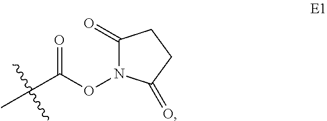
C00064

C00065

C00066

C00067

C00068

C00069

C00070

C00071

C00072

C00073

C00074

C00075

C00076

C00077

C00078

C00079

C00080

C00081

C00082

C00083

C00084

C00085

C00086

C00087

C00088

C00089

C00090

C00091

C00092

C00093

C00094

C00095

C00096

C00097

C00098

C00099

C00100

C00101

C00102

C00103

C00104

C00105

C00106

C00107

C00108

C00109

C00110

C00111

C00112

C00113

C00114

C00115

C00116

C00117

C00118

C00119

C00120

C00121

C00122

C00123

C00124

C00125

C00126

C00127

C00128

C00129

C00130

C00131

C00132

C00133

C00134

C00135

C00136

C00137

C00138

C00139
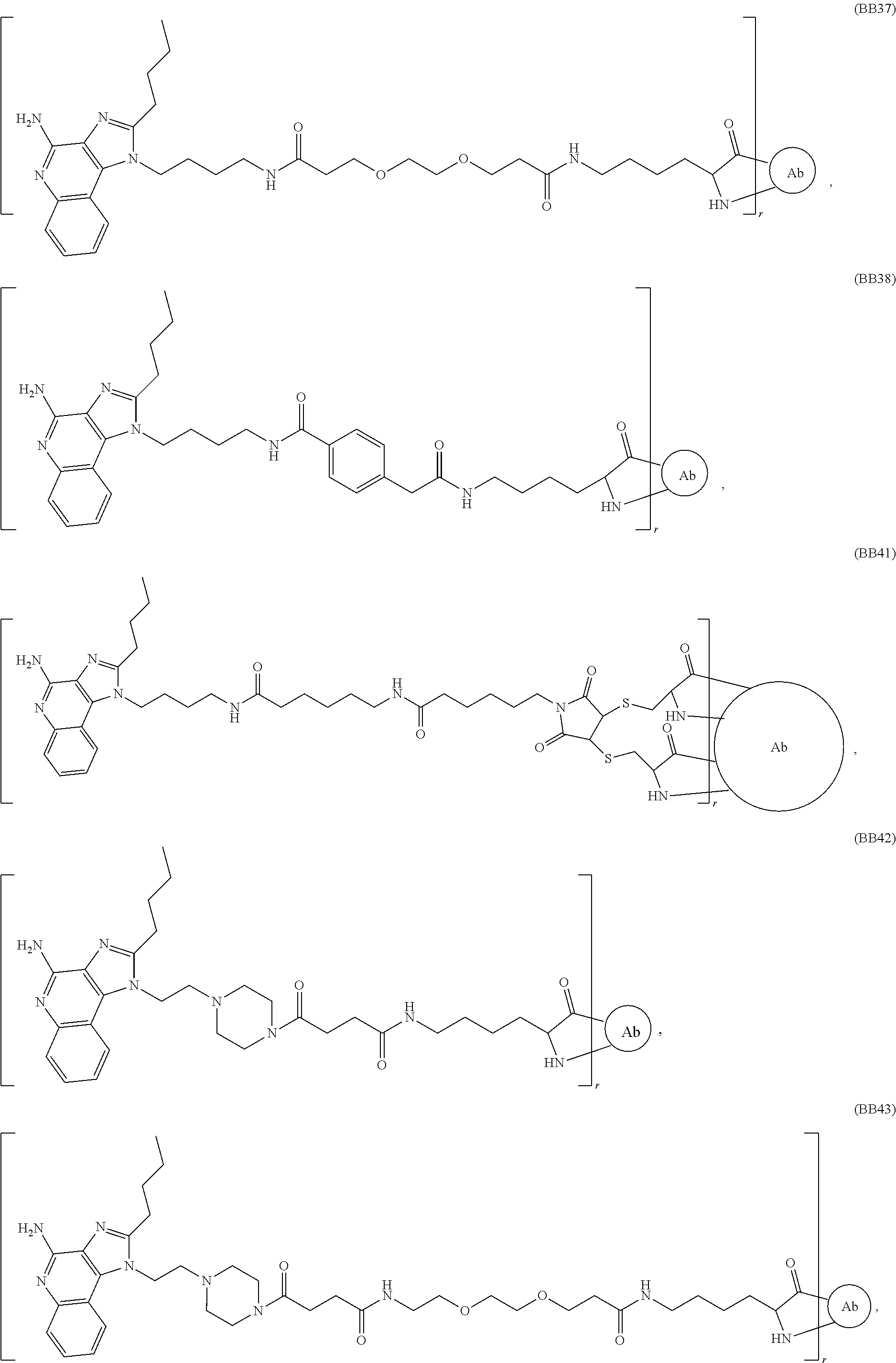
C00140

C00141

C00142

C00143

C00144

C00145

C00146

C00147

C00148

C00149

C00150

C00151

C00152

C00153

C00154

C00155

C00156

C00157

C00158

C00159

C00160

C00161

C00162

C00163

C00164

C00165

C00166

D00001

D00002

D00003

D00004

D00005

D00006

D00007

D00008

D00009

D00010

D00011

D00012

D00013

D00014

D00015

D00016

D00017

D00018

D00019

D00020

D00021

D00022

D00023

D00024

D00025

D00026

D00027

D00028

D00029

D00030

D00031

D00032

D00033

D00034

D00035

D00036

D00037

D00038

D00039

D00040

D00041

D00042

D00043

D00044

D00045

D00046

D00047

D00048

D00049

D00050

D00051

D00052

D00053

D00054

D00055

D00056

D00057

D00058

D00059

D00060

D00061

D00062

D00063

D00064

D00065

D00066

D00067

D00068

D00069

D00070

D00071

D00072

D00073

D00074

D00075

D00076

D00077

D00078

D00079

D00080

D00081

D00082
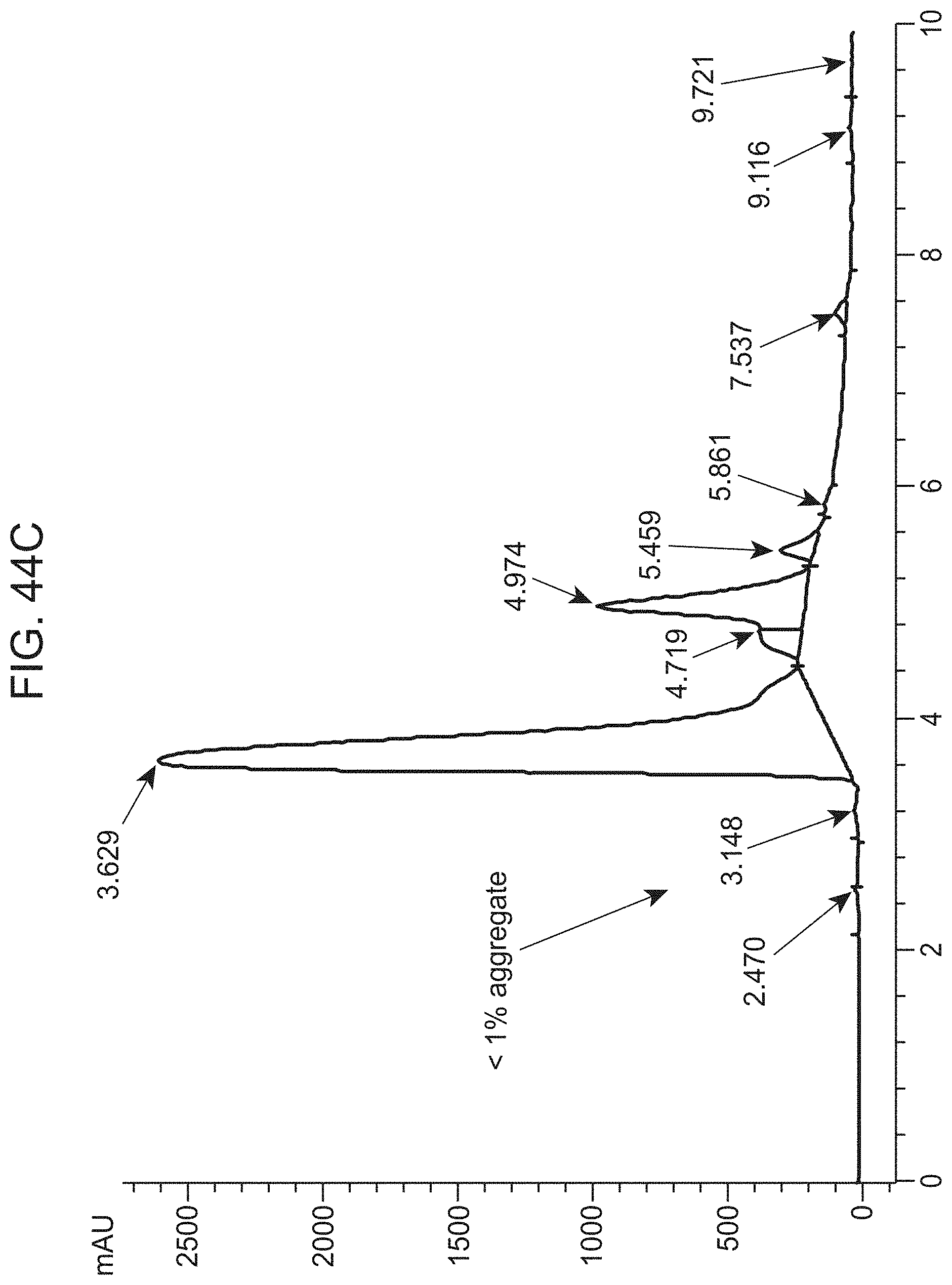
D00083

D00084

D00085

D00086

D00087

D00088

D00089

D00090

D00091

D00092

D00093

D00094

D00095

D00096

D00097

D00098

D00099

D00100

D00101
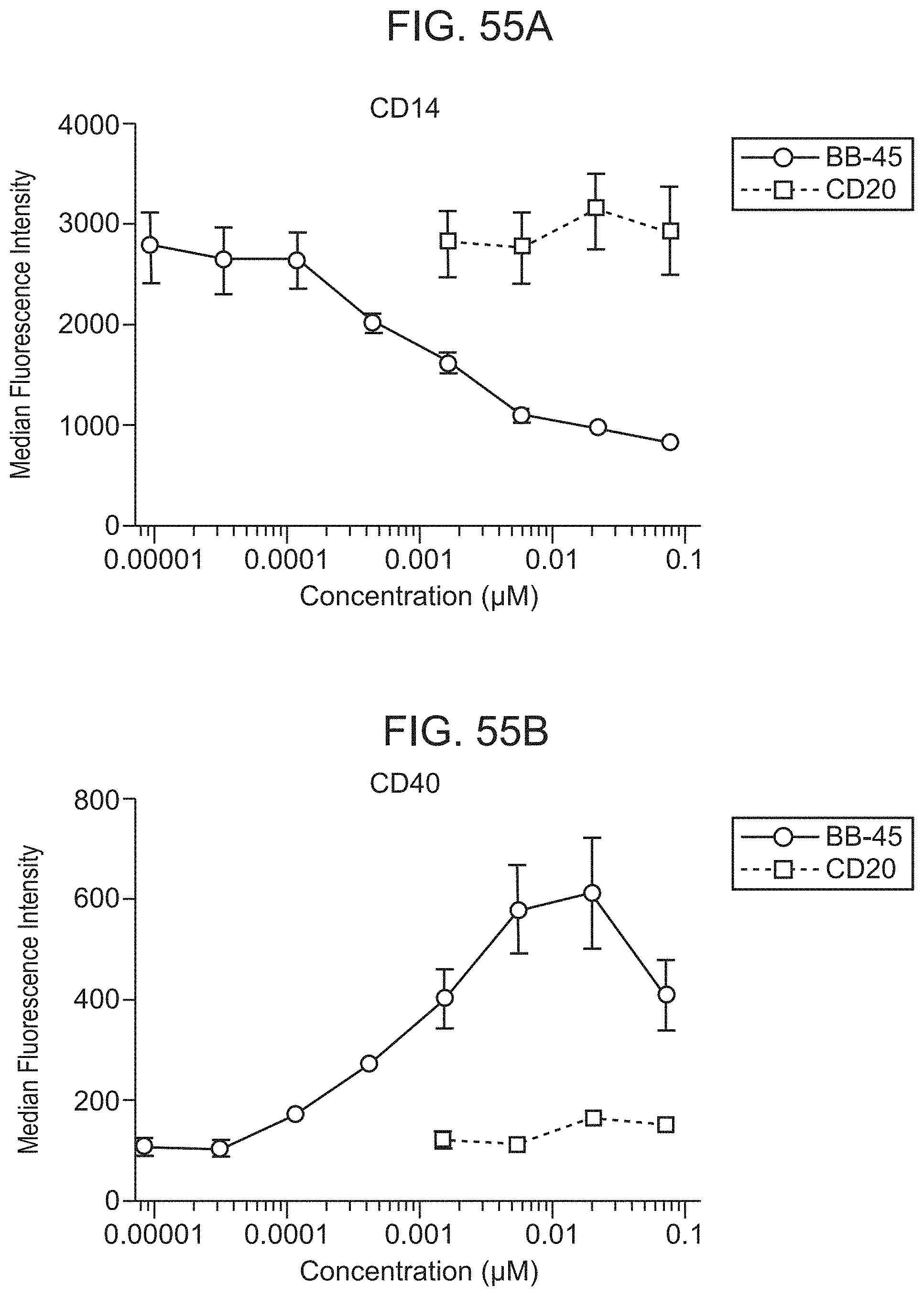
D00102

D00103

D00104

D00105

D00106

D00107

D00108

D00109

D00110

D00111

D00112

D00113

D00114

D00115
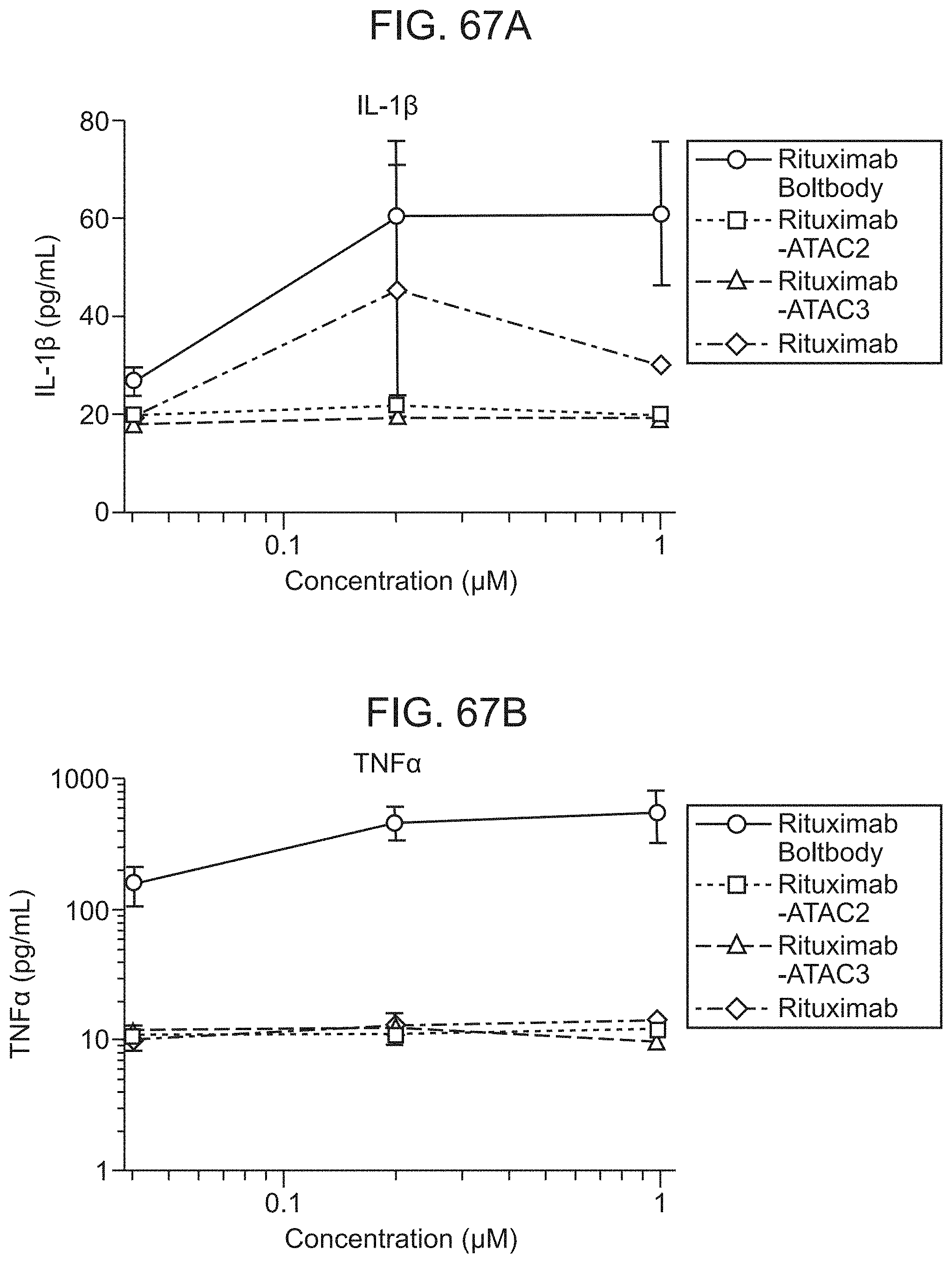
D00116

D00117

D00118

D00119

D00120

D00121

D00122

D00123

D00124

D00125

D00126

D00127

D00128

D00129

D00130

D00131

D00132

D00133

D00134

D00135

D00136

D00137

D00138

D00139

D00140

D00141

D00142

D00143

D00144

D00145

D00146

D00147

D00148

D00149

D00150

D00151

D00152

D00153

D00154

D00155

D00156

D00157

D00158

D00159

D00160

D00161

D00162

D00163

D00164

D00165

D00166

D00167

D00168

D00169

D00170

D00171

D00172

D00173

D00174

D00175

D00176

D00177

D00178

D00179

D00180

D00181

D00182

D00183

D00184

D00185

D00186

D00187

D00188

D00189

D00190

D00191

D00192

D00193

D00194

D00195

D00196

D00197

D00198
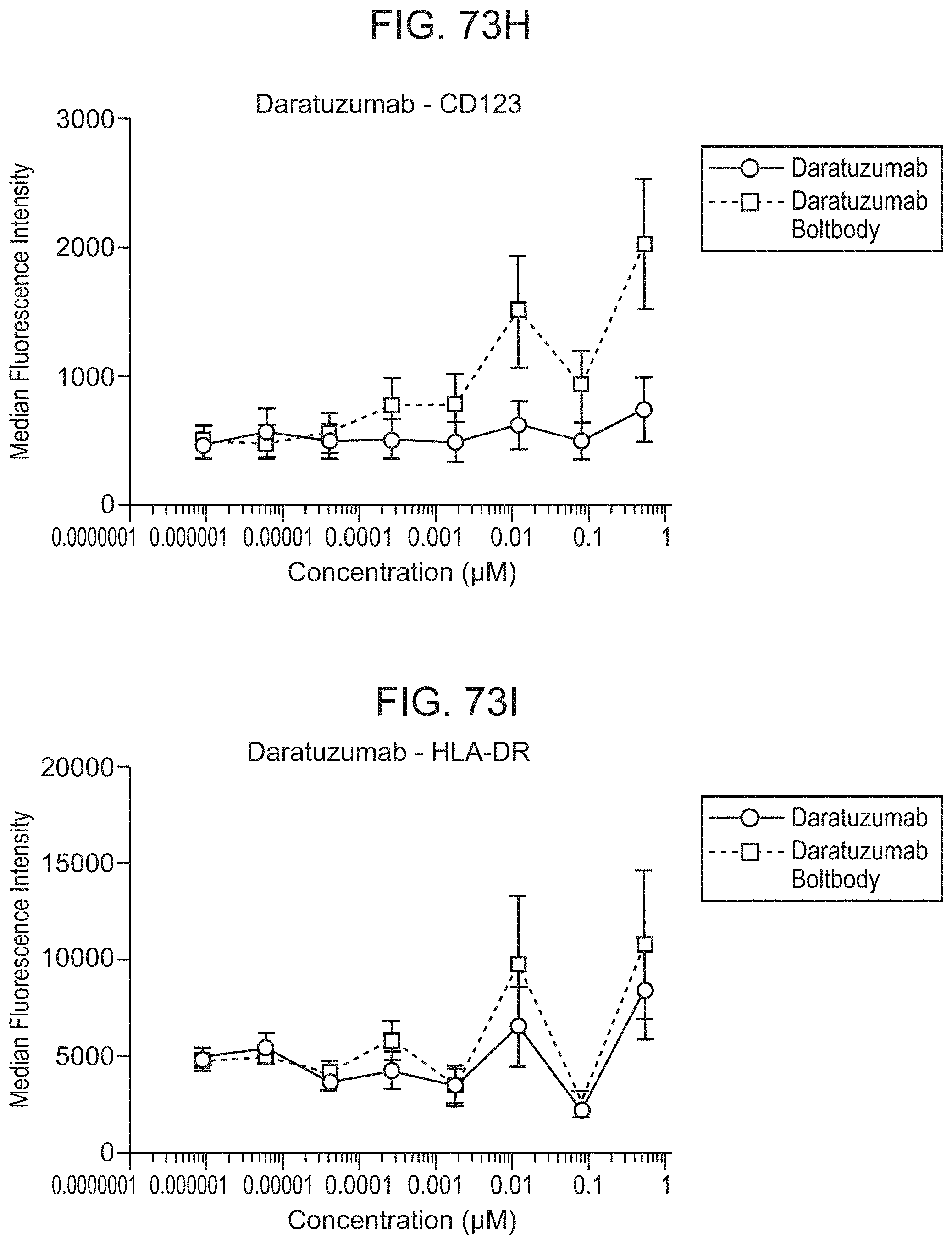
D00199

D00200

D00201

D00202

D00203

D00204

D00205

D00206

D00207

D00208

D00209

D00210

D00211

D00212

D00213

D00214

D00215

D00216

D00217

D00218

D00219

D00220

D00221

D00222

D00223

D00224

D00225

D00226

D00227

D00228

D00229

D00230

D00231

D00232

D00233

D00234

D00235

D00236

D00237

D00238

D00239

D00240

D00241

D00242

D00243

D00244

D00245

D00246

D00247

D00248

D00249

D00250

D00251

D00252

D00253

D00254

D00255

D00256

D00257

D00258

D00259

D00260

D00261

D00262

D00263

D00264

D00265

D00266

D00267

D00268

D00269

D00270

D00271

D00272

D00273

D00274

D00275

D00276

D00277

D00278

D00279

D00280

D00281

D00282

D00283

D00284

D00285

D00286

D00287

D00288

D00289

D00290

D00291

D00292

D00293

D00294

D00295

D00296

D00297

D00298

D00299

D00300

D00301

D00302

D00303

D00304

D00305

D00306

D00307

D00308

D00309

D00310

D00311

D00312

D00313

D00314

D00315

D00316

D00317

D00318

D00319

D00320

D00321

D00322

D00323

D00324

D00325

D00326

D00327

D00328

D00329

D00330

D00331

D00332

D00333

D00334

D00335

D00336

D00337

D00338

D00339

D00340

D00341

D00342

D00343

D00344

D00345

D00346

D00347

D00348

D00349

D00350

D00351

D00352

D00353

D00354

D00355

D00356

D00357

D00358

D00359

D00360

D00361

D00362

D00363

D00364

D00365

D00366

D00367

D00368

D00369

D00370

D00371

D00372

D00373

D00374

D00375

D00376

D00377

D00378

D00379

D00380

D00381

D00382

D00383

D00384

D00385

D00386

D00387

D00388

D00389

D00390

D00391

D00392

D00393

D00394

D00395

D00396

D00397

D00398

D00399

D00400

D00401

D00402

D00403

D00404

D00405

D00406

D00407

D00408

D00409

D00410

D00411

D00412

D00413

D00414

D00415

D00416

D00417

D00418

D00419

D00420

D00421

D00422

D00423

D00424

D00425

D00426

D00427

D00428

D00429

D00430

D00431

D00432

D00433

D00434

D00435

D00436
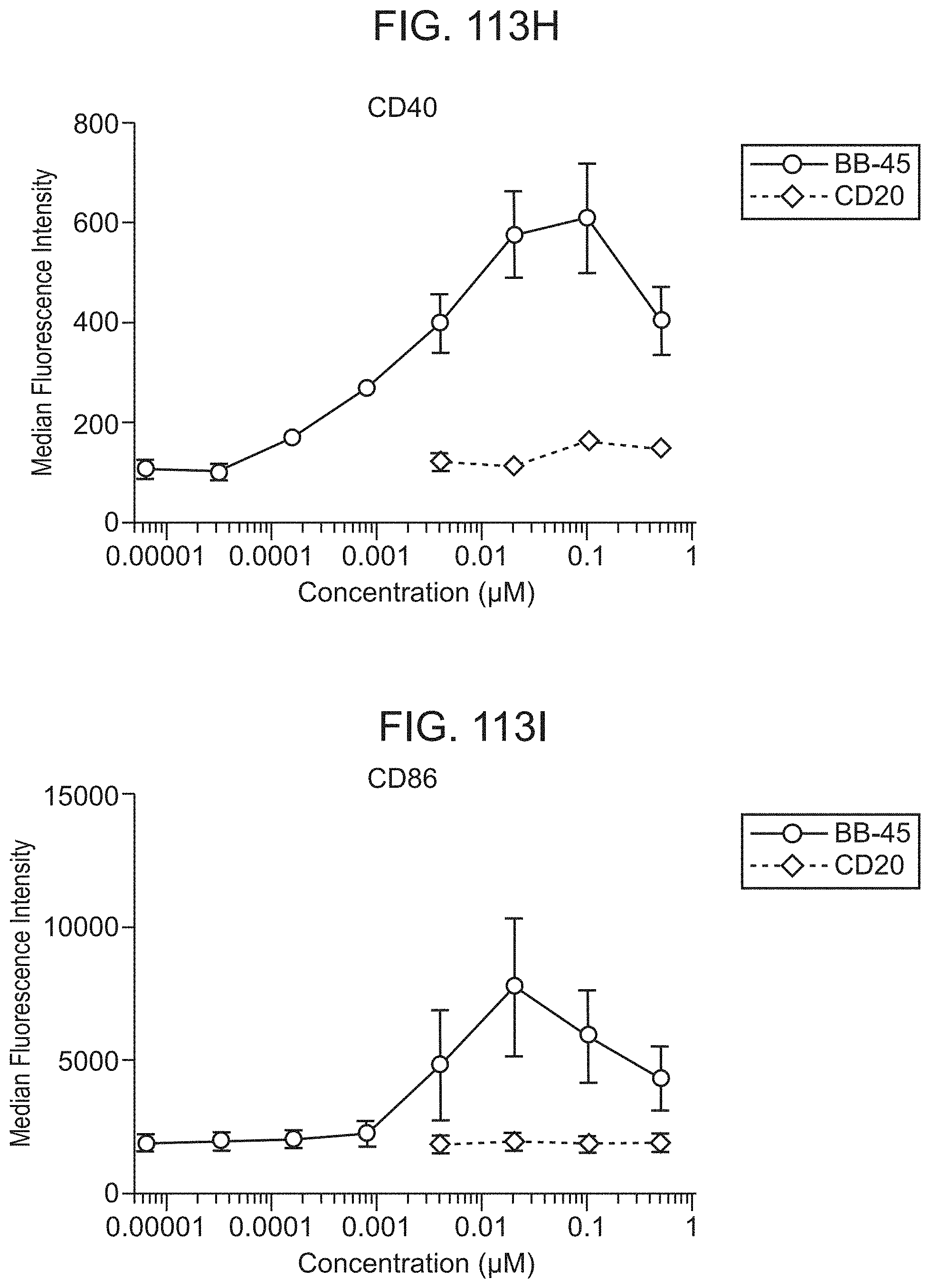
D00437

D00438

D00439

D00440

D00441

D00442

D00443

D00444

D00445

D00446

D00447

D00448

D00449

D00450

D00451

D00452

D00453

D00454

D00455

D00456

D00457

D00458

D00459

D00460

D00461

D00462

D00463

D00464

D00465

D00466

D00467

D00468

D00469

D00470

D00471

D00472

D00473

D00474

D00475

D00476

D00477

D00478

D00479

D00480

D00481

D00482

D00483

D00484
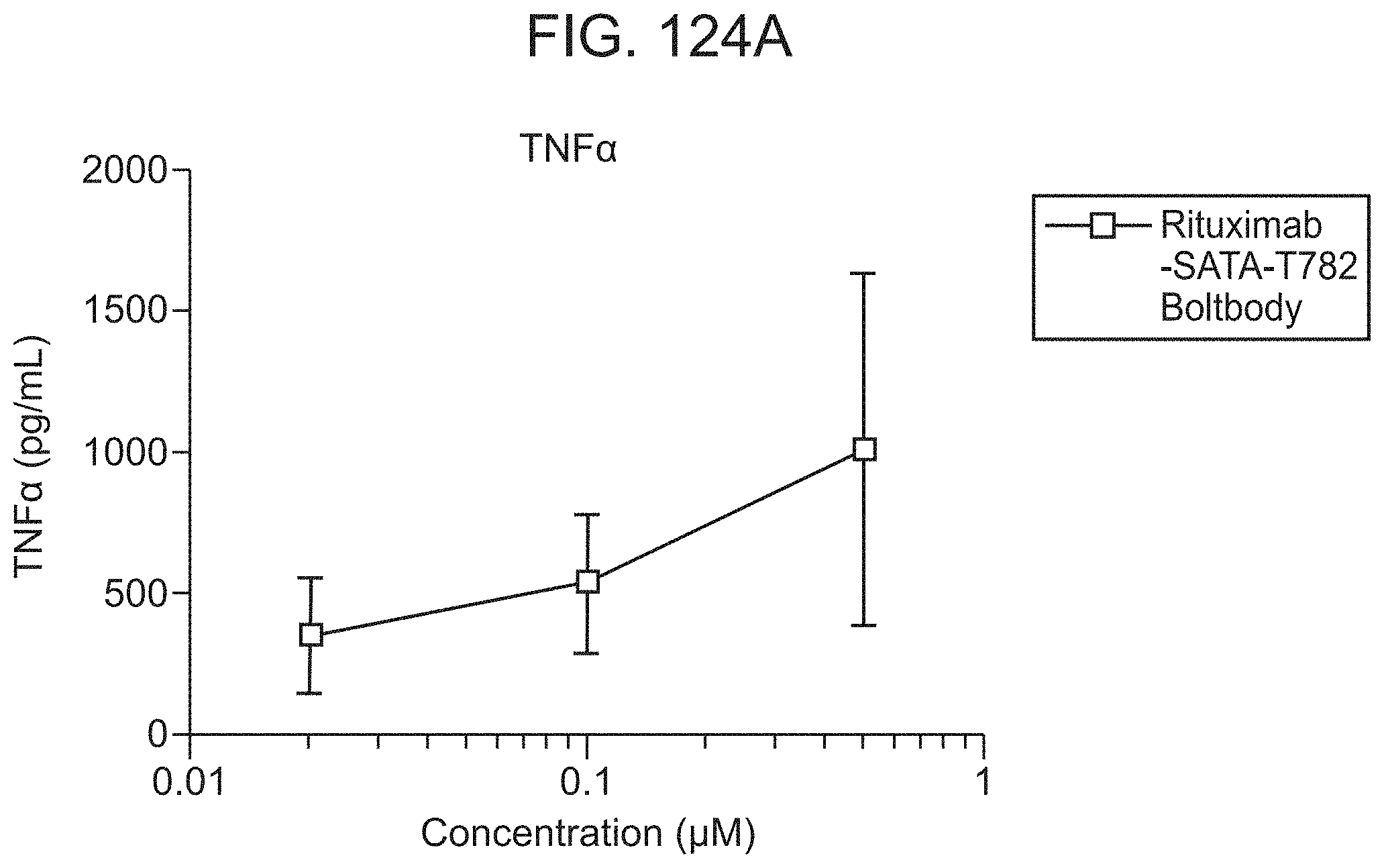
D00485

D00486

D00487

D00488

D00489

D00490

D00491

D00492

D00493

D00494

D00495

D00496

D00497

D00498

D00499

D00500

D00501

D00502

D00503

D00504

D00505

D00506

D00507

D00508

D00509

D00510

D00511

D00512

D00513

D00514

D00515

D00516

D00517

D00518

D00519

D00520

D00521

D00522

D00523

D00524

D00525

D00526

D00527

D00528

D00529

D00530

D00531

D00532

D00533

D00534

D00535

D00536

D00537

D00538

D00539

D00540

D00541

D00542

D00543

D00544

D00545

D00546

D00547

D00548

D00549

D00550

D00551

D00552

D00553

D00554

D00555

D00556

D00557

D00558

D00559

D00560

D00561

D00562

D00563

D00564

D00565

D00566

D00567

D00568

D00569

D00570

D00571

D00572

D00573

D00574

D00575

D00576

D00577

D00578

D00579

D00580

D00581

D00582

D00583

D00584

D00585

D00586

D00587

D00588

P00001

P00002

S00001
XML
uspto.report is an independent third-party trademark research tool that is not affiliated, endorsed, or sponsored by the United States Patent and Trademark Office (USPTO) or any other governmental organization. The information provided by uspto.report is based on publicly available data at the time of writing and is intended for informational purposes only.
While we strive to provide accurate and up-to-date information, we do not guarantee the accuracy, completeness, reliability, or suitability of the information displayed on this site. The use of this site is at your own risk. Any reliance you place on such information is therefore strictly at your own risk.
All official trademark data, including owner information, should be verified by visiting the official USPTO website at www.uspto.gov. This site is not intended to replace professional legal advice and should not be used as a substitute for consulting with a legal professional who is knowledgeable about trademark law.