Pharmaceutical composition for treating bone diseases which comprises protein comprising Frizzled 1, Frizzled 2 or Frizzled 7 extracellular cysteine-rich domain
Kakitani , et al.
U.S. patent number 10,646,544 [Application Number 15/609,575] was granted by the patent office on 2020-05-12 for pharmaceutical composition for treating bone diseases which comprises protein comprising frizzled 1, frizzled 2 or frizzled 7 extracellular cysteine-rich domain. This patent grant is currently assigned to KYOWA KIRIN CO., LTD.. The grantee listed for this patent is KYOWA HAKKO KIRIN CO., LTD. Invention is credited to Makoto Kakitani, Kazuma Tomizuka.


View All Diagrams
| United States Patent | 10,646,544 |
| Kakitani , et al. | May 12, 2020 |
Pharmaceutical composition for treating bone diseases which comprises protein comprising Frizzled 1, Frizzled 2 or Frizzled 7 extracellular cysteine-rich domain
Abstract
This invention relates to a pharmaceutical composition for treatment of a bone disease comprising, as an active ingredient, a protein comprising an extracellular cysteine-rich domain, which is from the Frizzled receptor selected from the group consisting of mammalian animal-derived Frizzled 1, Frizzled 2, and Frizzled 7 and has activity of increasing bone mass, bone density, and/or bone strength, or a mutant of such domain having sequence identity of 85% or higher to the amino acid sequence of the domain and having activity of increasing bone mass, bone density, and/or bone strength, or a vector comprising a nucleic acid encoding the protein.
| Inventors: | Kakitani; Makoto (Tokyo, JP), Tomizuka; Kazuma (Tokyo, JP) | ||||||||||
|---|---|---|---|---|---|---|---|---|---|---|---|
| Applicant: |
|
||||||||||
| Assignee: | KYOWA KIRIN CO., LTD.
(Chiyoda-ku, Tokyo, JP) |
||||||||||
| Family ID: | 42073517 | ||||||||||
| Appl. No.: | 15/609,575 | ||||||||||
| Filed: | May 31, 2017 |
Prior Publication Data
| Document Identifier | Publication Date | |
|---|---|---|
| US 20170266255 A1 | Sep 21, 2017 | |
Related U.S. Patent Documents
| Application Number | Filing Date | Patent Number | Issue Date | ||
|---|---|---|---|---|---|
| 13869083 | Apr 24, 2013 | 9700594 | |||
| 13121637 | |||||
| PCT/JP2009/066996 | Sep 30, 2009 | ||||
Foreign Application Priority Data
| Sep 30, 2008 [JP] | 2008-255804 | |||
| May 29, 2009 [JP] | 2009-131449 | |||
| Current U.S. Class: | 1/1 |
| Current CPC Class: | A61K 45/06 (20130101); A61K 31/7088 (20130101); A61K 38/1709 (20130101); A61P 35/00 (20180101); A61K 47/60 (20170801); A61K 47/6835 (20170801); A61P 19/08 (20180101); A61P 29/00 (20180101); A01K 67/027 (20130101); A61K 38/177 (20130101); A61P 19/10 (20180101); C07K 14/723 (20130101); A61K 39/3955 (20130101); A01K 67/0271 (20130101); A61P 19/02 (20180101); A01K 2217/072 (20130101); A61K 48/00 (20130101); A01K 2207/30 (20130101); A01K 2227/105 (20130101); A01K 2267/035 (20130101); A01K 2207/12 (20130101); C07K 2319/30 (20130101) |
| Current International Class: | A61K 38/17 (20060101); A61K 31/7088 (20060101); A61K 39/395 (20060101); A61K 47/68 (20170101); A61K 47/60 (20170101); C07K 14/72 (20060101); A01K 67/027 (20060101); A61P 19/08 (20060101); A61P 19/02 (20060101); A61K 45/06 (20060101); C07K 14/71 (20060101); A61K 38/18 (20060101); A61K 48/00 (20060101) |
References Cited [Referenced By]
U.S. Patent Documents
| 7713526 | May 2010 | Rhee et al. |
| 2003/0103986 | June 2003 | Rixon et al. |
| 2003/0165500 | September 2003 | Rhee |
| 2006/0034852 | February 2006 | Rixon et al. |
| 2006/0134109 | June 2006 | Gaitanaris |
| 2007/0072238 | March 2007 | Bhat |
| 2007/0117751 | May 2007 | Gurney |
| 2008/0299136 | December 2008 | Ernst et al. |
| 2009/0209006 | August 2009 | Rixon et al. |
| 2004535182 | Nov 2004 | JP | |||
| 02094852 | Nov 2002 | WO | |||
| 2008031009 | Mar 2008 | WO | |||
| 2008031013 | May 2008 | WO | |||
Other References
|
Haibach et al., Am. J. Clin. Pathol., 1985, vol. 83(5):594-600. cited by examiner . Sagara et al., Biochem. Biophys. Res. Commun., 1998, vol. 25291):117-122. cited by examiner . Chacon et al., "Frizzled-1 is involved in the Neuroprotective Effect of Wnt3a Against A.beta. Oligomers", J. Cellular Physiology, 217:215-227 (2008). cited by applicant . Dann et al., "Insights into Wnt binding and signaling from the structures of two Frizzled cysteine-rich domains", Nature, 412:86-90 (2001). cited by applicant . Daroszewska et al., "Mechanisms of Disease: genetics of Paget's disease of bone and related disorders",, Nature Clinical Practice Rheumatology, 2:270-277 (2006). cited by applicant . Deltagen, Inc., "NIH initiative supporting placement of Deltagene, Inc. mice into public repositories" MGI Direct Data Submission 2005 (http://www.informatics.jax.org/javawi2/servlet/WIFetch?page=alleleDetail- &key=40116), last database update Sep. 12, 2012. cited by applicant . European Communication for EP 09817788 dated Sep. 4, 2012, with Supplementary European Search Report dated Aug. 29, 2012. cited by applicant . International Search Report for PCT/JP2009/066996 dated Nov. 2, 2009. cited by applicant . Li et al., "Targeted Deletion of the Sclerostin Gene in Mice Results in Increased Bone Formation and Bone Strength", J. of Bone and Mineral Research, 23(6):860-869 (2008). cited by applicant . Masiakowski et al., "The Wnt receptor CRD domain is also found in MuSK and related orphan receptor tyrosine kinases", Curr. biol., 8:R407 (1998). cited by applicant . Matsumoto et al., "Wnt9a secreted from the walls of hepatic sinusoids is essential for morphogenesis, proliferation, and glycogen accumulation of chick hepatic epithelium", Dev. Biol., 319(2):234-247 (2008). cited by applicant . Merle et al., "Oncogenic role of the Frizzled-7/.beta.-catenin pathway in hepatocellular carcinoma", J. of Hepatology, 43(5):854-862 (2005). cited by applicant . Milovanovic et al., "Expression of Wng genes and frizzled 1 and 2 receptors in normal breast epithelium and infiltrating breast carcinoma", Int. J. Oncology, 25:1337-1342 (2004). cited by applicant . Nakanishi et al., "Secreted Frizzled-related Protein 4 is a negative regulator of Peak BMD in SAMP6 Mice", J. of Bone and Mineral Research, 21:1713-1721 (2006). cited by applicant . Rawadi et al., "Wnt signaling pathway: a new target for the treatment of osteoporosis", Oncologic, Endocrine & Metabolic, Expert Opin. Ther. Targets, 9(5):1063-1077 (2005). cited by applicant . Sagara et al., "Molecular Cloning, Differential Expression, and Chromosomal Localization of Human Frizzled-1, Frizzled-2, and Frizzled-7", Biochemical and Biophysical Research Communications, 252:117-122 (1998). cited by applicant . Semenov et al., "SOST is a ligand for LRP5/LRP6 and a Wnt Signaling Inhibitor", J. B. C., 280(29):26770-26775 (2005). cited by applicant . Tamai et al., "LDL-receptor-related proteins in Wnt signal transduction", Nature, 407:530-535 (2000). cited by applicant . Gregorieff et al., "Expression Pattern of Wnt Signaling Components in the Adult Intestine", Gastroenterology, 129:626-638 (2005). cited by applicant . Holcombe et al., "Expression of Wnt ligands and Frizzled receptors in colonic mucosa and in colon carcinoma", Mol. Pathol., 55:220-226 (2006). cited by applicant . Huang et al., "The Frizzled family: receptors for multiple signal transduction pathways", Genome Biology, 5(234):1-7 (2004). cited by applicant . Gross, et al. "TACI-Ig Neutralizes Molecules Critical for B Cell Development and Autoimmune Disease: Impaired B Cell Maturation in Mice Lacking BLyS", Immunity, 15:289-302 (2001). cited by applicant . Katoh et al., "Comparative integromics on FZD7 orthologs: Conserved binding sites for PU.1, SP1, CCAAT-box and TCF/LEF/SOX transcription factors within 5'-promoter region of mammalian FZD7 orthologs", Int. J. Mol. Med., 19:529-533 (2007). cited by applicant . Kemp et al., "Expression of Frizzled5, Frizzled7, and Frizzled10 during early mouse development and Interactions with Canonical Wnt Signaling", Dev. Dynamics., 236:2011-2019 (2007). cited by applicant . Trevat et al., "Expression of Secreted Frizzled Related Protein 1, A Wnt Antagonists, in Brain, Kidney, and Skeleton in Dispensable for Normal Embryonic Development", J. Cell Physiol., 217:113-126 (2008). cited by applicant . Vincan et al., "Frizzled-7 Receptors ectodomain expression in a colon cancer cell line induces Morphological change and attenuates tumor growth", Differentiation, 73:142-153 (2005). cited by applicant . Wang et al., "A Large family of Putative Transmembrane Receptors Homologous to the Product of the Drosophila Tissue Polarity Gene Frizzled" J. B. C., 271(8):4468-4476 (1996). cited by applicant . Wheeler et al., "Inducible gene expression in transgenic Xenopus embryos", Current Biology, 10:849-852 (2000). cited by applicant . Yaccoby, "Antibody-based inhibition of DKK1 suppresses tumor-induced bone resorption and multiple myeloma growth in vivo", Blood, 109:2106-2111 (2007). cited by applicant. |
Primary Examiner: Xie; Xiaozhen
Attorney, Agent or Firm: Sughrue Mion, PLLC
Parent Case Text
CROSS-REFERENCE TO RELATED APPLICATIONS
This application is a Continuation of U.S. application Ser. No. 13/869,083, filed Apr. 24, 2013, now U.S. Pat. No. 9,700,594, issued Jul. 11, 2017; which is a Divisional of U.S. application Ser. No. 13/121,637, filed Jun. 13, 2011 (now abandoned); which is a 371 National Stage of International Application No. PCT/JP2009/066996 filed Sep. 30, 2009; which claims priority based on Japanese Patent Application No. 2008-255804, filed Sep. 30, 2008, and Japanese Patent Application No. 2009-131449 filed May 29, 2009; the contents of all of which are incorporated herein by reference in their entirety.
Claims
The invention claimed is:
1. A method for increasing bone mass, bone density and/or bone strength, comprising: administering to a mammalian animal with a disease selected from the group consisting of osteoporosis, osteoarthritis, articular rheumatism, hypercalcemia, Paget's disease of bone, osteopetrosis, Camurati-engelmann's disease, arthropathy, primary hyperthyreosis, osteopenia, osteohalisteresis, rachitis, traumatic bone fracture, and fatigue bone fracture, an effective amount of a pharmaceutical composition which comprises, as an active ingredient, a protein comprising an extracellular cysteine-rich domain comprising the amino acid sequence of SEQ ID NO:21 or 26, to increase bone mass, bone density and/or bone strength.
2. The method according to claim 1, wherein the mammalian animal is a human.
3. The method according to claim 1, wherein the extracellular cysteine-rich domain comprises an amino acid sequence spanning from the 1st cysteine residue on the N-terminal side to the 10th cysteine residue in the amino acid sequence of an extracellular region of a human Frizzled 2 receptor.
4. The method according to claim 1, wherein the protein comprises the amino acid sequence of SEQ ID NO: 19 or 25.
5. The method according to claim 1, wherein the protein is a recombinant protein.
6. The method according to claim 1, wherein the protein is a fusion protein of the extracellular cysteine-rich domain and a mammalian immunoglobulin Fc protein or a mutant thereof prepared so that antibody dependent cellular cytotoxicity (ADCC) and complement dependent cytotoxicity (CDC) activities are lowered.
7. The method according to claim 6, wherein the Fc protein or the mutant thereof comprises the amino acid sequence of SEQ ID NO: 4.
8. The method according to claim 6, wherein the Fc protein or the mutant thereof comprises the amino acid sequence encoded by the nucleotide sequence of SEQ ID NO: 3.
9. The method according to claim 1, wherein the protein is chemically modified.
10. The method according to claim 9, wherein the chemical modification is a binding of one or more polyethylene glycol molecules.
11. The method according to claim 9, wherein the chemical modification is a binding of one or more sugar chains.
12. The method according to claim 1, wherein the composition is simultaneously or continuously administered in combination with another therapeutic agent for increasing bone mass, bone density and/or bone strength.
Description
TECHNICAL FIELD
The present invention relates to a novel application of a therapeutic agent for a bone disease comprising a protein comprising the extracellular cysteine-rich domain of Frizzled 1, Frizzled 2, or Frizzled 7, which is known as a Wnt ligand receptor protein, or a mutant of the domain.
This finding resulted from analysis of properties of a knock-in mouse expressing the Frizzled extracellular cysteine-rich domain and a mouse to which a protein comprising the Frizzled extracellular cysteine-rich domain had been administered.
BACKGROUND ART
A super-aging society has arrived, the number of people with osteoporosis has increased, and bone fractures resulting therefrom have come to constitute a serious issue of concern at a societal level. In particular, patients with femoral neck fractures and vertebral body fractures become bedridden, which causes significant deterioration of the quality of life thereof, and the social, medical, and econimc burdens caused by care and hospital treatment have increased (Tosteson, A. N., et al., Osteoporos Int., 12, 1042-1049, 2001; and Yoh, K., et al., J. Bone Miner. Metab., 23, 167-173, 2005). It has also been discovered in recent years that osteoporosis is significantly associated with mortality in old age (Nguyen, N. D., et al., J. Bone Miner. Res., 22, 1147-1154, 2007; and Muraki, S., et al., J. Bone Miner. Metab., 24, 100-104, 2006). Under such circumstances, prevention and treatment of osteoporosis have become critical objectives to be achieved. Osteoporosis (i.e., a pathological condition where bone mass is reduced while the rate of the amount of the bone matrix to the amount of the mineralized bone matrix is held) is classified as primary osteoporosis or secondary osteoporosis. The former type is a pathological condition heretofore referred to as postmenopausal osteoporosis or senile osteoporosis. The latter type is a pathological condition caused by changes in bone metabolism resulting from other diseases, and such osteoporosis is classified based on the cause thereof, such as osteoporosis caused by endocrine, nutritional/metabolic, inflammatory, immobile, drug-induced, hematologic, congenital, or other diseases. According to the above classification, examples of causes for secondary osteoporosis include: endocrine causes, such as hyperparathyreosis, hyperthyreosis, hypogonadism, Cushing's syndrome, somatotropin deficiency, diabetes, Addison's disease, and calcitonin deficiency; nutritional/metabolic causes, such as chronic degenerative diseases, emaciation, serious liver diseases (primary biliary cirrhosis, in particular), gastric resection, scorbutus, malabsorption syndrome (including celiac disease), hypophosphatemia, chronic renal disease, hypercalciuria, hemochromatosis, amyloidosis, mast cell tumor, ingestion of excess sodium, insufficient calcium intake, and hypervitaminosis D or A; inflammatory causes, such as articular rheumatism, periarticular bone disease (elevated bone resorption induced by proinflammatory cytokines), and sarcoidosis; immobility-related causes, such as systemic, bed rest, paralysis, local, and post-fracture causes; drug-induced causes, such as with the use of steroids (steroids are extensively used for inflammatory diseases as immunosuppressive agents; examples of diseases treated with the use of steroids include collagen diseases, asthma, inflammatory bowel diseases, and in the case of organ transplantation, and bone loss is a serious side effect of such therapy), methotrexate, heparine, warfarin, anticonvulsant agents, lithium, and tamoxifen; blood-disease-induced causes, such as multiple myeloma, lymphoma, leukaemia, hemophilia, and chronic hemolytic diseases; congenital causes, such as dysosteogenesis, Marfan's syndrome, Kleinfelter's syndrome, congenital erythropoetic porphyria, and cystic fibrosis; and other disease-induced causes, such as with chronic obstructive lung diseases, hepatic failure, renal diseases, articular rheumatism, pregnancy, hyperoxemia, and HIV infection (Committee for Creation of Guidelines for Prevention and Treatment of Osteoporosis, Guidelines for Prevention and Treatment of Osteoporosis 2006, Life Science Publishing, Co., Ltd., Japan, 2006).
Among the above-mentioned diseases, bone diseases resulting from osteoarthritis, articular rheumatism, malignant tumors, or renal diseases are specifically regarded as bone diseases that impose serious influences at the societal level, in addition to primary osteoporosis.
Osteoarthritis develops most often in locomotor regions. The number of patients afflicted therewith is said to be 10,000,000 in Japan, and it has been deduced that the number of patients will keep increasing as the aging of society advances. Advanced articular disorders are treated via artificial joint replacement; however, radical treatment of moderate or milder symptoms has not yet been reported (Nampei, A. & Hashimoto, J., The Bone, 22, 3, 109-113, 2008).
Articular rheumatism is a chronic and progressive inflammatory disease characterized mainly by multiple arthritis. Articular synovial proliferation gradually causes infiltration of cartilage or bones in the vicinity thereof, and articular rheumatism often leads to destruction and deformation of joints. It has been reported that treatment with the use of an antirheumatic drug (methotrexate) cannot sufficiently inhibit the progress of joint destruction, and a biological agent targeting a tumor necrosis factor (TNF) a produces significant effects of inhibiting joint destruction. Thus, it is considered to be a revolutionary agent. However, increased incidence, as a side effect, of opportunistic infection, tuberculosis (extrapulmonary tuberculosis), Pneumocystis pneumonia, or the like when using such agent is an issue of concern (Soen, S., The Bone, 22, 3, 103-107, 2008).
Major examples of bone diseases involved in malignant tumors include hypercalcemia and bone metastasis related to malignant tumors. Hypercalcemia causes loss of appetite and diuresis, and it causes dehydration and renal failure caused thereby. Bone metastasis is often observed in patients with breast cancer, prostate cancer, or lung cancer, in particular. While bone metastasis is hardly ever fatal by itself, it causes bone ache, pathologic fracture, neuroparalysis, or the like. It thus often significantly deteriorate patients' QOL, and bone metastasis control is a critical objective in clinical settings (Takahashi, S., The Bone, 22, 3, 115-120, 2008). These bone diseases related to malignant tumors are treated with the use of bisphosphonate preparations, although the problem of side effects has been pointed out.
Among bone diseases related to renal diseases, a pathological condition of bone damage caused by renal tissue damage is referred to as renal osteodystrophy. Bone disease experienced by kidney dialysis patients are mainly caused by secondary hyperparathyreosis. Because of the elevated PTH concentration caused by hyperparathyreosis and, for example, insufficient production of bone morphogenetic protein (BMP) 7, renal osteodystrophy advances. Dialysis patients often exhibit lowered reactivity of the bone with the parathyroid hormone (PTH). When the PTH concentration is chronically and significantly elevated, accordingly, fibrous ostitis (high bone turnover) develops. When the PTH concentration is maintained within a standard range, in contrast, bone aplasia (low bone turnover) develops.
When fibrous ostitis advances, collagen fibers are irregularly formed, such fibers are mineralized as non-crystalline calcium phosphate, and woven bone is then formed. This enhances bone formation, although the bone becomes easily fracturable. Basic treatment of fibrous ostitis involves inhibition of parathyroid hormone secretion, which mainly entails calcium ingestion and administration of active vitamin D. When a patient has a chronic kidney disease (CKD) and receives dialysis treatment, in particular, various regulations, such as restrictions on food or water intake, are necessary. When secondary hyperparathyreosis advances, hypercalcemia also becomes an issue of concern. When prescribing active vitamin D, extreme caution, such as via the monitoring of renal functions (i.e., serum creatinine level) and serum calcium level, is always required.
Bone aplasia develops because of prolonged use and excessive administration of active vitamin D preparations or suppression of parathyroid hormone after parathyroidectomy (PTX).
The rate of fractures associated with bone aplasia is higher than that associated with fibrous ostitis, and it induces hypercalcemia or mineralization of blood vessels or other soft tissues. Thus, adequate treatment techniques have been desired. A pathological condition of bone aplasia is low bone turnover in which bone resorption and bone formation are inhibited, and there is no established treatment technique at present (Daugirdas, J. T., et al., Rinsho Toseki Handbook (Handbook of Dialysis), Fourth Edition, Medical Sciences International, Ltd., Japan, 2009).
Hyperphosphatemia or hypercalcemia caused by lowered capacity of the bone for phosphorus or calcium intake (low-turnover metabolic bone) or lowered storage capacity (high-turnover metabolic bone) is considered to be a cause of ectopic (vascular) mineralization. Cardiovascular complications account for 40% or more of the deaths of patients with chronic renal failures, and dialysis patients in particular, and arteriosclerosis involving vascular mineralization has drawn attention as a serious pathological condition. Treatment of mineralization of advanced lesions in patients with chronic renal failures remains difficult at present and the prognosis thereof is poor (Fujiu, A. et al., Rinsho Toseki (the Japanese Journal of Clinical Dialysis), 24, 43-50, Nihon Medical Center, Japan, 2008). In addition to agents for treating primary osteoporosis, accordingly, development of agents that more effectively act on bone diseases resulting from osteoarthritis, articular rheumatism, malignant tumors, or renal disease and vascular mineralization resulting from bone diseases with reduced side effects has been desired.
It is considered that bone metabolism is regulated by the balance between osteoblast functions and osteoclast functions, and osteoporosis develops when the bone-destroying activity exceeds bone-building activity (Cohen, M. M. Jr., American J. Med. Genetics, Part A, 140A, 2646-2706, 2006). In particular, secretion of the female hormone that assumes the role of protecting bones is lowered in postmenopausal women, a lowered capacity of osteoblasts for bone formation and the elevated bone resorption activity of osteoclasts are consequently observed, and it is highly likely that symptoms of osteoporosis would develop (Kousteni, S., et al., Cell, 104, 719-730, 2001; and Nakamura, T., et al., Cell, 130, 811-823, 2007). In order to overcome such problems, estrogen preparations have been used; however, application thereof has been restricted due to the increased risk of thrombosis and breast cancer caused by the use of such preparations. It is also reported that use of a selective estrogen receptor modulator would increase the risk of deep vein thrombosis (Wada, S., et al., Mebio, 25, 8, 89-95, 2008).
At present, calcitonin, bisphosphonate, and the like are used as agents that inhibit the bone resorption activity of osteoclasts. Calcitonin is known to bind to a calcitonin receptor expressed on the osteoclast surface to inactivate osteoclasts, and it is used for treatment of not only osteoporosis but also hypercalcemia, Paget's disease of bone, and the like in clinical settings. However, no effects thereof on bone fracture inhibition have yet been found, and calcitonin receptor expression is reported to be down-regulated by calcitonin administration (Wada, S., et al., Mebio, 25, 8, 89-95, 2008; and Wada, S. & Yasuda, S., Clin. Calcium, 11, 9, 1169-1175, 2001). Bisphosphonate exhibits potent bone resorption inhibitory activity, and amino-containing bisphosphonates, such as andronate and risedronate, are major therapeutic agents for osteoporosis in Japan. Such bisphosphonate preparations inhibit farnesyl diphosphate synthase, block lipid protein prenylation, and induce inhibition of bone-resorption functions and osteoclast apoptosis (Nakamura, T., The Bone, 22, 3, 147-151, 2008). However, the FDA warned of crises of severe skeletal, articular, or muscular pain in 2008 as problems of bisphosphonate preparations. In addition, side effects, such as jaw bone necrosis, caused by the prolonged use thereof (i.e., for 2 or 3 years or longer) after dental care have been reported (Sanna, G., et al., Ann. Oncol., 16, 1207-1208, 2005). An anti-RANKL antibody has been expected as a novel osteoclastic inhibitor other than those described above. Further, application of the anti-RANKL antibody as an inhibitor of articular destruction in the case of articular rheumatism or as a therapeutic agent for multiple myeloma has been expected, and clinical development thereof is in progress. Based on a report to the effect that the RANKL/RANK pathway is important for the survival and maintenance of dendritic cells (Theill, L. E., et al., Ann. Rev. Immunol., 20, 795-823, 2002) or a report to the effect that lymph node dysplasia is caused in an RANK- or RANKL-deficient mouse (Kong, Y. Y., et al., Nature, 397, 315-323, 1999; and Dougall, W. C., et al., Genes Dev., 13, 2412-2424, 1999), the influence of an anti-RANKL antibody preparation on the immune system has become an issue of concern. In 2008, AMGEN reported that an increased rate of development of some infectious diseases was found through a clincal test of the anti-RANKL antibody preparation (Denosumab). As a result of the clinical test of the anti-RANKL antibody conducted in 2009, development of jaw bone necrosis was found to be a side effect, as in the cases of the bisphosphonate preparations. Treatment via intermittent administration of PTH alone as an osteogenesis accelerator that activates osteoblasts has been conducted (Teriparatide, Eli Lilly; an unapproved drug in Japan), but such agent is not different from other therapeutic agents, such as bisphosphonate preparations, in that activity of increasing cortical bone thickness is not very high compared with activity of increasing cancellous bone mass. Accordingly, the effects thereof for bone fracture prevention are not considered to be very high. In relation to PTH, further, Asahi Kasei Pharma Corp. (Japan) has reported problems, such as side effects such as palpitation, tachycardia, and a lowering in blood pressure, and osteosarcoma observed in a long-term administration test to rats, unapproved continuous use thereof for 1.5 to 2 years or longer in Europe and the United States, and prohibited application thereof to cancer patients. Thus, it is impossible to use PTH for inhibition of cancer bone metastasis, treatment of cancer-induced hypercalcemia (paraneoplastic humoral hypercalcemia or local osteolytic hypercalcemia caused by the parathyroid-hormone-related peptide produced by tumor cells), or other purposes.
Accordingly, development of agents that more effectively work for osteoporosis caused by the lowered capacity of osteoblasts for bone formation or elevated bone resorption activity of osteoclasts in postmenopausal women, hypercalcemia, Paget's disease of bone, inhibition of bone metastasis inhibition of articular destruction associated with articular rheumatism, or multiple myeloma with reduced side effects has been awaited.
In addition thereto, osteohalisteresis and rachitis are known as bone diseases induced by selective inhibition of mineralization, unlike osteoporosis. A bone is formed by mineralization of a matrix layer comprising collagen or the like via hydroxyapatite deposition. Osteohalisteresis is a pathological condition in which such mineralization is blocked and osteoids increase, and it is referred to as rachitis if developed during childhood. Symptoms include bone and joint pains, such as chiropodalgia, arthralgia, lumbago, and backache, which lead to gait impairment and to a state in which bone is easily fractured. In the case of children, developmental disorders, limb deformities such as bow-legs, pigeon breast deformity, or other symptoms are observed. Such symptoms are generally treated with the use of vitamin D, calcium preparations, and phosphorus preparations, in addition to alimentary therapy. If the level of dysfunction caused by a deformity is high, however, surgical operation is the only possible symptomatic treatment. Therefore, development of agents that are more effective on osteohalisteresis or rachitis has been awaited.
As described above, bone is tissue that is always regulated by the balance between osteoblast functions and osteoclast functions and remodeled. In order to achieve tough bone that is more resistant to fracture, accordingly, a mere increase in bone mass may not be sufficient. In the case of hereditary diseases, such as osteopetrosis (Horiuchi A., CLINICIAN, 47, 401-404, 2000), Paget's disease of bone (Daroszewska, A., & Ralston, S. H., Nature Clinical Practice Rheumatology, 2, 270-277, 2006), or Camurati-engelmann's disease (CED) (Janssens, K., et al., Nature Genetics, 26, 273-275, 2000; and Tang, Y., et al., Nature Medicine, 15, 757-765, 2009), for example, it is known that the balance between bone formation and bone resorption becomes abnormal due to different causes, and bone strength is lowered even though bone mass is increased. Examples of factors that determine bone strength from the viewpoint of mechanisms of materials include form-related factors, such as connectivity of cancellous bones, thickness of cortical bones, porosity, and cross-sectional moment, and qualitative factors, such as mineralization or bone fatigue, in addition to quantitative factors represented by bone density (Mori S., CLINICIAN, 49, 621-626, 2002). Therefore, development of agents useful for improving bone strength, in addition to increasing bone mass, has been awaited for the purpose of treatment of primary osteoporosis and secondary osteoporosis.
In recent years, factors associated with the Wnt/LRP signal control mechanism have drawn attention as targets for drug discovery regarding a bone formation accelerator. Wnt is a secreted glycoprotein that has been lipid-modified by palmitic acid having a molecular weight of about 40,000, and 19 types thereof are considered to be present in mammalian animals. As Wnt receptors, 10 types of seven-transmembrane receptors (i.e., Frizzled receptors) and two types of single-transmembrane receptors (i.e., LRP5/6 receptors) have been reported (Tamai, K., et al., Nature, 407, 530-535, 2000). A region referred to as a cysteine-rich domain (CRD) containing conserved 10 cysteine residues is present in an extracellular region of the Frizzled receptor family molecule to which Wnt is considered to bind. The region from the cysteine residue located closest to the N-terminus to the cysteine residue located closest to the C-terminus of such 10 cysteine residues may be exclusively designated as a CRD (Masiakowski, P. & Yancopoulos, G. D., Curr. Biol. 8, R407, 1998), or a region comprising such 10 cysteine residues and sequences each located closer to the C- or N-terminus may be designated as a CRD (R & D systems). CRDs were reported to have homodimer structures based on crystal structural analysis using a CRD of mouse Frizzled 8 (Dann, C. E., et al., Nature, 412, 86-90, 2001). At least three types of Wnt signaling pathways are considered to exist: a canonical-Wnt signaling pathway; a non-canonical Wnt signaling pathway, which is a PCP (planar cell polarity) pathway mediated by a small G-binding protein; and a Ca.sup.2+ pathway mediated by a trimeric G protein. Bone-metabolism-related research on the canonical-Wnt signaling pathway is the most advanced, and Wnt is considered to promote bone formation (Rawadi, G. & Roman-Roman, S., Expert Opin. Ther. Targets, 9, 5, 1063-1077, 2005). Therefore, regulation of functions of endogenous factors that inhibit this signaling pathway has been attempted in recent years for the purpose of application thereof to treatment of bone diseases.
Sclerostin was recognized as a BMP antagonist at first; however, it was reported to be a factor that would directly bind to LRP5/6 to inhibit the signaling pathway in research conducted later (Semenov, M., et al., J. B. C., 280, 29, 26770-26775, 2005). A significant increase was observed in bone density in a Sclerostin-knockout mouse (Li, X., et al., J. Bone Miner. Res., 23, 860-869, 2008). At present, a Sclerostin-neutralizing antibody is undergoing phase I trials in Europe and the United States of America (AMG785, Amgen & UCB), and the future development thereof has drawn attention. A DKK1 (Dickkopf-1)-neutralizing antibody that is known as another canonical-Wnt signal inhibitor was prepared, inhibition of lowered bone density was observed in an SCID mouse into which multiple myeloma (MM) cells had been transplanted (Yaccoby, S., Blood., 109, 2106-2111, 2007), and clinical trials using a neutralizing antibody (BHQ880, Novartis) have been conducted.
sFRP (soluble frizzled-related protein) that is considered to be a Wnt decoy receptor and has high amino acid sequence homology to the Frizzled extracellular domain is considered to negatively regulate Wnt signals (Nakanishi, R., et al., J. Bone Miner. Res., 21, 1713-1721, 2006), and an increase in the amount of cancellous bone in the femur of an sFRP1 knockout mouse has been reported (Trevant, B., et al., J. Cell. Physiol. 217, 113-126, 2008). Under such circumstances, research and development related to sFRP1 inhibitors have proceeded (Wyeth).
Frizzled 7 has been identified as a receptor that binds to a Wnt ligand and transmits signals thereof (Wang, Y., et al., J. B. C., 271, 8, 4468-4476, 1996; and Huang, H-C., & Klein, P. S., Genome Biology, 5, 234, 1-7, 2004). The amino acid sequence of the human Frizzled 7 extracellular cysteine-rich domain (when a region from the cysteine residue located closest to the N-terminus to the cysteine residue located closest to the C-terminus of such 10 conserved cysteine residues is exclusively designated as a CRD) is completely identical to that of the mouse Frizzled 7 extracellular cysteine-rich domain (i.e., there is no difference between species). Involvement thereof with generation and differentiation of individual organisms (Wheeler, G. N., Current Biology, 10, 849-852, 2000) and involvement thereof with liver cell multiplication (Matsumoto, K., et al., Dev. Biol., 319, 2, 234-247, 2008) have been reported.
Expression patterns of such molecules have been reported: an expression pattern localized in the crypt base of the mouse small intestine or large intestine (Gregorieff, A., et al., Gastroenterology, 129, 626-638, 2005); elevated expression levels in various cancer cells (Katoh, M. & Katoh, M., Int. J. Mol. Med., 19, 529-533, 2007); expression in various tissues (the brain, eyeball, heart, kidney, liver, lung, or spermary) other than those of the spleen via expression analysis of adult mouse-derived tissues of (Wang, Y., et al., J. B. C., 271, 8, 4468-4476, 1996); and expression in tissue (the lung or kidney) other than those of the brain and the liver via expression analysis of human fetal tissue and potent expression in the skeletal muscle and relatively potent expression in the heart, weak expression in the brain, the placenta, and the kidney; and no expression in the lung, the liver, the pancreas, the spleen, the thymic gland, the prostate, the testicle, the ovary, the small intestine, or the large intestine via expression analysis of adult human-derived tissue (Sagara, N., et al., B. B. R. C., 252, 117-122, 1998).
An extracellular cysteine-rich domain that is a soluble receptor of the Frizzled receptor is considered to bind to Wnt and inhibit functions thereof. It is reported by an in vitro experimentation system that a fusion product of the Frizzled 7 extracellular cysteine-rich domain (comprising a region from the cysteine residue located closest to the N-terminus to the cysteine residue located closest to the C-terminus of the conserved 10 cysteine residues and sequences each located closer to the C- or N-terminus) and Fc (R & D Systems) inhibits stabilization of cytoplasmic .beta.-catenin by Wnt3a (Kemp, C. R., et al., Dev. Dynanics, 236, 2011-2019, 2007). Since the expression level of Frizzled 7 is elevated in cancer cells, it has drawn attention as a target molecule for tumor treatment (WO 2008/031009; and Merle, P., et al., J. Hepatol., 43, 5, 854-862, 2005). Regarding colon cancer cells into which a vector that expresses a Frizzled 7 extracellular domain has been introduced, for example, growth thereof was inhibited to a greater extent in a xenograft tumor cell transplantation model compared with colon cancer cells into which a control vector had been introduced (Vincan, E., et al., Differentiation, 73, 142-153, 2005). This suggests the possibility that Frizzled 7 would be a target of drug discovery for tumor treatment.
As described above, 10 types of Frizzled family molecules have been reported, and Frizzled 1 and Frizzled 2 have been reported as molecules having particularly high primary sequence homology with Frizzled 7 in the extracellular cysteine-rich domain (when a region from the cysteine residue located closest to the N-terminus to the cysteine residue located closest to the C-terminus of the conserved 10 cysteine residues is exclusively designated as a CRD, Daroszewska, A., & Ralston, S. H., Nature Clinical Practice Rheumatology, 2, 270-277, 2006). The amino acid homologies of Frizzled 7 in the cysteine rich domain (when a region from the cysteine residue located closest to the N-terminus to the cysteine residue located closest to the C-terminus of the conserved 10 cysteine residues is exclusively designated as a CRD) of such molecule to Frizzled 1 and Frizzled 2 are 91% and 93% respectively in humans and mice. That is, such amino acid sequence homology is very high. As with the case of Frizzled 7, Frizzled 1 and Frizzled 2 do not show differences between mouse-derived and human-derived amino acid sequences in the cysteine rich domain (when a region from the cysteine residue located closest to the N-terminus to the cysteine residue located closest to the C-terminus of the conserved 10 cysteine residues is exclusively designated as a CRD); i.e., such sequences are 100% consistent with each other.
As with Frizzled 7, it is reported that both Frizzled 1 and Frizzled 2 interact with Wnt and Frizzled 1 interacts with Wnt3a to protect the hippocampal neuron from being destroyed by amyloid .beta. peptide (Chacon, M. A., et al., J. Cell Physiol., 217, 215-227, 2008). In addition, regarding Frizzled 1 expression patterns, potent expression in the heart, the placenta, the lung, the kidney, the pancreas, the prostate, and the ovary observed via expression analysis of adult human-derived tissue and potent expression in the lung and the kidney observed via expression analysis of fetus-derived tissue have been reported (Sagara, N., et al., B. B. R. C., 252, 117-122, 1998). Since the expression levels of both Frizzled 1 and Frizzled 2 are elevated in the case of colon cancer or breast cancer, the correlation thereof with canceration is suggested, and they have drawn attention as target molecules for tumor treatment (WO 2008/061013; Holcombe, R. F., et al., Mol. Pathol., 55, 220-226, 2002; and Milovanovic, T., et al., Int. J. Oncology, 25, 1337-1342, 2004). Further, it was reported that Frizzled 1 would not cause any changes in the phenotype of the Frizzled 1 gene-disrupted mouse (Deltagen, Inc., "NIH initiative supporting placement of Deltagen, Inc. mice into public repositories" MGI Direct Data Submission 2005 (www.informatics.jax.org/javawi2/servlet/WIFetch?page=alleleDetail&key=40- 116)). When a protein comprising an extracellular cysteine-rich domain derived from the Frizzled 1, Frizzled 2, or Frizzled 7 receptor is expressed in vivo at high levels or when a protein comprising an extracellular cysteine-rich domain derived from Frizzled 1, Frizzled 2, or Frizzled 7 is administered in vivo, accordingly, it has been very difficult to deduce that such protein would promotively and specifically function so as to increase bone mass and to enhance bone strength.
SUMMARY OF THE INVENTION
Problem to be Solved by the Invention
Since the arrival of a super-aging society, treatment of bone diseases resulting from osteoporosis, osteoarthritis, articular rheumatism, and malignant tumors, and bone diseases associated therewith have become critical issues in the society, and research and development regarding therapeutic agents for bone diseases have been extensively and actively conducted. At present, bisphosphonate is one of the most commonly used agents, and it has high efficacy, although the side effects thereof have become problematic in recent years. Problems to be overcome have been pointed out regarding other agents. Therefore, development of an agent that is more effective for treatment of bone diseases with reduced side effects has been strongly desired.
Means for Solving the Problem
Despite earlier deductions, surprisingly, the present inventors have now found for the first time that, when a protein comprising an extracellular cysteine-rich domain derived from the Frizzled 1, Frizzled 2, or Frizzled 7 receptor is expressed in vivo at high level or when a protein comprising an extracellular cysteine-rich domain derived from Frizzled 1, Frizzled 2, or Frizzled 7 is administered in vivo, such protein would promotively and specifically function so as to increase the bone mass and to enhance bone strength.
The present inventors have now prepared a mouse overexpressing a fusion protein, which comprises Frizzled 1, Frizzled 2, or Frizzled 7 extracellular cysteine-rich domain, and Fc, and discovered based on overexpression of the fusion of a protein, which comprises the Frizzled 7 extracellular cysteine-rich domain, and Fc the following: whitening of the femur, whitening of the sternum, whitening and hardening of the cranium, whitening and hardening of the spondylus, and hardening of the costa; the increased femoral cancellous bone and the increased sternal cancellous bone via observation of H&E stained pathological sections; increased tibial bone density via X-ray photography; increases in the tibial bone volume/tissue volume, the mineral apposition rate, the mineralization surface, and the bone formation rate via bone morphometry; the increased maximum load of femur via bone strength assays; the increased bone volume/tissue volume, increased trabecular thickness, increased trabecular number, decreased trabecular separation, or decreased trabecular spacing in the cancellous bone region of the proximal tibial metaphysis or the distal femoral metaphysis via bone structural analysis; and the increased femoral cancellous bones, the thickened diaphyseal wall, and the increased sternal cancellous bone via observation of H&E stained pathological sections. They also discovered based on overexpression of the fusion of a protein, which comprises the Frizzled 1 extracellular cysteine-rich domain, and Fc: whitening of the femur, whitening of the sternum, whitening and hardening of the cranium, hardening of the spondylus, and hardening of the costa; the increased tibial bone density via X-ray photography; the thickened diaphyseal wall, the increased cancellous bone, and the increased sternal cancellous bone of the femur via observation of H&E stained pathological sections; increases in the increased tibial bone volume/tissue volume, the mineral apposition rate, the mineralization surface, and the bone formation rate via bone morphometry; the increased maximum load of femur via bone strength assays; and the increased bone volume/tissue volume, the increased trabecular thickness, the increased trabecular number, the decreased trabecular separation, and the decreased trabecular spacing in the cancellous bone region at the distal femoral metaphysis via bone structural analysis. They further discovered based on overexpression of the fusion of a protein, which comprises the Frizzled 2 extracellular cysteine-rich domain, and Fc: whitening of the femur, whitening of the sternum, whitening and hardening of the cranium, hardening of the spondylus, and hardening of the costa; the thickened femoral diaphyseal wall via observation of H&E stained pathological sections; increases in the tibial bone volume/tissuel volume, the mineral apposition rate, the mineralization surface, and the bone formation rate via bone morphometry; the increased maximum load of femur via bone strength assays; and the increased bone volume/tissue volume, the increased trabecular thickness, the increased trabecular number, the decreased trabecular separation, and the decreased trabecular spacing in the cancellous bone region at the distal femoral metaphysis via bone structural analysis.
Further, the present inventors have now obtained a fusion protein, which comprises the Frizzled 1, Frizzled 2, or Frizzled 7 extracellular cysteine-rich domain, and Fc, as a recombinant protein. The present inventors have now discovered via administration of the obtained recombinant fusion protein, which comprises the Frizzled 7 extracellular cysteine-rich domain, and Fc to a mouse: whitening of the femur, whitening of the cranium, whitening of the sternum, and a tendency of thickening node; the thickened femoral diaphyseal wall via observation of H&E stained pathological sections; and increased bone volume/tissue volume in the secondary cancellous bone at the tibial metaphysis via bone morphometry. The present inventors have also discovered via administration of the obtained recombinant fusion protein of a protein, which comprises the Frizzled 7 extracellular cysteine-rich domain: whitening of the femur, whitening of the sternum, whitening and hardening of the cranium, hardening of the costa, and hardening of the spondylus, and Fc to an ovariectomized mouse (OVX); the thickened femoral diaphyseal wall via observation of H&E stained pathological sections; the increased cortical bone cross-sectional area of the femur via 2D micro CT; and the increased maximum load of femur via bone strength assays. Further, the present inventors have now administered the obtained recombinant fusion protein of a protein, which comprises a minimum CRD sequence comprising the amino acid sequence from N-terminal side cysteine-1 to C-terminal side cysteine-10 in the Frizzled 7 extracellular cysteine-rich domain to a mouse. As a result, the present inventors have now discovered the increased maximum load of femur via bone strength assays and the increased bone volume/tissue volume, the increased trabecular thickness, the increased trabecular number, the decreased trabecular separation, and the decreased trabecular spacing of the tibia via bone structural analysis. Also, the present inventors have now administered the obtained recombinant fusion protein of a protein, which comprises the Frizzled 1 extracellular cysteine-rich domain, and Fc to a mouse and observed that whitening and epiphyseal hypertrophy of the femur, whitening of the sternum, whitening and hardening of the cranium, and hardening of the costa had occurred.
Based on such findings, it was demonstrated that a therapeutic agent for a bone disease comprising, as an active ingredient, a protein comprising the Frizzled 1, Frizzled 2, or Frizzled 7 extracellular cysteine-rich domain or a mutant thereof can be provided as a novel therapeutic agent for a bone disease resulting from osteoporosis, arthritis, or malignant tumors.
Specifically, the present invention includes the following features.
(1) A pharmaceutical composition for treating a bone disease comprising, as an active ingredient, a protein comprising an extracellular cysteine-rich domain, which is from the Frizzled receptor selected from the group consisting of mammalian animal-derived Frizzled 1, Frizzled 2, and Frizzled 7 and has activity of increasing bone mass, bone density, and/or bone strength, or a mutant of such domain having sequence identity of 85% or higher to the amino acid sequence of such domain and having activity of increasing bone mass, bone density, and/or bone strength, or a vector comprising a nucleic acid encoding the protein.
(2) The composition according to (1), wherein the protein is a fusion protein of the extracellular cysteine-rich domain or a mutant thereof and the mammalian animal-derived immunoglobulin Fc protein or a mutant thereof, and the nucleic acid encoding the protein is a nucleic acid encoding such fusion protein.
(3) The composition according to (1) or (2), wherein the protein is chemically modified.
(4) The composition according to (3), wherein the chemical modification is a binding of one or a plurality of polyethylene glycol molecules.
(5) The composition according to (3), wherein the chemical modification is a binding of a sugar chain.
(6) The composition according to any of (1) to (5), wherein the protein is a fragment of an extracellular region protein of the Frizzled receptor, which fragment comprises the extracellular cysteine-rich domain.
(7) The composition according to any of (1) to (6), wherein the protein is a recombinant protein.
(8) The composition according to any of (1) to (7), wherein the extracellular cysteine-rich domain comprises an amino acid sequence comprising at least the amino acid sequence spanning from the 1st cysteine residue on the N-terminal side to the 10th cysteine residue in the amino acid sequence of the extracellular region protein of the Frizzled receptor.
(9) The composition according to any of (6) to (8), wherein the extracellular region protein comprises the amino acid sequence of SEQ ID NO: 19, 20, 22, 23, or 25.
(10) The composition according to any of (1) to (9), wherein the protein comprising the extracellular cysteine-rich domain comprises a protein comprising the amino acid sequence spanning from the 1st cysteine residue on the N-terminal side to the 10th cystein residue as shown in SEQ ID NO: 21, 24, or 26.
(11) The composition according to any of (1) to (10), wherein the nucleic acid encoding a protein comprising the extracellular cysteine-rich domain comprises a nucleotide sequence as shown in any of SEQ ID NOs: 44 to 49.
(12). The composition according to any of (2) to (11), wherein the Fc protein comprises the amino acid sequence of SEQ ID NO: 4.
(13) The composition according to any of (2) to (11), wherein the nucleic acid encoding the Fc protein comprises the nucleotide sequence of SEQ ID NO: 3.
(14) The composition according to any of (2) to (11), wherein the fusion protein comprises the amino acid sequence as shown in any of SEQ ID NOs: 27 to 31.
(15) The composition according to any of (2) to (11), wherein the nucleic acid encoding the fusion protein comprises the nucleotide sequence as shown in any of SEQ ID NOs: 38 to 43.
(16) The composition according to any of (1) to (15), wherein the mammalian animal is a human.
(17) The composition according to any of (1) to (16), wherein the bone disease is accompanied with lowering of bone mass, bone density, and/or bone strength.
(18) A method for treating a bone disease comprising administering the composition according to any of (1) to (17) to a mammalian animal.
(19) The method according to (18), wherein the mammalian animal is a human.
(20) The method according to (18), wherein the bone disease is accompanied with lowering of bone mass, bone density and/or bone strength.
(21) The method according to any of (18) to (20), wherein the composition is simultaneously or continuously administered in combination with another therapeutic agent for bone disease.
Effects of the Invention
The present invention can increase bone mass, bone density, and/or bone strength. Accordingly, a disease involving lowering of bone mass, bone density, and/or bone strength, such as a bone disease resulting from osteoporosis, osteoarthritis, articular rheumatism, malignant tumors, or other diseases and various bone diseases or disorders associated therewith can be treated without causing side effects.
BRIEF DESCRIPTION OF THE DRAWINGS
FIG. 1 shows images of H&E stained pathological sections obtained from the femurs of a 16-week-old USmFZD7crd-hFcm KI chimeric mouse (right diagram) and a control mouse (left diagram).
FIG. 2 shows images of H&E stained pathological sections obtained from the sternums of a 16-week-old USmFZD7crd-hFcm KI chimeric mouse (right diagram) and a control mouse (left diagram).
FIG. 3 shows a X-ray photograph of the tibiae of a 16-week-old female ( ) USmFZD7crd-hFcm KI chimeric mouse (lower portion) and a female ( ) control mouse (upper portion).
FIG. 4 shows a X-ray photograph of the tibiae of a 16-week-old male ( ) USmFZD7crd-hFcm KI chimeric mouse (lower portion) and a male ( ) control mouse (upper portion).
FIG. 5 shows an image showing the results of Western analysis using the serum obtained from the 16-week-old USmFZD7crd-hFcm KI chimeric mouse: wherein 1266 and 1268 represent serum samples obtained from the control chimeric mouse; A3, A6, B8, and B17 represent serum samples obtained from the USmFZD7crd-hFcm KI chimeric mouse; and an arrow indicates a position of a main band specific to the serum sample obtained from the USmFZD7crd-hFcm KI chimeric mouse.
FIG. 6 shows a X-ray photograph of the tibiae of the 16-week-old female ( ) USmFZD1crd-hFcm KI chimeric mouse (lower portion) and the female ( ) control mouse (upper portion).
FIG. 7 shows a X-ray photograph of the tibiae of the 16-week-old male ( ) USmFZD1crd-hFcm KI chimeric mouse (lower portion) and the male ( ) control mouse (upper portion).
FIG. 8 shows images of H&E stained pathological sections obtained from the femoral diaphyses of the 16-week-old USmFZD1crd-hFcm KI chimeric mouse (right diagram) and the control mouse (left diagram).
FIG. 9 shows images of H&E stained pathological sections obtained from the femurs (at a site 50% away from the proximal end) of the 16-week-old USmFZD1crd-hFcm KI chimeric mouse (right diagram) and the control mouse (left diagram).
FIG. 10 shows images of H&E stained pathological sections of the proximal femoral growth plates of the 16-week-old USmFZD1crd-hFcm KI chimeric mouse (right diagram) and the control mouse (left diagram).
FIG. 11 shows images of H&E stained pathological sections of the sternums of the 16-week-old USmFZD1crd-hFcm KI chimeric mouse (right diagram) and the control mouse (left diagram).
FIG. 12 shows a recombinant mFZD7crd-hFcm expression vector.
FIG. 13 shows a recombinant mFZD1crd-hFcm expression vector.
FIG. 14 shows images of H&E stained pathological sections of the femurs (at a site 30% away from the proximal end) of the 12-week-old UShFZD7crd-hFcm KI chimeric mouse (right diagram) and the control mouse (left diagram).
FIG. 15 shows images of H&E stained pathological sections of the femurs (at a site 50% away from the proximal end) of the 12-week-old UShFZD7crd-hFcm KI chimeric mouse (right diagram) and the control mouse (left diagram).
FIG. 16 shows images of H&E stained pathological sections of the femurs of the 12-week-old UShFZD7crd-hFcm KI chimeric mouse (right diagram) and the control mouse (left diagram).
FIG. 17 shows images of H&E stained pathological sections of the sternums of the 12-week-old UShFZD7crd-hFcm KI chimeric mouse (right diagram) and the control mouse (left diagram).
FIG. 18 shows 2D micro CT images of the femoral cortical bone (at a site 50% away from the proximal end) of the sham/non-treatment group (upper left diagram), the OVX/non-treatment group (lower left diagram), the sham/mFZD7crd-hFcm group (upper right diagram), and the OVX/mFZD7crd-hFcm group (lower right diagram).
FIG. 19 shows images of H&E stained pathological sections of the femurs (at a site 30% away from the proximal end) of the 12-week-old UShFZD1crd-hFcm KI chimeric mouse (right diagram) and the control mouse (left diagram).
FIG. 20 shows images of H&E stained pathological sections of the femurs (at a site 50% away from the proximal end) of the 12-week-old UShFZD1crd-hFcm KI chimeric mouse (right diagram) and the control mouse (left diagram).
FIG. 21 shows images of H&E stained pathological sections of the femurs of the 12-week-old UShFZD1crd-hFcm KI chimeric mouse (right diagram) and the control mouse (left diagram).
FIG. 22 shows images of H&E stained pathological sections of the sternums of the 12-week-old UShFZD1crd-hFcm KI chimeric mouse (right diagram) and the control mouse (left diagram).
FIG. 23 shows a recombinant mFZD2crd-hFcm expression vector.
FIG. 24 shows a recombinant mFZD7c10-hFcm expression vector.
FIG. 25 shows 3D micro CT images of tibial cancellous bones of a mouse to which a recombinant mFZD7c10-hFcm protein has been administered (right diagram) and the control mouse (left diagram).
PREFERRED EMBODIMENTS OF THE INVENTION
Hereafter, the present invention is described in detail.
As described above, the present invention provides a pharmaceutical composition for treatment of a bone disease comprising, as an active ingredient, a protein which comprises an extracellular cysteine-rich domain derived from the Frizzled receptor selected from the group consisting of mammalian animal-derived Frizzled 1, Frizzled 2, and Frizzled 7 and having activity of increasing bone mass, bone density, and/or bone strength or a mutant thereof having a 85% or higher sequence identity to the amino acid sequence of said domain and having activity of increasing bone mass, bone density, and/or bone strength, or a vector comprising a nucleic acid encoding said protein.
The present invention is based on the finding of that a fragment comprising an extracellular cysteine-rich domain in the extracellular region protein of a Frizzled receptor has a function of increasing the bone mass, bone density, and/or bone strength of a mammalian animal. Specifically, the present inventors have now prepared a mouse expressing the Frizzled extracellular cysteine-rich domain from mouse ES cells by a knock-in technique or administered a recombinant fusion protein of a protein, which comprises the Frizzled extracellular cysteine-rich domain, and Fc to a mouse. As a result, they have now found, for the first time, that the bone mass, bone density, and/or bone strength at a bone site of interest would be increased to the extent that such increase could be visually and sensuously recognized compared with wild-type mice. Further, the present inventors have now discovered that, surprisingly, the effects of the extracellular cysteine-rich domain for increasing the bone mass, bone density, and/or bone strength were bone-specific and such effects were not influential at all or substantially not influential on other tissue or organs; i.e., no side effects were observed. According to the past findings, the extracellular cysteine-rich domain of the Frizzled receptor had been considered to bind to Wnt, which is a ligand of the receptor and associated with bone morphogenesis, and inhibit the functions of the domain, as described in the "Background Art" above. Thus, such domain was not deduced to be involved in bone growth acceleration. The extracellular cysteine-rich domain of Frizzled 7 is reported to be particularly effective for inhibiting proliferation of tumors such as colon cancer and it has drawn attention as the target of drug discovery for cancer treatment.
Thus, the present inventors have now discovered that the extracellular cysteine-rich domain of a Frizzled receptor has novel useful functions of specifically and promotively increasing the bone mass, bone density, and/or bone strength. The pharmaceutical composition of the present invention can be used for treatment of a bone disease aimed at increasing the bone mass, bone density, and/or bone strength at a bone site.
Hereafter, the pharmaceutical composition of the present invention is described in greater detail.
<Extracellular Cysteine-Rich Domain of Frizzled Receptor>
The Frizzled receptor of the present invention is mammalian animal-derived Frizzled 1, Frizzled 2, or Frizzled 7. Such receptor has particularly high identity of the extracellular cysteine-rich domain (hereafter, it is occasionally referred to as "CRD") among ten types of Frizzled receptors whose ligands are Wnt. Identity of the amino acid sequences comprising N-terminal side cysteine-1 to C-terminal side cysteine-10 between CRDs of such receptors is 93% between a CRD of Frizzled 7 and a CRD of Frizzled 2 and 91% between a CRD of Frizzled 7 and a CRD of Frizzled 1, in the case of the human- and mouse-derived sequences. The amino acid sequence of such region of a human is identical to that of a mouse and highly conserved across species. Sequence identity between a CRD of any of Frizzled 3 to 6 and 8 to 10 and that of Frizzled 7 is as low as 42% to 56%.
Information regarding the amino acid and nucleotide sequences of Frizzled 1, Frizzled 2, and Frizzled 7 is available from NCBI (U.S.A.).
Frizzled 7 (also referred to as "FZD7") is isolated from, for example, human, mouse, Rhesus monkey, red junglefowl, zebrafish, or Xenopus, and sequence information is open to the public. In the present invention, the origin of the Frizzled 7 protein or a nucleic acid encoding the same is not limited, and it is preferably derived from, for example, a mammalian animal, such as a primate including a human and a rodent including mouse. Sequence information of human- or mouse-derived Frizzled 7 is registered under, for example, Accession Number: NM_003507.1 or NP_003498.1 in the case of human Frizzled 7, or Accession Number: NM_008057.1, NP_032083.1, NM_008057.2, NP_032083.2, NM_008057.3, or NP_032083.3 in the case of mouse Frizzled 7, with the GenBank (NCBI, U.S.A.).
The amino acid sequences of the extracellular region proteins of human and mouse Frizzled 7 are as follows.
TABLE-US-00001 Amino acid sequence of extracellular region protein of human Frizzled 7 (SEQ ID NO: 19): QPYHGEKGISVPDHGFCQPISIPLCTDIAYNQTILPNLLGHTNQEDAGLE VHQFYPLVKVQCSPELRFFLCSMYAPVCTVLDQAIPPCRSLCERARQGCE ALMNKFGFQWPERLRCENFPVHGAGEICVGQNTSDGSGGPGGGPTAYPTA PYL Amino acid sequence of the extracellular region protein of mouse Frizzled 7 (SEQ ID NO: 20): QPYHGEKGISVPDHGFCQPISIPLCTDIAYNQTILPNLLGHTNQEDAGLE VHQFYPLVKVQCSPELRFFLCSMYAPVCTVLDQAIPPCRSLCERARQGCE ALMNKFGFQWPERLRCENFPVHGAGEICVGQNTSDGSGGAGGSPTAYPTA PYL
The underlined portion represents a sequence comprising N-terminal side cysteine-1 to C-terminal side cysteine-10, which is the minimal CRD region (SEQ ID NO: 21).
TABLE-US-00002 SEQ ID NO: 21: CQPISIPLCTDIAYNQTILPNLLGHTNQEDAGLEVHQFYPLVKVQCSPEL RFFLCSMYAPVCTVLDQAIPPCRSLCERARQGCEALMNKFGFQWPERLRC ENFPVHGAGEIC
Frizzled 1 (also referred to as "FZD1") is isolated from, for example, human, mouse, rat, red junglefowl, or Xenopus, and sequence information is open to the public. In the present invention, the origin of the Frizzled 1 protein or a nucleic acid encoding the same is not limited, and it is preferably derived from, for example, a mammalian animal, such as a primate including a human and a rodent including mouse. Sequence information of human- or mouse-derived Frizzled 1 is registered under, for example, Accession Number: NM_003505.1 or NP_003496.1 in the case of human Frizzled 1, or Accession Number: NM_021457.1, NP_067432.1, NM_021457.2, NP_067432.2, or NM_021457.3 in the case of mouse FZD1, with the GenBank.
The amino acid sequences of the extracellular region proteins of human and mouse Frizzled 1 are as follows.
TABLE-US-00003 Amino acid sequence of extracellular region protein of human Frizzled 1 (SEQ ID NO: 22): QAAGQGPGQGPGPGQQPPPPPQQQQSGQQYNGERGISVPDHGYCQPISIP LCTDIAYNQTIMPNLLGHTNQEDAGLEVHQFYPLVKVQCSAELKFFLCSM YAPVCTVLEQALPPCRSLCERARQGCEALMNKFGFQWPDTLKCEKFPVHG AGELCVGQNTSDKGTPTPSLLPEFWTSNPQH Amino acid sequence of extracellular region protein of mouse Frizzled 1 (SEQ ID NO: 23): QAAGQVSGPGQQAPPPPQPQQSGQQYNGERGISIPDHGYCQPISIPLCTD IAYNQTIMPNLLGHTNQEDAGLEVHQFYPLVKVQCSAELKFFLCSMYAPV CTVLEQALPPCRSLCERARQGCEALMNKFGFQWPDTLKCEKFPVHGAGEL CVGQNTSDKGTPTPSLLPEFWTSNPQH
The underlined portion represents a sequence spanning from the 1st cysteine residue on the N-terminal side to the 10th cystein residue on the C-terminal side, which portion is the minimal CRD region (SEQ ID NO: 24). The amino acid sequence of this region of a human is identical to that of a mouse.
TABLE-US-00004 SEQ ID NO: 24: CQPISIPLCTDIAYNQTIMPNLLGHTNQEDAGLEVHQFYPLVKVQCSAEL KFFLCSMYAPVCTVLEQALPPCRSLCERARQGCEALMNKFGFQWPDTLKC EKFPVHGAGELC
Frizzled 2 (also referred to as "FZD2") is isolated from, for example, human, mouse, rat, or Xenopus, and sequence information is open to the public. In the present invention, the origin of the Frizzled 2 protein or a nucleic acid encoding the same is not limited, and it is preferably derived from, for example, a mammalian animal, such as a primate including a human and a rodent including mouse. Sequence information of human- or mouse-derived Frizzled 2 is registered under, for example, Accession Number: NM_001466.1, NM_001466.2, or NP_001457.1 in the case of human Frizzled 2, or Accession Number: NM_020510.1, NM_020510.2, NP_065256.1 in the case of mouse FZD2, with the GenBank.
The amino acid sequences of the extracellular region proteins of human Frizzled 2 is identical to that of mouse Frizzled 2 as shown below.
TABLE-US-00005 Amino acid sequences of extracellular region proteins of human and mouse Frizzled 2 (SEQ ID NO: 25): QFHGEKGISIPDHGFCQPISIPLCTDIAYNQTIMPNLLGHTNQEDAGLEV HQFYPLVKVQCSPELRFFLCSMYAPVCTVLEQAIPPCRSICERARQGCEA LMNKFGFQWPERLRCEHFPRHGAEQICVGQNHSEDGAPAL
The underlined portion represents a sequence spanning from the 1st cysteine residue on the N-terminal side to the 10th cystein residue on the C-terminal side, which portion is the minimal CRD region (SEQ ID NO: 26).
TABLE-US-00006 SEQ ID NO: 26: CQPISIPLCTDIAYNQTIMPNLLGHTNQEDAGLEVHQFYPLVKVQCSPEL RFFLCSMYAPVCTVLEQAIPPCRSICERARQGCEALMNKFGFQWPERLRC EHFPRHGAEQIC
In the present invention, the term "extracellular cysteine-rich domain" refers to a protein which comprises at least an amino acid sequence spanning the 1st cysteine residue on the N-terminal side to the 10th cysteine residue in the extracellular region protein of the Frizzled receptor selected from the group consisting of mammalian animal-derived Frizzled 1, Frizzled 2, and Frizzled 7 and which is capable of increasing the bone mass, bone density and/or bone strength of a mammalian animal. The expression "comprising at least" as used herein means that the extracellular cysteine-rich domain may be composed of a minimum CRD sequence spanning from the 1st cysteine residue on the N-terminal side to the 10th cysteine residue in the extracellular region protein of the Frizzled receptor, or alternatively that any foreign sequence may be added to the N- and/or C-terminus of the minimum CRD sequence, provided that the resulting sequence has an ability to increase the bone mass, bone density, and/or bone strength. The term "foreign sequence" may refer to, for example, a sequence derived from any foreign protein unrelated to the extracellular region protein of the Frizzled receptor, an artificial sequence, or a sequence derived from a portion of the extracellular region protein of a foreign Frizzled receptor other than the minimum CRD sequence.
The extracellular cysteine-rich domain according to the present invention is a protein which comprises an amino acid sequence comprising at least the amino acid sequence spanning from the 1st cysteine residue on the N-terminal side to the 10th cystein residue in the extracellular region protein of the Frizzled receptor selected from the group consisting of mammalian animal-derived Frizzled 1, Frizzled 2, and Frizzled 7 and which is capable of increasing the bone mass, bone density, and/or bone strength of a mammalian animal. The expression "comprising at least" as used herein means that the minimum sequence consists of the amino acid sequence spanning the 1st cystein residue on the N-terminal side to the 10th cysteine residue in the extracellular region protein of the Frizzled receptor, and a sequence derived from the extracellular region protein of the Frizzled receptor of the same species may be adequately extended and comprised at the N-terminus and/or C-terminus of the minimum sequence. Accordingly, the extracellular cysteine-rich domain can comprise any amino acid sequence spanning from the aforementioned minimum CRD sequence to the maximum CRD sequence of the extracellular region protein of the Frizzled receptor.
In the present invention, examples of mammalian animals include, but are not limited to, primates, livestock animals, rodents, ungulates, and pet animals. Preferable mammalian animals are humans and mice. Mice are important since they have the amino acid sequence of the extracellular cysteine-rich domain (CRD); specifically, the minimum CRD sequence spanning from the 1st cystein residue on the N-terminal side to the 10th cystein residue on the C-terminal side is identical to a human-derived sequence.
In the present invention, a preferable CRD is a protein which comprises an amino acid sequence comprising at least the amino acid sequence spanning from the 1st cystein residue on the N-terminal side to the 10th cysteine residue in the extracellular region protein of the Frizzled receptor selected from the group consisting of human- or mouse-derived Frizzled 7, Frizzled 1, and Frizzled 2 (SEQ ID NO: 21, 24, or 26) and which has an ability to increase the bone mass, bone density, and/or bone strength of a mammalian animal.
In the present invention, another preferable CRD is a protein which comprises an amino acid sequence comprising at least the amino acid sequence spanning the 1st cystein residue on the N-terminal side to the 10th cystein residue (as shown in SEQ ID NO: 21, 24, or 26) in the amino acid sequence (as shown in SEQ ID NO: 19, 20, 22, 23, or 25, respectively) of the extracellular region protein of the Frizzled receptor selected from the group consisting of human- or mouse-derived Frizzled 7, Frizzled 1, and Frizzled 2 and which has an ability to increase the bone mass, bone density, and/or bone strength of a mammalian animal.
In the present invention, an increase in "the bone mass, bone density, and/or bone strength" involves at least the increased cancellous bone, the thickened and proliferated diaphysis, or the increased maximum load, for example.
<Mutant of Extracellular Cysteine-Rich Domain>
The extracellular cysteine-rich domain of the present invention includes a mutant of the extracellular cysteine-rich domain described in the section of <Extracellular cysteine-rich domain of Frizzled receptor> above. Such mutant may be a naturally-occurring or artificial mutant, which comprises an amino acid sequence comprising a substitution(s), deletion(s), or addition(s) of one or more (preferably one or several) amino acids in the amino acid sequence of the extracellular cysteine-rich domain, or comprises an amino acid sequence having 80% or higher, preferably 85% or higher, and more preferably 90% or higher, such as 93% or higher, 95% or higher, 97% or higher, 98% or higher, or 99% or higher identity with the amino acid sequence of the extracellular cysteine-rich domain, and which has an ability to increase the bone mass, bone density, and/or bone strength.
For example, the mutant comprises an amino acid sequence comprising a substitution(s), deletion(s), or addition(s) of one or more (preferably one or several) amino acids in the amino acid sequence as shown in SEQ ID NO: 21, 24 or 26, 19, 20, 22, 23, or 25, or comprises an amino acid sequence having 80% or higher, preferably 85% or higher, and more preferably 90% or higher, such as 93% or higher, 95% or higher, 97% or higher, 98% or higher, or 99% or higher identity with the amino acid sequence as shown in SEQ ID NO: 21, 24 or 26, 19, 20, 22, 23, or 25, and the mutant has an ability to increase the bone mass, bone density, and/or bone strength.
The term "several" used herein generally refers to an arbitrary integer between 2 and 10, and it is preferably an integer between 2 and 5.
The term "identity" as used herein refers to a degree of coincidence between two amino acid sequences (or nucleotide sequences) that are aligned to maximize the number of identical amino acid residues (or the number of identical nucleotides). Specifically, the identity is represented by a percentage (%) of the number of identical amino acid residues (or the number of identical nucleotides) relative to the total number of amino acid residues (or the total number of nucleotides). When a gap is introduced as in the case of FASTA, the number of gaps is added to the total number of amino acid residues (or the total number of nucleotides).
Proteins having 80% or higher, and preferably 85% or higher sequence identity, can be screened for by accessing, for example, the sequence databases of NCBI (U.S.A.) or EMBL (Europe) and utilizing a sequence homology search program, such as BLAST or FASTA (e.g., Altschul, S. F. et al., 1990, J. Mol. Biol. 15:403-410; Karlin, S. and Altschul S. F., 1990, Proc. Natl. Acad. Sci., U.S.A., 87: 2264-2268). According to BLAST, a sequence is divided into words of a fixed length, similar fragments are screened for in the word unit, such fragments are extended toward the both directions to maximize the similarity, local alignment is performed, and the aligned sequences are bound in the end to perform the final alignment. According to FASTA, continuously coincide sequence fragments are screened for at a high speed, fragments exhibiting high similarity are selectively subjected to local alignment, the fragments are bound to each other in the end while gaps are taken into consideration to perform alignment.
When a mutation is introduced into the extracellular cysteine-rich domain of the present invention, it is preferable that amino acid residues other than 10 cysteine residues in the sequence spanning the 1st cystein residue on the N-terminal side to the 10th cysteine residue on the C-terminal side of the extracellular region protein of the Frizzled receptor be exclusively subjected to a mutation of substitution, deletion, or addition, natural disulfide bonds be not destructed, and a natural conformation be substantially maintained. If a natural disulfide bond(s) in the extracellular cysteine-rich domain is destructed and an inherent conformation is altered, the protein domain may disadvantageously lose or significantly reduce the ability of increasing the bone mass, bone density, and/or bone strength.
A preferable mutagenesis technique is a site-directed mutagenesis utilizing PCR involving the use of primers synthesized based on the known sequence of the extracellular cysteine-rich domain (including a complementary mutant sequence) (e.g., Kunkel et al., Proc. Natl. Acad. Sci., U.S.A., 1985, 82: 488-492; F. M. Ausubel et al., Short Protocols in Molecular Biology, 1995, John Wiley & Sons; J. Sambrook et al., Molecular Cloning: A Laboratory Manual, 2nd ed., 1989, Cold Spring Harbor Laboratory Press). Since mutagenesis kits are commercially available (e.g., Takara Shuzo Co., Ltd.), mutation can be introduced with the use of such kits in accordance with the instructions.
Briefly, the method of Kunkel comprises using a plasmid containing DNA encoding the extracellular cysteine-rich domain as a template, annealing a primer having a phosphorylated 5' terminus with T4 DNA polynucleotide kinase (including a complementary mutant sequence) to the template, synthesizing DNA, ligating the terminuses with the aid of T4 DNA ligase, and purifying DNA containing mutation of interest.
In the present invention, the mutation includes a substitution, a deletion, an addition, an insertion, or combinations thereof.
Substitution may be conservative or non-conservative. Conservative substitution is preferable in order to substantially refrain from altering the conformation of a protein of the extracellular cysteine-rich domain. The term "conservative substitution" refers to substitution across amino acids having similar structural properties (e.g., a branch state or aromaticity), electric properties (e.g., acidic or basic properties), and chemical and physical properties (e.g., polar or hydrophobic properties). Examples of branched amino acids include valine, leucine, and isoleucine. Examples of aromatic amino acids include tyrosine, tryptophan, phenylalanine, and histidine. Examples of acidic amino acids include glutamic acid and aspartic acid. Examples of basic amino acids include lysine, arginine, and histidine. Examples of polar amino acids include serine, threonine, glutamine, asparagine, tyrosine, cysteine, glycine, and proline. Examples of hydrophobic amino acids include alanine, valine, leucine, isoleucine, and methionine.
Deletion involves loss of one or a plurality of amino acid residues. Addition involves binding of one or a plurality of amino acid residues to the protein N- or C-terminus. Insertion involves binding of one or a plurality of amino acid residues to the inside of a protein. Deletion and insertion can be performed, provided that a protein conformation of the extracellular cysteine-rich domain is not substantially changed. Thus, the number of amino acid residues that can be subjected to deletion or insertion is preferably limited to about 1 to 5.
<Protein Comprising an Extracellular Cysteine-Rich Domain or a Mutant Thereof>
As described above, an active ingredient of the pharmaceutical composition of the present invention is a protein comprising an extracellular cysteine-rich domain derived from the Frizzled receptor selected from the group consisting of mammalian animal-derived Frizzled 1, Frizzled 2, and Frizzled 7 and having activity of increasing bone mass, bone density, and/or bone strength or a mutant thereof having 85% or higher sequence identity to the amino acid sequence of such domain and having activity of increasing bone mass, bone density, and/or bone strength.
The expression "comprise" or "comprising" used herein refers that the extracellular cysteine-rich domain or a mutant thereof may comprise a foreign peptide, polypeptide, or protein bound or fused to the N- or C-terminus of such domain or a mutant thereof via an adequate peptide linker (e.g., 1 to 20 amino acid residues), where needed. Examples of preferable foreign proteins include the mammalian animal-derived immunoglobulin Fc protein and a mutant thereof. Since a rejection reaction may take place upon administration of such foreign protein in an organism, it may be preferable that a protein inherent to a mammalian animal to which such protein is to be administered be used as the foreign protein, in order to avoid such rejection as much as possible.
A preferable Fc protein is a human immunoglobulin Fc protein from the viewpoint of application thereof to a human. Examples of immunoglobulin classes and subclasses include, but are not limited to, IgG, IgD, IgE, IgM, IgA, IgG1, IgG2, IgG2a, IgG2b, IgG2c, IgG3, IgG4, IgA1, and IgA2. Use of a human immunoglobulin class and subclass is preferable if the protein is applied to a human. The Fc protein can improve stability of the extracellular cysteine-rich domain or a mutant thereof in vivo. In such a case, however, biological activity, such as antibody dependent cellular cytotoxicity (ADCC) and/or complement dependent cytotoxicity (CDC), of the Fc protein is preferably lowered in advance in order to avoid the influence of such biological activity in vivo. To this end, it is preferable that a mutation for suppressing, lowering, or losing such biological activity be introduced. Such mutation is amino acid substitution of, for example, 1 to 10, preferably 1 to 5, and more preferably 1 to 3 amino acid residues in the amino acid sequence of the mammalian animal-derived Fc protein. Arbitrary amino acid substitution reduces ADCC and/or CDC activity. A specific example is substitution as described in Example 1 below. A preferable example of the Fe protein is a human IgG1 Fc mutant comprising the amino acid sequence as shown in SEQ ID NO: 4. An Fc protein may bound to an N- or C-terminal site of the extracellular cysteine-rich domain or a mutant thereof, with the C-terminal site being preferable.
Specific examples of the Fc fusion protein include proteins comprising amino acid sequences as shown in SEQ ID NOs: 27 to 31 below. The underlined portion represents a protein comprising the extracellular cysteine-rich domain and the non-underlined portion represents a human IgG1 Fc mutant protein.
TABLE-US-00007 SEQ ID NO: 27 (SEQ ID NO: 19 + SEQ ID NO: 4): QPYHGEKGISVPDHGFCQPISIPLCTDIAYNQTILPNLLGHTNQEDAGLE VHQFYPLVKVQCSPELRFFLCSMYAPVCTVLDQAIPPCRSLCERARQGCE ALMNKFGFQWPERLRCENFPVHGAGEICVGQNTSDGSGGPGGGPTAYPTA PYLAEPRSSDKTHTCPPCPAPEAEGAPSVFLFPPKPKDTLMISRTPEVTC VVVDVSHEDPEVKFNWYVDGVEVHNAKTKPREEQYNSTYRVVSVLTVLHQ DWLNGKEYKCAVSNKALPASIEKTISKAKGQPREPQVYTLPPSRDELTKN QVSLTCLVKGFYPSDIAVEWESNGQPENNYKTTPPVLDSDGSFFLYSKLT VDKSRWQQGNVFSCSVMHEALHNHYTQKSLSLSPGK SEQ ID NO: 28 (SEQ ID NO: 20 + SEQ ID NO: 4): QPYHGEKGISVPDHGFCQPISIPLCTDIAYNQTILPNLLGHTNQEDAGLE VHQFYPLVKVQCSPELRFFLCSMYAPVCTVLDQAIPPCRSLCERARQGCE ALMNKFGFQWPERLRCENFPVHGAGEICVGQNTSDGSGGAGGSPTAYPT APYLAEPRSSDKTHTCPPCPAPEAEGAPSVFLFPPKPKDTLMISRTPEVT CVVVDVSHEDPEVKFNWYVDGVEVHNAKTKPREEQYNSTYRVVSVLTVLH QDWLNGKEYKCAVSNKALPASIEKTISKAKGQPREPQVYTLPPSRDELTK NQVSLTCLVKGFYPSDIAVEWESNGQPENNYKTTPPVLDSDGSFFLYSKL TVDKSRWQQGNVFSCSVMHEALHNHYTQKSLSLSPGK SEQ ID NO: 29 (SEQ ID NO: 22 + SEQ ID NO: 4): QAAGQGPGQGPGPGQQPPPPPQQQQSGQQYNGERGISVPDHGYCQPISIP LCTDIAYNQTIMPNLLGHTNQEDAGLEVHQFYPLVKVQCSAELKFFLCSM YAPVCTVLEQALPPCRSLCERARQGCEALMNKFGFQWPDTLKCEKFPVHG AGELCVGQNTSDKGTPTPSLLPEFWTSNPQHAEPRSSDKTHTCPPCPAPE AEGAPSVFLFPPKPKDTLMISRTPEVTCVVVDVSHEDPEVKFNWYVDGVE VHNAKTKPREEQYNSTYRVVSVLTVLHQDWLNGKEYKCAVSNKALPASIE KTISKAKGQPREPQVYTLPPSRDELTKNQVSLTCLVKGFYPSDIAVEWES NGQPENNYKTTPPVLDSDGSFFLYSKLTVDKSRWQQGNVFSCSVMHEALH NHYTQKSLSLSPGK SEQ ID NO: 30 (SEQ ID NO: 23 + SEQ ID NO: 4): QAAGQVSGPGQQAPPPPQPQQSGQQYNGERGISIPDHGYCQPISIPLCTD IAYNQTIMPNLLGHTNQEDAGLEVHQFYPLVKVQCSAELKFFLCSMYAPV CTVLEQALPPCRSLCERARQGCEALMNKFGFQWPDTLKCEKFPVHGAGEL CVGQNTSDKGTPTPSLLPEFWTSNPQHAEPRSSDKTHTCPPCPAPEAEGA PSVFLFPPKPKDTLMISRTPEVTCVVVDVSHEDPEVKFNWYVDGVEVHNA KTKPREEQYNSTYRVVSVLTVLHQDWLNGKEYKCAVSNKALPASIEKTIS KAKGQPREPQVYTLPPSRDELTKNQVSLTCLVKGFYPSDIAVEWESNGQP ENNYKTTPPVLDSDGSFFLYSKLTVDKSRWQQGNVFSCSVMHEALHNHYT QKSLSLSPGK SEQ ID NO: 31 (SEQ ID NO: 25 + SEQ ID NO: 4): QFHGEKGISIPDHGFCQPISIPLCTDIAYNQTIMPNLLGHTNQEDAGLEV HQFYPLVKVQCSPELRFFLCSMYAPVCTVLEQAIPPCRSICERARQGCEA LMNKFGFQWPERLRCEHFPRHGAEQICVGQNHSEDGAPALAEPRSSDKTH TCPPCPAPEAEGAPSVFLFPPKPKDTLMISRTPEVTCVVVDVSHEDPEVK FNWYVDGVEVHNAKTKPREEQYNSTYRVVSVLTVLHQDWLNGKEYKCAVS NKALPASIEKTISKAKGQPREPQVYTLPPSRDELTKNQVSLTCLVKGFYP SDIAVEWESNGQPENNYKTTPPVLDSDGSFFLYSKLTVDKSRWQQGNVFS CSVMHEALHNHYTQKSLSLSPGK
The extracellular cysteine-rich domain in the amino acid sequence as shown in any of SEQ ID NOs: 27 to 31 is derived from the extracellular region protein of the Frizzled 7, Frizzled 1, or Frizzled 2 receptor, and the amino acid sequence of such domain may include mutation as described in the above <Mutant of extracellular cysteine-rich domain>, provided that it has the capacity for increasing the bone mass, bone density, and/or bone strength.
In the present invention, a protein comprising the extracellular cysteine-rich domain or a mutant thereof is not always required to bind or fuse to a foreign peptide, polypeptide, or protein. Specifically, the protein of the present invention may be a fragment of the extracellular region protein of the Frizzled 1, 2, or 7 receptor comprising the aforementioned extracellular cysteine-rich domain. Such fragment may include a mutation as described in the above <Mutant of extracellular cysteine-rich domain>, provided that the mutant has the capacity for increasing the bone mass, bone density, and/or bone strength.
The protein comprising the extracellular cysteine-rich domain or a mutant thereof according to the present invention can be prepared via a gene recombination technique common in the art. Briefly, such protein preparation comprises preparing DNA encoding the protein of the present invention, constructing an expression vector comprising the DNA, transforming or transfecting prokaryotic or eukaryotic cells with the use of such vector, and recovering a target recombinant protein from the cultured cells. Protein purification can be carried out by employing common protein purification techniques, such as ammonium sulfate precipitation, organic solvent precipitation, dialysis, electrophoresis, chromatofocusing, gel filtration chromatography, ion exchange chromatography, affinity chromatography, and HPLC, in adequate combination.
The DNA and the vector mentioned above are as described in the above <Nucleic acid and vector> and Examples below. Gene recombination techniques described in, for example, F. M. Ausubel et al., Short Protocols in Molecular Biology, 1995 or John Wiley & Sons, J. Sambrook et al., Molecular Cloning: A Laboratory Manual, 2nd ed., 1989, Cold Spring Harbor Laboratory Press can be applied to the present invention.
The protein comprising the extracellular cysteine-rich domain or a mutant thereof according to the present invention may be chemically modified.
Examples of chemical modification techniques include, but are not limited to, glycosylation, pegylation (PEG), acetylation, amidation, and phosphorylation. Particularly preferable chemical modification techniques are glycosylation and pegylation.
The term "pegylation" refers to binding of one or a plurality of polyethylene glycol (PEG) molecules to, for example, an amino acid residue, such as an N-terminal amino group of a protein or a .epsilon.-amino group of lysine (Lys). In general, a PEG molecule is bound to a free amino group of an amino acid. An average molecular weight of PEG can be in the range of, but is not limited to, about 3,000 to about 50,000. PEG can be bound to a protein by introducing an active group, such as a carboxyl, formyl (aldehyde), N-hydroxysuccinimide ester, amino, thiol, or maleimide group, to a terminus of PEG and allowing such group to react with a group of a protein, such as an amino, carboxyl, thiol, or hydroxyl group.
The term "glycosylation" refers to binding of a carbohydrate chain (i.e., a sugar chain) to an asparagine, serine, or threonine residue of a protein. In general, glycosylation takes place upon recognition of an Asn-X-Thr/Ser sequence (wherein X represents an amino acid residue other than Pro). When an amino acid sequence of the protein is modified so as to have such sequence, a sugar chain can be introduced into a site that is different from that of a naturally-occurring protein. In general, a nucleic acid encoding a recombinant protein is expressed in an eukaryotic cell (e.g., an yeast, animal, or plant cell) via genetic recombination to cause glycosylation of a recombinant protein. In the present invention, a sugar chain structure is not particularly limited, and it is considered to differ depending on a type of a cell selected for expression. When used for a human, a human-derived cell, an yeast cell capable of synthesizing a human sugar chain, a Chinese hamster ovary (CHO) cell, or the like can be used.
It is preferable that acetylation or amidation be mainly carried out at the protein N- or C-terminus. Such reaction can be carried out with the use of, for example, an alcohol, such as aliphatic alcohol or fatty acid, or a carboxylic acid. The number of carbon atoms in the alkyl moiety is, for example, about 1 to 20; however, conditions in terms of water-solubility and avirulence need to be satisfied.
<Nucleic Acid and Vector>
An example of an active ingredient of the composition of the present invention is a vector comprising a nucleic acid encoding a protein comprising the extracellular cysteine-rich domain or a mutant thereof.
The term "nucleic acid" used herein refers to both DNA and RNA, wherein DNA encompasses genomic DNA and cDNA, and RNA encompasses mRNA.
The extracellular cysteine-rich domain, the mutant thereof, and the protein comprising the same, including the fusion protein with the Fc protein, are as described in the above sections <Extracellular cysteine-rich domain of Frizzled receptor>, <Mutant of extracellular cysteine-rich domain>, and <Protein comprising an extracellular cysteine-rich domain or a mutant thereof, and all descriptions made in such sections are employed herein. Accordingly, the term "nucleic acid" used in the present invention encompasses the nucleic acid encoding a protein comprising the extracellular cysteine-rich domain or a mutant thereof specifically described above.
Specifically, examples of such nucleic acids include nucleic acids encoding amino acid sequences comprising at least CRD minimal sequences comprising amino acid sequences composed of a region from N-terminal side cysteine-1 to C-terminal side cysteine-10 (SEQ ID NOs: 21, 24, and 26) derived from the amino acid sequences comprising the extracellular region protein of mouse Frizzled 7, Frizzled 1, and Frizzled 2 (SEQ ID NOs: 20, 23, and 25) and the extracellular region protein of human Frizzled 7, Frizzled 1, and Frizzled 2 (SEQ ID NOs: 19, 22, and 25).
In view of nucleic acid expression in an eukaryotic cell and extracellular secretion of the expression product, it is preferable that a nucleotide sequence encoding a signal sequence be further included. Examples of signal sequences include a signal sequence derived from a Frizzled receptor protein, a signal sequence derived from human CD33, a signal sequence derived from human serum albumin, and a signal sequence derived from human preprotrypsin.
Examples of nucleotide sequences encoding protein precursors of the extracellular domains of mouse- and human-derived Frizzled 7, Frizzled 1, and Frizzled 2 are provided below. Underlined portions indicate nucleotide sequences encoding signal sequences and non-underlined portions indicate nucleotide sequences encoding mature sequences of extracellular region proteins.
TABLE-US-00008 DNA encoding the mouse Frizzled 7 extracellular region protein (SEQ ID NO: 32): ATGCGGGGCCCCGGCACGGCGGCGTCGCACTCGCCCCTGGGCCTCTGCGC CCTGGTGCTTGCTCTTCTGTGCGCGCTGCCCACGGACACCCGGGCTCAGC CATATCACGGCGAGAAAGGCATCTCGGTACCGGACCACGGCTTCTGCCAG CCCATCTCCATCCCGTTGTGCACGGATATCGCCTACAACCAGACCATCCT GCCCAACCTGCTGGGCCACACGAACCAAGAGGACGCGGGCCTCGAGGTGC ACCAGTTCTACCCTCTGGTAAAGGTGCAGTGTTCTCCTGAGCTACGCTTC TTCTTATGCTCTATGTACGCACCCGTGTGCACCGTGCTCGACCAAGCCAT TCCTCCGTGCCGTTCCTTGTGCGAGCGCGCCCGACAGGGCTGCGAGGCGC TCATGAACAAGTTCGGCTTCCAGTGGCCAGAGCGGTTGCGCTGCGAGAAC TTCCCAGTGCACGGTGCCGGCGAGATCTGCGTGGGGCAGAACACGTCCGA CGGCTCCGGGGGCGCGGGCGGCAGTCCCACCGCCTACCCTACTGCTCCCT ACCTG DNA encoding the human Frizzled 7 extracellular region protein (SEQ ID NO: 33): ATGCGGGACCCCGGCGCGGCCGCTCCGCTTTCGTCCCTGGGCCTCTGTGC CCTGGTGCTGGCGCTGCTGGGCGCACTGTCCGCGGGCGCCGGGGCGCAGC CGTACCACGGAGAGAAGGGCATCTCCGTGCCGGACCACGGCTTCTGCCAG CCCATCTCCATCCCGCTGTGCACGGACATCGCCTACAACCAGACCATCCT GCCCAACCTGCTGGGCCACACGAACCAAGAGGACGCGGGCCTCGAGGTGC ACCAGTTCTACCCGCTGGTGAAGGTGCAGTGTTCTCCCGAACTCCGCTTT TTCTTATGCTCCATGTATGCGCCCGTGTGCACCGTGCTCGATCAGGCCAT CCCGCCGTGTCGTTCTCTGTGCGAGCGCGCCCGCCAGGGCTGCGAGGCGC TCATGAACAAGTTCGGCTTCCAGTGGCCCGAGCGGCTGCGCTGCGAGAAC TTCCCGGTGCACGGTGCGGGCGAGATCTGCGTGGGCCAGAACACGTCGGA CGGCTCCGGGGGCCCAGGCGGCGGCCCCACTGCCTACCCTACCGCGCCCT ACCTG DNA encoding the mouse Frizzled 1 extracellular region protein (SEQ ID NO: 34): ATGGCTGAGGAGGCGGCGCCTAGCGAGTCCCGGGCCGCCGGCCGGCTGAG CTTGGAACTTTGTGCCGAAGCACTCCCGGGCCGGCGGGAGGAGGTGGGGC ACGAGGACACGGCCAGCCACCGCCGCCCCCGGGCTGATCCCCGGCGTTGG GCTAGCGGGCTGCTGCTGCTGCTTTGGTTGCTGGAGGCTCCTCTGCTTTT GGGGGTCCGAGCGCAGGCGGCGGGCCAGGTATCCGGGCCGGGCCAGCAAG CCCCGCCGCCGCCCCAGCCCCAGCAGAGCGGGCAGCAGTACAACGGCGAA CGGGGCATCTCCATCCCGGACCACGGCTACTGCCAGCCCATCTCCATCCC GCTGTGCACGGACATCGCGTACAACCAGACCATCATGCCCAACCTGCTGG GCCACACGAATCAGGAGGACGCCGGTCTGGAGGTGCACCAGTTCTACCCT CTGGTGAAGGTGCAGTGCTCCGCCGAGCTCAAGTTCTTCCTGTGCTCCAT GTACGCGCCTGTGTGCACCGTACTGGAGCAGGCGCTACCGCCCTGCCGCT CCCTGTGCGAGCGCGCACGCCAGGGCTGCGAGGCGCTCATGAACAAGTTC GGCTTCCAGTGGCCAGACACACTCAAGTGCGAGAAGTTCCCGGTGCACGG CGCAGGAGAGCTGTGCGTGGGCCAGAACACGTCCGACAAAGGCACCCCAA CTCCCTCCTTGCTACCAGAGTTCTGGACCAGTAATCCGCAGCAC DNA encoding the human Frizzled 1 extracellular region protein (SEQ ID NO: 35): ATGGCTGAGGAGGAGGCGCCTAAGAAGTCCCGGGCCGCCGGCGGTGGCGC GAGCTGGGAACTTTGTGCCGGGGCGCTCTCGGCCCGGCTGGCGGAGGAGG GCAGCGGGGACGCCGGTGGCCGCCGCCGCCCGCCAGTTGACCCCCGGCGA TTGGCGCGCCAGCTGCTGCTGCTGCTTTGGCTGCTGGAGGCTCCGCTGCT GCTGGGGGTCCGGGCCCAGGCGGCGGGCCAGGGGCCAGGCCAGGGGCCCG GGCCGGGGCAGCAACCGCCGCCGCCGCCTCAGCAGCAACAGAGCGGGCAG CAGTACAACGGCGAGCGGGGCATCTCCGTCCCGGACCACGGCTATTGCCA GCCCATCTCCATCCCGCTGTGCACGGACATCGCGTACAACCAGACCATCA TGCCCAACCTGCTGGGCCACACGAACCAGGAGGACGCGGGCCTGGAGGTG CACCAGTTCTACCCTCTAGTGAAAGTGCAGTGTTCCGCTGAGCTCAAGTT CTTCCTGTGCTCCATGTACGCGCCCGTGTGCACCGTGCTAGAGCAGGCGC TGCCGCCCTGCCGCTCCCTGTGCGAGCGCGCGCGCCAGGGCTGCGAGGCG CTCATGAACAAGTTCGGCTTCCAGTGGCCAGACACGCTCAAGTGTGAGAA GTTCCCGGTGCACGGCGCCGGCGAGCTGTGCGTGGGCCAGAACACGTCCG ACAAGGGCACCCCGACGCCCTCGCTGCTTCCAGAGTTCTGGACCAGCAAC CCTCAGCAC DNA encoding the mouse Frizzled 2 extracellular region protein (SEQ ID NO: 36): ATGCGGGCCCGCAGCGCCCTGCCCCGCAGCGCCCTGCCCCGCCTGCTGCT GCCACTGCTGCTGCTGCCGGCCGCCGGACCGGCCCAGTTCCACGGGGAGA AGGGCATCTCCATCCCGGACCACGGCTTCTGCCAGCCCATCTCCATCCCG CTGTGCACGGACATCGCCTACAACCAGACCATCATGCCCAACCTTCTTGG CCACACGAACCAGGAAGACGCGGGCCTGGAGGTGCATCAGTTCTACCCGC TGGTGAAGGTGCAGTGCTCGCCCGAGCTGCGCTTCTTCCTGTGCTCCATG TACGCGCCGGTGTGCACAGTGCTGGAGCAGGCCATCCCGCCGTGCCGCTC CATCTGCGAGCGCGCGCGCCAAGGCTGCGAGGCGCTCATGAACAAGTTCG GCTTCCAATGGCCCGAGCGCCTCCGCTGCGAGCATTTCCCGCGTCACGGC GCGGAGCAGATCTGCGTGGGCCAGAACCACTCGGAGGACGGAGCTCCTGC GCTA DNA encoding the human Frizzled 2 extracellular region protein (SEQ ID NO: 37): ATGCGGCCCCGCAGCGCCCTGCCCCGCCTGCTGCTGCCGCTGCTGCTGCT GCCCGCCGCCGGGCCGGCCCAGTTCCACGGGGAGAAGGGCATCTCCATCC CGGACCACGGCTTCTGCCAGCCCATCTCCATCCCGCTGTGCACGGACATC GCCTACAACCAGACCATCATGCCCAACCTTCTGGGCCACACGAACCAGGA GGACGCAGGCCTAGAGGTGCACCAGTTCTATCCGCTGGTGAAGGTGCAGT GCTCGCCCGAACTGCGCTTCTTCCTGTGCTCCATGTACGCACCCGTGTGC ACCGTGCTGGAACAGGCCATCCCGCCGTGCCGCTCTATCTGTGAGCGCGC GCGCCAGGGCTGCGAAGCCCTCATGAACAAGTTCGGTTTTCAGTGGCCCG AGCGCCTGCGCTGCGAGCACTTCCCGCGCCACGGCGCCGAGCAGATCTGC GTCGGCCAGAACCACTCCGAGGACGGAGCTCCCGCGCTA
The term "nucleic acid" used in the present invention encompasses a nucleic acid encoding a fusion protein of a protein comprising the extracellular cysteine-rich domain of the Frizzled receptor or a mutant thereof and the foreign protein defined above. A preferable example of the foreign protein is a mammalian animal-derived immunoglobulin Fc protein, with a human Fc protein being particularly preferable. It is preferable that such foreign protein be introduced so as to reduce or lose biological activity (ADCC and CDC in particular). An example of a nucleotide sequence encoding a mutant human IgG1-derived Fc protein is shown in SEQ ID NO: 3. Further, examples of nucleotide sequences encoding fusion proteins of the mutant human IgG1-derived Fc proteins (underlined portions) and proteins comprising the extracellular cysteine-rich domains of the mouse- or human-derived Frizzled 7, 1, and 2 receptors (non-underlined portions) are shown below.
TABLE-US-00009 DNA encoding the fusion protein of the mouse Frizzled 7 extracellular region protein and the mutant human IgG1-derived Fc protein (SEQ ID NO: 38): CAGCCATATCACGGCGAGAAAGGCATCTCGGTACCGGACCACGGCTTCTG CCAGCCCATCTCCATCCCGTTGTGCACGGATATCGCCTACAACCAGACCA TCCTGCCCAACCTGCTGGGCCACACGAACCAAGAGGACGCGGGCCTCGAG GTGCACCAGTTCTACCCTCTGGTAAAGGTGCAGTGTTCTCCTGAGCTACG CTTCTTCTTATGCTCTATGTACGCACCCGTGTGCACCGTGCTCGACCAAG CCATTCCTCCGTGCCGTTCCTTGTGCGAGCGCGCCCGACAGGGCTGCGAG GCGCTCATGAACAAGTTCGGCTTCCAGTGGCCAGAGCGGTTGCGCTGCGA GAACTTCCCAGTGCACGGTGCCGGCGAGATCTGCGTGGGGCAGAACACGT CCGACGGCTCCGGGGGCGCGGGCGGCAGTCCCACCGCCTACCCTACTGCT CCCTACCTGGCCGAGCCTAGGTCTTCAGACAAAACTCACACATGCCCACC GTGCCCAGCACCTGAAGCCGAGGGGGCCCCGTCAGTCTTCCTCTTCCCCC CAAAACCCAAGGACACCCTCATGATCTCCCGGACCCCTGAGGTCACATGC GTGGTGGTGGACGTGAGCCACGAAGACCCTGAGGTCAAGTTCAACTGGTA CGTGGACGGCGTGGAGGTGCATAATGCCAAGACAAAGCCGCGGGAGGAGC AGTACAACAGCACGTACCGTGTGGTCAGCGTCCTCACCGTCCTGCACCAG GACTGGCTGAATGGCAAGGAGTACAAGTGCGCCGTCTCCAACAAAGCCCT CCCAGCCTCCATCGAGAAAACCATCTCCAAAGCCAAAGGGCAGCCCCGAG AACCACAGGTGTACACCCTGCCCCCATCCCGGGATGAGCTGACCAAGAAC CAGGTCAGCCTGACCTGCCTGGTCAAAGGCTTCTATCCCAGCGACATCGC CGTGGAGTGGGAGAGCAATGGGCAGCCGGAGAACAACTACAAGACCACGC CTCCCGTGCTGGACTCCGACGGCTCCTTCTTCCTCTACAGCAAGCTCACC GTGGACAAGAGCAGGTGGCAGCAGGGGAACGTCTTCTCATGCTCCGTGAT GCATGAGGCTCTGCACAACCACTACACGCAGAAGAGCCTCTCCCTGTCTC CGGGTAAATGA DNA encoding the fusion protein of the human Frizzled 7 extracellular region protein and the mutant human IgG1-derived Fc protein (SEQ ID NO: 39): CAGCCGTACCACGGAGAGAAGGGCATCTCCGTGCCGGACCACGGCTTCTG CCAGCCCATCTCCATCCCGCTGTGCACGGACATCGCCTACAACCAGACCA TCCTGCCCAACCTGCTGGGCCACACGAACCAAGAGGACGCGGGCCTCGAG GTGCACCAGTTCTACCCGCTGGTGAAGGTGCAGTGTTCTCCCGAACTCCG CTTTTTCTTATGCTCCATGTATGCGCCCGTGTGCACCGTGCTCGATCAGG CCATCCCGCCGTGTCGTTCTCTGTGCGAGCGCGCCCGCCAGGGCTGCGAG GCGCTCATGAACAAGTTCGGCTTCCAGTGGCCCGAGCGGCTGCGCTGCGA GAACTTCCCGGTGCACGGTGCGGGCGAGATCTGCGTGGGCCAGAACACGT CGGACGGCTCCGGGGGCCCAGGCGGCGGCCCCACTGCCTACCCTACCGCG CCCTACCTGGCCGAGCCTAGGTCTTCAGACAAAACTCACACATGCCCACC GTGCCCAGCACCTGAAGCCGAGGGGGCCCCGTCAGTCTTCCTCTTCCCCC CAAAACCCAAGGACACCCTCATGATCTCCCGGACCCCTGAGGTCACATGC GTGGTGGTGGACGTGAGCCACGAAGACCCTGAGGTCAAGTTCAACTGGTA CGTGGACGGCGTGGAGGTGCATAATGCCAAGACAAAGCCGCGGGAGGAGC AGTACAACAGCACGTACCGTGTGGTCAGCGTCCTCACCGTCCTGCACCAG GACTGGCTGAATGGCAAGGAGTACAAGTGCGCCGTCTCCAACAAAGCCCT CCCAGCCTCCATCGAGAAAACCATCTCCAAAGCCAAAGGGCAGCCCCGAG AACCACAGGTGTACACCCTGCCCCCATCCCGGGATGAGCTGACCAAGAAC CAGGTCAGCCTGACCTGCCTGGTCAAAGGCTTCTATCCCAGCGACATCGC CGTGGAGTGGGAGAGCAATGGGCAGCCGGAGAACAACTACAAGACCACGC CTCCCGTGCTGGACTCCGACGGCTCCTTCTTCCTCTACAGCAAGCTCACC GTGGACAAGAGCAGGTGGCAGCAGGGGAACGTCTTCTCATGCTCCGTGAT GCATGAGGCTCTGCACAACCACTACACGCAGAAGAGCCTCTCCCTGTCTC CGGGTAAATGA DNA encoding the fusion protein of the mouse Frizzled 1 extracellular region protein and the mutant human IgG1-derived Fc protein (SEQ ID NO: 40): CAGGCGGCGGGCCAGGTATCCGGGCCGGGCCAGCAAGCCCCGCCGCCGCC CCAGCCCCAGCAGAGCGGGCAGCAGTACAACGGCGAACGGGGCATCTCCA TCCCGGACCACGGCTACTGCCAGCCCATCTCCATCCCGCTGTGCACGGAC ATCGCGTACAACCAGACCATCATGCCCAACCTGCTGGGCCACACGAATCA GGAGGACGCCGGTCTGGAGGTGCACCAGTTCTACCCTCTGGTGAAGGTGC AGTGCTCCGCCGAGCTCAAGTTCTTCCTGTGCTCCATGTACGCGCCTGTG TGCACCGTACTGGAGCAGGCGCTACCGCCCTGCCGCTCCCTGTGCGAGCG CGCACGCCAGGGCTGCGAGGCGCTCATGAACAAGTTCGGCTTCCAGTGGC CAGACACACTCAAGTGCGAGAAGTTCCCGGTGCACGGCGCAGGAGAGCTG TGCGTGGGCCAGAACACGTCCGACAAAGGCACCCCAACTCCCTCCTTGCT ACCAGAGTTCTGGACCAGTAATCCGCAGCACGCCGAGCCTAGGTCTTCAG ACAAAACTCACACATGCCCACCGTGCCCAGCACCTGAAGCCGAGGGGGCC CCGTCAGTCTTCCTCTTCCCCCCAAAACCCAAGGACACCCTCATGATCTC CCGGACCCCTGAGGTCACATGCGTGGTGGTGGACGTGAGCCACGAAGACC CTGAGGTCAAGTTCAACTGGTACGTGGACGGCGTGGAGGTGCATAATGCC AAGACAAAGCCGCGGGAGGAGCAGTACAACAGCACGTACCGTGTGGTCAG CGTCCTCACCGTCCTGCACCAGGACTGGCTGAATGGCAAGGAGTACAAGT GCGCCGTCTCCAACAAAGCCCTCCCAGCCTCCATCGAGAAAACCATCTCC AAAGCCAAAGGGCAGCCCCGAGAACCACAGGTGTACACCCTGCCCCCATC CCGGGATGAGCTGACCAAGAACCAGGTCAGCCTGACCTGCCTGGTCAAAG GCTTCTATCCCAGCGACATCGCCGTGGAGTGGGAGAGCAATGGGCAGCCG GAGAACAACTACAAGACCACGCCTCCCGTGCTGGACTCCGACGGCTCCTT CTTCCTCTACAGCAAGCTCACCGTGGACAAGAGCAGGTGGCAGCAGGGGA ACGTCTTCTCATGCTCCGTGATGCATGAGGCTCTGCACAACCACTACACG CAGAAGAGCCTCTCCCTGTCTCCGGGTAAATGA DNA encoding the fusion protein of the human Frizzled 1 extracellular region protein and the mutant human IgG1-derived Fc protein (SEQ ID NO: 41): CAGGCGGCGGGCCAGGGGCCAGGCCAGGGGCCCGGGCCGGGGCAGCAACC GCCGCCGCCGCCTCAGCAGCAACAGAGCGGGCAGCAGTACAACGGCGAGC GGGGCATCTCCGTCCCGGACCACGGCTATTGCCAGCCCATCTCCATCCCG CTGTGCACGGACATCGCGTACAACCAGACCATCATGCCCAACCTGCTGGG CCACACGAACCAGGAGGACGCGGGCCTGGAGGTGCACCAGTTCTACCCTC TAGTGAAAGTGCAGTGTTCCGCTGAGCTCAAGTTCTTCCTGTGCTCCATG TACGCGCCCGTGTGCACCGTGCTAGAGCAGGCGCTGCCGCCCTGCCGCTC CCTGTGCGAGCGCGCGCGCCAGGGCTGCGAGGCGCTCATGAACAAGTTCG GCTTCCAGTGGCCAGACACGCTCAAGTGTGAGAAGTTCCCGGTGCACGGC GCCGGCGAGCTGTGCGTGGGCCAGAACACGTCCGACAAGGGCACCCCGAC GCCCTCGCTGCTTCCAGAGTTCTGGACCAGCAACCCTCAGCACGCCGAGC CTAGGTCTTCAGACAAAACTCACACATGCCCACCGTGCCCAGCACCTGAA GCCGAGGGGGCCCCGTCAGTCTTCCTCTTCCCCCCAAAACCCAAGGACAC CCTCATGATCTCCCGGACCCCTGAGGTCACATGCGTGGTGGTGGACGTGA GCCACGAAGACCCTGAGGTCAAGTTCAACTGGTACGTGGACGGCGTGGAG GTGCATAATGCCAAGACAAAGCCGCGGGAGGAGCAGTACAACAGCACGTA CCGTGTGGTCAGCGTCCTCACCGTCCTGCACCAGGACTGGCTGAATGGCA AGGAGTACAAGTGCGCCGTCTCCAACAAAGCCCTCCCAGCCTCCATCGAG AAAACCATCTCCAAAGCCAAAGGGCAGCCCCGAGAACCACAGGTGTACAC CCTGCCCCCATCCCGGGATGAGCTGACCAAGAACCAGGTCAGCCTGACCT GCCTGGTCAAAGGCTTCTATCCCAGCGACATCGCCGTGGAGTGGGAGAGC AATGGGCAGCCGGAGAACAACTACAAGACCACGCCTCCCGTGCTGGACTC CGACGGCTCCTTCTTCCTCTACAGCAAGCTCACCGTGGACAAGAGCAGGT GGCAGCAGGGGAACGTCTTCTCATGCTCCGTGATGCATGAGGCTCTGCAC AACCACTACACGCAGAAGAGCCTCTCCCTGTCTCCGGGTAAATGA DNA encoding the fusion protein of the mouse Frizzled 2 extracellular region protein and the mutant human IgG1-derived Fc protein (SEQ ID NO: 42): CAGTTCCACGGGGAGAAGGGCATCTCCATCCCGGACCACGGCTTCTGCCA GCCCATCTCCATCCCGCTGTGCACGGACATCGCCTACAACCAGACCATCA TGCCCAACCTTCTTGGCCACACGAACCAGGAAGACGCGGGCCTGGAGGTG CATCAGTTCTACCCGCTGGTGAAGGTGCAGTGCTCGCCCGAGCTGCGCTT CTTCCTGTGCTCCATGTACGCGCCGGTGTGCACAGTGCTGGAGCAGGCCA TCCCGCCGTGCCGCTCCATCTGCGAGCGCGCGCGCCAAGGCTGCGAGGCG CTCATGAACAAGTTCGGCTTCCAATGGCCCGAGCGCCTCCGCTGCGAGCA TTTCCCGCGTCACGGCGCGGAGCAGATCTGCGTGGGCCAGAACCACTCGG AGGACGGAGCTCCTGCGCTAGCCGAGCCTAGGTCTTCAGACAAAACTCAC ACATGCCCACCGTGCCCAGCACCTGAAGCCGAGGGGGCCCCGTCAGTCTT CCTCTTCCCCCCAAAACCCAAGGACACCCTCATGATCTCCCGGACCCCTG AGGTCACATGCGTGGTGGTGGACGTGAGCCACGAAGACCCTGAGGTCAAG TTCAACTGGTACGTGGACGGCGTGGAGGTGCATAATGCCAAGACAAAGCC GCGGGAGGAGCAGTACAACAGCACGTACCGTGTGGTCAGCGTCCTCACCG TCCTGCACCAGGACTGGCTGAATGGCAAGGAGTACAAGTGCGCCGTCTCC AACAAAGCCCTCCCAGCCTCCATCGAGAAAACCATCTCCAAAGCCAAAGG GCAGCCCCGAGAACCACAGGTGTACACCCTGCCCCCATCCCGGGATGAGC
TGACCAAGAACCAGGTCAGCCTGACCTGCCTGGTCAAAGGCTTCTATCCC AGCGACATCGCCGTGGAGTGGGAGAGCAATGGGCAGCCGGAGAACAACTA CAAGACCACGCCTCCCGTGCTGGACTCCGACGGCTCCTTCTTCCTCTACA GCAAGCTCACCGTGGACAAGAGCAGGTGGCAGCAGGGGAACGTCTTCTCA TGCTCCGTGATGCATGAGGCTCTGCACAACCACTACACGCAGAAGAGCCT CTCCCTGTCTCCGGGTAAATGA DNA encoding the fusion protein of the human Frizzled 2 extracellular region protein and the mutant human IgG1-derived Fc protein (SEQ ID NO: 43): CAGTTCCACGGGGAGAAGGGCATCTCCATCCCGGACCACGGCTTCTGCCA GCCCATCTCCATCCCGCTGTGCACGGACATCGCCTACAACCAGACCATCA TGCCCAACCTTCTGGGCCACACGAACCAGGAGGACGCAGGCCTAGAGGTG CACCAGTTCTATCCGCTGGTGAAGGTGCAGTGCTCGCCCGAACTGCGCTT CTTCCTGTGCTCCATGTACGCACCCGTGTGCACCGTGCTGGAACAGGCCA TCCCGCCGTGCCGCTCTATCTGTGAGCGCGCGCGCCAGGGCTGCGAAGCC CTCATGAACAAGTTCGGTTTTCAGTGGCCCGAGCGCCTGCGCTGCGAGCA CTTCCCGCGCCACGGCGCCGAGCAGATCTGCGTCGGCCAGAACCACTCCG AGGACGGAGCTCCCGCGCTAGCCGAGCCTAGGTCTTCAGACAAAACTCAC ACATGCCCACCGTGCCCAGCACCTGAAGCCGAGGGGGCCCCGTCAGTCTT CCTCTTCCCCCCAAAACCCAAGGACACCCTCATGATCTCCCGGACCCCTG AGGTCACATGCGTGGTGGTGGACGTGAGCCACGAAGACCCTGAGGTCAAG TTCAACTGGTACGTGGACGGCGTGGAGGTGCATAATGCCAAGACAAAGCC GCGGGAGGAGCAGTACAACAGCACGTACCGTGTGGTCAGCGTCCTCACCG TCCTGCACCAGGACTGGCTGAATGGCAAGGAGTACAAGTGCGCCGTCTCC AACAAAGCCCTCCCAGCCTCCATCGAGAAAACCATCTCCAAAGCCAAAGG GCAGCCCCGAGAACCACAGGTGTACACCCTGCCCCCATCCCGGGATGAGC TGACCAAGAACCAGGTCAGCCTGACCTGCCTGGTCAAAGGCTTCTATCCC AGCGACATCGCCGTGGAGTGGGAGAGCAATGGGCAGCCGGAGAACAACTA CAAGACCACGCCTCCCGTGCTGGACTCCGACGGCTCCTTCTTCCTCTACA GCAAGCTCACCGTGGACAAGAGCAGGTGGCAGCAGGGGAACGTCTTCTCA TGCTCCGTGATGCATGAGGCTCTGCACAACCACTACACGCAGAAGAGCCT CTCCCTGTCTCCGGGTAAATGA
Examples of nucleotide sequences encoding amino acid sequences of the cysteine rich domains (CRDs) spanning from the 1st cysteine residue on the N-terminal side to the 10th cysteine residue of the mouse- and human-derived Frizzled 7, Frizzled 1, and Frizzled 2 extracellular region proteins are shown below.
TABLE-US-00010 SEQ ID NO: 44: mouse Frizzled 7 CRD TGCCAGCCCATCTCCATCCCGTTGTGCACGGATATCGCCTACAACCAGAC CATCCTGCCCAACCTGCTGGGCCACACGAACCAAGAGGACGCGGGCCTCG AGGTGCACCAGTTCTACCCTCTGGTAAAGGTGCAGTGTTCTCCTGAGCTA CGCTTCTTCTTATGCTCTATGTACGCACCCGTGTGCACCGTGCTCGACCA AGCCATTCCTCCGTGCCGTTCCTTGTGCGAGCGCGCCCGACAGGGCTGCG AGGCGCTCATGAACAAGTTCGGCTTCCAGTGGCCAGAGCGGTTGCGCTGC GAGAACTTCCCAGTGCACGGTGCCGGCGAGATCTGC SEQ ID NO: 45: human Frizzled 7 CRD TGCCAGCCCATCTCCATCCCGCTGTGCACGGACATCGCCTACAACCAGAC CATCCTGCCCAACCTGCTGGGCCACACGAACCAAGAGGACGCGGGCCTCG AGGTGCACCAGTTCTACCCGCTGGTGAAGGTGCAGTGTTCTCCCGAACTC CGCTTTTTCTTATGCTCCATGTATGCGCCCGTGTGCACCGTGCTCGATCA GGCCATCCCGCCGTGTCGTTCTCTGTGCGAGCGCGCCCGCCAGGGCTGCG AGGCGCTCATGAACAAGTTCGGCTTCCAGTGGCCCGAGCGGCTGCGCTGC GAGAACTTCCCGGTGCACGGTGCGGGCGAGATCTGC SEQ ID NO: 46: mouse Frizzled 1 CRD TGCCAGCCCATCTCCATCCCGCTGTGCACGGACATCGCGTACAACCAGAC CATCATGCCCAACCTGCTGGGCCACACGAATCAGGAGGACGCCGGTCTGG AGGTGCACCAGTTCTACCCTCTGGTGAAGGTGCAGTGCTCCGCCGAGCTC AAGTTCTTCCTGTGCTCCATGTACGCGCCTGTGTGCACCGTACTGGAGCA GGCGCTACCGCCCTGCCGCTCCCTGTGCGAGCGCGCACGCCAGGGCTGCG AGGCGCTCATGAACAAGTTCGGCTTCCAGTGGCCAGACACACTCAAGTGC GAGAAGTTCCCGGTGCACGGCGCAGGAGAGCTGTGC SEQ ID NO: 47: human Frizzled 1 CRD TGCCAGCCCATCTCCATCCCGCTGTGCACGGACATCGCGTACAACCAGAC CATCATGCCCAACCTGCTGGGCCACACGAACCAGGAGGACGCGGGCCTGG AGGTGCACCAGTTCTACCCTCTAGTGAAAGTGCAGTGTTCCGCTGAGCTC AAGTTCTTCCTGTGCTCCATGTACGCGCCCGTGTGCACCGTGCTAGAGCA GGCGCTGCCGCCCTGCCGCTCCCTGTGCGAGCGCGCGCGCCAGGGCTGCG AGGCGCTCATGAACAAGTTCGGCTTCCAGTGGCCAGACACGCTCAAGTGT GAGAAGTTCCCGGTGCACGGCGCCGGCGAGCTGTGC SEQ ID NO: 48: mouse Frizzled 2 CRD TGCCAGCCCATCTCCATCCCGCTGTGCACGGACATCGCCTACAACCAGAC CATCATGCCCAACCTTCTTGGCCACACGAACCAGGAAGACGCGGGCCTGG AGGTGCATCAGTTCTACCCGCTGGTGAAGGTGCAGTGCTCGCCCGAGCTG CGCTTCTTCCTGTGCTCCATGTACGCGCCGGTGTGCACAGTGCTGGAGCA GGCCATCCCGCCGTGCCGCTCCATCTGCGAGCGCGCGCGCCAAGGCTGCG AGGCGCTCATGAACAAGTTCGGCTTCCAATGGCCCGAGCGCCTCCGCTGC GAGCATTTCCCGCGTCACGGCGCGGAGCAGATCTGC SEQ ID NO: 49: human Frizzled 2 CRD TGCCAGCCCATCTCCATCCCGCTGTGCACGGACATCGCCTACAACCAGAC CATCATGCCCAACCTTCTGGGCCACACGAACCAGGAGGACGCAGGCCTAG AGGTGCACCAGTTCTATCCGCTGGTGAAGGTGCAGTGCTCGCCCGAACTG CGCTTCTTCCTGTGCTCCATGTACGCACCCGTGTGCACCGTGCTGGAACA GGCCATCCCGCCGTGCCGCTCTATCTGTGAGCGCGCGCGCCAGGGCTGCG AAGCCCTCATGAACAAGTTCGGTTTTCAGTGGCCCGAGCGCCTGCGCTGC GAGCACTTCCCGCGCCACGGCGCCGAGCAGATCTGC
Examples of the nucleotide sequences encoding the fusion proteins described above further include nucleotide sequences encoding signal sequences. Examples of signal sequences include human protein-derived signal sequences, such as human Frizzled 1, 2, and 7-derived signal sequences, human CD33-derived signal sequences, human serum albumin-derived signal sequences, and human preprotrypsin-derived signal sequences.
Homologs of nucleic acids encoding the proteins can be obtained from cDNA libraries prepared from cells or tissues that are known to express genes derived from mammalian animals other than humans and mice via well-known techniques involving the use of primers or probes prepared based on cDNAs synthesized from mRNAs encoding the human- and mouse-derived Frizzled 7, 1, and 2 genes. Examples of such techniques include PCR and hybridization (e.g., Southern or Northern hybridization).
PCR stands for a polymerase chain reaction, which involves about 25 to 40 cycles of a reaction cycle comprising a denaturing process for dessociating double-stranded DNA into single-stranded DNA (about 94.degree. C. to 96.degree. C. for about 30 seconds to 1 minute), an annealing process for binding a primer to template single-stranded DNA (about 55.degree. C. to 68.degree. C. for about 30 seconds to 1 minute), and an extension process for extending a DNA strand (about 72.degree. C. for about 30 seconds to 1 minute). Also, a pre-heating process can be carried out at about 94.degree. C. to 95.degree. C. for about 5 to 12 minutes prior to the denaturing process and another extension reaction can be carried out at 72.degree. C. for about 7 to 15 minutes after the final cycle of the extension process. PCR is carried out using a commercially available thermal cycler in a PCR buffer containing, for example, thermostable DNA polymerase (e.g., AmpliTaq Gold.RTM. (Applied Biosystems)), MgCl.sub.2, and dNTP (e.g., dATP, dGTP, dCTP, or dTTP) in the presence of sense and antisense primers (size: about 17 to 30 bases, preferably 20 to 25 bases) and template DNA. Amplified DNA can be separated and purified via agarose gel electrophoresis (ethidium bromide staining).
Hybridization is a technique comprising forming a double strand with an about 20 to 100 bases or longer label probe and detecting a target nucleic acid. In order to enhance selectivity, hybridization can be generally carried out under stringent conditions. Under stringent conditions, for example, hybridiztion is carried out in the presence of about 1 to 5.times.SSC at room temperature to about 40.degree. C., and washing is then carried out in the presence of about 0.1 to 1.times.SSC and 0.1% SDS at about 45.degree. C. to 65.degree. C. The term "1.times.SSC" used herein refers to a solution comprising 150 mM NaCl and 15 mM Na-citrate (pH 7.0). Under such conditions, nucleic acids having sequence identity of 80% or higher, and preferably 85% or higher, can be detected.
The nucleic acid is inserted into a vector, and the resulting vector is used for the production of a protein as an active ingredient of the pharmaceutical composition of the present invention, or such vector is formulated into and used for a pharmaceutical composition.
Examples of vectors include plasmid, phage, and virus vectors. Examples of plasmid vectors include, but are not limited to, E. coli-derived plasmid (e.g., pRSET, pTZ19R, pBR322, pBR325, pUC118, and pUC119), Bacillus subtilis-derived plasmid (e.g., pUB110 and pTPS), yeast-derived plasmid (e.g., YEp13, YEp24, and YCp50), and Ti plasmid vectors. An example of a phage vector is a X, phage vector. Examples of virus vectors include animal virus vectors, such as retrovirus, vaccinia virus, lentivirus, adenovirus, and adeno-associated virus vectors, and insect virus vectors, such as a baculovirus vector.
A vector may comprise a polylinker or multicloning site to incorporate target DNA, and it can comprise several control elements to express target DNA. Examples of control elements include promoters, enhancers, poly A addition signals, replication origins, selection markers, ribosome binding sequences, and terminators.
Examples of selection markers include drug-resistant genes (e.g., neomycin-resistant genes, ampicillin-resistant genes, kanamycin-resistant genes, and puromycin-resistant genes) and auxotrophic complementary genes (e.g., dihydrofolate reductase (DHFR) genes, HIS3 genes, LEU2 genes, and URA3 genes).
Promoters occasionally vary depending on host cells.
Examples of host cells include, but are not limited to: bacteria of the genus Escherichia such as E. coli, the genus Bacillus such as Bacillus subtilis, and the genus Pseudomonas such as Pseudomonas putida; yeast of the genus Saccharomyces such as Saccharomyces cerevisae and Schizosaccharomydces pombe, the genus Candida, and the genus Pichia; animal cells, such as CHO, COS, HEK293, NIH3T3, and NSO; insect cells, such as Sf9 and Sf21; and plant cells.
When bacterial host cells such as E. coli cells are used, examples of promoters include trp promoters, lac promoters, and P.sub.L or P.sub.R promoters.
When yeast hosts are used, examples of promoters include gall promoters, gal 10 promoters, heat shock protein promoters, MF.alpha.1 promoters, PHO5 promoters, PGK promoters, GAP promoters, ADH promoters, and AOX1 promoters.
When animal host cells are used, examples of promoters include SR.alpha. promoters, SV40 promoters, LTR promoters, CMV promoters, human CMV early gene promoters, adenovirus late promoters, vaccinia virus 7.5K promoters, metallothionein promoters, and polyhedral promoters.
When plant host cells are used, examples of promoters include CaMV promoters and TMV promoters.
Examples of transformation or transfection techniques include electroporation, the spheroplast method, the lithium acetate method, the calcium phosphate method, the agrobacterium method, the virus infection method, the liposome method, microinjection, the gene gun method, and lipofection method.
The transformed host cells are cultured under the conditions that are suitable for types of bacteria, yeast, animal cells, or plant cells, and target proteins are recovered from the cells or the culture solution.
Microorganisms are cultured with the use of a medium containing carbon sources, nitrogen sources, inorganic salts, and the like assimilable by microorganisms. Examples of carbon sources that can be used include carbohydrates, such as glucose, fructose, sucrose, and starch, organic acids, such as acetic acid and propionic acid, and alcohols, such as ethanol and propanol. Examples of nitrogen sources that can be used include ammonium salts of inorganic acids or organic acids, such as ammonia, ammonium chloride, ammonium sulfate, ammonium acetate, and ammonium phosphate, peptone, meat extract, and corn steep liquor. Examples of inorganic substances that can be used include monopotassium phosphate, dipotassium phosphate, magnesium phosphate, magnesium sulfate, sodium chloride, ferrous sulfate, manganese sulfate, copper sulfate, and calcium carbonate.
Animal cell culture involves the use of a medium prepared by adding fetal calf serum (FCS) or the like to a basal medium such as DMEM or RPMI 1640 medium.
As described above, target proteins can be recovered via common protein purification techniques, such as ammonium sulfate precipitation, organic solvent precipitation, dialysis, electrophoresis, chromatofocusing, gel filtration chromatography, ion exchange chromatography, affinity chromatography, or HPLC.
When a vector is used for a therapeutic purpose, a vector that is not incorporated into the subject's genome and is a virus or non-virus vector capable of infecting cells but is unreplicable is preferable. Examples of such vector include an adeno-associated virus vector and an adenovirus vector. Such vector can contain a promoter, an enhancer, a polyadenylation site, a selection marker, and a reporter gene. Examples of virus vectors include vectors described in J. Virol. 67:5911-5921, 1993, Human Gene Therapy 5: 717-729, 1994, Gene Therapy 1: 51-58, 1994, Human Gene Therapy 5: 793-801, 1994, and Gene Therapy 1:165-169, 1994 and modified vectors thereof. Further, examples of non-virus vectors include human artificial chromosome vectors that are composed of a chromosome fragment comprising human chromosome-derived centromere and telomere. Examples of human chromosome fragments include, but are not particularly limited to, a human chromosome 14 fragment and a human chromosome 21 fragment (e.g., JP Patent Publication (saihyo) No. 2004-031385 A and JP Patent Publication (kokai) No. 2007-295860 A). The nucleic acid defined above is inserted into the vector and the resulting vector is administered to the bone of the subject. Alternatively, the vector is introduced into the bone tissue or cell sampled from the subject and the resultant is then returned to the bone of the subject. Thus, the vector can be administered to the subject.
<Pharmaceutical Composition>
The present invention further provides a composition for treating a bone disease comprising, as an active ingredient, a protein comprising the extracellular cysteine-rich domain of the Frizzled 1, Frizzled 2, or Frizzled 7 receptor or a mutant thereof, or a vector comprising a nucleic acid encoding the protein.
The present invention also provides a method for treating a bone disease comprising administering such composition to a mammalian animal.
In the present invention, the bone disease refers to a disease that involves lowering of bone mass, bone density, and/or bone strength. Examples of the bone disease include osteoporosis, osteoarthritis, articular rheumatism, malignant tumors [e.g., osteoclastoma, osteosarcoma, and multiple myeloma (wherein the following is known with respect to multiple myeloma: Bone pain resulting from multiple myeloma often occurs in the spinal cord and in the costa and it is occasionally worsened by exercise. If the pain is persistent at the same region, pathologic fracture may have occurred. When a lesion exists on the spine, spinal cord compression may occur. In the case of multiple myeloma, IL-6 is released by the multiplied tumor cells. IL-6 is also known as a factor that activates osteoclasts (OAF: osteoclast activating factor), and osteoclasts activated by IL-6 absorb and destroy the bone. If the bone invaded with multiple myeloma is radiographed, accordingly, the bone seems to have holes (i.e., "punched-out" resorptive lesions). Also, bone destruction leads to an elevated blood calcium level, which causes hypercalcemia and various symptoms resulting therefrom)], bone diseases resulting from hypercalcemia, Paget's disease of bone, osteopetrosis, Camurati-engelmann's disease, arthropathy, primary hyperthyreosis, osteopenia, osteoporosis, osteohalisteresis, rachitis, traumatic bone fracture, or fatigue bone fracture, and various bone diseases or disorders associated therewith. Osteoporosis encompasses primary osteoporosis and secondary osteoporosis. Examples of primary osteoporosis include postmenopausal osteoporosis and senile osteoporosis, and examples of causal diseases of secondary osteoporosis include endocrine diseases (e.g., hyperparathyreosis, hyperthyreosis, hypogonadism, Cushing's syndrome, somatotropin deficiency, diabetes, Addison's disease, and calcitonin deficiency), nutritional/metabolic diseases [e.g., chronic degenerative diseases, emaciation, serious liver diseases (primary biliary cirrhosis, in particular), gastric resection, scorbutus, malabsorption syndrome (including celiac disease), hypophosphatemia, chronic renal disease, hypercalciuria, hemochromatosis, amyloidosis, mast cell tumor, ingestion of excess sodium, insufficient calcium intake, and hypervitaminosis D and A], inflammatory diseases [e.g., articular rheumatism, periarticular bone disease (elevated bone resorption induced by proinflammatory cytokines), and sarcoidosis], immobile diseases (e.g., systemic, bed rest, paralytic, local, or post-fracture diseases), drug-induced diseases [e.g., steroid, which is extensively used for inflammatory diseases as immunosuppressive agents, examples of diseases treated with steroid include a collagen disease, asthma, inflammatory bowel diseases, and organ transplantation. Bone loss is a serious side effect of such treatment techniques), methotrexate, heparin, warfarin, anticonvulsant agent, lithium, and tamoxifen], blood diseases [e.g., multiple myeloma, lymphoma, leukaemia, hemophilia, and chronic hemolytic disease], congenital diseases (e.g., dysosteogenesis, Marfan's syndrome, Kleinfelter's syndrome, congenital erythropoetic porphyria, and cystic fibrosis), and diseases resulting from other diseases [e.g., a chronic obstructive lung disease, hepatic failure, renal disease, articular rheumatism, pregnancy, hyperoxemia, and HIV infection].
According to the present invention, the term "bone disease" also refers to a bone disease caused via selective inhibition of a mineralization process, and an example thereof is rachitis.
When the composition of the present invention is administered to a mammalian animal with a bone disease, preferably a mammalian animal with a disease involving lowering of bone mass, bone density, and/or bone strength, the composition specifically acts on the bone site to increase bone mass, bone density, and/or bone strength, which at least enables increase in the cancellous bone and thickening and proliferation of the diaphysis. Since the composition of the present invention is bone-specific, advantageously, it would develop no or substantially no side effects in other tissue.
The dosage form of the composition of the present invention (i.e., a pharmaceutical preparation) is not limited, and it can be an oral or parenteral preparation. Also, the preparation may comprise other therapeutic agents for bone diseases, in addition to the active ingredients of the present invention. Examples of such therapeutic agents include, but are not limited to, calcium preparations (e.g., calcium L-aspartate, calcium gluconate, and calcium lactate), active vitamin D.sub.3 preparations (e.g., alfacalcidol and calcitriol), female hormone preparations (e.g., estriol and conjugated estrogen), calcitonin preparations (e.g., salmon calcitonin and elcatonin), vitamin K preparations (e.g., menatetrenone), bisphosphonate preparations (e.g., disodium etidronate, alendronate sodium hydrate, and sodium risedronate hydrate), selective estrogen receptor modulators (e.g, raloxifene hydrochloride), ipriflavone, and an anti-RANKL antibody.
The other therapeutic agents can be administered in combination with the composition of the present invention simultaneously or continuously to a mammalian animal in accordance with the therapeutic regimen made by the primary doctor. The term "continuously" used herein refers that the other therapeutic agent may be administered after the composition of the present invention is administered or the composition of the present invention may be administered after the other therapeutic agent is administered. That is, the timings of administration for such agents are separated. The term "simultaneously" refers that the composition of the present invention is administered simultaneously with the other therapeutic agent. In such a case, the other therapeutic agent may be incorporated into the composition of the present invention to constitute a single preparation.
A preferable form is a parenteral preparation and examples thereof include, but are not limited to, a preparation for intravenous administration, a preparation for intramuscular administration, a preparation for intraperitoneal administration, a preparation for subcutaneous administration, and a preparation for local administration. Local administration includes direct administration to an injured, fractured, or damaged bone, such as the lesion, including the cranial bone, the femur, the sternum, the spondylus, and the costa. For example, the preparation may be administered in the form of a preparation for transplantation prepared by incorporating an active ingredient into an artificial bone component, such as hydroxyapatite. Examples of preparations for parenteral administrations include injection preparations, drops, suppositories, percutaneous absorbent preparations, liposomes, and nanoparticle-encapsulated preparations.
Examples of oral preparations include tablets, pills, granules, capsules, powders, solutions, suspensions, controlled-release preparations, and enteric coated preparations.
When the protein of the present invention is an active ingredient, the composition can contain pharmaceutically acceptable excipients, carriers such as diluents, and additives.
Examples of carriers include physiological saline, glycerol, ethanol, almond oil, vegetable oil, sucrose, starch, and lactose.
Examples of additives include binders (e.g., pregelatinized corn starch, hydroxypropyl methylcellulose, and polyvinyl pyrrolidone), lubricants (e.g., magnesium stearate, talc, and silica), dispersants (e.g., polyvinyl pyrrolidone and corn starch), suspensions (e.g., talc and gum Arabic), emulsifiers (e.g., lecithin and gum Arabic), disintegrators (e.g., potato starch, sodium starch glycolate, and crospovidone), buffers (e.g., phosphate, acetate, citrate, and Tris salt), antioxidants (e.g., ascorbic acid and tocopherol), preservatives (e.g., sorbic acid, methyl p-hydroxybenzoate, and propyl p-hydroxybenzoate), isotonic agents (e.g., sodium chloride), and stabilizers (e.g., glycerol).
Enteric preparations can include, for example, a polymer(s) such as hydroxypropyl methylcellulose phthalate, a copolymer of methacrylic acid-methyl methacrylate, a copolymer of methacrylic acid-ethyl acrylate, and hydroxypropyl acetate succinate.
The dose of the pharmaceutical preparation should be adequately determined in accordance with the age, sex, body weight, and symptoms of the patient, the administration route, and other conditions. For example, it is from about 0.1 .mu.g/kg to 100 mg/kg per day per adult, and preferably from about 1 .mu.g/kg to 10 mg/kg, although the dose is not limited to such range. The pharmaceutical preparation may be administered daily during treatment, and it may be administered at intervals of several days, two weeks, or one month.
Another active ingredient of the present invention is a vector comprising a nucleic acid encoding an extracellular cysteine-rich domain of the Frizzled 7, Frizzled 1, or Frizzled 2 receptor or a mutant thereof.
The vector can be administered in the same manner as in the case of a technique or procedure employed for gene therapy. The vector may be directly administered to a subject (i.e., by the in vivo method). Alternatively, the vector may be introduced into a cell sampled from a subject, a transformed cell expressing the target Frizzled extracellular cysteine-rich domain may be selected, and the selected cell may then be administered to a subject (i.e., by the ex vivo method). Examples of gene delivery means that can be employed for administering a vector to a target tissue or cell include a colloidal dispersion system, a liposome-induced system, and an artificial viral envelope. Examples of delivery means that can be employed include a macromolecule complex, nanocapsules, microspheres, beads, oil-in-water emulsions, micells, mixed micells, and liposomes. Direct vector administration can be carried out via, for example, intravenous injection (including drops), intramuscular injection, intraperitoneal injection, or subcutaneous injection. A vector can be introduced into a cell (i.e., transformation) via a general gene introduction technique, such as the calcium phosphate method, the DEAE-dextran method, electroporation, or lipofection. The amount of the vector or transformant used varies depending on the administration route, the administration frequency, and a subject type. Such amount can be adequately determined in accordance with a technique common in the art.
<Preparation of Frizzled Extracellular Cysteine-Rich Domain Knock-in Mouse>
The present invention was discovered through a B-cell-specific expression knock-in chimeric mouse used for analyzing the in vivo functions of the extracellular cysteine-rich domain of the Frizzled 7, Frizzled 1, or Frizzled 2 receptor and a method for producing the same.
In the present invention, a knock-in chimeric mouse expressing a Frizzled 7, Frizzled 1, or Frizzled 2 extracellular cysteine-rich domain, preferably a knock-in chimeric mouse expressing a fusion protein of a Frizzled 7, Frizzled 1, or Frizzled 2 extracellular cysteine-rich domain and Fc, can be prepared in accordance with an established technique (for example, WO 2006/78072). In order to realize more efficient secretion and expression in mouse B cells, for example, a secretory signal sequence of Frizzled 7, Frizzled 1, or Frizzled 2 is substituted with the secretory signal sequence of the mouse Ig.kappa. gene. In this case: when knocking-in of the human Frizzled 7 extracellular cysteine-rich domain or the fusion protein of human Frizzled 7 extracellular cysteine-rich domain and Fc is intended, the substitution of the region from the N-terminus to alanine-32 of the human Frizzled 7 extracellular cysteine-rich domain protein (SEQ ID NO: 8) is preferable; when knocking-in of the mouse Frizzled 7 extracellular cysteine-rich domain is intended, the substitution of the region from the N-terminus to alanine-32 of the mouse Frizzled 7 extracellular cysteine-rich domain protein (SEQ ID NO: 2) is preferable; when knocking-in of the human Frizzled 1 extracellular cysteine-rich domain or the fusion protein of the human Frizzled 1 extracellular cysteine-rich domain and Fc is intended, the substitution of the region from the N-terminus to alanine-72 of the human Frizzled 1 extracellular cysteine-rich domain protein (SEQ ID NO: 16) is preferable; when knocking-in of the mouse Frizzled 1 extracellular cysteine-rich domain is intended, the substitution of the region from the N-terminus to alanine-71 of the mouse Frizzled 1 extracellular cysteine-rich domain protein (SEQ ID NO: 12) is preferable; and when knocking-in of the human or mouse Frizzled 2 extracellular cysteine-rich domain or the fusion protein of the human or mouse Frizzled 2 extracellular cysteine-rich domain and Fc is intended, the substitution of the region from the N-terminus to alanine-28 of the mouse Frizzled 2 extracellular cysteine-rich domain protein (SEQ ID NO: 59) is preferable. Since the amino acid sequences of the cysteine rich domains of human and mouse Frizzled 2 are identical to each other, either a human- or mouse-derived amino acid sequence may be used. When a fusion protein with Fc is to be expressed, use of an Fc mutant (hFcm) prepared by varying part of human IgG1-derived Fc into an ADCC and CDC activity-lowered form is preferable.
A knock-in chimeric mouse expressing a human or mouse Frizzled 7, Frizzled 1, or Frizzled 2 extracellular cysteine-rich domain or a fusion protein of a human or mouse Frizzled 7, Frizzled 1, or Frizzled 2 extracellular cysteine-rich domain and hFcm, and a control chimeric mouse prepared with the use of ES cells into which a foreign cDNA expression unit has not been inserted or only the hFcm expression unit has been exclusively inserted can be subjected to, for example, pathologic analysis of tissue, immunohistochemical analysis, biochemical examination of serum samples, or assay of blood cell components to identify changes resulting from the expression of the Frizzled 7, Frizzled 1, or Frizzled 2 extracellular cysteine-rich domain. In Examples 2 and 13 below, whitening of the femur, whitening of the sternum, whitening and hardening of the cranium, whitening and hardening of the spondylus, and hardening of the costa were more significantly observed in Example 2 (2-1) as phenotypes specific for a knock-in chimeric mouse expressing a mouse Frizzled 7 extracellular cysteine-rich domain compared with the control chimeric mouse. The increased femoral cancellous bone and the increased sternal cancellous bone were observed via observation of hematoxylin-eosin (H&E)-stained pathological sections in Example 2 (2-2). The increased tibial bone density was observed via X-ray photography of the tibia in Example 2 (2-3). The increased tibial bone volume/tissue volume was observed in Example 13 (13-2-2). The increased mineral apposition rate, the increased mineralization surface, and the increased bone formation rate of the tibia were observed in Example 13 (13-2-4). The increased maximum load of femur was observed in Example 13 (13-3). The increased bone volume/tissue volume, the increased trabecular thickness, the increased trabecular number, the decreased trabecular separation, and the decreased trabecular spacing in the cancellous bone region of the proximal tibial metaphysis were observed in Example 13 (13-4). In Example 14 below, whitening of the femur, whitening of the sternum, whitening and hardening of the cranium, and hardening of the costa were more significantly observed in Example 14 (14-2) and (14-5) as phenotypes specific for a knock-in chimeric mouse expressing a human Frizzled 7 extracellular cysteine-rich domain compared with the control chimeric mouse. The increased maximum load of femur was observed in Example 14 (14-3). The increased bone volume/tissue volume, the increased trabecular thickness, the increased trabecular number, the decreased trabecular separation, and the decreased trabecular spacing in the cancellous bone region of the distal femoral metaphysis were observed in Example 14 (14-4). In Example 14 (14-6), the increased femoral cancellous bone, the thickened femoral diaphyseal wall, and the increased sternal cancellous bone were observed via observation of H&E stained pathological sections. In Examples 5 and 16 below, whitening of the femur, whitening of the sternum, whitening and hardening of the cranium, hardening of the spondylus, and hardening of the costa were observed in Example 5 (5-1) as phenotypes specific for a knock-in chimeric mouse expressing a Frizzled 1 extracellular cysteine-rich domain. The increased tibial bone density was observed via X-ray photography of the tibia in Example 5 (5-2). The thickened femoral diaphyseal wall, the increased femoral cancellous bone, and the increased sternal cancellous bone were observed via observation of H&E stained pathological sections in Example 5 (5-4). The increased tibial bone volume/tissue volume was observed in Example 16 (16-2-2). The increases in a mineral apposition rate, a mineralization surface, and a bone formation rate of the tibia were observed in Example 16 (16-2-4). The increased maximum load of femur was observed in Example 16 (16-3). The increased bone volume/tissue volume, the increased trabecular thickness, the increased trabecular number, the decreased trabecular separation, and the decreased trabecular spacing in the cancellous bone region of the distal femoral metaphysis were observed in Example 16 (16-4). In Examples 10 and 19 below, whitening of the femur, whitening of the sternum, whitening and hardening of the cranium, hardening of the spondylus, and hardening of the costa were observed in Example 10 (10-1) as phenotypes specific for a knock-in chimeric mouse expressing a Frizzled 2 extracellular cysteine-rich domain. The thickened femoral diaphyseal wall was observed via observation of H&E stained pathological sections in Example 10 (10-2). The increased tibial bone volume/tissue volume was observed in Example 19 (19-4-2). The increased mineral apposition rate, the increased mineralization surface, and the increased bone formation rate of the tibia were observed in Example 19 (19-4-4). The decreased osteoclast number and the decreased osteoclast surface were observed in Example 19-4-5. The increased maximum load of femur was observed in Example 19 (19-5). The increased bone volume/tissue volume, the increased trabecular thickness, the increased trabecular number, the decreased trabecular separation, and the decreased trabecular spacing in the cancellous bone region of the distal femoral metaphysis were observed in Example 19 (19-6).
A knock-in chimeric mouse expressing a Frizzled 7, Frizzled 1, or Frizzled 2 extracellular cysteine-rich domain or a fusion protein of the Frizzled 7, Frizzled 1, or Frizzled 2 extracellular cysteine-rich domain and hFcm can be prepared in accordance with an established method (for example, WO 2006/78072). Whether or not the nucleic acid inserted in the knock-in ES cell-derived cells (i.e., the Frizzled 7, Frizzled 1, or Frizzled 2 extracellular cysteine-rich domain or a fusion protein of the Frizzled 7, Frizzled 1, or Frizzled 2 extracellular cysteine-rich domain and hFcm) is expressed can be detected via, for example, RT-PCR involving the use of RNA derived from the cell of interest, Northern blotting, enzyme-linked immunosorbent assay (ELISA) using an antibody against the Frizzled 7, Frizzled 1, or Frizzled 2 extracellular cysteine-rich domain or hFcm, or Western blotting.
Hereafter, the present invention is described in greater detail with reference to the examples, although the technical scope of the present invention is not limited thereto. Frizzled 7, Frizzled 1, or Frizzled 2 is denoted by FZD7, FZD1, or FZD2, respectively.
EXAMPLES
Example 1
Preparation of USmFZD7crd-hFcm KI Chimeric Mouse
In accordance with the method described in the examples of WO 2006/78072, a pUSmFZD7crd-hFcm KI vector was prepared from mouse FZD7-cDNA (a 1,719-bp sequence comprising a region from an initiation codon to a termination codon, SEQ ID NO: 1) and human IgG1 Fc mutant-cDNA (a 702-bp sequence comprising a region from a linker sequence to a termination codon inserted to bind to the FZD7 extracellular cysteine-rich domain, SEQ ID NO: 3).
The mouse FZD7 signal sequence, a CRD (the cystein-rich-domain), and a region located downstream of a CRD comprising the 7-transmembrane domain in SEQ ID NO: 1 are marked by a single underline, a solid box, and a double underline, respectively, based on the information regarding the GenBank Accession Numbers: NM_008057.2 and NP_032083.2.
TABLE-US-00011 SEQ ID NO: 1: ATGCGGGGCCCCGGCACGGCGGCGTCGCACTCGCCCCTGGGCCTCTGCGCCCTGGTGCTTGCTCTTCTGTGCGC- GCT GCCCACGGACACCCGGGCT ##STR00001## ##STR00002## ##STR00003## ##STR00004## ##STR00005## ##STR00006## CCAGACCCACCTTTCACTGCGATGTCCCCCTCAGATGGCAGAGGCCGCTTGTCTTTCCCCTTCTCGTGTCCGCG- CCA GCTCAAGGTGCCCCCCTACCTGGGCTACCGCTTCCTAGGTGAGCGTGACTGCGGTGCCCCGTGTGAGCCGGGCC- GTG CTAACGGCCTCATGTACTTTAAAGAAGAGGAGAGACGGTTCGCCCGCCTCTGGGTGGGTGTGTGGTCAGTGCTG- TGC TGCGCCTCGACGCTCTTCACGGTGCTCACCTACCTAGTGGACATGCGTCGCTTCAGCTATCCAGAGCGACCCAT- CAT CTTCCTGTCGGGTTGCTACTTCATGGTGGCAGTGGCGCACGTGGCAGGCTTCCTGCTAGAGGACCGTGCCGTGT- GCG TGGAGCGCTTCTCGGACGATGGCTACCGCACGGTGGCGCAGGGCACCAAGAAGGAGGGCTGCACCATCCTCTTC- ATG GTGCTTTACTTCTTCGGTATGGCCAGCTCCATCTGGTGGGTCATTCTGTCCCTCACTTGGTTCCTGGCAGCTGG- CAT GAAGTGGGGCCACGAGGCCATCGAGGCCAACTCGCAGTACTTTCATCTGGCCGCGTGGGCTGTGCCAGCGGTCA- AGA CAATCACCATTTTGGCCATGGGCCAGGTGGATGGTGACCTACTCAGTGGAGTGTGCTACGTGGGCCTGTCTAGT- GTG GATGCATTGCGGGGCTTCGTGCTGGCGCCCTTGTTCGTCTACCTCTTCATCGGGACGTCCTTCCTGTTGGCCGG- CTT TGTGTCTCTCTTTCGCATCCGCACCATCATGAAGCACGACGGCACCAAGACAGAGAAGCTGGAGAAGCTGATGG- TGC GCATCGGCGTCTTCAGCGTGCTCTACACGGTGCCGGCCACCATCGTGTTGGCCTGCTACTTTTATGAGCAGGCC- TTC CGAGAGCACTGGGAACGCACCTGGCTCCTGCAGACTTGCAAGAGCTACGCTGTGCCCTGCCCTCCGGGCCACTT- CTC TCCCATGAGCCCCGACTTTACAGTCTTCATGATCAAGTACCTGATGACCATGATCGTGGGCATCACTACGGGCT- TCT GGATCTGGTCGGGCAAGACCCTGCAGTCATGGCGTCGCTTCTACCACAGACTCAGCCACAGCAGCAAGGGGGAA- ACT GCGGTATGA
The amino acid sequence encoded by SEQ ID NO: 1 (572 amino acids, SEQ ID NO: 2) is shown below.
TABLE-US-00012 SEQ ID NO: 2: MRGPGTAASHSPLGLCALVLALLCALPTDTRA ##STR00007## ##STR00008## PDPPFTAMSPSDGRGRLSFPFSCPRQLKVPPYLGYRFLGERDCGAPCEPGRANGLMYFKEEERRFARLWVGVWS- VLC CASTLFTVLTYLVDMRRFSYPERPIIFLSGCYFMVAVAHVAGFLLEDRAVCVERFSDDGYRTVAQGTKKEGCTI- LFM VLYFFGMASSIWWVILSLTWFLAAGMKWGHEAIEANSQYFHLAAWAVPAVKTITILAMGQVDGDLLSGVCYVGL- SSV DALRGFVLAPLFVYLFIGTSFLLAGFVSLFRIRTIMKHDGTKTEKLEKLMVRIGVFSVLYTVPATIVLACYFYE- QAF REHWERTWLLQTCKSYAVPCPPGHFSPMSPDFTVFMIKYLMTMIVGITTGFWIWSGKTLQSWRRFYHRLSHSSK- GET AV
SEQ ID NOs: 3 and 4 show the cDNA sequence and the amino acid sequence of the human IgG1-derived Fc mutant (hFcm). A cDNA region and the amino acid sequence region (amino acids from the N-terminus before and after mutation, L.fwdarw.A, G.fwdarw.A) with lowered ADCC activity mutated based on known information (Tawara, T., et al., J. Immunology, 180, 2294-2298, 2008; Gross, J. A., et al., Immunity, 15, 289-302, 2001; and WO 02/094852) in the original human IgG1-derived Fc region are marked by a double underline, a cDNA region and an amino acid sequence region (amino acid sequences before and after mutation, K.fwdarw.A, P.fwdarw.S) mutated to have a decreased CDC activity are marked by a solid box, and a linker sequence (including the SfoI recognition sequence) added to the 5' terminus of the original human IgG1-derived Fc sequence so as to bind to the C terminal amino acid in the FZD7 extracellular cysteine-rich domain is marked by a single underline. In addition to the above method, the 116th residue from the N-terminus of the sequence as shown in SEQ ID NO: 4 can be mutated from A to S in order to decrease the CDC activity, based on known information (Gross, J. A., et al., Immunity, 15, 289-302, 2001).
TABLE-US-00013 SEQ ID NO: 3: GCCGAGCCTAGGTCTTCAGACAAAACTCACACATGCCCACCGTGCCCAGCACCTGAAGCCGAGGGGGCCCCGTC- AGT CTTCCTCTTCCCCCCAAAACCCAAGGACACCCTCATGATCTCCCGGACCCCTGAGGTCACATGCGTGGTGGTGG- ACG TGAGCCACGAAGACCCTGAGGTCAAGTTCAACTGGTACGTGGACGGCGTGGAGGTGCATAATGCCAAGACAAAG- CCG CGGGAGGAGCAGTACAACAGCACGTACCGTGTGGTCAGCGTCCTCACCGTCCTGCACCAGGACTGGCTGAATGG- CAA ##STR00009## CCCCGAGAACCACAGGTGTACACCCTGCCCCCATCCCGGGATGAGCTGACCAAGAACCAGGTCAGCCTGACCTG- CCT GGTCAAAGGCTTCTATCCCAGCGACATCGCCGTGGAGTGGGAGAGCAATGGGCAGCCGGAGAACAACTACAAGA- CCA CGCCTCCCGTGCTGGACTCCGACGGCTCCTTCTTCCTCTACAGCAAGCTCACCGTGGACAAGAGCAGGTGGCAG- CAG GGGAACGTCTTCTCATGCTCCGTGATGCATGAGGCTCTGCACAACCACTACACGCAGAAGAGCCTCTCCCTGTC- TCC GGGTAAATGA
The amino acid sequence encoded by SEQ ID NO: 3 (233 amino acids, SEQ ID NO: 4) is shown below.
TABLE-US-00014 SEQ ID NO: 4: AEPRSSDKTHTCPPCPAPEAEGAPSVFLFPPKPKDTLMISRTPEVTCVVVDVSHEDPEVKFNWYVDGVEVHNAK- TKP ##STR00010## LVKGFYPSDIAVEWESNGQPENNYKTTPPVLDSDGSFFLYSKLTVDKSRWQQGNVFSCSVMHEALHNHYTQKSL- SLS PGK
A polynucleotide sequence comprising a region from the initiation codon to the termination codon of the pUSmFZD7crd-hFcm KI vector expression unit (SEQ ID NO: 5; a 1,462-bp sequence comprising a mouse Ig.kappa. signal sequence containing an intron region substituted with the mouse FZD7 signal sequence (a region marked by a single underline) and a mouse FZD7crd-hFcm sequence downstream thereof. A region marked by a solid box represents a mouse Frizzled 7 extracellular cysteine-rich domain, a region marked by a double underline represents hFcm, and the amino acid sequence encoded by the cDNA (SEQ ID NO: 6; a sequence composed of 406 amino acids; a region marked by a single underline represents a mouse Ig.kappa. signal sequence, a region marked by a solid box represents a mouse Frizzled 7 extracellular cysteine-rich domain, and a region marked by a double underline represents hFcm) are shown below. As information regarding the mouse Ig.kappa. signal sequence containing an intron region, the genomic sequence located upstream of MUSIGKVR1 obtained from the GenBank (Accession Number: K02159) was obtained from the UCSC mouse genome database.
TABLE-US-00015 SEQ ID NO: 5: ATGGAGACAGACACACTCCTGTTATGGGTACTGCTGCTCTGGGTTCCAGGTGAGAGTGCAGAGAAGTGTTGGAT- GCA ACCTCTGTGGCCATTATGATACTCCATGCCTCTCTGTTCTTGATCACTATAATTAGGGCATTTGTCACTGGTTT- TAA GTTTCCCCAGTCCCCTGAATTTTCCATTTTCTCAGAGTGATGTCCAAAATTATTCTTAAAAATTTAAATAAAAA- GGT CCTCTGCTGTGAAGGCTTTTATACATATATAACAATAATCTTTGTGTTTATCATTCCAGGTTCCACTGGC ##STR00011## ##STR00012## ##STR00013## ##STR00014## ##STR00015## ##STR00016## GCCGAGCCTAGGTCTTCAGACAAAACTCACACATGCCCACCGTGCCCAGCACCTGAAGCCGAGGGGGCCCCGTC- AGT CTTCCTCTTCCCCCCAAAACCCAAGGACACCCTCATGATCTCCCGGACCCCTGAGGTCACATGCGTGGTGGTGG- ACG TGAGCCACGAAGACCCTGAGGTCAAGTTCAACTGGTACGTGGACGGCGTGGAGGTGCATAATGCCAAGACAAAG- CCG CGGGAGGAGCAGTACAACAGCACGTACCGTGTGGTCAGCGTCCTCACCGTCCTGCACCAGGACTGGCTGAATGG- CAA GGAGTACAAGTGCGCCGTCTCCAACAAAGCCCTCCCAGCCTCCATCGAGAAAACCATCTCCAAAGCCAAAGGGC- AGC CCCGAGAACCACAGGTGTACACCCTGCCCCCATCCCGGGATGAGCTGACCAAGAACCAGGTCAGCCTGACCTGC- CTG GTCAAAGGCTTCTATCCCAGCGACATCGCCGTGGAGTGGGAGAGCAATGGGCAGCCGGAGAACAACTACAAGAC- CAC GCCTCCCGTGCTGGACTCCGACGGCTCCTTCTTCCTCTACAGCAAGCTCACCGTGGACAAGAGCAGGTGGCAGC- AGG GGAACGTCTTCTCATGCTCCGTGATGCATGAGGCTCTGCACAACCACTACACGCAGAAGAGCCTCTCCCTGTCT- CCG GGTAAATGA SEQ ID NO: 6: METDTLLLWVLLLWVPGSTG ##STR00017## ##STR00018## AEPRSSDKTHTCPPCPAPEAEGAPSVFLFPPKPKDTLMISRTPEVTCVVVDVSHEDPEVKFNWYVDGVEVHNAK- TKP REEQYNSTYRVVSVLTVLHQDWLNGKEYKCAVSNKALPASIEKTISKAKGQPREPQVYTLPPSRDELTKNQVSL- TCL VKGFYPSDIAVEWESNGQPENNYKTTPPVLDSDGSFFLYSKLTVDKSRWQQGNVFSCSVMHEALHNHYTQKSLS- LSP GK
With the use of the pUSmFZD7crd-hFcm KI vector, the USmFZD7crd-hFcm KI chimeric mice expressing a fusion protein of the mouse Frizzled 7 extracellular cysteine-rich domain and human Fcm in a B-cell-specific manner were prepared in accordance with the method described in the examples of WO 2006/78072.
Control chimeric mice used in Examples 2, 13, 14, 16, 17, and 19 below were prepared in accordance with the method described in the examples of WO 2006/78072.
Example 2
Analysis of USmFZD7crd-hFcm KI Chimeric Mouse
2-1. Necropsy Finding
The chimeric mice prepared in Example 1 were subjected to necropsy at age of 16 weeks, and the spleen, the liver, the kidney, the adrenal gland, the stomach, the small intestine, the appendix, the large intestine, the pancreas, the mesenteric lymph node, the female/male reproductive organ, the thymic gland, the lung, the heart, the brain, the muscle, the skin, the femur, the sternum, the cranium, the spondylus, and the costa were observed. As a result, whitening of the femur, whitening of the sternum, whitening and hardening of the cranium, whitening and hardening of the spondylus, and hardening of the costa were more significantly observed as characteristic changes in the USmFZD7crd-hFcm KI chimeric mice compared with the control mice. In addition, spleen enlargement was observed in approximately a half of the USmFZD7crd-hFcm KI chimeric mice. The number of mice exhibiting changes is described below.
2-1-1. Femur
Whitening was observed more significant in all the 20 USmFZD7crd-hFcm KI chimeric mice subjected to necropsy compared with 29 control mice.
2-1-2. Sternum
Whitening was observed more significant in all the 20 USmFZD7crd-hFcm KI chimeric mice subjected to necropsy compared with 29 control mice.
2-1-3. Cranium
Whitening was observed more significant in 16 mice and hardening was observed more significant in 18 mice among all the 20 USmFZD7crd-hFcm KI chimeric mice subjected to necropsy compared with 29 control mice.
2-1-4. Spondylus
Whitening was observed more significant in a mouse and hardening was observed more significant in 10 mice among all the 20 USmFZD7crd-hFcm KI chimeric mice subjected to necropsy compared with 29 control mice.
2-1-5. Costa
Hardening was observed more significant in 7 mice among all the 20 USmFZD7crd-hFcm KI chimeric mice subjected to necropsy compared with 29 control mice.
2-1-6. Spleen
Enlargement was observed more significant in 11 mice among all the 20 USmFZD7crd-hFcm KI chimeric mice subjected to necropsy compared with control mice. Similar changes were observed in 4 of 29 control mice.
Thus, whitening of the femur, whitening of the sternum, whitening and hardening of the cranium, whitening and hardening of the spondylus, and hardening of the costa may have been induced by overexpression of the mouse FZD7 extracellular cysteine-rich domain-human Fc mutant fusion constructs.
2-2. Pathological Finding
H&E stained pathological sections of the liver, the kidney, the heart, the lung, the spleen, the thymic gland, the mesenteric lymph node, the pancreas, the brain, the adrenal gland, the spermary (in the case of male mice), the ovary (in the case of female mice), the femur, the sternum, the stomach, the duodenum, the jejunum, the ileum, the appendix, the colon, the spinal cord, the aorta, the skeletal muscle, and the skin obtained from seven 16-week-old control chimeric mice and twenty USmFZD7crd-hFcm KI chimeric mice were observed. As a result, increased femoral cancellous bone and the increased sternal cancellous bone were observed in all the USmFZD7crd-hFcm KI chimeric mice (FIG. 1 and FIG. 2). Only one control mouse was observed to exhibit similar changes as described above. There were no significant changes in organs and tissues other than bones compared with control mice.
The above results demonstrate that the increased femoral cancellous bone and the increased sternal cancellous bone may have been induced by overexpression of the mouse FZD7 extracellular cysteine-rich domain-human Fc mutant fusion constructs.
2-3. Analysis of X-Ray Photographs of Tibia
X-ray photographs (.mu.FX-1000, FUJIFILM) of the tibiae obtained from 16-week-old control chimeric mice (5 female mice and 4 male mice) and the USmFZD7crd-hFcm KI chimeric mice (7 female mice and 6 male mice) were prepared (FIG. 3 and FIG. 4).
In the obtained X-ray photographs of the tibiae, whitening was more advanced in both female and male USmFZD7crd-hFcm KI chimeric mice compared with the control mice.
The above results demonstrate that whitening of the tibia may have been induced by overexpression of the mouse FZD7 extracellular cysteine-rich domain-human Fc mutant fusion constructs.
2-4. Blood Cell Analysis
Fourteen 8-week-old USmFZD7crd-hFcm KI female chimeric mice and six 8-week-old USmFZD7crd-hFcm KI male chimeric mice, fifteen 8-week-old female control mice and nine 8-week-old male control mice, fourteen 15-week-old USmFZD7crd-hFcm KI female chimeric mice and six 15-week-old USmFZD7crd-hFcm KI male chimeric mice, and fourteen 15-week-old female control mice and eleven 15-week-old male control mice were subjected to orbital blood sampling using a glass capillary under ether anesthesia, and the obtained blood samples were subjected to blood component analysis using ADVIA120 (Bayer Medical Ltd.) (blood components: erythrocyte counts, hemoglobin, hematocrit, MCH, MCHC, reticulocyte counts, leukocyte counts, blood platelet counts, lymphocyte counts, neutrophil counts, monocyte counts, eosinophil counts, and basophil counts). As a result, values obtained with the use of the USmFZD7crd-hFcm KI chimeric mice did not show significant changes compared with the control mice at ages of 8 weeks and 15 weeks.
2-5. Biochemical Analysis of Serum
Fourteen USmFZD7crd-hFcm KI female chimeric mice, 6 USmFZD7crd-hFcm KI male chimeric mice, sixteen female control mice, and fourteen male control mice were exsanguinated under ether anesthesia at age of 16 weeks to prepare serum samples. With the use of Hitachi 7180 (Hitachi Science Systems Ltd.), serum samples were subjected to biochemical analysis (LDH activity, GOT activity, GPT activity, CK activity, ALP activity, AMY activity, LAP activity, LIP activity, T-CHO concentration, F-CHO concentration, LDL-CHO concentration, HDL-CHO concentration, TG concentration, PL concentration, GLU concentration, GA %, UA concentration, BUN concentration, CREA concentration, T-BIL concentration, D-BIL concentration, TP concentration, ALB concentration, A/G ratio, IP concentration, Ca concentration, Mg concentration, Na concentration, K concentration, Cl concentration, Fe concentration, UIBC concentration, and TIBC concentration). As a result, the values obtained with the use of the USmFZD7crd-hFcm KI chimeric mice were not significantly different from those of the control mice.
2-6. Confirmation of Expression of the Fusion Protein of Mouse FZD7 Extracellular Cysteine-Rich Domain and Human Fc Mutant in USmFZD7crd-hFcm KI Chimeric Mice
2-6-1. ELISA Assay of the Fusion Protein of Mouse FZD7 Extracellular Cysteine-Rich Domain and Human Fc Mutant Using Serum Obtained from USmFZD7crd-hFcm KI Chimeric Mice
The fusion protein of the mouse FZD7 extracellular cysteine-rich domain and the human Fc mutant existing in the blood sera of 16-week-old USmFZD7crd-hFcm KI chimeric mice (14 female mice and 6 male mice) was detected via ELISA.
In order to assay the concentration of the fusion protein of the FZD7 extracellular cysteine-rich domain and the human Fc mutant in the serum via ELISA, a test sample or a control sample (Recombinant Mouse Frizzled-7/Fc Chimera, R & D Systems, Product Number: 198-FZ) was applied to a 96-well plate (Maxi Soap, Corning) on which anti-Human IgG (.gamma.-Chain Specific, SIGMA, Product Number: 13382) has been immobilized, incubation was carried out at room temperature for 30 minutes, the plate was washed three times with T-PBS(-), peroxidase-labelled antibodies (anti-Human IgG (Fc fragment) peroxidase conjugates developed in goat, Product Number: A0170, SIGMA) were added, and incubation was then carried out at room temperature for 30 minutes. Thereafter, the plate was washed four times with T-PBS(-), a color was developed using a Sumilon peroxidase color-developing kit (Product Number: ML-1120T, Sumitomo Bakelite Co. Ltd.), and the absorbance at 450 nm was assayed to determine the concentration in the serum.
As a result, the average concentration among 14 female mice was 201.7 .mu.g/ml, that among 6 male mice was 168.4 and the concentrations assayed with the use of the serum samples obtained from 5 female control mice and a male control mouse were lower than the detection limit.
The above results suggest that the fusion protein of the mouse FZD7 extracellular cysteine-rich domain and the human Fc mutant is expressed in vivo and circulated in the blood.
2-6-2. Western Analysis Using Serum Obtained from USmFZD7crd-hFcm KI Chimeric Mice
Serum samples obtained from 16-week-old USmFZD7crd-hFcm KI chimeric mice and control chimeric mice were subjected to Western analysis using human IgG-recognizing rabbit polyclonal antibodies. As samples used for Western analysis, 50 .mu.l of serum samples were applied to Protein G columns (resin volume: about 100 .mu.l, GE Healthcare) in advance, nonspecific adsorbates were removed, and 1.25 .mu.l of serum samples and 2.5 .mu.l equivalent of resin were used as samples to be analyzed. As a result, a main band specific for the USmFZD7crd-hFcm KI chimeric mice was detected at around 60 kDa under reducing conditions (FIG. 5). The molecular weight determined via this analysis was larger than that deduced based only on the amino acid sequence (42.8 kDa under reducing conditions); however, the fusion protein of interest comprised 3 each of N-linked and O-linked glycosylation prediction sites, which indicates an increased molecular weight via glycosylation.
The above results suggest that the USmFZD7crd-hFcm KI chimeric mice prepared in this experiment express a fusion protein of the mouse FZD7 extracellular cysteine-rich domain and the human Fc mutant.
Example 3
Preparation of UShFZD7crd-hFcm KI Chimeric Mouse
A pUShFZD7crd-hFcm KI vector was prepared from human FZD7-cDNA (SEQ ID NO: 7) and human IgG1 Fc mutant-cDNA (SEQ ID NO: 3) in accordance with the method described in Example 1.
The human FZD7 signal sequence, CRD, and a region located downstream of a CRD comprising the 7-transmembrane domain in SEQ ID NO: 7 are marked by a single underline, a solid box, and a double underline, respectively, based on the information regarding the GenBank Accession Numbers: NM_003507.1 and NP_003498.1.
TABLE-US-00016 SEQ ID NO: 7: ATGCGGGACCCCGGCGCGGCCGCTCCGCTTTCGTCCCTGGGCCTCTGTGCCCTGGTGCTGGCGCTGCTGGGCGC- ACT GTCCGCGGGCGCCGGGGCG ##STR00019## ##STR00020## ##STR00021## ##STR00022## ##STR00023## ##STR00024## CCGGACCTGCCCTTCACCGCGCTGCCCCCGGGGGCCTCAGATGGCAGGGGGCGTCCCGCCTTCCCCTTCTCATG- CCC CCGTCAGCTCAAGGTGCCCCCGTACCTGGGCTACCGCTTCCTGGGTGAGCGCGATTGTGGCGCCCCGTGCGAAC- CGG GCCGTGCCAACGGCCTGATGTACTTTAAGGAGGAGGAGAGGCGCTTCGCCCGCCTCTGGGTGGGCGTGTGGTCC- GTG CTGTGCTGCGCCTCGACGCTCTTTACCGTTCTCACCTACCTGGTGGACATGCGGCGCTTCAGCTACCCAGAGCG- GCC CATCATCTTCCTGTCGGGCTGCTACTTCATGGTGGCCGTGGCGCACGTGGCCGGCTTCCTTCTAGAGGACCGCG- CCG TGTGCGTGGAGCGCTTCTCGGACGATGGCTACCGCACGGTGGCGCAGGGCACCAAGAAGGAGGGCTGCACCATC- CTC TTCATGGTGCTCTACTTCTTCGGCATGGCCAGCTCCATCTGGTGGGTCATTCTGTCTCTCACTTGGTTCCTGGC- GGC CGGCATGAAGTGGGGCCACGAGGCCATCGAGGCCAACTCGCAGTACTTCCACCTGGCCGCGTGGGCCGTGCCCG- CCG TCAAGACCATCACTATCCTGGCCATGGGCCAGGTAGACGGGGACCTGCTGAGCGGGGTGTGCTACGTTGGCCTC- TCC AGTGTGGACGCGCTGCGGGGCTTCGTGCTGGCGCCTCTGTTCGTCTACCTCTTCATAGGCACGTCCTTCTTGCT- GGC CGGCTTCGTGTCCCTCTTCCGTATCCGCACCATCATGAAACACGACGGCACCAAGACCGAGAAGCTGGAGAAGC- TCA TGGTGCGCATCGGCGTCTTCAGCGTGCTCTACACAGTGCCCGCCACCATCGTCCTGGCCTGCTACTTCTACGAG- CAG GCCTTCCGCGAGCACTGGGAGCGCACCTGGCTCCTGCAGACGTGCAAGAGCTATGCCGTGCCCTGCCCGCCCGG- CCA CTTCCCGCCCATGAGCCCCGACTTCACCGTCTTCATGATCAAGTACCTGATGACCATGATCGTCGGCATCACCA- CTG GCTTCTGGATCTGGTCGGGCAAGACCCTGCAGTCGTGGCGCCGCTTCTACCACAGACTTAGCCACAGCAGCAAG- GGG GAGACTGCGGTATGA
The amino acid sequence encoded by SEQ ID NO: 7 (574 amino acids, SEQ ID NO: 8) is shown below.
TABLE-US-00017 SEQ ID NO: 8: MRDPGAAAPLSSLGLCALVLALLGALSAGAGA ##STR00025## ##STR00026## PDLPFTALPPGASDGRGRPAFPFSCPRQLKVPPYLGYRFLGERDCGAPCEPGRANGLMYFKEEERRFARLWVGV- WSV LCCASTLFTVLTYLVDMRRFSYPERPIIFLSGCYFMVAVAHVAGFLLEDRAVCVERFSDDGYRTVAQGTKKEGC- TIL FMVLYFFGMASSIWWVILSLTWFLAAGMKWGHEAIEANSQYFHLAAWAVPAVKTITILAMGQVDGDLLSGVCYV- GLS SVDALRGFVLAPLFVYLFIGTSFLLAGFVSLFRIRTIMKHDGTKTEKLEKLMVRIGVFSVLYTVPATIVLACYF- YEQ AFREHWERTWLLQTCKSYAVPCPPGHFPPMSPDFTVFMIKYLMTMIVGITTGFWIWSGKTLQSWRRFYHRLSHS- SKG ETAV
A polynucleotide sequence comprising a region from the initiation codon to the termination codon of the pUShFZD7crd-hFcm KI vector expression unit (SEQ ID NO: 9; a 1,462-bp sequence comprising a mouse Ig.kappa. signal sequence containing an intron region (a region marked by a single underline) substituted with the human FZD7 signal sequence and the human FZD7crd-hFcm sequence located downstream thereof; wherein the region marked by a solid box represents the human Frizzled 7 extracellular cysteine-rich domain and the region marked by a double underline represents hFcm) and the amino acid sequence encoded by the cDNA (SEQ ID NO: 10; a sequence comprising 406 amino acids; wherein the region marked by a single underline represents the mouse Ig.kappa. signal sequence, the region marked by a solid box represents the human Frizzled 7 extracellular cysteine-rich domain, and the region marked by a double underline represents hFcm) are shown below. Information regarding the mouse Ig.kappa. signal sequence containing an intron region was obtained from the UCSC mouse genome database as the genome sequence located upstream of MUSIGKVR1 obtained from the GenBank (Accession Number: K02159).
TABLE-US-00018 SEQ ID NO: 9: ATGGAGACAGACACACTCCTGTTATGGGTACTGCTGCTCTGGGTTCCAGGTGAGAGTGCAGAGAAGTGTTGGAT- GCA ACCTCTGTGGCCATTATGATACTCCATGCCTCTCTGTTCTTGATCACTATAATTAGGGCATTTGTCACTGGTTT- TAA GTTTCCCCAGTCCCCTGAATTTTCCATTTTCTCAGAGTGATGTCCAAAATTATTCTTAAAAATTTAAATAAAAA- GGT CCTCTGCTGTGAAGGCTTTTATACATATATAACAATAATCTTTGTGTTTATCATTCCAGGTTCCACTGGC ##STR00027## ##STR00028## ##STR00029## ##STR00030## ##STR00031## ##STR00032## GCCGAGCCTAGGTCTTCAGACAAAACTCACACATGCCCACCGTGCCCAGCACCTGAAGCCGAGGGGGCCCCGTC- AGT CTTCCTCTTCCCCCCAAAACCCAAGGACACCCTCATGATCTCCCGGACCCCTGAGGTCACATGCGTGGTGGTGG- ACG TGAGCCACGAAGACCCTGAGGTCAAGTTCAACTGGTACGTGGACGGCGTGGAGGTGCATAATGCCAAGACAAAG- CCG CGGGAGGAGCAGTACAACAGCACGTACCGTGTGGTCAGCGTCCTCACCGTCCTGCACCAGGACTGGCTGAATGG- CAA GGAGTACAAGTGCGCCGTCTCCAACAAAGCCCTCCCAGCCTCCATCGAGAAAACCATCTCCAAAGCCAAAGGGC- AGC CCCGAGAACCACAGGTGTACACCCTGCCCCCATCCCGGGATGAGCTGACCAAGAACCAGGTCAGCCTGACCTGC- CTG GTCAAAGGCTTCTATCCCAGCGACATCGCCGTGGAGTGGGAGAGCAATGGGCAGCCGGAGAACAACTACAAGAC- CAC GCCTCCCGTGCTGGACTCCGACGGCTCCTTCTTCCTCTACAGCAAGCTCACCGTGGACAAGAGCAGGTGGCAGC- AGG GGAACGTCTTCTCATGGFCCGTGATGCATGAGGCTCTGCACAACCACTACACGCAGAAGAGCCTCTCCCTGTCT- CCG GGTAAATGA SEQ ID NO: 10: METDTLLLWVLLLWVPGSTG ##STR00033## ##STR00034## AEPRSSDKTHTCPPCPAPEAEGAPSVFLFPPKPKDTLMISRTPEVTCVVVDVSHEDPEVKFNWYVDGVEVHNAK- TKP REEQYNSTYRVVSVLTVLHQDWLNGKEYKCAVSNKALPASIEKTISKAKGQPREPQVYTLPPSRDELTKNQVSL- TCL VKGFYPSDIAVEWESNGQPENNYKTTPPVLDSDGSFFLYSKLTVDKSRWQQGNVFSCSVMHEALHNHYTQKSLS- LSP GK
The UShFZD7crd-hFcm KI chimeric mice expressing a fusion protein of the human Frizzled 7 extracellular cysteine-rich domain and human Fcm in a B-cell-specific manner are prepared with the use of the pUShFZD7crd-hFcm KI vector in accordance with the method described in the examples of WO 2006/78072.
Control chimeric mice into which no foreign cDNA expression unit has been inserted are prepared in accordance with the method described in the examples of WO 2006/78072.
Example 4
Preparation of USmFZD1crd-hFcm KI Chimeric Mouse
A pUSmFZD1crd-hFcm KI vector was prepared from mouse FZD1-cDNA (a 1,929-bp sequence comprising a region from an initiation codon to a termination codon; SEQ ID NO: 11) and human IgG1 Fc mutant-cDNA (SEQ ID NO: 3) in accordance with the method described in Example 1.
The mouse FZD1 signal sequence, a CRD (the cystein-rich-domain), and a region located downstream of a CRD comprising the 7-transmembrane domain in SEQ ID NO: 11 are marked by a single underline, a solid box, and a double underline, respectively, based on the information regarding the GenBank Accession Numbers: NM_021457.2 and NP_067432.2.
TABLE-US-00019 SEQ ID NO: 11: ATGGCTGAGGAGGCGGCGCCTAGCGAGTCCCGGGCCGCCGGCCGGCTGAGCTTGGAACTTTGTGCCGAAGCACT- CCC GGGCCGGCGGGAGGAGGTGGGGCACGAGGACACGGCCAGCCACCGCCGCCCCCGGGCTGATCCCCGGCGTTGGG- CTA GCGGGCTGCTGCTGCTGCTTTGGTTGCTGGAGGCTCCTCTGCTTTTGGGGGTCCGAGCG ##STR00035## ##STR00036## ##STR00037## ##STR00038## ##STR00039## ##STR00040## ##STR00041## GGCGGCGGTGGTTACCGCGGCGGCTACCCGGGGGGTGCCGGGACGGTGGAGCGGGGAAAGTTCTCCTGCCCGCG- CGC CCTCAGGGTGCCCTCCTACCTCAACTACCACTTTCTGGGGGAGAAGGACTGCGGCGCACCCTGCGAACCCACCA- AGG TTTACGGGCTCATGTACTTCGGGCCAGAGGAGCTGCGCTTCTCGCGCACCTGGATAGGCATCTGGTCCGTGCTG- TGC TGCGCCTCCACGCTCTTCACGGTGCTCACGTACCTAGTGGACATGCGGCGCTTCAGCTACCCGGAACGGCCCAT- CAT TTTCCTGTCCGGCTGTTACACAGCGGTGGCGGTGGCCTACATCGCTGGCTTTCTGTTGGAGGACCGGGTGGTGT- GCA ACGACAAGTTTGCAGAGGACGGGGCGCGCACGGTGGCGCAGGGCACTAAGAAAGAAGGCTGCACTATACTCTTT- ATG ATGCTCTACTTCTTCAGCATGGCCAGCTCCATCTGGTGGGTGATCCTGTCCCTCACCTGGTTCCTGGCAGCCGG- CAT GAAGTGGGGCCACGAAGCCATCGAGGCCAACTCACAGTATTTCCATTTAGCCGCCTGGGCTGTGCCAGCCATCA- AAA CTATAACCATCTTGGCGTTGGGCCAGGTGGATGGCGACGTACTGAGCGGAGTGTGTTTTGTGGGGCTCAACAAC- GTG GACGCACTGCGTGGCTTTGTGCTGGCGCCTCTCTTCGTTTATCTGTTCATTGGCACTTCTTTCCTGCTGGCCGG- TTT CGTGTCACTCTTCCGCATCCGCACCATCATGAAGCATGACGGCACCAAGACAGAGAAGCTGGAGAAGCTCATGG- TGC GCATCGGAGTCTTCAGTGTCCTCTACACTGTGCCGGCCACCATCGTCATCGCCTGCTACTTCTATGAACAGGCC- TTT CGGGACCAGTGGGAGCGCAGCTGGGTGGCCCAGAGCTGCAAGAGTTATGCCATCCCTTGCCCTCACCTCCAGGG- AGG TGGAGGAGTCCCACCACACCCGCCCATGAGCCCAGACTTTACAGTCTTCATGATCAAGTATCTCATGACGCTGA- TTG TGGGCATCACATCGGGCTTCTGGATCTGGTCCGGCAAGACACTGAATTCCTGGAGGAAGTTCTACACGAGGCTT- ACC AACAGCAAACAGGGGGAGACTACCGTCTGA
The amino acid sequence encoded by SEQ ID NO: 11 (642 amino acids, SEQ ID NO: 12) is shown below.
TABLE-US-00020 SEQ ID NO: 12: MAEEAAPSESRAAGRLSLELCAEALPGRREEVGHEDTASHRRPRADPRRWASGLLLLLWLLEAPLLLGVRA ##STR00042## ##STR00043## ##STR00044## GGGGYRGGYPGGAGTVERGKFSCPRALRVPSYLNYHFLGEKDCGAPCEPTKVYGLMYFGPEELRFSRTWIGIWS- VLC CASTLFTVLTYLVDMRRFSYPERPIIFLSGCYTAVAVAYIAGFLLEDRVVCNDKFAEDGARTVAQGTKKEGCTI- LFM MLYFFSMASSIWWVILSLTWFLAAGMKWGHEAIEANSQYFHLAAWAVPAIKTITILALGQVDGDVLSGVCFVGL- NNV DALRGFVLAPLFVYLFIGTSFLLAGFVSLFRIRTIMKHDGTKTEKLEKLMVRIGVFSVLYTVPATIVIACYFYE- QAF RDQWERSWVAQSCKSYAIPCPHLQGGGGVPPHPPMSPDFTVFMIKYLMTLIVGITSGFWIWSGKTLNSWRKFYT- RLT NSKQGETTV
A polynucleotide sequence comprising a region from the initiation codon to the termination codon of the pUSmFZD1crd-hFcm KI vector expression unit (SEQ ID NO: 13; a 1,534-bp sequence comprising a mouse Ig.kappa. signal sequence containing an intron region (a region marked by a single underline) substituted with the mouse FZD1 signal sequence and the mouse FZD1crd-hFcm sequence located downstream thereof; wherein the region marked by a solid box represents the mouse Frizzled 1 extracellular cysteine-rich domain and the region marked by a double underline represents hFcm) and the amino acid sequence encoded by the cDNA (SEQ ID NO: 14; 430 amino acids; wherein the region marked by a single underline represents the mouse Ig.kappa. signal sequence, the region marked by a solid box represents the mouse Frizzled 1 extracellular cysteine-rich domain, and the region marked by a double underline represents hFcm) are shown below. Information regarding the mouse Ig.kappa. signal sequence containing an intron region was obtained from the UCSC mouse genome database as the genome sequence located upstream of MUSIGKVR1 obtained from the GenBank (Accession Number: K02159).
TABLE-US-00021 SEQ ID NO: 13: ATGGAGACAGACACACTCCTGTTATGGGTACTGCTGCTCTGGGTTCCAGGTGAGAGTGCAGAGAAGTGTTGGAT- GCA ACCTCTGTGGCCATTATGATACTCCATGCCTCTCTGTTCTTGATCACTATAATTAGGGCATTTGTCACTGGTTT- TAA GTTTCCCCAGTCCCCTGAATTTTCCATTTTCTCAGAGTGATGTCCAAAATTATTCTTAAAAATTTAAATAAAAA- GGT CCTCTGCTGTGAAGGCTTTTATACATATATAACAATAATCTTTGTGTTTATCATTCCAGGTTCCACTGGC ##STR00045## ##STR00046## ##STR00047## ##STR00048## ##STR00049## ##STR00050## ##STR00051## GCCGAGCCTAGGTCTTCAGACAAAACTCACACATGCCCACCGTGCCCAGCACCTGAAGCCGAGGGGGCCCCGTC- AGT CTTCCTCTTCCCCCCAAAACCCAAGGACACCCTCATGATCTCCCGGACCCCTGAGGTCACATGCGTGGTGGTGG- ACG TGAGCCACGAAGACCCTGAGGTCAAGTTCAACTGGTACGTGGACGGCGTGGAGGTGCATAATGCCAAGACAAAG- CCG CGGGAGGAGCAGTACAACAGCACGTACCGTGTGGTCAGCGTCCTCACCGTCCTGCACCAGGACTGGCTGAATGG- CAA GGAGTACAAGTGCGCCGTCTCCAACAAAGCCCTCCCAGCCTCCATCGAGAAAACCATCTCCAAAGCCAAAGGGC- AGC CCCGAGAACCACAGGTGTACACCCTGCCCCCATCCCGGGATGAGCTGACCAAGAACCAGGTCAGCCTGACCTGC- CTG GTCAAAGGCTTCTATCCCAGCGACATCGCCGTGGAGTGGGAGAGCAATGGGCAGCCGGAGAACAACTACAAGAC- CAC GCCTCCCGTGCTGGACTCCGACGGCTCCTTCTTCCTCTACAGCAAGCTCACCGTGGACAAGAGCAGGTGGCAGC- AGG GGAACGTCTTCTCATGCTCCGTGATGCATGAGGCTCTGCACAACCACTACACGCAGAAGAGCCTCTCCCTGTCT- CCG GGTAAATGA SEQ ID NO: 14: METDTLLLWVLLLWVPGSTG ##STR00052## ##STR00053## ##STR00054## AEPRSSDKTHTCPPCPAPEAEGAPSVFLFPPKPKDTLMISRTPEVTCVVVDVSHEDPEVKFNWYVDGVEVHNAK- TKP REEQYNSTYRVVSVLTVLHQDWLNGKEYKCAVSNKALPASIEKTISKAKGQPREPQVYTLPPSRDELTKNQVSL- TCL VKGFYPSDIAVEWESNGQPENNYKTTPPVLDSDGSFFLYSKLTVDKSRWQQGNVFSCSVMHEALHNHYTQKSLS- LSP GK
The USmFZD1crd-hFcm KI chimeric mice expressing a fusion protein of the mouse Frizzled 1 extracellular cysteine-rich domain and human Fcm in a B-cell-specific manner are prepared with the use of the pUSmFZD1crd-hFcm KI vector in accordance with the method described in the examples of WO 2006/78072.
Further, control chimeric mice used in Example 5 below were prepared in accordance with the method described in the examples of WO 2006/78072.
Example 5
Analysis of USmFZD1crd-hFcm KI Chimeric Mouse
5-1. Necropsy Finding
The chimeric mice prepared in Example 4 were subjected to necropsy at age of 16 weeks, and the spleen, the liver, the kidney, the adrenal gland, the stomach, the small intestine, the appendix, the large intestine, the pancreas, the mesenteric lymph node, the female/male reproductive organ, the thymic gland, the lung, the heart, the brain, the muscle, the skin, the femur, the sternum, the cranium, the spondylus, and the costa were observed. As a result, whitening of the femur, whitening of the sternum, whitening and hardening of the cranium, hardening of the spondylus, and hardening of the costa were observed as characteristic changes in the USmFZD1crd-hFcm KI chimeric mice compared with the control mice. The number of mice exhibiting changes is described below.
5-1-1. Femur
Whitening was observed more significant in 10 of the 20 USmFZD1crd-hFcm KI chimeric mice subjected to necropsy compared with 10 control mice.
5-1-2. Sternum
Whitening was observed more significant in 18 mice among the 20 USmFZD1crd-hFcm KI chimeric mice subjected to necropsy compared with 10 control mice.
5-1-3. Cranium
Whitening was observed more significant in 19 mice and hardening was observed more significant in 13 mice among the 20 USmFZD1crd-hFcm KI chimeric mice subjected to necropsy compared with 10 control mice.
5-1-4. Spondylus
Hardening was observed more significant in 10 mice among the 20 USmFZD1crd-hFcm KI chimeric mice subjected to necropsy compared with 10 control mice.
5-1-5. Costa
Hardening was observed more significant in 7 mice among the 20 USmFZD1crd-hFcm KI chimeric mice subjected to necropsy compared with 10 control mice.
The above results demonstrate that whitening of the femur, whitening of the sternum, whitening and hardening of the cranium, hardening of the spondylus, and hardening of the costa may have been induced by overexpression of the mouse FZD1 extracellular cysteine-rich domain-human Fc mutant fusion constructs.
5-2. Analysis of X-Ray Photograph of Tibia
X-ray photographs (.mu.FX-1000, FUJIFILM) of tibiae obtained from the 16-week-old control chimeric mice (9 female mice and 7 male mice) and the USmFZD1crd-hFcm KI chimeric mice (8 female mice and 3 male mice) were obtained (FIGS. 6 and 7).
In the obtained X-ray photographs of tibiae, whitening was more advanced in both female and male USmFZD1crd-hFcm KI chimeric mice compared with control mice.
The above results demonstrate that whitening of the tibia may have been induced by overexpression of mouse FZD1 extracellular cysteine-rich domain-human Fc mutant fusion constructs.
5-3. Blood Cell Analysis
Seventeen 8-week-old USmFZD1crd-hFcm KI female chimeric mice, three 8-week-old USmFZD1crd-hFcm KI male chimeric mice, twenty one 8-week-old female control mice and four 8-week-old male control mice, seventeen 15-week-old USmFZD1crd-hFcm KI female chimeric mice and three 15-week-old USmFZD1crd-hFcm KI male chimeric mice, and seventeen 15-week-old female control mice and four 15-week-old male control mice were subjected to orbital blood sampling using a glass capillary under ether anesthesia to analyze the blood cell components with the use of ADVIA120 (Bayer Medical Ltd.) (blood cell components; erythrocyte counts, hemoglobin, hematocrit, MCH, MCHC, reticulocyte counts, leukocyte counts, blood platelet counts, lymphocyte counts, neutrophil counts, monocyte counts, eosinophil counts, and basophil counts). As a result, the values obtained with the use of the USmFZD1crd-hFcm KI chimeric mice were not significantly different from those of the control mice at ages of 8 and 15 weeks.
5-4. Pathological Finding
H&E stained pathological sections of the liver, the kidney, the heart, the lung, the spleen, the thymic gland, the mesenteric lymph node, the pancreas, the brain, the adrenal gland, the spermary (in the case of male mice), the ovary (in the case of female mice), the femur, the sternum, the stomach, the duodenum, the jejunum, the ileum, the appendix, the colon, the spinal cord, the aorta, the skeletal muscle, and the skin obtained from nine 16-week-old control chimeric mice and twenty USmFZD1crd-hFcm KI chimeric mice were observed. As a result, the thickened femoral diaphyseal wall (FIGS. 8 and 9, Table 1), the increased cancellous bone (FIG. 10), and the increased sternal cancellous bone (FIG. 11) were observed as characteristic changes in the USmFZD1crd-hFcm KI chimeric mice compared with the control mouse. The number of mice exhibiting changes is described below.
TABLE-US-00022 TABLE 1 Minimum Maximum Minimum Minimum diaphyseal diaphyseal diaphyseal diaphyseal wall wall wall wall thickness thickness thickness thickness at site at site at site at site 30% 50% 50% 80% away from away from away from away from Increased proximal Average proximal Average proximal Average proximal Average canc- ellous Genes Mouse No.. end (mm) (mm) end (mm) (mm) end (mm) (mm) end (mm) (mm) bone Control TAe1380 0.24 0.25 0.25 0.31 0.17 0.18 0.15 0.15 - TAe1383 0.25 0.37 0.19 0.15 - mFZD1crd- USN-103FcA2 0.26 0.25 0.36 0.42 0.2 0.20 0.13 0.13 - hFcm USN-103FcA3 0.28 0.45 0.24 0.16 .+-. USN-103FcA4 0.24 0.33 0.2 0.09 - USN-103FcB11 0.22 0.46 0.18 0.13 ++ USN-103FcB14 0.24 0.4 0.21 0.14 + USN-103FcB16 0.24 0.46 0.18 0.16 + USN-103FcB19 0.25 0.5 0.22 0.07 + Measurement of femoral diaphyseal wall thickness and finding on cancellous bone of 16-week-old USmFZD1crd-hFcm KI chimeric mice and control mice (cross-section)
5-4-1. Femur
In comparison with 9 control mice, the thickened diaphyseal wall was observed in 11 mice and the increased cancellous bone was observed in 15 mice among the 20 USmFZD1crd-hFcm KI chimeric mice subjected to necropsy. Further, transected sections obtained from 3 femoral sites (i.e., sites 30%, 50%, and 80% away from the proximal end) were subjected to measurement of the diaphyseal wall thickness using samples obtained from 7 USmFZD1crd-hFcm KI chimeric mice and 2 control mice. As a result, the maximum wall thickness of the site 50% away from the proximal end thereof was found to be larger than that of control mice (FIG. 9, Table 1). Also, an increase was observed in the cancellous bone at the site 80% away from the end in 5 of the 7 mice (Table 1).
5-4-2. Sternum
An increase in the cancellous bone was observed more significant in 14 mice among the 20 USmFZD1crd-hFcm KI chimeric mice subjected to necropsy compared with 9 control mice.
No significant changes were observed in organs or tissues other than bones compared with control mice.
The above results demonstrate that the thickened femoral diaphyseal wall, the increased cancellous bone, and the increased sternal cancellous bone may have been induced by overexpression of the mouse FZD1 extracellular cysteine-rich domain-human Fc mutant fusion constructs.
5-5. ELISA Assay Using Serum Sample Aimed at Confirmation of Expression of Fusion Protein of Mouse FZD1 Extracellular Cysteine-Rich Domain and Human Fc Mutant in USmFZD1crd-hFcm KI Chimeric Mouse
Fusions of the mouse FZD1 extracellular cysteine-rich domain and the human Fc mutant existing in the blood sera of 16-week-old USmFZD1crd-hFcm KI chimeric mice (17 female mice and 3 male mice) were detected via ELISA.
In order to assay the concentration of the fusion protein of the FZD1 extracellular cysteine-rich domain and the human Fc mutant in the serum via ELISA, a test sample or a control sample (Recombinant Mouse Frizzled-7/Fc Chimera, R & D Systems, Product Number: 198-FZ; notes: this assay system is a sandwich ELISA system involving the use of an antibody recognizing an Fc region, and use of the mouse Frizzled-7/Fc chimera as a control sample is not considered problematic if expression could be confirmed) was applied to a 96-well plate (Maxi Soap, Corning) on which anti-Human IgG (.gamma.-Chain Specific, SIGMA, Product Number: 13382) has been immobilized, incubation was carried out at room temperature for 30 minutes, the plate was washed three times with T-PBS(-), peroxidase-labelled antibodies (anti-Human IgG (Fc fragment) peroxidase conjugates developed in goat, Product Number: A0170, SIGMA) were added, and incubation was then carried out at room temperature for 30 minutes. Thereafter, the plate was washed four times with T-PBS(-), a color was developed using a Sumilon peroxidase color-developing kit (Product Number: ML-1120T, Sumitomo Bakelite Co. Ltd.), and the absorbance at 450 nm was assayed to determine the concentration in the serum.
As a result, the average concentration among 17 female mice was 298.4 .mu.g/ml, that among 3 male mice was 308.8 .mu.g/ml (both values are references), and the concentrations assayed with the use of the serum samples obtained from 5 female control mice and a male control mouse were lower than the detection limit.
The above results suggest that the fusion protein of the mouse FZD1 extracellular cysteine-rich domain and the human Fc mutant is expressed in vivo and circulated in the blood.
Example 6
Preparation of UShFZD1crd-hFcm KI Chimeric Mouse
A pUShFZD7crd-hFcm KI vector was prepared from human FZD1-cDNA (SEQ ID NO: 15) and human IgG1 Fc mutant-cDNA (SEQ ID NO: 3) in accordance with the method described in Example 1.
The human FZD1 signal sequence, a CRD (the cystein-rich-domain), and a region located downstream of a CRD comprising the 7-transmembrane domain in SEQ ID NO: 15 are marked by a single underline, a solid box, and a double underline, respectively, based on the information regarding the GenBank Accession Numbers: NM_003505.1 and NP_003496.1.
TABLE-US-00023 SEQ ID NO: 15: ATGGCTGAGGAGGAGGCGCCTAAGAAGTCCCGGGCCGCCGGCGGTGGCGCGAGCTGGGAACTTTGTGCCGGGGC- GCT CTCGGCCCGGCTGGCGGAGGAGGGCAGCGGGGACGCCGGTGGCCGCCGCCGCCCGCCAGTTGACCCCCGGCGAT- TGG CGCGCCAGCTGCTGCTGCTGCTTTGGCTGCTGGAGGCTCCGCTGCTGCTGGGGGTCCGGGCC ##STR00055## ##STR00056## ##STR00057## ##STR00058## ##STR00059## ##STR00060## ##STR00061## ##STR00062## GGCGGCGGAGGGCACCGTGGCGGCTTCCCGGGGGGCGCCGGCGCGTCGGAGCGAGGCAAGTTCTCCTGCCCGCG- CGC CCTCAAGGTGCCCTCCTACCTCAACTACCACTTCCTGGGGGAGAAGGACTGCGGCGCACCTTGTGAGCCGACCA- AGG TGTATGGGCTCATGTACTTCGGGCCCGAGGAGCTGCGCTTCTCGCGCACCTGGATTGGCATTTGGTCAGTGCTG- TGC TGCGCCTCCACGCTCTTCACGGTGCTTACGTACCTGGTGGACATGCGGCGCTTCAGCTACCCGGAGCGGCCCAT- CAT CTTCTTGTCCGGCTGTTACACGGCCGTGGCCGTGGCCTACATCGCCGGCTTCCTCCTGGAAGACCGAGTGGTGT- GTA ATGACAAGTTCGCCGAGGACGGGGCACGCACTGTGGCGCAGGGCACCAAGAAGGAGGGCTGCACCATCCTCTTC- ATG ATGCTCTACTTCTTCAGCATGGCCAGCTCCATCTGGTGGGTGATCCTGTCGCTCACCTGGTTCCTGGCGGCTGG- CAT GAAGTGGGGCCACGAGGCCATCGAAGCCAACTCACAGTATTTTCACCTGGCCGCCTGGGCTGTGCCGGCCATCA- AGA CCATCACCATCCTGGCGCTGGGCCAGGTGGACGGCGATGTGCTGAGCGGAGTGTGCTTCGTGGGGCTTAACAAC- GTG GACGCGCTGCGTGGCTTCGTGCTGGCGCCCCTCTTCGTGTACCTGTTTATCGGCACGTCCTTTCTGCTGGCCGG- CTT TGTGTCGCTCTTCCGCATCCGCACCATCATGAAGCACGATGGCACCAAGACCGAGAAGCTGGAGAAGCTCATGG- TGC GCATTGGCGTCTTCAGCGTGCTGTACACTGTGCCAGCCACCATCGTCATCGCCTGCTACTTCTACGAGCAGGCC- TTC CGGGACCAGTGGGAACGCAGGTGGGTGGCCCAGAGCTGCAAGAGCTACGCTATCCCCTGCCCTCACCTCCAGGC- GGG CGGAGGCGCCCCGCCGCACCCGCCCATGAGCCCGGACTTCACGGTCTTCATGATTAAGTACCTTATGACGCTGA- TCG TGGGCATCACGTCGGGCTTCTGGATCTGGTCCGGCAAGACCCTCAACTCCTGGAGGAAGTTCTACACGAGGCTC- ACC AACAGCAAACAAGGGGAGACTACAGTCTGA
The amino acid sequence encoded by SEQ ID NO: 15 (574 amino acids, SEQ ID NO: 16) is shown below.
TABLE-US-00024 SEQ ID NO: 16: MAEEEAPKKSRAAGGGASWELCAGALSARLAEEGSGDAGGRRRPPVDPRRLARQLLLLLWLLEAPLLLGVRA ##STR00063## ##STR00064## ##STR00065## GGGGHRGGFPGGAGASERGKFSCPRALKVPSYLNYHFLGEKDCGAPCEPTKVYGLMYFGPEELRFSRTWIGIWS- VLC CASTLFTVLTYLVDMRRFSYPERPIIFLSGCYTAVAVAYIAGFLLEDRVVCNDKFAEDGARTVAQGTKKEGCTI- LFM MLYFFSMASSIWWVILSLTWFLAAGMKWGHEAIEANSQYFHLAAWAVPAIKTITILALGQVDGDVLSGVCFVGL- NNV DALRGFVLAPLFVYLFIGTSFLLAGFVSLFRIRTIMKHDGTKTEKLEKLMVRIGVFSVLYTYPATIVIACYFYE- QAF RDQWERSWVAQSCKSYAIPCPHLQAGGGAPPHPPMSPDFTVFMIKYLMTLIVGITSGFWIWSGKTLNSWRKFYT- RLT NSKQGETTV
A polynucleotide sequence comprising a region from the initiation codon to the termination codon of the pUShFZD1crd-hFcm KI vector expression unit (SEQ ID NO: 17; a 1,546-bp sequence comprising a mouse Ig.kappa. signal sequence containing an intron region (a region marked by a single underline) substituted with the human FZD1 signal sequence and the human FZD1crd-hFcm sequence located downstream thereof; wherein the region marked by a solid box represents the human Frizzled 1 extracellular cysteine-rich domain and the region marked by a double underline represents hFcm) and the amino acid sequence encoded by the cDNA (SEQ ID NO: 18; a sequence comprising 434 amino acids; wherein the region marked by a single underline represents the mouse Ig.kappa. signal sequence, the region marked by a solid box represents the human Frizzled 1 extracellular cysteine-rich domain, and the region marked by a double underline represents hFcm) are shown below. Information regarding the mouse Ig.kappa. signal sequence containing an intron region was obtained from the UCSC mouse genome database as the genome sequence located upstream of MUSIGKVR1 obtained from the GenBank (Accession Number: K02159).
TABLE-US-00025 SEQ ID NO: 17: ATGGAGACAGACACACTCCTGTTATGGGTACTGCTGCTCTGGGTTCCAGGTGAGAGTGCAGAGAAGTGTTGGAT- GCA ACCTCTGTGGCCATTATGATACTCCATGCCTCTCTGTTCTTGATCACTATAATTAGGGCATTTGTCACTGGTTT- TAA GTTTCCCCAGTCCCCTGAATTTTCCATTTTCTCAGAGTGATGTCCAAAATTATTCTTAAAAATTTAAATAAAAA- GGT CCTCTGCTGTGAAGGCTTTTATACATATATAACAATAATCTTTGTGTTTATCATTCCAGGTTCCACTGGC ##STR00066## ##STR00067## ##STR00068## ##STR00069## ##STR00070## ##STR00071## ##STR00072## ##STR00073## GCCGAGCCTAGGTCTTCAGACAAAACTCACACATGCCCACCGTGCCCAGCACCTGAAGCCGAGGGGGCCCCGTC- AGT CTTCCTCTTCCCCCCAAAACCCAAGGACACCCTCATGATCTCCCGGACCCCTGAGGTCACATGCGTGGTGGTGG- ACG TGAGCCACGAAGACCCTGAGGTCAAGTTCAACTGGTACGTGGACGGCGTGGAGGTGCATAATGCCAAGACAAAG- CCG CGGGAGGAGCAGTACAACAGCACGTACCGTGTGGTCAGCGTCCTCACCGTCCTGCACCAGGACTGGCTGAATGG- CAA GGAGTACAAGTGCGCCGTCTCCAACAAAGCCCTCCCAGCCTCCATCGAGAAAACCATCTCCAAAGCCAAAGGGC- AGC CCCGAGAACCACAGGTGTACACCCTGCCCCCATCCCGGGATGAGCTGACCAAGAACCAGGTCAGCCTGACCTGC- CTG GTCAAAGGCTTCTATCCCAGCGACATCGCCGTGGAGTGGGAGAGCAATGGGCAGCCGGAGAACAACTACAAGAC- CAC GCCTCCCGTGCTGGACTCCGACGGCTCCTTCTTCCTCTACAGCAAGCTCACCGTGGACAAGAGCAGGTGGCAGC- AGG GGAACGTCTTCTCATGCTCCGTGATGCATGAGGCTCTGCACAACCACTACACGCAGAAGAGCCTCTCCCTGTCT- CCG GGTAAATGA SEQ ID NO: 18: METDTLLLWVLLLWVPGSTG ##STR00074## ##STR00075## ##STR00076## AEPRSSDKTHTCPPCPAPEAEGAPSVFLFPPKPKDTLMISRTPEVTCVVVDVSHEDPEVKFNWYVDGVEVHNAK- TKP REEQYNSTYRVVSVLTVLHQDWLNGKEYKCAVSNKALPASIEKTISKAKGQPREPQVYTLPPSRDELTKNQVSL- TCL VKGFYPSDIAVEWESNGQPENNYKTTPPVLDSDGSFFLYSKLTVDKSRWQQGNVFSCSVMHEALHNHYTQKSLS- LSP GK
The UShFZD1crd-hFcm KI chimeric mice expressing a fusion protein of the human Frizzled 1 extracellular cysteine-rich domain and human Fcm in a B-cell-specific manner are prepared with the use of the pUShFZD1crd-hFcm KI vector in accordance with the method described in the examples of WO 2006/78072.
Control chimeric mice into which no foreign cDNA expression unit has been inserted are prepared in accordance with the method described in the examples of WO 2006/78072.
Example 7
Expression and Preparation of mFZD7crd-hFcm Recombinant
7-1. Construction of mFZD7crd-hFcm Recombinant Expression Vector
7-1-1. Construction of pLN1V5 Vector
Sense oligo DNA (V5S) having the BamHI, NheI, and SalI sites at the 5' terminus and the XhoI site at the 3' terminus (a V5 tag and a stop codon) and corresponding antisense oligo DNA (V5AS) were synthesized.
TABLE-US-00026 V5S: (SEQ ID NO: 50) GATCCGCTAGCGTCGACGGTAAGCCTATCCCTAACCCTCTCCTCGGTCTC GATTCTACGTGAC V5AS: (SEQ ID NO: 51) TCGAGTCACGTAGAATCGAGACCGAGGAGAGGGTTAGGGATAGGCTTACC GTCGACGCTAGCG
Oligo DNA synthesized above was introduced into the BamHI-XhoI site on the pLN1 vector described in the report of Kakeda et al. (Gene Ther., 12, 852-856, 2005) to construct the pLN1V5 vector.
7-1-2. Synthesis of mFZD7crd-hFcm DNA Fragment
TABLE-US-00027 088Fc_BHIkozakFw: (SEQ ID NO: 52) TAAAGGATCCCGGCCACCATGCGGGGCCCCGGCACGGCGG 088Fc_mFZD7G1SA_3 primer: (SEQ ID NO: 53) GTCTGAAGACCTAGGCTCGGCCAGGTAGGGAGCAGTAGGG G1SA_5primer: (SEQ ID NO: 54) GCCGAGCCTAGGTCTTCAGAC SaLIG1SARev: (SEQ ID NO: 55) TAAAGTCGACTCATTTACCCGGAGACAGGG
A reaction solution was prepared using Prime STAR HS DNA Polymerase (Takara Bio Inc., Japan) in accordance with the instructions, 10 pmol each primers shown in SEQ ID NOs: 52 and 53 and mouse FZD7 cDNA (SEQ ID NO: 1) as a template were added to 50 .mu.l of the reaction solution, the resultant was incubated at 98.degree. C. for 1 minute, an amplification cycle of 98.degree. C. for 10 seconds, 57.degree. C. for 5 seconds, and 72.degree. C. for 2 minutes was repeated 20 times, and the resulting 594-bp amplified fragment was separated and recovered with 0.8% gel. The amplified fragment (BamHI mFZD7crd hFcm) was recovered from the gel using the QIAquick Gel Extraction Kit (Qiagen, Japan) in accordance with the instructions.
Similarly, a reaction solution was prepared using Prime STAR HS DNA Polymerase (Takara Bio Inc., Japan) in accordance with the instructions, 10 pmol each primers shown in SEQ ID NOs: 54 and 55 and hFcm cDNA (SEQ ID NO: 3) as a template were added to 50 .mu.l of the reaction solution, the resultant was incubated at 98.degree. C. for 1 minute, an amplification cycle of 98.degree. C. for 10 seconds, 57.degree. C. for 5 seconds, and 72.degree. C. for 2 minutes was repeated 20 times, and the resulting 712-bp amplified fragment was separated and recovered with 0.8% gel. The amplified fragment (hFcm SalI) was recovered from the gel using the QIAquick Gel Extraction Kit (Qiagen, Japan) in accordance with the instructions.
The amplified DNA fragments obtained via the two above PCR procedures (i.e., BamHI mFZD7 hFcm and hFcm SalI) were added to the PrimeSTAR buffer to bring the total amount to 100 .mu.l, the solution was heated at 100.degree. C. for 10 minutes, and the temperature was reduced to room temperature, followed by annealing of the hFcm region. Thereafter, 10 pmol each primers shown in SEQ ID NOs: 52 and 55 were added, an extension reaction was carried out at 72.degree. C. for 5 minutes, an amplification cycle of 98.degree. C. for 10 seconds, 57.degree. C. for 5 seconds, and 72.degree. C. for 2 minutes was repeated 20 times, incubation was carried out at 72.degree. C. for 2 minutes in the end, and the resulting 1,285-bp amplified fragment was separated and recovered with 0.8% gel. The amplified fragment was recovered from the gel using the QIAquick Gel Extraction Kit (Qiagen, Japan) in accordance with the instructions.
7-1-3. Construction of mFZD7crd-hFcm Recombinant Expression Vector
The PCR-amplified fragment recovered in Example 7-1-2 was digested with the BamHI and SalI restriction enzymes (Roche Diagnostics, K. K., Japan), and the resultant was separated and recovered with 0.8% agarose gel. The enzyme-treated fragment was recovered from the gel using the QIAquick Gel Extraction Extraction Kit (Qiagen, Japan) in accordance with the instructions. The obtained enzyme-treated fragment was introduced into the BamHI.SalI site of the pLN1V5 vector prepared in Example 7-1-1 to construct the mFZD7crd-hFcm recombinant expression vector (FIG. 12).
A polynucleotide sequence (1,257 bp, SEQ ID NO: 56) comprising a region from the initiation codon to the termination codon of mFZD7crd-hFcm recombinant cDNA and the amino acid sequence (418 amino acids, SEQ ID NO: 57) comprising a signal sequence of mFZD7-hFcm encoded by the cDNA are shown below. In SEQ ID NOs: 56 and 57, an underlined portion represents the mouse FZD7 signal sequence.
TABLE-US-00028 SEQ ID NO: 56: ATGCGGGGCCCCGGCACGGCGGCGTCGCACTCGCCCCTGGGCCTCTGCGC CCTGGTGCTTGCTCTTCTGTGCGCGCTGCCCACGGACACCCGGGCTCAGC CATATCACGGCGAGAAAGGCATCTCGGTACCGGACCACGGCTTCTGCCAG CCCATCTCCATCCCGTTGTGCACGGATATCGCCTACAACCAGACCATCCT GCCCAACCTGCTGGGCCACACGAACCAAGAGGACGCGGGCCTCGAGGTGC ACCAGTTCTACCCTCTGGTAAAGGTGCAGTGTTCTCCTGAGCTACGCTTC TTCTTATGCTCTATGTACGCACCCGTGTGCACCGTGCTCGACCAAGCCAT TCCTCCGTGCCGTTCCTTGTGCGAGCGCGCCCGACAGGGCTGCGAGGCGC TCATGAACAAGTTCGGCTTCCAGTGGCCAGAGCGGTTGCGCTGCGAGAAC TTCCCAGTGCACGGTGCCGGCGAGATCTGCGTGGGGCAGAACACGTCCGA CGGCTCCGGGGGCGCGGGCGGCAGTCCCACCGCCTACCCTACTGCTCCCT ACCTGGCCGAGCCTAGGTCTTCAGACAAAACTCACACATGCCCACCGTGC CCAGCACCTGAAGCCGAGGGGGCCCCGTCAGTCTTCCTCTTCCCCCCAAA ACCCAAGGACACCCTCATGATCTCCCGGACCCCTGAGGTCACATGCGTGG TGGTGGACGTGAGCCACGAAGACCCTGAGGTCAAGTTCAACTGGTACGTG GACGGCGTGGAGGTGCATAATGCCAAGACAAAGCCGCGGGAGGAGCAGTA CAACAGCACGTACCGTGTGGTCAGCGTCCTCACCGTCCTGCACCAGGACT GGCTGAATGGCAAGGAGTACAAGTGCGCCGTCTCCAACAAAGCCCTCCCA GCCTCCATCGAGAAAACCATCTCCAAAGCCAAAGGGCAGCCCCGAGAACC ACAGGTGTACACCCTGCCCCCATCCCGGGATGAGCTGACCAAGAACCAGG TCAGCCTGACCTGCCTGGTCAAAGGCTTCTATCCCAGCGACATCGCCGTG GAGTGGGAGAGCAATGGGCAGCCGGAGAACAACTACAAGACCACGCCTCC CGTGCTGGACTCCGACGGCTCCTTCTTCCTCTACAGCAAGCTCACCGTGG ACAAGAGCAGGTGGCAGCAGGGGAACGTCTTCTCATGCTCCGTGATGCAT GAGGCTCTGCACAACCACTACACGCAGAAGAGCCTCTCCCTGTCTCCGGG TAAATGA SEQ ID NO: 57: MRGPGTAASHSPLGLCALVLALLCALPTDTRAQPYHGEKGISVPDHGFCQ PISIPLCTDIAYNQTILPNLLGHTNQEDAGLEVHQFYPLVKVQCSPELRF FLCSMYAPVCTVLDQAIPPCRSLCERARQGCEALMNKFGFQWPERLRCEN FPVHGAGEICVGQNTSDGSGGAGGSPTAYPTAPYLAEPRSSDKTHTCPPC PAPEAEGAPSVFLFPPKPKDTLMISRTPEVTCVVVDVSHEDPEVKFNWYV DGVEVHNAKTKPREEQYNSTYRVVSVLTVLHQDWLNGKEYKCAVSNKALP ASIEKTISKAKGQPREPQVYTLPPSRDELTKNQVSLTCLVKGFYPSDIAV EWESNGQPENNYKTTPPVLDSDGSFFLYSKLTVDKSRWQQGNVFSCSVMH EALHNHYTQKSLSLSPGK
7-2. Transient Expression of mFZD7crd-hFcm Using mFZD7crd-hFcm Recombinant Expression Vector 7-2-1. Preparation of Expression Vector Used for Gene Introduction
The mFZD7crd-hFcm recombinant expression vector obtained in Example 7-1-3 was introduced into E. coli DH5a, and DNA was prepared from the transformant using a plasmid purification kit (Qiagen plasmid Maxi kit, Qiagen, Japan).
7-2-2. Introduction of Vector into Cultured Cell and Secretory Expression
FreeStyle 293F cells (Invitrogen Japan K. K.) are cultured in FreeStyle 293 expression medium (Invitrogen Japan K. K.) at 37.degree. C. in the presence of 5% CO.sub.2 at 125 rpm to reach a cell density of 2.times.10.sup.5 to 3.times.10.sup.6 cells/ml. When culture was conducted using 1 liter of medium, a solution comprising 35 ml of the Opti-MEM I reduced serum medium (Invitrogen Japan K. K.) added to 1 mg of the expression vector and a solution comprising 33.7 ml of the Opti-MEM I reduced serum medium added to 1.3 ml of the 293 fectin transfection reagent (Invitrogen Japan K. K.) were prepared, and the resulting solutions were incubated at room temperature for 5 minutes. These solutions were mixed with each other after incubation, and the resultant was incubated at room temperature for an additional about 30 minutes. Thereafter, the expression vector treated in the manner described above was added to a medium containing 1.times.10.sup.9 cells/1 of FreeStyle 293F cells, and culture was conducted for 3 days.
7-3. Purification and Preparation of mFZD7crd-hFcm Recombinant
7-3-1. Pretreatment of Culture Supernatant
The supernatant of the culture solution obtained in Example 7-2-2 was recovered, filtered through a 0.22 .mu.m filter (0.22 .mu.m GP Express Membrane 500 ml, Millipore, Japan), and then cooled to 4.degree. C.
7-3-2. Antibody Affinity Chromatography
The acidic buffer used is 1 liter of a solution comprising 3.895 g of citrate monohydrate (Nakalai Tesque, Inc., Japan), 0.38 g of trisodium citrate (Wako Pure Chemical Industries, Ltd., Japan), and 2.92 g of sodium chloride (Junsei Chemical Co., Ltd., Japan) dissolved in water. The neutralizing buffer used is 1 liter of a solution comprising 13.1 g of sodium dihydrogen phosphate dihydrate (Kanto Chemical Co., Inc., Japan) and 41.5 g of disodium hydrogen phosphate dodecahydrate (Wako Pure Chemical Industries, Ltd., Japan) dissolved in water.
The pretreated culture supernatant (1 liter) was applied to a PBS-equilibrated protein G column (Hi Trap Protein G HP, 5 ml, GEHealthcare Bio-Sciences Corp., Japan). Thereafter, the column was washed with 25 ml or more PBS, then with 25 ml or more buffer prepared by adding NaCl to PBS to bring the NaCl concentration to 1.85 M, and with 30 ml of PBS again. After the completion of the washing procedure, 25 ml of acidic buffer was added to the column, and the target protein was recovered. The target protein was neutralized with a neutralizing buffer immediately after it was recovered. AKTAexplorer 10s (GE Healthcare Bio-Sciences Corp, Japan) was used in the separation and purification procedure. Endotoxin was removed before use.
7-3-3. Preparation of Purified Authentic Sample
The purified authentic sample obtained in Example 7-3-2 was concentrated using an ultrafilter membrane VIVASPIN20 10,000 MWCO PES (Sartorius Stedim Japan K. K., Japan). Thereafter, the buffer in the sample was substituted with PBS using NAP-25 Columns (GE Healthcare Bio-Sciences Corp, Japan). After the completion of the concentration and substitution procedure, the resultant was filtered through a 0.22 .mu.m filter (Millex G V, Millipore, Japan).
A protein concentration was determined by measuring the specific absorbance at 280 nm (A280 nm) (E1%, 1 cm=10.3).
Example 8
Analysis of Mouse to which mFZD7crd-hFcm Recombinant has been Administered
8-1. Administration to Mouse
The mFZD7crd-hFcm recombinant was administered to mice in order to evaluate physiological effects thereof on bone tissue.
Since the mFZD7crd-hFcm recombinant is a protein comprising the human antibody Fc region, the possibility of suppression of activity of the mFZD7crd-hFcm recombinant upon production of the neutralizing antibody in the body resulting from administration was considered. Thus, fully human antibody-producing mice (JP Patent No. 3523245) were used for the administration experiment in order to reduce a risk for the production of neutralizing antibodies. The mice were introduced and naturalized at age of 3 weeks, 1 .mu.l of the blood was sampled from the caudal vein at age of 4 weeks, the blood was dispensed in a 96-well plate (Nunc ImmunoplateII 96 Maxi Soap 442404, Thermo Fisher Scientific K. K., Japan) on which the anti-human IgG (.gamma.-chain specific) goat antibodies (Product Number: 13382, Sigma-Aldrich Japan K. K.) were immobilized, human IgG in the blood was solid-phased, and human antibody production in the mouse blood was assayed via ELISA involving the use of anti-human IgG (Fc fragment) peroxidase labelled goat antibodies (Product Number: A0170, Sigma-Aldrich Japan K. K.) as detection antibodies and developing a color using a Sumilon peroxidase color-developing kit T (Product Number: ML-1120T, Sumitomo Bakelite Co. Ltd., Japan). Mice in which human antibody production was observed in the blood were divided into groups based on body weights at age of 6 weeks on the previous day of the initiation of administration (i.e., day 1).
The mFZD7crd-hFcm recombinant was diluted with PBS to adjust a protein concentration to 5 mg/ml, and the resultant was administered into the caudal veins of mice of the mFZD7crd-hFcm recombinant test group in amounts of 200 .mu.l per mouse once every 10 days (seven times in total). As a control group for comparison of changes in bone tissue, a non-treatment group was designated. The day of the initial administration was designated as day 0, the recombinant was administered to the caudal vein every 10 days up to day 60 (seven times in total), and mice were subjected to necropsy on day 68.
8-2. Pathological Finding
Tissues were sampled from the right femur, sternum, and the like at necropsy, the tissue samples were soaked and fixed in a 10% neutral buffered formalin solution (Wako Pure Chemical Industries, Ltd., Japan), the tissue samples were demineralized, H&E samples were prepared, and the maximum diaphyseal wall thickness at a site 50% away from the proximal end of the femur was measured.
The diaphyseal wall thickness of the samples obtained from 5 control mice and 4 mice to which the mFZD7crd-hFcm recombinants had been administered were measured at necropsy. As a result, the maximum diaphyseal wall thickness at a site 50% away from the proximal end of mice of the test group was higher than that of the control group (Table 2).
TABLE-US-00029 TABLE 2 Maximum diaphyseal wall Animal thickness at site 50% away Average Groups No. from proximal end (mm) (mm) Control INT52 0.32 0.35 INT53 0.42 INT54 0.39 INT55 0.33 INT56 0.28 mFZD7crd-hFcm mF7FcA7 0.32 0.44 mF7FcA9 0.50 mF7FcA10 0.47 mF7FcA11 0.45 Measurement of diaphyseal wall thickness of femurs (cross sections) of mice to which mFZD7crd-hFcm recombinants have been administered and control mice
8-3. Necropsy Finding
The femurs, sternums, and craniums of 4 mice to which the mFZD7crd-hFcm recombinants had been administered in Example 8-2 were observed at necropsy. As a result, whitening of the femur, whitening of the sternum, whitening of the cranium, whitening of the sternum, and a tendency of thickening node were observed as characteristic changes in mice to which the mFZD7crd-hFcm recombinants had been administered. The number of mice exhibiting changes is described below.
8-3-1. Femur
Whitening was observed in 3 mice among the 4 mice subjected to necropsy to which the mFZD7crd-hFcm recombinants had been administered.
8-3-2. Cranium
Whitening was observed in 2 mice among the 4 mice subjected to necropsy to which the mFZD7crd-hFcm recombinants had been administered.
8-3-3. Sternum
Whitening was observed in a mouse among the 4 mice subjected to necropsy to which the mFZD7crd-hFcm recombinants had been administered, and a tendency of thickening node was observed in 2 mice among such mice.
The above results suggest the possibility that whitening of the femur, whitening of the cranium, whitening of the sternum, and a tendency of thickening node were induced by administration of the mFZD7crd-hFcm recombinants.
8-4. Bone Morphometry
Tibial tissues were sampled at necropsy, samples of undemineralized tibial sections were prepared, and the resulting samples were subjected to toluidine blue staining. In order to prepare section samples, the tibia samples were embedded in GMA (glycolmethacrylate) resin in advance. The metaphyseal secondary cancellous bones of the obtained samples of undemineralized sections were subjected to measurement of the bone volume/tissue volume (BV/TV) as the structural parameter.
As a result of the measurement of the bone volume/tissue volume (BV/TV) of samples obtained from 5 control mice and 4 mice to which the mFZD7crd-hFcm recombinants had been administered, the increased BV/TV was observed at necropsy in the group to which the mFZD7-hFc recombinants had been administered compared with the control group. Accordingly, the increased bone volume/tissue volume was considered to have been induced by administration of the mFZD7crd-hFcm recombinants in the secondary cancellous bone of the tibial metaphysis (Table 3).
TABLE-US-00030 TABLE 3 Animal Groups No. BV/TV (%) Average (%) Control INT52 11.06 9.30 INT53 8.96 INT54 5.60 INT55 15.95 INT56 4.94 mFZD7crd-hFcm mF7FcA7 14.94 16.69 mF7FcA9 20.26 mF7FcA10 19.24 mF7FcA11 12.30 Bone volume/tissue volume of secondary cancellous bone region of mice to which mFZD7crd-hFcm recombinants have been administered and that of tibial metaphyseal end of control mice
Example 9
Preparation of USmFZD2crd-hFcm KI Chimeric Mouse
A pUSmFZD2crd-hFcm KI vector was prepared from mouse FZD2-cDNA (a 1,713-bp sequence comprising a region from an initiation codon to a termination codon, SEQ ID NO: 58) and human IgG1 Fc mutant-cDNA (SEQ ID NO: 3) in accordance with the method described in Example 1.
The mouse FZD2 signal sequence, a CRD (the cystein-rich-domain), and a region located downstream of a CRD comprising the 7-transmembrane domain in SEQ ID NO: 58 are marked by a single underline, a solid box, and a double underline, respectively, based on the information regarding the GenBank Accession Numbers: NM_020510.2 and NP_065256.1.
TABLE-US-00031 SEQ ID NO: 58: ATGCGGGCCCGCAGCGCCCTGCCCCGCAGCGCCCTGCCCCGCCTGCTGCTGCCACTGCTGCTGCTGCCGGCCGC- CGG ACCGGCC ##STR00077## ##STR00078## ##STR00079## ##STR00080## ##STR00081## ##STR00082## CTCACCACCGCGCCACCTTCTGGGCTGCAGCCCGGCGCGGGTGGCACCCCGGGCGGCCCTGGCGGTGGTGGCTC- GCC ACCGCGTTACGCCACTCTGGAGCACCCTTTCCACTGTCCCCGCGTCCTCAAGGTGCCGTCCTATCTCAGCTATA- AGT TTCTGGGTGAGCGCGATTGTGCCGCGCCCTGCGAGCCCGCACGGCCCGACGGCTCTATGTTCTTCTCGCAAGAG- GAG ACTCGTTTTGCCCGTCTCTGGATCCTCACATGGTCGGTGTTGTGCTGCGCTTCCACTTTCTTCACGGTCACCAC- CTA TTTAGTGGACATGCAGCGATTTCGCTACCCAGAGCGGCCCATCATCTTTCTGTCCGGCTGCTACACCATGGTGT- CAG TGGCCTACATTGCGGGCTTCGTTCTCCAGGAGCGCGTGGTATGCAATGAGCGCTTCTCAGAGGACGGTTATCGC- ACG GTGGTGCAGGGCACTAAGAAAGAAGGCTGCACTATACTCTTCATGATGCTCTACTTCTTCAGCATGGCCAGCTC- CAT CTGGTGGGTGATTCTGTCCCTCACCTGGTTCCTGGCAGCCGGAATGAAGTGGGGCCACGAGGCCATCGAGGCCA- ATT CGCAGTACTTCCACCTGGCCGCCTGGGCCGTGCCGGCCGTCAAAACCATCACCATCTTGGCCATGGGCCAGATC- GAC GGCGACCTGCTGAGCGGCGTGTGCTTCGTGGGCCTCAATAGCCTGGACCCGCTGCGGGGCTTCGTGCTGGCGCC- GCT CTTCGTATACCTGTTCATCGGTACATCCTTCCTGCTGGCCGGCTTCGTGTCACTCTTCCGCATCCGCACCATCA- TGA AGCACGACGGCACCAAGACGGAGAAGCTGGAGAGGCTCATGGTGCGCATTGGCGTCTTCTCGGTGCTCTACACG- GTA CCGGCCACCATCGTCATCGCCTGCTACTTCTATGAGCAGGCCTTCCGCGAGCACTGGGAGCGCTCCTGGGTAAG- CCA GCACTGCAAGAGCCTAGCCATCCCCTGCCCGGCCCACTACACGCCCCGCATGTCGCCCGACTTCACAGTCTACA- TGA TCAAATACCTCATGACGCTCATCGTGGGCATCACGTCGGGCTTCTGGATCTGGTCCGGCAAGACACTGCACTCG- TGG AGGAAGTTCTACACTCGTCTCACCAACAGCCGGCATGGCGAGACCACTGTGTGA
The amino acid sequence encoded by SEQ ID NO: 58 (570 amino acids, SEQ ID NO: 59) is shown below.
TABLE-US-00032 SEQ ID NO: 59: MRARSALPRSALPRLLLPLLLLPAAGPA ##STR00083## ##STR00084## LTTAPPSGLQPGAGGTPGGPGGGGSPPRYATLEHPFHCPRVLKVPSYLSYKFLGERDCAAPCEPARPDGSMFFS- QEE TRFARLWILTWSVLCCASTFFTVTTYLVDMQRFRYPERPIIFLSGCYTMVSVAYIAGFVLQERVVCNERFSEDG- YRT VVQGTKKEGCTILFMMLYFFSMASSIWWVILSLTWFLAAGMKWGHEAIEANSQYFHLAAWAVPAVKTITILAMG- QID GDLLSGVCFVGLNSLDPLRGFVLAPLFVYLFIGTSFLLAGFVSLFRIRTIMKHDGTKTEKLERLMVRIGVFSVL- YTV PATIVIACYFYEQAFREHWERSWVSQHCKSLAIPCPAHYTPRMSPDFTVYMIKYLMTLIVGITSGFWIWSGKTL- HSW RKFYTRLTNSRHGETTV
A polynucleotide sequence comprising a region from the initiation codon to the termination codon of the pUSmFZD2crd-hFcm KI vector expression unit (SEQ ID NO: 60; a 1,423-bp sequence comprising a mouse Ig.kappa. signal sequence containing an intron region (a region marked by a single underline) substituted with the mouse FZD2 signal sequence and the mouse FZD2crd-hFcm sequence located downstream thereof; wherein the region marked by a solid box represents the mouse Frizzled 2 extracellular cysteine-rich domain, and the region marked by a double underline represents hFcm) and the amino acid sequence encoded by the cDNA (SEQ ID NO: 61; a sequence comprising 393 amino acids; wherein the region marked by a single underline represents the mouse Ig.kappa. signal sequence, the region marked by a solid box represents the mouse Frizzled 2 extracellular cysteine-rich domain, and the region marked by a double underline represents hFcm) are shown below. Information regarding the mouse Ig.kappa. signal sequence containing an intron region was obtained from the UCSC mouse genome database as the genome sequence located upstream of MUSIGKVR1 obtained from the GenBank (Accession Number: K02159).
TABLE-US-00033 SEQ ID NO: 60: ATGGAGACAGACACACTCCTGTTATGGGTACTGCTGCTCTGGGTTCCAGGTGAGAGTGCAGAGAAGTGTTGGAT- GCA ACCTCTGTGGCCATTATGATACTCCATGCCTCTCTGTTCTTGATCACTATAATTAGGGCATTTGTCACTGGTTT- TAA GTTTCCCCAGTCCCCTGAATTTTCCATTTTCTCAGAGTGATGTCCAAAATTATTCTTAAAAATTTAAATAAAAA- GGT CCTCTGCTGTGAAGGCTTTTATACATATAT AACAATAATCTTTGTGTTTATCATTCCAGGTTCCACTGGC ##STR00085## ##STR00086## ##STR00087## ##STR00088## ##STR00089## ##STR00090## GCCGAGCCTAGGTCTTCAGACAAAACTCACACATGCCCACCGTGCCCAGCACCTGAAGCCGAGGGGGCCCCGTC- AGT CTTCCTCTTCCCCCCAAAACCCAAGGACACCCTCATGATCTCCCGGACCCCTGAGGTCACATGCGTGGTGGTGG- ACG TGAGCCACGAAGACCCTGAGGTCAAGTTCAACTGGTACGTGGACGGCGTGGAGGTGCATAATGCCAAGACAAAG- CCG CGGGAGGAGCAGTACAACAGCACGTACCGTGTGGTCAGCGTCCTCACCGTCCTGCACCAGGACTGGCTGAATGG- CAA GGAGTACAAGTGCGCCGTCTCCAACAAAGCCCTCCCAGCCTCCATCGAGAAAACCATCTCCAAAGCCAAAGGGC- AGC CCCGAGAACCACAGGTGTACACCCTGCCCCCATCCCGGGATGAGCTGACCAAGAACCAGGTCAGCCTGACCTGC- CTG GTCAAAGGCTTCTATCCCAGCGACATCGCCGTGGAGTGGGAGAGCAATGGGCAGCCGGAGAACAACTACAAGAC- CAC GCCTCCCGTGCTGGACTCCGACGGCTCCTTCTTCCTCTACAGCAAGCTCACCGTGGACAAGAGCAGGTGGCAGC- AGG GGAACGTCTTCTCATGCTCCGTGATGCATGAGGCTCTGCACAACCACTACACGCAGAAGAGCCTCTCCCTGTCT- CCG GGTAAATGA SEQ ID NO: 61: METDTLLLWVLLLWVPGSTG ##STR00091## ##STR00092## AEPRSSDKTHTCPPCPAPEAEGAPSVFLFPPKPKDTLMISRTPEVTCVVVDVSHEDPEVKFNWYVDGVEVHNAK- TKP REEQYNSTYRVVSVLTVLHQDWLNGKEYKCAVSNKALPASIEKTISKAKGQPREPQVYTLPPSRDELTKNQVSL- TCL VKGFYPSDIAVEWESNGQPENNYKTTPPVLDSDGSFFLYSKLTVDKSRWQQGNVFSCSVMHEALHNHYTQKSLS- LSP GK
The USmFZD2crd-hFcm KI chimeric mice expressing a fusion protein of the mouse FZD2 extracellular cysteine-rich domain and human Fcm in a B-cell-specific manner are prepared with the use of the pUSmFZD2crd-hFcm KI vector in accordance with the method described in the Examples of WO 2006/78072.
Since the amino acid sequence of the FZD2 extracellular cysteine-rich domain of a human is identical to that of a mouse, the USmFZD2crd-hFcm KI chimeric mouse is substantially identical to the UShFZD2crd-hFcm KI chimeric mouse expressing a fusion protein of the human FZD2 extracellular cysteine-rich domain and human Fcm.
Further, control chimeric mice used in Example 10 were prepared in accordance with the method described in Example 11 of WO 2006/78072.
Example 10
Analysis of USmFZD2crd-hFcm KI Chimeric Mouse
10-1. Necropsy Finding
The chimeric mice prepared in Example 9 were subjected to necropsy at age of 16 weeks, and the spleen, the liver, the kidney, the adrenal gland, the stomach, the small intestine, the appendix, the large intestine, the pancreas, the mesenteric lymph node, the female/male reproductive organ, the thymic gland, the lung, the heart, the brain, the muscle, the skin, the femur, the sternum, the cranium, the spondylus, and the costa were observed. As a result, whitening of the femur, whitening of the sternum, whitening and hardening of the cranium, hardening of the spondylus, and hardening of the costa were observed as characteristic changes in the USmFZD2crd-hFcm KI chimeric mice compared with the control mice. The number of mice exhibiting changes is described below.
10-1-1. Femur
Whitening was observed more significant in 7 mice among the 12 USmFZD2crd-hFcm KI chimeric mice subjected to necropsy compared with 6 control mice.
10-1-2. Sternum
Whitening was observed more significant in 9 mice among the 12 USmFZD2crd-hFcm KI chimeric mice subjected to necropsy compared with 6 control mice.
10-1-3. Cranium
Whitening was observed more significant in 6 mice and hardening was observed more significant in 4 mice among the 12 USmFZD1crd-hFcm KI chimeric mice subjected to necropsy compared with 6 control mice.
10-1-4. Spondylus
Hardening was observed more significant in 2 mice among the 12 USmFZD2crd-hFcm KI chimeric mice subjected to necropsy compared with 6 control mice.
10-1-5. Costa
Hardening was observed more significant in 2 mice among the 12 USmFZD2crd-hFcm KI chimeric mice subjected to necropsy compared with 6 control mice.
The above results demonstrate that whitening of the femur, whitening of the sternum, whitening and hardening of the cranium, hardening of the spondylus, and hardening of the costa that are considered to result from the increased bone mass may have been induced by overexpression of the fusion protein of mouse FZD2 extracellular cysteine-rich domain and human Fc mutant.
10-2. Pathological Finding
In accordance with the method described in Example 8-2, the maximum diaphyseal wall thickness at sites 30%, 50%, and 80% away from the proximal end of the femur was measured.
Diaphyseal wall thickness of samples obtained from 6 control mice and from 12 USmFZD2crd-hFcm KI chimeric mice were measured at necropsy. As a result, the maximum diaphyseal wall thickness at a site 50% away from the proximal end and the minimum diaphyseal wall thickness at a site 30% away therefrom were larger than those of control samples (Table 4).
TABLE-US-00034 TABLE 4 Minimum Maximum Minimum Minimum diaphyseal diaphyseal diaphyseal diaphyseal wall thickness wall thickness wall thickness wall thickness at site 30% at site 50% at site 50% at site 80% away from away from away from away from proximal end Average proximal end Average proximal Average proximal end Average Gene Name Mouse ID No. (mm) (mm) (mm) (mm) end (mm) (mm) (mm) (mm) Control TAe1943 0.20 0.20 0.26 0.36 0.16 0.18 0.11 0.12 TAe1945 0.20 0.31 0.20 0.16 TAe1947 0.22 0.32 0.18 0.12 TAe1949 0.20 0.44 0.18 0.10 TAe1950 0.20 0.40 0.17 0.11 TAe1951 0.20 0.40 0.17 0.11 mFZD2crd- USN-155FcA1 0.25 0.24 0.38 0.43 0.19 0.19 0.13 0.12 hFcm USN-155FcA2 0.23 0.30 0.20 0.15 USN-155FcA3 0.21 0.38 0.21 0.14 USN-155FcA4 0.23 0.40 0.20 0.11 USN-155FcA5 0.26 0.42 0.18 0.10 USN-155FcA6 0.25 0.53 0.13 0.13 USN-155FcB7 0.26 0.42 0.21 0.10 USN-155FcB9 0.22 0.49 0.20 0.12 USN-155FcB10 0.22 0.46 0.17 0.11 USN-155FcB11 0.29 0.45 0.22 0.11 USN-155FcB12 0.24 0.48 0.20 0.09 USN-155FcB16 0.24 0.40 0.21 0.10 Measurements of diaphyseal wall thickness of femers (cross sections) of 16-week-old USmFZD2crd-hFcm KI chimeric mice and control mice
The above results demonstrate that the thickened femoral diaphyseal wall may have been induced by overexpression of the fusion protein of mouse FZD2 extracellular cysteine-rich domain and human Fc mutant.
Example 11
Expression and Preparation of Recombinant mFZD1crd-hFcm
11-1. Construction of Recombinant mFZD1crd-hFcm Expression Vector
In accordance with the method described in Example 7-1, the recombinant mFZD1crd-hFcm expression vector was constructed using the PCR primers shown in SEQ ID NOs: 54, 55, 62, and 63 and, as templates, mouse Fzd1 cDNA (SEQ ID NO: 11) and hFcm cDNA (SEQ ID NO: 3) (FIG. 13).
TABLE-US-00035 103Fc_BHIkozakFw: (SEQ ID NO: 62) TAAA GGATCCCGGCCACC ATGGCTGAGGAGGCGGCGCC 103Fc_mFZD1G1SA_3 primer: (SEQ ID NO: 63) GTCTGAAGACCTAGGCTCGGC GTGCTGCGGATTACTGGTCC
A polynucleotide sequence comprising a region from the initiation codon to the termination codon of recombinant mFZD1crd-hFcm cDNA (1,446 bp, SEQ ID NO: 64) and an amino acid sequence comprising a signal sequence of mFZD1-hFcm encoded by the cDNA (481 amino acids, SEQ ID NO: 65) are shown below. In SEQ ID NOs: 64 and 65, the underlined portion represents the signal sequence of mouse FZD1.
TABLE-US-00036 SEQ ID NO: 64: ATGGCTGAGGAGGCGGCGCCTAGCGAGTCCCGGGCCGCCGGCCGGCTGAG CTTGGAACTTTGTGCCGAAGCACTCCCGGGCCGGCGGGAGGAGGTGGGGC ACGAGGACACGGCCAGCCACCGCCGCCCCCGGGCTGATCCCCGGCGTTGG GCTAGCGGGCTGCTGCTGCTGCTTTGGTTGCTGGAGGCTCCTCTGCTTTT GGGGGTCCGAGCGCAGGCGGCGGGCCAGGTATCCGGGCCGGGCCAGCAAG CCCCGCCGCCGCCCCAGCCCCAGCAGAGCGGGCAGCAGTACAACGGCGAA CGGGGCATCTCCATCCCGGACCACGGCTACTGCCAGCCCATCTCCATCCC GCTGTGCACGGACATCGCGTACAACCAGACCATCATGCCCAACCTGCTGG GCCACACGAATCAGGAGGACGCCGGTCTGGAGGTGCACCAGTTCTACCCT CTGGTGAAGGTGCAGTGCTCCGCCGAGCTCAAGTTCTTCCTGTGCTCCAT GTACGCGCCTGTGTGCACCGTACTGGAGCAGGCGCTACCGCCCTGCCGCT CCCTGTGCGAGCGCGCACGCCAGGGCTGCGAGGCGCTCATGAACAAGTTC GGCTTCCAGTGGCCAGACACACTCAAGTGCGAGAAGTTCCCGGTGCACGG CGCAGGAGAGCTGTGCGTGGGCCAGAACACGTCCGACAAAGGCACCCCAA CTCCCTCCTTGCTACCAGAGTTCTGGACCAGTAATCCGCAGCACGCCGAG CCTAGGTCTTCAGACAAAACTCACACATGCCCACCGTGCCCAGCACCTGA AGCCGAGGGGGCCCCGTCAGTCTTCCTCTTCCCCCCAAAACCCAAGGACA CCCTCATGATCTCCCGGACCCCTGAGGTCACATGCGTGGTGGTGGACGTG AGCCACGAAGACCCTGAGGTCAAGTTCAACTGGTACGTGGACGGCGTGGA GGTGCATAATGCCAAGACAAAGCCGCGGGAGGAGCAGTACAACAGCACGT ACCGTGTGGTCAGCGTCCTCACCGTCCTGCACCAGGACTGGCTGAATGGC AAGGAGTACAAGTGCGCCGTCTCCAACAAAGCCCTCCCAGCCTCCATCGA GAAAACCATCTCCAAAGCCAAAGGGCAGCCCCGAGAACCACAGGTGTACA CCCTGCCCCCATCCCGGGATGAGCTGACCAAGAACCAGGTCAGCCTGACC TGCCTGGTCAAAGGCTTCTATCCCAGCGACATCGCCGTGGAGTGGGAGAG CAATGGGCAGCCGGAGAACAACTACAAGACCACGCCTCCCGTGCTGGACT CCGACGGCTCCTTCTTCCTCTACAGCAAGCTCACCGTGGACAAGAGCAGG TGGCAGCAGGGGAACGTCTTCTCATGCTCCGTGATGCATGAGGCTCTGCA CAACCACTACACGCAGAAGAGCCTCTCCCTGTCTCCGGGTAAATGA SEQ ID NO: 65: MAEEAAPSESRAAGRLSLELCAEALPGRREEVGHEDTASHRRPRADPRRW ASGLLLLLWLLEAPLLLGVRAQAAGWVGPGQQAPPPPQPQQSGQQYNGER HGISIDHGYCQPISIPLCTDIAYNQTIMPNLLGHTNQEDAGLEVHQFYPL VKVQCSAELKFFLCSMYAPVCTVLEQALPPCRSLCERARQGCEALMNKFG FQWPDTLKCEKFPVHGAGELCVGQNTSDKGTPTPSLLPEFWTSNPQHAEP RSSDKTHTCPPCPAPEAEGAPSVFLFPPKPKDTLMISRTPEVTCVVVDVS HEDPEVKFNWYVDGVEVIINAKTKPREEQYNSTYRVVSVLTVLHQDWLNG KEYKCAVSNKALPASIEKTISKAKGQPREPQVYTLPPSRDELTKNQVSLT CLVKGFYPSDIAVEWESNGQPENNYKTTPPVLDSDGSFFLYSKLTVDKSR WQQGNVFSCSVMHEALHNHYTQKSLSLSPGK
11-2. Transient Expression of Recombinant mFZD1crd-hFcm Using Recombinant mFZD1crd-hFcm Expression Vector 11-2-1. Preparation of Expression Vector Used for Gene Introduction
The recombinant mFZD1crd-hFcm expression vector obtained in Example 11-1 was introduced into E. coli DH5a, and DNA was prepared from the transformant cells using a plasmid purification kit (Qiagen plasmid Maxi kit; Qiagen, Japan).
11-2-2. Introduction of Vector into Cultured Cell and Secretory Expression
FreeStyle 293F cells (Invitrogen, Japan) are cultured in FreeStyle 293 expression medium (Invitrogen, Japan) at 37.degree. C. in the presence of 5% CO.sub.2 at 125 rpm to reach a cell density of 2.times.10.sup.5 to 3.times.10.sup.6 cells/ml. When culture was conducted using 1 liter of medium, 20 ml of Opti PRO SFM (Invitrogen, Japan) was added to 1 mg of the expression vector, and 17.5 ml of Opti PRO SFM was added to 2.5 ml of PEI (polyethylenimine). These solutions were mixed with each other immediately thereafter, and the resultant was incubated at room temperature for 10 minutes. Thereafter, the expression vector treated in the manner described above was added to a medium containing 1.times.10.sup.9 cells/1 of FreeStyle 293F cells, and culture was conducted for 3 days.
11-3. Purification and Preparation of mFZD1crd-hFcm Recombinant
11-3-1. Pretreatment of Culture Supernatant
The supernatant of the culture solution obtained in Example 11-2-2 was recovered, the supernatant was filtered through a 0.22 .mu.m filter (0.22 .mu.m GP Express Membrane 500 ml; Millipore, Japan) and then cooled to 4.degree. C. (in a cold room).
11-3-2. Antibody Affinity Chromatography
The acidic buffer used is 1 liter of a solution comprising 3.895 g of citrate monohydrate (Nakalai Tesque, Inc., Japan, MW: 210.14), 0.38 g of trisodium citrate (Wako Pure Chemical Industries, Ltd., Japan, MW: 258.07), and 2.92 g of sodium chloride (Junsei Chemical Co., Ltd., Japan, MW: 58.44) dissolved in Milli-Q water. The neutralizing buffer used is 1 liter of a solution comprising 13.1 g of sodium dihydrogen phosphate dihydrate (Kanto Chemical Co., Inc., MW: 156.01), 41.5 g of disodium hydrogen phosphate dodecahydrate (Wako Pure Chemical Industries, Ltd., Japan, MW: 358.14), and 8.77 g of sodium chloride (Junsei Chemical Co., Ltd., MW: 58.44) dissolved in Milli-Q water.
The pretreated culture supernatant (1 liter) was applied to a protein G column (Hi Trap Protein G HP, 5 ml, GE Healthcare Bio-Sciences Corp., Japan) equilibrated with PBS (Dulecco's phosphate buffered saline, SIGMA). Thereafter, the column was washed with 25 ml or more PBS, then with 25 ml or more buffer prepared by adding NaCl to PBS to bring the NaCl concentration to 1.85 M, and with 30 ml of PBS again. After the completion of the washing procedure, 25 ml of acidic buffer was added to the column, and the target protein was recovered. The target protein was neutralized with a neutralizing buffer immediately after it was recovered. AKTAexplorer 10s (GE Healthcare Bio-Sciences Corp, Japan) was used in the separation and purification procedure. Endotoxin was removed before use.
11-3-3. Preparation of Purified Authentic Sample
The purified authentic sample obtained in Example 11-3-2 was concentrated using an ultrafilter membrane VIVASPIN20 10,000 MWCO PES (Sartorius Stedim Japan K. K., Japan). Thereafter, the buffer in the sample was substituted with PBS using NAP-25 Columns (GE Healthcare Bio-Sciences Corp, Japan). After the completion of the concentration and substitution procedures, the resultanting solution was filtered through a 0.22 .mu.m filter (Millex G V; Millipore, Japan).
A protein concentration was determined by measuring a specific absorbance at 280 nm (A280 nm) (E1%, 1 cm=10.6).
Example 12
Analysis of Mouse to which Recombinant mFZD1crd-hFcm has been Administered
12-1. Administration to Mouse
In accordance with the method described in Example 8-1, the recombinant mFZD1crd-hFcm obtained in Example 11-3 was administered to mice in order to evaluate physiological effects thereof on bone tissue.
Since the recombinant mFZD1crd-hFcm is a protein comprising the human antibody Fc region, the possibility of suppression of activity of the recombinant mFZD1crd-hFcm upon production of the neutralizing antibody in the body resulting from administration was considered. In order to reduce a risk of production of the neutralizing antibody, accordingly, homozygotes (97 KD mice, CLEA Japan, Inc., Proc. Natl. Acad. Sci., U.S.A., 97: 722-7, 2000) obtained via back-crossing of the immunoglobulin .mu. chain gene knockout mice lacking functional B lymphocytes and producing no antibodies into the MCH (ICR) strain (CLEA Japan, Inc.) were used. Mice were divided into groups based on body weights at age of 5 weeks on the previous day of the initiation of administration (i.e., day-1).
The recombinant mFZD1crd-hFcm was diluted with PBS to adjust a protein concentration to 5 mg/ml, and then administered into the tail veins of mice of the recombinant mFZD1crd-hFcm test group in amounts of 200 .mu.l per mouse once every 10 days (seven times in total). As a control group for comparison of changes in bone tissue, a non-treatment group was designated. The day of the initial administration was designated as day 0, the recombinant was administered to the tail vein every 10 days up to day 60 (seven times in total), and mice were subjected to necropsy on day 70.
12-2. Necropsy Finding
The femurs, sternums, and craniums of 5 mice to which the recombinant mFZD1crd-hFcm had been administered were observed at necropsy. As a result, whitening and epiphyseal hypertrophy of the femur, whitening of the sternum, whitening and hardening of the cranium, and hardening of the costa were observed as characteristic changes in mice to which the recombinant mFZD1crd-hFcm had been administered compared with the control mice. The number of mice exhibiting changes is described below.
12-2-1. Femur
At necropsy, whitening was observed in 4 mice, a certain degree of whitening was observed in a mouse, epiphyseal hypertrophy was observed in another mouse among the 5 mice to which the mFZD1crd-hFcm recombinants had been administered, and a certain degree of whitening was observed in a mouse among 5 control mice.
12-2-2. Sternum
At necropsy, whitening was observed in 4 mice and a certain degree of whitening was observed in a mouse among the 5 mice to which the mFZD1crd-hFcm recombinants had been administered, and a certain degree of whitening was observed in a mouse among 5 control mice.
12-2-3. Cranium
At necropsy, whitening and hardening were observed in 4 mice and a certain degree of whitening and hardening was observed in a mouse among the 5 mice to which the mFZD1crd-hFcm recombinants had been administered, and no change was observed in all 5 control mice.
12-2-4. Costa
At necropsy, hardening was observed in a mouse among the 5 mice to which the recombinant mFZD1crd-hFcm had been administered, although no change was observed in all 5 control mice.
The above results demonstrate the possibility that whitening and epiphyseal hypertrophy of the femur, whitening of the sternum, whitening and hardening of the cranium, and hardening of the costa were induced by administration of the recombinant mFZD1crd-hFcm.
Example 13
13-1. Confirmation of Expression of the Fusion Protein of Mouse FZD7 Extracellular Cysteine-Rich Domain and Human Fc Mutant in 4-, 8-, and 16-Week-Old USmFZD7crd-hFcm KI Chimeric Mice
The concentrations of the fusions of the mouse FZD7 extracellular cysteine-rich domains and the human Fc mutants in the serum samples of the 4-week-old USmFZD7crd-hFcm KI chimeric mice (6 female mice), the 4-week-old control mice (6 female mice), the 8-week-old USmFZD7crd-hFcm KI chimeric mice (6 female mice), the 8-week-old control mice (6 female mice), the 16-week-old USmFZD7crd-hFcm KI chimeric mice (6 female mice and 6 male mice), and the 16-week-old control mice (6 female mice and 5 male mice) prepared in accordance with the method described in Example 1 were detected via ELISA by the method described in Example 2. Mice were raised while humidity, temperature, and light conditions were kept constant (temperature: 22.degree. C.; humidity: 55%; and 12 hours light and 12 hours darkness) where they were allowed to freely eat feeds (CE-2, CLEA Japan, Inc.).
As a result, the average concentration among the 4-week-old female USmFZD7crd-hFcm KI chimeric mice was found to be 61.2 .mu.g/ml, that among the 8-week-old female mice was found to be 220.4 .mu.g/ml, that among the 16-week-old female mice was found to be 277.4 .mu.g/ml, that among the 16-week-old male mice was found to be 253.3 .mu.g/ml, and the concentrations in all control mice were lower than the detection limit.
The above results suggest that the fusion of the mouse FZD7 extracellular cysteine-rich domain and the human Fc mutant is expressed in the bodies of 4-week or older mice and circulated in the blood. The results also suggest that the concentration of the fusion is elevated with age.
13-2. Tibial Bone Morphometry Using 4-, 8-, and 16-Week-Old USmFZD7crd-hFcm KI Chimeric Mice
13-2-1. Bone Morphometry
In order to obtain the data regarding the mineral apposition rate, the mineralization surface, and the bone formation rate, calcein (Product Number: 340-00433, Dojindo Laboratories, Japan) was dissolved in 2% sodium bicarbonate solution (Product Number: 37116-00, Kanto Chemical Co., Inc., Japan), and the prepared calcein solution (a calcium chelator) was administered subcutaneously at a dose of 16 mg/kg prior to necropsy. In the case of necropsy at age of 4 weeks, calcein was administered 3 days and 1 day before necropsy. In the case of necropsy at age of 8 weeks and 16 weeks, calcein was administered 6 days and 1 day before necropsy. Tibiae were sampled from 4-, 8-, and 16-week-old mice at necropsy, samples of undemineralized tibial sections were prepared, and the samples were then subjected to toluidine blue staining (TB staining), alkaline phosphatase staining (ALP staining), and tartrate-resistant acid phosphatase staining (TRAP staining). In order to prepare section samples, the tibia samples were embedded in GMA (glycolmethacrylate) resin in advance. The metaphyseal secondary cancellous bones of the obtained samples of undemineralized sections were subjected to measurement of the bone volume/tissue volume as the bone structure parameter (BV/TV), the osteoblast number/bone perimeter as the bone formation parameter (Ob.N/B.Pm), the osteoblast surface/bone surface (Ob.S/BS), the osteoid volume/bone volume (OV/BV), the mineral apposition rate (MAR), the mineralization surface/bone surface (MS/BS), the bone formation rate/bone surface (BFR/BS), the osteoclast number/bone perimeter as the bone absorption parameter (Oc.N/B.Pm), and the osteoclast surface/bone surface (Oc.S/BS).
13-2-2. Bone Volume/Tissue Volume
As a result of the measurement of the bone volume/tissue volume (BV/TV) of tibia samples obtained from 6 female control mice and 6 female USmFZD7crd-hFcm KI chimeric mice subjected to necropsy at ages of 4, 8, and 16 weeks, increases were observed in the bone volume/tissue volume of the group of USmFZD7crd-hFcm KI chimeric mice at ages of 4, 8, and 16 weeks compared with the control group. This suggests the possibility that the increased bone volume/tissue volume in the secondary cancellous bone of the tibial metaphysis was induced by overexpression of the mouse FZD7 extracellular cysteine-rich domain-human Fc mutant fusion constructs.
Further, tibia samples obtained from 5 male control mice and 6 male USmFZD7crd-hFcm KI chimeric mice subjected to necropsy at age of 16 weeks were subjected to measurement of the bone volume/tissue volume. As a result, the bone volume/tissue volume of the group of USmFZD7crd-hFcm KI chimeric mice was found to have been increased compared with that of the control group (with a significant difference). The results demonstrate the possibility that increased bone volume/tissue volume in the secondary cancellous bone of the tibial metaphyseal end was caused by overexpression of the fusion protein of mouse FZD7 extracellular cysteine-rich domain and human Fc mutant in male mice as well as in female mice (Table 5).
TABLE-US-00037 TABLE 5 Bone volume/ tissue volume (BV/TV) Age/sex/transgene Average 4 W USmFZD7crd-hFcm KI/Control 11.8/7.4 8 W USmFZD7crd-hFcm KI/Control 16.1/5.2 16 W USmFZD7crd-hFcm KI/Control 18.5/4.5 16 W USmFZD7crd-hFcm KI/Control 15.4/5.3
13-2-3. Osteoblast Number/Bone Perimeter, Osteoblast Surface/Bone Surface, and Osteoid Volume/Bone Volume
The tibia samples obtained from 6 female control mice and 6 female USmFZD7crd-hFcm KI chimeric mice subjected to necropsy at ages of 4, 8, and 16 weeks were subjected to measurement of the osteoblast number/bone perimeter, the osteoblast surface/bone surface, and the osteoid volume/bone volume. As a result, the osteoblast number/bone perimeter and the osteoid volume/bone volume were found to be likely to decrease in the group of USmFZD7crd-hFcm KI chimeric mice at ages of 4 weeks and 8 weeks compared with the control group. This demonstrates that the osteoblast number/bone perimeter and the osteoid volume/bone volume in the secondary cancellous bone of the tibial metaphyseal end may not be substantially influenced or somewhat suppressed by overexpression of the fusion protein of mouse FZD7 extracellular cysteine-rich domain and human Fc mutant at young age. There was substantially no difference in terms of the osteoblast surface/bone surface between the recombinant mice and the control mice at ages of 4, 8, and 16 weeks.
As a result of measurement of the osteoblast number/bone perimeter, the osteoblast surface/bone surface, and the osteoid volume/bone volume of the tibia samples obtained from 5 male control mice and 6 male USmFZD7crd-hFcm KI chimeric mice subjected to necropsy at age of 16 weeks, substantially no difference was observed therebetween. This indicates that male mice are not substantially influenced by overexpression of the fusion protein of mouse FZD7 extracellular cysteine-rich domain and human Fc mutant fusion constructs as with the case of female mice (Table 6).
TABLE-US-00038 TABLE 6 Osteoblast Osteoblast surface/ number/bone bone perimeter surface Osteoidvolume/ (Ob. N/ (Ob. S/ bone volume B. Pm) BS) (OV/BV) Age/sex/transgene Average Average Average 4 W USmFZD7crd-hFcm 2219/2710 28.3/29.8 2.2/3 KI/Control 8 W USmFZD7crd-hFcm 1387/1963 20.2/23.9 1.1/2.3 KI/Control 16 W USmFZD7crd-hFcm 1255/1197 15.9/15.2 1.42/1.28 KI/Control 16 W USmFZD7crd-hFcm 733/626 9.7/7.5 0.26/0.38 KI/Control
13-2-4. Mineral Apposition Rate, Mineralization Surface, and Bone Formation Rate
As a result of measurement of the mineral apposition rate, the mineralization surface, and the bone formation rate of the tibia samples obtained from 6 female control mice and 6 female USmFZD7crd-hFcm KI chimeric mice subjected to necropsy at ages of 4, 8, and 16 weeks, increase was observed in the mineral apposition rate only at age of 16 weeks, increase was observed in the mineralization surface/bone surface at ages of 4 and 8 weeks, and increase was observed in the bone formation rate at ages of 4, 8, and 16 weeks in the group of USmFZD7crd-hFcm KI chimeric mice compared with the control group. This indicates that mineralization of the secondary cancellous bone of the tibial metaphyseal end may have been accelerated by overexpression of the fusion protein of mouse FZD7 extracellular cysteine-rich domain and human Fc mutant.
As a result of measurement of the mineral apposition rate, the mineralization surface, and the bone formation rate of the tibia samples obtained from 5 male control mice and 6 male USmFZD7crd-hFcm KI chimeric mice subjected to necropsy at age of 16 weeks, further, increases were observed in the chimeric mice. This indicates that mineralization of the secondary cancellous bone of the tibial metaphyseal end may have been accelerated by overexpression of the fusion protein of mouse FZD7 extracellular cysteine-rich domain and human Fc mutant as with the case of femal mice (Table 7).
TABLE-US-00039 TABLE 7 Bone Mineral Mineralization formation apposition surface/bone rate/bone rate surface surface (MAR) (MS/BS) (BFR/BS) Age/sex/transgene Average Average Average 4 W USmFZD7crd-hFcm 3.9/3.9 25.3/18.4 36.6/26.4 KI/Control 8 W USmFZD7crd-hFcm 2.5/2.5 19.5/15 18.3/14.2 KI/Control 16 W USmFZD7crd-hFcm 1.6/1.2 18.7/17.1 11.4/7.9 KI/Control 16 W USmFZD7crd-hFcm 1.2/0.9 17.7/11.3 7.9/3.8 KI/Control
13-2-5. Osteoclast Number/Bone Perimeter and Osteoclast Surface/Bone Surface
As a result of measurement of the osteoclast number/bone perimeter and the osteoclast surface/bone surface of the tibia samples obtained from 6 female control mice and 6 female USmFZD7crd-hFcm KI chimeric mice subjected to necropsy at age of 4, 8, and 16 weeks, no difference was observed therebetween. This indicates that the osteoclast number/bone perimeter and the osteoclast surface/bone surface of the secondary cancellous bone of the tibial metaphyseal end may not be substantially influenced by overexpression of the fusion protein of mouse FZD7 extracellular cysteine-rich domain and human Fc mutant.
As a result of measurement of the osteoclast number/bone perimeter and the osteoclast surface/bone surface of the tibia samples obtained from 5 male control mice and 6 male USmFZD7crd-hFcm KI chimeric mice subjected to necropsy at age of 16 weeks, further, no difference was observed therebetween. This indicates that the osteoclast number/bone perimeter and the osteoclast surface/bone surface of the secondary cancellous bone of the tibial metaphyseal end may not be influenced by overexpression of the fusion protein of mouse FZD7 extracellular cysteine-rich domain and human Fc mutant as with the case of female mice (Table 8).
TABLE-US-00040 TABLE 8 Osteoclast number/ Osteoclast surface/ bone perimeter bone (Oc. N/B. Pm) surface (OC. S/BS) Age/sex/transgene Average Average 4 W USmFZD7crd-hFcm 380.8/381.8 4.2/6.2 KI/Control 8 W USmFZD7crd-hFcm 265.7/289.9 4/4.6 KI/Control 16 W USmFZD7crd-hFcm 163.7/181.8 2.2/1.9 KI/Control 16 W USmFZD7crd-hFcm 115.5/112.9 1.5/1.4 KI/Control
13-3. Measurement of Bone Strength
The femur samples were obtained at necropsy and subjected to a three-point bending test. When conducting a test, the span of the support points was set as 6 mm, and a load was applied at the midpoint of the span to measure the maximum load (N).
As a result of measurement of the maximum load of femur samples obtained from 6 female control mice and 6 female USmFZD7crd-hFcm KI chimeric mice subjected to necropsy at ages of 4, 8, and 16 weeks, the measured values were found to have increased at both ages of 8 weeks and 16 weeks in the group of USmFZD7crd-hFcm KI chimeric mice compared with the control group. This indicates that the increased maximum load of the femur may have been caused by overexpression of the fusion protein of mouse FZD7 extracellular cysteine-rich domain and human Fc mutant.
As a result of measurement of the maximum load of femur samples obtained from 5 male control mice and 6 male USmFZD7crd-hFcm KI chimeric mice subjected to necropsy at age of 16 weeks, further, the values were found to have increased in the group of USmFZD7crd-hFcm KI chimeric mice compared with the control group. This indicates that an increase in the maximum load of the femur may have been induced by overexpression of the fusion protein of mouse FZD7 extracellular cysteine-rich domain and human Fc mutant as with the case of female mice (Table 9).
TABLE-US-00041 TABLE 9 Maximum load (N) Age/sex/transgene Average 4 W USmFZD7crd-hFcm KI/Control 11.1/11.3 8 W USmFZD7crd-hFcm KI/Control 23.3/17.4 16 W USmFZD7crd-hFcm KI/Control 35.3/26.6 16 W USmFZD7crd-hFcm KI/Control 35.2/22.8
13-4. Analysis of Bone Structure (3-Dimensional Microfocus X-Ray CT)
The left tibia samples were obtained at necropsy, and the internal structure of the cancellous bone region of the proximal tibial metaphysis was observed using a high-resolution microfocus X-ray CT scanner (micro-CT, Scan Xmate-L090, Comscantecno Co., Ltd., Japan) and the analytic software (TRY 3D-BON, Ratoc System Engineering Co., Ltd., Japan) in a non-invasive manner. The bone volume/tissue volume (BV/TV), the trabecular thickness (Tb. Th), the trabecular number (Tb. N), the trabecular separation (Tb. Sp), and the trabecular spacing (Tb. Spac) were measured.
The internal structure of the cancellous bone of the femur samples obtained from 6 female control mice and 6 female USmFZD7crd-hFcm KI chimeric mice subjected to necropsy at ages of 4, 8, and 16 weeks was observed via micro CT. As a result, the average bone volume/tissue volume, trabecular thickness, and trabecular number were found to have increased, and the average trabecular separation and trabecular spacing were found to have decreased in the group of USmFZD7crd-hFcm KI chimeric mice compared with the control group. In addition, the results obtained via micro CT using the femur samples obtained from 6 male control mice and 6 male USmFZD7crd-hFcm KI chimeric mice subjected to necropsy at age of 16 weeks were similar to those obtained from female mice at ages 4, 8, and 16 weeks. This suggests that the increased bone volume/tissue volume, the increased trabecular thickness, the increased trabecular number, the decreased trabecular separation, and the decreased trabecular spacing in the cancellous bone of the proximal tibial metaphysis may have been induced by overexpression of the fusion protein of mouse FZD7 extracellular cysteine-rich domain and human Fc mutant (Table 10).
TABLE-US-00042 TABLE 10 Bone Trabecular Trabecular Trabecular volume/tissue thickness number Trabecular spacing volume (Tb. Th, (Tb. N, separation (Tb. Spac, (BV/TV, %) .mu.m) 1/mm) (Tb. Sp, .mu.m) .mu.m) Age/sex/transgene Average Average Average Average Average 4 W 22.9/12.3 32.1/27.5 7/4.3 111.2/216.8 143.3/244.3 USmFZD7crd-hFcm KI/4 W control 8 W 21.2/6.8 40.8/28.8 5.1/2.2 157.6/436.8 198.4/465.6 USmFZD7crd-hFcm KI/8 W control 16 W 22.1/5 51.2/32.5 4.2/1.5 190.6/797.4 241.9/830 USmFZD7crd-hFcm KI/16 W control 16 W 17.5/6 41.2/29.4 4.1/2 203/510.6 244.3/540 USmFZD7crd-hFcm KI/16 W control
Example 14
14-1. Confirmation of Expression of the Fusion of Human FZD7 Extracellular Cysteine-Rich Domain and Human Fc Mutant in 8- and 12-Week-Old UShFZD7crd-hFcm KI Chimeric Mice
The fusion of human FZD7 extracellular cysteine-rich domain and human Fc mutant existing in the serum samples of the 8-week-old UShFZD7crd-hFcm KI chimeric mice (6 female mice), the 8-week-old control mice (6 female mice), the 8-week-old UShFZD7crd-hFcm KI chimeric mice (6 male mice), the 8-week-old control mice (6 male mice), the 12-week-old UShFZD7crd-hFcm KI chimeric mice (6 male mice), and the 12-week-old control mice (6 male mice) prepared in accordance with the method described in Example 3 were detected via ELISA in accordance with the method described in Example 2. Mice were raised while humidity, temperature, and light conditions were kept constant (temperature: 22.degree. C.; humidity: 55%; and 12 hours light and 12 hours darkness) where they were allowed to freely eat feeds (CE-2, CLEA Japan, Inc.).
As a result, the average concentration among the 8-week-old female mice was found to be 244.0 .mu.g/ml, that among the 8-week-old male mice was found to be 190.2 .mu.g/ml, that among the 12-week-old male mice was found to be 208.1 .mu.g/ml, and the concentrations assayed with the use of the serum samples obtained from control mice were lower than the detection limit.
The above results suggest that the fusion protein of the human FZD7 extracellular cysteine-rich domain and the human Fc mutant are expressed in the bodies of 8-week-old or older mice and circulated in the blood.
14-2. Necropsy Finding of 8-Week-Old UShFZD7crd-hFcm KI Chimeric Mice
The chimeric mice prepared in Example 3 (6 female mice and 6 male mice) were subjected to necropsy at age of 8 weeks, and the spleen, the femur, the sternum, the cranium, the spondylus, and the costa were observed. As a result, whitening of the femur, whitening of the sternum, whitening and hardening of the cranium, and a certain degree of hardening of the costa were observed as characteristic changes in the UShFZD7crd-hFcm KI chimeric mice compared with the control mice (6 female mice and 6 male mice). In addition, spleen enlargement was observed in the UShFZD7crd-hFcm KI chimeric mice. The number of mice exhibiting changes is described below.
14-2-1. Necropsy Finding of Femur
Whitening was observed in 10 mice and a certain degree of whitening was observed in 2 mice among the 12 UShFZD7crd-hFcm KI chimeric mice subjected to necropsy compared with 12 control mice.
14-2-2. Necropsy Finding of Sternum
Whitening was observed in 10 mice and a certain degree of whitening was observed in 2 mice among the 12 UShFZD7crd-hFcm KI chimeric mice subjected to necropsy compared with 12 control mice.
14-2-3. Necropsy Finding of Cranium
Whitening was observed in 10 mice, a certain degree of whitening was observed in 2 mice, hardening was observed in a mouse, and a certain degree of hardening was observed in 9 mice among the 12 UShFZD7crd-hFcm KI chimeric mice subjected to necropsy compared with 12 control mice.
14-2-4. Necropsy Finding of Costa
A certain degree of hardening was observed in 5 mice among the 12 UShFZD7crd-hFcm KI chimeric mice subjected to necropsy compared with 12 control mice.
14-2-5. Necropsy Finding of Spleen
Tendency of spleen enlargement was observed in 6 mice among the 12 UShFZD7crd-hFcm KI chimeric mice subjected to necropsy compared with 12 control mice.
The above results indicate that whitening of the femur, whitening of the sternum, whitening and hardening of the cranium, and a certain degree of hardening of the costa may have been induced by overexpression of the human FZD7 extracellular cysteine-rich domain-human Fc mutant fusion constructs.
14-3. Measurement of Bone Strength of 8-Week-Old UShFZD7crd-hFcm KI Chimeric Mice
The femur samples were obtained at necropsy and subjected to a three-point bending test. When conducting a test, the span of the support points was set as 6 mm, and a load was applied at the midpoint of the span to measure the maximum load (N).
As a result of measurement of the maximum load of femur samples obtained from 12 control mice and 12 UShFZD7crd-hFcm KI chimeric mice, the measured values were found to have increased in both female and male mice in the group of USmFZD7crd-hFcm KI chimeric mice compared with the control group. This indicates that the increased maximum load of the femur may have been induced by overexpression of the fusion protein of human FZD7 extracellular cysteine-rich domain and human Fc mutant (Table 11).
TABLE-US-00043 TABLE 11 Maximum load (N) Age/sex/transgene Average 8 W UShFZD7crd-hFcm KI/Control 22.5/19.3 8 W UShFZD7crd-hFcm KI/Control 24.3/19.8
14-4. Analysis of Bone Structure of 8-Week-Old UShFZD7crd-hFcm KI Chimeric Mouse (3-Dimensional Microfocus X-Ray CT)
The femur samples were obtained at necropsy, and the internal structure of the cancellous bone region of the distal femoral metaphysis was observed using a high-resolution microfocus X-ray CT scanner (micro-CT, Scan Xmate-L090, Comscantecno Co., Ltd.) and the analytic software (TRY 3D-BON, Ratoc System Engineering Co., Ltd.) in a non-invasive manner. The bone volume/tissue volume (BV/TV), the trabecular thickness (Tb. Th), the trabecular number (Tb. N), the trabecular separation (Tb. Sp), and the trabecular spacing (Tb. Spac) were measured.
As a result of observation of the internal structure of the cancellous bone region of the femur samples obtained from control mice (6 female mice and 6 male mice) and the UShFZD7crd-hFcm KI chimeric mice (6 female mice and 6 male mice) via micro-CT, the average bone volume/tissue volume, trabecular thickness, and trabecular number were found to have increased, and the average trabecular separation and trabecular spacing were found to have decreased in the group of UShFZD7crd-hFcm KI chimeric mice compared with the control group. It was thus suggested that the increased bone volume/tissue volume, the increased trabecular thickness, the increased trabecular number, the decreased trabecular separation, and the decreased trabecular spacing in the cancellous bone of the distal femoral metaphysis may have been induced by overexpression of the fusion protein of human FZD7 extracellular cysteine-rich domain and human Fc mutant (Table 12).
TABLE-US-00044 TABLE 12 Average bone Trabecular Trabecular Trabecular volume/tissue thickness number Trabecular spacing volume (Tb. Th, (Tb. N, separation (Tb. Spac, (BV/TV, %) .mu.m) 1/mm) (Tb. Sp, .mu.m) .mu.m) Age/sex/transgene Average Average Average Average Average 8 W UShFZD7crd-hFcm 25.1/7.6 43.1/28.2 5.7/2.6 130.8/367.5 174/395.7 KI/8 W control 8 W UShFZD7crd-hFcm 16.3/8.6 34.1/28.4 4.7/3.02 178.6/312.1 212.8/340.6 KI/8 W control
14-5. Necropsy Finding of 12-Week-Old UShFZD7crd-hFcm KI Chimeric Mouse
The chimeric mice prepared in Example 3 were subjected to necropsy (6 male mice) at age of 12 weeks, and the spleen, the femur, the sternum, the cranium, the spondylus, and the costa were observed. As a result, whitening of the femur, whitening of the sternum, whitening and hardening of the cranium, and a certain degree of hardening of the spondylus were observed as characteristic changes in the UShFZD7crd-hFcm KI chimeric mice compared with the control mice (6 male mice). In addition, a certain degree of tendency of spleen enlargement was observed in the UShFZD7crd-hFcm KI chimeric mice. The number of mice exhibiting changes is described below.
14-5-1. Necropsy Finding of Femur
Whitening was observed in a mouse and a certain degree of whitening was observed in 4 mice among the 6 UShFZD7crd-hFcm KI chimeric mice subjected to necropsy compared with 6 control mice.
14-5-2. Necropsy Finding of Sternum
Whitening was observed in a mouse and a certain degree of whitening was observed in a mouse among the 6 UShFZD7crd-hFcm KI chimeric mice subjected to necropsy compared with 6 control mice.
14-5-3. Necropsy Finding of Cranium
Whitening was observed in 2 mice, a certain degree of whitening was observed in 3 mice, hardening was observed in a mouse, and a certain degree of hardening was observed in a mouse among the 6 UShFZD7crd-hFcm KI chimeric mice subjected to necropsy compared with 6 control mice.
14-5-4. Necropsy Finding of Spondylus
A certain degree of hardening was observed in a mouse among the 6 UShFZD7crd-hFcm KI chimeric mice subjected to necropsy compared with 6 control mice.
14-5-5. Necropsy Finding of Spleen
Spleen enlargement was observed in a mouse among the 6 UShFZD7crd-hFcm KI chimeric mice subjected to necropsy compared with 6 control mice.
The above results indicate that whitening of the femur, whitening of the sternum, whitening and hardening of the cranium, and a certain degree of hardening of the spondylus may have been induced by overexpression of the fusion protein of human FZD7 extracellular cysteine-rich domain and human Fc mutant.
14-6. Pathological Finding of 12-Week-Old UShFZD7crd-hFcm KI Chimeric Mouse
The H&E stained femur and sternum pathological sections obtained from six 12-week-old control chimeric mice and six UShFZD7crd-hFcm KI chimeric mice were observed. As a result, the thickened femoral diaphyseal wall (FIGS. 14 and 15, Table 13), the increased cancellous bone (FIG. 16), and the increased sternal cancellous bone (FIG. 17) were observed in the UShFZD7crd-hFcm KI chimeric mice compared with control mice. The number of mice exhibiting changes is described below.
14-6-1. Femur
The increased cancellous bone was observed in all the 6 UShFZD7crd-hFcm KI chimeric mice subjected to necropsy compared with 6 control mice (FIG. 16). Further, transected sections obtained from 3 femoral sites (i.e., sites 30%, 50%, and 80% away from the proximal end) were subjected to measurement of the diaphyseal wall thickness. As a result, the average minimum wall thickness at a site 30% away from the proximal end and the average maximum wall thickness at a site 50% away from the proximal end were found to be larger than those of the control group (FIGS. 14 and 15, Table 13).
TABLE-US-00045 TABLE 13 Minimum diaphyseal Maximum diaphyseal wall thickness at site wall thickness at site 30% away from 50% away from proximal end (mm) proximal end (mm) Age/sex/transgene Average Average 12 W UShFZD7crd- 0.22/0.19 0.47/0.34 hFcm KI/12 W control
14-6-2. Sternum
The increased cancellous bone was observed in 5 mice among the 6 UShFZD7crd-hFcm KI chimeric mice subjected to necropsy compared with 6 control mice (FIG. 17).
The above results demonstrate that the thickened femoral diaphyseal wall, the increased cancellous bone, and the increased sternal cancellous bone may have been caused by overexpression of the fusion protein of human FZD7 extracellular cysteine-rich domain and human Fc mutant.
14-7. Biochemical Analysis of Serum
Six 12-week-old UShFZD7crd-hFcm KI male chimeric mice and 6 male control mice were exsanguinated under ether anesthesia to prepare serum samples. With the use of Hitachi 7180 (Hitachi Science Systems Ltd., Japan), serum samples were subjected to biochemical analysis (LDH activity, GOT activity, GPT activity, CK activity, ALP activity, AMY activity, LAP activity, LIP activity, T-CHO concentration, F-CHO concentration, LDL-CHO concentration, HDL-CHO concentration, TG concentration, PL concentration, GLU concentration, GA %, UA concentration, BUN concentration, CREA concentration, T-BIL concentration, D-BIL concentration, TP concentration, ALB concentration, A/G ratio, IP concentration, Ca concentration, Mg concentration, Na concentration, K concentration, Cl concentration, Fe concentration, UIBC concentration, and TIBC concentration). As a result, the values obtained with the use of UShFZD7crd-hFcm KI chimeric mice were not significantly different from those of the control mice.
Example 15
15-1. Preparation of Ovariectomized (OVX) Mouse Models
In order to evaluate the efficacy of the recombinant mFZD7crd-hFcm as a therapeutic agent for osteoporosis, ovariectomized (OVX) mouse models were prepared. Since the recombinant mFZD7crd-hFcm is a protein comprising the human antibody Fc region, the possibility of suppressing the activity of recombinant mFZD7crd-hFcm upon production of the neutralizing antibody in the body resulting from administration was considered. In order to reduce a risk of production of the neutralizing antibody, accordingly, homozygotes (97 KD mice, CLEA Japan, Inc., Proc. Natl. Acad. Sci., U.S.A., 97: 722-7, 2000) obtained via back-crossing of the immunoglobulin.mu. chain gene knockout mice lacking functional B lymphocytes and producing no antibodies into the MCH (ICR) strain (CLEA Japan, Inc.) were used for preparation of OVX mouse models. The dorsal regions of 10-week-old 97 KD mice were incised under anesthesia to remove both ovaries, or the mice were subjected to a sham operation of incision only without ovariectomy, followed by suturing.
15-2. Analysis of OVX Mouse Models to which Recombinant mFZD7crd-hFcm has been Administered
15-2-1. Administration to OVX Mouse Models
The recombinant mFZD7crd-hFcm was administered to the OVX mouse models prepared in Example 15-1 one week after the surgical operation in order to evaluate the efficacy of the recombinant mFZD7crd-hFcm in treatment of osteoporosis. As a test material, a bisphosphonate preparation (risedronate, Wako Pure Chemical Industries, Ltd., Product Number: 572-27451) was used for comparison, in addition to the recombinant mFZD7crd-hFcm, and a group to which both the recombinant mFZD7crd-hFcm and risedronate would be administered was further designated. The day at which administration was initiated was designated as day 0, and necropsy was carried out at day 69 and day 70. The recombinant mFZD7crd-hFcm was administered into the caudal veins (IV) in amounts of 1 mg/dose once every 10 days (seven times in total). Risedronate was administered subcutaneously (SC) in amounts of 5 .mu.g/kg/dose three times in a week (30 times in total). Groups were designated as follows: a group subjected to sham operation without administration of a test substance (i.e., the sham/non-treatment group); a group subjected to OVX and administration of risedronate (i.e., the OVX/risedronate group); a group subjected to sham operation and administration of risedronate (i.e., the sham/risedronate group); a group subjected to OVX and administration of the recombinant mFZD7crd-hFcm (i.e., the OVX/mFZD7crd-hFcm group); a group subjected to sham operation and administration of the recombinant mFZD7crd-hFcm (i.e., the sham/mFZD7crd-hFcm group); a group subjected to OVX and administration of the recombinant mFZD7crd-hFcm and risedronate (i.e., the OVX/mFZD7crd-hFcm/risedronate group); and a group subjected to OVX without administration of a test substance (i.e., the OVX/non-treatment group). These groups were subjected to the administration experiment.
15-2-2. Necropsy Finding of OVX Mouse Models to which the Recombinant mFZD7crd-hFcm had been Administered
The mice described in Example 15-2 were subjected to necropsy on day 69 and day 70, the femur, the sternum, the cranium, the costa, the spondylus, the spleen, and the uterus were observed. As a result, the sham/risedronate group, the OVX/mFZD7crd-hFcm group, the sham/mFZD7crd-hFcm group, and the OVX/mFZD7crd-hFcm/risedronate group exhibited characteristic changes in terms of whitening of the femur, whitening of the sternum, whitening and hardening of the cranium, hardening of the costa, and hardening of the spondylus (except for the sham/risedronate group) compared with the sham/non-treatment group. The number of mice exhibiting a certain degree of blackening in the spleen increased in the sham/mFZD7crd-hFcm group. Uterine involution was observed in the OVX/risedronate group, the OVX/mFZD7crd-hFcm group, the OVX/mFZD7crd-hFcm/risedronate group, and the OVX/non-treatment group. The number of mice exhibiting changes in the aforementioned organs is described below.
15-2-2-1. Necropsy Finding of Femur
While a certain degree of whitening was observed in 1 mouse among 10 mice of the sham/non-treatment group, whitening was observed in 1 mouse among 10 mice of the OVX/risedronate group, whitening was observed in 2 mice and a certain degree of whitening was observed in 4 mice among 10 mice of the sham/risedronate group, whitening was observed in 5 mice and a certain degree of whitening was observed in 4 mice among 10 mice of the OVX/mFZD7crd-hFcm group, whitening was observed in 8 mice and a certain degree of whitening was observed in 2 mice among 10 mice of the sham/mFZD7crd-hFcm group, whitening was observed in 7 mice and a certain degree of whitening was observed in 3 mice among 10 mice of the OVX/mFZD7crd-hFcm/risedronate group, and a certain degree of whitening was observed in a mouse among 10 mice of the OVX/non-treatment group.
15-2-2-2. Necropsy Finding of Sternum
While a certain degree of whitening was observed in 1 mouse among 10 mice of the sham/non-treatment group, a certain degree of whitening was observed in 2 mice among 10 mice of the OVX/risedronate group, whitening was observed in 4 mice and a certain degree of whitening was observed in 3 mice among 10 mice of the sham/risedronate group, whitening was observed in 4 mice and a certain degree of whitening was observed in 3 mice among 10 mice of the OVX/mFZD7crd-hFcm group, whitening was observed in 8 mice and a certain degree of whitening was observed in a mouse among 10 mice of the sham/mFZD7crd-hFcm group, whitening was observed in 8 mice and a certain degree of whitening was observed in 1 mouse among 10 mice of the OVX/mFZD7crd-hFcm/risedronate group, and a certain degree of whitening was observed in a mouse and deepening in color was observed in a mouse among 10 mice of the OVX/non-treatment group.
15-2-2-3. Necropsy Finding of Cranium
While a certain degree of whitening was observed in 1 mouse among 10 mice of the sham/non-treatment group, a certain degree of whitening was observed in 3 mice and a certain degree of hardening was observed in 1 mouse among 10 mice of the OVX/risedronate group, whitening was observed in 2 mice, a certain degree of whitening was observed in 6 mice, and a certain degree of hardening was observed in 2 mice among 10 mice of the sham/risedronate group, whitening was observed in 4 mice, a certain degree of whitening was observed in 3 mice, hardening was observed in 2 mice, and a certain degree of hardening was observed in 3 mice among 10 mice of the OVX/mFZD7crd-hFcm group, whitening was observed in 7 mice, a certain degree of whitening was observed in a mouse, hardening was observed in 5 mice, and a certain degree of hardening was observed in 4 mice among 10 mice of the sham/mFZD7crd-hFcm group, whitening was observed in 5 mice, a certain degree of whitening was observed in 2 mice, hardening was observed in 2 mice, and a certain degree of hardening was observed in 4 mice among 10 mice of the OVX/mFZD7crd-hFcm/risedronate group, and a certain degree of whitening was observed in 1 mouse and partial softening was observed in 2 mice among 10 mice of the OVX/non-treatment group.
15-2-2-4. Necropsy Finding of Costa
In comparison with the sham/non-treatment group (10 mice), hardening was observed in 1 mouse among 10 mice of the OVX/risedronate group, hardening was observed in 2 mice and a certain degree of hardening was observed in 1 mouse among 10 mice of the sham/risedronate group, hardening was observed in 2 mice and a certain degree of hardening was observed in 2 mice among 10 mice of the OVX/mFZD7crd-hFcm group, hardening was observed in 3 mice and a certain degree of hardening was observed in 2 mice among 10 mice of the sham/mFZD7crd-hFcm group, hardening was observed in 2 mice and a certain degree of hardening was observed in 2 mice among 10 mice of the OVX/mFZD7crd-hFcm/risedronate group, and partial softening was observed in 2 mice among 10 mice of the OVX/non-treatment group.
15-2-2-5. Necropsy Finding of Spondylus
In comparison with the sham/non-treatment group (10 mice), hardening was observed in 1 mouse and a certain degree of hardening was observed in a mouse among 10 mice of the OVX/mFZD7crd-hFcm group, hardening was observed in 4 mice among 10 mice of the sham/mFZD7crd-hFcm group, and hardening was observed in 2 mice and a certain degree of hardening was observed in 1 mouse among 10 mice of the OVX/mFZD7crd-hFcm/risedronate group.
15-2-2-6. Necropsy Finding of Spleen
In comparison with the sham/non-treatment group (10 mice), enlargement was observed in a mouse and blackening was observed in 1 mouse among 10 mice of the OVX/risedronate group, a tendency toward enlargement was observed in 1 mouse and a certain degree of blackening was observed in 1 mouse among 10 mice of the sham/risedronate group, a tendency toward enlargement was observed in 1 mouse and a certain degree of blackening was observed in 2 mice among 10 mice of the OVX/mFZD7crd-hFcm group, enlargement was observed in 1 mouse and a certain degree of blackening was observed in 6 mice among 10 mice of the sham/mFZD7crd-hFcm group, a tendency toward enlargement was observed in 1 mouse and a certain degree of blackening was observed in 2 mice among 9 mice of the OVX/mFZD7crd-hFcm/risedronate group, and a certain degree of blackening was observed in 1 mouse among 10 mice of the OVX/non-treatment group.
15-2-2-7. Necropsy Finding of Uterus
In comparison with the sham/non-treatment group (10 mice), involution was observed in 3 mice among 10 mice of the OVX/risedronate group, involution was observed in 4 mice and a tendency toward involution was observed in 2 mice among 10 mice of the OVX/mFZD7crd-hFcm group, involution was observed in 6 mice among 9 mice of the OVX/mFZD7crd-hFcm/risedronate group, and involution was observed in 3 mice and a tendency toward involution was observed in 2 mice among 10 mice of the OVX/non-treatment group.
The above results suggests the possibility that whitening of the femur, whitening of the sternum, whitening and hardening of the cranium, and hardening of the costa observed in the OVX/mFZD7crd-hFcm group and in the sham/mFZD7crd-hFcm group were induced by administration of the recombinant mFZD7crd-hFcm. The above results also suggest the possibility that whitening of the femur, whitening of the sternum, whitening and hardening of the cranium, and hardening of the costa observed in the OVX/risedronate group and in the sham/risedronate group were induced by administration of risedronate. The above results further suggest the possibility that whitening of the femur, whitening of the sternum, whitening and hardening of the cranium, and hardening of the costa observed in the OVX/mFZD7crd-hFcm/risedronate group were induced by administration of the mFZD7crd-hFcm recombinant and risedronate. Since hardening of the spondylus was observed exclusively in the OVX/mFZD7crd-hFcm group, the sham/mFZD7crd-hFcm group, and the OVX/mFZD7crd-hFcm/risedronate group, hardening of the spondylus may have been induced by administration of the recombinant mFZD7crd-hFcm. It was suggested that uterine involution observed only in the OVX treatment group was a change caused by OVX as reported in the literature (J. Bone Miner. Res., 20: 1085-92, 2005).
15-2-3. Pathological Finding
At necropsy conducted in Example 15-2-2, the right femur was sampled from each mouse, soaked and fixed in a 10% neutral buffered formalin solution (Wako Pure Chemical Industries, Ltd., Japan), and cut into round sections at sites 30% and 50% away from the proximal end to prepare H&E samples with lengthwise end surfaces. Changes in the cancellous bones at the ends were observed, and the maximum diaphyseal wall thickness at a site 30% away from the proximal end and the maximum/minimum diaphyseal wall thickness at a site 50% away from the proximal end were measured.
In accordance with the results of observation of changes in cancellous bones at the femoral ends, evaluation was made as follows: -: no change; .+-.; very mild; +: mild; ++; moderate; and +++; severe, and the results shown below were obtained.
While a very mild (.+-.) decrease was observed in 2 mice and a mild (+) decrease was observed in 3 mice in the OVX/non-treatment group, a very mild (.+-.) decrease was observed in 2 mice of the OVX/mFZD7crd-hFcm group. This indicates that a decrease in the cancellous bone resulting from OVX treatment is brought back to a normal state. While a very mild (.+-.) increase was observed in 1 mouse and a very mild (.+-.) decrease was observed in 2 mice of the sham/non-treatment group, a mild (+) increase was observed in 1 mouse of the sham/mFZD7crd-hFcm group.
As a result of the measurement of the minimum diaphyseal wall thickness at a site 30% away from the proximal end and the maximum/minimum diaphyseal wall thickness at a site 50% away from the proximal end, the average minimum diaphyseal wall thickness at a site 30% away from the proximal end and the average maximum/minimum diaphyseal wall thickness at a site 50% away from the proximal end of the sham/mFZD7crd-hFcm group increased compared with the sham/non-treatment group. Compared with the OVX/non-treatment group, the average minimum diaphyseal wall thickness at a site 30% away from the proximal end and the average maximum/minimum diaphyseal wall thickness at a site 50% away from the proximal end of the OVX/mFZD7crd-hFcm group increased. When the values of the OVX/mFZD7crd-hFcm group were compared with those of the sham/non-treatment group, the average maximum diaphyseal wall thickness at a site 50% away was equivalent to that of a group of normal control mice (i.e., the sham/non-treatment group), and the average minimum diaphyseal wall thickness at a site 30% away from the end and the average minimum diaphyseal wall thickness at a site 50% away from the end were found to be larger than those of the sham/non-treatment group (Table 14).
TABLE-US-00046 TABLE 14 Minimum Maximum Minimum diaphyseal diaphyseal diaphyseal wall wall wall thickness thickness thickness at site at site at site OVX non-treatmentOVX treatment/ 30% away 50% away 50% away risedronate from from from administrationrecombinant proximal proximal proximal administrationno end (mm) end (mm) end (mm) recombinant administration Average Average Average Sham/risedronate group vs 0.22/0.21 0.43/0.43 0.2/0.19 Sham/non-treatment group Sham/mFZD7crd-hFcm group vs 0.25/0.21 0.47/0.43 0.22/0.19 Sham/non-treatment group OVX/risedronate group vs 0.21/0.19 0.41/0.37 0.19/0.17 OVX/non-treatment group OVX/mFZD7crd-hFcm group vs 0.24/0.19 0.43/0.37 0.22/0.17 OVX/non-treatment group OVX/mFZD7crd-hFcm/risedronate 0.24/0.19 0.47/0.37 0.23/0.17 group vs OVX/non-treatment group OVX/mFZD7crd-hFcm group vs 0.24/0.21 0.43/0.43 0.22/0.19 Sham/non-treatment group
The above results demonstrate that the recombinant mFZD7crd-hFcm has an activity of increasing the cancellous bone mass and an activity of increasing the diaphyseal wall thickness on the OVX treated mice as well as normal control mice (i.e., Sham/non-treatment mice).
15-2-4. Measurement of Cross-Sectional Area of Femoral Cortical Bone
The right femur was sampled from each mouse at necropsy conducted in Example 15-2-2, a site 50% away from the proximal end was subjected to 2D micro-CT photographing (FIG. 18), and the cross-sectional area of the cortical bone at a site 50% away from the proximal end was measured (the number of mice subjected to measurement: 10 mice of each group).
Compared with the sham/non-treatment group, the average of the sham/mFZD7crd-hFcm group increased. Compared with the OVX/non-treatment group, the average of the OVX/mFZD7crd-hFcm group also increased. When the value of the OVX/mFZD7crd-hFcm group was compared with that of the sham/non-treatment group, the average was found to be larger than that of the group of normal control mice (i.e., the sham/non-treatment group).
The above results demonstrate that the mFZD7crd-hFcm recombinant has the activity of increasing the cross-sectional area of the cortical bone on the OVX treated mice as well as normal control mice (i.e., sham/non-treatment mice) (Table 15).
TABLE-US-00047 TABLE 15 Cross-sectional area of OVX non-treatmentOVX treatment/ femoral cortical Maximum risedronate administrationrecombinant bone (mm.sup.2) load (N) administrationno recombinant administration Average Average Sham/risedronate group vs 1.36/1.23 35/33.1 Sham/non-treatment group Sham/mFZD7crd-hFcm group vs 1.43/1.23 39.2/33.1 Sham/non-treatment group OVX/risedronate group vs 1.23/1.11 33.7/30.6 OVX/non-treatment group OVX/mFZD7crd-hFcm group vs 1.29/1.11 36/30.6 OVX/non-treatment group OVX/mFZD7crd-hFcm/risedronate group vs 1.44/1.11 38.1/30.6 OVX/non-treatment group OVX/mFZD7crd-hFcm group vs 1.29/1.23 36/33.1 Sham/non-treatment group
15-2-5. Measurement of Femoral Bone Strength
The right femur was sampled from each mouse at necropsy conducted in Example 15-2-2, and a three-point bending test was carried out (the number of mice subjected to measurement: 10 mice of each group). When conducting a test, the span of the support points was set as 6 mm, and a load was applied at the midpoint of the span to measure the maximum load (N).
In comparison with the sham/non-treatment group, the average of the sham/mFZD7crd-hFcm group increased. In comparison with the OVX/non-treatment group, the average of the OVX/mFZD7crd-hFcm group increased. When the values of the OVX/mFZD7crd-hFcm group were compared with those of the sham/non-treatment group, the average was found to be larger than that of the group of normal control mice (i.e., the sham/non-treatment group).
The above results demonstrate that the recombinant mFZD7crd-hFcm has the activity of increasing the femoral bone strength on the OVX treated mice as well as normal control mice (i.e., sham/non-treatment mice) (Table 15).
Example 16
16. Analysis of USmFZD1crd-hFcm KI Chimeric Mouse
16-1. Biochemical Analysis of Serum
Fourteen 16-week-old USmFZD1crd-hFcm KI female chimeric mice, 6 USmFZD1crd-hFcm KI male chimeric mice, sixteen female control mice, and fourteen male control mice prepared in Example 4 were exsanguinated under ether anesthesia to prepare serum samples. With the use of Hitachi 7180 (Hitachi Science Systems Ltd., Japan), serum samples were subjected to biochemical analysis (LDH activity, GOT activity, GPT activity, CK activity, ALP activity, AMY activity, LAP activity, LIP activity, T-CHO concentration, F-CHO concentration, LDL-CHO concentration, HDL-CHO concentration, TG concentration, PL concentration, GLU concentration, GA %, UA concentration, BUN concentration, CREA concentration, T-BIL concentration, D-BIL concentration, TP concentration, ALB concentration, A/G ratio, IP concentration, Ca concentration, Mg concentration, Na concentration, K concentration, Cl concentration, Fe concentration, UIBC concentration, and TIBC concentration). As a result, the values obtained with the use of the USmFZD1crd-hFcm KI chimeric mice were not significantly different from those of the control mice.
16-2. Tibial Bone Morphometry Using 15-Week-Old USmFZD1crd-hFcm KI Chimeric Mouse
16-2-1. Bone Morphometry
In order to obtain the data regarding the mineral apposition rate, the mineralization surface, and the bone formation rate, calcein (Product Number: 340-00433, Dojindo Laboratories, Japan) was dissolved in an aqueous solution of 2% sodium bicarbonate (Product Number: 37116-00, Kanto Chemical Co., Inc., Japan), and the prepared calcein solution (a calcium chelator) was administered subcutaneously at a dose of 16 mg/kg prior to necropsy. Administration was carried out 6 days and 1 day before necropsy. Tibiae were sampled at necropsy, samples of undemineralized tibial sections were prepared, and the samples were then subjected to toluidine blue staining (TB staining), alkaline phosphatase staining (ALP staining), and tartrate-resistant acid phosphatase staining (TRAP staining). In order to prepare section samples, the tibia samples were embedded in GMA (glycolmethacrylate) resin in advance. The metaphyseal secondary cancellous bones of the obtained samples of undemineralized sections were subjected to measurement of the bone volume/tissue volume as the bone structure parameter (BV/TV), the osteoblast number/bone perimeter as the bone formation parameter (Ob.N/B.Pm), the osteoblast surface/bone surface (Ob.S/BS), the osteoid/bone volume (OV/BV), the mineral apposition rate (MAR), the mineralization surface/bone surface (MS/BS), the bone formation rate/bone surface (BFR/BS), the osteoclast number/bone perimeter as the bone absorption parameter (Oc.N/B.Pm), and the osteoclast surface/bone surface (Oc.S/BS). In Example 16, all the control data were obtained from 16-week-old mice.
16-2-2. Bone Volume/Tissue Volume
As a result of the measurement of the bone volume/tissue volume (BV/TV) of tibia samples obtained from 6 female control mice and 6 female USmFZD1crd-hFcm KI chimeric mice subjected to necropsy, increases were observed in the bone volume/tissue volume of the group of USmFZD1crd-hFcm KI chimeric mice compared with the control group. This suggests the possibility that the increased bone volume/tissue volume in the secondary cancellous bone of the tibial metaphyseal end was induced by overexpression of the fusion protein of mouse FZD1 extracellular cysteine-rich domain and human Fc mutant.
Further, tibia samples obtained from 5 male control mice and 6 male USmFZD1crd-hFcm KI chimeric mice subjected to necropsy were subjected to measurement of the bone volume/tissue volume. As a result, the bone volume/tissue volume of the group of USmFZD1crd-hFcm KI chimeric mice was found to have increased compared with that of the control group. The results demonstrate the possibility that increased bone volume/tissue volume in the secondary cancellous bone of the tibial metaphyseal end was induced by overexpression of the fusion protein of mouse FZD1 extracellular cysteine-rich domain and human Fc mutant in male mice as well as in female mice (Table 16).
TABLE-US-00048 TABLE 16 Bone volume (BV/TV) Age/sex/transgene Average 15 W USmFZD1crd-hFcm KI/16 W control 15.2/4.5 15 W USmFZD1crd-hFcm KI/16 W control 11.4/5.3
16-2-3. Osteoblast Number/Bone Perimeter, Osteoblast Surface/Bone Surface, and Osteoid Volume/Bone Volume
The tibia samples obtained from 6 female control mice and 6 female USmFZD1crd-hFcm KI chimeric mice subjected to necropsy were subjected to measurement of the osteoblast number/bone perimeter, the osteoblast surface/bone surface, and the osteoid volume/bone volume. As a result, substantially no differences were observed between the chimeric mice and the control mice.
Further, tibia samples obtained from 5 male control mice and 6 male USmFZD1crd-hFcm KI chimeric mice subjected to necropsy were subjected to measurement of the osteoblast number/bone perimeter, the osteoblast surface/bone surface, and the osteoid volume/bone volume. As a result, substantially no differences were observed between the chimeric mice and the control mice. This indicates that male mice would not be influenced by overexpression of the fusion protein of mouse FZD1 extracellular cysteine-rich domain and human Fc mutant as with the case of female mice (Table 17).
TABLE-US-00049 TABLE 17 Osteoblast Osteoblast Osteoid number/bone surface/bone volume/bone perimeter surface volume (Ob. N/B. Pm) (Ob. S/BS) (OV/BV) Age/sex/transgene Average Average Average 15 W USmFZD1crd- 1167/1197 14.4/15.2 1.08/1.28 hFcm KI/16 W control 15 W USmFZD1crd- 612.6/626 8/7.5 0.1/0.38 hFcm KI/16 W control
16-2-4. Mineral Apposition Rate, Mineralization Surface, and Bone Formation Rate
As a result of measurement of the mineral apposition rate, the mineralization surface, and the bone formation rate of the tibia samples obtained from 6 female control mice and 6 female USmFZD1crd-hFcm KI chimeric mice subjected to necropsy, increases were observed in the mineral apposition rate, the mineralization surface, and the bone formation rate of the group of USmFZD1crd-hFcm KI chimeric mice compared with the control group. This indicates that mineralization of the secondary cancellous bone of the tibial metaphyseal end may have been accelerated by overexpression of the fusion protein of mouse FZD1 extracellular cysteine-rich domain and human Fc mutant.
As a result of measurement of the mineral apposition rate, the mineralization surface, and the bone formation rate of the tibia samples obtained from 5 male control mice and 6 male USmFZD1crd-hFcm KI chimeric mice subjected to necropsy, further, increases were observed in the chimeric mice in all items compared with control mice. This indicates that mineralization of the secondary cancellous bone of the tibial metaphyseal end may have been accelerated by overexpression of the fusion protein of mouse FZD1 extracellular cysteine-rich domain and human Fc mutant as with the case of femal mice (Table 18).
TABLE-US-00050 TABLE 18 Mineral Bone formation apposition Mineralization rate/bone rate surface/bone surface (MAR) surface (MS/BS) (BFR/BS) Age/sex/transgene Average Average Average 15 W USmFZD1crd- 1.4/1.2 21.2/17.1 10.7/7.9 hFcm KI/16 W control 15 W USmFZD1crd- 1.1/0.9 17.5/11.3 7.5/3.8 hFcm KI/16 W control
16-2-5. Osteoclast Number/Bone Perimeter and Osteoclast Surface/Bone Surface
As a result of measurement of the osteoclast number/bone perimeter and the osteoclast surface/bone surface of the tibia samples obtained from 6 female control mice and 6 female USmFZD1crd-hFcm KI chimeric mice subjected to necropsy, the values of the chimeric mice were substantially equivalent to those of the control mice. This indicates that the osteoclast number/bone perimeter and the osteoclast surface/bone surface of the secondary cancellous bone of the tibial metaphyseal end may not be substantially influenced by overexpression of the fusion protein of mouse FZD1 extracellular cysteine-rich domain and human Fc mutant.
As a result of measurement of the osteoclast number/bone perimeter and the osteoclast surface/bone surface of the tibia samples obtained from 5 male control mice and 6 male USmFZD1crd-hFcm KI chimeric mice subjected to necropsy, further, the values of the chimeric mice were substantially equivalent to those of the control mice. This indicates that the osteoclast number/bone perimeter and the osteoclast surface/bone surface of the secondary cancellous bone of the tibial metaphyseal end may not be influenced by overexpression of the fusion protein of mouse FZD1 extracellular cysteine-rich domain and human Fc mutant as with the case of female mice (Table 19).
TABLE-US-00051 TABLE 19 Osteoclast Osteoclast number/bone perimeter surface/bone surface (Oc. N/B. Pm) (OC. S/BS) Age/sex/transgene Average Average 15 W USmFZD1crd- 169.9/181.8 2.7/1.9 hFcm KI/16 W control 15 W USmFZD1crd- 112.9/112.9 1.5/1.4 hFcm KI/16 W control
16-3. Measurement of Bone Strength
The femur samples were obtained at necropsy and subjected to a three-point bending test. When conducting a test, the span of the support points was set as 6 mm, and a load was applied at the midpoint of the span to measure the maximum load (N).
As a result of measurement of the maximum load of femur samples obtained from 5 male control mice and 6 male USmFZD1crd-hFcm KI chimeric mice subjected to necropsy, the measured values were found to have increased in the group of USmFZD1crd-hFcm KI chimeric mice compared with the control group. This indicates that the increased maximum load of the femur may have been induced by overexpression of the fusion protein of mouse FZD1 extracellular cysteine-rich domain-human Fc mutant (Table 20).
TABLE-US-00052 TABLE 20 Maximum load (N) Age/sex/transgene Average 15 W USmFZD1crd-hFcm KI/16 W control 33/22.8
16-4. Analysis of Bone Structure of 15-Week-Old USmFZD1crd-hFcm KI Chimeric Mice (3-Dimensional Microfocus X-Ray CT)
The femur samples were obtained at necropsy, and the internal structure of the cancellous bone region of the distal femoral metaphysis was observed using a high-resolution microfocus X-ray CT scanner (micro-CT, Scan Xmate-L090, Comscantecno Co., Ltd., Japan) and the analytic software (TRY 3D-BON, Ratoc System Engineering Co., Ltd., Japan) in a non-invasive manner. The bone volume/tissue volume (BV/TV), the trabecular thickness (Tb. Th), the trabecular number (Tb. N), the trabecular separation (Tb. Sp), and trabecular spacing (Tb. Spac) were measured.
The internal structure of the cancellous bone of the femur samples obtained from control mice (6 female mice and 6 male mice) and USmFZD1crd-hFcm KI chimeric mice (6 female mice and 6 male mice) was observed via micro CT. As a result, the average bone volume/tissue volume, trabecular thickness, and trabecular number were found to have increased, and the average trabecular separation and trabecular spacing were found to have decreased in the group of USmFZD1crd-hFcm KI chimeric mice compared with the control group. This suggests that the increased bone volume/tissue volume, the increased trabecular thickness, the increased trabecular number, the decreased trabecular separation, and the decreased trabecular spacing in the cancellous bone of the distal femoral metaphysis may have been caused by overexpression of the fusion protein of mouse FZD1 extracellular cysteine-rich domain and human Fc mutant (Table 21).
TABLE-US-00053 TABLE 21 Bone Trabecular Trabecular volume/tissue thickness Trabecular Trabecular spacing volume (Tb. Th, number separation (Tb. Spac, (BV/TV, %) .mu.m) (Tb. N, 1/mm) (Tb. Sp, .mu.m) .mu.m) Age/sex/transgene Average Average Average Average Average 15 W 14.5/5 41.3/32.5 3.4/1.5 269.2/797.4 310.5/830 USmFZD1crd-hFcm KI/16 W control 15 W 13.9/6 37.4/29.4 3.5/2 260.7/510.6 298.1/540 USmFZD1crd-hFcm KI/16 W control
Example 17
17-1. Confirmation of Expression of Fusion Construct of Human FZD1 Extracellular Cysteine-Rich Domain and Human Fc Mutant in 8- and 12-Week-Old UShFZD1crd-hFcm KI Chimeric Mice
The fusion of human FZD1 extracellular cysteine-rich domain and human Fc mutant in the serum samples of the 8-week-old UShFZD1crd-hFcm KI chimeric mice (6 female mice and 6 male mice), the 8-week-old control mice (6 female mice and 6 male mice), the 12-week-old UShFZD1crd-hFcm KI chimeric mice (6 male mice), and the 12-week-old control mice (6 male mice) prepared in accordance with the method described in Example 6 was detected via ELISA in accordance with the method described in Example 2. Mice were raised while humidity, temperature, and light conditions were kept constant (temperature: 22.degree. C.; humidity: 55%; and 12 hours light and 12 hours darkness) where they were allowed to freely eat feeds (CE-2, CLEA Japan, Inc.).
As a result, the average concentration among the 8-week-old female mice was found to be 525.5 .mu.g/ml, that among the 8-week-old male mice was found to be 492.8 .mu.g/ml, that among the 12-week-old male mice was found to be 452.8 .mu.g/ml, and the concentrations assayed with the use of the serum samples obtained from all control mice were lower than the detection limit.
The above results suggest that the fusion protein of human FZD1 extracellular cysteine-rich domain and human Fc mutant is expressed in mouse bodies and circulated in the blood at age of 8 weeks.
17-2. Necropsy Finding of 8-Week-Old UShFZD1crd-hFcm KI Chimeric Mouse
The chimeric mice prepared in Example 6 were subjected to necropsy at age of 8 weeks (6 female mice and 6 male mice), and the spleen, the femur, the sternum, the cranium, the spondylus, and the costa were observed. As a result, whitening of the femur, whitening of the sternum, whitening and hardening of the cranium, and hardening of the costa were observed as characteristic changes in the UShFZD1crd-hFcm KI chimeric mice compared with the control mice. In addition, a certain degree of spleen enlargement was observed in the UShFZD1crd-hFcm KI chimeric mice. The number of mice exhibiting changes is described below.
17-2-1. Necropsy Finding of Femur
Whitening was observed in 5 mice and a certain degree of whitening was observed in 6 mice among the 12 UShFZD1crd-hFcm KI chimeric mice subjected to necropsy compared with the control group (6 female mice and 6 male mice).
17-2-2. Necropsy Finding of Sternum
Whitening was observed in 11 mice among the 12 UShFZD1crd-hFcm KI chimeric mice subjected to necropsy compared with the control group (6 female mice and 6 male mice).
17-2-3. Necropsy Finding of Cranium
Whitening was observed in 9 mice, a certain degree of whitening was observed in 3 mice, hardening was observed in 2 mice, and a certain degree of hardening was observed in 5 mice among the 12 UShFZD1crd-hFcm KI chimeric mice subjected to necropsy compared with the control group (6 female mice and 6 male mice).
17-2-4. Necropsy Finding of Costa
A certain degree of hardening was observed in 5 mice among the 12 UShFZD1crd-hFcm KI chimeric mice subjected to necropsy compared with the control group (6 female mice and 6 male mice).
17-2-5. Necropsy Finding of Spleen
A tendency toward enlargement was observed in 7 mice and a certain degree of blackening was observed in 8 mice among the 12 UShFZD1crd-hFcm KI chimeric mice subjected to necropsy compared with the control group (6 female mice and 6 male mice).
The above results indicate that whitening of the femur, whitening of the sternum, whitening and hardening of the cranium, whitening and hardening of the spondylus, and hardening of the costa may have been induced by overexpression of the fusion protein of humanZD1 extracellular cysteine-rich domain and human Fc mutant.
17-3. Measurement of Bone Strength of 8-Week-Old UShFZD1crd-hFcm KI Chimeric Mouse
The femur samples were obtained at necropsy and subjected to a three-point bending test. When conducting a test, the span of the support points was set as 6 mm, and a load was applied at the midpoint of the span to measure the maximum load (N).
As a result of measurement of the maximum load of femur samples obtained from 12 control mice and 12 UShFZD1crd-hFcm KI chimeric mice, the measured values were found to have increased in both female and male mice in the group of UShFZD1crd-hFcm KI chimeric mice compared with the control group. This indicates that the increased maximum load of the femur may have been caused by overexpression of the fusion protein of human FZD1 extracellular cysteine-rich domain and human Fc mutant (Table 22).
TABLE-US-00054 TABLE 22 Maximum load (N) Age/sex/transgene Average 8 W UShFZD1crd-hFcm KI/Controls 23.8/19.3 8 W UShFZD1crd-hFcm KI/Controls 27.6/19.8
17-4. Analysis of Bone Structure of 8-Week-Old UShFZD1crd-hFcm KI Chimeric Mouse (3-Dimensional Microfocus X-Ray CT)
The femur samples were obtained at necropsy, and the internal structure of the cancellous bone region of the distal femoral metaphysis was observed using a high-resolution microfocus X-ray CT scanner (micro-CT, Scan Xmate-L090, Comscantecno Co., Ltd., Japan) and the analytic software (TRY 3D-BON, Ratoc System Engineering Co., Ltd., Japan) in a non-invasive manner. The bone volume/tissue volume (BV/TV), the trabecular thickness (Tb. Th), the trabecular number (Tb. N), the trabecular separation (Tb. Sp), and the trabecular spacing (Tb. Spac) were measured.
The internal structures of the cancellous bones of the femur samples obtained from control mice (6 female mice and 6 male mice) and UShFZD1crd-hFcm KI chimeric mice (6 female mice and 6 male mice) were observed via micro CT. As a result, the average bone volume/tissue volume, trabecular thickness, and trabecular number were found to have increased, and the average trabecular separation and trabecular spacing were found to have decreased in the group of UShFZD1crd-hFcm KI chimeric mice compared with the control group. This suggests that the increased volume/tissue volume, the increased trabecular thickness, the increased trabecular number, the decreased trabecular separation, and the decreased trabecular spacing in the cancellous bone of the distal femoral metaphysis may have been caused by overexpression of the fusion protein of human FZD1 extracellular cysteine-rich domain and human Fc mutant (Table 23).
TABLE-US-00055 TABLE 23 Bone Trabecular Trabecular Trabecular volume/tissue thickness number Trabecular spacing volume (Tb. Th, (Tb. N, separation (Tb. Spac, (BV/TV, %) .mu.m) 1/mm) (Tb. Sp, .mu.m) .mu.m) Age/sex/transgene Average Average Average Average Average 8 W 20.8/7.6 41/28.2 5.05/2.6 159.1/367.5 200.2/395.7 UShFZD1crd-hFcm KI/8 W control 8 W 17.3/8.6 35.2/28.4 4.8/3.02 170.4/312.1 205.6/340.6 UShFZD1crd-hFcm KI/8 W control
17-5. Necropsy Finding of 12-Week-Old UShFZD1crd-hFcm KI Chimeric Mouse
The chimeric mice prepared in Example 6 (6 male mice) were subjected to necropsy at age of 12 weeks, and the spleen, the femur, the sternum, the cranium, the spondylus, and the costa were observed. As a result, whitening of the femur, whitening of the sternum, whitening and hardening of the cranium, and a certain degree of hardening of the costa were observed as characteristic changes in the UShFZD1crd-hFcm KI chimeric mice compared with the control mice (6 male mice). The number of mice exhibiting changes is described below.
17-5-1. Necropsy Finding of Femur
A certain degree of whitening was observed in 5 mice among the 6 UShFZD1crd-hFcm KI chimeric mice subjected to necropsy compared with the control group (6 mice).
17-5-2. Necropsy Finding of Sternum
Whitening was observed in 3 mice and a certain degree of whitening was observed in 3 mice among the 6 UShFZD1crd-hFcm KI chimeric mice subjected to necropsy compared with the control group (6 mice).
17-5-3. Necropsy Finding of Cranium
Whitening was observed in a mouse, a certain degree of whitening was observed in 4 mice, hardening was observed in a mouse, and a certain degree of hardening was observed in 2 mice among the 6 UShFZD1crd-hFcm KI chimeric mice subjected to necropsy compared with the control group (6 mice).
17-5-4. Necropsy Finding of Costa
A certain degree of hardening was observed in 2 mice among the 6 UShFZD1crd-hFcm KI chimeric mice subjected to necropsy compared with the control group (6 mice).
The above results indicate that whitening of the femur, whitening of the sternum, whitening and hardening of the cranium, and a certain degree of hardening of the costa may have been caused by overexpression of the fusion protein of human FZD1 extracellular cysteine-rich domain and human Fc mutant.
17-6. Pathological Finding of 12-Week-Old UShFZD1crd-hFcm KI Chimeric Mouse
The H&E stained femur and sternum pathological sections obtained from six 12-week-old control chimeric mice and six UShFZD1crd-hFcm KI chimeric mice were observed. As a result, the thickened femoral diaphyseal wall (FIGS. 19 and 20, Table 24), the increased cancellous bone (FIG. 21), and the increased sternal cancellous bone (FIG. 22) were observed in the UShFZD1crd-hFcm KI chimeric mice compared with control mice. The number of mice exhibiting changes is described below.
17-6-1. Femur
The increased cancellous bone was observed in all the 6 UShFZD1crd-hFcm KI chimeric mice subjected to necropsy compared with 6 control mice. Further, transected sections obtained from 3 femoral sites (i.e., sites 30%, 50%, and 80% away from the proximal end) were subjected to measurement of the diaphyseal wall thickness. As a result, the average minimum wall thickness at a site 30% away from the proximal end and the average maximum wall thickness at a site 50% away from the proximal end were found to be larger than those of the control group (FIGS. 19 and 20, Table 24).
TABLE-US-00056 TABLE 24 Minimum diaphyseal Maximum diaphyseal wall thickness at site wall thickness at site 30% away from 50% away from proximal end (mm) proximal end (mm) Age/sex/transgene Average Average 12 W UShFZD1crd- 0.21/0.19 0.47/0.34 hFcm KI/12 W control
17-6-2. Sternum
The increased cancellous bone was observed in all of the 6 UShFZD1crd-hFcm KI chimeric mice subjected to necropsy compared with 6 control mice (FIG. 22).
The above results demonstrate that the thickened femoral diaphyseal wall, the increased cancellous bone, and the increased sternal cancellous bone may have been caused by overexpression of the fusion protein of human FZD1 extracellular cysteine-rich domain and human Fc mutant.
17-7. Biochemical Analysis of Serum
Six 12-week-old UShFZD1crd-hFcm KI male chimeric mice and 6 male control mice were exsanguinated under ether anesthesia to prepare serum samples. With the use of Hitachi 7180 (Hitachi Science Systems Ltd., Japan), serum samples were subjected to biochemical analysis (LDH activity, GOT activity, GPT activity, CK activity, ALP activity, AMY activity, LAP activity, LIP activity, T-CHO concentration, F-CHO concentration, LDL-CHO concentration, HDL-CHO concentration, TG concentration, PL concentration, GLU concentration, GA %, UA concentration, BUN concentration, CREA concentration, T-BIL concentration, D-BIL concentration, TP concentration, ALB concentration, A/G ratio, IP concentration, Ca concentration, Mg concentration, Na concentration, K concentration, Cl concentration, Fe concentration, UIBC concentration, and TIBC concentration). As a result, the values obtained with the use of the UShFZD1crd-hFcm KI chimeric mice were not significantly different from those of the control mice.
Example 18
The calvarial thickness of the cranium and the bone strength of the femur obtained from the mice to which the mFZD1crd-hFcm recombinant had been administered (described in Example 12) were measured.
18-1. Measurement of Calvarial Thickness of Cranium
Toluidine blue-stained cranium samples were prepared, and the calvarial thickness (.mu.m) of the outer surface of the cranium to the parietal temporal suture (the squamous border) was measured while excluding the areas 0.6 mm each to the right and the left of the sagittal suture.
As a result of measurement of the calvarial thickness using 5 female control mice and 5 female mice to which the mFZD1crd-hFcm recombinant had been administered, the value of the mice to which the mFZD1crd-hFcm recombinant had been administered (average: 219.4 .mu.m) was found to have increased compared with that of the control group (average: 213.9 .mu.m). This suggests that the increased calvarial thickness of the cranium may have been caused by administration of the recombinant mFZD1crd-hFcm.
18-2. Measurement of Bone Strength of Femur
The femur samples were obtained at necropsy and subjected to a three-point bending test. When conducting a test, the span of the support points was set as 6 mm, and a load was applied at the midpoint of the span to measure the maximum load (N).
As a result of measurement of the maximum load of femur samples obtained from 5 female control mice and 5 female mice to which the recombinant mFZD1crd-hFcm had been administered, the measured values of the group of mice to which the recombinant mFZD1crd-hFcm had been administered were found to have increased (average: 31.3 N) compared with the values of the control group (average: 26.2 N). This indicates that the increased maximum load of the femur may have been caused by administration of the recombinant mFZD1crd-hFcm.
Example 19
Analysis Using USmFZD2crd-hFcm KI Chimeric Mouse Prepared in Accordance with the Method Described in Example 9
19-1. Blood Cell Analysis
Six 8-week-old USmFZD2crd-hFcm KI female chimeric mice, six 8-week-old USmFZD2crd-hFcm KI male chimeric mice, eleven 8-week-old female control mice, eleven 8-week-old male control mice, six 15-week-old USmFZD2crd-hFcm KI female chimeric mice, six 15-week-old USmFZD2crd-hFcm KI male chimeric mice, eleven 15-week-old female control mice, and eleven 15-week-old male control mice were subjected to orbital blood sampling using a glass capillary under ether anesthesia, and the obtained blood samples were subjected to blood component analysis using ADVIA120 (Bayer Medical Ltd., Japan) (blood components: erythrocyte counts, hemoglobin, hematocrit, MCH, MCHC, reticulocyte counts, leukocyte counts, blood platelet counts, lymphocyte counts, neutrophil counts, monocyte counts, eosinophil counts, and basophil counts). As a result, the values obtained with the use of the USmFZD2crd-hFcm KI chimeric mice were not significantly different from those of the control mice at ages of 8 weeks and 15 weeks.
19-2. Biochemical Analysis of Serum
Six USmFZD2crd-hFcm KI female chimeric mice, 6 USmFZD7crd-hFcm KI male chimeric mice, 9 female control mice, and 9 male control mice at age of 16 weeks were exsanguinated under ether anesthesia to prepare serum samples. With the use of Hitachi 7180 (Hitachi Science Systems Ltd., Japan), serum samples were subjected to biochemical analysis (LDH activity, GOT activity, GPT activity, CK activity, ALP activity, AMY activity, LAP activity, LIP activity, T-CHO concentration, F-CHO concentration, LDL-CHO concentration, HDL-CHO concentration, TG concentration, PL concentration, GLU concentration, GA %, UA concentration, BUN concentration, CREA concentration, T-BIL concentration, D-BIL concentration, TP concentration, ALB concentration, A/G ratio, IP concentration, Ca concentration, Mg concentration, Na concentration, K concentration, Cl concentration, Fe concentration, UIBC concentration, and TIBC concentration). As a result, the values obtained with the use of the USmFZD2crd-hFcm KI chimeric mice were not significantly different from those of the control mice.
19-3. Confirmation of Expression of the Fusion Protein of Mouse FZD2 Extracellular Cysteine-Rich Domain and Human Fc Mutant in USmFZD2crd-hFcm KI Chimeric Mouse
The concentration of the fusion protein of mouse FZD2 extracellular cysteine-rich domain and human Fc mutant existing in the serum samples of the 16-week-old USmFZD2crd-hFcm KI chimeric mice (6 female mice and 6 male mice) prepared in accordance with the method described in Example 2 was detected via ELISA. Mice were raised while humidity, temperature, and light conditions were kept constant (temperature: 22.degree. C.; humidity: 55%; and 12 hours light and 12 hours darkness) where they were allowed to freely eat feeds (CE-2, CLEA Japan, Inc.).
As a result, the average concentration among the 16-week-old female USmFZD2crd-hFcm KI chimeric mice was found to be 31.5 .mu.g/ml, that among the 16-week-old male mice was found to be 26.2 .mu.g/ml, and the concentrations assayed with the use of control mice (6 female mice and 6 male mice) were lower than the detection limit.
The above results suggest that the fusion protein of FZD2 extracellular cysteine-rich domain and human Fc mutant is expressed in mouse bodies and circulated in the blood.
19-4. Tibial Bone Morphometry Using 18-Week-Old USmFZD2crd-hFcm KI Chimeric Mouse
19-4-1. Bone Morphometry
In order to obtain the data regarding the mineral apposition rate, the mineralization surface, and the bone formation rate, calcein (Product Number: 340-00433, Dojindo Laboratories, Japan) was dissolved in an aqueous solution of 2% sodium bicarbonate (Product Number: 37116-00, Kanto Chemical Co., Inc., Japan), and the prepared calcein solution (a calcium chelator) was administered subcutaneously at a dose of 16 mg/kg prior to necropsy. Administration was performed 6 days and 1 day before necropsy. Tibiae were sampled at necropsy, the samples of undemineralized tibial sections were prepared, and the samples were then subjected to toluidine blue staining (TB staining), alkaline phosphatase staining (ALP staining), and tartrate-resistant acid phosphatase staining (TRAP staining). In order to prepare section samples, the tibia samples were embedded in GMA (glycolmethacrylate) resin in advance. The metaphyseal secondary cancellous bones of the obtained samples of undemineralized sections were subjected to measurement of the bone volume/tissue volume as the bone structure parameter (BV/TV), the osteoblast number/bone perimeter as the bone formation parameter (Ob.N/B.Pm), the osteoblast surface/bone surface (Ob.S/BS), the osteoid/bone volume (OV/BV), the mineral apposition rate (MAR), the mineralization surface/bone surface (MS/BS), the bone formation rate/bone surface (BFR/BS), the osteoclast number/bone perimeter as the bone absorption parameter (Oc.N/B.Pm), and the osteoclast surface/bone surface (Oc.S/BS). In Example 19-2 and subsequent examples, all the control data were obtained from 16-week-old mice.
19-4-2. Bone Volume/Tissue Volume
As a result of the measurement of the bone volume/tissue volume of tibia samples obtained from 6 female control mice and 3 female USmFZD2crd-hFcm KI chimeric mice subjected to necropsy, increases were observed in the bone volume/tissue volume of the group of USmFZD2crd-hFcm KI chimeric mice compared with the control group. This suggests the possibility that the increased bone volume/tissue volume of the secondary cancellous bone of the tibial metaphyseal end was caused by overexpression of the fusion protein of mouse FZD2 extracellular cysteine-rich domain and human Fc mutant.
Further, tibia samples obtained from 5 male control mice and 4 male USmFZD2crd-hFcm KI chimeric mice subjected to necropsy were subjected to measurement of the bone volume/tissue volume. As a result, the bone volume/tissue volume of the group of USmFZD2crd-hFcm KI chimeric mice was found to have increased compared with that of the control group. The results demonstrate the possibility that increased bone volume/tissue volume in the secondary cancellous bone of the tibial metaphyseal end was induced by overexpression of the mouse FZD2 extracellular cysteine-rich domain-human Fc mutant fusion constructs in male mice as well as in female mice (Table 25).
TABLE-US-00057 TABLE 25 Bone volume/tissue Age/sex/transgene volum (BV/TV) Average 18 W USmFZD2crd-hFcm KI/16 W control 15.3/4.5 18 W USmFZD2crd-hFcm KI/16 W control 14.9/5.3
19-4-3. Osteoblast Number/Bone Perimeter, Osteoblast Surface/Bone Surface, and Osteoid/Bone Volume
The osteoblast number/bone perimeter, the osteoblast surface/bone surface, and the osteoid/bone volume were measured using the tibia samples obtained from 6 female control mice and 3 female USmFZD2crd-hFcm KI chimeric mice subjected to necropsy. As a result, substantially no differences were observed between the chimeric mice and the control mice.
Further, the osteoblast number/bone perimeter, the osteoblast surface/bone surface, and the osteoid/bone volume were measured using the tibia samples obtained from 5 male control mice and 4 male USmFZD2crd-hFcm KI chimeric mice subjected to necropsy. As a result, substantially no differences were observed between the chimeric mice and the control mice. This indicates that male mice would not be influenced by overexpression of the fusion protein of mouse FZD2 extracellular cysteine-rich domain and human Fc mutant as with the case of female mice (Table 26).
TABLE-US-00058 TABLE 26 Osteoblast Osteoblast Osteoid number/bone surface/bone volume/bone perimeter surface volume (Ob. N/B. Pm) (Ob. S/BS) (OV/BV) Age/sex/transgene Average Average Average 18 W USmFZD2crd- 1257.7/1197 15.5/15.2 1.3/1.28 hFcm KI/16 W control 18 W USmFZD2crd- 566.1/626 6.1/7.5 0.2/0.38 hFcm KI/16 W control
19-4-4. Mineral Apposition Rate, Mineralization Surface, and Bone Formation Rate
As a result of measurement of the mineral apposition rate, the mineralization surface, and the bone formation rate of the tibia samples obtained from 6 female control mice and 3 female USmFZD2crd-hFcm KI chimeric mice subjected to necropsy, increases were observed in the mineral apposition rate, the mineralization surface, and the bone formation rate of the group of USmFZD2crd-hFcm KI chimeric mice compared with the control group. This indicates that mineralization of the secondary cancellous bone of the tibial metaphyseal end may have been accelerated by overexpression of the fusion protein of mouse FZD2 extracellular cysteine-rich domain and human Fc mutant.
As a result of measurement of the mineral apposition rate, the mineralization surface, and the bone formation rate of the tibia samples obtained from 5 male control mice and 4 male USmFZD1crd-hFcm KI chimeric mice subjected to necropsy, further, increases was observed in the mineralization surface compared with the control group (Table 27).
TABLE-US-00059 TABLE 27 Bone Mineralization formation Mineral surface/bone rate/bone apposition rate surface surface (MAR) (MS/BS) (BFR/BS) Age/sex/transgene Average Average Average 18 W USmFZD2crd- 1.7/1.2 22.7/17.1 14.1/7.9 hFcm KI/16 W control 18 W USmFZD2crd- 0.8/0.9 13.4/11.3 4.2/3.8 hFcm KI/16 W control
19-4-5. Osteoclast Number/Bone Perimeter and Osteoclast Surface/Bone Surface
As a result of measurement of the osteoclast number/bone perimeter and the osteoclast surface/bone surface of the tibia samples obtained from 6 female control mice and 3 female USmFZD2crd-hFcm KI chimeric mice subjected to necropsy, both values were found to tend to decrease compared with the control group. This indicates that the osteoclast number/bone perimeter and the osteoclast surface/bone surface of the secondary cancellous bone of the tibial metaphyseal end may have been suppressed by overexpression of the fusion protein of mouse FZD2 extracellular cysteine-rich domain and human Fc mutant.
As a result of measurement of the osteoclast number/bone perimeter and the osteoclast surface/bone surface of the tibia samples obtained from 5 male control mice and 4 male USmFZD2crd-hFcm KI chimeric mice subjected to necropsy, both values were found to tend to decrease compared with the control group. This indicates that the osteoclast number/bone perimeter and the osteoclast surface/bone surface of the secondary cancellous bone of the tibial metaphyseal end may have been suppressed by overexpression of the fusion protein of mouse FZD2 extracellular cysteine-rich domain and human Fc mutant as with the case of female mice (Table 28).
TABLE-US-00060 TABLE 28 Osteoclast Osteoclast surface/bone number/bone perimete surface (Oc. N/B. Pm) (OC. S/BS) Age/sex/transgene Average Average 18 W USmFZD2crd- 112.6/181.8 1.6/1.9 hFcm KI/16 W control 18 W USmFZD2crd- 67.4/112.9 0.9/1.4 hFcm KI/16 W control
19-5. Measurement of Bone Strength
The femur samples were obtained at necropsy and subjected to a three-point bending test. When conducting a test, the span of the support points was set as 6 mm, and a load was applied at the midpoint of the span to measure the maximum load (N).
As a result of measurement of the maximum load of femur samples obtained from six 16-week-old female control mice and three 18-week-old USmFZD2crd-hFcm KI chimeric mice subjected to necropsy, the measured values were found to have increased in the group of USmFZD2crd-hFcm KI chimeric mice compared with the control group. This indicates that the increased maximum load of the femur may have been caused by overexpression of the fusion protein of mouse FZD2 extracellular cysteine-rich domain and human Fc mutant.
As a result of measurement of the maximum load of femur samples obtained from five 16-week-old male control mice and four 18-week-old male USmFZD2crd-hFcm KI chimeric mice subjected to necropsy, further, the measured values were found to have increased in the group of USmFZD2crd-hFcm KI chimeric mice compared with the control group. This indicates that the increased maximum load of the femur may have been induced by overexpression of the fusion protein of mouse FZD2 extracellular cysteine-rich domain and human Fc mutant as with the case of female mice (Table 29).
TABLE-US-00061 TABLE 29 Maximum load (N) Age/sex/transgene Average 18 W USmFZD2crd-hFcm KI/16 W control 32.6/26.6 18 W USmFZD2crd-hFcm KI/16 W control 30.3/22.8
19-6. Analysis of Bone Structure of 18-Week-Old USmFZD2crd-hFcm KI Chimeric Mouse (3-Dimensional Microfocus X-Ray CT)
The femur samples were obtained at necropsy, and the internal structure of the cancellous bone region of the distal femoral metaphysis was observed using a high-resolution microfocus X-ray CT scanner (micro-CT, Scan Xmate-L090, Comscantecno Co., Ltd., Japan) and the analytic software (TRY 3D-BON, Ratoc System Engineering Co., Ltd., Japan) in a non-invasive manner. The bone volume/tissue volume (BV/TV), the trabecular thickness (Tb. Th), the trabecular number (Tb. N), the trabecular separation (Tb. Sp), and trabecular spacing (Tb. Spac) were measured.
The internal structure of the cancellous bone of the femur samples obtained from 16-week-old control mice (6 female mice and 6 male mice) and 18-week-old USmFZD2crd-hFcm KI chimeric mice (3 female mice and 4 male mice) was observed via micro CT. The average bone volume/tissue volume, trabecular thickness, and trabecular number were found to have increased, and the average trabecular separation and trabecular spacing were found to have decreased in the group of USmFZD2crd-hFcm KI chimeric mice compared with the control group. This suggests that the increased bone volume/tissue volume, the increased trabecular thickness, the increased trabecular number, the decreased trabecular separation, and the decreased trabecular spacing in the cancellous bone of the distal femoral metaphysis may have been caused by overexpression of the fusion protein of mouse FZD2 extracellular cysteine-rich domain and human Fc mutant (Table 30).
TABLE-US-00062 TABLE 30 Bone Trabecular Trabecular Trabecular Trabecular volume/tissue thickness number separation spacing volume (Tb. Th, (Tb. N, (Tb. Sp, (Tb. Spac, (BV/TV, %) .mu.m) 1/mm) .mu.m) .mu.m) Age/sex/transgene Average Average Average Average Average 18 W USmFZD2crd-hFcm 17.2/5 43.1/32.5 3.9/1.5 209.7/797.4 252.8/830 KI/16 W control 18 W USmFZD2crd-hFcm 15.2/6 40.2/29.4 3.7/2 226.6/510.6 266.8/540 KI/16 W control
Example 20
Expression and Preparation of Recombinant mFZD2crd-hFcm
20-1. Construction of Recombinant mFZD2crd-hFcm Expression Vector
The recombinant mFZD2crd-hFcm expression vector was constructed using the PCR primers shown in SEQ ID NOs: 54, 66, 67, and 68 and, as templates, mouse FZD2 cDNA (SEQ ID NO: 58) and hFcm cDNA (SEQ ID NO: 3) in accordance with the method described in Example 7-1 (FIG. 23).
20-1-1. Construction of pLN1V5 Vector
Sense oligo DNA (V5S) having the BamHI, NheI, and SalI sites at the 5' terminus and the XhoI site at the 3' terminus (a V5 tag and a stop codon) and corresponding antisense oligo DNA (V5AS) were synthesized.
TABLE-US-00063 V5S: (SEQ ID NO: 50) GATCCGCTAGCGTCGACGGTAAGCCTATCCCTAACCCTCTCCTCGGTCTC GATTCTACGTGAC V5AS: (SEQ ID NO: 51) TCGAGTCACGTAGAATCGAGACCGAGGAGAGGGTTAGGGATAGGCTTACC GTCGACGCTAGCG
Oligo DNA synthesized above was introduced into the BamHI-XhoI site on the pLN1 vector described in the report of Kakeda et al. (Gene Ther., 12, 852-856, 2005) to construct the pLN1V5 vector.
20-1-2. Synthesis of mFZD2crd-hFcm DNA Fragment
TABLE-US-00064 155Fc_BHIkozakFw: (SEQ ID NO: 66) TAAAGGATCCCGGCCACCATGCGGGCCCGCAGCGCCCTGC 155Fc_mFZD2G1SA_3 primer: (SEQ ID NO: 67) GTCTGAAGACCTAGGCTCGGCTAGCGCAGGAGCTCCGTCC GISA_5 primer: (SEQ ID NO: 54) GCCGAGCCTAGGTCTTCAGAC hFc-NotI-Rv: (SEQ ID NO: 68) ATAGTTTAGCGGCCGCTCATTTACCCGGAGACAGG
A reaction solution was prepared using Prime STAR HS DNA Polymerase (Takara Bio Inc., Japan) in accordance with the instructions, 10 pmol each primers shown in SEQ ID NOs: 66 and 67 and mouse FZD2 cDNA (SEQ ID NO: 58) as a template were added to 50 of the reaction solution, the resultant was incubated at 98.degree. C. for 1 minute, an amplification cycle of 98.degree. C. for 10 seconds, 62.degree. C. for 5 seconds, and 72.degree. C. for 40 seconds was repeated 30 times, and the resulting 543-bp amplified fragment was separated and recovered with 0.8% gel. The amplified fragment (BamHI mFZD2crd hFcm) was recovered from the gel using the QIAquick Gel Extraction Kit (Qiagen, Japan) in accordance with the instructions.
Similarly, a reaction solution was prepared using Prime STAR HS DNA Polymerase (Takara Bio Inc., Japan) in accordance with the instructions, 10 pmol each primers shown in SEQ ID NOs: 54 and 68 and hFcm cDNA (SEQ ID NO: 3) as a template were added to 50 .mu.l of the reaction solution, the resultant was incubated at 98.degree. C. for 1 minute, an amplification cycle of 98.degree. C. for 10 seconds, 62.degree. C. for 5 seconds, and 72.degree. C. for 40 seconds was repeated 30 times, and the resulting 718-bp amplified fragment was separated and recovered with 0.8% gel. The amplified fragment (hFcm NotI) was recovered from the gel using the QIAquick Gel Extraction Kit (Qiagen, Japan) in accordance with the instructions.
The amplified DNA fragments obtained via the two above PCR procedures (i.e., BamHI mFZD2 hFcm and hFcm NotI) were added in amounts of 10 .mu.l each, the solution was heated at 100.degree. C. for 3 minutes, and the temperature was lowered to room temperature, followed by annealing of the hFcm region. Thereafter, 10 pmol each primers shown in SEQ ID NOs: 66 and 68 were added, an extension reaction was carried out at 72.degree. C. for 5 minutes, the resultant was heated at 98.degree. C. for 1 minute, an amplification cycle of 98.degree. C. for 10 seconds, 62.degree. C. for 5 seconds, and 72.degree. C. for 1 minutes was repeated 30 times, and the resulting 1,240-bp amplified fragment was separated and recovered with 0.8% gel. The amplified fragment was recovered from the gel using the QIAquick Gel Extraction Kit (Qiagen, Japan) in accordance with the instructions.
20-1-3. Construction of mFZD2crd-hFcm Recombinant Expression Vector
The PCR-amplified fragment recovered in Example 20-1-2 was digested with the BamHI and NotI restriction enzymes (Roche Diagnostics, K. K., Japan), and the resultant was separated and recovered with 0.8% agarose gel. The enzyme-treated fragment was recovered from the gel using the QIAquick Gel Extraction Extraction Kit (Qiagen, Japan) in accordance with the instructions. The NotI site was added to the pLN1V5 vector prepared in Example 20-1-1 to prepare another vector, and the obtained enzyme-treated fragment was introduced into the BamHI.NotI site of the resulting vector to construct the mFZD2crd-hFcm recombinant expression vector (FIG. 23).
A polynucleotide sequence (1206 bp, SEQ ID NO: 69) comprising a region from the initiation codon to the termination codon of cDNA of the recombinant mFZD2crd-hFcm and an amino acid sequence (401 amino acids, SEQ ID NO: 70) comprising the signal sequence of mFZD2-hFcm encoded by the cDNA are shown below. In SEQ ID NOs: 69 and 70, the underlined portion represents the mouse FZD2 signal sequence.
TABLE-US-00065 SEQ ID NO: 69: ATGCGGGCCCGCAGCGCCCTGCCCCGCAGCGCCCTGCCCCGCCTGCTGCT GCCACTGCTGCTGCTGCCGGCCGCCGGACCGGCCCAGTTCCACGGGGAGA AGGGCATCTCCATCCCGGACCACGGCTTCTGCCAGCCCATCTCCATCCCG CTGTGCACGGACATCGCCTACAACCAGACCATCATGCCCAACCTTCTTGG CCACACGAACCAGGAAGACGCGGGCCTGGAGGTGCATCAGTTCTACCCGC TGGTGAAGGTGCAGTGCTCGCCCGAGCTGCGCTTCTTCCTGTGCTCCATG TACGCGCCGGTGTGCACAGTGCTGGAGCAGGCCATCCCGCCGTGCCGCTC CATCTGCGAGCGCGCGCGCCAAGGCTGCGAGGCGCTCATGAACAAGTTCG GCTTCCAATGGCCCGAGCGCCTCCGCTGCGAGCATTTCCCGCGTCACGGC GCGGAGCAGATCTGCGTGGGCCAGAACCACTCGGAGGACGGAGCTCCTGC GCTAGCCGAGCCTAGGTCTTCAGACAAAACTCACACATGCCCACCGTGCC CAGCACCTGAAGCCGAGGGGGCCCCGTCAGTCTTCCTCTTCCCCCCAAAA CCCAAGGACACCCTCATGATCTCCCGGACCCCTGAGGTCACATGCGTGGT GGTGGACGTGAGCCACGAAGACCCTGAGGTCAAGTTCAACTGGTACGTGG ACGGCGTGGAGGTGCATAATGCCAAGACAAAGCCGCGGGAGGAGCAGTAC AACAGCACGTACCGTGTGGTCAGCGTCCTCACCGTCCTGCACCAGGACTG GCTGAATGGCAAGGAGTACAAGTGCGCCGTCTCCAACAAAGCCCTCCCAG CCTCCATCGAGAAAACCATCTCCAAAGCCAAAGGGCAGCCCCGAGAACCA CAGGTGTACACCCTGCCCCCATCCCGGGATGAGCTGACCAAGAACCAGGT CAGCCTGACCTGCCTGGTCAAAGGCTTCTATCCCAGCGACATCGCCGTGG AGTGGGAGAGCAATGGGCAGCCGGAGAACAACTACAAGACCACGCCTCCC GTGCTGGACTCCGACGGCTCCTTCTTCCTCTACAGCAAGCTCACCGTGGA CAAGAGCAGGTGGCAGCAGGGGAACGTCTTCTCATGCTCCGTGATGCATG AGGCTCTGCACAACCACTACACGCAGAAGAGCCTCTCCCTGTCTCCGGGT AAATGA SEQ ID NO: 70: MRARSALPRSALPRLLLPLLLLPAAGPAQFHGEKGISIPDHGFCQPISIP LCTDIAYNQTIMPNLLGHTNQEDAGLEVHQFYPLVKVQCSPELRFFLCSM YAPVCTVLEQAIPPCRSICERARQGCEALMNKFGFQWPERLRCEHFPRHG AEQICVGQNHSEDGAPALAEPRSSDKTHTCPPCPAPEAEGAPSVFLFPPK PKDTLMISRTPEVTCVVVDVSHEDPEVKFNWYVDGVEVHNAKTKPREEQY NSTYRVVSVLTVLHQDWLNGICEYKCAVSNKALPASIEKTISICAKGQPR EPQVYTLPPSRDELTKNQVSLTCLVKGFYPSDIAVEWESNGQPENNYKTT PPVLDSDGSFFLYSKLTVDKSRWQQGNVFSCSVMHEALHNHYTQKSLSLS PGK
20-2. Transient Expression of Recombinant mFZD2crd-hFcm Using Recombinant mFZD2crd-hFcm Expression Vector 20-2-1. Preparation of Expression Vector Used for Gene Introduction
The recombinant mFZD2crd-hFcm expression vector obtained in Example 20-1-3 was introduced into E. coli DH5a, and DNA was prepared from the transformant cells using a plasmid purification kit (Qiagen plasmid Maxi kit, Qiagen, Japan).
20-2-2. Introduction of Vector into Cultured Cell and Secretory Expression
FreeStyle CHO-S cells (Invitrogen, Japan) were cultured in FreeStyle CHO expression medium (Invitrogen, Japan) at 37.degree. C. in the presence of 5% CO.sub.2 at 125 rpm to reach a cell density of 1.times.10.sup.5 to 4.times.10.sup.6 cells/ml. When culture was conducted using 1 liter of medium, 20 ml of Opti PRO SFM (Invitrogen, Japan) was added to 1 mg of the expression vector, and 12.5 ml of Opti PRO SFM (Invitrogen, Japan) was added to 7.5 ml of polyethyleneimine (PEI). These solutions were mixed with each other immediately thereafter, and the resultant was incubated at room temperature for 10 minutes. Thereafter, the expression vector treated in the manner described above was added to a medium containing 2.times.10.sup.9 cells/1 of FreeStyle CHO-S cells, and culture was conducted for 3 days.
20-3. Purification and Preparation of Recombinant mFZD2crd-hFcm
20-3-1. Pretreatment of Culture Supernatant
After culture, the supernatant was recovered, filtered through a 0.22 .mu.m filter (TC Filter Unit, PES, Nalgene), and then cooled to 4.degree. C. (in a low-temperature chamber). When cryopreserved, the resultant was thawed and then filtered through a 0.22 .mu.m filter again.
20-3-2. Antibody Affinity Chromatography
The acidic buffer used is 1 liter of a solution comprising 3.43 g of citrate monohydrate (Nakalai Tesque, Inc., Japan, MW: 210.14), 0.90 g of trisodium citrate (Wako Pure Chemical Industries, Ltd., MW: 258.07), and 8.77 g of sodium chloride (Junsei Chemical Co., Ltd., MW: 58.44) dissolved in Milli-Q water. The neutralizing buffer used is 1 liter of a solution comprising 13.1 g of sodium dihydrogen phosphate dihydrate (Kanto Chemical Co., Inc., MW: 156.01), 41.5 g of disodium hydrogen phosphate dodecahydrate (Wako Pure Chemical Industries, Ltd., MW: 358.14), and 8.77 g of sodium chloride (Junsei Chemical Co., Ltd., MW: 58.44) dissolved in Milli-Q water.
The pretreated culture supernatant (1 liter) was applied to a protein A column (Hi Trap Protein A HP, 5 ml, GE Healthcare Bio-Sciences Corp., Japan) equilibrated with PBS (Dulecco's phosphate buffered saline, SIGMA). Thereafter, the column was washed with 25 ml or more PBS, and the column was washed again with 30 ml of PBS. After the completion of the washing procedure, 25 ml of acidic buffer was added to the column, and the target protein was recovered. AKTAexplorer 10s (GE Healthcare Bio-Sciences Corp, Japan) was used in the separation and purification procedure. Endotoxin was removed before use.
20-3-3. Preparation of Purified Authentic Sample
The purified authentic sample obtained in Example 20-3-2 was concentrated using an ultrafilter membrane VIVASPIN20 10,000 MWCO PES (Sartorius Stedim Japan K. K., Japan). Thereafter, the buffer in the sample was substituted with PBS using NAP-25 Columns (GE Healthcare Bio-Sciences Corp, Japan). After the completion of the concentration and substitution procedure, the resultanting solution was filtered through a 0.22 Inn filter (Millex G V, Millipore, Japan). The concentration procedure was carried out in a clean bench to the extent possible. All the procedures conducted in Example 20-3 other than those conducted in a clean bench were carried out in a low-temperature chamber (+4.degree. C.) or on ice. A protein concentration was determined by measuring a specific absorbance at 280 nm (A280 nm) (E1%, 1 cm=9.7).
Example 21
Expression and Preparation of Recombinant mFZD7c10-hFcm
21-1. Construction of Recombinant mFZD7c10-hFcm Expression Vector
The recombinant mFZD7c10-hFcm expression vector was constructed using the PCR primers shown in SEQ ID NOs: 55 and 71 and, as templates, mouse FZD7 cDNA (SEQ ID NO: 1) and hFcm cDNA (SEQ ID NO: 3) in accordance with the method described in Example 7-1 (FIG. 24).
21-1-1. Construction of pLN1V5 Vector
Sense oligo DNA (V5S) having the BamHI, NheI, and SalI sites at the 5' terminus and the XhoI site at the 3' terminus (a V5 tag and a stop codon) and corresponding antisense oligo DNA (V5AS) were synthesized.
TABLE-US-00066 V5S: (SEQ ID NO: 50) GATCCGCTAGCGTCGACGGTAAGCCTATCCCTAACCCTCTCCTCGGTCTC GATTCTACGTGAC V5AS: (SEQ ID NO: 51) TCGAGTCACGTAGAATCGAGACCGAGGAGAGGGTTAGGGATAGGCTTACC GTCGACGCTAGCG
Oligo DNA synthesized above was introduced into the BamHI-XhoI site on the pLN1 vector described in the article of Kakeda et al. (Gene Ther., 12, 852-856, 2005) to construct the pLN1V5 vector.
21-1-2. Synthesis of mFZD7c10-hFcm DNA Fragment
TABLE-US-00067 pLN1V5-BHIkozakFw: (SEQ ID NO: 71) TAAAGGATCCCGGCCACCATGGAGACAGACACACTCCTG SalIG1SARv: (SEQ ID NO: 55) TAAAGTCGACTCATTTACCCGGAGACAGGG
A reaction solution was prepared using Prime STAR HS DNA Polymerase (Takara Bio Inc., Japan) in accordance with the instructions, 10 pmol each primers shown in SEQ ID NOs: 71 and 55 and, as a template, the fusion DNA construct encoding the fusion protein of mouse Ig.kappa. signal sequence, Frizzled 7 mouse CRD, and mutant human IgG1-derived Fc protein prepared in accordance with the method described in Example 1 were added to 50 .mu.l of the reaction solution, the mixture was incubated at 98.degree. C. for 10 seconds, an amplification cycle of 98.degree. C. for 10 seconds, 57.degree. C. for 5 seconds, and 72.degree. C. for 2 minutes was repeated 20 times, and the resulting 1,367-bp amplified fragment was separated and recovered with 0.8% gel. The amplified fragment (BamHI mFZD7c10hFcm SalI) was recovered from the gel using the QIAquick Gel Extraction Kit (Qiagen, Japan) in accordance with the instructions.
21-1-3. Construction of Recombinant mFZD7c10-hFcm Expression Vector
The PCR-amplified fragment recovered in Example 21-1-2 was digested with the BamHI and SalI restriction enzymes (Roche Diagnostics, Japan), and the resultant was separated and recovered with 0.8% agarose gel. The enzyme-treated fragment was recovered from the gel using the QIAquick Gel Extraction Kit (Qiagen, Japan) in accordance with the instructions. The obtained enzyme-treated fragment was introduced into the BamHI.SalI site of the pLN1V5 vector prepared in Example 21-1-1 to construct the mFZD7c10-hFcm recombinant expression vector (FIG. 24).
The polynucleotide sequence (1339 bp, SEQ ID NO: 72) comprising a region from the initiation codon to the termination codon of cDNA of the recombinant mFZD7c10-hFcm, and the amino acid sequence (365 amino acids, SEQ ID NO: 73) comprising the mouse Ig.kappa. signal sequence encoded by the cDNA, are shown below. In SEQ ID NOs: 72 and 73, the underlined portion represents the mouse Ig.kappa. signal sequence, the region marked by the solid box represents the cysteine-rich domain comprising N-terminal cysteine 1 to cysteine 10 of the mouse Frizzled 7 extracellular region protein (the minimum CRD region), and the region marked by the double underline represents hFcm.
TABLE-US-00068 SEQ ID NO: 72: ATGGAGACAGACACACTCCTGTTATGGGTACTGCTGCTCTGGGTTCCAGGTGAGAGTGCAGAGAAGTGTT GGATGCAACCTCTGTGGCCATTATGATACTCCATGCCTCTCTGTTCTTGATCACTATAATTAGGGCATTT GTCACTGGTTTTAAGTTTCCCCAGTCCCCTGAATTTTCCATTTTCTCAGAGTGATGTCCAAAATTATTCT TAAAAATTTAAATAAAAAGGTCCTCTGCTGTGAAGGCTTTTATACATATATAACAATAATCTTTGTGTTT ##STR00093## ##STR00094## ##STR00095## ##STR00096## ##STR00097## ##STR00098## GGGGCCCCGTCAGTCTTCCTCTTCCCCCCAAAACCCAAGGACACCCTCATGATCTCCCGGACCCCTGAGG TCACATGCGTGGTGGTGGACGTGAGCCACGAAGACCCTGAGGTCAAGTTCAACTGGTACGTGGACGGCGT GGAGGTGCATAATGCCAAGACAAAGCCGCGGGAGGAGCAGTACAACAGCACGTACCGTGTGGTCAGCGTC CTCACCGTCCTGCACCAGGACTGGCTGAATGGCAAGGAGTACAAGTGCGCCGTCTCCAACAAAGCCCTCC CAGCCTCCATCGAGAAAACCATCTCCAAAGCCAAAGGGCAGCCCCGAGAACCACAGGTGTACACCCTGCC CCCATCCCGGGATGAGCTGACCAAGAACCAGGTCAGCCTGACCTGCCTGGTCAAAGGCTTCTATCCCAGC GACATCGCCGTGGAGTGGGAGAGCAATGGGCAGCCGGAGAACAACTACAAGACCACGCCTCCCGTGCTGG ACTCCGACGGCTCCTTCTTCCTCTACAGCAAGCTCACCGTGGACAAGAGCAGGTGGCAGCAGGGGAACGT CTTCTCATGCTCCGTGATGCATGAGGCTCTGCACAACCACTACACGCAGAAGAGCCTCTCCCTGTCTCCG GGTAAATGA SEQ ID NO: 73: ##STR00099## ##STR00100## THTCPPCPAPEAEGAPSVFLFPPKPKDTLMISRTPEVTCVVVDVSHEDPEVKFNWYVDGVEVHNAKTKPR EEQYNSTYRVVSVLTVLHQDWLNGKEYKCAVSNKALPASIEKTISKAKGQPREPQVYTLPPSRDELTKNQ VSLTCLVKGFYPSDIAVEWESNGQPENNYKTTPPVLDSDGSFFLYSKLTVDKSRWQQGNVFSCSVMHEAL HNHYTQKSLSLSPGK
21-2. Transient Expression of Recombinant mFZD7c10-hFcm Using Recombinant mFZD7c10-hFcm Expression Vector 21-2-1. Preparation of Expression Vector Used for Gene Introduction
The recombinant mFZD7c10-hFcm expression vector obtained in Example 21-1-3 was introduced into E. coli DH5a, and DNA was prepared from the transformant using a plasmid purification kit (Qiagen plasmid Maxi kit, Qiagen, Japan).
21-2-2. Introduction of Vector into Cultured Cell and Secretory Expression
FreeStyle 293F cells (Invitrogen Japan K. K.) are cultured in FreeStyle 293 expression medium (Invitrogen Japan K. K.) at 37.degree. C. in the presence of 5% CO.sub.2 at 125 rpm to reach a cell density of 2.times.10.sup.5 to 3.times.10.sup.6 cells/ml. When culture was conducted using 1 liter of medium, 35 ml of the Opti-MEM I reduced serum medium (Invitrogen, Japan) was added to 1 mg of the expression vector, 33.7 ml of the Opti-MEM I reduced serum medium (Invitrogen, Japan) was added to 1.3 ml of the 293 fectin transfection reagent (Invitrogen, Japan), and the resulting solutions were incubated at room temperature for 5 minutes. These solutions were mixed with each other after incubation, and the resultant was further incubated at room temperature for 20 to 30 minutes. Thereafter, the expression vector treated in the manner described above was added to a medium containing 1.times.10.sup.9 cells/l of FreeStyle 293F cells, and culture was conducted for 3 days.
21-3. Purification and Preparation of Recombinant mFZD7c10-hFcm
21-3-1. Pretreatment of Culture Supernatant
After culture, the supernatant was recovered, filtered through a 0.22 .mu.m filter (TC Filter Unit, PES, Nalgene), and then cooled to 4.degree. C. (in a low-temperature chamber). When cryopreserved, the resultant was thawed and then filtered through a 0.22 .mu.m filter again.
21-3-2. Antibody Affinity Chromatography
The acidic buffer used is 1 liter of a solution comprising 3.895 g of citrate monohydrate (Nakalai Tesque, Inc., Japan, MW: 210.14), 0.38 g of trisodium citrate (Wako Pure Chemical Industries, Ltd., MW: 258.07), and 8.77 g of sodium chloride (Junsei Chemical Co., Ltd., MW: 58.44) dissolved in Milli-Q water. The neutralizing buffer used is 1 liter of a solution comprising 13.1 g of sodium dihydrogen phosphate dihydrate (Kanto Chemical Co., Inc., MW: 156.01), 41.5 g of disodium hydrogen phosphate dodecahydrate (Wako Pure Chemical Industries, Ltd., MW: 358.14), and 8.77 g of sodium chloride (Junsei Chemical Co., Ltd., MW: 58.44) dissolved in Milli-Q water.
The pretreated culture supernatant (1 liter) was applied to a protein G column (Hi Trap Protein G HP, 5 ml, GE Healthcare Bio-Sciences Corp., Japan) equilibrated with PBS (Dulecco's phosphate buffered saline, SIGMA). Thereafter, the column was washed with 25 ml or more PBS, the column was then washed with 25 ml or more buffer prepared by adding NaCl to PBS to bring the NaCl concentration to 1.85 mol/l, and the column was washed again with 30 ml of PBS. After the completion of the washing procedure, 25 ml of acidic buffer was added to the column, and the target protein was recovered. The target protein was neutralized immediately after recovery with the use of a neutralizing buffer. AKTAexplorer 10s (GE Healthcare Bio-Sciences Corp, Japan) was used in the separation and purification procedure. Endotoxin was removed before use.
21-3-3. Preparation of Purified Authentic Sample
The buffer in the purified authentic sample obtained in Example 21-3-2 was substituted with PBS using an ultrafilter membrane VIVASPIN20 10,000 MWCO PES (Sartorius Stedim Japan K. K., Japan), and then concentrated. After the completion of the concentration and substitution procedure, the resultant was filtered through a 0.22 .mu.m filter (Millex G V, Millipore, Japan). The concentration procedure was carried out in a clean bench to the extent possible. All the procedures conducted in Example 21-3 other than those conducted in a clean bench were carried out in a low-temperature chamber (+4.degree. C.) or on ice. The final purification product was subjected to SDS-PAGE (CBB staining), and monomers were detected under reducing conditions. A protein concentration was determined by measuring a specific absorbance at 280 nm (A280 nm) (E1%, 1 cm=10.5).
Example 22
Analysis of Mouse to which Recombinant mFZD7c10-hFcm had been Administered
22-1. Administration of Recombinant mFZD7c10-hFcm
In order to evaluate the efficacy of the recombinant mFZD7c10-hFcm, the recombinant mFZD7c10-hFcm was administered to mice. Since the recombinant mFZD7c10-hFcm is a protein comprising the human antibody Fc region, the possibility of suppressing the activity of the recombinant mFZD7c10-hFcm upon production of the neutralizing antibody in the body resulting from administration was considered. In order to reduce a risk of production of the neutralizing antibody, accordingly, the homozygote (the 97 KD mouse, CLEA Japan, Inc., Proc. Natl. Acad. Sci., U.S.A., 97: 722-7, 2000) obtained via back-crossing of the immunoglobulin .mu. chain gene knockout mice lacking functional B lymphocytes and producing no antibodies into the MCH (ICR) strain (CLEA Japan, Inc.) was used for the administration experiment. During the administration period, 97 KD mice were raised while humidity, temperature, and light conditions were kept constant (temperature: 22.degree. C.; humidity: 55%; 12 hours of light and 12 hours of darkness) where they were allowed to freely eat feeds (CE-2, CLEA Japan, Inc.). Mice were divided into four groups (each group consisting of 5 mice) based on body weights at age of 6 weeks on the previous day of the initiation of administration (i.e., day-1). The recombinant mFZD7c10-hFcm was diluted with PBS to adjust the protein concentration to 5 mg/ml, and the resultant was cryopreserved, the cryopreserved product was thawed at the time of use, and the resulting solution was administered through the caudal vein to the group of the mFZD7c10-hFcm recombinant administration in amounts of 200 .mu.l per mouse at 1 mg/dose once every 10 days (7 times in total) (q10d7). As a control group, a non-treatment group was designated. The day of the initial administration was designated as day 0, the recombinant protein was administered to the caudal vein every 10 days up to day 60 (seven times in total), and all mice were subjected to necropsy on day 70.
22-2. Measurement of Bone Strength
The right femur samples were obtained at necropsy and subjected to a three-point bending test. When conducting a test, the span of the support points was set as 6 mm, and a load was applied at the midpoint of the span to measure the maximum load (N).
As a result of measurement of the maximum load of femur samples obtained from 5 control mice and 5 mice to which the mFZD7c10-hFcm recombinant had been administered, the measured values were found to have increased in the group of mice to which the mFZD7c10-hFcm recombinant had been administered (average: 28) compared with the values of the control group (average: 26). This indicates that the increased maximum load of the femur may have been induced by administration of the mFZD7c10-hFcm recombinant.
22-3. Analysis of Bone Structure of Mouse to which Recombinant mFZD7c10-hFcm had been Administered (3-Dimensional Microfocus X-Ray CT)
The left tibia samples were obtained at necropsy, and the internal structure of the cancellous bone region of the proximal tibial metaphysis was observed using a high-resolution microfocus X-ray CT scanner (micro-CT, Scan Xmate-L090, Comscantecno Co., Ltd., Japan) and the analytic software (TRY 3D-BON, Ratoc System Engineering Co., Ltd., Japan) in a non-invasive manner. The bone volume/tissue volume (BV/TV), the trabecular thickness (Tb. Th), the trabecular number (Tb. N), the trabecular separation (Tb. Sp), and trabecular spacing (Tb. Spac) were measured (FIG. 25).
The internal structure of the cancellous bone of the tibia samples obtained from 5 control mice and 5 mice to which the mFZD7c10-hFcm recombinant had been administered was observed via micro CT. As a result, the average bone volume/tissue volume, trabecular thickness, and trabecular number were found to have increased, and the average trabecular separation and trabecular spacing were found to have decreased in the group of mice to which the recombinant mFZD7c10-hFcm had been administered compared with the control group. This suggests that the increased bone volume/tissue volume, the increased trabecular thickness, the increased trabecular number, the decreased trabecular separation, and the decreased trabecular spacing in the cancellous bone of the proximal tibial metaphysis may have been induced by administration of the recombinant mFZD7c10-hFcm (Table 31).
TABLE-US-00069 TABLE 31 Bone Trabecular Trabecular Trabecular Recombinant volume/tissue Trabecular number separation spacing administration/ volume thickness (Tb. N, (Tb. Sp, (Tb. Spac, no recombinant (BV/TV, %) (Tb. Th, .mu.m) 1/mm) .mu.m) .mu.m) administration Average Average Average Average Average mFZD7c10-hFcm 8.2/6.7 39.4/35.1 2/1.8 451.7/519.4 491.1/554.5 group vs non-treatment group
All publications, patents, and patent applications cited herein are incorporated herein by reference in their entirety.
INDUSTRIAL APPLICABILITY
The present invention can increase bone mass, bone density, and/or bone strength. Accordingly, bone diseases resulting from osteoporosis, osteoarthritis, articular rheumatism, or malignant tumors, and various diseases and disorders associated therewith can be treated without causing side effects.
SEQUENCE LISTING FREE TEXT
SEQ ID NOs: 3 and 4: Human IgG1 Fc mutants
SEQ ID NO: 5: DNA encoding a fusion protein
SEQ ID NO: 6: Fusion protein
SEQ ID NO: 9: DNA encoding a fusion protein
SEQ ID NO: 10: Fusion protein
SEQ ID NO: 13: DNA encoding a fusion protein
SEQ ID NO: 14: Fusion protein
SEQ ID NO: 17: DNA encoding a fusion protein
SEQ ID NO: 18: Fusion protein
SEQ ID NOs: 27 to 31: Fusion proteins
SEQ ID NOs: 38 to 43: DNAs encoding fusion proteins
SEQ ID NOs: 50 and 51: Sense oligo DNAs
SEQ ID NOs: 52 to 55: Primers
SEQ ID NO: 56: DNA encoding a fusion protein
SEQ ID NO: 57: Fusion protein
SEQ ID NOs: 62 and 63: Primers
SEQ ID NO: 64: DNA encoding a fusion protein
SEQ ID NO: 65: Fusion protein
SEQ ID NOs: 66 to 68: Primers
SEQ ID NO: 69: DNA encoding a fusion protein
SEQ ID NO: 70: Fusion protein
SEQ ID NO: 71: Primer
SEQ ID NO: 72: DNA encoding a fusion protein
SEQ ID NO: 73: Fusion protein
SEQUENCE LISTINGS
1
7311719DNAMus musculus 1atgcggggcc ccggcacggc ggcgtcgcac tcgcccctgg gcctctgcgc cctggtgctt 60gctcttctgt gcgcgctgcc cacggacacc cgggctcagc catatcacgg cgagaaaggc 120atctcggtac cggaccacgg cttctgccag cccatctcca tcccgttgtg cacggatatc 180gcctacaacc agaccatcct gcccaacctg ctgggccaca cgaaccaaga ggacgcgggc 240ctcgaggtgc accagttcta ccctctggta aaggtgcagt gttctcctga gctacgcttc 300ttcttatgct ctatgtacgc acccgtgtgc accgtgctcg accaagccat tcctccgtgc 360cgttccttgt gcgagcgcgc ccgacagggc tgcgaggcgc tcatgaacaa gttcggcttc 420cagtggccag agcggttgcg ctgcgagaac ttcccagtgc acggtgccgg cgagatctgc 480gtggggcaga acacgtccga cggctccggg ggcgcgggcg gcagtcccac cgcctaccct 540actgctccct acctgccaga cccacctttc actgcgatgt ccccctcaga tggcagaggc 600cgcttgtctt tccccttctc gtgtccgcgc cagctcaagg tgccccccta cctgggctac 660cgcttcctag gtgagcgtga ctgcggtgcc ccgtgtgagc cgggccgtgc taacggcctc 720atgtacttta aagaagagga gagacggttc gcccgcctct gggtgggtgt gtggtcagtg 780ctgtgctgcg cctcgacgct cttcacggtg ctcacctacc tagtggacat gcgtcgcttc 840agctatccag agcgacccat catcttcctg tcgggttgct acttcatggt ggcagtggcg 900cacgtggcag gcttcctgct agaggaccgt gccgtgtgcg tggagcgctt ctcggacgat 960ggctaccgca cggtggcgca gggcaccaag aaggagggct gcaccatcct cttcatggtg 1020ctttacttct tcggtatggc cagctccatc tggtgggtca ttctgtccct cacttggttc 1080ctggcagctg gcatgaagtg gggccacgag gccatcgagg ccaactcgca gtactttcat 1140ctggccgcgt gggctgtgcc agcggtcaag acaatcacca ttttggccat gggccaggtg 1200gatggtgacc tactcagtgg agtgtgctac gtgggcctgt ctagtgtgga tgcattgcgg 1260ggcttcgtgc tggcgccctt gttcgtctac ctcttcatcg ggacgtcctt cctgttggcc 1320ggctttgtgt ctctctttcg catccgcacc atcatgaagc acgacggcac caagacagag 1380aagctggaga agctgatggt gcgcatcggc gtcttcagcg tgctctacac ggtgccggcc 1440accatcgtgt tggcctgcta cttttatgag caggccttcc gagagcactg ggaacgcacc 1500tggctcctgc agacttgcaa gagctacgct gtgccctgcc ctccgggcca cttctctccc 1560atgagccccg actttacagt cttcatgatc aagtacctga tgaccatgat cgtgggcatc 1620actacgggct tctggatctg gtcgggcaag accctgcagt catggcgtcg cttctaccac 1680agactcagcc acagcagcaa gggggaaact gcggtatga 17192572PRTMus musculus 2Met Arg Gly Pro Gly Thr Ala Ala Ser His Ser Pro Leu Gly Leu Cys1 5 10 15Ala Leu Val Leu Ala Leu Leu Cys Ala Leu Pro Thr Asp Thr Arg Ala 20 25 30Gln Pro Tyr His Gly Glu Lys Gly Ile Ser Val Pro Asp His Gly Phe 35 40 45Cys Gln Pro Ile Ser Ile Pro Leu Cys Thr Asp Ile Ala Tyr Asn Gln 50 55 60Thr Ile Leu Pro Asn Leu Leu Gly His Thr Asn Gln Glu Asp Ala Gly65 70 75 80Leu Glu Val His Gln Phe Tyr Pro Leu Val Lys Val Gln Cys Ser Pro 85 90 95Glu Leu Arg Phe Phe Leu Cys Ser Met Tyr Ala Pro Val Cys Thr Val 100 105 110Leu Asp Gln Ala Ile Pro Pro Cys Arg Ser Leu Cys Glu Arg Ala Arg 115 120 125Gln Gly Cys Glu Ala Leu Met Asn Lys Phe Gly Phe Gln Trp Pro Glu 130 135 140Arg Leu Arg Cys Glu Asn Phe Pro Val His Gly Ala Gly Glu Ile Cys145 150 155 160Val Gly Gln Asn Thr Ser Asp Gly Ser Gly Gly Ala Gly Gly Ser Pro 165 170 175Thr Ala Tyr Pro Thr Ala Pro Tyr Leu Pro Asp Pro Pro Phe Thr Ala 180 185 190Met Ser Pro Ser Asp Gly Arg Gly Arg Leu Ser Phe Pro Phe Ser Cys 195 200 205Pro Arg Gln Leu Lys Val Pro Pro Tyr Leu Gly Tyr Arg Phe Leu Gly 210 215 220Glu Arg Asp Cys Gly Ala Pro Cys Glu Pro Gly Arg Ala Asn Gly Leu225 230 235 240Met Tyr Phe Lys Glu Glu Glu Arg Arg Phe Ala Arg Leu Trp Val Gly 245 250 255Val Trp Ser Val Leu Cys Cys Ala Ser Thr Leu Phe Thr Val Leu Thr 260 265 270Tyr Leu Val Asp Met Arg Arg Phe Ser Tyr Pro Glu Arg Pro Ile Ile 275 280 285Phe Leu Ser Gly Cys Tyr Phe Met Val Ala Val Ala His Val Ala Gly 290 295 300Phe Leu Leu Glu Asp Arg Ala Val Cys Val Glu Arg Phe Ser Asp Asp305 310 315 320Gly Tyr Arg Thr Val Ala Gln Gly Thr Lys Lys Glu Gly Cys Thr Ile 325 330 335Leu Phe Met Val Leu Tyr Phe Phe Gly Met Ala Ser Ser Ile Trp Trp 340 345 350Val Ile Leu Ser Leu Thr Trp Phe Leu Ala Ala Gly Met Lys Trp Gly 355 360 365His Glu Ala Ile Glu Ala Asn Ser Gln Tyr Phe His Leu Ala Ala Trp 370 375 380Ala Val Pro Ala Val Lys Thr Ile Thr Ile Leu Ala Met Gly Gln Val385 390 395 400Asp Gly Asp Leu Leu Ser Gly Val Cys Tyr Val Gly Leu Ser Ser Val 405 410 415Asp Ala Leu Arg Gly Phe Val Leu Ala Pro Leu Phe Val Tyr Leu Phe 420 425 430Ile Gly Thr Ser Phe Leu Leu Ala Gly Phe Val Ser Leu Phe Arg Ile 435 440 445Arg Thr Ile Met Lys His Asp Gly Thr Lys Thr Glu Lys Leu Glu Lys 450 455 460Leu Met Val Arg Ile Gly Val Phe Ser Val Leu Tyr Thr Val Pro Ala465 470 475 480Thr Ile Val Leu Ala Cys Tyr Phe Tyr Glu Gln Ala Phe Arg Glu His 485 490 495Trp Glu Arg Thr Trp Leu Leu Gln Thr Cys Lys Ser Tyr Ala Val Pro 500 505 510Cys Pro Pro Gly His Phe Ser Pro Met Ser Pro Asp Phe Thr Val Phe 515 520 525Met Ile Lys Tyr Leu Met Thr Met Ile Val Gly Ile Thr Thr Gly Phe 530 535 540Trp Ile Trp Ser Gly Lys Thr Leu Gln Ser Trp Arg Arg Phe Tyr His545 550 555 560Arg Leu Ser His Ser Ser Lys Gly Glu Thr Ala Val 565 5703702DNAArtificialhuman IgG1 Fc variant 3gccgagccta ggtcttcaga caaaactcac acatgcccac cgtgcccagc acctgaagcc 60gagggggccc cgtcagtctt cctcttcccc ccaaaaccca aggacaccct catgatctcc 120cggacccctg aggtcacatg cgtggtggtg gacgtgagcc acgaagaccc tgaggtcaag 180ttcaactggt acgtggacgg cgtggaggtg cataatgcca agacaaagcc gcgggaggag 240cagtacaaca gcacgtaccg tgtggtcagc gtcctcaccg tcctgcacca ggactggctg 300aatggcaagg agtacaagtg cgccgtctcc aacaaagccc tcccagcctc catcgagaaa 360accatctcca aagccaaagg gcagccccga gaaccacagg tgtacaccct gcccccatcc 420cgggatgagc tgaccaagaa ccaggtcagc ctgacctgcc tggtcaaagg cttctatccc 480agcgacatcg ccgtggagtg ggagagcaat gggcagccgg agaacaacta caagaccacg 540cctcccgtgc tggactccga cggctccttc ttcctctaca gcaagctcac cgtggacaag 600agcaggtggc agcaggggaa cgtcttctca tgctccgtga tgcatgaggc tctgcacaac 660cactacacgc agaagagcct ctccctgtct ccgggtaaat ga 7024233PRTArtificialhuman IgG1 Fc variant 4Ala Glu Pro Arg Ser Ser Asp Lys Thr His Thr Cys Pro Pro Cys Pro1 5 10 15Ala Pro Glu Ala Glu Gly Ala Pro Ser Val Phe Leu Phe Pro Pro Lys 20 25 30Pro Lys Asp Thr Leu Met Ile Ser Arg Thr Pro Glu Val Thr Cys Val 35 40 45Val Val Asp Val Ser His Glu Asp Pro Glu Val Lys Phe Asn Trp Tyr 50 55 60Val Asp Gly Val Glu Val His Asn Ala Lys Thr Lys Pro Arg Glu Glu65 70 75 80Gln Tyr Asn Ser Thr Tyr Arg Val Val Ser Val Leu Thr Val Leu His 85 90 95Gln Asp Trp Leu Asn Gly Lys Glu Tyr Lys Cys Ala Val Ser Asn Lys 100 105 110Ala Leu Pro Ala Ser Ile Glu Lys Thr Ile Ser Lys Ala Lys Gly Gln 115 120 125Pro Arg Glu Pro Gln Val Tyr Thr Leu Pro Pro Ser Arg Asp Glu Leu 130 135 140Thr Lys Asn Gln Val Ser Leu Thr Cys Leu Val Lys Gly Phe Tyr Pro145 150 155 160Ser Asp Ile Ala Val Glu Trp Glu Ser Asn Gly Gln Pro Glu Asn Asn 165 170 175Tyr Lys Thr Thr Pro Pro Val Leu Asp Ser Asp Gly Ser Phe Phe Leu 180 185 190Tyr Ser Lys Leu Thr Val Asp Lys Ser Arg Trp Gln Gln Gly Asn Val 195 200 205Phe Ser Cys Ser Val Met His Glu Ala Leu His Asn His Tyr Thr Gln 210 215 220Lys Ser Leu Ser Leu Ser Pro Gly Lys225 23051462DNAArtificialDNA encoding fusion protein 5atggagacag acacactcct gttatgggta ctgctgctct gggttccagg tgagagtgca 60gagaagtgtt ggatgcaacc tctgtggcca ttatgatact ccatgcctct ctgttcttga 120tcactataat tagggcattt gtcactggtt ttaagtttcc ccagtcccct gaattttcca 180ttttctcaga gtgatgtcca aaattattct taaaaattta aataaaaagg tcctctgctg 240tgaaggcttt tatacatata taacaataat ctttgtgttt atcattccag gttccactgg 300ccagccatat cacggcgaga aaggcatctc ggtaccggac cacggcttct gccagcccat 360ctccatcccg ttgtgcacgg atatcgccta caaccagacc atcctgccca acctgctggg 420ccacacgaac caagaggacg cgggcctcga ggtgcaccag ttctaccctc tggtaaaggt 480gcagtgttct cctgagctac gcttcttctt atgctctatg tacgcacccg tgtgcaccgt 540gctcgaccaa gccattcctc cgtgccgttc cttgtgcgag cgcgcccgac agggctgcga 600ggcgctcatg aacaagttcg gcttccagtg gccagagcgg ttgcgctgcg agaacttccc 660agtgcacggt gccggcgaga tctgcgtggg gcagaacacg tccgacggct ccgggggcgc 720gggcggcagt cccaccgcct accctactgc tccctacctg gccgagccta ggtcttcaga 780caaaactcac acatgcccac cgtgcccagc acctgaagcc gagggggccc cgtcagtctt 840cctcttcccc ccaaaaccca aggacaccct catgatctcc cggacccctg aggtcacatg 900cgtggtggtg gacgtgagcc acgaagaccc tgaggtcaag ttcaactggt acgtggacgg 960cgtggaggtg cataatgcca agacaaagcc gcgggaggag cagtacaaca gcacgtaccg 1020tgtggtcagc gtcctcaccg tcctgcacca ggactggctg aatggcaagg agtacaagtg 1080cgccgtctcc aacaaagccc tcccagcctc catcgagaaa accatctcca aagccaaagg 1140gcagccccga gaaccacagg tgtacaccct gcccccatcc cgggatgagc tgaccaagaa 1200ccaggtcagc ctgacctgcc tggtcaaagg cttctatccc agcgacatcg ccgtggagtg 1260ggagagcaat gggcagccgg agaacaacta caagaccacg cctcccgtgc tggactccga 1320cggctccttc ttcctctaca gcaagctcac cgtggacaag agcaggtggc agcaggggaa 1380cgtcttctca tgctccgtga tgcatgaggc tctgcacaac cactacacgc agaagagcct 1440ctccctgtct ccgggtaaat ga 14626406PRTArtificialfusion protein 6Met Glu Thr Asp Thr Leu Leu Leu Trp Val Leu Leu Leu Trp Val Pro1 5 10 15Gly Ser Thr Gly Gln Pro Tyr His Gly Glu Lys Gly Ile Ser Val Pro 20 25 30Asp His Gly Phe Cys Gln Pro Ile Ser Ile Pro Leu Cys Thr Asp Ile 35 40 45Ala Tyr Asn Gln Thr Ile Leu Pro Asn Leu Leu Gly His Thr Asn Gln 50 55 60Glu Asp Ala Gly Leu Glu Val His Gln Phe Tyr Pro Leu Val Lys Val65 70 75 80Gln Cys Ser Pro Glu Leu Arg Phe Phe Leu Cys Ser Met Tyr Ala Pro 85 90 95Val Cys Thr Val Leu Asp Gln Ala Ile Pro Pro Cys Arg Ser Leu Cys 100 105 110Glu Arg Ala Arg Gln Gly Cys Glu Ala Leu Met Asn Lys Phe Gly Phe 115 120 125Gln Trp Pro Glu Arg Leu Arg Cys Glu Asn Phe Pro Val His Gly Ala 130 135 140Gly Glu Ile Cys Val Gly Gln Asn Thr Ser Asp Gly Ser Gly Gly Ala145 150 155 160Gly Gly Ser Pro Thr Ala Tyr Pro Thr Ala Pro Tyr Leu Ala Glu Pro 165 170 175Arg Ser Ser Asp Lys Thr His Thr Cys Pro Pro Cys Pro Ala Pro Glu 180 185 190Ala Glu Gly Ala Pro Ser Val Phe Leu Phe Pro Pro Lys Pro Lys Asp 195 200 205Thr Leu Met Ile Ser Arg Thr Pro Glu Val Thr Cys Val Val Val Asp 210 215 220Val Ser His Glu Asp Pro Glu Val Lys Phe Asn Trp Tyr Val Asp Gly225 230 235 240Val Glu Val His Asn Ala Lys Thr Lys Pro Arg Glu Glu Gln Tyr Asn 245 250 255Ser Thr Tyr Arg Val Val Ser Val Leu Thr Val Leu His Gln Asp Trp 260 265 270Leu Asn Gly Lys Glu Tyr Lys Cys Ala Val Ser Asn Lys Ala Leu Pro 275 280 285Ala Ser Ile Glu Lys Thr Ile Ser Lys Ala Lys Gly Gln Pro Arg Glu 290 295 300Pro Gln Val Tyr Thr Leu Pro Pro Ser Arg Asp Glu Leu Thr Lys Asn305 310 315 320Gln Val Ser Leu Thr Cys Leu Val Lys Gly Phe Tyr Pro Ser Asp Ile 325 330 335Ala Val Glu Trp Glu Ser Asn Gly Gln Pro Glu Asn Asn Tyr Lys Thr 340 345 350Thr Pro Pro Val Leu Asp Ser Asp Gly Ser Phe Phe Leu Tyr Ser Lys 355 360 365Leu Thr Val Asp Lys Ser Arg Trp Gln Gln Gly Asn Val Phe Ser Cys 370 375 380Ser Val Met His Glu Ala Leu His Asn His Tyr Thr Gln Lys Ser Leu385 390 395 400Ser Leu Ser Pro Gly Lys 40571725DNAHomo sapiens 7atgcgggacc ccggcgcggc cgctccgctt tcgtccctgg gcctctgtgc cctggtgctg 60gcgctgctgg gcgcactgtc cgcgggcgcc ggggcgcagc cgtaccacgg agagaagggc 120atctccgtgc cggaccacgg cttctgccag cccatctcca tcccgctgtg cacggacatc 180gcctacaacc agaccatcct gcccaacctg ctgggccaca cgaaccaaga ggacgcgggc 240ctcgaggtgc accagttcta cccgctggtg aaggtgcagt gttctcccga actccgcttt 300ttcttatgct ccatgtatgc gcccgtgtgc accgtgctcg atcaggccat cccgccgtgt 360cgttctctgt gcgagcgcgc ccgccagggc tgcgaggcgc tcatgaacaa gttcggcttc 420cagtggcccg agcggctgcg ctgcgagaac ttcccggtgc acggtgcggg cgagatctgc 480gtgggccaga acacgtcgga cggctccggg ggcccaggcg gcggccccac tgcctaccct 540accgcgccct acctgccgga cctgcccttc accgcgctgc ccccgggggc ctcagatggc 600agggggcgtc ccgccttccc cttctcatgc ccccgtcagc tcaaggtgcc cccgtacctg 660ggctaccgct tcctgggtga gcgcgattgt ggcgccccgt gcgaaccggg ccgtgccaac 720ggcctgatgt actttaagga ggaggagagg cgcttcgccc gcctctgggt gggcgtgtgg 780tccgtgctgt gctgcgcctc gacgctcttt accgttctca cctacctggt ggacatgcgg 840cgcttcagct acccagagcg gcccatcatc ttcctgtcgg gctgctactt catggtggcc 900gtggcgcacg tggccggctt ccttctagag gaccgcgccg tgtgcgtgga gcgcttctcg 960gacgatggct accgcacggt ggcgcagggc accaagaagg agggctgcac catcctcttc 1020atggtgctct acttcttcgg catggccagc tccatctggt gggtcattct gtctctcact 1080tggttcctgg cggccggcat gaagtggggc cacgaggcca tcgaggccaa ctcgcagtac 1140ttccacctgg ccgcgtgggc cgtgcccgcc gtcaagacca tcactatcct ggccatgggc 1200caggtagacg gggacctgct gagcggggtg tgctacgttg gcctctccag tgtggacgcg 1260ctgcggggct tcgtgctggc gcctctgttc gtctacctct tcataggcac gtccttcttg 1320ctggccggct tcgtgtccct cttccgtatc cgcaccatca tgaaacacga cggcaccaag 1380accgagaagc tggagaagct catggtgcgc atcggcgtct tcagcgtgct ctacacagtg 1440cccgccacca tcgtcctggc ctgctacttc tacgagcagg ccttccgcga gcactgggag 1500cgcacctggc tcctgcagac gtgcaagagc tatgccgtgc cctgcccgcc cggccacttc 1560ccgcccatga gccccgactt caccgtcttc atgatcaagt acctgatgac catgatcgtc 1620ggcatcacca ctggcttctg gatctggtcg ggcaagaccc tgcagtcgtg gcgccgcttc 1680taccacagac ttagccacag cagcaagggg gagactgcgg tatga 17258574PRTHomo sapiens 8Met Arg Asp Pro Gly Ala Ala Ala Pro Leu Ser Ser Leu Gly Leu Cys1 5 10 15Ala Leu Val Leu Ala Leu Leu Gly Ala Leu Ser Ala Gly Ala Gly Ala 20 25 30Gln Pro Tyr His Gly Glu Lys Gly Ile Ser Val Pro Asp His Gly Phe 35 40 45Cys Gln Pro Ile Ser Ile Pro Leu Cys Thr Asp Ile Ala Tyr Asn Gln 50 55 60Thr Ile Leu Pro Asn Leu Leu Gly His Thr Asn Gln Glu Asp Ala Gly65 70 75 80Leu Glu Val His Gln Phe Tyr Pro Leu Val Lys Val Gln Cys Ser Pro 85 90 95Glu Leu Arg Phe Phe Leu Cys Ser Met Tyr Ala Pro Val Cys Thr Val 100 105 110Leu Asp Gln Ala Ile Pro Pro Cys Arg Ser Leu Cys Glu Arg Ala Arg 115 120 125Gln Gly Cys Glu Ala Leu Met Asn Lys Phe Gly Phe Gln Trp Pro Glu 130 135 140Arg Leu Arg Cys Glu Asn Phe Pro Val His Gly Ala Gly Glu Ile Cys145 150 155 160Val Gly Gln Asn Thr Ser Asp Gly Ser Gly Gly Pro Gly Gly Gly Pro 165 170 175Thr Ala Tyr Pro Thr Ala Pro Tyr Leu Pro Asp Leu Pro Phe Thr Ala 180 185 190Leu Pro Pro Gly Ala Ser Asp Gly Arg Gly Arg Pro Ala Phe Pro Phe 195 200 205Ser Cys Pro Arg Gln Leu Lys Val Pro Pro Tyr Leu Gly Tyr Arg Phe 210 215 220Leu Gly Glu Arg Asp Cys Gly Ala Pro Cys Glu Pro Gly Arg Ala Asn225 230 235 240Gly Leu Met Tyr Phe Lys Glu Glu Glu Arg Arg Phe Ala Arg Leu Trp 245 250 255Val Gly Val Trp Ser Val Leu Cys Cys Ala Ser Thr Leu Phe Thr Val 260 265 270Leu Thr Tyr Leu Val Asp Met Arg Arg Phe Ser Tyr Pro Glu Arg Pro 275 280 285Ile Ile Phe Leu Ser Gly Cys Tyr Phe
Met Val Ala Val Ala His Val 290 295 300Ala Gly Phe Leu Leu Glu Asp Arg Ala Val Cys Val Glu Arg Phe Ser305 310 315 320Asp Asp Gly Tyr Arg Thr Val Ala Gln Gly Thr Lys Lys Glu Gly Cys 325 330 335Thr Ile Leu Phe Met Val Leu Tyr Phe Phe Gly Met Ala Ser Ser Ile 340 345 350Trp Trp Val Ile Leu Ser Leu Thr Trp Phe Leu Ala Ala Gly Met Lys 355 360 365Trp Gly His Glu Ala Ile Glu Ala Asn Ser Gln Tyr Phe His Leu Ala 370 375 380Ala Trp Ala Val Pro Ala Val Lys Thr Ile Thr Ile Leu Ala Met Gly385 390 395 400Gln Val Asp Gly Asp Leu Leu Ser Gly Val Cys Tyr Val Gly Leu Ser 405 410 415Ser Val Asp Ala Leu Arg Gly Phe Val Leu Ala Pro Leu Phe Val Tyr 420 425 430Leu Phe Ile Gly Thr Ser Phe Leu Leu Ala Gly Phe Val Ser Leu Phe 435 440 445Arg Ile Arg Thr Ile Met Lys His Asp Gly Thr Lys Thr Glu Lys Leu 450 455 460Glu Lys Leu Met Val Arg Ile Gly Val Phe Ser Val Leu Tyr Thr Val465 470 475 480Pro Ala Thr Ile Val Leu Ala Cys Tyr Phe Tyr Glu Gln Ala Phe Arg 485 490 495Glu His Trp Glu Arg Thr Trp Leu Leu Gln Thr Cys Lys Ser Tyr Ala 500 505 510Val Pro Cys Pro Pro Gly His Phe Pro Pro Met Ser Pro Asp Phe Thr 515 520 525Val Phe Met Ile Lys Tyr Leu Met Thr Met Ile Val Gly Ile Thr Thr 530 535 540Gly Phe Trp Ile Trp Ser Gly Lys Thr Leu Gln Ser Trp Arg Arg Phe545 550 555 560Tyr His Arg Leu Ser His Ser Ser Lys Gly Glu Thr Ala Val 565 57091462DNAArtificialDNA encoding fusion protein 9atggagacag acacactcct gttatgggta ctgctgctct gggttccagg tgagagtgca 60gagaagtgtt ggatgcaacc tctgtggcca ttatgatact ccatgcctct ctgttcttga 120tcactataat tagggcattt gtcactggtt ttaagtttcc ccagtcccct gaattttcca 180ttttctcaga gtgatgtcca aaattattct taaaaattta aataaaaagg tcctctgctg 240tgaaggcttt tatacatata taacaataat ctttgtgttt atcattccag gttccactgg 300ccagccgtac cacggagaga agggcatctc cgtgccggac cacggcttct gccagcccat 360ctccatcccg ctgtgcacgg acatcgccta caaccagacc atcctgccca acctgctggg 420ccacacgaac caagaggacg cgggcctcga ggtgcaccag ttctacccgc tggtgaaggt 480gcagtgttct cccgaactcc gctttttctt atgctccatg tatgcgcccg tgtgcaccgt 540gctcgatcag gccatcccgc cgtgtcgttc tctgtgcgag cgcgcccgcc agggctgcga 600ggcgctcatg aacaagttcg gcttccagtg gcccgagcgg ctgcgctgcg agaacttccc 660ggtgcacggt gcgggcgaga tctgcgtggg ccagaacacg tcggacggct ccgggggccc 720aggcggcggc cccactgcct accctaccgc gccctacctg gccgagccta ggtcttcaga 780caaaactcac acatgcccac cgtgcccagc acctgaagcc gagggggccc cgtcagtctt 840cctcttcccc ccaaaaccca aggacaccct catgatctcc cggacccctg aggtcacatg 900cgtggtggtg gacgtgagcc acgaagaccc tgaggtcaag ttcaactggt acgtggacgg 960cgtggaggtg cataatgcca agacaaagcc gcgggaggag cagtacaaca gcacgtaccg 1020tgtggtcagc gtcctcaccg tcctgcacca ggactggctg aatggcaagg agtacaagtg 1080cgccgtctcc aacaaagccc tcccagcctc catcgagaaa accatctcca aagccaaagg 1140gcagccccga gaaccacagg tgtacaccct gcccccatcc cgggatgagc tgaccaagaa 1200ccaggtcagc ctgacctgcc tggtcaaagg cttctatccc agcgacatcg ccgtggagtg 1260ggagagcaat gggcagccgg agaacaacta caagaccacg cctcccgtgc tggactccga 1320cggctccttc ttcctctaca gcaagctcac cgtggacaag agcaggtggc agcaggggaa 1380cgtcttctca tgctccgtga tgcatgaggc tctgcacaac cactacacgc agaagagcct 1440ctccctgtct ccgggtaaat ga 146210406PRTArtificialfusion protein 10Met Glu Thr Asp Thr Leu Leu Leu Trp Val Leu Leu Leu Trp Val Pro1 5 10 15Gly Ser Thr Gly Gln Pro Tyr His Gly Glu Lys Gly Ile Ser Val Pro 20 25 30Asp His Gly Phe Cys Gln Pro Ile Ser Ile Pro Leu Cys Thr Asp Ile 35 40 45Ala Tyr Asn Gln Thr Ile Leu Pro Asn Leu Leu Gly His Thr Asn Gln 50 55 60Glu Asp Ala Gly Leu Glu Val His Gln Phe Tyr Pro Leu Val Lys Val65 70 75 80Gln Cys Ser Pro Glu Leu Arg Phe Phe Leu Cys Ser Met Tyr Ala Pro 85 90 95Val Cys Thr Val Leu Asp Gln Ala Ile Pro Pro Cys Arg Ser Leu Cys 100 105 110Glu Arg Ala Arg Gln Gly Cys Glu Ala Leu Met Asn Lys Phe Gly Phe 115 120 125Gln Trp Pro Glu Arg Leu Arg Cys Glu Asn Phe Pro Val His Gly Ala 130 135 140Gly Glu Ile Cys Val Gly Gln Asn Thr Ser Asp Gly Ser Gly Gly Pro145 150 155 160Gly Gly Gly Pro Thr Ala Tyr Pro Thr Ala Pro Tyr Leu Ala Glu Pro 165 170 175Arg Ser Ser Asp Lys Thr His Thr Cys Pro Pro Cys Pro Ala Pro Glu 180 185 190Ala Glu Gly Ala Pro Ser Val Phe Leu Phe Pro Pro Lys Pro Lys Asp 195 200 205Thr Leu Met Ile Ser Arg Thr Pro Glu Val Thr Cys Val Val Val Asp 210 215 220Val Ser His Glu Asp Pro Glu Val Lys Phe Asn Trp Tyr Val Asp Gly225 230 235 240Val Glu Val His Asn Ala Lys Thr Lys Pro Arg Glu Glu Gln Tyr Asn 245 250 255Ser Thr Tyr Arg Val Val Ser Val Leu Thr Val Leu His Gln Asp Trp 260 265 270Leu Asn Gly Lys Glu Tyr Lys Cys Ala Val Ser Asn Lys Ala Leu Pro 275 280 285Ala Ser Ile Glu Lys Thr Ile Ser Lys Ala Lys Gly Gln Pro Arg Glu 290 295 300Pro Gln Val Tyr Thr Leu Pro Pro Ser Arg Asp Glu Leu Thr Lys Asn305 310 315 320Gln Val Ser Leu Thr Cys Leu Val Lys Gly Phe Tyr Pro Ser Asp Ile 325 330 335Ala Val Glu Trp Glu Ser Asn Gly Gln Pro Glu Asn Asn Tyr Lys Thr 340 345 350Thr Pro Pro Val Leu Asp Ser Asp Gly Ser Phe Phe Leu Tyr Ser Lys 355 360 365Leu Thr Val Asp Lys Ser Arg Trp Gln Gln Gly Asn Val Phe Ser Cys 370 375 380Ser Val Met His Glu Ala Leu His Asn His Tyr Thr Gln Lys Ser Leu385 390 395 400Ser Leu Ser Pro Gly Lys 405111929DNAMus musculus 11atggctgagg aggcggcgcc tagcgagtcc cgggccgccg gccggctgag cttggaactt 60tgtgccgaag cactcccggg ccggcgggag gaggtggggc acgaggacac ggccagccac 120cgccgccccc gggctgatcc ccggcgttgg gctagcgggc tgctgctgct gctttggttg 180ctggaggctc ctctgctttt gggggtccga gcgcaggcgg cgggccaggt atccgggccg 240ggccagcaag ccccgccgcc gccccagccc cagcagagcg ggcagcagta caacggcgaa 300cggggcatct ccatcccgga ccacggctac tgccagccca tctccatccc gctgtgcacg 360gacatcgcgt acaaccagac catcatgccc aacctgctgg gccacacgaa tcaggaggac 420gccggtctgg aggtgcacca gttctaccct ctggtgaagg tgcagtgctc cgccgagctc 480aagttcttcc tgtgctccat gtacgcgcct gtgtgcaccg tactggagca ggcgctaccg 540ccctgccgct ccctgtgcga gcgcgcacgc cagggctgcg aggcgctcat gaacaagttc 600ggcttccagt ggccagacac actcaagtgc gagaagttcc cggtgcacgg cgcaggagag 660ctgtgcgtgg gccagaacac gtccgacaaa ggcaccccaa ctccctcctt gctaccagag 720ttctggacca gtaatccgca gcacggcggc ggtggttacc gcggcggcta cccggggggt 780gccgggacgg tggagcgggg aaagttctcc tgcccgcgcg ccctcagggt gccctcctac 840ctcaactacc actttctggg ggagaaggac tgcggcgcac cctgcgaacc caccaaggtt 900tacgggctca tgtacttcgg gccagaggag ctgcgcttct cgcgcacctg gataggcatc 960tggtccgtgc tgtgctgcgc ctccacgctc ttcacggtgc tcacgtacct agtggacatg 1020cggcgcttca gctacccgga acggcccatc attttcctgt ccggctgtta cacagcggtg 1080gcggtggcct acatcgctgg ctttctgttg gaggaccggg tggtgtgcaa cgacaagttt 1140gcagaggacg gggcgcgcac ggtggcgcag ggcactaaga aagaaggctg cactatactc 1200tttatgatgc tctacttctt cagcatggcc agctccatct ggtgggtgat cctgtccctc 1260acctggttcc tggcagccgg catgaagtgg ggccacgaag ccatcgaggc caactcacag 1320tatttccatt tagccgcctg ggctgtgcca gccatcaaaa ctataaccat cttggcgttg 1380ggccaggtgg atggcgacgt actgagcgga gtgtgttttg tggggctcaa caacgtggac 1440gcactgcgtg gctttgtgct ggcgcctctc ttcgtttatc tgttcattgg cacttctttc 1500ctgctggccg gtttcgtgtc actcttccgc atccgcacca tcatgaagca tgacggcacc 1560aagacagaga agctggagaa gctcatggtg cgcatcggag tcttcagtgt cctctacact 1620gtgccggcca ccatcgtcat cgcctgctac ttctatgaac aggcctttcg ggaccagtgg 1680gagcgcagct gggtggccca gagctgcaag agttatgcca tcccttgccc tcacctccag 1740ggaggtggag gagtcccacc acacccgccc atgagcccag actttacagt cttcatgatc 1800aagtatctca tgacgctgat tgtgggcatc acatcgggct tctggatctg gtccggcaag 1860acactgaatt cctggaggaa gttctacacg aggcttacca acagcaaaca gggggagact 1920accgtctga 192912642PRTMus musculus 12Met Ala Glu Glu Ala Ala Pro Ser Glu Ser Arg Ala Ala Gly Arg Leu1 5 10 15Ser Leu Glu Leu Cys Ala Glu Ala Leu Pro Gly Arg Arg Glu Glu Val 20 25 30Gly His Glu Asp Thr Ala Ser His Arg Arg Pro Arg Ala Asp Pro Arg 35 40 45Arg Trp Ala Ser Gly Leu Leu Leu Leu Leu Trp Leu Leu Glu Ala Pro 50 55 60Leu Leu Leu Gly Val Arg Ala Gln Ala Ala Gly Gln Val Ser Gly Pro65 70 75 80Gly Gln Gln Ala Pro Pro Pro Pro Gln Pro Gln Gln Ser Gly Gln Gln 85 90 95Tyr Asn Gly Glu Arg Gly Ile Ser Ile Pro Asp His Gly Tyr Cys Gln 100 105 110Pro Ile Ser Ile Pro Leu Cys Thr Asp Ile Ala Tyr Asn Gln Thr Ile 115 120 125Met Pro Asn Leu Leu Gly His Thr Asn Gln Glu Asp Ala Gly Leu Glu 130 135 140Val His Gln Phe Tyr Pro Leu Val Lys Val Gln Cys Ser Ala Glu Leu145 150 155 160Lys Phe Phe Leu Cys Ser Met Tyr Ala Pro Val Cys Thr Val Leu Glu 165 170 175Gln Ala Leu Pro Pro Cys Arg Ser Leu Cys Glu Arg Ala Arg Gln Gly 180 185 190Cys Glu Ala Leu Met Asn Lys Phe Gly Phe Gln Trp Pro Asp Thr Leu 195 200 205Lys Cys Glu Lys Phe Pro Val His Gly Ala Gly Glu Leu Cys Val Gly 210 215 220Gln Asn Thr Ser Asp Lys Gly Thr Pro Thr Pro Ser Leu Leu Pro Glu225 230 235 240Phe Trp Thr Ser Asn Pro Gln His Gly Gly Gly Gly Tyr Arg Gly Gly 245 250 255Tyr Pro Gly Gly Ala Gly Thr Val Glu Arg Gly Lys Phe Ser Cys Pro 260 265 270Arg Ala Leu Arg Val Pro Ser Tyr Leu Asn Tyr His Phe Leu Gly Glu 275 280 285Lys Asp Cys Gly Ala Pro Cys Glu Pro Thr Lys Val Tyr Gly Leu Met 290 295 300Tyr Phe Gly Pro Glu Glu Leu Arg Phe Ser Arg Thr Trp Ile Gly Ile305 310 315 320Trp Ser Val Leu Cys Cys Ala Ser Thr Leu Phe Thr Val Leu Thr Tyr 325 330 335Leu Val Asp Met Arg Arg Phe Ser Tyr Pro Glu Arg Pro Ile Ile Phe 340 345 350Leu Ser Gly Cys Tyr Thr Ala Val Ala Val Ala Tyr Ile Ala Gly Phe 355 360 365Leu Leu Glu Asp Arg Val Val Cys Asn Asp Lys Phe Ala Glu Asp Gly 370 375 380Ala Arg Thr Val Ala Gln Gly Thr Lys Lys Glu Gly Cys Thr Ile Leu385 390 395 400Phe Met Met Leu Tyr Phe Phe Ser Met Ala Ser Ser Ile Trp Trp Val 405 410 415Ile Leu Ser Leu Thr Trp Phe Leu Ala Ala Gly Met Lys Trp Gly His 420 425 430Glu Ala Ile Glu Ala Asn Ser Gln Tyr Phe His Leu Ala Ala Trp Ala 435 440 445Val Pro Ala Ile Lys Thr Ile Thr Ile Leu Ala Leu Gly Gln Val Asp 450 455 460Gly Asp Val Leu Ser Gly Val Cys Phe Val Gly Leu Asn Asn Val Asp465 470 475 480Ala Leu Arg Gly Phe Val Leu Ala Pro Leu Phe Val Tyr Leu Phe Ile 485 490 495Gly Thr Ser Phe Leu Leu Ala Gly Phe Val Ser Leu Phe Arg Ile Arg 500 505 510Thr Ile Met Lys His Asp Gly Thr Lys Thr Glu Lys Leu Glu Lys Leu 515 520 525Met Val Arg Ile Gly Val Phe Ser Val Leu Tyr Thr Val Pro Ala Thr 530 535 540Ile Val Ile Ala Cys Tyr Phe Tyr Glu Gln Ala Phe Arg Asp Gln Trp545 550 555 560Glu Arg Ser Trp Val Ala Gln Ser Cys Lys Ser Tyr Ala Ile Pro Cys 565 570 575Pro His Leu Gln Gly Gly Gly Gly Val Pro Pro His Pro Pro Met Ser 580 585 590Pro Asp Phe Thr Val Phe Met Ile Lys Tyr Leu Met Thr Leu Ile Val 595 600 605Gly Ile Thr Ser Gly Phe Trp Ile Trp Ser Gly Lys Thr Leu Asn Ser 610 615 620Trp Arg Lys Phe Tyr Thr Arg Leu Thr Asn Ser Lys Gln Gly Glu Thr625 630 635 640Thr Val131534DNAArtificialDNA encoding fusion protein 13atggagacag acacactcct gttatgggta ctgctgctct gggttccagg tgagagtgca 60gagaagtgtt ggatgcaacc tctgtggcca ttatgatact ccatgcctct ctgttcttga 120tcactataat tagggcattt gtcactggtt ttaagtttcc ccagtcccct gaattttcca 180ttttctcaga gtgatgtcca aaattattct taaaaattta aataaaaagg tcctctgctg 240tgaaggcttt tatacatata taacaataat ctttgtgttt atcattccag gttccactgg 300ccaggcggcg ggccaggtat ccgggccggg ccagcaagcc ccgccgccgc cccagcccca 360gcagagcggg cagcagtaca acggcgaacg gggcatctcc atcccggacc acggctactg 420ccagcccatc tccatcccgc tgtgcacgga catcgcgtac aaccagacca tcatgcccaa 480cctgctgggc cacacgaatc aggaggacgc cggtctggag gtgcaccagt tctaccctct 540ggtgaaggtg cagtgctccg ccgagctcaa gttcttcctg tgctccatgt acgcgcctgt 600gtgcaccgta ctggagcagg cgctaccgcc ctgccgctcc ctgtgcgagc gcgcacgcca 660gggctgcgag gcgctcatga acaagttcgg cttccagtgg ccagacacac tcaagtgcga 720gaagttcccg gtgcacggcg caggagagct gtgcgtgggc cagaacacgt ccgacaaagg 780caccccaact ccctccttgc taccagagtt ctggaccagt aatccgcagc acgccgagcc 840taggtcttca gacaaaactc acacatgccc accgtgccca gcacctgaag ccgagggggc 900cccgtcagtc ttcctcttcc ccccaaaacc caaggacacc ctcatgatct cccggacccc 960tgaggtcaca tgcgtggtgg tggacgtgag ccacgaagac cctgaggtca agttcaactg 1020gtacgtggac ggcgtggagg tgcataatgc caagacaaag ccgcgggagg agcagtacaa 1080cagcacgtac cgtgtggtca gcgtcctcac cgtcctgcac caggactggc tgaatggcaa 1140ggagtacaag tgcgccgtct ccaacaaagc cctcccagcc tccatcgaga aaaccatctc 1200caaagccaaa gggcagcccc gagaaccaca ggtgtacacc ctgcccccat cccgggatga 1260gctgaccaag aaccaggtca gcctgacctg cctggtcaaa ggcttctatc ccagcgacat 1320cgccgtggag tgggagagca atgggcagcc ggagaacaac tacaagacca cgcctcccgt 1380gctggactcc gacggctcct tcttcctcta cagcaagctc accgtggaca agagcaggtg 1440gcagcagggg aacgtcttct catgctccgt gatgcatgag gctctgcaca accactacac 1500gcagaagagc ctctccctgt ctccgggtaa atga 153414430PRTArtificialfusion protein 14Met Glu Thr Asp Thr Leu Leu Leu Trp Val Leu Leu Leu Trp Val Pro1 5 10 15Gly Ser Thr Gly Gln Ala Ala Gly Gln Val Ser Gly Pro Gly Gln Gln 20 25 30Ala Pro Pro Pro Pro Gln Pro Gln Gln Ser Gly Gln Gln Tyr Asn Gly 35 40 45Glu Arg Gly Ile Ser Ile Pro Asp His Gly Tyr Cys Gln Pro Ile Ser 50 55 60Ile Pro Leu Cys Thr Asp Ile Ala Tyr Asn Gln Thr Ile Met Pro Asn65 70 75 80Leu Leu Gly His Thr Asn Gln Glu Asp Ala Gly Leu Glu Val His Gln 85 90 95Phe Tyr Pro Leu Val Lys Val Gln Cys Ser Ala Glu Leu Lys Phe Phe 100 105 110Leu Cys Ser Met Tyr Ala Pro Val Cys Thr Val Leu Glu Gln Ala Leu 115 120 125Pro Pro Cys Arg Ser Leu Cys Glu Arg Ala Arg Gln Gly Cys Glu Ala 130 135 140Leu Met Asn Lys Phe Gly Phe Gln Trp Pro Asp Thr Leu Lys Cys Glu145 150 155 160Lys Phe Pro Val His Gly Ala Gly Glu Leu Cys Val Gly Gln Asn Thr 165 170 175Ser Asp Lys Gly Thr Pro Thr Pro Ser Leu Leu Pro Glu Phe Trp Thr 180 185 190Ser Asn Pro Gln His Ala Glu Pro Arg Ser Ser Asp Lys Thr His Thr 195 200 205Cys Pro Pro Cys Pro Ala Pro Glu Ala Glu Gly Ala Pro Ser Val Phe 210 215 220Leu Phe Pro Pro Lys Pro Lys Asp Thr Leu Met Ile Ser Arg Thr Pro225 230 235 240Glu Val Thr Cys Val Val Val Asp Val Ser His Glu Asp Pro Glu Val 245 250 255Lys Phe Asn Trp Tyr Val Asp Gly Val Glu Val His Asn Ala Lys Thr 260 265 270Lys Pro Arg Glu Glu Gln Tyr Asn Ser Thr Tyr Arg Val Val Ser Val 275 280 285Leu Thr Val Leu His Gln Asp
Trp Leu Asn Gly Lys Glu Tyr Lys Cys 290 295 300Ala Val Ser Asn Lys Ala Leu Pro Ala Ser Ile Glu Lys Thr Ile Ser305 310 315 320Lys Ala Lys Gly Gln Pro Arg Glu Pro Gln Val Tyr Thr Leu Pro Pro 325 330 335Ser Arg Asp Glu Leu Thr Lys Asn Gln Val Ser Leu Thr Cys Leu Val 340 345 350Lys Gly Phe Tyr Pro Ser Asp Ile Ala Val Glu Trp Glu Ser Asn Gly 355 360 365Gln Pro Glu Asn Asn Tyr Lys Thr Thr Pro Pro Val Leu Asp Ser Asp 370 375 380Gly Ser Phe Phe Leu Tyr Ser Lys Leu Thr Val Asp Lys Ser Arg Trp385 390 395 400Gln Gln Gly Asn Val Phe Ser Cys Ser Val Met His Glu Ala Leu His 405 410 415Asn His Tyr Thr Gln Lys Ser Leu Ser Leu Ser Pro Gly Lys 420 425 430151944DNAHomo sapiens 15atggctgagg aggaggcgcc taagaagtcc cgggccgccg gcggtggcgc gagctgggaa 60ctttgtgccg gggcgctctc ggcccggctg gcggaggagg gcagcgggga cgccggtggc 120cgccgccgcc cgccagttga cccccggcga ttggcgcgcc agctgctgct gctgctttgg 180ctgctggagg ctccgctgct gctgggggtc cgggcccagg cggcgggcca ggggccaggc 240caggggcccg ggccggggca gcaaccgccg ccgccgcctc agcagcaaca gagcgggcag 300cagtacaacg gcgagcgggg catctccgtc ccggaccacg gctattgcca gcccatctcc 360atcccgctgt gcacggacat cgcgtacaac cagaccatca tgcccaacct gctgggccac 420acgaaccagg aggacgcggg cctggaggtg caccagttct accctctagt gaaagtgcag 480tgttccgctg agctcaagtt cttcctgtgc tccatgtacg cgcccgtgtg caccgtgcta 540gagcaggcgc tgccgccctg ccgctccctg tgcgagcgcg cgcgccaggg ctgcgaggcg 600ctcatgaaca agttcggctt ccagtggcca gacacgctca agtgtgagaa gttcccggtg 660cacggcgccg gcgagctgtg cgtgggccag aacacgtccg acaagggcac cccgacgccc 720tcgctgcttc cagagttctg gaccagcaac cctcagcacg gcggcggagg gcaccgtggc 780ggcttcccgg ggggcgccgg cgcgtcggag cgaggcaagt tctcctgccc gcgcgccctc 840aaggtgccct cctacctcaa ctaccacttc ctgggggaga aggactgcgg cgcaccttgt 900gagccgacca aggtgtatgg gctcatgtac ttcgggcccg aggagctgcg cttctcgcgc 960acctggattg gcatttggtc agtgctgtgc tgcgcctcca cgctcttcac ggtgcttacg 1020tacctggtgg acatgcggcg cttcagctac ccggagcggc ccatcatctt cttgtccggc 1080tgttacacgg ccgtggccgt ggcctacatc gccggcttcc tcctggaaga ccgagtggtg 1140tgtaatgaca agttcgccga ggacggggca cgcactgtgg cgcagggcac caagaaggag 1200ggctgcacca tcctcttcat gatgctctac ttcttcagca tggccagctc catctggtgg 1260gtgatcctgt cgctcacctg gttcctggcg gctggcatga agtggggcca cgaggccatc 1320gaagccaact cacagtattt tcacctggcc gcctgggctg tgccggccat caagaccatc 1380accatcctgg cgctgggcca ggtggacggc gatgtgctga gcggagtgtg cttcgtgggg 1440cttaacaacg tggacgcgct gcgtggcttc gtgctggcgc ccctcttcgt gtacctgttt 1500atcggcacgt cctttctgct ggccggcttt gtgtcgctct tccgcatccg caccatcatg 1560aagcacgatg gcaccaagac cgagaagctg gagaagctca tggtgcgcat tggcgtcttc 1620agcgtgctgt acactgtgcc agccaccatc gtcatcgcct gctacttcta cgagcaggcc 1680ttccgggacc agtgggaacg cagctgggtg gcccagagct gcaagagcta cgctatcccc 1740tgccctcacc tccaggcggg cggaggcgcc ccgccgcacc cgcccatgag cccggacttc 1800acggtcttca tgattaagta ccttatgacg ctgatcgtgg gcatcacgtc gggcttctgg 1860atctggtccg gcaagaccct caactcctgg aggaagttct acacgaggct caccaacagc 1920aaacaagggg agactacagt ctga 194416647PRTHomo sapiens 16Met Ala Glu Glu Glu Ala Pro Lys Lys Ser Arg Ala Ala Gly Gly Gly1 5 10 15Ala Ser Trp Glu Leu Cys Ala Gly Ala Leu Ser Ala Arg Leu Ala Glu 20 25 30Glu Gly Ser Gly Asp Ala Gly Gly Arg Arg Arg Pro Pro Val Asp Pro 35 40 45Arg Arg Leu Ala Arg Gln Leu Leu Leu Leu Leu Trp Leu Leu Glu Ala 50 55 60Pro Leu Leu Leu Gly Val Arg Ala Gln Ala Ala Gly Gln Gly Pro Gly65 70 75 80Gln Gly Pro Gly Pro Gly Gln Gln Pro Pro Pro Pro Pro Gln Gln Gln 85 90 95Gln Ser Gly Gln Gln Tyr Asn Gly Glu Arg Gly Ile Ser Val Pro Asp 100 105 110His Gly Tyr Cys Gln Pro Ile Ser Ile Pro Leu Cys Thr Asp Ile Ala 115 120 125Tyr Asn Gln Thr Ile Met Pro Asn Leu Leu Gly His Thr Asn Gln Glu 130 135 140Asp Ala Gly Leu Glu Val His Gln Phe Tyr Pro Leu Val Lys Val Gln145 150 155 160Cys Ser Ala Glu Leu Lys Phe Phe Leu Cys Ser Met Tyr Ala Pro Val 165 170 175Cys Thr Val Leu Glu Gln Ala Leu Pro Pro Cys Arg Ser Leu Cys Glu 180 185 190Arg Ala Arg Gln Gly Cys Glu Ala Leu Met Asn Lys Phe Gly Phe Gln 195 200 205Trp Pro Asp Thr Leu Lys Cys Glu Lys Phe Pro Val His Gly Ala Gly 210 215 220Glu Leu Cys Val Gly Gln Asn Thr Ser Asp Lys Gly Thr Pro Thr Pro225 230 235 240Ser Leu Leu Pro Glu Phe Trp Thr Ser Asn Pro Gln His Gly Gly Gly 245 250 255Gly His Arg Gly Gly Phe Pro Gly Gly Ala Gly Ala Ser Glu Arg Gly 260 265 270Lys Phe Ser Cys Pro Arg Ala Leu Lys Val Pro Ser Tyr Leu Asn Tyr 275 280 285His Phe Leu Gly Glu Lys Asp Cys Gly Ala Pro Cys Glu Pro Thr Lys 290 295 300Val Tyr Gly Leu Met Tyr Phe Gly Pro Glu Glu Leu Arg Phe Ser Arg305 310 315 320Thr Trp Ile Gly Ile Trp Ser Val Leu Cys Cys Ala Ser Thr Leu Phe 325 330 335Thr Val Leu Thr Tyr Leu Val Asp Met Arg Arg Phe Ser Tyr Pro Glu 340 345 350Arg Pro Ile Ile Phe Leu Ser Gly Cys Tyr Thr Ala Val Ala Val Ala 355 360 365Tyr Ile Ala Gly Phe Leu Leu Glu Asp Arg Val Val Cys Asn Asp Lys 370 375 380Phe Ala Glu Asp Gly Ala Arg Thr Val Ala Gln Gly Thr Lys Lys Glu385 390 395 400Gly Cys Thr Ile Leu Phe Met Met Leu Tyr Phe Phe Ser Met Ala Ser 405 410 415Ser Ile Trp Trp Val Ile Leu Ser Leu Thr Trp Phe Leu Ala Ala Gly 420 425 430Met Lys Trp Gly His Glu Ala Ile Glu Ala Asn Ser Gln Tyr Phe His 435 440 445Leu Ala Ala Trp Ala Val Pro Ala Ile Lys Thr Ile Thr Ile Leu Ala 450 455 460Leu Gly Gln Val Asp Gly Asp Val Leu Ser Gly Val Cys Phe Val Gly465 470 475 480Leu Asn Asn Val Asp Ala Leu Arg Gly Phe Val Leu Ala Pro Leu Phe 485 490 495Val Tyr Leu Phe Ile Gly Thr Ser Phe Leu Leu Ala Gly Phe Val Ser 500 505 510Leu Phe Arg Ile Arg Thr Ile Met Lys His Asp Gly Thr Lys Thr Glu 515 520 525Lys Leu Glu Lys Leu Met Val Arg Ile Gly Val Phe Ser Val Leu Tyr 530 535 540Thr Val Pro Ala Thr Ile Val Ile Ala Cys Tyr Phe Tyr Glu Gln Ala545 550 555 560Phe Arg Asp Gln Trp Glu Arg Ser Trp Val Ala Gln Ser Cys Lys Ser 565 570 575Tyr Ala Ile Pro Cys Pro His Leu Gln Ala Gly Gly Gly Ala Pro Pro 580 585 590His Pro Pro Met Ser Pro Asp Phe Thr Val Phe Met Ile Lys Tyr Leu 595 600 605Met Thr Leu Ile Val Gly Ile Thr Ser Gly Phe Trp Ile Trp Ser Gly 610 615 620Lys Thr Leu Asn Ser Trp Arg Lys Phe Tyr Thr Arg Leu Thr Asn Ser625 630 635 640Lys Gln Gly Glu Thr Thr Val 645171546DNAArtificialDNA encoding fusion protein 17atggagacag acacactcct gttatgggta ctgctgctct gggttccagg tgagagtgca 60gagaagtgtt ggatgcaacc tctgtggcca ttatgatact ccatgcctct ctgttcttga 120tcactataat tagggcattt gtcactggtt ttaagtttcc ccagtcccct gaattttcca 180ttttctcaga gtgatgtcca aaattattct taaaaattta aataaaaagg tcctctgctg 240tgaaggcttt tatacatata taacaataat ctttgtgttt atcattccag gttccactgg 300ccaggcggcg ggccaggggc caggccaggg gcccgggccg gggcagcaac cgccgccgcc 360gcctcagcag caacagagcg ggcagcagta caacggcgag cggggcatct ccgtcccgga 420ccacggctat tgccagccca tctccatccc gctgtgcacg gacatcgcgt acaaccagac 480catcatgccc aacctgctgg gccacacgaa ccaggaggac gcgggcctgg aggtgcacca 540gttctaccct ctagtgaaag tgcagtgttc cgctgagctc aagttcttcc tgtgctccat 600gtacgcgccc gtgtgcaccg tgctagagca ggcgctgccg ccctgccgct ccctgtgcga 660gcgcgcgcgc cagggctgcg aggcgctcat gaacaagttc ggcttccagt ggccagacac 720gctcaagtgt gagaagttcc cggtgcacgg cgccggcgag ctgtgcgtgg gccagaacac 780gtccgacaag ggcaccccga cgccctcgct gcttccagag ttctggacca gcaaccctca 840gcacgccgag cctaggtctt cagacaaaac tcacacatgc ccaccgtgcc cagcacctga 900agccgagggg gccccgtcag tcttcctctt ccccccaaaa cccaaggaca ccctcatgat 960ctcccggacc cctgaggtca catgcgtggt ggtggacgtg agccacgaag accctgaggt 1020caagttcaac tggtacgtgg acggcgtgga ggtgcataat gccaagacaa agccgcggga 1080ggagcagtac aacagcacgt accgtgtggt cagcgtcctc accgtcctgc accaggactg 1140gctgaatggc aaggagtaca agtgcgccgt ctccaacaaa gccctcccag cctccatcga 1200gaaaaccatc tccaaagcca aagggcagcc ccgagaacca caggtgtaca ccctgccccc 1260atcccgggat gagctgacca agaaccaggt cagcctgacc tgcctggtca aaggcttcta 1320tcccagcgac atcgccgtgg agtgggagag caatgggcag ccggagaaca actacaagac 1380cacgcctccc gtgctggact ccgacggctc cttcttcctc tacagcaagc tcaccgtgga 1440caagagcagg tggcagcagg ggaacgtctt ctcatgctcc gtgatgcatg aggctctgca 1500caaccactac acgcagaaga gcctctccct gtctccgggt aaatga 154618434PRTArtificialfusion protein 18Met Glu Thr Asp Thr Leu Leu Leu Trp Val Leu Leu Leu Trp Val Pro1 5 10 15Gly Ser Thr Gly Gln Ala Ala Gly Gln Gly Pro Gly Gln Gly Pro Gly 20 25 30Pro Gly Gln Gln Pro Pro Pro Pro Pro Gln Gln Gln Gln Ser Gly Gln 35 40 45Gln Tyr Asn Gly Glu Arg Gly Ile Ser Val Pro Asp His Gly Tyr Cys 50 55 60Gln Pro Ile Ser Ile Pro Leu Cys Thr Asp Ile Ala Tyr Asn Gln Thr65 70 75 80Ile Met Pro Asn Leu Leu Gly His Thr Asn Gln Glu Asp Ala Gly Leu 85 90 95Glu Val His Gln Phe Tyr Pro Leu Val Lys Val Gln Cys Ser Ala Glu 100 105 110Leu Lys Phe Phe Leu Cys Ser Met Tyr Ala Pro Val Cys Thr Val Leu 115 120 125Glu Gln Ala Leu Pro Pro Cys Arg Ser Leu Cys Glu Arg Ala Arg Gln 130 135 140Gly Cys Glu Ala Leu Met Asn Lys Phe Gly Phe Gln Trp Pro Asp Thr145 150 155 160Leu Lys Cys Glu Lys Phe Pro Val His Gly Ala Gly Glu Leu Cys Val 165 170 175Gly Gln Asn Thr Ser Asp Lys Gly Thr Pro Thr Pro Ser Leu Leu Pro 180 185 190Glu Phe Trp Thr Ser Asn Pro Gln His Ala Glu Pro Arg Ser Ser Asp 195 200 205Lys Thr His Thr Cys Pro Pro Cys Pro Ala Pro Glu Ala Glu Gly Ala 210 215 220Pro Ser Val Phe Leu Phe Pro Pro Lys Pro Lys Asp Thr Leu Met Ile225 230 235 240Ser Arg Thr Pro Glu Val Thr Cys Val Val Val Asp Val Ser His Glu 245 250 255Asp Pro Glu Val Lys Phe Asn Trp Tyr Val Asp Gly Val Glu Val His 260 265 270Asn Ala Lys Thr Lys Pro Arg Glu Glu Gln Tyr Asn Ser Thr Tyr Arg 275 280 285Val Val Ser Val Leu Thr Val Leu His Gln Asp Trp Leu Asn Gly Lys 290 295 300Glu Tyr Lys Cys Ala Val Ser Asn Lys Ala Leu Pro Ala Ser Ile Glu305 310 315 320Lys Thr Ile Ser Lys Ala Lys Gly Gln Pro Arg Glu Pro Gln Val Tyr 325 330 335Thr Leu Pro Pro Ser Arg Asp Glu Leu Thr Lys Asn Gln Val Ser Leu 340 345 350Thr Cys Leu Val Lys Gly Phe Tyr Pro Ser Asp Ile Ala Val Glu Trp 355 360 365Glu Ser Asn Gly Gln Pro Glu Asn Asn Tyr Lys Thr Thr Pro Pro Val 370 375 380Leu Asp Ser Asp Gly Ser Phe Phe Leu Tyr Ser Lys Leu Thr Val Asp385 390 395 400Lys Ser Arg Trp Gln Gln Gly Asn Val Phe Ser Cys Ser Val Met His 405 410 415Glu Ala Leu His Asn His Tyr Thr Gln Lys Ser Leu Ser Leu Ser Pro 420 425 430Gly Lys19153PRTHomo sapiens 19Gln Pro Tyr His Gly Glu Lys Gly Ile Ser Val Pro Asp His Gly Phe1 5 10 15Cys Gln Pro Ile Ser Ile Pro Leu Cys Thr Asp Ile Ala Tyr Asn Gln 20 25 30Thr Ile Leu Pro Asn Leu Leu Gly His Thr Asn Gln Glu Asp Ala Gly 35 40 45Leu Glu Val His Gln Phe Tyr Pro Leu Val Lys Val Gln Cys Ser Pro 50 55 60Glu Leu Arg Phe Phe Leu Cys Ser Met Tyr Ala Pro Val Cys Thr Val65 70 75 80Leu Asp Gln Ala Ile Pro Pro Cys Arg Ser Leu Cys Glu Arg Ala Arg 85 90 95Gln Gly Cys Glu Ala Leu Met Asn Lys Phe Gly Phe Gln Trp Pro Glu 100 105 110Arg Leu Arg Cys Glu Asn Phe Pro Val His Gly Ala Gly Glu Ile Cys 115 120 125Val Gly Gln Asn Thr Ser Asp Gly Ser Gly Gly Pro Gly Gly Gly Pro 130 135 140Thr Ala Tyr Pro Thr Ala Pro Tyr Leu145 15020153PRTMus musculus 20Gln Pro Tyr His Gly Glu Lys Gly Ile Ser Val Pro Asp His Gly Phe1 5 10 15Cys Gln Pro Ile Ser Ile Pro Leu Cys Thr Asp Ile Ala Tyr Asn Gln 20 25 30Thr Ile Leu Pro Asn Leu Leu Gly His Thr Asn Gln Glu Asp Ala Gly 35 40 45Leu Glu Val His Gln Phe Tyr Pro Leu Val Lys Val Gln Cys Ser Pro 50 55 60Glu Leu Arg Phe Phe Leu Cys Ser Met Tyr Ala Pro Val Cys Thr Val65 70 75 80Leu Asp Gln Ala Ile Pro Pro Cys Arg Ser Leu Cys Glu Arg Ala Arg 85 90 95Gln Gly Cys Glu Ala Leu Met Asn Lys Phe Gly Phe Gln Trp Pro Glu 100 105 110Arg Leu Arg Cys Glu Asn Phe Pro Val His Gly Ala Gly Glu Ile Cys 115 120 125Val Gly Gln Asn Thr Ser Asp Gly Ser Gly Gly Ala Gly Gly Ser Pro 130 135 140Thr Ala Tyr Pro Thr Ala Pro Tyr Leu145 15021112PRTHomo sapiens 21Cys Gln Pro Ile Ser Ile Pro Leu Cys Thr Asp Ile Ala Tyr Asn Gln1 5 10 15Thr Ile Leu Pro Asn Leu Leu Gly His Thr Asn Gln Glu Asp Ala Gly 20 25 30Leu Glu Val His Gln Phe Tyr Pro Leu Val Lys Val Gln Cys Ser Pro 35 40 45Glu Leu Arg Phe Phe Leu Cys Ser Met Tyr Ala Pro Val Cys Thr Val 50 55 60Leu Asp Gln Ala Ile Pro Pro Cys Arg Ser Leu Cys Glu Arg Ala Arg65 70 75 80Gln Gly Cys Glu Ala Leu Met Asn Lys Phe Gly Phe Gln Trp Pro Glu 85 90 95Arg Leu Arg Cys Glu Asn Phe Pro Val His Gly Ala Gly Glu Ile Cys 100 105 11022181PRTHomo sapiens 22Gln Ala Ala Gly Gln Gly Pro Gly Gln Gly Pro Gly Pro Gly Gln Gln1 5 10 15Pro Pro Pro Pro Pro Gln Gln Gln Gln Ser Gly Gln Gln Tyr Asn Gly 20 25 30Glu Arg Gly Ile Ser Val Pro Asp His Gly Tyr Cys Gln Pro Ile Ser 35 40 45Ile Pro Leu Cys Thr Asp Ile Ala Tyr Asn Gln Thr Ile Met Pro Asn 50 55 60Leu Leu Gly His Thr Asn Gln Glu Asp Ala Gly Leu Glu Val His Gln65 70 75 80Phe Tyr Pro Leu Val Lys Val Gln Cys Ser Ala Glu Leu Lys Phe Phe 85 90 95Leu Cys Ser Met Tyr Ala Pro Val Cys Thr Val Leu Glu Gln Ala Leu 100 105 110Pro Pro Cys Arg Ser Leu Cys Glu Arg Ala Arg Gln Gly Cys Glu Ala 115 120 125Leu Met Asn Lys Phe Gly Phe Gln Trp Pro Asp Thr Leu Lys Cys Glu 130 135 140Lys Phe Pro Val His Gly Ala Gly Glu Leu Cys Val Gly Gln Asn Thr145 150 155 160Ser Asp Lys Gly Thr Pro Thr Pro Ser Leu Leu Pro Glu Phe Trp Thr 165 170 175Ser Asn Pro Gln His 18023177PRTMus musculus 23Gln Ala Ala Gly Gln Val Ser Gly Pro Gly Gln Gln Ala Pro Pro Pro1 5 10 15Pro Gln Pro Gln Gln Ser Gly Gln Gln Tyr Asn Gly Glu Arg Gly Ile 20 25 30Ser Ile Pro Asp His Gly Tyr Cys Gln Pro Ile Ser Ile Pro Leu Cys 35 40 45Thr Asp Ile Ala Tyr
Asn Gln Thr Ile Met Pro Asn Leu Leu Gly His 50 55 60Thr Asn Gln Glu Asp Ala Gly Leu Glu Val His Gln Phe Tyr Pro Leu65 70 75 80Val Lys Val Gln Cys Ser Ala Glu Leu Lys Phe Phe Leu Cys Ser Met 85 90 95Tyr Ala Pro Val Cys Thr Val Leu Glu Gln Ala Leu Pro Pro Cys Arg 100 105 110Ser Leu Cys Glu Arg Ala Arg Gln Gly Cys Glu Ala Leu Met Asn Lys 115 120 125Phe Gly Phe Gln Trp Pro Asp Thr Leu Lys Cys Glu Lys Phe Pro Val 130 135 140His Gly Ala Gly Glu Leu Cys Val Gly Gln Asn Thr Ser Asp Lys Gly145 150 155 160Thr Pro Thr Pro Ser Leu Leu Pro Glu Phe Trp Thr Ser Asn Pro Gln 165 170 175His24112PRTHomo sapiens 24Cys Gln Pro Ile Ser Ile Pro Leu Cys Thr Asp Ile Ala Tyr Asn Gln1 5 10 15Thr Ile Met Pro Asn Leu Leu Gly His Thr Asn Gln Glu Asp Ala Gly 20 25 30Leu Glu Val His Gln Phe Tyr Pro Leu Val Lys Val Gln Cys Ser Ala 35 40 45Glu Leu Lys Phe Phe Leu Cys Ser Met Tyr Ala Pro Val Cys Thr Val 50 55 60Leu Glu Gln Ala Leu Pro Pro Cys Arg Ser Leu Cys Glu Arg Ala Arg65 70 75 80Gln Gly Cys Glu Ala Leu Met Asn Lys Phe Gly Phe Gln Trp Pro Asp 85 90 95Thr Leu Lys Cys Glu Lys Phe Pro Val His Gly Ala Gly Glu Leu Cys 100 105 11025140PRTHomo sapiens 25Gln Phe His Gly Glu Lys Gly Ile Ser Ile Pro Asp His Gly Phe Cys1 5 10 15Gln Pro Ile Ser Ile Pro Leu Cys Thr Asp Ile Ala Tyr Asn Gln Thr 20 25 30Ile Met Pro Asn Leu Leu Gly His Thr Asn Gln Glu Asp Ala Gly Leu 35 40 45Glu Val His Gln Phe Tyr Pro Leu Val Lys Val Gln Cys Ser Pro Glu 50 55 60Leu Arg Phe Phe Leu Cys Ser Met Tyr Ala Pro Val Cys Thr Val Leu65 70 75 80Glu Gln Ala Ile Pro Pro Cys Arg Ser Ile Cys Glu Arg Ala Arg Gln 85 90 95Gly Cys Glu Ala Leu Met Asn Lys Phe Gly Phe Gln Trp Pro Glu Arg 100 105 110Leu Arg Cys Glu His Phe Pro Arg His Gly Ala Glu Gln Ile Cys Val 115 120 125Gly Gln Asn His Ser Glu Asp Gly Ala Pro Ala Leu 130 135 14026112PRTHomo sapiens 26Cys Gln Pro Ile Ser Ile Pro Leu Cys Thr Asp Ile Ala Tyr Asn Gln1 5 10 15Thr Ile Met Pro Asn Leu Leu Gly His Thr Asn Gln Glu Asp Ala Gly 20 25 30Leu Glu Val His Gln Phe Tyr Pro Leu Val Lys Val Gln Cys Ser Pro 35 40 45Glu Leu Arg Phe Phe Leu Cys Ser Met Tyr Ala Pro Val Cys Thr Val 50 55 60Leu Glu Gln Ala Ile Pro Pro Cys Arg Ser Ile Cys Glu Arg Ala Arg65 70 75 80Gln Gly Cys Glu Ala Leu Met Asn Lys Phe Gly Phe Gln Trp Pro Glu 85 90 95Arg Leu Arg Cys Glu His Phe Pro Arg His Gly Ala Glu Gln Ile Cys 100 105 11027386PRTArtificialfusion protein 27Gln Pro Tyr His Gly Glu Lys Gly Ile Ser Val Pro Asp His Gly Phe1 5 10 15Cys Gln Pro Ile Ser Ile Pro Leu Cys Thr Asp Ile Ala Tyr Asn Gln 20 25 30Thr Ile Leu Pro Asn Leu Leu Gly His Thr Asn Gln Glu Asp Ala Gly 35 40 45Leu Glu Val His Gln Phe Tyr Pro Leu Val Lys Val Gln Cys Ser Pro 50 55 60Glu Leu Arg Phe Phe Leu Cys Ser Met Tyr Ala Pro Val Cys Thr Val65 70 75 80Leu Asp Gln Ala Ile Pro Pro Cys Arg Ser Leu Cys Glu Arg Ala Arg 85 90 95Gln Gly Cys Glu Ala Leu Met Asn Lys Phe Gly Phe Gln Trp Pro Glu 100 105 110Arg Leu Arg Cys Glu Asn Phe Pro Val His Gly Ala Gly Glu Ile Cys 115 120 125Val Gly Gln Asn Thr Ser Asp Gly Ser Gly Gly Pro Gly Gly Gly Pro 130 135 140Thr Ala Tyr Pro Thr Ala Pro Tyr Leu Ala Glu Pro Arg Ser Ser Asp145 150 155 160Lys Thr His Thr Cys Pro Pro Cys Pro Ala Pro Glu Ala Glu Gly Ala 165 170 175Pro Ser Val Phe Leu Phe Pro Pro Lys Pro Lys Asp Thr Leu Met Ile 180 185 190Ser Arg Thr Pro Glu Val Thr Cys Val Val Val Asp Val Ser His Glu 195 200 205Asp Pro Glu Val Lys Phe Asn Trp Tyr Val Asp Gly Val Glu Val His 210 215 220Asn Ala Lys Thr Lys Pro Arg Glu Glu Gln Tyr Asn Ser Thr Tyr Arg225 230 235 240Val Val Ser Val Leu Thr Val Leu His Gln Asp Trp Leu Asn Gly Lys 245 250 255Glu Tyr Lys Cys Ala Val Ser Asn Lys Ala Leu Pro Ala Ser Ile Glu 260 265 270Lys Thr Ile Ser Lys Ala Lys Gly Gln Pro Arg Glu Pro Gln Val Tyr 275 280 285Thr Leu Pro Pro Ser Arg Asp Glu Leu Thr Lys Asn Gln Val Ser Leu 290 295 300Thr Cys Leu Val Lys Gly Phe Tyr Pro Ser Asp Ile Ala Val Glu Trp305 310 315 320Glu Ser Asn Gly Gln Pro Glu Asn Asn Tyr Lys Thr Thr Pro Pro Val 325 330 335Leu Asp Ser Asp Gly Ser Phe Phe Leu Tyr Ser Lys Leu Thr Val Asp 340 345 350Lys Ser Arg Trp Gln Gln Gly Asn Val Phe Ser Cys Ser Val Met His 355 360 365Glu Ala Leu His Asn His Tyr Thr Gln Lys Ser Leu Ser Leu Ser Pro 370 375 380Gly Lys38528386PRTArtificialfusion protein 28Gln Pro Tyr His Gly Glu Lys Gly Ile Ser Val Pro Asp His Gly Phe1 5 10 15Cys Gln Pro Ile Ser Ile Pro Leu Cys Thr Asp Ile Ala Tyr Asn Gln 20 25 30Thr Ile Leu Pro Asn Leu Leu Gly His Thr Asn Gln Glu Asp Ala Gly 35 40 45Leu Glu Val His Gln Phe Tyr Pro Leu Val Lys Val Gln Cys Ser Pro 50 55 60Glu Leu Arg Phe Phe Leu Cys Ser Met Tyr Ala Pro Val Cys Thr Val65 70 75 80Leu Asp Gln Ala Ile Pro Pro Cys Arg Ser Leu Cys Glu Arg Ala Arg 85 90 95Gln Gly Cys Glu Ala Leu Met Asn Lys Phe Gly Phe Gln Trp Pro Glu 100 105 110Arg Leu Arg Cys Glu Asn Phe Pro Val His Gly Ala Gly Glu Ile Cys 115 120 125Val Gly Gln Asn Thr Ser Asp Gly Ser Gly Gly Ala Gly Gly Ser Pro 130 135 140Thr Ala Tyr Pro Thr Ala Pro Tyr Leu Ala Glu Pro Arg Ser Ser Asp145 150 155 160Lys Thr His Thr Cys Pro Pro Cys Pro Ala Pro Glu Ala Glu Gly Ala 165 170 175Pro Ser Val Phe Leu Phe Pro Pro Lys Pro Lys Asp Thr Leu Met Ile 180 185 190Ser Arg Thr Pro Glu Val Thr Cys Val Val Val Asp Val Ser His Glu 195 200 205Asp Pro Glu Val Lys Phe Asn Trp Tyr Val Asp Gly Val Glu Val His 210 215 220Asn Ala Lys Thr Lys Pro Arg Glu Glu Gln Tyr Asn Ser Thr Tyr Arg225 230 235 240Val Val Ser Val Leu Thr Val Leu His Gln Asp Trp Leu Asn Gly Lys 245 250 255Glu Tyr Lys Cys Ala Val Ser Asn Lys Ala Leu Pro Ala Ser Ile Glu 260 265 270Lys Thr Ile Ser Lys Ala Lys Gly Gln Pro Arg Glu Pro Gln Val Tyr 275 280 285Thr Leu Pro Pro Ser Arg Asp Glu Leu Thr Lys Asn Gln Val Ser Leu 290 295 300Thr Cys Leu Val Lys Gly Phe Tyr Pro Ser Asp Ile Ala Val Glu Trp305 310 315 320Glu Ser Asn Gly Gln Pro Glu Asn Asn Tyr Lys Thr Thr Pro Pro Val 325 330 335Leu Asp Ser Asp Gly Ser Phe Phe Leu Tyr Ser Lys Leu Thr Val Asp 340 345 350Lys Ser Arg Trp Gln Gln Gly Asn Val Phe Ser Cys Ser Val Met His 355 360 365Glu Ala Leu His Asn His Tyr Thr Gln Lys Ser Leu Ser Leu Ser Pro 370 375 380Gly Lys38529414PRTArtificialfusion protein 29Gln Ala Ala Gly Gln Gly Pro Gly Gln Gly Pro Gly Pro Gly Gln Gln1 5 10 15Pro Pro Pro Pro Pro Gln Gln Gln Gln Ser Gly Gln Gln Tyr Asn Gly 20 25 30Glu Arg Gly Ile Ser Val Pro Asp His Gly Tyr Cys Gln Pro Ile Ser 35 40 45Ile Pro Leu Cys Thr Asp Ile Ala Tyr Asn Gln Thr Ile Met Pro Asn 50 55 60Leu Leu Gly His Thr Asn Gln Glu Asp Ala Gly Leu Glu Val His Gln65 70 75 80Phe Tyr Pro Leu Val Lys Val Gln Cys Ser Ala Glu Leu Lys Phe Phe 85 90 95Leu Cys Ser Met Tyr Ala Pro Val Cys Thr Val Leu Glu Gln Ala Leu 100 105 110Pro Pro Cys Arg Ser Leu Cys Glu Arg Ala Arg Gln Gly Cys Glu Ala 115 120 125Leu Met Asn Lys Phe Gly Phe Gln Trp Pro Asp Thr Leu Lys Cys Glu 130 135 140Lys Phe Pro Val His Gly Ala Gly Glu Leu Cys Val Gly Gln Asn Thr145 150 155 160Ser Asp Lys Gly Thr Pro Thr Pro Ser Leu Leu Pro Glu Phe Trp Thr 165 170 175Ser Asn Pro Gln His Ala Glu Pro Arg Ser Ser Asp Lys Thr His Thr 180 185 190Cys Pro Pro Cys Pro Ala Pro Glu Ala Glu Gly Ala Pro Ser Val Phe 195 200 205Leu Phe Pro Pro Lys Pro Lys Asp Thr Leu Met Ile Ser Arg Thr Pro 210 215 220Glu Val Thr Cys Val Val Val Asp Val Ser His Glu Asp Pro Glu Val225 230 235 240Lys Phe Asn Trp Tyr Val Asp Gly Val Glu Val His Asn Ala Lys Thr 245 250 255Lys Pro Arg Glu Glu Gln Tyr Asn Ser Thr Tyr Arg Val Val Ser Val 260 265 270Leu Thr Val Leu His Gln Asp Trp Leu Asn Gly Lys Glu Tyr Lys Cys 275 280 285Ala Val Ser Asn Lys Ala Leu Pro Ala Ser Ile Glu Lys Thr Ile Ser 290 295 300Lys Ala Lys Gly Gln Pro Arg Glu Pro Gln Val Tyr Thr Leu Pro Pro305 310 315 320Ser Arg Asp Glu Leu Thr Lys Asn Gln Val Ser Leu Thr Cys Leu Val 325 330 335Lys Gly Phe Tyr Pro Ser Asp Ile Ala Val Glu Trp Glu Ser Asn Gly 340 345 350Gln Pro Glu Asn Asn Tyr Lys Thr Thr Pro Pro Val Leu Asp Ser Asp 355 360 365Gly Ser Phe Phe Leu Tyr Ser Lys Leu Thr Val Asp Lys Ser Arg Trp 370 375 380Gln Gln Gly Asn Val Phe Ser Cys Ser Val Met His Glu Ala Leu His385 390 395 400Asn His Tyr Thr Gln Lys Ser Leu Ser Leu Ser Pro Gly Lys 405 41030410PRTArtificialfusion protein 30Gln Ala Ala Gly Gln Val Ser Gly Pro Gly Gln Gln Ala Pro Pro Pro1 5 10 15Pro Gln Pro Gln Gln Ser Gly Gln Gln Tyr Asn Gly Glu Arg Gly Ile 20 25 30Ser Ile Pro Asp His Gly Tyr Cys Gln Pro Ile Ser Ile Pro Leu Cys 35 40 45Thr Asp Ile Ala Tyr Asn Gln Thr Ile Met Pro Asn Leu Leu Gly His 50 55 60Thr Asn Gln Glu Asp Ala Gly Leu Glu Val His Gln Phe Tyr Pro Leu65 70 75 80Val Lys Val Gln Cys Ser Ala Glu Leu Lys Phe Phe Leu Cys Ser Met 85 90 95Tyr Ala Pro Val Cys Thr Val Leu Glu Gln Ala Leu Pro Pro Cys Arg 100 105 110Ser Leu Cys Glu Arg Ala Arg Gln Gly Cys Glu Ala Leu Met Asn Lys 115 120 125Phe Gly Phe Gln Trp Pro Asp Thr Leu Lys Cys Glu Lys Phe Pro Val 130 135 140His Gly Ala Gly Glu Leu Cys Val Gly Gln Asn Thr Ser Asp Lys Gly145 150 155 160Thr Pro Thr Pro Ser Leu Leu Pro Glu Phe Trp Thr Ser Asn Pro Gln 165 170 175His Ala Glu Pro Arg Ser Ser Asp Lys Thr His Thr Cys Pro Pro Cys 180 185 190Pro Ala Pro Glu Ala Glu Gly Ala Pro Ser Val Phe Leu Phe Pro Pro 195 200 205Lys Pro Lys Asp Thr Leu Met Ile Ser Arg Thr Pro Glu Val Thr Cys 210 215 220Val Val Val Asp Val Ser His Glu Asp Pro Glu Val Lys Phe Asn Trp225 230 235 240Tyr Val Asp Gly Val Glu Val His Asn Ala Lys Thr Lys Pro Arg Glu 245 250 255Glu Gln Tyr Asn Ser Thr Tyr Arg Val Val Ser Val Leu Thr Val Leu 260 265 270His Gln Asp Trp Leu Asn Gly Lys Glu Tyr Lys Cys Ala Val Ser Asn 275 280 285Lys Ala Leu Pro Ala Ser Ile Glu Lys Thr Ile Ser Lys Ala Lys Gly 290 295 300Gln Pro Arg Glu Pro Gln Val Tyr Thr Leu Pro Pro Ser Arg Asp Glu305 310 315 320Leu Thr Lys Asn Gln Val Ser Leu Thr Cys Leu Val Lys Gly Phe Tyr 325 330 335Pro Ser Asp Ile Ala Val Glu Trp Glu Ser Asn Gly Gln Pro Glu Asn 340 345 350Asn Tyr Lys Thr Thr Pro Pro Val Leu Asp Ser Asp Gly Ser Phe Phe 355 360 365Leu Tyr Ser Lys Leu Thr Val Asp Lys Ser Arg Trp Gln Gln Gly Asn 370 375 380Val Phe Ser Cys Ser Val Met His Glu Ala Leu His Asn His Tyr Thr385 390 395 400Gln Lys Ser Leu Ser Leu Ser Pro Gly Lys 405 41031373PRTArtificialfusion protein 31Gln Phe His Gly Glu Lys Gly Ile Ser Ile Pro Asp His Gly Phe Cys1 5 10 15Gln Pro Ile Ser Ile Pro Leu Cys Thr Asp Ile Ala Tyr Asn Gln Thr 20 25 30Ile Met Pro Asn Leu Leu Gly His Thr Asn Gln Glu Asp Ala Gly Leu 35 40 45Glu Val His Gln Phe Tyr Pro Leu Val Lys Val Gln Cys Ser Pro Glu 50 55 60Leu Arg Phe Phe Leu Cys Ser Met Tyr Ala Pro Val Cys Thr Val Leu65 70 75 80Glu Gln Ala Ile Pro Pro Cys Arg Ser Ile Cys Glu Arg Ala Arg Gln 85 90 95Gly Cys Glu Ala Leu Met Asn Lys Phe Gly Phe Gln Trp Pro Glu Arg 100 105 110Leu Arg Cys Glu His Phe Pro Arg His Gly Ala Glu Gln Ile Cys Val 115 120 125Gly Gln Asn His Ser Glu Asp Gly Ala Pro Ala Leu Ala Glu Pro Arg 130 135 140Ser Ser Asp Lys Thr His Thr Cys Pro Pro Cys Pro Ala Pro Glu Ala145 150 155 160Glu Gly Ala Pro Ser Val Phe Leu Phe Pro Pro Lys Pro Lys Asp Thr 165 170 175Leu Met Ile Ser Arg Thr Pro Glu Val Thr Cys Val Val Val Asp Val 180 185 190Ser His Glu Asp Pro Glu Val Lys Phe Asn Trp Tyr Val Asp Gly Val 195 200 205Glu Val His Asn Ala Lys Thr Lys Pro Arg Glu Glu Gln Tyr Asn Ser 210 215 220Thr Tyr Arg Val Val Ser Val Leu Thr Val Leu His Gln Asp Trp Leu225 230 235 240Asn Gly Lys Glu Tyr Lys Cys Ala Val Ser Asn Lys Ala Leu Pro Ala 245 250 255Ser Ile Glu Lys Thr Ile Ser Lys Ala Lys Gly Gln Pro Arg Glu Pro 260 265 270Gln Val Tyr Thr Leu Pro Pro Ser Arg Asp Glu Leu Thr Lys Asn Gln 275 280 285Val Ser Leu Thr Cys Leu Val Lys Gly Phe Tyr Pro Ser Asp Ile Ala 290 295 300Val Glu Trp Glu Ser Asn Gly Gln Pro Glu Asn Asn Tyr Lys Thr Thr305 310 315 320Pro Pro Val Leu Asp Ser Asp Gly Ser Phe Phe Leu Tyr Ser Lys Leu 325 330 335Thr Val Asp Lys Ser Arg Trp Gln Gln Gly Asn Val Phe Ser Cys Ser 340 345 350Val Met His Glu Ala Leu His Asn His Tyr Thr Gln
Lys Ser Leu Ser 355 360 365Leu Ser Pro Gly Lys 37032555DNAMus musculus 32atgcggggcc ccggcacggc ggcgtcgcac tcgcccctgg gcctctgcgc cctggtgctt 60gctcttctgt gcgcgctgcc cacggacacc cgggctcagc catatcacgg cgagaaaggc 120atctcggtac cggaccacgg cttctgccag cccatctcca tcccgttgtg cacggatatc 180gcctacaacc agaccatcct gcccaacctg ctgggccaca cgaaccaaga ggacgcgggc 240ctcgaggtgc accagttcta ccctctggta aaggtgcagt gttctcctga gctacgcttc 300ttcttatgct ctatgtacgc acccgtgtgc accgtgctcg accaagccat tcctccgtgc 360cgttccttgt gcgagcgcgc ccgacagggc tgcgaggcgc tcatgaacaa gttcggcttc 420cagtggccag agcggttgcg ctgcgagaac ttcccagtgc acggtgccgg cgagatctgc 480gtggggcaga acacgtccga cggctccggg ggcgcgggcg gcagtcccac cgcctaccct 540actgctccct acctg 55533555DNAHomo sapiens 33atgcgggacc ccggcgcggc cgctccgctt tcgtccctgg gcctctgtgc cctggtgctg 60gcgctgctgg gcgcactgtc cgcgggcgcc ggggcgcagc cgtaccacgg agagaagggc 120atctccgtgc cggaccacgg cttctgccag cccatctcca tcccgctgtg cacggacatc 180gcctacaacc agaccatcct gcccaacctg ctgggccaca cgaaccaaga ggacgcgggc 240ctcgaggtgc accagttcta cccgctggtg aaggtgcagt gttctcccga actccgcttt 300ttcttatgct ccatgtatgc gcccgtgtgc accgtgctcg atcaggccat cccgccgtgt 360cgttctctgt gcgagcgcgc ccgccagggc tgcgaggcgc tcatgaacaa gttcggcttc 420cagtggcccg agcggctgcg ctgcgagaac ttcccggtgc acggtgcggg cgagatctgc 480gtgggccaga acacgtcgga cggctccggg ggcccaggcg gcggccccac tgcctaccct 540accgcgccct acctg 55534744DNAMus musculus 34atggctgagg aggcggcgcc tagcgagtcc cgggccgccg gccggctgag cttggaactt 60tgtgccgaag cactcccggg ccggcgggag gaggtggggc acgaggacac ggccagccac 120cgccgccccc gggctgatcc ccggcgttgg gctagcgggc tgctgctgct gctttggttg 180ctggaggctc ctctgctttt gggggtccga gcgcaggcgg cgggccaggt atccgggccg 240ggccagcaag ccccgccgcc gccccagccc cagcagagcg ggcagcagta caacggcgaa 300cggggcatct ccatcccgga ccacggctac tgccagccca tctccatccc gctgtgcacg 360gacatcgcgt acaaccagac catcatgccc aacctgctgg gccacacgaa tcaggaggac 420gccggtctgg aggtgcacca gttctaccct ctggtgaagg tgcagtgctc cgccgagctc 480aagttcttcc tgtgctccat gtacgcgcct gtgtgcaccg tactggagca ggcgctaccg 540ccctgccgct ccctgtgcga gcgcgcacgc cagggctgcg aggcgctcat gaacaagttc 600ggcttccagt ggccagacac actcaagtgc gagaagttcc cggtgcacgg cgcaggagag 660ctgtgcgtgg gccagaacac gtccgacaaa ggcaccccaa ctccctcctt gctaccagag 720ttctggacca gtaatccgca gcac 74435759DNAHomo sapiens 35atggctgagg aggaggcgcc taagaagtcc cgggccgccg gcggtggcgc gagctgggaa 60ctttgtgccg gggcgctctc ggcccggctg gcggaggagg gcagcgggga cgccggtggc 120cgccgccgcc cgccagttga cccccggcga ttggcgcgcc agctgctgct gctgctttgg 180ctgctggagg ctccgctgct gctgggggtc cgggcccagg cggcgggcca ggggccaggc 240caggggcccg ggccggggca gcaaccgccg ccgccgcctc agcagcaaca gagcgggcag 300cagtacaacg gcgagcgggg catctccgtc ccggaccacg gctattgcca gcccatctcc 360atcccgctgt gcacggacat cgcgtacaac cagaccatca tgcccaacct gctgggccac 420acgaaccagg aggacgcggg cctggaggtg caccagttct accctctagt gaaagtgcag 480tgttccgctg agctcaagtt cttcctgtgc tccatgtacg cgcccgtgtg caccgtgcta 540gagcaggcgc tgccgccctg ccgctccctg tgcgagcgcg cgcgccaggg ctgcgaggcg 600ctcatgaaca agttcggctt ccagtggcca gacacgctca agtgtgagaa gttcccggtg 660cacggcgccg gcgagctgtg cgtgggccag aacacgtccg acaagggcac cccgacgccc 720tcgctgcttc cagagttctg gaccagcaac cctcagcac 75936504DNAMus musculus 36atgcgggccc gcagcgccct gccccgcagc gccctgcccc gcctgctgct gccactgctg 60ctgctgccgg ccgccggacc ggcccagttc cacggggaga agggcatctc catcccggac 120cacggcttct gccagcccat ctccatcccg ctgtgcacgg acatcgccta caaccagacc 180atcatgccca accttcttgg ccacacgaac caggaagacg cgggcctgga ggtgcatcag 240ttctacccgc tggtgaaggt gcagtgctcg cccgagctgc gcttcttcct gtgctccatg 300tacgcgccgg tgtgcacagt gctggagcag gccatcccgc cgtgccgctc catctgcgag 360cgcgcgcgcc aaggctgcga ggcgctcatg aacaagttcg gcttccaatg gcccgagcgc 420ctccgctgcg agcatttccc gcgtcacggc gcggagcaga tctgcgtggg ccagaaccac 480tcggaggacg gagctcctgc gcta 50437489DNAHomo sapiens 37atgcggcccc gcagcgccct gccccgcctg ctgctgccgc tgctgctgct gcccgccgcc 60gggccggccc agttccacgg ggagaagggc atctccatcc cggaccacgg cttctgccag 120cccatctcca tcccgctgtg cacggacatc gcctacaacc agaccatcat gcccaacctt 180ctgggccaca cgaaccagga ggacgcaggc ctagaggtgc accagttcta tccgctggtg 240aaggtgcagt gctcgcccga actgcgcttc ttcctgtgct ccatgtacgc acccgtgtgc 300accgtgctgg aacaggccat cccgccgtgc cgctctatct gtgagcgcgc gcgccagggc 360tgcgaagccc tcatgaacaa gttcggtttt cagtggcccg agcgcctgcg ctgcgagcac 420ttcccgcgcc acggcgccga gcagatctgc gtcggccaga accactccga ggacggagct 480cccgcgcta 489381161DNAArtificialDNA encoding fusion protein 38cagccatatc acggcgagaa aggcatctcg gtaccggacc acggcttctg ccagcccatc 60tccatcccgt tgtgcacgga tatcgcctac aaccagacca tcctgcccaa cctgctgggc 120cacacgaacc aagaggacgc gggcctcgag gtgcaccagt tctaccctct ggtaaaggtg 180cagtgttctc ctgagctacg cttcttctta tgctctatgt acgcacccgt gtgcaccgtg 240ctcgaccaag ccattcctcc gtgccgttcc ttgtgcgagc gcgcccgaca gggctgcgag 300gcgctcatga acaagttcgg cttccagtgg ccagagcggt tgcgctgcga gaacttccca 360gtgcacggtg ccggcgagat ctgcgtgggg cagaacacgt ccgacggctc cgggggcgcg 420ggcggcagtc ccaccgccta ccctactgct ccctacctgg ccgagcctag gtcttcagac 480aaaactcaca catgcccacc gtgcccagca cctgaagccg agggggcccc gtcagtcttc 540ctcttccccc caaaacccaa ggacaccctc atgatctccc ggacccctga ggtcacatgc 600gtggtggtgg acgtgagcca cgaagaccct gaggtcaagt tcaactggta cgtggacggc 660gtggaggtgc ataatgccaa gacaaagccg cgggaggagc agtacaacag cacgtaccgt 720gtggtcagcg tcctcaccgt cctgcaccag gactggctga atggcaagga gtacaagtgc 780gccgtctcca acaaagccct cccagcctcc atcgagaaaa ccatctccaa agccaaaggg 840cagccccgag aaccacaggt gtacaccctg cccccatccc gggatgagct gaccaagaac 900caggtcagcc tgacctgcct ggtcaaaggc ttctatccca gcgacatcgc cgtggagtgg 960gagagcaatg ggcagccgga gaacaactac aagaccacgc ctcccgtgct ggactccgac 1020ggctccttct tcctctacag caagctcacc gtggacaaga gcaggtggca gcaggggaac 1080gtcttctcat gctccgtgat gcatgaggct ctgcacaacc actacacgca gaagagcctc 1140tccctgtctc cgggtaaatg a 1161391161DNAArtificialDNA encoding fusion protein 39cagccgtacc acggagagaa gggcatctcc gtgccggacc acggcttctg ccagcccatc 60tccatcccgc tgtgcacgga catcgcctac aaccagacca tcctgcccaa cctgctgggc 120cacacgaacc aagaggacgc gggcctcgag gtgcaccagt tctacccgct ggtgaaggtg 180cagtgttctc ccgaactccg ctttttctta tgctccatgt atgcgcccgt gtgcaccgtg 240ctcgatcagg ccatcccgcc gtgtcgttct ctgtgcgagc gcgcccgcca gggctgcgag 300gcgctcatga acaagttcgg cttccagtgg cccgagcggc tgcgctgcga gaacttcccg 360gtgcacggtg cgggcgagat ctgcgtgggc cagaacacgt cggacggctc cgggggccca 420ggcggcggcc ccactgccta ccctaccgcg ccctacctgg ccgagcctag gtcttcagac 480aaaactcaca catgcccacc gtgcccagca cctgaagccg agggggcccc gtcagtcttc 540ctcttccccc caaaacccaa ggacaccctc atgatctccc ggacccctga ggtcacatgc 600gtggtggtgg acgtgagcca cgaagaccct gaggtcaagt tcaactggta cgtggacggc 660gtggaggtgc ataatgccaa gacaaagccg cgggaggagc agtacaacag cacgtaccgt 720gtggtcagcg tcctcaccgt cctgcaccag gactggctga atggcaagga gtacaagtgc 780gccgtctcca acaaagccct cccagcctcc atcgagaaaa ccatctccaa agccaaaggg 840cagccccgag aaccacaggt gtacaccctg cccccatccc gggatgagct gaccaagaac 900caggtcagcc tgacctgcct ggtcaaaggc ttctatccca gcgacatcgc cgtggagtgg 960gagagcaatg ggcagccgga gaacaactac aagaccacgc ctcccgtgct ggactccgac 1020ggctccttct tcctctacag caagctcacc gtggacaaga gcaggtggca gcaggggaac 1080gtcttctcat gctccgtgat gcatgaggct ctgcacaacc actacacgca gaagagcctc 1140tccctgtctc cgggtaaatg a 1161401233DNAArtificialDNA encoding fusion protein 40caggcggcgg gccaggtatc cgggccgggc cagcaagccc cgccgccgcc ccagccccag 60cagagcgggc agcagtacaa cggcgaacgg ggcatctcca tcccggacca cggctactgc 120cagcccatct ccatcccgct gtgcacggac atcgcgtaca accagaccat catgcccaac 180ctgctgggcc acacgaatca ggaggacgcc ggtctggagg tgcaccagtt ctaccctctg 240gtgaaggtgc agtgctccgc cgagctcaag ttcttcctgt gctccatgta cgcgcctgtg 300tgcaccgtac tggagcaggc gctaccgccc tgccgctccc tgtgcgagcg cgcacgccag 360ggctgcgagg cgctcatgaa caagttcggc ttccagtggc cagacacact caagtgcgag 420aagttcccgg tgcacggcgc aggagagctg tgcgtgggcc agaacacgtc cgacaaaggc 480accccaactc cctccttgct accagagttc tggaccagta atccgcagca cgccgagcct 540aggtcttcag acaaaactca cacatgccca ccgtgcccag cacctgaagc cgagggggcc 600ccgtcagtct tcctcttccc cccaaaaccc aaggacaccc tcatgatctc ccggacccct 660gaggtcacat gcgtggtggt ggacgtgagc cacgaagacc ctgaggtcaa gttcaactgg 720tacgtggacg gcgtggaggt gcataatgcc aagacaaagc cgcgggagga gcagtacaac 780agcacgtacc gtgtggtcag cgtcctcacc gtcctgcacc aggactggct gaatggcaag 840gagtacaagt gcgccgtctc caacaaagcc ctcccagcct ccatcgagaa aaccatctcc 900aaagccaaag ggcagccccg agaaccacag gtgtacaccc tgcccccatc ccgggatgag 960ctgaccaaga accaggtcag cctgacctgc ctggtcaaag gcttctatcc cagcgacatc 1020gccgtggagt gggagagcaa tgggcagccg gagaacaact acaagaccac gcctcccgtg 1080ctggactccg acggctcctt cttcctctac agcaagctca ccgtggacaa gagcaggtgg 1140cagcagggga acgtcttctc atgctccgtg atgcatgagg ctctgcacaa ccactacacg 1200cagaagagcc tctccctgtc tccgggtaaa tga 1233411245DNAArtificialDNA encoding fusion protein 41caggcggcgg gccaggggcc aggccagggg cccgggccgg ggcagcaacc gccgccgccg 60cctcagcagc aacagagcgg gcagcagtac aacggcgagc ggggcatctc cgtcccggac 120cacggctatt gccagcccat ctccatcccg ctgtgcacgg acatcgcgta caaccagacc 180atcatgccca acctgctggg ccacacgaac caggaggacg cgggcctgga ggtgcaccag 240ttctaccctc tagtgaaagt gcagtgttcc gctgagctca agttcttcct gtgctccatg 300tacgcgcccg tgtgcaccgt gctagagcag gcgctgccgc cctgccgctc cctgtgcgag 360cgcgcgcgcc agggctgcga ggcgctcatg aacaagttcg gcttccagtg gccagacacg 420ctcaagtgtg agaagttccc ggtgcacggc gccggcgagc tgtgcgtggg ccagaacacg 480tccgacaagg gcaccccgac gccctcgctg cttccagagt tctggaccag caaccctcag 540cacgccgagc ctaggtcttc agacaaaact cacacatgcc caccgtgccc agcacctgaa 600gccgaggggg ccccgtcagt cttcctcttc cccccaaaac ccaaggacac cctcatgatc 660tcccggaccc ctgaggtcac atgcgtggtg gtggacgtga gccacgaaga ccctgaggtc 720aagttcaact ggtacgtgga cggcgtggag gtgcataatg ccaagacaaa gccgcgggag 780gagcagtaca acagcacgta ccgtgtggtc agcgtcctca ccgtcctgca ccaggactgg 840ctgaatggca aggagtacaa gtgcgccgtc tccaacaaag ccctcccagc ctccatcgag 900aaaaccatct ccaaagccaa agggcagccc cgagaaccac aggtgtacac cctgccccca 960tcccgggatg agctgaccaa gaaccaggtc agcctgacct gcctggtcaa aggcttctat 1020cccagcgaca tcgccgtgga gtgggagagc aatgggcagc cggagaacaa ctacaagacc 1080acgcctcccg tgctggactc cgacggctcc ttcttcctct acagcaagct caccgtggac 1140aagagcaggt ggcagcaggg gaacgtcttc tcatgctccg tgatgcatga ggctctgcac 1200aaccactaca cgcagaagag cctctccctg tctccgggta aatga 1245421122DNAArtificialDNA encoding fusion protein 42cagttccacg gggagaaggg catctccatc ccggaccacg gcttctgcca gcccatctcc 60atcccgctgt gcacggacat cgcctacaac cagaccatca tgcccaacct tcttggccac 120acgaaccagg aagacgcggg cctggaggtg catcagttct acccgctggt gaaggtgcag 180tgctcgcccg agctgcgctt cttcctgtgc tccatgtacg cgccggtgtg cacagtgctg 240gagcaggcca tcccgccgtg ccgctccatc tgcgagcgcg cgcgccaagg ctgcgaggcg 300ctcatgaaca agttcggctt ccaatggccc gagcgcctcc gctgcgagca tttcccgcgt 360cacggcgcgg agcagatctg cgtgggccag aaccactcgg aggacggagc tcctgcgcta 420gccgagccta ggtcttcaga caaaactcac acatgcccac cgtgcccagc acctgaagcc 480gagggggccc cgtcagtctt cctcttcccc ccaaaaccca aggacaccct catgatctcc 540cggacccctg aggtcacatg cgtggtggtg gacgtgagcc acgaagaccc tgaggtcaag 600ttcaactggt acgtggacgg cgtggaggtg cataatgcca agacaaagcc gcgggaggag 660cagtacaaca gcacgtaccg tgtggtcagc gtcctcaccg tcctgcacca ggactggctg 720aatggcaagg agtacaagtg cgccgtctcc aacaaagccc tcccagcctc catcgagaaa 780accatctcca aagccaaagg gcagccccga gaaccacagg tgtacaccct gcccccatcc 840cgggatgagc tgaccaagaa ccaggtcagc ctgacctgcc tggtcaaagg cttctatccc 900agcgacatcg ccgtggagtg ggagagcaat gggcagccgg agaacaacta caagaccacg 960cctcccgtgc tggactccga cggctccttc ttcctctaca gcaagctcac cgtggacaag 1020agcaggtggc agcaggggaa cgtcttctca tgctccgtga tgcatgaggc tctgcacaac 1080cactacacgc agaagagcct ctccctgtct ccgggtaaat ga 1122431122DNAArtificialDNA encoding fusion protein 43cagttccacg gggagaaggg catctccatc ccggaccacg gcttctgcca gcccatctcc 60atcccgctgt gcacggacat cgcctacaac cagaccatca tgcccaacct tctgggccac 120acgaaccagg aggacgcagg cctagaggtg caccagttct atccgctggt gaaggtgcag 180tgctcgcccg aactgcgctt cttcctgtgc tccatgtacg cacccgtgtg caccgtgctg 240gaacaggcca tcccgccgtg ccgctctatc tgtgagcgcg cgcgccaggg ctgcgaagcc 300ctcatgaaca agttcggttt tcagtggccc gagcgcctgc gctgcgagca cttcccgcgc 360cacggcgccg agcagatctg cgtcggccag aaccactccg aggacggagc tcccgcgcta 420gccgagccta ggtcttcaga caaaactcac acatgcccac cgtgcccagc acctgaagcc 480gagggggccc cgtcagtctt cctcttcccc ccaaaaccca aggacaccct catgatctcc 540cggacccctg aggtcacatg cgtggtggtg gacgtgagcc acgaagaccc tgaggtcaag 600ttcaactggt acgtggacgg cgtggaggtg cataatgcca agacaaagcc gcgggaggag 660cagtacaaca gcacgtaccg tgtggtcagc gtcctcaccg tcctgcacca ggactggctg 720aatggcaagg agtacaagtg cgccgtctcc aacaaagccc tcccagcctc catcgagaaa 780accatctcca aagccaaagg gcagccccga gaaccacagg tgtacaccct gcccccatcc 840cgggatgagc tgaccaagaa ccaggtcagc ctgacctgcc tggtcaaagg cttctatccc 900agcgacatcg ccgtggagtg ggagagcaat gggcagccgg agaacaacta caagaccacg 960cctcccgtgc tggactccga cggctccttc ttcctctaca gcaagctcac cgtggacaag 1020agcaggtggc agcaggggaa cgtcttctca tgctccgtga tgcatgaggc tctgcacaac 1080cactacacgc agaagagcct ctccctgtct ccgggtaaat ga 112244336DNAMus musculus 44tgccagccca tctccatccc gttgtgcacg gatatcgcct acaaccagac catcctgccc 60aacctgctgg gccacacgaa ccaagaggac gcgggcctcg aggtgcacca gttctaccct 120ctggtaaagg tgcagtgttc tcctgagcta cgcttcttct tatgctctat gtacgcaccc 180gtgtgcaccg tgctcgacca agccattcct ccgtgccgtt ccttgtgcga gcgcgcccga 240cagggctgcg aggcgctcat gaacaagttc ggcttccagt ggccagagcg gttgcgctgc 300gagaacttcc cagtgcacgg tgccggcgag atctgc 33645336DNAHomo sapiens 45tgccagccca tctccatccc gctgtgcacg gacatcgcct acaaccagac catcctgccc 60aacctgctgg gccacacgaa ccaagaggac gcgggcctcg aggtgcacca gttctacccg 120ctggtgaagg tgcagtgttc tcccgaactc cgctttttct tatgctccat gtatgcgccc 180gtgtgcaccg tgctcgatca ggccatcccg ccgtgtcgtt ctctgtgcga gcgcgcccgc 240cagggctgcg aggcgctcat gaacaagttc ggcttccagt ggcccgagcg gctgcgctgc 300gagaacttcc cggtgcacgg tgcgggcgag atctgc 33646336DNAMus musculus 46tgccagccca tctccatccc gctgtgcacg gacatcgcgt acaaccagac catcatgccc 60aacctgctgg gccacacgaa tcaggaggac gccggtctgg aggtgcacca gttctaccct 120ctggtgaagg tgcagtgctc cgccgagctc aagttcttcc tgtgctccat gtacgcgcct 180gtgtgcaccg tactggagca ggcgctaccg ccctgccgct ccctgtgcga gcgcgcacgc 240cagggctgcg aggcgctcat gaacaagttc ggcttccagt ggccagacac actcaagtgc 300gagaagttcc cggtgcacgg cgcaggagag ctgtgc 33647336DNAHomo sapiens 47tgccagccca tctccatccc gctgtgcacg gacatcgcgt acaaccagac catcatgccc 60aacctgctgg gccacacgaa ccaggaggac gcgggcctgg aggtgcacca gttctaccct 120ctagtgaaag tgcagtgttc cgctgagctc aagttcttcc tgtgctccat gtacgcgccc 180gtgtgcaccg tgctagagca ggcgctgccg ccctgccgct ccctgtgcga gcgcgcgcgc 240cagggctgcg aggcgctcat gaacaagttc ggcttccagt ggccagacac gctcaagtgt 300gagaagttcc cggtgcacgg cgccggcgag ctgtgc 33648336DNAMus musculus 48tgccagccca tctccatccc gctgtgcacg gacatcgcct acaaccagac catcatgccc 60aaccttcttg gccacacgaa ccaggaagac gcgggcctgg aggtgcatca gttctacccg 120ctggtgaagg tgcagtgctc gcccgagctg cgcttcttcc tgtgctccat gtacgcgccg 180gtgtgcacag tgctggagca ggccatcccg ccgtgccgct ccatctgcga gcgcgcgcgc 240caaggctgcg aggcgctcat gaacaagttc ggcttccaat ggcccgagcg cctccgctgc 300gagcatttcc cgcgtcacgg cgcggagcag atctgc 33649336DNAHomo sapiens 49tgccagccca tctccatccc gctgtgcacg gacatcgcct acaaccagac catcatgccc 60aaccttctgg gccacacgaa ccaggaggac gcaggcctag aggtgcacca gttctatccg 120ctggtgaagg tgcagtgctc gcccgaactg cgcttcttcc tgtgctccat gtacgcaccc 180gtgtgcaccg tgctggaaca ggccatcccg ccgtgccgct ctatctgtga gcgcgcgcgc 240cagggctgcg aagccctcat gaacaagttc ggttttcagt ggcccgagcg cctgcgctgc 300gagcacttcc cgcgccacgg cgccgagcag atctgc 3365063DNAArtificialsense oligoDNA 50gatccgctag cgtcgacggt aagcctatcc ctaaccctct cctcggtctc gattctacgt 60gac 635163DNAArtificialantisense oligoDNA 51tcgagtcacg tagaatcgag accgaggaga gggttaggga taggcttacc gtcgacgcta 60gcg 635240DNAArtificialprimer 52taaaggatcc cggccaccat gcggggcccc ggcacggcgg 405340DNAArtificialprimer 53gtctgaagac ctaggctcgg ccaggtaggg agcagtaggg 405421DNAArtificialprimer 54gccgagccta ggtcttcaga c 215530DNAArtificialprimer 55taaagtcgac tcatttaccc ggagacaggg 30561257DNAArtificialDNA encoding fusion protein 56atgcggggcc ccggcacggc ggcgtcgcac tcgcccctgg gcctctgcgc cctggtgctt 60gctcttctgt gcgcgctgcc cacggacacc cgggctcagc catatcacgg cgagaaaggc 120atctcggtac cggaccacgg cttctgccag cccatctcca tcccgttgtg cacggatatc 180gcctacaacc agaccatcct gcccaacctg ctgggccaca cgaaccaaga ggacgcgggc 240ctcgaggtgc accagttcta ccctctggta aaggtgcagt gttctcctga gctacgcttc 300ttcttatgct ctatgtacgc acccgtgtgc accgtgctcg accaagccat tcctccgtgc 360cgttccttgt gcgagcgcgc ccgacagggc tgcgaggcgc tcatgaacaa gttcggcttc 420cagtggccag agcggttgcg ctgcgagaac ttcccagtgc acggtgccgg cgagatctgc 480gtggggcaga acacgtccga cggctccggg ggcgcgggcg gcagtcccac cgcctaccct
540actgctccct acctggccga gcctaggtct tcagacaaaa ctcacacatg cccaccgtgc 600ccagcacctg aagccgaggg ggccccgtca gtcttcctct tccccccaaa acccaaggac 660accctcatga tctcccggac ccctgaggtc acatgcgtgg tggtggacgt gagccacgaa 720gaccctgagg tcaagttcaa ctggtacgtg gacggcgtgg aggtgcataa tgccaagaca 780aagccgcggg aggagcagta caacagcacg taccgtgtgg tcagcgtcct caccgtcctg 840caccaggact ggctgaatgg caaggagtac aagtgcgccg tctccaacaa agccctccca 900gcctccatcg agaaaaccat ctccaaagcc aaagggcagc cccgagaacc acaggtgtac 960accctgcccc catcccggga tgagctgacc aagaaccagg tcagcctgac ctgcctggtc 1020aaaggcttct atcccagcga catcgccgtg gagtgggaga gcaatgggca gccggagaac 1080aactacaaga ccacgcctcc cgtgctggac tccgacggct ccttcttcct ctacagcaag 1140ctcaccgtgg acaagagcag gtggcagcag gggaacgtct tctcatgctc cgtgatgcat 1200gaggctctgc acaaccacta cacgcagaag agcctctccc tgtctccggg taaatga 125757418PRTArtificialfusion protein 57Met Arg Gly Pro Gly Thr Ala Ala Ser His Ser Pro Leu Gly Leu Cys1 5 10 15Ala Leu Val Leu Ala Leu Leu Cys Ala Leu Pro Thr Asp Thr Arg Ala 20 25 30Gln Pro Tyr His Gly Glu Lys Gly Ile Ser Val Pro Asp His Gly Phe 35 40 45Cys Gln Pro Ile Ser Ile Pro Leu Cys Thr Asp Ile Ala Tyr Asn Gln 50 55 60Thr Ile Leu Pro Asn Leu Leu Gly His Thr Asn Gln Glu Asp Ala Gly65 70 75 80Leu Glu Val His Gln Phe Tyr Pro Leu Val Lys Val Gln Cys Ser Pro 85 90 95Glu Leu Arg Phe Phe Leu Cys Ser Met Tyr Ala Pro Val Cys Thr Val 100 105 110Leu Asp Gln Ala Ile Pro Pro Cys Arg Ser Leu Cys Glu Arg Ala Arg 115 120 125Gln Gly Cys Glu Ala Leu Met Asn Lys Phe Gly Phe Gln Trp Pro Glu 130 135 140Arg Leu Arg Cys Glu Asn Phe Pro Val His Gly Ala Gly Glu Ile Cys145 150 155 160Val Gly Gln Asn Thr Ser Asp Gly Ser Gly Gly Ala Gly Gly Ser Pro 165 170 175Thr Ala Tyr Pro Thr Ala Pro Tyr Leu Ala Glu Pro Arg Ser Ser Asp 180 185 190Lys Thr His Thr Cys Pro Pro Cys Pro Ala Pro Glu Ala Glu Gly Ala 195 200 205Pro Ser Val Phe Leu Phe Pro Pro Lys Pro Lys Asp Thr Leu Met Ile 210 215 220Ser Arg Thr Pro Glu Val Thr Cys Val Val Val Asp Val Ser His Glu225 230 235 240Asp Pro Glu Val Lys Phe Asn Trp Tyr Val Asp Gly Val Glu Val His 245 250 255Asn Ala Lys Thr Lys Pro Arg Glu Glu Gln Tyr Asn Ser Thr Tyr Arg 260 265 270Val Val Ser Val Leu Thr Val Leu His Gln Asp Trp Leu Asn Gly Lys 275 280 285Glu Tyr Lys Cys Ala Val Ser Asn Lys Ala Leu Pro Ala Ser Ile Glu 290 295 300Lys Thr Ile Ser Lys Ala Lys Gly Gln Pro Arg Glu Pro Gln Val Tyr305 310 315 320Thr Leu Pro Pro Ser Arg Asp Glu Leu Thr Lys Asn Gln Val Ser Leu 325 330 335Thr Cys Leu Val Lys Gly Phe Tyr Pro Ser Asp Ile Ala Val Glu Trp 340 345 350Glu Ser Asn Gly Gln Pro Glu Asn Asn Tyr Lys Thr Thr Pro Pro Val 355 360 365Leu Asp Ser Asp Gly Ser Phe Phe Leu Tyr Ser Lys Leu Thr Val Asp 370 375 380Lys Ser Arg Trp Gln Gln Gly Asn Val Phe Ser Cys Ser Val Met His385 390 395 400Glu Ala Leu His Asn His Tyr Thr Gln Lys Ser Leu Ser Leu Ser Pro 405 410 415Gly Lys581713DNAMus musculus 58atgcgggccc gcagcgccct gccccgcagc gccctgcccc gcctgctgct gccactgctg 60ctgctgccgg ccgccggacc ggcccagttc cacggggaga agggcatctc catcccggac 120cacggcttct gccagcccat ctccatcccg ctgtgcacgg acatcgccta caaccagacc 180atcatgccca accttcttgg ccacacgaac caggaagacg cgggcctgga ggtgcatcag 240ttctacccgc tggtgaaggt gcagtgctcg cccgagctgc gcttcttcct gtgctccatg 300tacgcgccgg tgtgcacagt gctggagcag gccatcccgc cgtgccgctc catctgcgag 360cgcgcgcgcc aaggctgcga ggcgctcatg aacaagttcg gcttccaatg gcccgagcgc 420ctccgctgcg agcatttccc gcgtcacggc gcggagcaga tctgcgtggg ccagaaccac 480tcggaggacg gagctcctgc gctactcacc accgcgccac cttctgggct gcagcccggc 540gcgggtggca ccccgggcgg ccctggcggt ggtggctcgc caccgcgtta cgccactctg 600gagcaccctt tccactgtcc ccgcgtcctc aaggtgccgt cctatctcag ctataagttt 660ctgggtgagc gcgattgtgc cgcgccctgc gagcccgcac ggcccgacgg ctctatgttc 720ttctcgcaag aggagactcg ttttgcccgt ctctggatcc tcacatggtc ggtgttgtgc 780tgcgcttcca ctttcttcac ggtcaccacc tatttagtgg acatgcagcg atttcgctac 840ccagagcggc ccatcatctt tctgtccggc tgctacacca tggtgtcagt ggcctacatt 900gcgggcttcg ttctccagga gcgcgtggta tgcaatgagc gcttctcaga ggacggttat 960cgcacggtgg tgcagggcac taagaaagaa ggctgcacta tactcttcat gatgctctac 1020ttcttcagca tggccagctc catctggtgg gtgattctgt ccctcacctg gttcctggca 1080gccggaatga agtggggcca cgaggccatc gaggccaatt cgcagtactt ccacctggcc 1140gcctgggccg tgccggccgt caaaaccatc accatcttgg ccatgggcca gatcgacggc 1200gacctgctga gcggcgtgtg cttcgtgggc ctcaatagcc tggacccgct gcggggcttc 1260gtgctggcgc cgctcttcgt atacctgttc atcggtacat ccttcctgct ggccggcttc 1320gtgtcactct tccgcatccg caccatcatg aagcacgacg gcaccaagac ggagaagctg 1380gagaggctca tggtgcgcat tggcgtcttc tcggtgctct acacggtacc ggccaccatc 1440gtcatcgcct gctacttcta tgagcaggcc ttccgcgagc actgggagcg ctcctgggta 1500agccagcact gcaagagcct agccatcccc tgcccggccc actacacgcc ccgcatgtcg 1560cccgacttca cagtctacat gatcaaatac ctcatgacgc tcatcgtggg catcacgtcg 1620ggcttctgga tctggtccgg caagacactg cactcgtgga ggaagttcta cactcgtctc 1680accaacagcc ggcatggcga gaccactgtg tga 171359570PRTMus musculus 59Met Arg Ala Arg Ser Ala Leu Pro Arg Ser Ala Leu Pro Arg Leu Leu1 5 10 15Leu Pro Leu Leu Leu Leu Pro Ala Ala Gly Pro Ala Gln Phe His Gly 20 25 30Glu Lys Gly Ile Ser Ile Pro Asp His Gly Phe Cys Gln Pro Ile Ser 35 40 45Ile Pro Leu Cys Thr Asp Ile Ala Tyr Asn Gln Thr Ile Met Pro Asn 50 55 60Leu Leu Gly His Thr Asn Gln Glu Asp Ala Gly Leu Glu Val His Gln65 70 75 80Phe Tyr Pro Leu Val Lys Val Gln Cys Ser Pro Glu Leu Arg Phe Phe 85 90 95Leu Cys Ser Met Tyr Ala Pro Val Cys Thr Val Leu Glu Gln Ala Ile 100 105 110Pro Pro Cys Arg Ser Ile Cys Glu Arg Ala Arg Gln Gly Cys Glu Ala 115 120 125Leu Met Asn Lys Phe Gly Phe Gln Trp Pro Glu Arg Leu Arg Cys Glu 130 135 140His Phe Pro Arg His Gly Ala Glu Gln Ile Cys Val Gly Gln Asn His145 150 155 160Ser Glu Asp Gly Ala Pro Ala Leu Leu Thr Thr Ala Pro Pro Ser Gly 165 170 175Leu Gln Pro Gly Ala Gly Gly Thr Pro Gly Gly Pro Gly Gly Gly Gly 180 185 190Ser Pro Pro Arg Tyr Ala Thr Leu Glu His Pro Phe His Cys Pro Arg 195 200 205Val Leu Lys Val Pro Ser Tyr Leu Ser Tyr Lys Phe Leu Gly Glu Arg 210 215 220Asp Cys Ala Ala Pro Cys Glu Pro Ala Arg Pro Asp Gly Ser Met Phe225 230 235 240Phe Ser Gln Glu Glu Thr Arg Phe Ala Arg Leu Trp Ile Leu Thr Trp 245 250 255Ser Val Leu Cys Cys Ala Ser Thr Phe Phe Thr Val Thr Thr Tyr Leu 260 265 270Val Asp Met Gln Arg Phe Arg Tyr Pro Glu Arg Pro Ile Ile Phe Leu 275 280 285Ser Gly Cys Tyr Thr Met Val Ser Val Ala Tyr Ile Ala Gly Phe Val 290 295 300Leu Gln Glu Arg Val Val Cys Asn Glu Arg Phe Ser Glu Asp Gly Tyr305 310 315 320Arg Thr Val Val Gln Gly Thr Lys Lys Glu Gly Cys Thr Ile Leu Phe 325 330 335Met Met Leu Tyr Phe Phe Ser Met Ala Ser Ser Ile Trp Trp Val Ile 340 345 350Leu Ser Leu Thr Trp Phe Leu Ala Ala Gly Met Lys Trp Gly His Glu 355 360 365Ala Ile Glu Ala Asn Ser Gln Tyr Phe His Leu Ala Ala Trp Ala Val 370 375 380Pro Ala Val Lys Thr Ile Thr Ile Leu Ala Met Gly Gln Ile Asp Gly385 390 395 400Asp Leu Leu Ser Gly Val Cys Phe Val Gly Leu Asn Ser Leu Asp Pro 405 410 415Leu Arg Gly Phe Val Leu Ala Pro Leu Phe Val Tyr Leu Phe Ile Gly 420 425 430Thr Ser Phe Leu Leu Ala Gly Phe Val Ser Leu Phe Arg Ile Arg Thr 435 440 445Ile Met Lys His Asp Gly Thr Lys Thr Glu Lys Leu Glu Arg Leu Met 450 455 460Val Arg Ile Gly Val Phe Ser Val Leu Tyr Thr Val Pro Ala Thr Ile465 470 475 480Val Ile Ala Cys Tyr Phe Tyr Glu Gln Ala Phe Arg Glu His Trp Glu 485 490 495Arg Ser Trp Val Ser Gln His Cys Lys Ser Leu Ala Ile Pro Cys Pro 500 505 510Ala His Tyr Thr Pro Arg Met Ser Pro Asp Phe Thr Val Tyr Met Ile 515 520 525Lys Tyr Leu Met Thr Leu Ile Val Gly Ile Thr Ser Gly Phe Trp Ile 530 535 540Trp Ser Gly Lys Thr Leu His Ser Trp Arg Lys Phe Tyr Thr Arg Leu545 550 555 560Thr Asn Ser Arg His Gly Glu Thr Thr Val 565 570601423DNAMus musculus 60atggagacag acacactcct gttatgggta ctgctgctct gggttccagg tgagagtgca 60gagaagtgtt ggatgcaacc tctgtggcca ttatgatact ccatgcctct ctgttcttga 120tcactataat tagggcattt gtcactggtt ttaagtttcc ccagtcccct gaattttcca 180ttttctcaga gtgatgtcca aaattattct taaaaattta aataaaaagg tcctctgctg 240tgaaggcttt tatacatata taacaataat ctttgtgttt atcattccag gttccactgg 300ccagttccac ggggagaagg gcatctccat cccggaccac ggcttctgcc agcccatctc 360catcccgctg tgcacggaca tcgcctacaa ccagaccatc atgcccaacc ttcttggcca 420cacgaaccag gaagacgcgg gcctggaggt gcatcagttc tacccgctgg tgaaggtgca 480gtgctcgccc gagctgcgct tcttcctgtg ctccatgtac gcgccggtgt gcacagtgct 540ggagcaggcc atcccgccgt gccgctccat ctgcgagcgc gcgcgccaag gctgcgaggc 600gctcatgaac aagttcggct tccaatggcc cgagcgcctc cgctgcgagc atttcccgcg 660tcacggcgcg gagcagatct gcgtgggcca gaaccactcg gaggacggag ctcctgcgct 720agccgagcct aggtcttcag acaaaactca cacatgccca ccgtgcccag cacctgaagc 780cgagggggcc ccgtcagtct tcctcttccc cccaaaaccc aaggacaccc tcatgatctc 840ccggacccct gaggtcacat gcgtggtggt ggacgtgagc cacgaagacc ctgaggtcaa 900gttcaactgg tacgtggacg gcgtggaggt gcataatgcc aagacaaagc cgcgggagga 960gcagtacaac agcacgtacc gtgtggtcag cgtcctcacc gtcctgcacc aggactggct 1020gaatggcaag gagtacaagt gcgccgtctc caacaaagcc ctcccagcct ccatcgagaa 1080aaccatctcc aaagccaaag ggcagccccg agaaccacag gtgtacaccc tgcccccatc 1140ccgggatgag ctgaccaaga accaggtcag cctgacctgc ctggtcaaag gcttctatcc 1200cagcgacatc gccgtggagt gggagagcaa tgggcagccg gagaacaact acaagaccac 1260gcctcccgtg ctggactccg acggctcctt cttcctctac agcaagctca ccgtggacaa 1320gagcaggtgg cagcagggga acgtcttctc atgctccgtg atgcatgagg ctctgcacaa 1380ccactacacg cagaagagcc tctccctgtc tccgggtaaa tga 142361393PRTMus musculus 61Met Glu Thr Asp Thr Leu Leu Leu Trp Val Leu Leu Leu Trp Val Pro1 5 10 15Gly Ser Thr Gly Gln Phe His Gly Glu Lys Gly Ile Ser Ile Pro Asp 20 25 30His Gly Phe Cys Gln Pro Ile Ser Ile Pro Leu Cys Thr Asp Ile Ala 35 40 45Tyr Asn Gln Thr Ile Met Pro Asn Leu Leu Gly His Thr Asn Gln Glu 50 55 60Asp Ala Gly Leu Glu Val His Gln Phe Tyr Pro Leu Val Lys Val Gln65 70 75 80Cys Ser Pro Glu Leu Arg Phe Phe Leu Cys Ser Met Tyr Ala Pro Val 85 90 95Cys Thr Val Leu Glu Gln Ala Ile Pro Pro Cys Arg Ser Ile Cys Glu 100 105 110Arg Ala Arg Gln Gly Cys Glu Ala Leu Met Asn Lys Phe Gly Phe Gln 115 120 125Trp Pro Glu Arg Leu Arg Cys Glu His Phe Pro Arg His Gly Ala Glu 130 135 140Gln Ile Cys Val Gly Gln Asn His Ser Glu Asp Gly Ala Pro Ala Leu145 150 155 160Ala Glu Pro Arg Ser Ser Asp Lys Thr His Thr Cys Pro Pro Cys Pro 165 170 175Ala Pro Glu Ala Glu Gly Ala Pro Ser Val Phe Leu Phe Pro Pro Lys 180 185 190Pro Lys Asp Thr Leu Met Ile Ser Arg Thr Pro Glu Val Thr Cys Val 195 200 205Val Val Asp Val Ser His Glu Asp Pro Glu Val Lys Phe Asn Trp Tyr 210 215 220Val Asp Gly Val Glu Val His Asn Ala Lys Thr Lys Pro Arg Glu Glu225 230 235 240Gln Tyr Asn Ser Thr Tyr Arg Val Val Ser Val Leu Thr Val Leu His 245 250 255Gln Asp Trp Leu Asn Gly Lys Glu Tyr Lys Cys Ala Val Ser Asn Lys 260 265 270Ala Leu Pro Ala Ser Ile Glu Lys Thr Ile Ser Lys Ala Lys Gly Gln 275 280 285Pro Arg Glu Pro Gln Val Tyr Thr Leu Pro Pro Ser Arg Asp Glu Leu 290 295 300Thr Lys Asn Gln Val Ser Leu Thr Cys Leu Val Lys Gly Phe Tyr Pro305 310 315 320Ser Asp Ile Ala Val Glu Trp Glu Ser Asn Gly Gln Pro Glu Asn Asn 325 330 335Tyr Lys Thr Thr Pro Pro Val Leu Asp Ser Asp Gly Ser Phe Phe Leu 340 345 350Tyr Ser Lys Leu Thr Val Asp Lys Ser Arg Trp Gln Gln Gly Asn Val 355 360 365Phe Ser Cys Ser Val Met His Glu Ala Leu His Asn His Tyr Thr Gln 370 375 380Lys Ser Leu Ser Leu Ser Pro Gly Lys385 3906238DNAArtificialprimer 62taaaggatcc cggccaccat ggctgaggag gcggcgcc 386341DNAArtificialprimer 63gtctgaagac ctaggctcgg cgtgctgcgg attactggtc c 41641446DNAArtificialDNA encoding fusion protein 64atggctgagg aggcggcgcc tagcgagtcc cgggccgccg gccggctgag cttggaactt 60tgtgccgaag cactcccggg ccggcgggag gaggtggggc acgaggacac ggccagccac 120cgccgccccc gggctgatcc ccggcgttgg gctagcgggc tgctgctgct gctttggttg 180ctggaggctc ctctgctttt gggggtccga gcgcaggcgg cgggccaggt atccgggccg 240ggccagcaag ccccgccgcc gccccagccc cagcagagcg ggcagcagta caacggcgaa 300cggggcatct ccatcccgga ccacggctac tgccagccca tctccatccc gctgtgcacg 360gacatcgcgt acaaccagac catcatgccc aacctgctgg gccacacgaa tcaggaggac 420gccggtctgg aggtgcacca gttctaccct ctggtgaagg tgcagtgctc cgccgagctc 480aagttcttcc tgtgctccat gtacgcgcct gtgtgcaccg tactggagca ggcgctaccg 540ccctgccgct ccctgtgcga gcgcgcacgc cagggctgcg aggcgctcat gaacaagttc 600ggcttccagt ggccagacac actcaagtgc gagaagttcc cggtgcacgg cgcaggagag 660ctgtgcgtgg gccagaacac gtccgacaaa ggcaccccaa ctccctcctt gctaccagag 720ttctggacca gtaatccgca gcacgccgag cctaggtctt cagacaaaac tcacacatgc 780ccaccgtgcc cagcacctga agccgagggg gccccgtcag tcttcctctt ccccccaaaa 840cccaaggaca ccctcatgat ctcccggacc cctgaggtca catgcgtggt ggtggacgtg 900agccacgaag accctgaggt caagttcaac tggtacgtgg acggcgtgga ggtgcataat 960gccaagacaa agccgcggga ggagcagtac aacagcacgt accgtgtggt cagcgtcctc 1020accgtcctgc accaggactg gctgaatggc aaggagtaca agtgcgccgt ctccaacaaa 1080gccctcccag cctccatcga gaaaaccatc tccaaagcca aagggcagcc ccgagaacca 1140caggtgtaca ccctgccccc atcccgggat gagctgacca agaaccaggt cagcctgacc 1200tgcctggtca aaggcttcta tcccagcgac atcgccgtgg agtgggagag caatgggcag 1260ccggagaaca actacaagac cacgcctccc gtgctggact ccgacggctc cttcttcctc 1320tacagcaagc tcaccgtgga caagagcagg tggcagcagg ggaacgtctt ctcatgctcc 1380gtgatgcatg aggctctgca caaccactac acgcagaaga gcctctccct gtctccgggt 1440aaatga 144665481PRTArtificialfusion protein 65Met Ala Glu Glu Ala Ala Pro Ser Glu Ser Arg Ala Ala Gly Arg Leu1 5 10 15Ser Leu Glu Leu Cys Ala Glu Ala Leu Pro Gly Arg Arg Glu Glu Val 20 25 30Gly His Glu Asp Thr Ala Ser His Arg Arg Pro Arg Ala Asp Pro Arg 35 40 45Arg Trp Ala Ser Gly Leu Leu Leu Leu Leu Trp Leu Leu Glu Ala Pro 50 55 60Leu Leu Leu Gly Val Arg Ala Gln Ala Ala Gly Gln Val Ser Gly Pro65 70 75 80Gly Gln Gln Ala Pro Pro Pro Pro Gln Pro Gln Gln Ser Gly Gln Gln 85 90 95Tyr Asn Gly Glu Arg Gly Ile Ser Ile Pro Asp His Gly Tyr Cys Gln 100 105 110Pro Ile Ser Ile Pro Leu Cys Thr Asp Ile Ala Tyr Asn Gln Thr Ile 115 120 125Met Pro Asn Leu Leu Gly His Thr Asn Gln Glu Asp Ala Gly Leu Glu 130 135 140Val His Gln Phe Tyr Pro Leu Val Lys Val Gln Cys Ser Ala Glu Leu145 150
155 160Lys Phe Phe Leu Cys Ser Met Tyr Ala Pro Val Cys Thr Val Leu Glu 165 170 175Gln Ala Leu Pro Pro Cys Arg Ser Leu Cys Glu Arg Ala Arg Gln Gly 180 185 190Cys Glu Ala Leu Met Asn Lys Phe Gly Phe Gln Trp Pro Asp Thr Leu 195 200 205Lys Cys Glu Lys Phe Pro Val His Gly Ala Gly Glu Leu Cys Val Gly 210 215 220Gln Asn Thr Ser Asp Lys Gly Thr Pro Thr Pro Ser Leu Leu Pro Glu225 230 235 240Phe Trp Thr Ser Asn Pro Gln His Ala Glu Pro Arg Ser Ser Asp Lys 245 250 255Thr His Thr Cys Pro Pro Cys Pro Ala Pro Glu Ala Glu Gly Ala Pro 260 265 270Ser Val Phe Leu Phe Pro Pro Lys Pro Lys Asp Thr Leu Met Ile Ser 275 280 285Arg Thr Pro Glu Val Thr Cys Val Val Val Asp Val Ser His Glu Asp 290 295 300Pro Glu Val Lys Phe Asn Trp Tyr Val Asp Gly Val Glu Val His Asn305 310 315 320Ala Lys Thr Lys Pro Arg Glu Glu Gln Tyr Asn Ser Thr Tyr Arg Val 325 330 335Val Ser Val Leu Thr Val Leu His Gln Asp Trp Leu Asn Gly Lys Glu 340 345 350Tyr Lys Cys Ala Val Ser Asn Lys Ala Leu Pro Ala Ser Ile Glu Lys 355 360 365Thr Ile Ser Lys Ala Lys Gly Gln Pro Arg Glu Pro Gln Val Tyr Thr 370 375 380Leu Pro Pro Ser Arg Asp Glu Leu Thr Lys Asn Gln Val Ser Leu Thr385 390 395 400Cys Leu Val Lys Gly Phe Tyr Pro Ser Asp Ile Ala Val Glu Trp Glu 405 410 415Ser Asn Gly Gln Pro Glu Asn Asn Tyr Lys Thr Thr Pro Pro Val Leu 420 425 430Asp Ser Asp Gly Ser Phe Phe Leu Tyr Ser Lys Leu Thr Val Asp Lys 435 440 445Ser Arg Trp Gln Gln Gly Asn Val Phe Ser Cys Ser Val Met His Glu 450 455 460Ala Leu His Asn His Tyr Thr Gln Lys Ser Leu Ser Leu Ser Pro Gly465 470 475 480Lys6640DNAArtificialprimer 66taaaggatcc cggccaccat gcgggcccgc agcgccctgc 406740DNAArtificialprimer 67gtctgaagac ctaggctcgg ctagcgcagg agctccgtcc 406835DNAArtificialprimer 68atagtttagc ggccgctcat ttacccggag acagg 35691206DNAArtificialDNA encoding fusion protein 69atgcgggccc gcagcgccct gccccgcagc gccctgcccc gcctgctgct gccactgctg 60ctgctgccgg ccgccggacc ggcccagttc cacggggaga agggcatctc catcccggac 120cacggcttct gccagcccat ctccatcccg ctgtgcacgg acatcgccta caaccagacc 180atcatgccca accttcttgg ccacacgaac caggaagacg cgggcctgga ggtgcatcag 240ttctacccgc tggtgaaggt gcagtgctcg cccgagctgc gcttcttcct gtgctccatg 300tacgcgccgg tgtgcacagt gctggagcag gccatcccgc cgtgccgctc catctgcgag 360cgcgcgcgcc aaggctgcga ggcgctcatg aacaagttcg gcttccaatg gcccgagcgc 420ctccgctgcg agcatttccc gcgtcacggc gcggagcaga tctgcgtggg ccagaaccac 480tcggaggacg gagctcctgc gctagccgag cctaggtctt cagacaaaac tcacacatgc 540ccaccgtgcc cagcacctga agccgagggg gccccgtcag tcttcctctt ccccccaaaa 600cccaaggaca ccctcatgat ctcccggacc cctgaggtca catgcgtggt ggtggacgtg 660agccacgaag accctgaggt caagttcaac tggtacgtgg acggcgtgga ggtgcataat 720gccaagacaa agccgcggga ggagcagtac aacagcacgt accgtgtggt cagcgtcctc 780accgtcctgc accaggactg gctgaatggc aaggagtaca agtgcgccgt ctccaacaaa 840gccctcccag cctccatcga gaaaaccatc tccaaagcca aagggcagcc ccgagaacca 900caggtgtaca ccctgccccc atcccgggat gagctgacca agaaccaggt cagcctgacc 960tgcctggtca aaggcttcta tcccagcgac atcgccgtgg agtgggagag caatgggcag 1020ccggagaaca actacaagac cacgcctccc gtgctggact ccgacggctc cttcttcctc 1080tacagcaagc tcaccgtgga caagagcagg tggcagcagg ggaacgtctt ctcatgctcc 1140gtgatgcatg aggctctgca caaccactac acgcagaaga gcctctccct gtctccgggt 1200aaatga 120670401PRTArtificialfusion protein 70Met Arg Ala Arg Ser Ala Leu Pro Arg Ser Ala Leu Pro Arg Leu Leu1 5 10 15Leu Pro Leu Leu Leu Leu Pro Ala Ala Gly Pro Ala Gln Phe His Gly 20 25 30Glu Lys Gly Ile Ser Ile Pro Asp His Gly Phe Cys Gln Pro Ile Ser 35 40 45Ile Pro Leu Cys Thr Asp Ile Ala Tyr Asn Gln Thr Ile Met Pro Asn 50 55 60Leu Leu Gly His Thr Asn Gln Glu Asp Ala Gly Leu Glu Val His Gln65 70 75 80Phe Tyr Pro Leu Val Lys Val Gln Cys Ser Pro Glu Leu Arg Phe Phe 85 90 95Leu Cys Ser Met Tyr Ala Pro Val Cys Thr Val Leu Glu Gln Ala Ile 100 105 110Pro Pro Cys Arg Ser Ile Cys Glu Arg Ala Arg Gln Gly Cys Glu Ala 115 120 125Leu Met Asn Lys Phe Gly Phe Gln Trp Pro Glu Arg Leu Arg Cys Glu 130 135 140His Phe Pro Arg His Gly Ala Glu Gln Ile Cys Val Gly Gln Asn His145 150 155 160Ser Glu Asp Gly Ala Pro Ala Leu Ala Glu Pro Arg Ser Ser Asp Lys 165 170 175Thr His Thr Cys Pro Pro Cys Pro Ala Pro Glu Ala Glu Gly Ala Pro 180 185 190Ser Val Phe Leu Phe Pro Pro Lys Pro Lys Asp Thr Leu Met Ile Ser 195 200 205Arg Thr Pro Glu Val Thr Cys Val Val Val Asp Val Ser His Glu Asp 210 215 220Pro Glu Val Lys Phe Asn Trp Tyr Val Asp Gly Val Glu Val His Asn225 230 235 240Ala Lys Thr Lys Pro Arg Glu Glu Gln Tyr Asn Ser Thr Tyr Arg Val 245 250 255Val Ser Val Leu Thr Val Leu His Gln Asp Trp Leu Asn Gly Lys Glu 260 265 270Tyr Lys Cys Ala Val Ser Asn Lys Ala Leu Pro Ala Ser Ile Glu Lys 275 280 285Thr Ile Ser Lys Ala Lys Gly Gln Pro Arg Glu Pro Gln Val Tyr Thr 290 295 300Leu Pro Pro Ser Arg Asp Glu Leu Thr Lys Asn Gln Val Ser Leu Thr305 310 315 320Cys Leu Val Lys Gly Phe Tyr Pro Ser Asp Ile Ala Val Glu Trp Glu 325 330 335Ser Asn Gly Gln Pro Glu Asn Asn Tyr Lys Thr Thr Pro Pro Val Leu 340 345 350Asp Ser Asp Gly Ser Phe Phe Leu Tyr Ser Lys Leu Thr Val Asp Lys 355 360 365Ser Arg Trp Gln Gln Gly Asn Val Phe Ser Cys Ser Val Met His Glu 370 375 380Ala Leu His Asn His Tyr Thr Gln Lys Ser Leu Ser Leu Ser Pro Gly385 390 395 400Lys7139DNAArtificialprimer 71taaaggatcc cggccaccat ggagacagac acactcctg 39721339DNAArtificialDNA encoding fusion protein 72atggagacag acacactcct gttatgggta ctgctgctct gggttccagg tgagagtgca 60gagaagtgtt ggatgcaacc tctgtggcca ttatgatact ccatgcctct ctgttcttga 120tcactataat tagggcattt gtcactggtt ttaagtttcc ccagtcccct gaattttcca 180ttttctcaga gtgatgtcca aaattattct taaaaattta aataaaaagg tcctctgctg 240tgaaggcttt tatacatata taacaataat ctttgtgttt atcattccag gttccactgg 300ctgccagccc atctccatcc cgttgtgcac ggatatcgcc tacaaccaga ccatcctgcc 360caacctgctg ggccacacga accaagagga cgcgggcctc gaggtgcacc agttctaccc 420tctggtaaag gtgcagtgtt ctcctgagct acgcttcttc ttatgctcta tgtacgcacc 480cgtgtgcacc gtgctcgacc aagccattcc tccgtgccgt tccttgtgcg agcgcgcccg 540acagggctgc gaggcgctca tgaacaagtt cggcttccag tggccagagc ggttgcgctg 600cgagaacttc ccagtgcacg gtgccggcga gatctgcgcc gagcctaggt cttcagacaa 660aactcacaca tgcccaccgt gcccagcacc tgaagccgag ggggccccgt cagtcttcct 720cttcccccca aaacccaagg acaccctcat gatctcccgg acccctgagg tcacatgcgt 780ggtggtggac gtgagccacg aagaccctga ggtcaagttc aactggtacg tggacggcgt 840ggaggtgcat aatgccaaga caaagccgcg ggaggagcag tacaacagca cgtaccgtgt 900ggtcagcgtc ctcaccgtcc tgcaccagga ctggctgaat ggcaaggagt acaagtgcgc 960cgtctccaac aaagccctcc cagcctccat cgagaaaacc atctccaaag ccaaagggca 1020gccccgagaa ccacaggtgt acaccctgcc cccatcccgg gatgagctga ccaagaacca 1080ggtcagcctg acctgcctgg tcaaaggctt ctatcccagc gacatcgccg tggagtggga 1140gagcaatggg cagccggaga acaactacaa gaccacgcct cccgtgctgg actccgacgg 1200ctccttcttc ctctacagca agctcaccgt ggacaagagc aggtggcagc aggggaacgt 1260cttctcatgc tccgtgatgc atgaggctct gcacaaccac tacacgcaga agagcctctc 1320cctgtctccg ggtaaatga 133973365PRTArtificialfusion protein 73Met Glu Thr Asp Thr Leu Leu Leu Trp Val Leu Leu Leu Trp Val Pro1 5 10 15Gly Ser Thr Gly Cys Gln Pro Ile Ser Ile Pro Leu Cys Thr Asp Ile 20 25 30Ala Tyr Asn Gln Thr Ile Leu Pro Asn Leu Leu Gly His Thr Asn Gln 35 40 45Glu Asp Ala Gly Leu Glu Val His Gln Phe Tyr Pro Leu Val Lys Val 50 55 60Gln Cys Ser Pro Glu Leu Arg Phe Phe Leu Cys Ser Met Tyr Ala Pro65 70 75 80Val Cys Thr Val Leu Asp Gln Ala Ile Pro Pro Cys Arg Ser Leu Cys 85 90 95Glu Arg Ala Arg Gln Gly Cys Glu Ala Leu Met Asn Lys Phe Gly Phe 100 105 110Gln Trp Pro Glu Arg Leu Arg Cys Glu Asn Phe Pro Val His Gly Ala 115 120 125Gly Glu Ile Cys Ala Glu Pro Arg Ser Ser Asp Lys Thr His Thr Cys 130 135 140Pro Pro Cys Pro Ala Pro Glu Ala Glu Gly Ala Pro Ser Val Phe Leu145 150 155 160Phe Pro Pro Lys Pro Lys Asp Thr Leu Met Ile Ser Arg Thr Pro Glu 165 170 175Val Thr Cys Val Val Val Asp Val Ser His Glu Asp Pro Glu Val Lys 180 185 190Phe Asn Trp Tyr Val Asp Gly Val Glu Val His Asn Ala Lys Thr Lys 195 200 205Pro Arg Glu Glu Gln Tyr Asn Ser Thr Tyr Arg Val Val Ser Val Leu 210 215 220Thr Val Leu His Gln Asp Trp Leu Asn Gly Lys Glu Tyr Lys Cys Ala225 230 235 240Val Ser Asn Lys Ala Leu Pro Ala Ser Ile Glu Lys Thr Ile Ser Lys 245 250 255Ala Lys Gly Gln Pro Arg Glu Pro Gln Val Tyr Thr Leu Pro Pro Ser 260 265 270Arg Asp Glu Leu Thr Lys Asn Gln Val Ser Leu Thr Cys Leu Val Lys 275 280 285Gly Phe Tyr Pro Ser Asp Ile Ala Val Glu Trp Glu Ser Asn Gly Gln 290 295 300Pro Glu Asn Asn Tyr Lys Thr Thr Pro Pro Val Leu Asp Ser Asp Gly305 310 315 320Ser Phe Phe Leu Tyr Ser Lys Leu Thr Val Asp Lys Ser Arg Trp Gln 325 330 335Gln Gly Asn Val Phe Ser Cys Ser Val Met His Glu Ala Leu His Asn 340 345 350His Tyr Thr Gln Lys Ser Leu Ser Leu Ser Pro Gly Lys 355 360 365
* * * * *
References
C00001
C00002
C00003
C00004
C00005
C00006
C00007
C00008
C00009
C00010
C00011
C00012
C00013

C00014

C00015

C00016

C00017

C00018

C00019

C00020

C00021

C00022

C00023

C00024

C00025

C00026

C00027

C00028

C00029

C00030

C00031

C00032

C00033

C00034

C00035

C00036

C00037

C00038

C00039

C00040

C00041

C00042

C00043

C00044

C00045

C00046

C00047

C00048

C00049

C00050

C00051

C00052

C00053

C00054

C00055

C00056

C00057

C00058

C00059

C00060

C00061

C00062

C00063

C00064

C00065

C00066

C00067

C00068

C00069

C00070

C00071

C00072

C00073

C00074

C00075

C00076

C00077

C00078

C00079

C00080

C00081

C00082

C00083

C00084

C00085

C00086

C00087

C00088

C00089

C00090

C00091

C00092

C00093

C00094

C00095

C00096

C00097

C00098

C00099

C00100

D00001

D00002

D00003

D00004

D00005

D00006

D00007

D00008

D00009

D00010

D00011
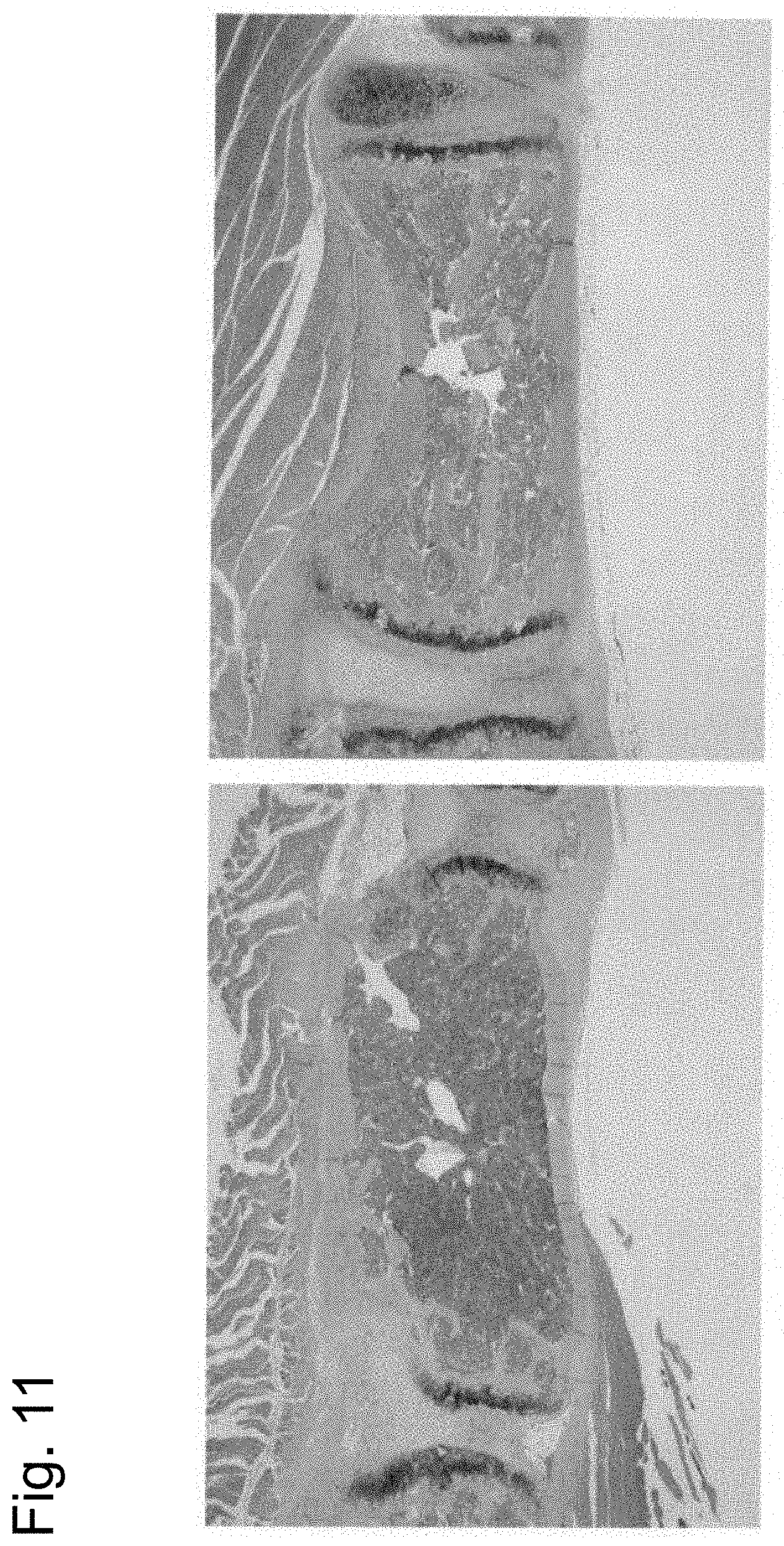
D00012

D00013

D00014

D00015

D00016

D00017

D00018

D00019
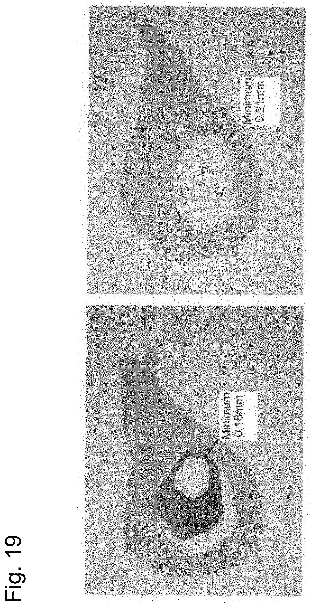
D00020

D00021

D00022
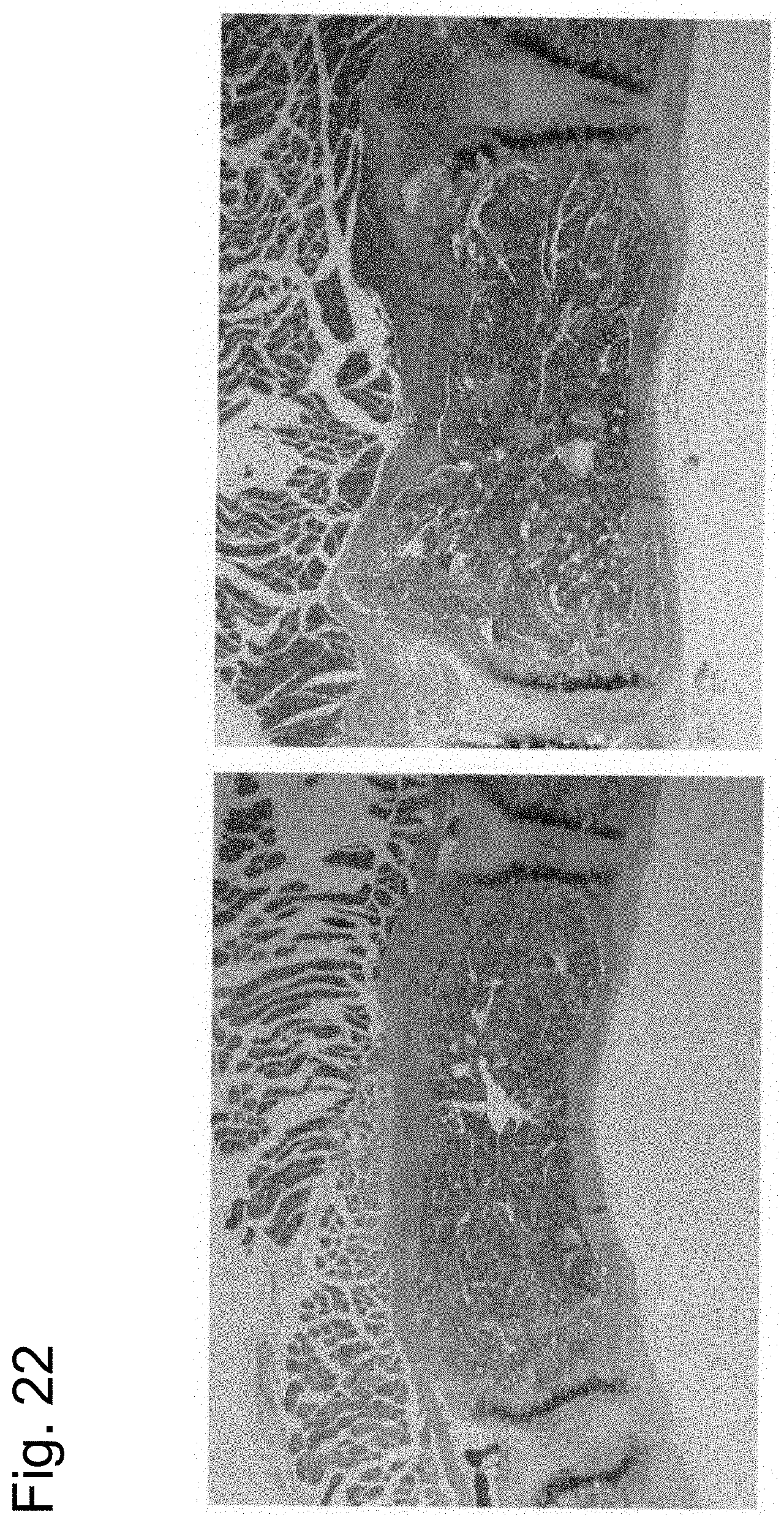
D00023

D00024

D00025

S00001
XML
uspto.report is an independent third-party trademark research tool that is not affiliated, endorsed, or sponsored by the United States Patent and Trademark Office (USPTO) or any other governmental organization. The information provided by uspto.report is based on publicly available data at the time of writing and is intended for informational purposes only.
While we strive to provide accurate and up-to-date information, we do not guarantee the accuracy, completeness, reliability, or suitability of the information displayed on this site. The use of this site is at your own risk. Any reliance you place on such information is therefore strictly at your own risk.
All official trademark data, including owner information, should be verified by visiting the official USPTO website at www.uspto.gov. This site is not intended to replace professional legal advice and should not be used as a substitute for consulting with a legal professional who is knowledgeable about trademark law.