Substrates and methods for collection, stabilization and elution of biomolecules
Kovacs , et al.
U.S. patent number 10,625,242 [Application Number 15/285,986] was granted by the patent office on 2020-04-21 for substrates and methods for collection, stabilization and elution of biomolecules. This patent grant is currently assigned to General Electric Company. The grantee listed for this patent is General Electric Company. Invention is credited to Ernest William Kovacs, Erik Leeming Kvam, Bing Li, Frank John Mondello.
| United States Patent | 10,625,242 |
| Kovacs , et al. | April 21, 2020 |
Substrates and methods for collection, stabilization and elution of biomolecules
Abstract
A solid substrate for the extraction, stabilization, and storage of proteins is provided. The substrate includes: a polysaccharide, such as melezitose under a substantially dry state. The substrate is configured to extract proteins from a sample and stabilize the extracted proteins in a dry format under ambient conditions for a prolonged period of time. Methods for collecting and recovering the proteins stored in the dry solid substrate are also described.
| Inventors: | Kovacs; Ernest William (Niskayuna, NY), Mondello; Frank John (Niskayuna, NY), Kvam; Erik Leeming (Niskayuna, NY), Li; Bing (Niskayuna, NY) | ||||||||||
|---|---|---|---|---|---|---|---|---|---|---|---|
| Applicant: |
|
||||||||||
| Assignee: | General Electric Company
(Schenectady, NY) |
||||||||||
| Family ID: | 51351478 | ||||||||||
| Appl. No.: | 15/285,986 | ||||||||||
| Filed: | October 5, 2016 |
Prior Publication Data
| Document Identifier | Publication Date | |
|---|---|---|
| US 20170021333 A1 | Jan 26, 2017 | |
Related U.S. Patent Documents
| Application Number | Filing Date | Patent Number | Issue Date | ||
|---|---|---|---|---|---|
| 14261900 | Apr 25, 2014 | 9480966 | |||
| 13968497 | May 26, 2015 | 9040679 | |||
| 13721948 | May 26, 2015 | 9040675 | |||
| 13460076 | Jun 2, 2015 | 9044738 | |||
| Current U.S. Class: | 1/1 |
| Current CPC Class: | C07K 14/5421 (20130101); C07K 14/775 (20130101); C07K 14/525 (20130101); C12N 9/2471 (20130101); B01J 20/28038 (20130101); B01J 20/22 (20130101); C12N 15/1017 (20130101); B01J 20/24 (20130101); B01J 20/3246 (20130101); B01J 20/3242 (20130101); C12N 15/1006 (20130101); C12Y 302/01023 (20130101); B01J 20/3212 (20130101); C07K 1/145 (20130101); B01J 20/28033 (20130101); C07K 1/16 (20130101); C07K 14/545 (20130101) |
| Current International Class: | C12N 9/38 (20060101); B01J 20/28 (20060101); B01J 20/32 (20060101); C12N 15/10 (20060101); B01J 20/22 (20060101); C07K 14/775 (20060101); C07K 14/545 (20060101); C07K 14/525 (20060101); C07K 14/54 (20060101); B01J 20/24 (20060101); C07K 1/16 (20060101); C07K 1/14 (20060101) |
| Field of Search: | ;536/123.1,25.41 ;530/412,359,351 ;502/404,4 ;435/207 |
References Cited [Referenced By]
U.S. Patent Documents
| 4540506 | September 1985 | Jacobson et al. |
| 4806343 | February 1989 | Carpenter et al. |
| 4891319 | January 1990 | Roser |
| 5089407 | February 1992 | Baker et al. |
| 5173422 | December 1992 | Knowles et al. |
| 5496562 | March 1996 | Burgoyne |
| 5518928 | May 1996 | Cremins et al. |
| 5593824 | January 1997 | Treml |
| 5756126 | May 1998 | Burgoyne |
| 5807527 | September 1998 | Burgoyne |
| 5939259 | August 1999 | Harvey et al. |
| 5981218 | November 1999 | Rio et al. |
| 6004574 | December 1999 | Backstrom |
| 6184011 | February 2001 | Siegel et al. |
| 6528641 | March 2003 | Lader |
| 6602718 | August 2003 | Augello et al. |
| 6610531 | August 2003 | Mateczun et al. |
| 6776959 | August 2004 | Helftenbein |
| 6794140 | September 2004 | Goldsborough |
| 7163793 | January 2007 | Kudlicki et al. |
| 7250270 | July 2007 | Goldrick et al. |
| 7282371 | October 2007 | Helftenbein |
| 7473397 | January 2009 | Griffin et al. |
| 7589184 | September 2009 | Hogan et al. |
| 7767447 | August 2010 | Breidenthal et al. |
| 7833486 | November 2010 | Fielden et al. |
| 8025850 | September 2011 | Chan |
| 8088576 | January 2012 | Gumbrecht et al. |
| 8158357 | April 2012 | Birnboim et al. |
| 9040675 | May 2015 | Bales et al. |
| 9040679 | May 2015 | Kvam et al. |
| 9044738 | June 2015 | Li et al. |
| 9415392 | August 2016 | Ismagilov et al. |
| 9480966 | November 2016 | Kovacs et al. |
| 9534214 | January 2017 | Li et al. |
| 2001/0007746 | July 2001 | Smith et al. |
| 2001/0039010 | November 2001 | Burgoyne |
| 2002/0146696 | October 2002 | Burgoyne et al. |
| 2003/0134312 | July 2003 | Burgoyne |
| 2003/0143566 | July 2003 | Helftenbein et al. |
| 2004/0009496 | January 2004 | Eiblmaier et al. |
| 2004/0101895 | May 2004 | Fomovskaia et al. |
| 2004/0126783 | July 2004 | Bortolin et al. |
| 2004/0219534 | November 2004 | Belly et al. |
| 2004/0235034 | November 2004 | Kuno et al. |
| 2005/0009045 | January 2005 | Greenfield et al. |
| 2005/0123965 | June 2005 | Yamashita et al. |
| 2006/0099567 | May 2006 | Muller-Cohn et al. |
| 2006/0147918 | July 2006 | Goldsborough |
| 2007/0106071 | May 2007 | Yamashita et al. |
| 2007/0117173 | May 2007 | Levison et al. |
| 2008/0176209 | July 2008 | Muller et al. |
| 2008/0262097 | October 2008 | Eady et al. |
| 2009/0043087 | February 2009 | Davis et al. |
| 2009/0053704 | February 2009 | Novoradovskaya et al. |
| 2009/0143570 | June 2009 | Jiang et al. |
| 2009/0162924 | June 2009 | Birnboim |
| 2009/0208919 | August 2009 | Utermohlen et al. |
| 2009/0246750 | October 2009 | Lloyd et al. |
| 2009/0291427 | November 2009 | Muller-Cohn et al. |
| 2010/0173392 | July 2010 | Davis et al. |
| 2010/0209957 | August 2010 | Hogan et al. |
| 2010/0248363 | September 2010 | Hogan et al. |
| 2011/0054157 | March 2011 | Bitner |
| 2011/0059869 | March 2011 | Kojima et al. |
| 2011/0070585 | March 2011 | Ollikka et al. |
| 2011/0081363 | April 2011 | Whitney |
| 2012/0052572 | March 2012 | Whitney et al. |
| 2012/0059160 | March 2012 | Bitner et al. |
| 2012/0152743 | June 2012 | Finehout et al. |
| 2012/0237939 | September 2012 | Reed et al. |
| 2012/0289690 | November 2012 | Page et al. |
| 2013/0102501 | April 2013 | Craighead et al. |
| 2013/0289257 | October 2013 | Bales et al. |
| 2013/0309679 | November 2013 | Ismagilov et al. |
| 2013/0323723 | December 2013 | Horton et al. |
| 2014/0038172 | February 2014 | De La Rosa et al. |
| 2015/0118683 | April 2015 | Li et al. |
| 2015/0119566 | April 2015 | Li et al. |
| 2015/0299693 | October 2015 | Chen et al. |
| 101287449 | Oct 2008 | CN | |||
| 101878304 | Nov 2010 | CN | |||
| 102728407 | Oct 2012 | CN | |||
| 102803483 | Nov 2012 | CN | |||
| 102947082 | Feb 2013 | CN | |||
| 103173432 | Jun 2013 | CN | |||
| 1484111 | Dec 2004 | EP | |||
| 1559784 | Aug 2005 | EP | |||
| 2388312 | Nov 2011 | EP | |||
| 6188896 | May 1986 | JP | |||
| 2007502428 | Feb 2007 | JP | |||
| 4189321 | Dec 2008 | JP | |||
| 2012523851 | Oct 2012 | JP | |||
| 5282088 | Sep 2013 | JP | |||
| 5766178 | Aug 2015 | JP | |||
| 9118091 | Nov 1991 | WO | |||
| 0066606 | Nov 2000 | WO | |||
| 03020924 | Mar 2003 | WO | |||
| 03/086443 | Oct 2003 | WO | |||
| 03086443 | Oct 2003 | WO | |||
| 2007008722 | Jan 2007 | WO | |||
| 2009029433 | Mar 2009 | WO | |||
| 2010123908 | Oct 2010 | WO | |||
| 2010132508 | Nov 2010 | WO | |||
| 2010/144682 | Dec 2010 | WO | |||
| 2010144682 | Dec 2010 | WO | |||
| 20110131720 | Oct 2011 | WO | |||
| WO 2011/131720 | Oct 2011 | WO | |||
| 2012075471 | Jun 2012 | WO | |||
| 2012113907 | Aug 2012 | WO | |||
| 2012113911 | Aug 2012 | WO | |||
| 2013066249 | May 2013 | WO | |||
| 2015162093 | Oct 2015 | WO | |||
Other References
|
Wolfgramm et al, Forensic Science International: Genetics, 2009, 3, 125-127. cited by examiner . Miles, M., etal, ZyGem, Oct. 1, 2012, pp. 1-2. cited by examiner . First Office Action and Search issued in connection with corresponding CN Application No. 201580021334.X dated Jul. 30, 2018. cited by applicant . Abouhaidar et al., "Non-Enzymatic RNA Hydrolysis Promoted by the Combined Catalytic Activity of Buffers and Magnesium", Department of Botany, pp. 542-548, 1999. cited by applicant . Wolfgramm et al., "Simplified Buccal DNA Extraction with FTA Elute Cards", Forensic Science International: Genetics, vol. No. 3, Issue No. 2, pp. 125-127, Mar. 1, 2009. cited by applicant . Johanson et al., "DNA Elution from Buccal Cells Stored on Whatman FTA Classic Cards using a Modified Methanol Fixation Method", Biotechniques, vol. No. 46, Issue No. 4, pp. 309-311, Apr. 2009. cited by applicant . Miles et al., "Improved Elution of DNA from Whatman FTA Cards Using PrepGEM/ ForensicGEM Storage Card Extraction Kits", ZyGEM, Oct. 1, 2012. cited by applicant . Henniges et al., "Electron Beam Irradiation of Cellulosic Materials--Opportunities and Limitations", Materials,vol. No. 6, pp. 1584-1598, Apr. 29, 2013. cited by applicant . PCT Search Report and Written Opinion issued in connection with corresponding PCT Application No. PCT/US2014/062570 dated Jul. 10, 2015. cited by applicant . European Office Action issued in connection with corresponding EP Application No. 14752322.9 dated Nov. 28, 2016. cited by applicant . Unofficial English Translation of Japanese Office Action issued in connection with corresponding JP Application No. 2015510354 dated Feb. 7, 2017. cited by applicant . Unofficial English Translation of Japanese Search Report issued in connection with corresponding JP Application No. 2015510354 dated Aug. 2, 2017. cited by applicant . Zale et al., "Why does Ribonuclease Irreversibly Inactivate at High Temperatures?", Biochemistry, vol. No. 25, Issue No. 19, pp. 5432-5444, Sep. 1986. cited by applicant . Sambrook et al., "Molecular Cloning: a Laboratory Manual, 2nd edition", Cold Spring Harbor, Cold Spring Harbor Laboratory Press, vol. No. 01, pp. 7.2, 7.3, 7.4 and 7.5, Dec. 1989. cited by applicant . Zhang et al., "RNA Analysis from Newborn Screening Dried Blood Specimens", Human genetics, vol. No. 89, Issue No. 03, pp. 311-314, May 1992. cited by applicant . Matsubara et al., "Dried Blood Spot on Filter Paper as a Source of mRNA", Nucleic Acids Research, vol. No. 20, Issue No. 08, 1 page, 1998. cited by applicant . Li et al., "Kinetics of RNA Degradation by Specific Base Catalysis of Transesterification Involving the 2'-Hydroxyl Group", Journal of the American Chemical Society, vol. No. 121, Issue No. 23, pp. 5364-5372, 1999. cited by applicant . Natarajan et al., "Paper-Based Archiving of Mammalian and Plant Samples for RNA Analysis", BioTechniques, vol. No. 29, Issue No. 6, pp. 1328-1333, Dec. 2000. cited by applicant . Ambion Technotes, "Maximize Your RNA Yield: What Yield to Expect", vol. No. 08, Issue No. 3, pp. 1, 13-14, I, May 18, 2001. cited by applicant . Ambion, "RNAqueousTM-4PCR Instruction Manual Passage", RNAqueousTM-4PCR Instruction Manual, pp. 1-29, Apr. 18, 2002. cited by applicant . Kumar et al., "Inhibition of Mammalian Ribonucleases by Endogenous Adenosine Dinucleotides", Biochemical and Biophysical Research Communications, vol. No. 300, Issue No. 01, pp. 81-86, Jan. 3, 2003. cited by applicant . Cline, et al., "New Water-Soluble Phosphines as Reductants of Peptide and Protein Disulfide Bonds: Reactivity and Membrane Permeability", Biochemistry, vol. No. 43, Issue No. 48, pp. 15195-15203, 2004. cited by applicant . Mollmann et al., "The Stability of Insulin in Solid Formulations Containing Melezitose and Starch. Effects of Processing and Excipients", Drug Development and Industrial Pharmacy, vol. No. 32, Issue No. 06, pp. 765-778, 2006. cited by applicant . Tan et al., "DNA, RNA, and Protein Extraction: The Past and the Present", Journal of Biomedicine and Biotechnology, vol. No. 2009, Issue No. 2009, 10 pages, 2009. cited by applicant . PCT Search Report and Written Opinion issued in connection with related PCT Application No. PCT/US2013/038576 dated Sep. 6, 2013. cited by applicant . PCT Search Report and Written Opinion issued in connection with related PCT Application No. PCT/US2013/065821 dated Jan. 29, 2014. cited by applicant . PCT Search Report and Written Opinion issued in connection with related PCT Application No. PCT/EP2014/067453 dated Dec. 19, 2014. cited by applicant . European Search Report and Opinion issued in connection with related EP Application No. 13784927.9 dated Jan. 29, 2015. cited by applicant . PCT Search Report and Written Opinion issued in connection with related PCT Application No. PCT/EP2015/058518 dated Jun. 19, 2015. cited by applicant . European Search Report and Opinion issued in connection with related EP Application No. 13865596.4 dated Jun. 28, 2016. cited by applicant . Singapore Written Opinion issued in connection with related SG Application No. 11201601114P dated Nov. 16, 2016. cited by applicant . Singapore Invitation to Respond to Written Opinion issued in connection with related SG Application No. 11201601114P dated Dec. 19, 2016. cited by applicant . Australia Search and Examination Report issued in connection with corresponding AU Application No. 2015250915 dated Feb. 9, 2017. cited by applicant . Singapore Written Opinion issued in connection with corresponding SG Application No. 11201608021T dated Apr. 7, 2017. cited by applicant . Chinese Office Action issued in connection with related CN Application No. 201380022584.6 dated Jul. 17, 2015. cited by applicant . Chinese Office Action for CN Application No. 201580021334X dated Jun. 19, 2019 (14 pages). cited by applicant. |
Primary Examiner: Krishnan; Ganapathy
Attorney, Agent or Firm: Eversheds Sutherland (US) LLP
Government Interests
FEDERALLY SPONSORED RESEARCH & DEVELOPMENT
This invention was made with Government support under contract number HR0011-11-00127 awarded by the Defense Advanced Research Projects Agency. The Government has certain rights in the invention.
Parent Case Text
CROSS REFERENCE TO RELATED APPLICATIONS
This application is a continuation of U.S. patent application Ser. No. 14/261,900 entitled "Substrates and methods for collection, stabilization and elution of biomolecules", filed on Apr. 25, 2014, which is a continuation-in-part of U.S. patent application Ser. No. 13/460,076 entitled "Methods and compositions for extraction and storage of nucleic acids", filed Apr. 30, 2012, now U.S. Pat. No. 9,044,738 issued on Jun. 2, 2015; U.S. patent application Ser. No. 13/721,948 entitled "Formulations for nucleic acid stabilization on solid substrates", filed Dec. 20, 2012, now U.S. Pat. No. 9,040,675, issued on May 26, 2015; and U.S. patent application Ser. No. 13/968,497 entitled "Methods and Compositions For Extraction and Storage of Nucleic Acids", filed Aug. 16, 2013, now U.S. Pat. No. 9,040,679, issued on May 26, 2015; which are herein incorporated by reference.
Claims
What is claimed is:
1. A paper substrate for collection, stabilization and storage of a biological specimen or sample, the paper substrate consisting essentially of: (a) a non-dissolvable material selected from the group consisting of cellulose, cellulose acetate and combinations thereof; and (b) a trisaccharide impregnated in the non-dissolvable material.
2. The paper substrate of claim 1, wherein the trisaccharide is melezitose, raffinose, maltotriulose, isomaltotriose, nigerotriose, maltotriose, kestose, or a combination thereof.
3. The paper substrate of claim 1, wherein the trisaccharide is melezitose.
4. A method for extracting, stabilizing and eluting a biomolecule from a sample, the method comprising: contacting the sample to the paper substrate of claim 1; drying the sample to a water content of less than 2%; and eluting the biomolecule from the sample dried on the paper substrate by rehydrating the paper substrate in an elution buffer, wherein the biomolecule comprises a protein, a peptide, an amino acid, an enzyme, an antibody, or combinations thereof.
5. The method of claim 4, wherein 70 to 90% of the biomolecule is recovered from the substrate.
6. The method of claim 4, wherein the biomolecule is eluted in an intact form.
7. The method of claim 4, wherein the material is cellulose.
8. The paper substrate of claim 1, wherein the material consists of cellulose.
9. The paper substrate of claim 1, wherein the material consists of cellulose acetate.
10. The paper substrate of claim 1, wherein the material consists of cellulose and the trisaccharide is melezitose.
11. The paper substrate of claim 1, wherein the material is cellulose, and the trisaccharide is melezitose, raffinose, maltotriulose, isomaltotriose, nigerotriose, maltotriose, kestose, or a combination thereof.
12. The method of claim 4, further comprising storing the dried sample on the substrate for several weeks.
13. The method of claim 4, further comprising washing the substrate before eluting the biomolecule.
14. The paper substrate of claim 1, wherein the paper substrate consists of the non-dissolvable material and the trisaccharide.
Description
FIELD
The invention relates to dry solid substrates for collection, stabilization and elution of biomolecules from a biological sample. The invention further relates to methods for collection, stabilization and elution of biomolecules from a biological sample from the dry solid substrates.
BACKGROUND
Preserving the structural and functional integrity of biomolecules during isolation or purification from a biological sample is essential for various downstream applications including analyte detection, sensing, forensic, diagnostic or therapeutic applications, and the like. The extraction and stabilization of proteins, peptides or amino acids derived from a biological sample are sensitive to a number of environmental factors including, but are not limited to, solution pH, temperature, and the ubiquitous presence of various proteases. Consequently, proteins or peptides in solution states are typically stored under refrigeration (e.g. 4.degree. C., -20.degree. C., or -80.degree. C.) to prevent hydrolysis and enzymatic degradation and to preserve the integrity of protein structure or function.
Dry-state technologies claiming successful collection and preservation of proteins or peptides in dry formats typically require protein to be "pre-purified" and "concentrated" from a sample prior to storage. Other dry-state technologies for the preservation of proteins in dry formats require additional drying facilities (e.g. forced air flow, lyophilization). These methods are therefore not conducive to direct collection and stabilization of proteins or peptides from a sample (e.g., a biological sample) without additional and significant processing steps.
Proteins or peptides are prone to denaturation and consequently tend to lose biological activity or epitope recognition during storage. Proteins that are targets of different analytical tests, such as biomarkers or biological therapeutic drugs, may be present in low quantities in unpurified states. Thus, methods for maximizing the recovery of protein analytes of interest are highly desirable. Degradation of protein or peptide may be slowed or prevented using chemical additives that, for example, inhibit protease activity. However, the presence of chemical additives may affect down-stream analytical techniques including mass spectroscopy and immunoassays.
Untreated cellulose paper substrates, such as, 903 or 31ETF papers (Whatman.TM., GE Healthcare) or Grade 226 paper (Ahlstrom, PerkinElmer) are used widely for preservation of enzymes, antibodies, proteins, peptides, and amino acids in dried blood spots for analytical purposes such as neonatal testing. However, the recovery of analytes from untreated cellulose substrates and subsequent biological activity of said analytes, particularly proteins that are prone to degradation, are often not sufficient. Dried specimens, such as dried blood spot samples used in neonatal testing, are generally stored under refrigeration to maintain analyte stability. Analytes which can be eluted inefficiently from dried blood spots may be interpreted in the art as unstable targets due to poor functional recovery. Supplementing different chemical fillers to stabilize proteins has been reported in the art, however the fillers have limited ability to recover and stabilize sensitive proteins.
Accordingly, compositions and methods which enable collection and extraction of biomolecules including proteins, peptides or amino acids from a biological sample, and then stabilize the biomolecules under a dry-state and ambient conditions without pre-purification, and elute the biomolecules in a substantially intact form thereafter for further analysis are highly desirable.
BRIEF DESCRIPTION
One embodiment of a solid substrate for extraction, stabilization and elution of biomolecules, comprises a melezitose under a substantially dry state.
In another embodiment, a solid substrate for collection, stabilization and elution of biomolecules, comprises a trisaccharide under a substantially dry state.
One example of a method for extracting, stabilizing and eluting biomolecules from a biological sample disposed on a solid substrate, comprises contacting the biological sample to the substrate; drying the biological sample to a substantially dry state; and eluting the biomolecules from the biological sample dried on the substrate by rehydrating the substrate in an elution buffer, wherein the solid substrate comprises melezitose under a substantially dry state, and optionally one or more lysis reagents, nucleic acid denaturing reagents or combinations thereof, impregnated therein under a substantially dry state.
BRIEF DESCRIPTION OF DRAWING
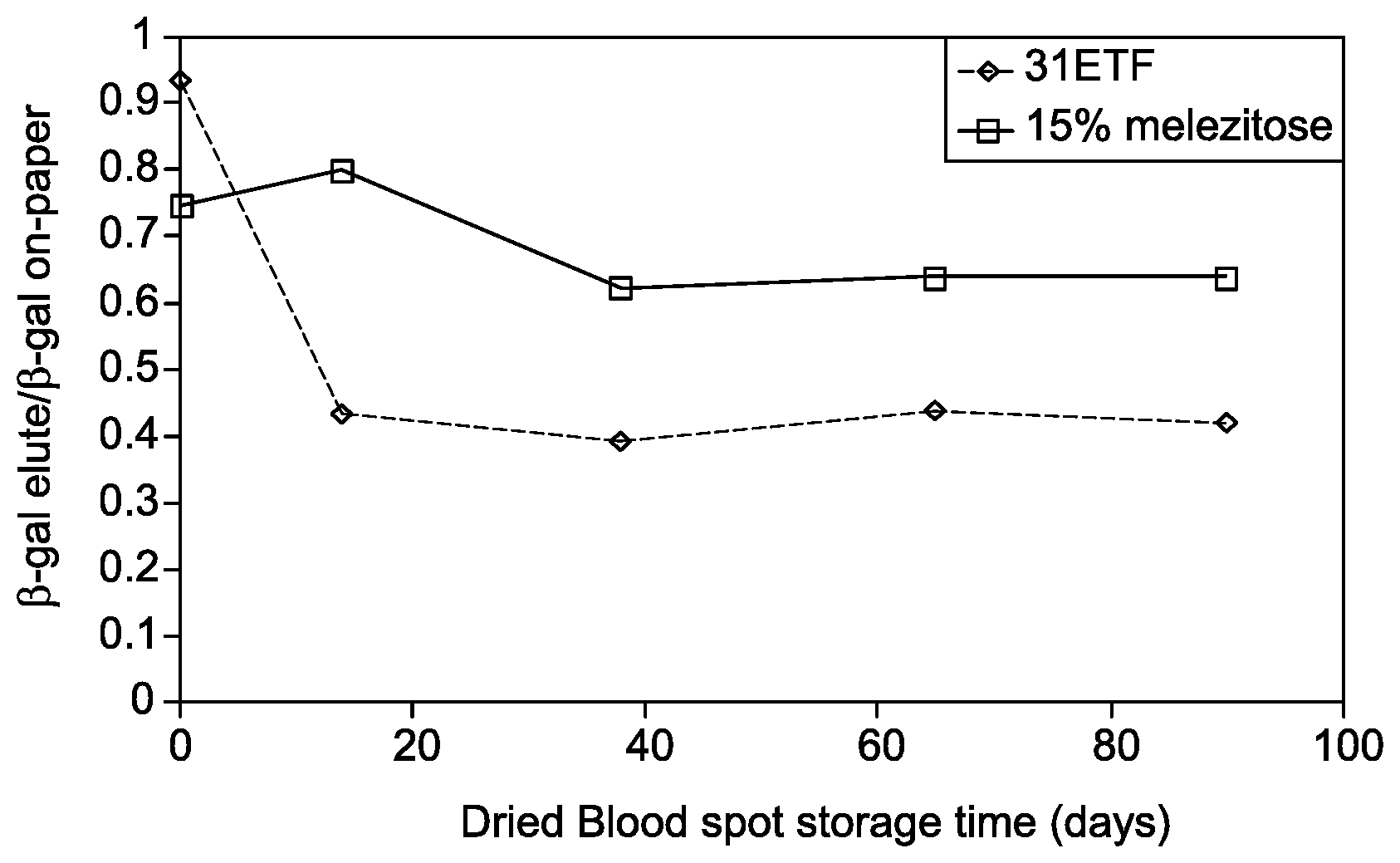
FIG. 1 is a graph showing enhanced elution efficiency of active, non-denatured .beta.-gal while recovered from a substrate with 15% melezitose compared to unmodified 31-ETF cellulose.
DETAILED DESCRIPTION
The embodiments provide suitable matrices and methods for extraction, stabilization and elution of biomolecules, such as proteins, peptides, amino acids, enzymes, and antibodies. Biomolecules which are prone to denaturation are therefore difficult to preserve in an intact form. One or more embodiments of the invention relate to a solid substrate for extraction, stabilization and elution of biomolecules, wherein the substrate comprises a trisaccharide, such as melezitose under a substantially dry state. The solid substrate is configured to collect a biological sample, then extract and stabilize proteins, peptides or amino acids from the sample for a prolonged period, followed by elution within a single process step. The eluted proteins or peptides are used in various downstream applications. The substrate is configured to stabilize proteins or peptides in a substantially dry-state at ambient temperature and substantially retain the integral structure and/or function of the protein.
To more clearly and concisely describe the subject matter of the claimed invention, the following definitions are provided for specific terms, which are used in the following description and the appended claims. Throughout the specification, exemplification of specific terms should be considered as non-limiting examples.
The singular forms "a", "an" and "the" include plural referents unless the context clearly dictates otherwise. Approximating language, as used herein throughout the specification and claims, may be applied to modify any quantitative representation that could permissibly vary without resulting in a change in the basic function to which it is related. Accordingly, a value modified by a term such as "about" is not to be limited to the precise value specified. In some instances, the approximating language may correspond to the precision of an instrument for measuring the value. Where necessary, ranges have been supplied, and those ranges are inclusive of all sub-ranges there between.
The term "biological sample" as referred to herein includes, but is not limited to, blood, serum, tissue, and saliva obtained from any organism, including a human. Biological samples may be obtained by an individual undergoing a self-diagnostic test (e.g., blood glucose monitoring) or by a trained medical professional through a variety of techniques including, for example, aspirating blood using a needle or scraping or swabbing a particular area, such as a lesion on a patient's skin. Methods for collecting various biological samples are well known in the art. The term "sample" includes biological samples as defined above, but also includes, for example, tissue cultured cells and purified proteins.
The term, "reducing agents" as referred to herein include any chemical species that provides electrons to another chemical species. A variety of reducing agents are known in the art. Exemplary reducing agents include dithiothreitol (DTT), 2-mercaptoethanol (2-ME), and tris(2-carboxyethyl)phosphine (TCEP). Moreover, any combination of these or other reducing agents may be used. In particular embodiments, the reducing agent is TCEP.
The term "buffer" as used herein includes, for example, 2-Amino-2-hydroxymethyl-propane-1,3-diol (Tris), 2-(N-morpholino) ethanesulfonic acid (MES), 3-(N-morpholino)propanesulfonic acid (MOPS), citrate buffers, 4-(2-hydroxyethyl)-1-piperazineethanesulfonic acid (HEPES), and phosphate buffers. This list of potential buffers is for illustrative purposes only. The pH of the buffer selected for use in the compositions and methods disclosed herein is typically in the range of 3 to 10. In some embodiments, the pH of the buffer used herein is in a range of 6 to 9, or in some other embodiments, the pH of the buffer is in a range of 7 to 8.
One embodiment of a solid substrate for collection, stabilization and elution of biomolecules, comprises a trisaccharide under a substantially dry state. The trisaccharide may be selected from melezitose, raffinose, maltotriulose, isomaltotriose, nigerotriose, maltotriose, ketose or combinations thereof.
One or more embodiments of a solid substrate comprise a melezitose under a substantially dry state. Melezitose is a non-reducing trisaccharide sugar, having a molecular weight of 504, 44 g/mol. In one or more embodiments, the solid substrate comprises melezitose, wherein a concentration of the melezitose is in range of about 10 to 30%. In one embodiment, the concentration of melezitose is 15%. The melezitose may be impregnated in the substrate. In some embodiments, the impregnated melezitose concentration in the substrate is between 10 to 30%. In some other embodiments, 15% melezitose is impregnated in the substrate. The substrate may be passively coated or covalently-modified with melezitose. In some other embodiments, the substrate is coated with a 15% solution of melezitose. The substrate with melezitose has shown high stability of the proteins, as well as provide higher yield, as described in Example 2.
In one or more examples, the substrate is further impregnated with one or more reagents, such as lysis reagents, buffer reagents or reducing agents. In some embodiments, the impregnated reagents comprise cell lytic reagents, biomolecule stabilizing reagents such as protein-stabilizing reagents, protein storage chemicals and combinations thereof impregnated therein under a substantially dry state.
The substrate is also configured to extract proteins or peptides from a biological sample and preserve that in a substantially dry state at ambient temperature. As used herein, the term "substantially dry state" refers to drying the extracted biomolecules to have approximately less than 2% of water content. Similarly, the reagents are impregnated in the substrate in a substantially dry state.
"Incorporation" of the compositions into the substrate includes, but is not limited to, the "dipping" procedure described below. In some embodiments, such methods accomplish incorporation of the composition into the dry solid substrate. Following incorporation of the composition into the dry solid substrate, the solid substrate is dried using any appropriate method.
In one or more embodiments, the substrate comprises lysis reagents. The lysis reagents may comprise detergents, chaotropes, denaturants or combinations thereof. Without intending to be limited to a particular denaturant, it may be categorized as either weak or strong lytic reagents depending on their biophysical properties and ability to completely inhibit biological enzyme activity (e.g. proteases). In some embodiments, weak protein denaturants (e.g. detergent) may be used for lysing cells and disrupting protein-protein interactions without denaturing proteins. Numerous lysis reagents are known in the art and may be selected for use in the compositions and methods described herein. Without intending to be limited to a particular lysis reagents, exemplary lysis reagents include guanidinium thiocyanate, guanidinium hydrochloride, sodium thiocyanate, potassium thiocyanate, arginine, sodium dodecyl sulfate (SDS), urea or a combination thereof.
As noted, the lysis reagents may include detergents, wherein exemplary detergents may be categorized as ionic detergents, non-ionic detergents, or zwitterionic detergents. The ionic detergent may comprise anionic detergent such as, sodium dodecylsulphate (SDS) or cationic detergent, such as ethyl trimethyl ammonium bromide. Non-limiting examples of non-ionic detergent for cell lysis include TritonX-100, NP-40, Brij 35, Tween 20, Octyl glucoside, Octyl thioglucoside or digitonin. Some zwitterionic detergents may comprise 3-[(3-Cholamidopropyl)dimethylammonio]-1-propanesulfonate (CHAPS) and 3-[(3-Cholamindopropyl) dimethylammonio]-2-hydroxy-1-propanesulfonate (CHAPS 0).
In one or more embodiments, the lysis reagent comprises a thiocyanate salt. One or more embodiments of the substrate comprises a thiocyanate salt impregnated in a dry state. Exemplary thiocyanate salts include, but are not limited to, guanidinium thiocyanate, sodium thiocyanate, potassium thiocyanate or combinations thereof. In some other embodiments, the lysis reagent is selected from guanidinium thiocyanate, sodium thiocyanate, sodium dodecyl sulfate (SDS) or combinations thereof.
In one or more embodiments, the substrate maintains stability and integrity of the protein at a desired level after extraction from a biological sample. In one embodiment, the substrate is impregnated with one or more protein stabilizing reagents. These stabilizing reagents may include protease inhibitors, buffer, or chelating agents (e.g EDTA).
The digestion of recovered proteins in the presence of proteases may be avoided by adding one or more protease inhibitors to the substrate, wherein the protease inhibitors may be added externally or may be impregnated in the substrate. In one embodiment, impregnated protease inhibitors may be activated upon wetting of the dry substrate. In some embodiments, the substrate further comprises a protease inhibitor, wherein the protease inhibitor is synthetic or occurs naturally (e.g. naturally-occurring peptide or protein) and comprises aprotinin, bestatin, chymostatin, leupeptin, alpha-2-macroglobulin, pepstatin, phenylmethanesulfonyl fluoride, N-ethylmaleimide, ethylenediaminetetraacetid acid, antithrombin, or combinations thereof. In one example, an addition of such protease inhibitors enhances the stability of the proteins in both liquid state and dry-formats by inhibiting the proteases or peptidases.
Certain embodiments of the substrate comprise buffer reagents in a dry-state, which may be re-hydrated during the extraction process. Examples of these buffers include, but are not limited to, 2-Amino-2-hydroxymethyl-propane-1,3-diol (Tris), 2-(N-morpholino) ethanesulfonic acid (MES), 3-(N-morpholino) prop anes ulfonic acid (MOPS), citrate buffers, 4-(2-hydroxyethyl)-1-piperazineethanesulfonic acid (HEPES), phosphate buffers or combinations thereof. As noted, the substrate provides a pH of 6 to 8 on hydration, which enables extraction of biomolecules from the biological samples and stabilization of the extracted biomolecules. The hydration may be achieved by adding a sample, water or any other solution (e.g. a buffer solution). One or more embodiments of the substrate provide a pH in a range from 2 to 7 on hydration. In some embodiments, the substrate provides a pH in a range from 7 to 10 on hydration. In one embodiment, the substrate provides a pH range from 6 to 8 upon hydration.
In some embodiments, the substrate further comprises at least one reducing agent, wherein the reducing agent is selected from the group consisting of dithiothreitol (DTT), 2-mercaptoethanol (2-ME), tris(2-carboxyethyl) phosphine (TCEP) and combinations thereof.
In some embodiments, the substrate comprises one or more chelating agents. The chelating agents may be selected from ethylenediaminetetraacetic acid (EDTA), citric acid, ethylene glycol tetraacetic acid (EGTA), or combinations thereof.
In some embodiments, the substrate further comprises a polysaccharide. The polysaccharide may be selected from dextran, Picoll.RTM., chitosan, amylopectin, alginate, carboxymethyl cellulose, or combinations thereof. In one embodiment, the polysaccharide is Picoll.RTM.. In one embodiment, the substrate further comprises 15% solution of Illustra.TM. Ready-To-Go (RTG) components from GE Healthcare.
The substrate enables collection, extraction and storage of proteins or peptides without solubilizing the substrate material. The solid substrate may be selected from the group consisting of a nitrocellulose membrane, a cellulose membrane, a cellulose acetate membrane, a regenerated cellulose membrane, a nitrocellulose mixed ester membranes, a polyethersulfone membrane, a nylon membrane, a polyolefin membrane, a polyester membrane, a polycarbonate membrane, a polypropylene membrane, a polyvinylidene difluoride membrane, a polyethylene membrane, a polystyrene membrane, a polyurethane membrane, a polyphenylene oxide membrane, a poly(tetrafluoroethylene-co-hexafluoropropylene membrane, glass fiber and any combination of two or more of the above membranes.
The solid substrate may be porous. In one embodiment, the solid substrate is a porous cellulose paper, such as a cellulose substrate from Whatman.TM.. In one example, the cellulose substrate from Whatman.TM. comprises 903-cellulose, FTA.TM. or FTA.TM. Elute.
As noted, the solid substrate comprises the composition in a dry state and also preserves the extracted proteins under dry conditions. The use of a dry solid substrate for extraction and storage is advantageous over liquid-based extraction, because the dry substrate ensures minimal volumetric dilution of the sample applied to the substrate. A liquid-based extraction may dilute the concentration of the sample in an excess volume of stabilizing reagent. In contrast, a dry solid substrate for extracting and stabilizing biomolecules maintains the concentration of the sample, as well as the extracted biomolecules, and eliminates issues, such as sample degradation, that are related to improper dilution of sample in an insufficient volume of liquid preservative. In addition, the solid substrate comprises a fixed composition of the dry reagents, which enables efficient extraction of biomolecules, such as proteins, peptides or amino acids upon hydration, followed by stabilization of the extracted biomolecules at ambient temperature.
The terms "ambient condition" or "ambient temperature" are interchangeably used hereinafter. As used herein, the term "ambient temperature" refers to a temperature in a range between 0.degree. C. to 60.degree. C. In one or more embodiments, the ambient temperature is room temperature. In some embodiments, the substrate is configured to store or preserve proteins under ambient temperature in a dried state.
As noted, the solid substrate is configured to store or preserve proteins under dry-state for prolonged period. The term "configured to" or "configured for" is referred to herein as the structure or composition of the substrate that enables the substrate to extract and store proteins for periods of time at ambient temperature. The terms "storage" or "preservation" may be interchangeably used herein with respect to maintaining the extracted proteins in a format suitable for further analysis. More specifically, the proteins may be stored or preserved in a solid substrate, wherein the substrate ensures maintaining the integrity of the molecules.
In some embodiments, the substrate is a solid phase extraction substrate. A substrate, where the solid phase extraction method is used, is referred to herein as a solid phase extraction substrate. Solid-phase extraction (SPE) technology has been leveraged to reduce the extraction times of high purity proteins for sequencing and other applications. The solid phase extraction is an extraction method that uses a solid phase and a liquid phase to isolate one or more molecules of the same type, or different types, from a material. The substrate is used, for example, to purify a sample upstream of a chromatographic separation or other analytical method.
In some examples, the substrate permits the storage of proteins, which are prone to degradation, in a dry format (e.g., on a solid substrate) at ambient temperatures. In one or more embodiments, the substrate is configured to provide improved stability and elution for biomolecules at ambient temperatures. In some embodiments, the substrate is configured to provide improved stability to the biomolecules during storage of at least one month to three months at ambient temperature between 20 to 22.degree. C.
The substrate is configured to store proteins in a dry format at ambient temperature under substantially intact form. The term "form" of the proteins refers to the integral structure or function of the proteins.
In some embodiments, the dried reagents impregnated in the substrate are hydrated by adding a buffer, water or a sample. In one embodiment, the impregnated dried reagents are hydrated by a sample, more specifically a biological sample, which is disposed on the substrate for extraction or storage of proteins. In some other embodiments, in addition to a sample, water or buffer is added to hydrate the substrate and reconstitute or activate the reagents embedded in the substrate. In some embodiments, the hydration of the substrate generates an appropriate pH for extraction of proteins on the substrate. In some embodiments, the hydration further results in reconstituting the reagents, such as cell-lysis reagents, protein stabilizing reagents, reducing agents, buffer reagents that are present in a dried form in the substrate.
Methods for extracting, stabilizing and eluting biomolecules from a biological sample disposed on a solid substrate are provided herein. An example of a method comprises contacting the biological sample to the substrate, wherein the substrate comprises melezitose under a substantially dry state, and one or more lysis reagents, nucleic acid denaturing reagents or combinations thereof impregnated therein under a substantially dry state. Non-limiting examples of the term "contacting the biological sample" include, applying a sample or disposing a sample on the substrate using a pipet, catheter, syringe or conduit. In some embodiments, the sample may be poured onto the substrate. The method further comprises drying the biological sample to a substantially dry state. For elution, the biomolecules are eluted from the biological sample dried on the substrate by rehydrating the substrate in an elution buffer.
The term "extraction" refers to any method for separating or isolating the proteins from a sample, more particularly from a biological sample. The term "extraction" and "collection" are interchangeably used herein. Biomolecules such as proteins and peptides may be released from a cell by cell-lysis. In one embodiment, the proteins may be released during evaporative cell-lysis. In another embodiment, the cells are lysed upon contact with the substrate comprising cell lysis reagents. Contacting a biological sample comprising cells to the substrate results in cell lysis which releases proteins, for example by using FTA.TM. or FTA.TM. Elute cellulose papers.
As noted, the method further comprises drying the biological sample to a substantially dry state, wherein the dried sample may be stored on the substrate for a longer period of time. The solid substrate is dried using any appropriate method, such as air-drying or vacuum-drying. The sample dried substrate may be stored for several weeks or months, and depending on the requirement, proteins or peptides may be eluted from the dried sample on the substrate.
In one embodiment, the method further comprises storing the extracted proteins on the solid substrate in a substantially dry state at ambient temperature. In some embodiments, the proteins may be stored for more than one month time period. In some embodiments, the proteins may be stored for more than a six months period. As some of the proteins or peptides are prone to degradation, the extraction and preservation using the substrate is useful and the recovered proteins or peptides may further be used for various downstream applications.
As noted, the method further comprises eluting the biomolecules from the biological sample dried on the substrate by rehydrating the substrate in an elution buffer. The term "elution" refers to recovering the biomolecules, such as protein or peptides from the substrate by various means. One or more embodiments of the method comprise recovering the biomolecules from the substrate by solid phase extraction technique. In one or more embodiments, the proteins are eluted from the solid substrate by rehydrating the substrate in an aqueous solution, a buffer, or an organic solution, and wherein the proteins are subjected to further analysis. Any method that enables the elution of the biomolecules from a sample (e.g., an unpurified biological sample) may be employed. The proteins may be eluted by rehydrating the solid substrate (e.g., cellulose paper) in an aqueous solution, a buffer solution as defined above, or an organic solution. In some embodiments, the proteins are recovered from the solid substrate by electroelution, electrophoresis, or washing with elution buffer.
The method delineated above may optionally include a step of washing the substrate before eluting the proteins from the solid substrate for further analysis. For example, the substrate may be washed for one or more times with a suitable buffer or water prior to elution of the proteins.
In some embodiments, the substrate is configured to provide 70 to 90% recovery on elution of the biomolecules from the substrate. The elution is performed so that the biomolecules are eluted in an intact form. In embodiments of the method, the elution of biomolecules does not require pre-purification of the proteins or peptides for effective stabilization and preservation. The proteins or peptides may be extracted followed by stabilization and elution in a single step.
As noted, the proteins or peptides, which are prone to degradation on long term storage may be defined in terms of percent recovery of the protein in biologically active state. The protein that is prone to degradation is defined as, a protein which has less than about 60% recovery, or has less than about 40% recovery in a biologically active state after storage in a substrate for one week at a room temperature, wherein the substrate is devoid of any reagents.
In some embodiments, the entire method of using the substrate, such as applying the biological sample, drying the sample on the substrate, storage, extraction and elution of the proteins or peptides from the substrate may be performed under aseptic conditions.
The samples utilized in this method include, but are not limited to, biological samples such as blood, serum, tissue, and saliva obtained from any organism, including a human.
The extracted biomolecules may comprise proteins, peptides, amino acids, enzymes, antibodies, or combinations thereof. The biomolecules may include naturally occurring proteins, synthetic proteins, mutated proteins, fusion proteins or chimeric proteins. The proteins or peptides may be chemically synthesized. In some embodiments, the proteins or peptides may be naturally synthesized in a cell. In some embodiments, the biomolecules are recombinant proteins or peptides that may be isolated from cells or tissue section. The biomolecules may include post translationally modified proteins or peptides. The proteins may comprise enzymes or catalyst. The proteins, peptides or amino acids may be isolated from various sources, such as a bacterial source, an animal source or a human source.
Example 1
Preparation of Paper Substrate
Reagents: 31-ETF was from GE Healthcare. Paper substrates were impregnated with melezitose and other reagents by dipping cellulose paper (Whatman 31ETF) in warmed solutions of the appropriate formulations followed by drying the substrate using line oven conveyors. The dried substrates were then sealed in Mylar bags with dessicant until further testing. Four dipping formulations were prepared: (1) a 15% (on a weight-per-volume basis) melezitose solution, (2) a standard FTA solution also containing 15% melezitose. The standard FTA components comprise the following (on a weight-per-volume basis): 0.24% EDTA, 1.63% sodium dodecyl sulfate (SDS), 1.61% Tris buffer salt, and 0.56% Uric acid, (3) a 5% (on a weight-per-volume basis) Ficoll PM400, and (4) a solution contained the following percent sub-components: 6.5% melezitose, 4.2% Ficoll PM 70, and 4.2% Ficoll PM400, referred to hereinafter as melezitose-Ficoll formulation.
Example 2
Protein Stability Assay
Three proteins were selected for initial stability evaluation on cellulose-based substrates: the cytokine IL-8, the cholesterol protein Apolipoprotein B (ApoB), and the enzyme .beta.-galactosidase (.beta.-gal). IL-8 was selected as it is a representative biomarker for respiratory infection. ApoB was selected as a model labile protein (i.e. known short half-life on dried blood spots). .beta.-gal was selected as a third model protein for its ability to provide direct quantification of enzymatic activity.
For the evaluation of each substrate formulation, 3 mm punches of the substrates were individually spotted with 3 .mu.L of buffer or citrate-phosphate-dextrose (CPD)-stabilized human blood spiked with the protein of interest. The spike concentrations used were: 0.1 mg/mL for Apo B, 3.3 ng/mL for IL-8 and 10 .mu.g/mL for (.beta.-gal. The spotted samples were then dried and stored in a low humidity environment. For ApoB and IL-8, proteins were eluted off by adding an elution buffer, and the substrate was incubated under continuous shaking. The eluted protein was then detected using a commercial ELISA kit. For (.beta.-gal, protein stability was determined using two different methods. In the first method, the protein was eluted from each substrate into an elution buffer and the activity of the eluted protein was measured. In the second method, a punch of each substrate was placed directly in analysis buffer and the enzymatic activity `on-paper` was analyzed. With buffered samples, .beta.-gal activity was assessed by the conversion of the colorless substrate o-nitrophenyl galactoside to the yellow product o-nitrophenol. With blood samples, .beta.-gal activity was assessed by the conversion of the substrate with chlorophenol red galactopyranoside (CPRG) to chlorophenol red. In later studies, other proteins including IL-1.beta., IL-6, and TNF-.beta., were processed and analyzed in a manner similar to IL-8, as described above.
The stability of protein in dried blood spot was determined after long-term storage at room temperature using 31-ETF cellulose dip-coated in an FTA.TM. solution or in a solution of FTA.TM.+15% melezitose, and the relative stability data from both substrates are shown in Table 1. The data were normalized to the signal of each protein analyte on or eluted off from unmodified 31-ETF.TM. cellulose. Table 1 clearly demonstrates that inclusion of melezitose in either dipping formulation unexpectedly resulted in improved analyte signal relative to either 31-ETF.TM. cellulose or FTA.TM. substrate. Although the magnitude of signal improvement is somewhat protein dependent, the highest signal was achieved for the substrate comprising FTA.TM.+15% melezitose for each analyte in this example. Melezitose-containing substrates show a minimum of 20-60% improvement in signal for eluted proteins when compared to proteins eluted from unmodified 31-ETF.TM. after at least a month of sample storage at ambient temperatures ranging from .about.20-22.degree. C. (Table 1). Days of storage are denoted in parentheses and % improvement was calculated as: (signal for test paper-signal for 31ETF)/(signal for 31ETF)*100.
TABLE-US-00001 TABLE 1 Change in protein signal relative to unmodified 31-ETF .TM. ceullulose after prolonged room temperature storage: % Change with FTA + % Change with Proteins Assay 15% melezitose FTA Apo B ELISA +29 (46) -27 (46) .beta.-gal Enzyme activity +23 (60) -46 (60) (on paper) .beta. -gal Enzyme activity +17 (60) -40 (60) (eluted) IL-8 ELISA +64 (60) +50 (60)
The FTA.TM. substrate contains denaturants, such as SDS, which results in negative values for % change in protein signal relative to 31-ETF.TM. for some analytes, as shown in Table 1. Unexpectedly, impregnating trisaccharides, such as melezitose, into the substrate results in improved detection signal (net positive values) in the presence of SDS during room temperature sample storage, thereby demonstrating enhanced protein stability relative to FTA.TM. without melezitose.
Example 3
Protein Stability at 30.degree. C.
A list of formulations was selected for determining protein stability after long-term (90-day) sample storage. In this example, human blood samples containing analyte of interest were applied to each substrate, and stored for 90 days at 30.degree. C. under low humidity conditions, and then protein stability was determined as described in Example 2. All substrates were prepared as described above in Example 1. Punches of the substrate formulations were individually dosed with IL-1.beta. (6 pg/.mu.L), IL-8 (7.5 ng/mL), TNF-.alpha. (8 pg/.mu.L), or .beta.-gal (10 .mu.g/mL) in blood and dried at room temperature as described above. Dried blood spots from the selected substrates were compared with unmodified 31-ETF cellulose after long-term storage (days of storage in parentheses) at 30.degree. C.
Based on ELISA or enzymatic activity data, the stabilizing effects of melezitose-containing substrates varied with the types of protein analytes tested, however, all the analytes showed improved signal on substrates containing melezitose relative to 31-ETF cellulose (Table 2). For example, after 90 days of storage, the eluted IL-1.beta. signal was 24% greater for the substrate containing 15% melezitose and 52% greater for the substrate containing melezitose-Ficoll formulation than the corresponding signal from the unmodified 31-ETF. The net improvement in signal for melezitose-containing substrate relative to 31-ETF cellulose after long-term storage at 30.degree. C. is shown in Table 2. Though 5% Ficoll PM400 alone provided a stabilizing effect for some proteins such as, IL-1.beta., IL-8, TNF-.alpha., and .beta.-gal as investigated herein, the substrates with melezitose-Ficoll formulation which comprises melezitose in combination with Ficoll (or similar compositions as of Ficoll) showed superior stabilizing effects, as shown in Table 2. In Table 2, the days of storage for all dried blood spot samples are shown in parentheses and % improvement calculated as: (test paper -31ETF)/(31ETF)*100.
TABLE-US-00002 TABLE 2 Improvement in signal for melezitose-containing substrate relative to 31-ETF cellulose after long-term storage at 30.degree. C. % Improvement with melezitose- Ficoll 5% Ficoll 15% formulation PM-400 melezitose Protein Assay (days) (days) (days) IL-1 .beta. ELISA +52 (91) +31 (91) +24 (91) IL-8 ELISA +52 (90) +8 (90) +34 (90) TNF-.alpha. ELISA +14 (91) -10 (91) +23 (91) .beta.-gal Enzymatic activity +63 (90) +47 (90) +49 (90) (on paper) .beta.-gal Enzymatic activity +181 (90) +186 (90) +127 (90) (eluted)
Example 4
Elution Efficiency of .beta.-gal from Dried Samples
ELISA signal for cytokine targets (IL-1.beta., IL-8, and TNF-.alpha.) and ApoB provided a useful measure of the relative stabilizing effects of different cellulose substrates. When the analyte signal is suppressed, it is difficult to determine whether the suppressed signal results from complete elution coupled with a substantial protein denaturation or incomplete elution of substantially active, non-denatured proteins. Since .beta.-gal signal is necessarily dependent on the active protein, this particular analyte provided an opportunity to measure the actual elution efficiency of active, non-denatured protein by comparing the "on-paper" signal with the eluted signal.
.beta.-gal spiked blood samples were prepared as above and the stability of proteins was determined for long-term storage in a manner identical to Example 3. The samples were thus spotted onto 3 mm substrate punches, dried, and stored at 30.degree. C. under low humidity condition. The samples were stored for 14-90 days, and approximately 50% elution improvement was consistently observed across this storage interval, as shown in FIG. 1. Each data point of FIG. 1 represents the ratio of eluted-to-total ("on-paper") signal of active protein as determined by a colorimetric enzymatic activity assay. Elution conditions were achieved by placing a 3 mm dried blood spot sample into a microcentrifuge tube with 100 mL buffer (PBS/0.05% Tween-20) and mixed thoroughly (800 rpm, 1 h) before determining the elution efficiency. FIG. 1 shows the enhanced elution efficiency of active, non-denatured .beta.-gal when recovered from a substrate with 15% melezitose relative to unmodified 31-ETF cellulose when measured in this manner.
While only certain features of the invention have been illustrated and described herein, many modifications and changes will occur to those skilled in the art. It is, therefore, to be understood that the appended claims are intended to cover all such modifications and changes as fall within the scope of the invention.
* * * * *
D00001
XML
uspto.report is an independent third-party trademark research tool that is not affiliated, endorsed, or sponsored by the United States Patent and Trademark Office (USPTO) or any other governmental organization. The information provided by uspto.report is based on publicly available data at the time of writing and is intended for informational purposes only.
While we strive to provide accurate and up-to-date information, we do not guarantee the accuracy, completeness, reliability, or suitability of the information displayed on this site. The use of this site is at your own risk. Any reliance you place on such information is therefore strictly at your own risk.
All official trademark data, including owner information, should be verified by visiting the official USPTO website at www.uspto.gov. This site is not intended to replace professional legal advice and should not be used as a substitute for consulting with a legal professional who is knowledgeable about trademark law.