Products and processes for multiplex nucleic acid identification
Honisch , et al.
U.S. patent number 10,604,791 [Application Number 13/551,486] was granted by the patent office on 2020-03-31 for products and processes for multiplex nucleic acid identification. This patent grant is currently assigned to Agena Bioscience, Inc.. The grantee listed for this patent is Christiane Honisch, Michael Mosko, Anders Nygren, Dirk Johannes Van Den Boom. Invention is credited to Christiane Honisch, Michael Mosko, Anders Nygren, Dirk Johannes Van Den Boom.


View All Diagrams
| United States Patent | 10,604,791 |
| Honisch , et al. | March 31, 2020 |
Products and processes for multiplex nucleic acid identification
Abstract
Provided herein are products and processes for detecting the presence or absence of multiple target nucleic acids. Certain methods include amplifying the target nucleic acids, or portion thereof; extending oligonucleotides that specifically hybridize to the amplicons, where the extended oligonucleotides include a capture agent; capturing the extended oligonucleotides to a solid phase via the capture agent; releasing the extended oligonucleotide by competition with a competitor; detecting the extended oligonucleotide, and thereby determining the presence or absence of each target nucleic acid by the presence or absence of the extended oligonucleotide.
| Inventors: | Honisch; Christiane (La Jolla, CA), Van Den Boom; Dirk Johannes (La Jolla, CA), Mosko; Michael (San Diego, CA), Nygren; Anders (San Diego, CA) | ||||||||||
|---|---|---|---|---|---|---|---|---|---|---|---|
| Applicant: |
|
||||||||||
| Assignee: | Agena Bioscience, Inc. (San
Diego, CA) |
||||||||||
| Family ID: | 46147797 | ||||||||||
| Appl. No.: | 13/551,486 | ||||||||||
| Filed: | July 17, 2012 |
Prior Publication Data
| Document Identifier | Publication Date | |
|---|---|---|
| US 20130017960 A1 | Jan 17, 2013 | |
Related U.S. Patent Documents
| Application Number | Filing Date | Patent Number | Issue Date | ||
|---|---|---|---|---|---|
| PCT/US2012/038710 | May 18, 2012 | ||||
| 61488082 | May 19, 2011 | ||||
| Current U.S. Class: | 1/1 |
| Current CPC Class: | C12Q 1/6823 (20130101); C12Q 1/6858 (20130101); C12Q 1/6853 (20130101); C12Q 1/6809 (20130101); C12Q 1/6809 (20130101); C12Q 2563/167 (20130101); C12Q 2565/518 (20130101); C12Q 1/6853 (20130101); C12Q 2537/143 (20130101); C12Q 2563/167 (20130101); C12Q 2565/518 (20130101); C12Q 1/6858 (20130101); C12Q 2537/143 (20130101); C12Q 2563/167 (20130101); C12Q 2565/518 (20130101) |
| Current International Class: | C12Q 1/68 (20180101); C12Q 1/6858 (20180101); C12Q 1/6853 (20180101); C12Q 1/6809 (20180101); C12Q 1/6823 (20180101); C12P 19/34 (20060101) |
References Cited [Referenced By]
U.S. Patent Documents
| 4515781 | May 1985 | Torrence et al. |
| 4582789 | April 1986 | Sheldon, III et al. |
| 4683195 | July 1987 | Mullis et al. |
| 4689405 | August 1987 | Frank et al. |
| 4711955 | December 1987 | Ward et al. |
| 5003059 | March 1991 | Brennan |
| 5013830 | May 1991 | Ohtsuka et al. |
| 5037882 | August 1991 | Steel |
| 5059654 | October 1991 | Hou et al. |
| 5064754 | November 1991 | Mills |
| 5118605 | June 1992 | Urdea |
| 5237016 | August 1993 | Ghosh et al. |
| 5242974 | September 1993 | Holmes |
| 5364760 | November 1994 | Chu et al. |
| 5380833 | January 1995 | Urdea |
| 5399857 | March 1995 | Doroshenko et al. |
| 5412083 | May 1995 | Giese et al. |
| 5480784 | January 1996 | Kacian et al. |
| 5489507 | February 1996 | Chehab |
| 5510270 | April 1996 | Fodor et al. |
| 5512439 | April 1996 | Hornes et al. |
| 5547835 | August 1996 | Koster |
| 5605798 | February 1997 | Koster |
| 5622824 | April 1997 | Koster |
| 5650489 | July 1997 | Lam et al. |
| 5691141 | November 1997 | Koster |
| 5700642 | December 1997 | Monforte et al. |
| 5710028 | January 1998 | Eyal et al. |
| 5770272 | June 1998 | Biemann et al. |
| 5800984 | September 1998 | Vary |
| 5851765 | December 1998 | Koster |
| 5869242 | February 1999 | Kamb |
| 5872003 | February 1999 | Koster |
| 5885775 | March 1999 | Haff |
| 5888819 | March 1999 | Goelet et al. |
| 5925520 | July 1999 | Tully et al. |
| 5965363 | October 1999 | Monforte et al. |
| 5976798 | November 1999 | Parker et al. |
| 5989871 | November 1999 | Grossman et al. |
| 6004744 | December 1999 | Goelet et al. |
| 6043031 | March 2000 | Koster et al. |
| 6074823 | June 2000 | Koster |
| 6194144 | February 2001 | Koster |
| 6197498 | March 2001 | Koster |
| 6221601 | April 2001 | Koster et al. |
| 6221605 | April 2001 | Koster |
| 6225450 | May 2001 | Koster |
| 6235478 | May 2001 | Koster |
| 6238871 | May 2001 | Koster |
| 6258538 | July 2001 | Koster et al. |
| 6268144 | July 2001 | Koster et al. |
| 6277573 | August 2001 | Koster |
| 6300076 | October 2001 | Koster |
| 6428955 | August 2002 | Koster et al. |
| 6500621 | December 2002 | Koster |
| 6602662 | August 2003 | Koster et al. |
| 6613509 | September 2003 | Chen |
| 7074563 | July 2006 | Koster |
| 7108974 | September 2006 | Ecker et al. |
| 7198893 | April 2007 | Koster et al. |
| 7217510 | May 2007 | Ecker et al. |
| 7226739 | June 2007 | Ecker et al. |
| 7255992 | August 2007 | Ecker et al. |
| 7419787 | September 2008 | Koster |
| 7759065 | July 2010 | Koster |
| 8003317 | August 2011 | Beaulieu et al. |
| 8349566 | January 2013 | Beaulieu et al. |
| 8586708 | November 2013 | Ting et al. |
| 2003/0022225 | January 2003 | Monforte et al. |
| 2003/0119004 | June 2003 | Wenz et al. |
| 2003/0203381 | October 2003 | Kambara |
| 2004/0259105 | December 2004 | Fan |
| 2005/0079521 | April 2005 | Beaulieu et al. |
| 2005/0233381 | October 2005 | Liu |
| 2005/0287533 | December 2005 | Ehrich et al. |
| 2005/0287592 | December 2005 | Kless |
| 2006/0003352 | January 2006 | Lipkin et al. |
| 2006/0166201 | July 2006 | Schatz et al. |
| 2007/0202514 | August 2007 | Koster et al. |
| 2007/0292861 | December 2007 | Thompson |
| 2008/0167197 | July 2008 | Schmidt et al. |
| 2008/0305479 | December 2008 | Van Den Boom |
| 2011/0165133 | July 2011 | Rabinovich et al. |
| 2012/0015826 | January 2012 | Beaulieu et al. |
| 2012/0156685 | June 2012 | Cantor et al. |
| 2013/0017960 | January 2013 | Honisch et al. |
| 2013/0237428 | September 2013 | Beaulieu et al. |
| 2016/0102347 | April 2016 | Beaulieu et al. |
| 2016/0312278 | October 2016 | Nygren |
| 2018/0298433 | October 2018 | Nygren |
| 2019/0153526 | May 2019 | Nygren |
| 103 131 787 | May 2014 | CN | |||
| 0 655 501 | May 1995 | EP | |||
| 0 269 520 | Jun 1998 | EP | |||
| 1 176 212 | Jan 2002 | EP | |||
| 7-159404 | Jun 1995 | JP | |||
| 8-256764 | Oct 1996 | JP | |||
| 11-501008 | Jan 1999 | JP | |||
| 2004-527732 | Sep 2004 | JP | |||
| 2005-336107 | Dec 2005 | JP | |||
| 2006-320271 | Nov 2006 | JP | |||
| 2008-531052 | Aug 2008 | JP | |||
| WO 89/10977 | Nov 1989 | WO | |||
| WO 91/11533 | Aug 1991 | WO | |||
| WO 92/13969 | Aug 1992 | WO | |||
| WO 94/00562 | Jan 1994 | WO | |||
| WO 94/16101 | Jul 1994 | WO | |||
| WO 96/30545 | Oct 1996 | WO | |||
| WO 96/32504 | Oct 1996 | WO | |||
| WO 97/37041 | Oct 1997 | WO | |||
| WO 2004/007773 | Jan 2004 | WO | |||
| WO 2005/012578 | Feb 2005 | WO | |||
| WO 10/056513 | May 2010 | WO | |||
| WO 2011/034115 | Mar 2011 | WO | |||
| WO 2012/159089 | Nov 2012 | WO | |||
| WO 2016/172571 | Oct 2016 | WO | |||
| WO 2016/172579 | Oct 2016 | WO | |||
Other References
|
KIm et al., Multiples genotyping of the human beta 2-adrenergic receptor gene using solid-phase captureable dideoxynucleotides and mass spectrometry, Analytical Biochemistry (2003) 316, pp. 251-258. cited by examiner . Kim et al., Digital genotyping using molecular affinity and mass spectrometry, Nature Reviews Genetics, vol. 4, Dec. 2003, pp. 1001-1008. cited by examiner . Duffield et al., Simultaneous determination of multiple mRNA levels utilizing MALDI-TOF mass spectrometry and biotinylated dideoxynucleotides, RNA (2010, 16, pp. 1285-1291. cited by examiner . Gardner et al., Acyclic and dideoxy terminator preferences denote divergent sugar recognition by archaeon and Taq DNA polymerases, Nucleic Acids Research, 2002, vol. 30, No. 2, pp. 605-613. cited by examiner . Hirsch et al., Easily reversible desthiobiotin binding to streptavidin, avidin, and other biotin-binding proteins:uses for protein labeling, detection and isolation, Analytical Biochemistry, 308 (2002), pp. 343-357. cited by examiner . Dubiley et al., Nucleic Acids Research, 1999, vol. 27, No. 18, pp. 1-6. cited by examiner . Tost et al., Mass Spectrometry Reviews, 2002, 21, pp. 388-418. cited by examiner . Kim et al., "Solid phase capturable dideoxynucleotides for multiplex genotyping using mass spectrometry," Nucleic Acids Research 2002, vol. 30, No. 16, e85, pp. 1-6. cited by examiner . Gardner et al., "Acyclic and dideoxy terminator preference denote divergent sugar recognition by archaeon and Taq DNA polymerases," Nucleic Acids Research, 2002, vol. 30, No. 2, pp. 605-613. cited by examiner . Singer-Sam et al., "A Sensitive, Quantitative Assay for Measurement of Allele-specific Transcripts Differing by a Single Nucleotide," Genome Research, vol. 1, pp. 160-163. (Year: 1992). cited by examiner . Adler et al. "Cell Membrane Coating with Glutaraldehyde: Application to a versatile Solid-Phase Assay for Thyroid Membrane Proteins and Molecules Interacting with Thyroid Membranes," Analytical Chemistry 148:320-327 (1985). cited by applicant . Anker et al., Hum. Mol. Genet. 1:137 (1992). cited by applicant . Banerjee et al., Science 263:227 (1994). cited by applicant . Beckmann et al., Genomics (1992) 12:627-631. cited by applicant . Bird, Genes Dev., 16:6-21 (2002). cited by applicant . Caruthers C.H., "Gene synthesis machines: DNA chemistry and its uses", Science, 230:281-285 (1985). cited by applicant . Caruthers et al., "Chemical synthesis of deoxyoligonucleotides by the phosphoramidite method", in Methods in Enzymology 154:287-313 (1987). cited by applicant . Caskey et al., Science 256, 784 (1992). cited by applicant . Chakrabarti et al., Nature, 328:534-547 (1987). cited by applicant . Chee, Enzymatic multiples DNA sequencing, Nucleic Acids Res. 19(12): 3301-3305 (1991). cited by applicant . Cook et al., "Synthesis and hybridization of a series of biotinylated oligonucleotides", Nucleic Acids Research 16:4077-4095 (1988). cited by applicant . Doktycz et al., "Analysis of Polymerase Chain Reaction-Amplified DNA Products by Mass Spectrometry Using Matrix Assisted Laser Desorption and Electrospray: Current Status" Anal. Biochem. 230: 205-214 (1995). cited by applicant . Duffield et al., "Simultaneous determination of multiple mRNA levels utilizing MALDI-TOF mass spectrometry and biotinylated dideoxynucleotides" RNA (2010) 16:1285-1291. cited by applicant . Eckstein, F., (Ed.) Oligonucleotides and Analogues: A Practical Approach Oxford:Oxford University Press, 56-57, 137-139, 256-259, (1991). cited by applicant . Edwards et al., Nucl Acids Res. 19:4791 (1991). cited by applicant . Extended European Search Report dated Oct. 15, 2012 from EP Application No. EP 09826542.4-2402, published as EP 2 356 259. cited by applicant . Fitzgrald et al., "Basic Matrices for the Matrix-Assisted Laser Desorption/Ionization Mass Spectrometry of Proteins and Oligonucleotides", Anal. Chem., 65:3204-3211 (1993). cited by applicant . Ganem et al., Detection of oligonucleotide duplex forms by ion-spray mass spectrometry, Tetrahedron Letters 34(9): 1445-1448 (1993). cited by applicant . Gardner et al., "Acyclic and dideoxy terminator preferences denote divergent sugar recognition by archaeon and Taq DNA polymerases" Nucleic Acids Research (2002) 30(2):605-613. cited by applicant . German et al., Clin. Genet. 35:57 (1989). cited by applicant . Gust et al., Intervirology, 20:1-7 (1983). cited by applicant . Guyader et al., Nature 328:662-669 (1987). cited by applicant . Heym et al., Lancet 344:293 (1994). cited by applicant . Hirsch et al., "Easily reversible desthiobiotin binding to streptavidin, avidin, and other biotin-binding proteins: uses for protein labeling, detection and isolation" Analytical Biochemistry (2002) 308:343-357. cited by applicant . International Preliminary Report on Patentability dated May 3, 2011 in International Patent Application No. PCT/US2009/062239 filed on Oct. 27, 2009 and published as WO 10/056513 on May 20, 2010. cited by applicant . International Search Report and Written Opinion dated Jul. 9, 2010 in International Patent Application No. PCT/US2009/062239 filed on Oct. 27, 2009 and published as WO 10/056513 on May 20, 2010. cited by applicant . International Search Report and Written Opinion dated Sep. 5, 2012 in International Patent Application No. PCT/US2012/038710 filed on May 18, 2012 and published as WO 2012/159089 on Nov. 22, 2012. cited by applicant . Jacobson et al., Applications of Mass Spectrometry to DNA Sequencing, Genet. Anal. Tech. Appl. 8(8): 223-229 (1991). cited by applicant . Jeffreys et al., Nature 314:67-73 (1985). cited by applicant . Khrapko et al., "A method for DNA sequencing by hybridization with oligonucleotide matrix," J. DNA Sequencing and Mapping 1: 375-388 (1991). cited by applicant . Kim et al., "Digital genotyping using molecular affinity and mass spectrometry" Nature Review Genetics (2003) 4:1001-1008. cited by applicant . Kim et al., "Multiples genotyping of the human beta 2-adrenergic receptor gene using solid-phase captureable dideoxynucleotides and mass spectrometry" Analytical Biochemistry (2003) 316:251-258. cited by applicant . Lai-Qiang Ying et al., Chemical Communications (2011) 47:8593-8595. cited by applicant . Lefmann et al., "Novel mass spectrometry-based tool for genotypic identification of mycobacteria" Jounrnal of Clinical Microbiology (2004) 42:339-346. cited by applicant . Leonard et al., "High-resolution structure of a mutagenic lesion in DNA," Proc. Natl. Acad. Sci. USA 87: 9573-9576 (1990). cited by applicant . Litt et al., Nucleic Acids Res. 18:4301 (1990). cited by applicant . Litt et al., Nucleic Acids Res. 18:5921 (1990). cited by applicant . Luty et al., Am. J. Hum. Genet. 46:776-783 (1990). cited by applicant . Luty et al., Nucleic Acids Res. 19:4308 (1991). cited by applicant . Matthews et al., "Analytical Strategies for the Use of DNA Probes," Analytical Biochemistry 169: 1-25 (1988). cited by applicant . McKinnen, Hum.Genet. 175:197 (1997). cited by applicant . Mizusawa et al., Improvement of the Dideoxy Chain Termination Method of DNA Sequencing by use of Deoxy-7-Deazaguanosine Triphosphate in Place of dGTP, Nucleic Acids Research, vol. 14, No. 3, pp. 1319-1325 (1986). cited by applicant . Morris et al., J. Infect. Dis. 171:954 (1995). cited by applicant . Morrison et al. (Eds.), in Organic Chemistry, published by Allyn and Bacon, Inc., Boston, Massachusetts, USA, pp. 406-409 (1973). cited by applicant . Muddiman et al., Anal. Chem. (1997) 69:1543-1549. cited by applicant . Naito et al., "Molecular Mass Measurement of Polymerase Chain Reaction Products Amplified from Human Blood DNA by Electrospray Ionization Mass Spectrometry," Rapid Communications in Mass Spectrometry, vol. 9, 1484-1486 (1995). cited by applicant . Nakamura et al., Science 235:1616-1622 (1987). cited by applicant . Naomi Hammond et al., "Rapid mass spectrometric identification of human genomic polymorphisms using multiplexed photocleavable mass-tagged probes and solid phase capture" Organic and Biomolecular Chemistry (2007) 5(12):1878-1895. cited by applicant . Nishimura et al., Nucl. Acids Res. 20:1167 (1992). cited by applicant . Nordhoff et al., "Matrix-assisted laser desorption/ionization mass spectrometry of nucleic acids with wavelengths in the ultraviolet and infrared", Rapid Commun Mass Spectrom. Dec. 1992;6(12):771-776. cited by applicant . Overberg et al., "Matrix-assisted laser desorption of large biomolecules with a TEA-CO.sub.2-laser", Rapid Comm in Mass Spectro 5(3):128-131 (1991). cited by applicant . Palejwala et al., Quantitative Multiplex Sequence Analysis of Mutational Hot Spots. Frequency and Specificity of Mutations Induced by a Site-Specific Ethenocytosine in M13 Viral DNA, Biochemistry 32: 4105-4111 (1993). cited by applicant . Pieles et al., Matrix-assisted laser desorption ionization time-of-flight mass spectrometry: a powerful tool for the mass and sequence analysis of natural and modified oligonucleotides, Nucleic Acids Res. 21(14): 3191-3196 (1993). cited by applicant . Ploos et al., Nucl Acids Res. 18:4957(1990). cited by applicant . Polymeropoulos et al., Nucl. Acids Res. 18:7468 (1990). cited by applicant . Polymeropoulos et al., Nucl. Acids Res. 19:195 (1991). cited by applicant . Polymeropoulos et al., Nucl. Acids Res. 19:4018 (1991). cited by applicant . Polymeropoulos et al., Nucl. Acids Res. 19:4306 (1991). cited by applicant . Ratner et al., Nature, 313:227-284 (1985). cited by applicant . Reymer et al., Nature Genetics 10:28-34 (1995). cited by applicant . Schechter et al., Nature Genetics (1994) 6:29-32. cited by applicant . Schlesinger, D. H ., Macromolecular Sequencing and Synthesis: Selected Methods and Applications, Alan R. Liss, Inc., New York, pp. 127-149 (1988). cited by applicant . Stahl et al., Solid Phase DNA Sequencing using the Biotin-Avidin System, Nucleic Acids Research 16(7): 3025-3039 (1988). cited by applicant . Stanssens et al., "High-throughput MALDI-TOF discovery of genomic sequence polymorphisms" Genome Research (2004) 14:126-133. cited by applicant . Tautz, Nucl Acids Res. 17:6463-6471 (1989). cited by applicant . Thompson et al., "Electrospray ionization-cleavable tandem nucleic acid mass tag-peptide nucleic acid conjugates: synthesis and applications to quantitative genomic analysis using electrospray ionisation-MS/MS" Nucleic Acids Research (2007) 35(4):e28 1-13. cited by applicant . Trainor, DNA Sequencing, Automation and the Human Genome, Anal. Chem. 62: 418-426 (1990). cited by applicant . Uhlmann et al., "Antisense Oligonucleotides: A New Therapeutic Principle," Chemical Reviews 90(4): 544-583 (1990). cited by applicant . Wain Hobson et al., Cell, 40:9-17 (1985). cited by applicant . Wang et al., Science (1998) 280: 1077-1082. cited by applicant . Weber et al., Am. J. Hum. Genet. 44:388 (1989). cited by applicant . Weissenbach et al., Nature 358, 794 (1992). cited by applicant . Wu et al., Matrix-assisted Laser Desorption Time-of-flight Mass Spectrometry of Oligonucleotides Using 3-Hydroxypicolinic Acid as an Ultraviolet-sensitive Matrix, Rapid Comm Mass Spec 7: 142-146 (1993). cited by applicant . Wunschel et al., "Analysis of Double-stranded Polymerase Chain Reaction Products from he Bacillus cereus Group by Electrospray Ionization Fourier Transform Ion Cyclotron Resonance Mass Spectrometry," Rapid Communications in Mass Spectrometry, vol. 10, 29-35 (1996). cited by applicant . Yolov et al., Synthesis of RNA using T7 RNA polymerase and immobilized DNA in a stream type reactor, Biooraanicheskala Khhimia, 17(6) 789-794 (1991) With English Translation. cited by applicant . Zuliani et al., Nucl. Acids Res. 18:4958 (1990). cited by applicant . Office Action dated Jun. 29, 1993 in U.S. Appl. No. 08/001,323, filed Jan. 7, 1993, Abandoned. cited by applicant . Office Action dated Apr. 5, 1994 in U.S. Appl. No. 08/001,323, filed Jan. 7, 1993, Abandoned. cited by applicant . Office Action dated Feb. 23, 1995 in U.S. Appl. No. 08/178,216, filed Jan. 6, 1994, now U.S. Pat. No. 5,547,835 issued on Aug. 20, 1996. cited by applicant . Office Action dated Aug. 22, 1995 in U.S. Appl. No. 08/178,216, filed Jan. 6, 1994, now U.S. Pat. No. 5,547,835 issued on Aug. 20, 1996. cited by applicant . Office Action dated Apr. 15, 1996 in U.S. Appl. No. 08/406,199, filed Mar. 17, 1995 now U.S. Pat. No. 5,605,798 issued on Feb. 25, 1997. cited by applicant . Office Action dated Sep. 14, 1995 in U.S. Appl. No. 08/406,199, filed Mar. 17, 1995 now U.S. Pat. No. 5,605,798 issued on Feb. 25, 1997. cited by applicant . Office Action dated Nov. 6, 1997 in U.S. Appl. No. 08/617,256, filed Mar. 18, 1996 now U.S. Pat. No. 6,043,031 issued on Mar. 28, 2000. cited by applicant . Office Action dated Sep. 10, 1999 in U.S. Appl. No. 08/617,256, filed Mar. 18, 1996 now U.S. Pat. No. 6,043,031 issued on Mar. 28, 2000. cited by applicant . Office Action dated May 5, 1997 in U.S. Appl. No. 08/617,256, filed Mar. 18, 1996 now U.S. Pat. No. 6,043,031 issued on Mar. 28, 2000. cited by applicant . Office Action dated Feb. 3, 1998 in U.S. Appl. No. 08/617,256, filed Mar. 18, 1996 now U.S. Pat. No. 6,043,031 issued on Mar. 28, 2000. cited by applicant . Office Action dated Oct. 21, 1998 in U.S. Appl. No. 08/617,256, filed Mar. 18, 1996 now U.S. Pat. No. 6,043,031 issued on Mar. 28, 2000. cited by applicant . Office Action dated Apr. 20, 1999 in U.S. Appl. No. 08/617,256, filed Mar. 18, 1996 now U.S. Pat. No. 6,043,031 issued on Mar. 28, 2000. cited by applicant . Office Action dated Feb. 16, 2000 in U.S. Appl. No. 09/287,141, filed Apr. 6, 1999 now U.S. Pat. No. 6,197,489 issued on Mar. 6, 2001. cited by applicant . Office Action dated May 30, 2000 in U.S. Appl. No. 09/287,141, filed Apr. 6, 1999 now U.S. Pat. No. 6,197,489 issued on Mar. 6, 2001. cited by applicant . Office Action dated Feb. 16, 2000 in U.S. Appl. No. 09/287,682, filed Apr. 6, 1999 now U.S. Pat. No. 6,235,478 issued on May 22, 2001. cited by applicant . Office Action dated Jun. 14, 2000 in U.S. Appl. No. 09/287,682, filed Apr. 6, 1999 now U.S. Pat. No. 6,235,478 issued on May 22, 2001. cited by applicant . Office Action dated May 5, 2000 in U.S. Appl. No. 09/287,679, filed Apr. 6, 1999 now U.S. Pat. No. 6,258,538 issued on Jul. 10, 2001. cited by applicant . Office Action dated Sep. 22, 2000 in U.S. Appl. No. 09/287,679, filed Apr. 6, 1999 now U.S. Pat. No. 6,258,538 issued on Jul. 10, 2001. cited by applicant . Office Action dated Nov. 16, 2000 in U.S. Appl. No. 09/287,679, filed Apr. 6, 1999 now U.S. Pat. No. 6,258,538 issued on Jul. 10, 2001. cited by applicant . Office Action dated Feb. 25, 2000 in U.S. Appl. No. 09/287,681, filed Apr. 6, 1999 now U.S. Pat. No. 6,277,573 issued on Aug. 21, 2001. cited by applicant . Office Action dated Jun. 28, 2000 in U.S. Appl. No. 09/287,681, filed Apr. 6, 1999 now U.S. Pat. No. 6,277,573 issued on Aug. 21, 2001. cited by applicant . Office Action dated Aug. 21, 2000 in U.S. Appl. No. 09/431,613, filed Nov. 2, 1999 now U.S. Pat. No. 6,221,601 issued on Apr. 24, 2001. cited by applicant . Office Action dated Oct. 10, 2000 in U.S. Appl. No. 09/431,613, filed Nov. 2, 1999 now U.S. Pat. No. 6,221,601 issued on Apr. 24, 2001. cited by applicant . Office Action dated Jan. 9, 2001 in U.S. Appl. No. 09/431,613, filed Nov. 2, 1999 now U.S. Pat. No. 6,221,601 issued on Apr. 24, 2001. cited by applicant . Office Action dated Sep. 8, 2000 in U.S. Appl. No. 09/495,444, filed Jan. 31, 2000 now U.S. Pat. No. 6,300,076 issued on Oct. 9, 2001. cited by applicant . Office Action dated Nov. 3, 2000 in U.S. Appl. No. 09/495,444, filed Jan. 31, 2000 now U.S. Pat. No. 6,300,076 issued on Oct. 9, 2001. cited by applicant . Office Action dated Sep. 8, 2000 in U.S. Appl. No. 09/397,766, filed Sep. 15, 1999 now U.S. Pat. No. 6,268,144 issued on Jul. 31, 2001. cited by applicant . Office Action dated Sep. 8, 2000 in U.S. Appl. No. 09/504,245, filed Feb. 15, 2000 now U.S. Pat. No. 6,221,605 issued on Apr. 24, 2001. cited by applicant . Office Action dated Oct. 20, 2000 in U.S. Appl. No. 09/504,245, filed Feb. 15, 2000 now U.S. Pat. No. 6,221,605 issued on Apr. 24, 2001. cited by applicant . Office Action dated Jun. 28, 2002 in U.S. Appl. No. 09/724,877, filed Nov. 28, 2000 now U.S. Pat. No. 6,602,662 issued on Aug. 5, 2003. cited by applicant . Office Action dated Sep. 19, 2002 in U.S. Appl. No. 09/724,877, filed Nov. 28, 2000 now U.S. Pat. No. 6,602,662 issued on Aug. 5, 2003. cited by applicant . Office Action dated Dec. 19, 2001 in U.S. Appl. No. 09/796,416, filed Feb. 28, 2001 now U.S. Pat. No. 6,500,621 issued on Dec. 31, 2002. cited by applicant . Office Action dated May 17, 2002 in U.S. Appl. No. 09/796,416, filed Feb. 28, 2001 now U.S. Pat. No. 6,500,621 issued on Dec. 31, 2002. cited by applicant . Office Action dated Apr. 10, 2002 in U.S. Appl. No. 09/879,341, filed Jun. 11, 2001 now U.S. Pat. No. 6,589,485 issued on Jul. 8, 2003. cited by applicant . Office Action dated Nov. 27, 2002 in U.S. Appl. No. 09/879,341, filed Jun. 11, 2001 now U.S. Pat. No. 6,589,485 issued on Jul. 8, 2003. cited by applicant . Office Action dated Aug. 5, 2005 in U.S. Appl. No. 10/375,714, filed Feb. 24, 2003 now U.S. Pat. No. 7,074,563 issued on Jul. 11, 2006. cited by applicant . Office Action dated Jan. 10, 2006 in U.S. Appl. No. 10/375,714, filed Feb. 24, 2003 now U.S. Pat. No. 7,074,563 issued on Jul. 11, 2006. cited by applicant . Office Action dated Jul. 3, 2007 in U.S. Appl. No. 11/432,171, filed May 11, 2006 now U.S. Pat. No. 7,419,787 issued on Sep. 2, 2008. cited by applicant . Office Action dated Feb. 25, 2008 in U.S. Appl. No. 11/432,171, filed May 11, 2006 now U.S. Pat. No. 7,419,787 issued on Sep. 2, 2008. cited by applicant . Office Action dated May 23, 2010 in U.S. Appl. No. 12/125,857, filed May 22, 2008 published as.: US-2009/0092977 published on Apr. 9, 2009 and now U.S. Pat. No. 7,759,065 on May 20, 2010. cited by applicant . Office Action dated May 11, 2009 in U.S. Appl. No. 12/125,857, filed May 22, 2008 published as.: US-2009/0092977 published on Apr. 9, 2009 and now U.S. Pat. No. 7,759,065 on May 20, 2010. cited by applicant . Office Action dated May 20, 2009 in U.S. Appl. No. 12/163,915, filed Jun. 27, 2008 published as.: US-2009/0042203 published on Feb. 12, 2009. cited by applicant . Office Action dated Nov. 3, 2010 in U.S. Appl. No. 12/795,155, filed Jun. 7, 2010 published as.: US-2011/0027773 published on Feb. 3, 2011. cited by applicant . Office Action dated Aug. 17, 1998 in U.S. Appl. No. 08/744,481, filed Nov. 6, 1996 now U.S. Pat. No. 6,428,955 issued on Aug. 6, 2002. cited by applicant . Office Action dated Apr. 1, 1999 in U.S. Appl. No. 08/744,481, filed Nov. 6, 1996 now U.S. Pat. No. 6,428,955 issued on Aug. 6, 2002. cited by applicant . Office Action dated Jan. 2, 2001 in U.S. Appl. No. 08/744,481, filed Nov. 6, 1996 now U.S. Pat. No. 6,428,955 issued on Aug. 6, 2002. cited by applicant . Office Action dated Jul. 6, 2000 in U.S. Appl. No. 09/179,536, filed Oct. 26, 1998 published as.: US-2002/0042112 published on Apr. 11, 2002. cited by applicant . Office Action dated Mar. 28, 2001 in U.S. Appl. No. 09/179,536, filed Oct. 26, 1998 published as.: US-2002/0042112 published on Apr. 11, 2002. cited by applicant . Office Action dated Dec. 26, 2001 in U.S. Appl. No. 09/179,536, filed Oct. 26, 1998 published as.: US-2002/0042112 published on Apr. 11, 2002. cited by applicant . Office Action dated Jul. 25, 2002 in U.S. Appl. No. 09/179,536, filed Oct. 26, 1998 published as.: US-2002/0042112 published on Apr. 11, 2002. cited by applicant . Office Action dated Aug. 8, 2001 in U.S. Appl. No. 09/297,576, filed Jun. 28, 1999 published as.: US-2003/129589 published on Jul. 10, 2003. cited by applicant . Office Action dated May 8, 2002 in U.S. Appl. No. 09/297,576, filed Jun. 28, 1999 published as.: US-2003/-129589 published on Jul. 10, 2003. cited by applicant . Office Action dated Nov. 20, 2002 in U.S. Appl. No. 09/686,148, filed Oct. 10, 2000 now U.S. Pat. No. 7,198,893 issued on Apr. 3, 2007. cited by applicant . Office Action dated Aug. 27, 2003 in U.S. Appl. No. 09/686,148, filed Oct. 10, 2000 now U.S. Pat. No. 7,198,893 issued on Apr. 3, 2007. cited by applicant . Office Action dated Apr. 19, 2005 in U.S. Appl. No. 09/686,148, filed Oct. 10, 2000 now U.S. Pat. No. 7,198,893 issued on Apr. 3, 2007. cited by applicant . Office Action dated Apr. 28, 2006 in U.S. Appl. No. 09/686,148, filed Oct. 10, 2000 now U.S. Pat. No. 7,198,893 issued on Apr. 3, 2007. cited by applicant . Office Action dated May 8, 2002 in U.S. Appl. No. 09/783,881, filed Feb. 13, 2001 now abandonded. cited by applicant . Office Action dated Aug. 27, 2002 in U.S. Appl. No. 09/783,881, filed Feb. 13, 2001 now abandonded. cited by applicant . Office Action dated Feb. 20, 2008 in U.S. Appl. No. 11/541,871, filed Oct. 2, 2006 now U.S. Pat. No. 7,501,251 issued on Mar. 10, 2009. cited by applicant . Office Action dated Oct. 8, 2008 in U.S. Appl. No. 11/541,871, filed Oct. 2, 2006 now U.S. Pat. No. 7,501,251 issued on Mar. 10, 2009. cited by applicant . Office Action dated Nov. 16, 2009 in U.S. Appl. No. 12/163,923, filed Jun. 27, 2008 and published as: US-2009-0023150 on Jan. 22, 2009. cited by applicant . Office Action dated Feb. 6, 2009 in U.S. Appl. No. 12/163,923, filed Jun. 27, 2008 and published as: US-2009-0023150 on Jan. 22, 2009. cited by applicant . Office Action dated Mar. 20, 1997 in U.S. Appl. No. 08/467,208, filed Jun. 6, 1995 now abandoned. cited by applicant . Office Action dated Oct. 28, 1997 in U.S. Appl. No. 08/467,208, filed Jun. 6, 1995 now abandoned. cited by applicant . Office Action dated Aug. 19, 1998 in U.S. Appl. No. 08/467,208, filed Jun. 6, 1995 now abandoned. cited by applicant . Office Action dated Jan. 12, 1999 in U.S. Appl. No. 08/467,208, filed Jun. 6, 1995 now abandoned. cited by applicant . Office Action dated Oct. 13, 1999 in U.S. Appl. No. 08/467,208, filed Jun. 6, 1995 now abandoned. cited by applicant . Office Action dated Jul. 31, 2000 in U.S. Appl. No. 08/467,208, filed Jun. 6, 1995 now abandoned. cited by applicant . Office Action dated Mar. 26, 2001 in U.S. Appl. No. 08/467,208, filed Jun. 6, 1995 now abandoned. cited by applicant . Office Action dated Jul. 16, 2001 in U.S. Appl. No. 08/467,208, filed Jun. 6, 1995 now abandoned. cited by applicant . Office Action dated Jul. 29, 2002 in U.S. Appl. No. 08/467,208, filed Jun. 6, 1995 now abandoned. cited by applicant . Office Action dated Aug. 1, 2003 in U.S. Appl. No. 08/467,208, filed Jun. 6, 1995 now abandoned. cited by applicant . Office Action dated Sep. 9, 2004 in U.S. Appl. No. 08/467,208, filed Jun. 6, 1995 now abandoned. cited by applicant . Office Action dated Sep. 8, 2005 in U.S. Appl. No. 08/467,208, filed Jun. 6, 1995 now abandoned. cited by applicant . International Preliminary Report on Patentability dated Nov. 28, 2013 in International Application No. PCT/US2012/038710, filed on May 18, 2012 and published as WO 2012/159089 on Nov. 22, 2012. cited by applicant . Ding, "Qualitative and quantitative DNA and RNA analysis by matrix-assisted laser desorption/ionization time-of-flight mass spectrometry" Methods Mol. Biol. (2006) 336:59-71. cited by applicant . Haff et al., "Multiplex genotyping of PCR products with MassTag-labeled primers" Nucleic Acids Research (1997) 25(18):3749-50. cited by applicant . Kim et al., "Solid phase capturable dideoxynucleotides for multiplex genotyping using mass spectrometry" Nucleic Acids Research (2002) 30(16):e85. cited by applicant . Li et al., "Single nucleotide polymorphism determination using primer extension and time-of-flight mass spectrometry" Electrophoresis (1999) 20(6):1258-65. cited by applicant . Vallone et al., "Genotyping SNPs using a UV-photocleavable oligonucleotide in MALDI-TOF MS" Methods Mol. Biol. (2005) 297:169-78. cited by applicant . Wenzel et al., "Genosnip: SNP genotyping by MALDI-TOF MS using photocleavable oligonucleotides" Nucleosides Nucleotides Nucleic Acids (2003) 22(5-8):1579-81. cited by applicant . Office Action dated Sep. 8, 2014 in U.S. Appl. No. 13/099,236, filed May 2, 2011 and published as US 2011-0269643 on Nov. 3, 2011. cited by applicant . Office Action dated Oct. 3, 2014 in U.S. Appl. No. 13/790,996, filed Mar. 8, 2013 and published as US 2014-0011195 on Jan. 9, 2014. cited by applicant . Office Action dated Dec. 26, 2014 in U.S. Appl. No. 13/126,684, filed Oct. 12, 2011 and published as US 2012-0046178 on Feb. 23, 2012. cited by applicant . Rosli et al., "Quantitative recovery of biotinylated proteins from streptavidin-based affinity chromatography resins" Methods Mol. Biol. (2008) 418:89-100. cited by applicant . Rybak et al., "Purification of biotinylated proteins on streptavidin resin: a protocol for quantitative elution" Proteomics (2004) 4:2296-2299. cited by applicant . Ding et al., "MS analysis of single-nucleotide differences in circulating nucleic acids: Application to noninvasive prenatal diagnosis" PNAS USA (2004) 101(29):10762-10767. cited by applicant . Holmberg et al., "The biotin-streptavidin interaction can be reversibly broken using water at elevated temperatures" Electrophoresis (2005) 26:501-510. cited by applicant . Sano and Cantor, "Intersubunit contacts made by tryptophan 120 with biotin are essential for both strong biotin binding and biotin-induced tighter subunit association of streptavidin" PNAS USA (1995) 92:3180-3184. cited by applicant . Tsang et al., "Mass spectrometry-based detection of hemoglobin E mutation by allele-specific base extension reaction" Clin. Chem. (2007) 53(12):2205-2209. cited by applicant . Vivante et al., "High-throughput, sensitive and quantitative assay for the detection of BCR-ABL kinase domain mutations" Leukemia (2007) 21:1318-1321. cited by applicant . Office Action dated Apr. 10, 2014 in U.S. Appl. No. 13/126,684, filed Oct. 12, 2011 and published as US 2012-0046178 on Feb. 23, 2012. cited by applicant . Office Action dated Jun. 19, 2015 in U.S. Appl. No. 13/790,996, filed Mar. 8, 2013 and published as US 2014-0011195 on Jan. 9, 2014. cited by applicant . Binladen et al., "The use of coded PCR primers enables high-throughput sequencing of multiple homolog amplification products by 454 parallel sequencing" Plos One (2007) 2(2):e197:1-9. cited by applicant . Hartmer et al., "RNase T1 mediated base-specific cleavage and MALDI-TOF MS for high-throughput comparative sequence analysis" Nucleic Acids Research (2003) 31(9):e47:1-10. cited by applicant . Office Action dated Jan. 6, 2016 in U.S. Appl. No. 13/790,996, filed Mar. 8, 2013 and published as US 2014-0011195 on Jan. 9, 2014. cited by applicant . Office Action dated Jan. 29, 2016 in U.S. Appl. No. 13/126,684, filed Oct. 12, 2011 and published as US 2012-0046178 on Feb. 23, 2012. cited by applicant . Hitchcock et al., "Cleavage of deoxyoxanosine-containing oligodeoxyribonucleotides by bacterial endonuclease V" Nucleic Acids Research (2004) 32(13):4071-4080. cited by applicant . Smith et al., "Immobilization of Nucleic Acids Using Biotin-Strept(avidin) Systems" Topics in Current Chemistry (2005) 261:63-90. cited by applicant . Office Action dated Apr. 22, 2016 in U.S. Appl. No. 13/790,996, filed Mar. 8, 2013 and published as US 2014-0011195 on Jan. 9, 2014. cited by applicant . Donovan et al., "Increase in the stability of avidin produced by binding of biotin. A differential scanning calorimetric study of denaturation by heat" Biochemistry (1973) 12(3):512-7. cited by applicant . Ying et al., "Design of a reversible biotin analog and applications in protein labeling, detection, and isolation" Chem. Commun. (2011) 47(30):8593-8595. cited by applicant . Office Action dated Oct. 3, 2016 in U.S. Appl. No. 13/126,684, filed Oct. 12, 2011 and published as US 2012-0046178 on Feb. 23, 2012 cited by applicant . Nguyen et al., "Mild conditions for releasing mono and bis-biotinylated macromolecules from immobilized streptavidin" Biomolecular Engineering (2005) 22:147-150. cited by applicant . Office Action dated Dec. 16, 2016 in U.S. Appl. No. 13/790,996, filed Mar. 8, 2013 and published as US 2014-0011195 on Jan. 9, 2014. cited by applicant . Office Action dated Jan. 17, 2017 in U.S. Appl. No. 13/126,684, filed Oct. 12, 2011 and published as US 2012-0046178 on Feb. 23, 2012. cited by applicant . Office Action dated Jul. 18, 2017 in U.S. Appl. No. 13/126,684, filed Oct. 12, 2011 and published as US 2012-0046178 on Feb. 23, 2012. cited by applicant . Office Action dated Jun. 27, 2017 in U.S. Appl. No. 13/790,996, filed Mar. 8, 2013 and published as US 2014-0011195 on Jan. 9, 2014. cited by applicant . Fan et al., "Parallel genotyping of human SNPs using generic high-density oligonucleotide tag arrays" Genome Research (2000) 10(6):853-860. cited by applicant . McKinnon, "Ataxia-telangiectasia: an inherited disorder of ionizing-radiation sensitivity in man. Progress in the elucidation of the underlying biochemical defect" Hum. Genet. (1987) 75(3):197-208. cited by applicant . Oeth et al., SEQUENOM.RTM. Application Note, Document No. 8876-006, R04, published Nov. 10, 2006. cited by applicant . Pierce et al., J.Clin. Microbiol. (2012) 50(11):3458-3465. cited by applicant . Takenaka et al., "Multiplex single-base extension typing to identify nuclear genes required for RNA editing plant organelles" Nucleic Acids Research (2008) 37(2):e13. cited by applicant . International Search Report and Written Opinion dated Jul. 15, 2016 in International Application No. PCT/US2016/028971, filed on Apr. 22, 2016 and published as WO 2016/172571 on Oct. 27, 2016. cited by applicant . Office Action dated Nov. 15, 2017 in U.S. Appl. No. 13/126,684, filed Oct. 12, 2011 and published as US 2012-0046178 on Feb. 23, 2012. cited by applicant . Office Action dated Jan. 17, 2018 in U.S. Appl. No. 13/790,996, filed Mar. 8, 2013 and published as US 2014-0011195 on Jan. 9, 2014. cited by applicant . International Preliminary Report on Patentability dated Nov. 2, 2017 in International Application No. PCT/US2016/028971, filed on Apr. 22, 2016 and published as WO 2016/172571 on Oct. 27, 2016. cited by applicant . International Search Report and Written Opinion dated Jun. 22, 2016 in International Application No. PCT/US2016/028980, filed on Apr. 22, 2016 and published as WO 2016/172579 on Oct. 27, 2016. cited by applicant . Haff et al., "Single-nucleotide polymorphism identification assays using a thermostable DNA polymerase and delayed extraction MALDI-TOF mass spectrometry" Genome Research (1997) 7:378-388. cited by applicant . Hirschhorn et al., "SBE-TAGS: an array-based method for efficient single-nucleotide polymorphism genotyping" PNAS USA (2000) 97:12164-12169. cited by applicant . Tuschihashi et al., "Progress in high throughput SNP genotyping methods" Pharmacogenomics Journal (2002) 2:103-110. cited by applicant . International Preliminary Report on Patentability dated Nov. 2, 2017 in International Application No. PCT/US2016/028980, filed on Apr. 22, 2016 and published as WO 2016/172579 on Oct. 27, 2016. cited by applicant . Office Action dated May 29, 2018 in U.S. Appl. No. 15/136,024, filed Apr. 22, 2016 and published as US 2016-0312278 on Oct. 27, 2016. cited by applicant . Office Action dated Jun. 28, 2018 in U.S. Appl. No. 13/126,684, filed Oct. 12, 2011 and published as US 2012-0046178 on Feb. 23, 2012. cited by applicant . Office Action dated Oct. 22, 2018 in U.S. Appl. No. 15/136,024, filed Apr. 22, 2016 and published as US 2016-0312278 on Oct. 27, 2016. cited by applicant . Office Action dated Nov. 16, 2018 in U.S. Appl. No. 13/126,684, filed Oct. 12, 2011 and published as US 2012-0046178 on Feb. 23, 2012. cited by applicant . Office Action dated Feb. 7, 2019 in U.S. Appl. No. 13/790,996, filed Mar. 8, 2013 and published as US 2014-0011195 on Jan. 9, 2014. cited by applicant . Yao et al., "Purification and characterization of a novel deoxyinosine-specific enzyme, deoxyinosine 3' endonuclease, from Escherichia coli" Journal of Biological Chemistry (1994) 269:16260-16268. cited by applicant . Yao and Kow, "Interaction of deoxyinosine 3'-endonuclease from Escherichia coli with DNA containing deoxyinosine" Journal of Biological Chemistry (1995) 270:28609-28616. cited by applicant . Yao and Kow, "Strand-specific cleavage of mismatch-containing DNA by deoxyinosine 3'-endonuclease from Escherichia coli" Journal of Biological Chemistry (1994) 269:31390-31396. cited by applicant . Office Action dated May 13, 2019 in U.S. Appl. No. 16/255,718, filed Jan. 23, 2019 and published as US 2019-0153526 on May 23, 2019. cited by applicant . Office Action dated Jun. 6, 2019 in U.S. Appl. No. 15/568,701, filed Oct. 23, 2017 and published as US 2018-0298433 on Oct. 18, 2018. cited by applicant . Mullis, Scientific American (1990) 262(4):56. cited by applicant . Mosko et al., "Ultrasensitive Detection of Multiplexed Somatic Mutations Using MALDI-TOF Mass Spectrometry" The Journal of Molecular Diagnostics (2016) 18:23-31. cited by applicant . Office Action dated Aug. 2, 2019 in U.S. Appl. No. 16/255,718, filed Jan. 23, 2019 and published as US 2019-0153526 on May 23, 2019. cited by applicant . Office Action dated Aug. 22, 2019 in U.S. Appl. No. 13/126,684, filed Oct. 12, 2011 and published as US 2012-0046178 on Feb. 23, 2012. cited by applicant. |
Primary Examiner: Kim; Young J
Attorney, Agent or Firm: Grant IP, Inc.
Parent Case Text
RELATED PATENT APPLICATIONS
This patent application is a continuation application of international patent application no. PCT/US2012/038710 filed on May 18, 2012, entitled PRODUCTS AND PROCESSES FOR MULTIPLEX NUCLEIC ACID IDENTIFICATION, naming Christiane Honisch, Dirk Johannes Van Den Boom, Michael Mosko, and Anders Nygren as inventors, which claims the benefit of U.S. Provisional Patent Application No. 61/488,082 filed on May 19, 2011, entitled PRODUCTS AND PROCESSES FOR MULTIPLEX NUCLEIC ACID IDENTIFICATION, naming Christiane Honisch, Dirk Johannes Van Den Boom, and Michael Mosko as inventors, and this patent application is related to U.S. patent application Ser. No. 13/126,684 filed on Oct. 27, 2009, entitled PRODUCTS AND PROCESSES FOR MULTIPLEX NUCLEIC ACID IDENTIFICATION, naming Dirk Johannes Van Den Boom, Christiane Honisch, Andrew Timms and Smita Chitnis as inventors, which is a national phase application of international patent application number PCT/US2009/062239, filed on Oct. 27, 2009, entitled PRODUCTS AND PROCESSES FOR MULTIPLEX NUCLEIC ACID IDENTIFICATION, naming Dirk Johannes Van Den Boom, Christiane Honisch, Andrew Timms and Smita Chitnis as applicants and inventors, which claims the benefit of U.S. Provisional Patent Application No. 61/109,885 filed on Oct. 30, 2008, entitled PRODUCTS AND PROCESSES FOR MULTIPLEX NUCLEIC ACID IDENTIFICATION, naming Dirk Johannes Van Den Boom, Christiane Honisch, Andrew Timms and Smita Chitnis as inventors. The entire content of the foregoing patent applications hereby is incorporated by reference, including all text, tables and drawings.
Claims
What is claimed is:
1. A multiplex method for detecting the presence or absence of genetic variants of a plurality of target nucleic acid species in a composition comprising a sample, comprising: (a) preparing a plurality of amplicons derived from a plurality of target nucleic acid species, or portions thereof, wherein nucleotide sequences of a target nucleic acid species comprise a first allele comprising a mutant sequence and a second allele comprising a wild-type sequence, wherein the first allele is of lower abundance and the second allele is of higher abundance and the lower abundance allele, if present, is about 1% or less of the target nucleic acid species; (b) hybridizing the amplicons to a plurality of oligonucleotide species, wherein an oligonucleotide species comprising a hybridization sequence capable of specifically hybridizing under hybridization conditions to amplicons derived from the first allele and the second allele of a target nucleic acid species at a position 5' to a single base position that differs between the first allele and the second allele of the target nucleic acid species, thereby generating hybridized oligonucleotides; and (c) in a first container contacting the hybridized oligonucleotides with an extension composition comprising one, two or three terminating nucleotides specific for one or more of the first alleles and no terminating nucleotides specific for the second alleles under extension conditions; wherein: (i) the one, two or three terminating nucleotide or nucleotides each comprise biotin, and (ii) oligonucleotides hybridized to a first allele of a target nucleic acid species, if present, are extended by a terminating nucleotide specific for one or more of the first alleles and oligonucleotides hybridized to a second allele of a target nucleic acid species are not extended by a terminating nucleotide specific for one or more of the first alleles, thereby generating extended oligonucleotides comprising a terminating nucleotide specific for the first alleles of the plurality of target nucleic acid species and not generating extended oligonucleotides for the second alleles; (d) capturing the extended oligonucleotides to a solid phase comprising streptavidin or avidin; (e) contacting the solid phase in (d) under conditions consisting of free biotin at a concentration of about 25 ug/ml to about 50 ug/ml and a temperature of about 90 degrees Celsius to about 95 degrees Celsius for about 1 to about 5 minutes, whereby a detectable amount of the extended oligonucleotides of the first allele for each nucleic acid species are released from the solid phase; and (f) detecting the mass of the extended oligonucleotides released from the solid phase in (e) by mass spectrometry; thereby detecting the presence or absence of the first alleles of the plurality of target nucleic acid species.
2. The method of claim 1, wherein each oligonucleotide species comprises a mass distinguishable tag located 5' of the region of the oligonucleotide species that hybridizes to an amplicon.
3. The method of claim 1, wherein the mutant sequence of a first allele comprises a somatic mutation.
4. The method of claim 1, wherein the terminating nucleotides consist of one terminating nucleotide.
5. The method of claim 1, wherein the terminating nucleotides consist of two terminating nucleotides.
6. The method of claim 1, wherein the terminating nucleotides consist of three terminating nucleotides.
7. The method of claim 1, wherein the terminating nucleotides independently are selected from ddATP, ddGTP, ddCTP, ddTTP and ddUTP.
8. The method of claim 1, wherein the plurality of target nucleic acid species is about 2 to about 20 target nucleic acid species.
9. The method of claim 1, wherein the extension conditions in (c) comprise cycling 20 to 300 times.
10. The method of claim 1, wherein the extension conditions in (c) comprise cycling 200 to 300 times.
11. The method of claim 1, comprising washing the solid phase after the extended oligonucleotides are captured.
12. The method of claim of 11, wherein the washing removes salts that produce interfering adducts in mass spectrometry analysis.
13. The method of claim of 1, wherein extended oligonucleotides are not contacted with an ion exchange resin.
14. The method of claim 1, wherein a signal to noise ratio and/or a sensitivity for extending only a first allele is greater than a signal to noise ratio and/or a sensitivity for extending a first allele and a second allele.
15. The method of claim 1, wherein in (e) the temperature is about 90 degrees Celsius for about 5 minutes and the free biotin concentration is about 25 ug/ml.
16. The method of claim 1, wherein in (e) the temperature is about 95 degrees Celsius for about 5 minutes and the free biotin concentration is about 50 ug/ml.
Description
SEQUENCE LISTING
The instant application contains a Sequence Listing which has been submitted in ASCII format via EFS-Web and is hereby incorporated by reference in its entirety. Said ASCII copy, created on Jul. 16, 2012, is named SEQ62CT2.txt and is 88,792 bytes in size.
FIELD
The technology relates in part to nucleic acid identification procedures in which multiple target nucleic acids can be detected in one procedure. The technology also in part relates to identification of nucleic acid modifications.
BACKGROUND
The detection of specific nucleic acids is an important tool for diagnostic medicine and molecular biology research. Nucleic acid assays currently play roles in identifying infectious organisms such as bacteria and viruses, in probing the expression of normal genes and identifying mutant genes such as oncogenes, in typing tissue for compatibility preceding tissue transplantation, in matching tissue or blood samples for forensic medicine, and for exploring homology among genes from different species, for example.
SUMMARY
Provided in some embodiments is a method for determining the presence or absence of a plurality of target nucleic acids in a composition, which includes: (a) preparing amplicons of the target nucleic acids by amplifying the target nucleic acids, or portions thereof, under amplification conditions; (b) contacting the amplicons in solution with a set of oligonucleotides under hybridization conditions, where each oligonucleotide in the set includes a hybridization sequence capable of specifically hybridizing to one amplicon under the hybridization conditions when the amplicon is present in the solution; (c) generating extended oligonucleotides that include a capture agent by extending oligonucleotides hybridized to the amplicons by one or more nucleotides, wherein one of the one of more nucleotides is a terminating nucleotide and one or more of the nucleotides added to the oligonucleotides includes the capture agent; (d) contacting the extended oligonucleotides with a solid phase under conditions in which the capture agent interacts with the solid phase; (e) releasing the extended oligonucleotides that have interacted with the solid phase by competition with a competitor; and (f) detecting the extended oligonucleotides released in (e); whereby the presence or absence of each target nucleic acid is determined by the presence or absence of the corresponding extended oligonucleotide. In certain embodiments, (i) the mass of one oligonucleotide species detectably differs from the masses of the other oligonucleotide species in the set; and (ii) each oligonucleotide species specifically corresponds to a specific amplicon and thereby specifically corresponds to a specific target nucleic acid. In some embodiments, (i) each oligonucleotide in the set includes a mass distinguishable tag located 5' of the hybridization sequence, (ii) the mass of the mass distinguishable tag of one oligonucleotide detectably differs from the masses of mass distinguishable tags of the other oligonucleotides in the set; and (iii) each mass distinguishable tag specifically corresponds to a specific amplicon and thereby specifically corresponds to a specific target nucleic acid, the mass of the mass distinguishable tag is detected by mass spectrometry, and the presence or absence of each target nucleic acid is determined by the presence or absence of the corresponding mass distinguishable tag. In some embodiments, detecting the mass distinguishable tag detects the extended oligonucleotide. In certain embodiments, the extended oligonucleotides released in (e), or the mass distinguishable tags associated or cleaved from the released extended oligonucleotides, are detected by mass spectrometry.
Provided also in certain embodiments is a method for determining the presence or absence of a plurality of target nucleic acids in a composition, which includes: (a) preparing amplicons of the target nucleic acids by amplifying the target nucleic acids, or portions thereof, under amplification conditions; (b) contacting the amplicons in solution with a set of oligonucleotides under hybridization conditions, where: (i) each oligonucleotide in the set includes a hybridization sequence capable of specifically hybridizing to one amplicon under the hybridization conditions when the amplicon is present in the solution, (ii) each oligonucleotide in the set includes a mass distinguishable tag located 5' of the hybridization sequence, (iii) the mass of the mass distinguishable tag of one oligonucleotide detectably differs from the masses of mass distinguishable tags of the other oligonucleotides in the set; and (iv) each mass distinguishable tag specifically corresponds to a specific amplicon and thereby specifically corresponds to a specific target nucleic acid; (c) generating extended oligonucleotides that include a capture agent by extending oligonucleotides hybridized to the amplicons by one or more nucleotides, wherein one of the one of more nucleotides is a terminating nucleotide and one or more of the nucleotides added to the oligonucleotides includes the capture agent; (d) contacting the extended oligonucleotides with a solid phase under conditions in which the capture agent interacts with the solid phase; (e) releasing the extended oligonucleotides that have interacted with the solid phase by competition with a competitor; and (f) detecting the mass distinguishable tags released in (e); whereby the presence or absence of each target nucleic acid is determined by the presence or absence of the corresponding mass distinguishable tag. In certain embodiments, the extended oligonucleotides released in (e), or the mass distinguishable tags associated or cleaved from the released extended oligonucleotides, are detected by mass spectrometry.
In some embodiments, the mass distinguishable tag is not cleaved and released from the extended oligonucleotide, and in certain embodiments, the mass distinguishable tag is cleaved and released from the extended oligonucleotide. In some embodiments, the mass distinguishable tag is the extended oligonucleotide. In certain embodiments, the extension in (c) is performed once yielding one extended oligonucleotide. In some embodiments, the extension in (c) is performed multiple times (e.g., under amplification conditions) yielding multiple copies of the extended oligonucleotide. In certain embodiments, a solution containing amplicons (e.g., amplicons produced in (a)) is treated with an agent that removes terminal phosphates from any nucleotides not incorporated into the amplicons. The terminal phosphate sometimes is removed by contacting the amplicons with a phosphatase, and in certain embodiments the phosphatase is alkaline phosphatase (e.g., shrimp alkaline phosphatase).
Also provided in some embodiments is a method for determining the presence or absence of a plurality of target nucleic acids in a composition, which comprises (a) contacting target nucleic acids in solution with a set of oligonucleotides under hybridization conditions, where (i) each oligonucleotide in the set comprises a hybridization sequence capable of specifically hybridizing to one target nucleic acid species under the hybridization conditions when the target nucleic acid species is present in the solution; (b) generating extended oligonucleotides that comprise a capture agent by extending oligonucleotides hybridized to the amplicons by one or more nucleotides under amplification conditions, wherein one of the one of more nucleotides is a terminating nucleotide and one or more of the nucleotides added to the oligonucleotides comprises the capture agent; (c) contacting the extended oligonucleotides with a solid phase under conditions in which the capture agent interacts with the solid phase; (d) releasing the extended oligonucleotides that have interacted with the solid phase by competition with a competitor; and (e) detecting the extended oligonucleotides released in (d); whereby the presence or absence of each target nucleic acid is determined by the presence or absence of the corresponding extended oligonucleotide. In certain embodiments, (i) the mass of one oligonucleotide species detectably differs from the masses of the other oligonucleotide species in the set; and (ii) each oligonucleotide species specifically corresponds to a specific amplicon and thereby specifically corresponds to a specific target nucleic acid. In some embodiments, (i) each oligonucleotide in the set includes a mass distinguishable tag located 5' of the hybridization sequence, (ii) the mass of the mass distinguishable tag of one oligonucleotide detectably differs from the masses of mass distinguishable tags of the other oligonucleotides in the set; and (iii) each mass distinguishable tag specifically corresponds to a specific amplicon and thereby specifically corresponds to a specific target nucleic acid, the mass of the mass distinguishable tag is detected by mass spectrometry, and the presence or absence of each target nucleic acid is determined by the presence or absence of the corresponding mass distinguishable tag. In some embodiments, detecting the mass distinguishable tag detects the extended oligonucleotide. In some embodiments, detecting the mass distinguishable tag detects the extended oligonucleotide. In certain embodiments, the extended oligonucleotides released in (d), or the mass distinguishable tags associated or cleaved from the released extended oligonucleotides, are detected by mass spectrometry.
Any suitable amplification procedure can be utilized in multiplex detection assays described herein, and sometimes the following procedure is utilized in some embodiments, which comprises: (a) contacting the target nucleic acids with a set of first polynucleotides, where each first polynucleotide comprises (1) a first complementary sequence that hybridizes to the target nucleic acid and (2) a first tag located 5' of the complementary sequence; (b) preparing extended first polynucleotides by extending the first polynucleotide; (c) joining a second polynucleotide to the 3' end of the extended first polynucleotides, where the second polynucleotide comprises a second tag; (d) contacting the product of (c) with a primer and extending the primer, where the primer hybridizes to the first tag or second tag; and (e) amplifying the product of (c) with a set of primers under amplification conditions, where one primer in the set hybridizes to one of the tags and another primer in the set hybridizes to the complement of the other tag. In certain embodiments linear amplification is performed with one set of primers. In some embodiments, the second polynucleotide comprises a nucleotide sequence that hybridizes to the target nucleic acid. The nucleotide sequence of the first tag and the nucleotide sequence of the second tag are different in some embodiments, and are identical, or are complementary to one another, in other embodiments. In certain embodiments, the first tag and the second tag are included in each of the amplification products produced in (e). Such an amplification process can further comprise (f) contacting the amplicons in solution with a set of oligonucleotides under hybridization conditions, where each oligonucleotide in the set comprises a hybridization sequence capable of specifically hybridizing to one amplicon under the hybridization conditions when the amplicon is present in the solution; (g) generating extended oligonucleotides that comprise a capture agent by extending oligonucleotides hybridized to the amplicons by one or more nucleotides, where one of the one of more nucleotides is a terminating nucleotide and one or more of the nucleotides added to the oligonucleotides comprises the capture agent; (h) contacting the extended oligonucleotides with a solid phase under conditions in which the capture agent interacts with the solid phase; (i) releasing the extended oligonucleotides that have interacted with the solid phase by competition with a competitor; and (j) detecting the released extended oligonucleotides in (i); whereby the presence or absence of each target nucleic acid is determined by the presence or absence of the extended oligonucleotide. In certain embodiments, the extension in (g) is performed once yielding one extended oligonucleotide. In some embodiments, the extension in (g) is performed multiple times (e.g., under amplification conditions) yielding multiple copies of the extended oligonucleotide. In certain embodiments, (i) the mass of one oligonucleotide species detectably differs from the masses of the other oligonucleotide species in the set; and (ii) each oligonucleotide species specifically corresponds to a specific amplicon and thereby specifically corresponds to a specific target nucleic acid. In some embodiments, (i) each oligonucleotide in the set includes a mass distinguishable tag located 5' of the hybridization sequence, (ii) the mass of the mass distinguishable tag of one oligonucleotide detectably differs from the masses of mass distinguishable tags of the other oligonucleotides in the set; and (iii) each mass distinguishable tag specifically corresponds to a specific amplicon and thereby specifically corresponds to a specific target nucleic acid, the mass of the mass distinguishable tag is detected by mass spectrometry, and the presence or absence of each target nucleic acid is determined by the presence or absence of the corresponding mass distinguishable tag. In some embodiments, detecting the mass distinguishable tag detects the extended oligonucleotide. In some embodiments, detecting the mass distinguishable tag detects the extended oligonucleotide.
In some embodiments, competition with a competitor includes contacting the solid phase with a competitor. In certain embodiments, the nucleotide that includes the capture agent is a capture agent conjugated to a nucleotide triphosphate. In some embodiments, the nucleotide triphosphate is a dideoxynucleotide triphosphate.
In certain embodiments, the capture agent includes a member of a binding pair. In some embodiments, the capture agent includes biotin or a biotin analogue, and on certain embodiments, the solid phase includes avidin or streptavidin. In some embodiments, the capture agent includes avidin or streptavidin, and in certain embodiments, the solid phase includes biotin. In some embodiments, releasing the mass distinguishable tags by competition with a competitor is carried out under elevated temperature conditions. In certain embodiments, the elevated temperature conditions include treatment for between about 1 minute to about 10 minutes (e.g., about 1 minute, about 2 minutes about 3 minutes, about 4 minutes, about 5 minutes, about 6 minutes, about 7 minutes, about 8 minutes, about 9 minutes or about 10 minutes) at a temperature of between about 80 degrees Celsius to about 100 degrees Celsius (e.g., about 80 degrees Celsius (.degree. C.), about 81.degree. C., about 82.degree. C., about 83.degree. C., about 84.degree. C., about 85.degree. C., about 86.degree. C., about 87.degree. C., about 88.degree. C., about 89.degree. C., about 90.degree. C., about 91.degree. C., about 92.degree. C., about 93.degree. C., about 94.degree. C., about 95.degree. C., about 96.degree. C., about 97.degree. C., about 98.degree. C., about 99.degree. C., or 100.degree. C.). In some embodiments, the elevated temperature conditions comprise treatment for about 5 minutes at about 90 degrees Celsius. In certain embodiments, (c) (e.g., generating extended oligonucleotides that include a capture agent by extending oligonucleotides hybridized to the amplicons by one or more nucleotides, wherein one of the one of more nucleotides is a terminating nucleotide and one or more of the nucleotides added to the oligonucleotides includes the capture agent) is carried out in one container and the method further comprises transferring the released mass distinguishable tags to another container between (e) and (f).
In some embodiments, the solution containing amplicons produced in (a) is treated with an agent that removes terminal phosphates from any nucleotides not incorporated into the amplicons. In certain embodiments, the terminal phosphate is removed by contacting the solution with a phosphatase. In some embodiments, the phosphatase is alkaline phosphatase, and in certain embodiments, the alkaline phosphatase is shrimp alkaline phosphatase.
In some embodiments, the terminal nucleotides in the extended oligonucleotides comprise the capture agent. In certain embodiments, one or more non-terminal nucleotides in the extended oligonucleotides comprise the capture agent. In some embodiments, the hybridization sequence is about 5 to about 200 nucleotides in length. In some embodiments, the hybridization sequence in each oligonucleotide is about 5 to about 50 nucleotides in length. In certain embodiments, terminal nucleotides in the extended oligonucleotides comprise the capture agent, and sometimes one or more non-terminal nucleotides in the extended oligonucleotides comprise the capture agent. In some embodiments, the capture agent comprises biotin, or alternatively avidin or streptavidin, in which case the solid phase comprises avidin or streptavidin, or biotin, respectively.
The distinguishable tag is distinguished in part by mass in certain embodiments (i.e., a mass distinguishable tag where a distinguishing feature is mass). The distinguishable tag in some embodiments consists of nucleotides, and sometimes the tag is about 5 nucleotides to about 50 nucleotides in length. The distinguishable tag in certain embodiments is a nucleotide compomer, which sometimes is about 5 nucleotides to about 35 nucleotides in length. In some embodiments, the distinguishable tag is a peptide, which sometimes is about 5 amino acids to about 100 amino acids in length. The distinguishable tag in certain embodiments is a concatemer of organic molecule units. In some embodiments, the tag is a trityl molecule concatemer.
In certain embodiments, the solid phase is selected from a flat surface, a silicon chip, a bead, sphere or combination of the foregoing. A solid phase sometimes is paramagnetic. In some embodiments, the solid phase is a paramagnetic bead, and in certain embodiments, the solid phase includes a capture agent.
In certain embodiments, the presence or absence of about 50 or more target nucleic acid species is detected by a method described herein. In some embodiments, about 100 or more, 150 or more, 200 or more, 250 or more, 300 or more, 325 or more, 350 or more, 375 or more, 400, or more, 425 or more, 450 or more, 475 or more or 500 or more target nucleic acids is detected. In some embodiments, the presence, absence or amount of about 2 to 500 target nucleic acid species is detected by a method described herein (e.g., about 5, 10, 25, 50, 75, 100, 150, 200, 250, 300, 350, 400, 450 target nucleic acid species). The target nucleic acids in certain embodiments are genomic DNA (e.g., human, microbial, viral, fungal or plant genomic DNA; any eukaryotic or prokaryotic nucleic acid (RNA and DNA)). In some embodiments, the oligonucleotides are RNA or DNA.
In some embodiments, the mass spectrometry is matrix-assisted laser desorption ionization (MALDI) mass spectrometry. In certain embodiments, the mass spectrometry is electrospray (ES) mass spectrometry. In some embodiments, the presence or absence of about 1 to about 50 or more target nucleic acids is detected. In certain embodiments, the mass distinguishable tag consists of nucleotides. In some embodiments, the mass distinguishable tag is a nucleotide compomer. In certain embodiments, the nucleotide compomer is about 5 nucleotides to about 150 nucleotides in length. In some embodiments, the target nucleic acids are genomic DNA, and in certain embodiments, the genomic DNA is human genomic DNA.
In some embodiments, detecting comprises an increased signal to noise ratio when releasing comprises competition with a competitor as compared to releasing that does not comprise competition with a competitor. In some embodiments, the detecting is with a signal to noise ratio greater than a signal to noise ratio for detecting after releasing without competition with a competitor. In some embodiments, a signal to noise ratio for extending only a mutant is greater than a signal to noise ratio for extending a wild type and a mutant allele. In some embodiments, the sensitivity of detecting a mutant allele is greater for extending only a mutant allele than for extending a wild type allele and a mutant allele. In some embodiments, the detecting comprises a signal to noise ratio greater than the signal to noise ratio for a method in which releasing does not comprise competition with a competitor.
In some embodiments provided is a method for detecting the presence, absence or amount of a plurality of genetic variants in a composition, comprising: (a) preparing a plurality of amplicons derived from a plurality of target nucleic acid species, or portions thereof, where each target nucleic acid species comprises a first variant and a second variant; (b) hybridizing the amplicons to oligonucleotide species, where each oligonucleotide species hybridizes to an amplicon derived from a target nucleic acid species, thereby generating hybridized oligonucleotide species; and (c) contacting the hybridized oligonucleotide species with an extension composition comprising one or more terminating nucleotides under extension conditions; where (i) at least one of the one or more terminating nucleotides comprises a capture agent, and (ii) the hybridized oligonucleotide species that hybridize to the first variant are extended by a terminating nucleotide and the hybridized oligonucleotide species that hybridize to the second variant are not extended by a terminating nucleotide, thereby generating extended oligonucleotide species; (d) capturing the extended oligonucleotide species to a solid phase that captures the capture agent; (e) releasing the extended oligonucleotide species bound to the solid phase in (d) from the solid phase; and (f) detecting the mass of each extended oligonucleotide species released from the solid phase in (e) by mass spectrometry; whereby the presence, absence or amount of the genetic variants is detected. In some embodiments, the extended oligonucleotide species of the second variant is not detected. In some embodiments, each oligonucleotide species comprises a mass distinguishable tag located 5' of the hybridization sequence. In some embodiments a method comprises a first variant and a second variant where the first variant is a lower abundance variation and the second variant is a higher abundance variation. In some embodiments the genetic variants are single nucleotide polymorphism (SNP) variants, the first variant is a lower abundance allele and the second variant is a higher abundance allele. In some embodiments the one or more terminating nucleotides consist of one terminating nucleotide. In some embodiments the one or more terminating nucleotides consist of two terminating nucleotides. In some embodiments the one or more terminating nucleotides consist of three terminating nucleotides. In some embodiments the one or more terminating nucleotides independently are selected from ddATP, ddGTP, ddCTP, ddTTP and ddUTP. In some embodiments the extension composition comprises a non-terminating nucleotide. In some embodiments the extension composition comprises one or more extension nucleotides, which extension nucleotides comprise no capture agent. In some embodiments releasing the extended oligonucleotide species comprises contacting the solid phase with a releasing agent. In some embodiments the capture agent comprises biotin or a biotin analogue, the solid phase comprises streptavidin and the releasing agent comprises free biotin or a biotin analogue. In some embodiments, free biotin or a biotin analogue is the releasing agent. In some embodiments, free biotin or a biotin analogue is added at a concentration of about 10 to about 100 ug/ml. In some embodiments, free biotin or a biotin analogue is added at a concentration of about 25 ug/ml. In some embodiments the releasing agent has a higher affinity for the solid phase than the capture agent. In some embodiments releasing the extended oligonucleotide species comprises heating from about 30.degree. C. to about 100.degree. C. In some embodiments releasing the extended oligonucleotide species comprises heating from about 60.degree. C. to about 100.degree. C. In some embodiments releasing the extended oligonucleotide species comprises heating from 89.degree. C. to about 100.degree. C. In some embodiments releasing the extended oligonucleotide species comprises heating to about 90.degree. C. In some embodiments, the solid phase is washed after an extended oligonucleotide is captured. In some embodiments, the washing removes salts that produce interfering adducts in mass spectrometry analysis. In some embodiments, an extended oligonucleotide is not contacted with a resin (e.g. an ion exchange resin).
In some embodiments a plurality of target nucleic acid species is 20 or more target nucleic acid species. In some embodiments a plurality of target nucleic acid species is 200 or more target nucleic acid species. In some embodiments a plurality of target nucleic acid species is 200 to 300 target nucleic acid species.
In some embodiments the extension conditions comprise cycling 20 to 300 times. In some embodiments the extension conditions comprise cycling 200 to 300 times.
In some embodiments, a composition comprising a plurality of genetic variants comprises a synthetic template. In some embodiments, a composition comprising a plurality of genetic variants comprises a synthetic template and the amount and/or percentage of a first variant in the composition is determined wherein the synthetic template comprises a variant different than in the first variant and second variant and hybridizes to the same oligonucleotides species. In some embodiments, a plurality of amplicons comprise a synthetic template and the amount and/or percentage of a first variant in a composition is determined wherein the synthetic template comprises a variant different than in the first variant and second variant and hybridizes to the same oligonucleotides species.
Certain embodiments are described further in the following description, claims and drawings.
BRIEF DESCRIPTION OF THE DRAWINGS
The drawings illustrate certain non-limiting embodiments of the technology and not necessarily drawn to scale.
FIG. 1 shows amplification of a gene of interest using extension of a gene specific primer with a universal PCR tag and a subsequent single strand ligation to a second universal tag followed by exonuclease clean-up and amplification utilizing tag 1 and 2 (Approach 1).
FIG. 2 shows amplification of a gene of interest using a gene specific biotinylated primer with a universal tag 3 that is extended on a template then ligated downstream to a gene specific phosphorylated oligonucleotide tag 4 on the same strand. This product is subsequently amplified utilizing tag 3 and 4 (Concept2).
FIG. 3 shows the universal PCR products from both Approach 1 and 2 procedures from FIGS. 1 and 2, which can be identified using a post-PCR reaction (iPLEX Gold, SEQUENOM.RTM.).
FIG. 4 shows MALDI-TOF MS spectra for genotyping of a single nucleotide polymorphism (dbSNP # rs10063237) using a Approach 1 protocol.
FIG. 5A shows MALDI-TOF MS spectra for genotyping of rs1015731 using a Approach 2 protocol.
FIG. 5B shows MALDI-TOF MS spectra for genotyping 12 targets (e.g., a 12 plex reaction) using a Approach 2 protocol.
FIG. 5C shows MALDI-TOF MS spectra for genotyping a 19 plex reaction using a Approach 2 protocol.
FIG. 5D shows MALDI-TOF MS spectra for genotyping a 35 plex reaction using a Approach 2 protocol.
FIG. 5E shows the genotypes acquired from MALDI-TOF MS spectra from FIG. 5C (19 plex) and FIG. 5D (35 plex).
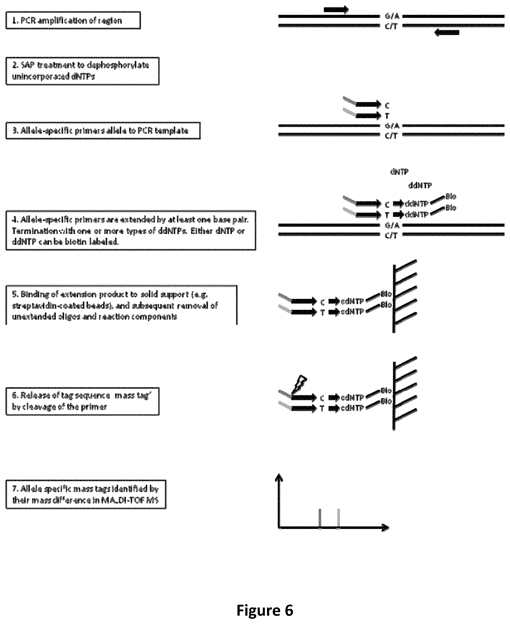
FIG. 6 shows PCR amplification and post-PCR primer extension with allele-specific extension primers containing allele-specific mass tags.
FIG. 7 shows MALDI-TOF MS spectra for 35 plex genotyping using post-PCR primer extension with allele-specific extension primers containing allele-specific mass tags as a readout.
FIG. 8 shows MALDI-TOF MS spectra for genotyping of rs1000586 and rs10131894.
FIG. 9 shows oligonucleotides mass tags corresponding to a 70plex assay. All oligonucleotides were diluted to a final total concentration of 10 pmol and spotted on a 384 well chip. Values for area, peak height and signal-to-noise ratio were collected from Typer 3.4 (SEQUENOM.RTM.).
FIG. 10 shows peak areas for oligonucleotides mass tags corresponding to 70plex assay sorted by nucleotide composition. All oligonucleotides were diluted to a final total concentration of 10 pmol and spotted on a 384 well chip. Area values were collected from Typer 3.4 (SEQUENOM.RTM.).
FIG. 11A shows a MALDI-TOF MS spectrum (zoomed views) of oligonucleotide tags corresponding to a 100plex assay. FIG. 11B shows signal to noise ratios of oligonucleotide tags corresponding to a 100plex assay. All oligonucleotides were diluted to a final total concentration of 10, 5, 2.5 or 1 pmol, with 8 replicates spotted on a 384 well chip. Values for signal-to-noise ratio were collected from Typer 3.4 (SEQUENOM.RTM.). FIG. 11C shows a MALDI-TOF MS spectrum (zoomed views) of a 100plex assay after PCR amplification and post-PCR primer extension with allele-specific extension primers containing allele-specific mass tags.
FIG. 12 shows extension rates for a 5plex reaction. Comparing extension oligonucleotides with or without a deoxyinosine, and either standard ddNTPs or nucleotides containing a biotin moiety. Extension rates were calculated by dividing the area of extended product by the total area of the peak (extended product and unextended oligonucleotide) in Typer 3.4 (SEQUENOM.RTM.). All experiments compare six DNAs.
FIG. 13 shows extension rates for 7 plex and 5 plex reactions over two DNAs. Results compare extension by a single biotinylated ddNTP or a biotinylated dNTP and terminated by an unmodified ddNTP, and final amounts of biotinylated dNTP or ddNTP of 210 or 420 .mu.mol added to the reaction. Extension rates were calculated by dividing the area of extended product by the total area (extended product and unextended oligonucleotide) in Typer 3.4. All experiments include two replicates of two Centre de'Etude du Polymorphisme Humain (CEPH) DNAs, NA07019 and NA11036.
FIG. 14 shows a comparison of iPLEX Gold enzyme concentrations in an extension reaction using a 70plex assay. All assays followed the same protocol except for the amount of iPLEX Gold enzyme used. All experiments include four replicates of the two CEPH DNAs NA06991 and NA07019. The results compare the signal-to-noise ratios of the extension products from Typer 3.4 (SEQUENOM.RTM.).
FIG. 15 shows a comparison of iPLEX Gold buffer concentration in extension reactions using a 70plex assay. All assays followed the same protocol except for the amount of goldPLEX buffer used. All experiments include four replicates of the two CEPH DNAs NA06991 and NA07019. The results compare the signal-to-noise ratios of the extension products from Typer 3.4 (SEQUENOM.RTM.).
FIGS. 16, 17, 18 and 19 show a comparison of extension oligonucleotide concentration in extension reactions using a 70plex assay. All assays followed the same protocol except for the amount of extension oligonucleotide used. All experiments include four replicates of the two CEPH DNAs NA06991 and NA07019. The results compare the signal-to-noise ratios of the extension products from Typer 3.4 (SEQUENOM.RTM.).
FIGS. 20 and 21 show a comparison of biotinylated ddNTP concentration in extension reactions using a 70plex assay. All assays followed the same protocol except for the amount of biotinylated ddNTP used (value indicates final amount of each biotinylated nucleotide). All experiments include four replicates of the two CEPH DNAs NA06991 and NA07019. The results compare the signal-to-noise ratios of the extension products from Typer 3.4 (SEQUENOM.RTM.).
FIG. 22 shows a comparison of Solulink and Dynabeads MyOne C1 magnetic streptavidin beads for capturing the extend products. A total amount of 10 pmol of each oligonucleotide corresponding to the two possible alleles for assay rs1000586 were bound to the magnetic streptavidin beads, in the presence of either water or varying quantities of biotinylated dNTPs (total 10, 100 or 500 pmol). The mass tags were then cleaved from the bound oligonucleotide with 10 U of endonuclease V. The results compare the area of the mass tag peaks from Typer 3.4 (SEQUENOM.RTM.) and are listed in comparison with 10 pmol of an oligonucleotide which has a similar mass.
FIG. 23 shows analysis of the ability of endonuclease V to cleave an extension product containing a deoxyinosine nucleotide in different locations. The oligonucleotides were identical aside from the deoxyinosine being 10, 15, 20 or 25 bases from the 3' end of the oligonucleotide. After binding the oligonucleotide to the magnetic streptavidin beads, the supernatant was collected, cleaned by a nucleotide removal kit (Qiagen) and then cleaved by treatment with endonuclease V (termed unbound oligonucleotide). The beads were washed, and cleaved with endonuclease V, as outlined in protocol section (termed captured/cleaved). The results compare the area of the peaks from Typer 3.4 (SEQUENOM.RTM.), and are listed as a percentage of oligonucleotide cleaved by endonuclease V without being bound to magnetic streptavidin beads.
FIG. 24 shows a comparison of magnetic streptavidin beads and endonuclease V concentration using a 70 plex assay. All assays were conducted using the same conditions except for the amount of magnetic streptavidin beads and endonuclease V. All experiments include four replicates of the CEPH DNA NA11036. The results compare the signal-to-noise ratio from Typer 3.4.
FIGS. 25 and 26 show a comparison of magnetic streptavidin beads and endonuclease V concentration using a 70 plex assay. All assays followed the same protocol except for the amount of magnetic streptavidin beads and endonuclease V. All experiments include four replicates of the two CEPH DNAs NA06991 and NA07019. The results compare the signal-to-noise ratio from Typer 3.4.
FIGS. 27A-G show a schematic representation of a biotin competition method for releasing a biotinylated amplification product of interest from a streptavidin coated magnetic or paramagnetic bead. In panel A, a region of interest is PCR amplified (e.g., using uniplex or multiplex methods) with subsequent dephosphorylation of the amplified products with shrimp alkaline phosphatase (not shown in diagram). Panel B illustrates single base extension of biotinylated dideoxynucleotides over the residue of interest in the product amplified in panel A. Panel C illustrates the capture of the biotinylated extension products by streptavidin coated magnetic beads. Panel D illustrates a washing step to remove unused reaction components followed by a capture step, to capture the streptavidin coated magnetic beads to which the biotinylated extension products are bound. Panel E illustrates release of the biotinylated extension products from the streptavidin coated magnetic bead by competition with free biotin. Panel F illustrates the purified biotinylated extension products, which can be further analyzed using a variety of methods, including matrix assisted laser desorption/time of flight (MALDI-TOF) mass spectrometry (MS). For use in MALDI-TOF mass spectrometry, the isolated extension products can be dispensed onto a SpectroCHIP.RTM. (SEQUENOM.RTM.), for example. Panel G illustrates a representative mass spectrum from MALDI-TOF MS analysis of a biotinylated extension product generated as described herein. See Example 12 for experimental details and results.
FIG. 28 is a representative mass spectrum of the mass difference of various allelic variants (e.g., polymorphism) as measured by MALDI-TOF MS analysis of single base extension products generated using biotinylated dideoxynucleotide terminators and released from the solid surface by biotin competition as described herein and illustrated in FIG. 27A-G. See Example 12 for experimental details and results. FIG. 28 discloses SEQ ID NOS 337-339, respectively, in order of appearance.
FIGS. 29A-G illustrate a flow chart showing a mechanism of a biotin competition releasing step.
FIG. 30A illustrates a flow chart showing a mechanism of an inosine cleavage releasing step. Steps are the same as for a biotin capture method through the wash step with the exception that the extension oligonucleotides have a 5' mass tag separated by an inosine residue. The mass tags are cleaved from captured products through endonuclease V cleavage specific to inosine residues.
FIG. 30B illustrates a detection of cleaved mass tags on the MALDI. The mass represents a genetic variant.
FIG. 31 shows comparative results of biotin competition vs. inosine cleavage using different concentrations of 3'-biotinylated oligonucleotide and different capture beads.
FIG. 32 shows comparative results of biotin competition vs. inosine cleavage using Dynal Cl beads. The mass spectra peak representing detected capture oligonucleotide and detected quantification oligonucleotide are indicated by down arrows. FIG. 32A shows the results of a biotin competition using Dynal C1 streptavidin beads for capture and free-biotin for the competition. The concentration of the biotinylated oligonucleotide and reference oligonucleotide (i.e. quantification oligonucleotide) tested was 0.031 uM. FIG. 32B shows the results of an inosine cleavage release using Dynal C1 streptavidin beads for capture and endonuclease V for the release. The concentration of the biotinylated oligonucleotide and reference oligonucleotide (i.e. quantification oligonucleotide) tested was 0.031 uM.
FIG. 33 shows an evaluation of different capture beads using a competitor template.
FIG. 34 shows mass spectrometry results from an assay using a very low competitor template (about 30 molecules) for each bead type tested. The mass spectra peak representing the detected capture oligonucleotide and detected quantification oligonucleotide are indicated by down arrows.
FIG. 34A shows results using Dynal C1 beads. FIG. 34B shows results using Solulink Beads. FIG. 34C shows results using Dynal M270 beads.
FIG. 35 shows the detection of BRAF-2 and BRAF-15 mutations using different extension compositions and demonstrates an increase in signal to noise ratio when ddNTPs corresponding to the wild type (e.g. more abundant variant) are excluded from the extension composition.
FIG. 36 shows results of a competition assay with a 1% rare allele.
FIG. 37 illustrates a model plasmid that can be cleaved through EcoRI restriction digest to separate the regions and more adequately reflect a genomic context.
DETAILED DESCRIPTION
Methods for determining the presence or absence of a plurality of target nucleic acids in a composition described herein find multiple uses by the person of ordinary skill in the art (hereafter referred to herein as the "person of ordinary skill"). Such methods can be utilized, for example, to: (a) rapidly determine whether a particular target sequence (e.g. a target sequence comprising a genetic variation) is present in a sample; (b) perform mixture analysis, e.g., identify a mixture and/or its composition or determine the frequency of a target sequence in a mixture (e.g., mixed communities, quasispecies); (c) detect sequence variations (e.g., mutations, single nucleotide polymorphisms) in a sample; (d) perform haplotyping determinations; (e) perform microorganism (e.g., pathogen) typing; (f) detect the presence or absence of a microorganism target sequence in a sample; (g) identify disease markers; (h) detect microsatellites; (i) identify short tandem repeats; (j) identify an organism or organisms; (k) detect allelic variations; (l) determine allelic frequency; (m) determine methylation patterns; (n) perform epigenetic determinations; (o) re-sequence a region of a biomolecule; (p) perform analyses in human clinical research and medicine (e.g. cancer marker detection, sequence variation detection; detection of sequence signatures favorable or unfavorable for a particular drug administration), (q) perform HLA typing; (r) perform forensics analyses; (s) perform vaccine quality control analyses; (t) monitor treatments; (u) perform vector identity analyses; (v) perform vaccine or production strain quality control and (w) test strain identity (x) plants. Such methods also may be utilized, for example, in a variety of fields, including, without limitation, in commercial, education, medical, agriculture, environmental, disease monitoring, military defense, and forensics fields.
Target Nucleic Acids
As used herein, the term "nucleic acid" refers to an oligonucleotide or polynucleotide, including, without limitation, natural nucleic acids (e.g., deoxyribonucleic acid (DNA), ribonucleic acid (RNA)), synthetic nucleic acids, non-natural nucleic acids (e.g., peptide nucleic acid (PNA)), unmodified nucleic acids, modified nucleic acids (e.g., methylated DNA or RNA, labeled DNA or RNA, DNA or RNA having one or more modified nucleotides). Reference to a nucleic acid as a "polynucleotide" refers to two or more nucleotides or nucleotide analogs linked by a covalent bond. Nucleic acids may be any type of nucleic acid suitable for use with processes described herein. A nucleic acid in certain embodiments can be DNA (e.g., complementary DNA (cDNA), genomic DNA (gDNA), plasmids and vector DNA and the like), RNA (e.g., viral RNA, message RNA (mRNA), short inhibitory RNA (siRNA), ribosomal RNA (rRNA), tRNA and the like), and/or DNA or RNA analogs (e.g., containing base analogs, sugar analogs and/or a non-native backbone and the like). A nucleic acid can be in any form useful for conducting processes herein (e.g., linear, circular, supercoiled, single-stranded, double-stranded and the like). A nucleic acid may be, or may be from, a plasmid, phage, autonomously replicating sequence (ARS), centromere, artificial chromosome, chromosome, a cell, a cell nucleus or cytoplasm of a cell in certain embodiments. A nucleic acid in some embodiments is from a single chromosome (e.g., a nucleic acid sample may be from one chromosome of a sample obtained from a diploid organism). In the case of fetal nucleic acid, the nucleic acid may be from the paternal allele, the maternal allele or the maternal and paternal allele.
The term "species," as used herein with reference to a target nucleic acid, amplicon, primer, sequence tag, polynucleotide, or oligonucleotide, refers to one nucleic acid having a nucleotide sequence that differs by one or more nucleotides from the nucleotide sequence of another nucleic acid when the nucleotide sequences are aligned. Thus, a first nucleic acid species differs from a second nucleic acid species when the sequences of the two species, when aligned, differ by one or more nucleotides (e.g., about 1, 2, 3, 4, 5, 10, 15, 20, 25, 30, 35, 40, 45, 50, 55, 60, 65, 70, 75, 80, 85, 90, 95, 100 or more than 100 nucleotide differences). In certain embodiments, the number of nucleic acid species, such as target nucleic acid species, amplicon species or extended oligonucleotide species, includes, but is not limited to about 2 to about 10000 nucleic acid species, about 2 to about 1000 nucleic acid species, about 2 to about 500 nucleic acid species, or sometimes about 2, 3, 4, 5, 6, 7, 8, 9, 10, 11, 12, 13, 14, 15, 16, 17, 18, 19, 20, 25, 30, 35, 40, 45, 50, 55, 60, 65, 70, 75, 80, 80, 85, 90, 95, 100, 125, 150, 175, 200, 225, 250, 275, 300, 325, 350, 375, 400, 425, 450, 475, 500, 600, 700, 800, 900, 1000, 2000, 3000, 4000, 5000, 6000, 7000, 8000, 9000 or 10000 nucleic acid species.
In some embodiments an oligonucleotide species is hybridized to a nucleic acid template (e.g. an amplicon) thereby forming a double stranded nucleic acid and the oligonucleotide species that is hybridized to the template is referred to herein as a hybridized oligonucleotide species. In some embodiments a hybridized oligonucleotide species can comprise one or more nucleotides that are not hybridized to the template. For example, a hybridized oligonucleotide species can comprise one or more mismatched nucleotides (e.g. non-complementary nucleotides) and sometimes a 5' and/or 3' region of nucleotides that do not hybridize. In some embodiments a hybridized oligonucleotide species comprises a tag (e.g. a mass distinguishable tag, a sequence tag, a light emitting tag or a radioactive tag). In some embodiments a hybridized oligonucleotide species comprises a capture agent (e.g. biotin, or any member of binding pair). In some embodiments a hybridized oligonucleotide species comprises a terminating nucleotide.
As used herein, the term "nucleotides" refers to natural and non-natural nucleotides. Nucleotides include, but are not limited to, naturally occurring nucleoside mono-, di-, and triphosphates: deoxyadenosine mono-, di- and triphosphate; deoxyguanosine mono-, di- and triphosphate; deoxythymidine mono-, di- and triphosphate; deoxycytidine mono-, di- and triphosphate; deoxyuridine mono-, di- and triphosphate; and deoxyinosine mono-, di- and triphosphate (referred to herein as dA, dG, dT, dC, dU and dl, or A, G, T, C, U and I respectively). Nucleotides also include, but are not limited to, modified nucleotides and nucleotide analogs. Modified nucleotides and nucleotide analogs include, without limitation, deazapurine nucleotides, e.g., 7-deaza-deoxyguanosine (7-deaza-dG) and 7-deaza-deoxyadenosine (7-deaza-dA) mono-, di- and triphosphates, deutero-deoxythymidine (deutero-dT) mon-, di- and triphosphates, methylated nucleotides e.g., 5-methyldeoxycytidine triphosphate, .sup.13C/.sup.15N labeled nucleotides and deoxyinosine mono-, di- and triphosphate. Modified nucleotides, isotopically enriched nucleotides, depleted nucleotides, tagged and labeled nucleotides and nucleotide analogs can be obtained using a variety of combinations of functionality and attachment positions.
The term "composition" as used herein with reference to nucleic acids refers to a tangible item that includes one or more nucleic acids. A composition sometimes is a sample extracted from a source, but also a composition of all samples at the source, and at times is the source of one or more nucleic acids. A composition can comprise nucleic acids. In some embodiments, a composition can comprise genomic DNA. In some embodiments, a composition can comprise maternal DNA, fetal DNA or a mixture of maternal and fetal DNA. In some embodiments, a composition can comprise fragments of genomic DNA. In some embodiments a composition can comprise nucleic acids derived from a virus, bacteria, yeast, fungus, mammal or mixture thereof.
A nucleic acid sample may be derived from one or more sources. A sample may be collected from an organism, mineral or geological site (e.g., soil, rock, mineral deposit, fossil), or forensic site (e.g., crime scene, contraband or suspected contraband), for example. Thus, a source may be environmental, such as geological, agricultural, combat theater or soil sources, for example. A source also may be from any type of organism such as any plant, fungus, protistan, moneran, virus or animal, including but not limited, human, non-human, mammal, reptile, cattle, cat, dog, goat, swine, pig, monkey, ape, gorilla, bull, cow, bear, horse, sheep, poultry, mouse, rat, fish, dolphin, whale, and shark, or any animal or organism that may have a detectable nucleic acids. Sources also can refer to different parts of an organism such as internal parts, external parts, living or non-living cells, tissue, fluid and the like. A sample therefore may be a "biological sample," which refers to any material obtained from a living source or formerly-living source, for example, an animal such as a human or other mammal, a plant, a bacterium, a fungus, a protist or a virus. A source can be in any form, including, without limitation, a solid material such as a tissue, cells, a cell pellet, a cell extract, or a biopsy, or a biological fluid such as urine, blood, saliva, amniotic fluid, exudate from a region of infection or inflammation, or a mouth wash containing buccal cells, hair, cerebral spinal fluid and synovial fluid and organs. A sample also may be isolated at a different time point as compared to another sample, where each of the samples are from the same or a different source. A nucleic acid may be from a nucleic acid library, such as a cDNA or RNA library, for example. A nucleic acid may be a result of nucleic acid purification or isolation and/or amplification of nucleic acid molecules from the sample. Nucleic acid provided for sequence analysis processes described herein may contain nucleic acid from one sample or from two or more samples (e.g., from 1, 2, 3, 4, 5, 6, 7, 8, 9, 10, 11, 12, 13, 14, 15, 16, 17, 18, 19, 20, 25, 50, 75, 100, 200, 300, 400, 500, 600, 700, 800, 900 or 1000 or more samples).
Nucleic acids may be treated in a variety of manners. For example, a nucleic acid may be reduced in size (e.g., sheared, digested by nuclease or restriction enzyme, de-phosphorylated, de-methylated), increased in size (e.g., phosphorylated, reacted with a methylation-specific reagent, attached to a detectable label), treated with inhibitors of nucleic acid cleavage and the like.
Nucleic acids may be provided for conducting methods described herein without processing, in certain embodiments. In some embodiments, nucleic acid is provided for conducting methods described herein after processing. For example, a nucleic acid may be extracted, isolated, purified or amplified from a sample. The term "isolated" as used herein refers to nucleic acid removed from its original environment (e.g., the natural environment if it is naturally occurring, or a host cell if expressed exogenously), and thus is altered "by the hand of man" from its original environment. An isolated nucleic acid generally is provided with fewer non-nucleic acid components (e.g., protein, lipid) than the amount of components present in a source sample. A composition comprising isolated nucleic acid can be substantially isolated (e.g., about 90%, 91%, 92%, 93%, 94%, 95%, 96%, 97%, 98%, 99% or greater than 99% free of non-nucleic acid components). The term "purified" as used herein refers to nucleic acid provided that contains fewer nucleic acid species than in the sample source from which the nucleic acid is derived. A composition comprising nucleic acid may be substantially purified (e.g., about 90%, 91%, 92%, 93%, 94%, 95%, 96%, 97%, 98%, 99% or greater than 99% free of other nucleic acid species).
Nucleic acids may be processed by a method that generates nucleic acid fragments, in certain embodiments, before providing nucleic acid for a process described herein. In some embodiments, nucleic acid subjected to fragmentation or cleavage may have a nominal, average or mean length of about 5 to about 10,000 base pairs, about 100 to about 1,000 base pairs, about 100 to about 500 base pairs, or about 10, 15, 20, 25, 30, 35, 40, 45, 50, 55, 60, 65, 70, 75, 80, 85, 90, 95, 100, 200, 300, 400, 500, 600, 700, 800, 900, 1000, 2000, 3000, 4000, 5000, 6000, 7000, 8000, 9000 or 10000 base pairs. Fragments can be generated by any suitable method known in the art, and the average, mean or nominal length of nucleic acid fragments can be controlled by selecting an appropriate fragment-generating procedure. In certain embodiments, nucleic acid of a relatively shorter length can be utilized to analyze sequences that contain little sequence variation and/or contain relatively large amounts of known nucleotide sequence information. In some embodiments, nucleic acid of a relatively longer length can be utilized to analyze sequences that contain greater sequence variation and/or contain relatively small amounts of unknown nucleotide sequence information.
As used herein, the term "target nucleic acid" or "target nucleic acid species" refers to any nucleic acid species of interest in a sample. A target nucleic acid includes, without limitation, (i) a particular allele amongst two or more possible alleles, and (ii) a nucleic acid having, or not having, a particular mutation, nucleotide substitution, sequence variation, repeat sequence, marker or distinguishing sequence. As used herein, the term "different target nucleic acids" refers to nucleic acid species that differ by one or more features. As used herein, the term "genetic variation" refers to nucleic acid species that differ by one or more features. As used herein, the term "variant" refers to nucleic acid species that differ by one or more features. Features include, without limitation, one or more methyl groups or a methylation state, one or more phosphates, one or more acetyl groups, and one or more deletions, additions or substitutions of one or more nucleotides. Examples of one or more deletions, additions or substitutions of one or more nucleotides include, without limitation, the presence or absence of a particular mutation, presence or absence of a nucleotide substitution (e.g., single nucleotide polymorphism (SNP)), presence or absence of a repeat sequence (e.g., di-, tri-, tetra-, penta-nucleotide repeat), presence or absence of a marker (e.g., microsatellite) and presence of absence of a distinguishing sequence (e.g., a sequence that distinguishes one organism from another (e.g., a sequence that distinguishes one viral strain from another viral strain)). Different target nucleic acids may be distinguished by any known method, for example, by mass, binding, distinguishable tags and the like, as described herein.
As used herein, the term "plurality of target nucleic acids" or "plurality of target nucleic acid species" refers to more than one target nucleic acid species. A plurality of target nucleic acids can be about 2 to about 10000 nucleic acid species, about 2 to about 1000 nucleic acid species, about 2 to about 500 nucleic acid species, or sometimes about 2, 3, 4, 5, 6, 7, 8, 9, 10, 11, 12, 13, 14, 15, 16, 17, 18, 19, 20, 25, 30, 35, 40, 45, 50, 55, 60, 65, 70, 75, 80, 80, 85, 90, 95, 100, 125, 150, 175, 200, 225, 250, 275, 300, 325, 350, 375, 400, 425, 450, 475, 500, 600, 700, 800, 900, 1000, 2000, 3000, 4000, 5000, 6000, 7000, 8000, 9000 or 10000 nucleic acid species, in certain embodiments. Detection or identification of nucleic acids results in detection of the target and can indicate the presence or absence of a particular mutation, sequence variation (mutation or polymorphism) or genetic variation (e.g. sequence variation, sequence difference or polymorphism). Within the plurality of target nucleic acids, there may be detection of the same or different target nucleic acids. The plurality of target nucleic acids may also be identified quantitatively as well as qualitatively in terms of identification. Also refer to multiplexing below.
Amplification and Extension
A nucleic acid (e.g., a target nucleic acid) can be amplified in certain embodiments. As used herein, the term "amplifying," and grammatical variants thereof, refers to a process of generating copies of a template nucleic acid. For example, nucleic acid template may be subjected to a process that linearly or exponentially generates two or more nucleic acid amplicons (copies) having the same or substantially the same nucleotide sequence as the nucleotide sequence of the template, or a portion of the template. Nucleic acid amplification often is specific (e.g., amplicons have the same or substantially the same sequence), and can be non-specific (e.g., amplicons have different sequences) in certain embodiments. Nucleic acid amplification sometimes is beneficial when the amount of target sequence present in a sample is low. By amplifying the target sequences and detecting the amplicon synthesized, sensitivity of an assay can be improved, since fewer target sequences are needed at the beginning of the assay for detection of a target nucleic acid. A target nucleic acid sometimes is not amplified prior to hybridizing an extension oligonucleotide, in certain embodiments.
Amplification conditions are known and can be selected for a particular nucleic acid that will be amplified. Amplification conditions include certain reagents some of which can include, without limitation, nucleotides (e.g., nucleotide triphosphates), modified nucleotides, oligonucleotides (e.g., primer oligonucleotides for polymerase-based amplification and oligonucleotide building blocks for ligase-based amplification), one or more salts (e.g., magnesium-containing salt), one or more buffers, one or more polymerizing agents (e.g., ligase enzyme, polymerase enzyme), one or more nicking enzymes (e.g., an enzyme that cleaves one strand of a double-stranded nucleic acid) and one or more nucleases (e.g., exonuclease, endonuclease, RNase). Any polymerase suitable for amplification may be utilized, such as a polymerase with or without exonuclease activity, DNA polymerase and RNA polymerase, mutant forms of these enzymes, for example. Any ligase suitable for joining the 5' of one oligonucleotide to the 3' end of another oligonucleotide can be utilized. Amplification conditions also can include certain reaction conditions, such as isothermal or temperature cycle conditions. Methods for cycling temperature in an amplification process are known, such as by using a thermocycle device. The term "cycling" refers to amplification (e.g. an amplification reaction or extension reaction) utilizing a single primer or multiple primers where temperature cycling is used. Amplification conditions also can, in some embodiments, include an emulsion agent (e.g., oil) that can be utilized to form multiple reaction compartments within which single nucleic acid molecule species can be amplified. Amplification is sometimes an exponential product generating process and sometimes is a linear product generating process.
A strand of a single-stranded nucleic acid target can be amplified and one or two strands of a double-stranded nucleic acid target can be amplified. An amplification product (amplicon), in some embodiments, is about 10 nucleotides to about 10,000 nucleotides in length, about 10 to about 1000 nucleotides in length, about 10 to about 500 nucleotides in length, 10 to about 100 nucleotides in length, and sometimes about 10, 11, 12, 13, 14, 15, 16, 17, 18, 19, 20, 25, 30, 35, 40, 45, 50, 55, 60, 65, 70, 75, 80, 80, 85, 90, 95, 100, 125, 150, 175, 200, 225, 250, 275, 300, 325, 350, 375, 400, 425, 450, 475, 500, 600, 700, 800, 900 or 1000 nucleotides in length.
Any suitable amplification technique and amplification conditions can be selected for a particular nucleic acid for amplification. Known amplification processes include, without limitation, polymerase chain reaction (PCR), extension and ligation, ligation amplification (or ligase chain reaction (LCR)) and amplification methods based on the use of Q-beta replicase or template-dependent polymerase (see US Patent Publication Number US20050287592). Also useful are strand displacement amplification (SDA), thermophilic SDA, nucleic acid sequence based amplification (3SR or NASBA) and transcription-associated amplification (TAA). Reagents, apparatus and hardware for conducting amplification processes are commercially available, and amplification conditions are known and can be selected for the target nucleic acid at hand.
Polymerase-based amplification can be effected, in certain embodiments, by employing universal primers. In such processes, hybridization regions that hybridize to one or more universal primers are incorporated into a template nucleic acid. Such hybridization regions can be incorporated into (i) a primer that hybridizes to a target nucleic acid and is extended, and/or (ii) an oligonucleotide that is joined (e.g., ligated using a ligase enzyme) to a target nucleic acid or a product of (i), for example. Amplification processes that involve universal primers can provide an advantage of amplifying a plurality of target nucleic acids using only one or two amplification primers, for example.
FIG. 1 shows certain embodiments of amplification processes. In certain embodiments, only one primer is utilized for amplification (e.g., FIG. 1A). In certain embodiments, two primers are utilized. Under amplification conditions at least one primer has a complementary distinguishable tag. The gene specific extend primer has a 5' universal PCRTag1R (e.g., FIG. 1A). It may be extended on any nucleic acid, for example genomic DNA. The DNA or the PCR Tag1R gene specific extend primer may be biotinylated, to facilitate clean up of the reaction. The extended strand then is ligated by a single strand ligase to a universal phosphorylated oligonucleotide, which has a sequence that is the reverse complement of Tag2F (universal PCR primer; FIG. 1B). To facilitate cleanup in the next step, the phosphorylated oligonucleotide can include exonuclease resistant nucleotides at its 3' end. During the exonuclease treatment, all non-ligated extended strands are degraded, whereas ligated products are protected and remain in the reaction (e.g., FIG. 1C). A universal PCR then is performed, using Tag1R and the Tag2F primers, to amplify multiple targets (e.g., FIG. 1D).
FIG. 2 also shows certain embodiments of amplification processes. In some embodiments, a method involving primer extension and ligation takes place in the same reaction (e.g., FIG. 2A). Biotinylated PCRTag3R gene-specific primer is an extension primer. The phosphorylated oligonucleotide has a gene-specific sequence and binds about 40 bases (e.g., 4 to 100 or more) away from the primer extension site, to the same strand of DNA. Thus a DNA polymerase, such as Stoffel polymerase, extends the strand, until it reaches the phosphorylated oligonucleotide. A ligase enzyme ligates the gene specific sequence of the phosphorylated oligonucleotide to the extended strand. The 3' end of phosphorylated oligonucleotide has PCRTag4(RC)F as its universal tag. The biotinylated extended strands then are bound to streptavidin beads. This approach facilitates cleanup of the reaction (e.g., FIG. 2B). DNA, such as genomic DNA, and the gene specific phosphorylated oligonucleotides are washed away. A universal PCR then is performed, using Tag3R and Tag4F as primers, to amplify different genes of interest (e.g., FIG. 2C).
Certain nucleic acids can be extended in certain embodiments. The term "extension," and grammatical variants thereof, as used herein refers to elongating one strand of a nucleic acid. For example, an oligonucleotide that hybridizes to a target nucleic acid or an amplicon generated from a target nucleic acid can be extended in certain embodiments. An extension reaction is conducted under extension conditions, and a variety of such conditions are known and selected for a particular application. Extension conditions include certain reagents, including without limitation, one or more oligonucleotides, extension nucleotides (e.g., nucleotide triphosphates (dNTPs)), terminating nucleotides (e.g., one or more dideoxynucleotide triphosphates (ddNTPs)), one or more salts (e.g., magnesium-containing salt), one or more buffers (e.g., with beta-NAD, Triton X-100), and one or more polymerizing agents (e.g., DNA polymerase, RNA polymerase). Extension can be conducted under isothermal conditions or under non-isothermal conditions (e.g., thermocycled conditions), in certain embodiments. One or more nucleic acid species can be extended in an extension reaction, and one or more molecules of each nucleic acid species can be extended. A nucleic acid can be extended by one or more nucleotides, and in some embodiments, the extension product is about 10 nucleotides to about 10,000 nucleotides in length, about 10 to about 1000 nucleotides in length, about 10 to about 500 nucleotides in length, 10 to about 100 nucleotides in length, and sometimes about 10, 11, 12, 13, 14, 15, 16, 17, 18, 19, 20, 25, 30, 35, 40, 45, 50, 55, 60, 65, 70, 75, 80, 80, 85, 90, 95, 100, 125, 150, 175, 200, 225, 250, 275, 300, 325, 350, 375, 400, 425, 450, 475, 500, 600, 700, 800, 900 or 1000 nucleotides in length. Incorporation of a terminating nucleotide (e.g., ddNTP), the hybridization location, or other factors, can determine the length to which the oligonucleotide is extended. In certain embodiments, amplification and extension processes are carried out in the same detection procedure.
In some embodiments an extension reaction includes multiple temperature cycles repeated to amplify the amount of extension product in the reaction. In some embodiments the extension reaction is cycled 2 or more times. In some embodiments the extension reaction is cycled 10 or more times. In some embodiments the extension reaction is cycled about 10, 15, 20, 50, 100, 200, 300, 400, 500 or 600 or more times. In some embodiments the extension reaction is cycled 20 to 50 times. In some embodiments the extension reaction is cycled 20 to 100 times. In some embodiments the extension reaction is cycled 20 to 300 times. In some embodiments the extension reaction is cycled 200 to 300 times.
In some embodiments a target nucleic acid (e.g. target nucleic acid species, oligonucleotide species, hybridized oligonucleotide species or amplicon) is extended in the presence of an extension composition where the target nucleic acid is extended by one nucleotide. An extension composition can comprise one or more buffers, salts, enzymes (e.g. polymerases, Klenow, etc.), water, templates (e.g. DNA, RNA, amplicons, etc.), primers (e.g. oligonucleotides), nucleotide triphosphates, glycerol, macromolecular exclusion molecules and any other additives used in the art. An extension composition can comprise terminating nucleotides (e.g. dideoxynucleotides (e.g. ddNTPs)), non-terminating or extension nucleotides (e.g. dNTPs) or a mixture of terminating nucleotides and non-terminating nucleotides. An extension composition consisting essentially of a particular terminating nucleotide or terminating nucleotides, can contain any other component of an extension composition (e.g. buffers, salts, templates, primers, etc.), but does not contain any other terminating nucleotide or nucleotide triphosphate (e.g. dNTP) except those specified. For example an extension composition consisting essentially of ddTTP and ddCTP does not contain ddATP, ddGTP or any other dNTP. In some embodiments the nucleotides in an extension composition are only terminating nucleotides and the target nucleic acid is extended by one nucleotide (i.e. sometimes there are no extension nucleotides in the extension composition). In some embodiments an extension composition consists essentially of terminating nucleotides (e.g. ddNTPs). In some embodiments, a terminating nucleotide comprises one or more (e.g. 1, 2, 3, 4, 5, 6, 7, 8, 9, 10, 11, 12, 13, 14, 15, 20, or more) capture agents. In some embodiments, a terminating nucleotide comprises one or more (e.g. 1, 2, 3, 4, 5, 6, 7, 8, 9, 10, 11, 12, 13, 14, 15, 20, or more) different capture agents. In some embodiments, a terminating nucleotide comprises (e.g. is covalently bound to) one or more (e.g. 1, 2, 3, 4, 5, 6, 7, 8, 9, 10, 11, 12, 13, 14, 15, 20, or more) capture agent molecules. In some embodiments, a terminating nucleotide comprises one capture agent molecule. In some embodiments, a first terminating nucleotide comprises a capture agent and a second terminating nucleotide comprises a different capture agent. In some embodiments, an extension composition comprises one or more terminating nucleotides where each terminating nucleotide comprises a different capture agent. In some embodiments, an extension composition comprises one or more terminating nucleotides where each terminating nucleotide comprises a capture agent and the capture agent is the same. In some embodiments, an extension composition comprises a terminating nucleotide and an extension nucleotide and one or more of the nucleotides (e.g. terminating nucleotides and/or extension nucleotides) include a capture agent. In some embodiments a terminating nucleotide comprises a capture agent and the capture agent is biotin or a biotin analogue. In some embodiments, the extension composition consists essentially of terminating nucleotides that are bound to one or more capture agents. In some embodiments the capture agent is biotin or a biotin analogue. A biotin analogue can be any modified biotin that effects the binding properties of biotin to avidin or streptavidin (e.g. 9-methylbiotin, biotin methyl ester (MEBio), desthiobiotin (DEBio), 2'-iminobiotin (IMBio), e-N-Biotinyl-L-lysine, diaminobiotin (DABio), including all biotin analogues disclosed in Lai-Qiang et. al. (Lai-Qiang Ying and Bruce P. Branchaud, Chemical Communications, 2011, 47, 8593-8595)). In some embodiments the capture agent is avidin, streptavidin or a modified form of avidin or streptavidin (e.g. nitroavidin, nitrostreptavidin, NeutrAvidin, CaptAvidin and derivatives thereof).
Any suitable extension reaction can be selected and utilized. An extension reaction can be utilized, for example, to discriminate SNP alleles by the incorporation of deoxynucleotides and/or dideoxynucleotides to an extension oligonucleotide that hybridizes to a region adjacent to the SNP site in a target nucleic acid. The primer often is extended with a polymerase. In some embodiments, the oligonucleotide is extended by only one deoxynucleotide or dideoxynucleotide complementary to the SNP site. In some embodiments, an oligonucleotide may be extended by dNTP incorporation and terminated by a ddNTP, or terminated by ddNTP incorporation without dNTP extension in certain embodiments. One or more dNTP and/or ddNTP used during the extension reaction are labeled with a moiety allowing immobilization to a solid support, such as biotin, in some embodiments. Extension may be carried out using unmodified extension oligonucleotides and unmodified dideoxynucleotides, unmodified extension oligonucleotides and biotinylated dideoxynucleotides, extension oligonucleotides containing a deoxyinosine and unmodified dideoxynucleotides, extension oligonucleotides containing a deoxyinosine and biotinylated dideoxynucleotides, extension by biotinylated dideoxynucleotides, or extension by biotinylated deoxynucleotide and/or unmodified dideoxynucleotides, in some embodiments.
In some embodiments an oligonucleotide species can hybridize, under hybridization conditions, to a template (e.g. a target nucleic acid species) adjacent to a genetic variation or variant (e.g. the 3' end of the oligonucleotide species may be located 5' of the genetic variation site and may be 0 to 10 nucleotides away from the 5' end of the genetic variation site). Several variant may exist at a site of genetic variation in a target nucleic acid. A genetic variant sometimes is a single nucleotide polymorphism (SNP) or single nucleotide variant. Several single nucleotide variants may exist at a single base position on a template target located 3' of a hybridized oligonucleotide. Several single nucleotide variants may differ by a single base located at a position on a template target that is 3' of a hybridized oligonucleotide species. In some embodiments an oligonucleotide species is extended by one nucleotide at the variant position. The oligonucleotide can be extended by any one of five terminating nucleotides (e.g. ddATP, ddUTP, ddTTP, ddGTP, ddCTP), depending on the number of variants present, in some embodiments. A target nucleic acid species and its variants, or a corresponding amplicon, can act as the template and can, in part, determine which terminating nucleotide is added to the oligonucleotide in the extension reaction. A target nucleic acid species may have two or more variants. In some embodiments a target nucleic acid species comprises two variants. In some embodiments a target nucleic acid species comprises three variants. In some embodiments a target nucleic acid species comprises four variants. In some embodiments a target nucleic acid species comprises no variants.
In some embodiments the amount of molecules of a target mutant variant (e.g. low abundant variant) present in an assay where the wild type (e.g. high abundance species) extension product is not generated is determined by the use of a synthetic template included in the extension reaction. In some embodiments the amount of target (e.g. copy number, concentration, percentage) mutant variant (i.e. mutant extension products) and/or percentage of target mutant variant in the sample is quantified by including a known amount of synthetic template in the extension reaction. In some embodiments the synthetic template can hybridize to an oligonucleotide species and contain a base substitution at the mutant position located just 3' of the oligonucleotide species to be extended. In some embodiments, the base substitution is different than the wild type or target mutant variant (e.g. first variant, low abundant variant, SNP). In some embodiments, the base substitution present in the template is not present in the sample prior to introduction of the template. In some embodiments a ddNTP (e.g. a biotin-ddNTP) that is complementary to the base substitution in the synthetic template is also introduced into the reaction. In some embodiments, oligonucleotide species that hybridize to the target mutant variant are co-amplified (e.g. co-extended) with oligonucleotide species that hybridize to the synthetic template. In some embodiments, multiple reactions, that include serial dilutions of a synthetic template, are performed to determine the amount and/or percentage of the target mutant variant. In some embodiments, the amount and/or percentage of the target mutant variant is determined by the amount of synthetic template that yields equal extension product as the target mutant variant.
In some embodiments, one variant can be in greater abundance than other variants. In some embodiments, the variant of greatest abundance is referred to as the wild type variant. In some embodiments a target nucleic acid species comprises a first and second variant where the second variant is represented in greater abundance (i.e. more template is present). In some embodiments a target nucleic acid species comprises a first, second and third variant where the second variant is represented in greater abundance over the first and third variant. In some embodiments a target nucleic acid species comprises a first, second, third and fourth variant where the second variant is represented in greater abundance over the first, third and fourth variant. A variant that is represented in a greater abundance generally is present at a higher concentration or is represented by a greater number of molecules (e.g. copies) when compared to another variant. A higher concentration can be 2-fold or more. In some embodiments, a higher concentration is 10-fold or more. In some embodiments, a higher concentration is a 100-fold, a 1000-fold or 10000-fold or more. In some embodiments, a second variant represents a wild type sequence and is present at a 100-fold or higher concentration than a first variant. In some embodiments, a first variant is represented at a significantly lower concentration than a second variant (e.g. a wild type) where the first variant represents less of the target nucleic acid species. In some embodiments a first variant represents less than 30%, 20%, 15%, 10%, 8%, 5%, 4%, 3%, 2%, 1%, 0.8%, 0.75%, 0.5%, 0.1%, 0.05%, 0.01% or less of the target nucleic acid species. In some embodiments a first variant represents between about 5% to about 0.75% of the target nucleic acid species. In some embodiments a first variant represents less than 30%, 20%, 15%, 10%, 8%, 5%, 4%, 3%, 2%, 1%, 0.8%, 0.75%, 0.5%, 0.1%, 0.05%, 0.01% or less of the total nucleic acid in a composition.
In some embodiments, a terminating nucleotide that is present (or, in some embodiments absent) in an extension composition determines which terminating nucleotide is added to an oligonucleotide. In some embodiments, an extension composition comprises one or more terminating nucleotides (e.g. ddNTPs). In some embodiments, an extension composition comprises one or more terminating nucleotides and one or more non-terminating nucleotides (e.g. dNTPs). In some embodiments, an extension composition comprises only terminating nucleotides that correspond to a specific variant (e.g. a first variant or a less abundant variant) and therefore only allow extension of that specific variant. In some embodiments, a terminating nucleotide that would allow extension of a second variant (e.g. a wild type or more abundant variant) can be excluded from an extension composition thereby preventing extension of the second variant. In some embodiments, an extension composition comprises only terminating nucleotides that correspond to a first and third variant and therefore only allow extension of those specific variants. In some embodiments, an extension composition comprises only terminating nucleotides that correspond to a first, third and fourth variant and therefore only allow extension of the first, third and fourth variants. In some embodiments, an extension composition consists essentially of terminating nucleotides that correspond to a first variant. In some embodiments, a method comprises contacting hybridized oligonucleotide species with an extension composition comprising one or more terminating nucleotides under extension conditions where (i) at least one of the one or more terminating nucleotides comprises a capture agent, and (ii) the hybridized oligonucleotide species that hybridize to the first variant (e.g. a less abundant variant, (e.g., less abundant SNP variant)) are extended by a terminating nucleotide and the hybridized oligonucleotide species that hybridize to the second variant (e.g. wild type or more abundant variant) are not extended by a terminating nucleotide, thereby generating extended oligonucleotide species. In some embodiments an extended oligonucleotide species of a second variant is not detected.
The term "signal to noise ratio" as used herein refers to the quantitative measurement of the quality of a signal by quantifying the ratio of intensity of a signal relative to noise when using a detection process (e.g. mass spectrometry). In some embodiments, an intensive peak on one spectrum has a greater signal to noise ratio than a low intensity peak generated by the same analyte (e.g. an extended oligonucleotide species) on another spectrum. In some embodiments, noise is generated by extended oligonucleotide species derived from abundant variants (e.g. wild type alleles, second variants, wild type variants). In some embodiments, the signal generated from an extended oligonucleotide species derived from a less abundant variant (e.g. a first variant, third variant, fourth variant, mutant variant, mutant allele, SNP) is obscured by the noise generated by a more abundant extended oligonucleotide species (e.g. a second variant, wild type variant, wild type allele) when using mass spectrometry. The term "signal" as used in the phrase "signal to noise ratio" herein refers to the intensity of a signal peak of an extended oligonucleotide species. In some embodiments, the term "signal" as used in the phrase "signal to noise ratio" herein generally refers to the intensity of a signal peak of an extended oligonucleotide species derived from a less abundant variant (e.g. a first variant, mutant variant, mutant allele, SNP). In some embodiments, a terminating nucleotide that would allow extension of a second variant (e.g. a wild type or more abundant variant) is excluded from an extension composition thereby preventing extension of the second variant and increasing the signal to noise ratio for a less abundant variant (e.g. a first variant, mutant variant, mutant allele, SNP). In some embodiments, a method comprises contacting hybridized oligonucleotide species with an extension composition comprising one or more terminating nucleotides under extension conditions where (i) at least one of the one or more terminating nucleotides comprises a capture agent, and (ii) the hybridized oligonucleotide species that hybridize to the first variant (e.g. a less abundant variant, (e.g., less abundant SNP variant)) are extended by a terminating nucleotide and the hybridized oligonucleotide species that hybridize to the second variant (e.g. wild type or more abundant variant) are not extended by a terminating nucleotide, thereby generating extended oligonucleotide species and increasing the signal to noise ratio compared to a condition where both the first and second variants are extended. In some embodiments the detecting in (f) is with a signal to noise ratio greater than a signal to noise ratio for detecting after releasing without competition with a competitor. In some embodiments the detecting in (f) comprises an increase in a signal to noise ratio when the releasing step (e) comprises competition with a competitor as compared to a releasing step that does not comprise competition with a competitor. In some embodiments a signal to noise ratio for extending only a mutant allele is greater than a signal to noise ratio for extending a wild type and a mutant allele.
The term "sensitivity" as used herein refers to an amount of analyte that can be detected at a given signal-to-noise ratio when using a detection process (e.g. mass spectrometry). In some embodiments, sensitivity can be improved by decreasing the background or noise level. In some embodiments, noise is generated by extended oligonucleotide species derived from abundant variants (e.g. wild type alleles, second variants, wild type variants). In some embodiments, sensitivity is increased when the signal generated from an extended oligonucleotide species derived from a more abundant extended oligonucleotide species (e.g. a second variant, wild type variant, wild type allele) is reduced or eliminated. In some embodiments, a terminating nucleotide that would allow extension of a second variant (e.g. a wild type or more abundant variant) is excluded from an extension composition thereby preventing extension of the second variant and increasing the sensitivity for detection of a less abundant variant (e.g. a first variant, mutant variant, mutant allele, SNP). In some embodiments, a method comprises contacting hybridized oligonucleotide species with an extension composition comprising one or more terminating nucleotides under extension conditions where (i) at least one of the one or more terminating nucleotides comprises a capture agent, and (ii) the hybridized oligonucleotide species that hybridize to the first variant (e.g. a less abundant variant, (e.g., less abundant SNP variant)) are extended by a terminating nucleotide and the hybridized oligonucleotide species that hybridize to the second variant (e.g. wild type or more abundant variant) are not extended by a terminating nucleotide, thereby generating extended oligonucleotide species and increasing the sensitivity for detection of the first variant compared to a condition where both the first and second variants are extended. In some embodiments the sensitivity of detecting a mutant allele in (f) is greater for extending only a mutant allele than for extending a wild type and a mutant allele.
Any suitable type of nucleotides can be incorporated into an amplification product or an extension product. Nucleotides may be naturally occurring nucleotides, terminating nucleotides, or non-naturally occurring nucleotides (e.g., nucleotide analog or derivative), in some embodiments. Certain nucleotides can comprise a detectable label and/or a member of a binding pair (e.g., the other member of the binding pair may be linked to a solid phase), in some embodiments. A solution containing amplicons produced by an amplification process, or a solution containing extension products produced by an extension process, can be subjected to further processing. For example, a solution can be contacted with an agent that removes phosphate moieties from free nucleotides that have not been incorporated into an amplicon or extension product. An example of such an agent is a phosphatase (e.g., alkaline phosphatase). Amplicons and extension products also may be associated with a solid phase, may be washed, may be contacted with an agent that removes a terminal phosphate (e.g., exposure to a phosphatase), may be contacted with an agent that removes a terminal nucleotide (e.g., exonuclease), may be contacted with an agent that cleaves (e.g., endonuclease, ribonuclease), and the like.
The term "oligonucleotide" as used herein refers to two or more nucleotides or nucleotide analogs linked by a covalent bond. An oligonucleotide is of any convenient length, and in some embodiments is about 5 to about 200 nucleotides in length, about 5 to about 150 nucleotides in length, about 5 to about 100 nucleotides in length, about 5 to about 75 nucleotides in length or about 5 to about 50 nucleotides in length, and sometimes is about 5, 6, 7, 8, 9, 10, 11, 12, 13, 14, 15, 16, 17, 18, 19, 20, 25, 30, 35, 40, 45, 50, 55, 60, 65, 70, 75, 80, 80, 85, 90, 95, 100, 125, 150, 175, or 200 nucleotides in length. Oligonucleotides may include deoxyribonucleic acid (DNA), ribonucleic acid (RNA), naturally occurring and/or non-naturally occurring nucleotides or combinations thereof and any chemical or enzymatic modification thereof (e.g. methylated DNA, DNA of modified nucleotides). The length of an oligonucleotide sometimes is shorter than the length of an amplicon or target nucleic acid, but not necessarily shorter than a primer or polynucleotide used for amplification. An oligonucleotide often comprises a nucleotide subsequence or a hybridization sequence that is complementary, or substantially complementary, to an amplicon, target nucleic acid or complement thereof (e.g., about 95%, 96%, 97%, 98%, 99% or greater than 99% identical to the amplicon or target nucleic acid complement when aligned). An oligonucleotide may contain a nucleotide subsequence not complementary to, or not substantially complementary to, an amplicon, target nucleic acid or complement thereof (e.g., at the 3' or 5' end of the nucleotide subsequence in the primer complementary to or substantially complementary to the amplicon). An oligonucleotide in certain embodiments, may contain a detectable molecule (e.g., a tag, fluorophore, radioisotope, colormetric agent, particle, enzyme and the like) and/or a member of a binding pair, in certain embodiments (e.g., biotin/avidin, biotin/streptavidin).
The term "in solution" as used herein refers to a liquid, such as a liquid containing one or more nucleic acids, for example. Nucleic acids and other components in solution may be dispersed throughout, and a solution often comprises water (e.g., aqueous solution). A solution may contain any convenient number of oligonucleotide species, and there often are at least the same number of oligonucleotide species as there are amplicon species or target nucleic acid species to be detected.
The term "hybridization sequence" as used herein refers to a nucleotide sequence in an oligonucleotide capable of specifically hybridizing to an amplicon, target nucleic acid or complement thereof. The hybridization sequence is readily designed and selected and can be of a length suitable for hybridizing to an amplicon, target sequence or complement thereof in solution as described herein. In some embodiments, the hybridization sequence in each oligonucleotide is about 5 to about 200 nucleotides in length (e.g., about 5 to 10, about 10 to 15, about 15 to 20, about 20 to 25, about 25 to 30, about 30 to 35, about 35 to 40, about 40 to 45, or about 45 to 50, about 50 to 70, about 80 to 90, about 90 to 110, about 100 to 120, about 110 to 130, about 120 to 140, about 130 to 150, about 140 to 160, about 150 to 170, about 160 to 180, about 170 to 190, about 180 to 200 nucleotides in length).
The term "hybridization conditions" as used herein refers to conditions under which two nucleic acids having complementary nucleotide sequences can interact with one another. Hybridization conditions can be high stringency, medium stringency or low stringency, and conditions for these varying degrees of stringency are known. Hybridization conditions often are selected that allow for amplification and/or extension depending on the application of interest.
The term "specifically hybridizing to one amplicon or target nucleic acid" as used herein refers to hybridizing substantially to one amplicon species or target nucleic acid species and not substantially hybridizing to other amplicon species or target nucleic acid species in the solution. Specific hybridization rules out mismatches so that, for example, an oligonucleotide may be designed to hybridize specifically to a certain allele and only to that allele. An oligonucleotide that is homogenously matched or complementary to an allele will specifically hybridize to that allele, whereas if there is one or more base mismatches then no hybridization may occur.
The term "hybridization location" as used herein refers to a specific location on an amplicon or target nucleic acid to which another nucleic acid hybridizes. In certain embodiments, the terminus of an oligonucleotide is adjacent to or substantially adjacent to a site on an amplicon species or target nucleic acid species that has a different sequence than another amplicon species or target nucleic acid species. The terminus of an oligonucleotide is "adjacent" to a site when there are no nucleotides between the site and the oligonucleotide terminus. The terminus of an oligonucleotide is "substantially adjacent" to a site when there are 1, 2, 3, 4, 5, 6, 7, 8, 9 or 10 nucleotides between the site and the oligonucleotide terminus, in certain embodiments.
Capture Agents and Solid Phases
One or more capture agents may be utilized for the methods described herein. There are several different types of capture agents available for processes described herein, including, without limitation, members of a binding pair, for example. Examples of binding pairs, include, without limitation, (a) non-covalent binding pairs (e.g., antibody/antigen, antibody/antibody, antibody/antibody fragment, antibody/antibody receptor, antibody/protein A or protein G, hapten/anti-hapten, biotin/avidin, biotin/streptavidin, folic acid/folate binding protein, receptor/ligand or binding portion thereof, and vitamin B12/intrinsic factor); and (b) covalent attachment pairs (e.g., sulfhydryl/maleimide, sulfhydryl/haloacetyl derivative, amine/isotriocyanate, amine/succinimidyl ester, and amine/sulfonyl halides), and the like. In some embodiments, one member of a binding pair is in association with an extended oligonucleotide or amplification product and another member in association with a solid phase. The term "in association with" as used herein refers to an interaction between at least two units, where the two units are bound or linked to one another, for example.
The term "competitor" as used herein refers to any molecule that competes with the capture agent for interaction with (e.g., binding to) the solid phase. Non-limiting examples of competitors include free capture agent (e.g., one or the other member of a binding pair, free biotin, free avidin/streptavidin), a competing fragment of a capture agent (e.g., a competing fragment of biotin or avidin/streptavidin), a competing multimer of the capture agent (e.g., a biotin multimer), another competing molecule or fragment or multimer thereof, a molecule that competes specifically for binding to the solid phase, elevated salt conditions, elevated temperature conditions, or combinations thereof. In some embodiments, a multimer of a capture agent comprises between about 2 and about 50 monomers. In some embodiments, a multimer of a capture agent comprises between about 2 and about 10 monomers. In some embodiments, a multimer of a capture agent comprises about 2, 3, 4, 5, 6, 7, 8, 9, 10, 11, 12, 13, 14, 15, 16, 17, 18, 19, or 20 monomers. In some embodiments, a capture agent comprising a multimer of capture agents comprises monomers that are covalently bound to each other. In some embodiments, a capture agent comprising a multimer of capture agents comprises monomers that are not covalently bound to each other. The term "free capture agent" as used herein refers to a capture agent that is not in association with a solid phase or extended oligonucleotide. In some embodiments, a free capture agent can be biotin or a competing portion or fragment thereof. In certain embodiments, a free capture agent can be avidin, streptavidin, or a competing portion or fragment thereof. The term "competing portion or fragment" refers to capture agent that is less than full size, yet still retains the functionality of the intact capture agent (e.g., the same, less or more of the capture agent interaction activity with the solid support) with respect to interaction with the other member of a binding pair (e.g., a fragment or portion of biotin that still can bind to avidin or streptavidin, a fragment or portion of avidin or streptavidin that still can bind to biotin). In some embodiments, a fragment of a free capture agent (e.g. a fragment of biotin), is any size that still retains the functionality of the intact capture agent. In some embodiments, a free capture agent (e.g. a fragment of biotin), is any size that still retains some of the functionality of the intact capture agent. In some embodiments, a free capture agent (e.g. a fragment of biotin), is a size that retains between about 30% and about 100% of the functionality of the intact capture agent. In some embodiments, a free capture agent (e.g. a fragment of biotin), is a size that retains about 30%, 40%, 50%, 60%, 70%, 80%, 90% or 100% of the functionality of the intact capture agent.
In some embodiments, free capture agent (e.g. free biotin) is added at a concentration from about 0.1 to about 5000 ug/ml. In some embodiments, free capture agent (e.g. free biotin) is added at a concentration of about 0.1, 0.25, 0.5, 1, 2.5, 5, 10, 20, 25, 30, 40, 50, 60, 70, 80, 90, 100, 200, 400, 800, 1000, 2000, 4000, 5000 ug/ml or higher. In some embodiments, free capture agent (e.g. free biotin) is added at a concentration from about 10 to about 100 ug/ml. In some embodiments, free capture agent (e.g. free biotin) is added at a concentration of about 10, 20, 21, 22, 23, 24, 25, 26, 27, 28, 29, or 30 ug/ml. In some embodiments, free capture agent (e.g. free biotin) is added to a composition comprising an extended oligonucleotides species at a concentration of about 25 ug/ml.
The term "solid support" or "solid phase" as used herein refers to an insoluble material with which nucleic acid can be associated. Examples of solid supports for use with processes described herein include, without limitation, arrays, beads (e.g., paramagnetic beads, magnetic beads, microbeads, nanobeads) and particles (e.g., microparticles, nanoparticles). Particles or beads having a nominal, average or mean diameter of about 1 nanometer to about 500 micrometers can be utilized, such as those having a nominal, mean or average diameter, for example, of about 10 nanometers to about 100 micrometers; about 100 nanometers to about 100 micrometers; about 1 micrometer to about 100 micrometers; about 10 micrometers to about 50 micrometers; about 1, 5, 10, 15, 20, 25, 30, 35, 40, 45, 50, 55, 60, 65, 70, 75, 80, 85, 90, 95, 100, 200, 300, 400, 500, 600, 700, 800 or 900 nanometers; or about 1, 5, 10, 15, 20, 25, 30, 35, 40, 45, 50, 55, 60, 65, 70, 75, 80, 85, 90, 95, 100, 200, 300, 400, 500 micrometers. The term "paramagnetic" as used herein refers to magnetism that generally occurs only in the presence of an externally applied magnetic field. Thus, a paramagnetic bead can be attracted to an externally applied magnetic source, but typically does not exert its own magnetic field in the absence of an externally applied magnetic field. Magnetic beads comprising a ferrous core, generally exert their own magnetic field.
A solid support can comprise virtually any insoluble or solid material, and often a solid support composition is selected that is insoluble in water. For example, a solid support can comprise or consist essentially of silica gel, glass (e.g. controlled-pore glass (CPG)), nylon, Sephadex.RTM., Sepharose.RTM., cellulose, a metal surface (e.g. steel, gold, silver, aluminum, silicon and copper), a magnetic material, a plastic material (e.g., polyethylene, polypropylene, polyamide, polyester, polyvinylidenedifluoride (PVDF)) and the like. Beads or particles may be swellable (e.g., polymeric beads such as Wang resin) or non-swellable (e.g., CPG). Commercially available examples of beads include without limitation Wang resin, Merrifield resin and Dynabeads.RTM. and SoluLink. A solid phase (e.g. a bead) can comprise a member of a binding pair (e.g. avidin, streptavidin or derivative thereof). In some embodiments a solid phase is substantially hydrophilic. In some embodiments a solid phase (e.g. a bead) is substantially hydrophobic. In some embodiments a solid phase comprises a member of a binding pair (e.g. avidin, streptavidin or derivative thereof) and is substantially hydrophobic or substantially hydrophilic. In some embodiments, a solid phase comprises a member of a binding pair (e.g. avidin, streptavidin or derivative thereof) and has a binding capacity greater than about 1350 pmoles of free capture agent (e.g. free biotin) per mg solid support. In some embodiments the binding capacity of solid phase comprising a member of a binding pair is greater than 800, 900, 1000, 1100, 1200, 1250, 1300, 1350, 1400, 1450, 1500, 1600, 1800, 2000 pmoles of free capture agent per mg solid support.
A solid support may be provided in a collection of solid supports. A solid support collection comprises two or more different solid support species. The term "solid support species" as used herein refers to a solid support in association with one particular solid phase nucleic acid species or a particular combination of different solid phase nucleic acid species. In certain embodiments, a solid support collection comprises 2 to 10,000 solid support species, 10 to 1,000 solid support species or about 2, 3, 4, 5, 6, 7, 8, 9, 10, 15, 20, 25, 30, 35, 40, 45, 50, 55, 60, 65, 70, 75, 80, 85, 90, 95, 100, 200, 300, 400, 500, 600, 700, 800, 900, 1000, 2000, 3000, 4000, 5000, 6000, 7000, 8000, 9000 or 10000 unique solid support species. The solid supports (e.g., beads) in the collection of solid supports may be homogeneous (e.g., all are Wang resin beads) or heterogeneous (e.g., some are Wang resin beads and some are magnetic beads). Each solid support species in a collection of solid supports sometimes is labeled with a specific identification tag. An identification tag for a particular solid support species sometimes is a nucleic acid (e.g., "solid phase nucleic acid") having a unique sequence in certain embodiments. An identification tag can be any molecule that is detectable and distinguishable from identification tags on other solid support species.
Solid phase nucleic acid often is single-stranded and is of any type suitable for hybridizing nucleic acid (e.g., DNA, RNA, analogs thereof (e.g., peptide nucleic acid (PNA)), chimeras thereof (e.g., a single strand comprises RNA bases and DNA bases) and the like). Solid phase nucleic acid is associated with the solid support in any manner known by the person of ordinary skill and suitable for hybridization of solid phase nucleic acid to nucleic acid. Solid phase nucleic acid may be in association with a solid support by a covalent linkage or a non-covalent interaction. Non-limiting examples of non-covalent interactions include hydrophobic interactions (e.g., C18 coated solid support and tritylated nucleic acid), polar interactions, and the like. Solid phase nucleic acid may be associated with a solid support by different methodology known to the person of ordinary skill, which include without limitation (i) sequentially synthesizing nucleic acid directly on a solid support, and (ii) synthesizing nucleic acid, providing the nucleic acid in solution phase and linking the nucleic acid to a solid support. Solid phase nucleic acid may be linked covalently at various sites in the nucleic acid to the solid support, such as (i) at a 1', 2', 3', 4' or 5' position of a sugar moiety or (ii) a pyrimidine or purine base moiety, of a terminal or non-terminal nucleotide of the nucleic acid, for example. The 5' terminal nucleotide of the solid phase nucleic acid is linked to the solid support in certain embodiments.
After extended oligonucleotides are associated with a solid phase (i.e. post capture), unextended oligonucleotides and/or unwanted reaction components that do not bind often are washed away or degraded. In some embodiments, a solid phase is washed after extended oligonucleotide species are captured. In some embodiments, a solid phase is washed after extended oligonucleotide species are captured and prior to releasing the extended oligonucleotide species. In some embodiments, washing a solid phase removes salts. In some embodiments, washing a solid phase removes salts that produce interfering adducts in mass spectrometry. In some embodiments, washing a solid phase removes salts that interfere with mass spectrometry. In some embodiments, extended oligonucleotide species are contacted with an anion exchange resin after washing the solid phase. In some embodiments, extended oligonucleotide species are not contacted with an anion exchange resin after washing the solid phase. In some embodiments, extended oligonucleotide species are captured on a solid phase, washed one or more times, released from the solid phase and are not contacted with an anion exchange resin. Extended oligonucleotides may be treated by one or more procedures prior to detection. For example, extended oligonucleotides may be conditioned prior to detection (e.g., homogenizing the type of cation and/or anion associated with captured nucleic acid by ion exchange). Extended oligonucleotides may be released from a solid phase prior to detection in certain embodiments.
In some embodiments, an extended oligonucleotide (e.g. an extended oligonucleotide species) is in association with a capture agent comprising one member of a binding pair (e.g., biotin or avidin/streptavidin). In certain embodiments, an extended oligonucleotide comprising a capture agent is captured by contacting a binding pair member with a solid phase comprising the other member of the binding pair (e.g., avidin/streptavidin or biotin). In certain embodiments an extended oligonucleotide is biotinylated, and the biotin moiety with extended oligonucleotide product is captured by contacting the biotin moiety with an avidin or streptavidin coated solid phase. In some embodiments, an extended oligonucleotide comprises a mass distinguishable tag, and in certain embodiments, detecting the mass distinguishable tag comprises detecting the presence or absence of an extended oligonucleotide. In some embodiments, the extended oligonucleotide is extended by one, two, three, or more nucleotides. In some embodiments, an extended oligonucleotide bound to a solid phase is released from the solid phase by competition with a competitor and the extended oligonucleotide is detected. In some embodiments, an extended oligonucleotide bound to a solid phase is released from the solid phase by competition with a competitor and a distinguishable label in, or associated with, the extended oligonucleotide is detected. In some embodiments, an extended oligonucleotide bound to a solid phase is released from the solid phase by competition with a competitor, a distinguishable label is released from the extended oligonucleotide, and the released distinguishable label is detected.
Distinguishable Labels and Release
As used herein, the terms "distinguishable labels" and "distinguishable tags" refer to types of labels or tags that can be distinguished from one another and used to identify the nucleic acid to which the tag is attached. A variety of types of labels and tags may be selected and used for multiplex methods provided herein. For example, oligonucleotides, amino acids, small organic molecules, light-emitting molecules, light-absorbing molecules, light-scattering molecules, luminescent molecules, isotopes, enzymes and the like may be used as distinguishable labels or tags. In certain embodiments, oligonucleotides, amino acids, and/or small molecule organic molecules of varying lengths, varying mass-to-charge ratios, varying electrophoretic mobility (e.g., capillary electrophoresis mobility) and/or varying mass also can be used as distinguishable labels or tags. Accordingly, a fluorophore, radioisotope, colormetric agent, light emitting agent, chemiluminescent agent, light scattering agent, and the like, may be used as a label. The choice of label may depend on the sensitivity required, ease of conjugation with a nucleic acid, stability requirements, and available instrumentation. The term "distinguishable feature," as used herein with respect to distinguishable labels and tags, refers to any feature of one label or tag that can be distinguished from another label or tag (e.g., mass and others described herein). In some embodiments, label composition of the distinguishable labels and tags can be selected and/or designed to result in optimal flight behavior in a mass spectrometer and to allow labels and tags to be distinguished at high multiplexing levels.
For methods used herein, a particular target nucleic acid species, amplicon species and/or extended oligonucleotide species often is paired with a distinguishable detectable label species, such that the detection of a particular label or tag species directly identifies the presence of a particular target nucleic acid species, amplicon species and/or extended oligonucleotide species in a particular composition. Accordingly, one distinguishable feature of a label species can be used, for example, to identify one target nucleic acid species in a composition, as that particular distinguishable feature corresponds to the particular target nucleic acid. Labels and tags may be attached to a nucleic acid (e.g., oligonucleotide) by any known methods and in any location (e.g., at the 5' of an oligonucleotide). Thus, reference to each particular label species as "specifically corresponding" to each particular target nucleic acid species, as used herein, refers to one label species being paired with one target species. When the presence of a label species is detected, then the presence of the target nucleic acid species associated with that label species thereby is detected, in certain embodiments.
The term "species," as used herein with reference to a distinguishable tag or label (collectively, "label"), refers to one label that is detectably distinguishable from another label. In certain embodiments, the number of label species, includes, but is not limited to, about 2 to about 10000 label species, about 2 to about 500,000 label species, about 2 to about 100,000, about 2 to about 50000, about 2 to about 10000, and about 2 to about 500 label species, or sometimes about 2, 3, 4, 5, 6, 7, 8, 9, 10, 11, 12, 13, 14, 15, 16, 17, 18, 19, 20, 25, 30, 35, 40, 45, 50, 55, 60, 65, 70, 75, 80, 80, 85, 90, 95, 100, 125, 150, 175, 200, 225, 250, 275, 300, 325, 350, 375, 400, 425, 450, 475, 500, 600, 700, 800, 900, 1000, 2000, 3000, 4000, 5000, 6000, 7000, 8000, 9000, 10000, 20000, 30000, 40000, 50000, 60000, 70000, 80000, 90000, 100000, 200000, 300000, 400000 or 500000 label species.
The term "mass distinguishable label" as used herein refers to a label that is distinguished by mass as a feature. A variety of mass distinguishable labels can be selected and used, such as for example a compomer, amino acid and/or a concatemer. Different lengths and/or compositions of nucleotide strings (e.g., nucleic acids; compomers), amino acid strings (e.g., peptides; polypeptides; compomers) and/or concatemers can be distinguished by mass and be used as labels. Any number of units can be utilized in a mass distinguishable label, and upper and lower limits of such units depends in part on the mass window and resolution of the system used to detect and distinguish such labels. Thus, the length and composition of mass distinguishable labels can be selected based in part on the mass window and resolution of the detector used to detect and distinguish the labels.
The term "compomer" as used herein refers to the composition of a set of monomeric units and not the particular sequence of the monomeric units. For a nucleic acid, the term "compomer" refers to the base composition of the nucleic acid with the monomeric units being bases. The number of each type of base can be denoted by B.sub.n (i.e.: A.sub.aC.sub.cG.sub.gT.sub.t, with A.sub.0C.sub.0G.sub.0T.sub.0 representing an "empty" compomer or a compomer containing no bases). A natural compomer is a compomer for which all component monomeric units (e.g., bases for nucleic acids and amino acids for polypeptides) are greater than or equal to zero. In certain embodiments, at least one of a, c, g or t equals 1 or more (e.g., A.sub.0C.sub.0G.sub.1T.sub.0, A.sub.1C.sub.0G.sub.1T.sub.0, A.sub.2C.sub.1G.sub.1T.sub.2, A.sub.3C.sub.2G.sub.1T.sub.5). For purposes of comparing sequences to determine sequence variations, in the methods provided herein, "unnatural" compomers containing negative numbers of monomeric units can be generated by an algorithm utilized to process data. For polypeptides, a compomer refers to the amino acid composition of a polypeptide fragment, with the number of each type of amino acid similarly denoted. A compomer species can correspond to multiple sequences. For example, the compomer A.sub.2G.sub.3 corresponds to the sequences AGGAG, GGGAA, AAGGG, GGAGA and others. In general, there is a unique compomer corresponding to a sequence, but more than one sequence can correspond to the same compomer. In certain embodiments, one compomer species is paired with (e.g., corresponds to) one target nucleic acid species, amplicon species and/or oligonucleotide species. Different compomer species have different base compositions, and distinguishable masses, in embodiments herein (e.g., A.sub.0C.sub.0G.sub.5T.sub.0 and A.sub.0C.sub.5G.sub.0T.sub.0 are different and mass-distinguishable compomer species). In some embodiments, a set of compomer species differ by base composition and have the same length. In certain embodiments, a set of compomer species differ by base compositions and length.
A nucleotide compomer used as a mass distinguishable label can be of any length for which all compomer species can be detectably distinguished, for example about 1 to 15, 5 to 20, 1 to 30, 5 to 35, 10 to 30, 15 to 30, 20 to 35, 25 to 35, 30 to 40, 35 to 45, 40 to 50, or 25 to 50, or sometimes about 55, 60, 65, 70, 75, 80, 85, 90, 85 or 100, nucleotides in length. A peptide or polypeptide compomer used as a mass distinguishable label can be of any length for which all compomer species can be detectably distinguished, for example about 1 to 20, 10 to 30, 20 to 40, 30 to 50, 40 to 60, 50 to 70, 60 to 80, 70 to 90, or 80 to 100 amino acids in length. As noted above, the limit to the number of units in a compomer often is limited by the mass window and resolution of the detection method used to distinguish the compomer species.
The terms "concatemer" and "concatemer" are used herein synonymously (collectively "concatemer"), and refer to a molecule that contains two or more units linked to one another (e.g., often linked in series; sometimes branched in certain embodiments). A concatemer sometimes is a nucleic acid and/or an artificial polymer in some embodiments. A concatemer can include the same type of units (e.g., a homoconcatemer) in some embodiments, and sometimes a concatemer can contain different types of units (e.g., a heteroconcatemer). A concatemer can contain any type of unit(s), including nucleotide units, amino acid units, small organic molecule units (e.g., trityl), particular nucleotide sequence units, particular amino acid sequence units, and the like. A homoconcatemer of three particular sequence units ABC is ABCABCABC, in an embodiment. A concatemer can contain any number of units so long as each concatemer species can be detectably distinguished from other species. For example, a trityl concatemer species can contain about 1, 2, 3, 4, 5, 6, 7, 8, 9, 10, 11, 12, 13, 14, 15, 16, 17, 18, 19, 20, 25, 30, 35, 40, 45, 50, 55, 60, 65, 70, 75, 80, 80, 85, 90, 95, 100, 125, 150, 175, 200, 225, 250, 275, 300, 325, 350, 375, 400, 425, 450, 475, 500, 600, 700, 800, 900 or 1000 trityl units, in some embodiments.
A distinguishable label can be released from a nucleic acid product (e.g., an extended oligonucleotide) in certain embodiments. The linkage between the distinguishable label and a nucleic acid can be of any type that can be transcribed and cleaved, cleaved and allow for detection of the released label or labels (e.g., U.S. patent application publication no. US20050287533A1, entitled "Target-Specific Compomers and Methods of Use," naming Ehrich et al.). Such linkages and methods for cleaving the linkages ("cleaving conditions") are known. In certain embodiments, a label can be separated from other portions of a molecule to which it is attached. In some embodiments, a label (e.g., a compomer) is cleaved from a larger string of nucleotides (e.g., extended oligonucleotides). Non-limiting examples of linkages include linkages that can be cleaved by a nuclease (e.g., ribonuclease, endonuclease); linkages that can be cleaved by a chemical; linkages that can be cleaved by physical treatment; and photocleavable linkers that can be cleaved by light (e.g., o-nitrobenzyl, 6-nitroveratryloxycarbonyl, 2-nitrobenzyl group). Photocleavable linkers provide an advantage when using a detection system that emits light (e.g., matrix-assisted laser desorption ionization (MALDI) mass spectrometry involves the laser emission of light), as cleavage and detection are combined and occur in a single step.
In certain embodiments, a label can be part of a larger unit, and can be separated from that unit prior to detection. For example, in certain embodiments, a label is a set of contiguous nucleotides in a larger nucleotide sequence, and the label is cleaved from the larger nucleotide sequence. In such embodiments, the label often is located at one terminus of the nucleotide sequence or the nucleic acid in which it resides. In some embodiments, the label, or a precursor thereof, resides in a transcription cassette that includes a promoter sequence operatively linked with the precursor sequence that encodes the label. In the latter embodiments, the promoter sometimes is a RNA polymerase-recruiting promoter that generates an RNA that includes or consists of the label. An RNA that includes a label can be cleaved to release the label prior to detection (e.g., with an RNase).
In certain embodiments, a distinguishable label or tag is not cleaved from an extended oligonucleotide, and in some embodiments, the distinguishable label or tag comprises a capture agent. In certain embodiments, detecting a distinguishable feature includes detecting the presence or absence of an extended oligonucleotide, and in some embodiments an extended oligonucleotide includes a capture agent. In some embodiments an extended oligonucleotide is released from a solid phase by competition with a competitor, and in certain embodiments competition with a competitor comprises contacting a solid phase with a competitor. In some embodiments, releasing an extended oligonucleotide from a solid phase is carried out under elevated temperature conditions. In certain embodiments, the elevated temperature conditions are between about 80 degrees Celsius and about 100 degrees Celsius. In some embodiments, releasing the extend oligonucleotides from the capture agent occurs under elevated temperature conditions for between about 1 minute and about 10 minutes. In certain embodiments, releasing an extended oligonucleotides from a solid phase includes treatment with a competitor (e.g., free capture agent, competing fragment of free capture agent, multimer of free capture agent, any molecule that specifically competes for binding to the solid phase, the like and combinations thereof) for about 5 minutes at about 90 degrees Celsius. In some embodiments, a competitor is biotin and a solid phase comprises avidin/streptavidin, and in certain embodiments a competitor is avidin/streptavidin and a solid phase comprises biotin.
In certain embodiments, a multiplex assay includes some oligonucleotides that are extended and some oligonucleotides that are not extended after extension. In such embodiments, oligonucleotides that are not extended often do not bind to a solid phase, and in some embodiments, oligonucleotides that are not extended can interact with a solid phase.
In some embodiments, the ratio of competitor to capture agent attached to a nucleotide or nucleic acid (e.g., extended oligonucleotide with incorporated capture agent (e.g., biotin)) can be 1:1. In certain embodiments, a competitor may be used in excess of capture agent associated with an oligonucleotide, and in some embodiments, capture agent associated with an oligonucleotide may be in excess of competitor. In such embodiments, the excess sometimes is about a 5-fold excess to about a 50,000-fold excess (e.g., about a 10-fold excess, about a 100-fold excess, about a 1,000-fold excess, or about a 10,000-fold excess).
Detection and Degree of Multiplexing
The term "detection" of a label as used herein refers to identification of a label species. Any suitable detection device can be used to distinguish label species in a sample. Detection devices suitable for detecting mass distinguishable labels, include, without limitation, certain mass spectrometers and gel electrophoresis devices. Examples of mass spectrometry formats include, without limitation, Matrix-Assisted Laser Desorption/Ionization Time-of-Flight (MALDI-TOF) Mass Spectrometry (MS), MALDI orthogonal TOF MS (OTOF MS; two dimensional), Laser Desorption Mass Spectrometry (LDMS), Electrospray (ES) MS, Ion Cyclotron Resonance (ICR) MS, and Fourier Transform MS. Methods described herein are readily applicable to mass spectrometry formats in which analyte is volatized and ionized ("ionization MS," e.g., MALDI-TOF MS, LDMS, ESMS, linear TOF, OTOF). Orthogonal ion extraction MALDI-TOF and axial MALDI-TOF can give rise to relatively high resolution, and thereby, relatively high levels of multiplexing. Detection devices suitable for detecting light-emitting, light absorbing and/or light-scattering labels, include, without limitation, certain light detectors and photodetectors (e.g., for fluorescence, chemiluminescence, absorbtion, and/or light scattering labels).
Methods provided herein allow for high-throughput detection or discovery of target nucleic acid species in a plurality of target nucleic acids. Multiplexing refers to the simultaneous detection of more than one target nucleic acid species. General methods for performing multiplexed reactions in conjunction with mass spectrometry, are known (see, e.g., U.S. Pat. Nos. 6,043,031, 5,547,835 and International PCT application No. WO 97/37041). Multiplexing provides an advantage that a plurality of target nucleic acid species (e.g., some having different sequence variations) can be identified in as few as a single mass spectrum, as compared to having to perform a separate mass spectrometry analysis for each individual target nucleic acid species. Methods provided herein lend themselves to high-throughput, highly-automated processes for analyzing sequence variations with high speed and accuracy, in some embodiments. In some embodiments, methods herein may be multiplexed at high levels in a single reaction. Multiplexing is applicable when the genotype at a polymorphic locus is not known, and in some embodiments, the genotype at a locus is known.
In certain embodiments, the number of target nucleic acid species multiplexed include, without limitation, about 2 to 1,000 species, and sometimes about 1-3, 3-5, 5-7, 7-9, 9-11, 11-13, 13-15, 15-17, 17-19, 19-21, 21-23, 23-25, 25-27, 27-29, 29-31, 31-33, 33-35, 35-37, 37-39, 39-41, 41-43, 43-45, 45-47, 47-49, 49-51, 51-53, 53-55, 55-57, 57-59, 59-61, 61-63, 63-65, 65-67, 67-69, 69-71, 71-73, 73-75, 75-77, 77-79, 79-81, 81-83, 83-85, 85-87, 87-89, 89-91, 91-93, 93-95, 95-97, 97-101, 101-103, 103-105, 105-107, 107-109, 109-111, 111-113, 113-115, 115-117, 117-119, 121-123, 123-125, 125-127, 127-129, 129-131, 131-133, 133-135, 135-137, 137-139, 139-141, 141-143, 143-145, 145-147, 147-149, 149-151, 151-153, 153-155, 155-157, 157-159, 159-161, 161-163, 163-165, 165-167, 167-169, 169-171, 171-173, 173-175, 175-177, 177-179, 179-181, 181-183, 183-185, 185-187, 187-189, 189-191, 191-193, 193-195, 195-197, 197-199, 199-201, 201-203, 203-205, 205-207, 207-209, 209-211, 211-213, 213-215, 215-217, 217-219, 219-221, 221-223, 223-225, 225-227, 227-229, 229-231, 231-233, 233-235, 235-237, 237-239, 239-241, 241-243, 243-245, 245-247, 247-249, 249-251, 251-253, 253-255, 255-257, 257-259, 259-261, 261-263, 263-265, 265-267, 267-269, 269-271, 271-273, 273-275, 275-277, 277-279, 279-281, 281-283, 283-285, 285-287, 287-289, 289-291, 291-293, 293-295, 295-297, 297-299, 299-301, 301-303, 303-305, 305-307, 307-309, 309-311, 311-313, 313-315, 315-317, 317-319, 319-321, 321-323, 323-325, 325-327, 327-329, 329-331, 331-333, 333-335, 335-337, 337-339, 339-341, 341-343, 343-345, 345-347, 347-349, 349-351, 351-353, 353-355, 355-357, 357-359, 359-361, 361-363, 363-365, 365-367, 367-369, 369-371, 371-373, 373-375, 375-377, 377-379, 379-381, 381-383, 383-385, 385-387, 387-389, 389-391, 391-393, 393-395, 395-397, 397-401, 401-403, 403-405, 405-407, 407-409, 409-411, 411-413, 413-415, 415-417, 417-419, 419-421, 421-423, 423-425, 425-427, 427-429, 429-431, 431-433, 433-435, 435-437, 437-439, 439-441, 441-443, 443-445, 445-447, 447-449, 449-451, 451-453, 453-455, 455-457, 457-459, 459-461, 461-463, 463-465, 465-467, 467-469, 469-471, 471-473, 473-475, 475-477, 477-479, 479-481, 481-483, 483-485, 485-487, 487-489, 489-491, 491-493, 493-495, 495-497, 497-501 species or more. Design methods for achieving resolved mass spectra with multiplexed assays can include primer and oligonucleotide design methods and reaction design methods. For primer and oligonucleotide design in multiplexed assays, the same general guidelines for primer design applies for uniplexed reactions, such as avoiding false priming and primer dimers, only more primers are involved for multiplex reactions. In addition, analyte peaks in the mass spectra for one assay are sufficiently resolved from a product of any assay with which that assay is multiplexed, including pausing peaks and any other by-product peaks. Also, analyte peaks optimally fall within a user-specified mass window, for example, within a range of 5,000-8,500 Da. Extension oligonucleotides can be designed with respect to target sequences of a given SNP strand, in some embodiments. In such embodiments, the length often is between limits that can be, for example, user-specified (e.g., 17 to 24 bases or 17-26 bases) and often do not contain bases that are uncertain in the target sequence. Hybridization strength sometimes is gauged by calculating the sequence-dependent melting (or hybridization/dissociation) temperature, T.sub.m. A particular primer choice may be disallowed, or penalized relative to other choices of primers, because of its hairpin potential, false priming potential, primer-dimer potential, low complexity regions, and problematic subsequences such as GGGG. Methods and software for designing extension oligonucleotides (e.g., according to these criteria) are known, and include, for example, SpectroDESIGNER (SEQUENOM.RTM.).
As used herein, the term "call rate" or "calling rate" refers to the number of calls (e.g., genotypes determined) obtained relative to the number of calls attempted to be obtained. In other words, for a 12-plex reaction, if 10 genotypes are ultimately determined from conducting methods provided herein, then 10 calls have been obtained with a call rate of 10/12. Different events can lead to failure of a particular attempted assay, and lead to a call rate lower than 100%. Occasionally, in the case of a mix of dNTPs and ddNTPs for termination, inappropriate extension products can occur by pausing of a polymerase after incorporation of one non-terminating nucleotide (i.e., dNTP), resulting in a prematurely terminated extension primer, for example. The mass difference between this falsely terminated and a correctly terminated primer mass extension reaction at the polymorphic site sometimes is too small to resolve consistently and can lead to miscalls if an inappropriate termination mix is used. The mass differences between a correct termination and a false termination (i.e., one caused by pausing) as well between a correct termination and salt adducts as well as a correct termination and an unspecific incorporation often is maximized to reduce the number of miscalls.
Multiplex assay accuracy may be determined by assessing the number of calls obtained (e.g., correctly or accurately assessed) and/or the number of false positive and/or false negative events in one or more assays. Accuracy also may be assessed by comparison with the accuracy of corresponding uniplex assays for each of the targets assessed in the multiplex assay. In certain embodiments, one or more methods may be used to determine a call rate. For example, a manual method may be utilized in conjunction with an automated or computer method for making calls, and in some embodiments, the rates for each method may be summed to calculate an overall call rate. In certain embodiments, accuracy or call rates, when multiplexing two or more target nucleic acids (e.g., fifty or more target nucleic acids), can be about 99% or greater, 98%, 97%, 96%, 95%, 94%, 93%, 92%, 91%, 90%, 89%, 87-88%, 85-86%, 83-84%, 81-82%, 80%, 78-79% or 76-77%, for example. In some embodiments, a call rate for each target species in a multiplex assay that includes about 2 to 200 target species is greater than or equal to 80% or more (e.g., 81%, 82%, 83%, 84%, 85%, 86%, 87%, 88%, 89%, 90%, 91%, 92%, 93%, 94%, 95%, 96%, 97%, 98%, 99% or greater).
In certain embodiments the error rate may be determined based on the call rate or rate of accuracy. For example, the error rate may be the number of calls made in error. In some embodiments, for example, the error rate may be 100% less the call rate or rate of accuracy. The error rate may also be referred to as the "fail rate." Identification of false positives and/or false negatives can readjust both the call and error rates. In certain embodiments running more assays can also help in identifying false positives and/or false negatives, thereby adjusting the call and/or error rates. In certain embodiments, error rates, when multiplexing two or more target nucleic acids (e.g., fifty or more target nucleic acids), can be about 1% or less, 2%, 3%, 4%, 5%, 6%, 7%, 8%, 9%, 10%, 11%, 12%, 13%, 14%, 15%, 16%, 17%, 18%, 19%, 20%, 21%, 22%, 23%, 24% or 25%, for example.
Applications
Following are examples of non-limiting applications of multiplex technology described herein.
1. Detection of Sequence Variations (e.g. Genetic Variants)
Provided are improved methods for identifying the genomic basis of disease and markers thereof. The sequence variation (e.g. genetic variant) candidates that can be identified by the methods provided herein include sequences containing sequence variations that are polymorphisms. Polymorphisms include both naturally occurring, somatic sequence variations and those arising from mutation. Polymorphisms include but are not limited to: sequence microvariants where one or more nucleotides in a localized region vary from individual to individual, insertions and deletions which can vary in size from one nucleotides to millions of bases, and microsatellite or nucleotide repeats which vary by numbers of repeats. Nucleotide repeats include homogeneous repeats such as dinucleotide, trinucleotide, tetranucleotide or larger repeats, where the same sequence in repeated multiple times, and also heteronucleotide repeats where sequence motifs are found to repeat. For a given locus the number of nucleotide repeats can vary depending on the individual.
A polymorphic marker or site is the locus at which divergence occurs. Such a site can be as small as one base pair (an SNP). Polymorphic markers include, but are not limited to, restriction fragment length polymorphisms (RFLPs), variable number of tandem repeats (VNTR's), hypervariable regions, minisatellites, dinucleotide repeats, trinucleotide repeats, tetranucleotide repeats and other repeating patterns, simple sequence repeats and insertional elements, such as Alu. Polymorphic forms also are manifested as different Mendelian alleles for a gene. Polymorphisms can be observed by differences in proteins, protein modifications, RNA expression modification, DNA and RNA methylation, regulatory factors that alter gene expression and DNA replication, and any other manifestation of alterations in genomic nucleic acid or organelle nucleic acids.
Furthermore, numerous genes have polymorphic regions. Since individuals have any one of several allelic variants of a polymorphic region, individuals can be identified based on the type of allelic variants of polymorphic regions of genes. This can be used, for example, for forensic purposes. In other situations, it is crucial to know the identity of allelic variants that an individual has. For example, allelic differences in certain genes, for example, major histocompatibility complex (MHC) genes, are involved in graft rejection or graft versus host disease in bone marrow transportation. Accordingly, it is highly desirable to develop rapid, sensitive, and accurate methods for determining the identity of allelic variants of polymorphic regions of genes or genetic lesions. A method or a kit as provided herein can be used to genotype a subject by determining the identity of one or more allelic variants of one or more polymorphic regions in one or more genes or chromosomes of the subject. Genotyping a subject using a method as provided herein can be used for forensic or identity testing purposes and the polymorphic regions can be present in mitochondrial genes or can be short tandem repeats.
Single nucleotide polymorphisms (SNPs) are generally biallelic systems, that is, there are two alleles that an individual can have for any particular marker. This means that the information content per SNP marker is relatively low when compared to microsatellite markers, which can have upwards of 10 alleles. SNPs also tend to be very population-specific; a marker that is polymorphic in one population can not be very polymorphic in another. SNPs, found approximately every kilobase (see Wang et al. (1998) Science 280:1077-1082), offer the potential for generating very high density genetic maps, which will be extremely useful for developing haplotyping systems for genes or regions of interest, and because of the nature of SNPs, they can in fact be the polymorphisms associated with the disease phenotypes under study. The low mutation rate of SNPs also makes them excellent markers for studying complex genetic traits.
Much of the focus of genomics has been on the identification of SNPs, which are important for a variety of reasons. They allow indirect testing (association of haplotypes) and direct testing (functional variants). They are the most abundant and stable genetic markers. Common diseases are best explained by common genetic alterations, and the natural variation in the human population aids in understanding disease, therapy and environmental interactions.
Sensitive detection of somatic mutations is especially valuable to the cancer research community whose interest is the identification of genetic determinants for the initiation and proliferation of tumors. The information gained from a sensitive approach can also be used for profiling mutations to predict patient outcomes and inform a relevant treatment option. In some embodiments, a sensitive detection method, that can detect a genetic variant that represents less than or equal to 5% of its counterpart wild type sequence, is needed. In some embodiments, a detection method that can detect less than or equal to 1% of wild type is implemented. In some embodiments, a detection method that can detect less than or equal to 5%, 4%, 3%, 2%, 1%, 0.8%, 0.75%, 0.5%, 0.1%, 0.05%, or 0.01% of wild type is implemented. Additionally, within pre-natal diagnostics, this type of method could elucidate paternally derived mutations in utero.
In some embodiments, allelic analysis can be performed by generating extended oligonucleotides from nucleic acid targets carrying one or more somatic mutations (e.g., SNPs, disease markers, the like and combinations thereof) of interest. Detecting the presence or absence of a released, extended oligonucleotide representing an allele carrying a somatic mutation can be utilized as a rapid method of screening for the presence or absence of a particular mutation in a target population, in some embodiments. In certain embodiments involving generating an extended oligonucleotide from a mutant allele, the extended oligonucleotide can be detected as the appropriate mutant allele gives rise to an extended oligonucleotide product.
2. Identifying Disease Markers
Provided herein are methods for the rapid and accurate identification of sequence variations that are genetic markers of disease, which can be used to diagnose or determine the prognosis of a disease. Diseases characterized by genetic markers can include, but are not limited to, atherosclerosis, obesity, diabetes, autoimmune disorders, and cancer. Diseases in all organisms have a genetic component, whether inherited or resulting from the body's response to environmental stresses, such as viruses and toxins. The ultimate goal of ongoing genomic research is to use this information to develop new ways to identify, treat and potentially cure these diseases. The first step has been to screen disease tissue and identify genomic changes at the level of individual samples. The identification of these "disease" markers is dependent on the ability to detect changes in genomic markers in order to identify errant genes or sequence variants. Genomic markers (all genetic loci including single nucleotide polymorphisms (SNPs), microsatellites and other noncoding genomic regions, tandem repeats, introns and exons) can be used for the identification of all organisms, including humans. These markers provide a way to not only identify populations but also allow stratification of populations according to their response to disease, drug treatment, resistance to environmental agents, and other factors. A disease marker sometimes is a mutation, and can be a relatively rare allele such as, for example, a somatic mutation against the background of a wild type allele (e.g., cancer tissue versus normal tissue, mutant viral type versus normal viral type (e.g. HIV)), in some embodiments. In some embodiments the rare allele or mutation represents less than 5%, 4%, 3%, 2%, 1%, 0.8%, 0.75%, 0.5%, 0.1%, 0.05%, or 0.01% of the wild type. In some embodiment, the rare allele or mutation can represent less than 1% of the wild type.
3. Microbial Identification
Provided herein is a process or method for identifying genera, species, strains, clones or subtypes of microorganisms and viruses. The microorganism(s) and viruses are selected from a variety of organisms including, but not limited to, bacteria, fungi, protozoa, ciliates, and viruses. The microorganisms are not limited to a particular genus, species, strain, subtype or serotype or any other classification. The microorganisms and viruses can be identified by determining sequence variations in a target microorganism sequence relative to one or more reference sequences or samples. The reference sequence(s) can be obtained from, for example, other microorganisms from the same or different genus, species strain or serotype or any other classification, or from a host prokaryotic or eukaryotic organism or any mixed population.
Identification and typing of pathogens (e.g., bacterial or viral) is critical in the clinical management of infectious diseases. Precise identity of a microbe is used not only to differentiate a disease state from a healthy state, but is also fundamental to determining the source of the infection and its spread and whether and which antibiotics or other antimicrobial therapies are most suitable for treatment. In addition treatment can be monitored. Traditional methods of pathogen typing have used a variety of phenotypic features, including growth characteristics, color, cell or colony morphology, antibiotic susceptibility, staining, smell, serotyping, biochemical typing and reactivity with specific antibodies to identify microbes (e.g., bacteria). All of these methods require culture of the suspected pathogen, which suffers from a number of serious shortcomings, including high material and labor costs, danger of worker exposure, false positives due to mishandling and false negatives due to low numbers of viable cells or due to the fastidious culture requirements of many pathogens. In addition, culture methods require a relatively long time to achieve diagnosis, and because of the potentially life-threatening nature of such infections, antimicrobial therapy is often started before the results can be obtained. Some organisms cannot be maintained in culture or exhibit prohibitively slow growth rates (e.g., up to 6-8 weeks for Mycobacterium tuberculosis).
In many cases, the pathogens are present in minor amounts and/or are very similar to the organisms that make up the normal flora, and can be indistinguishable from the innocuous strains by the methods cited above. In these cases, determination of the presence of the pathogenic strain can require the higher resolution afforded by the molecular typing methods provided herein.
4. Detecting the Presence of Viral or Bacterial Nucleic Acid Sequences Indicative of an Infection
The methods provided herein can be used to determine the presence of viral or bacterial nucleic acid sequences indicative of an infection by identifying sequence variations that are present in the viral or bacterial nucleic acid sequences relative to one or more reference sequences. The reference sequence(s) can include, but are not limited to, sequences obtained from an infectious organism, related non-infectious organisms, or sequences from host organisms. Viruses, bacteria, fungi and other infectious organisms contain distinct nucleic acid sequences, including sequence variants, which are different from the sequences contained in the host cell. A target DNA sequence can be part of a foreign genetic sequence such as the genome of an invading microorganism, including, for example, bacteria and their phages, viruses, fungi, protozoa, and the like. The processes provided herein are particularly applicable for distinguishing between different variants or strains of a microorganism (e.g., pathogenic, less pathogenic, resistant versus non-resistant and the like) in order, for example, to choose an appropriate therapeutic intervention. Examples of disease-causing viruses that infect humans and animals and that can be detected by a disclosed process include but are not limited to Retroviridae (e.g., human immunodeficiency viruses such as HIV-1 (also referred to as HTLV-III, LAV or HTLV-III/LAV; Ratner et al., Nature, 313:227-284 (1985); Wain Hobson et al., Cell, 40:9-17 (1985), HIV-2 (Guyader et al., Nature, 328:662-669 (1987); European Patent Publication No. 0 269 520; Chakrabarti et al., Nature, 328:543-547 (1987); European Patent Application No. 0 655 501), and other isolates such as HIV-LP (International Publication No. WO 94/00562); Picornaviridae (e.g., polioviruses, hepatitis A virus, (Gust et al., Intervirology, 20:1-7 (1983)); enteroviruses, human coxsackie viruses, rhinoviruses, echoviruses); Calcivirdae (e.g. strains that cause gastroenteritis); Togaviridae (e.g., equine encephalitis viruses, rubella viruses); Flaviridae (e.g., dengue viruses, encephalitis viruses, yellow fever viruses); Coronaviridae (e.g., coronaviruses); Rhabdoviridae (e.g., vesicular stomatitis viruses, rabies viruses); Filoviridae (e.g., ebola viruses); Paramyxoviridae (e.g., parainfluenza viruses, mumps virus, measles virus, respiratory syncytial virus); Orthomyxoviridae (e.g., influenza viruses); Bungaviridae (e.g., Hantaan viruses, bunga viruses, phleboviruses and Nairo viruses); Arenaviridae (hemorrhagic fever viruses); Reoviridae (e.g., reoviruses, orbiviruses and rotaviruses); Birnaviridae; Hepadnaviridae (Hepatitis B virus); Parvoviridae (parvoviruses); Parvoviridae (most adenoviruses); Papovaviridae (papilloma viruses, polyoma viruses); Adenoviridae (most adenoviruses); Herpesviridae (herpes simplex virus type 1 (HSV-1) and HSV-2, varicella zoster virus, cytomegalovirus, herpes viruses; Poxyiridae (variola viruses, vaccinia viruses, pox viruses); Iridoviridae (e.g., African swine fever virus); and unclassified viruses (e.g., the etiological agents of Spongiform encephalopathies, the agent of delta hepatitis (thought to be a defective satellite of hepatitis B virus), the agents of non-A, non-B hepatitis (class 1=internally transmitted; class 2=parenterally transmitted, i.e., Hepatitis C); Norwalk and related viruses, and astroviruses.
Examples of infectious bacteria include but are not limited to Helicobacter pyloris, Borelia burgdorferi, Legionella pneumophilia, Mycobacteria sp. (e.g. M. tuberculosis, M. avium, M. intracellulare, M. kansaii, M. gordonae), Salmonella, Staphylococcus aureus, Neisseria gonorrheae, Neisseria meningitidis, Listeria monocytogenes, Streptococcus pyogenes (Group A Streptococcus), Streptococcus agalactiae (Group B Streptococcus), Streptococcus sp. (viridans group), Streptococcus faecalis, Streptococcus bovis, Streptococcus sp. (anaerobic species), Streptococcus pneumoniae, pathogenic Campylobacter sp., Enterococcus sp., Haemophilus influenzae, Bacillus anthracis, Corynebacterium diphtheriae, Corynebacterium sp., Erysipelothrix rhusiopathiae, Clostridium perfringens, Clostridium tetani, Escherichia coli, Enterobacter aerogenes, Klebsiella pneumoniae, Pasturella multocida, Bacteroides sp., Fusobacterium nucleatum, Streptobacillus moniliformis, Treponema pallidium, Treponema pertenue, Leptospira, and Actinomyces israelli and any variants including antibiotic resistance variants
Examples of infectious fungi include but are not limited to Cryptococcus neoformans, Histoplasma capsulatum, Coccidioides immitis, Blastomyces dermatitidis, Chlamydia trachomatis, Candida albicans. Other infectious organisms include protists such as Plasmodium falciparum and Toxoplasma gondii.
5. Antibiotic Profiling
Methods provided herein can improve the speed and accuracy of detection of nucleotide changes involved in drug resistance, including antibiotic resistance. Genetic loci involved in resistance to isoniazid, rifampin, streptomycin, fluoroquinolones, and ethionamide have been identified [Heym et al., Lancet 344:293 (1994) and Morris et al., J. Infect. Dis. 171:954 (1995)]. A combination of isoniazid (inh) and rifampin (rif) along with pyrazinamide and ethambutol or streptomycin, is routinely used as the first line of attack against confirmed cases of M. tuberculosis [Banerjee et al., Science 263:227 (1994)]. The increasing incidence of such resistant strains necessitates the development of rapid assays to detect them and thereby reduce the expense and community health hazards of pursuing ineffective, and possibly detrimental, treatments. The identification of some of the genetic loci involved in drug resistance has facilitated the adoption of mutation detection technologies for rapid screening of nucleotide changes that result in drug resistance. In addition, the technology facilitates treatment monitoring and tracking or microbial population structures as well as surveillance monitoring during treatment. In addition, correlations and surveillance monitoring of mixed populations can be performed.
6. Haplotyping
The methods provided herein can be used to detect haplotypes. In any diploid cell, there are two haplotypes at any gene or other chromosomal segment that contain at least one distinguishing variance. In many well-studied genetic systems, haplotypes are more powerfully correlated with phenotypes than single nucleotide variations. Thus, the determination of haplotypes is valuable for understanding the genetic basis of a variety of phenotypes including disease predisposition or susceptibility, response to therapeutic interventions, and other phenotypes of interest in medicine, animal husbandry, and agriculture.
Haplotyping procedures as provided herein permit the selection of a portion of sequence from one of an individual's two homologous chromosomes and to genotype linked SNPs on that portion of sequence. The direct resolution of haplotypes can yield increased information content, improving the diagnosis of any linked disease genes or identifying linkages associated with those diseases.
7. Microsatellites
Methods provided herein allow for rapid, unambiguous detection of microsatellite sequence variations. Microsatellites (sometimes referred to as variable number of tandem repeats or VNTRs) are short tandemly repeated nucleotide units of one to seven or more bases, the most prominent among them being di-, tri-, and tetranucleotide repeats. Microsatellites are present every 100,000 bp in genomic DNA (J. L. Weber and P. E. Can, Am. J. Hum. Genet. 44, 388 (1989); J. Weissenbach et al., Nature 359, 794 (1992)). CA dinucleotide repeats, for example, make up about 0.5% of the human extra-mitochondrial genome; CT and AG repeats together make up about 0.2%. CG repeats are rare, most probably due to the regulatory function of CpG islands. Microsatellites are highly polymorphic with respect to length and widely distributed over the whole genome with a main abundance in non-coding sequences, and their function within the genome is unknown. Microsatellites can be important in forensic applications, as a population will maintain a variety of microsatellites characteristic for that population and distinct from other populations which do not interbreed.
Many changes within microsatellites can be silent, but some can lead to significant alterations in gene products or expression levels. For example, trinucleotide repeats found in the coding regions of genes are affected in some tumors (C. T. Caskey et al., Science 256, 784 (1992) and alteration of the microsatellites can result in a genetic instability that results in a predisposition to cancer (P. J. McKinnen, Hum. Genet. 175, 197 (1987); J. German et al., Clin. Genet. 35, 57 (1989)).
8. Short Tandem Repeats
The methods provided herein can be used to identify short tandem repeat (STR) regions in some target sequences of the human genome relative to, for example, reference sequences in the human genome that do not contain STR regions. STR regions are polymorphic regions that are not related to any disease or condition. Many loci in the human genome contain a polymorphic short tandem repeat (STR) region. STR loci contain short, repetitive sequence elements of 3 to 7 base pairs in length. It is estimated that there are 200,000 expected trimeric and tetrameric STRs, which are present as frequently as once every 15 kb in the human genome (see, e.g., International PCT application No. WO 9213969 A1, Edwards et al., Nucl. Acids Res. 19:4791 (1991); Beckmann et al. (1992) Genomics 12:627-631). Nearly half of these STR loci are polymorphic, providing a rich source of genetic markers. Variation in the number of repeat units at a particular locus is responsible for the observed sequence variations reminiscent of variable nucleotide tandem repeat (VNTR) loci (Nakamura et al. (1987) Science 235:1616-1622); and minisatellite loci (Jeffreys et al. (1985) Nature 314:67-73), which contain longer repeat units, and microsatellite or dinucleotide repeat loci (Luty et al. (1991) Nucleic Acids Res. 19:4308; Litt et al. (1990) Nucleic Acids Res. 18:4301; Litt et al. (1990) Nucleic Acids Res. 18:5921; Luty et al. (1990) Am. J. Hum. Genet. 46:776-783; Tautz (1989) Nucl. Acids Res. 17:6463-6471; Weber et al. (1989) Am. J. Hum. Genet. 44:388-396; Beckmann et al. (1992) Genomics 12:627-631). VNTR typing is a very established tool in microbial typing e.g. M. tuberculosis (MIRU typing).
Examples of STR loci include, but are not limited to, pentanucleotide repeats in the human CD4 locus (Edwards et al., Nucl. Acids Res. 19:4791 (1991)); tetranucleotide repeats in the human aromatase cytochrome P-450 gene (CYP19; Polymeropoulos et al., Nucl. Acids Res. 19:195 (1991)); tetranucleotide repeats in the human coagulation factor XIII A subunit gene (F13A1; Polymeropoulos et al., Nucl. Acids Res. 19:4306 (1991)); tetranucleotide repeats in the F13B locus (Nishimura et al., Nucl. Acids Res. 20:1167 (1992)); tetranucleotide repeats in the human c-les/fps, proto-oncogene (FES; Polymeropoulos et al., Nucl. Acids Res. 19:4018 (1991)); tetranucleotide repeats in the LFL gene (Zuliani et al., Nucl. Acids Res. 18:4958 (1990)); trinucleotide repeat sequence variations at the human pancreatic phospholipase A-2 gene (PLA2; Polymeropoulos et al., Nucl. Acids Res. 18:7468 (1990)); tetranucleotide repeat sequence variations in the VWF gene (Ploos et al., Nucl. Acids Res. 18:4957 (1990)); and tetranucleotide repeats in the human thyroid peroxidase (hTPO) locus (Anker et al., Hum. Mol. Genet. 1:137 (1992)).
9. Organism Identification
Polymorphic STR loci and other polymorphic regions of genes are sequence variations that are extremely useful markers for human identification, paternity and maternity testing, genetic mapping, immigration and inheritance disputes, zygosity testing in twins, tests for inbreeding in humans, quality control of human cultured cells, identification of human remains, and testing of semen samples, blood stains, microbes and other material in forensic medicine. Such loci also are useful markers in commercial animal breeding and pedigree analysis and in commercial plant breeding. Traits of economic importance in plant crops and animals can be identified through linkage analysis using polymorphic DNA markers. Efficient and accurate methods for determining the identity of such loci are provided herein.
10. Detecting Allelic Variation
The methods provided herein allow for high-throughput, fast and accurate detection of allelic variants. Studies of allelic variation involve not only detection of a specific sequence in a complex background, but also the discrimination between sequences with few, or single, nucleotide differences. One method for the detection of allele-specific variants by PCR is based upon the fact that it is difficult for Taq polymerase to synthesize a DNA strand when there is a mismatch between the template strand and the 3' end of the primer. An allele-specific variant can be detected by the use of a primer that is perfectly matched with only one of the possible alleles; the mismatch to the other allele acts to prevent the extension of the primer, thereby preventing the amplification of that sequence. The methods herein also are applicable to association studies, copy number variations, detection of disease marker and SNP sets for typing and the like.
11. Determining Allelic Frequency
The methods herein described are valuable for identifying one or more genetic markers whose frequency changes within the population as a function of age, ethnic group, sex or some other criteria. For example, the age-dependent distribution of ApoE genotypes is known in the art (see, e.g., Schechter et al. (1994) Nature Genetics 6:29-32). The frequencies of sequence variations known to be associated at some level with disease can also be used to detect or monitor progression of a disease state. For example, the N291 S polymorphism (N291 S) of the Lipoprotein Lipase gene, which results in a substitution of a serine for an asparagine at amino acid codon 291, leads to reduced levels of high density lipoprotein cholesterol (HDL-C) that is associated with an increased risk of males for arteriosclerosis and in particular myocardial infarction (see, Reymer et al. (1995) Nature Genetics 10:28-34). In addition, determining changes in allelic frequency can allow the identification of previously unknown sequence variations and ultimately a gene or pathway involved in the onset and progression of disease.
12. Epigenetics
The methods provided herein can be used to study variations in a target nucleic acid or protein relative to a reference nucleic acid or protein that are not based on sequence, e.g., the identity of bases or amino acids that are the naturally occurring monomeric units of the nucleic acid or protein. For example, methods provided herein can be used to recognize differences in sequence-independent features such as methylation patterns, the presence of modified bases or amino acids, or differences in higher order structure between the target molecule and the reference molecule, to generate fragments that are cleaved at sequence-independent sites. Epigenetics is the study of the inheritance of information based on differences in gene expression rather than differences in gene sequence. Epigenetic changes refer to mitotically and/or meiotically heritable changes in gene function or changes in higher order nucleic acid structure that cannot be explained by changes in nucleic acid sequence. Examples of features that are subject to epigenetic variation or change include, but are not limited to, DNA methylation patterns in animals, histone modification and the Polycomb-trithorax group (Pc-G/tx) protein complexes (see, e.g., Bird, A., Genes Dev., 16:6-21 (2002)).
Epigenetic changes usually, although not necessarily, lead to changes in gene expression that are usually, although not necessarily, inheritable. For example, as discussed further below, changes in methylation patterns is an early event in cancer and other disease development and progression. In many cancers, certain genes are inappropriately switched off or switched on due to aberrant methylation. The ability of methylation patterns to repress or activate transcription can be inherited. The Pc-G/trx protein complexes, like methylation, can repress transcription in a heritable fashion. The Pc-G/trx multiprotein assembly is targeted to specific regions of the genome where it effectively freezes the embryonic gene expression status of a gene, whether the gene is active or inactive, and propagates that state stably through development. The ability of the Pc-G/trx group of proteins to target and bind to a genome affects only the level of expression of the genes contained in the genome, and not the properties of the gene products. The methods provided herein can be used with specific cleavage reagents or specific extension reactions that identify variations in a target sequence relative to a reference sequence that are based on sequence-independent changes, such as epigenetic changes.
13. Methylation Patterns
The methods provided herein can be used to detect sequence variations that are epigenetic changes in the target sequence, such as a change in methylation patterns in the target sequence. Analysis of cellular methylation is an emerging research discipline. The covalent addition of methyl groups to cytosine is primarily present at CpG dinucleotides (microsatellites). Although the function of CpG islands not located in promoter regions remains to be explored, CpG islands in promoter regions are of special interest because their methylation status regulates the transcription and expression of the associated gene. Methylation of promoter regions leads to silencing of gene expression. This silencing is permanent and continues through the process of mitosis. Due to its significant role in gene expression, DNA methylation has an impact on developmental processes, imprinting and X-chromosome inactivation as well as tumor genesis, aging, and also suppression of parasitic DNA. Methylation is thought to be involved in the cancerogenesis of many widespread tumors, such as lung, breast, and colon cancer, and in leukemia. There is also a relation between methylation and protein dysfunctions (long Q-T syndrome) or metabolic diseases (transient neonatal diabetes, type 2 diabetes). Bisulfite treatment of genomic DNA can be utilized to analyze positions of methylated cytosine residues within the DNA. Treating nucleic acids with bisulfite deaminates cytosine residues to uracil residues, while methylated cytosine remains unmodified. Thus, by comparing the sequence of a target nucleic acid that is not treated with bisulfite with the sequence of the nucleic acid that is treated with bisulfite in the methods provided herein, the degree of methylation in a nucleic acid as well as the positions where cytosine is methylated can be deduced.
Methylation analysis via restriction endonuclease reaction is made possible by using restriction enzymes which have methylation-specific recognition sites, such as HpaII and MSPI. The basic principle is that certain enzymes are blocked by methylated cytosine in the recognition sequence. Once this differentiation is accomplished, subsequent analysis of the resulting fragments can be performed using the methods as provided herein.
These methods can be used together in combined bisulfite restriction analysis (COBRA). Treatment with bisulfite causes a loss in BstUI recognition site in amplified PCR product, which causes a new detectable fragment to appear on analysis compared to untreated sample. Methods provided herein can be used in conjunction with specific cleavage of methylation sites to provide rapid, reliable information on the methylation patterns in a target nucleic acid sequence.
14. Resequencing
The dramatically growing amount of available genomic sequence information from various organisms increases the need for technologies allowing large-scale comparative sequence analysis to correlate sequence information to function, phenotype, or identity. The application of such technologies for comparative sequence analysis can be widespread, including SNP discovery and sequence-specific identification of pathogens. Therefore, resequencing and high-throughput mutation screening technologies are critical to the identification of mutations underlying disease, as well as the genetic variability underlying differential drug response.
Several approaches have been developed in order to satisfy these needs. Current technology for high-throughput DNA sequencing includes DNA sequencers using electrophoresis and laser-induced fluorescence detection. Electrophoresis-based sequencing methods have inherent limitations for detecting heterozygotes and are compromised by GC compressions. Thus a DNA sequencing platform that produces digital data without using electrophoresis can overcome these problems. Matrix-assisted laser desorption/ionization time-of-flight mass spectrometry (MALDI-TOF MS) measures nucleic acid fragments with digital data output. Methods provided herein allow for high-throughput, high speed and high accuracy in the detection of sequence identity and sequence variations relative to a reference sequence. This approach makes it possible to routinely use MALDI-TOF MS sequencing for accurate mutation detection, such as screening for founder mutations in BRCA1 and BRCA2, which are linked to the development of breast cancer.
15. Disease Outbreak Monitoring
In times of global transportation and travel outbreaks of pathogenic endemics require close monitoring to prevent their worldwide spread and enable control. DNA based typing by high-throughput technologies enable a rapid sample throughput in a comparatively short time, as required in an outbreak situation (e.g. monitoring in the hospital environment, early warning systems). Monitoring is dependent of the microbial marker region used, but can facilitate monitoring to the genus, species, strain or subtype specific level. Such approaches can be useful in biodefense, in clinical and pharmaceutical monitoring and metagenomics applications (e.g. analysis of gut flora). Such monitoring of treatment progress or failure is described in U.S. Pat. Nos. 7,255,992, 7,217,510, 7,226,739 and 7,108,974 which are incorporated by reference herein.
16. Vaccine Quality Control and Production Clone Quality Control
Methods provided herein can be used to control the identity of recombinant production clones (not limited to vaccines), which can be vaccines or e.g. insulin or any other production clone or biological or medical product.
17. Microbial Monitoring in Pharmacology for Production Control and Quality
Methods provided herein can be used to control the quality of pharmacological products by, for example, detecting the presence or absence of certain microorganism target nucleic acids in such products.
Kits
In some embodiments, provided are kits for carrying out methods described herein. Kits often comprise one or more containers that contain one or more components described herein. A kit comprises one or more components in any number of separate containers, packets, tubes, vials, multiwell plates and the like, or components may be combined in various combinations in such containers. One or more of the following components, for example, may be included in a kit: (i) one or more nucleotides (e.g. terminating nucleotides and/or non-terminating nucleotides); (ii) one or more nucleotides comprising a capture agent; (iii) one or more oligonucleotides (e.g. oligonucleotide primers, one or more extension oligonucleotides, oligonucleotides comprising a tag, oligonucleotides comprising a capture agent); (iv) free capture agent (e.g. free biotin); (v) a solid phase (e.g. a bead) comprising a member of a binding pair (viii); (ix) one or more enzymes (e.g. a polymerase, endonuclease, restriction enzyme, etc.); (x) controls components (e.g. control genomic DNA, primers, synthetic templates, target nucleic acids, etc.) (xi) one or more buffers and (xii) printed matter (e.g. directions, labels, etc).
A kit sometimes is utilized in conjunction with a process, and can include instructions for performing one or more processes and/or a description of one or more compositions. A kit may be utilized to carry out a process (e.g., using a solid phase) described herein. Instructions and/or descriptions may be in tangible form (e.g., paper and the like) or electronic form (e.g., computer readable file on a tangle medium (e.g., compact disc) and the like) and may be included in a kit insert. A kit also may include a written description of an internet location that provides such instructions or descriptions.
EXAMPLES
The examples set forth below illustrate, and do not limit, the technology.
Example 1
Pre-PCR Reaction
The presented process provides an alternative biochemistry to the regular PCR, which usually has two gene specific primers amplifying the same target. The process is suited for the amplification of target regions e.g. containing a SNP.
Approach 1: This method uses only one primer to extend, see FIG. 1. The gene specific extend primer has a 5' universal PCRTag1R. It is extended on the genomic DNA. The DNA or the PCR Tag1R gene specific extend primer may be biotinylated, to facilitate clean up of the reaction. The extended strand is then ligated to a universal phosphorylated oligonucleotide, which has sequence which is reverse complement of Tag2F (universal PCR primer). To facilitate clean up in the next step, the phosphorylated oligonucleotide has exonuclease resistant nucleotides at its 3' end. During the exonuclease treatment, all non-ligated extend strands are digested, whereas ligated products are protected and remain in the reaction. A universal PCR is then performed using Tag1R and the Tag2F primers, to amplify multiple targets. An overview of concept-1 is outlined in FIG. 1.
Approach 2: In this method, primer extension and ligation takes place in the same reaction. FIG. 2 shows the use of a biotinylated PCRTag3R gene specific primer as an extension primer. The phosphorylated oligonucleotide has a gene specific sequence and binds around 40 bases away from the primer extension site, to the same strand of DNA. Thus, Stoffel DNA polymerase extends the strand, until it reaches the phosphorylated oligonucleotide. Amp ligase (Epicentre) ligates the gene specific sequence of the phosphorylated oligonucleotide to the extended strand. The 3' end of Phospho oligonucleotide has PCRTag4(RC)F as its universal tag. The biotinylated extended strands are then bound to streptavidin beads. This facilitates clean up of the reaction. Genomic DNA and the gene specific phosphorylated oligonucleotides will get washed away. A universal PCR is then performed using Tag3R and Tag4F as primers, to amplify different genes of interest. An overview of concept-2 is as shown in FIG. 2.
The universal PCR products from both the Approach 1 and 2 can be identified using the post-PCR reaction, as shown in FIG. 3. SAP was used to clean up the PCR reaction. Post-PCR reactions were performed using gene specific oligonucleotides binding just before the SNP and the single base extended products were spotted on a chip array and analyzed on mass spectrometry. Alternatively the methods provided herein can be used for post-PCR read-out.
Example 2
Pre-PCR Reaction Materials from Example 1
Approach 1:
1a) Extension: A 90 ul reaction was performed with 18 ng plasmid insert, 1.times. Qiagen PCR buffer with Mg, 2.82 mM of total MgCl.sub.2, 10 mM Tris, pH 9.5, 50 uM dNTPs, 0.5 uM 5' PCR tag1R gene specific extension primer, 5.76U Thermosequenase. The thermo cycling conditions used were 2 minutes at 94.degree. C. followed by 45 cycles of 10 second denaturation at 94.degree. C.; 10 seconds annealing at 56.degree. C.; 20 seconds extension at 72.degree. C.
1b) Ligation: 5 ul of extended product was ligated with 500 pmols of a phospho oligonucleotide (reverse complement of the Tag2F primer) which is exonuclease resistant at its 3' end. The extension product and phospho-oligonucleotide were denatured at 65.degree. C./10 minutes, cooled before volume made to 50 ul with 50 mM Tris-HCl, pH 7.8, 10 mM MgCl.sub.2, 10 mM DTT, 1 mM ATP and 50 U T4 RNA Ligase1. Incubation was carried out at 37.degree. C./4 hours, 65.degree. C./20 minutes.
1c) Exonuclease treatment: 10 ul of the ligated product was denatured at 95.degree. C./5 minutes, cooled and diluted with 0.5.times. exonuclease III buffer containing 20U exonuclease 1 and 100U exonuclease III in a total volume of 20 ul. The reaction was incubated at 37.degree. C./4 hours, 80.degree. C./20 minutes.
1d) Universal PCR: 2 ul of the exonuclease treated product was amplified with 0.4 uM each of M13 forward and reverse primers in a 25 ul reaction containing 1.times. Qiagen buffer containing 1.5 mM MgCl.sub.2, 200 uM dNTP and 0.625U Hot star DNA polymerase. The thermo cycling conditions used were 15 minutes at 94.degree. C., followed by 45 cycles of 30 second denaturation at 94.degree. C.; 30 seconds annealing at 55.degree. C. and one minute extension at 72.degree. C. The primers and PCR tag sequences used were:
TABLE-US-00001 Universal Tag 1R (rs10063237) = (SEQ ID NO: 1) 5' GGAAACAGCTATGACCATG-(GTAATTGTACTGTGAGTGGC) gene specific sequence 3', Universal Tag2 (RC) F = (SEQ ID NO: 2) 5'P-CATGTCGTTTTACAACGTCG*T*G*ddC 3' (The * represents exonuclease resistant linkages between the nucleotides) Tag1R (M13R) = (SEQ ID NO: 3) 5' GGAAACAGCTATGACCATG 3' Tag2F (M13F) = (SEQ ID NO: 4) 5' CACGACGTTGTAAAACGAC 3' rs10063237_E1 (for post-PCR reaction): (SEQ ID NO: 5) 5'TCAAAGAATTATATGGCTAAGG 3'
Results from Approach 1 can be seen in FIG. 4.
Approach 2:
2a) Extension and Ligation: The 20 ul reaction was carried out with 16-35 ng genomic DNA, 1.times. Amp ligase buffer(Epicentre), 200 uM dNTP, 10 nM biotinylated extension primer, 50 nM gene specific phospho oligonucleotide, 1U Stoffel fragment DNA polymerase and 4U Amp ligase (Epicentre). The thermo cycling conditions used: 5 minutes at 94.degree. C. followed by 19 cycles of 30 second denaturation at 94.degree. C.; 150 seconds annealing at 58.5.degree. C., with a decrease in temperature by 0.2.degree. C. at every cycle; 45 seconds extension at 72.degree. C. The extension and ligation reaction was treated with 40 ug of proteinase K at 60.degree. C. for 20 minutes.
2b) Bead Clean up: 15 ul of Dyna beads M-280 streptavidin beads were washed three times with 1.times. binding buffer (5 mM Tris-HCl pH 7.5, 1 M NaCl, 0.5 mM EDTA). During all washes, the beads were bound to the magnet and the supernatant then discarded. Two extension reactions were pooled and diluted to get a 1.times. binding buffer concentration and then mixed with the beads. The beads were incubated at room temperature for 20 minutes, with gentle agitation. The beads were then washed 3 times with 1.times. wash buffer (10 mM Tris, pH 81 mM EDTA) and 2 times with water. The beads were then treated with 0.1N NaOH at room temperature for 10 minutes. The beads were then washed 2 times with 1.times. wash buffer and 2 times with water. The beads were finally suspended in 15 ul water.
2c) Universal PCR: 2 ul beads were added to a 25 ul PCR reaction containing 1.times.PCR Gold buffer (Applied Biosystems), 250 uM dNTP, 2.5 mM MgCl.sub.2, and 0.4 uM each of Tag4F and Tag3R primers, 1.25U AmpliTaq Gold DNA polymerase and 0.05% Tween 20. The thermo cycling conditions used were 12 minutes at 94.degree. C. followed by 60 cycles of 30 second denaturation at 94.degree. C.; 30 seconds annealing at 68.degree. C.; 45 seconds extension at 72.degree. C., with a final extension of 72.degree. C. for 2 minutes.
The primers and Tag sequences used were:
TABLE-US-00002 Universal Tag 3R = (SEQ ID NO: 6) 5' GAGCTGCTGCACCATATTCCTGAAC-gene specific sequence 3', Universal Tag4 (RC) F = (SEQ ID NO: 7) 5'P-gene specific sequence- GCTCTGAAGGCGGTGTATGACATGG 3' Tag3R = (SEQ ID NO: 8) 5' GAGCTGCTGCACCATATTCCTGAAC 3' Tag4F = (SEQ ID NO: 9) 5' CCATGTCATACACCGCCTTCAGAGC 3'
Approach 2 gene specific extend primers, phospho oligonucleotides and post-PCR reaction extension primers are listed in Tables 1, 2 and 3 respectively. For Table 1, the PCR tag region is underlined. In Approach 2,5'-Biotinylated and PCR-tagged gene specific-primer is extended on genomic DNA by Stoffel DNA polymerase and simultaneously ligated to a downstream gene specific PCR-tagged phospho oligonucleotide bound on the same strand, by Amp Ligase (Epicentre). Results from Approach 2 are shown in FIG. 5A-5.
TABLE-US-00003 TABLE 1 Extension primers used to extend genomic DNA in the extension ligation reaction (non-hybridizing regions are underlined) SEQ ID Primer Name 5'Biotin-primer seq NO: 5'BiotinUF rs1000586 5'Biotin-GAGCTGCTGCACCATATTCCTGAACTCTCAAACTCCAGAGTGGC- C 10 5'BiotinUF rs10012004 5'Biotin-GAGCTGCTGCACCATATTCCTGAACAGCAGTGCTTCACACACT- TTAG 11 5'BiotinUF rs10014076 5'Biotin-GAGCTGCTGCACCATATTCCTGAACGTCCTGATTTCTCCTCCA- GAG 12 5'BiotinUF rs10027673 5'Biotin-GAGCTGCTGCACCATATTCCTGAACCCCTCTTGCATAAAATGT- TGCAG 13 5'BiotinUF rs10028716 5'Biotin-GAGCTGCTGCACCATATTCCTGAACCATGAAGAGAAATAGTTC- TGAGGTTTCC 14 5'BiotinNewUF rs10063237 5'Biotin-GAGCTGCTGCACCATATTCCTGAACCTGATAGTAATTGTACTGTGAGTGGC 1- 5 5'BiotinUF rs1007716 5'Biotin-GAGCTGCTGCACCATATTCCTGAACCTAAAAACTTATAATTTTA- ATAGAGGGTGCATTGAAG 16 5'BiotinUF rs10131894 5'Biotin-GAGCTGCTGCACCATATTCCTGAACACGTAAGCACACATCCCC- AG 17 5'BiotinUF rs1014337 5'Biotin-GAGCTGCTGCACCATATTCCTGAACGATTTCTATCCTCAAAAAG- CTTATGGG 18 5'BiotinUF rs1015731 5'Biotin-GAGCTGCTGCACCATATTCCTGAACGATGAATCATCTTACTCTT- TAGTATGGTTGC 19 5'BiotinUF rs10164484 5'Biotin-GAGCTGCTGCACCATATTCCTGAACCCTGCCCTTTAGACAGGA- ATC 20 5'BiotinUF rs10251765 5'Biotin-GAGCTGCTGCACCATATTCCTGAACCATCTGCCTTGATCTCCC- TTC 21 5'BiotinUF rs10265857 5'Biotin-GAGCTGCTGCACCATATTCCTGAACCCTTCATGCTCTTCTTCC- TGC 22 5'BiotinUF rs1032426 5'Biotin-GAGCTGCTGCACCATATTCCTGAACGCTATTTTTATAATATTTA- TTATTTT 23 AAATAATTCAAAATACAAAAGTAACAC 5'BiotinUF rs10495556 5'Biotin-GAGCTGCTGCACCATATTCCTGAACCTAGACATTGGGAATACA- TAGGAGTG 24 5'BiotinUF rs10499226 5'Biotin-GAGCTGCTGCACCATATTCCTGAACAACTTGTACCCAGATGCA- GTC 25 5'BiotinUF rs10505007 5'Biotin-GAGCTGCTGCACCATATTCCTGAACCTTCTAAGGCTTCAGGGA- TGAC 26 5'BiotinUF rs1063087 5'Biotin-GAGCTGCTGCACCATATTCCTGAACGTACTTGAAAAGAAGCCCG- G 27 5'BiotinUF rs10732346 5'Biotin-GAGCTGCTGCACCATATTCCTGAACGATCTCTCTACCACCATC- AGGG 28 5'BiotinNewUF rs10742993 5'Biotin-GAGCTGCTGCACCATATTCCTGAACAGGAGTCACTACATTCAGGGATG 29 5'BiotinUF rs10882763 5'Biotin-GAGCTGCTGCACCATATTCCTGAACGTGTCTCAGGTGAAAGTG- ACTC 30 5'BiotinNewUF rs10911946 5'Biotin-GAGCTGCTGCACCATATTCCTGAACCTTCAGGATTATACTGGCAGTTGC 31 5'BioinUF rs11033260 5'Biotin-GAGCTGCTGCACCATATTCCTGAACGCTTTGAATGGTATCACCC- TCAC 32 5'BiotinUF rs11240574 5'Biotin-GAGCTGCTGCACCATATTCCTGAACAAACGCAGTCATCACTCT- CC 33 5'BiotinUF rs11599388 5'Biotin-GAGCTGCTGCACCATATTCCTGAACGGGAGCGGGAATCTTAAA- TCC 34 5'BiotinUF rs11634405 5'Biotin-GAGCTGCTGCACCATATTCCTGAACGCAACAGGATTCGACTAA- GGC 35 5'BiotinUF rs1222958 5'Biotin-GAGCTGCTGCACCATATTCCTGAACCATGTATATAGTTTGGCTA- GCAGTGAAAG 36 5'BiotinUF rs12334756 5'Biotin-GAGCTGCTGCACCATATTCCTGAACGAATCCTACTCCTAAGGT- GATGTTG 37 5'BiotinUF rs1266886 5'Biotin-GAGCTGCTGCACCATATTCCTGAACCTTCATCAGCAAGCAACTA- CATTG 38 5'BiotinNewUF rs12825566 5'Biotin-GAGCTGCTGCACCATATTCCTGAACGGGTCCAAAACTGCTCATGTC 39 5'BiotinUF13023380 5'Biotin-GAGCTGCTGCACCATATTCCTGAACTTTTTCCATGGCTTTTGGGC - 40 5'BiotinUF rs1393257 5'Biotin-GAGCTGCTGCACCATATTCCTGAACTGTACAGGCAGGTCTTAGA- GATG 41 5'BiotinUF rs1400130 5'Biotin-GAGCTGCTGCACCATATTCCTGAACGTAGCCAATTCCTTCAGTG- CAG 42 5'BiotinNewUF rs1490492 5'Biotin-GAGCTGCTGCACCATATTCCTGAACAGGGCTTGTTTCAGCTTGAG 43 5'BiotinUF rs1567603 5'Biotin-GAGCTGCTGCACCATATTCCTGAACCAAAAGTTTTGTTTAGGTG- CCTTCC 44
TABLE-US-00004 TABLE 2 Gene-specific phospho oligonucleotides used to ligate the extended strand in the extension ligation reaction (non-hybridizing regions are underlined) Primer name 5'P-Primer Sequence SEQ ID NO: 5'P rs1000586 GGGGAGTGTAGGTTCTGGTACCCAGGCTCTGAAGGCGGTGTATGACATGG 45 5'P rs10012004 CATCACCTATATCATTATTTACTAAATTATTTTTTCTTCAAACTGACTTAGGCTCTGAA- 46 GGCGGTGTATGACATGG 5'P rs10014076 CCCTTTTTTCCTAAAAGCCCCCAAACTTTTGGCTCTGAAGGCGGTGTATGACATGG 47- 5'P rs10027673 CTTTTGTGAGCTGGCTTTTGCTCATCTCGCTCTGAAGGCGGTGTATGACATGG 48 5'P rs10028716 CCTATTTGAGTTTTGCTTTTTTGTTTTGGTCTCGGCTCTGAAGGCGGTGTATGACATGG- 49 5'P rs10063237long GATTTAGACAGAGTCTTACTCTGTCACCAGGGCTCTGAAGGCGGTGTATGACATG- G 50 5'P rs1007716 CTATACTCTTGCTCGTGGAGTTAATCTCAGAGGGCTCTGAAGGCGGTGTATGACATGG 5- 1 5'P rs10131894 CTCAGAAGTGTGGAACAGCTGCCCGCTCTGAAGGCGGTGTATGACATGG 52 5'P rs1014337 CTTGGGACTTCAGGTAGACTTAGTTTGAACATCGCTCTGAAGGCGGTGTATGACATGG 5- 3 5'P rs1015731 CCATCTACATTAGCTTACCAGGGCTGCGCTCTGAAGGCGGTGTATGACATGG 54 5'P rs10164484 CTCTCTAATGTTCCAGAGAAACCCCAGGGCTCTGAAGGCGGTGTATGACATGG 55 5'P rs10251765 CGTTTTCTTATGTGTCTGGCCTCATCCGCTCTGAAGGCGGTGTATGACATGG 56 5'P rs10265857 GGAGCGCTCCATGAAACACAACAGGCTCTGAAGGCGGTGTATGACATGG 57 5'P rs1032426 GTTGACAGTTGATTTTGTAATGCCTCCACGCTCTGAAGGCGGTGTATGACATGG 58 5'P rs10495556 CGATGTGATCCTGTGTCAAATAATGACGGGCTCTGAAGGCGGTGTATGACATGG 59 5'P rs10499226 CTGAAGGGAATGGCTGGTTTTTAATTTGTAGTGGCTCTGAAGGCGGTGTATGACATGG - 60 5'P rs10505007 GAAGGTGGGATTACGCCTAACTTTAGGGCTCTGAAGGCGGTGTATGACATGG 61 5'P rs1063087 GACTTCATGGCTGGCAGAAAGCTCTGAAGGCGGTGTATGACATGG 62 5'P rs10732346 CTGCATTTCTACTGGTAACATGCGCCGCTCTGAAGGCGGTGTATGACATGG 63 5'PNew rs10742993 CTATTCAGGTGTCACTTTTATTATGATTATCTAAGGTCAGTGGCTCTGAAGGCGGT- GTATGACATGG 64 5'P rs10882763 CAGGTCCAGTTCTTGAGTTTCATCCTTTCGCTCTGAAGGCGGTGTATGACATGG 65 5'P rs10911946long CCTCTCTGTTTTGTTGAGAAATCCACTCTTGGTCGCTCTGAAGGCGGTGTATGAC- ATGG 66 5'P rs11033260 GCAAAATGGGTATGGTTTAGCCAGAAACATGGCTCTGAAGGCGGTGTATGACATGG 67- 5'P rs11240574 GGTGATGGACCCACTGCCTGGCTCTGAAGGCGGTGTATGACATGG 68 5'P rs11599388 GTGACCTGACACTGGTGGGATGGCTCTGAAGGCGGTGTATGACATGG 69 5'P rs11634405 GCTTTGTGTGCAAATCACCTATTTTCCTGGCTCTGAAGGCGGTGTATGACATGG 70 5'P rs1222958 GGTGAGAGAATATGAAAGCAAAACAGCAACCGCTCTGAAGGCGGTGTATGACATGG 71 5'P rs12334756 GGGCTATGTAGACACTTCAAAGGTGTTCGCTCTGAAGGCGGTGTATGACATGG 72 5'P rs1266886 GTTTGCTCTAGCTCAATGGCCTCTTAAGGCTCTGAAGGCGGTGTATGACATGG 73 5'PNew rs12825566 CCAACACAGTCATCTGATCCCATCTCCGCTCTGAAGGCGGTGTATGACATGG 74 5'P rs13023380 GTAGGCAAGGCTGTTCTTTTTTGTGTTGGCTCTGAAGGCGGTGTATGACATGG 75 5'P rs1393257 CCATATGCAGTTTTTGTTTTCCCAGTGCGCTCTGAAGGCGGTGTATGACATGG 76 5'P rs1400130 CACCATAATAGTTTATCTGCTTCTACTAAAATTATTATTGGCGCTCTGAAGGCGGTGTAT- GACATGG 77 5'PNew rs1490492 CCTCAGAATGAAATCATGCTTTTCTGCTAATTTGTAGGCTCTGAAGGCGGTGTATGA- CATGG 78 5'P rs1567603 CCTTCAGACATACCTTGGGAAAATGTCAGGCTCTGAAGGCGGTGTATGACATGG 79
TABLE-US-00005 TABLE 3 Standard post-PCR primers used in the post-PCR assay for the universal PCR readout SEQ ID EXT1_ EXT1_ TERM SNP_ID UEP_DIR UEP_MASS UEP_SEQ 5'-3' NO: CALL MASS EXT2_CALL EXT2_MASS L1 goldPLEX rs10882763 F 4374.9 CCTTCTTCATCCCCC 80 G 4662.1 T 4701.9 L2 goldPLEX rs12334756 R 4515 GCCCATAAGCCAACA 81 G 4762.2 A 4842.1 L3 goldPLEX rs1014337 F 4627 GTCCCAAGGGAGAGC 82 G 4914.2 T 4954.1 L4 goldPLEX rs1063087 R 4875.2 GGTAAAGCCCCTCGAA 83 C 5162.4 A 5202.3 L5 goldPLEX rs1000586 R 5027.3 CTCCCCACCTGACCCTG 84 G 5274.5 A 5354.4 L6 goldPLEX rs1400130 R 5118.3 TTATGGTGTCTTTCCCC 85 T 5389.5 C 5405.5 L7 goldPLEX rs11634405 R 5237.4 CAAAGCAGGTGCACGAA 86 G 5484.6 A 5564.5 L8 goldPLEX rs12825566 R 5311.5 ACTTCCTCCCTTCTTACT 87 C 5598.7 A 5638.6 L9 goldPLEX rs10251765 F 5448.5 CCCTTTTGGCTTCCTGGG 88 G 5735.7 T 5775.6 L10 goldPLEX rs11033260 F 5704.7 CCCATTTTGCGCCATTTAT 89 A 5975.9 G 5991.9 L11 goldPLEX rs10495556 F 5827.8 GGATCACATCGTGTTAGAC 90 C 6075 T 6154.9 L12 goldPLEX rs10027673 R 5867.8 ggAAGACGCTTATCATGGT 91 G 6115 A 6194.9 M1 goldPLEX rs10131894 F 6037.9 ccctTGCATGCATGCGCACA 92 C 6285.1 G 6325.1 M2 goldPLEX rs1393257 F 6239.1 agGCAATAGAGGGAGTATCA 93 C 6486.3 T 6566.2 M3 goldPLEX rs10164484 F 6246.1 aaactTCTCCCTCAGCCTACC 94 A 6517.3 G 6533.3- M4 goldPLEX rs10499226 R 6373.2 CAGAAATACATTTGCCACTAT 95 G 6620.4 C 6660.4- M5 goldPLEX rs1007716 R 6446.2 gcGCTGTATCCTCAGAGAGTA 96 G 6693.4 A 6773.3 M6 goldPLEX rs10732346 R 6731.4 GGGAGAATGCATTTCTTTTTCC 97 T 7002.6 C 7018.- 6 M7 goldPLEX rs10014076 R 6831.5 GGATACTTCAAGAATAGTAGAG 98 G 7078.7 A 7158.- 6 M8 goldPLEX rs1266886 R 6840.4 cccacTCTATTCCCACGTCAGCC 99 T 7111.7 C 7127.- 7 M9 goldPLEX rs11240574 F 6954.5 tttaTTTTTCCATCACACGTATG 100 C 7201.7 T 728- 1.6 M10 goldPLEX rs11599388 R 7233.7 tttcTAAATCCCCACCCGGCGCAG 101 G 7480.9 A 7- 560.8 M11 goldPLEX rs1222958 F 7240.7 gCTCTCACCATTAACTATACAGCA 102 A 7511.9 G 75- 27.9 M12 goldPLEX rs10742993 R 7327.8 gttgACAGTTCTCCAAGTCCAGAT 103 T 7599 C 761- 5 H1 goldPLEX rs10505007 F 7398.8 ggattACAGATGCCTTCTTGGGTA 104 A 7670 G 7686- H2 goldPLEX rs10063237 R 7722.1 CAATCAAAGAATTATATGGCTAAGG 105 G 7969.2 A 8- 049.2 H3 goldPLEX rs10012004 F 7902.1 cccttTAACACCTATATGGGTTTTTG 106 C 8149.3 T - 8229.2 H4 goldPLEX rs13023380 F 7909.2 gcagcACAGCCTTGCCTACAATGACA 107 A 8180.4 G - 8196.4 H5 goldPLEX rs1490492 F 8098.3 gggCATTCTGAGGAAAATAATGTATG 108 C 8345.5 T 8- 425.4 H6 goldPLEX rs10265857 R 8106.3 ggacGAGAGGTCTGAGAGTTTCTGAT 109 T 8377.5 C - 8393.5 H7 goldPLEX rs1567603 F 8265.4 acATAACTCTCAGATAATTAAAGTTGT 110 C 8512.6 T - 8592.5 H8 goldPLEX rs1015731 R 8310.5 atgtTAACAGAAAGCACAATAAAAACA 111 G 8557.7 A - 8637.6 H9 goldPLEX rs10911946 F 8470.5 gggagGAGAGGAACCATAAGATATTAG 112 C 8717.7 T- 8797.6 H10 goldPLEX rs10028716 R 8477.5 cctggTTTTGTCTTCCCTATTTACTGAT 113 T 8748.7- C 8764.7 H11 goldPLEX rs1032426 F 8672.7 ggacAAAAGTTCTGAATTATTTGGTTTG 114 A 8943.9 - G 8959.9
Example 3
Post-PCR Reaction after Examples 1 and 2
SAP/Post-PCR Reaction: 5 ul Univ PCR was dispensed in a 384 well plate and 2 ul SAP reaction containing 0.6U SAP (shrimp alkaline phosphatase) were added with incubation at 37.degree. C. for 40 minutes and finally inactivation of the enzyme at 85.degree. C. for 5 minutes. Extension reagents were added in 2 ul amounts containing 0.9 mM acyclic terminators and 1.353U post-PCR enzyme. The extension oligonucleotide mixture differed in concentration according to its mass: 0.5 uM of low mass: 4000-5870 daltons, 1.0 uM of medium mass: 6000-7350 daltons and 1.5 uM of high mass: 7400-8700 daltons were added in a final volume of 9 ul. The cycling conditions used for post-PCR reaction were 94.degree. C./30 sec and 40 cycles of an 11 temperature cycle (94.degree. C./5 secs and 5 internal cycles of (52.degree. C./5 sec and 80.degree. C./5 sec) and final extension at 72.degree. C./3 minutes.
MALDI-TOF MS: The extension reaction was diluted with 16 ul water and 6 mg CLEAN Resin (SEQUENOM.RTM.) was added to desalt the reaction. It was rotated for 2 hours at room temperature. 15 nl of the post-PCR reaction were dispensed robotically onto silicon chips preloaded with matrix (SpectroCHIP.RTM., SEQUENOM.RTM.). Mass spectra were acquired using a Mass ARRAY Compact Analyzer (MALDI-TOF mass spectrometer, SEQUENOM.RTM.).
Example 4
Post-PCR Reaction to Increase Multiplexing and Flexibility in SNP Genotyping
The presented process provides a concept for an alternative goldPLEX primer extension post-PCR format to increase multiplexing and flexibility of SNP genotyping. It utilizes allele specific extension primers, with two extension primers per SNP designed to hybridize on the SNP site. Each primer contains a gene and allele specific 3' nucleotide for specific hybridization to the SNP site of interest and a varied defined 5' nucleotide sequence which corresponds to a mass tag. The specificity of the assay is determined by the match of the 3' end of the primer to the template, which will only be extended by DNA polymerase if corresponding to the specific SNP. An overview of the process is outlined in FIG. 6.
The extension primers are extended by dNTP incorporation and terminated by a ddNTP or alternatively terminated by ddNTP incorporation without dNTP extension. One or more dNTP and/or ddNTP used during the extension reaction are labeled with a moiety allowing immobilization to a solid support, such as biotin.
The extension product is subsequently immobilized on a solid support, such as streptavidin coated beads, where only extended/terminated products will bind. Unextended primers and unwanted reaction components do not bind and are washed away.
The 5' nucleotide sequence or an alternative group which corresponds to a mass tag is cleaved from the extension product, leaving the 3' section of the extension product bound to the solid support. The cleavage can be achieved with a variety of methods including enzymatic, chemical and physical treatments. The possibility outlined in this example utilizes Endonuclease V to cleave a deoxyinosine within the primer. The reaction cleaves the second phosphodiester bonds 3' to deoxyinosine releasing an oligonucleotide mass tag.
The 5' nucleotide sequence (mass tag) is then transferred to a chip array and analyzed by mass spectrometry (e.g. MALDI-TOF MS). The presence of a mass signal matching the tag's mass indicates an allele specific primer was extended and therefore the presence of that specific allele.
Example 5
Endonuclease V Cleavage of Deoxyinosine
Prior to the extension reaction a 35plex PCR was carried out in a 5 .mu.l reaction volume using the following reagents; 5 ng DNA, 1.times.PCR buffer, 500 .mu.M each dNTP, 100 nM each PCR primer (as listed in Table 4), 3 mM MgCl.sub.2, and 0.15 U Taq (SEQUENOM.RTM.). Thermocycling was carried out using the following conditions: 7 minutes at 95.degree. C.; followed by 45 cycles of 20 seconds at 95.degree. C., 30 seconds at 56.degree. C. and 1 minute at 72.degree. C.; and concludes with 3 minutes at 72.degree. C.
The PCR reaction was treated with SAP (shrimp alkaline phosphatase) to dephosphorylate unincorporated dNTPs. A 2 .mu.l mixture containing 0.6 U SAP was added to the PCR product and then subjected to 40 minutes at 37.degree. C. and 5 minutes at 85.degree. C.
Extension reaction reagents were combined in a 3 .mu.l volume, which was added to the SAP treated PCR product. The total extension reaction contained the following reagents; 1.times. goldPLEX buffer, 17 .mu.M each biotin ddNTP, 0.8 .mu.M each extension primer (listed in Table 5) and 1.times. post-goldPLEX enzyme.
Thermocycling was carried out using a 200 cycle program consisting of 2 minutes at 94.degree. C.; followed by 40 cycles of 5 seconds at 94.degree. C., followed by 5 cycles of 5 seconds at 52.degree. C., and 5 seconds at 72.degree. C.; and concludes with 3 minutes at 72.degree. C. Extension primer sequences containing the mass tags and resulting masses of the cleaved products corresponding to specific alleles are listed in Table 5.
Solulink magnetic streptavidin beads were conditioned by washing three times with 50 mM Tris-HCl pH 7.5, 1M NaCl, 0.5 mM EDTA, pH 7.5. The extension reaction was then combined with 300 .mu.g conditioned beads. Beads were incubated at room temperature for 30 minutes with gentle agitation and then pelleted using a magnetic rack. The supernatant was removed. Subsequently the beads were washed 3 times with 50 mM Tris-HCl, 1M NaCl, 0.5 mM EDTA, pH 7.5 and 3 times with water. For each wash step the beads were pelleted and the supernatant removed.
The mass tags were cleaved from the extension product by addition of a solution containing 30 U Endonuclease V and 0.4.times. buffer 4(NEB) and incubation at 37.degree. C. for 1 hour. After incubation the magnetic beads were pelleted using a magnetic rack and the supernatant containing the mass tag products was removed.
Desalting was achieved by the addition of 6 mg CLEAN Resin (SEQUENOM.RTM.). 15 nl of the cleavage reactions were dispensed robotically onto silicon chips preloaded with matrix (SpectroCHIP.RTM., SEQUENOM.RTM.). Mass spectra were acquired using a MassARRAY Compact Analyser (MALDI-TOF mass spectrometer (SEQUENOM.RTM.). FIG. 7 shows MALDI-TOF MS spectra for 35plex genotyping using the post-PCR readout as presented herein.
TABLE-US-00006 TABLE 4 PCR primers used in this study SNP ID Forward Primer SEQ ID NO: Reverse Primer SEQ ID NO: rs11155591 ACGTTGGATGAAAGGCTGATCCAGGTCATC 115 ACGTTGGATGTTCTCTTCAAACCTCCCA- TC 150 rs12554258 ACGTTGGATGTTGAGACACGGCACAGCGG 116 ACGTTGGATGTTTTCCTCTTCCTACCCCT- C 151 rs12162441 ACGTTGGATGAAGGTAGGCCTTTAGGAGAG 117 ACGTTGGATGTGGCAACACACGACTGTA- CT 152 rs11658800 ACGTTGGATGATGCACAATCGTCCTACTCC 118 ACGTTGGATGTGCTTCCCAGGTCACTAT- TG 153 rs13194159 ACGTTGGATGTGAGCCAGGGATATCCTAAC 119 ACGTTGGATGTCCATGAGTGCAGGACTA- CG 154 rs1007716 ACGTTGGATGTAATAGAGGGTGCATTGAAG 120 ACGTTGGATGCTCCACGAGCAAGAGTATA- G 155 rs11637827 ACGTTGGATGAAAGAGAGAGAGATCCCTG 121 ACGTTGGATGATCCCATACGGCCAAGAAG- A 156 rs13188128 ACGTTGGATGCACTAATAAAGGCAGCCTGT 122 ACGTTGGATGATGAGTAACGCTTGGTGC- TG 157 rs1545444 ACGTTGGATGGGCTCTGATCCCTTTTTTTAG 123 ACGTTGGATGTGGTAGCCTCAAGAATGC- TC 158 rs1544928 ACGTTGGATGGCTTTTCCTCTTCTTTGGTAG 124 ACGTTGGATGGAATGTGTAAAACAAACC- AG 159 rs11190684 ACGTTGGATGTCTCAGTTCCAACTCATGCC 125 ACGTTGGATGTGAGCCATGTAGAGACTC- AG 160 rs12147286 ACGTTGGATGAGAATGTGCCAAAGAGCAG 126 ACGTTGGATGTCTGCATCCCTTAGGTTCA- C 161 rs11256200 ACGTTGGATGCCTTATTGGATTCTATGTCCC 127 ACGTTGGATGACCAAGCACTGTACTTT- TC 162 rs1124181 ACGTTGGATGACTTGGCGAGTCCCCATTTC 128 ACGTTGGATGTTAATATAGTCCCCAGCCA- C 163 rs1392592 ACGTTGGATGTCTTGTCTCTTACCTCTCAG 129 ACGTTGGATGCTGTGCTGACTGAGTAGAT- G 164 rs1507157 ACGTTGGATGTGAGGATTAAAGGATCTGGG 130 ACGTTGGATGATCTTTGAAGGCTCCTCTG- G 165 rs1569907 ACGTTGGATGGAGGCTCCTCTACACAAAAG 131 ACGTTGGATGGCATGTCCCTATGAGATCA- G 166 rs1339007 ACGTTGGATGTTGCTCTAAGGTGGATGCTG 132 ACGTTGGATGTTAGGCACCCCAAGTTTCA- G 167 rs1175500 ACGTTGGATGGTTTACAACCTGTGGCAGAC 133 ACGTTGGATGTGTAGCATGTCAGCCATCA- G 168 rs11797485 ACGTTGGATGGAAAGTGACCCATCAAGCAG 134 ACGTTGGATGGTAGTTGCTTGTGGTTAC- CG 169 rs1475270 ACGTTGGATGCTATGGGGAACTGAATAAGTG 135 ACGTTGGATGGAGCAATTCATTTGTCTC- C 170 rs12631412 ACGTTGGATGCAAACTATTGACTGGTCATGG 136 ACGTTGGATGTTTTGTTGTTTGGGCAT- TGG 171 rs1456076 ACGTTGGATGGCAGAGGTTTGAGAAAAGAG 137 ACGTTGGATGGTTCCCATCCAGTAATGGA- G 172 rs12958106 ACGTTGGATGGTATATGCCTGTATGTGGTC 138 ACGTTGGATGCCAACAGTTTTTCTTTAA- GGG 173 rs1436633 ACGTTGGATGGAGGGAAAGACCTGCTTCTA 139 ACGTTGGATGAGAAGCTCCGAGAAAAGGT- G 174 rs1587543 ACGTTGGATGGAGAAGGCTTTCCAGAATTTG 140 ACGTTGGATGTATAGCCATTACTGGGCT- TG 175 rs10027673 ACGTTGGATGCAAAAGCCAGCTCACAAAAG 141 ACGTTGGATGCCCTCTTGCATAAAATGT- TGC 176 rs12750459 ACGTTGGATGTTTTGGGCCCCTCCATATTC 142 ACGTTGGATGCTCCATGCAAGGCTGTGG- C 177 rs13144228 ACGTTGGATGTGGATATGCTGAATTTGAGG 143 ACGTTGGATGCGTTATCAAGGACTTTGT- GC 178 rs11131052 ACGTTGGATGCTTTTGTCCATGTTTGGCAG 144 ACGTTGGATGGAGGTTATCTTATTGTAA- CGC 179 rs1495805 ACGTTGGATGAGGACAGTTGTCGTGAGATG 145 ACGTTGGATGAGACTGTCCTTTCCCAGGA- T 180 rs1664131 ACGTTGGATGCTGAGGCTGGGTAACTTATC 146 ACGTTGGATGTCATCAGAAGCAGATGCTG- G 181 rs1527448 ACGTTGGATGGCCCTTGGCACATAGTACTG 147 ACGTTGGATGCCATACGTTCAAGGATTGG- G 182 rs11062992 ACGTTGGATGTTGGTTATAGAGCGTCCCTG 148 ACGTTGGATGAGGTGTGCAAGTGTCAGA- AG 183 rs12518099 ACGTTGGATGACCCCTTACTCCAATAAGTC 149 ACGTTGGATGGTATATCATGTCCAGTGA- AG 184
TABLE-US-00007 TABLE 5 Extension primers and mass tags released after cleavage* SEQ SEQ ID ID SNP ID Extension Primer Sequence NO: Mass Tag Sequence NO: Mass rs11155591_a CCACCGCCTCCICCTCCCATCTCCACCCTCTA 185 CCACCGCCTCCIC 255 3802.4- 9 rs11155591_g CCACCGCCTACICCTCCCATCTCCACCCTCTG 186 CCACCGCCTACIC 256 3826.5- 2 rs12554258_c CCACAGCCTACICTTCCTACCCCTCCAGCCGC 187 CCACAGCCTACIC 257 3850.5- 4 rs12554258_t CCACAGCATACICTTCCTACCCCTCCAGCCGT 188 CCACAGCATACIC 258 3874.5- 7 rs12162441_c CAACAGCACAAITTGCTATCCCCACAATTACC 189 CAACAGCACAAIT 259 3922.6- 2 rs12162441_t CAACAGAACAAITTGCTATCCCCACAATTACT 190 CAACAGAACAAIT 260 3946.6- 4 rs11658800_c CAAAAGAACAAITGAAACTGCAGACTCTTCCC 191 CAAAAGAACAAIT 261 3970.6- 7 rs11658800_t CAAAAGAAAAAITGAAACTGCAGACTCTTCCT 192 CAAAAGAAAAAIT 262 3994.6- 9 rs13194159_c AATAAGAAGAAICGTCTGATTGGCTTTAGTTC 193 AATAAGAAGAAIC 263 4010.6- 9 rs13194159_t GATAAGAAGAAICGTCTGATTGGCTTTAGTTT 194 GATAAGAAGAAIC 264 4026.6- 9 rs1007716_c AATAGCGAGAAIGCTGTATCCTCAGAGAGTAC 195 AATAGCGAGAAIG 265 4042.69- rs1007716_t AATAGCGAGAGIGCTGTATCCTCAGAGAGTAT 196 AATAGCGAGAGIG 266 4058.69- rs11637827_a CCACCCCCGCCCITTCTCCCACAGTAAACTTCCA 197 CCACCCCCGCCCIT 267 409- 1.68 rs11637827_g CCACCACCGCCCITTCTCCCACAGTAAACTTCCG 198 CCACCACCGCCCIT 268 411- 5.70 rs13188128_c CCACCGCACTACICTCTTCTGCTTCATATTTCAC 199 CCACCGCACTACIC 269 413- 9.73 rs13188128_g CCACAGCACTACICTCTTCTGCTTCATATTTCAG 200 CCACAGCACTACIC 270 416- 3.75 rs1545444_a CAACAGCACCACITTCATTATTTCACTCAAGCGA 201 CAACAGCACCACIT 271 4187- .78 rs1545444_g CAACAGCAACACITTCATTATTTCACTCAAGCGG 202 CAACAGCAACACIT 272 4211- .80 rs1544928_a CAACAGCTACAAIAAACAAACCAGAAAGTCACTA 203 CAACAGCTACAAIA 273 4235- .83 rs1544928_g CAACAGATACAAIAAACAAACCAGAAAGTCACTG 204 CAACAGATACAAIA 274 4259- .85 rs11190684_c CAAAAGATACAAIATGTAGAGACTCAGTCTCTTC 205 CAAAAGATACAAIA 275 428- 3.88 rs11190684_g CAAAAGATAGAAIATGTAGAGACTCAGTCTCTTG 206 CAAAAGATAGAAIA 276 432- 3.90 rs12147286_c CAAAAGAGAGAAITGCAAATTAGATTTGTCAGGC 207 CAAAAGAGAGAAIT 277 433- 9.90 rs12147286_t CAGAAGAGAGAAITGCAAATTAGATTTGTCAGGT 208 CAGAAGAGAGAAIT 278 435- 5.90 rs11256200_a CAGAAGAGAGAGITATGTCTTATTCTTCTTCACCA 209 CAGAAGAGAGAGIT 279 43- 71.90 rs11256200_g CAGGAGAGAGAGITATGTCTTATTCTTCTTCACCG 210 CAGGAGAGAGAGIT 280 43- 87.90 rs1124181_c CCACCCACCGCCCITAGTCCCCAGCCACTATAAAAC 211 CCACCCACCGCCCIT 281 4- 404.89 rs1124181_g CCACCCGCCGCCCITAGTCCCCAGCCACTATAAAAG 212 CCACCCGCCGCCCIT 282 4- 420.89 rs1392592_c CCACCCGCCGCTCITTCCCAAAGTTGAGGGACTTAC 213 CCACCCGCCGCTCIT 283 4- 435.90 rs1392592_t CCACTCGCCGCTCITTCCCAAAGTTGAGGGACTTAT 214 CCACTCGCCGCTCIT 284 4- 450.91 rs1507157_c CCACGCGCCCTACIAAGGCTCCTCTGGGGCACAAGC 215 CCACGCGCCCTACIA 285 4- 468.94 rs1507157_t CAACGCGCACTACIAAGGCTCCTCTGGGGCACAAGT 216 CAACGCGCACTACIA 286 4- 516.99 rs1569907_a CAACAAGCACTACIGGGTTTTGTTGTGCCAGTAGAA 217 CAACAAGCACTACIG 287 4- 541.01 rs1569907_g CAACAAGCAATACIGGGTTTTGTTGTGCCAGTAGAG 218 CAACAAGCAATACIG 288 4- 565.04 rs1339007_c CAAGAAGAAATAAICTGCCAATTAATCATCAACTCTC 219 CAAGAAGAAATAAIC 289 - 4613.09 rs1339007_t AAAGAAGAAATAAICTGCCAATTAATCATCAACTCTT 220 AAAGAAGAAATAAIC 290 - 4637.11 rs1175500_a GAAGAAGACATAAIATGTCAGCCATCAGCCTCTCACA 221 GAAGAAGACATAAIA 291 - 4653.11 rs1175500_g GAAGAAGACATAGIATGTCAGCCATCAGCCTCTCACG 222 GAAGAAGACATAGIA 292 - 4669.11 rs11797485_c GAAGAGGACGTAGIGCTCTTATATCTCATATGAACAC 223 GAAGAGGACGTAGIG 293- 4717.11 rs11797485_g GAGGAGGACGTAGIGCTCTTATATCTCATATGAACAG 224 GAGGAGGACGTAGIG 294- 4733.11 rs1475270_c CCACGCTCCTCTACIACTTTTCATGGTTATTCTCAGTC 225 CCACGCTCCTCTACIA 29- 5 4748.12 rs1475270_t CCGCGCTCCTCTACIACTTTTCATGGTTATTCTCAGTT 226 CCGCGCTCCTCTACIA 29- 6 4764.12 rs12631412_c CCACGCGCACCAACITGTTTTGTTTGTTTTGTTTTTTC 227 CCACGCGCACCAACIT 2- 97 4782.15 rs12631412_t CCACGCGCGCCAACITGTTTTGTTTGTTTTGTTTTTTT 228 CCACGCGCGCCAACIT 2- 98 4798.15 rs1456076_c CCACGCGAGTCAACICCATCCAGTAATGGAGTACAGTC 229 CCACGCGAGTCAACIC 29- 9 4822.17 rs1456076_g CCACGAGAGTCAACICCATCCAGTAATGGAGTACAGTG 230 CCACGAGAGTCAACIC 30- 0 4846.20 rs12958106_a CCACGAGAGTCAACIAGTTTTTCTTTAAGGGGAGTAGA 231 CCACGAGAGTCAACIA 3- 01 4870.22 rs12958106_g CAACGAGAGTAAACIAGTTTTTCTTTAAGGGGAGTAGG 232 CAACGAGAGTAAACIA 3- 02 4918.27 rs1436633_c CAAAGAGAATAAACIGGACAAAGATGAGTGCGTATATC 233 CAAAGAGAATAAACIG 30- 3 4942.30 rs1436633_t CAAAGAGAATAAAAIGGACAAAGATGAGTGCGTATATT 234 CAAAGAGAATAAAAIG 30- 4 4966.32 rs1587543_a CAAAGAGAATAGAAIGGCTTGGGGTCCCCATTAAAGCGA 235 CAAAGAGAATAGAAIG 3- 05 4982.32 rs1587543_g CAGAGAGAATAGAAIGGCTTGGGGTCCCCATTAAAGCGG 236 CAGAGAGAATAGAAIG 3- 06 4998.32 rs10027673_c AAGAGCGAGAGAGAITACTAAAGACGCTTATCATGGTC 237 AAGAGCGAGAGAGAIT 3- 07 5014.32 rs10027673_t AGGAGCGAGAGAGAITACTAAAGACGCTTATCATGGTT 238 AGGAGCGAGAGAGAIT 3- 08 5030.32 rs12750459_c CGGAGAGAGAGGAGITGCAAGGCTGTGGCTGGACAAGAC 239 CGGAGAGAGAGGAGIT - 309 5046.32 rs12750459_t CGGAGAGGGAGGAGITGCAAGGCTGTGGCTGGACAAGAT 240 CGGAGAGGGAGGAGIT - 310 5062.31 rs13144228_c CCCGCTCCGCCAGTCIATTCTATATTAGAACAACTCTCTTC 241 CCCGCTCCGCCAGTC- IA 311 5078.31 rs13144228_t CCACGCGCGCCAGTCIATTCTATATTAGAACAACTCTCTTT 242 CCACGCGCGCCAGTC- IA 312 5127.35 rs11131052_c CCACGCGCGACAGACITAACGCATATGCACATGCACACATC 243 CCACGCGCGACAGAC- IT 313 5151.38 rs11131052_t CCACGCGAGACAGACITAACGCATATGCACATGCACACATT 244 CCACGCGAGACAGAC- IT 314 5175.40 rs1495805_c CAACGCGAGACAGACITGTCCTTTCCCAGGATGCTCAAAGC 245 CAACGCGAGACAGACI- T 315 5199.43 rs1495805_t CAACGCGAGACAGAAITGTCCTTTCCCAGGATGCTCAAAGT 246 CAACGCGAGACAGAAI- T 316 5223.45 rs1664131_g CAACGAGAGACAGTAIAGCAGATGCTGGCCCCATGCTTCAG 247 CAACGAGAGACAGTAI- A 317 5247.48 rs1664131_t CAACGAGAGAAAGTAIAGCAGATGCTGGCCCCATGCTTCAT 248 CAACGAGAGAAAGTAI- A 318 5271.50 rs1527448_c CAAGGAGAGAAAGAAITAATAGTACAACAGCTATCAATTAC 249 CAAGGAGAGAAAGAAI- T 319 5311.53 rs1527448_t CAAGGAGAGAGAGAAITAATAGTACAACAGCTATCAATTAT 250 CAAGGAGAGAGAGAAI- T 320 5327.53 rs11062992_a CAAGGAGAGAGAGAGITGTGCAAGTGTCAGAAGATGAACAA 251 CAAGGAGAGAGAGAG- IT 321 5343.53 rs11062992_g CGAGGAGAGAGAGAGITGTGCAAGTGTCAGAAGATGAACAG 252 CGAGGAGAGAGAGAG- IT 322 5359.53 rs12518099_c CCACCTACCACCAGTCIGAAGAAATAAGAAACATTGAGACAC 253 CCACCTACCACCAG- TCIG 323 5375.52 rs12518099_t CCACATACCACCAGTCIGAAGAAATAAGAAACATTGAGACAT 254 CCACATACCACCAG- TCIG 324 5399.55 *SNP specific nucleotides are underlined, mass tags are underlined and "I" refers to deoxyinosine.
Example 6
RNase A Cleavage of Ribonucleotide
Materials and Methods
Prior to the extension reaction a 2-plex PCR was carried out in a 5 .mu.l reaction volume using the following reagents; 2 ng DNA, 1.25.times. HotStar Taq buffer, 500 .mu.M each dNTP, 100 nM each PCR primer (as listed in Table 1), 3.5 mM MgCl.sub.2, and 0.15 U HotStar Taq (Qiagen). Thermocycling was carried out using the following conditions: 15 minutes at 95.degree. C.; followed by 45 cycles of 20 seconds at 95.degree. C., 30 seconds at 56.degree. C. and 1 minute at 72.degree. C.; and concludes with 3 minutes at 72.degree. C. The PCR reaction was treated with SAP (shrimp alkaline phosphatase) to dephosphorylate unincorporated dNTPs. A 2 .mu.l mixture containing 0.3 U SAP was added to the PCR product and then subjected to 40 minutes at 37.degree. C. and 5 minutes at 85.degree. C.
TABLE-US-00008 TABLE 6 PCR primers used SEQ SEQ ID ID SNP ID Forward Primer NO: Reverse Primer NO: rs1000586 ACGTTGGATGTACCAGAACCTACACTCCCC 325 ACGTTGGATGTCTCAAACTCCAGAGTGGC- C 327 rs10131894 ACGTTGGATGACGTAAGCACACATCCCCAG 326 ACGTTGGATGAGCTGTTCCACACTTCTG- AG 328
Extension reaction reagents were combined in a 2 .mu.l volume, which was added to the SAP treated PCR product. The extension reaction contained the following reagents; 21 .mu.M each biotin ddNTP, 1 .mu.M each extension primer including a ribonucleotide for subsequent RNase A cleavage (listed in Table 7) and 1.25 U Thermo Sequenase. Thermocycling was carried out using the following cycling conditions: 2 minutes at 94.degree. C.; followed by 100 cycles of 5 seconds at 94.degree. C., 5 seconds at 52.degree. C., and 5 seconds at 72.degree. C.; and concludes with 3 minutes at 72.degree. C. Removal of unbound nucleotides was carried out using the QIAquick Nucleotide Removal Kit (Qiagen) as recommended by the manufacturer.
The eluted extension reaction was then combined with 30 .mu.g prepared Dynabeads M-280 Streptavidin beads (Dynal) (washed three times with 5 mM Tris-HCl pH 7.5, 1M NaCl, 0.5 mM EDTA). Beads were incubated at room temperature for 15 minutes with gentle agitation and then pelleted using a magnetic rack. The supernatant was removed. Subsequently the beads were washed 6 times with 5 mM Tris-HCl pH 7.5, 1 M NaCl, 0.5 mM EDTA. For each wash step the beads were pelleted and the supernatant removed.
The mass tags were cleaved from the extension product by addition of RNase A and incubation at 37.degree. C. for 1 hour. After incubation the magnetic beads were pelleted using a magnetic rack and the supernatant containing the mass tag products was removed. Desalting was achieved by the addition of 6 mg CLEAN Resin (SEQUENOM.RTM.).
15 nl of the cleavage reactions were dispensed robotically onto silicon chips preloaded with matrix (SpectroCHIP.RTM., SEQUENOM.RTM.). Mass spectra were acquired using a MassARRAY Compact Analyser (MALDI-TOF mass spectrometer, SEQUENOM.RTM.).
Extension primer sequences containing the mass tags and resulting masses of the cleaved products corresponding to specific alleles are listed in Table 7. Example spectra are shown in FIG. 8. For each of the two SNPs both homozygous as well as a heterozygous sample are displayed and show a clear distinction of the corresponding mass tags.
TABLE-US-00009 TABLE 7 Extension primers and mass tags released after cleavage SEQ SEQ ID ID Assay Name Extension Primer Sequence NO: Mass Tag Sequence NO: Mass rs1000586_C TTTCTCCCCACCTGACCCTGC 329 TTTCTCCCC 333 2697.73 rs1000586_T TTTTCTCCCCACCTGACCCTGT 330 TTTTCTCCCC 334 3001.93 rs10131894_C TTATTCCCAGGUGCATGCATGCGCACAC 331 TTATTCCCAGGU 335 3694.37 rs10131894_G TTATTTCCCAGGUGCATGCATGCGCACAG 332 TTATTTCCCAGGU 336 3998.57
In Table 7, ribonucleotides are highlighted in bold, SNP specific nucleotides are underlined and mass tags are underlined. In FIG. 8, MALDI-TOF MS spectra are shown for genotyping of rs1000586 and rs10131894.
Example 7
Mass Taq Design
Mass Tags were designed to be at least 16 Daltons apart to avoid any overlap with potential salt adducts, and so a double charge of any mass signal would not interfere with a mass tag signal. The calculation of the mass tags must take into account the deoxyinosine and the nucleotide 3' to the deoxyinosine.
Nucleotide mass tags: MALDI-TOF flight behavior was examined for oligonucleotides which correspond to the mass tags used in a 70 plex (FIGS. 9 and 10) and 100 plex assay (FIGS. 11A and B).
All oligonucleotides corresponding to a 70plex assay were called by the standard SEQUENOM.RTM. Typer 3.4 software using the three parameters; area, peak height and signal-to-noise ratio at a comparable level (FIG. 9). Using oligonucleotides representing a 70plex assay, the area value of each peak correlates to the sequence composition of that oligonucleotide. The higher percentage of guanidine and cytosine nucleotides results in larger area values; whereas the percentage of adenosine corresponds with lower area values (FIG. 10). Using oligonucleotides representing a 100plex assay we examined the effects of oligonucleotide concentration (10, 5, 2.5 and 1 pmol final concentration per oligonucleotide) on signal-to-noise ratio (FIG. 11B). The lower oligonucleotide concentrations of 2.5 and 1 pmol gave consistently higher signal-to-noise ratio values than oligonucleotides concentrations of 10 and 5 pmol. This observation was confirmed by manual observation of the peaks seen in Typer 3.4. However, the four oligonucleotides concentrations gave comparable area values (data not shown).
Example 8
Extension Primer Design and dNTP/ddNTP Incorporation
Extension primers were designed using Scqucnom's SEQUENOM.RTM.'s Assay Design software utilizing the following parameters SBE Mass Extend/goldPLEX extension, primer lengths between 20 and 35 bases (and corresponding mass window), and a minimum peak separation of 10 Daltons for analytes (the minimum possible) and 0 Daltons for mass extend primers.
Extension oligonucleotide and ddNTP role in extension reaction: To investigate the effects of extension oligonucleotide (with/without deoxyinosine nucleotide) and ddNTP composition (with/without biotin moiety) upon primer extension, we investigated extension rates of a 5 plex (FIG. 12). Assays generally show the best extension rates using unmodified extension oligonucleotides and ddNTPs. Extension oligonucleotides containing a deoxyinosine showed no significant reduction in extension rate. However, when using a ddNTP including a biotin moiety a reduction in extension rate was seen in all assays, when using either type of extension oligonucleotide.
Biotinylated dNTP/ddNTP extension: To compare the effects of extending by a single biotinylated ddNTP or a biotinylated dNTP and terminated by an unmodified ddNTP, we compared extension rates in a 7 plex and 5 plex. The 7 plex was extended by a biotinylated ddCTP or biotinylated dCTP and a ddATP, ddUTP, or ddGTP. The 5 plex was extended by a biotinylated ddUTP or biotinylated dUTP and a ddATP, ddCTP, or ddGTP. The experiment also compared two concentrations of biotinylated dNTP or ddNTP, either 210 or 420 pmol.
In both plexes, and in all individual assays extension rates when extended by a biotinylated dNTP and terminated by an unmodified ddNTP were significantly decreased when compared to extending by a single biotinylated ddNTPs (FIG. 13).
These results indicated that extension with a single biotinylated ddNTPs gives greater extension efficiency.
PCR Amplification
Prior to the extension reaction a PCR was carried out in a 5 .mu.l reaction volume using the following reagents; 5 ng DNA, 1.times.PCR buffer, 500 .mu.M each dNTP, 100 nM each PCR primer, 3 mM MgCl.sub.2, and 0.15 U Taq (SEQUENOM.RTM.).
Thermocycling was carried out using the following conditions: 7 minutes at 95.degree. C.; followed by 45 cycles of 20 seconds at 95.degree. C., 30 seconds at 56.degree. C. and 1 minute at 72.degree. C.; and concludes with 3 minutes at 72.degree. C.
SAP Treatment
The PCR reaction was treated with SAP (shrimp alkaline phosphatase) to dephosphorylate unincorporated dNTPs. A 2 .mu.l mixture containing 0.6 U SAP was added to the PCR product and then subjected to 40 minutes at 37.degree. C. and 5 minutes at 85.degree. C. in a Thermocycler.
Extension Reaction
Extension reaction reagents were combined in a 3 .mu.l volume, which was added to the SAP treated PCR product. The total extension reaction contained the following reagents; 1.times. goldPLEX buffer, 0.2 .mu.l of 250 .mu.M stock each biotinylated ddNTP (50 pmol final), 0.8 .mu.l of 2.5 .mu.M solution each extension primer (2 pmol final) (IDT), and 0.05 .mu.l goldPLEX enzyme (SEQUENOM.RTM.).
Thermocycling was carried out using a 300 cycle program consisting of: 2 minutes at 94.degree. C.; followed by 60 cycles of; 5 seconds at 94.degree. C. followed by 5 cycles of 5 seconds at 52.degree. C. and 5 seconds at 80.degree. C.; and concludes with 3 minutes at 72.degree. C.
Capture
For conditioning magnetic streptavidin beads were washed two times with 100 .mu.l of 50 mM Tris-HCl, 1M NaCl, 0.5 mM EDTA, pH 7.5. The extension reaction was combined with 50 .mu.g (5 .mu.l) conditioned beads. Beads were incubated at room temperature for 1 hour with gentle agitation and then pelleted using a magnetic rack. The supernatant was removed. Subsequently the beads were washed 3 times with 100 .mu.l of 50 mM Tris-HCl, 1 M NaCl, 0.5 mM EDTA, pH 7.5 and 3 times with 100 .mu.l of water. For each wash step the beads were pelleted and the supernatant removed.
MALDI-TOF
Desalting was achieved by the addition of 6 mg CLEAN Resin (SEQUENOM.RTM.). 15 nl of the cleavage reactions was dispensed robotically onto silicon chips preloaded with matrix (SpectroCHIP.RTM., SEQUENOM.RTM.). Mass spectra were acquired using a MassARRAY Compact Analyser (MALDI-TOF mass spectrometer).
Example 9
Enzyme, Buffer, Oligonucleotide and Biotin ddNTP Titration
Enzyme Titration:
The amount of post-PCR enzyme used in the extension reaction was examined. The standard PCR, extension, and immobilization/cleavage conditions (as outlined in the protocol in Example 8) were used except for the enzyme. The amount of enzyme used resulted in no difference in either manual calls or signal-to-noise ratio values for individual assays (FIG. 14).
Buffer Titration:
The amount of goldPLEX buffer used in the extension reaction was examined. The standard PCR, extension, and immobilization/cleavage conditions (as outlined in the protocol in example 8) were used except for adjusting the amount of buffer. The amount of buffer used resulted in no difference in either manual calls or signal-to-noise ratio values for individual assays (FIG. 15).
Oligonucleotide Titration:
The amount of oligonucleotide used in the extension reaction was examined. The standard PCR, extension, and immobilization/cleavage conditions (as outlined in the protocol section) were used except for adjusting the amount of oligonucleotide.
In the initial experiment (FIG. 16) final amounts of 15 pmol, 10 pmol and 5 pmol of each oligonucleotide were tested. The 10 and 15 pmol amounts gave similar results, but 5 pmol gave significantly more manual and software genotype calls. This can be seen by observing signal-to-noise ratio values (FIG. 9), where poorly performing assays showing an increased signal-to-noise ratio when using lower amounts of oligonucleotide.
In follow-up experiments final amounts of 5 pmol, 2.5 pmol and 1 pmol of each oligonucleotide were tested (FIG. 17). The results for all three amounts gave similar results as assessed by signal-to-noise ratio and manual genotype calls. However, three individual assays, for which peaks were clearly seen when concentrations of 2.5 or 1 pmol were used, were difficult to call due to low intensity when a final concentration of 5 pmol was used. When using two 70 plex assays comparing final amounts of 2 pmol, 1 pmol and 0.5 pmol of each oligonucleotide the same amount of manual calls were seen for all concentrations. However, greater signal-to-noise ratios were seen when more oligonucleotide was used (FIGS. 18 and 19).
These results show the optimal amount of each oligonucleotide to be 2 pmol when using a 70 plex assay. However, similar results were seen with final amounts of each oligonucleotide ranging from 0.5 to 5 pmol.
Biotinylated ddNTP Concentration:
The amount of biotinylated ddNTP used in the extension reaction was examined. The standard PCR, extension, and immobilization/cleavage conditions (as outlined in the protocol in Example 8) were used except for adjusting the amount of biotinylated ddNTP.
In the initial experiment final amounts of 100, 200, 300 and 400 pmol of each biotinylated ddNTP in each extension reaction were tested. Manual calls and signal-to-noise ratio (FIG. 20), show similar results were seen with all test amounts of biotinylated ddNTP.
To further investigate the amount of biotinylated ddNTP needed in each extension reaction, an experiment compared 50 and 100 pmol of each biotinylated ddNTP in an alternative 70 plex assay.
These assays again show no difference in manual calls or signal-to-noise ratio (FIG. 21). This indicates 50 pmol of each biotinylated ddNTP is sufficient to get an optimal extension reaction when using a 70 plex assay.
Example 10
Capture and Cleavage Optimization
Immobilization and Oligonucleotide Cleavage:
Binding capacity of magnetic streptavidin beads. Comparison of Solulink and Dynabeads MyOne C1 magnetic streptavidin beads to capture biotinylated oligonucleotide followed the capture protocol as described in Example 8. The experiment uses two oligonucleotides which correspond to extension products for the two possible alleles for an assay designed for SNP rs1000586. The oligonucleotides contain a deoxyinosine nucleotide and 3' biotinylated nucleotide. The oligonucleotides are bound to the magnetic streptavidin in the presence of either water or varying quantities of biotinylated dNTPs, and are cleaved by treatment with endonuclease V.
Dynabeads MyOne C1 magnetic streptavidin beads show no reduction in area in the presence of 10 or 100 pmol biotinylated ddNTP. However, a large decrease in signal is seen with the addition of 500 pmol of biotinylated ddNTP.
Solulink magnetic beads show no reduction in signal in the presence of up to and including 500 pmol of biotinylated dNTP. This indicates that unincorporated biotinylated ddNTP from an extension reaction would not cause a decrease in final signal if it does not total greater than 500 pmol.
These results in combination with experiments not outlined in this report indicate Solulink beads have a greater tolerance to biotinylated small molecules inhibiting the binding of biotinylated extension product. This is probably due to the greater binding capacity of the beads, which is reported to be 2500 vs. 500 pmol biotin oligonucleotides/mg (FIG. 22).
Cleavage
The mass tags were cleaved from the extension product by addition of a solution containing 12 U Endonuclease V (NEB) and 10 mM Magnesium Acetate (Sigma) and incubation at 37.degree. C. for 4 hours in a Thermomixer R (Eppendorf) shaking at 1500 rpm. After incubation the magnetic beads were pelleted using a magnetic rack and the supernatant was removed.
Effect of Deoxyinosine Position on Cleavage Properties:
This experiment was designed to analyze the ability of endonuclease V to cleave an extension product containing a deoxyinosine nucleotide in different locations. Four oligonucleotides were designed to simulate an extension product (contained a 3' biotin and a deoxyinosine nucleotide), which only differed in the location of the deoxyinosine nucleotide. The deoxyinosine was placed 10, 15, 20 and 25 base pairs from the 3' nucleotide containing the biotin moiety.
The mass tag signal seen after cleavage of the supernatant from the binding step (unbound oligonucleotide) indicates a similar quantity of oligonucleotide was bound onto the magnetic streptavidin beads for all oligonucleotides. However, after cleaving the oligonucleotides bound to the magnetic streptavidin beads a clear pattern is seen. The larger the distance of deoxyinosine to the 3' end of the oligonucleotide the greater the signal and presumably the cleavage. These results led to design all extension oligonucleotides so the deoxyinosine is at least 20 nucleotides from the putative 3' end of the extension product (FIG. 23).
Bead and Endonuclease V Titration:
The quantity of Solulink magnetic streptavidin beads to efficiently capture biotinylated extension products, and endonuclease V to cleave captured product to release mass tags was evaluated in a series of experiments using 70 plex assays.
The initial experiment compared 10, 20 and 30 .mu.l of Solulink magnetic streptavidin beads and 10, 20 and 30 units of endonuclease V. Signal-to-noise ratios show similar results with all combinations tested except when using 20 and 30 .mu.l of magnetic beads in combination with 10 units of endonuclease V (FIG. 24). Identical results were seen when calling genotypes manually comparing 30 .mu.l of beads and 30 U endonuclease V with 10 .mu.l of beads and 10 U endonuclease V.
To follow up these results an experiment compared the following conditions; 10 .mu.l beads/10 U endonuclease V; 5 .mu.l beads/10 U endonuclease V, 10 .mu.l beads/5 U endonuclease V, and 5 .mu.l beads/5 U endonuclease V. When examining either manual genotype calls or signal-to-noise ratio similar results were seen when using either 10 or 5 .mu.l of magnetic beads (FIG. 25). However, when using 5 U endonuclease V there was a significant reduction in both manual calls and signal-to-noise ratio when compared to 10 U endonuclease V.
To confirm these results an additional experiment compared the following conditions; 10 .mu.l beads/12 U endonuclease V; 5 .mu.l beads/6 U endonuclease V, 5 .mu.l beads/12 U endonuclease V, and 5 .mu.l beads/18 U endonuclease V. When comparing both manual genotype calls and signal-to-noise ratios, similar results were seen when comparing 10 or 5 .mu.l of Solulink magnetic beads (FIG. 26). When comparing different quantities of endonuclease V, similar results were seen with 12 and 18 U endonuclease V. However, when using 6 U of endonuclease V a reduction in signal was observed (FIG. 26).
Example 11
Alternative Oligonucleotide Cleavage Mechanism
Ribonucleotide:
Initial experiments used extension oligonucleotides which included a ribonucleotide. After extension and subsequent capture on magnetic streptavidin beads the mass tags are released by RNase A cleavage of the ribonucleotide. The method is outlined in the following section. The assays were developed for the SNPs rs1000586 and rs10131894 in combination. The 2 plex reaction worked well and the genotypes are clearly seen (FIG. 8). A challenge to overcome in the future is cleavage of the ribonucleotides-containing oligonucleotides due to freeze thawing.
Photocleavable:
To explore an alternative to cleavage of deoxyinosine with endonuclease V oligonucleotides containing a photocleavable linker were tested (IDT). The linker contains a 10-atom spacer arm which can be cleaved with exposure to UV light in the 300-350 nm spectral range.
Methylphosphonate:
As a further alternative to using cleavage of deoxyinosine with endonuclease V, oligonucleotides containing a methylphosphonate modification were examined. The oligonucleotides contain a modification of the phosphate backbone at a single position, where oxygen is substituted with a methyl group. This results in a neutrally charged backbone which can be cleaved by Sodium hydroxide (NaOH), or potassium hydroxide (KOH) and heat. A series of experiments showed that the oligonucleotides can be cleaved by addition of as little as 50 mM of NaOH or 200 mM KOH and heating at 70.degree. C. for one hour.
dSpacer, Phosphorothioate/Phosphoramidite:
Three alternative cleavage mechanisms that have not been explored in detail are the replacement of a nucleotide with a 1',2'-Dideoxyribose (dSpacer) and the backbone modifications creating either a phosphorothioate or phosphoramidite. A phosphorothioate modification replaces a bridging oxygen with a sulphur. This enables the backbone to be cleaved with treatment with either 30/50 mM aqueous sliver nitrate solution (with/without dithiothreitol) or 50 mM iodine in aqueous acetone. A phosphoramidite modification replaces a bridging oxygen with a amide group. The resulting P--N bond can be cleaved with treatment with 80% CH.sub.3COOH or during the MALDI-TOF procedure.
Example 12
Isolation of Biotinylated Extension Products Using a Biotin Competition Release Method
A method for purifying biotinylated extension products and releasing the products from streptavidin coated magnetic beads using free biotin is described herein, and illustrated in FIG. 27A-G. In some embodiments, the method is utilized to purify and isolate and analyze single base extension reactions for polymorphism identification
A genomic region of interest (e.g., a region having genetic variation) can be targeted using PCR based methods (see FIG. 27A). After PCR amplification of a region of interest, the reaction products were treated with shrimp alkaline phosphatase (SAP) to dephosphorylate unincorporated nucleotide triphosphates. The region of interest, including a polymorphism of interest, can be targeted by nucleotide probes using single base extension (SBE) reactions that correspond to the nucleotide residue of interest (e.g., polymorphism; see FIG. 27B). The SBE reactions utilize biotin labeled dideoxynucleotide triphosphate terminators. Biotinylated extension products were subsequently captured (see FIG. 27C), washed and purified away from (see FIG. 27D) unused reaction components utilizing streptavidin coated magnetic beads and magnetic separation.
The purified extension products were subsequently eluted from the streptavidin coated magnetic beads by competition with free biotin under elevated temperature conditions (see FIG. 27E). The eluant containing the biotinylated extension products from the region of interest are dispensed (see FIG. 27F) onto a SpectroCHIP.RTM. (SEQUENOM.RTM.), and analyzed using MALDI-TOF mass spectrometry (see FIG. 27G). Reaction components and conditions are described herein.
Methods
Reaction components use in PCR amplification reactions.
TABLE-US-00010 Final Concentration per Reagent Reaction RNase-free ddH.sub.2O N/A 10X Buffer 1x dNTPs (25 mM each) 200 .mu.M MgCl.sub.2 (25 mM) 1.0 mM HotStar Taq (5 U/.mu.l) N/A Primer Mix (1 .mu.l M) 200 nM Template various
Reaction components used in Shrimp Alkaline Phosphatase dephosphorylation reactions.
TABLE-US-00011 Final Concentration per Reagent Reaction RNase-free ddH.sub.2O N/A 10X SAP Buffer 0.24X SAP enzyme (1.7 U/.mu.l) 0.073 U
Reaction components used in single base extension reactions.
TABLE-US-00012 Final Concentration Reagent per Reaction H.sub.2O N/A 10X Buffer 0.222X Primer mix (9 .mu.M) 1 .mu.M Biotinylated ddNTPs (250 .mu.l 5.56 .mu.M .mu.M) Thermosequenase (32 U/.mu.l) 1.3 U
Binding and wash solutions used isolate and purify biotinylated extension products.
TABLE-US-00013 Solution Composition 2X Binding 2M NaCl, 10 mM Tris pH 7.5, 1.0 mM Buffer EDTA 10X WASH 100 mM Tris-HCl pH 8.0 Buffer 1X WASH 10 mM Tris pH 8.0 Buffer
Components and procedure for preparing streptavidin coated magnetic beads.
TABLE-US-00014 Reagent Volume per Reaction [.mu.l] Beads 10
Place on magnet at least 3 min to concentrate beads; remove supernatant.
Add 2.times. Binding Buffer to the tube as follows:
TABLE-US-00015 Reagent Volume per Reaction [.mu.l] 2x Binding Buffer 10
Mix gently, then place 2-3 min on magnet to capture beads; remove supernatant.
Add 2.times. Binding Buffer to the tube to repeat for a total of 2 washes as follows:
TABLE-US-00016 Reagent Volume per Reaction [.mu.l] 2x Binding Buffer 10
Mix gently, then place 2-3 min on magnet to capture beads; remove supernatant.
Resuspend beads in 2.times. Binding Buffer as follows:
TABLE-US-00017 Reagent Volume per Reaction 25
Components and procedure for capturing biotinylated extension products using streptavidin coated magnetic beads.
Add an equal volume the 2.times. Binding Buffer with Beads to each well of PCR reaction: 25 .mu.l Beads in 2.times. Binding Buffer 25 .mu.l Extension product
Rotate plate to mix for 15-30 min at room temperature.
Place plate on magnetic separator, remove supernatant.
Components and procedure for purification and washing of biotinylated extension products using streptavidin coated magnetic beads.
Add 1.times.WASH Buffer as follows:
TABLE-US-00018 Volume per Reagent Reaction [.mu.l] 1x WASH Buffer 50
Mix gently, then place 2-3 min on magnet to capture beads; remove supernatant.
Add 1.times.WASH Buffer to the tube to repeat for a total of 2 washes as follows:
TABLE-US-00019 Volume per Reagent Reaction [.mu.l] 1x WASH Buffer 100
Mix gently, then place 2-3 min on magnet to capture beads; remove supernatant.
Add WATER as follows:
TABLE-US-00020 Volume per Reagent Reaction [.mu.l] WATER 100
Mix gently, then place 2-3 min on magnet to capture beads; remove supernatant.
Add WATER to the tube to repeat for a total of 2 washes as follows:
TABLE-US-00021 Volume per Reagent Reaction [.mu.l] WATER 100
Mix gently, then place 2-3 min on magnet to capture beads; remove supernatant.
Elution of captured biotinylated extension products from streptavidin coated magnetic beads for subsequent analysis.
The biotinylated extension products were eluted from the streptavidin coated magnetic beads using competition with free biotin at elevated temperatures. The reaction conditions are given in the table below. Add 15 .mu.l of BIOTIN (Resin treated, 25 ng/.mu.l). Heat to 90.degree. C. for 5 min. then chill to 4.degree. C. Place on magnet for 2-3 min to capture beads.
After capturing the magnetic beads as described in the table, the eluant was removed from the beads and prepared for further analysis. In some embodiments, preparation for further analysis includes dispensing or spotting onto a solid support suitable for use in MALDI-TOF mass spectrometry. In certain embodiments, the solid support is a SpectroCHIP.RTM. (SEQUENOM.RTM.) solid support. The biotinylated extension products sometimes are analyzed by MALDI-TOF mass spectrometry, which uses differences in mass of the extension products to elucidate the genotype of the sample at the region of interest (e.g., at the site of the polymorphism). A representative spectrum tracing is shown in FIG. 28.
FIG. 28 illustrates the mass differences in single base reaction products analyzed by MALDI-TOF mass spectrometry. Unextended primer is shown on the spectrum tracing at a mass of approximately 5997 Daltons. Two extension reaction products also are shown in FIG. 28, a ddU reaction product representing about 10% of the input template and having a mass of approximately 6665 Daltons, and a ddA reaction product representing about 90% of the input template and having a mass of approximately 6687 Daltons. The additional dotted lines at mass 6510 and 6579 are the expected mass for alleles of the BRAF_2_WT and BRAF_2_R marker single base extension products.
Example 13
Extension and Releasing Extended Oligonucleotides from a Solid Phase
A typical multiplex (i.e. iPLEX) was followed up to the extension step (e.g. Example 8). PCR amplification of the region of interest was performed followed by SAP dephosphorylation of the unincorporated nucleotides. The single base extension utilized biotinylated dideoxynucleotides. This extension was performed by either including only the nucleotides corresponding to the minor species or all four nucleotides. Biotinylated oligonucleotides were captured using streptavidin beads from different manufacturers. For the releasing step, the inosine cleavage method was compared to using free-biotin to compete off bound biotinylated oligonucleotides. Other components of this reaction were similar to assays previously described (e.g. Example 8). Conditions used for the PCR amplification, SAP dephosphorylation and extension are shown in Table 8 below.
TABLE-US-00022 TABLE 8 PCR Setup Final Concentration Volume for Single Reaction Reagent per Reaction [ul] RNase-free ddH.sub.2O N/A 0.16 10X Buffer 1x 0.50 dNTPs (25 mM each) 200 uM 0.04 MgCl.sub.2 (25 mM) 1.0 mM 0.20 HotStar Taq (5 U/ul) N/A 0.10 Primer Mix (1 uM) 200 nM 1.00 Competitor Titration 3.00 TOTAL 5.00 PCR Amplification Parameters 95 C. 15 min 95 C. 20 sec 10 cycles 56 C. 20 sec 72 C. 1 min 72 C. 3 min 4 C. .infin. SAP Setup Volume Final Concentration for Single Reaction Reagent per Reaction [ul] RNase-free ddH.sub.2O N/A 1.53 10X SAP Buffer 0.24X 0.17 SAP enzyme (1.7 U/ul) 0.073 U/ul 0.3 TOTAL 2 SAP Incubation 37.degree. C. for 20 minutes 85.degree. C. for 10 minutes 4.degree. C. forever Extension Setup Volume for Final Concentration Single Reaction Reagent per Reaction [ml] H.sub.2O N/A 0.56 10X Buffer 0.222X 0.2 Primer mix (9 uM) 1 uM 1 Biotinylated ddT&A (250 uM) 5.56 uM 0.2 Thermosequenase (32 U/ul) 1.3U 0.04 TOTAL 2 Extension Parameters 94.degree. C. for 30 seconds 94.degree. C. for 5 seconds 40 cycles 52.degree. C. for 5 seconds 5 cycles 80.degree. C. for 5 seconds 72.degree. C. for 3 minutes 4.degree. C. forever
After extension, the reaction was introduced to streptavidin coated magnetic beads. The extended products were allowed to capture for a short duration. Subsequent wash steps were conducted to remove reaction components except the captured extension products (Table 9). This procedure removed salts that produce interfering adducts in MALDI-TOF mass spectrometry, which allowed for the removal of the anion exchange resin procedure now employed with current iPLEX workflow.
After washing, captured extension products were eluted from the beads by introducing a high molar free biotin solution at 25 ng/ul. After elution, the extension products remained in the eluent. The cleaned analyte was now ready for dispensing on the bioarray chip and was substantially free from unextended primer, salts, and other contaminants that can obscure a low abundant species on the MALDI-TOF spectra, thus allowing for a more sensitive detection. FIGS. 29A-G depict a flow chart of this procedure.
TABLE-US-00023 TABLE 9 Bead Conditioning Vortex MyOne Steptavidin C1 beads to completely resuspend beads. Transfer beads to a tube as follows: Reagent Vol per Reaction Beads 10 Place on magnet at least 3 min to concentrate beads; remove supernatant. Add 2x Binding Buffer to the tube as follows: Reagent Vol per Reaction 2x Binding Buffer 10 Mix gently, then place 2-3 min on magnet to capture beads; remove supernatant. Add 2x Binding Buffer to the tube to repeat for a total of 2 washes as follows: Reagent Vol per Reaction 2x Binding Buffer 10 Mix gently, then place 2-3 min on magnet to capture beads; remove supernatant. Capture Resupend beads in 2x Binding Buffer as follows: Reagent Vol per Reaction 2x Binding Buffer 25 Add an equal volume the 2x Binding Buffer with Beads to each well of PCR reaction. 25 ul Beads in 2x Binding Buffer 25 ul PCR product 50 ul Rotate plate to mix for 15-30 min at room temp. Place plate on magnetic separator, remove supernatant. Bead Wash Add 1x WASH Buffer as follows: Reagent Vol per Reaction 1x WASH Buffer 50 Mix gently, then place 2-3 min on magnet to capture beads; remove supernatant. Add 1x WASH Buffer to the tube to repeat for a total of 2 washes as follows: Reagent Vol per Reaction 1x WASH Buffer 100 Mix gently, then place 2-3 min on magnet to capture beads; remove supernatant. Add WATER as follows: Reagent Vol per Reaction WATER 100 Mix gently, then place 2-3 min on magnet to capture beads; remove supernatant. Add WATER to the tube to repeat for a total of 2 washes as follows: Reagent Vol per Reaction WATER 100 Mix gently, then place 2-3 min on magnet to capture beads; remove supernatant. Elution Add 15 of BIOTIN (Resin treated, 25 ng/ul). Heat to 90 C. for 5 min. then chill to 4 C. Place on magnet for 2-3 min to capture beads Remove supernatant to a clean 96-well plate and run on MALDI.
Inosine Cleavage
As an alternate elution method, inosine cleavage of mass tags from the captured extension products was performed. This method deviated from the biotin competition approach for extension oligonucleotide design. This approach required a non-templated sequence on the 5' end of the extension primer to correspond to the extension product. In addition, this oligonucleotide was synthesized with an inosine residue that separates the complementary sequence of the target and the mass tag sequence identifier. Through this inosine residue, the mass tag was cleaved from the capture agent by endonuclease V activity. The cleavage portion of this process is illustrated in FIGS. 30A and 30B.
Streptavidin Bead Selection and Elution Evaluation (Biotin Vs Inosine Cleavage)
Five streptavidin bead products were selected for evaluation. The selection was based on surface characteristics and binding capacity of free biotin. These characteristics are listed in Table 10.
TABLE-US-00024 TABLE 10 Bead characteristics Binding capacity of free biotin Bead Surface (pmoles/mg beads) Dynal M270 Hydrophilic 650-1350 Dynal M280 Hydrophobic 650-900 Dynal C1 Hydrophilic >2,500 Dynal T1 Hydrophobic >1,300 Solulink Hydrophilic >1,300
The performance of the beads was evaluated using oligonucleotides synthesized with 3' biotin. Two oligonucleotides were designed with an identical "sequence specific" region; one had no 5' modification while the other contained a 5' mass tag with an inosine residue. To evaluate efficiency of capture and elution strategy, a comparative measure within each spectrum was employed. Additional oligonucleotides were designed so their respective size was within an acceptable mass range of the eluted products for comparison.
The testing procedure included capturing the 3' biotinylated oligonucleotides, performing a set of washes, and subsequent elution (biotin competition vs. inosine cleavage). This strategy alleviates variability that may have been introduced during the PCR and extension steps. Beads and elution strategies were evaluated by response to limiting the capturable oligonucleotide. This evaluation was performed by serially diluting the 3' biotinylated oligonucleotide from 2 uM to 0.031 uM. For each concentration tested, an equal amount of quantification oligonucleotide was added to the eluent to measure bead capture and elution efficiency.
The ratio of capture oligonucleotide to quantification oligonucleotide heights was measured at each concentration for each bead evaluated. This initial experiment showed the biotin competition method to outperform the elution method. Both elution methods exhibited a captured product at the lowest starting input, but the biotin capture clearly showed more captured and eluted product, as displayed by the ratio. This outcome was evident regardless of bead used and the biotin competition was chosen as the elution strategy. The data also showed underperformance of DynalM280 and DynalT1. The data reflecting this experiment can be seen in FIG. 31 and FIG. 32.
Bead Evaluation
The next approach was to analyze capture beads for further development. This experimentation utilized all steps of the proposed ultra-sensitive detection workflow to evaluate bead performance from an extended product off a PCR template. In order to control input material, competitor oligonucleotides were used as template material. PCR, SAP, extension and capture were performed as outlined previously. The template complement, Biotin-ddUTP, was the sole nucleotide used in the extension reaction. Competitor oligonucleotide template concentration was serial diluted from approx. 60,000 molecules to approx. 30 molecules. Each dilution was replicated six times. For all reactions 1 ul of a 1 uM solution of extension oligonucleotides was used. The beads evaluated were Dynal M270, Dynal C1, and Solulink. To elucidate binding performance of each bead using the same strategy as employed in the previous study, 1 ul of a 1 uM quantification oligonucleotide solution was added to the eluent post biotin capture.
The results of this evaluation demonstrated Dynal C1 to outperform both Solulink and M270 at all template concentrations. M270 failed to capture any product. For Dynal C1, a gradual decline in the ratio of extension product to quantification oligonucleotide using either height or area as the measure was observed as it related to input amount. This same relationship was not observed with Solulink, suggesting a limit of capture. The data for this experiment can be seen in FIG. 33 and FIG. 34.
Genomic Variants (i.e. Genomic Mix Model)
With the essential components and procedures for the process established, development proceeded to actual samples. A model system was developed using samples and assays well characterized in a previous validation (Oncocarta validation). The genetic material used is commercially available from ATCC and is known to carry somatic mutations. Sample HTB-26D (genomic DNA of a cell line derived from breast adenomcarcinoma) carries a mutation in the serine/threonine-protein kinase B-Raf (BRAF) encoding region. Specifically, this sample has previously shown a somatic mutation in BRAF-2 (Wild type--G; Mutant--T). This sample was characterized as being 30% mutant. Sample HTB-38D (genomic DNA of cell line derived from colorectal adenocarcinoma) also carries a mutation in the BRAF region. This sample has previously shown a mutation in BRAF-15 (Wild type--T; Mutant--A). This sample was characterized as 15% mutant. HTB-26D is wild type for BRAF-15 and HTB-38D is wild type for BRAF-2.
One rationale for the selection of these assays and samples, beyond the ease of obtaining genetic material, was the specific genotypes involved. Biotin-ddCTP and biotin-ddTTP are only separated by 1 Da in mass. Although this mass difference is within the resolution of the MALDI-TOF instruments, a larger mass difference between products was evaluated. Subsequently, a different vendor was found to offer biotin-ddUTP with a 16 carbon linker (vs. the 11 carbon linker of the original set). Replacement of the 11 carbon linker with the 16 carbon linker in this assay alleviated any potential design issues.
To evaluate sensitivity in this model system, the two samples were mixed to further dilute each corresponding somatic mutation. The two samples were mixed at different ratios, titrating the somatic mutation in BRAF-2 for sample 26D from 30% to 1.5% and titrating the BRAF-15 variant from 15% to 0.75% for sample 38D. Each titration point was run in duplicate and recombined after SAP. The mixed analyte was redistributed to two different reactions. One of the reactions was subject to all four biotin-ddNTP's while the other used just biotin-ddUTP (for BRAF-2) and biotin-ddATP (for BRAF-15). Capture and elution were performed with the Dynal-C1 beads and free-biotin competition. FIG. 35 shows the results for these four scenarios.
The BRAF-2 mutant showed a slight signal in the reaction run with all 4 biotin-ddNTP's (top left panel, FIG. 35). Ordinarily this sort of signal would not be considered significant above baseline noise. With just biotin-ddUTP (for BRAF-2 reaction, bottom left of FIG. 35) a very clear and distinct signal was observed for the mutant. This same observation held true for the 0.75% mutant in the BRAF-15 assay for sample 38D. A very clear and distinct signal was observed for the BRAF-15 mutant when just biotin-ddATP (bottom right of FIG. 35) was included in the extension composition. This data demonstrated a significant increase in the signal to noise ratio (SNR) by excluding the biotin-ddNTP's corresponding to the more abundant wild type sequence. In the case of BRAF-15, there was no observed signal for the mutant in the reaction when all 4 biotin-ddNTP's were included in the extension reaction (top right panel, FIG. 35).
Detection and Quantitaion of a Low Abandance Variant
In some embodiments the amount of molecules of a target mutant variant (e.g. low abundant variant) present in an assay where the wild type (e.g. high abundance species) extension product is not generated is determined by the use of a synthetic template included in the extension reaction. The initial goal of this evaluation was to assess the ability to reliably detect the minor contribution (i.e. of a low abundance mutant) in a mixture at sensitive levels. The post-PCR enrichment strategy summarized here defines this ability is possible by effectively removing the wild type (e.g. high abundance species) extension product. It is possible to determine the amount (e.g. copy number, concentration, percentage) of target mutant molecules present (i.e. mutant extension products) in an assay (e.g. extension reaction), if the input quantity of template is known. The amount of target (e.g. copy number, concentration, percentage) mutant variant (i.e. mutant extension products) and/or percentage of target mutant variant in the sample is quantified by including a known amount of synthetic template in the extension reaction. The synthetic template can hybridize to an oligonucleotide species and contain a base substitution at the mutant position located just 3' of the oligonucleotide species to be extended. The base substitution is different than the wild type or target mutant variant (e.g. first variant, low abundant variant, SNP). The base substitution present in the synthetic template is not present in the sample prior to introduction of the synthetic template. A ddNTP that is complementary to the base substitution in the synthetic template is also introduced into the reaction. Oligonucleotide species that hybridize to the target mutant variant are co-amplified (e.g. co-extended) with oligonucleotide species that hybridize to the synthetic template. By performing multiple reactions, that include serial dilutions of a synthetic template, the amount and/or percentage of the target mutant variant can be ascertained. The amount and/or percentage of the target mutant variant is determined by the amount of synthetic template that yields equal extension product as the target mutant variant.
Mutant quantification, as described, was carried out on a genomic mix model. With constant mutant percentages of 5, 1, 0.5 and 0.1, synthetic template titrations were applied targeting a theoretical number of molecules given a total input DNA of 20 ng. The result showed an accurate count of mutant molecules for the 5 and 1% samples. The process was not as accurate at lower levels, presumably due to PCR sampling bias with limited template. FIG. 36 shows the titration profile for the 1% mutant.
Conclusions
Elimination of the wild type extension product can increase sensitivity of the multiplex assay disclosed herein. In some embodiments, assays of the same plex have the same wild type genotype, or have the same mutant genotype. In some embodiments, synthetic templates or plasmid constructs with designed "mutations" against a genomic DNA of a healthy population (i.e. HAPMAP consortium samples) can be used. This strategy can alleviate design concerns and can have the distinct advantage of artificially creating different mutant percentages in different assays for the same plex (competitor only). In some embodiments, synthetic templates (e.g. plasmids or oligonucleotide templates) are used as controls. In some embodiments, synthetic templates (e.g. plasmids or oligonucleotide templates) are included in kits.
In some embodiments, designs are tailored toward the wild type (i.e more abundant) nucleotide. In this way all assays in one plex share the same nucleotide for wild type and the extension mix used leaves out this nucleotide. In some embodiments, designs can be tailored toward the mutant. In some embodiments, all assays in a plex have the same mutant base in common. In certain embodiments, the extension mix only contains the mutant base. In some embodiments, there is a risk of non-specific interaction with the overwhelming background wild type DNA. In some embodiments there should be at least one control plex representing each wild type base removed, or each mutant base left in depending on the design style chosen.
Workflow Improvements
The processes as described are amenable to automation. Key steps to consider for automation can be bead conditioning, bead addition, bead washing, and aspiration of the eluted product.
Example 14
Detecting "Wild Type" and "Mutant" Genotypes Using Multiplex Assays and Kits
Assay Design
A hindrance to a more sensitive detection has been the presence of the wild type peak scaling the intensity to a point where low level mutants are no longer visible above baseline. Removing the wild type peak from detection has improved sensitivity and signal to noise ratio. Assays designed within a single plex have the same wild type peak in common with corresponding wild type base removed from the extension reaction, or designed to have the same mutant peak in common with only that specific base included in the extension reaction (Table 11). In terms of material cost, the strategy of choosing a plex directed toward the mutant allele with only one base used in the extension reaction is used for a model system.
Multiplex (i.e. Plex) designs are divided into three classes of assays. The three classes represent each of the other 3 nucleotides as the wild type. Four multiplex assays (i.e. plexes) are designed using this rationale with each plex targeting a different mutant base. This assay design strategy allows the exploration of all possible wild type/mutant combinations.
The design incorporates six regions from a Lung Panel. Each designed "mutant" is interrogated in the forward and reverse direction to facilitate the requirement of all possible wild type/mutant combinations. The design avoids overlap from extension oligonucleotide and PCR primer so as to avoid any potential exonuclease derived additional signals. The mutation designed into the model represents actual somatic mutations used in the Lung Panel. The design with multiplex is outlined in Table 12. The four multiplexes are designed so they can also be multiplexed together in one plex using acyclic extension mix. The design incorporates an EcoRI site separating the regions from each other (FIG. 37). Prior to using the model, the plasmid is cleaved through EcoRI restriction digest to separate the regions and more adequately reflect a genomic context.
TABLE-US-00025 TABLE 11 Extension nucleotide mix Design with all assays sharing the same wild type nucleotide in a plex Plex with wild type-T C, G, A Plex with wild type-C T, G, A Plex with wild type-G T, C, A Plex with wild type-A T, C, G Design with all assays sharing the same mutant nucleotide in a plex Plex with mutant-T T Plex with mutant-C C Plex with mutant-G G Plex with mutant-A A
TABLE-US-00026 TABLE 12 Wild Type/ Extension Multiplex Region Mutation Direction Mutant C DDR2_L63V G/C R Multiplex EPHA3_N379K A/C R JAK2_Y931C T/C R Alubmin_Ctrl_C A/C R Mutant A NOTCH1_R2328W G/A R Multiplex TP53_R249W T/A F ALK_C4493A C/A F Alubmin_Ctrl_A G/A F Mutant T ALK_C4493A G/T R Multiplex NOTCH1_R2328W C/T F TP53_R249W A/T R Alubmin_Ctrl_T C/T R Mutant G EPHA3_N379K T/G F Multiplex DDR2_L63V C/G F JAK2_Y931C A/G F Alubmin_Ctrl_G T/G F
Controls
Elements of the process lead to controls for downstream analysis. Failure to capture a product can be due to limited template, failed PCR/extension, or failed capture and elution. To evaluate issues with these specific variables, a control assay is included in each plex designed. The control designed targets the human albumin gene. A control is represented in each reaction that is sufficiently templated with proper functioning PCR and extension. Four separate extension oligonucleotides are intended for this control. These extension oligonucleotides target a residue representing each of the four nucleotides and are used with the appropriate extension nucleotide mix.
Subsequent to the extension assay, a 5' biotinylated oligonucleotide with 3' inverted dTTP is spiked into all assays as a control for capture and elution. The absence of this signal coinciding with an absence of any analyte informs the user of a failed capture and/or elution. The molarity of this particular control should not overwhelm the reaction to avoid masking any low level mutant in detection.
Elution Optimization
Captured and washed products are eluted into 15 ul of high molar biotin solution. New hardware which pellets beads at the appropriate height for 15 ul elution is optimized for the process using Matrix PlateMate 2.times.2 and/or the Epimotion 5075. A titration experiment is used to evaluate the performance of the new plate. As a test of the process, this evaluation is done in triplicate for each dilution of capture control. The capture control is spiked into an iPLEX simulant solution. The test solution undergoes the typical post-extension process using the hardware.
Automation adjustments for the PlateMate are made prior to elution experimentation. The titration encompasses twelve steps of a serial dilution bringing input capture oligonucleotide from 2500 molecules to approximately 1 molecule. The method with new hardware is optimized for the ability to maintain a pellet during the washes without loss of beads. Ultimately, the performance is judged by the sensitivity as demonstrated by detection of capture oligonucleotide.
Quality Control
The four multiplexes are initially run with the typical iPLEX process using an acyclic extension mix on plasmid alone. This is performed as a quality measure of plasmid manufacturing. Restriction digest is also optimized and visualized in an agarose gel to ensure complete digestion into constituent fragments.
Capture Control Optimization
There is an initial experiment to determine what concentration of capture control is appropriate for subsequent experiments. This control is useful for those reactions where there is no mutant and therefore no extension peak. In this situation, it is necessary to determine if the absence of a peak is the result of the absence of sufficient template to generate an extension product or a failure of capture or elution. A titration of plasmid is performed in quadruplicate. For each replicate, a different concentration of capture oligonucleotide is used. The titration of plasmid mirrors the sensitivity experiment with eight dilutions from 50% mutant to 0.01% mutant and a no mutant reaction. The lowest concentration of capture control used is determined from the elution optimization experiment. This concentration is used as input for one replicate and doubling concentrations for the other three replicates. Performance of the capture control is evaluated by how mutant assay detection is effected by the presence of a capture control peak. The peak should not obscure low level mutant by being too prevalent in the spectra. However, the capture control peak must be clearly detected at low level mutant concentrations. The findings of this experiment are the basis for capture control concentration in subsequent sensitivity, specificity and concordance assays. The 3' inverted dT is not necessary for these reactions, as the oligonucleotide is not involved in the extension reaction. However, the control is designed this way as to allow incorporation of the control in the extension reaction itself.
Sensitivity and Specificity
Establishing a sensitivity threshold of the process involves titrating plasmid DNA relative to the human DNA. Sensitivity thresholds are determined when no mutant analyte is detectable or to a point were a single copy of the variant is used for the minority template. Various dilutions of the mixture are used. The number of template mutant molecules is 15000, 3750, 938, 235, 59, 15, 4 and 0. The respective number of wild type copies is 15000, 26250, 29062, 29765, 29941, 29985, 29996 and 30000. Combined, these eight mixtures represent a 50%, 12.5%, 3.13%, 0.78%, 0.2%, 0.05%, 0.01% and 0% mutant concentration, respectively. Total template is 90 ng/rz.times.n, or 30,000 genomic copies. Every dilution of each plex is run with 48 replicates. An extra two plates running 48 non-templated reactions for each multiplex is run as a control to assess the extent of non-specific interactions. Additionally, a "golden standard" plate of 48 samples run in duplicate using the 50% mutant and 12.5% mutant establishes proper ratios are being employed. In total, the sensitivity and specificity trial requires nineteen 96 well plates. PCR and SAP is implemented according to current iPLEX protocol. Post-SAP reactions are subject to an extension reaction containing biotin-ddNTP's as the alternative terminating nucleotide substrate (except the "golden standard plate"). All other reaction components will remain the same. Table 13 shows the model system dilution setup in terms of molecules number and weight of each constituent DNA. Table 14 and Table 15 lists the entire process from PCR to extension in concentrations for each component on a per reaction basis.
Initial analysis considers at what point no signal is observed to establish a statistically significant sensitivity threshold. This analysis considers the overall data encompassing all multiplexes, and also takes into account variability that may occur when extending a specific base as well as what impact, if any, the wild type background genotype has on successful extension. This analysis evaluates how the sensitivity has effect on specificity.
TABLE-US-00027 TABLE 13 Extension mix table Total Weight Weight Weight Total % (ng) molecules (ng) molecules (ng) molecules Mutant 45 15000 45 15000 90 30000 50 78.75 26250 11.25 3750 90 30000 12.5 87.19 29062 2.81 938 90 30000 3.13 89.3 29765 0.71 235 90 30000 0.78 89.82 29941 0.18 59 90 30000 0.2 89.96 29985 0.05 15 90 30000 0.05 89.99 29996 0.01 4 90 30000 0.01 90 30000 0 0 90 30000 0
Concordance
Concordance analysis considers the data collected from the sensitivity and specificity experiments. All replicates are gauged for agreement within each experiment as well as agreement across experiments. The "golden standard genotype" is established by running the model system itself in the model system quality control.
An additional measure of concordance is performed with samples provided by Horizon Diagnostics. Horizon Diagnostics provides genetically defined gDNA and FFPE cell reference standards. Evaluation considers no more than 23 samples that are FFPE prepared. Eight to fifteen mutations are selected from a list prepared by Horizon. To explore the power of the detection system, mutants selected are purchased with the corresponding wild type version, or mixed with in house healthy population samples. The samples provided by Horizon are in 50% mutant state and a dilution is required to evaluate sensitive detection in this context. The dilution series is the same as utilized in the sensitivity and specificity evaluation. In addition, a non-templated control is run to bring total sample number to 24. All samples are run in quadruplicate for a total of eight 96 well plate. This experimental design will include an iPLEX "golden standard" using the Horizon Diagnostic samples without dilution. This evaluation not only further assesses concordance to traditional iPLEX, but also gives information on performance of actual FFPE samples.
Control Variables
Pre-enrichment processing: All reactions are carried out according to iPLEX SOP up to the extension step. These processes are detailed in Table 14 and Table 15. All reagents used are controlled so that the same lot of reagents is used across the studies.
Bead Processing: Conditioning, wash, and elution steps have been established from other studies to produce a reliable system. After elution strategies have been decided upon from the Pre-Testing phase, a defined protocol is used for all ensuing experiments.
Sample DNA: DNA derived from three sources is used for all studies. The first is plasmid DNA containing the model system. Secondly is HapMap samples from Utah residents of European ancestry. Lastly is DNA provided by Horizon Diagnostics.
Instrumentation: Pre and Post-PCR instrumentation include either the PlateMate 2.times.2 and/or the Hamilton Micro Lab 4000. Nanodispensing is performed on the SEQUENOM.RTM. Nanodispenser RS1000 with analyte detection using the MassARRAY Analyzer 4. All instrumentation serial numbers are cataloged.
TABLE-US-00028 TABLE 14 PCR Setup Volume Final Concentration for Single Reagent per Reaction Reaction [ml] RNase-free ddH.sub.2O N/A 1.16 10X Buffer (w/20 mM MgCl.sub.2) 1x 0.50 dNTPs (25 mM each) 200 uM 0.04 MgCl.sub.2 (25 mM) 1.0 mM 0.20 Fastart Taq (5 U/ul) 0.1 U/rxn 0.10 Primer Mix (1 uM) 200 nM 1.00 DNA (5 ng/ul) 10 ng/rxn 2.00 TOTAL 5.00 PCR Amplification Parameters 95 C. 15 min 95 C. 20 sec 46 cycles 56 C. 20 sec 72 C. 1 min 72 C. 3 min 4 C. .infin. SAP Addition Final Concentration Volume for Single Reagent per Reaction Reaction [ml] RNase-free ddH.sub.2O N/A 1.53 10X SAP Buffer 0.24X 0.17 SAP enzyme (1.7 U/ul) 0.073 U/ul 0.30 TOTAL 2.00 SAP incubation 37.degree. C. 20 min 85.degree. C. 6 min 4.degree. C. .infin.
TABLE-US-00029 TABLE 15 Extension Reaction Volume Final Concentration for Single Reagent per Reaction Reaction [ml] H.sub.2O N/A 0.56 10X Buffer 0.222X 0.20 Primer mix (9 uM) 1 um 1.00 Biotinylated ddNTPs (250 uM) 5.56 uM 0.20 Thermosequenase (32 U/ul) 1.3 U/rxn 0.04 TOTAL 2.00 Extension Parameters 94.degree. C. 30 sec 94.degree. C. 5 sec 40 cycles 52.degree. C. 6 sec 5 cycles 80.degree. C. 6 sec 72.degree. C. 3 min 4.degree. C. .infin.
Response Variables
The experiments employed in this test plan are evaluated for several parameters. Control variables have potential impact on several parameters. The ability of the process to deliver a reliable and desirable result is evaluated. Any bearing on response variable that can be reliably ascertained by control variables is accounted for. Peak heights Peak confidence scores Expected genotypes
TABLE-US-00030 TABLE 16 Bead Conditioning Vortex MyOne Steptavidin C1 beads to completely resuspend beads. Transfer beads to a tube as follows: Reagent Vol per Reaction Beads 10 Place on magnet at least 3 min to concentrate beads; remove supernatant. Add 2x Binding Buffer to the tube as follows: Reagent Vol per Reaction 2x Binding Buffer 10 Mix gently, then place 2-3 min on magnet to capture beads; remove supernatant. Add 2x Binding Buffer to the tube to repeat for a total of 2 washes as follows: Reagent Vol per Reaction 2x Binding Buffer 10 Mix gently, then place 2-3 min on magnet to capture beads; remove supernatant. Capture Resupend beads in 2x Binding Buffer as follows: Reagent Vol per Reaction 2x Binding Buffer 25 Add an equal volume the 2x Binding Buffer with Beads to each well of PCR reaction: 25 ul Beads in 2x Binding Buffer 25 ul PCR product 50 ul Rotate plate to mix for 15-30 min at room temp. Place plate on magnetic separator, remove supernatant. Bead Wash Add 1x WASH Buffer as follows: Reagent Vol per Reaction 1x WASH Buffer 50 Mix gently, then place 2-3 min on magnet to capture beads; remove supernatant. Add 1x WASH Buffer to the tube to repeat for a total of 2 washes as follows: Reagent Vol per Reaction 1x WASH Buffer 100 Mix gently, then place 2-3 min on magnet to capture beads; remove supernatant. Add WATER as follows: Reagent Vol per Reaction WATER 100 Mix gently, then place 2-3 min on magnet to capture beads; remove supernatant. Add WATER to the tube to repeat for a total of 2 washes as follows: Reagent Vol per Reaction WATER 100 Mix gently, then place 2-3 min on magnet to capture beads; remove supernatant. Elution Add 15 of BIOTIN (Resin treated, 25 ng/ul). Heat to 90 C. for 5 min. then chill to 4 C. Place on magnet for 2-3 min to capture beads Remove supernatant to a clean 96-well plate and run on MALDI.
Bead Process
Beads are conditioned in 2.times. binding buffer and a final volume of 25 ul conditioned beads are added to the 9 ul extension reaction. Water is added to bring the total volume to 50 ul. Bead capture is executed in a 96 well plate to accommodate the volume. Capture of extension products is performed on the hematology rotator at room temperature for 30 minutes. After capture, the beads are washed of reaction components in a 1.times. Tris buffer solution. This wash is repeated for a total of two washes. The beads are then washed with water. The water wash is also repeated for a total of two more washes. Each wash step utilizes 100 ul total volume. A 96 well plate magnet is used to pellet beads. The wash steps can be done manually or through the use of automation. Washed beads are re-suspended in 15 ul of concentrated free biotin solution (25 ng/ul; resin treated). Free biotin is allowed to out compete the biotinylated extension products at 90.degree. C. for 5 min. Of the 15 ul re-suspension, 10 ul is aspirated while beads are pelleted under magnet. This 10 ul clean eluent is dispensed into a 384 well plate for dispensing. Bead conditioning and washing parameters are shown in Table 16.
Dispensing parameters require some alterations to typical dispensing protocols given volume height and analyte characteristics. Aspiration offset is set to 8 mm and dispense speed is changed to approximately 150 mm/sec or other higher dispense speed to account for viscosity difference in this analyte from typical iPLEX biochemistry.
Example 15
Non-Limiting Examples of Embodiments
Provided hereafter are non-limiting examples of certain embodiments of the technology.
A1. A method for determining the presence or absence of a plurality of target nucleic acids in a composition, which comprises: (a) preparing amplicons of the target nucleic acids by amplifying the target nucleic acids, or portions thereof, under amplification conditions; (b) contacting the amplicons in solution with a set of oligonucleotides under hybridization conditions, where each oligonucleotide in the set includes a hybridization sequence capable of specifically hybridizing to one amplicon under the hybridization conditions when the amplicon is present in the solution; (c) generating extended oligonucleotides that include a capture agent by extending oligonucleotides hybridized to the amplicons by one or more nucleotides, wherein one of the one of more nucleotides is a terminating nucleotide and one or more of the nucleotides added to the oligonucleotides includes the capture agent; (d) contacting the extended oligonucleotides with a solid phase under conditions in which the capture agent interacts with the solid phase; (e) releasing the extended oligonucleotides that have interacted with the solid phase by competition with a competitor; and (f) detecting the extended oligonucleotides released in (e) by mass spectrometry; whereby the presence or absence of each target nucleic acid is determined by the presence or absence of the corresponding extended oligonucleotide.
A1.1. The method of embodiment A1, wherein (i) the mass of one oligonucleotide species detectably differs from the masses of the other oligonucleotide species in the set; and (ii) each oligonucleotide species specifically corresponds to a specific amplicon and thereby specifically corresponds to a specific target nucleic acid.
A1.2. A method for determining the presence or absence of a plurality of target nucleic acids in a composition, which comprises: (a) preparing amplicons of the target nucleic acids by amplifying the target nucleic acids, or portions thereof, under amplification conditions; (b) contacting the amplicons in solution with a set of oligonucleotides under hybridization conditions, wherein: (i) each oligonucleotide in the set comprises a hybridization sequence capable of specifically hybridizing to one amplicon under the hybridization conditions when the amplicon is present in the solution, (ii) each oligonucleotide in the set comprises a mass distinguishable tag located 5' of the hybridization sequence, (iii) the mass of the mass distinguishable tag of one oligonucleotide detectably differs from the masses of mass distinguishable tags of the other oligonucleotides in the set; and (iv) each mass distinguishable tag specifically corresponds to a specific amplicon and thereby specifically corresponds to a specific target nucleic acid; (c) generating extended oligonucleotides that comprise a capture agent by extending oligonucleotides hybridized to the amplicons by one or more nucleotides, wherein one of the one of more nucleotides is a terminating nucleotide and one or more of the nucleotides added to the oligonucleotides comprises the capture agent; (d) contacting the extended oligonucleotides with a solid phase under conditions in which the capture agent interacts with the solid phase; (e) releasing the mass distinguishable tags in association with the extended oligonucleotides that have interacted with the solid phase from the solid phase by competition with a competitor; and (f) detecting the mass distinguishable tags released in (e) by mass spectrometry; whereby the presence or absence of each target nucleic acid is determined by the presence or absence of the corresponding mass distinguishable tag.
A2. The method of any one of embodiments A1 to A1.2, wherein competition with a competitor comprises contacting the solid phase with a competitor.
A3. The method of any one of embodiments A1 to A2, wherein the competitor consists of free capture agent, or a competing fragment or multimer thereof.
A3.1. The method of embodiment A3, wherein the competitor consists of free capture agent.
A4. The method of any one of embodiments A1 to A3.1, wherein the nucleotide that comprises the capture agent is a capture agent conjugated to a nucleotide triphosphate.
A5. The method of embodiment A4, wherein the nucleotide triphosphate is a dideoxynucleotide triphosphate.
A6. The method of any one of embodiments A1 to A5, wherein the capture agent comprises a member of a binding pair.
A7. The method of any one of embodiments A1 to A6, wherein the capture agent comprises biotin.
A8. The method of embodiment A7, wherein the solid phase comprises avidin or streptavidin.
A9. The method of any one of embodiments A1 to A6, wherein the capture agent comprises avidin or streptavidin.
A10. The method of embodiment A9, wherein the solid phase comprises biotin.
A11. The method of any one of embodiments A1 to A10, wherein releasing the mass distinguishable tags by competition with free capture agent is carried out under elevated temperature conditions.
A12. The method of embodiment A11, wherein the elevated temperature conditions comprise treatment for about 5 minutes at about 90 degrees Celsius.
A13. The method of any one of embodiments A1 to A12, wherein (c) is carried out in one container and the method further comprises transferring the released mass distinguishable tags to another container between (e) and (f).
A14. The method of any one of embodiments A1 to A13, wherein the solution containing amplicons produced in (a) is treated with an agent that removes terminal phosphates from any nucleotides not incorporated into the amplicons.
A15. The method of any one of embodiments A1 to A14, wherein the terminal phosphate is removed by contacting the solution with a phosphatase.
A16. The method of embodiment A15, wherein the phosphatase is alkaline phosphatase.
A17. The method of embodiment A16, wherein the alkaline phosphatase is shrimp alkaline phosphatase.
A18. The method of any one of embodiments A1 to A17, wherein the terminal nucleotides in the extended oligonucleotides comprise the capture agent.
A19. The method of any one of embodiments A1 to A18, wherein one or more non-terminal nucleotides in the extended oligonucleotides comprise the capture agent.
A20. The method of any one of embodiments A1 to A19, wherein the hybridization sequence is about 5 to about 200 nucleotides in length.
A21. The method of any one of embodiments A1 to A20, wherein the solid phase is selected from a flat surface, a bead, a silicon chip, or combinations of the foregoing.
A22. The method of any one of embodiments A1 to A21, wherein the solid phase is paramagnetic.
A23. The method of any one of embodiments A1 to A22, wherein the mass spectrometry is matrix-assisted laser desorption ionization (MALDI) mass spectrometry.
A24. The method of any one of embodiments A1 to A23, wherein the mass spectrometry is electrospray (ES) mass spectrometry.
A25. The method of any one of embodiments A1 to A24, wherein the presence or absence of about 1 to about 50 or more target nucleic acids is detected.
A26. The method of any one of embodiments A1 to A25, wherein the mass distinguishable tag consists of nucleotides.
A27. The method of any one of embodiments A1 to A26, wherein the mass distinguishable tag is a nucleotide compomer.
A28. The method of embodiment A27, wherein the nucleotide compomer is about 5 nucleotides to about 150 nucleotides in length.
A29. The method of any one of embodiments A1 to A28, wherein the target nucleic acids are genomic DNA.
A30. The method of embodiment A29, wherein the genomic DNA is human genomic DNA.
A31. The method of any one of embodiments A1 to A30, wherein the detecting in (f) comprises a signal to noise ratio greater than the signal to noise ratio for a method in which releasing does not comprise competition with a competitor.
B1. A method for detecting the presence, absence or amount of a plurality of genetic variants in a composition, comprising: (a) preparing a plurality of amplicons derived from a plurality of target nucleic acid species, or portions thereof, wherein each target nucleic acid species comprises a first variant and a second variant; (b) hybridizing the amplicons to oligonucleotide species, wherein each oligonucleotide species hybridizes to an amplicon derived from a target nucleic acid species, thereby generating hybridized oligonucleotide species; and (c) contacting the hybridized oligonucleotide species with an extension composition comprising one or more terminating nucleotides under extension conditions; wherein: (i) at least one of the one or more terminating nucleotides comprises a capture agent, and (ii) the hybridized oligonucleotide species that hybridize to the first variant are extended by a terminating nucleotide and the hybridized oligonucleotide species that hybridize to the second variant are not extended by a terminating nucleotide, thereby generating extended oliognucleotide species; (d) capturing the extended oligonucleotide species to a solid phase that captures the capture agent; (e) releasing the extended oligonucleotide species bound to the solid phase in (d) from the solid phase; and (f) detecting the mass of each extended oligonucleotide species released from the solid phase in (e) by mass spectrometry; whereby the presence, absence or amount of the genetic variants is detected.
B2. The method of embodiment 1, wherein each oligonucleotide species comprises a mass distinguishable tag located 5' of the hybridization sequence
B3. The method of embodiment 1 or 2, wherein the first variant is a lower abundance variation and the second variant is a higher abundance variation.
B4. The method of any one of embodiments 1 to 3, wherein the genetic variants are single nucleotide polymorphism (SNP) variants, the first variant is a lower abundance allele and the second variant is a higher abundance allele.
B5. The method of any one of embodiments 1 to 4, wherein the one or more terminating nucleotides consist of one terminating nucleotide.
B6. The method of any one of embodiments 1 to 4, wherein the one or more terminating nucleotides consist of two terminating nucleotides.
B7. The method of any one of embodiments 1 to 4, wherein the one or more terminating nucleotides consist of three terminating nucleotides.
B8. The method of any one of embodiments 1 to 4, wherein the one or more terminating nucleotides independently are selected from ddATP, ddGTP, ddCTP, ddTTP and ddUTP.
B9. The method of any one of embodiments 1 to 4 wherein the extension composition comprises a non-terminating nucleotide.
B10. The method of embodiment 9, wherein the extension composition comprises one or more extension nucleotides, which extension nucleotides comprise no capture agent.
B11. The method of any one of embodiments 1 to 10, wherein releasing the extended oligonucleotide species comprises contacting the solid phase with a releasing agent.
B12. The method of embodiment 11 wherein the capture agent comprises biotin or a biotin analogue, the solid phase comprises streptavidin and the releasing agent comprises free biotin or a biotin analogue.
B13. The method of embodiments 11 or 12 wherein the releasing agent has a higher affinity for the solid phase than the capture agent.
B14. The method of any one of embodiments 11 to 13 wherein releasing the extended oligonucleotide species in (e) comprises heating from about 30.degree. C. to about 100.degree. C.
B15. The method of embodiment 14, comprising heating from about 60.degree. C. to about 100.degree. C.
B16. The method of embodiment 14, comprising heating from about 89.degree. C. to about 100.degree. C.
B17. The method of embodiment 14, comprising heating to about 90.degree. C.
B18. The method of any one of embodiments 1 to 17, wherein the plurality of target nucleic acid species is 20 or more target nucleic acid species.
B19. The method of any one of embodiments 1 to 18, wherein the plurality of target nucleic acid species is 200 or more target nucleic acid species.
B20. The method of any one of embodiments 1 to 19, wherein the plurality of target nucleic acid species is 200 to 300 target nucleic acid species.
B21. The method of any one of embodiments 1 to 20, wherein the extension conditions in (c) comprise cycling 20 to 300 times.
B22. The method of any one of embodiments 1 to 19 wherein the extension conditions in (c) comprise cycling 200 to 300 times.
B23. The method of any one of embodiments 1 to 22 wherein the extension reaction comprises a competitor oligonucleotide.
B24. The method of any one of embodiments 1 to 23 comprising washing the solid phase after the extended oligonucleotide species is captured.
B25. The embodiment of B24 wherein the washing removes salts that produce interfering adducts in mass spectrometry analysis.
B26. The embodiment of B25 wherein extended oligonucleotides are not contacted with an ion exchange resin.
B27. The method of any one of embodiments B1 to B26, wherein the detecting in (f) is with a signal to noise ratio greater than a signal to noise ratio for detecting after releasing without competition with a competitor.
B28. The method of any one of embodiments B1 to B27, wherein a signal to noise ratio for extending only a mutant allele is greater than a signal to noise ratio for extending a wild type and a mutant allele.
B29. The method of any one of embodiments B1 to B28, wherein the sensitivity of detecting a mutant allele in (f) is greater for extending only a mutant allele than for extending a wild type and a mutant allele.
B30. The method of any one of embodiments B1 to B29, wherein the extended oligonucleotide species of the second variant is not detected.
B31. The method of any one of embodiments B12 to B30, wherein the free biotin or biotin analogue is added at a concentration from about 10 to about 100 ug/ml.
B32. The embodiment of B31, wherein the free biotin or biotin analogue is added at a concentration of about 25 ug/ml.
B33. The method of any one of embodiments B1 to B32 wherein the composition comprises a synthetic template and the amount and/or percentage of a first variant in the composition is determined wherein the synthetic template comprises a variant different than in the first variant and second variant and hybridizes to the same oligonucleotides species.
The entirety of each patent, patent application, publication and document referenced herein hereby is incorporated by reference. Citation of the above patents, patent applications, publications and documents is not an admission that any of the foregoing is pertinent prior art, nor does it constitute any admission as to the contents or date of these publications or documents.
Modifications may be made to the foregoing without departing from the basic aspects of the technology. Although the technology has been described in substantial detail with reference to one or more specific embodiments, those of ordinary skill in the art will recognize that changes may be made to the embodiments specifically disclosed in this application, yet these modifications and improvements are within the scope and spirit of the technology.
The technology illustratively described herein suitably may be practiced in the absence of any element(s) not specifically disclosed herein. Thus, for example, in each instance herein any of the terms "comprising," "consisting essentially of," and "consisting of" may be replaced with either of the other two terms. The terms and expressions which have been employed are used as terms of description and not of limitation, and use of such terms and expressions do not exclude any equivalents of the features shown and described or portions thereof, and various modifications are possible within the scope of the technology claimed. The term "a" or "an" can refer to one of or a plurality of the elements it modifies (e.g., "a reagent" can mean one or more reagents) unless it is contextually clear either one of the elements or more than one of the elements is described. The term "about" as used herein refers to a value within 10% of the underlying parameter (i.e., plus or minus 10%), and use of the term "about" at the beginning of a string of values modifies each of the values (i.e., "about 1, 2 and 3" is about 1, about 2 and about 3). For example, a weight of "about 100 grams" can include weights between 90 grams and 110 grams. Thus, it should be understood that although the present technology has been specifically disclosed by representative embodiments and optional features, modification and variation of the concepts herein disclosed may be resorted to by those skilled in the art, and such modifications and variations are considered within the scope of this technology.
Embodiments of the technology are set forth in the claim(s) that follow(s).
SEQUENCE LISTINGS
1
339139DNAArtificial SequenceDescription of Artificial Sequence Synthetic primer 1ggaaacagct atgaccatgg taattgtact gtgagtggc 39223DNAArtificial SequenceDescription of Artificial Sequence Synthetic primer 2catgtcgttt tacaacgtcg tgc 23319DNAArtificial SequenceDescription of Artificial Sequence Synthetic primer 3ggaaacagct atgaccatg 19419DNAArtificial SequenceDescription of Artificial Sequence Synthetic primer 4cacgacgttg taaaacgac 19522DNAArtificial SequenceDescription of Artificial Sequence Synthetic primer 5tcaaagaatt atatggctaa gg 22625DNAArtificial SequenceDescription of Artificial Sequence Synthetic primer 6gagctgctgc accatattcc tgaac 25725DNAArtificial SequenceDescription of Artificial Sequence Synthetic primer 7gctctgaagg cggtgtatga catgg 25825DNAArtificial SequenceDescription of Artificial Sequence Synthetic primer 8gagctgctgc accatattcc tgaac 25925DNAArtificial SequenceDescription of Artificial Sequence Synthetic primer 9ccatgtcata caccgccttc agagc 251045DNAArtificial SequenceDescription of Artificial Sequence Synthetic primer 10gagctgctgc accatattcc tgaactctca aactccagag tggcc 451147DNAArtificial SequenceDescription of Artificial Sequence Synthetic primer 11gagctgctgc accatattcc tgaacagcag tgcttcacac actttag 471246DNAArtificial SequenceDescription of Artificial Sequence Synthetic primer 12gagctgctgc accatattcc tgaacgtcct gatttctcct ccagag 461348DNAArtificial SequenceDescription of Artificial Sequence Synthetic primer 13gagctgctgc accatattcc tgaacccctc ttgcataaaa tgttgcag 481453DNAArtificial SequenceDescription of Artificial Sequence Synthetic primer 14gagctgctgc accatattcc tgaaccatga agagaaatag ttctgaggtt tcc 531551DNAArtificial SequenceDescription of Artificial Sequence Synthetic primer 15gagctgctgc accatattcc tgaacctgat agtaattgta ctgtgagtgg c 511662DNAArtificial SequenceDescription of Artificial Sequence Synthetic primer 16gagctgctgc accatattcc tgaacctaaa aacttataat tttaatagag ggtgcattga 60ag 621745DNAArtificial SequenceDescription of Artificial Sequence Synthetic primer 17gagctgctgc accatattcc tgaacacgta agcacacatc cccag 451852DNAArtificial SequenceDescription of Artificial Sequence Synthetic primer 18gagctgctgc accatattcc tgaacgattt ctatcctcaa aaagcttatg gg 521956DNAArtificial SequenceDescription of Artificial Sequence Synthetic primer 19gagctgctgc accatattcc tgaacgatga atcatcttac tctttagtat ggttgc 562046DNAArtificial SequenceDescription of Artificial Sequence Synthetic primer 20gagctgctgc accatattcc tgaaccctgc cctttagaca ggaatc 462146DNAArtificial SequenceDescription of Artificial Sequence Synthetic primer 21gagctgctgc accatattcc tgaaccatct gccttgatct cccttc 462246DNAArtificial SequenceDescription of Artificial Sequence Synthetic primer 22gagctgctgc accatattcc tgaacccttc atgctcttct tcctgc 462378DNAArtificial SequenceDescription of Artificial Sequence Synthetic primer 23gagctgctgc accatattcc tgaacgctat ttttataata tttattattt taaataattc 60aaaatacaaa agtaacac 782451DNAArtificial SequenceDescription of Artificial Sequence Synthetic primer 24gagctgctgc accatattcc tgaacctaga cattgggaat acataggagt g 512546DNAArtificial SequenceDescription of Artificial Sequence Synthetic primer 25gagctgctgc accatattcc tgaacaactt gtacccagat gcagtc 462647DNAArtificial SequenceDescription of Artificial Sequence Synthetic primer 26gagctgctgc accatattcc tgaaccttct aaggcttcag ggatgac 472745DNAArtificial SequenceDescription of Artificial Sequence Synthetic primer 27gagctgctgc accatattcc tgaacgtact tgaaaagaag cccgg 452847DNAArtificial SequenceDescription of Artificial Sequence Synthetic primer 28gagctgctgc accatattcc tgaacgatct ctctaccacc atcaggg 472948DNAArtificial SequenceDescription of Artificial Sequence Synthetic primer 29gagctgctgc accatattcc tgaacaggag tcactacatt cagggatg 483047DNAArtificial SequenceDescription of Artificial Sequence Synthetic primer 30gagctgctgc accatattcc tgaacgtgtc tcaggtgaaa gtgactc 473149DNAArtificial SequenceDescription of Artificial Sequence Synthetic primer 31gagctgctgc accatattcc tgaaccttca ggattatact ggcagttgc 493248DNAArtificial SequenceDescription of Artificial Sequence Synthetic primer 32gagctgctgc accatattcc tgaacgcttt gaatggtatc accctcac 483345DNAArtificial SequenceDescription of Artificial Sequence Synthetic primer 33gagctgctgc accatattcc tgaacaaacg cagtcatcac tctcc 453446DNAArtificial SequenceDescription of Artificial Sequence Synthetic primer 34gagctgctgc accatattcc tgaacgggag cgggaatctt aaatcc 463546DNAArtificial SequenceDescription of Artificial Sequence Synthetic primer 35gagctgctgc accatattcc tgaacgcaac aggattcgac taaggc 463654DNAArtificial SequenceDescription of Artificial Sequence Synthetic primer 36gagctgctgc accatattcc tgaaccatgt atatagtttg gctagcagtg aaag 543750DNAArtificial SequenceDescription of Artificial Sequence Synthetic primer 37gagctgctgc accatattcc tgaacgaatc ctactcctaa ggtgatgttg 503849DNAArtificial SequenceDescription of Artificial Sequence Synthetic primer 38gagctgctgc accatattcc tgaaccttca tcagcaagca actacattg 493946DNAArtificial SequenceDescription of Artificial Sequence Synthetic primer 39gagctgctgc accatattcc tgaacgggtc caaaactgct catgtc 464045DNAArtificial SequenceDescription of Artificial Sequence Synthetic primer 40gagctgctgc accatattcc tgaacttttt ccatggcttt tgggc 454148DNAArtificial SequenceDescription of Artificial Sequence Synthetic primer 41gagctgctgc accatattcc tgaactgtac aggcaggtct tagagatg 484247DNAArtificial SequenceDescription of Artificial Sequence Synthetic primer 42gagctgctgc accatattcc tgaacgtagc caattccttc agtgcag 474345DNAArtificial SequenceDescription of Artificial Sequence Synthetic primer 43gagctgctgc accatattcc tgaacagggc ttgtttcagc ttgag 454450DNAArtificial SequenceDescription of Artificial Sequence Synthetic primer 44gagctgctgc accatattcc tgaaccaaaa gttttgttta ggtgccttcc 504550DNAArtificial SequenceDescription of Artificial Sequence Synthetic primer 45ggggagtgta ggttctggta cccaggctct gaaggcggtg tatgacatgg 504676DNAArtificial SequenceDescription of Artificial Sequence Synthetic primer 46catcacctat atcattattt actaaattat tttttcttca aactgactta ggctctgaag 60gcggtgtatg acatgg 764756DNAArtificial SequenceDescription of Artificial Sequence Synthetic primer 47cccttttttc ctaaaagccc ccaaactttt ggctctgaag gcggtgtatg acatgg 564853DNAArtificial SequenceDescription of Artificial Sequence Synthetic primer 48cttttgtgag ctggcttttg ctcatctcgc tctgaaggcg gtgtatgaca tgg 534959DNAArtificial SequenceDescription of Artificial Sequence Synthetic primer 49cctatttgag ttttgctttt ttgttttggt ctcggctctg aaggcggtgt atgacatgg 595056DNAArtificial SequenceDescription of Artificial Sequence Synthetic primer 50gatttagaca gagtcttact ctgtcaccag ggctctgaag gcggtgtatg acatgg 565158DNAArtificial SequenceDescription of Artificial Sequence Synthetic primer 51ctatactctt gctcgtggag ttaatctcag agggctctga aggcggtgta tgacatgg 585249DNAArtificial SequenceDescription of Artificial Sequence Synthetic primer 52ctcagaagtg tggaacagct gcccgctctg aaggcggtgt atgacatgg 495358DNAArtificial SequenceDescription of Artificial Sequence Synthetic primer 53cttgggactt caggtagact tagtttgaac atcgctctga aggcggtgta tgacatgg 585452DNAArtificial SequenceDescription of Artificial Sequence Synthetic primer 54ccatctacat tagcttacca gggctgcgct ctgaaggcgg tgtatgacat gg 525553DNAArtificial SequenceDescription of Artificial Sequence Synthetic primer 55ctctctaatg ttccagagaa accccagggc tctgaaggcg gtgtatgaca tgg 535652DNAArtificial SequenceDescription of Artificial Sequence Synthetic primer 56cgttttctta tgtgtctggc ctcatccgct ctgaaggcgg tgtatgacat gg 525749DNAArtificial SequenceDescription of Artificial Sequence Synthetic primer 57ggagcgctcc atgaaacaca acaggctctg aaggcggtgt atgacatgg 495854DNAArtificial SequenceDescription of Artificial Sequence Synthetic primer 58gttgacagtt gattttgtaa tgcctccacg ctctgaaggc ggtgtatgac atgg 545954DNAArtificial SequenceDescription of Artificial Sequence Synthetic primer 59cgatgtgatc ctgtgtcaaa taatgacggg ctctgaaggc ggtgtatgac atgg 546058DNAArtificial SequenceDescription of Artificial Sequence Synthetic primer 60ctgaagggaa tggctggttt ttaatttgta gtggctctga aggcggtgta tgacatgg 586152DNAArtificial SequenceDescription of Artificial Sequence Synthetic primer 61gaaggtggga ttacgcctaa ctttagggct ctgaaggcgg tgtatgacat gg 526245DNAArtificial SequenceDescription of Artificial Sequence Synthetic primer 62gacttcatgg ctggcagaaa gctctgaagg cggtgtatga catgg 456351DNAArtificial SequenceDescription of Artificial Sequence Synthetic primer 63ctgcatttct actggtaaca tgcgccgctc tgaaggcggt gtatgacatg g 516467DNAArtificial SequenceDescription of Artificial Sequence Synthetic primer 64ctattcaggt gtcactttta ttatgattat ctaaggtcag tggctctgaa ggcggtgtat 60gacatgg 676554DNAArtificial SequenceDescription of Artificial Sequence Synthetic primer 65caggtccagt tcttgagttt catcctttcg ctctgaaggc ggtgtatgac atgg 546659DNAArtificial SequenceDescription of Artificial Sequence Synthetic primer 66cctctctgtt ttgttgagaa atccactctt ggtcgctctg aaggcggtgt atgacatgg 596756DNAArtificial SequenceDescription of Artificial Sequence Synthetic primer 67gcaaaatggg tatggtttag ccagaaacat ggctctgaag gcggtgtatg acatgg 566845DNAArtificial SequenceDescription of Artificial Sequence Synthetic primer 68ggtgatggac ccactgcctg gctctgaagg cggtgtatga catgg 456947DNAArtificial SequenceDescription of Artificial Sequence Synthetic primer 69gtgacctgac actggtggga tggctctgaa ggcggtgtat gacatgg 477054DNAArtificial SequenceDescription of Artificial Sequence Synthetic primer 70gctttgtgtg caaatcacct attttcctgg ctctgaaggc ggtgtatgac atgg 547156DNAArtificial SequenceDescription of Artificial Sequence Synthetic primer 71ggtgagagaa tatgaaagca aaacagcaac cgctctgaag gcggtgtatg acatgg 567253DNAArtificial SequenceDescription of Artificial Sequence Synthetic primer 72gggctatgta gacacttcaa aggtgttcgc tctgaaggcg gtgtatgaca tgg 537353DNAArtificial SequenceDescription of Artificial Sequence Synthetic primer 73gtttgctcta gctcaatggc ctcttaaggc tctgaaggcg gtgtatgaca tgg 537452DNAArtificial SequenceDescription of Artificial Sequence Synthetic primer 74ccaacacagt catctgatcc catctccgct ctgaaggcgg tgtatgacat gg 527553DNAArtificial SequenceDescription of Artificial Sequence Synthetic primer 75gtaggcaagg ctgttctttt ttgtgttggc tctgaaggcg gtgtatgaca tgg 537653DNAArtificial SequenceDescription of Artificial Sequence Synthetic primer 76ccatatgcag tttttgtttt cccagtgcgc tctgaaggcg gtgtatgaca tgg 537767DNAArtificial SequenceDescription of Artificial Sequence Synthetic primer 77caccataata gtttatctgc ttctactaaa attattattg gcgctctgaa ggcggtgtat 60gacatgg 677862DNAArtificial SequenceDescription of Artificial Sequence Synthetic primer 78cctcagaatg aaatcatgct tttctgctaa tttgtaggct ctgaaggcgg tgtatgacat 60gg 627954DNAArtificial SequenceDescription of Artificial Sequence Synthetic primer 79ccttcagaca taccttggga aaatgtcagg ctctgaaggc ggtgtatgac atgg 548015DNAArtificial SequenceDescription of Artificial Sequence Synthetic primer 80ccttcttcat ccccc 158115DNAArtificial SequenceDescription of Artificial Sequence Synthetic primer 81gcccataagc caaca 158215DNAArtificial SequenceDescription of Artificial Sequence Synthetic primer 82gtcccaaggg agagc 158316DNAArtificial SequenceDescription of Artificial Sequence Synthetic primer 83ggtaaagccc ctcgaa 168417DNAArtificial SequenceDescription of Artificial Sequence Synthetic primer 84ctccccacct gaccctg 178517DNAArtificial SequenceDescription of Artificial Sequence Synthetic primer 85ttatggtgtc tttcccc 178617DNAArtificial SequenceDescription of Artificial Sequence Synthetic primer 86caaagcaggt gcacgaa 178718DNAArtificial SequenceDescription of Artificial Sequence Synthetic primer 87acttcctccc ttcttact 188818DNAArtificial SequenceDescription of Artificial Sequence Synthetic primer 88cccttttggc ttcctggg 188919DNAArtificial SequenceDescription of Artificial Sequence Synthetic primer 89cccattttgc gccatttat 199019DNAArtificial SequenceDescription of Artificial Sequence Synthetic primer 90ggatcacatc gtgttagac 199119DNAArtificial SequenceDescription of Artificial Sequence Synthetic primer 91ggaagacgct tatcatggt 199220DNAArtificial SequenceDescription of Artificial Sequence Synthetic primer 92cccttgcatg catgcgcaca 209320DNAArtificial SequenceDescription of Artificial Sequence Synthetic primer 93aggcaataga gggagtatca 209421DNAArtificial SequenceDescription of Artificial Sequence Synthetic primer 94aaacttctcc ctcagcctac c 219521DNAArtificial SequenceDescription of Artificial Sequence Synthetic primer 95cagaaataca tttgccacta t 219621DNAArtificial SequenceDescription of Artificial Sequence Synthetic primer 96gcgctgtatc ctcagagagt a 219722DNAArtificial SequenceDescription of Artificial Sequence Synthetic primer 97gggagaatgc atttcttttt cc 229822DNAArtificial SequenceDescription of Artificial Sequence Synthetic primer 98ggatacttca agaatagtag ag 229923DNAArtificial SequenceDescription of Artificial Sequence Synthetic primer 99cccactctat tcccacgtca gcc 2310023DNAArtificial SequenceDescription of Artificial Sequence Synthetic primer 100tttatttttc catcacacgt atg 2310124DNAArtificial SequenceDescription of Artificial Sequence Synthetic primer 101tttctaaatc cccacccggc gcag 2410224DNAArtificial SequenceDescription of Artificial Sequence Synthetic primer 102gctctcacca ttaactatac agca 2410324DNAArtificial SequenceDescription of Artificial Sequence Synthetic primer 103gttgacagtt ctccaagtcc agat 2410424DNAArtificial SequenceDescription of Artificial Sequence Synthetic primer 104ggattacaga tgccttcttg ggta 2410525DNAArtificial SequenceDescription of Artificial Sequence Synthetic primer 105caatcaaaga attatatggc taagg 2510626DNAArtificial SequenceDescription of Artificial Sequence Synthetic primer 106ccctttaaca cctatatggg tttttg 2610726DNAArtificial SequenceDescription of Artificial Sequence Synthetic primer 107gcagcacagc cttgcctaca atgaca 2610826DNAArtificial SequenceDescription of Artificial Sequence Synthetic primer 108gggcattctg aggaaaataa tgtatg 2610926DNAArtificial SequenceDescription of Artificial Sequence Synthetic primer 109ggacgagagg tctgagagtt tctgat
2611027DNAArtificial SequenceDescription of Artificial Sequence Synthetic primer 110acataactct cagataatta aagttgt 2711127DNAArtificial SequenceDescription of Artificial Sequence Synthetic primer 111atgttaacag aaagcacaat aaaaaca 2711227DNAArtificial SequenceDescription of Artificial Sequence Synthetic primer 112gggaggagag gaaccataag atattag 2711328DNAArtificial SequenceDescription of Artificial Sequence Synthetic primer 113cctggttttg tcttccctat ttactgat 2811428DNAArtificial SequenceDescription of Artificial Sequence Synthetic primer 114ggacaaaagt tctgaattat ttggtttg 2811530DNAArtificial SequenceDescription of Artificial Sequence Synthetic primer 115acgttggatg aaaggctgat ccaggtcatc 3011629DNAArtificial SequenceDescription of Artificial Sequence Synthetic primer 116acgttggatg ttgagacacg gcacagcgg 2911730DNAArtificial SequenceDescription of Artificial Sequence Synthetic primer 117acgttggatg aaggtaggcc tttaggagag 3011830DNAArtificial SequenceDescription of Artificial Sequence Synthetic primer 118acgttggatg atgcacaatc gtcctactcc 3011930DNAArtificial SequenceDescription of Artificial Sequence Synthetic primer 119acgttggatg tgagccaggg atatcctaac 3012030DNAArtificial SequenceDescription of Artificial Sequence Synthetic primer 120acgttggatg taatagaggg tgcattgaag 3012129DNAArtificial SequenceDescription of Artificial Sequence Synthetic primer 121acgttggatg aaagagagag agatccctg 2912230DNAArtificial SequenceDescription of Artificial Sequence Synthetic primer 122acgttggatg cactaataaa ggcagcctgt 3012331DNAArtificial SequenceDescription of Artificial Sequence Synthetic primer 123acgttggatg ggctctgatc ccttttttta g 3112431DNAArtificial SequenceDescription of Artificial Sequence Synthetic primer 124acgttggatg gcttttcctc ttctttggta g 3112530DNAArtificial SequenceDescription of Artificial Sequence Synthetic primer 125acgttggatg tctcagttcc aactcatgcc 3012629DNAArtificial SequenceDescription of Artificial Sequence Synthetic primer 126acgttggatg agaatgtgcc aaagagcag 2912731DNAArtificial SequenceDescription of Artificial Sequence Synthetic primer 127acgttggatg ccttattgga ttctatgtcc c 3112830DNAArtificial SequenceDescription of Artificial Sequence Synthetic primer 128acgttggatg acttggcgag tccccatttc 3012930DNAArtificial SequenceDescription of Artificial Sequence Synthetic primer 129acgttggatg tcttgtctct tacctctcag 3013030DNAArtificial SequenceDescription of Artificial Sequence Synthetic primer 130acgttggatg tgaggattaa aggatctggg 3013130DNAArtificial SequenceDescription of Artificial Sequence Synthetic primer 131acgttggatg gaggctcctc tacacaaaag 3013230DNAArtificial SequenceDescription of Artificial Sequence Synthetic primer 132acgttggatg ttgctctaag gtggatgctg 3013330DNAArtificial SequenceDescription of Artificial Sequence Synthetic primer 133acgttggatg gtttacaacc tgtggcagac 3013430DNAArtificial SequenceDescription of Artificial Sequence Synthetic primer 134acgttggatg gaaagtgacc catcaagcag 3013531DNAArtificial SequenceDescription of Artificial Sequence Synthetic primer 135acgttggatg ctatggggaa ctgaataagt g 3113631DNAArtificial SequenceDescription of Artificial Sequence Synthetic primer 136acgttggatg caaactattg actggtcatg g 3113730DNAArtificial SequenceDescription of Artificial Sequence Synthetic primer 137acgttggatg gcagaggttt gagaaaagag 3013830DNAArtificial SequenceDescription of Artificial Sequence Synthetic primer 138acgttggatg gtatatgcct gtatgtggtc 3013930DNAArtificial SequenceDescription of Artificial Sequence Synthetic primer 139acgttggatg gagggaaaga cctgcttcta 3014031DNAArtificial SequenceDescription of Artificial Sequence Synthetic primer 140acgttggatg gagaaggctt tccagaattt g 3114130DNAArtificial SequenceDescription of Artificial Sequence Synthetic primer 141acgttggatg caaaagccag ctcacaaaag 3014230DNAArtificial SequenceDescription of Artificial Sequence Synthetic primer 142acgttggatg ttttgggccc ctccatattc 3014330DNAArtificial SequenceDescription of Artificial Sequence Synthetic primer 143acgttggatg tggatatgct gaatttgagg 3014430DNAArtificial SequenceDescription of Artificial Sequence Synthetic primer 144acgttggatg cttttgtcca tgtttggcag 3014530DNAArtificial SequenceDescription of Artificial Sequence Synthetic primer 145acgttggatg aggacagttg tcgtgagatg 3014630DNAArtificial SequenceDescription of Artificial Sequence Synthetic primer 146acgttggatg ctgaggctgg gtaacttatc 3014730DNAArtificial SequenceDescription of Artificial Sequence Synthetic primer 147acgttggatg gcccttggca catagtactg 3014830DNAArtificial SequenceDescription of Artificial Sequence Synthetic primer 148acgttggatg ttggttatag agcgtccctg 3014930DNAArtificial SequenceDescription of Artificial Sequence Synthetic primer 149acgttggatg accccttact ccaataagtc 3015030DNAArtificial SequenceDescription of Artificial Sequence Synthetic primer 150acgttggatg ttctcttcaa acctcccatc 3015130DNAArtificial SequenceDescription of Artificial Sequence Synthetic primer 151acgttggatg ttttcctctt cctacccctc 3015230DNAArtificial SequenceDescription of Artificial Sequence Synthetic primer 152acgttggatg tggcaacaca cgactgtact 3015330DNAArtificial SequenceDescription of Artificial Sequence Synthetic primer 153acgttggatg tgcttcccag gtcactattg 3015430DNAArtificial SequenceDescription of Artificial Sequence Synthetic primer 154acgttggatg tccatgagtg caggactacg 3015530DNAArtificial SequenceDescription of Artificial Sequence Synthetic primer 155acgttggatg ctccacgagc aagagtatag 3015630DNAArtificial SequenceDescription of Artificial Sequence Synthetic primer 156acgttggatg atcccatacg gccaagaaga 3015730DNAArtificial SequenceDescription of Artificial Sequence Synthetic primer 157acgttggatg atgagtaacg cttggtgctg 3015830DNAArtificial SequenceDescription of Artificial Sequence Synthetic primer 158acgttggatg tggtagcctc aagaatgctc 3015930DNAArtificial SequenceDescription of Artificial Sequence Synthetic primer 159acgttggatg gaatgtgtaa aacaaaccag 3016030DNAArtificial SequenceDescription of Artificial Sequence Synthetic primer 160acgttggatg tgagccatgt agagactcag 3016130DNAArtificial SequenceDescription of Artificial Sequence Synthetic primer 161acgttggatg tctgcatccc ttaggttcac 3016229DNAArtificial SequenceDescription of Artificial Sequence Synthetic primer 162acgttggatg accaagcact gtacttttc 2916330DNAArtificial SequenceDescription of Artificial Sequence Synthetic primer 163acgttggatg ttaatatagt ccccagccac 3016430DNAArtificial SequenceDescription of Artificial Sequence Synthetic primer 164acgttggatg ctgtgctgac tgagtagatg 3016530DNAArtificial SequenceDescription of Artificial Sequence Synthetic primer 165acgttggatg atctttgaag gctcctctgg 3016630DNAArtificial SequenceDescription of Artificial Sequence Synthetic primer 166acgttggatg gcatgtccct atgagatcag 3016730DNAArtificial SequenceDescription of Artificial Sequence Synthetic primer 167acgttggatg ttaggcaccc caagtttcag 3016830DNAArtificial SequenceDescription of Artificial Sequence Synthetic primer 168acgttggatg tgtagcatgt cagccatcag 3016930DNAArtificial SequenceDescription of Artificial Sequence Synthetic primer 169acgttggatg gtagttgctt gtggttaccg 3017029DNAArtificial SequenceDescription of Artificial Sequence Synthetic primer 170acgttggatg gagcaattca tttgtctcc 2917130DNAArtificial SequenceDescription of Artificial Sequence Synthetic primer 171acgttggatg ttttgttgtt tgggcattgg 3017230DNAArtificial SequenceDescription of Artificial Sequence Synthetic primer 172acgttggatg gttcccatcc agtaatggag 3017331DNAArtificial SequenceDescription of Artificial Sequence Synthetic primer 173acgttggatg ccaacagttt ttctttaagg g 3117430DNAArtificial SequenceDescription of Artificial Sequence Synthetic primer 174acgttggatg agaagctccg agaaaaggtg 3017530DNAArtificial SequenceDescription of Artificial Sequence Synthetic primer 175acgttggatg tatagccatt actgggcttg 3017631DNAArtificial SequenceDescription of Artificial Sequence Synthetic primer 176acgttggatg ccctcttgca taaaatgttg c 3117729DNAArtificial SequenceDescription of Artificial Sequence Synthetic primer 177acgttggatg ctccatgcaa ggctgtggc 2917830DNAArtificial SequenceDescription of Artificial Sequence Synthetic primer 178acgttggatg cgttatcaag gactttgtgc 3017931DNAArtificial SequenceDescription of Artificial Sequence Synthetic primer 179acgttggatg gaggttatct tattgtaacg c 3118030DNAArtificial SequenceDescription of Artificial Sequence Synthetic primer 180acgttggatg agactgtcct ttcccaggat 3018130DNAArtificial SequenceDescription of Artificial Sequence Synthetic primer 181acgttggatg tcatcagaag cagatgctgg 3018230DNAArtificial SequenceDescription of Artificial Sequence Synthetic primer 182acgttggatg ccatacgttc aaggattggg 3018330DNAArtificial SequenceDescription of Artificial Sequence Synthetic primer 183acgttggatg aggtgtgcaa gtgtcagaag 3018430DNAArtificial SequenceDescription of Artificial Sequence Synthetic primer 184acgttggatg gtatatcatg tccagtgaag 3018532DNAArtificial SequenceDescription of Artificial Sequence Synthetic primermodified_base(12)..(12)Deoxyinosine 185ccaccgcctc cncctcccat ctccaccctc ta 3218632DNAArtificial SequenceDescription of Artificial Sequence Synthetic primermodified_base(12)..(12)Deoxyinosine 186ccaccgccta cncctcccat ctccaccctc tg 3218732DNAArtificial SequenceDescription of Artificial Sequence Synthetic primermodified_base(12)..(12)Deoxyinosine 187ccacagccta cncttcctac ccctccagcc gc 3218832DNAArtificial SequenceDescription of Artificial Sequence Synthetic primermodified_base(12)..(12)Deoxyinosine 188ccacagcata cncttcctac ccctccagcc gt 3218932DNAArtificial SequenceDescription of Artificial Sequence Synthetic primermodified_base(12)..(12)Deoxyinosine 189caacagcaca anttgctatc cccacaatta cc 3219032DNAArtificial SequenceDescription of Artificial Sequence Synthetic primermodified_base(12)..(12)Deoxyinosine 190caacagaaca anttgctatc cccacaatta ct 3219132DNAArtificial SequenceDescription of Artificial Sequence Synthetic primermodified_base(12)..(12)Deoxyinosine 191caaaagaaca antgaaactg cagactcttc cc 3219232DNAArtificial SequenceDescription of Artificial Sequence Synthetic primermodified_base(12)..(12)Deoxyinosine 192caaaagaaaa antgaaactg cagactcttc ct 3219332DNAArtificial SequenceDescription of Artificial Sequence Synthetic primermodified_base(12)..(12)Deoxyinosine 193aataagaaga ancgtctgat tggctttagt tc 3219432DNAArtificial SequenceDescription of Artificial Sequence Synthetic primermodified_base(12)..(12)Deoxyinosine 194gataagaaga ancgtctgat tggctttagt tt 3219532DNAArtificial SequenceDescription of Artificial Sequence Synthetic primermodified_base(12)..(12)Deoxyinosine 195aatagcgaga angctgtatc ctcagagagt ac 3219632DNAArtificial SequenceDescription of Artificial Sequence Synthetic primermodified_base(12)..(12)Deoxyinosine 196aatagcgaga gngctgtatc ctcagagagt at 3219734DNAArtificial SequenceDescription of Artificial Sequence Synthetic primermodified_base(13)..(13)Deoxyinosine 197ccacccccgc ccnttctccc acagtaaact tcca 3419834DNAArtificial SequenceDescription of Artificial Sequence Synthetic primermodified_base(13)..(13)Deoxyinosine 198ccaccaccgc ccnttctccc acagtaaact tccg 3419934DNAArtificial SequenceDescription of Artificial Sequence Synthetic primermodified_base(13)..(13)Deoxyinosine 199ccaccgcact acnctcttct gcttcatatt tcac 3420034DNAArtificial SequenceDescription of Artificial Sequence Synthetic primermodified_base(13)..(13)Deoxyinosine 200ccacagcact acnctcttct gcttcatatt tcag 3420134DNAArtificial SequenceDescription of Artificial Sequence Synthetic primermodified_base(13)..(13)Deoxyinosine 201caacagcacc acnttcatta tttcactcaa gcga 3420234DNAArtificial SequenceDescription of Artificial Sequence Synthetic primermodified_base(13)..(13)Deoxyinosine 202caacagcaac acnttcatta tttcactcaa gcgg 3420334DNAArtificial SequenceDescription of Artificial Sequence Synthetic primermodified_base(13)..(13)Deoxyinosine 203caacagctac aanaaacaaa ccagaaagtc acta 3420434DNAArtificial SequenceDescription of Artificial Sequence Synthetic primermodified_base(13)..(13)Deoxyinosine 204caacagatac aanaaacaaa ccagaaagtc actg 3420534DNAArtificial SequenceDescription of Artificial Sequence Synthetic primermodified_base(13)..(13)Deoxyinosine 205caaaagatac aanatgtaga gactcagtct cttc 3420634DNAArtificial SequenceDescription of Artificial Sequence Synthetic primermodified_base(13)..(13)Deoxyinosine 206caaaagatag aanatgtaga gactcagtct cttg 3420734DNAArtificial SequenceDescription of Artificial Sequence Synthetic primermodified_base(13)..(13)Deoxyinosine 207caaaagagag aantgcaaat tagatttgtc aggc 3420834DNAArtificial SequenceDescription of Artificial Sequence Synthetic primermodified_base(13)..(13)Deoxyinosine 208cagaagagag aantgcaaat tagatttgtc aggt 3420935DNAArtificial SequenceDescription of Artificial Sequence Synthetic primermodified_base(13)..(13)Deoxyinosine 209cagaagagag agntatgtct tattcttctt cacca 3521035DNAArtificial SequenceDescription of Artificial Sequence Synthetic primermodified_base(13)..(13)Deoxyinosine 210caggagagag agntatgtct tattcttctt caccg 3521136DNAArtificial SequenceDescription of Artificial Sequence Synthetic primermodified_base(14)..(14)Deoxyinosine 211ccacccaccg cccntagtcc ccagccacta taaaac 3621236DNAArtificial SequenceDescription of Artificial Sequence Synthetic
primermodified_base(14)..(14)Deoxyinosine 212ccacccgccg cccntagtcc ccagccacta taaaag 3621336DNAArtificial SequenceDescription of Artificial Sequence Synthetic primermodified_base(14)..(14)Deoxyinosine 213ccacccgccg ctcnttccca aagttgaggg acttac 3621436DNAArtificial SequenceDescription of Artificial Sequence Synthetic primermodified_base(14)..(14)Deoxyinosine 214ccactcgccg ctcnttccca aagttgaggg acttat 3621536DNAArtificial SequenceDescription of Artificial Sequence Synthetic primermodified_base(14)..(14)Deoxyinosine 215ccacgcgccc tacnaaggct cctctggggc acaagc 3621636DNAArtificial SequenceDescription of Artificial Sequence Synthetic primermodified_base(14)..(14)Deoxyinosine 216caacgcgcac tacnaaggct cctctggggc acaagt 3621736DNAArtificial SequenceDescription of Artificial Sequence Synthetic primermodified_base(14)..(14)Deoxyinosine 217caacaagcac tacngggttt tgttgtgcca gtagaa 3621836DNAArtificial SequenceDescription of Artificial Sequence Synthetic primermodified_base(14)..(14)Deoxyinosine 218caacaagcaa tacngggttt tgttgtgcca gtagag 3621937DNAArtificial SequenceDescription of Artificial Sequence Synthetic primermodified_base(14)..(14)Deoxyinosine 219caagaagaaa taanctgcca attaatcatc aactctc 3722037DNAArtificial SequenceDescription of Artificial Sequence Synthetic primermodified_base(14)..(14)Deoxyinosine 220aaagaagaaa taanctgcca attaatcatc aactctt 3722137DNAArtificial SequenceDescription of Artificial Sequence Synthetic primermodified_base(14)..(14)Deoxyinosine 221gaagaagaca taanatgtca gccatcagcc tctcaca 3722237DNAArtificial SequenceDescription of Artificial Sequence Synthetic primermodified_base(14)..(14)Deoxyinosine 222gaagaagaca tagnatgtca gccatcagcc tctcacg 3722337DNAArtificial SequenceDescription of Artificial Sequence Synthetic primermodified_base(14)..(14)Deoxyinosine 223gaagaggacg tagngctctt atatctcata tgaacac 3722437DNAArtificial SequenceDescription of Artificial Sequence Synthetic primermodified_base(14)..(14)Deoxyinosine 224gaggaggacg tagngctctt atatctcata tgaacag 3722538DNAArtificial SequenceDescription of Artificial Sequence Synthetic primermodified_base(15)..(15)Deoxyinosine 225ccacgctcct ctacnacttt tcatggttat tctcagtc 3822638DNAArtificial SequenceDescription of Artificial Sequence Synthetic primermodified_base(15)..(15)Deoxyinosine 226ccgcgctcct ctacnacttt tcatggttat tctcagtt 3822738DNAArtificial SequenceDescription of Artificial Sequence Synthetic primermodified_base(15)..(15)Deoxyinosine 227ccacgcgcac caacntgttt tgtttgtttt gttttttc 3822838DNAArtificial SequenceDescription of Artificial Sequence Synthetic primermodified_base(15)..(15)Deoxyinosine 228ccacgcgcgc caacntgttt tgtttgtttt gttttttt 3822938DNAArtificial SequenceDescription of Artificial Sequence Synthetic primermodified_base(15)..(15)Deoxyinosine 229ccacgcgagt caacnccatc cagtaatgga gtacagtc 3823038DNAArtificial SequenceDescription of Artificial Sequence Synthetic primermodified_base(15)..(15)Deoxyinosine 230ccacgagagt caacnccatc cagtaatgga gtacagtg 3823138DNAArtificial SequenceDescription of Artificial Sequence Synthetic primermodified_base(15)..(15)Deoxyinosine 231ccacgagagt caacnagttt ttctttaagg ggagtaga 3823238DNAArtificial SequenceDescription of Artificial Sequence Synthetic primermodified_base(15)..(15)Deoxyinosine 232caacgagagt aaacnagttt ttctttaagg ggagtagg 3823338DNAArtificial SequenceDescription of Artificial Sequence Synthetic primermodified_base(15)..(15)Deoxyinosine 233caaagagaat aaacnggaca aagatgagtg cgtatatc 3823438DNAArtificial SequenceDescription of Artificial Sequence Synthetic primermodified_base(15)..(15)Deoxyinosine 234caaagagaat aaaanggaca aagatgagtg cgtatatt 3823539DNAArtificial SequenceDescription of Artificial Sequence Synthetic primermodified_base(15)..(15)Deoxyinosine 235caaagagaat agaanggctt ggggtcccca ttaaagcga 3923639DNAArtificial SequenceDescription of Artificial Sequence Synthetic primermodified_base(15)..(15)Deoxyinosine 236cagagagaat agaanggctt ggggtcccca ttaaagcgg 3923738DNAArtificial SequenceDescription of Artificial Sequence Synthetic primermodified_base(15)..(15)Deoxyinosine 237aagagcgaga gagantacta aagacgctta tcatggtc 3823838DNAArtificial SequenceDescription of Artificial Sequence Synthetic primermodified_base(15)..(15)Deoxyinosine 238aggagcgaga gagantacta aagacgctta tcatggtt 3823939DNAArtificial SequenceDescription of Artificial Sequence Synthetic primermodified_base(15)..(15)Deoxyinosine 239cggagagaga ggagntgcaa ggctgtggct ggacaagac 3924039DNAArtificial SequenceDescription of Artificial Sequence Synthetic primermodified_base(15)..(15)Deoxyinosine 240cggagaggga ggagntgcaa ggctgtggct ggacaagat 3924141DNAArtificial SequenceDescription of Artificial Sequence Synthetic primermodified_base(16)..(16)Deoxyinosine 241cccgctccgc cagtcnattc tatattagaa caactctctt c 4124241DNAArtificial SequenceDescription of Artificial Sequence Synthetic primermodified_base(16)..(16)Deoxyinosine 242ccacgcgcgc cagtcnattc tatattagaa caactctctt t 4124341DNAArtificial SequenceDescription of Artificial Sequence Synthetic primermodified_base(16)..(16)Deoxyinosine 243ccacgcgcga cagacntaac gcatatgcac atgcacacat c 4124441DNAArtificial SequenceDescription of Artificial Sequence Synthetic primermodified_base(16)..(16)Deoxyinosine 244ccacgcgaga cagacntaac gcatatgcac atgcacacat t 4124541DNAArtificial SequenceDescription of Artificial Sequence Synthetic primermodified_base(16)..(16)Deoxyinosine 245caacgcgaga cagacntgtc ctttcccagg atgctcaaag c 4124641DNAArtificial SequenceDescription of Artificial Sequence Synthetic primermodified_base(16)..(16)Deoxyinosine 246caacgcgaga cagaantgtc ctttcccagg atgctcaaag t 4124741DNAArtificial SequenceDescription of Artificial Sequence Synthetic primermodified_base(16)..(16)Deoxyinosine 247caacgagaga cagtanagca gatgctggcc ccatgcttca g 4124841DNAArtificial SequenceDescription of Artificial Sequence Synthetic primermodified_base(16)..(16)Deoxyinosine 248caacgagaga aagtanagca gatgctggcc ccatgcttca t 4124941DNAArtificial SequenceDescription of Artificial Sequence Synthetic primermodified_base(16)..(16)Deoxyinosine 249caaggagaga aagaantaat agtacaacag ctatcaatta c 4125041DNAArtificial SequenceDescription of Artificial Sequence Synthetic primermodified_base(16)..(16)Deoxyinosine 250caaggagaga gagaantaat agtacaacag ctatcaatta t 4125141DNAArtificial SequenceDescription of Artificial Sequence Synthetic primermodified_base(16)..(16)Deoxyinosine 251caaggagaga gagagntgtg caagtgtcag aagatgaaca a 4125241DNAArtificial SequenceDescription of Artificial Sequence Synthetic primermodified_base(16)..(16)Deoxyinosine 252cgaggagaga gagagntgtg caagtgtcag aagatgaaca g 4125342DNAArtificial SequenceDescription of Artificial Sequence Synthetic primermodified_base(17)..(17)Deoxyinosine 253ccacctacca ccagtcngaa gaaataagaa acattgagac ac 4225442DNAArtificial SequenceDescription of Artificial Sequence Synthetic primermodified_base(17)..(17)Deoxyinosine 254ccacatacca ccagtcngaa gaaataagaa acattgagac at 4225513DNAArtificial SequenceDescription of Artificial Sequence Synthetic oligonucleotidemodified_base(12)..(12)Deoxyinosine 255ccaccgcctc cnc 1325613DNAArtificial SequenceDescription of Artificial Sequence Synthetic oligonucleotidemodified_base(12)..(12)Deoxyinosine 256ccaccgccta cnc 1325713DNAArtificial SequenceDescription of Artificial Sequence Synthetic oligonucleotidemodified_base(12)..(12)Deoxyinosine 257ccacagccta cnc 1325813DNAArtificial SequenceDescription of Artificial Sequence Synthetic oligonucleotidemodified_base(12)..(12)Deoxyinosine 258ccacagcata cnc 1325913DNAArtificial SequenceDescription of Artificial Sequence Synthetic oligonucleotidemodified_base(12)..(12)Deoxyinosine 259caacagcaca ant 1326013DNAArtificial SequenceDescription of Artificial Sequence Synthetic oligonucleotidemodified_base(12)..(12)Deoxyinosine 260caacagaaca ant 1326113DNAArtificial SequenceDescription of Artificial Sequence Synthetic oligonucleotidemodified_base(12)..(12)Deoxyinosine 261caaaagaaca ant 1326213DNAArtificial SequenceDescription of Artificial Sequence Synthetic oligonucleotidemodified_base(12)..(12)Deoxyinosine 262caaaagaaaa ant 1326313DNAArtificial SequenceDescription of Artificial Sequence Synthetic oligonucleotidemodified_base(12)..(12)Deoxyinosine 263aataagaaga anc 1326413DNAArtificial SequenceDescription of Artificial Sequence Synthetic oligonucleotidemodified_base(12)..(12)Deoxyinosine 264gataagaaga anc 1326513DNAArtificial SequenceDescription of Artificial Sequence Synthetic oligonucleotidemodified_base(12)..(12)Deoxyinosine 265aatagcgaga ang 1326613DNAArtificial SequenceDescription of Artificial Sequence Synthetic oligonucleotidemodified_base(12)..(12)Deoxyinosine 266aatagcgaga gng 1326714DNAArtificial SequenceDescription of Artificial Sequence Synthetic oligonucleotidemodified_base(13)..(13)Deoxyinosine 267ccacccccgc ccnt 1426814DNAArtificial SequenceDescription of Artificial Sequence Synthetic oligonucleotidemodified_base(13)..(13)Deoxyinosine 268ccaccaccgc ccnt 1426914DNAArtificial SequenceDescription of Artificial Sequence Synthetic oligonucleotidemodified_base(13)..(13)Deoxyinosine 269ccaccgcact acnc 1427014DNAArtificial SequenceDescription of Artificial Sequence Synthetic oligonucleotidemodified_base(13)..(13)Deoxyinosine 270ccacagcact acnc 1427114DNAArtificial SequenceDescription of Artificial Sequence Synthetic oligonucleotidemodified_base(13)..(13)Deoxyinosine 271caacagcacc acnt 1427214DNAArtificial SequenceDescription of Artificial Sequence Synthetic oligonucleotidemodified_base(13)..(13)Deoxyinosine 272caacagcaac acnt 1427314DNAArtificial SequenceDescription of Artificial Sequence Synthetic oligonucleotidemodified_base(13)..(13)Deoxyinosine 273caacagctac aana 1427414DNAArtificial SequenceDescription of Artificial Sequence Synthetic oligonucleotidemodified_base(13)..(13)Deoxyinosine 274caacagatac aana 1427514DNAArtificial SequenceDescription of Artificial Sequence Synthetic oligonucleotidemodified_base(13)..(13)Deoxyinosine 275caaaagatac aana 1427614DNAArtificial SequenceDescription of Artificial Sequence Synthetic oligonucleotidemodified_base(13)..(13)Deoxyinosine 276caaaagatag aana 1427714DNAArtificial SequenceDescription of Artificial Sequence Synthetic oligonucleotidemodified_base(13)..(13)Deoxyinosine 277caaaagagag aant 1427814DNAArtificial SequenceDescription of Artificial Sequence Synthetic oligonucleotidemodified_base(13)..(13)Deoxyinosine 278cagaagagag aant 1427914DNAArtificial SequenceDescription of Artificial Sequence Synthetic oligonucleotidemodified_base(13)..(13)Deoxyinosine 279cagaagagag agnt 1428014DNAArtificial SequenceDescription of Artificial Sequence Synthetic oligonucleotidemodified_base(13)..(13)Deoxyinosine 280caggagagag agnt 1428115DNAArtificial SequenceDescription of Artificial Sequence Synthetic oligonucleotidemodified_base(14)..(14)Deoxyinosine 281ccacccaccg cccnt 1528215DNAArtificial SequenceDescription of Artificial Sequence Synthetic oligonucleotidemodified_base(14)..(14)Deoxyinosine 282ccacccgccg cccnt 1528315DNAArtificial SequenceDescription of Artificial Sequence Synthetic oligonucleotidemodified_base(14)..(14)Deoxyinosine 283ccacccgccg ctcnt 1528415DNAArtificial SequenceDescription of Artificial Sequence Synthetic oligonucleotidemodified_base(14)..(14)Deoxyinosine 284ccactcgccg ctcnt 1528515DNAArtificial SequenceDescription of Artificial Sequence Synthetic oligonucleotidemodified_base(14)..(14)Deoxyinosine 285ccacgcgccc tacna 1528615DNAArtificial SequenceDescription of Artificial Sequence Synthetic oligonucleotidemodified_base(14)..(14)Deoxyinosine 286caacgcgcac tacna 1528715DNAArtificial SequenceDescription of Artificial Sequence Synthetic oligonucleotidemodified_base(14)..(14)Deoxyinosine 287caacaagcac tacng 1528815DNAArtificial SequenceDescription of Artificial Sequence Synthetic oligonucleotidemodified_base(14)..(14)Deoxyinosine 288caacaagcaa tacng 1528915DNAArtificial SequenceDescription of Artificial Sequence Synthetic oligonucleotidemodified_base(14)..(14)Deoxyinosine 289caagaagaaa taanc 1529015DNAArtificial SequenceDescription of Artificial Sequence Synthetic oligonucleotidemodified_base(14)..(14)Deoxyinosine 290aaagaagaaa taanc 1529115DNAArtificial SequenceDescription of Artificial Sequence Synthetic oligonucleotidemodified_base(14)..(14)Deoxyinosine 291gaagaagaca taana 1529215DNAArtificial SequenceDescription of Artificial Sequence Synthetic oligonucleotidemodified_base(14)..(14)Deoxyinosine 292gaagaagaca tagna 1529315DNAArtificial SequenceDescription of Artificial Sequence Synthetic oligonucleotidemodified_base(14)..(14)Deoxyinosine 293gaagaggacg tagng 1529415DNAArtificial SequenceDescription of Artificial Sequence Synthetic oligonucleotidemodified_base(14)..(14)Deoxyinosine 294gaggaggacg tagng 1529516DNAArtificial SequenceDescription of Artificial Sequence Synthetic oligonucleotidemodified_base(15)..(15)Deoxyinosine 295ccacgctcct ctacna 1629616DNAArtificial
SequenceDescription of Artificial Sequence Synthetic oligonucleotidemodified_base(15)..(15)Deoxyinosine 296ccgcgctcct ctacna 1629716DNAArtificial SequenceDescription of Artificial Sequence Synthetic oligonucleotidemodified_base(15)..(15)Deoxyinosine 297ccacgcgcac caacnt 1629816DNAArtificial SequenceDescription of Artificial Sequence Synthetic oligonucleotidemodified_base(15)..(15)Deoxyinosine 298ccacgcgcgc caacnt 1629916DNAArtificial SequenceDescription of Artificial Sequence Synthetic oligonucleotidemodified_base(15)..(15)Deoxyinosine 299ccacgcgagt caacnc 1630016DNAArtificial SequenceDescription of Artificial Sequence Synthetic oligonucleotidemodified_base(15)..(15)Deoxyinosine 300ccacgagagt caacnc 1630116DNAArtificial SequenceDescription of Artificial Sequence Synthetic oligonucleotidemodified_base(15)..(15)Deoxyinosine 301ccacgagagt caacna 1630216DNAArtificial SequenceDescription of Artificial Sequence Synthetic oligonucleotidemodified_base(15)..(15)Deoxyinosine 302caacgagagt aaacna 1630316DNAArtificial SequenceDescription of Artificial Sequence Synthetic oligonucleotidemodified_base(15)..(15)Deoxyinosine 303caaagagaat aaacng 1630416DNAArtificial SequenceDescription of Artificial Sequence Synthetic oligonucleotidemodified_base(15)..(15)Deoxyinosine 304caaagagaat aaaang 1630516DNAArtificial SequenceDescription of Artificial Sequence Synthetic oligonucleotidemodified_base(15)..(15)Deoxyinosine 305caaagagaat agaang 1630616DNAArtificial SequenceDescription of Artificial Sequence Synthetic oligonucleotidemodified_base(15)..(15)Deoxyinosine 306cagagagaat agaang 1630716DNAArtificial SequenceDescription of Artificial Sequence Synthetic oligonucleotidemodified_base(15)..(15)Deoxyinosine 307aagagcgaga gagant 1630816DNAArtificial SequenceDescription of Artificial Sequence Synthetic oligonucleotidemodified_base(15)..(15)Deoxyinosine 308aggagcgaga gagant 1630916DNAArtificial SequenceDescription of Artificial Sequence Synthetic oligonucleotidemodified_base(15)..(15)Deoxyinosine 309cggagagaga ggagnt 1631016DNAArtificial SequenceDescription of Artificial Sequence Synthetic oligonucleotidemodified_base(15)..(15)Deoxyinosine 310cggagaggga ggagnt 1631117DNAArtificial SequenceDescription of Artificial Sequence Synthetic oligonucleotidemodified_base(16)..(16)Deoxyinosine 311cccgctccgc cagtcna 1731217DNAArtificial SequenceDescription of Artificial Sequence Synthetic oligonucleotidemodified_base(16)..(16)Deoxyinosine 312ccacgcgcgc cagtcna 1731317DNAArtificial SequenceDescription of Artificial Sequence Synthetic oligonucleotidemodified_base(16)..(16)Deoxyinosine 313ccacgcgcga cagacnt 1731417DNAArtificial SequenceDescription of Artificial Sequence Synthetic oligonucleotidemodified_base(16)..(16)Deoxyinosine 314ccacgcgaga cagacnt 1731517DNAArtificial SequenceDescription of Artificial Sequence Synthetic oligonucleotidemodified_base(16)..(16)Deoxyinosine 315caacgcgaga cagacnt 1731617DNAArtificial SequenceDescription of Artificial Sequence Synthetic oligonucleotidemodified_base(16)..(16)Deoxyinosine 316caacgcgaga cagaant 1731717DNAArtificial SequenceDescription of Artificial Sequence Synthetic oligonucleotidemodified_base(16)..(16)Deoxyinosine 317caacgagaga cagtana 1731817DNAArtificial SequenceDescription of Artificial Sequence Synthetic oligonucleotidemodified_base(16)..(16)Deoxyinosine 318caacgagaga aagtana 1731917DNAArtificial SequenceDescription of Artificial Sequence Synthetic oligonucleotidemodified_base(16)..(16)Deoxyinosine 319caaggagaga aagaant 1732017DNAArtificial SequenceDescription of Artificial Sequence Synthetic oligonucleotidemodified_base(16)..(16)Deoxyinosine 320caaggagaga gagaant 1732117DNAArtificial SequenceDescription of Artificial Sequence Synthetic oligonucleotidemodified_base(16)..(16)Deoxyinosine 321caaggagaga gagagnt 1732217DNAArtificial SequenceDescription of Artificial Sequence Synthetic oligonucleotidemodified_base(16)..(16)Deoxyinosine 322cgaggagaga gagagnt 1732318DNAArtificial SequenceDescription of Artificial Sequence Synthetic oligonucleotidemodified_base(17)..(17)Deoxyinosine 323ccacctacca ccagtcng 1832418DNAArtificial SequenceDescription of Artificial Sequence Synthetic oligonucleotidemodified_base(17)..(17)Deoxyinosine 324ccacatacca ccagtcng 1832530DNAArtificial SequenceDescription of Artificial Sequence Synthetic primer 325acgttggatg taccagaacc tacactcccc 3032630DNAArtificial SequenceDescription of Artificial Sequence Synthetic primer 326acgttggatg acgtaagcac acatccccag 3032730DNAArtificial SequenceDescription of Artificial Sequence Synthetic primer 327acgttggatg tctcaaactc cagagtggcc 3032830DNAArtificial SequenceDescription of Artificial Sequence Synthetic primer 328acgttggatg agctgttcca cacttctgag 3032921DNAArtificial SequenceDescription of Artificial Sequence Synthetic primer 329tttctcccca cctgaccctg c 2133022DNAArtificial SequenceDescription of Artificial Sequence Synthetic primer 330ttttctcccc acctgaccct gt 2233128DNAArtificial SequenceDescription of Artificial Sequence Synthetic primerDescription of Combined DNA/RNA Molecule Synthetic primer 331ttattcccag gugcatgcat gcgcacac 2833229DNAArtificial SequenceDescription of Artificial Sequence Synthetic primerDescription of Combined DNA/RNA Molecule Synthetic primer 332ttatttccca ggugcatgca tgcgcacag 293339DNAArtificial SequenceDescription of Artificial Sequence Synthetic oligonucleotide 333tttctcccc 933410DNAArtificial SequenceDescription of Artificial Sequence Synthetic oligonucleotide 334ttttctcccc 1033512DNAArtificial SequenceDescription of Artificial Sequence Synthetic oligonucleotideDescription of Combined DNA/RNA Molecule Synthetic oligonucleotide 335ttattcccag gu 1233613DNAArtificial SequenceDescription of Artificial Sequence Synthetic oligonucleotideDescription of Combined DNA/RNA Molecule Synthetic oligonucleotide 336ttatttccca ggu 1333720DNAArtificial SequenceDescription of Artificial Sequence Synthetic oligonucleotide 337acccactcca tcgagatttc 2033821DNAArtificial SequenceDescription of Artificial Sequence Synthetic oligonucleotide 338acccactcca tcgagatttc t 2133921DNAArtificial SequenceDescription of Artificial Sequence Synthetic oligonucleotide 339acccactcca tcgagatttc a 21
* * * * *
D00001
D00002
D00003
D00004
D00005
D00006
D00007
D00008
D00009
D00010
D00011
D00012
D00013

D00014

D00015

D00016
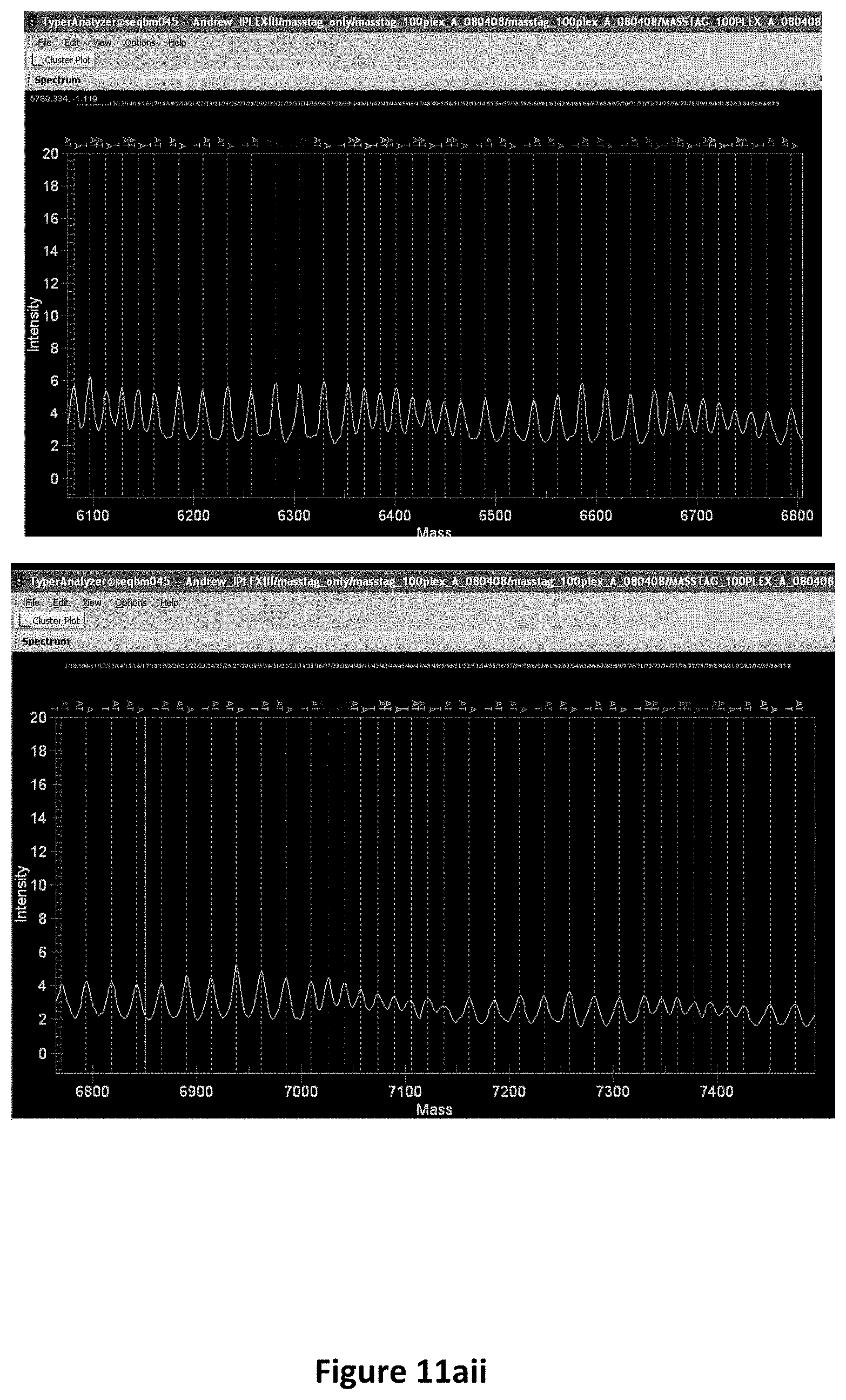
D00017
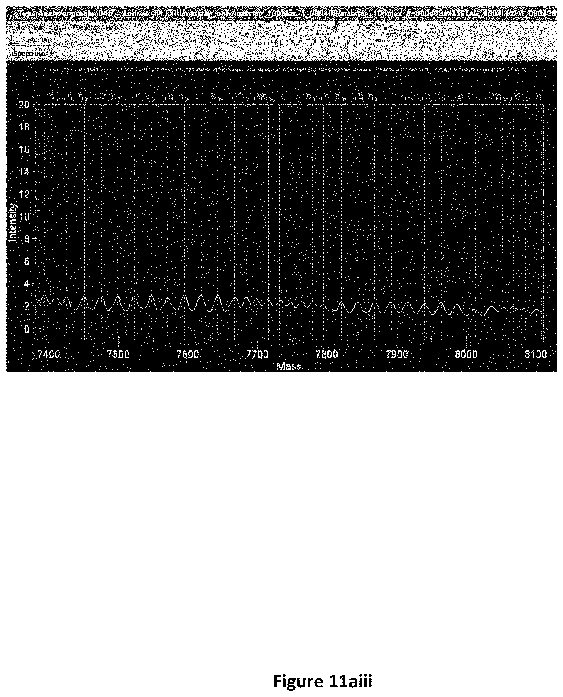
D00018

D00019

D00020

D00021
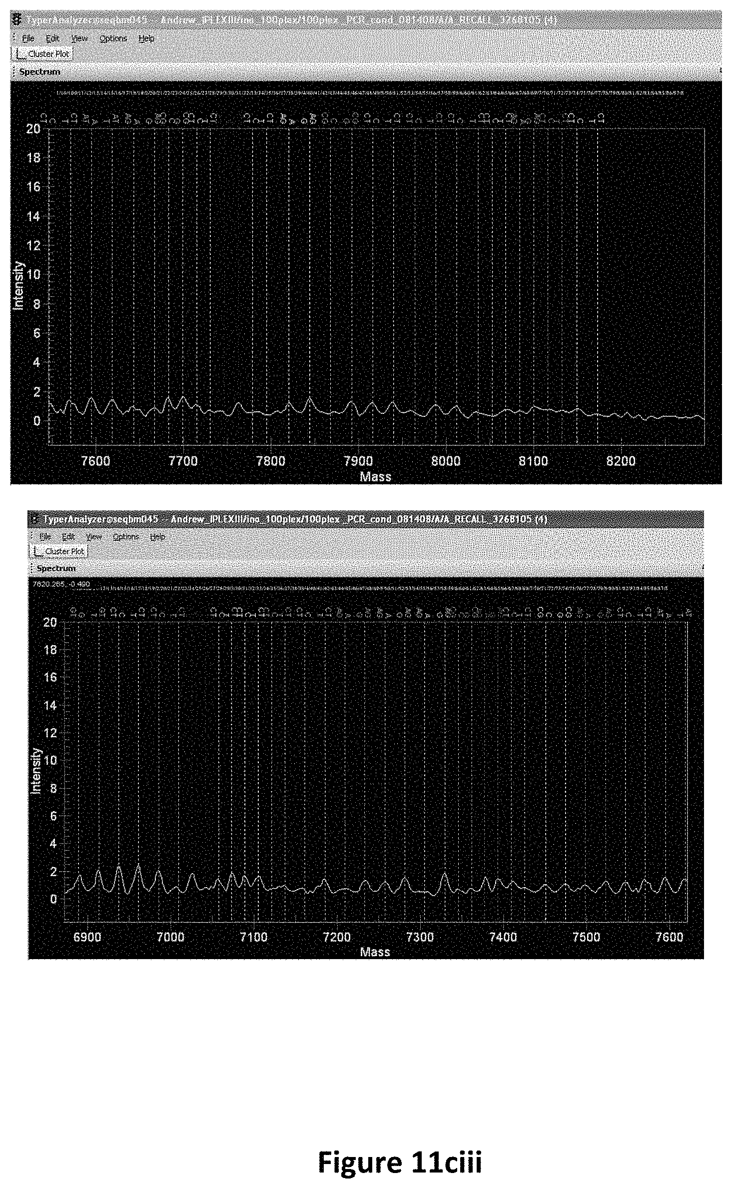
D00022

D00023

D00024

D00025

D00026

D00027

D00028

D00029

D00030

D00031

D00032

D00033

D00034

D00035

D00036

D00037

D00038

D00039

D00040

D00041

D00042

D00043

D00044

D00045

D00046

D00047
D00048

S00001
XML
uspto.report is an independent third-party trademark research tool that is not affiliated, endorsed, or sponsored by the United States Patent and Trademark Office (USPTO) or any other governmental organization. The information provided by uspto.report is based on publicly available data at the time of writing and is intended for informational purposes only.
While we strive to provide accurate and up-to-date information, we do not guarantee the accuracy, completeness, reliability, or suitability of the information displayed on this site. The use of this site is at your own risk. Any reliance you place on such information is therefore strictly at your own risk.
All official trademark data, including owner information, should be verified by visiting the official USPTO website at www.uspto.gov. This site is not intended to replace professional legal advice and should not be used as a substitute for consulting with a legal professional who is knowledgeable about trademark law.