Covalently linked antigen-antibody conjugates
Benz , et al. Dec
U.S. patent number 10,517,945 [Application Number 14/577,084] was granted by the patent office on 2019-12-31 for covalently linked antigen-antibody conjugates. This patent grant is currently assigned to HOFFMAN-LA ROCHE INC.. The grantee listed for this patent is Hoffmann-La Roche Inc.. Invention is credited to Joerg Benz, Ulrich Brinkmann, Stefan Dengl, Sebastian Dziadek, Guy Georges, Michael Grote, Alexander Haas, Eike Hoffmann.
View All Diagrams
| United States Patent | 10,517,945 |
| Benz , et al. | December 31, 2019 |
Covalently linked antigen-antibody conjugates
Abstract
Herein is reported a conjugate comprising an antigen and an antibody that specifically binds to the antigen wherein a covalent bond is formed between the antigen and an amino acid residue in the CDR2 of the antibody, an antibody comprising a cysteine residue at amino acid position 53 (according to Kabat) in the heavy chain CDR2 and uses thereof.
| Inventors: | Benz; Joerg (Rheinfelden, DE), Brinkmann; Ulrich (Weilheim, DE), Dengl; Stefan (Munich, DE), Dziadek; Sebastian (Benediktbeuern, DE), Georges; Guy (Habach, DE), Grote; Michael (Penzberg, DE), Haas; Alexander (Munich, DE), Hoffmann; Eike (Herrsching A. Ammersee, DE) | ||||||||||
|---|---|---|---|---|---|---|---|---|---|---|---|
| Applicant: |
|
||||||||||
| Assignee: | HOFFMAN-LA ROCHE INC. (Little
Falls, NJ) |
||||||||||
| Family ID: | 48794058 | ||||||||||
| Appl. No.: | 14/577,084 | ||||||||||
| Filed: | December 19, 2014 |
Prior Publication Data
| Document Identifier | Publication Date | |
|---|---|---|
| US 20150258209 A1 | Sep 17, 2015 | |
Related U.S. Patent Documents
| Application Number | Filing Date | Patent Number | Issue Date | ||
|---|---|---|---|---|---|
| PCT/EP2013/064100 | Jul 4, 2013 | ||||
Foreign Application Priority Data
| Jul 4, 2012 [EP] | 12174947 | |||
| Current U.S. Class: | 1/1 |
| Current CPC Class: | A61P 3/10 (20180101); C07K 16/40 (20130101); C07K 16/00 (20130101); A61P 29/00 (20180101); B82Y 5/00 (20130101); A61K 47/6835 (20170801); C07K 16/468 (20130101); A61P 3/00 (20180101); A61K 39/385 (20130101); C07K 16/44 (20130101); A61P 35/00 (20180101); A61P 31/12 (20180101); C07K 16/2896 (20130101); A61K 47/6879 (20170801); A61K 47/6897 (20170801); C07K 16/16 (20130101); G01N 33/6857 (20130101); C07K 2317/31 (20130101); A61K 2039/627 (20130101); A61K 2039/6056 (20130101); C07K 2317/40 (20130101); C07K 2317/94 (20130101); C07K 2317/24 (20130101); C07K 2317/624 (20130101); C07K 2317/55 (20130101); C07K 2317/565 (20130101) |
| Current International Class: | C07K 16/44 (20060101); C07K 16/16 (20060101); A61K 39/395 (20060101); A61K 47/68 (20170101); G01N 33/68 (20060101); C07K 16/46 (20060101); B82Y 5/00 (20110101); C07K 16/40 (20060101); C07K 16/28 (20060101); C07K 16/00 (20060101); A61K 39/385 (20060101); A61K 39/00 (20060101) |
References Cited [Referenced By]
U.S. Patent Documents
| 4275149 | June 1981 | Litman et al. |
| 4318980 | March 1982 | Boguslaski et al. |
| 4524025 | June 1985 | Geltosky |
| 4676980 | June 1987 | Segal et al. |
| 4737456 | April 1988 | Weng et al. |
| 4816567 | March 1989 | Cabilly et al. |
| 4855226 | August 1989 | Polito et al. |
| 4855522 | August 1989 | Diaz |
| 5198537 | March 1993 | Huber et al. |
| 5208020 | May 1993 | Chari et al. |
| 5316757 | May 1994 | Sherry et al. |
| 5342606 | August 1994 | Sherry et al. |
| 5385893 | January 1995 | Kiefer |
| 5416064 | May 1995 | Chari et al. |
| 5428139 | June 1995 | Kiefer et al. |
| 5428155 | June 1995 | Sherry et al. |
| 5462725 | October 1995 | Kiefer et al. |
| 5480990 | January 1996 | Kiefer et al. |
| 5500362 | March 1996 | Robinson et al. |
| 5571894 | November 1996 | Wels et al. |
| 5587458 | December 1996 | King et al. |
| 5591828 | January 1997 | Bosslet et al. |
| 5620686 | April 1997 | Mason |
| 5624821 | April 1997 | Winter et al. |
| 5635483 | June 1997 | Pettit et al. |
| 5648237 | July 1997 | Carter |
| 5648260 | July 1997 | Winter et al. |
| 5712374 | January 1998 | Kuntsmann et al. |
| 5714586 | February 1998 | Kuntsmann et al. |
| 5731168 | March 1998 | Carter et al. |
| 5739116 | April 1998 | Hamann et al. |
| 5739294 | April 1998 | Kiefer et al. |
| 5750373 | May 1998 | Garrard et al. |
| 5750660 | May 1998 | Kiefer et al. |
| 5767285 | June 1998 | Hamann et al. |
| 5770429 | June 1998 | Lonberg et al. |
| 5770701 | June 1998 | McGahren et al. |
| 5770710 | June 1998 | McGahren et al. |
| 5773001 | June 1998 | Hamann et al. |
| 5780588 | July 1998 | Pettit et al. |
| 5789199 | August 1998 | Joly et al. |
| 5804371 | September 1998 | Hoss et al. |
| 5821337 | October 1998 | Carter et al. |
| 5834456 | November 1998 | Kiefer et al. |
| 5840523 | November 1998 | Simmons et al. |
| 5869046 | February 1999 | Presta et al. |
| 5877296 | March 1999 | Hamann et al. |
| 5959083 | September 1999 | Bosslet et al. |
| 5959177 | September 1999 | Hein et al. |
| 6040498 | March 2000 | Stomp et al. |
| 6075181 | June 2000 | Kucherlapati et al. |
| 6150584 | November 2000 | Kucherlapati et al. |
| 6171586 | January 2001 | Lam et al. |
| 6194551 | February 2001 | Idusogie et al. |
| 6248516 | June 2001 | Winter et al. |
| 6267958 | July 2001 | Andya et al. |
| 6350860 | February 2002 | Buyse et al. |
| 6417429 | July 2002 | Hein et al. |
| 6420548 | July 2002 | Vezina et al. |
| 6511663 | January 2003 | King et al. |
| 6528624 | March 2003 | Idusogie et al. |
| 6602684 | August 2003 | Umana et al. |
| 6630579 | October 2003 | Chari et al. |
| 6737056 | May 2004 | Presta |
| 6881536 | April 2005 | Shah et al. |
| 6897044 | May 2005 | Braslawsky et al. |
| 6946292 | September 2005 | Kanda et al. |
| 6982321 | January 2006 | Winter |
| 7041870 | May 2006 | Tomizuka et al. |
| 7087409 | August 2006 | Barbas, III et al. |
| 7125978 | October 2006 | Vezina et al. |
| 7129330 | October 2006 | Little et al. |
| 7189826 | March 2007 | Rodman |
| 7332581 | February 2008 | Presta |
| 7371826 | March 2008 | Presta |
| 7498298 | March 2009 | Doronina et al. |
| 7504256 | March 2009 | Ogawa et al. |
| 7521541 | April 2009 | Eigenbrot et al. |
| 7527791 | May 2009 | Adams et al. |
| 7829674 | November 2010 | Sabbadini |
| 8313913 | November 2012 | Nakamura et al. |
| 8435784 | May 2013 | Berd et al. |
| 8907069 | December 2014 | Brinkmann et al. |
| 8945867 | February 2015 | Ogawa et al. |
| 9050375 | June 2015 | Bramlage et al. |
| 2001/0036923 | November 2001 | Chari et al. |
| 2002/0164328 | November 2002 | Shinkawa et al. |
| 2003/0115614 | June 2003 | Kanda et al. |
| 2003/0157108 | August 2003 | Presta |
| 2003/0166871 | September 2003 | Barbas, III et al. |
| 2004/0093621 | May 2004 | Shitara et al. |
| 2004/0109865 | June 2004 | Niwa et al. |
| 2004/0110282 | June 2004 | Kanda et al. |
| 2004/0110704 | June 2004 | Yamane et al. |
| 2004/0127688 | July 2004 | Winter |
| 2004/0132140 | July 2004 | Satoh et al. |
| 2005/0014934 | January 2005 | Hinton et al. |
| 2005/0026263 | February 2005 | Meares |
| 2005/0031613 | February 2005 | Nakamura et al. |
| 2005/0059100 | March 2005 | Meares |
| 2005/0079170 | April 2005 | Le Gall et al. |
| 2005/0079574 | April 2005 | Bond |
| 2005/0100543 | May 2005 | Hansen et al. |
| 2005/0119455 | June 2005 | Fuh et al. |
| 2005/0123546 | June 2005 | Umana et al. |
| 2005/0163782 | July 2005 | Glaser et al. |
| 2005/0233382 | October 2005 | Presta |
| 2005/0238649 | October 2005 | Doronina et al. |
| 2005/0260186 | November 2005 | Bookbinder et al. |
| 2005/0266000 | December 2005 | Bond et al. |
| 2005/0276802 | December 2005 | Adams et al. |
| 2005/0276805 | December 2005 | Hanai et al. |
| 2006/0025576 | February 2006 | Miller et al. |
| 2006/0104968 | May 2006 | Bookbinder et al. |
| 2006/0194291 | August 2006 | Presta |
| 2007/0061900 | March 2007 | Murphy et al. |
| 2007/0092940 | April 2007 | Eigenbrot et al. |
| 2007/0117126 | May 2007 | Sidhu et al. |
| 2007/0134759 | June 2007 | Nishiya et al. |
| 2007/0160598 | July 2007 | Dennis et al. |
| 2007/0166303 | July 2007 | Hanai et al. |
| 2007/0237764 | October 2007 | Birtalan et al. |
| 2007/0292936 | December 2007 | Barthelemy et al. |
| 2008/0069820 | March 2008 | Fuh et al. |
| 2008/0241884 | October 2008 | Shitara et al. |
| 2009/0002360 | January 2009 | Chen et al. |
| 2009/0093618 | April 2009 | Meares |
| 2010/0111856 | May 2010 | Gill et al. |
| 2010/0143934 | June 2010 | Herzog |
| 2015/0166670 | June 2015 | Castoldi et al. |
| 2015/0232577 | August 2015 | Brinkmann et al. |
| 2015/0238628 | August 2015 | Brinkmann et al. |
| 2017/0058050 | March 2017 | Brinkmann et al. |
| 2017/0058051 | March 2017 | Brinkmann et al. |
| 3836656 | May 1990 | DE | |||
| 012984 | Feb 2010 | EA | |||
| 0 077 896 | May 1983 | EP | |||
| 0 098 179 | Jan 1984 | EP | |||
| 0 404 097 | Dec 1990 | EP | |||
| 0 425 235 | May 1991 | EP | |||
| 1 870 459 | Dec 2007 | EP | |||
| S58-046072 | Mar 1983 | JP | |||
| 2010-501187 | Jan 2010 | JP | |||
| 2012-518892 | Aug 2012 | JP | |||
| 2219949 | Dec 2003 | RU | |||
| 2005104430 | Aug 2005 | RU | |||
| 2010108429 | Sep 2011 | RU | |||
| 2450020 | May 2012 | RU | |||
| WO-91/06305 | May 1991 | WO | |||
| WO-92/04053 | Mar 1992 | WO | |||
| WO-93/01161 | Jan 1993 | WO | |||
| WO-93/08829 | May 1993 | WO | |||
| WO-93/16185 | Aug 1993 | WO | |||
| WO-93/16185 | Aug 1993 | WO | |||
| WO-93/21232 | Oct 1993 | WO | |||
| WO-94/11026 | May 1994 | WO | |||
| WO-94/29351 | Dec 1994 | WO | |||
| WO-94/29351 | Dec 1994 | WO | |||
| WO-95/09917 | Apr 1995 | WO | |||
| WO-96/027011 | Sep 1996 | WO | |||
| WO-97/01580 | Jan 1997 | WO | |||
| WO-1997/25069 | Jul 1997 | WO | |||
| WO-97/30087 | Aug 1997 | WO | |||
| WO-98/50431 | Nov 1998 | WO | |||
| WO-98/50431 | Nov 1998 | WO | |||
| WO-98/58964 | Dec 1998 | WO | |||
| WO-99/22764 | May 1999 | WO | |||
| WO-99/51642 | Oct 1999 | WO | |||
| WO-2000/023053 | Apr 2000 | WO | |||
| WO-2000/023053 | Apr 2000 | WO | |||
| WO-2000/50088 | Aug 2000 | WO | |||
| WO-2000/50088 | Aug 2000 | WO | |||
| WO-00/61739 | Oct 2000 | WO | |||
| WO-01/29246 | Apr 2001 | WO | |||
| WO-2001/34651 | May 2001 | WO | |||
| WO-01/77342 | Oct 2001 | WO | |||
| WO-02/31140 | Apr 2002 | WO | |||
| WO-03/011878 | Feb 2003 | WO | |||
| WO-03/011878 | Feb 2003 | WO | |||
| WO-03/084570 | Oct 2003 | WO | |||
| WO-03/085107 | Oct 2003 | WO | |||
| WO-03/085119 | Oct 2003 | WO | |||
| WO-2004/009116 | Jan 2004 | WO | |||
| WO-2004/009116 | Jan 2004 | WO | |||
| WO-2004/045642 | Jun 2004 | WO | |||
| WO-2004/056312 | Jul 2004 | WO | |||
| WO-2004/056312 | Jul 2004 | WO | |||
| WO-2004/065569 | Aug 2004 | WO | |||
| WO-2004/065569 | Aug 2004 | WO | |||
| WO2005004809 | Jan 2005 | WO | |||
| WO-2005/035586 | Apr 2005 | WO | |||
| WO-2005/035778 | Apr 2005 | WO | |||
| WO-2005/053742 | Jun 2005 | WO | |||
| WO-2005/058940 | Jun 2005 | WO | |||
| WO-2005/058940 | Jun 2005 | WO | |||
| WO-2005/100402 | Oct 2005 | WO | |||
| WO-2006/020258 | Feb 2006 | WO | |||
| WO-2006/020258 | Feb 2006 | WO | |||
| WO-2006/029879 | Mar 2006 | WO | |||
| WO-2006/029879 | Mar 2006 | WO | |||
| WO-2006/044908 | Apr 2006 | WO | |||
| WO-2006/044908 | Apr 2006 | WO | |||
| WO-2007/024715 | Mar 2007 | WO | |||
| WO-2007/024715 | Mar 2007 | WO | |||
| WO-2007/065808 | Jun 2007 | WO | |||
| WO-2007/065808 | Jun 2007 | WO | |||
| WO-2007/109254 | Sep 2007 | WO | |||
| WO-2007/109254 | Sep 2007 | WO | |||
| WO-2007/130697 | Nov 2007 | WO | |||
| WO-2007/130697 | Nov 2007 | WO | |||
| WO-2008/022349 | Feb 2008 | WO | |||
| WO-2008/022349 | Feb 2008 | WO | |||
| WO-2008/077546 | Jul 2008 | WO | |||
| WO-2009/022328 | Feb 2009 | WO | |||
| WO-2009/080251 | Jul 2009 | WO | |||
| WO-2009/080252 | Jul 2009 | WO | |||
| WO-2009/080253 | Jul 2009 | WO | |||
| WO-2009/080254 | Jul 2009 | WO | |||
| WO-2009/089004 | Jul 2009 | WO | |||
| WO-2010/034651 | Apr 2010 | WO | |||
| WO-2010/045388 | Apr 2010 | WO | |||
| WO-2010/045388 | Apr 2010 | WO | |||
| WO-2010/056893 | May 2010 | WO | |||
| WO-2010/098992 | Sep 2010 | WO | |||
| WO-2010/112193 | Oct 2010 | WO | |||
| WO-2010/115589 | Oct 2010 | WO | |||
| WO-2010/119704 | Oct 2010 | WO | |||
| WO-2010/136172 | Dec 2010 | WO | |||
| WO-2010/145792 | Dec 2010 | WO | |||
| WO-2010/145793 | Dec 2010 | WO | |||
| WO-2011/003557 | Jan 2011 | WO | |||
| WO-2011/003780 | Jan 2011 | WO | |||
| WO-2011/032022 | Mar 2011 | WO | |||
| WO-2011/156328 | Dec 2011 | WO | |||
| WO-2012/075037 | Jun 2012 | WO | |||
| WO-2012/093068 | Jul 2012 | WO | |||
| WO-2012/143379 | Oct 2012 | WO | |||
| WO-2013/106577 | Jul 2013 | WO | |||
| WO-2013/106577 | Jul 2013 | WO | |||
| WO-2014/006124 | Jan 2014 | WO | |||
| WO-2015/101587 | Jul 2015 | WO | |||
| WO-2015/101589 | Jul 2015 | WO | |||
Other References
|
Nguyen et al., The EMBO J 19(5): 921-930, 2000. cited by examiner . Rudikoff et al., Proc. Natl. Acad. Sci. USA, 79(6): 1979-1983, Mar. 1982. cited by examiner . Yu et al., Investigative Ophthalmology & Visual Science 49(2): 522-527, Feb. 2008. cited by examiner . Stancovski et al., PNAS, 88: 8691-8695, 1991. cited by examiner . Kussie et al., J. Immunol. 152: 146-152, 1994. cited by examiner . Chen et al., EMBO J., 14: 2784-2794, 1995. cited by examiner . Chen et al., J. Exp. Med. 176 (3): 855-66, Sep. 1, 1992. cited by examiner . Brown et al., J. Immunol. 156 (9): 3285-91, May 1996. cited by examiner . Lloyd et al., Protein Engineering, Design & Selection 22:159-168 (Year: 2009). cited by examiner . Edwards et al. J Mol Biol. 334(1): 103-118 (Year: 2003). cited by examiner . Dengl et al., Immunol Rev 270(1): 165-177 (Year: 2016). cited by examiner . Glockhuber et al., Biochemistry 30(12): 3049-54 (Year: 1991), abstract only. cited by examiner . Jorgensen et al., Dev Comp Immunol 26(2): 201-206 (Year: 2002). cited by examiner . Albert et al. "Direct Synthesis of [DOTA-DPhe1]-Octreotide and [DOTA-DPhe1, Tyr3]-Octreotide (SMT487): Two Conjugates for Systemic Delivery of Radiotherapeutical Nuclides to Somatostatin Receptor Positive Tumors in Man," Bioorganic & Medicinal Chemistry Letters 8:1207-1210, (1998). cited by applicant . AvantGen, Inc. "AvantGen's Antibody Humanization and Discovery Technologies--GermlinerTM Antibodies: An Effective and Proprietary Technology for Humanizing Antibodies Based on Epitope-Guided Selection," (Jul. 27, 2009). cited by applicant . Bagci et al. "Monoclonal Anti-biotin Antibodies Simulate Avidin in the Recognition of Biotin," FEBS 322(1):47-50, (May 1993). cited by applicant . Bera et al. "Comparison of Recembinant Immunotoxins Against Le.sup.Y Antigen Expressing Tumor Cells: Influence of Affinity, Size, and Stability," Bioconjugate Chemistry 9(6):736-743, (Nov.-Dec. 1998). cited by applicant . Berger et al. "Production of Antibodies that Bind Biotin and Inhibit Biotin Containing Enzymes," Biochemistry 14(11):2338-2342, (1975). cited by applicant . Blend et al. "Labeling anti-HER2/neu Monoclonal Antibodies With .sup.111In and .sup.90Y Using a Bifunctional DTPA Chelating Agent," Cancer Biotherapy & Radiopharmaceuticals 18(3):355-363, (2003). cited by applicant . Briggs et al. "Synthesis of Functionalised Fluorescent Dyes and Their Coupling to Amines and Amino Acids," J. Chem. Soc., Perkin-Trans. 1:1051-1058, (1997). cited by applicant . Cahn et al. "Specification of Molecular Chirality," Angew. Chem. Int. Ed. Engl. 5(4):385-415, (1966). cited by applicant . Camera et al. "Comparative Biodistribution of Indium- and Yttrium-Labeled B3 Monoclonal Antibody Conjugated to Either 2-(p-SCN-Bz)-6-Methyl-DTPA (1B4M-DTPA) or 2-(p-SCN-Bz)-1,4,7,10-Tetraazacyclododecane Tetraacetic Acid (2B-DOTA)," European Journal of Nuclear Medicine 21(7):640-646, (Jul. 1994). cited by applicant . Camera et al. "Evaluation of a New DTPA-Derivative Chelator: Comparative Biodistribution and Imaging Studies of .sup.111In-Labeled B3 Monoclonal Antibody in Athymic Mice Bearing Human Epidermoid Carcinoma Xenografts," Nucl. Med. Biol. 20(8):955-962, (1993). cited by applicant . Cao et al. "Development of a Bispecific Monoclonal Antibody as a Universal Immunoprobe for Detecting Biotinylated Macromolecules," Journal of Immunological Methods 220:85-91, (1998). cited by applicant . Chen et al. "MicroPET and Autoradiographic Imaging of Breast Cancer .alpha..sub.v-Integrin Expression Using .sup.18F- and .sup.64Cu-Labeled RGD Peptide," Bioconjugate Chem. 15(1):41-49, (2004, e-published on Dec. 30, 2003). cited by applicant . Chowdhury. "Targeting Random Mutations to Hotspots in Antibody Variable Domains for Affinity Improvement," Chapter 24 in Methods in Molecular Biology, O'Brien, P.M. (ed.) et al., Himana Press Inc., Totowa, NJ, 178:269-285, (2001). cited by applicant . Dakshinamurti et al. "Production and Characterization of a Monoclonal Antibody to Biotin," Biochem. J. 237:477-482, (1986). cited by applicant . De Leon-Rodriguez et al. "Solid-Phase Synthesis of DOTA-Peptides," Chem. Eur. J. 10:1149-1155, (2004). cited by applicant . Debinski et al. "An Immunotoxin with Increased Activity and Homogeneity Produced by Reducing the Number of Lysine Residues in Recombinant Pseudomonas Exotoxin," Bioconjugate Chem. 5(1):40-46, (Jan. 1994). cited by applicant . Debinski et al. "Monovalent Immunotoxin Containing Truncated Form of Pseudomonas Exotoxin as Potent Antitumor Agent," Cancer Research 52(19):5379-5385, (Oct. 1, 1992). cited by applicant . Denardo et al. "Comparison of 1,4,7,10-Tetraazacyclododecane-N,N',N'',N'''-Tetraacetic Acid (DOTA)-Peptide-ChL6, a Novel Immunoconjugate with Catabolizable Linker, to 2-Iminothiolane-2[p-(Bromoacetamido)benzyl]-DOTA-ChL6 in Breast Cancer Xenografts," Clinical Cancer Research 4(10):2483-2490, (Oct. 1, 1998). cited by applicant . Hanes et al. "Ribosome Display Efficiently Selects and Evolves High-Affinity Antibodies in Vitro From Immune Libraries," Proceedings of the National Academy of Sciences 95(24):14130-14135, (Nov. 24, 1998). cited by applicant . Hansen et al. "A Recombinant Immunotoxin Targeting CD22 With Low Immunogenicity, Low Nonspecific Toxicity, and High Antitumor Activity in Mice," Journal of Immunotherapy 33(3):297-304, (Apr. 2010). cited by applicant . Hermanson. "Buckyballs, Fullerenes, and Carbon Nanotubes," in Chapter 1 of Bioconjugate Techniques, Academic Press, San Diego, pp. 3-168, (1996). cited by applicant . Hermanson. "Buckyballs, Fullerenes, and Carbon Nanotubes," in Chapter 15 of Bioconjugate Techniques, Academic Press, San Diego, pp. 627-648, (1996). cited by applicant . Hermanson. "Chemoselective Ligation: Bioorthogonal Reagents" in Chapter 17 of Bioconjugate Techniques, Academic Press, San Diego, pp. 666-706, (1996). cited by applicant . Hermanson. "Mass Tags and Isotope Tags," in Chapter 16 of Bioconjugate Techniques, Academic Press, San Diego, pp. 649-665, (1996). cited by applicant . Hnatowich et al. "The Preparation of DTPA-Coupled Antibodies Radiolabeled With Metallic Radionuclides: An Improved Method," Journal of Immunological Methods 65:147-157, (1983). cited by applicant . Hoffmann et al. "PK Modulation of Haptenylated Peptides Via Non-Covalent Antibody Complexation," Journal of Controlled Release 171(1):48-56, (Oct. 10, 2013, e-published on Jun. 22, 2013). cited by applicant . Hwang et al. "Use of Human Germline Genes in a CDR Homology-based Approach to Antibody Humanization," Methods 36(1):35-42, (May 2005). cited by applicant . Izard et al. "An Improved Method for Labeling Monoclonal Antibodies With Samarium-153: Use of the Bifunctional Chelate 2-(p-Isothiocyanatobenzyl)-6-Methyldiethylenetriaminepentaacetic Acid," Bioconjugate Chem. 3(4):346-350, (1992). cited by applicant . Klussman et al. "Secondary mAb--vcMMAE Conjugates are Highly Sensitive Reporters of Antibody Internalization Via the Lysosome Pathway," Bioconjugate Chemistry 15(4):765-773, (2004, e-published on Jun. 18, 2004). cited by applicant . Kobayashi et al. "Evaluation of the In Vivo Biodistribution of Indium-111 and Yttrium-88 Labeled Dendrimer-1B4M-DTPA and its Conjugation With Anti-Tac Monoclonal Antibody," Bioconjugate Chem. 10:103-111, (1999, e-published on Dec. 10, 1998). cited by applicant . Kohen et al. "Preparation and Properties of Anti-biotin Antibodies," Methods Enzymology 279:451-463, (1997). cited by applicant . Li et al. "Vinyl Sulfone Bifunctional Derivatives of DOTA Allow Sulfhydryl- or Amino-Directed Coupling to Antibodies. Conjugates Retain Immunoreactivity and Have Similar Biodistributions," Bioconjugate Chem. 13:110-115, (2002, e-published on Dec. 14, 2001). cited by applicant . Manheimer-Lory et al. "Lupus-specific Antibodies Reveal an Altered Pattern of Somatic Mutation," J. Clin. Invest. 100(10):2538-2546, (Nov. 1997). cited by applicant . Mardirossian et al. "The Stability in Liver Homogenates of Indium-111 and Yttrium-90 Attached to Antibody Via Two Popular Chelators," Nucl. Med. Biol. 20(1):65-74, (1993). cited by applicant . Meares et al. "Conjugation of Antibodies With Bifunctional Chelating Agents: Isothiocyanate and Bromoacetamide Reagents, Methods of Analysis, and Subsequent Addition of Metal Ions," Analytical Biochemistry 142:68-78, (1984). cited by applicant . Mirzadeh et al. "Radiometal Labeling of Immunoproteins: Covalent Linkage of 2-(4-Isothiocyanatobenzyl)Diethylenetriaminepentaacetic Acid Ligands to Immunoglobulin," Bioconjugate Chem. 1(1):59-65, (1990). cited by applicant . Nikula et al. "A Rapid, Single Vessel Method for Preparation of Clinical Grade Ligand Conjugated Monoclonal Antibodies," Nucl. Med. Biol. 22(3):387-390, (1995). cited by applicant . Nygaard et al. "The PP-Fold Solution Structure of Human Polypeptide YY and Human PYY3-36 as Determined by NMR," Biochemistry 45(27):8350-8357, (Jun. 16, 2006). cited by applicant . Pai et al. "Anti-tumor Activities of Immunotoxins Made of Monoclonal Antibody B3 and Various Forms of Pseudomonas Exotoxin," Proceedings of the National Academy of Sciences, USA 88(8):3358-3362, (Apr. 15, 1991). cited by applicant . Pastan et al. "Immunotoxins with Decreased Immunogenicity and Improved Activity," Leukemia & Lymphoma 52(Supp. 2):87-90, (Jun. 2011; e-published on Apr. 19, 2011). cited by applicant . Polya. Biochemical Targets of Plant Bioactive Compounds, Taylor & Francis Inc., 29 West 35th Street, New York, NY 10001, pp. 847, (2003), three pages. cited by applicant . Roselli et al. "In Vivo Comparison of CHX-DTPA Ligand Isomers in Athymic Mice Bearing Carcinoma Xenografts," Cancer Biotherapy & Radiopharmaceuticals 14(3):209-220, (1999). cited by applicant . Vincent et al. "A Comparison of the Binding of Biotin and Biotinylated Macromolecular Ligands to an Anti-Biotin Monoclonal Antibody and to Streptavidin," Journal of Immunological Methods 165:177-182, (1993). cited by applicant . Wark et al. "Latest Technologies for the Enhancement of Antibody Affinity," Advanced Drug Delivery Reviews 58:657-670, (2006; e-published on May 22, 2006). cited by applicant . Weldon et al. "A Guide to Taming a Toxin--Recombinant Immunotoxins Constructed From Pseudomonas Exotoxin A for the Treatment of Cancer," FEBS Journal 278(23):4683-4700, (Dec. 2011; e-published on Jun. 2, 2011). cited by applicant . Wiedemann et al. "Molecular and Structural Analysis of a Continuous Birch Profilin Epitope Defined by a Monoclonal Antibody," The Journal of Biological Chemistry 271(47):29915-29921, (Nov. 22, 1996). cited by applicant . Wu. "Simultaneous Humanization and Affinity Optimization of Monoclonal Antibodies," Chapter 12 in Methods in Molecular Biology, Welschol, M. (ed.) et al., Humana Press Inc., Totowa, NJ, 207:197-212, (2003). cited by applicant . Zahnd et al. "Directed in Vitro Evolution and Crystallographic Analysis of a Peptide-Binding Single Chain Antibody Fragment (Scfv) with Low Picomolar Affinity," Journal of Biological Chemistry 279(18):18870-18877, (Apr. 30, 2004; e-published on Jan. 30, 2004). cited by applicant . International Search Report dated Apr. 22, 2015 for International Application No. PCT/EP2014/079354, filed on Dec. 29, 2014, five pages. cited by applicant . International Search Report dated Aug. 20, 2013, for PCT Patent Application No. PCT/EP2013/064090, filed on Jul. 4, 2013, five pages. cited by applicant . International Search Report dated Mar. 11, 2015 for International Application No. PCT/EP2014/079352 filed on Dec. 29, 2014, five pages. cited by applicant . Written Opinion of the International Searching Authority dated Apr. 22, 2015 for International Application No. PCT/EP2014/079354, filed on Dec. 29, 2014, four pages. cited by applicant . Written Opinion of the International Searching Authority dated Aug. 20, 2013, for PCT Patent Application No. PCT/EP2013/064090, filed on Jul. 4, 2013, three pages. cited by applicant . Written Opinion of the International Searching Authority dated Mar. 11, 2015 for International Application No. PCT/EP2014/079352 filed on Dec. 29, 2014, six pages. cited by applicant . Brinkley. "A Brief Survey of Methods for Preparing Protein Conjugates with Dyes, Haptens and Crosslinking Reagents," Bioconjugate Chem. 3(1):2-13, (Jan. 1992). cited by applicant . Almagro et al. (2008). "Humanization of antibodies," Front. Biosci. 13:1619-1633. cited by applicant . Atwell et al. (1997). "Stable heterodimers from remodeling the domain interface of a homodimer using a phage display library" J. Mol. Biol. 270:26-35. cited by applicant . Baca et al. (1997). "Antibody humanization using monovalent phage display," J. Biol. Chem. 272:10678-10684. cited by applicant . Boerner et al. (1991). "Production of antigen-specific human monoclonal antibodies from in vitro-primed human splenocytes," J. Immunol. 147:86-95. cited by applicant . Brennan et al. (1985). "Preparation of bispecific antibodies by chemical recombination of monoclonal immunoglobulin G1 fragments," Science 229:81-83. cited by applicant . Brodeur, B.R. et al., Monoclonal Antibody Production Techniques and Applications, Marcel Dekker, Inc., New York (1987), pp. 51-63. cited by applicant . Bruggemann et al. (1987). "Comparison of the effector functions of human immunoglobulins using a matched set of chimeric antibodies," J. Exp. Med. 166:1351-1361. cited by applicant . Carter et al. (1992). "Humanization of an anti-p185HER2 antibody for human cancer therapy," PNAS 89:4285-4289. cited by applicant . Chen et al. (1999). "Selection and analysis of an optimized anti-VEGF antibody: crystal structure of an affinity-matured Fab in complex with antigen," J. Mol. Biol. 293:865-881. cited by applicant . Chari, R.V., et al. (1992). "Immunoconjugates containing novel maytansinoids: promising anticancer drugs," Cancer Res. 52:127-131. cited by applicant . Charlton, K.A., In: Methods in Molecular Biology, vol. 248, Lo, B.K.C. (ed.), Humana Press, Totowa, NJ (2003), pp. 245-254. cited by applicant . Chothia et al. (1987). "Canonical structures for the hypervariable regions of immunoglobulins," J. Mol. Biol. 196:901-917. cited by applicant . Chowdhury, P.S. (2003). "Engineering hot spots for affinity enhancement of antibodies," Methods Mol. Biol. 207:179-196. cited by applicant . Clackson et al. (1991). "Making antibody fragments using phage display libraries," Nature 352:624-628. cited by applicant . Clynes, R., et al. (1998). "Fc receptors are required in passive and active immunity to melanoma" Proc. Natl. Acad. Sci. USA 95:652-656. cited by applicant . Coloma, M.J., et al. (1997). "Design and production of novel tetravalent bispecific antibodies" Nature Biotech 15:159-163. cited by applicant . Collaborative Computational Project, No. 4 (1994). "The CCP4 suite: programs for protein crystallography," Acta Crystallogr. Section D. 50(Pt. 5):760-763. cited by applicant . Cragg, M.S., et al. (2003). "Complement-mediated lysis by anti-CD20 mAb correlates with segregation into lipid rafts," Blood 101:1045-1052. cited by applicant . Cragg et al. (2004). "Antibody specificity controls in vivo effector mechanisms of anti-CD20 reagents," Blood 103:2738-2743. cited by applicant . Cunningham et al. (1989). "High-resolution epitope mapping of hGH-receptor interactions by alanine-scanning mutagenesis," Science 244:1081-1085. cited by applicant . Dall'Acqua et al. (2005). "Antibody humanization by framework shuffling," Methods 36:43-60. cited by applicant . Decarie A., et al. "Development of digoxigenin-labeled peptide: application to chemiluminoenzyme immunoassay of bradykinin in inflamed tissues," Peptides 15 (1994) 511-518. cited by applicant . Dubowchik et al. (2002). "Doxorubicin immunoconjugates containing bivalent, lysosomally-cleavable dipeptide linkages," Bioorg. & Med. Chem. Letters 12:1529-1532. cited by applicant . Duncan et al. (1988). "The binding site for C1q on IgG," Nature 332:738-740. cited by applicant . Emsley, P., et al. (2010). "Features and development of Coot," Acta Crystallogr. D Biol. Crystallogr. 66:486-501. cited by applicant . Fellouse et al. (2004). "Synthetic antibodies from a four-amino-acid code: a dominant role for tyrosine in antigen recognition" PNAS 101:12467-12472. cited by applicant . Fischer et al. (2007). "Bispecific antibodies: molecules that enable novel therapeutic strategies," Pathobiology 74:3-14. cited by applicant . Flatman et al. (2007). "Process analytics for purification of monoclonal antibodies," J. Chrom. B. 848:79-87. cited by applicant . Fraker et al (1978). "Protein and cell membrane iodinations with a sparingly soluble chloroamide, 1, 3, 4, 6-tetrachloro-3a, 6a-diphennylglycoluril," Biochem. Biophys. Res. Commun. 80:849-857. cited by applicant . Gazzano-Santoro et al. (1997). "A non-radioactive complement-dependent cytotoxicity assay for anti-CD20 monoclonal antibody," J. Immunol. Methods 202:163-171. cited by applicant . Gerngross, T.U. (2004). "Advances in the production of human therapeutic proteins in yeasts and filamentous fungi," Nat. Biotech. 22:1409-1414. cited by applicant . Graham, F.L., et al. (1977). "Characteristics of a human cell line transformed by DNA from human adenovirus type 5," J. Gen Virol. 36:59-74. cited by applicant . Griffiths, A.D. et al. (1993). "Human anti-self antibodies with high specificity from phage display libraries," EMBO J. 12:725-734. cited by applicant . Gruber et al. (1994). "Efficient tumor cell lysis mediated by a bispecific single chain antibody expressed in Escherichia coli," J. Immunol. 152:5368-5374. cited by applicant . Guyer, R.L., et al. (1976). "Immunoglobulin binding by mouse intestinal epithelial cell receptors," J. Immunol. 117 (1976) 587-593. cited by applicant . Hellstrom et al. (1986). "Antitumor effects of L6, an IgG2a antibody that reacts with most human carcinomas," PNAS 83:7059-7063. cited by applicant . Hellstrom et al. (1985). "Strong antitumor activities of IgG3 antibodies to a human melanoma-associated ganglioside," PNAS 82:1499-1502. cited by applicant . Hinman, L.M., et al. "Preparation and characterization of monoclonal antibody conjugates of the calicheamicins: a novel and potent family of antitumor antibiotics," Cancer Res. 53 (1993) 3336-3342. cited by applicant . Holliger, P., et al. "Engineered antibody fragments and the rise of single domains," Nature Biotech 23 (2005) 1126-1136. cited by applicant . Holliger, P., et al. (1993). "Diabodies: small bivalent and bispecific antibody fragments," Proc. Natl. Acad. Sci. USA 90:6444-6448. cited by applicant . Hoogenboom, H.R., et al. (2002). "Overview of antibody phage-display technology and its applications," Methods in Molecular Biology 178:1-37. cited by applicant . Hoogenboom et al. (1992). "By-passing immunisation. Human antibodies from synthetic repertoires of germline VH gene segments rearranged in vitro," J. Mol. Biol. 227:381-388. cited by applicant . Hudson et al. (2003). "Engineered antibodies," Nat. Med. 9:129-134. cited by applicant . Idusogie et al. (2000). "Mapping of the C1q binding site on rituxan, a chimeric antibody with a human IgG1 Fc," J. Immunol. 164:4178-4184. cited by applicant . International Search Report dated Nov. 19, 2013, for PCT Patent Application No. PCT/EP2013/064100, filed on Jul. 4, 2013, four pages. cited by applicant . Jeffrey, S.C., et al. (2006). "Dipeptide-based highly potent doxorubicin antibody conjugates," Bioorg. Med. Chem. Lett. 16:358-362. cited by applicant . Kabsch, W. (1993). "Automatic processing of rotation diffraction data from crystals of initially unknown symmetry and cell constants," J. Appl. Cryst. 26:795-800. cited by applicant . Kam, N.W., et al. (2005). "Carbon nanotubes as multifunctional biological transporters and near-infrared agents for selective cancer cell destruction," PNAS 102:11600-11605. cited by applicant . Kanda, Y., et al. (2006). "Comparison of cell lines for stable production of fucose-negative antibodies with enhanced ADCC," Biotechnol. Bioeng. 94:680-688. cited by applicant . Kashmiri et al. (2005). "SDR grafting--a new approach to antibody humanization" Methods 36:25-34. cited by applicant . Kim, J.K., et al. (1994). "Localization of the site of the murine IgG1 molecule that is involved in binding to the murine intestinal Fc receptor," J. Immunol. 24:2429-2434. cited by applicant . Kindt, T.J., et al., Kuby Immunology, 6th ed., W.H. Freeman and Co., N.Y. (2007), p. 91. cited by applicant . King, H.D., et al. (2002). "Monoclonal antibody conjugates of doxorubicin prepared with branched peptide linkers: inhibition of aggregation by methoxytriethyleneglycol chains," J. Med. Chem. 45:4336-4343. cited by applicant . Klimka (2000). "Human anti-CD30 recombinant antibodies by guided phage antibody selection using cell panning," Br. J. Cancer 83:252-260. cited by applicant . Kobayashi et al. (1998). "Evaluation of the in vivo biodistribution of yttrium-labeled isomers of CHX-DTPA-conjugated monoclonal antibodies," J. Nucl. Med. 39:829-836. cited by applicant . Kostelny et al. (1992). "Formation of a bispecific antibody by the use of leucine zippers," J. Immunol. 148:1547-1553. cited by applicant . Kozbor (1984). "A human hybrid myeloma for production of human monoclonal antibodies," J. Immunol. 133 (1984) 3001-3005. cited by applicant . Kratz, F., et al. "Prodrugs of anthracyclines in cancer chemotherapy," Current Med. Chem. 13 (2006) 477-523. cited by applicant . Kurkis et al, "Optimized conditions for chelation of yttrium-90-DOTA immunoconjugates," J. Nucl. Med. 39 (1998) 2105-2110. cited by applicant . Laskowski (1993). "PROCHECK: a program to check the stereochemical quality of protein structures," J. Appl. Crystallogr. 26:283-291. cited by applicant . Lee, C.V. et al., "Bivalent antibody phage display mimics natural immunoglobulin," J. Immunol. Methods 284 (2004) 119-132. cited by applicant . Lee, C.V. et al. (2004). "High affinity human antibodies from phage displayed synthetic fab libraries with a single framework scaffold," J. Mol. Biol. 340 (2004) 1073-1093. cited by applicant . Lee et al. (2001). "Specific localization, gamma camera imaging, and intracellular trafficking of radiolabelled chimeric anti-G(D3) ganglioside monoclonal antibody KM871 in SK-MEL-28 melanoma xenografts," Cancer Res. 61:4474-4482. cited by applicant . Lewis et al. (2001). "An improved method for conjugating monoclonal antibodies with N-hydroxysulfosuccinimidyl DOTA" Bioconjugate Chem. 12:320-324. cited by applicant . Li, J. et al. (2006). "Human antibodies for immunotherapy development generated via a human B cell hybridoma technology," PNAS 103:3557-3562. cited by applicant . Li, H., et al. (2006). "Optimization of humanized IgGs in glycoengineered Pichia pastoris," Nat. Biotech. 24:210-215. cited by applicant . Lode, H.N., et al. (1998). "Targeted therapy with a novel enediyene antibiotic calicheamicin theta(I)1 effectively suppresses growth and dissemination of liver metastases in a syngeneic model of murine neuroblastoma," Cancer Res. 58:2925-2928. cited by applicant . Lonberg (2008). "Fully human antibodies from transgenic mouse and phage display platforms," Curr. Opin. Immunol. 20:450-459. cited by applicant . Lonberg (2005). "Human antibodies from transgenic animals," Nat. Biotech. 23:1117-1125. cited by applicant . MacCallum et al. (1996). "Antibody-antigen interactions: contact analysis and binding site topography," J. Mol. Biol. 262:732-745. cited by applicant . Marks et al. (1991). "By-passing immunization. Human antibodies from V-gene libraries displayed on phage," J. Mol. Biol. 222:581-597. cited by applicant . Marks et al. (2004). "Selection of human antibodies from phage display libraries," Methods in Molecular Biology 248:161-175. cited by applicant . Mather, J.P. (1980). "Establishment and characterization of two distinct mouse testicular epithelial cell lines," Biol. Reprod. 23:243-252. cited by applicant . Mather, J.P., et al. (1982). "Culture of testicular cells in hormone-supplemented serum-free medium," Annals N.Y. Acad. Sci. 383:44-68. cited by applicant . McCafferty, J. et al. (1990). "Phage antibodies: filamentous phage displaying antibody variable domains," Nature 348:552-554. cited by applicant . Meares et al. (1990). "Macrocyclic chelates of radiometals for diagnosis and therapy," Br. J. Cancer Suppl. 10:21-26. cited by applicant . Merchant et al. (1998). "An efficient route to human bispecific IgG," Nat Biotechnol 16:677-681. cited by applicant . Metz et al. (2011). "Bispecific digoxigenin-binding antibodies for targeted payload delivery," PNAS 108:8194-8199. cited by applicant . Miederer et al. (2004). Pharmacokinetics, dosimetry, and toxicity of the targetable atomic generator, 225Ac-HuM195, in nonhuman primates J. Nucl. Med. 45:129-137. cited by applicant . Mier et al. (2005). "conjugation of DOTA using isolated phenolic active esters: the labeling and biodistribution of albumin as blood pool marker," Bioconjugate Chem. 16:237-240. cited by applicant . Milstein (1983). "Hybrid hybridomas and their use in immunohistochemistry," Nature 305:537-540. cited by applicant . Mitchell et al. (2003). "Targeting primary human Ph(+) B-cell precursor leukemia-engrafted SCID mice using radiolabeled anti-CD19 monoclonal antibodies," J. Nucl. Med. 44:1105-1112. cited by applicant . Morrison et al. (1984). "Chimeric human antibody molecules: mouse antigen-binding domains with human constant region domains," PNAS 81:6851-6855. cited by applicant . Morrison (2007). "Two heads are better than one," Nature Biotech 25:1233-1234. cited by applicant . Murshudov, G.N., et al. (1997). "Refinement of macromolecular structures by the maximum-likelihood method" Acta Crystallogr. D Biol. Crystallogr. 53:240-255. cited by applicant . Nagy, A., et al., "Stability of cytotoxic luteinizing hormone-releasing hormone conjugate (AN-152) containing doxorubicin 14-O-hemiglutarate in mouse and human serum in vitro: implications for the design of preclinical studies," Proc. Natl. Acad. Sci. USA 97 (2000) 829-834. cited by applicant . Neuberger, M.S. (1983). "Expression and regulation of immunoglobulin heavy chain gene transfected into lymphoid cells," EMBO J. 2:1373-1378. cited by applicant . Ni (2006). "Research progress and future perspectives in antibodomics and antibodomic drugs," Xiandai Mianyixue 26:265-268. cited by applicant . Nikula et al. (1999). "Alpha-emitting bismuth cyclohexylbenzyl DTPA constructs of recombinant humanized anti-CD33 antibodies: pharmacokinetics, bioactivity, toxicity and chemistry," J. Nucl. Med. 40:166-176. cited by applicant . Okazaki, A., et al. (2004). "Fucose depletion from human IgG1 oligosaccharide enhances binding enthalpy and association rate between IgG1 and FcgammaRIIIa," J. Mol. Biol. 336:1239-1249. cited by applicant . Osbourn et al. (2005). "From rodent reagents to human therapeutics using antibody guided selection," Methods 36:61-68. cited by applicant . O'Sullivan et al "Methods for the Preparation of Enzyme-Antibody Conjugates for use in Enzyme Immunoassay", in Methods in Enzym. (ed. by J. Langone & IT Van Vunakis), Academic Press, New York, 73 (1981) 147-166. cited by applicant . Padlan et al. (1991). "A possible procedure for reducing the immunogenicity of antibody variable domains while preserving their ligand-binding properties," Mol. Immunol. 28:489-498. cited by applicant . Pace, et al. (1995). "How to measure and predict the molar absorption coefficient of a protein," Protein Science 4:2411-2423. cited by applicant . Petkova, S.B., et al. (2006). "Enhanced half-life of genetically engineered human IgG1 antibodies in a humanized FcRn mouse model: potential application in humorally mediated autoimmune disease," Int. Immunol. 18:1759-1769. cited by applicant . Picard (1984). "A lymphocyte-specific enhancer in the mouse immunoglobulin .kappa. gene" Nature 307:80-82. cited by applicant . Pluckthun, A., In: The Pharmacology of Monoclonal Antibodies, vol. 113, Rosenburg and Moore (eds.), Springer-Verlag, New York (1994), pp. 269-315. cited by applicant . Portolano, S. et al. (1993). "Lack of promiscuity in autoantigen-specific H and L chain combinations as revealed by human H and L chain roulette." J. Immunol. 150 (1993) 880-887. cited by applicant . Presta et al. (1993). "Humanization of an antibody directed against IgE" J. Immunol. 151:2623-2632. cited by applicant . Presta et al. (1997). "Humanization of an anti-vascular endothelial growth factor monoclonal antibody for the therapy of solid tumors and other disorders," Cancer Res. 57:4593-4599. cited by applicant . Queen et al. (1989). "A humanized antibody that binds to the interleukin 2 receptor" PNAS 86:10029-10033. cited by applicant . Ravetch et al. (1991). "Fc receptors," Annu. Rev. Immunol. 9:457-492. cited by applicant . Reiter, Y., et al. (1996). "Engineering antibody Fv fragments for cancer detection and therapy: disulfide-stabilized Fv fragments," Nature biotechnology 14 (1996) 1239-1245. cited by applicant . Ridgway et al. (1996). "Knobs-into-holes engineering of antibody CH3 domains for heavy chain heterodimerization," Protein Eng. 9:617-621. cited by applicant . Riechmann et al. (1988). "Reshaping human antibodies for therapy," Nature 332:323-329. cited by applicant . Ripka, J., et al. (1986). "Two Chinese hamster ovary glycosylation mutants affected in the conversion of GDP-mannose to GDP-fucose," Arch. Biochem. Biophys. 249:533-545. cited by applicant . Rosok et al. (1996). "A combinatorial library strategy for the rapid humanization of anticarcinoma BR96 Fab" J. Biol. Chem. 271:22611-22618. cited by applicant . Ruegg et al. (1990). "Improved in vivo stability and tumor targeting of bismuth-labeled antibody," Cancer Res. 50:4221-4226. cited by applicant . Schroder et al. (1965). "The Peptides", vol. 1, Academic Press, pp. 76-136. cited by applicant . Shen, J., et al. (2007). "Single variable domain antibody as a versatile building block for the construction of IgG-like bispecific antibodies," Journal of Immunological Methods 318:65-74. cited by applicant . Shields, R.L., et al. (2001). "High resolution mapping of the binding site on human IgG1 for Fc .gamma. RI, Fc .gamma. RII, Fc .gamma. RIII, and FcRn and design of IgG1 variants with improved binding to the Fc .gamma. R," J. Biol. Chem. 276:6591-6604. cited by applicant . Sidhu et al. (2004). "Phage-displayed antibody libraries of synthetic heavy chain complementarity determining regions," J. Mol. Biol. 338:299-310. cited by applicant . Singh et al (2002). "Labeling of antibodies by in situ modification of thiol groups generated from selenol-catalyzed reduction of native disulfide bonds," Anal. Biochem. 304:147-156. cited by applicant . Sims et al. (1993). "A humanized CD18 antibody can block function without cell destruction," J. Immunol. 151:2296-2308. cited by applicant . Stella, et al., "Prodrugs: A Chemical Approach to Targeted Drug Delivery", Directed Drug Delivery, Borchardt, et al., (eds.), pp. 247-267, Humana Press (1985). cited by applicant . Tinianow et al. (2010). "Site-specifically .sup.89Zr-labeled monoclonal antibodies for ImmunoPET," Nuclear Medicine and Biology, 37(3):289-297. cited by applicant . Torgov, M.Y., et al., "Generation of an intensely potent anthracycline by a monoclonal antibody-beta-galactosidase conjugate," Bioconjug. Chem. 16 (2005) 717-721. cited by applicant . Traunecker et al. (1991). "Bispecific single chain molecules (Janusins) target cytotoxic lymphocytes on HIV infected cells," EMBO J. 10:3655-3659. cited by applicant . Tutt et al. (1991). "Trispecific F(ab')3 derivatives that use cooperative signaling via the TCR/CD3 complex and CD2 to activate and redirect resting cytotoxic T cells," J. Immunol. 147:60-69. cited by applicant . Urlaub, G., et al. (1980). "Isolation of Chinese hamster cell mutants deficient in dihydrofolate reductase activity," PNAS 77:4216-4220. cited by applicant . Van Dijk (2001). "Human antibodies as next generation therapeutics," Curr. Opin. Pharmacol. 5:368-374. cited by applicant . Verel et al. (2003). "Quantitative 89Zr immuno-PET for in vivo scouting of 90Y-labeled monoclonal antibodies in xenograft-bearing nude mice," J. Nucl. Med. 44:1663-1670. cited by applicant . Vitetta, E.S., et al., "Redesigning nature's poisons to create anti-tumor reagents," Science 238 (1987) 1098-1104. cited by applicant . Vollmers et al. (2005). "The "early birds": natural IgM antibodies and immune surveillance," Histology and Histopathology 20:927-937. cited by applicant . Vollmers et al. (2005). "Death by stress: natural IgM-induced apoptosis," Methods and Findings in Experimental and Clinical Pharmacology 27:185-191. cited by applicant . Wilman (1986). "Prodrugs in Cancer Chemotherapy" Biochemical Society Transactions, vol. 14, 615th Meeting Belfast, pp. 375-382. cited by applicant . Winter et al. (1994). "Making antibodies by phage display technology," Ann. Rev. Immunol. 12:433-455. cited by applicant . Wright et al. (1997). "Effect of glycosylation on antibody function: implications for genetic engineering," Trends Biotech. 15:26-32. cited by applicant . Written Opinion of the International Searching Authority dated Jan. 6, 2015, for PCT Patent Application No. PCT/EP2013/064100, filed on Jul. 4, 2013, seven pages. cited by applicant . Wu, C., et al. (2007). "Simultaneous targeting of multiple disease mediators by a dual-variable-domain immunoglobulin," Nature Biotech. 25:1290-1297. cited by applicant . Wu et al. (2005). "Arming antibodies: prospects and challenges for immunoconjugates," Nature Biotechnology 23(9):1137-1146. cited by applicant . Yamane-Ohnuki, N., et al. (2004). "Establishment of FUT8 knockout Chinese hamster ovary cells: an ideal host cell line for producing completely defucosylated antibodies with enhanced antibody-dependent cellular cytotoxicity," Biotech. Bioeng. 87:614-622. cited by applicant . Yazaki, P. and Wu, A.M., Methods in Molecular Biology, vol. 248, Lo, B.K.C. (ed.), Humana Press, Totowa, NJ (2004), pp. 255-268. cited by applicant . Zola, Monoclonal Antibodies: A Manual of Techniques (1987) pp. 147-158, CRC Press, Inc. cited by applicant . U.S. Appl. No. 14/551,957, filed Nov. 24, 2014, by Castoldi et al. (Copy not attached). cited by applicant . U.S. Appl. No. 14/576,033, filed Dec. 18, 2014, by Brinkmann et al. (Copy not attached). cited by applicant . U.S. Appl. No. 14/576,916, filed Dec. 19, 2014, by Brinkmann et al. (Copy not attached). cited by applicant . Dengl et al. "Hapten-Directed Spontaneous Disulfide Shuffling: A Universal Technology for Site-Directed Covalent Coupling of Payloads to Antibodies," FASEB J 29(5):1763-1779, (May 2015; e-published on Feb. 10, 2015). cited by applicant . Dermer. "Another Anniversary for the War on Cancer," Bio/Technology 12:320, (Mar. 1994). cited by applicant . Doppalapudi et al. "Chemical Generation of Bispecific Antibodies," PNAS 107(52):22611-22616, (Dec. 28, 2010). cited by applicant . Ohno et al. "Antigen-binding Specificities of Antibodies are Primarily Determined by Seven Residues of V.sub.H," Proc. Natl. Acad. Sci. USA 82(9):2945-2949, (May 1985). cited by applicant . Paul. "Structures and Function of Immunoglobulins," in Chapter 9 of Fundamental Immunology, Third Edition, Raven Press Ltd., New York, pp. 292-295, (1993). cited by applicant . Riemer et al. "Matching of Trastuzumab (Herceptin.RTM.) Epitope Mimics onto the Surface of Her-2/neu--A New Method of Epitope Definition," Molecular Immunology 42:1121-1124, (2005). cited by applicant . Roitt et al. "Enzymatic Cleavage of Human IgG1," Immunology, Moscow, "Mir" pp. 110-111, (2000), (with English Translation). cited by applicant . Roitt et al. "Molecules which Recognize Antigen," in Immunology, Gower Medical Publishing, New York, pp. 5.8-5.9, (1989), four pages. cited by applicant . Wu et al. "Humanization of a Murine Monoclonal Antibody by Simultaneous Optimization of Framework and CDR Residues," J. Mol. Biol. 294:151-162, (1999). cited by applicant . Deyev, S.M. et al. "Modern Technologies for Creating Synthetic Antibodies for Clinical Application," Acta Nature 1:32-50, (2009). cited by applicant . Kanda , P. et al. "Dependence of the Murine Antibody Response to an Anti-CDR2 VH Peptide on Immunogen Formulation," Mol. Immunol. 32:1319-1328, (Dec. 1995). cited by applicant . Knappik, A. et al. "Fully Synthetic Human Combinatorial Antibody Libraries (HuCAL) Based on Modular Consensus Frameworks and CDRs Randomized With Trinucleotides," J. Mol Biol 296(1):57-86. (Feb. 11, 2000). cited by applicant . Yasui, H. et al. "Class Switch From .mu. to o is Mediated by Homologous Recombination Between .sigma..sub..mu. and .SIGMA..sub..mu. Sequences in Human Immunoglobulin Gene Loci.," Eur. J. Immunol. 19:1399-1403, (1989). cited by applicant . Abhinandan et al. "Analysis and Improvements to Kabat and Structurally Correct Numbering of Antibody Variable Domain," Molecular Immunology 45(14):3832-3839, (Aug. 2008, e-pub. Jul. 9, 2008). cited by applicant . Berzofsky, J.A. et al. "Nature of Antigenic Determinants: Haptens," Immunology 3:47-49, (1989), with English Translation. cited by applicant . Schneeweiss et al. "Gamma-Globulin Prophylaxis," Immunology, Kiev:Navukova Dumka, p. 141, (1981), with English Translation. cited by applicant . Yarilin, A.A. "3: Molecular and Cellular Bases of Adaptive Immunity," Fundamentals of Immunology M:Medicine pp. 169-174, (1999), with English Translation. cited by applicant . Barnes, P.J. "Theophylline," Pharmaceuticals 3(3):725-747, (Mar. 18, 2010). cited by applicant . Bowie et al. "Deciphering the Message in Protein Sequences: Tolerance to Amino Acid Substitutions," Science 247(4948):1306-1310, (Mar. 16, 1990). cited by applicant . Burgress et al. "Possible Dissociation of the Heparin-Binding and Mitogenic Activities of Heparin-Binding (Acidic Fibroblast) Growth Factor-1 From Its Receptor-Binding Activities by Site-Directed Mutagenesis of a Single Lysine Residue," Journal of Cell Biology 111:2129-2138, (Nov. 1990). cited by applicant . Caldas et al. "Humanization of the Anti-CD-18 Antibody 6.7: An Unexpected Effect of a Framework Residue in Binding to Antigen," Mol. Immunol. 39(15):941-952, (May 2003). cited by applicant . Chang et al. "Loop-Sequence Features and Stability Determines in Antibody Variable Domains by High-Throughput Experiments," Structure 22(1):9-21, (Jan. 7, 2014). cited by applicant . Chien et al. "Significant Structural and Functional Change of an Antigen-Binding Site by a Distant Amino Acid Substitution: Proposal of a Structural Mechanism," Proc. Natl. Acad. Sci. USA 86(14):5532-5536, (Jul. 1989). cited by applicant . Collignon, A. "High Affinity Monoclonal Anti-Digoxigenin Antibody Analysis of Specific Binding Properties," Monoclonal Antibody Newsletter No. 4119891231, 4:56-61 (Dec. 31, 1989) with English Abstract. cited by applicant . De Pascalis et al. "Grafting of `Abbreviated` Complementarity-Determining Regions Containing Specificity-Determining Residues Essential for Ligand Contact to Engineer a Less Immunogenic Humanized Monoclonal Antibody," J. Immunol. 169(6):3076-3084, (2002). cited by applicant . Dengl et al. "Engineered Hapten-Binding Antibody Derivatives for Modulation of Pharmacokinetic Properties of Small Molecules and Targeted Payload Delivery," Immunol. Rev. 270(1):165-177, (Mar. 2016). cited by applicant . Dolbeare, F. et al. "Flow Cyometric Measurement of Total DNA Content and Incorporated Bromodeoxyuridine," Proceedings of the National Academy of Sciences 80(18):5573-5577, (Sep. 1983). cited by applicant . Giusti et al. "Somatic Diversification of S107 From an Antiphosphocholine to an Anti-DNA Autoantibody is Due to a Single Base Change in Its Heavy Chain Variable Region," Proc. Natl. Acad. Sci. USA 84(9):2926-2930, (May 1987). cited by applicant . Glockshuber et al. "Mapping and Modification of an Antibody Hapten Binding Site: A Site-Directed Mutagenesis Study of McPC603," Biochemistry 30(12):3049-3054, (Mar. 26, 1991). cited by applicant . Guo et al. "Protein Tolerance to Random Amino Acid Change," Proc. Natl. Acad. Sci. USA 101(25):9205-9210, (Jun. 22, 2004). cited by applicant . Hermanson, G.T. Bioconjugate Techniques, 2nd Ed. p. 67 and p. 507, (2008). cited by applicant . Jorgensen et al. "The Antibody Site in Atlantic Salmon: Phage Display and Modeling of scFv With Anti-Hapten Binding Ability," Dev. Comp. Immunol. 26(2):201-206, (Mar. 2006). cited by applicant . Klimpel et al. "Anthrax Toxin Lethal Factor Contains a Zinc Metalloprotease Consensus Sequence Which is Required for Lethal Toxin Activity," Mol. Microbiol. 13(6):1093-1100, (Sep. 1994). cited by applicant . Lazar et al. "Transforming Growth Factor .alpha.: Mutation of Aspartic Acid 47 and Leucine 48 Results in Different Biological Activities," Molecular and Cellular Biology 8(3):1247-1252, (Mar. 1998). cited by applicant . Li, X. et al. "Application of Biotin, Digoxigenin or Fluorescein Conjugated Deoxynucleotides to Label DNA Strand Breaks for Analysis of Cell Proliferation and Apoptosis Using Flow Cytometry," Biotech. Histochem. 70(5):234-242, (1995). cited by applicant . Luque et al. "A Highly Conserved Arginine is Critical for the Functional Folding of Inhibitor of Apoptosis (IAP) BIR Domains," Biochemistry 41(46):13663-13671, (Nov. 19, 2002, e-pub. Oct. 24, 2002). cited by applicant . Mariuzza et al. "The Structural Basis of Antigen-Antibody Recognition," Annu. Rev. Biophys. Biophys. Chem. 16:139-159, (1987). cited by applicant . Panke et al. "Quantification of Cell Surface Proteins With Bispecific Antibodies," Protein. Eng. Des. Sel. 26(10):645-654, (Oct. 2013, e-pub. Aug. 19, 2013). cited by applicant . Qin et al. "Structure-Function Analysis of the Human Insulin-Like Growth Factor Binding Protein-4," J. Biol. Chem. 273(36):23509-23516, (Sep. 4, 1998). cited by applicant . Schildbach et al. "Modulation of Antibody Affinity by a Non-Contact Residue," Protein Sci. 2(2):206-214, (Feb. 1993). cited by applicant . Solem et al. "The Primary Structure and Specificity Determining Residues Displayed by Recombinant Salomon Antibody Domains," Mol. Immunol. 40(18):1347-1360, (Apr. 2004). cited by applicant . Takada et al. "Alteration of a Single Amino Acid in Peroxisome Proliferator-Active Receptor-.alpha. (PPAR.alpha.) Generates A PPAR.sigma. Phenotype," Mol. Endocrinol. 14(5):733-740, (2000). cited by applicant . Ulbrich K. et al. "Transferrin-and Transferrin-Receptor-Antibody-Modified Nanoparticles Enable Drug Delivery Across the Blood-Brain Barrier (BBB)," European Journal of Pharmaceutics and Biopharmaceutics 71(2):251-256, (Feb. 2009, e-pub. Sep. 5, 2008). cited by applicant . Vajdos et al. "Comprehensive Functional Maps of the Antigen-Binding Site of an Anti-ErbB2 Antibody Obtained With Shotgun Scanning Mutagenesis," J. Mol. Biol. 320(2):415-428, (Jul. 5, 2002). cited by applicant . Vucic et al. "A Mutational Analysis of the Baculovirus Inhibitor of Apoptosis Op-IAP*," J. Biol. Chem. 273(51):33915-33921, (Dec. 18, 1998). cited by applicant . Winkler et al. "Changing the Antigen Binding Specificity by Single Point Mutations of an Anti-p24 (HIV-1) Antibody," J. Immunol. 165(8):4505-4514, (Oct. 15, 2000). cited by applicant . Yu, Y.J. et al. "Boosting Brain Uptake of a Therapeutic Antibody by Reducing its Affinity for a Transcytosis Target," Science Translational Medicine 3(84):84ra44-84ra44, (May 25, 2011), 10 pages. cited by applicant . Yu et al. "Rationalization and Design of the Complementarity Determining Region Sequences in an Antibody-Antigen Recognition Interface," PloS One 7(3):e33340, pp. 1-15, (Mar. 22, 2012). cited by applicant . Zhang, Y. et al. "Blood-Brain Barrier Targeting of BDNF Improves Motor Function in Rats With Middle Cerebral Artery Occlusion," Brain Research 1111(1):227-229, (Sep. 2006, e-pub. Aug. 1, 2006). cited by applicant . Gratzner, H.G. "Monoclonal Antibody to 5-Bromo- and 5-Iododeoxyuridine: A New Reagent for Detections of DNA Replication," Science 218(4571):474-475, (Oct. 29, 1982). cited by applicant . Kabat, E.A. et al. Sequences of Proteins of Immunological Interest, 5th ed., Public Health Service, National Institutes of Health, Bethesda, MD, 1:311, (1991). cited by applicant . Magaud, J.-P. et al. "Double Immunocytochemical Labeling of Cell and Tissue Samples With Monoclonal Anti-Bromodeozyuridine," The Journal of Histochemistry and Cytochemistry 37(10):1517-1527, (1989). cited by applicant . Polya, G. Biochemical Targets of Plant Bioactive Compounds: A Pharmacological Reference Guide to Sites of Action and Biological Effects CRC Press pp. 1-44 & 157-167, (2003). cited by applicant . Sun, W.-C. et al. "Synthesis of Fluorinated Fluoresceins," The Journal of Organic Chemistry 62(19):6469-6475, (1997). cited by applicant. |
Primary Examiner: Huynh; Phuong
Attorney, Agent or Firm: Morrison & Foerster LLP
Parent Case Text
CROSS REFERENCE TO RELATED APPLICATIONS
This application is a continuation of International Application No. PCT/EP2013/064100 having and international filing date of Jul. 4, 2013, the entire contents of which are incorporated herein by reference, and which claims benefit under 35 U.S.C. .sctn. 119 to European Patent Application No. 12174947.7, filed Jul. 4, 2012.
Claims
What is claimed is:
1. An isolated antibody that binds to biotin, wherein the antibody comprises a heavy chain CDR1 comprising the amino acid sequence of SEQ ID NO: 57; a heavy chain CDR2 comprising the amino acid sequence of SEQ ID NO: 58; a heavy chain CDR3 comprising the amino acid sequence of SEQ ID NO: 59; a light chain CDR1 comprising the amino acid sequence of SEQ ID NO: 61; a light chain CDR2 comprising the amino acid sequence of SEQ ID NO:62; and a light chain CDR3 comprising the amino acid sequence of SEQ ID NO: 63.
2. The antibody of claim 1, wherein the antibody comprises a variable heavy chain comprising the amino acid sequence of SEQ ID NO: 60 and a variable light chain comprising the amino acid sequence of SEQ ID NO: 64.
3. An isolated antibody that binds to theophylline, wherein the antibody comprises a heavy chain CDR1 comprising the amino acid sequence of SEQ ID NO: 89; a heavy chain CDR2 comprising the amino acid sequence of SEQ ID NO: 90; a heavy chain CDR3 comprising the amino acid sequence of SEQ ID NO: 91; a light chain CDR1 comprising the amino acid sequence of SEQ ID NO: 93; a light chain CDR2 comprising the amino acid sequence of SEQ ID NO:94; and a light chain CDR3 comprising the amino acid sequence of SEQ ID NO: 95.
4. The antibody of claim 3, wherein the antibody comprises a variable heavy chain comprising the amino acid sequence of SEQ ID NO: 92 and a variable light chain comprising the amino acid sequence of SEQ ID NO: 96.
5. An isolated antibody that binds to fluorescein, wherein the antibody comprises a heavy chain CDR1 comprising the amino acid sequence of SEQ ID NO: 105; a heavy chain CDR2 comprising the amino acid sequence of SEQ ID NO: 106; a heavy chain CDR3 comprising the amino acid sequence of SEQ ID NO: 107; a light chain CDR1 comprising the amino acid sequence of SEQ ID NO: 109; a light chain CDR2 comprising the amino acid sequence of SEQ ID NO:110; and a light chain CDR3 comprising the amino acid sequence of SEQ ID NO: 111.
6. The antibody of claim 5, wherein the antibody comprises a variable heavy chain comprising the amino acid sequence of SEQ ID NO: 108 and a variable light chain comprising the amino acid sequence of SEQ ID NO: 112.
7. An isolated antibody that binds to digoxigenin, wherein the antibody comprises a heavy chain CDR1 comprising the amino acid sequence of SEQ ID NO: 25; a heavy chain CDR2 comprising the amino acid sequence of SEQ ID NO: 26; a heavy chain CDR3 comprising the amino acid sequence of SEQ ID NO: 27; a light chain CDR1 comprising the amino acid sequence of SEQ ID NO: 29; a light chain CDR2 comprising the amino acid sequence of SEQ ID NO:30; and a light chain CDR3 comprising the amino acid sequence of SEQ ID NO: 31.
8. The antibody of claim 7, wherein the antibody comprises a variable heavy chain comprising the amino acid sequence of SEQ ID NO: 28 and a variable light chain comprising the amino acid sequence of SEQ ID NO: 32.
9. The antibody of claim 1, wherein the antibody is a multispecific antibody comprising a first binding site that binds to biotin and a second binding site that specifically binds to a cell surface marker.
10. The antibody of claim 1, wherein the antibody is humanized.
11. The antibody of claim 3, wherein the antibody is a multispecific antibody comprising a first binding site that binds to theophylline and a second binding site that specifically binds to a cell surface marker.
12. The antibody of claim 3, wherein the antibody is humanized.
13. The antibody of claim 5, wherein the antibody is a multispecific antibody comprising a first binding site that binds to fluorescein and a second binding site that specifically binds to a cell surface marker.
14. The antibody of claim 5, wherein the antibody is humanized.
15. The antibody of claim 7, wherein the antibody is a multispecific antibody comprising a first binding site that binds to digoxigenin and a second binding site that specifically binds to a cell surface marker.
16. The antibody of claim 7, wherein the antibody is humanized.
Description
SEQUENCE LISTING
The instant application contains a Sequence Listing submitted via EFS-Web. Said ASCII copy, created on Dec. 16, 2014, is named P31087_US_C_SeqList.txt., and is 107,313 bytes in size.
Herein are reported complexes comprising an antigen and an antibody whereby the components of the complex are covalently linked to each other via a single bond. Also reported are methods for producing the covalent complexes and uses thereof.
BACKGROUND OF THE INVENTION
Major bottlenecks for therapeutic application of polypeptides are their limited solubility, in vivo stability, short serum half-life and fast clearance from the bloodstream.
Different approaches are reported to address these drawbacks. However, none of these technologies provides for a robust and universal platform that enables pharmacokinetic (PK) modulation without encountering immunogenicity risks or potential loss of biological activity.
One approach to improve PK/stability and biophysical behavior of therapeutic polypeptides is to fuse them to entities which stabilized the polypeptide, keep it in solution, and extend its half-life. Examples of such entities are human serum albumin or human immunoglobulin Fc-regions. This approach is applicable to many linear polypeptides that are composed of naturally occurring amino acid residues and that tolerate modifications at either their C- or N-terminus without losing their biological activity. Polypeptides that are cyclic, stapled, contain non-natural amino acid residues, or additional modifications cannot be recombinantly produced as fusion polypeptides. However, such polypeptides may be the desired choice for therapeutic applications because they are frequently superior to `normal` linear peptides in terms of protease stability, activity and specificity.
One approach to improve PK/stability and biophysical behavior of therapeutic polypeptides, which can also be applied to those that are cyclic, stapled, or contain non-natural structures, is the chemical or enzymatic conjugation to polymers, for example by PEGylation or HESylation. However, such modifications frequently lead to significant reduction of the biological activity of the polypeptide and can under certain circumstances be the reason for safety or toxicity problems.
A major disadvantage of most existing chemical coupling technologies for stabilization or PK modulation of therapeutic polypeptides is their complexity. Beside the chemical coupling step the methods result in many cases in a mixture of polypeptide derivatives that are connected to the PK-modulating entity with uncertain stoichiometries and/or at undefined positions. Additionally currently used polypeptide modification-technologies often result in strongly reduced or even complete loss of biological activity of the therapeutic polypeptide. In addition, it is difficult to predict pharmacological properties and/or possible degradation routes of the chemical conjugates.
U.S. Pat. No. 5,804,371 reports hapten-labeled peptides and their use in an immunological method of detection. A digoxigenin-labeled peptide (Bradykinin) and its application to chemiluminoenzyme immunoassay of Bradykinin in inflamed tissues are reported by Decarie A., et al. (Peptides 15 (1994) 511-518).
In WO 2004/065569 multi-functional antibodies are reported.
SUMMARY OF THE INVENTION
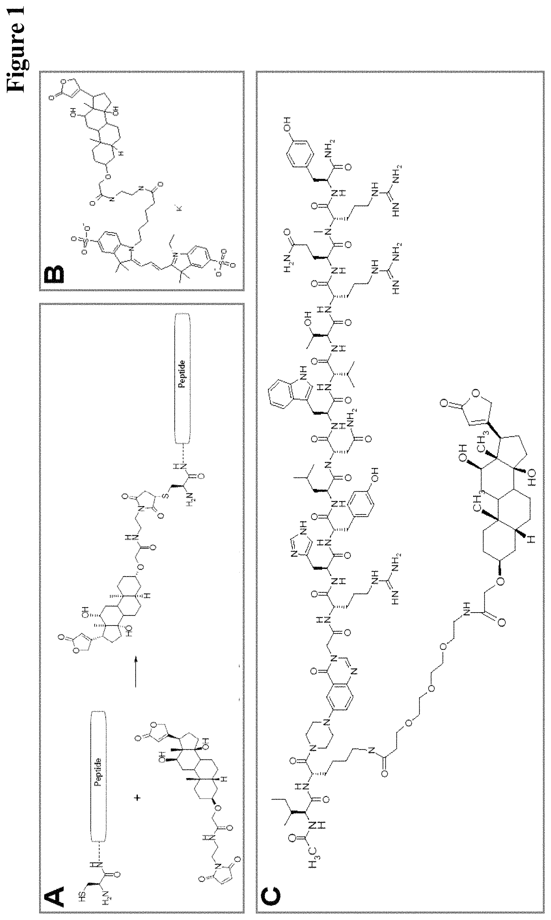
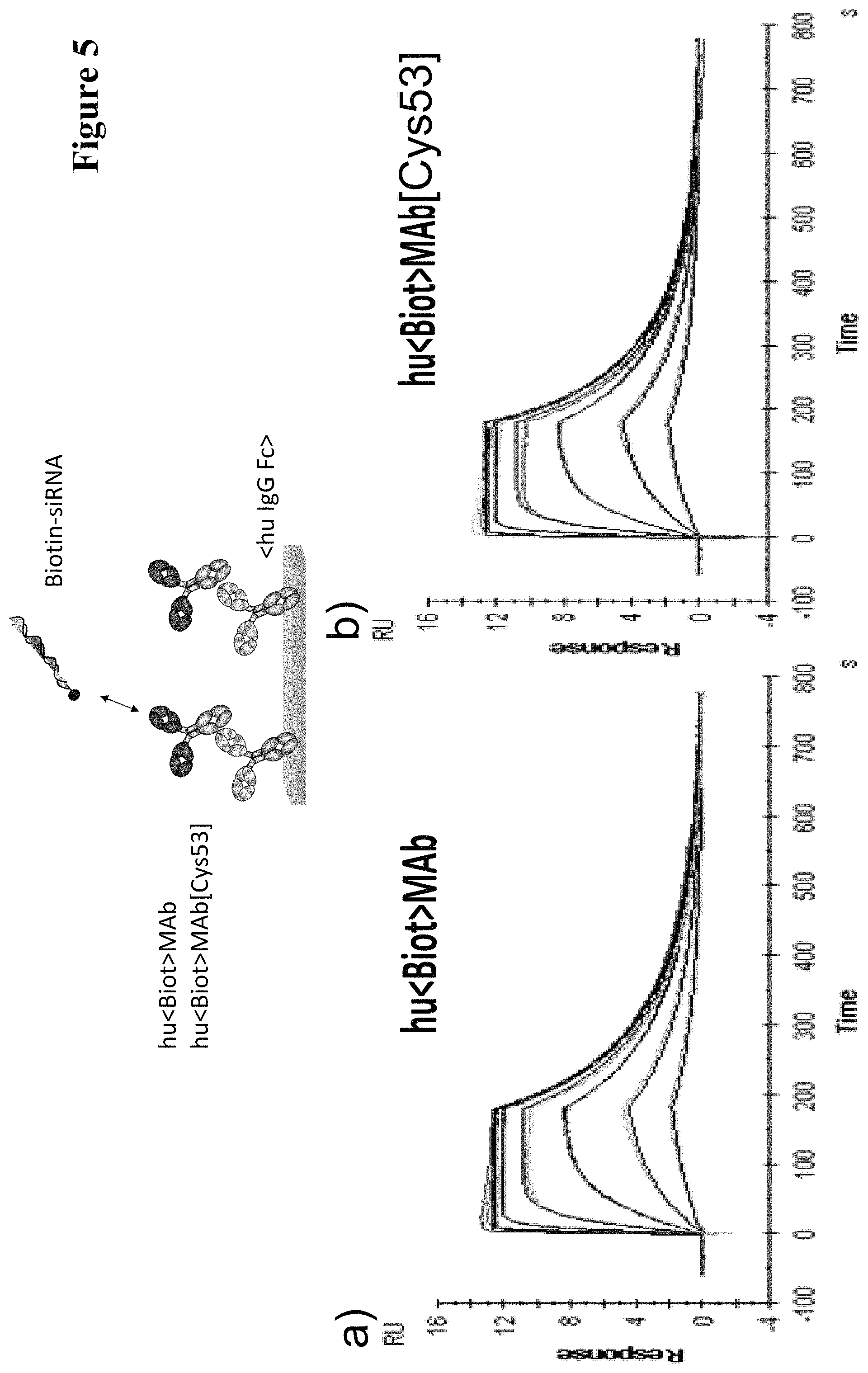
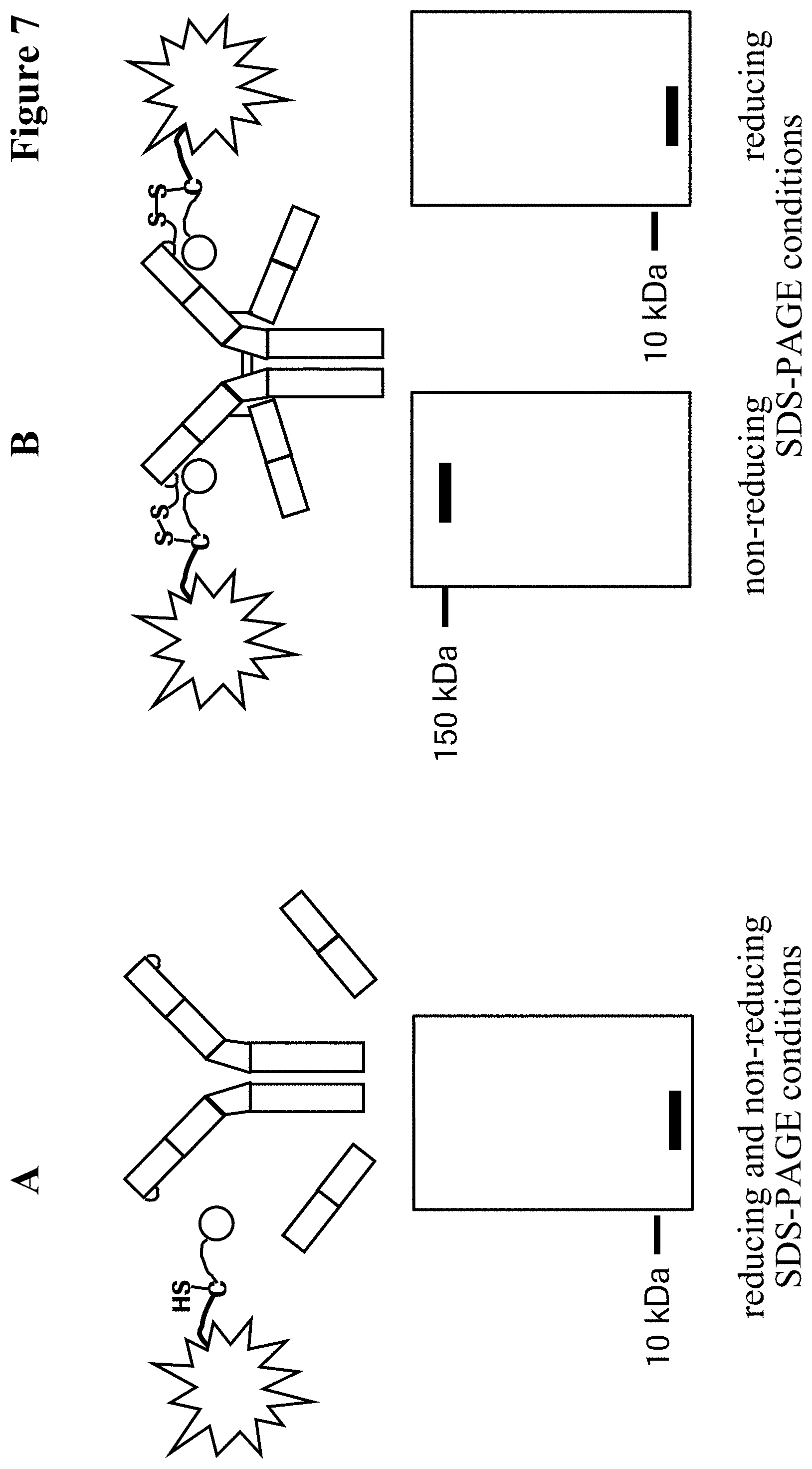
It has been found that by the covalent coupling of a haptenylated compound to an anti-hapten antibody a stabilization and PK-property improvement of the compound can be achieved.
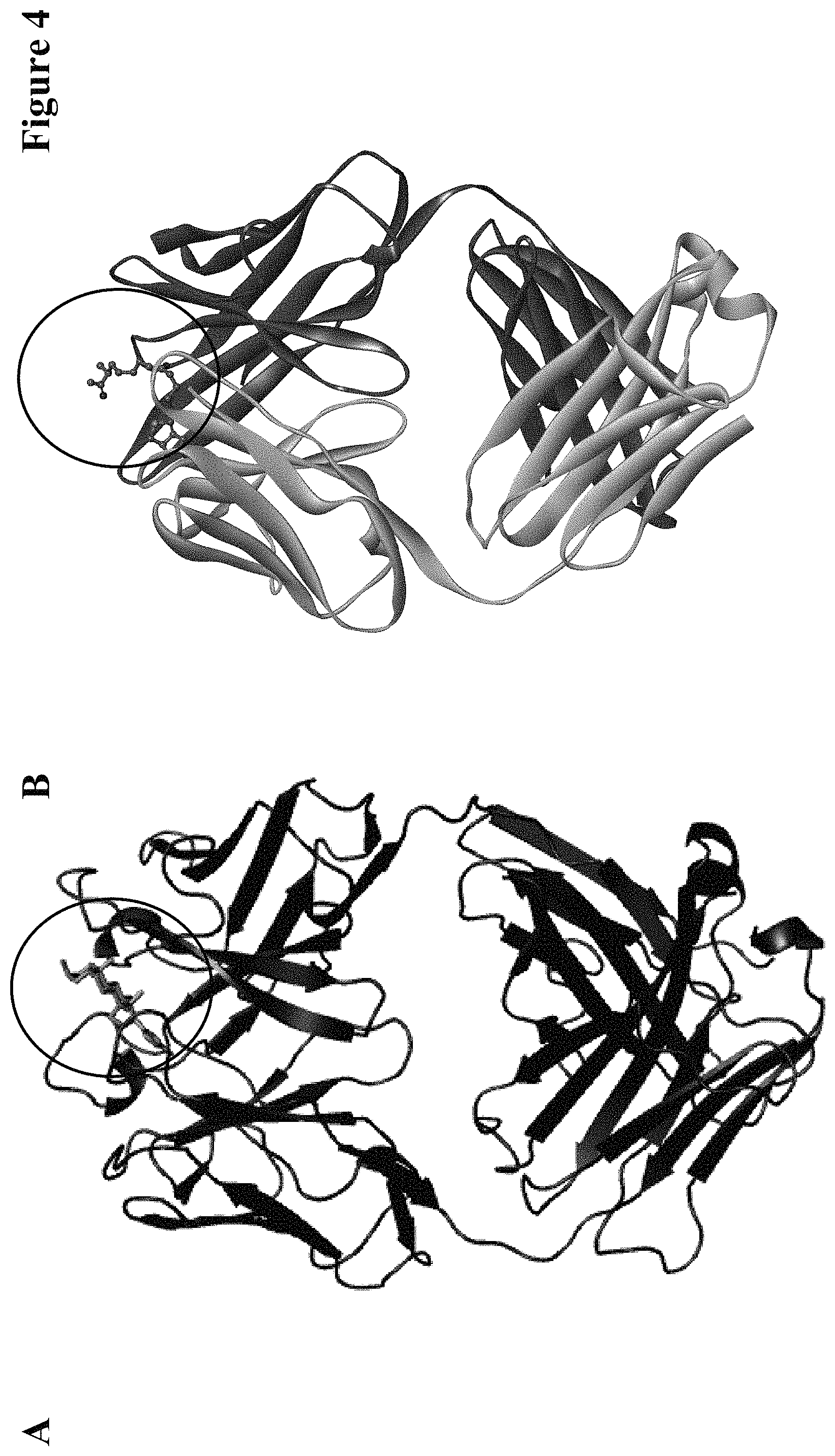
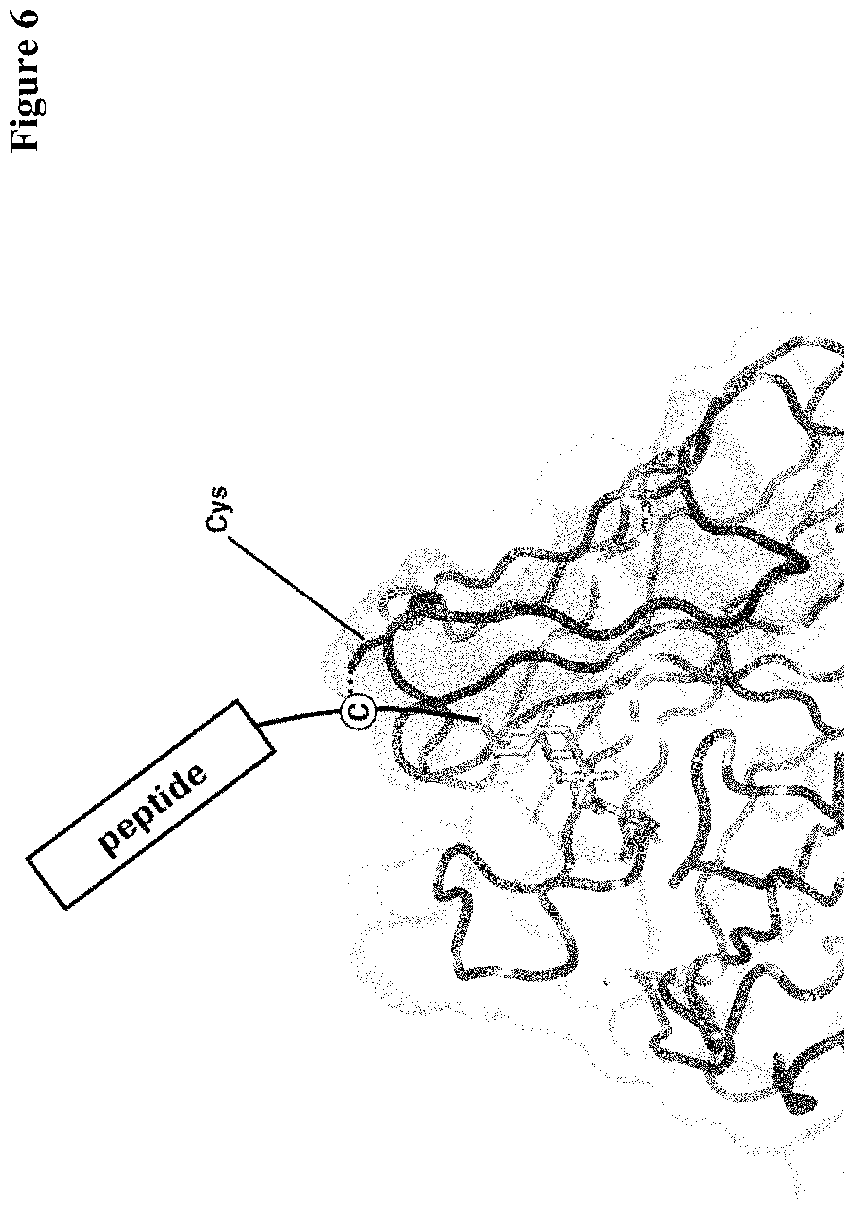
One aspect as reported herein is a conjugate comprising an antigen and an antibody that specifically binds to the antigen characterized by a covalent bond between the antigen and an amino acid residue in the CDR2 of the antibody, whereby the CDR2 is determined according to Kabat.
It has been found that any antigen can be used in the conjugates and methods as reported herein upon derivatization with a universal linker which comprises the functional residue for the formation of the covalent bond between the antigen and an amino acid residue in the CDR2 of the antibody. The location of the functional group in the universal linker has the advantage that it is not necessary to re-engineer the synthesis and the position of the functional group in the CDR2 of the antibody.
One aspect as reported herein is a conjugate comprising a haptenylated compound and an antibody that specifically binds to the hapten of the haptenylated compound (anti-hapten antibody) characterized by a covalent bond between the haptenylated compound and an amino acid residue in the CDR2 of the antibody, whereby the CDR2 is determined according to Kabat.
In one embodiment of all aspects the CDR2 is the heavy chain CDR2.
In one embodiment of all aspects the haptenylated compound comprises a hapten, an optional linker, and a payload.
In one embodiment of all aspects the covalent bond is between the linker of the haptenylated compound and an amino acid residue in the CDR2 of the antibody.
In one embodiment of all aspects the covalent bond is between a functional group in the haptenylated compound and the amino acid residue in the CDR2 of the antibody.
In one embodiment the functional group is in the linker of the haptenylated compound.
In one embodiment of all aspects as reported herein the covalent bond is between a cysteine residue in the heavy chain CDR2 of the antibody and a functional group in the antigen or a functional group in the haptenylated compound.
In one embodiment the cysteine residue in the heavy chain CDR2 of the antibody is at position 52, or position 52a, or position 52b, or position 52c, or position 52d, or position 53 according to the heavy chain variable domain numbering of Kabat.
In one embodiment the cysteine residue in the heavy chain CDR2 of the antibody is at position 52a, or position 52b, or position 52c, or position 53 according to the heavy chain variable domain numbering of Kabat.
In one embodiment the cysteine residue in the heavy chain CDR2 of the antibody is at position 52b or at position 53 according to the heavy chain variable domain numbering of Kabat.
In one embodiment of all aspects the covalent bond is a disulfide bond.
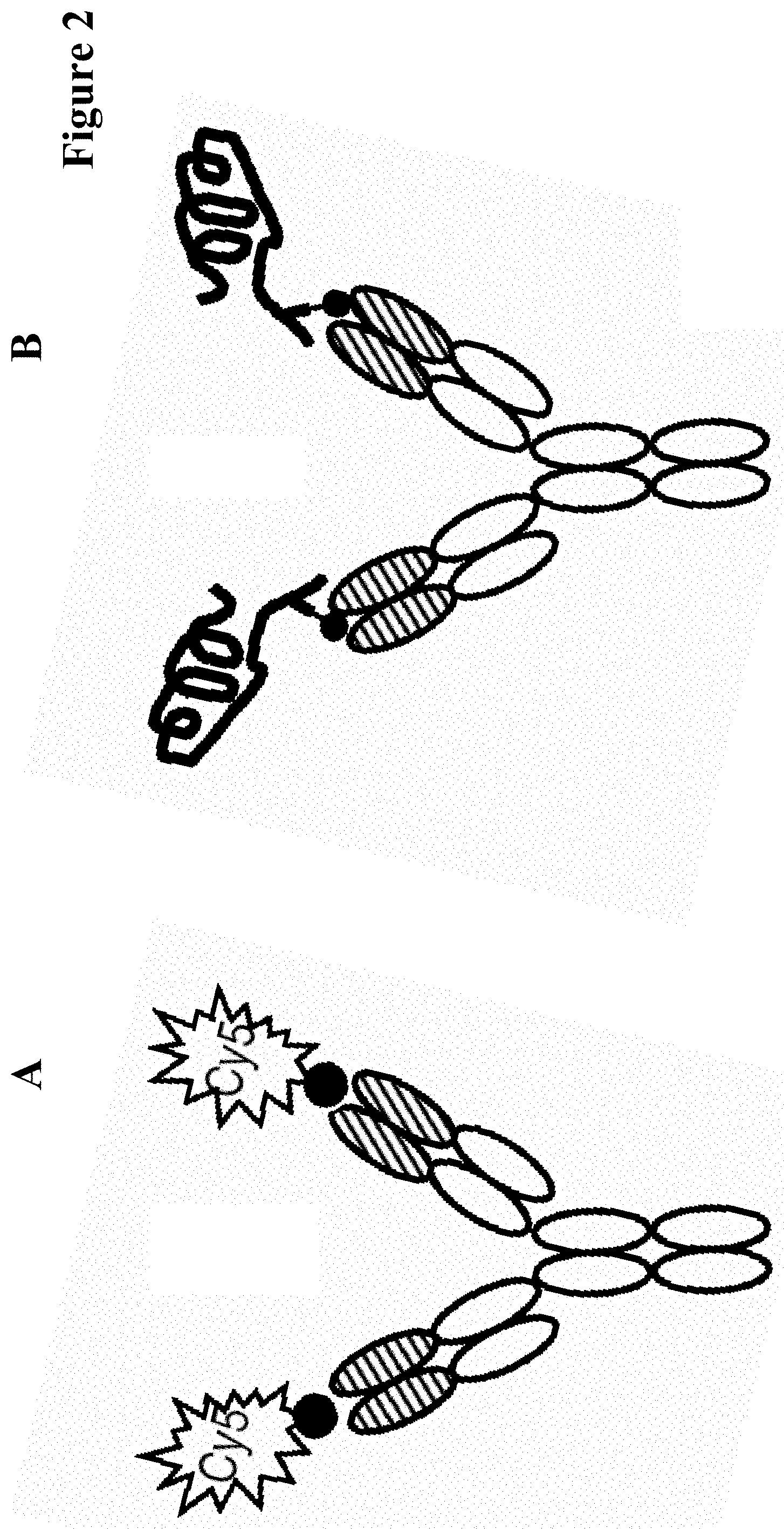
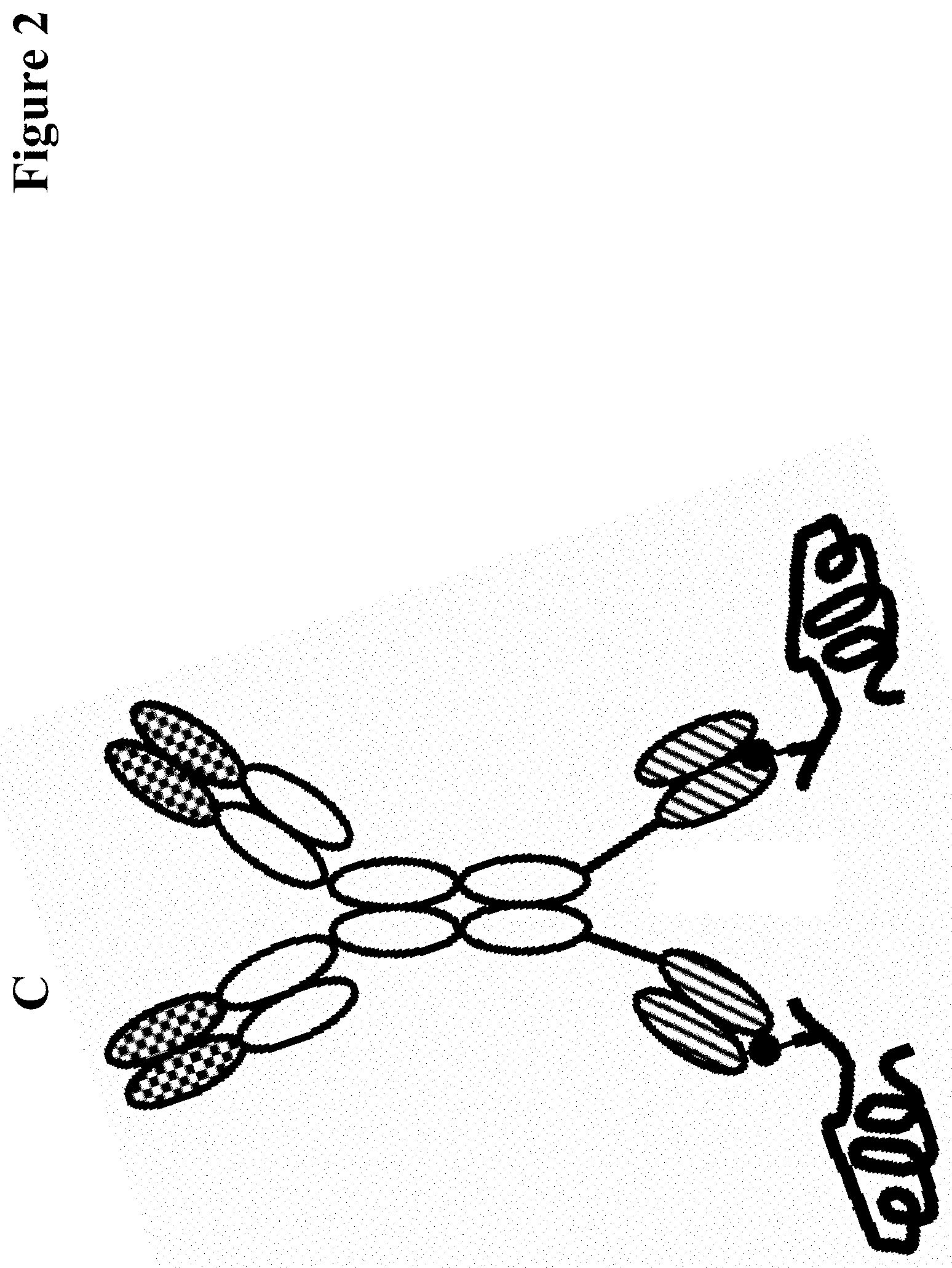
In one embodiment of all aspects the antibody is a bispecific antibody comprising a first binding specificity to a first antigen or a first hapten and a second binding specificity to a second antigen or a second hapten, wherein the first antigen/hapten is different from the second antigen/hapten.
In one embodiment the antibody comprises a first binding specificity to a first hapten and a second binding specificity to a second hapten.
In one embodiment the antibody comprises a first binding specificity to a hapten and a second binding specificity to an (non-hapten) antigen.
In one embodiment the (non-hapten) antigen is a cell surface antigen. In one embodiment the cell surface antigen is a tumor associated antigen.
In one embodiment the bispecific antibody is a full length antibody. In one embodiment one heavy chain of the bispecific antibody comprises a hole mutation and the respective other chain comprises a knob mutation.
In one embodiment of all aspects the payload is selected from a binding moiety, a labeling moiety, and a biologically active moiety.
In one embodiment of all aspects the antibody is a full length antibody.
In one embodiment of all aspects the antibody is a humanized or a human antibody.
In one embodiment the constant region of the antibody is of the IgG1 subclass or of the IgG4 subclass.
In one embodiment the antibody has a constant region of the IgG1 subclass with an alanine at position 234 and 235 and with a glycine at position 329 with numbering according to the EU index of Kabat.
In one embodiment the antibody has a constant region of the IgG4 class with a proline at position 228, a glutamic acid at position 235 and a glycine at position 329 with numbering according to the EU index of Kabat.
In one embodiment of all aspects as reported herein the antibody is a fragment of an antibody. In one embodiment the fragment is a Fab or a (Fab).sub.2.
In one embodiment of all aspects the conjugate comprises exactly one covalent bond per heavy chain CDR2.
In one embodiment of all aspects as reported herein the antigen or the haptenylated compound comprises a reactive group that can form a covalent bond with the thiol group of the cysteine residue in the CDR2 of the antibody. In one embodiment the reactive group is a thiol, or a maleimide, or a haloacetyl.
In one embodiment of all aspects as reported herein the covalent bond is a disulfide bond and it is formed without the addition of redox active agents.
In one embodiment of all aspects the conjugate comprises a therapeutic or detectable moiety. In one embodiment the therapeutic or detectable moiety is covalently conjugated to the antigen or the hapten.
In one embodiment the antigen or hapten is conjugated to a polypeptide consisting of 5 to 60 amino acid residues. In one embodiment the polypeptide comprises 10 to 50 amino acid residues. In one embodiment the polypeptide comprises 12 to 40 amino acid residues. In one embodiment the polypeptide comprises 12 to 30 amino acids residues.
In one embodiment the antigen or hapten is conjugated to a detectable label.
In one embodiment the antigen or the hapten is conjugated to the polypeptide, or to the detectable label, or to the payload via a linker. In one embodiment the linker is a non-peptidic linker.
One aspect as reported herein is an antibody that has in the light and/or the heavy chain a cysteine residue in the CDR2 whereby the CDRs are determined according to Kabat.
In one embodiment the cysteine residue is in the heavy chain CDR2.
In one embodiment the cysteine residue in the heavy chain CDR2 of the antibody is at position 52, or position 52a, or position 52b, or position 52c, or position 52d, or position 53 according to the heavy chain variable domain numbering of Kabat.
In one embodiment the cysteine residue in the heavy chain CDR2 of the antibody is at position 52a, or position 52b, or position 52c, or position 53 according to the heavy chain variable domain numbering of Kabat.
In one embodiment the cysteine residue in the heavy chain CDR2 of the antibody is at position 52b or at position 53 according to the heavy chain variable domain numbering of Kabat.
In one embodiment the antibody has in exactly one heavy chain variable domain a cysteine residue at position 52b or position 53.
In one embodiment the hapten is biotin, or theophylline, or digoxigenin, or carborane, or fluorescein.
In one embodiment the antibody is a humanized or human antibody.
In one embodiment the antibody is a full length antibody, or a Fab, or a scFv, or a scFv conjugated to an Fc-region.
In one embodiment the cysteine forms a disulfide bond with an isolated cysteine residue or an isolated homocysteine residue
One aspect as reported herein is an immunoconjugate comprising the conjugate as reported herein and a cytotoxic agent.
One aspect as reported herein is a pharmaceutical formulation comprising the conjugate as reported herein and a pharmaceutically acceptable carrier.
The conjugate as reported herein for use as a medicament.
The conjugate as reported herein for the treatment of cancer.
The conjugate as reported herein for the treatment of diabetes.
The conjugate as reported herein for the treatment of adiposities.
The conjugate as reported herein for the treatment of an inflammatory disease.
The conjugate as reported herein for the treatment of a metabolic disease.
The conjugate as reported herein for the treatment of a viral disease.
One aspect as reported herein is the use of a conjugate as reported herein in the manufacture of a medicament.
One aspect as reported herein is the use of a conjugate as reported herein as diagnostic agent.
One aspect as reported herein is the use of a conjugate as reported herein comprising a therapeutic polypeptide to increase the stability of the therapeutic polypeptide.
One aspect as reported herein is the use of a conjugate as reported herein comprising a therapeutic polypeptide to increase the activity of the therapeutic polypeptide.
One aspect as reported herein is the use of a conjugate as reported herein comprising a therapeutic polypeptide to increase the in vivo half-life of the therapeutic polypeptide.
One aspect as reported herein is the use of a conjugate as reported herein in the treatment of a disease.
One aspect as reported herein is a method of treating an individual having a disease comprising administering to the individual an effective amount of a conjugate as reported herein.
One aspect as reported herein is a method of treating a disease in an individual comprising administering to the individual an effective amount of the conjugate as reported herein.
In one embodiment the disease is cancer.
In one embodiment the disease is diabetes.
In one embodiment the disease is adipositas.
One aspect as reported herein is a method of producing a conjugate as reported herein comprising the combination of an antibody comprising a first reactive group and an antigen that has a second reactive group whereby the alpha carbon atom of the amino acid residue that bears the first reactive group is about 10 to 11 Angstrom apart from the atom of the antigen to which the linker is fused.
One aspect as reported herein is a method of producing a conjugate as reported herein comprising the steps of combining in solution an antibody that specifically binds to a hapten and comprises a reactive group at one amino acid residue in the CDR2 with a haptenylated compound comprising a reactive group, wherein the haptenylated compound comprises a payload, such as a peptide consisting of 5 to 60 amino acids or a detectable label, and recovering of the conjugate from the solution.
One aspect as reported herein is a method for producing an antibody for the formation of a conjugate as reported herein, comprising the step of cultivating a cell comprising a nucleic acid encoding the antibody, and recovering the antibody from the cell or the cultivation medium,
wherein in the antibody the residue in the heavy chain CDR2 is mutated to cysteine that has in the X-ray structure of the non-covalent complex of the antibody and its antigen or a haptenylated compound a distance of 10 to 11 Angstrom between the alpha-carbon atom of the amino acid residue in the antibody CDR2 and the atom of the hapten to which the linker is connected or to an antigen atom between which the covalent bond is to be formed.
One aspect as reported herein is a method for identifying a position in an antibody CDR2 that can be mutated to cysteine for the formation of a covalent bond between the residue in the antibody CDR2 and the bound antigen or haptenylated compound comprising the step of providing a crystal structure of the non-covalent complex of the antibody and the antigen or haptenylated compound, whereby the antigen or haptenylated compound comprises a linker sequence, and identifying an amino acid residue in the CDR2 of the antibody and in the linker sequence with a distance of 10 to 11 Angstrom between the alpha-carbon atoms of the amino acid residue in the antibody CDR2 and the atom in the antigen to which the linker is fused,
wherein the identified position is the position in an antibody CDR2 that can be mutated to cysteine for the formation of a covalent bond between the residue in the antibody CDR2 and the bound antigen or haptenylated compound.
One aspect as reported herein is a bispecific antibody for targeted delivery of a haptenylated compound to a target cell, wherein the bispecific antibody comprises a first binding site that specifically binds to the haptenylated compound and a second binding specificity that specifically binds to a cell surface marker of the cell.
DESCRIPTION OF THE FIGURES
FIG. 1: Procedure for digoxigenylation (conjugation of digoxigenin to) of peptides (FIG. 1A). Examples of a digoxigenylated label (fluorophore Dig-Cy5; FIG. 1B) and of a digoxigenylated polypeptide (PYY-derivative (DIG-PYY); FIG. 1C).
FIG. 2: Scheme of a complex of a monospecific bivalent anti-digoxigenin antibody and a digoxigenin-Cy5 conjugate (FIG. 2A) and of a complex of a monospecific bivalent anti-digoxigenin antibody and a digoxigenin-polypeptide conjugate (FIG. 2B). Scheme of a complex of a bispecific tetravalent anti-digoxigenin antibody and a digoxigenin-polypeptide conjugate (FIG. 2C).
FIG. 3: Size exclusion chromatogram (recorded at 280 nm) of a complex comprising an anti-digoxigenin antibody and digoxigenin which is conjugated to a peptide (DIG-PYY) showing a single peak of a complex of defined size.
FIG. 4: A: Structure model of an anti-digoxigenin Fab (left) showing that digoxigenin (encircled) is captured in a deep pocket which is formed by the CDRs of the VH and VL regions. B: Structure model of an anti-biotin Fab (right) showing that biocytinamid (encircled) is captured in a deep pocket which is formed by the CDRs of the VH and VL regions.
FIG. 5: Comparison of the binding of recombinant humanized anti-biotin antibodies with and without introduced VH53C mutation. Binding properties were analyzed by surface plasmon resonance (SPR) technology using a BIAcore T100 or BIAcore 3000 instrument. a) humanized anti-biotin antibody. Binding of biotinylated siRNA to humanized anti-biotin antibody, KD=624 pM; b) humanized Cys53 mutated anti-biotin antibody. Binding of biotinylated siRNA, KD=643 pM; siRNA concentrations: 0.14, 0.41, 1.23, 3.70, 11.1, 33.3, and 100 nM; anti-biotin antibody concentration: 2 nM; Sensor Chip CM3; binding of antibody via anti-human IgG Fc antibody
TABLE-US-00001 ka (1/Ms) kd (1/s) KD (M) humanized anti-biotin 2.2 * 10.sup.7 0.01 6.4 * 10.sup.-10 antibody VH53C humanized anti-biotin 2.0 * 10.sup.7 0.01 6.2 * 10.sup.-10 antibody
FIG. 6: Introduction of SH functionalities in the hapten as well as in the antibody at appropriate positions allow the antibody and the hapten to form a covalent bond in between resulting in a conjugate.
FIG. 7: Scheme of SDS-PAGE self-fluorescence band pattern (without further staining of the SDS-PAGE gel): A: If no covalent bond is formed between the antibody and the hapten-fluorophore conjugate both under reducing or non-reducing conditions one self-fluorescent band at the molecular weight of free hapten-fluorophore conjugate can be detected. B: If a covalent bond is formed between the antibody and the hapten-fluorophore conjugate under non-reducing conditions one self-fluorescent band at the combined molecular weight of the antibody and the hapten-fluorophore conjugate can be detected. Under reducing conditions the disulfide bridges in the conjugate of the antibody and the hapten-fluorophore conjugate (haptenylated compound) are cleaved and one self-fluorescent band at the molecular weight of free hapten-fluorophore conjugate can be detected.
FIG. 8: Conjugate formation of hapten-binding Cys-mutated antibodies with hapten-Cys-fluorescent label conjugates (haptenylated compound) in the presence of redox active agents: oxidation agent (glutathione disulfide, GSSG) and reducing agent (dithioerythritol, DTE): Antibody complexation and subsequent covalent linkage at defined positions is detected by fluorescence signals in SDS PAGE analyses. Non-reducing (upper images) and reducing (lower images) SDS-PAGE analyses were performed as described in Example 11. Covalently antibody linked haptens are detectable as larger sized protein bound signals at the appropriate positions under non-reduced conditions. These signals detach from protein upon reduction and are visible as small entities under reducing conditions. Left: fluorescence image Right: Coomassie blue staining Series 1: anti-digoxigenin antibody with 52bC mutation Series 2: anti-digoxigenin antibody with wild-type residue at position 52b (A) covalent coupling with 3 mM DTE and 10 mM GSSG; (B) covalent coupling with 0.3 mM DTE and 1 mM GSSG; (C) covalent coupling with 0.03 mM DTE and 0.1 mM GSSG.
FIG. 9: Complex formation of hapten-binding Cys mutated antibodies with hapten-Cys-fluorescent label conjugates in the presence solely of an oxidation agent (glutathione disulfide, GSSG) but in the absence of reducing agents or in the absence of both: Antibody complexation and subsequent covalent linkage at defined positions is detected by fluorescence signals in SDS PAGE analyses. Non-reducing (upper images) and reducing (lower images) SDS-PAGE analyses were performed as described in Example 12. Covalently antibody linked haptens are detectable as larger sized protein bound signals at the appropriate positions under non-reduced conditions. These signals detach from protein upon reduction and are visible as small entities under reducing conditions. Left: fluorescence image Right: Coomassie blue staining Series 1: anti-digoxigenin antibody with 52bC mutation Series 2: anti-digoxigenin antibody with wild-type residue at position 52b (A) no additives (B) covalent coupling with 1 mM GSSG; (C) covalent coupling with 0.1 mM GSSG.
FIG. 11: Structure of DIG-3-cme-eda-Cy5
FIG. 12: Structure of DIG-maleiimid-Cy5
FIG. 13: Structure of DIG-eda-Cys-Cy5
FIG. 14: Structure of DIG-Ahx-Cys-Cy5
FIG. 15: Structure of DIG-Cys-MR121
FIG. 16: Structure of Ac-PYY(PEG3-Dig)
FIG. 17: Structure of Ac-PYY(PEG3-Cys-4Abu-Dig)
FIG. 18: Structure of PEG3-PYY(PEG3-Cys-4Abu-Dig)
FIG. 19: Structure of Dy636-eda-Btn
FIG. 20: Structure of Dy636-Ser-Btn
FIG. 21: Structure of Dy636-Cys-Btn
FIG. 22: Structure of Cy5-Cys-Btn
FIG. 23: Structure of Cy5-Ser-Btn
FIG. 24: Structure of Ac-PYY(PEG2-Btn)
FIG. 25: Structure of Ac-PYY-PEG3-Cys-.beta.-Ala-Btn)
FIG. 26: Structure of Ac-PYY-PEG3-Ser-PEG2-Btn)
FIG. 27: Structure of Ac-PYY-PEG3-Cys-PEG2-Btn
FIG. 28: Structure of Ac-PYY(PEG3-Cys-4-Abu-5-Fluo)
FIG. 29: Structure of Ac-PYY(PEG3-Cys-PEG2-5-Fluo)
FIG. 30: Scheme for the generation of Ac-PYY(PEG2-Btn)
FIG. 31: Scheme for the generation of Ac-PYY(PEG3-Cys-.beta.-Ala-Btn)
FIG. 32: Scheme for the generation of Ac-PYY(PEG3-Cys-4-Abu-Dig)
FIG. 33: X-ray structure of murine anti-biotin antibody in complex with biocytinamid. Amino acid residues that are interacting with biocytinamid are shown in a stick representation.
FIG. 34A and FIG. 34B:
Results of the in vivo blood PK study with covalent conjugates and non-covalent complexes compared to non-complexed antigen/hapten; the relative remaining fluorescence intensity (%, solid marks) of Cy5-mediated fluorescence of Biotin-Cy5 non-covalent complexes (FIG. 34A) and covalent (SS-bridged) conjugates (FIG. 34B), as well as of non-complexed Biotin-Ser-Cy5 (asterix) is shown; the fluorescence signal at time point t=0.08 h was set to 100%; additionally, the relative remaining amount of human IgG in the mouse serum samples is shown (open marks); IgG serum concentration (mg/ml) at t=0.08 h was set to 100%.
FIG. 35: Western blot of the determination of the amount of digoxigenylated PYY polypeptide in the serum of mice.
FIG. 36: Analysis of affinity-driven complexation of haptenylated compounds with anti-hapten antibodies. Antibody complexation and subsequent covalent linkage at defined positions is directed by fluorescence signals in SDS PAGE analyses, which were carried out as described in Example 19. Left: fluorescent image with non-reduced (left side of gel) and reduced (right side of gel) samples. Right: Coomassie blue staining. 1: humanized anti-digoxigenin antibody+biotin-Cys-Cy5 2: humanized anti-digoxigenin antibody VH52bC+biotin-Cys-Cy5 3: humanized anti-biotin antibody+biotin-Cys-Cy5 4: humanized anti-biotin antibody VH53C+biotin-Cys-Cy5 The white arrows mark the excess (uncoupled) biotin-Cys-Cy5, which is significantly higher when anti-digoxigenin antibody VH52bC is used, because the conjugation reaction is not affinity driven in this case.
FIG. 37: Cysteine positions and disulfide patterns within the Fab region, required to form a Dig-binding antibody with additional cysteine at position 52b for hapten-mediated site-directed directed covalent payload coupling. (A) Cysteines and disulfide pattern in VH and CH1 domains, and in VL and CL domains that are required to form functional Fab fragments. (B) Cysteines and disulfide pattern in VH and CH1 domains, and in VL and CL domains that are required to form functional Fab fragments with additional cysteine at position 52b for hapten-mediated site-directed directed covalent payload coupling. (C&D) Potential to form incorrect disulfide bonds within the VH domain of the VH52b variant which would result in misfolded nonfunctional antibodies. E) Example for a potential incorrect interdomain disulfide bond within the Fv region of the VH52b variant, which would result in misfolded nonfunctional antibodies.
FIG. 38: Cysteine positions and disulfide patterns required to form a Dig-binding disulfide-stabilized single-chain Fv with additional cysteine at position 52b for hapten-mediated site-directed directed covalent payload coupling. (A) Cysteines in VH and VL domains that are required to form functional scFvs, dsscFvs and 52b mutated dsscFvs. (B) correct pattern of disulfide bonds that must be formed to generate functional scFvs, dsscFvs and 52b mutated dsscFvs. (C) Potential to form incorrect disulfide bonds which would result in misfolded nonfunctional scFvs. (D) Potential to form incorrect disulfide bonds which would result in misfolded nonfunctional dsscFvs. (E) Potential to form incorrect disulfide bonds which would result in misfolded nonfunctional 52b mutated dsscFvs.
FIG. 39: Composition of a LeY-Dig bispecific antibody derivative as delivery vehicle for covalently coupled payloads.
FIG. 40: Expression and Purification of bispecific anti-hapten antibody derivatives for targeted delivery of covalently coupled payloads. (A) For Western blot analyses, cell culture supernatants were subjected to SDS PAGE (NuPAGE 4-12% Bis-Tris Gel (1.0 mm.times.12 well) (Invitrogen; Cat. No. NP0322) and proteins were subsequently transferred to Immobilon Transfer Membranes (Immobilon-P) (Millipore; Cat. No. IPVH07850), PVDF with pore Size: 0.45 .mu.m. Antibody derivatives were detected by Anti-Human Kappa Light Chain)-Alkaline Phosphatase antibody produced in goat, (affinity purified), Sigma (Cat. No. A3813) at a 1:1000 dilution, and Anti-Human IgG (Fc specific)-Alkaline Phosphatase antibody produced in goat, Sigma (Cat. No. A9544) at a 1:1000 dilution. The substrate BCIP/NBT-Blue Liquid Substrate (Sigma Cat. No. B3804 was applied for the development of the Western blot. Lane 1--molecular weight marker; Lane 2 & 3-control antibody with unmodified heavy-chain; Lane 4 LeY-Dig(52bC) bispecific antibody with extended H-chain. (B) SDS-PAGE analyses (NuPAGE 4-12% Bis-Tris Gel [Invitrogen] and subsequent staining with Coomassie brilliant blue demonstrates purity of protein preparations and visualizes polypeptide chains related to the IgG with the apparent molecular sizes that correspond to their calculated molecular weights. Lane 1--molecular weight marker; Lane 2-LeY-Dig(52bC) bispecific antibody with extended H-chain reduced, lane 3-LeY-Dig(52bC) bispecific antibody with extended heavy-chain non-reduced; (C) Size exclusion chromatography (Superdex 200) demonstrates homogeneity and lack of aggregates in the protein preparations of the LeY-Dig(52bC) bispecific antibody derivative after Protein A purification.
FIG. 41: Relative remaining fluorescence intensity (%) of Cy5-mediated fluorescence of Dig-Cy5 non-covalent complexes and covalent (disulfide-bridged) conjugates, as well as of non-complexed Dig-Cy5; the fluorescence signal at time point t=0.08 h was set to 100%; additionally, the relative remaining amount of human IgG in the mouse serum samples is shown; IgG serum concentration (mg/ml) at t=0.08 h was set to 100%.
FIG. 42: Pharmacokinetics under in vivo-like conditions of Cy5-mediated fluorescence of Biotin-Cy5 of non-covalent complexes and of covalent (disulfide-bridged) conjugates, as well as of non-complexed Biotin-Cy5, determined by non-invasive eye imaging; solid diamond: biotin-Cy5, solid square biotin-Cy5+anti-biotin antibody (complex); triangle: Cy5-Biotin-anti-biotin antibody conjugate.
FIG. 43: a) Composition, structure and molecular weight of Theophylline-Cys-Cy5; b) Size exclusion chromatography demonstrates purity and homogeneity of purified theophylline-binding antibody variants; peak #2 shows the purified product, lack of peak #1 indicates that such preparations are free of aggregates; c) formation of covalent complexes between theophylline-binding antibodies and Theophylline-Cys-Cy5 as demonstrated by non-reducing (left lanes) and reducing (right lanes) SDS PAGE; Cy5 appears coupled to the H-chain under non-reducing conditions only in samples that contained Theophylline-Cys-Cy5 and cys-mutated antibody, these covalent conjugates disintegrate upon reduction (right lanes); Lanes 1: Molecular weight marker; 2-4 non-reducing--2: anti-Theophylline antibody (without cys-mutation)+Theophylline-Cys-Cy5 (complex); 3: anti-Theophylline antibody-cys_55+Theophylline-Cys-Cy5 (conjugate); 4: anti-Theophylline antibody-cys_54+Theophylline-Cys-Cy5 (conjugate); 5-7 reducing--5: anti-Theophylline antibody (without cys-mutation)+Theophylline-Cys-Cy5 (complex); 6: anti-Theophylline antibody-cys_55+Theophylline-Cys-Cy5 (conjugate); 7: anti-Theophylline antibody-cys_54+Theophylline-Cys-Cy5 (conjugate).
FIG. 44: Formation of covalent complexes between biotin-binding antibodies and Biotin-Cys-Cy5 is demonstrated by non-reducing and reducing SDS PAGE; the coupling reaction was performed in murine serum at 37.degree. C. for 1 hr. Cy5 appears coupled to the H-chain under non-reducing conditions only in samples that contained Biotin-Cys-Cy5 and cys-mutated antibody; these covalent conjugates disintegrate upon reduction (right lanes); lanes 1: Molecular weight marker; 2-3 non-reducing--2: anti-Biotin antibody (without cys mutation)+Biotin-Cys-Cy5 (complex); 3: anti-Biotin antibody-cys+Biotin-Cys-Cy5 (conjugate); 4-5 reducing--5: anti-Biotin antibody (without cys mutation)+Biotin-Cys-Cy5 (complex); 6: anti-Biotin antibody-cys+Biotin-Cys-Cy5 (conjugate)
FIG. 45: In vivo pharmacokinetics of Cy5-mediated fluorescence of Biotin-Cy5 of non-covalent complexes and of covalent (disulfide-bridged) conjugates, as well as of non-complexed Biotin-Cy5, determined by non-invasive eye imaging; solid diamond: biotin-Cy5, solid circle: biotin-Cy5 administered 24 hours after administration of anti-biotin antibody (in vivo complex formation); solid square: biotin-Cys-Cy5 administered 24 hours after administration of anti-biotin antibody-cys (in vivo conjugate formation).
FIG. 46: The protein structure of murine anti-Biotin antibody-Fab-fragment was determined in complex with biocytinamid: the complexed hapten is positioned in close proximity to a negatively charged cluster of amino acids; biotin which--as hapten--is derivatized for payload coupling at its carboxyl group binds with good efficacy as there is no charge repulsion at this position (due to the lack of the COOH group); in contrast, free (normal) biotin cannot bind efficient to the antibody because its carboxyl group would be in close proximity to this negative charge cluster, and hence becomes repulsed.
DETAILED DESCRIPTION OF THE INVENTION
I. Definitions
An "acceptor human framework" for the purposes herein is a framework comprising the amino acid sequence of a light chain variable domain (VL) framework or a heavy chain variable domain (VH) framework derived from a human immunoglobulin framework or a human consensus framework, as defined below. An acceptor human framework "derived from" a human immunoglobulin framework or a human consensus framework may comprise the same amino acid sequence thereof, or it may contain amino acid sequence changes. In some embodiments, the number of amino acid changes are 10 or less, 9 or less, 8 or less, 7 or less, 6 or less, 5 or less, 4 or less, 3 or less, or 2 or less. In some embodiments, the VL acceptor human framework is identical in sequence to the VL human immunoglobulin framework sequence or human consensus framework sequence.
The term "amino acid" denotes the group of carboxy .alpha.-amino acids, either occurring naturally, i.e. which directly or in form of a precursor can be encoded by a nucleic acid, or occurring non-naturally. The individual naturally occurring amino acids are encoded by nucleic acids consisting of three nucleotides, so called codons or base-triplets. Each amino acid is encoded by at least one codon. This is known as "degeneration of the genetic code". The term "amino acid" as used within this application denotes the naturally occurring carboxy .alpha.-amino acids comprising alanine (three letter code: ala, one letter code: A), arginine (arg, R), asparagine (asn, N), aspartic acid (asp, D), cysteine (cys, C), glutamine (gln, Q), glutamic acid (glu, E), glycine (gly, G), histidine (his, H), isoleucine (ile, I), leucine (leu, L), lysine (lys, K), methionine (met, M), phenylalanine (phe, F), proline (pro, P), serine (ser, S), threonine (thr, T), tryptophan (trp, W), tyrosine (tyr, Y), and valine (val, V). Examples of non-naturally occurring amino acids include, but are not limited to, Aad (alpha-Aminoadipic acid), Abu (Aminobutyric acid), Ach (alpha-aminocyclohexane-carboxylic acid), Acp (alpha-aminocyclopentane-carboxylic acid), Acpc (1-Aminocyclopropane-1-carboxylic acid), Aib (alpha-aminoisobutyric acid), Aic (2-Aminoindane-2-carboxylic acid; also called 2-2-Aic), 1-1-Aic (1-aminoindane-1-carboxylic acid), (2-aminoindane-2-carboxylic acid), Allylglycine (AllylGly), Alloisoleucine (allo-Ile), Asu (alpha-Aminosuberic acid, 2-Aminooctanedioc acid), Bip (4-phenyl-phenylalanine-carboxylic acid), BnHP ((2S,4R)-4-Hydroxyproline), Cha (beta-cyclohexylalanine), Cit (Citrulline), Cyclohexylglycine (Chg), Cyclopentylalanine, beta-Cyclopropyl alanine, Dab (1,4-Diaminobutyric acid), Dap (1,3-Diaminopropionic acid), p (3,3-diphenylalanine-carboxylic acid), 3,3-Diphenylalanine, Di-n-propylglycine (Dpg), 2-Furylalanine, Homocyclohexylalanine (HoCha), Homocitrulline (HoCit), Homocycloleucine, Homoleucin (HoLeu), Homoarginine (HoArg), Homoserine (HoSer), Hydroxyproline, Lys(Ac), (1) Nal (1-Naphtyl Alanine), (2) Nal (2-Naphtyl Alanine), 4-MeO-Apc (1-amino-4-(4-methoxyphenyl)-cyclohexane-1-carboxylic acid), Nor-leucine (Nle), Nva (Norvaline), Omathine, 3-Pal (alpha-amino-3-pyridylalanine-carboxylic acid), 4-Pal (alpha-amino-4-pyridylalanine-carboxylic acid), 3,4,5,F3-Phe (3,4,5-Trifluoro-phenylalanine), 2,3,4,5,6,F5-Phe (2,3,4,5,6-Pentafluoro-phenylalanine), Pqa (4-oxo-6-(1-piperazinyl)-3(4H)-quinazoline-acetic acid (CAS 889958-08-1)), Pyridylalanine, Quinolylalanine, Sarcosine (Sar), Thiazolylalanine, Thienylalanine, Tic (alpha-amino-1,2,3,4,tetrahydroisoquinoline-3-carboxylic acid), Tic(OH), Tle (tertbutylGlycine), and Tyr(Me).
The term "amino acid sequence variant" refers to polypeptides having amino acid sequences that differ to some extent from a native sequence polypeptide. Ordinarily, amino acid sequence variants will possess at least about 70% sequence identity with the native sequence polypeptide. In one embodiment the variant has about 80% or more sequence identity with the native sequence polypeptide. In one embodiment the variant has about 90% or more sequence identity with the native sequence polypeptide. In one embodiment the variant has about 95% or more sequence identity with the native sequence polypeptide. In one embodiment the variant has about 98% or more sequence identity with the native sequence polypeptide. The amino acid sequence variants possess substitutions, deletions, and/or insertions at certain positions within the amino acid sequence of the native amino acid sequence Amino acids are designated by the conventional names, one-letter and three-letter codes.
The term "antibody" herein is used in the broadest sense and encompasses various antibody structures, including but not limited to monoclonal antibodies, polyclonal antibodies, multispecific antibodies (e.g., bispecific antibodies), and antibody fragments so long as they exhibit the desired antigen-binding activity.
The term "antibody fragment" denotes a molecule other than an intact antibody that comprises a portion of an intact antibody that binds the antigen to which the intact antibody binds. Examples of antibody fragments include but are not limited to Fv, Fab, Fab', Fab'-SH, F(ab').sub.2; diabodies; linear antibodies; single-chain antibody molecules (e.g. scFv); and multispecific antibodies formed from antibody fragments.
The term "biotin", short "BI", denotes 5-[(3aS,4S,6aR)-2-oxohexahydro-1H-thieno[3,4-d]imidazol-4-yl]pentanoic acid. Biotin is also known as vitamin H or coenzyme R.
The term "bispecific antibodies" denotes antibodies which have two different (antigen/hapten) binding specificities. In one embodiment bispecific antibodies as reported herein are specific for two different antigens, i.e. a hapten and a non-hapten antigen.
The term "chimeric" antibody refers to an antibody in which a portion of the heavy and/or light chain is derived from a particular source or species, while the remainder of the heavy and/or light chain is derived from a different source or species.
The "class" of an antibody refers to the type of constant domain or constant region possessed by its heavy chain. There are five major classes of antibodies: IgA, IgD, IgE, IgG, and IgM, and several of these may be further divided into subclasses (isotypes), e.g., IgG1, IgG2, IgG3, IgG4, IgA1, and IgA2. The heavy chain constant domains that correspond to the different classes of immunoglobulins are called .alpha., .delta., .epsilon., .gamma., and .mu., respectively.
The term "cytotoxic agent" as used herein refers to a substance that inhibits or prevents a cellular function and/or causes cell death or destruction. Cytotoxic agents include, but are not limited to, radioactive isotopes (e.g., At211, I131, I125, Y90, Re186, Re188, Sm153, Bi212, P32, Pb212 and radioactive isotopes of Lu); chemotherapeutic agents or drugs (e.g., methotrexate, adriamicin, vinca alkaloids (vincristine, vinblastine, etoposide), doxorubicin, melphalan, mitomycin C, chlorambucil, daunorubicin or other intercalating agents); growth inhibitory agents; enzymes and fragments thereof such as nucleolytic enzymes; antibiotics; toxins such as small molecule toxins or enzymatically active toxins of bacterial, fungal, plant or animal origin, including fragments and/or variants thereof; and the various antitumor or anticancer agents disclosed below.
The term "digoxigenin", short "DIG", denotes 3-[(3S,5R,8R,9S,10S,12R,13S,14S,17R)-3,12,14-trihydroxy-10,13-dimethyl-1,- 2,3,4,5,6,7,8,9,11,12,15,16,17-tetradecahydro-cyclopenta[a]-phenanthren-17- -yl]-2H-furan-5-one (CAS number 1672-46-4). Digoxigenin (DIG) is a steroid found exclusively in the flowers and leaves of the plants Digitalis purpurea, Digitalis orientalis and Digitalis lanata (foxgloves) (Polya, G., Biochemical targets of plant bioactive compounds, CRC Press, New York (2003) p. 847).
The term "effector functions" denotes those biological activities attributable to the Fc-region of an antibody, which vary with the antibody class. Examples of antibody effector functions include: C1q binding and complement dependent cytotoxicity (CDC); Fc receptor binding; antibody-dependent cell-mediated cytotoxicity (ADCC); phagocytosis; down regulation of cell surface receptors (e.g. B cell receptor); and B cell activation.
The term "effective amount" of an agent, e.g., a pharmaceutical formulation, denotes an amount effective, at dosages and for periods of time necessary, to achieve the desired therapeutic or prophylactic result.
Papain digestion of antibodies produces two identical antigen-binding fragments, called "Fab" fragments, each with a single antigen-binding site, and a residual "Fc" fragment, whose name reflects its ability to crystallize readily. Pepsin treatment yields an F(ab').sub.2 fragment that has two antigen-binding sites and is still capable of cross-linking antigen.
The Fab fragment also contains the constant domain of the light chain and the first constant domain (CH1) of the heavy chain. Fab' fragments differ from Fab fragments by the addition of a few residues at the carboxy terminus of the heavy chain CH1 domain including one or more cysteines from the antibody hinge region. Fab'-SH is the designation herein for Fab' in which the cysteine residue(s) of the constant domains bear at least one free thiol group. F(ab').sub.2 antibody fragments originally were produced as pairs of Fab' fragments which have hinge cysteines between them. Other chemical couplings of antibody fragments are also known.
"Fv" is the minimum antibody fragment which contains a complete antigen-recognition and antigen-binding site. This region consists of a dimer of one heavy chain and one light chain variable domain in tight, non-covalent association. It is in this configuration that the three hypervariable regions of each variable domain interact to define an antigen binding site on the surface of the VH-VL dimer. Collectively, the six hypervariable regions confer antigen-binding specificity to the antibody. However, even a single variable domain (or half of an Fv comprising only three hypervariable regions specific for an antigen) has the ability to recognize and bind antigen, although at a lower affinity than the entire binding site.
The term "Fc-region" herein is used to define a C-terminal region of an immunoglobulin heavy chain that contains at least a portion of the constant region. The term includes native sequence Fc-regions and variant Fc-regions. In one embodiment, a human IgG heavy chain Fc-region extends from Cys226, or from Pro230, to the carboxyl-terminus of the heavy chain. However, the C-terminal lysine (Lys447) of the Fc-region may or may not be present. Unless otherwise specified herein, numbering of amino acid residues in the Fc-region or constant region is according to the EU numbering system, also called the EU index, as described in Kabat, E. A. et al., Sequences of Proteins of Immunological Interest, 5th ed., Public Health Service, National Institutes of Health, Bethesda, Md. (1991), NIH Publication 91-3242.
The term "fluorescein", short "FLUO", denotes 6-hydroxy-9-(2-carboxyphenyl)-(3H)-xanthen-3-on, alternatively 2-(6-hydroxy-3-oxo-(3H)-xanthen-9-yl)-benzoic acid. Fluorescein is also known as resorcinolphthalein, C.I. 45350, solvent yellow 94, D & C yellow no. 7, angiofluor, Japan yellow 201, or soap yellow.
The term "framework", short "FR", denotes heavy and light chain variable domain amino acid residues other than hypervariable region (HVR) residues. The FR of a variable domain generally consists of four FR domains: FR1, FR2, FR3, and FR4. Accordingly, the HVR and FR sequences generally appear in the following sequence in VH (or VL): FR1-H1(L1)-FR2-H2(L2)-FR3-H3(L3)-FR4.
The term "free cysteine amino acid" denotes a cysteine amino acid residue which has been engineered into a parent antibody, has a thiol functional group (SH), and is not paired as an intramolecular disulfide bridge. Nevertheless, a free cysteine amino acid can be pair as intramolecular disulfide bridge, e.g. with glutathione.
The term "full length antibody" denotes an antibody having a structure substantially similar to a native antibody structure or having heavy chains that contain an Fc-region as defined herein. Native IgG antibodies are heterotetrameric glycoproteins of about 150,000 daltons, composed of two identical light chains and two identical heavy chains that are disulfide-bonded. From N- to C-terminus, each heavy chain has a variable region (VH), also called a variable heavy domain or a heavy chain variable domain, followed by three constant domains (CH1, CH2, and CH3). Similarly, from N- to C-terminus, each light chain has a variable region (VL), also called a variable light domain or a light chain variable domain, followed by a constant light (CL) domain. The light chain of an antibody may be assigned to one of two types, called kappa (.kappa.) and lambda (.lamda.), based on the amino acid sequence of its constant domain.
A "full length antibody" is an antibody comprising a VL and VH domain, as well as a light chain constant domain (CL) and heavy chain constant domains, CH1, CH2 and CH3. The constant domains may be native sequence constant domains (e.g., human native sequence constant domains) or an amino acid sequence variant thereof. The full length antibody may have one or more "effector functions" which refer to those biological activities attributable to the Fc constant region (a native sequence Fc-region or amino acid sequence variant Fc-region) of an antibody. Examples of antibody effector functions include C1q binding; complement dependent cytotoxicity; Fc receptor binding; antibody-dependent cell-mediated cytotoxicity (ADCC); phagocytosis; and down regulation of cell surface receptors such as B-cell receptor and BCR.
The term "hapten" denotes a small molecule that can elicit an immune response only when attached to a large carrier such as a protein. Exemplary haptens are aniline, o-, m-, and p-aminobenzoic acid, quinone, histamine-succinyl-glycine (HSG), hydralazine, halothane, indium-DTPA, fluorescein, biotin, digoxigenin, theophylline and dinitrophenol. In one embodiment the hapten is biotin or digoxigenin or theophylline or carborane.
The term "a hapten that is conjugated to" or "haptenylated compound" denotes to a hapten which is covalently linked to a further moiety such as a polypeptide or a label. Activated hapten derivatives are often used as starting materials for the formation of such conjugates. In one embodiment the hapten is digoxigenin and it is conjugated (in one embodiment via its 3-hydroxy group) to the moiety via a linker. In one embodiment the linker comprises a) one or more (in one embodiment three to six) methylene-carboxy-methyl groups (--CH.sub.2--C(O)--), and/or b) from 1 to 10 (in one embodiment from 1 to 5) amino acid residues (in one embodiment selected from glycine, serine, glutamate, .beta.-alanine, .gamma.-aminobutyric acid, .epsilon.-aminocaproic acid or lysine), and/or c) one or more (in one embodiment one or two) compounds having the structural formula NH.sub.2--[(CH.sub.2).sub.nO].sub.xCH.sub.2--CH.sub.2--COOH in which n is 2 or 3 and x is 1 to 10, in one embodiment 1 to 7. The last element results (at least partly) in a linker (part) of the formula --NH--[(CH.sub.2).sub.nO].sub.xCH.sub.2--CH.sub.2--C(O)--. One example of such a compound is e.g. 12-amino-4,7,10-trioxadodecanoic acid (results in a TEG (triethylenglycol) linker). In one embodiment the linker further comprises a maleimido group. The linker has a stabilizing and solubilizing effect since it contains charges or/and can form hydrogen bridges. In addition it can sterically facilitate the binding of the anti-hapten antibody to the hapten-conjugated polypeptide. In one embodiment the linker is located at a side chain of an amino acid of the polypeptide (e.g. conjugated to a lysine or cysteine side chain via an amino or thiol group). In one embodiment the linker is located at the amino terminus or at the carboxy terminus of the polypeptide. The position of the linker on the polypeptide is typically chosen at a region where the biological activity of the polypeptide is not affected. Therefore the attachment position of the linker depends on the nature of the polypeptide and the relevant structure elements which are responsible for the biological activity. The biological activity of the polypeptide to which the hapten attached can be tested in an in vitro assay.
The terms "host cell", "host cell line", and "host cell culture" are used interchangeably and refer to cells into which exogenous nucleic acid has been introduced, including the progeny of such cells. Host cells include "transformants" and "transformed cells," which include the primary transformed cell and progeny derived therefrom without regard to the number of passages. Progeny may not be completely identical in nucleic acid content to a parent cell, but may contain mutations. Mutant progeny that have the same function or biological activity as screened or selected for in the originally transformed cell are included herein.
A "human antibody" is one which possesses an amino acid sequence which corresponds to that of an antibody produced by a human or a human cell or derived from a non-human source that utilizes human antibody repertoires or other human antibody-encoding sequences. This definition of a human antibody specifically excludes a humanized antibody comprising non-human antigen-binding residues.
A "humanized" antibody refers to a chimeric antibody comprising amino acid residues from non-human HVRs and amino acid residues from human ERs. In certain embodiments, a humanized antibody will comprise substantially all of at least one, and typically two, variable domains, in which all or substantially all of the HVRs (e.g., CDRs) correspond to those of a non-human antibody, and all or substantially all of the ERs correspond to those of a human antibody. A humanized antibody optionally may comprise at least a portion of an antibody constant region derived from a human antibody. A "humanized form" of an antibody, e.g., a non-human antibody, refers to an antibody that has undergone humanization.
The term "hypervariable region" or "HVR", as used herein, refers to each of the regions of an antibody variable domain which are hypervariable in sequence ("complementarity determining regions" or "CDRs") and/or form structurally defined loops ("hypervariable loops"), and/or contain the antigen-contacting residues ("antigen contacts"). Generally, antibodies comprise six HVRs; three in the VH (H1, H2, H3), and three in the VL (L1, L2, L3).
HVRs herein include (a) hypervariable loops occurring at amino acid residues 26-32 (L1), 50-52 (L2), 91-96 (L3), 26-32 (H1), 53-55 (H2), and 96-101 (H3) (Chothia, C. and Lesk, A. M., J. Mol. Biol. 196 (1987) 901-917); (b) CDRs occurring at amino acid residues 24-34 (L1), 50-56 (L2), 89-97 (L3), 31-35b (H1), 50-65 (H2), and 95-102 (H3) (Kabat, E. A. et al., Sequences of Proteins of Immunological Interest, 5th ed. Public Health Service, National Institutes of Health, Bethesda, Md. (1991), NIH Publication 91-3242); (c) antigen contacts occurring at amino acid residues 27c-36 (L1), 46-55 (L2), 89-96 (L3), 30-35b (H1), 47-58 (H2), and 93-101 (H3) (MacCallum et al. J. Mol. Biol. 262: 732-745 (1996)); and (d) combinations of (a), (b), and/or (c), including HVR amino acid residues 46-56 (L2), 47-56 (L2), 48-56 (L2), 49-56 (L2), 26-35 (H1), 26-35b (H1), 49-65 (H2), 93-102 (H3), and 94-102 (H3).
An "individual" or "subject" is a mammal. Mammals include, but are not limited to, domesticated animals (e.g. cows, sheep, cats, dogs, and horses), primates (e.g., humans and non-human primates such as monkeys), rabbits, and rodents (e.g., mice and rats). In certain embodiments, the individual or subject is a human.
An "isolated" antibody is one which has been separated from a component of its natural environment. In some embodiments, an antibody is purified to greater than 95% or 99% purity as determined by, for example, electrophoretic (e.g., SDS-PAGE, isoelectric focusing (IEF), capillary electrophoresis) or chromatographic (e.g., ion exchange or reverse phase HPLC). For review of methods for assessment of antibody purity, see, e.g., Flatman, S. et al., J. Chrom. B 848 (2007) 79-87.
An "isolated" nucleic acid refers to a nucleic acid molecule that has been separated from a component of its natural environment. An isolated nucleic acid includes a nucleic acid molecule contained in cells that ordinarily contain the nucleic acid molecule, but the nucleic acid molecule is present extrachromosomally or at a chromosomal location that is different from its natural chromosomal location.
The term "monoclonal antibody" as used herein refers to an antibody obtained from a population of substantially homogeneous antibodies, i.e., the individual antibodies comprising the population are identical and/or bind the same epitope, except for possible variant antibodies, e.g., containing naturally occurring mutations or arising during production of a monoclonal antibody preparation, such variants generally being present in minor amounts. In contrast to polyclonal antibody preparations, which typically include different antibodies directed against different determinants (epitopes), each monoclonal antibody of a monoclonal antibody preparation is directed against a single determinant on an antigen. Thus, the modifier "monoclonal" indicates the character of the antibody as being obtained from a substantially homogeneous population of antibodies, and is not to be construed as requiring production of the antibody by any particular method. For example, the monoclonal antibodies to be used in accordance with the present invention may be made by a variety of techniques, including but not limited to the hybridoma method, recombinant DNA methods, phage-display methods, and methods utilizing transgenic animals containing all or part of the human immunoglobulin loci, such methods and other exemplary methods for making monoclonal antibodies being described herein.
The term "monospecific antibody" denotes an antibody that has one or more binding sites each of which has the same binding specificity, i.e. binds to the same antigen or hapten.
A "naked antibody" refers to an antibody that is not conjugated to a heterologous moiety (e.g., a cytotoxic moiety) or radiolabel. The naked antibody may be present in a pharmaceutical formulation.
The term "package insert" is used to refer to instructions customarily included in commercial packages of therapeutic products, that contain information about the indications, usage, dosage, administration, combination therapy, contraindications and/or warnings concerning the use of such therapeutic products.
A "parent antibody" is an antibody comprising an amino acid sequence from which one or more amino acid residues are replaced by one or more cysteine residues. The parent antibody may comprise a native or wild-type sequence. The parent antibody may have pre-existing amino acid sequence modifications (such as additions, deletions and/or substitutions) relative to other native, wild-type, or modified forms of an antibody. The parent antibody binds specifically to a hapten. A parent antibody may be directed additionally also against a target antigen of interest, e.g. a biologically important polypeptide. Antibodies directed against non-polypeptide antigens are also contemplated.
The term "payload" denotes any molecule or combination of molecules whose activity it is desired to be delivered (in)to and/or localize at a cell. Payloads include, but are not limited to labels, cytotoxins (e.g. Pseudomonas exotoxin, ricin, abrin, Diphtheria toxin, and the like), enzymes, growth factors, transcription factors, drugs, radionuclides, ligands, antibodies, liposomes, nanoparticles, viral particles, cytokines, and the like.
A "chemotherapeutic agent" is a chemical compound useful in the treatment of cancer. Examples of chemotherapeutic agents include alkylating agents such as thiotepa and cyclosphosphamide (CYTOXAN.TM.); alkyl sulfonates such as busulfan, improsulfan and piposulfan; aziridines such as benzodopa, carboquone, meturedopa, and uredopa; ethylenimines and methylamylamines including altretamine, triethylenemelamine, trietylenephosphoramide, triethylenethiophosphoramide and trimethylomelamine; nitrogen mustards such as chlorambucil, chlornaphazine, chlorophosphamide, estramustine, ifosfamide, mechlorethamine, mechlorethamine oxide hydrochloride, melphalan, novembichin, phenesterine, prednimustine, trofosfamide, uracil mustard; nitroureas such as carmustine, chlorozotocin, fotemustine, lomustine, nimustine, ranimustine; antibiotics such as aclacinomysins, actinomycin, authramycin, azaserine, bleomycins, cactinomycin, calicheamicin, carabicin, carminomycin, carzinophilin, chromomycins, dactinomycin, daunorubicin, detorubicin, 6-diazo-5-oxo-L-norleucine, doxorubicin, epirubicin, esorubicin, idarubicin, marcellomycin, mitomycins, mycophenolic acid, nogalamycin, olivomycins, peplomycin, potfiromycin, puromycin, quelamycin, rodorubicin, streptonigrin, streptozocin, tubercidin, ubenimex, zinostatin, zorubicin; anti-metabolites such as methotrexate and 5-fluorouracil (5-FU); folic acid analogues such as denopterin, methotrexate, pteropterin, trimetrexate; purine analogs such as fludarabine, 6-mercaptopurine, thiamiprine, thioguanine; pyrimidine analogs such as ancitabine, azacitidine, 6-azauridine, carmofur, cytarabine, dideoxyuridine, doxifluridine, enocitabine, floxuridine, 5-FU; androgens such as calusterone, dromostanolone propionate, epitiostanol, mepitiostane, testolactone; anti-adrenals such as aminoglutethimide, mitotane, trilostane; folic acid replenisher such as frolinic acid; aceglatone; aldophosphamide glycoside; aminolevulinic acid; amsacrine; bestrabucil; bisantrene; edatraxate; defofamine; demecolcine; diaziquone; elfornithine; elliptinium acetate; etoglucid; gallium nitrate; hydroxyurea; lentinan; lonidamine; mitoguazone; mitoxantrone; mopidamol; nitracrine; pentostatin; phenamet; pirarubicin; podophyllinic acid; 2-ethylhydrazide; procarbazine; PSK.RTM.; razoxane; sizofiran; spirogermanium; tenuazonic acid; triaziquone; 2,2',2''-trichlorotriethylamine; urethan; vindesine; dacarbazine; mannomustine; mitobronitol; mitolactol; pipobroman; gacytosine; arabinoside ("Ara-C"); cyclophosphamide; thiotepa; taxanes, e.g. paclitaxel (TAXOL.RTM., Bristol-Myers Squibb Oncology, Princeton, N.J.) and docetaxel (TAXOTERE.RTM., Rhone-Poulenc Rorer, Antony, France); chlorambucil; gemcitabine; 6-thioguanine; mercaptopurine; methotrexate; platinum analogs such as cisplatin and carboplatin; vinblastine; platinum; etoposide (VP-16); ifosfamide; mitomycin C; mitoxantrone; vincristine; vinorelbine; navelbine; novantrone; teniposide; daunomycin; aminopterin; xeloda; ibandronate; CPT-II; 35 topoisomerase inhibitor RFS 2000; difluoromethylornithine (DMFO); retinoic acid; esperamicins; capecitabine; and pharmaceutically acceptable salts, acids or derivatives of any of the above. Also included in this definition are anti-hormonal agents that act to regulate or inhibit hormone action on tumors such as anti-estrogens including for example tamoxifen, raloxifene, aromatase inhibiting 4(5)-imidazoles, 4-hydroxytamoxifen, trioxifene, keoxifene, LY117018, onapristone, and toremifene (Fareston); and anti-androgens such as flutamide, nilutamide, bicalutamide, leuprolide, and goserelin; and pharmaceutically acceptable salts, acids or derivatives of any of the above.
An "anti-angiogenic agent" refers to a compound which blocks, or interferes with to some degree, the development of blood vessels. The anti-angiogenic agent may, for instance, be a small molecule or an antibody that binds to a growth factor or growth factor receptor involved in promoting angiogenesis. The anti-angiogenic factor is in one embodiment an antibody that binds to Vascular Endothelial Growth Factor (VEGF).
The term "cytokine" is a generic term for proteins released by one cell population which act on another cell as intercellular mediators. Examples of such cytokines are lymphokines, monokines, and traditional polypeptide hormones. Included among the cytokines are growth hormone such as human growth hormone, N-methionyl human growth hormone, and bovine growth hormone; parathyroid hormone; thyroxine; insulin; proinsulin; relaxin; prorelaxin; glycoprotein hormones such as follicle stimulating hormone (FSH), thyroid stimulating hormone (TSH), and luteinizing hormone (LH); hepatic growth factor; fibroblast growth factor; prolactin; placental lactogen; tumor necrosis factor-a and -P; mullerian-inhibiting substance; mouse gonadotropin-associated peptide; inhibin; activin; vascular endothelial growth factor; integrin; thrombopoietin (TPO); nerve growth factors such as NGF-p; platelet growth factor; transforming growth factors (TGFs) such as TGF-a and TGF-p; insulin-like growth factor-I and -II; erythropoietin (EPO); osteoinductive factors; interferons such as interferon-a, -P, and -y; colony stimulating factors (CSFs) such as macrophage-CSF (M-CSF); granulocyte-macrophage-CSF (GM-CSF); and granulocyte-CSF (GCSF); interleukins (ILs) such as IL-I, IL-la, IL-2, IL-3, IL-4, IL-5, IL-6, IL-7, IL-8, IL-9, IL-IO, IL-II, IL-12; a tumor necrosis factor such as TNF-.alpha. or TNF-P; and other polypeptide factors including LIF and kit ligand (KL). As used herein, the term cytokine includes proteins from natural sources or from recombinant cell culture and biologically active equivalents of the native sequence cytokines.
The term "fMLP" denotes the tripeptide consisting of N-formylmethionine, leucine and phenylalanine. In one embodiment the effector moiety is fMLP or a derivative thereof.
The term "prodrug" refers to a precursor or derivative form of a pharmaceutically active substance that is less cytotoxic to tumor cells compared to the parent drug and is capable of being enzymatically activated or converted into the more active parent form. See, e.g., Wilman, "Prodrugs in Cancer Chemotherapy" Biochemical Society Transactions, Vol. 14, 615th Meeting Belfast (1986) pp. 375-382 and Stella, et al., "Prodrugs: A Chemical Approach to Targeted Drug Delivery", Directed Drug Delivery, Borchardt, et al., (eds.), pp. 247-267, Humana Press (1985). The prodrugs that can be used as effector moiety include, but are not limited to, phosphate-containing prodrugs, thiophosphate-containing prodrugs, sulfate-containing prodrugs, peptide-containing prodrugs, D-amino acid-modified prodrugs, glycosylated prodrugs, b-lactam-containing prodrugs, optionally substituted phenoxyacetamide-containing prodrugs or optionally substituted phenylacetamide-containing prodrugs, 5-fluorocytosine and other 5-fluorouridine prodrugs which can be converted into the more active cytotoxic free drug. Examples of cytotoxic drugs that can be derivatized into a prodrug form for use in this invention include, but are not limited to, those chemotherapeutic agents described herein.
The term "cytotoxic moiety" refers to a substance that inhibits or prevents a cellular function and/or causes cell death or destruction. Cytotoxic agents include, but are not limited to, radioactive isotopes (e.g., At.sup.211, I.sup.131, I.sup.125, Y.sup.90, Re.sup.186, Re.sup.188, Sm.sup.153, Bi.sup.212, P.sup.32, Pb.sup.212 and radioactive isotopes of Lu); chemotherapeutic agents or drugs (e.g., methotrexate, adriamicin, vinca alkaloids (vincristine, vinblastine, etoposide), doxorubicin, melphalan, mitomycin C, chlorambucil, daunorubicin or other intercalating agents); growth inhibitory agents; enzymes and fragments thereof such as nucleolytic enzymes; antibiotics; toxins such as small molecule toxins or enzymatically active toxins of bacterial, fungal, plant or animal origin, including fragments and/or variants thereof; and the various antitumor or anticancer agents disclosed herein.
"Percent (%) amino acid sequence identity" with respect to a reference polypeptide sequence is defined as the percentage of amino acid residues in a candidate sequence that are identical with the amino acid residues in the reference polypeptide sequence, after aligning the sequences and introducing gaps, if necessary, to achieve the maximum percent sequence identity, and not considering any conservative substitutions as part of the sequence identity. Alignment for purposes of determining percent amino acid sequence identity can be achieved in various ways that are within the skill in the art, for instance, using publicly available computer software such as BLAST, BLAST-2, ALIGN or Megalign (DNASTAR) software. Those skilled in the art can determine appropriate parameters for aligning sequences, including any algorithms needed to achieve maximal alignment over the full length of the sequences being compared. For purposes herein, however, % amino acid sequence identity values are generated using the sequence comparison computer program ALIGN-2. The ALIGN-2 sequence comparison computer program was authored by Genentech, Inc., and the source code has been filed with user documentation in the U.S. Copyright Office, Washington D.C., 20559, where it is registered under U.S. Copyright Registration No. TXU510087. The ALIGN-2 program is publicly available from Genentech, Inc., South San Francisco, Calif., or may be compiled from the source code. The ALIGN-2 program should be compiled for use on a UNIX operating system, including digital UNIX V4.0D. All sequence comparison parameters are set by the ALIGN-2 program and do not vary.
In situations where ALIGN-2 is employed for amino acid sequence comparisons, the % amino acid sequence identity of a given amino acid sequence A to, with, or against a given amino acid sequence B (which can alternatively be phrased as a given amino acid sequence A that has or comprises a certain % amino acid sequence identity to, with, or against a given amino acid sequence B) is calculated as follows: 100 times the fraction X/Y
where X is the number of amino acid residues scored as identical matches by the sequence alignment program ALIGN-2 in that program's alignment of A and B, and where Y is the total number of amino acid residues in B. It will be appreciated that where the length of amino acid sequence A is not equal to the length of amino acid sequence B, the % amino acid sequence identity of A to B will not equal the % amino acid sequence identity of B to A. Unless specifically stated otherwise, all % amino acid sequence identity values used herein are obtained as described in the immediately preceding paragraph using the ALIGN-2 computer program.
The term "pharmaceutical formulation" refers to a preparation which is in such form as to permit the biological activity of an active ingredient contained therein to be effective, and which contains no additional components which are unacceptably toxic to a subject to which the formulation would be administered.
A "pharmaceutically acceptable carrier" refers to an ingredient in a pharmaceutical formulation, other than an active ingredient, which is nontoxic to a subject. A pharmaceutically acceptable carrier includes, but is not limited to, a buffer, excipient, stabilizer, or preservative.
A "polypeptide" is a polymer consisting of amino acids joined by peptide bonds, whether produced naturally or synthetically. Polypeptides of less than about 20 amino acid residues may be referred to as "peptides", whereas molecules consisting of two or more polypeptides or comprising one polypeptide of more than 100 amino acid residues may be referred to as "proteins". A polypeptide may also comprise non-amino acid components, such as carbohydrate groups, metal ions, or carboxylic acid esters. The non-amino acid components may be added by the cell, in which the polypeptide is expressed, and may vary with the type of cell. Polypeptides are defined herein in terms of their amino acid backbone structure or the nucleic acid encoding the same. Additions such as carbohydrate groups are generally not specified, but may be present nonetheless.
All polypeptide sequences are written according to the generally accepted convention whereby the alpha-N-terminal amino acid residue is on the left and the alpha-C-terminal amino acid residue is on the right. As used herein, the term "N-terminus" refers to the free alpha-amino group of an amino acid in a polypeptide, and the term "C-terminus" refers to the free a-carboxylic acid terminus of an amino acid in a polypeptide. A polypeptide which is N-terminated with a group refers to a polypeptide bearing a group on the alpha-amino nitrogen of the N-terminal amino acid residue. An amino acid which is N-terminated with a group refers to an amino acid bearing a group on the alpha-amino nitrogen.
Unless indicated otherwise by a "D" prefix, e.g., D-Ala or N-Me-D-Ile, or written in lower case format, e.g., a, i, 1, (D versions of Ala, Ile, Leu), the stereochemistry of the alpha-carbon of the amino acids and aminoacyl residues in polypeptides described in this specification and the appended claims is the natural or "L" configuration. The Cahn-Ingold-Prelog "R" and "S" designations are used to specify the stereochemistry of chiral centers in certain acyl substituents at the N-terminus of the polypeptides. The designation "R,S" is meant to indicate a racemic mixture of the two enantiomeric forms. This nomenclature follows that described in Cahn, R. S., et al., Angew. Chem. Int. Ed. Engl. 5 (1966) 385-415.
The term "single-chain Fv", short "scFv", denotes an antibody fragment that comprise the VH and VL domains of antibody, wherein these domains are present in a single polypeptide chain. In one embodiment, the Fv polypeptide further comprises a polypeptide linker between the VH and VL domains which enables the scFv to form the desired structure for antigen binding. For a review of scFv, see Plueckthun in The Pharmacology of Monoclonal Antibodies, vol. 113, Rosenburg and Moore (Eds), Springer-Verlag, New York, pp. 269-315 (1994).
The term "theophylline", short "THEO", denotes 1,3-dimethyl-7H-purine-2,6-dione. Theophylline is also known as dimethylxanthine.
The term "treatment" (and grammatical variations thereof such as "treat" or "treating") denotes a clinical intervention in an attempt to alter the natural course of the individual being treated, and can be performed either for prophylaxis or during the course of clinical pathology. Desirable effects of treatment include, but are not limited to, preventing occurrence or recurrence of disease, alleviation of symptoms, diminishment of any direct or indirect pathological consequences of the disease, preventing metastasis, decreasing the rate of disease progression, amelioration or palliation of the disease state, and remission or improved prognosis. In some embodiments, antibodies of the invention are used to delay development of a disease or to slow the progression of a disease.
The term "x-valent", e.g. "mono-valent" or "bi-valent" or "tri-valent" or "tetra-valent", denotes the presence of a specified number of binding sites, i.e. "x", in an antibody molecule. As such, the terms "bivalent", "tetravalent", and "hexavalent" denote the presence of two binding site, four binding sites, and six binding sites, respectively, in an antibody molecule. The bispecific antibodies as reported herein are at least "bivalent" and may be "trivalent" or "multivalent" (e.g. "tetravalent" or "hexavalent"). In one embodiment the bispecific antibody as reported herein is bivalent, trivalent, or tetravalent. In one embodiment the bispecific antibody is bivalent. In one embodiment the bispecific antibody is trivalent. In one embodiment the bispecific antibody is tetravalent.
In certain aspects and embodiments the antibodies as reported herein have two or more binding sites and are bispecific. That is, the antibodies may be bispecific even in cases where there are more than two binding sites (i.e. that the antibody is trivalent or multivalent). The term bispecific antibodies includes, for example, multivalent single chain antibodies, diabodies and triabodies, as well as antibodies having the constant domain structure of full length antibodies to which further antigen-binding sites (e.g., single chain Fv, a VH domain and/or a VL domain, Fab, or (Fab)2,) are linked via one or more peptide-linkers. The antibodies can be full length from a single species, or be chimerized or humanized. For an antibody with more than two antigen binding sites, some binding sites may be identical, so long as the protein has binding sites for two different antigens. That is, whereas a first binding site is specific for a hapten, a second binding site is specific for a non-hapten antigen, and vice versa.
The term "variable region" denotes the domain of an antibody heavy or light chain that is involved in binding the antibody to its antigen. The variable domains of the heavy chain and light chain (VH and VL, respectively) of a native antibody generally have similar structures, with each domain comprising four conserved framework regions (FRs) and three hypervariable regions (HVRs). (See, e.g., Kindt, T. J. et al. Kuby Immunology, 6th ed., W.H. Freeman and Co., N.Y. (2007), page 91) A single VH or VL domain may be sufficient to confer antigen-binding specificity. Furthermore, antibodies that bind a particular antigen may be isolated using a VH or VL domain from an antibody that binds the antigen to screen a library of complementary VL or VH domains, respectively. See, e.g., Portolano, S. et al., J. Immunol. 150 (1993) 880-887; Clackson, T. et al., Nature 352 (1991) 624-628).
The term "vector" denotes a nucleic acid molecule capable of propagating another nucleic acid to which it is linked. The term includes the vector as a self-replicating nucleic acid structure as well as the vector incorporated into the genome of a host cell into which it has been introduced. Certain vectors are capable of directing the expression of nucleic acids to which they are operatively linked. Such vectors are referred to herein as "expression vectors".
II. Conjugates as Reported Herein
In one aspect the invention is based on the finding that a covalent conjugate comprising an antigen and an antibody that specifically binds to the antigen can be obtained by the formation of a covalent bond between a properly placed functional group in the antigen and an amino acid residue comprising a functional group in a variable domain of the antibody, especially in the CDR2 of the antibody, whereby the CDR2 is determined according to heavy chain variable domain numbering according to Kabat.
In certain embodiments the antigen is a hapten. In one embodiment the hapten is biotin, or digoxigenin, or fluorescein, or theophylline, or carborane.
In one embodiment the antigen is a haptenylated compound. In one embodiment the haptenylated compound is a conjugate comprising a hapten, a linker and a payload. In one embodiment the hapten is biotin, or digoxigenin, or fluorescein, or theophylline, or carborane.
In certain embodiments the functional group may contain electron deficient double bonds or a thiol. In one embodiment the functional group is a maleimide or a cysteine.
In certain aspects of the invention antibodies are provided that specifically bind to digoxigenin.
In certain aspects of the invention antibodies are provided that specifically bind to biotin.
In certain aspects of the invention antibodies are provided that specifically bind to theophylline.
As the conjugates as reported herein can be used a therapeutic agent in humans also humanized variants of the above antibodies against digoxigenin, biotin, and theophylline are provided herein.
Covalent conjugates of a hapten-polypeptide conjugate (haptenylated compound) and an anti-hapten antibody may confer benign biophysical behavior and improved PK properties to the polypeptide. Furthermore, in case a bispecific antibody is used, the conjugates can be used to target the polypeptide to cells which display the antigen that is recognized by the second binding specificity of the bispecific antibody. Such conjugates are composed of one anti-hapten binding specificity and one (non-hapten) antigen binding specificity. The stoichiometric ratio of antibody to hapten-polypeptide conjugate depends on the format of the bispecific antibody and can be 1:1, 1:2, 2:1, 2:2, 2:4 and 4:2 (antibody:hapten-polypeptide).
It is desired that the polypeptide retains good biological activity despite being conjugated the hapten, as well as being conjugated to the antibody. It is also desired (in case of bispecific targeting modules) that the cell surface target binding site of the bispecific antibody retains its binding specificity and affinity in the presence of the covalently conjugated haptenylated compound.
The reactive group in the antigen (hapten) may be any reactive group, such as e.g. a maleimide, e.g. N-ethyl maleimide (NEM), a iodoacetamide, a pyridyl disulfide, or other reactive conjugation partner (see e.g. Haugland, 2003, Molecular Probes Handbook of Fluorescent Probes and Research Chemicals, Molecular Probes, Inc.; Brinkley, 1992, Bioconjugate Chem. 3:2; Garman, 1997, Non-Radioactive Labeling: A Practical Approach, Academic Press, London; Means (1990) Bioconjugate Chem. 1:2; Hermanson, G. in Bioconjugate Techniques (1996) Academic Press, San Diego, pp. 40-55 and 643-671).
The reactive group on the antibody is limited to those that can be selectively, i.e. position specifically, generated. Therefore, it is limited to the side chain groups of the amino acid residues cysteine, serine, asparagine, glutamine, tyrosine, lysine, arginine, aspartic acid, and glutamic acid.
For the formation of a covalent conjugate between the antibody and the antigen/hapten both compounds have to be modified by the introduction of a reactive group. Upon binding of the antigen/hapten by the antibody the two reactive groups are brought in close proximity allowing the formation of a covalent bond. In one embodiment the modification is the introduction of a thiol functionality in each of the compounds. In one embodiment the thiol compound is a cysteine residue.
The position to be mutated must simultaneously meet two requirements: (i) the coupling positions should be in proximity to the binding region to utilize the antigen/hapten positioning effect for directed coupling, and (ii) the mutation and coupling position must be positioned in a manner that antigen/hapten binding by itself is not affected. These requirements for finding a suitable position are de facto `contradicting` each other because requirement (i) is best served by a position close to the binding site, while requirement (ii) is most safely achieved by positions that are distant from the binding site.
Despite these virtually excluding requirements, positions were identified that can be mutated without affecting hapten positioning, and which nevertheless simultaneously allow directed covalent coupling.
The first position is located at position VH52b or at position VH53, respectively, according to the Kabat numbering of the heavy chain variable domain. If the antibody has a short VH CDR2, which does not have intermittent residues, such as 52a, 52c, 52c, and 52d, the position is 53 (numbering and alignment according to the numbering scheme and rules of Kabat for the antibody heavy chain variable domain). If the antibody has a long VH CDR2 comprising residues 52a and 52b, and optionally further residues as 52c and 52d, etc. the position is 52b (numbering and alignment according to the numbering scheme and rules of Kabat for the antibody heavy chain variable domain).
The second position is located at position VH28 according to the Kabat numbering.
For example, in the anti-digoxigenin antibody structure, the hapten is bound in a deep pocket formed by hydrophobic residues. A fluorescent digoxigenin-Cy5 conjugate was used in this crystallographic study, wherein the fluorophore as well as the linker between digoxigenin and Cy5 were not visible in the structure due to a high flexibility and resulting disorder in the crystal. However, the linker and Cy5 are attached to 032 of digoxigenin which points into the direction of the CDR2 of the heavy chain. The distance between 032 of digoxigenin to the C.alpha. of the amino acid residue in position 52b according to Kabat is about 10.5 .ANG..
It has been found that the positions are "universal" position, i.e. the position is applicable to any (anti-hapten) antibody and, thus, it is not required to start from scratch every time a new (anti-hapten) antibody has to be modified by providing the crystal structure and determining the appropriate position that enables hapten-positioned covalent coupling.
The mutation VH52bC or VH53C, respectively, according to the numbering scheme of Kabat could unexpectedly be used for each hapten-binding antibody analyzed. Even though the antibodies and structures of their binding pockets are quite diverse, it has been shown that the VH52bC/VH53C mutation can be used for covalent attachment of antigens/haptens to antibodies that bind digoxigenin, biotin, fluorescein, as well as theophylline. Thus, it has further been found that the change of the amino acid residue at position VH52b/VH53 according to the numbering according to Kabat, which residue is inside the heavy chain CDR2 of the antibody, to cysteine is applicable to other (anti-hapten) antibodies, without further need of structural design or knowledge of specific antibody structures, and without interference in the binding properties inherent to the variable domains of the antibody.
The antibodies modified as reported herein retain the hapten (antigen) binding capability of their parent (i.e. wild-type) antibody counterparts. Thus, the engineered antibody is capable of binding, in one embodiment it is capable of specifically binding, to haptens (antigens).
The terms "binding specificity" or "an antibody that binds to" denote that the molecule comprising the binding specificity or an antibody can form a complex with a further molecule in a specific manner. The binding can be detected in an in vitro assay, such as in a plasmon resonance assay (BIAcore, GE-Healthcare Uppsala, Sweden). The affinity of the complex formation is defined by the terms ka (rate constant for the association of the compounds to form the complex), k.sub.D (dissociation constant, dissociation of the complex), and K.sub.D (k.sub.D/ka). Binding or specifically binding means a binding affinity (K.sub.D) of about 10.sup.-8 M or less, in one embodiment of about 10.sup.-8 M to about 10.sup.-13 M, in one embodiment of about 10.sup.-9 M to about 10.sup.-13 M. Thus, an antibody that binds to a hapten to form a complex as reported herein specifically binds to the hapten with a binding affinity (K.sub.D) of about 10.sup.-8 mol/l or less, in one embodiment of about 10.sup.-8 mol/l to about 10.sup.-13 mol/1, in one embodiment of about 10.sup.-9 mol/l to 10.sup.-13 mol/1.
It has been found that the formation of a covalent bond between a cysteine-modified antibody and a cysteine-modified hapten-polypeptide conjugate bearing the cysteine residue in the linker between the hapten and the polypeptide takes place upon binding of the antibody to the hapten without the requirement of the addition of reducing and/or oxidizing agents if the formed bond is a disulfide bond. Thus, the disulfide bridge between the two compounds is formed spontaneously upon formation of the non-covalent complex. Therefore, a method for the formation of a covalent complex as reported herein simply requires the mixing of the two compounds. The only pre-requisite for the formation of the disulfide bond is a proper orientation of the two compounds with respect to each other.
The engineered antibodies as reported herein may be site-specifically and efficiently coupled with a hapten comprising a reactive group.
Replacement of the amino acid residue at position VH52b and VH53, respectively, (according to the Kabat numbering scheme) with a Cys residue resulted in antibody derivatives with heavy chain variable region sequences that are listed in SEQ ID NO: 20 and 28 for anti-digoxigenin antibody-VH52bC, in SEQ ID NO: 84 and 92 for anti-theophylline antibody-VH53C, in SEQ ID NO: 52 and 60 for anti-biotin antibody-VH53C, and in SEQ ID NO: 108 for anti-fluorescein antibody-VH52bC.
Replacement of the heavy chain variable domain amino acid residue at position VH28 (according to the Kabat numbering scheme) with a Cys residue resulted in antibody derivatives with heavy chain variable region sequences that are listed in SEQ ID NO: 116, 124, 132, 140, 148, 156, and 164, respectively.
One aspect as reported herein is an anti-digoxigenin antibody that is a humanized antibody.
In one aspect as reported herein an anti-digoxigenin antibody is provided comprising at least one, two, three, four, five, or six CDRs selected from (a) heavy chain CDR1 comprising the amino acid sequence of SEQ ID NO: 09 or 25, (b) heavy chain CDR2 comprising the amino acid sequence of SEQ ID NO: 10 or 26, (c) heavy chain CDR3 comprising the amino acid sequence of SEQ ID NO: 11 or 27, (d) light chain CDR1 comprising the amino acid sequence of SEQ ID NO: 13 or 29, (e) light chain CDR2 comprising the amino acid sequence of SEQ ID NO: 14 or 30, and (f) light chain CDR3 comprising the amino acid sequence of SEQ ID NO: 15 or 31.
One aspect as reported herein is an anti-biotin antibody that is a humanized antibody.
In one aspect as reported herein an anti-biotin antibody is provided comprising at least one, two, three, four, five, or six CDRs selected from (a) heavy chain CDR1 comprising the amino acid sequence of SEQ ID NO: 41 or 57, (b) heavy chain CDR2 comprising the amino acid sequence of SEQ ID NO: 42 or 58, (c) heavy chain CDR3 comprising the amino acid sequence of SEQ ID NO: 43 or 59, (d) light chain CDR1 comprising the amino acid sequence of SEQ ID NO: 45 or 61, (e) light chain CDR2 comprising the amino acid sequence of SEQ ID NO: 46 or 62, and (f) light chain CDR3 comprising the amino acid sequence of SEQ ID NO: 47 or 64.
One aspect as reported herein is an anti-fluorescein antibody comprising at least one, two, three, four, five, or six CDRs selected from (a) heavy chain CDR1 comprising the amino acid sequence of SEQ ID NO: 105 or 113, (b) heavy chain CDR2 comprising the amino acid sequence of SEQ ID NO: 106 or 114, (c) heavy chain CDR3 comprising the amino acid sequence of SEQ ID NO: 107 or 115, (d) light chain CDR1 comprising the amino acid sequence of SEQ ID NO: 109 or 117, (e) light chain CDR2 comprising the amino acid sequence of SEQ ID NO: 110 or 118, and (f) light chain CDR3 comprising the amino acid sequence of SEQ ID NO: 111 or 119.
One aspect as reported herein is an anti-theophylline antibody. In one embodiment the anti-theophylline is a humanized antibody.
In one embodiment an anti-theophylline antibody is provided comprising at least one, two, three, four, five, or six CDRs selected from (a) heavy chain CDR1 comprising the amino acid sequence of SEQ ID NO: 73 or 89, (b) heavy chain CDR2 comprising the amino acid sequence of SEQ ID NO: 74 or 90, (c) heavy chain CDR3 comprising the amino acid sequence of SEQ ID NO: 75 or 91, (d) light chain CDR1 comprising the amino acid sequence of SEQ ID NO: 77 or 93, (e) light chain CDR2 comprising the amino acid sequence of SEQ ID NO: 78 or 94, and (f) light chain CDR3 comprising the amino acid sequence of SEQ ID NO: 79 or 95.
One aspect as reported herein is an anti-digoxigenin antibody comprising (a) a VH domain comprising at least one, at least two, or all three VH CDR sequences selected from (i) heavy chain CDR1 comprising the amino acid sequence of SEQ ID NO: 09 or 25, (ii) heavy chain CDR2 comprising the amino acid sequence of SEQ ID NO: 10 or 26, and (iii) heavy chain CDR3 comprising an amino acid sequence of SEQ ID NO: 11 or 27, and (b) a VL domain comprising at least one, at least two, or all three VL CDR sequences selected from (i) light chain CDR1 comprising the amino acid sequence of SEQ ID NO: 13 or 29, (ii) light chain CDR2 comprising the amino acid sequence of SEQ ID NO: 14 or 30, and (c) light chain CDR3 comprising the amino acid sequence of SEQ ID NO: 15 or 31.
One aspect as reported herein is an anti-biotin antibody comprising (a) a VH domain comprising at least one, at least two, or all three VH CDR sequences selected from (i) heavy chain CDR1 comprising the amino acid sequence of SEQ ID NO: 41 or 57, (ii) heavy chain CDR2 comprising the amino acid sequence of SEQ ID NO: 42 or 58, and (iii) heavy chain CDR3 comprising an amino acid sequence of SEQ ID NO: 43 or 59, and (b) a VL domain comprising at least one, at least two, or all three VL CDR sequences selected from (i) light chain CDR1 comprising the amino acid sequence of SEQ ID NO: 45 or 61, (ii) light chain CDR2 comprising the amino acid sequence of SEQ ID NO: 46 or 6242, and (c) light chain CDR3 comprising the amino acid sequence of SEQ ID NO: 47 or 63.
One aspect as reported herein is an anti-fluorescein antibody comprising (a) a VH domain comprising at least one, at least two, or all three VH CDR sequences selected from (i) heavy chain CDR1 comprising the amino acid sequence of SEQ ID NO: 105 or 113, (ii) heavy chain CDR2 comprising the amino acid sequence of SEQ ID NO: 106 or 114, and (iii) heavy chain CDR3 comprising an amino acid sequence of SEQ ID NO: 107 or 115, and (b) a VL domain comprising at least one, at least two, or all three VL CDR sequences selected from (i) light chain CDR1 comprising the amino acid sequence of SEQ ID NO: 109 or 117, (ii) light chain CDR2 comprising the amino acid sequence of SEQ ID NO: 110 or 118, and (c) light chain CDR3 comprising the amino acid sequence of SEQ ID NO: 111 or 119.
One aspect as reported herein is an anti-theophylline antibody comprising (a) a VH domain comprising at least one, at least two, or all three VH CDR sequences selected from (i) heavy chain CDR1 comprising the amino acid sequence of SEQ ID NO: 73 or 89, (ii) heavy chain CDR2 comprising the amino acid sequence of SEQ ID NO: 74 or 90, and (iii) heavy chain CDR3 comprising an amino acid sequence of SEQ ID NO: 75 or 91, and (b) a VL domain comprising at least one, at least two, or all three VL CDR sequences selected from (i) light chain CDR1 comprising the amino acid sequence of SEQ ID NO: 77 or 93, (ii) light chain CDR2 comprising the amino acid sequence of SEQ ID NO: 78 or 94, and (c) light chain CDR3 comprising the amino acid sequence of SEQ ID NO: 79 or 95.
In any of the above aspects and embodiment, the anti-digoxigenin antibody, and/or the anti-biotin antibody, and/or the anti-theophylline antibody is humanized.
In one embodiment, an anti-digoxigenin antibody comprises CDRs as in any of the above aspects and/or embodiments, and further comprises an acceptor human framework, e.g. a human immunoglobulin framework or a human consensus framework.
In one embodiment, an anti-biotin antibody comprises CDRs as in any of the above aspects and/or embodiments, and further comprises an acceptor human framework, e.g. a human immunoglobulin framework or a human consensus framework.
In one embodiment, an anti-theophylline antibody comprises CDRs as in any of the above aspects and/or embodiments, and further comprises an acceptor human framework, e.g. a human immunoglobulin framework or a human consensus framework.
Another aspect as reported herein is an anti-digoxigenin antibody that comprises a heavy chain variable domain (VH) sequence having at least 90%, 91%, 92%, 93%, 94%, 95%, 96%, 97%, 98%, 99%, or 100% sequence identity to the amino acid sequence of SEQ ID NO: 04 or 12 or 20 or 28. In certain embodiments, a VH sequence having at least 90%, 91%, 92%, 93%, 94%, 95%, 96%, 97%, 98%, or 99% identity contains substitutions (e.g., conservative substitutions), insertions, or deletions relative to the reference sequence, but an anti-digoxigenin antibody comprising that sequence retains the ability to bind to digoxigenin. In certain embodiments, a total of 1 to 10 amino acids have been substituted, inserted and/or deleted in SEQ ID NO: 01 or 09 or 17 or 25. In certain embodiments, substitutions, insertions, or deletions occur in regions outside the CDRs (i.e., in the FRs). Optionally, the anti-digoxigenin antibody comprises the VH sequence in SEQ ID NO: 01 or 09 or 17 or 25, including post-translational modifications of that sequence.
Another aspect as reported herein is an anti-digoxigenin antibody that comprises a light chain variable domain (VL) having at least 90%, 91%, 92%, 93%, 94%, 95%, 96%, 97%, 98%, 99%, or 100% sequence identity to the amino acid sequence of SEQ ID NO: 08 or 16 or 24 or 32. In certain embodiments, a VL sequence having at least 90%, 91%, 92%, 93%, 94%, 95%, 96%, 97%, 98%, or 99% identity contains substitutions (e.g., conservative substitutions), insertions, or deletions relative to the reference sequence, but an anti-digoxigenin antibody comprising that sequence retains the ability to bind to digoxigenin. In certain embodiments, a total of 1 to 10 amino acids have been substituted, inserted and/or deleted in SEQ ID NO: 08 or 16 or 24 or 32. In certain embodiments, the substitutions, insertions, or deletions occur in regions outside the CDRs (i.e., in the FRs). Optionally, the anti-digoxigenin antibody comprises the VL sequence in SEQ ID NO: 08 or 16 or 24 or 32, including post-translational modifications of that sequence.
Another aspect as reported herein is an anti-biotin antibody that comprises a heavy chain variable domain (VH) sequence having at least 90%, 91%, 92%, 93%, 94%, 95%, 96%, 97%, 98%, 99%, or 100% sequence identity to the amino acid sequence of SEQ ID NO: 36 or 44 or 52 or 60. In certain embodiments, a VH sequence having at least 90%, 91%, 92%, 93%, 94%, 95%, 96%, 97%, 98%, or 99% identity contains substitutions (e.g., conservative substitutions), insertions, or deletions relative to the reference sequence, but an anti-biotin antibody comprising that sequence retains the ability to bind to biotin. In certain embodiments, a total of 1 to 10 amino acids have been substituted, inserted and/or deleted in SEQ ID NO: 36 or 44 or 52 or 60. In certain embodiments, substitutions, insertions, or deletions occur in regions outside the CDRs (i.e., in the FRs). Optionally, the anti-biotin antibody comprises the VH sequence in SEQ ID NO: 36 or 44 or 52 or 60, including post-translational modifications of that sequence.
Another aspect as reported herein is an anti-fluorescein antibody that comprises a heavy chain variable domain (VH) sequence having at least 90%, 91%, 92%, 93%, 94%, 95%, 96%, 97%, 98%, 99%, or 100% sequence identity to the amino acid sequence of SEQ ID NO: 108 or 116. In certain embodiments, a VH sequence having at least 90%, 91%, 92%, 93%, 94%, 95%, 96%, 97%, 98%, or 99% identity contains substitutions (e.g., conservative substitutions), insertions, or deletions relative to the reference sequence, but an anti-fluorescein antibody comprising that sequence retains the ability to bind to fluorescein. In certain embodiments, a total of 1 to 10 amino acids have been substituted, inserted and/or deleted in SEQ ID NO: 108 or 116. In certain embodiments, substitutions, insertions, or deletions occur in regions outside the CDRs (i.e., in the FRs). Optionally, the anti-fluorescein antibody comprises the VH sequence in SEQ ID NO: 108 or 116, including post-translational modifications of that sequence.
Another aspect as reported herein is an anti-theophylline antibody that comprises a heavy chain variable domain (VH) sequence having at least 90%, 91%, 92%, 93%, 94%, 95%, 96%, 97%, 98%, 99%, or 100% sequence identity to the amino acid sequence of SEQ ID NO: 68 or 76 or 84 or 92. In certain embodiments, a VH sequence having at least 90%, 91%, 92%, 93%, 94%, 95%, 96%, 97%, 98%, or 99% identity contains substitutions (e.g., conservative substitutions), insertions, or deletions relative to the reference sequence, but an anti-theophylline antibody comprising that sequence retains the ability to bind to theophylline. In certain embodiments, a total of 1 to 10 amino acids have been substituted, inserted and/or deleted in SEQ ID NO: 68 or 76 or 84 or 92. In certain embodiments, substitutions, insertions, or deletions occur in regions outside the CDRs (i.e., in the FRs). Optionally, the anti-theophylline antibody comprises the VH sequence in SEQ ID NO: 68 or 76 or 84 or 92 including post-translational modifications of that sequence.
Another aspect as reported herein is an anti-digoxigenin antibody, wherein the antibody comprises a VH as in any of the aspects and/or embodiments provided above, and a VL as in any of the aspects and/or embodiments provided above. In one embodiment, the antibody comprises the VH and VL sequences in SEQ ID NO: 04 or 12 or 20 or 28, and SEQ ID NO: 08 or 16 or 24 or 32, respectively, including post-translational modifications of those sequences.
Another aspect as reported herein is an anti-biotin antibody, wherein the antibody comprises a VH as in any of the aspects and/or embodiments provided above, and a VL as in any of the aspects and/or embodiments provided above. In one embodiment, the antibody comprises the VH and VL sequences in SEQ ID NO: 36 or 44 or 52 or 60, and SEQ ID NO: 40 or 48 or 56 or 64, respectively, including post-translational modifications of those sequences.
Another aspect as reported herein is an anti-fluorescein antibody, wherein the antibody comprises a VH as in any of the aspects and/or embodiments provided above, and a VL as in any of the aspects and/or embodiments provided above. In one embodiment, the antibody comprises the VH and VL sequences in SEQ ID NO: 108 or 116, and SEQ ID NO: 112 or 120, respectively, including post-translational modifications of those sequences.
Another aspect as reported herein is an anti-theophylline antibody, wherein the antibody comprises a VH as in any of the aspects and/or embodiments provided above, and a VL as in any of the aspects and/or embodiments provided above. In one embodiment, the antibody comprises the VH and VL sequences in SEQ ID NO: 68 or 76 or 84 or 92, and SEQ ID NO: 72 or 80 or 88 or 96, respectively, including post-translational modifications of those sequences.
A further position that was identified as modification point is the position VH28 according to the Kabat numbering.
Replacement of the amino acid at position VH28 according to Kabat with Cys generated antibody derivatives with heavy chain variable region sequences that are listed as SEQ ID NO: 124 and 132 for anti-digoxigenin antibody-VH28C, SEQ ID NO: 156 and 164 for anti-theophylline antibody-VH28C, SEQ ID NO: 140 and 148 for anti-biotin antibody-VH28C, and SEQ ID NO: 116 for anti-fluorescein antibody-VH28C.
ESI-MS analyses demonstrate that covalent antibody conjugation of haptenylated therapeutic peptides result in a conjugate of defined size which is larger than non-complexed antibody or non-complexed peptide.
TABLE-US-00002 TABLE 1 TIC table. LC LC HC HC Conjugate sample Notes MW.sub.calc MW.sub.exp MW.sub.calc MW.sub.exp MW.sub.calc Con- jugate MW.sub.exp humanized 1); 23371 23371 49635 49634 n.a. n.a. anti- 2); 3) digoxigenin antibody humanized 1); 23371 23371 49681 49680 n.a. n.a. anti- 2); 3) digoxigenin antibody- VH52bC Ac- 1); 23371 23371 49681 49680 152227 152233 PYY[PEG3- 2); 3) Cys(SS-R)- 4Abu-Dig] R = humanized anti- digoxigenin antibody VH52bC chimeric 2); 3) 23429 23429 49312 49311 n.a. n.a. anti-biotin antibody chimeric 2); 3) 23429 23429 49344 49343 n.a. n.a. anti-biotin antibody VH53C humanized 1); 23465 23464 49218 49217 n.a. n.a. anti-biotin 2); 3) antibody humanized 1); 23465 23465 49250 49250 n.a. n.a. anti-biotin 2); 3) antibody VH53C Ac- 2); 23429 23429 49344 49344 151233 151238 PYY[PEG3- 3) Cys(SS-R)- .beta.Ala-Biot R = chimeric anti-biotin antibody VH53C Ac- 2); 23429 23429 49344 49344 151381 151385 PYY[PEG3- 3) Cys(SS-R)- PEG2-Biot R = chimeric anti-biotin antibody VH53C Ac- 1); 23465 23465 49250 49250 151118 151124 PYY[PEG3- 2); 3) Cys(SS-R)- .beta.Ala-Biot R = humanized anti-biotin antibody VH53C Ac- 1); 23465 23465 49250 49250 151266 151272 PYY[PEG3- 2); 3) Cys(SS-R)- PEG2-Biot R = humanized anti-biotin antibody VH53C anti- 2); 23958 23958 49150 49149 n.a. n.a. fluorescein 3) antibody anti- 2); 23958 23957 49124 49124 n.a. n.a. fluorescein 3) antibody VH52bC anti- 2); 23958 23957 49152 49151 n.a. n.a. fluorescein 3) antibody VH28C Ac- 2); 23958 23957 49124 49125 152271 152265 PYY[PEG3- 3) Cys(SS-R)- PEG2-Fluo R = anti- fluorescein antibody VH52bC Ac- 2); 23958 23958 49152 49152 152324 152319 PYY[PEG3- 3) Cys(SS-R)- PEG2-Fluo R = anti- fluorescein antibody VH28C 1) HC w N-terminal pyro-glutamic acid 2) HC w/o C-terminal Lys 3) HC w N -> D at Glycosylation site due to deglycosylation 4) LC w N-terminal pyro-glutamic acid
Antibody Affinity
In certain embodiments, the antibody as reported herein itself or the antibody in the complex as reported herein has a dissociation constant (Kd) of .ltoreq.10 nM, .ltoreq.1 nM, .ltoreq.0.1 nM, .ltoreq.0.01 nM, or .ltoreq.0.001 nM (e.g. of about 10.sup.-8 M or less, e.g. from about 10.sup.-8M to about 10.sup.-13M, e.g., from about 10.sup.-9M to about 10.sup.-13 M).
In one embodiment, Kd is measured by a radiolabeled antigen binding assay (RIA) performed with the Fab version of an antibody of interest and its antigen as described by the following assay. Solution binding affinity of FABs for antigen is measured by equilibrating Fab with a minimal concentration of (.sup.125I)-labeled antigen in the presence of a titration series of unlabeled antigen, then capturing bound antigen with an anti-Fab antibody-coated plate (see, e.g., Chen, Y. et al., J. Mol. Biol. 293 (1999) 865-881). To establish conditions for the assay, MICROTITER.RTM. multi-well plates (Thermo Scientific) are coated overnight with 5 .mu.g/ml of a capturing anti-Fab antibody (Cappel Labs) in 50 mM sodium carbonate (pH 9.6), and subsequently blocked with 2% (w/v) bovine serum albumin in PBS for two to five hours at room temperature (approximately 23.degree. C.). In a non-adsorbent plate (Nunc #269620), 100 pM or 26 pM [.sup.125I]-antigen are mixed with serial dilutions of a Fab of interest (e.g., consistent with assessment of the anti-VEGF antibody, Fab-12, in Presta, L. G. et al., Cancer Res. 57 (1997) 4593-4599). The Fab of interest is then incubated overnight; however, the incubation may continue for a longer period (e.g., about 65 hours) to ensure that equilibrium is reached. Thereafter, the mixtures are transferred to the capture plate for incubation at room temperature (e.g., for one hour). The solution is then removed and the plate washed eight times with 0.1% polysorbate 20 (TWEEN-20.RTM.) in PBS. When the plates have dried, 150 .mu.l/well of scintillant (MICROSCINT-20.TM.; Packard) is added, and the plates are counted on a TOPCOUNT.TM. gamma counter (Packard) for ten minutes. Concentrations of each Fab that give less than or equal to 20% of maximal binding are chosen for use in competitive binding assays.
According to another embodiment, Kd is measured using surface plasmon resonance assays using a BIACORE.RTM.-2000 or a BIACORE.RTM.-3000 (BIAcore, Inc., Piscataway, N.J.) at 25.degree. C. with immobilized antigen CM5 chips at .about.10 response units (RU). Briefly, carboxymethylated dextran biosensor chips (CM5, BIACORE, Inc.) are activated with N-ethyl-N'-(3-dimethylaminopropyl)-carbodiimide hydrochloride (EDC) and N-hydroxysuccinimide (NHS) according to the supplier's instructions. Antigen is diluted with 10 mM sodium acetate, pH 4.8, to 5 .mu.g/ml (about 0.2 .mu.M) before injection at a flow rate of 5 .mu.l/minute to achieve approximately 10 response units (RU) of coupled protein. Following the injection of antigen, 1 M ethanolamine is injected to block unreacted groups. For kinetics measurements, two-fold serial dilutions of Fab (0.78 nM to 500 nM) are injected in PBS with 0.05% polysorbate 20 (TWEEN-20.TM.) surfactant (PBST) at 25.degree. C. at a flow rate of approximately 25 .mu.l/min Association rates (k.sub.on) and dissociation rates (k.sub.off) are calculated using a simple one-to-one Langmuir binding model (BIACORE.RTM. Evaluation Software version 3.2) by simultaneously fitting the association and dissociation sensorgrams. The equilibrium dissociation constant (Kd) is calculated as the ratio k.sub.off/k.sub.on. See, e.g., Chen, Y. et al., J. Mol. Biol. 293 (1999) 865-881. If the on-rate exceeds 10.sup.6 M.sup.-1 s.sup.-1 by the surface plasmon resonance assay above, then the on-rate can be determined by using a fluorescent quenching technique that measures the increase or decrease in fluorescence emission intensity (excitation=295 nm; emission=340 nm, 16 nm band-pass) at 25.degree. C. of a 20 nM anti-antigen antibody (Fab form) in PBS, pH 7.2, in the presence of increasing concentrations of antigen as measured in a spectrometer, such as a stop-flow equipped spectrophotometer (Aviv Instruments) or a 8000-series SLM-AMINCO.TM. spectrophotometer (ThermoSpectronic) with a stirred cuvette.
Antibody Fragments
In certain embodiments, an antibody provided herein or in a conjugate as reported herein is an antibody fragment. Antibody fragments include, but are not limited to, Fab, Fab', Fab'-SH, F(ab').sub.2, Fv, and scFv fragments, and other fragments described below. For a review of certain antibody fragments, see Hudson, P. J. et al., Nat. Med. 9 (2003) 129-134. For a review of scFv fragments, see, e.g., Plueckthun, A., In; The Pharmacology of Monoclonal Antibodies, Vol. 113, Rosenburg and Moore (eds.), Springer-Verlag, New York (1994), pp. 269-315; see also WO 93/16185; and U.S. Pat. Nos. 5,571,894 and 5,587,458. For discussion of Fab and F(ab').sub.2 fragments comprising salvage receptor binding epitope residues and having increased in vivo half-life, see U.S. Pat. No. 5,869,046.
Diabodies are antibody fragments with two antigen-binding sites that may be bivalent or bispecific. See, for example, EP 0 404 097; WO 1993/01161; Hudson, P. J. et al., Nat. Med. 9 (2003) 129-134; and Holliger, P. et al., Proc. Natl. Acad. Sci. USA 90 (1993) 6444-6448. Triabodies and tetrabodies are also described in Hudson, P. J. et al., Nat. Med. 9 (20039 129-134).
Single-domain antibodies are antibody fragments comprising all or a portion of the heavy chain variable domain or all or a portion of the light chain variable domain of an antibody. In certain embodiments, a single-domain antibody is a human single-domain antibody (Domantis, Inc., Waltham, Mass.; see, e.g., U.S. Pat. No. 6,248,516).
Antibody fragments can be made by various techniques, including but not limited to proteolytic digestion of an intact antibody as well as production by recombinant host cells (e.g. E. coli or phage), as described herein.
Chimeric and Humanized Antibodies
In certain embodiments, an antibody provided herein or the antibody in a conjugate as reported herein is a chimeric antibody. Certain chimeric antibodies are described, e.g., in U.S. Pat. No. 4,816,567; and Morrison, S. L. et al., Proc. Natl. Acad. Sci. USA 81 (1984) 6851-6855). In one example, a chimeric antibody comprises a non-human variable region (e.g., a variable region derived from a mouse, rat, hamster, rabbit, or non-human primate, such as a monkey) and a human constant region. In a further example, a chimeric antibody is a "class switched" antibody in which the class or subclass has been changed from that of the parent antibody. Chimeric antibodies include antigen-binding fragments thereof.
In certain embodiments, a chimeric antibody is a humanized antibody. Typically, a non-human antibody is humanized to reduce immunogenicity to humans, while retaining the specificity and affinity of the parental non-human antibody. Generally, a humanized antibody comprises one or more variable domains in which HVRs, e.g., CDRs, (or portions thereof) are derived from a non-human antibody, and FRs (or portions thereof) are derived from human antibody sequences. A humanized antibody optionally will also comprise at least a portion of a human constant region. In some embodiments, some FR residues in a humanized antibody are substituted with corresponding residues from a non-human antibody (e.g., the antibody from which the HVR residues are derived), e.g., to restore or improve antibody specificity or affinity.
Humanized antibodies and methods of making them are reviewed, e.g., in Almagro, J. C. and Fransson, J., Front. Biosci. 13 (2008) 1619-1633, and are further described, e.g., in Riechmann, I. et al., Nature 332 (1988) 323-329; Queen, C. et al., Proc. Natl. Acad. Sci. USA 86 (1989) 10029-10033; U.S. Pat. Nos. 5,821,337, 7,527,791, 6,982,321, and 7,087,409; Kashmiri, S. V. et al., Methods 36 (2005) 25-34 (describing SDR (a-CDR) grafting); Padlan, E. A., Mol. Immunol. 28 (1991) 489-498 (describing "resurfacing"); Dall'Acqua, W. F. et al., Methods 36 (2005) 43-60 (describing "FR shuffling"); and Osbourn, J. et al., Methods 36 (2005) 61-68 and Klimka, A. et al., Br. J. Cancer 83 (2000) 252-260 (describing the "guided selection" approach to FR shuffling).
Human framework regions that may be used for humanization include but are not limited to: framework regions selected using the "best-fit" method (see, e.g., Sims, M. J. et al., J. Immunol. 151 (1993) 2296-2308; framework regions derived from the consensus sequence of human antibodies of a particular subgroup of light or heavy chain variable regions (see, e.g., Carter, P. et al., Proc. Natl. Acad. Sci. USA 89 (1992) 4285-4289; and Presta, L. G. et al., J. Immunol. 151 (1993) 2623-2632); human mature (somatically mutated) framework regions or human germline framework regions (see, e.g., Almagro, J. C. and Fransson, J., Front. Biosci. 13 (2008) 1619-1633); and framework regions derived from screening FR libraries (see, e.g., Baca, M. et al., J. Biol. Chem. 272 (1997) 10678-10684 and Rosok, M. J. et al., J. Biol. Chem. 271 (19969 22611-22618).
Human Antibodies
In certain embodiments, an antibody provided herein or the antibody in a conjugate as reported herein is a human antibody. Human antibodies can be produced using various techniques known in the art. Human antibodies are described generally in van Dijk, M. A. and van de Winkel, J. G., Curr. Opin. Pharmacol. 5 (2001) 368-374 and Lonberg, N., Curr. Opin. Immunol. 20 (2008) 450-459.
Human antibodies may be prepared by administering an immunogen to a transgenic animal that has been modified to produce intact human antibodies or intact antibodies with human variable regions in response to antigenic challenge. Such animals typically contain all or a portion of the human immunoglobulin loci, which replace the endogenous immunoglobulin loci, or which are present extrachromosomally or integrated randomly into the animal's chromosomes. In such transgenic mice, the endogenous immunoglobulin loci have generally been inactivated. For review of methods for obtaining human antibodies from transgenic animals, see Lonberg, N., Nat. Biotech. 23 (2005) 1117-1125. See also, e.g., U.S. Pat. Nos. 6,075,181 and 6,150,584 describing XENOMOUSE.TM. technology; U.S. Pat. No. 5,770,429 describing HUMAB.RTM. technology; U.S. Pat. No. 7,041,870 describing K-M MOUSE.RTM. technology, and US 2007/0061900, describing VELOCIMOUSE.RTM. technology). Human variable regions from intact antibodies generated by such animals may be further modified, e.g., by combining with a different human constant region.
Human antibodies can also be made by hybridoma-based methods. Human myeloma and mouse-human heteromyeloma cell lines for the production of human monoclonal antibodies have been described. (See, e.g., Kozbor, D., J. Immunol. 133 (1984) 3001-3005; Brodeur, B. R. et al., Monoclonal Antibody Production Techniques and Applications, Marcel Dekker, Inc., New York (1987), pp. 51-63; and Boerner, P. et al., J. Immunol. 147 (1991) 86-95) Human antibodies generated via human B-cell hybridoma technology are also described in Li, J. et al., Proc. Natl. Acad. Sci. USA 103 (2006) 3557-3562. Additional methods include those described, for example, in U.S. Pat. No. 7,189,826 (describing production of monoclonal human IgM antibodies from hybridoma cell lines) and Ni, J., Xiandai Mianyixue 26 (2006) 265-268 (describing human-human hybridomas). Human hybridoma technology (Trioma technology) is also described in Vollmers, H. P. and Brandlein, S., Histology and Histopathology 20 (2005) 927-937 and Vollmers, H. P. and Brandlein, S., Methods and Findings in Experimental and Clinical Pharmacology 27 (2005) 185-191.
Human antibodies may also be generated by isolating Fv clone variable domain sequences selected from human-derived phage display libraries. Such variable domain sequences may then be combined with a desired human constant domain. Techniques for selecting human antibodies from antibody libraries are described below.
Library-Derived Antibodies
Antibodies of the invention or antibodies in the conjugate as reported herein may be isolated by screening combinatorial libraries for antibodies with the desired activity or activities. For example, a variety of methods are known in the art for generating phage display libraries and screening such libraries for antibodies possessing the desired binding characteristics. Such methods are reviewed, e.g., in Hoogenboom, H. R. et al., Methods in Molecular Biology 178 (2001) 1-37 and further described, e.g., in the McCafferty, J. et al., Nature 348 (1990) 552-554; Clackson, T. et al., Nature 352 (1991) 624-628; Marks, J. D. et al., J. Mol. Biol. 222 (1992) 581-597; Marks, J. D. and Bradbury, A., Methods in Molecular Biology 248 (2003) 161-175; Sidhu, S. S. et al., J. Mol. Biol. 338 (2004) 299-310; Lee, C. V. et al., J. Mol. Biol. 340 (2004) 1073-1093; Fellouse, F. A., Proc. Natl. Acad. Sci. USA 101 (2004) 12467-12472; and Lee, C. V. et al., J. Immunol. Methods 284 (2004) 119-132.
In certain phage display methods, repertoires of VH and VL genes are separately cloned by polymerase chain reaction (PCR) and recombined randomly in phage libraries, which can then be screened for antigen-binding phage as described in Winter, G. et al., Ann Rev. Immunol. 12 (1994) 433-455. Phage typically display antibody fragments, either as single-chain Fv (scFv) fragments or as Fab fragments. Libraries from immunized sources provide high-affinity antibodies to the immunogen without the requirement of constructing hybridomas. Alternatively, the naive repertoire can be cloned (e.g., from human) to provide a single source of antibodies to a wide range of non-self and also self-antigens without any immunization as described by Griffiths, A. D. et al., EMBO J. 12 (1993) 725-734. Finally, naive libraries can also be made synthetically by cloning non-rearranged V-gene segments from stem cells, and using PCR primers containing random sequence to encode the highly variable CDR3 regions and to accomplish rearrangement in vitro, as described by Hoogenboom, H. R. and Winter, G., J. Mol. Biol. 227 (1992) 381-388. Patent publications describing human antibody phage libraries include, for example: U.S. Pat. No. 5,750,373, and US Patent Publication Nos. 2005/0079574, 2005/0119455, 2005/0266000, 2007/0117126, 2007/0160598, 2007/0237764, 2007/0292936, and 2009/0002360.
Antibodies or antibody fragments isolated from human antibody libraries are considered human antibodies or human antibody fragments herein.
Antibody Formats
The above outlined antibodies and antibody fragments can be combined in multiple ways to generate different antibody formats.
For example, one or more scFv antibody fragments can be fused to the C-terminus of one or more polypeptide chains of a complete antibody. Especially to each heavy chain C-terminus or to each light chain C-terminus a scFv antibody fragment can be fused.
For example, one or more antibody Fab fragments can be fused to the C-terminus of one or more polypeptide chains of a complete antibody. Especially to each heavy chain C-terminus or to each light chain C-terminus an antibody Fab fragment can be fused.
For example, one scFv and one antibody Fab fragment can be fused to the N-termini of an antibody Fc-region.
For example one scFv or antibody Fab fragment can be fused to an N-terminus of an antibody Fc-region and one scFv or antibody Fab fragment can be fused to the C-terminus of the respective other chain of an antibody Fc-region.
Multispecific Antibodies
A wide variety of recombinant antibody formats have been developed, e.g. tetravalent bispecific antibodies by fusion of, e.g., an IgG antibody format and single chain domains (see e.g. Coloma, M. J., et al., Nature Biotech 15 (1997) 159-163; WO 2001/077342; and Morrison, S. L., Nature Biotech 25 (2007) 1233-1234).
Also several other formats wherein the antibody core structure (IgA, IgD, IgE, IgG or IgM) is no longer retained such as dia-, tria- or tetrabodies, minibodies, several single chain formats (scFv, Bis-scFv), which are capable of binding two or more antigens, have been developed (Holliger, P., et al., Nature Biotech 23 (2005) 1126-1136; Fischer, N., Leger, O., Pathobiology 74 (2007) 3-14; Shen, J., et al., Journal of Immunological Methods 318 (2007) 65-74; Wu, C., et al., Nature Biotech. 25 (2007) 1290-1297).
All such formats use linkers either to fuse the antibody core (IgA, IgD, IgE, IgG or IgM) to a further binding protein (e.g. scFv) or to fuse e.g. two Fab fragments or scFvs (Fischer, N. and Leger, O., Pathobiology 74 (2007) 3-14). It has to be kept in mind that one may want to retain effector functions, such as e.g. complement-dependent cytotoxicity (CDC) or antibody dependent cellular cytotoxicity (ADCC), which are mediated through the Fc receptor binding, by maintaining a high degree of similarity to naturally occurring antibodies.
In WO 2007/024715 are reported dual variable domain immunoglobulins as engineered multivalent and multispecific binding proteins. A process for the preparation of biologically active antibody dimers is reported in U.S. Pat. No. 6,897,044. Multivalent FV antibody construct having at least four variable domains which are linked with each over via peptide linkers are reported in U.S. Pat. No. 7,129,330. Dimeric and multimeric antigen binding structures are reported in US 2005/0079170. Tri- or tetra-valent monospecific antigen-binding protein comprising three or four Fab fragments bound to each other covalently by a connecting structure, which protein is not a natural immunoglobulin are reported in U.S. Pat. No. 6,511,663. In WO 2006/020258 tetravalent bispecific antibodies are reported that can be efficiently expressed in prokaryotic and eukaryotic cells, and are useful in therapeutic and diagnostic methods. A method of separating or preferentially synthesizing dimers which are linked via at least one interchain disulfide linkage from dimers which are not linked via at least one interchain disulfide linkage from a mixture comprising the two types of polypeptide dimers is reported in US 2005/0163782. Bispecific tetravalent receptors are reported in U.S. Pat. No. 5,959,083. Engineered antibodies with three or more functional antigen binding sites are reported in WO 2001/077342.
Multispecific and multivalent antigen-binding polypeptides are reported in WO 1997/001580. WO 1992/004053 reports homoconjugates, typically prepared from monoclonal antibodies of the IgG class which bind to the same antigenic determinant are covalently linked by synthetic cross-linking. Oligomeric monoclonal antibodies with high avidity for antigen are reported in
WO 1991/06305 whereby the oligomers, typically of the IgG class, are secreted having two or more immunoglobulin monomers associated together to form tetravalent or hexavalent IgG molecules. Sheep-derived antibodies and engineered antibody constructs are reported in U.S. Pat. No. 6,350,860, which can be used to treat diseases wherein interferon gamma activity is pathogenic. In US 2005/0100543 are reported targetable constructs that are multivalent carriers of bi-specific antibodies, i.e., each molecule of a targetable construct can serve as a carrier of two or more bi-specific antibodies. Genetically engineered bispecific tetravalent antibodies are reported in WO 1995/009917. In WO 2007/109254 stabilized binding molecules that consist of or comprise a stabilized scFv are reported.
In certain embodiments, an antibody provided herein or the antibody in a conjugate as reported herein is a multispecific antibody, e.g. a bispecific antibody. Multispecific antibodies are monoclonal antibodies that have binding specificities for at least two different sites. In certain embodiments, one of the binding specificities is for a hapten and the other is for any other (non-hapten) antigen. Bispecific antibodies may also be used to localize cytotoxic agents to cells. Bispecific antibodies can be prepared as full length antibodies or antibody fragments.
Techniques for making multispecific antibodies include, but are not limited to, recombinant co-expression of two immunoglobulin heavy chain-light chain pairs having different specificities (see Milstein, C. and Cuello, A. C., Nature 305 (1983) 537-540, WO 93/08829, and Traunecker, A. et al., EMBO J. 10 (1991) 3655-3659), and "knob-in-hole" engineering (see, e.g., U.S. Pat. No. 5,731,168). Multi-specific antibodies may also be made by engineering electrostatic steering effects for making antibody Fc-heterodimeric molecules (WO 2009/089004); cross-linking two or more antibodies or fragments (see, e.g., U.S. Pat. No. 4,676,980, and Brennan, M. et al., Science 229 (1985) 81-83); using leucine zippers to produce bi-specific antibodies (see, e.g., Kostelny, S. A. et al., J. Immunol. 148 (1992) 1547-1553; using "diabody" technology for making bispecific antibody fragments (see, e.g., Holliger, P. et al., Proc. Natl. Acad. Sci. USA 90 (1993) 6444-6448); and using single-chain Fv (sFv) dimers (see, e.g. Gruber, M et al., J. Immunol. 152 (1994) 5368-5374); and preparing trispecific antibodies as described, e.g., in Tutt, A. et al., J. Immunol. 147 (1991) 60-69).
In one embodiment the CH3 domains of the heavy chains of the bispecific antibody are altered by the "knob-into-holes" technology which is described in detail with several examples in e.g. WO 96/027011, WO 98/050431, Ridgway J. B., et al., Protein Eng. 9 (1996) 617-621, Merchant, A. M., et al., Nat Biotechnol 16 (1998) 677-681. In this method the interaction surfaces of the two CH3 domains are altered to increase the heterodimerization of both heavy chains containing these two CH3 domains. Each of the two CH3 domains (of the two heavy chains) can be the "knob", while the other is the "hole". The introduction of a disulfide bridge stabilizes the heterodimers (Merchant, A. M, et al., Nature Biotech 16 (1998) 677-681, Atwell, S., et al. J. Mol. Biol. 270 (1997) 26-35) and increases the yield.
In one embodiment of all aspects the bispecific antibody is characterized in that the CH3 domain of one heavy chain and the CH3 domain of the other heavy chain each meet at an interface which comprises an original interface between the antibody CH3 domains, wherein said interface is altered to promote the formation of the bispecific antibody, wherein the alteration is characterized in that a) the CH3 domain of one heavy chain is altered, so that within the original interface the CH3 domain of one heavy chain that meets the original interface of the CH3 domain of the other heavy chain within the bispecific antibody, an amino acid residue is replaced with an amino acid residue having a larger side chain volume, thereby generating a protuberance within the interface of the CH3 domain of one heavy chain which is positionable in a cavity within the interface of the CH3 domain of the other heavy chain and b) the CH3 domain of the other heavy chain is altered, so that within the original interface of the second CH3 domain that meets the original interface of the first CH3 domain within the bispecific antibody an amino acid residue is replaced with an amino acid residue having a smaller side chain volume, thereby generating a cavity within the interface of the second CH3 domain within which a protuberance within the interface of the first CH3 domain is positionable.
Thus, the antibodies as reported herein are in one embodiment characterized in that the CH3 domain of the first heavy chain of the full length antibody and the CH3 domain of the second heavy chain of the full length antibody each meet at an interface which comprises an alteration in the original interface between the antibody CH3 domains, wherein i) in the CH3 domain of the first heavy chain an amino acid residue is replaced with an amino acid residue having a larger side chain volume, thereby generating a protuberance within the interface of the CH3 domain of one heavy chain which is positionable in a cavity within the interface of the CH3 domain of the other heavy chain and wherein ii) in the CH3 domain of the second heavy chain an amino acid residue is replaced with an amino acid residue having a smaller side chain volume, thereby generating a cavity within the interface of the second CH3 domain within which a protuberance within the interface of the first CH3 domain is positionable.
In one embodiment the amino acid residue having a larger side chain volume is selected from the group consisting of arginine (R), phenylalanine (F), tyrosine (Y), tryptophan (W).
In one embodiment the amino acid residue having a smaller side chain volume is selected from the group consisting of alanine (A), serine (S), threonine (T), valine (V).
In one embodiment both CH3 domains are further altered by the introduction of cysteine (C) as amino acid in the corresponding positions of each CH3 domain such that a disulfide bridge between both CH3 domains can be formed.
In one embodiment the bispecific antibody comprises a T366W mutation in the CH3 domain of the "knobs chain" and T366S, L368A, Y407V mutations in the CH3 domain of the "hole chain". An additional interchain disulfide bridge between the CH3 domains can also be used (Merchant, A. M, et al., Nature Biotech 16 (1998) 677-681) e.g. by introducing a Y349C mutation into the CH3 domain of the "knobs chain" and a E356C mutation or a S354C mutation into the CH3 domain of the "hole chain" (numbering according to the EU index of Kabat et al., Sequences of Proteins of Immunological Interest, 5th ed., Public Health Service, National Institutes of Health, Bethesda, Md. (1991)).
In one embodiment the bispecific antibody comprises Y349C, T366W mutations in one of the two CH3 domains and E356C, T366S, L368A, Y407V mutations in the other of the two CH3 domains. In one embodiment the bispecific antibody comprises Y349C, T366W mutations in one of the two CH3 domains and S354C, T366S, L368A, Y407V mutations in the other of the two CH3 domains (the additional Y349C mutation in one CH3 domain and the additional E356C or S354C mutation in the other CH3 domain forming a interchain disulfide bridge) (numbering according to EU index of Kabat; (Kabat, E. A., et al., Sequences of Proteins of Immunological Interest, 5th ed., Public Health Service, National Institutes of Health, Bethesda, Md. (1991))). Further knobs-in-holes technologies as described by EP 1 870 459 A1, can be used alternatively or additionally. Thus another example for the bispecific antibody are R409D, K370E mutations in the CH3 domain of the "knobs chain" and D399K, E357K mutations in the CH3 domain of the "hole chain" (numbering according to EU index of Kabat; (Kabat, E. A., et al., Sequences of Proteins of Immunological Interest, 5th ed., Public Health Service, National Institutes of Health, Bethesda, Md. (1991)).
In one embodiment the bispecific antibody comprises a T366W mutation in the CH3 domain of the "knobs chain" and T366S, L368A, Y407V mutations in the CH3 domain of the "hole chain" and additionally R409D, K370E mutations in the CH3 domain of the "knobs chain" and D399K, E357K mutations in the CH3 domain of the "hole chain".
In one embodiment the bispecific antibody comprises Y349C, T366W mutations in one of the two CH3 domains and S354C, T366S, L368A, Y407V mutations in the other of the two CH3 domains or the bispecific antibody comprises Y349C, T366W mutations in one of the two CH3 domains and S354C, T366S, L368A, Y407V mutations in the other of the two CH3 domains and additionally R409D, K370E mutations in the CH3 domain of the "knobs chain" and D399K, E357K mutations in the CH3 domain of the "hole chain". Such knob and hole mutations in the CH3 domain are typically used in human heavy chain constant regions of SEQ ID NO: 169, SEQ ID NO: 170, SEQ ID NO: 171, or SEQ ID NO: 172 (human IgG1 subclass allotypes (Caucasian and Afro-American or mutants L234A/L235A, and L234A/L235A/P329G), SEQ ID NO: 173, SEQ ID NO: 174, or SEQ ID NO: 175 (human IgG4 subclass or mutants S228P, L235E, and S228P/L235E/P329G) (numbering according to the EU index of Kabat et al., Sequences of Proteins of Immunological Interest, 5th ed., Public Health Service, National Institutes of Health, Bethesda, Md. (1991)
In one embodiment the bispecific antibody comprises human heavy chain constant regions of SEQ ID NO: 169, SEQ ID NO: 170, SEQ ID NO: 171, or SEQ ID NO: 172, SEQ ID NO: 173, SEQ ID NO: 174, or SEQ ID NO: 175 further including such "knob" and "hole" mutations in the CH3 domain (e.g. Y349C, T366W mutations in one of the two CH3 domains and S354C, T366S, L368A, Y407V mutations in the other of the two CH3 domains) (numbering according to the EU index of Kabat et al., Sequences of Proteins of Immunological Interest, 5th ed., Public Health Service, National Institutes of Health, Bethesda, Md. (1991).
Engineered antibodies with three or more functional antigen binding sites, including "Octopus antibodies," are also included herein (see, e.g. US 2006/0025576).
The antibody or fragment herein also includes a "Dual Acting Fab" or "DAF" comprising an antigen binding site that binds to a hapten as well as another, different antigen (see US 2008/0069820, for example).
The antibody or fragment herein also includes multispecific antibodies described in WO 2009/080251, WO 2009/080252, WO 2009/080253, WO 2009/080254, WO 2010/112193, WO 2010/115589, WO 2010/136172, WO 2010/145792, and WO 2010/145793.
In one embodiment the first binding specificity of the bispecific antibody is to a hapten and the second binding specificity is to a non-hapten antigen. In one embodiment the non-hapten antigen is selected from the leukocyte markers, CD2, CD3, CD4, CD5, CD6, CD7, CD8, CD11a,b,c, CD13, CD14, CD18, CD19, CD22, CD23, CD27 and its ligand, CD28 and its ligands B7.1, B7.2, B7.3, CD29 and its ligand, CD30 and its ligand, CD40 and its ligand gp39, CD44, CD45 and isoforms, CD56, CD58, CD69, CD72, CTLA-4, LFA-1 and TCR; the histocompatibility antigens, MHC class I or II, the Lewis Y antigens, SLex, SLey, SLea, and SLeb; the integrins, VLA-1, VLA-2, VLA-3, VLA-4, VLA-5, VLA-6, .alpha.V.beta.3, and LFA-1, Mac-1, and p150,95, .alpha.V.beta.1, gpIIbIIIa, .alpha.R .beta.3, .alpha.6.beta.4, .alpha.V.beta. 5, .alpha.V.beta.6, and .alpha.V 62 7; the selectins, L-selectin, P-selectin, and E-selectin and their counter receptors VCAM-1, ICAM-1, ICAM-2, and LFA-3; the interleukins, IL-1, IL-2, IL-3, IL-4, IL-5, IL-6, IL-7, IL-8, IL-9, IL-10, IL-11, IL-12, IL-13, IL-14, and IL-15; the interleukin receptor is selected from the group consisting of IL-1R, IL-2R, IL-3R, IL-4R, IL-5R, IL-6R, IL-7R, IL-8R, IL-9R, IL-10R, IL-11R, IL-12R, IL-13R, IL-14R, and IL-15R; the chemokine is selected from the group consisting of PF4, RANTES, MIPla, MCP1, NAP-2, Groa, GroP, and IL-8; the growth factor is selected from the group consisting of TNFalpha, TGFbeta, TSH, VEGF/VPF, VEGFA, VEGFB, VEGF111, VEGF121, VEGF165, VEGF189, VEGF206, PTHrP, EGF family, PDGF family, endothelin, Fibrosin (FSF-1), human Laminin, and gastrin releasing peptide (GRP), PLGF, HGH, HGHR; the growth factor receptor is selected from the group consisting of TNFalphaR, RGFbetaR, TSHR, VEGFR/VPFR, EGFR, PTHrPR, PDGFR family, EPO-R, GCSF-R and other hematopoietic receptors; the interferon receptor is selected from the group consisting of IFNC.alpha.R, IFN.beta.R, and IFN.lamda.R; the Ig and its receptor is selected from the group consisting of IgE, Fc.gamma.RI, and Fc.gamma.RII; the tumor antigen is selected from the group consisting of her2-neu, mucin, CEA and endosialin; the allergen is selected from the group consisting of house dust mite antigen, lol p1 (grass) antigens, and urushiol; the viral polypeptide is selected from the group consisting of CMV glycoproteins B, H, and gCIII, HIV-1 envelope glycoproteins, RSV envelope glycoproteins, HSV envelope glycoproteins, HPV envelope glycoproteins, Hepatitis family surface antigens; the toxin is selected from the group consisting of pseudomonas endotoxin and osteopontin/uropontin, snake venom, spider venom, and bee venom conotoxin; the blood factor is selected from the group consisting of complement C3b, complement C4a, complement C4b-9, Rh factor, fibrinogen, fibrin, and myelin associated growth inhibitor; and the enzyme is selected from the group consisting of cholesterol ester transfer polypeptide, membrane bound matrix metalloproteases, and glutamic acid decarboxylase (GAD).
Antibody Variants
In certain embodiments, amino acid sequence variants of the antibodies provided herein are contemplated. For example, it may be desirable to improve the binding affinity and/or other biological properties of the antibody Amino acid sequence variants of an antibody may be prepared by introducing appropriate modifications into the nucleotide sequence encoding the antibody, or by peptide synthesis. Such modifications include, for example, deletions from, and/or insertions into and/or substitutions of residues within the amino acid sequences of the antibody. Any combination of deletion, insertion, and substitution can be made to arrive at the final construct, provided that the final construct possesses the desired characteristics, e.g., antigen-binding.
a) Substitution, Insertion, and Deletion Variants
In certain embodiments, antibody variants having one or more amino acid substitutions are provided. Sites of interest for substitutional mutagenesis include the HVRs and FRs. Amino acid substitutions may be introduced into an antibody of interest and the products screened for a desired activity, e.g., retained/improved antigen binding, decreased immunogenicity, or improved ADCC or CDC.
TABLE-US-00003 TABLE 2 Original Exemplary Preferred Residue Substitutions Substitutions Ala (A) Val; Leu; Ile Val Arg (R) Lys; Gln; Asn Lys Asn (N) Gln; His; Asp, Lys; Arg Gln Asp (D) Glu; Asn Glu Cys (C) Ser; Ala Ser Gln (Q) Asn; Glu Asn Glu (E) Asp; Gln Asp Gly (G) Ala Ala His (H) Asn; Gln; Lys; Arg Arg Ile (I) Leu; Val; Met; Ala; Phe; Norleucine Leu Leu (L) Norleucine; Ile; Val; Met; Ala; Phe Ile Lys (K) Arg; Gln; Asn Arg Met (M) Leu; Phe; Ile Leu Phe (F) Trp; Leu; Val; Ile; Ala; Tyr Tyr Pro (P) Ala Ala Ser (S) Thr Thr Thr (T) Val; Ser Ser Trp (W) Tyr; Phe Tyr Tyr (Y) Trp; Phe; Thr; Ser Phe Val (V) Ile; Leu; Met; Phe; Ala; Norleucine Leu
Amino acids may be grouped according to common side-chain properties: (1) hydrophobic: Norleucine, Met, Ala, Val, Leu, Ile; (2) neutral hydrophilic: Cys, Ser, Thr, Asn, Gln; (3) acidic: Asp, Glu; (4) basic: His, Lys, Arg; (5) residues that influence chain orientation: Gly, Pro; (6) aromatic: Trp, Tyr, Phe.
Non-conservative substitutions will entail exchanging a member of one of these classes for another class.
One type of substitutional variant involves substituting one or more hypervariable region residues of a parent antibody (e.g. a humanized or human antibody). Generally, the resulting variant(s) selected for further study will have modifications (e.g., improvements) in certain biological properties (e.g., increased affinity, reduced immunogenicity) relative to the parent antibody and/or will have substantially retained certain biological properties of the parent antibody. An exemplary substitutional variant is an affinity matured antibody, which may be conveniently generated, e.g., using phage display-based affinity maturation techniques such as those described herein. Briefly, one or more HVR residues are mutated and the variant antibodies displayed on phage and screened for a particular biological activity (e.g. binding affinity).
Alterations (e.g., substitutions) may be made in HVRs, e.g., to improve antibody affinity. Such alterations may be made in HVR "hotspots," i.e., residues encoded by codons that undergo mutation at high frequency during the somatic maturation process (see, e.g., Chowdhury, P. S., Methods Mol. Biol. 207 (2008) 179-196), and/or SDRs (a-CDRs), with the resulting variant VH or VL being tested for binding affinity. Affinity maturation by constructing and reselecting from secondary libraries has been described, e.g., in Hoogenboom, H. R. et al. in Methods in Molecular Biology 178 (2002) 1-37. In some embodiments of affinity maturation, diversity is introduced into the variable genes chosen for maturation by any of a variety of methods (e.g., error-prone PCR, chain shuffling, or oligonucleotide-directed mutagenesis). A secondary library is then created. The library is then screened to identify any antibody variants with the desired affinity. Another method to introduce diversity involves HVR-directed approaches, in which several HVR residues (e.g., 4-6 residues at a time) are randomized HVR residues involved in antigen binding may be specifically identified, e.g., using alanine scanning mutagenesis or modeling. Heavy chain CDR3 and light chain CDR3 in particular are often targeted.
In certain embodiments, substitutions, insertions, or deletions may occur within one or more HVRs so long as such alterations do not substantially reduce the ability of the antibody to bind antigen. For example, conservative alterations (e.g., conservative substitutions as provided herein) that do not substantially reduce binding affinity may be made in HVRs. Such alterations may be outside of HVR "hotspots" or SDRs. In certain embodiments of the variant VH and VL sequences provided above, each HVR either is unaltered, or contains no more than one, two or three amino acid substitutions.
A useful method for identification of residues or regions of an antibody that may be targeted for mutagenesis is called "alanine scanning mutagenesis" as described by Cunningham, B. C. and Wells, J. A., Science 244 (1989) 1081-1085. In this method, a residue or group of target residues (e.g., charged residues such as arg, asp, his, lys, and glu) are identified and replaced by a neutral or negatively charged amino acid (e.g., alanine or polyalanine) to determine whether the interaction of the antibody with antigen is affected. Further substitutions may be introduced at the amino acid locations demonstrating functional sensitivity to the initial substitutions. Alternatively, or additionally, a crystal structure of an antigen-antibody complex to identify contact points between the antibody and antigen. Such contact residues and neighboring residues may be targeted or eliminated as candidates for substitution. Variants may be screened to determine whether they contain the desired properties.
Amino acid sequence insertions include amino- and/or carboxyl-terminal fusions ranging in length from one residue to polypeptides containing a hundred or more residues, as well as intrasequence insertions of single or multiple amino acid residues. Examples of terminal insertions include an antibody with an N-terminal methionyl residue. Other insertional variants of the antibody molecule include the fusion to the N- or C-terminus of the antibody to an enzyme (e.g. for ADEPT) or a polypeptide which increases the serum half-life of the antibody.
b) Glycosylation Variants
In certain embodiments, an antibody provided herein or comprised in a conjugate as reported herein is altered to increase or decrease the extent to which the antibody is glycosylated. Addition or deletion of glycosylation sites to an antibody may be conveniently accomplished by altering the amino acid sequence such that one or more glycosylation sites is created or removed.
Where the antibody comprises an Fc-region, the carbohydrate attached thereto may be altered. Native antibodies produced by mammalian cells typically comprise a branched, biantennary oligosaccharide that is generally attached by an N-linkage to Asn297 of the CH2 domain of the Fc-region. See, e.g., Wright, A. and Morrison, S. L., TIBTECH 15 (1997) 26-32. The oligosaccharide may include various carbohydrates, e.g., mannose, N-acetyl glucosamine (GlcNAc), galactose, and sialic acid, as well as a fucose attached to a GlcNAc in the "stem" of the biantennary oligosaccharide structure. In some embodiments, modifications of the oligosaccharide in an antibody of the invention may be made in order to create antibody variants with certain improved properties.
In one embodiment, antibody variants are provided having a carbohydrate structure that lacks fucose attached (directly or indirectly) to an Fc-region. For example, the amount of fucose in such antibody may be from 1% to 80%, from 1% to 65%, from 5% to 65% or from 20% to 40%. The amount of fucose is determined by calculating the average amount of fucose within the sugar chain at Asn297, relative to the sum of all glycostructures attached to Asn 297 (e. g. complex, hybrid and high mannose structures) as measured by MALDI-TOF mass spectrometry, as described in WO 2008/077546, for example. Asn297 refers to the asparagine residue located at about position 297 in the Fc-region (EU numbering of Fc-region residues); however, Asn297 may also be located about .+-.3 amino acids upstream or downstream of position 297, i.e., between positions 294 and 300, due to minor sequence variations in antibodies. Such fucosylation variants may have improved ADCC function. See, e.g., US 2003/0157108; US 2004/0093621. Examples of publications related to "defucosylated" or "fucose-deficient" antibody variants include: US 2003/0157108; WO 2000/61739; WO 2001/29246; US 2003/0115614; US 2002/0164328; US 2004/0093621; US 2004/0132140; US 2004/0110704; US 2004/0110282; US 2004/0109865; WO 2003/085119; WO 2003/084570; WO 2005/035586; WO 2005/035778; WO 2005/053742; WO 2002/031140; Okazaki, A. et al., J. Mol. Biol. 336 (2004) 1239-1249; Yamane-Ohnuki, N. et al., Biotech. Bioeng. 87 (2004) 614-622. Examples of cell lines capable of producing defucosylated antibodies include Lec13 CHO cells deficient in protein fucosylation (Ripka, J. et al., Arch. Biochem. Biophys. 249 (1986) 533-545; US 2003/0157108; and WO 2004/056312, especially at Example 11), and knockout cell lines, such as alpha-1,6-fucosyltransferase gene, FUT8, knockout CHO cells (see, e.g., Yamane-Ohnuki, N. et al., Biotech. Bioeng. 87 (2004) 614-622; Kanda, Y. et al., Biotechnol. Bioeng. 94 (2006) 680-688; and WO 2003/085107).
Antibodies variants are further provided with bisected oligosaccharides, e.g., in which a biantennary oligosaccharide attached to the Fc-region of the antibody is bisected by GlcNAc. Such antibody variants may have reduced fucosylation and/or improved ADCC function. Examples of such antibody variants are described, e.g., in WO 2003/011878; U.S. Pat. No. 6,602,684; and US 2005/0123546. Antibody variants with at least one galactose residue in the oligosaccharide attached to the Fc-region are also provided. Such antibody variants may have improved CDC function. Such antibody variants are described, e.g., in WO 1997/30087; WO 1998/58964; and WO 1999/22764.
c) Fc-Region Variants
In certain embodiments, one or more amino acid modifications may be introduced into the Fc-region of an antibody provided herein, thereby generating an Fc-region variant. The Fc-region variant may comprise a human Fc-region sequence (e.g., a human IgG1, IgG2, IgG3 or IgG4 Fc-region) comprising an amino acid modification (e.g. a substitution) at one or more amino acid positions.
In certain embodiments, the invention contemplates an antibody variant that possesses some but not all effector functions, which make it a desirable candidate for applications in which the half-life of the antibody in vivo is important yet certain effector functions (such as complement and ADCC) are unnecessary or deleterious. In vitro and/or in vivo cytotoxicity assays can be conducted to confirm the reduction/depletion of CDC and/or ADCC activities. For example, Fc receptor (FcR) binding assays can be conducted to ensure that the antibody lacks Fc.gamma.R binding (hence likely lacking ADCC activity), but retains FcRn binding ability. The primary cells for mediating ADCC, NK cells, express Fc (RIII only, whereas monocytes express Fc.gamma.RI, Fc.gamma.RII and Fc.gamma.RIII. FcR expression on hematopoietic cells is summarized in Table 3 on page 464 of Ravetch, J. V. and Kinet, J. P., Annu. Rev. Immunol. 9 (1991) 457-492. Non-limiting examples of in vitro assays to assess ADCC activity of a molecule of interest is described in U.S. Pat. No. 5,500,362 (see, e.g. Hellstrom, I. et al., Proc. Natl. Acad. Sci. USA 83 (1986) 7059-7063; and Hellstrom, I. et al., Proc. Natl. Acad. Sci. USA 82 (1985) 1499-1502); U.S. Pat. No. 5,821,337 (see Bruggemann, M. et al., J. Exp. Med. 166 (1987) 1351-1361). Alternatively, non-radioactive assays methods may be employed (see, for example, ACTI.TM. non-radioactive cytotoxicity assay for flow cytometry (CellTechnology, Inc. Mountain View, Calif.; and CytoTox 96.RTM. non-radioactive cytotoxicity assay (Promega, Madison, Wis.). Useful effector cells for such assays include peripheral blood mononuclear cells (PBMC) and Natural Killer (NK) cells. Alternatively, or additionally, ADCC activity of the molecule of interest may be assessed in vivo, e.g., in an animal model such as that disclosed in Clynes, R. et al., Proc. Natl. Acad. Sci. USA 95 (1998) 652-656. C1q binding assays may also be carried out to confirm that the antibody is unable to bind C1q and hence lacks CDC activity. See, e.g., C1q and C3c binding ELISA in WO 2006/029879 and WO 2005/100402. To assess complement activation, a CDC assay may be performed (see, for example, Gazzano-Santoro, H. et al., J. Immunol. Methods 202 (1996) 163-171; Cragg, M. S. et al., Blood 101 (2003) 1045-1052; and Cragg, M. S. and M. J. Glennie, Blood 103 (2004) 2738-2743). FcRn binding and in vivo clearance/half-life determinations can also be performed using methods known in the art (see, e.g., Petkova, S. B. et al., Int. Immunol. 18 (2006: 1759-1769).
Antibodies with reduced effector function include those with substitution of one or more of Fc-region residues 238, 265, 269, 270, 297, 327 and 329 (U.S. Pat. No. 6,737,056). Such Fc mutants include Fc mutants with substitutions at two or more of amino acid positions 265, 269, 270, 297 and 327, including the so-called "DANA" Fc mutant with substitution of residues 265 and 297 to alanine (U.S. Pat. No. 7,332,581).
Certain antibody variants with improved or diminished binding to FcRs are described. (See, e.g., U.S. Pat. No. 6,737,056; WO 2004/056312, and Shields, R. L. et al., J. Biol. Chem. 276 (2001) 6591-6604).
In certain embodiments, an antibody variant comprises an Fc-region with one or more amino acid substitutions which improve ADCC, e.g., substitutions at positions 298, 333, and/or 334 of the Fc-region (EU numbering of residues).
In some embodiments, alterations are made in the Fc-region that result in altered (i.e., either improved or diminished) C1q binding and/or Complement Dependent Cytotoxicity (CDC), e.g., as described in U.S. Pat. No. 6,194,551, WO 99/51642, and Idusogie, E. E. et al., J. Immunol. 164 (2000) 4178-4184.
Antibodies with increased half-lives and improved binding to the neonatal Fc receptor (FcRn), which is responsible for the transfer of maternal IgGs to the fetus (Guyer, R. L. et al., J. Immunol. 117 (1976) 587-593, and Kim, J. K. et al., J. Immunol. 24 (1994) 2429-2434), are described in US 2005/0014934. Those antibodies comprise an Fc-region with one or more substitutions therein which improve binding of the Fc-region to FcRn. Such Fc variants include those with substitutions at one or more of Fc-region residues: 238, 256, 265, 272, 286, 303, 305, 307, 311, 312, 317, 340, 356, 360, 362, 376, 378, 380, 382, 413, 424 or 434, e.g., substitution of Fc-region residue 434 (U.S. Pat. No. 7,371,826).
See also Duncan, A. R. and Winter, G., Nature 322 (1988) 738-740; U.S. Pat. Nos. 5,648,260; 5,624,821; and WO 94/29351 concerning other examples of Fc-region variants.
d) Cysteine Engineered Antibody Variants
In certain embodiments, it may be desirable to create cysteine engineered antibodies, e.g., "thioMAbs," in which one or more residues of an antibody are substituted with cysteine residues. In particular embodiments, the substituted residues occur at accessible sites of the antibody. By substituting those residues with cysteine, reactive thiol groups are thereby positioned at accessible sites of the antibody and may be used to conjugate the antibody to other moieties, such as drug moieties or linker-drug moieties, to create an immunoconjugate, as described further herein. In certain embodiments, any one or more of the following residues may be substituted with cysteine: V205 (Kabat numbering) of the light chain; A118 (EU numbering) of the heavy chain; and S400 (EU numbering) of the heavy chain Fc-region. Cysteine engineered antibodies may be generated as described, e.g., in U.S. Pat. No. 7,521,541.
e) Antibody Derivatives
In certain embodiments, an antibody provided herein may be further modified to contain additional non-proteinaceous moieties that are known in the art and readily available. The moieties suitable for derivatization of the antibody include but are not limited to water soluble polymers. Non-limiting examples of water soluble polymers include, but are not limited to, polyethylene glycol (PEG), copolymers of ethylene glycol/propylene glycol, carboxymethylcellulose, dextran, polyvinyl alcohol, polyvinyl pyrrolidone, poly-1,3-dioxolane, poly-1,3,6-trioxane, ethylene/maleic anhydride copolymer, polyaminoacids (either homopolymers or random copolymers), and dextran or poly(n-vinyl pyrrolidone)polyethylene glycol, polypropylene glycol homopolymers, polypropylene oxide/ethylene oxide copolymers, polyoxyethylated polyols (e.g., glycerol), polyvinyl alcohol, and mixtures thereof. Polyethylene glycol propionaldehyde may have advantages in manufacturing due to its stability in water. The polymer may be of any molecular weight, and may be branched or unbranched. The number of polymers attached to the antibody may vary, and if more than one polymer is attached, they can be the same or different molecules. In general, the number and/or type of polymers used for derivatization can be determined based on considerations including, but not limited to, the particular properties or functions of the antibody to be improved, whether the antibody derivative will be used in a therapy under defined conditions, etc.
In another embodiment, conjugates of an antibody and non-proteinaceous moiety that may be selectively heated by exposure to radiation are provided. In one embodiment, the non-proteinaceous moiety is a carbon nanotube (Kam, N. W. et al., Proc. Natl. Acad. Sci. USA 102 (2005) 11600-11605). The radiation may be of any wavelength, and includes, but is not limited to, wavelengths that do not harm ordinary cells, but which heat the non-proteinaceous moiety to a temperature at which cells proximal to the antibody-non-proteinaceous moiety are killed.
Haptenylated Compounds
The hapten in a conjugate as reported herein may be conjugated, if it is not by itself one of the molecules, to a therapeutic agent (drug), a cytotoxic agent (e.g. a toxin such as doxorubicin or pertussis toxin), a fluorophores such as a fluorescent dye like fluorescein or rhodamine, a chelating agent for an imaging or radiotherapeutic metal, a peptidyl or non-peptidyl label or detection tag, or a clearance-modifying agent such as various isomers of polyethylene glycol, a peptide that binds to a third component, or another carbohydrate or lipophilic agent. Such a conjugate is denoted as haptenylated compound. The conjugation can be either directly or via an intervening linker.
a) Therapeutic Moieties
The drug moiety (D) of the hapten-drug conjugate (ADC, haptenylated drug) can be any compound, moiety or group which has a cytotoxic or cytostatic effect. Drug moieties include: (i) chemotherapeutic agents, which may function as microtubule inhibitors, mitosis inhibitors, topoisomerase inhibitors, or DNA intercalators; (ii) protein toxins, which may function enzymatically; and (iii) radioisotopes.
Exemplary drug moieties include, but are not limited to, a maytansinoid, an auristatin, a dolastatin, a trichothecene, CC1065, a calicheamicin and other enediyne antibiotics, a taxane, an anthracycline, and stereoisomers, isosters, analogs or derivatives thereof.
Protein toxins include diphtheria-A chain, non-binding active fragments of diphtheria toxin, exotoxin A chain (from Pseudomonas aeruginosa), ricin A chain (Vitetta et al (1987) Science, 238:1098), abrin A chain, modeccin A chain, alpha-sarcin, Aleurites fordii proteins, dianthin proteins, Phytolaca americana proteins (PAPI, PAPII, and PAP-5), Momordica charantia inhibitor, curcin, crotin, Sapaonaria officinalis inhibitor, gelonin, mitogellin, restrictocin, phenomycin, enomycin, and the tricothecenes (WO 93/21232).
Therapeutic radioisotopes include 32P, 33P, 90Y, 125I, 131I, 131In, 153Sm, 186Re, 188Re, 211At, 212B, 212Pb, and radioactive isotopes of Lu.
The radioisotope or other labels may be incorporated in known ways (Fraker et al (1978) Biochem. Biophys. Res. Commun. 80: 49-57; "Monoclonal Antibodies in Immunoscintigraphy" Chatal, CRC Press 1989). Carbon-14-labeled 1-isothiocyanatobenzyl-3-methyldiethylene triamine pentaacetic acid (MX-DTPA) is an exemplary chelating agent for conjugation of a radionuclide to the complex (WO 94/11026).
b) Labels
The haptenylated compound can be a haptenylated label. Any label moiety which can be covalently attached to the hapten can be used (see e.g. Singh et al (2002) Anal. Biochem. 304:147-15; Harlow E. and Lane, D. (1999) Using Antibodies: A Laboratory Manual, Cold Springs Harbor Laboratory Press, Cold Spring Harbor, N.Y.; Lundblad R. L. (1991) Chemical Reagents for Protein Modification, 2nd ed. CRC Press, Boca Raton, Fla.). The label may function to: (i) provide a detectable signal; (ii) interact with a second label to modify the detectable signal provided by the first or second label, e.g. to give FRET (fluorescence resonance energy transfer); (iii) affect mobility, e.g. electrophoretic mobility or cell-permeability, by charge, hydrophobicity, shape, or other physical parameters, or (iv) provide a capture moiety, e.g. to modulate ionic complexation.
Conjugates comprising a haptenylated label as reported herein may be useful in diagnostic assays, e.g., for detecting expression of an antigen of interest in specific cells, tissues, or serum. For diagnostic applications, a bispecific antibody will be used wherein the first binding specificity binds to a target and the second binding specificity binds to a haptenylated label. The hapten will typically be labeled with a detectable moiety. Numerous labels are available which can be generally grouped into the following categories:
(a) Radioisotopes (radionuclides), such as 3H, 11C, 14C, 18F, 32P, 35S, 64Cu, 68Gn, 86Y, 89Zr, 99TC, 111In, 123I, 124I, 125I, 131I, 133Xe, 177Lu, 211At, or 131Bi. Radioisotope labeled conjugates are useful in receptor targeted imaging experiments. The antigen (hapten) can be labeled with ligand reagents that bind, chelate or otherwise complex a radioisotope metal using the techniques described in Current Protocols in Immunology, (1991) Volumes 1 and 2, Coligen et al, Ed. Wiley-Interscience, New York, N.Y., Pubs. Chelating ligands which may complex a metal ion include DOTA, DOTP, DOTMA, DTPA and TETA (Macrocyclics, Dallas, Tex.). Radionuclides can be targeted via complexation with the complex as reported herein (Wu et al, Nature Biotechnology 23(9) (2005) 1137-1146). Receptor target imaging with radionuclide labeled complexes can provide a marker of pathway activation by detection and quantitation of progressive accumulation of complexes or corresponding therapeutic antibodies in tumor tissue (Albert et al (1998) Bioorg. Med. Chem. Lett. 8:1207-1210).
Metal-chelate complexes suitable as labels for imaging experiments (US 2010/0111856; U.S. Pat. Nos. 5,342,606; 5,428,155; 5,316,757; 5,480,990; 5,462,725; 5,428,139; 5,385,893; 5,739,294; 5,750,660; 5,834,456; Hnatowich et al, J. Immunol. Methods 65 (1983) 147-157; Meares et al, Anal. Biochem. 142 (1984) 68-78; Mirzadeh et al, Bioconjugate Chem. 1 (1990) 59-65; Meares et al, J. Cancer (1990), Suppl. 10:21-26; Izard et al, Bioconjugate Chem. 3 (1992) 346-350; Nikula et al, Nucl. Med. Biol. 22 (1995) 387-90; Camera et al, Nucl. Med. Biol. 20 (1993) 955-62; Kukis et al, J. Nucl. Med. 39 (1998) 2105-2110; Verel et al., J. Nucl. Med. 44 (2003) 1663-1670; Camera et al, J. Nucl. Med. 21 (1994) 640-646; Ruegg et al, Cancer Res. 50 (1990) 4221-4226; Verel et al, J. Nucl. Med. 44 (2003) 1663-1670; Lee et al, Cancer Res. 61 (2001) 4474-4482; Mitchell, et al, J. Nucl. Med. 44 (2003) 1105-1112; Kobayashi et al Bioconjugate Chem. 10 (1999) 103-111; Miederer et al, J. Nucl. Med. 45 (2004) 129-137; DeNardo et al, Clinical Cancer Research 4 (1998) 2483-90; Blend et al, Cancer Biotherapy & Radiopharmaceuticals 18 (2003) 355-363; Nikula et al J. Nucl. Med. 40 (1999) 166-76; Kobayashi et al, J. Nucl. Med. 39 (1998) 829-36; Mardirossian et al, Nucl. Med. Biol. 20 (1993) 65-74; Roselli et al, Cancer Biotherapy & Radiopharmaceuticals, 14 (1999) 209-20).
(b) Fluorescent labels such as rare earth chelates (europium chelates), fluorescein types including FITC, 5-carboxyfluorescein, 6-carboxy fluorescein; rhodamine types including TAMRA; dansyl; Lissamine; cyanines; phycoerythrins; Texas Red; and analogs thereof. The fluorescent labels can be conjugated to the antigen (hapten) using the techniques disclosed in Current Protocols in Immunology, supra, for example. Fluorescent dyes and fluorescent label reagents include those which are commercially available from Invitrogen/Molecular Probes (Eugene, Oreg., USA) and Pierce Biotechnology, Inc. (Rockford, Ill.).
Detection labels such as fluorescent dyes and chemiluminescent dyes (Briggs et al "Synthesis of Functionalised Fluorescent Dyes and Their Coupling to Amines and Amino Acids," J. Chem. Soc., Perkin-Trans. 1 (1997) 1051-1058) provide a detectable signal and are generally applicable for labeling, especially with the following properties: (i) the labeled conjugate should produce a very high signal with low background so that small quantities of conjugate can be sensitively detected in both cell-free and cell-based assays; and (ii) the labeled conjugate should be photostable so that the fluorescent signal may be observed, monitored and recorded without significant photo bleaching. For applications involving cell surface binding of labeled conjugates to membranes or cell surfaces, especially live cells, the labels should (iii) have good water-solubility to achieve effective conjugate concentration and detection sensitivity and (iv) are non-toxic to living cells so as not to disrupt the normal metabolic processes of the cells or cause premature cell death.
(c) Various enzyme-substrate labels are available or disclosed (see e.g. U.S. Pat. No. 4,275,149). The enzyme generally catalyzes a chemical alteration of a chromogenic substrate that can be measured using various techniques. For example, the enzyme may catalyze a color change in a substrate, which can be measured spectrophotometrically. Alternatively, the enzyme may alter the fluorescence or chemiluminescence of the substrate. The chemiluminescent substrate becomes electronically excited by a chemical reaction and may then emit light which can be measured (using a chemiluminometer, for example) or donates energy to a fluorescent acceptor. Examples of enzymatic labels include luciferases (e.g., firefly luciferase and bacterial luciferase; U.S. Pat. No. 4,737,456), luciferin, 2,3-dihydrophthalazinediones, malate dehydrogenase, urease, peroxidase such as horseradish peroxidase (HRP), alkaline phosphatase (AP), (3-galactosidase, glucoamylase, lysozyme, saccharide oxidases (e.g., glucose oxidase, galactose oxidase, and glucose-6-phosphate dehydrogenase), heterocyclic oxidases (such as uricase and xanthine oxidase), lactoperoxidase, microperoxidase, and the like. Techniques for conjugating enzymes to polypeptides are described in O'Sullivan et al "Methods for the Preparation of Enzyme-Antibody Conjugates for use in Enzyme Immunoassay", in Methods in Enzym. (ed. by J. Langone & IT Van Vunakis), Academic Press, New York, 73 (1981) 147-166.
Examples of enzyme-substrate combinations (U.S. Pat. Nos. 4,275,149; 4,318,980) include, for example:
(i) Horseradish peroxidase (HRP) with hydrogen peroxidase as a substrate, wherein the hydrogen peroxidase oxidizes a dye precursor (e.g., orthophenylene diamine (OPD) or 3,3',5,5'-tetramethylbenzidine hydrochloride (TMB));
(ii) alkaline phosphatase (AP) with para-nitrophenyl phosphate as chromogenic substrate; and
(iii) (3-D-galactosidase ((3-D-Gal) with a chromogenic substrate (e.g., p-nitro phenyl-(3-D-galactosidase) or fluorogenic substrate 4-methylumbelliferyl-(3-D-galactosidase.
The labeled conjugate as reported herein may be employed in any known assay method, such as ELISA, competitive binding assays, direct and indirect sandwich assays, and immunoprecipitation assays (Zola, Monoclonal Antibodies: A Manual of Techniques (1987) pp. 147-158, CRC Press, Inc.).
Labeled conjugates as reported herein are useful as imaging biomarkers and probes by the various methods and techniques of biomedical and molecular imaging such as: (i) MRI (magnetic resonance imaging); (ii) MicroCT (computerized tomography); (iii) SPECT (single photon emission computed tomography); (iv) PET (positron emission tomography) Tinianow, J. et al Nuclear Medicine and Biology, 37(3) (2010) 289-297; Chen et al, Bioconjugate Chem. 15 (2004) 41-49; US 2010/0111856 (v) bioluminescence; (vi) fluorescence; and (vii) ultrasound. Immunoscintigraphy is an imaging procedure in which conjugates labeled with radioactive substances are administered to an animal or human patient and a picture is taken of sites in the body where the conjugate localizes (U.S. Pat. No. 6,528,624). Imaging biomarkers may be objectively measured and evaluated as an indicator of normal biological processes, pathogenic processes, or pharmacological responses to a therapeutic intervention. Biomarkers may be of several types: Type 0 markers are natural history markers of a disease and correlate longitudinally with known clinical indices, e.g. MRI assessment of synovial inflammation in rheumatoid arthritis; Type I markers capture the effect of an intervention in accordance with a mechanism-of-action, even though the mechanism may not be associated with clinical outcome; Type II markers function as surrogate endpoints where the change in, or signal from, the biomarker predicts a clinical benefit to "validate" the targeted response, such as measured bone erosion in rheumatoid arthritis by CT. Imaging biomarkers thus can provide pharmacodynamic (PD) therapeutic information about: (i) expression of a target protein, (ii) binding of a therapeutic to the target protein, i.e. selectivity, and (iii) clearance and half-life pharmacokinetic data. Advantages of in vivo imaging biomarkers relative to lab-based biomarkers include: non-invasive treatment, quantifiable, whole body assessment, repetitive dosing and assessment, i.e. multiple time points, and potentially transferable effects from preclinical (small animal) to clinical (human) results. For some applications, bioimaging supplants or minimizes the number of animal experiments in preclinical studies.
Peptide labeling methods are well known. See Haugland (2003) Molecular Probes Handbook of Fluorescent Probes and Research Chemicals, Molecular Probes, Inc.; Brinkley (1992) Bioconjugate Chem. 3:2; Garman, (1997) Non-Radioactive Labeling: A Practical Approach, Academic Press, London; Means (1990) Bioconjugate Chem. 1:2; Glazer et al Chemical Modification of Proteins. Laboratory Techniques in Biochemistry and Molecular Biology (T. S. Work and E. Work, Eds.) American Elsevier Publishing Co., New York; Lundblad, R. L. and Noyes, C. M. (1984) Chemical Reagents for Protein Modification, Vols. I and II, CRC Press, New York; Pfleiderer, G. (1985) "Chemical Modification of Proteins", Modern Methods in Protein Chemistry, H. Tschesche, Ed., Walter DeGruyter, Berlin and New York; and Wong (1991) Chemistry of Protein Conjugation and Cross-linking, CRC Press, Boca Raton, Fla.); DeLeon-Rodriguez et al, Chem. Eur. J. 10 (2004) 1149-1155; Lewis et al, Bioconjugate Chem. 12 (2001) 320-324; Li et al, Bioconjugate Chem. 13 (2002) 110-115; Mier et al Bioconjugate Chem. 16 (2005) 240-237.
Antibody Conjugates
The antibody in a conjugate as reported herein may be further conjugated, if it is not by itself one of the molecules, to a therapeutic agent (drug), a cytotoxic agent (e.g. a toxin such as doxorubicin or pertussis toxin), a fluorophores such as a fluorescent dye like fluorescein or rhodamine, a chelating agent for an imaging or radiotherapeutic metal, a peptidyl or non-peptidyl label or detection tag, or a clearance-modifying agent such as various isomers of polyethylene glycol, a peptide that binds to a third component, or another carbohydrate or lipophilic agent.
Immunoconjugates
The invention also provides immunoconjugates comprising an antibody as reported herein or a conjugate as reported herein conjugated to one or more cytotoxic agents, such as chemotherapeutic agents or drugs, growth inhibitory agents, toxins (e.g., protein toxins, enzymatically active toxins of bacterial, fungal, plant, or animal origin, or fragments thereof), or radioactive isotopes.
In one embodiment, an immunoconjugate is an antibody-drug conjugate (ADC) in which an antibody is conjugated to one or more drugs, including but not limited to a maytansinoid (see U.S. Pat. Nos. 5,208,020, 5,416,064 and EP 0 425 235 B1); an auristatin such as monomethyl auristatin drug moieties DE and DF (MMAE and MMAF) (see U.S. Pat. Nos. 5,635,483, 5,780,588, and 7,498,298); a dolastatin; a calicheamicin or derivative thereof (see U.S. Pat. Nos. 5,712,374, 5,714,586, 5,739,116, 5,767,285, 5,770,701, 5,770,710, 5,773,001, and 5,877,296; Hinman, L. M. et al., Cancer Res. 53 (1993) 3336-3342; and Lode, H. N. et al., Cancer Res. 58 (1998) 2925-2928); an anthracycline such as daunomycin or doxorubicin (see Kratz, F. et al., Curr. Med. Chem. 13 (2006) 477-523; Jeffrey, S. C. et al., Bioorg. Med. Chem. Lett. 16 (2006) 358-362; Torgov, M. Y. et al., Bioconjug. Chem. 16 (2005) 717-721; Nagy, A. et al., Proc. Natl. Acad. Sci. USA 97 (2000) 829-834; Dubowchik, G. M. et al., Bioorg. & Med. Chem. Letters 12 (2002) 1529-1532; King, H. D. et al., J. Med. Chem. 45 (2002) 4336-4343; and U.S. Pat. No. 6,630,579); methotrexate; vindesine; a taxane such as docetaxel, paclitaxel, larotaxel, tesetaxel, and ortataxel; a trichothecene; and CC1065.
In another embodiment, an immunoconjugate comprises an antibody as described herein conjugated to an enzymatically active toxin or fragment thereof, including but not limited to diphtheria A chain, nonbinding active fragments of diphtheria toxin, exotoxin A chain (from Pseudomonas aeruginosa), ricin A chain, abrin A chain, modeccin A chain, alpha-sarcin, Aleurites fordii proteins, dianthin proteins, Phytolaca americana proteins (PAPI, PAPII, and PAP-S), Momordica charantia inhibitor, curcin, crotin, Sapaonaria officinalis inhibitor, gelonin, mitogellin, restrictocin, phenomycin, enomycin, and the tricothecenes.
In another embodiment, an immunoconjugate comprises an antibody as described herein or a complex as reported herein conjugated to a radioactive atom to form a radioconjugate. A variety of radioactive isotopes are available for the production of radioconjugates. Examples include At.sup.211, I.sup.31, I.sup.125, Y.sup.90, Re.sup.186, Re.sup.188, Sm.sup.153, B.sup.212, P.sup.32, Pb.sup.212 and radioactive isotopes of Lu. When the radioconjugate is used for detection, it may comprise a radioactive atom for scintigraphic studies, for example TC.sup.99m or I.sup.123, or a spin label for nuclear magnetic resonance (NMR) imaging (also known as magnetic resonance imaging, MRI), such as iodine-123 again, iodine-131, indium-111, fluorine-19, carbon-13, nitrogen-15, oxygen-17, gadolinium, manganese or iron.
Conjugates of an antibody and a cytotoxic agent may be made using a variety of bifunctional protein coupling agents such as N-succinimidyl-3-(2-pyridyldithio) propionate (SPDP), succinimidyl-4-(N-maleimidomethyl)cyclohexane-1-carboxylate (SMCC), iminothiolane (IT), bifunctional derivatives of imidoesters (such as dimethyl adipimidate HCl), active esters (such as disuccinimidyl suberate), aldehydes (such as glutaraldehyde), bis-azido compounds (such as his (p-azidobenzoyl) hexanediamine), bis-diazonium derivatives (such as bis-(p-diazoniumbenzoyl)-ethylenediamine), diisocyanates (such as toluene 2,6-diisocyanate), and bis-active fluorine compounds (such as 1,5-difluoro-2,4-dinitrobenzene). For example, a ricin immunotoxin can be prepared as described in Vitetta, E. S. et al., Science 238 (1987) 1098-1104. Carbon-14-labeled 1-isothiocyanatobenzyl-3-methyldiethylene triamine pentaacetic acid (MX-DTPA) is an exemplary chelating agent for conjugation of radionucleotide to the antibody. See WO 94/11026. The linker may be a "cleavable linker" facilitating release of a cytotoxic drug in the cell. For example, an acid-labile linker, peptidase-sensitive linker, photolabile linker, dimethyl linker or disulfide-containing linker (Chari, R. V. et al., Cancer Res. 52 (1992) 127-131; U.S. Pat. No. 5,208,020) may be used.
The immunoconjugates or ADCs herein expressly contemplate, but are not limited to such conjugates prepared with cross-linker reagents including, but not limited to, BMPS, EMCS, GMBS, HBVS, LC-SMCC, MBS, MPBH, SBAP, SIA, SIAB, SMCC, SMPB, SMPH, sulfo-EMCS, sulfo-GMBS, sulfo-KMUS, sulfo-MBS, sulfo-SIAB, sulfo-SMCC, and sulfo-SMPB, and SVSB (succinimidyl-(4-vinylsulfone)benzoate) which are commercially available (e.g., from Pierce Biotechnology, Inc., Rockford, Ill., U.S.A).
Linker
The term "linker" denotes a bifunctional or multifunctional moiety which can be used to conjugate (link) the antigen (e.g. a hapten) to other moieties, such as detectable labels or drugs. Antigen (hapten) conjugates can be conveniently prepared using a linker having reactive functionality for binding to the drug, to the antigen (hapten) and to the anti-hapten antibody.
In one embodiment, a linker has a reactive site which has an electrophilic group that is reactive to a nucleophilic group present on the anti-hapten antibody. A cysteine thiol group on the antibody for example is reactive with an electrophilic group on a linker and forms a covalent bond to a linker. Useful electrophilic groups include, but are not limited to, another thiol, maleimide and haloacetamide groups (see e.g. conjugation method at page 766 of Klussman et al, Bioconjugate Chemistry 15(4) (2004) 765-773).
Examples of thiol-reaction functional groups include, but are not limited to, thiol, maleimide, alpha-haloacetyl, activated esters such as succinimide esters, 4-nitrophenyl esters, pentafluorophenyl esters, tetrafluorophenyl esters, anhydrides, acid chlorides, sulfonyl chlorides, isocyanates and isothiocyanates.
The linker may comprise amino acid residues which link the antigen (hapten) to the payload. The amino acid residues may form a dipeptide, tripeptide, tetrapeptide, pentapeptide, hexapeptide, heptapeptide, octapeptide, nonapeptide, decapeptide, undecapeptide or dodecapeptide unit. Amino acid residues include those occurring naturally, as well as non-naturally occurring amino acid analogs, such as e.g. citrulline or .beta.-amino acids, such as e.g. .beta.-alanine, or .omega.-amino acids such as 4-amino-butyric acid.
In another embodiment, the linker has a reactive functional group which has a nucleophilic group that is reactive to an electrophilic group present on the antigen (hapten) or the antibody (anti-hapten antibody). Useful electrophilic groups include, but are not limited to, aldehyde and ketone carbonyl groups. The heteroatom of a nucleophilic group of a linker can react with an electrophilic group on the hapten or the antibody and form a covalent bond to an antigen (hapten) or the antibody. Useful nucleophilic groups on a linker include, but are not limited to, hydrazide, oxime, amino, hydrazine, thiosemicarbazone, hydrazine carboxylate, and arylhydrazide. The electrophilic group on an antigen (hapten) provides a convenient site for attachment to a linker.
Typically, peptide-type linkers can be prepared by forming a peptide bond between two or more amino acids and/or peptide fragments. Such peptide bonds can be prepared, for example, according to the liquid phase synthesis method (E. Schroder and K. Lubke "The Peptides", volume 1 (1965) 76-136, Academic Press) which is well known in the field of peptide chemistry.
In another embodiment, the linker may be substituted with groups which modulated solubility or reactivity. For example, a charged substituent such as sulfonate (SO.sub.3.sup.-) or ammonium or a polymer such as PEG, may increase water solubility of the reagent and facilitate the coupling reaction of the linker reagent with the antigen (hapten) or the drug moiety, or facilitate the coupling reaction depending on the synthetic route employed.
The conjugates comprising a drug or label as reported herein expressly contemplate, but are not limited to, complexes prepared with linker reagents: BMPEO, BMPS, EMCS, GMBS, HBVS, LC-SMCC, MBS, MPBH, SBAP, SIA, SIAB, SMCC, SMPB, SMPH, sulfo-EMCS, sulfo-GMBS, sulfo-KMUS, sulfo-MBS, sulfo-SIAB, sulfo-SMCC, and sulfo-SMPB, and SVSB (succinimidyl-(4-vinylsulfone)benzoate), and including bis-maleimide reagents: DTME, BMB, BMDB, BMH, BMOE, BM(PEO).sub.3, and BM(PEO).sub.4, which are commercially available from Pierce Biotechnology, Inc. Bis-maleimide reagents allow the attachment of e.g. a thiol group to a thiol-containing drug moiety, label, or linker intermediate, in a sequential or concurrent fashion. Other functional groups besides maleimide, which are reactive with e.g. a thiol group include iodoacetamide, bromoacetamide, vinyl pyridine, disulfide, pyridyl disulfide, isocyanate, and isothiocyanate.
Exemplary linker include a valine-citrulline (val-cit or vc) dipeptide linker reagent having a maleimide stretcher and a para-aminobenzylcarbamoyl (PAB) self-immolative spacer, and a phe-lys(Mtr) dipeptide linker reagent having a maleimide Stretcher unit and a p-amino benzyl self-immolative spacer.
Cysteine thiol groups are nucleophilic and capable of reacting to form covalent bonds with electrophilic groups on linker reagents and haptenylated compounds including: (i) active esters such as NHS esters, HOBt esters, haloformates, and acid halides; (ii) alkyl and benzyl halides, such as haloacetamides; (iii) aldehydes, ketones, carboxyl, and maleimide groups; and (iv) disulfides, including pyridyl disulfides, via sulfide exchange. Nucleophilic groups on a haptenylated compound include, but are not limited to: amine, thiol, hydroxyl, hydrazide, oxime, hydrazine, thiosemicarbazone, hydrazine carboxylate, and arylhydrazide groups capable of reacting to form covalent bonds with electrophilic groups on linker moieties and linker reagents.
III. Nucleic Acid
The DNA encoding the amino acid sequence variant of the antibody as reported herein or as comprised in a conjugate as reported herein can be prepared by a variety of methods known in the art. These methods include, but are not limited to, preparation by site-directed (or oligonucleotide-mediated) mutagenesis, PCR mutagenesis, and cassette mutagenesis of an earlier prepared DNA encoding the polypeptide. Variants of recombinant antibodies may be constructed also by restriction fragment manipulation or by overlap extension PCR with synthetic oligonucleotides. Mutagenic primers encode the cysteine codon replacement(s). Standard mutagenesis techniques can be employed to generate DNA encoding such modified engineered antibodies. General guidance can be found in Sambrook et al., Molecular Cloning, A Laboratory Manual, Cold Spring Harbor Laboratory Press, Cold Spring Harbor, N.Y., 1989; and Ausubel et al Current Protocols in Molecular Biology, Greene Publishing and Wiley-Interscience, New York, N.Y., 1993.
IV. Expression and Purification
Antibodies may be produced using recombinant methods and compositions, e.g., as described in U.S. Pat. No. 4,816,567. In one embodiment, isolated nucleic acid encoding an antibody described herein is provided. Such nucleic acid may encode an amino acid sequence comprising the VL and/or an amino acid sequence comprising the VH of the antibody (e.g., the light and/or heavy chains of the antibody). In a further embodiment, one or more vectors (e.g., expression vectors) comprising such nucleic acid are provided. In a further embodiment, a host cell comprising such nucleic acid is provided. In one such embodiment, a host cell comprises (e.g., has been transformed with): (1) a vector comprising a nucleic acid that encodes an amino acid sequence comprising the VL of the antibody and an amino acid sequence comprising the VH of the antibody, or (2) a first vector comprising a nucleic acid that encodes an amino acid sequence comprising the VL of the antibody and a second vector comprising a nucleic acid that encodes an amino acid sequence comprising the VH of the antibody. In one embodiment, the host cell is eukaryotic, e.g. a Chinese Hamster Ovary (CHO) cell or lymphoid cell (e.g., Y0, NS0, Sp20 cell). In one embodiment, a method of making an antibody as reported herein is provided, wherein the method comprises culturing a host cell comprising a nucleic acid encoding the antibody, as provided above, under conditions suitable for expression of the antibody, and optionally recovering the antibody from the host cell (or host cell culture medium).
For recombinant production of an antibody as reported herein, nucleic acid encoding an antibody, e.g., as described above, is isolated and inserted into one or more vectors for further cloning and/or expression in a host cell. Such nucleic acid may be readily isolated and sequenced using conventional procedures (e.g., by using oligonucleotide probes that are capable of binding specifically to genes encoding the heavy and light chains of the antibody).
Suitable host cells for cloning or expression of antibody-encoding vectors include prokaryotic or eukaryotic cells described herein. For example, antibodies may be produced in bacteria, in particular when glycosylation and Fc effector function are not needed. For expression of antibody fragments and polypeptides in bacteria, see, e.g., U.S. Pat. Nos. 5,648,237, 5,789,199, and 5,840,523. (See also Charlton, K. A., In: Methods in Molecular Biology, Vol. 248, Lo, B. K. C. (ed.), Humana Press, Totowa, N.J. (2003), pp. 245-254, describing expression of antibody fragments in E. coli.) After expression, the antibody may be isolated from the bacterial cell paste in a soluble fraction and can be further purified.
In addition to prokaryotes, eukaryotic microbes such as filamentous fungi or yeast are suitable cloning or expression hosts for antibody-encoding vectors, including fungi and yeast strains whose glycosylation pathways have been "humanized," resulting in the production of an antibody with a partially or fully human glycosylation pattern. See Gemgross, T. U., Nat. Biotech. 22 (2004) 1409-1414; and Li, H. et al., Nat. Biotech. 24 (2006) 210-215.
Suitable host cells for the expression of glycosylated antibody are also derived from multicellular organisms (invertebrates and vertebrates). Examples of invertebrate cells include plant and insect cells. Numerous baculoviral strains have been identified which may be used in conjunction with insect cells, particularly for transfection of Spodoptera frugiperda cells.
Plant cell cultures can also be utilized as hosts. See, e.g., U.S. Pat. Nos. 5,959,177, 6,040,498, 6,420,548, 7,125,978, and 6,417,429 (describing PLANTIBODIES.TM. technology for producing antibodies in transgenic plants).
Vertebrate cells may also be used as hosts. For example, mammalian cell lines that are adapted to grow in suspension may be useful. Other examples of useful mammalian host cell lines are monkey kidney CV1 line transformed by SV40 (COS-7); human embryonic kidney line (293 or 293 cells as described, e.g., in Graham, F. L. et al., J. Gen Virol. 36 (1977) 59-74); baby hamster kidney cells (BHK); mouse sertoli cells (TM4 cells as described, e.g., in Mather, J. P., Biol. Reprod. 23 (1980) 243-252); monkey kidney cells (CV1); African green monkey kidney cells (VERO-76); human cervical carcinoma cells (HELA); canine kidney cells (MDCK; buffalo rat liver cells (BRL 3A); human lung cells (W138); human liver cells (Hep G2); mouse mammary tumor (MMT 060562); TRI cells, as described, e.g., in Mather, J. P. et al., Annals N.Y. Acad. Sci. 383 (1982) 44-68; MRC 5 cells; and FS4 cells. Other useful mammalian host cell lines include Chinese hamster ovary (CHO) cells, including DHFR.sup.- CHO cells (Urlaub, G. et al., Proc. Natl. Acad. Sci. USA 77 (1980) 4216-4220); and myeloma cell lines such as Y0, NS0 and Sp2/0. For a review of certain mammalian host cell lines suitable for antibody production, see, e.g., Yazaki, P. and Wu, A. M., Methods in Molecular Biology, Vol. 248, Lo, B. K. C. (ed.), Humana Press, Totowa, N.J. (2004), pp. 255-268.
V. Methods and Compositions for Diagnostics and Detection
In certain embodiments, any of the antibodies, especially the bispecific antibodies, and conjugates as reported herein is useful for detecting the presence of one or more target molecules in a biological sample. The term "detecting" as used herein encompasses quantitative or qualitative detection. In one embodiment a biological sample comprises a cell or tissue.
In one embodiment, an antibody or conjugate as reported herein for use in a method of diagnosis or detection is provided. In certain embodiments, the method comprises contacting the biological sample with an antibody or conjugate as reported herein under conditions permissive for binding of the antibody or the conjugate to the target, and detecting whether a complex is formed between the antibody or the conjugate and the target. Such method may be an in vitro or in vivo method.
In certain embodiments, labeled antibodies or conjugates are provided. Labels include, but are not limited to, labels or moieties that are detected directly (such as fluorescent, chromophoric, electron-dense, chemiluminescent, and radioactive labels), as well as moieties, such as enzymes or ligands, that are detected indirectly, e.g., through an enzymatic reaction or molecular interaction. Exemplary labels include, but are not limited to, the radioisotopes .sup.32P, .sup.14C, .sup.125I, .sup.3H, and .sup.131I, fluorophores such as rare earth chelates or fluorescein and its derivatives, rhodamine and its derivatives, dansyl, umbelliferone, luceriferases, e.g., firefly luciferase and bacterial luciferase (U.S. Pat. No. 4,737,456), luciferin, 2,3-dihydrophthalazinediones, horseradish peroxidase (HRP), alkaline phosphatase, .beta.-galactosidase, glucoamylase, lysozyme, saccharide oxidases, e.g., glucose oxidase, galactose oxidase, and glucose-6-phosphate dehydrogenase, heterocyclic oxidases such as uricase and xanthine oxidase, coupled with an enzyme that employs hydrogen peroxide to oxidize a dye precursor such as HRP, lactoperoxidase, or microperoxidase, biotin/avidin, spin labels, bacteriophage labels, stable free radicals, and the like.
VI. Pharmaceutical Formulations
Pharmaceutical formulations of an antibody or conjugate as reported herein are prepared by mixing such antibody or conjugate having the desired degree of purity with one or more optional pharmaceutically acceptable carriers (Remington's Pharmaceutical Sciences, 16th edition, Osol, A. (ed.) (1980)), in the form of lyophilized formulations or aqueous solutions. Pharmaceutically acceptable carriers are generally nontoxic to recipients at the dosages and concentrations employed, and include, but are not limited to: buffers such as phosphate, citrate, and other organic acids; antioxidants including ascorbic acid and methionine; preservatives (such as octadecyl dimethylbenzyl ammonium chloride; hexamethonium chloride; benzalkonium chloride; benzethonium chloride; phenol, butyl or benzyl alcohol; alkyl parabens such as methyl or propyl paraben; catechol; resorcinol; cyclohexanol; 3-pentanol; and m-cresol); low molecular weight (less than about 10 residues) polypeptides; proteins, such as serum albumin, gelatin, or immunoglobulins; hydrophilic polymers such as poly(vinylpyrrolidone); amino acids such as glycine, glutamine, asparagine, histidine, arginine, or lysine; monosaccharides, disaccharides, and other carbohydrates including glucose, mannose, or dextrins; chelating agents such as EDTA; sugars such as sucrose, mannitol, trehalose or sorbitol; salt-forming counter-ions such as sodium; metal complexes (e.g. Zn-protein complexes); and/or non-ionic surfactants such as polyethylene glycol (PEG). Exemplary pharmaceutically acceptable carriers herein further include interstitial drug dispersion agents such as soluble neutral-active hyaluronidase glycoproteins (sHASEGP), for example, human soluble PH-20 hyaluronidase glycoproteins, such as rhuPH20 (HYLENEX.RTM., Baxter International, Inc.). Certain exemplary sHASEGPs and methods of use, including rhuPH20, are described in US Patent Publication Nos. 2005/0260186 and 2006/0104968. In one aspect, a sHASEGP is combined with one or more additional glycosaminoglycanases such as chondroitinases.
Exemplary lyophilized antibody formulations are described in U.S. Pat. No. 6,267,958. Aqueous antibody formulations include those described in U.S. Pat. No. 6,171,586 and WO 2006/044908, the latter formulations including a histidine-acetate buffer.
The formulation herein may also contain more than one active ingredients as necessary for the particular indication being treated, preferably those with complementary activities that do not adversely affect each other. Such active ingredients are suitably present in combination in amounts that are effective for the purpose intended.
Active ingredients may be entrapped in microcapsules prepared, for example, by coacervation techniques or by interfacial polymerization, for example, hydroxymethylcellulose or gelatin-microcapsules and poly-(methyl methacrylate) microcapsules, respectively, in colloidal drug delivery systems (for example, liposomes, albumin microspheres, microemulsions, nano-particles and nanocapsules) or in macroemulsions. Such techniques are disclosed in Remington's Pharmaceutical Sciences, 16th edition, Osol, A. (ed.) (1980).
Sustained-release preparations may be prepared. Suitable examples of sustained-release preparations include semi-permeable matrices of solid hydrophobic polymers containing the antibody or conjugate, which matrices are in the form of shaped articles, e.g. films, or microcapsules.
The formulations to be used for in vivo administration are generally sterile. Sterility may be readily accomplished, e.g., by filtration through sterile filtration membranes.
VII. Therapeutic Methods and Compositions
Any of the antibodies or conjugates reported herein may be used in therapeutic methods.
In one aspect, an antibody or a conjugate as reported herein for use as a medicament is provided. In further aspects, an antibody or a conjugate as reported herein for use in treating a disease is provided. In certain embodiments, an antibody or a conjugate as reported herein for use in a method of treatment is provided. In certain embodiments, the invention provides an antibody or a conjugate as reported herein for use in a method of treating an individual comprising administering to the individual an effective amount of the antibody or the conjugate as reported herein. In one such embodiment, the method further comprises administering to the individual an effective amount of at least one additional therapeutic agent, e.g., as described below. An "individual" according to any of the above embodiments may be a human.
In a further aspect, the invention provides for the use of an antibody or a conjugate as reported herein in the manufacture or preparation of a medicament. In one embodiment, the medicament is for treatment of a disease. In a further embodiment, the medicament is for use in a method of treating a disease comprising administering to an individual having a disease an effective amount of the medicament. In one such embodiment, the method further comprises administering to the individual an effective amount of at least one additional therapeutic agent, e.g., as described below. An "individual" according to any of the above embodiments may be a human.
In a further aspect, the invention provides a method for treating a disease. In one embodiment, the method comprises administering to an individual having such a disease an effective amount of an antibody or a conjugate as reported herein. In one such embodiment, the method further comprises administering to the individual an effective amount of at least one additional therapeutic agent, as described below. An "individual" according to any of the above embodiments may be a human.
In a further aspect, the invention provides pharmaceutical formulations comprising any of the antibodies or conjugates as reported herein, e.g., for use in any of the above therapeutic methods. In one embodiment, a pharmaceutical formulation comprises any of the antibodies or conjugates as reported herein and a pharmaceutically acceptable carrier. In another embodiment, a pharmaceutical formulation comprises any of the antibodies or conjugates as reported herein and at least one additional therapeutic agent, e.g., as described below.
Antibodies and conjugates as reported herein can be used either alone or in combination with other agents in a therapy. For instance, an antibody or conjugate as reported herein may be co-administered with at least one additional therapeutic agent.
Such combination therapies noted above encompass combined administration (where two or more therapeutic agents are included in the same or separate formulations), and separate administration, in which case, administration of the antibody of the invention can occur prior to, simultaneously, and/or following, administration of the additional therapeutic agent and/or adjuvant. Antibodies and conjugates as reported herein can also be used in combination with radiation therapy.
An antibody or conjugate as reported herein (and any additional therapeutic agent) can be administered by any suitable means, including parenteral, intrapulmonary, and intranasal, and, if desired for local treatment, intralesional administration. Parenteral infusions include intramuscular, intravenous, intraarterial, intraperitoneal, or subcutaneous administration. Dosing can be by any suitable route, e.g. by injections, such as intravenous or subcutaneous injections, depending in part on whether the administration is brief or chronic. Various dosing schedules including but not limited to single or multiple administrations over various time-points, bolus administration, and pulse infusion are contemplated herein.
Antibodies or conjugates as reported herein would be formulated, dosed, and administered in a fashion consistent with good medical practice. Factors for consideration in this context include the particular disorder being treated, the particular mammal being treated, the clinical condition of the individual patient, the cause of the disorder, the site of delivery of the agent, the method of administration, the scheduling of administration, and other factors known to medical practitioners. The antibody or conjugate need not be, but is optionally formulated with one or more agents currently used to prevent or treat the disorder in question. The effective amount of such other agents depends on the amount of antibody or conjugate present in the formulation, the type of disorder or treatment, and other factors discussed above. These are generally used in the same dosages and with administration routes as described herein, or about from 1 to 99% of the dosages described herein, or in any dosage and by any route that is empirically/clinically determined to be appropriate.
For the prevention or treatment of disease, the appropriate dosage of an antibody or conjugate as reported herein (when used alone or in combination with one or more other additional therapeutic agents) will depend on the type of disease to be treated, the type of antibody or conjugate, the severity and course of the disease, whether the antibody or conjugate is administered for preventive or therapeutic purposes, previous therapy, the patient's clinical history and response to the antibody or conjugate, and the discretion of the attending physician. The antibody or conjugate is suitably administered to the patient at one time or over a series of treatments. Depending on the type and severity of the disease, about 1 .mu.g/kg to 15 mg/kg (e.g. 0.5 mg/kg-10 mg/kg) of antibody or conjugate can be an initial candidate dosage for administration to the patient, whether, for example, by one or more separate administrations, or by continuous infusion. One typical daily dosage might range from about 1 .mu.g/kg to 100 mg/kg or more, depending on the factors mentioned above. For repeated administrations over several days or longer, depending on the condition, the treatment would generally be sustained until a desired suppression of disease symptoms occurs. One exemplary dosage of the antibody or conjugate would be in the range from about 0.05 mg/kg to about 10 mg/kg. Thus, one or more doses of about 0.5 mg/kg, 2.0 mg/kg, 4.0 mg/kg or 10 mg/kg (or any combination thereof) may be administered to the patient. Such doses may be administered intermittently, e.g. every week or every three weeks (e.g. such that the patient receives from about two to about twenty, or e.g. about six doses of the antibody). An initial higher loading dose, followed by one or more lower doses may be administered. However, other dosage regimens may be useful. The progress of this therapy is easily monitored by conventional techniques and assays.
It is understood that any of the above formulations or therapeutic methods may be carried out using an immunoconjugate of the invention in place of or in addition to an antibody or a conjugate as reported herein.
VIII. Articles of Manufacture
In another aspect of the invention, an article of manufacture containing materials useful for the treatment, prevention and/or diagnosis of the disorders described above is provided. The article of manufacture comprises a container and a label or package insert on or associated with the container. Suitable containers include, for example, bottles, vials, syringes, IV solution bags, etc. The containers may be formed from a variety of materials such as glass or plastic. The container holds a composition which is by itself or combined with another composition effective for treating, preventing and/or diagnosing the condition and may have a sterile access port (for example the container may be an intravenous solution bag or a vial having a stopper pierceable by a hypodermic injection needle). At least one active agent in the composition is an antibody or a complex as reported herein. The label or package insert indicates that the composition is used for treating the condition of choice. Moreover, the article of manufacture may comprise (a) a first container with a composition contained therein, wherein the composition comprises an antibody or a complex as reported herein; and (b) a second container with a composition contained therein, wherein the composition comprises a further cytotoxic or otherwise therapeutic agent. The article of manufacture in this embodiment of the invention may further comprise a package insert indicating that the compositions can be used to treat a particular condition. Alternatively, or additionally, the article of manufacture may further comprise a second (or third) container comprising a pharmaceutically-acceptable buffer, such as bacteriostatic water for injection (BWFI), phosphate-buffered saline, Ringer's solution and dextrose solution. It may further include other materials desirable from a commercial and user standpoint, including other buffers, diluents, filters, needles, and syringes.
It is understood that any of the above articles of manufacture may include an immunoconjugate of the invention in place of or in addition to an antibody or a conjugate as reported herein.
The disclosure of all references cited herein is herewith incorporated by reference.
The following examples, figures and sequences are provided to aid the understanding of the present invention, the true scope of which is set forth in the appended claims. It is understood that modifications can be made in the procedures set forth without departing from the spirit of the invention.
EXAMPLES
Example 1
Isolation and Characterization of cDNAs Encoding the VH and VL Domains of a Murine Anti-Digoxigenin Antibody and a Murine Anti-Biotin Antibody of IgG1 Class with Kappa Light Chain from Mouse Hybridoma
The isolation and characterization of cDNAs encoding the VH and VL domains of anti-digoxigenin antibodies, the RNA preparation, generation of DNA fragments, the cloning of the DNA fragments into plasmids and the determination of the DNA- and amino acid sequences were described in WO 2011/003557 and WO 2011/003780, respectively.
The protein and (DNA) sequence information of the VH and VL domains of the murine hapten-binding antibodies were obtained directly from hybridoma clones. The experimental steps performed subsequently were (i) the isolation of RNA from antibody producing hybridoma cells, (ii) conversion of this RNA into cDNA, the transfer into VH and VL harboring PCR fragments, and (iii) integration of these PCR fragments into plasmids vectors for propagation in E. coli and determination of their DNA (and deduced protein) sequences.
RNA Preparation from Hybridoma Cells:
RNA was prepared from 5.times.10.sup.6 antibody expressing hybridoma cells applying the RNeasy-Kit (Qiagen). Briefly, the sedimented cells were washed once in PBS and sedimented and subsequently resuspended for lysis in 500 .mu.l RLT-buffer (+.beta.-ME). The cells were completely lysed by passing through a Qiashredder (Qiagen) and then subjected to the matrix-mediated purification procedure (ETOH, RNeasy columns) as described in the manufacturer's manual. After the last washing step, RNA was recovered from the columns in 50 .mu.l RNAse-free water. The concentration of the recovered RNA was determined by quantifying A260 and A280 of 1:20 diluted samples. The integrity (quality, degree of degradation) of the isolated RNA samples was analyzed by denaturing RNA gel electrophoresis on Formamide-Agarose gels (see Maniatis Manual). Discrete bands representing the intact 18s and 28 s ribosomal RNAs were obtained and intactness (and approx. 2:1 intensity ratios) of these bands indicated a good quality of the RNA preparations. The isolated RNAs from hybridoma were frozen and stored at -80.degree. C. in aliquots.
Generation of DNA Fragments Encoding VH and VH by RACE PCR, Cloning of these DNA Fragments into Plasmids and Determination of their DNA- and Amino Acid Sequences
The cDNA for subsequent (RACE-) PCR reactions were prepared from RNA preparations by applying the technologies as described in International patent application PCT/EP2011/074273. Subsequently, the VH and VL-encoding PCR fragments were isolated by agarose gel extraction and subsequent purification by standard molecular biology techniques. PWO-generated purified PCR fragments were inserted into the vector pCR bluntII topo by applying the pCR bluntII topo Kit (Invitrogen) exactly following the manufacturer's instructions. The Topo-ligation reactions were transformed into E. coli Topo10-one-shot competent cells. Thereafter, E. coli clones that contained vectors with either VL- or VH containing inserts were identified as colonies on LB-Kanamycin agar plates. Plasmids were prepared from these colonies and the presence of the desired insert in the vector was confirmed by restriction digestion with EcoRI. Because the vector backbone contains EcoRI restriction recognition sites flanking each side of the insert, plasmids harboring inserts were defined by having EcoRI-releasable inserts of approx. 800 bp (for VL) or 600 bp (for VH). The DNA sequence and the deduced protein sequence of the VL and VH were determined by automated DNA sequencing on multiple clones for VH and VL.
The murine VL sequence of the anti-biotin antibody is depicted in SEQ ID NO: 40. The murine VH sequence of the anti-biotin antibody is depicted in SEQ ID NO: 36. The murine VL sequence of the anti-digoxigenin antibody is depicted in SEQ ID NO: 08. The murine VH sequence of the anti-digoxigenin antibody is depicted in SEQ ID NO: 04.
Example 2
Isolation and Characterization of cDNAs Encoding the VH and VL Domains of a Murine Anti-Theophylline Antibody of IgG1 Class with Kappa Light Chain from Mouse Hybridoma
The sequences of the anti-theophylline antibody were obtained as outlined in Example 1.
The murine VL sequence of the anti-theophylline antibody is depicted in SEQ ID NO: 72. The murine VH sequence of the anti-theophylline antibody is depicted in SEQ ID NO: 68.
Example 3
Humanization of the VH and VL Domains of Murine Anti-Digoxigenin Antibody and Anti-Biotin Antibody
The generation of humanized variants of the digoxigenin-binding antibody has been described in detail in WO 2011/003557 and WO 2011/003780. The murine biotin-binding antibody muM33 was humanized in a similar manner as follows:
The generation and characterization of encoding sequences and amino acid sequences that comprise the VH and VL domains of a murine anti-biotin antibody of the IgG1 class with kappa light chain from mouse hybridoma are described in WO 2011/003557 and WO 2011/003780. Based on this information, a corresponding humanized anti-biotin antibody was generated (huM33) based on the human germline framework IGHV1-69-02 and IGKV1-27-01 combination. For VL, it was not necessary to integrate any backmutation in the framework of the human IGKV1-27-01 and the human J element of the IGKJ2-01 germline. The humanized VH is based on the human IGHV1-69-02 germline and the human J element of the IGHJ4-01-3 germline Two backmutations in framework region 1 at position 24 (A24S) and in framework region 3 at position 73 (K73T) were introduced. The amino acid sequence of the humanized VH is depicted in SEQ ID NO: 44 and the amino acid sequence of the humanized VL is shown in SEQ ID NO: 48.
Example 4
Humanization of the VH and VL Domains of the Murine Anti-Theophylline Antibody
The murine theophylline-binding antibody was humanized as follows: a humanized anti-theophylline antibody was generated based on the human germline framework IGHV4-31-02 and IGKV2-30-01 combination. The humanized VH is based on the human IGHV4-31-02 germline and the human J element of the IGHJ4-01-3 germline One backmutations in framework region 3 at position 71 (V71R) was introduced. The humanized VL is based on the human IGHV2-30-01 germline and the human J element of the IGKJ2-01 germline. One backmutations in framework region 2 at position 46 (R46L) was introduced. The amino acid sequence of the humanized VH is depicted in SEQ ID NO: 76 and the amino acid sequence of the humanized VL is shown in SEQ ID NO: 80.
Example 5
Crystallization and X-Ray Structure Determination of the Binding Region of the Murine Anti-Digoxigenin Fv Region in the Presence of Digoxigenin, and of the Binding Region of the Murine Anti-Biotin Fv Region in the Presence of Biotin
The determination of the structure of the Fab fragment of the digoxigenin-binding antibody has been described in detail in WO 2011/003557 and WO 2011/003780, also published (3RA7) in Metz, S. et al., Proc. Natl. Acad. Sci. USA 108 (2011) 8194-8199.
The structure of the murine anti-biotin antibody was determined. Therefore, Fab fragments were generated by protease digestion of the purified IgGs and subsequently purified, applying well known state of the art methods (papain digestion).
For crystallization of the apo Fab fragment (purified Fabs) in 20 mM His-HCl, 140 mM NaCl, pH 6.0 were concentrated to 13 mg/ml. Crystallization droplets were set up at 21.degree. C. by mixing 0.2 .mu.l of protein solution with 0.2 .mu.l reservoir solution in vapor diffusion sitting drop experiments. Crystals appeared out of 0.1 M Tris pH 8.5, 0.01 M cobalt chloride, 20% polyvinylpyrrolidone K15 within 5 days and grew to a final size of 0.3 mm.times.0.06 mm.times.0.03 mm within 8 days.
Crystals were harvested with 15% Glycerol as cryoprotectant and then flash frozen in liquid N2. Diffraction images were collected with a Pilatus 6M detector at a temperature of 100K at the beam line X10SA of the Swiss Light Source and processed with the programs XDS (Kabsch, W., J. Appl. Cryst. 26 (1993) 795-800) and scaled with SCALA (obtained from BRUKER AXS), yielding data to 2.22 .ANG. resolution. This Fab fragment crystal belongs to monoclinic space group P21 with cell dimensions of a=90.23 .ANG. b=118.45 .ANG. c=96.79 .ANG. and .beta.=117.53.degree. and contains four Fab molecules per crystallographic asymmetric unit (see Table 3).
Standard crystallographic programs from the CCP4 software suite were used to solve the structure by molecular replacement with the PDB entry 3PQP as search model, to calculate the electron density, and to refine the x-ray structure (CCP4, Collaborative Computational Project, Acta Crystallogr. D, 760-763 (1994)). The structural models were rebuilt into the electron density using COOT (Emsley, P., et al. Acta Crystallogr. D Biol. Crystallogr. 60 (2010) 486-501). Coordinates were refined with REFMAC5 (Murshudov, G. N., et al. Acta Crystallogr. D Biol. Crystallogr. 53 (1997) 240-55) and with autoBUSTER (Global Phasing Ltd.).
TABLE-US-00004 TABLE 3 Data collection and structure refinement statistics for monoclinic muM33 Fab fragment apo-crystal Data Collection Wavelength (.ANG.) 1.0 Resolution.sup.1 (.ANG.) 2.22 (2.34-2.22) Unique reflections.sup.1 77716 (11301) Completeness (%).sup.1 98.0 (100) R.sub.merge (%).sup.1,2 6.4 (44.4) <I/.sigma.>.sup.1 8.3 (1.7) Unit Cell (Space group C2) a = 90.23 .ANG. b = 118.45 .ANG. c = 96.73 .ANG. and .beta. = 117.53.degree. Refinement Resolution (.ANG.) 2.2 (2.28-2.22) R.sub.cryst.sup.1,3 20.66 (21.84)) R.sub.free.sup.1,4 25.23 (26.47) Number of Atoms in 13314 refinement R.m.s. deviations from ideality 0.01/1.21 Bond lengths (.ANG.)/angles (.degree.) Main chain dihedral angles (%) 90.4/9.1/0.3/0.2 Most favored/allowed/generous/ disallowed.sup.5 .sup.1Values in parentheses refer to the highest resolution bins. .sup.2R.sub.merge = .SIGMA.|I - <I>|/.SIGMA.I where I is intensity. .sup.3R.sub.cryst = .SIGMA.|F.sub.o - <F.sub.c>|/.SIGMA.F.sub.o where F.sub.o is the observed and F.sub.c is the calculated structure factor amplitude. .sup.4R.sub.free was calculated based on 5% of the total data omitted during refinement. .sup.5Calculated with PROCHECK [Laskowski, R. A., et al., J. Appl. Crystallogr. 26, 283-291 (1993)].
For the crystallization of Fab-fragment in complex with a biotin-derivative, apo Crystals of the Fab fragment used for soaking experiments were derived out of 0.8 M Succinic Acid within 3 days after screening and grew to a final size of 0.25 mm.times.0.04 mm.times.0.04 mm within 5 days. Biocytinamid was dissolved at 100 mM in water. Subsequently, the compound was diluted to 10 mM working concentration in crystallization solution and applied to the crystals in the crystallization droplet. Crystals were washed three times with 2 .mu.l of 10 mM compound solution and were finally incubated for 16 h with biocytinamid at 21.degree. C.
Crystals were harvested with 15% glycerol as cryoprotectant and then flash frozen in liquid N.sub.2. Diffraction images were collected with a Pilatus 6M detector at a temperature of 100 K at the beam line X10SA of the Swiss Light Source and processed with the programs XDS (Kabsch, W., J. Appl. Cryst. 26 (1993) 795-800) and scaled with SCALA (obtained from BRUKER AXS), yielding data to 2.35 .ANG. resolution. This Fab fragment crystal belongs to monoclinic space group P21 with cell dimensions of a=89.09 .ANG. b=119.62 .ANG. c=96.18 .ANG. and .beta.=117.15.degree. and contains four Fab molecules per crystallographic asymmetric unit (see Table 4).
Standard crystallographic programs from the CCP4 software suite were used to solve the structure by molecular replacement with the coordinates of the apo Fab fragment as search model, to calculate the electron density, and to refine the x-ray structure to a resolution of 2.5 .ANG. (CCP4). The structural models were rebuilt into the electron density using COOT Emsley, P., Lohkamp, B., Scott, W. G. & Cowtan, K. Features and development of COOT. Acta Crystallogr. D Biol. Crystallogr. 60, 486-501 (2010)). Coordinates were refined with REFMAC5 (Murshudov, G. N., et al. Acta Crystallogr. D Biol. Crystallogr. 53, 240-255 (1997)) and with autoBUSTER (Global Phasing Ltd.).
TABLE-US-00005 TABLE 4 Data collection and structure refinement statistics for monoclinic muM33 Fab fragment biocytinamid complex crystal Data Collection Wavelength (.ANG.) 1.0 Resolution.sup.1 (.ANG.) 2.35 (2.45-2.35) Unique reflections.sup.1 74645 (8714) Completeness (%).sup.1 99.9 (99.9) R.sub.merge (%).sup.1,2 6.30 (65.00) <I/.sigma.>.sup.1 10.29 (1.18) Unit Cell (Space group C2) a = 89.09 .ANG. b = 119.62 .ANG. c = 96.18 .ANG. and .beta. = 117.15.degree. Refinement Resolution (.ANG.) 2.5 (2.565-2.500) R.sub.cryst.sup.1,3 20.92 (36.86)) R.sub.free.sup.1,4 27.56 (47.5) Number of Atoms in refinement 13656 R.m.s. deviations from ideality 0.009/1.43 Bond lengths (.ANG.)/angles (.degree.) Main chain dihedral angles (%) 87.5/12.0/0.2/0.3 Most favored/allowed/generous/ disallowed.sup.5 .sup.1Values in parentheses refer to the highest resolution bins. .sup.2R.sub.merge = .SIGMA.|I - <I>|/.SIGMA.I where I is intensity. .sup.3R.sub.cryst = .SIGMA.|F.sub.o - <F.sub.c>|/.SIGMA.F.sub.o where F.sub.o is the observed and F.sub.c is the calculated structure factor amplitude. .sup.4R.sub.free was calculated based on 5% of the total data omitted during refinement. .sup.5Calculated with PROCHECK [Laskowski, R. A., Mac Arthur, M. W., Moss, D. S. & Thornton, J. M. PROCHECK: a program to check the stereochemical quality of protein structure. J. Appl. Crystallogr. 26, 283-291 (1993)].
The result of the experimental structure determination is shown in FIG. 33. The crystal form of the complex contained four independent biocytinamid:anti-biotin Fab complexes in the asymmetric unit, with biocytinamid bound similarly by all Fab molecules. Biocytidinamide is bound in a pocket formed by CDRs 1 and 3 of the heavy chain and all 3 light chain CDRs. The binding pocket of the ligand is defined by residues ASN29, ASP31, THR32, PHE33, GLN35, TRP99 and TRP106 from the heavy chain and ASN31, TYR32, LEU33, SER34, TYR49, SER50, PHE91 and TYR96 from the light chain. The biotin head group forms hydrogen bonds with residues of CDR2 and CDR1 at one end of the pocket: N3 of biocytinamid is interacting with the hydroxyl-oxygen of Ser50 whereas O22 is in contact with the backbone-amide nitrogen of the same residue. In addition, O22 of biocytinamid is also hydrogen-bonded to the hydroxyl-group oxygen of Ser34. In addition to that, hydrophobic interactions are observed between biocytinamid and the aromatic side chains lining the binding pocket. The amide bond at the end of the (CH.sub.2).sub.4 aliphatic tail of biotin stacks onto PHE33 of heavy chain CDR1 and is stabilized by an additional hydrogen bond to the backbone amide nitrogen of PHE33 and to Asp31. This positions the amide nitrogen, which is the site of linkage to the active entity, in a way that atoms that are following the nitrogen are pointing away from the binding pocket towards the solvent.
The results of the experimental determination of the binding region at a resolution of 2.5 .ANG. enables the characterization of the binding mode of the ligand to its antibody, which is a prerequisite for detailed modeling and further improvement via protein engineering of recombinant biotin binding modules.
Example 6
Definition and Generation of Anti-Hapten Antibody with Introduced Functionalities for Covalent Conjugation
Derivatization of the humanized VH and VL sequences of the anti-hapten antibody described above was done to generate compounds that permit covalent coupling of antigens/haptens to the antibody at a defined position.
The experimentally determined structure of an anti-digoxigenin Fab-fragment bound to digoxigenin (3RA7) (Metz, S. et al., Proc. Natl. Acad. Sci. USA 108 (2011) 8194-8199) was used to identify positions in which alterations enable a site-directed coupling reaction to occur between the antibody and its complexed antigen/hapten. The structure of the anti-biotin Fab-fragment bound to biocytinamid (see Example 5) was used to confirm the correct position of the introduced cysteine residue for the biotin-binding antibody fragment and provide the proof of the general applicability of the identified position(s).
The positions to be mutated must simultaneously meet two requirements: (i) the coupling positions should be in proximity to the binding region to utilize the antigen/hapten positioning effect for directed coupling, and (ii) the mutation and coupling position must be positioned in a manner that antigen/hapten binding by itself is not affected. These requirements for finding a suitable position are de facto `contradicting` each other because requirement (i) is best served by a position close to the binding site, while requirement (ii) is most safely achieved by positions that are distant from the binding site.
Despite these virtually excluding requirements, we were able to identify positions that can be mutated without affecting hapten positioning, and which nevertheless simultaneously allow directed covalent coupling of a haptenylated compound.
The first position is located at position VH52b or VH53 according to the Kabat numbering depending on the actual length of the CDR2 of the respective antibody. In the anti-digoxigenin antibody structure, the hapten is bound in a deep pocket formed by hydrophobic residues. A fluorescent digoxigenin-Cy5 conjugate was used in this crystallographic study, wherein the fluorophore as well as the linker between digoxigenin and Cy5 were not visible in the structure due to a high flexibility and resulting disorder in the crystal. However, the linker and Cy5 are attached to 032 of digoxigenin which points into the direction of the CDR2 of the heavy chain. The distance between 032 (see above) of digoxigenin to the C.alpha. of the amino acid residue in position 52b according to Kabat numbering is 10.5 .ANG..
Replacement of the amino acid at position VH52b/VH53 with Cys generated antibody derivatives with heavy chain variable region sequences that are listed as SEQ ID NO: 20 and 28 for anti-digoxigenin antibody-VH52bC, SEQ ID NO: 84 and 92 for anti-theophylline antibody-VH53C, SEQ ID NO: 52 and 60 for anti-biotin antibody-VH53C, and SEQ ID NO: 108 for anti-fluorescein antibody-VH52bC.
A further position that was identified as modification point is the position VH28 according to the Kabat numbering.
In consequence, we introduced a cysteine at Kabat position VH28. Replacement of the amino acid at position VH28 with Cys generated antibody derivatives with heavy chain variable region sequences that are listed as SEQ ID NO: 124 and 132 for anti-digoxigenin antibody-VH28C, SEQ ID NO: 156 and 164 for anti-theophylline antibody-VH28C, SEQ ID NO: 140 and 148 for anti-biotin antibody-VH28C, and SEQ ID NO: 116 for anti-fluorescein antibody-VH28C.
It has been found that one of these positions is a `universal` position, i.e. this position is applicable to any antibody and, thus, it is not required to start from scratch every time a new antibody has to be modified by providing the crystal structure and determining the appropriate position that enables hapten-positioned covalent coupling.
The mutation VH52bC or VH53C, respectively, according to Kabat heavy chain variable region numbering could be used for each hapten-binding antibody (anti-hapten antibody). Even though the antibodies and structures of their binding pockets are quite diverse, it has been shown that the VH52bC/VH53C mutation can be used for covalent attachment of antigens/haptens to antibodies that bind digoxigenin, biotin, fluorescein, as well as theophylline.
Binding entities that are composed of these sequences could be expressed and purified with standard Protein A--and size exclusion chromatography (see Example 7). The resulting molecules were fully functional and retained affinity towards their cognate haptens in the same manner as their unmodified parent molecules. This was demonstrated by Surface-Plasmon-Resonance (SPR) experiments (see Example 9).
Example 7
Composition, Expression and Purification of Recombinant Anti-Hapten Antibodies
Murine and humanized anti-hapten antibody variable regions were combined with constant regions of human origin to form mono- or bispecific chimeric or humanized antibodies.
The generation of monospecific humanized anti-hapten antibodies and bispecific humanized anti-hapten antibodies that specifically bind a hapten as well as a different non-hapten target (e.g. receptor tyrosine kinases or IGF-1R) required (i) design and definition of amino acid and nucleotide sequences for such molecules, (ii) expression of these molecules in transfected cultured mammalian cells, and (iii) purification of these molecules from the supernatants of transfected cells. These steps were performed as previously described in PCT/EP2011/074273.
In general, to generate a humanized antibody of the IgG class that has the binding specificity of the (original) murine anti-hapten antibody, the humanized VH sequence was fused in frame to the N-terminus of CH1-hinge-CH2-CH3 of a human Fc-region of the subclass IgG1. Similarly, the humanized VL sequence was fused in frame to the N-terminus of human CLkappa constant region.
To generate bispecific antibody derivatives that contain the hapten-binding specificity as well as specificities to other targets, the anti-hapten antibody, a scFv or Fab fragment was fused in frame to the C-terminus of the heavy chain of previously described antibodies. In many cases, the applied anti-hapten scFv was further stabilized by introduction of a VH44-VL100 disulfide bond which has been previously described (e.g. Reiter, Y., et al., Nature biotechnology 14 (1996) 1239-1245).
Expression Plasmids
Expression plasmids comprising expression cassettes for the expression of the heavy and light chains were separately assembled in mammalian cell expression vectors.
Thereby the gene segments encoding the individual elements were joined as outlined above.
General information regarding the nucleotide sequences of human light and heavy chains from which the codon usage can be deduced is given in: Kabat, E. A., et al., Sequences of Proteins of Immunological Interest, 5th ed., Public Health Service, National Institutes of Health, Bethesda, Md. (1991), NIH Publication No 91-3242.
The transcription unit of the .kappa.-light chain is composed of the following elements: the immediate early enhancer and promoter from the human cytomegalovirus (hCMV), a synthetic 5'-UT including a Kozak sequence, a murine immunoglobulin heavy chain signal sequence including the signal sequence intron, the cloned variable light chain cDNA arranged with a unique BsmI restriction site at the 5' end and a splice donor site and a unique NotI restriction site at the 3' end, the genomic human K-gene constant region, including the intron 2 mouse Ig-.kappa. enhancer (Picard, D., and Schaffner, W. Nature 307 (1984) 80-82), and the human immunoglobulin K-polyadenylation ("poly A") signal sequence.
The transcription unit of the .gamma.1-heavy chain is composed of the following elements: the immediate early enhancer and promoter from the human cytomegalovirus (hCMV), a synthetic 5'-UT including a Kozak sequence, a modified murine immunoglobulin heavy chain signal sequence including the signal sequence intron, the cloned monospecific variable heavy chain cDNA or the cloned bispecific fusion scFv-variable heavy chain cDNA arranged with a unique BsmI restriction site at the 5' and a splice donor site and a unique NotI restriction site at the 3' end, the genomic human .gamma.1-heavy gene constant region, including the mouse Ig .mu.-enhancer (Neuberger, M. S., EMBO J. 2 (1983) 1373-1378), and the human .gamma.1-immunoglobulin polyadenylation ("polyA") signal sequence.
Beside the .kappa.-light chain or .gamma.1-heavy chain expression cassette these plasmids contain a hygromycin resistance gene, an origin of replication, oriP, of Epstein-Barr virus (EBV), an origin of replication from the vector pUC18 which allows replication of this plasmid in E. coli, and a .beta.-lactamase gene which confers ampicillin resistance in E. coli.
Recombinant DNA Techniques
Cloning was performed using standard cloning techniques as described in Sambrook et al., 1999 (supra). All molecular biological reagents were commercially available (if not indicated otherwise) and were used according to the manufacturer's instructions.
DNA that contains coding sequences, mutations or further genetic elements was synthesized by Geneart AG, Regensburg.
DNA sequences were determined by double strand sequencing performed at SequiServe (SequiServe GmbH, Germany).
DNA and Protein Sequence Analysis and Sequence Data Management
The Vector NTI Advance suite version 9.0 was used for sequence creation, mapping, analysis, annotation, and illustration.
Expression of Anti-Hapten Antibodies and Derivatives
The anti-hapten antibodies were expressed by transient transfection of human embryonic kidney 293 (HEK293) cells in suspension. For that, light and heavy chains of the corresponding mono- or bispecific antibodies were constructed in expression vectors carrying prokaryotic and eukaryotic selection markers as outlined above. These plasmids were amplified in E. coli, purified, and subsequently applied for transient transfections. Standard cell culture techniques were used for handling of the cells as described in Current Protocols in Cell Biology (2000), Bonifacino, J. S., Dasso, M., Harford, J. B., Lippincott-Schwartz, J. and Yamada, K. M. (eds.), John Wiley & Sons, Inc.
The cells were cultivated in appropriate expression medium at 37.degree. C./8% CO.sub.2. On the day of transfection the cells were seeded in fresh medium at a density of 1-2.times.10.sup.6 viable cells/ml. The DNA-complexes with transfection reagents were prepared in Opti-MEM I medium (Invitrogen, USA) comprising 250 .mu.g of heavy and light chain plasmid DNA in a 1:1 molar ratio for a 250 ml final transfection volume. The monospecific or bispecific antibody containing cell culture supernatants were clarified 7 days after transfection by centrifugation at 14,000 g for 30 minutes and filtration through a sterile filter (0.22 .mu.m). Supernatants were stored at -20.degree. C. until purification.
To determine the concentration of antibodies and derivatives in the cell culture supernatants, affinity HPLC chromatography was applied. For that, the cell culture supernatant containing mono- or bispecific antibody or derivatives thereof that bind to protein-A was applied to an Applied Biosystems Poros A/20 column in a solution comprising 200 mM KH.sub.2PO.sub.4, 100 mM sodium citrate, at pH 7.4. Elution from the chromatography material was performed by applying a solution comprising 200 mM NaCl, 100 mM citric acid, at pH 2.5. An UltiMate 3000 HPLC system (Dionex) was used. The eluted protein was quantified by UV absorbance and integration of peak areas. A purified IgG1 antibody served as a standard.
Purification of Anti-Hapten Antibodies that Bind Digoxigenin, Fluorescein, Theophylline or Biotin
Seven days after transfection the HEK 293 cell supernatants were harvested. The recombinant antibody (or -derivatives) contained therein were purified from the supernatant in two steps by affinity chromatography using protein A-Sepharose.TM. affinity chromatography (GE Healthcare, Sweden) and Superdex200 size exclusion chromatography. Briefly, the antibody containing clarified culture supernatants were applied on a MabSelectSuRe Protein A (5-50 ml) column equilibrated with PBS buffer (10 mM Na.sub.2HPO.sub.4, 1 mM KH.sub.2PO.sub.4, 137 mM NaCl and 2.7 mM KCl, pH 7.4). Unbound proteins were washed out with equilibration buffer. The antibodies (or -derivatives) were eluted with 50 mM citrate buffer, pH 3.2. The protein containing fractions were neutralized with 0.1 ml 2 M Tris buffer, pH 9.0. Then, the eluted protein fractions were pooled, concentrated with an Amicon Ultra centrifugal filter device (MWCO: 30 K, Millipore) and loaded on a Superdex200 HiLoad 26/60 gel filtration column (GE Healthcare, Sweden) equilibrated with 20 mM histidine, 140 mM NaCl, at pH 6.0. The protein concentration of purified antibodies and derivatives was determined by determining the optical density (OD) at 280 nm with the OD at 320 nm as the background correction, using the molar extinction coefficient calculated on the basis of the amino acid sequence according to Pace et. al., Protein Science 4 (1995) 2411-2423. Monomeric antibody fractions were pooled, snap-frozen and stored at -80.degree. C. Part of the samples was provided for subsequent protein analytics and characterization.
The homogeneity of the antibodies was confirmed by SDS-PAGE in the presence and absence of a reducing agent (5 mM 1,4-dithiotreitol) and staining with Coomassie brilliant blue. The NuPAGE.RTM. Pre-Cast gel system (Invitrogen, USA) was used according to the manufacturer's instruction (4-20% Tris-Glycine gels).
Under reducing conditions, polypeptide chains related to the IgG were identified after SDS-PAGE at apparent molecular sizes analogous to the calculated molecular weights. Expression levels of all constructs were analyzed by protein A. Average protein yields were between 6 mg and 35 mg of purified protein per liter of cell-culture supernatant in such non-optimized transient expression experiments.
Example 8
Generation of Haptenylated Compounds
For the generation of compounds for non-covalent complexation as well as for conjugation (covalent complexation) it is necessary (i) to couple the hapten via suitable linkers to the compound (=payload), and (ii) to assure that the coupling occurs in a manner that allows the compound to retain its functionality.
a) Hapten-Polypeptide Conjugates:
Any polypeptide can be derivatized N- or C-terminal or in a side-chain position by the hapten bearing linker as long as a reactive residue, such as a cysteine residue, can be introduced into the linker between polypeptide and hapten. Especially the polypeptide can comprise non-natural amino acid residues.
Exemplary haptenylated compounds are listed in the following Table 5.
TABLE-US-00006 TABLE 5 compound FIG. Ac-PYY(PEG3-Cys-4Abu-NH.sub.2) 10 Ac-Ile-Lys(N-propyl-(OCH.sub.2CH.sub.2).sub.3-Cys-4Abu-NH.sub.2)-Pqa-Arg- His-Tyr-Leu-Asn-Trp-Val-Thr-Arg-Gln-(NMe)Arg-Tyr-NH.sub.2 DIG-3-cme-eda-Cy5 11 DIG-maleiimid-Cy5 12 DIG-eda-Cys-Cy5 13 DIG-Ahx-Cys-Cy5 14 DIG-Cys-MR121 15 Ac-PYY(PEG3-Dig) 16 Ac-Ile-Lys(N-(Digoxigenin-3-carboxlmethyl-N-12-amino-4,7,10- trioxadodecanoic acid)-Pqa-Arg-His-Tyr-Leu-Asn-Trp-Val-Thr- Arg-Gln-(NMe)Arg-Tyr-NH.sub.2 Ac-PYY(PEG3-Cys-4Abu-Dig) 17 Ac-Ile-Lys(N-(Digoxigenin-3-carboxlmethyl-N-4-amino-butyric acidyl-N-Cysteinyl-N-12-amino-4,7,10-trioxododecanoic acid)- Pqa-Arg-His-Tyr-Leu-Asn-Trp-Val-Thr-Arg-Gln-(NMe)Arg- Tyr-NH.sub.2 PEG3-PYY(PEG3-Cys-4Abu-Dig) 18 3,6,9-trioxo-decanoic acidyl-Ile-Lys(N-propyl-(OCH.sub.2CH.sub.2).sub.3-Cys- Abu-Dig-3cme)-Pqa-Arg-His-Tyr-Leu-Asn-Trp-Val-Thr-Arg-Gln- (NMe)Arg-Tyr-NH.sub.2 Dy636-eda-Btn 19 Dy636-Ser-Btn 20 Dy636-Cys-Btn 21 Cy5-Cys-Btn 22 Cy5-Ser-Btn 23 Ac-PYY(PEG2-Btn) 24 Ac-Ile-Lys(N-carboxymethyl-(OCH.sub.2CH.sub.2).sub.2--NH-Btn)-Pqa-Arg- His-Tyr-Leu-Asn-Trp-Val-Thr-Arg-Gln-(NMe)Arg-Tyr-NH.sub.2 Ac-PYY(PEG3-Cys-.beta.-Ala-Btn) 25 Ac-Ile-Lys(N-carboxymethyl-(OCH.sub.2CH.sub.2).sub.3--NH-Cys-.beta.-Ala-Bt- n)- Pqa-Arg-His-Tyr-Leu-Asn-Trp-Val-Thr-Arg-Gln-(NMe)Arg- Tyr-NH.sub.2 Ac-PYY(PEG3-Ser-PEG2-Btn) 26 Ac-Ile-Lys(N-arboxymethyl-(OCH.sub.2CH.sub.2).sub.3--NH-Ser- carboxymethyl-(OCH.sub.2CH.sub.2).sub.2-NH-Btn)-Pqa-Arg-His-Tyr-Leu- Asn-Trp-Val-Thr-Arg-Gln-(NMe)Arg-Tyr-NH.sub.2 Ac-PYY(PEG3-Cys-PEG2-Btn) 27 Ac-Ile-Lys(N-carboxymethyl-(OCH.sub.2CH.sub.2).sub.3--NH-Cys- carboxymethyl-(OCH.sub.2CH.sub.2).sub.2-NH-Btn)-Pqa-Arg-His-Tyr- Leu-Asn-Trp-Val-Thr-Arg-Gln-(NMe)Arg-Tyr-NH.sub.2 Ac-PYY(PEG3-Cys-4-Abu-5-Fluo) 28 Ac-Ile-Lys(N-carboxymethyl-(OCH.sub.2CH.sub.2).sub.3--NH-Cys-4Abu-5- Fluo)-Pqa-Arg-His-Tyr-Leu-Asn-Trp-Val-Thr-Arg-Gln-(NMe)Arg- Tyr-NH.sub.2 Ac-PYY (PEG3-Cys-PEG2-5-Fluo) 29 Ac-Ile-Lys(N-carboxymethyl-(OCH.sub.2CH.sub.2).sub.3--NH-Cys- carboxymethyl-(OCH.sub.2CH.sub.2).sub.2-NH-5-Fluo)-Pqa-Arg-His-Tyr- Leu-Asn-Trp-Val-Thr-Arg-Gln-(NMe)Arg-Tyr-NH.sub.2 Abbreviations: 4Abu = 4-Amino-butyric acid Ahx = Aminohexanoic acid Btn = biotinyl cme = carboxymethyl Cy5 = Indodicarbocyanine, Cyanin-5 Dadoo = 1,8-Diamino-3,6-dioxo-octane DCM = dichloromethane Dig(OSu) = Digoxigenin-3-carboxylmethyl-N-hydroxysuccinimide Dy636 = Fluorophore eda = ethylenediamine Fluo = 5-Carboxy-fluorescein HATU = 0-(7-Aza-1H-benzotriazole-1-yl)-1,1,3,3-tetramethyluronium hexafluorophosphate HFIP = 1,1,1,3,3,3,-hexafluoro-2-propanol Mmt = 4-Methoxytrityl MR121 = Oxazine fluorophore MTBE = tert. Butyl-methyl-ether NMM = N-Methyl-morpholine NMP = N-Methyl-2-pyrrolidone PEG2 = 8-amino-3,6-dioxa-octanoic acid PEG3 = 12-amino-4,7,10-trioxadodecanoic acid O.sub.2Oc = 8-amino-3,6-dioxa-octanoic acid Pip = piperidine Pqa = 4-oxo-6-piperazin-1-yl-4H-quinazolin-3-yl)-acetic acid TBTU = 2-(1H-Benzotriazole-1-yl)-1,1,3,3-tetramethyluronium tetrafluoroborate TCEP = Tris(2-chloroethyl)phosphate TFE = 2,2,2,Trifluoroethanole TIS = Triisopropylsilane
A scheme of the coupling procedure and the employed reagents is shown in FIGS. 30, 31 and 32.
An exemplary polypeptide that has been used herein was a neuropeptide-2 receptor agonist derivative. This polypeptide is a Peptide Tyrosine Tyrosine or Pancreatic Peptide YY short PYY(3-36) analog as reported in WO 2007/065808. It was digoxigenylated via the amino acid residue lysine in position 2. The digoxigenylated PYY polypeptide is termed DIG-PYY in the following text irrespective of the side-chain linking the polypeptide to the digoxigenin residue.
Other exemplary compounds are the non-peptide fluorescent dyes Cy5, Dy636 and MR121. These compounds can be coupled to the digoxigenin or biotin containing linker systems via NHS-ester chemistry.
i) General Method for the Generation of the PYY(3-36)-Derived Polypeptide Conjugation Precursor
Standard protocol for PYY derivatives on an automated multiple synthesizer:
Synthesizer:
Multiple Synthesizer SYRO I (MultiSynTech GmbH, Witten) with vortex stirring system
Resin:
200 mg TentaGel S RAM (0.25 mmol/g), RAPP Polymere, Tubingen, 10 ml plastic syringe with a Teflon frit as reaction vessel
Stock Solutions:
Fmoc amino acids: 0.5 M in DMF or NMP
Deblocking reagent: 30% piperidine in DMF
Activator: 0.5 M TBTU and HATU, respectively
Base: 50% NMM in NMP
Coupling:
Fmoc amino acid: 519 .mu.l
Base: 116 .mu.l
Activator: 519 .mu.l
Reaction time: double coupling: 2.times.30 min
Fmoc-Deblock:
Deblocking reagent: 1200 .mu.l
Reaction time: 5 min+12 min
Washing:
Solvent: 1200 .mu.l
Volume: 1300 .mu.l
Reaction time: 5.times.1 min
Final Cleavage:
Cleavage reagent: 8 ml TFA/thioanisol/thiocresol/TIS (95:2, 5:2, 5:3)
Reaction time: 4 h
Work-up:
The cleavage solution was filtered and concentrated to 1-2 ml and the peptide precipitated by addition of MTBE. The white solid was collected by centrifugation, washed 2 times with MTBE and dried.
Ac-IK-Pqa-R(Pbf)H(Trt)Y(tBu)LN(Trt)W(Boc)VT(But)R(Pbf)Q(Trt)-MeArg(Mtr)-Y- (tBu)-TentaGel S RAM resin (SEQ ID NO: 176)
The PYY(3-36)-polypeptide derivative (termed PYY) was obtained by automated solid-phase synthesis of the resin-bound peptide sequence Ac-IK(Mmt)-Pqa-R(Pbf)H(Trt)Y(tBu)LN(Trt)W(Boc)VT(tBu)R(Pbf)Q(Trt)-MeArg(M- tr)-Y(tBu)-TentaGel-RAM resin. Peptide synthesis was performed according in a Multiple Synthesizer SYRO I (MultiSynTech GmbH, Witten) with vortex stifling system using Fmoc chemistry. Employing a TentaGel RAM resin (loading: 0.25 mmol/g; Rapp Polymers, Germany), the peptide sequence was assembled in iterative cycles by sequential coupling of the corresponding Fmoc-amino acids (scale: 0.05 mmol). In every coupling step, the N-terminal Fmoc-group was removed by treatment of the resin (5 min+12 min) with 30% piperidine in Dimethylformamide (DMF). Couplings were carried out employing Fmoc-protected amino acids (0.25 mmol) activated by TBTU (0.25 mmol) at positions 1, 13, 14 and 15 and NMM 50% in NMP (double coupling 2.times.30 min vortex). At all other positions HATU (0.25 mmol) and NMM 50% in NMP was used as activator. Between each coupling step the resin was washed 5.times.1 min with DMF. After synthesis of the linear precursor, acetylation was performed by reaction with DMF/DIPEA/Ac.sub.2O in 15 min and washing with DMF yielding Ac-IK(Mmt)-Pqa-R(PbeH(Trt)Y(tBu)LN(Trt)W(Boc)VT(But)R(PbeQ(Trt)-MeArg(Mtr- )-Y(tBu)-TentaGel S RAM resin.
For the removal of the Mmt group, the peptide was treated with DCM/HFIP/TFE/TIS (6.5:2:1:0.5), 2.times.1 h, yielding the partial deblocked precursor Ac-IK-Pqa-R(PbeH(Trt)Y(tBu)LN(Trt)W(Boc)VT(But)R(PbeQ(Trt)-MeArg(Mtr)-Y(t- Bu)-TentaGel S RAM resin after washing with DMF.
Ac-PYY(PEG3-Dig)/Ac-IK(PEG3-Dig)-Pqa-RHYLNWVTRQ-MeArg-Y-NH.sub.2 (SEQ ID NO: 177)
Synthesis see also WO 2012/093068.
To a solution of peptide Ac-IK(H.sub.2N-TEG)-Pqa-RHYLNWVTRQ(N-methyl)RY (100 mg, 40.6 .mu.mol) in water (5 mL) was added Digoxigenin-3-carboxy-methyl-N-hydroxysuccinimide (26.6 mg, 48.8 .mu.mol) dissolved in NMP (1 mL). Triethylamine (13.6 L, 97.6 .mu.mol) was added and the mixture was tumbled for 2 h at room temperature. Subsequently, additional Digoxigenin-3-carboxy-methyl-N-hydroxysuccinimide (13.3 mg, 24.4 .mu.mol) dissolved in NMP (0.5 mL), and triethylamine (6.8 .mu.L, 48.8 .mu.mol) were added and the solution was tumbled for 15 h. The crude product was purified by preparative reversed phase HPLC employing an acetonitrile/water gradient containing 0.1% TFA (Merck Chromolith prep RP-18e column, 100.times.25 mm) to furnish the Dig-PYY peptide (29 mg, 10.0 .mu.mol, 25%) as a colorless solid. For analytical characterization of the peptide derivative we applied the following conditions an d received the following data: Analytical HPLC: t.sub.R=11.3 min (Merck Chromolith Performance RP-18e, 100.times.4.6 mm, water+0.1% TFA.fwdarw.acetonitrile/water+0.1% TFA 80:20, 25 min); ESI-MS (positive ion mode): m/z: calcd for C.sub.140H.sub.207N.sub.35O.sub.32: 2892, 4. found: 964.9 [M+2H].sup.2+, calcd: 965.1. Until the point of complexation to the antibody, we stored the digoxigenated peptide as lyophilisate at 4.degree. C. FIG. 2C shows the structure of DIG-moPYY.
ii) Generation of the Digoxigenylated PYY(3-36-Derived Polypeptides with a Cysteine Containing Linker
Ac-IK(PEG3-Cys-4Abu-NH.sub.2)-Pqa-RHYLNWVTRQ-MeArg-Y-NH.sub.2 (SEQ ID NO: 178)
Starting with the precursor Ac-IK-Pqa-R(PbeH(Trt)Y(tBu)LN(Trt)W(Boc)VT(But)R(PbeQ(Trt)-MeArg(Mtr)-Y(t- Bu)-TentaGel S RAM resin (SEQ ID NO: 176) the peptide synthesis was continued with following steps:
Manual double coupling with 66.5 mg (3 eq.) Fmoc-12-amino-4,7,10-trioxadodecanoic acid (PEGS-spacer), 57.0 mg (3 equiv.) HATU and 16.7 .mu.l (3 equiv.) NMM in 1.2 ml DMF for 2.times.30 min After washings with DMF (5.times.1 min) the Fmoc-group was cleaved with 30% Pip/DMF and the resin was washed with DMF using the standard protocol.
The following double couplings of Fmoc-Cys(Trt)-OH and Fmoc-4-Abu-OH were performed automatically in the SYRO 1 synthesizer by means of the protocol as described in the standard protocol for PYY derivatives on an automated multiple synthesizer. Finally the resin was washed with DMF, EtOH, MTBE and dried.
Cleavage from the resin was performed with 8 ml TFA/thioanisol/thiocresol/TIS (95:2.5:2.5:3) for 4 h. The cleavage solution was filtered and concentrated to 1-2 ml and the peptide precipitated by addition of MTBE. The white solid was collected by centrifugation, washed 2 times with MTBE and dried.
The crude product was purified by preparative reversed phase HPLC giving a colorless solid. Yield: 28.0 mg.
Purification Protocol
HPLC: Shimadzu LC-8A with UV-Vis-detector SPD-6A
Solvent A: 0.05% TFA in water
Solvent B: 0.05% TFA in 80% acetonitrile/water
Column: UltraSep ES, RP-18, 10 .mu.m, 250.times.20 mm (SEPSERV, Berlin)
Flow: 15 ml/min
Detection: 230 nm
Gradient: 20-50% B in 30 min
Analytical Data:
HPLC: Shimadzu LC-9A with photodiode array-detector SPD-M6A
Solvent A: 0.05% TFA in water
Solvent B: 0.05% TFA in 80% acetonitrile/water
Column: UltraSep ES, RP-18, 7 .mu.m, 250.times.3 mm (SEPSERV, Berlin)
Flow: 0.6 ml/min
Gradient: 5-80% B in 30 min
MS: Shimadzu time-of-flight mass spectrometer AXIMA Linear (MALDI-TOF), molecular weights are calculated as average mass
m/z: calc. for C122H185N37O28S=2650.13. found: 2650.3.
Ac-IK(PEG3-Cys-4Abu-Dig)-Pqa-RHYLNWVTRQ-MeArg-Y-NH2 (SEQ ID NO: 179)
To a solution of 15 mg of peptide Ac-IK(PEG3-Cys-4Abu-NH.sub.2)-Pqa-RHYLNWVTRQ-MeArg-Y-amide (SEQ ID NO: 180) in 50 .mu.l DMSO, 250 .mu.l PBS buffer pH 7.4 was added and the solution stirred overnight. The dimer formation was controlled by HPLC. After 18 h app. 90% of the dimer was formed.
To this solution was added 7.3 mg Digoxigenin-3-carboxy-methyl-N-hydroxysuccinimide (Dig-OSu) dissolved in 100 .mu.l DMF and the mixture was stirred for 5 h at room temperature. Subsequently, additional 16.9 mg Dig-OSu dissolved in 100 .mu.l DMF was added and stirred for 2 h. Further amount of 6.9 mg in 100 .mu.l DMF was added and stirred for 18 h. For the reduction of the dimer TCEP was added, stirred for 3 h and the solution was used directly for purification by means of preparative reversed phase HPLC.
Analytical Data:
Conditions were the same as described for SEQ ID NO: 178
Gradient for preparative HPLC: 38-58% B in 30 min
Yield: 5.3 mg
m/z: calc. for C.sub.147H.sub.219N.sub.37O.sub.34S=3080.7. found: 3079.8.
PEG3-IK(PEG3-Cys-4Abu-Dig)-Pqa-RHYLNWVTRQ-MeArg-Y-NH.sub.2 (SEQ ID NO: 180)
Automated solid-phase synthesis of resin-bound PYY sequence:
TABLE-US-00007 (SEQ ID NO: 181) PEG2-IK(ivDde)-Pqa-R(Pbf)H(Trt)Y(tBu)LN(Trt)W (Boc)VT(tBu)R(Pbf)Q(Trt)-MeArg(Mtr)-Y(tBu)- TentaGel-RAM resin
The peptide synthesis was performed according to established protocols (FastMoc 0.25 mmol) in an automated Applied Biosystems ABI 433A peptide synthesizer using Fmoc chemistry. Employing a TentaGel RAM resin (loading: 0.18 mmol/g; Rapp Polymers, Germany), the peptide sequence was assembled in iterative cycles by sequential coupling of the corresponding Fmoc-amino acids (scale: 0.25 mmol). In every coupling step, the N-terminal Fmoc-group was removed by treatment of the resin (3.times.2.5 min) with 20% piperidine in N-methyl pyrrolidone (NMP). Couplings were carried out employing Fmoc-protected amino acids (1 mmol) activated by HBTU/HOBt (1 mmol each) and DIPEA (2 mmol) in DMF (45-60 min vortex). At positions 2, 3, and 14, respectively, the amino acid derivatives Fmoc-Lys(ivDde)-OH, Fmoc-Pqa-OH, and Fmoc-N-Me-Arg(Mtr)-OH were incorporated into the synthesis sequence. After every coupling step, unreacted amino groups were capped by treatment with a mixture of Ac.sub.2O (0.5 M), DIPEA (0.125 M) and HOBt (0.015 M) in NMP (10 min vortex). Between each step, the resin was extensively washed with N-methyl pyrrolidone and DMF. Incorporation of sterically hindered amino acids was accomplished in automated double couplings. For this purpose, the resin was treated twice with 1 mmol of the activated building block without a capping step in between coupling cycles. After completion of the target sequence, the N-terminal Fmoc-group was removed with 20% piperidine in NMP and 2-[2-(Methoxyethoxy)-ethoxy]acetic acid (4 mmol) was coupled after activation with HBTU/HOBt (2 mmol each) and DIPEA (4 mmol). Subsequently, the resin was transferred into a fritted solid-phase reactor for further manipulations.
PEG2-IK(PEG3-Cys-Abu-NH2)-Pqa-RHYLNWVTRQ-MeArg-Y-NH2 (SEQ ID NO: 182)
For the removal of the ivDde group, the peptide resin (PEG2-IK(ivDde)-Pqa-R(Pbf)H(Trt)Y(tBu)LN(Trt)W(Boc)VT(tBu)R(Pbf)Q(Trt)-Me- Arg(Mtr)-Y(tBu)-TentaGel-RAM resin; SEQ ID NO: 181) was swelled with DMF for 30 min, and was subsequently treated with a 2% solution of hydrazine hydrate in DMF (60 mL) for 2 h. After washing the resin extensively with isopropanol and DMF, a solution of Fmoc-12-amino-4,7,10-trioxadodecanoic acid (PEG3-spacer) (887 mg, 2 mmol), HBTU (2 mmol), HOBt (2 mmol) and a 2 M diisopropylethyl amine (2 mL, 4 mmol) in DMF (3 mL) was added, and the mixture was shaken for 3 h. The resin was washed with DMF and the Fmoc-group was cleaved with a mixture 20% pyridine in DMF. Subsequently, the resin was treated with a mixture of Fmoc-Cys(Trt)-OH (1.2 g; 2 mmol), HBTU/HOBt (2 mmol each) and DIPEA (4 mmol) for 2 h. The resin was washed with DMF and the Fmoc-group was cleaved with a mixture 20% pyridine in DMF and Fmoc-4-aminobutyric acid (0.65 g, 2 mmol) activated with HBTU/HOBt (2 mmol each) and DIPEA (4 mmol) was coupled (2 h). The N-terminal Fmoc-group was removed with 20% piperidine in NMP and the resin washed repeatedly with DMF. Subsequently, the resin was treated with a mixture of trifluoroacetic acid, water and triisopropylsilane (19 mL: 0.5 mL: 0.5 mL) for 2.5 h. The cleavage solution was filtered and the peptide was precipitated by addition of cold (0.degree. C.) diisopropyl ether (300 mL) to furnish a colorless solid, which was repeatedly washed with diisopropyl ether. The crude product was re-dissolved in a mixture of acetic acid/water and lyophilized and purified by preparative reversed phase HPLC employing an acetonitrile/water gradient containing 0.1% TFA (Merck Chromolith prep RP-18e column, 100.times.25 mm).
Analytical HPLC: tR=8.6 min (Merck Chromolith Performance RP-18e, 100.times.4.6 mm, water+0.1% TFA.fwdarw.acetonitrile/water+0.1% TFA 80:20, 25 min); ESI-MS (positive ion mode): m/z: calcd. for C127H195N37O31S: 2768.3. found: 1385.0 [M+2H]2+. calcd: 1385.1; 923.7 [M+3H]3+, calcd: 923.8; 693.1 [M+4H]4+, calcd: 693.1.
PEG2-IK(PEG3-Cys-4Abu-Dig)-Pqa-RHYLNWVTRQ-MeArg-Y-NH2 (PEG2-PYY(PEG3-Cys-4Abu-Dig) (SEQ ID NO: 183)
To a solution of peptide PEG2-IK(PEG3-Cys-Abu-NH2)-Pqa-RHYLNWVTRQ-MeArg-Y-NH2 (SEQ ID NO: 182, 4.1 mg, 1.48 .mu.mol) in DMF (3 mL) was added Digoxigenin-3-carboxy-methyl-N-hydroxysuccinimide (0.81 mg, 1.48 .mu.mol) dissolved in NMP (1 mL). Triethylamine (0.41 .mu.l, 97.6 .mu.mol) in DMF was added and the mixture was tumbled for 2 h at room temperature. The crude product was purified by preparative reversed phase HPLC employing an acetonitrile/water gradient containing 0.1% TFA (Merck Chromolith prep RP-18e column, 100.times.25 mm) to furnish the PEG3-Cys-4Abu-Dig peptide (1.2 mg, 0.375 .mu.mol, 25%) as a colorless solid.
Analytical HPLC: tR=10.2 min (Merck Chromolith Performance RP-18e, 100.times.4.6 mm, water+0.1% TFA.fwdarw.acetonitrile/water+0.1% TFA 80:20, 25 min); ESI-MS (positive ion mode): m/z: calcd for C152H229N37O37S: 3198.8. found: 1067.3 [M+3H]3+. calcd: 1067.3.
iii) Generation of PYY(3-36)-Derived Polypeptides with Biotin or with Biotin and Cysteine Containing Linker:
Ac-IK(PEG2-Biotin)-Pqa-RHYLNWVTRQ-MeArg-Y-Amide/Ac-PYY(PEG2-Biot) (SEQ ID NO: 184)
Starting with the common precursor peptide resin (SEQ ID NO: 176), the peptide was coupled manually 2 times with 57.8 mg (3 equiv.) Fmoc-8-amino-dioxaoctanoic acid (PEG2 spacer), 48.2 mg (3 equiv.) TBTU and 33.3 .mu.l (6 equiv.) NMM in 1.2 ml DMF, 30 min each and washed with DMF. The Fmoc-group was cleaved with 30% Pip/DMF using the standard protocol described for SEQ ID NO: 176, the resin was washed with DMF and treated for 2 h with a biotin-OBt solution in NMP (48.9 mg biotin (4 equiv.), 64.2 mg TBTU (4 equiv.) and 44.4 .mu.l NMM (8 equiv.) in 1.2 ml NMP, pre-activation 3 min) After washing with DMF, EtOH and MTBE the peptide resin was dried.
Final cleavage was performed as described above. The crude product was purified by preparative reversed phase HPLC employing a gradient of 22-52% B in 30 min giving a solid. Yield: 42 mg.
Purification Protocol
HPLC: Shimadzu LC-8A with UV-Vis-detector SPD-6A
Solvent A: 0.05% TFA in water
Solvent B: 0.05% TFA in 80% acetonitrile/water
Column: UltraSep ES, RP-18, 10 .mu.m, 250.times.20 mm (SEPSERV, Berlin)
Flow: 15 ml/min
Detection: 230 nm
Analytical Data:
HPLC: Shimadzu LC-9A with photodiode array-detector SPD-M6A
Solvent A: 0.05% TFA in water
Solvent B: 0.05% TFA in 80% acetonitrile/water
Column: UltraSep ES, RP-18, 7 .mu.m, 250.times.3 mm (SEPSERV, Berlin)
Flow: 0.6 ml/min
Gradient: 5-80% B in 30 min
MS: Shimadzu time-of-flight mass spectrometer AXIMA Linear (MALDI-TOF), molecular weights are calculated as average mass
m/z: calc. for C.sub.122H.sub.181N.sub.37O.sub.27S=2630.10. found: 2631.5.
Ac-IK(PEG3-Cys-.beta.-Ala-Biotin)-Pqa-RHYLNWVTRQ-MeArg-Y-NH.sub.2/Ac-PYY(- PEG3-Cys-.beta.-Ala-Biot) (SEQ ID NO: 185)
Starting with the precursor Ac-IK-Pqa-R(Pbf)H(Trt)Y(tBu)LN(Trt)W(Boc)VT(But)R(Pbf)Q(Trt)-MeArg(Mtr)-Y- (tBu)-TentaGel S RAM resin (SEQ ID NO: 176) the peptide was coupled manually 2 times 30 min with 66.5 mg (3 equiv.) Fmoc-12-amino-4,7,10-trioxadodecanoic acid (PEGS-spacer), 57.0 mg (3 equiv.) HATU and 16.7 .mu.l (3 equiv.) NMM in 1.2 ml DMF. After washing with DMF the Fmoc-group was cleaved with 30% Pip/DMF and the resin was washed with DMF using the standard protocol.
Following double couplings of Fmoc-Cys(Trt)-OH and Fmoc-.beta.-Ala-OH performed automatically in the SYRO 1 synthesizer by means of the standard protocol, a solution of biotin-OBt in NMP (prepared from 48.9 mg biotin (4 equiv.), 64.2 mg TBTU (4 equiv.) and 44.4 .mu.l NMM (8 equiv.) in 1.2 ml NMP, pre-activation 3 min) was added manually and stirred at room temperature. After 2 h the resin was washed with DMF, EtOH, MTBE and dried.
Final cleavage was performed as described above. The crude product was purified by preparative reversed phase HPLC as described for SEQ ID NO: 184 giving a colorless solid. Yield: 41.4 mg
Analytical Data:
Gradient for preparative HPLC: 28-58% B in 30 min
m/z: calc. for C.sub.131H.sub.197N.sub.39O.sub.30S.sub.2=2862.4. found: 2862.4.
Ac-IK(PEG3-Cys-PEG2-Biotin)-Pqa-RHYLNWVTRQ-MeArg-Y-NH.sub.2/Ac-PYY(PEG3-C- ys-PEG2-Biot) (SEQ ID NO: 186)
Starting with the precursor Ac-IK-Pqa-R(PbeH(Trt)Y(tBu)LN(Trt)W(Boc)VT(But)R(PbeQ(Trt)-MeArg(Mtr)-Y(t- Bu)-TentaGel S RAM resin (SEQ ID NO: 176) the peptide synthesis was continued with following steps:
double coupling with Fmoc-PEG3-OH (by means of the standard protocol),
double coupling of Fmoc Cys(Trt)-OH (by means of the standard protocol),
double coupling of Fmoc-PEG2-OH with 57.8 mg (3 equiv.) Fmoc-8-amino-dioxaoctanoic acid (PEG2 spacer), 48.2 mg (3 equiv.) TBTU and 33.3 .mu.l (6 equiv.) NMM in 1.2 ml DMF, 2.times.30 min and biotinylation with a solution of 48.9 mg biotin (4 equiv.), 64.2 mg TBTU (4 equiv.) and 44.4 .mu.l NMM (8 equiv.) in 1.2 ml NMP, (pre-activation 3 min), single coupling 2 h.
Cleavage from the resin, purification and analysis was performed as described in for SEQ ID NO: 184. Yield: 47.7 mg
Analytical Data:
The same conditions as for SEQ ID NO: 184.
Gradient for preparative HPLC: 25-45% B in 30 min.
m/z: calc. for C.sub.134H.sub.203N.sub.39O.sub.32S.sub.2=2936.5. found: 2937.8.
iv) Generation of PYY(3-36)-Derived Polypeptides with a Fluorescein or with a Fluorescein and Cysteine Containing Linker
Ac-IK(PEG3-Cys-4-Abu-5-Fluo)-Pqa-RHYLNWVTRQ-MeArg-Y-NH.sub.2/Ac-PYY(PEG3-- Cys-4-Abu-5-Fluo) (SEQ ID NO: 187)
Starting with the precursor Ac-IK-Pqa-R(PbeH(Trt)Y(tBu)LN(Trt)W(Boc)VT(But)R(PbeQ(Trt)-MeArg(Mtr)-Y(t- Bu)-TentaGel S RAM resin (SEQ ID NO: 176) the peptide synthesis was continued analogously to SEQ ID NO: 179. For labeling a solution of 54.2 mg 5-Carboxyfluorescein, 33.1 mg HOBt and 35.6 .mu.l DIC in DMF was added and stirred for 18 h at room temperature.
Cleavage from the resin, purification and analysis was performed as described in for SEQ ID NO: 179. Yield: 41.6 mg
Analytical Data:
Gradient for preparative HPLC: 29-49% B in 30 min.
m/z: calc. for C.sub.143H.sub.195N.sub.37O.sub.34S=3008.44. found: 3007.2.
Ac-IK(PEG3-Cys-PEG2-5-Fluo)-Pqa-RHYLNWVTRQ-MeArg-Y-NH.sub.2/Ac-PYY(PEG3-C- ys-PEG2-5-Fluo) (SEQ ID NO: 188)
Starting with the precursor Ac-IK-Pqa-R(PbeH(Trt)Y(tBu)LN(Trt)W(Boc)VT(But)R(PbeQ(Trt)-MeArg(Mtr)-Y(t- Bu)-TentaGel S RAM resin (SEQ ID NO: 176) the peptide synthesis was continued with following steps:
double coupling with Fmoc-PEG3-OH (by means of the standard protocol),
double coupling of Fmoc Cys(Trt)-OH (by means of the standard protocol),
double coupling Fmoc-PEG2-OH (see SEQ ID NO: 186).
For the labeling the peptide resin was stirred for 18 h with a solution of 56.7 mg 5-Carboxyfluorescein, 34.6 mg HOBt and 37.3 .mu.l DIC in DMF. Cleavage from the resin, purification and analysis were performed as described in SEQ ID NO; 185. Yield: 41.7 mg
Analytical Data:
Gradient for preparative HPLC: 34-64% B in 30 min.
m/z: calc. for C.sub.145H.sub.199N.sub.37O.sub.36S.sub.1=3068.5. found: 3069.2.
b) Hapten-Labeled Fluorescent Dyes:
i) Generation of Digoxigenylated Cy5
Syntheses see WO 2012/093068.
ii) Generation of Dig-Cys-MR121
In an Erlenmeyer flask 1,2-Diamino-propane trityl resin (250 mg, 0.225 mmol, loading 0.9 mmol/g) was swelled with DMF (5 mL) for 30 min. Subsequently, a solution of Fmoc-Cys(Trt)-OH (395 mg, 0.675 mmol) in DMF (2 mL) and a solution of HATU (433 mg, 1.2375 mmol) and HOAt (164 mg, 1.2375 mmol) in DMF (8 mL) were added to the resin. To this suspension was added DIPEA (385 .mu.L, 2.25 mmol) and the mixture was swirled for 16 h at ambient temperature, filtered, and washed repeatedly with DMF. After the coupling step, unreacted amino groups were capped by treatment with a mixture of Ac.sub.2O (20%) in DMF followed by a washing step with DMF. Removal of the N-terminal Fmoc group was accomplished by treatment of the resin with piperidine (20%) in DMF for 2 h. Afterwards, the resin was washed thoroughly with DMF and isopropanol, and again DMF and was then treated with a solution of MR121 (25 mg, 0.05 mmol) in 1% DIPEA in DMF (10 mL) for 16 h. After filtration and washing with DMF, the resin was treated with a mixture of trifluoroacetic acid, water and triisopropylsilane (9 mL: 9 mL: 1 mL) for 3 h. The cleavage solution was filtered, concentrated under reduced pressure, and the resulting solid was purified by preparative reversed phase HPLC employing an acetonitrile/water gradient containing 0.1% TFA (Merck Chromolith prep RP-18e column, 100.times.25 mm) and lyophilized. Analytical HPLC: t.sub.R=7.7 min (Merck Chromolith Performance RP-18e, 100.times.4.6 mm, water+0.1% TFA.fwdarw.acetonitrile/water+0.1% TFA 80:20, 25 min. Subsequently, a portion of this intermediate (10.0 mg, 17.6 .mu.mol) was dissolved in DMF (1 mL) and a solution of Digoxigenin-3-carboxy-methyl-N-hydroxysuccinimide (9.6 mg, 17.6 .mu.mol) in DMF (1 mL) and 1% triethylamine in DMF (2 mL) were added and the mixture was tumbled for 16 h. The solution was concentrated afterwards, and the target compound was purified by preparative reversed phase HPLC employing an acetonitrile/water gradient containing 0.1% TFA (Merck Chromolith prep RP-18e column, 100.times.25 mm) Yield: 1.0 mg. Analytical HPLC: t.sub.R=10.1 min (Merck Chromolith Performance RP-18e, 100.times.4.6 mm, water+0.1% TFA.fwdarw.acetonitrile/water+0.1% TFA 80:20, 25 min. ESI-MS (positive ion mode): m/z: calcd for [M]: 996.3. found: 995.8 [M].sup.+.
iii) Generation of DIG-Cys-Ahx-Cy5
In an Erlenmeyer flask 1,2-Diamino-propane trityl resin (250 mg, 0.225 mmol, loading 0.9 mmol/g) was swelled with DMF (5 mL) for 30 min. Subsequently, a solution of Fmoc-Cys(Trt)-OH (395 mg, 0.675 mmol) in DMF (2 mL) and a solution of HATU (433 mg, 1.2375 mmol) and HOAt (164 mg, 1.2375 mmol) in DMF (8 mL) were added to the resin. To this suspension was added DIPEA (385 .mu.L, 2.25 mmol) and the mixture was swirled for 16 h at ambient temperature, filtered, and washed repeatedly with DMF. After the coupling step, unreacted amino groups were capped by treatment with a mixture of Ac.sub.2O (20%) in DMF followed by a washing step with DMF. Removal of the N-terminal Fmoc group was accomplished by treatment of the resin with piperidine (20%) in DMF. Afterwards, the resin was washed thoroughly with DMF and isopropanol, and again DMF and was then treated with a solution of Cy5-Mono NHS-ester (25 mg, 0.0316 mmol) in 1% DIPEA in DMF (10 mL) for 16 h. After filtration and washing with DMF, the resin was treated with a mixture of trifluoroacetic acid, water and triisopropylsilane (9 mL: 9 mL: 1 mL) for 3 h. The cleavage solution was filtered, concentrated under reduced pressure, and the resulting solid was re-dissolved in water and lyophilized. Purification of the intermediate was accomplished by preparative reversed phase HPLC employing an acetonitrile/water gradient containing 0.1% TFA (Merck Chromolith prep RP-18e column, 100.times.25 mm) resulting in a blue solid after lyophilization. Analytical HPLC: t.sub.R=6.2 min (Merck Chromolith Performance RP-18e, 100.times.4.6 mm, water+0.1% TFA.fwdarw.acetonitrile/water+0.1% TFA 80:20, 25 min. Subsequently, a portion of this intermediate (6.5 mg, 7.9 .mu.mol) was dissolved in DMF (1 mL) and a solution of Dig-Amcap-OSu (5.2 mg, 7.9 .mu.mol) in DMF (1 mL) and 1% triethylamine in DMF (2 mL) were added and the mixture was tumbled for 16 h. The solution was concentrated afterwards, and the target compound was purified by preparative reversed phase HPLC employing an acetonitrile/water gradient containing 0.1% TFA (Merck Chromolith prep RP-18e column, 100.times.25 mm) Yield: 3 mg. Analytical HPLC: t.sub.R=8.7 min (Merck Chromolith Performance RP-18e, 100.times.4.6 mm, water+0.1% TFA.fwdarw.acetonitrile/water+0.1% TFA 80:20, 25 min. ESI-MS (positive ion mode): m/z: calcd for [M]: 1360.0. found: 1360.7 [M+H].sup.1+.
iv) Generation of Biotin-Eda-Dy636
To a solution of biotin-ethylenediamine hydrobromide (2.14 mg, 5.83 .mu.mol) in 0.1 M K.sub.3PO.sub.4 buffer (pH 8.0, 500 .mu.L) was added a solution of Dy636-OSu (5 mg, 5.83 .mu.mol) in 0.1 M K.sub.3PO.sub.4 buffer (pH 8.0, 500 .mu.L) and the resulting mixture was tumbled for 2 h at ambient temperature, filtered, and the target compound was isolated by preparative reversed phase HPLC employing an acetonitrile/water gradient containing 0.1% TFA (Merck Chromolith prep RP-18e column, 100.times.25 mm) After lyophilization the Dy636-Ethylendiamin-Bi conjugate was obtained as a colorless solid (2.8 mg, 48% %). Analytical HPLC: t.sub.R=8 5 min (Merck Chromolith Performance RP-18e, 100.times.4.6 mm, water+0.1% TFA.fwdarw.acetonitrile/water+0.1% TFA 80:20, 25 mM); ESI-MS (positive ion mode): m/z: calcd for C.sub.50H.sub.65N.sub.6O.sub.10S.sub.3: 1006.3. found: 1007.3 [M+H].sup.+.
v) Generation of Biotin-Ser-Dy636
Step 1: Biotin-O.sub.2Oc-Ser-O.sub.2Oc-DADOO--NH.sub.2
On an O-bis-(aminoethyl)ethylene glycol trityl resin (176 mg, 0.125 mmol, loading 0.71 mmol/g, Novabiochem) Fmoc-O.sub.2Oc-OH, Fmoc-Ser(tBu)-OH, Fmoc-O.sub.2Oc-OH (all his Biotech), and DMTr-D-Biotin (Roche) were coupled consecutively. Peptide synthesis was performed according to established protocols (FastMoc 0.25 mmol) in an automated Applied Biosystems ABI 433A peptide synthesizer using Fmoc chemistry (as described for SEQ ID NO: 180).
After synthesis, the resin was washed thoroughly with DMF, methanol, dichloromethane, and dried under vacuum. Then, the resin was placed into an Erlenmeyer flask and treated with a mixture of trifluoroacetic acid, water and triisopropylsilane (9.5 mL: 250 .mu.L: 250 .mu.L) for 2 h at room temperature. The cleavage solution was filtered and the peptide was precipitated by addition of cold (0.degree. C.) diisopropyl ether (80 mL) to furnish a colorless solid, which was repeatedly washed with diisopropyl ether. The crude product was re-dissolved in water, lyophilized and subsequently purified by preparative reversed phase HPLC employing an acetonitrile/water gradient containing 0.1% TFA (Merck Chromolith prep RP-18e column, 100.times.25 mm) resulting in a colorless solid after lyophilization. Yield: 56 mg (60%). Analytical HPLC: t.sub.R=4.5 mM (Merck Chromolith Performance RP-18e, 100.times.3 mm, water+0.1% TFA.fwdarw.acetonitrile/water+0.1% TFA 80:20, 25 mM ESI-MS (positive ion mode): m/z: calcd for [M]: 751.9. found: 752.4 [M+H].sup.+; 376.9 [M+2H].sup.2+.
Step 2: Biotin-O.sub.2Oc-Ser-O.sub.2Oc-DADOO-Dy-636 (Bi-Ser-Dy-636)
The peptide (5.3 mg, 7.0 .mu.mol) was dissolved in 200 mM potassium phosphate buffer, pH 7.5 (583 .mu.L). Dy-636 NHS-ester (4 mg, 4.7 .mu.mol, Dyomics) was dissolved in water (583 .mu.L) and added to the peptide solution. The reaction solution was stirred for 2 hours at room temperature and was subsequently purified by preparative reversed phase HPLC employing an acetonitrile/water gradient containing 0.1% TFA (Merck Chromolith prep RP-18e column, 100.times.25 mm) resulting in a blue solid after lyophilization. Yield: 3.9 mg (55%). Analytical HPLC: t.sub.R=8.3 mM (Merck Chromolith Performance RP-18e, 100.times.3 mm, water+0.025% TFA.fwdarw.acetonitrile/water+0.023% TFA 80:20, 25 min. ESI-MS (positive ion mode): m/z: calcd for [M]: 1472.8. found: 1472.8 [M+H].sup.+; 737.0 [M+2H].sup.2+.
vi) Generation of Biotin-Cys-Dy636
Step 1: Biotin-O.sub.2Oc-Cys-O.sub.2Oc-DADOO--NH.sub.2
On an O-bis-(aminoethyl)ethylene glycol trityl resin (352 mg, 0.25 mmol, loading 0.71 mmol/g, Novabiochem) Fmoc-O.sub.2Oc-OH, Fmoc-Cys(Trt)-OH, Fmoc-O.sub.2Oc-OH (all his Biotech), and DMTr-D-Biotin (Roche) were coupled consecutively. Peptide synthesis was performed according to established protocols (FastMoc 0.25 mmol) in an automated Applied Biosystems ABI 433A peptide synthesizer using Fmoc chemistry (as described in for SEQ ID NO: 180).
After synthesis, the resin was washed thoroughly with DMF, methanol, dichloromethane, and dried under vacuum. Then, the resin was placed into an Erlenmeyer flask and treated with a mixture of trifluoroacetic acid, water and triisopropylsilane (9.5 mL: 250 .mu.L: 250 .mu.L) for 2 h at room temperature. The cleavage solution was filtered and the peptide was precipitated by addition of cold (0.degree. C.) diisopropyl ether (100 mL) to furnish a colorless solid, which was repeatedly washed with diisopropyl ether. The crude product was re-dissolved in water, lyophilized and subsequently purified by preparative reversed phase HPLC employing an acetonitrile/water gradient containing 0.1% TFA (Merck Chromolith prep RP-18e column, 100.times.25 mm) resulting in a colorless solid after lyophilization. Yield: 79 mg (41%). Analytical HPLC: t.sub.R=5.3 mM (Merck Chromolith Performance RP-18e, 100.times.3 mm, water+0.1% TFA.fwdarw.acetonitrile/water+0.1% TFA 80:20, 25 mM ESI-MS (positive ion mode): m/z: calcd for [M]: 767.9. found: 768.4 [M+H].sup.+; 384.8 [M+2H].sup.2+.
Step 2: Biotin-O.sub.2Oc-Cys(TNB)-O.sub.2Oc-DADOO--NH.sub.2
The peptide (30 mg, 39 .mu.mol) was dissolved in 100 mM potassium phosphate buffer, pH 7.5 (4 mL) and 5,5'-dithiobis(2-nitrobenzoic acid) (77 mg, 195 .mu.mol) was added. The mixture was stirred for 30 minutes at room temperature and subsequently purified by preparative reversed phase HPLC employing an acetonitrile/water gradient containing 0.1% TFA (Merck Chromolith prep RP-18e column, 100.times.25 mm) resulting in a yellow solid after lyophilization. Yield: 31 mg (83%). Analytical HPLC: t.sub.R=5 4 mM (Merck Chromolith Performance RP-18e, 100.times.3 mm, water+0.025% TFA.fwdarw.acetonitrile/water+0.023% TFA 80:20, 25 mM ESI-MS (positive ion mode): m/z: calcd for [M]: 965.1. found: 965.4 [M+H].sup.+; 483.3 [1\4+2H].sup.2+.
Step 3: Biotin-O.sub.2Oc-Cys(TNB)-O.sub.2Oc-DADOO-Dy-636
The TNB protected peptide (1.35 mg, 1.4 .mu.mol) was dissolved in 200 mM potassium phosphate buffer, pH 7.5 (291 .mu.L). Dy-636 NHS-ester (1 mg, 1.2 .mu.mol, Dyomics) was dissolved in water (291 .mu.L) and added to the peptide solution. The reaction solution was stirred for 1 hour at room temperature and was subsequently purified by preparative reversed phase HPLC employing an acetonitrile/water gradient containing 0.1% TFA (Merck Chromolith prep RP-18e column, 100.times.25 mm) resulting in a blue solid after lyophilization. Yield: 1 mg (50%). Analytical HPLC: t.sub.R=9.0 min (Merck Chromolith Performance RP-18e, 100.times.3 mm, water+0.025% TFA.fwdarw.acetonitrile/water+0.023% TFA 80:20, 25 min. ESI-MS (positive ion mode): m/z: calcd for [M]: 1686.0. found: 1686.7 [M+H].sup.+; 844.2 [M+2H].sup.2+.
Step 4: Biotin-O.sub.2Oc-Cys-O.sub.2Oc-DADOO-Dy-636 (Bi-Cys-Dy-636)
The TNB protected and dye labeled peptide (1 mg, 0.6 .mu.mol) was dissolved in a mixture of 200 mM potassium phosphate buffer, pH 7.5 (250 .mu.L) and water (192 .mu.L). 100 mM tris(2-carboxyethyl)phosphine hydrochloride solution (58 .mu.L) was added and the reaction mixture was stirred for 30 minutes at room temperature. Purification was performed by preparative reversed phase HPLC employing an acetonitrile/water gradient containing 0.1% TFA (Merck Chromolith prep RP-18e column, 100.times.25 mm) resulting in a blue solid after lyophilization. Yield: 0.7 mg (79%). Analytical HPLC: t.sub.R=8 6 mM (Merck Chromolith Performance RP-18e, 100.times.3 mm, water+0.025% TFA.fwdarw.acetonitrile/water+0.023% TFA 80:20, 25 mM ESI-MS (positive ion mode): m/z: calcd for [M]: 1488.9. found: 1488.6 [M+H].sup.+; 745.1 [M+2H].sup.2+.
vii) Generation of Biotin-Cys-Cy5
Step 1: Biotin-O.sub.2Oc-Cys-O.sub.2Oc-DADOO--NH.sub.2
On an O-bis-(aminoethyl)ethylene glycol trityl resin (352 mg, 0.25 mmol, loading 0.71 mmol/g, Novabiochem) Fmoc-O.sub.2Oc-OH, Fmoc-Cys(Trt)-OH, Fmoc-O.sub.2Oc-OH (all his Biotech), and DMTr-D-Biotin (Roche) were coupled consecutively. Peptide synthesis was performed according to established protocols (FastMoc 0.25 mmol) in an automated Applied Biosystems ABI 433A peptide synthesizer using Fmoc chemistry (as described for SEQ ID NO: 180).
After synthesis, the resin was washed thoroughly with DMF, methanol, dichloromethane, and dried under vacuum. Then, the resin was placed into an Erlenmeyer flask and treated with a mixture of trifluoroacetic acid, water and triisopropylsilane (9.5 mL: 250 .mu.L: 250 .mu.L) for 2 h at room temperature. The cleavage solution was filtered and the peptide was precipitated by addition of cold (0.degree. C.) diisopropyl ether (100 mL) to furnish a colorless solid, which was repeatedly washed with diisopropyl ether. The crude product was re-dissolved in water, lyophilized and subsequently purified by preparative reversed phase HPLC employing an acetonitrile/water gradient containing 0.1% TFA (Merck Chromolith prep RP-18e column, 100.times.25 mm) resulting in a colorless solid after lyophilization. Yield: 79 mg (41%). Analytical HPLC: t.sub.R=5.3 min (Merck Chromolith Performance RP-18e, 100.times.3 mm, water+0.1% TFA.fwdarw.acetonitrile/water+0.1% TFA 80:20, 25 min. ESI-MS (positive ion mode): m/z: calcd for [M]: 767.9. found: 768.4 [M+H].sup.+; 384.8 [M+2H].sup.2+.
Step 2: Biotin-O.sub.2Oc-Cys(TNB)-O.sub.2Oc-DADOO--NH.sub.2
The peptide (30 mg, 39 .mu.mol) was dissolved in 100 mM potassium phosphate buffer, pH 7.5 (4 mL) and 5,5'-dithiobis(2-nitrobenzoic acid) (77 mg, 195 .mu.mol) was added. The mixture was stirred for 30 minutes at room temperature and subsequently purified by preparative reversed phase HPLC employing an acetonitrile/water gradient containing 0.1% TFA (Merck Chromolith prep RP-18e column, 100.times.25 mm) resulting in a yellow solid after lyophilization. Yield: 31 mg (83%). Analytical HPLC: t.sub.R=5 4 min (Merck Chromolith Performance RP-18e, 100.times.3 mm, water+0.025% TFA.fwdarw.acetonitrile/water+0.023% TFA 80:20, 25 min. ESI-MS (positive ion mode): m/z: calcd for [M]: 965.1. found: 965.4 [M+H].sup.+; 483.3 [M+2H].sup.2+.
Step 3: Biotin-O.sub.2Oc-Cys(TNB)-O.sub.2Oc-DADOO-Cy5
The TNB protected peptide (9.9 mg, 10.3 .mu.mol) was dissolved in 200 mM potassium phosphate buffer, pH 7.5 (1026 .mu.L). Cy5-Mono NHS-ester (6.5 mg, 8.2 .mu.mol, GE Healthcare) was dissolved in water (1026 .mu.L) and added to the peptide solution. The reaction solution was stirred for 2 hours at room temperature and was subsequently purified by preparative reversed phase HPLC employing an acetonitrile/water gradient containing 0.1% TFA (Merck Chromolith prep RP-18e column, 100.times.25 mm) resulting in a blue solid after lyophilization. Yield: 10 mg (80%). Analytical HPLC: t.sub.R=7.2 min (Merck Chromolith Performance RP-18e, 100.times.3 mm, water+0.025% TFA.fwdarw.acetonitrile/water+0.023% TFA 80:20, 25 mM ESI-MS (positive ion mode): m/z: calcd for [M]: 1603.9. found: 1604.9 [M+H].sup.+; 803.1 [M+2H].sup.2+.
Step 4: Biotin-O.sub.2Oc-Cys-O.sub.2Oc-DADOO-Cy5 (Bi-Cys-Cy5)
The TNB protected and dye labeled peptide (10 mg, 6.1 .mu.mol) was dissolved in a mixture of 200 mM potassium phosphate buffer, pH 7.5 (1522 .mu.L) and water (1218 .mu.L). 100 mM tris(2-carboxyethyl)phosphine hydrochloride solution (304 .mu.L) was added and the reaction mixture was stirred for 30 minutes at room temperature. Purification was performed by preparative reversed phase HPLC employing an acetonitrile/water gradient containing 0.1% TFA (Merck Chromolith prep RP-18e column, 100.times.25 mm) resulting in a blue solid after lyophilization. Yield: 7.6 mg (86%). Analytical HPLC: t.sub.R=6.4 mM (Merck Chromolith Performance RP-18e, 100.times.3 mm, water+0.025% TFA.fwdarw.acetonitrile/water+0.023% TFA 80:20, 25 mM ESI-MS (positive ion mode): m/z: calcd for [M]: 1406.8. found: 1406.8 [M+H].sup.+; 704.0 [1\4+2H].sup.2+.
viii) Generation of Biotin-Ser-Cy5
Step 1: Biotin-O.sub.2Oc-Ser-O.sub.2Oc-DADOO--NH.sub.2
On an O-bis-(aminoethyl)ethylene glycol trityl resin (176 mg, 0.125 mmol, loading 0.71 mmol/g, Novabiochem) Fmoc-O.sub.2Oc-OH, Fmoc-Ser(tBu)-OH, Fmoc-O.sub.2Oc-OH (all his Biotech), and DMTr-D-Biotin (Roche) were coupled consecutively. Peptide synthesis was performed according to established protocols (FastMoc 0.25 mmol) in an automated Applied Biosystems ABI 433A peptide synthesizer using Fmoc chemistry (as described for SEQ ID NO: 180).
After synthesis, the resin was washed thoroughly with DMF, methanol, dichloromethane, and dried under vacuum. Then, the resin was placed into an Erlenmeyer flask and treated with a mixture of trifluoroacetic acid, water and triisopropylsilane (9.5 mL: 250 .mu.L: 250 .mu.L) for 2 h at room temperature. The cleavage solution was filtered and the peptide was precipitated by addition of cold (0.degree. C.) diisopropyl ether (80 mL) to furnish a colorless solid, which was repeatedly washed with diisopropyl ether. The crude product was re-dissolved in water, lyophilized and subsequently purified by preparative reversed phase HPLC employing an acetonitrile/water gradient containing 0.1% TFA (Merck Chromolith prep RP-18e column, 100.times.25 mm) resulting in a colorless solid after lyophilization. Yield: 56 mg (60%). Analytical HPLC: t.sub.R=4.5 mM (Merck Chromolith Performance RP-18e, 100.times.3 mm, water+0.1% TFA.fwdarw.acetonitrile/water+0.1% TFA 80:20, 25 mM ESI-MS (positive ion mode): m/z: calcd for [M]: 751.9. found: 752.4 [M+H].sup.+; 376.9 [M+2H].sup.2+.
Step 2: Biotin-O.sub.2Oc-Ser-O.sub.2Oc-DADOO-Cy5 (Bi-Ser-Cy5)
The peptide (5.7 mg, 7.6 .mu.mol) was dissolved in 200 mM potassium phosphate buffer, pH 7.5 (789 .mu.L). Cy5-Mono NHS-ester (5 mg, 6.3 .mu.mol, GE Healthcare) was dissolved in water (789 .mu.L) and added to the peptide solution. The reaction solution was stirred for 2 hours at room temperature and was subsequently purified by preparative reversed phase HPLC employing an acetonitrile/water gradient containing 0.1% TFA (Merck Chromolith prep RP-18e column, 100.times.25 mm) resulting in a blue solid after lyophilization. Yield: 6 mg (58%). Analytical HPLC: t.sub.R=6.1 mM (Merck Chromolith Performance RP-18e, 100.times.3 mm, water+0.025% TFA.fwdarw.acetonitrile/water+0.023% TFA 80:20, 25 mM ESI-MS (positive ion mode): m/z: calcd for [M]: 1390.72. found: 1391.2 [M+H].sup.+.
Example 9
Binding of Recombinant Humanized Anti-Biotin Antibody to Biotin-Labeled Compound (Haptenylated Compound)
In order to determine whether the humanization procedure and the subsequent introduction of cysteine mutations resulted in derivatives that had retained full binding activity the following experiments were performed.
The binding properties of the recombinant anti-biotin antibody derivatives were analyzed by biolayer interferometry (BLI) technology using an Octet QK instrument (Fortebio Inc.). This system is well established for the study of molecule interactions. BLi-technology is based on the measurement of the interference pattern of white light reflected from the surface of a biosensor tip and an internal reference. Binding of molecules to the biosensor tip is resulting in a shift of the interference pattern which can be measured. To analyze if the humanization procedure described above diminished the ability of the anti-biotin antibody to bind to biotin, the properties of the chimeric and the humanized versions of the antibody in their ability to bind to a biotinylated protein were compared directly. Binding studies were performed by capturing anti-biotin antibody on anti-huIgG Fc antibody Capture (AHC) Biosensors (Fortebio Inc.). First, biosensors were incubated in an antibody solution with a concentration of 0.5 mg/ml in 20 mM histidine, 140 mM NaCl, pH 6.0 for 1 min. Thereafter, the biosensors were incubated for 1 mM in 1.times.PBS pH 7.4 to reach a stable baseline. Binding was measured by incubating the antibody-coated biosensors in a solution containing biotinylated protein with a concentration of 0.06 mg/ml in 20 mM histidine, 140 mM NaCl, pH 6.0 for 5 mM Dissociation was monitored for 5 min in 1.times.PBS pH 7.4. The resulting binding curves for chimeric and humanized anti-biotin antibodies were compared directly.
The humanized version of the antibody showed equal or even better binding of the biotinylated antigen than the chimeric antibody. The same is true for the humanized antibody with the Cys mutation at Kabat position VH53. The biotinylated protein showed residual unspecific binding to the biosensors which was reduced when the biosensors were coated with Herceptin, which does not bind biotin. Thus, the functionality of the anti-biotin antibody was retained in its humanized variant (which is defined by the sequences as depicted in SEQ ID NO: 44 and 48, SEQ ID NO: 60 and 64).
Surface Plasmon Resonance
Surface plasmon resonance measurement was performed on a BIAcore.RTM. T200 instrument (GE Healthcare Biosciences AB, Sweden) at 25.degree. C. Around 4300 resonance units (RU) of the capturing system (10 .mu.g/ml Anti-human Capture (IgG Fc) from Human Antibody Capture Kit, BR-1008-39, GE Healthcare Biosciences AB, Sweden) were coupled on a CM3 chip (GE Healthcare, BR-1005-36) at pH 5.0 by using the standard amine coupling kit supplied by GE Healthcare (BR-1000-50). The running buffer for amine coupling was HBS-N (10 mM HEPES, pH 7.4, 150 mM NaCl, GE Healthcare, BR-1006-70). Running and dilution buffer for the followed binding study was PBS-T (10 mM phosphate buffered saline including 0.05% Tween 20) pH 7.4. The humanized anti-biotin antibody was captured by injecting a 2 nM solution for 60 sec at a flow rate of 5 .mu.l/min. Biotinylated siRNA was diluted with PBS-T at concentrations of 0.14-100 nM (1:3 dilution series). Binding was measured by injecting each concentration for 180 sec at a flow rate of 30 .mu.l/min, dissociation time 600 sec. The surface was regenerated by 30 sec washing with a 3 M MgCl.sub.2 solution at a flow rate of 5 .mu.l/min. The data were evaluated using BIAevaluation software (GE Healthcare Biosciences AB, Sweden). Bulk refractive index differences were corrected by subtracting the response obtained from an anti-human IgG Fc surface. Blank injections were also subtracted (=double referencing). For calculation of KD and kinetic parameters the Langmuir 1:1 model was used.
Kinetic binding analysis by surface plasmon resonance (SPR) was carried out for humanized anti-biotin antibody SEQ ID NO: 44 and 48 and humanized anti-biotin antibody VH53C SEQ ID NO: 60 and 64. Anti-biotin antibodies at a concentration of 2 nM were captured by anti-human IgG Fc antibody which was bound to a CM3 sensor chip. Binding of biotinylated siRNA (Mw: 13868 Da) was recorded at the concentrations 0.41, 1.23, 3.7, 11.1, 33.3, 100 and 300 nM. Measurements were carried out in duplicates. The calculated K.sub.D for humanized anti-biotin antibody and humanized anti-biotin antibody VH53C were 0.633 nM and 0.654 nM, respectively.
Example 10
Generation of Non-Covalent Complexes of Haptenylated Compounds with Anti-Hapten Antibodies
General Method:
The generation of complexes of anti-hapten antibodies with haptenylated compounds (=haptens conjugated to a payload) shall result in defined complexes and it shall be assure that the compound (=payload) in these complexes retains its activity. For the generation of complexes of haptenylated compounds with the respective anti-hapten antibody the haptenylated compound was dissolved in H.sub.2O to a final concentration of 1 mg/ml. The antibody was concentrated to a final concentration of 1 mg/ml (4.85 .mu.M) in 20 mM histidine buffer, 140 mM NaCl, pH=6.0. Haptenylated payload and antibody were mixed to a 2:1 molar ratio (compound to antibody) by pipetting up and down and incubated for 15 minutes at RT.
Alternatively, the haptenylated compound was dissolved in 100% DMF to a final concentration of 10 mg/ml. The antibody was concentrated to a final concentration of 10 mg/ml in 50 mM Tris-HCl, 1 mM EDTA, pH=8.2. Haptenylated compound and antibody were mixed to a 2.5:1 molar ratio (compound to antibody) by pipetting up and down and incubated for 60 minutes at RT and 350 rpm.
Exemplary Method for the Formation of Complexes of Haptenylated Fluorescent Dyes and Anti-Hapten Antibodies--Non-Covalent Digoxigenin-Cy5 Complex
Humanized and murine anti-digoxigenin antibody or bispecific anti-digoxigenin antibody derivatives were used as antibody components. For the generation of complexes of digoxigenylated Cy5 with the anti-digoxigenin antibodies the Cy5-digoxigenin conjugate was dissolved in PBS to a final concentration of 0.5 mg/ml. The antibody was used in a concentration of 1 mg/ml (about 5 .mu.M) in a buffer composed of 20 mM histidine and 140 mM NaCl, pH 6. Digoxigenylated Cy5 and antibody were mixed at a 2:1 molar ratio (digoxigenylated Cy5 to antibody). This procedure resulted in a homogenous preparation of complexes of defined composition.
The complexation reaction can be monitored by determining the fluorescence (650/667 nm) of the antibody-associated fluorophore on a size exclusion column. The results of these experiments demonstrate that complexation only occurs if the antibody contains binding specificities for digoxigenin. Antibodies without binding specificities for digoxigenin do not bind the digoxigenin-Cy5 conjugate. An increasing signal can be observed for bivalent anti-digoxigenin antibodies until a digoxigenin-Cy5 conjugate to antibody ratio of 2:1. Thereafter, the composition dependent fluorescence signals reach a plateau.
Exemplary Method for the Formation of Complexes of Haptenylated Fluorescent Dyes and Anti-Hapten Antibodies--Biotin-Cy5/Chimeric Anti-Biotin Antibody (Human IgG Subclass) Complex
For the generation of complexes of biotin-derivatized-Cy5 (Biotin-Cys-Cy5) containing a cysteinylated linker, 0.16 mg of Biotin-Cys-Cy5 were dissolved in 100% DMF to a concentration of 10 mg/ml. 1 mg of the antibody was used in a concentration of 10.1 mg/ml (about 69 .mu.M) in a buffer composed of 50 mM Tris-HCl, 1 mM EDTA, pH 8.2. Biotin-Cys-Cy5 and antibody were mixed at a 2.5:1 molar ratio (Biotin-Cys-Cy5 to antibody) and incubated for 60 mM at RT, shaken at 350 rpm. The resulting conjugate was analyzed by SDS-PAGE as described in Example 11a. Detection of fluorescence was carried out as described in Example 11a.
Exemplary Method for the Formation of Conjugates of Biotinylated Fluorescent Dyes and Anti-Biotin Antibodies--Biotin-Ser-Cy5/Humanized Anti-Biotin Antibody:
For the generation of complexes of biotin-derivatized-Cy5 (Biotin-Ser-Cy5) containing a serine residue within the linker, 0.61 mg of Biotin-Ser-Cy5 were dissolved in 20 mM histidine, 140 mM NaCl, pH 6.0 to a concentration of 10 mg/ml. 18.5 mg of the humanized anti-biotin antibody was used in a concentration of 10 mg/ml (about 69 .mu.M) in a buffer composed of 50 mM Tris-HCl, 1 mM EDTA, pH 8.2. Biotin-Ser-Cy5 and antibody were mixed at a 2.5:1 molar ratio (Biotin-Ser-Cy5 to antibody) and incubated for 60 mM at RT, shaken at 350 rpm. The sample was then subjected to size exclusion chromatography using Superdex 200 16/60 high load prep grade column (GE Healthcare) with a flow rate of 1.5 ml/min and 20 mM histidine, 140 mM NaCl, pH 6.0 as the mobile phase. Peak fractions were collected and analyzed by SDS-PAGE for purity. The dye to antibody ratio was calculated by (1) measuring the absorbance of the samples at the wavelength 280 nm (protein) and 650 nm (Cy5); (2) using the formula: A.sub.650 of labeled protein/ (Cy5)*protein concentration (M)=moles dye per mole protein, where (Cy5)=250000 M.sup.-1cm.sup.-1, A.sub.650 of the complex=47.0 and the protein concentration is 86.67 .mu.M. The resulting ratio of dye to antibody molecule was 2.17 which suggests that all antibody paratopes are saturated with Biotin-Cy5 molecules.
Exemplary Method for the Formation of Complexes of Haptenylated Polypeptides and Anti-Hapten Antibodies--Digoxigenin-PYY(3-36)/Anti-Digoxigenin Antibody Complex
For the generation of non-covalent complexes of digoxigenylated polypeptides with an anti-digoxigenin antibody the murine hybridoma-derived antibody (lyophilisate from 10 mM KPO.sub.4, 70 mM NaCl; pH 7.5) was dissolved in 12 ml water and dialyzed against a solution comprising 20 mM histidine, 140 mM NaCl, pH 6.0 to yield 300 mg (2.times.10.sup.-6 mol) in 11 ml buffer (c=27.3 mg/ml). Digoxigenin-PYY(3-36) conjugate (11.57 mg, 4.times.10.sup.-6 mol, 2 eq.) was added in 4 portions of 2.85 mg within 1 h and incubated for another hour at room temperature. After completion of the complexation reaction, the complexes were purified by size exclusion chromatography via a Superdex 200 26/60 GL column (320 ml) in 20 mM histidine, 140 mM NaCl, at pH 6.0 at a flow rate of 2.5 ml/min. The eluted complex was collected in 4 ml fractions, pooled and sterilized over a 0.2 .mu.m filter to give 234 mg of the complex at a concentration of 14.3 mg/ml. In a similar manner, for generation of complexes of the humanized anti-digoxigenin antibody the antibody was adjusted to a concentration of 10.6 mg/ml (9.81 mg, 6.5.times.10.sup.-8 mol in 0.93 ml) in 20 mM histidine, 140 mM NaCl, pH 6.0. 0.57 mg=1.97.times.10.sup.-7 mol=3.03 eq. of the digoxigenylated polypeptide (DIG-PYY) were added to the antibody solution as lyophilisate. Polypeptide and antibody were incubated for 1.5 hrs. at room temperature. The excess of polypeptide was removed by size exclusion chromatography via a Superose 6 10/300 GL column in 20 mM histidine, 140 mM NaCl, at pH 6.0 at a flow rate of 0.5 ml/min. The eluted complex was collected in 0.5 ml fractions, pooled and sterilized over a 0.2 .mu.m filter to give 4.7 mg of the complex at a concentration of 1.86 mg/ml.
The resulting haptenylated polypeptide-anti-hapten antibody complex was defined as monomeric IgG-like molecule via the occurrence of a single peak in a size exclusion chromatography. The resulting complex was defined as monomeric IgG-like molecule, carrying two Digoxigenin-PYY derivatives per antibody molecule. The defined composition of these peptide complexes was confirmed by size exclusion chromatography, which also indicated the absence of protein aggregates. The defined composition (and 2:1 polypeptide to protein ratio) of these bispecific peptide complexes was further confirmed by SEC-MALS (Size exclusion chromatography-Multi Angle Light Scattering). For SEC-MALS analysis, 100-500 .mu.g of the respective sample was applied to a Superdex 200 10/300 GL size exclusion column with a flow rate of 0.25-0.5 ml/min with 1.times.PBS pH 7.4 as mobile phase. Light scattering was detected with a Wyatt miniDawn TREOS/QELS detector, the refractive index was measured with a Wyatt Optilab rEX-detector. Resulting data was analyzed using the software ASTRA (version 5.3.4.14). The results of SEC MALLS analyses provide information about the mass, radius and size of the complex. These data were then compared with those of the corresponding non-complexed antibody. The results of these experiments demonstrate that exposure of Digoxigenylated-PYY to the anti-digoxigenin antibody results in complexes that contain two Digoxigenin-PYY derivatives per one antibody molecule. Thus, digoxigenylated PYY can be complexed with the anti-digoxigenin antibody at defined sites (binding region) and with a defined stoichiometry.
Characterization of the complex by surface plasmon resonance studies provided additional evidence that the complexation reaction generated defined and completely complexed molecules. The anti-digoxigenin antibody can be bound to the SPR chip which results in signal increases. Subsequent addition of digoxigenin-PYY conjugate results in further signal increases until all binding sites are completely occupied. At these conditions, addition of more Digoxigenin-PYY does not increase the signal further. This indicates that the complexing reaction is specific and that the signals are not caused by non-specific stickiness of the digoxigenylated polypeptide.
Exemplary Method for the Formation of Complexes of Haptenylated Polypeptides and Anti-Hapten Antibodies--Ac-PYY-PEG3-Cys-.beta.-Ala-Biot/Chimeric Anti-Biotin Antibody Complex
For the generation of non-covalent complexes of biotinylated-PYY-polypeptide containing a cysteinylated linker, 0.19 mg of Ac-PYY-PEG3-Cys-.beta.-Ala-Biot were dissolved in 100% DMF to a concentration of 10 mg/ml. The antibody was used in a concentration of 10.7 mg/ml (about 73 .mu.M) in a buffer composed of 50 mM Tris-HCl, 1 mM EDTA, pH 8.2. Ac-PYY-PEG3-Cys-.beta.-Ala-Biot and antibody were mixed at a 2.5:1 molar ratio (Ac-PYY-PEG3-Cys-.beta.-Ala-Biot to antibody) and incubated for 60 mM at RT and 350 rpm. The resulting complex was defined as monomeric IgG-like molecule via the occurrence of a single peak in a size exclusion chromatography (95% monomer). The resulting complex was further analyzed by SDS-PAGE and subsequent Western Blot analysis. 10 .mu.g of the complex were mixed with 4.times.LDS sample buffer (Invitrogen) and incubated at 95.degree. C. for 5 mM The sample was applied to a 4-12% Bis-Tris polyacrylamide-gel (NuPAGE, Invitrogen) which was run for 35 mM at 200V and 120 mA. Molecules that were separated in the polyacrylamide-gel were transferred to a PVDF membrane (0.2 .mu.m pore size, Invitrogen) for 40 mM at 25V and 160 mA. The membrane was blocked in 1% (w/v) skim milk in 1.times.PBST (1.times.PBS+0.1% Tween20) for 1 h at RT. The membrane was washed 3.times. for 5 mM in 1.times.PBST and subsequently incubated with a streptavidin-POD-conjugate (2900 U/ml, Roche) which was used in a 1:2000 dilution. Detection of streptavidin-POD bound to biotin on the membrane was carried out using Lumi-Light Western Blotting Substrate (Roche).
Exemplary Method for the Formation of Complexes of Haptenylated Polypeptides and Anti-Hapten Antibodies--Ac-PYY-PEG3-Cys-PEG2-Biot)/Chimeric Anti-Biotin Antibody Complex
For the generation of non-covalent complexes of biotinylated-PYY-polypeptide containing a cysteinylated linker, 0.16 mg of Ac-PYY-PEG3-Cys-PEG2-Biot were dissolved in 100% DMF to a concentration of 10 mg/ml. The antibody was used in a concentration of 10.7 mg/ml (about 73 .mu.M) in a buffer composed of 50 mM Tris-HCl, 1 mM EDTA, pH 8.2. Ac-PYY-PEG3-Cys-PEG2-Biot and antibody were mixed at a 2.5:1 molar ratio (Ac-PYY-PEG3-Cys-PEG2-Biot to antibody) and incubated for 60 mM at RT and 350 rpm. The resulting complex was defined as 63% monomeric IgG-like molecule and 37% dimeric soluble aggregates via size exclusion chromatography. The resulting complex was further analyzed by SDS-PAGE and subsequent Western Blot analysis. 10 .mu.g of the complex were mixed with 4.times.LDS sample buffer (Invitrogen) and incubated at 95.degree. C. for 5 mM The sample was applied to a 4-12% Bis-Tris polyacrylamide-gel (NuPAGE, Invitrogen) which was run for 35 mM at 200V and 120 mA. Molecules that were separated in the polyacrylamide-gel were transferred to a PVDF membrane (0.2 .mu.m pore size, Invitrogen) for 40 mM at 25V and 160 mA. The membrane was blocked in 1% (w/v) skim milk in 1.times.PBST (1.times.PBS+0.1% Tween20) for 1 h at RT. The membrane was washed 3.times. for 5 mM in 1.times.PBST and subsequently incubated with a streptavidin-POD-conjugate (2900 U/ml, Roche) which was used in a 1:2000 dilution. Detection of streptavidin-POD bound to biotin on the membrane was carried out using Lumi-Light Western Blotting Substrate (Roche).
Exemplary Method for the Formation of Complexes of Haptenylated Polypeptides and Anti-Hapten Antibodies--Ac-PYY(PEG3-Cys-PEG2-5-Fluo)/Chimeric Anti-Fluorescein Antibody Complex
For the generation of non-covalent complexes of fluorescein-conjugated-PYY-polypeptide containing a cysteinylated linker, 0.33 mg of Ac-PYY(PEG3-Cys-PEG2-5-Fluo were dissolved in 100% DMF to a concentration of 10 mg/ml. The antibody was used in a concentration of 9.99 mg/ml (about 68 .mu.M) in a buffer composed of 50 mM Tris-HCl, 1 mM EDTA, pH 8.2. Ac-PYY(PEG3-Cys-PEG2-5-Fluo and antibody were mixed at a 2.5:1 molar ratio (Ac-PYY(PEG3-Cys-PEG2-5-Fluo) to antibody) and incubated for 60 min at RT and 350 rpm. The resulting complex was defined as 76% monomeric IgG-like molecule and 24% dimeric soluble aggregates via size exclusion chromatography. The resulting complex was further analyzed by SDS-PAGE and subsequent detection of fluorescein-related fluorescence in the polyacrylamide-gel. 8 .mu.g of the complex were mixed with 4.times.LDS sample buffer (Invitrogen) and incubated at 95.degree. C. for 5 mM Fluorescein-related fluorescence was recorded using a LumiImager F1 device (Roche) at an excitation wavelength of 645 nm.
Example 11
Generation of Defined Covalent Conjugates of Haptenylated Dyes or Polypeptides with an Anti-Hapten Antibody VH52bC/VH53C in the Presence of Redox Agents
Exemplary Method for the Formation of Conjugates of Haptenylated Fluorescent Dyes and Anti-Hapten Antibodies--Dig-Cys-Ahx-Cy5/Anti-Digoxigenin Antibody VH52bC
The generation of covalent conjugates of anti-hapten antibodies and haptenylated fluorescent dyes containing a cysteine-linker results in defined conjugates where a disulfide bridge is formed at a specific position between VH52bC in the CDR2 of the anti-hapten antibody and the cysteine in the linker between the hapten and the fluorescent dye. The conjugation reaction was carried out in the presence of redox reagents. Dig-Cys-Ahx-Cy5 was dissolved in 20 mM histidine, 140 mM NaCl, pH 6.0. Solubilization was facilitated by drop wise addition of 10% (v/v) acetic acid. The final concentration was adjusted to 0.4 mg/ml. The anti-digoxigenin antibody VH52bC in 20 mM histidine, 140 mM NaCl, pH 6.0 was brought to a concentration of 10 mg/ml. An anti-digoxigenin antibody was used as a control and was treated the same way as anti-digoxigenin antibody VH52bC. 4.7 nmol of each antibody was mixed with 2.5 molar equivalents of Dig-Cys-Ahx-Cy5. This was achieved by adding 11.7 nmol of this substance in 4 portions (2.9 nmol each) every 15 mM In between these additions, the samples were incubated at 25.degree. C. while gently shaking. After addition of the last portion, 0.64 nmol of each antibody-Dig-Cys-Ahx-Cy5 complex was transferred to buffer containing the following redox reagents: 3 mM DTE (Dithioerythritol)+10 mM GSSG (oxidized Glutathione), 0.3 mM DTE+1 mM GSSG and 0.03 mM DTE+0.1 mM GSSG. All samples were incubated for 15 mM in these conditions. After the incubation, samples were split into half (0.34 nmol each) and prepared for SDS gel electrophoresis. For this, 4.times.LDS sample buffer (Invitrogen) was added. For each sample also a reduced version was prepared by adding 10.times. NuPAGE sample reducing agent (Invitrogen). All samples were incubated at 70.degree. C. for 5 mM before electrophoresis on a 4-12% Bis-Tris polyacrylamide gel (NuPAGE, Invitrogen) with 1.times.MOPS buffer (Invitrogen). Cy5-related fluorescence in the gel was detected with a LumiImager F1 device (Roche) at an excitation wavelength of 645 nm. After detection of fluorescence, the gel was stained with SimplyBlue SafeStain (Invitrogen). Gels are shown in FIG. 8.
Site-specific disulfide bond formation was shown for anti-digoxigenin antibody VH52bC (FIG. 8, gels on top, lanes 1 A-C) with a low background fluorescence signal when anti-digoxigenin antibody without a cysteine in CDR2 was used (lanes 2 A-C). The background signals in the control reactions can be explained by coupling of Dig-Cys-Ahx-Cy5 to cysteines that are normally involved in the formation of antibody-interchain disulfide bonds. Increasing amounts of redox reagents substantially reduce disulfide bridges that connect antibody heavy and light chains, producing mainly 3/4 antibodies (-1.times.LC), HC-dimers (-2.times.LC) and 1/2 antibodies (1.times.HC+1.times.LC). On the bottom of the gel fluorescence of Dig-Cys-Ahx-Cy5 that was not covalently linked to the antibody can be detected. The gels on the bottom of FIG. 8 show, that upon reduction of the samples, no Cy5-related fluorescence is detectable near the antibody heavy and light chains, indicating that the covalent linkage was indeed formed by a disulfide bridge. Coomassie stains of each gel show that the total amount of protein in each lane was equal.
Exemplary Method for the Formation of Conjugates of Haptenylated Fluorescent Dyes and Anti-Hapten Antibodies--Dig-Cys-Cy5/Anti-Digoxigenin Antibody VH52bC
Dig-Cys-Cy5 was dissolved in 8.3 mM HCl, 10% (v/v) DMF to a final concentration of 3.25 mg/ml. The anti-digoxigenin antibody VH52bC antibody in 20 mM histidine, 140 mM NaCl, pH 6.0 was brought to a concentration of 15 mg/ml. anti-digoxigenin antibody was used as a control and was treated the same way as anti-digoxigenin antibody VH52bC. 13.3 nmol of each antibody was mixed with 2 molar equivalents of Dig-Cys-Cy5 at a final antibody concentration of 10 mg/ml in the presence of 1 mM GSH (reduced glutathione) and 5 mM GSSG (reduced glutathione). This was achieved by adding 26.6 nmol of this substance in 2 portions every 5 mM In between these additions, the samples were incubated at RT while gently stirred. After addition of the last portion, the samples were incubated for 1 h at RT. The efficiency of the coupling reaction was evaluated by SDS-PAGE and subsequent recording of the Cy5-related fluorescence signal. 5, 10 and 20 .mu.g of each sample were prepared for SDS-PAGE. For this, 4.times.LDS sample buffer (Invitrogen) was added. All samples were incubated at 70.degree. C. for 5 min before electrophoresis on a 4-12% Bis-Tris polyacrylamide gel (NuPAGE, Invitrogen) with 1.times.MOPS buffer (Invitrogen). Cy5-related fluorescence in the gel was detected with a LumiImager F1 device (Roche) at an excitation wavelength of 645 nm. After detection of fluorescence, the gel was stained with SimplyBlue SafeStain (Invitrogen).
Exemplary Method for the Formation of Conjugates of Haptenylated Polypeptides and Anti-Hapten Antibodies--PEG3-PYY(PEG3-Cys-4Abu-Dig)/Humanized Anti-Digoxigenin Antibody VH52bC
For the generation of conjugates of digoxigenin-derivatized-PYY-polypeptide containing a cysteinylated linker, 1.4 mg of PEG3-PYY(PEG3-Cys-4Abu-Dig) were dissolved in 100% DMF to a concentration of 10 mg/ml. 1 mg of the antibody was used in a concentration of 10 mg/ml (about 68 .mu.M) in a buffer composed of 5 mM Tris-HCl, 1 mM EDTA, 1 mM GSH, 5 mM GSSG, pH 8.2. PEG3-PYY(PEG3-Cys-4Abu-Dig) and antibody were mixed at a 2:1 molar ratio (PEG3-PYY(PEG3-Cys-4Abu-Dig) to antibody) and incubated for 60 min at RT, stirred at 100 rpm. The resulting conjugate was analyzed by mass spectrometry. 43% of the detected species was identified as antibody coupled to 2 polypeptide molecules, 46% was antibody coupled to 1 polypeptide molecule and 11% was identified as uncoupled antibody.
Example 12
Generation of Defined Covalent Conjugates of Haptenylated Dyes and Polypeptides with an Anti-Hapten Antibody VH52bC/NH53C in the Absence of Redox Agents
For the generation of covalent anti-hapten antibody/haptenylated polypeptide or haptenylated dye disulfide-linked conjugates it is necessary to (i) couple the hapten (e.g. digoxigenin, fluorescein, biotin or theophylline) via a suitable a reactive group (such as e.g. cysteine, maleimide) containing linkers to the polypeptide or dye that allows the polypeptide to be exposed above the antibody surface and hence to retain its activity, and (ii) generate covalent site specific conjugates of the haptenylated polypeptides with the anti-hapten antibody with a cysteine mutation (=antibody VH52bC/VH53C) in which the biological activity of the polypeptide is retained, and (iii) to carry out the reaction in the absence of a reducing agent in order to avoid the reduction of antibody inter-chain disulfide bridges.
General Method:
The generation of conjugates of anti-hapten antibodies with haptenylated compounds shall result in conjugates with defined stoichiometry and it shall be assured that the compound in these conjugates retains its activity. For the generation of conjugates of haptenylated compounds with the respective anti-hapten antibody the haptenylated compound was dissolved in 100% DMF to a final concentration of 10 mg/ml. The anti-hapten antibody VH52bC/VH53C was brought to a concentration of 10 mg/ml in 50 mM Tris-HCl, 1 mM EDTA, pH=8.2. Haptenylated compound and anti-hapten antibody VH52bC/VH53C were mixed in a 2.5:1 molar ratio (compound to antibody) by pipetting up and down and incubated for 60 minutes at RT and 350 rpm.
A polypeptide conjugated to the hapten via a cysteine containing linker is termed hapten-Cys-polypeptide or polypeptide-Cys-hapten in the following. The polypeptide may either have a free N-terminus or a capped N-terminus e.g. with an acetyl-group (Ac-polypeptide-Cys-hapten) or a PEG-residue (PEG-polypeptide-Cys-hapten).
A fluorescent dye conjugated to the hapten via a cysteine containing linker is termed dye-Cys-hapten or hapten-Cys-dye in the following.
Exemplary Method for the Formation of Conjugates of Haptenylated Fluorescent Dyes and Anti-Hapten Antibodies--Dig-Cys-Ahx-Cy5/Anti-Digoxigenin Antibody VH52bC
Samples were prepared exactly as described in Example 11a, with the difference that antibody-Dig-Cys-Ahx-Cy5 complexes were transferred to buffer containing either no redox compounds, 0.1 mM GSSG (oxidized glutathione) or 1 mM GSSG. The resulting fluorescence-scanned and Coomassie stained polyacrylamide gels are shown in FIG. 9. An three conditions show a similar specificity for site-specific disulfide bond formation (FIG. 9, top gels, lanes 1 A-C) with a low level of background reactions (FIG. 9, lanes 2 A-C). This confirms that formation of the disulfide bond can be accomplished without the need of reducing agents. This significantly stabilizes the antibody/reduces antibody disintegration, as only residual amounts of 3/4 antibodies (-1.times.LC), HC-dimers (-2.times.LC) and 1/2 antibodies (1.times.HC+1.times.LC) are detected in comparison to Example 11.
Exemplary Method for the Formation of Conjugates of Haptenylated Fluorescent Dyes and Anti-Hapten Antibodies--Dig-Cys-Cy5/Anti-Digoxigenin Antibody VH52bC
Samples were prepared exactly as described in Example 11b, with the difference that 13.3 nmol of antibody was mixed with 2 molar equivalents of Dig-Cys-Cy5 at a final antibody concentration of 10 mg/ml in the absence of redox reagents.
Exemplary Method for the Formation of Conjugates of Haptenylated Fluorescent Dyes and Anti-Hapten Antibodies--Biotin-Cys-Cy5/Chimeric Anti-Biotin Antibody VH53C
For the generation of conjugates of biotin-derivatized-Cy5 containing a cysteinylated linker, 0.16 mg of Biotin-Cys-Cy5 were dissolved in 100% DMF to a concentration of 10 mg/ml. 1 mg of the anti-biotin antibody VH53C was used in a concentration of 9.7 mg/ml (about 68 .mu.M) in a buffer composed of 50 mM Tris-HCl, 1 mM EDTA, pH 8.2. Biotin-Cys-Cy5 and antibody were mixed at a 2.5:1 molar ratio (Ac-Biotin-Cys-Cy5 to antibody) and incubated for 60 mM at RT, shaken at 350 rpm. The resulting conjugate was analyzed by SDS-PAGE as described in Example 11a. Detection of fluorescence was carried out as described in Example 11a.
Exemplary Method for the Formation of Conjugates of Haptenylated Fluorescent Dyes and Anti-Hapten Antibodies--Biotin-Cys-Cy5/Humanized Anti-Biotin Antibody VH53C
For the generation of conjugates of biotin-derivatized-Cy5 containing a cysteinylated linker, 0.16 mg of Biotin-Cys-Cy5 were dissolved in 100% DMF to a concentration of 10 mg/ml. 1 mg of the humanized anti-biotin antibody VH53C was used in a concentration of 7.4 mg/ml (about 51 .mu.M) in a buffer composed of 50 mM Tris-HCl, 1 mM EDTA, pH 8.2. Biotin-Cys-Cy5 and antibody were mixed at a 2.5:1 molar ratio (Ac-Biotin-Cys-Cy5 to antibody) and incubated for 60 min at RT, shaken at 350 rpm. The resulting conjugate was analyzed by SDS-PAGE as described in Example 11a. Detection of fluorescence was carried out as described in Example 11a.
Exemplary Method for the Formation of Conjugates of Haptenylated Polypeptides and Anti-Hapten Antibodies--Ac-PYY(PEG3-Cys-4Abu-Dig)/Humanized Anti-Digoxigenin Antibody VH52bC
For the generation of conjugates of digoxigenin-derivatized-PYY-polypeptide containing a cysteinylated linker, 2.4 mg of Ac-PYY(PEG3-Cys-4Abu-Dig) were dissolved in 20% acetate to a concentration of 5 mg/ml. 10 mg of the humanized anti-digoxigenin antibody VH52bC (68.4 nmol) was used in a concentration of 19.5 mg/ml (about 133 .mu.M) in a buffer composed of 20 mM histidine, 140 mM NaCl, pH 6.0. Ac-PYY(PEG3-Cys-4Abu-Dig) and antibody were mixed at a 2:1 molar ratio (Ac-PYY(PEG3-Cys-4Abu-Dig) to antibody) and incubated for 60 min at RT, stirred at 100 rpm. The resulting conjugate was analyzed by mass spectrometry. 7.4% of the detected species was identified as antibody coupled to 2 peptide molecules, 40% was antibody coupled to 1 peptide molecule and 52% was identified as uncoupled antibody.
Exemplary Method for the Formation of Conjugates of Haptenylated Polypeptides and Anti-Hapten Antibodies--Ac-PYY(PEG3-Cys-.beta.Ala-Biot)/Chimeric Anti-Biotin Antibody VH53C
For the generation of conjugates of biotin-derivatized-PYY-polypeptide containing a cysteinylated linker, 0.19 mg of Ac-PYY(PEG3-Cys-.beta.Ala-Biot) were dissolved in 100% DMF to a concentration of 10 mg/ml. 1 mg of the chimeric anti-biotin antibody VH53C was used in a concentration of 9.7 mg/ml (about 67 .mu.M) in a buffer composed of 50 mM Tris-HCl, 1 mM EDTA, pH 8.2. Ac-PYY[PEG3-Cys-.beta.Ala-Biot and antibody were mixed at a 2.5:1 molar ratio (Ac-PYY[PEG3-Cys-.beta.Ala-Biot] to antibody) and incubated for 60 mM at RT, shaken at 350 rpm. The resulting conjugate was analyzed by mass spectrometry. 87.7% of the detected species was identified as antibody coupled to 2 peptide molecules, 12.3% was identified as antibody coupled to 1 peptide molecule.
Exemplary Method for the Formation of Conjugates of Haptenylated Polypeptides and Anti-Hapten Antibodies--Ac-PYY(PEG3-Cys-PEG2-Biot)/Chimeric Anti-Biotin Antibody VH53C
For the generation of conjugates of biotin-derivatized-PYY-polypeptide containing a cysteinylated linker, 0.16 mg of Ac-PYY(PEG3-Cys-PEG2-Biot) were dissolved in 100% DMF to a concentration of 10 mg/ml. 1 mg of the chimeric anti-biotin antibody VH53C was used in a concentration of 9.9 mg/ml (about 68 .mu.M) in a buffer composed of 50 mM Tris-HCl, 1 mM EDTA, pH 8.2. Ac-PYY[PEG3-Cys-PEG2-Biot and antibody were mixed at a 2.5:1 molar ratio (Ac-PYY[PEG3-Cys-PEG2-Biot] to antibody) and incubated for 60 mM at RT, shaken at 350 rpm. The resulting conjugate was analyzed by mass spectrometry. 100% of the detected species was identified as antibody coupled to 2 peptide molecules.
Exemplary Method for the Formation of Conjugates of Haptenylated Poly Peptides and Anti-Hapten Antibodies--Ac-PYY(PEG3-Cys-.beta.Ala-Biot)/Humanized Anti-Biotin Antibody VH53C
For the generation of conjugates of biotin-derivatized-PYY-polypeptide containing a cysteinylated linker, 0.06 mg of Ac-PYY(PEG3-Cys-.beta.Ala-Biot) were dissolved in 100% DMF to a concentration of 10 mg/ml. 0.8 mg of the humanized anti-biotin antibody VH53C was used in a concentration of 9 mg/ml (about 62 .mu.M) in a buffer composed of 50 mM Tris-HCl, 1 mM EDTA, pH 8.2. Ac-PYY[PEG3-Cys-.beta.Ala-Biot and antibody were mixed at a 2.5:1 molar ratio (Ac-PYY[PEG3-Cys-.beta.Ala-Biot] to antibody) and incubated for 60 mM at RT, shaken at 350 rpm. The resulting conjugate was analyzed by mass spectrometry. 62.2% of the detected species was identified as antibody coupled to 2 peptide molecules, 33.9% was identified as antibody coupled to 1 peptide molecule and 3.9% was identified as uncoupled antibody.
Exemplary Method for the Formation of Conjugates of Haptenylated Polypeptides and Anti-Hapten Antibodies--Ac-PYY(PEG3-Cys-PEG2-Biot)/Humanized Anti-Biotin Antibody VH53C
For the generation of conjugates of biotin-derivatized-PYY-polypeptide containing a cysteinylated linker, 0.08 mg of Ac-PYY(PEG3-Cys-PEG2-Biot) were dissolved in 100% DMF to a concentration of 10 mg/ml. 0.8 mg of the humanized anti-biotin antibody VH53C was used in a concentration of 9 mg/ml (about 62 .mu.M) in a buffer composed of 50 mM Tris-HCl, 1 mM EDTA, pH 8.2. Ac-PYY[PEG3-Cys-PEG2-Biot and antibody were mixed at a 2.5:1 molar ratio (Ac-PYY[PEG3-Cys-PEG2-Biot] to antibody) and incubated for 60 mM at RT, shaken at 350 rpm. The resulting conjugate was analyzed by mass spectrometry. 71.4% of the detected species was identified as antibody coupled to 2 peptide molecules, 26% was identified as antibody coupled to 1 peptide molecule and 2.5% was identified as uncoupled antibody.
Exemplary Method for the Formation of Conjugates of Haptenylated Polypeptides and Anti-Hapten Antibodies--Ac-PYY(PEG3-Cys-PEG2-Fluo)/Anti-Fluorescein Antibody VH52bC
For the generation of conjugates of biotin-derivatized-PYY-polypeptide containing a cysteinylated linker, 0.33 mg of Ac-PYY[PEG3-Cys-PEG2-Fluo were dissolved in 100% DMF to a concentration of 10 mg/ml. 1 mg of the anti-fluorescein antibody VH52bC was used in a concentration of 9.3 mg/ml (about 63 .mu.M) in a buffer composed of 50 mM Tris-HCl, 1 mM EDTA, pH 8.2. Ac-PYY[PEG3-Cys-PEG2-Fluo and antibody were mixed at a 2.5:1 molar ratio (Ac-PYY[PEG3-Cys-PEG2-Fluo] to antibody) and incubated for 60 mM at RT, shaken at 350 rpm. The resulting conjugate was analyzed by mass spectrometry. 95% of the detected species was identified as antibody coupled to 2 peptide molecules, 5% was identified as antibody coupled to 1 peptide molecule.
Exemplary Method for the Formation of Conjugates of Haptenylated Polypeptides and Anti-Hapten Antibodies--Ac-PYY(PEG3-Cys-PEG2-Fluo)/Anti-Fluorescein Antibody VH28C
For the generation of conjugates of biotin-derivatized-PYY-polypeptide containing a cysteinylated linker, 0.33 mg of Ac-PYY[PEG3-Cys-PEG2-Fluo were dissolved in 100% DMF to a concentration of 10 mg/ml. 1 mg of the anti-fluorescein antibody VH28C was used in a concentration of 9.5 mg/ml (about 63 .mu.M) in a buffer composed of 50 mM Tris-HCl, 1 mM EDTA, pH 8.2. Ac-PYY[PEG3-Cys-PEG2-Fluo and antibody were mixed at a 2.5:1 molar ratio (Ac-PYY[PEG3-Cys-PEG2-Fluo] to antibody) and incubated for 60 mM at RT, shaken at 350 rpm. The resulting conjugate was analyzed by mass spectrometry. 100% of the detected species was identified as antibody coupled to two peptide molecules.
Example 13
Generation of Covalent Theophylline-Anti-Theophylline Antibody Complexes
To evaluate the formation of covalent antibody complexes that utilize theophylline and theophylline-binding antibodies as hapten recognition system, Theophyllin-Cys-Cy5 was generated as fluorescent payload, applying generally the synthesis and purification technologies that have been described for Digoxigenin-Cys-Cy5 or Biotin-Cys-Cy5, with the exception that the hapten has been exchanged against theophylline (see Example 8 and FIGS. 13, 14 and 22). The composition of the Theophylline-Cys-Cy5 derivative that had been synthesized is shown in FIG. 43a). To demonstrate the formation of a covalent disulfide, theophylline-binding antibodies were generated which contained a designed Cys at position 54 or 55 of the heavy chain variable region (anti-theophylline antibody-Cys). The purity of these antibodies is shown exemplarily for the Y54C variant in FIG. 43b). These antibody derivatives were complexed with Theophylline-Cys-Cy5 and subsequently subjected to SDS-PAGE under non-reducing and reducing conditions as described in Example 12. Under non-reducing conditions, disulfide-linked anti-theophylline-antibody complexed Cy5 was detected by its H-chain associated fluorescence within the gel in the same manner as described in Example 12. This is depicted in FIG. 43c), which demonstrates that covalent complexes between antibody had been formed as a consequence of the simple loading reaction in the same manner as the disulfides that were observed when using Digoxigenin, Fluorescein or Biotin as hapten. These complexes dissociated as expected upon reduction, i.e. released the payload from the H-chain only when the disulfide became reduced (FIG. 43c)).
Example 14
Generation of Covalent Hapten-Antibody Complexes Under In-Vivo Like Conditions, and Evidence for Directed Disulfide-Formation In Vivo
To evaluate the formation of covalent hapten-antibody complexes under in-vivo like conditions, anti-Biotin antibodies-Cys were incubated at 37.degree. C. in murine serum with Biotin-Cys-Cy5 for 60 min. Subsequently, the antibody was captured from the murine serum by protein-A. Thereafter the captured antibodies were subjected to SDS-PAGE under non-reducing and reducing conditions as described in Example 12. Disulfide-linked antibody-complexed Cy5 was detected by its H-chain associated fluorescence within the gel in the same manner as described in Example 12. FIG. 44 demonstrates that covalent complexes between antibody form in serum at 37.degree. C., i.e. under conditions that resemble the in-vivo conditions. These complexes dissociate as expected upon reduction, i.e. the payload is released from the H-chain only when the disulfide becomes reduced (FIG. 44). The observation that upon hapten-positioning a directed disulfide bond between antibody and payload can be formed even in the presence of serum is unexpected as serum contains a high amount of proteins, peptides and other compounds (which can interfere with disulfide-formation reactions). The observation that upon hapten-positioning a directed disulfide bond between antibody and payload can be formed in serum at 37.degree. C. also opens the possibility to apply this PK-modulation system in a pre-targeting setting: separate application of antibody and hapten-payload, followed by in-vivo assembly of antibody complexes and subsequent disulfide formation.
To further evaluate potential in vivo `pre-targeting` applications, the pharmacokinetics of Biotin-Cy5 was determined under pre-targeting conditions by the non-invasive optical imaging technology of the eye of animals as described in Example 18. In these experiments, the presence of Cy5 was determined non-invasive by optical imaging of the eye of animals, which revealed the fluorescence of Cy5 in the capillaries. The Cy5-mediated fluorescence values that we detected in the eye of mice 10 min. after injection of Biotin-Cy5 were set as 100% value, and fluorescence values measured at subsequent time points were expressed relative thereto. In this experiment, 1 mg antibody (either anti-Biotin antibody or anti-Biotin antibody-Cys (=Cys-mutant of anti-Biotin antibody)) was applied 24 hours before injection of Biotin-Cy5 and start of the eye imaging. The control group was not pre-injected with the anti-biotin antibody.
The results of these experiments are shown in FIG. 45: injection of Biotin-Cy5 into animals that did not receive pre-injected antibody was eliminated with a low serum half-life and low exposure levels (diamonds). The serum levels and half-life of Biotin-Cy5 that was injected into animals with 24 hours pre-injection of anti-Biotin antibody (without cys mutation) were greatly increased. This shows that the antibody captures its antigen (with the payload) in the circulation, and prolongs the antigen's (and likewise of the conjugated payload) serum half-life. The relative serum level and half-life of Biotin-Cys-Cy5 that was injected into animals that were 24 hours pre-injected with the anti-Biotin antibody-cys (i.e. an antibody containing the cys mutation as reported herein for covalent payload coupling) were even further increased. In these samples, the relative Cy5 levels were not only higher than those of non-complexed compound, but also higher than the levels of complexed (but not disulfide-bonded) Cy5. Thus, hapten-complexed disulfide-linked payloads (which are formed under pre-targeting conditions in vivo) are more stable in the circulation, and can reach higher exposure levels, than non-covalent complexed payloads.
Example 15
Polypeptides in Conjugates and in Complexes with Anti-Hapten Antibody Retain Functionality
We have previously shown that polypeptides which are part of non-covalent hapten-polypeptide conjugates and in complexes with anti-hapten antibodies retain functionality (WO2011/003557, WO 2011/003780 and PCT/EP2011/074273). To demonstrate that coupled peptides retain functionality also upon covalent disulfide-coupling, the biological activity of anti-digoxigenin antibody complexed polypeptides and their disulfide-conjugates with anti-digoxigenin antibody VH52bC were compared.
The therapeutically desired functionality of PYY-derived peptides is binding to and interfering with the signaling of its cognate receptor NPY2. Signaling via the NPY2 receptor is involved in and/or regulates metabolic processes.
To evaluate whether complexation or SS-conjugation of the polypeptide Dig-PYY with the anti-digoxigenin antibody or the conjugation of the polypeptide Dig-Cys-PYY with the anti-digoxigenin antibody VH52bC, respectively, affect its activity, we evaluated its ability to inhibit the Forskolin stimulated cAMP accumulation in HEK293 cells expressing the NPY.sub.2 receptor (cAMP assay).
The following Table 6 shows the results of cAMP-assays that were performed to assess the biological activity of PYY(3-36), its Y2receptor specific modified analog moPYY, its antibody-complexed Dig-variant and its disulfide-conjugated Dig-Cys-derivative.
TABLE-US-00008 TABLE 6 day 1 day 2 sample EC.sub.50 [nM] EC.sub.50 [nM] PYY.sub.wt 0.09 0.1 moPYY 0.14 0.15 moPYY(Cys-Dig)-disulfide conjugated-anti- 5.38 5.33 digoxigenin antibody VH52bC moPYY(Dig) - anti-digoxigenin antibody 9.26 12.55 complex
For the cAMP agonist assay, the following materials were used: 384-well plate; Tropix cAMP-Screen Kit; cAMP ELISA System (Applied Biosystems, cat. #T1505; CS 20000); Forskolin (Calbiochem cat. #344270); cells: HEK293/hNPY2R; growth medium: Dulbecco's modified eagle medium (D-MEM, Gibco); 10% Fetal bovine serum (FBS, Gibco), heat-inactivated; 1% Penicillin/Streptomycin (Pen 10000 unit/mL: Strep 10000 mg/mL, Gibco); 500 .mu.g/mL G418 (Geneticin, Gibco cat. #11811-031); and plating medium: DMEM/F12 w/o phenol red (Gibco); 10% FBS (Gibco, cat. #10082-147), heat-inactivated; 1% Penicillin/Streptomycin (Gibco, cat. #15140-122); 500 .mu.g/mL G418 (Geneticin, Gibco, cat. #11811-031).
To perform the assay, on the first day, medium was discarded, and the monolayer cells were washed with 10 mL PBS per flask (T225). After decanting with PBS, 5 mL VERSENE (Gibco, cat #1504006) was used to dislodge the cells (5 min @ 37.degree. C.). The flask was gently tapped and the cell suspension was pooled. Each flask was rinsed with 10 mL plating medium and centrifuged at 1000 rpm for 5 min. The suspension was pooled and counted. The suspension was resuspended in plating medium at a density of 2.0.times.10.sup.5 cells/mL for HEK293/hNPY2R. 50 microliters of cells (HEK293/hNPY2R--10,000 cells/well) were transferred into the 384-well plate using Multi-drop dispenser. The plates were incubated at 37.degree. C. overnight. On the second day, the cells were checked for 75-85% confluence. The media and reagents were allowed to come to room temperature. Before the dilutions were prepared, the stock solution of stimulating compound in dimethyl sulphoxide (DMSO, Sigma, cat #D2650) was allowed to warm up to 32.degree. C. for 5-10 min. The dilutions were prepared in DMEM/F12 with 0.5 mM 3-Isobutyl-1-methylxanthine (IBMX, Calbiochem, cat #410957) and 0.5 mg/mL BSA. The final DMSO concentration in the stimulation medium was 1.1% with Forskolin concentration of 5 .mu.M. The cell medium was tapped off with a gentle inversion of the cell plate on a paper towel. 50 .mu.l of stimulation medium was placed per well (each concentration done in four replicates). The plates were incubated at room temperature for 30 min, and the cells were checked under a microscope for toxicity. After 30 mM of treatment, the stimulation media was discarded and 50 .mu.L/well of Assay Lysis Buffer (provided in the Tropix kit) was added. The plates were incubated for 45 mM @ 37.degree. C. 20 .mu.l of the lysate was transferred from stimulation plates into the pre-coated antibody plates (384-well) from the Tropix kit. 10 .mu.l of AP conjugate and 20 .mu.l of anti-cAMP antibody were added. The plates were incubated at room temperature while shaking for 1 hour. The plates were then washed 5 times with Wash Buffer, 70 .mu.l per well for each wash. The plates were tapped to dry. 30 .mu.l/well of CSPD/Sapphire-II RTU substrate/enhancer solution was added and incubated for 45 min @ RT (shake). Signal for 1 sec/well in a Luminometer. (VICTOR-V) was measured.
The results of these assays (Table 6) show that the modified peptide derivative moPYY has a neglectable lower activity than the wild-type PYY. The IC.sub.50 value of the cAMP assay was 0.09 nM for the wild-type PYY and 0.14 nM for the modified analog. Covalent disulfide-conjugation resulted to a slight reduction in biological activity. The IC.sub.50 value was 5-36 nM for the conjugate. Surprisingly the covalent disulfide-conjugate is 2-fold more active than the non-covalent complex with an IC.sub.50 value of 10.91 nM.
Example 16
Serum Stability of Complexes of Biotinylated Cy5 with Humanized Anti-Biotin Antibody in Comparison to Covalent Conjugates of Biotinylated Cy5 with Humanized Anti-Biotin Antibody VH53C
The objective of the described peptide modification technology is to improve the therapeutic applicability of peptides. Major bottlenecks for therapeutic application of peptides are currently limited stability in vivo and/or short serum half-life and fast clearance. The PK parameters of antibody conjugates of fluorophores were determined in vivo and compare with the PK of non-covalent antibody-fluorophore complexes. Therefore, (i) the anti-biotin antibody VH53C was covalently conjugated to the biotinylated fluorophore Biot-Cys-Cy5, (ii) a non-covalent complex of the anti-biotin antibody with biotinylated fluorophore Biot-Cy5 was generated, (iii) the covalently conjugated and the non-covalently complexed compounds were applied to animals and (iv) the serum concentrations of the compounds over time in these animals was measured by determination of the fluorescence of Cy5 (A650), and that of the corresponding antibody by an ELISA method that specifically detects the humanized antibody.
Experimental Procedure
To analyze the influence on PK parameters of antibody-complexation of a small fluorescent substrate, 13 nmol of Cy5-biotin/humanized anti-biotin antibody VH53C-conjugate, or of the corresponding antibody non-covalently complexed compound, or of the fluorescent compound alone, in 20 mM histidine/140 mM NaCl, pH 6.0 were applied to six female mice (strain NRMI) for each substance. About 0.1 ml blood samples were collected after the following time points: 0.08 h, 4 h and 48 h for Mouse 1, 2, and 3 in a first group, and 0.08 h, 24 h and 72 h for Mouse 1, 2 and 3 in a second group. Serum samples of at least 50 .mu.l were obtained after 1 h at RT by centrifugation (9300.times.g, 3 min, 4.degree. C.). Serum samples were stored at -80.degree. C.
To determine the amount of compound in the serum at the given time points the fluorescent properties of Cy5 are used: Cy5 related fluorescence in serum samples were measured in 120 .mu.l quartz cuvettes at room temperature using a Cary Eclipse Fluorescence Spectrophotometer (Varian). Excitation wavelength was 640 nm, Emission was measured at 667 nm. Serum samples were diluted in 1.times.PBS to reach an appropriate range of Emission intensity. Blood serum of an untreated mouse in the same dilution in 1.times.PBS as the respective sample was used as a blank probe and did not show any fluorescence signal.
FIG. 34 shows the results of an analysis employing covalent conjugates, non-covalent complexes and non-complexed hapten-Cy5. The data is shown as relative (%) levels of Cy5-mediated fluorescence normalized to the (peak) serum levels 5 min after injection. For a compound of rather small molecular weight, non-complexed Biotin-Ser-Cy5 disappears rapidly from the serum. One hour after injection, only 6% of the fluorescence that was applied and detectable after 5 minutes in the serum was still detectable. At later time points, 2 hrs., 4 hrs. and 24 hrs. after injection, Cy5-mediated signals were not detectable.
Of the antibody-complexed compound four hours after injection, still approx. 50% of the fluorescence that was applied (5 min levels set to 100%) was detectable in the serum. Cy5-mediated fluorescence levels were also detectable at later time points with approx. 22% of the 5 min values detectable at 2 hrs. and approx. 12% detectable 48 hrs. after injection and 8% still detectable after 72 hrs. The antibody-conjugated compound shows a significantly longer in vivo half-life than the antibody-complexed compound. Four hours after injection 58% of the fluorescence that was applied (5 min levels set to 100%) was still detectable in the serum (a factor of 1.16 higher than for the antibody-complexed compound). After 24 hrs. 35% (factor 1.6), after 48 hrs. 31% (factor 2.6) and after 72 hrs. 26% (factor 3.3) of the Cy5-mediated fluorescence was detected in serum. The comparable decrease of fluorescence for complexed and conjugated compounds in the first 24 hrs. of the experiments can be accounted for the early distribution which is similar for complexes and conjugates. After 24 hrs. the in vivo stability of antibody-conjugated compounds is responsible for the difference.
To determine the amount of human IgG antibody in the serum at the given time points, the following assay principle was used: human IgG1 antibodies in serum samples were captured on a solid phase (Maxisorb.RTM. microtiter plate, NUNC-Immuno.TM.) coated with an anti-human kappa-chain monoclonal IgG antibody. Serum samples were diluted 1:10.sup.5 and 1:10.sup.6 and 100 .mu.l of these dilutions were added to the wells. After incubation, wells were washed 3-times with 300 .mu.l PBS/0.05% Tween 20 each. Detection of human IgG antibodies was carried out by first adding 100 .mu.l of anti-human C.sub.H1-domain IgG which is digoxigenylated at the C-terminus at a concentration of 0.25 .mu.g/ml. After washing 3-times with 300 .mu.l of 1.times.PBS/0.05% Tween 20 each, 100 .mu.l of anti-digoxigenin antibody Fab-fragment conjugated to horse-radish peroxidase (HRP) was added at a concentration of 25 mU/ml. Finally, per well 100 .mu.l of ABTS.RTM. were added. After 30 mM incubation at ambient temperature, the extinction (OD) was measured at 405 nm and 492 nm [405/492] in a commercial microtiter plate ELISA Reader (e.g. Tecan Sunrise).
FIG. 34 shows the Bio-Cy5 serum levels as well as the serum levels of human IgG in mice treated with antibody-biotin-Cy5-complexes and -conjugates. The data is shown as relative (%) human IgG levels normalized to the (peak) serum levels 5 mM after injection. The relative human IgG serum levels of both antibody-hapten-complexes and -conjugates are in-line with the relative fluorescence measured for the antibody-hapten conjugates. Thus, the Biotin-Cys-Cy5 compound shows a similar in vivo stability as the antibody it is conjugated to, which means that antibody-hapten conjugates stay intact in vivo. This is clearly not the case for antibody-hapten complexes for which the relative Cy5-mediated fluorescence decreases faster than the relative human IgG serum levels. This means that the complexes release the payload over time in vivo.
In summary, the in vivo stability of haptenylated compounds is significantly increased when bound by an anti-hapten antibody. However, antibody-hapten complexes are not completely stable in vivo as the decrease of the hapten-Cy5 serum levels is faster than the decrease of antibody serum levels. This is not the case for antibody-hapten-Cy5 conjugates, which show a similar in vivo behavior as normal IgG antibodies.
Dig-Peptide Serum Kinetic (Comparison of Non-Covalent Complex and Covalent Conjugate)
To analyze the influence on PK parameters of antibody-complexation and antibody conjugation of the digoxigenylated polypeptide, 32.1 nmol of the polypeptide, or of the corresponding antibody non-covalently complexed polypeptide in 20 mM histidine/140 mM NaCl pH 6.0 were applied to 2 female mice (strain NRMI) for each substance. The mice had a weight of 23 g and 25 g for MAK-DIG-PYY and 28 g and 26 g for DIG-PYY. About 0.1 ml blood samples were collected after the following time points: 0.08 h, 2 h and 24 h for Mouse 1 and 0.08 h, 4 h 24 h for Mouse 2. Serum samples of at least 40 .mu.l were obtained after 1 h at RT by centrifugation (9300.times.g, 3 mM, 4.degree. C.). Serum samples were stored at -80.degree. C.
The determination of the amount of digoxigenylated peptide in the serum at the given time points was difficult compared to the detection of Dig-Cy5 as no direct means to detect the polypeptide in serum samples was available. Therefore, a Western-Blot related assay to detect digoxigenylated peptide in serum was established. In a first step, the serum samples were separated on reducing SDS-PAGE. Because sample preparation included exposure of the serum to high concentrations of SDS and reducing agents, complexed Dig-polypeptide conjugates can become released from the (completely denatured/unfolded) anti-digoxigenin antibody, whereas covalently conjugates remained bound. To mediate the release of the polypeptide from the non-covalent antibody complex and separate the individual components by SDS-PAGE, 2 .mu.l of each serum sample was diluted in 18 .mu.l 20 mM histidine/140 mM NaCl pH 6.0, mixed with 6.7 .mu.l of 4.times.LDS sample buffer and 3 .mu.l of 10.times. sample reducing agent (NuPAGE, Invitrogen) for 5 mM at 95.degree. C. As a control, 2 .mu.l of serum of an untreated mouse of the same strain was used. Samples were applied to a 4-12% Bis-Tris Gel (NuPAGE, Invitrogen) which was run at 200 V/120 mA for 20 minutes using 1.times.MES (Invitrogen) as a running buffer. Subsequently, separated polypeptides were blotted onto a PVDF membrane (0.22 .mu.m pore size, Invitrogen) using the XCell Sure Lock.RTM. Mini-Cell system (Invitrogen) for 40 min at 25 V/130 mA. Membranes were blocked in 1% skim milk in 1.times.PBS+1% Tween20 (PBST) for 1 h at RT. Digoxigenylated polypeptides were subsequently detected on the membrane with an anti-digoxigenin antibody. For that, anti-digoxigenin antibody was applied to the membranes in a concentration of 13 .mu.g/ml in 10 ml of 1% skim milk/PBST for 2 h at RT. Membranes were washed for 3.times.5 mM in 1.times.PBST. Anti-mouse IgG Fab-fragments coupled to POD from the LumiLight.sup.PLUS Western Blotting Kit (Roche) was applied in a 1:25 dilution in 10 ml of 1% skim milk/PBST for 1 h at RT. Membranes were washed 3.times.5 mM with 1.times.PBST. Detection was carried out by incubating the membranes in 4 ml LumiLight Western Blotting substrate for 5 min at RT. Chemiluminescence was detected with the LumiImager F1 (Roche) with an exposure time of 20 min.
The results of these analyses are shown in FIGS. 35 A and B. The presence/amount of the digoxigenin polypeptide in murine serum at different time points has been determined. Mice that had received antibody complexed peptides (FIG. 35 left) showed strong signals at the earliest time point (5 mM after administration). These signals were clearly assignable as shown by the size and location on the blot of the controls. In sera of mice that were treated with antibody-complexed polypeptide, polypeptide-associated signals were strongest at the early time points and decreased over time. Nevertheless, polypeptide was still detectable with good signals at all time points and even 24 hrs. after administration.
In mice that received non-complexed polypeptide, barely any signal associable to the small polypeptide was detectable even at the earliest time point. FIG. 35 shows in the right that under normal exposure conditions, no free polypeptide is visible on the blot. Contrast enhancement of the blot revealed the presence of some polypeptide 5 min after administration, however only in trace amounts. At later time points, no defined polypeptide band was detectable.
It can be seen that non-complexed polypeptide has a very short half-life in the serum of mice. Mice that received the same polypeptides but in antibody complexed form, show presence of these polypeptides in the serum for an increased period of time. Twenty four hrs. after injection polypeptide can be determined in the serum of these mice.
Example 17
Serum Half-Life of Covalently Linked Digoxigenin-Antibody Complexes and Digoxigenin-Binding IgGs
To analyze if the covalent complexation further improves the PK-properties in view of the non-covalently linked hapten complexes, the PK parameters of anti-digoxigenin antibody-Digoxigenin-Cy5 complexes, as well as of the covalently linked [anti-digoxigenin antibody-Cys]-[Digoxigenin-Cys-Cy5] conjugates were determined in vivo. Therefore, Digoxigenin-Cy5 was determined using its fluorescence (A650), and the corresponding antibody was determined by an ELISA method that specifically detects the humanized antibody. Digoxigenin-Cy5 was applied as low molecular weight `surrogate` for hapten-coupled peptides because its fluorescent properties allow easy and accurate detection in the serum.
In the same manner as described for Biotin-Cy5 or Biotin-Cys-Cy5 (see Example 16), Digoxigenin-Cy5 or antibody-complexed or additionally antibody-cys-linked Digoxigenin-Cy5 were injected intravenously into female NRMI mice, followed by collection of blood at 0.08 h, 2 h, 4 h and 24 h. The Cy5-mediated fluorescence values detected for/in both mice 5 min. after injection (t=0.08 hrs.) was set as 100% value and fluorescence values measured at subsequent time points were expressed relative thereto.
The results of these experiments demonstrate that for Digoxigenin-Cy5 less than 10% of the fluorescence that was applied (5 min value) was detectable 2 hours after injection. At later time points, 4 hrs. and 24 hrs., respectively, after injection no Cy5-mediated signals were detectable (see FIG. 41). In contrast to non-complexed compound, antibody-complexed compound was detectable at much higher levels and at later time points (FIG. 41). This indicates that antibody complexation significantly increases the serum half-life of the small compound Digoxigenin-Cy5. Furthermore, covalently linked payloads display a greater PK prolongation compared to the non-covalently linked complexes. A direct comparison of the Digoxigenin-Cy5 levels and antibody levels indicated payload loss from the antibody over time, with Cy5 levels decreasing faster than antibody levels. In contrast, covalently linked Digoxigenin-conjugates showed almost identical Cy5 and IgG serum half-lives (FIG. 41). This indicates that the disulfide-inked payloads remain stably connected to the antibodies while the non-covalent complexes dissociate over time.
Example 18
Serum Half-Life and Exposure Levels of Covalently Linked Hapten-Antibody Complexes and Complexes which are Only Attached Via the Hapten-Binding Site
To analyze if the covalent complexation improves the PK-properties of non-covalently linked hapten complexes, the PK of a complex of anti-biotin antibody with Biotin-Cy5, as well as that of the covalently linked conjugate [anti-biotin-antibody-Cys]-[Biotin-Cys-Cy5] in vivo were determined. The presence of Cy5 was determined non-invasive by optical imaging of the eye of animals, which revealed the fluorescence of Cy5 in the capillaries. The Cy5-mediated fluorescence values that we detected in the eye of mice 10 min after injection was set as 100% value, and fluorescence values measured at subsequent time points were expressed relative thereto. The results of these experiments are shown in FIG. 42: non-complexed Biotin-Cy5 by itself has a low serum half-life and low exposure levels. Antibody-complexed compound which was not covalently linked was detectable at much higher levels and with an extended half-life. Furthermore, covalently linked payloads displayed a greater PK prolongation, and higher serum levels compared to the non-covalently linked complexes. This indicates that hapten-complexed disulfide-linked payloads are more stable in the circulation, and can reach higher exposure levels, than non-covalent complexed payloads.
Example 19
Peptide-Complexation and Covalent Conjugation with Antibodies that Bind Different Haptens
The application of hapten binding modules to couple haptenylated compounds (=payloads) to targeting vehicles is one technical possibility by which hapten-mediated delivery can be realized. The concept can be expanded to further haptens or other entities that capture compounds and connect them to the targeting module. For example, for polypeptide delivery or stabilization, mono- or bispecific antibodies that bind digoxigenin or other haptens can be applied to stabilize and PK-optimize therapeutic polypeptides.
Prerequisites for application as polypeptide capturing modules are (i) that coupling of compounds to the hapten does not severely interfere with polypeptide activity and (ii) the possibility of effective binding/complexation of the antibody to haptenylated compounds.
Hapten-directed binding is a prerequisite for the efficient covalent coupling of haptenylated dyes or polypeptides with an anti-hapten cysteinylated antibody.
To show that affinity-driven complexation of haptenylated compounds with anti-hapten antibodies is a prerequisite for efficient disulfide-bond formation, Biotin-Cys-Cy5 was incubated with humanized anti-digoxigenin antibody and humanized anti-digoxigenin antibody VH53C. Incubation of Biotin-Cys-Cy5 with humanized anti-biotin antibody and humanized anti-biotin antibody VH53C was carried out as a control reaction.
0.13 mg of Biotin-Cys-Cy5 were dissolved in 100% DMF to a concentration of 10 mg/ml. 0.7 mg of each antibody was used in a concentration of 6.7 mg/ml (about 46 .mu.M) in a buffer composed of 50 mM Tris-HCl, 1 mM EDTA, pH 8.2. Biotin-Cys-Cy5 and antibodies were mixed at a 2.5:1 molar ratio (Ac-Biotin-Cys-Cy5 to antibody) and incubated for 60 mM at RT, shaken at 350 rpm. The resulting complex/conjugate was further analyzed by SDS-PAGE and subsequent detection of Cy5-related fluorescence in the polyacrylamide-gel. 15 .mu.g of the complex/conjugate were mixed with 4.times.LDS sample buffer (Invitrogen) and incubated at 95.degree. C. for 5 mM Cy5-related fluorescence was recorded using a LumiImager F1 device (Roche) at an excitation wavelength of 645 nm.
The non-reduced samples show covalent site-specific disulfide bond formation for humanized anti-biotin antibody VH53C (FIG. 36, lane 4) with very low background fluorescence signal when humanized anti-biotin antibody without a cysteine in CDR2 was used (FIG. 36, lane 3). Biotin-Cys-Cy5 was also covalently coupled to humanized anti-digoxigenin antibody VH52bC (FIG. 36, lane 2) with a low background signal when humanized anti-digoxigenin antibody was used (FIG. 36, lane 1), but with significantly lower efficiency. This can be deduced from the excess Biotin-Cys-Cy5 that is detected on the bottom of the gel (arrows). In the case of humanized anti-digoxigenin antibody VH52bC significantly more uncoupled Biotin-Cys-Cy5 can be detected (lane 2) than with humanized anti-biotin antibody VH53C (lane 4). Upon reduction of the samples, no Cy5-related fluorescence is detectable near the antibody heavy- and light-chains, indicating that the covalent linkage was indeed formed by a disulfide bridge. Coomassie stains of each gel show that the total amount of protein in each lane was equal.
Example 20
Hapten-Directed Binding is a Prerequisite for the Efficient Covalent Coupling of Haptenylated Dyes or Polypeptides with an Anti-Hapten Cysteinylated Antibody
To show that affinity-driven complexation of haptenylated compounds with anti-hapten antibodies is a prerequisite for efficient disulfide-bond formation, the non-haptenylated peptide Ac-PYY(PEG3-Cys-4Abu-NH2) (Biosynthan 1763.1, SEQ ID NO: 178) was incubated with humanized anti-digoxigenin antibody VH52bC and humanized anti-digoxigenin antibody. 1.4 mg of Ac-PYY(PEG3-Cys-4Abu-NH2) were dissolved in 100% DMF to a concentration of 10 mg/ml. 2 mg of each antibody was used in a concentration of 11-13 mg/ml (about 75-89 .mu.M) in a buffer composed of 50 mM Tris-HCl, 1 mM EDTA, pH 8.2. Ac-PYY(PEG3-Cys-4Abu-NH2) and antibodies were mixed at a 2.1:1 molar ratio (Ac-PYY(PEG3-Cys-4Abu-NH2 to antibody)). The peptide was added in 3 portions while the solution was stirred at 500 rpm with a stirrer bar. Between each addition, samples were incubated for 5 mM at 200 rpm. After addition of the last portion, samples were incubated for 1 h at RT and 200 rpm.
The resulting complex/conjugate was defined as 97% monomeric IgG-like molecule and 3% dimeric soluble aggregates for the Ac-PYY(PEG3-Cys-4Abu-NH2): humanized anti-digoxigenin antibody VH52bC conjugate and as 100% monomeric for the Ac-PYY(PEG3-Cys-4Abu-NH2): humanized anti-digoxigenin antibody complex via size exclusion chromatography. Furthermore, the resulting complex/conjugate was analyzed by mass spectrometry. For the Ac-PYY(PEG3-Cys-4Abu-NH2): humanized anti-digoxigenin antibody VH52bC conjugate 17% of the detected species was identified as antibody coupled to 2 peptide molecules, 51% was identified as antibody coupled to 1 peptide molecule and 32% was identified as antibody without coupled peptide. For the Ac-PYY(PEG3-Cys-4Abu-NH2): humanized anti-digoxigenin antibody complex 100% of the antibody was uncoupled.
Example 21
Disulfide Patterns that are Required for Formation of Properly Folded Functional Hapten-Binding Antibodies with a Cysteine Mutation for Covalent Payload Coupling
Hapten-binding modules for covalent compound/payload coupling may be composed of `standard` antibodies such as IgGs which contain extra cysteines that enable covalent attachment of haptenylated compounds/payloads. The method as reported herein introduces the required functionalities (cysteines) within folded domains, whose structure and sequence provide the basis for antibody functionality. Correct formation of defined disulfide bonds within as well as between the domains of antibodies is essential for the formation and maintenance of the correct structure and functionality. FIG. 37(A) shows the disulfide pattern that is required to form functional binding arms such as Fabs of unmodified antibodies, and FIG. 37(B) shows the disulfide pattern which is necessary to maintain structure and functionality of the VH52cB/VH53C mutated antibody derivative. To maintain the proper disulfide pattern, the additional cysteine that was introduced in the VH domain must be unoccupied and must not interfere or react with neighboring cysteines. FIGS. 37(C) and 37(D) show that the additions of the extra cysteines generate possibilities to form incorrect disulfides within the VH domains during the biosynthesis of such molecules. The fact that the VH52bC/VH53C position is located within the VH domain (and quite close to other cysteines) aggravates the risk that incorrect disulfides may be formed during the biosynthesis of the heavy chain. Another potential problem is that VH and VL domains become assembled within the secretory pathway to one Fv fragment. The secretory pathway involves redox-shuffling conditions and disulfide forming and--shuffling enzymes. Therefore, the potential to introduce incorrect disulfides by addition of the VH52bC/VH53C mutation may `spread` also to disulfides of the light chain (exemplarily shown in FIG. 37(E). This does further enhance the risk to obtain/generate improperly folded non-functional molecules. It is therefore quite surprising that--despite of these risks--good amounts of homogeneous functional antibody derivatives that contain the VH52bC/VH53C mutation could be expressed and obtained, and which are capable to covalently connect to haptenylated compounds/payloads.
Example 22
Composition and Generation of Anti-Hapten Disulfide-Stabilized Single-Chain Fv Fragments with a Cysteine Mutation for Covalent Coupling
Hapten-binding modules for covalent compound/payload coupling can consist of `standard` antibodies such as IgGs. Alternatively, they may be modified entities such as recombinant Fv or Fab fragments, or derivatives thereof. Single-chain Fv fragments are frequently applied as alternative to full lengths antibodies, especially in applications where small module size is required, or where additional binding modules are desired to generate bi- or multispecific antibody derivatives. One example for anti-hapten Fv-derived entities that have been generated is a disulfide-stabilized single-chain Fv which bind to and covalently connects digoxigenylated compounds/payloads. The disulfide-stabilized single-chain Fv with Dig-binding specificity was generated by connecting anti-digoxigenin antibody VH and VL domains via a flexible Gly and Ser rich linker to each other. These VH and VL domains harbored in addition cysteine mutations in positions 44 of VH and position 100 of VL (positions according to Kabat et al.). These additional cysteines form a stable intermolecular disulfide bond between VH and VL. This stabilizes the scFv, as previously described (e.g. Reiter, Y., et al., Nature Biotechnology 14 (1996) 1239-1245).
In addition to that, another cysteine was introduced into the VH at position 52b or 53, respectively, according to the Kabat numbering to add the covalent linkage functionality to the Fv fragment.
However, introducing such a mutation into disulfide-stabilized Fv fragments is far more challenging than placing them into full length antibodies. Single-chain Fv fragments are inherently less stable than full length IgGs or Fab fragments because they lack constant domains as stabilizing and heterodimerization forcing entities. Stability can be conferred by placing additional cysteine mutations into the Fvs such as the VH44-VL100 disulfide. However, this stabilizing principle works only if the disulfide forms at the correct positions between correct cysteines. Thus, in addition to defined intradomain disulfides (one in VH and one in VL), one single defined correct interdomain disulfide needs to be formed. Disulfide connections between non-matching cysteines will generate misfolded instable and non-functional entities. Considering that a disulfide-stabilized Fv fragment contains 6 cysteines, 21 different disulfide connections can theoretically be formed--but only the right combination of 3 defined disulfides will form a functional stabilized scdsFv. This challenge is aggravated upon addition of another free cysteine into the VH domain. The stabilized dsFv that is desired contains two defined intradomain disulfides (one each in VH and VL), one defined interdomain disulfide (between VH and VL), and furthermore one free cysteine for haptenylated compound/payload coupling (in VH at position 52b/53). Considering that a disulfide-stabilized Fv fragment with extra cysteine mutation for covalent coupling contains 7 cysteines, many different disulfide connections can theoretically be formed but only the right combination of the 3 defined disulfides, with the exact free cysteine position at VH52b/VH53 will result in a functional stabilized covalent coupling competent dsFv. One additional challenge is the fact that the additional free cysteine (VH52b/VH53) is located in close proximity to the VH44 cysteine which is not a naturally occurring cysteine but a mutation introduced for disulfide stabilization. VH44C is necessary for forming the correct inter-domain disulfide, and this disulfide most likely without being bound by this theory forms after independent folding and assembly of VH and VL. Proximity of VH44C and VH52bC/VH53C aggravates the risk that the intradomain disulfide does not form in a correct manner. But it has been found that functional disulfide stabilized single-chain Fv modules that bind digoxigenin and that are simultaneously capable to covalently connect to digoxigenylated payloads can be produced. The composition of the disulfide-stabilized single-chain Fv molecule that contains the correct disulfides and the free cysteine in the correct position and its comparison to the undesired incorrectly folded molecules is shown in FIG. 38. The sequences that encode the light chain variable regions and the modified heavy chain variable regions of this Dig-binding scdsFv with the VH52bC mutation Fv antibody derivative are listed under SEQ ID NO: 190 (VH) and the corresponding VL under SEQ ID NO: 189. The successful generation of such dsFv as modules for the generation of bispecific antibody derivatives is described in the Example 23 (below), as well as in FIG. 40(A), FIG. 40(B), and FIG. 40(C).
Example 23
Composition, Expression and Purification of Bispecific Anti-Hapten Antibody Derivatives for Targeted Delivery of Covalently Coupled Compounds/Payloads
Bispecific antibodies were generated that contain hapten-binding antibody modules for covalent compound/payload coupling. These antibodies additionally contain binding modules that enable targeting to other antigens. Applications for such bispecific antibodies include specific targeting of haptenylated compounds/payloads to cells or tissues that carry the targeting antigen. One example for such molecules that was generated is a bispecific antibody with binding regions that recognize the tumor associated carbohydrate antigen LeY, and simultaneously with disulfide-stabilized Fvs which bind and covalently connect digoxigenylated compounds/payloads. Therefore, disulfide-stabilized single-chain Fvs were connected via flexible Gly and Ser rich connector peptides to the C-termini of the CH3 domains of a LeY antibody, resulting in tetravalent molecules with two LeY binding arms and additionally two digoxigenin binding entities. The digoxigenin-binding entities harbored a VH44-VL100 disulfide bond which has been previously described (e.g. Reiter, Y., et al., Nature Biotechnology 14 (1996) 1239-1245). The digoxigenin binding entity contained in addition the VH52bC mutation for covalent coupling. The sequences that encode the light chain and the modified heavy chain of this LeY-Dig antibody derivative are listed under SEQ ID NO: 191 and SEQ ID NO: 192. The composition of the LeY-Dig bispecific antibody derivative as delivery vehicle for covalently coupled compounds/payloads is shown schematically in FIG. 39.
The bispecific molecules were generated by molecular biology techniques, expressed by secretion from cultured cells, subsequently purified from culture supernatants in the same manner as described above. FIG. 40(A) shows the presence of modified H-chain and L-chain of this LeY-Dig (52bC) bispecific antibody in cell culture supernatants, visualized in Western Blot analyses that detect antibody L-chains and H chains. FIG. 40(B) demonstrates the homogeneity of these antibodies after purification by SDS-PAGE in the presence of a reducing agent. Staining of the SDS-PAGE with Coomassie brilliant blue visualizes polypeptide chains related to the IgG with the apparent molecular sizes analogous to their calculated molecular weights. FIG. 40(C) shows the SEC profile of the LeY-Dig(52bC) bispecific antibody after Protein A purification, demonstrating homogeneity and lack of aggregates in the protein preparations. Thus, bispecific antibodies which contain targeting modules as well as modules for covalent coupling of haptenylated compounds/payloads can be generated and purified to homogeneity.
Example 24
X-Ray Structure Determination of Murine Anti-Biotin Antibody-Fab-Fragments in Complex with Biocytinamid
The protein structure of murine anti-Biotin antibody Fab-fragment was determined in complex with biocytinamid. Therefore, crystals of the Fab-fragment were grown in 0.8 M Succinic Acid, followed by charging of the antibody crystals with Biocytidinamide (diluted to 10 mM working concentration in crystallization solution, applied to the crystals in the crystallization droplet). Crystals were washed three times with 2 .mu.l of 10 mM Biocytidinamide solution and were finally incubated for 16 hrs. with Biocytidinamide at 21.degree. C., harvested with 15% Glycerol as cryoprotectant and flash frozen in liquid nitrogen. Processed diffraction images yielded a protein structure at 2.5 .ANG. resolution. The structure and charge composition of the biotin-binding variable region is shown in FIG. 46: Biotin binds into a surface pocket which is flanked by charged regions that composed of amino acids from the CDR regions. The complexed hapten is positioned in close proximity to a negatively charged cluster of amino acids. Biotin which--as hapten--is derivatized for payload coupling at its carboxyl group binds with good efficacy as there is no charge repulsion at this position (due to the lack of the COOH group). In contrast, free (normal) biotin cannot bind efficient to the antibody because its carboxyl group would be in close proximity to this negative charge cluster, and hence becomes repulsed.
SEQUENCE LISTINGS
1
19215PRTMus musculus 1Asp Tyr Ala Met Ser1 5217PRTMus musculus 2Ser Ile Asn Ile Gly Ala Thr Tyr Ala Tyr Tyr Pro Asp Ser Val Lys1 5 10 15Gly316PRTMus musculus 3Pro Gly Ser Pro Tyr Glu Tyr Asp Lys Ala Tyr Tyr Ser Met Ala Tyr1 5 10 154125PRTMus musculus 4Glu Val Gln Leu Val Glu Ser Gly Gly Gly Leu Val Lys Pro Gly Gly1 5 10 15Ser Leu Lys Leu Ser Cys Ala Val Ser Gly Phe Thr Phe Ser Asp Tyr 20 25 30Ala Met Ser Trp Ile Arg Gln Thr Pro Glu Asn Arg Leu Glu Trp Val 35 40 45Ala Ser Ile Asn Ile Gly Ala Thr Tyr Ala Tyr Tyr Pro Asp Ser Val 50 55 60Lys Gly Arg Phe Thr Ile Ser Arg Asp Asn Ala Lys Asn Thr Leu Phe65 70 75 80Leu Gln Met Ser Ser Leu Gly Ser Glu Asp Thr Ala Met Tyr Tyr Cys 85 90 95Ala Arg Pro Gly Ser Pro Tyr Glu Tyr Asp Lys Ala Tyr Tyr Ser Met 100 105 110Ala Tyr Trp Gly Pro Gly Thr Ser Val Thr Val Ser Ser 115 120 125511PRTMus musculus 5Arg Ala Ser Gly Asn Ile His Asn Tyr Leu Ser1 5 1067PRTMus musculus 6Ser Ala Lys Thr Leu Ala Asp1 579PRTMus musculus 7Gln His Phe Trp Ser Ser Ile Tyr Thr1 58107PRTMus musculus 8Asp Val Gln Met Thr Gln Ser Thr Ser Ser Leu Ser Ala Ser Leu Gly1 5 10 15Asp Arg Val Thr Ile Ser Cys Arg Ala Ser Gln Asp Ile Lys Asn Tyr 20 25 30Leu Asn Trp Tyr Gln Gln Lys Pro Gly Gly Thr Val Lys Leu Leu Ile 35 40 45Tyr Tyr Ser Ser Thr Leu Leu Ser Gly Val Pro Ser Arg Phe Ser Gly 50 55 60Arg Gly Ser Gly Thr Asp Phe Ser Leu Thr Ile Thr Asn Leu Glu Arg65 70 75 80Glu Asp Ile Ala Thr Tyr Phe Cys Gln Gln Ser Ile Thr Leu Pro Pro 85 90 95Thr Phe Gly Gly Gly Thr Lys Leu Glu Ile Lys 100 10595PRTArtificial Sequencehumanized anti-digoxigenin antibody HVR-H1 9Asp Tyr Ala Met Ser1 51017PRTArtificial Sequencehumanized anti-digoxigenin antibody HVR-H2 10Ser Ile Asn Ile Gly Ala Thr Tyr Ile Tyr Tyr Ala Asp Ser Val Lys1 5 10 15Gly1116PRTArtificial Sequencehumanized anti-digoxigenin antibody HVR-H3 11Pro Gly Ser Pro Tyr Glu Tyr Asp Lys Ala Tyr Tyr Ser Met Ala Tyr1 5 10 1512125PRTArtificial Sequencehumanized anti-digoxigenin antibody VH 12Gln Val Gln Leu Val Glu Ser Gly Gly Gly Leu Val Lys Pro Gly Gly1 5 10 15Ser Leu Arg Leu Ser Cys Ala Ala Ser Gly Phe Thr Phe Ser Asp Tyr 20 25 30Ala Met Ser Trp Ile Arg Gln Ala Pro Gly Lys Gly Leu Glu Trp Val 35 40 45Ser Ser Ile Asn Ile Gly Ala Thr Tyr Ile Tyr Tyr Ala Asp Ser Val 50 55 60Lys Gly Arg Phe Thr Ile Ser Arg Asp Asn Ala Lys Asn Ser Leu Tyr65 70 75 80Leu Gln Met Asn Ser Leu Arg Ala Glu Asp Thr Ala Val Tyr Tyr Cys 85 90 95Ala Arg Pro Gly Ser Pro Tyr Glu Tyr Asp Lys Ala Tyr Tyr Ser Met 100 105 110Ala Tyr Trp Gly Gln Gly Thr Thr Val Thr Val Ser Ser 115 120 1251311PRTArtificial Sequencehumanized anti-digoxigenin antibody HVR-L1 13Arg Ala Ser Gln Asp Ile Lys Asn Tyr Leu Asn1 5 10147PRTArtificial Sequencehumanized anti-digoxigenin antibody HVR-L2 14Tyr Ser Ser Thr Leu Leu Ser1 5159PRTArtificial Sequencehumanized anti-digoxigenin antibody HVR-L3 15Gln Gln Ser Ile Thr Leu Pro Pro Thr1 516108PRTArtificial Sequencehumanized anti-digoxigenin antibody VL 16Asp Ile Gln Met Thr Gln Ser Pro Ser Ser Leu Ser Ala Ser Val Gly1 5 10 15Asp Arg Val Thr Ile Thr Cys Arg Ala Ser Gln Asp Ile Lys Asn Tyr 20 25 30Leu Asn Trp Tyr Gln Gln Lys Pro Gly Lys Ala Pro Lys Leu Leu Ile 35 40 45Tyr Tyr Ser Ser Thr Leu Leu Ser Gly Val Pro Ser Arg Phe Ser Gly 50 55 60Ser Gly Ser Gly Thr Asp Phe Thr Leu Thr Ile Ser Ser Leu Gln Pro65 70 75 80Glu Asp Phe Ala Thr Tyr Tyr Cys Gln Gln Ser Ile Thr Leu Pro Pro 85 90 95Thr Phe Gly Gly Gly Thr Lys Val Glu Ile Lys Arg 100 105175PRTMus musculus 17Asp Tyr Ala Met Ser1 51817PRTArtificial Sequencemurine anti-digoxigenin antibody VH52bC HVR-H2 18Ser Ile Asn Ile Cys Ala Thr Tyr Ala Tyr Tyr Pro Asp Ser Val Lys1 5 10 15Gly1916PRTMus musculus 19Pro Gly Ser Pro Tyr Glu Tyr Asp Lys Ala Tyr Tyr Ser Met Ala Tyr1 5 10 1520125PRTArtificial Sequencemurine anti-digoxigenin antibody VH52bC VH 20Glu Val Gln Leu Val Glu Ser Gly Gly Gly Leu Val Lys Pro Gly Gly1 5 10 15Ser Leu Lys Leu Ser Cys Ala Val Ser Gly Phe Thr Phe Ser Asp Tyr 20 25 30Ala Met Ser Trp Ile Arg Gln Thr Pro Glu Asn Arg Leu Glu Trp Val 35 40 45Ala Ser Ile Asn Ile Cys Ala Thr Tyr Ala Tyr Tyr Pro Asp Ser Val 50 55 60Lys Gly Arg Phe Thr Ile Ser Arg Asp Asn Ala Lys Asn Thr Leu Phe65 70 75 80Leu Gln Met Ser Ser Leu Gly Ser Glu Asp Thr Ala Met Tyr Tyr Cys 85 90 95Ala Arg Pro Gly Ser Pro Tyr Glu Tyr Asp Lys Ala Tyr Tyr Ser Met 100 105 110Ala Tyr Trp Gly Pro Gly Thr Ser Val Thr Val Ser Ser 115 120 1252111PRTMus musculus 21Arg Ala Ser Gly Asn Ile His Asn Tyr Leu Ser1 5 10227PRTMus musculus 22Ser Ala Lys Thr Leu Ala Asp1 5239PRTMus musculus 23Gln His Phe Trp Ser Ser Ile Tyr Thr1 524107PRTMus musculus 24Asp Val Gln Met Thr Gln Ser Thr Ser Ser Leu Ser Ala Ser Leu Gly1 5 10 15Asp Arg Val Thr Ile Ser Cys Arg Ala Ser Gln Asp Ile Lys Asn Tyr 20 25 30Leu Asn Trp Tyr Gln Gln Lys Pro Gly Gly Thr Val Lys Leu Leu Ile 35 40 45Tyr Tyr Ser Ser Thr Leu Leu Ser Gly Val Pro Ser Arg Phe Ser Gly 50 55 60Arg Gly Ser Gly Thr Asp Phe Ser Leu Thr Ile Thr Asn Leu Glu Arg65 70 75 80Glu Asp Ile Ala Thr Tyr Phe Cys Gln Gln Ser Ile Thr Leu Pro Pro 85 90 95Thr Phe Gly Gly Gly Thr Lys Leu Glu Ile Lys 100 105255PRTArtificial Sequencehumanized anti-digoxigenin antibody VH52bC HVR-H1 25Asp Tyr Ala Met Ser1 52617PRTArtificial Sequencehumanized anti-digoxigenin antibody VH52bC HVR-H2 26Ser Ile Asn Ile Cys Ala Thr Tyr Ile Tyr Tyr Ala Asp Ser Val Lys1 5 10 15Gly2716PRTArtificial Sequencehumanized anti-digoxigenin antibody VH52bC HVR-H3 27Pro Gly Ser Pro Tyr Glu Tyr Asp Lys Ala Tyr Tyr Ser Met Ala Tyr1 5 10 1528125PRTArtificial Sequencehumanized anti-digoxigenin antibody VH52bC VH 28Gln Val Gln Leu Val Glu Ser Gly Gly Gly Leu Val Lys Pro Gly Gly1 5 10 15Ser Leu Arg Leu Ser Cys Ala Ala Ser Gly Phe Thr Phe Ser Asp Tyr 20 25 30Ala Met Ser Trp Ile Arg Gln Ala Pro Gly Lys Gly Leu Glu Trp Val 35 40 45Ser Ser Ile Asn Ile Cys Ala Thr Tyr Ile Tyr Tyr Ala Asp Ser Val 50 55 60Lys Gly Arg Phe Thr Ile Ser Arg Asp Asn Ala Lys Asn Ser Leu Tyr65 70 75 80Leu Gln Met Asn Ser Leu Arg Ala Glu Asp Thr Ala Val Tyr Tyr Cys 85 90 95Ala Arg Pro Gly Ser Pro Tyr Glu Tyr Asp Lys Ala Tyr Tyr Ser Met 100 105 110Ala Tyr Trp Gly Gln Gly Thr Thr Val Thr Val Ser Ser 115 120 1252911PRTArtificial Sequencehumanized anti-digoxigenin antibody VH52bC HVR-L1 29Arg Ala Ser Gln Asp Ile Lys Asn Tyr Leu Asn1 5 10307PRTArtificial Sequencehumanized anti-digoxigenin antibody VH52bC HVR-L2 30Tyr Ser Ser Thr Leu Leu Ser1 5319PRTArtificial Sequencehumanized anti-digoxigenin antibody VH52bC HVR-L3 31Gln Gln Ser Ile Thr Leu Pro Pro Thr1 532107PRTArtificial Sequencehumanized anti-digoxigenin antibody VH52bC VL 32Asp Ile Gln Met Thr Gln Ser Pro Ser Ser Leu Ser Ala Ser Val Gly1 5 10 15Asp Arg Val Thr Ile Thr Cys Arg Ala Ser Gln Asp Ile Lys Asn Tyr 20 25 30Leu Asn Trp Tyr Gln Gln Lys Pro Gly Lys Ala Pro Lys Leu Leu Ile 35 40 45Tyr Tyr Ser Ser Thr Leu Leu Ser Gly Val Pro Ser Arg Phe Ser Gly 50 55 60Ser Gly Ser Gly Thr Asp Phe Thr Leu Thr Ile Ser Ser Leu Gln Pro65 70 75 80Glu Asp Phe Ala Thr Tyr Tyr Cys Gln Gln Ser Ile Thr Leu Pro Pro 85 90 95Thr Phe Gly Cys Gly Thr Lys Val Glu Ile Lys 100 105335PRTMus musculus 33Asp Thr Phe Phe Gln1 53417PRTMus musculus 34Arg Ile Asp Pro Ala Asn Gly Phe Thr Lys Tyr Asp Pro Lys Phe Gln1 5 10 15Gly3511PRTMus musculus 35Trp Asp Thr Tyr Gly Ala Ala Trp Phe Ala Tyr1 5 1036120PRTMus musculus 36Glu Val Gln Leu Gln Gln Ser Gly Ala Glu Leu Val Lys Pro Gly Ala1 5 10 15Ser Val Lys Leu Ser Cys Thr Ser Ser Gly Phe Asn Asn Lys Asp Thr 20 25 30Phe Phe Gln Trp Val Lys Gln Arg Pro Glu Glu Cys Leu Glu Trp Ile 35 40 45Gly Arg Ile Asp Pro Ala Asn Gly Phe Thr Lys Tyr Asp Pro Lys Phe 50 55 60Gln Gly Lys Ala Thr Ile Thr Val Asp Thr Ser Ser Asn Thr Ala Tyr65 70 75 80Leu Gln Leu Asn Ser Leu Thr Ser Glu Asp Thr Ala Leu Tyr Tyr Cys 85 90 95Thr Arg Trp Asp Thr Tyr Gly Ala Ala Trp Phe Ala Tyr Trp Gly Gln 100 105 110Gly Thr Leu Val Thr Val Ser Ala 115 1203711PRTMus musculus 37Arg Ala Ser Gly Asn Ile His Asn Tyr Leu Ser1 5 10387PRTMus musculus 38Ser Ala Lys Thr Leu Ala Asp1 5399PRTMus musculus 39Gln His Phe Trp Ser Ser Ile Tyr Thr1 540107PRTMus musculus 40Asp Ile Gln Met Thr Gln Ser Pro Ala Ser Leu Ser Ala Ser Val Gly1 5 10 15Glu Thr Val Thr Ile Thr Cys Arg Ala Ser Gly Asn Ile His Asn Tyr 20 25 30Leu Ser Trp Phe Gln Gln Lys Gln Gly Lys Ser Pro Gln Leu Leu Val 35 40 45Tyr Ser Ala Lys Thr Leu Ala Asp Gly Val Pro Ser Arg Phe Ser Gly 50 55 60Ser Gly Ser Gly Thr Gln Tyr Ser Leu Lys Ile Asn Ser Leu Gln Pro65 70 75 80Glu Asp Phe Gly Thr Tyr Tyr Cys Gln His Phe Trp Ser Ser Ile Tyr 85 90 95Thr Phe Gly Cys Gly Thr Lys Leu Glu Ile Lys 100 105415PRTArtificial Sequencehumanized anti-biotin antibody HVR-H1 41Asp Thr Phe Phe Gln1 54217PRTArtificial Sequencehumanized anti-biotin antibody HVR-H2 42Arg Ile Asp Pro Ala Asn Gly Phe Thr Lys Tyr Ala Gln Lys Phe Gln1 5 10 15Gly4311PRTArtificial Sequencehumanized anti-biotin antibody HVR-H3 43Trp Asp Thr Tyr Gly Ala Ala Trp Phe Ala Tyr1 5 1044120PRTArtificial Sequencehumanized anti-biotin antibody VH 44Gln Val Gln Leu Val Gln Ser Gly Ala Glu Val Lys Lys Pro Gly Ser1 5 10 15Ser Val Lys Val Ser Cys Lys Ser Ser Gly Phe Asn Asn Lys Asp Thr 20 25 30Phe Phe Gln Trp Val Arg Gln Ala Pro Gly Gln Gly Leu Glu Trp Met 35 40 45Gly Arg Ile Asp Pro Ala Asn Gly Phe Thr Lys Tyr Ala Gln Lys Phe 50 55 60Gln Gly Arg Val Thr Ile Thr Ala Asp Thr Ser Thr Ser Thr Ala Tyr65 70 75 80Met Glu Leu Ser Ser Leu Arg Ser Glu Asp Thr Ala Val Tyr Tyr Cys 85 90 95Ala Arg Trp Asp Thr Tyr Gly Ala Ala Trp Phe Ala Tyr Trp Gly Gln 100 105 110Gly Thr Leu Val Thr Val Ser Ser 115 1204511PRTArtificial Sequencehumanized anti-biotin antibody HVR-L1 45Arg Ala Ser Gly Asn Ile His Asn Tyr Leu Ser1 5 10467PRTArtificial Sequencehumanized anti-biotin antibody HVR-L2 46Ser Ala Lys Thr Leu Ala Asp1 5479PRTArtificial Sequencehumanized anti-biotin antibody HVR-L3 47Gln His Phe Trp Ser Ser Ile Tyr Thr1 548107PRTArtificial Sequencehumanized anti-biotin antibody VL 48Asp Ile Gln Met Thr Gln Ser Pro Ser Ser Leu Ser Ala Ser Val Gly1 5 10 15Asp Arg Val Thr Ile Thr Cys Arg Ala Ser Gly Asn Ile His Asn Tyr 20 25 30Leu Ser Trp Tyr Gln Gln Lys Pro Gly Lys Val Pro Lys Leu Leu Ile 35 40 45Tyr Ser Ala Lys Thr Leu Ala Asp Gly Val Pro Ser Arg Phe Ser Gly 50 55 60Ser Gly Ser Gly Thr Asp Phe Thr Leu Thr Ile Ser Ser Leu Gln Pro65 70 75 80Glu Asp Val Ala Thr Tyr Tyr Cys Gln His Phe Trp Ser Ser Ile Tyr 85 90 95Thr Phe Gly Gln Gly Thr Lys Leu Glu Ile Lys 100 105495PRTMus musculus 49Asp Thr Phe Phe Gln1 55017PRTArtificial Sequencemurine anti-biotin antibody VH53C HVR-H2 50Arg Ile Asp Pro Cys Asn Gly Phe Thr Lys Tyr Asp Pro Lys Phe Gln1 5 10 15Gly5111PRTMus musculus 51Trp Asp Thr Tyr Gly Ala Ala Trp Phe Ala Tyr1 5 1052120PRTArtificial Sequencemurine anti-biotin antibody VH53C VH 52Glu Val Gln Leu Gln Gln Ser Gly Ala Glu Leu Val Lys Pro Gly Ala1 5 10 15Ser Val Lys Leu Ser Cys Thr Ser Ser Gly Phe Asn Asn Lys Asp Thr 20 25 30Phe Phe Gln Trp Val Lys Gln Arg Pro Glu Glu Cys Leu Glu Trp Ile 35 40 45Gly Arg Ile Asp Pro Cys Asn Gly Phe Thr Lys Tyr Asp Pro Lys Phe 50 55 60Gln Gly Lys Ala Thr Ile Thr Val Asp Thr Ser Ser Asn Thr Ala Tyr65 70 75 80Leu Gln Leu Asn Ser Leu Thr Ser Glu Asp Thr Ala Leu Tyr Tyr Cys 85 90 95Thr Arg Trp Asp Thr Tyr Gly Ala Ala Trp Phe Ala Tyr Trp Gly Gln 100 105 110Gly Thr Leu Val Thr Val Ser Ala 115 1205311PRTMus musculus 53Arg Ala Ser Gly Asn Ile His Asn Tyr Leu Ser1 5 10547PRTMus musculus 54Ser Ala Lys Thr Leu Ala Asp1 5559PRTMus musculus 55Gln His Phe Trp Ser Ser Ile Tyr Thr1 556107PRTMus musculus 56Asp Ile Gln Met Thr Gln Ser Pro Ala Ser Leu Ser Ala Ser Val Gly1 5 10 15Glu Thr Val Thr Ile Thr Cys Arg Ala Ser Gly Asn Ile His Asn Tyr 20 25 30Leu Ser Trp Phe Gln Gln Lys Gln Gly Lys Ser Pro Gln Leu Leu Val 35 40 45Tyr Ser Ala Lys Thr Leu Ala Asp Gly Val Pro Ser Arg Phe Ser Gly 50 55 60Ser Gly Ser Gly Thr Gln Tyr Ser Leu Lys Ile Asn Ser Leu Gln Pro65 70 75 80Glu Asp Phe Gly Thr Tyr Tyr Cys Gln His Phe Trp Ser Ser Ile Tyr 85 90 95Thr Phe Gly Cys Gly Thr Lys Leu Glu Ile Lys 100 105575PRTArtificial Sequencehumanized anti-biotin antibody VH53C HVR-H1 57Asp Thr Phe Phe Gln1 55817PRTArtificial Sequencehumanized anti-biotin antibody VH53C HVR-H2 58Arg Ile Asp Pro Cys Asn Gly Phe Thr Lys Tyr Ala Gln Lys Phe Gln1 5 10 15Gly5911PRTArtificial Sequencehumanized anti-biotin antibody VH53C HVR-H3 59Trp Asp Thr Tyr Gly Ala Ala Trp Phe Ala Tyr1 5 1060120PRTArtificial Sequencehumanized anti-biotin antibody VH53C VH
60Gln Val Gln Leu Val Gln Ser Gly Ala Glu Val Lys Lys Pro Gly Ser1 5 10 15Ser Val Lys Val Ser Cys Lys Ser Ser Gly Phe Asn Asn Lys Asp Thr 20 25 30Phe Phe Gln Trp Val Arg Gln Ala Pro Gly Gln Gly Leu Glu Trp Met 35 40 45Gly Arg Ile Asp Pro Cys Asn Gly Phe Thr Lys Tyr Ala Gln Lys Phe 50 55 60Gln Gly Arg Val Thr Ile Thr Ala Asp Thr Ser Thr Ser Thr Ala Tyr65 70 75 80Met Glu Leu Ser Ser Leu Arg Ser Glu Asp Thr Ala Val Tyr Tyr Cys 85 90 95Ala Arg Trp Asp Thr Tyr Gly Ala Ala Trp Phe Ala Tyr Trp Gly Gln 100 105 110Gly Thr Leu Val Thr Val Ser Ser 115 1206111PRTArtificial Sequencehumanized anti-biotin antibody VH53C HVR-L1 61Arg Ala Ser Gly Asn Ile His Asn Tyr Leu Ser1 5 10627PRTArtificial Sequencehumanized anti-biotin antibody VH53C HVR-L2 62Ser Ala Lys Thr Leu Ala Asp1 5639PRTArtificial Sequencehumanized anti-biotin antibody VH53C HVR-L3 63Gln His Phe Trp Ser Ser Ile Tyr Thr1 564107PRTArtificial Sequencehumanized anti-biotin antibody VH53C VL 64Asp Ile Gln Met Thr Gln Ser Pro Ser Ser Leu Ser Ala Ser Val Gly1 5 10 15Asp Arg Val Thr Ile Thr Cys Arg Ala Ser Gly Asn Ile His Asn Tyr 20 25 30Leu Ser Trp Tyr Gln Gln Lys Pro Gly Lys Val Pro Lys Leu Leu Ile 35 40 45Tyr Ser Ala Lys Thr Leu Ala Asp Gly Val Pro Ser Arg Phe Ser Gly 50 55 60Ser Gly Ser Gly Thr Asp Phe Thr Leu Thr Ile Ser Ser Leu Gln Pro65 70 75 80Glu Asp Val Ala Thr Tyr Tyr Cys Gln His Phe Trp Ser Ser Ile Tyr 85 90 95Thr Phe Gly Gln Gly Thr Lys Leu Glu Ile Lys 100 105656PRTMus musculus 65Ser Asp Tyr Ala Trp Asn1 56616PRTMus musculus 66Tyr Ile Arg Tyr Ser Gly His Thr Gly Tyr Asn Pro Ser Leu Lys Ser1 5 10 15677PRTMus musculus 67Trp Val Asp Tyr Phe Asp Tyr1 568116PRTMus musculus 68Asp Val Gln Leu Gln Glu Ser Gly Pro Gly Leu Val Lys Pro Ser Gln1 5 10 15Ser Leu Ser Leu Thr Cys Ala Val Thr Gly Tyr Ser Ile Thr Ser Asp 20 25 30Tyr Ala Trp Asn Trp Ile Arg Gln Phe Pro Gly Asn Arg Leu Glu Trp 35 40 45Met Gly Tyr Ile Arg Tyr Ser Gly His Thr Gly Tyr Asn Pro Ser Leu 50 55 60Lys Ser Arg Ile Ser Ile Thr Arg Asp Thr Phe Lys Asn Gln Phe Phe65 70 75 80Leu Gln Phe Asn Ser Val Thr Thr Glu Asp Thr Ala Thr Tyr Tyr Cys 85 90 95Ala Arg Trp Val Asp Tyr Phe Asp Tyr Trp Gly Gln Gly Thr Thr Leu 100 105 110Thr Val Ser Ser 1156916PRTMus musculus 69Arg Ser Ser Gln Ser Ile Val Tyr Asn Asn Arg Tyr Thr Tyr Leu Glu1 5 10 15707PRTMus musculus 70Gly Val Ser Asn Arg Phe Ser1 5719PRTMus musculus 71Tyr Gln Gly Thr His Ala Pro Tyr Thr1 572112PRTMus musculus 72Asp Val Val Met Thr Gln Ser Pro Leu Ser Leu Pro Val Ser Leu Gly1 5 10 15Asp Gln Ala Ser Ile Ser Cys Arg Ser Ser Gln Ser Ile Val Tyr Asn 20 25 30Asn Arg Tyr Thr Tyr Leu Glu Trp Phe Val Gln Lys Pro Gly Gln Ser 35 40 45Leu Lys Leu Leu Ile Tyr Gly Val Ser Asn Arg Phe Ser Gly Val Pro 50 55 60Asp Arg Phe Ser Gly Ser Gly Ser Gly Thr Asp Phe Thr Leu Lys Ile65 70 75 80Ser Arg Val Glu Ala Glu Asp Met Gly Val Tyr Tyr Cys Tyr Gln Gly 85 90 95Thr His Ala Pro Tyr Thr Phe Gly Gly Gly Thr Lys Leu Glu Ile Lys 100 105 110736PRTArtificial Sequencehumanized anti-theophylline antibody HVR-H1 73Ser Asp Tyr Ala Trp Asn1 57416PRTArtificial Sequencehumanized anti-theophylline antibody HVR-H2 74Tyr Ile Arg Tyr Ser Gly His Thr Gly Tyr Asn Pro Ser Leu Lys Ser1 5 10 15757PRTArtificial Sequencehumanized anti-theophylline antibody HVR-H3 75Trp Val Asp Tyr Phe Asp Tyr1 576116PRTArtificial Sequencehumanized anti-theophylline antibody VH 76Gln Val Gln Leu Gln Glu Ser Gly Pro Gly Leu Val Lys Pro Ser Gln1 5 10 15Thr Leu Ser Leu Thr Cys Thr Val Ser Gly Tyr Ser Ile Thr Ser Asp 20 25 30Tyr Ala Trp Asn Trp Ile Arg Gln His Pro Gly Lys Gly Leu Glu Trp 35 40 45Ile Gly Tyr Ile Arg Tyr Ser Gly His Thr Gly Tyr Asn Pro Ser Leu 50 55 60Lys Ser Arg Val Thr Ile Ser Arg Asp Thr Ser Lys Asn Gln Phe Ser65 70 75 80Leu Lys Leu Ser Ser Val Thr Ala Ala Asp Thr Ala Val Tyr Tyr Cys 85 90 95Ala Arg Trp Val Asp Tyr Phe Asp Tyr Trp Gly Gln Gly Thr Leu Val 100 105 110Thr Val Ser Ser 1157716PRTArtificial Sequencehumanized anti-theophylline antibody HVR-L1 77Arg Ser Ser Gln Ser Ile Val Tyr Asn Asn Arg Tyr Thr Tyr Leu Glu1 5 10 15787PRTArtificial Sequencehumanized anti-theophylline antibody HVR-L2 78Gly Val Ser Asn Arg Phe Ser1 5799PRTArtificial Sequencehumanized anti-theophylline antibody HVR-L3 79Tyr Gln Gly Thr His Ala Pro Tyr Thr1 580112PRTArtificial Sequencehumanized anti-theophylline antibody VL 80Asp Val Val Met Thr Gln Ser Pro Leu Ser Leu Pro Val Thr Leu Gly1 5 10 15Gln Pro Ala Ser Ile Ser Cys Arg Ser Ser Gln Ser Ile Val Tyr Asn 20 25 30Asn Arg Tyr Thr Tyr Leu Glu Trp Phe Gln Gln Arg Pro Gly Gln Ser 35 40 45Pro Arg Leu Leu Ile Tyr Gly Val Ser Asn Arg Phe Ser Gly Val Pro 50 55 60Asp Arg Phe Ser Gly Ser Gly Ser Gly Thr Asp Phe Thr Leu Lys Ile65 70 75 80Ser Arg Val Glu Ala Glu Asp Val Gly Val Tyr Tyr Cys Tyr Gln Gly 85 90 95Thr His Ala Pro Tyr Thr Phe Gly Gln Gly Thr Lys Leu Glu Ile Lys 100 105 110816PRTMus musculus 81Ser Asp Tyr Ala Trp Asn1 58216PRTArtificial Sequencemurine anti-theophylline antibody VH53C HVR-H2 82Tyr Ile Arg Cys Ser Gly His Thr Gly Tyr Asn Pro Ser Leu Lys Ser1 5 10 15837PRTMus musculus 83Trp Val Asp Tyr Phe Asp Tyr1 584116PRTArtificial Sequencemurine anti-theophylline antibody VH53C VH 84Asp Val Gln Leu Gln Glu Ser Gly Pro Gly Leu Val Lys Pro Ser Gln1 5 10 15Ser Leu Ser Leu Thr Cys Ala Val Thr Gly Tyr Ser Ile Thr Ser Asp 20 25 30Tyr Ala Trp Asn Trp Ile Arg Gln Phe Pro Gly Asn Arg Leu Glu Trp 35 40 45Met Gly Tyr Ile Arg Cys Ser Gly His Thr Gly Tyr Asn Pro Ser Leu 50 55 60Lys Ser Arg Ile Ser Ile Thr Arg Asp Thr Phe Lys Asn Gln Phe Phe65 70 75 80Leu Gln Phe Asn Ser Val Thr Thr Glu Asp Thr Ala Thr Tyr Tyr Cys 85 90 95Ala Arg Trp Val Asp Tyr Phe Asp Tyr Trp Gly Gln Gly Thr Thr Leu 100 105 110Thr Val Ser Ser 1158516PRTMus musculus 85Arg Ser Ser Gln Ser Ile Val Tyr Asn Asn Arg Tyr Thr Tyr Leu Glu1 5 10 15867PRTMus musculus 86Gly Val Ser Asn Arg Phe Ser1 5879PRTMus musculus 87Tyr Gln Gly Thr His Ala Pro Tyr Thr1 588112PRTMus musculus 88Asp Val Val Met Thr Gln Ser Pro Leu Ser Leu Pro Val Ser Leu Gly1 5 10 15Asp Gln Ala Ser Ile Ser Cys Arg Ser Ser Gln Ser Ile Val Tyr Asn 20 25 30Asn Arg Tyr Thr Tyr Leu Glu Trp Phe Val Gln Lys Pro Gly Gln Ser 35 40 45Leu Lys Leu Leu Ile Tyr Gly Val Ser Asn Arg Phe Ser Gly Val Pro 50 55 60Asp Arg Phe Ser Gly Ser Gly Ser Gly Thr Asp Phe Thr Leu Lys Ile65 70 75 80Ser Arg Val Glu Ala Glu Asp Met Gly Val Tyr Tyr Cys Tyr Gln Gly 85 90 95Thr His Ala Pro Tyr Thr Phe Gly Gly Gly Thr Lys Leu Glu Ile Lys 100 105 110896PRTArtificial Sequencehumanized anti-theophylline antibody VH53C HVR-H1 89Ser Asp Tyr Ala Trp Asn1 59016PRTArtificial Sequencehumanized anti-theophylline antibody VH53C HVR-H2 90Tyr Ile Arg Cys Ser Gly His Thr Gly Tyr Asn Pro Ser Leu Lys Ser1 5 10 15917PRTArtificial Sequencehumanized anti-theophylline antibody VH53C HVR-H3 91Trp Val Asp Tyr Phe Asp Tyr1 592116PRTArtificial Sequencehumanized anti-theophylline antibody VH53C VH 92Gln Val Gln Leu Gln Glu Ser Gly Pro Gly Leu Val Lys Pro Ser Gln1 5 10 15Thr Leu Ser Leu Thr Cys Thr Val Ser Gly Tyr Ser Ile Thr Ser Asp 20 25 30Tyr Ala Trp Asn Trp Ile Arg Gln His Pro Gly Lys Gly Leu Glu Trp 35 40 45Ile Gly Tyr Ile Arg Cys Ser Gly His Thr Gly Tyr Asn Pro Ser Leu 50 55 60Lys Ser Arg Val Thr Ile Ser Arg Asp Thr Ser Lys Asn Gln Phe Ser65 70 75 80Leu Lys Leu Ser Ser Val Thr Ala Ala Asp Thr Ala Val Tyr Tyr Cys 85 90 95Ala Arg Trp Val Asp Tyr Phe Asp Tyr Trp Gly Gln Gly Thr Leu Val 100 105 110Thr Val Ser Ser 1159316PRTArtificial Sequencehumanized anti-theophylline antibody VH53C HVR-L1 93Arg Ser Ser Gln Ser Ile Val Tyr Asn Asn Arg Tyr Thr Tyr Leu Glu1 5 10 15947PRTArtificial Sequencehumanized anti-theophylline antibody VH53C HVR-L2 94Gly Val Ser Asn Arg Phe Ser1 5959PRTArtificial Sequencehumanized anti-theophylline antibody VH53C HVR-L3 95Tyr Gln Gly Thr His Ala Pro Tyr Thr1 596112PRTArtificial Sequencehumanized anti-theophylline antibody VH53C VL 96Asp Val Val Met Thr Gln Ser Pro Leu Ser Leu Pro Val Thr Leu Gly1 5 10 15Gln Pro Ala Ser Ile Ser Cys Arg Ser Ser Gln Ser Ile Val Tyr Asn 20 25 30Asn Arg Tyr Thr Tyr Leu Glu Trp Phe Gln Gln Arg Pro Gly Gln Ser 35 40 45Pro Arg Leu Leu Ile Tyr Gly Val Ser Asn Arg Phe Ser Gly Val Pro 50 55 60Asp Arg Phe Ser Gly Ser Gly Ser Gly Thr Asp Phe Thr Leu Lys Ile65 70 75 80Ser Arg Val Glu Ala Glu Asp Val Gly Val Tyr Tyr Cys Tyr Gln Gly 85 90 95Thr His Ala Pro Tyr Thr Phe Gly Gln Gly Thr Lys Leu Glu Ile Lys 100 105 110975PRTMus musculus 97His Tyr Trp Met Asn1 59819PRTMus musculus 98Gln Phe Arg Asn Lys Pro Tyr Asn Tyr Glu Thr Tyr Tyr Ser Asp Ser1 5 10 15Val Lys Gly997PRTMus musculus 99Ala Ser Tyr Gly Met Glu Tyr1 5100118PRTMus musculus 100Gly Val Lys Leu Asp Glu Thr Gly Gly Gly Leu Val Gln Pro Gly Gly1 5 10 15Ala Met Lys Leu Ser Cys Val Thr Ser Gly Phe Thr Phe Gly His Tyr 20 25 30Trp Met Asn Trp Val Arg Gln Ser Pro Glu Lys Gly Leu Glu Trp Val 35 40 45Ala Gln Phe Arg Asn Lys Pro Tyr Asn Tyr Glu Thr Tyr Tyr Ser Asp 50 55 60Ser Val Lys Gly Arg Phe Thr Ile Ser Arg Asp Asp Ser Lys Ser Ser65 70 75 80Val Tyr Leu Gln Met Asn Asn Leu Arg Val Glu Asp Thr Gly Ile Tyr 85 90 95Tyr Cys Thr Gly Ala Ser Tyr Gly Met Glu Tyr Leu Gly Gln Gly Thr 100 105 110Ser Val Thr Val Ser Ser 11510116PRTMus musculus 101Arg Ser Ser Gln Ser Leu Val His Ser Asn Gly Asn Thr Tyr Leu Arg1 5 10 151027PRTMus musculus 102Lys Val Ser Asn Arg Val Ser1 51039PRTMus musculus 103Ser Gln Ser Thr His Val Pro Trp Thr1 5104120PRTMus musculus 104Asp Val Val Met Thr Gln Thr Pro Leu Ser Leu Pro Val Ser Leu Gly1 5 10 15Asp Gln Ala Ser Ile Ser Cys Arg Ser Ser Gln Ser Leu Val His Ser 20 25 30Asn Gly Asn Thr Tyr Leu Arg Trp Tyr Leu Gln Lys Pro Gly Gln Ser 35 40 45Pro Lys Val Leu Ile Tyr Lys Val Ser Asn Arg Val Ser Gly Val Pro 50 55 60Asp Arg Phe Ser Gly Ser Gly Ser Gly Thr Asp Phe Thr Leu Lys Ile65 70 75 80Asn Arg Val Glu Ala Glu Asp Leu Gly Val Tyr Phe Cys Ser Gln Ser 85 90 95Thr His Val Pro Trp Thr Phe Gly Gly Gly Thr Lys Leu Glu Ile Lys 100 105 110Arg Thr Val Ala Ala Pro Ser Val 115 1201055PRTMus musculus 105His Tyr Trp Met Asn1 510619PRTArtificial Sequencemurine anti-fluorescein antibody VH53C HVR-H2 106Gln Phe Arg Asn Cys Pro Tyr Asn Tyr Glu Thr Tyr Tyr Ser Asp Ser1 5 10 15Val Lys Gly1077PRTMus musculus 107Ala Ser Tyr Gly Met Glu Tyr1 5108118PRTArtificial Sequencemurine anti-fluorescein antibody VH53C VH 108Gly Val Lys Leu Asp Glu Thr Gly Gly Gly Leu Val Gln Pro Gly Gly1 5 10 15Ala Met Lys Leu Ser Cys Val Thr Ser Gly Phe Thr Phe Gly His Tyr 20 25 30Trp Met Asn Trp Val Arg Gln Ser Pro Glu Lys Gly Leu Glu Trp Val 35 40 45Ala Gln Phe Arg Asn Cys Pro Tyr Asn Tyr Glu Thr Tyr Tyr Ser Asp 50 55 60Ser Val Lys Gly Arg Phe Thr Ile Ser Arg Asp Asp Ser Lys Ser Ser65 70 75 80Val Tyr Leu Gln Met Asn Asn Leu Arg Val Glu Asp Thr Gly Ile Tyr 85 90 95Tyr Cys Thr Gly Ala Ser Tyr Gly Met Glu Tyr Leu Gly Gln Gly Thr 100 105 110Ser Val Thr Val Ser Ser 11510916PRTMus musculus 109Arg Ser Ser Gln Ser Leu Val His Ser Asn Gly Asn Thr Tyr Leu Arg1 5 10 151107PRTMus musculus 110Lys Val Ser Asn Arg Val Ser1 51119PRTMus musculus 111Ser Gln Ser Thr His Val Pro Trp Thr1 5112112PRTMus musculus 112Asp Val Val Met Thr Gln Thr Pro Leu Ser Leu Pro Val Ser Leu Gly1 5 10 15Asp Gln Ala Ser Ile Ser Cys Arg Ser Ser Gln Ser Leu Val His Ser 20 25 30Asn Gly Asn Thr Tyr Leu Arg Trp Tyr Leu Gln Lys Pro Gly Gln Ser 35 40 45Pro Lys Val Leu Ile Tyr Lys Val Ser Asn Arg Val Ser Gly Val Pro 50 55 60Asp Arg Phe Ser Gly Ser Gly Ser Gly Thr Asp Phe Thr Leu Lys Ile65 70 75 80Asn Arg Val Glu Ala Glu Asp Leu Gly Val Tyr Phe Cys Ser Gln Ser 85 90 95Thr His Val Pro Trp Thr Phe Gly Gly Gly Thr Lys Leu Glu Ile Lys 100 105 1101135PRTMus musculus 113His Tyr Trp Met Asn1 511419PRTMus musculus 114Gln Phe Arg Asn Lys Pro Tyr Asn Tyr Glu Thr Tyr Tyr Ser Asp Ser1 5 10 15Val Lys Gly1157PRTMus musculus 115Ala Ser Tyr Gly Met Glu Tyr1 5116118PRTArtificial Sequencemurine anti-fluorescein antibody VH28C VH 116Gly Val Lys Leu Asp Glu Thr Gly Gly Gly Leu Val Gln Pro Gly Gly1 5 10 15Ala Met Lys Leu Ser Cys Val Thr Ser Gly Phe Cys Phe Gly His Tyr 20 25 30Trp Met Asn Trp Val Arg Gln Ser Pro Glu Lys Gly Leu Glu Trp Val 35 40 45Ala Gln Phe Arg Asn Lys Pro Tyr Asn Tyr Glu Thr Tyr Tyr Ser Asp 50 55 60Ser Val Lys Gly Arg Phe Thr Ile Ser Arg Asp Asp Ser Lys Ser Ser65 70 75 80Val Tyr Leu Gln Met Asn Asn Leu Arg Val Glu Asp Thr Gly Ile Tyr 85 90 95Tyr Cys Thr Gly Ala Ser Tyr Gly Met Glu Tyr Leu Gly Gln Gly Thr 100 105 110Ser Val Thr
Val Ser Ser 11511716PRTMus musculus 117Arg Ser Ser Gln Ser Leu Val His Ser Asn Gly Asn Thr Tyr Leu Arg1 5 10 151187PRTMus musculus 118Lys Val Ser Asn Arg Val Ser1 51199PRTMus musculus 119Ser Gln Ser Thr His Val Pro Trp Thr1 5120112PRTMus musculus 120Asp Val Val Met Thr Gln Thr Pro Leu Ser Leu Pro Val Ser Leu Gly1 5 10 15Asp Gln Ala Ser Ile Ser Cys Arg Ser Ser Gln Ser Leu Val His Ser 20 25 30Asn Gly Asn Thr Tyr Leu Arg Trp Tyr Leu Gln Lys Pro Gly Gln Ser 35 40 45Pro Lys Val Leu Ile Tyr Lys Val Ser Asn Arg Val Ser Gly Val Pro 50 55 60Asp Arg Phe Ser Gly Ser Gly Ser Gly Thr Asp Phe Thr Leu Lys Ile65 70 75 80Asn Arg Val Glu Ala Glu Asp Leu Gly Val Tyr Phe Cys Ser Gln Ser 85 90 95Thr His Val Pro Trp Thr Phe Gly Gly Gly Thr Lys Leu Glu Ile Lys 100 105 1101215PRTMus musculus 121Asp Tyr Ala Met Ser1 512217PRTMus musculus 122Ser Ile Asn Ile Gly Ala Thr Tyr Ala Tyr Tyr Pro Asp Ser Val Lys1 5 10 15Gly12316PRTMus musculus 123Pro Gly Ser Pro Tyr Glu Tyr Asp Lys Ala Tyr Tyr Ser Met Ala Tyr1 5 10 15124125PRTArtificial Sequencemurine anti-digoxigenin antibody VH28C VH 124Glu Val Gln Leu Val Glu Ser Gly Gly Gly Leu Val Lys Pro Gly Gly1 5 10 15Ser Leu Lys Leu Ser Cys Ala Val Ser Gly Phe Cys Phe Ser Asp Tyr 20 25 30Ala Met Ser Trp Ile Arg Gln Thr Pro Glu Asn Arg Leu Glu Trp Val 35 40 45Ala Ser Ile Asn Ile Gly Ala Thr Tyr Ala Tyr Tyr Pro Asp Ser Val 50 55 60Lys Gly Arg Phe Thr Ile Ser Arg Asp Asn Ala Lys Asn Thr Leu Phe65 70 75 80Leu Gln Met Ser Ser Leu Gly Ser Glu Asp Thr Ala Met Tyr Tyr Cys 85 90 95Ala Arg Pro Gly Ser Pro Tyr Glu Tyr Asp Lys Ala Tyr Tyr Ser Met 100 105 110Ala Tyr Trp Gly Pro Gly Thr Ser Val Thr Val Ser Ser 115 120 12512511PRTMus musculus 125Arg Ala Ser Gln Asp Ile Lys Asn Tyr Leu Asn1 5 101267PRTMus musculus 126Tyr Ser Ser Thr Leu Leu Ser1 51279PRTMus musculus 127Gln Gln Ser Ile Thr Leu Pro Pro Thr1 5128107PRTMus musculus 128Asp Val Gln Met Thr Gln Ser Thr Ser Ser Leu Ser Ala Ser Leu Gly1 5 10 15Asp Arg Val Thr Ile Ser Cys Arg Ala Ser Gln Asp Ile Lys Asn Tyr 20 25 30Leu Asn Trp Tyr Gln Gln Lys Pro Gly Gly Thr Val Lys Leu Leu Ile 35 40 45Tyr Tyr Ser Ser Thr Leu Leu Ser Gly Val Pro Ser Arg Phe Ser Gly 50 55 60Arg Gly Ser Gly Thr Asp Phe Ser Leu Thr Ile Thr Asn Leu Glu Arg65 70 75 80Glu Asp Ile Ala Thr Tyr Phe Cys Gln Gln Ser Ile Thr Leu Pro Pro 85 90 95Thr Phe Gly Gly Gly Thr Lys Leu Glu Ile Lys 100 1051295PRTArtificial Sequencehumanized anti-digoxigenin antibody VH28C HVR-H1 129Asp Tyr Ala Met Ser1 513017PRTArtificial Sequencehumanized anti-digoxigenin antibody VH28C HVR-H2 130Ser Ile Asn Ile Gly Ala Thr Tyr Ile Tyr Tyr Ala Asp Ser Val Lys1 5 10 15Gly13116PRTArtificial Sequencehumanized anti-digoxigenin antibody VH28C HVR-H3 131Pro Gly Ser Pro Tyr Glu Tyr Asp Lys Ala Tyr Tyr Ser Met Ala Tyr1 5 10 15132125PRTArtificial Sequencehumanized anti-digoxigenin antibody VH28C VH 132Gln Val Gln Leu Val Glu Ser Gly Gly Gly Leu Val Lys Pro Gly Gly1 5 10 15Ser Leu Arg Leu Ser Cys Ala Ala Ser Gly Phe Cys Phe Ser Asp Tyr 20 25 30Ala Met Ser Trp Ile Arg Gln Ala Pro Gly Lys Gly Leu Glu Trp Val 35 40 45Ser Ser Ile Asn Ile Gly Ala Thr Tyr Ile Tyr Tyr Ala Asp Ser Val 50 55 60Lys Gly Arg Phe Thr Ile Ser Arg Asp Asn Ala Lys Asn Ser Leu Tyr65 70 75 80Leu Gln Met Asn Ser Leu Arg Ala Glu Asp Thr Ala Val Tyr Tyr Cys 85 90 95Ala Arg Pro Gly Ser Pro Tyr Glu Tyr Asp Lys Ala Tyr Tyr Ser Met 100 105 110Ala Tyr Trp Gly Gln Gly Thr Thr Val Thr Val Ser Ser 115 120 12513311PRTArtificial Sequencehumanized anti-digoxigenin antibody VH28C HVR-L1 133Arg Ala Ser Gln Asp Ile Lys Asn Tyr Leu Asn1 5 101347PRTArtificial Sequencehumanized anti-digoxigenin antibody VH28C HVR-L2 134Tyr Ser Ser Thr Leu Leu Ser1 51359PRTArtificial Sequencehumanized anti-digoxigenin antibody VH28C HVR-L3 135Gln Gln Ser Ile Thr Leu Pro Pro Thr1 5136108PRTArtificial Sequencehumanized anti-digoxigenin antibody VH28C VL 136Asp Ile Gln Met Thr Gln Ser Pro Ser Ser Leu Ser Ala Ser Val Gly1 5 10 15Asp Arg Val Thr Ile Thr Cys Arg Ala Ser Gln Asp Ile Lys Asn Tyr 20 25 30Leu Asn Trp Tyr Gln Gln Lys Pro Gly Lys Ala Pro Lys Leu Leu Ile 35 40 45Tyr Tyr Ser Ser Thr Leu Leu Ser Gly Val Pro Ser Arg Phe Ser Gly 50 55 60Ser Gly Ser Gly Thr Asp Phe Thr Leu Thr Ile Ser Ser Leu Gln Pro65 70 75 80Glu Asp Phe Ala Thr Tyr Tyr Cys Gln Gln Ser Ile Thr Leu Pro Pro 85 90 95Thr Phe Gly Gly Gly Thr Lys Val Glu Ile Lys Arg 100 1051375PRTMus musculus 137Asp Thr Phe Phe Gln1 513817PRTMus musculus 138Arg Ile Asp Pro Ala Asn Gly Phe Thr Lys Tyr Asp Pro Lys Phe Gln1 5 10 15Gly13911PRTMus musculus 139Trp Asp Thr Tyr Gly Ala Ala Trp Phe Ala Tyr1 5 10140120PRTArtificial Sequencemurine anti-biotin antibody VH28C VH 140Glu Val Gln Leu Gln Gln Ser Gly Ala Glu Leu Val Lys Pro Gly Ala1 5 10 15Ser Val Lys Leu Ser Cys Thr Ser Ser Gly Phe Cys Asn Lys Asp Thr 20 25 30Phe Phe Gln Trp Val Lys Gln Arg Pro Glu Glu Cys Leu Glu Trp Ile 35 40 45Gly Arg Ile Asp Pro Ala Asn Gly Phe Thr Lys Tyr Asp Pro Lys Phe 50 55 60Gln Gly Lys Ala Thr Ile Thr Val Asp Thr Ser Ser Asn Thr Ala Tyr65 70 75 80Leu Gln Leu Asn Ser Leu Thr Ser Glu Asp Thr Ala Leu Tyr Tyr Cys 85 90 95Thr Arg Trp Asp Thr Tyr Gly Ala Ala Trp Phe Ala Tyr Trp Gly Gln 100 105 110Gly Thr Leu Val Thr Val Ser Ala 115 12014111PRTMus musculus 141Arg Ala Ser Gly Asn Ile His Asn Tyr Leu Ser1 5 101427PRTMus musculus 142Ser Ala Lys Thr Leu Ala Asp1 51439PRTMus musculus 143Gln His Phe Trp Ser Ser Ile Tyr Thr1 5144107PRTMus musculus 144Asp Ile Gln Met Thr Gln Ser Pro Ala Ser Leu Ser Ala Ser Val Gly1 5 10 15Glu Thr Val Thr Ile Thr Cys Arg Ala Ser Gly Asn Ile His Asn Tyr 20 25 30Leu Ser Trp Phe Gln Gln Lys Gln Gly Lys Ser Pro Gln Leu Leu Val 35 40 45Tyr Ser Ala Lys Thr Leu Ala Asp Gly Val Pro Ser Arg Phe Ser Gly 50 55 60Ser Gly Ser Gly Thr Gln Tyr Ser Leu Lys Ile Asn Ser Leu Gln Pro65 70 75 80Glu Asp Phe Gly Thr Tyr Tyr Cys Gln His Phe Trp Ser Ser Ile Tyr 85 90 95Thr Phe Gly Cys Gly Thr Lys Leu Glu Ile Lys 100 1051455PRTArtificial Sequencehumanized anti-biotin antibody VH28C HVR-H1 145Asp Thr Phe Phe Gln1 514617PRTArtificial Sequencehumanized anti-biotin antibody VH28C HVR-H2 146Arg Ile Asp Pro Ala Asn Gly Phe Thr Lys Tyr Ala Gln Lys Phe Gln1 5 10 15Gly14711PRTArtificial Sequencehumanized anti-biotin antibody VH28C HVR-H3 147Trp Asp Thr Tyr Gly Ala Ala Trp Phe Ala Tyr1 5 10148120PRTArtificial Sequencehumanized anti-biotin antibody VH28C VH 148Gln Val Gln Leu Val Gln Ser Gly Ala Glu Val Lys Lys Pro Gly Ser1 5 10 15Ser Val Lys Val Ser Cys Lys Ser Ser Gly Phe Cys Asn Lys Asp Thr 20 25 30Phe Phe Gln Trp Val Arg Gln Ala Pro Gly Gln Gly Leu Glu Trp Met 35 40 45Gly Arg Ile Asp Pro Ala Asn Gly Phe Thr Lys Tyr Ala Gln Lys Phe 50 55 60Gln Gly Arg Val Thr Ile Thr Ala Asp Thr Ser Thr Ser Thr Ala Tyr65 70 75 80Met Glu Leu Ser Ser Leu Arg Ser Glu Asp Thr Ala Val Tyr Tyr Cys 85 90 95Ala Arg Trp Asp Thr Tyr Gly Ala Ala Trp Phe Ala Tyr Trp Gly Gln 100 105 110Gly Thr Leu Val Thr Val Ser Ser 115 12014911PRTArtificial Sequencehumanized anti-biotin antibody VH28C HVR-L1 149Arg Ala Ser Gly Asn Ile His Asn Tyr Leu Ser1 5 101507PRTArtificial Sequencehumanized anti-biotin antibody VH28C HVR-L2 150Ser Ala Lys Thr Leu Ala Asp1 51519PRTArtificial Sequencehumanized anti-biotin antibody VH28C HVR-L3 151Gln His Phe Trp Ser Ser Ile Tyr Thr1 5152107PRTArtificial Sequencehumanized anti-biotin antibody VH28C VL 152Asp Ile Gln Met Thr Gln Ser Pro Ser Ser Leu Ser Ala Ser Val Gly1 5 10 15Asp Arg Val Thr Ile Thr Cys Arg Ala Ser Gly Asn Ile His Asn Tyr 20 25 30Leu Ser Trp Tyr Gln Gln Lys Pro Gly Lys Val Pro Lys Leu Leu Ile 35 40 45Tyr Ser Ala Lys Thr Leu Ala Asp Gly Val Pro Ser Arg Phe Ser Gly 50 55 60Ser Gly Ser Gly Thr Asp Phe Thr Leu Thr Ile Ser Ser Leu Gln Pro65 70 75 80Glu Asp Val Ala Thr Tyr Tyr Cys Gln His Phe Trp Ser Ser Ile Tyr 85 90 95Thr Phe Gly Gln Gly Thr Lys Leu Glu Ile Lys 100 1051536PRTMus musculus 153Ser Asp Tyr Ala Trp Asn1 515416PRTMus musculus 154Tyr Ile Arg Tyr Ser Gly His Thr Gly Tyr Asn Pro Ser Leu Lys Ser1 5 10 151557PRTMus musculus 155Trp Val Asp Tyr Phe Asp Tyr1 5156116PRTArtificial Sequencemurine anti-theophylline antibody VH28C VH 156Asp Val Gln Leu Gln Glu Ser Gly Pro Gly Leu Val Lys Pro Ser Gln1 5 10 15Ser Leu Ser Leu Thr Cys Ala Val Thr Gly Tyr Cys Ile Thr Ser Asp 20 25 30Tyr Ala Trp Asn Trp Ile Arg Gln Phe Pro Gly Asn Arg Leu Glu Trp 35 40 45Met Gly Tyr Ile Arg Tyr Ser Gly His Thr Gly Tyr Asn Pro Ser Leu 50 55 60Lys Ser Arg Ile Ser Ile Thr Arg Asp Thr Phe Lys Asn Gln Phe Phe65 70 75 80Leu Gln Phe Asn Ser Val Thr Thr Glu Asp Thr Ala Thr Tyr Tyr Cys 85 90 95Ala Arg Trp Val Asp Tyr Phe Asp Tyr Trp Gly Gln Gly Thr Thr Leu 100 105 110Thr Val Ser Ser 11515716PRTMus musculus 157Arg Ser Ser Gln Ser Ile Val Tyr Asn Asn Arg Tyr Thr Tyr Leu Glu1 5 10 151587PRTMus musculus 158Gly Val Ser Asn Arg Phe Ser1 51599PRTMus musculus 159Tyr Gln Gly Thr His Ala Pro Tyr Thr1 5160112PRTMus musculus 160Asp Val Val Met Thr Gln Ser Pro Leu Ser Leu Pro Val Ser Leu Gly1 5 10 15Asp Gln Ala Ser Ile Ser Cys Arg Ser Ser Gln Ser Ile Val Tyr Asn 20 25 30Asn Arg Tyr Thr Tyr Leu Glu Trp Phe Val Gln Lys Pro Gly Gln Ser 35 40 45Leu Lys Leu Leu Ile Tyr Gly Val Ser Asn Arg Phe Ser Gly Val Pro 50 55 60Asp Arg Phe Ser Gly Ser Gly Ser Gly Thr Asp Phe Thr Leu Lys Ile65 70 75 80Ser Arg Val Glu Ala Glu Asp Met Gly Val Tyr Tyr Cys Tyr Gln Gly 85 90 95Thr His Ala Pro Tyr Thr Phe Gly Gly Gly Thr Lys Leu Glu Ile Lys 100 105 1101616PRTArtificial Sequencehumanized anti-theophylline antibody VH28C HVR-H1 161Ser Asp Tyr Ala Trp Asn1 516216PRTArtificial Sequencehumanized anti-theophylline antibody VH28C HVR-H2 162Tyr Ile Arg Tyr Ser Gly His Thr Gly Tyr Asn Pro Ser Leu Lys Ser1 5 10 151637PRTArtificial Sequencehumanized anti-theophylline antibody VH28C HVR-H3 163Trp Val Asp Tyr Phe Asp Tyr1 5164116PRTArtificial Sequencehumanized anti-theophylline antibody VH28C VH 164Gln Val Gln Leu Gln Glu Ser Gly Pro Gly Leu Val Lys Pro Ser Gln1 5 10 15Thr Leu Ser Leu Thr Cys Thr Val Ser Gly Tyr Cys Ile Thr Ser Asp 20 25 30Tyr Ala Trp Asn Trp Ile Arg Gln His Pro Gly Lys Gly Leu Glu Trp 35 40 45Ile Gly Tyr Ile Arg Tyr Ser Gly His Thr Gly Tyr Asn Pro Ser Leu 50 55 60Lys Ser Arg Val Thr Ile Ser Arg Asp Thr Ser Lys Asn Gln Phe Ser65 70 75 80Leu Lys Leu Ser Ser Val Thr Ala Ala Asp Thr Ala Val Tyr Tyr Cys 85 90 95Ala Arg Trp Val Asp Tyr Phe Asp Tyr Trp Gly Gln Gly Thr Leu Val 100 105 110Thr Val Ser Ser 11516516PRTArtificial Sequencehumanized anti-theophylline antibody VH28C HVR-L1 165Arg Ser Ser Gln Ser Ile Val Tyr Asn Asn Arg Tyr Thr Tyr Leu Glu1 5 10 151667PRTArtificial Sequencehumanized anti-theophylline antibody VH28C HVR-L2 166Gly Val Ser Asn Arg Phe Ser1 51679PRTArtificial Sequencehumanized anti-theophylline antibody VH28C HVR-L3 167Tyr Gln Gly Thr His Ala Pro Tyr Thr1 5168112PRTArtificial Sequencehumanized anti-theophylline antibody VH28C VL 168Asp Val Val Met Thr Gln Ser Pro Leu Ser Leu Pro Val Thr Leu Gly1 5 10 15Gln Pro Ala Ser Ile Ser Cys Arg Ser Ser Gln Ser Ile Val Tyr Asn 20 25 30Asn Arg Tyr Thr Tyr Leu Glu Trp Phe Gln Gln Arg Pro Gly Gln Ser 35 40 45Pro Arg Leu Leu Ile Tyr Gly Val Ser Asn Arg Phe Ser Gly Val Pro 50 55 60Asp Arg Phe Ser Gly Ser Gly Ser Gly Thr Asp Phe Thr Leu Lys Ile65 70 75 80Ser Arg Val Glu Ala Glu Asp Val Gly Val Tyr Tyr Cys Tyr Gln Gly 85 90 95Thr His Ala Pro Tyr Thr Phe Gly Gln Gly Thr Lys Leu Glu Ile Lys 100 105 110169330PRTHomo sapiens 169Ala Ser Thr Lys Gly Pro Ser Val Phe Pro Leu Ala Pro Ser Ser Lys1 5 10 15Ser Thr Ser Gly Gly Thr Ala Ala Leu Gly Cys Leu Val Lys Asp Tyr 20 25 30Phe Pro Glu Pro Val Thr Val Ser Trp Asn Ser Gly Ala Leu Thr Ser 35 40 45Gly Val His Thr Phe Pro Ala Val Leu Gln Ser Ser Gly Leu Tyr Ser 50 55 60Leu Ser Ser Val Val Thr Val Pro Ser Ser Ser Leu Gly Thr Gln Thr65 70 75 80Tyr Ile Cys Asn Val Asn His Lys Pro Ser Asn Thr Lys Val Asp Lys 85 90 95Lys Val Glu Pro Lys Ser Cys Asp Lys Thr His Thr Cys Pro Pro Cys 100 105 110Pro Ala Pro Glu Leu Leu Gly Gly Pro Ser Val Phe Leu Phe Pro Pro 115 120 125Lys Pro Lys Asp Thr Leu Met Ile Ser Arg Thr Pro Glu Val Thr Cys 130 135 140Val Val Val Asp Val Ser His Glu Asp Pro Glu Val Lys Phe Asn Trp145 150 155 160Tyr Val Asp Gly Val Glu Val His Asn Ala Lys Thr Lys Pro Arg Glu 165 170 175Glu Gln Tyr Asn Ser Thr Tyr Arg Val Val Ser Val Leu Thr Val Leu 180 185 190His Gln Asp Trp Leu Asn Gly Lys Glu Tyr Lys Cys Lys Val Ser Asn 195 200 205Lys Ala Leu Pro Ala Pro Ile Glu Lys Thr Ile Ser
Lys Ala Lys Gly 210 215 220Gln Pro Arg Glu Pro Gln Val Tyr Thr Leu Pro Pro Ser Arg Asp Glu225 230 235 240Leu Thr Lys Asn Gln Val Ser Leu Thr Cys Leu Val Lys Gly Phe Tyr 245 250 255Pro Ser Asp Ile Ala Val Glu Trp Glu Ser Asn Gly Gln Pro Glu Asn 260 265 270Asn Tyr Lys Thr Thr Pro Pro Val Leu Asp Ser Asp Gly Ser Phe Phe 275 280 285Leu Tyr Ser Lys Leu Thr Val Asp Lys Ser Arg Trp Gln Gln Gly Asn 290 295 300Val Phe Ser Cys Ser Val Met His Glu Ala Leu His Asn His Tyr Thr305 310 315 320Gln Lys Ser Leu Ser Leu Ser Pro Gly Lys 325 330170330PRTHomo sapiens 170Ala Ser Thr Lys Gly Pro Ser Val Phe Pro Leu Ala Pro Ser Ser Lys1 5 10 15Ser Thr Ser Gly Gly Thr Ala Ala Leu Gly Cys Leu Val Lys Asp Tyr 20 25 30Phe Pro Glu Pro Val Thr Val Ser Trp Asn Ser Gly Ala Leu Thr Ser 35 40 45Gly Val His Thr Phe Pro Ala Val Leu Gln Ser Ser Gly Leu Tyr Ser 50 55 60Leu Ser Ser Val Val Thr Val Pro Ser Ser Ser Leu Gly Thr Gln Thr65 70 75 80Tyr Ile Cys Asn Val Asn His Lys Pro Ser Asn Thr Lys Val Asp Lys 85 90 95Lys Val Glu Pro Lys Ser Cys Asp Lys Thr His Thr Cys Pro Pro Cys 100 105 110Pro Ala Pro Glu Leu Leu Gly Gly Pro Ser Val Phe Leu Phe Pro Pro 115 120 125Lys Pro Lys Asp Thr Leu Met Ile Ser Arg Thr Pro Glu Val Thr Cys 130 135 140Val Val Val Asp Val Ser His Glu Asp Pro Glu Val Lys Phe Asn Trp145 150 155 160Tyr Val Asp Gly Val Glu Val His Asn Ala Lys Thr Lys Pro Arg Glu 165 170 175Glu Gln Tyr Asn Ser Thr Tyr Arg Val Val Ser Val Leu Thr Val Leu 180 185 190His Gln Asp Trp Leu Asn Gly Lys Glu Tyr Lys Cys Lys Val Ser Asn 195 200 205Lys Ala Leu Pro Ala Pro Ile Glu Lys Thr Ile Ser Lys Ala Lys Gly 210 215 220Gln Pro Arg Glu Pro Gln Val Tyr Thr Leu Pro Pro Ser Arg Glu Glu225 230 235 240Met Thr Lys Asn Gln Val Ser Leu Thr Cys Leu Val Lys Gly Phe Tyr 245 250 255Pro Ser Asp Ile Ala Val Glu Trp Glu Ser Asn Gly Gln Pro Glu Asn 260 265 270Asn Tyr Lys Thr Thr Pro Pro Val Leu Asp Ser Asp Gly Ser Phe Phe 275 280 285Leu Tyr Ser Lys Leu Thr Val Asp Lys Ser Arg Trp Gln Gln Gly Asn 290 295 300Val Phe Ser Cys Ser Val Met His Glu Ala Leu His Asn His Tyr Thr305 310 315 320Gln Lys Ser Leu Ser Leu Ser Pro Gly Lys 325 330171330PRTHomo sapiens 171Ala Ser Thr Lys Gly Pro Ser Val Phe Pro Leu Ala Pro Ser Ser Lys1 5 10 15Ser Thr Ser Gly Gly Thr Ala Ala Leu Gly Cys Leu Val Lys Asp Tyr 20 25 30Phe Pro Glu Pro Val Thr Val Ser Trp Asn Ser Gly Ala Leu Thr Ser 35 40 45Gly Val His Thr Phe Pro Ala Val Leu Gln Ser Ser Gly Leu Tyr Ser 50 55 60Leu Ser Ser Val Val Thr Val Pro Ser Ser Ser Leu Gly Thr Gln Thr65 70 75 80Tyr Ile Cys Asn Val Asn His Lys Pro Ser Asn Thr Lys Val Asp Lys 85 90 95Lys Val Glu Pro Lys Ser Cys Asp Lys Thr His Thr Cys Pro Pro Cys 100 105 110Pro Ala Pro Glu Ala Ala Gly Gly Pro Ser Val Phe Leu Phe Pro Pro 115 120 125Lys Pro Lys Asp Thr Leu Met Ile Ser Arg Thr Pro Glu Val Thr Cys 130 135 140Val Val Val Asp Val Ser His Glu Asp Pro Glu Val Lys Phe Asn Trp145 150 155 160Tyr Val Asp Gly Val Glu Val His Asn Ala Lys Thr Lys Pro Arg Glu 165 170 175Glu Gln Tyr Asn Ser Thr Tyr Arg Val Val Ser Val Leu Thr Val Leu 180 185 190His Gln Asp Trp Leu Asn Gly Lys Glu Tyr Lys Cys Lys Val Ser Asn 195 200 205Lys Ala Leu Pro Ala Pro Ile Glu Lys Thr Ile Ser Lys Ala Lys Gly 210 215 220Gln Pro Arg Glu Pro Gln Val Tyr Thr Leu Pro Pro Ser Arg Asp Glu225 230 235 240Leu Thr Lys Asn Gln Val Ser Leu Thr Cys Leu Val Lys Gly Phe Tyr 245 250 255Pro Ser Asp Ile Ala Val Glu Trp Glu Ser Asn Gly Gln Pro Glu Asn 260 265 270Asn Tyr Lys Thr Thr Pro Pro Val Leu Asp Ser Asp Gly Ser Phe Phe 275 280 285Leu Tyr Ser Lys Leu Thr Val Asp Lys Ser Arg Trp Gln Gln Gly Asn 290 295 300Val Phe Ser Cys Ser Val Met His Glu Ala Leu His Asn His Tyr Thr305 310 315 320Gln Lys Ser Leu Ser Leu Ser Pro Gly Lys 325 330172330PRTHomo sapiens 172Ala Ser Thr Lys Gly Pro Ser Val Phe Pro Leu Ala Pro Ser Ser Lys1 5 10 15Ser Thr Ser Gly Gly Thr Ala Ala Leu Gly Cys Leu Val Lys Asp Tyr 20 25 30Phe Pro Glu Pro Val Thr Val Ser Trp Asn Ser Gly Ala Leu Thr Ser 35 40 45Gly Val His Thr Phe Pro Ala Val Leu Gln Ser Ser Gly Leu Tyr Ser 50 55 60Leu Ser Ser Val Val Thr Val Pro Ser Ser Ser Leu Gly Thr Gln Thr65 70 75 80Tyr Ile Cys Asn Val Asn His Lys Pro Ser Asn Thr Lys Val Asp Lys 85 90 95Lys Val Glu Pro Lys Ser Cys Asp Lys Thr His Thr Cys Pro Pro Cys 100 105 110Pro Ala Pro Glu Ala Ala Gly Gly Pro Ser Val Phe Leu Phe Pro Pro 115 120 125Lys Pro Lys Asp Thr Leu Met Ile Ser Arg Thr Pro Glu Val Thr Cys 130 135 140Val Val Val Asp Val Ser His Glu Asp Pro Glu Val Lys Phe Asn Trp145 150 155 160Tyr Val Asp Gly Val Glu Val His Asn Ala Lys Thr Lys Pro Arg Glu 165 170 175Glu Gln Tyr Asn Ser Thr Tyr Arg Val Val Ser Val Leu Thr Val Leu 180 185 190His Gln Asp Trp Leu Asn Gly Lys Glu Tyr Lys Cys Lys Val Ser Asn 195 200 205Lys Ala Leu Gly Ala Pro Ile Glu Lys Thr Ile Ser Lys Ala Lys Gly 210 215 220Gln Pro Arg Glu Pro Gln Val Tyr Thr Leu Pro Pro Ser Arg Asp Glu225 230 235 240Leu Thr Lys Asn Gln Val Ser Leu Thr Cys Leu Val Lys Gly Phe Tyr 245 250 255Pro Ser Asp Ile Ala Val Glu Trp Glu Ser Asn Gly Gln Pro Glu Asn 260 265 270Asn Tyr Lys Thr Thr Pro Pro Val Leu Asp Ser Asp Gly Ser Phe Phe 275 280 285Leu Tyr Ser Lys Leu Thr Val Asp Lys Ser Arg Trp Gln Gln Gly Asn 290 295 300Val Phe Ser Cys Ser Val Met His Glu Ala Leu His Asn His Tyr Thr305 310 315 320Gln Lys Ser Leu Ser Leu Ser Pro Gly Lys 325 330173327PRTHomo sapiens 173Ala Ser Thr Lys Gly Pro Ser Val Phe Pro Leu Ala Pro Cys Ser Arg1 5 10 15Ser Thr Ser Glu Ser Thr Ala Ala Leu Gly Cys Leu Val Lys Asp Tyr 20 25 30Phe Pro Glu Pro Val Thr Val Ser Trp Asn Ser Gly Ala Leu Thr Ser 35 40 45Gly Val His Thr Phe Pro Ala Val Leu Gln Ser Ser Gly Leu Tyr Ser 50 55 60Leu Ser Ser Val Val Thr Val Pro Ser Ser Ser Leu Gly Thr Lys Thr65 70 75 80Tyr Thr Cys Asn Val Asp His Lys Pro Ser Asn Thr Lys Val Asp Lys 85 90 95Arg Val Glu Ser Lys Tyr Gly Pro Pro Cys Pro Ser Cys Pro Ala Pro 100 105 110Glu Phe Leu Gly Gly Pro Ser Val Phe Leu Phe Pro Pro Lys Pro Lys 115 120 125Asp Thr Leu Met Ile Ser Arg Thr Pro Glu Val Thr Cys Val Val Val 130 135 140Asp Val Ser Gln Glu Asp Pro Glu Val Gln Phe Asn Trp Tyr Val Asp145 150 155 160Gly Val Glu Val His Asn Ala Lys Thr Lys Pro Arg Glu Glu Gln Phe 165 170 175Asn Ser Thr Tyr Arg Val Val Ser Val Leu Thr Val Leu His Gln Asp 180 185 190Trp Leu Asn Gly Lys Glu Tyr Lys Cys Lys Val Ser Asn Lys Gly Leu 195 200 205Pro Ser Ser Ile Glu Lys Thr Ile Ser Lys Ala Lys Gly Gln Pro Arg 210 215 220Glu Pro Gln Val Tyr Thr Leu Pro Pro Ser Gln Glu Glu Met Thr Lys225 230 235 240Asn Gln Val Ser Leu Thr Cys Leu Val Lys Gly Phe Tyr Pro Ser Asp 245 250 255Ile Ala Val Glu Trp Glu Ser Asn Gly Gln Pro Glu Asn Asn Tyr Lys 260 265 270Thr Thr Pro Pro Val Leu Asp Ser Asp Gly Ser Phe Phe Leu Tyr Ser 275 280 285Arg Leu Thr Val Asp Lys Ser Arg Trp Gln Glu Gly Asn Val Phe Ser 290 295 300Cys Ser Val Met His Glu Ala Leu His Asn His Tyr Thr Gln Lys Ser305 310 315 320Leu Ser Leu Ser Leu Gly Lys 325174327PRTHomo sapiens 174Ala Ser Thr Lys Gly Pro Ser Val Phe Pro Leu Ala Pro Cys Ser Arg1 5 10 15Ser Thr Ser Glu Ser Thr Ala Ala Leu Gly Cys Leu Val Lys Asp Tyr 20 25 30Phe Pro Glu Pro Val Thr Val Ser Trp Asn Ser Gly Ala Leu Thr Ser 35 40 45Gly Val His Thr Phe Pro Ala Val Leu Gln Ser Ser Gly Leu Tyr Ser 50 55 60Leu Ser Ser Val Val Thr Val Pro Ser Ser Ser Leu Gly Thr Lys Thr65 70 75 80Tyr Thr Cys Asn Val Asp His Lys Pro Ser Asn Thr Lys Val Asp Lys 85 90 95Arg Val Glu Ser Lys Tyr Gly Pro Pro Cys Pro Pro Cys Pro Ala Pro 100 105 110Glu Phe Glu Gly Gly Pro Ser Val Phe Leu Phe Pro Pro Lys Pro Lys 115 120 125Asp Thr Leu Met Ile Ser Arg Thr Pro Glu Val Thr Cys Val Val Val 130 135 140Asp Val Ser Gln Glu Asp Pro Glu Val Gln Phe Asn Trp Tyr Val Asp145 150 155 160Gly Val Glu Val His Asn Ala Lys Thr Lys Pro Arg Glu Glu Gln Phe 165 170 175Asn Ser Thr Tyr Arg Val Val Ser Val Leu Thr Val Leu His Gln Asp 180 185 190Trp Leu Asn Gly Lys Glu Tyr Lys Cys Lys Val Ser Asn Lys Gly Leu 195 200 205Pro Ser Ser Ile Glu Lys Thr Ile Ser Lys Ala Lys Gly Gln Pro Arg 210 215 220Glu Pro Gln Val Tyr Thr Leu Pro Pro Ser Gln Glu Glu Met Thr Lys225 230 235 240Asn Gln Val Ser Leu Thr Cys Leu Val Lys Gly Phe Tyr Pro Ser Asp 245 250 255Ile Ala Val Glu Trp Glu Ser Asn Gly Gln Pro Glu Asn Asn Tyr Lys 260 265 270Thr Thr Pro Pro Val Leu Asp Ser Asp Gly Ser Phe Phe Leu Tyr Ser 275 280 285Arg Leu Thr Val Asp Lys Ser Arg Trp Gln Glu Gly Asn Val Phe Ser 290 295 300Cys Ser Val Met His Glu Ala Leu His Asn His Tyr Thr Gln Lys Ser305 310 315 320Leu Ser Leu Ser Leu Gly Lys 325175327PRTHomo sapiens 175Ala Ser Thr Lys Gly Pro Ser Val Phe Pro Leu Ala Pro Cys Ser Arg1 5 10 15Ser Thr Ser Glu Ser Thr Ala Ala Leu Gly Cys Leu Val Lys Asp Tyr 20 25 30Phe Pro Glu Pro Val Thr Val Ser Trp Asn Ser Gly Ala Leu Thr Ser 35 40 45Gly Val His Thr Phe Pro Ala Val Leu Gln Ser Ser Gly Leu Tyr Ser 50 55 60Leu Ser Ser Val Val Thr Val Pro Ser Ser Ser Leu Gly Thr Lys Thr65 70 75 80Tyr Thr Cys Asn Val Asp His Lys Pro Ser Asn Thr Lys Val Asp Lys 85 90 95Arg Val Glu Ser Lys Tyr Gly Pro Pro Cys Pro Pro Cys Pro Ala Pro 100 105 110Glu Phe Glu Gly Gly Pro Ser Val Phe Leu Phe Pro Pro Lys Pro Lys 115 120 125Asp Thr Leu Met Ile Ser Arg Thr Pro Glu Val Thr Cys Val Val Val 130 135 140Asp Val Ser Gln Glu Asp Pro Glu Val Gln Phe Asn Trp Tyr Val Asp145 150 155 160Gly Val Glu Val His Asn Ala Lys Thr Lys Pro Arg Glu Glu Gln Phe 165 170 175Asn Ser Thr Tyr Arg Val Val Ser Val Leu Thr Val Leu His Gln Asp 180 185 190Trp Leu Asn Gly Lys Glu Tyr Lys Cys Lys Val Ser Asn Lys Gly Leu 195 200 205Gly Ser Ser Ile Glu Lys Thr Ile Ser Lys Ala Lys Gly Gln Pro Arg 210 215 220Glu Pro Gln Val Tyr Thr Leu Pro Pro Ser Gln Glu Glu Met Thr Lys225 230 235 240Asn Gln Val Ser Leu Thr Cys Leu Val Lys Gly Phe Tyr Pro Ser Asp 245 250 255Ile Ala Val Glu Trp Glu Ser Asn Gly Gln Pro Glu Asn Asn Tyr Lys 260 265 270Thr Thr Pro Pro Val Leu Asp Ser Asp Gly Ser Phe Phe Leu Tyr Ser 275 280 285Arg Leu Thr Val Asp Lys Ser Arg Trp Gln Glu Gly Asn Val Phe Ser 290 295 300Cys Ser Val Met His Glu Ala Leu His Asn His Tyr Thr Gln Lys Ser305 310 315 320Leu Ser Leu Ser Leu Gly Lys 32517615PRTArtificial Sequencehaptenylated polypeptide 01 - Ac IK Pqa R(Pbf)H(Trt)Y(tBu)LN(Trt)W(Boc)VT(But)R(Pbf)Q(Trt)-MeArg(Mtr)-Y(t Bu)MISC_FEATURE(3)..(3)X=PQAMISC_FEATURE(14)..(14)X=Me-ARG 176Ile Lys Xaa Arg His Tyr Leu Asn Trp Val Thr Arg Gln Xaa Tyr1 5 10 1517715PRTArtificial Sequencehaptenylated polypeptide 02MISC_FEATURE(2)..(2)-PEG3-digMISC_FEATURE(3)..(3)X=PQAMISC_FEATURE(14).- .(14)X=Me-ARG 177Ile Lys Xaa Arg His Tyr Leu Asn Trp Val Thr Arg Gln Xaa Tyr1 5 10 1517815PRTArtificial Sequencehaptenylated polypeptide 03MISC_FEATURE(2)..(2)-PEG3-Cys-4Abu-NH2MISC_FEATURE(3)..(3)X=PQAMISC_FEA- TURE(14)..(14)X=Me-ARG 178Ile Lys Xaa Arg His Tyr Leu Asn Trp Val Thr Arg Gln Xaa Tyr1 5 10 1517915PRTArtificial Sequencehaptenylated polypeptide 04MISC_FEATURE(2)..(2)-PEG3-Cys-4Abu-digMISC_FEATURE(3)..(3)X=PQAMISC_FEA- TURE(14)..(24)X=Me-ARG 179Ile Lys Xaa Arg His Tyr Leu Asn Trp Val Thr Arg Gln Xaa Tyr1 5 10 1518015PRTArtificial Sequencehaptenylated polypeptide 05MISC_FEATURE(2)..(2)-PEG3-Cys-4Abu-NH2MISC_FEATURE(3)..(3)X=PQAMISC_FEA- TURE(14)..(14)X=Me-ARG 180Ile Lys Xaa Arg His Tyr Leu Asn Trp Val Thr Arg Gln Xaa Tyr1 5 10 1518116PRTArtificial Sequencehaptenylated polypeptide 06 - PEG2-IK(ivDde)-Pqa- R(Pbf)H(Trt)Y(tBu)LN(Trt)W(Boc)VT(tBu)R(Pbf)Q(Trt)-MeArg(Mtr)- Y(tBu)MISC_FEATURE(1)..(1)X=PEG2MISC_FEATURE(3)..(3)-ivDdeMISC_FEATURE(4)- ..(4)X=PQAMISC_FEATURE(15)..(15)X=Me-ARG 181Xaa Ile Lys Xaa Arg His Tyr Leu Asn Trp Val Thr Arg Gln Xaa Tyr1 5 10 1518216PRTArtificial Sequencehaptenylated polypeptide 07MISC_FEATURE(1)..(1)X=PEG2MISC_FEATURE(3)..(3)-PEG3-Cys-Abu-NH2MISC_FEA- TURE(4)..(4)X=PQAMISC_FEATURE(15)..(15)X=Me-ARG 182Xaa Ile Lys Xaa Arg His Tyr Leu Asn Trp Val Thr Arg Gln Xaa Tyr1 5 10 1518316PRTArtificial Sequencehaptenylated polypeptide 08MISC_FEATURE(1)..(1)X=PEG2MISC_FEATURE(3)..(3)-PEG3-Cys-4Abu-digMISC_FE- ATURE(4)..(4)X=PQAMISC_FEATURE(15)..(15)X=Me-ARG 183Xaa Ile Lys Xaa Arg His Tyr Leu Asn Trp Val Thr Arg Gln Xaa Tyr1 5
10 1518415PRTArtificial Sequencehaptenylated polypeptide 09MISC_FEATURE(2)..(2)-PEG2-biotinMISC_FEATURE(3)..(3)X=PQAMISC_FEATURE(1- 4)..(14)X=Me-ARG 184Ile Lys Xaa Arg His Tyr Leu Asn Trp Val Thr Arg Gln Xaa Tyr1 5 10 1518515PRTArtificial Sequencehaptenylated polypeptide 10MISC_FEATURE(2)..(2)-PEG3-Cys-_-Ala-biotinMISC_FEATURE(3)..(3)X=PQAMISC- _FEATURE(14)..(14)X=Me-ARG 185Ile Lys Xaa Arg His Tyr Leu Asn Trp Val Thr Arg Gln Xaa Tyr1 5 10 1518615PRTArtificial Sequencehaptenylated polypeptide 11MISC_FEATURE(2)..(2)-PEG3-Cys-PEG2-biotinMISC_FEATURE(3)..(3)X=PQAMISC_- FEATURE(14)..(14)X=Me-ARG 186Ile Lys Xaa Arg His Tyr Leu Asn Trp Val Thr Arg Gln Xaa Tyr1 5 10 1518715PRTArtificial Sequencehaptenylated polypeptide 12MISC_FEATURE(2)..(2)-PEG3-Cys-4-Abu-5-FluoMISC_FEATURE(3)..(3)X=PQAMISC- _FEATURE(14)..(14)X=Me-ARG 187Ile Lys Xaa Arg His Tyr Leu Asn Trp Val Thr Arg Gln Xaa Tyr1 5 10 1518815PRTArtificial Sequencehaptenylated polypeptide 13MISC_FEATURE(2)..(2)-PEG3-Cys-PEG2-5-FluoMISC_FEATURE(3)..(3)X=PQAMISC_- FEATURE(14)..(14)X=Me-ARG 188Ile Lys Xaa Arg His Tyr Leu Asn Trp Val Thr Arg Gln Xaa Tyr1 5 10 15189107PRTArtificial Sequenceanti-digoxigenin scdsFv VL 189Asp Ile Gln Met Thr Gln Ser Pro Ser Ser Leu Ser Ala Ser Val Gly1 5 10 15Asp Arg Val Thr Ile Thr Cys Arg Ala Ser Gln Asp Ile Lys Asn Tyr 20 25 30Leu Asn Trp Tyr Gln Gln Lys Pro Gly Lys Ala Pro Lys Leu Leu Ile 35 40 45Tyr Tyr Ser Ser Thr Leu Leu Ser Gly Val Pro Ser Arg Phe Ser Gly 50 55 60Ser Gly Ser Gly Thr Asp Phe Thr Leu Thr Ile Ser Ser Leu Gln Pro65 70 75 80Glu Asp Phe Ala Thr Tyr Tyr Cys Gln Gln Ser Ile Thr Leu Pro Pro 85 90 95Thr Phe Gly Cys Gly Thr Lys Val Glu Ile Lys 100 105190125PRTArtificial Sequenceanti-digoxigenin scdsFv VH 190Gln Val Gln Leu Val Glu Ser Gly Gly Gly Leu Val Lys Pro Gly Gly1 5 10 15Ser Leu Arg Leu Ser Cys Ala Ala Ser Gly Phe Thr Phe Ser Asp Tyr 20 25 30Ala Met Ser Trp Ile Arg Gln Ala Pro Gly Lys Cys Leu Glu Trp Val 35 40 45Ser Ser Ile Asn Ile Cys Ala Thr Tyr Ile Tyr Tyr Ala Asp Ser Val 50 55 60Lys Gly Arg Phe Thr Ile Ser Arg Asp Asn Ala Lys Asn Ser Leu Tyr65 70 75 80Leu Gln Met Asn Ser Leu Arg Ala Glu Asp Thr Ala Val Tyr Tyr Cys 85 90 95Ala Arg Pro Gly Ser Pro Tyr Glu Tyr Asp Lys Ala Tyr Tyr Ser Met 100 105 110Ala Tyr Trp Gly Gln Gly Thr Thr Val Thr Val Ser Ser 115 120 125191219PRTArtificial Sequenceanti-LeY antibody kappa light chain 191Asp Val Leu Met Thr Gln Ser Pro Leu Ser Leu Pro Val Ser Leu Gly1 5 10 15Asp Gln Ala Ser Ile Ser Cys Arg Ser Ser Gln Ile Ile Val His Ser 20 25 30Asn Gly Asn Thr Tyr Leu Glu Trp Tyr Leu Gln Lys Pro Gly Gln Ser 35 40 45Pro Lys Leu Leu Ile Tyr Lys Val Ser Asn Arg Phe Ser Gly Val Pro 50 55 60Asp Arg Phe Ser Gly Ser Gly Ser Gly Thr Asp Phe Thr Leu Lys Ile65 70 75 80Ser Arg Val Glu Ala Glu Asp Leu Gly Val Tyr Tyr Cys Phe Gln Gly 85 90 95Ser His Val Pro Phe Thr Phe Gly Ser Gly Thr Lys Leu Glu Ile Lys 100 105 110Arg Thr Val Ala Ala Pro Ser Val Phe Ile Phe Pro Pro Ser Asp Glu 115 120 125Gln Leu Lys Ser Gly Thr Ala Ser Val Val Cys Leu Leu Asn Asn Phe 130 135 140Tyr Pro Arg Glu Ala Lys Val Gln Trp Lys Val Asp Asn Ala Leu Gln145 150 155 160Ser Gly Asn Ser Gln Glu Ser Val Thr Glu Gln Asp Ser Lys Asp Ser 165 170 175Thr Tyr Ser Leu Ser Ser Thr Leu Thr Leu Ser Lys Ala Asp Tyr Glu 180 185 190Lys His Lys Val Tyr Ala Cys Glu Val Thr His Gln Gly Leu Ser Ser 195 200 205Pro Val Thr Lys Ser Phe Asn Arg Gly Glu Cys 210 215192706PRTArtificial Sequenceanti-LeY antibody heavy chain_ds44- 100scFvDig-Cys53 192Asp Val Lys Leu Val Glu Ser Gly Gly Gly Leu Val Gln Pro Gly Gly1 5 10 15Ser Leu Lys Leu Ser Cys Ala Thr Ser Gly Phe Thr Phe Ser Asp Tyr 20 25 30Tyr Met Tyr Trp Val Arg Gln Thr Pro Glu Lys Arg Leu Glu Trp Val 35 40 45Ala Tyr Ile Ser Asn Asp Asp Ser Ser Ala Ala Tyr Ser Asp Thr Val 50 55 60Lys Gly Arg Phe Thr Ile Ser Arg Asp Asn Ala Arg Asn Thr Leu Tyr65 70 75 80Leu Gln Met Ser Arg Leu Lys Ser Glu Asp Thr Ala Ile Tyr Tyr Cys 85 90 95Ala Arg Gly Leu Ala Trp Gly Ala Trp Phe Ala Tyr Trp Gly Gln Gly 100 105 110Thr Leu Val Thr Val Ser Ser Ala Ser Thr Lys Gly Pro Ser Val Phe 115 120 125Pro Leu Ala Pro Ser Ser Lys Ser Thr Ser Gly Gly Thr Ala Ala Leu 130 135 140Gly Cys Leu Val Lys Asp Tyr Phe Pro Glu Pro Val Thr Val Ser Trp145 150 155 160Asn Ser Gly Ala Leu Thr Ser Gly Val His Thr Phe Pro Ala Val Leu 165 170 175Gln Ser Ser Gly Leu Tyr Ser Leu Ser Ser Val Val Thr Val Pro Ser 180 185 190Ser Ser Leu Gly Thr Gln Thr Tyr Ile Cys Asn Val Asn His Lys Pro 195 200 205Ser Asn Thr Lys Val Asp Lys Lys Val Glu Pro Lys Ser Cys Asp Lys 210 215 220Thr His Thr Cys Pro Pro Cys Pro Ala Pro Glu Leu Leu Gly Gly Pro225 230 235 240Ser Val Phe Leu Phe Pro Pro Lys Pro Lys Asp Thr Leu Met Ile Ser 245 250 255Arg Thr Pro Glu Val Thr Cys Val Val Val Asp Val Ser His Glu Asp 260 265 270Pro Glu Val Lys Phe Asn Trp Tyr Val Asp Gly Val Glu Val His Asn 275 280 285Ala Lys Thr Lys Pro Arg Glu Glu Gln Tyr Asn Ser Thr Tyr Arg Val 290 295 300Val Ser Val Leu Thr Val Leu His Gln Asp Trp Leu Asn Gly Lys Glu305 310 315 320Tyr Lys Cys Lys Val Ser Asn Lys Ala Leu Pro Ala Pro Ile Glu Lys 325 330 335Thr Ile Ser Lys Ala Lys Gly Gln Pro Arg Glu Pro Gln Val Tyr Thr 340 345 350Leu Pro Pro Ser Arg Asp Glu Leu Thr Lys Asn Gln Val Ser Leu Thr 355 360 365Cys Leu Val Lys Gly Phe Tyr Pro Ser Asp Ile Ala Val Glu Trp Glu 370 375 380Ser Asn Gly Gln Pro Glu Asn Asn Tyr Lys Thr Thr Pro Pro Val Leu385 390 395 400Asp Ser Asp Gly Ser Phe Phe Leu Tyr Ser Lys Leu Thr Val Asp Lys 405 410 415Ser Arg Trp Gln Gln Gly Asn Val Phe Ser Cys Ser Val Met His Glu 420 425 430Ala Leu His Asn His Tyr Thr Gln Lys Ser Leu Ser Leu Ser Pro Gly 435 440 445Lys Gly Gly Gly Gly Ser Gly Gly Gly Gly Ser Gln Val Gln Leu Val 450 455 460Glu Ser Gly Gly Gly Leu Val Lys Pro Gly Gly Ser Leu Arg Leu Ser465 470 475 480Cys Ala Ala Ser Gly Phe Thr Phe Ser Asp Tyr Ala Met Ser Trp Ile 485 490 495Arg Gln Ala Pro Gly Lys Cys Leu Glu Trp Val Ser Ser Ile Asn Ile 500 505 510Cys Ala Thr Tyr Ile Tyr Tyr Ala Asp Ser Val Lys Gly Arg Phe Thr 515 520 525Ile Ser Arg Asp Asn Ala Lys Asn Ser Leu Tyr Leu Gln Met Asn Ser 530 535 540Leu Arg Ala Glu Asp Thr Ala Val Tyr Tyr Cys Ala Arg Pro Gly Ser545 550 555 560Pro Tyr Glu Tyr Asp Lys Ala Tyr Tyr Ser Met Ala Tyr Trp Gly Gln 565 570 575Gly Thr Thr Val Thr Val Ser Ser Gly Gly Gly Gly Ser Gly Gly Gly 580 585 590Gly Ser Gly Gly Gly Gly Ser Asp Ile Gln Met Thr Gln Ser Pro Ser 595 600 605Ser Leu Ser Ala Ser Val Gly Asp Arg Val Thr Ile Thr Cys Arg Ala 610 615 620Ser Gln Asp Ile Lys Asn Tyr Leu Asn Trp Tyr Gln Gln Lys Pro Gly625 630 635 640Lys Ala Pro Lys Leu Leu Ile Tyr Tyr Ser Ser Thr Leu Leu Ser Gly 645 650 655Val Pro Ser Arg Phe Ser Gly Ser Gly Ser Gly Thr Asp Phe Thr Leu 660 665 670Thr Ile Ser Ser Leu Gln Pro Glu Asp Phe Ala Thr Tyr Tyr Cys Gln 675 680 685Gln Ser Ile Thr Leu Pro Pro Thr Phe Gly Cys Gly Thr Lys Val Glu 690 695 700Ile Lys705
* * * * *
D00001
D00002
D00003
D00004
D00005
D00006
D00007
D00008
D00009
D00010
D00011
D00012
D00013
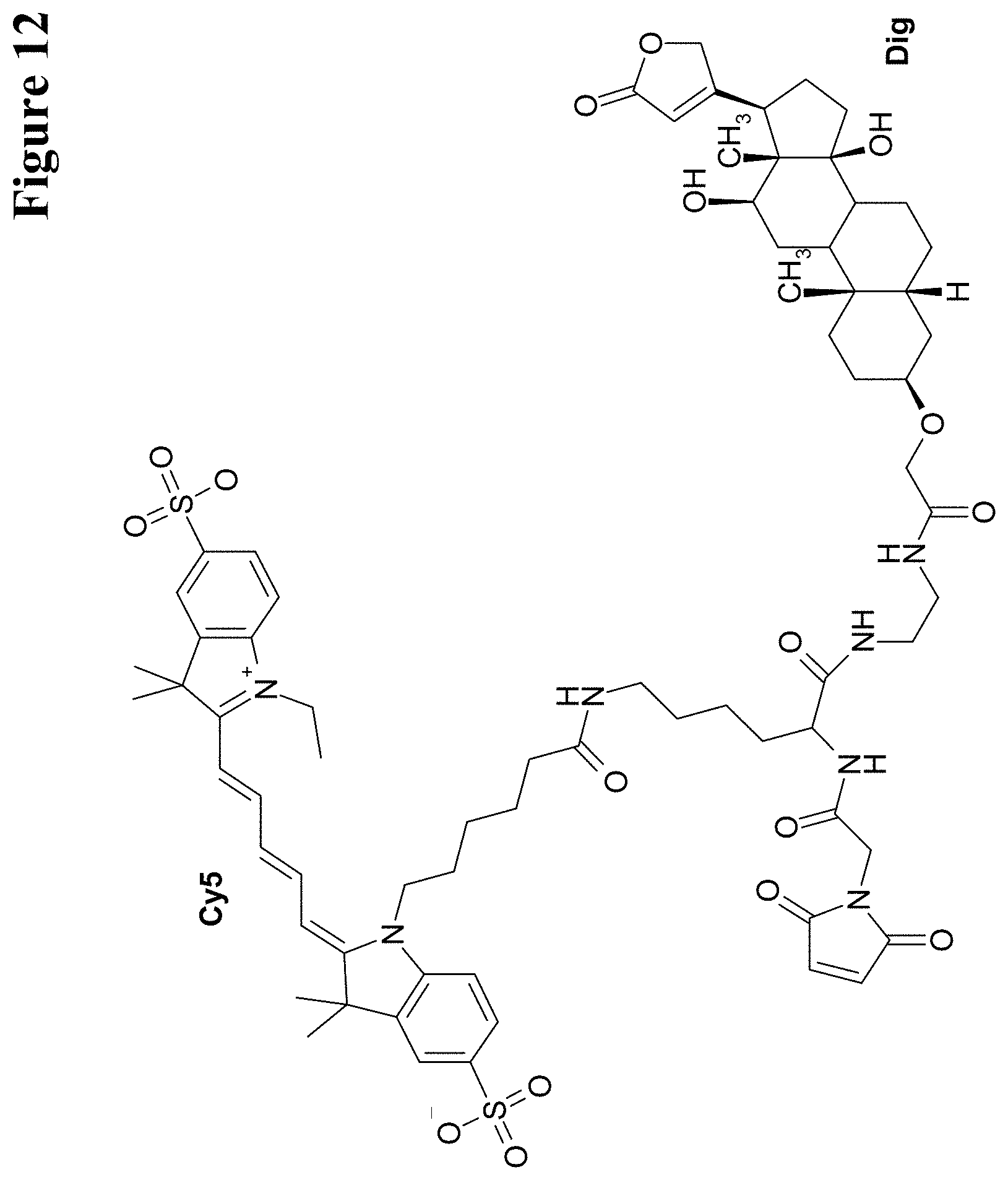
D00014
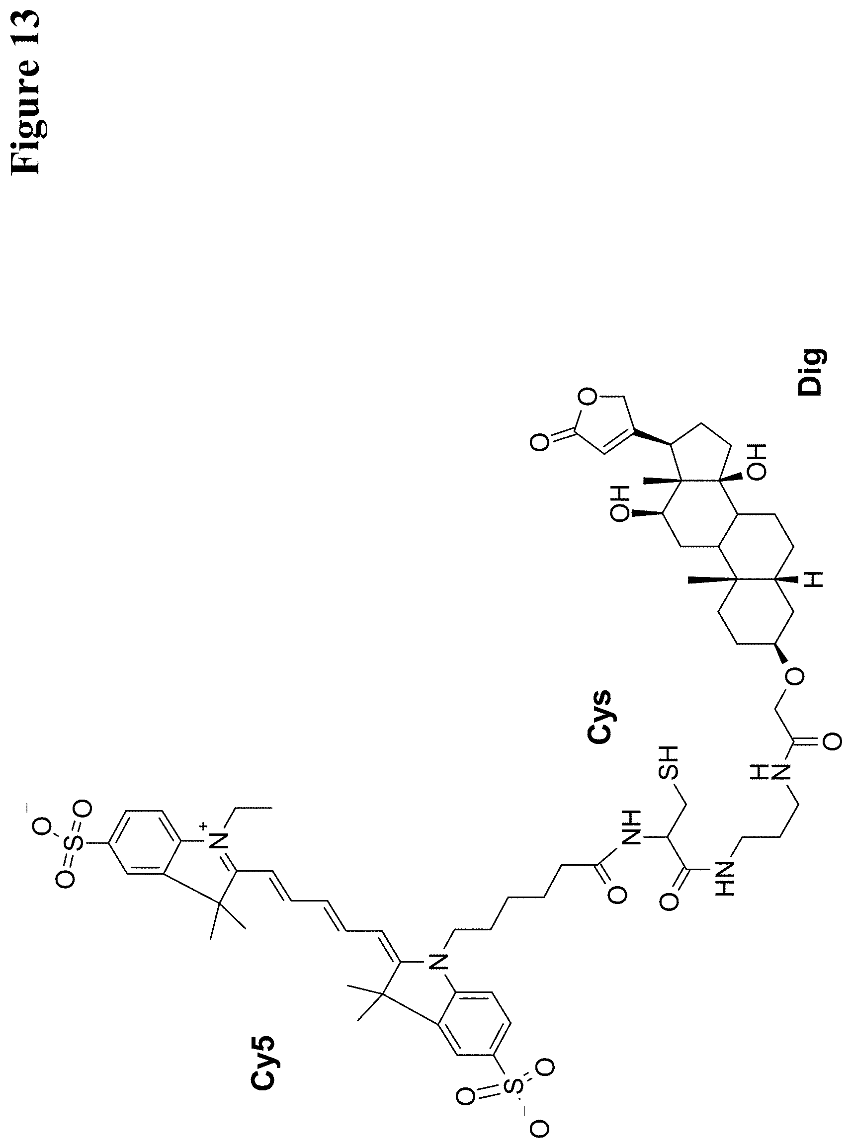
D00015
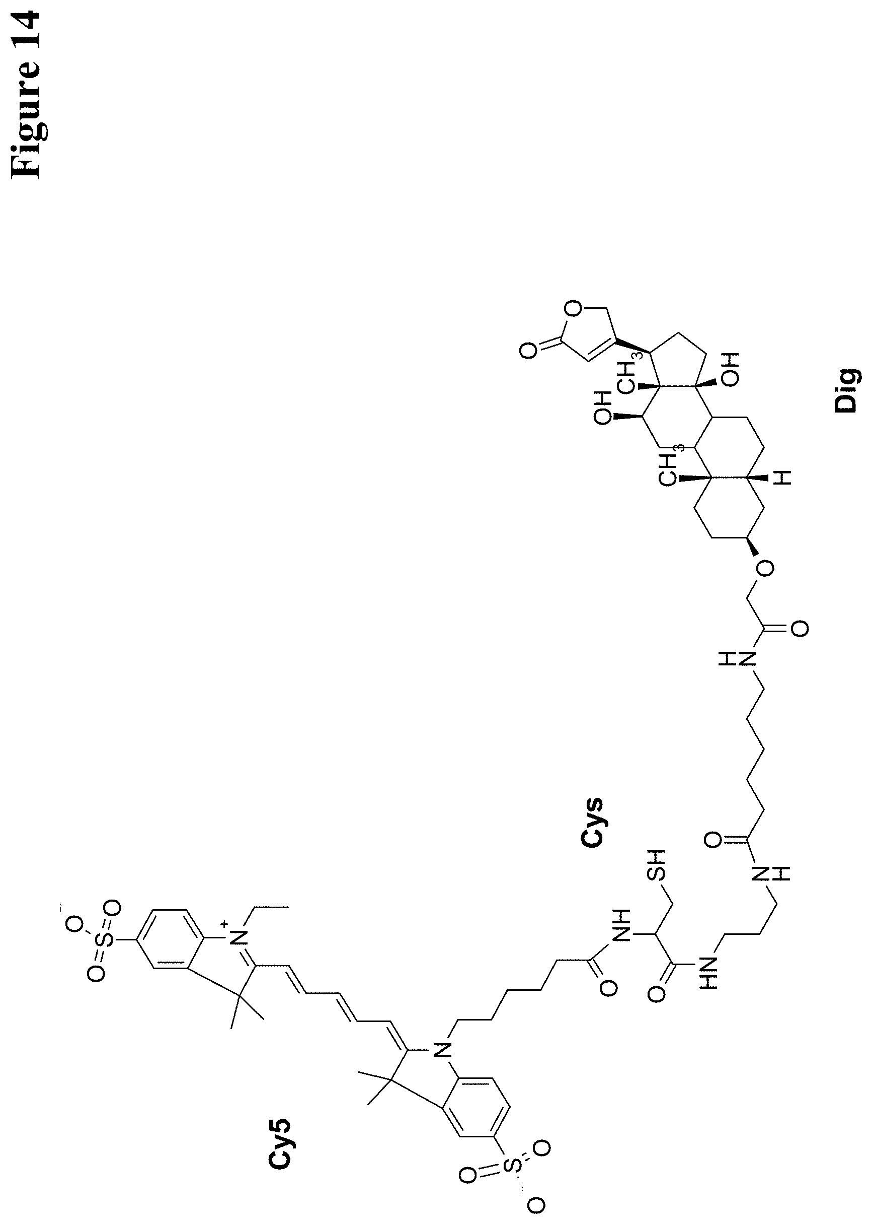
D00016
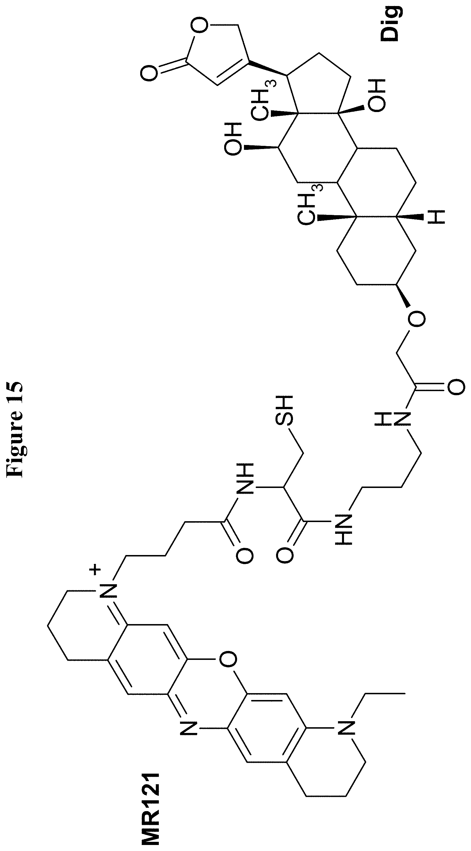
D00017
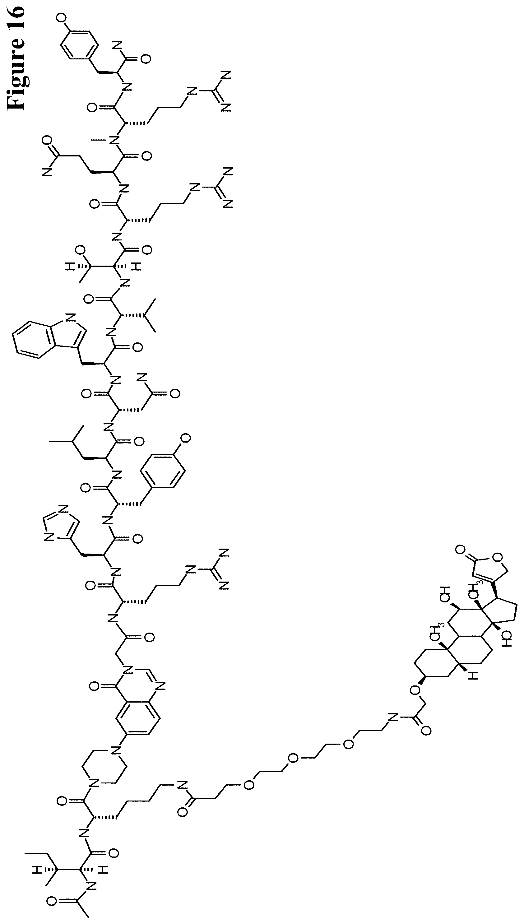
D00018
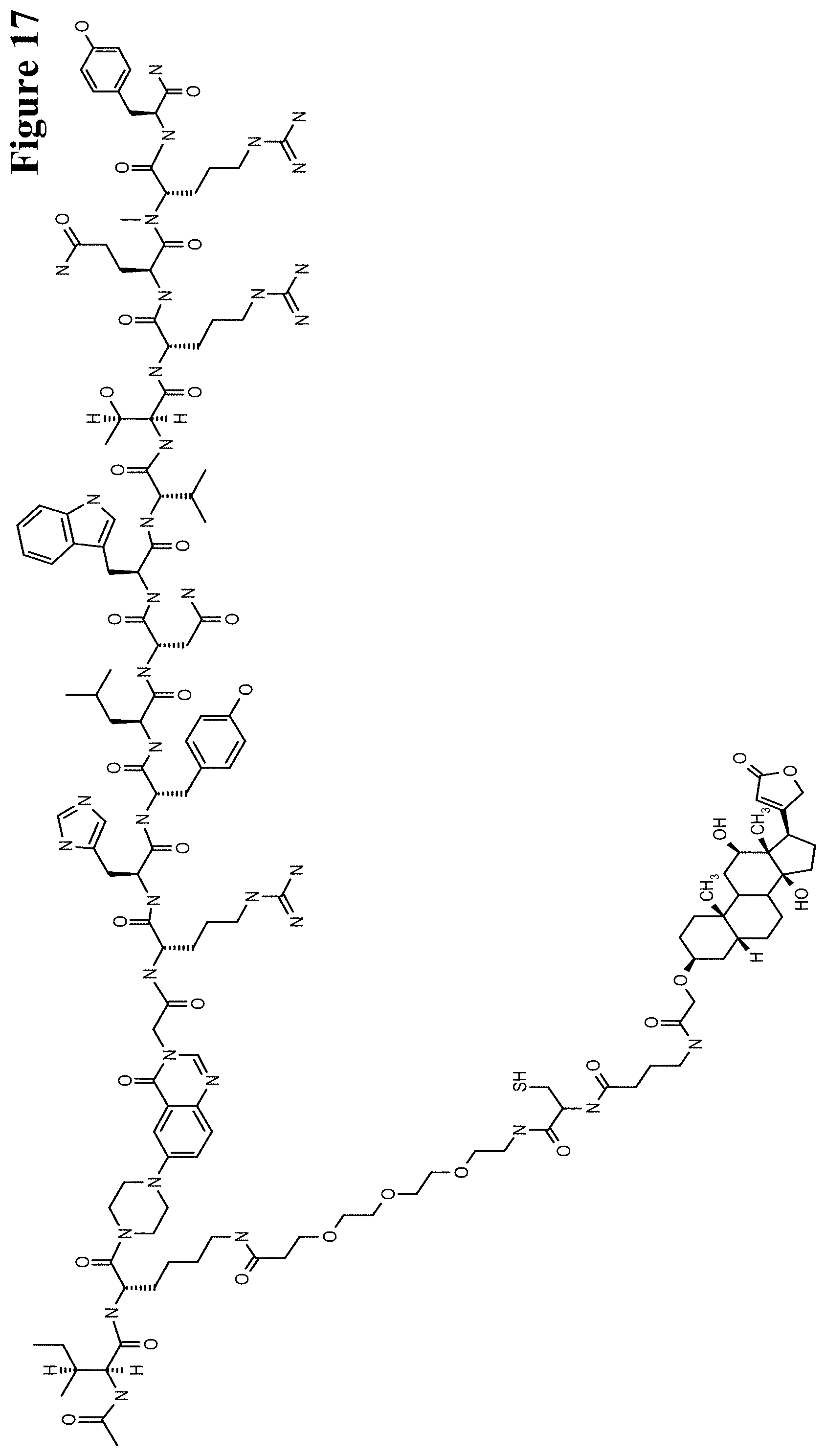
D00019
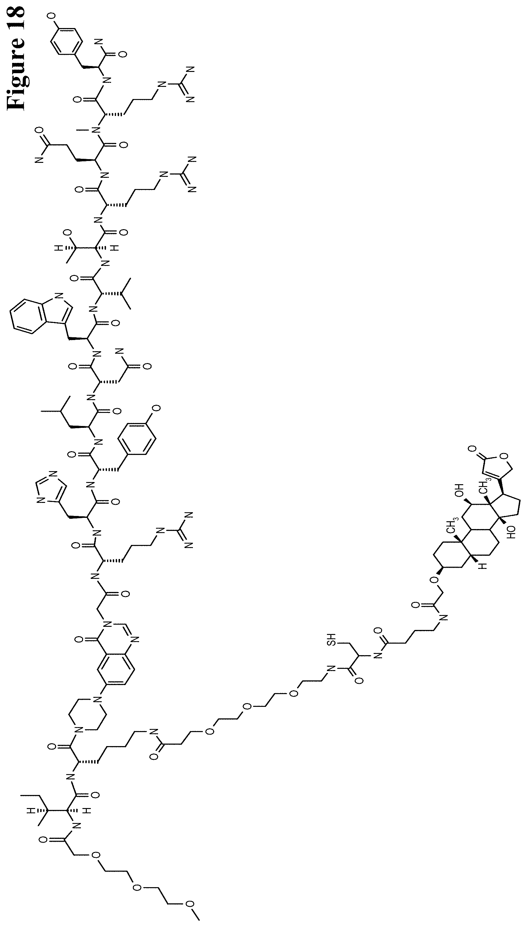
D00020
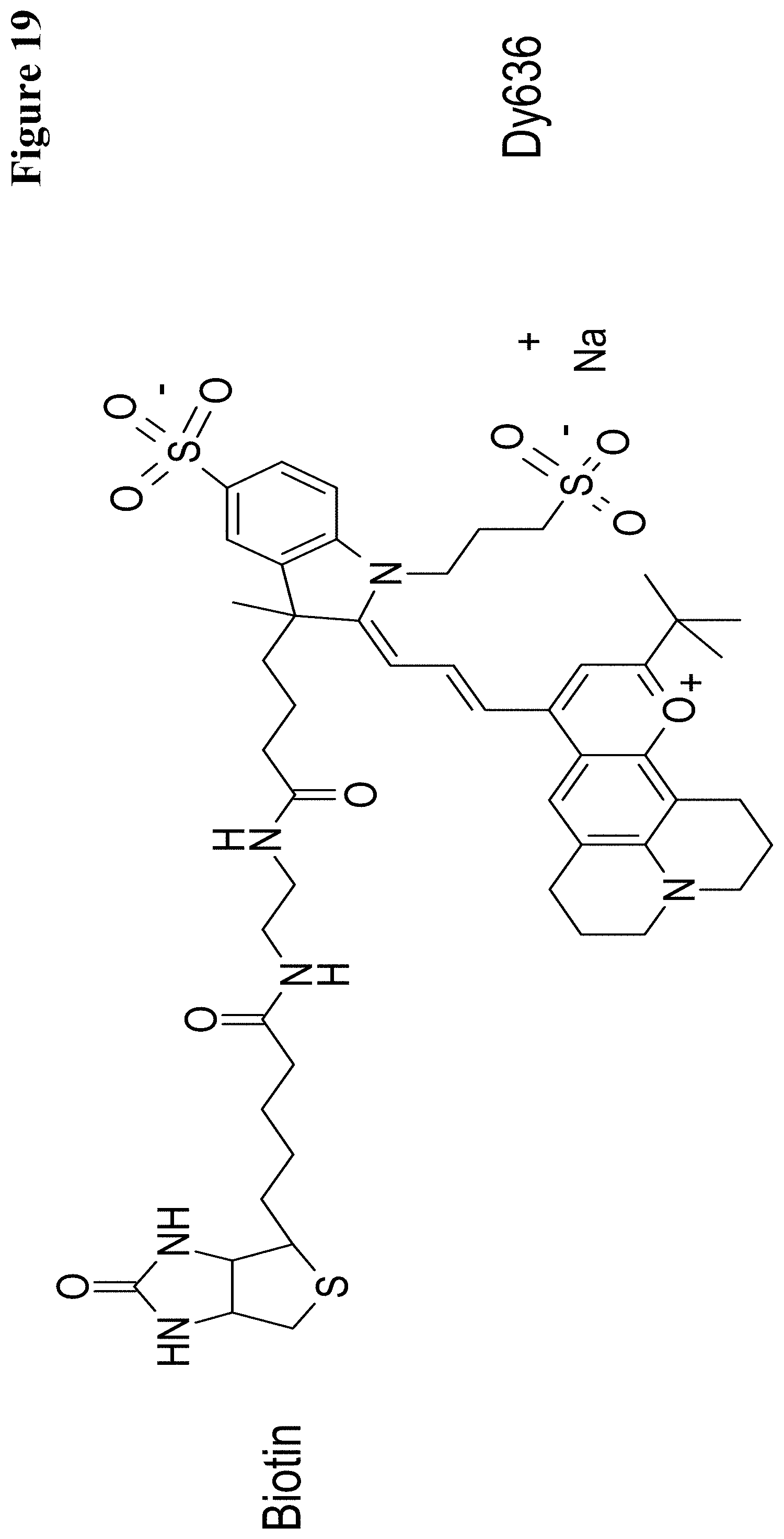
D00021

D00022
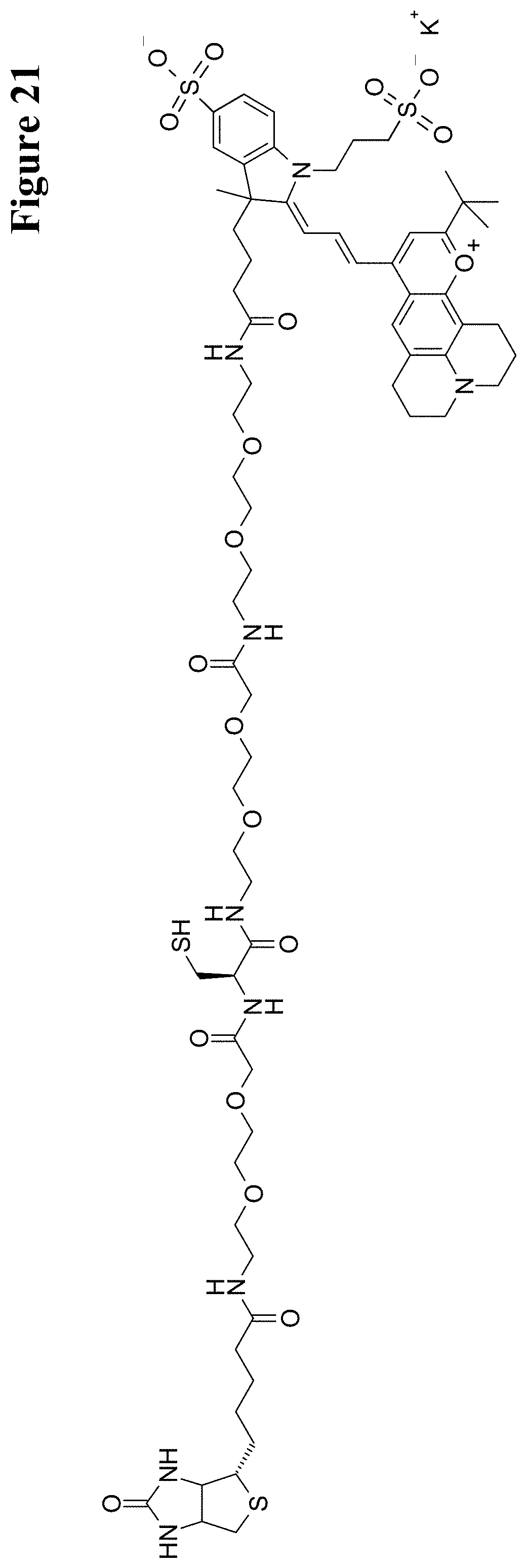
D00023
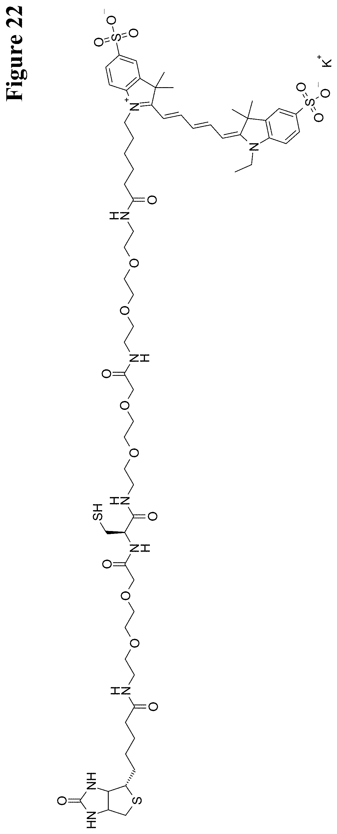
D00024
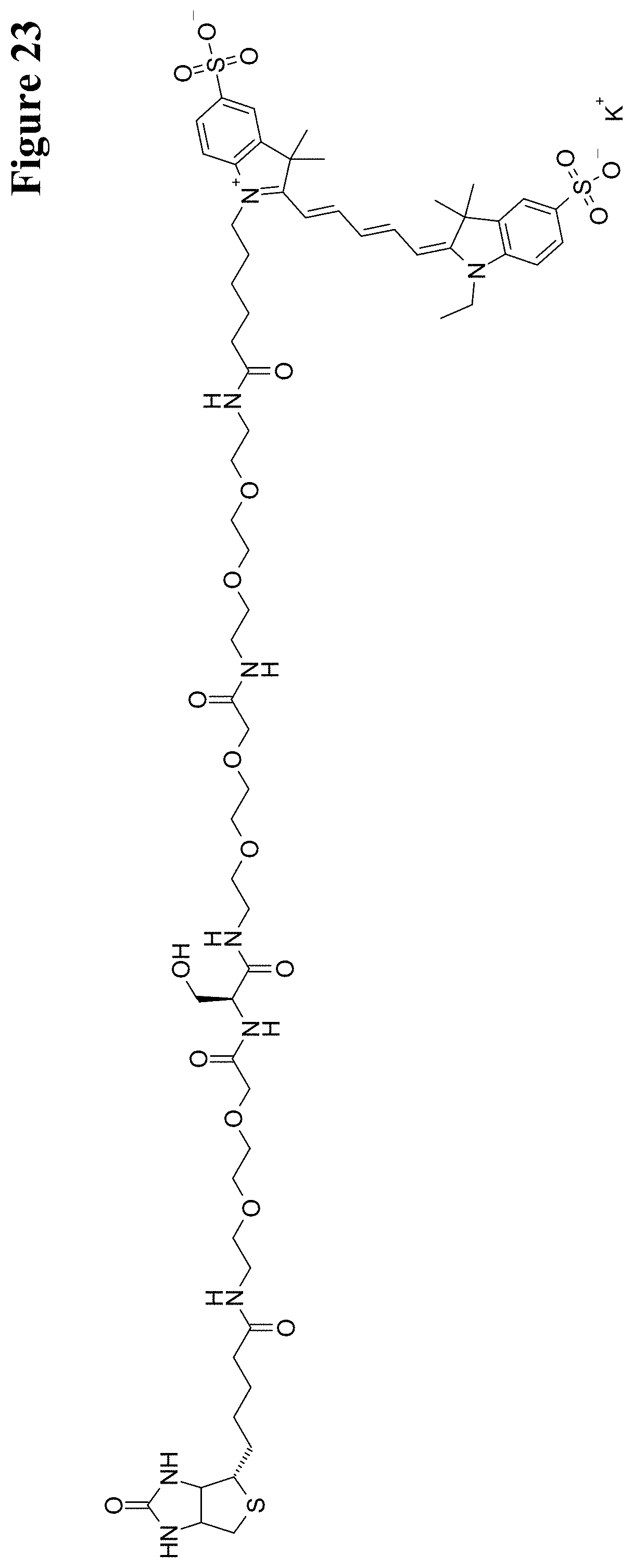
D00025
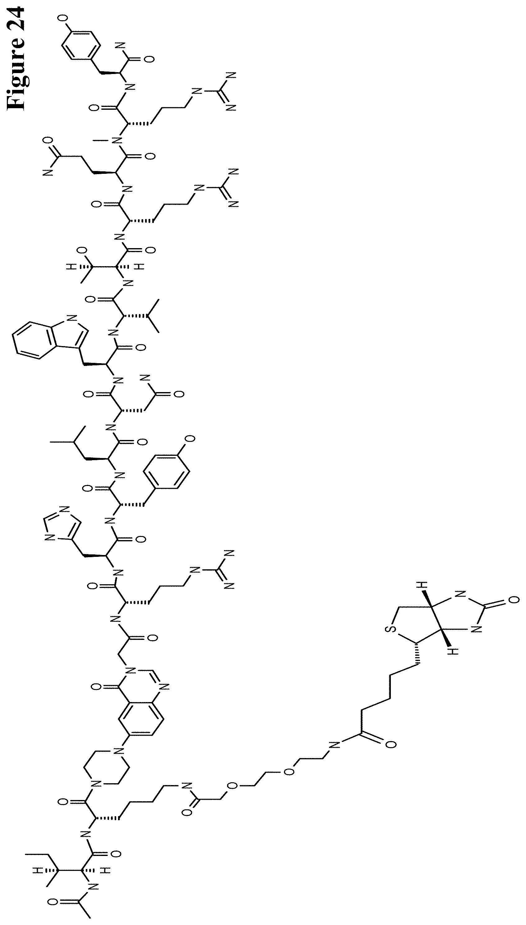
D00026
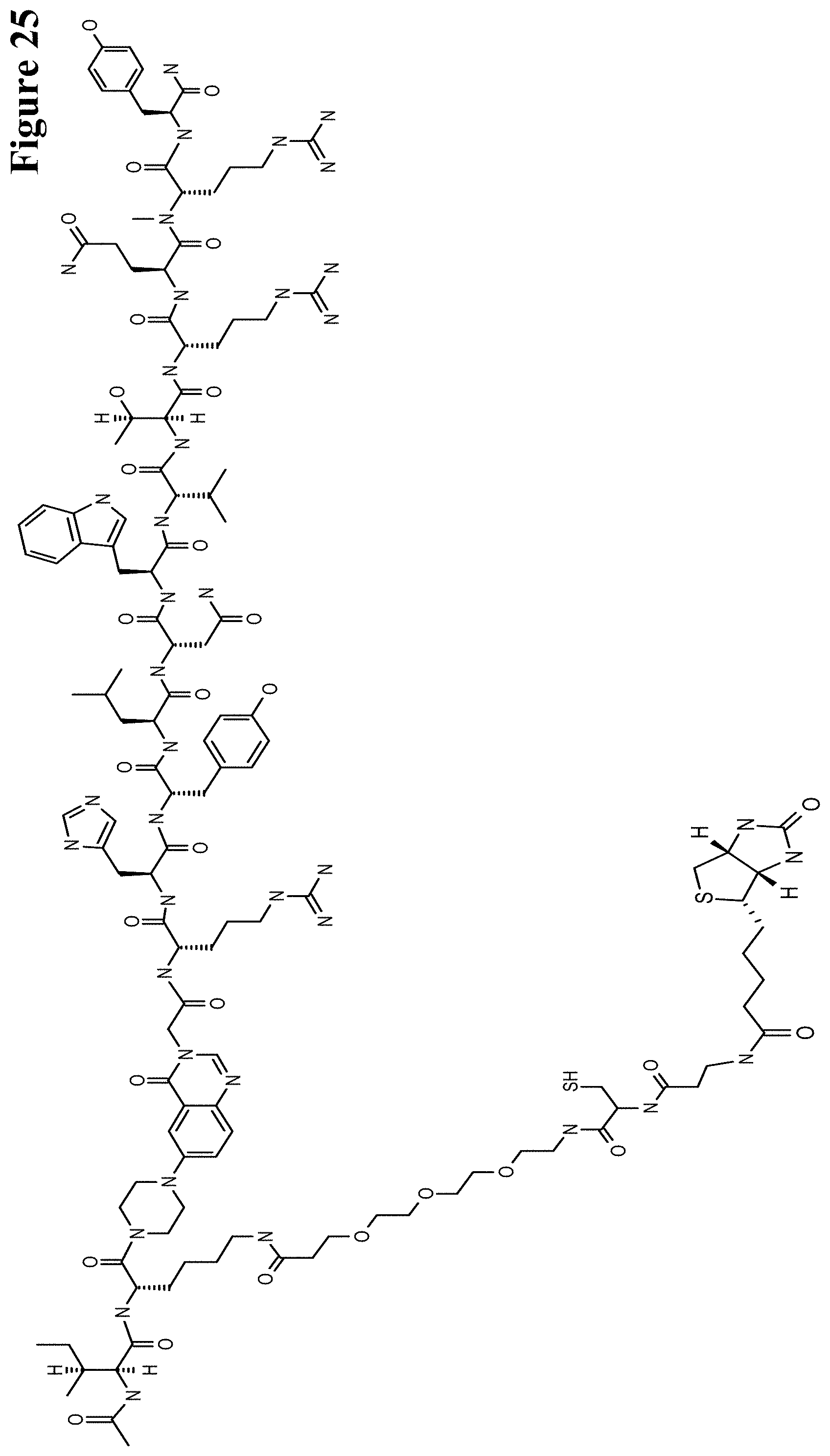
D00027
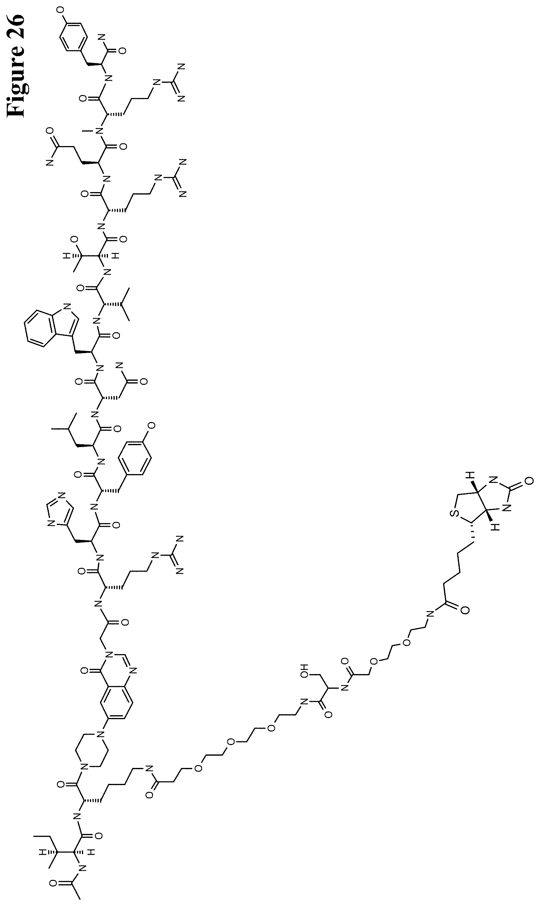
D00028
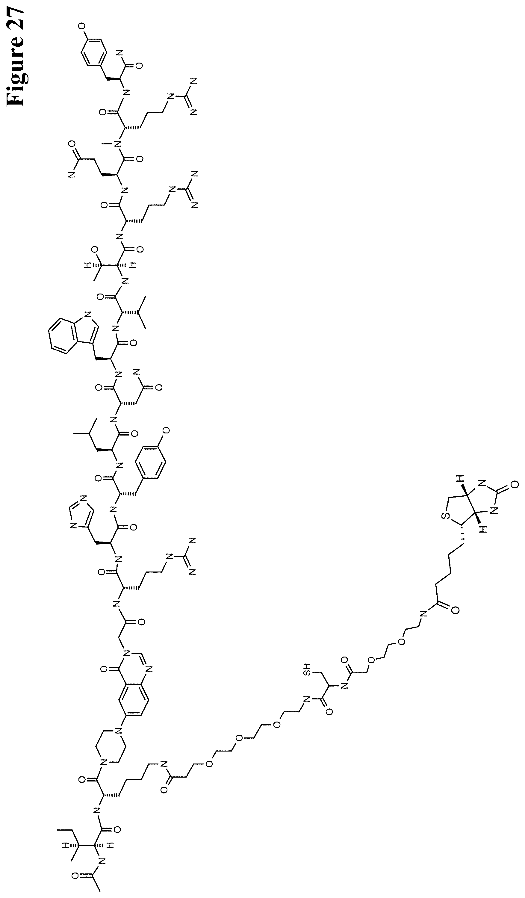
D00029
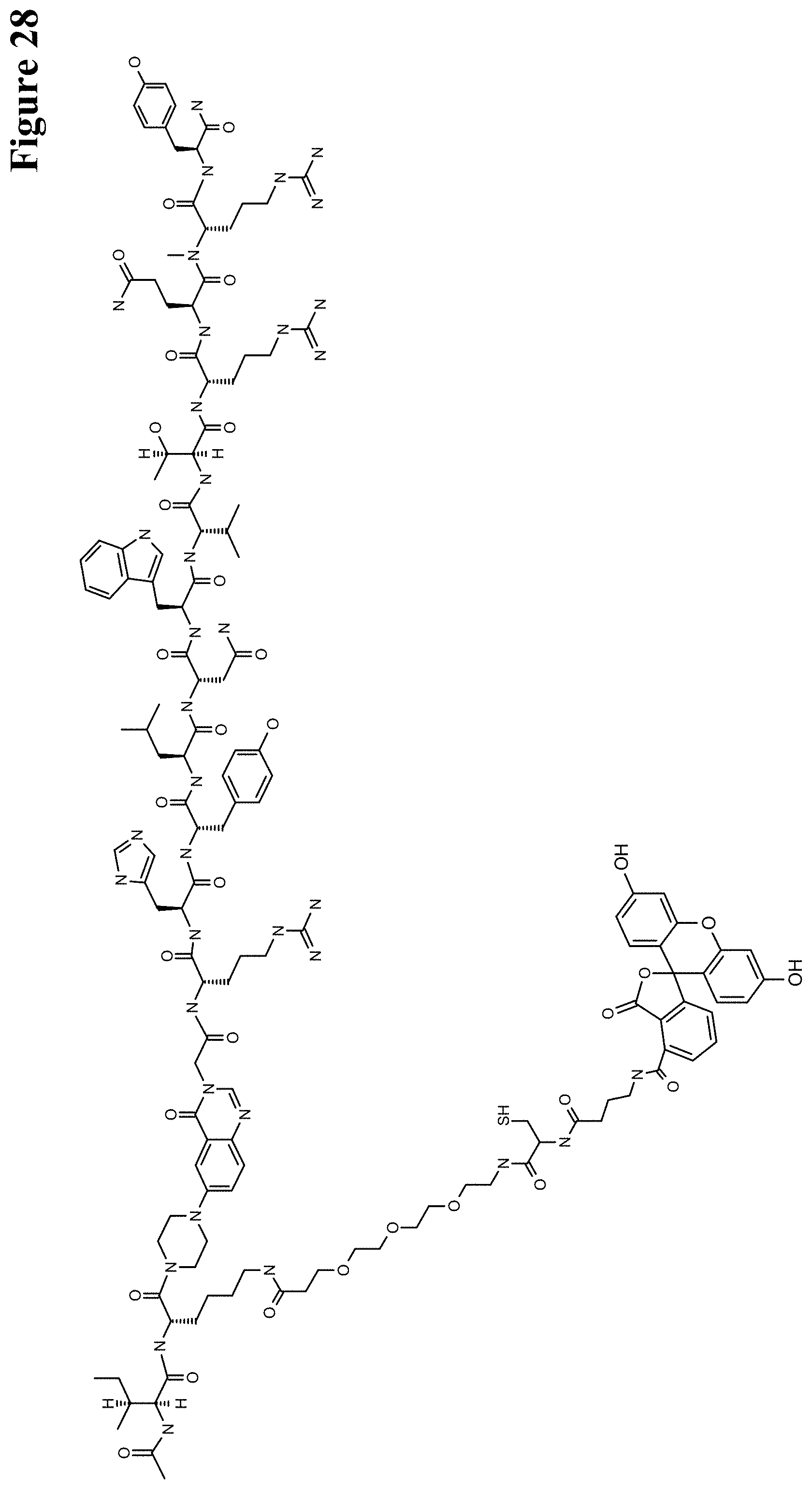
D00030
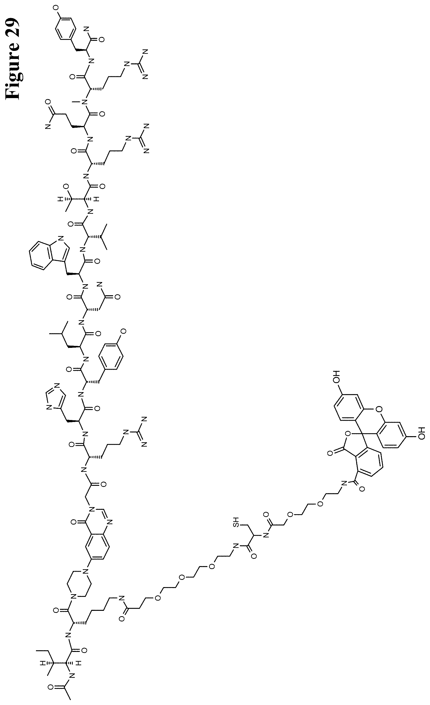
D00031
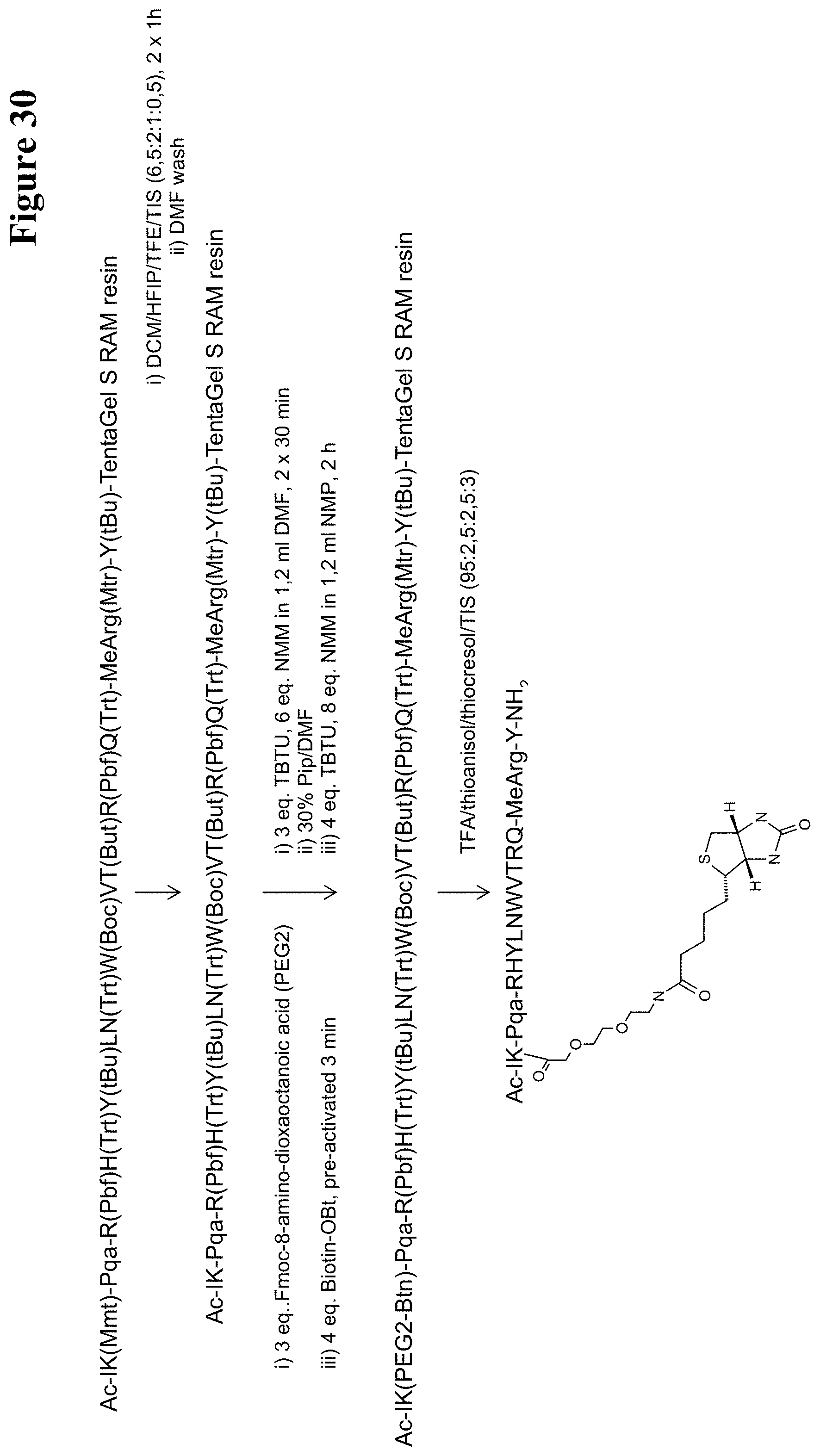
D00032

D00033
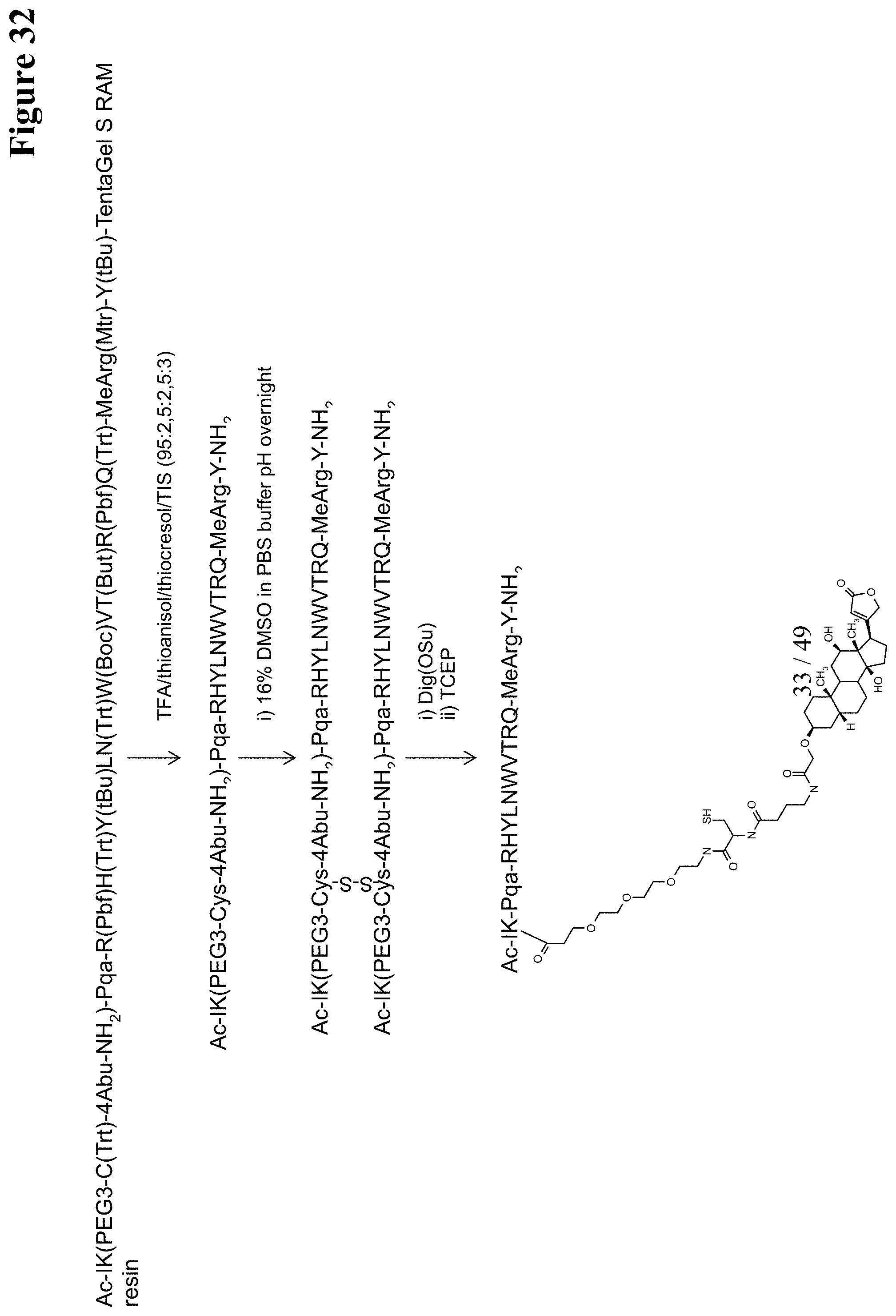
D00034
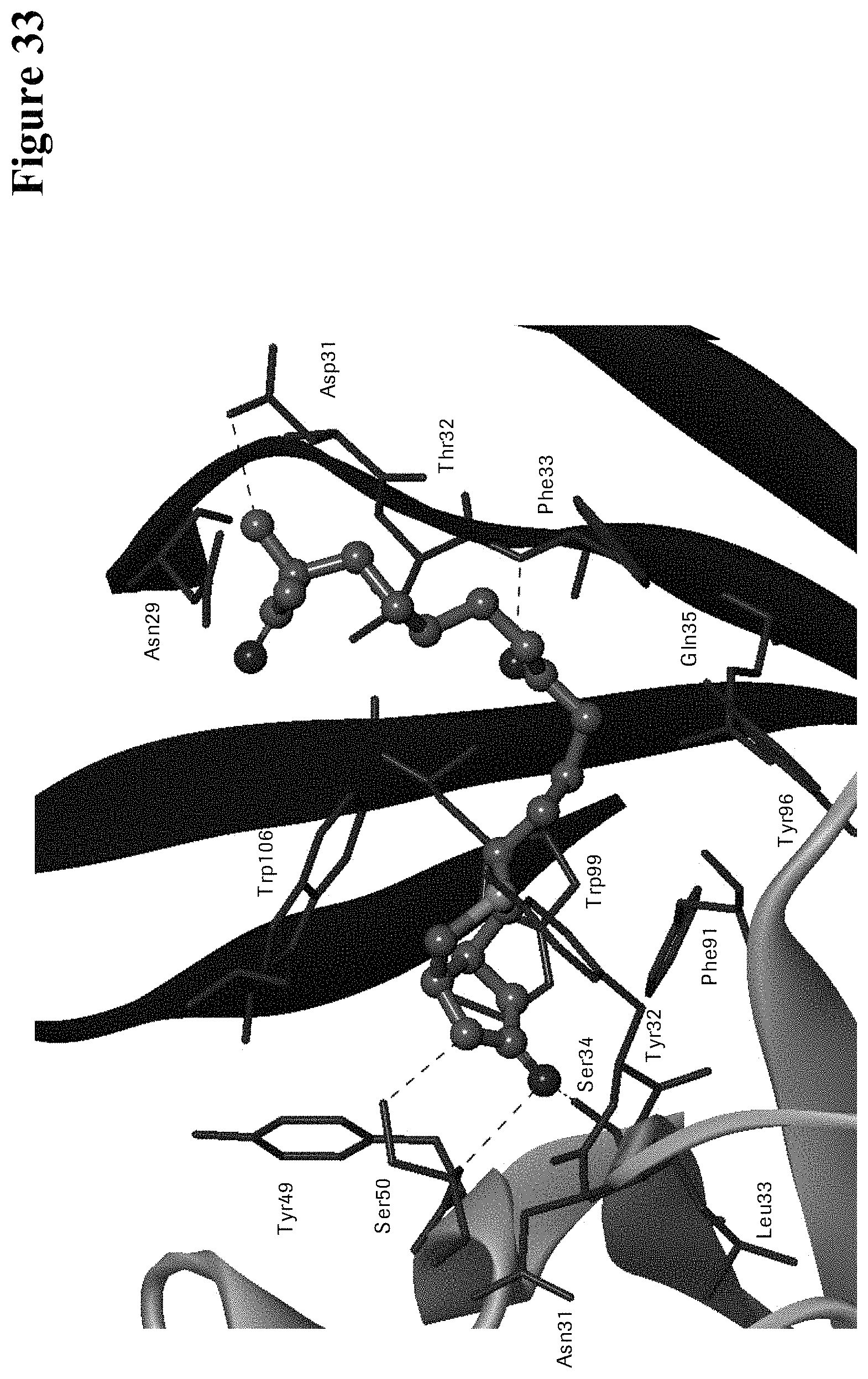
D00035
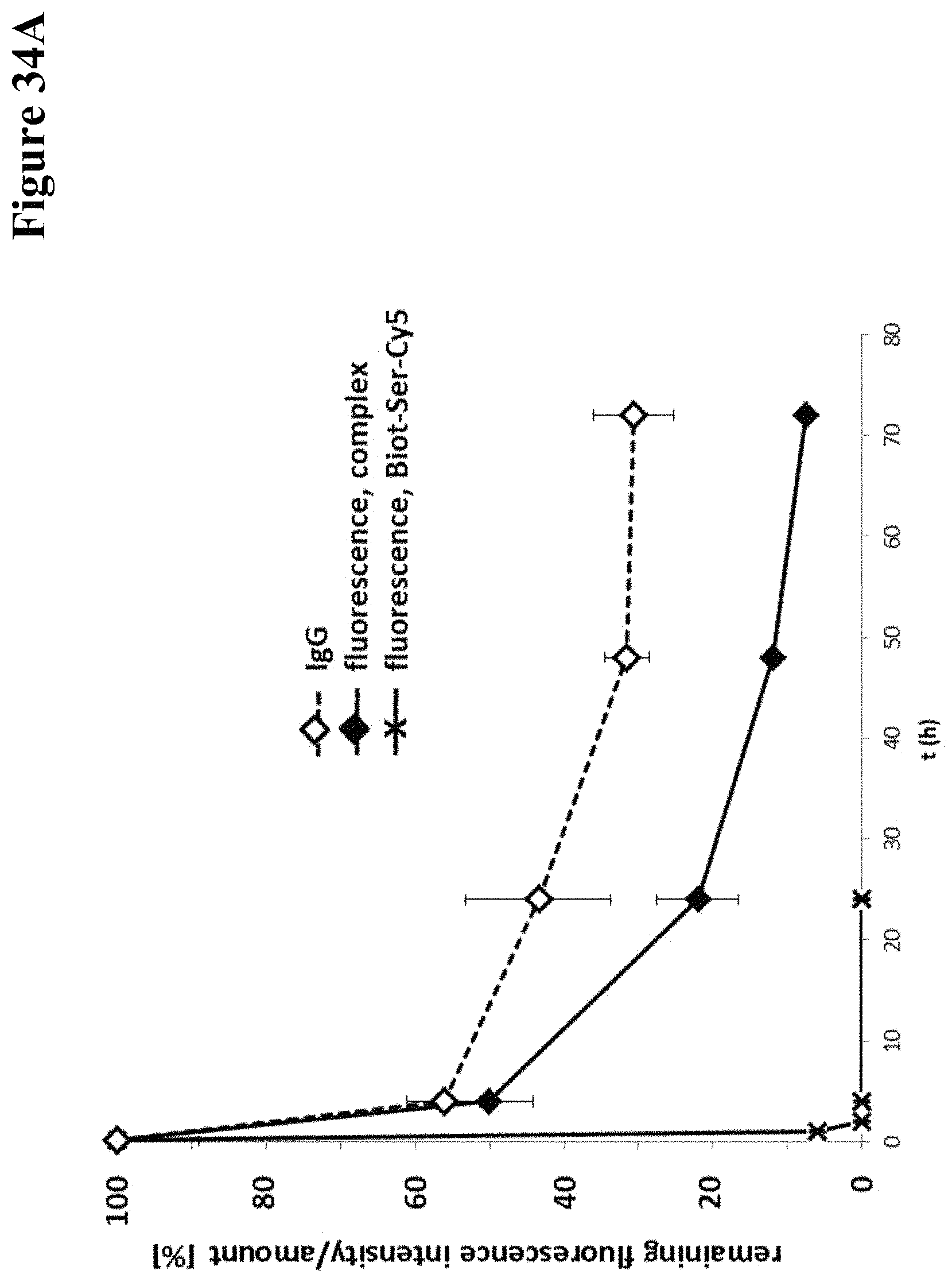
D00036
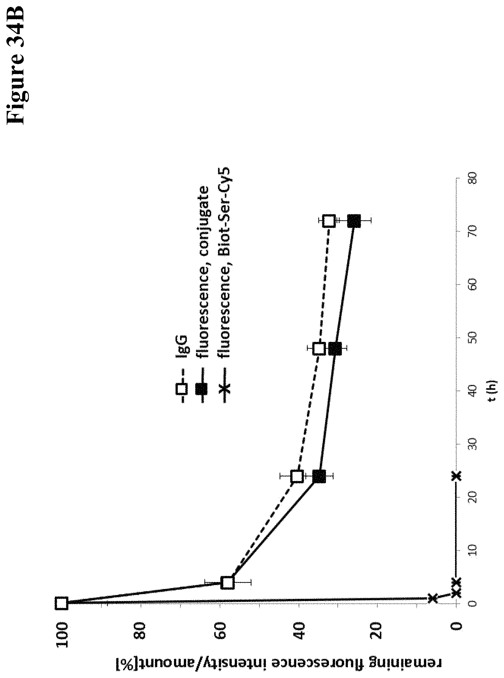
D00037

D00038
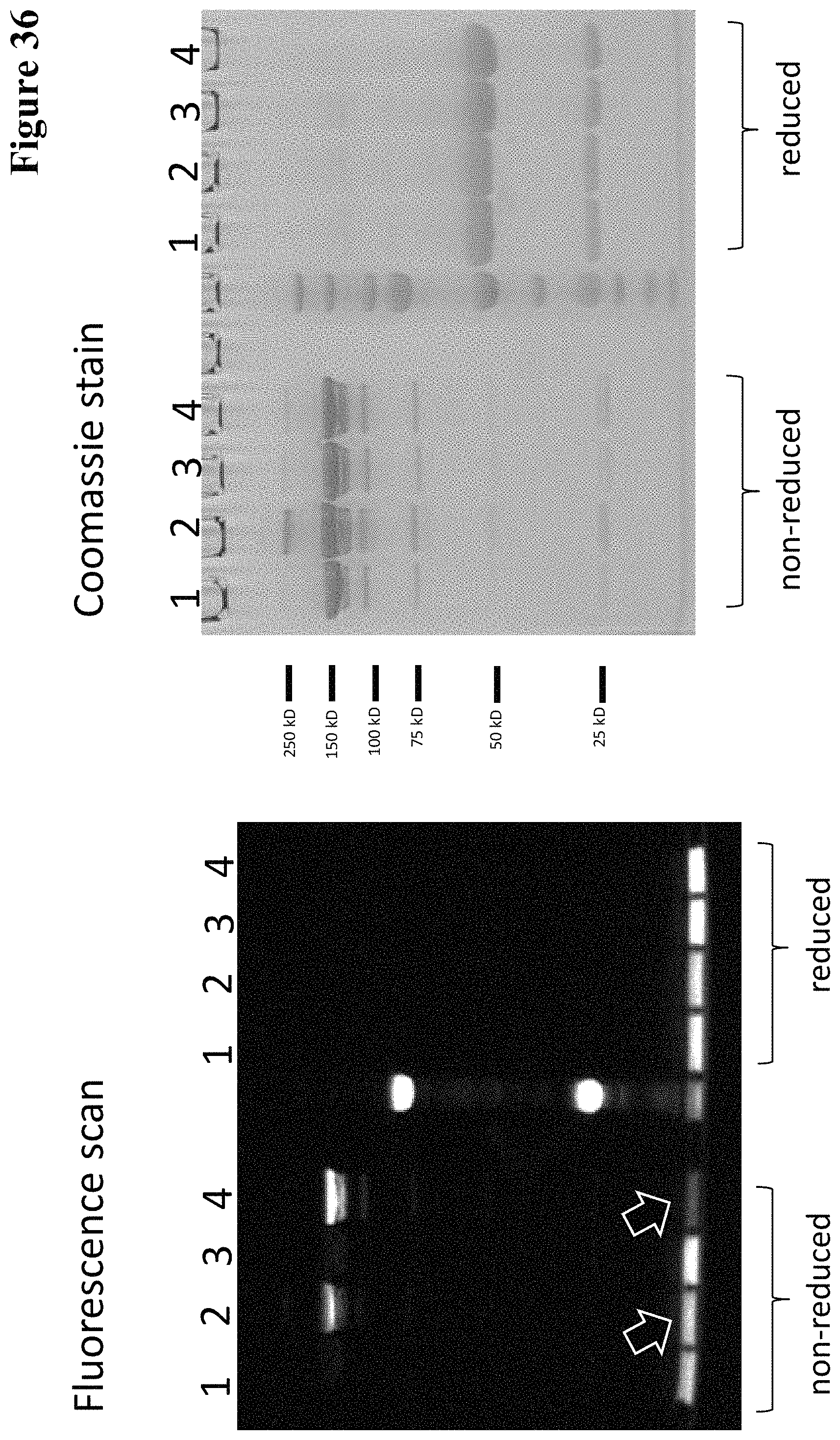
D00039
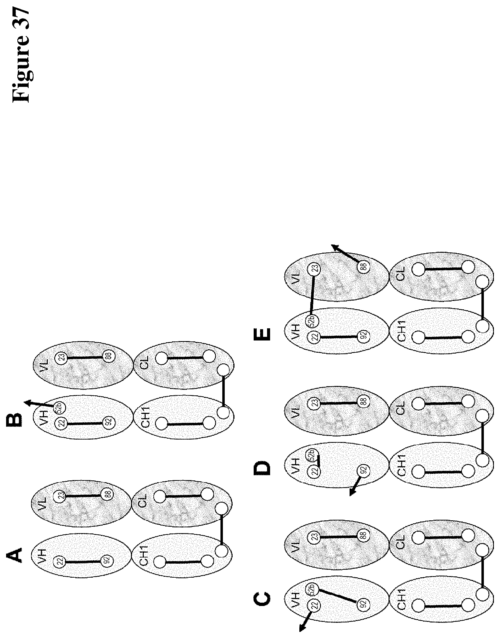
D00040
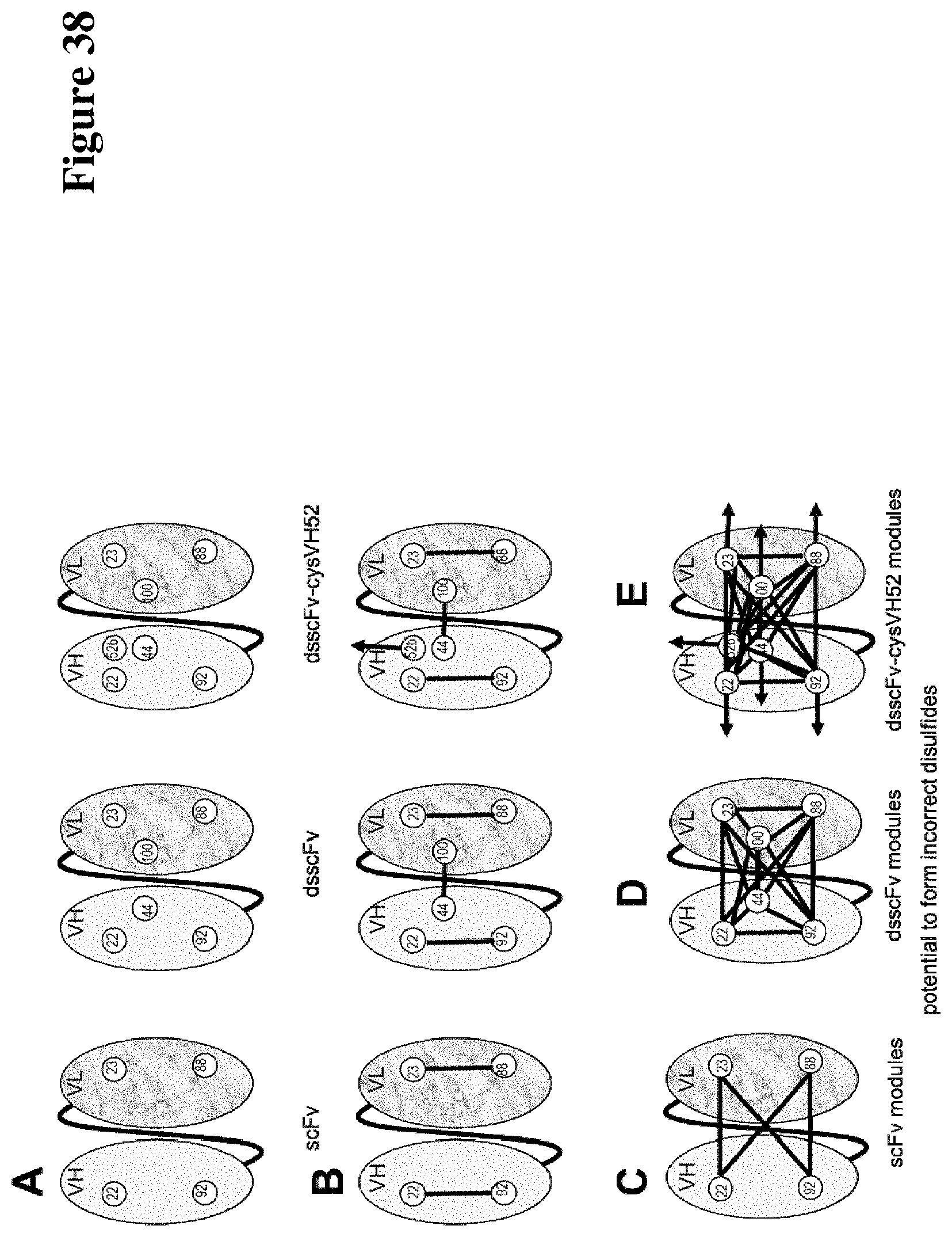
D00041
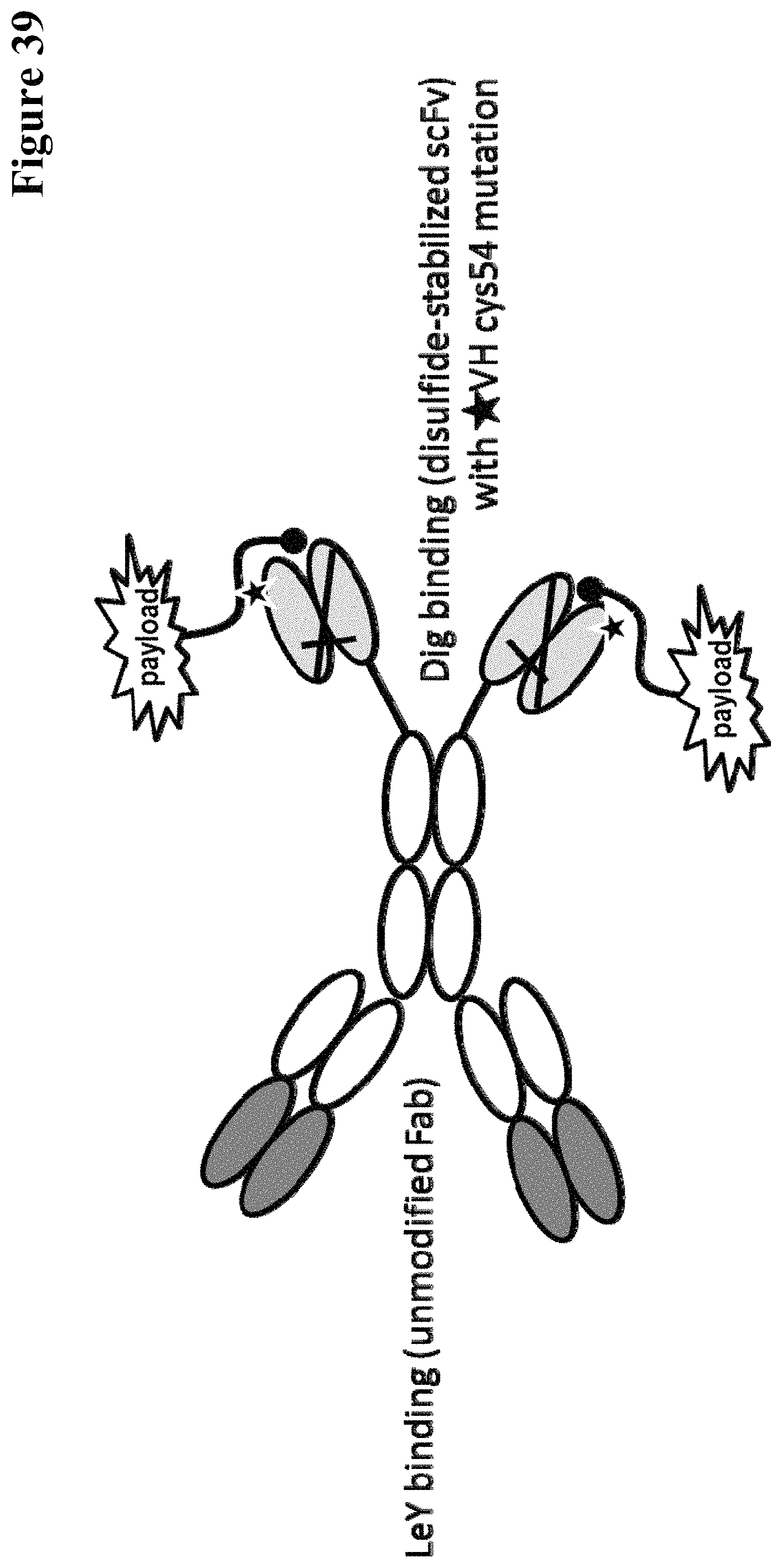
D00042
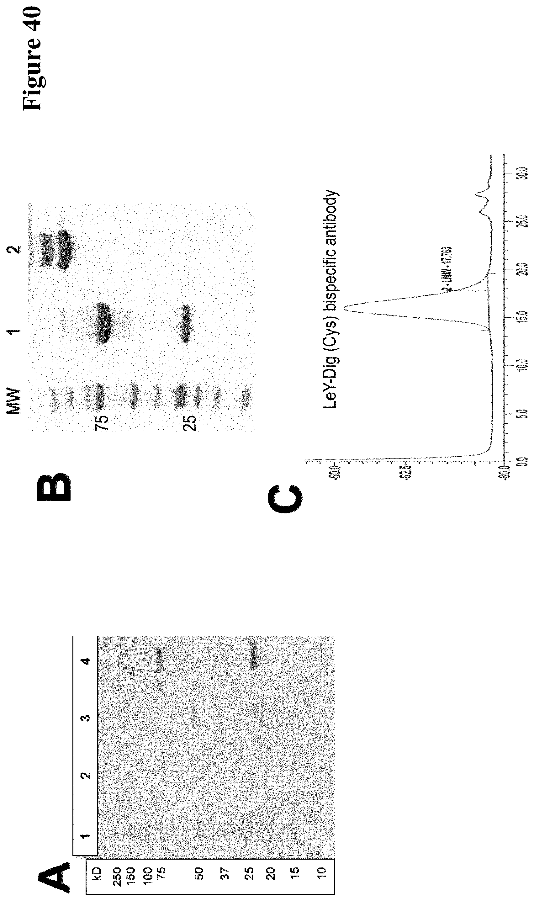
D00043
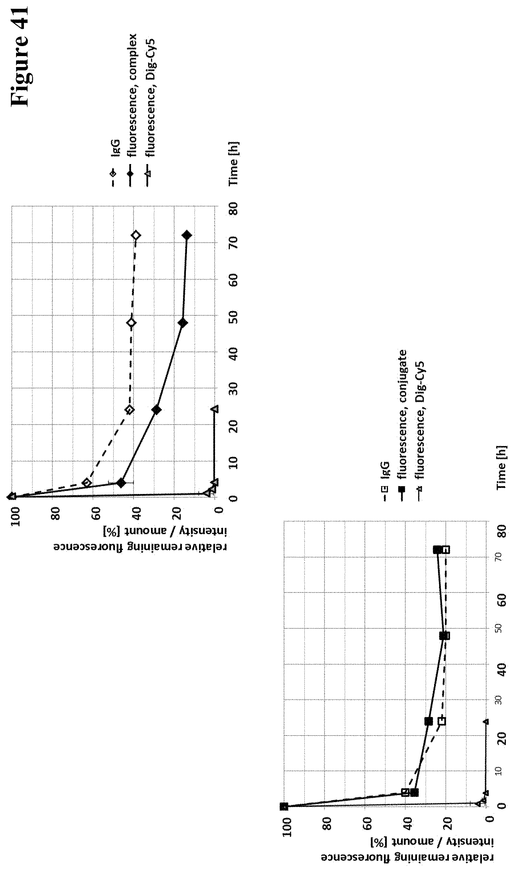
D00044

D00045
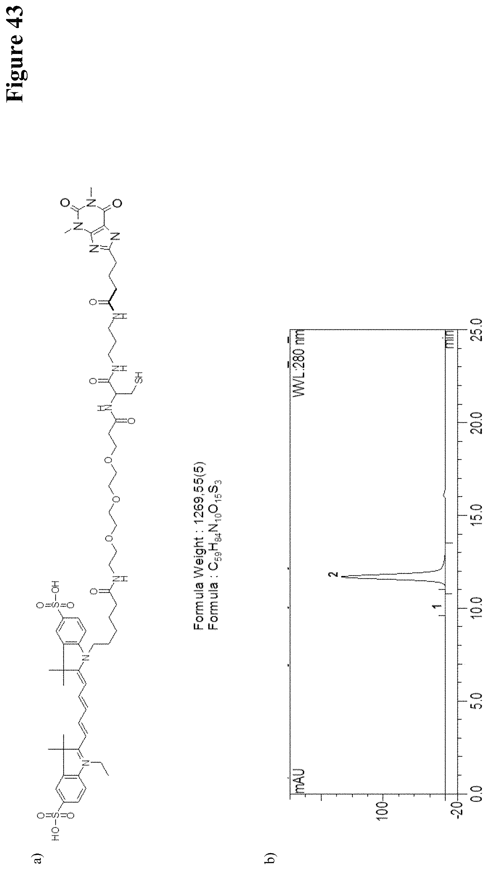
D00046
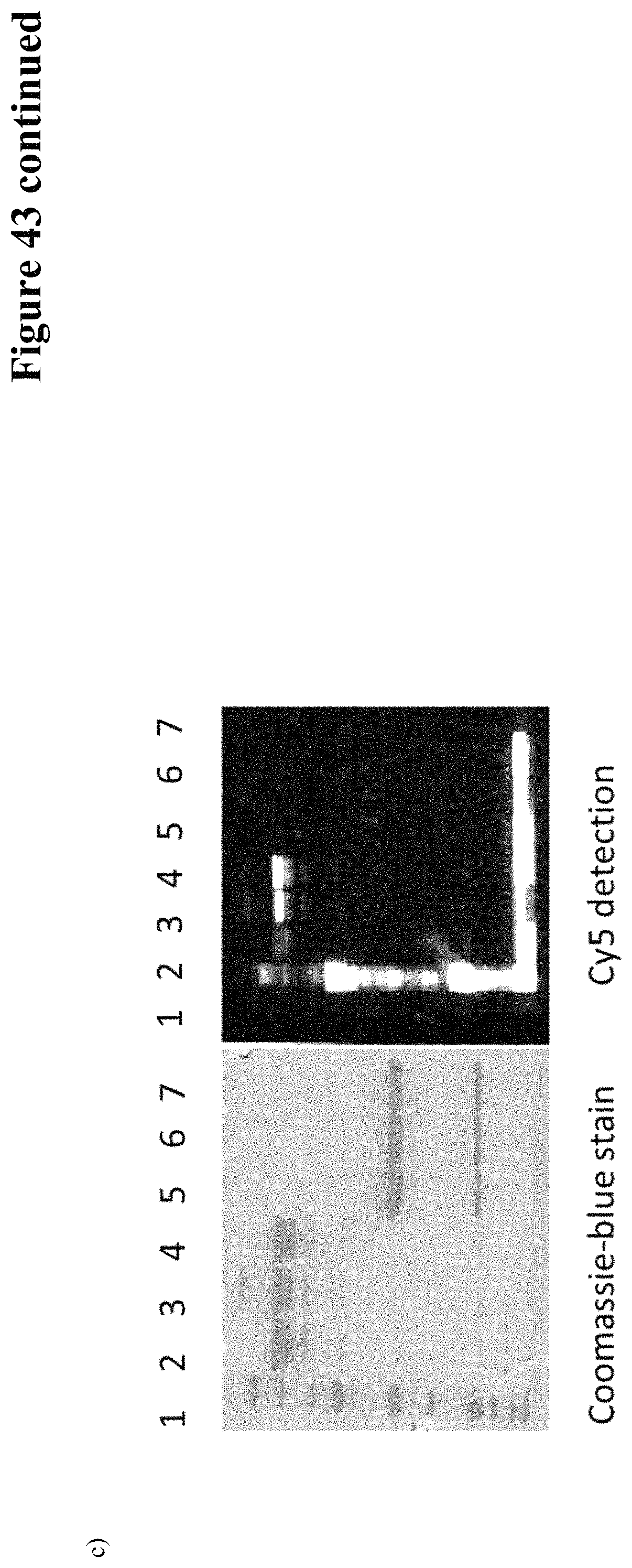
D00047
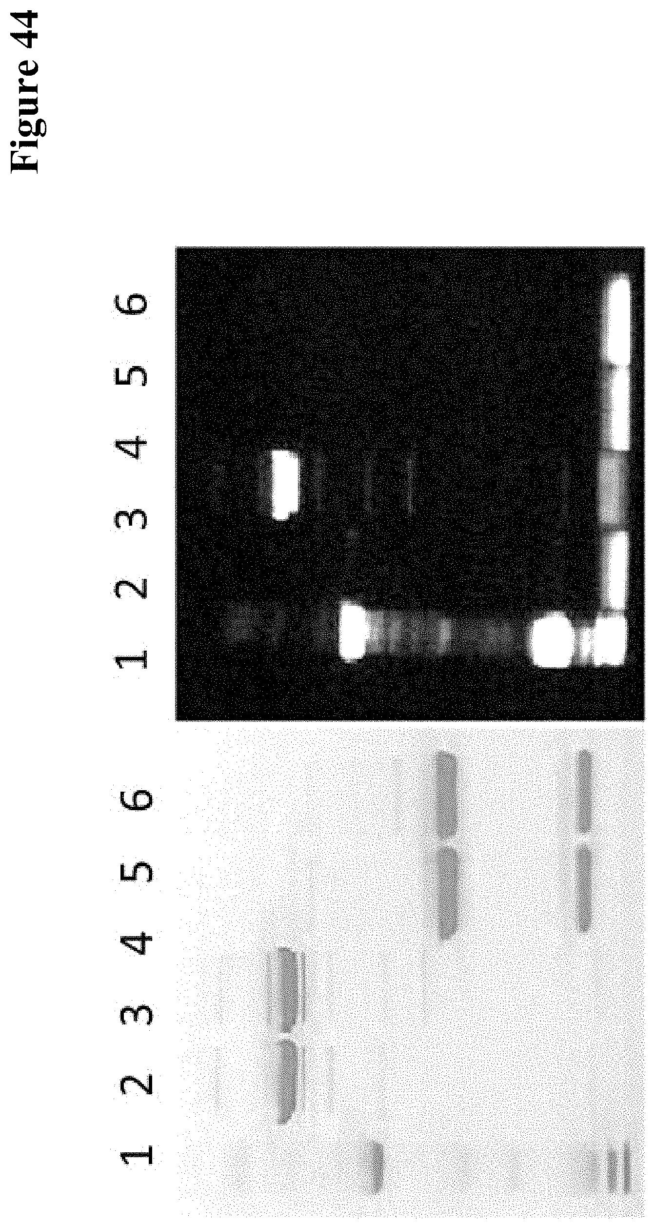
D00048

D00049
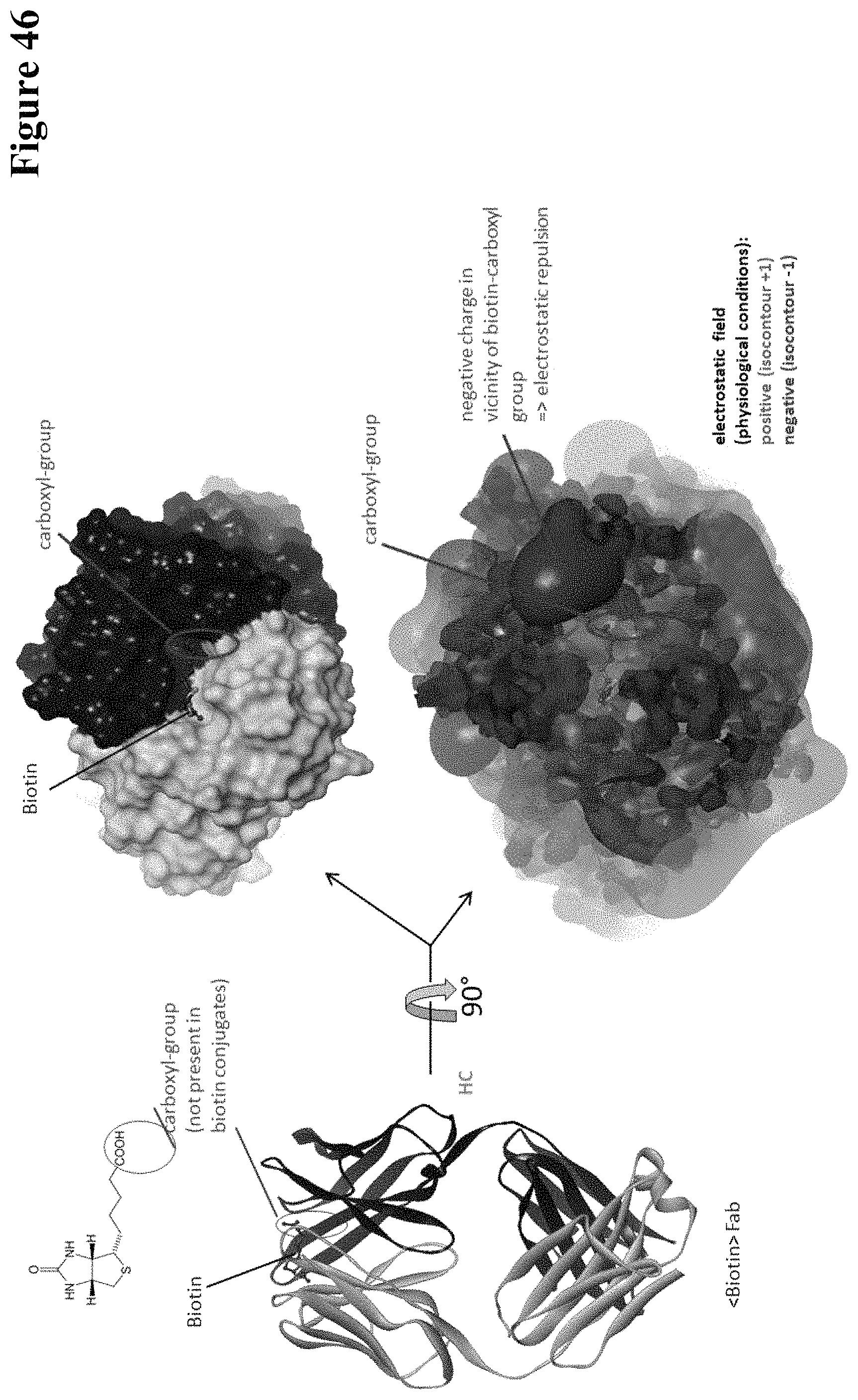
S00001
XML
uspto.report is an independent third-party trademark research tool that is not affiliated, endorsed, or sponsored by the United States Patent and Trademark Office (USPTO) or any other governmental organization. The information provided by uspto.report is based on publicly available data at the time of writing and is intended for informational purposes only.
While we strive to provide accurate and up-to-date information, we do not guarantee the accuracy, completeness, reliability, or suitability of the information displayed on this site. The use of this site is at your own risk. Any reliance you place on such information is therefore strictly at your own risk.
All official trademark data, including owner information, should be verified by visiting the official USPTO website at www.uspto.gov. This site is not intended to replace professional legal advice and should not be used as a substitute for consulting with a legal professional who is knowledgeable about trademark law.