SMC combination therapy for the treatment of cancer
Korneluk , et al. Oc
U.S. patent number 10,441,654 [Application Number 15/113,634] was granted by the patent office on 2019-10-15 for smc combination therapy for the treatment of cancer. This patent grant is currently assigned to Children's Hospital of Eastern Ontario Research Institute Inc.. The grantee listed for this patent is Children's Hospital of Eastern Ontario Research Institute Inc.. Invention is credited to Shawn T. Beug, Robert G. Korneluk, Eric C. Lacasse, Vera A. Tang.


View All Diagrams
| United States Patent | 10,441,654 |
| Korneluk , et al. | October 15, 2019 |
SMC combination therapy for the treatment of cancer
Abstract
The present invention includes methods and compositions for enhancing the efficacy of SMCs in the treatment of cancer. In particular, the present invention includes methods and compositions for combination therapies that include an SMC and at least a second agent that stimulates one or more apoptotic or immune pathways. The second agent may be, e.g., an immunostimulatory compound or oncolytic virus.
| Inventors: | Korneluk; Robert G. (Ottawa, CA), Lacasse; Eric C. (Ottawa, CA), Beug; Shawn T. (Ottawa, CA), Tang; Vera A. (Ottawa, CA) | ||||||||||
|---|---|---|---|---|---|---|---|---|---|---|---|
| Applicant: |
|
||||||||||
| Assignee: | Children's Hospital of Eastern
Ontario Research Institute Inc. (Ottawa, ON,
CA) |
||||||||||
| Family ID: | 53680533 | ||||||||||
| Appl. No.: | 15/113,634 | ||||||||||
| Filed: | January 26, 2015 | ||||||||||
| PCT Filed: | January 26, 2015 | ||||||||||
| PCT No.: | PCT/CA2015/000043 | ||||||||||
| 371(c)(1),(2),(4) Date: | July 22, 2016 | ||||||||||
| PCT Pub. No.: | WO2015/109391 | ||||||||||
| PCT Pub. Date: | July 30, 2015 |
Prior Publication Data
| Document Identifier | Publication Date | |
|---|---|---|
| US 20170239347 A1 | Aug 24, 2017 | |
Related U.S. Patent Documents
| Application Number | Filing Date | Patent Number | Issue Date | ||
|---|---|---|---|---|---|
| 61931321 | Jan 24, 2014 | ||||
| Current U.S. Class: | 1/1 |
| Current CPC Class: | A61K 31/407 (20130101); A61K 35/761 (20130101); A61P 35/00 (20180101); A61P 37/04 (20180101); A61K 31/433 (20130101); A61K 35/765 (20130101); A61K 31/55 (20130101); A61K 39/205 (20130101); A61K 45/06 (20130101); A61K 31/427 (20130101); A61K 38/21 (20130101); A61K 31/409 (20130101); A61K 39/0011 (20130101); A61K 31/4745 (20130101); A61K 31/404 (20130101); A61K 35/766 (20130101); C12N 7/00 (20130101); A61K 38/212 (20130101); A61K 39/39 (20130101); A61K 31/404 (20130101); A61K 2300/00 (20130101); A61K 31/4745 (20130101); A61K 2300/00 (20130101); A61K 35/761 (20130101); A61K 2300/00 (20130101); A61K 35/765 (20130101); A61K 2300/00 (20130101); A61K 35/766 (20130101); A61K 2300/00 (20130101); A61K 38/21 (20130101); A61K 2300/00 (20130101); A61K 2039/55511 (20130101); A61K 2039/585 (20130101); A61K 2039/5252 (20130101); A61K 2039/55588 (20130101); A61K 2039/55594 (20130101); A61K 2039/55561 (20130101); C12N 2760/20134 (20130101); A61K 2039/572 (20130101) |
| Current International Class: | A61K 31/427 (20060101); A61K 35/761 (20150101); A61K 38/21 (20060101); A61K 35/765 (20150101); A61K 31/4745 (20060101); A61K 31/404 (20060101); A61K 31/407 (20060101); A61K 31/409 (20060101); A61K 31/433 (20060101); A61K 31/55 (20060101); A61K 39/00 (20060101); A61K 39/205 (20060101); A61K 39/12 (20060101); A61K 31/40 (20060101); A61K 39/39 (20060101); A61K 35/766 (20150101); C12N 7/00 (20060101); A61K 45/06 (20060101) |
References Cited [Referenced By]
U.S. Patent Documents
| 2003/0224399 | December 2003 | Reed et al. |
| 2005/0250854 | November 2005 | Li et al. |
| 2006/0058229 | March 2006 | Steller et al. |
| 2006/0258581 | November 2006 | Reed et al. |
| 2007/0042428 | February 2007 | Springs et al. |
| 2007/0203749 | August 2007 | Chunduru et al. |
| 2008/0241155 | October 2008 | Ni et al. |
| 2008/0248046 | October 2008 | Ni et al. |
| 2009/0226429 | September 2009 | Salcedo et al. |
| 2010/0016218 | January 2010 | Lichter et al. |
| 2010/0074863 | March 2010 | Or et al. |
| 2010/0135951 | June 2010 | Zhou et al. |
| 2010/0179163 | July 2010 | Kung et al. |
| 2010/0256046 | October 2010 | Springs et al. |
| 2011/0104143 | May 2011 | Buchsbaum et al. |
| 2011/0183358 | July 2011 | Reed et al. |
| 2011/0305777 | December 2011 | Condon et al. |
| 2013/0196927 | August 2013 | Benetatos et al. |
| 2014/0057924 | February 2014 | Wang et al. |
| 2014/0127271 | May 2014 | Sill et al. |
| 2014/0271688 | September 2014 | Abrams et al. |
| 2014/0303090 | October 2014 | Condon et al. |
| 2014/0341920 | November 2014 | Noelle |
| 2015/0010613 | January 2015 | Dubensky, Jr. et al. |
| 2015/0110779 | April 2015 | Shahabi et al. |
| 2015/0119288 | April 2015 | Soper et al. |
| 2015/0125447 | May 2015 | Heider |
| 2015/0141273 | May 2015 | Bosch et al. |
| 2015/0202273 | July 2015 | Wang |
| 2015/0250853 | September 2015 | Mak |
| 2015/0284416 | October 2015 | Zhao |
| 2015/0322155 | November 2015 | Zhao |
| 2016/0051672 | February 2016 | Stewart et al. |
| 2016/0067337 | March 2016 | Barnhart et al. |
| 2016/0125127 | May 2016 | Sarkar et al. |
| 2016/0177276 | June 2016 | Lo et al. |
| 2016/0184383 | June 2016 | Lalaoui et al. |
| 2016/0312297 | October 2016 | Ayers et al. |
| 2016/0317605 | November 2016 | Seneci et al. |
| 2016/0346408 | December 2016 | Kelsen et al. |
| 2017/0042920 | February 2017 | Bantia |
| 2017/0095473 | April 2017 | Molineaux et al. |
| 2017/0106048 | April 2017 | Kunz et al. |
| 2017/0106067 | April 2017 | Jaffee et al. |
| 2017/0114137 | April 2017 | Li |
| 2017/0119807 | May 2017 | Lee et al. |
| 2017/0119877 | May 2017 | Green et al. |
| 2017/0121421 | May 2017 | Cortez et al. |
| 2017/0158776 | June 2017 | Feltquate et al. |
| 2017/0182001 | June 2017 | Shyur et al. |
| 2017/0196879 | July 2017 | Pache et al. |
| 2017/0209574 | July 2017 | Cao et al. |
| 2017/0224836 | August 2017 | Bialucha et al. |
| 2017/0239347 | August 2017 | Korneluk et al. |
| 2017/0240639 | August 2017 | Kumar et al. |
| 2017/0274073 | September 2017 | Grogan et al. |
| 2017/0283408 | October 2017 | Lu et al. |
| 2011259844 | Nov 2012 | AU | |||
| 104634852 | May 2015 | CN | |||
| 104975063 | Oct 2015 | CN | |||
| 105566447 | May 2016 | CN | |||
| 105585583 | May 2016 | CN | |||
| 105617400 | Jun 2016 | CN | |||
| 105777632 | Jul 2016 | CN | |||
| 106153939 | Nov 2016 | CN | |||
| 106265764 | Jan 2017 | CN | |||
| 106710510 | May 2017 | CN | |||
| 1633336 | Mar 2006 | EP | |||
| 2116602 | Nov 2009 | EP | |||
| 2438813 | Apr 2012 | EP | |||
| 3067062 | Sep 2016 | EP | |||
| 3150224 | Apr 2017 | EP | |||
| 4203144 | Dec 2008 | JP | |||
| 20120050728 | May 2012 | KR | |||
| 201347760 | Dec 2013 | TW | |||
| 71889 | Jan 2005 | UA | |||
| WO-98/22131 | May 1998 | WO | |||
| WO-98/35693 | Aug 1998 | WO | |||
| WO-98/53091 | Nov 1998 | WO | |||
| WO-00/05366 | Feb 2000 | WO | |||
| WO-02/16402 | Feb 2002 | WO | |||
| WO-02/26968 | Apr 2002 | WO | |||
| WO-03/106460 | Dec 2003 | WO | |||
| WO-2004/005248 | Jan 2004 | WO | |||
| WO-2004/017991 | Mar 2004 | WO | |||
| WO-2004/031144 | Apr 2004 | WO | |||
| WO-2004/050895 | Jun 2004 | WO | |||
| WO-2004/072105 | Aug 2004 | WO | |||
| WO-2004/085682 | Oct 2004 | WO | |||
| WO-2005/009287 | Feb 2005 | WO | |||
| WO-2005/040391 | May 2005 | WO | |||
| WO-2005/069894 | Aug 2005 | WO | |||
| WO-2005/074989 | Aug 2005 | WO | |||
| WO-2005/097791 | Oct 2005 | WO | |||
| WO-2006/010118 | Jan 2006 | WO | |||
| WO-2006/014361 | Feb 2006 | WO | |||
| WO-2006/017295 | Feb 2006 | WO | |||
| WO-2006/047250 | May 2006 | WO | |||
| WO-2006/060898 | Jun 2006 | WO | |||
| WO-2006/124477 | Nov 2006 | WO | |||
| WO-2006/128455 | Dec 2006 | WO | |||
| WO-2006/133147 | Dec 2006 | WO | |||
| WO-2007/075525 | Jul 2007 | WO | |||
| WO-2007/101347 | Sep 2007 | WO | |||
| WO-2007/130626 | Nov 2007 | WO | |||
| WO-2007/131366 | Nov 2007 | WO | |||
| WO-2008/014229 | Jan 2008 | WO | |||
| WO-2008/014236 | Jan 2008 | WO | |||
| WO-2008/014238 | Jan 2008 | WO | |||
| WO-2008/014240 | Jan 2008 | WO | |||
| WO-2008/017121 | Feb 2008 | WO | |||
| WO-2008/017123 | Feb 2008 | WO | |||
| WO-2008/045905 | Apr 2008 | WO | |||
| WO-2008/051243 | May 2008 | WO | |||
| WO-2008/057172 | May 2008 | WO | |||
| WO-2008/067280 | Jun 2008 | WO | |||
| WO-2008/109057 | Sep 2008 | WO | |||
| WO-2008/124129 | Oct 2008 | WO | |||
| WO-2008/128171 | Oct 2008 | WO | |||
| WO-2008/140794 | Nov 2008 | WO | |||
| WO-2009/044172 | Apr 2009 | WO | |||
| WO-2009/070689 | Jun 2009 | WO | |||
| WO-2009/089502 | Jul 2009 | WO | |||
| WO-2009/094287 | Jul 2009 | WO | |||
| WO-2009/098701 | Aug 2009 | WO | |||
| WO-2009/126947 | Oct 2009 | WO | |||
| WO-2009/140447 | Nov 2009 | WO | |||
| WO-2010/017035 | Feb 2010 | WO | |||
| WO-2010/033315 | Mar 2010 | WO | |||
| WO-2010/063011 | Jun 2010 | WO | |||
| WO-2010/077589 | Jul 2010 | WO | |||
| WO-2010/086722 | Aug 2010 | WO | |||
| WO-2010/142994 | Dec 2010 | WO | |||
| WO-2011/019782 | Feb 2011 | WO | |||
| WO-2011/035083 | Mar 2011 | WO | |||
| WO-2011/050068 | Apr 2011 | WO | |||
| WO-2011/082285 | Jul 2011 | WO | |||
| WO-2011/116344 | Sep 2011 | WO | |||
| WO-2011/153514 | Dec 2011 | WO | |||
| WO-2012/052758 | Apr 2012 | WO | |||
| WO-2013/043591 | Mar 2013 | WO | |||
| WO-2013/079174 | Jun 2013 | WO | |||
| WO-2014/043708 | Mar 2014 | WO | |||
| WO-2014/066834 | May 2014 | WO | |||
| WO-2014/071231 | May 2014 | WO | |||
| WO-2014/083178 | Jun 2014 | WO | |||
| WO-2014/085489 | Jun 2014 | WO | |||
| WO-2014/127917 | Aug 2014 | WO | |||
| WO-2014/145613 | Sep 2014 | WO | |||
| WO-2014/160160 | Oct 2014 | WO | |||
| WO-2014/163714 | Oct 2014 | WO | |||
| WO-2014/189805 | Nov 2014 | WO | |||
| WO-2014/193898 | Dec 2014 | WO | |||
| WO-2014/194312 | Dec 2014 | WO | |||
| WO-2014/205516 | Dec 2014 | WO | |||
| WO-2015/017520 | Feb 2015 | WO | |||
| WO-2015/017788 | Feb 2015 | WO | |||
| WO-2015/049280 | Apr 2015 | WO | |||
| WO-2015/054593 | Apr 2015 | WO | |||
| WO-2015/069697 | May 2015 | WO | |||
| WO-2015/069770 | May 2015 | WO | |||
| WO-2015/075557 | May 2015 | WO | |||
| WO-2015/077414 | May 2015 | WO | |||
| WO-2015/090572 | Jun 2015 | WO | |||
| WO-2015/092420 | Jun 2015 | WO | |||
| WO-2015/095410 | Jun 2015 | WO | |||
| WO-2015/095423 | Jun 2015 | WO | |||
| WO-2015/095811 | Jun 2015 | WO | |||
| WO-2015/103431 | Jul 2015 | WO | |||
| WO-2015/109391 | Jul 2015 | WO | |||
| WO-2015/127501 | Sep 2015 | WO | |||
| WO-2015/142675 | Sep 2015 | WO | |||
| WO-2015/151078 | Oct 2015 | WO | |||
| WO-2015/151079 | Oct 2015 | WO | |||
| WO-2015/151080 | Oct 2015 | WO | |||
| WO-2015/172128 | Nov 2015 | WO | |||
| WO-2015/175334 | Nov 2015 | WO | |||
| WO-2016/004218 | Jan 2016 | WO | |||
| WO-2016/011160 | Jan 2016 | WO | |||
| WO-2016/012615 | Jan 2016 | WO | |||
| WO-2016/020791 | Feb 2016 | WO | |||
| WO-2016/024228 | Feb 2016 | WO | |||
| WO-2016/024231 | Feb 2016 | WO | |||
| WO-2016/040892 | Mar 2016 | WO | |||
| WO-2016/044189 | Mar 2016 | WO | |||
| WO-2016/054555 | Apr 2016 | WO | |||
| WO-2016/061142 | Apr 2016 | WO | |||
| WO-2016/061231 | Apr 2016 | WO | |||
| WO-2016/063233 | Apr 2016 | WO | |||
| WO-2016/073759 | May 2016 | WO | |||
| WO-2016/079527 | May 2016 | WO | |||
| WO-2016/087680 | Jun 2016 | WO | |||
| WO-2016/097773 | Jun 2016 | WO | |||
| WO-2016/128912 | Aug 2016 | WO | |||
| WO-2016/130502 | Aug 2016 | WO | |||
| WO-2016/130839 | Aug 2016 | WO | |||
| WO-2016/132366 | Aug 2016 | WO | |||
| WO-2016/137730 | Sep 2016 | WO | |||
| WO-2016/137985 | Sep 2016 | WO | |||
| WO-2016/141209 | Sep 2016 | WO | |||
| WO-2016/146035 | Sep 2016 | WO | |||
| WO-2016/160966 | Oct 2016 | WO | |||
| WO-2016/160972 | Oct 2016 | WO | |||
| WO-2016/161347 | Oct 2016 | WO | |||
| WO-2016/162867 | Oct 2016 | WO | |||
| WO-2016/169989 | Oct 2016 | WO | |||
| WO-2016/172134 | Oct 2016 | WO | |||
| WO-2016/172583 | Oct 2016 | WO | |||
| WO-2016/189326 | Dec 2016 | WO | |||
| WO-2016/191397 | Dec 2016 | WO | |||
| WO-2016/203432 | Dec 2016 | WO | |||
| WO-2016/204193 | Dec 2016 | WO | |||
| WO-2016/205320 | Dec 2016 | WO | |||
| WO-2017/004165 | Jan 2017 | WO | |||
| WO-2017/011590 | Jan 2017 | WO | |||
| WO-2017/011623 | Jan 2017 | WO | |||
| WO-2017/011670 | Jan 2017 | WO | |||
| WO-2017/015442 | Jan 2017 | WO | |||
| WO-2017/019894 | Feb 2017 | WO | |||
| WO-2017/019897 | Feb 2017 | WO | |||
| WO-2017/023793 | Feb 2017 | WO | |||
| WO-2017/024296 | Feb 2017 | WO | |||
| WO-2017/025496 | Feb 2017 | WO | |||
| WO-2017/031367 | Feb 2017 | WO | |||
| WO-2017/040660 | Mar 2017 | WO | |||
| WO-2017/040666 | Mar 2017 | WO | |||
| WO-2017/042634 | Mar 2017 | WO | |||
| WO-2017/046747 | Mar 2017 | WO | |||
| WO-2017/053823 | Mar 2017 | WO | |||
| WO-2017/059224 | Apr 2017 | WO | |||
| WO-2017/060650 | Apr 2017 | WO | |||
| WO-2017/070110 | Apr 2017 | WO | |||
| WO-2017/070137 | Apr 2017 | WO | |||
| WO-2017/075052 | May 2017 | WO | |||
| WO-2017/079080 | May 2017 | WO | |||
| WO-2017/079283 | May 2017 | WO | |||
| WO-2017/079297 | May 2017 | WO | |||
| WO-2017/079431 | May 2017 | WO | |||
| WO-2017/082186 | May 2017 | WO | |||
| WO-2017/087857 | May 2017 | WO | |||
| WO-2017/106656 | Jun 2017 | WO | |||
| WO-2017/112940 | Jun 2017 | WO | |||
| WO-2017/112943 | Jun 2017 | WO | |||
| WO-2017/114497 | Jul 2017 | WO | |||
| WO-2017/120445 | Jul 2017 | WO | |||
| WO-2017/123981 | Jul 2017 | WO | |||
| WO-2017/125532 | Jul 2017 | WO | |||
| WO-2017/127282 | Jul 2017 | WO | |||
| WO-2017/129763 | Aug 2017 | WO | |||
| WO-2017/133175 | Aug 2017 | WO | |||
| WO-2017/133706 | Aug 2017 | WO | |||
| WO-2017/141049 | Aug 2017 | WO | |||
| WO-2017/141243 | Aug 2017 | WO | |||
| WO-2017/143071 | Aug 2017 | WO | |||
| WO-2017/143115 | Aug 2017 | WO | |||
| WO-2017/143237 | Aug 2017 | WO | |||
| WO-2017/143449 | Aug 2017 | WO | |||
| WO-2017/144877 | Aug 2017 | WO | |||
Other References
|
Houghton et al. Pediatr. Blood Cancer, 2012, vol. 58, pp. 636-639. cited by examiner . Beug et al, "Interferon-mediated potentiation of SMAC mimetic compound cytotoxicity by oncolytic virotherapy," Cytokine 63:248, Abstract 21 (2013). cited by applicant . Brun et al., "Identification of genetically modified Maraba virus as an oncolytic rhabdovirus," Mol Ther. 18(8):1440-9 (2010). cited by applicant . Crawford et al., "SAHA overcomes FLIP-mediated inhibition of SMAC mimetic-induced apoptosis in mesothelioma," Cell Death Dis. 4:e733 (2013) (11 pages). cited by applicant . International Preliminary Report on Patentability for International Patent Application No. PCT/CA2015/000043, dated Jul. 26, 2016 (8 pages). cited by applicant . International Search Report and Written Opinion for International Application No. PCT/CA2015/000043, dated Apr. 30, 2015 (12 pages). cited by applicant . Lemay et al., "Harnessing oncolytic virus-mediated antitumor immunity in an infected cell vaccine," Mol Ther. 20(9):1791-9 (2012). cited by applicant . Lu et al., "Therapeutic potential and molecular mechanism of a novel, potent, nonpeptide, Smac mimetic SM-164 in combination with Trail for cancer treatment," Mol Cancer Ther. 10(5):902-14 (2011). cited by applicant . Beug et al., "Combinatorial cancer immunotherapy strategies with proapoptotic small-molecule IAP antagonists," Int J Dev Biol. 59(1-3):141-7 (2015). cited by applicant . Beug et al., "Smac mimetics synergize with immune checkpoint inhibitors to promote tumour immunity against glioblastoma," Nat Commun. 8:14278 (2017) (29 pages). cited by applicant . Beug et al., "Smac mimetics combined with innate immune stimuli create the perfect cytokine storm to kill tumor cells," Oncoimmunology. 3:e28541 (2014) (3 pages). cited by applicant . Beug et al., "Smac mimetics and innate immune stimuli synergize to promote tumor death," Nat Biotechnol. 32(2):182-90 (2014) (11 pages). cited by applicant . Vanneman et al., "Combining immunotherapy and targeted therapies in cancer treatment," Nat Rev Cancer. 12(4):237-51 (2012). cited by applicant . Knights et al., "Inhibitor of apoptosis protein (IAP) antagonists demonstrate divergent immunomodulatory properties in human immune subsets with implications for combination therapy," Cancer Immunol Immunother. 62(2):321-35 (2013). cited by applicant . "Debiopharm International SA Announces Clinical Collaboration with the Merck-Pfizer Alliance in Cancer Immunotherapy," Debiopharm Group Press Release, <https://www.debiopharm.com/debiopharm-international/press-re- leases/debiopharm-international-sa-announces-clinical-collaboration-with-t- he-merck-pfizer-alliance-in-cancer-immunotherapy/>, dated Oct. 20, 2016 (2 pages). cited by applicant . Tao et al., "Smac mimetic Debio 1143 and radiotherapy synergize to enhance antitumor immunity in lung cancer by targeting immunosuppressive cells," 106th Annual Meeting of the American Association for Cancer Research, Apr. 18-22, Philadelphia, Pennsylvania. Abstract 283 (2015) (1 page). cited by applicant . Barkhouse et al., "The SMAC mimetic Debio 1143 synergizes with radiotherapy and immune checkpoint inhibitors to enhance antitumor immunity," AACR-NCI-EORTC International Conference on Molecular Targets and Cancer Therapeutics, Nov. 5-9, Boston, Massachusetts. Abstract A93 (2015) (1 page). cited by applicant . LaCasse et al., "Abstract B034: Smac mimetics synergistically improve the efficacy of cancer immunotherapies including immune checkpoint blockade in preclinical models," Cancer Immunol Res. 4(11 Suppl.): Abstract B034 (2016) (2 pages). cited by applicant . LaCasse et al., "The inhibitors of apoptosis (IAPs): Over 20 years of research into life and death," Semin Cell Dev Biol. 39:70-1 (2015). cited by applicant . Fulda, "Targeting extrinsic apoptosis in cancer: Challenges and opportunities," Semin Cell Dev Biol. 39:20-5 (2015). cited by applicant. |
Primary Examiner: Jean-Louis; Samira J
Attorney, Agent or Firm: Clark & Elbing LLP
Claims
What is claimed is:
1. A composition comprising LCL161 or a pharmaceutically acceptable salt thereof and a vesicular stomatitis virus (VSV) .DELTA.51.
2. A pharmaceutical composition comprising the composition of claim 1 and a pharmaceutically acceptable carrier.
3. A kit comprising LCL161 or a pharmaceutically acceptable salt thereof and a VSV.DELTA.51.
4. The kit of claim 3, wherein the LCL161 and the VSV.DELTA.51 are formulated as separate compositions.
5. The kit of claim 4, wherein the LCL161 and the VSV.DELTA.51 are formulated as a single composition.
6. The kit of claim 3, further comprising instructions for administration.
Description
BACKGROUND OF THE INVENTION
The death of cells by apoptosis (or programmed cell death), and other cell death pathways, is regulated by various cellular mechanisms. Inhibitor of apoptosis (IAP) proteins, such as X-linked IAP (XIAP) or cellular IAP proteins 1 and 2 (cIAP1 and 2), are regulators of programmed cell death, including (but not limited to) apoptosis pathways, e.g., in cancer cells. Other forms of cell death could include, but are not limited to, necroptosis, necrosis, pyroptosis, and immunogenic cell death. In addition, these IAPs regulate various cell signaling pathways through their ubiquitin E3 ligase activity, which may or may not be related to cell survival. Another regulator of apoptosis is the polypeptide Smac. Smac is a proapoptotic protein released from mitochondria in conjunction with cell death. Smac can bind to IAPs, antagonizing their function. Smac mimetic compounds (SMCs) are non-endogenous proapoptotic compounds capable of carrying out one or more of the functions or activities of endogenous Smac.
The prototypical XIAP protein directly inhibits key initiator and executioner caspase proteins within apoptosis cascades. XIAP can thereby thwart the completion of apoptotic programs. Cellular IAP proteins 1 and 2 are E3 ubiquitin ligases that regulate apoptotic signaling pathways engaged by immune cytokines. The dual loss of cIAP1 and 2 can cause TNF.alpha., TRAIL, and/or IL-1.beta. to become toxic to, e.g., the majority of cancer cells. SMCs may inhibit XIAP, cIAP1, cIAP2, or other IAPs, and/or contribute to other proapoptotic mechanisms.
Treatment of cancer by the administration of SMCs has been proposed. However, SMCs alone may be insufficient to treat certain cancers. There exists a need for methods of treating cancer that improve the efficacy of SMC treatment in one or more types of cancer.
SUMMARY OF THE INVENTION
The present invention includes compositions and methods for the treatment of cancer by the administration of an SMC and an immunostimulatory, or immunomodulatory, agent. SMCs and immunostimulatory agents are described herein, including, without limitation, the SMCs of Table 1 and the immunostimulatory agents of Tables 2 and 3.
One aspect of the present invention is a composition including an SMC from Table 1 and an immunostimulatory agent from Table 2 or Table 3, such that the SMC and the immunostimulatory agent are provided in amounts that together are sufficient to treat cancer when administered to a patient in need thereof.
Another aspect of the present invention is a method for treating a patient diagnosed with cancer, the method including administering to the patient an SMC from Table 1 and an immunostimulatory agent from Table 2 or Table 3, such that the SMC and the immunostimulatory agent are administered simultaneously or within 28 days of each other in amounts that together are sufficient to treat the cancer.
In some embodiments, the SMC and the immunostimulatory agent are administered within 14 days of each other, within 10 days of each other, within 5 days of each other, within 24 hours of each other, within 6 hours of each other, or simultaneously.
In particular embodiments, the SMC is a monovalent SMC, such as LCL161, SM-122, GDC-0152/RG7419, GDC-0917/CUDC-427, or SM-406/AT-406/Debio1143. In other embodiments, the SMC is a bivalent SMC, such as AEG40826/HGS1049, OICR720, TL32711/Birinapant, SM-1387/APG-1387, or SM-164.
In particular embodiments, the immunostimulatory agent is a TLR agonist from Table 2. In certain embodiments, the immunostimulatory agent is a lipopolysaccharide, peptidoglycan, or lipopeptide. In other embodiments, the immunostimulatory agent is a CpG oligodeoxynucleotide, such as CpG-ODN 2216. In still other embodiments, the immunostimulatory agent is imiquimod or poly(I:C).
In particular embodiments, the immunostimulatory agent is a virus from Table 3. In certain embodiments, the immunostimulatory agent is a vesicular stomatitis virus (VSV), such as VSV-M51R, VSV-M.DELTA.51, VSV-IFN.beta., or VSV-IFN.beta.-NIS. In other embodiments, the immunostimulatory agent is an adenovirus, maraba vesiculovirus, reovirus, rhabdovirus, or vaccinia virus, or a variant thereof. In some embodiments, the immunostimulatory agent is a Talimogene laherparepvec.
In some embodiments, a composition or method of the present invention includes a plurality of immunostimulatory or immunomodulatory agents, including but not limited to interferons, and/or a plurality of SMCs.
In some embodiments, a composition or method of the present invention includes one or more interferon agents, such as an interferon type 1 agent, an interferon type 2 agent, and/or an interferon type 3 agent.
In any method of the present invention, the cancer can be a cancer that is refractory to treatment by an SMC in the absence of an immunostimulatory or immunomodulatory agent. In any method of the present invention, the treatment can further include administration of a therapeutic agent including an interferon.
In any method of the present invention, the cancer can be a cancer that is selected from adrenal cancer, basal cell carcinoma, biliary tract cancer, bladder cancer, bone cancer, brain cancer, breast cancer, cervical cancer, choriocarcinoma, colon cancer, colorectal cancer, connective tissue cancer, cancer of the digestive system, endometrial cancer, epipharyngeal carcinoma, esophageal cancer, eye cancer, gallbladder cancer, gastric cancer, cancer of the head and neck, hepatocellular carcinoma, intra-epithelial neoplasm, kidney cancer, laryngeal cancer, leukemia, liver cancer, liver metastases, lung cancer, lymphoma, melanoma, myeloma, multiple myeloma, neuroblastoma, mesothelioma, neuroglioma, myelodysplastic syndrome, multiple myeloma, oral cavity cancer, ovarian cancer, paediatric cancer, pancreatic cancer, pancreatic endocrine tumors, penile cancer, plasma cell tumors, pituitary adenoma, thymoma, prostate cancer, renal cell carcinoma, cancer of the respiratory system, rhabdomyosarcoma, salivary gland cancer, sarcoma, skin cancer, small bowel cancer, stomach cancer, testicular cancer, thyroid cancer, ureteral cancer, and cancer of the urinary system.
The invention further includes a composition including an SMC from Table 1 and an immunostimulatory agent. The immunostimulatory agent may include a killed virus, an inactivated virus, or a viral vaccine, such that the SMC and the immunostimulatory agent are provided in amounts that together are sufficient to treat cancer when administered to a patient in need thereof. In particular embodiments, the said immunostimulatory agent is a NRRP or a rabies vaccine. In other embodiments, the invention includes a composition including an SMC from Table 1 and an immunostimulatory agent. The immunostimulatory agent may include a first agent that primes an immune response and at least a second agent that boosts the immune response, such that the SMC and the said immunostimulatory agent are provided in amounts that together are sufficient to treat cancer when administered to a patient in need thereof. In certain embodiments, one or both of the first agent and the second agent is an oncolytic virus vaccine. In other particular embodiments, the first agent is an adenovirus carrying a tumor antigen and the second agent is a vesiculovirus, such as a Maraba-MG1 carrying the same tumor antigen as the adenovirus or a Maraba-MG1 that does not carry a tumor antigen.
"Neighboring" cell means a cell sufficiently proximal to a reference cell to directly or indirectly receive an immune, inflammatory, or proapoptotic signal from the reference cell.
"Potentiating apoptosis or cell death" means to increase the likelihood that one or more cells will apoptose or die. A treatment may potentiate cell death by increasing the likelihood that one or more treated cells will apoptose, and/or by increasing the likelihood that one or more cells neighboring a treated cell will apoptose or die.
"Endogenous Smac activity" means one or more biological functions of Smac that result in the potentiation of apoptosis, including at least the inhibition of cIAP1 and cIAP2. It is not required that the biological function occur or be possible in all cells under all conditions, only that Smac is capable of the biological function in some cells under certain naturally occurring in vivo conditions.
"Smac mimetic compound" or "SMC" means a composition of one or more components, e.g., a small molecule, compound, polypeptide, protein, or any complex thereof, capable of inhibiting cIAP1 and/or inhibiting cIAP2. Smac mimetic compounds include the compounds listed in Table 1. To "induce an apoptotic program" means to cause a change in the proteins or protein profiles of one or more cells such that the amount, availability, or activity of one or more proteins capable of participating in an IAP-mediated apoptotic pathway is increased, or such that one or more proteins capable of participating in an IAP-mediated apoptotic pathway are primed for participation in the activity of such a pathway. Inducing an apoptotic program does not require the initiation of cell death per se: induction of a program of apoptosis in a manner that does not result in cell death may synergize with treatment with an SMC that potentiates apoptosis, leading to cell death.
"Immunostimulatory agent" means a composition of one or more components cumulatively capable of inducing an apoptotic or inflammatory program in one or more cells of a subject, and cell death downstream of this program being inhibited by at least cIAP1 and cIAP2. An immunostimulatory agent may be, e.g., a TLR agonist (e.g., a compound listed in Table 2) or a virus (e.g., a virus listed in Table 3), such as an oncolytic virus.
"Treating cancer" means to induce the death of one or more cancer cells in a subject, or to provoke an immune response which could lead to tumor regression and block tumor spread (metastasis). Treating cancer may completely or partially abolish some or all of the signs and symptoms of cancer in a subject, decrease the severity of one or more symptoms of cancer in a subject, lessen the progression of one or more symptoms of cancer in a subject, or mediate the progression or severity of one or more subsequently developed symptoms.
"Prodrug" means a therapeutic agent that is prepared in an inactive form that may be converted to an active form within the body of a subject, e.g. within the cells of a subject, by the action of one or more enzymes, chemicals, or conditions present within the subject.
By a "low dosage" or "low concentration" is meant at least 5% less (e.g., at least 10%, 20%, 50%, 80%, 90%, or even 95%) than the lowest standard recommended dosage or lowest standard recommended concentration of a particular compound formulated for a given route of administration for treatment of any human disease or condition.
By a "high dosage" is meant at least 5% (e.g., at least 10%, 20%, 50%, 100%, 200%, or even 300%) more than the highest standard recommended dosage of a particular compound for treatment of any human disease or condition.
BRIEF DESCRIPTION OF THE DRAWINGS
FIGS. 1a-1f are a set of graphs and images showing that SMC synergizes with oncolytic rhabdoviruses to induce cancer cell death. All panels of FIG. 1 are representative of data from at least three independent experiments using biological replicates (n=3). FIG. 1a is a pair of graphs showing the results of Alamar blue viability assays of cells treated with LCL161 and increasing MOIs of VSV.DELTA.51. Error bars, mean.+-.s.d. FIG. 1b is a set of micrographs of cells treated with LCL161 and 0.1 MOI of VSV.DELTA.51-GFP. FIG. 1c is a pair of graphs showing viability (Alamar Blue) of cells infected with VSV.DELTA.51 (0.1 MOI) in the presence of increasing concentrations of LCL161. Error bars, mean.+-.s.d. FIG. 1d is a pair of graphs showing data from cells that were infected with VSV.DELTA.51 for 24 hours. Cell culture supernatant was exposed to virus-inactivating UV light and then media was applied to new cells for viability assays (Alamar Blue) in the presence of LCL161. Error bars, mean.+-.s.d. FIG. 1e is a graph showing the viability of cells co-treated with LCL161 and non-spreading virus VSV.DELTA.51.DELTA.G (0.1 MOI). Error bars, mean.+-.s.d. FIG. 1f is a graph and a pair of images relating to cells that were overlaid with agarose media containing LCL161, inoculated with VSV.DELTA.51-GFP in the middle of the well, and infectivity measured by fluorescence and cytotoxicity was assessed by crystal violet staining (images were superimposed; non-superimposed images are in FIG. 11). Error bars, mean.+-.s.d.
FIGS. 2a-2e are a set of graphs and images showing that SMC treatment does not alter the cancer cell response to oncolytic virus (OV) infection. All panels of FIG. 2 are representative of data from at least three independent experiments using biological replicates. FIG. 2a is a pair of graphs showing data from cells that were pretreated with LCL161 and infected with the indicated MOI of VSV.DELTA.51. Virus titer was assessed by a standard plaque assay. FIG. 2b is a pair of graphs and a set of micrographs captured over time from cells that were treated with LCL161 and VSV.DELTA.51-GFP. The graphs plot the number of GFP signals over time. Error bars, mean.+-.s.d. n=12. FIG. 2c, is pair of graphs showing data from an experiment in which cell culture supernatants from LCL161 and VSV.DELTA.51 treated cells were processed for the presence of IFN.beta. by ELISA. Error bars, mean.+-.s.d. n=3. FIG. 2d is a pair of graphs showing data from an experiment in which cells were treated with LCL161 and VSV.DELTA.51 for 20 hours and processed for RT-qPCR to measure interferon stimulated gene (ISG) expression. Error bars, mean.+-.s.d. n=3. FIG. 2e is a pair of images showing immunoblots for STAT1 pathway activation performed on cells that were pretreated with LCL161 and subsequently stimulated with IFN.beta..
FIGS. 3a-3h are a set of graphs showing that SMC treatment of OV-infected cancer cells leads to type 1 interferons (type 1 IFN) and nuclear-factor kappa B (NF-.kappa.b)-dependent production of proinflammatory cytokines. All panels of FIG. 3 are representative of data from at least three independent experiments using biological replicates (n=3). FIG. 3a is a graph showing Alamar blue viability assay of cells transfected with combinations of nontargeting (NT), TNF-R1 and DR5 siRNA and subsequently treated with LCL161 and VSV.DELTA.51 (0.1 MOI or IFN.beta.. Error bars, mean.+-.s.d. FIG. 3b is a graph showing the viability of cells transfected with NT or IFNAR1 siRNA and subsequently treated with LCL161 and VSV.DELTA.51.DELTA.G. Error bars, mean.+-.s.d. FIG. 3c is a graph showing data from an experiment in which cells were pretreated with LCL161, infected with 0.5 MOI of VSV.DELTA.51, and cytokine gene expression was measured by RT-qPCR. Error bars, mean.+-.s.d. FIG. 3d is a chart showing data collected from an experiment in which cytokine ELISAs were performed on cells transfected with NT or IFNAR1 siRNA and subsequently treated with LCL161 and 0.1 MOI of VSV.DELTA.51. Error bars, mean.+-.s.d. FIG. 3e is a graph showing the viability of cells co-treated with LCL161 and cytokines. Error bars, mean.+-.s.d. FIG. 3f is a graph showing data from an experiment in which cells were pretreated with LCL161, stimulated with 250 U/mL (.about.20 pg/mL) IFN.beta. and cytokine mRNA levels were determined by RT-qPCR. Error bars, mean.+-.s.d. FIG. 3g is a pair of graphs showing the results of cytokine ELISAs conducted on cells treated with LCL161 and 0.1 MOI of VSV.DELTA.51. FIG. 3h is a graph showing the result of cytokine ELISAs performed on cells expressing IKK.beta.-DN and treated with LCL161 and VSV.DELTA.51 or IFN.beta.. Error bars, mean.+-.s.d.
FIGS. 4a-4g are a set of graphs and images showing that combinatorial SMC and OV treatment is efficacious in vivo and is dependent on cytokine signaling. FIG. 4a is a pair of graphs showing data from an experiment in which EMT6-Fluc tumors were treated with 50 mg/kg LCL161 (p.o.) and, 5.times.10.sup.8 PFU VSV.DELTA.51 (i.v.). The left panel depicts tumor growth. The right panel represents the Kaplan-Meier curve depicting mouse survival. Error bars, mean.+-.s.e.m. n=5 per group. Log-rank with Holm-Sidak multiple comparison: **, p<0.01; ***, p<0.001. Representative data from two independent experiments are shown. FIG. 4b is a series of representative IVIS images that were acquired from the experiment of FIG. 4a. FIGS. 4c and 4d are sets of immunofluorescence images of infection and apoptosis in 24 hour treated tumors using .alpha.-VSV or .alpha.-c-caspase-3 antibodies. FIG. 4e is an image showing an immunoblot in which protein lysates of tumors from the corresponding treated mice were immunoblotted with the indicated antibodies. FIG. 4f is a pair of graphs showing data from an experiment in which mice bearing EMT6-Fluc tumors were injected with neutralizing TNF.alpha. or isotype matched antibodies, and subsequently treated with 50 mg/kg LCL161 (p.o.) and 5.times.10.sup.8 PFU VSV.DELTA.51 (i.v.). The left panel depicts tumor growth. The right panel represents the Kaplan-Meier curve depicting mouse survival. Error bars, mean.+-.s.e.m. Vehicle .alpha.-TNF.alpha., n=5; SMC .alpha.-TNF.alpha., n=5; vehicle+VSV.DELTA.51, n=5; .alpha.-TNF.alpha., n=5; SMC+VSV.DELTA.51 .alpha.-TNF.alpha., n=7; SMC+VSV.DELTA.51 .alpha.-IgG, n=7. Log-rank with Holm-Sidak multiple comparison: ***, p<0.001. FIG. 4g is a set of representative IVIS images that were acquired from the experiment of FIG. 4f.
FIGS. 5a-5e are a series of graphs and images showing that small molecule immune stimulators enhance SMC therapy in murine cancer models. FIG. 5a is a graph showing the results of Alamar blue viability assays of EMT6 cells which were co-cultured with splenocytes in a transwell system, and for which the segregated splenocytes were treated with LCL161 and the indicated TLR agonists. Error bars, mean.+-.s.d. Representative data from at least three independent experiments using biological replicates (n=3) is shown. FIG. 5b is a pair of graphs showing the results of an experiment in which established EMT6-Fluc tumors were treated with SMC (50 mg/kg LCL161, p.o.) and poly(I:C) (15 ug i.t. or 2.5 mg/kg i.p.). The left panel depicts tumor growth. The right panel represents the Kaplan-Meier curve depicting mouse survival. Vehicle, vehicle+poly(I:C) i.p., n=4; remainder groups, n=5. Error bars, mean.+-.s.e.m. Log-rank with Holm-Sidak multiple comparison: **, p<0.01; ***, p<0.001. FIG. 5c is a series of representative IVIS images that were acquired from the experiment of FIG. 5b. FIG. 5d is a pair of graphs showing the results of an experiment in which EMT6-Fluc tumors were treated with LCL161 or combinations of 200 .mu.g (i.t.) and/or 2.5 mg/kg (i.p.) CpG ODN 2216. The left panel depicts tumor growth. The right panel represents the Kaplan-Meier curve depicting mouse survival. Vehicle, n=5; SMC, n=5; vehicle+CpG i.p., n=5; SMC+CpG i.p., n=7; vehicle+CpG i.t., n=5; SMC+CpG i.t., n=8; vehicle+CpG i.p.+i.t., n=5; SMC+CpG i.p.+i.t., n=8. Error bars, mean.+-.s.e.m. Log-rank with Holm-Sidak multiple comparison: *, p<0.05; **, p<0.01; ***, p<0.001. FIG. 5e is a series of representative IVIS images that were acquired from the experiment of FIG. 5d.
FIG. 6 is a graph showing the responsiveness of a panel of cancer and normal cells to the combinatorial treatment of SMC and OV. The indicated cancer cell lines (n=28) and non-cancer human cells (primary human skeletal muscle (HSkM) and human fibroblasts (GM38)) were treated with LCL161 and increasing VSV.DELTA.51 for 48 hours. The dose required to yield 50% viable cells in the presence in SMC versus vehicle was determined using nonlinear regression and plotted as a log EC50 shift toward increasing sensitivity. Representative data from at least two independent experiments using biological replicates (n=3) are shown.
FIG. 7 is pair of graphs showing that SMC and OV co-treatment is highly synergistic in cancer cells. The graphs show Alamar blue viability of cells treated with serial dilutions of a fixed ratio combination mixture of VSV.DELTA.51 and LCL161 (PFU: .mu.M LCL161). Combination indexes (CI) were calculated using Calcusyn. Plots represent the algebraic estimate of the CI in function of the fraction of cells affected (Fa). Error bars, mean.+-.s.e.m. Representative data from three independent experiments using biological replicates (n=3) is shown.
FIG. 8 is a pair of graphs showing that monovalent and bivalent SMCs synergize with OVs to cause cancer cell death. The graphs show the result of Alamar blue viability assay of cells treated with 5 .mu.M monovalent SMCs (LCL161, SM-122) or 0.1 .mu.M bivalent SMCs (AEG40730, OICR720, SM-164) and VSV.DELTA.51 at differing MOIs. Error bars, mean.+-.s.d. Representative data from three independent experiments using biological replicates (n=3) is shown.
FIGS. 9a and 9b are a set of images and graphs showing that SMC-mediated cancer cell death is potentiated by oncolytic viruses. FIG. 9a is a series of images showing the results of a virus spreading assay of cells that were overlaid with 0.7% agarose in the presence of vehicle or LCL161 and 500 PFU of the indicated viruses were dispensed in to the middle of the well. Cytotoxicity was assessed by crystal violet staining. Arrow denotes extension of the cell death zone from the origin of OV infection. FIG. 9b is a set of graphs showing the Alamar blue viability of cells treated with LCL161 and increasing MOIs of VSV.DELTA.51 or Maraba-MG1. Error bars, mean.+-.s.d. Representative data from two independent experiments using biological replicates (n=3) is shown.
FIGS. 10a and 10b are a set of graphs and images showing that cIAP1, cIAP2 and XIAP cooperatively protect cancer cells from OV-induced cell death. FIG. 10a shows Alamar blue viability of cells transfected with nontargeting (NT) siRNA or siRNA targeting cIAP1, cIAP2 or XIAP, and subsequently treated with LCL161 and 0.1 MOI VSV.DELTA.51 for 48 hours. Error bars, mean.+-.s.d. Representative data from three independent experiments using biological replicates (n=3) is shown. FIG. 10b is a representative siRNA efficacy immunoblots for the experiment of FIG. 10a.
FIG. 11 is a set of images used for superimposed images depicted in FIG. 1g. Cells were overlaid with agarose media containing LCL161, inoculated with VSV.DELTA.51-GFP in the middle of the well, and infectivity measured by fluorescence and cytotoxicity was denoted by crystal violet (CV) staining. Note: the bars represent the same size.
FIGS. 12a and 12b are a set of images and a graph showing that SMC treatment does not affect OV distribution or replication in vivo. FIG. 12a is a set of images showing images from an experiment in which EMT6-bearing mice were treated with 50 mg/kg LCL161 (p.o.) and 5.times.10.sup.8 PFU firefly luciferase tagged VSV.DELTA.51 (VSV.DELTA.51-Fluc) via i.v. injection. Virus distribution and replication was imaged at 24 and 48 hours using the IVIS. Red outline denotes region of tumors. Representative data from two independent experiments are shown. Arrow indicates spleen infected with VSV.DELTA.51-Fluc. FIG. 12b is a graph showing data from an experiment in which tumors and tissues at 48 hour post-infection were homogenized and virus titrations were performed for each group. Error bars, mean.+-.s.e.m.
FIGS. 13a and 13b are images showing verification of siRNA-mediated knockdown of non-targeting (NT), TNFR1, DR5 and IFNAR1 by immunoblotting. FIG. 13a is an immunoblot showing knockdown in samples from the experiment of FIG. 3a. FIG. 13b is an immunoblot showing knockdown in samples from the experiment of FIG. 3b.
FIGS. 14a-14g are images and graphs showing that SMC synergizes with OVs to induce caspase-8- and RIP-1-dependent apoptosis in cancer cells. All panels of FIG. 14 show representative data from three independent experiments using biological replicates. FIG. 14a is a pair of images of immunoblots in which immunoblotting for caspase and PARP activation was conducted on cells pretreated with LCL161 and subsequently treated with 1 MOI of VSV.DELTA.51. FIG. 14b is a series of images showing micrographs of caspase activation that were acquired with cells that were co-treated with LCL161 and VSV.DELTA.51 in the presence of the caspase-3/7 substrate DEVD-488. FIG. 14c is a graph in which the proportion of DEVD-488-positive cells from the experiment of FIG. 14b was plotted (n=12). Error bars, mean.+-.s.d. FIG. 14d is a series of images from an experiment in which apoptosis was assessed by micrographs of translocated phosphatidyl serine (Annexin V-CF594, green) and loss of plasma membrane integrity (YOYO-1, blue) in cells treated with LCL161 and VSV.DELTA.51. FIG. 14e is a graph in which the proportion of Annexin V-CF594-positive and YOYO-1-negative apoptotic cells from the experiment of FIG. 14d was plotted (n=9). Error bars, mean.+-.s.d. FIG. 14f is a pair of graphs showing alamar blue viability of cells transfected with nontargeting (NT) siRNA or siRNA targeting caspase-8 or RIP1, and subsequently treated with LCL161 and 0.1 MOI of VSV.DELTA.51 (n=3). Error bars, mean.+-.s.d. FIG. 14g, is an image of an immunoblot showing representative siRNA efficacy for the experiment of FIG. 14f.
FIGS. 15a and 15b are a set of graphs showing that expression of TNF.alpha. transgene from OVs potentiates SMC-mediated cancer cell death further. FIG. 15a is a pair of graphs showing Alamar blue viability assay of cells co-treated with 5 .mu.M SMC and increasing MOIs of VSV.DELTA.51-GFP or VSV.DELTA.51-TNF.alpha. for 24 hours. Error bars, mean.+-.s.d. FIG. 15b is a graph showing representative EC50 shifts from the experiment of FIG. 15a. The dose required to yield 50% viable cells in the presence in SMC versus vehicle was determined using nonlinear regression and plotted as EC50 shift. Representative data from three independent experiments using biological replicates (n=3).
FIG. 16 is a set of images showing that oncolytic virus infection leads to enhanced TNF.alpha. expression upon SMC treatment. EMT6 cells were co-treated with 5 .mu.M SMC and 0.1 MOI VSV.DELTA.51-GFP for 24 hours, and cells were processed for the presence of intracellular TNF.alpha. via flow cytometry. Images show representative data from four independent experiments.
FIGS. 17a-17c are a pair of graphs and an image showing that TNF.alpha. signaling is required for type I IFN induced synergy with SMC treatment. All panels of FIG. 17 show representative data from at least three independent experiments using biological replicates (n=3). FIG. 17a is a graph showing the results of an Alamar blue viability assay of EMT6 cells transfected with nontargeting (NT) or TNF-R1 siRNA and subsequently treated with LCL161 and VSV.DELTA.51 (0.1 MOI) or IFN.beta.. Error bars, mean.+-.s.d. FIG. 17b is a representative siRNA efficacy blot from the experiment of FIG. 17a. FIG. 17c is a graph showing the viability of EMT6 cells that were pretreated with TNF.alpha. neutralizing antibodies and subsequently treated with 5 .mu.M SMC and VSV.DELTA.51 or IFN.beta..
FIGS. 18a and 18b are a schematic of OV-induced type I IFN and SMC synergy in bystander cancer cell death. FIG. 18a is a schematic showing that virus infection in refractory cancer cells leads to the production of Type 1 IFN, which subsequently induces expression of IFN stimulated genes, such as TRAIL. Type 1 IFN stimulation also leads to the NF-.kappa.B-dependent production of TNF.alpha.. IAP antagonism by SMC treatment leads to upregulation of TNF.alpha. and TRAIL expression and apoptosis of neighboring tumor cells. FIG. 18b is a schematic showing that infection of a single tumor cell results in the activation of innate antiviral Type 1 IFN pathway, leading to the secretion of Type 1 IFNs onto neighboring cells. The neighboring cells also produce the proinflammatory cytokines TNF.alpha. and TRAIL. The singly infected cell undergoes oncolysis and the remainder of the tumor mass remains intact. On the other hand, neighboring cells undergo bystander cell death due upon SMC treatment as a result of the SMC-mediated upregulation of TNF.alpha./TRAIL and promotion of apoptosis upon proinflammatory cytokine activation.
FIGS. 19a and 19b are a graph and a blot showing that SMC treatment causes minimal transient weight loss and leads to downregulation of cIAP1/2. FIG. 19a is graph showing weights from LCL161 treated mice female BALB/c mice (50 mg/kg LCL161, p.o.) that were recorded after a single treatment (day 0). n=5 per group. Error bars, mean.+-.s.e.m. FIG. 19b is a blot of samples from an experiment in which EMT6-tumor bearing mice were treated with 50 mg/kg LCL161 (p.o.). Tumors were harvested at the indicated time for western blotting using the indicated antibodies.
FIGS. 20a-20c are a set of graphs showing that SMC treatment induces transient weight loss in a syngeneic mouse model of cancer. FIGS. 20a to 20c are graphs showing measurements of mouse weights upon SMC and oncolytic VSV (FIG. 20a), poly(I:C) (FIG. 20b), or CpG (FIG. 20c) co-treatment in tumor-bearing animals from the experiments depicted in FIGS. 4a, 5b, and 5d, respectively. Error bars, mean.+-.s.e.m.
FIGS. 21a-21d are a series of graphs showing that VSV.DELTA.51-induced cell death in HT-29 cell is potentiated by SMC treatment in vitro and in vivo. FIG. 21a is a graph showing data from an experiment in which cells were infected with VSV.DELTA.51, the cell culture supernatant was exposed to UV light for 1 hour and was applied to new cells at the indicated dose in the presence of LCL161. Viability was ascertained by Alamar blue. Error bars, mean.+-.s.d. FIG. 21b is a graph showing Alamar blue viability of cells co-treated with LCL161 and a non-spreading virus VSV.DELTA.51.DELTA.G (0.1 MOI). Error bars, mean.+-.s.d. Panels a and b show representative data from three independent experiments using biological replicates (n=3). FIG. 21c is a pair of graphs showing data from an experiment in which CD-1 nude mice with established HT-29 tumors were treated with 50 mg/kg LCL161 (p.o.) and 1.times.10.sup.8 PFU VSV.DELTA.51 (i.t.). Vehicle, n=5; VSV.DELTA.51, n=6; SMC, n=6; VSV.DELTA.51+SMC, n=7. The left panel depicts tumor growth relative to day 0 post-treatment. The right panel represents the Kaplan-Meier curve depicting mouse survival. Error bars, mean.+-.s.e.m. Log-rank with Holm-Sidak multiple comparison: ***, p<0.001. FIG. 21d is a graph showing measurement of mouse weights upon SMC and OV co-treatment in tumor-bearing animals. Error bars, mean.+-.s.e.m.
FIG. 22 is a blot showing that type I IFN signaling is required for SMC and OV synergy in vivo. EMT6 tumor bearing mice were treated with vehicle or 50 mg/kg LCL161 for 4 hours, and subsequently treated with neutralizing IFNAR1 or isotype antibodies for 20 hours. Subsequently, animals were treated with PBS or VSV.DELTA.51 for 18 hours. Tumors were processed for Western blotting with the indicated antibodies.
FIGS. 23a and 23b are a pair of graphs showing that oncolytic infection of innate immune cells leads to cancer cell death in the presence of SMCs. FIG. 23a is a graph showing data from an experiment in which immune subpopulations were sorted from splenocytes (CD11b+F4/80+: macrophage; CD11b+Gr1+: neutrophil; CD11 b-CD49b+: NK cell; CD11 b-CD49b-: T and B cells) and were infected with 1 MOI of VSV.DELTA.51 for 24 hours. Cell culture supernatants were applied to SMC-treated ETM6 cells for 24 hours and EMT6 viability was assessed by Alamar Blue. Error bars, mean.+-.s.d. FIG. 23b is a chart showing data from an experiment in which bone marrow derived macrophages were infected with VSV.DELTA.51 and the supernatant was applied to EMT6 cells in the presence of 5 .mu.M SMC, and viability was measured by Alamar blue. Error bars, mean.+-.s.d.
FIGS. 24a-24h are a series of images of full-length immunoblots. Immunoblots of FIGS. 24a to 24h pertain to (a) FIG. 2e, (b) FIG. 4e, (c) FIG. 10b, (d) FIG. 13, (e) FIG. 14a, (f) FIG. 14g, (g) FIG. 19, and (h) FIG. 17, respectively.
FIGS. 25a and 25b are a set of graphs showing that non-replicating rhabdovirus-derived particles (NRRPs) synergize with SMCs to cause cancer cell death. FIG. 25a is a set of graphs showing data from an experiment in which EMT6, DBT, and CT-2A cancer cells were co-treated with the SMC LCL161 (SMC; EMT6: 5 .mu.M, DBT and CT-2A: 15 .mu.M) and different numbers of NRRPs for 48 hr (EMT6) or 72 hr (DBT, CT-2A), and cell viability was assessed by Alamar Blue. FIG. 25b is a pair of graphs showing data from an experiment in which ufractionated mouse splenocytes were incubated with 1 particle per cell of NRRP or 250 .mu.M CpG ODN 2216 for 24 hr. Subsequently, the supernatant was applied to EMT6 cells in a dose-response fashion, and 5 .mu.M LCL161 was added. EMT6 viability was assessed 48 hr post-treatment by Alamar blue.
FIGS. 26a and 26b are a graph and a set of image showing that vaccines synergize with SMCs to cause cancer cell death. FIG. 26a is a graph showing data from an experiment in which EMT6 cells were treated with vehicle or 5 .mu.M LCL161 (SMC) and 1000 CFU/mL BCG or 1 ng/mL TNF.alpha. for 48 hr, and viability was assessed by Alamar blue. FIG. 26b is a set of representative IVIS images depicting survival of mice bearing mammary fat pad tumors (EMT6-Fluc) that were treated twice with vehicle or 50 mg/kg LCL161 (SMC) and PBS intratumorally (i.t.), BCG (1.times.10.sup.5 CFU) i.t., or BCG (1.times.10.sup.5 CFU) intraperitoneally (i.p.) and subjected to live tumor bioluminescence imaging by IVIS CCD camera at various time points. Scale: p/sec/cm2/sr.
FIGS. 27a and 27b are a pair of graphs and a set of images showing that SMCs synergize with type I IFN to cause mammary tumor regression. FIG. 27a is a pair of graphs showing data from an experiment in which mice were injected with EMT6-Fluc tumors in the mammary fat pad and were treated at eight days post-implantation with combinations of vehicle or 50 mg/kg LCL161 (SMC) orally and bovine serum albumin (BSA), 1 .mu.g IFN.alpha. intraperitoneally (i.p.), or 2 .mu.g IFN.alpha. intratumorally (i.t.). The left panel depicts tumor growth. The right panel represents the Kaplan-Meier curve depicting mouse survival. Error bars, mean.+-.s.e.m. FIG. 27b is a series of representative IVIS images from the experiment described in 27a. Scale: p/sec/cm2/sr.
FIG. 28 is a graph showing that the expression of type I IFN from VSV synergizes with SMCs to cause cancer cell death. The graph shows data from an experiment in which EMT6 cells were co-treated with vehicle or 5 .mu.M LCL161 (SMC) and differing multiplicity of infection (MOI) of VSV.DELTA.51-GFP, VSV-IFN.beta., or VSV-NIS-IFN.beta.. Cell viability was assessed 48 hr post-treatment by Alamar blue.
FIG. 29 is a graph showing that non-viral and viral triggers induce robust expression of TNF.alpha. in vivo. Mice were treated with 50 mg of poly(I:C) intraperitoneally or with intravenous injections of 5.times.10.sup.8 PFU VSV.DELTA.51, VSV-mIFN.beta., or Maraba-MG1. At the indicated times, serum was isolated and processed for ELISA to quantify the levels of TNF.alpha..
FIGS. 30a-30c are a set of graphs and images showing that virally-expressed proinflammatory cytokines synergizes with SMCs to induce mammary tumor regression. FIG. 30a is a pair of graphs showing data from an experiment in which mice were injected with EMT6-Fluc tumors in the mammary fat pad, and were treated at seven days post-implantation with combinations of vehicle or 50 mg/kg LCL161 (SMC) orally and PBS, 1.times.10.sup.8 PFU VSV.DELTA.51-memTNF.alpha. (i.v.), or 1.times.10.sup.8 PFU VSV.DELTA.51-solTNF.alpha. (i.v.). The left panel depicts tumor growth. The right panel represents the Kaplan-Meier curve depicting mouse survival. Error bars, mean.+-.s.e.m. FIG. 30b is a set of representative bioluminescent IVIS images that were acquired from the experiment described in FIG. 30a. Scale: p/sec/cm2/sr. FIG. 30c is a pair of graphs showing data from an experiment in which mice were injected with CT-26 tumors subcutaneously and were treated 10 days post-implantation with combinations of vehicle or 50 mg/kg LCL161 orally and either PBS or 1.times.10.sup.8 PFU VSV.DELTA.51-solTNF.alpha. intratumorally. The left panel depicts tumor growth. The right panel represents the Kaplan-Meier curve depicting mouse survival. Error bars, mean.+-.s.e.m.
FIGS. 31a and 31b are a set of images showing that SMC treatment leads to down-regulation of cIAP1/2 protein in vivo in an orthotopic, syngeneic mouse model of glioblastoma. FIG. 31a is an image showing an immunoblot from an experiment in which CT-2A cells were implanted intracranially and treated with 50 mg/kg orally of LCL161 (SMC) and tumors were excised at the indicated time points and processed for western blotting using antibodies against cIAP1/2, XIAP, and .beta.-tubulin. FIG. 31b is an image showing an immunoblot from an experiment in which CT-2A cells were implanted intracranially and treated with 10 uL of 100 .mu.M LCL161 intratumorally and tumors were excised at the indicated time points and processed for western blotting using antibodies against cIAP1/2, XIAP, and .beta.-tubulin.
FIGS. 32a-32e are a set of graphs and images showing that a transient proinflammatory response in the brain synergizes with SMCs to cause glioblastoma cell death. FIG. 32a is a graph showing data from an experiment in which an ELISA was conducted to determine the levels of soluble TNF.alpha. from 300 mg of crude brain protein extract that was derived from mice injected intraperitoneally (i.p.) with PBS or 50 mg poly(I:C) for 12 or 24 h. Brain protein extracts were obtained by mechanical homogenization in saline solution. FIG. 32b is a graph showing data from Alamar blue viability assays of mouse glioblastoma cells (CT-2A, K1580) that were treated with 70 mg of crude brain homogenates and 5 .mu.M LCL161 (SMC) in culture for 48 h. Brain homogenates were obtained from mice that were treated for 12 h with i.p. injections of poly(I:C), or intravenous injections of 5.times.10.sup.8 PFU VSV.DELTA.51 or VSV-mIFN.beta.. FIG. 32c represents the Kaplan-Meier curve depicting survival of mice that received three intracranial treatments of 50 mg poly(I:C). Treatments were on days 0, 3, and 7. FIG. 32d represents the Kaplan-Meier curve depicting survival of mice bearing CT-2A intracranial tumors that received combinations of SMC, VSV.DELTA.51 or poly(I:C). Mice received combinations of three treatments of vehicle, three treatments of 75 mg/kg LCL161 (oral), three treatments of 5.times.10.sup.8 PFU VSV.DELTA.51 (i.v.), or two treatments of 50 mg poly(I:C) (intracranial, i.c.). Mice were treated on day 7, 10, and 14 post tumor cell implantation with the different conditions, except for the poly(I:C) treated group that received i.c. injections on day 7 and 15. Numbers in brackets denote number of mice per group. FIG. 34e is a series of representative MRI images of mouse skulls from the experiments depicted in FIG. 34d, which shows an animal at endpoint and a representative mouse of the indicated groups at 50 days post-implantation. Dashed line denotes the brain tumor.
FIG. 33 is a graph showing that SMCs synergize with type I IFN to eradicate brain tumors. The graph represents the Kaplan-Meier curve depicting survival of mice bearing CT-2A that received intracranial injections of vehicle or 100 .mu.M LCL161 (SMC) with PBS or 1 .mu.g IFN.alpha. at 7 days post-implantation.
DETAILED DESCRIPTION
The present invention includes methods and compositions for enhancing the efficacy of Smac mimetic compounds (SMCs) in the treatment of cancer. In particular, the present invention includes methods and compositions for combination therapies that include an SMC and a second agent that stimulates one or more cell death pathways that are inhibited by cIAP1 and/or cIAP2. The second agent may be, e.g., a TLR agonist a virus, such as an oncolytic virus, or an interferon or related agent.
The data provided herein demonstrates that treatment with an immunostimulatory agent and an SMC results in tumor regression and durable cures in vivo (see, e.g., Example 1). These combination therapies were well tolerated by mice, with body weight returning to pre-treatment levels shortly after the cessation of therapy. Tested combination therapies were able to treat several treatment refractory, aggressive mouse models of cancer. One of skill in the art will recognize, based on the disclosure and data provided herein, that any one or more of a variety of SMCs and any one or more of a variety of immunostimulatory agents, such as a TLR agonist, pathogen, or pathogen mimetic, may be combined in one or more embodiments of the present invention to potentiate apoptosis and treat cancer.
While other approaches to improve SMC therapy have been attempted, very rarely have complete responses been observed, particularly in aggressive immunocompetent model systems. Some embodiments of the present invention, including treatment of cancer with a pathogen mimetic, e.g., a pathogen mimetic having a mechanism of action partially dependent on TRAIL, can have certain advantages. First, this approach can evoke TNF.alpha.-mediated apoptosis and necroptosis: given the plasticity and heterogeneity of some advanced cancers, treatments that simultaneously induce multiple distinct cell death mechanisms may have greater efficacy than those that do not. Second, pathogen mimetics can elicit an integrated innate immune response that includes layers of negative feedback. These feedback mechanisms may act to temper the cytokine response in a manner difficult to replicate using recombinant proteins, and thus act as a safeguard to this combination therapy strategy.
SMCs
An SMC of the present invention may be any small molecule, compound, polypeptide, protein, or any complex thereof, capable, or predicted of being capable, of inhibiting cIAP1 and/or cIAP2, and, optionally, one or more additional endogenous Smac activities. An SMC of the present invention is capable of potentiating apoptosis by mimicking one or more activities of endogenous Smac, including but not limited to, the inhibition of cIAP1 and the inhibition of cIAP2. An endogenous Smac activity may be, e.g., interaction with a particular protein, inhibition of a particular protein's function, or inhibition of a particular IAP. In particular embodiments, the SMC inhibits both cIAP1 and cIAP2. In some embodiments, the SMC inhibits one or more other IAPs in addition to cIAP1 and cIAP2, such as XIAP or Livin/ML-IAP, the single BIR-containing IAP. In particular embodiments, the SMC inhibits cIAP1, cIAP2, and XIAP. In any embodiment including an SMC and an immune stimulant, an SMC having particular activities may be selected for combination with one or more particular immune stimulants. In any embodiment of the present invention, the SMC may be capable of activities of which Smac is not capable. In some instances, these additional activities may contribute to the efficacy of the methods or compositions of the present invention.
Treatment with SMCs can deplete cells of cIAP1 and cIAP2, through, e.g., the induction of auto- or trans-ubiquitination and proteasomal-mediated degradation. SMCs can also de-repress XIAP's inhibition of caspases. SMCs may primarily function by targeting cIAP1 and 2, and by converting TNF.alpha., and other cytokines or death ligands, from a survival signal to a death signal, e.g., for cancer cells.
Certain SMCs inhibit at least XIAP and the cIAPs. Such "pan-IAP" SMCs can intervene at multiple distinct yet interrelated stages of programmed cell death inhibition. This characteristic minimizes opportunities for cancers to develop resistance to treatment with a pan-IAP SMC, as multiple death pathways are affected by such an SMC, and allows synergy with existing and emerging cancer therapeutics that activate various apoptotic pathways in which SMCs can intervene.
One or more inflammatory cytokines or death ligands, such as TNF.alpha., TRAIL, and IL-1.beta., potently synergize with SMC therapy in many tumor-derived cell lines. Strategies to increase death ligand concentrations in SMC-treated tumors, in particular using approaches that would limit the toxicities commonly associated with recombinant cytokine therapy, are thus very attractive. TNF.alpha., TRAIL, and dozens of other cytokines and chemokines can be upregulated in response to pathogen recognition by the innate immune system of a subject. Importantly, this ancient response to microbial pathogens is usually self-limiting and safe for the subject, due to stringent negative regulation that limits the strength and duration of its activity.
SMCs may be rationally designed based on Smac. The ability of a compound to potentiate apoptosis by mimicking one or more functions or activities of endogenous Smac can be predicted based on similarity to endogenous Smac or known SMCs. An SMC may be a compound, polypeptide, protein, or a complex of two or more compounds, polypeptides, or proteins.
In some instances, SMCs are small molecule IAP antagonists based on an N-terminal tetrapeptide sequence (revealed after processing) of the polypeptide Smac. In some instances, an SMC is a monomer (monovalent) or dimer (bivalent). In particular instances, an SMC includes 1 or 2 moieties that mimic the tetrapeptide sequence of AVPI from Smac/DIABLO, the second mitochondrial activator of caspases, or other similar IBMs (e.g., IAP-binding motifs from other proteins like casp9). A dimeric SMC of the present invention may be a homodimer or a heterodimer. In certain embodiments, the dimer subunits are tethered by various linkers. The linkers may be in the same defined spot of either subunit, but could also be located at different anchor points (which may be `aa` position, P1, P2, P3 or P4, with sometimes a P5 group available). In various arrangements, the dimer subunits may be in different orientations, e.g., head to tail, head to head, or tail to tail. The heterodimers can include two different monomers with differing affinities for different BIR domains or different IAPs. Alternatively, a heterodimer can include a Smac monomer and a ligand for another receptor or target which is not an IAP. In some instances, an SMCs can be cyclic. In some instances, an SMC can be trimeric or multimeric. A multimerized SMC can exhibit a fold increase in activity of 7,000-fold or more, such as 10-, 20-, 30-, 40-, 50-, 100-, 200-, 1,000-, 5,000-, 7,000-fold, or more (measured, e.g., by EC50 in vitro) over one or more corresponding monomers. This may occur, in some instances, e.g., because the tethering enhances the ubiquitination between IAPs or because the dual BIR binding enhances the stability of the interaction. Although multimers, such as dimers, may exhibit increased activity, monomers may be preferable in some embodiments. For example, in some instances, a low molecular weight SMC may be preferable, e.g., for reasons related to bioavailability.
In some instances of the present invention, an agent capable of inhibiting cIAP1/2 is a bestatin or Me-bestatin analog. Bestatin or Me-bestatin analogs may induce cIAP1/2 autoubiquitination, mimicking the biological activity of Smac.
In certain embodiments of the present invention, an SMC combination treatment includes one or more SMCs and one or more interferon agents, such as an interferon type 1 agent, an interferon type 2 agent, and an interferon type 3 agent. Combination treatments including an interferon agent may be useful in the treatment of cancer, such as multiple myeloma.
In some embodiments, a VSV expressing IFN, and optionally expressing a gene that enables imaging, such as NIS, the sodium-iodide symporter, is used in combination with an SMC. For instance, such a VSV may be used in combination with an SMC, such as the Ascentage Smac mimetic SM-1387/APG-1387, the Novartis Smac mimetic LCL161, or Birinapant. Such combinations may be useful in the treatment of cancer, such as hepatocellular carcinoma or liver metastases.
Various SMCs are known in the art. Non-limiting examples of SMCs are provided in Table 1. While Table 1 includes suggested mechanisms by which various SMCs may function, methods and compositions of the present invention are not limited by or to these mechanisms.
TABLE-US-00001 TABLE 1 Smac mimetic compounds Clinical Organization; Compound Structure or Reference Status author/inventor GDC-0152/ Baker J E, Boerboom L E, Olinger G N. Cardioplegia-induced damage to ischemic Clinical Genentech/Roche; W. RG7419 immature myocardium is independent of oxygen availability. Ann Thorac Surg. 1990 trials Fairbrother Dec;50(6):934-9. GDC-0145 Clinical Genentech/Roche; W. trials Fairbrother AEG40826/ Clinical Aegera/Pharmascience HGS1029 trials (Canada); J. Jaquith LCL-161 Chen K F, Lin J P, Shiau C W, Tai W T, Liu C Y, Yu H C, Chen P J, Cheng A L. Clinical Novartis; L. Zawel Inhibition of Bcl-2 improves effect of LCL161, a SMAC mimetic, in hepatocellular carcinoma trials cells. Biochem Pharmacol. 2012 Aug. 1;84(3): 268-77. doi: 10.1016/j.bcp.2012.04.023. Epub 2012 May 9. AT-406/ Cai Q, Sun H, Peng Y, Lu J, Nikolovska-Coleska Z, McEachern D, Liu L, Qiu S, Yang Clinical Ascenta SM406/ C Y, Miller R, Yi H, Zhang T, Sun D, Kang S, Guo M, Leopoid L, Yang D, Wang S. A trials (USA)/DebioPharma Debio1143/ potent and orally active antagonist (SM-406/AT-406) of multiple inhibitor of apoptosis (Switzerland); D1143 proteins (IAPs) in clinical development for cancer treatment. J Med Chem. 2011 Apr. Shaomeng Wang 28;54(8):2714-26. doi: 10.1021/jm101505d. Epub 2011 Mar. 28. (University of Michigan) TL32711/ Dubrez L, Berthelet J, Glorian V. IAP proteins as targets for drug development in Clinical Tetralogic (USA, Birinapant oncology. Onco Targets Ther. 2013 Sep. 16;9:1285-1304. eCollection 2013. Review. trials formerly Gentara with (formerly GTI cpd designations); TL32711) S. Condon GDC-0917/ Wong H, Gould S E, Budha N, Darbonne W C, Kadel E E 3rd, La H, Alicke B, Halladay J S, Clinical Curis (Genentech); W. CUDC-427 Erickson R, Portera C, Tolcher A W, Infante J R, Mamounas M, Flygare J A, Hop C E, trials Fairbrother Fairbrother W J. Learning and confirming with preclinical studies: modeling and simulation in the discovery of GDC-0917, an inhibitor of apoptosis proteins antagonist. Drug Metab Dispos. 2013 Dec.;41(12):2014-13. doi:10.1124/dmd.113.053926. Epub 2013 Sep. 16. APG-1387/ Clinical Ascenta SM-1387 trials (USA)/Ascentage (China); Shaomeng Wang AZD5582 Hennessy E J, Adam A, Aquila B M, Castriotta L M, Cook D, Hattersley M, Hird A W, Clinical AstraZeneca; E. Huntington C, Kamhi V M, Laing N M, Li D, Macintyre T, Omer C A , Oza V, Patterson candidate Hennessy T, Repik G, Rooney M T, Saeh J C, Sha L, Vasbinder M M, Wang H, Whitston D. Discovery of a Novel Class of Dimeric Smac Mimetics as Potent IAP Antagonists Resulting in a Clinical Candidate for the Treatment of Cancer (AZD5582). J Med Chem. 2013 Dec. 27;56(24):9897-919. doi: 10.1021/jm401075x. Epub 2013 Dec. 13. T-3256336 Sumi H, Yabuki M, Iwai K, Morimoto M, Hibino R, Inazuka M, Hashimoto K, Kosugi Y, Clinical Takeda (Japan); D. Aoyama K, Yamamoto S, Yoshimatsu M, Yamasaki H, Tozawa R, Ishikawa T, Yoshida candidate Dougan, T. Ishikawa S. Antitumor activity and pharmacodynamic biomarkers of a novel and orally available small-molecule antagonist of inhibitor of apoptosis proteins. Mol Cancer Ther. 2013 Feb;12(2):230-40. doi: 10.1158/1535-7163.MCT-12-0699. Epub 2012 Dec. 12. JP1584 Clinical Joyant (GeminX, USA); candidate Xiaodong Wang, Patrick Harran JP1201 Clinical Joyant (GeminX, USA); candidate Xiaodong Wang, Patick Harran GT-A Clinical Joyant (GerminX, candidate USA); Xiaodong Wang, Patrick Harran AT-IAP Gianni Chessari, Ahn Maria, Ildiko Buck, Elisabetta Chiarparin, Joe Coyle, James Day, Clinical Astex (UK)/Otsuka Martyn Frederickson, Charlotte Griffiths-Jones, Keisha Hearn, Steven Howard, Tom candidate (Japan); G. Chessari Heightman, Petra Hillmann, Aman Iqbal, Christopher N. Johnson, Jon Lewis, Vanessa Martins, Joanne Munck, Mike Reader, Lee Page, Anna Hopkins, Alessia Millemaggi, Caroline Richardson, Gordon Saxty, Tomoko Smyth, Emiliano Tamanini, Neil Thompson, George Ward, Glyn Williams, Pamela Williams, Nicola Wilsher, and Alison Woolford. Abstract 2944: AT-IAP, a dual cIAP1 and XIAP antagonist with oral antitumor activity in melanoma models. Cancer Research: Apr. 15, 2013; Volume 73, Issue 8, Supplement 1 doi: 10.1158/1538-7445.AM2013-2944 Proceedings: AACR 104th Annual Meeting 2013; Apr. 6-10, 2013; Washington, DC inhib1 Park C M, Sun C, Olejniczak E T, Wilson A E, Meadows R P, Betz S F, Elmore S W, Fesik Pfizer (IDUN acquired S W. Non-peptidic small molecule inhibitors of XIAP. Bioorg Med Chem Lett. 2005 Feb. cpds from Abbott 1;15(3):771-5. collaboration); S W Fesik, K J Tomaselli inhib2 Park C M, Sun C, Olejniczak E T, Wilson A E, Meadows R P, Betz S F, Elmore S W, Fesik Pfizer (IDUN acquired S W. Non-peptidic small molecule inhibitors of XIAP. Bioorg Med Chem Lett. 2005 Feb. cpds from Abbott 1;15(3):771-5. collaboration); S W Fesik, K J Tomaselli AT-406/ Cai Q, Sun H, Peng Y, Lu J, Nikolovska-Coleska Z, McEachern D, Liu L, Qiu S, Yang Clinical Ascenta SM406/ C Y, Miller R, Yi H, Zhang T, Sun D, Kang S, Guo M, Leopoid L, Yang D, Wang S. A trials (USA)/DebioPharma Debio 1143/ potent and orally active antagonist (SM-406/AT-406) of multiple inhibitor of apoptosis (Switzerland); D1143 proteins (IAPs) in clinical development for cancer treatment. J Med Chem. 2011 Apr. 28; Shaomeng Wang 54(8):2714-26. doi: 10. 1021/jm101505d. Epub 2011 Mar. 28. (University of Michigan) AT-406/ Cai Q, Sun H, Peng Y, Lu J, Nikolovska-Coleska Z, McEachern D, Liu L, Qiu S, Yang Clinical Ascenta SM406/ C Y, Miller R, Yi H, Zhang T, Sun D, Kang S, Guo M, Leopold L, Yang D, Wang S. A trials (USA)/DebioPharma Debio1143/ potent and orally active antagonist (SM-406/AT-406) of multiple inhibitor of apoptosis (Switzerland); D1143 proteins (IAPs) in clinical development for cancer treatment. J Med Chem. 2011 Apr. Shaomeng Wang 28;54(8):2714-26. doi: 10.1021/jm101505d. Epub 2011 Mar. 28. (University of Michigan) BI-75D2 ##STR00001## Preclinical Sanford-Burnham Institute; J. Reed T5TR1 Crisostomo F R, Feng Y, Zhu X, Welsh K, An J, Reed J C, Huang Z. Design and synthesis Preclinical Sanford-Burnham of a simplified inhibitor for XIAP-BIR3 domain. Bioorg Med Chem Lett. 2009 Nov. Institute (NIH?); J. 15;19(22):6413-8. doi: 0.1016/j.bmcl.2009.09.058. Epub 2009 Sep. 17. PubMed PMID: Reed 19819692; PubMed Central PMCID: PMC3807767. ML-101 Welsh K, Yuan H, Stonich D, Su Y, Garcia X, Cuddy M, Houghten R, Sergienko E, Reed Preclinical Sanford-Burnham J C, Ardecky R, Ganji S R, Lopez M, Dad S, Chung T D Y, Cosford N. Antogonists of IAP- Institute (NIH?); J. family anti-apoptotic proteins- Probe 1. 2009 May 18 [updated 2010 Sep. 2]. Probe Reed Reports from the NIH Molecular Libraries Program [Internet]. Bethesda (MD): National Center for Biotechnology Information (US); 2010-. Available from http://www.ncbi.nim.nih.gov/books/NBK47341/; Gonzalez-Lopez M, Welsh K, Finlay D, Ardecky R J, Ganji S R, Su Y, Yuan H, Teriete P, Mace P D, Riedl S J, Vuori K, Reed J C, Cosford N D. Design, synthesis and evalutation of monovalent Smac mimetics that bind to the BIR2 domain of the anti-apoptotic protein XIAP. Bioorg Med Chem Lett. 2011 Jul. 15;21(14):4332-6. doi: 10.1016;j.bmcl.2011.05.049. Epub 2011 May 24. MLS- Welsh K, Yuan H, Stonich D, Su Y, Garcia X, Cuddy M, Houghten R, Sergienko E, Reed Preclinical Sanford-Burnham 0390866 J C, Ardecky R, Ganji S R, Lopez M, Dad S, Chung T D Y, Cosford N. Antagonists of IAP- Institute (NIH?); J. family anti-apoptotic proteins-Probe 1. 2009 May 18 [updated 2010 Sep. 2]. Probe Reed Reports from the NIH Molecular Libraries Program [Internet]. Bethesda (MD): National Center for Biotechnology Information (US); 2010-. Available from http://www.ncbi.nlm.nih.gov/books/NBK47341/PubMed MLS- Finlay D, Vamos M, Gonzalez-Lopez M, Ardecky R J, Ganji S R, Yuan H, Su Y, Cooley Preclinical Sanford-Burnham T R, Hauser C T, Welsh K, Reed J C, Cosford N D, Vuori K. Small-Molecule IAP institute (NIH?); J. Antagonists Sensitize Cancer Cells to TRAIL-Induced Apoptosis: Roles of XIAP and Reed cIAPS. Mol Cancer Ther. 2014 January;13(1):5-15. doi: 10.1158/1535-7163.MCT-13-0153. Epub 2013 Nov. 5. ML183 Ardecky R J, Welsh K, Finlay D, Lee P S, Gonzalez-Lopez M, Ganji S R, Ravanan P, Mace Preclinical Sanford-Burnham P D, Riedl S J, Vuori K, Reed J C, Cosford N D. Design, synthesis and evaluation of Institute (NIH?); J. inhibitor of apoptosis protein (IAP) antagonists that are highly selective for the BIR2 Reed domain of XIAP. Bioorg Med Chem Lett. 2013 Jul. 15;23(14):4253-7. doi: 10.1016/j.bmcl.2013.04.096. Epub 2013 May 14;-Lopez M, Welsh K, Yuan H, Stonich D, Su Y, Garcia X, Cuddy M, Houghten R, Sergienko E, Reed J C, Ardecky R, Reddy S, Finlay D, Vuori K, Dad S, Chung T D Y, Cosford N D P. Antagonists of IAP-family anti- apoptotic proteins-Probe 2. 2009 Sep. 1 [undated 2011 Feb. 10 ]. Probe Reports from the NIH Molecular Libraries Program [Internet]. Bethesda (MD): National Center for Biotechnology Information (US); 2010-. Available from http://www.ncbi.nim.nih.gov/books/NBK55068/ SM-83 Gatti L, De Cesare M, Ciusani E, Corna E, Arrighetti N, Cominetti D, Belvisi L, Potenza Preclinical University of Milan; M. D, Moroni E, Vasile F, Lecis D, Delia D, Castiglioni V, Scanziani E, Seneci P, Zaffaroni N, Bolognesi Perego P. Antitumor Activity of a Novel Homodimeric SMAC Mimetic in Ovarian Carcinoma. Mol Pharm. 2014 Jan. 6;11(1):283-93. doi: 10.1021/mp4004578. Epub 2013 Nov. 27. SMAC037/ Mastrangelo E, Cossu F, Milani M, Sorrentino G, Lecis D, Delia D, Manzoni L, Drago C, Preclinical University of Milan; M. SM37 Seneci P, Scolastico C, Rizzo V, Bolognesi M. Targeting the X-linked inhibitor of Bolognesi apoptosis protein through 4-substituted azabicyclo[5.3.0]alkene smac mimetics. Structure, activity, and recognition principles. J Mol Biol. 2008 Dec. 19;384(3):673-89. dio: 10.1016/j.jmb.2008.09.064. pub 2008 Oct. 7. SMAC066 Cossu F, Malvezzi F, Canevari G, Mastrangelo E, Lecis D, Delia D, Seneci P, Scolastico Preclinical University of Milan; M. C, Bolognesi M, Milani M. Recognition of Smac-mimetic compounds by the BIR domain Bolognesi of cIAP1. Protein Sci. 2010 ec;19(12):2418-29. doi: 10.1002/pro.523. SMC9a Monomer: Seneci P, Bianchi A, Battaglia C, Belvisi L, Bolognesi M, Caprini A, Cossu F, Preclinical University of Milan; M. Franco Ed, Matteo Md, Delia D, Drago C, Khaled A, Lecis D, Manzoni L, Marizzoni M, Bolognesi Mastrangelo E, Milani M, Motto I, Moroni E, Potenza D, Rizzo V, Servida F, Turlizzi E, Varrone M, Vasile F, Scolastico C. Rational design, synthesis and characterization of potent, non-peptidic Smac mimics/XIAP inhibitors as proapoptotic agents for cancer therapy. Bioorg Med Chem. 2009 Aug. 15;17(16):5834-56. doi: 10.1016/j.bmc.2009.07.009. Epub 2009 Jul. 10. Dimer in different configurations: Ferrari V, Cutler D J, Uptake of chloroquine by human erythrocytes. Biochem Pharmacol. 1990 Feb. 15;39(4):753-62. PubMed PMID: 2306282.; Cossu F, Milani M, Vachette P, Malvezzi F, Grassi S, Lecis D, Delia D, Drago C, Seneci P, Bolognesi M, Mastrangelo E. Structural insight into inhibitor of apoptosis proteins recognition by a potent divalent smac-mimetic. PLoS One. 2012;7(11):e49527. doi: 10.1371/journal.pone.0049527. Epub 2012 Nov. 15. OICR-720 Enwere E K, Holbrook J, Lejmi-Mrad R, Vineham J, Timusk K, Sivaraj B, Isaac M, Uehling Preclinical Ontario Institute for D, Al-awar R, LaCasse E, Komeluk R G. TWEAK and cIAP1 regulate myoblast fusion Cancer Research; R. through the noncanonical NF-.kappa.B signaling pathway. Sci Signal. 2012
Oct. 16;5(246):ra75. Komeluk doi: 10.1126/scisignal.2003086. SM-164 Sun H, Nikolovska-Coleska Z, Lu J, Meagher J L, Yang C Y, Qiu S, Tomita Y, Ueda Y, Preclinical Ascenta Jiang S, Krajewski K, Roller P P, Stuckey J A, Wang S. Design, synthesis, and characterization of a potent, nonpeptide, cell-permeable, bivalent Smac mimetic that concurrently targets both the BIR2 and BIR3 domains in XIAP. J Am Chem Soc. 2007 Dec. 12;129(49):15279-94. Epub 2007 Nov. 14. SM1200 Sheng R, Sun H, Liu L, Lu J, McEachern D, Wang G, Wen J, Min P, Du Z, Lu H, Kang S, Preclinical Ascenta Guo M, Yang D, Wang S. A potent bivalent Smac mimetic (SM-1200) achieving rapid, complete, and durable tumor regression in mice. J Med Chem. 2013 May 23;56(10):3969-79. doi: 10.1021/jm400216d. Epub 2013 May 7. SM-173 Lu J, Bai L, Sun H, Nikolovska-Coleska Z, McEachern D, Qiu S, Miller R S, Yi H, Preclinical Ascenta Shangary S, Sun Y, Meagher J L, Stuckey J A, Wang S. SM-164: a novel, bivalent Smac mimetic that includes apoptosis and tumor regression by concurrent removal of the blockade of cIAP-1/2 and XIAP. Cancer Res. 2008 Nov. 5; 68(22):9384-93. doi: 10.1158/0008-5472.CAN-08-2655. Compound Sun H, Stuckey J A, Nikolovska-Coleska Z, Qin D, Meagher J L, Qiu S, Lu J, Yang C Y, Preclinical Ascenta 21 Saito N G, Wang S. Structure-based design, synthesis, evaluation, and crystallographic studies of conformationally constrained Smac mimetics as inhibitors of the X-linked inhibitor of apoptosis protein (XIAP). J Med Chem. 2008 Nov. 27;51(22):7169-80. doi: 10.1021/jm8006849. WS-5 Zhang B, Nikolovska-Coleska Z, Bai L, Qiu S, Yang C Y, Sun H, Wang S, Wu Preclinical Ascenta Y. Design, synthesis, and evaluation of tricyclic, conformationally constrained small- molecule mimetics of second mitochondria-derived activator of caspases. J Med Chem. 2008 Dec 11;51(23):7352-5. doi: 0.1021/jm801146d. SH-130 Dai Y, Liu M, Tang W, DeSano J, Burstein E, Davis M, Pienta K, Lawrence T, Xu L. Preclinical Ascenta Molecularly targeted radiosensitization of human prostate cancer by modulating inhibitor of apoptosis. Clin Cancer Res. 2008 Dec. 1;14(23): 7701-10. doi:10.1158/1078- 0432.CCR-08-088. SM162 Sun H, Liu L, Lu J, Qiu S, Yang C Y, Yi H, Wang S. Cyclopeptide Smac mimetics as Preclinical Ascenta antagonists of IAP proteins. Bioorg Med Chem Lett. 2010 May 5;20(10):3043-6. SM163 Sun H, Liu L, Lu J, Qiu S, Yang C Y, Yi H, Wang S. Cyclopeptide Smac mimetics as Preclinical Ascenta (compound antagonists of IAP proteins. Bioorg Med Chem Lett. 2010 May 15;20(10):3043-6. 3) SM337 Wang S. Design of small-molecule Smac mimetics as IAP antagonists. Curr Top Preclinical Ascenta Microbiol Immunol. 2011;348:89-113. doi: 10.1007/82 2010 111. SM122 (or Lu J, Bai L, Sun H, Nikolovska-Coleska Z, McEachern D, Qiu S, Miller R S, Yi H, Preclinical Ascenta SH122) Shangary S, Sun Y, Meagher J L, Stuckey J A, Wang S, SM-164: a novel, bivalent Smac mimetic that induces apoptosis and tumor regression by concurrent removal of the blockade of cIAP-1/2 and XIAP. Cancer Res. 2008 Nov. 15;68(22):9384-93. doi: 10.1158/0008-5472.CAN-08-2655. AEG40730 Bertrand M J, Milutinovic S, Dickson K M, Ho W C, Boudreault A, Durkin J, Gillard J W, Preclinical Aegera Jaquith J B, Morris S J, Barker P A. cIAP1 and cIAP2 facilitate cancer cell survival by functioning as E3 ligases that promote RIP1 ubiquitination. Mol Cell. 2008 Jun. 20;30(6):689-700. doi: 10.1016/j.molcel.2008.05.014. LBW242 Keating J, Tsoli M, Hallahan A R, Ingram W J, Haber M, Ziegler D S. Targeting the inhibitor Preclinical Novartis of apoptosis proteins as a novel therapeutic strategy in medulloblastoma. Mol Cancer Ther. 2012 Dec;11(12):2654-63. doi: 10.1158/1535-7163.MCT-12-0352. Epub 2012 Sep. 25. BV6 Muller-Sienerth N, Dietz L, Holtz P, Kapp M, Grigoleit G U, Schmuck C, Wajant H, Preclinical Genentech Siegmund D. SMAC mimetic BV6 induces cell death in monocytes and maturation of monocyte-derived dendritic cells. PLoS One. 2011;6(6):e21556. doi: 10.1371/journal.pone.0021556. Epub 2011 Jun. 30. MV1 Monomeric version of BV6: Fulda S, Vucic D. Targeting IAP proteins for therapeutic Preclinical Genentech intervention in cancer. Nat Rev Drug Discov. 2012 Feb. 1;11(2):109-24. dio: 10.1038/nrd3627. Review. Erratum in: Nat Rev Drug Discov. 2012 Apr.; 11(4):331. ATRA Itoh Y, Ishikawa M, Kitaguchi R, Okuhira K, Naito M, Hashimoto Y. Double protein Preclinical Genentech hybrid knockdown of cIAP1 and CRABP-II using a hybrid molecule consisting of ATRA and IAPs antagonist. Bioorg Med Chem Lett. 2012 Jul. 1;22(13):4453-7. doi: 10.1016/j.bmcl.2012.04.134. Epub 2012 May 23. SNIPER Okuhira K, Demizu Y, Hattori T, Ohoka N, Shibata N, Nishimaki-Mogami T, Okuda H, Preclinical (bestatin Kurihara M, Naito M. Development of hybrid small molecules that induces degradation of and estrogen receptor-alpha and necrotic cell death in breast cancer cells. Cancer Sci. 2013 Estrogen Aug. 30. doi: 10.1111/cas.12272. [Epub ahead of print] receptor ligand fusion) RMT5265 Ramachandiran S, Cain J, Liao A, He Y, Guo X, Boise L H, Fu H, Ratner L, Khoury H J, Preclincal Joyant (USA) Bernal-Mizrachi L. The Smac mimetic RMT5265.2HCL induces apoptosis in EBV and HTLV-I associated lymphoma cells by inhibiting XIAP and promoting the mitochondrial release of cytochrome C and Smac. Leuk Res. 2012 Jun;36(6):784-90. doi: 10.1016/j.leukres.2011.12.024. Epub 2012 Feb. 10; Li L, Thomas R M, Suzuki H, De Brabander J K, Wang X, Harran P G. A small molecule Smac mimic potentiates TRAIL- and TNFalpha-mediated cell death. Science. 2004 Sep. 3;305(5689):1471-4. JP1010 Probst B L, Liu L, Ramesh V, Li L, Sun H, Minna J D, Wang L. Smac mimetics increase Preclinical Joyant (USA) cancer cell response to chemotherapeutics in a TNF-.alpha.-dependent manner. Cell Death Differ. 2010 October;17(10):1645-54. doi: 10.1038/cdd.2010.44. Epub 2010 Apr. 30. JP1400 Probst B L, Liu L, Ramesh V, Li L, Sun H, Minna J D, Wang L. Smac mimetics increase Preclinical Joyant (USA) cancer cell response to chemotherapeutics in a TNF-.alpha.-dependent manner. Cell Death Differ. 2010 October;17(10):1645-54. doi: 10.1038/cdd.2010.44. Epub 2010 Apr. 30. ABT-10 Preclinical Abbott A-410099.1 Oost T K, Sun C, Armstrong R C, Al-Assaad A S, Betz S F, Deckwerth T L, Ding H, Elmore Preclinical Abbott S W, Meadows R P, Olejniczak E T, Oleksijew A, Oltersdorf T, Rosenberg S H, Shoemaker A R, Tomaselli K J, Zou H, Fesik S W. Discovery of potent antagonists of the antiapoptotic protein XIAP for the treatment of cancer. J Med Chem. 2004 Aug. 26;47(18):4417-26. 822B Jae Sik Shin, Seung-Woo Hong, Dong-Hoon Jin, In-Hwan Bae, Maeng-Sug Kim, Young- Preclinical Hanmi (Korea) Soon Na, Jae-Lyun Lee, Yong Sang Hong, and Tae-Won Kim. Abstract 592: Novel IAP antagonist (822B) induces apoptosis through degradation of IAP proteins which have a BIR3 domain in human pancreatic cancer cells. Cancer Research: Apr. 15, 2011; Volume 71, Issue 8, Supplement 1 doi: 10.1158/1538-7445.AM011-592 Proceedings: AACR 102nd Annual Meeting 2011 Apr. 2-6, 2011; Orlando, FL GT13402 Preclinical Tetralogic SWiii-123 Zeng C, Vangveravong S, McDunn J E, Hawkins W G, Mach R H, Sigma-2 receptor ligand Preclinical (R H Mach) (sigma2R as a novel method for delivering a SMAC mimetic drug for treating ovarian cancer. Br J ligand Cancer. 2013 Oct. 29;109(9):2368-77. doi: 10.1038/bjc.2013.593. Epub 2013 Oct. 8. hybrid) Preclinical Apoptos (USA) Preclinical Sanofi- Aventis/Synthelabo (EU)
Immunostimulatory Agents
An immunostimulatory or immunomodulatory agent of the present invention may be any agent capable of inducing a receptor-mediated apoptotic program that is inhibited by cIAP1 and cIAP2 in one or more cells of a subject. An immune stimulant of the present invention may induce an apoptotic program regulated by cIAP1 (BIRC2), cIAP2 (BIRC3 or API2), and optionally, one or more additional IAPs, e.g., one or more of the human IAP proteins NAIP (BIRC1), XIAP (BIRC4), survivin (BIRC5), Apollon/Bruce (BIRC6), ML-IAP (BIRC7 or livin), and ILP-2 (BIRC8). It is additionally known that various immunomodulatory or immunostimulatory agents, such as CpGs or IAP antagonists, can change immune cell contexts.
In some instances, an immune stimulant may be a TLR agonist, such as a TLR ligand. A TLR agonist of the present invention may be an agonist of one or more of TLR-1, TLR-2, TLR-3, TLR-4, TLR-5, TLR-6, TLR-7, TLR-8, TLR-9, and TLR-10 in humans or related proteins in other species (e.g., murine TLR-1 to TLR-9 and TLR-11 to TLR-13). TLRs can recognize highly conserved structural motifs known as pathogen-associated microbial patterns (PAMPs), which are exclusively expressed by microbial pathogens, as well as danger-associated molecular patterns (DAMPs) that are endogenous molecules released from necrotic or dying cells. PAMPs include various bacterial cell wall components such as lipopolysaccharide (LPS), peptidoglycan (PGN), and lipopeptides, as well as flagellin, bacterial DNA, and viral double-stranded RNA. DAMPs include intracellular proteins such as heat shock proteins as well as protein fragments from the extracellular matrix. Agonists of the present invention further include, for example, CpG oligodeoxynucleotides (CpG ODNs), such as Class A, B, and C CpG ODN's, base analogs, nucleic acids such as dsRNA or pathogen DNA, or pathogen or pathogen-like cells or virions. In certain embodiments, the immunostimulatory agent is an agent that mimics a virus or bacteria or is a synthetic TLR agonist.
Various TLR agonists are known in the art. Non-limiting examples of TLR agonists are provided in Table 2. While Table 2 includes suggested mechanisms, uses, or TLR targets by which various TLR agonists may function, methods and compositions of the present invention are not limited by or to these mechanisms, uses, or targets.
TABLE-US-00002 TABLE 2 Immunostimulatory agents: TLR Agonists Compound Structure or Reference Poly-ICLC Levy H B. Historical overview of the use of polynucleotides in cancer. J Biol Response Mod. 1985;4:475-480. 7. Levy H B. Induction of (polyinosinic: interferon in vivo by polynucleotides. Tex Rep Biol Med. 1977;35:91-98. polycytidylic acid; poly(I:C)) Poly(A:U) Ducret J P, Caille P, Sancho Garnier H, et al. A phase I clinical tolerance study of polyadenylic-polyuridylic acid in cancer patients. J polyadenylic- Biol Response Mod 1985;4:129-133. Polyadenylic.polyuridylic acid in the cotreatment of cancer. Michelson A M, Lacour F, Lacour J. Proc polyuridylic acid Soc Exp Biol Med. 1985 May;179(1);1-8. CL075 ##STR00002## CL097 Salio M. et al., 2007. Modulation of human natural killer T cell ligands on TLR-mediated antigen-presenting cell activation. PNAS 104: 20490- 20495. Butchi nJ. et al., 2008. Analysis of the Neuroinflammatory Response to TLR7 Stimulation in the Brain: Comparison of Multiple TLR7 and/or TLR8 Agonists J Immunol 180: 7604-7612 CL264 ##STR00003## CL307 Gardiquimod .TM. ##STR00004## Loxoribine ##STR00005## Poly(dT) Jurk M. et al., 2006. Modulating responsiveness of human TLR7 and 8 to small molecule ligands with T-rich phosphorothiate oligodeoxynucleotides. Eur J Immunol. 36(7):1815-26. 2. Gorden KKB. et al., 2006. Oligodeoxynucleotides Differentially Modulate Activation of TLR7 and TLR8 by Imidazoquinolines. J. Immunol. 177: 8164-8170. 3. Gorden KKB. et al., 2006. Cutting Edge: Activation of Murine TLR8 by a Combination of Imidazoquinoline Immune Response Modifiers and Poly T Oligodeoxynucleotides J. Immunol., 177: 6584-6587. R848 ##STR00006## ODN 1585 Ballas Z K. et al., 2001. Divergent therapeutic and immunologic effects of oligodeoxynucleotides with destinct CpG motifs. J Immunol. 167(9):4878-86 ODN 2216 ODN 2336 Ballas Z K. et al., 2001. Divergent therapeutic and immunologic effects of oligodeoxynucleotides with distinct CpG motifs. J Immunol. 167(9):4878-86 ODN 1668 Heit A. et al., 2004. CpG-DNA aided cross-priming by cross-presenting B cells. J Immunol. 172(3):1501-7 ODN 1826 Z Moldoveanu, L Love-Homan, W. Q Huang, A. M Krieg CpG DNA, a novel immune enhancer for systemic and mucosal immunization with influenza virus Vaccine, 16 (1998), pp. 1216-1224 ODN 2006 Z Moldoveanu, L Love-Homan, W. Q Huang, A. M Krieg CpG DNA, a novel immune enhancer for systemic and mucosal immunization with (ODN 7909 or influenza virus Vaccine, 16 (1998), pp. 1216-1224 PF-3512676) ODN 2007 Krieg, A; CpG motifs in bacterial DNA and their immune effects. Annu Rev Immunol 2002. 20: 709 ODN 2395 Roda J M. et al., 2005. CpG-containing oligodeoxynucleotides act through TLR9 to enhance the NK cell cytokine response to antibodycoated tumor cells. J Immunol. 175(3):1619-27. ODN M362 Hartmann G, Battiany J, Poeck H, et al.: Rational design of new CpG oligonucleotides that combine B cell activation with high IFN-alpha induction in plasmacytoid dendritic cells. Eur J Immunol 2003, 33:1633-41 ODN 1018 Magone, M. T., Chan, C. C., Beck, L., Whitcup, S. M., Raz, E. (2000) Systemic or mucosal administration of immunostimulatory DNA inhibits early and late phases of murine allergic conjunctivitis Eur. J. Immunol. 30, 1841-1850 CL401 ##STR00007## Adilipoline .TM. (CL413;) ##STR00008## CL531 ##STR00009## CL572 ( ##STR00010## AdiFectin .TM. (CL347;) ##STR00011## CL419 ##STR00012## PamadiFectin .TM. (CL553;) ##STR00013## Peptidoglycan Diacylated Buwitt-Beckmann u. et al., 2005. Toll-like receptor 6-independent signaling by diacylated lipopeptides. Eur J Immunol. 35(1):282-9 lipopeptide Triacylated Aliprantis ao et al., 1999. Cell activation and apoptosis by bacterial lipoproteins through toll-like receptor-2. Science.285(5428):736-9. lipopeptide Ozinsky a. et al., 2000. The repertoire for pattern recognition of pathogens by the innate immune system is defined by cooperation between toll-like receptors. PNAS. 97(25):13766-71.3 Lipopolysaccharide N/A (LPS) CpG 7909 852A Ampligen Resiquimod ANA975 Imiquimod (InvivoGen) Monophosphoryl lipid A (MPL) CpG 7909 (i.e., PF- 3512676) CpG 1018 ISS Bacillus Calmette- N/A Guerin (BCG) Zymosan A Compound Compound Type or Application Agonist of: Poly-ICLC Intratumoral admininistration for treatment of mesothelioma (see, Toil-like receptor (TLR)-3 (polyinosinic:polycytidylic e.g., Currie A J, Van Der Most R G, Broomfield S A, Prosser A C, Tovey acid; poly(I:C)) M G, Robinson B W. Targeting the effector site with IFN-.alpha..beta.-inducing TLR ligands reactivates tumor-resident CD8 T cell responses to eradicate established solid tumors. J. Immunol. 2008; 180(3):1535-1544.) Poly(A:U) Synthetic double stranded RNA molecule TLR-3 polyadenylic-polyuridylic acid CL075 Thiazoquinoline compound TRL-7 or TLR-7/8 CL097 Imidazoquinline compound TRL-7 or TLR-7/8 CL264 Adenine analog TRL-7 or TLR-7/8 CL307 Base analog TRL-7 or TLR-7/8 Gardi- Imidazoquinoline compound TRL-7 or TLR-7/8 quimod .TM. Loxoribine Guanosine analog TRL-7 or TLR-7/8 Poly(dT) Thymidine homopolymer ODN (17 mer) TRL-7 or TLR-7/8 R848 Imidazoquinoline compound TRL-7 or TLR-7/8 ODN 1585 Class A CpG ODN TLR-9 ODN 2216 Class A CpG ODN TLR-9 ODN 2336 Class A CpG ODN TLR-9 ODN 1668 Class B CpG ODN TLR-9 ODN 1826 Class B CpG ODN TLR-9 ODN 2006 Class B CpG ODN TLR-9 (ODN 7909 or PF-3512676) ODN 2007 Class B CpG ODN TLR-9 ODN 2395 Class C CpG ODN TLR-9 ODN M362 Class C CpG ODN TLR-9 ODN 1018 Class B TLR-9 agonist CL401 Dual TLR agonist TLR-2 and TLR-7 Adilipoline .TM. (CL413;) Dual TLR agonist TLR-2 and TLR-7 CL531 Dual TLR agonist TLR-2 and TLR-7 CL572 ( Dual TLR agonist Human TLR-2, mouse TLR-7, and human TLR-7 AdiFectin .TM. (CL347;) TLR agonist and nucleic acid carrier TLR-7 CL419 TLR agonist and nucleic and carrier TLR-2 PamadiFectin .TM. (CL553;) TLR agonist and nucleic acid carrier TLR-2 and TLR-7 Peptidoglycan TLR ligand; cell surface location (Expert Rev Clin Pharmacol 4(2):275-289, 2011) TLR-1/2; TLR-2/6 Diacylated lipopeptide TLR ligand; cell surface location TLR-2/6 Triacylated lipopeptide TLR ligand; cell surface location TLR-1/2 Lipopolysaccharide (LPS) TLR ligand; cell surface location; intratumoral administration for treatment of glioma. (see, e.g., TLR-4 Mariani C L, Rajon D, Bova F J, Streit W J. Nonspecific immunotherapy with intratumoral lipopolysaccharide and zymosan A but not GM-CSF leads to an effective anti-tumor response in subcutaneous RG-2 gliomas. J. Neurooncol. 2007; 85(3):231-240.) CpG 7909 Intravenous administration for treatment of non-Hodgkin lymphoma. (see. e.g., Link B K, TLR-9 Ballas Z K, Weisdorf D, et al. Oligodeoxynucleotide CpG 7909 delivered as intravenous infusion demonstrates immunologic modulation in patients with previously treated non-Hodgkin lymphoma. J. Immunother. 2006; 29(5):558-568.) 852A Intravenious administration for treatment of melanoma and other cancer [12, 55]; (see, e.g., Dudek TLR-7 A Z, Yunis C, Harrison L I, et al. First in human Phase I trial of 852A, a novel systematic Toll-like receptor 7 agonist, to activate innate immune responses in patients with advanced cancer. Clin. Cancer Res. 2007; 13(23):7119-7125'; Dummer R, Hauschild A, Becker J C, et al. An exploratory study of systemic administration of the Toll-like receptor-7 agonist 852A in patients with refractory metastatic melanoma. Clin. Cancer Res. 2008; 14(3):856-864. intravenous administration for treatment of chronic lymphocytic leukemia (see, e.g., Spaner D E, Shi Y, White D, et al. A Phase I/II trial of TLR7 agonist immunotherapy in chronic lymphocytic leukemia. Leukemia. 2010; 24(1):222-226.) Ampligen Intravenous administration for treatment of chronic fatigue syndrome [60]; intravenous administration for treatment TLR-3 of HIV (see, e.g. Thompson K A, Strayer D R, Salvato P D, et al. Results of a double-blind placebo-controlled study of the double-stranded RNA drug poly:polyC12U in the treatment of HIV infection. Eur. J. Clin. Microbiol. Infect. Dis. 1996; 15(7):580-587. [PubMed: 8874076]) Resiquimod Oral administration for treatment of hepatitis C ((see, e.g., Pockros P J, Guyader D, Patton H, et al. Oral TLR-7/8 resiquimod in chronic HCV infection: safety and efficacy in 2 placebo-controlled, double-blind Phase IIa studies, J. Hepatol. 2007; 47(2):174-182.); Topical administration for treatment of Herpes simplex virus 2 (see, e.g., Mark K E, Corey L, Meng T C, et al. Topical resiquimod 0.01% gel decreases herpes simplex virus type 2 genital shedding: a randomized, controlled trial. J. Infect. Dis. 2007; 195(9):1342-1331.) ANA975 Oral administration for treatment of hepatitis (see, e.g., Fletcher S, Steffy K, Averett D. Masked oral TLR-7 prodrugs of Toll-like receptor 7 agonists: a new approach for the treatment of infectious disease. Curr. Opin. Investig. Drugs. 2006; 7(8):702-708.) Imiquimod (InvivoGen) Imidazoquinoline compound; topical administration for treatment of basal cell carcinoma (see, e.g., Schulze TLR-7 H J, Cribier B, Requena L, et al. Imiquimod 5% cream for the treatment of superficial basal cell carcinoma: results from a randomized vehicle-controlled Phase III study in Europe. Br. J. Dermatol. 2005; 152(5):939-947; Quirk C, Gebauer K, Owens M, Stampone P. Two-year interim results from a 5-year study evaluating clinical recurrence of superficial basal cell carcinoma after treatment with imiquimod 5% cream daily for 6 weeks. Australas J. Dermatol. 2006; 47(4):258-265.); Topical administration for treatment of squamous cell carcinoma (see, e.g., Ondo A L, Mings S M, Pestak R M, Shanier S D, Topical combination therapy for cutaneous squamous cell carcinoma in situ with 5-fluorouracil cream and imiquimod cream in patients who have failed topical monotherapy. J. Am. Acad. Dermatol. 2006; 55(6):1092-1094.) Topical administration for treatment of melanoma (see, e.g., Turza K, Dengel L T, Harris R C, et al. Effectiveness of imquimod limited to dermal melanoma metastases, with simultaneous resistance of subcutaneous metastasis. J. Cutan. Pathol. 2009 DOI: 10.1111/j.1600- 0560.2009.01290.x. (Epub ahead of print); (see, e.g., Green D S, Dalgleish A G, Belonwu N, Fischer M D, Bodman-Smith M D. Topical imiquimod and intralesional interleukin-2 increase activated
lymphocytes and restore the Th1/Th2 balance in patients with metastatic melanoma. Br. J. Dermatol. 2008; 159(3):606-614.); Topical administration for treatment of vulvar intraepithelial neoplasia (see, e.g., Van Seters M, Van Beurden M, Ten Kate F J, et al. Treatment of vulvar intraepithelial neoplasia with topical imiquimod. N. Engl. J. Med. 2008; 358(14):1465-1473.); Topical administration for treatment of cutaneous lymphoma (see, e.g., Stavrakoglou A, Brown V L, Coutts I. Successful treatment of primary cutaneous follicle centre lymphoma with topical 5% imiquimod. Br. J. Dermatol. 2007; 157(3):620-622.); Topical treatment as Human papillomavirus (HPV) vaccine (see, e.g., Daayana S, Elkord E, Winters U, et al. Phase II trial of imiquimod and HPV therapeutic vaccination in patients with vulval intraepithelial neoplasia. Br. J. Cancer. 2010; 102(7):1129-1136.); Subcutaneous/intramuscular administration: New York esophageal squamous cell carcinoma 1 cancer antigen (NY- ESO-1) protein vaccine for melanoma (see, e.g., Adams S, O'Neill D W, Nonaka D, et al. Immunization of malignant melanoma patients with full- length NY-ESO-1 protein using TLR7 agonist imiquimod as vaccine adjuvant. J. Immunol. 2008; 181(1):776-784.) Monophosphoryl Subcutaneous/intramuscular administration for vaccination against HPV (see, e.g., Harper D M TLR-4 lipid A (MPL) Franco E L, Wheeler C M, et al. Sustained efficacy up to 4.5 years of a bivalent L1 virus- like particle vaccine against human papillomavirus types 16 and 18: follow-up from arandomised control trial. Lancet. 2006; 367(9518):1247-1255.); Subcutaneous/intramuscular administration for vaccination against non-small-cell lung cancer (see, e.g., Butts C, Murray N, Maksymiuk A, et al. Randomized Phase IIB trial of BLP25 liposome vaccine in stage IIIB and IV non-small-cell lung cancer. J. Clin. Oncol. 2005; 23(27):6674-6681.) CpG 7909 Subcutaneous/intramuscular administration for treatment of non-small-cell lung cancer (see, e.g., TLR-9 (i.e., PF-3512676) Manegold C, Gravenor D, Woytowitz D, et al. Randomized Phase II trial of a Toll-like receptor 9 agonist oligodeoxynucleotide, PF-3512676, in combination with first-line taxane plus platinum chemotherapy for advanced-stage non-small-cell lung cancer. J. Clin. Oncol. 2008; 26(24):3979-3986; Readett, D.; Denis, L.; Krieg, A.; Benner, R.; Hanson, D. PF-3512676 (CPG 7909) a Toll-like receptor 9 agonist-status of development for non-small cell lung cancer (NSCLC). Presented at: 12th World Congress on Lung Cancer; Seoul, Korea. 2-6 Sept, 2007); Subcutaneous/intramuscular administration for treatment of metastatic melanoma (see, e.g., Pashenkov M, Goess G, Wagner C, et al. Phase II trial of a Toll-like receptor 9-activating oligonucleotide in patients with metastatic melanoma. J. Clin. Oncol. 2006; 24(36):5716-5724.; Subcutaneous/intramuscular administration; Melan-A peptide vaccine for melanoma (see, e.g., Speiser D E, Lienard D, Rufer N, et al. Rapid and strong human CD8+ T cell responses to vaccination with peptide, IFA, and CpG oligodeoxynucleotide 7909. J. Clin. Invest. 2005; 115(3): 739-746; Appay V, Jandus C, Voelter V, et al. New generation vaccine induces effective melanoma-specific CD8+ T cells in the circulation but not in the tumor site. J. Immunol. 2006; 177(3):1670-1678.); Subcutaneous/intramuscular administration; NY-ESO-1 protein vaccine (see, e.g., Valmori D, Souleimanian N E, Tosello V, et al. Vaccination with NY-ESO-1 protein and CpG in Montanide induces integrated antibody/Th1 responses and CD8 T cells through cross-priming. Proc. Natl Acad. Sci. USA. 2007; 104(21):8947-8952.) CpG 1018 ISS Subcutaneous/intramuscular administration for treatment of lymphoma (see, e.g., Friedberg J W, Kim H, McCauley M, et al. Combination immunotherapy with a CpG oligonucleotide (1018 ISS) and TLR-9 rituximab in patients with non-Hodgkin lymphoma: increased interferon-.alpha./.beta.-inducible gene expression, without significant toxicity. Blood. 2005; 105(2):489-495; Friedberg J W, Kelly J L, Neuberg D, et al. Phase II study of a TLR-9 agonist (1018 ISS) with rituximab in patients with relapsed or refractory follicular lymphoma. Br. J. Haematol. 2009; 146(3):282-291.) Bacillus Calmette-Guerin Intratumoral administration for treatment of bladder cancer (see, e.g., Simons M P, O'Donnell M A. TLR-2 (BCG) Griffith T S. Role of neutrophilis in BCG immunotherapy for bladder cancer. Urol. Oncol. 2008; 26(4):341-345.) Zymosan A Intratumoral administration for treatment of glioma (see, e.g., Mariani C L, Rajon D, Bova F J, Streit TLR-2 W J. Nonspecific immunotherapy with intratumoral lipopolysaccharide and zymosan A but not GM-CSF leads to an effective anti-tumor response in subcutaneous RG-2 gliomas. J. Neurooncol. 2007; 85(3):231-240.)
In other instances, an immune stimulant may be a virus, e.g., an oncolytic virus. An oncolytic virus is a virus that selectively infects, replicates, and/or selectively kills cancer cells. Viruses of the present invention include, without limitation, adenoviruses, Herpes simplex viruses, measles viruses, Newcastle disease viruses, parvoviruses, polioviruses, reoviruses, Seneca Valley viruses, retroviruses, Vaccinia viruses, vesicular stomatitis viruses, lentiviruses, rhabdoviruses, sindvis viruses, coxsackieviruses, poxviruses, and others. In particular embodiments of the present invention, the immunostimulatory agent is a rhabodvirus, e.g., VSV. Rhabdoviruses can replicate quickly with high IFN production. In other particular embodiments, the immunostimulatory agent is a feral member, such as Maraba virus, with the MG1 double mutation, Farmington virus, Carajas virus. Viral immunostimulatory agents of the present invention include mutant viruses (e.g., VSV with a .DELTA.51 mutation in the Matrix, or M, protein), transgene-modified viruses (e.g., VSV-hIFN.beta.), viruses carrying -TNF.alpha., -LT.alpha./TNF.beta., -TRAIL, FasL, -TL1.alpha., chimeric viruses (eg rabies), or pseudotyped viruses (e.g., viruses pseudotyped with G proteins from LCMV or other viruses). In some instances, the virus of the present invention will be selected to reduce neurotoxicity. Viruses in general, and in particular oncolytic viruses, are known in the art.
In certain embodiments, the immunostimulatory agent is a killed VSV NRRP particle or a prime-and-boost tumor vaccine. NRRPs are wild type VSV that have been modified to produce an infectious vector that can no longer replicate or spread, but that retains oncolytic and immunostimulatory properties. NRRPs may be produced using gamma irradiation, UV, or busulfan. Particular combination therapies include prime-and-boost with adeno-MAGE3 (melanoma antigen) and/or Maraba-MG1-MAGE3. Other particular combination therapies include UV-killed or gamma irradiation-killed wild-type VSV NRRPs. NRRPs may demonstrate low or absent neurotixicity. NRRPs may be useful, e.g., in the treatment of glioma, hematological (liquid) tumors, or multiple myeloma.
In some instances, the immunostimulatory agent of the present invention is a vaccine strain, attenuated virus or microorganism, or killed virus or microorganism. In some instances, the immunostimulatory agent may be, e.g., BCG, live or dead Rabies vaccines, or an influenza vaccine.
Non-limiting examples of viruses of the present invention, e.g., oncolytic viruses, are provided in Table 3. While Table 3 includes suggested mechanisms or uses for the provided viruses, methods and compositions of the present invention are not limited by or to these mechanisms or uses.
TABLE-US-00003 TABLE 3 Immunostimulatory agents Modification(s)/ Strain Description Virus Clinical Trial; Indication; Route; Status; Reference Oncorine (H101) E1B-55k- Adenovirus Phase 2; SCCHN; intratumoral (IT); completed; Xu RH, Yuan ZY, Guan ZZ, Cao Y, Wang HQ, Hu XH, Feng JF, Zhang Y, Li F, Chen ZT, Wang JJ, Huang JJ, Zhou QH, Song ST. [Phase II clinical study of intratumoral H101, an E1B deleted adenovirus, in combination with chemotherapy in patients with cancer]. Ai Zheng. 2003 Dec; 22(12): 1307-10. Chinese. Oncorine (H101) E3- Adenovirus Phase 3; SCCHN; IT; Completed; Xia ZJ, Chang JH, Zhang L, Jiang WQ, Guan ZZ, Liu JW, Zhang Y, Hu XH, Wu GH, Wang HQ, Chen ZC, Chen JC, Zhou QH, Lu JW, Fan QX, Huang JJ, Zheng X. [Phase III randomized clinical trial of intratumoral injection of E1B gene-deleted adenovirus (H101) combined with cisplatin-based chemotherapy in treating squamous cell cancer of head and neck or esophagus]. Ai Zheng. 2004 Dec; 23(12): 1666-70. Chinese. Onyx-015 E1B-55k- Adenovirus Phase 1; Lung Mets; intravenous (IV); Completed; Nemunaitis J, Cunningham C, Buchanan A, Blackburn A, Edelman G, Maples P, Netto G, Tong A, Randlev B, Olson S, Kirn D. Intravenous infusion of a replication-selective adenovirus (ONYX-015) in cancer patients: safety, feasibility and biological activity. Gene Ther. 2001 May; 8(10): 746-59. Onyx-015 E3B- Adenovirus Phase 1; Glioma; Intracavity; Completed; Chiocca EA, Abbed KM, Tatter S, Louis DN, Hochberg FH, Barker F, Kracher J, Grossman SA, Fisher JD, Carson K, Rosenblum M, Mikkelsen T, Olson J, Markert J, Rosenfeld S, Nabors LB, Brem S, Phuphanich S, Freeman S, Kaplan R, Zwiebel J. A phase I open-label, dose-escalation, multi-institutional trial of injection with an E1B- Attenuated adenovirus, ONYX-015, into the peritumoral region of recurrent malignant gliomas, in the adjuvant setting. Mol Ther. 2004 Nov; 10(5): 958-66. Phase 1; Ovarian cancer; intraperitoneal (IP); Completed; Vasey PA, Shulman LN, Campos S, Davis J, Gore M, Johnston S, Kirn DH, O'Neill V, Siddiqui N, Seiden MV, Kaye SB. Phase I trial of intraperitoneal injection of the E1B-55- kd-gene-deleted adenovirus ONYX-015 (dI1520) given on days 1 through 5 every 3 weeks in patients with recurrent/refractory epithelial ovarian cancer. J Clin Oncol. 2002 Mar 15; 20(6): 1562-9. Phase 1; SCCHN; IT; Completed; Ganly I, Kirn D, Eckhardt G, Rodriguez GI, Soutar DS, Otto R, Robertson AG, Park O, Gulley ML, Heise C, Von Hoff DD, Kaye SB. A phase I study of Onyx-015, an E1B attenuated adenovirus, administered intratumorally to patients with recurrent head and neck cancer. Clin Cancer Res. 2000 Mar; 6(3): 798-806. Erratum in: Clin Cancer Res 2000 May; 6(5): 2120. Clin Cancer Res 2001 Mar; 7(3): 754. Eckhardt SG [corrected to Eckhardt G]. Phase 1; Solid tumors; IV; Completed; Nemunaitis J, Senzer N, Sarmiento S, Zhang YA, Arzaga R, Sands B, Maples P, Tong AW. A phase I trial of intravenous infusion of ONYX-015 and enbrel in solid tumor patients. Cancer Gene Ther. 2007 Nov; 14(11): 885-93. Epub 2007 Aug 17. Phase 1; Sarcoma; IT; Completed; Galanis E, Okuno SH, Nascimento AG, Lewis BD, Lee RA, Oliveira AM, Sloan JA, Atherton P, Edmonson JH, Erlichman C, Randlev B, Wang Q, Freeman S, Rubin J. Phase I-II trial of ONYX-015 in combination with MAP chemotherapy in patients with advanced sarcomas. Gene Ther. 2005 Mar; 12(5): 437-45. Phase 1/2; PanCa; IT; Completed; Hecht JR, Bedford R, Abbruzzese JL, Lahoti S, Reid TR, Soetikno RM, Kirn DH, Freeman SM. A phase I/II trial of intratumoral endoscopic ultrasound injection of ONYX-015 with intravenous gemcitabine in unresectable pancreatic carcinoma. Clin Cancer Res. 2003 Feb; 9(2): 555-61. Phase 2; CRC; IV; Completed; Hamid O, Varterasian ML, Wadler S, Hecht JR, Benson A 3rd, Galanis E, Uprichard M, Omer C, Bycott P, Hackman RC, Shields AF. Phase II trial of intravenous CI-1042 in patients with metastatic colorectal cancer. J Clin Oncol. 2003 Apr 15; 21(8): 1498-504. Phase 2; Hepatobiliary; IT; Completed; Makower D, Rozenblit A, Kaufman H, Edelman M, Lane ME, Zwiebel J, Haynes H, Wadler S. Phase II clinical trial of intralesional administration of the oncolytic adenovirus ONYX-015 in patients with hepatobiliary tumors with correlative p53 studies. Clin Cancer Res. 2003 Feb; 9(2): 693-702. Phase 2; CRC, PanCa; intra-arteria (IA); Completed; Reid T, Galanis E, Abbruzzese J, Sze D, Wein LM, Andrews J, Randlev B, Heise C, Uprichard M, Hatfield M, Rome L, Rubin J, Kirn D. Hepatic arterial infusion of a replication- selective oncolytic adenovirus (dI1520): phase II viral, immunologic, and clinical endpoints. Cancer Res. 2002 Nov 1; 62(21): 6070-9. Phase 2; SCCHN; IT; Completed; Nemunaitis J, Khuri F, Ganly I, Arseneau J, Posner M, Vokes E, Kuhn J, McCarty T, Landers S, Blackburn A, Romel L, Randlev B, Kaye S, Kirn D. Phase II trial of intratumoral administration of ONYX-015, a replication-selective adenovirus, in patients with refractory head and neck cancer. J Clin Oncol. 2001 Jan 15; 19(2): 289-98. Phase 2; SCCHN; IT; Completed; Khuri FR, Nemunaitis J, Ganly I, Arseneau J, Tannock IF, Romel L, Gore M, Ironside J, MacDougall RH, Heise C, Randlev B, Gillenwater AM, Bruso P, Kaye SB, Hong WK, Kirn DH. a controlled trial of intratumoral ONYX-015, a selectively-replicating adenovirus, in combination with cisplatin and 5-fluorouracil in patients with recurrent head and neck cancer. Nat Med. 2000 Aug; 6(8): 879-85. Phase 2; CRC; IV; Completed; Reid TR, Freeman S, Post L, McCormick F, Sze DY. Effects of Onyx-015 among metastatic colorectal cancer patients that have failed prior treatment with 5-FU/leucovorin. Cancer Gene Ther. 2005 Aug; 12(8): 673-81. CG7060 PSA control Adenovirus Phase 1; Prostate cancer; IT; Completed; DeWeese TL, van der Poel H, Li S, Mikhak B, Drew R, Goemann M, Hamper U, DeJong R, Detorie N, Rodriguez R, Haulk T, DeMarzo AM, Piantadosi S, Yu DC, Chen Y, Henderson DR, Carducci MA, Nelson WG, Simons JW. A phase I trial of CV706, a replication- competent, PSA selective oncolytic adenovirus, for the treatment of locally recurrent prostate cancer following radiation therapy. Cancer Res. 2001 Oct 15; 61(20): 7464-72. CG7870/CV787 Rat probasin-E1A Adenovirus Phase 1/2; Prostate cancer; IV; Completed; Small EJ, Carducci MA, Burke JM, Rodriguez R, Fong L, van Ummersen L, Yu DC, Aimi J, Ando D, Working P, Kirn D, Wilding G. A phase I trial of intravenous CG7870, a replication- selective, prostate-specific antigen-targeted oncolytic adenovirus, for the treatment of hormone-refractory, metastatic prostate cancer. Mol Ther. 2006 Jul; 14(1): 107-17. Epub 2006 May 9. CG7870/CV787 hPSA-E1B, E3+ Adenovirus Phase 1/2; Prostate cancer; IV; Terminated 2005 CG0070 E2F-1, GM-CSF Adenovirus Phase 2/3; Bladder cancer; Intracavity; Not yet open; Ramesh N, Ge Y, Ennist DL, Zhu M, Mina M, Ganesh S, Reddy PS, Yu DC. CG0070, a conditionally replicating granulocyte macrophage colony-stimulating factor - armed oncolytic adenovirus for the treatment of bladder cancer. Clin Cancer Res. 2006 Jan 1; 12(1): 305-13. Telomelysin hTERT Adenovirus Phase 1; Solid tumors; IT; Completed; Nemunaitis J, Tong AW, Nemunaitis M, Senzer N, Phadke AP, Bedell C, Adams N, Zhang YA, Maples PB, Chen S, Pappen B, Burke J, Ichimaru D, Urata Y, Fujiwara T. A phase I study of telomerase-specific replication competent oncolytic adenovirus (telomelysin) for various solid tumors. Mol Ther. 2010 Feb; 18(2): 429-34. doi: 10.1038/mt.2009.262. Epub 2009 Nov 24. Ad5-CD/TKrep CD/TK Adenovirus Phase 1; Prostate cancer; IT; Completed; Freytag SO, Khil M, Stricker H, Peabody J, Menon M, DePeralta-Venturina M, Nafziger D, Pegg J, Paielli D, Brown S, Barton K, Lu M, Aguilar-Cordova E, Kirn JH. Phase I study of replication-competent adenovirus-mediated double suicide gene therapy for the treatment of locally recurrent prostate cancer. Cancer Res. 2002 Sep 1; 62(17): 4968-76. Phase 1; Prostate cancer; IT; Completed; Freytag SO, Stricker H, Pegg J, Paielli D, Pradhan DG, Peabody J, DePeralta-Venturina M, Xia X, Brown S, Lu M, Kirn JH. Phase I study of replication-competent adenovirus-mediated double-suicide gene therapy in combination with conventional-dose three- dimensional conformal radiation therapy for the treatment of newly diagnosed, intermediate- to high-risk prostate cancer. Cancer Res. 2003 Nov 1; 63(21): 7497-506. Ad5-D24-RGD RGD, Delta-24 Adenovirus Phase 1; Ovarian cancer; IP; Completed; Kirnball KJ, Preuss MA, Barnes MN, Wang M, Siegal GP, Wan W, Kuo H, Saddekni S, Stockard CR, Grizzle WE, Harris RD, Aurigemma R, Curiel DT, Alvarez RD. A phase I study of a tropism- modified conditionally replicative adenovirus for recurrent malignant gynecologic diseases. Clin Cancer Res. 2010 Nov 1; 16(21): 5277-87. doi: 10.1158/1078-0432.CCR-10-0791. Epub 2010 Oct 26. Phase 1; Glioma; IT; Recruiting Phase 1/2; Glioma; IT; Recruiting Ad5-SSTR/TK- SSTR, TK, RGD Adenovirus Phase 1; Ovarian cancer; IP; Active; Ramesh N, Ge Y, Ennist DL, Zhu M, RGD Mina M, Ganesh S, Reddy PS, Yu DC. CG0070, a conditionally replicating granulocyte macrophage colony-stimulating factor - armed oncolytic adenovirus for the treatment of bladder cancer. Clin Cancer Res. 2006 Jan 1; 12(1): 305-13. CGTG-102 Ad5/3, GM-CSF Adenovirus Phase 1/2; Solid tumors; IT; Not open; Koski A, Kangasniemi L, Escutenaire S, Pesonen S, Cerullo V, Diaconu I, Nokisalmi P, Raki M, Rajecki M, Guse K, Ranki T, Oksanen M, Holm SL, Haavisto E, Karioja-Kallio A, Laasonen L, Partanen K, Ugolini M, Helminen A, Karli E, Hannuksela P, Pesonen S, Joensuu T, Kanerva A, Hemminki A. Treatment of cancer patients with a serotype 5/3 chimeric oncolytic adenovirus expressing GMCSF. Mol Ther. 2010 Oct; 18(10): 1874-84. doi: 10.1038/mt.2010.161. Epub 2010 Jul 27. CGTG-102 Delta-24 Adenovirus Phase 1; Solid tumors; IT/IV; Recruiting INGN-007 wtE1a, ADP Adenovirus Phase 1; Solid tumors; IT; Not open; Lichtenstein DL, Spencer JF, Doronin K, (VRX-007) Patra D, Meyer JM, Shashkova EV, Kuppuswamy M, Dhar D, Thomas MA, Tollefson AE, Zumstein LA, Wold WS, Toth K. An acute toxicology study with INGN 007, an oncolytic adenovirus vector, in mice and permissive Syrian hamsters; comparisons with wild-type Ad5 and a replication-defective adenovirus vector. Cancer Gene Ther. 2009 Aug; 16(8): 644-54. doi: 10.1038/cgt.2009.5. Epub 2009 Feb 6. ColoAd1 Ad3/11p Adenovirus Phase 1/2; CRC, HCC;; Not open; Kuhn I, Harden P, Bauzon M, Chartier C, Nye J, Thorne S, Reid T, Ni S, Lieber A, Fisher K, Seymour L, Rubanyi GM, Harkins RN, Hermiston TW. Directed evolution generates a novel oncolytic virus for the treatment of colon cancer. PLoS One. 2008 Jun 18; 3(6): e2409. doi: 10.1371/journal.pone.0002409. CAVATAK -- Coxsackie Phase 1; Melanoma; IT; Completed virus Phase 2; Melanoma; IT; Recruiting (CVA21) Phase 1; SCCHN; IT; Terminated Phase 1; Solid tumors; IV; Recruiting
Talimogene GM-CSF Herpes simplex Phase 1; Solid tumors; IT; Completed; Hu JC, Coffin RS, Davis CJ, Graham NJ, laherparepvec virus Groves N, Guest PJ, Harrington KJ, James ND, Love CA, McNeish I, (OncoVEX) Medley LC, Michael A, Nutting CM, Pandha HS, Shorrock CA, Simpson J, Steiner J, Steven NM, Wright D, Coombes RC. A phase I study of OncoVEXGM-CSF, a second-generation oncolytic herpes simplex virus expressing granulocyte macrophage colony-stimulating factor. Clin Cancer Res. 2006 Nov 15; 12(22): 6737-47. Talimogene ICP34.5(-) Herpes simplex Phase 2; Melanoma; IT; Completed; Kaufman HL, Kirn DW, DeRaffele G, laherparepvec virus Mitcham J, Coffin RS, Kirn-Schulze S. Local and distant immunity induced by (OncoVEX) intralesional vaccination with an oncolytic herpes virus encoding GM-CSF in patients with stage IIIc and IV melanoma. Ann Surg Oncol. 2010 Mar; 17(3): 718-30. doi: 10.1245/s10434-009-0809-6; Senzer NN, Kaufman HL, Amatruda T, Nemunaitis M, Reid T, Daniels G, Gonzalez R, Glaspy J, Whitman E, Harrington K, Goldsweig H, Marshall T, Love C, Coffin R, Nemunaitis JJ. Phase II clinical trial of a granulocyte-macrophage colony-stimulating factor- encoding, second-generation oncolytic herpesvirus in patients with unresectable metastatic melanoma. J Clin Oncol. 2009 Dec 1; 27(34): 5763-71. doi: 0.1200/JCO.2009.24.3675. Epub 2009 Nov 2. Talimogene ICP47(-) Herpes simplex Phase 3; Melanoma; IT; Active laherparepvec virus (OncoVEX) Talimogene Us11 .uparw. Herpes simplex Phase 1/2; SCCHN; IT; Completed; Harrington KJ, Hingorani M, Tanay MA, laherparepvec virus Hickey J, Bhide SA, Clarke PM, Renouf LC, Thway K, Sibtain A, McNeish IA, (OncoVEX) Newbold KL, Goldsweig H, Coffin R, Nutting CM. Phase I/II study of oncolytic HSV GM-CSF in combination with radiotherapy and cisplatin in untreated stage III/IV squamous cell cancer of the head and neck. Clin Cancer Res. 2010 Aug 1; 16(15): 4005-15. doi: 10.1158/1078-0432.CCR-10-0196. G207 ICP34.5(-), ICP6(-) Herpes simplex Phase 1/2; Glioma; IT; Completed; Markert JM, Liechty PG, Wang W, Gaston virus S, Braz E, Karrasch M, Nabors LB, Markiewicz M, Lakeman AD, Palmer CA, Parker JN, Whitley RJ, Gillespie GY. Phase Ib trial of mutant herpes simplex virus G207 inoculated pre-and post-tumor resection for recurrent GBM. Mol Ther. 2009 Jan; 17(1): 199-207. doi: 10.1038/mt.2008.228. Epub 2008 Oct 28; Markert JM, Medlock MD, Rabkin SD, Gillespie GY, Todo T, Hunter WD, Palmer CA, Feigenbaum F, Tornatore C, Tufaro F, Martuza RL. Conditionally replicating herpes simplex virus mutant, G207 for the treatment of malignant glioma: results of a phase I trial. Gene Ther. 2000 May; 7(10): 867-74. G207 LacZ(+) Herpes simplex Phase 1; Glioma; IT; Completed virus G47Delta From G207, ICP47- Herpes simplex Phase 1; Glioma; IT; Recruiting; Todo T, Martuza RL, Rabkin SD, Johnson PA. virus Oncolytic herpes simplex virus vector with enhanced MHC class I presentation and tumor cell killing. Proc Natl Acad Sci USA. 2001 May 22; 98(11): 6396-401. Epub 2001 May 15. PubMed PMID: 11353831; PubMed Central PMCID: PMC33479. HSV 1716 ICP34.5(-) Herpes simplex Phase 1; Non-CNS solid tumors; IT; Recruiting (Seprehvir) virus Phase 1; SCCHN; IT; Completed; Mace AT, Ganly I, Soutar DS, Brown SM. Potential for efficacy of the oncolytic Herpes simplex virus 1716 in patients with oral squamous cell carcinoma. Head Neck. 2008 Aug; 30(8): 1045-51. doi: 10.1002/hed.20840. Phase 1; Glioma; IT; Completed; Harrow S, Papanastassiou V, Harland J, Mabbs R, Petty R, Fraser M, Hadley D, Patterson J, Brown SM, Rampling R. HSV1716 injection into the brain adjacent to tumor following surgical resection of high-grade glioma: safety data and long-term survival. Gene Ther. 2004 Nov; 11(22): 1648-58; Papanastassiou V, Rampling R, Fraser M, Petty R, Hadley D, Nicoll J, Harland J, Mabbs R, Brown M. The potential for efficacy of the modified (ICP 34.5(-)) herpes simplex virus HSV1716 following intra- tumoral injection into human malignant glioma: a proof of principle study. Gene Ther. 2002 Mar; 9(6): 398-406. Phase 1; Melanoma; IT; MacKie RM, Stewart B, Brown SM. Intralesional injection of herpes simplex virus 1716 in metastatic melanoma. Lancet. 2001 Feb 17; 357(9255): 525-6. Phase 1; Mesothelioma; IF; not active HF10 HSV-1 HF strain Herpes simplex Phase 1; Solid tumors; IT; Recruiting virus Phase 1; Pancreatic cancer; IT; Completed; Nakao A, Kasuya H, Sahin TT, Nomura N, Kanzaki A, Misawa M, Shirota T, Yamada S, Fujii T, Sugimoto H, Shikano T, Nomoto S, Takeda S, Kodera Y, Nishiyama Y. A phase I dose- escalation clinical trial of intraoperative direct intratumoral injection of HF10 oncolytic virus in non-resectable patients with advanced pancreatic cancer. Cancer Gene Ther. 2011 Mar; 18(3): 167-75. doi: 10.1038/cgt.2010.65. Epub 2010 Nov 19. Phase 1; Breast cancer; IT; Completed; Kimata H, Imai T, Kikumori T, Teshigahara O, Nagasaka T, Goshima F, Nishiyama Y, Nakao A. Pilot study of oncolytic viral therapy using mutant herpes simplex virus (HF10) against recurrent metastatic breast cancer. Ann Surg Oncol. 2006 Aug; 13(8): 1078-84. Epub 2006 Jul 24. Phase 1; SCCHN; IT; Completed; Fujimoto Y, Mizuno T, Sugiura S, Goshima F, Kohno S, Nakashima T, Nishiyama Y. Intratumoral injection of herpes simplex virus HF10 in recurrent head and neck squamous cell carcinoma. Acta Otolaryngol. 2006 Oct; 126(10): 1115-7. NV1020 Herpes simplex Phase 1; CRC liver mets; IA; Completed; Fong Y, Kim T, Bhargava A, virus Schwartz L, Brown K, Brody L, Covey A, Karrasch M, Getrajdman G, Mescheder A, Jarnagin W, Kemeny N. A herpes oncolytic virus can be delivered via the vasculature to produce biologic changes in human colorectal cancer. Mol Ther. 2009 Feb; 17(2): 389-94. doi: 10.1038/mt.2008.240. Epub 2008 Nov 18. MV-CEA CEA Measles virus Phase 1; Ovarian cancer; IP; Completed; Galanis E, Hartmann LC, Cliby WA, (Edmonston) Long HJ, Peethambaram PP, Barrette BA, Kaur JS, Haluska PJ Jr, Aderca I, Zollman PJ, Sloan JA, Keeney G, Atherton PJ, Podratz KC, Dowdy SC, Stanhope CR, Wilson TO, Federspiel MJ, Peng KW, Russell SJ. Phase I trial of intraperitoneal administration of an oncolytic measles virus strain engineered to express carcinoembryonic antigen for recurrent ovarian cancer. Cancer Res. 2010 Feb 1; 70(3): 875-82. doi: 10.1158/0008-5472.CAN-09-2762. Epub 2010 Jan 26. Phase 1; Glioma; IT; Recruiting MV-NIS NIS Measles virus Phase 1; Myeloma; IV; Recruiting (Edmonston) Phase 1; Ovarian cancer; IP; Recruiting Phase 1; Mesothelioma; IP; Recruiting Phase 1; SCCHN; IT; Not open NDV-HUJ -- Newcastle Phase 1/2; Glioma; IV; Completed; Freeman AI, Zakay-Rones Z, Gomori JM, disease virus Linetsky E, Rasooly L, Greenbaum E, Rozenman-Yair S, Panet A, Libson E, Irving CS, Galun E, Siegal T. Phase I/II trial of intravenous NDV-HUJ oncolytic virus in recurrent glioblastoma multiforme. Mol Ther. 2006 Jan; 13(1): 221-8. Epub 2005 Oct 28; Pecora AL, Rizvi N, Cohen GI, Meropol NJ, Sterman D, Marshall JL, Goldberg S, Gross P, O'Neil JD, Groene WS, Roberts MS, Rabin H, Bamat MK, Lorence RM. Phase I trial of intravenous administration of PV701, an oncolytic virus, in patients with advanced solid cancers. J Clin Oncol. 2002 May 1; 20(9): 2251-66. PV701 -- Newcastle Phase 1; Solid tumors; IV; Completed; Laurie SA, Bell JC, Atkins HL, Roach J, disease virus Bamat MK, O'Neil JD, Roberts MS, Groene WS, Lorence RM. A phase 1 clinical study of intravenous administration of PV701, an oncolytic virus, using two-step desensitization. Clin Cancer Res. 2006 Apr 15; 12(8): 2555-62. MTH-68/H -- Newcastle Phase 2; Solid tumors; Inhalation; Completed; Csatary LK, Eckhardt S, disease virus Bukosza I, Czegledi F, Fenyvesi C, Gergely P, Bodey B, Csatary CM. Attenuated veterinary virus vaccine for the treatment of cancer. Cancer Detect Prev. 1993; 17(6): 619-27. H-1PV -- Parvovirus Phase 1/2; Glioma; IT/IV; Recruiting; Geletneky K, Kiprianova I, Ayache A, Koch R, Herrero Y Calle M, Deleu L, Sommer C, Thomas N, Rommelaere J, Schlehofer JR. Regression of advanced rat and human gliomas by local or systemic treatment with oncolytic parvovirus H-1 in rat models. Neuro Oncol. 2010 Aug; 12(8): 804-14. doi: 10.1093/neuonc/noq023. Epub 2010 Mar 18. PVS-RIPO IRES Poliovirus Phase 1; Glioma; IT; Recruiting; Goetz C, Gromeier M. Preparing an oncolytic (Sabin) poliovirus recombinant for clinical application against glioblastoma multiforme. Cytokine Growth Factor Rev. 2010 Apr-Jun; 21(2-3): 197-203. doi: 10.1016/j.cytogfr.2010.02.005. Epub 2010 Mar 17. Review. Reolysin -- Reovirus Phase 1/2; Glioma; IT; Completed; Forsyth P, Roldan G, George D, Wallace C, (Dearing) Palmer CA, Morris D, Cairncross G, Matthews MV, Markert J, Gillespie Y, Coffey M, Thompson B, Hamilton M. A phase I trial of intratumoral administration of reovirus in patients with histologically confirmed recurrent malignant gliomas. Mol Ther. 2008 Mar; 16(3): 627-32. doi: 10.1038/sj.mt.6300403. Epub 2008 Feb 5. Phase 1; Peritoneal cancer; IF; Recruiting Phase 1; Solid tumors; IV; Completed; Vidal L, Pandha HS, Yap TA, White CL, Twigger K, Vile RG, Melcher A, Coffey M, Harrington KJ, DeBono JS. A phase I study of intravenous oncolytic reovirus type 3 Dearing in patients with advanced cancer. Clin Cancer Res. 2008 Nov 1; 14(21): 7127-37. doi: 10.1158/1078-0432.CCR-08-0524. Phase 1; Solid tumors; IV; Recruiting Phase 1; CRC; IV; Recruiting Phase 2; Sarcoma; IV; Completed Phase 2; Melanoma; IV; Suspended Phase 2; Ovarian, peritoneal cancer; IV; Recruiting Phase 2; Pancreatic cancer; IV; Recruiting Phase 2; SCCHN; IV; Not recruiting Phase 2; Melanoma; IV; Recruiting Phase 2; Pancreatic cancer; IV; Recruiting Phase 2; Lung cancer; IV; Recruiting Phase 3; SCCHN; IV; Recruiting NTX-010 Seneca Valley Phase 2; Small cell lung cancer; IV; Recruiting; PMID: 17971529 virus Toca 511 CD Retrovirus Phase 1/2; Glioma; IT; Recruiting; Tai CK, Wang WJ, Chen TC, Kasahara N. Single-shot, multicycle suicide gene therapy by replication-competent retrovirus vectors achieves long-term survival benefit in experimental glioma. Mol Ther. 2005 Nov; 12(5): 842-51. JX-594 GM-CSF Vaccinia Phase 1; CRC; IV; Recruiting (Wyeth strain) JX-594 TK(-) Vaccinia Phase 1; Solid tumors; IV; Completed (Wyeth Phase 1; HCC; IT; Completed; Park BH, Hwang T, Liu TC, Sze DY, Kim JS, strain) Kwon HC, Oh SY, Han SY, Yoon JH, Hong SH, Moon A, Speth K, Park C, Ahn YJ, Daneshmand M, Rhee BG, Pinedo HM, Bell JC, Kim DH. Use of a targeted oncolytic poxvirus, JX-594, in patients with refractory primary or metastatic liver cancer: a phase I trial. Lancet Oncol. 2008 Jun; 9(6): 533-42.
doi: 10.1016/S1470-2045(08)70107-4. Epub 2008 May 19. Erratum in: Lancet Oncol. 2008 Jul; 9(7): 613. Phase 1; Pediatric solid tumors; IT; Recruiting Phase 1; Melanoma; IT; Completed; Hwang TH, Moon A, Burke J, Ribas A, Stephenson J, Breitbach CJ, Daneshmand M, De Silva N, Parato K, Diallo JS, Lee YS, Liu TC, Bell JC, Kim DH. A mechanistic proof-of-concept clinical trial with JX-594, a targeted multi-mechanistic oncolytic poxvirus, in patients with metastatic melanoma. Mol Ther. 2011 Oct; 19(10): 1913-22. doi: 10.1038/mt.2011.132. Epub 2011 Jul 19. Phase 1/2; Melanoma; IT; Completed; Mastrangelo MJ, Maguire HC Jr, Eisenlohr LC, Laughlin CE, Monken CE, McCue PA, Kovatich AJ, Lattime EC. Intratumoral recombinant GM-CSF-encoding virus as gene therapy in patients with cutaneous melanoma. Cancer Gene Ther. 1999 Sep-Oct; 6(5): 409-22. Phase 2; HCC; IT; Not recruiting, analyzing data Phase 2B; HCC; IV; Recruiting Phase 1/2; CRC; IV/IT; Recruiting Phase 2; CRC; IT; Not yet recruiting vvDD-CDSR TK-, VGF-, LacZ, CD, Vaccinia Phase 1; Solid tumors; IT/IV; Recruiting; McCart JA, Mehta N, Scollard D, Somatostatin R (Western Reilly RM, Carrasquillo JA, Tang N, Deng H, Miller M, Xu H, Libutti SK, Reserve) Alexander HR, Bartlett DL. Oncolytic vaccinia virus expressing the human somatostatin receptor SSTR2: molecular imaging after systemic delivery using 111In-pentetreotide. Mol Ther. 2004 Sep; 10(3): 553-61. GL-ONC1 Renilla luciferase Vaccinia Phase 1; Solid tumors; IV; Recruiting, Gentschev I, Muller M, Adelfinger M, Weibel S, Grummt F, Zimmermann M, Bitzer M, Heisig M, Zhang Q, Yu YA, Chen NG, Stritzker J, Lauer UM, Szalay AA. Efficient colonization and therapy of human hepatocellular carcinoma (HCC) using the oncolytic vaccinia virus strain GLV-1h68. PLoS One. 2011; 6(7): e22069. doi: 10.1371/journal.pone.0022069. Epub 2011 Jul 11. (GLV-h68) GFP, .beta.-gal Vaccinia Phase 1/2; Peritoneal carcinomatosis; IP; Recruiting Lister .beta.-glucoronidase Vaccinia Phase 1/2; SCCHN; IV; Recruiting VSV-hIFN.beta. IFN-.beta. Vesicular Phase 1; HCC; IT; Recruiting stomatitis virus (Indiana) DNX-2401 DNAtrix Adenovirus See, e.g., Molecular Therapy 21(10): 1814-1818, 2013 and Journal of Vascular and Interventional Radiology 24(8): 1115-1122, 2013 Toca511 Tocagen Lentivirus See, e.g., Molecular Therapy 21(10): 1814-1818, 2013 and Journal of Vascular and Interventional Radiology 24(8): 1115-1122, 2013 HSV T-VEC HSV See, e.g., Molecular Therapy 21(10): 1814-1818, 2013 and Journal of Vascular and Interventional Radiology 24(8): 1115-1122, 2013 H-1 ParvOryx Parvovirus See, e.g., Molecular Therapy 21(10): 1814-1818, 2013 and Journal of Vascular and Interventional Radiology 24(8): 1115-1122, 2013 VACV-TRAIL (see work of Karolina Vaccinia virus See, e.g., Molecular Therapy 21(10): 1814-1818, 2013 and Journal of Autio and Suvi Vascular and Interventional Radiology 24(8): 1115-1122, 2013 Parvainen, Helsinki) VACV-CD40L (see work of Karolina Vaccinia virus See, e.g., Molecular Therapy 21(10): 1814-1818, 2013 and Journal of Autio and Suvi Vascular and Interventional Radiology 24(8): 1115-1122, 2013 Parvainen, Helsinki) Maraba (see work of Dave Rhabdovirus Preclinical/Clinical Candidate Stojdl, and John Bell) Maraba-MG1 (see work of Dave Rhabdovirus Stojdl, and John Bell) Maraba MG1- (see work of Dave Stojdl, Rhabdovirus Preclinical/Clinical Candidate hMAGE-A3 Brian Litchy and John Bell) Sindbis virus Preclinical/Clinical Candidate Coxsackievirus Preclinical/Clinical Candidate A21 MYXV Poxvirus Preclinical/Clinical Candidate Chan WM, Rahman MM, McFadden G. Oncolytic myxoma virus: the path to clinic. Vaccine. 2013 Sep 6; 31(39): 4252-8. doi: 10.1016/j.vaccine.2013.05.056. Epub 2013 May 29. WT VSV (`Rose The parental rWT VSV for most Recombinant VSV used as oncolytic agent against cancer(see, e.g., see, e.g., lab`) VSV-based OVs. The L gene J Gen Virol 93(12): 2529-2545, 2012; Lawson ND, Stillman EA, Whitt MA, and the N-terminal 49 residues Rose JK. Recombinant vesicular stomatitis viruses from DNA. Proc Natl Acad of the N gene are derived from Sci USA. 1995 May 9; 92(10): 4477-81. Erratum in: Proc Natl Acad Sci USA the Mudd-Summers strain, the 1995 Sep 12; 92(19): 9009.) rest is from the San Juan strain (both Indiana serotype) VSV-WT-XN2 Derivative of rWT VSV (`Rose Recombinant VSV used as oncolytic agent against cancer (see, e.g., Hastie E, (or XN1) lab`). Generated using pVSV- Grdzelishvili VZ. Vesicular stomatitis virus as a flexible platform for oncolytic XN2 (or pVSV-XN1), a full- virotherapy against cancer. J Gen Virol. 2012 Dec; 93(Pt 12): 2529-45. doi: length VSV plasmid containing 10.1099/vir.0.046672-0. Epub 2012 Oct 10.; Schnell MJ, Buonocore L, uniqueXhoI and NheI sites Kretzschmar E, Johnson E, Rose JK. Foreign glycoproteins expressed from flanked by VSV transcription recombinant vesicular stomatitis viruses are incorporated efficiently into virus start and stop signals between particles. Proc Natl Acad Sci USA. 1996 Oct 15; 93(21): 11359-65.) G and L genes. pVSV-XN2 (or pVSV-XN1) is commonly used to generate recombinant VSVs encoding an extra gene WT VSV (`Wertz Alternative rWT VSV. The N, Recombinant VSV used as oncolytic agent against cancer (see, e.g., Hastie E, lab`) P, M and L genes originate Grdzelishvili VZ. Vesicular stomatitis virus as a flexible platform for oncolytic from the San Juan strain; G virotherapy against cancer. J Gen Virol. 2012 Dec; 93(Pt 12): 2529-45. doi: gene from the Orsay strain 10.1099/vir.0.046672-0. Epub 2012 Oct 10; Whelan SP, Ball LA, Barr JN, (both Indiana serotype). Wertz GT. Efficient recovery of infectious vesicular stomatitis virus entirely Rarely used in OV studies from cDNA clones. Proc Natl Acad Sci USA. 1995 Aug 29; 92(18): 8388-92.) VSV-WT-GFP, - WT VSV encoding reporter Recombinant VSV used as oncolytic agent against cancer (see, e.g., Hastie E, RFP, -Luc, - genes (between G and L) to Grdzelishvili VZ. Vesicular stomatitis virus as a flexible platform for oncolytic LacZ track virus infection. Based on virotherapy against cancer. J Gen Virol. 2012 Dec; 93(Pt 12): 2529-45. doi: pVSV-XN2. Toxicity similar to 10.1099/vir.0.046672-0. Epub 2012 Oct 10; Fernadez et al., "Genetically VSV-WT Engineered Vesicular Stomatitis Virus in Gene Therapy: Application for Treatment of Malignant Disease", J Virol 76: 895-904 (2002); Lan Wu, Tian-gui Huang, Marcia Meseck, Jennifer Altomonte, Oliver Ebert, Katsunori Shinozaki, Adolfo Garcia-Sastre, John Fallon, John Mandeli, and Savio L. C. Woo. Human Gene Therapy. June 2008, 19(6): 635-647) VSV-G/GFP GFP sequence fused to VSV G Recombinant VSV used as oncolytic agent against cancer (see, e.g., Hastie E, gene is inserted between the Grdzelishvili VZ. Vesicular stomatitis virus as a flexible platform for oncolytic WT G and L genes (in addition virotherapy against cancer. J Gen Virol. 2012 Dec; 93(Pt 12): 2529-45. doi: to WT G). Toxicity similar to 10.1099/vir.0.046672-0. Epub 2012 Oct 10; Dalton, K. P. & Rose, J. K. (2001). that of VSV-WT Vesicular stomatitis virus glycoprotein containing the entire green fluorescent protein on its cytoplasmic domain is incorporated efficiently into virus particles. Virology 279, 414-421.) VSV-rp30 Derivative of VSV-G/GFP. Recombinant VSV used as oncolytic agent against cancer (see, e.g., Hastie E, Generated by positive selection Grdzelishvili VZ. Vesicular stomatitis virus as a flexible platform for oncolytic on glioblastoma cells and virotherapy against cancer. J Gen Virol. 2012 Dec; 93(Pt 12): 2529-45. doi: contains two silent mutations 10.1099/vir.0.046672-0. Epub 2012 Oct 10; Wollmann, G., Tattersall, P. & van and two missense mutations, den Pol, A. N. (2005). Targeting human glioblastoma cells: comparison of nine one in P and one in L. `rp30` viruses with oncolytic potential. J Virol 79, 6005-6022.) indicates 30 repeated passages VSV-p1-GFP, VSV expressing GFP or red Recombinant VSV used as oncolytic agent against cancer (see, e.g., Hastie E, VSV-p1-RFP fluorescent protein (RFP or Grdzelishvili VZ. Vesicular stomatitis virus as a flexible platform for oncolytic dsRed) reporter gene at virotherapy against cancer. J Gen Virol. 2012 Dec; 93(Pt 12): 2529-45. doi: position 1. Attenuated because 10.1099/vir.0.046672-0. Epub 2012 Oct 10; Wollmann, G., Rogulin, V., Simon, all VSV genes are moved I., Rose, J. K. & van den Pol, A. N. (2010). Some attenuated variants of downward, to positions 2-6. vesicular stomatitis virus show enhanced oncolytic activity against human Safe and still effective as an OV glioblastoma cells relative to normal brain cells. J Virol 84, 1563-1573.) VSV-dG-GFP Similar to VSV-p1-GFP or Recombinant VSV used as oncolytic agent against cancer (see, e.g., Hastie E, (or RFP) VSV-p1-RFP described above, Grdzelishvili VZ. Vesicular stomatitis virus as a flexible platform for oncolytic (replication- but with the G gene deleted. virotherapy against cancer. J Gen Virol. 2012 Dec; 93(Pt 12): 2529-45. doi: defective) Cannot generate a second 10.1099/vir.0.046672-0. Epub 2012 Oct 10; Wollmann, G., Rogulin, V., Simon, round of infection. Poor ability I., Rose, J. K. & van den Pol, A. N. (2010). Some attenuated variants of to kill tumor cells vesicular stomatitis virus show enhanced oncolytic activity against human glioblastoma cells relative to normal brain cells. J Virol 84, 1563-1573.) VSV-.DELTA.P, -.DELTA.L, - Each virus cannot replicate Recombinant VSV used as oncolytic agent against cancer (see, e.g., Hastie E, .DELTA.G (semi- alone because of one VSV Grdzelishvili VZ. Vesicular stomatitis virus as a flexible platform for oncolytic replication- gene deleted, but when viruses virotherapy against cancer. J Gen Virol. 2012 Dec; 93(Pt 12): 2529-45. doi: competent) co-infect, they show good 10.1099/vir.0.046672-0. Epub 2012 Oct 10; Muik, A., Dold, C., Gei.beta., Y., Volk, replication, safety and oncolysis A., Werbizki, M., Dietrich, U. & von Laer, D. (2012). Semireplication-competent (especially the combination of vesicular stomatitis virus as a novel platform for oncolytic virotherapy. J Mol VSV.DELTA.G/VSV.DELTA.L). VSV.DELTA.P and Med (Berl) 90, 959-970.) VSV.DELTA.L contain dsRed in place of the corresponding viral gene. VSV.DELTA.G contains GFP gene in place of G VSV-M51R M mutant; the M51R mutation Recombinant VSV used as oncolytic agent against cancer (see, e.g., Hastie E, was introduced into M Grdzelishvili VZ. Vesicular stomatitis virus as a flexible platform for oncolytic virotherapy against cancer. J Gen Virol. 2012 Dec; 93(Pt 12): 2529-45. doi: 10.1099/vir.0.046672-0. Epub 2012 Oct 10; Kopecky, S. A., Willingham, M. C. & Lyles, D. S. (2001). Matrix protein and another viral component contribute to induction of apoptosis in cells infected with vesicular stomatitis virus. J Virol 75, 12169-12181.) VSV-.DELTA.M51, M mutant; the .DELTA.M51 mutation Recombinant VSV used as oncolytic agent against cancer (see, e.g., Hastie E, VSV-.DELTA.M51- was introduced into M. In Grdzelishvili VZ. Vesicular stomatitis virus as a flexible platform for oncolytic GFP, -RFP, - addition, some recombinants virotherapy against cancer. J Gen Virol. 2012 Dec; 93(Pt 12): 2529-45. doi: FLuc, -Luc, - encode a reporter gene 10.1099/vir.0.046672-0. Epub 2012 Oct 10; Stojdl, D. F., Lichty, B. D., LacZ between the G and L tenOever, B. R., Paterson, J. M., Power, A. T., Knowles, S., Marius, R., Reynard, J., Poliquin, L. & other authors (2003). VSV strains with defects in their ability to shutdown innate immunity are potent systemic anti-cancer agents. Cancer Cell 4, 263-275.; Power, A. T. & Bell, J. C. (2007). Cell-based delivery of oncolytic viruses: a new strategic alliance for a biological strike against cancer. Mol Ther 15, 660-665.; Wu, L., Huang, T. G., Meseck, M., Altomonte, J., Ebert, O., Shinozaki, K., Garci'a-Sastre, A., Fallon, J., Mandeli, J. & Woo, S. L. (2008). rVSV(MD51)-M3 is an effective and safe oncolytic virus for cancer therapy. Hum Gene Ther 19, 635-647.)
VSV-*Mmut M mutant; VSV with a single Recombinant VSV used as oncolytic agent against cancer (see, e.g., Hastie E, mutation or combination of Grdzelishvili VZ. Vesicular stomatitis virus as a flexible platform for oncolytic mutations at the following M virotherapy against cancer. J Gen Virol. 2012 Dec; 93(Pt 12): 2529-45. doi: positions: M33A, M51R, V221F 10.1099/vir.0.046672-0. Epub 2012 Oct 10; Hoffmann, M., Wu, Y. J., Gerber, and S226R M., Berger-Rentsch, M., Heimrich, B., Schwemmle, M. & Zimmer, G. (2010). Fusion-active glycoprotein G mediates the cytotoxicity of vesicular stomatitis virus M mutants lacking host shut-off activity. J Gen Virol 91, 2782-2793.) VSV-M6PY M mutant; the M51R mutation Recombinant VSV used as oncolytic agent against cancer (see, e.g., Hastie E, >A4-R34E and was introduced into the M gene, Grdzelishvili VZ. Vesicular stomatitis virus as a flexible platform for oncolytic other M mutants and, in addition, the mutations virotherapy against cancer. J Gen Virol. 2012 Dec; 93(Pt 12): 2529-45. doi: in the PSAP motif (residues 10.1099/vir.0.046672-0. Epub 2012 Oct 10; Irie, T., Carnero, E., Okumura, A., 37-40) of M Garci'a-Sastre, A. & Harty, R. N. (2007). Modifications of the PSAP region of the matrix protein lead to attenuation of vesicular stomatitis virus in vitro and in vivo. J Gen Virol 88, 2559-2567.) VSV-M(mut) M mutant; VSV M residues Recombinant VSV used as oncolytic agent against cancer (see, e.g., Hastie E, 52-54 are mutated from DTY to Grdzelishvili VZ. Vesicular stomatitis virus as a flexible platform for oncolytic AAA. M(mut) cannot block virotherapy against cancer. J Gen Virol. 2012 Dec; 93(Pt 12): 2529-45. doi: nuclear mRNA export 10.1099/vir.0.046672-0. Epub 2012 Oct 10; Heiber, J. F. & Barber, G. N. (2011). Vesicular stomatitis virus expressing tumor suppressor p53 is a highly attenuated, potent oncolytic agent. J Virol 85, 10440-10450.) VSV-G5, -G5R, - G mutant; VSV-expressing Recombinant VSV used as oncolytic agent against cancer (see, e.g., Hastie E, G6, -G6R mutant G with amino acid Grdzelishvili VZ. Vesicular stomatitis virus as a flexible platform for oncolytic substitutions at various virotherapy against cancer. J Gen Virol. 2012 Dec; 93(Pt 12): 2529-45. doi: positions (between residues 10.1099/vir.0.046672-0. Epub 2012 Oct 10; Janelle, V., Brassard, F., Lapierre, 100 and 471). Triggers type I P., Lamarre, A. & Poliquin, L. (2011). Mutations in the glycoprotein of vesicular IFN secretion as the M51R, but stomatitis virus affect cytopathogenicity: potential for oncolytic virotherapy. J inhibits cellular transcription and Virol 85, 6513-6520.) host protein translation like WT VSV-CT1 G mutant; the cytoplasmic tail Recombinant VSV used as oncolytic agent against cancer (see, e.g., Hastie E, of the G protein was truncated Grdzelishvili VZ. Vesicular stomatitis virus as a flexible platform for oncolytic from 29 to 1 aa. Decreased virotherapy against cancer. J Gen Virol. 2012 Dec; 93(Pt 12): 2529-45. doi: neuropathology, but marginal 10.1099/vir.0.046672-0. Epub 2012 Oct 10; Ozduman, K., Wollmann, G., oncolytic efficacy Ahmadi, S. A. & van den Pol A. N. (2009). Peripheral immunization blocks lethal actions of vesicular stomatitis virus within the brain. J Virol 83, 11540- 11549.; Wollmann, G., Rogulin, V., Simon, I., Rose, J. K. & van den Pol, A. N. (2010). Some attenuated variants of vesicular stomatitis virus show enhanced oncolytic activity against human glioblastoma cells relative to normal brain cells. J Virol 84, 1563-1573.) VSV-CT9-M51 G mutant; the cytoplasmic tail of Recombinant VSV used as oncolytic agent against cancer (see, e.g., Hastie E, VSV-G was reduced from 29 to Grdzelishvili VZ. Vesicular stomatitis virus as a flexible platform for oncolytic 9 aa, also has .DELTA.M51 mutation. virotherapy against cancer. J Gen Virol. 2012 Dec; 93(Pt 12): 2529-45. doi: Attenuated neurotoxicity and 10.1099/vir.0.046672-0. Epub 2012 Oct 10; Ozduman, K., Wollmann, G., good OV abilities Ahmadi, S. A. & van den Pol, A. N. (2009). Peripheral immunization blocks lethal actions of vesicular stomatitis virus within the brain. J Virol 83, 11540- 11549.; Wollmann, G., Rogulin, V., Simon, I., Rose, J. K. & van den Pol, A. N. (2010). Some attenuated variants of vesicular stomatitis virus show enhanced oncolytic activity against human glioblastoma cells relative to normal brain cells. J Virol 84, 1563-1573.) VSV- Foreign glycoprotein; VSV Recombinant VSV used as oncolytic agent against cancer (see, e.g., Hastie E, DV/F(L289A) expressing the NDV fusion Grdzelishvili VZ. Vesicular stomatitis virus as a flexible platform for oncolytic (same as rVSV- protein gene between G and L. virotherapy against cancer. J Gen Virol. 2012 Dec; 93(Pt 12): 2529-45. doi: F) The L289A mutation in this 10.1099/vir.0.046672-0. Epub 2012 Oct 10; Ebert, O., Shinozaki, K., Kournioti, protein allows it to induce C., Park, M. S., Garci'a-Sastre, A. & Woo, S. L. (2004). Syncytia induction syncytia alone (without NDV enhances the oncolytic potential of vesicular stomatitis virus in virotherapy for HN protein) cancer. Cancer Res 64, 3265-3270.) VSV-S-GP Foreign glycoprotein; VSV with Recombinant VSV used as oncolytic agent against cancer (see, e.g., Hastie E, the native G gene deleted and Grdzelishvili VZ. Vesicular stomatitis virus as a flexible platform for oncolytic replaced with a modified virotherapy against cancer. J Gen Virol. 2012 Dec; 93(Pt 12): 2529-45. doi: glycoprotein protein (GP) from 10.1099/vir.0.046672-0. Epub 2012 Oct 10; Bergman, I., Griffin, J. A., Gao, Y. Sindbis virus (SV). Also & Whitaker-Dowling, P. (2007). Treatment of implanted mammary tumors with expressing mouse GM-CSF recombinant vesicular stomatitis virus targeted to Her2/neu. Int J Cancer 121, and GFP (between SV GP and 425-430.) VSV L). The modified GP protein recognizes the Her2 receptor, which is overexpressed on many breast cancer cells VSV-.DELTA.G-SV5-F Foreign glycoprotein; VSV G Recombinant VSV used as oncolytic agent against cancer (see, e.g., Hastie E, gene is replaced with the Grdzelishvili VZ. Vesicular stomatitis virus as a flexible platform for oncolytic fusogenic simian parainfluenza virotherapy against cancer. J Gen Virol. 2012 Dec; 93(Pt 12): 2529-45. doi: virus 5 fusion protein (SV5-F) 10.1099/vir.0.046672-0. Epub 2012 Oct 10; Chang, G., Xu, S., Watanabe, M., gene Jayakar, H. R., Whitt, M. A. & Gingrich, J. R. (2010). Enhanced oncolytic activity of vesicular stomatitis virus encoding SV5-F protein against prostate cancer. J Urol 183, 1611-1618.) VSV-FAST, Foreign glycoprotein; VSV or Recombinant VSV used as oncolytic agent against cancer (see, e.g., Hastie E, VSV-(.DELTA.M51)- VSV-M.DELTA.51 expressing the p14 Grdzelishvili VZ. Vesicular stomatitis virus as a flexible platform for oncolytic FAST FAST protein of reptilian virotherapy against cancer. J Gen Virol. 2012 Dec; 93(Pt 12): 2529-45. doi: reovirus (between VSV G and 10.1099/vir.0.046672-0. Epub 2012 Oct 10; Brown, C. W., Stephenson, K. B., L) Hanson, S., Kucharczyk, M., Duncan, R., Bell, J. C. & Lichty, B. D. (2009). The p14 FAST protein of reptilian reovirus increases vesicular stomatitis virus neuropathogenesis. J Virol 83, 552-561.) VSV-LCMV-GP Foreign glycoprotein; VSV Recombinant VSV used as oncolytic agent against cancer (see, e.g., Hastie E, (replication- lacking the G gene was Grdzelishvili VZ. Vesicular stomatitis virus as a flexible platform for oncolytic defective) pseudotyped with the non- virotherapy against cancer. J Gen Virol. 2012 Dec; 93(Pt 12): 2529-45. doi: neurotropic glycoprotein of 10.1099/vir.0.046672-0. Epub 2012 Oct 10; Muik, A., Kneiske, I., Werbizki, M., LMCV Wilflingseder, D., Giroglou, T., Ebert, O., Kraft, A., Dietrich, U., Zimmer, G. & other authors (2011). Pseudotyping vesicular stomatitis virus with lymphocytic choriomeningitis virus glycoproteins enhances infectivity for glioma cells and minimizes neurotropism. J Virol 85, 5679-5684.) VSV-H/F, - Foreign glycoprotein; VSV Recombinant VSV used as oncolytic agent against cancer (see, e.g., Hastie E, .alpha.EGFR, -.alpha.FR, - lacking the G gene was Grdzelishvili VZ. Vesicular stomatitis virus as a flexible platform for oncolytic .alpha.PSMA pseudotyped with the MV F and virotherapy against cancer. J Gen Virol. 2012 Dec; 93(Pt 12): 2529-45. doi: (replication- H displaying single-chain 10.1099/vir.0.046672-0. Epub 2012 Oct 10; Ayala-Breton, C., Barber, G. N., defective) antibodies (scFv) specific for Russell, S. J. & Peng, K. W. (2012). Retargeting vesicular stomatitis virus using epidermal growth factor measles virus envelope glycoproteins. Hum Gene Ther 23, 484-491.) receptor, folate receptor, or prostate membrane-specific antigen. Retargeted VSV to cells that expressed the targeted receptor VSV-let-7wt microRNA target; the let-7 Recombinant VSV used as oncolytic agent against cancer (see, e.g., Hastie E, microRNA targets are inserted Grdzelishvili VZ. Vesicular stomatitis virus as a flexible platform for oncolytic into the 3'-UTR of VSV M virotherapy against cancer. J Gen Virol. 2012 Dec; 93(Pt 12): 2529-45. doi: 10.1099/vir.0.046672-0. Epub 2012 Oct 10; Edge, R. E., Falls, T. J., Brown, C. W., Lichty, B. D., Atkins, H. & Bell, J. C. (2008). A let-7 microRNA- sensitive vesicular stomatitis virus demonstrates tumor-specific replication. Mol Ther 16, 1437-1443.) VSV-124, -125, - microRNA target; VSV Recombinant VSV used as oncolytic agent against cancer (see, e.g., Hastie E, 128, -134 (M or recombinants with neuron- Grdzelishvili VZ. Vesicular stomatitis virus as a flexible platform for oncolytic L mRNA) specific microRNA (miR-124, virotherapy against cancer. J Gen Virol. 2012 Dec; 93(Pt 12): 2529-45. doi: 125, 128 or 134) targets 10.1099/vir.0.046672-0. Epub 2012 Oct 10; Kelly, E. J., Nace, R., Barber, G. N. inserted in the 3'-UTR of & Russell, S. J. (2010). Attenuation of vesicular stomatitis virus encephalitis VSV M or L mRNA through microRNA targeting. J Virol 84, 1550-1562.) VSV-mp53, Cancer suppressor; VSV [WT Recombinant VSV used as oncolytic agent against cancer (see, e.g., Hastie E, VSV-M(mut)- or M(mut)] expressing the Grdzelishvili VZ. Vesicular stomatitis virus as a flexible platform for oncolytic mp53 murine p53 gene. M(mut) has virotherapy against cancer. J Gen Virol. 2012 Dec; 93(Pt 12): 2529-45. doi: residues 52-54 of the M protein 10.1099/vir.0.046672-0. Epub 2012 Oct 10; Heiber, J. F. & Barber, G. N. changed from DTY to AAA (2011). Vesicular stomatitis virus expressing tumor suppressor p53 is a highly attenuated, potent oncolytic agent. J Virol 85, 10440-10450.) VSV-C:U Suicide gene; VSV Recombinant VSV used as oncolytic agent against cancer (see, e.g., Hastie E, expressing E. coli CD/UPRT, Grdzelishvili VZ. Vesicular stomatitis virus as a flexible platform for oncolytic catalysing the modification of 5- virotherapy against cancer. J Gen Virol. 2012 Dec; 93(Pt 12): 2529-45. doi: fluorocytosine into 10.1099/vir.0.046672-0. Epub 2012 Oct 10; Porosnicu, M., Mian, A. & Barber, chemotherapeutic 5-FU G. N. (2003). The oncolytic effect of recombinant vesicular stomatitis virus is enhanced by expression of the fusion cytosine deaminase/uracil phosphoribosyltransferase suicide gene. Cancer Res 63, 8366-8376.) VSV-C Suicide gene; VSV-M.DELTA.51 Recombinant VSV used as oncolytic agent against cancer (see, e.g., Hastie E, expressing CD/UPRT Grdzelishvili VZ. Vesicular stomatitis virus as a flexible platform for oncolytic virotherapy against cancer. J Gen Virol. 2012 Dec; 93(Pt 12): 2529-45. doi: 10.1099/vir.0.046672-0. Epub 2012 Oct 10; Leveille, S., Samuel, S., Goulet, M. L. & Hiscott, J. (2011). Enhancing VSV oncolytic activity with an improved cytosine deaminase suicide gene strategy. Cancer Gene Ther 18, 435-443.) VSV-(M.DELTA.51)- Suicide gene; VSV-M.DELTA.51 Recombinant VSV used as oncolytic agent against cancer (see, e.g., Hastie E, NIS expressing the human NIS Grdzelishvili VZ. Vesicular stomatitis virus as a flexible platform for oncolytic gene (for `radiovirotherapy` virotherapy against cancer. J Gen Virol. 2012 Dec; 93(Pt 12): 2529-45. doi: with 131I) 10.1099/vir.0.046672-0. Epub 2012 Oct 10; Goel, A., Carlson, S. K., Classic, K. L., Greiner, S., Naik, S., Power, A. T., Bell, J. C. & Russell, S. J. (2007). Radioiodide imaging and radiovirotherapy of multiple myeloma using VSV(D51)-NIS, an attenuated vesicular stomatitis virus encoding the sodium
iodide symporter gene. Blood 110, 2342-2350.) VSV-TK Suicide gene; VSV expressing Recombinant VSV used as oncolytic agent against cancer (see, e.g., Hastie E, TK; can improve oncolysis if Grdzelishvili VZ. Vesicular stomatitis virus as a flexible platform for oncolytic used with non-toxic prodrug virotherapy against cancer. J Gen Virol. 2012 Dec; 93(Pt 12): 2529-45. doi: ganciclovir 10.1099/vir.0.046672-0. Epub 2012 Oct 10; Fernandez, M., Porosnicu, M., Markovic, D. & Barber, G. N. (2002). Genetically engineered vesicular stomatitis virus in gene therapy: application for treatment of malignant disease. J Virol 76, 895-904.) VSV-mIFN.beta., - Immunomodulation; VSV Recombinant VSV used as oncolytic agent against cancer (see, e.g., Hastie E, hIFN.beta., VSV- expressing the murine (m), Grdzelishvili VZ. Vesicular stomatitis virus as a flexible platform for oncolytic rIFN.beta. human (h) or rat (r) IFN-.beta. gene virotherapy against cancer. J Gen Virol. 2012 Dec; 93(Pt 12): 2529-45. doi: 10.1099/vir.0.046672-0. Epub 2012 Oct 10; Jenks, N., Myers, R., Greiner, S. M., Thompson, J., Mader, E. K., Greenslade, A., Griesmann, G. E., Federspiel, M. J., Rakela, J. & other authors (2010). Safety studies on intrahepatic or intratumoral injection of oncolytic vesicular stomatitis virus expressing interferonb in rodents and nonhuman primates. Hum Gene Ther 21, 451-462.; Obuchi, M., Fernandez, M. & Barber, G. N. (2003). Development of recombinant vesicular stomatitis viruses that exploit defects in host defense to augment specific oncolytic activity. J Virol 77, 8843-8856.) VSV-IL4 Immunomodulation; VSV Recombinant VSV used as oncolytic agent against cancer (see, e.g., Hastie E, expressing IL-4 Grdzelishvili VZ. Vesicular stomatitis virus as a flexible platform for oncolytic virotherapy against cancer. J Gen Virol. 2012 Dec; 93(Pt 12): 2529-45. doi: 10.1099/vir.0.046672-0. Epub 2012 Oct 10; Fernandez, M., Porosnicu, M., Markovic, D. & Barber, G. N. (2002). Genetically engineered vesicular stomatitis virus in gene therapy: application for treatment of malignant disease. J Virol 76, 895-904.) VSV-IFN-NIS VSV expressing IFNb and Naik S, Nace R, Federspiel MJ, Barber GN, Peng KW, Russell SJ. Curative thyroidal sodium iodide one-shot systemic virotherapy in murine myeloma. Leukemia. 2012 symporter Aug; 26(8): 1870-8. doi: 10.1038/leu.2012.70. Epub 2012 Mar 19. VSV-IL12 Immunomodulation; VSV Recombinant VSV used as oncolytic agent against cancer (see, e.g., Hastie E, expressing IL-12 Grdzelishvili VZ. Vesicular stomatitis virus as a flexible platform for oncolytic virotherapy against cancer. J Gen Virol. 2012 Dec; 93(Pt 12): 2529-45. doi: 10.1099/vir.0.046672-0. Epub 2012 Oct 10; Shin, E. J., Wanna, G. B., Choi, B., Aguila, D., III, Ebert, O., Genden, E. M. & Woo, S. L. (2007a). Interleukin-12 expression enhances vesicular stomatitis virus oncolytic therapy in murine squamous cell carcinoma. Laryngoscope 117, 210-214.) VSV-IL23 Immunomodulation; VSV Recombinant VSV used as oncolytic agent against cancer (see, e.g., Hastie E, expressing IL-23. Significantly Grdzelishvili VZ. Vesicular stomatitis virus as a flexible platform for oncolytic attenuated in the CNS, but virotherapy against cancer. J Gen Virol. 2012 Dec; 93(Pt 12): 2529-45. doi: effective OV 10.1099/vir.0.046672-0. Epub 2012 Oct 10; Miller, J. M., Bidula, S. M., Jensen, T. M. & Reiss, C. S. (2010). Vesicular stomatitis virus modified with single chain IL-23 exhibits oncolytic activity against tumor cells in vitro and in vivo. Int J Infereron Cytokine Mediator Res 2010, 63-72.) VSV-IL28 Immunomodulation; VSV Recombinant VSV used as oncolytic agent against cancer (see, e.g., Hastie E, expressing IL-28, a member of Grdzelishvili VZ. Vesicular stomatitis virus as a flexible platform for oncolytic the type III IFN (IFN-.lamda.) family virotherapy against cancer. J Gen Virol. 2012 Dec; 93(Pt 12): 2529-45. doi: 10.1099/vir.0.046672-0. Epub 2012 Oct 10; Wongthida, P., Diaz, R. M., Galivo, F., Kottke, T., Thompson, J., Pulido, J., Pavelko, K., Pease, L., Melcher, A. & Vile, R. (2010). Type III IFN interleukin-28 mediates the antitumor efficacy of oncolytic virus VSV in immune-competent mouse models of cancer. Cancer Res 70, 4539-4549.) VSV-opt.hIL-15 Immunomodulation; VSV-M.DELTA.51 Recombinant VSV used as oncolytic agent against cancer (see, e.g., Hastie E, expressing a highly secreted Grdzelishvili VZ. Vesicular stomatitis virus as a flexible platform for oncolytic version of human IL-15 virotherapy against cancer. J Gen Virol. 2012 Dec; 93(Pt 12): 2529-45. doi: 10.1099/vir.0.046672-0. Epub 2012 Oct 10; Stephenson, K. B., Barra, N. G., Davies, E., Ashkar, A. A. & Lichty, B. D. (2012). Expressing human interleukin- 15 from oncolytic vesicular stomatitis virus improves survival in a murine metastatic colon adenocarcinoma model through the enhancement of antitumor immunity. Cancer Gene Ther 19, 238-246.) VSV-CD40L Immunomodulation; VSV Recombinant VSV used as oncolytic agent against cancer (see, e.g., Hastie E, expressing CD40L, a member Grdzelishvili VZ. Vesicular stomatitis virus as a flexible platform for oncolytic of the tumor necrosis factor virotherapy against cancer. J Gen Virol. 2012 Dec; 93(Pt 12): 2529-45. doi: (TNF) family of cell-surface 10.1099/vir.0.046672-0. Epub 2012 Oct 10; Galivo, F., Diaz, R. M., molecules Thanarajasingam, U., Jevremovic, D., Wongthida, P., Thompson, J., Kottke, T., Barber, G. N., Melcher, A. & Vile, R. G. (2010). Interference of CD40L- mediated tumor immunotherapy by oncolytic vesicular stomatitis virus. Hum Gene Ther 21, 439-450.) VSV-Flt3L Immunomodulation; VSV-M.DELTA.51 Recombinant VSV used as oncolytic agent against cancer (see, e.g., Hastie E, expressing the soluble form of Grdzelishvili VZ. Vesicular stomatitis virus as a flexible platform for oncolytic the human FIt3L, a growth virotherapy against cancer. J Gen Virol. 2012 Dec; 93(Pt 12): 2529-45. doi: factor activating DCs 10.1099/vir.0.046672-0. Epub 2012 Oct 10; Leveille, S., Goulet, M. L., Lichty, B. D. & Hiscott, J. (2011). Vesicular stomatitis virus oncolytic treatment interferes with tumor-associated dendritic cell functions and abrogates tumor antigen presentation. J Virol 85, 12160-12169.) VSV/hDCT Immunomodulation; VSV-M.DELTA.51 Recombinant VSV used as oncolytic agent against cancer (see, e.g., Hastie E, expressing hDCT Grdzelishvili VZ. Vesicular stomatitis virus as a flexible platform for oncolytic virotherapy against cancer. J Gen Virol. 2012 Dec; 93(Pt 12): 2529-45. doi: 10.1099/vir.0.046672-0. Epub 2012 Oct 10; Boudreau, J. E., Bridle, B. W., Stephenson, K. B., Jenkins, K. M., Brunellie're, J., Bramson, J. L., Lichty, B. D. & Wan, Y. (2009). Recombinant vesicular stomatitis virus transduction of dendritic cells enhances their ability to prime innate and adaptive antitumor immunity. Mol Ther 17, 1465-1472.) VSV-hgp100 Immunomodulation; VSV Recombinant VSV used as oncolytic agent against cancer (see, e.g., Hastie E, expressing hgp100, an altered Grdzelishvili VZ. Vesicular stomatitis virus as a flexible platform for oncolytic self-TAA against which virotherapy against cancer. J Gen Virol. 2012 Dec; 93(Pt 12): 2529-45. doi: tolerance is well-established in 10.1099/vir.0.046672-0. Epub 2012 Oct 10; Wongthida, P., Diaz, R. M., Galivo, C57BL/6 mice F., Kottke, T., Thompson, J., Melcher, A. & Vile, R. (2011). VSV oncolytic virotherapy in the B16 model depends upon intact MyD88 signaling. Mol Ther 19, 150-158.) VSV-ova Immunomodulation; VSV Recombinant VSV used as oncolytic agent against cancer (see, e.g., Hastie E, expressing chicken ovalbumin Grdzelishvili VZ. Vesicular stomatitis virus as a flexible platform for oncolytic (for B16ova cancer model) virotherapy against cancer. J Gen Virol. 2012 Dec; 93(Pt 12): 2529-45. doi: 10.1099/vir.0.046672-0. Epub 2012 Oct 10; Diaz, R. M., Galivo, F., Kottke, T., Wongthida, P., Qiao, J., Thompson, J., Valdes, M., Barber, G. & Vile, R. G. (2007). Oncolytic immunovirotherapy for melanoma using vesicular stomatitis virus. Cancer Res 67, 2840-2848.) VSV-gG Immunomodulation; VSV Recombinant VSV used as oncolytic agent against cancer (see, e.g., Hastie E, expressing EHV-1 glycoprotein Grdzelishvili VZ. Vesicular stomatitis virus as a flexible platform for oncolytic G, a broad-spectrum viral virotherapy against cancer. J Gen Virol. 2012 Dec; 93(Pt 12): 2529-45. doi: chemokine-binding protein 10.1099/vir.0.046672-0. Epub 2012 Oct 10; Altomonte, J., Wu, L., Chen, L., Meseck, M., Ebert, O., Garci'a-Sastre, A., Fallon, J. & Woo, S. L. (2008). Exponential enhancement of oncolytic vesicular stomatitis virus potency by vector-mediated suppression of inflammatory responses in vivo. Mol Ther 16, 146-153.) VSV-UL141 Immunomodulation; VSV Recombinant VSV used as oncolytic agent against cancer (see, e.g., Hastie E, expressing a secreted form of Grdzelishvili VZ. Vesicular stomatitis virus as a flexible platform for oncolytic the human cytomegalovirus virotherapy against cancer. J Gen Virol. 2012 Dec; 93(Pt 12): 2529-45. doi: UL141 protein, known to inhibit 10.1099/vir.0.046672-0. Epub 2012 Oct 10; Altamonte, J., Wu, L., Meseck, M., the function of NK cells by Chen, L., Ebert, O., Garcia-Sastre, A., Fallon, J., Mandeli, J. & Woo, S. L. blocking the ligand of NK cell- (2009). Enhanced oncolytic potency of vesicular stomatitis virus through activating receptors vector-mediated inhibition of NK and NKT cells. Cancer Gene Ther 16, 266-278.) VSV-(.DELTA.51)-M3 Immunomodulation; VSV-M.DELTA.51 Recombinant VSV used as oncolytic agent against cancer (see, e.g., Hastie E, expressing the murine Grdzelishvili VZ. Vesicular stomatitis virus as a flexible platform for oncolytic gammaherpesvirus-68 virotherapy against cancer. J Gen Virol. 2012 Dec; 93(Pt 12): 2529-45. doi: chemokine-binding protein M3 10.1099/vir.0.046672-0. Epub 2012 Oct 10; Wu, L., Huang, T. G., Meseck, M., Altomonte, J., Ebert, O., Shinozaki, K., Garci'a-Sastre, A., Fallon, J., Mandeli, J. & Woo, S. L. (2008). rVSV(MD51)-M3 is an effective and safe oncolytic virus for cancer therapy. Hum Gene Ther 19, 635-647.) HSV-1 Genome and Structure: ds Herpesviridae Clinical phase I/II; Glioma; Wollmann et al. Oncolytic virus therapy for DNA; Enveloped glioblastoma multiforme: concepts and candidates. Cancer J. 2012 Jan- Representative Host: Human Feb; 18(1): 69-81 NDV Genome and Structure: ss (-) Para- Clinical phase I/II; Glioma; Wollmann et al. Oncolytic virus therapy for RNA; Enveloped myxoviridae glioblastoma multiforme: concepts and candidates. Cancer J. 2012 Jan- Representative Host: Avian Feb; 18(1): 69-81 Adeno Genome and Structure: ds Adenoviridae Clinical phase I; Glioma; Wollmann et al. Oncolytic virus therapy for DNA; Naked glioblastoma multiforme: concepts and candidates. Cancer J. 2012 Jan- Representative Host: Human Feb; 18(1): 69-81 Reo Genome and Structure: ds Reoviridae Clinical phase I; Glioma; Wollmann et al. Oncolytic virus therapy for RNA; Naked glioblastoma multiforme: concepts and candidates. Cancer J. 2012 Jan- Representative Host: Feb; 18(1): 69-81 Mammalian Vaccinia Genome and Structure: ds Poxviridae Preclinical in vivo; Glioma; Wollmann et al. Oncolytic virus therapy for DNA; Enveloped glioblastoma multiforme: concepts and candidates. Cancer J. 2012 Jan- Representative Host: Feb; 18(1): 69-81 Cow/horse, others Polio Genome and Structure: ss (+) Picornaviridae Clinical phase I; Glioma; Wollmann et al. Oncolytic virus therapy for RNA; Naked glioblastoma multiforme: concepts and candidates. Cancer J. 2012 Jan- Representative Host: Human Feb; 18(1): 69-81 VSV Genome and Structure: ss (-) Rhabdoviridae Preclinical in vivo; Glioma; Wollmann et al. Oncolytic virus therapy for RNA; Enveloped glioblastoma multiforme: concepts and candidates. Cancer J. 2012 Jan-
Representative Host: Feb; 18(1): 69-81 Livestock/mosquito MVM Genome and Structure: ss Parvoviridae Preclinical in vitro; Glioma; Wollmann et al. Oncolytic virus therapy for DNA; Naked glioblastoma multiforme: concepts and candidates. Cancer J. 2012 Jan- Representative Host: Mouse Feb; 18(1): 69-81 Sindbis Genome and Structure: ss (+) Togaviridae Preclinical in vitro; Glioma; Wollmann et al. Oncolytic virus therapy for RNA; Enveloped glioblastoma multiforme: concepts and candidates. Cancer J. 2012 Jan- Representative Host: Feb; 18(1): 69-81 Mammalian/mosquito PRV Genome and Structure: ds Herpesviridae Preclinical in vitro; Glioma; Wollmann et al. Oncolytic virus therapy for DNA; Enveloped glioblastoma multiforme: concepts and candidates. Cancer J. 2012 Jan- Representative Host: Pig Feb; 18(1): 69-81 Measles Genome and Structure: ss (-) Para- Clinical phase I; Glioma; Wollmann et al. Oncolytic virus therapy for RNA; Enveloped myxoviridae glioblastoma multiforme: concepts and candidates. Cancer J. 2012 Jan- Representative Host: Human Feb; 18(1): 69-81 Myxoma Genome and Structure: ds Poxviridae Preclinical in vivo; Glioma; Wollmann et al. Oncolytic virus therapy for DNA; Enveloped glioblastoma multiforme: concepts and candidates. Cancer J. 2012 Jan- Representative Host: Rabbit Feb; 18(1): 69-81 H1PV Genome and Structure: ss Parvoviridae Clinical phase I; Glioma; Wollmann et al. Oncolytic virus therapy for DNA; Naked glioblastoma multiforme: concepts and candidates. Cancer J. 2012 Jan- Representative Host: Rat Feb; 18(1): 69-81 SVV Genome and Structure: ss (+) Picornaviridae Preclinical in vitro; Glioma; Wollmann et al. Oncolytic virus therapy for RNA; Naked glioblastoma multiforme: concepts and candidates. Cancer J. 2012 Jan- Representative Host: Pig Feb; 18(1): 69-81 HSV (G207)I Phase I; Malignant glioma; IT injection; Wollmann et al. Oncolytic virus therapy for glioblastoma multiforme: concepts and candidates. Cancer J. 2012 Jan- Feb; 18(1): 69-81; Markert JM, Medlock MD, Rabkin SD, et al. Conditionally replicating herpes simplex virus mutant, G207 for the treatment of malignant glioma: results of a phase I trial. Gene Ther. 2000; 7: 867Y874. Phase I; Malignant glioma; IT injection; Wollmann et al. Oncolytic virus therapy for glioblastoma multiforme: concepts and candidates. Cancer J. 2012 Jan- Feb; 18(1): 69-81; Markert JM, Liechty PG, WangW, et al. Phase Ib trial of mutant herpes simplex virus G207 inoculated pre-and post-tumor resection for recurrent GBM. Mol Ther. 2009; 17: 199Y207. Phase I; Malignant glioma; IT injection; Wollmann et al. Oncolytic virus therapy for glioblastoma multiforme: concepts and candidates. Cancer J. 2012 Jan- Feb; 18(1): 69-81 HSV (1716) Phase II; Malignant glioma; IT injection; Wollmann et al. Oncolytic virus therapy for glioblastoma multiforme: concepts and candidates. Cancer J. 2012 Jan- Feb; 18(1): 69-81; Rampling R, Cruickshank G, Papanastassiou V. et al. Toxicity evaluation of replication-competent herpes simplex virus (ICP 34.5 null mutant 1716) in patients with recurrent malignant glioma. Gene Ther. 2000; 7: 859Y866. Phase I; Malignant glioma; IT injection; Wollmann et al. Oncolytic virus therapy for glioblastoma multiforme: concepts and candidates. Cancer J. 2012 Jan- Feb; 18(1): 69-81; Papanastassiou V, Rampling R, Fraser M, et al. The potential for efficacy of the modified (ICP 34.5(j)) herpes simplex virus HSV1716 following intratumoral injection into human malignant glioma: a proof of principle study. Gene Ther. 2002; 9: 398Y406. Phase I; Malignant glioma; IT injection; Wollmann et al. Oncolytic virus therapy for glioblastoma multiforme: concepts and candidates. Cancer J. 2012 Jan- Feb; 18(1): 69-81; Harrow S, Papanastassiou V, Harland J, et al. HSV1716 injection into the brain adjacent to tumor following surgical resection of high- grade glioma: safety data and long-term survival. Gene Ther. 2004; 11: 1648Y1658. Phase II; Malignant glioma; Wollmann et al. Oncolytic virus therapy for glioblastoma multiforme: concepts and candidates. Cancer J. 2012 Jan- Feb; 18(1): 69-81 HSV (G47.DELTA.) Phase I; Malignant glioma; Wollmann et al. Oncolytic virus therapy for glioblastoma multiforme: concepts and candidates. Cancer J. 2012 Jan- Feb; 18(1): 69-81 HSV (M032) Phase I; Malignant glioma; Wollmann et al. Oncolytic virus therapy for glioblastoma multiforme: concepts and candidates. Cancer J. 2012 Jan- Feb; 18(1): 69-81 AdV (ONYX- Phase I; Malignant glioma; injection to tumor resection cavity; Wollmann et al. 015) Oncolytic virus therapy for glioblastoma multiforme: concepts and candidates. Cancer J. 2012 Jan-Feb; 18(1): 69-81; Chiocca EA, Abbed KM, Tatter S, et al. A phase I open-label, dose-escalation, multi-institutional trial of injection with an E1BAttenuated adenovirus, ONYX-015, into the peritumoral region of recurrent malignant gliomas, in the adjuvant setting. Mol Ther. 2004; 10: 958Y966. AdV (Delta24- Phase I; Malignant glioma; Wollmann et al. Oncolytic virus therapy for RGD) glioblastoma multiforme: concepts and candidates. Cancer J. 2012 Jan- Feb; 18(1): 69-81 ReoV Phase I; Malignant glioma; IT injection; Wollmann et al. Oncolytic virus therapy for glioblastoma multiforme: concepts and candidates. Cancer J. 2012 Jan- Feb; 18(1): 69-81; Forsyth P, Roldan G, George D, et al. A phase I trial of intratumoral administration of reovirus in patients with histologically confirmed recurrent malignant gliomas. Mol Ther. 2008; 16: 627Y632. Phase I; Malignant glioma; Convection enhanced; Wollmann et al. Oncolytic virus therapy for glioblastoma multiforme: concepts and candidates. Cancer J. 2012 Jan-Feb; 18(1): 69-81 NDV (HUJ) Phase I/II; Malignant glioma; IV; Wollmann et al. Oncolytic virus therapy for glioblastoma multiforme: concepts and candidates. Cancer J. 2012 Jan- Feb; 18(1): 69-81; Freeman AI, Zakay-Rones Z, Gomori JM, et al. Phase I/II trial of intravenous NDV-HUJ oncolytic virus in recurrent glioblastoma multiforme. Mol Ther. 2006; 13: 221Y228. Phase I/II; Malignant glioma; IV; Wollmann et al. Oncolytic virus therapy for glioblastoma multiforme: concepts and candidates. Cancer J. 2012 Jan- Feb; 18(1): 69-81 NDV (MTH-68) Case Studies/Series; Malignant glioma; IV; Wollmann et al. Oncolytic virus therapy for glioblastoma multiforme: concepts and candidates. Cancer J. 2012 Jan-Feb; 18(1): 69-81; Csatary LK, Bakacs T. Use of Newcastle disease virus vaccine (MTH-68/H) in a patient with high-grade glioblastoma. JAMA. 1999; 281: 1588Y1589. Case Studies/Series; Malignant glioma; IV; Wollmann et al. Oncolytic virus therapy for glioblastoma multiforme: concepts and candidates. Cancer J. 2012 Jan-Feb; 18(1): 69-81; Csatary LK, Gosztonyi G, Szeberenyi J, et al. MTH-68/H oncolytic viral treatment in human high-grade gliomas. J Neurooncol. 2004; 67: 83Y93. Case Studies/Series; Malignant glioma; IV; Wollmann et al. Oncolytic virus therapy for glioblastoma multiforme: concepts and candidates. Cancer J. 2012 Jan-Feb; 18(1): 69-81; Wagner S, Csatary CM, Gosztonyi G, et al. Combined treatment of pediatric high-grade glioma with the oncolytic viral strain MTH- 68/H and oral valproic acid. APMIS. 2006; 114: 731Y743. Measles (MV- Phase I; Malignant glioma; IT injection; Wollmann et al. Oncolytic virus therapy CEA) for glioblastoma multiforme: concepts and candidates. Cancer J. 2012 Jan- Feb; 18(1): 69-81 H1 H1PV Phase I; Malignant glioma; IT injection; Wollmann et al. Oncolytic virus therapy for glioblastoma multiforme: concepts and candidates. Cancer J. 2012 Jan- Feb; 18(1): 69-81 Polio (PVS- Phase I; Malignant glioma; convection-enahnced IT injection; Wollmann et at. RIPO) Oncolytic virus therapy for glioblastoma multiforme: concepts and candidates. Cancer J. 2012 Jan-Feb; 18(1): 69-81
Cancers
The methods and compositions of the present invention may be used to treat a wide variety of cancer types. One of skill in the art will appreciate that, since cells of many if not all cancers are capable of receptor-mediated apoptosis, the methods and compositions of the present invention are broadly applicable to many if not all cancers. The combinatorial approach of the present invention is efficacious in various aggressive, treatment refractory tumor models. In particular embodiments, for example, the cancer treated by a method of the present invention may be adrenal cancer, basal cell carcinoma, biliary tract cancer, bladder cancer, bone cancer, brain and other central nervous system (CNS) cancer, breast cancer, cervical cancer, choriocarcinoma, colon cancer, colorectal cancer, connective tissue cancer, cancer of the digestive system, endometrial cancer, epipharyngeal carcinoma, esophageal cancer, eye cancer, gallbladder cancer, gastric cancer, cancer of the head and neck, hepatocellular carcinoma, intra-epithelial neoplasm, kidney cancer, laryngeal cancer, leukemia, liver cancer, liver metastases, lung cancer, lymphomas including Hodgkin's and non-Hodgkin's lymphomas, melanoma, myeloma, multiple myeloma, neuroblastoma, mesothelioma, neuroglioma, myelodysplastic syndrome, multiple myeloma, oral cavity cancer (e.g. lip, tongue, mouth, and pharynx), ovarian cancer, paediatric cancer, pancreatic cancer, pancreatic endocrine tumors, penile cancer, plasma cell tumors, pituitary adenomathymoma, prostate cancer, renal cell carcinoma, cancer of the respiratory system, rhabdomyosarcoma, salivary gland cancer, sarcoma, skin cancer, small bowel cancer, stomach cancer, testicular cancer, thyroid cancer, ureteral cancer, cancer of the urinary system, and other carcinomas and sarcomas. Other cancers are known in the art.
The cancer may be a cancer that is refractory to treatment by SMCs alone. The methods and compositions of the present invention may be particularly useful in cancers that are refractory to treatment by SMCs alone. Typically, a cancer refractory to treatment with SMCs alone may be a cancer in which IAP-mediated apoptotic pathways are not significantly induced. In particular embodiments, a cancer of the present invention is a cancer in which one or more apoptotic pathways are not significantly induced, i.e., is not activated in a manner such that treatment with SMCs alone is sufficient to effectively treat the cancer. For instance, a cancer of the present invention can be a cancer in which a cIAP1/2-mediated apoptotic pathway is not significantly induced.
A cancer of the present invention may be a cancer refractory to treatment by one or more immunostimulatory agents. In particular embodiments, a cancer of the present invention may be a cancer refractory to treatment by one or more immunostimulatory agents (absent an SMC) and also refractory to treatment by one or more SMCs (absent an immunostimulatory agent).
Formulations and Administration
In some instances, delivery of a naked, i.e. native form, of an SMC and/or immunostimulatory agent may be sufficient to potentiate apoptosis and/or treat cancer. SMCs and/or immunostimulatory agents may be administered in the form of salts, esters, amides, prodrugs, derivatives, and the like, provided the salt, ester, amide, prodrug or derivative is suitably pharmacologically effective, e.g., capable of potentiating apoptosis and/or treating cancer.
Salts, esters, amides, prodrugs and other derivatives of an SMC or immunostimulatory agent can be prepared using standard procedures known in the art of synthetic organic chemistry. For example, an acid salt of SMCs and/or immunostimulatory agents may be prepared from a free base form of the SMC or immunostimulatory agent using conventional methodology that typically involves reaction with a suitable acid. Generally, the base form of the SMC or immunostimulatory agent is dissolved in a polar organic solvent, such as methanol or ethanol, and the acid is added thereto. The resulting salt either precipitates or can be brought out of solution by addition of a less polar solvent. Suitable acids for preparing acid addition salts include, but are not limited to, both organic acids, e.g., acetic acid, propionic acid, glycolic acid, pyruvic acid, oxalic acid, malic acid, malonic acid, succinic acid, maleic acid, fumaric acid, tartaric acid, citric acid, benzoic acid, cinnamic acid, mandelic acid, methanesulfonic acid, ethanesulfonic acid, p-toluenesulfonic acid, salicylic acid, and the like, as well as inorganic acids, e.g., hydrochloric acid, hydrobromic acid, sulfuric acid, nitric acid, phosphoric acid, and the like.
An acid addition salt can be reconverted to the free base by treatment with a suitable base. Certain typical acid addition salts of SMCs and/or immunostimulatory agents, for example, halide salts, such as may be prepared using hydrochloric or hydrobromic acids. Conversely, preparation of basic salts of SMCs and/or immunostimulatory agents of the present invention may be prepared in a similar manner using a pharmaceutically acceptable base, such as sodium hydroxide, potassium hydroxide, ammonium hydroxide, calcium hydroxide, trimethylamine, or the like. Certain typical basic salts include, but are not limited to, alkali metal salts, e.g., sodium salt, and copper salts.
Preparation of esters may involve functionalization of, e.g., hydroxyl and/or carboxyl groups that are present within the molecular structure of SMCs and/or immunostimulatory agents. In certain embodiments, the esters are acyl-substituted derivatives of free alcohol groups, i.e., moieties derived from carboxylic acids of the formula RCOOH where R is alky, and preferably is lower alkyl. Esters may be reconverted to the free acids, if desired, by using conventional hydrogenolysis or hydrolysis procedures.
Amides may also be prepared using techniques known in the art. For example, an amide may be prepared from an ester using suitable amine reactants or prepared from an anhydride or an acid chloride by reaction with ammonia or a lower alkyl amine.
An SMC or immunostimulatory agent of the present invention may be combined with a pharmaceutically acceptable carrier (excipient) to form a pharmacological composition. Pharmaceutically acceptable carriers can contain one or more physiologically acceptable compound(s) that act, e.g., to stabilize the composition, increase or decrease the absorption of the SMC or immunostimulatory agent, or improve penetration of the blood brain barrier (where appropriate). Physiologically acceptable compounds may include, e.g., carbohydrates (e.g., glucose, sucrose, or dextrans), antioxidants (e.g. ascorbic acid or glutathione), chelating agents, low molecular weight proteins, protection and uptake enhancers (e.g., lipids), compositions that reduce the clearance or hydrolysis of the active agents, or excipients or other stabilizers and/or buffers. Other physiologically acceptable compounds, particularly of use in the preparation of tablets, capsules, gel caps, and the like include, but are not limited to, binders, diluents/fillers, disintegrants, lubricants, suspending agents, and the like. In certain embodiments, a pharmaceutical formulation may enhance delivery or efficacy of an SMC or immunostimulatory agent.
In various embodiments, an SMC or immunostimulatory agent of the present invention may be prepared for parenteral, topical, oral, nasal (or otherwise inhaled), rectal, or local administration. Administration may occur, for example, transdermally, prophylactically, or by aerosol.
A pharmaceutical composition of the present invention may be administered in a variety of unit dosage forms depending upon the method of administration. Suitable unit dosage forms, include, but are not limited to, powders, tablets, pills, capsules, lozenges, suppositories, patches, nasal sprays, injectibles, implantable sustained-release formulations, and lipid complexes.
In certain embodiments, an excipient (e.g., lactose, sucrose, starch, mannitol, etc.), an optional disintegrator (e.g. calcium carbonate, carboxymethylcellulose calcium, sodium starch glycollate, crospovidone, etc.), a binder (e.g. alpha-starch, gum arabic, microcrystalline cellulose, carboxymethylcellulose, polyvinylpyrrolidone, hydroxypropylcellulose, cyclodextrin, etc.), or an optional lubricant (e.g., talc, magnesium stearate, polyethylene glycol 6000, etc.) may be added to an SMC or immunostimulatory agent and the resulting composition may be compressed to manufacture an oral dosage form (e.g., a tablet). In particular embodiments, a compressed product may be coated, e.g., to mask the taste of the compressed product, to promote enteric dissolution of the compressed product, or to promote sustained release of the SMC or immunostimulatory agent. Suitable coating materials include, but are not limited to, ethyl-cellulose, hydroxymethylcellulose, polyoxyethylene glycol, cellulose acetate phthalate, hydroxypropylmethylcellulose phthalate, and Eudragit (Rohm & Haas, Germany; methacrylic-acrylic copolymer).
Other physiologically acceptable compounds that may be included in a pharmaceutical composition including an SMC or immunostimulatory agent may include wetting agents, emulsifying agents, dispersing agents or preservatives that are particularly useful for preventing the growth or action of microorganisms. Various preservatives are well known and include, for example, phenol and ascorbic acid. The choice of pharmaceutically acceptable carrier(s), including a physiologically acceptable compound, depends, e.g., on the route of administration of the SMC or immunostimulatory agent and on the particular physio-chemical characteristics of the SMC or immunostimulatory agent.
In certain embodiments, one or more excipients for use in a pharmaceutical composition including an SMC or immunostimulatory agent may be sterile and/or substantially free of undesirable matter. Such compositions may be sterilized by conventional techniques known in the art. For various oral dosage form excipients, such as tablets and capsules, sterility is not required. Standards are known in the art, e.g., the USP/NF standard.
An SMC or immunostimulatory agent pharmaceutical composition of the present invention may be administered in a single or in multiple administrations depending on the dosage, the required frequency of administration, and the known or anticipated tolerance of the subject for the pharmaceutical composition with respect to dosages and frequency of administration. In various embodiments, the composition may provide a sufficient quantity of an SMC or immunostimulatory agent of the present invention to effectively treat cancer.
The amount and/or concentration of an SMC or immunostimulatory agent to be administered to a subject may vary widely, and will typically be selected primarily based on activity of the SMC or immunostimulatory agent and the characteristics of the subject, e.g., species and body weight, as well as the particular mode of administration and the needs of the subject, e.g., with respect to a type of cancer. Dosages may be varied to optimize a therapeutic and/or prophylactic regimen in a particular subject or group of subjects.
In certain embodiments, an SMC or immunostimulatory agent of the present invention is administered to the oral cavity, e.g., by the use of a lozenge, aersol spray, mouthwash, coated swab, or other mechanism known in the art.
In certain embodiments, an SMC or immunostimulatory agent of the present invention may be administered systemically (e.g., orally or as an injectable) in accordance with standard methods known in the art. In certain embodiments, the SMC or immunostimulatory agent may be delivered through the skin using a transdermal drug delivery systems, i.e., transdermal "patches," wherein the SMCs or immunostimulatory agents are typically contained within a laminated structure that serves as a drug delivery device to be affixed to the skin. In such a structure, the drug composition is typically contained in a layer or reservoir underlying an upper backing layer. The reservoir of a transdermal patch includes a quantity of an SMC or immunostimulatory agent that is ultimately available for delivery to the surface of the skin. Thus, the reservoir may include, e.g., an SMC or immunostimulatory agent of the present invention in an adhesive on a backing layer of the patch or in any of a variety of different matrix formulations known in the art. The patch may contain a single reservoir or multiple reservoirs.
In particular transdermal patch embodiments, a reservoir may comprise a polymeric matrix of a pharmaceutically acceptable contact adhesive material that serves to affix the system to the skin during drug delivery. Examples of suitable skin contact adhesive materials include, but are not limited to, polyethylenes, polysiloxanes, polyisobutylenes, polyacrylates, and polyurethanes. Alternatively, the SMC and/or immunostimulatory agent-containing reservoir and skin contact adhesive are present as separate and distinct layers, with the adhesive underlying the reservoir which, in this case, may be either a polymeric matrix as described above, a liquid or hydrogel reservoir, or another form of reservoir known in the art. The backing layer in these laminates, which serves as the upper surface of the device, preferably functions as a primary structural element of the patch and provides the device with a substantial portion of flexibility. The material selected for the backing layer is preferably substantially impermeable to the SMC and/or immunostimulatory agent and to any other materials that are present.
Additional formulations for topical delivery include, but are not limited to, ointments, gels, sprays, fluids, and creams. Ointments are semisolid preparations that are typically based on petrolatum or other petroleum derivatives. Creams including an SMC or immunostimulatory agent are typically viscous liquids or semisolid emulsions, e.g. oil-in-water or water-in-oil emulsions. Cream bases are typically water-washable and include an oil phase, an emulsifier, and an aqueous phase. The oil phase, also sometimes called the "internal" phase, of a cream base is generally comprised of petrolatum and a fatty alcohol, e.g., cetyl alcohol or stearyl alcohol; the aqueous phase usually, although not necessarily, exceeds the oil phase in volume, and generally contains a humectant. The emulsifier in a cream formulation is generally a nonionic, anionic, cationic, or amphoteric surfactant. The specific ointment or cream base to be used may be selected to provide for optimum drug delivery according to the art. As with other carriers or vehicles, an ointment base may be inert, stable, non-irritating, and non-sensitizing.
Various buccal and sublingual formulations are also contemplated.
In certain embodiments, administration of an SMC or immunostimulatory agent of the present invention may be parenteral. Parenteral administration may include intraspinal, epidural, intrathecal, subcutaneous, or intravenous administration. Means of parenteral administration are known in the art. In particular embodiments, parenteral administration may include a subcutaneously implanted device.
In certain embodiments, it may be desirable to deliver an SMC or immunostimulatory agent to the brain. In embodiments including system administration, this could require that the SMC or immunostimulatory agent cross the blood brain barrier. In various embodiments this may be facilitated by co-administering an SMC or immunostimulatory agent with carrier molecules, such as cationic dendrimers or arginine-rich peptides, which may carry an SMC or immunostimulatory agent over the blood brain barrier.
In certain embodiments, an SMC or immunostimulatory agent may be delivered directly to the brain by administration through the implantation of a biocompatible release system (e.g., a reservoir), by direct administration through an implanted cannula, by administration through an implanted or partially implanted drug pump, or mechanisms of similar function known the art. In certain embodiments, an SMC or immunostimulatory agent may be systemically administered (e.g., injected into a vein). In certain embodiments, it is expected that the SMC or immunostimulatory agent will be transported across the blood brain barrier without the use of additional compounds included in a pharmaceutical composition to enhance transport across the blood brain barrier.
In certain embodiments, one or more an SMCs or immunostimulatory agents of the present invention may be provided as a concentrate, e.g., in a storage container or soluble capsule ready for dilution or addition to a volume of water, alcohol, hydrogen peroxide, or other diluent. A concentrate of the present invention may be provided in a particular amount of an SMC or immunostimulatory agent and/or a particular total volume. The concentrate may be formulated for dilution in a particular volume of diluents prior to administration.
An SMC or immunostimulatory agent may be administered orally in the form of tablets, capsules, elixirs or syrups, or rectally in the form of suppositories. The compound may also be administered topically in the form of foams, lotions, drops, creams, ointments, emollients, or gels. Parenteral administration of a compound is suitably performed, for example, in the form of saline solutions or with the compound incorporated into liposomes. In cases where the compound in itself is not sufficiently soluble to be dissolved, a solubilizer, such as ethanol, can be applied. Other suitable formulations and modes of administration are known or may be derived from the art.
An SMC or immunostimulatory agent of the present invention may be administered to a mammal in need thereof, such as a mammal diagnosed as having cancer. An SMC or immunostimulatory agent of the present invention may be administered to potentiate apoptosis and/or treat cancer.
A therapeutically effective dose of a pharmaceutical composition of the present invention may depend upon the age of the subject, the gender of the subject, the species of the subject, the particular pathology, the severity of the symptoms, and the general state of the subject's health.
The present invention includes compositions and methods for the treatment of a human subject, such as a human subject having been diagnosed with cancer. In addition, a pharmaceutical composition of the present invention may be suitable for administration to an animal, e.g., for veterinary use. Certain embodiments of the present invention may include administration of a pharmaceutical composition of the present invention to non-human organisms, e.g., a non-human primates, canine, equine, feline, porcine, ungulate, or lagomorphs organism or other vertebrate species.
Therapy according to the invention may be performed alone or in conjunction with another therapy, e.g., another cancer therapy, and may be provided at home, the doctor's office, a clinic, a hospital's outpatient department, or a hospital. Treatment optionally begins at a hospital so that the doctor can observe the therapy's effects closely and make any adjustments that are needed or it may begin on an outpatient basis. The duration of the therapy depends on the type of disease or disorder being treated, the age and condition of the subject, the stage and type of the subject's disease, and how the patient responds to the treatment.
In certain embodiments, the combination of therapy of the present invention further includes treatment with a recombinant interferon, such as IFN-.alpha., IFN-.beta., IFN-.gamma., pegylated IFN, or liposomal interferon. In some embodiments, the combination of therapy of the present invention further includes treatment with recombinant TNF-.alpha., e.g., for isolated-limb perfusion. In particular embodiments, the combination therapy of the present invention further includes treatment with one or more of a TNF-.alpha. or IFN-inducing compound, such as DMXAA, Ribavirin, or the like. Additional cancer immunotherapies that may be used in combination with present invention include antibodies, e.g., monoclonal antibodies, targeting CTLA-4, PD-1, PD-L1, PD-L2, or other checkpoint inhibitors.
Routes of administration for the various embodiments include, but are not limited to, topical, transdermal, nasal, and systemic administration (such as, intravenous, intramuscular, subcutaneous, inhalation, rectal, buccal, vaginal, intraperitoneal, intraarticular, ophthalmic, otic, or oral administration). As used herein, "systemic administration" refers to all nondermal routes of administration, and specifically excludes topical and transdermal routes of administration.
In any of the above embodiments, the route of administration may be optimized based on the characteristics of the SMC or immunostimulatory agent. In some instances, the SMC or immunostimulatory agent is a small molecule or compound. In other instances, the SMC or immunostimulatory agent is a nucleic acid. In still other instances, the immunostimulatory agent may be a cell or virus. In any of these or other embodiments, appropriate formulations and routes of administration will be selected in accordance with the art.
In the embodiments of the present invention, an SMC and an immunostimulatory agent are administered to a subject in need thereof, e.g., a subject having cancer. In some instances, the SMC and immunostimulatory agent will be administered simultaneously. In some embodiments, the SMC and immunostimulatory agent may be present in a single therapeutic dosage form. In other embodiments, the SMC and immunostimulatory agent may be administered separately to the subject in need thereof. When administered separately, the SMC and immunostimulatory agent may be administered simultaneously or at different times. In some instances, a subject will receive a single dosage of an SMC and a single dosage of an immunostimulatory agent. In certain embodiments, one or more of the SMC and immunostimulatory agent will be administered to a subject in two or more doses. In certain embodiments, the frequency of administration of an SMC and the frequency of administration of an immunostimulatory agent are non-identical, i.e., the SMC is administered at a first frequence and the immunostimulatory agent is administered at a second frequency.
In some embodiments, an SMC is administered within one week of the administration of an immunostimulatory agent. In particular embodiments, an SMC is administered within 3 days (72 hours) of the administration of an immunostimulatory agent. In still more particular embodiments, an SMC is administered within 1 day (24 hours) of the administration of an immunostimulatory agent.
In particular embodiments of any of the methods of the present invention, the SMC and immunostimulatory agent are administered within 28 days of each other or less, e.g., within 14 days of each other. In certain embodiments of any of the methods of the present invention, the SMC and immunostimulatory agent are administered, e.g., simultaneously or within 1 minute, 5 minutes, 10 minutes, 15 minutes, 30 minutes, 1 hour, 2 hours, 4 hours, 6 hours, 12 hours, 18 hours, 24 hours, 36 hours, 2 days, 4 days, 8 days, 10 days, 12 days, 16 days, 20 days, 24 days, or 28 days of each other. In any of these embodiments, the first administration of an SMC of the present invention may precede the first administration of an immunostimulatory agent of the present invention. Alternatively, in any of these embodiments, the first administration of an SMC of the present invention may follow the first administration of an immunostimulatory agent of the present invention. Because an SMC and/or immunostimulatory agent of the present invention may be administered to a subject in two more doses, and because, in such instances, doses of the SMC and immunostimulatory agent of the present invention may be administered at different frequencies, it is not required that the period of time between the administration of an SMC and the administration of an immunostimulatory agent remain constant within a given course of treatment or for a given subject.
One or both of the SMC and the immunostimulatory agent may be administered in a low dosage or in a high dosage. In embodiments in which the SMC and immunostimulatory agent are formulated separately, the pharmacokinetic profiles for each agent can be suitably matched to the formulation, dosage, and route of administration, etc. In some instances, the SMC is administered at a standard or high dosage and the immunostimulatory agent is administered at a low dosage. In some instances, the SMC is administered at a low dosage and the immunostimulatory agent is administered at a standard or high dosage. In some instances, both of the SMC and the immunostimulatory agent are administered at a standard or high dosage. In some instances, both of the SMC and the immunostimulatory agent are administered at a low dosage.
The dosage and frequency of administration of each component of the combination can be controlled independently. For example, one component may be administered three times per day, while the second component may be administered once per day or one component may be administered once per week, while the second component may be administered once per two weeks. Combination therapy may be given in on-and-off cycles that include rest periods so that the subject's body has a chance to recover from effects of treatment.
Kits
In general, kits of the invention contain one or more SMCs and one or more immunostimulatory agents. These can be provided in the kit as separate compositions, or combined into a single composition as described above. The kits of the invention can also contain instructions for the administration of one or more SMCs and one or more immunostimulatory agents.
Kits of the invention can also contain instructions for administering an additional pharmacologically acceptable substance, such as an agent known to treat cancer that is not an SMC or immunostimulatory agent of the present invention.
The individually or separately formulated agents can be packaged together as a kit. Non-limiting examples include kits that contain, e.g., two pills, a pill and a powder, a suppository and a liquid in a vial, two topical creams, ointments, foams etc. The kit can include optional components that aid in the administration of the unit dose to subjects, such as vials for reconstituting powder forms, syringes for injection, customized IV delivery systems, inhalers, etc. Additionally, the unit dose kit can contain instructions for preparation and administration of the compositions. The kit may be manufactured as a single use unit dose for one subject, multiple uses for a particular subject (at a constant dosage regimen or in which the individual compounds may vary in potency as therapy progresses); or the kit may contain multiple doses suitable for administration to multiple subjects ("bulk packaging"). The kit components may be assembled in cartons, blister packs, bottles, tubes, and the like.
The dosage of each compound of the claimed combinations depends on several factors, including: the administration method, the disease (e.g., a type of cancer) to be treated, the severity of the disease, and the age, weight, and health of the person to be treated. Additionally, pharmacogenomic (the effect of genotype on the pharmacokinetic, pharmacodynamic or efficacy profile of a therapeutic) information about a particular subject may affect the dosage regimen or other aspects of administration.
EXAMPLES
Example 1
Smac Mimetics Prime Tumors for Destruction by the Innate Immune System
Smac mimetic compounds are a class of apoptosis sensitizing drugs that have proven safe in cancer patient Phase I trials. Stimulating an innate anti-pathogen response may generate a potent yet safe inflammatory "cytokine storm" that would trigger death of tumors treated with Smac mimetics. The present example demonstrates that activation of innate immune responses via oncolytic viruses and adjuvants, such as poly(I:C) and CpG, induces bystander death of cancer cells treated with Smac mimetics in a manner mediated by IFN.beta., TNF.alpha. or TRAIL. This therapeutic strategy may lead to durable cures, e.g., in several aggressive mouse models of cancer. With these and other innate immune stimulants having demonstrated safety in human clinical trials, the data provided herein points strongly towards their combined use with Smac mimetics for treating cancer.
The present example examines whether stimulating the innate immune system using pathogen mimetics would be a safe and effective strategy to generate a cytokine milieu necessary to initiate apoptosis in tumors treated with an SMC. We report here that non-pathogenic oncolytic viruses, as well as mimetics of microbial RNA or DNA, such as poly (I:C) and CpG, induce bystander killing of cancer cells treated with an SMC that is dependent either upon IFN.beta., TNF.alpha., or TRAIL production. Importantly, this therapeutic strategy was tolerable in vivo and led to durable cures in several aggressive mouse models of cancer.
SMC Therapy Sensitizes Cancer Cells to Bystander Cell Death During Oncolytic Virus Infection
Oncolytic viruses (OVs) are emerging biotherapies for cancer currently in phase I-Ill clinical evaluation. One barrier to OV therapy may be the induction of type I IFN- and NF.kappa.B-responsive cytokines by the host, which orchestrate an antiviral state in tumors. It was examined whether we could harness those innate immune cytokines to induce apoptosis in cancer cells pretreated with an SMC. To begin, a small panel of tumor-derived and normal cell lines (n=30) was screened for responsiveness to the SMC LCL161 and the oncolytic rhabdovirus VSV.DELTA.51. We chose LCL161 because this compound is the most clinically advanced drug in the SMC class, and VSV.DELTA.51 because it is known to induce a robust antiviral cytokine response. In 15 of the 28 cancer cell lines tested (54%), SMC treatment enhanced sensitivity the EC50 of VSV.DELTA.51 by 10-10,000 fold (FIG. 6, and representative examples in FIGS. 1a and 1b). Similarly, low dose of VSV.DELTA.51 reduced the EC50 of SMC therapy from undetermined levels (>2500 nM) to 4.5 and 21.9 nM in two representative cell lines: the mouse mammary carcinoma EMT6 and the human glioblastoma SNB75 cells, respectively (FIG. 1c). Combination index analyses determined that the interaction between SMC therapy and VSV.DELTA.51 was synergistic (FIG. 7). Experiments using four other SMCs and five other oncolytic viruses showed that a spectrum of monovalent and bivalent SMCs synergize with VSV.DELTA.51 (FIG. 8). We find that the oncolytic rhabdoviruses, VSV.DELTA.51 and Maraba-MG1, are superior in eliciting bystander killing in synergizing with SMCs, compared to HSV, reovirus, vaccinia and wild-type VSV platforms, all of which have elaborate mechanisms to disarm aspects of innate immune signalling (FIGS. 9a and 9b). Genetic experiments using RNAi-mediated silencing demonstrated that both XIAP and the cIAPs must be inhibited to obtain synergy with VSV.DELTA.51 (FIGS. 10a, 10b, and 24c). In stark contrast to the results in tumor-derived cell lines, non-cancer GM38 primary human skin fibroblasts and HSkM human skeletal myoblasts were unaffected by VSV.DELTA.51 and SMC combination therapy (FIG. 6). Taken together, these data indicate that oncolytic VSV synergizes with SMC therapy in a tumor-selective fashion.
To determine if VSV.DELTA.51 elicits bystander cell death in IAP-depleted neighbouring cells not infected by the virus, cells were treated with SMCs prior to infection with a low dose of VSV.DELTA.51 (MOI=0.01 infectious particles per cell). We assessed whether conditioned media derived from cells infected with VSV.DELTA.51 (which was subsequently inactivated by UV light) could induce death when transferred to a plate of virus naive cancer cells treated with an SMC. The conditioned media induced cell death only when the cells were co-treated with an SMC (FIG. 1d). We also found that a low-dose of a pseudo-typed G-less strain of VSV.DELTA.51 (MOI=0.1), containing a deletion of the gene encoding for its glycoprotein (VSV.DELTA.51.DELTA.G) that limits the virus to a single round of infection, was toxic to an entire plate of cancer cells treated with an SMC (FIG. 1e). Finally, we performed a cytotoxicity assay in cells overlaid with agarose, used to retard the spread of VSV.DELTA.51 expressing a fluorescent tag, and observed dramatic cell death in SMC treated cells outside of the zone of virus infection (FIGS. 1f and 11). Overall, these results indicate that VSV.DELTA.51 infection leads to the release of at least one soluble factor that can potently induce bystander cell death in neighboring, uninfected, cancer cells treated with SMCs.
SMC Therapy does not Impair the Cellular Innate Immune Response to Oncolytic VSV
The cellular innate immune response to an RNA virus infection in mammalian tumor cells can be initiated by members of a family of cytosolic (RIG-I-like receptors, RLRs) and endosomal (toll-like receptors, TLRs) viral RNA sensors. Once triggered, these receptors can seed parallel IFN-response factor (IRF) 3/7 and nuclear-factor kappa B (NF-.kappa.B) cell signalling cascades. These signals can culminate in the production of IFNs and their responsive genes as well as an array of inflammatory chemokines and cytokines. This prompts neighboring cells to preemptively express an armament of antiviral genes and also aids in the recruitment and activation of cells within the innate and adaptive immune systems to ultimately clear the virus infection. The cIAP proteins have recently been implicated in numerous signalling pathways downstream of pathogen recognition, including those emanating from RLRs and TLRs. Accordingly, it was examined whether SMC therapy alters the antiviral response to oncolytic VSV infection in tumor cells and in mice. To begin, the effect of SMC therapy on VSV.DELTA.51 productivity and spread was evaluated. Single-step and multi-step growth curves of VSV.DELTA.51 productivity revealed that SMC treatment does not affect the growth kinetics of VSV.DELTA.51 in EMT6 or SNB75 cells in vitro (FIG. 2a). Moreover, analysis through time-lapse microscopy demonstrates that SMC treatment does not alter VSV.DELTA.51 infectivity in or spread through tumor cells (FIG. 2b). Furthermore, viral replication and spread in vivo were analyzed by determining tumor load using IVIS imaging and tissue virus titration. No differences in viral kinetics were found upon SMC treatment in EMT6 tumor-bearing mice (FIGS. 12a and 12b). As EMT6 and SNB75 cells both have functional type I IFN responses that regulate the VSV life cycle, these data provide strong, albeit indirect, evidence that SMC therapy does not affect the antiviral signalling cascades in cancer cells.
To probe deeper, IFN.beta. production was measured in EMT6 and SNB75 cells treated with VSV.DELTA.51 and SMCs. This experiment revealed that the SMC treated cancer cells respond to VSV.DELTA.51 by secreting IFN.beta. (FIG. 2c), although at slightly lower levels as compared to VSV.DELTA.51 alone. It was asked whether the dampened IFN.beta. secretion from SMC treated cells had any bearing on the induction of downstream IFN stimulated genes (ISGs). Quantitative RT-PCR analyses of a small panel of ISGs in cells treated with VSV.DELTA.51 and SMC revealed that IAP inhibition had no bearing on ISG gene expression in response to an oncolytic VSV infection (FIG. 2d). Consistent with this finding, western blot analyses indicated that SMCs do not alter the activation of Jak/Stat signalling downstream of IFN.beta. (FIGS. 2e and 24a). Collectively, these data suggest that SMCs do not impede the ability of tumor cells to sense and respond to an infection from VSV.DELTA.51.
IFN.beta. Orchestrates Bystander Cell Death During SMC and Oncolytic VSV Co-therapy
SMCs sensitize a number of cancer cell lines towards caspase 8-dependant apoptosis induced by TNF.alpha., TRAIL, and IL-1.beta.. As RNA viruses can trigger the production of these cytokines as part of the cellular antiviral response, the involvement of cytokine signaling in SMC and OV induced cell death was investigated. To start, the TNF receptor (TNF-R1) and/or the TRAIL receptor (DR5) were silenced and synergy between SMC and VSV.DELTA.51 was assayed. This experiment revealed that TNF.alpha. and TRAIL are not only involved, but collectively are indispensable for bystander cell death (FIGS. 3a-3h, 13a, and 24d). Consistent with this finding, western blot and immunofluorescence experiments revealed strong activation of the extrinsic apoptosis pathway, and RNAi knockdown experiments demonstrated a requirement for both caspase-8 and Rip1 in the synergy response (FIGS. 14a-14g, 24e, and 24f). Moreover, engineering TNF.alpha. into VSV.DELTA.51 improved synergy with SMC therapy by an order of magnitude (FIGS. 15a and 15b).
Next, the type I IFN receptor (IFNAR1) was silenced and it was found, unexpectedly, that IFNAR1 knockdown prevented the synergy between SMC therapy and oncolytic VSV (FIGS. 3b, 13b, and 24d). It was predicted that IFNAR1 knockdown would dampen but not completely suppress bystander killing, as TRAIL is a well-established ISG that is responsive to type I IFN28. TNF.alpha. and IL-1.beta. are considered to be independent of IFN signaling, but they are nevertheless responsive to NF-.kappa.B signaling downstream of virus detection. This result suggests the possibility of a non-canonical type I IFN-dependant pathway for the production of TNF.alpha. and/or IL-1.beta.. Indeed, when the mRNA expression of IFN3, TRAIL, TNF.alpha., and IL-1.beta. were probed during an oncolytic VSV infection, a significant temporal lag was found between the induction of IFN.beta. and that of both TRAIL and TNF.alpha. (FIG. 3c). This data also suggests that TNF.alpha.--like TRAIL--may be induced secondary to IFN.beta.. To prove this concept, IFNAR1 was silenced before treating cells with VSV.DELTA.51. IFNAR1 knockdown completely abrogated the induction of both TRAIL and TNF.alpha. by oncolytic VSV (FIG. 3d). Moreover, synergy with SMC was recapitulated using recombinant type I IFNs (IFN.alpha./.beta.) and type II IFN (IFN.gamma.), but not type III IFNs (IL28/29) (FIG. 3e). Taken together, these data indicate that type I IFN is required for the induction of TNF.alpha. and TRAIL during a VSV.DELTA.51 infection of tumor cells. Moreover, the production of these cytokines is responsible for bystander killing of neighboring, uninfected SMC-treated cells.
To explore the non-canonical induction of TNF.alpha. further, the mRNA expression levels of TRAIL and TNF.alpha. in SNB75 cells treated with recombinant IFN.beta. were measured. Both cytokines were induced by IFN.beta. treatment (FIG. 3f), and ELISA experiments confirmed the production of their respective protein products in the cell culture media (FIG. 3g). Interestingly, there was a significant time lag between the induction of TRAIL and that of TNF.alpha.. As TRAIL is a bona fide ISG and TNF.alpha. is not, this result raised the possibility that TNF.alpha. is not induced by IFN.beta. directly, but responds to a downstream ISG up-regulated by IFN.beta.. Thus, quantitative RT-PCR was performed on 176 cytokines in SNB75 cells and 70 that were significantly up-regulated by IFN.beta. were identified (Table 4). The role of these ISGs in the induction of TNF.alpha. by IFN.beta. is currently being investigated. It is also intriguing that SMC treatment potentiated the induction of both TRAIL and TNF.alpha. by IFN.beta. in SNB75 cells (FIGS. 3f and 3g). Furthermore, using a dominant-negative construct of IKK, it was found that the production of these inflammatory cytokines downstream of IFN.beta. was dependent, at least in part, on classical NF-.kappa.B signalling (FIG. 3h). In EMT6 cells, SMC treatment was found to enhance cellular production of TNF.alpha. (5- to 7-fold percentage increase) upon VSV infection (FIG. 16). Finally, it was also demonstrated that blocking TNF-R1 signalling (with antibodies or siRNA) prevents EMT6 cell death in the presence of SMC and VSV.DELTA.51 or IFN.beta. (FIGS. 17a-17c and 24h). The relationship between type I IFN and TNF.alpha. is complex, having either complimentary or inhibitory effects depending on the biological context. However, without limiting the present invention to any particular mechanism of action, a simple working model can be proposed as follows: Tumor cells infected by an oncolytic RNA virus up-regulate type I IFN, and this process is not affected by SMC antagonism of the IAP proteins. Those IFNs in turn signal to neighboring, uninfected cancer cells to express and secrete TNF.alpha. and TRAIL, a process that is enhanced by SMC treatment, which consequently induces autocrine and paracrine programmed cell death in uninfected tumor cells exposed to SMC (FIGS. 18a and 18b).
TABLE-US-00004 TABLE 4 VSV IFN.beta. Gene Name Gene Identification 25465.4 1017.8 CCL8 Chemokine (C-C motif) ligand 8 13388.9 44.9 IL29 Interleukin 29 (interferon, lambda 1) 5629.3 24.3 IFNB1 Interferon, beta 1, fibroblast 1526.8 16.2 TNFSF15 Tumor necrosis factor (ligand) superfamily, member 15 847 24.6 CCL5 Chemokine (C-C motif) ligand 5 747.7 17.2 CCL3 Chemokine (C-C motif) ligand 3 650.9 60.6 TNFSF10 Tumor necrosis factor (ligand) superfamily, member 10 421.3 296.1 IL12A Interleukin 12A 289.3 10.7 TNFSF18 Tumor necrosis factor (ligand) superfamily, member 18 255.3 18.8 CCL7 Chemokine (C-C motif) ligand 7 154.2 19.2 IL6 Interleukin 6 (interferon, beta 2) 150.8 12.9 IL1RN Interleukin 1 receptor antagonist 108.1 25.5 CCL20 Chemokine (C-C motif) ligand 20 78.6 6.2 CXCL1 Chemokine (C--X--C motif) ligand 1 64.7 14.8 CCL2 Chemokine (C-C motif) ligand 2 62.5 14.5 CCL4 Chemokine (C-C motif) ligand 4 55.6 1.2 CXCL3 Chemokine (C--X--C motif) ligand 3 55.2 4.3 TNF Tumor necrosis factor (TNF superfamily, member 2) 48.8 4.3 IGF1 Insulin-like growth factor 1 (somatomedin C) 48.4 2.8 CXCL2 Chemokine (C--X--C motif) ligand 2 38.5 3.8 CCL11 Chemokine (C-C motif) ligand 11 37.5 3.8 HGF Hepatocyte growth factor 36.5 75.1 NGFB Nerve growth factor, beta polypeptide 32.9 4 FGF14 Fibroblast growth factor 14 24.7 25.6 FGF20 Fibroblast growth factor 20 21.5 16.4 IL1B Interleukin 1, beta 20 36.3 CSF2 Colony stimulating factor 2 (granulocyte-macrophage) 18.3 2.6 GDF3 Growth differentiation factor 3 17.2 2 CCL28 Chemokine (C-C motif) ligand 28 12 2.1 CCL22 Chemokine (C-C motif) ligand 22 11.3 2.5 CCL17 Chemokine (C-C motif) ligand 17 10.5 2 CCL13 Chemokine (C-C motif) ligand 13 10.5 15.3 IL20 Interleukin 20 9.7 22.8 FGF16 Fibroblast growth factor 16 8.8 3.6 TNFSF14 Tumor necrosis factor (ligand) superfamily, member 14 8.2 2.7 FGF2 Fibroblast growth factor 2 (basic) 7.1 8.1 BDNF Brain-derived neurotrophic factor 7.1 9.7 IL1A Interleukin 1, alpha 7.1 10.9 ANGPT4 Angiopoietin 4 7 1.5 TGFB3 Transforming growth factor, beta 3 7 5.8 IL22 Interleukin 22 6.9 9.7 IL1F5 Interleukin 1 family, member 5 (delta) 6.7 2.4 IFNW1 Interferon, omega 1 6.6 12.6 IL11 Interleukin 11 6.6 25.1 IL1F8 Interleukin 1 family, member 8 (eta) 6.3 -1.3 EDA Ectodysplasin A 5.9 8 FGF5 Fibroblast growth factor 5 5.8 5 VEGFC Vascular endothelial growth factor C 5.2 4.9 LIF Leukemia inhibitory factor 5 1.3 CCL25 Chemokine (C-C motif) ligand 25 4.9 8.3 BMP3 Bone morphogenetic protein 3 4.9 1.6 IL17C Interleukin 17C 4.8 -2.3 TNFSF7 CD70 molecule 4.3 2.5 TNFSF8 Tumor necrosis factor (ligand) superfamily, member 8 4.3 2.5 FASLG Fas ligand (TNF superfamily, member 6) 4.2 2.7 BMP8B Bone morphogenetic protein 8b 4.2 6 IL7 Interleukin 7 4.1 5.2 CCL24 Chemokine (C-C motif) ligand 24 4 -2.2 INHBE Inhibin, beta E 4 5.8 IL23A Interleukin 23, alpha subunit p19 3.8 -1.1 IL17F Interleukin 17F 3.7 2.9 CCL21 Chemokine (C-C motif) ligand 21 3.5 8.5 CSF1 Colony stimulating factor 1 (macrophage) 3.5 3 IL15 Interleukin 15 3.4 5.7 NRG2 Neuregulin 2 3.3 N/A INHBB Inhibin, beta B 3.3 N/A LTB Lymphotoxin beta (TNF superfamily, member 3) 3.3 N/A BMP7 Bone morphogenetic protein 7 3 -3.8 IL1F9 Interleukin 1 family, member 9 2.9 6.1 IL12B Interleukin 12B 2.8 6.2 FLT3LG Fms-related tyrosine kinase 3 ligand 2.7 3 FGF1 Fibroblast growth factor 1 (acidic) 2.5 -2 CXCL13 Chemokine (C--X--C motif) ligand 13 2.4 2.2 IL17B Interleukin 17B 2.3 7.8 GDNF Glial cell derived neurotrophic factor 2.3 -1.7 GDF7 Growth differentiation factor 7 2.3 -2.4 LTA Lymphotoxin alpha (TNF superfamily, member 1) 2.2 1.7 LEFTY2 Left-right determination factor 2 2.1 5 FGF19 Fibroblast growth factor 19 2.1 9.8 FGF23 Fibroblast growth factor 23 2.1 4.8 CLC Cardiotrophin-like cytokine factor 1 2.1 3 ANGPT1 Angiopoietin 1 2 10.6 TPO Thyroid peroxidase 2 2.1 EFNA5 Ephrin-A5 1.9 6.4 IL1F10 Interleukin 1 family, member 10 (theta) 1.9 7.6 LEP Leptin (obesity homolog, mouse) 1.8 3 IL5 Interleukin 5 (colony-stimulating factor, eosinophil) 1.8 5.7 IFNE1 Interferon epsilon 1 1.8 2.7 EGF Epidermal growth factor (beta-urogastrone) 1.7 3.4 CTF1 Cardiotrophin 1 1.7 -1.9 BMP2 Bone morphogenetic protein 2 1.7 3 EFNB2 Ephrin-B2 1.6 1 FGF8 Fibroblast growth factor 8 (androgen-induced) 1.6 -2 TGFB2 Transforming growth factor, beta 2 1.5 -1.6 BMP8A Bone morphogenetic protein 8a 1.5 3.3 NTF5 Neurotrophin 5 (neurotrophin 4/5) 1.5 1 GDF10 Growth differentiation factor 10 1.5 1.5 TNFSF13B Tumor necrosis factor (ligand) superfamily, member 13b 1.5 2.5 IFNA1 Interferon, alpha 1 1.4 -1.3 INHBC Inhibin, beta C 1.4 2.8 FGF7 Galactokinase 2 1.4 3.3 IL24 Interleukin 24 1.4 -1.1 CCL27 Chemokine (C-C motif) ligand 27 1.3 1.9 FGF13 Fibroblast growth factor 13 1.3 1.4 IFNK Interferon, kappa 1.3 2 ANGPT2 Angiopoietin 2 1.3 7.6 IL18 Interleukin 18 (interferon-gamma-inducing factor) 1.3 7 NRG1 Neuregulin 1 1.3 4.9 NTF3 Neurotrophin 3 1.2 15 FGF10 Fibroblast growth factor 10 1.2 1.9 KITLG KIT ligand 1.2 -1.3 IL17D Interleukin 17D 1.2 1.1 TNFSF4 Tumor necrosis factor (ligand) superfamily, member 4 1.2 1.3 VEGFA Vascular endothelial growth factor 1.1 2.4 FGF11 Fibroblast growth factor 11 1.1 -1.4 IL17E Interleukin 17E 1.1 -2.1 TGFB1 Transforming growth factor, beta 1 1 3.1 GH1 Growth hormone 1 -1 6.1 IL9 Interleukin 9 -1 -2.5 EFNB3 Ephrin-B3 -1 1.8 VEGFB Vascular endothelial growth factor B -1 -1.2 IL1F7 Interleukin 1 family, member 7 (zeta) -1 -2.1 GDF11 Growth differentiation factor 11 -1.1 1.3 ZFP91 Zinc finger protein 91 homolog (mouse) -1.2 -1.1 BMP6 Bone morphogenetic protein 6 -1.2 -1.2 AMH Anti-Mullerian hormone -1.3 -1 LEFTY1 Left-right determination factor 1 -1.3 2.4 EFNA3 Ephrin-A3 -1.3 -1.3 LASS1 LAG1 longevity assurance homolog 1 -1.5 1 EFNA4 Ephrin-A4 -1.8 1.3 PDGFD DNA-damage inducible protein 1 -1.8 1.8 IL10 Interleukin 10 -1.9 1.6 GDF5 Growth differentiation factor 5 -1.9 1.3 EFNA2 Ephrin-A2 -1.9 -1.5 EFNB1 Ephrin-B1 -1.9 -1.4 GDF8 Growth differentiation factor 8 -1.9 1.6 PDGFC Platelet derived growth factor C -2.2 2.4 TSLP Thymic stromal lymphopoietin -2.3 -1.5 BMP10 Bone morphogenetic protein 10 -2.4 -4.6 CXCL12 Chemokine (C--X--C motif) ligand 12 -2.5 4 IFNG Interferon, gamma -2.6 1.2 EPO Erythropoietin -2.7 -2.1 GAS6 Growth arrest-specific 6 -2.9 2.9 PRL Prolactin -2.9 -2.1 BMP4 Bone morphogenetic protein 4 -2.9 -5.7 INHA Inhibin, alpha -3 -1.3 GDF9 Growth differentiation factor 9 -3.1 -1.5 FGF18 Fibroblast growth factor 18 -3.2 N/A IL17 Interleukin 17 -3.2 -1.1 IL26 Interleukin 26 -3.4 1.2 EFNA1 Ephrin-A1 -3.8 -1.1 FGF12 Fibroblast growth factor 12 -4 -2.3 FGF9 Fibroblast growth factor 9 (glia-activating factor) -4.5 1.4 CCL26 Chemokine (C-C motif) ligand 26 -8 9.7 CCL19 Chemokine (C-C motif) ligand 19 N/A N/A BMP15 Bone morphogenetic protein 15 N/A N/A CCL15 Chemokine (C-C motif) ligand 14 N/A N/A CCL16 Chemokine (C-C motif) ligand 16 N/A N/A CCL18 Chemokine (C-C motif) ligand 18 N/A N/A CCL23 Chemokine (C-C motif) ligand 23 N/A N/A CD40LG CD40 ligand (TNF superfamily) N/A N/A CSF3 Colony stimulating factor 3 (granulocyte) N/A N/A CXCL5 Chemokine (C--X--C motif) ligand 5 N/A N/A FGF4 Fibroblast growth factor 4 N/A N/A FGF6 Fibroblast growth factor 6 N/A N/A GH2 Growth hormone 2 N/A N/A IL2 Interleukin 2 N/A N/A IL21 Interleukin 21 N/A N/A IL28A Interleukin 28A (interferon, lambda 2) N/A N/A INHBA Inhibin, beta A N/A N/A NRG3 Neuregulin 3 N/A N/A TNFSF11 Tumor necrosis factor (ligand) superfamily, member 11 N/A N/A TNFSF13 Tumor necrosis factor (ligand) superfamily, member 13 N/A 6.5 NRG4 Neuregulin 4 N/A 6.1 IL3 Interleukin 3 (colony-stimulating factor, multiple) N/A 1.8 TNFSF9 Tumor necrosis factor (ligand) superfamily, member 9
Oncolytic VSV Potentiates SMC Therapy in Preclinical Animal Models of Cancer
To evaluate SMC and oncolytic VSV co-therapy in vivo, the EMT6 mammary carcinoma was used as a syngeneic, orthotopic model. Preliminary safety and pharmacodynamic experiments revealed that a dose of 50 mg/kg LCL161 delivered by oral gavage was well tolerated and induced cIAP1/2 knockdown in tumors for at least 24 hrs, and up to 48-72 hours in some cases (FIGS. 19a, 19b, and 24g). When tumors reached .about.100 mm.sup.3, we began treating mice twice weekly with SMC and VSV.DELTA.51, delivered systemically. As single agents, SMC therapy led to a decrease in the rate of tumor growth and a modest extension in survival, while VSV.DELTA.51 treatments had no bearing on tumor size or survival (FIGS. 4a and 4b). In stark contrast, combined SMC and VSV.DELTA.51 treatment induced dramatic tumor regressions and led to durable cures in 40% of the treated mice. Consistent with the bystander killing mechanism elucidated in vitro, immunofluorescence analyses revealed that the infectivity of VSV.DELTA.51 was transient and limited to small foci within the tumor (FIG. 4c), whereas caspase-3 activation was widespread in the SMC and VSV.DELTA.51 co-treated tumors (FIG. 4d). Furthermore, immunoblots with tumor lysates demonstrated activation of caspase-8 and -3 in doubly-treated tumors (FIGS. 4e, 24b, and 24g). While the animals in the combination treatment cohort experienced weight loss, the mice fully recovered following the last treatment (FIG. 20a).
To confirm these in vivo data in another model system, the human HT-29 colorectal adenocarcinoma xenograft model was tested in nude (athymic) mice. HT-29 is a cell line that is highly responsive to bystander killing by SMC and VSV.DELTA.51 co-treatment in vitro (FIGS. 21a and 21b). Similar to our findings in the EMT6 model system, combination therapy with SMC and VSV.DELTA.51 induced tumor regression and a significant extension of mouse survival (FIG. 21c). In contrast, neither monotherapy had any effect on HT-29 tumors. Furthermore, there was no additional weight loss in the double treated mice compared to SMC treated mice (FIG. 21d). These results indicate that the synergy is highly efficacious in a refractory xenograft model and that the adaptive immune response does not have a major role initially in the efficacy of SMC and OV co-therapy.
Role of the Innate Antiviral Responses and Immune Effectors in Co-treatment Synergy
It was next determined whether oncolytic VSV infection coupled with SMC treatment leads to TNF.alpha.- or IFN.beta.-mediated cell death in vivo. It was investigated whether blocking TNF.alpha. signalling via neutralizing antibodies would affect SMC and VSV.DELTA.51 synergy in the EMT6 tumor model. Compared to isotype matched antibody controls, the application of TNF.alpha. neutralizing antibodies reverted the tumor regression and decreased the survival rate to values close to the control and single treatment groups (FIGS. 4f and 4g). This demonstrates that TNF.alpha. is required in vivo for the anti-tumor combination efficacy of SMC and oncolytic VSV.
To investigate the role of IFN.beta. signaling in the SMC and OV combination paradigm, Balb/c mice bearing EMT6 tumors were treated with IFNAR1 blocking antibodies. Mice treated with the IFNAR1 blocking antibody succumbed to viremia within 24-48 hours post infection. Prior to death, tumors were collected at 18-20 hours after virus infection, and the tumors were analyzed for caspase activity. Even though these animals with defective type I IFN signaling were ill due to a large viral burden, the excised tumors did not demonstrate signs of caspase-8 activity and only showed minimal signs of caspase-3 activity (FIG. 22) in contrast to the control group, which showed the expected activation of caspases within the tumor (FIG. 22). These results support the hypothesis that intact type I IFN signaling is required to mediate the anti-tumor effects of the combination approach.
To assess the contribution of innate immune cells or other immune mediators to the efficacy of OV/SMC combination therapy, treating EMT6 tumors was first attempted in immunodeficient NOD-scid or NSG (NOD-scid-IL2Rgamma.sup.null) mice. However, similar to the IFNAR1 depletion signaling studies, these mice also died rapidly due to viremia. Therefore, the contribution of innate immune cells was addressed by employing an ex vivo splenocyte culture system as a surrogate model. Innate immune populations that have the capacity to produce TNF.alpha. were positively selected and further sorted from naive splenocytes. Macrophages (CD11b+F4/80+), neutrophils (CD11b+Gr1+), NK cells (CD11b-CD49b+) and myeloid-negative (lymphoid) population (CD11b-CD49-) were stimulated with VSV.DELTA.51, and the conditioned medium was transferred to EMT6 cells to measure cytotoxicity in the presence of SMC. These results show that VSV.DELTA.51-stimulated macrophages and neutrophils, but not NK cells, are capable of producing factors that lead to cancer cell death in the presence of SMCs (FIG. 23a). Primary macrophages from bone marrow were also isolated and these macrophages also responded to oncolytic VSV infection in a dose-dependent manner to produce factors which kill EMT6 cells (FIG. 23b). Altogether, these findings demonstrate that multiple innate immune cell populations can respond to mediate the observed anti-tumor effects, and that macrophages are the most likely effectors of this response.
Immune Adjuvants Poly(I:C) and CpG Potentiate SMC Therapy In Vivo
It was next investigated whether synthetic TLR agonists, which are known to induce an innate proinflammatory response, would synergize with SMC therapy. EMT6 cells were co-cultured with mouse splenocytes in a transwell insert system, and the splenocytes were treated with SMC and agonists of TLR 3, 4, 7 or 9. All of the tested TLR agonists were found to induce the bystander death of SMC treated EMT6 cells (FIG. 5a). The TLR4, 7, and 9 agonists LPS, imiquimod, and CpG, respectively, required splenocytes to induce bystander killing of EMT6 cells, presumably because their target TLR receptors are not expressed in EMT6 cells. However, the TLR3 agonist poly(I:C) led to EMT6 cell death directly in the presence of SMCs. Poly(I:C) and CpG were next tested in combination with SMC therapy in vivo. These agonists were chosen as they have proven to be safe in humans and are currently in numerous mid to late stage clinical trials for cancer. EMT6 tumors were established and treated as described above. While poly(I:C) treatment had no bearing on tumor growth as a single agent, combination with SMCs induced substantial tumor regression and, when delivered intraperitoneally, led to durable cures in 60% of the treated mice (FIGS. 5b and 5c). Similarly, CpG monotherapy had no bearing on tumor size or survival, but when combined with SMC therapy led to tumor regressions and durable cures in 88% of the treated mice (FIGS. 5d and 5e). Importantly, these combination therapies were well tolerated by the mice, and their body weight returned to pre-treatment levels shortly after the cessation of therapy (FIGS. 20b and 20c). Taken together with the oncolytic VSV results, the data demonstrate that a series of clinically advanced innate immune adjuvants strongly and safely synergize with SMC therapy in vivo, inducing tumor regression and durable cures in several treatment refractory, aggressive mouse models of cancer.
Example 2
Inactivated Viral Particles, Cancer Vaccines, and Stimulatory Cytokines Synergize with SMCs to Kill Tumors
The use of current cancer immunotherapies, such as BCG (Bacillus Calmette-Guerin), recombinant interferon (e.g. IFN.alpha.), and recombinant Tumor Necrosis Factor (e.g. TNF.alpha. used in isolated limb perfusion for example), and the recent clinical use of biologics (e.g. blocking antibodies) to immune checkpoint inhibitors that overcome tumor-mediated suppression of the immune system (such as anti-CTLA-4 and anti-PD-1 or PDL-1 monoclonal antibodies) highlight the potential of `cancer immunotherapy` as an effective treatment modality. As shown in Example 1, we have demonstrated the robust potential of non-viral immune stimulants to synergize with SMCs (FIG. 5). To expand on these studies, we also examined for the potential of SMCs to synergize with non-replicating rhabdovirus-derived particles (called NRRPs), which are UV-irradiated VSV particles that retain their infectious and immunostimulatory properties without the ability to replicate and spread. To assess if NRRPs directly synergize with SMCs, we co-treated various cancer cell lines, EMT6, DBT, and CT-2A, with SMCs and differing levels of NRRPs, and assessed cell viability by Alamar blue. We observed that NRRPs synergize with SMCs in these cancer cell lines (FIG. 25a). To assess if NRRPs can induce a potent proinflammatory response, we treated fractionated mouse splenocytes with NRRPs (or synthetic CpG ODN 2216 as a positive control), transferred the cell culture supernatants to EMT6 cells in culture in a dose-response fashion, and treated the cells with vehicle or SMC. We observed that the immunogenicity of NRRPs is at a similar level of CpG, as there was a considerable proinflammatory response, which led to a high degree of EMT6 cell death in the presence of SMCs (FIG. 25b). As the treatment of CpG and SMC in the EMT6 tumor model resulted in a 88% cure rate (FIG. 5d), these findings suggest that the combination of SMCs and NRRPs can be highly synergistic in vivo.
Our success in finding synergy between SMCs and live or inactivated single-stranded RNA oncolytic rhabdoviruses (e.g., VSV.DELTA.51, Maraba-MG1, and NRRPs) suggested that a clinic approved attenuated vaccine may be able to synergize with SMCs. To test this possibility, we assessed the ability to synergize with SMCs of the cancer biologic, the vaccine for tuberculosis mycobacterium, BCG, which is typically used to treat bladder cancer in situ due to the high local production of TNF.alpha.. Indeed, the combination of SMC and BCG potently synergises to kill EMT6 cells in vitro (FIG. 26a). These findings were similarly extended in vivo; we observed significant tumor regression with combined treatment of an oral SMC and BCG administered locally or systemically (i.e., either given intratumorally or intraperitoneally, respectively) (FIG. 26b). These findings attest to the applicability of approved vaccines for combination cancer immunotherapies with SMCs.
Type I IFN Synergizes with SMCs in vivo
The effects of viruses, and likely other TLR agonists and vaccines, appear to be mediated, in part, by type I IFN production, which is controlled by various signaling mechanism, including mRNA translation. Our findings raised the distinct possibility of combining SMC treatment with existing immunotherapies, such as recombinant IFN, as an effective approach to treat cancer. To explore the potential of this combination, we conducted two treatment regimens of SMC and either intraperitoneal or intratumoral injections of recombinant IFN.alpha. in the syngeneic orthotopic EMT6 mammary carcinoma model. While treatment of IFN.alpha. had no effect on EMT6 tumor growth or overall survival, SMC treatment slightly extended mouse survival and had a cure rate of 17% (FIG. 27). However, the combined treatment of SMC and intraperitoneal or intratumoral injections of IFN.alpha. significantly delayed tumor growth and extended survival of tumor-bearing mice, resulting in cure rates of 57% and 86%, respectively (FIG. 27) These results support the hypothesis that direct stimulation with type I IFN can synergize with SMCs to eradicate tumors in vivo. Assessment of Additional Oncolytic Rhabdoviruses for the Potential of Synergy with SMCs
While VSV.DELTA.51 is a preclinical candidate, the oncolytic rhabdoviruses VSV-IFN.beta. and Maraba-MG1 are currently undergoing clinical testing in cancer patients. As shown in Example 1, we have demonstrated that Maraba-MG1 synergizes with SMCs in vitro (FIG. 9). We also confirmed that SMCs synergized with the clinical candidates, VSV-IFN.beta. and VSV-NIS-IFN.beta. (i.e. carrying the imaging gene, NIS, sodium iodide symporter), in EMT6 cells (FIG. 28). To assess whether these viruses can induce a proinflammatory state in vivo, we treated infected mice i.v. with 5.times.10.sup.8 PFU of VSV.DELTA.51, VSV-IFN.beta., and Maraba-MG1 and measured the level of TNF.alpha. from the serum of infected mice. In all cases, there was a transient, but robust increase of TNF.alpha. from oncolytic virus infection at 12 hrs post-infection, which was barely detectable by 24 hr (FIG. 29). This makes sense as these infections are self-limiting in immunocompetent hosts. These results suggest that the clinical candidate oncolytic rhabdoviruses have the potential to synergize with SMCs in a fashion similar to VSV.DELTA.51.
As shown in Example 1, we documented that a form of VSV.DELTA.51 that was engineered to express full-length TNF.alpha. can enhance oncolytic virus induced death in the presence of SMC (FIG. 15). To expand on these findings, we also engineered VSV.DELTA.51 to express a form of TNF.alpha. that had its intracellular and transmembrane components replaced with the secretory signal from human serum albumin (VSV.DELTA.51-solTNF.alpha.). Compared to full-length TNF.alpha. (memTNF.alpha.), solTNF.alpha. is constitutively secreted from host cells, while the memTNF.alpha. form may be anchored on plasma membrane (and still capable of inducing cell death in a juxtacrine manner) or is released due to endogenous processing by metalloproteases (such as ADAM17) to kill cells in a paracrine fashion. We assessed whether either forms of TNF.alpha. from oncolytic VSV infected cells will synergize with SMC in the orthotopic syngeneic mammary cancer model, EMT6. As expected, treatment with SMC slightly delayed EMT6 tumor growth rates and slightly extended the survival of tumor bearing mice, and the combination of vehicle with either VSV.DELTA.51-memTNF.alpha. or VSV.DELTA.51-solTNF.alpha. had no impact on overall survival or tumor growth rates (FIGS. 30a, b). On the other hand, virally expressed TNF.alpha. significantly slowed tumor growth rates and led to increases in the survival rates of 30% and 70%, respectively. Notably, the 40% tumor cure rate from combined SMC and VSV.DELTA.51 (FIG. 4a) required four treatments and a dose of 5.times.10.sup.8 PFU of VSV.DELTA.51. However, the combination of TNF.alpha.-expressing oncolytic VSV and SMC resulted in a higher cure rate and was accomplished with two treatment regimens at a virus dose of 1.times.10.sup.8 PFU. To assess whether this treatment strategy can be applied to other refractory syngeneic models, we assessed whether VSV.DELTA.51-solTNF.alpha. synergizes with SMCs in a subcutaneous model of the mouse colon carcinoma cell line, CT-26. As expected, we did not observe an impact of tumor growth rates or survival with VSV.DELTA.51-solTNF.alpha. and observed a modest decrease of the tumor growth rate and a slight extension of survival (FIG. 30c). However, we were able to further delay tumor growth and extend survival of these tumor bearing mice with the combined treatment of SMC and VSV.DELTA.51-solTNF.alpha.. Hence, the inclusion of a TNF.alpha. transgene within oncolytic viruses is a significant advantage for the combination of SMC. One could easily envisage the inclusion of other death ligand transgenes, such as TRAIL, FasL, or lymphotoxin, into viruses to synergize with SMCs.
Exploring the Potential of SMCs to Eradicate Brain Tumors
The combination of SMCs with immune stimulatory agents is applicable to many different types of cancer, including brain malignancies for which effective therapies are lacking and for which immunotherapies hold promise. As a first step, we determined whether SMCs can cross the blood-brain-barrier (BBB) in a mouse model of brain tumors, as the BBB is a significant barrier to drug entry into the brain. We observed the SMC-induced degradation of cIAP1/2 proteins in intracranial CT-2A tumors several hours after drug administration, indicative that SMCs are capable of crossing the BBB to antagonize cIAP1/2 and potentially XIAP within brain tumors (FIG. 31a). We also demonstrated that the direct injection of SMC (10 .mu.L of a 100 .mu.M solution) intracranially can result in the potent down-regulation of both cIAP1/2 and XIAP proteins (FIG. 31b), which is a direct consequence of SMC-induced autoubiquitination of the IAPs or the result of tumor cell death induction in the case of XIAP loss. As a second step, we wished to determine whether systemic stimulation of immune stimulants can led to a proinflammatory response in the brain of naive mice. Indeed, we observed marked up-regulation of TNF.alpha. levels from the brain from mice that were intraperitoneally injected with the viral mimic, poly(I:C), a TLR3 agonist (FIG. 32a). We followed up this finding by extracting crude protein lysates from the brains of mice that were treated with poly(I:C) or with the clinical candidate oncolytic rhabdoviruses VSV.DELTA.51, VSV-IFN.beta., or Maraba-MG1, and then applied these lysates onto CT-2A or K1580 glioblastoma cells in the presence of SMCs. We observed that the stimulation of an innate immune response with these non-viral synthetic or biologic viral agents resulted in enhanced cell death in the presence of SMCs with these two glioblastoma cell lines (FIG. 32b). As a third step, we also confirmed that poly(I:C) could be directly administered intracranially without overt toxicities, which may provide an even increased cytokine induction at the site of tumors (FIG. 32c). Finally, we assessed whether the direct immune stimulation within the brain or systemic stimulation would lead to durable cures in SMC-treated mouse models of brain cancer. The combination of SMCs orally and poly(I:C) intracranially or VSV.DELTA.51 i.v. results in the near complete survival of CT-2A bearing mouse gliomas (FIG. 32d,e), with an expected survival rate of 86 and 100%, respectively. As a follow-up to the observed synergy between SMC and intracranial treatment of poly(I:C), we also assessed the potential for treatment of CT-2A gliomas with direct, simultaneous intracranial injections of SMC and recombinant human IFN.alpha. (B/D). Indeed, we observed a marked positive impact of mouse survival with the combined treatment, with a cure rate of 50% (FIG. 33). Importantly, the single or combined SMC or IFN.alpha. treatment did not result in any overt neurotoxicity in these tumor bearing mice. Overall, these results reveal that multiple modes of SMC treatment can synergize with a multitude of locally or systemically administered innate immunostimulants to kill cancer cell in vitro and to eradicate tumors in animal models of cancer.
Methods
Reagents
Novartis provided LCL161 (Houghton, P. J. et al. Initial testing (stage 1) of LCL161, a SMAC mimetic, by the Pediatric Preclinical Testing Program. Pediatr Blood Cancer 58: 636-639 (2012); Chen, K. F. et al. Inhibition of Bcl-2 improves effect of LCL161, a SMAC mimetic, in hepatocellular carcinoma cells. Biochemical Pharmacology 84: 268-277 (2012)). SM-122 and SM-164 were provided by Dr. Shaomeng Wang (University of Michigan, USA) (Sun, H. et al. Design, synthesis, and characterization of a potent, nonpeptide, cellpermeable, bivalent Smac mimetic that concurrently targets both the BIR2 and BIR3 domains in XIAP. J Am Chem Soc 129: 15279-15294 (2007)). AEG40730 (Bertrand, M. J. et al. cIAP1 and cIAP2 facilitate cancer cell survival by functioning as E3 ligases that promote RIP1 ubiquitination. Mol Cell 30: 689-700 (2008)) was synthesized by Vibrant Pharma Inc (Brantford, Canada). OICR720 was synthesized by the Ontario Institute for Cancer Research (Toronto, Canada) (Enwere, E. K. et al. TWEAK and cIAP1 regulate myoblast fusion through the noncanonical NF-kappaB signalling pathway. Sci Signal 5: ra75 (2013)). IFN.alpha., IFN.beta., IL28 and IL29 were obtained from PBL Interferonsource (Piscataway, USA). All siRNAs were obtained from Dharmacon (Ottawa, Canada; ON TARGETplus SMARTpool). CpG-ODN 2216 was synthesized by IDT (5'-gggGGACGATCGTCgggggg-3' (SEQ ID NO: 1), lowercase indicates phosphorothioate linkages between these nucleotides, while italics identify three CpG motifs with phosphodiester linkages). Imiquimod was purchased from BioVision Inc. (Milpitas, USA). poly(I:C) was obtained from InvivoGen (San Diego, USA). LPS was from Sigma (Oakville, Canada).
Cell Culture
Cells were maintained at 37.degree. C. and 5% CO2 in DMEM media supplemented with 10% heat inactivated fetal calf serum, penicillin, streptomycin, and 1% non-essential amino acids (Invitrogen, Burlington, USA). All of the cell lines were obtained from ATCC, with the following exceptions: SNB75 (Dr. D. Stojdl, Children's Hospital of Eastern Ontario Research Institute) and SF539 (UCSF Brain Tumor Bank). Cell lines were regularly tested for mycoplasma contamination. For siRNA transfections, cells were reverse transfected with Lipofectamine RNAiMAX (Invitrogen) or DharmaFECT I (Dharmacon) for 48 hours as per the manufacturer's protocol.
Viruses
The Indiana serotype of VSV.DELTA.51 (Stojdl, D. F. et al. VSV strains with defects in their ability to shutdown innate immunity are potent systemic anti-cancer agents. Cancer Cell 4(4), 263-275 (2003)) was used in this study and was propagated in Vero cells. VSV.DELTA.51-GFP is a recombinant derivative of VSV.DELTA.51 expressing jellyfish green fluorescent protein. VSV.DELTA.51-Fluc expresses firefly luciferase. VSV.DELTA.51 with the deletion of the gene encoding for glycoprotein (VSV.DELTA.51.DELTA.G) was propagated in HEK293T cells that were transfected with pMD2-G using Lipofectamine2000 (Invitrogen). To generate the VSV.DELTA.51-TNF.alpha. construct, full-length human TNF.alpha. gene was inserted between the G and L viral genes. All VSV.DELTA.51 viruses were purified on a sucrose cushion. Maraba-MG1, VVDD-B18R-, Reovirus and HSV1 ICP34.5 were generated as previously described (Brun, J. et al. Identification of genetically modified Maraba virus as an oncolytic rhabdovirus. Mol Ther 18, 1440-1449 (2010); Le Boeuf, F. et al. Synergistic interaction between oncolytic viruses augments tumor killing. Mol Ther 18, 888-895 (2011); Lun, X. et al. Efficacy and safety/toxicity study of recombinant vaccinia virus JX-594 in two immunocompetent animal models of glioma. Mol Ther 18, 1927-1936 (2010)). Generation of adenoviral vectors expressing GFP or co-expressing GFP and dominant negative IKK.beta. was as previously described 16.
In vitro Viability Assay
Cell lines were seeded in 96-well plates and incubated overnight. Cells were treated with vehicle (0.05% DMSO) or 5 .mu.M LCL161 and infected with the indicated MOI of OV or treated with 250 U/mL IFN.beta., 500 U/mL IFN.alpha., 500 U/mL IFN.gamma., 10 ng/mL IL28, or 10 ng/mL IL29 for 48 hours. Cell viability was determined by Alamar blue (Resazurin sodium salt (Sigma)) and data was normalized to vehicle treatment. The chosen sample size is consistent with previous reports that used similar analyses for viability assays. For combination indices, cells were seeded overnight, treated with serial dilutions of a fixed combination mixture of VSV.DELTA.51 and LCL161 (5000:1, 1000:1 and 400:1 ratios of PFU VSV.DELTA.51: .mu.M LCL161) for 48 hours and cell viability was assessed by Alamar blue. Combination indices (CI) were calculated according to the method of Chou and Talalay using Calcusyn (Chou, T. C. & Talaly, P. A simple generalized equation for the analysis of multiple inhibitions of Michaelis-Menten kinetic systems. J Biol Chem 252, 6438-6442 (1977)). An n=3 of biological replicates was used to determine statistical measures (mean with standard deviation or standard error).
Spreading Assay
A confluent monolayer of 786-0 cells was overlaid with 0.7% agarose in complete media. A small hole was made with a pipette in the agarose overlay in the middle of the well where 5.times.103 PFU of VSV.DELTA.51-GFP was administered. Media containing vehicle or 5 .mu.M LCL161 was added on top of the overlay, cells were incubated for 4 days, fluorescent images were acquired, and cells were stained with crystal violet.
Splenocyte Co-culture
EMT6 cells were cultured in multiwell plates and overlaid with cell culture inserts containing unfractionated splenocytes. Briefly, single-cell suspensions were obtained by passing mouse spleens through 70 .mu.m nylon mesh and red blood cells were lysed with ACK lysis buffer. Splenocytes were treated for 24 hr with either 0.1 MOI of VSV.DELTA.51.DELTA.G, 1 .mu.g/mL poly(I:C), 1 .mu.g/mL LPS, 2 .mu.M imiquimod, or 0.25 .mu.M CpG prior in the presence of 1 .mu.M LCL161. EMT6 cell viability was determined by crystal violet staining. An n=3 of biological replicates was used to determine statistical measures (mean, standard deviation).
Cytokine Responsiveness Bioassay
Cells were infected with the indicated MOI of VSV.DELTA.51 for 24 hours and the cell culture supernatant was exposed to UV light for 1 hour to inactive VSV.DELTA.51 particles. Subsequently, the UV-inactivated supernatant was applied to naive cells in the presence of 5 .mu.M LCL161 for 48 hours. Cell viability was assessed by Alamar blue. An n=3 of biological replicates was used to determine statistical measures (mean, standard deviation).
Microscopy
To measure caspase-3/7 activation, 5 .mu.M LCL161, the indicated MOI of VSV.DELTA.51, and 5 .mu.M CellPlayer Apoptosis Caspase-3/7 reagent (Essen Bioscience, Ann Arbor, USA) were added to the cells. Cells were placed in an incubator outfitted with an IncuCyte Zoom microscope with a 10.times. objective and phase-contrast and fluorescence images were acquired over a span of 48 hours. Alternatively, cells were treated with 5 .mu.M LCL161 and 0.1 MOI of VSV.DELTA.51-GFP and SMC for 36 hours and labeled with the Magic Red Caspase-3/7 Assay Kit (ImmunoChemsitry Technologies, Bloomington, USA). To measure the proportion of apoptotic cells, 1 .mu.g/mL Annexin V-CF594 (Biotium, Hayward, USA) and 0.2 .mu.M YOYO-1 (Invitrogen) was added to SMC and VSV.DELTA.51 treated cells. Images were acquired 24 hours post-treatment using the IncuCyte Zoom. Enumeration of fluorescence signals was processed using the integrated object counting algorithm within the IncuCyte Zoom software. An n=12 (caspase-3/7) or n=9 (Annexin V, YOYO-1) of biological replicates was used to determine statistical measures (mean, standard deviation).
Multiple Step Growth Curves
Cells were treated with vehicle or 5 .mu.M LCL161 for 2 hours and subsequently infected at the indicated MOI of VSV.DELTA.51 for 1 hour. Cells were washed with PBS, and cells were replenished with vehicle or 5 .mu.M LCL161 and incubated at 37.degree. C. Aliquots were obtained at the indicated times and viral titers assessed by a standard plaque assay using African green monkey VERO cells.
Western Immunoblotting
Cells were scraped, collected by centrifugation and lysed in RIPA lysis buffer containing a protease inhibitor cocktail (Roche, Laval, Canada). Equal amounts of soluble protein were separated on polyacrylamide gels followed by transfer to nitrocellulose membranes. Individual proteins were detected by western immunoblotting using the following antibodies: pSTAT1 (9171), caspase-3 (9661), caspase-8 (9746), caspase-9 (9508), DR5 (3696), TNF-R1 (3736), cFLIP (3210), and PARP (9541) from Cell Signalling Technology (Danvers, USA); caspase-8 (1612) from Enzo Life Sciences (Farmingdale, USA); IFNAR1 (EP899) and TNF-R1 (19139) from Abcam (Cambridge, USA); caspase-8 (AHZ0502) from Invitrogen; cFLIP (clone NF6) from Alexis Biochemicals (Lausen, Switzerland); RIP1 (clone 38) from BD Biosciences (Franklin Lakes, USA); and E7 from Developmental Studies Hybridoma Bank (Iowa City, USA). Our rabbit anti-rat IAP1 and IAP3 polyclonal antibodies were used to detect human and mouse cIAP1/2 and XIAP, respectively. AlexaFluor680 (Invitrogen) or IRDye800 (Li-Cor, Lincoln, USA) were used to detect the primary antibodies, and infrared fluorescent signals were detected using the Odyssey Infrared Imaging System (Li-Cor).
RT-qPCR
Total RNA was isolated from cells using the RNAEasy Mini Plus kit (Qiagen, Toronto, Canada). Two-step RT-qPCR was performed using Superscript III (Invitrogen) and SsoAdvanced SYBR Green supermix (BioRad, Mississauga, Canada) on a Mastercycler ep realplex (Eppendorf, Mississauga, Canada). All primers were obtained from realtimeprimers.com. An n=3 of biological replicates was used to determine statistical measures (mean, standard deviation).
ELISA
Cells were infected with virus at the indicated MOI or treated with IFN.beta. for 24 hours and clarified cell culture supernatants were concentrated using Amicon Ultra filtration units. Cytokines were measured with the TNF.alpha. Quantikine high sensitivity, TNF.alpha. DuoSet, TRAIL DuoSet (R&D Systems, Minneapolis, USA) and VeriKine IFN.beta. (PBL Interferonsource) assay kits. An n=3 of biological replicates was used to determine statistical analysis.
EMT6 Mammary Tumor Model
Mammary tumors were established by injecting 1.times.105 wild-type EMT6 or firefly luciferase-tagged EMT6 (EMT6-Fluc) cells in the mammary fat pad of 6-week old female BALB/c mice. Mice with palpable tumors (.about.100 mm.sup.3) were co-treated with either vehicle (30% 0.1 M HCl, 70% 0.1 M NaOAc pH 4.63) or 50 mg/kg LCL161 per os and either i.v. injections of either PBS or 5.times.108 PFU of VSV.DELTA.51 twice weekly for two weeks. For poly(I:C) 25 and SMC treatments, animals were treated with LCL161 twice a week and either BSA (i.t.), 20 ug poly(I:C) i.t. or 2.5 mg/kg poly(I:C) i.p. four times a week. The SMC and CpG group was injected with 2 mg/kg CpG (i.p.) and the next day was followed with CpG and SMC treatments. The CpG and SMC treatments were repeated 4 days later. Treatment groups were assigned by cages and each group had min n=4-8 for statistical measures (mean, standard error; Kaplan-Meier with log rank analysis). The sample size is consistent with previous reports that examined tumor growth and mouse survival following cancer treatment. Blinding was not possible. Animals were euthanized when tumors metastasized intraperitoneally or when the tumor burden exceeded 2000 mm.sup.3. Tumor volume was calculated using (.pi.)(W).sup.2(L)/4 where W=tumor width and L=tumor length. Tumor bioluminescence imaging was captured with a Xenogen2000 IVIS CCD-camera system (Caliper Life Sciences Massachusetts, USA) following i.p. injection of 4 mg luciferin (Gold Biotechnology, St. Louis, USA).
HT-29 Subcutaneous Tumor Model
Subcutaneous tumors were established by injecting 3.times.106 HT-29 cells in the right flank of 6-week old female CD-1 nude mice. Palpable tumors (.about.200 mm3) were treated with five intratumoral injections (i.t.) of PBS or 1.times.108 PFU of VSV.DELTA.51. Four hours later, mice were administered vehicle or 50 mg/kg LCL161 per os. Treatment groups were assigned by cages and each group had min n=5-7 for statistical measures (mean, standard error; Kaplan-Meier with log rank analysis). The sample size is consistent with previous reports that examined tumor growth and mouse survival following cancer treatment. Blinding was not possible. Animals were euthanized when tumor burden exceeded 2000 mm.sup.3. Tumor volume was calculated using (.pi.)(W).sup.2(L)/4 where W=tumor width and L=tumor length.
All animal experiments were conducted with the approval of the University of Ottawa Animal Care and Veterinary Service in concordance with guidelines established by the Canadian Council on Animal Care.
Antibody-mediated Cytokine Neutralization
For neutralizing TNF.alpha. signaling in vitro, 25 .mu.g/mL of .alpha.-TNF.alpha.(XT3.11) or isotype control (HRPN) was added to EMT6 cells for 1 hour prior to LCL161 and VSV.DELTA.51 or IFN.beta. co-treatment for 48 hours. Viability was assessed by Alamar blue. For neutralizing TNF.alpha. in the EMT6-Fluc tumor model, 0.5 mg of .alpha.-TNF.alpha. or .alpha.-HRPN was administered 8, 10 and 12 days post-implantation. Mice were treated with 50 mg/kg LCL161 (p.o.) on 8, 10 and 12 days post-implantation and were infected with 5.times.108 PFU VSV.DELTA.51 i.v. on days 9, 11 and 13. For neutralization of type I IFN signalling, 2.5 mg of .alpha.-IFNAR1 (MAR1-5A3) or isotype control (MOPC-21) were injected into EMT6-tumor bearing mice and treated with 50 mg/kg LCL161 (p.o.) for 20 hours. Mice were infected with 5.times.108 PFU VSV.DELTA.51 (i.v.) for 18-20 hours and tumors were processed for Western blotting. All antibodies were from BioXCell (West Lebanon, USA).
Flow Cytometry and Sorting
EMT6 cells were co-treated with 0.1 MOI of VSV.DELTA.51-GFP and 5 .mu.M LCL161 for 20 hours. Cells were trypsinized, permeabilized with the CytoFix/CytoPerm kit (BD Biosciences) and stained with APC-TNF.alpha. (MP6-XT22) (BD Biosciences). Cells were analyzed on a Cyan ADP 9 flow cytometer (Beckman Coulter, Mississauga, Canada) and data was analyzed with FlowJo (Tree Star, Ashland, USA).
Splenocytes were enriched for CD11 b using the EasySep CD11b positive selection kit (StemCell Technologies, Vancouver, Canada). CD49+ cells were enriched using the EasySep CD49b positive selection kit (StemCell Technologies) from the CD11 b- fraction. CD11b+ cells were stained with F4/80-PE-Cy5 (BM8, eBioscience) and Gr1-FITC (RB6-8C5, BD Biosciences) and further sorted with MoFlo Astrios (Beckman Coulter). Flow cytometry data was analyzed using Kaluza (Beckman Coulter). Isolated cells were infected with VSV.DELTA.51 for 24 hours and clarified cell culture supernatants were applied to EMT6 cells for 24 hours in the presence of 5 .mu.M LCL161.
Bone Marrow Derived Macrophages
Mouse femurs and radius were removed and flushed to remove bone marrow. Cells were cultured in RPMI with 8% FBS and 5 ng/ml of M-CSF for 7 days. Flow cytometry was used to confirm the purity of macrophages (F4/80+CD11b+).
Immunohistochemistry
Excised tumors were fixed in 4% PFA, embedded in a 1:1 mixture of OCT compound and 30% sucrose, and sectioned on a cryostat at 12 .mu.m. Sections were permeablized with 0.1% Triton X-100 in blocking solution (50 mM Tris-HCl pH 7.4, 100 mM L-lysine, 145 mM NaCl and 1% BSA, 10% goat serum). .alpha.-cleaved caspase 3 (C92-605, BD Pharmingen, Mississauga, Canada) and polyclonal antiserum VSV (Dr. Earl Brown, University of Ottawa, Canada) were incubated overnight followed by secondary incubation with AlexaFluor-coupled secondary antibodies (Invitrogen).
Statistical Analysis
Comparison of Kaplan-Meier survival plots was conducted by log-rank analysis and subsequent pairwise multiple comparisons were performed using the Holm-Sidak method (SigmaPlot, San Jose, USA). Calculation of EC.sub.50 values was performed in GraphPad Prism using normalized nonlinear regression analysis. The EC.sub.50 shift was calculated by subtracting the log.sub.10 EC.sub.50 of SMC-treated and VSV.DELTA.51-infected cells from log.sub.10 EC.sub.50 of vehicle treated cells infected by VSV.DELTA.51. To normalize the degree of SMC synergy, the EC.sub.50 value was normalized to 100% to compensate for cell death induced by SMC treatment alone.
Other Embodiments
All publications, patent applications, and patents mentioned in this specification are herein incorporated by reference.
While the invention has been described in connection with the specific embodiments, it will be understood that it is capable of further modifications. Therefore, this application is intended to cover any variations, uses, or adaptations of the invention that follow, in general, the principles of the invention, including departures from the present disclosure that come within known or customary practice within the art.
* * * * *
References
C00001
C00002
C00003
C00004
C00005
C00006
C00007
C00008
C00009
C00010
C00011
C00012
C00013
D00001

D00002

D00003
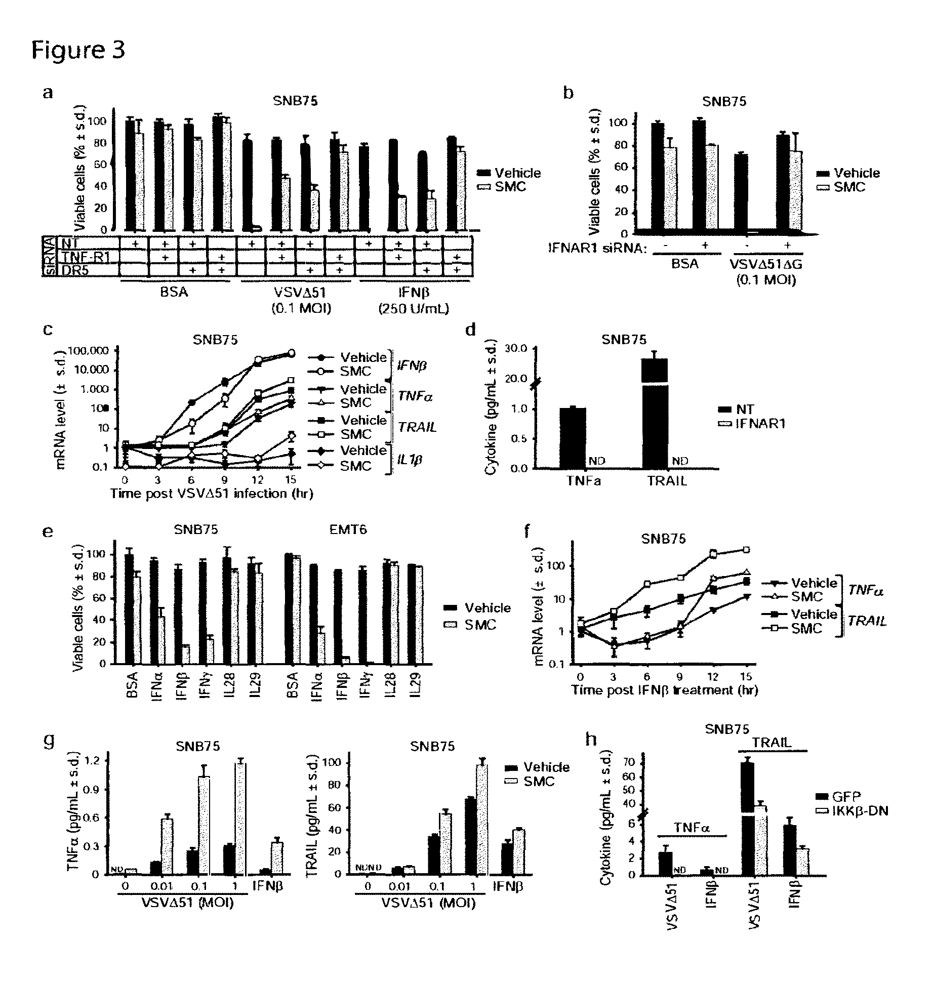
D00004

D00005

D00006

D00007

D00008

D00009
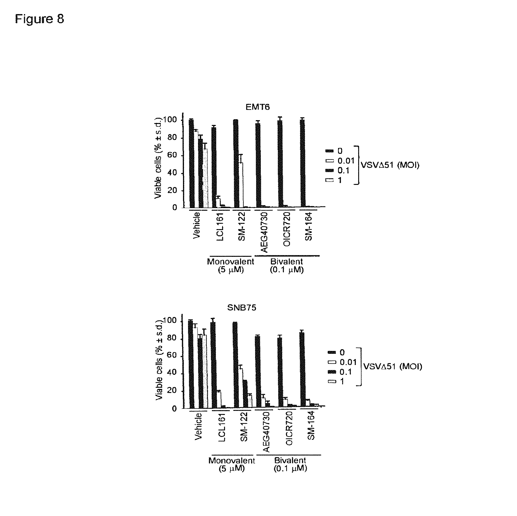
D00010

D00011

D00012
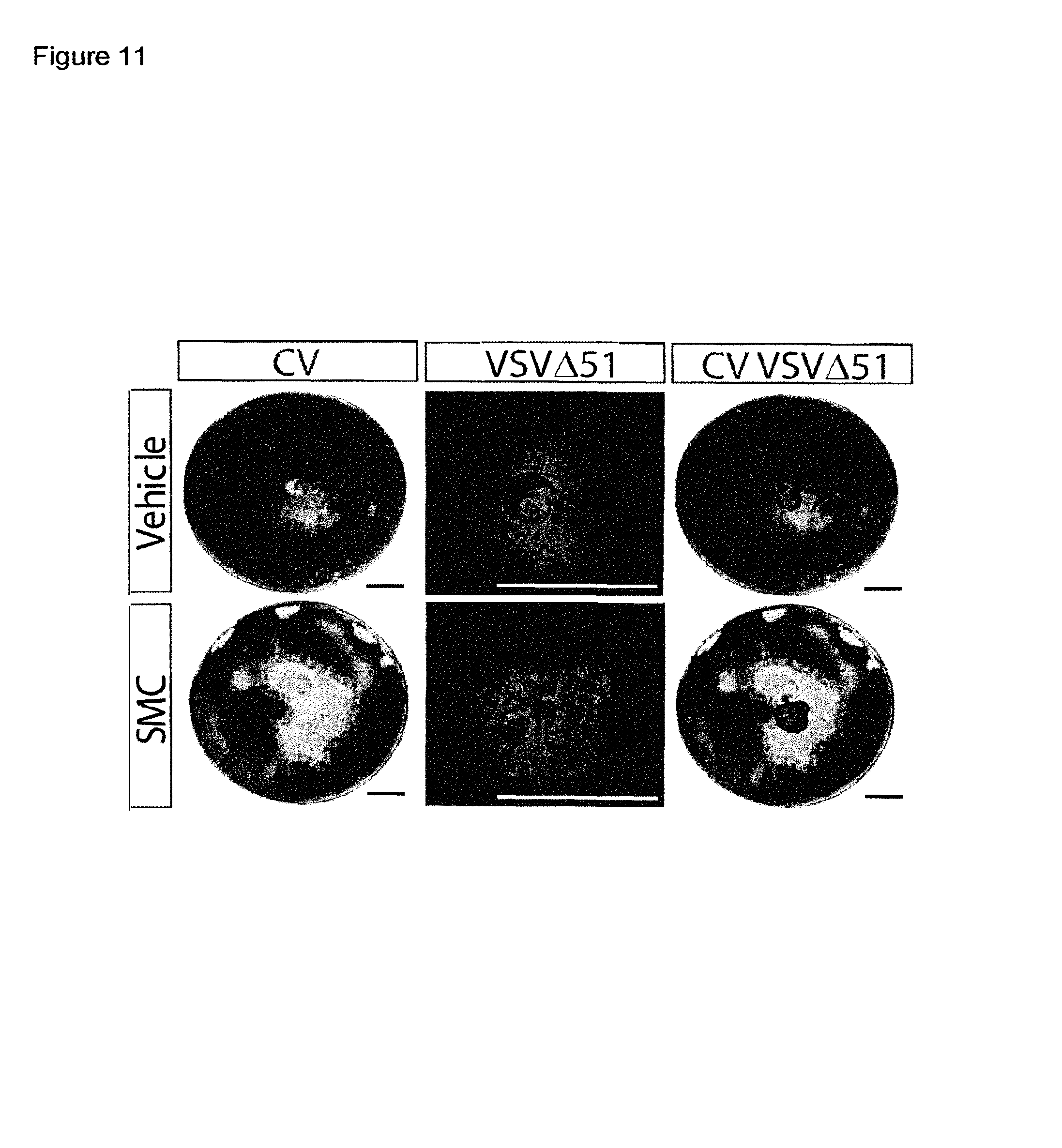
D00013

D00014
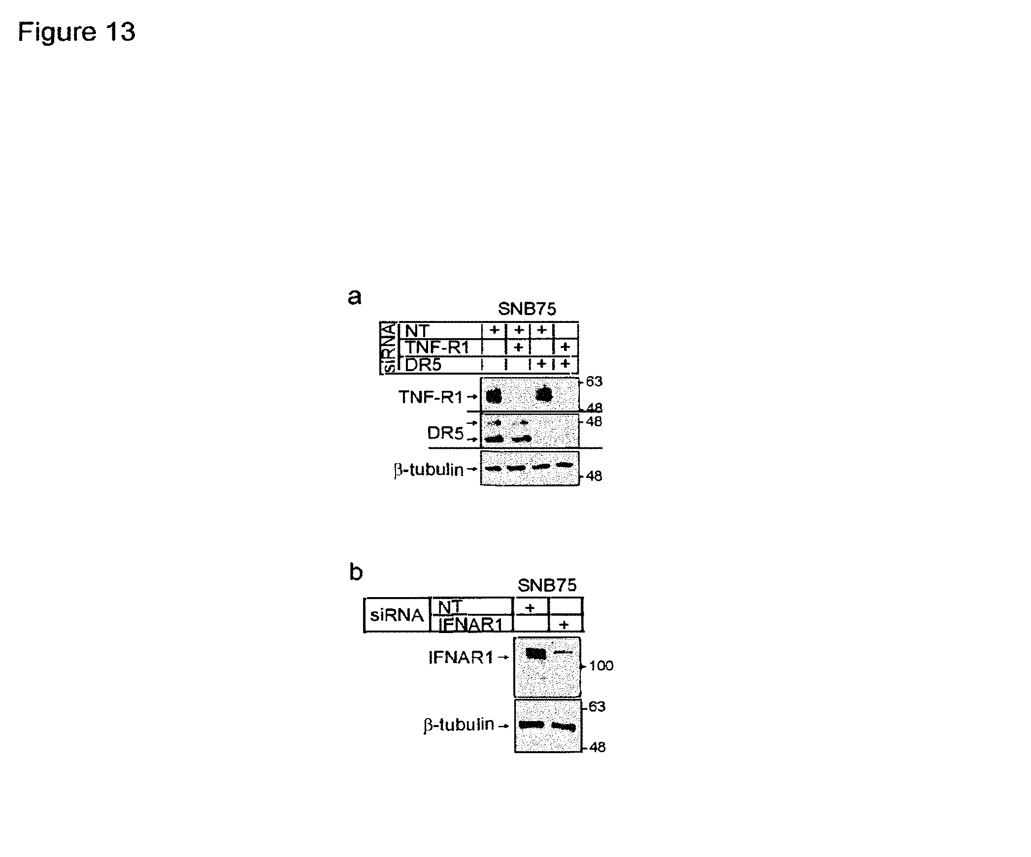
D00015

D00016

D00017

D00018

D00019
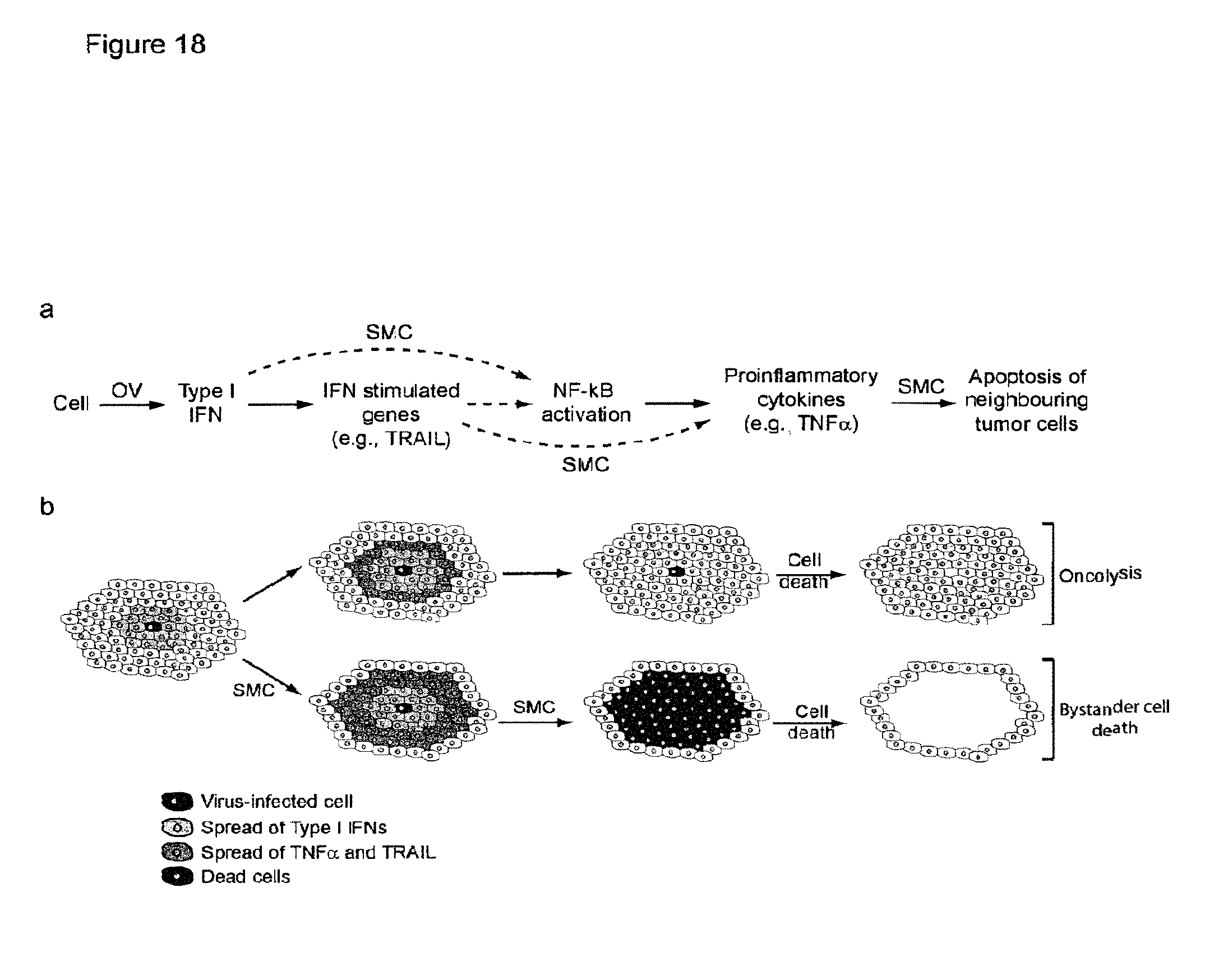
D00020

D00021

D00022

D00023

D00024

D00025
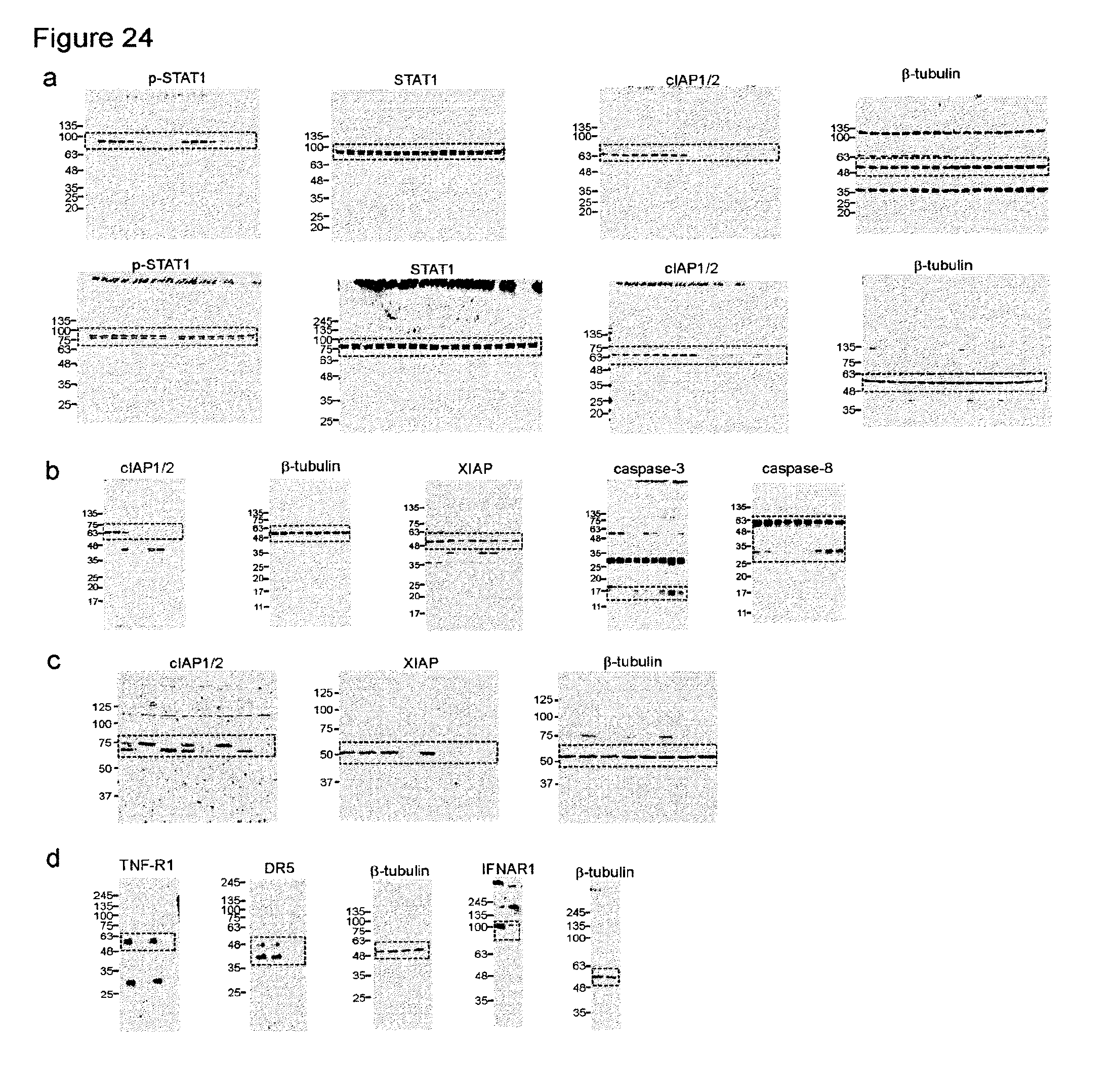
D00026
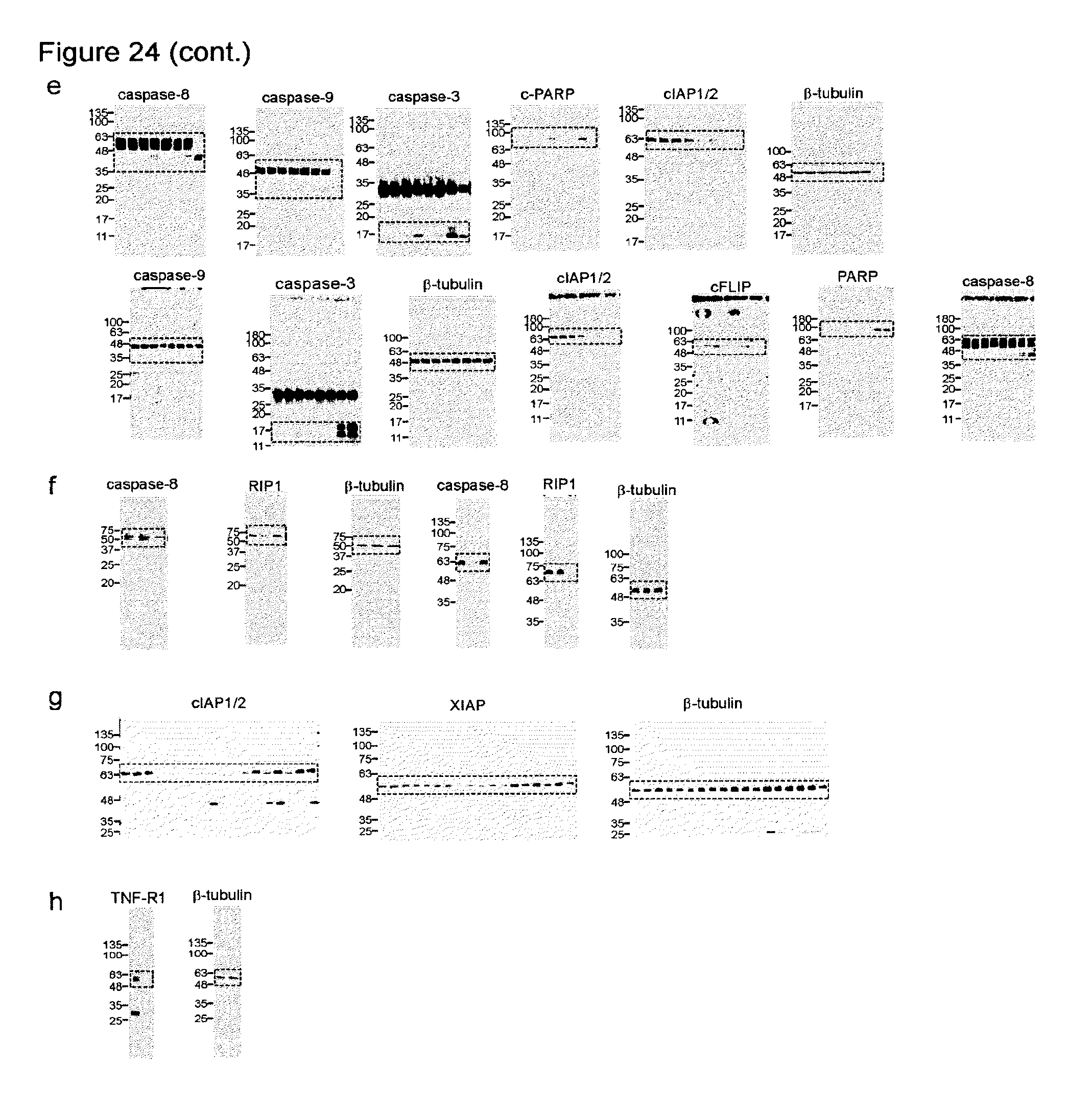
D00027
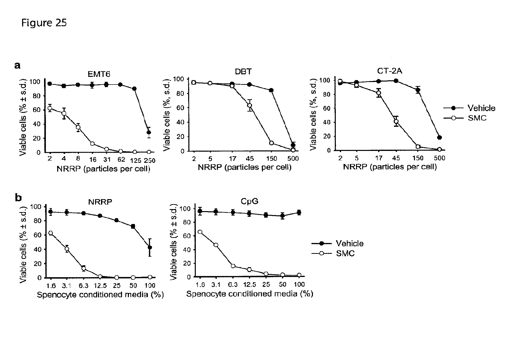
D00028
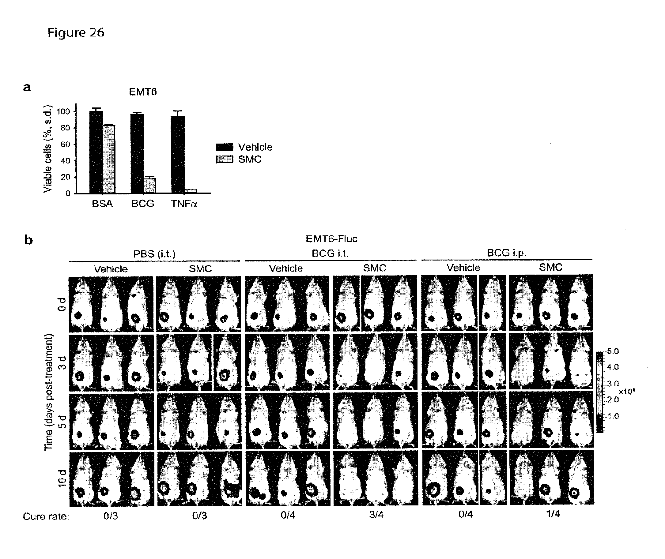
D00029

D00030
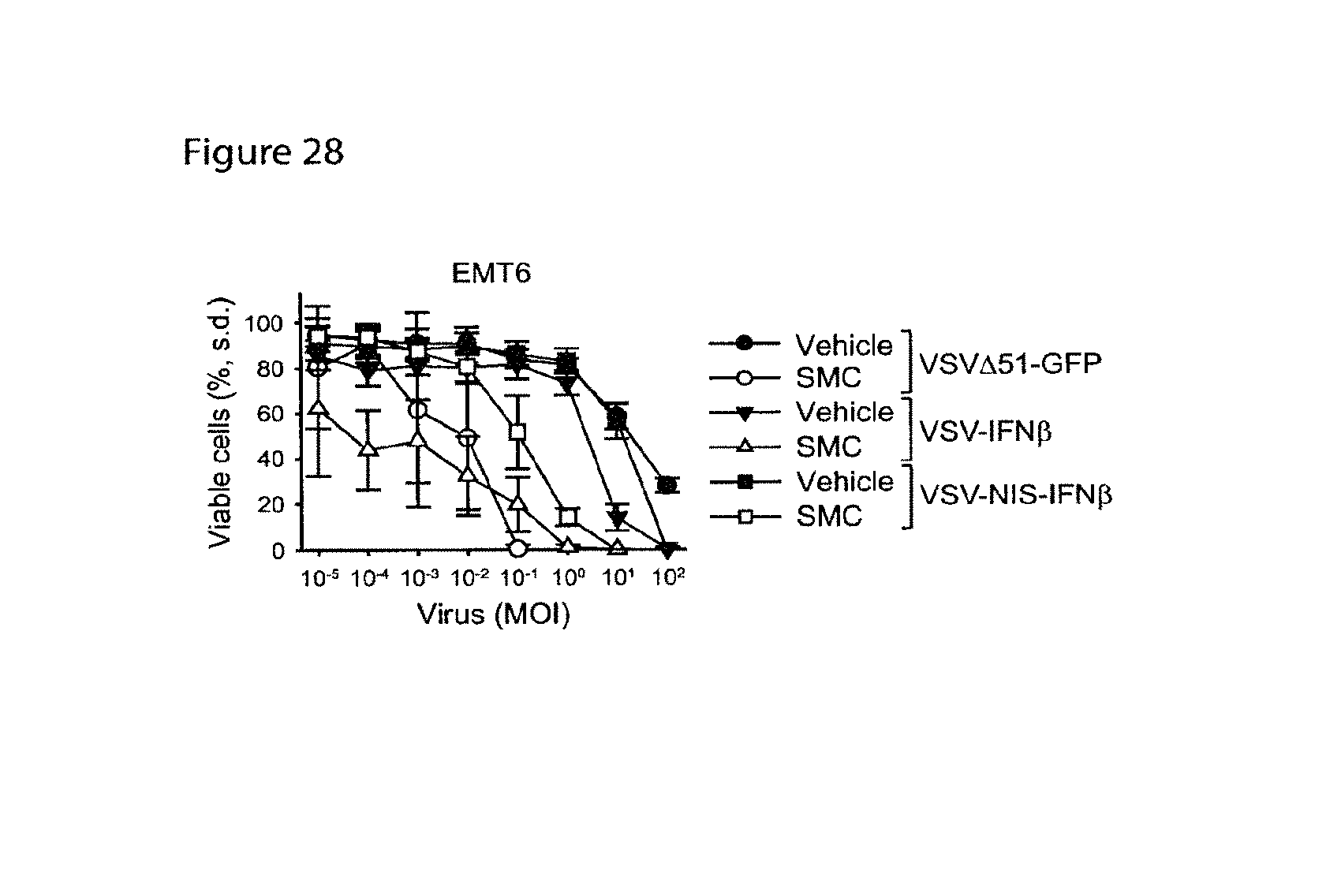
D00031
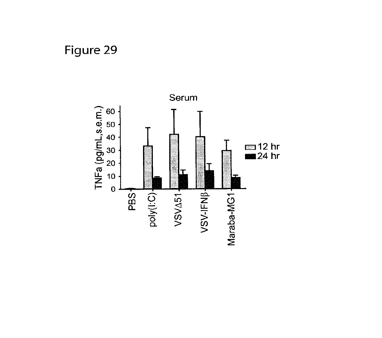
D00032

D00033
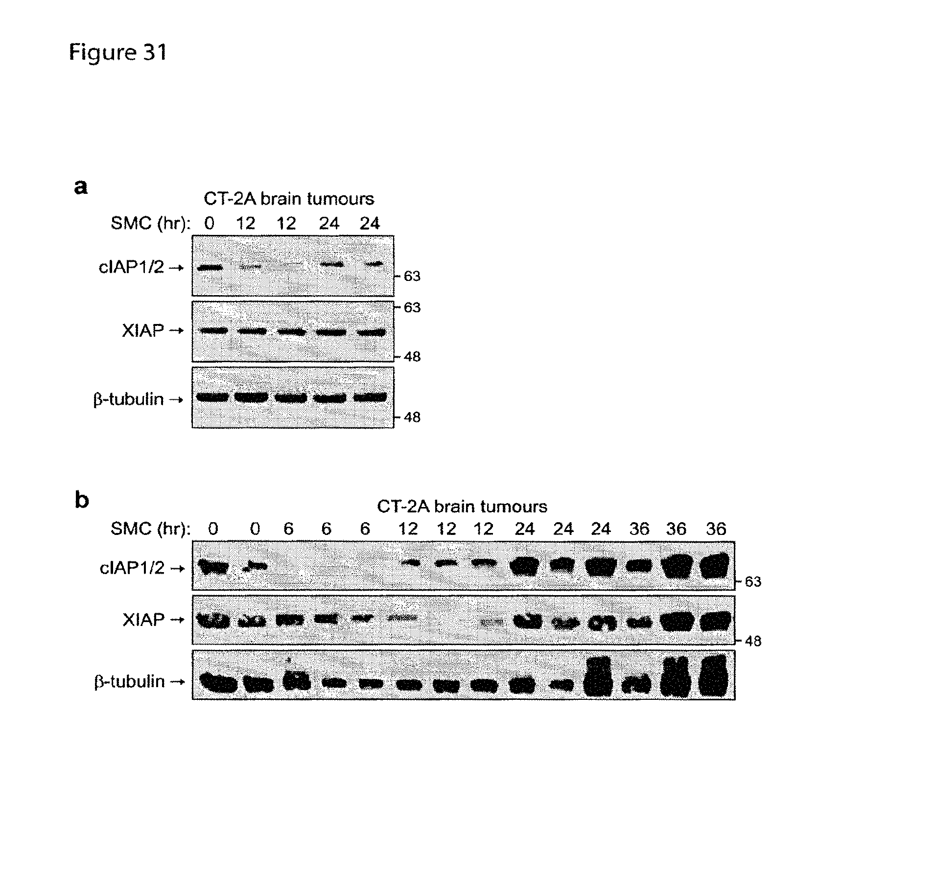
D00034

D00035

XML
uspto.report is an independent third-party trademark research tool that is not affiliated, endorsed, or sponsored by the United States Patent and Trademark Office (USPTO) or any other governmental organization. The information provided by uspto.report is based on publicly available data at the time of writing and is intended for informational purposes only.
While we strive to provide accurate and up-to-date information, we do not guarantee the accuracy, completeness, reliability, or suitability of the information displayed on this site. The use of this site is at your own risk. Any reliance you place on such information is therefore strictly at your own risk.
All official trademark data, including owner information, should be verified by visiting the official USPTO website at www.uspto.gov. This site is not intended to replace professional legal advice and should not be used as a substitute for consulting with a legal professional who is knowledgeable about trademark law.