Methods and systems for converting precursor cells into gastric tissues through directed differentiation
Wells , et al. J
U.S. patent number 10,174,289 [Application Number 15/312,939] was granted by the patent office on 2019-01-08 for methods and systems for converting precursor cells into gastric tissues through directed differentiation. This patent grant is currently assigned to Children's Hospital Medical Center. The grantee listed for this patent is Children's Hospital Medical Center. Invention is credited to Kyle William McCracken, James Macormack Wells.


View All Diagrams
| United States Patent | 10,174,289 |
| Wells , et al. | January 8, 2019 |
Methods and systems for converting precursor cells into gastric tissues through directed differentiation
Abstract
Disclosed are methods of inducing formation of a gastric cells and/or a gastric tissue, such as in the form of a gastric organoid. The formation of gastric cells and/or tissue may be carried out by the activating and/or inhibiting of one or more signaling pathways within a precursor cell. Also disclosed are methods for using the disclosed gastric cells, gastric tissues, and/or gastric organoids derived from precursor cells.
| Inventors: | Wells; James Macormack (Cincinnati, OH), McCracken; Kyle William (Cincinnati, OH) | ||||||||||
|---|---|---|---|---|---|---|---|---|---|---|---|
| Applicant: |
|
||||||||||
| Assignee: | Children's Hospital Medical
Center (Cincinnati, OH) |
||||||||||
| Family ID: | 53385964 | ||||||||||
| Appl. No.: | 15/312,939 | ||||||||||
| Filed: | May 27, 2015 | ||||||||||
| PCT Filed: | May 27, 2015 | ||||||||||
| PCT No.: | PCT/US2015/032626 | ||||||||||
| 371(c)(1),(2),(4) Date: | November 21, 2016 | ||||||||||
| PCT Pub. No.: | WO2015/183920 | ||||||||||
| PCT Pub. Date: | December 03, 2015 |
Prior Publication Data
| Document Identifier | Publication Date | |
|---|---|---|
| US 20170240866 A1 | Aug 24, 2017 | |
Related U.S. Patent Documents
| Application Number | Filing Date | Patent Number | Issue Date | ||
|---|---|---|---|---|---|
| 62003719 | May 28, 2014 | ||||
| Current U.S. Class: | 1/1 |
| Current CPC Class: | C12N 5/0697 (20130101); C12N 5/0679 (20130101); C12N 2501/385 (20130101); C12N 2501/155 (20130101); C12N 2506/02 (20130101); C12N 2501/16 (20130101); C12N 2501/113 (20130101); C12N 2501/11 (20130101); C12N 2501/119 (20130101); C12N 2501/415 (20130101); C12N 2501/727 (20130101); C12N 2501/115 (20130101); C12N 2506/45 (20130101) |
| Current International Class: | C12N 5/071 (20100101) |
References Cited [Referenced By]
U.S. Patent Documents
| 5942435 | August 1999 | Wheeler |
| 6607501 | August 2003 | Gorsuch |
| 7291626 | November 2007 | Beachy et al. |
| 7326572 | February 2008 | Fisk et al. |
| 7510876 | March 2009 | D'Amour et al. |
| 7514185 | April 2009 | Fukushima et al. |
| 7541185 | June 2009 | D'Amour et al. |
| 7625753 | December 2009 | Kelly et al. |
| 7704738 | April 2010 | D'Amour et al. |
| 7776592 | August 2010 | Wandinger-Ness et al. |
| 7985585 | July 2011 | D'Amour et al. |
| 7993916 | August 2011 | Agulnick et al. |
| 8187878 | May 2012 | Dalton et al. |
| 8216826 | July 2012 | D'Amour et al. |
| 8216836 | July 2012 | D'Amour et al. |
| 8298822 | October 2012 | Kruse et al. |
| 8318492 | November 2012 | Choo et al. |
| 8501476 | August 2013 | Morgan et al. |
| 8586357 | November 2013 | D'Amour et al. |
| 8603809 | December 2013 | Kruse |
| 8609406 | December 2013 | Subramanian et al. |
| 8609413 | December 2013 | Suter et al. |
| 8623645 | January 2014 | D'Amour et al. |
| 8633024 | January 2014 | D'Amour et al. |
| 8642339 | February 2014 | Sato et al. |
| 8647873 | February 2014 | D'Amour et al. |
| 8658151 | February 2014 | Kelly et al. |
| 8685386 | April 2014 | West et al. |
| 8758323 | June 2014 | Michaud et al. |
| 9127254 | September 2015 | Cohen et al. |
| 9133439 | September 2015 | Davis et al. |
| 9181301 | November 2015 | Carlson et al. |
| 9200258 | December 2015 | Mezghanni et al. |
| 9206393 | December 2015 | Kruse |
| 9334479 | May 2016 | Herrera Sanchez et al. |
| 9375514 | June 2016 | Kruse et al. |
| 9381181 | July 2016 | Roberts et al. |
| 9446076 | September 2016 | Gaussin et al. |
| 9447380 | September 2016 | Subramanian et al. |
| 9476030 | October 2016 | Gadue et al. |
| 9499795 | November 2016 | D'Amour et al. |
| 9605243 | March 2017 | D'Amour et al. |
| 9616039 | April 2017 | Roberts et al. |
| 9618500 | April 2017 | Giselbrecht et al. |
| 9675646 | June 2017 | Bitar |
| 9677085 | June 2017 | Guye et al. |
| 9719067 | August 2017 | Snoeck et al. |
| 9719068 | August 2017 | Wells et al. |
| 9732116 | August 2017 | Steiner et al. |
| 9763964 | September 2017 | Pellicciari et al. |
| 9765301 | September 2017 | Huch Ortega et al. |
| 9771562 | September 2017 | Shen et al. |
| 9790470 | October 2017 | Vallier et al. |
| 9828583 | November 2017 | Rauagopal et al. |
| 9849104 | December 2017 | Bisgaier et al. |
| 9850461 | December 2017 | Rizzi et al. |
| 9856458 | January 2018 | Rosowski et al. |
| 9878005 | January 2018 | Johns et al. |
| 9914920 | March 2018 | Goodwin et al. |
| 9926532 | March 2018 | Esteban et al. |
| 9938499 | April 2018 | Slukvin et al. |
| 2003/0129751 | July 2003 | Grikscheit et al. |
| 2005/0266554 | December 2005 | D'Amour et al. |
| 2006/0236415 | October 2006 | Silversides et al. |
| 2007/0238169 | October 2007 | Abilez et al. |
| 2007/0239083 | October 2007 | Voss |
| 2009/0263357 | October 2009 | Sayre et al. |
| 2010/0151568 | June 2010 | D'Amour et al. |
| 2011/0151564 | June 2011 | Menu et al. |
| 2012/0071451 | March 2012 | Spenard et al. |
| 2012/0196312 | August 2012 | Sato et al. |
| 2013/0031645 | January 2013 | Touboul et al. |
| 2013/0115673 | May 2013 | West et al. |
| 2013/0316442 | November 2013 | Meurville et al. |
| 2014/0038279 | February 2014 | Ingber et al. |
| 2014/0044713 | February 2014 | De Lau et al. |
| 2014/0193905 | July 2014 | Kelly et al. |
| 2014/0212910 | July 2014 | Bhatia et al. |
| 2014/0243227 | August 2014 | Clevers et al. |
| 2014/0273210 | September 2014 | Baker et al. |
| 2014/0302491 | October 2014 | Nadauld et al. |
| 2014/0308695 | October 2014 | Bruce et al. |
| 2014/0336282 | November 2014 | Ewald et al. |
| 2015/0017140 | January 2015 | Bhatia et al. |
| 2015/0197802 | July 2015 | Zink et al. |
| 2015/0201588 | July 2015 | Kamb et al. |
| 2015/0238656 | August 2015 | Orlando et al. |
| 2015/0330970 | November 2015 | Knoblich et al. |
| 2015/0343018 | December 2015 | Sansonetti et al. |
| 2015/0361393 | December 2015 | Nicholas et al. |
| 2016/0002602 | January 2016 | Almeida-Porada et al. |
| 2016/0022873 | January 2016 | Besner et al. |
| 2016/0060707 | March 2016 | Goldenberg et al. |
| 2016/0068805 | March 2016 | Martin et al. |
| 2016/0101133 | April 2016 | Basu et al. |
| 2016/0102289 | April 2016 | Yu et al. |
| 2016/0122722 | May 2016 | Ejiri et al. |
| 2016/0143949 | May 2016 | Ingber et al. |
| 2016/0184387 | June 2016 | Charmot et al. |
| 2016/0206664 | July 2016 | Sokal et al. |
| 2016/0237400 | August 2016 | Xian |
| 2016/0237401 | August 2016 | Vallier et al. |
| 2016/0237409 | August 2016 | Little et al. |
| 2016/0245653 | August 2016 | Park et al. |
| 2016/0257937 | September 2016 | Wauthier et al. |
| 2016/0263098 | September 2016 | Mantzoros |
| 2016/0289635 | October 2016 | Sasai et al. |
| 2016/0312181 | October 2016 | Freed et al. |
| 2016/0312190 | October 2016 | Ghaedi et al. |
| 2016/0312191 | October 2016 | Spence et al. |
| 2016/0319240 | November 2016 | Chan et al. |
| 2016/0340645 | November 2016 | D'Amour et al. |
| 2016/0340749 | November 2016 | Stelzer et al. |
| 2016/0354408 | December 2016 | Hariri et al. |
| 2016/0361466 | December 2016 | Yanagawa et al. |
| 2016/0376557 | December 2016 | Dubart Kupperschmitt et al. |
| 2017/0002330 | January 2017 | Vunjak-Novakovic et al. |
| 2017/0027994 | February 2017 | Kotton et al. |
| 2017/0035661 | February 2017 | Kyrkanides et al. |
| 2017/0035784 | February 2017 | Lancaster et al. |
| 2017/0037043 | February 2017 | Liu |
| 2017/0067014 | March 2017 | Takebe et al. |
| 2017/0101628 | April 2017 | Ingber et al. |
| 2017/0107469 | April 2017 | Costa et al. |
| 2017/0107483 | April 2017 | Pendergraft et al. |
| 2017/0107498 | April 2017 | Sareen et al. |
| 2017/0128625 | May 2017 | Bhatia et al. |
| 2017/0151049 | June 2017 | La Francesca et al. |
| 2017/0152486 | June 2017 | Shen et al. |
| 2017/0152528 | June 2017 | Zhang |
| 2017/0184569 | June 2017 | Keshavarzian et al. |
| 2017/0191030 | July 2017 | Huch Ortega et al. |
| 2017/0198261 | July 2017 | Sabaawy et al. |
| 2017/0204375 | July 2017 | Accili et al. |
| 2017/0205396 | July 2017 | Izpisua Belmonte et al. |
| 2017/0205398 | July 2017 | Bruce et al. |
| 2017/0239262 | August 2017 | Lefebvre |
| 2017/0240863 | August 2017 | Sokal et al. |
| 2017/0240964 | August 2017 | Leung et al. |
| 2017/0258772 | September 2017 | Sabatini et al. |
| 2017/0260501 | September 2017 | Semechkin et al. |
| 2017/0266145 | September 2017 | Nahmias et al. |
| 2017/0267970 | September 2017 | Gupta et al. |
| 2017/0267977 | September 2017 | Huang et al. |
| 2017/0275592 | September 2017 | Sachs et al. |
| 2017/0285002 | October 2017 | Taniguchi et al. |
| 2017/0296621 | October 2017 | Sansonetti et al. |
| 2017/0304294 | October 2017 | Wu et al. |
| 2017/0304369 | October 2017 | Ang et al. |
| 2017/0319548 | November 2017 | Lefebvre |
| 2017/0321188 | November 2017 | Viczian et al. |
| 2017/0321191 | November 2017 | Kojima |
| 2017/0335283 | November 2017 | Wang et al. |
| 2017/0342385 | November 2017 | Sachs et al. |
| 2017/0348433 | December 2017 | Kay et al. |
| 2017/0349659 | December 2017 | Garcia et al. |
| 2017/0349884 | December 2017 | Karp et al. |
| 2017/0360962 | December 2017 | Kay et al. |
| 2018/0021341 | January 2018 | Harriman et al. |
| 2018/0030409 | February 2018 | Lewis et al. |
| 2018/0042970 | February 2018 | Rossen et al. |
| 2018/0059119 | March 2018 | Takats et al. |
| 105985395 | Oct 2016 | CN | |||
| 3228306 | Oct 2017 | EP | |||
| 10-2006-0114355 | Nov 2006 | KR | |||
| WO 98/21312 | May 1998 | WO | |||
| WO 2003/082201 | Oct 2003 | WO | |||
| WO 2005/001072 | Jan 2005 | WO | |||
| WO 2005/081970 | Sep 2005 | WO | |||
| WO 2005/097974 | Oct 2005 | WO | |||
| WO 2005/113747 | Dec 2005 | WO | |||
| WO 2006/126236 | Nov 2006 | WO | |||
| WO 2008/075339 | Jun 2008 | WO | |||
| WO 2009/022907 | Feb 2009 | WO | |||
| WO 2009/146911 | Dec 2009 | WO | |||
| WO 2010/008905 | Jan 2010 | WO | |||
| WO 2010/090513 | Aug 2010 | WO | |||
| WO 2010/094694 | Aug 2010 | WO | |||
| WO 2011/140441 | Aug 2010 | WO | |||
| WO 2010/127399 | Nov 2010 | WO | |||
| WO 2010/143747 | Dec 2010 | WO | |||
| WO 2011/139628 | Nov 2011 | WO | |||
| WO 2012/014076 | Feb 2012 | WO | |||
| WO 2012/027474 | Mar 2012 | WO | |||
| WO 2012/089669 | Jul 2012 | WO | |||
| WO 2012/118799 | Sep 2012 | WO | |||
| WO 2012/154834 | Nov 2012 | WO | |||
| WO 2012/155110 | Nov 2012 | WO | |||
| WO 2012/166903 | Dec 2012 | WO | |||
| WO 2012/168930 | Dec 2012 | WO | |||
| WO 2013/040087 | Mar 2013 | WO | |||
| WO 2013/067498 | May 2013 | WO | |||
| WO 2013/086486 | Jun 2013 | WO | |||
| WO 2013/086502 | Jun 2013 | WO | |||
| WO 2013/093812 | Jun 2013 | WO | |||
| WO 2013/096741 | Jun 2013 | WO | |||
| WO 2013/127921 | Sep 2013 | WO | |||
| WO 2013/155060 | Oct 2013 | WO | |||
| WO 2013/192290 | Dec 2013 | WO | |||
| WO 2014/013334 | Jan 2014 | WO | |||
| WO 2014/048637 | Apr 2014 | WO | |||
| WO 2014/053596 | Apr 2014 | WO | |||
| WO 2014/082096 | May 2014 | WO | |||
| WO 2014/090993 | Jun 2014 | WO | |||
| WO 2014/127170 | Aug 2014 | WO | |||
| WO 2014/151921 | Sep 2014 | WO | |||
| WO 2014/153230 | Sep 2014 | WO | |||
| WO 2014/153294 | Sep 2014 | WO | |||
| WO 2014/159356 | Oct 2014 | WO | |||
| WO 2014/173907 | Oct 2014 | WO | |||
| WO 2014/182885 | Nov 2014 | WO | |||
| WO 2014/197934 | Dec 2014 | WO | |||
| WO 2014/199622 | Dec 2014 | WO | |||
| WO 2015/021358 | Feb 2015 | WO | |||
| WO 2015/060790 | Apr 2015 | WO | |||
| WO 2015/076388 | May 2015 | WO | |||
| WO 2015/108893 | Jul 2015 | WO | |||
| WO 2015/123183 | Aug 2015 | WO | |||
| WO 2015/129822 | Sep 2015 | WO | |||
| WO 2015/130919 | Sep 2015 | WO | |||
| WO 2015/135893 | Sep 2015 | WO | |||
| WO 2015/138032 | Sep 2015 | WO | |||
| WO 2015/152954 | Oct 2015 | WO | |||
| WO 2015/156929 | Oct 2015 | WO | |||
| WO 2015/157163 | Oct 2015 | WO | |||
| WO 2015/168022 | Nov 2015 | WO | |||
| WO 2015/0173425 | Nov 2015 | WO | |||
| WO 2015/183920 | Dec 2015 | WO | |||
| WO 2015/184273 | Dec 2015 | WO | |||
| WO 2015/184375 | Dec 2015 | WO | |||
| WO 2015/185714 | Dec 2015 | WO | |||
| WO 2015/196012 | Dec 2015 | WO | |||
| WO 2015/200901 | Dec 2015 | WO | |||
| WO 2016/011377 | Jan 2016 | WO | |||
| WO 2016/015158 | Feb 2016 | WO | |||
| WO 2016/030525 | Mar 2016 | WO | |||
| WO 2016/033163 | Mar 2016 | WO | |||
| WO 2016/057571 | Apr 2016 | WO | |||
| WO 2016/061464 | Apr 2016 | WO | |||
| WO 2016/073989 | May 2016 | WO | |||
| WO 2016/083612 | Jun 2016 | WO | |||
| WO 2016/083613 | Jun 2016 | WO | |||
| WO 2016/085765 | Jun 2016 | WO | |||
| WO 2016/094948 | Jun 2016 | WO | |||
| WO 2016/103002 | Jun 2016 | WO | |||
| WO 2016/103269 | Jun 2016 | WO | |||
| WO 2016/121512 | Aug 2016 | WO | |||
| WO 2016/140716 | Sep 2016 | WO | |||
| WO 2016/141137 | Sep 2016 | WO | |||
| WO 2016/144769 | Sep 2016 | WO | |||
| WO 2016/164413 | Oct 2016 | WO | |||
| WO 2016/168950 | Oct 2016 | WO | |||
| WO 2016/174604 | Nov 2016 | WO | |||
| WO 2016/176208 | Nov 2016 | WO | |||
| WO 2016/183143 | Nov 2016 | WO | |||
| WO 2016/193441 | Dec 2016 | WO | |||
| WO 2016/207621 | Dec 2016 | WO | |||
| WO 2016/210313 | Dec 2016 | WO | |||
| WO 2016/210416 | Dec 2016 | WO | |||
| WO 2017/009263 | Jan 2017 | WO | |||
| WO 2017/036533 | Mar 2017 | WO | |||
| WO 2017/037295 | Mar 2017 | WO | |||
| WO 2017/041041 | Mar 2017 | WO | |||
| WO 2017/048193 | Mar 2017 | WO | |||
| WO 2017/048322 | Mar 2017 | WO | |||
| WO 2017/049243 | Mar 2017 | WO | |||
| WO 2017/059171 | Apr 2017 | WO | |||
| WO 2017/060884 | Apr 2017 | WO | |||
| WO 2017/066507 | Apr 2017 | WO | |||
| WO 2017/066659 | Apr 2017 | WO | |||
| WO 2017/070007 | Apr 2017 | WO | |||
| WO 2017/070224 | Apr 2017 | WO | |||
| WO 2017/070471 | Apr 2017 | WO | |||
| WO 2017/070506 | Apr 2017 | WO | |||
| WO 2017/075389 | May 2017 | WO | |||
| WO 2017/077535 | May 2017 | WO | |||
| WO 2017/079632 | May 2017 | WO | |||
| WO 2017/083705 | May 2017 | WO | |||
| WO 2017/096192 | Jun 2017 | WO | |||
| WO 2017/096282 | Jun 2017 | WO | |||
| WO 2017/112901 | Jun 2017 | WO | |||
| WO 2017/115982 | Jul 2017 | WO | |||
| WO 2017/117333 | Jul 2017 | WO | |||
| WO 2017/117547 | Jul 2017 | WO | |||
| WO 2017/117571 | Jul 2017 | WO | |||
| WO 2017/120543 | Jul 2017 | WO | |||
| WO 2017/121754 | Jul 2017 | WO | |||
| WO 2017/123791 | Jul 2017 | WO | |||
| WO 2017/136462 | Aug 2017 | WO | |||
| WO 2017/136479 | Aug 2017 | WO | |||
| WO 2017/139455 | Aug 2017 | WO | |||
| WO 2017/139638 | Aug 2017 | WO | |||
| WO 2017/142069 | Aug 2017 | WO | |||
| WO 2017/143100 | Aug 2017 | WO | |||
| WO 2017/149025 | Sep 2017 | WO | |||
| WO 2017/153992 | Sep 2017 | WO | |||
| WO 2017/160234 | Sep 2017 | WO | |||
| WO 2017/160671 | Sep 2017 | WO | |||
| WO 2017/172638 | Oct 2017 | WO | |||
| WO 2017/174609 | Oct 2017 | WO | |||
| WO 2017/176810 | Oct 2017 | WO | |||
| WO 2017/184586 | Oct 2017 | WO | |||
| WO 2017/192997 | Nov 2017 | WO | |||
| WO 2017/205511 | Nov 2017 | WO | |||
| WO 2017/218287 | Dec 2017 | WO | |||
| WO 2017/220586 | Dec 2017 | WO | |||
| WO 2018/011558 | Jan 2018 | WO | |||
| WO 2018/019704 | Feb 2018 | WO | |||
| WO 2018/026947 | Feb 2018 | WO | |||
| WO 2018/027023 | Feb 2018 | WO | |||
| WO 2018/027112 | Feb 2018 | WO | |||
| WO 2018/035574 | Mar 2018 | WO | |||
| WO 2018/038042 | Mar 2018 | WO | |||
| WO 2018/044685 | Mar 2018 | WO | |||
| WO 2018/044885 | Mar 2018 | WO | |||
| WO 2018/044937 | Mar 2018 | WO | |||
| WO 2018/044940 | Mar 2018 | WO | |||
Other References
|
Han, Myoung-Eun; Oh, Sae-Ock; "Gastric stem cells and gastric cancer stem cells" Anatomy & Cell Biology, 46, 8-18, 2013 (Year: 2013). cited by examiner . Singaporean Written Opinion dated Oct. 19, 2017 for Application No. SG11201609953X, 8 pgs. cited by applicant . Arora, N., et al., "A process engineering approach to increase organoid yield," Development, 2017, 144:1128-1136, 9 pgs. cited by applicant . Asai, A., et al., "Paracrine signals regulate human liver organoid maturation from induced pluripotent stem cells," Development, 2017, 144:1056-1064, 9 pgs. cited by applicant . Eberhard, J., et al., "A cohort study of the prognostic and treatment predictive value of SATB2 expression in colorectal cancer," British Journal of Cancer, 2012, 106:931-938, 8 pgs. cited by applicant . Li, Z., et al., "SATB2 is a sensitive marker for lower gastrointestinal well-differentiated neuroendocrine tumors," Int J Clin Exp Pathol, 2015, 8(6):7072-7082, 11 pgs. cited by applicant . McCauley, H.A., et al., "Pluripotent stem cell-derived organoids: using principles of developmental biology to grow human tissues in a dish," Development, 2017, 144:958-962, 5 pgs. cited by applicant . McCracken, K.W., et al., "Wnt/.beta.-catenin promotes gastric fundus specification in mice and humans," Nature, Jan. 2017, 541(7636):182-187, 31 pgs. cited by applicant . Munera, J.O., et al., "Generation of Gastrointestinal Organoids from Human Pluripotent Stem Cells," Organ Regeneration, In: Tsuji, T., (eds), Organ Regeneration. Methods in Molecular Biology, vo. 1597, Humana Press, New York, NY, 2017, 11 pgs. cited by applicant . Munera, J.O., et al., "Differentiation of Human Pluripotent Stem Cells into Colonic Organoids via Transient Activation of BMP Signaling," Cell Stem Cell, Jul. 2017, 21(1):51-64.e6, 21 pgs. cited by applicant . Rankin, S.A., et al., "Timing is everything: Reiterative Wnt, BMP and RA signaling regulate developmental competence during endoderm organogenesis," Developmental Biology, Feb. 1, 2018, 434(1):121-132, 12 pgs. cited by applicant . Workman, M.J., et al., "Engineered human pluripotent-stem-cell-derived intestinal tissues with a functional enteric nervous system," Nat Med, Jan. 2017, 23(1):49-59, 29 pgs. cited by applicant . Zachos, N.C., et al., "Human Enteroids/Colonoids and Intestinal Organoids Functionally Recapitulate Normal Intestinal Physiology and Pathophysiology," The Journal of Blological Chemistry, Feb. 2016, 291(8):3759-3766, 8 pgs. cited by applicant . International Search Report and Written Opinion dated Feb. 21, 2018 for Application No. PCT/US2017/064600, 15 pgs. cited by applicant . Agopian, V.G., et al., "Intestinal Stem Cell Organoid Transplantation Generates Neomucosa in Dogs," Journal of Gastrointestinal Surgery, Jan. 23, 2009, 13(5):971-982, XP055241418, 12 pgs. cited by applicant . Alessi, D.R., et al., "LKB1-Dependent Signaling Pathways," Annu. Rev. Biochem., 2006, 75:137-63, 30 pgs. cited by applicant . Ameri, J., et al., "FGF2 Specifies hESC-Derived Definitive Endoderm into Foregut/Midgut Cell Lineages in a Concentration-Dependent Manner," Stem Cells, ePUB Nov. 3, 2009, 28(1):45-56, 12 pgs. cited by applicant . Anderson, G., et al., "Loss of enteric dopaminergic neurons and associated changes in colon motility in an MPTP mouse model of Parkinson's disease," Exp Neurol, Sep. 2007, 207:4-12, 16 pgs. cited by applicant . Anlauf, M., et al., "Chemical coding of the human gastrointestinal nervous system: cholinergic, VIPergic, and catecholaminergic phenotypes," The Journal of Comparative Neurology, 2003, 459:90-111, 22 pgs. cited by applicant . Avansino, J.R., et al., "Orthotopic transplantation of intestinal mucosal organiods in rodents," Surgery, Sep. 2006, 140(3):423-434, XP005610494, 12 pgs. cited by applicant . Baetge, G., et al., "Transient catecholaminergic (TC) cells in the vagus nerves and bowel of fetal mice: relationship to the development of enteric neurons," Developmental Biology, 1989, 132:189-211, 23 pgs. cited by applicant . Bajpai, R., et al., "CHD7 cooperates with PBAF to control multipotent neural crest formation," Nature, Feb. 18, 2010, 463:958-962, 7 pgs. cited by applicant . Bansal, D., et al., "An ex-vivo human intestinal model to study Entamoeba histolytica Pathogenesis," PLoS Neglected Tropical Diseases, Nov. 2009, 3(11):e551. cited by applicant . Bastide, P., et al. "Sox9 regulates cell proliferation and is required for Paneth cell differentiation in the intestinal epithelium," JCB, 2007, 178(4), pp. 635-648, 14 pgs. cited by applicant . Beck, F., et al., "Expression of Cdx-2 in the mouse embryo and placenta: possible tole in patterning of the extra-embroyonic membranes," Dev Dyn, 1995, 204:219-227. cited by applicant . Bergner, A.J., et al., "Birthdating of myenteric neuron subtypes in the small intestine of the mouse," The Journal of Comparative Neurology, 2014, 522:514-527, 14 pgs. cited by applicant . Blaugrund, E., et al., "Distinct subpopulations of enteric neuronal progenitors defined by time of development, sympathoadrenal lineage markers and Mash-1-dependence," Development 122, 1996, 309-320, 12 pgs. cited by applicant . Brevini, T.A.L., et al., "No shortcuts to pig embryonic stem cells," Theriogenology, 2010, 74:544-550, 7 pgs. cited by applicant . Burns, A.J., et al., "Neural stem cell therapies for enteric nervous system disorders," Nature Reviews/Gastroenterology & Hepatology, May 2014, 11:317-328, 12 pgs. cited by applicant . Campbell, F.C., et al., "Transplantation of cultured small bowel enterocytes," Gut, 1993, 34:1153-1155, 4 pgs. cited by applicant . Chen, C., et al., "Pdx1 inactivation restricted to the intestinal epithelium in mice alters duodenal gene expression in enterocytes and enteroendocrine cells," Am. J. Physiol. Gastrointest. Liver Pyshiol., 2009, 297:G1126-G1137, 12 pgs. cited by applicant . Chen, T-W., et al., "Ultrasensitive fluorescent proteins for imaging neuronal activity," Nature, Jul. 18, 2013, 499:295-300, 8 pgs. cited by applicant . Cheng, X., et al., "Self-renewing endodermal progenitor lines generated from human pluripotent stem cells," Cell Stem Cell, Apr. 6, 2012, 10:371-384, 14 pgs. cited by applicant . Costa, M., et al., "A method for genetic modification of human embryonic stem cells using electroporation," Nature Protocols, Apr. 5, 2007, 2:792-796, 5 pgs. cited by applicant . Curchoe, C.L., et al., "Early acquisition of neural crest competence during hESCs neuralization," PloS One, Nov. 2010, 5:1-17, 17 pgs. cited by applicant . Dekaney, C.M., et al., "Expansion of intestinal stem cells associated with long-term adaptation following ileocecal resection in mice," Am J Physiol Gastrointest Liver Physiol, Sep. 13, 2007, 293:G1013-G1022, 10 pgs. cited by applicant . Denham, M., et al., "Multipotent caudal neural progenitors derived from human pluripotent stem cells that give rise to lineages of the central and peripheral nervous system," Stem Cells, Mar. 5, 2015, 33:1759-1770, 12 pgs. cited by applicant . Fordham, R.P., et al., "Transplantation of expanded fetal intestinal progenitors contributes to colon regeneration after injury," Cell Stem Cell, Dec. 5, 2013, 13:734-744, 11 pgs. cited by applicant . Fu, M., et al., "HOXB5 expression is spatially and temporarily regulated in human embryonic gut during neural crest cell colonization and differentiation of enteric neuroblasts," Developmental Dynamics, 2003, 228:1-10, 10 pgs. cited by applicant . Fu, M., et al., "Embryonic development of the ganglion plexuses and the concentric layer structure of human gut: a topographical study," Anatomy and Embryology, Feb. 27, 2004, 208:33-41, 10 pgs. cited by applicant . Furness, J.B., "The enteric nervous system and neurogastroenterology," Nature Reviews/Gastroenterology & Hepatology, May 2012, 9:286-294, 9 pgs. cited by applicant . Gracz, A.D., et al., "Sox9 Expression Marks a Subset of CD24-expressing Small Intestine Epithelial Stem Cells the Form Organoids in vitro," Am J Physiol Gastrointest Liver Physiol, 2010, 298:G590-600. cited by applicant . Gracz, A.D., et al., "Brief report: CD24 and CD44 mark human intestinal epithelial cell populations with characteristics of active and facultative stem cells," Stem Cells, Apr. 4, 2013, 31:2024-2030, 7 pgs. cited by applicant . Groneberg, D.A., et al., "Intestinal peptide transport: ex vivo uptake studies and localization of peptide carrier PEPT1," Am J Physiol Gastrointest Liver Physiol, Sep. 2001, 281:G697-G704, 8 pgs. cited by applicant . Hao, M.M., et al., "Development of enteric neuron diversity," J. Cell. Mol. Med., 2009 13:1193-1210, 18 pgs. cited by applicant . Haveri, H., et al., "Transcription factors GATA-4 and GATA-6 in normal and neoplastic human gastrointestinal mucosa," BMC Gastroenterlology, 2008, 8:9. cited by applicant . Hockemeyer, D., et al., "Genetic engineering of human ES and iPS cells using TALE nucleases," Nat Biotechnol., 2012, 29:731-734, 8 pgs. cited by applicant . Huebsch, N., et al., "Automated video-based analysis of contractility and calcium flux in human-induced pluripotent stem cell-derived cardiomyocytes cultured over different spatial scales," Tissue Engineering: Part C, 2015, 21:467-479, 15 pgs. cited by applicant . Johannesson, M., et al., "FGF4 and Retinoic Acid Direct Differentiation of hESCs into PDX1-Expressing Foregut Endoderm in a Time- and Concentration-Dependent Manner," PLoS One, Mar. 2009, 4(3):1-13, 13 pgs. cited by applicant . Jung, P., et al., "Isolation and in vitro expansion of human colonic stem cells," Nature Medicine, Oct. 2011, 17:1225-1227, 3 pgs. cited by applicant . Juno, R.J., et al., "A serum factor after intestinal resection stimulates epidermal growth factor receptor signaling and proliferation in intestinal epithelial cells," Surgery, Aug. 2002, 132:377-383, 7 pgs. cited by applicant . Juno, R.J., et al., "A serum factor(s) after small bowel resection induces intestinal epithelial cell proliferation: effects of timing, site, and extent of resection," Journal of Pediatric Surery, Jun. 2003, 38:868-874, 7 pgs. cited by applicant . Kabouridis, P.S., et al., "Microbiota controls the homeostasis of glial cells in the gut lamina propria," Neuron, Jan. 21, 2015, 85:289-295, 8 pgs. cited by applicant . Kosinski, C., et al., "Indian hedgehog regulates intestinal stem cell fate through epithelial-mesenchymal interactions during development," Gastroenterology, Sep. 2010, 139:893-903, 17 pgs. cited by applicant . Kovalenko, P.L., et al., "The correlation between the expression of differentiation markers in rat small intestinal mucosa and the transcript levels of schlafen 3," JAMA Surg., Sep. 4, 2013, 148:1013-1019, 7 pgs. cited by applicant . Kroon, E., et al., "Pancreatic endoderm derived from human embryonic stem cells generates glucose-responsive insulin secreting cells in vivo," Nat Biotechnol, 2008, 26(4):443-52. cited by applicant . Kudoh, T., et al., "Distinct roles for Fgf, Wnt and retinoic acid in posteriorizing the neural ectoderm," Development, 2002, 129:4335-4346, 12 pgs. cited by applicant . Lahar, N., et al., "Intestinal suhepithelial myofibroblasts support in vitro and in vivo growth of human small intestinal epithelium," PLoS One, Nov. 2011, 6:e26898, 9 pgs. cited by applicant . Lambert, P.F., et al., "Using an immortalized cell line to study the HPV life cycle in organotypic `raft` cultures," Methods in Molecular Medicine, 2005, 119:141-155. cited by applicant . Lancaster, M.A., et al., "Organogenesis in a dish: modeling development and disease using organoid technologies," Science, Jul. 18, 2014, 345:283 & 1247125-1-9, 11 pgs. cited by applicant . Lavial, F., et al., "Chicken embryonic stem cells as a non-mammalian embryonic stem cell model," Develop. Growth Diff., 2010, 52:101-114, 14 pgs. cited by applicant . Le Douarin, N.M., et al., "Neural crest cell plasticity and its limits," Development 131, 2004, 4637-4650, 14 pgs. cited by applicant . Lee, G., et al., "Isolation and directed differentiation of neural crest stem cells derived from human embryonic stem cells," Nature Biotechnology, Dec. 2007, 25:1468-1475, 9 pgs. cited by applicant . Levin, D.E., et al., "Human tissue-engineered small intestine forms from postnatal progenitor cells," Journal of Pediatric Surgery, 2013, 48:129-137, 9 pgs. cited by applicant . Lindley, R.M., et al., "Human and Mouse Enteric Nervous System Neurosphere Transplants Regulate the Function of Aganglionic Embryonic Distal Colon," Gastroenterology, Jul. 2008, 135(1):205-216, XP022823118, 18 pgs. cited by applicant . Ludwig, T.E., et al, "Derivation of human embryonic stem cells in defined conditions," Nat Biotechnol, 2006, 24:185-187, 3 pgs. cited by applicant . Ludwig, T.E., et al., "Feeder-independent culture of human embryonic stem cells," Nat Methods, 2006, 3:637-646, 10 pgs. cited by applicant . Lui, V.C., et al., "Perturbation of hoxb5 signaling in vagal neural crests down-regulates ret leading to intestinal hypoganglionosis in mice," Gastroenterology, 2008, 134:1104-1115, 12 pgs. cited by applicant . McKeown, S.J., et al., "Hirschsprung disease: a developmental disorder of the enteric nervous system," Wiley Interdisciplinary Reviews Developmental Biology, Jan./Feb. 2013, 2:113-129, 17 pgs. cited by applicant . McLin, V.A. et al., "The Role of the Visceral Mesoderm in the Development of the Gastrointestinal Tract," Gastroenterology, 2009, 136:2074-2091, 18 pgs. cited by applicant . Mica, Y., et al., "Modeling neural crest induction, melanocyte specification and disease-related pigmentation defects in hESCs and patient-specific iPSCs," Cell Reports, Apr. 25, 2013, 3:1140-1152, 27 pgs. cited by applicant . Munoz, M., et al., "Conventional pluripotency markers are unspecific for bovine embryonic-derived cell-lines," Theriogenology, 2008, 69:1159-1164, 6 pgs. cited by applicant . Obermayr, F., et al., "Development and developmental disorders of the enteric nervous system," Nature Reviews/Gastroenterology & Hepatology, Jan. 2013, 10:43-57, 15 pgs. cited by applicant . Ootani, A. et al., "Sustained in vitro intestinal epithelial culture within a Wnt-dependent stem cell niche," Nat Med, 2009, 15:701-706, 14 pgs. cited by applicant . Paris, D.B.B.P., et al., "Equine embryos and embryonic stem cells: Defining reliable markers of pluripotency," Theriogenology, 2010, 74:516-524, 9 pgs. cited by applicant . Petitte, J.N., et al., "Avian pluripotent stem cells," Mech. of Develop., 2004, 121:1159-1168, 10 pgs. cited by applicant . Richards, M., et al., "The Transcriptome Profile of Human Embryonic Stem Cells as Defined by SAGE," Stem Cells, 2004, 22:51-64, 14 pgs. cited by applicant . Rohrschneider, M.R., et al., "Polarity and cell fate specification in the control of C. elegans gastrulation," Dev. Dyn., 2009, 238(4):789-796, 15 pgs. cited by applicant . Saffrey, M.J., "Cellular changes in the enteric nervous system during ageing," Developmental Biology, 2013, 382:344-355, 12 pgs. cited by applicant . Sasselli, V., et al., "The enteric nervous system," Developmental Biology, Jan. 2012, 366:64-73, 10 pgs. cited by applicant . Sato, T., et al., "Single Lgr5 stem cells build crypt-villus structures in vitro without a mesenchymal niche," Nature, 2009, 459:262-265, 5 pgs. cited by applicant . Sato, T., et al., "Long-term expansion of epithelial organoids from human colon, adenoma, adenocarcinoma, and Barrett's epithelium," Gastroenterology, Nov. 2011, 141:1762-1772, 11 pgs. cited by applicant . Simon-Assmann, P., et al., "In vitro models of intestinal epithelial cell differentiation," Cell Biol. Toxicol., 2007, 23:241-256, 16 pgs. cited by applicant . Spence, J.R., et al., "Translational Embryology: Using Embryonic Principles to Generate Pancreatic Endocrine Cells from Embryonic Stem Cells," Developmental Dynamics, 2007, 236:3218-3227, 10 pgs. cited by applicant . Tait, I.S., et al., "Colonic mucosal replacement by syngeneic small intestinal stem cell transplantation," The American Journal of Surgery, Jan. 1994, 167:67-72, 6 pgs. cited by applicant . Tait, I.S., et al., "Generation of neomucosa in vivo by transplantation of dissociated rat postnatal small intestinal epithelium," Differentiation, 1994 56:91-100, 10 pgs. cited by applicant . Takahashi, K. et al., "Induction of Pluripotent Stem Cells from Adult Human Fibroblasts by Defined Factors," Cell, 2007, 131:861-872, 12 pgs. cited by applicant . Takahashi, K., et al., "Induction of Pluripotent Stem Cells from Mouse Embryonic and Adult Fibroblast Cultures by Defined Factors," Cell, 2006, 126:663-676, 14 pgs. cited by applicant . Takaki, M., et al., "In Vitro Formation of Enteric Neural Network Structure in a Gut-Like Organ Differentiated from Mouse Embryonic Stem Cells," Stem Cells, Jun. 9, 2006, 24(6):1414-1422, XP55241404, 9 pgs. cited by applicant . Tang, W., et al., "Faithful expression of multiple proteins via 2A-peptide self-processing: a versatile and reliable method for manipulating brain circuits," The Journal of Neuroscience, Jul. 8, 2009, 29:8621-8629, 9 pgs. cited by applicant . Van Breemen, R.B., et al., "Caco-2 cell permeability assays to measure drug absorption," Expert Opin. Drug Metab. Toxicol., Aug. 2005, 1(2):175-185, 11 pgs. cited by applicant . Wallace, A.S., et al., "Development of the enteric nervous system, smooth muscle and interstitial cells of Cajal in the human gastrointestinal tract," Cell and Tissue Research, Jan. 26, 2005, 319:367-382, 16 pgs. cited by applicant . Wang, F., et al., "Isolation and characterization of intestinal stem cells based on surface marker combinations and colony-formation assay," Gastroenterology, 2013, 145:383-395.e1-e21, 34 pgs. cited by applicant . Wang, J., et al., "Mutant Neurogenin-3 in Congenital Malabsorptive Diarrhea," New England Journal of Medicine, 2006, 355:270-280, 11 pgs. cited by applicant . Warlich, E., et al., "Lentiviral vector design and imaging approaches to visualize the early stages of cellular reprogramming," Mol. Ther., Apr. 2011, 19:782-789, 9 pgs. cited by applicant . Watson, C.L., et al., "An in vivo model of human small intestine using pluripotent stem cells," Nature Medicine, Oct. 19, 2014, 20(11):1310-1314, XP055241417, 7 pgs. cited by applicant . Wells, J.M., et al., "How to Make an intestine," Development, Feb. 15, 2014, 141(4):752-760, XP055241409, 9 pgs. cited by applicant . Williamson, R.C.N., et al., "Humoral stimulation of cell proliferation in small bowel after transection and resection in rats," Gastroenterology, 1978, 75:249-254, 6 pgs. cited by applicant . Yamada, S., et al. "Differentiation of immature enterocytes into enteroendocrine cells by Pdx1 overexpression," Am. J. Physiol. Gastrointest. Liver Pyshiol., 2001, 281:G229-G236, 8 pgs. cited by applicant . Young, H.M., et al., "Expression of Ret-, p75.sup.NTR-, Phox2a-, Phox2b-, and tyrosine hydroxylase-immunoreactivity by undifferentiated neural crest-derived cells and different classes of enteric neurons in the embryonic mouse gut," Developmental Dynamics, 1999, 216:137-152, 16 pgs. cited by applicant . Young, H.M., et al., "GDNF is a chemoattractant for enteric neural cells," Developmental biology, Dec. 19, 2000, 229:503-516, 14 pgs. cited by applicant . Yui, S., et al., "Functional engraftment of colon epithelium expanded in vitro from a single adult Lgr5(+) stem cell," Nature Medicine, Apr. 2012, 18:618-623, 8 pgs. cited by applicant . Zhang, D., et a., "Neural crest regionalisation for enteric nervous system formation: implications for Hirschsprung's disease and stem cell therapy," Developmental Biology, Jan. 18, 2010, 339:280-294, 15 pgs. cited by applicant . Zhou, Q., et al., "In vivo reprogramming of adult pancreatic exocrine cells to .beta.-cells," Nature, 2008, 455: 627-632, 6 pgs. cited by applicant . European Exam Report dated Sep. 28, 2017 for Application No. EP 15728704.6, 4 pgs. cited by applicant . International Search Report dated Feb. 9, 2012 for Application No. PCT/US2011/035518, 7 pgs. cited by applicant . International Preliminary Report on Patentability and Written Opinion dated Nov. 6, 2012 for Application No. PCT/US2011/035518, 5 pgs. cited by applicant . International Search Report and Written Opinion dated Jan. 25, 2016 for Application No. PCT/US2015/055956, 16 pgs. cited by applicant . International Preliminary Report on Patentability dated Apr. 18, 2017 for Application No. PCT/US2015/055956, 8 pgs. cited by applicant . International Search Report and Written Opinion dated Jan. 19, 2017 for Application No. PCT/US2016/058864, 11 pgs. cited by applicant . International Search Report and Written Opinion dated Aug. 14, 2017 for Application No. PCT/US2017/013109, 17 pgs. cited by applicant . U.S. Appl. No. 15/515,840, filed Mar. 30, 2017, Wells et al. cited by applicant . U.S. Appl. No. 15/627,588, filed Jun. 20, 2017, Wells et al. cited by applicant . U.S. Appl. No. 61/332,178, filed May 6, 2010. cited by applicant . U.S. Appl. No. 62/065,131, filed Oct. 17, 2014. cited by applicant . Amieva, M.R., et al. "Helicobacter pylori enter and survive within multivesicular vacuoles of epithelial cells," Cell. Microbiol., 2002, 4(10):677-690, 15 pgs. cited by applicant . Andrews, P.W., et al., "Embryonic stem (ES) cells and embryonal carcinoma (EC) cells: opposite sides of the same coin," Biochem Soc Trans, 2005, 33(part 6):1526-1530, 5 pgs. cited by applicant . Ang, S-L, et al., "The formation and maintenance of the definitive endoderm lineage in the mouse: involvement of HNF3/forkhead proteins," Development, 1993, 119:1301-1315, 15 pgs. cited by applicant . Barker, N., et al., "Lgr5.sup.+ve Stem Cells Drive Self-Renewal in the Stomach and Build Long-Lived Gastric Units In Vitro," Cell Stem Cell, 2010, 6:25-36, 12 pgs. cited by applicant . Churin, Y., et al., "Helicobacter pylori CagA protein targets the c-Met receptor and enhances the motogenic response," J. Cell Biol., 2003, 161:249-255, 7 pgs. cited by applicant . Coghlan, M.P., et al., "Selective small molecule inhibitors of glycogen synthase kinase-3 modulate glycogen metabolism and gene transcription," Chem. Biol., 2000, 7(10):793-803, 11 pgs. cited by applicant . Couzin, J., "Small RNAs Make Big Splash," Science, 2002, 298:2296-2297, 2 pgs. cited by applicant . Covacci, A., et al., "Molecular characterization of the 128-kDa immunodominant antigen of Helicobacter pylori associated with cytotoxicity and duodenal ulcer," Proc Natl Acad Sci USA, Jun. 1993, 90:5791-5795, 5 pgs. cited by applicant . D'Amour, K.A., et al., "Efficient differentiation of human embryonic stem cells to definitive endoderm," Nature Biotechnology, 2005, 23:1534-1541, 9 pgs. cited by applicant . D'Amour, K.A., et al., "Production of pancreatic hormone-expressing endocrine cells from human embryonic stem cells," Nat Biotechnol, 2006, 24:1392-1401, 10 pgs. cited by applicant . De Santa Barbara, P., et al., "Development and differentiation of the intestinal epithelium," Cell Mol Life Sci, 2003, 60(7):1322-1332, 12 pgs. cited by applicant . Dessimoz, J., et al., "FGF signaling is necessary for establishing gut tube domains along the anterior-posterior axis in vivo," Mech Dev, 2006, 123:42-55, 14 pgs. cited by applicant . Elbashir, S.M., et al., "Functional anatomy of siRNAs for mediating efficient RNAi in Drosophila melanogaster embryo lysate," EMBO J., 2001, 20(23):6877-6888, 12 pgs. cited by applicant . Evans, M.J., et al., "Establishment in culture of pluripotent cells from mouse embryos," Nature, 1981, 292:154-156, 3 pgs. cited by applicant . Gradwohl, G., et al., "neurogenin3 is required for the development of the four endocrine cell lineages of the pancreas," Proc Natl Acad Sci USA, 2000, 97:1607-1611, 5 pgs. cited by applicant . Gregorieff, A., et al., "Wnt signaling in the intestinal epithelium: from endoderm to cancer," Genes & Dev., 2005, 19:877-890, 15 pgs. cited by applicant . Grosse, A.S., et al., "Cell dynamics in fetal intestinal epithelium: implications for intestinal growth and morphogenesis," Development, 2011, 138:4423-4432, 10 pgs. cited by applicant . Hannon, G.J., "RNA interference," Nature, 2002, 418:244-251, 8 pgs. cited by applicant . Hutvagner, G., et al., "A microRNA in a Multiple-Turnover RNAi Enzyme Complex," Science, Sep. 20, 2002, 297:2056-2060, 6 pgs. cited by applicant . Jenny, M., et al., "Neurogenin3 is differentially required for endocrine cell fate specification in the intestinal and gastric epithelium," EMBO J, 2002, 21(23):6338-6347, 10 pgs. cited by applicant . Johansson, K.A., et al., "Temporal Control of Neurogenin3 Activity in Pancreas Progenitors Reveals Competence Windows for the Generation of Different Endocrine Cell Types," Dev Cell, 2007, 12:457-465, 9 pgs. cited by applicant . Johnson, L.R., et al., "Stimulation of rat oxyntic gland mucosal growth by epidermal growth factor," Am. J. Physiol., 1980, 238:G45-49, 5 pgs. cited by applicant . Kaji, K., et al., "Virus-free induction of pluripotency and subsequent excision of reprogramming factors," Nature, Apr. 2009, 458:771-775, 6 pgs. cited by applicant . Kawaguchi, Y., et al., "The role of the transcriptional regulator Ptf1a in converting intestinal to pancreatic progenitors," Nat Genet, 2002, 32:128-134, 7 pgs. cited by applicant . Klimanskaya, I., et al., "Human embryonic stem cells derived without feeder cells," Lancet, 2005, 365:1636-1641, 6 pgs. cited by applicant . Koo, B-K, et al., "Controlled gene expression in primary Lgr5 organoid cultures," Nature Methods, Jan. 1, 2012, 9(1):81-83, XP055225249, 5 pgs. cited by applicant . Kubo, A., et al., "Development of definitive endoderm from embryonic stem cells in culture," Development, 2004, 131:1651-1662, 12 pgs. cited by applicant . Kumar, M., et al., "Signals from lateral plate mesoderm instruct endoderm toward a pancreatic fate," Dev Biol, 2003, 259:109-122, 14 pgs. cited by applicant . Lee, C. S., et al., "Neurogenin 3 is essential for the proper specification of gastric enteroendocrine cells and the maintenance of gastric epithelial cell identity," Genes Dev, 2002, 16:1488-1497, 11 pgs. cited by applicant . Liu, J., et al., "A Small-Molecule Agonist of the Wnt Signaling Pathway," Angew Chem Int Ed Engl., 2005, 44(13):1987-1990, 4 pgs. cited by applicant . Logan, C.Y., et al., "The Wnt Signaling Pathway in Development and Disease," Annu. Rev. Cell Dev. Biol., 2004, 20:781-810, 32 pgs. cited by applicant . Longmire, T.A., et al., "Efficient Derivation of Purified Lung and Thyroid Progenitors from Embryonic Stem Cells," Stem Cell, 2012, 10:398-411, 14 pgs. cited by applicant . Lopez-Diaz, L., et al., "Intestinal Neurogenin 3 directs differentiation of a bipotential secretory progenitor to endocrine cell rather than goblet cell fate," Dev Biol. 2007, 309:298-305, 8 pgs. cited by applicant . Mahe, M.M., et al., "Establishment of gastrointestinal epithelial organoids," Current Protocols in Mouse Biology, 2013, 3(4):217-240, XP002750112, 31 pgs. cited by applicant . Majumdar, A.P.N., "Postnatal Undernutrition: Effect of Epidermal Growth Factor on Growth and Function of the Gastrointestinal Tract in Rats," J. Pediatr. Gastroenterol. Nutr., 1984, 3:618-625, 8 pgs. cited by applicant . Martin, G.R., "Teratocarcinomas and mammalian embryogenesis," Science, 1980, 209:768-776, 9pgs. cited by applicant . Martin, M., et al., "Dorsal pancreas agenesis in retinoic acid-deficient Raldh2 mutant mice," Dev Biol., 2005, 284:399-411, 13 pgs. cited by applicant . McCracken, K.W., et al., "Generating human intestinal tissue from pluripotent stem cells in vitro," Nature Protocols, 2011, 6(12):1920-1928, 19 pgs. cited by applicant . McCracken, K.W., et al., "Modelling human development and disease in pluripotent stem-cell-derived gastric organoids," Nature, Oct. 29, 2014, 516(7531):400-404, XP055210509, 30 pgs. cited by applicant . McLin, V.A., et al., "Repression of Wnt/.beta.-catenin signaling in the anterior endoderm is essential for liver and pancreas development," Development, 2007, 134:2207-2217, 11 pgs. cited by applicant . McManus, M.T., et al., "Gene Silencing in Mammals by Small Interfering RNAs," Nat. Rev. Genet., Oct. 2002, 3:737-747, 13 pgs. cited by applicant . Meerbrey, K.L., et al., "The pINDUCER lentiviral toolkit for inducible RNA interference in vitro and in vivo," Proc Natl Acad Sci USA, 2011, 108:3665-3670, 6 pgs. cited by applicant . Mills, J.C., et al., "Gastric Epithelial Stem Cells," Gastroenterology, 2011, 140:412-424, 13 pgs. cited by applicant . Miyabayashi, T., et al., "Wnt/.beta.-catenin/CBP signaling maintains long-term murine embryonic stem cell pluripotency," Proc Natl Acad Sci USA, 2007, 104(13):5668-5673, 6 pgs. cited by applicant . Molotkov, A., et al., "Retinoic Acid Generated by Raldh2 in Mesoderm is Required for Mouse Dorsal Endodermal Pancreas Development," Dev Dyn, 2005, 232:950-957, 8 pgs. cited by applicant . Mou, H., et al., "Generation of Multipotent Lung and Airway Progenitors from Mouse ESCs and Patient-Specific Cystic Fibrosis iPSCs," Stem Cell, 2012, 10:385-397, 13 pgs. cited by applicant . Neiiendam, J.L., et al., "An NCAM-derived FGF-receptor agonist, the FGL-peptide, induces neurite outgrowth and neuronal survival in primary rat neurons," J. Neurochem., 2004, 91(4):920-935, 17 pgs. cited by applicant . Noguchi, T-a.K., et al., "Generation of stomach tissue from mouse embryonic stem cells," Nature Cell Biology, 2015, 17(8):984-993, X13055225165, 20 pgs. cited by applicant . Okita, K., et al., "Generation of Mouse Induced Pluripotent Stem Cells Without Viral Vectors," Science, 2008, 322(5903):949-953, 6 pgs. cited by applicant . Okita, K., et al., "An Efficient Nonviral Method to Generate Integration-Free Human-Induced Pluripotent Stem Cells from Cord Blood and Peripheral Blood Cells," Stem Cells, 2013, 31:458-466, 9 pgs. cited by applicant . Olbe, L., et al., "A Mechanism by Which Helicobacter pylori Infection of the antrum Contributes to the Development of Duodenal Ulcer," Gastroenterology, 2001, 110:1386-1394, 9 pgs. cited by applicant . Paddison, P.J., et al., "RNA interference: the new somatic cell genetics?", Cancer Cell, 2002, 2:17-23, 7 pgs. cited by applicant . Pai, R., et al., "Deoxycholic Acid Activates .beta.-Catenin Signaling Pathway and Increases Colon Cell Cancer Growth and Invasiveness," Mol Biol Cell., 2004, 15(5):2156-2163, 8 pgs. cited by applicant . Parkin, D.M., "The global health burden of infection-associated cancers in the year 2002," Int. J. Cancer, 2006, 118:3030-3044, 15 pgs. cited by applicant . Peek, R.M., Jr., et al., "Helicobacter pylori cagA+ Strains and Dissociation of Gastric Epithelial Cell Proliferation From Apoptosis," J. Natl. Cancer Inst., 1997, 89:863-868, 7 pgs. cited by applicant . Peek, R.M., Jr., "Helicobacter pylori infection and disease: from humans to animal models," Dis Model Mech, 2008, 1:50-55, 6 pgs. cited by applicant . Sancho, E., et al., "Signaling Pathways in Intestinal Development and Cancer," Annu. Rev. Cell Dev. Biol., 2004, 20:695-723, 31 pgs. cited by applicant . Schonhoff, S.E., et al., "Neurogenin 3-expressing progenitor cells in the gastrointestinal tract differentiate into both endocrine and non-endocrine cell types," Dev Biol, 2004, 270:443-454, 12 pgs. cited by applicant . Schumacher, M.A., et al., "Gastric Sonic Hedgehog Acts as a Macrophage Chemoattractant During the Immune Response to Helicobacter pylori," Gastroenterology, 2012, 142:1150-1159, 16 pgs. cited by applicant . Shan, J., et al., "Identification of a Specific Inhibitor of the Dishevelled PDZ Domain," Biochemistry, 2005, 44(47):15495-15503, 9 pgs. cited by applicant . Si-Tayeb, K., et al., "Highly Efficient Generation of Human Hepatocyte-Like Cells from Induced Pluripotent Stem Cells," Hepatology, 2010, 51:297-305, 9 pgs. cited by applicant . Spear, P.C., et al., "Interkinetic nuclear migration: A mysterious process in search of a function," Develop. Growth Differ., 2012, 54:306-316, 12 pgs. cited by applicant . Speer, M.D., A.L., et al., "Murine Tissue-Engineered Stomach Demonstrates Epithelial Differentiation," Journal of Surgical Research, Mar. 22, 2011, 171(1):6-14, XP028317226, 9 pgs. cited by applicant . Spence, J.R., et al., "Directed differentiation of human pluripotent stem cells into intestinal tissue in vitro," Nature, 2011, 470:105-109, 13 pgs. cited by applicant . Stadtfeld, M., et al., "Induced pluripotent stem cells generated without viral integration," Science, 2008, 322(5903):945-949, 12 pgs. cited by applicant . Taipale, J., et al., "The Hedgehog and Wnt signalling pathways in cancer," Nature, 2001, 411:349-354, 8 pgs. cited by applicant . Teo, A.K.K., et al., "Activin and BMP4 Synergistically Promote Formation of Definitive Endoderm in Human Embryonic Stern Cells," Stem Cells, 2012, 30:631-642, 12 pgs. cited by applicant . Thomson, J.A., et al., "Embryonic Stem Cell Lines Derived from Human Blastocysts," Science, 1998, 282(5391):1145-1147, 4 pgs. cited by applicant . Tiso, N., et al., "BMP signalling regulates anteroposterior endoderm patterning in zebrafish," Mech Dev, 2002, 118:29-37, 9 pgs. cited by applicant . Tuschl, T., et al., "Targeted mRNA degradation by double-stranded RNA in vitro," Genes Dev., 1999, 13:3191-3197, 8 pgs. cited by applicant . Verzi, M.P., et al., "Role of the Homeodomain Transcription Factor Bapx1 in Mouse Distal Stomach Development," Gastroenterology, 2009, 136:1701-1710, 10 pgs. cited by applicant . Wang, Z., et al., "Retinoic acid regulates morphogenesis and patterning of posterior foregut derivatives," Dev Biol, 2006, 297:433-445. cited by applicant . Wells, J.M., et al., "Early mouse endoderm is patterned by soluble factors from adjacent germ layers," Development, 2000, 127:1563-1572, 10 pgs. cited by applicant . Wen, S. et al., "Helicobacter pylori virulence factors in gastric carcinogenesis," Cancer Lett., 2009, 282:1-8, 8 pgs. cited by applicant . Woltjen, K., et al., "piggyBac transposition reprograms fibroblasts to induced pluripotent stem cells," Nature, 2009, 458:766-770, 8 pgs. cited by applicant . Xia, H.H-X., et al. "Antral-Type Mucosa in the Gastric Incisura, Body, and Fundus (Antralization): A Link Between Helicobacter pylori Infection and Intestinal Metaplasia?", Am. J. Gastroenterol., 2000, 95:114-121, 8 ps. cited by applicant . Yuan, Y., et al., "Peptic ulcer disease today," Nat Clin Pract Gastroenterol Hepatol, 2006, 3:80-89 10 pgs. cited by applicant . Zhang, Q, et al., "Small-molecule synergist of the Wnt/.beta.-catenin signaling pathway," Proc Natl Acad Sci USA, 2007, 104(18):7444-7448, 6 pgs. cited by applicant . Zhou, H., et al., "Generation of Induced Pluripotent Stem Cells Using Recombinant Proteins," Cell Stem Cell, 2009, 4(5):381-384, 4 pgs. cited by applicant . Zorn, A.M., et al., "Vertebrate endoderm development and organ formation," Annu Rev Cell Dev Biol, 2009, 25:221-251, 36 pgs. cited by applicant . International Search Report and Written Opinion dated Dec. 15, 2015 for Application No. PCT/US2015/032626, 19 pgs. cited by applicant . U.S. Appl. No. 62/003,719, filed May 28, 2014. cited by applicant . Ader. M., et al., "Modeling human development in 3D culture," Current Opinion in Cell Biology, 2014, 31:23-28, 6 pgs. cited by applicant . Aurora, M., et al., "hPSC-derived lung and intestinal organoids as models of human fetal tissue," Developmental Biology, 2016, 420:230-238, 9 pgs. cited by applicant . Baptista, P.M., et al., "The Use of Whole Organ Decellularization for the Generation of a Vascularized Liver Organoid," Hepatology, 2011, 53(2):604-617, 14 pgs. cited by applicant . Barker, N., et al., "Tissue-Resident Adult Stem Cell Populations of Rapidly Self-Renewing Organs," Cell Stem Cell, Dec. 2010, 7:656-670, 15 pgs. cited by applicant . Bartfeld, S., et al., "Stem cell-derived organoids and their application for medical research and patient treatment," J Mol Med, 2017, 95:729-738, 10 pgs. cited by applicant . Baumann, K., "Colonic organoids for durg testing and colorectal disease modelling," Nature Reviews Molecular Cell Biology, Jul. 2017, 1 pg. cited by applicant . Bitar, K.N., et al., "Intestinal Tissue Engineering: Current Concepts and Future Vision of Regenerative Medicine in the Gut," Neurogastroenterol Motil., Jan. 2012, 24(1):7-19, 20 pgs. cited by applicant . Bruens, L., et al., "Expanding the Tissue Toolbox: Deriving Colon Tissue from Human Pluripotent Stem Cells," Cell Stem Cell, Jul. 2017, 21(1):3-5, 3 pgs. cited by applicant . Brugmann, S.A., et al., "Building additional complexity to in vitro-derived intestinal tissues," Stem Cell Research & Therapy, 2013, 4(Suppl 1):S1, 5 pgs. cited by applicant . Cao, L., et al., "Development of Intestinal Organoids as Tissue Surrogates: Cell Composition and the Epigenetic Control of Differentiation," Molecular Carcinogenesis, 2015, 54:189-202, 14 pgs. cited by applicant . Cieslar-Pobuda, A., et al., The expression pattern of PFKFB3 enzyme distinguishes between induced-pluripotent stem cells and cancer stem cells, Oncotarget, 6(30):29753-29770, 18 pgs. cited by applicant . Clevers, H., "Modeling Development and Disease with Organoids," Cell, Jun. 2016, 165:1586-1597, 12 pgs. cited by applicant . Correia, C., et al., "Combining Hypoxia and Bioreactor Hydrodynamics Boosts Induced Pluripotent Stem Cell Differentiation Towards Cardiomyocytes," Stem Cell Rev and Rep, 2014, 10:786-801, 16 pgs. cited by applicant . Davenport, C., et al., "Anterior-Posterior Patterning of Definitive Endoderm Generated from Human Embryonic Stem Cells Depends on the Differential Signaling of Retinoic Acid, Wnt-, and BMP-Signaling," Stem Cells, 2016, 34:2635-2647, 13 pgs. cited by applicant . Dedhia, P.H., et al., "Organoid Models of Human Gastrointestinal Development and Disease," Gastroenterology, 2016, 150:1098-1112, 15 pgs. cited by applicant . Ezashi, T., et al., "Low O.sub.2 tensions and the prevention of differentiation of hES cells," PNAS, Mar. 2005, 102(13):4783-4788, 6 pgs. cited by applicant . Fatehullah, A., et al., "Organoids as an in vitro model of human development and disease," Nature Cell Biology, Mar. 2106, 18(3):246-254, 9 pgs. cited by applicant . Finkbeiner, S.R., et al., "A Gutsy Task: Generating Intestinal Tissue from Human Pluripotent Stem Cells," Dig Dis Sci, 2013, 58:1176-1184, 9 pgs. cited by applicant . Finkbeiner, S.R., et al., "Stem Cell-Derived Human Intestinal Organoids as an Infection Model for Rotaviruses," mBio, Jul./Aug. 2012, 3(4):e00159-12, 6 pgs. cited by applicant . Gessner, R.C., et al., "Functional ultrasound imaging for assessment of extracellular matrix scaffolds used for liver organoid formation," Biomaterials, 2013, 34:9341-9351, 11 pgs. cited by applicant . Gouon-Evans, V., et al., "BMP-4 is required for hepatic specification of mouse embryonic stem cell-derived definitive endoderm," Nature Biotechnology, Nov. 2006, 24(11):1402-1411, 10 pgs. cited by applicant . Howell, J.C., et al., "Generating intestinal tissue from stem cells: potential for research and therapy," Regen Med., 6(6):743-755, 22 pgs. cited by applicant . Huch, M., et al., "Lgr5.sup.+ liver stem cells, hepatic organoids and regenerative medicine," Regen. Med., 2013, 8(4):385-387, 3 pgs. cited by applicant . Huch, M., et al., "Modeling mouse and human development using organoid cultures," Development, 2015, 142:3113-3125, 13 pgs. cited by applicant . Kostrzewski, T., et al., "Three-dimensional perfused human in vitro model of non-alcoholic fatty liver disease," World J Gastroenterol, 2017, 23(2):204-215, 13 pgs. cited by applicant . Kretzschmar, K., et al., "Organoids: Modeling Development and the Stem Cell Niche in a Dish," Developmental Cell, Sep. 2016, 38:590-600, 11 pgs. cited by applicant . Kuratnik, A., et al., "Intestinal organoids as tissue surrogates for toxicological and pharmacological studies," Biochemical Pharmacology, 2013, 85:1721-1726, 6 pgs. cited by applicant . Li, Y., et al., "In vitro organogenesis from pluripotent stem cells," Organogenesis, Jun. 2014, 10(2):159-163, 5 pgs. cited by applicant . Lu, Y., et al., "A Novel 3D Liver Organoid System fro Elucidation of Hepatic Glucose Metabolism," Biotechnol Bioeng., Feb. 2012, 109(2):595-604, 21 pgs. cited by applicant . McCracken, K.W., "Mechanisms of endoderm patterning and directed differentiation of human stem cells into foregut tissues," Dissertation, Graduate School of the University of Cincinnati, Jun. 19, 2014, 185 pgs. cited by applicant . Ogaki, S., et al., "Wnt and Notch Signals Guide Embryonic Stem Cell Differentiation into the Intestinal Lineages," Stem Cells, 2013, 31:1086-1096, 11 pgs. cited by applicant . Park, K.I., et al., "Acute injury directs the migration, proliferation, and differentiation of solid organ stem cells: Evidence for the effect of hypoxia-ischemia in the CNS on clonal "reporter" neural stem cells," Experimental Neurology, 2006, 199:159-178, 23 pgs. cited by applicant . Pastula, A., et al., "Three-Dimensional Gastrointestinal Organoid Culture in Combination with Nerves or Fibroblasts: A Method to Characterize the Gastrointestinal Stem Cell Niche," Stem Cells International, 2016, 16 pgs. cited by applicant . Pulikkot, S., "Establishment of a 3D Culture Model of Gastric Stem Cells Supporting Their Differentiation into Mucous Cells Using Microfibrous Polycaprolactone Scaffold," Dissertation, United Arab Emirates University, College of Medicine and Health Sciences, May 2015, 187 pgs. (4 parts: Part 1--58 pgs; Part 2--69 pgs; Part 3--31 pgs; Part 4--29 pgs.). cited by applicant . Ramachandran, S.D., et al., "In Vitro Generation of Functional Liver Organoid-Like Structures Using Adult Human Cells," Plos One, Oct. 2015, 14 pgs. cited by applicant . Ray, K., "Engineering human intestinal organoids with a functional ENS," Nature Reviews Gastroenterology & Hepatology, Nov. 2016, 1 pg. cited by applicant . Saito, M., et al., "Reconstruction of liver organoid using a bioreactor," World J Gastroenterol, Mar. 2006, 12(12):1881-1888, 8 pgs. cited by applicant . Sampaziotis, F., et al., "Potential of Human Induced Pluripotent Stem Cells in Studies of Liver Disease," Hepatology, Jul. 2015, 62(1):303-311, 9 pgs. cited by applicant . Sasai, Y., "Next-Generation Regenerative Medicine: Organogenesis from Stem Cells in 3D Culture," Cell Stem Cell, May 2013, 12:520-530, 11 pgs. cited by applicant . Schlieve, C.R., et al., "Created of Warm Blood and Nerves: Restoring an Enteric Nervous System in Organoids," Cell Stem Cell, Jan. 2017. 20:5-7, 3 pgs. cited by applicant . Shah, S.B., et al., "Cellular self-assembly and biomaterials-based organoid models of development and diseases," Acta Biomaterialia, 2017, 53:29-45, 17 pgs. cited by applicant . Sinagoga, K.L., et al., "Generating human intestinal tissues from pluripotent stem cells to study development and disease," The EMBO Journal, 2015, 34(9):1149-1163, 15 pgs cited by applicant . Snykers, S., et al., "In Vitro Differentiation of Embryonic and Adult Stem Cells into Hepatocytes: State of the Art," Stem Cells, 2009, 27:577-605, 29 pgs. cited by applicant . Soto-Gutierrez, A., et al., "Engineering of an Hepatic Organoid to Develop Liver Assist Devices," Cell Transplant., 2010, 19(6):815-822, 12 pgs. cited by applicant . Sugawara, T., et al., "Organoids recapitulate organs?," Stem Cell Investig, 2018, vol. 5, Iss. 3, 4 pgs. cited by applicant . Sui, L., et al., "Signaling pathways during maintenance and definitive endoderm differentiation of embryonic stem cells," Int J Dev Bio, 2013, 57:1-12, 12 pgs. cited by applicant . Sun, Y., et al., "Genome engineering of stem cell organoids for disease modeling," Protein Cell, 2017, 8(5):315-327, 13 pgs. cited by applicant . Tamminen, K., et al., "Intestinal Commitment and Maturation of Human Pluripotent Stem Cells Is Independent fo Exogenous FGF4 and R-spondin1," PLOS One, Jul. 2015, 10(7):e0134551, 19 pgs. cited by applicant . Toivonen, S., et al., "Activin A and Wnt-dependent specification of human definitive endoderm cells," Experimental Cell Research, 2013, 319:2535-2544, 10 pgs. cited by applicant . Tsakmaki, A., et al., "3D intestinal organoids in metabolic research: virtual reality in a dish," Current Opinion in Pharmacology, 2017, 37:51-58, 8 pgs. cited by applicant . Wang, A., et al., "Generating cells of the gastrointestinal system: current approaches and applications for the differentiation of human pluripotent stem cells," J Mol Med, 2012, 90:763-771, 9 pgs. cited by applicant . Workman, M.J., "Generating 3D human intestinal organoids with an enteric nervous system," Thesis, Graduate School of the University of Cincinnati, Oct. 2014, 61 pgs. cited by applicant . Xinaris, C., et al, "Organoid Models and Applications in Biomedical Research," Nephron, 2015, 130:191-199, 9 pgs. cited by applicant . Yin, C., et al., "Hepatic stellate cells in liver development, regeneration, and cancer," The Journal of Clinical Investigation, May 2013, 123(5):1902-1910, 9 pgs. cited by applicant . Adorini, L., et al., "Farnesoid X receptor targeting to treat nonalcoholic steatohepatitis," Drug Discovery Today, Sep. 2012, 17(17/18):988-997, 10 pgs. cited by applicant . Altman, G.H., et al., "Cell differentiation by mechanical stress," The FASEB Journal, 2001, 16(2):270-272, 13 pgs. cited by applicant . Bohorquez, D.V., et al., "An Enteroendocrine Cell--Enteric Glia Connection Revealed by 3D Electron Microscopy," PLOS One, Feb. 2014, 9(2):e89881, 13 pgs. cited by applicant . Cabezas, J., et al., "Nonalcoholic Fatty Liver Disease: A Pathological View," Liver Biopsy--Indications, Procedures, Results, Chapter 8, InTech, 2012, pp. 161-188, 29 pgs. cited by applicant . Dahl, A., et al., "Translational Regenerative Medicine--Hepatic Systems," Chapter 34, Clinical Aspects of Regenerative Medicine, eds. A. Atala, M.D. and J. Allickson, PhD, Elsevier, Inc., 2015, pp. 469-484, 16 pgs. cited by applicant . Date, S., et al., "Mini-Gut Organoids: Reconstitution of the Stem Cell Niche," Annual Review of Cell and Developmental Biology, Nov. 2015, 31:269-289. cited by applicant . Discher, D.E., et al., "Groth Factors, Matrices, and Forces Combine and Control Stem Cells," Science, Jun. 2009, 324:1673-1677, 5 pgs. cited by applicant . Eicher, A.K., et al., "Translating Developmental Principles to Generate Human Gastric Organoids," Cellular and Molecular Gastroenterology and Hepatology, 2018, 5(3):353-363, 11 pgs. cited by applicant . Gori, M., et al., "Investigating nonalcoholic Fatty Liver Disease in a Liver-on-a-Chip Microfluidic Device," PLOS One, Jul. 2016, 11(7):e0159729, 15 pgs. cited by applicant . Green, M.D., et al., "Generation of anterior foregut endoderm from human embryonic and induced pluripotent stem cells," Nature Biotechnology, Mar. 2011, 29(3):267-272, 7 pgs. cited by applicant . Guilak, F., et al., "Control of Stem Cell Fate by Physical Interactions with the Extracellular Matrix," Cell Stem Cell, Jul. 2009, 5:17-26, 10 pgs. cited by applicant . Hannon, N.R.F., et al., "Generation of Multipotent Foregut Stem Cells from Human Pluripotent Stem Cells," Stem Cell Reports, Oct. 2013, 1:293-306, 14 pgs. cited by applicant . Hardy, T., et al., "Nonalcoholic fatty liver disease: new treatments," Curr Opin Gastroenterol, May 2015, 31(3):175-183, 9 pgs. cited by applicant . Katoh, M., "WNT Signaling in Stem Cell Biology and Regenerative Medicine," Current Drug Targets, 2008, 9(7):565-570, 6 pgs. cited by applicant . Kim, T-H., et al., "Stomach development, stem cells and disease," Development, 2016, 143:554-565, 12 pgs. cited by applicant . Koike, M., et al., "Effects of mechanical strain on proliferation and differentiation of bone marrow stromal cell line ST2," J Bone Miner Metab, 2005, 23:219-225, 7 pgs. cited by applicant . Kolahchi, A.R., et al., "Microfluidic-Bases Multi-Organ Platforms for Drug Discovery," Micromachines, 2016, 7(162):1-33, 33 pgs. cited by applicant . Lin, C., et al., "The application of engineered liver tissues for novel drug discovery," Expert Opinion on Drug Discovery, 2015, 10(5):519-540. cited by applicant . Luo, X., et al., "Generation of endoderm lineages from pluripotent stem cells," Regenerative Medicine, 2017, 12(1):77-89, 13 pgs. cited by applicant . McCracken, K.W., et al., "Mechanism of embryonic stomach development," Seminars in Cell & Development Blology, 2017, 66:36-42, 7 pgs. cited by applicant . Micallef, S.J., et al., "Endocrine cells develop within pancreatic bud-like structures derived from mouse ES cells differentiated in response to BMP4 and retinoic acid," Stem Cell Research, 2007, 1:25-36, 12 pgs. cited by applicant . Mudaliar, S., et al., "Efficacy and Safety of the Farnesoid X Receptor Agonist Obeticholic Acid in Patients with Type 2 Diabetes and Nonalcoholic Fatty Liver Disease," Gastroenterology, 2013, 145:574-582, 10 pgs. cited by applicant . Nandivada, P., et al., "Treatment of Parenteral Nutrition-Associated Liver Disease: The Role of Lipid Emulsions," Advances in Nutrition, Reviews from ASN EB 2013 Symposia, pp. 711-717, 7 pgs. cited by applicant . Neuschwander-Tetri, B.A., et al., "Farnesoid X nuclear receptor ligand obeticholic acid for non-cirrhotic, non-alcoholic steatohepatitis (FLINT): a multicentre, randomised, placebo-controlled trial," Lancet, 2015, 385:956-965, 10 pgs. cited by applicant . Park, H.R., et al., "Lipotoxicity of Palmaitic Acid on Neural Progenitor Cells and Hippocampal Neurogenesis," Toxicol Res, 2011, 27(2):103-110, 8 pgs. cited by applicant . Park, J.S., et al., "Differential Effects of Equiaxial and Uniaxial Strain on Mesenchymal Stem Cells," Biotechnology and Bioengineering, Nov. 2004, 88(3):359-368, 10 pgs. cited by applicant . Park, J.S., et al., "The effect of matrix stiffness on the differentiation of mesenhymal stem cells in response to TGF-.beta.," Biomaterials, 2011, 32:3921-3930, 10 pgs. cited by applicant . Pennisi, C.P., Ph.D., et al., "Uniaxial Cyclic Strain Drives Assembly and Differentiation of Skeletal Myocytes," Tissue Engineering: Part A, 2011, 17(19-20):2543 2550, 8 pgs. cited by applicant . Pompaiah, M., et al., "Gastric Organoids: An Emerging Model System to Study Helicobacter pylori Pathogenesis," Molecular Pathogenesis and Signal Transduction by Helicobacter pylori, Current Topics in Microbiology and Immunology, N. Tegtmeyer, et al., (eds.), 2017, pp. 149-168. cited by applicant . Qi, M-C., et al., "Mechanical strain induces osteogenie differentiation: Cbfa1 and Ets-1 expression in stretched rat mesenchymal stem cells," Int J Oral Maxillofac Surg, 2008, 37:453-458, 6 pgs. cited by applicant . Reilly, G.C., et al., "Intrinsic extracellular matrix properties regulate stem cell differentiation," Journal of Biomechanics, 2010, 43:55-62, 8 pgs. cited by applicant . Rennert, K., et al., "A microfluidically perfused three dimensional human liver model," Biomaterials, 2015, 71:119-131, 13 pgs. cited by applicant . Saenz, J.B., et al., "Stomach growth in a dish: A protocol has been developed to grow structures that resemble the main part of the stomach in vitro from human embryonic stem cells--an advance that provides insights into stomach development," Nature, Jan. 2017, 541:160-161, 2 pgs. cited by applicant . Saha, S., et al., "Inhibition of Human Embryonic Stem Cell Differentiation by Medical Strain," Journal of Cellular Physiology, 2006, 206:126-137, 12 pgs. cited by applicant . Schmelter, M., et al., "Embryonic stem cells utilize reactive oxygen species as transducers of mechanical strain-induced cardiovascular differentiation," The FASEB Journal, Jun. 2006, 20(8):1182-1184, 16 pgs. cited by applicant . Schuppan, D., et al., "Non-alcoholic steatohepatitis: Pathogenesis and novel therapeutic approaches," Journal of Gastroenterology and Hepatology, 2013, 28(Suppl 1):68-76, 9 pgs. cited by applicant . Shimizu, N., et al., "Cyclic strain induces mouse embryonic stem cell differentiation into vascular smooth muscle cells by activating PDGF receptor .beta.," J Appl Physiol, 2008, 104:766-772, 7 pgs. cited by applicant . Singh, S., et al., "Comparative Effectiveness of Pharmacological Interventions for Nonalcoholic Steatohepatitis: A Systematic Review and Network Meta-analysis," Hepatology, Nov. 2015, 62(5):1417-1432, 16 pgs. cited by applicant . Skardal, A., et al., "Organoid-on-a-chip and body-on-a-chip systems for drug screening and disease modeling," Drug Discovery Today, Sep. 2016, 21(9): 1399-1411, 13 pgs. cited by applicant . Snoeck, H-W., "Generation of Anterior Foregut Derivatives from Pluripotent Stem Cells," Stem Cells Handbook, S. Sell (ed.), 2013, pp. 161-175. cited by applicant . Sonntag, F., et al., "Design and prototyping of a chip-based multi-micro-organoid culture system for substance testing, predictive to human (substance) exposure," Journal of Biotechnology, 2010, 148:70-75, 6 pgs. cited by applicant . Ward, D.F., Jr., et al., "Mechanical Strain Enhances Extracellular Matrix-Induced Gene Focusing and Promotes Osteogenic Differentiation of Human Mesenchymal Stem Cells Through an Extracellular-Related Kinase-Dependent Pathway," Stem Cells and Development, 2007, 16:467-479, 14 pgs. cited by applicant . Willet, S.G., et al., "Stomach Organ and Cell Lineage Differentiation: From Embryogenesis to Adult Homeostasis," Cellular and Molecular Gastroenterology and Hepatology, 2016, 2(5):546-559, 14 pgs. cited by applicant . Zhang, W., et al., "Elastomeric Free-Form Blood Vessels for Interconnecting Organs on Chip Systems," Lab Chip, Apr. 2016, 16(9):1579-1586, 19 pgs. cited by applicant . Zhang, Y.S., et al., "Multisensor-integrated organs-on-chips platforms for automated and continual in situ monitoring of organoid behaviors," PNAS Early Edition, 2017, 10 pgs. cited by applicant . Zhang, Y.S., et al., "Seeking the right context for evaluating nanomedicine: from tissue models in petri dishes to microfluidic organs-on-a-chip," Nanomedicine (Lond.), 2015, 10(5):685-688, 4 pgs. cited by applicant . Zhou, J., et al., "The Potential for Gut Organoid Derived Interstitial Cells of Cajal in Replacement Therapy," International Journal of Molecular Sciences, Sep. 2017, 18:1-17, 17 pgs. cited by applicant . International Search Report and Written Opinion dated Jan. 18, 2018 for Application No. PCT/US2017/059865, 12 pgs. cited by applicant . International Search Report and Written Opinion dated Jan. 19, 2018 for Application No. PCT/US2017/059845, 13 pgs. cited by applicant . International Search Report and Written Opinion dated Jan. 29, 2018 for Application No. PCT/US2017/059860, 13 pgs. cited by applicant . Stark, R., et al., "Development of an endoluminal intestinal lengthening capsule," Journal of Pediatric Surgery, 2012, 47:136-141, 6 pgs. cited by applicant . European Exam Report dated Jul. 4, 2018 for Application No. EP 15728704.6, 3 pgs. cited by applicant . International Searching Authority Invitation to Pay Additional Fees and, Where Applicable, Protest Fee, dated Jun. 27, 2018 for Application No. PCT/US/2018/029083, 3 pgs. cited by applicant . European Exam Report dated May 18, 2018 for Application No. EP 15791404.5, 3 pgs. cited by applicant . International Search Report and Written Opinion dated Jun. 14, 2018 for Application No. PCT/US2018/018585, 14 pgs. cited by applicant . Singaporean Second Written Opinion dated Sep. 4, 2018 for Application No. SG 11201609953X, 6 pgs. cited by applicant. |
Primary Examiner: Berke-Schlessel; David W
Attorney, Agent or Firm: Frost Brown Todd LLC
Government Interests
STATEMENT REGARDING FEDERALLY-SPONSORED RESEARCH
This invention was made with government support under DK080823, DK092456, and GM063483 awarded by the National Institutes of Health. The government has certain rights to the invention.
Parent Case Text
PRIORITY CLAIM
This application claims priority to and benefit of PCT/US2015/032626 filed May 27, 2016, which claims priority to and benefit of U.S. Provisional Patent Application No. 62/003,719, to Wells et al, filed on May 28, 2014, entitled "Methods and Systems for Converting Precursor Cells into Gastric Tissues through Directed Differentiation" in their entirety and for all purposes.
Claims
What is claimed is:
1. A method of inducing formation of a gastric tissue, comprising the steps of: a) contacting a culture containing a mammalian definitive endoderm cell with an FGF signaling pathway activator, a Wnt signaling pathway activator, and a BMP inhibitor for at least two days or for a period of time sufficient to form SOX2+foregut cells; b) contacting said SOX2+foregut cells of step (a) with retinoic acid for a period of time sufficient to form three-dimensional foregut spheroids, wherein said spheroids express SOX2, PDX1, HNF1.beta., and HNF6; c) contacting said three-dimensional foregut spheroids of step (b) with retinoic acid, EGF, and a BMP inhibitor for a period of time sufficient for formation of a gastric organoid, wherein said gastric organoid is characterized by expression of MUC5AC, gastrin, ghrelin, 5-HT, and ChrA and the presence of cell types selected from one or more of antral mucous cells and endocrine cells.
2. The method of claim 1, wherein said precursor cell is selected from an embryonic stem cell, an embryonic germ cell, an induced pluripotent stem cell, a mesoderm cell, a definitive endoderm cell, a posterior endoderm cell, and a hindgut cell.
3. The method of claim 1, wherein said precursor cell is a definitive endoderm derived from contacting a pluripotent stem cell with one or more molecules selected from Activin, the BMP subgroups of the TGF-beta superfamily of growth factors; Nodal, Activin A, Activin B, BMP4, Wnt3a, and combinations thereof.
4. The method of claim 1, wherein said FGF signalling pathway is activated using an agent selected from one or more molecules selected from the group consisting of FGF1, FGF2, FGF3, FGF4, FGF10, FGF11, FGF12, FGF13, FGF14, FGF15, FGF16, FGF17, FGF18, FGF19, FGF20, FGF21, FGF22, and FGF23.
5. The method of claim 1, wherein said WNT signalling pathway is activated using an agent selected from one or more molecules selected from the group consisting of Wnt1, Wnt2, Wnt2b, Wnt3, Wnt3a, Wnt4, Wnt5a, Wnt5b, Wnt6, Wnt7a, Wnt7b, Wnt8a, Wnt8b, Wnt9a, Wnt9b, Wnt10a, Wnt10b, Wnt11, and Wnt16.
6. The method of claim 1, wherein said activating step comprises contacting said precursor cell with both Wnt3a and FGF4 over a specified activation period.
7. The method of claim 1, wherein said inhibiting step comprises contacting said precursor cell with an agent that inhibits the BMP pathway, wherein said agent is selected from Noggin, Dorsomorphin, LDN189, DMH-1, and combinations thereof.
8. The method of claim 1, wherein said steps a) through c) are carried out for a period of time sufficient to cause formation of a foregut spheroid.
9. The method of claim 1 wherein the retinoic acid of step c) is administered in an amount of between about 0.2 uM to about 20 uM.
10. The method of claim 1, further comprising the step of culturing said activated precursor cell in vitro to form a 3-dimensional tissue structure.
11. The method of claim 1, wherein said activating and inhibiting steps are conducted in vitro.
12. The method of claim 1, wherein said activation period and said inhibition period overlap.
13. The method of claim 1, wherein said activation period and said inhibition period do not overlap.
14. The method of claim 1, wherein said activation period is between about 24 and about 120 hours.
Description
FIELD OF THE INVENTION
Disclosed herein are methods and systems relating to converting stem cells into specific tissue(s) or organ(s) through directed differentiation. In particular, disclosed are methods and systems for promoting definitive endoderm formation from human pluripotent stem cells. Also disclosed are methods and systems for promoting gastric organoids or tissue formations from differentiated definitive endoderm.
BACKGROUND
Stomach function and architecture vary widely between mammalian species, to accommodate a wide variety of habitats and diets. Consequently, non-human models of gastric development and disease have significant limitations. For example, the bacterium Helicobacter Pylori infects 50% of the world's population, with 10% developing peptic ulcer disease and 1-2%.sup.1-3 developing gastric cancer. Gastric diseases, including peptic ulcer disease and gastric cancer, affect 10% of the world's population and are largely due to chronic H. pylori infection. Current models of H. pylori-induced disease rely upon animal models that do not exhibit the same pathophysiological features as the human response to infection.sup.4, and gastric cell lines lack the cellular and architectural complexity of the gastric epithelium in vivo. Thus there is no adequate model to study the effects of H. pylori infection as it occurs in humans. While recent advances using adult gastric stem cells allow for growth of rodent gastric epithelium in vitro.sup.5, obtaining these cells from human patients would require surgery. Moreover, such a method could not be used to model embryonic development of the human stomach or stromal-epithelial interactions. Species differences in embryonic development and architecture of the adult stomach make murine models suboptimal for the study of organogenesis and pathogenesis of this organ. Thus, there is a need for robust in vitro systems for elucidating the mechanisms underlying human stomach development and disease and for the identification of novel treatments useful for human treatment of such diseases.
What is needed in the art are methods and systems for accurately controlling the destination of a precursor cell such as a human pluripotent stem cell, in order to create the specific type of tissue or organism desired, in particular, gastric tissues that can be used for one or more of the aforementioned purposes.
BRIEF SUMMARY
Disclosed are methods of inducing formation of a gastric cells and/or a gastric tissue, such as in the form of a gastric organoid. The formation of gastric cells and/or tissue may be carried out by the activating and/or inhibiting of one or more signaling pathways within a precursor cell. Also disclosed are methods for using the disclosed gastric cells, gastric tissues, and or gastric organoids derived from precursor cells.
BRIEF DESCRIPTION OF THE DRAWINGS
Those of skill in the art will understand that the drawings, described below, are for illustrative purposes only. The drawings are not intended to limit the scope of the present teachings in any way.
The patent or application file contains at least one drawing executed in color. Copies of this patent or patent application publication with color drawing(s) will be provided by the Office upon request and payment of the necessary fee.
FIG. 1 depicts expression of Sox2/Cdx2/B-catenin in gastric spheroids and the effect of RA.
FIG. 2A-2E depicts schematic representation of the in vitro culture system used to direct the differentiation of hPSCs into three-dimensional gastric organoids (FIG. 2A), the defining markers of developing posterior foregut organs by wholemount immunofluorescent staining of mouse E10.5 embryos with Sox2, Pdx1 and Cdx2 (FIG. 2B), PDX1 expression in the presence and absence of RA (FIG. 2C), stereomicrographs showing morphological changes during growth of posterior foregut spheroids into hGOs (FIG. 2D), and a comparison of developing mouse antrum at E14.5 and E18.5 and comparable stages of hGO development (FIG. 2E).
FIG. 3A-3D depictsMuc5AC, TFF2, GSII UEA1, and CHGA expression in P12 Antrum, E18.5 Antrum and d34 Organoid (FIG. 3A), a schematic representation of the different roles for EGF in the growth, morphogenesis, and cell type specification during development of hGOs (FIG. 3B), expression of gastrin, ghrelin, 5-HT, and ChrA in gastric organoids with and without DOX (FIG. 3C), and relative expression of NEUROG3 at multiple concentrations of EGF (FIG. 3D).
FIG. 4A-4D depicts SOX9 Ki67 expression in d34 Organoid, E18.5 Antrum, and P12 Antrum (FIG. 4A), H. Pylori infection of organoids visualized using brightfield microscopy and immunofluorescent staining (FIG. 4B), immunoprecipitation for the oncogene c-Met (FIG. 4C), and cell proliferation in the hGO epithelium, measured by EdU incorporation (FIG. 4D).
FIG. 5A-5E depicts Sox2 and Cdx2 expression in the presence of GSK33 inhibitor CHIR99021 and recombinant WNT3A in the presence and absence of noggin (FIG. 5A), CHIR induced gut tube morphogenesis and spheroid production visualized using bright field microscopy (FIG. 5B), immunofluorescent staining of monolayer cultures to assess CDX2 induction in CHIR/FGF-treated endoderm and SOX2 induction in noggin- and CHIR/FGF/noggin-treated endodenn (FIG. 5C), qPCR analysis of BMP target genes MSX1/2 (FIG. 5D), and SOX2 and CDX2 expression in the presence and absence of BMP2 (FIG. 5E).
FIG. 6A-6G depicts a table comparing spheroid formation and characteristics between two hESC lines (H1 and H9) and one iPSC line (72.3) (FIG. 6A), immunofluorescent staining of day 34 hGOs derived from H1 and iPSC 72.3 cell lines (FIG. 6B), organ epithelial cell type quantification in day 34 hGOs (FIG. 6C), characterization of the induced pluripotent stem cell line iPSC 72.3 (FIG. 6D-G).
FIG. 7A-7D depicts a schematic illustrating of foregut patterning experiments (FIG. 7A), Brightfield images that show that RA increases the number of spheroids that are produced from foregut monolayer cultures (FIG. 7B), a lower power image of FIG. 1d showing immunofluorescent image of a 14 somite stage embryo with Hnf1.beta. protein localized to the posterior portion of the foregut (FIG. 7C), qPCR analysis of gene expression in foregut spheroids treated with RA (FIG. 7D).
FIG. 8 depicts brightfield images and immunostaining at late stages of hGO differentiation.
FIG. 9 depicts transcription factor expression during development of the mouse antrum and human gastric organoids during four embryonic stages (E12.5, E14.5, E16.5 and E18.5) and one postnatal stage (P12) of in vivo antrum development.
FIG. 10 depicts pHH3/E-Cad/DAPI expression and aPCC/E-CAD/DAPI expression in E12.5 Antrum and Day 13 hGO.
FIG. 11A-11B depicts expression of the antral mesenchyme transcription factor BAPX1 (FIG. 11A) and staining for mesenchymal cell type markers (FIG. 11B).
FIG. 12 depicts gastric antrum endocrine cell development in vivo.
FIG. 13A-13B depicts staining for the pan-endocrine marker CHGA (FIG. 13A) and expression of endocrine markers CHGA, GASTRIN, GHRELIN, and SOMATOSTATIN (FIG. 13B).
FIG. 14 shows a summary of methods for the directed differentiation of gastric organoids. Each step in the differentiation process is indicated, along with representative stereomicrographs.
FIG. 15 depicts a schematic of the mouse stomach and measurement of known regional markers in the forestomach, fundus, antrum, and duodenum.
FIG. 16 depicts measurement of new regional markers in the forestomach, fundus, antrum, and duodenum.
FIG. 17 depicts the fundus specification protocol and measurement of GAPDH, Gata4, Axin2, Sox2, Pdx1, and Cdx2 in control, Wnt100, Wnt500 and CHIR treated cells. The y axis represents relative gene expression.
FIG. 18 depicts measurements of Axin2, IRX2, IRX3, Pitx1, and IRX4 in The fundus protocol. The y axis represents relative gene expression.
FIG. 19 is a schematic depicting the formation of intestine tissue, fundus tissue, and antrum tissue from definitive endoderm.
DETAILED DESCRIPTION
Unless otherwise noted, terms are to be understood according to conventional usage by those of ordinary skill in the relevant art.
As used herein, the term "totipotent stem cells" (also known as omnipotent stem cells) are stem cells that can differentiate into embryonic and extra-embryonic cell types. Such cells can construct a complete, viable, organism. These cells are produced from the fusion of an egg and sperm cell. Cells produced by the first few divisions of the fertilized egg are also totipotent.
As used herein, the term "pluripotent stem cells (PSCs)" encompasses any cells that can differentiate into nearly all cell types of the body, i.e., cells derived from any of the three germ layers (germinal epithelium), including endoderm (interior stomach lining, gastrointestinal tract, the lungs), mesoderm (muscle, bone, blood, urogenital), and ectoderm (epidermal tissues and nervous system). PSCs can be the descendants of inner cell mass cells of the primplantation blastocyst or obtained through induction of a non-pluripotent cell, such as an adult somatic cell, by forcing the expression of certain genes. Pluripotent stem cells can be derived from any suitable source, as will be readily understood by one of skill in the art. Examples of sources of pluripotent stem cells include mammalian sources, including human, rodent, porcine, bovine, but are not so limited.
As used herein, the term "induced pluripotent stem cells (iPSCs)," also commonly abbreviated as iPS cells, refers to a type of pluripotent stem cells artificially derived from a normally non-pluripotent cell, such as an adult somatic cell, by inducing a "forced" expression of certain genes.
As used herein, the term "embryonic stem cells (ESCs)," also commonly abbreviated as ES cells, refers to cells that are pluripotent and derived from the inner cell mass of the blastocyst, an early-stage embryo. For purpose of the present invention, the term "ESCs" is used broadly sometimes to encompass the embryonic germ cells as well.
As used herein, the term "precursor cell" encompasses any cells that can be used in methods described herein, through which one or more precursor cells acquire the ability to renew itself or differentiate into one or more specialized cell types. In some embodiments, a precursor cell is pluripotent or has the capacity to becoming pluripotent. In some embodiments, the precursor cells are subjected to the treatment of external factors (e.g., growth factors) to acquire pluripotency. In some embodiments, a precursor cell can be a totipotent (or omnipotent) stem cell; a pluripotent stem cell (induced or non-induced); a multipotent stem cell; an oligopotent stem cells and a unipotent stem cell. In some embodiments, a precursor cell can be from an embryo, an infant, a child, or an adult. In some embodiments, a precursor cell can be a somatic cell subject to treatment such that pluripotency is conferred via genetic manipulation or protein/peptide treatment.
In developmental biology, cellular differentiation is the process by which a less specialized cell becomes a more specialized cell type. As used herein, the term "directed differentiation" describes a process through which a less specialized cell becomes a particular specialized target cell type. The particularity of the specialized target cell type can be determined by any applicable methods that can be used to define or alter the destiny of the initial cell. Exemplary methods include but are not limited to genetic manipulation, chemical treatment, protein treatment, and nucleic acid treatment.
As used herein, the term "cellular constituents" are individual genes, proteins, mRNA expressing genes, and/or any other variable cellular component or protein activities such as the degree of protein modification (e.g., phosphorylation), for example, that is typically measured in biological experiments (e.g., by microarray or immunohistochemistry) by those skilled in the art. Significant discoveries relating to the complex networks of biochemical processes underlying living systems, common human diseases, and gene discovery and structure determination can now be attributed to the application of cellular constituent abundance data as part of the research process. Cellular constituent abundance data can help to identify biomarkers, discriminate disease subtypes and identify mechanisms of toxicity.
Stem cells are found in all multi cellular organisms. They are characterized by the ability to renew themselves through mitotic cell division and differentiate into a diverse range of specialized cell types. The two broad types of mammalian stem cells are 1) embryonic stem cells that are isolated from the inner cell mass of blastocysts, and 2) adult stem cells that are found in adult tissues. In a developing embryo, stem cells can differentiate into all of the specialized embryonic tissues. In adult organisms, stem cells and progenitor cells act as a repair system for the body, replenishing specialized cells, but also maintaining the normal turnover of regenerative organs, such as blood, skin, or gastric tissues.
Stem cells can now be grown and transformed into specialized cells with characteristics consistent with cells of various tissues such as muscles or nerves through cell culture. Highly plastic adult stem cells from a variety of sources, including umbilical cord blood and bone marrow, are routinely used in medical therapies. Embryonic cell lines and autologous embryonic stem cells generated through therapeutic cloning have also been proposed as promising candidates for future therapies.
The classical definition of a stem cell is typically indicative of two properties: self-renewal, the ability to go through numerous cycles of cell division while maintaining the undifferentiated state, and potency, the capacity to differentiate into specialized cell types. In some embodiments, stem cells are either totipotent or pluripotent, i.e. they are able to give rise to any mature cell type, although multipotent or unipotent progenitor cells may sometimes referred to as stem cells.
Potency specifies the differentiation potential (the potential to differentiate into different cell types) of the stem cell. Totipotent stem cells (also known as omnipotent stem cells) can differentiate into embryonic and extraembryonic cell types. These cells can construct a complete, viable, organism. The cells are produced from the fusion of an egg and sperm cell. Cells produced by the first few divisions of the fertilized egg are also totipotent. Pluripotent stem cells (PSCs) are the descendants of totipotent cells and can differentiate into nearly all cells, i.e., cells derived from any of the three germ layers, including endoderm (interior stomach lining, gastrointestinal tract, the lungs), mesoderm (muscle, bone, blood, urogenital), and ectoderm (epidermal tissues and nervous system). Multipotent stem cells can differentiate into a number of cells, but only those of a closely related family of cells. Oligopotent stem cells can differentiate into only a few cells, such as lymphoid or myeloid stem cells. Unipotent cells can produce only one cell type, their own, but have the property of self-renewal which distinguishes them from non-stem cells (e.g., muscle stem cells).
Embryonic and induced pluripotent stem cells have had an unprecedented impact on the ability to study human diseases and to generate replacement tissues that are therapeutically effective in animal models.
In developmental biology, cellular differentiation is the process by which a less specialized cell becomes a more specialized cell type. Most successful efforts to direct the differentiation of human PSCs into therapeutic cell types have been based on studies of embryonic organ development. Examples include the generation of liver hepatocytes and pancreatic endocrine cells, which have shown functional potential in animal models of liver disease and diabetes. Similarly, differentiation of PSCs into intestine may provide therapeutic benefit for diseases such as necrotizing enterocolitis, inflammatory bowel diseases and short gut syndromes.
As discussed above, a pluripotent stem cell has the potential to differentiate into any of the three germ layers: endoderm (interior stomach lining, gastrointestinal tract, the lungs), mesoderm (muscle, bone, blood, urogenital), and ectoderm (epidermal tissues and nervous system). As such, pluripotent stem cells can give rise to any fetal or adult cell type. However, the fate of the particular pluripotent stem cells is controlled by numerous cellular signaling pathway and numerous factors. Further, the pluripotent stem cells alone cannot develop into a fetal or adult animal because they lack the potential to contribute to extraembryonic tissue, such as the placenta.
To date, gastric tissues have not been generated from human pluripotent stem cells (hPSCs). Successful efforts to differentiate PSCs into lung, liver, pancreas and intestinal cells have depended on a sound molecular understanding of the embryonic development of these organs..sup.6-10 Unfortunately, a problem in the art is the many gaps in understanding of stomach development subsequent to endoderm formation. Therefore, to direct differentiation of hPSCs into gastric tissue, the signaling pathways that regulate several critical stages of early stomach development including foregut specification and patterning, gastric specification, and lastly, gastric epithelial growth and differentiation were identified by Applicant. In addition, to generate more functional, complex three-dimensional tissue, Applicant aimed to induce several morphogenetic processes that occur during stomach development including morphogenesis of the foregut tube and the formation of gastric epithelial structures including glands and pits.
As described herein, methods and systems are established using a temporal series of growth factor manipulations to mimic embryonic gastric tissue development in culture. In particular, methods and systems are established to direct in vitro the differentiation of PSCs, both human embryonic stem cells (hESC) and induced pluripotent stem cells (iPSC), into gastric tissue. These factors directed human intestinal development in vitro in stages that approximate fetal gut development: activin-induced definitive endoderm (DE) formation; FGF/Wnt/BMP induced posterior foregut pattering, and finally a pro-gastric culture system obtained by modulation of retinoic acid and EFG signaling that promoted gastric tissue growth, morphogenesis and cytodifferentiation into functional gastric cell types and morphology including gastric glands and pits, proliferative zones, surface and antral mucous cells, and endocrine cells expressing gastrin, ghrelin, and somatostatin.
Applicant has identified novel embryonic signaling pathways that allow for efficient step-wise differentiation of human PSCs into gastric cells, gastric tissues, and/or three-dimensional gastric tissue (hGOs) with complex architecture and cellular composition. Applicant has further found that the developing hGOs undergo molecular and morphological stages of differentiation that are nearly identical to the developing antrum of the mouse, and that the resulting gastric organoids may contain an array of mucous, endocrine, and progenitor cells that constitute the normal antral epithelium and a three-dimensional organization comparable to the fetal/postnatal stomach.
The disclosed human gastric cells, gastric tissue and/or gastric organoids (hGOs) may be used as an in vitro system to identify new mechanisms of human stomach development, physiology, and may be used as a model of the pathophysiological response of the gastric epithelium to H. pylori. The disclosed gastric cells, gastric tissue and/or gastric hGOs and methods present new opportunities for drug discovery and modeling of early stages of gastric cancer. Moreover, disclosed herein is the first three-dimensional production of a human embryonic foregut, which is a promising starting point for the generation of other foregut organ tissues including lungs and pancreas.
In one aspect, a method of inducing formation of a gastric cell, gastric tissue, and or gastric hGO from a precursor cell is disclosed. The method may comprise the step of a) activating one or more signaling pathways within a precursor cell, wherein the one or more signaling pathways are selected from the WNT signaling pathway, the WNT/FGF signaling pathway, and the FGF signaling pathway to obtain a gastric cell, gastric tissue and/or gastric hGO descended from the precursor cell. The method may further comprise a step b) of inhibiting one or more signaling pathways within a precursor cell. The one or more signaling pathways that are inhibited may comprise a BMP signaling pathway.
The method may further comprise the step of contacting the precursor cell with retinoic acid. The contacting of a precursor cell with retinoic acid may occur after the activating and inhibiting steps above.
The method may further comprise the step of contacting a gastric organoid to EGF at a concentration and/or length of time sufficient to increase the diameter of the gastric organoid to greater than about 1 mm in diameter, or greater than about 2 mm in diameter, or greater than about 3 mm in diameter, or greater than about 4 mm in diameter.
In one aspect, the one or more signaling pathways may be selected from a Wnt signaling pathway, Wnt/beta-catenin signaling, Wnt/APC signaling, and Wnt/PCP pathway signaling.
In one aspect, the step of activating a Wnt signaling pathway may comprise contacting a precursor cell with one or more molecules selected from the group consisting of Wnt1, Wnt2, Wnt2b, Wnt3, Wnt3a, Wnt4, Wnt5a, Wnt5b, Wnt6, Wnt7a, Wnt7b, Wnt8a, Wnt8b, Wnt9a, Wnt9b, Wnt10a, Wnt10b, Wnt11, and Wnt16.
In one aspect, the step of activating the FGF signaling pathway may comprise contacting a precursor cell with one or more molecules selected from the group consisting of FGF1, FGF2, FGF3, FGF4, FGF5, FGF6, FGF7 FGF8, FGF9, FGF10, FGF11, FGF12, FGF13, FGF14, FGF16, FGF17, FGF18, FGF19, FGF20, FGF21, FGF22, and FGF23.
In one aspect, the step of inhibiting a BMP signaling pathway may comprise contacting the precursor cell with a BMP inhibitor. In one aspect, the BMP inhibitor may be selected from Dorsomorphin, LDN189, DMH-1, Noggin and combinations thereof. In one aspect, the BMP inhibitor may be Noggin.
In one aspect, the activating step may comprise contacting a precursor cell with Wnt3a, FGF4, and a BMP inhibitor over a specified period which is referred to as an incubation period. The contacting steps may occur simultaneously, or, in other aspects, the contacting steps may occur subsequently.
In one aspect, a precursor cell, which may comprise definitive endoderm, may be contacted by a signaling agent that may comprise 1) Wnt3a or a GSK-inhibitor (for example, CHIRON) in combination with 2) FGF4, during a first incubation period. The first incubation period may further comprise a BMP inhibitor. Following the first incubation period, the precursor cells may be subjected to a second incubation period wherein the precursor cells are contacted with retinoic acid (RA). In one aspect, the first incubation period and the second incubation period overlap. In some embodiments, the first incubation period and the second incubation period do not overlap.
In one aspect, the first and/or second incubation period, and/or the totality of the first and second incubation period may be between 24 and 120 hours, or from about 36 to about 108 hours, or from about 48 to about 96 hours, or from about 60 to about 84 hours. In one aspect, the first incubation period may be at least about 24 hours.
In one aspect, the second incubation period (wherein the precursor cells may be contacted with RA) begins about 72 hours after the first incubation period. In a further aspect, the second incubation period begins after cultures have formed foregut spheroids from the precursor cells. The foregut spheroids may then be transferred to a 3-dimensional matrix under growth conditions suitable for formation of a gastric organoid, for example, by application of the foregut spheroids to Matrigel.TM. (Corning, BD Bioscience). Following transfer to Matrigel, the foregut spheroids are contacted with RA during a third incubation period in which continued 3D growth may occur. The spheroids may then be contacted with EGF during a fourth incubation period, which may overlap with the third incubation period. The third incubation period may be about 24 hours.
In one aspect, the precursor cell may be contacted with Wnt3a at a concentration between 50-1500 ng/ml, or from about 100 to about 1200 ng/ml, or from about 200 to about 1000 ng/ml, or from about 300 to about 900 ng/ml, or from about 400 to about 800 ng/ml, or from about 500 to about 700 ng/ml.
In one aspect, the precursor cell may be selected from an embryonic stem cell, an embryonic germ cell, an induced pluripotent stem cell, a mesoderm cell, a definitive endoderm cell, a posterior endodenn cell, and a hindgut cell.
In one aspect, the precursor cell may be a definitive endoderm cell derived from a pluripotent stem cell.
In one aspect, the precursor cell may be a pluripotent stem cell such as an embryonic stem cell, an embryonic stem cell, or an induced pluripotent stem cell.
In one aspect, the definitive endoderm cell may be derived by contacting the pluripotent stem cell with one or more molecules selected from of Activin, the BMP subgroups of the TGF-beta superfamily of growth factors; Nodal, Activin A, Activin B, BMP4, Wnt3a, and a combination thereof.
In one aspect, a gastric tissue may be produced in vitro from one or more precursor cells.
In one aspect, the one or more precursor cells may be selected from an embryonic stem cell, a mesoderm cell, a definitive endoderm cell, a posterior endoderm cell, an anterior endoderm cell, a foregut cell, and a hindgut cell.
In one aspect, the pluripotent stem cell may be a mammalian pluripotent stem cell, including but not limited to human pluripotent stem cell, or a mouse pluripotent stem cell.
In one aspect, the human pluripotent stem cell may be selected from a human embryonic stem cell, a human embryonic germ cell, and an induced human pluripotent stem cell.
In one aspect, a kit comprising a gastric cell, tissue, or organoid produced in vitro from one or more precursor cells is provided.
In one aspect, a method for identifying the absorption effect of gastric cells or tissues is provided. The method may comprise the steps of contacting a gastric cell, tissue, or organoid derived from a precursor cell with a compound; and detecting a level of absorption of a compound by said gastric cells or tissues.
In one aspect, a method for identifying the toxicity of a compound on a gastric cell or tissue is provided. The method may comprise the steps of contacting a gastric cell, tissue, or organoid derived from a precursor cell with a compound; and detecting a level of absorption of a compound by said gastric cells or tissues.
In one aspect, compositions comprising three-dimensional human gastric organoids (hGOs) generated de novo, and methods of making same through directed differentiation of human pluripotent stem cells (hPSCs) are disclosed. Such hGOs may be used these to model stomach development as well as the early events that occur during H. pylori infection.
In one aspect, methods for generating an hGO in vitro through the directed differentiation of human pluripotent stem cells (hPSCs) are disclosed. This human gastric tissue may be used to model human stomach development and disease. Methods for inducing definitive endoderm (DE) to form 3-dimensional gut tube structures are also disclosed. In one aspect, this may be carried out by activating FGF and WNT signaling, while a foregut fate may be promoted by simultaneously inhibiting BMP signaling. Foregut spheroids may then be directed into a posterior foregut and gastric fate by manipulation of retinoic acid and EGF signaling, resulting in hGOs.
Developing hGOs may undergo molecular and morphogenetic changes nearly identical to the developing mouse antrum, forming gastric glands and pits, proliferative zones, surface and antral mucous cells, and endocrine cells expressing Gastrin, Ghrelin and Somatostatin. Using hGOs to model human stomach development it has been determined that EGF signaling represses endocrine cell development upstream of the transcription factor NEUROGENIN 3. Applicant has further found that hGOs faithfully recapitulate early stages of gastric disease initiated by H. pylori, including rapid activation of c-Met signaling and epithelial proliferation. Together, these studies describe a novel and robust in vitro system for elucidating the mechanisms underlying human stomach development and disease.
Pluripotent Stem Cells Derived from Embryonic Cells
In one aspect, the methods may include the step of obtaining stem cells that are pluripotent or can be induced to become pluripotent. In some embodiments, pluripotent stem cells are derived from embryonic stem cells, which are in turn derived from totipotent cells of the early mammalian embryo and are capable of unlimited, undifferentiated proliferation in vitro. Embryonic stent cells are pluripotent stem cells derived from the inner cell mass of the blastocyst, an early-stage embryo. Methods for deriving embryonic stem cells from blastocytes are well known in the art. For example, while certain cell types are exemplified herein, it would be understood by one of skill in the art that the methods and systems described herein are applicable to any stem cells.
Additional stem cells that can be used in embodiments in accordance with the present invention include but are not limited to those provided by or described in the database hosted by the National Stem Cell Bank (NSCB), Human Embryonic Stem Cell Research Center at the University of California, San Francisco (UCSF); WISC cell Bank at the Wi Cell Research Institute; the University of Wisconsin Stem Cell and Regenerative Medicine Center (UW-SCRMC); Novocell, Inc. (San Diego, Calif.); Cellartis AB (Goteborg, Sweden); ES Cell International Pte Ltd (Singapore); Technion at the Israel Institute of Technology (Haifa, Israel); and the Stem Cell Database hosted by Princeton University and the University of Pennsylvania. Exemplary embryonic stem cells that can be used in embodiments in accordance with the present invention include but are not limited to SA01 (SA001); SA02 (SA002); ES01 (HES-1); ES02 (HES-2); ES03 (HES-3); ES04 (HES-4); ES05 (HES-5); ES06 (HES-6); BG01 (BON-01); BG02 (BGN-02); BG03 (BGN-03); TE03 (13); TE04 (14); TE06 (16); UC01 (HSF1); UC06 (HSF6); WA01 (H1); WA07 (H7); WA09 (H9); WA13 (H13); WA14 (H14).
In some embodiments, the stem cells may be further modified to incorporate additional properties. Exemplary modified cell lines include, but are not limited to, H1 OCT4-EGFP; H9 Cre-LoxP; H9 hNanog-pGZ; H9 hOct4-pGZ; H9 in GFPhES; and H9 Syn-GFP.
More details on embryonic stem cells can be found in, for example, Thomson et al., 1998, "Embryonic Stem Cell Lines Derived from Human Blastocysts," Science 282 (5391):1145-1147; Andrews et al., 2005, "Embryonic stem (ES) cells and embryonal carcinoma (EC) cells: opposite sides of the same coin," Biochem Soc Trans 33:1526-1530; Martin 1980, "Teratocarcinomas and mammalian embryogenesis,". Science 209 (4458):768-776; Evans and Kaufman, 1981, "Establishment in culture of pluripotent cells from mouse embryos," Nature 292(5819): 154-156; Klimanskaya et al., 2005, "Human embryonic stem cells derived without feeder cells," Lancet 365 (9471): 1636-1641; each of which is hereby incorporated herein in its entirety.
Alternative, pluripotent stem cells can be derived from embryonic germ cells (EGCs), which are the cells that give rise to the gametes of organisms that reproduce sexually. EGCs are derived from primordial germ cells found in the gonadal ridge of a late embryo, have many of the properties of embryonic stem cells. The primordial germ cells in an embryo develop into stem cells that in an adult generate the reproductive gametes (sperm or eggs). In mice and humans it is possible to grow embryonic germ cells in tissue culture under appropriate conditions. Both EGCs and ESCs are pluripotent. For purpose of the present invention, the term "ESCs" is used broadly sometimes to encompass EGCs.
Induced Pluripotent Stem Cells (iPSCs)
In some embodiments, iPSCs are derived by transfection of certain stem cell-associated genes into non-pluripotent cells, such as adult fibroblasts. Transfection is typically achieved through viral vectors, such as retroviruses. Transfected genes include the master transcriptional regulators Oct-3/4 (Pouf51) and Sox2, although it is suggested that other genes enhance the efficiency of induction. After 3-4 weeks, small numbers of transfected cells begin to become morphologically and biochemically similar to pluripotent stem cells, and are typically isolated through morphological selection, doubling time, or through a reporter gene and antibiotic selection. As used herein, iPSCs may include, but are not limited to, first generation iPSCs, second generation iPSCs in mice, and human induced pluripotent stem cells. In some embodiments, a retroviral system may used to transform human fibroblasts into pluripotent stem cells using four pivotal genes: Oct3/4, Sox2, Klf4, and c-Myc. In alternative embodiments, a lentiviral system is used to transform somatic cells with OCT4, SOX2, NANOG, and LIN28. Genes whose expression may be induced in iPSCs include but are not limited to Oct-3/4 (e.g., Pou5fl); certain members of the Sox gene family (e.g., Sox1, Sox2, Sox3, and Sox15); certain members of the Klf family (e.g., Klf1, Klf2, Klf4, and Klf5), certain members of the Myc family (e.g., C-myc, L-myc, and N-myc), Nanog, and LIN28.
In some embodiments, non-viral based technologies may be employed to generate iPSCs. In some embodiments, an adenovirus can be used to transport the requisite four genes into the DNA of skin and liver cells of mice, resulting in cells identical to embryonic stem cells. Since the adenovirus does not combine any of its own genes with the targeted host, the danger of creating tumors is eliminated. In some embodiments, reprogramming can be accomplished via plasmid without any virus transfection system at all, although at very low efficiencies. In other embodiments, direct delivery of proteins is used to generate iPSCs, thus eliminating the need for viruses or genetic modification. In some embodiment, generation of mouse iPSCs is possible using a similar methodology: a repeated treatment of the cells with certain proteins channeled into the cells via poly-arginine anchors was sufficient to induce pluripotency. In some embodiments, the expression of pluripotency induction genes can also be increased by treating somatic cells with FGF2 under low oxygen conditions.
More details on embryonic stem cells can be found in, for example, Kaji et al., 2009, "Virus free induction of pluripotency and subsequent excision of reprogramming factors," Nature 458:771-775; Woltjen et al., 2009, "piggyBac transposition reprograms fibroblasts to induced pluripotent stem cells," Nature 458:766-770; Okita et al., 2008, "Generation of Mouse Induced Pluripotent Stem Cells Without Viral Vectors," Science 322(5903):949-953; Stadtfeld et al., 2008, "Induced Pluripotent Stem Cells Generated without Viral Integration," Science 322(5903):945-949; and Zhou et al., 2009, "Generation of Induced Pluripotent Stem Cells Using Recombinant Proteins," Cell Stem Cell 4(5):381-384; each of which is hereby incorporated herein in its entirety.
In some embodiments, exemplary iPS cell lines include but are not limited to iPS-DF19-9; iPS-DF19-9; iPS-DF4-3; iPS-DF6-9; iPS(Foreskin); iPS(IMR90); and iPS(IMR90).
It has been shown that iPSCs were capable of differentiation in a fashion similar to ESCs into fully differentiated tissues. For example, iPSCs were differentiated into neurons, expressing .beta.III-tubulin, tyrosine hydroxylase, AADC, DAT, ChAT, LMX1B, and MAP2. The presence of catecholamine-associated enzymes may indicate that iPSCs, like hESCs, may be differentiable into dopaminergic neurons. Stem cell-associated genes were shown to be down-regulated after differentiation. It has also been shown that iPSCs may be differentiated into cardiomyocytes that spontaneously began beating. Cardiomyocytes expressed TnTc, MEF2C, MYL2A, MYHC.beta., and NKX2.5. Stem cell-associated genes were down-regulated after differentiation.
Gastric Organ and Development
Prior to Applicant's invention, no systems were available for converting precursor cells such as embryonic stem cells and/or iPSCs into gastric tissues.
In some embodiments, PSCs, such as ESCs and iPSCs, undergo directed differentiation in a step-wise manner first into definitive endoderm (DE) then into three dimensional gut tube structures (foregut spheroids) then into three dimensional gastric organoid (hGO) via formation of a posterior foregut/gastric tissue.
In some embodiments, PSCs, such as ESCs and iPSCs, undergo directed differentiation in a non step-wise manner where molecules (e.g., growth factors, ligands) for promoting DE formation and those for subsequent tissue formation are added at the same time.
Definitive Endoderm
The epithelium of the stomach is derived from a simple sheet of cells called the definitive endoderm (DE). The anterior DE forms the foregut and its associated organs including the lungs, esophagus, stomach, liver and pancreas and the posterior DE forms the midgut and hindgut, which forms the small and large intestines and parts of the genitourinary system. The DE gives rise to the epithelia of the gastrointestinal and respiratory tracts in vivo. Studies using mouse, chick and frog embryos suggest that establishing the anterior-posterior pattern in DE at the gastrula stage is a prerequisite for subsequent foregut and hindgut development. In some embodiments, PSCs, such as ESCs and iPSCs, undergo directed differentiation in a step-wise manner first into definitive endoderm (DE) then into anterior/foregut epithelium (e.g., foregut spheroids), and then into gastric tissue. The BMP, Wnt and FGF signaling pathways are believed to be critical for this process. Activation of WNT and FGF act to promote gut tube morphogenesis and inhibition of BMP signaling promotes a foregut fate. The simple cuboidal epithelium of the foregut first develops into a pseudostratified columnar epithelium, then into glands and pits containing gastric epithelium and a proliferative zone at the base of the villi, which corresponds with the presumptive progenitor domain.
A robust and efficient process is established to direct the differentiation of DE into gastric tissue in vitro. In some embodiments, directed differentiation is achieved by selectively activating certain signaling pathways in the iPSCs and/or DE cells. In some embodiments, the signaling pathways are those active in gastric tissue development, including but not limited to the Wnt signaling pathway, Wnt/APC signaling pathway, FGF signaling pathway, TGF-beta signaling pathway, BMP signaling pathway; EGF signaling pathway, and Retinoic Acid signaling pathway.
More details on the functions of signaling pathways relating to DE development and/or intestinal development in general can be found in, for example, Zorn and Wells, 2009, "Vertebrate endoderm development and organ formation," Annu Rev Cell Dev Biol 25:221-251; Dessimoz et al., 2006, "FGF signaling is necessary for establishing gut tube domains along the anterior-posterior axis in vivo," Mech Dev 123:42-55; McLin et al., 2007, "Repression of Wnt/{beta}-catenin signaling in the anterior endoderm is essential for liver and pancreas development. Development," 134:2207-2217; Wells and Melton, 2000, Development 127:1563-1572; de Santa Barbara et al., 2003, "Development and differentiation of the intestinal epithelium," Cell Mol Life Sci 60(7): 1322-1332; Sancho et al., 2004, "Signaling Pathways in Intestinal Development and Cancer," Annual Review of Cell and Developmental Biology 20:695-723; Logan and Nusse, 2004, "The Wnt Signaling Pathway in Development and Disease," Annual Review of Cell and Developmental Biology 20:781-810; Taipalel and Beachyl, 2001, "The Hedgehog and Wnt signalling pathways in cancer," Nature 411:349-354; Gregorieff and Clevers, 2005, "Wnt signaling in the intestinal epithelium: from endoderm to cancer," Genes & Dev. 19: 877-890; each of which is hereby incorporated herein in its entirety.
Any methods for producing definitive endoderm from pluripotent cells (e.g., iPSCs or ESCs) are applicable to the methods described herein. In some embodiments, pluripotent cells are derived from a morula. In some embodiments, pluripotent stem cells are stem cells. Stem cells used in these methods can include, but are not limited to, embryonic stem cells. Embryonic stem cells can be derived from the embryonic inner cell mass or from the embryonic gonadal ridges. Embryonic stem cells or germ cells can originate from a variety of animal species including, but not limited to, various mammalian species including humans. In some embodiments, human embryonic stem cells are used to produce definitive endoderm. In some embodiments, human embryonic germ cells are used to produce definitive endodenn. In some embodiments, iPSCs are used to produce definitive endoderm.
In some embodiments, one or more growth factors are used in the differentiation process from pluripotent stem cells to DE cells. The one or more growth factors used in the differentiation process can include growth factors from the TGF-beta superfamily. In such embodiments, the one or more growth factors comprise the Nodal/Activin and/or the BMP subgroups of the TGF-beta superfamily of growth factors. In some embodiments, the one or more growth factors are selected from the group consisting of Nodal, Activin A, Activin B, BMP4, Wnt3a or combinations of any of these growth factors.
In some embodiments, the embryonic stem cells or induced pluripotent cells and iPSCs are treated with the one or more growth factors for 6 or more hours; 12 or more hours; 18 or more hours; 24 or more hours; 36 or more hours; 48 or more hours; 60 or more hours; 72 or more hours; 84 or more hours; 96 or more hours; 120 or more hours; 150 or more hours; 180 or more hours; or 240 or more hours.
In some embodiments, the embryonic stem cells or germ cells and iPSCs are treated with the one or more growth factors at a concentration of 10 ng/ml or higher; 20 ng/ml or higher; 50 ng/ml or higher; 75 ng/ml or higher; 100 ng/ml or higher; 120 ng/ml or higher; 150 ng/ml or higher, 200 ng/ml or higher; 500 ng/ml or higher; 1,000 ng/ml or higher, 1,200 ng/ml or higher; 1,500 ng/ml or higher; 2,000 ng/ml or higher; 5,000 ng/ml or higher; 7,000 ng/ml or higher; 10,000 ng/ml or higher; or 15,000 ng/ml or higher. In some embodiments, concentration of the growth factor is maintained at a constant level throughout the treatment. In other embodiments, concentration of the growth factor is varied during the course of the treatment. In some embodiments, the growth factor is suspended in media that include fetal bovine serine (FBS) with varying HyClone concentrations. One of skill in the art would understand that the regimen described herein is applicable to any known growth factors, alone or in combination. When two or more growth factors are used, the concentration of each growth factor may be varied independently.
In some embodiments, populations of cells enriched in definitive endoderm cells are used. In some embodiments, the definitive endoderm cells are isolated or substantially purified. In some embodiments, the isolated or substantially purified definitive endoderm cells express the SOX17, FOXA2, and/or the CXRC4 marker to a greater extent than the OCT4, AFP, TM, SPARC and/or SOX7 markers.
Methods for enriching a cell population with definitive endoderm are also contemplated. In some embodiments, definitive endoderm cells can be isolated or substantially purified from a mixed cell population by contacting the cells with a reagent that binds to a molecule that is present on the surface of definitive endoderm cells but which is not present on the surface of other cells in the mixed cell population, and then isolating the cells bound to the reagent. In certain embodiments, the cellular constituent that is present on the surface of definitive endoderm cells is CXCR4.
Still other embodiments of the present invention relate to CXCR4 antibodies, SDF-1 ligands or other ligands for CXCR4 can be used to obtain definitive endoderm cells in an enriched, isolated or substantially purified form. For example, a CXCR4 antibody, an SDF-1 ligand or another ligand for CXCR4 can be used as a reagent in a method, such as affinity-based separation or magnetic-based separation, to enrich, isolate or substantially purify preparations of definitive endoderm cells that bind to the reagent.
In some embodiments, definitive endoderm cells and hESCs are treated with one or more growth factors. Such growth factors can include growth factors from the TGF-beta superfamily. In such embodiments, the one or more growth factors comprise the Nodal/Activin and/or the BMP subgroups of the TGF-beta superfamily of growth factors. In some embodiments, the one or more growth factors are selected from the group consisting of Nodal, Activin A, Activin B, BMP4, Wnt3a or combinations of any of these growth factors.
Additional methods for obtaining or creating DE cells that can be used in the present invention include but are not limited to those described in U.S. Pat. No. 7,510,876 to D'Amour et al.; U.S. Pat. No. 7,326,572 to Fisk et al.; Kubol et al., 2004, "Development of definitive endoderm from embryonic stem cells in culture," Development 131:1651-1662; D'Amour et al., 2005, "Efficient differentiation of human embryonic stem cells to definitive endoderm," Nature Biotechnology 23:1534-1541; and Ang et al., 1993, "The formation and maintenance of the definitive endoderm lineage in the mouse: involvement of HNF3/forkhead proteins," Development 119:1301-1315; each of which is hereby incorporated by reference herein in its entirety.
Directed Differentiation of Posteriorized DE
In some embodiments, activin-induced definitive endoderm (DE) can further undergo FGF/Wnt/Noggin induced anterior endoderm patterning, foregut specification and morphogenesis, and finally a pro-gastric culture system to promote gastric tissue growth, morphogenesis and cytodifferentiation into functional gastric cell types including surface mucous cells, mucous gland cells, endocrine, and progenitor cells. In some embodiments, human PSCs are efficiently directed to differentiate in vitro into gastric epithelium that includes mucous, endocrine, and progenitor cell types. It will be understood that molecules such as growth factors can be added to any stage of the development to promote a particular type of gastric tissue formation.
In some embodiments, anteriorized endoderm cells of the DE are further developed into one or more specialized cell types.
In some embodiments, soluble FGF and Wnt ligands and BMP antagonists are used to mimic early foregut specification in culture to convert, through directed differentiation, DE developed from iPSCs or ESCs into foregut epithelium that efficiently gives rise to all the major antrum gastric cell types. In human, directed differentiation of DE is achieved through selective activating certain signaling pathways that are important to gastric development.
Human stomach/gastric development in vitro occurs in stages that approximate fetal gut development; endoderm formation, anterior endoderm patterning, foregut morphogenesis, fetal gastric, antral and fundic development, epithelial morphogenesis, formation of a presumptive progenitor domain, and differentiation into functional cell types of the stomach.
It will be understood by one of skill in the art that altering the expression of any Wnt signaling protein in combination with any FGF ligand can give rise to directed differentiation in accordance of the present invention. In some embodiments, the alteration is over-expression of Wnt3, in particular Wnt3a. In some embodiments, the alternation is over-expression of Wnt1 or other Wnt ligands.
It will be understood by one of skill in the art that altering the signaling activity of the Wnt signaling pathway in combination with altering the signaling activity of the FGF signaling pathway can give rise to directed differentiation in accordance of the present invention. In some embodiments, the alteration is through the use of small molecule modulators that activate the aforementioned pathways. For example, Small molecule modulators of the Wnt pathway included, but is not limited to Lithium Chloride; 2-amino-4,6-disubstituted pyrimidine (hetero) arylpyrimidines; IQ1; QS11; NSC668036; DCA beta-catenin; 2-amino-4-[3,4-(methylenedioxy)-benzyl-amino]-6-(3-methoxyphenyl) pyrimidine.
In alternative embodiments, cellular constituents associated with the Wnt and/or FGF signaling pathways, for example, natural inhibitors or antagonist of the pathways can be inhibited to result in activation of the Wnt and/or FGF signaling pathways.
In some embodiment, the cellular constituents are inhibited by other cellular constituents or extrinsic molecules. Exemplary natural inhibitors of Wnt signaling include but are not limited to Dkk1, SFRP proteins and FrzB. In some embodiments, the extrinsic molecules may include, but are not limited to, small molecules such as WAY-316606; SB-216763; or BIO (6-bromoindirubin-3'-oxime).
More details are found, for example, in Liu et al., "A small-molecule agonist of the Wnt signaling pathway," Angew Chem Int Ed Engl. 44(13):1987-1990 (2005); Miyabayashi et al., "Wnt/beta-catenin/CBP signaling maintains long-term murine embryonic stem cell pluripotency," Proc Natl Acad Sci USA. 104(13):5668-5673 (2007); Zhang et al., "Small-molecule synergist of the Wnt/beta-catenin signaling pathway," Proc Natl Acad Sci USA. 104(18):7444-7448 (2007); Neiiendam et al., "An NCAM-derived FGF-receptor agonist, the FGL-peptide, induces neurite outgrowth and neuronal survival in primary rat neurons," J. Neurochem. 91(4):920-935 (2004); Shan et al., "Identification of a specific inhibitor of the disheveled PDZ domain," Biochemistry 44(47):15495-15503 (2005); Coghlan et al., "Selective small molecule inhibitors of glycogen synthase kinase-3 modulate glycogen metabolism and gene transcription," Chem. Biol. 7(10):793-803 (2000); Coghlan et al., "Selective small molecule inhibitors of glycogen synthase kinase-3 modulate glycogen metabolism and gene transcription," Chemistry & Biology 7(10):793-803; and Pai et al., "Deoxycholic acid activates beta-catenin signaling pathway and increases colon cell cancer growth and invasiveness," Mol Biol Cell. 15(5):2156-2163 (2004); each of which is hereby incorporated by reference in its entirety.
In some embodiments, siRNA and/or shRNA targeting cellular constituents associated with the Wnt and/or FGF signaling pathways are used to activate these pathways. It would be understood by one of skill in the art that the target cellular constituents may include, but are not limited to, SFRP proteins; GSK3, Dkk1, and FrzB.
More details about RNAi based technologies can be found, for example, in Couzin, 2002, Science 298:2296-2297; McManus et al., 2002, Nat. Rev. Genet. 3, 737-747; Hannon, G. J., 2002, Nature 418, 244-251; Paddison et al., 2002, Cancer Cell 2, 17-23; Elbashir et al, 2001. EMBO J. 20:6877-6888; Tuschl et al., 1999, Genes Dev. 13:3191-3197; Hutvagner et al., Sciencexpress 297:2056-2060; each of which is hereby incorporated by reference in its entirety.
Fibroblast growth factors (FGFs) are a family of growth factors involved in angiogenesis, wound healing, and embryonic development. The FGFs are heparin-binding proteins and interactions with cell-surface associated heparan sulfate proteoglycans have been shown to be essential for FGF signal transduction. FGFs are key players in the processes of proliferation and differentiation of wide variety of cells and tissues. In humans, 22 members of the FGF family have been identified, all of which are structurally related signaling molecules. Members FGF1 through FGF10 all bind fibroblast growth factor receptors (FGFRs). FGF1 is also known as acidic, and FGF2 is also known as basic fibroblast growth factor. Members FGF11, FGF12, FGF13, and FGF14, also known as FGF homologous factors 1-4 (FHF1-FHF4), have been shown to have distinct functional differences compared to the FGFs. Although these factors possess remarkably similar sequence homology, they do not bind FGFRs and are involved in intracellular processes unrelated to the FGFs. This group is also known as "iFGF." Members FGF16 through FGF23 are newer and not as well characterized. FGF15 is the mouse ortholog of human FGF19 (hence there is no human FGF15). Human FGF20 was identified based on its homology to Xenopus FGF-20 (XFGF-20). In contrast to the local activity of the other FGFs, FGF15/FGF19, FGF21 and FGF23 have more systemic effects.
In some embodiments, it will be understood by one of skill in the art that any of the FGFs can be used in conjunction with a protein from the Wnt signaling pathway. In some embodiments, soluble FGFs may include, but are not limited to, FGF4, FGF2, and FGF3.
In some embodiment, the cellular constituents of the FGF signaling pathway are inhibited by other cellular constituents or extrinsic molecules. Exemplary natural inhibitors of FGF signaling may include, but are not limited to, the Sprouty family of proteins and the Spred family of proteins. As discussed above, proteins, small molecules, nucleic acids can be used to activating the FGF signaling pathway.
It will be understood by one of skill in the art that the methods and compositions described herein in connection with the Wnt and FGF signaling pathways are provided by way of examples. Similar methods and compositions are applicable to other signaling pathways disclosed herein.
In some embodiments, DE culture may be treated with the one or more molecules of a signaling pathway described herein for 6 or more hours; 12 or more hours; 18 or more hours; 24 or more hours; 36 or more hours; 48 or more hours; 60 or more hours; 72 or more hours; 84 or more hours; 96 or more hours; 120 or more hours; 150 or more hours; 180 or more hours; 200 or more hours, 240 or more hours; 270 or more hours; 300 or more hours; 350 or more hours; 400 or more hours; 500 or more hours; 600 or more hours; 700 or more hours; 800 or more hours; 900 or more hours; 1,000 or more hours; 1,200 or more hours; or 1,500 or more hours.
In some embodiments, DE culture is treated with the one or more molecules of a signaling pathway described herein at a concentration of 10 ng/ml or higher; 20 ng/ml or higher; 50 ng/ml or higher; 75 ng/ml or higher; 100 ng/ml or higher; 120 ng/ml or higher, 150 ng/ml or higher; 200 ng/ml or higher; 500 ng/ml or higher, 1,000 ng/ml or higher; 1,200 ng/ml or higher; 1,500 ng/ml or higher; 2,000 ng/ml or higher; 5,000 ng/ml or higher; 7,000 ng/ml or higher; 10,000 ng/ml or higher; or 15,000 ng/ml or higher. In some embodiments, concentration of signaling molecule is maintained at a constant throughout the treatment. In other embodiments, concentration of the molecules of a signaling pathway is varied during the course of the treatment. In some embodiments, a signaling molecule in accordance with the present invention is suspended in media comprising DMEM and fetal bovine serine (FBS). The FBS can be at a concentration of 2% and more; 5% and more; 10% or more; 15% or more; 20% or more; 30% or more; or 50% or more. One of skill in the art would understand that the regiment described herein is applicable to any known molecules of the signaling pathways described herein, alone or in combination, including but not limited to any molecules in the Wnt and FGF signaling pathways.
In embodiments where two or more signaling molecules are used to treat the DE culture, the signaling molecules can be added simultaneously or separately. When two or more molecules are used, the concentration of each may be varied independently.
Differentiation of PSCs into DE culture and subsequently into various intermediate mature gastric cell types can be determined by the presence of stage-specific cell markers. In some embodiments, expression of representative cellular constituents is used to determine DE formation. The representative cellular constituents may include, but are not limited to, CMKOR1, CXCR4, GPR37, RTN4RL1, SLC5A9, SLC40A1, TRPA1, AGPAT3, APOA2, C20orf56, C21orf129, CALCR, CCL2, CER1, CMKOR1, CRIP1, CXCR4, CXorf1, DIO3, DIO30S, EB-1, EHHADH, ELOVL2, EPST11, FGF17, FLJ10970, FLJ21195, FLJ22471, FLJ23514, FOXA2, FOXQ1, GATA4, GPR37, GSC, LOC283537, MYL7, NPPB, NTN4, PRSS2, RTN4RL1, SEMA3E, SIAT8D, SLC5A9, SLC40A1, SOX17, SPOCK3, TMOD1, TRPA1, TTN, AW166727, AI821586, BF941609, AI916532, BC034407, N63706 and AW772192.
Additional cellular constituents suitable for detecting DE formation can be found in, for example, in U.S. patent application Ser. No. 11/165,305, filed Jun. 23, 2005; U.S. patent application Ser. No. 11/317,387, filed Dec. 22, 2005; U.S. patent Ser. No. 11/021,618, filed Dec. 23, 2004; U.S. patent application Ser. Nos. 11/021,618, 11/115,868 filed on Apr. 26, 2005; U.S. patent application Ser. No. 11/317,387, filed on Dec. 22, 2005; U.S. patent application Ser. No. 11/474,211, filed on Jun. 23, 2006; U.S. patent application Ser. No. 11/165,305, filed on Jun. 23, 2005; U.S. patent application Ser. No. 11/587,735 filed on Aug. 29, 2008; U.S. patent application Ser. No. 12/039,701, filed on Feb. 28, 2008; U.S. patent application Ser. No. 12/414,482, filed on Mar. 30, 2009; U.S. patent application Ser. No. 12/476,570, filed on Jun. 2, 2009; U.S. patent application Ser. No. 12/093,590 filed on Jul. 21, 2008; U.S. patent application Ser. No. 12/582,600 filed on Oct. 20, 2009; each of which is hereby incorporated by reference herein in its entirety.
In some embodiments, expression of SOX2 is used to reveal tendency of foregut formation after DE have been incubated with FGF4 and Wnt3a plus Noggin for a period of time, for example, for 12 hours or longer; 18 hours or longer; 24 hours or longer; 36 hours or longer; 48 hours or longer; 60 hours or longer; or 90 hours or longer. In some embodiments, longer periods of incubation are needed to achieve a stable anterior endoderm phenotype as measured by prolonged expressed of CDX2. In such embodiments, the periods of incubation can be for 60 hours or longer; 72 hours or longer; 84 hours or longer; 96 hours or longer; 108 hours or longer; 120 hours or longer; 140 hours or longer; 160 hours or longer; 180 hours or longer; 200 hours or longer; 240 hours or longer; or 300 hours or longer.
Alternatively, in some embodiments, the absence of cellular constituents, such as hindgut markers such as CDX2 can be used to reveal directed foregut formation. In some embodiments, gastric transcription factors PDX1, KLF5, and SOX9 can be used to represent gastric development. In some embodiments, GATA4 and/or GATA6 protein expression can be used to represent gastric development. In these embodiments, the periods of incubation can be for 12 hours or longer; 18 hours or longer; 24 hours or longer; 36 hours or longer; 48 hours or longer; 60 hours or longer; or 90 hours or longer. Alternatively, the periods of incubation can be for 60 hours or longer; 72 hours or longer; 84 hours or longer, 96 hours or longer; 108 hours or longer; 120 hours or longer; 140 hours or longer; 160 hours or longer; 180 hours or longer; 200 hours or longer; 240 hours or longer; or 300 hours or longer.
In some embodiments, abundance data of cellular constituents, for example, protein and/or gene expression levels, are determined by immunohistochemistry using primary and/or secondary antibodies targeting molecules in the relevant signaling pathways. In other embodiments, abundance data of cellular constituents, for example, protein and/or gene expression levels, are determined by microarray analyses.
Still alternatively, morphological changes can be used to represent the progress of directed differentiation. In some embodiments, foregut spheroids may be further subject to 3-dimensional culture conditions for further maturation. Additionally, gastric organoids can be observed in 6 days or longer; 7 days or longer; 9 days or longer; 10 days or longer; 12 days or longer; 15 days or longer; 20 days or longer; 25 days or longer; 28 days or longer; 32 days or longer; 36 days or longer; 40 days or longer; 45 days or longer, 50 days or longer; or 60 days or longer.
Directed Differentiation of Pluripotent Stem Cells
In some embodiments, pluripotent stem cells are converted into gastric cell types via a "one step" process. For example, one or more molecules that can differentiate pluripotent stem cells into DE culture (e.g., ActivinA) are combined with additional molecules that can promote directed differentiation of DE culture (e.g., Wnt3a/FGF4 activators and BMP inhibitors) to directly treat pluripotent stem cells.
Utilities and Kits Embodiments
In some embodiments, gastric tissue or related cell types described herein can be used to screen drugs for gastric uptake and/or mechanisms of transport and/or treatment of H. Pylori. For example, this can be done in a high throughput manner to screen for the most readily absorbed or effective drugs, and can augment Phase 1 clinical trials that are done to study drug gastric uptake and gastric toxicity. This may include pericellular and intracellular transport mechanisms of small molecules, peptides, metabolites, salts. The gastric tissues disclosed herein may further be used to assess compatibility with any agent and/or device that is intended to come into contact with the gastric tissues to assess biocompatibility.
In some embodiments, a gastric cell, gastric tissue and/or gastric hGO described herein can be used to identify the molecular basis of normal human gastric development.
In some embodiments, a gastric cell, gastric tissue and/or gastric hGO described herein can be used to identify the molecular basis of congenital defects affecting human gastric development.
In some embodiments, a gastric cell, gastric tissue and/or gastric hGO described herein can be used to correct gastric congenital defects caused by genetic mutations. In particular, mutation affecting human gastric development can be corrected using iPSC technology and genetically normal gastric tissue or related cell types described herein. In some embodiments, gastric tissue or related cell types described herein can be used to generate replacement tissue. Examples of genetic diseases include but are not limited to Neurog3 mutations and Enteric anendocrinosis, PTF1a mutations and neonatal diabetes, PDX1 mutations that effect enteroendocrine cells of the stomach.
In some embodiments, a gastric cell, gastric tissue and/or gastric hGO described herein can be used to generate replacement gastric tissue for diseases or conditions such as peptic ulcer disease, Menetrier's disease, or for gastric cancer patients.
In some embodiments, a gastric cell, gastric tissue and/or gastric hGO described herein can be used to study microbiotic interactions with the human host epithelium and host immunity.
In some embodiments, gastric tissue or related cell types described herein, in particular the enteroendocrine cells can be used to study hormonal regulation of feeding behavior, metabolism, mediated by gastric endocrine.
In some embodiments, a gastric cell, gastric tissue and/or gastric hGO described herein, in particular the enteroendocrine cells that produce the hormone gastrin or ghrelin can be used to study and improve, for example, metabolic control in patients with obesity, metabolic syndrome, or Type 2 diabetes.
In some embodiments, a gastric cell, gastric tissue and/or gastric hGO described herein can be used to replace any damaged or removed gastric tissue in a subject in need thereof.
In some embodiments, a gastric cell, gastric tissue and/or gastric hGO described herein can be used to screen for toxicity and efficacy of any drug that acts on the gastric tissues.
In some embodiments where a gastric cell, gastric tissue and/or gastric hGO described herein are used to determine the absorption level of a compound, the compound will be contacted with the gastric cell, gastric tissue and/or gastric hGO with a compound; and a level of absorption of the compound by the gastric cell, gastric tissue and/or gastric hGO can be quantified. In some embodiments, the compound may be labeled with a radio-isotope, a fluorescent label and or a primary or secondary visible marker.
In some embodiments, a diagnostic kit or package is developed to include the gastric cell, gastric tissue and/or gastric hGO described herein and based on one or more of the aforementioned utilities.
Having described the invention in detail, it will be apparent that modifications, variations, and equivalent embodiments are possible without departing the scope of the invention defined in the appended claims. Furthermore, it should be appreciated that all examples in the present disclosure are provided as non-limiting examples.
Examples
The following non-limiting examples are provided to further illustrate embodiments of the invention disclosed herein. It should be appreciated by those of skill in the art that the techniques disclosed in the examples that follow represent approaches that have been found to function well in the practice of the invention, and thus can be considered to constitute examples of modes for its practice. However, those of skill in the art should, in light of the present disclosure, appreciate that many changes can be made in the specific embodiments that are disclosed and still obtain a like or similar result without departing from the spirit and scope of the invention.
Pluripotent Stem Cell Culture
Human embryonic stem cell lines WA01 (H1) and WA09 (H9) were obtained from WiCell. ESC and iPSC lines were maintained as colonies in feeder-free conditions on HESC-qualified Matrigel (BD Biosciences) in mTesR1 media (Stem Cell Technologies). Cells were routinely passaged every four days with dispase (Invitrogen).
DE Induction.
(Summarized in FIG. 14.) Human ES and iPS cells were plated as single cells in mTesR1 media plus ROCK inhibitor Y27632 (10 .mu.M; Stemgent) in a Matrigel (BD Biosciences)-coated 24-well plate at 150,000 cells per well. The ROCK inhibitor enhances the survival of stem cells after plating for differentiation. Beginning the next day, cells were treated with Activin A (100 ng ml-1; Cell Guidance Systems) for three days in RPMI 1640 (Invitrogen) containing increasing concentrations of 0%, 0.2%, and 2.0% define fetal bovine serum (dFBS; Invitrogen).
Differentiation of Definitive Endoderm (DE)
For differentiation, PSCs were plated as single cells in a Matrigel-coated 24-well dish using accutase (Stem Cell Technologies), at a density of 150,000 cells per well in mTesR1 with ROCK inhibitor Y-27632 (10 .mu.M; Stemgent). On the following day, PSCs were differentiated to DE as previously described.sup.11,35. Cells were exposed to Activin A (100 ng ml-1; Cell Guidance Systems) for three days in RPMI 1640 media (Invitrogen) containing increasing concentrations of 0%, 0.2%, and 2.0% define fetal bovine serum (dFBS; Invitrogen). In addition, BMP4 (50 ng ml-1; R&D Systems) was added on the first day of DE induction.
Endoderm Patterning and Gut Tube Morphogenesis.
Following DE induction, cells were treated for three days with growth factors/antagonists in RPMI 1640 with 2.0% dFBS. To generate posterior foregut spheroids, DE was treated for three days with noggin (200 ng ml-1; R&D Systems), FGF4 (500 ng ml-1; R&D Systems), and either WNT3A (500 ng ml-1; R&D Systems) or CHIR99021 (2 .mu.M; Stemgent). CHIR99021 is a small molecule that stimulates the Wnt signaling pathway. RA (2 .mu.M; Sigma Aldrich) is added on the final day. Three-dimensional growth and antral specification. Posterior foregut spheroids were embedded in Matrigel (BD Biosciences) as previously described.sup.10,12 and subsequently grown in Advanced DMEM/F12 (Invitrogen) supplemented with N2 (Invitrogen), B27 (Invitrogen), L-glutamine, 10 .mu.M HEPES, penicillin/streptomycin, and EGF (100 ng ml-1; R&D Systems). For antral specification, RA and noggin were added for the first three days of three-dimensional growth. For endocrine cell specification, the EGF concentration is lowered to 10 ng ml.sup.-1 at day 30.
Endoderm Patterning and Foregut Spheroid Generation
Following DE induction, cells were cultured in RPMI 1640 media with 2.0% dFBS and growth factors: WNT3A (500 ng ml-1; R&D Systems), CHIR99021 (2 .mu.M; Stemgent); FGF4 (500 ng ml-1; R&D Systems), and Noggin (200 ng ml-1; R&D Systems). The media was changed every day. After three days, the combination of WNT3A (or CHIR99021), FGF4, and Noggin resulted in floating foregut spheroids in the culture wells. To posteriorize the foregut endoderm, RA (2 .mu.M; Sigma Aldrich) was added on the third day of WNT/FGF/Noggin treatment.
Three-Dimensional Culture of Gastric Organoids
The spheroids were transferred to a three-dimensional in vitro culture system as previously described5,10,12. Briefly, spheroids were collected, resuspended in 50 .mu.l Matrigel (BD Biosciences), and plated in a three-dimensional droplet. After Matrigel was allowed to solidify for 10-15 minutes in a tissue culture incubator, spheroids were overlaid with gut media: Advanced DMEM/F12 with N2 (Invitrogen), B27 (Invitrogen), L-glutamine, 10 .mu.M HEPES, penicillin/streptomycin, and EGF (100 ng ml-1; R&D Systems). For the first three days, RA and Noggin were added to the gut media. Media was replaced every 3-4 days, as necessary. At day 20, organoids were collected and re-plated in fresh Matrigel at dilution of .about.1:12.
Generation of Dox-Inducible hNEUROG3 hESC Line
To generate over-expression construct, hNEUROG3 cDNA (Dana-Farber/Harvard Cancer Center DNA Resource Core; clone HsCD00345898) was cloned into pInducer20 lentiviral vector (gift from T. Westbrook36) using Gateway Cloning (Invitrogen) methods. High-titer lentiviral particles were produced by the CCHMC Viral Vector Core. H1 hESCs were dissociated with Accutase, plated as a single cell suspension in mTesR1 with 10 .mu.M Y-27632, and exposed to lentivirus for four hours, mTesR1 was replaced daily and after two days, G418 (200 .mu.g ml-1) was added to the media to select for integrating clones. G418-resistant cells were maintained in antibiotic indefinitely, but were otherwise cultured and passaged normally.
Generation and Characterization of IPSC Lines
Primary human foreskin fibroblasts (HFFs) were cultured from neonatal human foreskin tissue and obtained from 2 donors through the Department of Dermatology, University of Cincinnati, and were a kind gift from Susanne Wells PhD. HFFs were cultured in Fibroblast Media consisting of DMEM (Invitrogen) supplemented with 10% FCS (Hyclone) and used for reprogramming between passages 5 and 8. EBNA1/OriP-based episomal plasmids pCLXE-hOct3/4-shp53, pCLXE-hSox2-Klf4, pCLXE-hLmyc-Lin28, and pCLXE-GFP used for this study were previously described37 and obtained from Addgene (ID #s: 27077, 27078, 27080, and 27082 respectively). The optimized Human Dermal Fibroblast Nucleofector Kit (VPD-1001; Lonza) was used for transfection of HFFs with episomal plasmids. Briefly, for each transfection 1.times.106 HFFs were pelleted by centrifugation at 200.times.g for 10 minutes at room temperature, resuspended in 100 .mu.l room temperature Nucleofector Solution and nucleofected with 1.25 .mu.g each episomal plasmid (program U20). Cells from 2 transfections (2.times.106 total cells) were replated in a 10 cm tissue culture plate in Fibroblast Media, and cultured at 37.degree. C./5% CO2. Six days post-transfection, 4.5.times.105 HFFs were replated in Fibroblast Media in a gelatin-coated 10 cm dish containing 1.07.times.106 irradiated mouse embryonic fibroblasts (MEFs). Starting on day 7 post-transfection, cells were fed daily with DMEM/F12 media supplemented with 20% knockout serum replacement, 1 mM L-glutamine, 0.1 mM .beta.-mercaptoethanol, 0.1 mM non-essential amino acids, and 4 ng ml-1 basic FGF (all from Invitrogen). Approximately 2 weeks later, discrete colonies with hESC-like morphology were manually excised and replated in mTeSR1 media (Stem Cell Technologies) in tissue culture dishes coated with hESC-qualified matrigel (Becton Dickinson). Following adaptation to mTeSR1/matrigel culture, iPSCs that maintained robust proliferation and hESC-like morphology with minimal spontaneous differentiation were expanded for cryopreservation and characterization.
Standard metaphase spreads and G-banded karyotypes were determined by the CCHMC Cytogenetics Laboratory. For teratoma formation, iPSCs from 3 wells of a 6-well dish were combined and gently resuspended in ice-cold DMEM/F12. Immediately before injection, matrigel was added to a final concentration of .about.33% and cells were injected subcutaneously into immune-compromised NOD/SCID GAMMA C-/- mice. Tumors formed within 6-12 weeks. Excised teratomas were fixed, embedded in paraffin, and sections were stained with hematoxylin and eosin for histological examination.
Exemplary Protocol for Gastric Organoid
The following Table illustrates an exemplary treatment protocol for the development of a gastric organoid from a precursor cell. **Basal Gut Media=Advanced DMEM/F12+B27+N2+1-glutamine+HEPES; mTesR is available from StemCell Technologies.
TABLE-US-00001 Stage of Day Media Treatment Development/Outcome -1 mTesR1 Y27632 (10 uM) 0 RPMI Activin A (100 ng/mL) + Monolayer hPSCs BMP4 (50 ng/mL) (ready to differentiate) 1 RPMI + 0.2% dFCS Activin A (100 ng/mL) 2 RPMI + 2.0% dFCS Activin A (100 ng/mL) 3 RPMI + 2.0% dFCS FGF4 (500 ng/mL) + Wnt3a Definitive Endoderm (500 ng/mL) OR CHIR99021 (2 uM) 4 RPMI + 2.0% dFCS FGF4 (500 ng/mL) + Wnt3a (500 ng/mL) OR CHIR99021 (2 uM) 5 RPMI + 2.0% dFCS RA (2 uM) + FGF4 (500 ng/mL) + Wnt3a (500 ng/mL) OR CHIR99021 (2 uM) 6 Basal Gut Media EGF (100 ng/mL) + RA (2 3-D Posterior Foregut uM) + Noggin (200 ng/mL) Spheroid 7 Basal Gut Media** EGF (100 ng/mL) + RA (2 uM) + Noggin (200 ng/mL) 8 Basal Gut Media** EGF (100 ng/mL) + RA (2 uM) + Noggin (200 ng/mL) 9 Basal Gut Media** EGF (100 ng/mL) + Noggin Presumptive Antral (200 ng/mL) Epithelium (Pdx1/Sox2) 10 Basal Gut Media** EGF (100 ng/mL) + Noggin (200 ng/mL) 11 Basal Gut Media** EGF (100 ng/mL) + Noggin (200 ng/mL) 12 Basal Gut Media** EGF (100 ng/mL) + Noggin (200 ng/mL) 13 Basal Gut Media** EGF (100 ng/mL) 14 Basal Gut Media** EGF (100 ng/mL) 15 Basal Gut Media** EGF (100 ng/mL) 16 Basal Gut Media** EGF (100 ng/mL) 17 Basal Gut Media** EGF (100 ng/mL) 18 Basal Gut Media** EGF (100 ng/mL) 19 Basal Gut Media** EGF (100 ng/mL) 20 Basal Gut Media** EGF (100 ng/mL) 21 Basal Gut Media** EGF (100 ng/mL) 22 Basal Gut Media** EGF (100 ng/mL) 23 Basal Gut Media** EGF (100 ng/mL) 24 Basal Gut Media** EGF (100 ng/mL) 25 Basal Gut Media** EGF (100 ng/mL) 26 Basal Gut Media** EGF (100 ng/mL) 27 Basal Gut Media** EGF (100 ng/mL) 28 Basal Gut Media** EGF (100 ng/mL) 29 Basal Gut Media** EGF (100 ng/mL) 30 Basal Gut Media** EGF (100 ng/mL) 31 Basal Gut Media** EGF (10 ng/mL) 32 Basal Gut Media** EGF (10 ng/mL) 33 Basal Gut Media** EGF (10 ng/mL) 34 Basal Gut Media** EGF (10 ng/mL) Gastric Organoids with Differentiated Cell Types
Fundus Specification Protocol
Applicant first sought to identify genes specifically expressed in the fundus, but not in the antrum, at embryonic stages. The digestive tracts of E14.5 mouse embryos were micro-dissected and separated into four regions: forestomach (including esophagus), fundus, antrum, and duodenum. See FIG. 15. These regions were then analyzed by qPCR for markers of regionalization. FIG. 15 shows expression of control genes known to be expressed in different regions. The fundus and antrum can be distinguished from the forestomach and duodenum by their high expression of Sox2 and Gata4, and absence of P63 and Cdx2. Importantly, Pdx1 (marker of antrum) is expressed at much higher levels in the antrum tissue than in the fundus, indicating accurate dissection.
Bioinformatics analysis of published microarray datasets from embryonic mouse endoderm and adult human stomach tissue were used to generate a list of candidate genes that may be preferentially expressed in the fundus but not antrum. Expression of these putative markers in the E14.5 mouse segments was examined by qPCR. Irx1, Irx2, Irx3, Irx5, and Pitx1 are indeed expressed at higher levels in the fundus than in the antrum. Thus these markers may be used as indicators of fundus specification in the hPSC-derived foregut cultures. See FIG. 16.
Next, the function of Wnt signaling in the regulation of fundus-antrum patterning at days 6-9 of the gastric organoid differentiation protocol was tested. Addition of Wnt3a (at 100 ng/mL and 500 ng/mL) had no effect on spheroid gene expression, but it also did induce expression of the Wnt target gene Axin2. Thus, the effects of the small molecule CHIR99021 (CHIR; 2 uM) were tested. CHIR99021 stimulates Wnt signaling in a receptor-independent fashion. Exposure to CHIR resulted in a robust repression of Pdx1 expression levels, consistent with fundus specification. CHIR did not induce expression of the intestinal marker Cdx2. See FIG. 17. FIG. 18 shows that, consistent with repression of Pdx1, exposure to CHIR induced high levels of expression of fundus-specific markers IRX3 and IRX5.
H. pylori Infection
H. pylori strain G2738 and a mutant G27 strain lacking CagA (.DELTA.CagA)39 were grown on blood agar plates consisting of Columbia Agar Base (Fisher Scientific), 5% horse blood (Colorado Serum Company), 5 .mu.g ml-1, vancomycin and 10 .mu.g ml-1 trimethoprim as described previously 40. For organoid injections, H. pylori were resuspended in brucella broth at a concentration of 1.times.109 bacteria ml-1 and loaded onto the Nanoject II (Drummond) microinjector apparatus. Approximately 200 nl (containing 2.times.105 bacteria) were injected directly in the lumen of each organoid, and injected organoids were cultured for 24 hours. Brucella broth was injected as a negative control.
Materials and Methods
Immunofluorescent Staining
All tissues were fixed in 4% paraformaldehyde for either one hour at room temperature for frozen processing or overnight at 4.degree. C. for paraffin processing. For frozen sections, tissue was protected in 30% sucrose overnight at 4.degree. C., then embedded in OCT (Tissue-Tek), and cut at 10 .mu.m. For paraffin sections, tissue was processed through a graded ethanol series, followed by xylene, and then embedded in paraffin and cut at 7 .mu.m. Tissue culture cells were fixed for 15 minutes at room temperature and stained directly. For staining, frozen slides were thawed to room temperature and rehydrated in PBS, while paraffin slides were deparaffinized and subjected to antigen retrieval. Slides were blocked in 5% normal donkey serum (Jackson Immuno Research) in PBS plus 0.5% Triton-X for 30 minutes at room temperature. Primary antibodies (listed in Methods Table 1) were diluted in blocking buffer and incubated overnight at 4.degree. C. Slides were washed in PBS and incubated with secondary antibody for one hour at room temperature, and coverslips were mounted using Fluoromount-G (Southern Biotech). Confocal images were captured on a Nikon AIRsi inverted confocal microscope.
RNA Isolation and qPCR
Total RNA was isolated from tissues using the Nucleospin RNA II kit (Machery-Nagel). Reverse transcription was performed from 100 ng RNA using Superscript VILO cDNA Synthesis Kit (Invitrogen) according to manufacturer's protocol. qPCR was done using Quantitect SybrGreen Master Mix (Qiagen) on a CFX-96 Real-time PCR Detection System (BioRad). Analysis was performed using the AACT method. PCR primers were designed using sequences from qPrimerDepot (http://primerdepot.nci.nih.gov) and are listed in Table 2.
Immunoprecipitation and Western Blot Analysis
H. pylori-infected organoids were harvested from Matrigel in ice-cold PBS and centrifuged at 150 g for 5 minutes. Tissue was lysed in M-PER Mammalian Protein Extract Reagent (Thermo Scientific) supplemented with protease inhibitors (Roche). 10 .mu.g total protein from the cell lysates were immunoprecipitated with anti-c-Met antibody (2 .mu.g; Cell Signaling 4560) at 4.degree. C. for 16 hours. Protein A/G agarose beads (20 .mu.l; Santa Cruz Biotechnology) were then added and the samples were incubated at 4.degree. C. for 16 hours. Immunoprecipitates were washed 3 times in PBS and then resuspended in Laemmli Loading Buffer containing .beta.-mercaptoethanol (40 .mu.l; BioRad). Samples were run on a 4-20% Tris-Glycine Gradient Gel (Invitrogen) and run at 80 V for 3.5 hours. Gels were transferred to nitrocellulose membranes (Whatman Protran, 0.45 .mu.m) at 105 V for 1.5 hours. Membranes were blocked in KPL Detector Block Solution (Kirkeaard & Perry Laboratories) for one hour at room temperature and then incubated with primary antibody overnight at 4.degree. C. Primary antibodies used: anti-phosphotyrosine (Santa Cruz, sc-7020; 1:100), anti-c-Met (Abeam, ab59884; 1:100), and anti-H. pylori CagA (Abcam, ab90490; 1:100). Membranes were washed and incubated in Alexa Fluor anti-mouse 680 (Invitrogen; 1:1000) secondary antibody. Blots were imaged using the Odyssey Infrared Imaging Software System (Licor).
Discussion
hPSCs were differentiated into definitive endoderm (DE).sup.11, which gives rise to the epithelia of the gastrointestinal and respiratory tracts in vivo. The next two essential events in development of all endodenn organs are patterning of DE along the anterior-to-posterior (A-P) axis and gut tube morphogenesis, resulting in formation of the Sox2+ foregut in the anterior and the Cdx2+ mid-hindgut in the posterior (as highlighted in the E8.5, 14 somite stage mouse embryo, FIG. 1A). This morphogenesis and the tissue interactions between endoderm and mesoderm appear to be critical for proper organogenesis both in vivo and in vitro. WNT3A and FGF4 have been previously demonstrated to synergize to do three things: posteriorize hPSC-derived DE, promote the expansion of mesenchyme, and induce the assembly of gut tube-like structures expressing the mid-hindgut marker CDX2.sup.10,12. FIG. 1A shows that Sox2 protein marks foregut endoderm and Cdx2 protein marks mid/hindgut endoderm in e8.5 (14 somite stage) mouse embryos. FIG. 1B shows that inhibiting BMP repressed mid/hindgut fate and promoted expression of the foregut marker SOX2. PCR analysis of patterning markers in hPSC-DE cultures exposed to three days in media alone (control) or with the indicated growth factors/antagonists. The combined activity of WNT and FGF induced CDX2 expression as previously reported.sup.10 whereas the BMP antagonist noggin repressed CDX2 expression and was sufficient to induce high levels of the foregut marker SOX2. *, p<0.05 compared to control. **, p<0.005 compared to WNT/FGF. FIG. 1C depicts that foregut spheroids generated with Wnt/FGF/Noggin have high levels of SOX2 protein by wholemount immunofluorescence staining and mRNA as compared to spheroids generated with Wnt and FGF alone, which have high levels of CDX2. *, p<1.0.times.10-6. FIG. 1D depicts that the posterior foregut in an e8.5, 14-somite stage mouse embryo gives rise to the stomach and pancreas and has high levels of Hnf1.beta. protein. FIG. 1E depicts that exposing cultures to RA on the final day of the spheroid generation step induces expression of HNF1.beta. in SOX2-expressing epithelium, resulting in the formation of posterior foregut spheroids. *, p<0.005. FIG. 1F depicts a lineage diagram that summarizes the patterning effects of noggin and RA in the formation of both anterior and posterior foregut endoderm. Scale bars, 100 .mu.m. Error bars represent standard deviation.
TABLE-US-00002 TABLE 1 Primary Antibodies. Antibody Species Company Product No. Dilution alpha-SM-actin rabbit GeneTex GTX100034 1:200 aPKC rabbit Santa Cruz sc216 1:200 B-catenin rabbit Santa Cruz sc7199 1:100 Cdx2 mouse Biogenex MU392A-UC 1:500 ChrA rabbit Immunostar 20086 1:500 Desmin goat Santa Cruz sc7559 1:200 E-Cadherin mouse BD Biosciences 610182 1:500 E-Cadherin goat R&D Systems AF648 1:500 FoxF1 goat R&D Systems AF4798 1:500 Gastrin rabbit Dako A0568 1:1000 Gata4 mouse Santa Cruz sc25310 1:200 Ghrelin goat Santa Cruz sc10368 1:200 H. pylori rabbit Abcam ab80519 1:1000 Hnf1B mouse BD Biosciences 612504 1:500 Hnf1B goat Santa Cruz sc4711 1:500 Ki67 rabbit Abcam ab833 1:200 Ki67 rat Dako m7249 1:100 Klf5 rat Dr. Ryozo Nagai Shindo et al., 2002 1:2000 Muc5AC mouse Abcam ab3649 1:500 Nanog rabbit Abcam ab21624 1:500 Oct3/4 mouse Santa Cruz sc5279 1:500 Pdx1 goat Abcam ab47383 1:5000 pHH3 rabbit Cell Signaling 9701 1:500 Serotonin (5-HT) rabbit Immunostar 20080 1:1000 Somatostatin goat Santa Cruz sc7819 1:100 Sox2 goat Santa Cruz sc17320 1:500 Sox2 rabbit Seven Hills Bioreagents WRAB-1236 1:1000 Tff2 goat Santa Cruz sc23558 1:500 Vimentin goat Santa Cruz sc7557 1:200
TABLE-US-00003 TABLE 2 qPCR Primer Sequences. Target (Human) Forward Primer Reverse Primer ATP4A TGGTAGTAGCCAAAGCAGCC TGCCATCCAGGCTAGTGAG ATP4B ACCACGTAGAAGGCCACGTA TGGAGGAGTTCCAGCGTTAC AXIN2 CTGGTGCAAAGACATAGCCA AGTGTGAGGTCCACGGAAAC BAPX1 CAACACCGTCGTCCTCG CCGCTTCCAAAGACCTAGAG CDX2 CTGGAGCTGGAGAAGGAGTT ATTTTAACCTGCCTCTCAGA TC GAGC CHGA TGACCTCAACGATGCATTTC CTGTCCTGGCTCTTCTGCTC GAPDH CCCATCACCATCTTCCAGGA CTTCTCCATGGTGGTGAAGA G CG GAST CAGAGCCAGTGCAAAGATCA AGAGACCTGAGAGGCACCAG GATA4 TCCAAACCAGAAAACGGAAGC GCCCGTAGTGAGATGACAGG GHRL GCTGGTACTGAACCCCTGAC GATGGAGGTCAAGCAGAAGG GKN1 AGCTAGGGCAGGAGCTAGAAA GCTTGCCTACTCCTCTGTCC HNF1B TCACAGATACCAGCAGCATCA GGGCATCACCAGGCTTGTA GT HNF6 TGTTGCCTCTATCCTTCCCA GGAGGATGTGGAAGTGGCT IRX1 CCGTAGGGGTAATAAGCCG ATCTCAGCCTCTTCTCGCAG IRX2 GTGGTGTGCGCGTCGTA GGCGTTCAGCCCCTACC IRX3 GGAGAGAGCCGATAAGACCA AGTGCCTTGGAAGTGGAGAA IRX5 GGTGTGTGGTCGTAGGGAGA GCTACAACTCGCACCTCCA MIST1 TGCTGGACATGGTCAGGAT CGGACAAGAAGCTCTCCAAG MSX1 GGTTTCGTCTTGTGTTTGCG CCCGAGAAGCCCGAGAG MSX2 GGTCTTGTGTTTCCTCAGGG AAATTCAGAAGATGGAGCGG MUC2 TGTAGGCATCGCTCTTCTCA GACACCATCTACCTCACCCG MUC5AC CCAAGGAGAACCTCCCATAT CCAAGCGTCATTCCTGAG MUC6 CAGCAGGAGGAGATCACGTT GTGGGTGTTTTCCTGTCTGT CAAG CATC NEUROG3 CITCGTCTICCGAGGCTCT CTATTCTTTTGCGCCGGTAG PDX1 CGTCCGCTTGTTCTCCTC CCTTTCCCATGGATGAAGTC PITX1 TTCTTGGCTGGGTCGTCT TCGTCTGACACGGAGCTG PTF1a AGAGAGTGTCCTGCTAGGGG CCAGAAGGTCATCATCTGCC SST GCGCTGTCCATCGTCCTGGC AGCCGGGTTTGAGTTAGCAG CC AT SOX2 GCTTAGCCTCGTCGATGAAC AACCCCAAGATGCACAACTC TFF1 AATTCTGTCTTTCACGGGGG GGAGAACAAGGTGATCTGCG TFF2 TCTGAGACCTCCATGACGC ATGGATGCTGTTTCGACTCC TFF3 CACTCCTTGGGGGTGACA CTCCAGCTCTGCTGAGGAGT
To promote the formation of foregut structures in the hPSC-derived DE, Applicant sought to separate the ability of WNT/FGF to stimulate gut tube morphogenesis from their role in promoting a posterior endoderm fate. Based on in vivo studies from developmental model organisms.sup.13,14, the function of BMP signaling in regulating A-P patterning was tested and Applicant determined that WNT/FGF require BMP activity to initiate the hindgut program. Specifically, inhibition of BMP signaling with the antagonist Noggin repressed CDX2 and induced the foregut marker SOX2 in DE cultures after three days, even in the presence of WNT/FGF (FIG. 1B-C and FIG. 5). Importantly, inhibition of BMP signaling had no effect on the ability of WNT/FGF to promote mesenchyme expansion and assembly of gut tube structures, thus resulting in the formation of SOX2.sup.+ foregut spheroids.
FIG. 5 shows that BMP signaling is required in parallel with activation of WNT and FGF to promote a posterior fate. FIG. 5A shows that the GSK3.beta. inhibitor CHIR99021 (CHIR; 2 .mu.M) induced the same posteriorizing effects as recombinant WNT3A and these can be blocked by BMP inhibition. FIG. 5B shows that CHIR induced gut tube morphogenesis and spheroid production occurs in a similar manner to WNT3A. FIG. 5C depicts immunofluorescent staining of monolayer cultures which confirms the high efficiency of CDX2 induction in CHIR/FGF-treated endoderm and SOX2 induction in noggin- and CHIR/FGF/noggin-treated endodenn. FIG. 5D shows qPCR analysis of BMP target genes MSX1/2, which indicates that BMP activity is not increased in response to Wnt/FGF, but target genes are suppressed in response to noggin, demonstrating the presence of endogenous BMP signaling. FIG. 5E shows that addition of BMP2 (100 ng mL-1) did not substitute for or augment the ability of Wnt/FGF to posteriorize endoderm. These data indicate that the posteriorizing effect of Wnt/FGF is not mediated by up-regulation of BMP signaling but does require endogenous BMP activity. Scale bars, 1 mm in FIG. 5B; 100 .mu.m in FIG. 5C. Error bars represent standard deviation.
Spheroid morphogenesis is a robust process in both hESC and hiPSC lines (FIG. 6A) and >90% of spheroid cells express SOX2 (FIG. 1C), indicating efficient specification into the foregut lineage. Thus, a new epistatic relationship between WNT, FGF and BMP has been identified by Applicant in which all three pathways cooperate to promote a mid-hindgut fate, whereas WNT and FGF act separately from BMP to drive assembly of endoderm and mesoderm into gut tube structures.
FIGS. 2A-2G depicts gastric organoid differentiation is an efficient and cell line-independent process. FIG. 2A, Table comparing spheroid formation and characteristics between two hESC lines (H1 and H9) and one iPSC line (72.3). FIG. 2B, Immunofluorescent staining of day 34 hGOs derived from H1 and iPSC 72.3 cell lines. iPSC-derived organoids exhibit the same morphological and molecular features of those derived from hESCs. FIG. 2C. Organ epithelial cell type quantification in day 34 hGOs. Greater than 90% of the epithelium is antral, indicated by PDX1 expression and lack of PTF1A expression, whereas less than 5% express markers associated with other organs derived from endoderm including CDX2 (intestine), albumin (liver) and p63 (squamous epithelium). d-g, Characterization of induced pluripotent stem cell line iPSC 72.3. FIG. 2D, iPSC 72.3 exhibits normal morphological characteristics of pluripotent stem cell colonies, as compared to the H1 hESC line and FIG. 2E, has a normal 46; XY karyotype. FIG. 2F, iPSC 72.3 expresses pluripotent markers OCT3/4 and NANOG, and FIG. 2G, demonstrates pluripotency by differentiation into endoderm, mesoderm, and ectoderm lineages in an in vivo teratoma assay. Scale bars, 100 .mu.m. Error bars represent standard deviation.
In vivo, both the fundic and antral domains of the stomach arise from the posterior segment of Sox2.sup.+ foregut endoderm, along with the pancreas, liver and duodenum. To direct SOX2.sup.+ foregut spheroids into the gastric lineage, Applicant sought to identify signaling pathways that promote posterior foregut fate. Applicant focused on retinoic acid (RA) signaling given its role in development of posterior foregut-derived organs..sup.15-17 In vivo, the posterior foregut is marked by expression of Hnf1.beta. (FIG. 1D. Applicant identified that a 24-hour exposure to RA on the final day (days 5-6) of the patterning/spheroid generation stage (FGF4/WNT3A/Noggin) results in robust activation of posterior foregut markers and the formation of SOX2/HNF1.beta..sup.+ posterior foregut spheroids (FIG. 1E and FIG. 7). Thus, the precise temporal and combinatorial manipulation of RA, WNT, FGF, and BMP signaling pathways allowed for the generation of three-dimensional posterior foregut spheroids.
FIGS. 7A-7D shows that retinoic acid posteriorizes foregut endoderm. FIG. 7A depicts a schematic illustrating of foregut patterning experiments. DE cultures were treated with Wnt(CHIR)/FGF/noggin for three days to generate Sox2-positive foregut spheroids, and RA is added for 24 hours on the third day of patterning. FIG. 7B depicts Brightfield images that show that RA increases the number of spheroids that are produced from foregut monolayer cultures. FIG. 7C depicts a lower power image of FIG. 1D showing immunofluorescent image of a 14 somite stage embryo with Hnf1.beta. protein localized to the posterior portion of the foregut. Boxed region of embryo is shown in FIG. 1D. FIG. 7D shows qPCR analysis of gene expression in foregut spheroids treated with RA. Posterior foregut markers HNF1.beta. and HNF6 are robustly induced by 24-hour exposure to RA. *, p<0.05. Scale bars, 1 mm in FIG. 7B; 100 .mu.m in FIG. 7C. Error bars represent standard deviation.
The molecular mechanisms that direct the posterior foregut into distinct organ lineages are poorly understood. Early in development, presumptive organ domains are marked by distinct gene expression patterns: Sox2.sup.+ fundus, Sox2/Pdx1.sup.+ antrum, Pdx1/Ptf1.alpha..sup.+ pancreas, and Pdx1/Cdx2.sup.+ duodenum (FIG. 2B). Applicant used these molecular markers to identify signaling pathways that direct posterior foregut spheroid cultures into the gastric lineage. Following transfer of spheroids to three-dimensional culture conditions, further treatment with RA for 72 hours (days 6-9) caused a>100-fold increase in PDX1 mRNA levels while maintaining high SOX2 expression (FIG. 2C). Importantly, the RA treatment did not promote a pancreatic fate as observed by others.sup.9, since expression of the pancreas-specific marker PTF1.alpha. was not induced. These data demonstrate that the combination of RA signaling with three-dimensional growth efficiently direct posterior foregut spheroids into a SOX2/PDX1.sup.+ epithelium indicative of an early antrum fate.
FIG. 2 generally depicts the specification and growth of human antral gastric organoids. Error bars represent standard deviation. FIG. 2A depicts schematic representation of the in vitro culture system used to direct the differentiation of hPSCs into three-dimensional gastric organoids, FIG. 2B depicts the defining markers of developing posterior foregut organs by wholemount immunofluorescent staining of mouse E10.5 embryos with Sox2, Pdx1 and Cdx2. Co-expression of Sox2 and Pdx1 is unique to the distal portion of the gastric epithelium, the presumptive antrum (a), Sox2 expression marks the fundus (f), Pdx1 (and Ptf1a) expression marks the dorsal (dp) and ventral (vp) pancreas, and Pdx1/Cdx2 co-expression marks the duodenum (d). FIG. 2C shows that the posterior foregut spheroids cultured in three-dimensional matrix for three days in the presence of RA (2 .mu.M) co-expressed high levels of PDX1 and SOX2 and did not express the pancreatic marker PTF1a, similar to the developing antrum *, p<0.05. FIG. 2D depicts stereomicrographs showing morphological changes during growth of posterior forgut spheroids into gastric organoids. By four weeks, the epithelium of hGOs exhibited a complex glandular architecture, scale bar, 500 .mu.m. FIG. 2E depicts a comparison of developing mouse antrum at E14.5 and E18.5 and comparable stages of hGO development. Sox2 and Pdx1 are co-expressed in the early pseudostratified epithelia in both mouse antrum and hGOs. At later stages, Sox2 is down-regulated as the epithelia transform into more mature glandular structures. Pdx1 is maintained in the antrum throughout adulthood in vivo and at all stages examined in hGOs, scale bars 100 .mu.m in FIG. 2E.
Applicant used SOX2/PDX1.sup.+ spheroids to identify pathways that promote growth and morphogenesis of the early gastric epithelium and found that high concentrations of EGF (100 ng mL.sup.-1) were sufficient to promote robust outgrowth of human antral gastric organoids (hGOs). Over the course of 3-4 weeks, spheroids that were <100 .mu.m in diameter grew into organoids that were 2-4 mm in diameter. At the later stages of culture (.about.day 27), the hGO epithelium underwent a series of morphogenetic changes reminiscent of the late stages of embryonic gastric development, during which a simple, flat pseudostratified epithelium transitions into an elaborate, convoluted glandular epithelium (FIG. 2D). The initial outgrowth of foregut spheroids is dependent on EGF (data not shown); moreover, epithelial expansion and morphogenesis into glands does not occur when EGF is removed from the media at day 27 (FIG. 8). These results support published findings that indicate an important role for EGF in promoting proper growth of the gastric mucosa.sup.19,20.
FIG. 8 shows that EGF is required for glandular morphogenesis in gastric organoids. Brightfield images and immunostaining demonstrate the requirement for EGF for epithelial morphogenesis and gland formation at late stages of hGO differentiation. When EGF is removed from the growth medium at day 27, prior to glandular morphogenesis, the hGO epithelium retains a simple, cuboidal structure that fails to form glands. Scale bars, 100 .mu.m.
A comparison of hGO growth with development of the embryonic mouse stomach revealed that hGO development is astonishingly similar to in vivo stomach organogenesis. At early stages (E12-14 in mouse and 13-day hGOs), both epithelia are pseudostratified and contain mitotic cells that are concentrated toward the luminal face (FIG. 9 and FIG. 10), indicating an interkinetic nuclear migration process.sup.21. The early hGOs are appropriately polarized and contain secondary lumina that are outlined by expression of the apical marker aPKC.sup.22 (FIG. 10).
Between E16.5 and early postnatal stages, the antrum transforms into a simple columnar epithelium exhibiting a highly structured organization consisting of glands and pits (FIG. 2E and FIG. 9). Between 13 and 34 days in vitro, the hGO epithelium undergoes similar transitions to form a tall columnar epithelium with a glandular structure similar to the late fetal antrum (FIG. 2E). Analysis of expression of the transcription factors Sox2, Pdx1, Gata4 and Klf5 revealed a stereotypic temporospatial expression pattern that accompany these morphogenetic processes both in vivo and in vitro (FIG. 9). At early stages these factors are all co-expressed in the immature, pseudostratified epithelium. However at later stages, Sox2 expression is down-regulated as the epithelium forms early glands and pits, whereas the expression of the other factors is maintained indefinitely. Based on these data, it is estimated that 13-day hGOs represent a developmental stage similar to the E12-14 mouse antrum, whereas 34-day hGOs are more comparable to the late fetal-early postnatal antrum. Further, it is concluded that hGOs recapitulate normal embryonic development and that the molecular and morphogenetic processes that occur during antrum development are conserved between rodents and humans.
FIG. 9 shows a comparison of transcription factor expression during development of the mouse antrum and human gastric organoids. Four embryonic stages (E12.5, E14.5, E16.5 and E18.5) and one postnatal stage (P12) of in vivo antrum development were analyzed for transcription factor expression: Sox2, Pdx1, Gata4, Klf5, and FoxF1. The same markers were analyzed at two stages (day 13 and day 34) of in vitro hGO development and revealed that organoid development parallels what occurs in vivo. At early stages of antrum development the epithelial marker Sox2 is expressed ubiquitously but at later stages it is down-regulated, while other epithelial transcription factors, Pdx1, Gata4 and Klf5, exhibit persistent expression throughout development. Both early and late stage hGOs contain FoxF1-positive mesenchymal cells surrounding the epithelium. Scale bars, 100 .mu.m. FIG. 10 shows that early stage human gastric organoids exhibit stereotypic architecture and nuclear behavior. At 13 days, hGOs contain pseudostratified epithelia that display apicobasal polarity marked by the apical marker aPKC and the basolateral marker E-Cadherin, similar to the E12.5 mouse antrum. Further, secondary lumina lined by apical membrane (white arrows) are seen within the organoid epithelium. Both the E12.5 mouse antrum and day 7 hGOs appear to undergo interkinetic nuclear migration, indicated by the presence of mitotic nuclei, pHH3, in only the apical portions of cells. Scale bars, 50 .mu.m.
The foregut spheroids contained a mesenchyme component similar to the mid-hindgut spheroids that were previously described.sup.10. During the differentiation into gastric organoids, the mesenchyme expands and expresses key transcription factors associated with antral mesenchyme development, including FOXF1 and BAPX1 (FIG. 10 and FIG. 11). At later stages, hGO mesenchyme largely consists of VIMENTIN.sup.+ submucosal fibroblasts and a smaller number of ACTA2.sup.+ subepithelial myofibroblasts (FIG. 11), indicative of immature gastric mesenchyme. hGOs do not form differentiated layers of smooth muscle as occurs in vivo. Given that there is such robust epithelial morphogenesis in the absence of any exogenous factors except EGF, it seems likely that the mesenchyme plays a role in epithelial development. It is therefore surprising that the epithelium does not appear to promote robust differentiation of the mesenchyme. This suggests that other stimuli, possibly mechanical, play a role in gastric mesenchyme differentiation.
FIG. 11 shows mesenchymal differentiation in gastric organoids. FIG. 11A shows temporal expression analysis of the antral mesenchyme transcription factor BAPX1. Similar to its known embryonic expression pattern, BAPX1 is up-regulated during the earlier stages of hGO differentiation and then down-regulated coincident with functional cell type marker expression. FIG. 11B shows that staining for mesenchymal cell type markers reveals that day 34 hGOs contain FOXF1N/VIMENTIN-positive submucosal fibroblasts and a small number of VIMENTIN/ALPHA-SM-ACTIN (SMA)-expressing subepithelial fibroblasts. hGOs lack a robust smooth muscle layer, indicated by SMA/Desmin-positive cells in the in vivo antrum. Scale bars, 100 .mu.m. Error bars represent standard deviation.
The principal functional cell types found in the antrum are mucous cells, which secrete the protective mucus layers that line the gastric epithelium, and endocrine cells that secrete hormones to regulate gastrointestinal physiology and metabolic homeostasis.sup.24. By day 34, hGOs contain surface mucous cells (MUC5AC/UEA1.sup.+) that secrete mucus into the lumen and have the same tall columnar morphology as their in vivo counterpart. hGOs also contain TFF2/GSII.sup.+ antral gland cells, indicating appropriate differentiation in the antral mucous lineages (FIG. 3A). In addition, hGOs develop a progenitor cell niche, indicated by basally located zones of proliferation and SOX9 expression (FIG. 4A), although the proliferative index of the epithelium is variable and ranges between 1-10%. Thus, the in vitro hGOs contain a physiological gastric epithelium that comprises both progenitor and differentiated cell types.
FIG. 4 shows that human gastric organoids exhibit acute responses to H. pylori infection. FIG. 4A shows that Day 28 hGOs contained proliferative cells, marked by Ki67, and SOX9+ progenitor cells that were restricted toward the bottoms of the early glands, similar to the late embryonic and postnatal mouse antrum. FIG. 4B shows that hGOs were used to model human-specific disease processes of H. pylori infection. Bacteria were microinjected into the lumen of hGOs and bacteria were visualized in the lumen 24 hours post-injection by brightfield microscopy (black arrow) and immunofluorescent staining. FIG. 4C depicts immunoprecipitation for the oncogene c-Met and demonstrates that H. pylori induced a robust activation (tyrosine phosphorylation) of c-Met, and that this is a CagA-dependent process. Further, CagA directly interacts with c-Met in human gastric epithelial cells. FIG. 4D shows that within 24 hours, H. pylori infection caused a two-fold increase in the number of proliferating cells in the hGO epithelium, measured by EdU incorporation. *, p<0.05. Scale bars, 100 .mu.m in a; 25 .mu.m in b. Error bars represent s.e.m.
FIG. 3 demonstrates that human gastric organoids contain normal differentiated antral cell types and can be used to model human stomach development. FIG. 3A demonstrates that hGOs contain all the major antral cell lineages. The 34-day hGOs have surface mucous cells (Muc5AC) and mucous gland cells (TFF2), as well as lectin staining that distinguishes surface mucous, UEA1, and mucous gland cells, GSII. hGOs also contain endocrine cells as marked by ChromograninA (CHGA). FIG. 3B is a schematic representation of the different roles for EGF in the growth, morphogenesis, and cell type specification during development of hGOs. High levels of EGF were required at early developmental stages for gland formation, however it repressed endocrine differentiation at late stages of development; thus, the EGF concentration was reduced at day 30 to allow for endocrine cell development. FIG. 3C shows that all major endocrine hormones are expressed in hGOs upon withdrawal of EGF including gastrin, ghrelin, and serotonin (5-HT). FIG. 3D shows that high levels of EGF repress NEUROG3 expression. A reduction in EGF concentration at day 30 resulted in a significant increase in NEUROG3 expression measured at day 34 by qPCR, indicating that EGF acts upstream of NEUROG3 in endocrine specification. *, p<0.05. FIG. 3E shows that NEUROG3 acts downstream of EGF to induce endocrine cell fate. Forced expression of NEUROG3 using a dox-inducible system was sufficient to override the endocrine-repressing effects of high EGF (100 ng mL-1). hGOs were exposed to dox (1 .mu.g mL-1) for 24 hours at day 30 and analyzed at day 34. Dox-treated organoids exhibited a robust induction of ChrA-expressing endocrine cells. Scale bars, 100 .mu.m. Error bars represent standard deviation.
There is also an abundance of CHROMOGRANIN-A (CHGA).sup.+ endocrine cells in 34-day hGOs, including the four main endocrine cell types in the antrum expressing gastrin, ghrelin, somatostatin, and serotonin (FIG. 3C and FIG. 12). Interestingly, we observed that high levels of EGF repress endocrine cell formation such that 100 ng ml.sup.-1 resulted in <1 endocrine cell per organoid. In contrast, hGOs cultured in lower levels of EGF (10 ng ml.sup.-1) from day 30-34 developed an abundance of endocrine cells (FIG. 13). Moreover, high EGF also inhibited expression of the proendocrine transcription factor NEUROG3 (FIG. 3D), which has been extensively studied in the pancreas and intestine.sup.25,28 and is required in the formation of most gastric endocrine lineages.sup.29,30. These data suggest a novel inhibitory effect of EGFR signaling in gastric endocrine cell specification upstream of NEUROG3. To test this model, Applicant used a doxycycline-inducible hNEUROG3 over-expressing hESC line, and found that NEUROG3 expression was sufficient to overcome the endocrine inhibitory effect of high EGF (100 ng ml.sup.-1), resulting in robust formation of CHGA.sup.+ endocrine cells (FIG. 3E and FIG. 13). From these findings we concluded that EGF inhibits the formation of endocrine progenitor cells through repression of NEUROG3, and that NEUROG3 is sufficient for the specification of human gastric endocrine cells.
FIG. 12 shows gastric antrum endocrine cell development in vivo. Endocrine cell differentiation in the antrum is first evident at E18.5, but is more definitive at postnatal stages (P12 shown). At early stages, all expected gastric endocrine subtypes are evident, including gastrin, ghrelin, somatostatin, and serotonin (5-HT). Scale bars, 100 .mu.m. FIG. 13 shows that EGF signaling represses a NEUROG3-dependent gastric endocrine specification program. FIG. 13A shows that hGOs maintained in high concentrations of EGF (100 ng mL-1) had very few endocrine cells at day 34, shown by staining for the pan-endocrine marker CHGA. A reduction of EGF concentration (10 ng mL-1) at day 24 resulted in more physiologic numbers of endocrine cells in the gastric epithelium. FIG. 13B shows generation of hGOs from a hESC line stably transfected with a dox-inducible NEUROG3-overexpressing transgene, to test whether EGF repression of endocrine differentiation occurs upstream of NEUROG3. hGOs were maintained in high EGF (100 ng mL-1) then at day 30 were treated with doxycycline (1 .mu.g mL-1) for 24 hours and then analyzed at day 34. Dox-treated hGOs show robust activation of endocrine markers CHGA, GASTRIN, GHRELIN, and SOMATOSTATIN, and they contain CHGA- (FIG. 3A), GHRELIN-, and SOMATOSTATIN-positive cells with endocrine morphology. *, p<0.05. Scale bars, 100 .mu.m. Error bars represent standard deviation.
Clinical evidence indicates that predominant colonization of the antrum has an important role in H. pylori-mediated disease.sup.31,32. Thus, Applicant tested whether hGOs could be used to model the pathophysiologic response of human stomach to the pathogen H. pylori. To mimic the normal host-pathogen interface, we introduced H. pylori directly to the luminal surface of the epithelium by microinjection into the lumen of the organoids and measured epithelial signaling and proliferation (FIG. 4). Bacteria were observed tightly associated with the hGO epithelium by immunofluorescence (FIG. 4B). Within 24 hours, Applicant observed significant epithelial responses to H. pylori including robust activation of the gastric oncogene c-Met.sup.33 and a 2-fold increase in epithelial cell proliferation. The H. pylori virulence factor CagA plays a pivotal role in the etiology of disease. Consistent with published studies.sup.34, Applicant demonstrated that CagA translocates into the organoid epithelial cells and forms a complex with c-Met (FIG. 4C). Furthermore, the epithelial response was abrogated when hGOs were injected with a non-pathogenic strain of H. pylori lacking CagA, reinforcing the importance of this factor in H. pylori-mediated human pathogenesis. Thus, the pathophysiological response of hGOs to H. pylori makes them an unprecedented model for elucidating the initiating events of human gastric disease mediated by H. pylori.
ADDITIONAL REFERENCES
1. Wen, S. & Moss, S. F. Helicobacter pylori virulence factors in gastric carcinogenesis. Cancer Lett. 282, 1-8 (2009). 2. Yuan, Y., Padol, I. T. & Hunt, R. H. Peptic ulcer disease today. Nat Clin Pract Gastroenterol Hepatol 3, 80-89 (2006). 3. Parkin, D. M. The global health burden of infection-associated cancers in the year 2002. Int. J. Cancer 118, 3030-3044 (2006). 4. Peek, R. M. Helicobacter pylori infection and disease: from humans to animal models. Dis Model Mech 1, 50-55 (2008). 5. Barker, N. et al. Lgr5(+ve) stem cells drive self-renewal in the stomach and build long-lived gastric units in vitro. Cell Stem Cell 6, 25-36 (2010). 6. Longmire, T. A. et al. Efficient Derivation of Purified Lungand Thyroid Progenitors from Embryonic Stem Cells. Stem Cell 10, 398-411 (2012). 7. Mou, H. et al. Generation of Multipotent Lung and Airway Progenitors from Mouse ESCs and Patient-Specific Cystic Fibrosis iPSCs. Stem Cell 10, 385-397 (2012). 8. Si-Tayeb, K. et al. Highly efficient generation of human hepatocyte-like cells from induced pluripotent stem cells. Hepatology 51, 297-305 (2010). 9. D'Amour, K. A. et al. Production of pancreatic hormone-expressing endocrine cells from human embryonic stem cells. Nat Biotechnol 24, 1392-1401 (2006). 10. Spence, J. R. et al. Directed differentiation of human pluripotent stem cells into intestinal tissue in vitro. Nature 470, 105-109 (2011). 11. D'Amour, K. A. et al. Efficient differentiation of human embryonic stem cells to definitive endoderm. Nat Biotechnol 23, 1534-1541 (2005). 12. McCracken, K. W., Howell, J. C., Spence, J. R. & Wells, J. M. Generating human intestinal tissue from pluripotent stem cells in vitro. Nature Protocols 6, 1920-1928 (2011). 13. Kumar, M., Jordan, N., Melton, D. & Grapin-Botton, A. Signals from lateral plate mesoderm instruct endoderm toward a pancreatic fate. Dev Biol 259, 109-122 (2003). 14. Tiso, N., Filippi, A., Pauls, S., Bortolussi, M. & Argenton, F. BMP signalling regulates anteroposterior endoderm patterning in zebrafish. Mech Dev 118, 29-37 (2002). 15. Wang, Z., Doll, P., Cardoso, W. V. & Niederreither, K. Retinoic acid regulates morphogenesis and patterning of posterior foregut derivatives. Dev Biol 297, 433-445 (2006). 16. Martin, M. et al. Dorsal pancreas agenesis in retinoic acid-deficient Raldh2 mutant mice. Dev Biol 284, 399-411 (2005). 17. Molotkov, A., Molotkova, N. & Duester, G. Retinoic acid generated by Raldh2 in mesoderm is required for mouse dorsal endodermal pancreas development. Dev Dyn 232, 950-957 (2005). 18. Kawaguchi, Y. et al. The role of the transcriptional regulator Ptf1a in converting intestinal to pancreatic progenitors. Nat Genet 32, 128-134 (2002). 19. Johnson, L. R. & Guthrie, P. D. Stimulation of rat oxyntic gland mucosal growth by epidermal growth factor. Am. J. Physiol. 238, G45-9 (1980). 20. Majumdar, A. P. Postnatal undernutrition: effect of epidermal growth factor on growth and function of the gastrointestinal tract in rats. J. Pediatr. Gastroenterol. Nutr. 3, 618-625 (1984). 21. Spear, P. C. & Erickson, C. A. Interkinetic nuclear migration: A mysterious process in search of a function. Develop. Growth Differ. 54, 306-316 (2012). 22. Grosse, A. S. et al. Cell dynamics in fetal intestinal epithelium: implications for intestinal growth and morphogenesis. Development 138, 4423-4432 (2011). 23. Verzi, M. P. et al. Role of the homeodomain transcription factor Bapx1 in mouse distal stomach development. Gastroenterology 136, 1701-1710 (2009). 24. Mills, J. C. & Shivdasani, R. A. Gastric Epithelial Stem Cells. Gastroenterology 140, 412-424 (2011). 25. Gradwohl, G., Dierich, A., LeMeur, M. & Guillemot, F. neurogenin3 is required for the development of the four endocrine cell lineages of the pancreas. Proc Natl Acad Sci USA 97, 1607-1611 (2000). 26. Jenny, M. et al. Neurogenin3 is differentially required for endocrine cell fate specification in the intestinal and gastric epithelium. EMBO J 21, 6338-6347 (2002). 27. Johansson, K. A. et al. Temporal control of neurogenin3 activity in pancreas progenitors reveals competence windows for the generation of different endocrine cell types. Dev Cell 12, 457-465 (2007). 28. Lopez-Diaz, L. et al. Intestinal Neurogenin 3 directs differentiation of a bipotential secretory progenitor to endocrine cell rather than goblet cell fate. Dev Biol 309, 298-305 (2007). 29. Schonhoff, S. E., Giel-Moloney, M. & Leiter, A. B. Neurogenin 3-expressing progenitor cells in the gastrointestinal tract differentiate into both endocrine and non-endocrine cell types. Dev Biol 270, 443-454 (2004). 30. Lee, C. S., Perreault, N, Brestelli, J. E. & Kaestner, K. H. Neurogenin 3 is essential for the proper specification of gastric enteroendocrine cells and the maintenance of gastric epithelial cell identity. Genes Dev 16, 1488-1497 (2002). 31. Olbe, L., Hamlet, A., DalenbMck, J. & FBndriks, L. A mechanism by which Helicobacter pylori infection of the antrum contributes to the development of duodenal ulcer. Gastroenterology 110, 1386-1394 (2001). 32. Xia, H. H. et al. Antral-type mucosa in the gastric incisura, body, and fundus (antralization): a link between Helicobacter pylori infection and intestinal metaplasia? Am. J. Gastroenterol. 95, 114-121 (2000). 33. Churin, Y. et al. Helicobacter pylori CagA protein targets the c-Met receptor and enhances the motogenic response. J. Cell Biol. 161, 249-255 (2003). 34. Peek, R. M. et al. Helicobacter pylori cagA+ strains and dissociation of gastric epithelial cell proliferation from apoptosis. J. Natl. Cancer Inst. 89, 863-868 (1997). 35. Teo, A. K. K. et al. Activin and BMP4 Synergistically Promote Formation of Definitive Endoderm in Human Embryonic Stem Cells. Stem Cells 30, 631-642 (2012). 36. Meerbrey, K. L. et al. The pINDUCER lentiviral toolkit for inducible RNA interference in vitro and in vivo. Proc Natl Acad Sci USA 108, 3665-3670 (2011). 37. Okita, K. et al. An efficient nonviral method to generate integration-free human-induced pluripotent stem cells from cord blood and peripheral blood cells. Stem Cells 31, 458-466 (2013). 38. Covacci, A. et al. Molecular characterization of the 128-kDa immunodominant antigen of Helicobacter pylori associated with cytotoxicity and duodenal ulcer. Proc Natl Acad Sci USA 90, 5791-5795 (1993). 39. Amieva, M. R., Salama, N. R., Tompkins, L. S. & Falkow, S. Helicobacter pylori enter and survive within multivesicular vacuoles of epithelial cells. Cell. Microbiol. 4, 677-690 (2002). 40. Schumacher, M. A. et al. Gastric Sonic Hedgehog acts as a macrophage chemoattractant during the immune response to Helicobacter pylori. Gastroenterology 142, 1150-1159.e6 (2012).
SEQUENCE LISTINGS
1
64120DNAArtificial SequenceATP4A Forward 1tggtagtagc caaagcagcc 20219DNAArtificial SequenceATP4A Reverse 2tgccatccag gctagtgag 19320DNAArtificial SequenceATP4B Forward 3accacgtaga aggccacgta 20420DNAArtificial SequenceATP4B Reverse 4tggaggagtt ccagcgttac 20520DNAArtificial SequenceAXIN2 Forward 5ctggtgcaaa gacatagcca 20620DNAArtificial SequenceAXIN2 Reverse 6agtgtgaggt ccacggaaac 20717DNAArtificial SequenceBAPX1 Forward 7caacaccgtc gtcctcg 17820DNAArtificial SequenceBAPX1 Reverse 8ccgcttccaa agacctagag 20922DNAArtificial SequenceCDX2 Forward 9ctggagctgg agaaggagtt tc 221024DNAArtificial SequenceCDX2 Reverse 10attttaacct gcctctcaga gagc 241120DNAArtificial SequenceCHGA Forward 11tgacctcaac gatgcatttc 201220DNAArtificial SequenceCHGA Reverse 12ctgtcctggc tcttctgctc 201321DNAArtificial SequenceGAPDH Forward 13cccatcacca tcttccagga g 211422DNAArtificial SequenceGAPDH Reverse 14cttctccatg gtggtgaaga cg 221520DNAArtificial SequenceGAST Forward 15cagagccagt gcaaagatca 201620DNAArtificial SequenceGAST Reverse 16agagacctga gaggcaccag 201721DNAArtificial SequenceGATA4 Forward 17tccaaaccag aaaacggaag c 211820DNAArtificial SequenceGATA4 Reverse 18gcccgtagtg agatgacagg 201920DNAArtificial SequenceGHRL Forward 19gctggtactg aacccctgac 202020DNAArtificial SequenceGHRL Reverse 20gatggaggtc aagcagaagg 202121DNAArtificial SequenceGKN1 Forward 21agctagggca ggagctagaa a 212220DNAArtificial SequenceGKN1 Reverse 22gcttgcctac tcctctgtcc 202323DNAArtificial SequenceHNF1B Forward 23tcacagatac cagcagcatc agt 232419DNAArtificial SequenceHNF1B Reverse 24gggcatcacc aggcttgta 192520DNAArtificial SequenceHNF6 Forward 25tgttgcctct atccttccca 202619DNAArtificial SequenceHNF6 Reverse 26ggaggatgtg gaagtggct 192719DNAArtificial SequenceIRX1 Forward 27ccgtaggggt aataagccg 192820DNAArtificial SequenceIRX1 Reverse 28atctcagcct cttctcgcag 202917DNAArtificial SequenceIRX2 Forward 29gtggtgtgcg cgtcgta 173017DNAArtificial SequenceIRX2 Reverse 30ggcgttcagc ccctacc 173120DNAArtificial SequenceIRX3 Forward 31ggagagagcc gataagacca 203220DNAArtificial SequenceIRX3 Reverse 32agtgccttgg aagtggagaa 203320DNAArtificial SequenceIRX5 Forward 33ggtgtgtggt cgtagggaga 203419DNAArtificial SequenceIRX5 Reverse 34gctacaactc gcacctcca 193519DNAArtificial SequenceMIST1 Forward 35tgctggacat ggtcaggat 193620DNAArtificial SequenceMIST1 Reverse 36cggacaagaa gctctccaag 203719DNAArtificial SequenceMSX1 Forward 37ggttcgtctt gtgtttgcg 193817DNAArtificial SequenceMSX1 Reverse 38cccgagaagc ccgagag 173920DNAArtificial SequenceMSX2 Forward 39ggtcttgtgt ttcctcaggg 204020DNAArtificial SequenceMSX2 Reverse 40aaattcagaa gatggagcgg 204120DNAArtificial SequenceMUC2 Forward 41tgtaggcatc gctcttctca 204220DNAArtificial SequenceMUC2 Reverse 42gacaccatct acctcacccg 204320DNAArtificial SequenceMUC5AC Forward 43ccaaggagaa cctcccatat 204418DNAArtificial SequenceMUC5AC Reverse 44ccaagcgtca ttcctgag 184524DNAArtificial SequenceMUC6 Forward 45cagcaggagg agatcacgtt caag 244624DNAArtificial SequenceMUC6 Reverse 46gtgggtgttt tcctgtctgt catc 244719DNAArtificial SequenceNEUROG3 Forward 47cttcgtcttc cgaggctct 194820DNAArtificial SequenceNEUROG3 Reverse 48ctattctttt gcgccggtag 204918DNAArtificial SequencePDX1 Forward 49cgtccgcttg ttctcctc 185020DNAArtificial SequencePDX1 Reverse 50cctttcccat ggatgaagtc 205118DNAArtificial SequencePITX1 Forward 51ttcttggctg ggtcgtct 185218DNAArtificial SequencePITX1 Reverse 52tcgtctgaca cggagctg 185320DNAArtificial SequencePTF1a Forward 53agagagtgtc ctgctagggg 205420DNAArtificial SequencePTF1a Reverse 54ccagaaggtc atcatctgcc 205522DNAArtificial SequenceSST Forward 55gcgctgtcca tcgtcctggc cc 225622DNAArtificial SequenceSST Reverse 56agccgggttt gagttagcag at 225720DNAArtificial SequenceSOX2 Forward 57gcttagcctc gtcgatgaac 205820DNAArtificial SequenceSOX2 Reverse 58aaccccaaga tgcacaactc 205920DNAArtificial SequenceTFF1 Forward 59aattctgtct ttcacggggg 206020DNAArtificial SequenceTFF1 Reverse 60ggagaacaag gtgatctgcg 206119DNAArtificial SequenceTFF2 Forward 61tctgagacct ccatgacgc 196220DNAArtificial SequenceTFF2 Reverse 62atggatgctg tttcgactcc 206318DNAArtificial SequenceTFF3 Forward 63cactccttgg gggtgaca 186420DNAArtificial SequenceTFF3 Reverse 64ctccagctct gctgaggagt 20
* * * * *
References
D00001
D00002
D00003
D00004
D00005
D00006
D00007
D00008
D00009
D00010
D00011
D00012
D00013
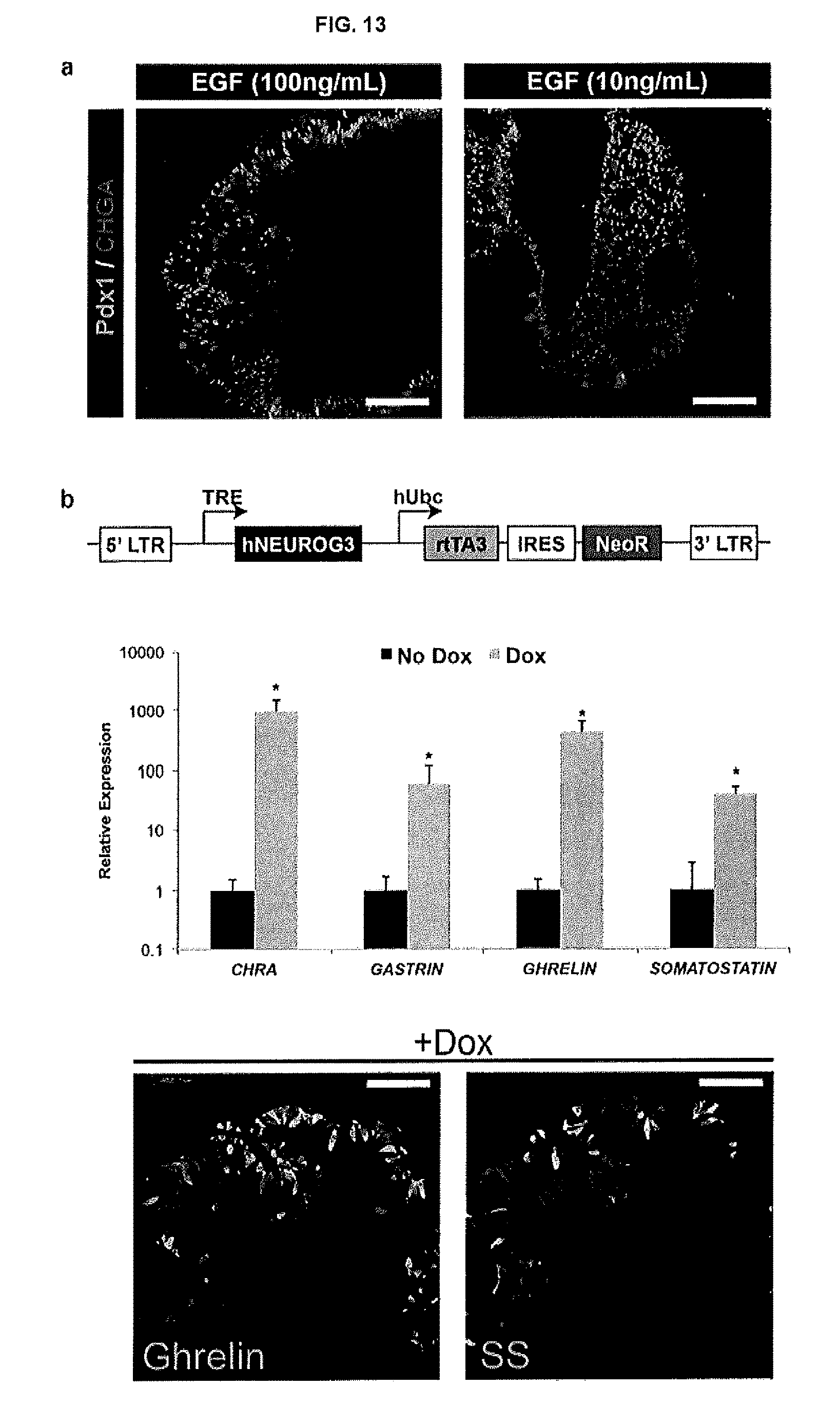
D00014

D00015

D00016

D00017

D00018

D00019

S00001
XML
uspto.report is an independent third-party trademark research tool that is not affiliated, endorsed, or sponsored by the United States Patent and Trademark Office (USPTO) or any other governmental organization. The information provided by uspto.report is based on publicly available data at the time of writing and is intended for informational purposes only.
While we strive to provide accurate and up-to-date information, we do not guarantee the accuracy, completeness, reliability, or suitability of the information displayed on this site. The use of this site is at your own risk. Any reliance you place on such information is therefore strictly at your own risk.
All official trademark data, including owner information, should be verified by visiting the official USPTO website at www.uspto.gov. This site is not intended to replace professional legal advice and should not be used as a substitute for consulting with a legal professional who is knowledgeable about trademark law.