Elimination Probe-based Method For Detecting Numerical Chromosomal Abnormalities, And Nucleic Acid Composition For Detecting Numerical Chromosomal Abnormalities
LEE; Si Seok ; et al.
U.S. patent application number 17/265102 was filed with the patent office on 2022-04-28 for elimination probe-based method for detecting numerical chromosomal abnormalities, and nucleic acid composition for detecting numerical chromosomal abnormalities. The applicant listed for this patent is SEASUN BIOMATERIALS. Invention is credited to Kyung Tak KIM, Si Seok LEE, Hee Kyung PARK, Eun Ju YANG.
| Application Number | 20220127665 17/265102 |
| Document ID | / |
| Family ID | 1000006107643 |
| Filed Date | 2022-04-28 |


View All Diagrams
| United States Patent Application | 20220127665 |
| Kind Code | A1 |
| LEE; Si Seok ; et al. | April 28, 2022 |
ELIMINATION PROBE-BASED METHOD FOR DETECTING NUMERICAL CHROMOSOMAL ABNORMALITIES, AND NUCLEIC ACID COMPOSITION FOR DETECTING NUMERICAL CHROMOSOMAL ABNORMALITIES
Abstract
The present invention relates to a method for analyzing the presence or absence of aneuploidy of a target chromosome with high sensitivity, and a composition for detecting chromosomal aneuploidy, and more particularly to a method of identifying chromosomal aneuploidy by amplifying a control nucleotide sequence, located on a chromosome not associated with chromosomal aneuploidy, and a target nucleotide sequence located on a chromosome associated with chromosomal aneuploidy, by using the same primer, and then hybridizing the amplification products with an assay probe that differs by one or two nucleotides from the control nucleotide sequence and with an elimination probe that comprises part or all of a sequence of the assay probe, which hybridizes with the target nucleotide sequence or the control nucleotide sequence, the elimination probe having a higher binding affinity for the amplification products than the assay probe, and analyzing melting curves of the hybridization products. The method for detecting chromosomal aneuploidy according to the present invention may analyze the ratio of the target nucleotide sequence to the control nucleotide sequence at high resolution by eliminating equal amounts (certain proportions) of the target nucleotide sequence and the control nucleotide sequence from the analysis using the elimination sequence. This method is useful because numerical abnormalities (aneuploidy) in chromosomes (e.g., fetal chromosomes in maternal blood, and circulating tumor DNA in cancer patients) present at low rates can be detected quickly with high sensitivity by the use of this method.
| Inventors: | LEE; Si Seok; (Daejeon, KR) ; KIM; Kyung Tak; (Daejeon, KR) ; YANG; Eun Ju; (Daejeon, KR) ; PARK; Hee Kyung; (Daejeon, KR) | ||||||||||
| Applicant: |
|
||||||||||
|---|---|---|---|---|---|---|---|---|---|---|---|
| Family ID: | 1000006107643 | ||||||||||
| Appl. No.: | 17/265102 | ||||||||||
| Filed: | July 23, 2019 | ||||||||||
| PCT Filed: | July 23, 2019 | ||||||||||
| PCT NO: | PCT/KR2019/009067 | ||||||||||
| 371 Date: | February 1, 2021 |
| Current U.S. Class: | 1/1 |
| Current CPC Class: | C12Q 1/686 20130101; C12Q 1/6825 20130101; C12Q 1/6883 20130101; C12Q 2600/156 20130101; C12Q 1/6818 20130101; C12Q 1/6832 20130101 |
| International Class: | C12Q 1/6832 20060101 C12Q001/6832; C12Q 1/6825 20060101 C12Q001/6825; C12Q 1/6818 20060101 C12Q001/6818; C12Q 1/686 20060101 C12Q001/686; C12Q 1/6883 20060101 C12Q001/6883 |
Foreign Application Data
| Date | Code | Application Number |
|---|---|---|
| Jul 31, 2018 | KR | 10-2018-0089224 |
Claims
1. A method for detecting chromosomal aneuploidy, the method comprising steps of: a) isolating DNAs from a normal sample and a sample derived from a patient expected to have chromosomal aneuploidy, respectively; b) performing amplification using a primer capable of amplifying both a control nucleotide sequence located on a chromosome not associated with chromosomal aneuploidy and a target nucleotide located on a chromosome associated with chromosomal aneuploidy; c) hybridizing the amplification products with an assay probe capable of hybridizing to a sequence, which differs by one or two nucleotides from the control nucleotide sequence or the target nucleotide sequence, and with an elimination probe comprising part or all of a sequence of the assay probe, which hybridizes with the target nucleotide sequence or the control nucleotide sequence, the elimination probe having a higher binding affinity for the amplification products of step b) than the assay probe; and d) identifying chromosomal aneuploidy by analyzing melting curves of the hybridization products for the normal sample and the subject sample, obtained in step c).
2. The method of claim 1, wherein a primer or probe hybridization region of the control nucleotide sequence of step b) is at least 90% homologous to a primer or probe hybridization region of the target nucleotide sequence.
3. The method of claim 1, wherein the assay probe of step c) has a melting temperature difference of 8.degree. C. or more when it either perfectly matches or mismatches the control nucleotide sequence or the target nucleotide sequence.
4. The method of claim 1, wherein the assay probe of step c) is a peptide nucleic acid (PNA), and a reporter and a quencher are attached to both ends of the assay probe.
5. The method of claim 4, wherein the reporter is at least one selected from the group consisting of FAM (6-carboxyfluorescein), Texas red, HEX (2',4',5',7'-tetrachloro-6-carboxy-4,7-dichlorofluorescein) and Cy5.
6. The method of claim 4, wherein the quencher is at least one selected from the group consisting of TAMRA (6-carboxytetramethyl-rhodamine), BHQ1, BHQ2 and Dabcyl.
7. The method of claim 1, wherein the elimination probe of step c) is selected from the group consisting of: a probe for eliminating only the product of amplification of the target nucleotide sequence; and a probe for eliminating both the products of amplification of the target nucleotide sequence and the control nucleotide sequence.
8. The method of claim 7, wherein the elimination probe of step c) hybridizes with the product of amplification of the control nucleotide sequence or the target nucleotide sequence competitively with the assay probe.
9. The method of claim 8, wherein the elimination probe is selected from the group consisting of an oligonucleotide, LNA, PNA, and combinations thereof.
10. The method of claim 1, wherein the elimination probe of step c) eliminates 50 to 90% of the amplification products of step b).
11. The method of claim 1, wherein analysis of the melting curves in step d) is performed by a method comprising steps of: a) calculating the mismatch value/perfect match value ratio of the product of amplification of the normal sample DNA; b) calculating the mismatch value/perfect match value ratio of the product of amplification of the subject sample DNA; and c) determining that the subject sample is normal when the ratio calculated in step a) is the same as the ratio calculated in step b), and has chromosomal aneuploidy when the ratio calculated in step a) is different from the ratio calculated in step b).
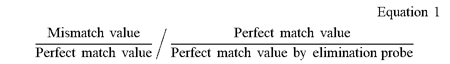
12. The method of claim 11, wherein the method for performing analysis of the melting curves further comprises: step d) of correcting the perfect match value, obtained by the elimination probe, using the following Equation 1 when calculating the ratios in step a) and step b): .times. Equation .times. .times. 1 ##EQU00002## Mismatch .times. .times. value Perfect .times. .times. match .times. .times. value / Perfect .times. .times. match .times. .times. value Perfect .times. .times. match .times. .times. value .times. .times. by .times. .times. elimination .times. .times. probe ##EQU00002.2##
13. The method of claim 1, which is a method for detecting multiple chromosomal aneuploidies, which uses at least two primers, at least two assay probes, and at least two elimination probes, in which the assay probes have different reporters.
14. A PCR composition for detecting chromosomal aneuploidy, the PCR composition comprising: i) a primer capable of amplifying both a control nucleotide sequence located on a chromosome not associated with chromosomal aneuploidy and a target nucleotide located on a chromosome associated with chromosomal aneuploidy; ii) an assay probe capable of hybridizing with a sequence that differs by one or two nucleotides from the control nucleotide sequence or the target nucleotide sequence; and iii) an elimination probe comprising part or all of a sequence of the assay probe, which hybridizes with the target nucleotide sequence or the control nucleotide sequence, the elimination probe having a higher binding affinity than the assay probe.
15. The method of claim 1, wherein the primer is selected from a group consisting of SEQ ID NOS:1 to 30.
16. The method of claim 1, wherein the assay probe is selected from a group consisting of SEQ ID NOS:31 to 60.
17. The method of claim 1, wherein the elimination probe is selected from a group consisting of SEQ ID NOS:61 to 86.
Description
SEQUENCE LISTING
[0001] The instant application contains a Sequence Listing which has been submitted electronically in ASCII format and is hereby incorporated by reference in its entirety. Said ASCII copy, created Oct. 7, 2021, is named 404266_001 US_SL.txt and is 20,409 bytes in size.
TECHNICAL FIELD
[0002] The present invention relates to a method for analyzing the presence or absence of aneuploidy of a target chromosome with high sensitivity, and a composition for detecting chromosomal aneuploidy, and more particularly to a method of identifying chromosomal aneuploidy by amplifying a control nucleotide sequence, located on a chromosome not associated with chromosomal aneuploidy, and a target nucleotide sequence located on a chromosome associated with chromosomal aneuploidy, by using the same primer, and then hybridizing the amplification products with an assay probe that differs by one or two nucleotides from the control nucleotide sequence or target nucleotide sequence and with an elimination probe that comprises part or all of a sequence of the assay probe, which hybridizes with the target nucleotide sequence or the control nucleotide sequence, the elimination probe having a higher binding affinity for the amplification products than the assay probe, and analyzing melting curves of the hybridization products.
BACKGROUND ART
[0003] Chromosomal abnormalities are associated with genetic defects and degenerative diseases. Chromosome abnormality may indicate deletion or duplication of a chromosome, deletion or duplication of a part of chromosome, or a break, translocation, or inversion in the chromosome. Chromosomal abnormalities are disturbances in the genetic balance and cause fetal death or serious defect in physical and mental states. For example, Down's syndrome is a common form of chromosomal aneuploidy (numerical chromosomal abnormality) caused by the presence of three chromosomes 21 (trisomy 21). Edwards syndrome (trisomy 18), Patau syndrome (trisomy 13), Turner syndrome (XO) and Klinefelter syndrome (XXY) also correspond to chromosomal aneuploidy.
[0004] Chromosomal abnormalities may be detected using karyotyping and fluorescent in situ hybridization (FISH). These detection methods are disadvantageous in terms of time, effort and accuracy. Furthermore, karyotyping requires a lot of time for cell culture. FISH is only available for samples of known nucleic acid sequence and chromosomal location. FISH may be used only for samples having known nucleic acid sequences and chromosomal locations. In order to avoid the problems of FISH, comparative genome hybridization (CGH) may be used. CGH may detect a region in which chromosomal aneuploidy has occurred by analyzing the whole genome. However, CGH has a disadvantage in that the resolution thereof is lower than that of FISH.
[0005] As an alternative approach, DNA microarrays may be used to detect chromosomal abnormalities. The DNA microarray systems may be classified, according to the type of bio-molecules immobilized on the microarray, into cDNA microarrays, oligonucleotide microarrays, and genomic microarrays. cDNA microarrays and oligonucleotide microarrays are easy to fabricate, but these systems have disadvantages in that the number of probes immobilized on the microarray is limited, probe fabrication is expensive, and it is difficult to detect chromosomal abnormalities located outside the probes.
[0006] In particular, in the case of a genomic DNA microarray system, it is easy to fabricate a probe, and it is possible detect chromosomal abnormalities not only in the extended region of the chromosome but also in the intron region of the chromosome, but it is difficult to produce a large number of DNA fragments whose localization and function within the chromosome are known.
[0007] Recently, next-generation sequencing technology has been used to analyze numerical chromosomal abnormalities (chromosomal aneuploidy) (Park, H., Kim et al., Nat Genet 2010, 42, 400-405.; Kidd, J. M. et al., Nature 2008, 453, 56-64). However, this technology requires high coverage reading for the analysis of chromosomal aneuploidy, and CNV measurements also require independent validation. Therefore, this technology was not suitable as a general gene search analysis method at that time because it was very expensive and the results were difficult to understand.
[0008] For this purpose, real-time qPCR is currently used as an advanced technique for quantitative gene analysis, because a wide dynamic range (Weaver, S. et al, Methods 2010, 50, 271-276) and a linear correlation between the threshold cycle and the initial target amount are observed reproducibly (Deepak, S. et al., Curr Genomics 2007,8, 234-251). However, the sensitivity of qPCR is not high enough to discriminate differences in the number of copies. Despite the wide dynamic range of qPCR assay, small changes, such as 1.5-fold changes, cannot be reliably measured due to the intrinsic variables of qPCR-based assays. In addition, multiple temporally repetitive analyses are required for reliable distinction between samples with similar copies of DNA. Furthermore, qPCR is not suitable for multimodal analysis. For example, for detection of multiple targets, a reaction for separating the targets from each other is required to distinguish one target from others (Bustin, S. A., J Mol Endocrinol 2002, 29, 23-39). In addition, due to the limited availability and spectral overlap of fluorescent tags, qPCR can separate up to only 4 targets per assay. However, for successful quadruplex analysis in qPCR, a careful combination of fluorescent tags is essential for each analysis (Bustin, S A, J Mol Endocrinol 2002, 29, 23-39), which is a serious disadvantage of qPCR as a clinical diagnostic tool.
[0009] Accordingly, the present inventors have made extensive efforts to solve the above-described problems and to develop a method for detecting chromosomal aneuploidy, which may provide analysis results quickly with high sensitivity. As a result, the present inventors have found that, when both a control nucleotide sequence and a target nucleotide sequence are amplified and then the amplification product from the control nucleotide sequence is eliminated using an elimination probe, analysis results may be obtained quickly with high sensitivity, thereby completing the present invention.
SUMMARY OF THE INVENTION
[0010] An object of the present invention is to provide a method for detecting chromosomal aneuploidy.
[0011] Another object of the present invention is to provide a PCR composition for detecting chromosomal aneuploidy.
[0012] To achieve the above objects, the present invention provides a method for detecting chromosomal aneuploidy, the method comprising steps of: a) isolating DNAs from a normal sample and a subject sample, respectively; b) performing amplification using a primer capable of amplifying both a control nucleotide sequence located on a chromosome not associated with chromosomal aneuploidy and a target nucleotide located on a chromosome associated with chromosomal aneuploidy; c) hybridizing the amplification products with an assay probe capable of hybridizing to a sequence, which differs by one or two nucleotides from the control nucleotide sequence or the target nucleotide sequence, and with an elimination probe comprising part or all of a sequence of the assay probe, which hybridizes with the target nucleotide sequence or the control nucleotide sequence, the elimination probe having a higher binding affinity for the amplification products of step b) than the assay probe; and d) identifying chromosomal aneuploidy by analyzing melting curves of the hybridization products for the normal sample and the subject sample, obtained in step c).
[0013] The present invention also provides a PCR composition for detecting chromosomal aneuploidy, the PCR composition comprising: i) a primer capable of amplifying both a control nucleotide sequence located on a chromosome not associated with chromosomal aneuploidy and a target nucleotide located on a chromosome associated with chromosomal aneuploidy; ii) an assay probe capable of hybridizing a sequence that differs by one or two nucleotides from the control nucleotide sequence or the target nucleotide sequence; and iii) an elimination probe comprising part or all of a sequence of the assay probe, which hybridizes to the target nucleotide sequence or the control nucleotide sequence, the elimination probe having a higher binding affinity than the assay probe.
[0014] The present invention also provides the use of the PCR composition for detection of chromosomal aneuploidy.
BRIEF DESCRIPTION OF THE DRAWINGS
[0015] FIG. 1 is a schematic view showing that normal and abnormal chromosomes are eliminated at the same rate by the use of an elimination probe according to the present invention.
[0016] FIG. 2 is a schematic view showing change in analytical resolution depending on the elimination rate by use of the elimination probe according to the present invention.
[0017] FIG. 3 is schematic view showing conditions for selecting a target nucleotide sequence according to the present invention and conditions for selecting a primer for amplification of the target nucleotide sequence.
[0018] FIG. 4 is a diagram showing real-time PCR conditions for determining whether the chromosome ratio is abnormal according to the present invention.
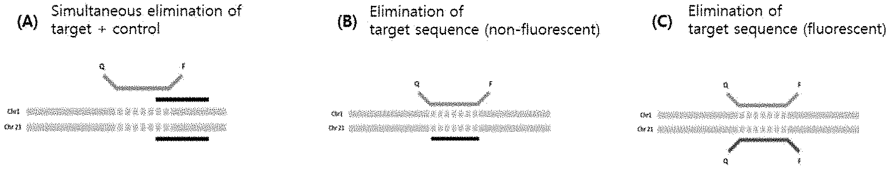
[0019] FIG. 5 is a schematic view showing a detection probe and an elimination probe according to the present invention. FIG. 5(A) shows a non-fluorescent elimination probe that binds to both a target nucleotide sequence and a control nucleotide sequence, FIG. 5(B) shows a non-fluorescent probe that binds only to a control nucleotide sequence, and FIG. 5(C) shows a fluorescent elimination probe that binds only to a control nucleotide sequence.
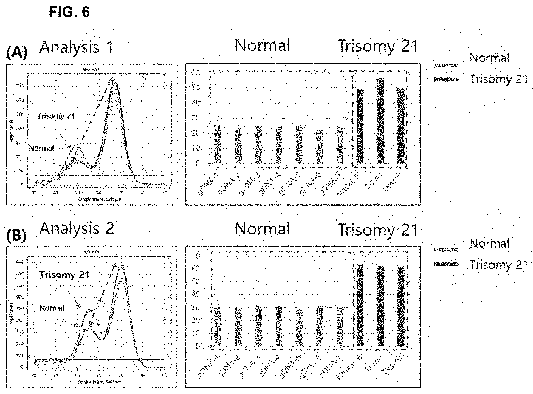
[0020] FIG. 6 shows the results of analysis using a Down's syndrome cell line according to the present invention. FIGS. 6(A) and 6(B) show the results of analyses based on different control nucleotide sequences and target nucleotide sequences.
[0021] FIG. 7 shows the results of analysis using an Edward's syndrome cell line according to the present invention. FIGS. 7(A) and 7(B) show the results of analyses based on different control nucleotide sequences and target nucleotide sequences.
[0022] FIG. 8 shows the results of analysis using a Patau syndrome cell line according to the present invention. FIGS. 8(A) and 8(B) show the results of analyses based on different control nucleotide sequences and target nucleotide sequences.
[0023] FIG. 9 illustrates the results of analyzing sensitivity depending on the proportion of DNA using a Down's syndrome cell line according to the present invention. FIGS. 9(A) and 9(B) show the results of analyses based on different control nucleotide sequences and target nucleotide sequences.
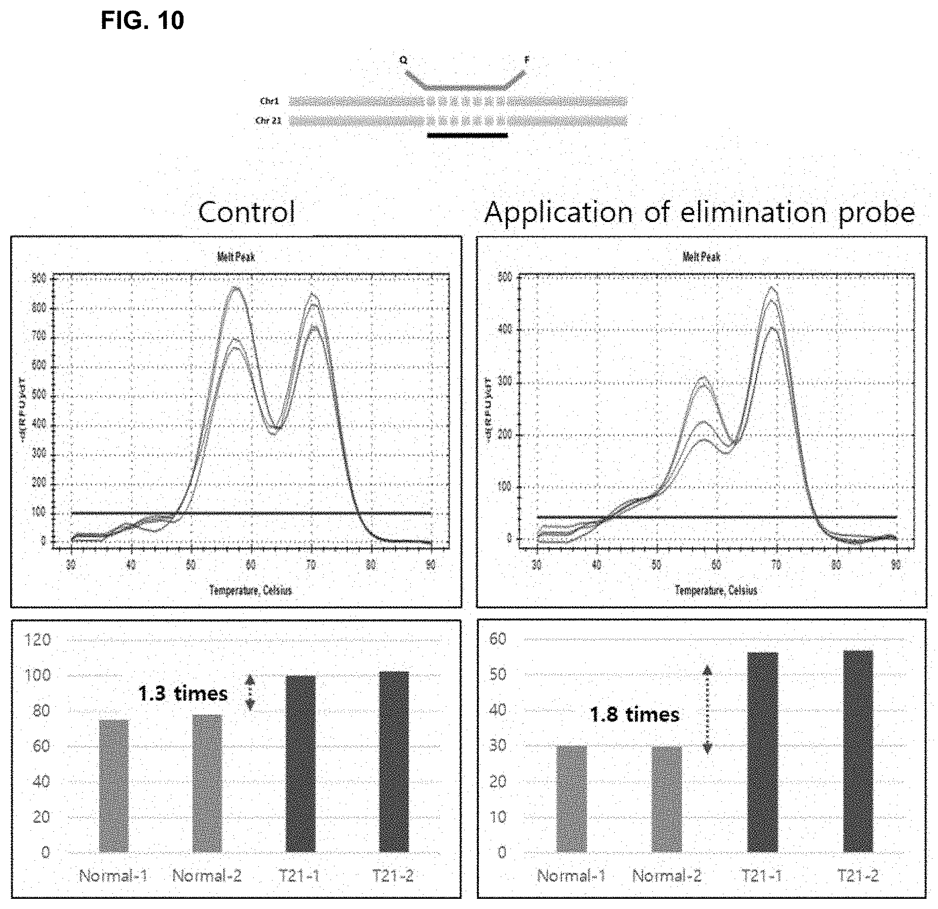
[0024] FIG. 10 shows results indicating the increase in analytical resolution by the use of a non-fluorescent probe that eliminates only a control nucleotide sequence according to the present invention.
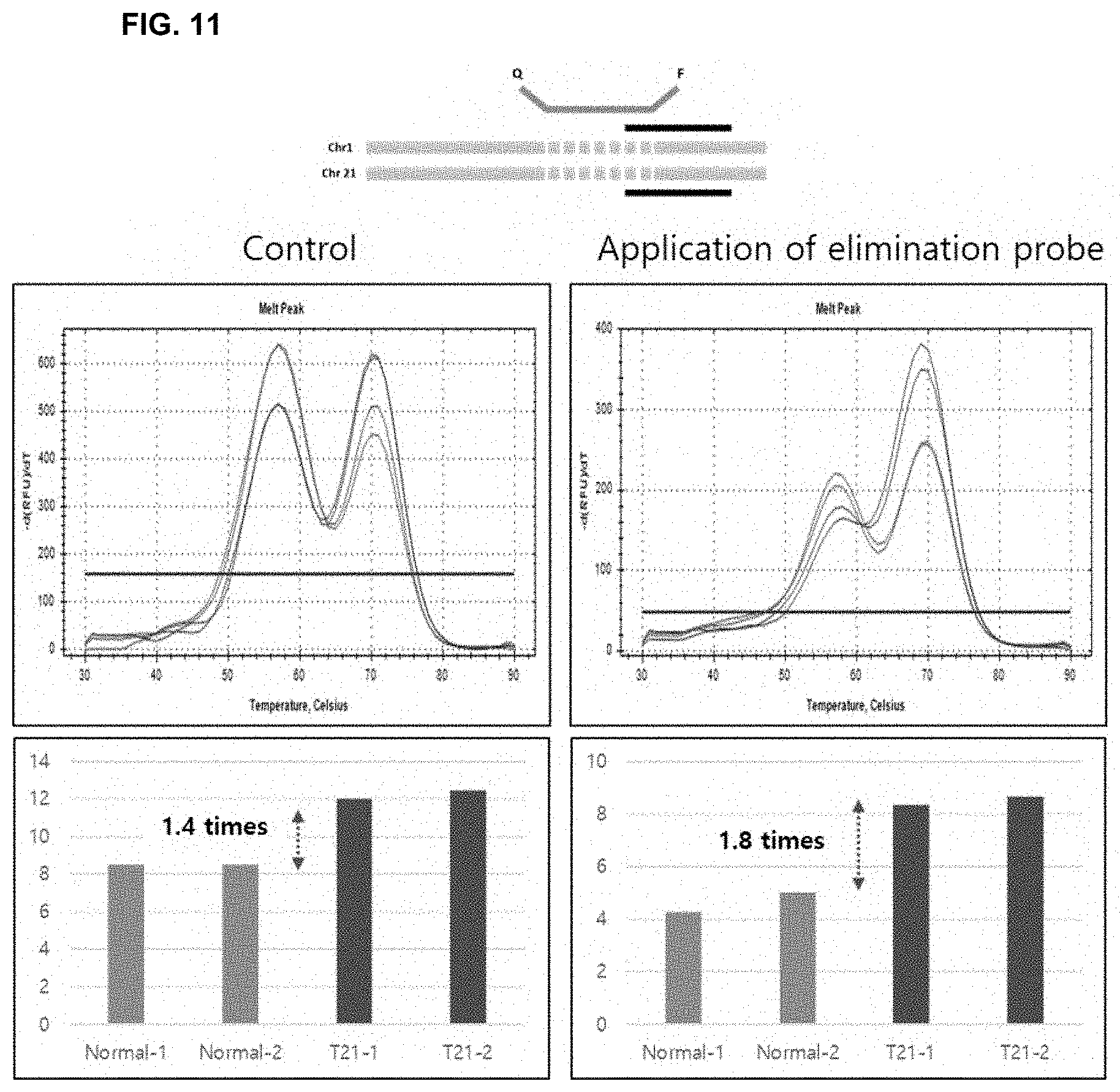
[0025] FIG. 11 shows results indicating the increase in analytical resolution by the use of a non-fluorescent probe that simultaneously eliminates a target nucleotide sequence and a control nucleotide sequence according to the present invention.
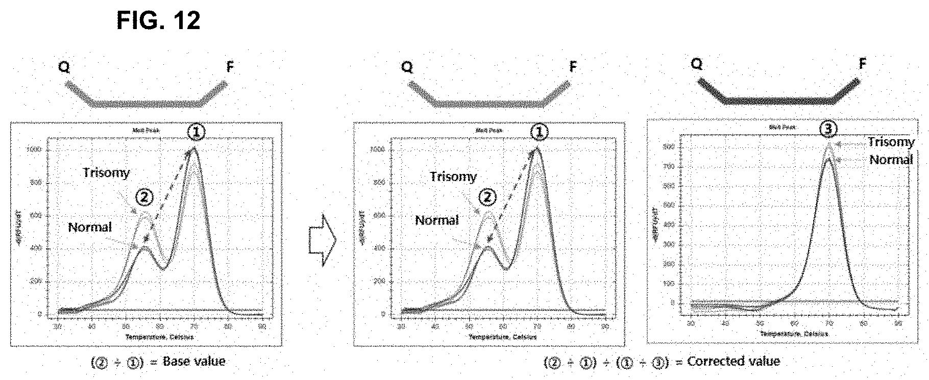
[0026] FIG. 12 is a schematic view showing correction of the results obtained using a fluorescent elimination probe targeting a control nucleotide sequence according to the present invention.
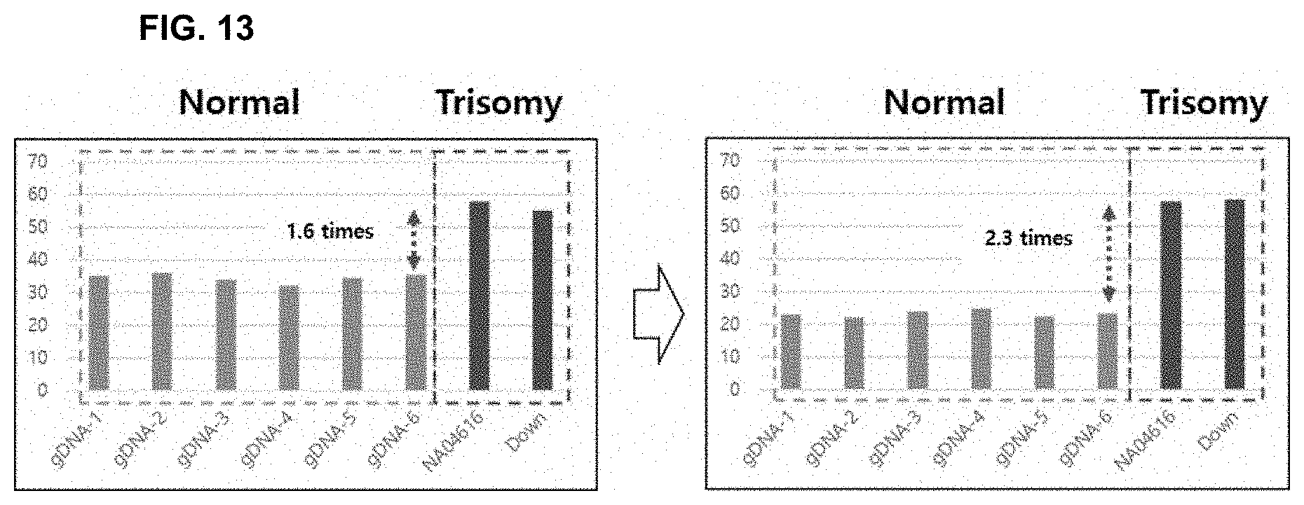
[0027] FIG. 13 illustrates the results of correcting the results according to the present invention.
[0028] FIG. 14 shows the results of comparatively analyzing a standard substance and a clinical sample.
DETAILED DESCRIPTION AND PREFERRED EMBODIMENTS OF THE INVENTION
[0029] Unless otherwise defined, all technical and scientific terms used in the present specification have the same meanings as commonly understood by those skilled in the art to which the present disclosure pertains. In general, the nomenclature used in the present specification is well known and commonly used in the art.
[0030] In the present invention, based on DNA isolated from a normal sample and a subject sample, a target nucleotide sequence located on a chromosome expected to be associated with chromosomal aneuploidy and a control nucleotide sequence located on a chromosome not associated with chromosomal aneuploidy while having at least 90% homology to the target nucleotide sequence were amplified using the same primer, certain amounts of the amplification products were eliminated with an elimination probe, and then melting curves of the amplification products were analyzed using an assay probe. As a result, it was confirmed that chromosomal aneuploidy could be detected with high sensitivity.
[0031] That is, in one example of the present invention, amplification products were produced using a synthesized primer capable of amplifying a certain region of each of chromosomes 1, 4 and 7 while amplifying a certain region of chromosome 21, or a synthesized primer capable of amplifying a certain region of each of chromosomes 1, 4, 9 and 15 while amplifying a certain region of chromosome 19, or a synthesized primer capable of amplifying a certain region of each of chromosomes 3, 6 and 12 while amplifying a certain region of chromosome 13, and then a certain proportion of each of the amplification products was prevented from binding to an assay probe by the use of an elimination probe capable of hybridizing with the amplification products, and then the mismatch/perfect match ratio for the normal sample and the subject sample was calculated by analyzing melting curves. As a result, it was confirmed that chromosomal aneuploidy could be detected with high sensitivity (FIGS. 1 and 2). Therefore, in one aspect, the present invention is directed to a method for detecting chromosomal aneuploidy, the method comprising steps of: [0032] a) isolating DNAs from a normal sample and a subject sample, respectively; [0033] b) performing amplification using a primer capable of amplifying both a control nucleotide sequence located on a chromosome not associated with chromosomal aneuploidy and a target nucleotide located on a chromosome associated with chromosomal aneuploidy; [0034] c) hybridizing the amplification products with an assay probe capable of hybridizing to a sequence, which differs by one or two nucleotides from the control nucleotide sequence or the target nucleotide sequence, and with an elimination probe comprising part or all of a sequence of the assay probe, which hybridizes to the target nucleotide sequence or the control nucleotide sequence, the elimination probe having a higher binding affinity for the amplification products of step b) than the assay probe; and [0035] d) identifying chromosomal aneuploidy by analyzing melting curves of the hybridization products for the normal sample and the subject sample, obtained in step c).
[0036] As used herein, the term "target nucleotide sequence" refers to all types of nucleic acids to be detected, and include chromosomal sequences from different species, subspecies or variants, or chromosomal mutations within the same species. The target nucleotide sequence may be characterized by all types of DNA including genomic DNA, mitochondrial DNA, and viral DNA, or all types of RNA including mRNA, ribosomal RNA, non-coding RNA, tRNA, and viral RNA, but is not limited thereto.
[0037] In the present invention, the target nucleotide sequence may be a mutant nucleotide sequence including a variation of the nucleotide sequence, and the mutation may be selected from the group consisting of single nucleotide polymorphism (SNP), insertion, deletion, point mutation, fusion mutation, translocation, inversion, and LOH (loss of heterozygosity), but is not limited thereto.
[0038] As used herein, the term "nucleoside" refers to a glycosylamine compound wherein a nucleic acid base (nucleobase) is linked to a sugar moiety. The term "nucleotide" refers to a nucleoside phosphate. A nucleotide may be represented using alphabetical letters (letter designation) corresponding to its nucleoside as described in Table 1. For example, A denotes adenosine (a nucleoside containing the nucleobase, adenine), C denotes cytidine, G denotes guanosine, U denotes uridine, and T denotes thymidine (5-methyl uridine). W denotes either A or T/U, and S denotes either G or C. N represents a random nucleoside and dNTP refers to deoxyribonucleoside triphosphate. N may be any of A, C, G, or T/U.
TABLE-US-00001 TABLE 1 Symbol Nucleotide represented letter by symbol letter G G A A T T C C U U R G or A Y T/U or C M A or C K G or T/U S G or C W A or T/U H A or C or T/U B G or T/U or C V G or C or A D G or A or T/U H G or A or T/U or C
[0039] As used herein, the term "oligonucleotide" refers to oligomers of nucleotides. The term "nucleic acid" as used herein refers to polymers of nucleotides. The term "sequence" as used herein refers to a nucleotide sequence of an oligonucleotide or a nucleic acid. Throughout the specification, whenever an oligonucleotide or nucleic acid is represented by a sequence of letters, the nucleotides are in 5'.fwdarw.3' order from left to right. The oligonucleotides or nucleic acids may be DNA, RNA, or analogues thereof (e.g., phosphorothioate analogue). The oligonucleotides or nucleic acids may also include modified bases and/or backbones (e.g., modified phosphate linkage or modified sugar moiety). Non-limiting examples of synthetic backbones that confer stability and/or other advantages to the nucleic acids may include phosphorothioate linkages, peptide nucleic acid, locked nucleic acid, xylose nucleic acid, or analogues thereof.
[0040] As used herein, the term "nucleic acid" refers to a nucleotide polymer, and unless otherwise limited, would encompass known analogs of natural nucleotides that can function in a similar manner (e.g., hybridization) as naturally occurring nucleotides.
[0041] The term "nucleic acid" includes, for example, genomic DNA; complementary DNA (cDNA) (which is a DNA representation of mRNA, usually obtained by reverse transcription of messenger RNA (mRNA) or by amplification); DNA molecules produced synthetically or by amplification; and any form of DNA or RNA including mRNA.
[0042] The term "nucleic acid" encompasses double- or triple-stranded nucleic acids, as well as single-stranded molecules. In double- or triple-stranded nucleic acids, the nucleic acid strands need not be coextensive (i.e., a double-stranded nucleic acid need not be double-stranded along the entire length of both strands).
[0043] The term nucleic acid also encompasses any chemical modification thereof, such as by methylation and/or by capping. Nucleic acid modifications can include addition of chemical groups that incorporate additional charge, polarizability, hydrogen bonding, electrostatic interaction, and functionality to the individual nucleic acid bases or to the nucleic acid as a whole. Such modifications may include base modifications such as 2'-position sugar modifications, 5-position pyrimidine modifications, 8-position purine modifications, modifications at cytosine exocyclic amines, substitutions of 5-bromo-uracil, backbone modifications, unusual base pairing combinations such as the isobases isocytidine and isoguanidine, and the like.
[0044] The nucleic acid(s) can be derived from a completely chemical synthesis process, such as solid phase-mediated chemical synthesis, from a biological source, such as through isolation from any species that produces nucleic acid, or from processes that involve the manipulation of nucleic acids by molecular biology tools, such as DNA replication, PCR amplification, reverse transcription, or from a combination of those processes.
[0045] As used herein, the term "complementary" refers to refers to the capacity for precise pairing between two nucleotides. That is, if a nucleotide at a given position of a nucleic acid is capable of hydrogen bonding with a nucleotide of another nucleic acid, then the two nucleic acids are considered to be complementary to one another at that position. Complementarity between two single-stranded nucleic acid molecules may be "partial," in which only some of the nucleotides bind, or it may be complete when total complementarity exists between the single-stranded molecules. The degree of complementarity between nucleic acid strands has significant effects on the efficiency and strength of hybridization between nucleic acid strands.
[0046] As used herein, the term "primer" refers to a short linear oligonucleotide that hybridizes with a target nucleic acid sequence (e.g., a DNA template to be amplified) to prime a nucleic acid synthesis reaction. The primer may be an RNA oligonucleotide, a DNA oligonucleotide, or a chimeric sequence. The primer may contain natural, synthetic, or modified nucleotides. Both the upper and lower limits of the length of the primer are empirically determined. The lower limit on the primer length is the minimum length that is required to form a stable duplex upon hybridization with the target nucleic acid under nucleic acid amplification reaction conditions. Very short primers (usually less than 3 to 4 nucleotides long) do not form thermodynamically stable duplexes with target nucleic acid under such hybridization conditions. The upper limit is often determined by the possibility of having duplex formation in a region other than the predetermined nucleic acid sequence in the target nucleic acid. Generally, suitable primer lengths are in the range of about 4 to about 40 nucleotides long.
[0047] As used herein, the term "probe" is a nucleic acid capable of binding to a target nucleic acid of complementary sequence through one or more types of chemical bonds, generally through complementary base pairing, usually through hydrogen bond formation, thus forming a duplex structure. The probe binds or hybridizes to a "probe binding site." The probe can be labeled with a detectable label to permit facile detection of the probe, particularly once the probe has hybridized to its complementary target. Alternatively, however, the probe may be unlabeled, but may be detectable by specific binding with a ligand that is labeled, either directly or indirectly. Probes can vary significantly in size. Generally, probes are at least 7 to 15 nucleotides in length. Other probes are at least 20, 30, or 40 nucleotides long. Still other probes are somewhat longer, being at least 50, 60, 70, 80, or 90 nucleotides long. Yet other probes are longer still, and are at least 100, 150, 200 or more nucleotides long. Probes can also be of any length that is within any range bounded by any of the above values (e.g., 15 to 20 nucleotides in length).
[0048] As used herein, the term "hybridization" refers to the formation of a double-stranded nucleic acid by hydrogen bonding between single-stranded nucleic acids having complementary base sequences, and is used in a similar sense to annealing. However, in a broader sense, hybridization includes cases where nucleotides are perfectly complementary (perfect match) between two single-stranded molecules, as well as cases where some nucleotides are not complementary (mismatch).
[0049] In the present invention, the amplification is not limited as long as it is polymerase chain reaction (PCR), but it is preferably asymmetric PCR.
[0050] In the present invention, the homology of the primer or probe hybridization region of the control nucleotide sequence of step b) is not limited as long as the same probe or primer is capable of binding complementarily to the primer or probe hybridization region of the target nucleotide sequence, but the homology is preferably at least 80%, more preferably at least 90%, most preferably 95%.
[0051] In the present disclosure, the control nucleotide sequence may be selected under the conditions described in FIG. 3.
[0052] In the present invention, the assay probe in step c) may be used without limitation as long as the melting temperature difference occurs to the extent that it can be distinguished on the analysis graph, when the assay probe either perfectly matches or mismatches the control nucleotide sequence or the target nucleotide sequence. Preferably, the melting temperature difference may be 5.degree. C. to 20.degree. C., more preferably 7.degree. C. to 20.degree. C., most preferably 8.degree. C. to 20.degree. C.
[0053] In the present invention, the assay probe in step c) may be a peptide nucleic acid (PNA), and a reporter and a quencher may be attached to both ends of the assay probe.
[0054] In the present invention, the peptide nucleic acid (PNA) is one of substances that recognize genes, like LNA (locked nucleic acid) and MNA (morpholino nucleic acid), and is synthesized artificially, and the backbone thereof is composed of polyamide. PNA has excellent affinity and selectivity, and is not degraded by existing restriction enzymes due to high stability thereof against nucleases. In addition, PNA has an advantage of being easy to store and is not easily degraded due to high thermal/chemical properties and stability thereof. In addition, PNA-DNA binding is much stronger than DNA-DNA binding, and a melting temperature (Tm) difference of about 10 to 15.degree. C. appears even for one nucleotide mismatch. Using this difference in binding strength, it is possible to detect changes in single nucleotide polymorphism (SNP) and insertion/deletion (InDel) nucleic acids.
[0055] The Tm value also changes depending on the difference between the nucleotide sequence of the PNA probe and the nucleolide sequence of DNA complementary thereto, and thus the development of applications based on this change is easily achieved. The PNA probe is analyzed using a hybridization reaction different from the hydrolysis reaction of the TaqMan probe, and probes having functions similar to those of the PNA probe include molecular beacon probes and scorpion probes.
[0056] In the present invention, a reporter or a quencher may be attached to the PNA probe, without being limited thereto. The PNA probe comprising the reporter and quencher according to the present invention generates a fluorescent signal after hybridization with the target nucleic acid, and as the temperature rises, the fluorescent signal is quenched by rapid melting of the target nucleic acid at an appropriate melting temperature of the probe. Through analysis of a high-resolution melting curve obtained from the fluorescent signal resulting from this temperature change, the presence or absence of the target nucleic acid may be detected.
[0057] The probe of the present invention may have a reporter and a quencher capable of quenching reporter fluorescence, attached at both ends thereof, and may include an intercalating fluorophore. The reporter may be one or more selected from the group consisting of FAM (6-carboxyfluorescein), HEX, Texas red, JOE, TAMRA, CY5, CY3, and Alexa680, and the quencher is preferably TAMRA (6-carboxytetramethyl-rhodamine), BHQ1, BHQ2 or Dabcyl, but is not limited thereto. The intercalating fluorophore may be selected from the group consisting of Acridine homodimer and derivatives thereof, Acridine Orange and derivatives thereof, 7-aminoactinomycin D (7-AAD) and derivatives thereof, Actinomycin D and derivatives thereof, 9-amino-6-chloro-2-methoxyacridine (ACMA) and derivatives thereof, DAPI and derivatives thereof, dihydroethidium and derivatives thereof, ethidium bromide and derivatives thereof, ethidium homodimer-1 (EthD-1) and derivatives thereof, ethidium homodimer-2 (EthD-2) and derivatives thereof, ethidium monoazide and derivatives thereof, hexidium iodide and derivatives thereof, bisbenzimide (Hoechst 33258) and derivatives thereof, Hoechst 33342 and derivatives thereof, Hoechst 34580 and derivatives thereof, hydroxystilbamidine and derivatives thereof, LDS 751 and derivatives thereof, propidium iodide (PI) and derivatives thereof, and Cy-dyes derivatives.
[0058] In the present invention, the elimination probe in step c) may be selected from the group consisting of: a probe for eliminating only the product of amplification of the target nucleotide sequence; and a probe for eliminating both the products of amplification of the target nucleotide sequence and the control nucleotide sequence.
[0059] In the present invention, the elimination probe in step c) may hybridize to the product of amplification of the control nucleotide sequence or the target nucleotide sequence competitively with the assay probe.
[0060] In the present invention, the elimination probe may be selected from the group consisting of an oligonucleotide, LNA, PNA, and combinations thereof.
[0061] In the present invention, the elimination probe in step c) may eliminate 50 to 90% of the amplification products obtained in step b).
[0062] In the present invention, the elimination probe may have a higher Tm value than the assay probe.
[0063] In the present invention, analysis of the melting curves in step d) may be performed by a method comprising steps of: [0064] a) calculating the mismatch value/perfect match value ratio of the product of amplification of the normal sample DNA; [0065] b) calculating the mismatch value/perfect match value ratio of the product of amplification of the subject sample DNA; and [0066] c) determining that the subject sample is normal when the ratio calculated in step a) is the same as the ratio calculated in step b), and has chromosomal aneuploidy when the ratio calculated in step a) is different from the ratio calculated in step b).
[0067] In the present invention, the analysis of the melting curves may further comprise: [0068] step d) of correcting the perfect match, obtained by the elimination probe value, using the following Equation 1 when calculating the ratios in step a) and step b):
[0068] .times. Equation .times. .times. 1 ##EQU00001## Mismatch .times. .times. value Perfect .times. .times. match .times. .times. value / Perfect .times. .times. match .times. .times. value Perfect .times. .times. match .times. .times. value .times. .times. by .times. .times. elimination .times. .times. probe ##EQU00001.2##
[0069] Fluorescence melting curve analysis (FMCA) is used as the method for analyzing the hybridization reaction. Fluorescence melting curve analysis analyzes the difference in binding affinity between the PCR reaction product and the introduced probe depending on the melting temperature. Unlike other SNP detection probes, the probe is very simple to design, and thus is constructed using an 11 to 18-mer nucleotide sequence including an SNP. Therefore, in order to design a probe having a desired melting temperature, the Tm value may be adjusted according to the length of the PNA probe, and even in the case of PNA probes having the same length, the Tm value may be adjusted by changing the probes. Since PNA has a higher binding affinity than DNA and thus has a higher basic Tm value, PNA may be designed with a shorter length than DNA, and thus can detect even closely neighboring SNPs. In a conventional HRM method, the difference in Tm value is very small at about 0.5.degree. C., and thus an additional analysis program or a minute temperature change are required, and analysis becomes difficult when two or more SNPs appear. However, the PNA probe is not affected by SNPs other than the probe sequence, and thus enables fast and accurate analysis.
[0070] The present invention is also directed to a method for detecting multiple chromosomal aneuploidies, which uses at least two primers, at least two assay probes, and at least two elimination probes, and in which the assay probes have different reporters.
[0071] It is obvious to those skilled in the art that the method for detecting chromosomal aneuploidy according to the present invention may be applied not only for detection of fetal chromosomal abnormalities, but also for detection of cancer-related chromosomal abnormalities.
[0072] In another aspect, the present invention is directed to a PCR composition for detecting chromosomal aneuploidy, the PCR composition comprising: [0073] i) a primer capable of amplifying both a control nucleotide sequence located on a chromosome not associated with chromosomal aneuploidy and a target nucleotide located on a chromosome associated with chromosomal aneuploidy; [0074] ii) an assay probe capable of hybridizing with a sequence that differs by one or two nucleotides from the control nucleotide sequence or the target nucleotide sequence; and [0075] iii) an elimination probe comprising part or all of a sequence of the assay probe, which hybridizes with the target nucleotide sequence or the control nucleotide sequence, the elimination probe having a higher binding affinity than the assay probe.
EXAMPLES
[0076] Hereinafter, the present invention will be described in more detail with reference to examples. It will be obvious to those skilled in the art that these examples serve merely to illustrate the present invention, and the scope of the present invention is not limited to these examples.
Example 1
Construction of Primers for Detection of Chromosomal Aneuploidy
[0077] For real-time polymerase chain reaction of target nucleotide sequences of chromosomal abnormalities (Down's syndrome (chromosome 21), Edward's syndrome (chromosome 18), and Patau syndrome (chromosome 13)) and internal control nucleotide sequences, primers for Down's syndrome (SEQ ID NOs: 1 to 10), Edward's syndrome (SEQ ID NOs: 11 to 20) and Patau syndrome (SEQ ID NOs: 21 to 30) were constructed (Table 2).
TABLE-US-00002 TABLE 2 SEQ ID NO Name Sequence (5'-3') Position (bp)* Target 1 DS_F1 AGAGGTCATAGAAGGTTAT Chr21: 29,066,953-29,067,073 Down GAAATAGC Chr1: 147,204,433-147,204,555 Syndrome 2 DS_R1 GAGGTACGAAGTAGAGATG AGACTTC 3 DS_F2 CAGCAAGGTTGAAATTGGG Chr21: 17,517,415-17,517,544 AATG ChrQ: 52,087,749-52,087,880 4 DS_R2 GAGTAGGAGAGTGGTTGAG GAAATCC 5 DS_F3 CAAACTGGAATAGCTAGCA Chr21: 17,517,519-17,517,644 TGTGCTTGC Chr4: 52,087,649-52,087,774 6 DS_R3 GGACATTCCCAATTTCAAC CTTGCTG 7 DS_F4 GGGACATGATTTGTAAAGT Chr21: 25,769,018-25,769,131 TCAAGGC Chr7: 63,894,430-63,894,543 8 DS_R4 CACATTCTGTGACCAAACG GTTCAAC 9 DS_F5 CCACAGGGCTAAAGCAACC Chr21: 33,577,024-33,577,151 ATCTCC Chr1: 157,711,009-157,711,136 10 DS_R5 CTCCCTTCTTATGACCCAAG TGGCT 11 ES_F1 CAGGGAAAATGACCTTCAC Chr18: 51,126,794-51,126,903 Edward TGCTG Chr1: 35,420,274-35,420,383 Syndrome 12 ES_R1 CATCCCCTTTACCTTAGTTT ACCCAC 13 ES_F2 GTGCTGGTGGCAGTGTTAT Chr18: 26,965,287-26,965,406 TTCC Chr4: 22,614,174-22,614,293 14 ES_R2 AGTAATGTGTTGTCAGTTC ACTGAGG 15 ES_F3 GGAGCTGCGACACGGAGAA Chr18: 61,816,256-61,816,374 16 ES_R3 CAAGCACACCTGCTGTTCA Chr9: 5,418,772-5,418,890 17 ES_F4 CAGTGCGCGAAATGTAGTT Chr18: 3,580,328-3,580,443 TTG Chr15: 84,801,962-84,802,077 18 ES_R4 GTGTAGCACAAACCACAGA GGAGAC 19 ES_F5 CCTCCTCCCTGTCTTCTCTG Chr18: 3,581,283-3,581,403 ATTC Chr15: 84,802,918-84,803,038 20 ES_R5 CAAACAGAGGTGTGCAGCA GAGG 21 PS_F1 CTGCCTTTTGAACCAGTTAG Chr13: 65,442,145-65,442,236 Patau TCTGGAG Chr3: 12,157,715-12,157,806 Syndrome 22 PS_R1 TCCCTTCTCTACTCTGACTC CTACC 23 PS_F2 CCTCAACAGGAGAGCAGAA Chr13: 21,351,718-21,351,840 GGCTC Chr6: 5,824,619-5,824,740 24 PS_R2 GTGTGCTTCAAGGCTCAGT TAGTG 25 PS_F3 CCCCATGCTGCCCAGTCCT Chr13: 21,345,064-21,345,164 G Chr6: 5,831,357-5,831,457 26 PS_R3 CTAGGTTCTCTACGGCCTCT TGTTACT 27 PS_F4 GCGTTCTTGTTCTCTAGCTT Chr13: 19,647,040-19,647,128 CCTG Chr12: 7,438,941-7,439,029 28 PS_R4 GATACCGATGTCAGAGGCA GGAGG 29 PS_F5 GTTTTGTTTCTCTTCTCTGC Chr13: 19,646,853-19,646,949 TGTCGG Chr12: 7,439,120-7,439,216 30 PS_R5 CCAGGTGGAATCTGAATCA AGTGTAC
Example 2
Construction of Fluorescent PNA Probes
[0078] For detection of the target nucleotide sequence of chromosomal aneuploidy, bifunctional fluorescent PNA probes (assay probes) having a melting temperature analysis function were constructed. Each of the probes was constructed such that a probe region targeting a sequence region that differs by one or two nucleotides from a control nucleotide sequence having at least 90% homology to a target nucleotide sequence would match the target nucleotide sequence or matches the control nucleotide sequence. A fluorophore (Texas Red) and quencher were attached to the assay probe comprising the target nucleotide sequence (Table 3).
TABLE-US-00003 TABLE 3 SEQ ID Perfect NO Name Sequence(5'-3') Fluor Target Match 31 DS_P1 Dabcyl-ATTTGGTATGTTGTTCTG-O-K TxR Down1 Target 32 DS_P2 Dabcyl-TCATCCCCCAACACAA-O-K TxR Down2 Target 33 DS_P3 Dabcyl-GTTCTTAATAGCAGGTAC-O- TxR Down3 Target K 34 DS_P4 Dabcyl-GACTCTTATTGGATACAG-O- TxR Down4 Target K 35 DS_P5 Dabcyl-GGTATGTTGTGTGATG-O-K TxR Down5 Target 36 DS_P6 Dabcyl-GGTATGGTTCCCTTAGA-O-K TxR Down6 Target 37 DS_P7 Dabcyl-CCCAGTCGTCAGCAA-O-K TxR Down7 Target 38 DS_P8 Dabcyl-CTCACCAAACTCCCAG-O-K TxR Down8 Target 39 DS_P9 Dabcyl-GAACCCCGCTAAGG-O-K TxR Down9 Target 40 DS_P10 Dabcyl-GGGCTTGTTCAGCT-O-K TxR Down10 Target 41 ES_P1 Dabcyl-TTCTGGGTCAAGCCT-O-K TxR Edward1 Target 42 ES_P2 Dabcyl-AGCTCCATAGCAGTG-O-K TxR Edward2 Target 43 ES_P3 Dabcyl-TGGTCCTCATCTGCTG-O-K TxR Edward3 Target 44 ES_P4 Dabcyl-GCTCGAATTTCAGAG-O-K TxR Edward4 Target 45 ES_P5 Dabcyl-CACTGGCTTATCATGTCT-O- TxR Edward5 Target K 46 ES_P6 Dabcyl-ATTATTCCGAACTCTAGC-O- TxR Edward6 Target K 47 ES_P7 Dabcyl-CAGACCTAAGTTCAAG-O-K TxR Edward7 Target 48 ES_P8 Dabcyl-GATGATTCTGAGCACA-O-K TxR Edward8 Target 49 ES_P9 Dabcyl-CCCCAGGCTGCTTAT-O-K TxR Edward9 Target 50 ES_P10 Dabcyl-TGA CTC TAA AGC AGA-O-K TxR Edward10 Target 51 PS_P1 Dabcyl-CTCTAGTTCGCCATAGCC-O- TxR Patau1 Target K 52 PS_P2 Dabcyl-CCACCATTAGTGCCTCT-O-K TxR Patau2 Target 53 PS_P3 Dabcyl-CCTCAAGCCACACAA-O-K TxR Patau3 Target 54 PS_P4 Dabcyl-TGTCCTCAGCCTTTCTCG-O-K TxR Patau4 Target 55 PS_P5 Dabcyl-CCCTTCACTGTCATCCT-O-K TxR Patau5 Target 56 PS_P6 Dabcyl-CCAGCAGCCTCCACA-O-K TxR Patau6 Target 57 PS_P7 Dabcyl-GCTGTGTCAGTCCTG-O-K TxR Patau7 Target 58 PS_P8 Dabcyl-CAGTTGACATTAGTAAAT-O- TxR Patau8 Target K 59 PS_P9 Dabcyl-CTCCCGAGCTGACTCC-O-K TxR Patau9 Target 60 PS_P10 Dabcyl-AAATCCGCCCTGAC-O-K TxR Patau10 Target * In Table 3 above, O- denotes a linker, and K denotes lysine.
Example 3
Construction of Elimination Probes
[0079] To increase the analytical resolution of the target probe that is used in the detection of chromosomal abnormalities, probes that eliminate both a target nucleotide sequence and a control nucleotide sequence by targeting a sequence region that differs by one or two nucleotides from the control nucleotide sequence having at least 90% homology to the target nucleotide sequence were constructed as set forth in SEQ ID NOs: 61 to 66. In addition, probes (non-fluorescent) that eliminate the target nucleotide sequence were constructed as set forth in SEQ ID NOs: 67 to 71, and probes, which eliminate the target nucleotide sequence and to which a fluorophore and a quencher have been attached, were constructed as set forth in SEQ ID NOs: 72 to 86 (Table 4).
TABLE-US-00004 TABLE 4 SEQ ID Perfect NO Name Sequence (5'-3') Fluor Target Match 61 E-Probe 1 GATACAGTGCAGC non Down Control 62 E-Probe 2 GATACAGTGCAGCG non Down Target 63 E-Probe 3 GGATACAGTGCAGCG non Down Target 64 E-Probe 4 GTGTGATGATCAGC non Down Target 65 E-Probe 5 GTGTGATGATCAGCA non Down Control 66 E-Probe 6 GTGTGATGATCAGCAC non Down Control 67 E-Probe 7 ATTTGGTATGTTGTTCTG non Down Control 68 E-Probe 8 TCATCCCCCAACACAA non Down Control 69 E-Probe 9 GTTCTTAATAGCAGGTAC non Down Control 70 E-Probe 10 GACTCTTATTGGATACAG non Down Control 71 E-Probe 11 GGTATGAGGTGTGA non Down Control 72 E-Probe 12 Dabcyl-ATTTGGTACGTTGTTCTG- FAM Down Control O-K 73 E-Probe 13 Dabcyl-TCATCTCCCAGCACAA-O-K FAM Down Control 74 E-Probe 14 Dabcyl-GTTCTTAACAGCAGGTAC- FAM Down Control O-K 75 E-Probe 15 Dabcyl-GACTCTTACTGGATACAG- FAM Down Control O-K 76 E-Probe 16 Dabcyl-GGTATGAGGTGTGA-O-K FAM Down Control 77 E-Probe 17 Dabcyl-TTCTGGATCAAGCCT-O-K FAM Edward Control 78 E-Probe 18 Dabcyl-AGCTCCGTAGCAGT-O-K FAM Edward Control 79 E-Probe 19 Dabcyl-GGTCCTCGTCTGCTG-O-K FAM Edward Control 80 E-Probe 20 Dabcyl-GCTCGAGTTTCAGAG-O-K FAM Edward Control 81 E-Probe 21 Dabcyl-CACTGGCTCATCATGTCT- FAM Edward Control O-K 82 E-Probe 22 Dabcyl-CTCTAGTTCTCCATAGCC- FAM Patau Control O-K 83 E-Probe 23 Dabcyl-CCACCATCAGTGCCTCT-O- FAM Patau Control K 84 E-Probe 24 Dabcyl-CCTCAAACCACACAA-O-K FAM Patau Control 85 E-Probe 25 Dabcyl-TGTCCTCAACCTTTCTCG-O- FAM Patau Control K 86 E-Probe 26 Dabcyl-CCCTTCATTGTCATCCT-O-K FAM Patau Control * In Table 4 above, O- denotes a linker, and K denotes lysine.
Example 4
Verification of PBA Probes Using Standard Cell Lines
[0080] For trisomy standard cell lines (Table 5), each primer constructed in Example 1 and each PNA probe constructed in Example 2 were mixed with the DNA extracted from the standard cell line, and then PCR was performed using the CFX96.TM. Real-Time system (BIO-RAD, USA).
[0081] In experimental conditions for real-time polymerase chain reaction, asymmetric PCR was used to produce single-stranded target nucleic acids. Asymmetric PCR was performed under the following conditions: 1 .mu.l of standard cell line DNA (Table 5) was added to 2.times. SeaSunBio Real-Time FMCA.TM. buffer (SeaSunBio, Korea), 2.5 mM MgCl.sub.2, 200 .mu.M dNTPs, 1.0 U Taq polymerase, 0.05 .mu.M forward primer (Table 2) and 0.5 .mu.M reverse primer (Table 2) (asymmetric PCR) to reach a total volume of 20 .mu.l, and then real-time PCR was performed, and then 0.5 .mu.l of the fluorescent PNA probe (Table 3) was added thereto and melting curve analysis was performed under the conditions shown in FIG. 4.
TABLE-US-00005 TABLE 5 # Cell line 1 NA01137 Trisomy 21 2 NA01920 Trisomy 21 3 NA01921 Trisomy 21 4 NA02067 Trisomy 21 5 NA00143 Trisomy 18 6 NA02422 Trisomy 18 7 NA02732 Trisomy 18 8 NA03623 Trisomy 18 9 NA00526 Trisomy 13 10 NA03330 Trisomy 13 11 NA02948 Trisomy 13 12 NG12070 Trisomy 13
[0082] As a result, as shown in FIGS. 6, 7, and 8, it was confirmed that the difference in analysis value (mismatch value/perfect match value) between the trisomic and euploid cell lines appeared.
Example 5
Comparative Analysis of Sensitivity of PNA Probe-Based Detection of Down's Syndrome
[0083] DNA extracted from the trisomy 21 (Down's syndrome) standard cell line (Table 5) was mixed with euploid normal gDNA at rates of 5, 10, 20, 30 and 100%, and sensitivity was analyzed. The primer and PNA probe constructed in Examples 1 and 2 were added thereto, and then PCR was performed using the CFX96.TM. Real-Time system (BIO-RAD, USA).
[0084] In experimental conditions for real-time polymerase chain reaction, asymmetric PCR was used to produce single-stranded target nucleic acids. Asymmetric PCR was performed under the following conditions: 1 .mu.l of standard cell line DNA (Table 5) was added to 2.times. SeaSunBio Real-Time FMCA.TM. buffer (SeaSunBio, Korea), 2.5 mM MgCl.sub.2, 200 .mu.M dNTPs, 1.0 U Taq polymerase, 0.05 .mu.M forward primer (Table 2) and 0.5 .mu.M reverse primer (Table 2) (asymmetric PCR) to reach a total volume of 20 .mu.l, and then real-time PCR was performed, and then 0.5 .mu.l of the fluorescent PNA probe (Table 3) was added thereto and melting curve analysis was performed under the conditions shown in FIG. 4.
[0085] As a result, it was confirmed that analysis of trisomy 21 (Down's syndrome) was possible even in the mixture containing 5% DNA (FIG. 9).
Example 6
Verification of Effect of Elimination Probe on Increased Analytical Resolution
[0086] To increase analytical resolution for detection of chromosomal abnormality in Examples 4 and 5, PCR was performed in the CFX96.TM. Real-Time system (BIO-RAD, USA) using the primer, PNA probe and non-fluorescent elimination probe constructed in Examples 1, 2 and 3.
[0087] In experimental conditions for real-time polymerase chain reaction, asymmetric PCR was used to produce single-stranded target nucleic acids. The asymmetric PCR was performed under the following conditions: 1 .mu.l of standard cell line DNA (Table 5) was added to 2.times. SeaSunBio Real-Time FMCA.TM. buffer (SeaSunBio, Korea), 2.5 mM MgCl.sub.2, 200 .mu.M dNTPs, 1.0 U Taq polymerase, 0.05 .mu.M forward primer (Table 2) and 0.5 .mu.M reverse primer (Table 2) (asymmetric PCR) to reach a total volume of 20 .mu.l, and then real-time PCR was performed. Then, 0.5 .mu.l fluorescent PNA probe (Table 3) and each non-fluorescent elimination probe (Table 4, E-Probes 1 to 11) were added thereto and melting curve analysis was performed under the conditions shown in FIG. 4.
[0088] The case where the assay probe was used alone was compared with the case where the assay probe and the elimination probe were used in combination. It was confirmed that the resolution was higher when the non-fluorescent probe that eliminates only the control sequence was used (the difference between normal and abnormal was 1.8 times) than when the conventional assay probe was used alone (the difference between normal and abnormal was 1.3 times; FIG. 10).
[0089] In addition, it was confirmed that, even when the non-fluorescent probe that eliminates the target nucleotide sequence and the control sequence was used, the difference between normal and abnormal was 1.8 times, and the resolution was higher than that in the conventional analysis method (the difference between normal and abnormal was 1.4 times) (FIG. 11).
Example 7
Verification of Effect of Result Correction on Increased Analytical Resolution
[0090] To increase analytical resolution for detection of chromosomal abnormality in Examples 4 and 5, PCR was performed in the CFX96.TM. Real-Time system (BIO-RAD, USA) using the primer, PNA probe and non-fluorescent elimination probe constructed in Examples 1, 2 and 3.
[0091] In experimental conditions for real-time polymerase chain reaction, asymmetric PCR was used to produce single-stranded target nucleic acids. Asymmetric PCR was performed under the following conditions: 1 .mu.l of standard cell line DNA (Table 5) was added to 2.times. SeaSunBio Real-Time FMCA.TM. buffer (SeaSunBio, Korea), 2.5 mM MgCl.sub.2, 200 .mu.M dNTPs, 1.0 U Taq polymerase, 0.05 .mu.M forward primer (Table 2) and 0.5 .mu.M reverse primer (Table 2) (asymmetric PCR) to reach a total volume of 20 .mu.l, and then real-time PCR was performed. Then, 0.5 .mu.l fluorescent PNA probe (Table 3) and each fluorescent elimination probe (Table 4, E-Probes 12 to 261) were added thereto and melting curve analysis was performed under the conditions shown in FIG. 4.
[0092] Correction of the results was performed using the fluorescent elimination probe targeting the control nucleotide sequence (FIG. 12). It was confirmed that the analytical resolution after result correction (mismatch value/perfect match value)/(perfect match value/perfect match value by elimination probe) increased compared to that before correction (mismatch value/perfect match value) (1.6 times.fwdarw.2.3 times; FIG. 13).
Example 8
[0093] Verification of Down's Syndrome Detection using Clinical Sample
[0094] cfDNA extracted from normal maternal blood was analyzed comparatively with a trisomy 21 standard substance (trisomy 21, Down's syndrome). As the trisomy 21 standard substance (trisomy 21, Down's syndrome), Seraseg.TM. Trisomy 21 Aneuploidy Linearity Panel (4-8% Fetal Fraction) and a standard cell line were used. As a result, as shown in FIG. 14, it was confirmed that the results obtained for 4% and 8% trisomy 21 standard substances (trisomy 21, Down's syndrome) were all different from the results obtained for the normal maternal cfDNA, indicating that chromosomal abnormalities could be detected (FIG. 14).
[0095] Although the present invention has been described in detail with reference to specific features, it will be apparent to those skilled in the art that this description is only of a preferred embodiment thereof, and does not limit the scope of the present invention. Thus, the substantial scope of the present invention will be defined by the appended claims and equivalents thereto.
INDUSTRIAL APPLICABILITY
[0096] The method for detecting chromosomal aneuploidy according to the present invention may analyze the ratio of the target nucleotide sequence to the control nucleotide sequence at high resolution by eliminating equal amounts (certain proportions) of the target nucleotide sequence and the control nucleotide sequence from the analysis using the elimination sequence. This method is useful because numerical abnormalities (aneuploidy) in chromosomes (e.g., fetal chromosomes in maternal blood, and circulating tumor DNA in cancer patients) present at low rates can be detected quickly with high sensitivity by the use of this method.
Sequence CWU 1
1
86127DNAArtificial SequenceSynthetic oligonucleotide 1agaggtcata
gaaggttatg aaatagc 27226DNAArtificial SequenceSynthetic
oligonucleotide 2gaggtacgaa gtagagatga gacttc 26323DNAArtificial
SequenceSynthetic oligonucleotide 3cagcaaggtt gaaattggga atg
23426DNAArtificial SequenceSynthetic oligonucleotide 4gagtaggaga
gtggttgagg aaatcc 26528DNAArtificial SequenceSynthetic
oligonucleotide 5caaactggaa tagctagcat gtgcttgc 28626DNAArtificial
SequenceSynthetic oligonucleotide 6ggacattccc aatttcaacc ttgctg
26726DNAArtificial SequenceSynthetic oligonucleotide 7gggacatgat
ttgtaaagtt caaggc 26826DNAArtificial SequenceSynthetic
oligonucleotide 8cacattctgt gaccaaacgg ttcaac 26925DNAArtificial
SequenceSynthetic oligonucleotide 9ccacagggct aaagcaacca tctcc
251025DNAArtificial SequenceSynthetic oligonucleotide 10ctcccttctt
atgacccaag tggct 251124DNAArtificial SequenceSynthetic
oligonucleotide 11cagggaaaat gaccttcact gctg 241226DNAArtificial
SequenceSynthetic oligonucleotide 12catccccttt accttagttt acccac
261323DNAArtificial SequenceSynthetic oligonucleotide 13gtgctggtgg
cagtgttatt tcc 231426DNAArtificial SequenceSynthetic
oligonucleotide 14agtaatgtgt tgtcagttca ctgagg 261519DNAArtificial
SequenceSynthetic oligonucleotide 15ggagctgcga cacggagaa
191619DNAArtificial SequenceSynthetic oligonucleotide 16caagcacacc
tgctgttca 191722DNAArtificial SequenceSynthetic oligonucleotide
17cagtgcgcga aatgtagttt tg 221825DNAArtificial SequenceSynthetic
oligonucleotide 18gtgtagcaca aaccacagag gagac 251924DNAArtificial
SequenceSynthetic oligonucleotide 19cctcctccct gtcttctctg attc
242023DNAArtificial SequenceSynthetic oligonucleotide 20caaacagagg
tgtgcagcag agg 232127DNAArtificial SequenceSynthetic
oligonucleotide 21ctgccttttg aaccagttag tctggag 272225DNAArtificial
SequenceSynthetic oligonucleotide 22tcccttctct actctgactc ctacc
252324DNAArtificial SequenceSynthetic oligonucleotide 23cctcaacagg
agagcagaag gctc 242424DNAArtificial SequenceSynthetic
oligonucleotide 24gtgtgcttca aggctcagtt agtg 242520DNAArtificial
SequenceSynthetic oligonucleotide 25ccccatgctg cccagtcctg
202627DNAArtificial SequenceSynthetic oligonucleotide 26ctaggttctc
tacggcctct tgttact 272724DNAArtificial SequenceSynthetic
oligonucleotide 27gcgttcttgt tctctagctt cctg 242824DNAArtificial
SequenceSynthetic oligonucleotide 28gataccgatg tcagaggcag gagg
242926DNAArtificial SequenceSynthetic oligonucleotide 29gttttgtttc
tcttctctgc tgtcgg 263026DNAArtificial SequenceSynthetic
oligonucleotide 30ccaggtggaa tctgaatcaa gtgtac 263118DNAArtificial
SequenceSynthetic oligo peptide nucleic
acidmodified_base(1)..(18)peptide nucleic acid (PNA) 31atttggtatg
ttgttctg 183216DNAArtificial SequenceSynthetic oligo peptide
nucleic acidmodified_base(1)..(16)peptide nucleic acid (PNA)
32tcatccccca acacaa 163318DNAArtificial SequenceSynthetic oligo
peptide nucleic acidmodified_base(1)..(18)peptide nucleic acid
(PNA) 33gttcttaata gcaggtac 183418DNAArtificial SequenceSynthetic
oligo peptide nucleic acidmodified_base(1)..(18)peptide nucleic
acid (PNA) 34gactcttatt ggatacag 183516DNAArtificial
SequenceSynthetic oligo peptide nucleic
acidmodified_base(1)..(16)peptide nucleic acid (PNA) 35ggtatgttgt
gtgatg 163617DNAArtificial SequenceSynthetic oligo peptide nucleic
acidmodified_base(1)..(17)peptide nucleic acid (PNA) 36ggtatggttc
ccttaga 173715DNAArtificial SequenceSynthetic oligo peptide nucleic
acidmodified_base(1)..(15)peptide nucleic acid (PNA) 37cccagtcgtc
agcaa 153816DNAArtificial SequenceSynthetic oligo peptide nucleic
acidmodified_base(1)..(16)peptide nucleic acid (PNA) 38ctcaccaaac
tcccag 163914DNAArtificial SequenceSynthetic oligo peptide nucleic
acidmodified_base(1)..(14)peptide nucleic acid (PNA) 39gaaccccgct
aagg 144014DNAArtificial SequenceSynthetic oligo peptide nucleic
acidmodified_base(1)..(14)peptide nucleic acid (PNA) 40gggcttgttc
agct 144115DNAArtificial SequenceSynthetic oligo peptide nucleic
acidmodified_base(1)..(15)peptide nucleic acid (PNA) 41ttctgggtca
agcct 154215DNAArtificial SequenceSynthetic oligo peptide nucleic
acidmodified_base(1)..(15)peptide nucleic acid (PNA) 42agctccatag
cagtg 154316DNAArtificial SequenceSynthetic oligo peptide nucleic
acidmodified_base(1)..(16)peptide nucleic acid (PNA) 43tggtcctcat
ctgctg 164415DNAArtificial SequenceSynthetic oligo peptide nucleic
acidmodified_base(1)..(15)peptide nucleic acid (PNA) 44gctcgaattt
cagag 154518DNAArtificial SequenceSynthetic oligo peptide nucleic
acidmodified_base(1)..(18)peptide nucleic acid (PNA) 45cactggctta
tcatgtct 184618DNAArtificial SequenceSynthetic oligo peptide
nucleic acidmodified_base(1)..(18)peptide nucleic acid (PNA)
46attattccga actctagc 184716DNAArtificial SequenceSynthetic oligo
peptide nucleic acidmodified_base(1)..(16)peptide nucleic acid
(PNA) 47cagacctaag ttcaag 164816DNAArtificial SequenceSynthetic
oligo peptide nucleic acidmodified_base(1)..(16)peptide nucleic
acid (PNA) 48gatgattctg agcaca 164915DNAArtificial
SequenceSynthetic oligo peptide nucleic acid
(PNA)modified_base(1)..(15)peptide nucleic acid (PNA) 49ccccaggctg
cttat 155015DNAArtificial SequenceSynthetic oligo peptide nucleic
acidmodified_base(1)..(15)peptide nucleic acid (PNA) 50tgactctaaa
gcaga 155118DNAArtificial SequenceSynthetic oligo peptide nucleic
acidmodified_base(1)..(18)peptide nucleic acid (PNA) 51ctctagttcg
ccatagcc 185217DNAArtificial SequenceSynthetic oligo peptide
nucleic acidmodified_base(1)..(17)peptide nucleic acid (PNA)
52ccaccattag tgcctct 175315DNAArtificial SequenceSynthetic oligo
peptide nucleic acidmodified_base(1)..(15)peptide nucleic acid
(PNA) 53cctcaagcca cacaa 155418DNAArtificial SequenceSynthetic
oligo peptide nucleic acidmodified_base(1)..(18)peptide nucleic
acid (PNA) 54tgtcctcagc ctttctcg 185517DNAArtificial
SequenceSynthetic oligo peptide nucleic
acidmodified_base(1)..(17)peptide nucleic acid (PNA) 55cccttcactg
tcatcct 175615DNAArtificial SequenceSynthetic oligo peptide nucleic
acidmodified_base(1)..(15)peptide nucleic acid (PNA) 56ccagcagcct
ccaca 155715DNAArtificial SequenceSynthetic oligo peptide nucleic
acidmodified_base(1)..(15)peptide nucleic acid (PNA) 57gctgtgtcag
tcctg 155818DNAArtificial SequenceSynthetic oligo peptide nucleic
acidmodified_base(1)..(18)peptide nucleic acid (PNA) 58cagttgacat
tagtaaat 185916DNAArtificial SequenceSynthetic oligo peptide
nucleic acidmodified_base(1)..(16)peptide nucleic acid (PNA)
59ctcccgagct gactcc 166014DNAArtificial SequenceSynthetic oligo
peptide nucleic acidmodified_base(1)..(14)peptide nucleic acid
(PNA) 60aaatccgccc tgac 146113DNAArtificial SequenceSynthetic
oligonucleotide 61gatacagtgc agc 136214DNAArtificial
SequenceSynthetic oligonucleotide 62gatacagtgc agcg
146315DNAArtificial SequenceSynthetic oligonucleotide 63ggatacagtg
cagcg 156414DNAArtificial SequenceSynthetic oligonucleotide
64gtgtgatgat cagc 146515DNAArtificial SequenceSynthetic
oligonucleotide 65gtgtgatgat cagca 156616DNAArtificial
SequenceSynthetic oligonucleotide 66gtgtgatgat cagcac
166718DNAArtificial SequenceSynthetic oligonucleotide 67atttggtatg
ttgttctg 186816DNAArtificial SequenceSynthetic oligonucleotide
68tcatccccca acacaa 166918DNAArtificial SequenceSynthetic
oligonucleotide 69gttcttaata gcaggtac 187018DNAArtificial
SequenceSynthetic oligonucleotide 70gactcttatt ggatacag
187114DNAArtificial SequenceSynthetic oligonucleotide 71ggtatgaggt
gtga 147218DNAArtificial SequenceSynthetic oligonucleotide
72atttggtacg ttgttctg 187316DNAArtificial SequenceSynthetic
oligonucleotide 73tcatctccca gcacaa 167418DNAArtificial
SequenceSynthetic oligonucleotide 74gttcttaaca gcaggtac
187518DNAArtificial SequenceSynthetic oligonucleotide 75gactcttact
ggatacag 187614DNAArtificial SequenceSynthetic oligonucleotide
76ggtatgaggt gtga 147715DNAArtificial SequenceSynthetic
oligonucleotide 77ttctggatca agcct 157814DNAArtificial
SequenceSynthetic oligonucleotide 78agctccgtag cagt
147915DNAArtificial SequenceSynthetic oligonucleotide 79ggtcctcgtc
tgctg 158015DNAArtificial SequenceSynthetic oligonucleotide
80gctcgagttt cagag 158118DNAArtificial SequenceSynthetic
oligonucleotide 81cactggctca tcatgtct 188218DNAArtificial
SequenceSynthetic oligonucleotide 82ctctagttct ccatagcc
188317DNAArtificial SequenceSynthetic oligonucleotide 83ccaccatcag
tgcctct 178415DNAArtificial SequenceSynthetic oligonucleotide
84cctcaaacca cacaa 158518DNAArtificial SequenceSynthetic
oligonucleotide 85tgtcctcaac ctttctcg 188617DNAArtificial
SequenceSynthetic oligonucleotide 86cccttcattg tcatcct 17
D00000
D00001
D00002
D00003
D00004
D00005
D00006
D00007
D00008
D00009
D00010
D00011
D00012
D00013
D00014


S00001
XML
uspto.report is an independent third-party trademark research tool that is not affiliated, endorsed, or sponsored by the United States Patent and Trademark Office (USPTO) or any other governmental organization. The information provided by uspto.report is based on publicly available data at the time of writing and is intended for informational purposes only.
While we strive to provide accurate and up-to-date information, we do not guarantee the accuracy, completeness, reliability, or suitability of the information displayed on this site. The use of this site is at your own risk. Any reliance you place on such information is therefore strictly at your own risk.
All official trademark data, including owner information, should be verified by visiting the official USPTO website at www.uspto.gov. This site is not intended to replace professional legal advice and should not be used as a substitute for consulting with a legal professional who is knowledgeable about trademark law.