USES OF PTHrP ANALOGUE IN REDUCING FRACTURE RISK
Hattersley; Gary
U.S. patent application number 17/471543 was filed with the patent office on 2022-04-28 for uses of pthrp analogue in reducing fracture risk. This patent application is currently assigned to Radius Health, Inc.. The applicant listed for this patent is Radius Health, Inc.. Invention is credited to Gary Hattersley.
| Application Number | 20220125887 17/471543 |
| Document ID | / |
| Family ID | 1000006075093 |
| Filed Date | 2022-04-28 |


View All Diagrams
| United States Patent Application | 20220125887 |
| Kind Code | A1 |
| Hattersley; Gary | April 28, 2022 |
USES OF PTHrP ANALOGUE IN REDUCING FRACTURE RISK
Abstract
Disclosed herein are PTHrP or analogues thereof, such as abaloparatide, for preventing or reducing bone fractures in subjects in need thereof, as well as methods of using PTHrP or analogues thereof to prevent or reduce bone fractures. Also disclosed are PTHrP or analogues thereof, such as abaloparatide, for increasing BMD and/or TBS in subjects in need thereof, as well as methods of using PTHrP or analogues thereof to increase BMD and/or TBS.
| Inventors: | Hattersley; Gary; (Stow, MA) | ||||||||||
| Applicant: |
|
||||||||||
|---|---|---|---|---|---|---|---|---|---|---|---|
| Assignee: | Radius Health, Inc. Boston MA |
||||||||||
| Family ID: | 1000006075093 | ||||||||||
| Appl. No.: | 17/471543 | ||||||||||
| Filed: | September 10, 2021 |
Related U.S. Patent Documents
| Application Number | Filing Date | Patent Number | ||
|---|---|---|---|---|
| 16903256 | Jun 16, 2020 | |||
| 17471543 | ||||
| 16566499 | Sep 10, 2019 | |||
| 16903256 | ||||
| 15253545 | Aug 31, 2016 | |||
| 16566499 | ||||
| PCT/US2016/020787 | Mar 3, 2016 | |||
| 15253545 | ||||
| 62278762 | Jan 14, 2016 | |||
| 62239733 | Oct 9, 2015 | |||
| 62201564 | Aug 5, 2015 | |||
| 62165841 | May 22, 2015 | |||
| 62127729 | Mar 3, 2015 | |||
| Current U.S. Class: | 1/1 |
| Current CPC Class: | A61K 9/0019 20130101; A61K 45/06 20130101; A61P 19/08 20180101; A61K 38/29 20130101; A61P 19/10 20180101 |
| International Class: | A61K 38/29 20060101 A61K038/29; A61P 19/08 20060101 A61P019/08; A61P 19/10 20060101 A61P019/10; A61K 9/00 20060101 A61K009/00; A61K 45/06 20060101 A61K045/06 |
Claims
1-21. (canceled)
22. A method for reducing a risk of non-vertebral bone fractures in a subject in need thereof, the method comprising administering to the subject 80 .mu.g of abaloparatide by subcutaneous injection for a first period of time of 18 months, followed by administering alendronate for a period of time of six months.
23. The method of claim 22, wherein the alendronate is administered orally at a dose of 5 mg per day or 10 mg per day.
24. The method of claim 22, wherein the alendronate is administered orally at a dose of 70 mg/week.
25. The method of claim 22, wherein the subject is a woman.
26. The method of claim 22, wherein the subject has osteoporosis.
27. The method of claim 22, wherein the subject is a postmenopausal woman having osteoporosis.
28. The method of claim 22, wherein the subject has high cortical porosity.
29. The method of claim 22, wherein the non-vertebral bone is selected from the group consisting of wrist and hip bones.
30. The method of claim 22, wherein the subject experiences a risk reduction for wrist fractures.
31. The method of claim 22, wherein the subject experiences an increase in total hip bone mineral density (BMD) of about 2% to about 6.5%.
32. The method of claim 22, wherein the subject experiences an increase in femoral neck BMD of at least about 4.5%.
32. The method of claim 22, wherein the subject has high cortical porosity.
33. The method of claim 32, wherein the subject has a normal BMD.
34. The method according to claim 32, wherein the subject has a BMD T-score of at least about -1.
35. A method for improving bone mineral density (BMD) and/or trabecular bone score (TBS) in a non-vertebral bone in a subject in need thereof, the method comprising administering 80 .mu.g of abaloparatide by subcutaneous injection for a first period of time of 18 months, followed by administration of alendronate for a period of time of six months.
36. The method of claim 35, wherein the alendronate is administered orally at a dose of 5 mg per day or 10 mg per day.
37. The method of claim 35, wherein the alendronate is administered orally at a dose of 70 mg/week.
38. The method of claim 35, wherein the subject is a woman.
39. The method of claim 35, wherein the subject has osteoporosis.
40. The method of claim 35, wherein the subject is a postmenopausal woman having osteoporosis.
41. The method of claim 35, wherein the subject has high cortical porosity.
42. The method of claim 35, wherein the non-vertebral bone is selected from the group consisting of wrist and hip bones.
Description
RELATED APPLICATIONS
[0001] This application is a continuation of U.S. application Ser. No. 16/903,256, filed Jun. 16, 2020, which is a continuation of U.S. application Ser. No. 16/566,499, filed Sep. 10, 2019, which is a continuation of U.S. application Ser. No. 15/253,545, filed Aug. 31, 2016, which is a continuation-in-part of PCT Application No. PCT/US2016/020787, filed Mar. 3, 2016, which claims priority to U.S. Provisional Application No. 62/127,729, filed Mar. 3, 2015, U.S. Provisional Application No. 62/165,841, filed May 22, 2015, U.S. Provisional Application No. 62/201,564, filed Aug. 5, 2015, U.S. Provisional Application No. 62/239,733, filed Oct. 9, 2015, and U.S. Provisional Application No. 62/278,762, filed Jan. 14, 2016, all of which are incorporated herein by reference in their entireties, including the drawings.
SEQUENCE LISTING
[0002] The instant application contains a sequence listing which has been submitted in in ASCII format via EFS-Web and is hereby incorporated by reference in its entirety. Said ASCII copy, created on Sep. 10, 2021, is named SL R105231 1120US C4.txt and is 1.54 kilobytes in size.
BACKGROUND
[0003] As our population ages, osteoporotic fractures are expected to have an increasing impact on the health of our population. Today, osteoporosis is estimated to affect over 20 million Americans, with 1.5 million osteoporotic fractures occurring in the United States every year (1). In patients with established osteoporosis, currently available medications can only modestly decrease the risk of clinical non-vertebral fracture (2, 3). At present, the mainstay of osteoporosis treatment is the use of oral and intravenous bisphosphonates. These drugs act by suppressing bone resorption but also decrease bone formation (4). Teriparatide (TPTD, hPTH(1-34)) is the only currently-available anabolic agent, and it acts by a mechanism that involves stimulating new bone formation (along with resorption) and reconstituting internal bone microarchitecture (5-7). The effects of teriparatide on bone mineral density (BMD) are superior to antiresorptive agents at the spine, but its effects at the hip are more modest, and often delayed until the second year of a 2-year course of therapy (8, 9). As hip fractures are particularly common among osteoporosis patients, there is a need to develop new treatments for improvement of BMD and decrease of hip fracture risk in osteoporosis patients.
[0004] Furthermore, patients with a high cortical porosity may have higher risk of fracture, even with slightly reduced or normal BMD (10). Thus, there is also a need to develop new treatment for not only improving BMD but also the microarchitecture of the bones to reduce fracture risk.
SUMMARY
[0005] Provided herein are methods for preventing or reducing bone fractures in a subject in need thereof comprising administering to the subject a therapeutically effective amount of PTHrP or an analogue thereof. In certain embodiments, the PTHrP analogue is abaloparatide ([Glu.sup.22,25, Leu.sup.23,28,31, Aib.sup.29, Lys.sup.26,30]hpTHrP(1-34)NH.sub.2), which has the amino acid sequence set forth in SEQ ID NO:1:
Ala Val Ser Glu His Gln Leu Leu His Asp Lys Gly Lys Ser Ile Gln Asp Leu Arg Arg Arg Glu Leu Leu Glu Lys Leu Leu Aib Lys Leu His Thr Ala. Aib is .alpha.-aminoisobutyric acid or 2-aminoisobutyric acid.
[0006] In certain embodiments, the subject has diabetes (e.g., type II diabetes). In certain embodiments, the subject has osteoporosis.
[0007] In certain embodiments, the method further comprises administering to the subject a therapeutically effective amount of an anti-resorptive agent (e.g., alendronate).
[0008] Provided herein are methods for preventing or reducing non-vertebral bone fractures in a subject in need thereof comprising administering to the subject a therapeutically effective amount of PTHrP or an analogue thereof. In certain embodiments, the PTHrP analogue is abaloparatide. In certain embodiments, the non-vertebral bone fractures are hip or wrist fractures. In certain embodiments, the method further comprises administering to the subject a therapeutically effective amount of an anti-resorptive agent (e.g., alendronate).
[0009] Provided herein are methods for preventing or reducing vertebral bone fractures in a subject in need thereof comprising administering to the subject a therapeutically effective amount of PTHrP or an analogue thereof. In certain embodiments, the PTHrP analogue is abaloparatide. In certain embodiments, the method further comprises administering to the subject a therapeutically effective amount of an anti-resorptive agent (e.g., alendronate).
[0010] Provided herein are methods for improving BMD and/or trabecular bone score (TBS) in a subject in need thereof comprising administering to the subject a therapeutically effective amount of PTHrP or an analogue thereof (e.g., abaloparatide).
BRIEF DESCRIPTION OF DRAWINGS
[0011] FIG. 1A: Major osteoporotic fractures in all patient groups at the end of the 18-month treatment (placebo, abaloparatide, or teriparatide). After a one-month follow-up visit after the 18 months of treatment, the placebo group and the abaloparatide group were subsequently treated with alendronate for another 6 months, which accounts for a total of 25 months of studies starting from the initiation of the treatment.
[0012] FIG. 1B: Kaplan-Meier curve of major osteoporotic fractures in all patient groups during the 18-month treatments.
[0013] FIG. 1C: Major osteoporotic fractures in patient groups treated with abaloparatide and alendronate or treated with placebo and alendronate at the end of the 25-month study.
[0014] FIG. 1D: Kaplan-Meier curve of major osteoporotic fractures in patient groups treated with abaloparatide and alendronate or treated with placebo and alendronate during the 25-month study.
[0015] FIG. 1E: Major osteoporotic fractures in patient groups treated with abaloparatide and alendronate or treated with placebo and alendronate during the 6-month treatments of alendronate.
[0016] FIG. 2A: Clinical osteoporotic fractures in all patient groups at the end of the 18-month treatment.
[0017] FIG. 2B: Kaplan-Meier curve of clinical osteoporotic fractures in all patient groups during the 18-month treatments.
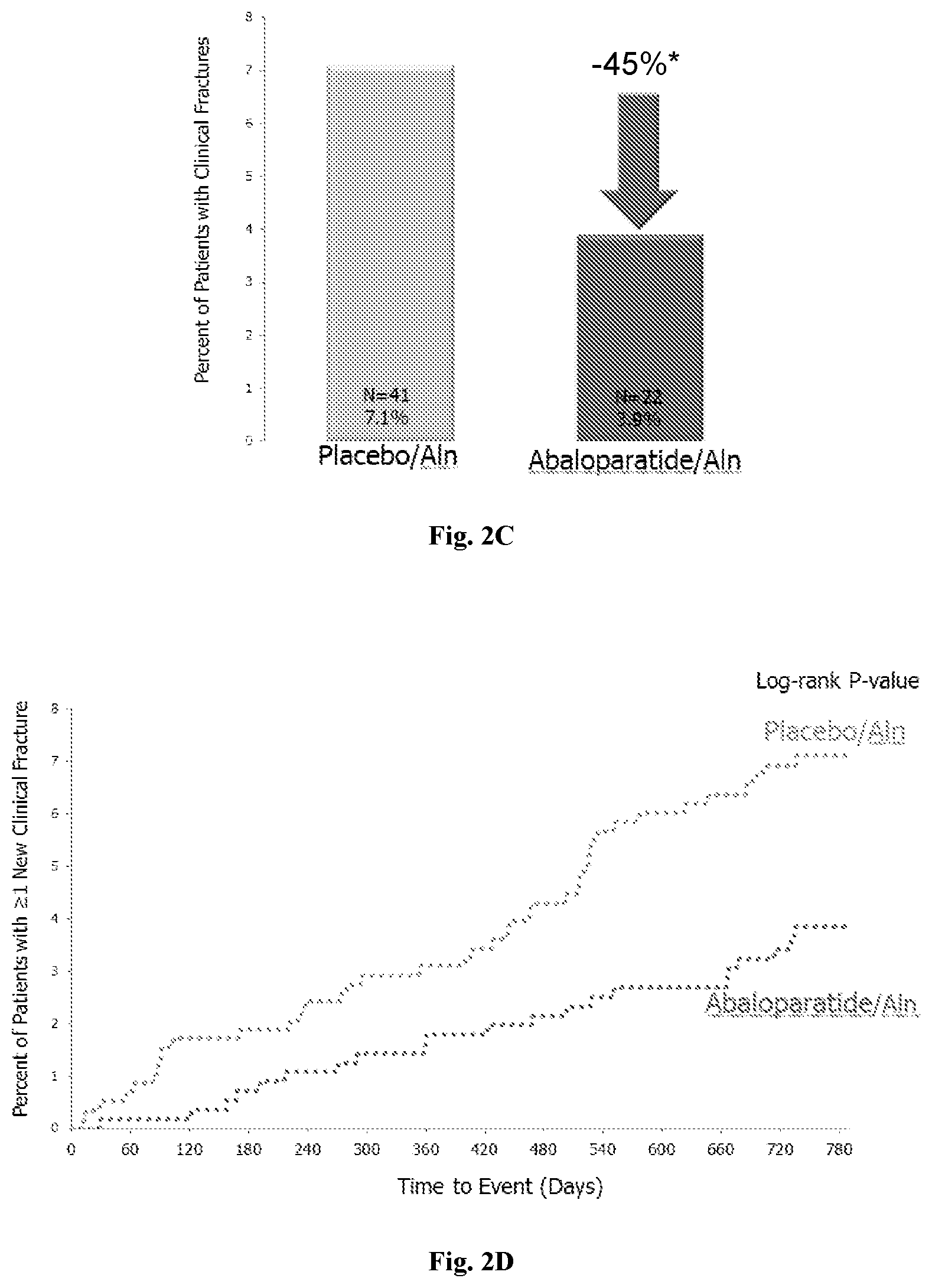
[0018] FIG. 2C: Clinical osteoporotic fractures in patient groups treated with abaloparatide and alendronate or treated with placebo and alendronate at the end of the 25-month study.
[0019] FIG. 2D: Kaplan-Meier curve of clinical osteoporotic fractures in patient groups treated with abaloparatide and alendronate or treated with placebo and alendronate during the 25-month study.
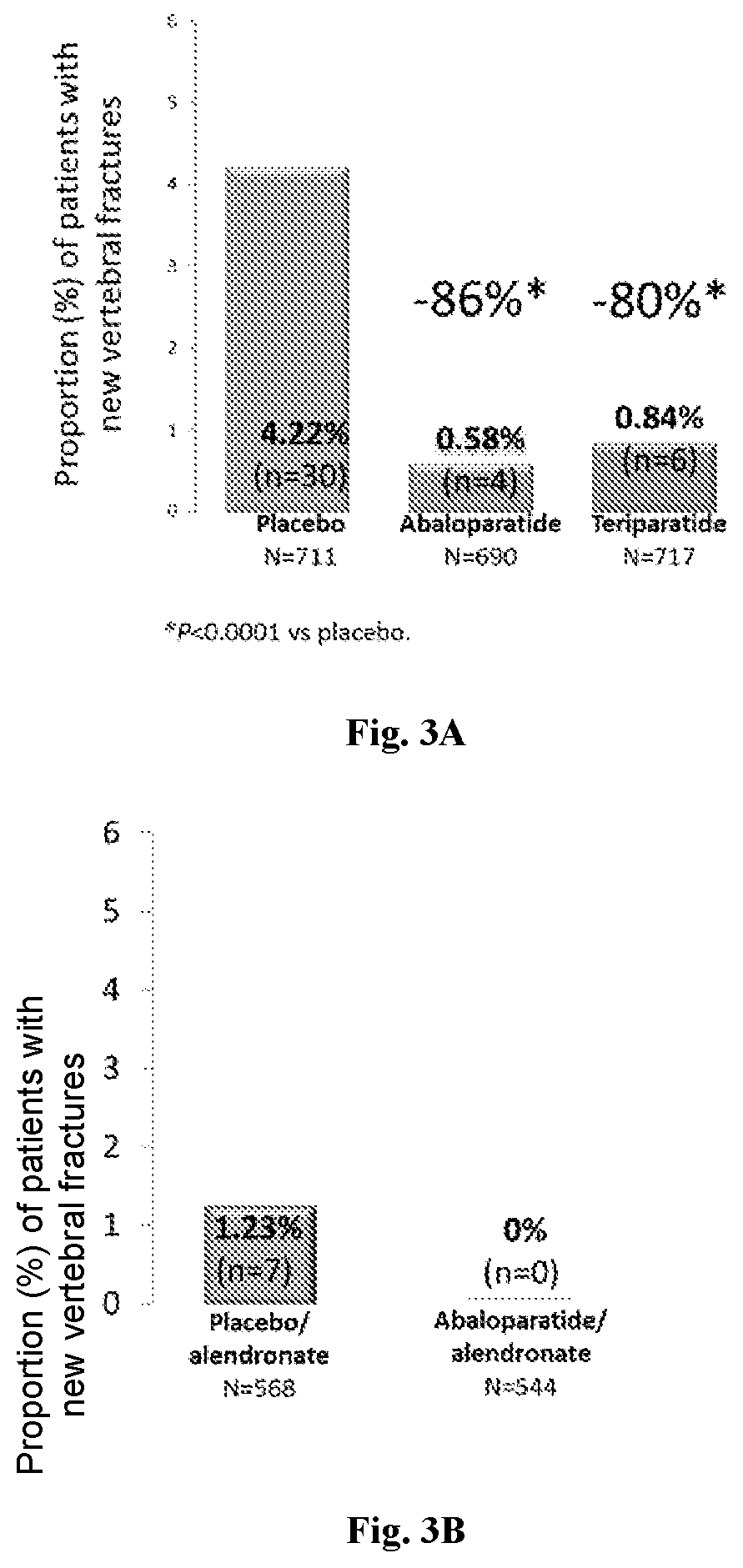
[0020] FIG. 3A: New vertebral fractures in all patient groups at the end of the 18-month treatments.
[0021] FIG. 3B: New vertebral fractures in patient groups treated with abaloparatide and alendronate or treated with placebo and alendronate during the 6-month treatments of alendronate.
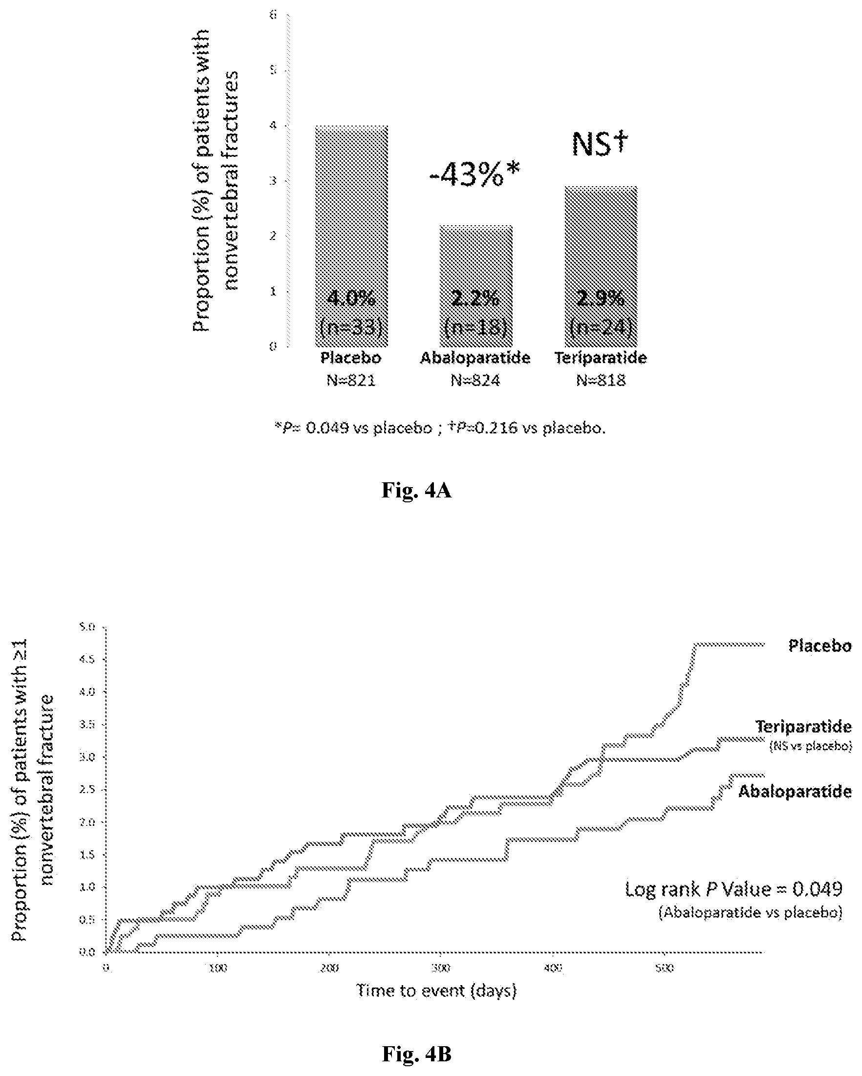
[0022] FIG. 4A: Non-vertebral fractures in all patient groups at the end of the 18-month treatment.
[0023] FIG. 4B: Kaplan-Meier curve of non-vertebral fractures in all patient groups during the 18-month treatments.
[0024] FIG. 4C: Non-vertebral fractures in patient groups treated with abaloparatide and alendronate or treated with placebo and alendronate at the end of the 25-month study.
[0025] FIG. 4D: Kaplan-Meier curve of non-vertebral fractures in patient groups treated with abaloparatide and alendronate or treated with placebo and alendronate during the 25-month study.
[0026] FIG. 4E: Non-vertebral fractures in patient groups treated with abaloparatide and alendronate or treated with placebo and alendronate during the 6-month treatments of alendronate.
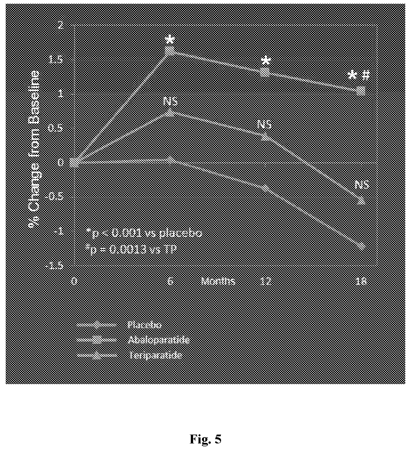
[0027] FIG. 5: Effect of abaloparatide on wrist BMD: Changes in wrist BMD in all patient groups over 18 months: patients treated with placebo (diamond), patients treated with abaloparatide (square), and patients treated with teriparatide (triangle).
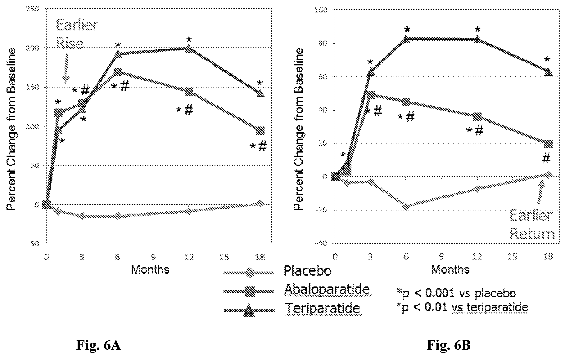
[0028] FIG. 6A: Changes in P1NP bone turnover marker in all patient groups over 18 months; patients treated with placebo (diamond), patients treated with abaloparatide (square), and patients treated with teriparatide (triangle).
[0029] FIG. 6B: Changes in CTX bone turnover marker in all patient groups. *: p<0.001 vs. placebo. .sup.#: p<0.01 vs. teriparatide.
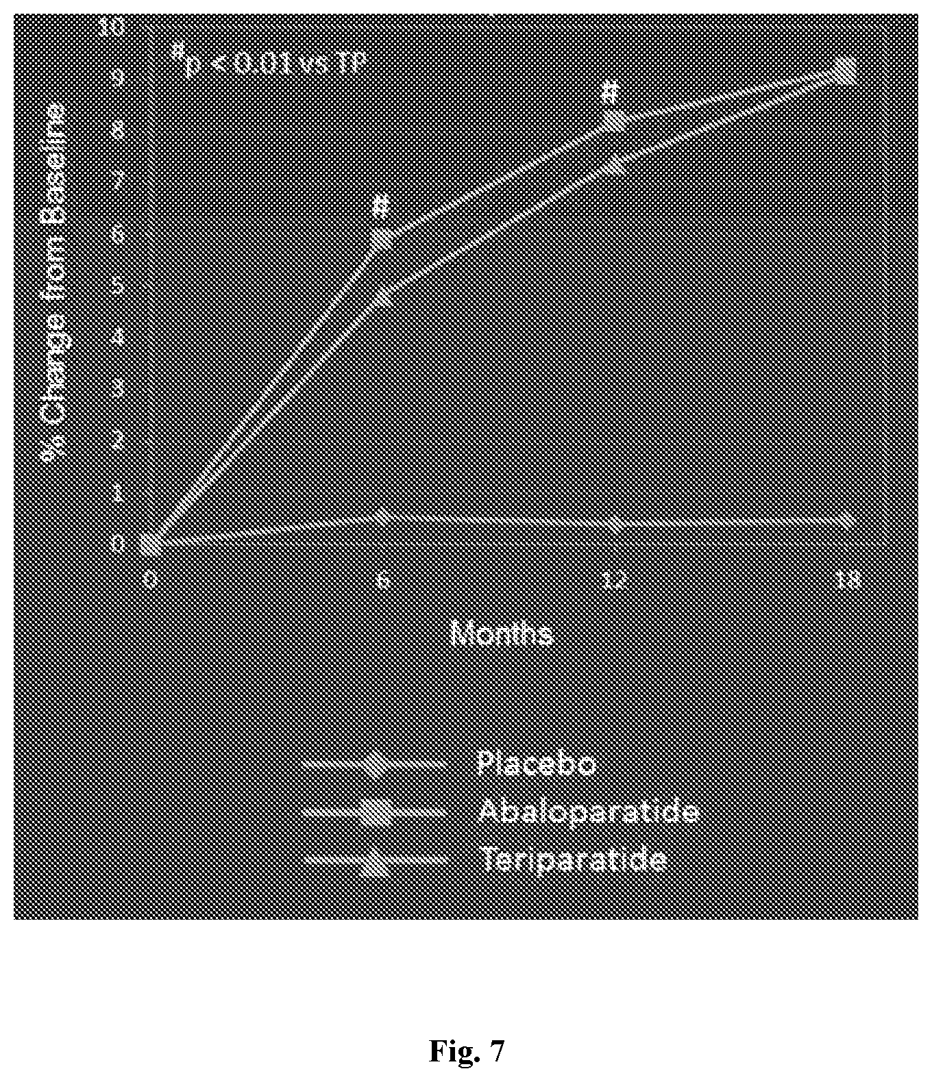
[0030] FIG. 7: Changes in BMD at the spine in all patient groups over 18 months: patients treated with placebo (diamond), patients treated with abaloparatide (square), and patients treated with teriparatide (triangle).
[0031] FIG. 8A: Total hip BMD in all patient groups over 18 months; patients treated with placebo (diamond), patients treated with abaloparatide (square), and patients treated with teriparatide (triangle).
[0032] FIG. 8B: Femoral neck BMD in all patient groups over 18 months; patients treated with placebo (diamond), patients treated with abaloparatide (square), and patients treated with teriparatide (triangle). Two-headed arrows indicate at least 6 month lead in BMD increases obtained by abaloparatide compared to teriparatide.
[0033] FIG. 9A: Average BMD increase at month 25 following treatment with abaloparatide and alendronate (unfilled) or treatment with placebo and alendronate (filled) at spine, hip and femoral neck. The patents were treated with placebo or abaloparatide for 18 months, and subsequently treated with alendronate for another 6 months.
[0034] FIG. 9B: The relative risk ratios (RRR) for new yertebral fractures by baseline BMD shown by Brewslow-Day test (no qualitative or quantitative interactions)
[0035] FIG. 9C: The relative risk ratios (RRR) for new yertebral fractures by age and fracture history shown by Brewslow-Day test (no qualitative or quantitative interactions)
[0036] FIG. 9D: The hazard ratios (HR) for nonvertebral fractures by baseline BMD shown by Cox Proportional Hazard Model (no qualitative or quantitative interactions).
[0037] FIG. 9E: The hazard ratios (HR) for nonvertebral fractures by age and fracture history shown by Cox Proportional Hazard Model (no qualitative or quantitative interactions).
[0038] FIG. 9F: The Least-squares (LS) mean differences in lumbar spine BMD percentage change from baseline at 18 months are shown by ANCOVA model. Percentage change by baseline BMD (no qualitative or quantitative interactions except for quantitative interaction for total hip T-score .ltoreq.vs >-2.5).
[0039] FIG. 9G: The Least-squares (LS) mean differences in lumbar spine BMD percentage change from baseline at 18 months are shown by ANCOVA model. Lumbar spine BMD percentage change by age and fracture history (no qualitative or quantitative interactions).
[0040] FIG. 9H: The Least-squares (LS) mean differences in total hip BMD percentage change from baseline at 18 months are shown by ANCOVA model. Total hip BMD percentage change by baseline BMD.
[0041] FIG. 9I: The Least-squares (LS) mean differences in total hip BMD percentage change from baseline at 18 months are shown by ANCOVA model. Total hip BMD percentage change by age and fracture history.
[0042] FIG. 10A: Effect of abaloparatide on any clinical fracture compared to placebo, expressed as hazard ratio (HR), across the range of major osteoporotic fracture probabilities at baseline.
[0043] FIG. 10B: Impact of abaloparatide on major osteoporotic fracture compared to placebo, shown with an example of CHMP threshold. *FRAX probability calculated with BMD. The solid line represents the hazard ratio, while the dotted lines represent the variance/confidence interval for that hazard ratio for FIGS. 10A and 10B.
[0044] FIG. 10C: Baseline major osteoporotic fracture (MOF) probabilities.
[0045] FIG. 11: Subject Disposition for Example 3. (For FIGS. 11, 12A-12C, 13A-13C, 14A-14C, 15A-15F, and 16A-16B, unless otherwise specified, ABL represents abaloparatide, TPTD represents teriparatide, PBO represents placebo, and Veh represents vehicle.
[0046] FIG. 12A: PA spine change in BMD (mean percent change.+-.SE) at the spine in all patient groups over 24 weeks: patients treated with placebo (square), patients treated with abaloparatide at 20 .mu.g (triangle), patients treated with abaloparatide at 40 .mu.g (reversed triangle), patients treated with abaloparatide at 80 .mu.g (diamond), and patients treated with teriparatide (filled circle).
[0047] FIG. 12B: Femoral neck change in BMD (mean percent change.+-.SE) at the spine in all patient groups over 24 weeks: patients treated with placebo (square), patients treated with abaloparatide at 20 .mu.g (triangle), patients treated with abaloparatide at 40 .mu.g (reversed triangle), patients treated with abaloparatide at 80 .mu.g (diamond), and patients treated with teriparatide (filled circle).
[0048] FIG. 12C: Total hip change in BMD (mean percent change.+-.SE) at the spine in all patient groups over 24 weeks: patients treated with placebo (square), patients treated with abaloparatide at 20 .mu.g (triangle), patients treated with abaloparatide at 40 .mu.g (reversed triangle), patients treated with abaloparatide at 80 .mu.g (diamond), and patients treated with teriparatide (filled circle). *: p<0.01 versus placebo. %: p<0.05 versus placebo. &: p<0.05 versus teriparatide.
[0049] FIG. 13A: Percentage of subjects who completed all study visits with a >3% increase in BMD after 24-weeks of treatment. *: p<0.01 versus placebo. &: p<0.05 versus teriparatide and placebo (PA spine BMD).
[0050] FIG. 13B: Percentage of subjects who completed all study visits with a >3% increase in BMD after 24-weeks of treatment. *: p<0.01 versus placebo. &: p<0.05 versus teriparatide and placebo (Femoral neck BMD).
[0051] FIG. 13C: Percentage of subjects who completed all study visits with a >3% increase in BMD after 24-weeks of treatment. *: p<0.01 versus placebo. &: p<0.05 versus teriparatide and placebo (Total hip BMD).
[0052] FIG. 14A: Changes in CTX bone turnover marker in all patient groups over 24 weeks: patients treated with placebo (square), patients treated with abaloparatide at 20 (triangle), patients treated with abaloparatide at 40 .mu.g (reversed triangle), patients treated with abaloparatide at 80 .mu.g (diamond), and patients treated with teriparatide (filled circle).
[0053] FIG. 14B: Changes in P1NP bone turnover marker in all patient groups over 24 weeks: patients treated with placebo (square), patients treated with abaloparatide at 20 (triangle), patients treated with abaloparatide at 40 .mu.g (reversed triangle), patients treated with abaloparatide at 80 .mu.g (diamond), and patients treated with teriparatide (filled circle).
[0054] FIG. 14C: Changes in osteocalcin bone turnover marker in all patient groups over 24 weeks: patients treated with placebo (square), patients treated with abaloparatide at 20 (triangle), patients treated with abaloparatide at 40 .mu.g (reversed triangle), patients treated with abaloparatide at 80 .mu.g (diamond), and patients treated with teriparatide (filled circle). a: p<0.002 versus placebo at 24 weeks. b: p<0.003 versus teriparatide at 24-weeks.
[0055] FIG. 15A: Effect of abaloparatide treatment on BMD in ovariectomized (OVX) osteopenic rats (BMD change from baseline at the lumbar spine).
[0056] FIG. 15B: Effect of abaloparatide treatment on lumbar spine BMD in ovariectomized (OVX) osteopenic rats.
[0057] FIG. 15C: Effect of abaloparatide treatment on BMD in ovariectomized (OVX) osteopenic rats (BMD change from baseline at total femur).
[0058] FIG. 15D: Effect of abaloparatide treatment on BMD in ovariectomized (OVX) osteopenic rats; Total femur BMD.
[0059] FIG. 15E: Effect of abaloparatide treatment on BMD in ovariectomized (OVX) osteopenic rats (BMD change from baseline at cortical bone at the femoral shaft).
[0060] FIG. 15F: Effect of abaloparatide treatment on BMD in ovariectomized (OVX) osteopenic rats; Femur midshaft BMD.
[0061] FIG. 16A: Effect of abaloparatide treatment on trabecular bone microarchitecture in OVX rats; Lumbar spine (L4).
[0062] FIG. 16B: Effect of abaloparatide treatment on trabecular bone microarchitecture in OVX rats; Distal femur.
DETAILED DESCRIPTION
[0063] The following description of the invention is merely intended to illustrate various embodiments of the invention. As such, the specific modifications discussed are not to be construed as limitations on the scope of the invention. It will be apparent to one skilled in the art that various equivalents, changes, and modifications may be made without departing from the scope of the invention, and it is understood that such equivalent embodiments are to be included herein.
[0064] The term "parathyroid hormone-related protein (PTHrP)" as used herein refers to native human PTHrP (hPTHrP) and fragments thereof. The sequence of native hPTHrP (1-34) is: Ala Val Ser Glu His Gln Leu Leu His Asp Lys Gly Lys Ser Ile Gln Asp Leu Arg Arg Arg Phe Phe Leu His His Leu Ile Ala Glu Ile His Thr Ala (SEQ ID NO:2). PTHrP is a protein with homology to PTH at the amino-terminus that binds to the same G-protein coupled receptor. Despite a common receptor (PTHR), PTH primarily acts as an endocrine regulator of calcium homeostasis, whereas PTHrP plays a fundamental paracrine role in the mediation of endochondral bone development (11). The differential effects of these proteins may be related not only to differential tissue expression, but also to distinct receptor binding properties (12-14). Over the past several years, PTHrP has been investigated as a potential treatment for osteoporosis. The results of these studies have been mixed, with some suggesting that intermittent administration of high dose PTHrP increases bone formation without concomitant stimulation of bone resorption and others reporting measurable stimulation of bone resorption and significant hypercalcemia (15-17).
[0065] A "fragment" of hPTHrP refers to a polypeptide having a sequence comprising less than the full complement of amino acids found in hPTHrP, which nonetheless elicits a similar biological response. Typically, fragments for use in the methods and compositions provided herein will be truncated from the C-terminus and will range from 30 to 40 residues in length. In particular, hPTHrP(1-34), as well as analogues thereof with between 1 and 15 substitutions, are useful in the methods and compositions of the present invention.
[0066] As used herein, an "analogue" of PTHrP refers to a polypeptide having between about 1 and about 20, between about 1 and about 15, or between about 1 and about 10 art-accepted substitutions, additions, or insertions relative to PTHrP (i.e., relative to hPTHrP or a fragment thereof), or combinations thereof, not to exceed a total combination of 20 substitutions, additions, and insertions. As used herein, "insertions" include the insertion of an amino acid between two existing amino acids in the peptide chain. As used herein, "addition" means the addition of an amino acid to the N or C terminus of the peptide chain. As used herein, "substitution" means the substitution of an amino acid for an existing amino acid in the peptide chain. As used herein, "art-accepted" substitutions, insertions, or additions are those which one of ordinary skill in the art would expect to maintain or increase the biological and/or hormonal activity of the peptide and not adversely affect the biological activity of the peptide. Art-accepted substitutions include, for example, substitution of one amino acid with a chemically or biologically similar amino acid, such as substituting one hydrophobic amino acid for another hydrophobic amino acid. PTHrP analogues are described with reference to their variation from the native sequence of hPTHrP.
[0067] Examples of PTHrP analogues include, without limitation, abaloparatide. Abaloparatide was selected to retain potent anabolic activity with decreased bone resorption, less calcium-mobilizing potential, and improved room temperature stability (18). Studies performed in animals have demonstrated marked bone anabolic activity for the PTHrP analogue abaloparatide, with complete reversal of bone loss in ovariectomy-induced osteopenic rats and monkeys (19, 20).
[0068] As set forth in the Examples below, subjects treated with abaloparatide exhibited a significant reduction in certain bone fractures as compared to subjects treated with a placebo or with teriparatide.
[0069] When compared to subjects treated with placebo, subjects treated with abaloparatide unexpectedly showed a statistically significant reduction in major osteoporotic fractures, clinical fractures, new vertebral fractures, and non-vertebral fractures in an 18-month trial (see, e.g., Example 1, Table 1). Abaloparatide significantly reduced vertebral and non-vertebral fractures and increased BMD regardless of baseline risk.
[0070] Subjects treated with teriparatide demonstrated a statistically significant reduction only in new vertebral fractures compared to the placebo group. Compared to subjects treated with teriparatide, subjects treated with abaloparatide unexpectedly demonstrated a statistically significant reduction in major osteoporotic fractures.
[0071] Subjects treated with abaloparatide also unexpectedly showed a significant reduction in the risk of non-vertebral fractures (e.g., wrist fractures), and clinical fractures (see, e.g., Example 1, Table 1). Abaloparatide was further found to significantly decrease the risk of major osteoporotic fracture and any clinical fracture in postmenopausal women, irrespective of baseline fracture probability, using the Fracture Risk Assessment Tool (FRAX). Moreover, treatment with abaloparatide was associated with a significant decrease in fractures across varying categories of fracture outcome, and the effect of abaloparatide on the various fracture outcomes did not change significantly across the range of baseline fracture probability.
[0072] Subjects treated with abaloparatide exhibited a significant increase not only in BMD, but also in TBS (see, e.g., Example 4). TBS is a grey-scale textural analysis applied to spinal D.times.A images that has been shown to be correlated with trabecular bone microarchitecture and bone strength. TBS is also a predictor of fragility fractures of the spine and hip in postmenopausal women independent of BMD and other major clinical risk factors. As such, it captures additional patients at risk of fracture that are missed by BMD alone (35), and together with BMD more accurately captures bone strength.
[0073] Although a lower BMD is usually associated with higher fracture risk, a normal or even slightly higher than normal BMD does not necessarily indicate a lower fracture risk. For example, subjects with type II diabetes may have increased fracture risk (especially at the hips and/or wrists) despite a higher BMD (21). One factor behind the discrepancy between relatively normal BMD and high fracture risks may be the higher cortical porosity of subjects with diabetes (e.g., type II diabetes). For example, subjects with type II diabetes may have a cortical porosity up to twice that of controls (21). In certain embodiments, the therapeutic methods provided herein may be beneficial to subjects having diabetes and/or subjects having higher cortical porosity.
[0074] Subjects treated with abaloparatide for 18 months unexpectedly demonstrated significant BMD increase in total hip and femoral neck versus subjects treated with teriparatide (see, e.g., Example 1, Tables 4-5). Abaloparatide demonstrated a statistically significant increase in lumbar spine BMD at 6 months and 12 months versus teriparatide, and a non-statistically significant BMD increase at 18 months (see, e.g., Example 1, Tables 4-5). Without wishing to be bound by any theory, an earlier increase in bone formation marker P1NP in subjects treated with abaloparatide compared to subjects treated with teriparatide may contribute to the faster effects of abaloparatide on BMD (see, e.g., Example 1, FIG. 6A; and Example 3, FIG. 14B). For the CTX marker (bone resorption), subjects treated with abaloparatide showed an earlier return to the baseline at 18 months compared to subjects treated with teriparatide (see, e.g., Example 1, FIG. 6B).
[0075] Furthermore, subjects treated with abaloparatide for 18 months followed by an anti-resorptive therapy (e.g., alendronate for 6 months) showed a significant reduction in fracture risk versus subjects treated with placebo for 18 months followed by similar anti-resorptive therapy (see, e.g., Example 1, Table 2).
[0076] Provided herein are practical applications of these findings in the form of methods, compositions, and kits for preventing or reducing bone fractures, improving BMS, and/or improving TBS in a subject in need thereof using PTHrP or analogues thereof (e.g., abaloparatide).
[0077] One aspect of the present disclosure relates to a method for preventing or reducing bone fractures in a subject in need thereof comprising administering to the subject a therapeutically effective amount of PTHrP or analogues thereof (e.g., abaloparatide). Exemplary bone fractures which may exhibit reduced fracture risk include, without limitation, major osteoporotic fractures (e.g., high- or low-trauma clinical fractures of the clinical spine, forearm, hip, or shoulder), non-vertebral fractures (e.g., wrist, hips, etc.), clinical fractures (e.g., fractures with or without high trauma, confirmed through x-ray scan, radiologist report, emergency room/urgent care reports, hospital discharge reports, surgery reports, hospital or clinical notes, or other medical confirmation), and new vertebral fractures.
[0078] Another aspect of the present disclosure relates to a method for preventing or reducing non-vertebral bone fractures in a subject in need thereof comprising administering to the subject a therapeutically effective amount of PTHrP or analogues thereof (e.g., abaloparatide).
[0079] Another aspect of the present disclosure relates to a method for preventing or reducing vertebral bone fractures in a subject in need thereof comprising administering to the subject a therapeutically effective amount of PTHrP or analogues thereof (e.g., abaloparatide).
[0080] Another aspect of the present disclosure relates to a method for improving BMD and/or TBS in a subject in need thereof comprising administering to the subject a therapeutically effective amount of PTHrP or analogues thereof (e.g., abaloparatide). Examples of bones which may exhibit improved BMD and/or TBS following administration include, without limitation, the lumbar spine, total hip, wrist, femur, cortical bone of the femur (femoral diaphysis), and/or femoral neck in the subject.
[0081] In certain embodiments, the therapeutic methods provided herein further comprise administering an anti-resorptive therapy following treatment with PTHrP or an analogue thereof (e.g., abaloparatide) for an extended period of time. For example, provided herein is a method for improving BMD and/or trabecular bone score TBS in a subject comprising administering to the subject a therapeutically effective amount of PTHrP or an analogue thereof (e.g., abaloparatide) for a period of time, and subsequently administering to the subject a therapeutically effective amount of an anti-resorptive agent. Examples of bones which may exhibit improved BMD and/or TBS following administration include, without limitation, the lumbar spine, total hip, wrist, femur, cortical bone of the femur (femoral diaphysis), and/or femoral neck in the subject. Also provided herein is a method for preventing or reducing bone fractures in a subject comprising administering to the subject a therapeutically effective amount of PTHrP or an analogue thereof (e.g., abaloparatide) for a period of time, and subsequently administering to the subject a therapeutically effective amount of an anti-resorptive agent. Exemplary bone fractures that may exhibit reduced fracture risk include, without limitation, major osteoporotic fracture, non-vertebral fracture (e.g., wrist, hip), clinical fracture, and new vertebral fracture. In those methods provided herein that comprise administration of a PTHrP analogue followed by administration of an anti-resorptive agent, administration of the PTHrP analogue and anti-resorptive agent may overlap for some period of time, i.e., administration of the anti-resorptive agent may be initiated while the subject is still receiving PTHrP analogue. Notably, the fracture prevention efficacy of abaloparatide relative to placebo carried through even in the 6 months after the abaloparatide therapy was discontinued and both groups treated with alendronate. This embodiment of the invention indicates that fracture reduction can be accomplished beyond the treatment period and that surprisingly there is a sustained effect of the drug. In certain embodiments, this invention comprises a method of preventing fractures and treating osteoporosis that relies on treating with abaloparatide for a period of time and then discontinuing abaloparatide treatment wherein the treatment window is extended beyond the actual treatment window. Although an embodiment of this invention includes the subsequent treatment with an antiresorptive agent post-abaloparatide treatment, such a treatment is believed to not be required to maintain at least some of the drug's benefit and so other embodiments do not require subsequent treatment with an antiresorptive drug to sustain meaningful clinical benefit.
[0082] It is within the purview of one skilled in the art to select a suitable anti-resorptive therapy for the aspects and embodiments disclosed in this application. In some embodiments, the anti-resorptive therapeutic agents include bisphosphonates, estrogens, selective estrogen receptor modulators (SERMs), calcitonin, cathepsin K inhibitors, and monoclonal antibodies such as denosumab. In certain embodiments, the anti-resorptive therapeutic agent may be a bisphosphonate such as alendronate.
[0083] The term "subject in need thereof" as used herein refers to a mammalian subject, e.g., a human. In certain embodiments, a subject in need thereof has a fracture risk higher than normal. In certain embodiments, a subject in need thereof has one or more conditions selected from the group consisting of low BMD and high cortical porosity. BMD may be measured by digital X-ray radiogrammetry (DXR) or other methods known in the art. As used herein, the term "low BMD" means a BMD T-score .ltoreq.about 2 or .ltoreq.about -2.5, e.g., at one or more sites selected from the group consisting of spine (e.g., lumbar spine), hip (e.g., total hip or femoral neck), and wrist. As used herein, the term "cortical porosity" means the fraction of cortical bone volume that is not occupied by the bone. Cortical porosity may be measured by DXR or other methods known in the art to provide an estimation of the local intensity minima ("holes") in the cortical bone regions using a recursive (climbing) algorithm starting from the outer region (10). A combined porosity measure is derived from the area percentage of holes found in the cortical part relative to the entire cortical area, by averaging over the involved bones and scaling to reflect a volumetric ratio rather than the projected area. A "high cortical porosity" means a porosity of about 10% higher, about 15% higher, about 20% higher, about 50% higher, about 100% higher, or about 150% higher than that of healthy subjects from the same age group as controls. For example, the subject may have a cortical porosity of about 0.01256, which the control group has a cortical porosity of about 0.01093 (10). Subjects having a high cortical porosity may have a slightly low BMD, a normal BMD, or even a slightly higher than normal BMD, e.g., a BMD T-score of at least about -2, at least about -1.5, at least about -1, at least about -0.5, at least about -0.25, at least about -0.2, at least about -0.1, at least about 0, about -2 to about 3, about -2 to about 2.5, about -2 to about 2, about -2 to about 1.5, about -2 to about 1, about -2 to about 0.5, about -2 to about 0.25, about -2 to about 0.2, about -2 to about 0.1, or about -2 to about 0. For example, subjects with type II diabetes may have a cortical porosity up to twice that of controls while having normal or even slightly higher than normal BMD (21). Examples of suitable subjects in need thereof include, without limitation, women, women with osteoporosis and/or diabetes (e.g., type I or type II diabetes), postmenopausal women, postmenopausal women with osteoporosis and/or diabetes (e.g., type I or type II diabetes), and men with osteoporosis and/or diabetes (e.g., type I or type II diabetes).
[0084] The term "therapeutically effective amount" as used herein refers to an amount of a compound or agent that is sufficient to elicit the required or desired therapeutic and/or prophylactic response, as the particular treatment context may require. In certain embodiments, the therapeutically effective amount is an amount of the composition that yields maximum therapeutic effect. In other embodiments, the therapeutically effective amount yields a therapeutic effect that is less than the maximum therapeutic effect. For example, a therapeutically effective amount may be an amount that produces a therapeutic effect while avoiding one or more side effects associated with a dosage that yields maximum therapeutic effect. A therapeutically effective amount for a particular composition will vary based on a variety of factors, including but not limited to the characteristics of the therapeutic composition (e.g., activity, pharmacokinetics, pharmacodynamics, and bioavailability), the physiological condition of the subject (e.g., age, body weight, sex, disease type and stage, medical history, general physical condition, responsiveness to a given dosage, and other present medications), the nature of any pharmaceutically acceptable carriers in the composition, and the route of administration. One skilled in the clinical and pharmacological arts will be able to determine a therapeutically effective amount through routine experimentation, namely by monitoring a subject's response to administration of a composition and adjusting the dosage accordingly. For additional guidance, see, e.g., Remington: The Science and Practice of Pharmacy, 22.sup.nd Edition, Pharmaceutical Press, London, 2012, and Goodman & Gilman's The Pharmacological Basis of Therapeutics, 12.sup.th Edition, McGraw-Hill, New York, N.Y., 2011, the entire disclosures of which are incorporated by reference herein.
[0085] Examples of therapeutically effective amounts of PTHrP or analogues thereof (e.g., abaloparatide) include, without limitation, about 10 .mu.g to about 250 .mu.g, about 50 .mu.g to about 200 .mu.g, about 50 .mu.g to about 150 .mu.g, about 70 .mu.g to about 100 .mu.g, about 70 .mu.g to about 90 .mu.g, about 75 .mu.g to about 85 .mu.g, about 20 .mu.g, about 40 .mu.g, about 60 .mu.g, about 80 .mu.g, about 100 .mu.g, about 120 .mu.g, about 150 .mu.g, about 200 .mu.g, or about 250 .mu.g. Other examples of therapeutically effective amounts of PTHrP or analogues thereof (e.g., abaloparatide) may also include, without limitation, about 5 .mu.g/kg or about 20 .mu.g/kg. Depending on the particular anti-resorptive agent, one skilled in the art can select a therapeutically effective amount of the anti-resorptive agent. The amount of the anti-resorptive agent can be further optimized when used in combination with or subsequent to the therapy of a PTHrP or an analogue thereof (e.g., abaloparatide).
[0086] In certain embodiments, PTHrP or analogues thereof (e.g., abaloparatide) are administered by subcutaneous injection or transdermal administration.
[0087] In certain embodiments, PTHrP or analogues thereof (e.g., abaloparatide) are administered for a fixed period of time. In other embodiments, administration occurs until a particular therapeutic benchmark is reached (e.g., BMD increase is about 3% or higher, at bones such as spine, hip and/or femoral neck). Examples of a suitable timeframe for administration include, without limitation, 6 weeks, 12 weeks, 3 months, 24 weeks, 6 months, 48 weeks, 12 months, 18 months, or 24 months. In certain embodiments, PTHrP or analogues thereof (e.g., abaloparatide) are administered once a day, twice a day, three times a day, or more than three times a day. In other embodiments, administration may occur once every 2 days, once every 3 days, once every 4 days, once per week, or once per month. In certain embodiments, PTHrP or analogues thereof (e.g., abaloparatide) are administered once a day for 18 months.
[0088] In certain embodiments, an anti-resorptive agent may be administered to a subject who has received a PTHrP or an analogue thereof (e.g., abaloparatide) for an extended period of time. Following the treatment with a PTHrP or analogue thereof (e.g., abaloparatide), the anti-resorptive agent is administered to the subject for a fixed period of time, such as 6 weeks, 12 weeks, 3 months, 24 weeks, 6 months, 48 weeks, 12 months, 18 months, and 24 months. In certain embodiments, the anti-resorptive agent is administered once a day, twice a day, three times a day, or more than three times a day. In other embodiments, administration may occur once every 2 days, once every 3 days, once every 4 days, once per week, once per month, or once per year. In certain embodiments, the anti-resorptive agent is administered once a day for 6 months, 9 moths or 12 months. In certain embodiments, administration of PTHrP analogue and the anti-resorptive agent may overlap for some period of time, i.e., administration of the anti-resorptive agent may commence while the subject is still receiving PTHrP analogue.
[0089] As disclosed herein, subjects treated with PTHrP or analogues thereof (e.g., abaloparatide) exhibit a significant reduction in fractures as compared to the subjects without treatment or subjects treated with a placebo. In certain embodiments, subjects treated with PTHrP or analogues thereof (e.g., abaloparatide) may exhibit a reduction in fractures of at least about 10%, at least about 20%, at least about 30%, at least about 40%, at least about 50%, at least about 60%, at least about 70%, at least about 80%, at least about 90%, or about 100% as compared to untreated subjects or subjects treated with a placebo.
[0090] In certain embodiments, the methods provided herein reduce the wrist fracture risk of subjects treated with PTHrP or analogues thereof (e.g., abaloparatide) by about 40% to about 70%, about 50% to about 65%, about 55% to about 60%, or at least about 58% when compared to untreated subjects or subjects treated with placebo. In certain embodiments, the wrist fracture risk for subjects treated with PTHrP or analogues thereof (e.g., abaloparatide) is reduced by about 40% to about 80%, about 50% to about 75%, about 60% to about 75%, about 65% to about 75%, about 70% to about 75%, or at least about 72% compared to subjects treated with teriparatide.
[0091] In certain embodiments, the methods provided herein reduce the major osteoporotic fracture risk of subjects treated with PTHrP or analogues thereof (e.g., abaloparatide) by about 30% to about 80%, about 40% to about 80%, about 50% to about 75%, about 60% to about 75%, about 65% to about 75%, about 70% to about 75%, about 58%, or at least about 71%, compared to untreated subjects or subjects treated with placebo. In certain embodiments, the major osteoporotic fracture risk for subjects treated with PTHrP or analogues thereof (e.g., abaloparatide) is reduced by about 40% to about 70%, about 50% to about 65%, about 55% to about 60%, or at least about 57% compared to subjects treated with teriparatide.
[0092] In certain embodiments, the methods provided herein reduce the clinical fracture risk of subjects treated with PTHrP or analogues thereof (e.g., abaloparatide) by about 30% to about 70%, about 35% to about 65%, about 40% to about 60%, about 40 to about 50%, or at least about 45% when compared to untreated subjects or subjects treated with placebo. In certain embodiments, the clinical fracture risk of subjects treated with PTHrP or analogues thereof (e.g., abaloparatide) is reduced by about 15% to about 40%, about 20% to about 35%, about 20% to about 30%, about 20% to about 25%, or at least about 23% compared to subjects treated with teriparatide.
[0093] In certain embodiments, the methods provided herein reduce the new vertebral fracture risk of subjects treated with PTHrP or analogues thereof (e.g., abaloparatide) by about 50% to about 95%, about 60% to about 95%, about 70% to about 90%, about 80 to about 88%, at least about 87%, or at least about 86% when compared to untreated subjects or subjects treated with placebo. In certain embodiments, subjects treated with PTHrP or analogues thereof (e.g., abaloparatide) exhibit a vertebral fracture risk that is reduced by about 15% to about 45%, about 20% to about 40%, about 25% to about 35%, or at least about 30% versus subjects treated with teriparatide.
[0094] In certain embodiments, the methods provided herein reduce the non-vertebral fracture risk of subjects treated with PTHrP or analogues thereof (e.g., abaloparatide) by about 30% to about 70%, about 35% to about 65%, about 40% to about 60%, about 40 to about 50%, about 51%, or at least about 45% when compared to untreated subjects or subjects treated with placebo. In certain embodiments, the non-vertebral fracture risk of subjects treated with PTHrP or analogues thereof (e.g., abaloparatide) is reduced by about 15% to about 40%, about 20% to about 35%, about 20% to about 30%, about 20% to about 25%, or at least about 24% compared to subjects treated with teriparatide.
[0095] In certain embodiments, the methods provided herein result in a significant increase in BMD in the lumbar spine, femoral neck, and total hip. In certain embodiments, the methods disclosed herein result in a significant BMD increase in lumbar spine, femoral neck, and total hip within the first year after the first administration of PTHrP or analogues thereof (e.g., abaloparatide) compared to subjects treated with teriparatide. In certain embodiments, the methods disclosed herein result in a significant BMD increase in femoral neck and total hip compared to subjects treated with teriparatide. In certain embodiments, BMD at the lumbar spine for subjects treated with PTHrP or analogues thereof (e.g., abaloparatide) may increase by at least about 2.9%, at least about 3%, at least about 5.2%, at least about 6%, at least about 6.7%, at least about 12.8%, about 2% to about 8%, about 6% to about 8%, about 2% to about 7%, about 6% to about 7%, about 5.8% to about 7%, about 2% to about 15%, about 6% to about 15%, about 2% to about 14%, about 6% to about 14%, about 2% to about 13%, about 6% to about 13%, about 2% to about 12.8%, about 6% to about 12.8%, or about 5.8% to about 12.8%; BMD at the femoral neck for subjects treated with PTHrP or analogues thereof (e.g., abaloparatide) may increase by at least about 2.2%, at least about 2.7%, at least about 3%, at least about 3.1%, at least about 4.5%, at least about 5%, at least about 6%, about 1.5% to about 4%, about 2% to about 4%, about 2.5% to about 4%, about 2% to about 3.5%, about 1.5% to about 6%, about 2% to about 6%, about 2.5% to about 6%, about 1.5% to about 5%, about 2% to about 5%, about 2.5% to about 5%, about 1.5% to about 4.5%, about 2% to about 4.5%, or about 2.5% to about 4.5%; and BMD for the total hip of subjects treated with PTHrP or analogues thereof (e.g., abaloparatide) may increase by at least about 1.4%, at least about 2.0%, at least about 2.6%, at least about 3%, at least about 3.5%, at least about 4%, at least about 4.5%, at least about 5%, at least about 5.5%, at least about 6%, at least about 7%, about 0.6% to about 3%, about 1% to about 3%, about 1.5% to about 3%, about 0.6% to about 3.5%, about 1% to about 3.5%, about 1.5% to about 3.5%, about 0.6% to about 4%, about 1% to about 4%, about 1.5% to about 4%, about 2% to about 4%, about 0.6% to about 4.5%, about 1% to about 4.5%, about 1.5% to about 4.5%, about 2% to about 4.5%, about 0.6% to about 5%, about 1% to about 5%, about 1.5% to about 5%, about 2.0% to about 5%, about 0.6% to about 5.5%, about 1% to about 5.5%, about 1.5% to about 5.5%, about 2% to about 5.5%, about 0.6% to about 6%, about 1% to about 6%, about 1.5% to about 6%, about 2% to about 6%, about 0.6% to about 6.5%, about 1% to about 6.5%, about 1.5% to about 6.5%, about 2.0% to about 6.5%, about 0.6% to about 7%, about 1% to about 7%, about 1.5% to about 7%, or about 2% to about 7%.
[0096] In certain embodiments, subjects are administered PTHrP or analogues thereof (e.g., abaloparatide) at a daily dose of 20 .mu.g, 40 .mu.g, or 80 .mu.g for 24 weeks. In certain embodiments, this administration results in a significant increase in BMD in the lumbar spine, femoral neck, and total hip (see, e.g., FIGS. 12A-C). In certain embodiments, BMD at the lumbar spine for subjects treated with PTHrP or analogues thereof (e.g., abaloparatide) may increase by at least about 2.9%, at least about 3%, at least about 5.2%, at least about 6%, about 6.7%, at least about 2% to about 8%, at least about 6% to about 8%, at least about 6% to about 7%, or about 5.8% to about 7%; BMD at the femoral neck for subjects treated with PTHrP or analogues thereof (e.g., abaloparatide) may increase by at least about 2.2%, at least about 2.7%, at least about 3.1%, about 2% to about 4%, about 1.5% to about 4%, about 2.5% to about 4%, or about 2% to about 3.5%; and BMD for the total hip of subjects treated with PTHrP or analogues thereof (e.g., abaloparatide) may increase by at least about 1.4%, at least about 2.0%, at least about 2.6%, about 1% to about 3%, about 0.6% to about 3.5%, about 1% to about 3.5%, or about 1.5% to about 3%.
[0097] In certain embodiments, subjects are administered with PTHrP or analogues thereof (e.g., abaloparatide) at a daily dose of 20 .mu.g, 40 .mu.g, or 80 .mu.g for 18 months and then administered with alendronate for 6 months with a dosage of 10 mg/day or 70 mg/week (e.g., oral), 5 mg/day or 35 mg/week (e.g., oral), 15 mg/day or 105 mg/week (e.g., oral), 20 mg/day or 140 mg/week (e.g., oral), about 5 to about 20 mg/day or about 35 to about 140 mg/week (e.g., oral), about 5 to about 15 mg/day or about 35 to about 105 mg/week (e.g., oral), about 5 to about 10 mg/day or about 35 to about 70 mg/week (e.g., oral), or about 10 to about 20 mg/day or about 70 to about 140 mg/week (e.g., oral). In certain embodiments, this results in a significant increase in BMD in the lumbar spine, femoral neck, and total hip (see, e.g., FIGS. 12A-C). In certain embodiments, BMD at the lumbar spine for subjects treated with PTHrP or analogues thereof (e.g., abaloparatide) may increase by at least about 2.9%, at least about 3%, at least about 5.2%, at least about 6%, at least about 6.7%, at least about 12.8%, about 2% to about 8%, about 6% to about 8%, about 2% to about 7%, about 6% to about 7%, about 5.8% to about 7%, about 2% to about 15%, about 6% to about 15%, about 2% to about 14%, about 6% to about 14%, about 2% to about 13%, about 6% to about 13%, about 2% to about 12.8%, about 6% to about 12.8%, or about 5.8% to about 12.8%; BMD at the femoral neck for subjects treated with PTHrP or analogues thereof (e.g., abaloparatide) may increase by at least about 2.2%, at least about 2.7%, at least about 3%, at least about 3.1%, at least about 4.5%, at least about 5%, at least about 6%, about 1.5% to about 4%, about 2% to about 4%, about 2.5% to about 4%, about 2% to about 3.5%, about 1.5% to about 6%, about 2% to about 6%, about 2.5% to about 6%, about 1.5% to about 5%, about 2% to about 5%, about 2.5% to about 5%, about 1.5% to about 4.5%, about 2% to about 4.5%, or about 2.5% to about 4.5%; and BMD for the total hip of subjects treated with PTHrP or analogues thereof (e.g., abaloparatide) may increase by at least about 1.4%, at least about 2.0%, at least about 2.6%, at least about 3%, at least about 3.5%, at least about 4%, at least about 4.5%, at least about 5%, at least about 5.5%, at least about 6%, at least about 7%, about 0.6% to about 3%, about 1% to about 3%, about 1.5% to about 3%, about 0.6% to about 3.5%, about 1% to about 3.5%, about 1.5% to about 3.5%, about 0.6% to about 4%, about 1% to about 4%, about 1.5% to about 4%, about 2% to about 4%, about 0.6% to about 4.5%, about 1% to about 4.5%, about 1.5% to about 4.5%, about 2% to about 4.5%, about 0.6% to about 5%, about 1% to about 5%, about 1.5% to about 5%, about 2.0% to about 5%, about 0.6% to about 5.5%, about 1% to about 5.5%, about 1.5% to about 5.5%, about 2% to about 5.5%, about 0.6% to about 6%, about 1% to about 6%, about 1.5% to about 6%, about 2% to about 6%, about 0.6% to about 6.5%, about 1% to about 6.5%, about 1.5% to about 6.5%, about 2.0% to about 6.5%, about 0.6% to about 7%, about 1% to about 7%, about 1.5% to about 7%, or about 2% to about 7%.
[0098] In certain embodiments, subjects are treated with PTHrP or analogues thereof (e.g., abaloparatide) at a daily dose of 20 .mu.g, 40 .mu.g, or 80 .mu.g for 12 weeks to 24 weeks. This administration regimen of abaloparatide has been shown herein to significantly increase TBS (trabecular score) in treated subjects, suggesting improved trabecular microarchitecture. In certain embodiments, TBS for subjects treated with PTHrP or analogues thereof (e.g., abaloparatide) for 12 weeks increases by at least about 1.2%, at least about 1.7%, at least about 1.9%, about 1% to about 2.5%, about 1% to about 2%, about 1.6% to about 2.5%, about 1.7% to about 2.5%, about 1.6% to about 2%, or about 1.7% to about 2%. In certain embodiments, TBS for subjects treated with PTHrP or analogues thereof (e.g., abaloparatide) for 24 weeks increases by at least about 2.4%, at least about 2.7%, at least about 3.6%, about 2% to about 4.5%, about 2% to about 4%, about 2.7% to about 4.5%, about 2.7% to about 4%, about 3% to about 4.5%, or about 3% to about 4%.
[0099] In certain embodiments of the methods disclosed herein, PTHrP or analogues thereof (e.g., abaloparatide) are administered in combination with one or more additional osteoporosis therapies, including for example an alendronate therapy. In these embodiments, the additional osteoporosis therapy may be administered before, during, or after the treatment with PTHrP or analogues thereof (e.g., abaloparatide). PTHrP or an analogue thereof and the additional osteoporosis therapy may be administered separately or as part of the same composition. Administration of the two agents may occur at or around the same time, e.g., simultaneously, or the two agents may be administered at different times.
[0100] In certain embodiments, PTHrP or analogues thereof (e.g., abaloparatide) and/or the additional osteoporosis therapy are administered in a pharmaceutical composition as the active ingredient(s). Such pharmaceutical composition may further comprise a pharmaceutically acceptable carrier. A "pharmaceutically acceptable carrier" as used herein refers to a pharmaceutically acceptable material, composition, or vehicle that is involved in carrying or transporting a compound or molecule of interest from one tissue, organ, or portion of the body to another tissue, organ, or portion of the body. A pharmaceutically acceptable carrier may comprise a variety of components, including but not limited to a liquid or solid filler, diluent, excipient, solvent, buffer, encapsulating material, surfactant, stabilizing agent, binder, or pigment, or some combination thereof. Each component of the carrier must be "pharmaceutically acceptable" in that it must be compatible with the other ingredients of the composition and must be suitable for contact with any tissue, organ, or portion of the body that it may encounter, meaning that it must not carry a risk of toxicity, irritation, allergic response, immunogenicity, or any other complication that excessively outweighs its therapeutic benefits.
[0101] Examples of pharmaceutically acceptable carriers that may be used in conjunction with the compositions provided herein include, but are not limited to, (1) sugars, such as lactose, glucose, sucrose, or mannitol; (2) starches, such as corn starch and potato starch; (3) cellulose and its derivatives, such as sodium carboxymethyl cellulose, ethyl cellulose and cellulose acetate; (4) powdered tragacanth; (5) malt; (6) gelatin; (7) talc; (8) excipients, such as cocoa butter and suppository waxes; (9) oils, such as peanut oil, cottonseed oil, safflower oil, sesame oil, olive oil, corn oil and soybean oil; (10) glycols such as propylene glycol; (11) polyols such as glycerin, sorbitol, mannitol and polyethylene glycol; (12) esters, such as ethyl oleate and ethyl laurate; (13) disintegrating agents such as agar or calcium carbonate; (14) buffering or pH adjusting agents such as magnesium hydroxide, aluminum hydroxide, sodium chloride, sodium lactate, calcium chloride, and phosphate buffer solutions; (15) alginic acid; (16) pyrogen-free water; (17) isotonic saline; (18) Ringer's solution; (19) alcohols such as ethyl alcohol and propane alcohol; (20) paraffin; (21) lubricants, such as talc, calcium stearate, magnesium stearate, solid polyethylene glycol, or sodium lauryl sulfate; (22) coloring agents or pigments; (23) glidants such as colloidal silicon dioxide, talc, and starch or tri-basic calcium phosphate; (24) other non-toxic compatible substances employed in pharmaceutical compositions such as acetone; and (25) combinations thereof.
[0102] In certain embodiments, abaloparatide is administered as a pharmaceutical composition having a pH range of about 2 to about 7, about 4.5 to about 5.6, or about 5.1.
[0103] The term "about" as used herein means within 10% of a stated value or range of values.
[0104] One of ordinary skill in the art will recognize that the various embodiments described herein can be combined. For example, steps from the various methods of treatment disclosed herein may be combined in order to achieve a satisfactory or improved level of treatment.
[0105] The following examples are provided to better illustrate the claimed invention and are not to be interpreted as limiting the scope of the invention. To the extent that specific materials are mentioned, it is merely for purposes of illustration and is not intended to limit the invention. One skilled in the art may develop equivalent means or reactants without the exercise of inventive capacity and without departing from the scope of the invention. It will be understood that many variations can be made in the procedures herein described while still remaining within the bounds of the present invention. It is the intention of the inventors that such variations are included within the scope of the invention.
EXAMPLES
Example 1. Evaluation of the PTHrP Analogue Abaloparatide for Use in the Reduction of Fractures in Postmenopausal Women with Osteoporosis
[0106] The ACTIVE phase 3 fracture prevention trial was conducted for abaloparatide in postmenopausal women with osteoporosis who were otherwise healthy. The enrolled subjects were treated with 80 micrograms (.mu.g) of abaloparatide, a matching placebo, or the approved daily dose of 20 .mu.g of teriparatide for 18 months. The ACTIVE trial evaluated fracture rates, fracture risks, BMD, and bone turnover biomarkers (e.g., CTX and P1NP) in all patient groups. Eligible subjects in the abaloparatide and placebo treatment groups continued in an extension study (ACTIVExtend), in which they received an approved alendronate therapy for osteoporosis management for 6 months and were evaluated for fracture incidence.
[0107] Fracture risk reduction and hazard ratio (HR) were derived from Kaplan-Meier (KM) curve. The abaloparatide treatment group exhibited a significant reduction in the risk of non-vertebral fractures (e.g., wrist) and clinical fractures (excluding fingers, toes, sternum, patella, skull and facial bones). When compared to placebo group, the abaloparatide treatment group showed a statistically significant reduction in major osteoporotic fractures, clinical fractures, new vertebral fractures and non-vertebral fractures both during the ACTIVE trial and the ACTIVExtend study (Tables 1 and 2). Compared to subjects treated with placebo, subjects treated with teriparatide demonstrated statistically significant fracture reduction only in new vertebral fractures, but did not show a statistically significant reduction in major osteoporotic fractures, clinical fractures, or non-vertebral fractures (Table 1). Furthermore, abaloparatide demonstrated a statistically significant reduction in major osteoporotic fractures and wrist fractures versus teriparatide. In fact, the teriparatide group showed a fracture risk higher than that of the placebo group for wrist fractures.
TABLE-US-00001 TABLE 1 Fracture Risk Reduction after 18-month ACTIVE Trial Fracture Risk Reduction FIG. Fracture Rate ABL v. TPTD v. No. Fracture Type PBO ABL TPTD PBO PBO ABL v. TPTD 1A Major 4.1% 1.2% 2.8% 70% 33% 55% osteoporotic (p = 0.0004) (p = 0.135) (p = 0.0309) fractures 2A Incident clinical 6.0% 3.3% 4.3% 43% 29% (NS) 19% (95% fractures (p = 0.0165) CI = 0.43-1.45) 4A Incident non- 4.0% 2.2% 2.9% 43% 28% 21% (NS) vertebral (p = 0.0489) (p = 0.2157) fractures Wrist 1.8% 0.8% 2.1% 51% -13% 57% (p = 0.1080) (p = 0.7382) (p = 0.0521) NS: not statistically significant
TABLE-US-00002 TABLE 2 Fracture Risk Reduction at Month 25 in ACTIVExtend Study Fracture Fracture Risk Rate Reduction FIG. PBO/ ABL/ PBO/ALN v. No. Fracture ALN ALN ABL/ALN P Value 1C Major osteoporotic 4.6% 2.0% 58% 0.0122 fractures 2C Incident clinical fractures 7.1% 3.9% 45% 0.0210 3B New incident vertebral 4.4% 0.55% 87% <0.0001 fractures 4C Incident non-vertebral 5.5% 2.7% 52% 0.0168 fractures
[0108] BMD and bone turnover biomarkers (CTX and P1NP) were also evaluated in all patient groups to compare the effects of abaloparatide versus teriparatide.
[0109] At all sites tested, including spine (e.g., lumbar spine), hip and femoral neck, patients treated with abaloparatide for 18 months followed by a treatment with alendronate for 6 months exhibited a significant BMD increase (FIG. 9). More patients in abaloparatide treatment group than in the placebo group achieved BMD threshold response as shown in Table 7.
[0110] Abaloparatide also demonstrated a statistically significant BMD increase versus teriparatide in total hip BMD and femoral neck BMD through the 18-month ACTIVE trial (Tables 4-5). Abaloparatide demonstrated a statistically significant BMD increase versus teriparatide in lumbar spine at 6 months and 12 months, and a non-statistically significant BMD increase at 18 months (Tables 4-5).
[0111] The abaloparatide group (square) demonstrated an earlier rise (at about one month) in P1NP marker (bone formation) compared to the teriparatide group (triangle) (FIG. 6A). For CTX marker (bone resorption), abaloparatide (square) showed an earlier return (at 18 months) compared to the teriparatide group (triangle) (FIG. 6B).
Trial Design:
[0112] The ACTIVE pivotal Phase 3 fracture prevention trial for the PTHrP analogue abaloparatide, Study BA058-05-003 (see ClinicalTrials.gov), was a randomized, double-blind, placebo-controlled trial in postmenopausal osteoporotic women randomized to receive daily doses of one of the following for 18 months: 80 micrograms (.mu.g) of abaloparatide; a matching placebo; or the approved daily dose of 20 .mu.g of teriparatide. Treatment with abaloparatide at a daily dose of 80 .mu.g or placebo remained blinded to all parties throughout the study. Teriparatide used was a proprietary prefilled drug and device combination that could not be repackaged. Therefore, its identity could not be blinded to treating physicians and patients once use began. Study medication was self-administered daily by subcutaneous injection for a maximum of 18 months. All enrolled patients also received calcium and vitamin D supplementation from the time of enrollment until the end of the treatment period. It was recommended to patients that they also continue these supplements through the one-month follow-up period.
[0113] The trial completed enrollment in March 2013 with 2,463 patients at 28 medical centers in 10 countries in the United States, Europe, Latin America, and Asia. The baseline characteristics of the selected patients are detailed in Table 3 below.
TABLE-US-00003 TABLE 3 Baseline Characteristics of the Selected Patients for ACTIVE Studies Abalo- Teri- Overall Placebo paratide paratide (N = (N = 821) (N = 824) (N = 818) 2,463) Age (years) 68.7 68.9 68.8 68.8 Age groups (%) <65 years 19.6 18.4 18.5 18.8 65 to 74 62.4 62.7 61.5 62.2 >74 18 18.8 20.0 19.0 Baseline prevalent vertebral 22.9 21.5 26.9 23.8 fracture (%) Prior non-vertebral fracture 50.7 49.2 45.4 48.4 history (%) Lumbar spine (LS) BMD T- -2.9 -2.9 -2.8 -2.9 score Total hip (TH) BMD T-score -1.9 -1.9 -1.8 -1.9 Femoral neck (FN) BMD T- -2.2 -2.2 -2.1 -2.1 score
[0114] The study enrolled otherwise healthy ambulatory women aged 49 to 86 (inclusive) who had been postmenopausal for at least five years, met the study entry criteria, and had provided written informed consent. The women enrolled in the study had a BMD T-score .ltoreq.-2.5 at the lumbar spine or hip (femoral neck) by dual-energy X-ray absorptiometry (DXA), and radiological evidence of two or more mild or one or more moderate lumbar or thoracic vertebral fractures, or history of low trauma forearm, humerus, sacral, pelvic, hip, femoral or tibial fracture within the past five years. Postmenopausal women older than 65 who met the above fracture criteria but had a T-score of .ltoreq.-2.0 could also be enrolled. Women at age 65 or older who did not meet the fracture criteria could also be enrolled if their T-score was .ltoreq.-3.0. All patients were to be in good general health as determined by medical history, physical examination (including vital signs), and clinical laboratory testing. This study population contained a patient population reflective of the type of severe osteoporosis patients that specialists would be expected to treat in their practices.
[0115] As set forth in the ACTIVE protocol, the primary efficacy endpoint was the number of patients treated with abaloparatide with incident vertebral fractures at the end of treatment as compared to those who received placebo. The pre-specified secondary efficacy parameters included, among other endpoints, reduction in the incidence/risk of non-vertebral fractures; changes in BMD of the spine, hip, and femoral neck from baseline to end of treatment as assessed by DXA and as compared to teriparatide; and the number of hypercalcemic events in abaloparatide treated patients when compared to teriparatide at end of treatment.
[0116] Safety evaluations performed in the ACTIVE trial included physical examinations, vital signs, 12-lead electrocardiograms, or ECGs, clinical laboratory tests and monitoring, and recording of adverse events. Specific safety assessments included pre-dose and post-dose (four hours) determination of serum calcium, determination of creatinine clearance, post-dose ECG assessments at selected visits, and assessments of postural hypotension (60 minutes post-dose) at selected clinic visits.
[0117] Each of the patients in abaloparatide 80 .mu.g and placebo groups in the Phase 3 ACTIVE trial were eligible to continue in an extension study (ACTIVExtend), in which they are receiving an approved alendronate therapy for osteoporosis management. Key endpoints for the abaloparatide development program are the reduction in incident vertebral and non-vertebral fractures at up to 24 months in all randomized patients, including abaloparatide-treated and placebo-treated patients, all of whom are treated with alendronate in ACTIVExtend.
[0118] The ACTIVExtend study included an administration of alendronate (10 mg/day or 70 mg/week, oral) to the patients for 6 months following treatment with abaloparatide 80 .mu.g/day for 18 months (N=558). The data was collected at month 25. The placebo group was also treated with alendronate for the same time period (N=581).
Results
Fracture Risk Reduction
[0119] On the secondary endpoints as compared to placebo, abaloparatide achieved a statistically significant fracture-risk reduction of 43% (p=0.0489, 95% CI=0.32-1.00) in the adjudicated non-vertebral fracture subset of patients (placebo group: n=33, fracture rate 4.0%; and abaloparatide group: n=18, fracture rate 2.2%)(FIG. 4A); a statistically significant reduction of 43% (p=0.0165, 95% CI=0.35-0.91) in the adjudicated clinical fracture group, which includes both vertebral and non-vertebral fractures (placebo group: n=49, fracture rate 6.0%; and abaloparatide group: n=27, fracture rate 3.3%) (FIG. 2A); and a statistically significant difference in the time to first incident non-vertebral fracture in both the adjudicated non-vertebral fracture (FIG. 4B) and the clinical fracture subset of patients (FIG. 2B). The open-label teriparatide [rDNA origin] injection treatment group, as compared to placebo, achieved a fracture-risk reduction of 28% (p=0.2157, 95% CI=0.42-1.22) in the adjudicated non-vertebral fracture subset of patients (FIG. 4A) and a reduction of 29% (95% CI=0.46-1.09) in the adjudicated clinical fracture group (FIG. 2A). The fracture-risk reduction observed in the abaloparatide treatment group, as compared to open-label teriparatide, was not statistically significant (FIGS. 2A and 4A, and Table 1).
[0120] Alternatively, the primary endpoint of incident vertebral fracture reduction was performed excluding worsening vertebral fractures and including only new vertebral fractures (FIGS. 3A and 3B). Using this analysis, on the primary endpoint of reduction of new vertebral fractures (excluding worsening), abaloparatide (N=690, n=4, fracture rate 0.58%) achieved a statistically significant 86% reduction as compared to the placebo-treated group (N=711, n=30, fracture rate 4.22%) (*: p<0.0001) (FIG. 3A). The open-label teriparatide injection treatment group (N=717, n=6, fracture rate 0.84%) showed a statistically significant 80% reduction of new vertebral fractures (excluding worsening) as compared to the placebo-treated group (*: p<0.0001) (FIG. 3A). For non-vertebral fractures, abaloparatide achieved a fracture rate of 2.7% (hazard ratio 0.57) as compared to the placebo-treated group, which had a fracture rate of 4.7%, and the teriparatide-treated group, which achieved a fracture rate of 3.3% (hazard ration 0.72). For incident clinical fractures, abaloparatide achieved a fracture rate of 4.0% (hazard ratio 0.57) as compared to the placebo-treated group, which had a fracture rate of 8.3%, and the teriparatide-treated group, which achieved a fracture rate of 4.8% (hazard ratio 0.71). Abaloparatide significantly decreased risk of vertebral and non-vertebral fractures, as well as incident clinical fractures, in comparison to placebo and achieved better results than teriparatide at its approved daily dose.
[0121] As shown in FIGS. 1A and 1B, after 18 months of treatment, abaloparatide unexpectedly demonstrated a significant reduction of 70% (95% CI=0.15-0.61) of the risk of major osteoporotic fractures as compared to placebo (FIG. 1A, *: p=0.0004, abaloparatide v. placebo), and a significant reduction of 55% in the risk of major osteoporotic fractures as compared to teriparatide group (FIG. 1A, : p=0.0309, abaloparatide v. teriparatide). However, risk of major osteoporotic fractures in group treated with teriparatide showed not statistically significant reduction of 33% compared to placebo (p=0.135, 95% CI=0.39-1.14). The risk of major osteoporotic fracture was reduced significantly more by abaloparatide than by teriparatide (HR 0.45, p=0.0309, 95% CI=0.21-0.95). Abaloparatide also demonstrated significantly improved effects on major osteoporotic fractures as compared to teriparatide at 18 months. As shown in FIGS. 1C and 1D, at 25th month patients (N=558) treated with abaloparatide for 18 months and followed by an alendronate treatment for another 6 months demonstrated significant reduction of 58% in the risk of major osteoporotic fractures as compared to placebo who were treated with alendronate only without the precedent treatment of abaloparatide (N=581) (p=0.0122). FIG. 1E shows that during the six months of alendronate treatment, patients previously treated with abaloparatide for 18 months (N=558) had reduced risk of major osteoporotic fractures (n=2) as compared to placebo who were treated with alendronate only without the precedent treatment of abaloparatide (N=581, n=4).
[0122] As shown in FIGS. 2A and 2B, at 18 moths abaloparatide unexpectedly demonstrated a significant reduction of 43% in the risk of clinical fractures as compared to placebo (p=0.0165). Abaloparatide also demonstrated improved effects on clinical fractures as compared to teriparatide at 18 months. As shown in FIGS. 2C and 2D, at 25 months patients treated with abaloparatide for 18 months and followed by an alendronate treatment for another 6 months demonstrated significant reduction of 45% in the risk of clinical fractures as compared to placebo who were treated with alendronate only without the precedent treatment of abaloparatide (p=0.0210).
[0123] As shown in FIGS. 3A and 3B, at 18 moths abaloparatide unexpectedly demonstrated a significant reduction of 86% in the incidence of new vertebral fractures as compared to placebo (p<0.0001). Abaloparatide also demonstrated improved effects on new vertebral fractures as compared to teriparatide (80% reduction) at 18 months (p<0.0001). FIG. 3B further demonstrates that no patients treated with abaloparatide had a vertebral fracture during the 6 months alendronate treatment period.
[0124] As shown in FIGS. 4A and 4B, at 18 moths abaloparatide unexpectedly demonstrated a significant reduction of 43% in the risk of non-vertebral fractures as compared to placebo (p=0.0489). Teriparatide demonstrated a NS reduction (28%) in the risk of non-vertebral fractures as compared to placebo (p=0.2157). Abaloparatide also demonstrated improved effects on non-vertebral fractures as compared to teriparatide at 18 months. As shown in FIGS. 4C and 4D, at 25 months patients treated with abaloparatide for 18 months and followed by an alendronate treatment for another 6 months (N=558) demonstrated significant reduction of 52% (p=0.0168) in the risk of non-vertebral fractures as compared to placebo who were treated with alendronate only without the precedent treatment of abaloparatide (N=581). FIG. 4E shows that during the six months of alendronate treatment, patients previously treated with abaloparatide for 18 months (N=558) had reduced risk of non-vertebral fractures (n=3) as compared to placebo who were treated with alendronate only without the precedent treatment of abaloparatide (N=581, n=7).
BMD and Bone Turnover Biomarkers
[0125] FIG. 5 demonstrated changes in wrist BMD in all patient groups: placebo (diamond), patients treated with abaloparatide (square), and patients treated with teriparatide (triangle). In comparison to teriparatide, abaloparatide unexpectedly showed significant improvement in BMD maintenance at the ultra-distal radius at 18 months.
[0126] FIG. 6A and FIG. 6B demonstrated the changes in bone turnover markers: CTX (bone resorption) and P1NP (bone formation) in all patient groups: placebo (diamond), patients treated with abaloparatide (square), and patients treated with teriparatide (triangle). FIG. 6A and FIG. 6B demonstrate that for P1NP marker (bone formation), abaloparatide (square) showed earlier rise in about one month comparing to teriparatide (triangle); and for CTX marker (bone resorption), abaloparatide (square) showed earlier return at 18 months comparing to teriparatide (triangle).
[0127] Comparative analyses of abaloparatide versus teriparatide were completed on the following BMD secondary endpoints using a Mixed-Effect Model for Repeated Measures (MMRM) method, shown in Table 4 below:
TABLE-US-00004 TABLE 4 Mean Percent Change in Bone Mineral Density (BMD) From Baseline (MMRM) Lumbar Spine Total Hip Femoral Neck 6 mo 12 mo 18 mo 6 mo 12 mo 18 mo 6 mo 12 mo 18 mo Placebo 0.60% 0.45% 0.63% 0.31% 0.09% -0.10% -0.13% -0.41% -0.43% Abaloparatide 6.58%** 9.77%** 11.20%* 2.32%** 3.41%** 4.18%** 1.72%** 2.65%** 3.60%** Teriparatide 5.25%* 8.28%* 10.49%* 1.44%* 2.29%* 3.26%* 0.87%* 1.54%* 2.66%* **p < 0.0001 vs placebo and teriparatide *p < 0.0001 vs placebo
[0128] Comparative analyses of the PTHrP analogues abaloparatide and teriparatide were completed on the following BMD secondary endpoints using an ANCOVA approach, shown in Table 5 below:
TABLE-US-00005 TABLE 5 Mean Percent Change In Bone Mineral Density (BMD) From Baseline (ANCOVA) Lumbar Spine Total Hip Femoral Neck 6 mo 12 mo 18 mo 6 mo 12 mo 18 mo 6 mo 12 mo 18 mo Placebo 0.55% 0.39% 0.48% 0.29% 0.10% -0.08% -0.12% -0.37% -0.44% Abaloparatide 5.90%** 8.19%*** 9.20%* 2.07%** 2.87%** 3.44%**** 1.54%** 2.21%** 2.90%***** Teriparatide 4.84%* 7.40%* 9.12%* 1.33%* 2.03%* 2.81%* 0.80%* 1.41%* 2.26%* *vs. placebo p < 0.0001 **vs. teriparatide p < 0.0001 ***vs. placebo p < 0.0001 AND vs. teriparatide p = 0.0087 ****vs. placebo p < 0.0001 AND vs. teriparatide p = 0.0003 *****vs. placebo p < 0.0001 AND vs. teriparatide p = 0.0016
[0129] Bone resorption: Changes in bone resorption showed a significant difference between patients treated with abaloparatide and patients treated with teriparatide. At all timepoints, CTX increased significantly more in the teriparatide group than in the group treated with abaloparatide. While abaloparatide showed a transient elevated level of CTX compared to placebo, teriparatide showed a persistent elevated level of CTX compared to placebo. The difference in CTX levels between abaloparatide group and teriparatide group may indicate different "anabolic windows" between the two treatments. At 18 months, the CTX level in the group treated with abaloparatide was statistically insignificant compared to placebo; whereas teriparatide showed elevated levels compared to placebo.
[0130] Bone formation: Changes in bone turn-over showed a different pattern from changes in bone resorption. The P1NP level of the teriparatide group was higher than that of the group treated with abaloparatide while the difference of the P1NP levels was not so significant as the difference in the CTX levels. The P1NP levels of both treatment groups were significantly higher than that of the placebo at all time points.
[0131] FIG. 7 demonstrates changes in BMD at the spine in all patient groups: placebo (diamond), patients treated with abaloparatide (square), and patients treated with teriparatide (triangle). Abaloparatide showed significantly greater BMD increase as compared to teriparatide at 6 and 12 months at lumbar spine.
[0132] FIGS. 8A-B demonstrates changes in BMD at non-vertebral sites (total hip and femoral neck) in all patient groups: placebo (diamond), patients treated with abaloparatide (square), and patients treated with teriparatide (triangle). At all timepoints, abaloparatide and teriparatide showed significantly greater BMD increase as compared to placebo.
[0133] Abaloparatide showed significantly greater BMD increase as compared to teriparatide at 6, 12, and 18 months at total hip and femoral neck. Moreover, there was a delay of about 6 months in the teriparatide group comparing to the group treated with abaloparatide to achieve the same level of BMD increase at total hip and femoral neck. Therefore, abaloparatide achieved significant results in rapid BMD response.
[0134] At month 6, 19.1% of subjects treated with abaloparatide showed increased BMD of >3% at all three sites (lumbar spine, total hip, femoral neck) compared to 0.9% for the placebo group and 6.5% for the teriparatide group. At 12 months, 33.2% of abaloparatide treated group had BMD increases of >3% compared to the placebo group (1.5%) or the teriparatide group (19.8%). At 18 months, 44.5% of abaloparatide treated group had BMD increases of >3% compared to the placebo group (1.9%) or the teriparatide group (32.0%). All of the differences were statistically significant, p<0.0001
[0135] FIG. 9A demonstrates that at all sites tested, including spine (e.g., lumbar spine), hip and femoral neck, the patients treated with abaloparatide for 18 months followed by a treatment with alendronate for 6 months exhibited a significant BMD increase.
[0136] Additionally, Table 6 demonstrates the percentage of patients with BMD increase at the spine, hip and femoral neck at 25 months. More patients in abaloparatide treatment group achieved BMD threshold response.
TABLE-US-00006 TABLE 6 Percentage of Patients with BMD Increase at the Spine, Hip and Femoral Neck BMD Placebo (%) Abaloparatide (%) P Value >0% 40.0 83.1 <0.0001 >3% 7.4 51.7 <0.0001 >6% 0.5 20.4 <0.0001
Efficacy:
[0137] FIG. 4B demonstrates the Kaplan-Meier curve of time to first incident non-vertebral fractures by treatment group in the intent-to-treat population (excluding fingers, toes, sternum, patella, skull and facial bones). FIG. 2B demonstrates the Kaplan-Meier curve of time to first incident clinical fractures by treatment group in the intent-to-treat population (excluding fingers, toes, sternum, patella, skull and facial bones). The Kaplan-Meier curves show a significant reduction in the risk of non-vertebral and clinical fractures in the group treated with abaloparatide.
Safety:
[0138] The ACTIVE trial also evaluated several potential safety measures, including blood calcium levels, orthostatic hypotension, nausea, dizziness, and injection-site reactions. The adverse events (AEs) reported by .gtoreq.5% in any treatment group were summarized below in Table 7 for groups treated with placebo, abaloparatide, and teriparatide, respectively.
TABLE-US-00007 TABLE 7 AE Reported for Patient Groups (N = 2460) Most Frequently Reported Abalo- ABs reported Placebo, paratide, Teriparatide, by .gtoreq.5% in any treatment group n = 820 n = 822 n = 818 Hypercalcemia* 0.37% 3.41%.dagger. 6.37%.dagger. Hypercalciuria 9.0% 11.3% 12.5%.dagger-dbl. Dizziness 6.1% 10.0%.dagger-dbl. 7.3% Arthralgia 9.8% 8.6% 8.6% Back Pain 10.0% 8.5% 7.2%.dagger-dbl. Nausea 3.0% 8.3%.dagger-dbl. 5.1%.dagger-dbl. Upper respiratory tract infection 7.7% 8.3% 8.9% Headache 6.0% 7.5% 6.2% Hypertension 6.6% 7.2% 5.0% Influenza 4.8% 6.3% 4.2% Nasopharyngitis 8.0% 5.8% 6.5% Urinary tract infection 4.6% 5.2% 5.0% Palpitations 0.4% 5.1%.dagger-dbl. 1.6%.dagger-dbl. Pain in extremity 6.0% 4.9% 5.1% Constipation 5.1% 4.5% 4.2% *Serum albumin-corrected calcium value .dagger.p = 0.006 abaloparatide vs teriparatide; .dagger-dbl.p <0.05 vs placebo.
[0139] Each of the abaloparatide group and teriparatide group had statistically significantly higher hypercalcemia event rates as compared to the placebo group, and the abaloparatide group had a statistically significant lower hypercalcemia event rate as compared to the teriparatide group (p=0.006).
[0140] The safety measures were also performed in a population of 1133 patients treated with alendronate during the ACTIVExtend study. The adverse events of patients treated with alendronate are detailed in Table 8 below. Abaloparatide showed a favorable safety profile, was well tolerated.
TABLE-US-00008 TABLE 8 Adverse Events of Patients Treated with Alendronate Placebo/ Abaloparatide/ Most Frequently Reported AEs Alendronate Alendronate (N = 1133) (n = 580) (n = 553) Arthralgia 4.7% 4.3% Dyspepsia 2.2% 2.7% Upper Respiratory Tract Infection 4.5% 2.5% Urinary Tract Infection 1.0% 2.4% Bone Pain 1.2% 2.2% Diarrhea 1.4% 2.0% Hypercalciuria 1.6% 2.0% Influenza 1.0% 2.0% Nasopharyngitis 1.4% 2.0% Abdominal pain, upper 2.6% 1.8% Back pain 2.1% 1.6% Pain in extremity 2.4% 1.3% Hypertension 2.1% 1.1%
[0141] To determine whether the effect of abaloparatide treatment by subcutaneous administration in comparison to placebo on fracture and BMD was consistent in different risk subgroups, prespecified baseline risk subgroups were defined categorically, including BMD T-score, fracture history (nonvertebral and prevalent vertebral), and age. The treatment effects were assessed in subgroups using Forest Plots and qualitative/quantitative treatment-by-subgroup interactions using statistical tests, including relative risk ratios (RRR) for new vertebral fractures (Breslow-Day test), hazard ratios (HR) for nonvertebral fractures (Cox proportional hazard model), and least-squares (LS) mean differences in percentage change for BMD (ANCOVA model).
[0142] As shown in FIGS. 9B-9I, consistent fracture reductions were observed in all risk subgroups for both new morphometric vertebral and nonvertebral fractures. Also, consistent improvements in BMD of the lumbar spine, total hip, and femoral neck were observed. No meaningful interactions were seen between baseline risk factor subgroups and treatment effects. Therefore, subcutaneous administration of abaloparatide can provide consistent protection against fractures and to increase BMD in a broad group of postmenopausal women with osteoporosis, regardless of baseline age, BMD or prior fracture history. Therefore, the PTHrP analogue abaloparatide significantly reduced vertebral and non-vertebral fractures and increased BMD regardless of baseline risk.
Example 2. Efficacy of the PTHrP Analogues Abaloparatide for Prevention of Major Osteoporotic Fracture or any Fracture
[0143] This example demonstrates the efficacy of the PTHrP analogue abaloparatide versus baseline fracture risk using the FRAX tool.
[0144] Fracture risk assessment, and FRAX specifically, is well known in the art (see, e.g., Unnanuntana et al., "Current Concepts Review: The Assessment of Fracture Risk," J. Bone Joint Surg Am. 92: 743-753 (2010), the content of which is incorporated by reference in its entirety). Briefly, FRAX is a prediction tool for assessing an individual's risk of fracture by incorporating non-BMD clinical risk factors, including age, sex, weight, height, previous fracture, parent fractured hip, current smoking, alcohol, or glucocorticoids, rheumatoid arthritis, and secondary osteoporosis, in addition to or in alternative to femoral neck BMD. FRAX can estimate a country-specific 10-year probability of hip fracture and a 10-year probability of a major osteoporotic fracture (clinical spine, forearm, hip or shoulder fracture).
[0145] Baseline clinical risk factors (such as age, BMI, prior fracture, glucocorticoid use, rheumatoid arthritis, smoking and maternal history of hip fracture) were entered into country-specific FRAX models to calculate the 10-year probability of major osteoporotic fractures with or without inclusion of femoral neck BMD. The interaction between probability of a major osteoporotic fracture and treatment efficacy was examined by a Poisson regression.
[0146] 821 women randomized to the placebo group and 824 women in abaloparatide were followed for up to 2 years. At baseline, the 10-year probability of major osteoporotic fractures (with BMD) ranged from 2.3-57.5%. Treatment with abaloparatide was associated with a 69% decrease in major osteoporotic fracture (MOF) compared to placebo treatment (95% CI: 38-85%). The risk of any clinical fracture (AF) decreased by 43%; (95% CI: 9-64%). Hazard ratios for the effect of abaloparatide on the fracture outcome did not change significantly with increasing fracture probability (p>0.30 for MOF and p=0.11 for AF (FIGS. 10A-10C)). Similar results were noted for the interaction when FRAX probability was computed without inclusion of BMD. The data are summarized in Tables 9-11 below and in FIG. 10D.
TABLE-US-00009 TABLE 9 Baseline Major Osteoporotic Fracture (MOF) Probabilities Ten-year probability n Mean Range Placebo MOF calculated with BMD 820 13.10 2.5-55.4 MOF calculated without BMD 821 13.14 2.3-49.8 Abaloparatide MOF calculated with BMD 822 13.20 2.4-57.5 MOF calculated without BMD 824 13.41 2.3-67.2
TABLE-US-00010 TABLE 10 Effect of Abaloparatide on Fracture Outcomes Compared to Placebo Fracture Outcome Osteo- Major Clinical Morphometric Any porotic osteoporotic vertebral vertebral fracture fracture fracture fracture fracture Overall 0.57 0.39 0.31 0.12 0.14 treatment (0.36, (0.21, (0.15, 0.62) (0.01, 0.92) (0.05, 0.39) effect (HR, 0.91) 0.70) 95% CI) Two-sided 0.019 0.0018 0.0010 0.041 <0.001 p-values Replicates efficacy in primary analysis.
TABLE-US-00011 TABLE 11 Effect of Abaloparatide vs Placebo for Various Fracture Outcomes 10-year Major Per- probabil- Any clinical Osteoporotic osteoporotic centile ity (%) fracture fracture fracture 10th 4.70 0.89 (0.45, 1.79) 0.49 (0.20, 1.19) 0.46 (0.16, 1.30) 25th 6.87 0.80 (0.44, 1.45) 0.46 (0.21, 1.01) 0.42 (0.17, 1.02) 50th 10.53 0.65 (0.40, 1.07) 0.42 (0.22, 0.80) 0.35 (0.17, 0.74) 75th 15.51 0.50 (0.30, 0.84) 0.38 (0.20, 0.70) 0.28 (0.13, 0.60) 90th 22.36 0.34 (0.15, 0.78) 0.32 (0.13, 0.79) 0.20 (0.06, 0.67) P-value 0.11 >0.30 >0.30 for inter- action* At different values of 10-year probability (%) of a major osteoporotic fracture calculated with BMD. *Two-sided p-value for interaction between treatment and FRAX.
[0147] Therefore, abaloparatide significantly decreased the risk of major osteoporotic fracture and any clinical fracture in postmenopausal women, irrespective of different categories of fracture outcome and baseline fracture probability. Significant anti-fracture efficacy is demonstrated in patients deemed at high risk according to the European Medicines Agency's Committee for Medicinal Products for Human Use (CHMP) guidance.
Example 3. Effects of the PTHrP Analogue Abaloparatide on BMD at the Lumbar Spine, Total Hip, and Femoral Neck in Postmenopausal Women with Osteoporosis
Patients and Methods
Study Subjects
[0148] Healthy postmenopausal women between the ages of 55 to 85 (based on a 5-year history of amenorrhea and an elevated serum level of FSH) were enrolled in the study if they met one of the following the following definitions of osteoporosis:
[0149] 1) DXA-derived BMD T-score .ltoreq.-2.5 at the lumbar spine or femoral neck or total hip.
[0150] 2) DXA-derived BMD T-score .ltoreq.-2.0 with a history of a prior low trauma forearm, humerus, vertebral, sacral, pelvic, hip, femoral, or tibial fracture within the past five years.
[0151] 3) DXA-derived BMD T-score .ltoreq.-2.0 with an additional osteoporosis risk factor such as age .gtoreq.65 years or strong maternal history of osteoporosis (defined as a fracture related to osteoporosis or osteoporosis itself determined by BMD criteria).
[0152] Women were required to have a body mass index (BMI) between 18.5 and 33 kg/m.sup.2, normal levels of serum calcium, PTH (1-84), 25-hydroxy vitamin D, phosphorus, and alkaline phosphatase, and normal cardiovascular parameters (normal ECG, systolic blood pressure .gtoreq.100 and .ltoreq.155 mmHg, diastolic blood pressure .gtoreq.40 and .ltoreq.95 mmHg).
[0153] Women were excluded for a history of osteosarcoma or other bone disorders (e.g. Paget's disease or osteomalacia), radiation therapy, malabsorption, nephrolithiasis, urolithiasis, renal dysfunction (serum creatinine >1.5 mg/dL), or any medical condition that could interfere with the conduct of the study. Women with spine abnormalities that would prohibit assessment of BMD and those who had undergone bilateral hip replacement were also excluded. In terms of medications, subjects were excluded if they had been treated with calcitonin, estrogens, estrogen derivatives, selective estrogen receptor modulators, tibolone, progestins, anabolic steroids or daily glucocorticoids in the past six months, if they had received bisphosphonates or strontium in the past five years, or if they had ever received parathyroid hormone or its analogues, fluoride, gallium nitrate or denosumab.
Study Design
[0154] This study (clinicaltrial.gov #NCT00542425) was a randomized, parallel-group, multi-center, dose-finding, double-blind placebo-controlled trial conducted at 30 study centers in the United States, Argentina, India, and the United Kingdom. All subjects provided informed written consent prior to initiating any study procedures. Subjects were screened for eligibility and then randomized to one of the following 24-week self-administered treatment groups: placebo subcutaneous injection daily, the PTHrP analogue abaloparatide (20-.mu.g, 40-.mu.g or 80-.mu.g) subcutaneous injection daily, or teriparatide (Forteo.RTM.; Eli Lilly) 20 .mu.g subcutaneous injection daily. All subjects received supplemental calcium (500-1000 mg) and vitamin D (400-800 IU) per local practice. Patients and investigators remained blinded to treatment with abaloparatide and placebo throughout the study, although patients randomized to teriparatide were unblinded due to the need to use the marketed drug and delivery device. BMD was assessed by DXA at baseline and again 3 and 6 months after treatment initiation. Biochemical markers of bone turnover, serum abaloparatide levels, and anti-abaloparatide antibody formation measurements were obtained throughout the treatment period. Blood calcium levels were assessed 4-hours and 24-hours after drug administration. Subjects were monitored for adverse events (AEs) and local tolerance at the injection site at each visit. Clinical and laboratory safety parameters, electrocardiograms, were also measured at each study visit.
Measurements
[0155] Dual X-ray absorptiometry: DXA scans were obtained at each local site and then sent to a central imaging reader (BioClinica Inc. Newton, Pa.) where they underwent a quality control review and then analyzed according to each manufacturers guidelines. Scans performed during the treatment period on the same instrument used for the baseline scan were acquired. Each study site performed Instrument Quality Control over time (instrument standardization and phantom calibration) that was reviewed by the central reader.
[0156] Biochemical Markers of bone turnover: Fasting morning blood samples (collected 24 hours after last injection if taking teriparatide) were obtained at each visit. Serum osteocalcin (OC) was measured via electrochemiluminescence assay (Roche Diagnostics, Basel, Switzerland), with intra-assay with coefficients of variation (CVs) of 1.8% and 4.8% respectively. Serum amino-terminal propeptide of type 1 procollagen (P1NP) was measured via radioimmunoassay (Orion Diagnostica, Espoo, Finland) with inter- and intra-assay CVs of 4.5% and 5.5% respectively. Serum .beta.-c-terminal telopeptide of type one collagen (CTX) was measured via electrochemiluminescence assay (Roche Diagnostics, Basel, Switzerland) with inter- and intraassay CVs of 3.8% and 6.9% respectively.
Statistical Analysis
[0157] Efficacy and safety were assessed using all randomized patients who received at least one dose of study drug. Baseline characteristics and safety parameters were summarized using descriptive statistics. The primary efficacy endpoints were changes from baseline to 24 weeks in BMD and bone turnover markers. The efficacy endpoints were analyzed using a mixed model repeated-measures analysis of the change at each visit, which included treatment group, study visit and treatment-by-visit interaction as the fixed effects. The variance-covariance matrix between visits was assumed to be unstructured. Comparisons of mean change from baseline for each abaloparatide dose versus placebo at Week 24 were assessed using this model in a sequential fashion, starting from the 80 mg group, then the 40 mg and lastly the 20 mg. The comparison of teriparatide vs. placebo was done using this model as well. Due to the skewedness of percentage change from baseline in bone marker results, median and interquartile ranges are reported. For treatment comparisons, bone marker results were log transformed prior to performing the mixed model repeated-measures analysis. The dose response relationship of increasing doses of abaloparatide to increased efficacy response was assessed by testing a linear contrast of among the three abaloparatide dose groups and the placebo group using the same model but excluding the teriparatide group. In a post-hoc analysis, we also assessed the number (%) of patients who achieved a >3% BMD at the spine, femoral neck, total hip after 24-weeks of treatment in the placebo, teriparatide, and abaloparatide 80-.mu.g groups only. The 3% threshold was chosen based on DXA scanner precision of approximately 1% corresponding to the least significant change (LSC) in BMD at the 95% confidence limits of 3% and to conform with prior responder analyses (22-28). In the responder analysis, only those patients who had both baseline and Week 24 BMD measurements were included (valid-completers). The difference in the number (%) of responders between treatment groups was assessed by the Chi-square test. All hypotheses were tested at the 2-sided 5% significance level. Because this was a Phase-II, dose-response, hypothesis generating study, p-values were not adjusted for multiple comparisons. The SAS System Version 8.2 (SAS Institute Inc.) was used for the statistical analysis.
Extension Study
[0158] A 24-week extension was added as an amendment to the protocol while the study was underway. To be eligible for the extension, study subjects were requited to have been within two weeks of receiving their last treatment dose. A total of 69 patients were eligible for the extension and of those, 55 continued treatment to 48 weeks (placebo group n=11, abaloparatide 20-.mu.g n=13, abaloparatide 40-.mu.g n=10, abaloparatide 80-.mu.g n=7, teriparatide 20-.mu.g n=14). BMD was re-measured at the 48-week visit.
Results
[0159] FIG. 11 shows the disposition of the study subjects. Of the 222 patients randomized, all but 1 received at least 1 dose of study drug, 191 (86%) patients had BMD measurements at 12 weeks, and 184 (83%) completed the study through the 24-week visit. Subjects in the 5 treatment groups were similar in regard to demographic and clinical characteristics, including baseline BMD measurements and levels of biochemical markers of bone turnover.
Bone Mineral Density
[0160] FIGS. 12A-C shows the 24-week changes in BMD of lumbar spine (FIG. 12A), femoral neck (FIG. 12B), and total hip (FIG. 12C) in the various treatment groups: patients treated with placebo (square), patients treated with abaloparatide at 20 .mu.g (triangle), patients treated with abaloparatide at 40 .mu.g (reversed triangle), patients treated with abaloparatide at 80 .mu.g (diamond), and patients treated with teriparatide (filled circle).
[0161] Lumbar spine BMD: At 24-weeks, lumbar spine BMD (.+-.SD) increased by 1.6.+-.3.4% in the placebo group, 5.5.+-.4.1% in the teriparatide group, and 2.9.+-.2.6%, 5.2.+-.4.5%, and 6.7.+-.4.2% in abaloparatide 20, 40 and 80-.mu.g groups, respectively. Compared to placebo, the increases in BMD in the 40 and 80-.mu.g abaloparatide groups and the teriparatide group were statistically significant (p<0.001). The difference in the BMD increase between the abaloparatide 80-.mu.g group and the teriparatide group was not statistically significant. Additionally, the effects of abaloparatide on lumbar spine BMD showed a significant dose response (linear trend) (p<0.001).
[0162] Femoral neck BMD: At 24-weeks, BMD at the femoral neck increased by 0.8.+-.4.8% in the placebo group, 1.1.+-.4.6% in the teriparatide group, and 2.7.+-.4.0%, 2.2.+-.4.4% and 3.1.+-.4.2% in abaloparatide 20, 40 and 80-.mu.g groups, respectively. Compared to placebo, the increases in femoral neck BMD in the 80-.mu.g group was statistically significantly (p=0.036) whereas there were no significant differences in BMD increases between placebo-treated subjects and those treated with either teriparatide, abaloparatide 20-.mu.g, or abaloparatide 40-.mu.g. The difference between the increase in femoral neck BMD in the abaloparatide 80-.mu.g group and the teriparatide group was not statistically significant (p=0.066).
[0163] Total Hip BMD: At 24-weeks, total hip BMD increased by 0.4.+-.3.1% in the placebo group, 0.5.+-.3.9% in the teriparatide group, and 1.4.+-.2.6%, 2.0.+-.3.7%, and 2.6.+-.3.5% in abaloparatide 20, 40 and 80-.mu.g groups, respectively. Compared to placebo, total hip BMD increased more in the abaloparatide 80-.mu.g group only (p=0.007). Moreover, the BMD increase at the total hip was significantly greater in both the abaloparatide 40-.mu.g and the abaloparatide 80-.mu.g groups than in the teriparatide group (p=0.047 and p=0.006, respectively).
Response to Therapy
[0164] The results of the responder analyses are shown in FIGS. 13A-C. The percentage of subjects with a >3% BMD gain at the lumbar spine was higher in the abaloparatide group (80 .mu.g dose, 86%) than the placebo group (36%) (*p<0.001) but not the teriparatide group (70%) (p=0.092) (FIG. 13A). Furthermore, more abaloparatide-treated women had a >3% total hip BMD gain (37%) than those treated with teriparatide (16%, p<0.02) or placebo (15%, p<0.04) (FIG. 13C). There was no statistically significant difference in the percent of women experiencing >3% BMD increases at the femoral neck in any of the three groups (FIG. 13B).
Biochemical Markers of Bone Turnover
[0165] FIGS. 14A-C shows the 24-week changes in serum biochemical markers of bone formation (P1NP (FIG. 14B), OC (FIG. 14C)) and bone resorption (CTX, FIG. 14A) in the various treatment groups: patients treated with placebo (square), patients treated with abaloparatide at 20 .mu.g (triangle), patients treated with abaloparatide at 40 .mu.g (reversed triangle), patients treated with abaloparatide at 80 .mu.g (diamond), and patients treated with teriparatide (filled circle). a: p<0.002 versus placebo at 24 weeks. b: p<0.003 versus teriparatide at 24-weeks
[0166] Bone formation: In the 40-.mu.g and 80-.mu.g abaloparatide groups (and the teriparatide group) P1NP began to increase by week 1. After 24-weeks, the median (interquartile range) of P1NP had increased by 55 (-2, 160)% in the 40-.mu.g abaloparatide group, 52 (0, 158)% in the 80-.mu.g abaloparatide group, and by 98 (21, 184)% in the teriparatide group (all changes statistically significantly different than placebo, which decreased by 20 (7, 28)%, p<0.001). P1NP increased more in the teriparatide group than in the 20-.mu.g abaloparatide group (p<0.001) but the increase was not significantly different when compared to the two higher dose groups of abaloparatide. The pattern of the change in OC was generally similar to those observed in P1NP. For both markers, the effects of abaloparatide showed a significant dose response (linear trend) (p<0.001).
[0167] Bone resorption: Changes in bone resorption showed a slightly different pattern than those in bone formation with increases not apparent until week 12. After 24-weeks, the median (interquartile range) of CTX had increased by 32 (-13, 77)% in the 40-.mu.g abaloparatide group, 23 (-9, 86)% in the 80-.mu.g abaloparatide group, and by 76 (13, 130)% in the teriparatide group (all changes statistically significantly different than placebo, which decreased by 7 (-19, 26)%). CTX increased more in the teriparatide group than in any abaloparatide group (p<0.003). In contrast to markers of bone formation, there was no incremental increase in CTX between the 40-.mu.g abaloparatide and 80-.mu.g abaloparatide groups.
Safety
[0168] During the 24-week treatment period, treatment-emergent AEs (TEAEs) were reported in 164 (74%) of 221 patients. The proportion of patients that experienced TEAEs was similar across treatment groups, with 71%, 72%, 74%, 76% and 78% in the placebo, abaloparatide 20,40 and 80 .mu.g, and teriparatide groups, respectively. TEAEs considered by the investigator to be possibly or probably related to study treatment were reported in 66 (30%) of 221 patients, with 27%, 21%, 35%, 38% and 29% in placebo, abaloparatide 20, 40 and 80 .mu.g, and teriparatide groups, respectively. The incidence of headache was numerically higher with abaloparatide 40-.mu.g and 80-.mu.g compared to placebo, with 7%, 5%, 14% and 13% of patients in the placebo, abaloparatide 20, 40 and 80-.mu.g groups, respectively, and similar to teriparatide (13%). Dizziness was also highest with abaloparatide 80-.mu.g, with 4%, 0%, 9%, 11% and 4% in the placebo, abaloparatide 20, 40 and 80-.mu.g, and teriparatide groups, respectively. The majority of injection site reactions were of mild or moderate intensity and similar in the abaloparatide and teriparatide treatment groups. The majority of TEAEs were mild to moderate in severity. Eight patients (4%) experienced at least 1 event that was severe in intensity during 24-week study period; the incidence of severe events was similar across the treatment groups. Severe events included back and chest pain (placebo group), influenza, ascites and ovarian epithelial cancer (abaloparatide 20-.mu.g group, diagnosed after 14 days of treatment), headache (abaloparatide 40-.mu.g group), dyspepsia, syncope, diarrhea and upper abdominal pain (abaloparatide 80-.mu.g group), and arthralgia and joint injury (teriparatide group). One event of severe intensity, syncope in a patient in the abaloparatide 80-.mu.g group was assessed as probably related to study treatment; the event was reported as resolved within 1 day and did not require treatment. All other events of severe intensity were reported as unrelated to study treatment. Serious TEAEs were reported in three patients (1%): acute bronchitis in a placebo treated patient, ovarian cancer with ascites in a patient assigned to abaloparatide 20-.mu.g and diverticulitis in a patient in the abaloparatide 80-.mu.g group. None was categorized as treatment-related, and no deaths were reported. Seven patients (3%) discontinued due to AEs, including one each (2%) in the abaloparatide 20-.mu.g and 40-.mu.g groups, three patients (7%) in the abaloparatide 80-.mu.g group and two patients (4%) in the teriparatide group. No clinically meaningful differences were noted between the placebo and active treatment groups for ECG parameters.
Hypercalcemia
[0169] Serum calcium levels .gtoreq.10.5 mg/dL were observed 4-hours post-dose in 1 patient (2%) in the placebo group, 3 patients (7%) in the abaloparatide 20-.mu.g group, 6 patients (14%) in the abaloparatide 40-.mu.g, 5 patients (11%) in the abaloparatide 80-.mu.g group, and 18 patients (40%) in the teriparatide group. The incidence of hypercalcemia at 4-hours was greater in the teriparatide group than in each abaloparatide group (p<0.01). When measured 24-hours after the last injection, serum calcium levels .gtoreq.10.5 mg/dL were observed in 1 patient (2%) in the placebo group, 2 patients (5%) in the abaloparatide 20-.mu.g group, 3 patients (7%) in the abaloparatide 40-.mu.g, 4 patients (9%) in the abaloparatide 80-.mu.g group, and 7 patients (16%) in the teriparatide group (no significant between-group differences). The highest value obtained by any subject 4-hours post-dose were 10.5, 11.0, 11.2, 11.6, and 12.6 mg/dL in the placebo, abaloparatide 20-.mu.g, abaloparatide 40-.mu.g, abaloparatide 80-.mu.g, and teriparatide groups, respectively. The highest value obtained by any patient 24-hours post-dose were 10.7, 11.3, 11.1, 10.7, and 11.2 mg/dL in the placebo, abaloparatide 20-.mu.g, abaloparatide 40-.mu.g, abaloparatide 80-.mu.g, and teriparatide groups, respectively.
Antibody Formation
[0170] After 24 weeks, 16 (12%) patients who had received abaloparatide demonstrated positive, low (.ltoreq.1:20) anti-abaloparatide antibody titer. The number and types of AEs in this group were similar to AEs overall. No immune-related events were reported in antibody positive patients. One antibody-positive patient in the abaloparatide 40-.mu.g group had evidence of in vitro abaloparatide neutralizing activity at 24 weeks, although there was no apparent evidence of efficacy attenuation in this patient (9.3% increase in total analyzable spine BMD at 24-weeks), or related safety events.
Extension Study
[0171] The baseline demographic and baseline characteristics in the extension population were similar to those of the entire study cohort and the number of subjects per treatment group ranged from 7-14 women. At 48-weeks, lumbar spine BMD increased by 0.7%, 5.1%, 9.8%, 12.9%, and 8.6% in the placebo, abaloparatide 20, 40 and 80-.mu.g groups, and the teriparatide group, respectively. Total hip BMD increased by 0.7%, 1.9%, 2.1%, 2.7%, and 1.3% in the placebo, abaloparatide 20, 40 and 80-.mu.g groups, and the teriparatide group, respectively. Femoral neck BMD increased by 1.0%, 3.9%, 1.8%, 4.1%, and 2.2% in the placebo, abaloparatide 20, 40 and 80-.mu.g groups, and the teriparatide group, respectively. Given the small numbers in the extension study, there were no significant between-group differences with the exception of spine BMD, which increased more in the abaloparatide 40-abaloparatide 80-.mu.g, and teriparatide groups as compared to placebo.
[0172] As in the entire cohort, tolerability was similar in all groups with treatment-related TEAEs occurring in 36%, 31%, 30%, 29% and 21% in the placebo, abaloparatide 20 .mu.g, 40 .mu.g and 80 .mu.g and teriparatide groups, respectively. The most common AEs were arthralgia and urinary tract infection (each 15%), bronchitis, influenza and nasopharyngitis (each 9%), and anemia, back pain, dizziness, dyslipidemia, hypercalciuria, and injection site hematoma (each 7%). One SAE, joint swelling, was reported in a patient who received placebo and one SAE, hospitalization for repair of bilateral femoral hernia that was unrelated to treatment, was reported with abaloparatide 80-.mu.g. One patient in the abaloparatide 40-.mu.g group discontinued due to moderate syncope that was classified by the investigator as possibly related to abaloparatide.
Discussion
[0173] In this study, 24-weeks of abaloparatide increased BMD in lumbar spine, femoral neck, and total hip. The magnitude of these increases were robust when compared to currently-available therapies. In the lumbar spine, a dose response relationship between abaloparatide at the tested doses and increases in BMD was shown. Moreover, at the hip, 40-.mu.g and 80-.mu.g daily dose of abaloparatide increased BMD more than the currently marketed 20-.mu.g daily dose of teriparatide. Additionally, fewer women receiving 80-.mu.g/day of abaloparatide lost BMD at the femoral neck and hip than those receiving teriparatide 20-.mu.g daily. Finally, the BMD changes observed in the limited population enrolled in the extension study suggest that the BMD increased with abaloparatide remained relatively linear during the first year of treatment.
[0174] The physiological mechanisms underlying the distinct BMD effects observed with abaloparatide 80-.mu.g versus teriparatide 20-.mu.g are not clear. While both bone formation and bone resorption were stimulated by abaloparatide treatment, the magnitude of these increases (even at the higher doses tested) was lower than with teriparatide. Notably, the 24-week increase in bone formation markers was approximately 50% greater in the teriparatide group than in the abaloparatide 80-.mu.g group whereas the increase in the resorption marker (CTX) was 100% higher. Thus, it is possible that the higher formation-to-resorption ratio in abaloparatide-treated women was a contributing factor to the differential effects of these two agents on BMD. Moreover, prior studies have suggested that the early effects of PTH and teriparatide at cortical sites such as the hip and radius are due to increased intracortical bone remodeling, leading to increased cortical porosity (29-32). Since the increase in the rate of bone resorption following the PTHrP analogue abaloparatide treatment was more limited and delayed compared to PTH, it is possible that earlier gains in BMD at sites with a higher proportion of cortical bone were also the result of an absolute lower rate of intracortical resorption hence less cortical porosity. It should be noted that the increase in cortical porosity at cortical bone-rich anatomic sites in teriparatide-treated patients was not associated with reduced estimated bone strength, an observation that may be due to improvement in trabecular microarchitecture (29-33). It remains to be tested whether the abaloparatide-induced increases in hip BMD, along with increases in trabecular bone as evidenced by the large spine BMD increases, will be associated with larger increases in estimated bone strength. Studies, assessing cortical and trabecular microarchitecture by in vivo imaging or bone biopsy may be useful in better defining the effects of abaloparatide on bone quality.
[0175] The molecular mechanisms underlying the differences between teriparatide and abaloparatide are unknown, but may relate to differing affinities of the two drugs to the specific conformations of the PTHR, as has been shown with PTH and PTHrP (12-14). Specifically, it has been reported that PTHrP activity at the PTHR is restricted to the cell surface, whereas teriparatide remains associated with the PTHR and it coupled G-protein and moves to internalized compartments of the cell, potentially acting as a persistent and active ternary complex. It is not yet clear if these differential receptor interactions account for the differences between PTH and PTHrP when used pharmacologically, or if the effects of abaloparatide are also impacted by distinct post-PTHR binding physiology.
[0176] The incidence of AEs were similar among groups, and most events were mild or moderate in intensity. Although a positive anti-abaloparatide antibody titer with low titers (.ltoreq.1:20) was reported in 16 patients with abaloparatide, no immune-related events were reported. Of the five patients in the 80-.mu.g daily dose group who developed antibodies in the first 24 weeks of exposure, all but one had an antibody titer of 1:1, and none were newly positive in the extension phase. Also notable was relatively low incidence of hypercalcemia observed in abaloparatide-treated subjects. This may be due to the lower rates of bone resorption observed in abaloparatide patients but differential effects in the kidney cannot be excluded.
[0177] In summary, 24-weeks of abaloparatide, especially at the 80-.mu.g daily subcutaneous dose, increased BMD of the spine and hip in a potentially clinically meaningful way. The abaloparatide-induced increases in lumbar spine BMD were robust and the BMD increases at the total hip were greater than both placebo and teriparatide, as were the patient response-rates at the hip and femoral neck. This capacity to increase BMD, along with the safety data presented, the low incidence of hypercalcemia, and the room-temperature stability of the PTHrP analogue abaloparatide, support the continued investigation of abaloparatide as promising anabolic treatment for postmenopausal osteoporosis.
Example 4. Effects of the PTHrP Analogue Abaloparatide on Trabecular Bone Score (TBS) at the Lumbar Spine, Total Hip, and Femoral Neck in Postmenopausal Women with Osteoporosis
[0178] To assess the effects of the PTHrP analogue abaloparatide on trabecular microarchitecture as indirectly assessed by TBS, the TBS (TBS Calculator v2.2, Medimaps group, Plan-les-Ouates, Geneva, Switzerland) in a blinded fashion at 0, 12, and 24-weeks in 222 postmenopausal osteoporotic women (age 55-85) who were randomized to receive 24-weeks of daily subcutaneous injections of placebo, abaloparatide 20-.mu.g, abaloparatide 40-.mu.g. abaloparatide 80-.mu.g, or teriparatide (TPTD) 20-.mu.g was retrospectively calculated. Between groups differences in the mean percent TBS changes were assessed by unpaired t-test.
Results:
[0179] Out of 221 women treated, 77 women could not be assessed as the DXA scanner was not compatible with TBS software. Subjects (N=145) in the 5 treatment groups were similar in regard to demographic and clinical characteristics, including baseline BMD measurements and levels of biochemical markers of bone turnover. After 12-weeks, TBS increased significantly by +1.2%, +1.7%, +1.9% and +1.5% in the abaloparatide 20-.mu.g, abaloparatide 40-.mu.g, abaloparatide 80-.mu.g and TPTD groups, respectively and decreased by -0.2% in the placebo group (PBO). The 12-week mean percent increases in TBS in the abaloparatide 40-.mu.g and abaloparatide 80-.mu.g treatment groups were significantly greater than in the placebo group (both p=0.05). After 24-weeks, TBS increased by +2.4%, +2.7%, +3.6% and +2.6% in the abaloparatide 20-.mu.g, abaloparatide 40-.mu.g, abaloparatide 80-.mu.g and TPTD groups, and decreased by -1.1% in the placebo group (PBO). The 24-week increases in TBS were significantly greater in all treatment groups compared to the change in the placebo group (p<0.005).
Summary:
[0180] 24-weeks of treatment with abaloparatide significantly improved trabecular microarchitecture as indirectly assessed by TBS. Combined with the effects of abaloparatide on BMD, these results support the further investigation of abaloparatide as an anabolic therapy in postmenopausal osteoporosis.
Example 5. Effects of the PTHrP Analogues Abaloparatide on Vertebral and Femoral BMD, Microarchitecture and Strength in Ovariectomized (OVX) Osteopenic Rats
[0181] The bone anabolic effect of six weeks daily administration of the PTHrP analogue abaloparatide to adult ovariectomized (OVX) osteopenic rats were assessed. Bone mass in OVX osteopenic rats received marked gains in response to abaloparatide treatment. Gains in bone mass were observed not only in the trabecular bone compartment of the lumbar spine and the femur, but also at the cortical bone of the femur (femoral diaphysis). These dose depended gains in bone mass were associated with improved bone microarchitecture and increased bone biomechanical properties.
Materials and Methods
Animals
[0182] All procedures, protocols and study designs were reviewed, approved and overseen by the Institutional Care and Use Committee (IACUC) at Radius Health. 10 week old female Sprague-Dawley rats (Charles River Laboratories) were housed individually in ventilated, polycarbonate cages with access to food and water ad libitum. Their environment was maintained at 18-26.degree. C. with 30-70% relative humidity and a 12 hour light/dark cycle.
Experimental Design
[0183] Sprague-Dawley rats were either sham-operated (Sham) or ovariectomized (OVX) at 12 weeks of age and remained untreated for 8 weeks (bone depletion period). Osteopenic OVX rats (n=20-24/group) were treated once daily by subcutaneous injection (SC) with vehicle (0.9% NaCl), abaloparatide 5 .mu.g/kg or abaloparatide 20 .mu.g/kg for 6 weeks. Sham rats were treated with vehicle (n=24). The study design is outlined in Table 12.
TABLE-US-00012 TABLE 12 Study Design Surgical Model Treatment N Sex Species Age Dosing Regimen Sham Vehicle 24 Sprague- 6 weeks daily OVX Vehicle 20 Dawley Rats 20 SC treatment OVX abaloparatide 5 20 weeks .mu.g/kg OVX abaloparatide 20 21 .mu.g/kg
[0184] Bone densitometry (BMD) was measured in vivo by dual energy x-ray adsorptiometry (DXA) at baseline and end of study at six weeks. Animals were then euthanized and the femurs and L4 vertebrae were collected, wrapped with ethanol-soaked gauze and frozen at -20.degree. C. for high resolution CT (.mu.CT) and biomechanical testing.
Bone Densitometry by Dual Energy x-Ray
[0185] Rats were anesthetized with isoflurane and DXA (PIXImus, GE-Lunar Corporation, Fitchburg, Wis.) was used to measure in vivo bone mineral density (BMD) (grams per square centimeter) of the forth lumbar vertebrae (L4) and whole femur. BMD was measured at baseline and at the end of the 6-week dosing period.
Microcomputed Tomography (.mu.CT) Measurements
[0186] Quantitative microcomputed tomography (mCT40 .mu.CT scanner, Scanco Medical AG, Basserdorf, Switzerland) was used ex vivo to assess trabecular bone morphology in the forth-lumbar vertebrae and distal femoral metaphysis, and cortical bone geometry at the midfemoral diaphysis.
[0187] Scanning for the trabecular bone at the distal femoral metaphysis was initiated proximally at the level of the growth plate and extended distally 250 slices. Evaluations were performed on 150 slices beginning from .about.0.2 mm distal to the growth plate. The entire L4 vertebrae was scanned, and the trabecular bone within the cranial and caudal growth plates and the cortex was evaluated. Morphometric parameters, including bone volume fraction (BV/TV, %), bone volume (BV, mm.sup.3), total volume (TV, mm.sup.3), trabecular number (Tb.N, 1/mm), trabecular thickness (Tb.Th, mm), trabecular spacing (Tb.Sp, mm), connectivity density (Conn.D, 1/mm.sup.3), structural model index (SMI) and bone density (BD, mg/mm.sup.2). At the femoral midshaft (cortical bone), 23 transverse CT slices were obtained and used to compute the total volume (TV, mm.sup.3), cortical bone volume (BV, mm.sup.3), marrow volume (MV, mm.sup.3), cortical thickness (Cort.Th, mm), and bone volume fraction (BV/TV, %).
Biomechanical Testing
[0188] Vertebrae bones (L4) were mechanically assayed by a compression test. Fresh-frozen vertebrae were thawed to room temperature then the posterior pedicle arch, spinous process, and cranial and caudal ends were removed to obtain a vertebral body specimen with two parallel surfaces and a height approximately equal to 4 mm. Width in the medial-lateral and anterior-posterior directions at both the cranial and caudal ends was measured for the calculation of cross-sectional area. Vertebrae were placed between two platens and a load applied at a constant displacement rate of 6 mm/min until failure in an Instron Mechanical Testing Instrument (Instron 4465 retrofitted to 5500). The load and displacement curve was recorded by instrument software (Bluehill v2.5, Instron). The locations for maximum load at failure, stiffness and energy absorbed were selected manually from the load and displacement curve and calculated by instrument software (Bluehill v2.5, Instron). The intrinsic properties, ultimate strength, elastic modulus and toughness, were calculated from maximum load (N), stiffness (N/mm), energy absorbed (mJ), cross-sectional area and height (mm).
[0189] pQCT was performed on the excised right femurs using a Stratec XCT-RM and associated software (Stratec Medizintechnik GmbH, Pforzheim, Germany; software version 5.40). The scan was performed at 50% of the total femoral length from the distal end of the femur. The positions were verified using scout views and one 0.5-mm slice perpendicular to the long axis of the femoral shaft was acquired from each site. The scans were analyzed using a threshold for delineation of the external boundary. Axial area moment of inertia obtained from the pQCT scan was used in the calculation of intrinsic strength parameters at the femoral shaft.
[0190] For a three point bending test of the femoral shaft, each right femur was placed on the lower supports of a three point bending fixture with the anterior side facing downward in an Instron Mechanical Testing Instrument (Instron 4465 retrofitted to 5500). The span between the two lower supports was set at 14 mm. The upper loading device was aligned to the center of the femoral shaft. The load was applied at a constant displacement rate of 6 mm/min until the femur broke. The locations of maximum load, stiffness and energy absorbed were selected manually from the load and displacement curve and values calculated by instrument software (Bluehill v2.5, Instron). The intrinsic properties, ultimate strength, elastic modulus and toughness, were calculated from maximum load (N), stiffness (N/mm), energy absorbed (mJ), anterior-posterior diameter (mm) and moment of inertia (mm.sup.4).
[0191] For cantilever compression test of the femoral neck the proximal half of the femur was placed firmly in an anchoring platform where the greater trochanter was lodged in a notch cut in the platform. The test was conducted with an Instron Mechanical Testing Instrument (Instron 4465 retrofitted to 5500). The load was applied to the femoral head with a stainless steel probe, parallel to the femoral shaft at a constant displacement rate of 6 mm/min until failure. The locations of maximum load (N), stiffness (N/mm) and energy absorbed (mJ) were selected manually from the load and displacement curve and calculated by instrument software (Bluehill v2.5, Instron).
Statistical Analysis
[0192] Results are expressed as mean and standard deviation. Statistical analysis was performed using ANOVA followed by Tukey's multiple comparison test (Graphpad Instat, Cary, N.C.; release 9.1). All comparisons made in the text are statistically significant (p<0.05) unless otherwise stated.
Results
Bone Mineral Density
[0193] At the end of the bone depletion period, whole femur BMD was significantly decreased in OVX rats compared to Sham rats (11%, p<0.001 vs. Sham, data not shown). BMD values in OVX treated controls rats remained decreased compared to intact sham rats after 6 weeks of treatment (14% decrease, p<0.001 vs. Sham).
[0194] The bone mineral density (BMD) was measured by DXA at baseline (before dose initiation) and after 6 weeks of daily treatment with vehicle or abaloparatide. Compared to baseline, treatment of OVX rats with abaloparatide 5 .mu.g/kg or abaloparatide 20 .mu.g/kg resulted in significant increases in BMD at the spine (27% and 39% respectively, p<0.001 vs. baseline, FIG. 15A). Six weeks of treatment with abaloparatide led to marked dose-dependent increases in vertebral BMD versus OVX-Veh (28% and 33%, for abaloparatide 5 .mu.g/kg and abaloparatide 20 .mu.g/kg respectively, p<0.001 vs OVX-Veh, FIG. 15B). Abaloparatide treatment, not only restored OVX-induced bone loss, but treatment with abaloparatide 20 .mu.g/kg increased BMD to levels above those of Sham control values (p<0.001 vs Sham).
[0195] Whole femur BMD was increased significantly and dose dependently with abaloparatide 5 .mu.g/kg and abaloparatide 20 .mu.g/kg over baseline by 21% and 27%, respectively (p<0.001 vs baseline, FIG. 15C). Similar increases in BMD from baseline were observed at the femur diaphysis (FIG. 15E). Abaloparatide treatment resulted in significant dose dependent gains in BMD for the total femur and at the femoral midshaft compared to OVX-Veh control rats as well as Sham control rats (p<0.001 vs OVX-Veh, p<0.001 vs Sham, FIGS. 15D and 15F). Collectivity, these data demonstrated marked gains in bone mass in response to abaloparatide treatment.
Bone Microarchitecture
[0196] Consistent with the BMD measurements, OVX was associated with significant bone deterioration, particularly in the trabecular compartment (FIGS. 16A-B, Tables 7 and 8). Compared to Sham control rats, OVX-Veh rats had 36% lower BV/TV in the vertebral trabecular bone (FIG. 16A, Table 8, p<0.001 vs Sham). Additionally, Tb.N, Tb.Th and BD were lower together with higher Tb.Sp in the vertebral bone of OVX-Veh rats compared to Sham control rats (Table 13).
TABLE-US-00013 TABLE 13 Effect of OVX and abaloparatide treatment on L4 lumbar spine, assessed by .mu.CT SHAM Vehicle OVX Vehicle OVX Abaloparatide 5 .mu.g/kg 2 .mu.g/kg L4 Lumbar Spine BV/TV (%) 51.5 .+-. 4.3*** 33.0 .+-. 0.5.sup..sctn..sctn..sctn. 51.7 .+-. 4.7*** 58.6 .+-. 5.3***.sup..sctn..sctn..sctn. TV (mm.sup.3) 30.4 .+-. 3.1 33.0 .+-. 4.1 32.3 .+-. 5.3 30.7 .+-. 4.6 BV (mm.sup.3) 15.7 .+-. 1.9*** 10.9 .+-. 2.1.sub..sctn..sctn..sctn. 16.6 .+-. 2.7*.sup..sctn..sctn. 18.0 .+-. 2.8*** Tb.Th (mm) 0.110 .+-. 0.01*** 0.095 .+-. 0.01.sup..sctn..sctn..sctn. 0.136 .+-. 0.01***.sup..sctn..sctn..sctn. 0.152 .+-. 0.01***.sup..sctn..sctn..sctn. Tb.N (1/mm) 4.87 .+-. 0.28*** 3.62 .+-. 0.48.sup..sctn..sctn..sctn. 3.91 .+-. 0.30*.sup..sctn..sctn..sctn. 4.05 .+-. 0.27***.sup..sctn..sctn..sctn. Tb.Sp (mm) 0.181 .+-. 0.01*** 0.268 .+-. 0.05.sup..sctn..sctn..sctn. 0.219 .+-. 0.03***.sup..sctn..sctn..sctn. 0.201 .+-. 0.02***.sup..sctn..sctn..sctn. Conn.D (1/mm.sup.3) 75.0 .+-. 12.9 68.3 .+-. 11.3 48.0 .+-. 5.4***.sup..sctn..sctn..sctn. 42.1 .+-. 7.2***.sup..sctn..sctn..sctn. SMI -1.82 .+-. 0.74*** 0.29 .+-. 0.43.sup..sctn..sctn..sctn. -1.33 .+-. 0.56* -2.23 .+-. 0.92***.sup..sctn..sctn..sctn. BD (mg/mm.sup.2) 560 .+-. 35*** 394 .+-. 51.sup..sctn..sctn..sctn. 570 .+-. 46*** 631 .+-. 50***.sup..sctn..sctn..sctn. Data are mean .+-. standard deviation. n =20-24 per treatment group. BV/TV, Bone volume fraction; TV, Total volume; BV, Bone volume; MV, marrow volume; Ct.Th, cortical thickness, Tb.Th, trabecular thickness; Tb.N, trabecular number; Tb.Sp, trabecular separation; Conn.D, Connectivity density; SMI, Structure model index; BD, bone density. p vs. vehicle treated OVX rats: *p <0.05; **p <0.01; ***p <0.001. p vs. vehicle treated Sham rats: .sup..sctn.p <0.05; .sup..sctn..sctn.p <0.01; .sup..sctn..sctn..sctn.p <0.001. Bolded p abaloparatide 20 .mu.g/kg vs. abaloparatide 5 .mu.g/kg treated OVX rats: p <0.05.
[0197] At the trabecular compartment of the distal femur, BV/TV was 71% lower in OVX-Veh rats relative to Sham rats (FIG. 16B, Table 12, p<0.001 vs Sham). Compared to Sham control rats, Tb.N, Tb.Th, and Conn.D were lower in OVX-Veh rats (Table 14). Cortical bone was also decreased by OVX, with BV/TV and Ct.Th significantly lower in the femur diaphysis of OVX-Veh rats than Sham control rats (Table 16, p<0.01 vs Sham).
TABLE-US-00014 TABLE 14 Effect of OVX and abaloparatide treatment on the distal femoral trabecular bone and femoral diaphysis, assessed by .mu.CT SHAM Vehicle OVX Vehicle OVX Abaloparatide 5 .mu.g/kg 20 .mu.g/kg Femoral trabecular bone BV/TV (%) 53.0 .+-. 9.2*** 15.2 .+-. 4.5.sup..sctn..sctn..sctn. 37.2 .+-. 6.5***.sup..sctn..sctn..sctn. 56.2 .+-. 7.9*** TV (mm3) 30.2 .+-. 2.8 28.9 .+-. 2.8 28.9 .+-. 3.5 29.2 .+-. 3.6 BV (mm3) 16.11 .+-. 3.8*** 4.43 .+-. 1.5.sup..sctn..sctn..sctn. 10.76 .+-. 2.4***.sup..sctn..sctn..sctn. 16.58 .+-. 3.8*** Tb.Th (mm) 0.119 .+-. 0.02*** 0.087 .+-. 0.01.sup..sctn..sctn..sctn. 0.128 .+-. 0.01*.sup..sctn..sctn. 0.186 .+-. 0.03***.sup..sctn..sctn..sctn. Tb.N (1/mm) 5.74 .+-. 0.62*** 1.66 .+-. 0.59.sup..sctn..sctn..sctn. 2.43 .+-. 0.66***.sup..sctn..sctn..sctn. 3.01 .+-. 0.52***.sup..sctn..sctn..sctn. Tb.Sp (mm) 0.147 .+-. 0.03*** 0.715 .+-. 0.28.sup..sctn..sctn..sctn. 0.494 .+-. 0.17**.sup..sctn..sctn..sctn. 0.399 .+-. 0.11***.sup..sctn..sctn..sctn. Conn.D (1/mm.sup.3) 115.9 .+-. 19.5*** 42.7 .+-. 12.8.sup..sctn..sctn..sctn. 53.1 .+-. 10.6***.sup..sctn..sctn..sctn. 34.7 .+-. 9.0*.sup..sctn..sctn..sctn. SMI -1.67 .+-. 2.31*** 1.58 .+-. 0.17.sup..sctn..sctn..sctn. -0.39 .+-. 0.46***.sup..sctn. -3.26 .+-. 1.73*.sup..sctn..sctn. BD (mg/mm.sup.2) 575 .+-. 79*** 199 .+-. 56.sup..sctn..sctn..sctn. 421 .+-. 65***.sup..sctn..sctn..sctn. 596 .+-. 84*** Femoral cortical bone BV/TV (%) 67.3 .+-. 3** 66.3 .+-. 2.sup..sctn..sctn. 66.8 .+-. 4.sup..sctn..sctn..sctn. 70.0 .+-. 3*.sup..sctn..sctn..sctn. TV (mm.sup.3) 3.97 .+-. 0.28 4.13 .+-. 0.30 4.48 .+-. 0.43*.sup..sctn..sctn..sctn. 4.35 .+-. 0.49.sup..sctn..sctn. BV (mm.sup.3) 2.67 .+-. 0.14 2.74 .+-. 0.18 2.98 .+-. 0.18**.sup..sctn..sctn..sctn. 3.04 .+-. 0.28***.sup..sctn..sctn..sctn. MV (mm.sup.3) 1.30 .+-. 0.19 1.39 .+-. 0.17 1.50 .+-. 0.29 1.32 .+-. 0.26 Ct.Th (mm) 0.616 .+-. 0.08** 0.674 .+-. 0.04.sup..sctn..sctn. 0.703 .+-. 0.04.sup..sctn..sctn..sctn. 0.723 .+-. 0.05*.sup..sctn..sctn..sctn. Data are mean .+-. standard deviation. n = 20-24 per treatment group. BV/TV, Bone volume fraction; TV, Total volume; BV, Bone volume; MV, marrow volume; Ct.Th, cortical thickness, Tb.Th, trabecular thickness; Tb.N, trabecular number; Tb.Sp, trabecular separation; Conn.D, Connectivity density; SMI, Structure model index; BD, bone density. p vs. vehicle treated OVX rats: *p .ltoreq.0.05; **p <0.01; ***p <0.001. p vs. vehicle treated Sham rats: .sup..sctn.p .ltoreq.0.05; .sup..sctn..sctn.p <0.01; .sup..sctn..sctn..sctn.p <0.001. Bolded p abaloparatide 20 .mu.g/kg vs. abaloparatide 5 .mu.g/kg treated OVX rats: p <0.05.
[0198] Six weeks treatment with abaloparatide improved bone microarchitectural properties in OVX rats and fully inhibited OVX-induced bone loss, improving cortical and trabecular bone parameters to levels at or above the OVX-Veh and Sham-Veh-treated rats. Specifically, abaloparatide 20 .mu.g/kg-treated animals had significantly higher BV/TV in the vertebral trabecular bone compartment compared to OVX-Veh animals (77%, p<0.001 vs OVX-Veh, FIG. 16A, Table 13) and Sham-Veh animals (14%, p<0.001 vs OVX-Veh, FIG. 16A, Table 13); and abaloparatide 5 .mu.g/kg treatment increased BV/TV by 56% over OVX-Veh treatment (p<0.001 vs OVX-Veh). At the trabecular bone of the distal femur, abaloparatide 5 .mu.g/kg and abaloparatide 20 .mu.g/kg treatment increased BV/TV by approximately 2.5- and 3.7-fold, respectively, over OVX-Veh (p<0.001 vs OVX-Veh, FIG. 16B, Table 14). Tb.Th, Tb.N along with lower Tb.Sp, better connectivity density, and more plate-like architecture (SMI) were significantly improved compared to Vehicle-treated animals at the femur (Table 14). In addition, six week of treatment with abaloparatide 20 .mu.g/kg improved femur midshaft properties in OVX animals, significantly increasing bone volume fraction (BV/TV) by 6% and 4% compared to OVX-Veh treatment (p<0.05 vs OVX-Veh, Table 14) and Sham-Veh control, respectively, (p<0.001 vs Sham, FIG. 16B, Table 14).
[0199] Treatment of abaloparatide 20 .mu.g/kg also led to increase in cortical thickness compared to OVXVeh treatment (p<0.05, Table 14).
Vertebral and Femoral Bone Strength
[0200] L4 maximum load and ultimate strength were .about.28% lower in OVX-Veh rats compared to Sham control rats (p<0.01, Table 15). Compression testing of L4 showed that rats treated with abaloparatide 5 .mu.g/kg and abaloparatide 20 .mu.g/kg had significantly higher mechanical testing values compared to OVX-Veh treated rats control with maximum load (170% and 180%, p<0.05 and 0.01 vs. OVX-Veh, respectively, Table 15), energy absorbed (280% and 290%, p<0.001), ultimate strength (170% and 180%, p<0.001) and toughness (270%, both groups, p<0.001). Further, significant increases in maximum load (126%, p<0.05) and toughness (170%, p<0.01) of the L4 vertebra were seen in OVX rats treated with abaloparatide 20 .mu.g/kg versus Sham control rats.
TABLE-US-00015 TABLE 15 Effect of OVX and abaloparatide treatment on L4 lumbar spine, assessed by biomechanical testing SHAM Vehicle OVX Vehicle OVX Abaloparatide 5 .mu.g/kg 20 .mu.g/kg Vertebral compression Maximum Load (N) 265 .+-. 81** 190 .+-. 71.sup..sctn..sctn. 323 .+-. 68***.sup..sctn. 336 .+-. 76***.sup..sctn..sctn. Stiffness (N/mm) 2032 .+-. 913 1795 .+-. 894 1872 .+-. 1037 1845 .+-. 954 Energy (mJ) 35 .+-. 9** 22 .+-. 12.sup..sctn..sctn. 62 .+-. 38***.sup..sctn..sctn. 64 .+-. 29***.sup..sctn..sctn..sctn. Ult. Strength 34 .+-. 9*** 24 .+-. 8.sup..sctn..sctn..sctn. 40 .+-. 9***.sup..sctn. 41 .+-. 9***.sup..sctn..sctn. (N/mm.sup.2) Elastic Modulus 1052 .+-. 445 930 .+-. 437 963 .+-. 565 941 .+-. 509 (MPa) Toughness (MJ/m.sup.3) 1.09 .+-. 0.46*** 0.66 .+-. 0.32.sup..sctn..sctn..sctn. 1.86 .+-. 1.04***.sup..sctn..sctn. 1.85 .+-. 0.62***.sup..sctn..sctn..sctn. Data are mean .+-. standard deviation. n = 20-24 per treatment group. p vs. vehicle treated OVX rats: *p .ltoreq.0.05; **p <0.01; ***p <0.001. p vs. vehicle treated Sham rats: .sup..sctn.p <0.05; .sup..sctn..sctn.p <0.01;.sup..sctn..sctn..sctn.p <0.001.
[0201] Strength parameters of femurs from OVX-Veh rats tended to be higher than Sham rats, with maximum load, energy and toughness parameters were 8%, 25% and 18%, respectively, higher in OVX-Veh rats (p<0.05 vs Sham, Table 16).
TABLE-US-00016 TABLE 16 Effect of OVX and abaloparatide treatment on the femur, assessed by biomechanical testing SHAM Vehicle OVX Vehicle OVX Abaloparatide 5 .mu.g/kg 20 .mu.g/kg Three Point Bending Test of the Femur Maximum Load (N) 188 .+-. 14*** 204 .+-. 21.sup..sctn..sctn..sctn. 223 .+-. 16**.sup..sctn..sctn..sctn. 224 .+-. 25**.sup..sctn..sctn..sctn. Stiffness (N/mm) 771 .+-. 105 779 .+-. 133 874 .+-. 120*.sup..sctn..sctn. 872 .+-. 127*.sup..sctn..sctn. Energy (mJ) 56 .+-. 16* 71 .+-. 19.sup..sctn. 78 .+-. 17.sup..sctn..sctn..sctn. 76 .+-. 20.sup..sctn..sctn..sctn. Ult. Strength 173 .+-. 16 176 .+-. 15 185 .+-. 18.sup..sctn. 184 .+-. 19.sup..sctn. (N/mm.sup.2) Elastic Modulus 7479 .+-. 1113 7100 .+-. 1173 7381 .+-. 1502 7449 .+-. 1480 (MPa) Toughness (MJ/m.sup.3) 4.9 .+-. 1.5* 5.8 .+-. 1.4.sup..sctn. 6.3 .+-. 1.3.sup..sctn..sctn..sctn. 6.0 .+-. 1.4.sup..sctn. AP Diameter (mm) 3.1 .+-. 0.1 3.1 .+-. 0.1 3.2 .+-. 0.1**.sup..sctn..sctn. 3.2 .+-. 0.2 AAMI (mm4) 5.9 .+-. 0.7 6.3 .+-. 0.8 6.9 .+-. 1.0*.sup..sctn..sctn..sctn. 6.9 .+-. 1.3.sup..sctn..sctn. Cantilever Compression, Femoral Neck Maximum Load (N) 100 .+-. 13 93 .+-. 15 123 .+-. 25***.sup..sctn..sctn..sctn. 116 .+-. 20***.sup..sctn..sctn. Stiffness (N/mm) 216 .+-. 55 189 .+-. 55 226 .+-. 65 198 .+-. 56 Energy (mJ) 31 .+-. 10 34 .+-. 11 46 .+-. 25.sup..sctn. 46 .+-. 14**.sup..sctn..sctn..sctn. Data are mean .+-. standard deviation. n =20-24 per treatment group. Ult. Strength = ultimate strength; AP Diameter = anterior-posterior diameter; AAMI =axial area of the moment inertia p vs. vehicle treated OVX rats: *p .ltoreq.0.05; **p <0.01; ***p <0.001. p vs. vehicle treated Sham rats: .sup..sctn.p .ltoreq.0.05; .sup..sctn..sctn.p <0.01; .sup..sctn..sctn..sctn.p <0.001.
[0202] Strength parameters of femurs in OVX-Veh rats that are higher than Sham control measured in the first 1-12 weeks from baseline have been reported previously in OVX rats (6). abaloparatide 5 .mu.g/kg and abaloparatide 20 .mu.g/kg treatment further improved mechanical properties of the femur bone compared to OVX control rats, with maximum load (110%, p<0.05 vs OVX-Veh, Table 16), ultimate strength (158%, p<0.001 vs OVX-Veh) and the axial area of moment of inertia (110%, p<0.001 vs OVX-Veh) higher than OVX-Veh control. Additionally, treatment with abaloparatide 5 .mu.g/kg and abaloparatide 20 .mu.g/kg improved mechanical properties of the femur compared to Sham control rats, with maximum load (19%, p<0.001 vs OVXVeh, Table 16), stiffness (13%, p<0.01 vs OVX-Veh), energy (34% and 37%, respectively, p<0.001 vs OVX-Veh), ultimate strength (7%, p<0.05 vs OVX-Veh), toughness (22% and 29%, respectively, p<0.05 vs OVX-Veh), and the axial area of moment of inertia (15%, p<0.01 vs OVX-Veh) higher than OVX-Veh control Cantilever compression of the femoral neck showed that the maximum load tolerated was 108% lower in OVX-Veh treated rats than Sham rats (p<0.01 vs Sham, Table 16). OVX rats treated with abaloparatide 5 .mu.g/kg and abaloparatide 20 .mu.g/kg demonstrated increased strength of the femoral neck, with maximum load (23% and 16%, respectively, p<0.01 vs OVX-Veh, Table 16), and energy (48%, p<0.05 vs OVX-Veh) higher than OVX-Veh control. Together, consistent with increases in BMD and bone microarchitecture, the data demonstrated that abaloparatide treatment improved bone strength parameters in OVX rats.
Discussion
[0203] The bone anabolic effect of six weeks of daily administration of abaloparatide, an example of synthetic PTHrP analog, in adult ovariectomized osteopenic rats were assessed. The results showed that abaloparatide treatment reversed bone loss and the deterioration of bone mechanical properties associated with OVX-induced osteopenia with promoted gains in bone mass and restoration of bone microarchitecture. Treatment with abaloparatide reversed bone mass and restored bone quality as demonstrated by increases in BMD, trabecular and cortical microarchitecture, and femoral neck and diaphysis strength values in the OVX rats treated with abaloparatide, compared with OVX-Veh rats after 6 weeks of treatment. Furthermore, treatment with abaloparatide resulted in values that were at or above the Sham-Vehicle control group. These observations of marked bone anabolic activity following treatment with abaloparatide in a rat OVX-induced osteoporosis model are consistent with the BMD gains seen in a effects of abaloparatide treatment in postmenopausal woman with osteoporosis (e.g., Example 1).
[0204] The results of this study demonstrate that six weeks of treatment with abaloparatide induced a marked and dose-dependent increase in BMD of the trabecular bone compartment at the lumbar spine (28% and 33%, for abaloparatide 5 .mu.g/kg and 20 .mu.g/kg, respectively) and femoral bone (17% and 23%, abaloparatide 5 .mu.g/kg and 20 .mu.g/kg, respectively) compared to OVX-Vehicle control rats. Assessment of trabecular bone microarchitecture provided further insight into the nature of abaloparatide-induced BMD gains. A dose dependent increases for abaloparatide 5 .mu.g/kg and abaloparatide 20 .mu.g/kg was observed in bone volume fraction (BV/TV) at the vertebral trabecular bone (57% and 78%, respectively) and the trabecular bone of the distal femur (145% and 270%, respectively).
[0205] These increases were related to increases in trabecular thickness, trabecular number, accompanied by concomitant decrease in trabecular separation compared to OVX-vehicle treated rats. These gain in bone mass and increases in bone microarchitecture parameters in the trabecular bone compartment were associated with increased biomechanical parameters. After 6 weeks of treatment bone mass, microarchitecture, and biomechanics were normalized for most parameters compared to sham controls and many parameters were significantly increased relative to Sham. These findings are consistent with recently reported clinical study results where abaloparatide treatment increase BMD in lumbar spine and hip as early as 12 weeks of treatment in woman with osteoporosis (see, e.g., Example 1). As shown in Example 1, BMD gains were greater than observed with teriparatide (rhPTH(1-34)) at both the 12 and 24-week time points. The increase in lumbar spine BMD with abaloparatide was markedly greater than that observed with teriparatide 20 .mu.g and were comparable to previously reported PTH induced bone gains in clinical studies (37).
[0206] The effects of abaloparatide treatment were seen in all regions of the femur suggesting that the effect on BMD potentially includes positive effects on both the trabecular and cortical bone compartments. Indeed, cortical bone exhibited approximately 8% increase in bone mass after six weeks of abaloparatide 5 .mu.g/kg and 20 .mu.g/kg treatment in OVX rats compared to OVX-vehicle treated rats. The physiological mechanisms underlying the BMD effects in the cortical bone observed with abaloparatide treatment are not entirely clear. Example 1 showed significant increases in total hip BMD with abaloparatide treatment compared to teriparatide treatment. The higher ratio of formation versus resorption in abaloparatide-treated women may be a contributing factor to the differential effects of these two agents on BMD. Prior studies reported that treatment with PTH in OVX monkeys increased cortical porosity in the humerus (38).
[0207] Moreover, clinical studies suggested that PTH early effects at cortical sites are to increase intracortical bone remodeling, leading to increased cortical porosity (29,31,32,39,37). It was further suggested that the increase in the rate of bone resorption following abaloparatide treatment is more limited and delayed compared to PTH, it is possible that gains in cortical BMD are also the result of an absolute lower rate of intracortical resorption hence less cortical porosity. Additional experimental studies that assessed the effect of abaloparatide on cortical porosity would provide further insight into the effect on cortical bone. The current study, also demonstrated abaloparatide-induced increases in cortical BMD, along with increases in trabecular bone microarchitecture parameters, where associated with increases in bone strength. Altogether, these increases in bone parameters suggested a positive effect on bone quality.
[0208] The molecular mechanisms by which abaloparatide exerts its anabolic action are not fully understood but may have some similarities to the parent protein, PTHrP. PTH and PTHrP share some sequence homology and may have arisen by duplication of a common ancestral gene, but each plays a distinct role in bone physiology. PTH, secreted by the parathyroid glands, acts in a classical endocrine manner to promote osteoclastic bone resorption and calcium mobilization. In contrast, PTHrP functions as a paracrine regulator of bone formation. Despite these differences, PTH and PTHrP both increase intracellular cAMP concentrations by activating the same PTH/PTHrP receptor type 1 (PTHR), a G protein-coupled receptor (GPCR). However, continuous administration of PTH leads to bone resorption over formation, whereas continuous PTHrP administration preferentially stimulates formation (40,41). Recent studies have provided a basis for the divergent actions of PTH and PTHrP in bone. Specifically, PTHrP activity at the PTHR is restricted to the cell surface and yields a brief intracellular cAMP burst. Whereas, the conformation associated with PTH stabilizes its binding to the receptor and its coupled G-protein and moves to internalized compartments of the cell, and leads to persistent cAMP generation (12,14,42,43). The significance of ligands that form more stable complexes and more cAMP responses a more catabolic response resulting in elevated blood calcium levels (13). In contrast, ligands such as PTHrP transiently producing cAMP and mobilizing calcium, yet results in greater anabolic action than PTH.
[0209] Consistent with these reports, a recent study evaluated the binding of abaloparatide to two distinct PTHR1 conformations. The findings suggested that the enhanced bone anabolic activity seen with abaloparatide treatment may arise from a more selective binding to the RO PTHR1 than the RG conformation, compared to PTH long-acting PTH (LA-PTH) or PTHrP (44). Further studies will be required to elucidate the molecular mechanisms of abaloparatide that are resulting in increased anabolic activity.
[0210] In summary, 6 weeks of abaloparatide treatment in OVX osteopenic rats, increased bone mass and microarchitecture parameters that resulted in increased bone strength. This capacity to increase BMD along with improvements in bone quality in this preclinical model highlights the bone anabolic activity of abaloparatide, and support the continued investigation of abaloparatide as potential therapy for the treatment of postmenopausal osteoporosis.
REFERENCES
[0211] All references listed below and in the disclosure are hereby incorporated by reference in their entireties. [0212] 1. 2004 Bone health and osteoporosis: A report of the Surgeon General. In: United States Department of Health and Human Services PHS ed.: Office of the Surgeon General. [0213] 2. Papapoulos S E 2011 Use of bisphosphonates in the management of postmenopausal osteoporosis. Ann N Y Acad Sci 1218:15-32. [0214] 3. Cosman F 2008 Parathyroid hormone treatment for osteoporosis. Curr Opin Endocrinol Diabetes Obes 15:495-501. [0215] 4. MacLean C, Newberry S, Maglione M, McMahon M, Ranganath V, Suttorp M, Mojica W, Timmer M, Alexander A, McNamara M, Desai S B, Zhou A, Chen S, Carter J, Tringale C, Valentine D, Johnsen B, Grossman J 2008 Systematic review: comparative effectiveness of treatments to prevent fractures in men and women with low bone density or osteoporosis. Ann Intern Med 148:197-213. [0216] 5. Recker R R, Marin F, Ish-Shalom S, Moricke R, Hawkins F, Kapetanos G, de la Pena M P, Kekow J, Farrerons J, Sanz B, Oertel H, Stepan J 2009 Comparative effects of teriparatide and strontium ranelate on bone biopsies and biochemical markers of bone turnover in postmenopausal women with osteoporosis. J Bone Miner Res 24:1358-1368. [0217] 6. Dempster D W, Zhou H, Recker R R, Brown J P, Bolognese M A, Recknor C P, Kendler D L, Lewiecki E M, Hanley D A, Rao D S, Miller P D, Woodson G C, 3rd, Lindsay R, Binkley N, Wan X, Ruff V A, Janos B, Taylor K A 2012 Skeletal histomorphometry in subjects on teriparatide or zoledronic acid therapy (SHOTZ) study: a randomized controlled trial. J Clin Endocrinol Metab 97:2799-2808. [0218] 7. Ma Y L, Marin F, Stepan J, Ish-Shalom S, Moricke R, Hawkins F, Kapetanos G, de la Pena M P, Kekow J, Martinez G, Malouf J, Zeng Q Q, Wan X, Recker R R 2011 Comparative effects of teriparatide and strontium ranelate in the periosteum of iliac crest biopsies in postmenopausal women with osteoporosis. Bone 48:972-978. [0219] 8. Leder B Z, Tsai J N, Uihlein A V, Burnett-Bowie S A, Zhu Y, Foley K, Lee H, Neer R M 2014 Two Years of Denosumab and Teriparatide Administration in Postmenopausal Women with Osteoporosis (The DATA Extension Study): a Randomized Controlled Trial. J Clin Endocrinol Metab:jc20134440. [0220] 9. Neer R M, Arnaud C D, Zanchetta J R, Prince R, Gaich G A, Reginster J Y, Hodsman A B, Eriksen E F, Ish-Shalom S, Genant H K, Wang O, Mitlak B H 2001 Effect of parathyroid hormone (1-34) on fractures and bone mineral density in postmenopausal women with osteoporosis. N Engl J Med 344:1434-1441. [0221] 10. Dhainaut A, Hoff M, Syversen U, and Haugeberg G. Cortical hand bone porosity and its association with distal radius fracture in middle aged and elderly women. PLOS 2013; 7:1-6. [0222] 11. Kronenberg H M 2006 PTHrP and skeletal development. Ann N Y Acad Sci 1068:1-13. [0223] 12. Pioszak A A, Parker N R, Gardella T J, Xu H E 2009 Structural basis for parathyroid hormone-related protein binding to the parathyroid hormone receptor and design of conformation-selective peptides. J Biol Chem 284:28382-28391. [0224] 13. Okazaki M, Ferrandon S, Vilardaga J P, Bouxsein M L, Potts J T, Jr., Gardella T J 2008 Prolonged signaling at the parathyroid hormone receptor by peptide ligands targeted to a specific receptor conformation. Proc Natl Acad Sci USA 105:16525-16530. [0225] 14. Dean T, Vilardaga J P, Potts J T, Jr., Gardella T J 2008 Altered selectivity of parathyroid hormone (PTH) and PTH-related protein (PTHrP) for distinct conformations of the PTH/PTHrP receptor. Mol Endocrinol 22:156-166. [0226] 15. Horwitz M J, Augustine M, Khan L, Martin E, Oakley C C, Carneiro R M, Tedesco M B, Laslavic A, Sereika S M, Bisello A, Garcia-Ocana A, Gundberg C M, Cauley J A, Stewart A F 2013 A comparison of parathyroid hormone-related protein (1-36) and parathyroid hormone (1-34) on markers of bone turnover and bone density in postmenopausal women: the PrOP study. J Bone Miner Res 28:2266-2276. [0227] 16. Horwitz M J, Tedesco M B, Garcia-Ocana A, Sereika S M, Prebehala L, Bisello A, Hollis B W, Gundberg C M, Stewart A F 2010 Parathyroid hormone-related protein for the treatment of postmenopausal osteoporosis: defining the maximal tolerable dose. J Clin Endocrinol Metab 95:1279-1287. [0228] 17. Horwitz M J, Tedesco M B, Sereika S M, Garcia-Ocana A, Bisello A, Hollis B W, Gundberg C, Stewart A F 2006 Safety and tolerability of subcutaneous PTHrP(1-36) in healthy human volunteers: a dose escalation study. Osteoporos Int 17:225-230. [0229] 18. Obaidi M, Chavira R E, Reinbolt L, Offman E, McKay E, O'Dea L L 2010 Pharmacokinetics and Pharmacokinetics and pharmacodynamic of subcutaneously (SC) administered doses of BA058, a bone mass density restoring agent in healthy postmenopausal women. In: AAPS(abstract); W5385. [0230] 19. Doyle N, Varela A, Smith S Y, Guldberg R, Hattersley G 2013 Long Term effect of BA058, a Novel Human PTHrP Analog, Restores Bone Mass in the Aged Osteopenic Ovariectomized Cynomolgus Monkey. J Bone Miner Res 28(Suppl 1)(abstract). [0231] 20. Hattersley G, Lesage E, Varela A, Mith S Y 2013 BA058, a Novel Human PTHrP Analog, Restores Bone Density and Increases Bone Strength At the Spine and Femur in Osteopenic Rats. Endocr Rev 34(abstract). [0232] 21. Oei et al., High bone mineral density and fracture risk in type 2 diabetes as skeletal complications of inadequate glucose control. [0233] 22. Bonnick S L, Johnston C C, Jr., Kleerekoper M, Lindsay R, Miller P, Sherwood L, Siris E 2001 Importance of precision in bone density measurements. J Clin Densitom 4:105-110. [0234] 23. Bonnick S, Saag K G, Kiel D P, McClung M, Hochberg M, Burnett S M, Sebba A, Kagan R, Chen E, Thompson D E, de Papp A E 2006 Comparison of weekly treatment of postmenopausal osteoporosis with alendronate versus risedronate over two years. J Clin Endocrinol Metab 91:2631-2637. [0235] 24. Gallagher J C, Rosen C J, Chen P, Misurski D A, Marcus R 2006 Response rate of bone mineral density to teriparatide in postmenopausal women with osteoporosis. Bone 39:1268-1275. [0236] 25. Hochberg M C, Ross P D, Black D, Cummings S R, Genant H K, Nevitt M C, Barrett-Connor E, Musliner T, Thompson D 1999 Larger increases in bone mineral density during alendronate therapy are associated with a lower risk of new vertebral fractures in women with postmenopausal osteoporosis. Fracture Intervention Trial Research Group. Arthritis Rheum 42:1246-1254. [0237] 26. Miller P D, McClung M R, Macovei L, Stakkestad J A, Luckey M, Bonvoisin B, Reginster J Y, Recker R R, Hughes C, Lewiecki E M, Felsenberg D, Delmas P D, Kendler D L, Bolognese M A, Mairon N, Cooper C 2005 Monthly oral ibandronate therapy in postmenopausal osteoporosis: 1-year results from the MOBILE study. J Bone Miner Res 20:1315-1322. [0238] 27. Sebba A I 2008 Significance of a decline in bone mineral density while receiving oral bisphosphonate treatment. Clin Ther 30:443-452. [0239] 28. Sebba A I, Bonnick S L, Kagan R, Thompson D E, Skalky C S, Chen E, de Papp A E 2004 Response to therapy with once-weekly alendronate 70 mg compared to once-weekly risedronate 35 mg in the treatment of postmenopausal osteoporosis. Curr Med Res Opin 20:2031-2041. [0240] 29. Hansen S, Hauge E M, Beck Jensen J E, Brixen K 2013 Differing effects of PTH 1-34, PTH 1-84, and zoledronic acid on bone microarchitecture and estimated strength in postmenopausal women with osteoporosis: an 18-month open-labeled observational study using HR-pQCT. J Bone Miner Res 28:736-745. [0241] 30. Tsai J, Uihlein A, Zhu Y, Foley K, Lee H, Burnett-Bowie S A, Neer R, Bouxsein M, Leder B 2013 Comparative effects of teriparatide, denosumab, and combination therapy on peripheral compartmental bone density and microarchitecture: the DATA-HRpQCT Study. In: Meeting of the American Society of Bone and Mineral Research. Baltimore, Md. [0242] 31. Nishiyama K K, Cohen A, Young P, Wang J, Lappe J M, Guo X E, Dempster D W, Recker R R, Shane E 2014 Teriparatide increases strength of the peripheral skeleton in premenopausal women with idiopathic osteoporosis: a pilot HR-pQCT study. J Clin Endocrinol Metab 99:2418-2425. [0243] 32. Ma Y L, Zeng Q Q, Chiang A Y, Burr D, Li J, Dobnig H, Fahrleitner-Pammer A, Michalska D, Marin F, Pavo I, Stepan J J 2014 Effects of teriparatide on cortical histomorphometric variables in postmenopausal women with or without prior alendronate treatment. Bone 59:139-147. [0244] 33. Keaveny T M, McClung M R, Wan X, Kopperdahl D L, Mitlak B H, Krohn K 2012 Femoral strength in osteoporotic women treated with teriparatide or alendronate. Bone 50:165-170. [0245] 34. Han S L, Wan S L 2012 Effect of teriparatide on bone mineral density and fracture in postmenopausal osteoporosis: meta-analysis of randomized controlled trials. Int J Clin Pract 66:199-209. [0246] 35. Silva et al., Trabecular bone score: a noninvasive analytical method based upon the DXA image, J. Bone Miner. Res. 29(3): 518-530 (2014). [0247] 36. Amugongo S K, Yao W, Jia J, Dai W, Lay Y A, Jiang L, Harvey D, Zimmermann E A, Schaible E, Dave N, Ritchie R O, Kimmel D B, Lane N E. Effect of sequential treatments with alendronate, parathyroid hormone (1-34) and raloxifene on cortical bone mass and strength in ovariectomized rats. Bone 2014; 67: 257-68. [0248] 37. Tsai J N, Uihlein A V, Lee H, Kumbhani R, Siwila-Sackman E, McKay E A, Burnett-Bowie S A, Neer R M, Leder B Z. Teriparatide and denosumab, alone or combined, in women with postmenopausal osteoporosis: the DATA study randomized trial. Lancet 2013; 382: 50-6. [0249] 38. Burr D B, Hirano T, Turner C H, Hotchkiss C, Brommage R, Hock J M. Intermittently administered human parathyroid hormone(1-34) treatment increases intracortical bone turnover and porosity without reducing bone strength in the humerus of ovariectomized cynomolgus monkeys. J Bone Miner Res 2001; 16: 157-65. [0250] 39. Tsai J N, Uihlein A V, Burnett-Bowie S A, Neer R M, Zhu Y, Derrico N, Lee H, Bouxsein M L, Leder B Z. Comparative effects of teriparatide, denosumab, and combination therapy on peripheral compartmental bone density, microarchitecture, and estimated strength: the DATA-HRpQCT Study. J Bone Miner Res 2015; 30: 39-45. [0251] 40. Horwitz M J, Tedesco M B, Sereika S M, Hollis B W, Garcia-Ocana A, Stewart A F. Direct comparison of sustained infusion of human parathyroid hormone-related protein-(1-36) [hPTHrP-(1-36)] versus hPTH-(1-34) on serum calcium, plasma 1,25-dihydroxyvitamin D concentrations, and fractional calcium excretion in healthy human volunteers. J Clin Endocrinol Metab 2003; 88: 1603-9. [0252] 41. Horwitz M J, Tedesco M B, Sereika S M, Syed M A, Garcia-Ocana A, Bisello A, Hollis B W, Rosen C J, Wysolmerski J J, Dann P, Gundberg C, Stewart A F. Continuous PTH and PTHrP infusion causes suppression of bone formation and discordant effects on 1,25(OH)2 vitamin D. J Bone Miner Res 2005; 20: 1792-803. [0253] 42. Ferrandon S, Feinstein T N, Castro M, Wang B, Bouley R, Potts J T, Gardella T J, Vilardaga J P. Sustained cyclic AMP production by parathyroid hormone receptor endocytosis. Nat Chem Biol 2009; 5: 734-42. [0254] 43. Dean T, Linglart A, Mahon M J, Bastepe M, Juppner H, Potts J T, Jr., Gardella T J. Mechanisms of ligand binding to the parathyroid hormone (PTH)/PTH-related protein receptor: selectivity of a modified PTH(1-15) radioligand for GalphaS-coupled receptor conformations. Mol Endocrinol 2006; 20: 931-43. [0255] 44. Hattersley G, Dean T, Gardella T J. Differential binding selectivity of abaloparatide (BA058) compared to PTH and PTHrP for PTH Type 1 receptor conformations. The Endocrine Society's 96th Annual Meeting 2014.
Sequence CWU 1
1
2134PRTArtificial SequencePTHrP
analogueMISC_FEATURE(29)..(29)alpha-aminoisobutyric acid or
2-aminoisobutyric acid 1Ala Val Ser Glu His Gln Leu Leu His Asp Lys
Gly Lys Ser Ile Gln1 5 10 15Asp Leu Arg Arg Arg Glu Leu Leu Glu Lys
Leu Leu Xaa Lys Leu His 20 25 30Thr Ala234PRTArtificial
Sequencenative hPTHrP(1-34) 2Ala Val Ser Glu His Gln Leu Leu His
Asp Lys Gly Lys Ser Ile Gln1 5 10 15Asp Leu Arg Arg Arg Phe Phe Leu
His His Leu Ile Ala Glu Ile His 20 25 30Thr Ala
D00001
D00002
D00003
D00004
D00005
D00006
D00007
D00008
D00009
D00010
D00011
D00012
D00013

D00014

D00015
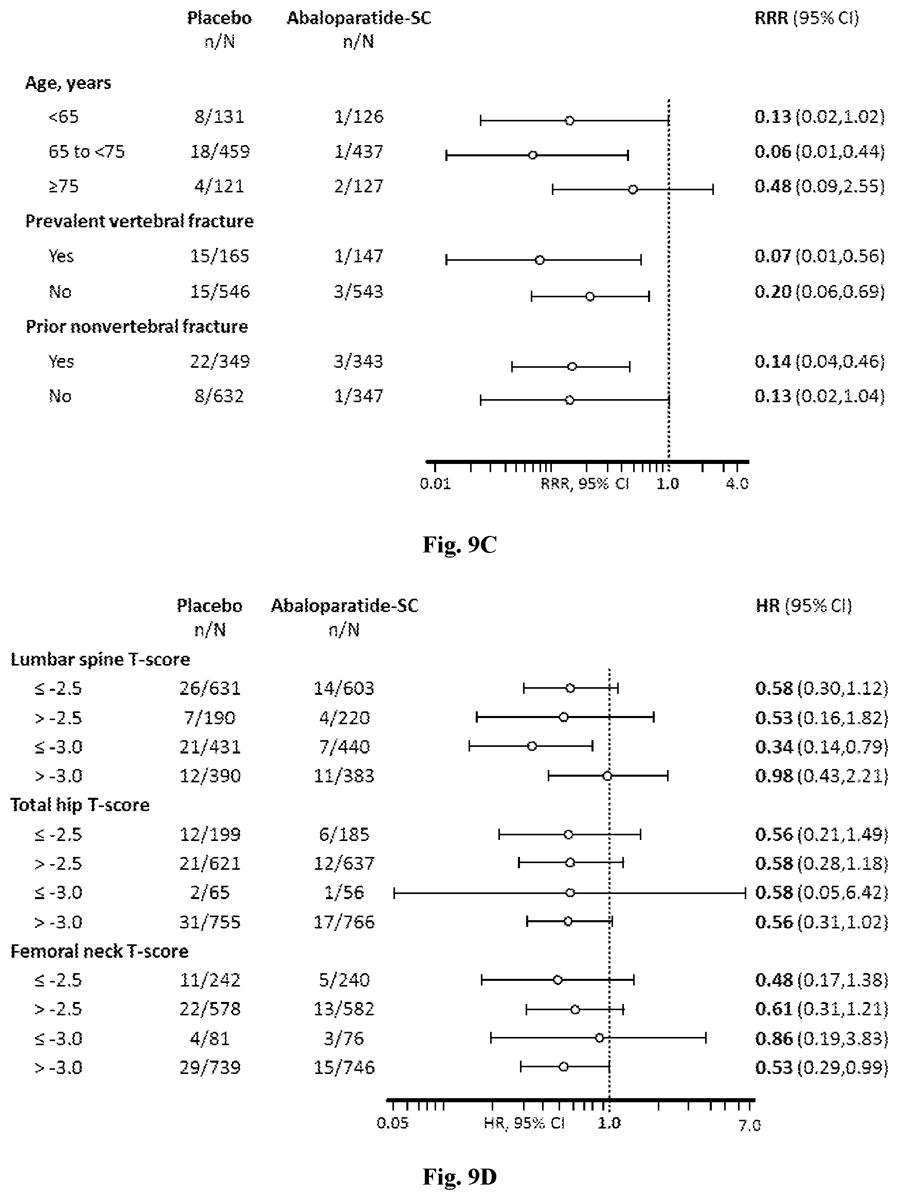
D00016

D00017
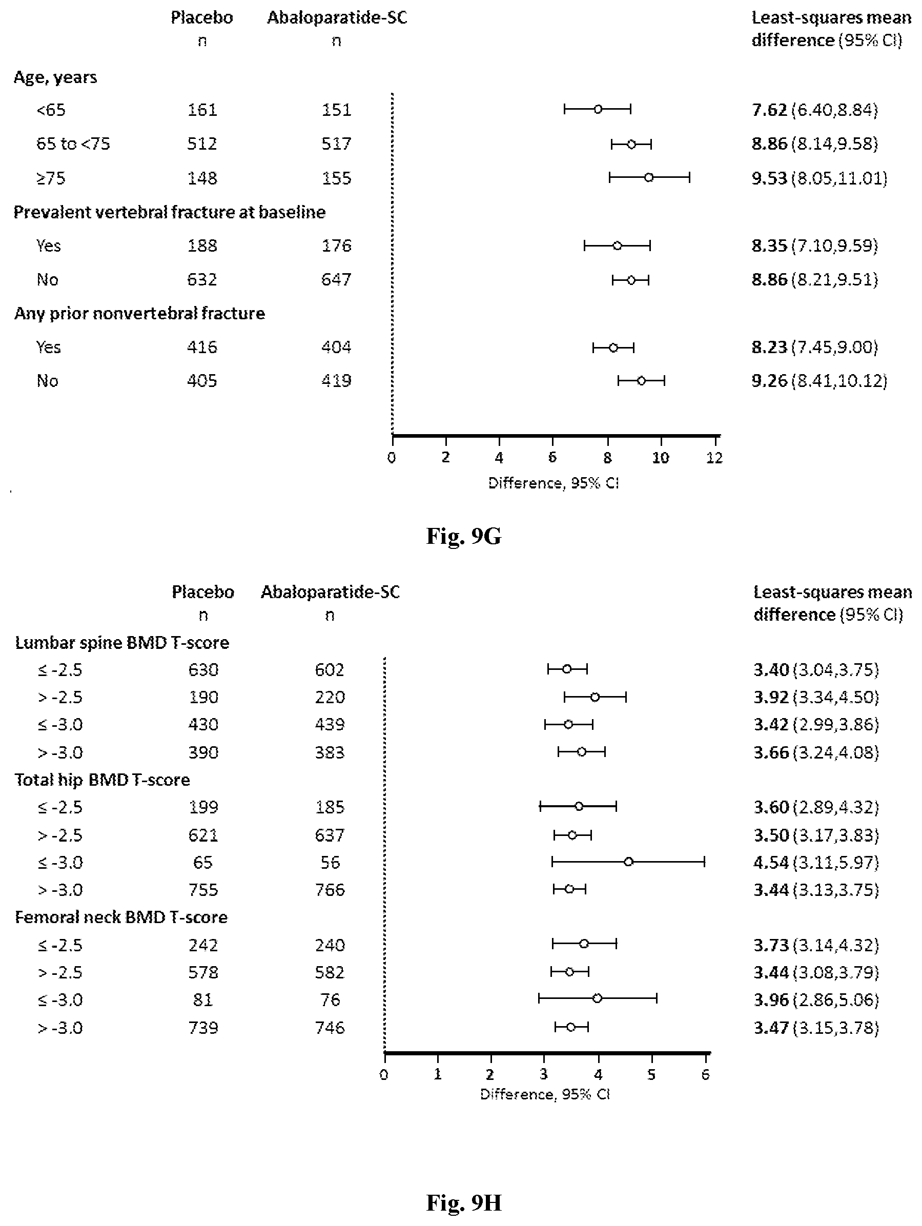
D00018
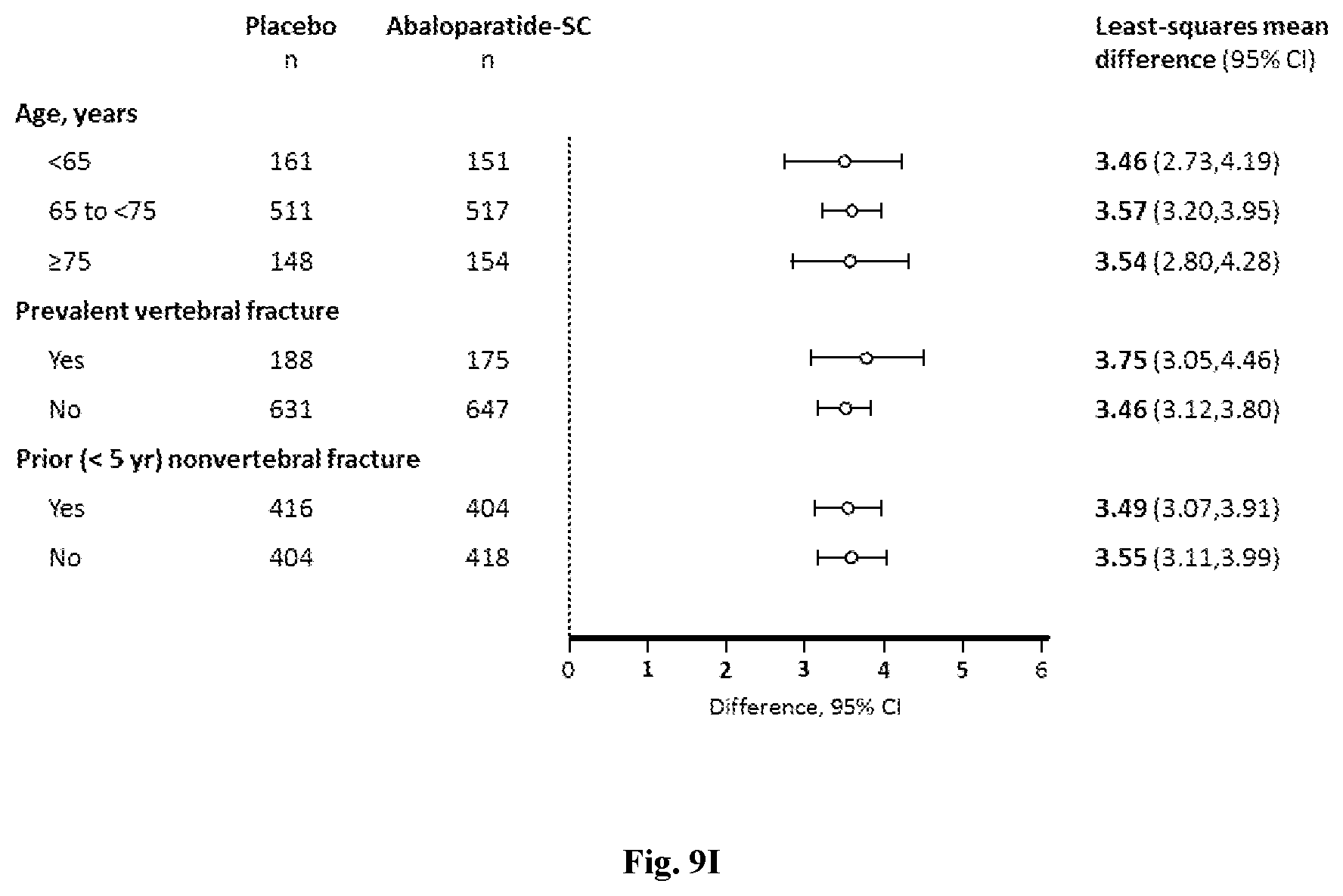
D00019
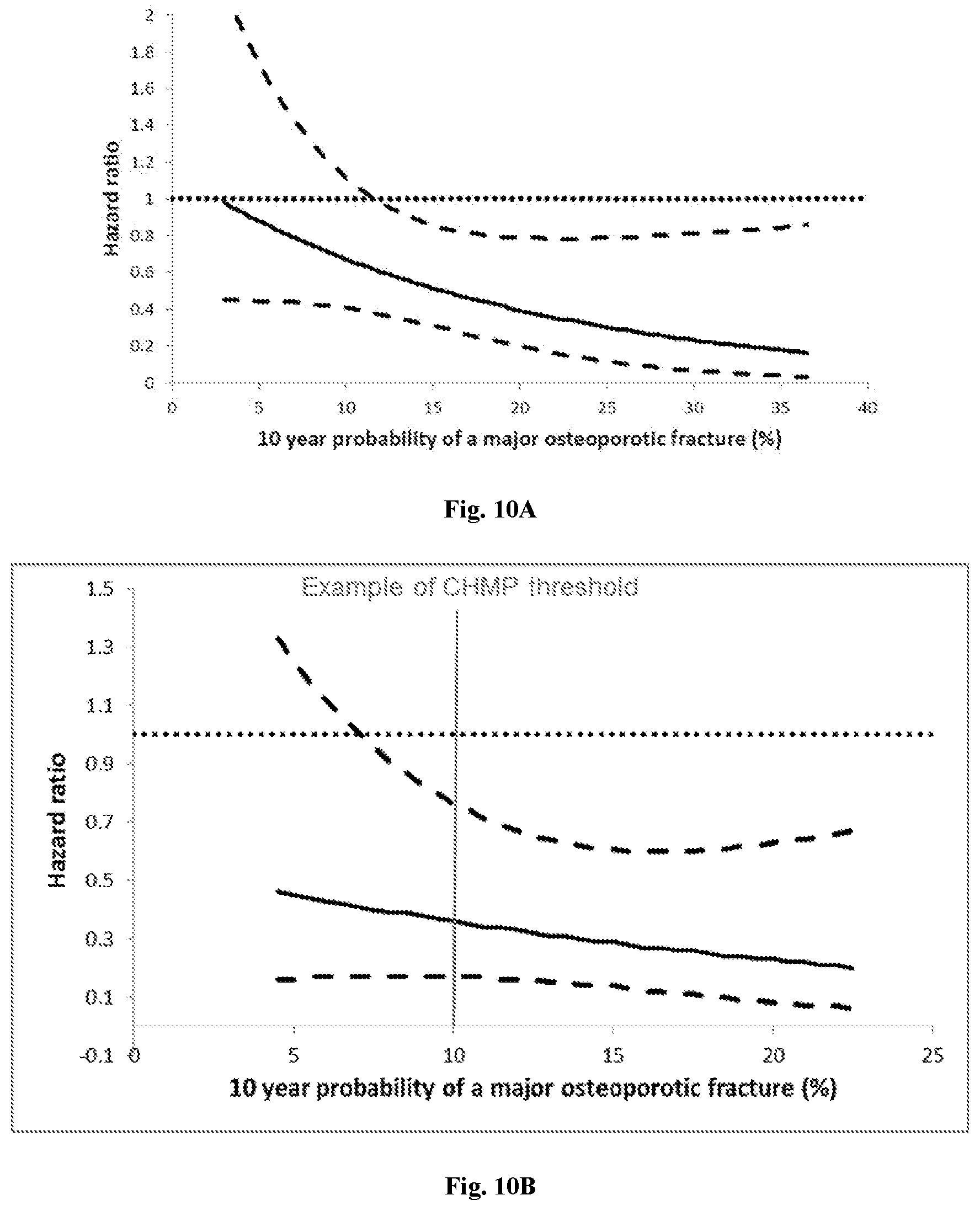
D00020
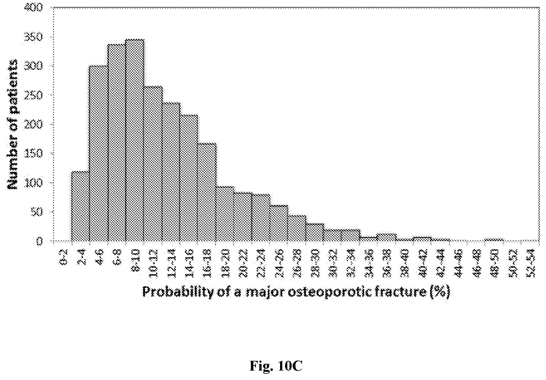
D00021
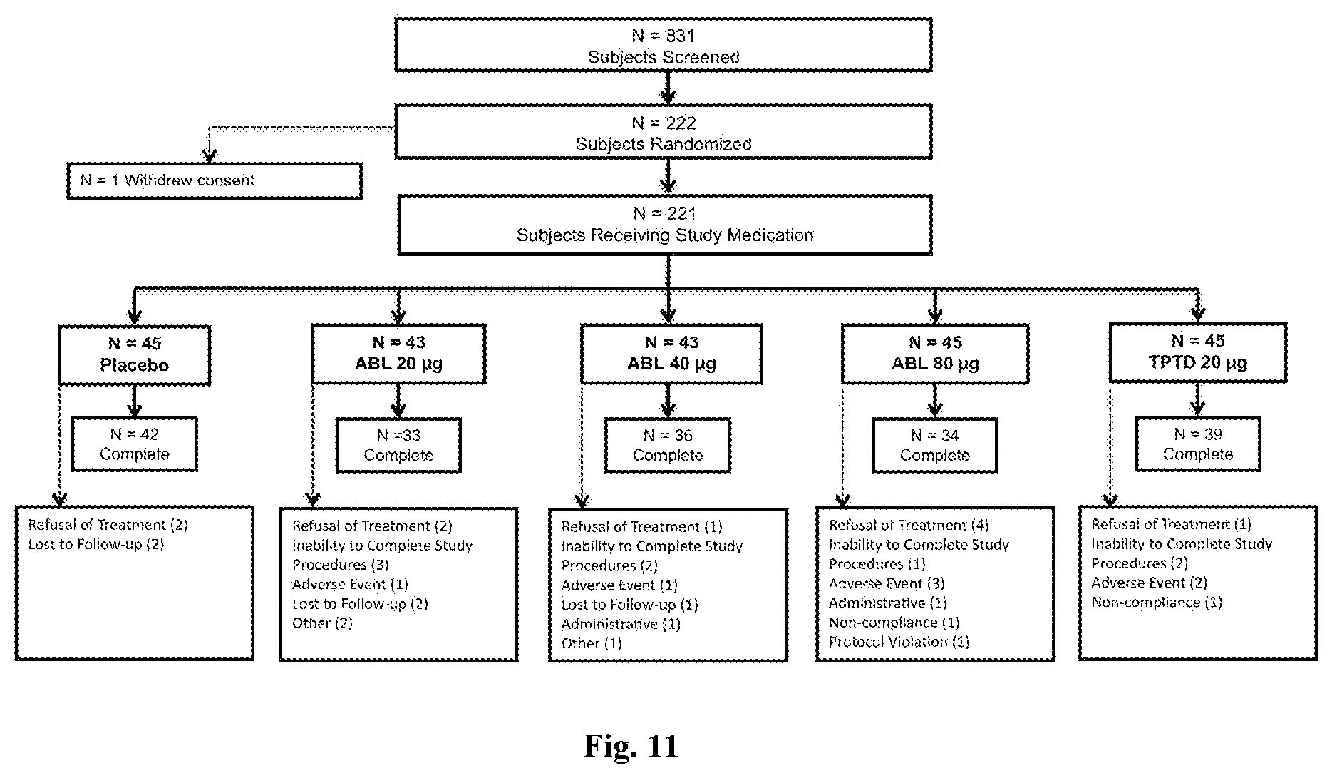
D00022

D00023
D00024
D00025

D00026

S00001
XML
uspto.report is an independent third-party trademark research tool that is not affiliated, endorsed, or sponsored by the United States Patent and Trademark Office (USPTO) or any other governmental organization. The information provided by uspto.report is based on publicly available data at the time of writing and is intended for informational purposes only.
While we strive to provide accurate and up-to-date information, we do not guarantee the accuracy, completeness, reliability, or suitability of the information displayed on this site. The use of this site is at your own risk. Any reliance you place on such information is therefore strictly at your own risk.
All official trademark data, including owner information, should be verified by visiting the official USPTO website at www.uspto.gov. This site is not intended to replace professional legal advice and should not be used as a substitute for consulting with a legal professional who is knowledgeable about trademark law.