Methods For Determining A Location Of An Analyte In A Biological Sample
Ramachandran Iyer; Eswar Prasad ; et al.
U.S. patent application number 17/565081 was filed with the patent office on 2022-04-21 for methods for determining a location of an analyte in a biological sample. The applicant listed for this patent is 10x Genomics, Inc.. Invention is credited to Zachary Bent, Eileen Dalin, Eswar Prasad Ramachandran Iyer, Yifeng Yin.
| Application Number | 20220119869 17/565081 |
| Document ID | / |
| Family ID | |
| Filed Date | 2022-04-21 |


| United States Patent Application | 20220119869 |
| Kind Code | A1 |
| Ramachandran Iyer; Eswar Prasad ; et al. | April 21, 2022 |
METHODS FOR DETERMINING A LOCATION OF AN ANALYTE IN A BIOLOGICAL SAMPLE
Abstract
Provided herein are methods of determining a location of a target analyte in a non-permeabilized biological sample that include the use of a blocking probe.
| Inventors: | Ramachandran Iyer; Eswar Prasad; (Pleasanton, CA) ; Bent; Zachary; (Pleasanton, 62) ; Yin; Yifeng; (Pleasanton, CA) ; Dalin; Eileen; (Pleasanton, CA) | ||||||||||
| Applicant: |
|
||||||||||
|---|---|---|---|---|---|---|---|---|---|---|---|
| Appl. No.: | 17/565081 | ||||||||||
| Filed: | December 29, 2021 |
Related U.S. Patent Documents
| Application Number | Filing Date | Patent Number | ||
|---|---|---|---|---|
| PCT/US2021/036557 | Jun 9, 2021 | |||
| 17565081 | ||||
| 63037458 | Jun 10, 2020 | |||
| International Class: | C12Q 1/6837 20060101 C12Q001/6837; C12Q 1/6841 20060101 C12Q001/6841 |
Claims
1. A method for determining a location of a target nucleic acid in a biological sample, the method comprising: (a) disposing a non-permeabilized biological sample onto an array at a first area, wherein the array comprises a plurality of capture probes, wherein: the first area comprises a capture probe of the plurality of capture probes comprising (i) a spatial barcode and (ii) a capture domain; and a second area of the array comprises a capture probe of the plurality of capture probes comprising (i) a spatial barcode and (ii) a capture domain, and the second area is adjacent to the biological sample disposed on the array; (b) contacting the second area of the array with a solution comprising a blocking probe, wherein the blocking probe comprises a sequence that binds specifically to the capture domain of the capture probe in the second area of the array; (c) removing residual solution comprising the blocking probe from the second area of the array; (d) permeabilizing the biological sample, such that the capture domain of the capture probe of the first area of the array binds specifically to the target nucleic acid; and (e) determining (i) a sequence corresponding to the spatial barcode of the capture probe of the first area of the array, or a complement thereof, and (ii) all or a portion of a sequence corresponding to the target nucleic acid, or a complement thereof, and using the sequences of (i) and (ii) to determine the location of the target nucleic acid in the biological sample.
2. The method of claim 1, wherein a 3' end of the blocking probe is substantially complementary to about 5 to 100 nucleotides of the capture domain of the capture probe in the second area.
3. The method of claim 1, wherein the blocking probe is a single-stranded blocking probe.
4. The method of claim 1, wherein the blocking probe is a partially double-stranded blocking probe.
5. The method of claim 1, wherein a 5' end of the blocking probe is phosphorylated.
6. The method of claim 1, wherein step (b) further comprises ligating the 5' end of the blocking probe to a 3' end of the capture probe in the second area of the array.
7. The method of claim 1, wherein a 3' end of the blocking probe comprises a chemical block.
8. The method of claim 7, wherein the chemical block is an azidomethyl group.
9. The method of claim 1, wherein the blocking probe comprises a hairpin structure.
10. The method of claim 1, wherein the blocking probe comprises a locked nucleic acid.
11. The method of claim 1, wherein the method further comprises, prior to step (a) or between steps (a) and (b), fixing and/or staining the biological sample.
12. The method of claim 11, wherein the step of fixing the biological sample comprises the use of a fixative selected from the group consisting of: ethanol, methanol, acetone, formaldehyde, paraformaldehyde-Triton, glutaraldehyde, and combinations thereof.
13. The method of claim 11, wherein the step of staining the biological sample comprises the use of a biological stain selected from the group consisting of: acridine orange, Bismarck brown, carmine, coomassie blue, cresyl violet, DAPI, eosin, ethidium bromide, acid fuchsine, hematoxylin, Hoechst stains, iodine, methyl green, methylene blue, neutral red, Nile blue, Nile red, osmium tetroxide, propidium iodide, rhodamine, safranin, and combinations thereof.
14. The method of claim 11, wherein the step of staining the biological sample comprises the use of a detectable label selected from the group consisting of: a radioisotope, a fluorophore, a chemiluminescent compound, a bioluminescent compound, and combinations thereof.
15. The method of claim 1, wherein the biological sample is selected from the group consisting of: a tissue sample, a fresh-frozen tissue section, a fixed tissue section, an organoid, embryonic stem cells, pluripotent stem cells, diseased cells, fetal cells, immune cells, cellular macromolecules, organelles, extracellular polynucleotides, and combinations thereof.
16. The method of claim 1, wherein the biological sample is a clinical sample selected from the group consisting of: whole blood, blood-derived products, blood cells, cultured tissue, cultured cells, a cell suspension, and combinations thereof.
17. The method of claim 1, wherein the removing in step (c) comprises washing.
18. The method of claim 1, wherein the determining in step (e) comprises sequencing (i) the sequence corresponding to the spatial barcode of the capture probe of the first area of the array, or a complement thereof, and (ii) all or a portion of the sequence corresponding to the target nucleic acid, or a complement thereof.
19. The method of claim 1, wherein the determining in step (e) comprises extending a 3' end of the capture probe of the first area of the array using the target nucleic acid as a template.
20. The method of claim 1, wherein the target nucleic acid is genomic DNA.
21. The method of claim 1, wherein the target nucleic acid is RNA.
22. The method of claim 21, wherein the RNA is mRNA.
23. A composition, comprising an array having a first area, wherein the array comprises a plurality of capture probes, wherein: the first area comprises a capture probe of the plurality of capture probes comprising (i) a spatial barcode and (ii) a capture domain specifically bound to a target nucleic acid from a biological sample, wherein the biological sample was previously disposed on the first area; and a second area of the array comprises a capture probe of the plurality of capture probes comprising (i) a spatial barcode and (ii) a capture domain specifically bound to a blocking probe, and the second area is adjacent to where the biological sample was previously disposed on the array.
24. The method of claim 23, wherein a 3' end of the blocking probe is substantially complementary to about 5 to 100 nucleotides of the capture domain of the capture probe in the second area.
25. The composition of claim 23, wherein the blocking probe is ligated to a 3' end of the capture probe in the second area.
26. The composition of claim 23, wherein the array comprises one or more features, optionally wherein the one or more features comprises a bead.
27. A composition, comprising an array having a first area, wherein the array comprises a plurality of capture probes, wherein: the first area comprises a capture probe of the plurality of capture probes comprising (i) a spatial barcode and (ii) a capture domain specifically bound to a target nucleic acid from a biological sample, wherein the biological sample is disposed on the first area; and a second area of the array comprises a capture probe of the plurality of capture probes comprising (i) a spatial barcode and (ii) a capture domain specifically bound to a blocking probe, and the second area is adjacent to where the biological sample is disposed on the array.
28. The method of claim 27, wherein a 3' end of the blocking probe is substantially complementary to about 5 to 100 nucleotides of the capture domain of the capture probe in the second area.
29. The composition of claim 27, wherein the blocking probe is ligated to a 3' end of the capture probe in the second area.
30. The composition of claim 27, wherein the array comprises one or more features, optionally wherein the one or more features comprises a bead.
Description
CROSS-REFERENCE TO RELATED APPLICATIONS
[0001] Pursuant to 35 U.S.C. .sctn. 119(e), this application is a continuation of International Application PCT/US2021/036557, with an international filing date of Jun. 9, 2021, which claims priority to U.S. Provisional Patent Application No. 63/037,458, filed on Jun. 10, 2020, the entire contents of which are incorporated herein by reference.
BACKGROUND
[0002] Cells within a tissue have differences in cell morphology and/or function due to varied analyte levels (e.g., gene and/or protein expression) within the different cells. The specific position of a cell within a tissue (e.g., the cell's position relative to neighboring cells or the cell's position relative to the tissue microenvironment) can affect, e.g., the cell's morphology, differentiation, fate, viability, proliferation, behavior, signaling, and cross-talk with other cells in the tissue.
[0003] Spatial heterogeneity has been previously studied using techniques that typically provide data for a handful of analytes in the context of intact tissue or a portion of a tissue (e.g., tissue section), or provide significant analyte data from individual, single cells, but fails to provide information regarding the position of the single cells from the originating biological sample (e.g., tissue).
[0004] Some techniques for studying spatial heterogeneity of a biological sample can cause analytes (e.g., nucleic acid) from the biological sample to diffuse to areas adjacent to the biological sample and be captured in such areas adjacent to the biological sample on the array. The result of capturing analytes on areas adjacent to the biological sample on the array (e.g., areas that do not correlate with the biological sample) can lead to wasted resources, such as unnecessary costs attributed to sequencing (e.g., next generation sequencing). Thus, methods to improve the incidence of captured analytes on areas of the array adjacent to the biological sample, such as blocking probes (e.g., a blocking probe to the capture domain of a capture probe), can improve efficiency, resource conservation, and resolution of the results.
SUMMARY
[0005] This application provides for a method to block capture probes on a spatial array that are not directly under the biological sample. The methods described herein can provide an improvement in resource conservation and a reduction and/or elimination of non-specific binding of analytes to unintended portions of the spatial array during performance of any of the methods described herein for determining a location of a target analyte in a biological sample.
[0006] Provided herein are methods for determining a location of a target nucleic acid in a biological sample that include: (a) disposing a non-permeabilized biological sample onto an array at a first area, where the array comprises a plurality of capture probes, where: the first area comprises a capture probe of the plurality of capture probes comprising a spatial barcode and a capture domain; and a second area of the array comprises a capture probe of the plurality of capture probes comprising a spatial barcode and a capture domain, and the second area is adjacent to the biological sample disposed on the array; (b) contacting the second area of the array with a solution comprising a blocking probe, where the blocking probe comprises a sequence that binds specifically to the capture domain of the capture probe in the second area of the array; (c) removing residual solution comprising the blocking probe from the second area of the array; (d) permeabilizing the biological sample, such that the capture domain of the capture probe of the first area of the array binds specifically to the target nucleic acid ; and (e) determining (i) all or a portion of a sequence corresponding to the spatial barcode of the capture probe of the first area of the array, or a complement thereof, and (ii) all or a portion of a sequence corresponding to the target nucleic acid, or a complement thereof, and using the sequences of (i) and (ii) to determine the location of the target nucleic acid in the biological sample.
[0007] In some embodiments of any of the methods described herein, a 3' end of the blocking probe is substantially complementary to about 5 to 100 nucleotides of the capture domain of the capture probe in the second area. In some embodiments of any of the methods described herein, the blocking probe is single-stranded. In some embodiments of any of the methods described herein, the blocking probe is at least partially double-stranded. In some embodiments of any of the methods described herein, a 5' end of the blocking probe is phosphorylated. In some embodiments of any of the methods described herein, step (b) further comprises ligating the 5' end of the blocking probe to a 3' end of the capture probe in the second area. In some embodiments of any of the methods described herein, a 3' end of the blocking probe is chemically blocked. In some embodiments of any of the methods described herein, the 3' end of the blocking probe is chemically blocked by an azidomethyl group. In some embodiments of any of the methods described herein, the blocking probe comprises a hairpin structure. In some embodiments of any of the methods described herein, the blocking probe comprises a locked nucleic acid.
[0008] In some embodiments of any of the methods described herein, the method further comprises, between steps (a) and (b), fixing and/or staining the biological sample. In some embodiments of any of the methods described herein, the non-permeabilized biological sample is fixed and/or stained prior to step (a). In some embodiments, the step of fixing the biological sample comprises the use of a fixative selected from the group of ethanol, methanol, acetone, formaldehyde, paraformaldehyde-Triton, glutaraldehyde, and combinations thereof. In some embodiments, the step of staining the biological sample comprises the use of a biological stain selected from the group of: acridine orange, Bismarck brown, carmine, coomassie blue, cresyl violet, DAPI, eosin, ethidium bromide, acid fuchsine, hematoxylin, Hoechst stains, iodine, methyl green, methylene blue, neutral red, Nile blue, Nile red, osmium tetroxide, propidium iodide, rhodamine, safranin, and combinations thereof. In some embodiments, the step of staining the biological sample comprises the use of eosin and hematoxylin. In some embodiments, the step of staining the biological sample comprises the use of a detectable label selected from the group consisting of a radioisotope, a fluorophore, a chemiluminescent compound, a bioluminescent compound, or a combination thereof.
[0009] In some embodiments of any of the methods described herein, the biological sample is a tissue sample. In some embodiments, the tissue sample is a tissue section. In some embodiments, the tissue section is a fresh, frozen tissue section. In some embodiments, the biological sample is a clinical sample. In some embodiments, the clinical sample is selected from the group of whole blood, blood-derived products, blood cells, and combinations thereof. In some embodiments, the clinical sample is a cultured tissue. In some embodiments, the clinical sample is cultured cells. In some embodiments, the clinical sample is a cell suspension.
[0010] In some embodiments of any of the methods described herein, the biological sample is an organoid, embryonic stem cells, pluripotent stem cells, and combinations thereof. In some embodiments, the organoid is selected from the group of a cerebral organoid, an intestinal organoid, a stomach organoid, a lingual organoid, a thyroid organoid, a thymic organoid, a testicular organoid, a hepatic organoid, a pancreatic organoid, an epithelial organoid, a lung organoid, a kidney organoid, a gastruloid, a cardiac organoid, a retinal organoid, and combinations thereof. In some embodiments of any of the methods described herein, the biological sample includes diseased cells, fetal cells, immune cells, cellular macromolecules, organelles, extracellular polynucleotides, and combinations thereof.
[0011] In some embodiments of any of the methods described herein, the removing in step (c) comprises washing. In some embodiments of any of the methods described herein, the array comprises a slide. In some embodiments of any of the methods described herein, the array is a bead array. In some embodiments of any of the methods described herein, the determining in step (e) comprises sequencing (i) all or a portion of the sequence corresponding to the spatial barcode of the capture probe of the first area of the array, or a complement thereof, and (ii) all or a portion of the sequence corresponding to the target nucleic acid, or a complement thereof. In some embodiments of any of the methods described herein, the sequencing is high throughput sequencing. In some embodiments of any of the methods described herein, the determining in step (e) comprises extending a 3' end of the capture probe of the first area of the array using the target nucleic acid as a template. In some embodiments of any of the methods described herein, wherein the target analyte is DNA. In some embodiments of any of the methods described herein, the DNA is genomic DNA. In some embodiments of any of the methods described herein, the target analyte is RNA. In some embodiments of any of the methods described herein, the RNA is mRNA. In some embodiments of any of the methods described herein, the method further comprises imaging the biological sample after step (a).
[0012] Also provided herein are methods for determining a location of a target analyte in a biological sample, the method comprising: (a) disposing a non-permeabilized biological sample onto an array at a first area, where the array comprises a plurality of capture probes, where: the first area comprises a capture probe of the plurality of capture probes comprising a spatial barcode and a capture domain that binds specifically to the analyte capture sequence; and a second area comprises a capture probe of the plurality of capture probes comprising a spatial barcode and a capture domain, the second area of which is adjacent to the biological sample disposed on the array; (b) contacting a plurality of analyte capture agents with the non-permeabilized biological sample, where an analyte capture agent of the plurality of analyte capture agents comprises an analyte binding moiety barcode, an analyte capture sequence, and an analyte binding moiety that binds specifically to the target analyte; (c) contacting the second area of the array with a solution comprising a blocking probe, where the blocking probe comprises a sequence that binds specifically to the capture domain of the capture probe in the second area of the array; (d) removing residual solution comprising the blocking probe from the second area of the array; (e) permeabilizing the biological sample, such that the capture domain of the capture probe of the first area of the array binds specifically to the analyte capture sequence; and (f) determining (i) all or a portion of the sequence of the spatial barcode of the capture probe in the first area of the array, or a complement thereof, and (ii) all or a portion of the sequence of the analyte binding moiety barcode, or a complement thereof, and using the sequences of (i) and (ii) to determine the location of the target analyte in the biological sample.
[0013] In some embodiments of any of the methods described herein, a 3' end of the blocking probe is substantially complementary to about 5to 100 nucleotides of the capture domain of the capture probe in the second area. In some embodiments of any of the methods described herein, the blocking probe is single-stranded. In some embodiments of any of the methods described herein, the blocking probe is partially double-stranded. In some embodiments of any of the methods described herein, a 5' end of the blocking probe is phosphorylated. In some embodiments of any of the methods described herein, step (c) further comprises ligating the 5' end of the blocking probe to a 3' end of the capture probe in the second area. In some embodiments of any of the methods described herein, a 3' end of the blocking probe is chemically blocked. In some embodiments of any of the methods described herein, the chemical block is an azidomethyl group. In some embodiments of any of the methods described herein, the blocking probe comprises a hairpin structure. In some embodiments of any of the methods described herein, the blocking probe comprises a locked nucleic acid.
[0014] In some embodiments of any of the methods described herein, the method further comprises, between steps (b) and (c), fixing the biological sample. In some embodiments of any of the methods described herein, the non-permeabilized biological sample is fixed and/or stained prior to step (a). In some embodiments, the step of fixing the biological sample comprises the use of a fixative selected from the group of ethanol, methanol, acetone, formaldehyde, paraformaldehyde-Triton, glutaraldehyde, and combinations thereof In some embodiments of any of the methods described herein, staining the biological sample comprises the use of a biological stain selected from the group of: acridine orange, Bismarck brown, carmine, coomassie blue, cresyl violet, DAPI, eosin, ethidium bromide, acid fuchsine, hematoxylin, Hoechst stains, iodine, methyl green, methylene blue, neutral red, Nile blue, Nile red, osmium tetroxide, propidium iodide, rhodamine, safranin, and combinations thereof. In some embodiments, the step of staining the biological sample comprises the use of eosin and hematoxylin. In some embodiments, the step of staining the biological sample comprises the use of a detectable label selected from the group of a radioisotope, a fluorophore, a chemiluminescent compound, a bioluminescent compound, or a combination thereof. In some embodiments of any of the methods described herein, the biological sample is a tissue sample. In some embodiments, the tissue sample is a tissue section. In some embodiments, the tissue section is a fresh, frozen tissue section. In some embodiments of any of the methods described herein, the biological sample is a clinical sample. In some embodiments, the clinical sample is selected from the group of whole blood, blood-derived products, blood cells, and combinations thereof. In some embodiments, the clinical sample is a cultured tissue. In some embodiments, the clinical sample is cultured cells. In some embodiments, the clinical sample is a cell suspension. In some embodiments of any of the methods described herein, the biological sample is an organoid, embryonic stem cells, pluripotent stem cells, and combinations thereof. In some embodiments, the organoid is selected from the group of a cerebral organoid, an intestinal organoid, a stomach organoid, a lingual organoid, a thyroid organoid, a thymic organoid, a testicular organoid, a hepatic organoid, a pancreatic organoid, an epithelial organoid, a lung organoid, a kidney organoid, a gastruloid, a cardiac organoid, a retinal organoid, and combinations thereof. In some embodiments of any of the methods described herein, the biological sample includes diseased cells, fetal cells, immune cells, cellular macromolecules, organelles, extracellular polynucleotides, and combinations thereof. In some embodiments of any of the methods described herein, the removing in step (d) comprises washing. In some embodiments of any of the methods described herein, the array comprises a slide. In some embodiments of any of the methods described herein, the array is a bead array.
[0015] In some embodiments of any of the methods described herein, the determining in step (f) comprises sequencing (i) all or a portion of the sequence corresponding to the spatial barcode of the capture probe in the first area of the array, or a complement thereof, and (ii) all or a portion of the sequence corresponding to the analyte binding moiety barcode, or a complement thereof. In some embodiments of any of the methods described herein, the sequencing is high throughput sequencing. In some embodiments of any of the methods described herein, the determining in step (f) comprises extending a 3' end of the capture probe of the first area of the array using the analyte binding moiety barcode as a template. In some embodiments of any of the methods described herein, the target analyte is a protein. In some embodiments of any of the methods described herein, the protein is an intracellular protein. In some embodiments of any of the methods described herein, the protein is an extracellular protein. In some embodiments of any of the methods described herein, the analyte binding moiety is an antibody or an antigen-binding moiety thereof. In some embodiments of any of the methods described herein, steps (a) and (b) are performed at substantially the same time. In some embodiments of any of the methods described herein, step (a) is performed before step (b). In some embodiments of any of the methods described herein, step (b) is performed before step (a). In some embodiments of any of the methods described herein, the method further comprises imaging the biological sample after step (b).
[0016] Also provided herein are kits comprising an array comprises a plurality of capture probes, where a capture probe of the plurality of capture probes comprising a spatial barcode and a capture domain; and a solution comprising a blocking probe, where the blocking probe comprises a sequence that binds specifically to the capture domain of the capture probe.
[0017] In some embodiments of any of the kits described herein, the kit(s) further comprise one or more fixative(s). In some embodiments of any of the kits described herein, the kit(s) further comprise one or more biological stains. In some embodiments, the one or more biological stains is eosin and hematoxylin. In some embodiments of any of the kits described herein, the kit(s) further comprise one or more permeabilization reagent(s). In some embodiments of any of the kits described herein, the one or more permeabilization reagent(s) is selected from the group of an organic solvent, a cross-linking agent, a detergent, an enzyme, and combinations thereof. In some embodiments of any of the kits described herein, the kit further comprises a reverse transcriptase. In some embodiments of any of the kits described herein, the kit further comprises a terminal deoxynucleotidyl transferase. In some embodiments of any of the kits described herein, the kit further comprises a template switching oligonucleotide. In some embodiments of any of the kits described herein, the kit further comprises a DNA polymerase. In some embodiments of any of the kits described herein, the kit further comprises a second strand primer. In some embodiments of any of the kits described herein, the kit further comprises a fragmentation buffer and a fragmentation enzyme. In some embodiments of any of the kits described herein, the kit further comprises a DNA ligase. In some embodiments, the DNA ligase is a T4 DNA ligase. In some embodiments of any of the kits described herein, the kit further comprises one or more adaptor(s). In some embodiments, the one or more adaptor(s) is/are selected from the group of an i5 sample index sequence, an i7 sample index sequence, a P5 sample index sequence, a P7 sample index sequence, and combinations thereof.
[0018] Also provided herein are composition(s), comprising an array, where the array comprises a plurality of capture probes, where: the first area comprises a capture probe of the plurality of capture probes comprising a spatial barcode and a capture domain specifically bound to a target analyte from the biological sample; and a second area of the array comprises a capture probe of the plurality of capture probes comprising a spatial barcode and a capture domain specifically bound to a blocking probe, and the second area is adjacent to the biological sample disposed on the array.
[0019] In some embodiments of any of the composition(s) described herein, a 3' end of the blocking probe is substantially complementary to about 5 to 100 nucleotides of the capture domain of the capture probe in the second area. In some embodiments of any of the composition(s) described herein, the blocking probe is single-stranded. In some embodiments of any of the composition(s) described herein, the blocking probe is partially double-stranded. In some embodiments of any of the composition(s) described herein, a 5' end of the blocking probe is phosphorylated. In some embodiments of any of the composition(s) described herein, the blocking probe is ligated to a 3' end of the capture probe in the second area. In some embodiments of any of the composition(s) described herein, a 3' end of the blocking probe is chemically blocked. In some embodiments of any of the composition(s) described herein, the chemical block is an azidomethyl group. In some embodiments of any of the composition(s) described herein, the blocking probe comprises a hairpin structure. In some embodiments of any of the composition(s) described herein, the blocking probe comprises a locked nucleic acid.
[0020] In some embodiments, a biological sample is disposed on the first area of the array. In some embodiments of any of the composition(s) described herein, the biological sample is a tissue sample. In some embodiments of any of the composition(s) described herein, the tissue sample is a tissue section. In some embodiments of any of the composition(s) described herein, the biological sample is a clinical sample. In some embodiments of any of the composition(s) described herein, the clinical sample is selected from the group of whole blood, blood-derived products, blood cells, and combinations thereof In some embodiments of any of the composition(s) described herein, the clinical sample is a cultured tissue. In some embodiments of any of the composition(s) described herein, the clinical sample is cultured cells. In some embodiments of any of the composition(s) described herein, the clinical sample is a cell suspension. In some embodiments of any of the composition(s) described herein, the biological sample is an organoid, embryonic stem cells, pluripotent stem cells, and combinations thereof. In some embodiments of any of the composition(s) described herein, the organoid is selected from the group of a cerebral organoid, an intestinal organoid, a stomach organoid, a lingual organoid, a thyroid organoid, a thymic organoid, a testicular organoid, a hepatic organoid, a pancreatic organoid, an epithelial organoid, a lung organoid, a kidney organoid, a gastruloid, a cardiac organoid, a retinal organoid, and combinations thereof. In some embodiments of any of the composition(s) described herein, the biological sample includes diseased cells, fetal cells, immune cells, cellular macromolecules, organelles, extracellular polynucleotides, and combinations thereof.
[0021] In some embodiments of any of the composition(s) described herein, the array comprises a slide. In some embodiments of any of the composition(s) described herein, the array is a bead array. In some embodiments of any of the composition(s) described herein, the target analyte is DNA. In some embodiments of any of the composition(s) described herein, the DNA is genomic DNA. In some embodiments of any of the composition(s) described herein, the target analyte is RNA. In some embodiments of any of the composition(s) described herein, the RNA is mRNA.
[0022] All publications, patents, and patent applications mentioned in this specification are herein incorporated by reference to the same extent as if each individual publication, patent, patent application, or item of information was specifically and individually indicated to be incorporated by reference. To the extent publications, patents, patent applications, and items of information incorporated by reference contradict the disclosure contained in the specification, the specification is intended to supersede and/or take precedence over any such contradictory material.
[0023] Where values are described in terms of ranges, it should be understood that the description includes the disclosure of all possible sub-ranges within such ranges, as well as specific numerical values that fall within such ranges irrespective of whether a specific numerical value or specific sub-range is expressly stated.
[0024] The term "each," when used in reference to a collection of items, is intended to identify an individual item in the collection but does not necessarily refer to every item in the collection, unless expressly stated otherwise, or unless the context of the usage clearly indicates otherwise.
[0025] Various embodiments of the features of this disclosure are described herein. However, it should be understood that such embodiments are provided merely by way of example, and numerous variations, changes, and substitutions can occur to those skilled in the art without departing from the scope of this disclosure. It should also be understood that various alternatives to the specific embodiments described herein are also within the scope of this disclosure.
DESCRIPTION OF DRAWINGS
[0026] The following drawings illustrate certain embodiments of the features and advantages of this disclosure. These embodiments are not intended to limit the scope of the appended claims in any manner. Like reference symbols in the drawings indicate like elements.
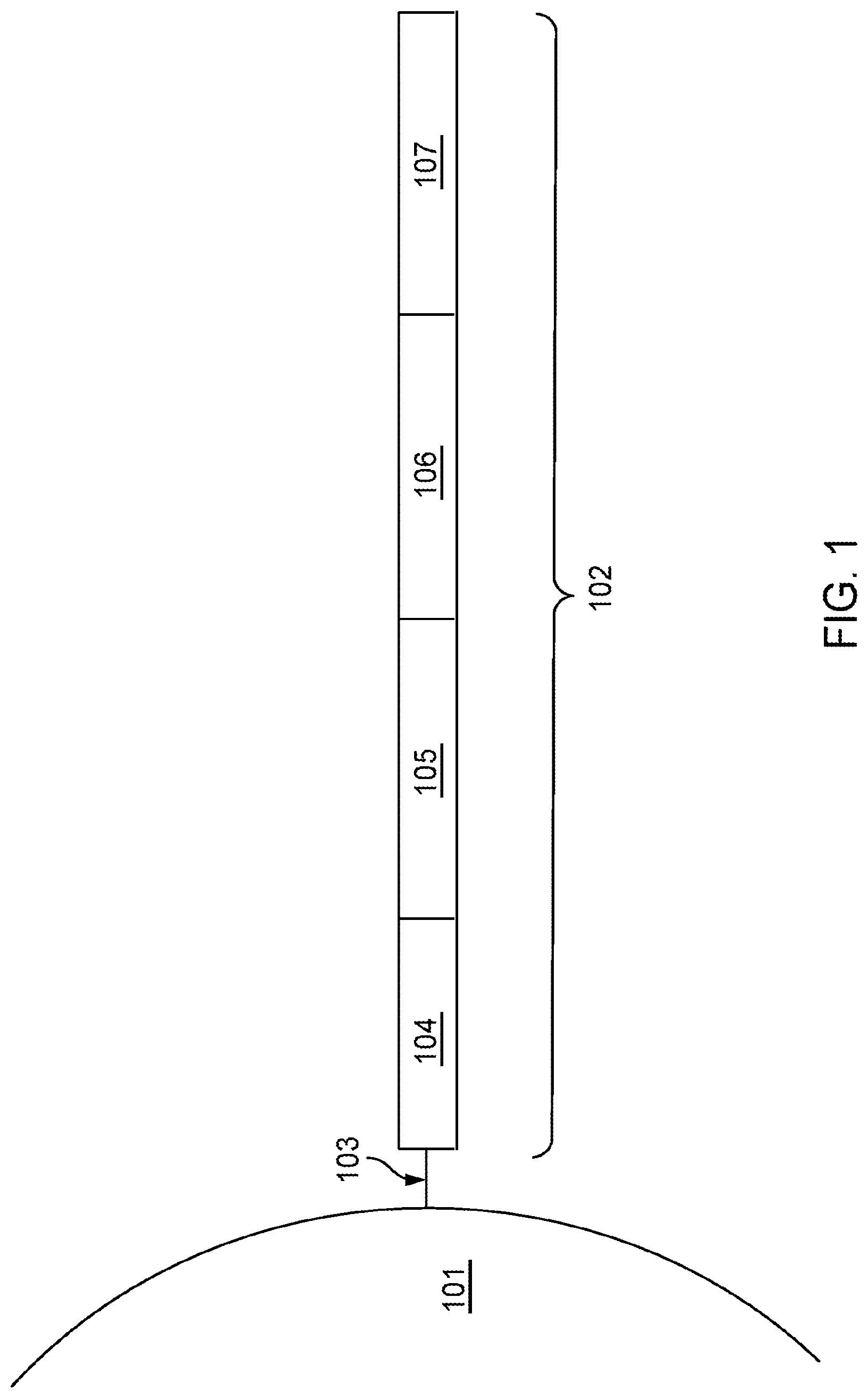
[0027] FIG. 1 is a schematic diagram showing an example of a barcoded capture probe, as described herein.
[0028] FIG. 2 shows an example of diffusion of target nucleic acids away from a biological sample towards an unintended area of an array.
[0029] FIG. 3A shows an exemplary blocking probe comprising a hairpin structure bound to a capture domain of a capture probe.
[0030] FIG. 3B shows an exemplary partially-double stranded blocking probe bound to a capture domain of a capture probe.
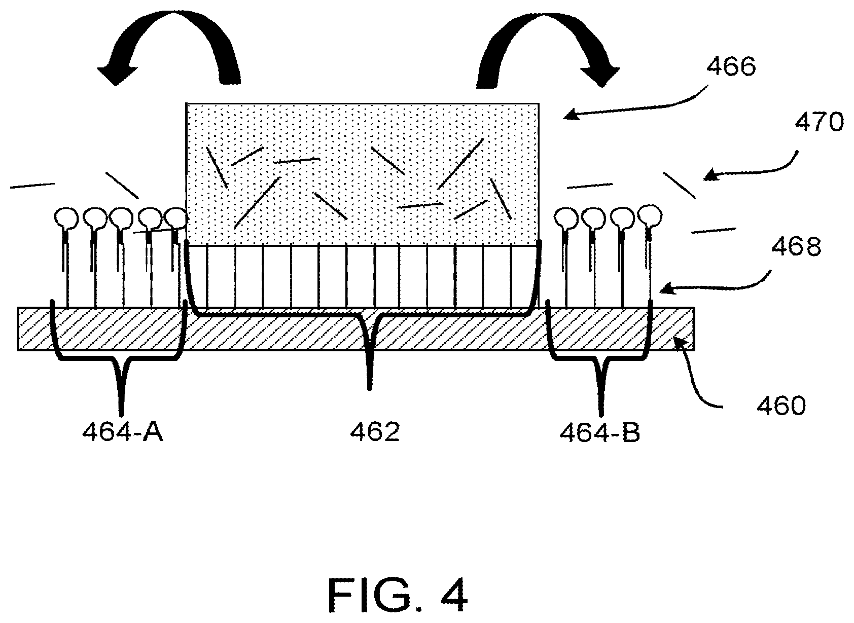
[0031] FIG. 4 shows an exemplary embodiment of blocked capture probes in the area of a spatial array that is not under a biological sample, where the block is the hairpin structure of FIG. 3A.
[0032] FIG. 5 shows a schematic of an exemplary workflow utilizing an exemplary embodiment of the methods described herein.
DETAILED DESCRIPTION
[0033] Blocking one or more capture domains of capture probes on spatial arrays (or portions thereof) can increase efficiency and/or decrease non-specific binding of analytes on arrays (or portions thereof). In some cases, one or more capture probes (e.g., capture domain of capture probes) can be blocked with one or more blocking probes. A 3' end of a blocking probe can be substantially complementary to about 5 to about 100 nucleotides of the capture domain. Provided herein are methods, compositions, and kits, e.g., for carrying out these methods. In some cases, a portion of an array can be selectively blocked and/or selectively unblocked.
[0034] Methods for reducing non-specific spatial interactions on a spatial array are described herein. Methods herein can improve the resolution of spatial array results by reducing non-specific binding of targeted analytes. For example, methods herein can reduce non-specific binding of target analytes by capture probes (e.g., by blocking the capture domain of capture probes) not proximal to the targeted analyte. In some cases, analytes from a biological sample can diffuse to areas of the array that are adjacent to the biological sample. This can cause analytes to bind to the capture domain(s) of one or more capture probes adjacent to the biological sample. Non-specific binding increases background results (e.g., non-specific results), thereby decreasing resolution. Blocking the capture domain of capture probes that adjacent to the biological sample can decrease the non-specific binding and increase the resolution of results.
[0035] Methods described herein can also conserve resources. For example, in some cases, the analysis of spatial arrays can include sequencing. Non-specific binding of analytes to the capture domain of one or more capture probes can result in sequencing of undesired targets. Non-specific analyte capture can cause downstream sequencing inefficiencies, for example, a decrease in the amount of target analyte sequencing due to sequencing of non-specific captured analytes is inefficient and reagent costly and can result in a decrease is spatial resolution. The present disclosure provides solutions for improving and/or preventing non-specific analyte capture on an array slide.
[0036] Spatial analysis methodologies and compositions described herein can provide a vast amount of analyte and/or expression data for a variety of analytes within a biological sample at high spatial resolution, while retaining native spatial context. Spatial analysis methods and compositions can include, e.g., the use of a capture probe including a spatial barcode (e.g., a nucleic acid sequence that provides information as to the location or position of an analyte within a cell or a tissue sample (e.g., mammalian cell or a mammalian tissue sample) and a capture domain that is capable of binding to an analyte (e.g., a protein and/or a nucleic acid) produced by and/or present in a cell. Spatial analysis methods and compositions can also include the use of a capture probe having a capture domain that captures an intermediate agent for indirect detection of an analyte. For example, the intermediate agent can include a nucleic acid sequence (e.g., a barcode) associated with the intermediate agent. Detection of the intermediate agent is therefore indicative of the analyte in the cell or tissue sample.
[0037] Non-limiting aspects of spatial analysis methodologies and compositions are described in U.S. Pat. Nos. 10,774,374, 10,724,078, 10,480,022, 10,059,990, 10,041,949, 10,002,316, 9,879,313, 9,783,841, 9,727,810, 9,593,365, 8,951,726, 8,604,182, 7,709,198, U.S. Patent Application Publication Nos. 2020/239946, 2020/080136, 2020/0277663, 2020/024641, 2019/330617, 2019/264268, 2020/256867, 2020/224244, 2019/194709, 2019/161796, 2019/085383, 2019/055594, 2018/216161, 2018/051322, 2018/0245142, 2017/241911, 2017/089811, 2017/067096, 2017/029875, 2017/0016053, 2016/108458, 2015/000854, 2013/171621, WO 2018/091676, WO 2020/176788, Rodrigues et al., Science 363(6434):1463-1467, 2019; Lee et al., Nat. Protoc. 10(3):442-458, 2015; Trejo et al., PLoS ONE 14(2):e0212031, 2019; Chen et al., Science 348(6233):aaa6090, 2015; Gao et al., BMC Biol. 15:50, 2017; and Gupta et al., Nature Biotechnol. 36:1197-1202, 2018; the Visium Spatial Gene Expression Reagent Kits User Guide (e.g., Rev C, dated June 2020), and/or the Visium Spatial Tissue Optimization Reagent Kits User Guide (e.g., Rev C, dated July 2020), both of which are available at the 10.times. Genomics Support Documentation website, and can be used herein in any combination. Further non-limiting aspects of spatial analysis methodologies and compositions are described herein.
[0038] Some general terminology that may be used in this disclosure can be found in Section (I)(b) of WO 2020/176788 and/or U.S. Patent Application Publication No. 2020/0277663. Typically, a "barcode" is a label, or identifier, that conveys or is capable of conveying information (e.g., information about an analyte in a sample, a bead, and/or a capture probe). A barcode can be part of an analyte, or independent of an analyte. A barcode can be attached to an analyte. A particular barcode can be unique relative to other barcodes. For the purpose of this disclosure, an "analyte" can include any biological substance, structure, moiety, or component to be analyzed. The term "target" can similarly refer to an analyte of interest.
[0039] Analytes can be broadly classified into one of two groups: nucleic acid analytes, and non-nucleic acid analytes. Examples of non-nucleic acid analytes include, but are not limited to, lipids, carbohydrates, peptides, proteins, glycoproteins (N-linked or O-linked), lipoproteins, phosphoproteins, specific phosphorylated or acetylated variants of proteins, amidation variants of proteins, hydroxylation variants of proteins, methylation variants of proteins, ubiquitylation variants of proteins, sulfation variants of proteins, viral proteins (e.g., viral capsid, viral envelope, viral coat, viral accessory, viral glycoproteins, viral spike, etc.), extracellular and intracellular proteins, antibodies, and antigen binding fragments. In some embodiments, the analyte(s) can be localized to subcellular location(s), including, for example, organelles, e.g., mitochondria, Golgi apparatus, endoplasmic reticulum, chloroplasts, endocytic vesicles, exocytic vesicles, vacuoles, lysosomes, etc. In some embodiments, analyte(s) can be peptides or proteins, including without limitation antibodies and enzymes. Additional examples of analytes can be found in Section (I)(c) of WO 2020/176788 and/or U.S. Patent Application Publication No. 2020/0277663. In some embodiments, an analyte can be detected indirectly, such as through detection of an intermediate agent, for example, a ligation product or an analyte capture agent (e.g., an oligonucleotide-conjugated antibody), such as those described herein.
[0040] A "biological sample" is typically obtained from the subject for analysis using any of a variety of techniques including, but not limited to, biopsy, surgery, and laser capture microscopy (LCM), and generally includes cells and/or other biological material from the subject. In some embodiments, a biological sample can be a tissue section. In some embodiments, a biological sample can be a fixed and/or stained biological sample (e.g., a fixed and/or stained tissue section). Non-limiting examples of stains include histological stains (e.g., hematoxylin and/or eosin) and immunological stains (e.g., fluorescent stains). In some embodiments, a biological sample (e.g., a fixed and/or stained biological sample) can be imaged. Biological samples are also described in Section (I)(d) of WO 2020/176788 and/or U.S. Patent Application Publication No. 2020/0277663.
[0041] In some embodiments, a biological sample is permeabilized with one or more permeabilization reagents. For example, permeabilization of a biological sample can facilitate analyte capture. Exemplary permeabilization agents and conditions are described in Section (I)(d)(ii)(13) or the Exemplary Embodiments Section of WO 2020/176788 and/or U.S. Patent Application Publication No. 2020/0277663.
[0042] Array-based spatial analysis methods involve the transfer of one or more analytes from a biological sample to an array of features on a substrate, where each feature is associated with a unique spatial location on the array. Subsequent analysis of the transferred analytes includes determining the identity of the analytes and the spatial location of the analytes within the biological sample. The spatial location of an analyte within the biological sample is determined based on the feature to which the analyte is bound (e.g., directly or indirectly) on the array, and the feature's relative spatial location within the array.
[0043] A "capture probe" refers to any molecule capable of capturing (directly or indirectly) and/or labelling an analyte (e.g., an analyte of interest) in a biological sample. In some embodiments, the capture probe is a nucleic acid or a polypeptide. In some embodiments, the capture probe includes a barcode (e.g., a spatial barcode and/or a unique molecular identifier (UMI)) and a capture domain). In some embodiments, a capture probe can include a cleavage domain and/or a functional domain (e.g., a primer-binding site, such as for next-generation sequencing (NGS)). See, e.g., Section (II)(b) (e.g., subsections (i)-(vi)) of WO 2020/176788 and/or U.S. Patent Application Publication No. 2020/0277663. Generation of capture probes can be achieved by any appropriate method, including those described in Section (II)(d)(ii) of WO 2020/176788 and/or U.S. Patent Application Publication No. 2020/0277663.
[0044] In some embodiments, more than one analyte type (e.g., nucleic acids and proteins) from a biological sample can be detected (e.g., simultaneously or sequentially) using any appropriate multiplexing technique, such as those described in Section (IV) of WO 2020/176788 and/or U.S. Patent Application Publication No. 2020/0277663.
[0045] In some embodiments, detection of one or more analytes (e.g., protein analytes) can be performed using one or more analyte capture agents. As used herein, an "analyte capture agent" refers to an agent that interacts with an analyte (e.g., an analyte in a biological sample) and with a capture probe (e.g., a capture probe attached to a substrate or a feature) to identify the analyte. In some embodiments, the analyte capture agent includes: (i) an analyte binding moiety (e.g., that binds to an analyte), for example, an antibody or antigen-binding fragment thereof; (ii) analyte binding moiety barcode; and (iii) an analyte capture sequence. As used herein, the term "analyte binding moiety barcode" refers to a barcode that is associated with or otherwise identifies the analyte binding moiety. As used herein, the term "analyte capture sequence" refers to a region or moiety configured to hybridize to, bind to, couple to, or otherwise interact with a capture domain of a capture probe. In some cases, an analyte binding moiety barcode (or portion thereof) may be able to be removed (e.g., cleaved) from the analyte capture agent. Additional description of analyte capture agents can be found in Section (II)(b)(ix) of WO 2020/176788 and/or Section (II)(b)(viii) U.S. Patent Application Publication No. 2020/0277663.
[0046] There are at least two methods to associate a spatial barcode with one or more neighboring cells, such that the spatial barcode identifies the one or more cells, and/or contents of the one or more cells, as associated with a particular spatial location. One method is to promote analytes or analyte proxies (e.g., intermediate agents) out of a cell and towards a spatially-barcoded array (e.g., including spatially-barcoded capture probes). Another method is to cleave spatially-barcoded capture probes from an array and promote the spatially-barcoded capture probes towards and/or into or onto the biological sample.
[0047] In some cases, capture probes may be configured to prime, replicate, and consequently yield optionally barcoded extension products from a template (e.g., a DNA or RNA template, such as an analyte or an intermediate agent (e.g., a ligation product or an analyte capture agent), or a portion thereof), or derivatives thereof (see, e.g., Section (II)(b)(vii) of WO 2020/176788 and/or U.S. Patent Application Publication No. 2020/0277663 regarding extended capture probes). In some cases, capture probes may be configured to form ligation products with a template (e.g., a DNA or RNA template, such as an analyte or an intermediate agent, or portion thereof), thereby creating ligations products that serve as proxies for a template.
[0048] As used herein, an "extended capture probe" refers to a capture probe having additional nucleotides added to the terminus (e.g., 3' or 5' end) of the capture probe thereby extending the overall length of the capture probe. For example, an "extended 3' end" indicates additional nucleotides were added to the most 3' nucleotide of the capture probe to extend the length of the capture probe, for example, by polymerization reactions used to extend nucleic acid molecules including templated polymerization catalyzed by a polymerase (e.g., a DNA polymerase or a reverse transcriptase). In some embodiments, extending the capture probe includes adding to a 3' end of a capture probe a nucleic acid sequence that is complementary to a nucleic acid sequence of an analyte or intermediate agent specifically bound to the capture domain of the capture probe. In some embodiments, the capture probe is extended using reverse transcription. In some embodiments, the capture probe is extended using one or more DNA polymerases. The extended capture probes include the sequence of the capture probe and the sequence of the spatial barcode of the capture probe.
[0049] In some embodiments, extended capture probes are amplified (e.g., in bulk solution or on the array) to yield quantities that are sufficient for downstream analysis, e.g., via DNA sequencing. In some embodiments, extended capture probes (e.g., DNA molecules) act as templates for an amplification reaction (e.g., a polymerase chain reaction).
[0050] Additional variants of spatial analysis methods, including in some embodiments, an imaging step, are described in Section (II)(a) of WO 2020/176788 and/or U.S. Patent Application Publication No. 2020/0277663. Analysis of captured analytes (and/or intermediate agents or portions thereof), for example, including sample removal, extension of capture probes, sequencing (e.g., of a cleaved extended capture probe and/or a cDNA molecule complementary to an extended capture probe), sequencing on the array (e.g., using, for example, in situ hybridization or in situ ligation approaches), temporal analysis, and/or proximity capture, is described in Section (II)(g) of WO 2020/176788 and/or U.S. Patent Application Publication No. 2020/0277663. Some quality control measures are described in Section (II)(h) of WO 2020/176788 and/or U.S. Patent Application Publication No. 2020/0277663.
[0051] Spatial information can provide information of biological and/or medical importance. For example, the methods and compositions described herein can allow for: identification of one or more biomarkers (e.g., diagnostic, prognostic, and/or for determination of efficacy of a treatment) of a disease or disorder; identification of a candidate drug target for treatment of a disease or disorder; identification (e.g., diagnosis) of a subject as having a disease or disorder; identification of stage and/or prognosis of a disease or disorder in a subject; identification of a subject as having an increased likelihood of developing a disease or disorder; monitoring of progression of a disease or disorder in a subject; determination of efficacy of a treatment of a disease or disorder in a subject; identification of a patient subpopulation for which a treatment is effective for a disease or disorder; modification of a treatment of a subject with a disease or disorder; selection of a subject for participation in a clinical trial; and/or selection of a treatment for a subject with a disease or disorder.
[0052] Spatial information can provide information of biological importance. For example, the methods and compositions described herein can allow for: identification of transcriptome and/or proteome expression profiles (e.g., in healthy and/or diseased tissue); identification of multiple analyte types in close proximity (e.g., nearest neighbor analysis); determination of up- and/or down-regulated genes and/or proteins in diseased tissue; characterization of tumor microenvironments; characterization of tumor immune responses; characterization of cells types and their co-localization in tissue; and identification of genetic variants within tissues (e.g., based on gene and/or protein expression profiles associated with specific disease or disorder biomarkers).
[0053] Typically, for spatial array-based methods, a substrate functions as a support for direct or indirect attachment of capture probes to features of the array. A "feature" is an entity that acts as a support or repository for various molecular entities used in spatial analysis. In some embodiments, some or all of the features in an array are functionalized for analyte capture. Exemplary substrates are described in Section (II)(c) of WO 2020/176788 and/or U.S. Patent Application Publication No. 2020/0277663. Exemplary features and geometric attributes of an array can be found in Sections (II)(d)(i), (II)(d)(iii), and (II)(d)(iv) of WO 2020/176788 and/or U.S. Patent Application Publication No. 2020/0277663.
[0054] Generally, analytes and/or intermediate agents (or portions thereof) can be captured when contacting a biological sample with a substrate including capture probes (e.g., a substrate with capture probes embedded, spotted, printed, fabricated on the substrate, or a substrate with features (e.g., beads, wells) comprising capture probes). As used herein, "contact," "contacted," and/or "contacting," a biological sample with a substrate refers to any contact (e.g., direct or indirect) such that capture probes can interact (e.g., bind covalently or non-covalently (e.g., hybridize)) with analytes from the biological sample. Capture can be achieved actively (e.g., using electrophoresis) or passively (e.g., using diffusion). Analyte capture is further described in Section (II)(e) of WO 2020/176788 and/or U.S. Patent Application Publication No. 2020/0277663.
[0055] In some cases, spatial analysis can be performed by attaching and/or introducing a molecule (e.g., a peptide, a lipid, or a nucleic acid molecule) having a barcode (e.g., a spatial barcode) to a biological sample (e.g., to a cell in a biological sample). In some embodiments, a plurality of molecules (e.g., a plurality of nucleic acid molecules) having a plurality of barcodes (e.g., a plurality of spatial barcodes) are introduced to a biological sample (e.g., to a plurality of cells in a biological sample) for use in spatial analysis. In some embodiments, after attaching and/or introducing a molecule having a barcode to a biological sample, the biological sample can be physically separated (e.g., dissociated) into single cells or cell groups for analysis. Some such methods of spatial analysis are described in Section (III) of WO 2020/176788 and/or U.S. Patent Application Publication No. 2020/0277663.
[0056] In some cases, spatial analysis can be performed by detecting multiple oligonucleotides that hybridize to an analyte. In some instances, for example, spatial analysis can be performed using RNA-templated ligation (RTL). Methods of RTL have been described previously. See, e.g., Credle et al., Nucleic Acids Res. 2017 Aug. 21; 45(14):e128. Typically, RTL includes hybridization of two oligonucleotides to adjacent sequences on an analyte (e.g., an RNA molecule, such as an mRNA molecule). In some instances, the oligonucleotides are DNA molecules. In some instances, one of the oligonucleotides includes at least two ribonucleic acid bases at the 3' end and/or the other oligonucleotide includes a phosphorylated nucleotide at the 5' end. In some instances, one of the two oligonucleotides includes a capture domain (e.g., a poly(A) sequence, a non-homopolymeric sequence). After hybridization to the analyte, a ligase (e.g., SplintR ligase) ligates the two oligonucleotides together, creating a ligation product. In some instances, the two oligonucleotides hybridize to sequences that are not adjacent to one another. For example, hybridization of the two oligonucleotides creates a gap between the hybridized oligonucleotides. In some instances, a polymerase (e.g., a DNA polymerase) can extend one of the oligonucleotides prior to ligation. After ligation, the ligation product is released from the analyte. In some instances, the ligation product is released using an endonuclease (e.g., RNAse H). The released ligation product can then be captured by capture probes (e.g., instead of direct capture of an analyte) on an array, optionally amplified, and sequenced, thus determining the location and optionally the abundance of the analyte in the biological sample.
[0057] During analysis of spatial information, sequence information for a spatial barcode associated with an analyte is obtained, and the sequence information can be used to provide information about the spatial distribution of the analyte in the biological sample. Various methods can be used to obtain the spatial information. In some embodiments, specific capture probes and the analytes they capture are associated with specific locations in an array of features on a substrate. For example, specific spatial barcodes can be associated with specific array locations prior to array fabrication, and the sequences of the spatial barcodes can be stored (e.g., in a database) along with specific array location information, so that each spatial barcode uniquely maps to a particular array location.
[0058] Alternatively, specific spatial barcodes can be deposited at predetermined locations in an array of features during fabrication such that at each location, only one type of spatial barcode is present so that spatial barcodes are uniquely associated with a single feature of the array. Where necessary, the arrays can be decoded using any of the methods described herein so that spatial barcodes are uniquely associated with array feature locations, and this mapping can be stored as described above.
[0059] When sequence information is obtained for capture probes and/or analytes during analysis of spatial information, the locations of the capture probes and/or analytes can be determined by referring to the stored information that uniquely associates each spatial barcode with an array feature location. In this manner, specific capture probes and captured analytes are associated with specific locations in the array of features. Each array feature location represents a position relative to a coordinate reference point (e.g., an array location, a fiducial marker) for the array. Accordingly, each feature location has an "address" or location in the coordinate space of the array.
[0060] Some exemplary spatial analysis workflows are described in the Exemplary Embodiments section of WO 2020/176788 and/or U.S. Patent Application Publication No. 2020/0277663. See, for example, the Exemplary embodiment starting with "In some non-limiting examples of the workflows described herein, the sample can be immersed . . . " of WO 2020/176788 and/or U.S. Patent Application Publication No. 2020/0277663. See also, e.g., the Visium Spatial Gene Expression Reagent Kits User Guide (e.g., Rev C, dated June 2020), and/or the Visium Spatial Tissue Optimization Reagent Kits User Guide (e.g., Rev C, dated July 2020).
[0061] In some embodiments, spatial analysis can be performed using dedicated hardware and/or software, such as any of the systems described in Sections (II)(e)(ii) and/or (V) of WO 2020/176788 and/or U.S. Patent Application Publication No. 2020/0277663, or any of one or more of the devices or methods described in Sections Control Slide for Imaging, Methods of Using Control Slides and Substrates for, Systems of Using Control Slides and Substrates for Imaging, and/or Sample and Array Alignment Devices and Methods, Informational labels of WO 2020/123320.
[0062] Suitable systems for performing spatial analysis can include components such as a chamber (e.g., a flow cell or sealable, fluid-tight chamber) for containing a biological sample. The biological sample can be mounted for example, in a biological sample holder. One or more fluid chambers can be connected to the chamber and/or the sample holder via fluid conduits, and fluids can be delivered into the chamber and/or sample holder via fluidic pumps, vacuum sources, or other devices coupled to the fluid conduits that create a pressure gradient to drive fluid flow. One or more valves can also be connected to fluid conduits to regulate the flow of reagents from reservoirs to the chamber and/or sample holder.
[0063] The systems can optionally include a control unit that includes one or more electronic processors, an input interface, an output interface (such as a display), and a storage unit (e.g., a solid state storage medium such as, but not limited to, a magnetic, optical, or other solid state, persistent, writeable and/or re-writeable storage medium). The control unit can optionally be connected to one or more remote devices via a network. The control unit (and components thereof) can generally perform any of the steps and functions described herein. Where the system is connected to a remote device, the remote device (or devices) can perform any of the steps or features described herein. The systems can optionally include one or more detectors (e.g., CCD, CMOS) used to capture images. The systems can also optionally include one or more light sources (e.g., LED-based, diode-based, lasers) for illuminating a sample, a substrate with features, analytes from a biological sample captured on a substrate, and various control and calibration media.
[0064] The systems can optionally include software instructions encoded and/or implemented in one or more of tangible storage media and hardware components such as application specific integrated circuits. The software instructions, when executed by a control unit (and in particular, an electronic processor) or an integrated circuit, can cause the control unit, integrated circuit, or other component executing the software instructions to perform any of the method steps or functions described herein.
[0065] In some cases, the systems described herein can detect (e.g., register an image) the biological sample on the array. Exemplary methods to detect the biological sample on an array are described in PCT Application No. 2020/061064 and/or U.S. patent application Ser. No. 16/951,854.
[0066] Prior to transferring analytes from the biological sample to the array of features on the substrate, the biological sample can be aligned with the array. Alignment of a biological sample and an array of features including capture probes can facilitate spatial analysis, which can be used to detect differences in analyte presence and/or level within different positions in the biological sample, for example, to generate a three-dimensional map of the analyte presence and/or level. Exemplary methods to generate a two- and/or three-dimensional map of the analyte presence and/or level are described in PCT Application No. 2020/053655 and spatial analysis methods are generally described in WO 2020/061108 and/or U.S. patent application Ser. No. 16/951,864.
[0067] In some cases, a map of analyte presence and/or level can be aligned to an image of a biological sample using one or more fiducial markers, e.g., objects placed in the field of view of an imaging system which appear in the image produced, as described in the Substrate Attributes Section, Control Slide for Imaging Section of WO 2020/123320, PCT Application No. 2020/061066, and/or U.S. patent application Ser. No. 16/951,843. Fiducial markers can be used as a point of reference or measurement scale for alignment (e.g., to align a sample and an array, to align two substrates, to determine a location of a sample or array on a substrate relative to a fiducial marker) and/or for quantitative measurements of sizes and/or distances.
Methods for Reducing Non-Specific Spatial Interactions on a Spatial Array
[0068] Spatial tissue arrays allow a researcher to identify gene expression, protein locations, and other cellular activity tracking in a spatial manner. The benefits of correlating spatial biological relationships with diseases and disorders does, and will, continue to advance many fields of scientific study. However, improvements in the resolution of spatial relationships between the cellular activates and diseases and disorders would enhance those data. For example, when a biological sample (e.g., a tissue section) affixed to a spatial array slide is permeabilized to release analytes of interest some of the analytes from the tissue can, via diffusion, move to areas of the array where there is no biological sample (e.g., tissue section), for example adjacent to a biological sample, where non-specific spatial analyte capture can occur. This type of non-specific spatial analyte capture can decrease the resolution of the desired spatial analyte data. Further, non-specific analyte capture can cause downstream sequencing inefficiencies; a decrease in the amount of target analyte sequencing due to sequencing of non-specific captured analytes is inefficient and reagent costly. The present disclosure provides solutions for improving and/or preventing non-specific analyte capture on an array slide.
[0069] Provided herein are methods for reducing non-specific analyte capture in a non-permeabilized biological sample (e.g., any of the exemplary biological samples described herein) that include: (a) disposing a non-permeabilized biological sample onto an array (e.g., any of the arrays described herein) at a first area (e.g., any of the first areas described herein), where the array comprises a plurality of capture probes (e.g., any of the exemplary capture probes described herein), where: the first area comprises a capture probe of the plurality of capture probes comprising a spatial barcode (e.g., any of the exemplary spatial barcodes described herein) and a capture domain (e.g., any of the exemplary capture domains described herein); and a second area (e.g., any of the second areas described herein) of the array comprises a capture probe of the plurality of capture probes (e.g., any of the capture probes described herein) comprising a spatial barcode (e.g., any of the spatial barcodes described herein) and a capture domain (e.g., any of the capture domains described herein), and the second area is adjacent to the biological sample disposed on the array; (b) contacting the array with a solution comprising at least one blocking probe (e.g., any of the exemplary blocking probes described herein), where the at least one blocking probe comprises a sequence that binds specifically to the capture domain of the capture probe in the second area of the array; (c) removing residual solution from the array (e.g., washing the array using any of the methods for removing solutions and/or blocking probes described herein); (d) permeabilizing the biological sample (e.g., using any of the methods for permeabilizing a biological sample described herein), such that the capture domain of the capture probe of the first area of the array binds specifically to the target nucleic acid and the target nucleic acid capture in the second area is reduced.
[0070] The biological sample can be any of the biological samples described herein. For example, in some embodiments, the biological sample is a tissue sample (e.g., a tissue section). In other embodiments, the biological sample is a clinical sample (e.g., whole blood, blood-derived products, blood cells, cultured tissue, cultured cells, or a cell suspension). In some embodiments, the biological sample is an organoid, embryonic stem cells, pluripotent stem cells, or any combination thereof. Non-limiting examples of an organoid include a cerebral organoid, an intestinal organoid, a stomach organoid, a lingual organoid, a thyroid organoid, a thymic organoid, a testicular organoid, a hepatic organoid, a pancreatic organoid, an epithelial organoid, a lung organoid, a kidney organoid, a gastruloid, a cardiac organoid, a retinal organoid, or any combination thereof. In other example embodiments, the biological sample can include diseased cells, fetal cells, immune cells, cellular macromolecules, organelles, extracellular polynucleotides, or any combination thereof.
[0071] Non-limiting examples of a target nucleic acid include DNA analytes such as genomic DNA, methylated DNA, specific methylated DNA sequences, fragmented DNA, mitochondrial DNA, in situ synthesized PCR products, and viral DNA.
[0072] Non-limiting examples of a target nucleic acid also include RNA analytes such as various types of coding and non-coding RNA. Examples of the different types of RNA analytes include messenger RNA (mRNA), ribosomal RNA (rRNA), transfer RNA (tRNA), microRNA (miRNA), and viral RNA. The RNA can be a transcript (e.g., present in a tissue section). The RNA can be small (e.g., less than 200 nucleic acid bases in length) or large (e.g., RNA greater than 200 nucleic acid bases in length). Small RNAs mainly include 5.8S ribosomal RNA (rRNA), 5S rRNA, transfer RNA (tRNA), microRNA (miRNA), small interfering RNA (siRNA), small nucleolar RNA (snoRNAs), Piwi-interacting RNA (piRNA), tRNA-derived small RNA (tsRNA), and small rDNA-derived RNA (srRNA). The RNA can be double-stranded RNA or single-stranded RNA. The RNA can be circular RNA. The RNA can be a bacterial rRNA (e.g., 16s rRNA or 23s rRNA). The RNA can be from an RNA virus, for example RNA viruses from Group III, IV or V of the Baltimore classification system. The RNA can be from a retrovirus, such as a virus from Group VI of the Baltimore classification system.
[0073] In some embodiments, the target nucleic acid can include at least 1, at least 2, at least 3, at least 4, at least 5, at least 6, at least 7, at least 8, at least 9, or at least 10 disease-causing mutations (e.g., cancer-causing mutations). In some embodiments, the target nucleic acid includes a single nucleotide polymorphism, gene amplification, or chromosomal translocation, deletion or insertion.
[0074] In some embodiments, the biological sample can be fixed (e.g., between steps (a) and (b) the biological sample can be fixed using any of the techniques described herein or known in the art). In some embodiments, fixing the biological sample comprises the use of a fixative selected from the group of ethanol, methanol, acetone, formaldehyde, formalin, paraformaldehyde-Triton, glutaraldehyde, or any combination thereof. In some embodiments, a fixed biological sample is a formalin fixed paraffin embedded tissue sample.
[0075] In some embodiments, the biological sample can be stained and/or imaged using any of the techniques described herein or known in the art (e.g., the biological sample can be stained and/or imaged between steps(a) and (b)). In some embodiments, the staining includes optical labels as described herein, including, but not limited to, fluorescent (e.g., fluorophore), radioactive (e.g., radioisotope), chemiluminescent (e.g., a chemiluminescent compound), a bioluminescent compound, calorimetric, or colorimetric detectable labels. In some embodiments, the staining includes a fluorescent antibody directed to a target analyte (e.g., cell surface or intracellular proteins) in the biological sample. In some embodiments, the staining includes an immunohistochemistry stain directed to a target analyte (e.g., cell surface or intracellular proteins) in the biological sample. In some embodiments, the staining includes a chemical stain, such as hematoxylin and eosin (H&E) or periodic acid-schiff (PAS). In some embodiments, staining the biological sample comprises the use of a biological stain including, but not limited to, acridine orange, Bismarck brown, carmine, coomassie blue, cresyl violet, DAPI, eosin, ethidium bromide, acid fuchsine, hematoxylin, Hoechst stains, iodine, methyl green, methylene blue, neutral red, Nile blue, Nile red, osmium tetroxide, propidium iodide, rhodamine, safranin, or any combination thereof. In some embodiments, significant time (e.g., days, months, or years) can elapse between staining and/or imaging the biological sample.
Methods for Determining a Location of a Target Analyte
[0076] Also provided herein are methods for determining a location of a target analyte in a non-permeabilized biological sample that include: (a) disposing a non-permeabilized biological sample onto an array (e.g., any of the example arrays described herein) at a first area (e.g., any of the first areas described herein), where the array comprises a plurality of capture probes (e.g., any of the exemplary capture probes described herein), where: the first area comprises a capture probe (e.g., any of the capture probes described herein) of the plurality of capture probes comprising a spatial barcode (e.g., any of the spatial barcodes described herein) and a capture domain (e.g., any of the capture domains described herein) that binds specifically to the analyte capture sequence; and a second area (e.g., any of the second areas described herein) comprises a capture probe (e.g., any of the capture probes described herein) of the plurality of capture probes comprising a spatial barcode (e.g., any of the spatial barcodes described herein) and a capture domain (e.g., any of the capture domains described herein), the second area of which is adjacent to the biological sample disposed on the array; (b) contacting a plurality of analyte capture agents (e.g., any of the analyte capture agents described herein) with the non-permeabilized biological sample (e.g., any of the biological samples described herein), where an analyte capture agent of the plurality of analyte capture agents comprises an analyte binding moiety barcode (e.g., any of the analyte binding moiety barcodes described herein), an analyte capture sequence (e.g., any of the analyte capture sequences described herein), and an analyte binding moiety (e.g., any of the analyte binding moieties described herein) that binds specifically to the target analyte; (c) contacting the array with a solution comprising a blocking probe (e.g., any of the blocking probes described herein), where the blocking probe comprises a sequence that binds specifically to the capture domain of the capture probe in the second area of the array; (d) removing residual solution (e.g., washing the array using any of the methods for removing solutions and/or blocking probes described herein) comprising the blocking probe from the second area of the array; (e) permeabilizing the biological sample (e.g., using any of the methods for permeabilizing the biological sample described herein), such that the capture domain of the capture probe of the first area of the array binds specifically to the analyte capture sequence; and (f) determining (i) all or a portion of a sequence corresponding to the spatial barcode of the capture probe in the first area of the array, or a complement thereof, and (ii) all or a portion of a sequence corresponding to the analyte binding moiety barcode, or a complement thereof, and using the sequences of (i) and (ii) to determine the location of the target analyte in the biological sample.
First and Second Areas
[0077] In some embodiments of any of the methods described herein, an array can have a first area upon which is disposed a biological sample and a second area that is adjacent to the biological sample. For instance, some embodiments of any of the methods described herein include disposing a biological sample (e.g., a non-permeabilized biological sample) onto an array (e.g., any of the exemplary arrays described herein), where the array then has a first area covered by the non-permeabilized biological sample and a second area not covered by the non-permeabilized biological sample.
[0078] In some examples, the first area can represent a portion of the array that is covered by the biological sample, e.g., about 10% to about 99%, about 10% to about 95%, about 10% to about 90%, about 10% to about 85%, about 10% to about 80%, about 10% to about 75%, about 10% to about 70%, about a 10% to about 65%, about 10% to about 60%, about 10% to about 55%, about 10% to about 50%, about 10% to about 45%, about 10% to about 40%, about 10% to about 35%, about 10% to about 30%, about 10% to about 25%, about 10% to about 20%, about 10% to about 15%, about 15% to about 99%, about 15% to about 95%, about 15% to about 90%, about 15% to about 85%, about 15% to about 80%, about 15% to about 75%, about 15% to about 70%, about a 15% to about 65%, about 15% to about 60%, about 15% to about 55%, about 15% to about 50%, about 15% to about 45%, about 15% to about 40%, about 15% to about 35%, about 15% to about 30%, about 15% to about 25%, about 15% to about 20%, about 20% to about 99%, about 20% to about 95%, about 20% to about 90%, about 20% to about 85%, about 20% to about 80%, about 20% to about 75%, about 20% to about 70%, about a 20% to about 65%, about 20% to about 60%, about 20% to about 55%, about 20% to about 50%, about 20% to about 45%, about 20% to about 40%, about 20% to about 35%, about 20% to about 30%, about 20% to about 25%, about 25% to about 99%, about 25% to about 95%, about 25% to about 90%, about 25% to about 85%, about 25% to about 80%, about 25% to about 75%, about 25% to about 70%, about a 25% to about 65%, about 25% to about 60%, about 25% to about 55%, about 25% to about 50%, about 25% to about 45%, about 25% to about 40%, about 25% to about 35%, about 25% to about 30%, about 30% to about 99%, about 30% to about 95%, about 30% to about 90%, about 30% to about 85%, about 30% to about 80%, about 30% to about 75%, about 30% to about 70%, about a 30% to about 65%, about 30% to about 60%, about 30% to about 55%, about 30% to about 50%, about 30% to about 45%, about 30% to about 40%, about 30% to about 35%, about 35% to about 99%, about 35% to about 95%, about 35% to about 90%, about 35% to about 85%, about 35% to about 80%, about 35% to about 75%, about 35% to about 70%, about a 35% to about 65%, about 35% to about 60%, about 35% to about 55%, about 35% to about 50%, about 35% to about 45%, about 35% to about 40%, about 40% to about 99%, about 40% to about 95%, about 40% to about 90%, about 40% to about 85%, about 40% to about 80%, about 40% to about 75%, about 40% to about 70%, about a 40% to about 65%, about 40% to about 60%, about 40% to about 55%, about 40% to about 50%, about 40% to about 45%, about 45% to about 99%, about 45% to about 95%, about 45% to about 90%, about 45% to about 85%, about 45% to about 80%, about 45% to about 75%, about 45% to about 70%, about a 45% to about 65%, about 45% to about 60%, about 45% to about 55%, about 45% to about 50%, about 50% to about 99%, about 50% to about 95%, about 50% to about 90%, about 50% to about 85%, about 50% to about 80%, about 50% to about 75%, about 50% to about 70%, about a 50% to about 65%, about 50% to about 60%, about 50% to about 55%, about 55% to about 99%, about 55% to about 95%, about 55% to about 90%, about 55% to about 85%, about 55% to about 80%, about 55% to about 75%, about 55% to about 70%, about a 55% to about 65%, about 55% to about 60%, about 60% to about 99%, about 60% to about 95%, about 60% to about 90%, about 60% to about 85%, about 60% to about 80%, about 60% to about 75%, about 60% to about 70%, about a 60% to about 65%, about 65% to about 99%, about 65% to about 95%, about 65% to about 90%, about 65% to about 85%, about 65% to about 80%, about 65% to about 75%, about 65% to about 70%, about 70% to about 99%, about 70% to about 95%, about 70% to about 90%, about 70% to about 85%, about 70% to about 80%, about 70% to about 75%, about 75% to about 99%, about 75% to about 95%, about 75% to about 90%, about 75% to about 85%, about 75% to about 80%, about 80% to about 99%, about 80% to about 95%, about 80% to about 90%, about 80% to about 85%, about 85% to about 99%, about 85% to about 95%, about 85% to about 90%, about 90% to about 99%, about 90% to about 95%, or about 95% to about 99%, of the total area of the array covered by the biological sample.
[0079] The second area represents a portion of the array that is not covered by the biological sample.
[0080] FIG. 2 shows an example of diffusion of target nucleic acids away from the first area of the array towards the second area of the array, the areas adjacent to a biological sample on an array. FIG. 2 shows a substrate 260 including a first area 262 covered by a non-permeabilized biological sample 266 and one or more second areas 264-A and 264-B, where there is no biological sample (e.g., the areas adjacent to the biological sample). The capture probes 268 are inferior to the biological sample 266 and adjacent thereto (264-A and 264-B). The biological sample 266 includes an analyte, for example one or more target nucleic acids 270. While two second areas 264-A and 264-B are shown in FIG. 2, the second areas described herein are not so limited. For example, a second area of an array is any area that is not covered by a biological sample, so areas around the biological sample and areas distal to the biological sample in all directions are considered second areas. Likewise, although FIG. 2 shows a single first area 262, the first areas described herein are not so limited. For example, a first area is any area that has a biological sample on it, so an area wherein the capture probes on the array are covered by a biological sample (e.g., the biological sample is superior to the capture probes). The first or second areas described herein can have a regular or irregular shape.
[0081] The first and second areas can comprise a capture probe of the plurality of capture probes 268 comprising a spatial barcode and a capture domain. During permeabilization and/or selective permeabilization using any of the methods discussed herein (e.g., acetone, electrophoresis, selective permeabilization, etc.), the target nucleic acids 270 can, in some examples, (indicated by the arrows of FIG. 2) diffuse into the second area(s) 264-A and 264-B. The capture of the target nucleic acids 270 on one or more second areas 264-A and 264-B result in non-specific spatial target analyte capture which can result in a waste of resources e.g., sequencing reads of the non-specific regions of the second area(s) 264-A and 264-B and possible decrease in spatial resolution. In some embodiments, contacting one or more of the second area(s) 264-A and 264-B with a solution including one or more blocking probes, where the blocking probe comprises a sequence that binds to the capture domain of the capture probe in the second area 264-A and 264-B, before the biological sample 266 is permeabilized, can prevent the capture of the target nucleic acids 270 to the second areas 264-A and 264-B. In some embodiments, a solution including one or more blocking probes can be applied to the first area 262 and one or more of the second area(s) 264-A and 264-B.
Blocking Probes
[0082] Non-limiting examples of blocking probes can include standard DNA probes that are modified to not prime amplification, peptide nucleic acid (PNA) probes, modified RNA nucleotides such as locked nucleic acids (LNAs), among others. In some embodiments, the blocking probe is used to block or modify the free 3' end of the capture domain of the capture probes in the second area of the array. In some embodiments, the blocking probe can include a hairpin structure. In some examples, the blocking probe can include a hairpin structure. In some embodiments, blocking probes can be hybridized to the capture domain of the capture probes in the second area of the array thereby blocking or masking the free 3' end of the capture domain, e.g., PNAs, LNAs, standard DNA probes, hairpin probes, partially double-stranded probes, or complementary sequences. In some embodiments, a free 3' end of a capture domain of the capture probes included in the second area can be blocked by chemical modification, e.g., addition of an azidomethyl group as a chemically reversible capping moiety such that the capture probes do not include a free 3' end. Blocking or modifying the capture probes in the second area of the array, particularly the free 3' end of a capture domain of the capture probes prevents the capture of a target analyte, such as a poly(A) tail of a mRNA, to the free 3' end of the capture probes thereby decreasing or eliminating non-specific analyte capture in those areas.
[0083] FIG. 3A shows an exemplary blocking probe 380 bound to a capture domain of a capture probe and FIG. 3B shows an exemplary blocking probe bound to a capture domain of a capture probe. The exemplary blocking probe 380 shown in FIG. 3A comprises a hairpin structure and a phosphorylated 5' end 369 that can be ligated to the capture domain of the capture probe 368 in the second area of an array. The blocking probe 380 shown in FIG. 3A can optionally include modifications 372 to enhance hybridization. A non-limiting example of an optional modification to enhance hybridization includes the utilization of locked nucleic acids. The 3' end 374 of the exemplary blocking probe 380 shown in FIG. 3A can be chemically blocked to prevent extension by polymerases. A non-limiting example of chemical blocking group is an azidomethyl group, which when added to a 3' end of the blocking probe prevents extension of the 3' end of the blocking probe.
[0084] FIG. 3B shows another example of a blocking probe including a partially double-stranded structure. The example blocking probe shown in FIG. 3B can have a phosphorylated 5' end 369 that can be ligated to the 3' end of the capture domain of the capture probe 368 in the second area of an array. A 3' end 374a and/or 374b of the exemplary blocking probe shown in FIG. 3B can be chemically blocked to prevent extension by polymerases. A non-limiting example of a chemical blocking group is an azidomethyl group.
[0085] In some embodiments, the blocking probe can be substantially complementary to about 5 to about 150 nucleotides (e.g., about 5 nucleotides to about 140 nucleotides, about 5 nucleotides to about 120 nucleotides, about 5 nucleotides to about 100 nucleotides, about 5 nucleotides to about 80 nucleotides, about 5 nucleotides to about 60 nucleotides, about 5 nucleotides to about 40 nucleotides, about 5 nucleotides to about 20 nucleotides, about 5 nucleotides to about 15 nucleotides, about 10 nucleotides to about 150 nucleotides, about 10 nucleotides to about 120 nucleotides, about 10 nucleotides to about 100 nucleotides, about 10 nucleotides to about 80 nucleotides, about 10 nucleotides to about 60 nucleotides, about 10 nucleotides to about 40 nucleotides, about 10 nucleotides to about 20 nucleotides, about 20 nucleotides to about 150 nucleotides, about 20 nucleotides to about 120 nucleotides, about 20 nucleotides to about 100 nucleotides, about 20 nucleotides to about 80 nucleotides, about 20 nucleotides to about 60 nucleotides, about 20 nucleotides to about 40 nucleotides, about 20 nucleotides to about 30 nucleotides, about 40 nucleotides to about 150 nucleotides, about 40 nucleotides to about 120 nucleotides, about 40 nucleotides to about 100 nucleotides, about 40 nucleotides to about 80 nucleotides, about 40 nucleotides to about 60 nucleotides, about 60 nucleotides to about 150 nucleotides, about 60 nucleotides to about 120 nucleotides, about 60 nucleotides to about 100 nucleotides, about 60 nucleotides to about 80 nucleotides, about 80 nucleotides to about 150 nucleotides, about 80 nucleotides to about 120 nucleotides, about 80 nucleotides to about 100 nucleotides, about 100 nucleotides to about 150 nucleotides, or about 100 nucleotides to about 130 nucleotides), of the capture domain of the capture probe in the second area and/or the capture domain of the capture probe in the first area.
[0086] FIG. 4 shows an exemplary embodiment of blocked capture probes in the area of a spatial array that is not under a biological sample, where the block is the hairpin structure of FIG. 3A. FIG. 4 shows a substrate 460 of an array including a first area 462 covered by a non-permeabilized biological sample 466 and second areas 464-A and 464-B. The array comprises a plurality of capture probes 468. The biological sample 466 includes a target nucleic acid 470. While two second areas 464-A and 464-B are shown in FIG. 4, the methods described herein are not so limited. The first area 462 can include a capture probe of the plurality of capture probes 468 comprising a spatial barcode and a capture domain. The one or more second areas 464-A and 464-B can comprise a capture probe of the plurality of capture probes 468 comprising a spatial barcode and a capture domain. During permeabilization and/or selective permeabilization using any methods discussed herein (e.g., acetone, electrophoresis, selective lysing, etc.) the target nucleic acid 470 can, in some examples, diffuse (indicated by the arrows of FIG. 4) to the second area(s) 464-A and 464-B of the array. The binding of the target nucleic acid 470 to capture domains of capture probes in one or more second areas 464-A and 464-B can cause non-specific analyte capture which can result in a waste of resources. To avoid the non-specific analyte capture of the target nucleic acid 470 to the second area(s) 464-A and 464-B of the array, blocking probes as described in FIG. 3A can be contacted to the second area (optionally in combination with a ligase). Contacting the second area(s) 464-A and 464-B of the array, and not the first area 462 of the array (because it is protected by the biological sample 466), with a solution including a blocking probe, where the blocking probe comprises a sequence that binds specifically to the capture domain of the capture probe in the second area(s) 464-A and 464-B of the array prevent(s) analyte capture of the target nucleic acid to the second area(s) 464-A and 464-B of the array. A blocking probe of FIG. 3B could also be used in FIG. 4, however this is not shown.
Contacting the Second Area of the Array with a Solution Comprising a Blocking Probe.
[0087] In some embodiments, the solution comprising one or more blocking probes is added automatically (e.g., by a device e.g., a robot) or manually (e.g., by pipetting) to the second area of the array. In some embodiments, the solution comprising a blocking probe is added dropwise by a pipette. In some embodiments, the solution comprising a blocking probe is added to contact all or a portion of the second area of the array. In some embodiments, the solution comprising a blocking probe is added to all or a portion of a surface of the non-permeabilized biological sample that is not facing or contacting the array. In some embodiments, the solution comprising the blocking probe is added to the whole array.
[0088] In some embodiments, the solution is added vertically above the second area of the array. In some embodiments, the solution is present in liquid form, such that the second area is covered by the solution. In alternative embodiments, the blocking probe is contacted to the second area in a gel form.
[0089] In some embodiments, the solution including the blocking probe can include a ligase. Non-limiting examples of suitable ligases include Tth DNA ligase, Taq DNA ligase, Thermococcus sp. (strain 9oN) DNA ligase (9oNTM DNA ligase, New England Biolabs), AMPLIGASE.TM. (available from LUCIGEN.RTM., Middleton, Wis.), and SplintR (available from New ENGLAND BIOLABS.RTM., Ipswich, Mass.).
[0090] In some embodiments, the concentration of the blocking probe in the solution is at least about 0.01 .mu.M to about 50 .mu.M, (e.g., about 0.01 .mu.M to about 45 .mu.M, about 0.01 .mu.M to about 40 .mu.M, about 0.01 .mu.M to about 35 .mu.M, about 0.01 .mu.M to about 30 .mu.M, about 0.01 .mu.M to about 25 .mu.M, about 0.01 .mu.M to about 20 .mu.M, about 0.01 .mu.M to about 15 about 0.01 .mu.M to about 10 about 0.01 .mu.M to about 5 about 0.01 .mu.M to about 2 about 0.01 to about 1 about 0.01 .mu.M to about 0.5 about 0.01 .mu.M to about 0.2 about 0.01 .mu.M to about 0.1 about 0.1 .mu.M to about 50 about 0.1 .mu.M to about 45 about 0.1 .mu.M to about 40 about 0.1 .mu.M to about 35 about 0.1 .mu.M to about 30 about 0.1 .mu.M to about 25 about 0.1 .mu.M to about 20 about 0.1 .mu.M to about 15 about 0.1 .mu.M to about 10 about 0.1 .mu.M to about 5 about 0.1 .mu.M to about 2 about 0.1 .mu.M to about 1 about 0.1 .mu.M to about 0.5 about 0.1 .mu.M to about 0.2 about 0.2 to about 50 about 0.2 .mu.M to about 45 about 0.2 .mu.M to about 40 about 0.2 to about 35 about 0.2 .mu.M to about 30 about 0.2 .mu.M to about 25 about 0.2 to about 20 about 0.2 .mu.M to about 15 about 0.2 .mu.M to about 10 about 0.2 .mu.M to about 5 about 0.2 .mu.M to about 2 about 0.2 .mu.M to about 1 about 0.2 to about 0.5 about 0.5 .mu.M to about 50 about 0.5 .mu.M to about 45 about 0.5 to about 40 about 0.5 .mu.M to about 35 about 0.5 .mu.M to about 30 about 0.5 to about 25 about 0.5 .mu.M to about 20 about 0.5 .mu.M to about 15 about 0.5 to about 10 about 0.5 .mu.M to about 5 about 0.5 .mu.M to about 2 about 0.5 .mu.M to about 1 about 1 .mu.M to about 50 about 1 .mu.M to about 45 about 1 .mu.M to about about 1 .mu.M to about 35 about 1 .mu.M to about 30 about 1 .mu.M to about 25 about 1 .mu.M to about 20 about 1 .mu.M to about 15 about 1 .mu.M to about 10 about 1 .mu.M to about 5 about 1 .mu.M to about 2 about 2 .mu.M to about 50 about 2 to about 45 about 2 .mu.M to about 40 about 2 .mu.M to about 35 about 2 .mu.M to about 30 about 2 .mu.M to about 25 about 2 .mu.M to about 20 about 2 .mu.M to about about 2 .mu.M to about 10 about 2 .mu.M to about 5 about 5 .mu.M to about 50 about 5 .mu.M to about 45 about 5 .mu.M to about 40 about 5 .mu.M to about 35 about 5 .mu.M to about 30 about 5 .mu.M to about 25 about 5 .mu.M to about 20 about 5 to about 15 about 5 .mu.M to about 10 about 10 .mu.M to about 50 about 10 .mu.M to about 45 about 10 .mu.M to about 40 about 10 .mu.M to about 35 about 10 .mu.M to about 30 about 10 .mu.M to about 25 about 10 .mu.M to about 20 about 10 .mu.M to about 15 about 15 .mu.M to about 50 about 15 .mu.M to about 45 about 15 .mu.M to about 40 about 15 .mu.M to about 35 about 15 .mu.M to about 30 about 15 .mu.M to about 25 .mu.M, about 15 .mu.M to about 20 .mu.M, about 20 .mu.M to about 50 .mu.M, about 20 .mu.M to about 45 .mu.M, about 20 .mu.M to about 40 .mu.M, about 20 .mu.M to about 35 .mu.M, about 20 .mu.M to about 30 .mu.M, about 20 .mu.M to about 25 .mu.M, about 25 .mu.M to about 50 .mu.M, about 25 .mu.M to about 45 .mu.M, about 25 .mu.M to about 40 .mu.M, about 25 .mu.M to about 35 .mu.M, about 25 .mu.M to about 30 .mu.M, about 30 .mu.M to about 50 .mu.M, about 30 .mu.M to about 45 .mu.M, about 30 .mu.M to about 40 .mu.M, about 30 .mu.M to about 35 .mu.M, about 35 .mu.M to about 50 .mu.M, about 35 .mu.M to about 45 .mu.M, about 35 .mu.M to about 40 .mu.M, about 40 .mu.M to about 50 .mu.M, about 40 .mu.M to about 45 .mu.M, or about 45 .mu.M to about 50 .mu.M).
[0091] In some embodiments, the second area of the array can be contacted by the solution for, e.g., about 5 minutes to about 1 hour, about 5 minutes to about 50 minutes, about 5 minutes to about 40 minutes, about 5 minutes to about 30 minutes, about 5 minutes to about 20 minutes, about 5 minutes to about 10 minutes, about 10 minutes to about 1 hour, about 10 minutes to about 50 minutes, about 10 minutes to about 40 minutes, about 10 minutes to about 30 minutes, about 10 minutes to about 20 minutes, about 20 minutes to about 1 hour, about 20 minutes to about 50 minutes, about 20 minutes to about 40 minutes, about 20 minutes to about 30 minutes, about 30 minutes to about 1 hour, about 30 minutes to about 50 minutes, about 30 minutes to about 40 minutes, about 40 minutes to about 1 hour, about 40 minutes to about 50 minutes, or about 50 minutes to about 1 hour, at a temperature of about 4.degree. C. to about 35.degree. C., about 4.degree. C. to about 30.degree. C., about 4.degree. C. to about 25.degree. C., about 4.degree. C. to about 20.degree. C., about 4.degree. C. to about 15.degree. C., about 4.degree. C. to about 10.degree. C., about 10.degree. C. to about 35.degree. C., about 10.degree. C. to about 30.degree. C., about 10.degree. C. to about 25.degree. C., about 10.degree. C. to about 20.degree. C., about 10.degree. C. to about 15.degree. C., about 15.degree. C. to about 35.degree. C., about 15.degree. C. to about 30.degree. C., about 15.degree. C. to about 25.degree. C., about 15.degree. C. to about 20.degree. C., about 20.degree. C. to about 35.degree. C., about 20.degree. C. to about 30.degree. C., about 20.degree. C. to about 25.degree. C., about 25.degree. C. to about 35.degree. C., about 25.degree. C. to about 30.degree. C., or about 30.degree. C. to about 35.degree. C.
Removing the Blocking Probe from the Second Area of the Array
[0092] In some embodiments, the solution comprising one or more blocking probes is removed by pipetting. In some embodiments, the blocking probe is removed by wicking (e.g., by an absorption paper). In some embodiments, the blocking probe is removed by washing (e.g., using a wash buffer). In some embodiments, a wash buffer can be added to contact the first and/or second area of the array then removed by pipetting, wicking, or other methods known in the art. In some embodiments, a combination of removing methods can be used. In some embodiments, contacting and removing steps can be repeated (e.g., at least 2 times, 3 times, 4 times, or greater). In some embodiments, a drying step can be performed after washing (e.g., air dry).
[0093] In some embodiments, the wash buffer is added automatically (e.g., by a robot) or manually (e.g., by pipetting). In some embodiments, the wash buffer is added vertically above the array. In some embodiments, the wash buffer is added vertically above the second area of the array. In some embodiments, the wash buffer is added dropwise by a pipette. In some embodiments, the wash buffer is added to contact all or a portion of the second area of the array. In some embodiments, the wash buffer is added to all or a portion of a surface of the non-permeabilized biological sample that is not facing or contacting the array. In some embodiments, a wash buffer is added to the whole array including the first and second areas. In some embodiments, the washing buffer is 1X TE buffer, 1X TAE buffer, 1X TBE buffer, or PBS. In some embodiments, the wash buffer contains a buffer (e.g., Tris, MOPS, HEPES, IVIES, or any other buffer known in the art), chelating agents (e.g., ethylenediaminetetraacetic acid (EDTA)), and/or metal ions (e.g., Mg.sup.2+). In some embodiments, the wash buffer can have a pH that is about 5.0, about 5.5, about 6.0, about 6.5, about 7.0, about 7.5, about 8.0, about 8.5, about 9.0, about 9.5, or about 10.0, or about 5.0 to 5.5, about 5.5 to 6.0, about 6.0 to 6.5, about 6.5 to 7.0, about 7.0 to 7.5, about 7.5 to 8.0, about 8.0 to 8.5, about 8.5 to 9.0, about 9.0 to 9.5, or about 9.5 to 10.0.
[0094] In some embodiments, the second area of the array is contacted by the wash buffer for about 5 seconds to about 1 hour, about 5 seconds to about 50 minutes, about 5 seconds to about 40 minutes, about 5 seconds to about 30 minutes, about 5 seconds to about 20 minutes, about 5 seconds to about 10 minutes, about 5 seconds to about 5 minutes, about 5 seconds to about 1 minute, about 5 seconds to about 30 seconds, about 5 seconds to about 10 seconds, about 10 seconds to about 1 hour, about 10 seconds to about 50 minutes, about 10 seconds to about 40 minutes, about 10 seconds to about 30 minutes, about 10 seconds to about 20 minutes, about 10 seconds to about 10 minutes, about 10 seconds to about 5 minutes, about 10 seconds to about 1 minute, about 10 seconds to about 30 seconds, about 30 seconds to about 1 hour, about 30 seconds to about 50 minutes, about 30 seconds to about 40 minutes, about 30 seconds to about 30 minutes, about 30 seconds to about 20 minutes, about 30 seconds to about 10 minutes, about 30 seconds to about 5 minutes, about 30 seconds to about 1 minute, about 1 minute to about 1 hour, about 1 minute to about 50 minutes, about 1 minute to about 40 minutes, about 1 minute to about 30 minutes, about 1 minute to about 20 minutes, about 1 minute to about 10 minutes, about 1 minute to about 5 minutes, about 5 minutes to about 1 hour, about 5 minutes to about 50 minutes, about 5 minutes to about 40 minutes, about 5 minutes to about 30 minutes, about 5 minutes to about 20 minutes, about 5 minutes to about 10 minutes, about 10 minutes to about 1 hour, about 10 minutes to about 50 minutes, about 10 minutes to about 40 minutes, about 10 minutes to about 30 minutes, about 10 minutes to about 20 minutes, about 20 minutes to about 1 hour, about 20 minutes to about 50 minutes, about 20 minutes to about 40 minutes, about 20 minutes to about 30 minutes, about 30 minutes to about 1 hour, about 30 minutes to about 50 minutes, about 30 minutes to about 40 minutes, about 40 minutes to about 1 hour, about 40 minutes to about 50 minutes, or about 50 minutes to about 1 hour, at a temperature of about 4.degree. C. to about 35.degree. C., about 4.degree. C. to about 30.degree. C., about 4.degree. C. to about 25.degree. C., about 4.degree. C. to about 20.degree. C., about 4.degree. C. to about 15.degree. C., about 4.degree. C. to about 10.degree. C., about 10.degree. C. to about 35.degree. C. to about 10.degree. C. to about 30.degree. C., about 10.degree. C. to about 25.degree. C., about 10.degree. C. to about 20.degree. C., about 10.degree. C. to about 15.degree. C., about 15.degree. C. to about 35.degree. C., about 15.degree. C. to about 30.degree. C., about 15.degree. C. to about 25.degree. C., about 15.degree. C. to about 20.degree. C., about 20.degree. C. to about 35.degree. C., about 20.degree. C. to about 30.degree. C., about 20.degree. to about 25.degree. C., about 25.degree. C. to about 35.degree. C., about 25.degree. C. to about 30.degree. C., or about 30.degree. C. to about 35.degree. C.
[0095] In some embodiments, the solution comprising the blocking probe contains a gel precursor material (e.g., polyacrylamide) and the blocking probe is removed by first adding a solution comprising a cross-linking agent (e.g., APS/TEMED) to polymerize or gel the precursor material, followed by separating the formed gel from the second area of the array.
[0096] In some embodiments, the solution comprising the blocking probe is present as a gel, and the gel can be removed by separating the gel from the second area of the array. In some embodiments, the blocking probe is linked to a magnetic bead (or a magnetic particle, or other magnetic substance thereof) and the blocking probe can be removed by applying a magnetic field.
[0097] FIG. 5 shows a schematic of an exemplary workflow utilizing blocking probes. In step 590, the example workflow places the non-permeabilized biological sample on a first area of the array containing capture probes. In step 592, the non-permeabilized biological sample is fixed and/or stained. For example, a sample can be fixed via immersion in ethanol, methanol, acetone, formaldehyde, formalin, paraformaldehyde-Triton, glutaraldehyde, glutaraldehyde, or any combination thereof.
[0098] In some embodiments, the non-permeabilized biological sample can be stained. In some embodiments, the staining includes optical labels as described herein, including, but not limited to, fluorescent (e.g., fluorophore), radioactive (e.g., radioisotope), chemiluminescent (e.g., a chemiluminescent compound), a bioluminescent compound, calorimetric, or colorimetric detectable labels. In some embodiments, the staining includes a fluorescent antibody directed to a target analyte (e.g., cell surface or intracellular proteins) in the biological sample. In some embodiments, the staining includes an immunohistochemistry stain directed to a target analyte (e.g., cell surface or intracellular proteins) in the biological sample. In some embodiments, the staining includes a chemical stain, such as hematoxylin and eosin (H&E) or periodic acid-schiff (PAS). In some embodiments, staining the biological sample comprises the use of a biological stain including, but not limited to, acridine orange, Bismarck brown, carmine, coomassie blue, cresyl violet, DAPI, eosin, ethidium bromide, acid fuchsine, hematoxylin, Hoechst stains, iodine, methyl green, methylene blue, neutral red, Nile blue, Nile red, osmium tetroxide, propidium iodide, rhodamine, safranin, or any combination thereof. In some embodiments, significant time (e.g., days, months, or years) can elapse between staining and/or imaging the biological sample.
[0099] In step 594, the example workflow blocks capture probes in the second area (e.g., not directly under the non-permeabilized biological sample) using a solution comprising a blocking probe. In some embodiments, the blocking probes can be ligated to the capture probes in the second area of the array (e.g., the area not directly under the biological sample).
[0100] In step 596, the workflow washes excess solution comprising blocking probes from the array. As mentioned herein, the solution comprising the blocking probes can be removed from the array using any of the exemplary methods described herein. For example, in some embodiments, the solution comprising a blocking probe is removed by pipetting. In some embodiments, the blocking probe is removed by wicking (e.g., by an absorption paper). In some embodiments, the blocking probe is removed by washing (e.g., using a wash buffer). In some embodiments, the wash buffer can be added to contact the second area of the array then removed by pipetting, wicking, or other methods known in the art.
[0101] In step 597, the example workflow permeabilized the biological sample. In general, a biological sample can be permeabilized by exposing the sample to one or more permeabilizing agents described herein.
[0102] In step 598, the example workflow describes analyte analysis which happens after the target nucleic acid is captured by the capture probes inferior to the biological sample on the array. For example, spatial analysis of the captured target nucleotides can be performed by determining (i) a sequence corresponding to the spatial barcode sequences of the capture probes in the first area, or a complement thereof, and (ii) a sequence corresponding to a nucleic acid analyte, or a complement thereof, in the first area. The determination of the sequences of (i) and (ii) allows for the determination of the spatial location of the nucleic acid analyte in the biological sample.
Kits
[0103] Also provided herein are kits that include an array (e.g., any of the arrays described herein) comprising a plurality of capture probes (e.g., any of the capture probes described herein), where a capture probe of the plurality of capture probes comprising a spatial barcode (e.g., any of the spatial barcodes described herein) and a capture domain (e.g., any of the capture domains described herein); and a solution comprising a blocking probe (e.g., any of the blocking probes described herein), where the blocking probe comprises a sequence that binds specifically to the capture domain of the capture probe.
[0104] In some embodiments, the kit can further comprise one or more fixative(s) (e.g., any of the fixatives described herein) to fix the biological sample and/or preserve the structure of the biological sample. Non-limiting examples of a fixative include ethanol, methanol, acetone, formaldehyde (e.g., 2% formaldehyde), formalin, paraformaldehyde-Triton, glutaraldehyde, or any combination thereof.
[0105] In some embodiments, the kit can further include one or more biological stain(s) (e.g., any of the biological stains as described herein). For example, the kit can further comprise eosin and hematoxylin. In other examples, the kit can include a biological stain selected from the group consisting of acridine orange, Bismarck brown, carmine, coomassie blue, cresyl violet, DAPI, eosin, ethidium bromide, acid fuchsine, hematoxylin, Hoechst stains, iodine, methyl green, methylene blue, neutral red, Nile blue, Nile red, osmium tetroxide, propidium iodide, rhodamine, safranin, or any combination thereof.
[0106] In some embodiments, the kit can further comprise one or more permeabilization reagent(s) (e.g., any of the permeabilization reagents described herein). For example, the kit can include one or more permeabilization reagent(s) selected from the group consisting of an organic solvent, a cross-linking agent, a detergent, an enzyme, or any combination thereof.
[0107] In some embodiments, the kit can further include an enzyme. For example, in some embodiments, the kit can further include a reverse transcriptase. In other embodiments, the kit can further include a DNA polymerase. For example, in some embodiments, the kit can further include a terminal deoxynucleotidyl transferase. In some embodiments, the kit can further include an oligonucleotide. For example, in some embodiments, the kit can include a template switching oligonucleotide. In some embodiments, the kit can further include a second strand primer. In some embodiments, the kit can further include a fragmentation buffer and a fragmentation enzyme. In some embodiments, the kit can further include a DNA ligase. In some examples, the DNA ligase is a T4 DNA ligase or any of the other exemplary DNA ligases described herein. In some embodiments, the kit can further include one or more adaptors. In some examples, the one or more adaptor(s) is/are selected from the group of an i5 sample index sequence, an i7 sample index sequence, a P5 sequence platform sequence, a P7 sequence platform sequence, or any combinations thereof.
Compositions
[0108] Also provided herein are compositions comprising an array (e.g., any of the arrays described herein e.g., a bead array or a slide) having a first area (e.g., any of the first areas described herein), where the array comprises a plurality of capture probes (e.g., any of the capture probes described herein), where: the first area comprises a capture probe of the plurality of capture probes comprising a spatial barcode (e.g., any of the spatial barcodes described herein) and a capture domain (e.g., any of the capture domains described herein) specifically bound to a target analyte (e.g., any of the target analytes described herein) from the biological sample; and a second area (e.g., any of the second areas described herein) of the array comprises a capture probe of the plurality of capture probes comprising a spatial barcode and a capture domain specifically bound to a blocking probe, and the second area is adjacent to the biological sample disposed on the array.
[0109] In some embodiments, the 3' end of the blocking probe is substantially complementary to about 5 to about 100 nucleotides (or any of the subranges of this range described herein) of the capture domain of the capture probe in the second area. Non-limiting examples of blocking probes include single-stranded blocking probes, and partially double-stranded blocking probes. In some examples, a 5' end of the blocking probe is phosphorylated. In some examples, the blocking probe is ligated to a 3' end of the capture probe in the second area. In some embodiments, a 3' end of the blocking probe is chemically blocked. For example, in some embodiments, the chemical block is an azidomethyl group.
[0110] In some embodiments, the blocking probe includes a hairpin structure. In some examples, the blocking probe includes a hairpin structure. In some examples, the blocking probe includes a locked nucleic acid.
[0111] In some embodiments, the biological sample is a tissue sample (e.g., a tissue section). In some embodiments, the biological sample is a clinical sample (e.g., whole blood, blood-derived products, blood cells, cultured tissue, cultured cells, a cell suspension, or any combination thereof). In some embodiments, the biological sample is an organoid, embryonic stem cells, pluripotent stem cells, or any combination thereof. Non-limiting examples of organoids include a cerebral organoid, an intestinal organoid, a stomach organoid, a lingual organoid, a thyroid organoid, a thymic organoid, a testicular organoid, a hepatic organoid, a pancreatic organoid, an epithelial organoid, a lung organoid, a kidney organoid, a gastruloid, a cardiac organoid, a retinal organoid, or any combination thereof. In some embodiments, the biological sample includes diseased cells, fetal cells, immune cells, cellular macromolecules, organelles, extracellular polynucleotides, or any combination thereof.
[0112] In some embodiments of any of the compositions described herein, the target analyte is DNA (e.g., genomic DNA). In some embodiments of any of the compositions described herein, the target analyte is RNA (e.g., mRNA).
Embodiments
[0113] Embodiment 1 is a method for determining a location of a target nucleic acid in a biological sample, the method comprising: (a) disposing a non-permeabilized biological sample onto an array at a first area, wherein the array comprises a plurality of capture probes, wherein: the first area comprises a capture probe of the plurality of capture probes comprising a spatial barcode and a capture domain; and a second area of the array comprises a capture probe of the plurality of capture probes comprising a spatial barcode and a capture domain, and the second area is adjacent to the biological sample disposed on the array; (b) contacting the second area of the array with a solution comprising a blocking probe, wherein the blocking probe comprises a sequence that binds specifically to the capture domain of the capture probe in the second area of the array; (c) removing residual solution comprising the blocking probe from the second area of the array; (d) permeabilizing the biological sample, such that the capture domain of the capture probe of the first area of the array binds specifically to the target nucleic acid; and (e) determining (i) all or a portion of a sequence corresponding to the spatial barcode of the capture probe of the first area of the array, or a complement thereof, and (ii) all or a portion of a sequence corresponding to the target nucleic acid, or a complement thereof, and using the sequences of (i) and (ii) to determine the location of the target nucleic acid in the biological sample.
[0114] Embodiment 2 is the method of embodiment 1, wherein a 3' end of the blocking probe is substantially complementary to about 5 to 100 nucleotides of the capture domain of the capture probe in the second area.
[0115] Embodiment 3 is the method of embodiment 1 or 2, wherein the blocking probe is single-stranded.
[0116] Embodiment 4 is the method of embodiment 1 or 2, wherein the blocking probe is partially double-stranded.
[0117] Embodiment 5 is the method of any one of embodiments 1-4, wherein a 5' end of the blocking probe is phosphorylated.
[0118] Embodiment 6 is the method of embodiment 5, wherein step (b) further comprises ligating the 5' end of the blocking probe to a 3' end of the capture probe in the second area.
[0119] Embodiment 7 is the method of any one of embodiments 1-6, wherein a 3' end of the blocking probe is chemically blocked.
[0120] Embodiment 8 is the method of embodiment 7, wherein the chemical block is an azidomethyl group.
[0121] Embodiment 9 is the method of any one of embodiments 1-8, wherein the blocking probe comprises a hairpin structure.
[0122] Embodiment 10 is the method of any one of embodiments 1-9, wherein the blocking probe comprises a locked nucleic acid.
[0123] Embodiment 11 is the method of any one of embodiments 1-10, wherein the non-permeabilized biological sample is fixed and/or stained prior to step (a).
[0124] Embodiment 12 is the method of any one of embodiments 1-10, wherein the method further comprises, between steps (a) and (b), fixing and/or staining the biological sample.
[0125] Embodiment 13 is the method of embodiment 11 or 12, wherein the step of fixing the biological sample comprises the use of a fixative selected from the group consisting of ethanol, methanol, acetone, formaldehyde, paraformaldehyde-Triton, glutaraldehyde, and combinations thereof.
[0126] Embodiment 14 is the method of any one of embodiments 11-13, wherein the step of staining the biological sample comprises the use of a biological stain selected from the group consisting of: acridine orange, Bismarck brown, carmine, coomassie blue, cresyl violet, DAPI, eosin, ethidium bromide, acid fuchsine, hematoxylin, Hoechst stains, iodine, methyl green, methylene blue, neutral red, Nile blue, Nile red, osmium tetroxide, propidium iodide, rhodamine, safranin, and combinations thereof.
[0127] Embodiment 15 is the method of embodiment 14, wherein the step of staining the biological sample comprises the use of eosin and hematoxylin.
[0128] Embodiment 16 is the method of any one of embodiments 11-13, wherein the step of staining the biological sample comprises the use of a detectable label selected from the group consisting of a radioisotope, a fluorophore, a chemiluminescent compound, a bioluminescent compound, or a combination thereof.
[0129] Embodiment 17 is the method of any one of embodiments 1-16, wherein the biological sample is a tissue sample.
[0130] Embodiment 18 is the method of embodiment 17, wherein the tissue sample is a tissue section.
[0131] Embodiment 19 is the method of embodiment 18, wherein the tissue section is a fresh, frozen tissue section.
[0132] Embodiment 20 is the method of any one of embodiments 1-19, wherein the biological sample is a clinical sample.
[0133] Embodiment 21 is the method of embodiment 20, wherein the clinical sample is selected from the group consisting of whole blood, blood-derived products, blood cells, and combinations thereof.
[0134] Embodiment 22 is the method of embodiment 20, wherein the clinical sample is a cultured tissue.
[0135] Embodiment 23 is the method of embodiment 20, wherein the clinical sample is cultured cells.
[0136] Embodiment 24 is the method of embodiment 20, wherein the clinical sample is a cell suspension.
[0137] Embodiment 25 is the method of any one of embodiments 1-16, wherein the biological sample is an organoid, embryonic stem cells, pluripotent stem cells, and combinations thereof.
[0138] Embodiment 26 is the method of embodiment 25, wherein the organoid is selected from the group consisting of a cerebral organoid, an intestinal organoid, a stomach organoid, a lingual organoid, a thyroid organoid, a thymic organoid, a testicular organoid, a hepatic organoid, a pancreatic organoid, an epithelial organoid, a lung organoid, a kidney organoid, a gastruloid, a cardiac organoid, a retinal organoid, and combinations thereof.
[0139] Embodiment 27 is the method any one of embodiments 1-16, wherein the biological sample includes diseased cells, fetal cells, immune cells, cellular macromolecules, organelles, extracellular polynucleotides, and combinations thereof.
[0140] Embodiment 28 is the method of any one of embodiments 1-27, wherein the removing in step (c) comprises washing.
[0141] Embodiment 29 is the method of any one of embodiments 1-28, wherein the array comprises a slide.
[0142] Embodiment 30 is the method of any one of embodiments 1-28, wherein the array is a bead array.
[0143] Embodiment 31 is the method of any one of embodiments 1-30, wherein the determining in step (e) comprises sequencing (i) all or a portion of the sequence corresponding to the spatial barcode of the capture probe of the first area of the array, or a complement thereof, and (ii) all or a portion of the sequence corresponding to the target nucleic acid, or a complement thereof.
[0144] Embodiment 32 is the method of embodiment 31, wherein the sequencing is high throughput sequencing.
[0145] Embodiment 33 is the method of any one of embodiments 1-32, wherein the determining in step (e) comprises extending a 3' end of the capture probe of the first area of the array using the target nucleic acid as a template.
[0146] Embodiment 34 is the method of any one of embodiments 1-33, wherein the target analyte is DNA.
[0147] Embodiment 35 is the method of embodiment 34, wherein the DNA is genomic DNA.
[0148] Embodiment 36 is the method of any one of embodiments 1-33, wherein the target analyte is RNA.
[0149] Embodiment 37 is the method of embodiment 36, wherein the RNA is mRNA.
[0150] Embodiment 38 is the method of any one of embodiments 1-37, wherein the method further comprises imaging the biological sample after step (a).
[0151] Embodiment 39 is a method for determining a location of a target analyte in a biological sample, the method comprising: (a) disposing a non-permeabilized biological sample onto an array at a first area, wherein the array comprises a plurality of capture probes, wherein: the first area comprises a capture probe of the plurality of capture probes comprising a spatial barcode and a capture domain that binds specifically to the analyte capture sequence; and a second area comprises a capture probe of the plurality of capture probes comprising a spatial barcode and a capture domain, the second area of which is adjacent to the biological sample disposed on the array; (b) contacting a plurality of analyte capture agents with the non-permeabilized biological sample, wherein an analyte capture agent of the plurality of analyte capture agents comprises an analyte binding moiety barcode, an analyte capture sequence, and an analyte binding moiety that binds specifically to the target analyte; (c) contacting the second area of the array with a solution comprising a blocking probe, wherein the blocking probe comprises a sequence that binds specifically to the capture domain of the capture probe in the second area of the array; (d) removing residual solution comprising the blocking probe from the second area of the array; (e) permeabilizing the biological sample, such that the capture domain of the capture probe of the first area of the array binds specifically to the analyte capture sequence; and (f) determining (i) all or a portion of a sequence corresponding to the spatial barcode of the capture probe in the first area of the array, or a complement thereof, and (ii) all or a portion of a sequence corresponding to the analyte binding moiety barcode, or a complement thereof, and using the sequences of (i) and (ii) to determine the location of the target analyte in the biological sample.
[0152] Embodiment 40 is the method of embodiment 39, wherein a 3' end of the blocking probe is substantially complementary to about 5 to 100 nucleotides of the capture domain of the capture probe in the second area.
[0153] Embodiment 41 is the method of embodiment 39 or 40, wherein the blocking probe is single-stranded.
[0154] Embodiment 42 is the method of embodiment 39 or 40, wherein the blocking probe is partially double-stranded.
[0155] Embodiment 43 is the method of any one of embodiments 39-42, wherein a 5' end of the blocking probe is phosphorylated.
[0156] Embodiment 44 is the method of embodiment 43, wherein step (c) further comprises ligating the 5' end of the blocking probe to a 3' end of the capture probe in the second area.
[0157] Embodiment 45 is the method of any one of embodiments 39-44, wherein a 3' end of the blocking probe is chemically blocked.
[0158] Embodiment 46 is the method of embodiment 45, wherein the chemical block is an azidomethyl group.
[0159] Embodiment 47 is the method of any one of embodiments 39-46, wherein the blocking probe comprises a hairpin structure.
[0160] Embodiment 48 is the method of any one of embodiments 39-47, wherein the blocking probe comprises a locked nucleic acid.
[0161] Embodiment 49 is the method of any one of embodiments 39-48, wherein the biological sample is fixed and/or stained prior to step (a).
[0162] Embodiment 50 is the method of any one of embodiments 39-48, wherein the method further comprises, between steps (b) and (c), fixing and/or staining the biological sample.
[0163] Embodiment 51 is the method of embodiment 49 or 50, wherein the step of fixing the biological sample comprises the use of a fixative selected from the group consisting of ethanol, methanol, acetone, formaldehyde, paraformaldehyde-Triton, glutaraldehyde, and combinations thereof.
[0164] Embodiment 52 is the method of any one of embodiments 49-51, wherein the step of staining the biological sample comprises the use of a biological stain selected from the group consisting of: acridine orange, Bismarck brown, carmine, coomassie blue, cresyl violet, DAPI, eosin, ethidium bromide, acid fuchsine, hematoxylin, Hoechst stains, iodine, methyl green, methylene blue, neutral red, Nile blue, Nile red, osmium tetroxide, propidium iodide, rhodamine, safranin, and combinations thereof.
[0165] Embodiment 53 is the method of embodiment 52, wherein the step of staining the biological sample comprises the use of eosin and hematoxylin.
[0166] Embodiment 54 is the method of any one of embodiments 49-53, wherein the step of staining the biological sample comprises the use of a detectable label selected from the group consisting of a radioisotope, a fluorophore, a chemiluminescent compound, a bioluminescent compound, or a combination thereof.
[0167] Embodiment 55 is the method of any one of embodiments 39-54, wherein the biological sample is a tissue sample.
[0168] Embodiment 56 is the method of embodiment 55, wherein the tissue sample is a tissue section.
[0169] Embodiment 57 is the method of embodiment 56, wherein the tissue section is a fresh, frozen tissue section.
[0170] Embodiment 58 is the method of any one of embodiments 39-54, wherein the biological sample is a clinical sample.
[0171] Embodiment 59 is the method of embodiment 58, wherein the clinical sample is selected from the group consisting of whole blood, blood-derived products, blood cells, and combinations thereof.
[0172] Embodiment 60 is the method of embodiment 58, wherein the clinical sample is a cultured tissue.
[0173] Embodiment 61 is the method of embodiment 58, wherein the clinical sample is cultured cells.
[0174] Embodiment 62 is the method of embodiment 58, wherein the clinical sample is a cell suspension.
[0175] Embodiment 63 is the method of any one of embodiments 39-54, wherein the biological sample is an organoid, embryonic stem cells, pluripotent stem cells, and combinations thereof.
[0176] Embodiment 64 is the method of embodiment 63, wherein the organoid is selected from the group consisting of a cerebral organoid, an intestinal organoid, a stomach organoid, a lingual organoid, a thyroid organoid, a thymic organoid, a testicular organoid, a hepatic organoid, a pancreatic organoid, an epithelial organoid, a lung organoid, a kidney organoid, a gastruloid, a cardiac organoid, a retinal organoid, and combinations thereof.
[0177] Embodiment 65 is the method any one of embodiments 39-54, wherein the biological sample includes diseased cells, fetal cells, immune cells, cellular macromolecules, organelles, extracellular polynucleotides, and combinations thereof.
[0178] Embodiment 66 is the method of any one of embodiments 39-65, wherein the removing in step (d) comprises washing.
[0179] Embodiment 67 is the method of any one of embodiments 39-66, wherein the array comprises a slide.
[0180] Embodiment 68 is the method of any one of embodiments 39-66, wherein the array is a bead array.
[0181] Embodiment 69 is the method of any one of embodiments 39-68, wherein the determining in step (f) comprises sequencing (i) all or a portion of the sequence corresponding to the spatial barcode of the capture probe in the first area of the array, or a complement thereof, and (ii) all or a portion of the sequence corresponding to the analyte binding moiety barcode, or a complement thereof.
[0182] Embodiment 70 is the method of embodiment 69, wherein the sequencing is high throughput sequencing.
[0183] Embodiment 71 is the method of any one of embodiments 39-70, wherein the determining in step (f) comprises extending a 3' end of the capture probe of the first area of the array using the analyte binding moiety barcode as a template.
[0184] Embodiment 72 is the method of any one of embodiments 39-71, wherein the target analyte is a protein.
[0185] Embodiment 73 is the method of embodiment 72, wherein the protein is an intracellular protein.
[0186] Embodiment 74 is the method of embodiment 72, wherein the protein is an extracellular protein.
[0187] Embodiment 75 is the method of any one of embodiments 72-74, wherein the analyte binding moiety is an antibody or an antigen-binding moiety thereof.
[0188] Embodiment 76 is the method of any one of embodiments 39-75, wherein steps (a) and (b) are performed at substantially the same time.
[0189] Embodiment 77 is the method of any one of embodiments 39-75, wherein step (a) is performed before step (b).
[0190] Embodiment 78 is the method of any one of embodiments 39-75, wherein step (b) is performed before step (a).
[0191] Embodiment 79 is the method of any one of embodiments 39-78, wherein the method further comprises imaging the biological sample after step (b).
[0192] Embodiment 80 is a kit comprising: an array comprising a plurality of capture probes, wherein a capture probe of the plurality of capture probes comprises a spatial barcode and a capture domain; and a solution comprising a blocking probe, wherein the blocking probe comprises a sequence that binds specifically to the capture domain of the capture probe.
[0193] Embodiment 81 is the kit of embodiment 80, further comprising one or more fixative(s).
[0194] Embodiment 82 is the kit of embodiment 80 or 81, further comprising one or more biological stains.
[0195] Embodiment 83 is the kit of embodiment 82, wherein the one or more biological stains comprises hematoxylin and eosin.
[0196] Embodiment 84. The kit of any one of embodiments 80-83, further comprising one or more permeabilization reagent(s).
[0197] Embodiment 85. The kit of embodiment 84, wherein the one or more permeabilization reagent(s) is selected from the group consisting of an organic solvent, a cross-linking agent, a detergent, an enzyme, and combinations thereof.
[0198] Embodiment 86 is the kit of any one of embodiments 80-85, further comprising a reverse transcriptase.
[0199] Embodiment 87 is the kit of any one of embodiments 80-86, further comprising a terminal deoxynucleotidyl transferase.
[0200] Embodiment 88 is the kit of any one of embodiments 80-87, further comprising a template switching oligonucleotide.
[0201] Embodiment 89 is the kit of any one of embodiments 80-88, further comprising a DNA polymerase.
[0202] Embodiment 90 is the kit of any one of embodiments 80-89, further comprising a second strand primer.
[0203] Embodiment 91 is the kit of any one of embodiments 80-90, further comprising a fragmentation buffer and a fragmentation enzyme.
[0204] Embodiment 92 is the kit of any one of embodiments 80-91, further comprising a DNA ligase.
[0205] Embodiment 93 is the kit of embodiment 92, wherein the DNA ligase is a T4 DNA ligase.
[0206] Embodiment 94 is the kit of any one of embodiments 80-93, further comprising one or more adaptor(s).
[0207] Embodiment 95 is the kit of embodiment 94, wherein the one or more adaptor(s) is/are selected from the group consisting of an i5 sample index sequence, an i7 sample index sequence, a P5 sample index sequence, a P7 sample index sequence, and combinations thereof.
[0208] Embodiment 96 is a composition comprising an array having a first area, wherein the array comprises a plurality of capture probes, wherein: the first area comprises a capture probe of the plurality of capture probes comprising a spatial barcode and a capture domain specifically bound to a target analyte from the biological sample; and a second area of the array comprises a capture probe of the plurality of capture probes comprising a spatial barcode and a capture domain specifically bound to a blocking probe, and the second area is adjacent to the biological sample disposed on the array.
[0209] Embodiment 97 is the composition of embodiment 96, wherein a 3' end of the blocking probe is substantially complementary to about 5 to 100 nucleotides of the capture domain of the capture probe in the second area.
[0210] Embodiment 98 is the composition of embodiment 96 or 97, wherein the blocking probe is single-stranded.
[0211] Embodiment 99 is the composition of embodiment 96 or 97, wherein the blocking probe is partially double-stranded.
[0212] Embodiment 100 is the composition of any one of embodiments 96-99, wherein the blocking probe is ligated to a 3' end of the capture probe in the second area.
[0213] Embodiment 101 is the composition of any one of embodiments 96-100, wherein a 3' end of the blocking probe is chemically blocked.
[0214] Embodiment 102 is the composition of embodiment 101, wherein the chemical block is an azidomethyl group.
[0215] Embodiment 103 is the composition of any one of embodiments 96-102, wherein the blocking probe comprises a hairpin structure.
[0216] Embodiment 104 is the composition of any one of embodiments 96-103, wherein the blocking probe comprises a locked nucleic acid.
[0217] Embodiment 105 is the composition of any one of embodiments 96-104, wherein a biological sample is disposed on the first area of the array.
[0218] Embodiment 106 is the method of embodiment 105, wherein the biological sample is a tissue sample.
[0219] Embodiment 107 is the composition of embodiment 106, wherein the tissue sample is a tissue section.
[0220] Embodiment 108 is the composition of any one of embodiments 105-107, wherein the biological sample is a clinical sample.
[0221] Embodiment 109 is the composition of embodiment 108, wherein the clinical sample is selected from the group consisting of whole blood, blood-derived products, blood cells, and combinations thereof.
[0222] Embodiment 110 is the composition of embodiment 108, wherein the clinical sample is a cultured tissue.
[0223] Embodiment 111 is the composition of embodiment 108, wherein the clinical sample is cultured cells.
[0224] Embodiment 112 is the composition of embodiment 108, wherein the clinical sample is a cell suspension.
[0225] Embodiment 113 is the composition of any one of embodiments 105-107, wherein the biological sample is an organoid, embryonic stem cells, pluripotent stem cells, and combinations thereof.
[0226] Embodiment 114 is the composition of embodiment 113, wherein the organoid is selected from the group consisting of a cerebral organoid, an intestinal organoid, a stomach organoid, a lingual organoid, a thyroid organoid, a thymic organoid, a testicular organoid, a hepatic organoid, a pancreatic organoid, an epithelial organoid, a lung organoid, a kidney organoid, a gastruloid, a cardiac organoid, a retinal organoid, and combinations thereof.
[0227] Embodiment 115 is the composition any one of embodiments 105-107, wherein the biological sample includes diseased cells, fetal cells, immune cells, cellular macromolecules, organelles, extracellular polynucleotides, and combinations thereof.
[0228] Embodiment 116 is the composition of any one of embodiments 96-115, wherein the array comprises a slide.
[0229] Embodiment 117 is the composition of any one of embodiments 96-115, wherein the array is a bead array.
[0230] Embodiment 118 is the composition of any one of embodiments 96-117, wherein the target analyte is DNA.
[0231] Embodiment 119 is the composition of embodiment 118, wherein the DNA is genomic DNA.
[0232] Embodiment 120 is the composition of any one of embodiments 96-117, wherein the target analyte is RNA.
[0233] Embodiment 121 is the composition of embodiment 120, wherein the RNA is mRNA.
Other Embodiments
[0234] It is to be understood that while the invention has been described in conjunction with the detailed description thereof, the foregoing description is intended to illustrate and not limit the scope of the invention, which is defined by the scope of the appended claims. Other aspects, advantages, and modifications are within the scope of the following claims.
* * * * *
D00000
D00001
D00002
D00003
D00004
D00005
D00006
XML
uspto.report is an independent third-party trademark research tool that is not affiliated, endorsed, or sponsored by the United States Patent and Trademark Office (USPTO) or any other governmental organization. The information provided by uspto.report is based on publicly available data at the time of writing and is intended for informational purposes only.
While we strive to provide accurate and up-to-date information, we do not guarantee the accuracy, completeness, reliability, or suitability of the information displayed on this site. The use of this site is at your own risk. Any reliance you place on such information is therefore strictly at your own risk.
All official trademark data, including owner information, should be verified by visiting the official USPTO website at www.uspto.gov. This site is not intended to replace professional legal advice and should not be used as a substitute for consulting with a legal professional who is knowledgeable about trademark law.