Methods For The Maturation Of Cardiomyocytes On Amniotic Fluid Cell-derived Ecm, Cellular Constructs, And Uses For Cardiotoxicity And Proarrhythmic Screening Of Drug Compounds
BLOCK; Travis ; et al.
U.S. patent application number 17/531402 was filed with the patent office on 2022-04-21 for methods for the maturation of cardiomyocytes on amniotic fluid cell-derived ecm, cellular constructs, and uses for cardiotoxicity and proarrhythmic screening of drug compounds. This patent application is currently assigned to STEMBIOSYS, INC.. The applicant listed for this patent is STEMBIOSYS, INC.. Invention is credited to Travis BLOCK, Edward S. GRIFFEY, Todd HERRON.
| Application Number | 20220119770 17/531402 |
| Document ID | / |
| Family ID | |
| Filed Date | 2022-04-21 |


View All Diagrams
| United States Patent Application | 20220119770 |
| Kind Code | A1 |
| BLOCK; Travis ; et al. | April 21, 2022 |
METHODS FOR THE MATURATION OF CARDIOMYOCYTES ON AMNIOTIC FLUID CELL-DERIVED ECM, CELLULAR CONSTRUCTS, AND USES FOR CARDIOTOXICITY AND PROARRHYTHMIC SCREENING OF DRUG COMPOUNDS
Abstract
Disclosed are methods of using a cell-derived extracellular matrix derived in-vitro from cells isolated from amniotic fluid (AFC-ECM) for the maturation of immature cardiomyocytes derived from human induced pluripotent stem cells (immature hiPSC-CMs) in culture forming mature cardiomyocytes. Also disclosed is a cell construct comprising a monolayer of these mature cardiomyocytes on an AFC-ECM useful for cardiotoxicity and/or proarrhythmic screening assays of drug compounds. Also disclosed are methods for determining the cardiotoxicity and/or proarrhythmic effect of a drug compound in vitro using such cell constructs.
| Inventors: | BLOCK; Travis; (San Antonio, TX) ; GRIFFEY; Edward S.; (San Antonio, TX) ; HERRON; Todd; (New Hudson, MI) | ||||||||||
| Applicant: |
|
||||||||||
|---|---|---|---|---|---|---|---|---|---|---|---|
| Assignee: | STEMBIOSYS, INC. San Antonio TX |
||||||||||
| Appl. No.: | 17/531402 | ||||||||||
| Filed: | November 19, 2021 |
Related U.S. Patent Documents
| Application Number | Filing Date | Patent Number | ||
|---|---|---|---|---|
| 16797945 | Feb 21, 2020 | 11220671 | ||
| 17531402 | ||||
| 62808690 | Feb 21, 2019 | |||
| International Class: | C12N 5/077 20060101 C12N005/077; G01N 33/50 20060101 G01N033/50 |
Claims
1-20. (canceled)
21. A method for determining the cardiotoxicity or proarrhythmic effect of a drug compound in vitro, the method comprising: (i) obtaining immature cardiomyocytes derived from human induced pluripotent stem cells, wherein the immature cardiomyocytes exhibit a single nucleus; (ii) contacting the immature cardiomyocytes with an extracellular matrix obtained from culturing isolated cells from amniotic fluid obtained from a human at greater than 37 weeks of gestational age (AFC-ECM), wherein the AFC-ECM comprises laminin, collagen alpha-1 (XVIII), basement membrane-specific heparan sulfate proteoglycan core protein, agrin, vimentin, and collagen alpha-2 (IV), or isoforms thereof; (iii) culturing the immature cardiomyocytes with the AFC-ECM in a culture media to form a layer of mature cardiomyocytes on the AFC-ECM, wherein the mature cardiomyocytes exhibit two nuclei; and (iv) contacting the drug compound with mature cardiomyocytes and observing for a change in the electrophysiology of the mature cardiomyocytes to confirm whether the drug compound has a cardiotoxic or proarrhythmic effect on the mature cardiomyocytes.
22. The method of claim 21, wherein the change in the electrophysiology of the mature cardiomyocytes is prolongation of action potential duration (APD), and wherein prolongation of APD confirms that the drug compound has a cardiotoxic or proarrhythmic effect on the mature cardiomyocytes.
23. The method of claim 21, wherein the change in the electrophysiology of the mature cardiomyocytes is early after depolarization (EAD), and wherein early after depolarization (EAD) confirms that the drug compound has a cardiotoxic or proarrhythmic effect on the mature cardiomyocytes.
24. The method of claim 21, wherein the change in the electrophysiology of the mature cardiomyocytes is delayed after depolarization (DAD), and wherein delayed after depolarization (DAD) confirms that the drug compound has a cardiotoxic or proarrhythmic effect on the mature cardiomyocytes.
25. The method of claim 21, wherein the change in the electrophysiology of the mature cardiomyocytes is action potential duration (APD) plus rotors, and wherein prolongation of APD plus rotors confirms that the drug compound has a cardiotoxic or proarrhythmic effect on the mature cardiomyocytes.
26. The method of claim 21, wherein the change in the electrophysiology of the mature cardiomyocytes is an arrhythmia, and wherein the arrhythmia confirms that the drug compound has a cardiotoxic or proarrhythmic effect on the mature cardiomyocytes.
27. The method of claim 21, wherein the isoform of collagen alpha-1 (XVIII) is isoform 2, or wherein the isoform of agrin is isoform 6.
28. The method of claim 21, wherein the AFC-ECM further comprises fibronectin or an isoform thereof.
29. The method of claim 21, wherein the cells isolated from amniotic fluid comprise fetal cells from amnion membrane, skin, and alimentary, respiratory, and urogenital tracts.
30. The method of claim 21, wherein the layer of mature cardiomyocytes on the AFC-ECM in step (iii) is a monolayer of the mature cardiomyocytes.
31. The method of claim 30, wherein the monolayer of the mature cardiomyocytes is a confluent monolayer.
32. The method of claim 21, wherein the time period for maturation of the immature cardiomyocytes into the mature cardiomyocytes during step (iii) is 4 days to 14 days.
33. The method of claim 21, wherein the time period for maturation of the immature cardiomyocytes into the mature cardiomyocytes during step (iii) is 6 days to 10 days.
34. The method of claim 21, wherein contacting the immature cardiomyocytes with the AFC-ECM comprises plating the immature cardiomyocytes on the AFC-ECM.
35. The method of claim 34, wherein the AFC-ECM is comprised in a cell culture container or multi-well plates during plating.
36. The method of claim 34, wherein a seeding density of about 10 cells/cm.sup.2 to about 100,000 cells/cm.sup.2 of the immature cardiomyocytes is used during plating.
37. The method of claim 21, wherein the AFC-ECM is organized into anisotropic fiber tracks.
38. The method of claim 37, wherein the mature cardiomyocytes are anisotropically aligned on the anisotropic fiber tracks of the AFC-ECM.
39. The method of claim 21, wherein the AFC-ECM does not contain decorin, perlecan, and collagen (III).
Description
CROSS REFERENCE TO RELATED APPLICATIONS
[0001] This application is a continuation of U.S. application Ser. No. 16/797,945, filed Feb. 21, 2020, which claims the benefit of U.S. Provisional Patent Application No. 62/808,690, filed Feb. 21, 2019. The contents of the referenced applications are incorporated into the present application by reference.
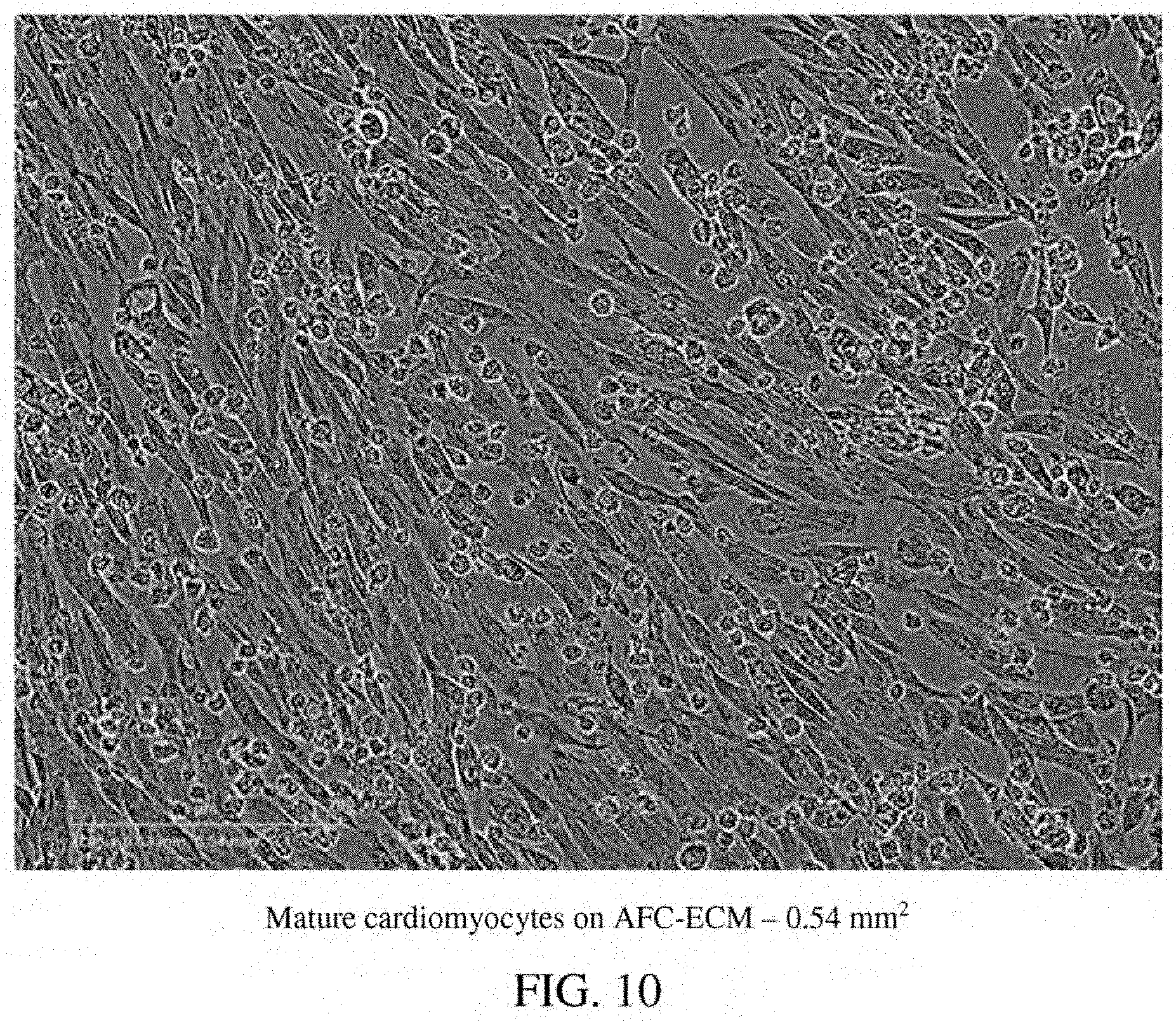
FIELD
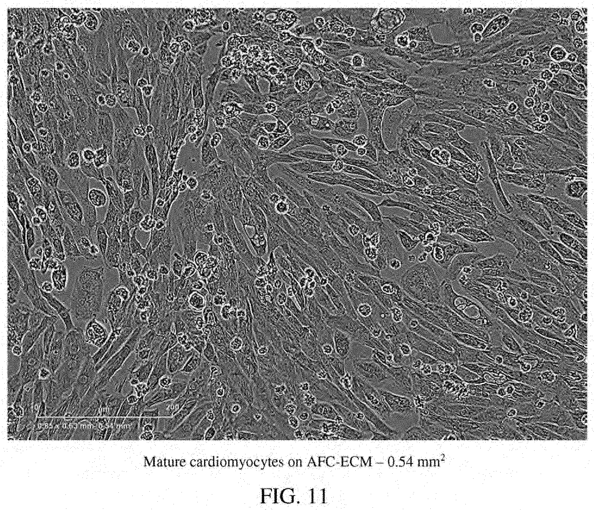
[0002] The disclosure generally relates to the use of cellular constructs of cardiomyocytes derived from human stem cells on cell-derived extracellular matrices, methods of making the constructs, and methods for cardiotoxicity and proarrhythmic screening assays of drug compounds using the constructs.
BACKGROUND
[0003] Cardiotoxicity, or the perceived potential for cardiotoxicity, is a leading cause of toxicity related drug attrition during the investigation and selection of new drugs. Cardiac safety testing of new chemical entities that become lead drug candidates is a critical aspect of the drug discovery and development pipeline. A large number of cardiac side effects of cardiac and non-cardiac drugs are caused by drug interaction with one or more cardiac ion channels. Cardiac ion channels regulate cellular excitability, contractility and overall cardiac performance, and alteration of cardiac ion channel function can lead to sudden cardiac death. This contributed to the release of drug candidate testing guidelines from The International Conference on Harmonization (ICH).
[0004] The current preclinical drug candidate testing guidelines from the IHC (ICH S7A and S7B-Pharmacology Studies) rely on genetically modified heterologous cells and in vivo animal models. It has become increasingly recognized that these studies, such as hERG assay and QT prolongation studies, do not accurately predict cardiotoxicity and proarrhythmia risk for humans. Since 2005, cardiac safety of a drug compound has been determined almost exclusively by its effect or potential effect on the QT interval of the electrocardiogram (ECG) or the action potential duration (APD), and the potential to lead to a life-threatening arrhythmia called Torsades de Pointes (TdP). However, QT prolongation is not the ideal indicator for TdP as drugs that prolong the QT interval do not always cause TdP. It is now recognized that the QT prolongation parameter is only a surrogate marker for proarrhythmia. Data from pre-clinical and clinical trials have shown that there is no fixed relationship between the magnitude of QT prolongation and the risk for development of fatal arrhythmias such as TdP.
[0005] Therefore, the US Food and Drug Administration (FDA) and other stakeholders in drug discovery have called for an evolution in pre-clinical cardiotoxicity testing. The proposed new paradigm is called the Comprehensive In-Vitro Proarrhythmia Assay (CiPA). An integral part of the CiPA Initiative (cipaproject.org/) includes incorporation of data collected from human stem cell derived cardiomyocytes for cardiotoxicity and proarrhythmia assays. The overall goal of these new proposed guidelines is to provide a more accurate and comprehensive mechanistic based assessment of proarrhythmic potential that would more accurately assess the risk of new drugs. In review of the proposed CiPA Initiative's guidelines, the FDA has defined two advances that must be made before human cardiomyocytes can be incorporated in the new initiative. First, the growth and maturation state of human stem cell derived cardiomyocytes needs to be advanced to more closely resemble the structure and function of adult human cardiomyocytes. Second, a reliable high throughput screening platform using these cells must be developed.
[0006] There are generally three types of systems currently utilized to evaluate the electrophysiology of cardiomyocytes in vitro: 1) Patch clamping systems; 2) micro-electrode array (MEA) systems; and 3) voltage sensitive dye (VSD) visualization methods. Manual patch clamping systems are most commonly used in very early research studies to evaluate the electrophysiology of individual cells. These devices, while providing accurate and sensitive ionic current measurements, cannot be used for in vitro cell systems that more closely mimic cell-cell interaction in cardiac tissue. MEA systems include electrodes that are incorporated into cell culture wells allowing for the measurement of current across the well. While allowing for high throughput analysis and ability to measure impedance in in vitro cell systems, these systems have low spatial resolution, do not provide data on action potential shape, and hinder direct visualization of the cells and the ability to assess 3-D culture systems that more closely mimic cardiac tissue architecture. VSD systems are gaining interest as they address many of the shortcomings of the current technologies described above with features that include: 1) allowing for high throughput analysis; 2) providing high spatial resolution; 3) allowing visualization of impulse propagation across a culture dish; and 4) allowing for modification of the culture conditions including addition of extracellular matrix leading to a more natural testing environment.
[0007] The use of cardiomyocytes derived from human stem cells, such as induced pluripotent stem cells, have had limited success to date in cardiotoxicity and proarrhythmic assay screening. Cardiomyocytes derived from induced human pluripotent stem cells (hiPSC-CMs) are commercially available and can be purchased from several companies in cryopreserved vials that can be thawed and plated as monolayers. These hiPSC-CMs can be made in large scale in vitro. Also, hiPSC-CMs can be obtained from patient specific hiPSCs for a specific individual. However, there are hurdles that still need to be overcome to make the hiPSC-CMs a meaningful part of the new CiPA Initiative paradigm. 1) The maturation of hiPSC-CM structure and function must be advanced. Notably, the Kir2.1 potassium channel is absent in currently available hiPSC-CMs and sodium channel expression is low. Currently available hiPSC-CMs are very immature functionally and structurally. The vast majority of currently used hiPSC-CM-based proarrhythmia screening assays rely on immature, fetal-like cells that do not resemble the structure of function of the adult cardiomyocyte. 2) Electrical pacing of hiPSC-CM monolayers in a high throughput electrophysiological screening platform is needed. The vast majority of current hiPSC-CM-based proarrhythmia screens rely solely on field potential duration (MEA technology) or action potential duration prolongation (VSD technology) as surrogate marker for TdP induction. This is a limitation because not all drugs that prolong the action potential (QT interval) will cause fatal TdP arrhythmias. Although certain maturation states of immature hiPSC-CMs have been achieved using Matrigel.TM. ECM and bone-marrow cell-derived ECM in culture, use of these hiPSC-CMs in proarrhythmia screening assays did not produce consistent results in the observation of arrhythmia activation patters consistent with what is known to occur in cases of TdP in humans, especially between low risk and high risk drugs.
[0008] Thus, advanced materials and new methods are needed for the precise assessment and screening of the cardiac safety liability of drug compounds for accurate, reliable and efficient development of new drug candidates.
SUMMARY
[0009] The present disclosure provides a solution to at least some of the aforementioned limitations and deficiencies in the art relating to the assessment and screening of the cardiac safety liability of drug candidates. The solution is premised on the discovery of an extracellular matrix derived from cells isolated from amniotic fluid (AFC-ECM), which can be used for the maturation of human stem cell derived cardiomyocytes. These mature cardiomyocytes can then be used in a cellular construct with the AFC-ECM for cardiotoxicity and proarrhythmic testing of drugs.
[0010] In one aspect, disclosed is a method for the maturation of immature cardiomyocytes derived from human induced pluripotent stem cells, the method comprising: (a) providing immature cardiomyocytes derived from human induced pluripotent stem cells (immature hiPSC-CMs); (b) providing an extracellular matrix derived in vitro from cells isolated from amniotic fluid (AFC-ECM); (c) contacting the immature hiPSC-CMs with the AFC-ECM; and (d) culturing the immature hiPSC-CMs with the AFC-ECM in a culture media to induce maturation of the immature hiPSC-CMs, thereby forming mature cardiomyocytes; wherein the mature cardiomyocytes are characterized by rod shaped cells with distinct sarcomere structure resembling adult human cardiac tissue. In some embodiments the mature cardiomyocytes have a similar or the same morphology as illustrated in any one of FIGS. 6 to 14. In some embodiments, the immature hiPSC-CMs are plated on the AFC-ECM. In some embodiments, the mature cardiomyocytes form as a monolayer on the AFC-ECM, thereby forming a cellular construct comprising a monolayer of mature cardiomyocytes on the AFC-ECM, wherein the mature cardiomyocytes are aligned on the AFC-ECM. In some embodiments, the immature hiPSC-CMs do not express inward-rectifier potassium channel Kir2.1. In some embodiments, the immature hiPSC-CMs do not include rod shaped cells with distinct sarcomere structure. In some embodiments, the immature hiPSC-CMs can be characterized by having less mitochondria than mature cardiomyocytes, by having disorganized myofilaments, by having a circular shape, and/or by having a single nucleus. In some embodiments, the mature cardiomyocytes can be further characterized by having greater amounts of mitochondria than immature hiPSC-CMs, by having organized, compact myofilaments, and/or by having two nuclei (bi-nucleated).
[0011] In another aspect, disclosed is a cellular construct comprising a monolayer of mature cardiomyocytes on an extracellular matrix derived from cells isolated in vitro from amniotic fluid (AFC-ECM), wherein the mature cardiomyocytes are AFC-ECM cultured cardiomyocytes derived from human induced pluripotent stem cells (hiPSC-CMs), and wherein the mature cardiomyocytes are characterized by rod shaped cells with distinct sarcomere structure resembling adult human cardiac tissue. In some embodiments the mature cardiomyocytes have a similar or the same morphology as illustrated in any one of FIGS. 6 to 14. In some aspects, the mature cardiomyocytes include the inward-rectifier Kir2.1 potassium channel and/or express inward-rectifier potassium channel Kir2.1. In certain aspects, the mature cardiomyocytes can be matured from immature cardiomyocytes derived from human induced pluripotent stem cells (immature hiPSC-CMs) in culture on the AFC-ECM, wherein the mature cardiomyocytes are characterized by rod shaped cells with distinct sarcomere structure (striped appearance) resembling adult human cardiac tissue. In some embodiments, the monolayer of mature cardiomyocytes is aligned on the AFC-ECM. In some embodiments, fiber tracks are present on the construct. In some embodiments, the AFC-ECM comprises laminin, collagen alpha-1 (XVIII), basement membrane-specific heparan sulfate proteoglycan core protein, agrin, vimentin, and collagen alpha-2 (IV), and/or isoforms thereof. In some embodiments, the isoform of collagen alpha-1 (XVIII) is isoform 2. In some embodiments, the isoform of agrin is isoform 6. In some embodiments, the AFC-ECM further comprises fibronectin and/or an isoform thereof. In some embodiments, the AFC-ECM does not contain decorin, perlecan, and/or collagen (III). In some embodiments, the immature hiPSC-CMs do not include rod shaped cells with distinct sarcomere structure. In some embodiments, the immature hiPSC-CMs can be characterized by having less mitochondria than mature cardiomyocytes, by having disorganized myofilaments, by having a circular shape, and/or by having a single nucleus. In some embodiments, the mature cardiomyocytes can be further characterized by having greater amounts of mitochondria than immature hiPSC-CMs, by having organized, compact myofilaments, and/or by having two nuclei (bi-nucleated).
[0012] In another aspect, disclosed is a method for making a cellular construct of mature cardiomyocytes on an extracellular matrix derived in vitro from cells isolated from amniotic fluid (AFC-ECM), the method comprising: (a) providing immature cardiomyocytes derived from human induced pluripotent stem cells (immature hiPSC-CMs); (b) providing an extracellular matrix derived in vitro from cells isolated from amniotic fluid (AFC-ECM); (c) plating the immature hiPSC-CMs on the AFC-ECM; and (d) culturing the plated immature hiPSC-CMs on the AFC-ECM in a culture media to induce maturation of the immature hiPSC-CMs into mature cardiomyocytes and to form a monolayer of the mature cardiomyocytes on the AFC-ECM, thereby forming the cellular construct, wherein the mature cardiomyocytes are characterized by rod shaped cells with distinct sarcomere structure resembling adult human cardiac tissue. In some embodiments the mature cardiomyocytes have a similar or the same morphology as illustrated in any one of FIGS. 6 to 14. In some embodiments, the monolayer of mature cardiomyocytes is aligned on the AFC-ECM. In some embodiments, fiber tracks are present on the cellular construct. In some embodiments, the immature hiPSC-CMs do not include rod shaped cells with distinct sarcomere structure. In some embodiments, the immature hiPSC-CMs can be characterized by having less mitochondria than mature cardiomyocytes, by having disorganized myofilaments, by having a circular shape, and/or by having a single nucleus. In some embodiments, the mature cardiomyocytes can be further characterized by having greater amounts of mitochondria than immature hiPSC-CMs, by having organized, compact myofilaments, and/or by having two nuclei (bi-nucleated).
[0013] In another aspect, disclosed is a method for determining the cardiotoxicity and/or proarrhythmic effect of a drug compound in vitro, the method comprising contacting the drug compound with the mature cardiomyocytes of any one of the cellular constructs disclosed throughout the specification, and observing for a change in the electrophysiology of the mature cardiomyocytes to confirm whether the drug compound has a cardiotoxic and/or proarrhythmic effect on the mature cardiomyocytes. A change in the electrophysiology of the mature cardiomyocytes confirms that the drug compound has a cardiotoxic and/or proarrhythmic effect on the mature cardiomyocytes. The changes in the electrophysiology of the mature cardiomyocytes can include, but are not limited to, APD prolongation, APD prolongation plus rotors, and/or various types of arrhythmias, such as tachyarrhythmia (TA), quiescence (Q), delayed afterdepolarization (DAD), and/or early afterdepolarization (EAD). Observations can also include the cell viability, cell density, and/or morphology of the cells. In some embodiments, the change in the electrophysiology of the mature cardiomyocytes is prolongation of action potential duration (APD). In some embodiments, the change in the electrophysiology of the mature cardiomyocytes is early after depolarization (EAD). In some embodiments, the change in the electrophysiology of the mature cardiomyocytes is delayed after depolarization (DAD). In some embodiments, the change in the electrophysiology of the mature cardiomyocytes is action potential duration prolongation (APD prolongation) plus rotors. In some embodiments, the change in the electrophysiology of the mature cardiomyocytes is an arrhythmia. In some aspects, the cellular construct can be prepared by a process comprising: (a) providing immature cardiomyocytes derived from human induced pluripotent stem cells (immature hiPSC-CMs); (b) providing an extracellular matrix derived in vitro from cells isolated from amniotic fluid (AFC-ECM); (c) plating the immature hiPSC-CMs on the AFC-ECM; and (d) culturing the plated immature hiPSC-CMs on the AFC-ECM in a culture media to induce maturation of the immature hiPSC-CMs into mature cardiomyocytes and to form a monolayer of the mature cardiomyocytes on the AFC-ECM, thereby forming a cellular construct, wherein the mature cardiomyocytes are characterized by rod shaped cells with distinct sarcomere structure resembling adult human cardiac tissue. In some embodiments the mature cardiomyocytes have a similar or the same morphology as illustrated in any one of FIGS. 6 to 14. In one embodiment, the immature hiPSC-CMs do not express inward-rectifier potassium channel Kir2.1. In another embodiment, the monolayer of mature cardiomyocytes is aligned on the AFC-ECM. In another embodiment, fiber tracks are present on the cellular construct. In some embodiments, the immature hiPSC-CMs do not include rod shaped cells with distinct sarcomere structure. In some embodiments, the immature hiPSC-CMs can be characterized by having less mitochondria than mature cardiomyocytes, by having disorganized myofilaments, by having a circular shape, and/or by having a single nucleus. In some embodiments, the mature cardiomyocytes can be further characterized by having greater amounts of mitochondria than immature hiPSC-CMs, by having organized, compact myofilaments, and/or by having two nuclei (bi-nucleated).
[0014] Also, disclosed in the context of the present invention are the following embodiments 1 to 20.
[0015] Embodiment 1 is a method for the maturation of immature cardiomyocytes derived from human induced pluripotent stem cells, the method comprising: (a) providing immature cardiomyocytes derived from human induced pluripotent stem cells (immature hiPSC-CMs); (b) providing an extracellular matrix derived in vitro from cells isolated from amniotic fluid (AFC-ECM); (c) contacting the immature hiPSC-CMs with the AFC-ECM; and (d) culturing the immature hiPSC-CMs with the AFC-ECM in a culture media to induce maturation of the immature hiPSC-CMs, thereby forming mature cardiomyocytes; wherein the mature cardiomyocytes are characterized by rod shaped cells with distinct sarcomere structure resembling adult human cardiac tissue.
[0016] Embodiment 2 is the method of embodiment 1, wherein the immature hiPSC-CMs are plated on the AFC-ECM.
[0017] Embodiment 3 is the method of any one of embodiments 1 or 2, wherein the mature cardiomyocytes form as a monolayer on the AFC-ECM, thereby forming a cellular construct comprising a monolayer of mature cardiomyocytes on the AFC-ECM, wherein the mature cardiomyocytes are aligned on the AFC-ECM.
[0018] Embodiment 4 is the method of any one of embodiments 1 to 3, wherein the immature hiPSC-CMs do not express inward-rectifier potassium channel Kir2.1.
[0019] Embodiment 5 is a cellular construct comprising a monolayer of mature cardiomyocytes on an extracellular matrix derived from cells isolated in vitro from amniotic fluid (AFC-ECM), wherein the mature cardiomyocytes are AFC-ECM cultured cardiomyocytes derived from human induced pluripotent stem cells (hiPSC-CMs), and wherein the mature cardiomyocytes are characterized by rod shaped cells with distinct sarcomere structure resembling adult human cardiac tissue.
[0020] Embodiments 6 is the cellular construct of embodiment 5, wherein the monolayer of mature cardiomyocytes is aligned on the AFC-ECM.
[0021] Embodiment 7 is the cellular construct of any one of embodiments 5 or 6, wherein fiber tracks are present on the construct.
[0022] Embodiment 8 is the cellular construct of any one of embodiments 5 to 7, wherein the AFC-ECM comprises laminin, collagen alpha-1 (XVIII), basement membrane-specific heparan sulfate proteoglycan core protein, agrin, vimentin, and collagen alpha-2 (IV), and/or isoforms thereof.
[0023] Embodiment 9 is the cellular construct of embodiment 8, wherein the isoform of collagen alpha-1 (XVIII) is isoform 2, and/or wherein the isoform of agrin is isoform 6.
[0024] Embodiment 10 is the cellular construct of any one of embodiments 8 or 9, wherein the AFC-ECM further comprises fibronectin and/or an isoform thereof.
[0025] Embodiment 11 is the cellular construct of any one of embodiments 5 to 10, wherein the AFC-ECM does not contain decorin, perlecan, and/or collagen (III).
[0026] Embodiment 12 is a method for making a cellular construct of mature cardiomyocytes on an extracellular matrix derived in vitro from cells isolated from amniotic fluid (AFC-ECM), the method comprising: (a) providing immature cardiomyocytes derived from human induced pluripotent stem cells (immature hiPSC-CMs), (b) providing an extracellular matrix derived in vitro from cells isolated from amniotic fluid (AFC-ECM); (c) plating the immature hiPSC-CMs on the AFC-ECM; (d) culturing the plated immature hiPSC-CMs on the AFC-ECM in a culture media to induce maturation of the immature hiPSC-CMs into mature cardiomyocytes and to form a monolayer of the mature cardiomyocytes on the AFC-ECM, thereby forming the cellular construct; wherein the mature cardiomyocytes are characterized by rod shaped cells with distinct sarcomere structure resembling adult human cardiac tissue.
[0027] Embodiment 13 is the method of embodiment 12, wherein the monolayer of mature cardiomyocytes is aligned on the AFC-ECM.
[0028] Embodiment 14 is the method of any one of embodiments 12 or 13, wherein fiber tracks are present on the cellular construct.
[0029] Embodiment 15 is a method for determining the cardiotoxicity and/or proarrhythmic effect of a drug compound in vitro, the method comprising contacting the drug compound with the mature cardiomyocytes of any one of the cellular constructs of embodiments 5 to 11, and observing for a change in the electrophysiology of the mature cardiomyocytes to confirm whether the drug compound has a cardiotoxic and/or proarrhythmic effect on the mature cardiomyocytes.
[0030] Embodiment 16 is the method of embodiment 15, wherein the change in the electrophysiology of the mature cardiomyocytes is prolongation of action potential duration (APD), and wherein prolongation of APD confirms that the drug compound has a cardiotoxic and/or proarrhythmic effect on the mature cardiomyocytes.
[0031] Embodiment 17 is the method of embodiment 15, wherein the change in the electrophysiology of the mature cardiomyocytes is early after depolarization, and wherein early after depolarization (EAD) confirms that the drug compound has a cardiotoxic and/or proarrhythmic effect on the mature cardiomyocytes.
[0032] Embodiment 18 is the method of embodiment 15, wherein the change in the electrophysiology of the mature cardiomyocytes is delayed after depolarization, and wherein delayed after depolarization (DAD) confirms that the drug compound has a cardiotoxic and/or proarrhythmic effect on the mature cardiomyocytes.
[0033] Embodiment 19 is the method of embodiment 15, wherein the change in the electrophysiology of the mature cardiomyocytes is action potential duration (APD) plus rotors, and wherein prolongation of APD plus rotors confirms that the drug compound has a cardiotoxic and/or proarrhythmic effect on the mature cardiomyocytes.
[0034] Embodiment 20 is the method of embodiment 15, wherein the change in the electrophysiology of the mature cardiomyocytes is an arrhythmia, and wherein the arrhythmia confirms that the drug compound has a cardiotoxic and/or proarrhythmic effect on the mature cardiomyocytes.
[0035] The terms "about" or "approximately" are defined as being close to as understood by one of ordinary skill in the art, and in one non-limiting embodiment the terms are defined to be within 10%, preferably within 5%, more preferably within 1%, and most preferably within 0.5%.
[0036] The term "substantially" and its variations are defined to include ranges within 10%, within 5%, within 1%, or within 0.5%.
[0037] As used herein, the terms "% w/w" or "wt. %" refers to a weight percentage of a component based on the total weight of material (e.g. a composition) that includes the component. In a non-limiting example, 10 grams of a component in 100 grams of a composition is 10% w/w of the component in the total weight of composition. As used herein, the terms "% v/v" or "vol. %" refers to a volume percentage of a component based on the total volume of material (e.g. a composition) that includes the component. In a non-limiting example, 10 mL of a component in 100 mL of a composition is 10% v/v of the component in the total volume of composition. As used herein, the term "% w/v" refers to a weight percentage of a component based on the total volume of material (e.g. a composition) that includes the component. In a non-limiting example, 10 grams of a component in 100 mL of a composition is 10% w/v of the component in the total volume of composition. As used herein, the term "% v/w" refers to a volume percentage of a component based on the total weight of material (e.g. a composition) that includes the component. In a non-limiting example, 10 mL of a component in 100 grams of a composition is 10% v/w of the component in the total weight of the composition.
[0038] The terms "inhibiting" or "reducing" or "preventing" or "avoiding" or any variation of these terms, when used in the claims and/or the specification includes any measurable decrease or complete inhibition to achieve a desired result.
[0039] The term "effective," as that term is used in the specification and/or claims, means adequate to accomplish a desired, expected, or intended result.
[0040] The words "comprising" (and any form of comprising, such as "comprise" and "comprises"), "having" (and any form of having, such as "have" and "has"), "including" (and any form of including, such as "includes" and "include") or "containing" (and any form of containing, such as "contains" and "contain") are inclusive or open-ended and do not exclude additional, unrecited elements or method steps.
[0041] The use of the word "a" or "an" when used in conjunction with the terms "comprising," "having," "including," or "containing" (or any variations of these words) may mean "one," but it is also consistent with the meaning of "one or more," "at least one," and "one or more than one."
[0042] The compositions and methods for their use can "comprise," "consist essentially of," or "consist of" any of the ingredients or steps disclosed throughout the specification. With respect to the transitional phrase "consisting essentially of," in one non-limiting aspect, a basic and novel characteristic of the cellular constructs disclosed herein is their use in cardiotoxicity and/or proarrhythmic screening testing of drug compounds due to their ability to mature cardiomyocytes derived from stem cells to a maturation state resembling that of mature native adult cardiomyocytes and native heart tissue.
[0043] It is contemplated that any embodiment discussed in this specification can be implemented with respect to any method or composition of the invention, and vice versa. Furthermore, compositions of the invention can be used to achieve methods of the invention.
[0044] Other objects, features and advantages of the present invention will become apparent from the following detailed description. It should be understood, however, that the detailed description and the specific examples, while indicating specific embodiments of the invention, are given by way of illustration only, since various changes and modifications within the spirit and scope of the invention will become apparent to those skilled in the art from this detailed description.
BRIEF DESCRIPTION OF THE DRAWINGS
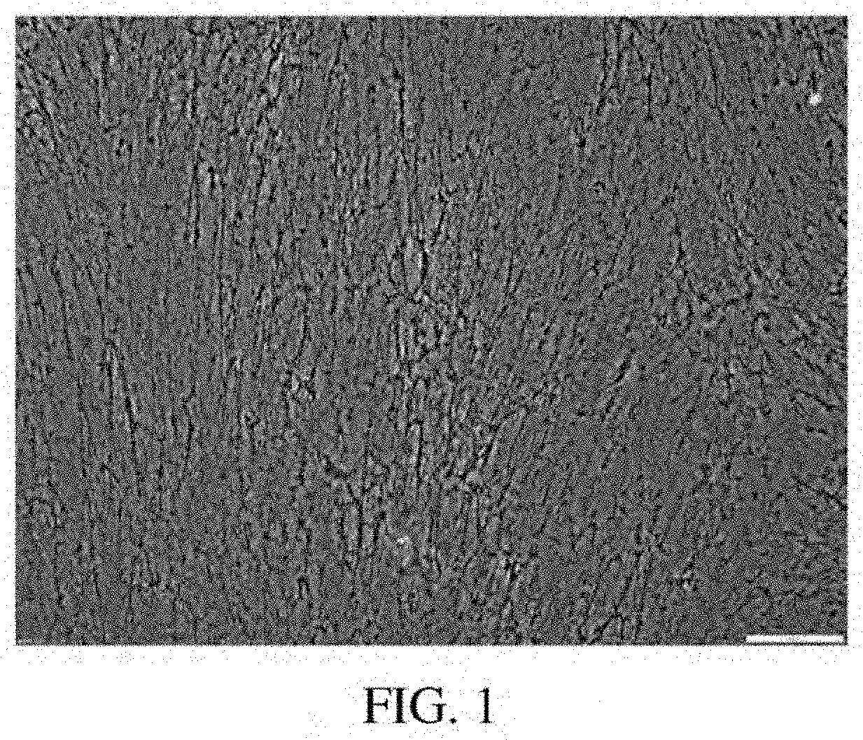
[0045] FIG. 1: A photomicrograph of a Brightfield Image of the amniotic fluid cell-derived ECM at 100.times. power using a 10.times. objective lens.
[0046] FIG. 2: An atomic force photomicrograph of 3 representative 40.times.40 um sections of the amniotic fluid cell-derived ECM and a bone marrow cell-derived ECM showing topography, adhesion, and stiffness.
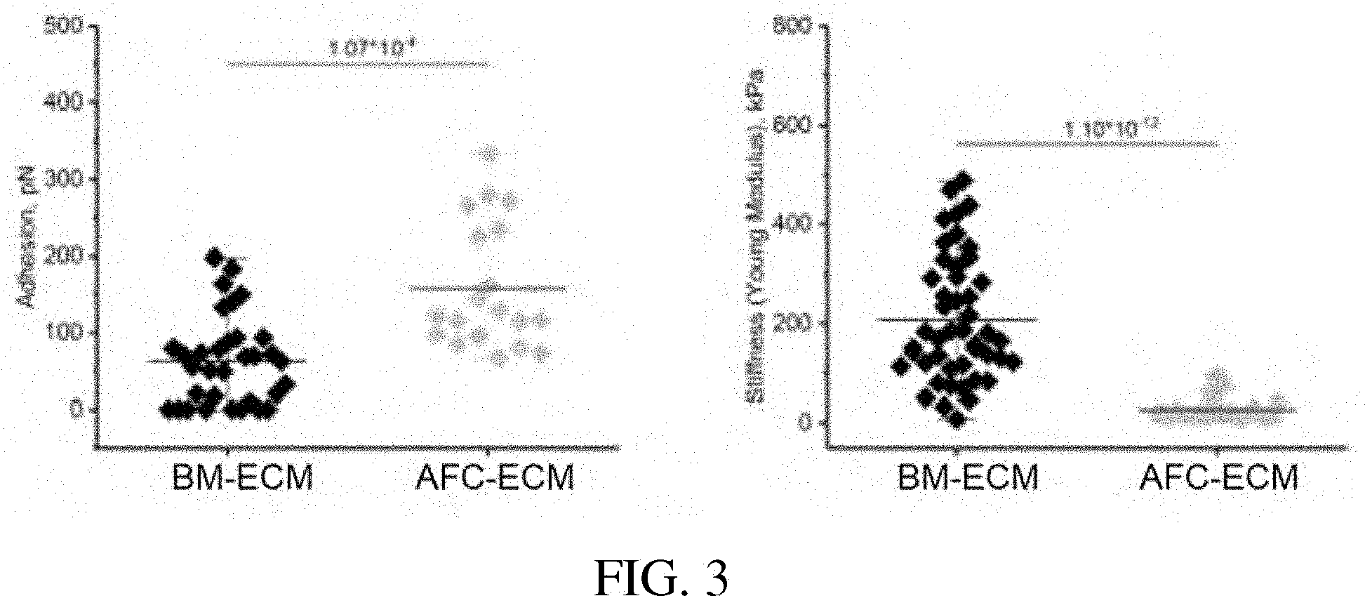
[0047] FIG. 3: Scatter plots showing quantification of adhesion and stiffness (elastic modulus) of bone marrow- and amniotic fluid- cell-derived ECMs. Each point represents an independent point of measurement.
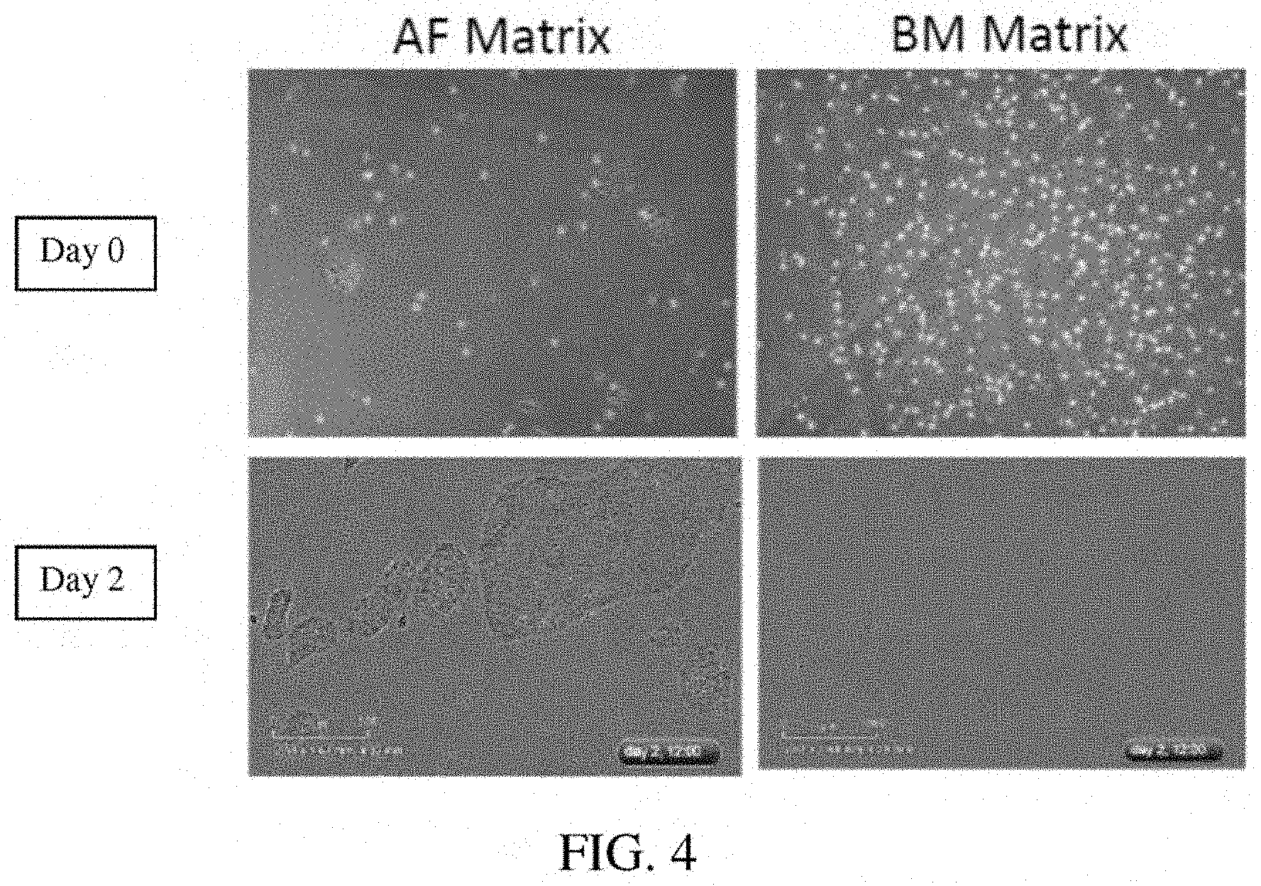
[0048] FIG. 4: A photomicrograph showing Day 0 and Day 2 culture of iPSCs on amniotic fluid cell-derived ECM and a bone marrow cell-derived ECM.
[0049] FIG. 5: A plot of growth curves of iPSCs cultured in the presence of the amniotic fluid cell-derived ECM and a bone marrow cell-derived ECM.
[0050] FIG. 6: A photomicrograph of mature cardiomyocytes on an AFC-ECM in a single well--3.13 mm.sup.2.
[0051] FIG. 7: A photomicrograph of mature cardiomyocytes on an AFC-ECM in a single well--2.15 mm.sup.2.
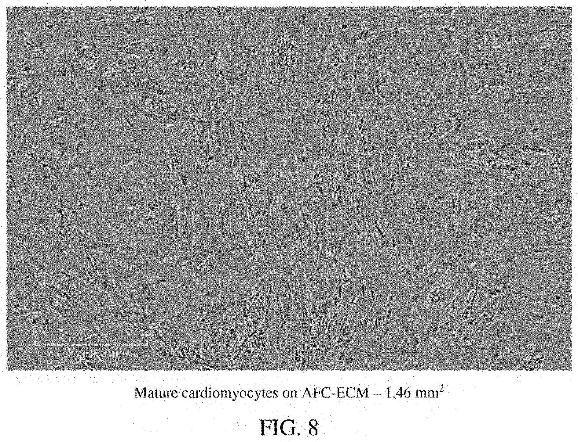
[0052] FIG. 8: A photomicrograph of mature cardiomyocytes on an AFC-ECM in a single well--1.46 mm.sup.2.
[0053] FIG. 9: A photomicrograph of mature cardiomyocytes on AFC-ECM in a single well--0.54 mm.sup.2.
[0054] FIG. 10: A photomicrograph of mature cardiomyocytes on AFC-ECM in a single well--0.54 mm.sup.2.
[0055] FIG. 11: A photomicrograph of mature cardiomyocytes on AFC-ECM in a single well--0.54 mm.sup.2.
[0056] FIG. 12: A photomicrograph of mature cardiomyocytes on AFC-ECM in a single well--0.54 mm.sup.2.
[0057] FIG. 13: A photomicrograph of mature cardiomyocytes on AFC-ECM in a single well--0.13 mm.sup.2.
[0058] FIG. 14: A photomicrograph of mature cardiomyocytes on AFC-ECM in a single well--0.04 mm.sup.2.
[0059] FIG. 15: A photomicrograph of cardiomyocytes on standard Matrigel.TM. ECM in a single well--3.13 mm.sup.2.
[0060] FIG. 16: A photomicrograph of cardiomyocytes on standard Matrigel.TM. ECM in a single well--2.15 mm.sup.2.
[0061] FIG. 17: A photomicrograph of cardiomyocytes on standard Matrigel.TM. ECM in a single well--1.46 mm.sup.2.
[0062] FIG. 18: A photomicrograph of cardiomyocytes on standard Matrigel.TM. ECM in a single well--0.54 mm.sup.2.
[0063] FIG. 19: A photomicrograph of cardiomyocytes on standard Matrigel.TM. ECM in a single well--0.13 mm.sup.2.
[0064] FIG. 20: A photomicrograph of mature cardiomyocytes on a BM-ECM in a single well--2.15 mm.sup.2.
[0065] FIG. 21: A photomicrograph of mature cardiomyocytes on a BM-ECM in a single well--1.46 mm.sup.2.
[0066] FIG. 22: A schematic of the configuration of instruments to record action potential or calcium transient measurements using mature cardiomyocytes on AFC-ECM in multi-well plates.
[0067] FIG. 23: Recordings of spontaneous action potentials recorded from cardiomyocytes cultured on Matrigel.TM. ECM, BM-ECM, and AFC-ECM, baseline and after drug E4031.
[0068] FIG. 24: Bar graph of number of Stable Rotors (TdP like arrhythmia) from cardiomyocytes cultured on Matrigel.TM. ECM, BM-ECM, and AFC-ECM after contact with drug E4031.
[0069] FIG. 25: Recordings of spontaneous action potentials recorded from mature cardiomyocytes cultured on AFC-ECM for various drugs.
[0070] FIG. 26: Bar graph of total arrythmias recorded for all doses of each of the listed drugs.
[0071] FIG. 27: Bar graph of % of well with arrythmia @10.times. the effective therapeutic plasma concentration (ETPC) of each of the listed drugs.
[0072] FIG. 28: Bar graph of the action potential triangulation (APD90-APD30) in time (ms) of each of the listed drugs.
[0073] FIG. 29: Bar graph of the action potential triangulation (APD90-APD30) in time (ms) of each of some of the listed drugs comparing cardiomyocyte performance of cardiomyocytes on the AFC-ECM (SBS-AF Matrix) versus on Matrigel.TM. ECM.
[0074] FIG. 30: Bar graph of the maximum drug-induced action potential triangulation of the listed drugs comparing cardiomyocyte performance of iCell.RTM. hiPSC-CMs from Cellular Dynamics (blank circles) versus Cor.4U.RTM. hiPSC-CMs from Ncardia (dark circles) at any concentration of the listed drugs. Figure from Blinova et al, 2018, Cell Reports.
[0075] FIG. 31: Photomicrophraphs of hiPSC-CMs cultured on Matrigel.TM. ECM vs. AFC-ECM with immunofluorescentstaining for Troponin I, using DAPI to mark the nuclei.
[0076] FIG. 32: Photomicrophraphs of hiPSC-CMs cultured on Matrigel.TM. ECM vs. AFC-ECM with immunofluorescentstaining for .alpha.-actinin using DAPI to mark the nuclei.
[0077] FIG. 33: Photmicrographs of hiPSC-CMs cultured on Matrigel.TM. ECM vs. AFC-ECM with immunofluorescentstaining for cTnT and N Cadherin, using DAPI to mark the nuclei.
[0078] FIG. 34: Photomicrographs of a single hiPSC-CM cell cultured on Matrigel.TM. ECM vs. AFC-ECM with immunofluorescentstaining for cTnT using DAPI to mark the nuclei.
[0079] FIG. 35: Photomicrographs of a single hiPSC-CM cell cultured on Matrigel.TM. ECM vs. AFC-ECM with immunofluorescentstaining for .alpha.-actinin using DAPI to mark the nuclei.
[0080] FIG. 36: Graph of a comparison of the cellular circularity of the single cells shown in FIG. 35.
[0081] FIG. 37: Photomicrographs of hiPSC-CMs cultured on Matrigel.TM. ECM vs. AFC-ECM with immunofluorescentstaining for cTnI expression, using DAPI to mark the nuclei.
[0082] FIG. 38: Western blotting of hiPSC-CMs on Matrigel.TM. ECM and AFC-ECM for cTnI expression and GAPDH.
[0083] FIG. 39: Graph of cTnI Expression relative to GAPDH of hiPSC-CMs on Matrigel.TM. ECM vs. AFC-ECM.
[0084] FIG. 40: Photomicrographs of hiPSC-CMs on Matrigel.TM. ECM vs. AFC-ECM stained for mitochondria with MitoTracker Red.
[0085] FIG. 41: Graph showing the MitoTraker.TM. Red fluorescence intensity/cardiomyocyte for hiPSC-CMs on Matrigel.TM. ECM vs. AFC-ECM.
[0086] FIG. 42: Photomicrographs (transmitted light) of hiPSC-CMs cultured on Matrigel.TM. ECM and AFC-ECM that were coated on microelectrode array (MEA) plates.
DETAILED DESCRIPTION
[0087] Human stem cell derived cardiomyocytes, such as those derived from human induced pluripotent stem cells (hiPSC-CMs), that have been matured in culture on an extracellular matrix derived in vitro from cells isolated from amniotic fluid (AFC-ECM) have demonstrated better, more consistent cardiotoxicity and proarrhythmia assay results than immature hiPSC-CMs or hiPSC-CMs matured on other ECMs, such as Matrigel.TM. ECM and bone marrow cell-derived ECM. The hiPSC-CMs matured on an AFC-ECM surprisingly have demonstrated a higher state of maturation than hiPSC-CMs matured on other ECMs, such as Matrigel.TM. ECM and even on other natural cell derived ECMs such as bone marrow cell-derived ECM, as shown by their cellular morphology and sarcomere structure that are found in cardiomyocytes of normal adult human cardiac tissue. Furthermore, a more robust expression of cTnI (cardiac troponin I) protein, as demonstrated by western blotting techniques, has been seen for hiPSC-CMs cultured on AFC-ECM than for hiPSC-CMs cultured on Matrigel.TM. ECM. Also, hiPSC-CMs cultured on AFC-ECM have more mitochondria and mitochondria with more polarized inner membrane potential than hiPSC-CMs cultured on Matrigel.TM. ECM. Thus, the AFC-EMC can stimulate mitochondrial biogenesis, maturation, and function in the hiPSC-CMs. Normal adult or mature human cardiac human tissue is characterized by rod shaped cells with sarcomere structure (striped appearance). The morphology and sarcomere structures of the cardiomyocytes can be identified visually by microscopy (transmitted light or by immunofluorescent staining). As a non-limiting example, the morphology and sarcomere structure of the cardiomyocytes matured on AFC-ECM can be distinctly seen by the presence of rod shaped cells with a striped appearance identified by arrows in the photomicrograph of FIG. 14. Additionally, the use of a silicone substrate such as PDMS were not needed to achieve these results. Surprisingly, a cellular construct comprising an AFC-ECM and a monolayer of mature cardiomyocytes that have been matured from immature hiPSC-CMs in culture on the AFC-ECM has shown to be useful, for example, by allowing consistent visualization of drug induced arrhythmias such as Torsades de Pointes (TdP) in high throughput in vitro screening assays. This "TdP in a dish" technology is a major advance over the reliance on field potential duration (MEA technology) or action potential duration prolongation as surrogate markers for drug induced TdP. Thus, the cellular construct and methods disclosed herein go beyond the current CiPA Initiative's guidelines by developing a more comprehensive and predictive in vitro arrhythmia assay that allows for visualization of arrhythmic events in an in vitro model, rather than simple polarization and depolarization events that are indirect indicators of proarrhythmia. The cellular system and methods outlined in this disclosure allow for arrhythmic events to be propagated and visualized in an in vitro human cardiac monolayer model system. In some embodiments, the immature hiPSC-CMs do not include rod shaped cells with distinct sarcomere structure. In some embodiments, the immature hiPSC-CMs can be characterized by having less mitochondria than mature cardiomyocytes, by having disorganized myofilaments, by having a circular shape, and/or by having a single nucleus. In some embodiments, the mature cardiomyocytes can be characterized by having rod shaped cells with distinct sarcomere structure (striped appearance). In some embodiments, the mature cardiomyocytes can be further characterized by having greater amounts of mitochondria than immature hiPSC-CMs, by having organized, compact myofilaments, and/or by having two nuclei (bi-nucleated).
[0088] Disclosed herein are methods of using a cell-derived extracellular matrix derived in-vitro from cells isolated from amniotic fluid (AFC-ECM) for the maturation of immature cardiomyocytes derived from human induced pluripotent stem cells (immature hiPSC-CMs) in culture forming mature cardiomyocytes (mature hiPSC-CMs). Also disclosed herein is a cell construct comprising a monolayer of these mature cardiomyocytes on an AFC-ECM useful for cardiotoxicity and/or proarrhythmic screening assays of drug compounds. Also disclosed herein are methods for determining the cardiotoxicity and/or proarrhythmic effect of a drug compound in vitro using such cell constructs.
A. Amniotic Fluid Cell-Derived Extracellular Matrix (AFC-ECM)
[0089] Perinatal cells can be divided into three groups: cells from amniotic fluid; cells from the placenta; and cells from the umbilical cord. Amniotic fluid has several sources of cells including cells derived from the developing fetus sloughed from the fetal amnion membrane, skin, and alimentary, respiratory, and urogenital tracts. Placenta also has several sources of cells including the membrane sheets (amnion and chorion), the villi, and the blood. Umbilical cord cells generally come from two sources, cord blood and Wharton's jelly. The cells from these three perinatal sources can include stem cells. The cells used to produce the amniotic fluid cell-derived ECM of the invention are obtained from the amniotic fluid of a mammal including but not limited to a human (Homo sapiens), murine, rabbit, cat, dog, pig, equine, or primate. In preferred embodiments, the cells are from the amniotic fluid of a human. The amniotic fluid can be sourced from humans at full-term births (greater than about 37 weeks gestational age) or pre-term births (less than about 37 weeks gestational age). Pre-term births include late pre-term births (about 33 to about 37 weeks gestational age) moderate pre-term births (about 29 to about 33 weeks gestational age), and extreme pre-term births (about 23 to about 29 weeks gestational age). The amniotic fluid can be sourced from humans prior to birth at any gestational age where amniotic fluid is present, and can be combined with sources of amniotic fluid from births. Generally, prior to birth, amniotic fluid is collected by an amniocentesis procedure. In some embodiments the amniotic fluid is sourced from humans at full-term births, at pre-term births, at late pre-term births, at moderate pre-term births, at extreme pre-term births, or prior to birth, or combinations thereof. In some embodiments, the amniotic fluid is sourced prior to birth and is collected from about 10 weeks gestational age up to birth, or from about 10 weeks to about 23 weeks gestational age, or from about 10 weeks to about 16 weeks gestational age, or from about 12 weeks gestational age up to birth, or from about 12 weeks to about 23 weeks gestational age, or from about 12 weeks to about 16 weeks gestational age. In some embodiments, the amniotic fluid sourced prior to birth is collected by an amniocentesis procedure. The cells can be obtained and isolated from amniotic fluid by techniques known in the art, such as those disclosed in Murphy et. al., Amniotic Fluid Stem Cells, Perinatal Stem Cells, Second Ed. 2013.
[0090] Amniotic fluid is comprised of cells having the ability to differentiate into cell types derived from all 3 embryonic germ layers (ectoderm, endoderm, mesoderm) spontaneously or as a result of treatment with specific growth factors or combinations of growth factors known to one of skill in the art. That is, a single cell has the capacity to be induced to express genes which are specific to any of the three germ layers. Amniotic fluid also contains a mixture of different cell types including cells derived from the developing fetus sloughed from the fetal amnion membrane, skin, and alimentary, respiratory, and urogenital tracts. Because of the origin of the amniotic fluid and placental membranes, these cells can maintain highly multipotent differentiation potential and comprise a cell population that contains cells of all three germ layers. The amniotic fluid cells can comprise stem cells. In some embodiments, the amniotic fluid cells are isolated stem cells. In some embodiments, the amniotic fluid cells comprise stem cells having the ability to differentiate into cell types derived from all 3 embryonic germ layers (ectoderm, endoderm, mesoderm) and/or multipotent stem cells, and/or pluripotent stem cells.
[0091] The amniotic fluid cell-derived ECM disclosed herein can comprise various proteins. The proteins of the ECM can be identified by techniques known in the art and include mass spectroscopy and immunohistochemical staining. The ECM can include, but is not limited to the components listed in Table 2 (see Example 1 below) and any variants, derivatives, or isoforms thereof. The amniotic fluid-cell derived ECM can include any combination of any of the components and any variants, derivatives, or isoforms thereof from Table 2. In some embodiments, a combination can comprise, consist essentially of, or consist of: laminin, collagen alpha-1 (XVIII), basement membrane-specific heparan sulfate proteoglycan core protein, agrin, vimentin, and collagen alpha-2 (IV), and/or isoforms thereof. In some embodiments, the isoform of collagen alpha-1 (XVIII) is isoform 2. In some embodiments, the isoform of agrin is isoform 6. In some embodiments, the cell-derived ECM further comprises, consist essentially of, or consists of fibronectin and/or an isoform thereof. In some embodiments, the amniotic fluid cell-derived ECM does not contain any one of or all of decorin, perlecan, and collagen (III). Some noteworthy differences in proteins between the amniotic fluid cell-derived ECM of the present inventions and a bone marrow cell-derived matrix are described in Table 1.
TABLE-US-00001 TABLE 1 Differences Between the Amniotic Fluid Cell-Derived ECM (AFC- Matrix) and a Bone Marrow Cell-Derived Matrix (BM-Matrix) Protein/ Difference between AFC- Gene Code Matrix & BM-Matrix Physiologic Relevance Laminin 5 sub-units are abundant in Laminin is known to AFC Matrix; Low expression support adhesion and in BM-Matrix expansion of pluripotent cells. Collagen Abundant in AFC Matrix; Important for ocular XVIII Absent in BM-Matrix development Agrin Present in AFC Matrix; produced by motoneurons Absent in BM-Matrix to induce aggregation of acetylcholine receptors Biglycan Abundant in BM-Matrix; Regulates bone and muscle Low expression in AFC development Matrix Collagen I Overexpressed in BM-Matrix Key to fibrillar proteins; relative to AFC Matrix highly abundant in bone Periostin Present in BM-Matrix; Regulates mineralization; Absent in AFC Matrix Marker of noncardiomyocyte lineage cells in heart
[0092] The amniotic fluid cell-derived ECM can be produced by the following process: (a) isolating cells from amniotic fluid, (b) seeding the isolated cells onto a cell culture container or onto a cell culture container coated with a substrate, (c) adding a culture media to the cell culture container, and (d) culturing the cells, thereby producing a cell-derived ECM, and (e) optionally decellularizing the cell-derived ECM.
[0093] Any cell seeding density may be used which allows cells to form a confluent monolayer immediately or after a period of time in culture. In some embodiments, the seeding density is about 10 cells/cm.sup.2-about 100,000 cells/cm.sup.2, or about 100 cells/cm.sup.2-about 75,000 cells/cm.sup.2, or about 500 cells/cm.sup.2-about 50,000 cells/cm.sup.2, or about 500 cells/cm.sup.2-about 10,000 cells/cm.sup.2, or about 500 cells/cm.sup.2-about 5,000 cells/cm.sup.2, or about 500 cells/cm.sup.2-about 2,500 cells/cm.sup.2, or about 1,000 cells/cm.sup.2-about 25,000 cells/cm.sup.2, or about 2,000 cells/cm.sup.2-about 10,000 cells/cm.sup.2, or about 3,000 cells/cm.sup.2-about 5000 cells/cm.sup.2.
[0094] Any type of container suitable for cultivation of cells can be used for the present invention. Examples include, but are not limited to cell culture flasks, T-flasks, stirred flasks, spinner flasks, fermenters, and bioreactors. Rocking bottles, shaking flasks, tubes, and other containers are also suitable containers when placed on a rocking platform or shaker. The cell culture container can be coated with a substrate to allow for better cell adhesion. A non-limiting example of a suitable substrate for coating the cell container is fibronectin.
[0095] Various commercially available cell culture media, e.g., alpha Minimum Essential Media (.alpha.-MEM) culture media (Thermo Fisher Scientific, Grand Island, N.Y.), are suitable for culturing amniotic fluid cells. The commercially available culture media can be modified by adding various supplemental substances to the media, e.g. sodium bicarbonate, L-glutamine, penicillin, streptomycin, Amphotericin B and/or serum. The serum can be fetal bovine serum. The media can also be serum free. Additionally, substances such as L-ascorbic acid can be added to the media or modified media to induce cell production of an ECM.
[0096] The initial culture media can be changed and/or replaced with another media at various times during the culturing process. For example, the initial media can be a "Complete Media" and then be replaced by an "Inducing Media" during the culturing process. A non-limiting example of a "Complete Media" contains (.alpha.-MEM) plus 2 mM L-Glutamine plus antibiotic-antimycotic plus 15% Fetal Bovine Serum. A non-limiting example of an "Inducing Media" contains the "Complete Media" plus 50 mM L-Ascorbic Acid.
[0097] The culturing of the amniotic fluid cells can take place in an incubator at 37.degree. C., 5% CO.sub.2, and 90% humidity. Culturing can take place under various environmental conditions including, but not limited to normoxic, i.e., 20-21% oxygen in the atmosphere, or hypoxic conditions.
[0098] Decellularizing the amniotic fluid cell-derived ECM of the amniotic fluid cells can include removing the viable amniotic fluid cells or rendering the amniotic fluid cells non-viable. The amniotic fluid cells can be decellularized from the ECM by using methods known in the art and can include, but are not limited to lysing the amniotic fluid cells and then removing the lysed amniotic fluid cells by washing. Various substances can be used to remove the amniotic fluid cells from the ECM. Non-limiting examples include an "Extraction Buffer" containing TRITON X-100 and ammonium hydroxide in PBS buffer. After the ECM has been decellularized of amniotic fluid cells, the resulting ECM is thereby essentially cell-free or free of viable amniotic fluid cells. If feeder cells are used, then the decellularizing methods also apply to any viable feeder cells present on the ECM, thereby resulting in the ECM being essentially free or free of viable feeder cells. The decellularizing methods also apply to any viable cells present on the ECM, thereby resulting in the ECM being essentially free or free of any viable cells. Thus, a decellularized ECM means that the ECM is acellular, meaning that the ECM is free of any viable cells.
[0099] In some embodiments, the amniotic fluid cell-derived ECM (AFC-ECM) is a three-dimensional (3D) ECM.
[0100] The methods described supra also apply to producing cell-derived ECMs from other perinatal cells such as cells from the umbilical cord including the cord blood and Wharton's jelly; and cells from placenta tissue including the membrane sheets (amnion and chorion), the villi and the blood.
[0101] In one embodiment, a perinatal cell-derived ECM is produced by the following process: (a) isolating cells from an umbilical cord, (b) seeding the isolated cells onto a cell culture container or onto a cell culture container coated with a substrate, (c) adding a culture media to the cell culture container, and (d) culturing the cells, thereby producing a cell-derived ECM, and (e) optionally decellularizing the cell-derived ECM. In some embodiments, the cells isolated from the umbilical cord are from the cord blood and/or the Wharton's jelly.
[0102] In another embodiment, a perinatal cell-derived ECM is produced by the following process: (a) isolating cells from placenta tissue, (b) seeding the isolated cells onto a cell culture container or onto a cell culture container coated with a substrate, (c) adding a culture media to the cell culture container, and (d) culturing the cells, thereby producing a cell-derived ECM, and (e) optionally decellularizing the cell-derived ECM. In some embodiments, the cells isolated from the placenta tissue are from the membrane sheets (amnion and/or chorion), the villi, and/or the blood.
[0103] In one aspect, disclosed is a cell-derived extracellular matrix (ECM) derived in vitro from cells isolated from an umbilical cord. In some embodiments, the cells isolated from the umbilical cord are from the cord blood and/or the Wharton's jelly.
[0104] In another aspect, disclosed is a cell-derived extracellular matrix (ECM) derived in vitro from cells isolated from placenta tissue. In some embodiments, the cells isolated from the placenta tissue are from the membrane sheets (amnion and/or chorion), the villi, and/or the blood.
B. Cellular Constructs and Methods for the Maturation of Immature hiPSC-CMs
[0105] Disclosed herein is a method for the maturation of immature cardiomyocytes derived from human induced pluripotent stem cells, the method comprising: (a) providing immature cardiomyocytes derived from human induced pluripotent stem cells (immature hiPSC-CMs); (b) providing an extracellular matrix derived in vitro from cells isolated from amniotic fluid (AFC-ECM); (c) contacting the immature hiPSC-CMs with the AFC-ECM; and (d) culturing the immature hiPSC-CMs with the AFC-ECM in a culture media to induce maturation of the immature hiPSC-CMs, thereby forming mature cardiomyocytes (mature hiPSC-CMs); wherein the mature cardiomyocytes are characterized by rod shaped cells with distinct sarcomere structure resembling adult human cardiac tissue. Normal adult human cardiac human tissue is characterized by rod shaped cells with sarcomere structure (striped appearance). The morphology and sarcomere structures of the cardiomyocytes can be identified visually by microscopy. As a non-limiting example, the morphology and sarcomere structure of the cardiomyocytes matured on AFC-ECM can be distinctly seen by the presence of rod shaped cells with a striped appearance identified by arrows in the photomicrograph of FIG. 14.
[0106] Also disclosed herein is a cellular construct comprising a monolayer of mature cardiomyocytes on an extracellular matrix derived from cells isolated in vitro from amniotic fluid (AFC-ECM), wherein the mature cardiomyocytes are AFC-ECM cultured cardiomyocytes derived from human induced pluripotent stem cells (hiPSC-CMs), and wherein the mature cardiomyocytes are characterized by rod shaped cells with distinct sarcomere structure resembling adult human cardiac tissue. In certain aspects, the mature cardiomyocytes can be matured from immature cardiomyocytes derived from human induced pluripotent stem cells (immature hiPSC-CMs) in culture on the AFC-ECM, wherein the mature cardiomyocytes are characterized by rod shaped cells with distinct sarcomere structure resembling adult human cardiac tissue. In some aspects, the mature cardiomyocytes include the inward-rectifier Kir2.1 potassium channel and/or express inward-rectifier potassium channel Kir2.1. In some embodiments, the monolayer of mature cardiomyocytes is aligned on the AFC-ECM. In some embodiments, fiber tracks are present on the construct. In some embodiments, the AFC-ECM comprises laminin, collagen alpha-1 (XVIII), basement membrane-specific heparan sulfate proteoglycan core protein, agrin, vimentin, and collagen alpha-2 (IV), and/or isoforms thereof. In some embodiments, the isoform of collagen alpha-1 (XVIII) is isoform 2, and/or wherein the isoform of agrin is isoform 6. In some embodiments, the AFC-ECM further comprises fibronectin and/or an isoform thereof. In some embodiments, the AFC-ECM does not contain decorin, perlecan, and/or collagen (III). In some embodiments, the immature hiPSC-CMs do not include rod shaped cells with distinct sarcomere structure. In some embodiments, the immature hiPSC-CMs can be characterized by having less mitochondria than mature cardiomyocytes, by having disorganized myofilaments, by having a circular shape, and/or by having a single nucleus. In some embodiments, the mature cardiomyocytes can be further characterized by having greater amounts of mitochondria than immature hiPSC-CMs, by having organized, compact myofilaments, and/or by having two nuclei (bi-nucleated).
[0107] Also disclosed herein is a method for the preparation of a cellular construct comprising mature cardiomyocytes on an extracellular matrix derived in vitro from cells isolated from amniotic fluid (AFC-ECM), the method comprising (a) providing immature cardiomyocytes derived from human induced pluripotent stem cells (immature hiPSC-CMs), (b) providing an extracellular matrix derived in vitro from cells isolated from amniotic fluid (AFC-ECM); (c) plating the immature hiPSC-CMs on the AFC-ECM; and (d) culturing the plated immature hiPSC-CMs on the AFC-ECM in a culture media to induce maturation of the immature hiPSC-CMs into mature cardiomyocytes and to form a monolayer of the mature cardiomyocytes on the AFC-ECM, thereby forming the cellular construct, wherein the mature cardiomyocytes are characterized by rod shaped cells with distinct sarcomere structure resembling adult human cardiac tissue. In some embodiments, the monolayer of mature cardiomyocytes is aligned on the AFC-ECM. In some embodiments, fiber tracks are present on the construct.
[0108] Immature hiPSC-CMs can be obtained from commercial sources such as Cellular Dynamics International-FUJI under the trade name iCell.RTM., and Takara Bio under the trade name Cellartis.RTM.. iCell.RTM. Cardiomyocytes, iCell.RTM. Cardiomyocytes.sup.2, and Cellartis.RTM. Cardiomyocytes are cryopreserved viable cardiomyocytes derived from human induced pluripotent stem cells (hiPSCs) available in vials. Immature hiPSC-CMs can also be generated from patient specific hiPSCs in a laboratory setting for a specific individual. In this case, differentiation of the hiPSCs can be accomplished using the small molecule protocol to obtain beating cardiomyocytes by day 8-10 using GSK3 inhibitor, RPMI/B27 minus insulin, Wnt Inhibitor and RPMI/B27 plus insulin at various days during the 7-day differentiation period. Suitable non-limiting examples of methods to generate immature hiPSC-CMs are disclosed in US publication 2015/0329825 herein incorporated by reference. In some embodiments, the immature hiPSC-CMs do not express inward-rectifier potassium channel Kir2.1. In some embodiments, the immature hiPSC-CMs do not include rod shaped cells with distinct sarcomere structure. In some embodiments, the immature hiPSC-CMs can be characterized by having less mitochondria than mature cardiomyocytes, by having disorganized myofilaments, by having a circular shape, and/or by having a single nucleus.
[0109] The AFC-ECM can be obtained using the production methods disclosed herein in this disclosure and can have the characteristics as described in this disclosure. In some embodiments, the AFC-ECM is decellularized prior to contact with the immature hiPSC-CMs.
[0110] The immature hiPSC-CMs can be in suspension when in contact with the AFC-ECM or the cells can be plated directly on the AFC-ECM which is in or on suitable cell culture containers or in multi-well plates. Non-limiting examples of suitable multi-well plates include 6-, 12-, 24-, 48, 96- and 384-well plates. In some embodiments, the contact surfaces of the cell culture containers or multi-well plates are coated with polydimethylsiloxane (PDMS) prior to the formation of the AFC-ECM. In some embodiments, the contact surfaces of the cell culture containers or multi-well plates are not coated with PDMS prior to the formation of the AFC-ECM. Any cell seeding density of immature hiPSC-CMs can be used. In some embodiments, a cell seeding density of immature hiPSC-CMs which allows the cells to form a confluent monolayer immediately or after a period of time in culture is used. Cell density can be modified as desired to improve monolayer formation. In multi-well plates, the immature hiPSC-CM cells are placed in the center of each well. Non-limiting examples of cell seeding densities of immature hiPSC-CMs in various multi-well plates are as follows: in 6-well plates, about 200,000 cells can be plated per well; in 12-well plates, about 150,000 cells can be plated per well; in 24-well plates about 175,000 cells can be plated per well; in 48-well plates about 100,000 cells can be plated per well; in 96-well plates, about 50,000 cells can be plated per well; in 384-well plates, about 15,000 cells can be plated per well. In some embodiments, the cell seeding density of immature hiPSC-CMs is about 50,000 cells per well in a 96-well plate. In some embodiments, the cell seeding density is about 200,000 cells per well in a 6-well plate. To induce maturation of the immature hiPSC-CMs, a suitable culture media such as RPMI media or Media 199 media is added to the immature hiPSC-CMs in contact with the AFC-ECM, and the cells are cultured with the AFC-ECM using standard cell culture techniques for a period of time, generally 7 days, until the cells have matured into mature cardiomyocytes having similar morphology as native adult cardiomyocytes. During the first 3 to 4 days, the immature hiPCS-CMs are adhering, forming a continuous monolayer, and beginning the maturation process. Surprisingly, the period of time for maturation of the cardiomyocytes can take 7 days or less, whereas generally a much longer period of time is typical with other substrates, e.g., up to 100 days. The morphology of these mature cardiomyocytes can be characterized by rod shaped cells with distinct sarcomere structure (striped appearance), the fundamental contractile unit of muscle, that can be visualized using conventional light microscopy techniques. In some embodiments, the mature cardiomyocytes can be further characterized by having greater amounts of mitochondria than immature hiPSC-CMs, by having organized, compact myofilaments, and/or by having two nuclei (bi-nucleated). Also, the AFC-ECM can naturally produce fiber tracks that the mature cardiomyocytes follow. This produces a degree of anisotropy to the monolayer that more closely mimics the native heart. Thus, the cardiomyocytes can be naturally aligned on the AFC-ECM following the alignment of the AFC-ECM that has been laid down in a natural anisotropic configuration by the amniotic fluid cells during formation of the AFC-ECM. In various embodiments, the period of time for the immature hiPSC-CMs to mature in culture can be 4, 5, 6, 7, 8, 9, 10, 11, 12, 13, 14, 15, 20, 25, 30, 40, 50, 60, 70, 80, 90, 100, 110, or 120 days. Preferably, the period of time for the maturation of the cardiomyocytes is 14 days, or more preferably, 10 days, or even more preferably 7 days. In some embodiments the period of time for the immature hiPSC-CMs to mature in culture is 7 days. In some embodiments, the immature hiPSC-CMs are plated on the AFC-ECM. In some embodiments, the mature cardiomyocytes form as a confluent monolayer on the AFC-ECM during culture, thereby forming a cellular construct comprising a monolayer of mature cardiomyocytes on the AFC-ECM. In some embodiments, the mature cardiomyocytes are aligned on the AFC-ECM. In some embodiments, the immature hiPSC-CMs are plated on the AFC-ECM in multi-well plates. In some embodiments, the multi-well plates have inserts of polydimethylsiloxane (PDMS). In some embodiment, the multi-well plates do not have inserts of PDMS.
C. Methods for Determining the Cardiotoxicity and/or Proarrhythmic Effect of a Drug Compound
[0111] Disclosed herein is a method for determining the cardiotoxicity and/or proarrhythmic effect of a drug compound in vitro, the method comprising contacting the drug compound with the mature cardiomyocytes of any one of the cellular constructs disclosed throughout the specification, and observing for one or more changes in the electrophysiology of the mature cardiomyocytes to confirm whether the drug compound has a cardiotoxic and/or proarrhythmic effect on the mature cardiomyocytes. One or more changes in the electrophysiology of the mature cardiomyocytes indicates and confirms that the drug compound has a cardiotoxic and/or proarrhythmic effect on the mature cardiomyocytes. The one or more changes of the electrophysiology of the mature cardiomyocytes can include, but are not limited to, APD prolongation, APD prolongation plus rotors, and/or various types of arrhythmias, such as tachyarrhythmia (TA), quiescence (Q), delayed afterdepolarization (DAD), and/or early afterdepolarization (EAD). In some embodiments, the change in the electrophysiology of the mature cardiomyocytes is prolongation of action potential duration (APD). In some embodiments, the change in the electrophysiology of the mature cardiomyocytes is early after depolarization (EAD). In some embodiments, the change in the electrophysiology of the mature cardiomyocytes is delayed after depolarization (DAD). In some embodiments, the change in the electrophysiology of the mature cardiomyocytes is action potential duration (APD) plus rotors. In some embodiments, the change in the electrophysiology of the mature cardiomyocytes is an arrhythmia. In some embodiments, the change in the electrophysiology of the mature cardiomyocytes is tachyarrhythmia (TA). In some embodiments, the change in the electrophysiology of the mature cardiomyocytes is quiescence (Q). Observations can also include the cell viability, cell density, and/or morphology of the cells. In some aspects, the cellular construct can be prepared by a process comprising: (a) providing immature cardiomyocytes derived from human induced pluripotent stem cells (immature hiPSC-CMs); (b) providing an extracellular matrix derived in vitro from cells isolated from amniotic fluid (AFC-ECM); (c) plating the immature hiPSC-CMs on the AFC-ECM; and (d) culturing the plated immature hiPSC-CMs on the AFC-ECM in a culture media to induce maturation of the immature hiPSC-CMs into mature cardiomyocytes and to form a monolayer of the mature cardiomyocytes on the AFC-ECM, thereby forming a cellular construct, wherein the mature cardiomyocytes are characterized by rod shaped cells with distinct sarcomere structure resembling adult human cardiac tissue. In other aspects, the method can comprise contacting the drug compound with any one of the cellular constructs disclosed throughout the specification and observing for cardiotoxic and/or proarrhythmic events. In one particular instance, the method comprises: (a) providing immature cardiomyocytes derived from human induced pluripotent stem cells (immature hiPSC-CMs); (b) providing an extracellular matrix derived in vitro from cells isolated from amniotic fluid (AFC-ECM); (c) plating the immature hiPSC-CMs on the AFC-ECM; (d) culturing the plated immature hiPSC-CMs on the AFC-ECM in a culture media to induce maturation of the immature hiPSC-CMs into mature cardiomyocytes and to form a monolayer of the mature cardiomyocytes on the AFC-ECM, thereby forming a cellular construct; (e) contacting the drug compound with the monolayer of the mature cardiomyocytes of the construct; and (f) observing for a change in the electrophysiology of the mature cardiomyocytes to confirm whether the drug compound has a cardiotoxic and/or proarrhythmic effect on the mature cardiomyocytes; wherein the mature cardiomyocytes are characterized by rod shaped cells with distinct sarcomere structure resembling adult human cardiac tissue. In some embodiments, the immature hiPSC-CMs are plated on the AFC-ECM in a multi-well plate. In some embodiments, the multi-well plates have inserts of polydimethylsiloxane (PDMS). In some embodiment, the multi-well plates do not have inserts of PDMS. In some embodiments, the immature hiPSC-CMs do not express inward-rectifier potassium channel Kir2.1. In some embodiments, the monolayer of mature cardiomyocytes is aligned on the AFC-ECM. In some embodiments, fiber tracks are present on the cellular construct.
[0112] The cardiotoxicity and/or proarrhythmia testing can be conducted using any type of equipment suitable for measuring such activity. In some embodiments, the cellular constructs of monolayers of mature cardiomyocytes on AFC-ECM are prepared as described supra. After the maturation process (generally 7 days or less), the electrophysiology of each well is observed using a plate reader. A suitable voltage sensitive or calcium sensitive fluorescent dye is loaded into each well. Non-limiting voltage sensitive dyes include FluoVolt.TM. dye commercially available from ThermoFisher. In some embodiments, the plate reader can rely on a suitable high spatiotemporal CCD camera combined with suitable lighting, such as light emitting diodes (LEDs), of the appropriate wavelength to excite each dye. Such high spatiotemporal CCD cameras are commercially available from SciMeasure. In some embodiments, the camera image acquisition rate is greater than or equal to 150 frames per second. In some embodiments, the camera and lens combination are designed such that it allows visualization of all the wells of the multi-well plate simultaneously with sufficient resolution to observe action potential and calcium wave propagation. Each plate is centered under the camera system, lighting is switched on and camera acquisition is initiated and electrophysiological activity is recorded. Experiments are performed at about 37.degree. C. After baseline readings are made, the drug to be tested is added to the wells and the effects are recorded. Spontaneous activity is recorded for a sufficient period of time to obtain images, e.g. at least 10 seconds. The images can be stored on a computer. Images are analyzed and action potential duration, conduction velocity, beat rate and activation patterns can be quantified using image analysis software. Visualization of electrical wave patterns is important to determine a drug compound's effect to cause potentially fatal arrhythmias, e.g., Torsades de Pointes (TdP). Thus, in addition to providing information on a compound's effect on spontaneous action potential duration, the methods disclosed herein can also provide information on impulse conduction velocity and activation pattern depending on the type of equipment used.
D. Methods to Expand/Proliferate Mammalian Stem Cells
[0113] Also disclosed herein are methods of using a cell-derived extracellular matrix derived in vitro from cells isolated from amniotic fluid (AFC-ECM) for the isolation, maintenance, and expansion/proliferation of mammalian cells. In vitro cell culture is perhaps the most ubiquitous, important, and poorly understood aspect of all cell biology as well as the developing fields of regenerative medicine and tissue engineering. Firstly, it allows for the observation of cell behavior so that various aspects of cell function may be studied in detail. Secondly, it allows for increase in numbers of specific cell groups. For basic research, as well as many clinical applications, it is necessary to achieve large quantities of relatively rare cells from small biological samples. In-vitro cell culture permits small numbers of cells to be expanded outside the body to achieve more relevant numbers. Lastly, it permits the storage of cells for later use. By expanding cell numbers in-vitro, and freezing viable cells for later use, relatively small biological samples can yield cells for multiple experiments over the span of days, months, or even years.
[0114] Despite the omnipresence of cell culture, the effects that in vitro culture has on the native characteristics of the cells is still relatively poorly understood. Many of the current practices have arisen not from deliberate thought, planning, and experimentation, but instead from chance observations. Mammalian cell culture began in the early 1900s when, in 1911, Alexis Carrel and Montrose Burrows first published an academic paper on the cultivation of mammalian tissues in vitro. They were studying the physiology and anatomy of tissues by cutting sections of mammalian tissues and placing them on microscope slides. They then noticed that some cells migrated out of the tissue onto the slide. They went on to describe techniques for culturing cells in perpetuity. It now appears that some of their observations may not have been valid, but their work paved the way for modern cell culture.
[0115] After the discovery of hematopoietic stem cells (HSCs), groups all over the world were studying (HSCs). During their culture (in suspension), it was observed that a sub-population of bone marrow cells stuck to the bottom of the plastic flasks and began to proliferate. These cells were later recognized to be distinct from HSCs, and were eventually dubbed mesenchymal stem cells (MSCs). Because of this chance observation that lead to the discovery of MSCs, plastic adherence is still widely used as a defining attribute of MSCs and many other mammalian cell types.
[0116] The practice of culturing cells on plastic substrates can be problematic because there is substantial evidence, that is now widely accepted in the literature, demonstrating the critical role of the microenvironment in regulating cell function. The microenvironment has been shown to help direct the differentiation of stem and progenitor cells, and regulate the behavior of mature cell types.
[0117] When cells are removed from their native environment to be expanded in-vitro they can lose important cues from their surrounding extracellular matrix or microenvironment which relay important information to the cells regarding the composition and state of their surroundings. Changes to a cell's microenvironment can have a profound effect on the behavior of those cells. The current standard for isolation and expansion of most adherent cells in vitro is to place the cells in culture vessels composed of polystyrene (plastic). The polystyrene may have been treated in some manner to facilitate cell attachment and growth but the surface is, in most cases, completely foreign to the cell. In other cases, the surface may be coated with individual matrix proteins (e.g. fibronectin or collagen) or some combination of proteins. These simple substrates disregard the complexity of the native microenvironment as well as the critical role of the microenvironment in normal cell function. The cell will immediately begin to respond to this foreign environment in a manner that is much different than when the cell is in its native environment.
[0118] Five major approaches are currently employed to address this issue of culturing cells on plastic substrates:
[0119] 1. Ignore the problem.--Instead of trying to achieve a desired function that matches what would be expected in vivo, a multitude of cell types can be tested in various media in order to find cells that will exhibit a specific desired function without the appropriate matrix substrate. This approach is unsophisticated and often fails to produce desired results because of the complex interplay of variables and the breadth of interactions between cells and the extracellular matrix.
[0120] 2. Identify key components.--Many academic laboratories and several companies have taken the approach of considering the tissue from which cells are isolated and looking for unique elements of that tissue that may be important for cell function. Cells are then cultured on simple substrates consisting of only one or a few matrix components. This approach often fails because matrices are naturally very complex environments including over one-hundred different proteins in some cases. Cells respond just as strongly to signals they need and fail to receive, as to signals they do not need and do receive.
[0121] 3. Shotgun approach--The use of protein gels like MATRIGEL.TM. employs a sort of shotgun approach. A gel is created that contains many different matrix proteins with the hopes that it will contain the necessary binding motifs for many different cell types. This approach may fail by providing cues that push cells in a particular direction or by failing to provide all the cues that cells are expecting.
[0122] 4. Tissue-derived matrices--This is a biomimetic approach that typically involves isolating a tissue of interest from a genetically similar animal, physically disrupting or chemically digesting the tissue to obtain a solution or uniform suspension, and then coating culture vessels with the deconstructed tissue. For example, someone who wishes to culture satellite cells, might collect muscle, homogenize the tissue, and then coat a culture vessel in homogenized muscle prior to seeding the cells. This method often fails for a few reasons. Firstly, even within a specific tissue type, the stem cell/progenitor cell niche, may be distinct from the rest of the tissue. Simply homogenizing muscle does not guarantee that an appropriate niche is being created. Secondly, the niche consists of structural and physical cues, in addition to biochemical cues. Even if many/most of the biochemical cues are present in a tissue homogenate, the structure has been destroyed, and cells may sense very different mechanical cues. Lastly, manufacturability of tissue derived matrices is dependent on availability of tissues. This affects the total amount of cell culture possible and contributes to lot-to-lot variability.
[0123] 5. Cell-derived matrices--Cells in culture can be induced to secrete a matrix in their culture vessel. This matrix is the best approximation available of the in vivo niche, and can be manufactured in vitro. Cells can be induced in vitro to elaborate a matrix and then the cells can subsequently be eliminated from the matrix, for example by using non-ionizing detergent to retain structure and chemistry of the matrix. This approach has several key advantages. (1) The matrix structure can be recreated and left undisturbed. (2) The matrix can be customized based on tissue/cell type of interest. (3) The matrix can be specific to stem and progenitor cell niche. (4) The matrix can be manufactured in large quantities.
[0124] With respect to cell-derived matrices, not all cell types can be efficiently isolated and expanded on any given cell-derived matrix. In fact, pluripotent stem cells (PSCs) appear to have much different requirements for a supportive growth substrate than do other types of cells. It is known that the specific cell type used to produce a matrix will have an effect on the composition of the matrix, and therefore, the reaction of various cell types to that matrix (see Marinkovic, M. et al., One size does not fit all: developing a cell-specific niche for in vitro study of cell behavior. Matrix Biol. 54-55, 426-441 (2016)). Prior work disclosed in U.S. Pat. No. 8,084,023 has described the production and composition of an extracellular matrix produced by bone marrow stromal or mesenchymal stem cells (see also Chen, X. et al., Extracellular Matrix Made by Bone Marrow Cells Facilitates Expansion of Marrow-Derived Mesenchymal Progenitor Cells and Prevents Their Differentiation into Osteoblasts. Journal of Bone and Mineral Research 22, 1943-1956 (2007) and Lai, Y. et al., Reconstitution of marrow-derived extracellular matrix ex vivo: a robust culture system for expanding large-scale highly functional human mesenchymal stem cells. Stem cells and development 19, 1095-107 (2010)). This bone marrow cell derived matrix has been shown to support the expansion of other MSCs but has not been effective for the attachment and growth of other types of stem cells, specifically, induced pluripotent stem cells (iPSCs). iPSCs have exhibited an expanded potential to form cells and tissues from a much broader category than MSCs. This represents a particularly interesting challenge, because the difficulty of growing a confluent monolayer of iPSCs in standard culture conditions makes it impractical to produce a cell-derived matrix from iPSCs. A major limitation of previous cell-derived matrices, is that in order to make a tissue-specific matrix (e.g., bone marrow matrix from bone marrow MSCs, adipose matrix from adipose MSCs, or endothelial matrix from hUVECs), it is necessary that the target population of cells already be capable of adhering to the starting substrate. A difficulty with iPSCs, embryonic stem cells (ES), and many other cell types is that they do not readily adhere to simple substrates.
[0125] The present disclosure provides a solution to at least some of the aforementioned limitations and deficiencies in the art relating to cell-derived extracellular matrices (ECMs) to support the isolation, expansion and proliferation of pluripotent stem cells (PSCs), including but not limited to induced pluripotent stem cells (iPSCs) and embryonic stem cells (ES). The solution is premised on the use of an amniotic fluid cell-derived extracellular matrix. The use of uncommitted, readily adherent, and highly proliferative perinatal cells found in amniotic fluid allows for the creation of an extracellular matrix (ECM) that surprisingly, supports adhesion, isolation, expansion, and proliferation of these PSCs. This technical achievement was not possible with the cell-derived ECMs of the prior art.
[0126] The function of mammalian cells is determined, largely, by the environment, e.g., an extracellular matrix, in which they reside. They react to signals that are present in their environment (positive signals) and also to signals that are required but are not present (negative signals). It is likely that uncommitted stem cells can produce a matrix that contains niche motifs necessary to maintain stem cell viability and stemness, but lack many lineage specific signals that more mature cells may secrete which would push a stem cell toward a particular fate. Without being bound by theory, it is suggested that a less mature cell, e.g., a perinatal cell or perinatal stem cell, may produce an ECM that is different from ECMs disclosed previously in the art, such as bone marrow stromal cell-derived ECMs, and may allow for better isolation and expansion/proliferation of stem cells with higher potential than mesenchymal stem cells (MSCs), such as pluripotent stem cells (PSCs). Mass spectrometry demonstrated, that compared to previously known cell-derived ECMs, the amniotic fluid cell-derived ECM of the invention contains matrix proteins found in all 3 germ layers and lacked specific proteins strongly associated with osteogenic lineages. Moreover, the ECM of the disclosure contain specific motifs, such as laminin, that are known to facilitate pluripotent cell adhesion and expansion.
[0127] Pluripotent stem cells (PSCs) can self-renew and differentiate into any of the three germ layers: ectoderm, endoderm, and mesoderm, from which all tissues and organs develop. Embryonic stem cells (ES) are currently the only known natural pluripotent stem cells. Induced pluripotent stem (iPSCs) cells also are PSCs. iPSCs are derived from cells generally taken from adult tissue or adult cells, and reprogrammed to the level of embryonic stem cells. Methods for producing iPSCs are known in the art.
[0128] Methods to expand/proliferate pluripotent stem cells (PSCs) include obtaining PSCs and culturing them in the presence of the amniotic fluid cell-derived ECM of the invention. The PSCs can be iPSCs or ES. Any seeding density may be used which allows cells to form a confluent monolayer immediately or after a period of time in culture. In some embodiments, the seeding density is about 10 cells/cm.sup.2--about 100,000 cells/cm.sup.2, or about 100 cells/cm.sup.2-about 75,000 cells/cm.sup.2, or about 500 cells/cm.sup.2-about 50,000 cells/cm.sup.2, or about 500 cells/cm.sup.2-about 10,000 cells/cm.sup.2, or about 500 cells/cm.sup.2-about 5,000 cells/cm.sup.2, or about 500 cells/cm.sup.2-about 2,500 cells/cm.sup.2, or about 1,000 cells/cm.sup.2-about 25,000 cells/cm.sup.2, or about 2,000 cells/cm.sup.2-about 10,000 cells/cm.sup.2, or about 3,000 cells/cm.sup.2-about 5000 cells/cm.sup.2.
[0129] In some embodiments the PSCs are maintained in an undifferentiated state and maintain their stemness. Cell culture techniques suitable for proliferation of PSCs in culture are known in the art. Suitable commercially available culture media for stem cell proliferation includes, but is not limited to StemMACS.TM. iPS-Brew XF, available from Miltenyl Biotec. In some embodiments, no Rock inhibitor is used. Once cells begin to approach confluence (e.g., as determined by brightfield microscopy), cells can be passaged manually, by cutting large colonies into smaller colonies and then re-plate those by physically lifting them off the dish and placing them on a fresh plate of amniotic fluid cell-derived ECM. This procedure can be repeated indefinitely. In some embodiments disclosed is a method of proliferating pluripotent stem cells (PSCs) in culture, the method comprising culturing the PSCs in the presence of a cell-derived extracellular matrix (ECM) in a culture media thereby proliferating the PSCs, wherein the cell-derived ECM is derived in-vitro from cells isolated from amniotic fluid.
[0130] The methods of expanding/proliferating PSCs described supra also apply to the use of expanding/proliferating PSCs in culture in the presence of other perinatal cell-derived ECMs. In some embodiments, disclosed is a method of proliferating pluripotent stem cells (PSCs) in culture, the method comprising culturing the PSCs in the presence of a cell-derived extracellular matrix (ECM) in a culture media thereby proliferating the PSCs, wherein the cell-derived ECM is derived in-vitro from cells isolated from an umbilical cord or placenta tissue. In some embodiments, the cells isolated from the umbilical cord are from the cord blood and/or the Wharton's jelly. In other embodiments, the cells isolated from the placenta tissue are from the membrane sheets (amnion and/or chorion), the villi, and/or the blood.
Examples
[0131] The following examples are included to demonstrate certain non-limiting aspects of the invention. It should be appreciated by those of skill in the art that the techniques disclosed in the examples that follow represent techniques discovered by the applicants to function well in the practice of the invention. However, those of skill in the art should, in light of the present disclosure, appreciate that many changes can be made in the specific embodiments that are disclosed and still obtain a like or similar result without departing from the spirit and scope of the invention.
A. Example 1--Production of an Amniotic Fluid Cell-Derived ECM (AFC-ECM)
[0132] Four amniotic fluid cell-derived ECMs (Matrix A, Matrix B, Matrix C, and Matrix D) were made using the following procedure: cells aseptically isolated from amniotic fluid collected from full term birth (>37 weeks gestational age) from 4 donors were seeded onto fibronectin coated tissue-culture treated flasks and cultured in Complete Media at 37.degree. C., 5% CO.sub.2 and 90% RH in an incubator. The Complete Media was alpha Minimum Essential Media (aMEM) plus 2 mM L-Glutamine plus antibiotic-antimycotic plus 15% Fetal Bovine Serum.
[0133] At day 3-4, one-half of the complete medium was aspirated from the flasks and replaced with one-half of new Complete Media. The flasks were placed back into the incubator at the same conditions as stated above.
[0134] At day 7-8, the Complete Media was aspirated from the culture flasks and was replenished with Inducing Media. The flasks were placed back into the incubator at the same conditions as stated above. The Inducing Media was Complete Media plus 50 mM L-Ascorbic Acid.
[0135] At day 10-11, the Inducing Media was aspirated from the culture flasks and the ECM which had formed inside the flasks was washed one time with phosphate buffered saline (PBS). Then the PBS was aspirated from the flasks. An Extraction Buffer was added to the flasks and incubated for 7-10 minutes at RT to decellularize each ECM, then the Extraction Buffer was aspirated from the flasks. The Extraction Buffer was PBS containing 0.5% (v/v) TRITON-X100 and 20 mM ammonium hydroxide (NH.sub.4OH).
[0136] Each of the decellularized ECMs in the flasks was washed three times with PBS followed by one wash with sterile water and then the sterile water was aspirated from the flasks. The four decellularized ECMs in the flasks were allowed to dry at RT and then stored at 4.degree. C.
[0137] A photomicrograph of a Brightfield Image of an amniotic fluid cell-derived ECM (Matrix B) is shown in FIG. 1 at 100.times. power using a 10.times. objective lens. An atomic force photomicrograph of 3 representative 40.times.40 um sections of the amniotic fluid cell-derived ECM (Matrix B) and a bone marrow cell-derived ECM showing topography, adhesion, and stiffness is shown in FIG. 2. The bone marrow- and amniotic fluid- cell-derived ECMs are structurally and physically distinct. Quantification of adhesion and stiffness (elastic modulus) of bone marrow- and amniotic fluid- cell-derived ECMs show bone marrow ECM is 10-fold stiffer, and 3-fold less adhesive, relative to amniotic fluid ECM as shown in the scatter plots in FIG. 3a (Adhesion) and FIG. 3b (Stiffness) where BM=bone marrow cell-derive ECM, AD=amniotic fluid cell-derived ECM (Matrix B). Each point represents an independent point of measurement.
B. Example 2--Composition of Amniotic Fluid Cell-Derived ECM
[0138] The composition of the each of the amniotic fluid cell-derived ECMs produced in Example 1 was determined by mass spectrometry. The components with their spectral count and molecular weight are listed in Table 2.
TABLE-US-00002 TABLE 2 Amniotic Fluid Cell-Derived ECM (AFC-ECM) Components Total Spectra Count Matrix Matrix Matrix Matrix Protein MW A B C D Isoform 7 of Fibronectin OS = Homo sapiens OX = 9606 269 kDa 817 13965 1143 794 GN = FN1 Isoform 3 of Fibronectin OS = Homo sapiens OX = 9606 259 kDa 807 13836 1150 790 GN = FN1 Fibronectin OS = Homo sapiens OX = 9606 GN = FN1 PE = 1 263 kDa 802 13851 1138 781 SV = 4 Isoform 14 of Fibronectin OS = Homo sapiens OX = 9606 249 kDa 770 12625 1103 763 GN = FN1 Isoform 10 of Fibronectin OS = Homo sapiens OX = 9606 240 kDa 758 12506 1082 746 GN = FN1 Myosin-9 OS = Homo sapiens OX = 9606 GN = MYH9 227 kDa 564 3923 295 340 PE = 1 SV = 4 SWISS-PROT: P60712 (Bos taurus) Actin, cytoplasmic1 42 kDa 293 2496 450 395 Vimentin OS = Homo sapiens OX = 9606 GN = VIM PE = 1 54 kDa 263 1179 178 338 SV = 4 Neuroblast differentiation-associated protein AHNAK 629 kDa 250 803 17 184 OS = Homo sapiens OX = 9606 GN = AHNAK PE = 1 SV = 2 Histone H2B type 1-D OS = Homo sapiens OX = 9606 14 kDa 212 1121 176 187 GN = HIST1H2BD PE = 1 SV = 2 Isoform 2 of Filamin-A OS = Homo sapiens OX = 9606 280 kDa 212 342 12 143 GN = FLNA Basement membrane-specific heparan sulfate 469 kDa 202 0 402 338 proteoglycan core protein OS = Homo sapiens OX = 9606 GN = HSPG2 PE = 1 SV = 4 Isoform 4 of Plectin OS = Homo sapiens OX = 9606 516 kDa 185 337 10 83 GN = PLEC SWISS-PROT: P02769 (Bos taurus) Bovine serum 69 kDa 122 112 53 72 albumin precursor Tubulin beta chain OS = Homo sapiens OX = 9606 50 kDa 117 0 36 92 GN = TUBB PE = 1 SV = 2 Spectrin alpha chain, non-erythrocytic 1 OS = Homo 285 kDa 107 342 10 78 sapiens OX = 9606 GN = SPTAN1 PE = 1 SV = 3 Tubulin beta-4B chain OS = Homo sapiens OX = 9606 50 kDa 107 0 34 84 GN = TUBB4B PE = 1 SV = 1 Isoform 3 of Spectrin alpha chain, non-erythrocytic 1 282 kDa 106 345 0 77 OS = Homo sapiens GN = SPTAN1 Histone H4 OS = Homo sapiens OX = 9606 11 kDa 100 1257 91 96 GN = HIST1H4A PE = 1 SV = 2 Tubulin beta-4A chain OS = Homo sapiens OX = 9606 50 kDa 99 0 31 76 GN = TUBB4A PE = 1 SV = 2 Protein-glutamine gamma-glutamyltransferase 2 77 kDa 94 188 73 25 OS = Homo sapiens OX = 9606 GN = TGM2 PE = 1 SV = 2 SWISS-PROT: P00761|TRYP_PIG Trypsin - Sus scrofa 24 kDa 94 362 121 96 (Pig). Tubulin alpha-1B chain OS = Homo sapiens OX = 9606 50 kDa 93 295 37 86 GN = TUBA1B PE = 1 SV = 1 Isoform 2 of Clathrin heavy chain 1 OS = Homo sapiens 188 kDa 92 97 1 15 OX = 9606 GN = CLTC Tubulin beta-2A chain OS = Homo sapiens OX = 9606 50 kDa 92 0 32 73 GN = TUBB2A PE = 1 SV = 1 Elongation factor 1-alpha 1 OS = Homo sapiens OX = 9606 50 kDa 89 132 14 70 GN = EEF1A1 PE = 1 SV = 1 Isoform 2 of Tubulin alpha-1A chain OS = Homo sapiens 46 kDa 88 305 34 82 OX = 9606 GN = TUBA1A Talin-1 OS = Homo sapiens OX = 9606 GN = TLN1 PE = 1 270 kDa 88 109 4 41 SV = 3 Myosin-10 OS = Homo sapiens GN = MYH10 PE = 1 SV = 3 229 kDa 84 417 56 46 Spectrin beta chain, non-erythrocytic 1 OS = Homo 275 kDa 84 260 7 61 sapiens OX = 9606 GN = SPTBN1 PE = 1 SV = 2 Pyruvate kinase PKM OS = Homo sapiens OX = 9606 58 kDa 83 73 21 46 GN = PKM PE = 1 SV = 4 Tubulin alpha-1C chain OS = Homo sapiens OX = 9606 50 kDa 83 0 0 78 GN = TUBA1C PE = 1 SV = 1 Major vault protein OS = Homo sapiens OX = 9606 99 kDa 82 360 68 108 GN = MVP PE = 1 SV = 4 Actin, aortic smooth muscle OS = Homo sapiens 42 kDa 78 0 126 77 OX = 9606 GN = ACTA2 PE = 1 SV = 1 Actin, gamma-enteric smooth muscle OS = Homo sapiens 42 kDa 76 313 122 73 OX = 9606 GN = ACTG2 PE = 1 SV = 1 Alpha-actinin-4 OS = Homo sapiens OX = 9606 105 kDa 75 199 11 76 GN = ACTN4 PE = 1 SV = 2 Isoform 8 of Filamin-B OS = Homo sapiens OX = 9606 282 kDa 75 123 2 94 GN = FLNB Tubulin alpha-4A chain OS = Homo sapiens OX = 9606 50 kDa 73 262 0 65 GN = TUBA4A PE = 1 SV = 1 Glyceraldehyde-3-phosphate dehydrogenase OS = Homo 36 kDa 71 145 24 66 sapiens OX = 9606 GN = GAPDH PE = 1 SV = 3 Cytoplasmic dynein 1 heavy chain 1 OS = Homo sapiens 532 kDa 65 49 0 5 OX = 9606 GN = DYNC1H1 PE = 1 SV = 5 Histone H2A.J OS = Homo sapiens OX = 9606 GN = H2AFJ 14 kDa 65 216 62 79 PE = 1 SV = 1 Serpin H1 OS = Homo sapiens OX = 9606 GN = SERPINH1 46 kDa 64 731 128 97 PE = 1 SV = 2 Histone H2A type 2-C OS = Homo sapiens OX = 9606 14 kDa 63 0 61 82 GN = HIST2H2AC PE = 1 SV = 4 Alpha-actinin-1 OS = Homo sapiens GN = ACTN1PE = 1 103 kDa 62 217 19 77 SV = 2 Histone H2AX OS = Homo sapiens OX = 9606 15 kDa 61 0 69 75 GN = H2AFX PE = 1 SV = 2 Tubulin beta-3 chain OS = Homo sapiens OX = 9606 50 kDa 58 194 28 53 GN = TUBB3 PE = 1 SV = 2 Endoplasmic reticulum chaperone BiP OS = Homo 72 kDa 57 0 35 46 sapiens OX = 9606 GN = HSPA5 PE = 1 SV = 2 Myosin regulatory light chain 12A OS = Homo sapiens 20 kDa 57 0 244 153 OX = 9606 GN = MYL12A PE = 1 SV = 1 Myosin regulatory light chain 12B OS = Homo sapiens 20 kDa 57 0 240 149 OX = 9606 GN = MYL12B PE = 1 SV = 2 Elongation factor 2 OS = Homo sapiens OX = 9606 95 kDa 53 72 3 45 GN = EEF2 PE = 1 SV = 4 Filamin-C OS = Homo sapiens OX = 9606 GN = FLNC 291 kDa 53 62 3 63 PE = 1 SV = 3 Histone H3.1 OS = Homo sapiens OX = 9606 15 kDa 53 0 24 33 GN = HIST1H3A PE = 1 SV = 2 60 kDa heat shock protein, mitochondrial OS = Homo 61 kDa 52 58 14 58 sapiens OX = 9606 GN = HSPD1 PE = 1 SV = 2 Tubulin beta-6 chain OS = Homo sapiens OX = 9606 50 kDa 52 0 23 52 GN = TUBB6 PE = 1 SV = 1 Isoform 4 of Collagen alpha-1(XII) chain OS = Homo 325 kDa 51 35 36 27 sapiens OX = 9606 GN = COL12A1 Nucleophosmin OS = Homo sapiens OX = 9606 33 kDa 48 103 15 43 GN = NPM1 PE = 1 SV = 2 Prelamin-A/C OS = Homo sapiens OX = 9606 GN = LMNA 74 kDa 48 149 20 39 PE = 1 SV = 1 Heat shock protein HSP 90-beta OS = Homo sapiens 83 kDa 47 0 3 31 OX = 9606 GN = HSP90AB1 PE = 1 SV = 4 Ras GTPase-activating-like protein IQGAP1 OS = Homo 189 kDa 47 0 11 36 sapiens OX = 9606 GN = IQGAP1 PE = 1 SV = 1 Heat shock cognate 71 kDa protein OS = Homo sapiens 71 kDa 46 130 28 56 OX = 9606 GN = HSPA8 PE = 1 SV = 1 Annexin A2 OS = Homo sapiens OX = 9606 GN = ANXA2 39 kDa 43 111 5 40 PE = 1 SV = 2 Isoform 2 of Collagen alpha-1(XVIII) chain OS = Homo 154 kDa 42 575 77 61 sapiens OX = 9606 GN = COL18A1 Myosin regulatory light polypeptide 9 OS = Homo sapiens 20 kDa 42 380 131 81 OX = 9606 GN = MYL9 PE = 1 SV = 4 Isoform 6 of Agrin OS = Homo sapiens OX = 9606 215 kDa 41 168 42 70 GN = AGRN Histone H3.2 OS = Homo sapiens OX = 9606 15 kDa 40 0 17 25 GN = HIST2H3A PE = 1 SV = 3 Isoform 1 of Core histone macro-H2A.1 OS = Homo 39 kDa 40 450 43 49 sapiens OX = 9606 GN = H2AFY ATP synthase subunit beta, mitochondrial OS = Homo 57 kDa 39 94 14 45 sapiens OX = 9606 GN = ATP5F1B PE = 1 SV = 3 T-complex protein 1 subunit alpha OS = Homo sapiens 60 kDa 39 0 25 36 OX = 9606 GN = TCP1 PE = 1 SV = 1 Isoform 2 of Heat shock protein HSP 90-alpha 98 kDa 38 80 1 20 OS = Homo sapiens GN = HSP90AA1 Transitional endoplasmic reticulum ATPase OS = Homo 89 kDa 38 86 1 37 sapiens OX = 9606 GN = VCP PE = 1 SV = 4 TREMBL: Q3KNV1; Q96GE1 Tax_Id = 9606 51 kDa 38 334 58 133 Gene_Symbol = KRT7 keratin 7 (Bos taurus) similar to alpha-2-macroglobulin isoform1 164 kDa 37 14 9 13 Heterogeneous nuclear ribonucleoprotein U OS = Homo 91 kDa 35 64 12 29 sapiens OX = 9606 GN = HNRNPU PE = 1 SV = 6 Histone H3.3 OS = Homo sapiens OX = 9606 GN = H3F3A 15 kDa 35 0 17 43 PE = 1 SV = 2 Microtubule-associated protein 4 OS = Homo sapiens 121 kDa 35 73 8 26 OX = 9606 GN = MAP4 PE = 1 SV = 3 Beta-actin-like protein 2 OS = Homo sapiens OX = 9606 42 kDa 34 147 42 31 GN = ACTBL2 PE = 1 SV = 2 Keratin, type I cytoskeletal 10 OS = Homo sapiens 59 kDa 34 464 32 37 OX = 9606 GN = KRT10 PE = 1 SV = 6 Ribosome-binding protein 1 OS = Homo sapiens 152 kDa 34 80 17 22 OX = 9606 GN = RRBP1 PE = 1 SV = 5 Cytoskeleton-associated protein 4 OS = Homo sapiens 66 kDa 33 139 30 35 OX = 9606 GN = CKAP4 PE = 1 SV = 2 DNA-dependent protein kinase catalytic subunit 469 kDa 33 10 0 5 OS = Homo sapiens OX = 9606 GN = PRKDC PE = 1 SV = 3 Adenylyl cyclase-associated protein 1 OS = Homo sapiens 52 kDa 32 33 2 37 OX = 9606 GN = CAP1 PE = 1 SV = 5 Keratin, type I cytoskeletal 9 OS = Homo sapiens 62 kDa 31 643 61 50 OX = 9606 GN = KRT9 PE = 1 SV = 3 Laminin subunit alpha-5 OS = Homo sapiens OX = 9606 400 kDa 31 50 7 13 GN = LAMA5 PE = 1 SV = 8 (Bos taurus) similar to fibulin-1 C isoform 1 77 kDa 30 71 40 18 60S ribosomal protein L4 OS = Homo sapiens OX = 9606 48 kDa 30 75 26 25 GN = RPL4 PE = 1 SV = 5 Alpha-enolase OS = Homo sapiens OX = 9606 GN = ENO1 47 kDa 30 61 3 26 PE = 1 SV = 2 ATP synthase subunit alpha, mitochondrial OS = Homo 60 kDa 30 52 12 34 sapiens OX = 9606 GN = ATP5F1A PE = 1 SV = 1 Heterogeneous nuclear ribonucleoprotein M OS = Homo 78 kDa 30 114 12 30 sapiens OX = 9606 GN = HNRNPM PE = 1 SV = 3 Histone H2A.V OS = Homo sapiens OX = 9606 14 kDa 30 131 23 34 GN = H2AFV PE = 1 SV = 3 Lysyl oxidase homolog 2 OS = Homo sapiens OX = 9606 87 kDa 30 320 94 52 GN = LOXL2 PE = 1 SV = 1 MICOS complex subunit MIC60 OS = Homo sapiens 84 kDa 30 98 20 36 OX = 9606 GN = IMMT PE = 1 SV = 1 SWISS-PROT: P12763 (Bos taurus) Alpha-2-HS- 38 kDa 30 42 18 19 glycoprotein precursor Endoplasmin OS = Homo sapiens OX = 9606 92 kDa 29 71 3 22 GN = HSP90B1 PE = 1 SV = 1 TREMBL: Q0IIK2 (Bos taurus) Transferrin 78 kDa 29 15 2 16 Laminin subunit beta-1 OS = Homo sapiens OX = 9606 198 kDa 28 30 4 11 GN = LAMB1 PE = 1 SV = 2 T-complex protein 1 subunit beta OS = Homo sapiens 57 kDa 27 117 19 31 OX = 9606 GN = CCT2 PE = 1 SV = 4 T-complex protein 1 subunit delta OS = Homo sapiens 58 kDa 27 73 19 26 OX = 9606 GN = CCT4 PE = 1 SV = 4 Heterogeneous nuclear ribonucleoprotein K OS = Homo 51 kDa 26 28 3 26 sapiens OX = 9606 GN = HNRNPK PE = 1 SV = 1 Bifunctional glutamate/proline--tRNA ligase OS = Homo 171 kDa 25 20 0 1 sapiens OX = 9606 GN = EPRS PE = 1 SV = 5 Isoform 1 of Vinculin OS = Homo sapiens OX = 9606 117 kDa 25 25 1 26 GN = VCL Isoform 2 of MICOS complex subunit MIC60 OS = Homo 83 kDa 25 99 17 34 sapiens OX = 9606 GN = IMMT T-complex protein 1 subunit theta OS = Homo sapiens 60 kDa 25 100 21 30 OX = 9606 GN = CCT8 PE = 1 SV = 4 Isoform LCRMP-4 of Dihydropyrimidinase-related 74 kDa 24 11 4 40 protein 3 OS = Homo sapiens OX = 9606 GN = DPYSL3 Thrombospondin-1 OS = Homo sapiens OX = 9606 129 kDa 24 103 3 1 GN = THBS1 PE = 1 SV = 2 Isoform Short of 14-3-3 protein beta/alpha OS = Homo 28 kDa 23 18 2 14 sapiens OX = 9606 GN = YWHAB Isoform Smooth muscle of Myosin light polypeptide 6 17 kDa 23 204 37 28 OS = Homo sapiens OX = 9606 GN = MYL6 Keratin, type I cytoskeletal 18 OS = Homo sapiens 48 kDa 23 376 99 120 OX = 9606 GN = KRT18 PE = 1 SV = 2 Keratin, type II cytoskeletal 8 OS = Homo sapiens 54 kDa 23 1404 107 155 OX = 9606 GN = KRT8 PE = 1 SV = 7 Lamin-B1 OS = Homo sapiens OX = 9606 GN = LMNB1 66 kDa 23 67 7 21 PE = 1 SV = 2 L-lactate dehydrogenase A chain OS = Homo sapiens 37 kDa 23 18 2 10 GN = LDHA PE = 1 SV = 2 Profilin-1 OS = Homo sapiens OX = 9606 GN = PFN1PE = 1 15 kDa 23 0 6 17 SV = 2 Protein disulfide-isomerase OS = Homo sapiens OX = 9606 57 kDa 23 0 6 19 GN = P4HB PE = 1 SV = 3 T-complex protein 1 subunit gamma OS = Homo sapiens 61 kDa 23 72 17 26 GN = CCT3 PE = 1 SV = 4 40S ribosomal protein S15 OS = Homo sapiens OX = 9606 17 kDa 22 0 26 38 GN = RPS15 PE = 1 SV = 2 ATP-citrate synthase OS = Homo sapiens OX = 9606 121 kDa 22 20 2 18 GN = ACLY PE = 1 SV = 3 Isoform 2 of Transgelin-2 OS = Homo sapiens OX = 9606 24 kDa 22 47 1 20 GN = TAGLN2 Versican core protein OS = Homo sapiens OX = 9606 373 kDa 22 188 33 27 GN = VCAN PE = 1 SV = 3 60S acidic ribosomal protein P0 OS = Homo sapiens 34 kDa 21 56 14 19 OX = 9606 GN = RPLP0 PE = 1 SV = 1
Histone H1.5 OS = Homo sapiens OX = 9606 23 kDa 21 53 11 20 GN = HIST1H1B PE = 1 SV = 3 Importin subunit beta-1 OS = Homo sapiens OX = 9606 97 kDa 21 19 1 6 GN = KPNB1 PE = 1 SV = 2 Isoform 2 of Fructose-bisphosphate aldolase A 45 kDa 21 37 0 21 OS = Homo sapiens OX = 9606 GN = ALDOA Microtubule-associated protein 1B OS = Homo sapiens 271 kDa 21 104 0 22 OX = 9606 GN = MAP1B PE = 1 SV = 2 TREMBL: Q3SX09 (Bos taurus) similar to HBGprotein 22 kDa 21 26 11 9 5'-nucleotidase OS = Homo sapiens OX = 9606 GN = NT5E 63 kDa 20 129 21 17 PE = 1 SV = 1 60S ribosomal protein L3 OS = Homo sapiens OX = 9606 46 kDa 20 65 15 24 GN = RPL3 PE = 1 SV = 2 60S ribosomal protein L6 OS = Homo sapiens OX = 9606 33 kDa 20 0 19 25 GN = RPL6 PE = 1 SV = 3 ATP-dependent 6-phosphofructokinase, platelet type 86 kDa 20 9 0 18 OS = Homo sapiens OX = 9606 GN = PFKP PE = 1 SV = 2 Collagen alpha-1(I) chain OS = Homo sapiens OX = 9606 139 kDa 20 41 43 27 GN = COL1A1 PE = 1 SV = 5 T-complex protein 1 subunit eta OS = Homo sapiens 59 kDa 20 72 15 22 OX = 9606 GN = CCT7 PE = 1 SV = 2 Trifunctional enzyme subunit alpha, mitochondrial 83 kDa 20 78 16 13 OS = Homo sapiens OX = 9606 GN = HADHA PE = 1 SV = 2 40S ribosomal protein S7 OS = Homo sapiens OX = 9606 22 kDa 19 104 25 28 GN = RPS7 PE = 1 SV = 1 60S ribosomal protein L9 OS = Homo sapiens OX = 9606 22 kDa 19 0 14 21 GN = RPL9 PE = 1 SV = 1 Actin-related protein 3 OS = Homo sapiens OX = 9606 47 kDa 19 0 16 18 GN = ACTR3 PE = 1 SV = 3 Annexin A5 OS = Homo sapiens OX = 9606 GN = ANXA5 36 kDa 19 0 0 15 PE = 1 SV = 2 Calpain-2 catalytic subunit OS = Homo sapiens OX = 9606 80 kDa 19 4 0 8 GN = CAPN2 PE = 1 SV = 6 Heterogeneous nuclear ribonucleoprotein Al OS = Homo 39 kDa 19 55 10 23 sapiens OX = 9606 GN = HNRNPA1 PE = 1 SV = 5 Integrin beta-1 OS = Homo sapiens OX = 9606GN = ITGB1 88 kDa 19 16 4 18 PE = 1 SV = 2 Interleukin enhancer-binding factor 2 OS = Homo sapiens 43 kDa 19 0 4 15 OX = 9606 GN = ILF2 PE = 1 SV = 2 Isoform 2 of Nidogen-2 OS = Homo sapiens OX = 9606 141 kDa 19 55 0 3 GN = NID2 Matrin-3 OS = Homo sapiens OX = 9606 GN = MATR3 95 kDa 19 46 5 20 PE = 1 SV = 2 T-complex protein 1 subunit zeta OS = Homo sapiens 58 kDa 19 64 23 28 OX = 9606 GN = CCT6A PE = 1 SV = 3 40S ribosomal protein S2 OS = Homo sapiens OX = 9606 31 kDa 18 0 14 17 GN = RPS2 PE = 1 SV = 2 40S ribosomal protein S3 OS = Homo sapiens OX = 9606 27 kDa 18 82 19 20 GN = RPS3 PE = 1 SV = 2 Collagen alpha-2(IV) chain OS = Homo sapiens OX = 9606 168 kDa 18 209 44 33 GN = COL4A2 PE = 1 SV = 4 Epiplakin OS = Homo sapiens OX = 9606 GN = EPPK1 556 kDa 18 22 1 39 PE = 1 SV = 3 Heat shock 70 kDa protein 1A OS = Homo sapiens 70 kDa 18 31 0 23 OX = 9606 GN = HSPA1A PE = 1 SV = 1 Heterogeneous nuclear ribonucleoproteins C1/C2 34 kDa 18 52 6 22 OS = Homo sapiens OX = 9606 GN = HNRNPC PE = 1 SV = 4 Isoform 2 of Gelsolin OS = Homo sapiens OX = 9606 81 kDa 18 87 36 24 GN = GSN Isoform 5 of Septin-9 OS = Homo sapiens OX = 9606 65 kDa 18 42 1 16 GN = SEPT9 Laminin subunit gamma-1 OS = Homo sapiens OX = 9606 178 kDa 18 48 0 6 GN = LAMC1 PE = 1 SV = 3 Nucleolin OS = Homo sapiens OX = 9606 GN = NCL PE = 1 77 kDa 18 58 8 16 SV = 3 Voltage-dependent anion-selective channel protein 1 31 kDa 18 50 4 19 OS = Homo sapiens OX = 9606 GN = VDAC1 PE = 1 SV = 2 X-ray repair cross-complementing protein 6 OS = Homo 70 kDa 18 33 2 14 sapiens OX = 9606 GN = XRCC6 PE = 1 SV = 2 Actin-related protein 2 OS = Homo sapiens OX = 9606 45 kDa 17 43 17 28 GN = ACTR2 PE = 1 SV = 1 Heat shock protein beta-1 OS = Homo sapiens OX = 9606 23 kDa 17 0 9 35 GN = HSPB1 PE = 1 SV = 2 Histone H1.4 OS = Homo sapiens OX = 9606 22 kDa 17 14 10 13 GN = HIST1H1E PE = 1 SV = 2 Isoform 2 of AP-2 complex subunit beta OS = Homo 106 kDa 17 33 1 15 sapiens OX = 9606 GN = AP2B1 Isoform 2 of Calnexin OS = Homo sapiens OX = 9606 72 kDa 17 21 1 19 GN = CANX Isoform 2 of Dolichyl-diphosphooligosaccharid--protein 68 kDa 17 35 8 25 glycosyltransferase subunit 2 OS = Homo sapiens OX = 9606 GN = RPN2 Moesin OS = Homo sapiens OX = 9606 GN = MSN PE = 1 68 kDa 17 61 3 15 SV = 3 Protein disulfide-isomerase A3 OS = Homo sapiens 57 kDa 17 26 0 14 OX = 9606 GN = PDIA3 PE = 1 SV = 4 Stress-70 protein, mitochondrial OS = Homo sapiens 74 kDa 17 33 0 10 OX = 9606 GN = HSPA9 PE = 1 SV = 2 SWISS-PROT: P34955 (Bos taurus) Alpha-1- 46 kDa 17 66 19 16 antiproteinase precursor T-complex protein 1 subunit epsilon OS = Homo sapiens 60 kDa 17 110 18 22 OX = 9606 GN = CCT5 PE = 1 SV = 1 14-3-3 protein zeta/delta OS = Homo sapiens OX = 9606 28 kDa 16 35 4 19 GN = YWHAZ PE = 1 SV = 1 40S ribosomal protein S4, X isoform OS = Homo sapiens 30 kDa 16 77 20 20 OX = 9606 GN = RPS4X PE = 1 SV = 2 60S ribosomal protein L10 OS = Homo sapiens OX = 9606 25 kDa 16 27 7 12 GN = RPL10 PE = 1 SV = 4 Ezrin OS = Homo sapiens OX = 9606 GN = EZR PE = 1 69 kDa 16 43 4 23 SV = 4 High mobility group protein HMG-I/HMG-Y OS = Homo 12 kDa 16 52 7 11 sapiens OX = 9606 GN = HMGA1 PE = 1 SV = 3 Hypoxia up-regulated protein 1 OS = Homo sapiens 111 kDa 16 37 0 18 OX = 9606 GN = HYOU1 PE = 1 SV = 1 Isoform 2 of Ubiquitin-like modifier-activating enzyme 1 114 kDa 16 21 0 5 OS = Homo sapiens OX = 9606 GN = UBA1 Isoform 3 of Heterogeneous nuclear ribonucleoprotein Q 63 kDa 16 37 5 20 OS = Homo sapiens OX = 9606 GN = SYNCRIP Nidogen-1 OS = Homo sapiens OX = 9606 GN = NID1 136 kDa 16 56 1 7 PE = 1 SV = 3 14-3-3 protein epsilon OS = Homo sapiens OX = 9606 29 kDa 15 26 3 16 GN = YWHAE PE = 1 SV = 1 40S ribosomal protein S5 OS = Homo sapiens OX = 9606 23 kDa 15 0 10 12 GN = RPS5 PE = 1 SV = 4 60S ribosomal protein L5 OS = Homo sapiens OX = 9606 34 kDa 15 0 16 16 GN = RPL5 PE = 1 SV = 3 60S ribosomal protein L7 OS = Homo sapiens OX = 9606 29 kDa 15 61 14 14 GN = RPL7 PE = 1 SV = 1 Caprin-1 OS = Homo sapiens OX = 9606 GN = CAPRIN1 78 kDa 15 18 0 14 PE = 1 SV = 2 Catenin alpha-1 OS = Homo sapiens OX = 9606 100 kDa 15 13 0 12 GN = CTNNA1 PE = 1 SV = 1 Eukaryotic initiation factor 4A-I OS = Homo sapiens 46 kDa 15 28 2 8 OX = 9606 GN = EIF4A1 PE = 1 SV = 1 Heterogeneous nuclear ribonucleoproteins A2/B1 37 kDa 15 58 6 20 OS = Homo sapiens OX = 9606 GN = HNRNPA2B1 PE = 1 SV = 2 Isoform 2 of Coatomer subunit alpha OS = Homo sapiens 139 kDa 15 8 0 0 OX = 9606 GN = COPA Isoform 2 of Protein disulfide-isomerase A6 OS = Homo 54 kDa 15 23 0 15 sapiens OX = 9606 GN = PDIA6 Isoform 3 of Septin-2 OS = Homo sapiens OX = 9606 43 kDa 15 55 6 16 GN = SEPT2 Staphylococcal nuclease domain-containing protein 1 102 kDa 15 33 0 6 OS = Homo sapiens OX = 9606 GN = SND1 PE = 1 SV = 1 Triosephosphate isomerase OS = Homo sapiens OX = 9606 31 kDa 15 15 0 11 GN = TPI1 PE = 1 SV = 3 UDP-glucose 6-dehydrogenase OS = Homo sapiens 55 kDa 15 24 2 15 OX = 9606 GN = UGDH PE = 1 SV = 1 ADP/ATP translocase 2 OS = Homo sapiens OX = 9606 33 kDa 14 27 9 17 GN = SLC25A5 PE = 1 SV = 7 ATP-dependent RNA helicase A OS = Homo sapiens 141 kDa 14 25 1 9 OX = 9606 GN = DHX9 PE = 1 SV = 4 Cofilin-1 OS = Homo sapiens OX = 9606 GN = CFL1 PE = 1 19 kDa 14 0 4 11 SV = 3 eIF-2-alpha kinase activator GCN1 OS = Homo sapiens 293 kDa 14 0 0 6 OX = 9606 GN = GCN1 PE = 1 SV = 6 Isoform 2 of Kinectin OS = Homo sapiens OX = 9606 150 kDa 14 62 1 8 GN = KTN1 Isoform 2 of Spliceosome RNA helicase DDX39B 51 kDa 14 25 1 12 OS = Homo sapiens OX = 9606 GN = DDX39B Isoleucine--tRNA ligase, cytoplasmic OS = Homo sapiens 145 kDa 14 0 0 0 OX = 9606 GN = IARS PE = 1 SV = 2 Keratin, type II cytoskeletal 2 epidermal OS = Homo 65 kDa 14 123 28 21 sapiens OX = 9606 GN = KRT2 PE = 1 SV = 2 Ribonuclease inhibitor OS = Homo sapiens OX = 9606 50 kDa 14 0 1 8 GN = RNH1 PE = 1 SV = 2 SWISS-PROT: P02070 (Bos taurus) Hemoglobin subunit 16 kDa 14 0 0 0 beta SWISS-PROT: Q9DCV7 Tax_Id = 10090 51 kDa 14 0 14 18 Gene_Symbol = Krt7 Keratin, type II cytoskeletal 7 Tubulointerstitial nephritis antigen-like OS = Homo 52 kDa 14 173 78 38 sapiens OX = 9606 GN = TINAGL1 PE = 1 SV = 1 26S proteasome non-ATPase regulatory subunit 2 100 kDa 13 18 0 10 OS = Homo sapiens OX = 9606 GN = PSMD2 PE = 1 SV = 3 40S ribosomal protein S8 OS = Homo sapiens OX = 9606 24 kDa 13 0 9 15 GN = RPS8 PE = 1 SV = 2 60S ribosomal protein L23 OS = Homo sapiens OX = 9606 15 kDa 13 0 14 10 GN = RPL23 PE = 1 SV = 1 60S ribosomal protein L7a OS = Homo sapiens OX = 9606 30 kDa 13 68 12 14 GN = RPL7A PE = 1 SV = 2 ADP/ATP translocase 3 OS = Homo sapiens OX = 9606 33 kDa 13 0 8 18 GN = SLC25A6 PE = 1 SV = 4 Arginine--tRNA ligase, cytoplasmic OS = Homo sapiens 75 kDa 13 33 0 5 OX = 9606 GN = RARS PE = 1 SV = 2 Calpain small subunit 1 OS = Homo sapiens OX = 9606 28 kDa 13 0 0 22 GN = CAPNS1 PE = 1 SV = 1 Glycine--tRNA ligase OS = Homo sapiens OX = 9606 83 kDa 13 0 1 6 GN = GARS PE = 1 SV = 3 Isoform 2 of Extended synaptotagmin-1 OS = Homo 124 kDa 13 21 1 17 sapiens OX = 9606 GN = ESYT1 Isoform 2 of Nuclear mitotic apparatus protein 1 237 kDa 13 18 0 2 OS = Homo sapiens OX = 9606 GN = NUMA1 Isoform 2 of Tropomyosin beta chain OS = Homo sapiens 33 kDa 13 122 33 24 OX = 9606 GN = TPM2 Isoform 7 of Interleukin enhancer-binding factor 3 96 kDa 13 31 0 13 OS = Homo sapiens GN = ILF3 Isoform Beta-1 of DNA topoisomerase 2-beta OS = Homo 183 kDa 13 97 3 3 sapiens OX = 9606 GN = TOP2B Lamin-B2 OS = Homo sapiens OX = 9606 GN = LMNB2 70 kDa 13 53 8 19 PE = 1 SV = 4 L-lactate dehydrogenase B chain OS = Homo sapiens 37 kDa 13 0 0 6 OX = 9606 GN = LDHB PE = 1 SV = 2 Nucleoprotein TPR OS = Homo sapiens OX = 9606 267 kDa 13 15 1 3 GN = TPR PE = 1 SV = 3 Receptor of activated protein C kinase 1 OS = Homo 35 kDa 13 0 1 12 sapiens OX = 9606 GN = RACK1 PE = 1 SV = 3 Serine protease HTRA1 OS = Homo sapiens OX = 9606 51 kDa 13 0 15 1 GN = HTRA1 PE = 1 SV = 1 Serine/threonine-protein phosphatase 2A 65 kDa 65 kDa 13 0 0 10 regulatory subunit A alpha isoform OS = Homo sapiens OX = 9606 GN = PPP2R1A PE = 1 SV = 4 X-ray repair cross-complementing protein 5 OS = Homo 83 kDa 13 0 2 15 sapiens OX = 9606 GN = XRCC5 PE = 1 SV = 3 14-3-3 protein theta OS = Homo sapiens OX = 9606 28 kDa 12 0 2 12 GN = YWHAQ PE = 1 SV = 1 40S ribosomal protein S12 OS = Homo sapiens OX = 9606 15 kDa 12 0 1 9 GN = RPS12 PE = 1 SV = 3 Cytoplasmic dynein 1 light intermediate chain 2 54 kDa 12 12 3 19 OS = Homo sapiens OX = 9606 GN = DYNC1LI2 PE = 1 SV = 1 Glutathione S-transferase P OS = Homo sapiens OX = 9606 23 kDa 12 0 2 13 GN = GSTP1 PE = 1 SV = 2 Isoform 2 of Septin-7 OS = Homo sapiens OX = 9606 51 kDa 12 31 0 12 GN = SEPT7 Isoform 3 of Unconventional myosin-Ic OS = Homo 120 kDa 12 64 9 8 sapiens OX = 9606 GN = MYO1C Peptidyl-prolyl cis-trans isomerase A OS = Homo sapiens 18 kDa 12 36 3 11 OX = 9606 GN = PPIA PE = 1 SV = 2 Polyadenylate-binding protein 1 OS = Homo sapiens 71 kDa 12 22 0 15 OX = 9606 GN = PABPC1 PE = 1 SV = 2 Rho-related GTP-binding protein RhoC OS = Homo 22 kDa 12 0 5 12 sapiens OX = 9606 GN = RHOC PE = 1 SV = 1 Ribosomal L1 domain-containing protein 1 OS = Homo 55 kDa 12 18 8 12 sapiens OX = 9606 GN = RSL1D1 PE = 1 SV = 3 Transgelin OS = Homo sapiens OX = 9606 GN = TAGLN 23 kDa 12 41 10 23 PE = 1 SV = 4 14-3-3 protein gamma OS = Homo sapiens OX = 9606 28 kDa 11 0 1 14 GN = YWHAG PE = 1 SV = 2 26S proteasome regulatory subunit 6A OS = Homo 47 kDa 11 0 0 17 sapiens OX = 9606 GN = PSMC3 PE = 1 SV = 1 40S ribosomal protein S17 OS = Homo sapiens OX = 9606 16 kDa 11 67 20 16 GN = RPS17 PE = 1 SV = 2 60S ribosomal protein L10a OS = Homo sapiens 25 kDa 11 0 11 17 OX = 9606 GN = RPL10A PE = 1 SV = 2 60S ribosomal protein L12 OS = Homo sapiens OX = 9606 18 kDa 11 36 10 9 GN = RPL12 PE = 1 SV = 1 ADP-ribosylation factor 4 OS = Homo sapiens OX = 9606 21 kDa 11 0 8 14 GN = ARF4 PE = 1 SV = 3 Annexin A6 OS = Homo sapiens OX = 9606 GN = ANXA6 76 kDa 11 15 0 9 PE = 1 SV = 3 ATP synthase subunit O, mitochondrial OS = Homo 23 kDa 11 0 5 10 sapiens OX = 9606 GN = ATP5PO PE = 1 SV = 1 Core histone macro-H2A.2 OS = Homo sapiens OX = 9606 40 kDa 11 226 28 21 GN = H2AFY2 PE = 1 SV = 3 Dolichyl-diphosphooligosaccharide--protein 69 kDa 11 43 7 18 glycosyltransferase subunit 1 OS = Homo sapiens OX = 9606 GN = RPN1 PE = 1 SV = 1 Elongation factor 1-gamma OS = Homo sapiens OX = 9606 50 kDa 11 29 0 8
GN = EEF1G PE = 1 SV = 3 Guanine nucleotide-binding protein G(i) subunit alpha-2 40 kDa 11 45 6 8 OS = Homo sapiens OX = 9606 GN = GNAI2 PE = 1 SV = 3 Heterogeneous nuclear ribonucleoprotein R OS = Homo 71 kDa 11 17 4 12 sapiens OX = 9606 GN = HNRNPR PE = 1 SV = 1 Isoform 2 of Probable ATP-dependent RNA helicase 72 kDa 11 17 0 9 DDX17 OS = Homo sapiens OX = 9606 GN = DDX17 Isoform 3 of Tropomyosin alpha-1 chain OS = Homo 33 kDa 11 181 46 37 sapiens OX = 9606 GN = TPM1 Isoform 4 of Caldesmon OS = Homo sapiens OX = 9606 63 kDa 11 51 10 32 GN = CALD1 Leucine--tRNA ligase, cytoplasmic OS = Homo sapiens 134 kDa 11 3 0 0 GN = LARS PE = 1 SV = 2 Phosphoglycerate kinase 1 OS = Homo sapiens OX = 9606 45 kDa 11 10 0 7 GN = PGK1 PE = 1 SV = 3 Polypyrimidine tract-binding protein 1 OS = Homo 57 kDa 11 33 1 20 sapiens OX = 9606 GN = PTBP1 PE = 1 SV = 1 Rab GDP dissociation inhibitor beta OS = Homo sapiens 51 kDa 11 17 0 10 OX = 9606 GN = GDI2 PE = 1 SV = 2 Reticulon-4 OS = Homo sapiens GN = RTN4 PE = 1 SV = 2 130 kDa 11 15 0 9 Serine hydroxymethyltransferase, mitochondrial 56 kDa 11 6 0 3 OS = Homo sapiens GN = SHMT2 PE = 1 SV = 3 Serine/threonine-protein phosphatase PP1-alpha catalytic 38 kDa 11 23 3 10 subunit OS = Homo sapiens OX = 9606 GN = PPP1CA PE = 1 SV = 1 Splicing factor, proline- and glutamine-rich OS = Homo 76 kDa 11 43 5 18 sapiens OX = 9606 GN = SFPQ PE = 1 SV = 2 SWISS-PROT: Q3MHN5 (Bos taurus) Vitamin D- 53 kDa 11 15 2 4 binding protein precursor Tenascin OS = Homo sapiens GN = TNC PE = 1 SV = 3 241 kDa 11 409 60 72 Threonine--tRNA ligase, cytoplasmic OS = Homo sapiens 83 kDa 11 10 0 3 OX = 9606 GN = TARS PE = 1 SV = 3 Tropomyosin alpha-4 chain OS = Homo sapiens OX = 9606 29 kDa 11 89 21 20 GN = TPM4 PE = 1 SV = 3 14-3-3 protein eta OS = Homo sapiens OX = 9606 28 kDa 10 0 1 10 GN = YWHAH PE = 1 SV = 4 26S proteasome regulatory subunit 6B OS = Homo sapiens 47 kDa 10 13 3 10 OX = 9606 GN = PSMC4 PE = 1 SV = 2 60S ribosomal protein L18 OS = Homo sapiens 19 kDa 10 26 7 9 GN = RPL18 PE = 1 SV = 1 Actin-related protein 2/3 complex subunit 2 OS = Homo 34 kDa 10 53 14 11 sapiens OX = 9606 GN = ARPC2 PE = 1 SV = 1 A-kinase anchor protein 12 OS = Homo sapiens OX = 9606 191 kDa 10 46 0 5 GN = AKAP12 PE = 1 SV = 4 Asparagine--tRNA ligase, cytoplasmic OS = Homo 63 kDa 10 10 0 1 sapiens OX = 9606 GN = NARS PE = 1 SV = 1 Aspartate--tRNA ligase, cytoplasmic OS = Homo sapiens 57 kDa 10 22 3 9 OX = 9606 GN = DARS PE = 1 SV = 2 Dolichyl-diphosphooligosaccharide--protein 51 kDa 10 13 6 17 glycosyltransferase 48 kDa subunit OS = Homo sapiens OX = 9606 GN = DDOST PE = 1 SV = 4 Erlin-2 OS = Homo sapiens OX = 9606 GN = ERLIN2 PE = 1 38 kDa 10 39 4 11 SV = 1 Fatty acid synthase OS = Homo sapiens OX = 9606 273 kDa 10 0 0 2 GN = FASN PE = 1 SV = 3 Heterogeneous nuclear ribonucleoprotein A3 OS = Homo 40 kDa 10 25 7 8 sapiens OX = 9606 GN = HNRNPA3 PE = 1 SV = 2 Isoform 3 of Exportin-2 OS = Homo sapiens OX = 9606 108 kDa 10 2 2 3 GN = CSE1L Isoform B of AP-1 complex subunit beta-1 OS = Homo 104 kDa 10 0 0 11 sapiens OX = 9606 GN = AP1B1 Leucine-rich PPR motif-containing protein, 158 kDa 10 5 0 0 mitochondrial OS = Homo sapiens OX = 9606 GN = LRPPRC PE = 1 SV = 3 Non-POU domain-containing octamer-binding protein 54 kDa 10 44 3 20 OS = Homo sapiens OX = 9606 GN = NONO PE = 1 SV = 4 Nucleolar protein 56 OS = Homo sapiens OX = 9606 66 kDa 10 18 1 2 GN = NOP56 PE = 1 SV = 4 Peroxidasin homolog OS = Homo sapiens OX = 9606 165 kDa 10 146 36 28 GN = PXDN PE = 1 SV = 2 Stomatin-like protein 2, mitochondrial OS = Homo sapiens 39 kDa 10 18 3 10 OX = 9606 GN = STOML2 PE = 1 SV = 1 SWISS-PROT: P01966 (Bos taurus) Hemoglobin subunit 15 kDa 10 34 12 10 alpha SWISS-PROT: Q3SZ57 (Bos taurus) Alpha-fetoprotein 69 kDa 10 11 6 7 precursor Transforming protein RhoA OS = Homo sapiens 22 kDa 10 0 4 10 OX = 9606 GN = RHOA PE = 1 SV = 1 40S ribosomal protein SA OS = Homo sapiens OX = 9606 33 kDa 9 0 1 10 GN = RPSA PE = 1 SV = 1 60S ribosomal protein L13 OS = Homo sapiens OX = 9606 24 kDa 9 32 12 12 GN = RPL13 PE = 1 SV = 4 60S ribosomal protein L8 OS = Homo sapiens OX = 9606 28 kDa 9 0 7 9 GN = RPL8 PE = 1 SV = 2 Aldehyde dehydrogenase X, mitochondrial OS = Homo 57 kDa 9 14 4 11 sapiens OX = 9606 GN = ALDH1B1 PE = 1 SV = 3 ATP-dependent RNA helicase DDX3X OS = Homo 73 kDa 9 18 5 11 sapiens OX = 9606 GN = DDX3X PE = 1 SV = 3 Calreticulin OS = Homo sapiens OX = 9606 GN = CALR 48 kDa 9 44 4 11 PE = 1 SV = 1 D-3-phosphoglycerate dehydrogenase OS = Homo sapiens 57 kDa 9 0 0 2 OX = 9606 GN = PHGDH PE = 1 SV = 4 F-actin-capping protein subunit alpha-1 OS = Homo 33 kDa 9 0 18 15 sapiens OX = 9606 GN = CAPZA1 PE = 1 SV = 3 Galectin-1 OS = Homo sapiens OX = 9606 GN = LGALS1 15 kDa 9 0 3 6 PE = 1 SV = 2 GTP-binding nuclear protein Ran OS = Homo sapiens 24 kDa 9 0 6 11 OX = 9606 GN = RAN PE = 1 SV = 3 Heterochromatin protein 1-binding protein 3 OS = Homo 61 kDa 9 24 4 7 sapiens OX = 9606 GN = HP1BP3 PE = 1 SV = 1 Isoform 1 of Voltage-dependent anion-selective channel 33 kDa 9 19 1 5 protein 2 OS = Homo sapiens OX = 9606 GN = VDAC2 Isoform 2 of Eukaryotic translation initiation factor 3 163 kDa 9 10 0 11 subunit A OS = Homo sapiens OX = 9606 GN = EIF3A Isoform 2 of Glutamine--tRNA ligase OS = Homo sapiens 87 kDa 9 16 0 4 OX = 9606 GN = QARS Isoform 2 of Tropomyosin alpha-3 chain OS = Homo 29 kDa 9 99 16 17 sapiens OX = 9606 GN = TPM3 Isoform 3 of Protein AHNAK2 OS = Homo sapiens 606 kDa 9 0 0 1 OX = 9606 GN = AHNAK2 Isoform D of Eukaryotic translation initiation factor 4 159 kDa 9 7 0 0 gamma 1 OS = Homo sapiens OX = 9606 GN = EIF4G1 KH domain-containing, RNA-binding, signal 48 kDa 9 24 5 9 transduction-associated protein 1 OS = Homo sapiens OX = 9606 GN = KHDRBS1 PE = 1 SV = 1 Methionine--tRNA ligase, cytoplasmic OS = Homo 101 kDa 9 1 0 0 sapiens OX = 9606 GN = MARS PE = 1 SV = 2 Myeloid-associated differentiation marker OS = Homo 35 kDa 9 0 16 12 sapiens OX = 9606 GN = MYADM PE = 1 SV = 2 Neutral alpha-glucosidase AB OS = Homo sapiens 107 kDa 9 21 0 13 OX = 9606 GN = GANAB PE = 1 SV = 3 Peroxiredoxin-5, mitochondrial OS = Homo sapiens 22 kDa 9 6 0 5 OX = 9606 GN = PRDX5 PE = 1 SV = 4 Serine--tRNA ligase, cytoplasmic OS = Homo sapiens 59 kDa 9 0 0 9 OX = 9606 GN = SARS PE = 1 SV = 3 Tryptophan--tRNA ligase, cytoplasmic OS = Homo 53 kDa 9 6 1 7 sapiens OX = 9606 GN = WARS PE = 1 SV = 2 Vacuolar protein sorting-associated protein 35 OS = Homo 92 kDa 9 5 0 4 sapiens OX = 9606 GN = VPS35 PE = 1 SV = 2 26S proteasome non-ATPase regulatory subunit 3 61 kDa 8 21 0 6 OS = Homo sapiens OX = 9606 GN = PSMD3 PE = 1 SV = 2 40S ribosomal protein S16 OS = Homo sapiens OX = 9606 16 kDa 8 0 9 7 GN = RPS16 PE = 1 SV = 2 40S ribosomal protein S6 OS = Homo sapiens OX = 9606 29 kDa 8 0 5 11 GN = RPS6 PE = 1 SV = 1 40S ribosomal protein S9 OS = Homo sapiens OX = 9606 23 kDa 8 54 11 10 GN = RPS9 PE = 1 SV = 3 ADP/ATP translocase 1 OS = Homo sapiens OX = 9606 33 kDa 8 0 0 12 GN = SLC25A4 PE = 1 SV = 4 Annexin A1 OS = Homo sapiens OX = 9606 GN = ANXA1 39 kDa 8 0 0 8 PE = 1 SV = 2 ATP-dependent RNA helicase DDX1 OS = Homo sapiens 82 kDa 8 15 2 10 OX = 9606 GN = DDX1 PE = 1 SV = 2 Coatomer subunit gamma-1 OS = Homo sapiens 98 kDa 8 9 1 6 OX = 9606 GN = COPG1 PE = 1 SV = 1 Elongation factor Tu, mitochondrial OS = Homo sapiens 50 kDa 8 0 0 0 OX = 9606 GN = TUFM PE = 1 SV = 2 Erythrocyte band 7 integral membrane protein OS = Homo 32 kDa 8 6 2 3 sapiens OX = 9606 GN = STOM PE = 1 SV = 3 Eukaryotic translation initiation factor 2 subunit 3 51 kDa 8 0 3 12 OS = Homo sapiens OX = 9606 GN = EIF2S3 PE = 1 SV = 3 Flotillin-1 OS = Homo sapiens OX = 9606 GN = FLOT1 47 kDa 8 25 1 4 PE = 1 SV = 3 Heterogeneous nuclear ribonucleoprotein L OS = Homo 64 kDa 8 11 0 5 sapiens OX = 9606 GN = HNRNPL PE = 1 SV = 2 Isoform 2 of Ankycorbin OS = Homo sapiens OX = 9606 110 kDa 8 38 2 5 GN = RAI14 Isoform 2 of Bifunctional purine biosynthesis protein 65 kDa 8 5 0 4 PURH OS = Homo sapiens OX = 9606 GN = ATIC Isoform 2 of Eukaryotic translation initiation factor 5A-1 20 kDa 8 14 1 7 OS = Homo sapiens OX = 9606 GN = EIF5A Isoform 2 of Golgi apparatus protein 1 OS = Homo 137 kDa 8 4 9 1 sapiens OX = 9606 GN = GLG1 Isoform 2 of Poly(rC)-binding protein 2 OS = Homo 39 kDa 8 4 0 4 sapiens OX = 9606 GN = PCBP2 Isoform 3 of Heterogeneous nuclear ribonucleoprotein 33 kDa 8 20 6 11 D0 OS = Homo sapiens OX = 9606 GN = HNRNPD Isoform B of Phosphate carrier protein, mitochondrial 40 kDa 8 7 5 8 OS = Homo sapiens OX = 9606 GN = SLC25A3 Kinesin-1 heavy chain OS = Homo sapiens OX = 9606 110 kDa 8 5 0 3 GN = KIF5B PE = 1 SV = 1 Malate dehydrogenase, mitochondrial OS = Homo sapiens 36 kDa 8 17 0 13 OX = 9606 GN = MDH2 PE = 1 SV = 3 Mitochondrial carrier homolog 2 OS = Homo sapiens 33 kDa 8 0 2 4 OX = 9606 GN = MTCH2 PE = 1 SV = 1 Pre-mRNA-processing factor 19 OS = Homo sapiens 55 kDa 8 0 0 14 OX = 9606 GN = PRPF19 PE = 1 SV = 1 Prohibitin OS = Homo sapiens OX = 9606 GN = PHB PE = 1 30 kDa 8 14 2 15 SV = 1 Protein transport protein Sec23A OS = Homo sapiens 86 kDa 8 9 1 10 OX = 9606 GN = SEC23A PE = 1 SV = 2 Protein transport protein Sec61 subunit alpha isoform 1 52 kDa 8 2 1 5 OS = Homo sapiens OX = 9606 GN = SEC61A1 PE = 1 SV = 2 Septin-11 OS = Homo sapiens OX = 9606 GN = SEPT11 49 kDa 8 27 0 9 PE = 1 SV = 3 Small nuclear ribonucleoprotein Sm D1 OS = Homo 13 kDa 8 0 6 7 sapiens OX = 9606 GN = SNRPD1 PE = 1 SV = 1 SWISS-PROT: P15497 (Bos taurus) Apolipoprotein A-I 30 kDa 8 21 6 4 precursor THO complex subunit 4 OS = Homo sapiens OX = 9606 27 kDa 8 37 7 11 GN = ALYREF PE = 1 SV = 3 TREMBL: Q3ZBS7 (Bos taurus) Vitronectin 54 kDa 8 26 12 11 Ubiquitin-40S ribosomal protein S27a OS = Homo sapiens 18 kDa 8 0 13 10 OX = 9606 GN = RPS27A PE = 1 SV = 2 26S proteasome non-ATPase regulatory subunit 12 53 kDa 7 6 0 2 OS = Homo sapiens OX = 9606 GN = PSMD12 PE = 1 SV = 3 26S proteasome regulatory subunit 8 OS = Homo sapiens 46 kDa 7 6 0 12 OX = 9606 GN = PSMC5 PE = 1 SV = 1 40S ribosomal protein S14 OS = Homo sapiens OX = 9606 16 kDa 7 40 13 9 GN = RPS14 PE = 1 SV = 3 40S ribosomal protein S19 OS = Homo sapiens OX = 9606 16 kDa 7 0 12 15 GN = RPS19 PE = 1 SV = 2 40S ribosomal protein S23 OS = Homo sapiens OX = 9606 16 kDa 7 0 4 12 GN = RPS23 PE = 1 SV = 3 60S ribosomal protein L30 OS = Homo sapiens OX = 9606 13 kDa 7 0 8 8 GN = RPL30 PE = 1 SV = 2 ADP-ribosylation factor 3 OS = Homo sapiens OX = 9606 21 kDa 7 11 7 7 GN = ARF3 PE = 1 SV = 2 Aminoacyl tRNA synthase complex-interacting 35 kDa 7 0 0 6 multifunctional protein 2 OS = Homo sapiens OX = 9606 GN = AIMP2 PE = 1 SV = 2 CAD protein OS = Homo sapiens OX = 9606 GN = CAD 243 kDa 7 0 0 0 PE = 1 SV = 3 Catenin beta-1 OS = Homo sapiens OX = 9606 85 kDa 7 3 0 6 GN = CTNNB1 PE = 1 SV = 1 Cell division control protein 42 homolog OS = Homo 21 kDa 7 0 2 11 sapiens OX = 9606 GN = CDC42 PE = 1 SV = 2 Chloride intracellular channel protein 1 OS = Homo 27 kDa 7 0 0 5 sapiens OX = 9606 GN = CLIC1 PE = 1 SV = 4 Coatomer subunit beta OS = Homo sapiens OX = 9606 107 kDa 7 5 0 1 GN = COPB1 PE = 1 SV = 3 Eukaryotic initiation factor 4A-III OS = Homo sapiens 47 kDa 7 20 4 8 OX = 9606 GN = EIF4A3 PE = 1 SV = 4 Heterogeneous nuclear ribonucleoprotein H OS = Homo 51 kDa 7 23 1 9 sapiens OX = 9606 GN = HNRNPH1 PE = 1 SV = 1 Importin-5 OS = Homo sapiens OX = 9606 GN = IPO5 124 kDa 7 5 0 3 PE = 1 SV = 4 Inhibitor of nuclear factor kappa-B kinase-interacting 39 kDa 7 7 6 7 protein OS = Homo sapiens OX = 9606 GN = IKBIP PE = 1 SV = 1 Isoform 2 of 26S proteasome non-ATPase regulatory 48 kDa 7 12 0 5 subunit 11 OS = Homo sapiens OX = 9606 GN = PSMD11 Isoform 2 of F-actin-capping protein subunit beta 31 kDa 7 46 10 5 OS = Homo sapiens OX = 9606 GN = CAPZB Isoform 2 of Heterogeneous nuclear ribonucleoprotein D- 34 kDa 7 8 0 7 like OS = Homo sapiens OX = 9606 GN = HNRNPDL Isoform 2 of Integrin alpha-3 OS = Homo sapiens 119 kDa 7 2 0 5 OX = 9606 GN = ITGA3 Isoform 2 of Nucleolar RNA helicase 2 OS = Homo 80 kDa 7 16 2 7 sapiens OX = 9606 GN = DDX21 Isoform 2 of PDZ and LIM domain protein 7 OS = Homo 47 kDa 7 13 0 5 sapiens OX = 9606 GN = PDLIM7 Isoform 2 of Spermine synthase OS = Homo sapiens 35 kDa 7 2 0 6 OX = 9606 GN = SMS Isoform 2 of Transketolase OS = Homo sapiens OX = 9606 69 kDa 7 29 0 9 GN = TKT Isoform 3 of 116 kDa U5 small nuclear ribonucleoprotein 108 kDa 7 6 0 9 component OS = Homo sapiens OX = 9606 GN = EFTUD2
Isoform 3 of 60S ribosomal protein L17 OS = Homo 26 kDa 7 54 5 8 sapiens OX = 9606 GN = RPL17 Isoform 3 of Integrin alpha-V OS = Homo sapiens 111 kDa 7 22 2 12 OX = 9606 GN = ITGAV Isoform SV3 of Supervillin OS = Homo sapiens OX = 9606 245 kDa 7 46 0 1 GN = SVIL NADH dehydrogenase [ubiquinone] 1 alpha subcomplex 43 kDa 7 12 1 7 subunit 9, mitochondrial OS = Homo sapiens OX = 9606 GN = NDUFA9 PE = 1 SV = 2 Peroxiredoxin-1 OS = Homo sapiens OX = 9606 22 kDa 7 0 0 7 GN = PRDX1 PE = 1 SV = 1 Poly(rC)-binding protein 1 OS = Homo sapiens OX = 9606 37 kDa 7 0 0 2 GN = PCBP1 PE = 1 SV = 2 Pre-mRNA-processing-splicing factor 8 OS = Homo 274 kDa 7 6 1 3 sapiens OX = 9606 GN = PRPF8 PE = 1 SV = 2 Probable ATP-dependent RNA helicase DDX5 69 kDa 7 25 7 19 OS = Homo sapiens OX = 9606 GN = DDX5 PE = 1 SV = 1 Prohibitin-2 OS = Homo sapiens OX = 9606 GN = PHB2 33 kDa 7 25 5 12 PE = 1 SV = 2 Protein disulfide-isomerase A4 OS = Homo sapiens 73 kDa 7 4 0 4 OX = 9606 GN = PDIA4 PE = 1 SV = 2 Ras GTPase-activating protein-binding protein 1 52 kDa 7 19 2 8 OS = Homo sapiens OX = 9606 GN = G3BP1 PE = 1 SV = 1 Ras-related protein Rab-1B OS = Homo sapiens OX = 9606 22 kDa 7 0 0 8 GN = RAB1B PE = 1 SV = 1 RNA-binding motif protein, X chromosome OS = Homo 42 kDa 7 22 5 13 sapiens OX = 9606 GN = RBMX PE = 1 SV = 3 Splicing factor 3B subunit 1 OS = Homo sapiens 146 kDa 7 16 1 3 OX = 9606 GN = SF3B1 PE = 1 SV = 3 SWISS-PROT: P02535-1 Tax_Id = 10090 58 kDa 7 39 7 0 Gene_Symbol = Krt10 Isoform 1 of Keratin, type I cytoskeletal 10 SWISS-PROT: P08730-1 Tax_Id = 10090 48 kDa 7 451 17 31 Gene_Symbol = Krt13 Isoform 1 of Keratin, type I cytoskeletal 13 SWISS-PROT: Q6IFZ6 Tax_Id = 10090 61 kDa 7 32 10 5 Gene_Symbol = Krt77 Keratin, type II cytoskeletal 1b Talin-2 OS = Homo sapiens OX = 9606 GN = TLN2 PE = 1 272 kDa 7 0 0 1 SV = 4 Transcription intermediary factor 1-beta OS = Homo 89 kDa 7 5 0 0 sapiens OX = 9606 GN = TRIM28 PE = 1 SV = 5 TREMBL: Q9TRI1 (Bos taurus) similar to inter-alpha- 106 kDa 7 57 7 8 trypsin inhibitor heavy chain2 U5 small nuclear ribonucleoprotein 200 kDa helicase 245 kDa 7 1 0 0 OS = Homo sapiens OX = 9606 GN = SNRNP200 PE = 1 SV = 2 Valine--tRNA ligase OS = Homo sapiens OX = 9606 140 kDa 7 8 0 4 GN = VARS PE = 1 SV = 4 26S proteasome regulatory subunit 4 OS = Homo sapiens 49 kDa 6 25 1 6 OX = 9606 GN = PSMC1 PE = 1 SV = 1 40S ribosomal protein S26 OS = Homo sapiens OX = 9606 13 kDa 6 0 6 6 GN = RPS26 PE = 1 SV = 3 60S ribosomal protein L24 OS = Homo sapiens OX = 9606 18 kDa 6 0 5 6 GN = RPL24 PE = 1 SV = 1 60S ribosomal protein L27a OS = Homo sapiens 17 kDa 6 0 6 4 OX = 9606 GN = RPL27A PE = 1 SV = 2 Alpha-centractin OS = Homo sapiens OX = 9606 43 kDa 6 26 3 11 GN = ACTR1A PE = 1 SV = 1 ATP-dependent RNA helicase DDX18 OS = Homo 75 kDa 6 5 3 8 sapiens OX = 9606 GN = DDX18 PE = 1 SV = 2 Collagen alpha-1(IV) chain OS = Homo sapiens OX = 9606 161 kDa 6 69 21 7 GN = COL4A1 PE = 1 SV = 4 Collagen alpha-1(VIII) chain OS = Homo sapiens 73 kDa 6 0 29 22 OX = 9606 GN = COL8A1 PE = 1 SV = 2 Cullin-associated NEDD8-dissociated protein 1 136 kDa 6 0 0 3 OS = Homo sapiens GN = CAND1 PE = 1 SV = 2 Dihydrolipoyllysine-residue succinyltransferase 49 kDa 6 17 0 8 component of 2-oxoglutarate dehydrogenase complex, mitochondrial OS = Homo sapiens OX = 9606 GN = DLST PE = 1 SV = 4 Dihydropyrimidinase-related protein 2 OS = Homo 62 kDa 6 3 0 5 sapiens OX = 9606 GN = DPYSL2 PE = 1 SV = 1 Eukaryotic translation initiation factor 3 subunit M 43 kDa 6 11 0 5 OS = Homo sapiens OX = 9606 GN = EIF3M PE = 1 SV = 1 Exportin-1 OS = Homo sapiens OX = 9606 GN = XPO1 123 kDa 6 0 0 0 PE = 1 SV = 1 F-actin-capping protein subunit alpha-2 OS = Homo 33 kDa 6 33 8 9 sapiens OX = 9606 GN = CAPZA2 PE = 1 SV = 3 Glycogen phosphorylase, brain form OS = Homo sapiens 97 kDa 6 0 0 1 OX = 9606 GN = PYGB PE = 1 SV = 5 Importin-7 OS = Homo sapiens OX = 9606 GN = IPO7 120 kDa 6 0 0 0 PE = 1 SV = 1 Isoform 2 of 6-phosphogluconate dehydrogenase, 52 kDa 6 4 0 3 decarboxylating OS = Homo sapiens OX = 9606 GN = PGD Isoform 2 of Coatomer subunit beta' OS = Homo sapiens 99 kDa 6 10 0 4 OX = 9606 GN = COPB2 Isoform 2 of Coronin-1C OS = Homo sapiens OX = 9606 54 kDa 6 36 5 7 GN = CORO1C Isoform 2 of Elongation factor 1-delta OS = Homo sapiens 71 kDa 6 32 3 4 OX = 9606 GN = EEF1D Isoform 2 of Inverted formin-2 OS = Homo sapiens 135 kDa 6 4 0 1 OX = 9606 GN = INF2 Isoform 2 of Programmed cell death 6-interacting protein 97 kDa 6 3 0 4 OS = Homo sapiens OX = 9606 GN = PDCD6IP Isoform 2 of Surfeit locus protein 4 OS = Homo sapiens 18 kDa 6 10 3 7 OX = 9606 GN = SURF4 Isoform 3 of Plasminogen activator inhibitor 1 RNA- 43 kDa 6 11 6 7 binding protein OS = Homo sapiens OX = 9606 GN = SERBP1 Isoform 5 of Phosphatidylinositol-binding clathrin 70 kDa 6 5 0 7 assembly protein OS = Homo sapiens GN = PICALM Junction plakoglobin OS = Homo sapiens OX = 9606 82 kDa 6 38 0 6 GN = JUP PE = 1 SV = 3 Lamina-associated polypeptide 2, isoforms beta/gamma 51 kDa 6 26 7 7 OS = Homo sapiens OX = 9606 GN = TMPO PE = 1 SV = 2 Leucine-rich repeat-containing protein 59 OS = Homo 35 kDa 6 0 7 10 sapiens OX = 9606 GN = LRRC59 PE = 1 SV = 1 Multifunctional protein ADE2 OS = Homo sapiens 47 kDa 6 3 0 2 OX = 9606 GN = PAICS PE = 1 SV = 3 Nicotinamide N-methyltransferase OS = Homo sapiens 30 kDa 6 0 0 1 OX = 9606 GN = NNMT PE = 1 SV = 1 Poly [ADP-ribose] polymerase 4 OS = Homo sapiens 193 kDa 6 19 0 5 OX = 9606 GN = PARP4 PE = 1 SV = 3 Protein S100-A6 OS = Homo sapiens OX = 9606 10 kDa 6 0 1 4 GN = S100A6 PE = 1 SV = 1 RuvB-like 2 OS = Homo sapiens OX = 9606 GN = RUVBL2 51 kDa 6 19 0 5 PE = 1 SV = 3 Sarcoplasmic/endoplasmic reticulum calcium ATPase 2 115 kDa 6 6 0 0 OS = Homo sapiens GN = ATP2A2 PE = 1 SV = 1 Signal transducer and activator of transcription 1- 87 kDa 6 15 1 5 alpha/beta OS = Homo sapiens OX = 9606 GN = STAT1 PE = 1 SV = 2 Synaptic vesicle membrane protein VAT-1 homolog 42 kDa 6 6 0 7 OS = Homo sapiens OX = 9606 GN = VAT1 PE = 1 SV = 2 Ubiquitin carboxyl-terminal hydrolase OS = Homo sapiens 27 kDa 6 0 0 4 OX = 9606 GN = UCHL1 PE = 1 SV = 1 Very-long-chain 3-oxoacyl-CoA reductase OS = Homo 34 kDa 6 0 2 6 sapiens OX = 9606 GN = HSD17B12 PE = 1 SV = 2 26S proteasome non-ATPase regulatory subunit 13 43 kDa 5 9 0 4 OS = Homo sapiens OX = 9606 GN = PSMD13 PE = 1 SV = 2 26S proteasome regulatory subunit 10B OS = Homo 44 kDa 5 0 0 5 sapiens OX = 9606 GN = PSMC6 PE = 1 SV = 1 40S ribosomal protein S10 OS = Homo sapiens OX = 9606 19 kDa 5 38 13 12 GN = RPS10 PE = 1 SV = 1 40S ribosomal protein S11 OS = Homo sapiens OX = 9606 18 kDa 5 26 3 7 GN = RPS11 PE = 1 SV = 3 60S ribosomal protein L21 OS = Homo sapiens OX = 9606 19 kDa 5 34 8 7 GN = RPL21 PE = 1 SV = 2 60S ribosomal protein L22 OS = Homo sapiens OX = 9606 15 kDa 5 0 6 8 GN = RPL22 PE = 1 SV = 2 60S ribosomal protein L23a (Fragment) OS = Homo 19 kDa 5 0 9 9 sapiens OX = 9606 GN = RPL23A PE = 1 SV = 1 60S ribosomal protein L27 OS = Homo sapiens OX = 9606 16 kDa 5 0 6 11 GN = RPL27 PE = 1 SV = 2 60S ribosomal protein L28 OS = Homo sapiens OX = 9606 16 kDa 5 13 4 4 GN = RPL28 PE = 1 SV = 3 60S ribosomal protein L31 OS = Homo sapiens OX = 9606 14 kDa 5 33 8 6 GN = RPL31 PE = 1 SV = 1 60S ribosomal protein L36 OS = Homo sapiens OX = 9606 12 kDa 5 0 7 7 GN = RPL36 PE = 1 SV = 3 Actin-related protein 2/3 complex subunit 3 OS = Homo 21 kDa 5 0 4 7 sapiens OX = 9606 GN = ARPC3 PE = 1 SV = 3 Actin-related protein 2/3 complex subunit 4 OS = Homo 20 kDa 5 19 2 6 sapiens OX = 9606 GN = ARPC4 PE = 1 SV = 3 Calpain-1 catalytic subunit OS = Homo sapiens OX = 9606 82 kDa 5 27 0 6 GN = CAPN1 PE = 1 SV = 1 Calponin-2 OS = Homo sapiens OX = 9606 GN = CNN2 34 kDa 5 5 1 6 PE = 1 SV = 4 Cathepsin D OS = Homo sapiens OX = 9606 GN = CTSD 45 kDa 5 0 0 3 PE = 1 SV = 1 Cleavage and polyadenylation specificity factor subunit 5 26 kDa 5 0 0 6 OS = Homo sapiens OX = 9606 GN = NUDT21 PE = 1 SV = 1 Coatomer subunit epsilon OS = Homo sapiens GN = COPE 34 kDa 5 14 2 8 PE = 1 SV = 3 Copine-3 OS = Homo sapiens OX = 9606 GN = CPNE3 60 kDa 5 0 0 1 PE = 1 SV = 1 Cytochrome c oxidase subunit 2 OS = Homo sapiens 26 kDa 5 0 0 6 OX = 9606 GN = MT-CO2 PE = 1 SV = 1 DNA-(apurinic or apyrimidinic site) lyase OS = Homo 36 kDa 5 8 0 3 sapiens OX = 9606 GN = APEX1 PE = 1 SV = 2 Erlin-1 OS = Homo sapiens OX = 9606 GN = ERLIN1 PE = 1 39 kDa 5 0 0 4 SV = 1 Eukaryotic translation elongation factor 1 epsilon-1 20 kDa 5 7 2 7 OS = Homo sapiens GN = EEF1E1 PE = 1 SV = 1 Eukaryotic translation initiation factor 2 subunit 1 36 kDa 5 9 1 7 OS = Homo sapiens OX = 9606 GN = EIF2S1 PE = 1 SV = 3 Eukaryotic translation initiation factor 3 subunit H 42 kDa 5 0 0 3 OS = Homo sapiens OX = 9606 GN = EIF3H PE = 1 SV = 1 Heat shock 70 kDa protein 4 OS = Homo sapiens 94 kDa 5 4 0 1 OX = 9606 GN = HSPA4 PE = 1 SV = 4 High mobility group protein B1 OS = Homo sapiens 25 kDa 5 0 0 3 OX = 9606 GN = HMGB1 PE = 1 SV = 3 Importin-9 OS = Homo sapiens OX = 9606 GN = IPO9 116 kDa 5 0 0 4 PE = 1 SV = 3 Isoform 2 of 26S proteasome non-ATPase regulatory 102 kDa 5 10 0 3 subunit 1 OS = Homo sapiens OX = 9606 GN = PSMD1 Isoform 2 of Calcium-binding mitochondrial carrier 74 kDa 5 10 0 9 protein Aralar2 OS = Homo sapiens OX = 9606 GN = SLC25A13 Isoform 2 of Collagen alpha-3(VI) chain OS = Homo 321 kDa 5 0 1 1 sapiens OX = 9606 GN = COL6A3 Isoform 2 of Eukaryotic translation initiation factor 3 99 kDa 5 5 0 6 subunit B OS = Homo sapiens OX = 9606 GN = EIF3B Isoform 2 of Glucose-6-phosphate isomerase OS = Homo 64 kDa 5 4 0 2 sapiens OX = 9606 GN = GPI Isoform 2 of HLA class I histocompatibility antigen, A- 41 kDa 5 3 0 3 11 alpha chain OS = Homo sapiens OX = 9606 GN = HLA- A Isoform 2 of Myb-binding protein 1A OS = Homo sapiens 149 kDa 5 1 0 0 OX = 9606 GN = MYBBP1A Isoform 2 of Serine/arginine-rich splicing factor 2 24 kDa 5 2 0 2 OS = Homo sapiens OX = 9606 GN = SRSF2 Isoform 2 of U1 small nuclear ribonucleoprotein 70 kDa 51 kDa 5 0 0 5 OS = Homo sapiens OX = 9606 GN = SNRNP70 Isoform 3 of Glutaminase kidney isoform, mitochondrial 65 kDa 5 7 0 3 OS = Homo sapiens OX = 9606 GN = GLS Isoform 4 of AP-3 complex subunit delta-1 OS = Homo 115 kDa 5 5 0 2 sapiens GN = AP3D1 Isoform 4 of Protein phosphatase 1 regulatory subunit 109 kDa 5 10 3 7 12A OS = Homo sapiens OX = 9606 GN = PPP1R12A Isoform Short of Eukaryotic translation initiation factor 25 kDa 5 5 1 4 4H OS = Homo sapiens OX = 9606 GN = EIF4H Lon protease homolog, mitochondrial OS = Homo sapiens 106 kDa 5 0 0 6 GN = LONP1 PE = 1 SV = 2 Mannosyl-oligosaccharide glucosidase OS = Homo 92 kDa 5 22 4 11 sapiens GN = MOGS PE = 1 SV = 5 Prolyl 3-hydroxylase 1 OS = Homo sapiens OX = 9606 83 kDa 5 6 0 3 GN = P3H1 PE = 1 SV = 2 Proteasome subunit alpha type-4 OS = Homo sapiens 29 kDa 5 0 0 7 OX = 9606 GN = PSMA4 PE = 1 SV = 1 Protein LYRIC OS = Homo sapiens OX = 9606 64 kDa 5 10 6 9 GN = MTDH PE = 1 SV = 2 Protein/nucleic acid deglycase DJ-1 OS = Homo sapiens 20 kDa 5 0 0 3 OX = 9606 GN = PARK7 PE = 1 SV = 2 Ras-related protein Rab-1A OS = Homo sapiens OX = 9606 23 kDa 5 13 2 8 GN = RAB1A PE = 1 SV = 3 Ras-related protein Rab-3B OS = Homo sapiens OX = 9606 25 kDa 5 0 2 7 GN = RAB3B PE = 1 SV = 2 Rho GDP-dissociation inhibitor 1 OS = Homo sapiens 23 kDa 5 0 0 4 OX = 9606 GN = ARHGDIA PE = 1 SV = 3 Signal recognition particle subunit SRP72 OS = Homo 75 kDa 5 8 1 8 sapiens OX = 9606 GN = SRP72 PE = 1 SV = 3 Small nuclear ribonucleoprotein Sm D2 OS = Homo 14 kDa 5 11 0 5 sapiens OX = 9606 GN = SNRPD2 PE = 1 SV = 1 Sorting and assembly machinery component 50 homolog 52 kDa 5 19 4 12 OS = Homo sapiens OX = 9606 GN = SAMM50 PE = 1 SV = 3 Src substrate cortactin OS = Homo sapiens OX = 9606 62 kDa 5 38 12 8 GN = CTTN PE = 1 SV = 2 SWI/SNF complex subunit SMARCC1 OS = Homo 123 kDa 5 0 0 2 sapiens OX = 9606 GN = SMARCC1 PE = 1 SV = 3 Transcription factor BTF3 OS = Homo sapiens OX = 9606 22 kDa 5 8 0 7 GN = BTF3 PE = 1 SV = 1 Translocon-associated protein subunit delta OS = Homo 19 kDa 5 0 2 4 sapiens OX = 9606 GN = SSR4 PE = 1 SV = 1 TREMBL: Q1RMK2 (Bos taurus) IGHM protein 65 kDa 5 0 0 0 WD repeat-containing protein 1 OS = Homo sapiens 66 kDa 5 11 0 5 OX = 9606 GN = WDR1 PE = 1 SV = 4 Zyxin OS = Homo sapiens OX = 9606 GN = ZYX PE = 1 61 kDa 5 13 0 4 SV = 1 2-oxoglutarate dehydrogenase, mitochondrial OS = Homo 116 kDa 4 2 0 6 sapiens OX = 9606 GN = OGDH PE = 1 SV = 3
40S ribosomal protein S27-like OS = Homo sapiens 9 kDa 4 0 1 1 OX = 9606 GN = RPS27L PE = 1 SV = 3 Adenosylhomocysteinase OS = Homo sapiens OX = 9606 48 kDa 4 4 0 2 GN = AHCY PE = 1 SV = 4 ADP-ribosylation factor 6 OS = Homo sapiens OX = 9606 20 kDa 4 0 0 2 GN = ARF6 PE = 1 SV = 2 Alanine--tRNA ligase, cytoplasmic OS = Homo sapiens 107 kDa 4 0 0 2 OX = 9606 GN = AARS PE = 1 SV = 2 ATPase family AAA domain-containing protein 3A 71 kDa 4 22 5 9 OS = Homo sapiens OX = 9606 GN = ATAD3A PE = 1 SV = 2 Coatomer subunit delta OS = Homo sapiens OX = 9606 57 kDa 4 10 0 5 GN = ARCN1 PE = 1 SV = 1 Cold-inducible RNA-binding protein OS = Homo sapiens 19 kDa 4 3 0 4 GN = CIRBP PE = 1 SV = 1 Copine-1 OS = Homo sapiens OX = 9606 GN = CPNE1 59 kDa 4 0 0 2 PE = 1 SV = 1 Cytochrome b-c1 complex subunit 2, mitochondrial 48 kDa 4 0 0 8 OS = Homo sapiens OX = 9606 GN = UQCRC2 PE = 1 SV = 3 DBH-like monooxygenase protein 1 OS = Homo sapiens 70 kDa 4 17 5 4 OX = 9606 GN = MOXD1 PE = 1 SV = 1 EH domain-containing protein 1 OS = Homo sapiens 61 kDa 4 11 0 9 OX = 9606 GN = EHD1 PE = 1 SV = 2 Eukaryotic translation initiation factor 3 subunit C 105 kDa 4 4 0 5 OS = Homo sapiens OX = 9606 GN = EIF3C PE = 1 SV = 1 Eukaryotic translation initiation factor 4 gamma 2 102 kDa 4 8 1 3 OS = Homo sapiens GN = EIF4G2 PE = 1 SV = 1 Eukaryotic translation initiation factor 6 OS = Homo 27 kDa 4 10 2 5 sapiens OX = 9606 GN = EIF6 PE = 1 SV = 1 Guanine nucleotide-binding protein G(I)/G(S)/G(T) 37 kDa 4 12 3 4 subunit beta-1 OS = Homo sapiens OX = 9606 GN = GNB1 PE = 1 SV = 3 Isoform 10 of Calpastatin OS = Homo sapiens OX = 9606 82 kDa 4 4 0 1 GN = CAST Isoform 2 of 40S ribosomal protein S24 OS = Homo 15 kDa 4 46 10 8 sapiens OX = 9606 GN = RPS24 Isoform 2 of Chromodomain-helicase-DNA-binding 221 kDa 4 5 0 1 protein 4 OS = Homo sapiens GN = CHD4 Isoform 2 of Eukaryotic translation initiation factor 3 61 kDa 4 5 0 6 subunit L OS = Homo sapiens OX = 9606 GN = EIF3L Isoform 2 of Glucosidase 2 subunit beta OS = Homo 59 kDa 4 5 0 4 sapiens OX = 9606 GN = PRKCSH Isoform 2 of Hexokinase-1 OS = Homo sapiens OX = 9606 102 kDa 4 3 0 1 GN = HK1 Isoform 2 of Isocitrate dehydrogenase [NADP], 45 kDa 4 19 1 4 mitochondrial OS = Homo sapiens OX = 9606 GN = IDH2 Isoform 2 of Nodal modulator 2 OS = Homo sapiens 134 kDa 4 4 0 3 OX = 9606 GN = NOMO2 Isoform 2 of Plastin-3 OS = Homo sapiens OX = 9606 69 kDa 4 0 0 1 GN = PLS3 Isoform 2 of Proteasome subunit alpha type-3 OS = Homo 28 kDa 4 6 0 4 sapiens OX = 9606 GN = PSMA3 Isoform 2 of Protein SET OS = Homo sapiens GN = SET 32 kDa 4 7 0 4 Isoform 2 of Splicing factor U2AF 65 kDa subunit 53 kDa 4 1 0 5 OS = Homo sapiens OX = 9606 GN = U2AF2 Isoform 2 of SWI/SNF complex subunit SMARCC2 125 kDa 4 9 0 3 OS = Homo sapiens OX = 9606 GN = SMARCC2 Isoform 2 of Unconventional myosin-Ib OS = Homo 125 kDa 4 16 0 0 sapiens OX = 9606 GN = MYO1B Isoform 3 of Dynactin subunit 1 OS = Homo sapiens 137 kDa 4 8 0 2 OX = 9606 GN = DCTN1 Isoform 3 of Heterogeneous nuclear ribonucleoprotein 31 kDa 4 10 2 6 A/B OS = Homo sapiens GN = HNRNPAB Isoform 3 of Myoferlin OS = Homo sapiens OX = 9606 233 kDa 4 6 0 1 GN = MYOF Isoform 3 of Nucleolar and coiled-body phosphoprotein 1 74 kDa 4 7 1 3 OS = Homo sapiens OX = 9606 GN = NOLC1 Isoform 4 of Inhibitor of nuclear factor kappa-B kinase- 43 kDa 4 11 2 5 interacting protein OS = Homo sapiens OX = 9606 GN = IKBIP Isoform 6 of MMS19 nucleotide excision repair protein 108 kDa 4 0 0 1 homolog OS = Homo sapiens GN = MMS19 Isoform Short of RNA-binding protein FUS OS = Homo 53 kDa 4 9 0 4 sapiens OX = 9606 GN = FUS Microtubule-actin cross-linking factor 1, isoforms 1/2/3/5 838 kDa 4 2 0 0 OS = Homo sapiens OX = 9606 GN = MACF1 PE = 1 SV = 4 Mitochondrial carrier homolog 1 (Fragment) OS = Homo 43 kDa 4 0 0 5 sapiens OX = 9606 GN = MTCH1 PE = 1 SV = 1 Nuclease-sensitive element-binding protein 1 OS = Homo 36 kDa 4 0 7 9 sapiens OX = 9606 GN = YBX1 PE = 1 SV = 3 Nucleolar GTP-binding protein 1 OS = Homo sapiens 74 kDa 4 5 0 1 OX = 9606 GN = GTPBP4 PE = 1 SV = 3 OCIA domain-containing protein 2 OS = Homo sapiens 17 kDa 4 0 0 0 OX = 9606 GN = OCIAD2 PE = 1 SV = 1 Peroxisomal multifunctional enzyme type 2 OS = Homo 80 kDa 4 10 0 9 sapiens GN = HSD17B4 PE = 1 SV = 3 Probable ATP-dependent RNA helicase DDX6 54 kDa 4 4 0 2 OS = Homo sapiens OX = 9606 GN = DDX6 PE = 1 SV = 2 Proteasome subunit alpha type-2 OS = Homo sapiens 26 kDa 4 0 0 8 OX = 9606 GN = PSMA2 PE = 1 SV = 2 Proteasome subunit beta type-1 OS = Homo sapiens 26 kDa 4 0 0 6 OX = 9606 GN = PSMB1 PE = 1 SV = 2 Proteasome subunit beta type-3 OS = Homo sapiens 23 kDa 4 0 0 6 OX = 9606 GN = PSMB3 PE = 1 SV = 2 Proteasome subunit beta type-7 OS = Homo sapiens 30 kDa 4 0 0 6 OX = 9606 GN = PSMB7 PE = 1 SV = 1 Protein arginine N-methyltransferase 1 OS = Homo 42 kDa 4 3 0 1 sapiens GN = PRMT1 PE = 1 SV = 2 Ras GTPase-activating protein-binding protein 2 54 kDa 4 14 2 5 OS = Homo sapiens OX = 9606 GN = G3BP2 PE = 1 SV = 2 Ras suppressor protein 1 OS = Homo sapiens OX = 9606 32 kDa 4 16 1 8 GN = RSU1 PE = 1 SV = 3 RNA-binding protein Raly OS = Homo sapiens OX = 9606 32 kDa 4 14 2 7 GN = RALY PE = 1 SV = 1 Sideroflexin-1 OS = Homo sapiens OX = 9606GN = SFXN1 36 kDa 4 3 1 4 PE = 1 SV = 4 Sideroflexin-3 OS = Homo sapiens OX = 9606GN = SFXN3 36 kDa 4 0 0 4 PE = 1 SV = 3 Signal recognition particle 9 kDa protein OS = Homo 10 kDa 4 7 1 3 sapiens OX = 9606 GN = SRP9 PE = 1 SV = 2 Splicing factor 3A subunit 1 OS = Homo sapiens 89 kDa 4 6 0 5 OX = 9606 GN = SF3A1 PE = 1 SV = 1 SWISS-PROT: Q9TTE1 (Bos taurus) Endopin-1 46 kDa 4 4 2 1 precursor Thy-l membrane glycoprotein OS = Homo sapiens 18 kDa 4 0 6 3 OX = 9606 GN = THY1 PE = 1 SV = 2 Transmembrane protein 43 OS = Homo sapiens OX = 9606 45 kDa 4 0 5 6 GN = TMEM43 PE = 1 SV = 1 TREMBL: Q1A7A4 (Bos taurus) similar to complement 189 kDa 4 0 0 0 component C5 Tricarboxylate transport protein, mitochondrial 34 kDa 4 2 0 3 OS = Homo sapiens OX = 9606 GN = SLC25A1 PE = 1 SV = 2 U2 small nuclear ribonucleoprotein A' OS = Homo sapiens 28 kDa 4 0 0 4 OX = 9606 GN = SNRPA1 PE = 1 SV = 2 Vasodilator-stimulated phosphoprotein OS = Homo 40 kDa 4 10 0 10 sapiens OX = 9606 GN = VASP PE = 1 SV = 3 (Bos taurus) 47 kDa protein 47 kDa 3 0 0 0 (Bos taurus) similar to Complement C4-A precursor 193 kDa 3 7 4 4 26S proteasome non-ATPase regulatory subunit 5 56 kDa 3 4 0 1 OS = Homo sapiens OX = 9606 GN = PSMD5 PE = 1 SV = 3 26S proteasome non-ATPase regulatory subunit 7 37 kDa 3 10 0 6 OS = Homo sapiens OX = 9606 GN = PSMD7 PE = 1 SV = 2 26S proteasome regulatory subunit 7 OS = Homo sapiens 49 kDa 3 4 0 4 OX = 9606 GN = PSMC2 PE = 1 SV = 3 39S ribosomal protein L28, mitochondrial OS = Homo 30 kDa 3 0 0 1 sapiens OX = 9606 GN = MRPL28 PE = 1 SV = 4 40S ribosomal protein S15a OS = Homo sapiens OX = 9606 15 kDa 3 0 2 3 GN = RPS15A PE = 1 SV = 2 60S ribosomal protein L15 OS = Homo sapiens OX = 9606 24 kDa 3 27 3 11 GN = RPL15 PE = 1 SV = 2 Acetyl-CoA acetyltransferase, mitochondrial OS = Homo 45 kDa 3 11 0 5 sapiens OX = 9606 GN = ACAT1 PE = 1 SV = 1 ATP synthase F(0) complex subunit B1, mitochondrial 29 kDa 3 0 0 2 OS = Homo sapiens OX = 9606 GN = ATP5PB PE = 1 SV = 2 ATP-binding cassette sub-family D member 3 OS = Homo 75 kDa 3 8 2 2 sapiens OX = 9606 GN = ABCD3 PE = 1 SV = 1 Calponin-3 OS = Homo sapiens OX = 9606 GN = CNN3 36 kDa 3 4 1 6 PE = 1 SV = 1 Cell surface glycoprotein MUC18 OS = Homo sapiens 72 kDa 3 3 0 6 OX = 9606 GN = MCAM PE = 1 SV = 2 CTP synthase 1 OS = Homo sapiens OX = 9606 67 kDa 3 0 0 0 GN = CTPS1 PE = 1 SV = 2 Cytoplasmic dynein 1 light intermediate chain 1 57 kDa 3 11 0 4 OS = Homo sapiens OX = 9606 GN = DYNC1LI1 PE = 1 SV = 3 Cytoplasmic FMR1-interacting protein 1 OS = Homo 145 kDa 3 2 0 0 sapiens OX = 9606 GN = CYFIP1 PE = 1 SV = 1 Desmoglein-2 OS = Homo sapiens OX = 9606 GN = DSG2 122 kDa 3 17 5 7 PE = 1 SV = 2 Desmoplakin OS = Homo sapiens OX = 9606 GN = DSP 332 kDa 3 35 0 0 PE = 1 SV = 3 Dihydrolipoyllysine-residue acetyltransferase component 69 kDa 3 0 2 10 of pyruvate dehydrogenase complex, mitochondrial OS = Homo sapiens OX = 9606 GN = DLAT PE = 1 SV = 3 DnaJ homolog subfamily A member 2 OS = Homo sapiens 46 kDa 3 0 1 6 OX = 9606 GN = DNAJA2 PE = 1 SV = 1 Dynein light chain Tctex-type 1 OS = Homo sapiens 12 kDa 3 13 3 5 OX = 9606 GN = DYNLT1 PE = 1 SV = 1 E3 ubiquitin/ISG15 ligase TRIM25 OS = Homo sapiens 71 kDa 3 0 0 1 OX = 9606 GN = TRIM25 PE = 1 SV = 2 EGF-like repeat and discoidin I-like domain-containing 54 kDa 3 26 1 7 protein 3 OS = Homo sapiens OX = 9606 GN = EDIL3 PE = 1 SV = 1 Enoyl-CoA hydratase, mitochondrial OS = Homo sapiens 31 kDa 3 0 0 2 OX = 9606 GN = ECHS1 PE = 1 SV = 4 Epidermal growth factor receptor kinase substrate 8-like 81 kDa 3 4 0 5 protein 2 OS = Homo sapiens OX = 9606 GN = EPS8L2 PE = 1 SV = 2 ER lumen protein-retaining receptor 1 OS = Homo sapiens 25 kDa 3 0 2 1 OX = 9606 GN = KDELR1 PE = 1 SV = 1 Eukaryotic translation initiation factor 2 subunit 2 38 kDa 3 0 7 10 OS = Homo sapiens OX = 9606 GN = EIF2S2 PE = 1 SV = 2 General transcription factor II-I OS = Homo sapiens 112 kDa 3 27 3 3 OX = 9606 GN = GTF2I PE = 1 SV = 2 High mobility group protein HMGI-C OS = Homo sapiens 12 kDa 3 48 3 0 GN = HMGA2 PE = 1 SV = 1 Inosine-5'-monophosphate dehydrogenase 2 OS = Homo 56 kDa 3 0 0 2 sapiens OX = 9606 GN = IMPDH2 PE = 1 SV = 2 Isoform 1 of Apoptosis inhibitor 5 OS = Homo sapiens 49 kDa 3 5 0 3 OX = 9606 GN = API5 Isoform 12 of Titin OS = Homo sapiens OX = 9606 3994 kDa 3 1 3 3 GN = TTN Isoform 2 of 3-hydroxyacyl-CoA dehydrogenase type-2 26 kDa 3 2 0 1 OS = Homo sapiens OX = 9606 GN = HSD17B10 Isoform 2 of ATP-dependent RNA helicase DDX54 99 kDa 3 1 0 3 OS = Homo sapiens OX = 9606 GN = DDX54 Isoform 2 of B-cell receptor-associated protein 31 35 kDa 3 1 4 7 OS = Homo sapiens OX = 9606 GN = BCAP31 Isoform 2 of Calcium-binding mitochondrial carrier 51 kDa 3 1 0 5 protein SCaMC-1 OS = Homo sapiens OX = 9606 GN = SLC25A24 Isoform 2 of Collagen alpha-1(V) chain OS = Homo 184 kDa 3 0 13 6 sapiens OX = 9606 GN = COL5A1 Isoform 2 of E3 ubiquitin-protein ligase UBR4 576 kDa 3 0 0 1 OS = Homo sapiens OX = 9606 GN = UBR4 Isoform 2 of Importin-4 OS = Homo sapiens GN = IPO4 119 kDa 3 0 0 0 Isoform 2 of Insulin-like growth factor 2 mRNA-binding 62 kDa 3 19 2 5 protein 2 OS = Homo sapiens GN = IGF2BP2 Isoform 2 of NADH dehydrogenase [ubiquinone] 1 alpha 49 kDa 3 0 0 2 subcomplex subunit 10, mitochondrial OS = Homo sapiens OX = 9606 GN = NDUFA10 Isoform 2 of NADH-cytochrome b5 reductase 3 32 kDa 3 6 0 3 OS = Homo sapiens OX = 9606 GN = CYB5R3 Isoform 2 of Nuclear pore complex protein Nup107 103 kDa 3 1 0 0 OS = Homo sapiens OX = 9606 GN = NUP107 Isoform 2 of Protein Dok-7 OS = Homo sapiens OX = 9606 37 kDa 3 0 2 5 GN = DOK7 Isoform 2 of Protein FAM98B OS = Homo sapiens 46 kDa 3 9 0 4 OX = 9606 GN = FAM98B Isoform 2 of Sacsin OS = Homo sapiens OX = 9606 437 kDa 3 0 0 0 GN = SACS Isoform 2 of Serine/arginine-rich splicing factor 3 14 kDa 3 8 0 4 OS = Homo sapiens OX = 9606 GN = SRSF3 Isoform 2 of Small nuclear ribonucleoprotein Sm D3 13 kDa 3 10 2 3 OS = Homo sapiens OX = 9606 GN = SNRPD3 Isoform 2 of Stathmin OS = Homo sapiens OX = 9606 20 kDa 3 0 0 3 GN = STMN1 Isoform 2 of TP53-binding protein 1 OS = Homo sapiens 214 kDa 3 4 0 0 OX = 9606 GN = TP53BP1 Isoform 2 of Very long-chain specific acyl-CoA 68 kDa 3 10 0 5 dehydrogenase, mitochondrial OS = Homo sapiens OX = 9606 GN = ACADVL Isoform 2 of Voltage-dependent anion-selective channel 31 kDa 3 15 0 5 protein 3 OS = Homo sapiens OX = 9606 GN = VDAC3 Isoform 2 of V-type proton ATPase catalytic subunit A 65 kDa 3 15 1 2 OS = Homo sapiens OX = 9606 GN = ATP6V1A Isoform 3 of 4F2 cell-surface antigen heavy chain 62 kDa 3 1 1 1 OS = Homo sapiens GN = SLC3A2 Isoform 3 of Aldehyde dehydrogenase family 16 member 80 kDa 3 0 0 1 A1 OS = Homo sapiens OX = 9606 GN = ALDH16A1 Isoform 3 of Drebrin OS = Homo sapiens GN = DBN1 76 kDa 3 31 4 3 Isoform 3 of Erbin OS = Homo sapiens OX = 9606 153 kDa 3 0 0 0 GN = ERBIN Isoform 3 of Nucleoside diphosphate kinase B OS = Homo 30 kDa 3 12 0 7 sapiens GN = NME2 Isoform 3 of Perilipin-3 OS = Homo sapiens OX = 9606 47 kDa 3 1 0 3 GN = PLIN3 Isoform 3 of SUN domain-containing protein 2 80 kDa 3 15 4 12 OS = Homo sapiens OX = 9606 GN = SUN2 Isoform 3 of Transportin-3 OS = Homo sapiens OX = 9606 103 kDa 3 0 0 0 GN = TNPO3
Isoform 4 of 26S proteasome non-ATPase regulatory 52 kDa 3 17 0 2 subunit 6 OS = Homo sapiens OX = 9606 GN = PSMD6 Isoform 9 of Protein transport protein Sec31A OS = Homo 131 kDa 3 3 0 1 sapiens GN = SEC31A Isoform K of Kinesin light chain 1 OS = Homo sapiens 70 kDa 3 2 0 2 OX = 9606 GN = KLC1 MICOS complex subunit MIC19 OS = Homo sapiens 26 kDa 3 0 3 6 OX = 9606 GN = CHCHD3 PE = 1 SV = 1 Nuclear pore complex protein Nup205 OS = Homo 228 kDa 3 0 0 0 sapiens OX = 9606 GN = NUP205 PE = 1 SV = 3 Nuclear pore complex protein Nup93 OS = Homo sapiens 93 kDa 3 6 0 3 OX = 9606 GN = NUP93 PE = 1 SV = 2 PC4 and SFRS1 -interacting protein OS = Homo sapiens 60 kDa 3 16 1 5 OX = 9606 GN = PSIP1 PE = 1 SV = 1 Peroxiredoxin-6 OS = Homo sapiens OX = 9606 25 kDa 3 0 0 0 GN = PRDX6 PE = 1 SV = 3 PRA1 family protein 3 OS = Homo sapiens OX = 9606 22 kDa 3 7 1 4 GN = ARL6IP5 PE = 1 SV = 1 Proliferating cell nuclear antigen OS = Homo sapiens 29 kDa 3 0 0 0 OX = 9606 GN = PCNA PE = 1 SV = 1 Proliferation-associated protein 2G4 OS = Homo sapiens 44 kDa 3 16 0 6 OX = 9606 GN = PA2G4 PE = 1 SV = 3 Proteasome adapter and scaffold protein ECM29 204 kDa 3 0 0 2 OS = Homo sapiens OX = 9606 GN = ECPAS PE = 1 SV = 2 Proteasome subunit beta type-4 OS = Homo sapiens 29 kDa 3 0 0 4 OX = 9606 GN = PSMB4 PE = 1 SV = 4 Proteasome subunit beta type-6 OS = Homo sapiens 25 kDa 3 0 0 3 OX = 9606 GN = PSMB6 PE = 1 SV = 4 Protein DEK OS = Homo sapiens OX = 9606 GN = DEK 43 kDa 3 2 0 2 PE = 1 SV = 1 Protein S 100-A11 OS = Homo sapiens OX = 9606 12 kDa 3 0 0 2 GN = S100A11 PE = 1 SV = 2 Protein transport protein Sec61 subunit beta OS = Homo 10 kDa 3 1 2 2 sapiens OX = 9606 GN = SEC61B PE = 1 SV = 2 Puromycin-sensitive aminopeptidase OS = Homo sapiens 103 kDa 3 0 0 2 GN = NPEPPS PE = 1 SV = 2 Ras-related protein Rab-11B OS = Homo sapiens 24 kDa 3 9 1 6 OX = 9606 GN = RAB11B PE = 1 SV = 4 Ras-related protein Rab-14 OS = Homo sapiens OX = 9606 24 kDa 3 0 0 6 GN = RAB14 PE = 1 SV = 4 Ras-related protein Rap-1b OS = Homo sapiens OX = 9606 21 kDa 3 7 2 3 GN = RAP1B PE = 1 SV = 1 Ras-related protein R-Ras OS = Homo sapiens OX = 9606 23 kDa 3 0 0 2 GN = RRAS PE = 1 SV = 1 RNA transcription, translation and transport factor 28 kDa 3 0 4 6 protein OS = Homo sapiens OX = 9606 GN = RTRAF PE = 1 SV = 1 SAP domain-containing ribonucleoprotein OS = Homo 24 kDa 3 0 0 2 sapiens OX = 9606 GN = SARNP PE = 1 SV = 3 SWISS-PROT: P06868 (Bos taurus) Plasminogen 91 kDa 3 1 5 0 precursor SWISS-PROT: P41361 (Bos taurus) Antithrombin-III 52 kDa 3 3 0 0 precursor TAR DNA-binding protein 43 OS = Homo sapiens 45 kDa 3 2 0 3 OX = 9606 GN = TARDBP PE = 1 SV = 1 Thioredoxin reductase 1, cytoplasmic OS = Homo sapiens 71 kDa 3 0 0 1 GN = TXNRD1 PE = 1 SV = 3 Thrombospondin type-1 domain-containing protein 4 112 kDa 3 22 10 1 OS = Homo sapiens OX = 9606 GN = THSD4 PE = 2 SV = 2 TREMBL: Q2KJF1 (Bos taurus) Alpha-1-Bglycoprotein 54 kDa 3 11 1 3 tRNA-splicing ligase RtcB homolog OS = Homo sapiens 55 kDa 3 19 3 9 OX = 9606 GN = RTCB PE = 1 SV = 1 Tyrosine--tRNA ligase, cytoplasmic OS = Homo sapiens 59 kDa 3 0 0 2 OX = 9606 GN = YARS PE = 1 SV = 4 Ubiquitin thioesterase OTUB1 OS = Homo sapiens 31 kDa 3 0 0 1 OX = 9606 GN = OTUB1 PE = 1 SV = 2 Vigilin OS = Homo sapiens OX = 9606 GN = HDLBP PE = 1 141 kDa 3 9 0 2 SV = 2 V-type proton ATPase 116 kDa subunit a isoform 3 93 kDa 3 16 1 2 OS = Homo sapiens OX = 9606 GN = TCIRG1 PE = 1 SV = 3 WD repeat-containing protein 61 OS = Homo sapiens 34 kDa 3 0 1 4 OX = 9606 GN = WDR61 PE = 1 SV = 1 39S ribosomal protein L41, mitochondrial OS = Homo 15 kDa 2 0 0 5 sapiens OX = 9606 GN = MRPL41 PE = 1 SV = 1 40S ribosomal protein S25 OS = Homo sapiens OX = 9606 14 kDa 2 0 8 7 GN = RPS25 PE = 1 SV = 1 60S acidic ribosomal protein P1 OS = Homo sapiens 12 kDa 2 13 3 5 OX = 9606 GN = RPLP1 PE = 1 SV = 1 Actin-like protein 6A OS = Homo sapiens OX = 9606 47 kDa 2 12 0 2 GN = ACTL6A PE = 1 SV = 1 Actin-related protein 2/3 complex subunit 5 OS = Homo 16 kDa 2 15 5 7 sapiens OX = 9606 GN = ARPC5 PE = 1 SV = 3 Actin-related protein 2/3 complex subunit 5-like protein 17 kDa 2 0 6 5 OS = Homo sapiens OX = 9606 GN = ARPC5L PE = 1 SV = 1 Activated RNA polymerase II transcriptional coactivator 14 kDa 2 1 0 2 p15 OS = Homo sapiens OX = 9606 GN = SUB1 PE = 1 SV = 3 Alcohol dehydrogenase [NADP(+)] OS = Homo sapiens 37 kDa 2 0 0 1 OX = 9606 GN = AKR1A1 PE = 1 SV = 3 Aspartyl/asparaginyl beta-hydroxylase OS = Homo 86 kDa 2 4 1 3 sapiens OX = 9606 GN = ASPH PE = 1 SV = 3 ATP synthase subunit f, mitochondrial OS = Homo 11 kDa 2 0 2 2 sapiens OX = 9606 GN = ATP5MF PE = 1 SV = 1 ATP-binding cassette sub-family B member 6, 78 kDa 2 0 0 0 mitochondrial (Fragment) OS = Homo sapiens OX = 9606 GN = ABCB6 PE = 1 SV = 1 Catenin delta-1 OS = Homo sapiens OX = 9606 108 kDa 2 13 0 1 GN = CTNND1 PE = 1 SV = 1 Cathepsin B OS = Homo sapiens OX = 9606 GN = CTSB 38 kDa 2 0 0 0 PE = 1 SV = 3 Caveolin-1 OS = Homo sapiens OX = 9606 GN = CAV1 20 kDa 2 11 2 4 PE = 1 SV = 4 Cell cycle and apoptosis regulator protein 2 OS = Homo 103 kDa 2 1 0 4 sapiens GN = CCAR2 PE = 1 SV = 2 Centromere protein V OS = Homo sapiens OX = 9606 30 kDa 2 3 0 1 GN = CENPV PE = 1 SV = 1 Citrate synthase OS = Homo sapiens OX = 9606 GN = CS 50 kDa 2 0 0 3 PE = 1 SV = 1 Cytochrome b-c1 complex subunit 1, mitochondrial 53 kDa 2 0 0 1 OS = Homo sapiens OX = 9606 GN = UQCRC1 PE = 1 SV = 3 Cytochrome c oxidase subunit 7A2, mitochondrial 9 kDa 2 0 0 1 OS = Homo sapiens OX = 9606 GN = COX7A2 PE = 1 SV = 1 Dystonin OS = Homo sapiens OX = 9606 GN = DST PE = 1 861 kDa 2 0 0 0 SV = 4 EH domain-containing protein 2 OS = Homo sapiens 61 kDa 2 2 0 3 OX = 9606 GN = EHD2 PE = 1 SV = 2 Enhancer of mRNA-decapping protein 4 OS = Homo 152 kDa 2 0 0 0 sapiens GN = EDC4 PE = 1 SV = 1 Eukaryotic translation initiation factor 5B OS = Homo 139 kDa 2 0 0 2 sapiens OX = 9606 GN = EIF5B PE = 1 SV = 4 Exosome RNA helicase MTR4 OS = Homo sapiens 118 kDa 2 0 0 1 OX = 9606 GN = MTREX PE = 1 SV = 3 Far upstream element-binding protein 2 OS = Homo 73 kDa 2 0 0 0 sapiens OX = 9606 GN = KHSRP PE = 1 SV = 4 Ferritin heavy chain OS = Homo sapiens OX = 9606 21 kDa 2 0 0 1 GN = FTH1 PE = 1 SV = 2 FH2 domain-containing protein 1 OS = Homo sapiens 125 kDa 2 0 0 0 OX = 9606 GN = FHDC1 PE = 1 SV = 2 Glutaredoxin-3 OS = Homo sapiens OX = 9606 37 kDa 2 0 0 0 GN = GLRX3 PE = 1 SV = 2 GTP-binding protein SAR1a OS = Homo sapiens 22 kDa 2 0 0 3 OX = 9606 GN = SAR1A PE = 1 SV = 1 Guanine nucleotide-binding protein G(I)/G(S)/G(T) 37 kDa 2 14 0 0 subunit beta-2 OS = Homo sapiens OX = 9606 GN = GNB2 PE = 1 SV = 3 Heterogeneous nuclear ribonucleoprotein U-like protein 2 85 kDa 2 8 0 2 OS = Homo sapiens OX = 9606 GN = HNRNPUL2 PE = 1 SV = 1 High mobility group protein B3 OS = Homo sapiens 23 kDa 2 0 0 3 OX = 9606 GN = HMGB3 PE = 1 SV = 4 Inversin OS = Homo sapiens OX = 9606 GN = INVS PE = 1 118 kDa 2 0 0 0 SV = 2 Isoform 10 of CD44 antigen OS = Homo sapiens 53 kDa 2 27 5 2 OX = 9606 GN = CD44 Isoform 2 of 40S ribosomal protein S20 OS = Homo 16 kDa 2 7 1 3 sapiens OX = 9606 GN = RPS20 Isoform 2 of AP-2 complex subunit alpha-2 OS = Homo 104 kDa 2 8 0 3 sapiens OX = 9606 GN = AP2A2 Isoform 2 of ATP-binding cassette sub-family F member 92 kDa 2 6 2 2 1 OS = Homo sapiens OX = 9606 GN = ABCF1 Isoform 2 of cAMP-dependent protein kinase type II- 43 kDa 2 11 0 3 alpha regulatory subunit OS = Homo sapiens OX = 9606 GN = PRKAR2A Isoform 2 of Chromatin target of PRMT1 protein 27 kDa 2 4 2 2 OS = Homo sapiens OX = 9606 GN = CHTOP Isoform 2 of Collagen alpha-1(XXII) chain OS = Homo 159 kDa 2 0 0 1 sapiens OX = 9606 GN = COL22A1 Isoform 2 of DNA repair protein RAD50 OS = Homo 155 kDa 2 0 0 1 sapiens OX = 9606 GN = RAD50 Isoform 2 of E3 ubiquitin-protein ligase RNF213 596 kDa 2 0 0 1 OS = Homo sapiens OX = 9606 GN = RNF213 Isoform 2 of Electron transfer flavoprotein subunit alpha, 30 kDa 2 1 0 5 mitochondrial OS = Homo sapiens OX = 9606 GN = ETFA Isoform 2 of Enoyl-CoA delta isomerase 2, mitochondrial 40 kDa 2 0 0 0 OS = Homo sapiens OX = 9606 GN = ECI2 Isoform 2 of Eukaryotic peptide chain release factor 45 kDa 2 2 0 2 subunit 1 OS = Homo sapiens OX = 9606 GN = ETF1 Isoform 2 of Eukaryotic translation initiation factor 3 58 kDa 2 2 0 0 subunit D OS = Homo sapiens OX = 9606 GN = EIF3D Isoform 2 of Fermitin family homolog 2 OS = Homo 72 kDa 2 0 0 6 sapiens OX = 9606 GN = FERMT2 Isoform 2 of Glutamine--fructose-6-phosphate 77 kDa 2 5 0 1 aminotransferase [isomerizing] 1 OS = Homo sapiens OX = 9606 GN = GFPT1 Isoform 2 of Guanine nucleotide-binding protein-like 3 61 kDa 2 3 0 0 OS = Homo sapiens OX = 9606 GN = GNL3 Isoform 2 of Histidine--tRNA ligase, cytoplasmic 53 kDa 2 3 0 4 OS = Homo sapiens OX = 9606 GN = HARS Isoform 2 of Histone H1.0 OS = Homo sapiens OX = 9606 19 kDa 2 1 3 3 GN = H1F0 Isoform 2 of Interferon-induced, double-stranded RNA- 57 kDa 2 1 0 1 activated protein kinase OS = Homo sapiens OX = 9606 GN = EIF2AK2 Isoform 2 of Neurotrimin OS = Homo sapiens OX = 9606 38 kDa 2 0 0 0 GN = NTM Isoform 2 of Neutral cholesterol ester hydrolase 1 47 kDa 2 1 0 3 OS = Homo sapiens OX = 9606 GN = NCEH1 Isoform 2 of Nucleosome assembly protein 1-like 1 43 kDa 2 10 1 3 OS = Homo sapiens OX = 9606 GN = NAP1L1 Isoform 2 of Nucleosome assembly protein 1-like 4 44 kDa 2 0 0 3 OS = Homo sapiens OX = 9606 GN = NAP1L4 Isoform 2 of Polyadenylate-binding protein 2 OS = Homo 31 kDa 2 7 0 2 sapiens OX = 9606 GN = PABPN1 Isoform 2 of Pre-mRNA-splicing factor SYF2 OS = Homo 24 kDa 2 0 0 3 sapiens OX = 9606 GN = SYF2 Isoform 2 of Procollagen-lysine,2-oxoglutarate 5- 88 kDa 2 12 0 1 dioxygenase 1 OS = Homo sapiens OX = 9606 GN = PLOD1 Isoform 2 of Procollagen-lysine,2-oxoglutarate 5- 87 kDa 2 2 0 0 dioxygenase 2 OS = Homo sapiens OX = 9606 GN = PLOD2 Isoform 2 of Protein enabled homolog OS = Homo sapiens 64 kDa 2 26 1 18 OX = 9606 GN = ENAH Isoform 2 of Protein SGT1 homolog OS = Homo sapiens 38 kDa 2 0 0 0 OX = 9606 GN = SUGT1 Isoform 2 of Protein transport protein Sec16A OS = Homo 229 kDa 2 0 0 1 sapiens OX = 9606 GN = SEC16A Isoform 2 of RNA-binding protein with serine-rich 32 kDa 2 3 1 2 domain 1 OS = Homo sapiens OX = 9606 GN = RNPS1 Isoform 2 of Signal recognition particle subunit SRP68 67 kDa 2 9 0 0 OS = Homo sapiens OX = 9606 GN = SRP68 Isoform 2 of Syntenin-1 OS = Homo sapiens OX = 9606 32 kDa 2 6 0 0 GN = SDCBP Isoform 2 of Translocating chain-associated membrane 40 kDa 2 6 4 5 protein 1 OS = Homo sapiens OX = 9606 GN = TRAM1 Isoform 2 of Trifunctional enzyme subunit beta, 49 kDa 2 51 9 10 mitochondrial OS = Homo sapiens OX = 9606 GN = HADHB Isoform 2 of UDP-glucose: glycoprotein 175 kDa 2 2 0 1 glucosyltransferase 1 OS = Homo sapiens OX = 9606 GN = UGGT1 Isoform 3 of 28S ribosomal protein S29, mitochondrial 42 kDa 2 5 2 1 OS = Homo sapiens GN = DAP3 Isoform 3 of Basic leucine zipper and W2 domain- 51 kDa 2 1 0 4 containing protein 1 OS = Homo sapiens OX = 9606 GN = BZW1 Isoform 3 of E3 ubiquitin-protein ligase HUWE1 481 kDa 2 4 0 0 OS = Homo sapiens GN = HUWE1 Isoform 3 of Heterogeneous nuclear ribonucleoprotein 32 kDa 2 8 0 3 H3 OS = Homo sapiens GN = HNRNPH3 Isoform 3 of Integrin-linked protein kinase OS = Homo 36 kDa 2 0 0 2 sapiens OX = 9606 GN = ILK Isoform 3 of Protein virilizer homolog OS = Homo sapiens 201 kDa 2 0 0 0 OX = 9606 GN = VIRMA Isoform 3 of Ubiquitin-associated protein 2-like 113 kDa 2 7 0 7 OS = Homo sapiens OX = 9606 GN = UBAP2L Isoform 4 of LIM domain and actin-binding protein 1 85 kDa 2 49 6 4 OS = Homo sapiens OX = 9606 GN = LIMA1 Isoform 4 of WASH complex subunit 2C OS = Homo 145 kDa 2 0 0 0 sapiens OX = 9606 GN = WASHC2C Isoform Delta 10 of Calcium/calmodulin-dependent 56 kDa 2 5 0 0 protein kinase type II subunit delta OS = Homo sapiens OX = 9606 GN = CAMK2D Isoform LAMP-2B of Lysosome-associated membrane 45 kDa 2 0 0 2 glycoprotein 2 OS = Homo sapiens OX = 9606 GN = LAMP2 Leucine-rich repeat flightless-interacting protein 1 89 kDa 2 0 0 1 OS = Homo sapiens OX = 9606 GN = LRRFIP1 PE = 1 SV = 2 Lysophospholipid acyltransferase 7 OS = Homo sapiens 53 kDa 2 2 0 0 GN = MBOAT7 PE = 1 SV = 2 Microsomal glutathione S-transferase 3 OS = Homo 17 kDa 2 1 1 2 sapiens OX = 9606 GN = MGST3 PE = 1 SV = 1 Mitochondrial import receptor subunit TOM40 homolog 38 kDa 2 0 0 0
OS = Homo sapiens OX = 9606 GN = TOMM40 PE = 1 SV = 1 Mucin-19 OS = Homo sapiens OX = 9606 GN = MUC19 805 kDa 2 0 0 0 PE = 1 SV = 3 NADH dehydrogenase [ubiquinone] iron-sulfur protein 3, 30 kDa 2 4 0 4 mitochondrial OS = Homo sapiens OX = 9606 GN = NDUFS3 PE = 1 SV = 1 Nascent polypeptide-associated complex subunit alpha, 205 kDa 2 6 0 4 muscle-specific form OS = Homo sapiens OX = 9606 GN = NACA PE = 1 SV = 1 Nicotinamide phosphoribosyltransferase OS = Homo 56 kDa 2 2 0 3 sapiens OX = 9606 GN = NAMPT PE = 1 SV = 1 Non-histone chromosomal protein HMG-14 OS = Homo 12 kDa 2 11 3 6 sapiens OX = 9606 GN = HMGN1 PE = 1 SV = 1 Nucleolar protein 11 OS = Homo sapiens OX = 9606 81 kDa 2 0 0 0 GN = NOL11 PE = 1 SV = 1 Obg-like ATPase 1 OS = Homo sapiens OX = 9606 45 kDa 2 4 0 3 GN = OLA1 PE = 1 SV = 2 PDZ and LIM domain protein 5 OS = Homo sapiens 64 kDa 2 3 0 2 GN = PDLIM5 PE = 1 SV = 5 Peroxiredoxin-2 OS = Homo sapiens OX = 9606 22 kDa 2 7 0 1 GN = PRDX2 PE = 1 SV = 5 Phospholipid-transporting ATPase IB OS = Homo sapiens 129 kDa 2 0 2 0 OX = 9606 GN = ATP8A2 PE = 1 SV = 3 Phosphoserine aminotransferase OS = Homo sapiens 40 kDa 2 0 0 0 OX = 9606 GN = PSAT1 PE = 1 SV = 2 Plasminogen activator inhibitor 1 OS = Homo sapiens 45 kDa 2 90 41 29 OX = 9606 GN = SERPINE1 PE = 1 SV = 1 pre-rRNA processing protein FTSJ3 OS = Homo sapiens 97 kDa 2 3 0 1 OX = 9606 GN = FTSJ3 PE = 1 SV = 2 PRKC apoptosis WT1 regulator protein OS = Homo 37 kDa 2 0 1 6 sapiens OX = 9606 GN = PAWR PE = 1 SV = 1 Proteasome activator complex subunit 2 OS = Homo 27 kDa 2 0 0 2 sapiens OX = 9606 GN = PSME2 PE = 1 SV = 4 Proteasome subunit alpha type-5 OS = Homo sapiens 26 kDa 2 2 0 2 OX = 9606 GN = PSMA5 PE = 1 SV = 3 Proteasome subunit alpha type-7 OS = Homo sapiens 28 kDa 2 7 0 2 OX = 9606 GN = PSMA7 PE = 1 SV = 1 Proteasome subunit beta type-2 OS = Homo sapiens 23 kDa 2 0 0 4 OX = 9606 GN = PSMB2 PE = 1 SV = 1 Protein flightless-1 homolog OS = Homo sapiens 145 kDa 2 0 0 1 OX = 9606 GN = FLII PE = 1 SV = 2 Protein SEC13 homolog OS = Homo sapiens OX = 9606 36 kDa 2 4 0 4 GN = SEC13 PE = 1 SV = 3 Ras-related protein Rab-5C OS = Homo sapiens OX = 9606 23 kDa 2 7 0 3 GN = RAB5C PE = 1 SV = 2 Ras-related protein Rab-7a OS = Homo sapiens OX = 9606 23 kDa 2 0 0 2 GN = RAB7A PE = 1 SV = 1 Remodeling and spacing factor 1 OS = Homo sapiens 164 kDa 2 9 0 0 GN = RSF1 PE = 1 SV = 2 Serine beta-lactamase-like protein LACTB, 61 kDa 2 13 5 3 mitochondrial OS = Homo sapiens OX = 9606 GN = LACTB PE = 1 SV = 2 Serine/arginine-rich splicing factor 1 OS = Homo sapiens 28 kDa 2 7 0 2 GN = SRSF1 PE = 1 SV = 2 Structural maintenance of chromosomes protein 1A 143 kDa 2 7 0 0 OS = Homo sapiens OX = 9606 GN = SMC1A PE = 1 SV = 2 SWISS-PROT: P02777 (Bos taurus) similar to Platelet 24 kDa 2 3 3 3 factor 4 SWISS-PROT: Q2UVX4 (Bos taurus) Complement C3 187 kDa 2 7 6 4 precursor SWISS-PROT: Q95121 (Bos taurus) Pigment epithelium- 46 kDa 2 0 3 3 derived factor precursor Transaldolase OS = Homo sapiens OX = 9606 38 kDa 2 0 0 3 GN = TALDO1 PE = 1 SV = 2 Translationally-controlled tumor protein OS = Homo 20 kDa 2 0 0 2 sapiens OX = 9606 GN = TPT1 PE = 1 SV = 1 Translocon-associated protein subunit alpha OS = Homo 32 kDa 2 5 3 5 sapiens OX = 9606 GN = SSR1 PE = 1 SV = 3 Transmembrane emp24 domain-containing protein 10 25 kDa 2 0 6 8 OS = Homo sapiens OX = 9606 GN = TMED10 PE = 1 SV = 2 Transmembrane emp24 domain-containing protein 9 27 kDa 2 0 1 2 OS = Homo sapiens OX = 9606 GN = TMED9 PE = 1 SV = 2 TREMBL: Q1RMN8 (Bos taurus) Similar to 25 kDa 2 0 0 0 Immunoglobulin lambda-like polypeptide 1 Tripeptidyl-peptidase 1 OS = Homo sapiens GN = TPP1 61 kDa 2 0 2 3 PE = 1 SV = 2 1,4-alpha-glucan-branching enzyme OS = Homo sapiens 80 kDa 1 0 0 3 OX = 9606 GN = GBE1 PE = 1 SV = 3 1-phosphatidylinositol 4,5-bisphosphate 139 kDa 1 0 0 2 phosphodiesterase beta-3 OS = Homo sapiens GN = PLCB3 PE = 1 SV = 2 28S ribosomal protein S35, mitochondrial OS = Homo 37 kDa 1 2 0 2 sapiens GN = MRPS35 PE = 1 SV = 1 40S ribosomal protein S30 OS = Homo sapiens OX = 9606 7 kDa 1 0 3 2 GN = FAU PE = 1 SV = 1 60S ribosomal protein L11 OS = Homo sapiens OX = 9606 20 kDa 1 18 1 3 GN = RPL11 PE = 1 SV = 2 60S ribosomal protein L35a OS = Homo sapiens 13 kDa 1 0 2 2 OX = 9606 GN = RPL35A PE = 1 SV = 2 60S ribosomal protein L37a OS = Homo sapiens 10 kDa 1 0 5 5 OX = 9606 GN = RPL37A PE = 1 SV = 2 Alpha-parvin OS = Homo sapiens OX = 9606 GN = PARVA 42 kDa 1 3 0 4 PE = 1 SV = 1 AT-rich interactive domain-containing protein 1A 242 kDa 1 3 0 0 OS = Homo sapiens OX = 9606 GN = ARID1 A PE = 1 SV = 3 Basigin OS = Homo sapiens OX = 9606 GN = BSG PE = 1 42 kDa 1 3 0 1 SV = 2 Biorientation of chromosomes in cell division protein 1- 330 kDa 1 0 0 3 like 1 OS = Homo sapiens OX = 9606 GN = BOD1L1 PE = 1 SV = 2 Cell division cycle 5-like protein OS = Homo sapiens 92 kDa 1 3 0 4 OX = 9606 GN = CDC5L PE = 1 SV = 2 Coactosin-like protein OS = Homo sapiens OX = 9606 16 kDa 1 0 0 2 GN = COTL1 PE = 1 SV = 3 Collagen alpha-1(III) chain OS = Homo sapiens OX = 9606 139 kDa 1 2 0 1 GN = COL3A1 PE = 1 SV = 4 Collagen alpha-2(I) chain OS = Homo sapiens OX = 9606 129 kDa 1 12 7 3 GN = COL1A2 PE = 1 SV = 7 Collagen alpha-2(V) chain OS = Homo sapiens OX = 9606 145 kDa 1 0 15 6 GN = COL5A2 PE = 1 SV = 3 Collagen alpha-6(VI) chain OS = Homo sapiens OX = 9606 247 kDa 1 0 3 2 GN = COL6A6 PE = 1 SV = 2 Complement component 1 Q subcomponent-binding 31 kDa 1 0 0 4 protein, mitochondrial OS = Homo sapiens OX = 9606 GN = C1QBP PE = 1 SV = 1 Cysteine and glycine-rich protein 1 OS = Homo sapiens 21 kDa 1 0 0 2 OX = 9606 GN = CSRP1 PE = 1 SV = 3 Cytochrome b-c1 complex subunit 9 OS = Homo sapiens 7 kDa 1 2 0 2 OX = 9606 GN = UQCR10 PE = 1 SV = 3 Cytochrome c1, heme protein, mitochondrial OS = Homo 35 kDa 1 0 0 3 sapiens OX = 9606 GN = CYC1 PE = 1 SV = 3 DDRGK domain-containing protein 1 OS = Homo sapiens 36 kDa 1 0 1 2 OX = 9606 GN = DDRGK1 PE = 1 SV = 2 Destrin OS = Homo sapiens OX = 9606 GN = DSTN PE = 1 19 kDa 1 3 0 6 SV = 3 Dihydrolipoyl dehydrogenase, mitochondrial OS = Homo 54 kDa 1 8 0 2 sapiens OX = 9606 GN = DLD PE = 1 SV = 2 DNA-directed RNA polymerases I, II, and III subunit 25 kDa 1 0 5 1 RPABC1 OS = Homo sapiens OX = 9606 GN = POLR2E PE = 1 SV = 4 ELAV-like protein 1 OS = Homo sapiens OX = 9606 36 kDa 1 3 0 1 GN = ELAVL1 PE = 1 SV = 2 Eukaryotic translation initiation factor 3 subunit G 36 kDa 1 0 0 3 OS = Homo sapiens OX = 9606 GN = EIF3G PE = 1 SV = 2 Farnesyl pyrophosphate synthase OS = Homo sapiens 48 kDa 1 3 0 1 OX = 9606 GN = FDPS PE = 1 SV = 4 Fibrillin-1 OS = Homo sapiens OX = 9606 GN = FBN1 312 kDa 1 0 2 0 PE = 1 SV = 3 Fumarate hydratase, mitochondrial OS = Homo sapiens 55 kDa 1 0 0 2 GN = FH PE = 1 SV = 3 Growth/differentiation factor 15 OS = Homo sapiens 34 kDa 1 0 7 6 OX = 9606 GN = GDF15 PE = 1 SV = 3 Histone deacetylase complex subunit SAP18 OS = Homo 20 kDa 1 0 0 2 sapiens OX = 9606 GN = SAP18 PE = 1 SV = 1 Histone H1x OS = Homo sapiens OX = 9606 GN = H1FX 22 kDa 1 0 0 2 PE = 1 SV = 1 Hydrocephalus-inducing protein homolog OS = Homo 576 kDa 1 0 1 0 sapiens OX = 9606 GN = HYDIN PE = 1 SV = 3 Insulin-like growth factor 2 mRNA-binding protein 3 64 kDa 1 18 2 0 OS = Homo sapiens GN = IGF2BP3 PE = 1 SV = 2 Isoform 2 of Adipocyte plasma membrane-associated 32 kDa 1 0 0 3 protein OS = Homo sapiens OX = 9606 GN = APMAP Isoform 2 of Aspartate aminotransferase, mitochondrial 43 kDa 1 0 0 3 OS = Homo sapiens OX = 9606 GN = GOT2 Isoform 2 of BH3-interacting domain death agonist 27 kDa 1 4 0 0 OS = Homo sapiens OX = 9606 GN = BID Isoform 2 of DnaJ homolog subfamily A member 3, 50 kDa 1 0 0 3 mitochondrial OS = Homo sapiens OX = 9606 GN = DNAJA3 Isoform 2 of Glia-derived nexin OS = Homo sapiens 44 kDa 1 8 0 0 OX = 9606 GN = SERPINE2 Isoform 2 of Histone deacetylase 2 OS = Homo sapiens 52 kDa 1 4 0 1 OX = 9606 GN = HDAC2 Isoform 2 of Lysosomal protective protein OS = Homo 52 kDa 1 1 0 2 sapiens OX = 9606 GN = CTSA Isoform 2 of Lysosome membrane protein 2 OS = Homo 38 kDa 1 4 0 0 sapiens OX = 9606 GN = SCARB2 Isoform 2 of Myosin phosphatase Rho-interacting protein 118 kDa 1 13 0 0 OS = Homo sapiens OX = 9606 GN = MPRIP Isoform 2 of NADH dehydrogenase [ubiquinone] 1 alpha 25 kDa 1 13 1 3 subcomplex subunit 13 OS = Homo sapiens OX = 9606 GN = NDUFA13 Isoform 2 of NADH dehydrogenase [ubiquinone] iron- 52 kDa 1 5 0 1 sulfur protein 2, mitochondrial OS = Homo sapiens OX = 9606 GN = NDUFS2 Isoform 2 of Nesprin-2 OS = Homo sapiens OX = 9606 799 kDa 1 12 1 0 GN = SYNE2 Isoform 2 of Nuclear pore complex protein Nup214 213 kDa 1 0 0 3 OS = Homo sapiens OX = 9606 GN = NUP214 Isoform 2 of Phenylalanine--tRNA ligase alpha subunit 54 kDa 1 7 1 4 OS = Homo sapiens OX = 9606 GN = FARSA Isoform 2 of Probable 28S rRNA (cytosine(4447)-C(5))- 89 kDa 1 6 0 6 methyltransferase OS = Homo sapiens OX = 9606 GN = NOP2 Isoform 2 of Protein FAM98A OS = Homo sapiens 55 kDa 1 2 0 3 OX = 9606 GN = FAM98A Isoform 2 of Protein TFG OS = Homo sapiens OX = 9606 43 kDa 1 4 0 4 GN = TFG Isoform 2 of Pyruvate dehydrogenase E1 component 37 kDa 1 7 0 3 subunit beta, mitochondrial OS = Homo sapiens OX = 9606 GN = PDHB Isoform 2 of UTP--glucose-1-phosphate 56 kDa 1 3 0 7 uridylyltransferase OS = Homo sapiens OX = 9606 GN = UGP2 Isoform 2 of V-type proton ATPase 116 kDa subunit a 96 kDa 1 11 0 3 isoform 1 OS = Homo sapiens OX = 9606 GN = ATP6V0A1 Isoform 2B of Cytoplasmic dynein 1 intermediate chain 2 71 kDa 1 16 1 3 OS = Homo sapiens OX = 9606 GN = DYNC1I2 Isoform 3 of Activating signal cointegrator 1 complex 77 kDa 1 6 0 1 subunit 2 OS = Homo sapiens OX = 9606 GN = ASCC2 Isoform 3 of Apoptosis-inducing factor 1, mitochondrial 66 kDa 1 3 0 4 OS = Homo sapiens OX = 9606 GN = AIFM1 Isoform 3 of Calumenin OS = Homo sapiens OX = 9606 38 kDa 1 1 0 2 GN = CALU Isoform 3 of Fragile X mental retardation syndrome- 60 kDa 1 4 0 1 related protein 1 OS = Homo sapiens OX = 9606 GN = FXR1 Isoform 3 of GRB10-interacting GYF protein 2 149 kDa 1 0 0 0 OS = Homo sapiens OX = 9606 GN = GIGYF2 Isoform 3 of Pyrroline-5-carboxylate reductase 1, 36 kDa 1 8 0 0 mitochondrial OS = Homo sapiens OX = 9606 GN = PYCR1 Isoform 3 of Ras-related protein R-Ras2 OS = Homo 20 kDa 1 5 0 1 sapiens GN = RRAS2 Isoform 3 of Signal peptidase complex catalytic subunit 21 kDa 1 2 1 3 SEC11A OS = Homo sapiens OX = 9606 GN = SEC11A Isoform 4 of Cadherin-13 OS = Homo sapiens OX = 9606 83 kDa 1 23 6 2 GN = CDH13 Isoform 4 of Dipeptidyl peptidase 3 OS = Homo sapiens 79 kDa 1 0 0 2 OX = 9606 GN = DPP3 Isoform 4 of Dynamin-1-like protein OS = Homo sapiens 81 kDa 1 2 0 2 OX = 9606 GN = DNM1L Isoform 4 of Dynamin-2 OS = Homo sapiens OX = 9606 98 kDa 1 13 0 6 GN = DNM2 Isoform 4 of Nexilin OS = Homo sapiens OX = 9606 73 kDa 1 28 12 7 GN = NEXN Isoform 5 of Obscurin OS = Homo sapiens OX = 9606 925 kDa 1 0 0 1 GN = OBSCN Isoform 6 of RNA-binding protein EWS OS = Homo 63 kDa 1 6 0 6 sapiens OX = 9606 GN = EWSR1 Isoform Heart of ATP synthase subunit gamma, 33 kDa 1 6 0 4 mitochondrial OS = Homo sapiens OX = 9606 GN = ATP5F1C Isoform Mitochondrial of Lysine--tRNA ligase 71 kDa 1 4 0 1 OS = Homo sapiens OX = 9606 GN = KARS Leucine-rich repeat-containing protein 17 OS = Homo 52 kDa 1 11 9 4 sapiens OX = 9606 GN = LRRC17 PE = 2 SV = 1 Leucyl-cystinyl aminopeptidase OS = Homo sapiens 117 kDa 1 4 3 2 OX = 9606 GN = LNPEP PE = 1 SV = 3 LIM and SH3 domain protein 1 OS = Homo sapiens 30 kDa 1 1 0 3 OX = 9606 GN = LASP1 PE = 1 SV = 2 Matrix metalloproteinase-14 OS = Homo sapiens 66 kDa 1 0 0 2 OX = 9606 GN = MMP14 PE = 1 SV = 3 Microtubule-associated protein RP/EB family member 1 30 kDa 1 4 0 4 OS = Homo sapiens OX = 9606 GN = MAPRE1 PE = 1 SV = 3 Mitochondrial 2-oxoglutarate/malate carrier protein 34 kDa 1 7 0 1 OS = Homo sapiens OX = 9606 GN = SLC25A11 PE = 1 SV = 3 Mitochondrial fission 1 protein OS = Homo sapiens 17 kDa 1 0 0 3 OX = 9606 GN = FIS1 PE = 1 SV = 2 NADH dehydrogenase [ubiquinone] 1 alpha subcomplex 17 kDa 1 6 1 2
subunit 12 OS = Homo sapiens OX = 9606 GN = NDUFA12 PE = 1 SV = 1 PDZ domain-containing protein 4 OS = Homo sapiens 86 kDa 1 0 0 0 OX = 9606 GN = PDZD4 PE = 1 SV = 1 Peroxisomal membrane protein PEX14 OS = Homo 41 kDa 1 3 0 2 sapiens OX = 9606 GN = PEX14 PE = 1 SV = 1 Platelet-activating factor acetylhydrolase IB subunit beta 26 kDa 1 6 0 1 OS = Homo sapiens GN = PAFAH1B2 PE = 1 SV = 1 Podocalyxin OS = Homo sapiens GN = PODXL PE = 1 59 kDa 1 0 0 2 SV = 2 Prolyl 3-hydroxylase 3 OS = Homo sapiens OX = 9606 82 kDa 1 0 0 2 GN = P3H3 PE = 1 SV = 1 Prolyl 4-hydroxylase subunit alpha-2 OS = Homo sapiens 61 kDa 1 2 0 0 OX = 9606 GN = P4HA2 PE = 1 SV = 1 Proteasome subunit alpha type-6 OS = Homo sapiens 27 kDa 1 8 0 1 OX = 9606 GN = PSMA6 PE = 1 SV = 1 Proteasome subunit beta type-5 OS = Homo sapiens 28 kDa 1 2 0 0 GN = PSMB5 PE = 1 SV = 3 Protein CYR61 OS = Homo sapiens OX = 9606 42 kDa 1 0 27 24 GN = CYR61 PE = 1 SV = 1 Protein mago nashi homolog OS = Homo sapiens 17 kDa 1 7 0 0 OX = 9606 GN = MAGOH PE = 1 SV = 1 RNA-binding protein 14 OS = Homo sapiens OX = 9606 69 kDa 1 18 2 5 GN = RBM14 PE = 1 SV = 2 RuvB-like 1 OS = Homo sapiens OX = 9606 GN = RUVBL1 50 kDa 1 18 0 2 PE = 1 SV = 1 Splicing factor 3B subunit 4 OS = Homo sapiens 44 kDa 1 0 0 2 OX = 9606 GN = SF3B4 PE = 1 SV = 1 Splicing factor U2AF 26 kDa subunit OS = Homo sapiens 4 kDa 1 0 2 1 OX = 9606 GN = U2AF1L4 PE = 4 SV = 1 SWISS-PROT: P04258 (Bos taurus) Similar to Collagen 138 kDa 1 9 0 0 alpha 1(III) chain TREMBL: A2I7N3; Q27984 (Bos taurus) SERPINA3-7 47 kDa 1 5 1 1 Unconventional myosin-Ie OS = Homo sapiens OX = 9606 127 kDa 1 20 5 4 GN = MYO1E PE = 1 SV = 2 Vacuolar protein sorting-associated protein VTA1 34 kDa 1 0 0 2 homolog OS = Homo sapiens OX = 9606 GN = VTA1 PE = 1 SV = 1 Very-long-chain (3R)-3-hydroxyacyl-CoA dehydratase 3 43 kDa 1 8 1 2 OS = Homo sapiens GN = HACD3 PE = 1 SV = 2 von Willebrand factor A domain-containing protein 1 47 kDa 1 7 0 0 OS = Homo sapiens OX = 9606 GN = VWA1 PE = 1 SV = 1 (Bos taurus) 63 kDa protein 63 kDa 0 55 0 0 10 kDa heat shock protein, mitochondrial OS = Homo 11 kDa 0 6 0 0 sapiens GN = HSPE1 PE = 1 SV = 2 1-phosphatidylinositol 3-phosphate 5-kinase OS = Homo 237 kDa 0 0 0 2 sapiens OX = 9606 GN = PIKFYVE PE = 1 SV = 3 26S protease regulatory subunit 10B OS = Homo sapiens 46 kDa 0 9 0 0 GN = PSMC6 PE = 1 SV = 1 26S protease regulatory subunit 6A OS = Homo sapiens 49 kDa 0 30 0 0 GN = PSMC3 PE = 1 SV = 3 26S proteasome non-ATPase regulatory subunit 14 35 kDa 0 17 0 0 OS = Homo sapiens GN = PSMD14 PE = 1 SV = 1 26S proteasome non-ATPase regulatory subunit 8 33 kDa 0 3 0 0 OS = Homo sapiens GN = PSMD8 PE = 1 SV = 1 28S ribosomal protein S34, mitochondrial OS = Homo 26 kDa 0 2 0 0 sapiens GN = MRPS34 PE = 1 SV = 2 39S ribosomal protein L11, mitochondrial OS = Homo 21 kDa 0 7 0 1 sapiens GN = MRPL11 PE = 1 SV = 1 39S ribosomal protein L34, mitochondrial OS = Homo 20 kDa 0 8 0 0 sapiens GN = MRPL34 PE = 1 SV = 1 3-ketoacyl-CoA thiolase, mitochondrial OS = Homo 42 kDa 0 0 0 2 sapiens OX = 9606 GN = ACAA2 PE = 1 SV = 2 40S ribosomal protein S13 OS = Homo sapiens OX = 9606 17 kDa 0 85 19 19 GN = RPS13 PE = 1 SV = 2 40S ribosomal protein S15a OS = Homo sapiens 11 kDa 0 25 0 0 GN = RPS15A PE = 1 SV = 1 40S ribosomal protein S18 OS = Homo sapiens 18 kDa 0 51 0 0 GN = RPS18 PE = 1 SV = 3 40S ribosomal protein S28 OS = Homo sapiens 8 kDa 0 6 0 0 GN = RPS28 PE = 1 SV = 1 40S ribosomal protein S29 OS = Homo sapiens OX = 9606 7 kDa 0 16 1 1 GN = RPS29 PE = 1 SV = 2 40S ribosomal protein S3a OS = Homo sapiens 30 kDa 0 53 0 0 GN = RPS3A PE = 1 SV = 2 40S ribosomal protein S4, Y isoform 1 OS = Homo 29 kDa 0 0 0 9 sapiens OX = 9606 GN = RPS4Y1 PE = 1 SV = 2 40S ribosomal protein SA (Fragment) OS = Homo sapiens 29 kDa 0 8 0 0 GN = RPSA PE = 1 SV = 8 60S acidic ribosomal protein P2 OS = Homo sapiens 12 kDa 0 50 0 0 GN = RPLP2 PE = 1 SV = 1 60S ribosomal protein L13a OS = Homo sapiens 24 kDa 0 33 0 0 GN = RPL13A PE = 1 SV = 2 60S ribosomal protein L14 OS = Homo sapiens 23 kDa 0 34 0 0 GN = RPL14 PE = 1 SV = 4 60S ribosomal protein L18a OS = Homo sapiens 21 kDa 0 31 0 0 GN = RPL18A PE = 1 SV = 2 60S ribosomal protein L23a (Fragment) OS = Homo 18 kDa 0 32 0 0 sapiens GN = RPL23A PE = 1 SV = 1 60S ribosomal protein L29 OS = Homo sapiens 18 kDa 0 6 0 0 GN = RPL29 PE = 1 SV = 2 60S ribosomal protein L32 (Fragment) OS = Homo 16 kDa 0 20 0 0 sapiens GN = RPL32 PE = 1 SV = 1 60S ribosomal protein L34 OS = Homo sapiens OX = 9606 13 kDa 0 0 3 3 GN = RPL34 PE = 1 SV = 3 60S ribosomal protein L35 OS = Homo sapiens 15 kDa 0 24 0 0 GN = RPL35 PE = 1 SV = 2 60S ribosomal protein L38 OS = Homo sapiens OX = 9606 8 kDa 0 0 5 4 GN = RPL38 PE = 1 SV = 2 78 kDa glucose-regulated protein OS = Homo sapiens 72 kDa 0 188 0 0 GN = HSPA5 PE = 1 SV = 2 A disintegrin and metalloproteinase with thrombospondin 105 kDa 0 12 3 0 motifs 1 OS = Homo sapiens OX = 9606 GN = ADAMTS1 PE = 1 SV = 4 Actin-related protein 10 OS = Homo sapiens 46 kDa 0 3 0 0 GN = ACTR10 PE = 1 SV = 1 Actin-related protein 2/3 complex subunit 1B OS = Homo 41 kDa 0 28 0 0 sapiens GN = ARPC1B PE = 1 SV = 3 ADP-ribosyl cyclase/cyclic ADP-ribose hydrolase 2 36 kDa 0 3 5 2 OS = Homo sapiens OX = 9606 GN = BST1 PE = 1 SV = 2 Aminopeptidase N OS = Homo sapiens GN = ANPEP 110 kDa 0 194 0 0 PE = 1 SV = 4 Angiopoietin-related protein 4 OS = Homo sapiens 45 kDa 0 1 3 0 OX = 9606 GN = ANGPTL4 PE = 1 SV = 2 Annexin A3 OS = Homo sapiens OX = 9606 GN = ANXA3 36 kDa 0 0 0 3 PE = 1 SV = 3 Annexin A4 OS = Homo sapiens OX = 9606 GN = ANXA4 36 kDa 0 0 0 2 PE = 1 SV = 4 AP-2 complex subunit sigma OS = Homo sapiens 19 kDa 0 3 0 0 GN = AP2S1 PE = 1 SV = 1 Arf-GAP with Rho-GAP domain, ANK repeat and PH 170 kDa 0 0 0 2 domain-containing protein 3 OS = Homo sapiens OX = 9606 GN = ARAP3 PE = 1 SV = 1 ATP synthase subunit epsilon-like protein, mitochondrial 6 kDa 0 3 0 0 OS = Homo sapiens GN = ATP5EP2 PE = 3 SV = 1 ATP synthase subunit g, mitochondrial OS = Homo 11 kDa 0 6 0 0 sapiens GN = ATP5L PE = 1 SV = 3 ATPase ASNA1 OS = Homo sapiens GN = ASNA1PE = 1 39 kDa 0 2 0 0 SV = 2 ATPase, H+ transporting, lysosomal accessory protein 1, 32 kDa 0 7 0 0 isoform CRA_c OS = Homo sapiens GN = ATP6AP1 PE = 1 SV = 1 ATP-dependent DNA helicase Q1 OS = Homo sapiens 73 kDa 0 2 0 0 GN = RECQL PE = 1 SV = 3 Barrier-to-autointegration factor OS = Homo sapiens 10 kDa 0 24 0 0 GN = BANF1 PE = 1 SV = 1 Basement membrane-specific heparan sulfate 469 kDa 0 4914 0 0 proteoglycan core protein OS = Homo sapiens GN = HSPG2 PE = 1 SV = 4 Beta-catenin-like protein 1 OS = Homo sapiens 65 kDa 0 2 0 0 GN = CTNNBL1 PE = 1 SV = 1 Beta-galactosidase OS = Homo sapiens GN = GLB1 PE = 1 76 kDa 0 3 0 0 SV = 2 Biglycan OS = Homo sapiens OX = 9606 GN = BGN PE = 1 42 kDa 0 0 23 0 SV = 2 Bone morphogenetic protein 1 OS = Homo sapiens 111 kDa 0 14 3 0 OX = 9606 GN = BMP1 PE = 1 SV = 2 Brain acid soluble protein 1 OS = Homo sapiens 23 kDa 0 10 0 0 GN = BASP1 PE = 1 SV = 2 C-1-tetrahydrofolate synthase, cytoplasmic OS = Homo 102 kDa 0 2 0 0 sapiens GN = MTHFD1 PE = 1 SV = 3 Calmodulin OS = Homo sapiens GN = CALM1 PE = 1 17 kDa 0 24 0 0 SV = 2 Calpain small subunit 1 OS = Homo sapiens 34 kDa 0 34 0 0 GN = CAPNS1 PE = 1 SV = 1 Carboxypeptidase M OS = Homo sapiens GN = CPM PE = 1 51 kDa 0 5 0 0 SV = 2 Cardiomyopathy-associated protein 5 OS = Homo sapiens 449 kDa 0 2 0 0 GN = CMYA5 PE = 1 SV = 3 Casein kinase II subunit alpha OS = Homo sapiens 45 kDa 0 10 1 1 OX = 9606 GN = CSNK2A1 PE = 1 SV = 1 CD2-associated protein OS = Homo sapiens GN = CD2AP 71 kDa 0 9 0 0 PE = 1 SV = 1 CD59 glycoprotein OS = Homo sapiens OX = 9606 14 kDa 0 0 3 0 GN = CD59 PE = 1 SV = 1 Chloride intracellular channel protein 4 OS = Homo 29 kDa 0 2 0 0 sapiens GN = CLIC4 PE = 1 SV = 4 Chloride intracellular channel protein 6 OS = Homo 73 kDa 0 2 0 0 sapiens GN = CLIC6 PE = 2 SV = 3 Chromobox protein homolog 1 (Fragment) OS = Homo 19 kDa 0 5 0 0 sapiens GN = CBX1 PE = 1 SV = 1 Coiled-coil domain-containing protein 124 OS = Homo 26 kDa 0 6 0 0 sapiens GN = CCDC124 PE = 1 SV = 1 Coiled-coil domain-containing protein 30 OS = Homo 91 kDa 0 2 0 0 sapiens GN = CCDC30 PE = 2 SV = 1 Coiled-coil-helix-coiled-coil-helix domain-containing 13 kDa 0 2 0 0 protein 1 OS = Homo sapiens GN = CHCHD1 PE = 1 SV = 1 Collagen alpha-1(II) chain OS = Homo sapiens OX = 9606 142 kDa 0 2 1 1 GN = COL2A1 PE = 1 SV = 3 Collagen alpha-1(VI) chain OS = Homo sapiens 108 kDa 0 3 0 0 GN = COL6A1 PE = 1 SV = 1 Collagen alpha-1(X) chain OS = Homo sapiens 66 kDa 0 3 0 0 GN = COL10A1 PE = 1 SV = 2 Collagen alpha-1(XXIII) chain OS = Homo sapiens 52 kDa 0 2 0 0 GN = COL23A1 PE = 1 SV = 1 Collagen alpha-1(XXVII) chain OS = Homo sapiens 187 kDa 0 1 0 2 OX = 9606 GN = COL27A1 PE = 1 SV = 1 Collagen alpha-2(IX) chain OS = Homo sapiens OX = 9606 65 kDa 0 0 0 0 GN = COL9A2 PE = 1 SV = 2 Collagen triple helix repeat-containing protein 1 26 kDa 0 3 1 1 OS = Homo sapiens OX = 9606 GN = CTHRC1 PE = 1 SV = 1 Connective tissue growth factor OS = Homo sapiens 38 kDa 0 4 6 2 OX = 9606 GN = CTGF PE = 1 SV = 2 COP9 signalosome complex subunit 5 OS = Homo sapiens 38 kDa 0 2 0 0 GN = COPS5 PE = 1 SV = 4 Coronin-1B OS = Homo sapiens GN = CORO1B PE = 1 54 kDa 0 9 0 0 SV = 1 Cullin-1 OS = Homo sapiens GN = CUL1 PE = 1 SV = 2 90 kDa 0 3 0 0 Cytochrome c (Fragment) OS = Homo sapiens GN = CYCS 11 kDa 0 3 0 0 PE = 1 SV = 1 Cytochrome c oxidase subunit 4 isoform 1, mitochondrial 20 kDa 0 5 0 0 OS = Homo sapiens GN = COX4I1 PE = 1 SV = 1 Cytoplasmic aconitate hydratase OS = Homo sapiens 98 kDa 0 0 0 2 OX = 9606 GN = ACO1 PE = 1 SV = 3 Cytosolic non-specific dipeptidase OS = Homo sapiens 53 kDa 0 10 0 8 OX = 9606 GN = CNDP2 PE = 1 SV = 2 Death-associated protein kinase 3 OS = Homo sapiens 53 kDa 0 6 0 0 OX = 9606 GN = DAPK3 PE = 1 SV = 1 Deoxyribose-phosphate aldolase OS = Homo sapiens 35 kDa 0 0 0 3 OX = 9606 GN = DERA PE = 1 SV = 2 Desmoglein-1 OS = Homo sapiens GN = DSG1 PE = 1 114 kDa 0 6 0 0 SV = 2 DNA damage-binding protein 1 OS = Homo sapiens 127 kDa 0 14 0 3 OX = 9606 GN = DDB1 PE = 1 SV = 1 DNA topoisomerase 1 OS = Homo sapiens GN = TOP1 91 kDa 0 10 0 0 PE = 1 SV = 2 DNA-directed RNA polymerase II subunit RPB1 217 kDa 0 11 0 0 OS = Homo sapiens OX = 9606 GN = POLR2A PE = 1 SV = 2 DNA-directed RNA polymerase II subunit RPB2 134 kDa 0 14 0 0 OS = Homo sapiens GN = POLR2B PE = 1 SV = 1 DNA-directed RNA polymerase II subunit RPB3 31 kDa 0 9 0 0 OS = Homo sapiens GN = POLR2C PE = 1 SV = 2 DNA-directed RNA polymerase II subunit RPB4 13 kDa 0 6 0 0 OS = Homo sapiens GN = POLR2D PE = 1 SV = 1 DNA-directed RNA polymerases I, II, and III subunit 17 kDa 0 2 3 0 RPABC3 OS = Homo sapiens OX = 9606 GN = POLR2H PE = 1 SV = 4 DnaJ homolog subfamily B member 11 OS = Homo 41 kDa 0 0 0 4 sapiens OX = 9606 GN = DNAJB11 PE = 1 SV = 1 Dolichol-phosphate mannosyltransferase subunit 1 30 kDa 0 2 0 0 OS = Homo sapiens GN = DPM1 PE = 1 SV = 1 Dolichyl-diphosphooligosaccharide--protein 94 kDa 0 2 0 0 glycosyltransferase subunit STT3B OS = Homo sapiens GN = STT3B PE = 1 SV = 1 Doublecortin domain-containing protein 2 OS = Homo 53 kDa 0 17 0 2 sapiens OX = 9606 GN = DCDC2 PE = 1 SV = 2 Double-stranded RNA-binding protein Staufen homolog 63 kDa 0 15 4 4 1 OS = Homo sapiens OX = 9606 GN = STAU1 PE = 1 SV = 2 Dynactin subunit 2 OS = Homo sapiens GN = DCTN2 44 kDa 0 6 0 2 PE = 1 SV = 4 Dynein heavy chain 10, axonemal OS = Homo sapiens 515 kDa 0 1 0 2 OX = 9606 GN = DNAH10 PE = 1 SV = 4 EBNA1 binding protein 2, isoform CRA_d OS = Homo 41 kDa 0 5 0 0 sapiens GN = EBNA1BP2 PE = 1 SV = 1 Ectonucleotide pyrophosphatase/phosphodiesterase 105 kDa 0 0 0 2 family member 1 OS = Homo sapiens OX = 9606 GN = ENPP1 PE = 1 SV = 2 EH domain-containing protein 4 OS = Homo sapiens 61 kDa 0 5 0 3 OX = 9606 GN = EHD4 PE = 1 SV = 1 Elongation factor Tu, mitochondrial OS = Homo sapiens 50 kDa 0 9 0 0 GN = TUFM PE = 1 SV = 2
Emerin OS = Homo sapiens GN = EMD PE = 1 SV = 1 29 kDa 0 6 0 0 Epoxide hydrolase 1 OS = Homo sapiens OX = 9606 53 kDa 0 0 0 3 GN = EPHX1 PE = 1 SV = 1 ER lumen protein-retaining receptor 3 OS = Homo sapiens 25 kDa 0 5 0 0 GN = KDELR3 PE = 2 SV = 1 Eukaryotic translation initiation factor 3 subunit E 52 kDa 0 8 0 0 OS = Homo sapiens GN = EIF3E PE = 1 SV = 1 Eukaryotic translation initiation factor 3 subunit F 38 kDa 0 14 0 0 OS = Homo sapiens GN = EIF3F PE = 1 SV = 1 Eukaryotic translation initiation factor 3 subunit H 40 kDa 0 5 0 0 OS = Homo sapiens GN = EIF3H PE = 1 SV = 1 FACT complex subunit SPT16 OS = Homo sapiens 120 kDa 0 7 0 0 GN = SUPT16H PE = 1 SV = 1 Far upstream element-binding protein 1 OS = Homo 68 kDa 0 3 0 1 sapiens OX = 9606 GN = FUBP1 PE = 1 SV = 3 Fascin OS = Homo sapiens GN = FSCN1 PE = 1 SV = 3 55 kDa 0 12 0 0 Fibrous sheath-interacting protein 2 OS = Homo sapiens 781 kDa 0 0 2 1 OX = 9606 GN = FSIP2 PE = 2 SV = 4 Filaggrin OS = Homo sapiens GN = FLG PE = 1 SV = 3 435 kDa 0 3 0 0 Filaggrin-2 OS = Homo sapiens GN = FLG2 PE = 1 SV = 1 248 kDa 0 2 0 0 Flotillin-2 OS = Homo sapiens GN = FLOT2 PE = 1 SV = 1 53 kDa 0 13 0 0 Galectin-3 OS = Homo sapiens GN = LGALS3 PE = 1 SV = 5 26 kDa 0 39 0 0 Galectin-3-binding protein OS = Homo sapiens OX = 9606 65 kDa 0 4 0 3 GN = LGALS3BP PE = 1 SV = 1 Galectin-8 OS = Homo sapiens OX = 9606 GN = LGALS8 36 kDa 0 5 2 0 PE = 1 SV = 4 Glutamate dehydrogenase 1, mitochondrial OS = Homo 61 kDa 0 4 0 0 sapiens OX = 9606 GN = GLUD1 PE = 1 SV = 2 Glutathione peroxidase 1 OS = Homo sapiens GN = GPX1 22 kDa 0 3 0 2 PE = 1 SV = 4 Glycylpeptide N-tetradecanoyltransferase 2 OS = Homo 57 kDa 0 2 0 0 sapiens GN = NMT2 PE = 1 SV = 1 Glypican-6 OS = Homo sapiens GN = GPC6 PE = 1 SV = 1 63 kDa 0 14 0 0 Golgi-associated plant pathogenesis-related protein 1 17 kDa 0 20 0 0 OS = Homo sapiens GN = GLIPR2 PE = 1 SV = 3 Gremlin-1 OS = Homo sapiens OX = 9606 GN = GREM1 21 kDa 0 0 3 0 PE = 1 SV = 1 Guanine nucleotide-binding protein G(I)/G(S)/G(O) 8 kDa 0 6 0 0 subunit gamma-12 OS = Homo sapiens GN = GNG12 PE = 1 SV = 3 Guanine nucleotide-binding protein G(k) subunit alpha 41 kDa 0 17 0 0 OS = Homo sapiens GN = GNAI3 PE = 1 SV = 3 Hamartin OS = Homo sapiens GN = TSC1 PE = 1 SV = 2 130 kDa 0 0 0 2 Heat shock-related 70 kDa protein 2 OS = Homo sapiens 70 kDa 0 0 0 24 OX = 9606 GN = HSPA2 PE = 1 SV = 1 Hemicentin-2 OS = Homo sapiens OX = 9606 542 kDa 0 0 0 0 GN = HMCN2 PE = 2 SV = 3 Heparan sulfate glucosamine 3-O-sulfotransferase 6 37 kDa 0 3 0 0 OS = Homo sapiens GN = HS3ST6 PE = 1 SV = 2 Heterogeneous nuclear ribonucleoprotein A0 OS = Homo 31 kDa 0 15 0 0 sapiens GN = HNRNPA0 PE = 1 SV = 1 Heterogeneous nuclear ribonucleoprotein H2 OS = Homo 49 kDa 0 14 0 0 sapiens GN = HNRNPH2 PE = 1 SV = 1 Homeobox protein Hox-B3 OS = Homo sapiens OX = 9606 44 kDa 0 5 0 0 GN = HOXB3 PE = 2 SV = 2 Hornerin OS = Homo sapiens GN = HRNR PE = 1 SV = 2 282 kDa 0 15 0 0 Hsc70-interacting protein (Fragment) OS = Homo sapiens 16 kDa 0 2 0 0 GN = ST13 PE = 1 SV = 1 Hyaluronan and proteoglycan link protein 1 OS = Homo 40 kDa 0 0 0 0 sapiens OX = 9606 GN = HAPLN1 PE = 2 SV = 2 Hyaluronan and proteoglycan link protein 3 OS = Homo 41 kDa 0 6 0 0 sapiens GN = HAPLN3 PE = 2 SV = 1 Intercellular adhesion molecule 1 OS = Homo sapiens 58 kDa 0 16 0 0 GN = ICAM1 PE = 1 SV = 2 Interleukin enhancer-binding factor 2 OS = Homo sapiens 39 kDa 0 29 0 0 GN = ILF2 PE = 1 SV = 1 Isocitrate dehydrogenase [NADP] cytoplasmic 47 kDa 0 7 0 0 OS = Homo sapiens GN = IDH1 PE = 1 SV = 2 Isoform 1 of Gamma-adducin OS = Homo sapiens 76 kDa 0 7 0 1 OX = 9606 GN = ADD3 Isoform 1 of Polypyrimidine tract-binding protein 3 57 kDa 0 5 0 3 OS = Homo sapiens OX = 9606 GN = PTBP3 Isoform 1 of Synaptic functional regulator FMR1 67 kDa 0 5 0 1 OS = Homo sapiens OX = 9606 GN = FMR1 Isoform 2 of 2,4-dienoyl-CoA reductase, mitochondrial 35 kDa 0 8 0 1 OS = Homo sapiens OX = 9606 GN = DECR1 Isoform 2 of A-kinase anchor protein 13 OS = Homo 308 kDa 0 2 0 0 sapiens OX = 9606 GN = AKAP13 Isoform 2 of Ankyrin repeat domain-containing protein 274 kDa 0 0 0 0 17 OS = Homo sapiens OX = 9606 GN = ANKRD17 Isoform 2 of AP-2 complex subunit mu OS = Homo 49 kDa 0 9 0 1 sapiens OX = 9606 GN = AP2M1 Isoform 2 of Bcl-2-associated transcription factor 1 106 kDa 0 4 0 0 OS = Homo sapiens OX = 9606 GN = BCLAF1 Isoform 2 of Cadherin-2 OS = Homo sapiens OX = 9606 97 kDa 0 7 0 2 GN = CDH2 Isoform 2 of Calponin-1 OS = Homo sapiens OX = 9606 31 kDa 0 1 5 8 GN = CNN1 Isoform 2 of Chromodomain Y-like protein OS = Homo 61 kDa 0 4 0 0 sapiens GN = CDYL Isoform 2 of Collagen alpha-1(VII) chain OS = Homo 292 kDa 0 70 2 1 sapiens OX = 9606 GN = COE7A1 Isoform 2 of Cyclin-Y OS = Homo sapiens GN = CCNY 37 kDa 0 3 0 0 Isoform 2 of Cysteine-rich protein 2 OS = Homo sapiens 30 kDa 0 5 0 0 OX = 9606 GN = CRIP2 Isoform 2 of DnaJ homolog subfamily C member 10 86 kDa 0 4 0 0 OS = Homo sapiens GN = DNAJC10 Isoform 2 of Fibroblast growth factor 2 OS = Homo 23 kDa 0 13 3 1 sapiens OX = 9606 GN = FGF2 Isoform 2 of Fibulin-2 OS = Homo sapiens OX = 9606 132 kDa 0 6 0 0 GN = FBLN2 Isoform 2 of GDNF family receptor alpha-1 OS = Homo 51 kDa 0 8 0 0 sapiens GN = GFRA1 Isoform 2 of Glycogen phosphorylase, liver form 93 kDa 0 2 0 1 OS = Homo sapiens OX = 9606 GN = PYGL Isoform 2 of Golgin subfamily A member 5 OS = Homo 78 kDa 0 0 0 0 sapiens OX = 9606 GN = GOLGA5 Isoform 2 of H/ACA ribonucleoprotein complex subunit 21 kDa 0 1 1 2 1 OS = Homo sapiens OX = 9606 GN = GAR1 Isoform 2 of Helicase SRCAP OS = Homo sapiens 337 kDa 0 2 0 0 OX = 9606 GN = SRCAP Isoform 2 of Histone-binding protein RBBP4 OS = Homo 48 kDa 0 6 0 0 sapiens OX = 9606 GN = RBBP4 Isoform 2 of Histone-lysine N-methyltransferase, H3 267 kDa 0 6 0 1 lysine-36 and H4 lysine-20 specific OS = Homo sapiens OX = 9606 GN = NSD1 Isoform 2 of Insulin-like growth factor-binding protein 7 29 kDa 0 54 13 7 OS = Homo sapiens OX = 9606 GN = IGFBP7 Isoform 2 of Interferon-inducible double-stranded RNA- 33 kDa 0 4 1 1 dependent protein kinase activator A OS = Homo sapiens OX = 9606 GN = PRKRA Isoform 2 of KH domain-containing, RNA-binding, 30 kDa 0 8 0 0 signal transduction-associated protein 3 OS = Homo sapiens GN = KHDRBS3 Isoform 2 of Lactadherin OS = Homo sapiens 35 kDa 0 25 2 1 GN = MFGE8 Isoform 2 of L-amino-acid oxidase OS = Homo sapiens 65 kDa 0 2 0 1 OX = 9606 GN = IL4I1 Isoform 2 of Macrophage-capping protein OS = Homo 37 kDa 0 3 0 0 sapiens GN = CAPG Isoform 2 of Matrilin-2 OS = Homo sapiens OX = 9606 105 kDa 0 37 8 5 GN = MATN2 Isoform 2 of Mesoderm-specific transcript homolog 38 kDa 0 0 0 4 protein OS = Homo sapiens OX = 9606 GN = MEST Isoform 2 of Microtubule-associated protein 1A 306 kDa 0 4 2 3 OS = Homo sapiens OX = 9606 GN = MAP1A Isoform 2 of Midkine OS = Homo sapiens OX = 9606 10 kDa 0 4 2 2 GN = MDK Isoform 2 of Monoacylglycerol lipase ABHD12 46 kDa 0 2 0 0 OS = Homo sapiens GN = ABHD12 Isoform 2 of Multidrug resistance protein 1 OS = Homo 134 kDa 0 1 0 2 sapiens OX = 9606 GN = ABCB1 Isoform 2 of Myosin-11 OS = Homo sapiens GN = MYH11 228 kDa 0 161 37 0 Isoform 2 of Myosin-14 OS = Homo sapiens OX = 9606 232 kDa 0 141 26 26 GN = MYH14 Isoform 2 of NADH dehydrogenase [ubiquinone] 1 beta 14 kDa 0 1 0 2 subcomplex subunit 4 OS = Homo sapiens OX = 9606 GN = NDUFB4 Isoform 2 of Nuclear receptor corepressor 1 OS = Homo 259 kDa 0 0 2 0 sapiens OX = 9606 GN = NCOR1 Isoform 2 of Periostin OS = Homo sapiens OX = 9606 87 kDa 0 0 5 5 GN = POSTN Isoform 2 of Pescadillo homolog OS = Homo sapiens 67 kDa 0 5 1 0 OX = 9606 GN = PES1 Isoform 2 of Platelet-derived growth factor subunit B 26 kDa 0 1 3 1 OS = Homo sapiens OX = 9606 GN = PDGFB Isoform 2 of Protein ELYS OS = Homo sapiens OX = 9606 256 kDa 0 0 2 0 GN = AHCTF1 Isoform 2 of Ran-binding protein 3 OS = Homo sapiens 60 kDa 0 5 0 0 OX = 9606 GN = RANBP3 Isoform 2 of Regulator of chromosome condensation 48 kDa 0 16 0 0 OS = Homo sapiens OX = 9606 GN = RCC1 Isoform 2 of Retinol-binding protein 1 OS = Homo 17 kDa 0 0 0 2 sapiens OX = 9606 GN = RBP1 Isoform 2 of RNA-binding protein 39 OS = Homo sapiens 59 kDa 0 0 0 2 GN = RBM39 Isoform 2 of RNA-binding protein 8A OS = Homo sapiens 20 kDa 0 5 0 1 OX = 9606 GN = RBM8A Isoform 2 of Ryanodine receptor 1 OS = Homo sapiens 565 kDa 0 1 0 2 OX = 9606 GN = RYR1 Isoform 2 of SCO-spondin OS = Homo sapiens OX = 9606 139 kDa 0 3 0 0 GN = SSPO Isoform 2 of Semaphorin-7A OS = Homo sapiens 73 kDa 0 3 1 3 OX = 9606 GN = SEMA7A Isoform 2 of Septin-8 OS = Homo sapiens OX = 9606 50 kDa 0 12 0 3 GN = SEPT8 Isoform 2 of Serine/threonine-protein phosphatase 28 kDa 0 5 0 1 PGAM5, mitochondrial OS = Homo sapiens OX = 9606 GN = PGAM5 Isoform 2 of SH3 domain-containing kinase-binding 69 kDa 0 2 0 2 protein 1 OS = Homo sapiens OX = 9606 GN = SH3KBP1 Isoform 2 of Signal recognition particle receptor subunit 67 kDa 0 3 0 1 alpha OS = Homo sapiens OX = 9606 GN = SRPRA Isoform 2 of Signal-induced proliferation-associated 1- 197 kDa 0 0 0 0 like protein 1 OS = Homo sapiens OX = 9606 GN = SIPA1L1 Isoform 2 of Sorting nexin-27 OS = Homo sapiens 60 kDa 0 4 0 0 GN = SNX27 Isoform 2 of Spectrin beta chain, erythrocytic OS = Homo 268 kDa 0 8 0 2 sapiens OX = 9606 GN = SPTB Isoform 2 of Testis-expressed protein 10 OS = Homo 104 kDa 0 2 0 0 sapiens OX = 9606 GN = TEX10 Isoform 2 of Tissue factor pathway inhibitor 2 OS = Homo 26 kDa 0 9 6 0 sapiens OX = 9606 GN = TFPI2 Isoform 2 of Tyrosine-protein kinase BAZ1B OS = Homo 170 kDa 0 4 0 0 sapiens OX = 9606 GN = BAZ1B Isoform 2 of UDP-glucuronosyltransferase 1-6 30 kDa 0 4 0 0 OS = Homo sapiens OX = 9606 GN = UGT1A6 Isoform 2 of Vesicle-associated membrane protein- 33 kDa 0 3 0 1 associated protein A OS = Homo sapiens OX = 9606 GN = VAPA Isoform 2 of Voltage-dependent calcium channel subunit 123 kDa 0 2 1 0 alpha-2/delta-l OS = Homo sapiens OX = 9606 GN = CACNA2D1 Isoform 2 of Y-box-binding protein 3 OS = Homo sapiens 32 kDa 0 15 0 1 OX = 9606 GN = YBX3 Isoform 2 of Zinc finger homeobox protein 4 OS = Homo 397 kDa 0 2 0 0 sapiens OX = 9606 GN = ZFHX4 Isoform 3 of 1-phosphatidylinositol 4,5-bisphosphate 136 kDa 0 3 0 0 phosphodiesterase beta-4 OS = Homo sapiens OX = 9606 GN = PLCB4 Isoform 3 of Alpha-adducin OS = Homo sapiens 84 kDa 0 2 0 0 GN = ADD1 Isoform 3 of Cytoskeleton-associated protein 5 226 kDa 0 3 0 0 OS = Homo sapiens OX = 9606 GN = CKAP5 Isoform 3 of DnaJ homolog subfamily C member 11 57 kDa 0 2 0 1 OS = Homo sapiens GN = DNAJC11 Isoform 3 of E3 ubiquitin-protein ligase CHFR 69 kDa 0 0 0 0 OS = Homo sapiens OX = 9606 GN = CHFR Isoform 3 of H/ACA ribonucleoprotein complex subunit 48 kDa 0 13 1 1 DKC1 OS = Homo sapiens OX = 9606 GN = DKC1 Isoform 3 of Histone-lysine N-methyltransferase 2D 594 kDa 0 1 0 1 OS = Homo sapiens OX = 9606 GN = KMT2D Isoform 3 of Latent-transforming growth factor beta- 169 kDa 0 34 0 0 binding protein 4 OS = Homo sapiens OX = 9606 GN = LTBP4 Isoform 3 of Malate dehydrogenase, cytoplasmic 39 kDa 0 3 0 0 OS = Homo sapiens OX = 9606 GN = MDH1 Isoform 3 of Putative oxidoreductase GLYR1 OS = Homo 60 kDa 0 4 0 0 sapiens OX = 9606 GN = GLYR1 Isoform 3 of Scaffold attachment factor B1 OS = Homo 103 kDa 0 3 0 0 sapiens GN = SAFB Isoform 3 of Torsin-1A-interacting protein 1 OS = Homo 66 kDa 0 5 0 0 sapiens GN = TOR1AIP1 Isoform 4 of CD109 antigen OS = Homo sapiens 160 kDa 0 18 0 0 GN = CD109 Isoform 4 of FYVE and coiled-coil domain-containing 169 kDa 0 0 0 0 protein 1 OS = Homo sapiens OX = 9606 GN = FYCO1 Isoform 4 of IQ domain-containing protein N OS = Homo 147 kDa 0 0 0 3 sapiens OX = 9606 GN = IQCN Isoform 4 of Kinesin-like protein KIF24 OS = Homo 129 kDa 0 2 0 0 sapiens OX = 9606 GN = KIF24 Isoform 4 of Latent-transforming growth factor beta- 187 kDa 0 1 4 1 binding protein 1 OS = Homo sapiens OX = 9606 GN = LTBP1 Isoform 4 of Protein diaphanous homolog 3 OS = Homo 136 kDa 0 2 0 0 sapiens GN = DIAPH3 Isoform 5 of E1A-binding protein p400 OS = Homo 340 kDa 0 1 0 0 sapiens OX = 9606 GN = EP400 Isoform 5 of Immunoglobulin-like and fibronectin type 384 kDa 0 3 0 2
III domain-containing protein 1 OS = Homo sapiens OX = 9606 GN = IGFN1 Isoform 5 of LIM domain only protein 7 OS = Homo 158 kDa 0 8 0 0 sapiens GN = LMO7 Isoform 5 of Papilin OS = Homo sapiens OX = 9606 136 kDa 0 17 1 1 GN = PAPLN Isoform 6 of Treacle protein OS = Homo sapiens 148 kDa 0 2 0 0 OX = 9606 GN = TCOF1 Isoform B of Collagen alpha-1(XI) chain OS = Homo 182 kDa 0 0 1 2 sapiens OX = 9606 GN = COL11Al Isoform B of Collagen alpha-6(IV) chain OS = Homo 164 kDa 0 5 2 0 sapiens OX = 9606 GN = COL4A6 Isoform B of DnaJ homolog subfamily B member 6 27 kDa 0 0 1 2 OS = Homo sapiens OX = 9606 GN = DNAJB6 Isoform B of Methyl-CpG-binding protein 2 OS = Homo 53 kDa 0 11 1 0 sapiens OX = 9606 GN = MECP2 Isoform B of Ras-related C3 botulinum toxin substrate 1 23 kDa 0 3 0 0 OS = Homo sapiens OX = 9606 GN = RAC1 Isoform B of Transforming growth factor beta-2 51 kDa 0 5 1 2 proprotein OS = Homo sapiens OX = 9606 GN = TGFB2 Isoform Beta-3B of Integrin beta-3 OS = Homo sapiens 86 kDa 0 4 0 2 OX = 9606 GN = ITGB3 Isoform C of Fibulin-1 OS = Homo sapiens OX = 9606 74 kDa 0 13 0 8 GN = FBLN1 Isoform Long of Proteasome subunit alpha type-1 30 kDa 0 2 0 2 OS = Homo sapiens OX = 9606 GN = PSMA1 Isoform Non-brain of Clathrin light chain A OS = Homo 24 kDa 0 17 0 2 sapiens OX = 9606 GN = CLTA Isoform Short of Laminin subunit gamma-2 OS = Homo 122 kDa 0 5 0 1 sapiens OX = 9606 GN = LAMC2 Keratin, type I cytoskeletal 14 OS = Homo sapiens 52 kDa 0 94 0 18 OX = 9606 GN = KRT14 PE = 1 SV = 4 Keratin, type I cytoskeletal 16 OS = Homo sapiens 51 kDa 0 99 0 0 GN = KRT16 PE = 1 SV = 4 Keratin, type I cytoskeletal 19 OS = Homo sapiens 44 kDa 0 753 0 0 GN = KRT19 PE = 1 SV = 4 Keratin, type II cuticular Hb5 OS = Homo sapiens 56 kDa 0 3 0 0 GN = KRT85 PE = 1 SV = 1 Keratin, type II cytoskeletal 1 OS = Homo sapiens 66 kDa 0 497 0 0 GN = KRT1 PE = 1 SV = 6 Keratin, type II cytoskeletal 5 OS = Homo sapiens 62 kDa 0 93 0 0 GN = KRT5 PE = 1 SV = 3 Keratin, type II cytoskeletal 6B OS = Homo sapiens 60 kDa 0 104 18 0 OX = 9606 GN = KRT6B PE = 1 SV = 5 Keratin, type II cytoskeletal 7 OS = Homo sapiens 51 kDa 0 0 0 135 OX = 9606 GN = KRT7 PE = 1 SV = 5 Kinesin-like protein KIFC2 OS = Homo sapiens OX = 9606 90 kDa 0 2 0 1 GN = KIFC2 PE = 2 SV = 1 Ladinin-1 OS = Homo sapiens OX = 9606 GN = LAD1 57 kDa 0 0 0 6 PE = 1 SV = 2 Laminin subunit beta-3 OS = Homo sapiens GN = LAMB3 130 kDa 0 10 0 0 PE = 1 SV = 1 Latent-transforming growth factor beta-binding protein 2 190 kDa 0 23 0 0 OS = Homo sapiens GN = LTBP2 PE = 1 SV = 1 Leucine zipper protein 1 OS = Homo sapiens OX = 9606 120 kDa 0 5 0 0 GN = LUZP1 PE = 1 SV = 2 Lipoamide acyltransferase component of branched-chain 53 kDa 0 6 4 8 alpha-keto acid dehydrogenase complex, mitochondrial OS = Homo sapiens OX = 9606 GN = DBT PE = 1 SV = 3 Low-density lipoprotein receptor-related protein 1B 515 kDa 0 2 0 0 OS = Homo sapiens GN = LRP1B PE = 1 SV = 2 Low-density lipoprotein receptor-related protein 2 522 kDa 0 0 0 2 OS = Homo sapiens OX = 9606 GN = LRP2 PE = 1 SV = 3 Lysyl oxidase homolog 1 OS = Homo sapiens OX = 9606 63 kDa 0 24 1 0 GN = LOXL1 PE = 1 SV = 2 Magnesium transporter protein 1 OS = Homo sapiens 42 kDa 0 4 1 1 GN = MAGT1 PE = 1 SV = 1 MARCKS-related protein OS = Homo sapiens 20 kDa 0 4 0 0 GN = MARCKSL1 PE = 1 SV = 2 Metastasis-associated protein MTA2 OS = Homo sapiens 75 kDa 0 5 0 0 GN = MTA2 PE = 1 SV = 1 Microsomal glutathione S-transferase 1 OS = Homo 18 kDa 0 0 0 3 sapiens OX = 9606 GN = MGST1 PE = 1 SV = 1 Mitochondrial GTPase 1 OS = Homo sapiens OX = 9606 37 kDa 0 0 0 2 PE = 3 SV = 1 MKI67 FHA domain-interacting nucleolar 20 kDa 0 6 0 0 phosphoprotein (Fragment) OS = Homo sapiens GN = NIFK PE = 1 SV = 1 Mucin-16 OS = Homo sapiens OX = 9606 GN = MUC16 1519 kDa 0 0 2 4 PE = 1 SV = 3 Myelin expression factor 2 OS = Homo sapiens OX = 9606 64 kDa 0 2 0 0 GN = MYEF2 PE = 1 SV = 3 Myosin light chain 6B OS = Homo sapiens OX = 9606 23 kDa 0 0 6 0 GN = MYL6B PE = 1 SV = 1 Myristoylated alanine-rich C-kinase substrate OS = Homo 32 kDa 0 11 0 0 sapiens GN = MARCKS PE = 1 SV = 4 N-acylneuraminate cytidylyltransferase OS = Homo 48 kDa 0 6 0 0 sapiens GN = CMAS PE = 1 SV = 2 NAD(P) transhydrogenase, mitochondrial OS = Homo 100 kDa 0 3 0 0 sapiens GN = NNT PE = 1 SV = 1 Nestin OS = Homo sapiens GN = NES PE = 1 SV = 2 177 kDa 0 18 0 0 Neurabin-2 OS = Homo sapiens GN = PPP1R9BPE = 1 89 kDa 0 3 0 0 SV = 1 Neurobeachin OS = Homo sapiens OX = 9606GN = NBEA 328 kDa 0 0 0 3 PE = 1 SV = 3 Nicotinate-nucleotide pyrophosphorylase [carboxylating] 31 kDa 0 0 0 4 OS = Homo sapiens OX = 9606 GN = QPRT PE = 1 SV = 3 Non-syndromic hearing impairment protein 5 OS = Homo 55 kDa 0 2 0 0 sapiens GN = DFNA5 PE = 1 SV = 2 Nuclear receptor-binding protein OS = Homo sapiens 61 kDa 0 5 0 0 GN = NRBP1 PE = 1 SV = 1 Nucleolar complex protein 3 homolog OS = Homo sapiens 93 kDa 0 5 0 0 GN = NOC3L PE = 1 SV = 1 Nucleolar complex protein 4 homolog OS = Homo sapiens 58 kDa 0 5 0 0 GN = NOC4L PE = 1 SV = 1 Nucleolar protein 58 OS = Homo sapiens GN = NOP58 60 kDa 0 6 0 0 PE = 1 SV = 1 Nucleolar transcription factor 1 OS = Homo sapiens 87 kDa 0 2 0 0 GN = UBTF PE = 1 SV = 1 Nucleoplasmin-3 OS = Homo sapiens GN = NPM3 PE = 1 19 kDa 0 3 0 0 SV = 3 Palladin OS = Homo sapiens OX = 9606 GN = PALLD 151 kDa 0 7 0 6 PE = 1 SV = 3 PDZ and LIM domain protein 1 OS = Homo sapiens 36 kDa 0 0 0 4 OX = 9606 GN = PDLIM1 PE = 1 SV = 4 PDZ and LIM domain protein 4 OS = Homo sapiens 35 kDa 0 3 0 0 GN = PDLIM4 PE = 1 SV = 2 Pentraxin-related protein PTX3 OS = Homo sapiens 42 kDa 0 23 0 0 GN = PTX3 PE = 1 SV = 3 Peptidyl-prolyl cis-trans isomerase B OS = Homo sapiens 24 kDa 0 52 0 0 GN = PPIB PE = 1 SV = 2 Peptidyl-prolyl cis-trans isomerase FKBP10 OS = Homo 64 kDa 0 3 0 0 sapiens GN = FKBP10 PE = 1 SV = 1 Peptidyl-prolyl cis-trans isomerase FKBP3 OS = Homo 25 kDa 0 8 0 0 sapiens GN = FKBP3 PE = 1 SV = 1 Periaxin OS = Homo sapiens OX = 9606 GN = PRX PE = 1 155 kDa 0 0 0 2 SV = 2 Periodic tryptophan protein 1 homolog OS = Homo 56 kDa 0 2 0 0 sapiens OX = 9606 GN = PWP1 PE = 1 SV = 1 Peroxiredoxin-1 (Fragment) OS = Homo sapiens 19 kDa 0 13 0 0 GN = PRDX1 PE = 1 SV = 1 Phosphoglycerate mutase 1 OS = Homo sapiens 29 kDa 0 11 0 0 GN = PGAM1 PE = 1 SV = 2 Pinin OS = Homo sapiens OX = 9606 GN = PNN PE = 1 82 kDa 0 6 2 4 SV = 5 Platelet-activating factor acetylhydrolase IB subunit 47 kDa 0 4 0 0 alpha OS = Homo sapiens OX = 9606 GN = PAFAH1B1 PE = 1 SV = 2 Poly [ADP-ribose] polymerase 1 OS = Homo sapiens 113 kDa 0 30 0 0 GN = PARP1 PE = 1 SV = 4 Poly(U)-binding-splicing factor PUF60 (Fragment) 57 kDa 0 3 0 0 OS = Homo sapiens GN = PUF60 PE = 1 SV = 1 Polymerase delta-interacting protein 3 OS = Homo sapiens 48 kDa 0 6 0 0 GN = POLDIP3 PE = 1 SV = 1 Polymerase I and transcript release factor OS = Homo 43 kDa 0 89 0 0 sapiens GN = PTRF PE = 1 SV = 1 Polyubiquitin-B OS = Homo sapiens GN = UBBPE = 1 17 kDa 0 111 0 0 SV = 1 POU domain, class 3, transcription factor 3 OS = Homo 50 kDa 0 20 0 0 sapiens GN = POU3F3 PE = 2 SV = 2 PR domain zinc finger protein 8 OS = Homo sapiens 72 kDa 0 2 0 0 GN = PRDM8 PE = 1 SV = 3 Prefoldin subunit 6 OS = Homo sapiens GN = PFDN6 15 kDa 0 3 0 0 PE = 1 SV = 1 Probable ATP-dependent RNA helicase DDX27 87 kDa 0 4 0 0 OS = Homo sapiens GN = DDX27 PE = 1 SV = 1 Probable global transcription activator SNF2L1 123 kDa 0 6 0 0 OS = Homo sapiens OX = 9606 GN = SMARCA1 PE = 1 SV = 2 Probable maltase-glucoamylase 2 OS = Homo sapiens 278 kDa 0 0 0 2 OX = 9606 GN = MGAM2 PE = 2 SV = 3 Procollagen galactosyltransferase 1 OS = Homo sapiens 72 kDa 0 3 0 0 GN = COLGALT1 PE = 1 SV = 1 Procollagen-lysine,2-oxoglutarate 5-dioxygenase 3 85 kDa 0 3 0 0 OS = Homo sapiens GN = PLOD3 PE = 1 SV = 1 Prolow-density lipoprotein receptor-related protein 1 505 kDa 0 2 0 0 OS = Homo sapiens GN = LRP1 PE = 1 SV = 2 Protein disulfide-isomerase OS = Homo sapiens 53 kDa 0 30 0 0 GN = P4HB PE = 1 SV = 2 Protein GREB1 OS = Homo sapiens GN = GREB1PE = 2 216 kDa 0 0 0 2 SV = 1 Protein kinase C delta-binding protein OS = Homo sapiens 31 kDa 0 20 0 0 GN = PRKCDBP PE = 1 SV = 1 Protein MAK16 homolog OS = Homo sapiens 35 kDa 0 2 0 0 GN = MAK16 PE = 1 SV = 2 Protein S100-A10 OS = Homo sapiens GN = S100A10 11 kDa 0 11 0 0 PE = 1 SV = 2 Protein S100-A13 OS = Homo sapiens GN = S100A13 11 kDa 0 5 0 0 PE = 1 SV = 1 Protein S100-A9 OS = Homo sapiens GN = S100A9 PE = 1 13 kDa 0 12 0 0 SV = 1 Protein-glutamine gamma-glutamyltransferase E 77 kDa 0 2 0 0 OS = Homo sapiens GN = TGM3 PE = 1 SV = 4 Pumilio homolog 3 OS = Homo sapiens GN = PUM3 PE = 1 74 kDa 0 6 0 0 SV = 3 Raftlin OS = Homo sapiens GN = RFTN1 PE = 1 SV = 4 63 kDa 0 3 0 0 Ras GTPase-activating-like protein IQGAP1 OS = Homo 189 kDa 0 135 0 0 sapiens GN = IQGAP1 PE = 1 SV = 1 Ras-related protein Rab-10 OS = Homo sapiens 23 kDa 0 13 0 0 GN = RAB10 PE = 1 SV = 1 Ras-related protein Rab-14 (Fragment) OS = Homo 20 kDa 0 8 0 0 sapiens GN = RAB14 PE = 1 SV = 1 Ras-related protein Rab-2A OS = Homo sapiens 24 kDa 0 5 0 1 GN = RAB2A PE = 1 SV = 1 Ras-related protein Ral-A OS = Homo sapiens GN = RALA 24 kDa 0 2 0 0 PE = 1 SV = 1 Regulation of nuclear pre-mRNA domain-containing 37 kDa 0 2 0 0 protein 1B OS = Homo sapiens GN = RPRD1B PE = 1 SV = 1 Replication protein A 32 kDa subunit OS = Homo sapiens 29 kDa 0 3 0 1 GN = RPA2 PE = 1 SV = 1 Replication protein A 70 kDa DNA-binding subunit 68 kDa 0 4 0 0 OS = Homo sapiens GN = RPA1 PE = 1 SV = 2 Reticulocalbin-1 OS = Homo sapiens GN = RCN1 PE = 1 39 kDa 0 7 0 0 SV = 1 Retinoic acid-induced protein 3 OS = Homo sapiens 40 kDa 0 0 0 2 OX = 9606 GN = GPRC5A PE = 1 SV = 2 Rho GTPase-activating protein 1 OS = Homo sapiens 50 kDa 0 8 0 0 GN = ARHGAP1 PE = 1 SV = 1 Ribosomal protein L19 OS = Homo sapiens GN = RPL19 23 kDa 0 21 0 0 PE = 1 SV = 1 Ribosome biogenesis protein BRX1 homolog OS = Homo 41 kDa 0 5 0 3 sapiens OX = 9606 GN = BRIX1 PE = 1 SV = 2 Ribosome biogenesis protein WDR12 OS = Homo sapiens 48 kDa 0 3 0 0 GN = WDR12 PE = 1 SV = 2 Ribosome biogenesis regulatory protein homolog 41 kDa 0 11 0 0 OS = Homo sapiens GN = RRS1 PE = 1 SV = 2 Ribosome production factor 2 homolog OS = Homo 36 kDa 0 3 0 0 sapiens GN = RPF2 PE = 1 SV = 2 RNA-binding protein 28 OS = Homo sapiens OX = 9606 86 kDa 0 2 0 2 GN = RBM28 PE = 1 SV = 3 RNA-binding protein 3 OS = Homo sapiens GN = RBM3 17 kDa 0 3 0 0 PE = 1 SV = 1 rRNA 2'-O-methyltransferase fibrillarin OS = Homo 34 kDa 0 23 0 0 sapiens GN = FBL PE = 1 SV = 2 Selenoprotein H OS = Homo sapiens GN = C11orf31 PE = 1 13 kDa 0 2 0 0 SV = 1 Semaphorin-3C OS = Homo sapiens GN = SEMA3C PE = 2 85 kDa 0 3 0 0 SV = 2 Serine protease 23 OS = Homo sapiens OX = 9606 43 kDa 0 7 7 5 GN = PRSS23 PE = 1 SV = 1 Serine protease HTRA3 OS = Homo sapiens OX = 9606 49 kDa 0 3 3 0 GN = HTRA3 PE = 1 SV = 2 Serine/threonine-protein phosphatase 2A catalytic 36 kDa 0 4 0 0 subunit beta isoform OS = Homo sapiens GN = PPP2CB PE = 1 SV = 1 Serine/threonine-protein phosphatase PP1-beta catalytic 37 kDa 0 22 0 0 subunit OS = Homo sapiens GN = PPP1CB PE = 1 SV = 3 Serpin B3 OS = Homo sapiens GN = SERPINB3PE = 1 45 kDa 0 4 0 0 SV = 2 SH3 domain-binding protein 1 OS = Homo sapiens 76 kDa 0 2 0 0 GN = SH3BP1 PE = 1 SV = 3 Signal peptidase complex subunit 1 OS = Homo sapiens 12 kDa 0 2 0 0 GN = SPCS1 PE = 1 SV = 4 Signal peptidase complex subunit 3 OS = Homo sapiens 20 kDa 0 5 0 0 GN = SPCS3 PE = 1 SV = 1 Signal recognition particle 14 kDa protein OS = Homo 15 kDa 0 9 0 0 sapiens GN = SRP14 PE = 1 SV = 2 Signal-induced proliferation-associated 1-like protein 2 190 kDa 0 0 0 2 OS = Homo sapiens OX = 9606 GN = SIPA1L2 PE = 1 SV = 2 Single-stranded DNA-binding protein, mitochondrial 17 kDa 0 24 0 0 OS = Homo sapiens GN = SSBP1 PE = 1 SV = 1
SNW domain-containing protein 1 OS = Homo sapiens 61 kDa 0 4 0 0 GN = SNW1 PE = 1 SV = 1 Solute carrier family 2, facilitated glucose transporter 54 kDa 0 9 0 0 member 1 OS = Homo sapiens GN = SLC2A1 PE = 1 SV = 2 SPARC (Fragment) OS = Homo sapiens GN = SPARC 17 kDa 0 2 0 0 PE = 1 SV = 1 Spermatogenesis-associated serine-rich protein 2 60 kDa 0 2 0 0 OS = Homo sapiens GN = SPATS2 PE = 1 SV = 1 Sphingosine-1-phosphate lyase 1 OS = Homo sapiens 64 kDa 0 2 0 0 GN = SGPL1 PE = 1 SV = 3 Splicing factor 3B subunit 2 OS = Homo sapiens 98 kDa 0 7 0 0 GN = SF3B2 PE = 1 SV = 1 SRA stem-loop-interacting RNA-binding protein, 14 kDa 0 2 0 0 mitochondrial OS = Homo sapiens GN = SLIRP PE = 1 SV = 1 Structural maintenance of chromosomes protein 3 142 kDa 0 11 0 0 OS = Homo sapiens GN = SMC3 PE = 1 SV = 2 SWI/SNF-related matrix-associated actin-dependent 122 kDa 0 17 0 0 regulator of chromatin subfamily A member 5 OS = Homo sapiens GN = SMARCA5 PE = 1 SV = 1 SWI/SNF-related matrix-associated actin-dependent 47 kDa 0 4 0 0 regulator of chromatin subfamily E member 1 OS = Homo sapiens OX = 9606 GN = SMARCE1 PE = 1 SV = 2 SWISS-PROT: P01044-1 (Bos taurus) Isoform HMW of 69 kDa 0 3 0 0 Kininogen-1 precursor SWISS-PROT: P02768-1 Tax_Id = 9606 69 kDa 0 0 5 13 Gene_Symbol = ALB Isoform 1 of Serum albumin precursor SWISS-PROT: Q3SZR3 (Bos taurus) Alpha-1-acid 23 kDa 0 2 0 0 glycoprotein precursor SWISS-PROT: Q3TTY5 Tax_Id = 10090 71 kDa 0 27 7 7 Gene_Symbol = Krt2 Keratin, type II cytoskeletal 2 epidermal SWISS-PROT: Q9D312 Tax_Id = 10090 49 kDa 0 6 0 4 Gene_Symbol = Krt20 Keratin, type I cytoskeletal 20 SWISS-PROT: Q9QWL7 Tax_Id = 10090 48 kDa 0 54 0 16 Gene_Symbol = Krt17 Keratin, type I cytoskeletal 17 Synaptosomal-associated protein 23 OS = Homo sapiens 23 kDa 0 6 0 0 GN = SNAP23 PE = 1 SV = 1 Tau-tubulin kinase 2 OS = Homo sapiens OX = 9606 182 kDa 0 0 0 0 GN = TTBK2 PE = 1 SV = 1 TBC1 domain family member 1 (Fragment) OS = Homo 98 kDa 0 2 0 0 sapiens GN = TBC1D1 PE = 1 SV = 3 T-complex protein 1 subunit alpha OS = Homo sapiens 60 kDa 0 98 0 0 GN = TCP1 PE = 1 SV = 1 Thyroid hormone receptor-associated protein 3 109 kDa 0 11 0 0 OS = Homo sapiens GN = THRAP3 PE = 1 SV = 2 Tight junction protein ZO-1 OS = Homo sapiens 188 kDa 0 3 0 0 GN = TJP1 PE = 1 SV = 1 Tight junction protein ZO-2 OS = Homo sapiens 141 kDa 0 3 0 0 GN = TJP2 PE = 4 SV = 1 Transcription factor A, mitochondrial OS = Homo sapiens 29 kDa 0 14 1 1 OX = 9606 GN = TFAM PE = 1 SV = 1 Transcription factor SOX-3 OS = Homo sapiens 45 kDa 0 2 0 0 GN = SOX3 PE = 1 SV = 2 Transforming growth factor-beta-induced protein ig-h3 75 kDa 0 670 73 58 OS = Homo sapiens OX = 9606 GN = TGFBI PE = 1 SV = 1 Translocator protein OS = Homo sapiens OX = 9606 19 kDa 0 0 0 3 GN = TSPO PE = 1 SV = 3 Transmembrane protein 165 OS = Homo sapiens 35 kDa 0 5 0 1 GN = TMEM165 PE = 1 SV = 1 TREMBL: Q0V8M9; Q9TRI0 (Bos taurus) similar to 100 kDa 0 7 0 0 inter-alpha (globulin) inhibitor H3 isoform 2 TREMBL: Q2KJC7; Q8HZM3 (Bos taurus) Periostin, 87 kDa 0 1 12 5 osteoblast specific factor TREMBL: Q3SZH5 (Bos taurus) Similar to 45 kDa 0 11 4 2 Angiotensinogen TREMBL: Q3T052; Q5EA67 (Bos taurus) Inter-alpha 102 kDa 0 10 5 1 (Globulin) inhibitor H4 TREMBL: Q6ISB0 Keratin, hair, basic, 4 - Homo sapiens 65 kDa 0 16 8 8 (Human). TREMBL: Q6NXH9 Tax_Id = 10090 59 kDa 0 114 7 10 Gene_Symbol = Krt73 Keratin 73 TRIO and F-actin-binding protein OS = Homo sapiens 261 kDa 0 2 0 0 OX = 9606 GN = TRIOBP PE = 1 SV = 3 Tropomodulin-3 OS = Homo sapiens OX = 9606 40 kDa 0 47 21 13 GN = TMOD3 PE = 1 SV = 1 Tropomyosin 1 (Alpha), isoform CRA_f OS = Homo 37 kDa 0 162 0 0 sapiens GN = TPM1 PE = 1 SV = 1 Tropomyosin 1 (Alpha), isoform CRA_m OS = Homo 29 kDa 0 118 0 0 sapiens GN = TPM1 PE = 1 SV = 1 Tropomyosin alpha-3 chain OS = Homo sapiens 33 kDa 0 112 0 0 GN = TPM3 PE = 1 SV = 1 Tubby-related protein 2 (Fragment) OS = Homo sapiens 24 kDa 0 0 0 2 OX = 9606 GN = TULP2 PE = 4 SV = 1 Twinfilin-1 OS = Homo sapiens OX = 9606 GN = TWF1 40 kDa 0 1 1 4 PE = 1 SV = 3 Tyrosine-protein kinase OS = Homo sapiens GN = YES1 61 kDa 0 7 0 0 PE = 1 SV = 1 Tyrosine--tRNA ligase OS = Homo sapiens GN = YARS 44 kDa 0 6 0 0 PE = 1 SV = 1 U1 small nuclear ribonucleoprotein A (Fragment) 28 kDa 0 7 0 0 OS = Homo sapiens GN = SNRPA PE = 1 SV = 1 U3 small nucleolar ribonucleoprotein protein MPP10 79 kDa 0 6 0 0 OS = Homo sapiens GN = MPHOSPH10 PE = 1 SV = 2 U3 small nucleolar RNA-associated protein 14 Homolog 88 kDa 0 2 0 0 A OS = Homo sapiens OX = 9606 GN = UTP14A PE = 1 SV = 1 U4/U6.U5 tri-snRNP-associated protein 1 OS = Homo 90 kDa 0 4 0 0 sapiens GN = SART1 PE = 1 SV = 1 UAP56-interacting factor OS = Homo sapiens OX = 9606 36 kDa 0 3 0 0 GN = FYTTD1 PE = 1 SV = 3 Ubiquitin carboxyl-terminal hydrolase 24 OS = Homo 294 kDa 0 0 0 2 sapiens OX = 9606 GN = USP24 PE = 1 SV = 3 Unconventional myosin-Id OS = Homo sapiens OX = 9606 116 kDa 0 0 2 0 GN = MYO1D PE = 1 SV = 2 Unconventional myosin-VI OS = Homo sapiens 145 kDa 0 26 0 0 GN = MYO6 PE = 1 SV = 1 Unconventional myosin-XV OS = Homo sapiens 395 kDa 0 0 2 0 OX = 9606 GN = MYO15A PE = 1 SV = 2 Urokinase-type plasminogen activator OS = Homo sapiens 47 kDa 0 8 0 0 GN = PLAU PE = 1 SV = 1 UV excision repair protein RAD23 homolog B 43 kDa 0 5 0 0 OS = Homo sapiens GN = RAD23B PE = 1 SV = 1 Vacuolar protein sorting-associated protein 26A 38 kDa 0 5 0 3 OS = Homo sapiens OX = 9606 GN = VPS26A PE = 1 SV = 2 Very-long-chain enoyl-CoA reductase OS = Homo sapiens 36 kDa 0 6 0 0 GN = TECR PE = 1 SV = 1 Vesicle-trafficking protein SEC22b OS = Homo sapiens 25 kDa 0 6 0 0 GN = SEC22B PE = 1 SV = 4 V-type proton ATPase subunit B, brain isoform 57 kDa 0 16 0 0 OS = Homo sapiens GN = ATP6V1B2 PE = 1 SV = 3 V-type proton ATPase subunit d 1 OS = Homo sapiens 45 kDa 0 22 0 0 GN = ATP6V0D1 PE = 1 SV = 1 Zinc finger protein 469 OS = Homo sapiens OX = 9606 410 kDa 0 0 0 4 GN = ZNF469 PE = 2 SV = 3
C. Example 3--Proliferation of iPSCs on Amniotic Fluid Cell-Derived ECM
[0139] Induced pluripotent stem cells (iPSCs) were allowed to proliferate on an amniotic fluid cell-derived ECM (Matrix B) from Example 1 in culture using the following procedure: commercially available, cryopreserved iPSCs were thawed using a water bath at 37.degree. C. Cell suspension was diluted into commercially available media for stem cell proliferation (Miltenyi Biotec MACS iPS Brew) and seeded onto the ECM at approximately 1,000 cells/cm' in a 6-well-plate with 2 mL of media/well. No Rock inhibitor was used. At day 1, the full volume of media was aspirated gently from cells in culture and replaced with fresh media. Every 24 hours, full media was replaced with fresh media. Once cells began to approach confluence (as determined by brightfield microscopy), cells were passage manually, using a sterile needle to cut large colonies into approximately 100 smaller colonies and then re-plate those by physically lifting them off the dish with the sterile needle and placing them on a fresh plate of the ECM. This procedure can be repeated indefinitely.
[0140] A photomicrograph showing Day 0 and Day 2 culture of iPSCs on amniotic fluid cell-derived ECM and a bone marrow cell-derived ECM is shown in FIG. 4.
[0141] A plot of iPSC colony growth curves of iPSCs cultured in the presence of the amniotic fluid cell-derived ECM and a bone marrow cell-derived ECM is shown in FIG. 5.
[0142] As can be seen in FIG. 4 and FIG. 5, the iPSCs proliferated in culture in the presence of the amniotic fluid cell-derived ECM, whereas iPSCs cultured in the presence of a bone marrow cell-derived ECM had no growth.
D. Example 4--Preparation of a Cellular Construct of Mature Cardiomyocytes on an AFC-ECM and Maturation of Immature hiPSC-CMs on the AFC-ECM
[0143] Cellular constructs comprising monolayers of mature cardiomyocytes on extracellular matrices derived from cells derived in-vitro from amniotic fluid (AFC-ECMs) were prepared using the following method.
[0144] Using the methodology as outlined in Example 1, AFC-ECMs were prepared in 96-well plates (no silicone inserts were used). Using standard cell culture techniques, commercially available immature hiPSC-CMs from Cellular Dynamics International-FUJI (iCell.RTM. Cardiomyocytes) were plated on the AFC-ECMs. The immature hiPSC-CMs were plated at a density of 50,000 cells per well (96 well plate) or 200,000 cells per well (6 well plate), and were cultured for 7 days in RPMI media forming confluent monolayers of mature cardiomyocytes on the AFC-ECMs, thereby forming cellular constructs of mature cardiomyocyte monolayers on the AFC-ECMs.
[0145] Over the 7-day period, the immature hiPSC-CMs were observed to mature into the morphology and alignment of mature native adult cardiomyocytes as characterized by rod shaped cells with distinct sarcomere structure. For comparison purposes, iCell.RTM. immature hiPSC-CMs were also plated (no silicone inserts were used) in 96-well plates and cultured in a similar fashion on standard Matrigel.TM. ECM, and on a bone marrow cell-derived ECM (BM-ECM) as prepared by methods as disclosed in U.S. Pat. No. 8,084,023, herein incorporated by reference. Results of the studies are shown in photomicrographs of the cardiomyocytes on the different ECMs in FIGS. 6 to 21.
[0146] As can be seen in the FIGS. 6 to 14, the cardiomyocytes on the AFC-ECM are mature, rod shaped cells with distinct sarcomere structure resembling native adult cardiomyocytes. The morphology and sarcomere structure of the cardiomyocytes matured on AFC-ECM can be distinctly seen by the presence of rod shaped cells with a striped appearance identified by arrows in the photomicrograph of FIG. 14. The figures show the presence of fiber tracks on the AFC-ECM, and also show that the monolayer of mature cardiomyocytes is in alignment with the AFC-ECM which closely resembles characteristics found in native adult cardiomyocytes and native heart muscle tissue. By contrast, as can be seen in FIGS. 15 to 19 and FIGS. 20 to 21, the cardiomyocytes on the standard Matrigel.TM. ECM and BM-ECM respectively, resemble fetal-like cardiomyocytes and do not have the characteristics of native adult cardiomyocytes or native heart muscle tissue.
[0147] Thus, the immature hiPSC-CMs cultured on the AFC-ECM achieved a higher state of maturation than did the immature hiPSC-CMs cultured on the standard Matrigel.TM. ECM or the natural cell-derived ECM from bone marrow cells. It is evident that the hiPSC-CM morphology was affected differently by different ECM.
E. Example 5--High Throughput Cardiotoxicity Screen Testing of Drugs Using Cellular Construct of Mature Cardiomyocytes on an AFC-ECM
[0148] Cellular constructs of monolayers of mature cardiomyocytes on AFC-ECM were prepared as described in Example 4 using 96-well plates (no silicone inserts were used). After the 7-day maturation process the electrophysiology of each well was observed using a plate reader using the following high throughput screen method. FluoVolt.TM. dye in Hanks Balanced Salt Solution was loaded into each well. A high spatiotemporal CCD camera (SciMeasure DaVinci camera) combined with light emitting diodes (LEDs) as shown in the schematic in FIG. 22. The camera and lens combination were designed such that it allowed visualization of all the wells of the 96-well plate simultaneously with sufficient resolution to observe action potential and calcium wave propagation. Each plate was centered under the camera system, lighting was switched on and camera acquisition was initiated and electrophysiological activity was recorded. Experiments were performed at about 37.degree. C. Spontaneous activity was recorded for at least 10 seconds and images were stored on a computer. After baseline readings were taken, 500 nM of the drug E4031 (a hERG channel blocker) was added to each well. Images were analyzed and action potential duration, conduction velocity, beat rate and activation patterns were quantified using image analysis software. For comparison purposes, immature hiPSC-CMs were cultured on standard Matrigel.TM. ECM and bone-marrow cell derived ECM (BM-ECM) in 96 well plates (no silicone inserts were used) as described in Example 4, and 500 nM of the drug E4031 (a hERG channel blocker) was analyzed in similar fashion as with the AFC-ECM studies. Results of the E4031 testing are shown in FIG. 23 and FIG. 24. As can be seen in FIG. 23, the recordings of the spontaneous action potentials recorded from the cardiomyocytes on the Matrigel.TM. ECM and BM-ECM showed that the drug E-4031 only caused action potential duration (APD) prolongation; however, on the mature cardiomyocytes on the AFC-ECM, the drug E-4031 caused APD prolongation plus Rotors (TdP-like arrhythmias) which are arrhythmia activation patterns consistent with what is known to occur in cases of TdP in humans. Thus, all three ECMs produced monolayers of cardiomyocytes that responded with predicted APD prolongation, but only the AFC-ECM produced monolayers of cardiomyocytes that revealed transition of APD prolongation to tachyarrhythmia, characteristic of TdP. As can be seen in FIG. 24, 100% of the cardiomyocytes on the AFC-ECM responded to the drug E4031 with rotors (TdP like arrhythmia).
[0149] In additional studies, the drugs domperidone (3 .mu.M), disopyramide (100 .mu.M), azimilide (10 .mu.M), D,1 Sotalol (100 .mu.M), ibutilide (0.10 .mu.M), and Bepridil (10 .mu.M) were tested with mature cardiomyocytes on AFC-ECM as prepared in Example 4 (no silicone inserts) in 96-well plates using the plate reader method described above. The results of the testing are shown in FIG. 25. As can be seen in the results, various types of arrhythmias, i.e., tachyarrhythmia (TA), quiescence (Q), early afterdepolarization (EAD), were caused by the various drugs as notated in the recordings. In response to drugs classified as high risk for causing TdP fatal arrhythmias in patients by the FDA--these are the range of arrhythmia types observed.
[0150] Various drugs shown in Table 3 and 4 below were also tested with mature cardiomyocytes on AFC-ECM as prepared in Example 4 (no silicone inserts) in 96-well plates using the plate reader method described above and observations for any arrhythmia detected and APD90 prolongation at 10 times the effective therapeutic plasma concentration (ETPC) were notated. The drugs were selected from the CiPA Initiative's list of compounds for validation and testing of CiPA and are classified as high risk, intermediate risk or low risk for causing fatal arrhythmias (TdPs) in patients. The complete list of CiPA compounds can be found at cipaproject.org/wp-content/uploads/sites/24/2016/05/CiPA-Compounds.pdf.
TABLE-US-00003 TABLE 3 APD90 Risk Dose 1 Dose 2 Dose 3 Dose 4 Arrhythmia Prolongation? Drug Category (.mu.M) (.mu.M) (.mu.M) (.mu.M) Detected? @10X ETPC Tamoxifen Low 0.1 0.5 1.0 10.0 No No Nifedipine Low 0.001 0.01 0.1 1.0 Yes, Q No, Shortening Nitrendipine Low 0.00951 0.03004 0.09494 0.3 No No, Shortening Mexiletine Low 0.01 0.1 1.0 10.0 No No Ranolazine Low 0.01 0.1 1.0 10.0 No No Domperidone Intermediate 0.003 0.03 0.3 3.0 Yes, TA Yes Droperidol Intermediate 0.03169 0.10014 0.31646 1.0 Yes, EAD Yes Clozapine Intermediate 0.09507 0.30043 0.94937 3.0 No Yes Terfenadine Intermediate 0.001 0.01 0.1 1.0 No Yes Disopyramide High 0.100 1.00 10.00 100.00 Yes, EAD, TA Yes Quinidine High 0.95 3.00 9.49 30.0 Yes, EAD Yes D, I Sotalol High 0.1 1.0 10.0 100.00 Yes, EAD Yes Bepridil High 0.01 0.10 1.00 10.0 Yes, Q Yes Dofetilide High 0.0005 0.0010 0.0032 0.010 Yes, EAD Yes
TABLE-US-00004 TABLE 4 APD90 Risk Dose 1 Dose 2 Dose 3 Dose 4 Arrhythmia Prolongation? Drug Category (.mu.M) (.mu.M) (.mu.M) (.mu.M) Detected? @10X ETPC Diltiazem Low 0.01 0.10 1.0 10.0 No No, shortening Loratadine Low 0.00095 0.003 0.00949 0.03 No No Metoprolol Low 3.169 10.0144 31.6456 100 Yes, No Verapamil Low 0.001 0.01 0.1 1 No No, Shortening Astemizole Intermediate 0.0001 0.001 0.01 0.1 Yes Yes Chlorpromazir Intermediate 0.09507 0.30043 0.94937 3.0 NA NA Cisapride Intermediate 0.00317 0.01001 0.03165 0.1 Yes No Pimozide Intermediate 0.00095 0.003 0.00949 0.03 Yes No Risperidone Intermediate 0.00317 0.01001 0.03165 0.1 No Ondansetron Intermediate 0.03 0.30 3.0 30.0 Yes, EAD Yes Azimilide High 0.01 0.10 1.0 10.0 Yes, EAD Yes Ibutilide High 0.0001 0.0010 0.0100 0.100 Yes, EAD YeS Vandetanib High Yes, EAD Yes
[0151] Further analyses of the data from the drugs tested in Tables 3 and 4 are shown in FIGS. 26 to 29. FIG. 26 graphically shows the total number of arrhythmias observed per any dose of each drug compound. FIG. 27 graphically shows the relative occurrence of arrhythmias for each drug at specific clinically relevant doses, 10.times. the effective therapeutic plasma concentration (ETPC).
[0152] FIG. 28 graphically shows the action potential triangulation (APD90-APD30) in time (ms) for each drug. Triangulation is defined as the repolarization time from APD30 to APD90. Action potential triangulation (APD90-APD30) is used as a predictor for proarrhythmia of a drug. FIG. 29 shows the action potential triangulation in time (ms) for some drugs comparing cardiomyocyte assay performance of the cardiomyocytes on the AFC-ECM (SBS-AF Matrix) versus on Matrigel.TM. ECM.
[0153] FIG. 30 graphically shows the maximum drug-induced action potential triangulation of the listed drugs comparing cardiomyocyte performance of iCell.RTM. hiPSC-CMs from Cellular Dynamics (blank circles) versus Cor.4U.RTM. hiPSC-CMs from Ncardia (dark circles) at any concentration of the listed drugs. This figure is from publication Blinova et al, International Multisite Study of Human-Induced Pluripotent Stem Cell-Derived Cardiomyocytes for Drug Proarrhythmic Potential Assessment, 2018, Cell Reports 24, 3582-3592. In contrast to FIG. 28, there is little stratification between high risk and intermediate risk compounds in the data set shown in FIG. 30. Thus, the AFC-ECM of this disclosure provides for the production of more mature cardiomyocytes with more realistic function and drug responsiveness than of other natural cell-derived ECMs or Matrigel.TM. ECM.
F. Example 6--Observations of a Cellular Construct of Mature Cardiomyocytes on an AFC-ECM
[0154] Cellular constructs of mature cardiomyocytes on an AFC-ECM were prepared following the procedures as described in Example 4 above with the following modifications as noted below:
[0155] AFC-ECM was deposited onto Thermanox coverslips to enable immunostaining of cells and imaging using laser scanning confocal microscopy (Nikon MR).
[0156] Matrigel.TM. ECM was applied to a separate subset of Thermanox coverslips for comparison.
[0157] Cells from Cellular Dynamics International-FUJI (iCell.RTM. Cardiomyocytes) were plated as monolayers on these Thermanox coverslips coated with each ECM at a density of 200,000 cells per well in 6 well plates.
[0158] After 7 days of incubation in cell culture media, cells were fixed in 3% paraformaldehyde and processed for immunocytochemistry with application of commercially available primary antibodies to determine cellular expression and localization in hiPSC-CMs. The following primary antibodies for key cardiac myofilament proteins were used: Troponin I, .alpha.-actinin, cardiac troponin T (cTnT), cardiac troponin I (cTnI) and N-cadherin. Commercially available fluorescently labelled secondary antibodies were used for detection. DAPI (4',6-diamidino-2-phenylindole) fluorescent stain was used to mark the nuclei.
[0159] All procedures for cell labeling and visualization are as described in the following references incorporated by reference herein. Herron, T. J. et al. Extracellular Matrix-Mediated Maturation of Human Pluripotent Stem Cell-Derived Cardiac Monolayer Structure and Electrophysiological Function. Circulation: Arrhythmia and Electrophysiology 9 (2016). da Rocha, M. A. et al. Deficient cMyBP-C protein expression during cardiomyocyte differentiation underlies human hypertrophic cardiomyopathy cellular phenotypes in disease specific human ES cell derived cardiomyocytes. J. Mol. Cell. Cardiol. 99, 197-206 (2016). da Rocha, A. M. et al. hiPSC-CM Monolayer Maturation State Determines Drug Responsiveness in High Throughput Pro-Arrhythmia Screen. Sci. Rep. 7, 13834 (2017).
[0160] Cell shape was quantified using fluorescent images analyzed in NIS Elements Software. Using cell area and perimeter, cellular circularity was quantified using the established mathematical equation; Circularity Index=4.pi.*Area/Perimeter.sup.2.
[0161] In some cases, mitochondria were stained using MitoTracker.TM. Red CMXRos (Thermo Fisher).
[0162] Results: Consistent with the results as seen in Example 4 above, data using fluorescent labeling and imaging demonstrates that the AFC-ECM promotes rapid (7-day) maturation of hiPSC-CMs, and shows that hiPSC-CMs cultured on Matrigel.TM. ECM are circular in shape and have disorganization of sarcomeres, whereas the same batch of hiPSC-CMs (isogenic coparator) cultured on AFC-ECM are rod shaped and have tight compaction/organization of sarcomeres and myofilaments.
[0163] The photomicrophraphs in FIG. 31 show hiPSC-CMs cultured on Matrigel.TM. ECM vs. AFC-ECM with immunofluorescent staining for Troponin I, using DAPI to mark the nuclei. The photomicrophraphs in FIG. 32 show hiPSC-CMs cultured on Matrigel.TM. ECM vs. AFC-ECM with immunofluorescent staining for .alpha.-actinin using DAPI to mark the nuclei. The photmicrographs in FIG. 33 show hiPSC-CMs cultured on Matrigel.TM. ECM vs. AFC-ECM with immunofluorescent staining for cTnT and N Cadherin, using DAPI to mark the nuclei. As can be seen in FIGS. 31 to 33, the hiPSC-CMs cultured on Matrigel.TM. ECM are circular in shape and have disorganization of sarcomeres, whereas the same batch of hiPSC-CMs (isogenic coparator) cultured on AFC-ECM are rod shaped and have tight compaction/organization of sarcomeres and myofilaments. Some examples of the cells are identified with arrows as either circular cells or rod shaped cells and some examples of the sarcomeres are identified with arrows in FIGS. 31 to 33. The photomicrographs in FIG. 34 show a single hiPSC-CM cell cultured on Matrigel.TM. ECM vs. AFC-ECM with immunofluorescent staining for cTnT using DAPI to mark the nuclei and show that the cell cultured on Matrigel.TM. ECM is circular, whereas the cell cultured on AFC-ECM is rod shaped. The photomicrographs in FIG. 35 show a single hiPSC-CM cell cultured on Matrigel.TM. ECM vs. AFC-ECM with immunofluorescent staining for .alpha.-actinin using DAPI to mark the nuclei and show that the cell cultured on Matrigel.TM. ECM is circular, whereas the cell cultured on AFC-ECM is rod shaped. The graph in FIG. 36 shows a comparison of the cellular circularity of the single cells shown in FIG. 35 and shows a higher circularity index for the cell cultured on the Matrigel.TM. ECM indicating more roundness.
[0164] The photomicrographs in FIG. 37 show hiPSC-CMs cultured on Matrigel.TM. ECM vs. AFC-ECM with immunofluorescent staining for cTnI expression, using DAPI to mark the nuclei. FIG. 38 shows western blotting of hiPSC-CMs on Matrigel.TM. ECM and AFC-ECM for cTnI expression and GAPDH (glyceraldehyde 3-phosphate dehydrogenase). An analysis of the western blotting of FIG. 38 is shown in the graph of FIG. 39 showing the cTnI expression relative to GAPDH for the hiPSC-CMs on Matrigel.TM. ECM vs. AFC-ECM. The analysis shows a higher cTnI expression/GAPDH ratio for the hiPSC-CMs on the AFC-ECM than the hiPSC-CMs on the Matrigel.TM. ECM which indicates a more robust cTnI expression from the hiPSC-CMs on the AFC-ECM than from the hiPSC-CMs on the Matrigel.TM. ECM.
[0165] The photomicrographs in FIG. 40 show hiPSC-CMs on Matrigel.TM. ECM vs. AFC-ECM stained for mitochondria with MitoTracker Red and show that cells on AFC-ECM have more mitochondria and mitochondria with more polarized inner membrane potential as evidenced by greater red signal. The graph in FIG. 41 shows the MitoTraker.TM. Red fluorescence intensity/cardiomyocyte for hiPSC-CMs on Matrigel.TM. ECM vs. AFC-ECM and shows that the hiPSC-CMs on the AFC-ECM have higher MitoTraker.TM. Red fluorescence intensity indicating that these cells have more mitochondria and mitochondria with more polarized inner membrane potential than the hiPSC-CMs on the Matrigel.TM. ECM.
[0166] The photomicrographs (transmitted light) in FIG. 42 show hiPSC-CMs cultured on Matrigel.TM. ECM and AFC-ECM that were coated on microelectrode array (MEA) plates. As can be seen in FIG. 42, the hiPSC-CMs cultured on the Matrigel.TM. ECM are circular in shape and the hiPSC-CMs cultured on the AFC-ECM are rod shaped.
* * * * *
D00001
D00002
D00003
D00004
D00005
D00006
D00007
D00008
D00009
D00010
D00011
D00012
D00013

D00014
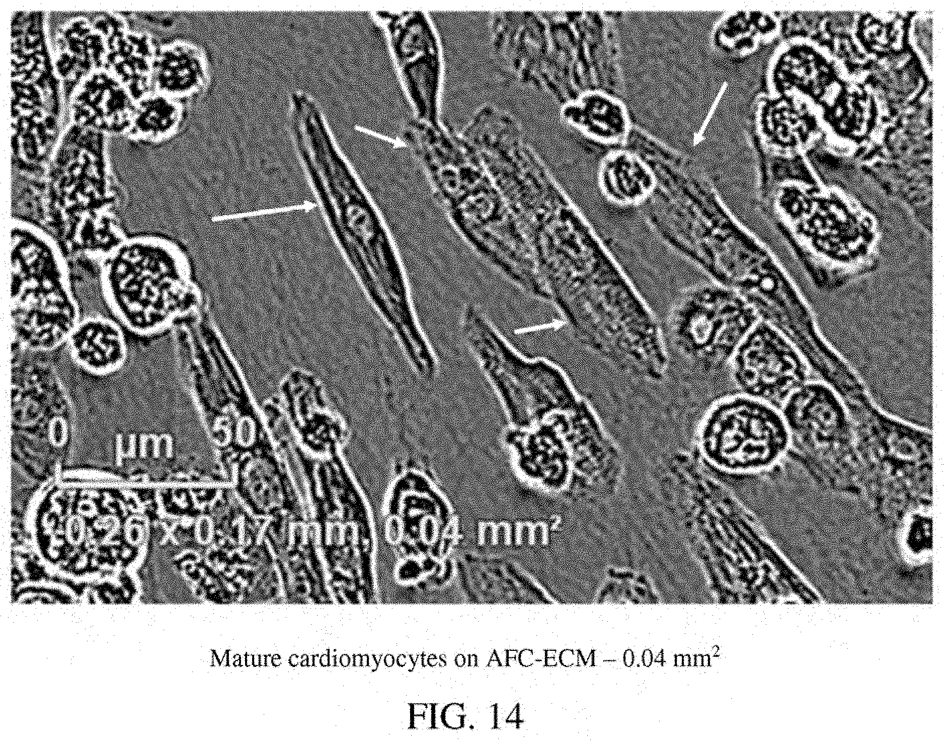
D00015

D00016
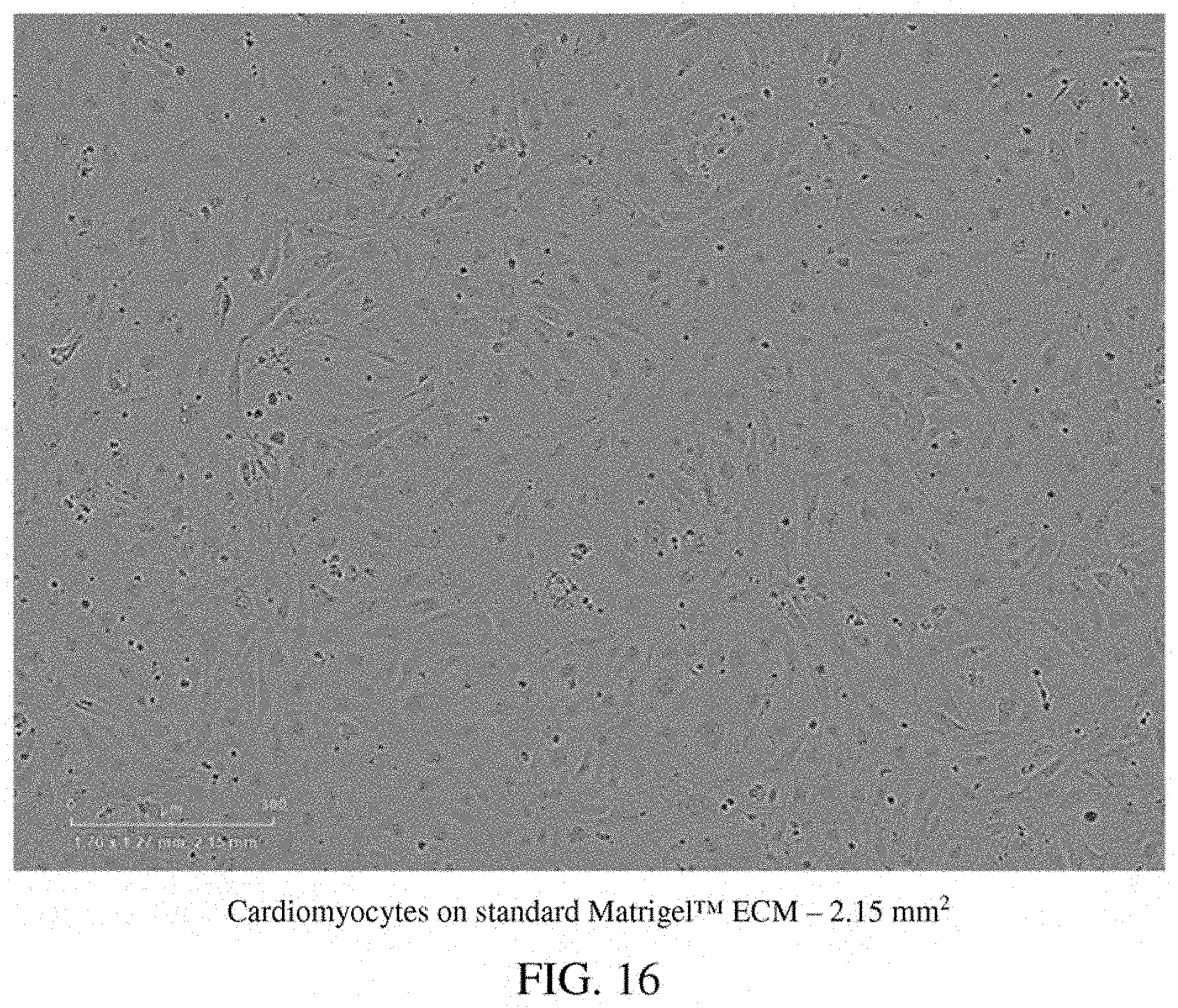
D00017
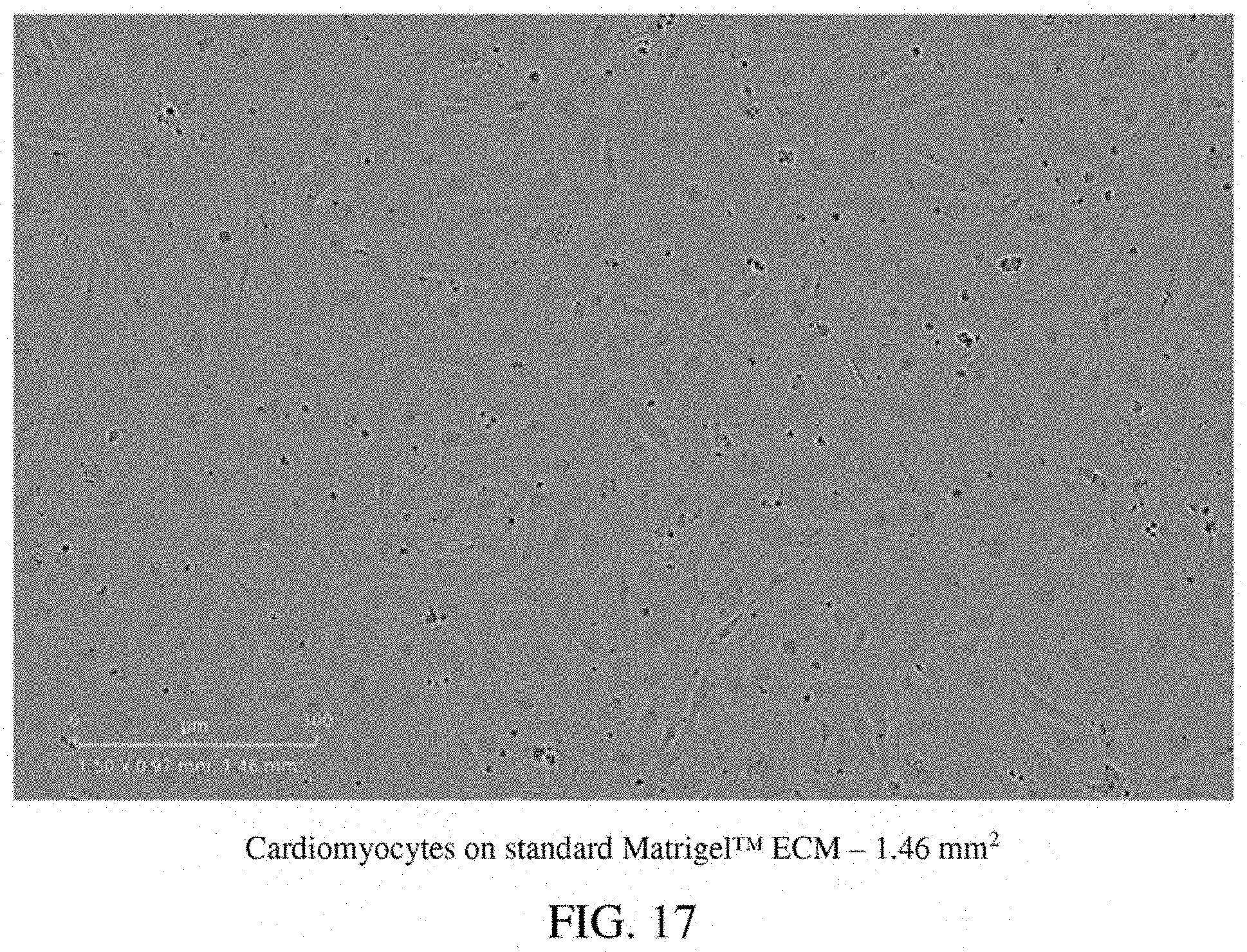
D00018
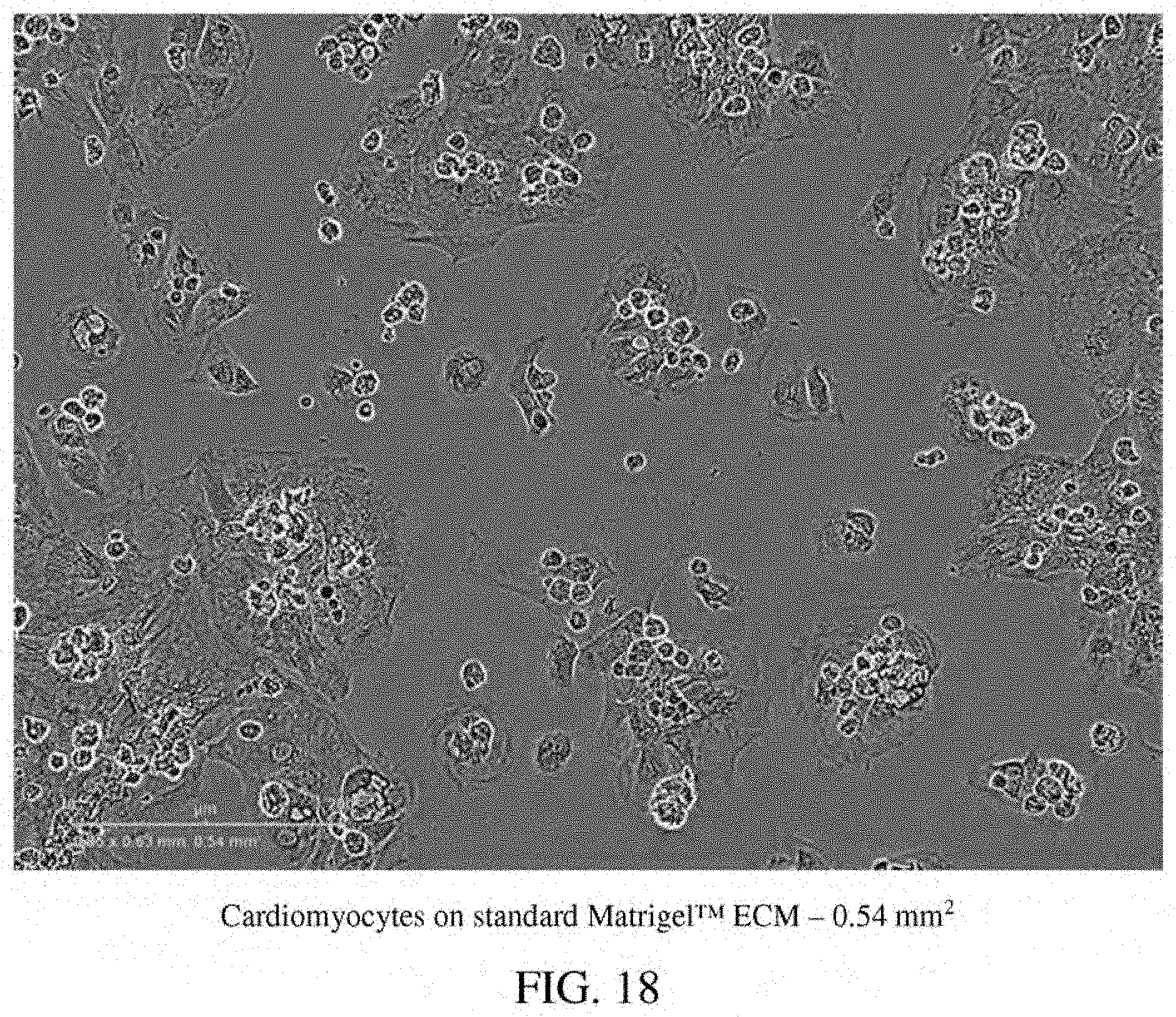
D00019
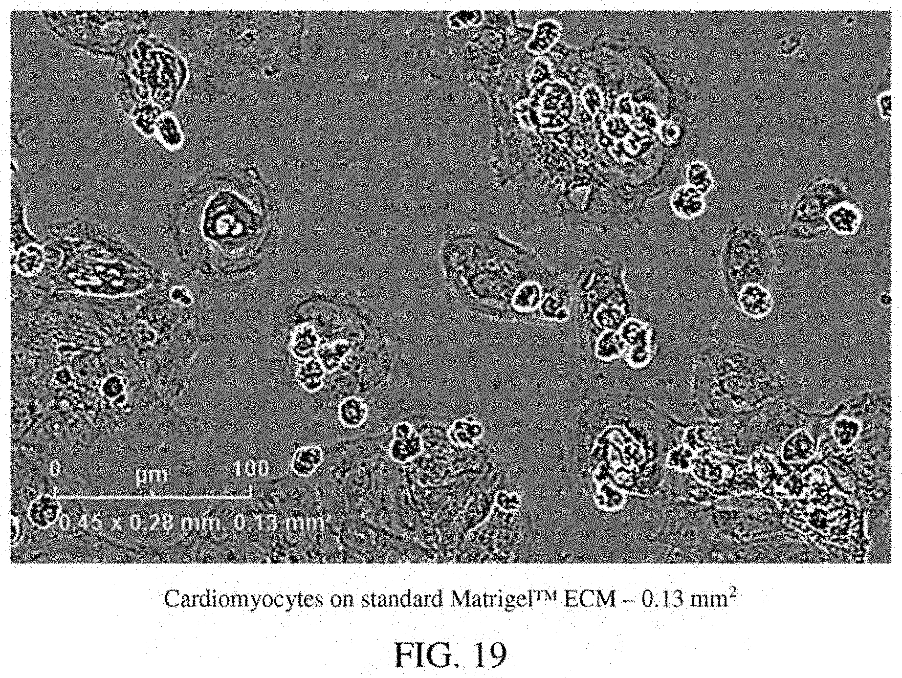
D00020

D00021

D00022
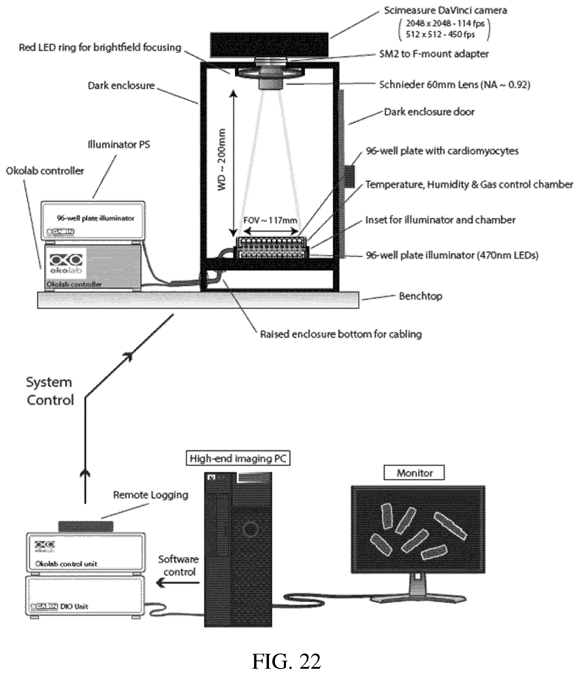
D00023

D00024
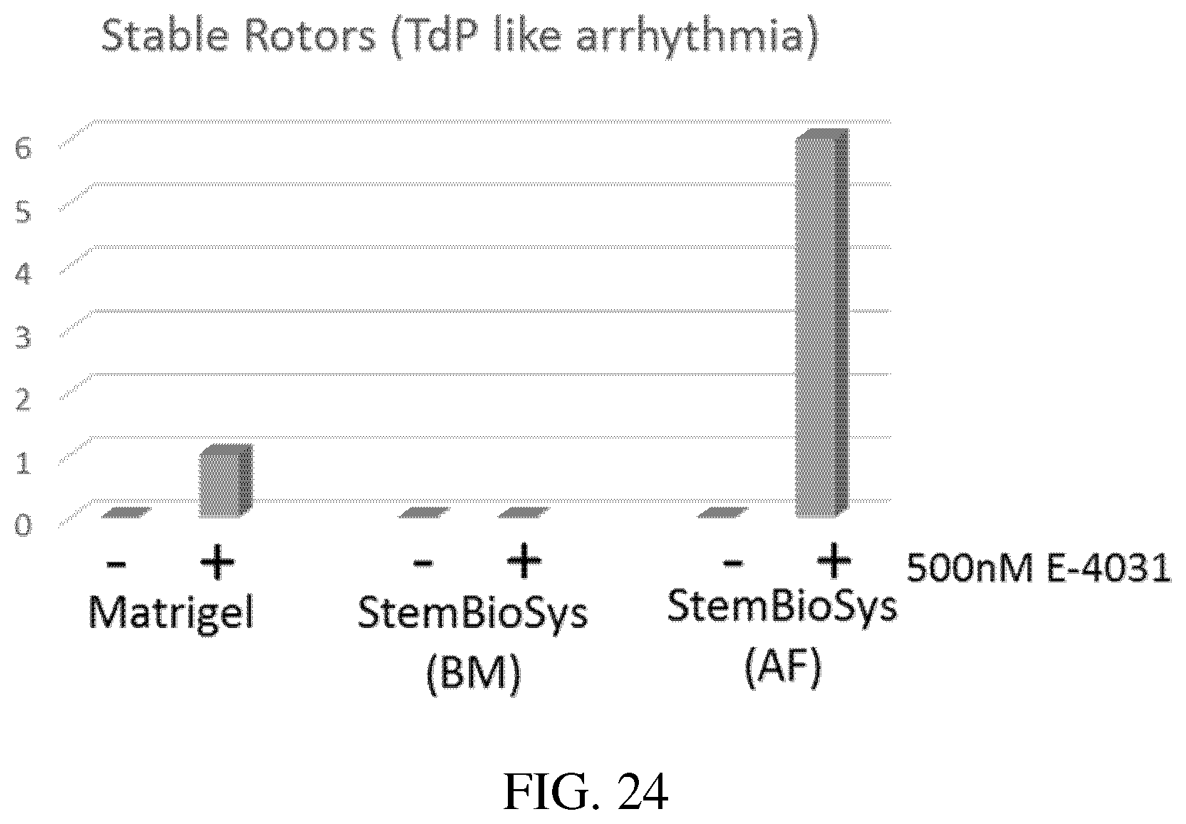
D00025
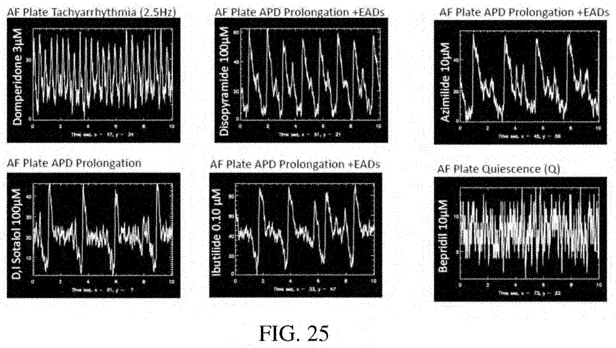
D00026
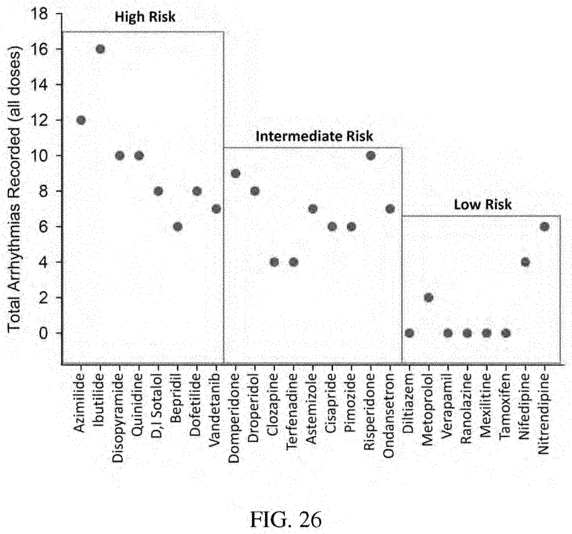
D00027

D00028
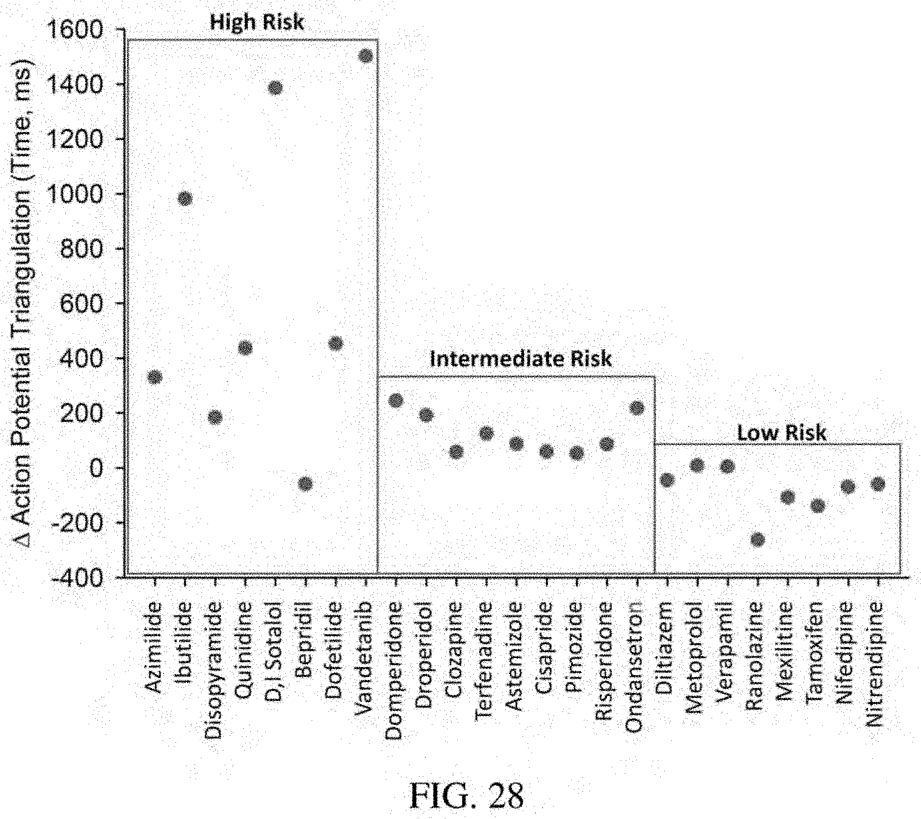
D00029

D00030
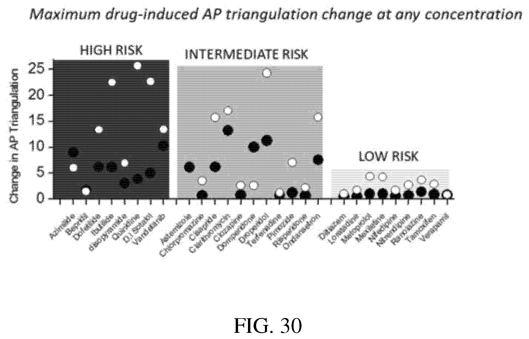
D00031

D00032
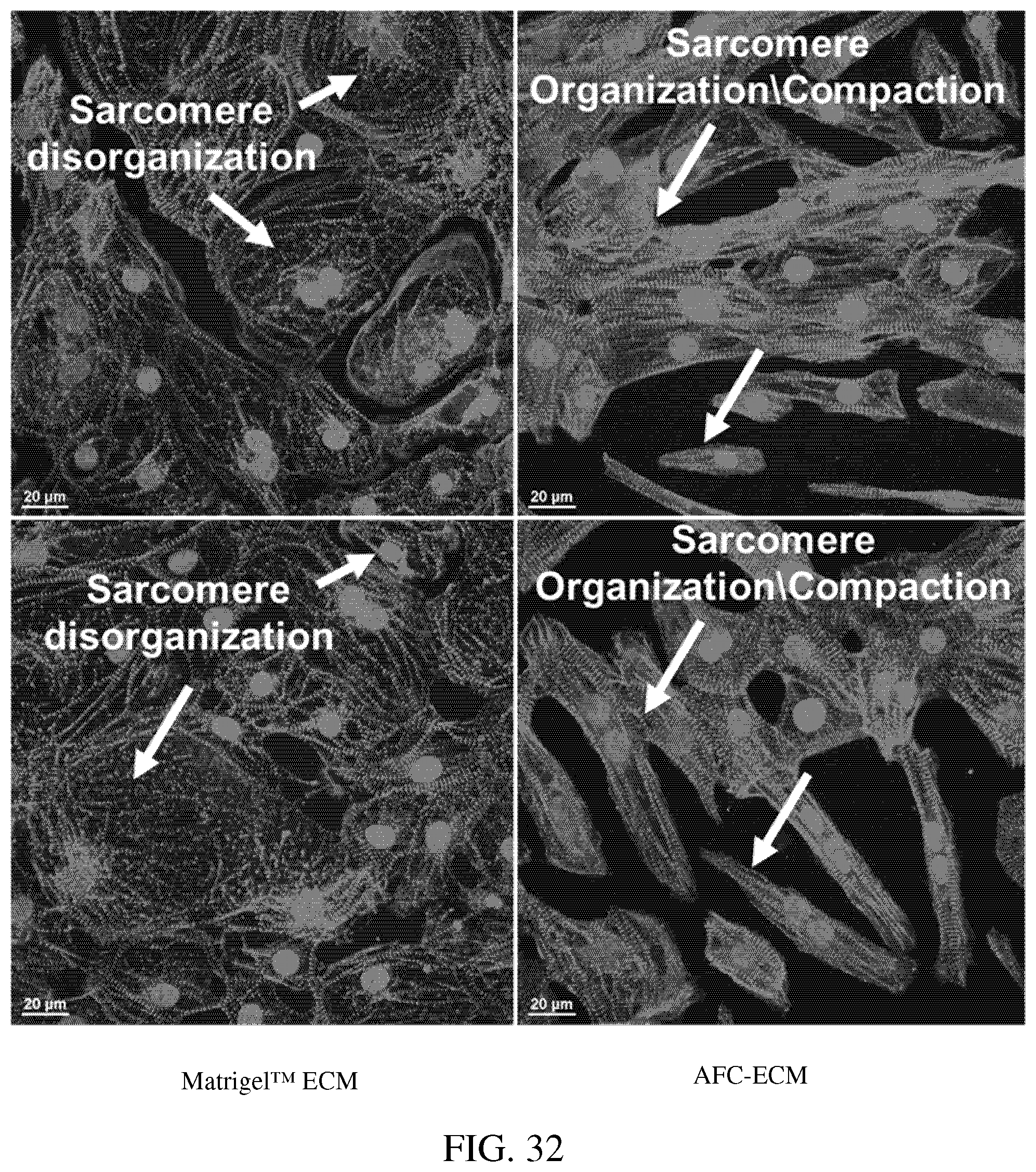
D00033

D00034

D00035

D00036

D00037
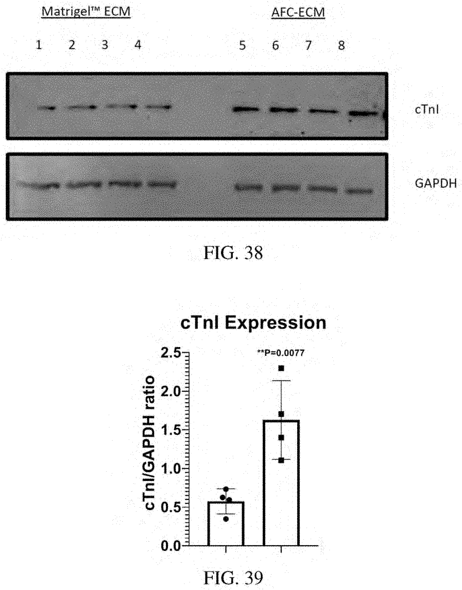
D00038

D00039

XML
uspto.report is an independent third-party trademark research tool that is not affiliated, endorsed, or sponsored by the United States Patent and Trademark Office (USPTO) or any other governmental organization. The information provided by uspto.report is based on publicly available data at the time of writing and is intended for informational purposes only.
While we strive to provide accurate and up-to-date information, we do not guarantee the accuracy, completeness, reliability, or suitability of the information displayed on this site. The use of this site is at your own risk. Any reliance you place on such information is therefore strictly at your own risk.
All official trademark data, including owner information, should be verified by visiting the official USPTO website at www.uspto.gov. This site is not intended to replace professional legal advice and should not be used as a substitute for consulting with a legal professional who is knowledgeable about trademark law.