Allogeneic Cell Therapy Of B Cell Malignancies Using Genetically Engineered T Cells Targeting Cd19
BENTON; Mark ; et al.
U.S. patent application number 17/505106 was filed with the patent office on 2022-04-21 for allogeneic cell therapy of b cell malignancies using genetically engineered t cells targeting cd19. The applicant listed for this patent is CRISPR Therapeutics AG. Invention is credited to Mark BENTON, Tony HO, Demetrios KALAITZIDIS, Ewelina MORAWA, Jonathan Alexander TERRETT.
| Application Number | 20220118019 17/505106 |
| Document ID | / |
| Family ID | |
| Filed Date | 2022-04-21 |


View All Diagrams
| United States Patent Application | 20220118019 |
| Kind Code | A1 |
| BENTON; Mark ; et al. | April 21, 2022 |
ALLOGENEIC CELL THERAPY OF B CELL MALIGNANCIES USING GENETICALLY ENGINEERED T CELLS TARGETING CD19
Abstract
A population of genetically engineered immune cells (e.g., T cells), which express a chimeric antigen receptor (CAR) specific to CD19 and contain a disrupted TRAC gene, a disrupted .beta.2M gene, or both, for use in treating a B cell malignancy.
| Inventors: | BENTON; Mark; (Cambridge, MA) ; HO; Tony; (Cambridge, MA) ; KALAITZIDIS; Demetrios; (Cambridge, MA) ; MORAWA; Ewelina; (Cambridge, MA) ; TERRETT; Jonathan Alexander; (Cambridge, MA) | ||||||||||
| Applicant: |
|
||||||||||
|---|---|---|---|---|---|---|---|---|---|---|---|
| Appl. No.: | 17/505106 | ||||||||||
| Filed: | October 19, 2021 |
Related U.S. Patent Documents
| Application Number | Filing Date | Patent Number | ||
|---|---|---|---|---|
| 63094252 | Oct 20, 2020 | |||
| 63140664 | Jan 22, 2021 | |||
| 63164690 | Mar 23, 2021 | |||
| 63215191 | Jun 25, 2021 | |||
| 63254226 | Oct 11, 2021 | |||
| International Class: | A61K 35/17 20060101 A61K035/17; A61P 35/00 20060101 A61P035/00; C07K 16/28 20060101 C07K016/28; A61K 39/395 20060101 A61K039/395; A61K 31/675 20060101 A61K031/675; A61K 31/7076 20060101 A61K031/7076 |
Claims
1. A method for treating a B-cell malignancy in a human patient, the method comprising: (i) subjecting a human patient having a B-cell malignancy to a lymphodepletion treatment; and (ii) administering to the human patient a first dose of a population of genetically engineered T cells after step (i), wherein the population of genetically engineered T cells comprising T cells that comprise: (a) a nucleic acid coding for a chimeric antigen receptor (CAR) that binds CD19, wherein the population of genetically engineered T cells is administered to the human patient at a dose of about 1.0.times.10.sup.7 to about 9.times.10.sup.8 CAR.sup.+ T cells.
2. The method of claim 1, wherein the CAR comprises an anti-CD19 single chain variable fragment (scFv) that comprises the same heavy chain complementary determining regions (CDRs) as those in a heavy chain variable region set forth in SEQ ID NO: 51, and the same light chain CDRs as those in a light chain variable region set forth in SEQ ID NO: 52.
3. The method of claim 1, wherein the population of genetically engineered T cells comprise (b) a disrupted T cell receptor alpha constant (TRAC) gene, and/or (c) a disrupted beta 2-microglobulin (.beta.2M) gene.
4. The method of claim 3, wherein the population of genetically engineered T cells comprise (b) a disrupted T cell receptor alpha constant (TRAC) gene, and (c) a disrupted beta 2-microglobulin (.beta.2M) gene.
5. The method of claim 1, wherein the lymphodepletion treatment in step (i) comprises co-administration to the human patient fludarabine at about 30 mg/m.sup.2 and cyclophosphamide at about 500-750 mg/m.sup.2 per day for three days.
6. The method of claim 1, wherein the first dose of the population of genetically engineered T cells is about 3.times.10.sup.7, about 1.times.10.sup.8, about 3.times.10.sup.8, about 4.5.times.10.sup.8, about 6.times.10.sup.8, or about 9.times.10.sup.8 CAR+ T cells.
7. The method of claim 6, wherein the first dose of the population of genetically engineered T cells is administered to the human patient at a dose of about 3.5.times.10.sup.8 to about 9.times.10.sup.8, optionally about 3.5.times.10.sup.8 to about 6.times.10.sup.8.
8. The method of claim 1, wherein the first dose of the population of genetically engineered T cells is administered to the human patient at a dose of about 4.5.times.10.sup.8, about 6.times.10.sup.8, or about 7.5.times.10.sup.8 CAR.sup.+ T cells.
9. The method of claim 1, wherein the lymphodepletion treatment in step (i) comprises co-administration to the human patient fludarabine at about 30 mg/m.sup.2 and cyclophosphamide at about 500 mg/m.sup.2 to about 750 mg/m.sup.2 per day for three days.
10. The method of claim 1, wherein prior to step (i), the human patient does not show one or more of the following features: (a) significant worsening of clinical status, (b) requirement for supplemental oxygen to maintain a saturation level of greater than 91%, (c) uncontrolled cardiac arrhythmia, (d) hypotension requiring vasopressor support, (e) active infection, and (f) grade .gtoreq.2 acute neurological toxicity.
11. The method of claim 1, wherein step (i) is performed about 2-7 days prior to step (ii).
12. The method of claim 1, wherein after step (i) and prior to step (ii), the human patient does not show one or more of the following features: (a) active uncontrolled infection; (b) worsening of clinical status compared to the clinical status prior to step (i); and (c) grade .gtoreq.2 acute neurological toxicity.
13. The method of claim 1, further comprising (iii) monitoring the human patient for development of acute toxicity after step (ii); and (iv) managing the acute toxicity if occurs.
14. The method of claim 11, wherein step (iii) is performed for at least 28 days after administration of the first dose of the population of genetically engineered T cells.
15. The method of claim 13, wherein the acute toxicity comprises tumor lysis syndrome (TLS), cytokine release syndrome (CRS), immune effector cell-associated neurotoxicity syndrome (ICANS), B cell aplasia, hemophagocytic lymphohistiocytosis (HLH), cytopenia, graft-versus-host disease (GvHD), hypertension, renal insufficiency, viral encephalitis, or a combination thereof.
16. The method of claim 1, further comprising administering to the human patient one or more subsequent doses of the population of genetically engineered T cells, optionally after the human patient shows progressive disease (PD), wherein the human patient had prior response.
17. The method of claim 16, wherein the human patient receives a lymphodepletion treatment within 2-7 days prior to the subsequent dose of the population of genetically engineered T cells.
18. The method of claim 17, wherein the human patient exhibits significant cytopenias and does not receive a lymphodepletion treatment prior to the subsequent dose of the population of genetically engineered T cells.
19. The method of claim 1, wherein the B cell malignancy is non-Hodgkin lymphoma, which optionally is selected from the group consisting of diffuse large B cell lymphoma (DLBCL), high grade B cell lymphoma with MYC and BCL2 and/or BCL6 rearrangement, transformed follicular lymphoma (FL), and grade 3b FL.
20. The method of claim 19, wherein DLBCL is DLBCL not otherwise specified (NOS).
21. The method of claim 1, wherein the human patient has at least one measurable lesion that is fluorodeoxyglucose positron emission tomography (PET)-positive.
22. The method of claim 1, wherein the B cell malignancy is refractory and/or relapsed.
23. The method of claim 1, wherein the human patient has undergone one or more lines of prior anti-cancer therapies.
24. The method of claim 23, wherein the human patient has undergone two or more lines of prior anti-cancer therapies.
25. The method of claim 23, wherein the prior anti-cancer therapies comprise an anti-CD20 antibody, an anthracycline-containing regimen, or a combination thereof.
26. The method of claim 25, wherein the human patient has refractory or relapsed transformed FL and has undergone at least one line of chemotherapy for disease after transformation to DLBCL.
27. The method of claim 22, wherein the B cell malignancy is refractory, and the human patient has progressive disease on last therapy, or has stable disease following at least two cycles of therapy with duration of stable disease of up to 6 months.
28. The method of claim 1, wherein the human patient has failed prior autologous hematopoietic stem cell transplantation (HSCT) or ineligible for prior autologous HSCT.
29. The method of claim 1, wherein the human patient is subject to an additional anti-cancer therapy after treatment with the population of genetically engineered T cells.
30. The method of claim 1, wherein the human patient has one or more of the following features: (a) has an Eastern Cooperative Oncology Group (ECOG) performance status 0 or 1; (b) adequate renal, liver, cardiac, and/or pulmonary function; (c) free of prior gene therapy or modified cell therapy; (d) free of prior treatment comprising an anti-CD19 antibody; (e) free of prior allogeneic HSCT; (f) free of detectable malignant cells from cerebrospinal fluid; (g) free of brain metastases; (h) free of prior central nervous system disorders; (i) free of unstable angina, arrhythmia, and/or myocardial infarction; (j)free of uncontrolled infection; (k) free of immunodeficiency disorders or autoimmune disorders that require immunosuppressive therapy; and (l) free of infection by human immunodeficiency virus, hepatitis B virus, or hepatitis C virus.
31. The method of claim 1, wherein the lymphodepletion treatment in step (i) comprises co-administration to the human patient fludarabine at about 30 mg/m.sup.2 and cyclophosphamide at about 500 mg/m.sup.2 per day for three days.
32. The method of claim 31, wherein the first dose of the population of genetically engineered T cells is at least 3.times.10.sup.7 CAR.sup.+ T cells.
33. The method of claim 31, wherein the human patient is administered a second dose of the population of genetically engineered T cells about 4 to 8 weeks after the first dose of the population of genetically engineered T cells.
34. The method of claim 33, wherein the human patient achieves stable disease (SD), partial response (PR), or complete response (CR) at least about 4 weeks after the first dose of the population of genetically engineered T cells.
35. The method of claim 33, wherein the human patient receives a second lymphodepletion treatment within 2-7 days prior to the second dose of the population of genetically engineered T cells.
36. The method of claim 33, wherein the human patient exhibits significant cytopenias and does not receive lymphodepletion treatment prior to the second dose of the population of genetically engineered T cells.
37. The method of claim 1, wherein the lymphodepletion treatment in step (i) comprises co-administration to the human patient fludarabine at about 30 mg/m.sup.2 and cyclophosphamide at about 750 mg/m.sup.2 per day for three days.
38. The method of claim 31, wherein the first dose of the population of genetically engineered T cells is at least 3.times.10.sup.8 CAR.sup.+ T cells.
39. The method of claim 37, wherein the human patient is administered a second dose of the population of genetically engineered T cells about 4 to 8 weeks after the first dose of the population of genetically engineered T cells.
40. The method of claim 39, wherein the human patient achieves stable disease (SD), partial response (PR), or complete response (CR) at least about 4 weeks after the first dose of the population of genetically engineered T cells.
41. The method of claim 39, wherein the human patient receives a second lymphodepletion treatment within 2-7 days prior to the second dose of the population of genetically engineered T cells, and wherein the second lymphodepletion treatment comprises co-administration to the human patient fludarabine at about 30 mg/m.sup.2 and cyclophosphamide at about 500 mg/m.sup.2 per day for three days.
42. The method of claim 39, wherein the human patient exhibits significant cytopenias and does not receive lymphodepletion treatment prior to the second dose of the population of genetically engineered T cells.
43. The method of claim 37, wherein the human patient receives at least one additional dose of the population of genetically engineered T cells, optionally wherein the human patient receives a lymphodepletion treatment comprising co-administration to the human patient fludarabine at about 30 mg/m.sup.2 and cyclophosphamide at about 500 mg/m.sup.2 per day for three days within 2-7 days prior to the additional dose of the population of genetically engineered T cells.
44. The method of claim 1, wherein the population of genetically engineered T cells administered to the human patient per dose contains no more than 7.times.10.sup.4 TCR.sup.+ T cells/kg.
45. The method of claim 1, wherein the anti-CD19 scFv comprises the amino acid sequence of SEQ ID NO: 47.
46. The method of claim 45, wherein the CAR that binds CD19 comprises the amino acid sequence of SEQ ID NO: 40.
47. The method of claim 3, wherein the nucleic acid encoding the anti-CD19 CAR is inserted in the disrupted TRAC gene.
48. The method of claim 3, wherein the disrupted TRAC gene comprises a deletion of a fragment comprising the nucleotide sequence of SEQ ID NO: 26.
49. The method of claim 48, wherein the nucleic acid encoding the anti-CD19 CAR is inserted at the deletion site of the disrupted TRAC gene.
50. The method of claim 48, wherein the disrupted TRAC gene comprises the nucleotide sequence of SEQ ID NO: 54.
51. The method of claim 3, wherein the disrupted .beta.2M gene in the population of genetically engineered T cells comprises at least one of the nucleotide sequence set forth in SEQ ID NOs: 9-14.
52. The method of claim 1, wherein the population of genetically engineered T cells is allogeneic.
53. The method of claim 1, wherein at least 90% of the T cells in the population of genetically engineered T cells do not express a detectable level of TCR surface protein.
54. The method of claim 1, wherein at least 70% of the T cells in the population of genetically engineered T cells do not express a detectable level of TCR surface protein, wherein at least 50% of the T cells in the population of genetically engineered T cells do not express a detectable level of B2M surface protein; and/or wherein at least 30% of the T cells in the population of genetically engineered T cells express a detectable level of the CAR.
55. The method of claim 53, wherein at least 99.5% of the T cells in the population of genetically engineered T cells do not express a detectable level of TCR surface protein.
56. The method of claim 1, wherein at least 70% of the T cells in the population of genetically engineered T cells do not express a detectable level of B2M surface protein.
57. The method of claim 55, at least 85% of the T cells in the population of the genetically engineered T cells do not express a detectable level of B2M surface protein.
58. The method of claim 1, wherein at least 50% of the T cells in the population of genetically engineered T cells express a detectable level of the CAR.
59. The method of claim 57, wherein at least 70% of the T cells in the population of genetically engineered T cells express a detectable level of the CAR.
60. The method of claim 1, wherein the population of genetically engineered T cells are administered to the human patient via intravenous infusion.
61. The method of claim 1, wherein the population of genetically engineered T cells are suspended in a cryopreservation solution.
62. A pharmaceutical composition for use in treating a B-cell malignancy, the pharmaceutical composition comprising a population of genetically engineered T cells that comprises a nucleic acid coding for a chimeric antigen receptor (CAR) that binds CD19, wherein the pharmaceutical composition is for use in a method set forth in claim 1.
Description
CROSS REFERENCE TO RELATED APPLICATIONS
[0001] This application claims the benefit of the filing dates of U.S. Provisional Application No. 63/094,252, filed Oct. 20, 2020, U.S. Provisional Application No. 63/140,664, filed Jan. 22, 2021, U.S. Provisional Application No. 63/164,690, filed Mar. 23, 2021, U.S. Provisional Application No. 63/215,191, filed Jun. 25, 2021, and U.S. Provisional Application No. 63/254,226, filed Oct. 11, 2021. The entire contents of each of the prior provisional applications are incorporated by reference herein.
SEQUENCE LISTING
[0002] The application contains a Sequence Listing that has been filed electronically in the form of a text file, created Oct. 15, 2021, and named "095136-0383-038US1_SEQ.TXT" (54,240 bytes), the contents of which are incorporated by reference herein in their entirety
BACKGROUND OF THE INVENTION
[0003] Chimeric antigen receptor (CAR) T cell therapies are adoptive T cell therapeutics used to treat human malignancies. Although CAR T cell therapy has led to tremendous clinical success, including durable remission in relapsed/refractory non-Hodgkin lymphoma (NHL) and pediatric acute lymphoblastic leukemia (ALL), the approved products are autologous and require patient-specific cell collection and manufacturing. Because of this, some patients have experienced disease progression or death while awaiting treatment. Accordingly, there remains a need for improved CAR T cell therapeutics.
SUMMARY OF THE INVENTION
[0004] The present disclosure is based, at least in part, on the development of allogeneic cell therapy for B cell malignancies such as transformed FL or DLBCL using genetically engineered T cells (e.g., CTX110 cells, a.k.a., TC1 cells) expressing an anti-CD19 chimeric antigen receptor (CAR) and having disrupted TRAC gene and B2M gene. The allogeneic CAR-T cell therapy disclosed herein showed treatment efficacies in human patients having B cell malignancies disclosed herein, including complete responses in certain patients and long durability of responses. Further, the allogeneic CAR-T cell therapy disclosed herein exhibited desired pharmacokinetic features in the human patients, including prolonged CAR-T cell expansion and persistence after infusion.
[0005] Accordingly, some aspects of the present disclosure features a method for treating a B-cell malignancy in a human patient, the method comprising: (i) subjecting a human patient having a B-cell malignancy to a lymphodepletion treatment; and (ii) administering to the human patient a first dose of a population of genetically engineered T cells after step (i), wherein the population of genetically engineered T cells comprising T cells that comprise: (a) a nucleic acid coding for a chimeric antigen receptor (CAR) that binds CD19. The population of genetically engineered T cells may be administered to the human patient at a dose of about 1.0.times.10.sup.7 to about 9.times.10.sup.8 CAR.sup.+ T cells. The population of genetically engineered T cells administered to the human patient per dose contains no more than 7.times.10.sup.4 TCR.sup.+ T cells/kg.
[0006] In some embodiments, the lymphodepletion treatment in step (i) comprises co-administration to the human patient fludarabine at about 30 mg/m.sup.2 and cyclophosphamide at about 500-750 mg/m.sup.2 per day for three days.
[0007] In some embodiments, the first dose of the population of genetically engineered T cells is of about 3.5.times.10.sup.8 to about 9.times.10.sup.8. For example, the first dose of the population of genetically engineered T cells is of about 3.5.times.10.sup.8 to about 6.times.10.sup.8. In some examples, the first dose of the population of genetically engineered T cells is about 3.times.10.sup.7 CAR+ T cells. In some examples, the first dose of the population of genetically engineered T cells is about 1.times.10.sup.8 CAR.sup.+ T cells. In some examples, the first dose of the population of genetically engineered T cells is about 3.times.10.sup.8 CAR.sup.+ T cells. In some examples, the first dose of the population of genetically engineered T cells is about 4.5.times.10.sup.8 CAR.sup.+ T cells. In some examples, the first dose of the population of genetically engineered T cells is about 6.times.10.sup.8 CAR.sup.+ T cells. In some examples, the first dose of the population of genetically engineered T cells is about 9.times.10.sup.8 CAR+ T cells. In specific examples, the first dose of the population of genetically engineered T cells is administered to the human patient at a dose of about 4.5.times.10.sup.8, about 6.times.10.sup.8, or about 7.5.times.10.sup.8 CAR.sup.+ T cells.
[0008] In some embodiments, the lymphodepletion treatment in step (i) comprises co-administration to the human patient fludarabine at about 30 mg/m.sup.2 and cyclophosphamide at about 500 mg/m.sup.2 to about 750 mg/m.sup.2 per day for three days. In some instances, step (i) may be performed about 2-7 days prior to step (ii).
[0009] Prior to step (i), the human patient may not show one or more of the following features: (a) significant worsening of clinical status, (b) requirement for supplemental oxygen to maintain a saturation level of greater than 91%, (c) uncontrolled cardiac arrhythmia, (d) hypotension requiring vasopressor support, (e) active infection, and (f) grade .gtoreq.2 acute neurological toxicity. In some embodiments, after step (i) and prior to step (ii), the human patient does not show one or more of the following features: (a) active uncontrolled infection; (b) worsening of clinical status compared to the clinical status prior to step (i); and (c) grade .gtoreq.2 acute neurological toxicity.
[0010] Any of the methods disclosed herein may further comprises (iii) monitoring the human patient for development of acute toxicity after step (ii); and (iv) managing the acute toxicity if occurs. In some instances, step (iii) can be performed for at least 28 days after administration of the first dose of the population of genetically engineered T cells. Exemplary acute toxicity comprises tumor lysis syndrome (TLS), cytokine release syndrome (CRS), immune effector cell-associated neurotoxicity syndrome (ICANS), B cell aplasia, hemophagocytic lymphohistiocytosis (HLH), cytopenia, graft-versus-host disease (GvHD), hypertension, renal insufficiency, viral encephalitis, or a combination thereof.
[0011] Alternatively or in addition, the method disclosed herein may further comprise administering to the human patient one or more subsequent doses of the population of genetically engineered T cells, optionally after the human patient shows progressive disease (PD), wherein the human patient had prior response. In some embodiments, the human patient may receive a lymphodepletion treatment within 2-7 days prior to the subsequent dose of the population of genetically engineered T cells. Alternatively, if the human patient exhibits significant cytopenias, no lymphodepletion treatment prior to the subsequent dose of the population of genetically engineered T cells may be given to the human patient.
[0012] In some embodiments, the B cell malignancy can be non-Hodgkin lymphoma. Examples include diffuse large B cell lymphoma (DLBCL), high grade B cell lymphoma with MYC and BCL2 and/or BCL6 rearrangement, transformed follicular lymphoma (FL), and grade 3b FL. In some examples, DLBCL is DLBCL not otherwise specified (NOS). The human patient may have at least one measurable lesion that is fluorodeoxyglucose positron emission tomography (PET)-positive.
[0013] In some embodiments, the B cell malignancy is refractory and/or relapsed. The human patients having refractory and/or relapsed B cell malignancy may have undergone one or more lines of prior anti-cancer therapies. For example, the human patient has undergone two or more lines of prior anti-cancer therapies. In some examples, the prior anti-cancer therapies comprise an anti-CD20 antibody, an anthracycline-containing regimen, or a combination thereof.
[0014] In some examples, the human patient has refractory or relapsed transformed FL and has undergone at least one line of chemotherapy for disease after transformation to DLBCL. In some examples, the B cell malignancy is refractory, and the human patient has progressive disease on last therapy, or has stable disease following at least two cycles of therapy with duration of stable disease of up to 6 months. In some examples, the human patient has failed prior autologous hematopoietic stem cell transplantation (HSCT) or ineligible for prior autologous HSCT. Alternatively or in addition, the human patient is subject to an additional anti-cancer therapy after treatment with the population of genetically engineered T cells.
[0015] In any of the methods disclosed herein, the human patient has one or more of the following features: (a)has an Eastern Cooperative Oncology Group (ECOG) performance status 0 or 1; (b) adequate renal, liver, cardiac, and/or pulmonary function; (c) free of prior gene therapy or modified cell therapy; (d) free of prior treatment comprising an anti-CD19 antibody; (e) free of prior allogeneic HSCT; (f) free of detectable malignant cells from cerebrospinal fluid; (g) free of brain metastases; (h) free of prior central nervous system disorders; (i) free of unstable angina, arrhythmia, and/or myocardial infarction; (j)free of uncontrolled infection; (k) free of immunodeficiency disorders or autoimmune disorders that require immunosuppressive therapy; and (l) free of infection by human immunodeficiency virus, hepatitis B virus, or hepatitis C virus.
[0016] In some embodiments, the method discloses herein may involve a lymphodepletion treatment in step (i) that comprises co-administration to the human patient fludarabine at about 30 mg/m.sup.2 and cyclophosphamide at about 500 mg/m.sup.2 per day for three days. Alternatively or in addition, the first dose of the population of genetically engineered T cells can be at least 3.times.10.sup.7 CAR.sup.+ T cells. In some instances, the human patient may be administered a second dose of the population of genetically engineered T cells about 4 to 8 weeks after the first dose of the population of genetically engineered T cells. The human patient may achieve stable disease (SD), partial response (PR), or complete response (CR) at least about 4 weeks after the first dose of the population of genetically engineered T cells. In some examples, the human patient receives a second lymphodepletion treatment within 2-7 days prior to the second dose of the population of genetically engineered T cells. Alternatively, the human patient may receive no lymphodepletion treatment prior to the second dose of the population of genetically engineered T cells, e.g., for a human patient who exhibits significant cytopenias and does not receive
[0017] In some embodiments, the method disclosed herein may involve a lymphodepletion treatment in step (i) that comprises co-administration to the human patient fludarabine at about 30 mg/m.sup.2 and cyclophosphamide at about 750 mg/m.sup.2 per day for three days. In some instances, the first dose of the population of genetically engineered T cells is at least 3.times.10.sup.8 CAR.sup.+ T cells. In some examples, the human patient may be administered a second dose of the population of genetically engineered T cells about 4 to 8 weeks after the first dose of the population of genetically engineered T cells. Such a human patient may achieve stable disease (SD), partial response (PR), or complete response (CR) at least about 4 weeks after the first dose of the population of genetically engineered T cells. In some instances, the human patient receives a second lymphodepletion treatment within 2-7 days prior to the second dose of the population of genetically engineered T cells. The second lymphodepletion treatment may comprise co-administration to the human patient fludarabine at about 30 mg/m.sup.2 and cyclophosphamide at about 500 mg/m.sup.2 per day for three days. Alternatively, the human patient may receive no lymphodepletion treatment prior to the second dose of the population of genetically engineered T cells, e.g., for a human patient who exhibits significant cytopenias and does not receive
[0018] In any of the methods disclosed herein, the human patient may receive at least one additional dose of the population of genetically engineered T cells. In some instances, the human patient receives a lymphodepletion treatment comprising co-administration to the human patient fludarabine at about 30 mg/m.sup.2 and cyclophosphamide at about 500 mg/m.sup.2 per day for three days within 2-7 days prior to the additional dose of the population of genetically engineered T cells.
[0019] In any of the methods disclosed herein, the genetically engineered T cells may express a CAR that comprises an anti-CD19 single chain variable fragment (scFv). In some examples, the anti-CD19 scFv may comprise the same heavy chain complementary determining regions (CDRs) as those in a heavy chain variable region set forth in SEQ ID NO: 51, and/or the same light chain CDRs as those in a light chain variable region set forth in SEQ ID NO: 52. In some instances, the anti-CD19 scFv comprises the amino acid sequence of SEQ ID NO: 47. In some specific examples, the CAR that binds CD19 comprises the amino acid sequence of SEQ ID NO: 40.
[0020] The population of genetically engineered T cells may further comprise (b) a disrupted T cell receptor alpha constant (TRAC) gene, and/or (c) a disrupted beta 2-microglobulin (.beta.2M) gene. In some embodiments, the population of genetically engineered T cells comprise (b) a disrupted T cell receptor alpha constant (TRAC) gene, and (c) a disrupted beta 2-microglobulin (.beta.2M) gene. In some examples, the nucleic acid encoding the anti-CD19 CAR is inserted in the disrupted TRAC gene. In some specific examples, the disrupted TRAC gene may comprise a deletion of a fragment comprising the nucleotide sequence of SEQ ID NO: 26. The nucleic acid encoding the anti-CD19 CAR may be inserted at the deletion site of the disrupted TRAC gene. In some examples, the disrupted TRAC gene comprises the nucleotide sequence of SEQ ID NO: 54.
[0021] Alternatively or in addition, the disrupted .beta.2M gene in the population of genetically engineered T cells comprises at least one of the nucleotide sequence set forth in SEQ ID NOs: 9-14.
[0022] In some embodiments, the population of genetically engineered T cells is allogeneic. In some embodiments, at least 90% of the T cells in the population of genetically engineered T cells do not express a detectable level of TCR surface protein. In some examples, at least 70% of the T cells in the population of genetically engineered T cells do not express a detectable level of TCR surface protein, wherein at least 50% of the T cells in the population of genetically engineered T cells do not express a detectable level of B2M surface protein; and/or wherein at least 30% of the T cells in the population of genetically engineered T cells express a detectable level of the CAR. Alternatively or in addition, at least 99.5% of the T cells in the population of genetically engineered T cells do not express a detectable level of TCR surface protein. Alternatively or in addition, at least 70% (e.g., at least 85%) of the T cells in the population of genetically engineered T cells do not express a detectable level of B2M surface protein. Alternatively or in addition, at least 50% (e.g., at least 70%) of the T cells in the population of genetically engineered T cells express a detectable level of the CAR.
[0023] In some embodiments, the population of genetically engineered T cells are administered to the human patient via intravenous infusion. The population of genetically engineered T cells may be suspended in a cryopreservation solution.
[0024] Also within the scope of the present disclosure are pharmaceutical compositions for use in treating a B-cell malignancy (e.g., in any of the methods disclosed herein), the pharmaceutical composition comprising any of the population of genetically engineered T cells disclosed herein (e.g., the CTX110 cells), as well as use of the genetically engineered T cells for manufacturing a medicament for use in treating a B-cell malignancy as disclosed herein.
[0025] The details of one or more embodiments of the invention are set forth in the description below. Other features or advantages of the present invention will be apparent from the following drawings and detailed description of several embodiments, and also from the appended claims.
BRIEF DESCRIPTION OF THE DRAWINGS
[0026] FIG. 1 is a series of flow cytometry plots of human primary T-cells, TRAC-/B2M-CD19CAR+T cells (TC1), 8 days post-editing. The graphs show reduced surface expression of TRAC and B2M. TCR/MHC I double knockout cells express high levels of the CAR transgene (bottom panel). Negative selection of TC1 cells with purification beads leads to a reduction in TCR positive cells (right panel).
[0027] FIG. 2 is a graph depicting high editing rates achieved at the TRAC and B2M loci in TRAC-/B2M-CD19CAR+T cells (TC1). Surface expression of TCR and MHCI, which is the functional output of gene editing, was measured and plotted as editing percentage on the y-axis. High efficiency (e.g., greater than 50%) site-specific integration and expression of the CAR from the TRAC locus were detected. These data demonstrate greater than 50% efficiency for the generation of TRAC-/B2M-/anti-CD19CAR+T cells.
[0028] FIG. 3 is a graph depicting a statistically significant decrease in tumor volume (mm.sup.3) (p=0. 007) in NOG Raji mice following treatment with TRAC-/.beta.2M-/CD19 CAR+ T cells (TC1).
[0029] FIG. 4 is a survival curve graph demonstrating increased survival of NOG Raji mice treated with TC1 cells in comparison to NOG Raji mice receiving no treatment.
[0030] FIG. 5 is a survival curve graph demonstrating increased survival of NOG Raji mice treated with TC1 cells on day 4, in comparison to control mice receiving no treatment on day 1.
[0031] FIGS. 6A and 6B include diagrams showing persistence and anti-tumor activity of TC1 cells in mice. 6A: a series of flow cytometry plots demonstrating that TC1 cells persist in NOG Raji mice. 6B: a graph demonstrating that TC1 cells selectively eradicate splenic Raji cells in NOG Raji mice treated with TC1 in comparison to controls (NOG Raji mice with no treatment or NOG mice). The effect is depicted as a decreased splenic mass in NOG Raji mice treated with TC1 in comparison to controls.
[0032] FIG. 7 is a series of flow cytometry plots demonstrating that persistent splenic TC1 cells are edited in two independent NOG Raji mice with TC1 treatment.
[0033] FIG. 8 is a Kaplan-Meier survival plot demonstrating increased survival of NOG Nalm6 mice treated with TC1 cells on day 4, in comparison to control mice receiving no treatment on day 1.
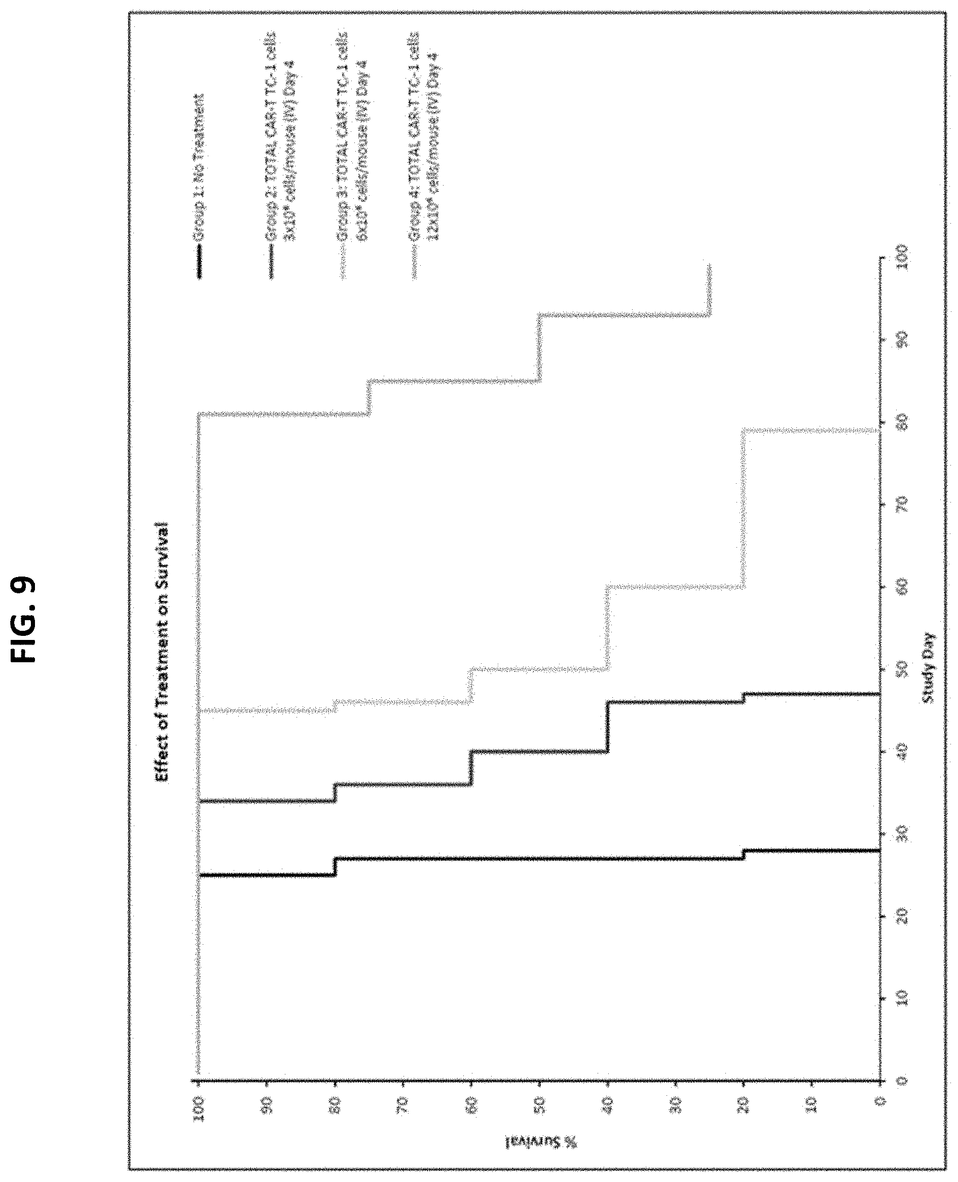
[0034] FIG. 9 is a Kaplan-Meier survival plot demonstrating an increase survival of mice bearing a disseminated Nalm6 B-cell acute lymphoblastic leukemia (B-ALL) after treatment with different concentrations of TC1, in comparison to control mice receiving no treatment.
[0035] FIG. 10 is a graph depicting a statistically significant inhibition in tumor cell expansion in the disseminated Nalm6 B-cell acute lymphoblastic leukemia (B-ALL) tumor model following treatment with TC1 cells.
[0036] FIG. 11 is a Kaplan-Meier survival plot of healthy mice treated with TC1 cells or various control cells (PBMCs or electroporated (EP) T cells) after radiation, or mice that only received radiation ("RT only").
[0037] FIG. 12 is a graph showing percentage of body weight change of the mice treated in FIG. 18.
[0038] FIG. 13 is a Kaplan-Meier survival plot of healthy mice treated with a low dose (2.times.10.sup.7) or high dose (4.times.10.sup.7) of TC1 cells, or unedited T cells after radiation, or mice that only received radiation ("Vehicle-RT").
[0039] FIG. 14 is a graph showing percentage of body weight change of the mice treated in FIG. 20, in addition to mice that were not irradiated and not dosed with cells ("Vehicle--no RT").
[0040] FIG. 15 is a bar graph showing percentage of CD27+CD45RO- cells within the unedited CD8+ T cell subset of peripheral blood cells from six different donors.
[0041] FIG. 16 provides flow cytometry results of TCR.alpha..beta. and B2M expression on TC1 cells before and after depletion of TCR.alpha..beta.+ cells.
[0042] FIG. 17 is a graph the percentage loss of protein for TCR- and MHC I- (B2M) after gene editing, and percentage of cells expressing an anti-CD19 CAR in edited TC1 cells from individual lots of TC1 production.
[0043] FIG. 18 provides graphs showing the percentage of PD1+ (top left), LAG3+ (top right), TIM3+ (bottom left) or CD57+ (bottom right) in the T cell population from six different donors before and after editing.
[0044] FIG. 19 is a graph showing the percentage of cell lysis of CD19-positive cell lines (Nalm6; Raji; and K562-CD19) and CD19-negative cells (K562) when co-cultured at different ratios with TC1 cells or unedited T cells.
[0045] FIG. 20 is a graph showing the number of viable TC1 cells when cultured in the presence of T-cell media (serum+IL2+IL7; Complete Media), media containing serum but no IL2 or IL7 cytokines (5% Serum, No cytokines) or no serum or cytokines (No Serum, No Cytokines). Cells were counted on the indicated days post gene editing. Mean values from three lots shown.+-.SD.
[0046] FIG. 21 is a schematic depicting the clinical study design to evaluate CTX110 cells, (a.k.a., TC1 cells) administered after lymphodepletion to human subjects having CD19+ malignancies. Cohorts A and B will comprise NHL subtypes: DLBCL NOS, high grade B cell lymphoma with MYC and BCL2 and/or BCL6 rearrangements, grade 3b FL, or transformed FL.
[0047] For Cohort A, LD chemotherapy comprises co-administration of fludarabine 30 mg/m.sup.2 and cyclophosphamide 500 mg/m.sup.2 IV daily for 3 days. For Cohort B, LD chemotherapy comprises co-administration of fludarabine 30 mg/m.sup.2 and cyclophosphamide 750 mg/m.sup.2 IV daily for 3 days. For Cohort A, subjects may be administered a planned second dose of CTX110 on Day 28 (4-8 weeks after the first dose) with or without LD chemotherapy if they meet the protocol-specified criteria. Subjects in both Cohorts A and B may be redosed upon disease progression if a subject has had prior objective response. The first course of treatment may comprise first (Day1) and the second (Day35) CTX110 infusion and associated LD regimen, as applicable. For both Cohort A and Cohort B, patients may receive a second course of treatment with a single CTX110 infusion with LD chemotherapy upon disease progression if a subject has had prior clinical response after the first infusion and meets the criteria for an additional infusion. D: day; DLBCL: diffuse large B cell lymphoma; DLT: dose-limiting toxicity; FL: follicular lymphoma; IV: intravenously; LD: lymphodepleting; M: month; MRD: minimal residual disease; NHL: non-Hodgkin lymphoma; NOS: not otherwise specified.
[0048] FIG. 22 is a diagram showing deep reduction in tumor size by CTX110 (including re-dosing). SD=stable disease, CR=Complete response. 1: value extends beyond top of axis (215%).
[0049] FIG. 23 is a diagram showing peripheral blood CAR-T cell levels in patients treated at DL2 through DL4. Values represent Mean.+-.SEM. N=15-21 at each time point.
[0050] FIGS. 24A and 24B include diagrams showing strong rationale for consolidation dose of CTX110. FIG. 24A: a diagram showing Complete response (CR) rate and Overall response (ORR) rate in patients receiving DL2-DL4 doses. FIG. 24B: a diagram showing the rations between cell dose and baseline tumor volumes in patients having different responses as indicated. Baseline tumor volume is calculated by CAR+ T cells (millions) divided by baseline SPD (mm.sup.2).
DETAILED DESCRIPTION OF THE INVENTION
[0051] Cluster of Differentiation 19 (CD19) is an antigenic determinant detectable on leukemia precursor cells. The human and murine amino acid and nucleic acid sequences can be found in a public database, such as GenBank, UniProt and Swiss-Prot. For example, the amino acid sequence of human CD19 can be found as UniProt/Swiss-Prot Accession No. P15391 and the nucleotide sequence encoding of the human CD19 can be found at Accession No. NM_001178098. CD19 is expressed on most B lineage cancers, including, e.g., acute lymphoblastic leukemia, chronic lymphocyte leukemia and non-Hodgkin's lymphoma. It is also an early marker of B cell progenitors. See, e.g., Nicholson et al. Mol. Immun 34 (16-17): 1157-1165 (1997).
[0052] The present disclosure provides an allogeneic CAR-T cell therapy for B cell malignancies. The CAR-T cell therapy involves a population of genetically engineered T cells expressing an anti-CD19 CAR and having disrupted TRAC gene and B2M gene, the nucleic acid coding for the anti-CD19 CAR being inserted into the TRAC gene locus, thereby disrupting expression of the TRAC gene. The allogenic anti-CD19 CAR-T cells are prepared using parent T cells obtained from healthy donors. As such, the CAR-T therapy is available to a patient having the target B cell malignancy immediately after diagnosis, as opposed to at least three week gap between diagnosis and treatment in autologous CAR-T therapy required for manufacturing the CAR-T cells from the patient's own T cells. The allogeneic CAR T therapy can be stored and inventoried at the site of care to facilitate treatment immediately following diagnosis. The immediate availability of the allogeneic anti-CD19 CAR T therapy eliminates the need for bridging chemo-therapy, which may be required when autologous CAR-T cells are manufactured from the patient's own cells. In sum, the allogeneic anti-CD19 CAR-T cell therapy (e.g., involving the use of CTX110 cells disclosed herein) would allow for immediate treatment without risk of manufacturing failure, saving patients valuable time in which their disease could progress. Further, it provides a more consistent product, flexible dosing (e.g., re-dosing is available if needed), scalable manufacturing and simpler logistics, and broader access. The allogeneic anti-CD19 CAR-T cell therapy (e.g., involving the use of CTX110 cells) can also avoid the need for more toxic lymphodepletion regimens.
[0053] The allogeneic anti-CD19 CAR-T cell therapy disclosed herein showed treatment efficacies in human patients having B cell malignancies disclosed herein, including complete responses in certain patients and long durability of responses. Further, the allogeneic CAR-T cell therapy disclosed herein exhibited desired pharmacokinetic features in the human patients, including prolonged CAR-T cell expansion and persistence after infusion.
[0054] Accordingly, provided herein are methods for treating a B-cell malignancy in a human patient using a population of genetically engineered immune cells such as T cells, which express an anti-CD19 CAR (e.g., SEQ ID NO: 40, encoded by SEQ ID NO:39). Such genetically engineered T cells may further comprise a disrupted TRAC gene, a disrupted B2M, or a combination thereof. The nucleic acid encoding the anti-CD19 CAR and optionally comprising a promoter sequence and one or more regulatory elements may be inserted in the disrupted TRAC gene locus, e.g., replacing the segment of SEQ ID NO: 26 in the TRAC gene. The human patient is subject to a lymphodepletion treatment prior to administration of the population of genetically engineered T cells.
I. Anti-CD19 CAR T Cells
[0055] Disclosed herein are anti-CD19 CAR T cells (e.g., CTX110 cells) for use in treating B cell malignancies. In some embodiments, the anti-CD19 CAR T cells are human T cells expressing an anti-CD19 CAR and having a disrupted TRAC gene, a disrupted B2M gene, or a combination thereof. In specific examples, the anti-CD19 CAR T cells express an anti-CD19 CAR and have endogenous TRAC and B2M genes disrupted.
[0056] (i) Anti-CD19 Chimeric Antigen Receptor (CAR)
[0057] The genetically engineered immune cells such as T cells disclosed here express a chimeric antigen receptor (CAR) that binds CD19 (an anti-CD19 CAR). A chimeric antigen receptor (CAR) refers to an artificial immune cell receptor that is engineered to recognize and bind to an antigen expressed by undesired cells, for example, disease cells such as cancer cells. A T cell that expresses a CAR polypeptide is referred to as a CAR T cell. CARs have the ability to redirect T-cell specificity and reactivity toward a selected target in a non-MHC-restricted manner The non-MHC-restricted antigen recognition gives CAR-T cells the ability to recognize an antigen independent of antigen processing, thus bypassing a major mechanism of tumor escape. Moreover, when expressed on T-cells, CARs advantageously do not dimerize with endogenous T-cell receptor (TCR) alpha and beta chains.
[0058] There are various generations of CARs, each of which contains different components. First generation CARs join an antibody-derived scFv to the CD3zeta (.zeta. or z) intracellular signaling domain of the T-cell receptor through hinge and transmembrane domains. Second generation CARs incorporate an additional co-stimulatory domain, e.g., CD28, 4-1BB (41BB), or ICOS, to supply a costimulatory signal. Third-generation CARs contain two costimulatory domains (e.g., a combination of CD27, CD28, 4-1BB, ICOS, or OX40) fused with the TCR CD3.zeta. chain. Maude et al., Blood. 2015; 125(26):4017-4023; Kakarla and Gottschalk, Cancer J. 2014; 20(2):151-155). Any of the various generations of CAR constructs is within the scope of the present disclosure.
[0059] Generally, a CAR is a fusion polypeptide comprising an extracellular domain that recognizes a target antigen (e.g., a single chain fragment (scFv) of an antibody or other antibody fragment) and an intracellular domain comprising a signaling domain of the T-cell receptor (TCR) complex (e.g., CD3.zeta.) and, in most cases, a co-stimulatory domain. (Enblad et al., Human Gene Therapy. 2015; 26(8):498-505). A CAR construct may further comprise a hinge and transmembrane domain between the extracellular domain and the intracellular domain, as well as a signal peptide at the N-terminus for surface expression. Examples of signal peptides include MLLLVTSLLLCELPHPAFLLIP (SEQ ID NO: 30) and MALPVTALLLPLALLLHAARP (SEQ ID NO: 31). Other signal peptides may be used.
[0060] The anti-CD19 CAR may comprise an anti-CD19 single-chain variable fragment (scFv) specific for CD19, followed by hinge domain and transmembrane domain (e.g., a CD8 hinge and transmembrane domain) that is fused to an intracellular co-signaling domain (e.g., a CD28 co-stimulatory domain) and a CD3 signaling domain. Exemplary components for use in constructing the anti-CD19 CAR disclosed herein can be found in the Sequence Table provided below.
[0061] (a) Antigen Binding Extracellular Domain
[0062] The antigen-binding extracellular domain is the region of a CAR polypeptide that is exposed to the extracellular fluid when the CAR is expressed on cell surface. In some instances, a signal peptide may be located at the N-terminus to facilitate cell surface expression. In some embodiments, the antigen binding domain can be a single-chain variable fragment (scFv, which may include an antibody heavy chain variable region (V.sub.H) and an antibody light chain variable region (V.sub.L) (in either orientation). In some instances, the V.sub.H and V.sub.L fragment may be linked via a peptide linker. The linker, in some embodiments, includes hydrophilic residues with stretches of glycine and serine for flexibility as well as stretches of glutamate and lysine for added solubility. The scFv fragment retains the antigen-binding specificity of the parent antibody, from which the scFv fragment is derived. In some embodiments, the scFv may comprise humanized V.sub.H and/or V.sub.L domains. In other embodiments, the V.sub.H and/or V.sub.L domains of the scFv are fully human.
[0063] The antigen-binding extracellular domain in the CAR polypeptide disclosed herein is specific to CD19 (e.g., human CD19). In some examples, the antigen-binding extracellular domain may comprise a scFv extracellular domain capable of binding to CD19. The anti-CD19 scFv may comprise a heavy chain variable domain (V.sub.H) having the same heavy chain complementary determining regions (CDRs) as those in SEQ ID NO: 51 and a light chain variable domain (V.sub.L) having the same light chain CDRs as those in SEQ ID NO: 52. Two antibodies having the same V.sub.H and/or V.sub.L CDRs means that their CDRs are identical when determined by the same approach (e.g., the Kabat approach, the Chothia approach, the AbM approach, the Contact approach, or the IMGT approach as known in the art. See, e.g., bioinf.org.uk/abs/). In some examples, the anti-CD19 scFv comprises the V.sub.H of SEQ ID NO: 51 and/or the V.sub.L of SEQ ID NO: 52. In specific examples, the anti-CD19 scFv may comprise the amino acid sequence of SEQ ID NO: 47.
[0064] (b) Transmembrane Domain
[0065] The anti-CD19 CAR polypeptide disclosed herein may contain a transmembrane domain, which can be a hydrophobic alpha helix that spans the membrane. As used herein, a "transmembrane domain" refers to any protein structure that is thermodynamically stable in a cell membrane, preferably a eukaryotic cell membrane. The transmembrane domain can provide stability of the CAR containing such.
[0066] In some embodiments, the transmembrane domain of a CAR as provided herein can be a CD8 transmembrane domain. In other embodiments, the transmembrane domain can be a CD28 transmembrane domain. In yet other embodiments, the transmembrane domain is a chimera of a CD8 and CD28 transmembrane domain. Other transmembrane domains may be used as provided herein. In one specific example, the transmembrane domain in the anti-CD19 CAR is a CD8.alpha. transmembrane domain having the amino acid sequence of SEQ ID NO: 32.
[0067] (c) Hinge Domain
[0068] In some embodiments, a hinge domain may be located between an extracellular domain (comprising the antigen binding domain) and a transmembrane domain of a CAR, or between a cytoplasmic domain and a transmembrane domain of the CAR. A hinge domain can be any oligopeptide or polypeptide that functions to link the transmembrane domain to the extracellular domain and/or the cytoplasmic domain in the polypeptide chain. A hinge domain may function to provide flexibility to the CAR, or domains thereof, or to prevent steric hindrance of the CAR, or domains thereof.
[0069] In some embodiments, a hinge domain may comprise up to 300 amino acids (e.g., 10 to 100 amino acids, or 5 to 20 amino acids). In some embodiments, one or more hinge domain(s) may be included in other regions of a CAR. In some embodiments, the hinge domain may be a CD8 hinge domain. Other hinge domains may be used.
[0070] (d) Intracellular Signaling Domains
[0071] Any of the anti-CD19 CAR constructs disclosed herein contain one or more intracellular signaling domains (e.g., CD3.zeta., and optionally one or more co-stimulatory domains), which are the functional end of the receptor. Following antigen recognition, receptors cluster and a signal is transmitted to the cell.
[0072] CD3.zeta. is the cytoplasmic signaling domain of the T cell receptor complex. CD3.zeta. contains three (3) immunoreceptor tyrosine-based activation motif (ITAM)s, which transmit an activation signal to the T cell after the T cell is engaged with a cognate antigen. In many cases, CD3.zeta. provides a primary T cell activation signal but not a fully competent activation signal, which requires a co-stimulatory signaling. In some examples, the anti-CD19 CAR construct disclosed herein comprise a CD3.zeta. cytoplasmic signaling domain, which may have the amino acid sequence of SEQ ID NO: 38.
[0073] In some embodiments, the anti-CD19 CAR polypeptides disclosed herein may further comprise one or more co-stimulatory signaling domains. For example, the co-stimulatory domains of CD28 and/or 4-1BB may be used to transmit a full proliferative/survival signal, together with the primary signaling mediated by CD3.zeta.. In some examples, the CAR disclosed herein comprises a CD28 co-stimulatory molecule, for example, a CD28 co-stimulatory signaling domain having the amino acid sequence of SEQ ID NO:36. In other examples, the CAR disclosed herein comprises a 4-1BB co-stimulatory molecule, for example, a 4-1BB co-stimulatory signaling domain having the amino acid sequence of SEQ ID NO: 34.
[0074] In specific examples, an anti-CD19 CAR disclosed herein may include a CD3.zeta. signaling domain (e.g., SEQ ID NO: 38) and a CD28 co-stimulatory domain (e.g., SEQ ID NO: 36).
[0075] It should be understood that methods described herein encompasses more than one suitable CAR that can be used to produce genetically engineered T cells expressing the CAR, for example, those known in the art or disclosed herein. Examples can be found in, e.g., International Application Number PCT/IB2018/001619, filed May 11, 2018, which published as WO 2019/097305A2, and International Application Number PCT/IB2019/000500, filed May 10, 2019, the relevant disclosures of each of the prior applications are incorporated by reference herein for the purpose and subject matter referenced herein.
[0076] In specific examples, the anti-CD19 CAR disclosed herein may comprise the amino acid sequence of SEQ ID NO: 40, which may be encoded by the nucleotide sequence of SEQ ID NO: 39. See the Sequence Table provided below.
[0077] In the genetically engineered T cells disclosed herein, a nucleic acid comprising the coding sequence of the anti-CD19 CAR, and optionally regulatory sequences for expression of the anti-CD19 CAR (e.g., a promoter such as the EF1a promoter provided in the sequence Table) may be inserted into a genomic locus of interest. In some examples, the nucleic acid is inserted in the endogenous TRAC gene locus, thereby disrupting expression of the TRAC gene. In specific examples, the nucleic acid may replace a fragment in the TRAC gene, for example, a fragment comprising the nucleotide sequence of SEQ ID NO: 26.
[0078] (ii) Knock-Out of TRAC and B2M Genes
[0079] The anti-CD19 CAR-T cells disclosed herein may further have a disrupted TRAC gene, a disrupted B2M gene, or a combination thereof. The disruption of the TRAC locus results in loss of expression of the T cell receptor (TCR) and is intended to reduce the probability of Graft versus Host Disease (GvHD), while the disruption of the .beta.2M locus results in lack of expression of the major histocompatibility complex type I (MHC I) proteins and is intended to improve persistence by reducing the probability of host rejection. The addition of the anti-CD19 CAR directs the modified T cells towards CD19-expressing tumor cells. In some instances, the CAR-expression construct is precisely inserted into the TRAC locus (see disclosures herein) without using lentivirus or retrovirus, leading to improved consistency and safety.
[0080] As used herein, the term "a disrupted gene" refers to a gene containing one or more mutations (e.g., insertion, deletion, or nucleotide substitution, etc.) relative to the wild-type counterpart so as to substantially reduce or completely eliminate the activity of the encoded gene product. The one or more mutations may be located in a non-coding region, for example, a promoter region, a regulatory region that regulates transcription or translation; or an intron region. Alternatively, the one or more mutations may be located in a coding region (e.g., in an exon). In some instances, the disrupted gene does not express or expresses a substantially reduced level of the encoded protein. In other instances, the disrupted gene expresses the encoded protein in a mutated form, which is either not functional or has substantially reduced activity. In some embodiments, a disrupted gene is a gene that does not encode functional protein. In some embodiments, a cell that comprises a disrupted gene does not express (e.g., at the cell surface) a detectable level (e.g. by antibody, e.g., by flow cytometry) of the protein encoded by the gene. A cell that does not express a detectable level of the protein may be referred to as a knockout cell. For example, a cell having a .beta.2M gene edit may be considered a .beta.2M knockout cell if .beta.2M protein cannot be detected at the cell surface using an antibody that specifically binds .beta.2M protein.
[0081] In some embodiments, a disrupted gene may be described as comprising a mutated fragment relative to the wild-type counterpart. The mutated fragment may comprise a deletion, a nucleotide substitution, an addition, or a combination thereof. In other embodiments, a disrupted gene may be described as having a deletion of a fragment that is present in the wild-type counterpart. In some instances, the 5' end of the deleted fragment may be located within the gene region targeted by a designed guide RNA such as those disclosed herein (known as on-target sequence) and the 3' end of the deleted fragment may go beyond the targeted region. Alternatively, the 3' end of the deleted fragment may be located within the targeted region and the 5' end of the deleted fragment may go beyond the targeted region.
[0082] In some instances, the disrupted TRAC gene in the anti-CD19 CAR-T cells disclosed herein may comprise a deletion, for example, a deletion of a fragment in Exon 1 of the TRAC gene locus. In some examples, the disrupted TRAC gene comprises a deletion of a fragment comprising the nucleotide sequence of SEQ ID NO: 26, which is the target site of TRAC guide RNA TA-1. See sequence table below. In some examples, the fragment of SEQ ID NO: 26 may be replaced by a nucleic acid encoding the anti-CD19 CAR. Such a disrupted TRAC gene may comprise the nucleotide sequence of SEQ ID NO: 39.
[0083] The disrupted B2M gene in the anti-CD19 CAR-T cells disclosed herein may be generated using the CRISPR/Cas technology. In some examples, a B2M gRNA provided in the sequence table below can be used. The disrupted B2M gene may comprise a nucleotide sequence of any one of SEQ ID Nos: 9-14.
[0084] (iii) Exemplary Population of Anti-CD19 CAR-T Cells for Allogeneic Therapy
[0085] Also provided herein is population of genetically engineered immune cells (e.g., T cells such as human T cells) comprising the anti-CD19 CAR-T cells disclosed herein, which express any of the anti-CD19 CAR disclosed herein (e.g., the anti-CD19 CAR comprising the amino acid sequence of SEQ ID NO: 40), and a disrupted TRAC gene and/or a disrupted B2M gene as also disclosed herein. In some examples, the population of genetically engineered T cells are CTX110 cells, which are CD19-directed T cells having disrupted TRAC gene and B2M gene. The nucleic acid encoding the anti-CD19 CAR can be inserted in the disrupted TRAC gene at the site of SEQ ID NO: 26, which is replaced by the nucleic acid encoding the anti-CD19 CAR, thereby disrupting expression of the TRAC gene. The disrupted TRAC gene in the CTX110 cells may comprise the nucleotide sequence of SEQ ID NO: 39.
[0086] CTX110 cells can be produced via ex vivo genetic modification using the CRISPR/Cas9 (Clustered Regularly Interspaced Short Palindromic Repeats/CRISPR associated protein 9) technology to disrupt targeted genes (TRAC and B2M genes), and adeno-associated virus (AAV) transduction to deliver the anti-CD19 CAR construct. CRISPR-Cas9-mediated gene editing involves two guide RNAs (sgRNAs): TA-1 sgRNA (SEQ ID NO: 18), which targets the TRAC locus, and B2M-1 sgRNA (SEQ ID NO: 20), which targets the .beta.2M locus. For any of the gRNA sequences provided herein, those that do not explicitly indicate modifications are meant to encompass both unmodified sequences and sequences having any suitable modifications.
[0087] The anti-CD19 CAR of CTX110 cells is composed of an anti-CD19 single-chain antibody fragment (scFv, which may comprise the amino acid sequence of SEQ ID NO: 47), followed by a CD8 hinge and transmembrane domain (e.g., comprising the amino acid sequence of SEQ ID NO: 32) that is fused to an intracellular co-signaling domain of CD28 (e.g., SEQ ID NO: 36) and a CD3.zeta. signaling domain (e.g., SEQ ID NO: 38). In specific examples, the anti-CD19 CAR in CTX110 cells comprises the amino acid sequence of SEQ ID NO:40.
[0088] In some embodiments, at least 30% of a population of CTX110 cells express a detectable level of the anti-CD19 CAR. For example, at least 40%, at least 50%, at least 60%, at least 70%, at least 75%, at least 80%, at least 85%, at least 90%, or at least 95% of the CTX110 cells express a detectable level of the anti-CD19 CAR.
[0089] In some embodiments, at least 50% of a population of CTX110 cells may not express a detectable level of .beta.2M surface protein. For example, at least 55%, at least 60%, at least 70%, at least 75%, at least 80%, at least 85%, at least 90%, or at least 95% of the CTX110 cells may not express a detectable level of .beta.2M surface protein. In some embodiments, 50%-100%, 50%-90%, 50%-80%, 50%-70%, 50%-60%, 60%-100%, 60%-90%, 60%-80%, 60%-70%, 70%-100%, 70%-90%, 70%-80%, 80%-100%, 80%-90%, or 90%-100% of the engineered T cells of a population does not express a detectable level of .beta.2M surface protein.
[0090] Alternatively or in addition, at least 50% of a population of CTX110 cells may not express a detectable level of TCR surface protein. For example, at least 55%, at least 60%, at least 70%, at least 75%, at least 80%, at least 85%, at least 90%, or at least 95% of the CTX110 cells may not express a detectable level of TCR surface protein. In some embodiments, 50%-100%, 50%-90%, 50%-80%, 50%-70%, 50%-60%, 60%-100%, 60%-90%, 60%-80%, 60%-70%, 70%-100%, 70%-90%, 70%-80%, 80%-100%, 80%-90%, or 90%-100% of the engineered T cells of a population does not express a detectable level of TRAC surface protein. In specific examples, more than 90% (e.g., more than 99.5%) of the CTX110 cells do not express a detectable TCR surface protein.
[0091] In some embodiments, a substantial percentage of the population of CTX110 T cells may comprise more than one gene edit, which results in a certain percentage of cells not expressing more than one gene and/or protein.
[0092] For example, at least 50% of a population of CTX110 cells may not express a detectable level of two surface proteins, e.g., does not express a detectable level of .beta.2M and TRAC proteins. In some embodiments, 50%-100%, 50%-90%, 50%-80%, 50%-70%, 50%-60%, 60%-100%, 60%-90%, 60%-80%, 60%-70%, 70%-100%, 70%-90%, 70%-80%, 80%-100%, 80%-90%, or 90%-100% of the CTX110 T cells do not express a detectable level of TRAC and B2M surface proteins. In another example, at least 50% of a population of the CTX110 cells do not express a detectable level of TRAC and B2M surface proteins.
[0093] In some embodiments, the population of CTX110 T cells may comprise more than one gene edit (e.g., in more than one gene), which may be an edit described herein. For example, the population of CTX110 T cells may comprise a disrupted TRAC gene via the CRISPR/Cas technology using the TA-1 TRAC gRNA. In some examples, the CTX110 cells may comprise a deletion in the TRAC gene relative to unmodified T cells. For example, the CTX110 T cells may comprise a deletion of the fragment AGAGCAACAGTGCTGTGGCC (SEQ ID NO: 26) in the TRAC gene. This fragment can be replaced by the nucleic acid encoding the anti-CD19 CAR (e.g., SEQ ID NO: 39). Alternatively or in addition, the population of CTX110 cells may comprise a disrupted .beta.2M gene via CRISPR/Cas9 technology using the gRNA of B2M-1. Such CTX110 cells may comprise Indels in the .beta.2M gene, which comprise one or more of the nucleotide sequences of SEQ ID NOs: 9-14. In specific examples, CTX110 cells comprise .gtoreq.30% CAR.sup.+ T cells, .ltoreq.50% B2M.sup.+ cells, and .ltoreq.30% TCR.alpha..beta..sup.+ cells. In additional specific examples, CTX110 cells comprise .gtoreq.30% CAR.sup.+ T cells, .ltoreq.30% B2M.sup.+ cells, and .ltoreq.0.5% TCR.alpha..beta..sup.+ cells.
[0094] See also WO 2019/097305A2, and WO2019215500, the relevant disclosures of each of which are incorporated by reference for the subject matter and purpose referenced herein.
[0095] (iv) Pharmaceutical Compositions
[0096] In some aspects, the present disclosure provides pharmaceutical compositions comprising any of the populations of genetically engineered anti-CD19 CAR T cells as disclosed herein, for example, CTX110 cells, and a pharmaceutically acceptable carrier. Such pharmaceutical compositions can be used in cancer treatment in human patients, which is also disclosed herein.
[0097] As used herein, the term "pharmaceutically acceptable" refers to those compounds, materials, compositions, and/or dosage forms which are, within the scope of sound medical judgment, suitable for use in contact with the tissues, organs, and/or bodily fluids of the subject without excessive toxicity, irritation, allergic response, or other problems or complications commensurate with a reasonable benefit/risk ratio. As used herein, the term "pharmaceutically acceptable carrier" refers to solvents, dispersion media, coatings, antibacterial agents, antifungal agents, isotonic and absorption delaying agents, or the like that are physiologically compatible. The compositions can include a pharmaceutically acceptable salt, e.g., an acid addition salt or a base addition salt. See, e.g., Berge et al., (1977) J Pharm Sci 66:1-19.
[0098] In some embodiments, the pharmaceutical composition further comprises a pharmaceutically acceptable salt. Non-limiting examples of pharmaceutically acceptable salts include acid addition salts (formed from a free amino group of a polypeptide with an inorganic acid (e.g., hydrochloric or phosphoric acids), or an organic acid such as acetic, tartaric, mandelic, or the like). In some embodiments, the salt formed with the free carboxyl groups is derived from an inorganic base (e.g., sodium, potassium, ammonium, calcium or ferric hydroxides), or an organic base such as isopropylamine, trimethylamine, 2-ethylamino ethanol, histidine, procaine, or the like).
[0099] In some embodiments, the pharmaceutical composition disclosed herein comprises a population of the genetically engineered anti-CD19 CAR-T cells (e.g., CTX110 cells) suspended in a cryopreservation solution (e.g., CryoStor.RTM. C55). The cryopreservation solution for use in the present disclosure may also comprise adenosine, dextrose, dextran-40, lactobionic acid, sucrose, mannitol, a buffer agent such as N-)2-hydroxethyl) piperazine-N'-(2-ethanesulfonic acid) (HEPES), one or more salts (e.g., calcium chloride, magnesium chloride, potassium chloride, potassium bicarbonate, potassium phosphate, etc.), one or more base (e.g., sodium hydroxide, potassium hydroxide, etc.), or a combination thereof. Components of a cryopreservation solution may be dissolved in sterile water (injection quality). Any of the cryopreservation solution may be substantially free of serum (undetectable by routine methods).
[0100] In some instances, a pharmaceutical composition comprising a population of genetically engineered anti-CD19 CAR-T cells such as the CTX110 cells suspended in a cryopreservation solution (e.g., substantially free of serum) may be placed in storage vials.
[0101] Any of the pharmaceutical compositions disclosed herein, comprising a population of genetically engineered anti-CD19 CAR T cells as also disclosed herein (e.g., CTX110 cells), which optionally may be suspended in a cryopreservation solution as disclosed herein may be stored in an environment that does not substantially affect viability and bioactivity of the T cells for future use, e.g., under conditions commonly applied for storage of cells and tissues. In some examples, the pharmaceutical composition may be stored in the vapor phase of liquid nitrogen at .ltoreq.-135.degree. C. No significant changes were observed with respect to appearance, cell count, viability, % CAR.sup.+ T cells, % TCR.sup.+ T cells, and % B2M.sup.+ T cells after the cells have been stored under such conditions for a period of time. In specific examples, the pharmaceutical composition may be placed in a vial, each comprising about 1.5.times.10.sup.8 CAR+ T cells such as CTX110 cells. In other examples, the pharmaceutical composition may be placed in a vial, each comprising about 3.times.10.sup.8 CAR+ T cells such as CTX110 cells.
II. Preparation of Genetically Engineered Immune Cells
[0102] Any suitable gene editing methods known in the art can be used for making the genetically engineered immune cells (e.g., T cells such as CTX110 cells) disclosed herein, for example, nuclease-dependent targeted editing using zinc-finger nucleases (ZFNs), transcription activator-like effector nucleases (TALENs), or RNA-guided CRISPR-Cas9 nucleases (CRISPR/Cas9; Clustered Regular Interspaced Short Palindromic Repeats Associated 9). In specific examples, the genetically engineered immune cells such as CTX110 cells are produced by the CRISPR technology in combination with homologous recombination using an adeno-associated viral vector (AAV) as a donor template.
[0103] (i) CRISPR-Cas9-Mediated Gene Editing System
[0104] The CRISPR-Cas9 system is a naturally-occurring defense mechanism in prokaryotes that has been repurposed as an RNA-guided DNA-targeting platform used for gene editing. It relies on the DNA nuclease Cas9, and two noncoding RNAs, crisprRNA (crRNA) and trans-activating RNA (tracrRNA), to target the cleavage of DNA. CRISPR is an abbreviation for Clustered Regularly Interspaced Short Palindromic Repeats, a family of DNA sequences found in the genomes of bacteria and archaea that contain fragments of DNA (spacer DNA) with similarity to foreign DNA previously exposed to the cell, for example, by viruses that have infected or attacked the prokaryote. These fragments of DNA are used by the prokaryote to detect and destroy similar foreign DNA upon re-introduction, for example, from similar viruses during subsequent attacks. Transcription of the CRISPR locus results in the formation of an RNA molecule comprising the spacer sequence, which associates with and targets Cas (CRISPR-associated) proteins able to recognize and cut the foreign, exogenous DNA. Numerous types and classes of CRISPR/Cas systems have been described (see, e.g., Koonin et al., (2017) Curr Opin Microbiol 37:67-78).
[0105] crRNA drives sequence recognition and specificity of the CRISPR-Cas9 complex through Watson-Crick base pairing typically with a 20 nucleotide (nt) sequence in the target DNA. Changing the sequence of the 5' 20 nt in the crRNA allows targeting of the CRISPR-Cas9 complex to specific loci. The CRISPR-Cas9 complex only binds DNA sequences that contain a sequence match to the first 20 nt of the crRNA, if the target sequence is followed by a specific short DNA motif (with the sequence NGG) referred to as a protospacer adjacent motif (PAM).
[0106] TracrRNA hybridizes with the 3' end of crRNA to form an RNA-duplex structure that is bound by the Cas9 endonuclease to form the catalytically active CRISPR-Cas9 complex, which can then cleave the target DNA.
[0107] Once the CRISPR-Cas9 complex is bound to DNA at a target site, two independent nuclease domains within the Cas9 enzyme each cleave one of the DNA strands upstream of the PAM site, leaving a double-strand break (DSB) where both strands of the DNA terminate in a base pair (a blunt end).
[0108] After binding of CRISPR-Cas9 complex to DNA at a specific target site and formation of the site-specific DSB, the next key step is repair of the DSB. Cells use two main DNA repair pathways to repair the DSB: non-homologous end joining (NHEJ) and homology-directed repair (HDR).
[0109] NHEJ is a robust repair mechanism that appears highly active in the majority of cell types, including non-dividing cells. NHEJ is error-prone and can often result in the removal or addition of between one and several hundred nucleotides at the site of the DSB, though such modifications are typically <20 nt. The resulting insertions and deletions (indels) can disrupt coding or noncoding regions of genes. Alternatively, HDR uses a long stretch of homologous donor DNA, provided endogenously or exogenously, to repair the DSB with high fidelity. HDR is active only in dividing cells, and occurs at a relatively low frequency in most cell types. In many embodiments of the present disclosure, NHEJ is utilized as the repair operant.
[0110] (a) Cas9
[0111] In some embodiments, the Cas9 (CRISPR associated protein 9) endonuclease is used in a CRISPR method for making the genetically engineered T cells as disclosed herein. The Cas9 enzyme may be one from Streptococcus pyogenes, although other Cas9 homologs may also be used. It should be understood, that wild-type Cas9 may be used or modified versions of Cas9 may be used (e.g., evolved versions of Cas9, or Cas9 orthologues or variants), as provided herein. In some embodiments, Cas9 comprises a Streptococcus pyogenes-derived Cas9 nuclease protein that has been engineered to include C- and N-terminal SV40 large T antigen nuclear localization sequences (NLS). The resulting Cas9 nuclease (sNLS-spCas9-sNLS) is a 162 kDa protein that is produced by recombinant E. coli fermentation and purified by chromatography. The spCas9 amino acid sequence can be found as UniProt Accession No. Q99ZW2, which is provided herein as SEQ ID NO: 55.
[0112] (b) Guide RNAs (gRNAs)
[0113] CRISPR-Cas9-mediated gene editing as described herein includes the use of a guide RNA or a gRNA. As used herein, a "gRNA" refers to a genome-targeting nucleic acid that can direct the Cas9 to a specific target sequence within a TRAC gene or a .beta.2M gene for gene editing at the specific target sequence. A guide RNA comprises at least a spacer sequence that hybridizes to a target nucleic acid sequence within a target gene for editing, and a CRISPR repeat sequence.
[0114] An exemplary gRNA targeting a TRAC gene is provided in SEQ ID NO: 18 or 22. See the sequence table below. See also WO 2019/097305A2, the relevant disclosures of which are incorporated by reference herein for the subject matter and purpose referenced herein. Other gRNA sequences may be designed using the TRAC gene sequence located on chromosome 14 (GRCh38: chromosome 14: 22,547,506-22,552,154; Ensembl; ENSG00000277734). In some embodiments, gRNAs targeting the TRAC genomic region and Cas9 create breaks in the TRAC genomic region resulting Indels in the TRAC gene disrupting expression of the mRNA or protein.
[0115] An exemplary gRNA targeting a .beta.2M gene is provided in SEQ ID NO: 20 or 24. See the sequence table below. See also WO 2019/097305A2, the relevant disclosures of which are incorporated by reference herein for the purpose and subject matter referenced herein. Other gRNA sequences may be designed using the .beta.2M gene sequence located on Chromosome 15 (GRCh38 coordinates: Chromosome 15: 44,711,477-44,718,877; Ensembl: ENSG00000166710). In some embodiments, gRNAs targeting the .beta.2M genomic region and RNA-guided nuclease create breaks in the .beta.2M genomic region resulting in Indels in the .beta.2M gene disrupting expression of the mRNA or protein.
[0116] In Type II systems, the gRNA also comprises a second RNA called the tracrRNA sequence. In the Type II gRNA, the CRISPR repeat sequence and tracrRNA sequence hybridize to each other to form a duplex. In the Type V gRNA, the crRNA forms a duplex. In both systems, the duplex binds a site-directed polypeptide, such that the guide RNA and site-direct polypeptide form a complex. In some embodiments, the genome-targeting nucleic acid provides target specificity to the complex by virtue of its association with the site-directed polypeptide. The genome-targeting nucleic acid thus directs the activity of the site-directed polypeptide.
[0117] As is understood by the person of ordinary skill in the art, each guide RNA is designed to include a spacer sequence complementary to its genomic target sequence. See Jinek et al., Science, 337, 816-821 (2012) and Deltcheva et al., Nature, 471, 602-607 (2011).
[0118] In some embodiments, the genome-targeting nucleic acid (e.g., gRNA) is a double-molecule guide RNA. In some embodiments, the genome-targeting nucleic acid (e.g., gRNA) is a single-molecule guide RNA.
[0119] A double-molecule guide RNA comprises two strands of RNA molecules. The first strand comprises in the 5' to 3' direction, an optional spacer extension sequence, a spacer sequence and a minimum CRISPR repeat sequence. The second strand comprises a minimum tracrRNA sequence (complementary to the minimum CRISPR repeat sequence), a 3' tracrRNA sequence and an optional tracrRNA extension sequence.
[0120] A single-molecule guide RNA (referred to as a "sgRNA") in a Type II system comprises, in the 5' to 3' direction, an optional spacer extension sequence, a spacer sequence, a minimum CRISPR repeat sequence, a single-molecule guide linker, a minimum tracrRNA sequence, a 3' tracrRNA sequence and an optional tracrRNA extension sequence. The optional tracrRNA extension may comprise elements that contribute additional functionality (e.g., stability) to the guide RNA. The single-molecule guide linker links the minimum CRISPR repeat and the minimum tracrRNA sequence to form a hairpin structure. The optional tracrRNA extension comprises one or more hairpins. A single-molecule guide RNA in a Type V system comprises, in the 5' to 3' direction, a minimum CRISPR repeat sequence and a spacer sequence.
[0121] The "target sequence" is in a target gene that is adjacent to a PAM sequence and is the sequence to be modified by Cas9. The "target sequence" is on the so-called PAM-strand in a "target nucleic acid," which is a double-stranded molecule containing the PAM-strand and a complementary non-PAM strand. One of skill in the art recognizes that the gRNA spacer sequence hybridizes to the complementary sequence located in the non-PAM strand of the target nucleic acid of interest. Thus, the gRNA spacer sequence is the RNA equivalent of the target sequence.
[0122] For example, if the TRAC target sequence is 5'-AGAGCAACAGTGCTGTGGCC-3' (SEQ ID NO: 26), then the gRNA spacer sequence is 5'-AGAGCAACAGUGCUGUGGCC-3' (SEQ ID NO: 19). In another example, if the .beta.2M target sequence is 5'-GCTACTCTCTCTTTCTGGCC-3' (SEQ ID NO: 27), then the gRNA spacer sequence is 5'-GCUACUCUCUCUUUCUGGCC-3' (SEQ ID NO: 21). The spacer of a gRNA interacts with a target nucleic acid of interest in a sequence-specific manner via hybridization (i.e., base pairing). The nucleotide sequence of the spacer thus varies depending on the target sequence of the target nucleic acid of interest.
[0123] In a CRISPR/Cas system herein, the spacer sequence is designed to hybridize to a region of the target nucleic acid that is located 5' of a PAM recognizable by a Cas9 enzyme used in the system. The spacer may perfectly match the target sequence or may have mismatches. Each Cas9 enzyme has a particular PAM sequence that it recognizes in a target DNA. For example, S. pyogenes recognizes in a target nucleic acid a PAM that comprises the sequence 5'-NRG-3', where R comprises either A or G, where N is any nucleotide and N is immediately 3' of the target nucleic acid sequence targeted by the spacer sequence.
[0124] In some embodiments, the target nucleic acid sequence has 20 nucleotides in length. In some embodiments, the target nucleic acid has less than 20 nucleotides in length. In some embodiments, the target nucleic acid has more than 20 nucleotides in length. In some embodiments, the target nucleic acid has at least: 5, 10, 15, 16, 17, 18, 19, 20, 21, 22, 23, 24, 25, 30 or more nucleotides in length. In some embodiments, the target nucleic acid has at most: 5, 10, 15, 16, 17, 18, 19, 20, 21, 22, 23, 24, 25, 30 or more nucleotides in length. In some embodiments, the target nucleic acid sequence has 20 bases immediately 5' of the first nucleotide of the PAM. For example, in a sequence comprising 5'-NNNNNNNNNNNNNNNNNNNNNRG-3', the target nucleic acid can be the sequence that corresponds to the Ns, wherein N can be any nucleotide, and the underlined NRG sequence is the S. pyogenes PAM. Examples are provides as SEQ ID NOs: 15-17.
[0125] The guide RNA disclosed herein may target any sequence of interest via the spacer sequence in the crRNA. In some embodiments, the degree of complementarity between the spacer sequence of the guide RNA and the target sequence in the target gene can be about 60%, 65%, 70%, 75%, 80%, 85%, 90%, 95%, 97%, 98%, 99%, or 100%. In some embodiments, the spacer sequence of the guide RNA and the target sequence in the target gene is 100% complementary. In other embodiments, the spacer sequence of the guide RNA and the target sequence in the target gene may contain up to 10 mismatches, e.g., up to 9, up to 8, up to 7, up to 6, up to 5, up to 4, up to 3, up to 2, or up to 1 mismatch.
[0126] Non-limiting examples of gRNAs that may be used as provided herein are provided in WO 2019/097305A2, and WO2019/215500, the relevant disclosures of each of which are herein incorporated by reference for the purposes and subject matter referenced herein. For any of the gRNA sequences provided herein, those that do not explicitly indicate modifications are meant to encompass both unmodified sequences and sequences having any suitable modifications.
[0127] The length of the spacer sequence in any of the gRNAs disclosed herein may depend on the CRISPR/Cas9 system and components used for editing any of the target genes also disclosed herein. For example, different Cas9 proteins from different bacterial species have varying optimal spacer sequence lengths. Accordingly, the spacer sequence may have 5, 6, 7, 8, 9, 10, 11, 12, 13, 14, 15, 16, 17, 18, 19, 20, 21, 22, 23, 24, 25, 26, 27, 28, 29, 30, 35, 40, 45, 50, or more than 50 nucleotides in length. In some embodiments, the spacer sequence may have 18-24 nucleotides in length. In some embodiments, the targeting sequence may have 19-21 nucleotides in length. In some embodiments, the spacer sequence may comprise 20 nucleotides in length.
[0128] In some embodiments, the gRNA can be a sgRNA, which may comprise a 20 nucleotide spacer sequence at the 5' end of the sgRNA sequence. In some embodiments, the sgRNA may comprise a less than 20 nucleotide spacer sequence at the 5' end of the sgRNA sequence. In some embodiments, the sgRNA may comprise a more than 20 nucleotide spacer sequence at the 5' end of the sgRNA sequence. In some embodiments, the sgRNA comprises a variable length spacer sequence with 17-30 nucleotides at the 5' end of the sgRNA sequence.
[0129] In some embodiments, the sgRNA comprises no uracil at the 3' end of the sgRNA sequence. In other embodiments, the sgRNA may comprise one or more uracil at the 3' end of the sgRNA sequence. For example, the sgRNA can comprise 1-8 uracil residues, at the 3' end of the sgRNA sequence, e.g., 1, 2, 3, 4, 5, 6, 7, or 8 uracil residues at the 3' end of the sgRNA sequence.
[0130] Any of the gRNAs disclosed herein, including any of the sgRNAs, may be unmodified. Alternatively, it may contain one or more modified nucleotides and/or modified backbones. For example, a modified gRNA such as a sgRNA can comprise one or more 2'-O-methyl phosphorothioate nucleotides, which may be located at either the 5' end, the 3' end, or both.
[0131] In certain embodiments, more than one guide RNAs can be used with a CRISPR/Cas nuclease system. Each guide RNA may contain a different targeting sequence, such that the CRISPR/Cas system cleaves more than one target nucleic acid. In some embodiments, one or more guide RNAs may have the same or differing properties such as activity or stability within the Cas9 RNP complex. Where more than one guide RNA is used, each guide RNA can be encoded on the same or on different vectors. The promoters used to drive expression of the more than one guide RNA is the same or different.
[0132] It should be understood that more than one suitable Cas9 and more than one suitable gRNA can be used in methods described herein, for example, those known in the art or disclosed herein. In some embodiments, methods comprise a Cas9 enzyme and/or a gRNA known in the art. Examples can be found in, e.g., WO 2019/097305A2, and WO2019/215500, the relevant disclosures of each of which are herein incorporated by reference for the purposes and subject matter referenced herein.
[0133] (ii) AAV Vectors for Delivery of CAR Constructs to T Cells
[0134] A nucleic acid encoding an anti-CD19 CAR construct as disclosed herein can be delivered to a cell using an adeno-associated virus (AAV). AAVs are small viruses which integrate site-specifically into the host genome and can therefore deliver a transgene, such as CAR. Inverted terminal repeats (ITRs) are present flanking the AAV genome and/or the transgene of interest and serve as origins of replication. Also present in the AAV genome are rep and cap proteins which, when transcribed, form capsids which encapsulate the AAV genome for delivery into target cells. Surface receptors on these capsids which confer AAV serotype, which determines which target organs the capsids will primarily bind and thus what cells the AAV will most efficiently infect. There are twelve currently known human AAV serotypes. In some embodiments, the AAV for use in delivering the CAR-coding nucleic acid is AAV serotype 6 (AAV6).
[0135] Adeno-associated viruses are among the most frequently used viruses for gene therapy for several reasons. First, AAVs do not provoke an immune response upon administration to mammals, including humans Second, AAVs are effectively delivered to target cells, particularly when consideration is given to selecting the appropriate AAV serotype. Finally, AAVs have the ability to infect both dividing and non-dividing cells because the genome can persist in the host cell without integration. This trait makes them an ideal candidate for gene therapy.
[0136] A nucleic acid encoding an anti-CD19 CAR can be designed to insert into a genomic site of interest in the host T cells. In some embodiments, the target genomic site can be in a safe harbor locus.
[0137] In some embodiments, a nucleic acid encoding the anti-CD19 CAR (e.g., via a donor template, which can be carried by a viral vector such as an adeno-associated viral (AAV) vector) can be designed such that it can insert into a location within a TRAC gene to disrupt the TRAC gene in the genetically engineered T cells and express the CAR polypeptide. Disruption of TRAC leads to loss of function of the endogenous TCR. For example, a disruption in the TRAC gene can be created with an endonuclease such as those described herein and one or more gRNAs targeting one or more TRAC genomic regions. Any of the gRNAs specific to a TRAC gene and the target regions can be used for this purpose, e.g., those disclosed herein.
[0138] In some examples, a genomic deletion in the TRAC gene and replacement by a CAR coding segment can be created by homology directed repair or HDR (e.g., using a donor template, which may be part of a viral vector such as an adeno-associated viral (AAV) vector). In some embodiments, a disruption in the TRAC gene can be created with an endonuclease as those disclosed herein and one or more gRNAs targeting one or more TRAC genomic regions, and inserting a CAR coding segment into the TRAC gene.
[0139] A donor template as disclosed herein can contain a coding sequence for a CAR. In some examples, the CAR-coding sequence may be flanked by two regions of homology to allow for efficient HDR at a genomic location of interest, for example, at a TRAC gene using CRISPR-Cas9 gene editing technology. In this case, both strands of the DNA at the target locus can be cut by a CRISPR Cas9 enzyme guided by gRNAs specific to the target locus. HDR then occurs to repair the double-strand break (DSB) and insert the donor DNA coding for the CAR. For this to occur correctly, the donor sequence is designed with flanking residues which are complementary to the sequence surrounding the DSB site in the target gene (hereinafter "homology arms"), such as the TRAC gene. These homology arms serve as the template for DSB repair and allow HDR to be an essentially error-free mechanism. The rate of homology directed repair (HDR) is a function of the distance between the mutation and the cut site so choosing overlapping or nearby target sites is important. Templates can include extra sequences flanked by the homologous regions or can contain a sequence that differs from the genomic sequence, thus allowing sequence editing.
[0140] Alternatively, a donor template may have no regions of homology to the targeted location in the DNA and may be integrated by NHEJ-dependent end joining following cleavage at the target site.
[0141] A donor template can be DNA or RNA, single-stranded and/or double-stranded, and can be introduced into a cell in linear or circular form. If introduced in linear form, the ends of the donor sequence can be protected (e.g., from exonucleolytic degradation) by methods known to those of skill in the art. For example, one or more dideoxynucleotide residues are added to the 3' terminus of a linear molecule and/or self-complementary oligonucleotides are ligated to one or both ends. See, for example, Chang et al., (1987) Proc. Natl. Acad. Sci. USA 84:4959-4963; Nehls et al., (1996) Science 272:886-889. Additional methods for protecting exogenous polynucleotides from degradation include, but are not limited to, addition of terminal amino group(s) and the use of modified internucleotide linkages such as, for example, phosphorothioates, phosphoramidates, and O-methyl ribose or deoxyribose residues.
[0142] A donor template can be introduced into a cell as part of a vector molecule having additional sequences such as, for example, replication origins, promoters and genes encoding antibiotic resistance. Moreover, a donor template can be introduced into a cell as naked nucleic acid, as nucleic acid complexed with an agent such as a liposome or poloxamer, or can be delivered by viruses (e.g., adenovirus, AAV, herpesvirus, retrovirus, lentivirus and integrase defective lentivirus (IDLV)).
[0143] A donor template, in some embodiments, can be inserted at a site nearby an endogenous promoter (e.g., downstream or upstream) so that its expression can be driven by the endogenous promoter. In other embodiments, the donor template may comprise an exogenous promoter and/or enhancer, for example, a constitutive promoter, an inducible promoter, or tissue-specific promoter to control the expression of the CAR gene. In some embodiments, the exogenous promoter is an EF1.alpha. promoter. Other promoters may be used.
[0144] Furthermore, exogenous sequences may also include transcriptional or translational regulatory sequences, for example, promoters, enhancers, insulators, internal ribosome entry sites, sequences encoding 2A peptides and/or polyadenylation signals.
[0145] To prepare the genetically engineered immune cells (e.g., T cells disclosed herein), immune cells such as T cells from a suitable source may be obtained, e.g., blood cells from a human donor, who may be a healthy donor or a patient need CAR-T cell therapy. The CTX110 cells can be made using blood cells from one or more healthy human donors. Manufacturing from healthy donor cells minimizes the risk of unintentionally transducing malignant lymphoma/leukemia cells and potentially may improve the functionality of the CAR T cells. The components of the CRISPR system (e.g., Cas9 protein and the gRNAs), optionally the AAV donor template, may be delivered into the host immune cells via conventional approaches. In some examples, the Cas9 and the gRNAs can form a ribonucleoprotein complex (RNP), which can be delivered to the host immune cells by electroporation. Optionally, the AAV donor template may be delivered to the immune cells concurrently with the RNP complex. Alternatively, delivery of the RNPs and the AAV donor template can be performed sequentially. In some examples, the T cells may be activated prior to delivery of the gene editing components.
[0146] After delivery of the gene editing components and optionally the donor template, the cells may be recovered and expanded in vitro. Gene editing efficiency can be evaluated using routine methods for confirm knock-in of the anti-CD19 CAR and knock-out of the target genes (e.g., TRAC, B2M, or both). In some examples, TCR.alpha..beta..sup.+ T cells may be removed. Additional information for preparation of the genetically engineered immune cells disclosed herein such as the CTX110 cells can be found in U.S. Patent Application No. 62/934,991, the relevant disclosures of which are incorporated by reference for the purpose and subject matter referenced herein.
III. Allogeneic CAR-T Cell Therapy of B Cell Malignancies
[0147] In some aspects, provided herein are methods for treating a human patient having a B cell malignancy using a population of any of the genetically engineered anti-CD19 CAR T cells such as the CTX110 T cells as disclosed herein. The allogeneic anti-CD19 CAR T cell therapy may comprise two stages of treatment: (i) a conditioning regimen (lymphodepleting treatment), which comprises giving one or more doses of one or more lymphodepleting agents to a suitable human patient, and (ii) a treatment regimen (allogeneic anti-CD19 CAR T cell therapy), which comprises administration of the population of allogeneic anti-CD19 CAR T cells such as the CTX110 T cells as disclosed herein to the human patient. In some instances, one or more additional doses of the anti-CD19 CAR-T cells may be administered to the human patient with or without accompanying lymphodepletion treatment.
[0148] (i) Patient Population
[0149] A human patient may be any human subject for whom diagnosis, treatment, or therapy is desired. A human patient may be of any age. In some embodiments, the human patient is an adult (e.g., a person who is at least 18 years old). In some examples, the human patient may have a body weight of 50 kg or higher. In some embodiments, the human patient can be a child.
[0150] A human patient to be treated by the methods described herein can be a human patient having, suspected of having, or a risk for having a B cell malignancy. A subject suspected of having a B cell malignancy might show one or more symptoms of B cell malignancy, e.g., unexplained weight loss, fatigue, night sweats, shortness of breath, or swollen glands. A subject at risk for a B cell malignancy can be a subject having one or more of the risk factors for B cell malignancy, e.g., a weakened immune system, age, male, or infection (e.g., Epstein-Barr virus infection). A human patient who needs the anti-CD19 CAR T cell (e.g., CTX110 T cell) treatment may be identified by routine medical examination, e.g., physical examination, laboratory tests, biopsy (e.g., bone marrow biopsy and/or lymph node biopsy), magnetic resonance imaging (MRI) scans, or ultrasound exams
[0151] In some embodiments, the CD19.sup.+ B cell malignancy is a non-Hodgkin lymphoma (NHLs), which are a heterogeneous group of malignancies originating from B lymphocytes, T lymphocytes, or natural killer (NK) cells. The World Health Organization defines more than 60 different subcategories of NHL based on cell type in which the cancer originates, histology, mutational profiling, and protein markers on the cellular surface, and NHL is the 10th most common malignancy worldwide (Chihara et al., 2015; Trask et al., 2012). NHL accounts for 4.3% of all new cancer cases reported and is the 8th leading cause of cancer deaths in the United States. The major subtypes of NHL include diffuse large B cell lymphoma (DLBCL), chronic lymphocytic leukemia (CLL), and follicular lymphoma (FL; (Teras et al., 2016; Trask et al., 2012). CD19 expression is ubiquitous on B cell malignancies and maintained among indolent and aggressive subtypes of NHL (Scheuermann and Racila, 1995), which has contributed to the increase of development of CD19-directed therapies in these indications.
[0152] In some examples, B cell malignancies that may be treated using the methods described herein include, but are not limited to, diffuse large B cell lymphoma (DLBCL), high grade B cell lymphoma with MYC and BCL2 and/or BCL6 rearrangement, transformed follicular lymphoma (FL), grade 3b FL, or Richter's transformation of chronic lymphocytic leukemia (CLL). In some examples, the B cell malignancy is DLBCL, e.g., high grade DLBCL or DLBCL not otherwise specified (NOS). In some examples, the B cell malignancy is transformed FL or grade 3b FL. In some examples, the human patient has at least one measurable lesion that is fluorodeoxyglucose positron emission tomography (PET)-positive. In some examples, the human patient may have a refractory NHL disease with bulky presentation (high-risk subjects).
[0153] DLBCL is the most common type of NHL, accounting for 30-40% of diagnosed cases (Sehn and Gascoyne, 2015). Approximately 30-50% achieve cure with first-line chemoimmunotherapy consisting of rituximab, cyclophosphamide, doxorubicin, vincristine, and prednisone (R-CHOP; Coiffier et al., 2010; Maurer et al., 2016). However, approximately 20% are refractory to R-CHOP and 30% relapse following complete response (CR; (Maurer et al., 2016).
[0154] FL is a heterogeneous disease, usually indolent, and accounts for about 20% of reported NHL. The course is characterized by initial response to therapies followed by relapse and, at times, transformation to a more aggressive form of lymphoma. It is generally considered incurable at more advanced stages, although the 10-year survival rate is 71% for subjects with early-stage disease and 0 to 1 risk factors based on Follicular Lymphoma International Prognostic Index score (Solal-Celigny et al., 2004). FL is divided into grades 1-3 based on histologic assessment and proportion of centrocytes to centroblasts, and grade 3 is subdivided into 3a and 3b. FL grade 3b is now considered a biologically distinct entity, with frequent absence of t(14;18) and CD10 expression, and increased p53 and MUM1/IRF4 expression (Horn et al., 2011). A large retrospective analysis of more than 500 FL cases further confirmed that the clinical course of FL grade 3b is similar to FL grade 1-2, whereas FL grade 3b has a clinical course more similar to that of DLBCL (Kahl and Yang, 2016; Wahlin et al., 2012). Because of this, FL grade 3b is typically managed similarly to DLBCL (Kahl and Yang, 2016).
[0155] In some embodiment, the human patient to be treated has DLBCL and exhibits pararectal mass, retroperitoneal mass, diffuse lymph nodes (LN), lytic lesions, tonsillar lesion, or a combination thereof. Alternatively or in addition, the human patient may have bone marrow diffusion. In other examples, the human patient is free of bone marrow diffusion.
[0156] In some embodiments, the human patient to be treated has transformed FL. Such a human patient may exhibit diffuse LN. In some instances, the human patient may have bone marrow diffusion. In other instances, the human patient may be free of bone marrow diffusion.
[0157] A human patient to be treated by methods described herein may be a human patient that has relapsed following a treatment and/or that has been become resistant to a treatment and/or that has been non-responsive to a treatment. As used herein, "relapsed" or "relapses" refers to a B cell malignancy such as those disclosed herein that returns following a period of complete response. Progressive disease refers to an instance when a disease worsens after the last evaluation (e.g., stable disease or partial response). In some embodiments, progression occurs during the treatment. In some embodiments, relapse occurs after the treatment. A lack of response may be determined by routine medical practice. For example, the human patient to be treated by methods described herein may be a human patient that has had one or more lines of prior anti-cancer therapies. In some instances, the human patient may have undergone two or more lines of prior anti-cancer therapies, e.g., a chemotherapy, an immunotherapy, a surgery, or a combination thereof. In some examples, the prior anti-cancer therapies may comprise an anti-CD20 antibody therapy, an anthracycline-containing therapy, or a combination thereof.
[0158] In some instances, the human patient has a refractory B cell malignancy. As used herein, "refractory" refers to a B cell malignancy such as those disclosed herein that does not respond to or becomes resistant to a treatment. A human patient having a refractory B cell malignancy may have progressive disease on last therapy, or has stable disease following at least two cycles of therapy with duration of stable disease of up to 6 months (e.g., up to 5 months, up to 4 months, or up to 3 months or up to 2 months or up to 1 month). In some instances, the human patient may have undergone a prior autologous hematopoietic stem cell transplantation (HSCT) and showed no response to such (failed) or have progressed or relapsed after achieving some response. In other instances, the human patient may not be eligible for prior autologous HSCT.
[0159] A human patient may be screened to determine whether the patient is eligible to undergo a conditioning regimen (lymphodepleting treatment) and/or an allogeneic anti-CD19 CAR-T cell therapy as disclosed herein. For example, a human patient who is eligible for lymphodepletion treatment does not show one or more of the following features: (a) significant worsening of clinical status, (b) requirement for supplemental oxygen to maintain a saturation level of greater than 90%, (c) uncontrolled cardiac arrhythmia, (d) hypotension requiring vasopressor support, (e) active infection, and (f) grade .gtoreq.2 acute neurological toxicity. In another example, a human patient who is eligible for a treatment regimen does not show one or more of the following features: (a) active uncontrolled infection, (b) worsening of clinical status compared to the clinical status prior to lymphodepletion treatment, and (c) grade .gtoreq.2 acute neurological toxicity.
[0160] A human patient may be screened and excluded from the conditioning regimen and/or treatment regimen based on such screening results. For example, a human patient may be excluded from a conditioning regimen and/or the allogeneic anti-CD19 CAR-T cell therapy, if the patient meets one or more of the following exclusion criteria: (a) has an Eastern Cooperative Oncology Group (ECOG) performance status 0 or 1; (b) adequate renal, liver, cardiac, and/or pulmonary function; (c) free of prior gene therapy or modified cell therapy; (d) free of prior treatment comprising an anti-CD19 antibody; (e) free of prior allogeneic HSCT; (f) free of detectable malignant cells from cerebrospinal fluid; (g) free of brain metastases; (h) free of prior central nervous system disorders; (i) free of unstable angina, arrhythmia, and/or myocardial infarction; (j) free of uncontrolled infection; (k) free of immunodeficiency disorders or autoimmune disorders that require immunosuppressive therapy; and (l) free of infection by human immunodeficiency virus, hepatitis B virus, or hepatitis C virus.
[0161] In some embodiments, the human patient is an NHL patient who is free of systemic anti-tumor therapy or investigational agent within 14 days or 5 half-lives, whichever is longer, prior to the screening. In some instances, the human patient may be on immunotherapy (e.g., rituximab, inotuzumab, etc.) previously, which may stop within 30 days (e.g., within 14 days) prior to the screening.
[0162] (ii) Conditioning Regimen (Lymphodepleting Therapy)
[0163] Any human patients suitable for the treatment methods disclosed herein may receive a lymphodepleting therapy to reduce or deplete the endogenous lymphocyte of the subject.
[0164] Lymphodepletion refers to the destruction of endogenous lymphocytes and/or T cells, which is commonly used prior to immunotransplantation and immunotherapy. Lymphodepletion can be achieved by irradiation and/or chemotherapy. A "lymphodepleting agent" can be any molecule capable of reducing, depleting, or eliminating endogenous lymphocytes and/or T cells when administered to a subject. In some embodiments, the lymphodepleting agents are administered in an amount effective in reducing the number of lymphocytes by at least 10%, 20%, 30%, 40%, 50%, 60%, 70%, 80%, 90%, 95%, 96%, 96%, 97%, 98%, or at least 99% as compared to the number of lymphocytes prior to administration of the agents. In some embodiments, the lymphodepleting agents are administered in an amount effective in reducing the number of lymphocytes such that the number of lymphocytes in the subject is below the limits of detection. In some embodiments, the subject is administered at least one (e.g., 2, 3, 4, 5 or more) lymphodepleting agents.
[0165] In some embodiments, the lymphodepleting agents are cytotoxic agents that specifically kill lymphocytes. Examples of lymphodepleting agents include, without limitation, fludarabine, cyclophosphamide, bendamustin, 5-fluorouracil, gemcitabine, methotrexate, dacarbazine, melphalan, doxorubicin, vinblastine, cisplatin, oxaliplatin, paclitaxel, docetaxel, irinotecan, etopside phosphate, mitoxantrone, cladribine, denileukin diftitox, or DAB-IL2. In some instances, the lymphodepleting agent may be accompanied with low-dose irradiation. The lymphodepletion effect of the conditioning regimen can be monitored via routine practice.
[0166] In some embodiments, the method described herein involves a conditioning regimen that comprises one or more lymphodepleting agents, for example, fludarabine and cyclophosphamide. A human patient to be treated by the method described herein may receive multiple doses of the one or more lymphodepleting agents for a suitable period (e.g., 1-5 days) in the conditioning stage. The patient may receive one or more of the lymphodepleting agents once per day during the lymphodepleting period. In one example, the human patient receives fludarabine at about 20-50 mg/m.sup.2 (e.g., 30 mg/m.sup.2) per day for 2-4 days (e.g., 3 days) and cyclophosphamide at about 500-750 mg/m.sup.2 (e.g., 500 or 750 mg/m.sup.2) per day for 2-4 days (e.g., 3 days). In specific examples, the human patient may receive fludarabine at about 30 mg/m.sup.2 and cyclophosphamide at about 500 mg/m.sup.2 per day for three days.
[0167] In other specific examples, the human patient may receive fludarabine at about 30 mg/m.sup.2 and cyclophosphamide at about 750 mg/m.sup.2 per day for three days. When a human patient receives this initial lymphodepletion treatment regimen, any subsequent lymphodepletion treatment, for example, in association with redosing of the anti-CD19 CAR T cells such as CTX110 disclosed herein, may comprise fludarabine at about 30 mg/m.sup.2 and cyclophosphamide at about 500 mg/m.sup.2 per day for 2-4 days such as for three days.
[0168] The human patient may then be administered any of the anti-CD19 CAR T cells such as CTX110 cells within a suitable period after the lymphodepleting therapy as disclosed herein. For example, a human patient may be subject to one or more lymphodepleting agent about 2-7 days (e.g., for example, 2, 3, 4, 5, 6, 7 days) before administration of the anti-CD19 CAR+ T cells (e.g., CTX110 cells). In some instances, a human patient is administered the anti-CD19 CAR+ T cells (e.g., CTX110 cells) within about 4-5 days after the lymphodepleting therapy.
[0169] Since the allogeneic anti-CD19 CAR-T cells such as CTX110 cells can be prepared in advance and may be stored at the treatment site, the lymphodepleting therapy as disclosed herein may be applied to a human patient having a B cell malignancy within a short time window (e.g., within 2 weeks) after the human patient is identified as suitable for the allogeneic anti-CD19 CAR-T cell therapy disclosed herein. For example, the first dose of the lymphodepleting therapy (e.g., fludarabine at about 30 mg/m.sup.2 and cyclophosphamide at about 500 mg/m.sup.2 or 750 mg/m.sup.2) may be administered to the human patient within two weeks (e.g., within 10 days, within 9 days, within 8 days, within 7 days, within 6 days, within 5 days, within 4 days, within 3 days, within two days, or less) after the human patient is identified as suitable for the allogeneic anti-CD19 CAR-T cell therapy. In some examples, the lymphodepleting therapy may be performed to the human patient within 24-72 hours (e.g., within 24 hours) after the human patient is identified as suitable for the treatment. The patient can then be administered the CAR-T cells within 2-7 days (e.g., for example, 2, 3, 4, 5, 6, or 7 days) after the lymphodepleting treatment. This allows for timely treatment of the human patient with the allogeneic anti-CD19 CAR-T cells disclosed herein such as CTX110 cells after disease diagnosis and/or patient identification without delay (e.g., delay due to preparation of the therapeutic cells). In certain instances, a patient may receive the treatment during inpatient hospital care. In certain instances, a patient may receive the treatment in outpatient care.
[0170] Prior to any of the lymphodepletion steps, a human patient may be screened for one or more features to determine whether the patient is eligible for lymphodepletion treatment. For example, prior to lymphodepletion, a human patient eligible for lymphodepletion treatment does not show one or more of the following features: (a) significant worsening of clinical status, (b) requirement for supplemental oxygen to maintain a saturation level of greater than 90%, (c) uncontrolled cardiac arrhythmia, (d) hypotension requiring vasopressor support, (e) active infection, and (f) grade .gtoreq.2 acute neurological toxicity.
[0171] Following lymphodepletion, a human patient may be screened for one or more features to determine whether the patient is eligible for treatment with anti-CD19 CAR T cells such as the CTX110 cells. For example, prior to anti-CD19 CAR T cell treatment and after lymphodepletion treatment, a human patient eligible for anti-CD19 CAR T cells treatment does not show one or more of the following features: (a) active uncontrolled infection, (b) worsening of clinical status compared to the clinical status prior to lymphodepletion treatment, and (c) grade .gtoreq.2 acute neurological toxicity.
[0172] (iii) Administration of Anti-CD19 CAR T Cells
[0173] Administering anti-CD19 CAR T cells may include placement (e.g., transplantation) of a genetically engineered T cell population as disclosed herein (e.g., the CTX110 cells) into a human patient as also disclosed herein by a method or route that results in at least partial localization of the genetically engineered T cell population at a desired site, such as a tumor site, such that a desired effect(s) can be produced. The genetically engineered T cell population can be administered by any appropriate route that results in delivery to a desired location in the subject where at least a portion of the implanted cells or components of the cells remain viable. The period of viability of the cells after administration to a subject can be as short as a few hours, e.g., twenty-four hours, to a few days, to several weeks or months, to as long as several years, or even the life time of the subject, i.e., long-term engraftment. In certain instances, a patient may receive the genetically engineered T cell population (e.g., CTX110 cells) during inpatient hospital care. In certain instances, a patient may receive genetically engineered T cell population (e.g., CTX110 cells) in outpatient care.
[0174] For example, in some aspects described herein, an effective amount of the genetically engineered T cell population can be administered via a systemic route of administration, such as an intraperitoneal or intravenous route.
[0175] In some embodiments, the genetically engineered T cell population is administered systemically, which refers to the administration of a population of cells other than directly into a target site, tissue, or organ, such that it enters, instead, the subject's circulatory system and, thus, is subject to metabolism and other like processes. Suitable modes of administration include injection, infusion, instillation, or ingestion. Injection includes, without limitation, intravenous, intramuscular, intra-arterial, intrathecal, intraventricular, intracapsular, intraorbital, intracardiac, intradermal, intraperitoneal, transtracheal, subcutaneous, subcuticular, intraarticular, sub capsular, subarachnoid, intraspinal, intracerebro spinal, and intrasternal injection and infusion. In some embodiments, the route is intravenous.
[0176] An effective amount refers to the amount of a genetically engineered T cell population needed to prevent or alleviate at least one or more signs or symptoms of a medical condition (e.g., a B cell malignancy), and relates to a sufficient amount of a genetically engineered T cell population to provide the desired effect, e.g., to treat a subject having a medical condition. An effective amount also includes an amount sufficient to prevent or delay the development of a symptom of the disease, alter the course of a symptom of the disease (for example but not limited to, slow the progression of a symptom of the disease), or reverse a symptom of the disease. It is understood that for any given case, an appropriate effective amount can be determined by one of ordinary skill in the art using routine experimentation.
[0177] An effective amount of a genetically engineered T cell population may comprise about 1.times.10.sup.7 anti-CD19 CAR+ cells to about 1.times.10.sup.9 anti-CD19 CAR+ cells, e.g., about 1.times.10.sup.7 cells to about 1.times.10.sup.9 cells that express a CAR that binds CD19 (CAR.sup.+ cells), for example, CAR.sup.+ CTX110 cells. In some embodiments, the effective amount of the anti-CD19 CAR+ T cells may range from about 3.times.10.sup.7 to about 1.times.10.sup.8 CAR+ T cells, about 3.times.10.sup.7 to about 3.times.10.sup.8 CAR+ T cells, about 3.times.10.sup.7 to about 4.5.times.10.sup.8 CAR+ T cells, or about 3.times.10.sup.7 to about 6.times.10.sup.8 CAR+ T cells. In other embodiments, the effective amount of the anti-CD19 CAR+ T cells may range from about 1.times.10.sup.8 to about 3.times.10.sup.8 CAR+ T cells, about 1.times.10.sup.8 to about 4.5.times.10.sup.8 CAR+ T cells, or about 1.times.10.sup.8 to about 6.times.10.sup.8 CAR+ T cells. In yet other embodiments, the effective amount of the anti-CD19 CAR+ T cells may range from about 3.times.10.sup.8 to about 4.5.times.10.sup.8 CAR+ T cells or about 3.times.10.sup.8 to about 6.times.10.sup.8 CAR+ T cells. In some embodiments, the effective amount of the anti-CD19 CAR+ T cells may range from about 4.5.times.10.sup.8 to about 6.times.10.sup.8 CAR+ T cells.
[0178] In some embodiments, an effective amount of a genetically engineered T cell population may comprise a dose of the genetically engineered T cell population, e.g., a dose comprising about 1.times.10.sup.7 CTX110 cells to about 1.times.10.sup.9 CTX110 cells. In some embodiments, the effective amount of the CAR.sup.+ CTX110 cells may range from about 3.times.10.sup.7 to about 1.times.10.sup.8 CAR+ CTX110 cells, about 3.times.10.sup.7 to about 3.times.10.sup.8 CAR+ CTX110 cells, about 3.times.10.sup.7 to about 4.5.times.10.sup.8 CAR+ CTX110 cells, or about 3.times.10.sup.7 to about 6.times.10.sup.8 CAR+ CTX110 cells. In other embodiments, the effective amount of the anti-CD19 CAR+ CTX110 cells may range from about 1.times.10.sup.8 to about 3.times.10.sup.8 CAR+ CTX110 cells, about 1.times.10.sup.8 to about 4.5.times.10.sup.8 CAR+ CTX110 cells, or about 1.times.10.sup.8 to about 6.times.10.sup.8 CAR+ CTX110 cells. In yet other embodiments, the effective amount of the anti-CD19 CAR+ CTX110 cells may range from about 3.times.10.sup.8 to about 4.5.times.10.sup.8 CAR+ CTX110 cells or about 3.times.10.sup.8 to about 6.times.10.sup.8 CAR+ CTX110 cells. In some embodiments, the effective amount of the anti-CD19 CAR+ CTX110 cells may range from about 4.5.times.10.sup.8 to about 6.times.10.sup.8 CAR+ CTX110 cells. In some embodiments, the effective amount of the anti-CD19 CAR+ CTX110 cells may range from about 6.0.times.10.sup.8 to about 7.5.times.10.sup.8 CAR+ CTX110 cells. In some embodiments, the effective amount of the anti-CD19 CAR+ CTX110 cells may range from about 7.5.times.10.sup.8 to about 9.0.times.10.sup.8 CAR+ CTX110 cells.
[0179] In some examples, an effective amount of a genetically engineered T cell population may be about 1.times.10.sup.7 CAR.sup.+ CTX110 cells. In some examples, an effective amount of a genetically engineered T cell population may be about 3.times.10.sup.7 CAR.sup.+ CTX110 cells. In some examples, an effective amount of a genetically engineered T cell population may be about 1.times.10.sup.8 CAR.sup.+ CTX110 cells. In some examples, an effective amount of a genetically engineered T cell population may be about 3.times.10.sup.8 CAR.sup.+ CTX110 cells. In some examples, an effective amount of a genetically engineered T cell population may be about 4.5.times.10.sup.8 CAR+ CTX110 cells. In some examples, an effective amount of a genetically engineered T cell population may be about 6.times.10.sup.8 CAR.sup.+ CTX110 cells. In some examples, an effective amount of a genetically engineered T cell population may be about 1.times.10.sup.9 CAR.sup.+ CTX110 cells.
[0180] In some instances, an effective amount of a genetically engineered T cell population may comprise about 3.0.times.10.sup.8 (e.g., 3.5.times.10.sup.8) CAR+ cells to about 9.times.10.sup.8 cells that express a CAR that binds CD19 (CAR.sup.+ cells). In some embodiments, an effective amount of a genetically engineered T cell population may comprise at least 3.5.times.10.sup.8 CAR.sup.+ CTX110 cells, at least 4.times.10.sup.8 CAR.sup.+ CTX110 cells, at least 4.5.times.10.sup.8 CAR.sup.+ CTX110 cells, at least 5.times.10.sup.8 CAR.sup.+ CTX110 cells, at least 5.5.times.10.sup.8 CAR.sup.+ CTX110 cells, at least 6.times.10.sup.8 CAR.sup.+ CTX110 cells, at least 6.5.times.10.sup.8 CAR.sup.+ CTX110 cells, at least 7.times.10.sup.8 CAR.sup.+ CTX110 cells, at least 7.5.times.10.sup.8 CAR.sup.+ CTX110 cells, at least 8.times.10.sup.8 CAR.sup.+ CTX110 cells, at least 8.5.times.10.sup.8 CAR.sup.+ CTX110 cells, or at least 9.times.10.sup.8 CAR.sup.+ CTX110 cells.
[0181] In some embodiments, an effective amount of the genetically engineered T cell population as disclosed herein (e.g., the CTX110 cells) may range from about 3.0.times.10.sup.8 to about 9.times.10.sup.8 CAR.sup.+ T cells, for example, about 3.5.times.10.sup.8 to about 6.times.10.sup.8 CAR.sup.+ T cells or about 3.5.times.10.sup.8 to about 4.5.times.10.sup.8 CAR.sup.+ T cells. In some embodiments, an effective amount of the genetically engineered T cell population as disclosed herein (e.g., the CTX110 cells) may range from about 4.5.times.10.sup.8 to about 9.times.10.sup.8 CAR.sup.+ T cells. In some embodiments, an effective amount of the genetically engineered T cell population as disclosed herein (e.g., the CTX110 cells) may range from about 4.5.times.10.sup.8 to about 6.times.10.sup.8 CAR.sup.+ T cells. In some embodiments, an effective amount of the genetically engineered T cell population as disclosed herein (e.g., the CTX110 cells) may range from about 6.times.10.sup.8 to about 9.times.10.sup.8 CAR.sup.+ T cells. In some embodiments, an effective amount of the genetically engineered T cell population as disclosed herein (e.g., the CTX110 cells) may range from about 7.5.times.10.sup.8 to about 9.times.10.sup.8 CAR.sup.+ T cells.
[0182] In specific examples, an effective amount of the genetically engineered T cell population as disclosed herein (e.g., the CTX110 cells) may comprise about 3.5.times.10.sup.8 CAR.sup.+ T cells. For example, an effective amount of the genetically engineered T cell population as disclosed herein (e.g., the CTX110 cells) may comprise about 4.5.times.10.sup.8 CAR.sup.+ T cells. In other examples, an effective amount of the genetically engineered T cell population as disclosed herein (e.g., the CTX110 cells) may comprise about 6.times.10.sup.8 CAR.sup.+ T cells. In some examples, an effective amount of the genetically engineered T cell population as disclosed herein (e.g., the CTX110 cells) may comprise about 7.5.times.10.sup.8 CAR.sup.+ T cells. In yet other examples, an effective amount of the genetically engineered T cell population as disclosed herein (e.g., the CTX110 cells) may comprise about 9.times.10.sup.8 CAR.sup.+ T cells.
[0183] In some embodiments, an effective amount of the genetically engineered T cell population as disclosed herein (e.g., the CTX110 cells) may range from about 3.times.10.sup.8 to about 9.times.10.sup.8 CAR.sup.+ T cells. In some embodiments, an effective amount of the genetically engineered T cell population as disclosed herein (e.g., the CTX110 cells) may range from about 3.times.10.sup.8 to about 7.5.times.10.sup.8 CAR.sup.+ T cells. In some embodiments, an effective amount of the genetically engineered T cell population as disclosed herein (e.g., the CTX110 cells) may range from about 3.times.10.sup.8 to about 6.times.10.sup.8 CAR.sup.+ T cells. In some embodiments, an effective amount of the genetically engineered T cell population as disclosed herein (e.g., the CTX110 cells) may range from about 3.times.10.sup.8 to about 4.5.times.10.sup.8 CAR.sup.+ T cells.
[0184] In some embodiments, an effective amount of a genetically engineered T cell population may comprise a dose of the genetically engineered T cell population, e.g., a dose comprising about 3.5.times.10.sup.8 CAR.sup.+ CTX110 cells to about 9.times.10.sup.8 CAR.sup.+ CTX110 cells, e.g., any dose or range of doses disclosed herein. In some examples, the effective amount is 4.5.times.10.sup.6 CAR.sup.+ CTX110 cells. In some examples, the effective amount is 6.times.10.sup.8 CAR.sup.+ CTX110 cells. In some examples, the effective amount is 7.5.times.10.sup.8 CAR.sup.+ CTX110 cells. In some examples, the effective amount is 9.times.10.sup.8 CAR.sup.+ CTX110 cells.
[0185] In some examples, a patient having DLBCL (e.g., relapsed and/or refractory) may be given a suitable dose of CTX110 cells, for example, about 3.times.10.sup.7 to about 6.times.10.sup.8 CAR.sup.+ CTX110 cells. Such a DLBCL patient may be administered about 3.times.10.sup.7 CAR.sup.+ CTX110 cells. Alternatively, the DLBCL patient may be administered about 1.times.10.sup.8 CAR.sup.+ CTX110 cells. In another example, the DLBCL patient may be administered about 3.times.10.sup.8 CAR.sup.+ CTX110 cells. In another example, the DLBCL patient may be administered about 6.times.10.sup.8 CAR.sup.+ CTX110 cells.
[0186] In some examples, a patient having transformed follicular lymphoma (tFL; relapsed and/or refractory) may be given a suitable dose of CTX110 cells, for example, about 3.times.10.sup.7 to about 6.times.10.sup.8 CAR.sup.+ CTX110 cells. Such a tFL patient may be administered about 3.times.10.sup.7 CAR.sup.+ CTX110 cells. Alternatively, such a tFL patient may be administered about 1.times.10.sup.8 CAR.sup.+ CTX110 cells. In another example, the tFL patient may be administered about 3.times.10.sup.8 CAR.sup.+ CTX110 cells. In yet another example, the tFL patient may be administered about 6.times.10.sup.8 CAR.sup.+ CTX110 cells.
[0187] In some examples, the patient to be treated in the allogenic anti-CD19 CAR-T cell therapy (e.g., involving CTX110 cells) may have a Stage III disease, which can be determined following Lugano 2014. A Stage III patient may be given a suitable dose of CTX110, for example, ranging from about 3.times.10.sup.7 to about 6.times.10.sup.8 CAR.sup.+ CTX110 cells. Such a Stage III patient may be administered about 3.times.10.sup.7 CAR.sup.+ CTX110 cells. Alternatively, such a Stage III patient may be administered about 1.times.10.sup.8 CAR.sup.+ CTX110 cells. In another example, the Stage III patient may be administered about 3.times.10.sup.8 CAR.sup.+ CTX110 cells. In yet another example, the Stage III patient may be administered about 6.times.10.sup.8 CAR.sup.+ CTX110 cells.
[0188] In some examples, the patient to be treated in the allogenic anti-CD19 CAR-T cell therapy (e.g., involving CTX110 cells) may have a Stage IV disease, which can be determined following Lugano 2014. A Stage IV patient may be given a suitable dose of CTX110, for example, ranging from about 3.times.10.sup.7 to about 6.times.10.sup.8 CAR.sup.+ CTX110 cells. Such a Stage IV patient may be administered about 3.times.10.sup.7 CAR.sup.+ CTX110 cells. Alternatively, such a Stage IV patient may be administered about 1.times.10.sup.8 CAR.sup.+ CTX110 cells. In another example, the Stage IV patient may be administered about 3.times.10.sup.8 CAR.sup.+ CTX110 cells. In yet another example, the Stage IV patient may be administered about 6.times.10.sup.8 CAR.sup.+ CTX110 cells.
[0189] The efficacy of anti-CD19 CAR T cell therapy described herein can be determined by the skilled clinician. An anti-CD19 CAR T cell therapy (e.g., involving CTX110 cells) is considered "effective", if any one or all of the signs or symptoms of, as but one example, levels of CD19 are altered in a beneficial manner (e.g., decreased by at least 10%), or other clinically accepted symptoms or markers of a B cell malignancy are improved or ameliorated. Efficacy can also be measured by failure of a subject to worsen as assessed by hospitalization or need for medical interventions (e.g., progression of the B cell malignancy is halted or at least slowed). Methods of measuring these indicators are known to those of skill in the art and/or described herein. Treatment includes any treatment of a B cell malignancy in a human patient and includes: (1) inhibiting the disease, e.g., arresting, or slowing the progression of symptoms; or (2) relieving the disease, e.g., causing regression of symptoms; and (3) preventing or reducing the likelihood of the development of symptoms.
[0190] When needed, a second dose (or, in some instances, more doses such as up to 3 doses in total) of any of the genetically engineered anti-CD19 CAR-T cells such as CTX110 cells may be given to the same patient who received one dose of the anti-CD19 CAR-T cells. Patients eligible for redosing may show progressive disease (PD) and had prior response. The subsequent dose or additional doses may be identical to the first dose. Alternatively, the second dose or additional doses may be different from the first dose, higher or lower. In some examples, the second dose may range from about 3.0.times.10.sup.8 to about 9.times.10.sup.8 CAR.sup.+ T cells, for example, about 4.5.times.10.sup.8 to about 6.times.10.sup.8 CAR.sup.+ T cells. In specific examples, the second dose may be about 3.times.10.sup.8 CAR+ T cells.
[0191] In some examples, a second dose of the anti-CD19 CAR T cells (e.g., CTX110) may be administered to the human patient at around 4 weeks after administration of the first dose of the anti-CD19 CAR T cells. The human patient eligible for the second dose may achieve stable disease (SD) or better at about 4 weeks after the first dose (e.g., based on Lugano criteria). The second dose may be accompanied with LD chemotherapy as disclosed herein (e.g., within 2-7 days prior to the second dose of the anti-CD19 CAR T cells). For patients receiving a high dose of LD chemotherapy in association with the first dose of the anti-CD19 CAR T cells (e.g., fludarabine 30 mg/m.sup.2+cyclophosphamide 750 mg/m.sup.2 IV daily for 3 days), the subsequent LD chemotherapy may be of a low dose, for example, co-administration of fludarabine 30 mg/m.sup.2+cyclophosphamide 500 mg/m.sup.2 IV daily for 3 days). Alternatively, the second dose (or additional doses) of the anti-CD19 CAR T cells may not be accompanied with the LD chemotherapy, e.g., for patients who exhibit significant cytopenias.
[0192] Following each dosing of anti-CD19 CAR T cells, a human patient may be monitored for acute toxicities such as tumor lysis syndrome (TLS), cytokine release syndrome (CRS), immune effector cell-associated neurotoxicity syndrome (ICANS), B cell aplasia, hemophagocytic lymphohistiocytosis (HLH), cytopenia, graft-versus-host disease (GvHD), hypertension, renal insufficiency, or a combination thereof.
[0193] When a human patient exhibits one or more symptoms of acute toxicity, the human patient may be subjected to toxicity management. Treatments for patients exhibiting one or more symptoms of acute toxicity are known in the art. For example, a human patient exhibiting a symptom of CRS (e.g., cardiac, respiratory, and/or neurological abnormalities) may be administered an anti-cytokine therapy. In addition, a human patient that does not exhibit a symptom of CRS may be administered an anti-cytokine therapy to promote proliferation of anti-CTX110 CAR T cells.
[0194] Alternatively, or in addition to, when a human patient exhibits one or more symptoms of acute toxicity, treatment of the human patient may be terminated. Patient treatment may also be terminated if the patient exhibits one or more signs of an adverse event (AE), e.g., the patient has an abnormal laboratory finding and/or the patient shows signs of disease progression.
[0195] The allogeneic anti-CD19 CAR T cell therapy (e.g., involving the CTX110 cells) described herein may also be used in combination therapies. For example, anti-CD19 CAR T cells treatment methods described herein may be co-used with other therapeutic agents, for treating a B cell malignancy, or for enhancing efficacy of the genetically engineered T cell population and/or reducing side effects of the genetically engineered T cell population.
[0196] (iv) Exemplary Treatment Regimens
[0197] A human patient having a CD19+ B cell malignancy can be treated by any of the treatment methods disclosed herein, using the anti-CD19 CAR-T cells (e.g., CTX110). Exemplary treatment regimens are provided in FIG. 21. Provided below are some examples.
[0198] In some embodiments, a human patient having NHL may be identified for the treatment disclosed herein. Such a human patient may have an NHL subtype such as diffuse large B cell lymphoma (DLBCL) not otherwise specified (NOS), high grade B cell lymphoma with MYC and BCL2 and/or BCL6 rearrangements, transformed follicular lymphoma (FL), or grade 3b FL. The human patient may meet the inclusion and exclusion criteria provided in Example 7 below. The human patient may receive an LD chemotherapy comprising co-administration of fludarabine 30 mg/m.sup.2 and cyclophosphamide 500 mg/m.sup.2 IV daily for 3 days. In some instances, both agents may be started on the same day and administered for 3 consecutive days and completed at least 48 hours (but no more than 7 days) prior to CTX110 infusion. The anti-CD19 CAR-T cells (e.g., CTX110) is administered to the human patient at a dose of at least 3.times.10.sup.7 CAR+ T cells via intravenous infusion. A planned second dose of the anti-CD19 CAR-T cells (e.g., CTX110) may be performed to the human patient about 4-8 weeks after the first dose (e.g., on Day 28, infusion of the first dose of the anti-CD19 CAR-T cells being Day 1), which may be in association with a further LD chemotherapy. The time period for the second dose may extend to up to 20 days, e.g., any period between 0-20 days, starting from Day 28 (with Day 1 being the day for the infusion of the first dose). In some instances, the human patient achieves SD or better at Day 28 scan (e.g., based on Lugano criteria). The second dose may be performed without LD chemotherapy, e.g., if subject is experiencing significant cytopenias. In some instances, the human patient who received this course of treatment (first course of treatment) may receive a redose of the anti-CD19 CAR T cells (e.g., CTX110) (a second course of treatment) with or without LD chemotherapy after PD if subject had prior response.
[0199] In some embodiments, a human patient having NHL may be identified for the treatment disclosed herein. Such a human patient may have an NHL subtype such as diffuse large B cell lymphoma (DLBCL) not otherwise specified (NOS), high grade B cell lymphoma with MYC and BCL2 and/or BCL6 rearrangements, transformed follicular lymphoma (FL), or grade 3b FL. The human patient may meet the inclusion and exclusion criteria provided in Example 7 below. The human patient may receive an LD chemotherapy comprising co-administration of fludarabine 30 mg/m.sup.2 and cyclophosphamide 750 mg/m.sup.2 IV daily for 3 days. In some instances, both agents may be started on the same day and administered for 3 consecutive days and completed at least 48 hours (but no more than 7 days) prior to CTX110 infusion. The anti-CD19 CAR-T cells (e.g., CTX110) is administered to the human patient at a dose of at least 3.times.10.sup.8 CAR+ T cells via intravenous infusion. A planned second dose of CTX110 at around 4-8 weeks after the first dose of anti-CD19 CART cells, for example, on Day 28, may be given to the human patient, optionally in combination with LD chemotherapy comprising co-administration of fludarabine 30 mg/m.sup.2 and cyclophosphamide 500 mg/m.sup.2 IV daily for 3 days. The time period for the second dose may extend to up to 20 days (e.g., any period between 0-20 days) starting from Day 28 (with Day 1 being the infusion of the first dose). The patient who receives the second dose may achieve SD or better at Day 28 scan (e.g., based on Lugano criteria). In some instances, the second dose is administered without LD chemotherapy, for example, if the subject is experiencing significant cytopenias. After receiving the first course of treatment described above, the patient is also option to redose of the anti-CD19 CAR T cells such as CTX110 (second course of treatment) after PD if subject had prior response. The redosing may be accompanied with an LD chemotherapy comprising co-administration of fludarabine 30 mg/m.sup.2 and cyclophosphamide 500 mg/m.sup.2 IV daily for 3 days.
[0200] The first course of treatment described herein, comprising two infusions of the anti-CD19 CART cells such as CTX110, is also named consolidation dose as described herein. In some embodiments, the first infusion and the second infusion may be 4-8 weeks apart (e.g., first infusion on Day1 and second infusion on Day 35 (-7 days/+21 days)). The first and/or second infusions may be associated with any of the LD regimen disclosed herein, when applicable. Any patient who receives the first course of treatment may further receive a second course of treatment, which may comprise a single dose of the anti-CD19 CAR T cells such as CTX110, accompanied with an LD regimen as applicable. The second course of treatment may be applied to a patient upon disease progression, provided that the patient had prior clinical response (as determined by a medical practitioner) after the first infusion and meets the criteria for an additional infusion as provided herein.
[0201] The option to redose anti-CD19 CART cells such as CTX110 is available to a human patients for treatment by any of the methods disclosed herein after PD and the human patient had prior response. The redose may be performed after PD at least 2 months after the initial CTX110 infusion for an NHL patient.
IV. Kit for Allogeneic CAR-T Cell Therapy of B Cell Malignancies
[0202] The present disclosure also provides kits for use of a population of anti-CD19 CAR T cells such as CTX110 cells as described herein in methods for treating a B cell malignancy. Such kits may include one or more containers comprising a first pharmaceutical composition that comprises one or more lymphodepleting agents, and a second pharmaceutical composition that comprises any nucleic acid or population of genetically engineered T cells (e.g., those described herein), and a pharmaceutically acceptable carrier. Kits comprising the genetically engineered CAR-T cells as disclosed herein, such at the CTX110 cells, may be stored and inventoried at the site of care, allowing for rapid treatment of human patients following diagnosis.
[0203] In some embodiments, the kit can comprise instructions for use in any of the methods described herein. The included instructions can comprise a description of administration of the first and/or second pharmaceutical compositions to a subject to achieve the intended activity in a human patient. The kit may further comprise a description of selecting a human patient suitable for treatment based on identifying whether the human patient is in need of the treatment. In some embodiments, the instructions comprise a description of administering the first and second pharmaceutical compositions to a human patient who is in need of the treatment.
[0204] The instructions relating to the use of a population of anti-CD19 CAR T cells such as CTX110 T cells described herein generally include information as to dosage, dosing schedule, and route of administration for the intended treatment. The containers may be unit doses, bulk packages (e.g., multi-dose packages) or sub-unit doses. Instructions supplied in the kits of the disclosure are typically written instructions on a label or package insert. The label or package insert indicates that the population of genetically engineered T cells is used for treating, delaying the onset, and/or alleviating a T cell or B cell malignancy in a subject.
[0205] The kits provided herein are in suitable packaging. Suitable packaging includes, but is not limited to, vials, bottles, jars, flexible packaging, and the like. Also contemplated are packages for use in combination with a specific device, such as an inhaler, nasal administration device, or an infusion device. A kit may have a sterile access port (for example, the container may be an intravenous solution bag or a vial having a stopper pierceable by a hypodermic injection needle). The container may also have a sterile access port. At least one active agent in the pharmaceutical composition is a population of the anti-CD19 CAR-T cells such as the CTX110 T cells as disclosed herein.
[0206] Kits optionally may provide additional components such as buffers and interpretive information. Normally, the kit comprises a container and a label or package insert(s) on or associated with the container. In some embodiment, the disclosure provides articles of manufacture comprising contents of the kits described above.
General Techniques
[0207] The practice of the present disclosure will employ, unless otherwise indicated, conventional techniques of molecular biology (including recombinant techniques), microbiology, cell biology, biochemistry, and immunology, which are within the skill of the art. Such techniques are explained fully in the literature, such as Molecular Cloning: A Laboratory Manual, second edition (Sambrook, et al., 1989) Cold Spring Harbor Press; Oligonucleotide Synthesis (M. J. Gait, ed. 1984); Methods in Molecular Biology, Humana Press; Cell Biology: A Laboratory Notebook (J. E. Cellis, ed., 1989) Academic Press; Animal Cell Culture (R. I. Freshney, ed. 1987); Introduction to Cell and Tissue Culture (J. P. Mather and P. E. Roberts, 1998) Plenum Press; Cell and Tissue Culture: Laboratory Procedures (A. Doyle, J. B. Griffiths, and D. G. Newell, eds. 1993-8) J. Wiley and Sons; Methods in Enzymology (Academic Press, Inc.); Handbook of Experimental Immunology (D. M. Weir and C. C. Blackwell, eds.): Gene Transfer Vectors for Mammalian Cells (J. M. Miller and M. P. Calos, eds., 1987); Current Protocols in Molecular Biology (F. M. Ausubel, et al. eds. 1987); PCR: The Polymerase Chain Reaction, (Mullis, et al., eds. 1994); Current Protocols in Immunology (J. E. Coligan et al., eds., 1991); Short Protocols in Molecular Biology (Wiley and Sons, 1999); Immunobiology (C. A. Janeway and P. Travers, 1997); Antibodies (P. Finch, 1997); Antibodies: a practice approach (D. Catty., ed., IRL Press, 1988-1989); Monoclonal antibodies: a practical approach (P. Shepherd and C. Dean, eds., Oxford University Press, 2000); Using antibodies: a laboratory manual (E. Harlow and D. Lane (Cold Spring Harbor Laboratory Press, 1999); The Antibodies (M. Zanetti and J. D. Capra, eds. Harwood Academic Publishers, 1995); DNA Cloning: A practical Approach, Volumes I and II (D. N. Glover ed. 1985); Nucleic Acid Hybridization (B. D. Hames & S. J. Higgins eds.(1985; Transcription and Translation (B. D. Hames & S. J. Higgins, eds. (1984; Animal Cell Culture (R. I. Freshney, ed. (1986; Immobilized Cells and Enzymes (1RL Press, (1986; and B. Perbal, A practical Guide To Molecular Cloning (1984); F. M. Ausubel et al. (eds.).
[0208] Without further elaboration, it is believed that one skilled in the art can, based on the above description, utilize the present invention to its fullest extent. The following specific embodiments are, therefore, to be construed as merely illustrative, and not limitative of the remainder of the disclosure in any way whatsoever. All publications cited herein are incorporated by reference for the purposes or subject matter referenced herein.
EXAMPLE 1
Preparation of CD19 Targeting Allogeneic CAR-T Cells
[0209] Allogeneic T cells expressing a chimeric antigen receptor (CAR) specific for CD19 were prepared from healthy donor peripheral blood mononuclear cells as described in US Publication No. US 2018-0325955, incorporated herein by reference. Briefly, primary human T cells were first electroporated with Cas9 or Cas9:sgRNA ribonucleoprotein (RNP) complexes targeting TRAC (AGAGCAACAGTGCTGTGGCC (SEQ ID NO: 26)) and B2M (GCTACTCTCTCTTTCTGGCC (SEQ ID NO: 27)). The DNA double stranded break at the TRAC locus was repaired by homology directed repair with an AAV6-delivered DNA template (SEQ ID NO: 56) containing right and left homology arms to the TRAC locus flanking a chimeric antigen receptor (CAR) cassette. The CAR comprised a single-chain variable fragment (scFv) derived from a murine antibody specific for CD19, a CD8 hinge region and transmembrane domain and a signaling domain comprising CD3z and CD28 signaling domains. The amino acid sequence of the CAR, and nucleotide sequence encoding the same, is set forth in SEQ ID NOs: 40 and 39, respectively. The gRNAs used in this Example comprise the following spacer sequences: TRAC gRNA spacer (AGAGCAACAGUGCUGUGGCC (SEQ ID NO: 19)); and B2M gRNA spacer (GCUACUCUCUCUUUCUGGCC (SEQ ID NO: 21)). A population of cells comprising TRAC.sup.-/.beta.2M.sup.-/anti-CD19 CAR.sup.+ T cells are referred to herein as "TC1 cells" or "CTX110 cells".
[0210] With CRISPR/Cas9 editing technology, high frequency knockout of the constant region of the TCR.alpha. gene (TRAC) with .about.98% reduction of TCR surface expression in human primary T-cells from healthy donors, which aims to significantly impair graft-versus-host disease (GVHD), was achieved. High frequency knockout of the .beta.-2-microglobulin (B2M) gene could also be obtained, which aims to increase persistence in patients, potentially leading to increased potency overall. TRAC/B2M double knockout frequencies have been obtained in .about.80% of T cells without any subsequent antibody-based purification or enrichment. Human T cells expressing a CD19-specific CAR from within a disrupted TRAC locus, produced by homology-directed repair using an AAV6-delivered donor template, along with knockout of the B2M gene have been consistently produced at a high efficiency. This site-specific integration of the CAR protects against the potential outgrowth of CD3+CAR+ cells, further reducing the risk of GVHD, while also reducing the risk of insertional mutagenesis associated with retroviral or lentiviral delivery mechanisms. These engineered allogeneic CAR-T cells show CD19-dependent T-cell cytokine secretion and potent CD19-specific cancer cell lysis.
[0211] The production of allogeneic anti-CD19 CAR-T product (FIG. 1) exhibited efficiency editing (e.g., greater than 50% TRAC-/B2M-/anti-CD19 CAR+T cells efficiency) (FIG. 2).
EXAMPLE 2
Dose Escalation Study to Determine the Efficacy of CAR-T Cells in the Subcutaneous Raji Human Burkitt's Lymphoma Tumor Xenograft Model in NOG Mice
[0212] The efficacy of CD19 targeting CAR-T cells against the subcutaneous Raji Human Burkitt's Lymphoma tumor xenograft model in NOG mice was evaluated using methods employed by Translational Drug Development, LLC (Scottsdale, Ariz.). In brief, 12, 5-8 week old female, CIEA NOG (NOD.Cg-Prkdc.sup.scidI12rg.sup.tm1Sug/JicTac) mice were individually housed in ventilated microisolator cages, maintained under pathogen-free conditions, 5-7 days prior to the start of the study. On Day 1 mice received a subcutaneous inoculation of 5.times.10.sup.6 Raji cells/mouse. The mice were further divided into 3 treatment groups as shown in Table 1. On Day 8 (7 days post inoculation with the Raji cells), treatment group 2 and group 3 received a single 200 .mu.l intravenous dose of TRAC.sup.-/B2M.sup.-/anti-CD19 CAR.sup.+ cells (TC1) according to Table 1.
TABLE-US-00001 TABLE 1 Treatment groups. Group Raji Cells (s.c.) TC1 Treatment (i.v.) N 1 5 .times. 10.sup.6 cells/mouse None 4 2 5 .times. 10.sup.6 cells/mouse 5 .times. 10.sup.6 cells/mouse 4 3 5 .times. 10.sup.6 cells/mouse 1 .times. 10.sup.7 cells/mouse 4
[0213] Tumor volume and body weight was measured and individual mice were euthanized when tumor volume was .gtoreq.500 mm.sup.3.
[0214] By Day 18, the data show a statistically significant decrease in the tumor volume in response to TC1 cells as compared to untreated mice (FIG. 3). The effect on tumor volume was dose-dependent (Table 2); mice receiving higher doses of TC1 cells showed significantly reduced tumor volume when compared to mice receiving either a lower dose of TC1 cells or no treatment. An increase in survival was also observed in the treated group (Table 2).
TABLE-US-00002 TABLE 2 Tumor response and survival. Tumor volume Tumor volume Survival Group (Day 18) (Day 20) (Days) N 1 379.6 .+-. 67.10 482 .+-. 47.37 20-22 4 2 214.0 .+-. 20.73 372.2 .+-. 78.21.sup. 25 4 3 107.5 .+-. 7.33* 157.1 .+-. 10.62** 27 (end of study) 4 p = 0.007 compared to control (Group 1) **p = 0.0005 compared to control (Group 1)
EXAMPLE 3
Assessment of CD19 Targeting CAR-T Cells Efficacy in Intravenous Disseminated Models in NOG Mice
[0215] To further assess the efficacy of TRAC.sup.-/B2M.sup.-/anti-CD19 CAR+ cells (TC1), disseminated mouse models were utilized.
[0216] Intravenous Disseminated Raji Human Burkitt's Lymphoma Tumor Xenograft Model
[0217] The Intravenous Disseminated Model (Disseminated Model) using the Raji Human Burkitt's Lymphoma tumor cell line in NOG mice was used to further demonstrate the efficacy of TC1. Efficacy of TC1 was evaluated in the Disseminated Model using methods employed by Translations Drug Development, LLC (Scottsdale, Ariz.) and described herein. In brief, 24, 5-8 week old female CIEA NOG (NOD.Cg-Prkdc.sup.scidI12rg.sup.tm1Sug/JicTac) mice were individually housed in ventilated microisolator cages, maintained under pathogen-free conditions, 5-7 days prior to the start of the study. At the start of the study, the mice were divided into 5 treatment groups as shown in Table 3. On Day 1 mice in Groups 2-5 received an intravenous injection of 0.5.times.10.sup.6 Raji cells/mouse. The mice were inoculated intravenously to model disseminated disease. On Day 8 (7 days post injection with the Raji cells), treatment Groups 3-5 received a single 200 .mu.l intravenous dose of TC1 cells (Table 3).
TABLE-US-00003 TABLE 3 Treatment groups. Group Raji Cells (i.v.) TC1 Treatment (i.v.) N 1 None None 8 2 0.5 .times. 10.sup.6 cells/mouse None 4 3 0.5 .times. 10.sup.6 cells/mouse 1 .times. 10.sup.6 cells/mouse 4 (~0.5 .times. 10.sup.6 CAR-T+ cells) 4 0.5 .times. 10.sup.6 cells/mouse 2 .times. 10.sup.6 cells/mouse 4 (~1.0 .times. 10.sup.6 CAR-T+ cells) 5 0.5 .times. 10.sup.6 cells/mouse 4 .times. 10.sup.6 cells/mouse 4 (~2.0 .times. 10.sup.6 CAR-T+ cells)
[0218] During the course of the study mice were monitored daily and body weight was measured two times weekly. A significant endpoint was the time to peri-morbidity and the effect of T-cell engraftment was also assessed. The percentage of animal mortality and time to death were recorded for every group in the study. Mice were euthanized prior to reaching a moribund state. Mice may be defined as moribund and sacrificed if one or more of the following criteria were met:
[0219] Loss of body weight of 20% or greater sustained for a period of greater than 1 week;
[0220] Tumors that inhibit normal physiological function such as eating, drinking, mobility and ability to urinate and or defecate;
[0221] Prolonged, excessive diarrhea leading to excessive weight loss (>20%); or
[0222] Persistent wheezing and respiratory distress.
[0223] Animals were also considered moribund if there was prolonged or excessive pain or distress as defined by clinical observations such as: prostration, hunched posture, paralysis/paresis, distended abdomen, ulcerations, abscesses, seizures and/or hemorrhages.
[0224] Similar to the subcutaneous xenograph model (Example 2), the Disseminated Model revealed a statistically significant survival advantage in mice treated with TRAC.sup.-/B2M.sup.-/anti-CD19 CAR+ cells (TC1) as shown in FIG. 4, p<0.0001. The effect of TC1 treatment on survival in the disseminated model was also dose dependent (Table 4).
TABLE-US-00004 TABLE 4 Animal survival. Max Median Raji Cells TC1 Treatment survival survival Group (i.v.) (i.v.) (days) (days) 1 No No Max Max 2 Yes No 20 20 3 Yes 1 .times. 10.sup.6 cells/mouse 21 21 4 Yes 2 .times. 10.sup.6 cells/mouse 25 25 5 Yes 4 .times. 10.sup.6 cells/mouse 32 26
[0225] A second experiment was run using the Intravenous Disseminated model described above.
[0226] On Day 1 mice in Groups 2-4 received an intravenous injection of 0.5.times.10.sup.6 Raji cells/mouse. The mice were inoculated intravenously to model disseminated disease. On Day 4 (3 days post injection with the Raji cells), treatment Groups 2-4 received a single 200 .mu.l intravenous dose of TC1 cells per Table 5.
TABLE-US-00005 TABLE 5 Treatment groups. Group Raji Cells (i.v.) TC1 Treatment (i.v.) N 1 0.5 .times. 10.sup.6 cells/mouse None 6 2 0.5 .times. 10.sup.6 cells/mouse 0.6 .times. 10.sup.6 CAR.sup.+ cells/mouse 7 3 0.5 .times. 10.sup.6 cells/mouse 1.2 .times. 10.sup.6 CAR.sup.+ cells/mouse 5 4 0.5 .times. 10.sup.6 cells/mouse 2.4 .times. 10.sup.6 CAR.sup.+ cells/mouse 5
[0227] Again, the Disseminated Model revealed a statistically significant survival advantage in mice treated with TRAC.sup.-/B2M.sup.-/anti-CD19 CAR+ cells (TC1) as shown in FIG. 5, p=0.0016. The effect of TC1 treatment on survival in the disseminated model was also dose dependent (Table 6).
TABLE-US-00006 TABLE 6 Animal survival. Max Median Raji Cells TC1 Treatment survival survival Group (i.v.) (i.v.) (days) (days) Significance 1 Yes No 20 20 2 Yes 0.6 .times. 10.sup.6 CAR.sup.+ 35 27 p = 0.005 cells/mouse 3 Yes 1.2 .times. 10.sup.6 CAR.sup.+ 39 37 p = 0.016 cells/mouse 4 Yes 2.4 .times. 10.sup.6 CAR.sup.+ 49 46 p = 0.016 cells/mouse
[0228] Evaluation of Splenic Response to TC1 Treatment
[0229] The spleen was collected from mice 2-3 weeks following Raji injection and the tissue was evaluated by flow cytometry for the persistence of TC1 cells and eradication of Raji cells in the spleen.
[0230] The spleen was transferred to 3 mL of 1.times. DPBS CMF in a C tube and dissociated using the MACS Octo Dissociator. The sample was transferred through a 100 micron screen into a 15 mL conical tube, centrifuged (1700 rpm, 5 minutes, ART with brake) and resuspended in 1 mL of 1.times. DPBS CMF for counting using the Guava PCA. Bone marrow was centrifuged and resuspended in 1 mL of 1.times. DPBS CMF for counting using the Guava PCA. Cells were resuspended at a concentration of 10.times.10.sup.6 cells/mL in 1.times. DPBS CMF for flow cytometry staining.
[0231] Specimens (50 .mu.L) were added to 1 mL 1.times. Pharm Lyse and incubated for 10-12 minutes at room temperature (RT). Samples were centrifuged and then washed once with 1.times. DPBS CMF. Samples were resuspended in 50 .mu.L of 1.times. DPBS and incubated with Human and Mouse TruStain for 10-15 minutes at RT. The samples were washed once with 1 mL 1.times. DPBS CMF and resuspend in 50 .mu.L of 1.times. DPBS CMF for staining. Surface antibodies were added and the cells incubated for 15-20 minutes in the dark at RT and then washed with 1 mL 1.times. DPBS CMF. Then samples were resuspended in 125 .mu.L of 1.times. DPBS CMF for acquisition on the flow cytometer. Cells were stained with the following surface antibody panel:
TABLE-US-00007 TABLE 7 Antibody panel. FITC PE APC C3 APCCy7 V421 V510 huCD3 huCD45 huCD19 7AAD CD8 CD4 mCD45 (UCHT1) (HI30) (HIB19) (SK1) (RPA- (30- T4) F11)
[0232] Cell populations were determined by electronic gating (Pl=total leukocytes) on the basis of forward versus side scatter. Compensation to address spill over from one channel to another was performed upon initial instrument set up using Ultra Comp Beads from Thermo Fisher. The flow cytometer was set to collect 10,000 CD45+ events in each tube. Flow cytometric data acquisition was performed using the FACSCantoll.TM. flow cytometer. Data was acquired using BO FACSDiva.TM. software (version 6.1.3 or 8.0.1). Flow cytometry data analysis was in the form of Flow Cytograms, which are graphical representations generated to measure relative percentages for each cell type.
[0233] This example demonstrates that following TC1 cell treatment, the therapeutically beneficial TRAC.sup.-/B2M.sup.-/anti-CD19 CAR+ cells persist in the spleen and selectively eradicate Raji cells from the tissue (FIG. 6A). In addition, treatment with TC1 cells do not exhibit Raji induced increase in cell mass (FIG. 6A). Further, FIG. 7 shows that the remaining human cells in spleens of mice treated with TRAC.sup.-/B2M.sup.-/anti-CD19 CAR+ cells are CD8+. These CD8+ T cells are also CD3 negative proving that persistent T cells in this model remain TCR/CD3 negative and are thus edited.
[0234] Intravenous Disseminated Nalm-6 Human Acute Lymphoblastic Leukemia Tumor Xenograft Model
[0235] The Intravenous Disseminated Model (Disseminated Model) using the Nalm-6 Human Acute Lymphoblastic Leukemia tumor cell line in NOG mice was used in to further demonstrate the efficacy of TC1. Efficacy of TC1 was evaluated in the Disseminated Model using methods employed by Translations Drug Development, LLC (Scottsdale, Ariz.) and described herein. In brief, 24, 5-8 week old female CIEA NOG (NOD.Cg-Prkdc.sup.scidI12rg.sup.tm1Sug/JicTac) mice were individually housed in ventilated microisolator cages, maintained under pathogen-free conditions, 5-7 days prior to the start of the study. At the start of the study, the mice were divided into 5 treatment groups as shown in Table 8. On Day 1 mice in Groups 2-4 received an intravenous injection of 0.5.times.10.sup.6 Nalm6 cells/mouse. The mice were inoculated intravenously to model disseminated disease. On Day 4 (3 days post injection with the Nalm6 cells), treatment Groups 2-4 received a single 200 .mu.l intravenous dose of TC1 cells per Table 8.
TABLE-US-00008 TABLE 8 Treatment groups. Group Nalm6 Cells (i.v.) TC1 Treatment (i.v.) N 1 0.5 .times. 10.sup.6 cells/mouse None 6 2 0.5 .times. 10.sup.6 cells/mouse 1 .times. 10.sup.6 CAR.sup.+ cells/mouse 6 3 0.5 .times. 10.sup.6 cells/mouse 2 .times. 10.sup.6 CAR.sup.+ cells/mouse 6 4 0.5 .times. 10.sup.6 cells/mouse 4 .times. 10.sup.6 CAR.sup.+ cells/mouse 6
[0236] During the course of the study mice were monitored daily and body weight was measured two times weekly as described above.
[0237] Similar to the Raji intravenous disseminated model (above), the Nalm6 Model also showed a statistically significant survival advantage in mice treated with TRAC.sup.-/B2M.sup.-/anti-CD19 CAR+ cells (TC1) as shown in FIG. 8, p=0.0004. The effect of TC1 treatment on survival in the Nalm6 disseminated model was also dose dependent (Table 9).
TABLE-US-00009 TABLE 9 Animal survival. Max Median Nalm6 Cells TC1 Treatment survival Survival Group (i.v.) (i.v.) (days) (days) Significance 1 Yes No 31 25.5 2 Yes 1 .times. 10.sup.6 CAR.sup.+ 32 31 p = 0.03 cells/mouse 3 Yes 2 .times. 10.sup.6 CAR.sup.+ 38 36 p = 0.0004 cells/mouse 4 Yes 4 .times. 10.sup.6 CAR.sup.+ 52 46 p = 0.0004 cells/mouse
EXAMPLE 4
Further Assessment of CD19 Targeting CAR-T Cells Efficacy in Intravenous Disseminated Models in NOG Mice
[0238] The purpose of this study was to evaluate the anti-tumor activity of anti-CD19 CAR+ T cells at multiple dose levels against the Nalm6-Fluc-GFP acute lymphoblastic leukemia tumor cell line in NOG mice. The mice were inoculated intravenously to model disseminated disease. Significant endpoint was time to peri-morbidity. Bioluminescent imaging was performed to monitor progression of disseminated disease.
[0239] In brief, 6 week old female, CIEA NOG (NOD.Cg-Prkdc.sup.scidI12rg.sup.tm1Sug/JicTac) mice were housed in ventilated microisolator cages, maintained under pathogen-free conditions, 5-7 days prior to the start of the study. On Day 1 mice received an intravenous inoculation of 5.times.10.sup.4 Nalm6-Fluc-GFP (Nalm6-Fluc-Neo/eGFP--Puro; Imanis Life Sciences (Rochester, Minn.)) cells/mouse. Three (3) days post inoculation with Nalm6-Fluc-GFP cells, the mice were divided into treatment groups and dosed with T cell populations comprising TRAC-/B2M-/anti-CD19 CAR+ T cells, as indicated in Table 10. Region of Interest values (ROI) values were captured and reported. Body weight was measured twice daily and bioluminescence was measured twice weekly starting on Day 4 (3 Days Post inoculation of Nalm6-Fluc-GFP cells) through Day 67, once weekly starting Day 74 to study end. To measure bioluminescence mice were injected intraperitoneally with 200 .mu.l of D-Luciferin 150 mg/kg. Kinetics images were taken at the beginning of the study and as needed throughout to determine optimal post D-Luciferin dose and exposure time to image the mice. Mice were imaged by capturing luminescence signal (open emission) using an AMI 1000 imaging unit with software version 1.2.0 (Spectral Instruments Imaging Inc.; Tucson, Ariz.).
TABLE-US-00010 TABLE 10 Treatment groups. # of T Cells Anti-CD19 Group Anti-CD19 CAR T Cell injected (iv) CAR+ T cells N 1 N/A N/A N/A 5 2 TRAC-/.beta.2M-/anti-CD19 3 .times. 10.sup.6 ~1.8 .times. 10.sup.6 5 cells/mouse 3 TRAC-/.beta.2M-/anti- CD19 6 .times. 10.sup.6 ~3.6 .times. 10.sup.6 5 cells/mouse 4 TRAC-/.beta.2M-/anti- CD19 12 .times. 10.sup.6 7.2 .times. 10.sup.6 4 cells/mouse
[0240] Individual mice were euthanized at peri-morbidity (clinical signs suggesting high tumor burden (e.g., lack of motility, hunch back, hypoactivity) or 20% or greater body weight loss sustained for a period of greater than 1-week). Mice were euthanized prior to reaching a moribund state. The study was ended on Day 99 when the final mouse was euthanized as a long-term survivor.
[0241] FIG. 9 shows prolonged survival of mice that received different doses of TC1 cells relative to untreated mice. FIG. 10 shows low to undetectable levels of bioluminescence in mice that received the highest dose of TC1 cells (12.times.10.sup.6 cells/mouse) and which resulted in the longest survival as shown in FIG. 9. At day 74 bioluminescence was detected in all 4 mice, indicative of tumor cell expansion in the treatment group.
[0242] Overall, these results show a single injection of TC1 cells can prolong survival of mice that were administered a lethal dose of Nalm6 B-ALL cells. This prolonged survival is dose dependent with a graded survival response observed between low, middle and high doses of TC1 cells.
EXAMPLE 5
Analysis of Graft Versus Host Disease in Mice Administered Allogeneic CD19 Targeting CAR T Cells
[0243] A study in mice was conducted to evaluate the potential for both unedited human T cells and TC1 cells to cause graft versus host disease (GvHD). After total body irradiation with 200 cGy, NOG female mice were administered a single intravenous slow bolus injection of unedited human T cells or TC1 cells Animals were followed for up to 119 days after radiation only (Group 1) or radiation plus a single dose administration of PBMCs (Group 2), electroporated T cells (Group 3) or TC1 cells (Group 4). Cells were administered approximately 6 hours post radiation on Day 1. Table 11 summarizes the groups and study design.
TABLE-US-00011 TABLE 11 Treatment groups. Total Number of Group Dose Level Irradiation Animals Number Test Article (cells/mouse) Dose (Female) 1 Radiation Only 0 200 cGy 12 2 Radiation + PBMCs 6 .times. 10P.sup.6 6 3 Radiation + EP T cells 3 .times. 10P.sup.7 6 4 Radiation + TC1 cells 3 .times. 10P.sup.7 6
[0244] The endpoints of the study were survival, kinetics of appearance of GvHD symptoms, and body weight measurements.
[0245] Mortality was observed in Group 1 (3 of 12 animals), Group 2 (6 of 6 animals) and Group 3 (2 of 6 animals) during the first 30 days post-treatment (FIG. 11). All animals in Group 4 (TC1 cells) survived until scheduled necropsy (FIG. 11). Moribund animals in Groups 1, 2 and 3 experienced weight loss and/or clinical observations consistent with the development of GvHD (slight to severe cold to touch, slight to moderate emaciation, slight to marked hunched posture, severe weight loss, mild to severe alopecia, severe hypoactivity, moderate labored respiration, and marked tachypnea). Animals in Groups 1 and 4, and non-moribund animals in Group 3, experienced mild weight loss following radiation which improved over the course of the study (FIG. 12). No notable clinical observations were recorded.
[0246] This study demonstrated that unedited human PBMCs induce fatal GvHD in irradiated NOG mice in all animals (Group 2), with onset 2 to 3 weeks after administration of cells. In contrast, no mice that received TC1 cells (Group 4) developed GvHD during the study (119 days), despite the higher number of cells that were administered to these animals (3.times.10.sup.7 TC1 cells per mouse compared to 6.times.10.sup.6 PBMCs per mouse). The irradiation procedure induced transient weight loss in all groups and recovered in all groups that did not receive unedited PBMCs.
[0247] A second study was conducted to further evaluate the potential for both unedited human T cells and TC1 cells to cause GvHD. Specifically, NOD/SCID/IL2R.gamma.null (NSG) female mice were administered a single intravenous slow bolus injection of unedited human T cells or TC1 cells after a total body irradiation (total irradiation dose of 200 cGy, 160 cGy/min; targeted LDR.sub.0/140R). The endpoints of this study were survival, kinetics of appearance of symptoms of GvHD and body weight measurements. Histopathology was also performed on all collected tissues. Exposure was assessed in mouse blood and tissues by flow cytometry and immunohistochemistry (IHC), where appropriate.
[0248] The cells were administered as a single dose via intravenous slow bolus as described in Table 12.
TABLE-US-00012 TABLE 12 Study Design. Total Number of Group Dose Concentration Irradiation Animals Number Test Article (Cells/Mouse) (Cells/mL) Dose M F 1 Vehicle - no RT.sup.a 0 0 0 cGy 5 5 2 Vehicle - RT.sup.a 0 0 200 cGy 15 15 3 Unedited T cells 1 .times. 10.sup.7 4 .times. 10.sup.7 15 15 4 TC1 - low dose 2 .times. 10.sup.7 8 .times. 10.sup.7 15 15 5 TC1 - high dose 4 .times. 10.sup.7 16 .times. 10.sup.7 15 15 .sup.aGroup 1 animals were not irradiated and were not dosed with cells (animals were administered with vehicle, PBS 1X). Group 2 animals were irradiated but were not dosed with cells (animals were administered with vehicle, phosphate-buffered saline [PBS]).
[0249] Animals were randomized into treatment groups by body weight using a validated preclinical software system (Provantis). Due to the large size of this study, dosing and necropsy activities were staggered over nine days. To minimize bias, animals from the control and TC1 groups (Groups 4 and 5) were dosed and necropsied on the same day. Necropsy occurred on Study Day 85 for all groups.
[0250] Mortality was observed for all animals that received unedited human T cells (Group 3), with onset at Day 14 (FIG. 13). All mice that received unedited human T cells (Group 3), were either found dead or sent to unscheduled euthanasia by Day 29. Clinical signs in these animals were consistent with the development of GvHD and included dull fur, slight to severe decreased activity, hunched back posture, slight to moderate thinness, and increased respiratory rate. Marked changes in hematology parameters were observed at euthanasia in mice that received unedited human T cells (Group 3), including decreases in red blood cells, hemoglobin, platelets, white blood cells and reticulocyte counts. Minimal to moderate inflammation was observed in the liver, lung, kidney, spleen, and thymus of Group 3 animals. Necrosis often accompanied inflammation in these tissues. These findings were consistent with the development of GvHD. Additionally, mild to severe hypocellularity in the femoral and sternal bone marrow was also present in the majority of Group 3 animals, which was likely attributable to the effects of total body irradiation. This was likely only observed in this group due to the early necropsy dates (2-4 weeks post-radiation), compared to 12 weeks for all other groups. Consistent with the presence of GvHD, immunohistochemical analysis of Group 3 animals revealed the presence of human CD45P.sup.+P cells in all tissues examined (kidney, liver, spleen, lung, skin, and the digestive tract). All animals in the other Groups survived until the scheduled necropsy.
[0251] Further, no significant weight loss was observed in Groups 1, 2, 4, or 5 (FIG. 14). No notable clinical observations that were consistent with GvHD, characterized by observations of at least two symptoms considered likely to denote GvHD, were recorded in these groups. Several animals from Groups 4 and 5 exhibited symptoms such as dull fur, slight to moderate decreased activity, and/or slight thinness throughout the study. Although these symptoms are often associated with GvHD, they did not appear to be TC1-related as they were infrequently observed, transient and of short duration, and were also seen in some irradiated control animals (Group 2).
[0252] Overall the results from these two studies confirmed TC1 cells do not induce graft versus host disease.
EXAMPLE 6
Preparation and Characterization of Developmental Lots of Allogeneic CD19 Targeting CAR T Cells
[0253] TC1 cells for the purposes of the clinical study were prepared from healthy donor peripheral blood mononuclear cells obtained via a standard leukopheresis procedure. The mononuclear cells were enriched for T cells and activated with anti-CD3/CD28 antibody-coated beads, then electroporated with CRISPR-Cas9 ribonucleoprotein complexes and transduced with a CAR gene-containing recombinant adeno-associated virus (AAV) vector. The modified T cells were expanded in cell culture, purified, formulated into a suspension, and cryopreserved.
[0254] Prior to modifying the cells, T cells from six different healthy donors were evaluated for expression of various cell surface markers. CD27+CD45RO- T cells within the CD8+ subset were previously shown to correlate with complete responses in chronic lymphocytic leukemia (CLL) when treated with anti-CD19 CAR T cell therapy (Fraietta et al., Nat Med, Vol. 24(5): 563-571, 2018). Accordingly, the percent of CD27+CD45O- T cells within the CD8+ subset of six different donors was evaluated by flow cytometry. In brief, 1.times.10.sup.6 cells were incubated with Fab-Biotin or IgG-Biotin antibodies as a negative control. Cells were washed with staining buffer and incubated with mouse anti-IgG to capture excess primary antibodies. Cells were washed again and incubated with the full panel of secondary antibodies (CD8, Biolegend: Catalog #300924, CD45RO, Biolegend: Catalog #304230, CD27, Biolegend: Catalog #560612) and viability dye. Cells were washed a final time with staining buffer and run on the flow cytometer to capture various stained populations. FIG. 15 shows the levels of CD27+CD45RO- T cells within their CD8+ subsets. Allogeneic CAR-T manufacturing allows for the selection of donor input material with favorable characteristics, such as high CD27+CD45RO- cells in the CD8+ fraction of a donor of interest.
[0255] More specifically, leukopaks from 18 to 40 year-old male donors were used to isolate CD4+ and CD8+ T cells. After isolation, enrichment and activation of CD4+ and CD8+ T cells, cells were electroporated with ribonucleoprotein complexes comprising Cas9 nuclease protein, TRAC sgRNA (SEQ ID NO: 26) or B2M sgRNA (SEQ ID NO: 27). The TRAC and B2M ribonucleoprotein complexes were combined prior to electroporation. After electroporation, freshly thawed rAAV comprising a donor template (SEQ ID NO: 54) encoding the anti-CD19 CAR (SEQ ID NO: 40) was added to the cells, and cells were incubated. Cells were then expanded in culture and supplemented with rhIL-2 and rhIL-7 every three to four days. Cells set up for monitoring were tested for T cell identity and gene editing with a TCR panel (CD5, CD4, CD8, TCR.alpha..beta., B2M and CD45). Upon confirmation of T cell identity, TCR.alpha..beta. depletion was performed by incubating the cells with a biotin-conjugated anti-TCR.alpha..beta. antibody and anti-biotin beads. The depleted cells were recovered and formulated for administration. The resulting population of cells had less than 0.5% TCR.alpha..beta.+ cells. FIG. 16 shows the analysis of TCR.alpha..beta.+ cells before and after purification.
[0256] Eight development lots of TC1 cells were tested for T cell identity. Average results from eight tested lots showed 84.58% knock-out of B2M (i.e., 15.42% B2M+ cells) and 99.98% of cells were TCR- (i.e., 0.2% TCR+), and .about.50% knock-in of anti-CD19 CAR (FIG. 17).
[0257] In addition, exhaustion and senescent markers were evaluated in donors before and after T cell editing. Specifically, the percentage of PD1+, LAG3+, TIM3+ and CD57+ cells were determined from total T cell populations. Expression of the markers was assessed by flow cytometry, as described above, using the following secondary antibodies: Mouse Anti-PD1 PeCy7, Biolegend, Catalog #329918; Mouse Anti-TIM3BV421, Biolegend, Catalog #345008; Mouse Anti-CD57 PerCp Cy5.5, Biolegend, Catalog #359622; and Mouse Anti-LAG3 PE, Biolegend, Catalog #369306. FIG. 18 shows that exhaustion or senescent markers never increased over 15% of the total T cell population after genome editing.
[0258] In addition, selective killing by three different lots of TC1 cells was evaluated in vitro. Specifically, TC1 cells were incubated with CD19-positive cell lines (K562-CD19; Raji; and Nalm6), or a CD19-negative cell line (K562). Killing was measured using a flow cytometry-based cytotoxicity assay after .about.24 hours. Specifically, target cells were labeled with 5 .mu.M efluor670 (Thermo Fisher Scientific, Waltham, Mass.), washed and incubated overnight (50,000 target cells/well; 96-well U-bottom plate [Coming, Tewksbury, Mass.]) in co-cultures with TC1 or control T cells at varying ratios (from 0.1:1 up to 4:1 T cells to target cells). The next day, wells were washed and media was replaced with 200 .mu.L of fresh media containing a 1:500 dilution of 5 mg/mL 4',6-diamidino-2-phenylindole (DAPI) (Thermo Fisher Scientific, Waltham, Mass.) to enumerate dead/dying cells. Finally, 25 .mu.L of CountBright beads (Thermo Fisher Scientific) was added to each well, and cells were then analyzed by flow cytometry using a Novocyte flow cytometer (ACEA Biosciences, San Diego, Calif.). Flowjo software (v10, Flowjo, Ashland, Oreg.) was used to analyze flow cytometry data files (fcs files). TCR.alpha..beta.+ T cells (unedited cells) were used as controls. TC1 cells efficiently killed CD19-positive cells at higher rates than unedited T cells, and CD19-negative cells showed low levels of cell lysis in the presence of TC1 cells that were no more than when co-cultured with unedited T cells (FIG. 19).
[0259] TC1 cells produced from three unique donors were also used to assess growth in the absence of cytokine and/or serum. Specifically, TC1 cells were grown in full T cell media for 14 days. On Day 0, cells from culture were grown either in complete T-cell media (containing X-VIVO 15 (Lonza, Basel, Switzerland), 5% human AB serum (Valley Biomedical, Winchester, Va.), IL-2 (Miltenyi, Bergisch Gladbach, Germany) and IL-7 (Cellgenix, Frieburg, Germany)) (Complete Media), media containing serum but no IL-2 or IL-7 cytokines (5% serum, no cytokines), or no serum or cytokines (No serum, No Cytokines). Cells were enumerated as above for up to 35 days after removal of cytokines and/or serum. No outgrowth of TC1 cells was observed in the absence of cytokine and/or serum (FIG. 20).
[0260] For administration, TC1 cells are resuspended in cryopreservative solution (CryoStor CS-5) and supplied in a 6 mL infusion vial. The total dose is contained in one or more vials. The infusion of each vial occurs within 20 minutes of thawing.
EXAMPLE 7
A Phase I, Open-Label, Multicenter, Dose Escalation and Cohort Expansion Study of the Safety and Efficacy of Allogeneic CRISPR-Cas9 Engineered T Cells (CTX110) in Subjects with Relapsed or Refractory B Cell Malignancies
[0261] CTX110 is a CD19-directed chimeric antigen receptor (CAR) T cell immunotherapy comprised of allogeneic T cells that are genetically modified ex vivo using CRISPR-Cas9 (clustered regularly interspaced short palindromic repeats/CRISPR-associated protein 9) gene editing components (single guide RNA and Cas9 nuclease). The modifications include targeted disruption of the T cell receptor (TCR) alpha constant (TRAC) and beta-2 microglobulin (B2M) loci, and the insertion of an anti-CD19 CAR transgene into the TRAC locus via an adeno-associated virus expression cassette. The anti-CD19 CAR (SEQ ID NO: 40) is composed of an anti-CD19 single-chain variable fragment comprising the SEQ ID NO: 47, the CD8 transmembrane domain of SEQ ID NO: 32, a CD28 co-stimulatory domain of SEQ ID NO: 36, and a CD3 signaling domain of SEQ ID NO: 38.
[0262] CTX110 cells are prepared from healthy donor peripheral blood mononuclear cells obtained via a standard leukapheresis procedure. The mononuclear cells are enriched for T cells and activated with anti-CD3/CD28 antibody-coated beads, then electroporated with CRISPR-Cas9 ribonucleoprotein complexes, and transduced with a CAR gene-containing recombinant adeno-associated virus (AAV) vector. The modified T cells are expanded in cell culture, purified, formulated into a suspension, and cryopreserved. CTX110 can be stored onsite and thawed immediately prior to administration.
[0263] The CTX110 allogenic CAR-T therapy enables simplified trial design: short screening time frame, no apheresis, no bridging chemotherapy, and on-site availability of CAR-T cell product. The median time from patient enrollment to start of lymphodepletion can be 2 days.
[0264] In this study, eligible human patients received an intravenous (IV) infusion of CTX110 following lymphodepleting (LD) chemotherapy. Patients who meet criteria disclosed herein may receive additional doses (e.g., up to three doses in total) of CTX110. Current results from this study show that CTX110 achieved dose-dependent anti-tumor activity against CD19.sup.+ cancer cells. The results also demonstrated expansion and persistence of the CTX110 cells in vivo for at least 6 months with durable response using standard LD regimens (as opposed to the more toxic LD regimens used in connection with other cell-based therapy).
1. Overview
[0265] 1.1 Study Population
[0266] Dose escalation and cohort expansion include adult subjects with B cell malignancies. Subjects are assigned to independent dose escalation groups based on disease histology. Enrolled adult subjects include those with select subtypes of non-Hodgkin lymphoma (NHL), including diffuse large B cell lymphoma (DLBCL) not otherwise specified (NOS), high grade B cell lymphoma with MYC and BCL2 and/or BCL6 rearrangements, transformed follicular lymphoma (FL), grade 3b FL or Richter's transformation of CLL. Further, enrolled subjects include adults with relapsed or refractory B cell acute lymphoblastic leukemia (ALL).
[0267] 1.2 Study Purpose and Rationale
[0268] The purpose of the Phase 1 dose escalation study is to evaluate the safety and efficacy of anti-CD19 allogeneic CRISPR-Cas9 engineered T cells (CTX110 cells) in subjects with relapsed or refractory B cell malignancies.
[0269] Outcomes for patients with relapsed/refractory B cell malignancies are historically poor. However, the use of autologous CAR T cell therapy in this setting has produced complete and durable responses where previous treatment options were palliative (June et al., (2018) Science, 359, 1361-1365; Maus and June, (2016) Clin Cancer Res, 22, 1875-1884; Neelapu et al., (2017) N Engl J Med, 377, 2531-2544; Schuster et al., (2019) N Engl J Med, 380, 45-56; Schuster et al., (2017) N Engl J Med, 377, 2545-2554). Autologous CAR T cell therapies require patient-specific cell collection and manufacturing. Unfortunately, some patients are not candidates to undergo leukapheresis, or they experience disease progression or death while awaiting treatment. An allogeneic off-the-shelf CAR T cell product such as CTX110 could provide the benefit of immediate availability, reduce manufacturing variability, and prevent individual subject manufacturing failures.
[0270] Further, patients treated with multiple rounds of chemotherapy may have T cells with exhausted or senescent phenotypes. The low response rates in patients with chronic lymphocytic leukemia (CLL) treated with autologous CAR T cell therapy have been partially attributed to the exhausted T cell phenotype (Fraietta et al., (2018) Nat Med, 24, 563-571; Riches et al., (2013) Blood, 121, 1612-1621). By starting with chemotherapy-naive T cells from a healthy donor, allogeneic approaches could increase the consistency and potency of CAR T therapy as compared to autologous products.
[0271] The main barrier to the use of allogeneic CAR T cells has been the risk of graft versus host disease (GvHD). CRISPR Cas9 gene-editing technology allows for reliable multiplex cellular editing. The CTX110 manufacturing process couples the introduction of the CAR construct to the disruption of the TRAC locus through homologous recombination. The delivery and precise insertion of the CAR at the TRAC genomic locus using an AAV-delivered DNA donor template and HDR contrasts with the random insertion of genetic material using lentiviral and retroviral transduction methods. CAR gene insertion at the TRAC locus results in elimination of TCR in nearly all cells expressing the CAR, which minimizes risk of GvHD. Furthermore, manufacturing from healthy donor cells removes the risk of unintentionally transducing malignant B cells (Ruella et al., (2018) Nat Med, 24, 1499-1503). This first-in-human trial in subjects with relapsed/refractory B cell malignancies aims to evaluate the safety as well as efficacy of CTX110 with this CRISPR-Cas9-modified allogeneic CAR T cell approach.
[0272] CTX110, a CD19-directed genetically modified allogeneic T-cell immunotherapy, is manufactured from the cells of healthy donors; therefore, the resultant manufactured cells are intended to provide each subject with a consistent, final product of reliable quality. Furthermore, the manufacturing of CTX110, through precise delivery and insertion of the CAR at the TRAC site using AAV and homology-directed repair (HDR), does not present the risks associated with random insertion of lentiviral and retroviral vectors.
2. Study Objectives
[0273] Primary objective, Part A (Dose escalation): To assess the safety of escalating doses of CTX110 in combination with various lymphodepletion agents in subjects with relapsed or refractory B cell malignancies to determine the recommended Part B dose.
[0274] Primary objective, Part B (Cohort expansion): To assess the efficacy of CTX110 in subjects with relapsed or refractory B cell malignancies, as measured by objective response rate (ORR).
[0275] Secondary objectives (dose escalation and cohort expansion): To further characterize the efficacy, safety, and pharmacokinetics of CTX110.
[0276] To evaluate the changes over time in patient-reported outcomes (PROs) associated with CTX110.
[0277] Exploratory objectives (dose escalation and cohort expansion): To identify genomic, metabolic, and/or proteomic biomarkers associated with CTX110 that may indicate or predict clinical response, resistance, safety, or pharmacodynamic activity.
3. Study Eligibility
[0278] 3.1 Inclusion Criteria
[0279] To be considered eligible to participate in this study, a subject must meet all the inclusion criteria listed below, which apply to all cohorts unless otherwise specified. [0280] 1. Age .gtoreq.18 years [0281] 2. Able to understand and comply with protocol-required study procedures and voluntarily sign a written informed consent document [0282] 3. Diagnosed with 1 of the following B cell malignancies: Histologically confirmed B cell NHLs: DLBCL NOS, high grade B cell lymphoma with MYC and BCL2 and/or BCL6 rearrangements, transformed FL, or grade 3b FL [0283] Confirmation of tumor histology from local pathology lab (archival tissue from last relapse/progression [within 3 months of enrollment] or biopsy during screening) [0284] At least 1 measurable lesion that is fluorodeoxyglucose (FDG) positron emission tomography (PET)-positive, as defined by Lugano criteria (Deauville score of 4 or 5 on Lugano criteria 5-point scale; Table 14). Note: Previously irradiated lesions will be considered measurable only if progression is documented following completion of radiation therapy. [0285] 4. Refractory or relapsed disease, as evidenced by the following cohort-specific criteria: 2 or more lines of prior therapy, including an anti-CD20monoclonal antibody and an anthracycline-containing regimen, and have failed prior autologous HSCT or ineligible for or refused prior autologous HSCT. Subjects who have received autologous HSCT must have recovered from HSCT-related toxicities. [0286] For refractory disease, subjects must have PD on last therapy, or have stable disease (MacMillan et al., 2010) following at least 2 cycles of therapy, with duration of SD of up to 6 months. [0287] For subjects with transformed FL, subjects must have received at least 1 line of chemotherapy for disease after transformation to DLBCL. [0288] 5. Eastern Cooperative Oncology Group (ECOG) performance status 0 or 1 (Table 28) [0289] 6. Meets criteria to undergo LD chemotherapy and CAR T cell infusion [0290] 7. Adequate organ function: [0291] Renal: Estimated glomerular filtration rate >50 mL/min/1.73 m.sup.2 [0292] Liver: Aspartate transaminase (AST) or alanine transaminase (ALT) <3.times.upper limit of normal (ULN); total bilirubin <1.5.times.ULN (for subjects with Gilbert's syndrome, total bilirubin <2 mg/dL) [0293] Cardiac: Hemodynamically stable and left ventricular ejection fraction .gtoreq.45% by echocardiogram [0294] Pulmonary: Oxygen saturation level on room air >91% per pulse oximetry [0295] 8. Female subjects of childbearing potential (postmenarcheal with an intact uterus and at least 1 ovary, who are less than 1 year postmenopausal) must agree to use acceptable method(s) of contraception from enrollment through at least 12 months after CTX110infusion. [0296] 9. Male subjects must agree to use effective acceptable method(s) of contraception from enrollment through at least 12 months after CTX110 infusion. [0297] 10. A maximum of 20 subjects with refractory NHL disease with bulky presentation (high-risk subjects) will be included in an NHL cohort expansion (Part B). Refractory NHL disease with bulky presentation is defined as: [0298] a single lesion with largest diameter .gtoreq.7.5 cm and/or sum of the product diameter(SPD) .gtoreq.5000 mm.sup.2 (pre prior to LD chemotherapy) as assessed by local and/or central analysis and/or [0299] no history of response to any chemotherapy regimen (PR or better) and/or a Large B Cell lymphoma diagnosis within 6 months of enrollment
[0300] The Lugano Classification provides a standardized way to assess imaging in lymphoma subjects. It is comprised of radiologic assessments of tumor burden on diagnostic CT, and metabolic assessments on F.sup.18 FDG-PET for FDG-avid histologies (see Tables 13 and 14).
TABLE-US-00013 TABLE 13 Lugano Classification Assessment Components. Diagnostic CT/MRI F.sup.18 FDG-PET Target Lymph Nodes and Extra Nodal 5 Point Scale (Deauville) PET Score Lesions (Lymph Nodes and Extra Lymphatic Sites) * Up to 6 of the largest target nodes, nodal masses, The 5-point scale scores the site of the most or other lymphomatous lesions that are intense FDG uptake for the time point, as follows: measurable in two diameters (longest diameter Score Criteria [LDi] and shortest diameter) should be identified 1 No uptake from different body regions representative of the 2 Uptake .ltoreq. mediastinum subject's overall disease burden and include 3 Uptake > mediastinum but .ltoreq. liver mediastinal and retroperitoneal disease, if 4 Uptake moderately higher than liver involved. (moderately indicates uptake greater Nodal disease: Must have an LDi >1.5 cm than normal liver) Extranodal disease: Must have an LDi >1.0 cm 5 Uptake markedly higher than liver Non-Measured Lesions (markedly indicates much higher than All other lesions (including nodal, extranodal, normal liver) and assessable disease) should be followed as and/or nonmeasured disease (e.g., cutaneous, GI, bone, New lesions spleen, liver, kidneys, pleural or pericardial X New areas of uptake unlikely to be effusions, ascites). related to lymphoma Organ Enlargement (Spleen) Bone Marrow: FDG uptake assessed as The spleen is considered enlarged No FDG uptake consistent with lymphoma (splenomegaly) when >13 cm in the cranial to Focal FDG uptake consistent with lymphoma caudal dimension. Diffuse FDG uptake consistent with New Lesions lymphoma Nodal disease: Must have an LDi >1.5 cm Extranodal disease: Any size CT: computed tomography; F.sup.18 FDG: fluorodeoxyglucose F18; LDi: longest diameter; MRI: magnetic resonance imaging; PET: positron emission tomography. * See (Barrington et al., 2014, J Clin Oncol, 32, 3048-3058).
TABLE-US-00014 TABLE 14 Lugano Criteria for Response Assessment. At each follow-up time point, a PET-based response and a CT-based response is made per the definitions below. Response and Site PET-based Response CT-based Response COMPLETE Complete Metabolic Response* Complete Radiologic Response ALL of the following ALL of the following Lymph nodes, Score of 1, 2, or 3* Lymph nodes: All <1.5 cm in extranodal lesions longest diameter. Extralymphatic disease absent. Nonmeasured lesion N/A Absent Organ enlargement N/A Spleen: normal size New lesions No new metabolically active lesions None (new lesions drive score 5) Bone marrow No FDG-avid disease in marrow Normal by morphology; if indeterminate, IHC negative. PARTIAL Partial Remission Partial Metabolic Response ALL of the following Lymph nodes, Score of 4, or 5 with reduced uptake .gtoreq.50% decrease in SPD of all extranodal lesions from baseline and residual masses of target lesions from baseline any size Nonmeasured lesion N/A Absent, normal, or regressed, but no increase Organ enlargement N/A Spleen: .gtoreq.50% decrease from baseline in enlarged portion New lesions None None Bone marrow Residual uptake higher than uptake in N/A normal marrow but reduced compared with baseline (e.g., persistent focal changes in the marrow with nodal response) NO RESPONSE/STABLE DISEASE No Metabolic Response Stable Disease Lymph nodes, Score of 4, or 5 with no significant <50% decrease in SPD of all target extranodal lesions change in FDG uptake from baseline lesion from baseline No progression Nonmeasured lesion N/A No increase consistent with progression Organ enlargement N/A Spleen: No increase consistent with progression New lesions None None Bone marrow No change from baseline N/A PROGRESSION Progressive Disease Progressive Metabolic Response ANY of the following Lymph nodes, Lymph nodes/nodal masses: Score PPD Progression extranodal lesions of 4 or 5 with increased uptake An individual node/extranodal lesion compared to baseline. must be abnormal (nodal disease with Extranodal lesions: New FDG avid LDi >1.5 cm, extranodal disease with foci consistent with lymphoma. and LDi >1.0 cm) with: Increase of .gtoreq.50% from the product of the perpendicular diameters (PPD) from nadir AND Increase in LDi or SDi from nadir .gtoreq.0.5 cm for lesions .ltoreq.2 cm .gtoreq.1.0 cm for lesions >2 cm Nonmeasured lesion None Unequivocal progression Organ enlargement None Progression of pre-existing splenomegaly: Splenic length must increase by 50% of the extent of its prior increase beyond baseline (e.g., a 15-cm spleen must increase to 16 cm). New splenomegaly: Spleen must increase by at least 2 cm from baseline Or Recurrent splenomegaly New lesions New FDG-avid foci consistent with Regrowth of previously resolved lymphoma rather than another etiology lesions New node >1.5 cm in any axis New extranodal site >1.0 cm in any axis New extranodal site <1.0 cm in any axis or unequivocal/attributable to lymphoma New assessable disease unequivocal/ attributable to lymphoma of any size Bone marrow New/recurrent FDG-avid foci New or recurrent involvement FDG: fluorodeoxyglucose; IHC: immunohistochemistry; LDi: longest diameter; N/A: not applicable; PPD: perpendicular diameters; SDi: shortest diameter; SPD: sum of the products of diameters. *Deauville score of 3 represent a complete metabolic response (Barrington et al., 2014; J Clin Oncol, 32, 3048-3058). Note: It is recognized that in Waldeyer's ring or extranodal sites with high physiologic uptake or with activation within spleen or marrow (e.g., with chemotherapy or myeloid colony-stimulating factors), uptake may be greater than normal mediastinum and/or liver. In this circumstance, complete metabolic response may be inferred if uptake at sites of initial involvement is no greater than surrounding normal tissue even if the tissue has high physiologic uptake.
[0301] 3.2 Exclusion Criteria
[0302] To be eligible for entry into the study, the subject must not meet any of the exclusion criteria listed below, which apply to all cohorts unless otherwise specified. [0303] 1. Eligible for and agrees to autologous HSCT [0304] 2. Treatment with the following therapies as described below: [0305] Prior treatment with any gene therapy or genetically modified cell therapy, including CAR T cells [0306] Prior treatment with a CD19-directed antibody, bispecific T cell engager, or antibody-drug conjugate, unless there is confirmed CD19 expression (by immunohistochemistry or flow cytometry) after progression or relapse following most recent CD19-directed treatment. [0307] 3. Prior allogeneic HSCT. [0308] 4. Diagnosis of Burkitt's lymphoma/leukemia [0309] 5. Known contraindication to cyclophosphamide, fludarabine, or any of the excipients of CTX110 product [0310] 6. Detectable malignant cells from cerebrospinal fluid (CSF) or magnetic resonance imaging (MRI) indicating brain metastases during screening, or a history of central nervous system (CNS) involvement by malignancy (CSF or imaging). [0311] 7. History of a seizure disorder, major cerebrovascular ischemia/hemorrhage, dementia, cerebellar disease, or any autoimmune disease with CNS involvement [0312] 8. Unstable angina, clinically significant arrhythmia, or myocardial infarction within 6 months of enrollment, or grade 3 or higher pericardial effusion at the time of enrollment [0313] 9. Presence of active bacterial, viral, or fungal infection that is uncontrolled in consultation with the medical monitor [0314] 10. Positive for presence of human immunodeficiency virus (HIV) type 1 or 2, or active hepatitis B virus (HBV) or hepatitis C virus (HCV) infection. Subjects with prior history of HBV or HBC infection who have documented undetectable viral load (by quantitative PCR or nucleic acid testing) are permitted. Infectious disease testing (HIV-1, HIV-2, HCV antibody and PCR, HBV surface antigen, HBV surface antibody, HBV core antibody) performed within 45 days of enrollment may be considered for subject eligibility [0315] 11. Previous or concurrent malignancy, except basal cell or squamous cell skin carcinoma, adequately resected and in situ carcinoma of cervix, or a previous malignancy that was completely resected and has been in remission for .gtoreq.5 years of enrollment. [0316] 12. Radiation therapy within 14 days of enrollment [0317] 13. Use of systemic antitumor therapy or investigational agent within 14 days or 5 half-lives, whichever is longer, of enrollment. Exceptions: 1) prior inhibitory/stimulatory immune checkpoint molecule therapy, which is prohibited within 3 half-lives of enrollment; and 2) rituximab use within 30 days prior to enrollment is prohibited (however PET/CT needs to occur at least 2 weeks after last rituximab dose). Exceptions: 1) immunotherapy agents (i.e., rituximab, inotuzumab) must be stopped within 14 days of enrollment; 2) long-acting chemotherapy agents (e.g., pegylated asperigenase, methotrexate >25 mg/m.sup.2) must be stopped within 14 days of enrollment; and 3) investigational agent must be stopped after 5 half-lives have passed before enrolling. Subjects must have recovered to grade 1 Common Terminology Criteria for Adverse Events (CTCAE; National Cancer Institute, version 5.0) from acute toxicity (except hematological) of all previous therapy prior to enrollment. Steroids are permitted until 2 days before starting LD chemotherapy for maintenance or to allow for control of peripheral blood blasts. [0318] 14. Primary immunodeficiency disorder or active autoimmune disease requiring steroids and/or other immunosuppressive therapy. [0319] 15. Diagnosis of significant psychiatric disorder or other medical condition that could impede the subject's ability to participate in the study [0320] 16. Women who are pregnant or breastfeeding [0321] 17. Life expectancy of less than 6 weeks [0322] 18. For Cohort B only, exclusion of subjects with Grade 4 neutropenia, Grade 4 thrombocytopenia, or Grade 4 anemia (CTCAE)
4. Study Design
[0323] 4.1 Investigational Plan
[0324] This is an open-label, multicenter, Phase 1 study evaluating the safety and efficacy of CTX110 in subjects with relapsed or refractory B cell malignancies. The study is divided into 2 parts: dose escalation (Part A) followed by cohort expansion (Part B).
[0325] Dose escalation (Part A) is conducted separately for each cohort and performed according to the criteria outlined below. In Part A, dose escalation is ongoing in Cohort A in adult subjects with 1 of the following NHL subtypes: DLBCL NOS, high grade B cell lymphoma with MYC and BCL2 and/or BCL6 rearrangements, grade 3b FL, or transformed FL (Table 15). One additional cohorts with an NHL population similar to Cohort A have been included to explore different treatment and lymphodepletion regimens (Table 15) in the dose escalation part of the study (Cohort B)).
[0326] Subjects in Cohort B will be treated with an increased dose of cyclophosphamide (750 mg/m.sup.2) relative to Cohort A (500 mg/m.sup.2) to explore the effects of a longer suppression of lymphocytes on CAR T cell expansion following CTX110 infusion.
[0327] In summary, for all cohorts that enroll subjects with NHL, an additional dose of CTX110 with LD chemotherapy may be administered on Day 28 after the first CTX110 infusion to subjects who achieve SD or better at Day 28 scan (Table 15). For all cohorts, the Day 28 dose of CTX110 may be administered without LD chemotherapy if subject is experiencing significant cytopenias, as described herein. Subjects who have received prior CD19 directed therapies such as blinatumomab may be limited to.
[0328] The cohorts for dose escalation (Part A) are summarized in Table 15 below:
TABLE-US-00015 TABLE 15 Lymphodepletion Regimen and CTX110 Dosing (Part A Dose Cohorts) Cohort Disease Subset Treatment A Adult subjects with Stage 2A DLBCLNOS, high LD chemotherapy: Co-administration of fludarabine grade B cell lymphoma 30 mg/m.sup.2 + cyclophosphamide 500 mg/m.sup.2 IV daily with MYC and BCL2 for 3 days; both agents should be started on the same and/or BCL6 day and administered for rearrangements, grade 3 consecutive days and completed at least 48 hours 3b FL, and transformed (but no morethan 7 days) prior to CTX110 infusion. FL Stage 2B CTX110 on Day 1 starting at DL1 A planned second dose of CTX110 on Day 28 (4- 8 weeks after the first dose) administered with LD chemotherapy for subjects who achieve SD or better at Day 28 scan (based on Lugano criteria). The Day 28 dose may be administered without LD chemotherapy if subject is experiencing significant cytopenias. (The above dosing schedule is referred to as consolidation dosing.) Option to redose CTX110 with LD chemotherapy after PD if subject had prior response B Same as Cohort A Stage 2A LD chemotherapy: Co-administration of fludarabine 30 mg/m2 + cyclophosphamide 750 mg/m2 IV daily for 3 days; both agents should be started on the same day and administered for 3 consecutive days and completed at least 48 hours (but no more than 7 days) prior to CTX110 infusion. Stage 2B CTX110 dose on Day 1 starting at DL3 A planned second dose of CTX110 on Day 28 (4-8 weeks after the first dose of CTX110) with LD chemotherapy (co- administration of fludarabine 30 mg/m2 + cyclophosphamide 500 mg/m2 IV daily for 3 days) to subjects who achieve SD or better at Day 28 scan (based on Lugano criteria). The Day 28 dose may be administered without LD chemotherapy if the subject is experiencing significant cytopenias. (The above dosing schedule is referred to as consolidation dosing.) Option to redose CTX110 with LD chemotherapy (co- adminsitration of fludarabine 30 mg/m2 + cyclophosphamide 500 mg/m2 IV daily for 3 days) after PD if subject had prior response ALL: acute lymphoblastic leukemia; CR: complete response; DL: Dose Level; DLBCL: diffuse large B cell lymphoma; FL: follicular lymphoma; IV: intravenously; LD: lymphodepleting; NOS: not otherwise specified; PD: progressive disease; PR: partial response; SD: stable disease. Subjects should meet the criteria specified in the protocol prior to both the initiation of LD chemotherapy and infusion of CTX110 (all cohorts) and should meet criteria specified for redosing prior to receiving any additionaldoses of CTX110. Criteria for LD chemotherapy should be confirmed as applicable.
[0329] 1. Primary population (N1): includes subjects meeting both of the following criteria: [0330] Sum of the products of diameter (SPD) <50 cm.sup.2 for target lesions (prior to LD chemotherapy), as assessed by local and/or central analysis [0331] Initiation of the first line of anticancer therapy for subjects with NHL >7 months prior to enrollment [0332] 2. Bulky-refractory population (N2): includes subjects meeting either of the following criteria: [0333] SPD .gtoreq.50 cm2 for target lesions (prior to LD chemotherapy), as assessed by local and/or central analysis [0334] Initiation of the first line of anticancer therapy for subjects with NHL .ltoreq.7 months prior to enrollment.
[0335] 4.1.1 Study Design
[0336] During both Parts A and B, the study consists of 3 main stages: screening, treatment, and follow-up. The dose escalation part of the study will involve 2 cohorts of subjects: Cohorts A and B. Cohorts A and B comprise subjects with NHL, including DLBCL NOS, high grade B cell lymphoma with MYC and BCL2 and/or BCL6 rearrangements, transformed FL, and grade 3b FL.
[0337] The 3 main stages of the study will be as follows: [0338] Stage 1--Screening to determine eligibility for treatment (1-2 weeks) [0339] Stage 2--Treatment (Stage 2A+Stage 2B). See Table 15 above for treatment by cohort (1-2 weeks) [0340] Stage 3--Follow-up for all cohorts (5 years after the last CTX110 infusion) Note: Subjects' clinical eligibility should be reconfirmed according to the criteria prior to both the initiation of LD chemotherapy (all cohorts), and infusion of CTX110 (all cohorts). See below for additional criteria for redosing.
[0341] For both the dose escalation and cohort expansion, subjects must remain within proximity of the investigative site (i.e., 1-hour transit time) for 28 days after each CTX110 infusion. During this acute toxicity monitoring period, subjects are routinely assessed for AEs, including CRS, neurotoxicity, and GvHD. Toxicity management guidelines are provided below. During dose escalation, all subjects are hospitalized for the first 7 days following each CTX110 infusion, or longer if required by local regulation or site practice.
[0342] After the acute toxicity monitoring period, subjects are subsequently followed for up to 5 years after the last CTX110 infusion with physical exams, regular laboratory and imaging assessments, and AE evaluations
[0343] 4.2 CTX110 Dose Escalation
[0344] In Part A, the following doses of CTX110, based on number of CAR+ T cells, may be evaluated in this study (Table 16).
TABLE-US-00016 TABLE 16 Dose Escalation of CTX110 Dose Level Total CAR.sup.+ T Cell Dose -1 1 .times. 10.sup.7 1 3 .times. 10.sup.7 2 1 .times. 10.sup.8 3 3 .times. 10.sup.8 3.5 4.5 .times. 10.sup.8 4 6 .times. 10.sup.8 5 9 .times. 10.sup.8 CAR: chimeric antigen receptor DL3.5 is an optional de-escalation dose level.
[0345] Dose escalation is to be performed using a standard 3+3 design. The doses of CTX110 presented in Table 16 above, based on the total number of CAR.sup.+ T cells, may be evaluated in this study, beginning with DL1 for Cohort A. For Cohort A, data from DL3 is evaluated to determine whether dose escalation is to continue with DL4.
[0346] Enrollment in subsequent cohort B may begin followed by dose escalation at higher dose levels only after assessment and confirmation of safety in Cohort A. There is a dose limit of 7.times.10.sup.4 TCR.sup.+ cells/kg for all dose levels, which determines the minimum weight for dosing
[0347] The DLT evaluation period begins with the first CTX110 infusion and last for 28 days.
[0348] 4.2.1. Dose-Limiting Toxicity (DLT) Definitions
[0349] Toxicities are graded and documented according to National Cancer Institute CTCAE version 5, except as provided below for CRS and neurotoxicity (Lee et al., 2019), and for GvHD (Harris et al., 2016).
[0350] A DLT is defined as any of the following events occurring during the DLT evaluation period that persists beyond the specified duration (relative to the time of onset): [0351] Grade .gtoreq.2 GvHD that is steroid-refractory (e.g., PD after 3 days of steroid treatment [e.g.,1 mg/kg/day], SD after 7 days, or PR after 14 days of treatment) [0352] Death during the DLT period (except due to disease progression) [0353] Grade 4 neurotoxicity of any duration that is possibly related or related to CTX110 [0354] Any CTX110-related grade 3 or 4 toxicity that is clinically significant and does not improve within 72 hours [0355] The following will NOT be considered as DLTs: [0356] Grade 3 or 4 CRS that improves to grade .ltoreq.2 within 72 hours [0357] Grade 3 neurotoxicity (e.g., encephalopathy, confusion) that improves to grade .ltoreq.2 within 14 days [0358] Grade 3 or 4 fever [0359] Bleeding in the setting of thrombocytopenia (platelet count <50.times.10.sup.9/L); documented bacterial infections or fever in the setting of neutropenia (absolute neutrophil count [ANC] <1000/mm.sup.3) [0360] Hypogammaglobulinemia [0361] Grade 3 or 4 pulmonary toxicity that resolves to grade .ltoreq.2 within 7 days. For subjects intubated due to fluid overload from supportive care, this may be extended to 14 days. [0362] Grade 3 or 4 liver function studies that improve to grade .ltoreq.2 within14 days [0363] Grade 3 or 4 renal insufficiency that improves to grade .ltoreq.2 within 21 days [0364] Grade 3 or 4 thrombocytopenia or neutropenia will be assessed retrospectively. After at least 6 subjects are infused, if .gtoreq.50% of subjects have prolonged cytopenias (i.e., lasting more than 28 days post infusion) dose escalation may be suspended.
[0365] Subjects must receive CTX110 to be evaluated for DLT. If a subject has a potential DLT for which the protocol definition allows time for improvement or resolution, the DLT evaluation period is to be extended accordingly before a DLT is declared. AEs occurring outside the DLT evaluation period that are assessed as related to CTX110 are considered when making dose escalation decisions. AEs that have no plausible causal relationship with CTX110 are not be considered DLTs.
[0366] 4.3 CTX110 Redosing (Part A+Part B)
[0367] Subjects dosed with CTX110 have achieved objective responses without multi-log CAR T cell expansion in peripheral blood, suggesting a different biology and cell behavior than autologous CAR T cells. As allogeneic CAR T cells may be susceptible to more rapid clearance than autologous CAR T cells upon lymphocyte recovery, it therefore may be necessary to administer more than a single dose to clear any remaining cancerous cells. In order to achieve greater responses and prolonged durability, redosing is proposed in subjects that do not experience significant toxicity following the first infusion.
[0368] Redosing is also proposed based on the safety profile demonstrated with CTX110 to date, which includes 16 subjects treated at 5 different dose levels (DL1, DL2, DL3, DL3.5, and DL4). CTX110 has caused toxicities at severities and frequencies at or below those, which were observed with autologous CD19-directed CAR T cell therapies in NHL. There have been no infusion reactions or GvHD.
[0369] 4.3.1 Planned Dose with CTX110
[0370] This study allows for up to three doses of CTX110.
[0371] Redosing may occur in the following 2 scenarios:
[0372] 1. Planned redosing with or without LD chemotherapy based on timing or disease response criteria. Table 17 below.
[0373] 2. Redosing of CTX110 with LD chemotherapy after PD if the subject has had initial response after the first CTX110 infusion (all cohorts). Table 17 below.
TABLE-US-00017 TABLE 17 Planned CTX110 Redosing (Consolidation Dose) Co- hort Planned CTX110 Redosing A A planned second dose of CTX110 on Day 28 (4-8 weeks after the first dose) administered with LD chemotherapy to subjects who achieve SD or better at Day 28 scan (based on Lugano criteria). Option to redose without LD chemotherapy ifsubject is experiencing significant cytopenias. B A planned seconddose of CTX110 on Day 28 (4-8 weeks after the first dose) administered with LD chemotherapy to subjects who achieve SD or better at Day 28 scan (based on Lugano criteria). Option to redose without LD chemotherapy ifsubject is experiencing significant cytopenias.
[0374] To be redosed with CTX110, subjects must meet the redosing criteria and repeat some of the screening assessments specified herein.
[0375] 4.3.2. Planned Redosing in Cohorts A and B
[0376] For planned second dose in Cohorts A and B, subjects must meet the following criteria: [0377] No prior DLT during dose escalation (if applicable) [0378] No prior grade .gtoreq.3 CRS without resolution to grade .ltoreq.2 within 72 hours following CTX110 infusion [0379] No prior GvHD following the initial CTX110 infusion [0380] No prior grade .gtoreq.2 ICANS following CTX110 infusion that did not resolve
[0381] Additional redosing criteria are as follows at the time of LD chemotherapy and prior to second CTX110 infusion for subjects in Cohort A: [0382] ECOG performance status 0 or 1 [0383] No requirement for supplemental oxygen to maintain a saturation level >91% [0384] No new uncontrolled cardiac arrhythmia [0385] No hypotension requiring vasopressor support or fluid bolus [0386] No active uncontrolled infection (positive blood cultures for bacteria, fungus, or virus not responding to treatment) [0387] Renal: Estimated glomerular filtration rate >50 mL/min/1.73 m.sup.2 [0388] Liver: AST or ALT <3.times.ULN; total bilirubin <1.5.times.ULN (for subjects with Gilbert's syndrome, total bilirubin <2 mg/dL) [0389] No worsening of clinical status compared to prior CTX110 infusion that places the subject at increased risk of toxicity. [0390] No new neurological symptoms suggesting CNS disease involvement. In the case of a normal finding on LP or brain MRI and following resolution of neurologic symptoms, redosing could be considered. [0391] Women who are pregnant or breastfeeding are not eligible for redosing.
[0392] Subjects who are redosed should be followed per the Schedule of Assessments (Tables 26-27 below),consistent with the initial dosing with the following considerations: [0393] Echocardiogram (unless new cardiac signs or symptoms), brain MRI and lumbar puncture (unless new neurological symptoms concerning for progression) are not required. [0394] Tissue biopsy should be obtained whenever possible to demonstrate CD19 expression. However, if not possible prior to the second planned dose, a biopsy of tumor should be performed if no response to second planned dose of CTX110 is observed. [0395] PET/CT must be performed within 4 weeks of the second planned dose.
[0396] Bone marrow biopsy and aspirate must be repeated within 4 weeks of the second planned dose in subjects with initial bone marrow involvement.
[0397] Subjects in Cohorts A and B who achieved SD or better at Day 28 may receive a second planned CTX110 infusion (Day 28 dose) 4 to 8 weeks after the first CTX110 infusion. In subjects with cytopenias (ANC <1000/mm.sup.3 and/or platelets <25,000.times.10.sup.9/L), it may choose to redose without LD chemotherapy. The subjects who are redosed with LD chemotherapy are to be evaluated continuously for prolonged cytopenias.
[0398] In subjects who receive second planned dose without LD chemotherapy in cohort expansion, if 8 subjects are dosed and all have progressive disease or there is no improvement in response observed based on imaging 28 days after the last dose, the option of redosing without LD chemotherapy may be removed and subsequent subjects will not receive an additional dose of CTX110 without LD chemotherapy
[0399] 4.3.3. Redosing After Progressive Disease for All Cohorts
[0400] For all cohorts, a subject may be redosed with CTX110 after PD, if the subject had prior clinical response after the first infusion. To be considered for redosing, subjects must have achieved evidence of clinical benefit, as demonstrated by a decrease in tumor size and/or FDG-avidity on a PET/CT scan after CTX110 infusion for subjects with NHL, and either concurrently or subsequently progressed or relapsed within 12 months of the initial or last CTX110 infusion.
[0401] Redosing occurs only if disease extent is less than with initial CTX110 infusion and proceeds after consultation with the medical monitor. The earliest time at which a subject could be redosed after PD is .gtoreq.2 months after the initial CTX110 infusion for NHL cohorts. Redosing in subjects with grade 3 or 4 neutropenia or thrombocytopenia who are >2 months post last CTX110 infusion are not be permitted unless the cytopenias can be clearly attributed to progressive disease or other reversible cause.
[0402] To be redosed with CTX110, subjects must meet the following criteria: [0403] Confirmation tumor (NHL) remains CD19.sup.+ at relapse (by flow cytometry or immunohistochemistry). [0404] No prior DLT during dose-escalation (if applicable) [0405] No prior grade .gtoreq.3 CRS without resolution to grade .ltoreq.2 within 72 hours following CTX110 infusion [0406] No prior GvHD following CTX110 infusion [0407] No prior grade .gtoreq.2 ICANS following CTX110 infusion [0408] Meet initial study inclusion criteria (#1, #2, #5-10) and exclusion criteria (#2 [except prior treatment with CTX110]-15), as described in above [0409] Meet criteria for LD chemotherapy and CTX110 infusion, as described herein. [0410] Subjects who are redosed after PD should be followed per Table 26 (Schedule of Assessments [Screening to M24]), consistent with the initial dosing.
[0411] All Stage 1 screening assessments must be repeated, including the following additional considerations: [0412] Brain MRI and lumbar puncture to be repeated if at high risk for CNS recurrence based on International Prognostic Index [IPI] criteria (Schmitz et al, 2016) or signs of CNS involvement are present at the time of redosing. [0413] Echocardiogram: may be omitted if redosing occurs within 3 months of the CTX110 dose and if no new cardiac symptoms have occurred. [0414] For NHL cohorts: The PET/CT scan demonstrating disease relapse or progression may serve as the new baseline for tumor response evaluation. Redosing must occur within 28 days of that scan. Bone marrow aspirate and biopsy must be repeated if it was not performed at the time of relapse/progression.
[0415] Subjects who undergo redosing after PD may receive a lymphodepletion regimen and CTX110 dose that is identical to that previously received. Exception will be made for subjects in Cohort B who may receive lymphodepletion similar to Cohort A.
[0416] In subjects who undergo redosing prior to disease progression, disease response assessments will continue using the baseline PET/CT and bone marrow biopsy performed during screening. For subjects who are redosed after PD, disease response is assessed relative to the most recent PET/CT scan and bone marrow prior to redosing.
5. Study Treatment
[0417] 5.1 Lymphodepleting Chemotherapy
[0418] All subjects in all cohorts receive LD chemotherapy prior to infusion of first dose of CTX110 and before redosing, as specified herein
[0419] For Cohort A, LD chemotherapy may consist of: [0420] Fludarabine 30 mg/m.sup.2 IV daily for 3 doses and [0421] Cyclophosphamide 500 mg/m.sup.2 IV daily for 3 doses.
[0422] For Cohort B, LD chemotherapy may consist of: [0423] Fludarabine 30 mg/m.sup.2 IV daily for 3 doses and [0424] Cyclophosphamide 750 mg/m.sup.2 IV daily for 3 doses.
[0425] Both agents are started on the same day and administered for 3 consecutive days. Subjects should start LD chemotherapy within 7 days of study enrollment. Adult subjects with moderate impairment of renal function (creatinine clearance 30-70 mL/min/1.73 m.sup.2) should receive a reduced dose of fludarabine in accordance with applicable prescribing information.
[0426] Reference the current full prescribing information for fludarabine and cyclophosphamide for guidance regarding the storage, preparation, administration, supportive care instructions, and toxicity management associated with LD chemotherapy.
[0427] For subjects in all cohorts, LD chemotherapy may be delayed if any of the following signs or symptoms are present: [0428] Significant worsening of clinical status that increases the potential risk of AEs associated with LD chemotherapy [0429] Requirement for supplemental oxygen to maintain a saturation level >91% [0430] New uncontrolled cardiac arrhythmia [0431] Hypotension requiring vasopressor support [0432] Active infection: Positive blood cultures for bacteria, fungus, or virus not responding to treatment [0433] Grade .gtoreq.2 acute neurological toxicity
[0434] Additional criteria to be met for LD chemotherapy for Cohort A prior to redosing are specified herein. For subjects whose toxicity(ies) are driven by underlying disease and require anticancer therapy, they must subsequently meet disease inclusion criteria, treatment washout, and end organ function criteria before restarting LD chemotherapy. Additionally, any subject who received anticancer therapy after enrollment (besides LD chemotherapy for Cohorts A and B) must have disease evaluation (including PET/CT scan) performed prior to starting LD chemotherapy.
[0435] 5.2. Administration of CTX110
[0436] CTX110 consists of allogeneic T cells modified with CRISPR-Cas9, resuspended in cryopreservative solution (CryoStor CS-5), and supplied in a 6-mL infusion vial. A flat dose of CTX110 (based on CAR.sup.+ T cells) is administered as a single IV infusion. A dose limit of 7.times.10.sup.4 TCR cells/kg is imposed for all dose levels. The total dose may be contained in 1 or multiple vials. Infusion should preferably occur through a central venous catheter. A leukocyte filter must not be used.
[0437] Prior to the start of CTX110 infusion, the site pharmacy must ensure that 2 doses of tocilizumab and emergency equipment are available for each specific subject treated. Subjects should be premedicated per the site standard of practice with acetaminophen PO (i.e., paracetamol or its equivalent per site formulary) and diphenhydramine hydrochloride IV or PO (or another H1-antihistamine per site formulary) approximately 30 to 60 minutes prior to CTX110 infusion. Prophylactic systemic corticosteroids should not be administered, as they may interfere with the activity of CTX110.
[0438] See below for a list of medications that must be discontinued prior to CTX110 infusion.
[0439] For all cohorts, each CTX110 infusion may be delayed if any of the following signs or symptoms are present: [0440] New active uncontrolled infection [0441] Worsening of clinical status compared to prior to start of LD chemotherapy that places the subject at increased risk of toxicity [0442] Grade .gtoreq.2 acute neurological toxicity
[0443] Each CTX110 infusion for Cohorts A and are administered at least 48 hours (but no more than 7 days) after the completion of LD chemotherapy. If CTX110 infusion is delayed by more than 10 days, LD chemotherapy must be repeated. See disclosures herein for redosing.
[0444] 5.2.1. CTX110 Post-Infusion Monitoring
[0445] Following CTX110 infusion, subject's vitals should be monitored every 30 minutes for 2 hours after infusion or until resolution of any potential clinical symptoms. Subjects in Part A may be hospitalized for a minimum of 7 days after CTX110 infusion, or longer if required by local regulation or site practice. In Parts A and B, subjects must remain in proximity of the investigative site (i.e., 1-hour transit time) for at least 28 days after CTX110 infusion.
[0446] Subjects are monitored for signs of CRS, tumor lysis syndrome (TLS), neurotoxicity, GvHD, and other AEs according to the schedule of assessments (Table 26 and Table 27). Guidelines for the management of CAR T cell-related toxicities are described herein. Subjects should remain hospitalized until CTX110-related non-hematologic toxicities (e.g., fever, hypotension, hypoxia, ongoing neurological toxicity) return to grade 1. Subjects may remain hospitalized for longer periods if considered necessary.
[0447] 5.2.1.2 Administration Schedule for Subsequent Infusions of CTX110
[0448] This study allows for up to 3 infusions of CTX110 in Cohorts A and B. The additional CTX110 infusions after Day 1 may occur in the following 2 scenarios: [0449] 1. In the first course of treatment, additional infusions are based on timing or disease response criteria as follows: A second infusion of CTX110 on Day 35 (-7 days/+21 days) administered with LD chemotherapy to subjects who achieve SD or better at Day 28 scan (based on Lugano criteria). Note: For LD chemotherapy for Cohort B, cyclophosphamide dose are 750 mg/m.sup.2 prior to first CTX110 infusion, and 500 mg/m.sup.2 prior to subsequent CTX110 infusion. [0450] 2. For all cohorts, a second course of treatment consisting of a single CTX110 infusion with LD chemotherapy may be administered after PD if the subject has demonstrated clinical benefit after the initial CTX110 infusion. The additional infusion after PD must be within 18 months of the initial CTX110 infusion and .gtoreq.4 weeks from the prior CTX110 infusion.
[0451] To receive an additional CTX110 infusion after Day 1, subjects must meet the criteria and repeat some of the screening assessments specified in this Example.
[0452] 5.2.1.3 Criteria for Day 8 and Day 35 CTX110 Infusions within the First Course of Treatment
[0453] For second infusions of CTX110 in Cohorts A and B (Day 35), subjects must meet the following criteria: [0454] No prior DLT during dose escalation (if applicable) [0455] No prior grade .gtoreq.3 CRS without resolution to grade .ltoreq.2 within 72 hours following CTC110 infusion [0456] No prior GvHD following initial CTX110 infusion [0457] No prior grade .gtoreq.2 ICANS following CTX110 infusion that did not resolve.
[0458] Additionally, at the time of LD chemotherapy and prior to additional infusion (second CTX110 infusion for subjects in Cohorts A and B [Day 35], subjects must meet the following criteria: [0459] ECOG performance status 0 or 1 [0460] No requirement for supplemental oxygen to maintain a saturation level >91% [0461] No new uncontrolled cardiac arrhythmia [0462] No hypotension requiring vasopressor support or fluid bolus [0463] No active uncontrolled infection (positive blood cultures for bacteria, fungus, or virus not responding to treatment) [0464] Renal: Estimated glomerular filtration rate >50 mL/min/1.73 m.sup.2 [0465] Liver: AST or ALT <3.times.ULN; total bilirubin <1.5.times.ULN (for subjects with Gilbert's syndrome, total bilirubin <2 mg/dL) [0466] No worsening of clinical status compared to prior CTX110 infusion that, in the opinion of the investigator, places the subject at increased risk of toxicity. [0467] No new neurological symptoms suggesting CNS disease involvement. In the case of a normal finding on lumbar puncture or brain MRI and following resolution of neurologic symptoms, additional CTX110 infusions could be considered. [0468] Women who are pregnant or breastfeeding are not eligible for additional CTX110 infusions.
[0469] Local laboratory testing performed within 14 days of planned start of lymphodepletion can be used to confirm eligibility for subsequent CTX110 infusions. In the first course of treatment, prior to second CTX110 infusion for subjects in Cohorts A and B, LD chemotherapy may be omitted for subjects with platelet count <25,000 cells/.mu.L or ANC <500/mm.sup.3 (unless alternative etiologies for cytopenias are provided).
[0470] Subjects who receive an additional infusion should be followed per the schedule of assessments in Table 26 and Table 27, consistent with their initial infusion, with the following considerations: [0471] Echocardiogram (unless new cardiac signs or symptoms), brain MRI, and lumbar puncture (unless new neurological symptoms concerning for progression) are not required. [0472] Tissue biopsy (NHL) or bone marrow sample (B cell ALL) should be obtained whenever possible to demonstrate CD19 expression. [0473] However, if not possible prior to the second infusion for Cohorts A and B, a sample of tumor or bone marrow should be collected at the next disease assessment if no response to this subsequent infusion of CTX110 is observed. [0474] PET/CT must be performed within 4 weeks of the additional infusion. [0475] Bone marrow biopsy and aspirate must be repeated within 28 days of the additional infusion in subjects with NHL and B cell ALL with initial bone marrow involvement.
[0476] 5.2.1.3.1 Cytopenia and LD Chemotherapy
[0477] During the first course of treatment, in subjects with significant cytopenias (ANC <500/mm.sup.3 and/or platelets <25,000 cells/.mu.L), the investigator may omit LD chemotherapy prior to the second CTX110 infusion (Cohorts A and B).
[0478] 5.3 Prior and Concomitant Medications
[0479] 5.3.1 Allowed Medications
[0480] Necessary supportive measures for optimal medical care are given throughout the study, including IV antibiotics to treat infections, growth factors, blood components, etc., except for prohibited medications listed below.
[0481] All concurrent therapies, including prescription and nonprescription medication, must be recorded through 3 months after CTX110 infusion. Beginning 3 months post-CTX110 infusion, only the following selected concomitant medications are to be collected: IV immunoglobulins, vaccinations, anticancer treatments (e.g., chemotherapy, radiation, immunotherapy), immunosuppressants (including steroids), and any investigational agents.
[0482] 5.3.2 Prohibited Medications
[0483] The following medications are prohibited during certain periods of the study as specified below: [0484] Corticosteroid therapy at a pharmacologic dose (.gtoreq.5 mg/day of prednisone or equivalent doses of other corticosteroids) and other immunosuppressive drugs should be avoided after CTX110 administration unless medically indicated to treat new toxicity or as part of management of CRS or neurotoxicity associated with CTX110, as described herein. [0485] Granulocyte-macrophage colony-stimulating factor (GM-CSF) following CTX110 infusion due to the potential to worsen symptoms of CRS [0486] Granulocyte colony-stimulating factor (G-CSF) may be administered .gtoreq.21 days following CTX110 infusion. [0487] Intrathecal CNS prophylaxis must be stopped at least 1 week prior to CTX110 infusion. [0488] Any anticancer therapy (e.g., chemotherapy, immunotherapy, targeted therapy, radiation or other investigational agents) or LD chemotherapy (all cohorts) prior to disease progression
6. Toxicity Management
[0489] 6.1 General Guidance
[0490] Subjects must be closely monitored for at least 28 days after CTX110 infusion. Significant toxicities have been reported with autologous CAR T cell therapies and proactive monitor and treatment all adverse events are required in accordance with protocol guidance.
[0491] Although this is a first-in-human study and the clinical safety profile of CTX110 has not been described, the following general recommendations are provided based on prior experience with CD19-directed autologous CAR T cell therapies: [0492] Fever is the most common early manifestation of CRS; however, subjects may also experience weakness, hypotension, or confusion as first presentation. [0493] Diagnosis of CRS should be based on clinical symptoms and not laboratory values. [0494] In subjects who do not respond to CRS-specific management, always consider sepsis and resistant infections. Subjects should be continually evaluated for resistant or emergent bacterial infections, as well as fungal or viral infections. [0495] CRS, HLH, and TLS may occur at the same time following CAR T cell infusion. Subjects should be consistently monitored for signs and symptoms of all the conditions and managed appropriately. Hemophagocytic lymphohistiocytosis (HLH) observed signs and symptoms are a manifestation of CRS and will therefore not be graded separately. [0496] Neurotoxicity may occur at the time of CRS, during CRS resolution, or following resolution of CRS. Grading and management of neurotoxicity will be performed separately from CRS. [0497] Tocilizumab must be administered within 2 hours from the time of order.
[0498] 6.2 Toxicity-Specific Guidance
[0499] 6.2.1. Infusion Reactions
[0500] Infusion reactions have been reported in autologous CD19-directed CAR T cell trials, including transient fever, chills, and/or nausea. Acetaminophen (paracetamol) and diphenhydramine hydrochloride (or another H1-antihistamine) may be repeated every 6 hours after CTX110 infusion, as needed, if an infusion reaction occurs.
[0501] Nonsteroidal anti-inflammatory medications may be prescribed, as needed, if the subject continues to have fever not relieved by acetaminophen. Systemic steroids should not be administered except in cases of life-threatening emergency, as this intervention may have a deleterious effect on CAR T cells.
[0502] 6.2.2 Febrile Reaction and Infection Prophylaxis
[0503] Infection prophylaxis should occur according to the institutional standard of care.
[0504] In the event of febrile reaction, an evaluation for infection should be initiated and the subject managed appropriately with antibiotics, fluids, and other supportive care as medically indicated and determined by the treating physician. Viral and fungal infections should be considered throughout a subject's medical management if fever persists. If a subject develops sepsis or systemic bacteremia following CTX110 infusion, appropriate cultures and medical management should be initiated. Additionally, consideration of CRS should be given in any instances of fever following CTX110 infusion within 30 days post-infusion.
[0505] For subjects receiving multiple CTX infusions with LD chemotherapy, pneumocystis jirovecii prophylaxis is recommended.
[0506] 6.2.3 Tumor Lysis Syndrome (TLS)
[0507] Subjects receiving CAR T cell therapy are at increased risk of TLS. Subjects should be closely monitored for TLS via laboratory assessments and symptoms from the start of LD chemotherapy until 28 days following CTX110 infusion. All subjects should receive prophylactic allopurinol (or a non-allopurinol alternative, such as febuxostat) and increased oral/IV hydration during screening and before initiation of LD chemotherapy. Prophylaxis can be stopped after 28 days following CTX110 infusion or once the risk of TLS passes. Sites should monitor and treat TLS as per their institutional standard of care, or according to published guidelines (Cairo and Bishop, (2004) Br J Haematol, 127, 3-11). TLS management, including administration of rasburicase, should be instituted promptly when clinically indicated.
[0508] 6.2.4 Cytokine Release Syndrome (CRS)
[0509] CRS is a major toxicity reported with autologous CD19-directed CAR T cell therapy. CRS is due to hyperactivation of the immune system in response to CAR engagement of the target antigen, resulting in multi-cytokine elevation from rapid T cell stimulation and proliferation (Frey et al., (2014) Blood, 124, 2296; Maude et al., (2014) Cancer J, 20, 119-122). When cytokines are released, a variety of clinical signs and symptoms associated with CRS may occur, including cardiac, gastrointestinal (GI), neurological, respiratory (dyspnea, hypoxia), skin, cardiovascular (hypotension, tachycardia), and constitutional (fever, rigors, sweating, anorexia, headaches, malaise, fatigue, arthralgia, nausea, and vomiting) symptoms, and laboratory (coagulation, renal, and hepatic) abnormalities.
[0510] The goal of CRS management is to prevent life-threatening sequelae while preserving the potential for the antitumor effects of CTX110. Symptoms usually occur 1 to 14 days after autologous CAR T cell therapy, but the timing of symptom onset has not been fully defined for allogeneic CAR T cells.
[0511] CRS should be identified and treated based on clinical presentation and not laboratory cytokine measurements. If CRS is suspected, grading and management should be performed according to the recommendations in Tables 18-20, which are adapted from published guidelines (Lee et al., (2014) Blood, 124, 188-195). Since the development of the original Lee CRS grading criteria, physicians using CAR T cell therapies have gained further understanding of the presentation and time course of CRS. The recent American Society for Blood and Marrow Transplantation (ASBMT) consensus criteria (Lee et al., (2018) Biol Blood Marrow Transplant) recommend that grading should be based on the presence of fever with hypotension and/or hypoxia, and that other end organ toxicities should be managed separately with supportive care. Accordingly, in this protocol neurotoxicity is graded and managed using a different scale (see section entitled "Immune Effector Cell-Associated Neurotoxicity Syndrome (ICANS)"), and end organ toxicity in the context of CRS management refers only to hepatic and renal systems (as in the Penn Grading criteria; (Porter et al., (2018) J Hematol Oncol, 11, 35).
TABLE-US-00018 TABLE 18 Grading of CRS According to ASTCT Consensus Criteria, Part A and Part B CRS Parameter Grade 1 Grade 2 Grade 3 Grade 4 Fever .sup.1 Temperature .gtoreq.38.degree. C. Temperature .gtoreq.38.degree. C. Temperature .gtoreq.38.degree. C. Temperature .gtoreq.38.degree. C. With None Not requiring Requiring a Requiring hypotension vasopressors vasopressor with multiple or without vasopressors vasopressin (excluding vasopressin) And/or None Requiring low- Requiring high- Requiring positive hypoxia .sup.2 flow nasal flow nasal pressure (e.g., cannula .sup.3 or cannula .sup.3, CPAP, BiPAP, blow-by facemask, intubation, and nonrebreather mechanical mask, or Venturi ventilation) mask ASTCT: American Society for Transplantation and Cellular Therapy; BiPAP: bilevel positive airway pressure; C: Celsius; CPAP: continuous positive airway pressure; CRS: cytokine release syndrome Note: Organ toxicides associated with CRS may be graded according to CTCAE v5.0 but they do not influence CRS grading. CTCAE: Common Terminology Criteria for Adverse Events .sup.1 Fever is defined as temperature .gtoreq.38.degree. C. not attributable to any other cause. In subjects who have CRS then receive antipyretics or anticytokine therapy such as tocilizumab or steroids, fever is no longer required to grade subsequent CRS severity. In this case, CRS grading is driven by hypotension and/or hypoxia. .sup.2 CRS grade is determined by the more severe event: hypotension or hypoxia not attributable to any other cause. For example, a patient with temperature of 39.5.degree. C., hypotension requiring 1 vasopressor, and hypoxia requiring low-flow nasal cannula is classified as grade 3 CRS. .sup.3 Low-flow nasal cannula is defined as oxygen delivered at .ltoreq.6 L/minute. Low flow also includes blow-by oxygen delivery, sometimes used in pediatrics. High-flow nasal cannula is defined as oxygen delivered at >6 L/minute.
TABLE-US-00019 TABLE 19 Cytokine Release Syndrome Grading and Management Guidance. CRS Severity .sup.1 Tocilizumab Corticosteroids Grade 1 Tocilizumab .sup.2 may be N/A considered in consultation with the medical monitor. Grade 2 Administer tocilizumab If no improvement within 24 8 mg/kg IV over 1 hour (not hours after starting to exceed 800 mg) .sup.2 tocilizumab, administer Repeat tocilizumab every methylprednisolone 1 mg/kg 8 hours as needed if not IV twice daily. responsive to IV fluids or Continue corticosteroid use increasing supplemental until the event is grade .ltoreq.1, oxygen. then taper over 3 days. Limit to .ltoreq.3 doses in a 24-hour period; maximum total of 4 doses. Grade 3 Per grade 2 Per grade 2 Grade 4 Per grade 2 Per grade 2 If no response to multiple doses of tocilizumab and steroids, consider using other anticytokine therapies (e.g., siltuximab, anakinra). CRS: cytokine release syndrome; IV: intravenously; N/A: not applicable. .sup.1 See Lee et al., 2019. .sup.2 Refer to tocilizumab prescribing information.
TABLE-US-00020 TABLE 20 High-dose Vasopressors. Pressor Dose* Norepinephrine monotherapy .gtoreq.20 .mu.g/min Dopamine monotherapy .gtoreq.10 .mu.g/kg/min Phenylephrine monotherapy .gtoreq.200 .mu.g/min Epinephrine monotherapy .gtoreq.10 .mu.g/min If on vasopressin Vasopressin + norepinephrine equivalent of .gtoreq.10 .mu.g/min** If on combination vasopressors Norepinephrine equivalent (not vasopressin) of .gtoreq.20 .mu.g/min** *All doses are required for .gtoreq.3 hours. **VASST Trial vasopressor equivalent equation: norepinephrine equivalent dose = [norepinephrine (.mu.g/min)] + [dopamine (.mu.g/min)/2] + [epinephrine (.mu.g/min)] + [phenylephrine (.mu.g/min)/10].
[0512] Throughout the duration of CRS, subjects should be provided with supportive care consisting of antipyretics, IV fluids, and oxygen. Subjects who experience grade .gtoreq.2 CRS (e.g., hypotension, not responsive to fluids, or hypoxia requiring supplemental oxygenation) should be monitored with continuous cardiac telemetry and pulse oximetry. For subjects experiencing grade 3 CRS, consider performing an echocardiogram to assess cardiac function. For grade 3 or 4 CRS, consider intensive care supportive therapy. Intubation for airway protection due to neurotoxicity (e.g., seizure) and not due to hypoxia should not be captured as grade 4 CRS. Similarly, prolonged intubation due to neurotoxicity without other signs of CRS (e.g., hypoxia) is not considered grade 4 CRS. An underlying infection in cases of severe CRS shall be considered, as the presentation (fever, hypotension, hypoxia) is similar. Resolution of CRS is defined as resolution of fever (temperature .gtoreq.38.degree. C.), hypoxia, and hypotension (Lee et al., (2018) Biol Blood Marrow Transplant).
[0513] 6.2.5 Neurotoxicity
[0514] Lumbar puncture is required for any grade .gtoreq.3 neurotoxicity and is strongly recommended for grade 1 and grade 2 events, if clinically feasible. Lumbar puncture must be performed within 48 hours of symptom onset, unless not clinically feasible.
[0515] Viral encephalitis (e.g., HHV-6 encephalitis; see below) must be considered in the differential diagnosis for subjects who experience neurocognitive symptoms after receiving CTX110. Whenever lumbar puncture is performed, in addition to the standard panel performed at site (which should include at least cell count, Gram stain, and Neisseria meningitidis), the following viral panel must be performed: CSF PCR analysis for HSV-1 and -2, enterovirus, varicella zoster virus (VZV), cytomegalovirus (CMV), and HHV-6. Results from the infectious disease panel must be available within 5 business days of the lumbar puncture in order to appropriately manage the subject. If a site is unable to perform the panel tests, it must be discussed with the medical monitor.
[0516] Neurotoxicity has been observed with autologous CD19-directed CAR T cell therapies. It may occur at the time of CRS, during the resolution of CRS, or following resolution of CRS, and its pathophysiology is unclear. The ASTCT consensus further defined neurotoxicity associated with CRS as ICANS, a disorder characterized by a pathologic process involving the CNS following any immune therapy that results in activation or engagement of endogenous or infused T cells and/or other immune effector cells (Lee et al., 2019).
[0517] Signs and symptoms can be progressive and may include aphasia, altered level of consciousness, impairment of cognitive skills, motor weakness, seizures, and cerebral edema. ICANS grading (Table 21) was developed based on CAR T cell-therapy-associated TOXicity (CARTOX) working group criteria used previously in autologous CAR T cell trials (Neelapu et al., 2018). ICANS incorporates assessment of level of consciousness, presence/absence of seizures, motor findings, presence/absence of cerebral edema, and overall assessment of neurologic domains by using a modified assessment tool called the ICE (immune effector cell-associated encephalopathy) assessment tool (Table 22).
[0518] Evaluation of any new onset neurotoxicity should include a neurological examination (including ICE assessment tool, Table 22), brain MRI, and examination of the CSF (via lumbar puncture) as clinically indicated. If a brain MRI is not possible, all subjects should receive a non-contrast CT to rule out intracerebral hemorrhage. Electroencephalogram should also be considered as clinically indicated. Endotracheal intubation may be needed for airway protection in severe cases.
[0519] Non-sedating, anti-seizure prophylaxis (e.g., levetiracetam) should be considered in all subjects for at least 21 days following CTX110 infusion or upon resolution of neurological symptoms (unless the antiseizure medication is considered to be contributing to the detrimental symptoms). Subjects who experience ICANS grade .gtoreq.2 should be monitored with continuous cardiac telemetry and pulse oximetry. For severe or life-threatening neurologic toxicities, intensive care supportive therapy should be provided. Neurology consultation should always be considered. Monitor platelets and for signs of coagulopathy, and transfuse blood products appropriately to diminish risk of intracerebral hemorrhage. Table 21 provides neurotoxicity grading, Table 23 provides management guidance, and Table 22 provides neurocognitive assessment performed using the ICE assessment (see below). In addition to treatment guidelines provided in Table 23, nonsteroidal agents (e.g., anakinra, etc.) may be considered for ICANS management after discussion with the CRISPR medical monitor (Neill et al., 2020).
[0520] For subjects who receive active steroid management for more than 3 days, antifungal and antiviral prophylaxis is recommended to mitigate a risk of severe infection with prolonged steroid use. Consideration for antimicrobial prophylaxis should also be given.
TABLE-US-00021 TABLE 21 ICANS Grading Neurotoxicity Domain Grade 1 Grade 2 Grade 3 Grade 4 ICE score .sup.1 7-9 3-6 0-2 0 (subject is unarousable and unable to undergo ICE assessment) Depressed level Awakens Awakens to Awakens only to Subject is of consciousness .sup.2 spontaneously voice tactile stimulus unarousable or requires vigorous or repetitive tactile stimuli to arise; stupor or coma Seizure N/A N/A Any clinical seizure, Life-threatening focal or generalized, prolonged seizure that resolves rapidly, (>5 min) or or nonconvulsive repetitive clinical or seizures on EEG electrical seizures that resolve with without return to intervention baseline in between Motor findings .sup.3 N/A N/A N/A Deep focal motor weakness such as hemiparesis or paraparesis Elevated ICP/ N/A N/A Focal/local edema Diffuse cerebral cerebral edema on neuroimaging .sup.4 edema on neuroimaging, decerebrate or decorticate posturing, cranial nerve VI palsy, papilladema, or Cushing's triad CTCAE: Common Terminology Criteria for Adverse Events; EEG: electroencephalogram; ICANS: immune effector cell-associated neurotoxicity syndrome; ICE: immune effector cell-associated encephalopathy (assessment tool); ICP: intracranial pressure; N/A: not applicable. ICANS grade is determined by the most severe event (ICE score, level of consciousness, seizure, motor findings, raised ICP/cerebral edema) not attributable to any other cause. .sup.1 A subject with an ICE score of 0 may be classified as grade 3 ICANS if awake with global aphasia, but a subject with an ICE score of 0 may be classified as grade 4 ICANS if unarousable. .sup.2 Depressed level of consciousness should be attributable to no other cause (e.g., sedating medication). .sup.3 Tremors and myoclonus associated with immune effector therapies should be graded according to CTCAE v5.0 but do not influence ICANS grading
TABLE-US-00022 TABLE 22 ICE Assessment. Maximum Domain Assessment Score Orientation Orientation to year, month, city, hospital 4 points Naming Name 3 objects (e.g., point to clock, pen, 3 points button) Following Ability to follow commands (e.g., "Show me 1 point command 2 fingers" or "Close your eyes and stick out your tongue") Writing Ability to write a standard sentence 1 point (includes a noun and verb) Attention Ability to count backward from 100 by 10 1 point ICE score are reported as the total number of points (0-10) across all assessments. See disclosures below
[0521] The ICE assessment is performed at screening, before administration of CTX110 on Day 1, and on Days 2, 3, 5, 8, and 28. If a subject experiences CNS symptoms, the ICE assessment should continue to be performed approximately every 2 days until resolution of symptoms. To minimize variability, whenever possible the assessment should be performed by the same research staff member who is familiar with or trained in administration of the ICE assessment.
TABLE-US-00023 TABLE 23 ICANS Management Guidance. Severity Management Grade 2 Consider administering dexamethasone 10 mg IV every 6 hours (or equivalent methylprednisolone) unless subject already on equivalent dose of steroids for CRS. Continue dexamethasone use until event is grade .ltoreq.1, then taper over 3 days. Grade 3 Administer dexamethasone 10 mg IV every 6 hours, unless subject already on equivalent dose of steroids for CRS. Continue dexamethasone use until event is grade .ltoreq.1, then taper over 3 days. Grade 4 Administer methylprednisolone 1000 mg IV per day for 3 days; if improves, manage as above. CRS: cytokine release syndrome; ICANS: immune effector cell-associated neurotoxicity syndrome; IV: intravenously.
[0522] Headache, which may occur in a setting of fever or after chemotherapy, is a nonspecific symptom. Headache alone may not necessarily be a manifestation of ICANS and further evaluation should be performed. Weakness or balance problem resulting from deconditioning and muscle loss are excluded from definition of ICANS. Similarly, intracranial hemorrhage with or without associated edema may occur due to coagulopathies in these subjects and are also excluded from definition of ICANS. These and other neurotoxicities should be captured in accordance with CTCAE v5.0.
[0523] 6.2.5.1 Human Herpes Virus 6 Encephalitis
[0524] Most humans are exposed to HHV-6 during childhood and seroprevalence can approach 100% in adults. HHV-6 is thought to remain clinically latent in most individuals after primary infections and to reactivate to cause disease in persons with severe immunosuppression (Agut et al., 2015; Hanson et al., 2018). Two types of HHV-6 (A and B) have been identified. Although no diseases have clearly been linked to HHV-6A infection, HHV-6B is responsible for the childhood disease exanthem subitem. The virus also exhibits neurotropism and persists in brain tissue in a latent form. HHV-6 encephalitis has been predominantly described in immunocompromised patients following allogeneic HSCT, and has also been described in immunocompromised patients receiving autologous CAR T cell therapies (Bhanushali et al., 2013; Hanson et al., 2018; Hill and Zerr, 2014). Based on data from allogeneic HSCT, immunocompromised patients who are treated with steroids are at higher risk of developing HHV-6 encephalitis.
[0525] Diagnosis of HHV-6 encephalitis should be considered in any immunocompromised subject with neurological symptoms (e.g., confusion, memory loss, seizures) following CTX110 infusion. In addition to brain MRI, the following samples are required for diagnostic tests: lumbar puncture for HHV-6 DNA PCR (should be performed within 48 hours of symptoms if clinically feasible) and blood (plasma preferred) for HHV-6 DNA PCR. Diagnosis of HHV-6 encephalitis should be considered in a subject with elevated CSF HHV-6 DNA detected by PCR, elevated blood (plasma preferred) HHV-6 DNA detected by PCR, and acute mental status findings (encephalopathy), or short-term memory loss, or seizures (Hill and Zerr, 2014). Associated brain MRI abnormalities (typically, but not exclusively, non-enhancing, hyperintense lesions in the medial temporal lobes, especially hippocampus and amygdala) may not be seen initially (Ward et al., 2019). Because brain MRI findings may not be present initially, treatment for HHV-6 encephalitis should be considered in the setting of neurological findings and high HHV-6 CSF viral load. CSF protein and cell count often may be unremarkable, although there may be mild protein elevation and mild pleocytosis. Subjects may also experience fever and/or rash (Ward et al., 2019). For any subject suspected to have HHV-6 encephalitis, the CRISPR medical monitor must be contacted.
[0526] In subjects diagnosed with HHV-6 encephalitis, treatment with ganciclovir or foscarnet should be initiated. Drug selection should be dictated by the drug's side effects, the subject's comorbidities, and the site's clinical practice. The recommended duration of therapy is 3 weeks or as per site clinical practice (Hill and Zerr, 2014; Ward et al., 2019).
[0527] Once treatment is initiated, peripheral blood HHV-6 viral load should be checked weekly by PCR. Decrease in blood viral load should be seen within 1 to 2 weeks after initiation of treatment. If viral load does not decrease following 1 to 2 weeks of treatment, switching to another antiviral agent (ganciclovir or foscarnet) should be considered. Antiviral therapy should be continued for at least 3 weeks and until PCR testing demonstrates clearance of HHV-6 DNA in blood. At the end of the therapy, lumbar puncture should be performed to confirm clearance of HHV-6 DNA in CSF. If possible, immunosuppressive medications (including steroids) should be reduced during treatment for HHV-6 encephalitis; however, this needs to be balanced with the subject's need for steroids, especially if ICANS is also suspected.
[0528] For subjects in whom HHV-6 encephalitis is suspected, retrospective assessment of HHV-6 IgG, IgM, and HHV-6 DNA by PCR should be performed from blood samples collected prior to CTX110 infusion, if available.
[0529] In subjects with consistently elevated HHV-6 DNA viral load (e.g., >10,000 copies/mL), and especially when viral load does not decrease following initiation of antiviral therapy, attempt should be made to distinguish HHV-6 reactivation from chromosomally integrated HHV-6 (CIHHV-6). If the site has capabilities to do so, CIHHV-6 can be confirmed by evidence of 1 copy of viral DNA/cellular genome, or viral DNA in hair follicles/nails, or by fluorescence in situ hybridization demonstrating HHV-6 integrated into a human chromosome.
[0530] In suspected end-organ disease, if biopsy occurs, tissue from the affected organ should be tested for HHV-6 infection by culture, immunochemistry, in situ hybridization, or reverse transcription PCR for mRNA, if the site is able to perform these.
[0531] 6.2.6 B Cell Aplasia
[0532] B cell aplasia may occur and can be monitored by following immunoglobulin G blood levels. IV gammaglobulin can be administered for clinically significant hypogammaglobulinemia (systemic infections) according to institutional standard of care.
[0533] 6.2.7 Hemophagocytic Lymphohistiocytosis (HLH)
[0534] HLH has been reported after treatment with autologous CD19-directed CAR T cells (Barrett et al., (2014) Curr Opin Pediatr, 26, 43-49; Maude et al., (2014) N Engl J Med, 371, 1507-1517; Maude et al., (2015) Blood, 125, 4017-4023; Porter et al., (2015) Sci Transl Med, 7, 303ra139; Teachey et al., (2013) Blood, 121, 5154-5157. HLH is a clinical syndrome that is a result of an inflammatory response following infusion of CAR T cells in which cytokine production from activated T cells leads to excessive macrophage activation. Signs and symptoms of HLH may include fevers, cytopenias, hepatosplenomegaly, hepatic dysfunction with hyperbilirubinemia, coagulopathy with significantly decreased fibrinogen, and marked elevations in ferritin and C-reactive protein (CRP). Neurologic findings have also been observed (Jordan et al., (2011) Blood, 118, 4041-4052; La Rosee, (2015) Hematology Am Soc Hematol Educ Program, 190-196.
[0535] CRS and HLH may possess similar clinical syndromes with overlapping clinical features and pathophysiology. HLH likely occurs at the time of CRS or as CRS is resolving. HLH should be considered if there are unexplained elevated liver function tests or cytopenias with or without other evidence of CRS. Monitoring of CRP and ferritin may assist with diagnosis and define the clinical course.
[0536] If HLH is suspected: [0537] Frequently monitor coagulation parameters, including fibrinogen. These tests may be done more frequently than indicated in the schedule of assessments, and frequency should be driven based on laboratory findings. [0538] Fibrinogen should be maintained .gtoreq.100 mg/dL to decrease risk of bleeding. Coagulopathy should be corrected with blood products.
[0539] Given the overlap with CRS, subjects should also be managed per CRS treatment guidance in Tables 18-20. The IL-1 inhibitor, anakinra or other anti cytokine therapies (such as emapalumab-lzsg) may also be considered following discussion with the medical monitor.
[0540] 6.2.8 Cytopenias
[0541] Grade 3 neutropenia and thrombocytopenia, at times lasting more than 28 days post-infusion, have been reported in subjects treated with autologous CD19-directed CAR T cell products (Kymriah USPI, 2017; Yescarta USPI, 2017). Therefore, subjects receiving CTX110 should be monitored for such toxicities and appropriately supported. Consideration should be given to antimicrobial and antifungal prophylaxis for any subject with prolonged neutropenia.
[0542] For subjects experiencing grade .gtoreq.3 neutropenia, thrombocytopenia, or anemia that has not resolved within 28 days of CTX110 infusion, a complete blood count with differential should be performed weekly until resolution to grade .ltoreq.2 or administration of a new systemic anticancer therapy weekly until Month 3 after each dose of CTX110, then a minimum of monthly until in accordance with institutional practice.
[0543] G-CSF may be considered in cases of grade 4 neutropenia 21 days post-CTX110 infusion, when the risk of CRS has passed. G-CSF administration may be considered earlier but must first be discussed with the medical monitor. Antimicrobial and antifungal prophylaxis should be considered for any subject with prolonged neutropenia or on high doses of steroids.
[0544] 6.2.9 Graft Versus Host Disease (GvHD)
[0545] GvHD is seen in the setting of allogeneic HSCT and is the result of immunocompetent donor T cells (the graft) recognizing the recipient (the host) as foreign. The subsequent immune response activates donor T cells to attack the recipient to eliminate foreign antigen-bearing cells. GvHD is divided into acute, chronic, and overlap syndromes based on both the time from allogeneic HSCT and clinical manifestations. Signs of acute GvHD may include a maculopapular rash; hyperbilirubinemia with jaundice due to damage to the small bile ducts, leading to cholestasis; nausea, vomiting, and anorexia; and watery or bloody diarrhea and cramping abdominal pain (Zeiser and Blazar, (2017) N Engl J Med, 377, 2167-2179.
[0546] To support the proposed clinical study, a nonclinical Good Laboratory Practice (GLP)-compliant GvHD and tolerability study was performed in immunocompromised mice at 2 doses that exceed all proposed clinical dose levels by at least 10-fold. Further, due to the specificity of CAR insertion at the TRAC locus, it is highly unlikely for a T cell to be both CAR+ and TCR+. Remaining TCR+ cells are removed during the manufacturing process by immunoaffinity chromatography on an anti-TCR antibody column to achieve <0.15% TCR.sup.+ cells in the final product. A dose limit of 7.times.10.sup.4 TCR+ cells/kg can be imposed for all dose levels. This limit is lower than the limit of 1.times.10.sup.5 TCR+ cells/kg based on published reports on the number of allogeneic cells capable of causing severe GvHD during SCT with haploidentical donors (Bertaina et al., (2014) Blood, 124, 822-826. Through this specific editing, purification, and strict product release criteria, the risk of GvHD following CTX110 should be low, although the true incidence is unknown. Subjects should be monitored closely for signs of acute GvHD following infusion of CTX110. The timing of potential symptoms is unknown. However, given that CAR T cell expansion is antigen-driven and likely occurs only in TCR- cells, it is unlikely that the number of TCR+ cells appreciably increases above the number infused.
[0547] Diagnosis and grading of GvHD should be based on published criteria (Harris et al., (2016) Biol Blood Marrow Transplant, 22, 4-10), as outlined in Table 24.
TABLE-US-00024 TABLE 24 Criteria for Grading Acute GvHD Skin Liver Lower GI Stage (active erythema only) (bilirubin mg/dL) Upper GI (stool output/day) 0 No active (erythematous) <2 No or intermittent <500 ml/day or <3 GvHD rash nausea, vomiting, episodes/day or anorexia 1 Maculopapular 2-3 Persistent nausea, 500-999 ml/day or rash <25% BSA vomiting, or 3-4 episodes/day anorexia 2 Maculopapular 3.1-6 -- 1000-1500 ml/day or rash 25-50% BSA 5-7 episodes/day 3 Maculopapular 6.1-15 -- >1500 ml/day or >7 rash >50% BSA episodes/day 4 Generalized erythroderma >15 -- Severe abdominal pain (>50% BSA) plus bullous with or without ileus, or formation and grossly bloody stool desquamation >5% BSA (regardless of stool volume) BSA: body surface area; GI: gastrointestinal; GvHD: graft versus host disease.
[0548] Overall GvHD grade can be determined based on most severe target organ involvement. [0549] Grade 0: No stage 1-4 of any organ. [0550] Grade 1: Stage 1-2 skin without liver, upper GI, or lower GI involvement. [0551] Grade 2: Stage 3 rash and/or stage 1 liver and/or stage 1 upper GI and/or stage 1 lower GI. [0552] Grade 3: Stage 2-3 liver and/or stage 2-3 lower GI, with stage 0-3 skin and/or stage 0-1 upper GI. [0553] Grade 4: Stage 4 skin, liver, or lower GI involvement, with stage 0-1 upper GI.
[0554] Potential confounding factors that may mimic GvHD such as infections and reactions to medications should be ruled out. Skin and/or GI biopsy should be obtained for confirmation before or soon after treatment has been initiated. In instance of liver involvement, liver biopsy should be attempted if clinically feasible.
[0555] Recommendations for management of acute GvHD are outlined in Table 25. To allow for intersubject comparability at the end of the trial, these recommendations shall follow except in specific clinical scenarios in which following them could put the subject at risk.
TABLE-US-00025 TABLE 25 Acute GvHD Management Grade Management 1 Skin: Topical steroids or immunosuppressants; if stage 2: prednisone 1 mg/kg (or equivalent dose). 2-4 Initiate prednisone 2 mg/kg daily (or equivalent dose). IV form of steroid such as methylprednisolone should be considered if there are concerns with malabsorption. Steroid taper may begin after improvement is seen after .gtoreq.3 days of steroids. Taper should be 50% decrease of total daily steroid dose every 5 days. GI: In addition to steroids, start anti-diarrheal agents per standard practice. GI: gastrointestinal; IV: intravenous.
[0556] Decisions to initiate second-line therapy should be made sooner for subjects with more severe GvHD. For example, secondary therapy may be indicated after 3 days with progressive manifestations of GvHD, after 1 week with persistent grade 3 GvHD, or after 2 weeks with persistent grade 2 GvHD. Second-line systemic therapy may be indicated earlier in subjects who cannot tolerate high-dose glucocorticoid treatment (Martin et al., (2012) Biol Blood Marrow Transplant, 18, 1150-1163). Choice of secondary therapy and when to initiate can be based on conventional practice.
[0557] Management of refractory acute GvHD or chronic GvHD can be per institutional guidelines. Anti-infective prophylaxis measures should be instituted per local guidelines when treating subjects with immunosuppressive agents (including steroids).
[0558] 6.2.10 Hypotension and Renal Insufficiency
[0559] Hypotension and renal insufficiency have been reported with CAR T cell therapy and should be treated with IV administration of normal saline boluses according to institutional practice guidelines. Dialysis should be considered when appropriate.
[0560] 6.2.11. Special Consideration During COVID-19 Pandemic
[0561] Subjects enrolled in this study undergo LD chemotherapy, are immunocompromised, and at increased risk of infections. Hence, the clinical study protocol requires exclusion of subjects in the case of any ongoing active infection during screening, prior to LD chemotherapy, and prior to CTX110 infusion, or delayed infusions (see Section 4.2).
[0562] This measure will include subjects with active infection with Severe Acute Respiratory Syndrome Coronavirus-2 (SARS CoV 2), the causal agent of COVID 19 (coronavirus disease 2019). Due to the rapidly changing evidence as well as locoregional differences, local regulations and institutional guidelines shall be followed if the current situation allows a safe conduct of the study for an individual subject at a given time.
7. Study Procedures
[0563] Both the dose escalation and expansion parts of the study will consist of 3 distinct stages: [0564] (1) screening and eligibility confirmation; (2) treatment, consisting of LD chemotherapy, depending on cohort assignment and CTX110 infusion; and (3) follow-up. During the screening stage, subjects are assessed according to the eligibility criteria outlined above.
[0565] After enrollment, subjects in Cohorts A and B receive LD chemotherapy followed by infusion of CTX110. During follow-up, subjects will be assessed for tumor response, disease progression, and survival. Throughout all study stages, subjects are regularly monitored for safety.
[0566] A complete schedule of assessments is provided in Table 26 and Table 27 (all cohorts) Descriptions of all required study procedures are provided in this section. In addition to protocol-mandated assessments, subjects should be followed per institutional guidelines, and unscheduled assessments should be performed when clinically indicated
[0567] Missed evaluations should be rescheduled and performed as close to the original scheduled date as possible. An exception is made when rescheduling becomes medically unnecessary or unsafe because it is too close in time to the next scheduled evaluation. In that case, the missed evaluation should be abandoned.
[0568] For the purposes of this protocol, there is no Day 0. All visit dates and windows are to be calculated using Day 1 as the date of first CTX110 infusion.
TABLE-US-00026 TABLE 26 Schedule of Assessments (Screening to Month 24) Study Stage Treatment(Stage 2) Follow-up(Stage 3) Day Screening D -10 D -5 D 3 .+-. D 5 .+-. D 8 .+-. D 10 .+-. D 14 .+-. D 21 .+-. (Stage1) .sup.1 to D -6 .sup.(2) to D -3 D 1.sup.(3) D 2 1 d 2 d 2 d 2 d 2 d 2 d Informed consent X Medical history.sup.4 X Physical exam X X X X X X X X X X X Vital signs .sup.5 X X X X X X X X X X X Height, weight .sup.6 X X X X X X Pregnancy test .sup.7 X X X ECOG status X X Echocardiogram X 12-lead ECG .sup.8 X X X X Brain MRI X Lumbar puncture .sup.9 X ICE assessment .sup.10 X X X X X X Patient-reported X X outcomes .sup.11 Concomitant meds .sup.12 Continuous Adverse events .sup.13 Continuous Hospital utilization Continuous Treatment LD chemotherapy .sup.15 X* CTX110 infusion .sup.16 X* X**.sup..dagger-dbl. NHL Disease Response/Assessment (Central and Local) PET scan/CT with IV X contrast .sup.17 BM aspirate/biopsy .sup.18 X Tumor biopsy .sup.19 Tumor pathology .sup.20 X Adult B Cell ALL Disease Response/Assessment BM biopsy and aspirate X (central and local) .sup.17 18 Imaging for X extramedullary disease (if applicable) Peripheral blood X chimerism (local) .sup.22 Laboratory Assessments (Local) CBC w/ differential X X X X X X X X X X X Serum chemistry.sup.24 X X X X X X X X X X X Coagulation parameters X X X X X X X X X X Viral serology .sup.25 X Immunoglobulins X X Ferritin, CRP X X X X X X X X X TBNK panel .sup.26 X X X X X X X X X X B cells (CD19, CD20) X X Blood type, Ab screen .sup.27 X Biomarkers (Blood, Central) CTX110 PK .sup.28, 29 X X .sup.30 X X X X X X X pre/post Cytokines .sup.31 X X X X X X X X X Anti-CTX110 Ab .sup.29, X DNA X Cell-free DNA X X Exploratory biomarkers .sup.33 X X .sup.34 X X X X Study Stage Follow-up(Stage 3) Day D 28 .+-. M 2 .+-. M 3 .+-. M 6 .+-. M 9 .+-. M 12 .+-. M 15 .+-. M 18 .+-. M 24 .+-. 4 d 7 d 14 d 14 d 14 d 14 d 14 d 14 d 21 d Informed consent Medical history.sup.4 Physical exam X X X X X X X X X Vital signs .sup.5 X X X X X X X X X Height, weight .sup.6 X X X X X X X X X Pregnancy test .sup.7 ECOG status X X Echocardiogram 12-lead ECG .sup.8 X Brain MRI Lumbar puncture .sup.9 ICE assessment .sup.10 X Patient-reported X X X X X X X outcomes .sup.11 Concomitant meds .sup.12 Continuous Adverse events .sup.13 Continuous Hospital utilization Continuous Treatment LD chemotherapy .sup.15 X.sup..dagger-dbl. CTX110 infusion .sup.16 X.sup..dagger-dbl. NHL Disease Response/Assessment (Central and Local) PET scan/CT with IV X X X X X X X contrast .sup.17 BM aspirate/biopsy .sup.18 X Tumor biopsy .sup.19 X Tumor pathology .sup.20 Adult B Cell ALL Disease Response/Assessment BM biopsy and aspirate X X .sup.21 X X X X X X X (central and local) .sup.17 18 Imaging for X X X X X X X X X extramedullary disease (if applicable) Peripheral blood chimerism (local) .sup.22 Laboratory Assessments (Local) CBC w/ differential X.sup.23 X X X X X X X X Serum chemistry.sup.24 X X X X X X X X X Coagulation parameters X Viral serology .sup.25 Immunoglobulins X X X X X X X X X Ferritin, CRP X TBNK panel .sup.26 X X X X X X B cells (CD19, CD20) X X X X X X X X X Blood type, Ab screen .sup.27 Biomarkers (Blood, Central) CTX110 PK .sup.28, 29 X X X X X X Cytokines .sup.31 X X Anti-CTX110 Ab .sup.29, X X X X DNA Cell-free DNA X X X X X X X X Exploratory biomarkers .sup.33 X X X X X X Ab: antibody; AE: adverse event; B-ALL: B cell acute lymphoblastic leukemia; BM: bone marrow: CBC: complete blood count; CNS: central nervous system; CRISPR: clustered regularly interspaced short palindromic repeats; CRP: C-reactive protein; CT: computed tomography; D or d: day; EC.sub.90: 90% effective concentration; ECG: electrocardiogram; ECOG: Eastern Cooperative Oncology Group; HBV: hepatitis B virus; HCV: hepatitis C virus; HIV-1/-2: human immunodeficiency virus type 1 or 2; HSCT: hematopoietic stem cell transplant; ICE: immune effector cell-associated encephalopathy; ICF: informed consent form; IPI: International Prognostic Index; LD: lymphodepleting; LP: lumbar puncture; M: month; MRI: magnetic resonance imaging; PBMC: peripheral blood mononuclear cell; PCR: polymerase chain reaction; PET: positron emission tomography; PK: pharmacokinetic(s); Q: every; TBNK: T-, B-, natural killer (cells). *NOTE: For both Part A and Part B, this study will allow for planned redosing with CTX110 for subjects in Cohort A per the redosing criteria disclosed herein. Subjects who are redosed should be followed per the schedule of assessments consistent with the initial dosing. All Stage 1 screening assessmentsmust be repeated, including the additional considerations specified in herein. .sup..dagger-dbl.For both Part A and Part B, this study will allow for planned redosing with CTX110 for subjects in Cohort A and redosing after pregression of disease. See descriptions herein for the eligibility for redosing and additional considerations for screening assessments. NOTE: Certain assessments for visits after Day 8 may be performed as in-home or alternate-site visits. Assessments include hospital utilization, changes in health and/or changes in medications, body system assessment, vital signs, weight, PRO questionnaire distribution, and blood sample collections for local andcentral laboratory assessments. .sup.1 Screening assessments completed within 14 days of informed consent. Subjects allowed 1-time rescreening within 3 months of initial consent. .sup.(3) All baseline assessments on Day 1 are to be performed prior to CTX110 infusion unless otherwise specified; refer to Laboratory Manual for details. .sup.4Includes complete surgical and cardiac history. .sup.3 Includes sitting blood pressure, heart rate, respiratory rate, pulse oximetry, and temperature. .sup.6 Height at screening only. .sup.7 For female subjects of childbearing potential. Serum pregnancy test at screening. Serum or urine pregnancy test within 72 hours before start of LD chemotherapy (Cohorts A and B) or subsequent planned dose of CTX110 (Cohort A) .sup.8 Prior to ED chemotherapy (Cohorts A and B), and prior to CTX110 infusion (all cohorts). .sup.9 For Cohorts A and B: LP at screening on subjects with high risk for CNS involvement (e.g., high-grade B celllymphoma with MYC and BCL2 and/or BCL6 rearrangements, subjects with testicular involvement of lymphoma, or subjects with high-risk CNS IPI score). For LPs performed during neurotoxicity, samples should be sent to central laboratory for CTX110 PK and exploratory biomarkers whenever possible. .sup.10 On Day 1 prior to CTX110 administration. If CNS symptoms persist, ICE assessment should continue to be performed approximately every 2 days untilsymptom resolution to grade 1 or baseline. .sup.11 Patient-reported outcomes surveys should be administered before any visit specific procedures are performed. .sup.12 A11 concomitant medications will be collected .ltoreq.3 months post-CTXl 10, after which only select concomitant medications will be collected. .sup.13 Collect all AEs from informed consent to 3 months after each CTX110 infusion and collect only SAEs and AESIs from 3 months after last CTX110 infusionthrough Month 24 visit. After Month 24 to Month 60 or after a subject starts a new anticancer therapy after Month 3 study visit, only CTX110-related SAEs and CTX110-related AESIs, and new malignancies will be reported. .sup.15 For first CTX110 dose, start LD chemotherapy within 7 days of study enrollment. After completion of LD chemotherapy, ensure washout period of .gtoreq.48 hours(but .ltoreq.7 days) before CTX110 infusion. Physical exam, weight, and coagulation laboratories performed prior to first dose of LD chemotherapy. Vital signs, CBC, clinical chemistry, and AEs/concomitant medications assessed and recorded daily (i.e., 3 times) during LD chemotherapy. .sup.16 For first CTX110 dose and other redosings with LD chemotherapy, CTX110 administered 48 hours to 7 days after completion of LD chemotherapy. .sup.17 Baseline disease assessment (PET scan/CT with IV contrast for subjects with NHL or BM biopsy with imaging for subjects with B-ALL) to be performed within 28 days prior to CTX110 infusion. MRI with contrast allowed if CT is clinically contraindicated, or as required by local regulation. Additional imagingat M2 allowed. .sup.18 Additional BM biopsy and aspirate should be performed to confirm CR as part of disease evaluation and to evaluate CTX110 trafficking. BM biopsy and aspirate may also be performed at time of disease relapse. Samples from all BM biopsy and aspirate should be sent for CTX110 PK and exploratory biomarkers. To be performed .+-.5 days of visit date. If HLH is suspected, BM biopsy and aspirate to be performed as specified herein. In Part B (dose expansion), at Day 28, for NHL subjects BM data will be collected for the first 20 patients. The requirement for BM biopsy and aspirate on Day 28 for subjectsthat have no BM involvement at screening is to be revisited. .sup.19 Optional: For subjects who have disease amenable to biopsy and who provide separate consent. To be performed .+-.5 days of visit date. .sup.20 It is preferred that subjects undergo tumor biopsy during screening. However, if a biopsy of relapsed/refractory disease was performed within 3 months prior toenrollment and after the most recent line of therapy, archival tissue may be used. If relapse occurs on study, every attempt should be made to obtain biopsy of relapsed tumor and send to central pathology. Tumor biopsy refers to tissue other than bone marrow. .sup.21 Assessments at Months 2 and 3 to confirm CR if not achieved at Month 1. .sup.22 To be performed only in subjects who have received prior allogeneic HSCT. .sup.23For subjects experiencing grade .gtoreq.3 neutropenia, thrombocytopenia, or anemia that has not resolved within 28 days of CTX110 infusion, a complete bloodcount with differential must be performed weekly until resolution to grade .ltoreq.2. .sup.24Ferritin and CRP performed at Screening, D 1, D 2, D 3, D 5, D 8, D 10, D 14, D 21, and D 28 only .sup.25 Infectious disease testing (HIV-1, HIV-2, HCV antibody/PCR, HBV surface antigen, HBV surface antibody, HBV core antibody) .ltoreq.45 days of enrollment maybe considered for subject eligibility. .sup.26 TBNK panel assessment at screening, before start of first day of LD chemotherapy (Cohorts A and B), beforeCTX110 infusion (all cohorts), then all listed time points. To include 6-color TBNK panel, or equivalent for T, B, and natural killer cells. .sup.28 Samples for CTX110 PK should be sent from any LP, BM aspirate, or tissue biopsy performed following CTX110 infusion. In subjects experiencing signs orsymptoms of CRS, neurotoxicity, or suspected HLH, additional blood samples should be drawn at intervals outlined in the laboratory manual. .sup.29 Discontinuation of sample collection may be requested if consecutive tests are negative. Continue sample collection for all listed
time points. .sup.30 Two samples collected on Day 1: One pre-CTX110 infusion and one 20 (.+-.5) minutes after the end of CTX110 infusion. .sup.31 Additional cytokine samples should be collected daily for the duration of CRS. Day 1 samples to be collected prior to CTX110 infusion. During neurotoxicityand suspected HLH, additional cytokine samples will be collected (see laboratory manual for specific information). .sup.33 Samples for exploratory biomarkers should be sent from any LP or BM aspirate performed following CTX110 infusion. If CRS, neurotoxicity, or HLH occur, samples for assessment of exploratory biomarkers will be collected as instructed in the laboratory manual. .sup.34 Prior to first day of LD chemotherapy only.
TABLE-US-00027 TABLE 27 Schedule of Assessments (Months 30-60) M 30 M 36 M 42 M 48 M 54 M 60 Progressive Secondery Assessments (.+-.21 days) (.+-.21 days) (.+-.21 days) (.+-.21 days) (.+-.21 days) (.+-.21 days) Disease Follow-Up .sup.1 Vital signs .sup.2 X X X X X X X X Physical exam X X X X X X X X Concomitant medications .sup.3 X X X X X X X X Disease assessment .sup.4 X X X X X X X Tumor biopsy .sup.4 X CBC with differential .sup.5 X X X X X X X X Serum chemistry .sup.5 X X X X X X X X Immunoglobulins .sup.5, 6 X X X X X X X TBNK profile .sup.5, 6 X X X X X X X CTX110 persistence (blood, X X X X X central) .sup.6, 7 Cell-free DNA (blood, central) X Exploratory biomarkers X X X X X (blood, central) Anti-CTX110 (blood, central) .sup.6 X X X X Patient-reported outcomes X X X X X X X Adverse events .sup.8 X X X X X X X X Ab: antibody; AESI: adverse event of special interest; ALL: acute lymphoblastic leukemia; BM: bone marrow; Cas9: CRISPR-associated protein 9; CBC: complete blood count; CRISPR: clustered regularly interspaced short palindromic repeats; CT: computed tomography; NHL: non-Hodgkin lymphoma; PET:positron emission tomography; PK: pharmacokinetic; SAE: serious adverse event; TBNK: T-, B-, natural killer (cells). NOTE: Certain assessments for visits after Day 8 may be performed as in-home or alternate-site visits. Assessments include hospital utilization, changes inhealth and/or changes in medications, body system assessment, vital signs, weight, PRO questionnaire distribution, and blood sample collections for local and central laboratory assessments. .sup.1 Subjects with progressive disease or who undergo stem cell transplant will discontinue the normal schedule of assessments and attend annual study visits. Visits will occur at 12-month intervals. Subjects who partially withdraw consent will undergo these procedures at minimum. One hundred-day transplant-related outcomes will be collected from subjects with B cell ALL who undergo stem cell transplant. These may include survival rate, non-relapse survivalrate and rate of GvHD. .sup.2 Includes temperature, blood pressure, pulse rate, and respiratory rate. .sup.3 Only select concomitant medications will be collected. .sup.4 Disease assessment will consist of review of physical exam, CBC, clinical chemistry, and lactate dehydrogenase for NHL subjects, and ofphysical exam, CBC with differential, and clinical chemistry for B cell ALL. NHL subjects with suspected malignancy will undergo PET/CT imagingand/or a BM biopsy to confirm relapse. B cell ALL subjects with suspected malignancy will undergo bone marrow biopsy and aspirate. Every attempt should be made to obtain a biopsy of the relapsed tumor in subjects who progress. .sup.5 Assessed at local laboratory. To include 6-color TBNK panel, or equivalent for T, B, and natural killer cells. .sup.6 Discontinuation of sample collection may be requested. Continue sample collection for all listed time points. .sup.7 Samples for CTX110 PK analysis should be sent to the central laboratory from any lumbar puncture, BM biopsy, or tissue biopsy performed followingCTX110 infusion. .sup.8 SAEs and AESIs should be reported until last study visit. Only CTX110-related AESIs, CTX110-related SAEs, and new malignancies will be reported afterMonth 24 to Month 60 or if a subject begins new anticancer therapy after Month 3 study visit.
[0569] 7.1 Subject Screening, Enrollment, and Withdrawal
[0570] 7.1.1 Subject Screening
[0571] The screening period begins on the date that the subject informed consent form (ICF) and continues through confirmation of eligibility and enrollment into the study. Once informed consent has been obtained, the subject will be screened to confirm study eligibility as outlined in the schedule of assessments (Tables 26-27). Screening assessments to be completed within 14 days of a subject signing the informed consent.
[0572] Subjects will be allowed a one-time rescreening, which may take place within 3 months of the initial consent.
[0573] 7.1.2. Assignment of Subjects to Treatment Cohorts
[0574] Cohorts A and B comprise subjects with NHL, including DLBCL NOS, high grade B cell lymphoma with MYC and BCL2 and/or BCL6 rearrangements, transformed FL, and grade 3b FL. CTX110 infusion may begin at DL3 in Cohorts B. Dosing will be staggered as described herein.
[0575] For Part B (NHL cohort expansion), enrollment of subjects with refractory NHL disease with bulky presentation (bulky refractory population N2) is limited to 20 subjects. Refractory NHL disease with bulky presentation is defined as high risk prospectively with local results and/or retrospectively with central analysis if any of the following apply (in case of discrepancy, central analysis will take precedent): [0576] SPD .gtoreq.50 cm.sup.2 (pre LD chemotherapy) as assessed by local and/or central analysis [SPD=sum of the product diameter]; and/or [0577] No history of response to any chemotherapy regimen (PR or better) and/or a DLBCL diagnosis within 6 months of enrollment [0578] Initiation of the first line of anticancer therapy for subjects with NHL .ltoreq.7 months prior to enrollment
[0579] 7.2 Study Assessments
[0580] Refer to the schedule of assessments (Table 26 and Table 27) for the timing of the required procedures.
[0581] 7.2.1. Medical History
[0582] Demographic data are collected. Medical history, including a full history of the subject's disease, previous cancer treatments, and response to treatment from date of diagnosis are obtained. Cardiac, neurological, and surgical history are obtained. For trial entry, all subjects must fulfill all inclusion criteria described herein, and have none of the exclusion criteria described herein.
[0583] 7.2.2 Physical Exam
[0584] Physical examination, including examination of major body systems, including general appearance, skin, neck, head, eyes, ears, nose, throat, heart, lungs, abdomen, lymph nodes, extremities, and nervous system, are performed at every study visit and the results documented. Changes noted from the exam performed at screening are recorded as an AE.
[0585] 7.2.3. Vital Signs, Including Height and Weight
[0586] Vital signs will be recorded at every study visit and include sitting blood pressure, heart rate, respiratory rate, pulse oximetry, temperature, and height. Weight will be obtained according to the schedule in Tables 26-27, and height will only be obtained at screening.
[0587] 7.2.4 ECOG Performance Status
[0588] Performance status is assessed at the screening, CTX110 infusion (Day 1), Day 28, and Month 3 visits using the ECOG scale to determine the subject's general well-being and ability to perform activities of daily life. The ECOG performance status scale is provided in Table 28 below.
TABLE-US-00028 TABLE 28 Eastern Cooperative Oncology Group Performance Status Scale Grade ECOG Performance Status 0 Fully active, able to carry on all pre-disease performance without restriction 1 Restricted in physically strenuous activity but ambulatory and able to carry out work of a light or sedentary nature, e.g., light house work, office work 2 Ambulatory and capable of all self-care but unable to carry out any work activities; up and about more than 50% of waking hours 3 Capable of only limited self-care; confined to bed or chair more than 50% of waking hours 4 Completely disabled; cannot carry on any self-care; totally confined to bed or chair 5 Dead Developed by the Eastern Cooperative Oncology Group, Robert L. Comis, MD, Group Chair (Oken et al., 1982).
[0589] 7.2.5 Echocardiogram
[0590] A transthoracic cardiac echocardiogram (for assessment of left ventricular ejection fraction) will be performed and read by trained medical personnel at screening to confirm eligibility. Additional cardiac assessment is recommended during grade 3 or 4 CRS for all subjects who require >1 fluid bolus for hypotension, who are transferred to the intensive care unit for hemodynamic management, or who require any dose of vasopressor for hypotension (Brudno and Kochenderfer, 2016).
[0591] 7.2.6 Electrocardiogram
[0592] Twelve (12)-lead electrocardiograms (ECGs) will be obtained during screening, prior to LD chemotherapy on the first day of treatment (Cohorts A and B), and prior to CTX110 administration on Day 1, and on Day 28. QTc and QRS intervals are determined from ECGs.
[0593] 7.2.7 NHL Tumor Pathology
[0594] Histopathological diagnosis of NHL subtype is based on local laboratory assessment. It is preferred that subjects undergo tumor biopsy during screening. However, if a biopsy of relapsed/refractory disease was performed after completion of last line of therapy and within 3 months prior to enrollment, archival tissue may be used. Bone biopsies and other decalcified tissues are not acceptable due to interference with downstream assays.
[0595] Portions of the tissue biopsy will be submitted to a central laboratory for analysis. Requirements for tissue preparation and shipping can be found in the Laboratory Manual. If archival tissue is of insufficient volume or quality to fulfill central laboratory requirements, a biopsy during screening must be performed. Archival tumor tissue samples may be analyzed for markers of aggressive NHL (e.g., MYC, BCL2, BCL6) as well as immune markers in the tumor and surrounding microenvironment (e.g., programmed cell death protein 1, programmed cell death-ligand 1).
[0596] 7.2.8 Brain MRI
[0597] To rule out CNS metastasis, a brain MRI will be performed during the screening. Requirements for the acquisition, processing, and transfer of this MRI will be outlined in the Imaging Manual.
[0598] 7.2.9 Lumbar Puncture
[0599] Lumbar puncture is to be performed in subjects at high risk for CNS involvement. These include subjects with high grade B cell lymphoma with MYC and BCL2 and/or BCL6 rearrangement; subjects with testicular involvement of lymphoma; or subjects with high-risk scores on the CNS IPI, a tool used to estimate risk of CNS relapse/progression in patients with DLBCL treated with R-CHOP (Schmitz et al., 2016). If clinically feasible, for lumbar punctures performed during neurotoxicity, CSF samples should be sent to the central laboratory for exploratory biomarkers and for presence of CTX110 (by PCR). Whenever lumbar puncture is performed in the setting of neurotoxicity evaluation, in addition to the standard panel performed at the site (which should include at least cell count, Gram stain, and Neisseria meningitidis) the following viral panel must be performed: [0600] CSF PCR analysis for HSV-1 and -2, enterovirus, VZV, CMV, and HHV-6
[0601] Results of viral panel should be available within 5 business days from draw to support appropriate management of a subject.
[0602] 7.2.10 Immune Effector Cell-Associated Encephalopathy (ICE) Assessment
[0603] Neurocognitive assessment will be performed using the ICE assessment. The ICE assessment is a slightly modified version of the CARTOX-10 screening tool, which now includes a test for receptive aphasia. The ICE assessment (Table 22) examines various areas of cognitive function: orientation, naming, following commands, writing, and attention.
[0604] The ICE assessment is performed at screening, before administration of CTX110 on Day 1, and on Days 2, 3, 5, 8, and 28. If a subject experiences CNS symptoms, the ICE assessment should continue to be performed approximately every 2 days until resolution of symptoms. To minimize variability, whenever possible the assessment should be performed by the same research staff member who is familiar with or trained in administration of the ICE assessment.
[0605] 7.2.11 PET/CT and Radiologic Disease Response Assessments for NHL
[0606] PET/CT (CT must include IV contrast) scans of all sites of disease (including the neck, chest, abdomen, and pelvis) are required. The CT portion of PET/CT should be diagnostic quality, or a standalone CT with IV contrast should be performed. MRI with contrast may be used when CT is clinically contraindicated or as required by local regulation. The baseline PET/CT (with IV contrast) must be performed within 28 days prior to administration of CTX110, and post-infusion scans will be conducted per the schedule of assessments in Tables 26-27. If a subject has symptoms consistent with possible disease progression, an unscheduled PET/CT (with IV contrast) should be performed.
[0607] For all subjects who receive a second CTX110 infusion on Day 35 (-7 days/+21 days), a PET/CT scan is required 28 days after that infusion to assess efficacy. If that PET/CT scan from the second CTX110 infusion occurs within 14 days of the initial Month 3 scan (including window), it is permissible for that scan to replace the Month 3 imaging.
[0608] Requirements for the acquisition, processing, and transfer of scans will be outlined in the Imaging Manual. When possible, the imaging modalities, machines, and scanning parameters used to acquire PET/CT should be kept consistent during the study. Tumor burden is quantified at baseline according to Lugano criteria (disclosed herein). Tumor burden assessment are to include the sum of perpendicular diameters (SPD) calculated by aggregating the dimensions of each target (nodal or extra nodal) lesion for a maximum of six target lesions, by multiplying the two longest perpendicular diameters of lesions. Target lesions should be selected from those with the largest size that can be reproducibly measured, representing overall tumor burden across multiple sites and/organs.
[0609] 7.2.12 Bone Marrow Biopsy and Aspirate for NHL
[0610] A bone marrow biopsy and aspirate is performed at screening and at Day 28 to evaluate extent of disease. Subjects with history of bone marrow involvement who achieve a CR as determined on PET/CT scan will have a bone marrow biopsy to confirm response assessment. If a subject shows signs of relapse, the biopsy collection should be repeated. A sample of aspirate for presence of CTX110 (detected via PCR) should be sent for central laboratory evaluation at any point when bone marrow analysis is performed. Standard institutional guidelines for the bone marrow biopsy should be followed. Further instructions on processing and shipment are provided in the Laboratory Manual. Excess sample (if available) will be stored for exploratory research.
[0611] 7.2.13 Optional Tumor Biopsy for NHL
[0612] To understand more about the trafficking of CTX110 into the tumor tissue and the impact of tumor environment on the function of CTX110, optional tumor biopsies will be obtained from subjects with tumor amenable to biopsy and who provide separate consent for this procedure. The optional tumor biopsy is performed at Day 28. Standard institutional guidelines for the tumor biopsy should be followed.
[0613] 7.2.14 Laboratory Tests
[0614] Laboratory samples are collected and analyzed according to the schedule of assessment (Table 26 and Table 27). Some details are provided in Table 29 below.
TABLE-US-00029 TABLE 29 Local Laboratory Tests Hematology Hematocrit, hemoglobin, red blood cell count, white blood cell count, neutrophils, lymphocytes, monocytes, basophils, eosinophils, platelet count, absolute neutrophil count, ABO type,.sup.1 and antibody screen.sup.1, TBNK panel 6-color TBNK or equivalent commonly staining T cells (CD3, CD4, CD8), B cells (CD19), and natural killer cells (CD56, CD16) Serum Chemistry ALT (SGPT), AST (SGOT), bilirubin (total and direct), albumin, alkaline phosphatase, bicarbonate, blood urea nitrogen, calcium, chloride, creatinine, eGFR, glucose, lactate dehydrogenase, magnesium, phosphorus, potassium, sodium, total protein, CRP, ferritin Coagulation Prothrombin time, activated partial thromboplastin time, international normalized ratio, fibrinogen Infectious Disease HIV-1, HIV-2, hepatitis C virus antibody and PCR, hepatitis B surface antigen, hepatitis B surface antibody, hepatitis B core antibody Immunological CD19, CD20, IgA, IgG, IgM Serum or Urine Human chorionic gonadotropin Pregnancy.sup.2 ALT: alanine aminotransferase; AST: aspartate aminotransferase; CRP: C-reactive protein; CRS: cytokine release syndrome; eGFR: estimated glomerular filtration rate; HHV-6: human herpesvirus 6; HIV-1/-2: human immunodeficiency virus type 1 or 2; HLH: hemophagocytic lymphohistiocytosis; IgA/G/M: immunoglobulin A, G, or M; LD: lymphodepleting; PCR: polymerase chain reaction; SGOT: serum glutamic oxaloacetic transaminase; SGPT: serum glutamic pyruvic transaminase; TNBK: T, B, and natural killer cells. .sup.2For females of childbearing potential only. Serum pregnancy test required at screening. Serum or urine pregnancy test within 72 hours of start of LD chemotherapy or second dose of CTX110, including the redosing schedule for respective cohorts.
[0615] 7.3. Biomarkers
[0616] Blood, bone marrow, tumor, and CSF samples (only in subjects with ICANS) are collected to identify genomic, metabolic, and/or proteomic biomarkers that may be indicative of clinical response, resistance, safety, pharmacodynamic activity, or the mechanism of action of CTX110.
[0617] The following labs are drawn for analysis at a central laboratory. Reference the Laboratory Manual for information regarding the blood draw and sample handling for tests sent to the central laboratory for processing. Excess sample (if available) will be stored for exploratory research.
[0618] 7.3.1. CTX110 Pharmacokinetic Analysis
[0619] PK analysis of CTX110 cells will be performed on blood samples collected according to the schedule described in Table 26 and Table 27. In subjects experiencing signs or symptoms of CRS, neurotoxicity, and HLH, additional blood samples should be drawn in intervals outlined in the laboratory manual. The time course of the disposition of CTX110 in blood (Tsai et al., 2017) is described using a PCR assay that measures copies of CAR construct per .mu.g DNA. Complementary analyses using flow cytometry to confirm the presence of CAR protein on the cellular surface may also be performed. The trafficking of CTX110 in CSF, bone marrow, or tumor tissues may be evaluated in any of these samples collected as per protocol-specific sampling.
[0620] 7.3.2. Cytokines
[0621] Cytokines, including IL-2, IL-6, IL-8, IL-12, IL-15, IL-17a, interferon .gamma., tumor necrosis factor .alpha., and GM-CSF, will be analyzed in a central laboratory. Correlational analysis performed in multiple prior CAR T cell clinical studies have identified these cytokines, and others, as potential predictive markers for severe CRS and/or neurotoxicity, as summarized in a recent review (Wang and Han, 2018). Blood for cytokines are collected at specified times as described in Tables 26 and 27. In subjects experiencing signs or symptoms of CRS, neurotoxicity, and HLH, additional samples should be drawn (per the schedule outlined in the laboratory manual).
[0622] 7.3.3 Anti-CTX110 Antibody
[0623] The CAR construct is composed of a murine scFv. Blood will be collected throughout the study to assess for potential immunogenicity, per Tables 26-27.
[0624] 7.3.4 Exploratory Research Biomarkers
[0625] Exploratory research may be conducted to identify molecular (genomic, metabolic, and/or proteomic) biomarkers and immunophenotypes that may be indicative or predictive of clinical response, resistance, safety, pharmacodynamic activity, and/or the mechanism of action of treatment.
8. Safety, Adverse Events, an Study Oversight
[0626] Each subject is monitored for clinical and laboratory evidence of AEs on a routine basis throughout the study. AEs in response to a query, observed by site personnel, or reported spontaneously by the subject are recorded. All AEs are followed to a satisfactory conclusion.
[0627] 8.1. Adverse Events
[0628] An AE is any untoward medical occurrence in a patient or clinical investigation subject administered a pharmaceutical product and which does not necessarily have a causal relationship with this treatment. An AE can therefore be any unfavorable or unintended sign (including an abnormal laboratory finding, for example), symptom or disease temporally associated with the use of a medicinal (investigational) product whether or not considered related to the medicinal (investigational) product. [(GCP) E6(R2)] In clinical studies, an AE can include an undesirable medical condition occurring at any time, including baseline or washout periods, even if no study treatment has been administered. Additional criteria defining an AE are described below.
[0629] The following are considered to be adverse events: [0630] Aggravation of a pre-existing disease or permanent disorder (any clinically significant worsening in the nature, severity, frequency, or duration of a pre-existing condition) [0631] Events resulting from protocol-mandated procedures (e.g. complications from invasive procedures)
[0632] The following are not considered to be adverse events: [0633] Medical or surgical procedures including elective or pre-planned such as surgery, endoscopy, tooth extraction, transfusion. [0634] Note: an untoward medical event occurring during the prescheduled elective procedure or routinely scheduled treatment should be recorded as an AE or SAE [0635] Pre-existing diseases or conditions that do not worsen during or after administration of the investigational medicinal product [0636] Hospitalization planned for study treatment infusion or observation [0637] The malignancy under study or signs and symptoms associated with the disease as well as progress or relapse of the underlying malignancy
[0638] Only abnormal laboratory results considered to be clinically significant should be reported as AEs (e.g., an abnormal laboratory finding associated with clinical symptoms, of prolonged duration, or that requires additional monitoring and/or medical intervention). Whenever possible, these should be reported as a clinical diagnosis rather than the abnormal parameter itself (i.e. neutropenia versus neutrophil count decreased). Abnormal laboratory results without clinical significant should not be recorded as AEs.
[0639] Adverse events can occur before, during, or after treatment, and can be either treatment emergent (i.e., occurring post-CTX110 infusion) or non-treatment emergent. A non-treatment-emergent AE is any new sign or symptom, disease, or other untoward medical event that occurs after written informed consent has been obtained before the subject has received CTX110.
[0640] 8.2. Serious Adverse Event
[0641] An AE of any untoward medical consequence must be classified as an SAE if it meets any of the following criteria: [0642] Results in death [0643] Is life-threatening (i.e., an AE that places the subject at immediate risk of death) [0644] Requires in-patient hospitalization or prolongs an existing hospitalization (hospitalizations for scheduled medical or surgical procedures or to conduct scheduled treatments do not meet these criteria) [0645] Results in persistent or significant disability or incapacity [0646] Results in a congenital anomaly or birth defect in the newborn [0647] Other important/significant medical events. Important medical events that may not result in death, be life-threatening, or require hospitalization may be considered serious when, based upon appropriate medical judgment, they may jeopardize the patient or subject and may require medical or surgical intervention to prevent one of the outcomes listed in this definition.
[0648] Hospitalization for study treatment infusions, or planned hospitalizations following CTX110 infusion, are not considered SAEs. Furthermore, hospitalizations for observation or prolongation of hospitalization for observation alone should not be reported as an SAE unless they are associated with a medically significant event that meets other SAE criteria.
[0649] 8.3. Adverse Events of Special Interest
[0650] Unless specified, all AESI should be reported if occurring after CTX110 infusion and prior to initiation new anticancer therapy. AESIs after CTX110 infusion must be reported, and include: [0651] CTX110 infusion reactions [0652] Grade .gtoreq.3 infections [0653] Tumor lysis syndrome [0654] Cytokine release syndrome, including cases with overlapping manifestations of HLH [0655] ICANS [0656] B cell aplasia [0657] Hypogammaglobulinemia [0658] Graft vs host disease [0659] Secondary malignancy [0660] Uncontrolled T cell proliferation [0661] Any new hematological or autoimmune disorder that the investigator determines is possibly related or related to CTX110.
[0662] Additional information on the required AESI reporting collection period is detailed in Tables 30 and 31 below.
[0663] 7.4. Adverse Event Severity
[0664] AEs are to be graded according to CTCAE version 5.0, with the exception of CRS, neurotoxicity, and GvHD, which will be graded according to the criteria disclosed herein.
[0665] When a CTCAE grade or protocol-specified criteria are not available, the toxicity grading in Table 30 can be used.
TABLE-US-00030 TABLE 30 Adverse Event Severity Grade 1 Mild; asymptomatic or mild symptoms; clinical or diagnostic observations only; intervention not indicated. Grade 2 Moderate; minimal, local, or noninvasive intervention indicated; limiting age-appropriate instrumental ADL..sup.1 Grade 3 Severe or medically significant but not immediately life-threatening; hospitalization or prolongation of hospitalization indicated; disabling; limiting self-care ADL..sup.2 Grade 4 Life-threatening consequences; urgent intervention indicated. Grade 5 Death related to AE. ADL: Activities of Daily Living; AE: adverse event. .sup.1Instrumental ADL refer to preparing meals, shopping for groceries or clothes, using the telephone, managing money, etc. .sup.2Self-care ADL refer to bathing, dressing and undressing, feeding self, using the toilet, taking medications, and not bedridden.
[0666] 8.5 Adverse Event Causality
[0667] The relationship between each AE and CTX110, LD chemotherapy, daratumumab administration, and any protocol-mandated study procedure (all assessed individually) is to be assessed. The assessment of relationship will be made based on the following definitions: [0668] Related: There is a clear causal relationship between the study treatment or procedure and the AE. [0669] Possibly related: There is some evidence to suggest a causal relationship between the study treatment or procedure and the AE, but alternative potential causes also exist. [0670] Not related: There is no evidence to suggest a causal relationship between the study treatment or procedure and the AE.
[0671] The temporal association between the timing of the event and administration of the treatment or procedure, a plausible biological mechanism, and other potential causes of the event (e.g., concomitant therapy, underlying disease) is to be considered when making their assessment of causality.
[0672] If an SAE is assessed to be not related to any study intervention, an alternative etiology must be provided in the CRF. If the relationship between the SAE and the investigational product is determined to be "possible", a rationale for the assessment must be provided.
[0673] 8.6 Outcome
[0674] The outcome of an AE or SAE classified and reported as follows: [0675] Fatal [0676] Not recovered/not resolved [0677] Recovered/resolved [0678] Recovered/resolved with sequelae [0679] Recovering/resolving [0680] Unknown
TABLE-US-00031 [0680] TABLE 31 Adverse Event Collection by Study Time Period AE Reporting Time Period Requirements Informed consent to 3 months after each All AEs CTX110 infusion 3 months after last CTX110 infusion through SAEs Month 24 visit AESIs Month 24 to Month 60 visit or after a subject CTX110-related SAEs receives a new anticancer therapy after CTX110-related AESIs Month 3 visit New malignancies AE: adverse event; AESI: adverse event of special interest; SAE: serious adverse event.
[0681] If a subject receives a new anticancer therapy within 3 months of the last CTX110 infusion, all SAEs and AESIs should be reported until Month 3. If a subject starts a new anticancer therapy after the Month 3 study visit, only CTX110-related SAEs, CTX110-related AESIs, and new malignancies are to be reported. If a subject does not receive CTX110 therapy after enrollment, the AE reporting period ends 30 days after last study-related procedure (e.g., biopsy, imaging, LD chemotherapy).
[0682] 8.7 Disease Progression
[0683] Disease progression and signs and symptoms of disease progression should not be reported as an AE with the following exception:
[0684] Atypical or accelerated progression of malignancy under study that in its nature, presentation, or severity differ from the normal course of the disease, with symptoms meeting serious criteria. In this case worsening of underlying condition should be reported as the SAE.
[0685] Disease progression with outcome of death within 30 days of study dose regardless of relationship to CTX110 should be recorded as an SAE and reported per Section.
[0686] 8.8. Termination
[0687] The treatment may be delayed, suspended, or terminated if one or more of the following events occur: [0688] Life-threatening (grade 4) toxicity attributable to CTX110 that is unmanageable, unexpected, and unrelated to LD chemotherapy [0689] Death related to CTX110 within 30 days of infusion [0690] Grade >2 GvHD in subjects who receive >7.times.10.sup.4 TCR.sup.+ cells/kg prior to the initiation of any new anti cancer therapy including HSCT [0691] After at least 12 subjects are enrolled in cohort expansion and at least 1 of the following occurs: [0692] >35% grade 3 or 4 neurotoxicity not resolving within 2 weeks to grade .ltoreq.2 [0693] >20% grade .gtoreq.2 GvHD that is steroid refractory. [0694] >30% grade 4 CRS [0695] New malignancy (distinct from recurrence/progression of previously treated malignancy) [0696] Lack of efficacy, defined as 2 or fewer objective responses (per central review) after 15 subjects in cohort expansion have 3 months of post-CTX110 assessment. [0697] Any medical condition that would put the subject at risk during continuing study-related treatments or follow up
9. Statistical Methods
[0698] 9.1 Study Objectives and Hypotheses
[0699] The primary objective of Part A is to assess the safety of escalating doses of CTX110 in subjects with relapsed or refractory B cell malignancies to determine the recommended Part B dose.
[0700] The primary objective of Part B is to assess the efficacy of CTX110 in subjects with relapsed or refractory B cell malignancies, as measured by objective response rate.
[0701] 9.2. Study Endpoints
[0702] 9.2.1 Primary Endpoints
[0703] Dose escalation for all cohorts: The incidence of adverse events, defined as dose-limiting toxicities for each of the cohorts
[0704] Cohort expansion: The objective response rate (CR+PR) per Lugano Response Criteria for Malignant Lymphoma (Cheson et al., 2014), as determined by independent central review
[0705] 9.2.2. Dose Escalation and Cohort Expansion Secondary Efficacy Endpoints
[0706] Duration of response/remission will be reported only for subjects who have had objective response events. This is to be assessed using the time between the first objective response and the first disease progression or death due to any cause after the first objective response. Subjects who have not progressed since the first objective response and are still on study at the data cutoff date will be censored at their last assessment date.
[0707] Duration of clinical benefit (DOCB) is calculated as the time between the first objective response and the last disease progression or death. Subjects who have not progressed and are still on study at the data cutoff date will be censored at their last assessment date.
[0708] Treatment failure free survival (TFFS) is calculated as the time between the first CTX110 infusion and the last disease progression or death due to any cause. Subjects who have not progressed and are still on study at the data cutoff date are censored at their last assessment date.
[0709] Overall survival is calculated as the time between date of first dose of CTX110 and death due to any cause. Subjects who are alive at the data cutoff date are censored at their last date known to be alive.
[0710] 9.2.3. Secondary Safety Endpoints
[0711] The frequency and severity of AEs and clinically significant laboratory abnormalities will be summarized and reported according to CTCAE version 5.0, except for CRS (Lee and ASTCT criteria), neurotoxicity (ICANS and CTCAE v5.0), and GvHD (Mount Sinai Acute GVHD International Consortium [MAGIC] criteria).
[0712] 9.2.4. Pharmacokinetics
[0713] Pharmacokinetic data will include levels of CTX110 in blood over time as assessed by a PCR assay that measures copies of CAR construct. Analysis of CTX110 in blood may also occur using flow cytometry that detects CAR protein on the cellular surface. Such analysis may be used to confirm the presence of CTX110 in blood and to further characterize other cellular immunophenotypes.
[0714] 9.2.5. Secondary Patient-Reported Outcome Endpoint
[0715] Change over time in PROs associated with CTX110 will be evaluated and analyzed as disclosed herein for the PRO surveys administered to subjects in various cohorts.
[0716] 9.2.6. Dose Escalation and Cohort Expansion Exploratory Endpoints [0717] Levels of CTX110 in tissues (trafficking of CTX110 in bone marrow, CSF, and/or tumor tissue may be evaluated in any samples collected per protocol-specific sampling) [0718] Levels of cytokines in blood and other tissues [0719] Incidence of anti-CTX110 antibodies [0720] Levels of B cells and immunoglobulins over time [0721] Impact of anti-cytokine therapy on CTX110 proliferation, CRS, and response [0722] Incidence of autologous or allogeneic HSCT following CTX110 therapy [0723] Incidence and type of subsequent anticancer therapy [0724] Time to complete response/remission, defined as the time between the date of first CTX110 infusion until first documented complete response [0725] First subsequent therapy-free survival, defined as the time between date of first CTX110 infusion and date of first subsequent therapy or death due to any cause [0726] Other genomic, proteomic, metabolic, or pharmacodynamic endpoints
[0727] 9.3 Analysis Sets
[0728] The following analysis sets will be evaluated and used for presentation of the data.
[0729] Part A (Dose Escalation) [0730] DLT evaluable set (DES): All subjects who receive CTX110 and complete the DLT evaluation period or discontinue early after experiencing a DLT. The DLT evaluation period begins with first CTX110 infusion and last for 28 days. The DES is used for determination of the recommended Part B dose.
[0731] Part A+Part B (Dose Escalation+Cohort Expansion) [0732] Enrolled set: All subjects enrolled in the study. The enrolled set will be classified according to the assigned dose level of CTX110. [0733] Treated set: All subjects who received any study treatment in the study. The subjects in the treated set will be classified according to the received study treatment. [0734] Modified intent to treat set (mITT): All subjects who received CTX110 infusion. The subjects in mITT will be classified according to the assigned dose level of CTX110. The mITT will be the primary analysis set for clinical activity assessment. [0735] Safety analysis set (SAS): All subjects who received CTX110 infusion. The subjects in SAS will be classified according to the received dose level of CTX110. The SAS will be the primary analysis set for the characterization of CTX110 safety profile.
[0736] 9.4. Interim Analyses
[0737] 9.4.1. Efficacy Interim Analysis
[0738] One interim analysis for early efficacy and futility will be performed by independent statistician and reviewed by the DSMB. The interim analysis will occur when 38 (50%) of the planned 77 subjects in the enriched subset of the expanded cohort for NHL have been enrolled in Part B and have 3 months of evaluable tumor response data or have discontinued earlier.
[0739] An alpha-spending function according to Lan-Demets (Lan and Demets, 1983) will be used to construct O'Brien-Fleming type of efficacy boundary at the interim analysis. Based on this choice of alpha-spending function, if the interim analysis is performed with 38 subjects (50% of 77), the lower bound of the 2-sided 99.26% exact confidence interval for ORR will need to be greater than 26% to declare statistical significance. As a result, an ORR of at least 19/38=50% will be needed to claim early efficacy at the interim analysis. The demonstration of early efficacy can be used to support regulatory interactions and/or publications. At the primary analysis when 76 subjects in the enriched subset are treated and followed for at least 3 months, 2-sided 95.14% exact confidence interval will be used correspondingly, requiring an ORR of at least 29/76=38% to claim success.
[0740] For futility, enrollment of NHL subjects will stop if up to 10 subjects achieve an objective response among these 38 subjects at interim analysis. Based on a non-informative prior on the probability of success, if no more than 10 of the 39 subjects achieve objective response at the interim analysis, the Bayesian predictive probability of having at least 29 responders out of 76 subjects at the final analysis is less than 5%. If there are at least 11 responders (based on central imaging review) at the time the 39th subject has enrolled, screening and enrollment will not be halted, as the minimum criteria for continuing enrollment will have been met. If the true response rate to CTX110 is not different from standard of care, the likelihood of stopping for futility at this analysis is 60.1%.
[0741] 9.5. Planned Method of Analyses
[0742] The primary analysis of efficacy occurs after all subjects in the FAS in Part B of the study have had the opportunity to be assessed for response 3 months after CTX110 infusion. A final analysis will occur when all subjects complete or withdraw from the study. Tabulations are produced for appropriate disposition, demographic, baseline, efficacy and safety parameters. By-subject listings will be provided for all data, unless otherwise specified.
[0743] 9.5.1. Efficacy Analysis
[0744] The primary endpoint of ORR for all analyses (interim and primary) will be based on independent central review of disease assessments in the FAS. Hierarchical testing will be performed with the null hypothesis tested in the enriched subset of the expanded cohort first, followed by testing in the whole expanded cohort if the null hypothesis is rejected at the first step.
[0745] Sensitivity analyses of ORR can be performed. For NHL, Lugano response criteria (Cheson et al, 2014) are to be used and ORR refers to the rate of CR+PR (Tables 13 and 14).
[0746] Objective response rate is summarized as a proportion with exact 95% confidence intervals. For time-to-event variables such as DOR, DOCB, TFFS, and overall survival, medians with 95% confidence intervals will be calculated using Kaplan-Meier methods.
[0747] 9.5.2. Safety Analysis
[0748] All subjects who receive CTX110, LD chemotherapy, and/or daratumumab are included in the safety analysis set. Clinical AEs will be graded according to CTCAE version 5, except for CRS, which will be graded according to Lee criteria and (Lee et al., 2019), neurotoxicity, which will be graded according to ICANS (Lee et al., 2019) and CTCAE, and GvHD, which will be graded according to MAGIC criteria (Harris et al., 2016). The AEs, SAEs, and AESIs are summarized and reported according to the intervals in Table 31 where AE collection by study time period is described.
[0749] Treatment-emergent adverse events are defined as AEs that start or worsen on or after the initial CTX110 infusion. Vital signs are summarized using descriptive statistics. Frequencies of subjects experiencing at least 1 AE will be reported by body system and preferred term according to MedDRA terminology. Detailed information collected for each AE will include description of the event, duration, whether the AE was serious, intensity, relationship to study drug, action taken, clinical outcome, and whether or not it was a DLT. Emphasis in the analysis is placed on AEs classified as dose-limiting.
[0750] 9.5.3. Pharmacokinetic and Pharmacodynamic Analyses
[0751] Incidence of anti-CTX110 antibodies, levels of CTX110 CAR.sup.+ T cells in blood, and levels of cytokines in serum will be summarized.
[0752] 9.5.4. Biomarker Analyses
[0753] Investigation of additional biomarkers may include assessment of blood cells, tumor cells, and other subject-derived tissue. These assessments may evaluate DNA, RNA, proteins, and other biologic molecules derived from those tissues. Such evaluations will inform understanding of factors related to subject's response to CTX110 and the mechanism of action of the investigational product.
Results
[0754] Patients have been enrolled in the study, and all have received CTX110. Dose escalation began at 30 million CAR positive T cells (Dose Level 1 or DL1), and escalated in approximately 2- or 3-fold increments to the highest dose of 600 million CAR positive T cells, which is Dose Level 4 (DL4). See Table 16 above for dose levels.
[0755] Provided below are additional efficacy and safety results from 26 patients in a period .gtoreq.28 days of follow-up. Among them, 3 patients received the DL1 dose, 3 received the DL2 dose, 6 received the DL3 dose, 6 received the DL3.5 dose, and 8 received the DL4 dose. All of these patients have large B cell lymphoma (e.g., DLBCL NOS, high-grade lymphoma (e.g., triple hit), or transformed follicular lymphoma) and high burden of disease with significant baseline tumor volume. These patients are all relapsed and refractory patients, including primary refractory patients that had no prior response to any anti-cancer therapy. The patients have history of rapidly progressive disease, including 31% of the patients who had progressed through 2 or more lines of therapy and received CTX110 within 9 months of their first lymphoma treatment.
[0756] Baseline patient characteristics of the 26 patients are provided in Table 32 below:
TABLE-US-00032 TABLE 32 Baseline Patient Characteristics Cell dose (CAR.sup.+ T cells) DL1 DL2 DL3 DL3.5 DL4.sup.2 30 .times. 10.sup.6 100 .times. 10.sup.6 300 .times. 10.sup.6 450 .times. 10.sup.6 600 .times. 10.sup.6 N = 3 N = 3 N = 6 N = 6 N = 8 Median age, years 52 (50-61) 64 (58-74) 71 (59-80) 67.5 (25-74) 67 (55-75) (range) Female 1 (33) 1 (33) 5 (46) 2 (33) 7 (64) Lymphoma subtypes Large B-cell 3 (100) 3 (100) 11 (100) 6 (100) 11 (100) lymphoma (LBCL).sup.1 Current disease stage (per Lugano 2014) Stage IV 2 (67) 2 (67) 2 (33) 5 (83) 4 (50) Prior treatments Median number 2 (2-8) 3 (2-3) 3 (2-4) 2.5 (2-10) 2 (2-10) (range) Hematopoietic 0 0 3 (50) 4 (67) 2 (25) stem cell transplant Refractory to last 2 (67) 3 (100) 2 (33) 1 (17) 5 (63) therapy Primary 1 (33) 0 1 (17) 1 (17) 1 (13) refractory.sup.2 N (%) (unless otherwise noted) .sup.1Including DLBCL NOS, high grade lymphoma (e.g., triple hit), transformed follicular lymphoma (tFL), and Richter's transformation; .sup.2Defined as having never achieved partial response or better for any prior line of anticancer therapy
[0757] All patients that participated in this study have completed Stage 1 (eligibility screening) within 14 days with at least one subject completed Stage 1 within 2 days. At least one subject who met the eligibility criteria started lymphodepleting therapy within 24 hours of completing Stage 1.
[0758] All eligible subjects have completed the screening period (stage 1) and received LD chemotherapy in less than 15 days, with at least one patient completing screening and starting an LD chemo dose within 72 hrs. All patients began LD chemotherapy within a median of 2 days of enrollment.
[0759] All subjects receiving LD chemotherapy have progressed to receiving CTX110 within 2-7 days following completion of the LD chemotherapy. Results obtained from these patients to date are summarized below.
[0760] Efficacy
[0761] The percentage of patients who achieved a complete response to the treatment increases with increase of dose. See Table 33 below. See also FIG. 22 for tumor reduction levels in patients treated with CTX110.
TABLE-US-00033 TABLE 33 D 28 Response Following First CTX110 Dose per 2014 Lugano Criteria Cell dose (CAR+ T cells) DL1 DL2 DL3 DL3.5 DL4 30 .times. 100 .times. 300 .times. 450 .times. 600 .times. 10.sup.6 10.sup.6 10.sup.6 10.sup.6 10.sup.6 N = 3 N = 3 N = 6 N = 6 N = 8 Overall response rate 0 (0%) 1 (33%) 3 (50%) 4 (67%) 6 (75%) (ORR), N (%) Complete response 0 (0%) 1 (33%) 2 (33%) 3 (50%) 3 (38%) (CR) rate, N (%)
[0762] For patients receiving doses DL2 or higher, around 60% showed overall response (ORR) and around 40% showed complete response (CR). All patients in complete response at 6 months remain alive to date with no radiographic evidence of disease without receiving any additional systemic cancer therapy other than CTX110.
[0763] In one example, a female patient having DLBCL and relapsed following two prior line of therapy, including autologous SCT, was treating with CTX110 at DL3 and showed complete response at Day 28 after a single dose with no tumor visible. Complete response is on-going at 12+ months. This patient showed no fever, CRS or ICANS following CTX110 infusion.
[0764] Safety Profile
[0765] It was also found that CTX110 was well tolerated across all dose levels. See Table 34 below.
TABLE-US-00034 TABLE 34 Safety Profile of Patients Receiving CTX110 DL1 (N = 3) DL2 (N = 3) DL3 (N = 6) DL3.5 (N = 6) DL4 (N = 8) DL2+ (N = 23) All Gr Gr 3+ All Gr Gr 3+ All Gr Gr 3+ All Gr Gr 3+ All Gr Gr 3+ All Gr Gr 3+ Cytokine Release 1 (33) -- 2 (67) -- 2 (33) -- 3 (50) -- 6 (75) -- 13 (57) -- Syndrome (CRS).sup.1 ICANS.sup.2 -- -- 1 (33) -- -- -- -- -- 1 (13) 1 (13) 2 (9) 1 (4) GvHD -- -- -- -- -- -- -- -- -- -- -- -- Hypogammag -- -- -- -- 1 (17) -- -- -- -- -- 1 (4) -- lobulinemia Infections.sup.3 1 (33) 1 (33) -- -- 2 (33) 1 (17) 1 (17) -- 2 (25) 1 (13) 5 (22) 2 (9) .sup.1Grading per ASTCT criteria; other AEs graded per CTCAE; .sup.2Immune Effector Cell-associated Neurotoxicity Syndrome; .sup.3All infections (bacterial, fungal, and viral) included Serious adverse events (SAEs) following CTX110 infusion not related to disease progression among_treated patients: ICANS (N = 1), CRS (N = 1), periorbital cellulitis (N = 1), febrile neutropenia (N = 1).
[0766] A summary of the safety results is provided below (including patients with re-dosing) [0767] No GvHD [0768] No CRS above Grade 2 and only one case of ICANS above Grade 2 [0769] Low rate of infections [0770] Only 2 patients showed viral encephalitis due to HHV6 infection and pseudomonal sepsis that resolved in 4 days
[0771] Pharmacokinetic profile of CTX110 was investigated. For patients receiving DL2 and above, CAR-T cells were detected at multiple time points in all patients. Consistent peak expansion was observed in the peripheral blood around eight days post infusion. Similar expansion was observed in re-dosed patients. In many patients, CTX110 levels in the peripheral blood group drop below the lower limit of detection with ddPCR by 3-4 weeks. See FIG. 23.
[0772] The pharmacokinetic profile of CTX110 supports consolidation dose of CTX110 at around one month (see, e.g., Cohort A or Cohort B above). For example, CTX110 shows a clear dose response with better responses achieved with higher effector:target (E:T) ratios. FIG. 24A and FIG. 24B. As such, consolidation has the potential to create a 2.sup.nd round of tumor killing with favorable E:T ratio to increase deep and durable responses.
[0773] Re-dosing
[0774] At least three subjects have been re-dosed with a second dose of CTX110 at DL3. One subject had DLBCL and was originally treated at DL2, and achieved a CR. The subject experienced progressive disease approximately 6 months after the initial CTX110 infusion and was re-dosed at DL3. The subject received standard lymphodepleting chemotherapy (e.g., fludarabine and cyclophosphamide) before the second dose. The subject achieved a PR by PET/CT following the second dose. A second subject also achieved PR at Day 28. No fever, CRS, ICANS or GvHD was observed with either the first or second dose. These results demonstrate that an additional dose of CTX110 can be administered safely and can produce a clinical response.
[0775] One male patient having Stage IV DLBCL (NOS) and refractory to both prior lines of therapy (R-CHOP, R-GDP) received a first infusion of CTX110 at DL2 and achieved CR at Day 28 but progressed at .about.7 months. He received a 2.sup.nd infusion of CTX110 at DL3 and achieved CR at Day 28. He remains in complete response to date. No CRS, ICANS, or other adverse events of special interest to either infusion was observed.
CONCLUSION
[0776] Taken together, results from this clinical trial involving CTX110 provide early evidence of dose response.
[0777] Data from 24 patients having DLBCL showed: (a) intent-to-treat (ITT) efficacy (e.g., ORR, CR, and 6-month CR) better than or similar to approved autologous CAR-T therapies (including YESCARTA.RTM., BREYANZI.RTM., and KYMRIAH.RTM.); and (b) differentiated safety profile with much lower rates of Grade 3+ and overall CRS, ICANS, and infection as compared with approved autologous CAR-T therapies.
[0778] Responses were achieved without the use of more toxic lymphodepletion agents, consistent with CTX110 being engineered for immune evasion. All enrolled patients were treated rapidly--no need for bridging chemotherapy or risk of manufacturing failure. Responses were seen across multiple product lots manufactured from different donors. These results validate the CRISPR-edited allogenic CAR-T approach as disclosed herein.
TABLE-US-00035 SEQUENCE TABLE SEQ ID NO Description Sequence 1 TRAC gene-edit AAGAGCAACAAATCTGACT 2 TRAC gene-edit AAGAGCAACAGTGCTGTGCCTGGAGCAACAAATCTGACT AAGAGCAACAAATCTGACT 3 TRAC gene-edit AAGAGCAACAGTGCTGGAGCAACAAATCTGACT AAGAGCAACAAATCTGACT 4 TRAC gene-edit AAGAGCAACAGTGCCTGGAGCAACAAATCTGACT AAGAGCAACAAATCTGACT 5 TRAC gene-edit AAGAGCAACAGTGCTGACTAAGAGCAACAAATCTGACT 6 TRAC gene-edit AAGAGCAACAGTGCTGTGGGCCTGGAGCAACAAATCTGA CT AAGAGCAACAAATCTGACT 7 TRAC gene-edit AAGAGCAACAGTGCTGGCCTGGAGCAACAAATCTGACT AAGAGCAACAAATCTGACT 8 TRAC gene-edit AAGAGCAACAGTGCTGTGTGCCTGGAGCAACAAATCTGA CT AAGAGCAACAAATCTGACT 9 B2M gene-edit CGTGGCCTTAGCTGTGCTCGCGCTACTCTCTCTTTCTGCCT GGAGGCTATCCAGCGTGAGTCTCTCCTACCCTCCCGCT 10 B2M gene-edit CGTGGCCTTAGCTGTGCTCGCGCTACTCTCTCTTTCGCCTG GAGGCTATCCAGCGTGAGTCTCTCCTACCCTCCCGCT 11 B2M gene-edit CGTGGCCTTAGCTGTGCTCGCGCTACTCTCTCTTTCTGGA GGCTATCCAGCGTGAGTCTCTCCTACCCTCCCGCT 12 B2M gene-edit CGTGGCCTTAGCTGTGCTCGCGCTACTCTCTCTTTCTGGAT AGCCTGGAGGCTATCCAGCGTGAGTCTCTCCTACCCTCCC GCT 13 B2M gene-edit CGTGGCCTTAGCTGTGCTCGCGCTATCCAGCGTGAGTCTC TCCTACCCTCCCGCT 14 B2M gene-edit CGTGGCCTTAGCTGTGCTCGCGCTACTCTCTCTTTCTGTGG CCTGGAGGCTATCCAGCGTGAGTCTCTCCTACCCTCCCGC T 15 sgRNA nnnnnnnnnnnnnnnnnnnnguuuuagagcuagaaauagcaaguuaaaauaaggcu aguccguuaucaacuugaaaaaguggcaccgagucggugcuuuu 16 sgRNA nnnnnnnnnnnnnnnnnnnnguuuuagagcuagaaauagcaaguuaaaauaaggcu aguccguuaucaacuugaaaaaguggcaccgagucggugc 17 sgRNA n.sub.(17-30)guuuuagagcuagaaauagcaaguuaaaauaaggcuaguccguuauc aacuugaaaaaguggcaccgagucggugcu.sub.(1-8) 18 TRAC sgRNA (TA-1) AGAGCAACAGUGCUGUGGCCguuuuagagcuagaaauagcaaguuaa unmodified aauaaggcuaguccguuaucaacuugaaaaaguggcaccgagucggugcUUUU 19 TRAC sgRNA spacer AGAGCAACAGUGCUGUGGCC unmodified 20 B2M sgRNA GCUACUCUCUCUUUCUGGCCguuuuagagcuagaaauagcaaguuaaa unmodified auaaggcuaguccguuaucaacuugaaaaaguggcaccgagucggugcUUUU 21 B2M sgRNA spacer GCUACUCUCUCUUUCUGGCC unmodified 22 TRAC sgRNA (TA-1) A*G*A*GCAACAGUGCUGUGGCCguuuuagagcuagaaauagcaagu modified uaaaauaaggcuaguccguuaucaacuugaaaaaguggcaccgagucggugcU*U* *: 2'-O-methyl U*U phosphorothioate residue 23 TRAC sgRNA spacer A*G*A*GCAACAGUGCUGUGGCC modified *: 2'-O-methyl phosphorothioate residue 24 B2M sgRNA G*C*U*ACUCUCUCUUUCUGGCCguuuuagagcuagaaauagcaagu modified uaaaauaaggcuaguccguuaucaacuugaaaaaguggcaccgagucggugcU*U* *: 2'-O-methyl U*U phosphorothioate residue 25 B2M sgRNA spacer G*C*U*ACUCUCUCUUUCUGGCC modified *: 2'-O-methyl phosphorothioate residue 26 TRAC target AGAGCAACAGTGCTGTGGCC sequence 27 B2M target sequence GCTACTCTCTCTTTCTGGCC 28 TRAC target AGAGCAACAGTGCTGTGGCC (TGG) sequence with (PAM) 29 B2M target sequence GCTACTCTCTCTTTCTGGCC (TGG) with (PAM) 30 signal peptide MLLLVTSLLLCELPHPAFLLIP 31 signal peptide MALPVTALLLPLALLLHAARP 32 CD8a transmembrane IYIWAPLAGTCGVLLLSLVITLY domain 33 4-1BB nucleotide AAACGGGGCAGAAAGAAACTCCTGTATATATTCAAACAA sequence CCATTTATGAGACCAGTACAAACTACTCAAGAGGAAGAT GGCTGTAGCTGCCGATTTCCAGAAGAAGAAGAAGGAGGA TGTGAACTG 34 4-1BB amino acid KRGRKKLLYIFKQPFMRPVQTTQEEDGCSCRFPEEEEGGCEL sequence 35 CD28 nucleotide TCAAAGCGGAGTAGGTTGTTGCATTCCGATTACATGAATA sequence TGACTCCTCGCCGGCCTGGGCCGACAAGAAAACATTACC AACCCTATGCCCCCCCACGAGACTTCGCTGCGTACAGGTC C 36 CD28 amino acid SKRSRLLHSDYMNMTPRRPGPTRKHYQPYAPPRDFAAYRS sequence 37 CD3-zeta nucleotide CGAGTGAAGTTTTCCCGAAGCGCAGACGCTCCGGCATAT sequence CAGCAAGGACAGAATCAGCTGTATAACGAACTGAATTTG GGACGCCGCGAGGAGTATGACGTGCTTGATAAACGCCGG GGGAGAGACCCGGAAATGGGGGGTAAACCCCGAAGAAA GAATCCCCAAGAAGGACTCTACAATGAACTCCAGAAGGA TAAGATGGCGGAGGCCTACTCAGAAATAGGTATGAAGGG CGAACGACGACGGGGAAAAGGTCACGATGGCCTCTACCA AGGGTTGAGTACGGCAACCAAAGATACGTACGATGCACT GCATATGCAGGCCCTGCCTCCCAGA 38 CD3-zeta amino acid RVKFSRSADAPAYQQGQNQLYNELNLGRREEYDVLDKRRG sequence RDPEMGGKPRRKNPQEGLYNELQKDKMAEAYSEIGMKGER RRGKGHDGLYQGLSTATKDTYDALHMQALPPR 39 FMC63-28Z ATGCTTCTTTTGGTTACGTCTCTGTTGCTTTGCGAACTTCC (FMC63-CD8[tm]- TCATCCAGCGTTCTTGCTGATCCCCGATATTCAGATGACT CD28[co-stimulatory CAGACCACCAGTAGCTTGTCTGCCTCACTGGGAGACCGA domain]-CD3z) GTAACAATCTCCTGCAGGGCAAGTCAAGACATTAGCAAA TACCTCAATTGGTACCAGCAGAAGCCCGACGGAACGGTA AAACTCCTCATCTATCATACGTCAAGGTTGCATTCCGGAG TACCGTCACGATTTTCAGGTTCTGGGAGCGGAACTGACTA TTCCTTGACTATTTCAAACCTCGAGCAGGAGGACATTGCG ACATATTTTTGTCAACAAGGTAATACCCTCCCTTACACTT TCGGAGGAGGAACCAAACTCGAAATTACCGGGTCCACCA GTGGCTCTGGGAAGCCTGGCAGTGGAGAAGGTTCCACTA AAGGCGAGGTGAAGCTCCAGGAGAGCGGCCCCGGTCTCG TTGCCCCCAGTCAAAGCCTCTCTGTAACGTGCACAGTGAG TGGTGTATCATTGCCTGATTATGGCGTCTCCTGGATAAGG CAGCCCCCGCGAAAGGGTCTTGAATGGCTTGGGGTAATA TGGGGCTCAGAGACAACGTATTATAACTCCGCTCTCAAA AGTCGCTTGACGATAATAAAAGATAACTCCAAGAGTCAA GTTTTCCTTAAAATGAACAGTTTGCAGACTGACGATACCG CTATATATTATTGTGCTAAACATTATTACTACGGCGGTAG TTACGCGATGGATTATTGGGGGCAGGGGACTTCTGTCAC AGTCAGTAGTGCTGCTGCCTTTGTCCCGGTATTTCTCCCA GCCAAACCGACCACGACTCCCGCCCCGCGCCCTCCGACA CCCGCTCCCACCATCGCCTCTCAACCTCTTAGTCTTCGCC CCGAGGCATGCCGACCCGCCGCCGGGGGTGCTGTTCATA CGAGGGGCTTGGACTTCGCTTGTGATATTTACATTTGGGC TCCGTTGGCGGGTACGTGCGGCGTCCTTTTGTTGTCACTC GTTATTACTTTGTATTGTAATCACAGGAATCGCTCAAAGC GGAGTAGGTTGTTGCATTCCGATTACATGAATATGACTCC TCGCCGGCCTGGGCCGACAAGAAAACATTACCAACCCTA TGCCCCCCCACGAGACTTCGCTGCGTACAGGTCCCGAGTG AAGTTTTCCCGAAGCGCAGACGCTCCGGCATATCAGCAA GGACAGAATCAGCTGTATAACGAACTGAATTTGGGACGC CGCGAGGAGTATGACGTGCTTGATAAACGCCGGGGGAGA GACCCGGAAATGGGGGGTAAACCCCGAAGAAAGAATCC CCAAGAAGGACTCTACAATGAACTCCAGAAGGATAAGAT GGCGGAGGCCTACTCAGAAATAGGTATGAAGGGCGAACG ACGACGGGGAAAAGGTCACGATGGCCTCTACCAAGGGTT GAGTACGGCAACCAAAGATACGTACGATGCACTGCATAT GCAGGCCCTGCCTCCCAGA 40 FMC63-28Z MLLLVTSLLLCELPHPAFLLIPDIQMTQTTSSLSASLGDRVTI (FMC63-CD8[tm]- SCRASQDISKYLNWYQQKPDGTVKLLIYHTSRLHSGVPSRFS CD28[co-stimulatory GSGSGTDYSLTISNLEQEDIATYFCQQGNTLPYTFGGGTKLEI domain]-CD3z) TGSTSGSGKPGSGEGSTKGEVKLQESGPGLVAPSQSLSVTCT Amino Acid VSGVSLPDYGVSWIRQPPRKGLEWLGVIWGSETTYYNSALK SRLTIIKDNSKSQVFLKMNSLQTDDTAIYYCAKHYYYGGSY AMDYWGQGTSVTVSSAAAFVPVFLPAKPTTTPAPRPPTPAP TIASQPLSLRPEACRPAAGGAVHTRGLDFACDIYIWAPLAGT CGVLLLSLVITLYCNHRNRSKRSRLLHSDYMNMTPRRPGPT RKHYQPYAPPRDFAAYRSRVKFSRSADAPAYQQGQNQLYN ELNLGRREEYDVLDKRRGRDPEMGGKPRRKNPQEGLYNEL QKDKMAEAYSEIGMKGERRRGKGHDGLYQGLSTATKDTY DALHMQALPPR 41 TRAC-LHA (800 bp) GAGATGTAAGGAGCTGCTGTGACTTGCTCAAGGCCTTAT ATCGAGTAAACGGTAGTGCTGGGGCTTAGACGCAGGTGT TCTGATTTATAGTTCAAAACCTCTATCAATGAGAGAGCAA TCTCCTGGTAATGTGATAGATTTCCCAACTTAATGCCAAC ATACCATAAACCTCCCATTCTGCTAATGCCCAGCCTAAGT TGGGGAGACCACTCCAGATTCCAAGATGTACAGTTTGCTT TGCTGGGCCTTTTTCCCATGCCTGCCTTTACTCTGCCAGA GTTATATTGCTGGGGTTTTGAAGAAGATCCTATTAAATAA AAGAATAAGCAGTATTATTAAGTAGCCCTGCATTTCAGGT TTCCTTGAGTGGCAGGCCAGGCCTGGCCGTGAACGTTCAC TGAAATCATGGCCTCTTGGCCAAGATTGATAGCTTGTGCC TGTCCCTGAGTCCCAGTCCATCACGAGCAGCTGGTTTCTA AGATGCTATTTCCCGTATAAAGCATGAGACCGTGACTTGC CAGCCCCACAGAGCCCCGCCCTTGTCCATCACTGGCATCT GGACTCCAGCCTGGGTTGGGGCAAAGAGGGAAATGAGAT CATGTCCTAACCCTGATCCTCTTGTCCCACAGATATCCAG AACCCTGACCCTGCCGTGTACCAGCTGAGAGACTCTAAA TCCAGTGACAAGTCTGTCTGCCTATTCACCGATTTTGATT CTCAAACAAATGTGTCACAAAGTAAGGATTCTGATGTGT ATATCACAGACAAAACTGTGCTAGACATGAGGTCTATGG ACTTCA 42 TRAC-RHA (800 bp) TGGAGCAACAAATCTGACTTTGCATGTGCAAACGCCTTCA ACAACAGCATTATTCCAGAAGACACCTTCTTCCCCAGCCC AGGTAAGGGCAGCTTTGGTGCCTTCGCAGGCTGTTTCCTT GCTTCAGGAATGGCCAGGTTCTGCCCAGAGCTCTGGTCA ATGATGTCTAAAACTCCTCTGATTGGTGGTCTCGGCCTTA TCCATTGCCACCAAAACCCTCTTTTTACTAAGAAACAGTG AGCCTTGTTCTGGCAGTCCAGAGAATGACACGGGAAAAA AGCAGATGAAGAGAAGGTGGCAGGAGAGGGCACGTGGC CCAGCCTCAGTCTCTCCAACTGAGTTCCTGCCTGCCTGCC TTTGCTCAGACTGTTTGCCCCTTACTGCTCTTCTAGGCCTC ATTCTAAGCCCCTTCTCCAAGTTGCCTCTCCTTATTTCTCC CTGTCTGCCAAAAAATCTTTCCCAGCTCACTAAGTCAGTC TCACGCAGTCACTCATTAACCCACCAATCACTGATTGTGC CGGCACATGAATGCACCAGGTGTTGAAGTGGAGGAATTA AAAAGTCAGATGAGGGGTGTGCCCAGAGGAAGCACCATT CTAGTTGGGGGAGCCCATCTGTCAGCTGGGAAAAGTCCA AATAACTTCAGATTGGAATGTGTTTTAACTCAGGGTTGAG AAAACAGCTACCTTCAGGACAAAAGTCAGGGAAGGGCTC TCTGAAGAAATGCTACTTGAAGATACCAGCCCTACCAAG GGCAGGGAGAGGACCCTATAGAGGCCTGGGACAGGAGC TCAATGAGAAAGG 43 EF1a GGCTCCGGTGCCCGTCAGTGGGCAGAGCGCACATCGCCC ACAGTCCCCGAGAAGTTGGGGGGAGGGGTCGGCAATTGA ACCGGTGCCTAGAGAAGGTGGCGCGGGGTAAACTGGGAA AGTGATGTCGTGTACTGGCTCCGCCTTTTTCCCGAGGGTG GGGGAGAACCGTATATAAGTGCAGTAGTCGCCGTGAACG TTCTTTTTCGCAACGGGTTTGCCGCCAGAACACAGGTAAG TGCCGTGTGTGGTTCCCGCGGGCCTGGCCTCTTTACGGGT TATGGCCCTTGCGTGCCTTGAATTACTTCCACTGGCTGCA GTACGTGATTCTTGATCCCGAGCTTCGGGTTGGAAGTGGG TGGGAGAGTTCGAGGCCTTGCGCTTAAGGAGCCCCTTCG CCTCGTGCTTGAGTTGAGGCCTGGCCTGGGCGCTGGGGCC
GCCGCGTGCGAATCTGGTGGCACCTTCGCGCCTGTCTCGC TGCTTTCGATAAGTCTCTAGCCATTTAAAATTTTTGATGA CCTGCTGCGACGCTTTTTTTCTGGCAAGATAGTCTTGTAA ATGCGGGCCAAGATCTGCACACTGGTATTTCGGTTTTTGG GGCCGCGGGCGGCGACGGGGCCCGTGCGTCCCAGCGCAC ATGTTCGGCGAGGCGGGGCCTGCGAGCGCGGCCACCGAG AATCGGACGGGGGTAGTCTCAAGCTGGCCGGCCTGCTCT GGTGCCTGGCCTCGCGCCGCCGTGTATCGCCCCGCCCTGG GCGGCAAGGCTGGCCCGGTCGGCACCAGTTGCGTGAGCG GAAAGATGGCCGCTTCCCGGCCCTGCTGCAGGGAGCTCA AAATGGAGGACGCGGCGCTCGGGAGAGCGGGCGGGTGA GTCACCCACACAAAGGAAAAGGGCCTTTCCGTCCTCAGC CGTCGCTTCATGTGACTCCACGGAGTACCGGGCGCCGTCC AGGCACCTCGATTAGTTCTCGAGCTTTTGGAGTACGTCGT CTTTAGGTTGGGGGGAGGGGTTTTATGCGATGGAGTTTCC CCACACTGAGTGGGTGGAGACTGAAGTTAGGCCAGCTTG GCACTTGATGTAATTCTCCTTGGAATTTGCCCTTTTTGAGT TTGGATCTTGGTTCATTCTCAAGCCTCAGACAGTGGTTCA AAGTTTTTTTCTTCCATTTCAGGTGTCGTGA 44 GM-CSF signal ATGCTTCTTTTGGTTACGTCTCTGTTGCTTTGCGAACTTCC peptide TCATCCAGCGTTCTTGCTGATCCCC 45 GM-CSF signal MLLLVTSLLLCELPHPAFLLIP peptide 46 Anti-CD19 scFv GATATTCAGATGACTCAGACCACCAGTAGCTTGTCTGCCT CACTGGGAGACCGAGTAACAATCTCCTGCAGGGCAAGTC AAGACATTAGCAAATACCTCAATTGGTACCAGCAGAAGC CCGACGGAACGGTAAAACTCCTCATCTATCATACGTCAA GGTTGCATTCCGGAGTACCGTCACGATTTTCAGGTTCTGG GAGCGGAACTGACTATTCCTTGACTATTTCAAACCTCGAG CAGGAGGACATTGCGACATATTTTTGTCAACAAGGTAAT ACCCTCCCTTACACTTTCGGAGGAGGAACCAAACTCGAA ATTACCGGGTCCACCAGTGGCTCTGGGAAGCCTGGCAGT GGAGAAGGTTCCACTAAAGGCGAGGTGAAGCTCCAGGAG AGCGGCCCCGGTCTCGTTGCCCCCAGTCAAAGCCTCTCTG TAACGTGCACAGTGAGTGGTGTATCATTGCCTGATTATGG CGTCTCCTGGATAAGGCAGCCCCCGCGAAAGGGTCTTGA ATGGCTTGGGGTAATATGGGGCTCAGAGACAACGTATTA TAACTCCGCTCTCAAAAGTCGCTTGACGATAATAAAAGA TAACTCCAAGAGTCAAGTTTTCCTTAAAATGAACAGTTTG CAGACTGACGATACCGCTATATATTATTGTGCTAAACATT ATTACTACGGCGGTAGTTACGCGATGGATTATTGGGGGC AGGGGACTTCTGTCACAGTCAGTAGT 47 CD19 scFv amino DIQMTQTTSSLSASLGDRVTISCRASQDISKYLNWYQQKPD acid sequence GTVKLLIYHTSRLHSGVPSRFSGSGSGTDYSLTISNLEQEDIA Linker underlined TYFCQQGNTLPYTFGGGTKLEITGSTSGSGKPGSGEGSTKGE VKLQESGPGLVAPSQSLSVTCTVSGVSLPDYGVSWIRQPPRK GLEWLGVIWGSETTYYNSALKSRLTIIKDNSKSQVFLKMNS LQTDDTAIYYCAKHYYYGGSYAMDYWGQGTSVTVSS 48 CD8a extracellular + GCTGCTGCCTTTGTCCCGGTATTTCTCCCAGCCAAACCGA CD8a transmembrane + CCACGACTCCCGCCCCGCGCCCTCCGACACCCGCTCCCAC 5' Linker CATCGCCTCTCAACCTCTTAGTCTTCGCCCCGAGGCATGC (underlined) CGACCCGCCGCCGGGGGTGCTGTTCATACGAGGGGCTTG GACTTCGCTTGTGATATTTACATTTGGGCTCCGTTGGCGG GTACGTGCGGCGTCCTTTTGTTGTCACTCGTTATTACTTTG TATTGTAATCACAGGAATCGC 49 CD8a extracellular + TTTGTCCCGGTATTTCTCCCAGCCAAACCGACCACGACTC CD8a transmembrane CCGCCCCGCGCCCTCCGACACCCGCTCCCACCATCGCCTC (without linker) TCAACCTCTTAGTCTTCGCCCCGAGGCATGCCGACCCGCC GCCGGGGGTGCTGTTCATACGAGGGGCTTGGACTTCGCTT GTGATATTTACATTTGGGCTCCGTTGGCGGGTACGTGCGG CGTCCTTTTGTTGTCACTCGTTATTACTTTGTATTGTAATC ACAGGAATCGC 50 CD8a extracellular + FVPVFLPAKPTTTPAPRPPTPAPTIASQPLSLRPEACRPAAGG CD8a transmembrane AVHTRGLDFACDIYIWAPLAGTCGVLLLSLVITLYCNHRNR 51 CD19 VH EVKLQESGPGLVAPSQSLSVTCTVSGVSLPDYGVSWIRQPPR KGLEWLGVIWGSETTYYNSALKSRLTIIKDNS KS QVFLKMN SLQTDDTAIYYCAKHYYYGGSYAMDYWGQGTSVTVSS 52 CD19 VL DIQMTQTTSSLSASLGDRVTISCRASQDISKYLNWYQQKPD GTVKLLIYHTSRLHSGVPSRFSGSGSGTDYSLTISNLEQEDIA TYFCQQGNTLPYTFGGGTKLEIT 53 CD19 linker GSTSGSGKPGSGEGSTKG 54 LHA to RHA GAGATGTAAGGAGCTGCTGTGACTTGCTCAAGGCCTTAT ATCGAGTAAACGGTAGTGCTGGGGCTTAGACGCAGGTGT TCTGATTTATAGTTCAAAACCTCTATCAATGAGAGAGCAA TCTCCTGGTAATGTGATAGATTTCCCAACTTAATGCCAAC ATACCATAAACCTCCCATTCTGCTAATGCCCAGCCTAAGT TGGGGAGACCACTCCAGATTCCAAGATGTACAGTTTGCTT TGCTGGGCCTTTTTCCCATGCCTGCCTTTACTCTGCCAGA GTTATATTGCTGGGGTTTTGAAGAAGATCCTATTAAATAA AAGAATAAGCAGTATTATTAAGTAGCCCTGCATTTCAGGT TTCCTTGAGTGGCAGGCCAGGCCTGGCCGTGAACGTTCAC TGAAATCATGGCCTCTTGGCCAAGATTGATAGCTTGTGCC TGTCCCTGAGTCCCAGTCCATCACGAGCAGCTGGTTTCTA AGATGCTATTTCCCGTATAAAGCATGAGACCGTGACTTGC CAGCCCCACAGAGCCCCGCCCTTGTCCATCACTGGCATCT GGACTCCAGCCTGGGTTGGGGCAAAGAGGGAAATGAGAT CATGTCCTAACCCTGATCCTCTTGTCCCACAGATATCCAG AACCCTGACCCTGCCGTGTACCAGCTGAGAGACTCTAAA TCCAGTGACAAGTCTGTCTGCCTATTCACCGATTTTGATT CTCAAACAAATGTGTCACAAAGTAAGGATTCTGATGTGT ATATCACAGACAAAACTGTGCTAGACATGAGGTCTATGG ACTTCAGGCTCCGGTGCCCGTCAGTGGGCAGAGCGCACA TCGCCCACAGTCCCCGAGAAGTTGGGGGGAGGGGTCGGC AATTGAACCGGTGCCTAGAGAAGGTGGCGCGGGGTAAAC TGGGAAAGTGATGTCGTGTACTGGCTCCGCCTTTTTCCCG AGGGTGGGGGAGAACCGTATATAAGTGCAGTAGTCGCCG TGAACGTTCTTTTTCGCAACGGGTTTGCCGCCAGAACACA GGTAAGTGCCGTGTGTGGTTCCCGCGGGCCTGGCCTCTTT ACGGGTTATGGCCCTTGCGTGCCTTGAATTACTTCCACTG GCTGCAGTACGTGATTCTTGATCCCGAGCTTCGGGTTGGA AGTGGGTGGGAGAGTTCGAGGCCTTGCGCTTAAGGAGCC CCTTCGCCTCGTGCTTGAGTTGAGGCCTGGCCTGGGCGCT GGGGCCGCCGCGTGCGAATCTGGTGGCACCTTCGCGCCT GTCTCGCTGCTTTCGATAAGTCTCTAGCCATTTAAAATTTT TGATGACCTGCTGCGACGCTTTTTTTCTGGCAAGATAGTC TTGTAAATGCGGGCCAAGATCTGCACACTGGTATTTCGGT TTTTGGGGCCGCGGGCGGCGACGGGGCCCGTGCGTCCCA GCGCACATGTTCGGCGAGGCGGGGCCTGCGAGCGCGGCC ACCGAGAATCGGACGGGGGTAGTCTCAAGCTGGCCGGCC TGCTCTGGTGCCTGGCCTCGCGCCGCCGTGTATCGCCCCG CCCTGGGCGGCAAGGCTGGCCCGGTCGGCACCAGTTGCG TGAGCGGAAAGATGGCCGCTTCCCGGCCCTGCTGCAGGG AGCTCAAAATGGAGGACGCGGCGCTCGGGAGAGCGGGC GGGTGAGTCACCCACACAAAGGAAAAGGGCCTTTCCGTC CTCAGCCGTCGCTTCATGTGACTCCACGGAGTACCGGGCG CCGTCCAGGCACCTCGATTAGTTCTCGAGCTTTTGGAGTA CGTCGTCTTTAGGTTGGGGGGAGGGGTTTTATGCGATGGA GTTTCCCCACACTGAGTGGGTGGAGACTGAAGTTAGGCC AGCTTGGCACTTGATGTAATTCTCCTTGGAATTTGCCCTTT TTGAGTTTGGATCTTGGTTCATTCTCAAGCCTCAGACAGT GGTTCAAAGTTTTTTTCTTCCATTTCAGGTGTCGTGACCAC CATGCTTCTTTTGGTTACGTCTCTGTTGCTTTGCGAACTTC CTCATCCAGCGTTCTTGCTGATCCCCGATATTCAGATGAC TCAGACCACCAGTAGCTTGTCTGCCTCACTGGGAGACCG AGTAACAATCTCCTGCAGGGCAAGTCAAGACATTAGCAA ATACCTCAATTGGTACCAGCAGAAGCCCGACGGAACGGT AAAACTCCTCATCTATCATACGTCAAGGTTGCATTCCGGA GTACCGTCACGATTTTCAGGTTCTGGGAGCGGAACTGACT ATTCCTTGACTATTTCAAACCTCGAGCAGGAGGACATTGC GACATATTTTTGTCAACAAGGTAATACCCTCCCTTACACT TTCGGAGGAGGAACCAAACTCGAAATTACCGGGTCCACC AGTGGCTCTGGGAAGCCTGGCAGTGGAGAAGGTTCCACT AAAGGCGAGGTGAAGCTCCAGGAGAGCGGCCCCGGTCTC GTTGCCCCCAGTCAAAGCCTCTCTGTAACGTGCACAGTGA GTGGTGTATCATTGCCTGATTATGGCGTCTCCTGGATAAG GCAGCCCCCGCGAAAGGGTCTTGAATGGCTTGGGGTAAT ATGGGGCTCAGAGACAACGTATTATAACTCCGCTCTCAA AAGTCGCTTGACGATAATAAAAGATAACTCCAAGAGTCA AGTTTTCCTTAAAATGAACAGTTTGCAGACTGACGATACC GCTATATATTATTGTGCTAAACATTATTACTACGGCGGTA GTTACGCGATGGATTATTGGGGGCAGGGGACTTCTGTCA CAGTCAGTAGTGCTGCTGCCTTTGTCCCGGTATTTCTCCC AGCCAAACCGACCACGACTCCCGCCCCGCGCCCTCCGAC ACCCGCTCCCACCATCGCCTCTCAACCTCTTAGTCTTCGC CCCGAGGCATGCCGACCCGCCGCCGGGGGTGCTGTTCAT ACGAGGGGCTTGGACTTCGCTTGTGATATTTACATTTGGG CTCCGTTGGCGGGTACGTGCGGCGTCCTTTTGTTGTCACT CGTTATTACTTTGTATTGTAATCACAGGAATCGCTCAAAG CGGAGTAGGTTGTTGCATTCCGATTACATGAATATGACTC CTCGCCGGCCTGGGCCGACAAGAAAACATTACCAACCCT ATGCCCCCCCACGAGACTTCGCTGCGTACAGGTCCCGAGT GAAGTTTTCCCGAAGCGCAGACGCTCCGGCATATCAGCA AGGACAGAATCAGCTGTATAACGAACTGAATTTGGGACG CCGCGAGGAGTATGACGTGCTTGATAAACGCCGGGGGAG AGACCCGGAAATGGGGGGTAAACCCCGAAGAAAGAATC CCCAAGAAGGACTCTACAATGAACTCCAGAAGGATAAGA TGGCGGAGGCCTACTCAGAAATAGGTATGAAGGGCGAAC GACGACGGGGAAAAGGTCACGATGGCCTCTACCAAGGGT TGAGTACGGCAACCAAAGATACGTACGATGCACTGCATA TGCAGGCCCTGCCTCCCAGATAATAATAAAATCGCTATCC ATCGAAGATGGATGTGTGTTGGTTTTTTGTGTGTGGAGCA ACAAATCTGACTTTGCATGTGCAAACGCCTTCAACAACA GCATTATTCCAGAAGACACCTTCTTCCCCAGCCCAGGTAA GGGCAGCTTTGGTGCCTTCGCAGGCTGTTTCCTTGCTTCA GGAATGGCCAGGTTCTGCCCAGAGCTCTGGTCAATGATG TCTAAAACTCCTCTGATTGGTGGTCTCGGCCTTATCCATT GCCACCAAAACCCTCTTTTTACTAAGAAACAGTGAGCCTT GTTCTGGCAGTCCAGAGAATGACACGGGAAAAAAGCAGA TGAAGAGAAGGTGGCAGGAGAGGGCACGTGGCCCAGCC TCAGTCTCTCCAACTGAGTTCCTGCCTGCCTGCCTTTGCTC AGACTGTTTGCCCCTTACTGCTCTTCTAGGCCTCATTCTAA GCCCCTTCTCCAAGTTGCCTCTCCTTATTTCTCCCTGTCTG CCAAAAAATCTTTCCCAGCTCACTAAGTCAGTCTCACGCA GTCACTCATTAACCCACCAATCACTGATTGTGCCGGCACA TGAATGCACCAGGTGTTGAAGTGGAGGAATTAAAAAGTC AGATGAGGGGTGTGCCCAGAGGAAGCACCATTCTAGTTG GGGGAGCCCATCTGTCAGCTGGGAAAAGTCCAAATAACT TCAGATTGGAATGTGTTTTAACTCAGGGTTGAGAAAACA GCTACCTTCAGGACAAAAGTCAGGGAAGGGCTCTCTGAA GAAATGCTACTTGAAGATACCAGCCCTACCAAGGGCAGG GAGAGGACCCTATAGAGGCCTGGGACAGGAGCTCAATGA GAAAGG 55 spCas9 MDKKYSIGLDIGTNSVGWAVITDEYKVPSKKFKVLGNTDR HSIKKNLIGALLFDSGETAEATRLKRTARRRYTRRKNRICYL QEIFSNEMAKVDDSFFHRLEESFLVEEDKKHERHPIFGNIVD EVAYHEKYPTIYHLRKKLVDSTDKADLRLIYLALAHMIKFR GHFLIEGDLNPDNSDVDKLFIQLVQTYNQLFEENPINASGVD AKAILSARLSKSRRLENLIAQLPGEKKNGLFGNLIALSLGLTP NFKSNFDLAEDAKLQLSKDTYDDDLDNLLAQIGDQYADLF LAAKNLSDAILLSDILRVNTEITKAPLSASMIKRYDEHHQDL TLLKALVRQQLPEKYKEIFFDQSKNGYAGYIDGGASQEEFY KFIKPILEKMDGTEELLVKLNREDLLRKQRTFDNGSIPHQIHL GELHAILRRQEDFYPFLKDNREKIEKILTFRIPYYVGPLARGN SRFAWMTRKSEETITPWNFEEVVDKGASAQSFIERMTNFDK NLPNEKVLPKHSLLYEYFTVYNELTKVKYVTEGMRKPAFLS GEQKKAIVDLLFKTNRKVTVKQLKEDYFKKIECFDSVEISGV EDRFNASLGTYHDLLKIIKDKDFLDNEENEDILEDIVLTLTLF EDREMIEERLKTYAHLFDDKVMKQLKRRRYTGWGRLSRKL INGIRDKQSGKTILDFLKSDGFANRNFMQLIHDDSLTFKEDIQ KAQVSGQGDSLHEHIANLAGSPAIKKGILQTVKVVDELVKV MGRHKPENIVIEMARENQTTQKGQKNSRERMKRIEEGIKEL GSQILKEHPVENTQLQNEKLYLYYLQNGRDMYVDQELDIN RLSDYDVDHIVPQSFLKDDSIDNKVLTRSDKNRGKSDNVPS EEVVKKMKNYWRQLLNAKLITQRKFDNLTKAERGGLSELD KAGFIKRQLVETRQITKHVAQILDSRMNTKYDENDKLIREV KVITLKSKLVSDFRKDFQFYKVREINNYHHAHDAYLNAVV GTALIKKYPKLESEFVYGDYKVYDVRKMIAKSEQEIGKATA KYFFYSNIMNFFKTEITLANGEIRKRPLIETNGETGEIVWDKG RDFATVRKVLSMPQVNIVKKTEVQTGGFSKESILPKRNSDK LIARKKDWDPKKYGGFDSPTVAYSVLVVAKVEKGKSKKLK SVKELLGITIMERSSFEKNPIDFLEAKGYKEVKKDLIIKLPKY SLFELENGRKRMLASAGELQKGNELALPSKYVNFLYLASHY EKLKGSPEDNEQKQLFVEQHKHYLDEIIEQISEFSKRVILADA NLDKVLSAYNKHRDKPIREQAENIIHLFTLTNLGAPAAFKYF DTTIDRKRYTSTKEVLDATLIHQSITGLYETRIDLSQLGGD 56 rAAV CCTGCAGGCAGCTGCGCGCTCGCTCGCTCACTGAGGCCG CCCGGGCGTCGGGCGACCTTTGGTCGCCCGGCCTCAGTG AGCGAGCGAGCGCGCAGAGAGGGAGTGGCCAACTCCATC ACTAGGGGTTCCTGCGGCCGCACGCGTGAGATGTAAGGA GCTGCTGTGACTTGCTCAAGGCCTTATATCGAGTAAACGG TAGTGCTGGGGCTTAGACGCAGGTGTTCTGATTTATAGTT CAAAACCTCTATCAATGAGAGAGCAATCTCCTGGTAATG TGATAGATTTCCCAACTTAATGCCAACATACCATAAACCT CCCATTCTGCTAATGCCCAGCCTAAGTTGGGGAGACCACT CCAGATTCCAAGATGTACAGTTTGCTTTGCTGGGCCTTTT TCCCATGCCTGCCTTTACTCTGCCAGAGTTATATTGCTGG GGTTTTGAAGAAGATCCTATTAAATAAAAGAATAAGCAG TATTATTAAGTAGCCCTGCATTTCAGGTTTCCTTGAGTGG CAGGCCAGGCCTGGCCGTGAACGTTCACTGAAATCATGG CCTCTTGGCCAAGATTGATAGCTTGTGCCTGTCCCTGAGT CCCAGTCCATCACGAGCAGCTGGTTTCTAAGATGCTATTT CCCGTATAAAGCATGAGACCGTGACTTGCCAGCCCCACA GAGCCCCGCCCTTGTCCATCACTGGCATCTGGACTCCAGC CTGGGTTGGGGCAAAGAGGGAAATGAGATCATGTCCTAA CCCTGATCCTCTTGTCCCACAGATATCCAGAACCCTGACC CTGCCGTGTACCAGCTGAGAGACTCTAAATCCAGTGACA AGTCTGTCTGCCTATTCACCGATTTTGATTCTCAAACAAA TGTGTCACAAAGTAAGGATTCTGATGTGTATATCACAGAC
AAAACTGTGCTAGACATGAGGTCTATGGACTTCAGGCTC CGGTGCCCGTCAGTGGGCAGAGCGCACATCGCCCACAGT CCCCGAGAAGTTGGGGGGAGGGGTCGGCAATTGAACCGG TGCCTAGAGAAGGTGGCGCGGGGTAAACTGGGAAAGTGA TGTCGTGTACTGGCTCCGCCTTTTTCCCGAGGGTGGGGGA GAACCGTATATAAGTGCAGTAGTCGCCGTGAACGTTCTTT TTCGCAACGGGTTTGCCGCCAGAACACAGGTAAGTGCCG TGTGTGGTTCCCGCGGGCCTGGCCTCTTTACGGGTTATGG CCCTTGCGTGCCTTGAATTACTTCCACTGGCTGCAGTACG TGATTCTTGATCCCGAGCTTCGGGTTGGAAGTGGGTGGGA GAGTTCGAGGCCTTGCGCTTAAGGAGCCCCTTCGCCTCGT GCTTGAGTTGAGGCCTGGCCTGGGCGCTGGGGCCGCCGC GTGCGAATCTGGTGGCACCTTCGCGCCTGTCTCGCTGCTT TCGATAAGTCTCTAGCCATTTAAAATTTTTGATGACCTGC TGCGACGCTTTTTTTCTGGCAAGATAGTCTTGTAAATGCG GGCCAAGATCTGCACACTGGTATTTCGGTTTTTGGGGCCG CGGGCGGCGACGGGGCCCGTGCGTCCCAGCGCACATGTT CGGCGAGGCGGGGCCTGCGAGCGCGGCCACCGAGAATCG GACGGGGGTAGTCTCAAGCTGGCCGGCCTGCTCTGGTGC CTGGCCTCGCGCCGCCGTGTATCGCCCCGCCCTGGGCGGC AAGGCTGGCCCGGTCGGCACCAGTTGCGTGAGCGGAAAG ATGGCCGCTTCCCGGCCCTGCTGCAGGGAGCTCAAAATG GAGGACGCGGCGCTCGGGAGAGCGGGCGGGTGAGTCAC CCACACAAAGGAAAAGGGCCTTTCCGTCCTCAGCCGTCG CTTCATGTGACTCCACGGAGTACCGGGCGCCGTCCAGGC ACCTCGATTAGTTCTCGAGCTTTTGGAGTACGTCGTCTTT AGGTTGGGGGGAGGGGTTTTATGCGATGGAGTTTCCCCA CACTGAGTGGGTGGAGACTGAAGTTAGGCCAGCTTGGCA CTTGATGTAATTCTCCTTGGAATTTGCCCTTTTTGAGTTTG GATCTTGGTTCATTCTCAAGCCTCAGACAGTGGTTCAAAG TTTTTTTCTTCCATTTCAGGTGTCGTGACCACCATGCTTCT TTTGGTTACGTCTCTGTTGCTTTGCGAACTTCCTCATCCAG CGTTCTTGCTGATCCCCGATATTCAGATGACTCAGACCAC CAGTAGCTTGTCTGCCTCACTGGGAGACCGAGTAACAAT CTCCTGCAGGGCAAGTCAAGACATTAGCAAATACCTCAA TTGGTACCAGCAGAAGCCCGACGGAACGGTAAAACTCCT CATCTATCATACGTCAAGGTTGCATTCCGGAGTACCGTCA CGATTTTCAGGTTCTGGGAGCGGAACTGACTATTCCTTGA CTATTTCAAACCTCGAGCAGGAGGACATTGCGACATATTT TTGTCAACAAGGTAATACCCTCCCTTACACTTTCGGAGGA GGAACCAAACTCGAAATTACCGGGTCCACCAGTGGCTCT GGGAAGCCTGGCAGTGGAGAAGGTTCCACTAAAGGCGAG GTGAAGCTCCAGGAGAGCGGCCCCGGTCTCGTTGCCCCC AGTCAAAGCCTCTCTGTAACGTGCACAGTGAGTGGTGTAT CATTGCCTGATTATGGCGTCTCCTGGATAAGGCAGCCCCC GCGAAAGGGTCTTGAATGGCTTGGGGTAATATGGGGCTC AGAGACAACGTATTATAACTCCGCTCTCAAAAGTCGCTTG ACGATAATAAAAGATAACTCCAAGAGTCAAGTTTTCCTT AAAATGAACAGTTTGCAGACTGACGATACCGCTATATAT TATTGTGCTAAACATTATTACTACGGCGGTAGTTACGCGA TGGATTATTGGGGGCAGGGGACTTCTGTCACAGTCAGTA GTGCTGCTGCCTTTGTCCCGGTATTTCTCCCAGCCAAACC GACCACGACTCCCGCCCCGCGCCCTCCGACACCCGCTCCC ACCATCGCCTCTCAACCTCTTAGTCTTCGCCCCGAGGCAT GCCGACCCGCCGCCGGGGGTGCTGTTCATACGAGGGGCT TGGACTTCGCTTGTGATATTTACATTTGGGCTCCGTTGGC GGGTACGTGCGGCGTCCTTTTGTTGTCACTCGTTATTACTT TGTATTGTAATCACAGGAATCGCTCAAAGCGGAGTAGGT TGTTGCATTCCGATTACATGAATATGACTCCTCGCCGGCC TGGGCCGACAAGAAAACATTACCAACCCTATGCCCCCCC ACGAGACTTCGCTGCGTACAGGTCCCGAGTGAAGTTTTCC CGAAGCGCAGACGCTCCGGCATATCAGCAAGGACAGAAT CAGCTGTATAACGAACTGAATTTGGGACGCCGCGAGGAG TATGACGTGCTTGATAAACGCCGGGGGAGAGACCCGGAA ATGGGGGGTAAACCCCGAAGAAAGAATCCCCAAGAAGG ACTCTACAATGAACTCCAGAAGGATAAGATGGCGGAGGC CTACTCAGAAATAGGTATGAAGGGCGAACGACGACGGGG AAAAGGTCACGATGGCCTCTACCAAGGGTTGAGTACGGC AACCAAAGATACGTACGATGCACTGCATATGCAGGCCCT GCCTCCCAGATAATAATAAAATCGCTATCCATCGAAGAT GGATGTGTGTTGGTTTTTTGTGTGTGGAGCAACAAATCTG ACTTTGCATGTGCAAACGCCTTCAACAACAGCATTATTCC AGAAGACACCTTCTTCCCCAGCCCAGGTAAGGGCAGCTT TGGTGCCTTCGCAGGCTGTTTCCTTGCTTCAGGAATGGCC AGGTTCTGCCCAGAGCTCTGGTCAATGATGTCTAAAACTC CTCTGATTGGTGGTCTCGGCCTTATCCATTGCCACCAAAA CCCTCTTTTTACTAAGAAACAGTGAGCCTTGTTCTGGCAG TCCAGAGAATGACACGGGAAAAAAGCAGATGAAGAGAA GGTGGCAGGAGAGGGCACGTGGCCCAGCCTCAGTCTCTC CAACTGAGTTCCTGCCTGCCTGCCTTTGCTCAGACTGTTT GCCCCTTACTGCTCTTCTAGGCCTCATTCTAAGCCCCTTCT CCAAGTTGCCTCTCCTTATTTCTCCCTGTCTGCCAAAAAA TCTTTCCCAGCTCACTAAGTCAGTCTCACGCAGTCACTCA TTAACCCACCAATCACTGATTGTGCCGGCACATGAATGCA CCAGGTGTTGAAGTGGAGGAATTAAAAAGTCAGATGAGG GGTGTGCCCAGAGGAAGCACCATTCTAGTTGGGGGAGCC CATCTGTCAGCTGGGAAAAGTCCAAATAACTTCAGATTG GAATGTGTTTTAACTCAGGGTTGAGAAAACAGCTACCTTC AGGACAAAAGTCAGGGAAGGGCTCTCTGAAGAAATGCTA CTTGAAGATACCAGCCCTACCAAGGGCAGGGAGAGGACC CTATAGAGGCCTGGGACAGGAGCTCAATGAGAAAGGTAA CCACGTGCGGACCGAGGCTGCAGCGTCGTCCTCCCTAGG AACCCCTAGTGATGGAGTTGGCCACTCCCTCTCTGCGCGC TCGCTCGCTCACTGAGGCCGGGCGACCAAAGGTCGCCCG ACGCCCGGGCTTTGCCCGGGCGGCCTCAGTGAGCGAGCG AGCGCGCAGCTGCCTGCAGG * indicates a nucleotide with a 2'-O-methyl phosphorothioate modification. "n" refers to the spacer sequence at the 5' end.
OTHER EMBODIMENTS
[0779] All of the features disclosed in this specification may be combined in any combination. Each feature disclosed in this specification may be replaced by an alternative feature serving the same, equivalent, or similar purpose. Thus, unless expressly stated otherwise, each feature disclosed is only an example of a generic series of equivalent or similar features.
[0780] From the above description, one skilled in the art can easily ascertain the essential characteristics of the present invention, and without departing from the spirit and scope thereof, can make various changes and modifications of the invention to adapt it to various usages and conditions. Thus, other embodiments are also within the claims.
EQUIVALENTS
[0781] While several inventive embodiments have been described and illustrated herein, those of ordinary skill in the art will readily envision a variety of other means and/or structures for performing the function and/or obtaining the results and/or one or more of the advantages described herein, and each of such variations and/or modifications is deemed to be within the scope of the inventive embodiments described herein. More generally, those skilled in the art will readily appreciate that all parameters, dimensions, materials, and configurations described herein are meant to be exemplary and that the actual parameters, dimensions, materials, and/or configurations will depend upon the specific application or applications for which the inventive teachings is/are used. Those skilled in the art will recognize, or be able to ascertain using no more than routine experimentation, many equivalents to the specific inventive embodiments described herein. It is, therefore, to be understood that the foregoing embodiments are presented by way of example only and that, within the scope of the appended claims and equivalents thereto, inventive embodiments may be practiced otherwise than as specifically described and claimed. Inventive embodiments of the present disclosure are directed to each individual feature, system, article, material, kit, and/or method described herein. In addition, any combination of two or more such features, systems, articles, materials, kits, and/or methods, if such features, systems, articles, materials, kits, and/or methods are not mutually inconsistent, is included within the inventive scope of the present disclosure.
[0782] All definitions, as defined and used herein, should be understood to control over dictionary definitions, definitions in documents incorporated by reference, and/or ordinary meanings of the defined terms.
[0783] All references, patents and patent applications disclosed herein are incorporated by reference with respect to the subject matter for which each is cited, which in some cases may encompass the entirety of the document.
[0784] The indefinite articles "a" and "an," as used herein in the specification and in the claims, unless clearly indicated to the contrary, should be understood to mean "at least one."
[0785] The phrase "and/or," as used herein in the specification and in the claims, should be understood to mean "either or both" of the elements so conjoined, i.e., elements that are conjunctively present in some cases and disjunctively present in other cases. Multiple elements listed with "and/or" should be construed in the same fashion, i.e., "one or more" of the elements so conjoined. Other elements may optionally be present other than the elements specifically identified by the "and/or" clause, whether related or unrelated to those elements specifically identified. Thus, as a non-limiting example, a reference to "A and/or B", when used in conjunction with open-ended language such as "comprising" can refer, in one embodiment, to A only (optionally including elements other than B); in another embodiment, to B only (optionally including elements other than A); in yet another embodiment, to both A and B (optionally including other elements); etc.
[0786] As used herein in the specification and in the claims, "or" should be understood to have the same meaning as "and/or" as defined above. For example, when separating items in a list, "or" or "and/or" shall be interpreted as being inclusive, i.e., the inclusion of at least one, but also including more than one, of a number or list of elements, and, optionally, additional unlisted items. Only terms clearly indicated to the contrary, such as "only one of" or "exactly one of," or, when used in the claims, "consisting of," will refer to the inclusion of exactly one element of a number or list of elements. In general, the term "or" as used herein shall only be interpreted as indicating exclusive alternatives (i.e. "one or the other but not both") when preceded by terms of exclusivity, such as "either," "one of," "only one of," or "exactly one of." "Consisting essentially of," when used in the claims, shall have its ordinary meaning as used in the field of patent law.
[0787] The term "about" or "approximately" means within an acceptable error range for the particular value as determined by one of ordinary skill in the art, which will depend in part on how the value is measured or determined, i.e., the limitations of the measurement system. For example, "about" can mean within an acceptable standard deviation, per the practice in the art. Alternatively, "about" can mean a range of up to .+-.20%, preferably up to .+-.10%, more preferably up to .+-.5%, and more preferably still up to .+-.1% of a given value. Alternatively, particularly with respect to biological systems or processes, the term can mean within an order of magnitude, preferably within 2-fold, of a value. Where particular values are described in the application and claims, unless otherwise stated, the term "about" is implicit and in this context means within an acceptable error range for the particular value. In some embodiments, the hinge domain is a hinge domain of a naturally occurring protein.
[0788] As used herein in the specification and in the claims, the phrase "at least one," in reference to a list of one or more elements, should be understood to mean at least one element selected from any one or more of the elements in the list of elements, but not necessarily including at least one of each and every element specifically listed within the list of elements and not excluding any combinations of elements in the list of elements. This definition also allows that elements may optionally be present other than the elements specifically identified within the list of elements to which the phrase "at least one" refers, whether related or unrelated to those elements specifically identified. Thus, as a non-limiting example, "at least one of A and B" (or, equivalently, "at least one of A or B," or, equivalently "at least one of A and/or B") can refer, in one embodiment, to at least one, optionally including more than one, A, with no B present (and optionally including elements other than B); in another embodiment, to at least one, optionally including more than one, B, with no A present (and optionally including elements other than A); in yet another embodiment, to at least one, optionally including more than one, A, and at least one, optionally including more than one, B (and optionally including other elements); etc.
[0789] It should also be understood that, unless clearly indicated to the contrary, in any methods claimed herein that include more than one step or act, the order of the steps or acts of the method is not necessarily limited to the order in which the steps or acts of the method are recited.
Sequence CWU 1
1
56119DNAArtificial SequenceSynthetic 1aagagcaaca aatctgact
19258DNAArtificial SequenceSynthetic 2aagagcaaca gtgctgtgcc
tggagcaaca aatctgacta agagcaacaa atctgact 58352DNAArtificial
SequenceSynthetic 3aagagcaaca gtgctggagc aacaaatctg actaagagca
acaaatctga ct 52453DNAArtificial SequenceSynthetic 4aagagcaaca
gtgcctggag caacaaatct gactaagagc aacaaatctg act 53538DNAArtificial
SequenceSynthetic 5aagagcaaca gtgctgacta agagcaacaa atctgact
38660DNAArtificial SequenceSynthetic 6aagagcaaca gtgctgtggg
cctggagcaa caaatctgac taagagcaac aaatctgact 60757DNAArtificial
SequenceSynthetic 7aagagcaaca gtgctggcct ggagcaacaa atctgactaa
gagcaacaaa tctgact 57860DNAArtificial SequenceSynthetic 8aagagcaaca
gtgctgtgtg cctggagcaa caaatctgac taagagcaac aaatctgact
60979DNAArtificial SequenceSynthetic 9cgtggcctta gctgtgctcg
cgctactctc tctttctgcc tggaggctat ccagcgtgag 60tctctcctac cctcccgct
791078DNAArtificial SequenceSynthetic 10cgtggcctta gctgtgctcg
cgctactctc tctttcgcct ggaggctatc cagcgtgagt 60ctctcctacc ctcccgct
781175DNAArtificial SequenceSynthetic 11cgtggcctta gctgtgctcg
cgctactctc tctttctgga ggctatccag cgtgagtctc 60tcctaccctc ccgct
751284DNAArtificial SequenceSynthetic 12cgtggcctta gctgtgctcg
cgctactctc tctttctgga tagcctggag gctatccagc 60gtgagtctct cctaccctcc
cgct 841355DNAArtificial SequenceSynthetic 13cgtggcctta gctgtgctcg
cgctatccag cgtgagtctc tcctaccctc ccgct 551482DNAArtificial
SequenceSynthetic 14cgtggcctta gctgtgctcg cgctactctc tctttctgtg
gcctggaggc tatccagcgt 60gagtctctcc taccctcccg ct
8215100RNAArtificial SequenceSyntheticmisc_feature(1)..(20)n is a,
c, g, or u 15nnnnnnnnnn nnnnnnnnnn guuuuagagc uagaaauagc aaguuaaaau
aaggcuaguc 60cguuaucaac uugaaaaagu ggcaccgagu cggugcuuuu
1001696RNAArtificial SequenceSyntheticmisc_feature(1)..(20)n is a,
c, g, or u 16nnnnnnnnnn nnnnnnnnnn guuuuagagc uagaaauagc aaguuaaaau
aaggcuaguc 60cguuaucaac uugaaaaagu ggcaccgagu cggugc
9617114RNAArtificial SequenceSyntheticmisc_feature(1)..(17)n is a,
c, g, or umisc_feature(18)..(30)may be
absentmisc_feature(108)..(114)may be absent 17nnnnnnnnnn nnnnnnnnnn
nnnnnnnnnn guuuuagagc uagaaauagc aaguuaaaau 60aaggcuaguc cguuaucaac
uugaaaaagu ggcaccgagu cggugcuuuu uuuu 11418100RNAArtificial
SequenceSynthetic 18agagcaacag ugcuguggcc guuuuagagc uagaaauagc
aaguuaaaau aaggcuaguc 60cguuaucaac uugaaaaagu ggcaccgagu cggugcuuuu
1001920RNAArtificial SequenceSynthetic 19agagcaacag ugcuguggcc
2020100RNAArtificial SequenceSynthetic 20gcuacucucu cuuucuggcc
guuuuagagc uagaaauagc aaguuaaaau aaggcuaguc 60cguuaucaac uugaaaaagu
ggcaccgagu cggugcuuuu 1002120RNAArtificial SequenceSynthetic
21gcuacucucu cuuucuggcc 2022100RNAArtificial
SequenceSyntheticmisc_feature(1)..(4)modified with 2'-O-methyl
phosphorothioatemisc_feature(97)..(100)modified with 2'-O-methyl
phosphorothioate 22agagcaacag ugcuguggcc guuuuagagc uagaaauagc
aaguuaaaau aaggcuaguc 60cguuaucaac uugaaaaagu ggcaccgagu cggugcuuuu
1002320RNAArtificial SequenceSyntheticmisc_feature(1)..(4)modified
with 2'-O-methyl phosphorothioate 23agagcaacag ugcuguggcc
2024100RNAArtificial SequenceSyntheticmisc_feature(1)..(4)modified
with 2'-O-methyl phosphorothioatemisc_feature(97)..(100)modified
with 2'-O-methyl phosphorothioate 24gcuacucucu cuuucuggcc
guuuuagagc uagaaauagc aaguuaaaau aaggcuaguc 60cguuaucaac uugaaaaagu
ggcaccgagu cggugcuuuu 1002520RNAArtificial
SequenceSyntheticmisc_feature(1)..(4)modified with 2'-O-methyl
phosphorothioate 25gcuacucucu cuuucuggcc 202620DNAArtificial
SequenceSynthetic 26agagcaacag tgctgtggcc 202720DNAArtificial
SequenceSynthetic 27gctactctct ctttctggcc 202823DNAArtificial
SequenceSynthetic 28agagcaacag tgctgtggcc tgg 232923DNAArtificial
SequenceSynthetic 29gctactctct ctttctggcc tgg 233022PRTArtificial
SequenceSynthetic 30Met Leu Leu Leu Val Thr Ser Leu Leu Leu Cys Glu
Leu Pro His Pro1 5 10 15Ala Phe Leu Leu Ile Pro 203121PRTArtificial
SequenceSynthetic 31Met Ala Leu Pro Val Thr Ala Leu Leu Leu Pro Leu
Ala Leu Leu Leu1 5 10 15His Ala Ala Arg Pro 203223PRTArtificial
SequenceSynthetic 32Ile Tyr Ile Trp Ala Pro Leu Ala Gly Thr Cys Gly
Val Leu Leu Leu1 5 10 15Ser Leu Val Ile Thr Leu Tyr
2033126DNAArtificial SequenceSynthetic 33aaacggggca gaaagaaact
cctgtatata ttcaaacaac catttatgag accagtacaa 60actactcaag aggaagatgg
ctgtagctgc cgatttccag aagaagaaga aggaggatgt 120gaactg
1263442PRTArtificial SequenceSynthetic 34Lys Arg Gly Arg Lys Lys
Leu Leu Tyr Ile Phe Lys Gln Pro Phe Met1 5 10 15Arg Pro Val Gln Thr
Thr Gln Glu Glu Asp Gly Cys Ser Cys Arg Phe 20 25 30Pro Glu Glu Glu
Glu Gly Gly Cys Glu Leu 35 4035120DNAArtificial SequenceSynthetic
35tcaaagcgga gtaggttgtt gcattccgat tacatgaata tgactcctcg ccggcctggg
60ccgacaagaa aacattacca accctatgcc cccccacgag acttcgctgc gtacaggtcc
1203640PRTArtificial SequenceSynthetic 36Ser Lys Arg Ser Arg Leu
Leu His Ser Asp Tyr Met Asn Met Thr Pro1 5 10 15Arg Arg Pro Gly Pro
Thr Arg Lys His Tyr Gln Pro Tyr Ala Pro Pro 20 25 30Arg Asp Phe Ala
Ala Tyr Arg Ser 35 4037336DNAArtificial SequenceSynthetic
37cgagtgaagt tttcccgaag cgcagacgct ccggcatatc agcaaggaca gaatcagctg
60tataacgaac tgaatttggg acgccgcgag gagtatgacg tgcttgataa acgccggggg
120agagacccgg aaatgggggg taaaccccga agaaagaatc cccaagaagg
actctacaat 180gaactccaga aggataagat ggcggaggcc tactcagaaa
taggtatgaa gggcgaacga 240cgacggggaa aaggtcacga tggcctctac
caagggttga gtacggcaac caaagatacg 300tacgatgcac tgcatatgca
ggccctgcct cccaga 33638112PRTArtificial SequenceSynthetic 38Arg Val
Lys Phe Ser Arg Ser Ala Asp Ala Pro Ala Tyr Gln Gln Gly1 5 10 15Gln
Asn Gln Leu Tyr Asn Glu Leu Asn Leu Gly Arg Arg Glu Glu Tyr 20 25
30Asp Val Leu Asp Lys Arg Arg Gly Arg Asp Pro Glu Met Gly Gly Lys
35 40 45Pro Arg Arg Lys Asn Pro Gln Glu Gly Leu Tyr Asn Glu Leu Gln
Lys 50 55 60Asp Lys Met Ala Glu Ala Tyr Ser Glu Ile Gly Met Lys Gly
Glu Arg65 70 75 80Arg Arg Gly Lys Gly His Asp Gly Leu Tyr Gln Gly
Leu Ser Thr Ala 85 90 95Thr Lys Asp Thr Tyr Asp Ala Leu His Met Gln
Ala Leu Pro Pro Arg 100 105 110391518DNAArtificial
SequenceSynthetic 39atgcttcttt tggttacgtc tctgttgctt tgcgaacttc
ctcatccagc gttcttgctg 60atccccgata ttcagatgac tcagaccacc agtagcttgt
ctgcctcact gggagaccga 120gtaacaatct cctgcagggc aagtcaagac
attagcaaat acctcaattg gtaccagcag 180aagcccgacg gaacggtaaa
actcctcatc tatcatacgt caaggttgca ttccggagta 240ccgtcacgat
tttcaggttc tgggagcgga actgactatt ccttgactat ttcaaacctc
300gagcaggagg acattgcgac atatttttgt caacaaggta ataccctccc
ttacactttc 360ggaggaggaa ccaaactcga aattaccggg tccaccagtg
gctctgggaa gcctggcagt 420ggagaaggtt ccactaaagg cgaggtgaag
ctccaggaga gcggccccgg tctcgttgcc 480cccagtcaaa gcctctctgt
aacgtgcaca gtgagtggtg tatcattgcc tgattatggc 540gtctcctgga
taaggcagcc cccgcgaaag ggtcttgaat ggcttggggt aatatggggc
600tcagagacaa cgtattataa ctccgctctc aaaagtcgct tgacgataat
aaaagataac 660tccaagagtc aagttttcct taaaatgaac agtttgcaga
ctgacgatac cgctatatat 720tattgtgcta aacattatta ctacggcggt
agttacgcga tggattattg ggggcagggg 780acttctgtca cagtcagtag
tgctgctgcc tttgtcccgg tatttctccc agccaaaccg 840accacgactc
ccgccccgcg ccctccgaca cccgctccca ccatcgcctc tcaacctctt
900agtcttcgcc ccgaggcatg ccgacccgcc gccgggggtg ctgttcatac
gaggggcttg 960gacttcgctt gtgatattta catttgggct ccgttggcgg
gtacgtgcgg cgtccttttg 1020ttgtcactcg ttattacttt gtattgtaat
cacaggaatc gctcaaagcg gagtaggttg 1080ttgcattccg attacatgaa
tatgactcct cgccggcctg ggccgacaag aaaacattac 1140caaccctatg
cccccccacg agacttcgct gcgtacaggt cccgagtgaa gttttcccga
1200agcgcagacg ctccggcata tcagcaagga cagaatcagc tgtataacga
actgaatttg 1260ggacgccgcg aggagtatga cgtgcttgat aaacgccggg
ggagagaccc ggaaatgggg 1320ggtaaacccc gaagaaagaa tccccaagaa
ggactctaca atgaactcca gaaggataag 1380atggcggagg cctactcaga
aataggtatg aagggcgaac gacgacgggg aaaaggtcac 1440gatggcctct
accaagggtt gagtacggca accaaagata cgtacgatgc actgcatatg
1500caggccctgc ctcccaga 151840506PRTArtificial SequenceSynthetic
40Met Leu Leu Leu Val Thr Ser Leu Leu Leu Cys Glu Leu Pro His Pro1
5 10 15Ala Phe Leu Leu Ile Pro Asp Ile Gln Met Thr Gln Thr Thr Ser
Ser 20 25 30Leu Ser Ala Ser Leu Gly Asp Arg Val Thr Ile Ser Cys Arg
Ala Ser 35 40 45Gln Asp Ile Ser Lys Tyr Leu Asn Trp Tyr Gln Gln Lys
Pro Asp Gly 50 55 60Thr Val Lys Leu Leu Ile Tyr His Thr Ser Arg Leu
His Ser Gly Val65 70 75 80Pro Ser Arg Phe Ser Gly Ser Gly Ser Gly
Thr Asp Tyr Ser Leu Thr 85 90 95Ile Ser Asn Leu Glu Gln Glu Asp Ile
Ala Thr Tyr Phe Cys Gln Gln 100 105 110Gly Asn Thr Leu Pro Tyr Thr
Phe Gly Gly Gly Thr Lys Leu Glu Ile 115 120 125Thr Gly Ser Thr Ser
Gly Ser Gly Lys Pro Gly Ser Gly Glu Gly Ser 130 135 140Thr Lys Gly
Glu Val Lys Leu Gln Glu Ser Gly Pro Gly Leu Val Ala145 150 155
160Pro Ser Gln Ser Leu Ser Val Thr Cys Thr Val Ser Gly Val Ser Leu
165 170 175Pro Asp Tyr Gly Val Ser Trp Ile Arg Gln Pro Pro Arg Lys
Gly Leu 180 185 190Glu Trp Leu Gly Val Ile Trp Gly Ser Glu Thr Thr
Tyr Tyr Asn Ser 195 200 205Ala Leu Lys Ser Arg Leu Thr Ile Ile Lys
Asp Asn Ser Lys Ser Gln 210 215 220Val Phe Leu Lys Met Asn Ser Leu
Gln Thr Asp Asp Thr Ala Ile Tyr225 230 235 240Tyr Cys Ala Lys His
Tyr Tyr Tyr Gly Gly Ser Tyr Ala Met Asp Tyr 245 250 255Trp Gly Gln
Gly Thr Ser Val Thr Val Ser Ser Ala Ala Ala Phe Val 260 265 270Pro
Val Phe Leu Pro Ala Lys Pro Thr Thr Thr Pro Ala Pro Arg Pro 275 280
285Pro Thr Pro Ala Pro Thr Ile Ala Ser Gln Pro Leu Ser Leu Arg Pro
290 295 300Glu Ala Cys Arg Pro Ala Ala Gly Gly Ala Val His Thr Arg
Gly Leu305 310 315 320Asp Phe Ala Cys Asp Ile Tyr Ile Trp Ala Pro
Leu Ala Gly Thr Cys 325 330 335Gly Val Leu Leu Leu Ser Leu Val Ile
Thr Leu Tyr Cys Asn His Arg 340 345 350Asn Arg Ser Lys Arg Ser Arg
Leu Leu His Ser Asp Tyr Met Asn Met 355 360 365Thr Pro Arg Arg Pro
Gly Pro Thr Arg Lys His Tyr Gln Pro Tyr Ala 370 375 380Pro Pro Arg
Asp Phe Ala Ala Tyr Arg Ser Arg Val Lys Phe Ser Arg385 390 395
400Ser Ala Asp Ala Pro Ala Tyr Gln Gln Gly Gln Asn Gln Leu Tyr Asn
405 410 415Glu Leu Asn Leu Gly Arg Arg Glu Glu Tyr Asp Val Leu Asp
Lys Arg 420 425 430Arg Gly Arg Asp Pro Glu Met Gly Gly Lys Pro Arg
Arg Lys Asn Pro 435 440 445Gln Glu Gly Leu Tyr Asn Glu Leu Gln Lys
Asp Lys Met Ala Glu Ala 450 455 460Tyr Ser Glu Ile Gly Met Lys Gly
Glu Arg Arg Arg Gly Lys Gly His465 470 475 480Asp Gly Leu Tyr Gln
Gly Leu Ser Thr Ala Thr Lys Asp Thr Tyr Asp 485 490 495Ala Leu His
Met Gln Ala Leu Pro Pro Arg 500 50541800DNAArtificial
SequenceSynthetic 41gagatgtaag gagctgctgt gacttgctca aggccttata
tcgagtaaac ggtagtgctg 60gggcttagac gcaggtgttc tgatttatag ttcaaaacct
ctatcaatga gagagcaatc 120tcctggtaat gtgatagatt tcccaactta
atgccaacat accataaacc tcccattctg 180ctaatgccca gcctaagttg
gggagaccac tccagattcc aagatgtaca gtttgctttg 240ctgggccttt
ttcccatgcc tgcctttact ctgccagagt tatattgctg gggttttgaa
300gaagatccta ttaaataaaa gaataagcag tattattaag tagccctgca
tttcaggttt 360ccttgagtgg caggccaggc ctggccgtga acgttcactg
aaatcatggc ctcttggcca 420agattgatag cttgtgcctg tccctgagtc
ccagtccatc acgagcagct ggtttctaag 480atgctatttc ccgtataaag
catgagaccg tgacttgcca gccccacaga gccccgccct 540tgtccatcac
tggcatctgg actccagcct gggttggggc aaagagggaa atgagatcat
600gtcctaaccc tgatcctctt gtcccacaga tatccagaac cctgaccctg
ccgtgtacca 660gctgagagac tctaaatcca gtgacaagtc tgtctgccta
ttcaccgatt ttgattctca 720aacaaatgtg tcacaaagta aggattctga
tgtgtatatc acagacaaaa ctgtgctaga 780catgaggtct atggacttca
80042804DNAArtificial SequenceSynthetic 42tggagcaaca aatctgactt
tgcatgtgca aacgccttca acaacagcat tattccagaa 60gacaccttct tccccagccc
aggtaagggc agctttggtg ccttcgcagg ctgtttcctt 120gcttcaggaa
tggccaggtt ctgcccagag ctctggtcaa tgatgtctaa aactcctctg
180attggtggtc tcggccttat ccattgccac caaaaccctc tttttactaa
gaaacagtga 240gccttgttct ggcagtccag agaatgacac gggaaaaaag
cagatgaaga gaaggtggca 300ggagagggca cgtggcccag cctcagtctc
tccaactgag ttcctgcctg cctgcctttg 360ctcagactgt ttgcccctta
ctgctcttct aggcctcatt ctaagcccct tctccaagtt 420gcctctcctt
atttctccct gtctgccaaa aaatctttcc cagctcacta agtcagtctc
480acgcagtcac tcattaaccc accaatcact gattgtgccg gcacatgaat
gcaccaggtg 540ttgaagtgga ggaattaaaa agtcagatga ggggtgtgcc
cagaggaagc accattctag 600ttgggggagc ccatctgtca gctgggaaaa
gtccaaataa cttcagattg gaatgtgttt 660taactcaggg ttgagaaaac
agctaccttc aggacaaaag tcagggaagg gctctctgaa 720gaaatgctac
ttgaagatac cagccctacc aagggcaggg agaggaccct atagaggcct
780gggacaggag ctcaatgaga aagg 804431178DNAArtificial
SequenceSynthetic 43ggctccggtg cccgtcagtg ggcagagcgc acatcgccca
cagtccccga gaagttgggg 60ggaggggtcg gcaattgaac cggtgcctag agaaggtggc
gcggggtaaa ctgggaaagt 120gatgtcgtgt actggctccg cctttttccc
gagggtgggg gagaaccgta tataagtgca 180gtagtcgccg tgaacgttct
ttttcgcaac gggtttgccg ccagaacaca ggtaagtgcc 240gtgtgtggtt
cccgcgggcc tggcctcttt acgggttatg gcccttgcgt gccttgaatt
300acttccactg gctgcagtac gtgattcttg atcccgagct tcgggttgga
agtgggtggg 360agagttcgag gccttgcgct taaggagccc cttcgcctcg
tgcttgagtt gaggcctggc 420ctgggcgctg gggccgccgc gtgcgaatct
ggtggcacct tcgcgcctgt ctcgctgctt 480tcgataagtc tctagccatt
taaaattttt gatgacctgc tgcgacgctt tttttctggc 540aagatagtct
tgtaaatgcg ggccaagatc tgcacactgg tatttcggtt tttggggccg
600cgggcggcga cggggcccgt gcgtcccagc gcacatgttc ggcgaggcgg
ggcctgcgag 660cgcggccacc gagaatcgga cgggggtagt ctcaagctgg
ccggcctgct ctggtgcctg 720gcctcgcgcc gccgtgtatc gccccgccct
gggcggcaag gctggcccgg tcggcaccag 780ttgcgtgagc ggaaagatgg
ccgcttcccg gccctgctgc agggagctca aaatggagga 840cgcggcgctc
gggagagcgg gcgggtgagt cacccacaca aaggaaaagg gcctttccgt
900cctcagccgt cgcttcatgt gactccacgg agtaccgggc gccgtccagg
cacctcgatt 960agttctcgag cttttggagt acgtcgtctt taggttgggg
ggaggggttt tatgcgatgg 1020agtttcccca cactgagtgg gtggagactg
aagttaggcc agcttggcac ttgatgtaat 1080tctccttgga atttgccctt
tttgagtttg gatcttggtt cattctcaag cctcagacag 1140tggttcaaag
tttttttctt ccatttcagg tgtcgtga 11784466DNAArtificial
SequenceSynthetic 44atgcttcttt tggttacgtc tctgttgctt tgcgaacttc
ctcatccagc gttcttgctg 60atcccc 664522PRTArtificial
SequenceSynthetic 45Met Leu Leu Leu Val Thr Ser Leu Leu Leu Cys Glu
Leu Pro His Pro1 5 10 15Ala Phe Leu Leu Ile Pro
2046735DNAArtificial SequenceSynthetic 46gatattcaga tgactcagac
caccagtagc ttgtctgcct cactgggaga ccgagtaaca 60atctcctgca gggcaagtca
agacattagc aaatacctca attggtacca gcagaagccc 120gacggaacgg
taaaactcct catctatcat acgtcaaggt tgcattccgg agtaccgtca
180cgattttcag gttctgggag cggaactgac tattccttga ctatttcaaa
cctcgagcag 240gaggacattg cgacatattt ttgtcaacaa ggtaataccc
tcccttacac tttcggagga 300ggaaccaaac tcgaaattac cgggtccacc
agtggctctg ggaagcctgg cagtggagaa 360ggttccacta aaggcgaggt
gaagctccag
gagagcggcc ccggtctcgt tgcccccagt 420caaagcctct ctgtaacgtg
cacagtgagt ggtgtatcat tgcctgatta tggcgtctcc 480tggataaggc
agcccccgcg aaagggtctt gaatggcttg gggtaatatg gggctcagag
540acaacgtatt ataactccgc tctcaaaagt cgcttgacga taataaaaga
taactccaag 600agtcaagttt tccttaaaat gaacagtttg cagactgacg
ataccgctat atattattgt 660gctaaacatt attactacgg cggtagttac
gcgatggatt attgggggca ggggacttct 720gtcacagtca gtagt
73547245PRTArtificial SequenceSynthetic 47Asp Ile Gln Met Thr Gln
Thr Thr Ser Ser Leu Ser Ala Ser Leu Gly1 5 10 15Asp Arg Val Thr Ile
Ser Cys Arg Ala Ser Gln Asp Ile Ser Lys Tyr 20 25 30Leu Asn Trp Tyr
Gln Gln Lys Pro Asp Gly Thr Val Lys Leu Leu Ile 35 40 45Tyr His Thr
Ser Arg Leu His Ser Gly Val Pro Ser Arg Phe Ser Gly 50 55 60Ser Gly
Ser Gly Thr Asp Tyr Ser Leu Thr Ile Ser Asn Leu Glu Gln65 70 75
80Glu Asp Ile Ala Thr Tyr Phe Cys Gln Gln Gly Asn Thr Leu Pro Tyr
85 90 95Thr Phe Gly Gly Gly Thr Lys Leu Glu Ile Thr Gly Ser Thr Ser
Gly 100 105 110Ser Gly Lys Pro Gly Ser Gly Glu Gly Ser Thr Lys Gly
Glu Val Lys 115 120 125Leu Gln Glu Ser Gly Pro Gly Leu Val Ala Pro
Ser Gln Ser Leu Ser 130 135 140Val Thr Cys Thr Val Ser Gly Val Ser
Leu Pro Asp Tyr Gly Val Ser145 150 155 160Trp Ile Arg Gln Pro Pro
Arg Lys Gly Leu Glu Trp Leu Gly Val Ile 165 170 175Trp Gly Ser Glu
Thr Thr Tyr Tyr Asn Ser Ala Leu Lys Ser Arg Leu 180 185 190Thr Ile
Ile Lys Asp Asn Ser Lys Ser Gln Val Phe Leu Lys Met Asn 195 200
205Ser Leu Gln Thr Asp Asp Thr Ala Ile Tyr Tyr Cys Ala Lys His Tyr
210 215 220Tyr Tyr Gly Gly Ser Tyr Ala Met Asp Tyr Trp Gly Gln Gly
Thr Ser225 230 235 240Val Thr Val Ser Ser 24548261DNAArtificial
SequenceSynthetic 48gctgctgcct ttgtcccggt atttctccca gccaaaccga
ccacgactcc cgccccgcgc 60cctccgacac ccgctcccac catcgcctct caacctctta
gtcttcgccc cgaggcatgc 120cgacccgccg ccgggggtgc tgttcatacg
aggggcttgg acttcgcttg tgatatttac 180atttgggctc cgttggcggg
tacgtgcggc gtccttttgt tgtcactcgt tattactttg 240tattgtaatc
acaggaatcg c 26149252DNAArtificial SequenceSynthetic 49tttgtcccgg
tatttctccc agccaaaccg accacgactc ccgccccgcg ccctccgaca 60cccgctccca
ccatcgcctc tcaacctctt agtcttcgcc ccgaggcatg ccgacccgcc
120gccgggggtg ctgttcatac gaggggcttg gacttcgctt gtgatattta
catttgggct 180ccgttggcgg gtacgtgcgg cgtccttttg ttgtcactcg
ttattacttt gtattgtaat 240cacaggaatc gc 2525084PRTArtificial
SequenceSynthetic 50Phe Val Pro Val Phe Leu Pro Ala Lys Pro Thr Thr
Thr Pro Ala Pro1 5 10 15Arg Pro Pro Thr Pro Ala Pro Thr Ile Ala Ser
Gln Pro Leu Ser Leu 20 25 30Arg Pro Glu Ala Cys Arg Pro Ala Ala Gly
Gly Ala Val His Thr Arg 35 40 45Gly Leu Asp Phe Ala Cys Asp Ile Tyr
Ile Trp Ala Pro Leu Ala Gly 50 55 60Thr Cys Gly Val Leu Leu Leu Ser
Leu Val Ile Thr Leu Tyr Cys Asn65 70 75 80His Arg Asn
Arg51120PRTArtificial SequenceSynthetic 51Glu Val Lys Leu Gln Glu
Ser Gly Pro Gly Leu Val Ala Pro Ser Gln1 5 10 15Ser Leu Ser Val Thr
Cys Thr Val Ser Gly Val Ser Leu Pro Asp Tyr 20 25 30Gly Val Ser Trp
Ile Arg Gln Pro Pro Arg Lys Gly Leu Glu Trp Leu 35 40 45Gly Val Ile
Trp Gly Ser Glu Thr Thr Tyr Tyr Asn Ser Ala Leu Lys 50 55 60Ser Arg
Leu Thr Ile Ile Lys Asp Asn Ser Lys Ser Gln Val Phe Leu65 70 75
80Lys Met Asn Ser Leu Gln Thr Asp Asp Thr Ala Ile Tyr Tyr Cys Ala
85 90 95Lys His Tyr Tyr Tyr Gly Gly Ser Tyr Ala Met Asp Tyr Trp Gly
Gln 100 105 110Gly Thr Ser Val Thr Val Ser Ser 115
12052107PRTArtificial SequenceSynthetic 52Asp Ile Gln Met Thr Gln
Thr Thr Ser Ser Leu Ser Ala Ser Leu Gly1 5 10 15Asp Arg Val Thr Ile
Ser Cys Arg Ala Ser Gln Asp Ile Ser Lys Tyr 20 25 30Leu Asn Trp Tyr
Gln Gln Lys Pro Asp Gly Thr Val Lys Leu Leu Ile 35 40 45Tyr His Thr
Ser Arg Leu His Ser Gly Val Pro Ser Arg Phe Ser Gly 50 55 60Ser Gly
Ser Gly Thr Asp Tyr Ser Leu Thr Ile Ser Asn Leu Glu Gln65 70 75
80Glu Asp Ile Ala Thr Tyr Phe Cys Gln Gln Gly Asn Thr Leu Pro Tyr
85 90 95Thr Phe Gly Gly Gly Thr Lys Leu Glu Ile Thr 100
1055318PRTArtificial SequenceSynthetic 53Gly Ser Thr Ser Gly Ser
Gly Lys Pro Gly Ser Gly Glu Gly Ser Thr1 5 10 15Lys
Gly544358DNAArtificial SequenceSynthetic 54gagatgtaag gagctgctgt
gacttgctca aggccttata tcgagtaaac ggtagtgctg 60gggcttagac gcaggtgttc
tgatttatag ttcaaaacct ctatcaatga gagagcaatc 120tcctggtaat
gtgatagatt tcccaactta atgccaacat accataaacc tcccattctg
180ctaatgccca gcctaagttg gggagaccac tccagattcc aagatgtaca
gtttgctttg 240ctgggccttt ttcccatgcc tgcctttact ctgccagagt
tatattgctg gggttttgaa 300gaagatccta ttaaataaaa gaataagcag
tattattaag tagccctgca tttcaggttt 360ccttgagtgg caggccaggc
ctggccgtga acgttcactg aaatcatggc ctcttggcca 420agattgatag
cttgtgcctg tccctgagtc ccagtccatc acgagcagct ggtttctaag
480atgctatttc ccgtataaag catgagaccg tgacttgcca gccccacaga
gccccgccct 540tgtccatcac tggcatctgg actccagcct gggttggggc
aaagagggaa atgagatcat 600gtcctaaccc tgatcctctt gtcccacaga
tatccagaac cctgaccctg ccgtgtacca 660gctgagagac tctaaatcca
gtgacaagtc tgtctgccta ttcaccgatt ttgattctca 720aacaaatgtg
tcacaaagta aggattctga tgtgtatatc acagacaaaa ctgtgctaga
780catgaggtct atggacttca ggctccggtg cccgtcagtg ggcagagcgc
acatcgccca 840cagtccccga gaagttgggg ggaggggtcg gcaattgaac
cggtgcctag agaaggtggc 900gcggggtaaa ctgggaaagt gatgtcgtgt
actggctccg cctttttccc gagggtgggg 960gagaaccgta tataagtgca
gtagtcgccg tgaacgttct ttttcgcaac gggtttgccg 1020ccagaacaca
ggtaagtgcc gtgtgtggtt cccgcgggcc tggcctcttt acgggttatg
1080gcccttgcgt gccttgaatt acttccactg gctgcagtac gtgattcttg
atcccgagct 1140tcgggttgga agtgggtggg agagttcgag gccttgcgct
taaggagccc cttcgcctcg 1200tgcttgagtt gaggcctggc ctgggcgctg
gggccgccgc gtgcgaatct ggtggcacct 1260tcgcgcctgt ctcgctgctt
tcgataagtc tctagccatt taaaattttt gatgacctgc 1320tgcgacgctt
tttttctggc aagatagtct tgtaaatgcg ggccaagatc tgcacactgg
1380tatttcggtt tttggggccg cgggcggcga cggggcccgt gcgtcccagc
gcacatgttc 1440ggcgaggcgg ggcctgcgag cgcggccacc gagaatcgga
cgggggtagt ctcaagctgg 1500ccggcctgct ctggtgcctg gcctcgcgcc
gccgtgtatc gccccgccct gggcggcaag 1560gctggcccgg tcggcaccag
ttgcgtgagc ggaaagatgg ccgcttcccg gccctgctgc 1620agggagctca
aaatggagga cgcggcgctc gggagagcgg gcgggtgagt cacccacaca
1680aaggaaaagg gcctttccgt cctcagccgt cgcttcatgt gactccacgg
agtaccgggc 1740gccgtccagg cacctcgatt agttctcgag cttttggagt
acgtcgtctt taggttgggg 1800ggaggggttt tatgcgatgg agtttcccca
cactgagtgg gtggagactg aagttaggcc 1860agcttggcac ttgatgtaat
tctccttgga atttgccctt tttgagtttg gatcttggtt 1920cattctcaag
cctcagacag tggttcaaag tttttttctt ccatttcagg tgtcgtgacc
1980accatgcttc ttttggttac gtctctgttg ctttgcgaac ttcctcatcc
agcgttcttg 2040ctgatccccg atattcagat gactcagacc accagtagct
tgtctgcctc actgggagac 2100cgagtaacaa tctcctgcag ggcaagtcaa
gacattagca aatacctcaa ttggtaccag 2160cagaagcccg acggaacggt
aaaactcctc atctatcata cgtcaaggtt gcattccgga 2220gtaccgtcac
gattttcagg ttctgggagc ggaactgact attccttgac tatttcaaac
2280ctcgagcagg aggacattgc gacatatttt tgtcaacaag gtaataccct
cccttacact 2340ttcggaggag gaaccaaact cgaaattacc gggtccacca
gtggctctgg gaagcctggc 2400agtggagaag gttccactaa aggcgaggtg
aagctccagg agagcggccc cggtctcgtt 2460gcccccagtc aaagcctctc
tgtaacgtgc acagtgagtg gtgtatcatt gcctgattat 2520ggcgtctcct
ggataaggca gcccccgcga aagggtcttg aatggcttgg ggtaatatgg
2580ggctcagaga caacgtatta taactccgct ctcaaaagtc gcttgacgat
aataaaagat 2640aactccaaga gtcaagtttt ccttaaaatg aacagtttgc
agactgacga taccgctata 2700tattattgtg ctaaacatta ttactacggc
ggtagttacg cgatggatta ttgggggcag 2760gggacttctg tcacagtcag
tagtgctgct gcctttgtcc cggtatttct cccagccaaa 2820ccgaccacga
ctcccgcccc gcgccctccg acacccgctc ccaccatcgc ctctcaacct
2880cttagtcttc gccccgaggc atgccgaccc gccgccgggg gtgctgttca
tacgaggggc 2940ttggacttcg cttgtgatat ttacatttgg gctccgttgg
cgggtacgtg cggcgtcctt 3000ttgttgtcac tcgttattac tttgtattgt
aatcacagga atcgctcaaa gcggagtagg 3060ttgttgcatt ccgattacat
gaatatgact cctcgccggc ctgggccgac aagaaaacat 3120taccaaccct
atgccccccc acgagacttc gctgcgtaca ggtcccgagt gaagttttcc
3180cgaagcgcag acgctccggc atatcagcaa ggacagaatc agctgtataa
cgaactgaat 3240ttgggacgcc gcgaggagta tgacgtgctt gataaacgcc
gggggagaga cccggaaatg 3300gggggtaaac cccgaagaaa gaatccccaa
gaaggactct acaatgaact ccagaaggat 3360aagatggcgg aggcctactc
agaaataggt atgaagggcg aacgacgacg gggaaaaggt 3420cacgatggcc
tctaccaagg gttgagtacg gcaaccaaag atacgtacga tgcactgcat
3480atgcaggccc tgcctcccag ataataataa aatcgctatc catcgaagat
ggatgtgtgt 3540tggttttttg tgtgtggagc aacaaatctg actttgcatg
tgcaaacgcc ttcaacaaca 3600gcattattcc agaagacacc ttcttcccca
gcccaggtaa gggcagcttt ggtgccttcg 3660caggctgttt ccttgcttca
ggaatggcca ggttctgccc agagctctgg tcaatgatgt 3720ctaaaactcc
tctgattggt ggtctcggcc ttatccattg ccaccaaaac cctcttttta
3780ctaagaaaca gtgagccttg ttctggcagt ccagagaatg acacgggaaa
aaagcagatg 3840aagagaaggt ggcaggagag ggcacgtggc ccagcctcag
tctctccaac tgagttcctg 3900cctgcctgcc tttgctcaga ctgtttgccc
cttactgctc ttctaggcct cattctaagc 3960cccttctcca agttgcctct
ccttatttct ccctgtctgc caaaaaatct ttcccagctc 4020actaagtcag
tctcacgcag tcactcatta acccaccaat cactgattgt gccggcacat
4080gaatgcacca ggtgttgaag tggaggaatt aaaaagtcag atgaggggtg
tgcccagagg 4140aagcaccatt ctagttgggg gagcccatct gtcagctggg
aaaagtccaa ataacttcag 4200attggaatgt gttttaactc agggttgaga
aaacagctac cttcaggaca aaagtcaggg 4260aagggctctc tgaagaaatg
ctacttgaag ataccagccc taccaagggc agggagagga 4320ccctatagag
gcctgggaca ggagctcaat gagaaagg 4358551368PRTArtificial
SequenceSynthetic 55Met Asp Lys Lys Tyr Ser Ile Gly Leu Asp Ile Gly
Thr Asn Ser Val1 5 10 15Gly Trp Ala Val Ile Thr Asp Glu Tyr Lys Val
Pro Ser Lys Lys Phe 20 25 30Lys Val Leu Gly Asn Thr Asp Arg His Ser
Ile Lys Lys Asn Leu Ile 35 40 45Gly Ala Leu Leu Phe Asp Ser Gly Glu
Thr Ala Glu Ala Thr Arg Leu 50 55 60Lys Arg Thr Ala Arg Arg Arg Tyr
Thr Arg Arg Lys Asn Arg Ile Cys65 70 75 80Tyr Leu Gln Glu Ile Phe
Ser Asn Glu Met Ala Lys Val Asp Asp Ser 85 90 95Phe Phe His Arg Leu
Glu Glu Ser Phe Leu Val Glu Glu Asp Lys Lys 100 105 110His Glu Arg
His Pro Ile Phe Gly Asn Ile Val Asp Glu Val Ala Tyr 115 120 125His
Glu Lys Tyr Pro Thr Ile Tyr His Leu Arg Lys Lys Leu Val Asp 130 135
140Ser Thr Asp Lys Ala Asp Leu Arg Leu Ile Tyr Leu Ala Leu Ala
His145 150 155 160Met Ile Lys Phe Arg Gly His Phe Leu Ile Glu Gly
Asp Leu Asn Pro 165 170 175Asp Asn Ser Asp Val Asp Lys Leu Phe Ile
Gln Leu Val Gln Thr Tyr 180 185 190Asn Gln Leu Phe Glu Glu Asn Pro
Ile Asn Ala Ser Gly Val Asp Ala 195 200 205Lys Ala Ile Leu Ser Ala
Arg Leu Ser Lys Ser Arg Arg Leu Glu Asn 210 215 220Leu Ile Ala Gln
Leu Pro Gly Glu Lys Lys Asn Gly Leu Phe Gly Asn225 230 235 240Leu
Ile Ala Leu Ser Leu Gly Leu Thr Pro Asn Phe Lys Ser Asn Phe 245 250
255Asp Leu Ala Glu Asp Ala Lys Leu Gln Leu Ser Lys Asp Thr Tyr Asp
260 265 270Asp Asp Leu Asp Asn Leu Leu Ala Gln Ile Gly Asp Gln Tyr
Ala Asp 275 280 285Leu Phe Leu Ala Ala Lys Asn Leu Ser Asp Ala Ile
Leu Leu Ser Asp 290 295 300Ile Leu Arg Val Asn Thr Glu Ile Thr Lys
Ala Pro Leu Ser Ala Ser305 310 315 320Met Ile Lys Arg Tyr Asp Glu
His His Gln Asp Leu Thr Leu Leu Lys 325 330 335Ala Leu Val Arg Gln
Gln Leu Pro Glu Lys Tyr Lys Glu Ile Phe Phe 340 345 350Asp Gln Ser
Lys Asn Gly Tyr Ala Gly Tyr Ile Asp Gly Gly Ala Ser 355 360 365Gln
Glu Glu Phe Tyr Lys Phe Ile Lys Pro Ile Leu Glu Lys Met Asp 370 375
380Gly Thr Glu Glu Leu Leu Val Lys Leu Asn Arg Glu Asp Leu Leu
Arg385 390 395 400Lys Gln Arg Thr Phe Asp Asn Gly Ser Ile Pro His
Gln Ile His Leu 405 410 415Gly Glu Leu His Ala Ile Leu Arg Arg Gln
Glu Asp Phe Tyr Pro Phe 420 425 430Leu Lys Asp Asn Arg Glu Lys Ile
Glu Lys Ile Leu Thr Phe Arg Ile 435 440 445Pro Tyr Tyr Val Gly Pro
Leu Ala Arg Gly Asn Ser Arg Phe Ala Trp 450 455 460Met Thr Arg Lys
Ser Glu Glu Thr Ile Thr Pro Trp Asn Phe Glu Glu465 470 475 480Val
Val Asp Lys Gly Ala Ser Ala Gln Ser Phe Ile Glu Arg Met Thr 485 490
495Asn Phe Asp Lys Asn Leu Pro Asn Glu Lys Val Leu Pro Lys His Ser
500 505 510Leu Leu Tyr Glu Tyr Phe Thr Val Tyr Asn Glu Leu Thr Lys
Val Lys 515 520 525Tyr Val Thr Glu Gly Met Arg Lys Pro Ala Phe Leu
Ser Gly Glu Gln 530 535 540Lys Lys Ala Ile Val Asp Leu Leu Phe Lys
Thr Asn Arg Lys Val Thr545 550 555 560Val Lys Gln Leu Lys Glu Asp
Tyr Phe Lys Lys Ile Glu Cys Phe Asp 565 570 575Ser Val Glu Ile Ser
Gly Val Glu Asp Arg Phe Asn Ala Ser Leu Gly 580 585 590Thr Tyr His
Asp Leu Leu Lys Ile Ile Lys Asp Lys Asp Phe Leu Asp 595 600 605Asn
Glu Glu Asn Glu Asp Ile Leu Glu Asp Ile Val Leu Thr Leu Thr 610 615
620Leu Phe Glu Asp Arg Glu Met Ile Glu Glu Arg Leu Lys Thr Tyr
Ala625 630 635 640His Leu Phe Asp Asp Lys Val Met Lys Gln Leu Lys
Arg Arg Arg Tyr 645 650 655Thr Gly Trp Gly Arg Leu Ser Arg Lys Leu
Ile Asn Gly Ile Arg Asp 660 665 670Lys Gln Ser Gly Lys Thr Ile Leu
Asp Phe Leu Lys Ser Asp Gly Phe 675 680 685Ala Asn Arg Asn Phe Met
Gln Leu Ile His Asp Asp Ser Leu Thr Phe 690 695 700Lys Glu Asp Ile
Gln Lys Ala Gln Val Ser Gly Gln Gly Asp Ser Leu705 710 715 720His
Glu His Ile Ala Asn Leu Ala Gly Ser Pro Ala Ile Lys Lys Gly 725 730
735Ile Leu Gln Thr Val Lys Val Val Asp Glu Leu Val Lys Val Met Gly
740 745 750Arg His Lys Pro Glu Asn Ile Val Ile Glu Met Ala Arg Glu
Asn Gln 755 760 765Thr Thr Gln Lys Gly Gln Lys Asn Ser Arg Glu Arg
Met Lys Arg Ile 770 775 780Glu Glu Gly Ile Lys Glu Leu Gly Ser Gln
Ile Leu Lys Glu His Pro785 790 795 800Val Glu Asn Thr Gln Leu Gln
Asn Glu Lys Leu Tyr Leu Tyr Tyr Leu 805 810 815Gln Asn Gly Arg Asp
Met Tyr Val Asp Gln Glu Leu Asp Ile Asn Arg 820 825 830Leu Ser Asp
Tyr Asp Val Asp His Ile Val Pro Gln Ser Phe Leu Lys 835 840 845Asp
Asp Ser Ile Asp Asn Lys Val Leu Thr Arg Ser Asp Lys Asn Arg 850 855
860Gly Lys Ser Asp Asn Val Pro Ser Glu Glu Val Val Lys Lys Met
Lys865 870 875 880Asn Tyr Trp Arg Gln Leu Leu Asn Ala Lys Leu Ile
Thr Gln Arg Lys 885 890 895Phe Asp Asn Leu Thr Lys Ala Glu Arg Gly
Gly Leu Ser Glu Leu Asp 900 905 910Lys Ala Gly Phe Ile Lys Arg Gln
Leu Val Glu Thr Arg Gln Ile Thr 915 920 925Lys His Val Ala Gln Ile
Leu Asp Ser Arg Met Asn Thr Lys Tyr Asp 930 935 940Glu Asn Asp Lys
Leu Ile Arg Glu Val Lys Val Ile Thr Leu Lys Ser945 950 955 960Lys
Leu Val Ser Asp Phe Arg Lys Asp Phe Gln Phe Tyr Lys Val Arg
965 970 975Glu Ile Asn Asn Tyr His His Ala His Asp Ala Tyr Leu Asn
Ala Val 980 985 990Val Gly Thr Ala Leu Ile Lys Lys Tyr Pro Lys Leu
Glu Ser Glu Phe 995 1000 1005Val Tyr Gly Asp Tyr Lys Val Tyr Asp
Val Arg Lys Met Ile Ala 1010 1015 1020Lys Ser Glu Gln Glu Ile Gly
Lys Ala Thr Ala Lys Tyr Phe Phe 1025 1030 1035Tyr Ser Asn Ile Met
Asn Phe Phe Lys Thr Glu Ile Thr Leu Ala 1040 1045 1050Asn Gly Glu
Ile Arg Lys Arg Pro Leu Ile Glu Thr Asn Gly Glu 1055 1060 1065Thr
Gly Glu Ile Val Trp Asp Lys Gly Arg Asp Phe Ala Thr Val 1070 1075
1080Arg Lys Val Leu Ser Met Pro Gln Val Asn Ile Val Lys Lys Thr
1085 1090 1095Glu Val Gln Thr Gly Gly Phe Ser Lys Glu Ser Ile Leu
Pro Lys 1100 1105 1110Arg Asn Ser Asp Lys Leu Ile Ala Arg Lys Lys
Asp Trp Asp Pro 1115 1120 1125Lys Lys Tyr Gly Gly Phe Asp Ser Pro
Thr Val Ala Tyr Ser Val 1130 1135 1140Leu Val Val Ala Lys Val Glu
Lys Gly Lys Ser Lys Lys Leu Lys 1145 1150 1155Ser Val Lys Glu Leu
Leu Gly Ile Thr Ile Met Glu Arg Ser Ser 1160 1165 1170Phe Glu Lys
Asn Pro Ile Asp Phe Leu Glu Ala Lys Gly Tyr Lys 1175 1180 1185Glu
Val Lys Lys Asp Leu Ile Ile Lys Leu Pro Lys Tyr Ser Leu 1190 1195
1200Phe Glu Leu Glu Asn Gly Arg Lys Arg Met Leu Ala Ser Ala Gly
1205 1210 1215Glu Leu Gln Lys Gly Asn Glu Leu Ala Leu Pro Ser Lys
Tyr Val 1220 1225 1230Asn Phe Leu Tyr Leu Ala Ser His Tyr Glu Lys
Leu Lys Gly Ser 1235 1240 1245Pro Glu Asp Asn Glu Gln Lys Gln Leu
Phe Val Glu Gln His Lys 1250 1255 1260His Tyr Leu Asp Glu Ile Ile
Glu Gln Ile Ser Glu Phe Ser Lys 1265 1270 1275Arg Val Ile Leu Ala
Asp Ala Asn Leu Asp Lys Val Leu Ser Ala 1280 1285 1290Tyr Asn Lys
His Arg Asp Lys Pro Ile Arg Glu Gln Ala Glu Asn 1295 1300 1305Ile
Ile His Leu Phe Thr Leu Thr Asn Leu Gly Ala Pro Ala Ala 1310 1315
1320Phe Lys Tyr Phe Asp Thr Thr Ile Asp Arg Lys Arg Tyr Thr Ser
1325 1330 1335Thr Lys Glu Val Leu Asp Ala Thr Leu Ile His Gln Ser
Ile Thr 1340 1345 1350Gly Leu Tyr Glu Thr Arg Ile Asp Leu Ser Gln
Leu Gly Gly Asp 1355 1360 1365564682DNAArtificial SequenceSynthetic
56cctgcaggca gctgcgcgct cgctcgctca ctgaggccgc ccgggcgtcg ggcgaccttt
60ggtcgcccgg cctcagtgag cgagcgagcg cgcagagagg gagtggccaa ctccatcact
120aggggttcct gcggccgcac gcgtgagatg taaggagctg ctgtgacttg
ctcaaggcct 180tatatcgagt aaacggtagt gctggggctt agacgcaggt
gttctgattt atagttcaaa 240acctctatca atgagagagc aatctcctgg
taatgtgata gatttcccaa cttaatgcca 300acataccata aacctcccat
tctgctaatg cccagcctaa gttggggaga ccactccaga 360ttccaagatg
tacagtttgc tttgctgggc ctttttccca tgcctgcctt tactctgcca
420gagttatatt gctggggttt tgaagaagat cctattaaat aaaagaataa
gcagtattat 480taagtagccc tgcatttcag gtttccttga gtggcaggcc
aggcctggcc gtgaacgttc 540actgaaatca tggcctcttg gccaagattg
atagcttgtg cctgtccctg agtcccagtc 600catcacgagc agctggtttc
taagatgcta tttcccgtat aaagcatgag accgtgactt 660gccagcccca
cagagccccg cccttgtcca tcactggcat ctggactcca gcctgggttg
720gggcaaagag ggaaatgaga tcatgtccta accctgatcc tcttgtccca
cagatatcca 780gaaccctgac cctgccgtgt accagctgag agactctaaa
tccagtgaca agtctgtctg 840cctattcacc gattttgatt ctcaaacaaa
tgtgtcacaa agtaaggatt ctgatgtgta 900tatcacagac aaaactgtgc
tagacatgag gtctatggac ttcaggctcc ggtgcccgtc 960agtgggcaga
gcgcacatcg cccacagtcc ccgagaagtt ggggggaggg gtcggcaatt
1020gaaccggtgc ctagagaagg tggcgcgggg taaactggga aagtgatgtc
gtgtactggc 1080tccgcctttt tcccgagggt gggggagaac cgtatataag
tgcagtagtc gccgtgaacg 1140ttctttttcg caacgggttt gccgccagaa
cacaggtaag tgccgtgtgt ggttcccgcg 1200ggcctggcct ctttacgggt
tatggccctt gcgtgccttg aattacttcc actggctgca 1260gtacgtgatt
cttgatcccg agcttcgggt tggaagtggg tgggagagtt cgaggccttg
1320cgcttaagga gccccttcgc ctcgtgcttg agttgaggcc tggcctgggc
gctggggccg 1380ccgcgtgcga atctggtggc accttcgcgc ctgtctcgct
gctttcgata agtctctagc 1440catttaaaat ttttgatgac ctgctgcgac
gctttttttc tggcaagata gtcttgtaaa 1500tgcgggccaa gatctgcaca
ctggtatttc ggtttttggg gccgcgggcg gcgacggggc 1560ccgtgcgtcc
cagcgcacat gttcggcgag gcggggcctg cgagcgcggc caccgagaat
1620cggacggggg tagtctcaag ctggccggcc tgctctggtg cctggcctcg
cgccgccgtg 1680tatcgccccg ccctgggcgg caaggctggc ccggtcggca
ccagttgcgt gagcggaaag 1740atggccgctt cccggccctg ctgcagggag
ctcaaaatgg aggacgcggc gctcgggaga 1800gcgggcgggt gagtcaccca
cacaaaggaa aagggccttt ccgtcctcag ccgtcgcttc 1860atgtgactcc
acggagtacc gggcgccgtc caggcacctc gattagttct cgagcttttg
1920gagtacgtcg tctttaggtt ggggggaggg gttttatgcg atggagtttc
cccacactga 1980gtgggtggag actgaagtta ggccagcttg gcacttgatg
taattctcct tggaatttgc 2040cctttttgag tttggatctt ggttcattct
caagcctcag acagtggttc aaagtttttt 2100tcttccattt caggtgtcgt
gaccaccatg cttcttttgg ttacgtctct gttgctttgc 2160gaacttcctc
atccagcgtt cttgctgatc cccgatattc agatgactca gaccaccagt
2220agcttgtctg cctcactggg agaccgagta acaatctcct gcagggcaag
tcaagacatt 2280agcaaatacc tcaattggta ccagcagaag cccgacggaa
cggtaaaact cctcatctat 2340catacgtcaa ggttgcattc cggagtaccg
tcacgatttt caggttctgg gagcggaact 2400gactattcct tgactatttc
aaacctcgag caggaggaca ttgcgacata tttttgtcaa 2460caaggtaata
ccctccctta cactttcgga ggaggaacca aactcgaaat taccgggtcc
2520accagtggct ctgggaagcc tggcagtgga gaaggttcca ctaaaggcga
ggtgaagctc 2580caggagagcg gccccggtct cgttgccccc agtcaaagcc
tctctgtaac gtgcacagtg 2640agtggtgtat cattgcctga ttatggcgtc
tcctggataa ggcagccccc gcgaaagggt 2700cttgaatggc ttggggtaat
atggggctca gagacaacgt attataactc cgctctcaaa 2760agtcgcttga
cgataataaa agataactcc aagagtcaag ttttccttaa aatgaacagt
2820ttgcagactg acgataccgc tatatattat tgtgctaaac attattacta
cggcggtagt 2880tacgcgatgg attattgggg gcaggggact tctgtcacag
tcagtagtgc tgctgccttt 2940gtcccggtat ttctcccagc caaaccgacc
acgactcccg ccccgcgccc tccgacaccc 3000gctcccacca tcgcctctca
acctcttagt cttcgccccg aggcatgccg acccgccgcc 3060gggggtgctg
ttcatacgag gggcttggac ttcgcttgtg atatttacat ttgggctccg
3120ttggcgggta cgtgcggcgt ccttttgttg tcactcgtta ttactttgta
ttgtaatcac 3180aggaatcgct caaagcggag taggttgttg cattccgatt
acatgaatat gactcctcgc 3240cggcctgggc cgacaagaaa acattaccaa
ccctatgccc ccccacgaga cttcgctgcg 3300tacaggtccc gagtgaagtt
ttcccgaagc gcagacgctc cggcatatca gcaaggacag 3360aatcagctgt
ataacgaact gaatttggga cgccgcgagg agtatgacgt gcttgataaa
3420cgccggggga gagacccgga aatggggggt aaaccccgaa gaaagaatcc
ccaagaagga 3480ctctacaatg aactccagaa ggataagatg gcggaggcct
actcagaaat aggtatgaag 3540ggcgaacgac gacggggaaa aggtcacgat
ggcctctacc aagggttgag tacggcaacc 3600aaagatacgt acgatgcact
gcatatgcag gccctgcctc ccagataata ataaaatcgc 3660tatccatcga
agatggatgt gtgttggttt tttgtgtgtg gagcaacaaa tctgactttg
3720catgtgcaaa cgccttcaac aacagcatta ttccagaaga caccttcttc
cccagcccag 3780gtaagggcag ctttggtgcc ttcgcaggct gtttccttgc
ttcaggaatg gccaggttct 3840gcccagagct ctggtcaatg atgtctaaaa
ctcctctgat tggtggtctc ggccttatcc 3900attgccacca aaaccctctt
tttactaaga aacagtgagc cttgttctgg cagtccagag 3960aatgacacgg
gaaaaaagca gatgaagaga aggtggcagg agagggcacg tggcccagcc
4020tcagtctctc caactgagtt cctgcctgcc tgcctttgct cagactgttt
gccccttact 4080gctcttctag gcctcattct aagccccttc tccaagttgc
ctctccttat ttctccctgt 4140ctgccaaaaa atctttccca gctcactaag
tcagtctcac gcagtcactc attaacccac 4200caatcactga ttgtgccggc
acatgaatgc accaggtgtt gaagtggagg aattaaaaag 4260tcagatgagg
ggtgtgccca gaggaagcac cattctagtt gggggagccc atctgtcagc
4320tgggaaaagt ccaaataact tcagattgga atgtgtttta actcagggtt
gagaaaacag 4380ctaccttcag gacaaaagtc agggaagggc tctctgaaga
aatgctactt gaagatacca 4440gccctaccaa gggcagggag aggaccctat
agaggcctgg gacaggagct caatgagaaa 4500ggtaaccacg tgcggaccga
ggctgcagcg tcgtcctccc taggaacccc tagtgatgga 4560gttggccact
ccctctctgc gcgctcgctc gctcactgag gccgggcgac caaaggtcgc
4620ccgacgcccg ggctttgccc gggcggcctc agtgagcgag cgagcgcgca
gctgcctgca 4680gg 4682
D00001
D00002
D00003
D00004
D00005
D00006
D00007
D00008
D00009
D00010
D00011
D00012
D00013

D00014

D00015

D00016

D00017

D00018

D00019

D00020

D00021

D00022

S00001
XML
uspto.report is an independent third-party trademark research tool that is not affiliated, endorsed, or sponsored by the United States Patent and Trademark Office (USPTO) or any other governmental organization. The information provided by uspto.report is based on publicly available data at the time of writing and is intended for informational purposes only.
While we strive to provide accurate and up-to-date information, we do not guarantee the accuracy, completeness, reliability, or suitability of the information displayed on this site. The use of this site is at your own risk. Any reliance you place on such information is therefore strictly at your own risk.
All official trademark data, including owner information, should be verified by visiting the official USPTO website at www.uspto.gov. This site is not intended to replace professional legal advice and should not be used as a substitute for consulting with a legal professional who is knowledgeable about trademark law.