Use Of Small Molecule Inhibitors Of The Bfrb:bfd Interaction In Biofilms
Rivera; Mario ; et al.
U.S. patent application number 17/502975 was filed with the patent office on 2022-04-21 for use of small molecule inhibitors of the bfrb:bfd interaction in biofilms. This patent application is currently assigned to Board of Supervisors of Louisiana State University and Agricultural and Mechanical College. The applicant listed for this patent is Board of Supervisors of Louisiana State University and Agricultural and Mechanical College, Oklahoma State University, University of Kansas. Invention is credited to Richard A. Bunce, Kate Eshelman, Krishna Kumar Gnanasekaran, Achala N.D. Punchi Hewage, Scott Lovell, Baskar Nammalwar, Mario Rivera, Anabel Soldano, Huili Yao.
| Application Number | 20220117937 17/502975 |
| Document ID | / |
| Family ID | 1000006108907 |
| Filed Date | 2022-04-21 |


View All Diagrams
| United States Patent Application | 20220117937 |
| Kind Code | A1 |
| Rivera; Mario ; et al. | April 21, 2022 |
USE OF SMALL MOLECULE INHIBITORS OF THE BFRB:BFD INTERACTION IN BIOFILMS
Abstract
The present invention discloses methods of inhibiting biofilm formation, increasing bacteriocidal activity within a biofilm, treating bacteria within a biofilm, or remediating a biofilm in or on a subject, comprising administering to the subject an effective amount of a compound according to Formula I: ##STR00001## wherein R.sup.1-5 are defined herein.
| Inventors: | Rivera; Mario; (Baton Rouge, LA) ; Yao; Huili; (Baton Rouge, LA) ; Bunce; Richard A.; (Stillwater, OK) ; Nammalwar; Baskar; (San Diego, CA) ; Gnanasekaran; Krishna Kumar; (Mississauga, CA) ; Eshelman; Kate; (Alexandria, VA) ; Hewage; Achala N.D. Punchi; (Lawrence, KS) ; Lovell; Scott; (Shawnee, KS) ; Soldano; Anabel; (Baton Rouge, LA) | ||||||||||
| Applicant: |
|
||||||||||
|---|---|---|---|---|---|---|---|---|---|---|---|
| Assignee: | Board of Supervisors of Louisiana
State University and Agricultural and Mechanical College Baton Rouge LA University of Kansas Lawrence KS Oklahoma State University Stillwater LA |
||||||||||
| Family ID: | 1000006108907 | ||||||||||
| Appl. No.: | 17/502975 | ||||||||||
| Filed: | October 15, 2021 |
Related U.S. Patent Documents
| Application Number | Filing Date | Patent Number | ||
|---|---|---|---|---|
| 63120405 | Dec 2, 2020 | |||
| 63092571 | Oct 16, 2020 | |||
| Current U.S. Class: | 1/1 |
| Current CPC Class: | A61P 31/04 20180101; A61K 31/4035 20130101 |
| International Class: | A61K 31/4035 20060101 A61K031/4035; A61P 31/04 20060101 A61P031/04 |
Goverment Interests
U.S. GOVERNMENT RIGHTS
[0002] This invention was made with government support under grant number RAI125529B awarded by the National Institutes of Health, and grant number MCB1837877 awarded by the National Science Foundation. The government has certain rights in the invention.
Claims
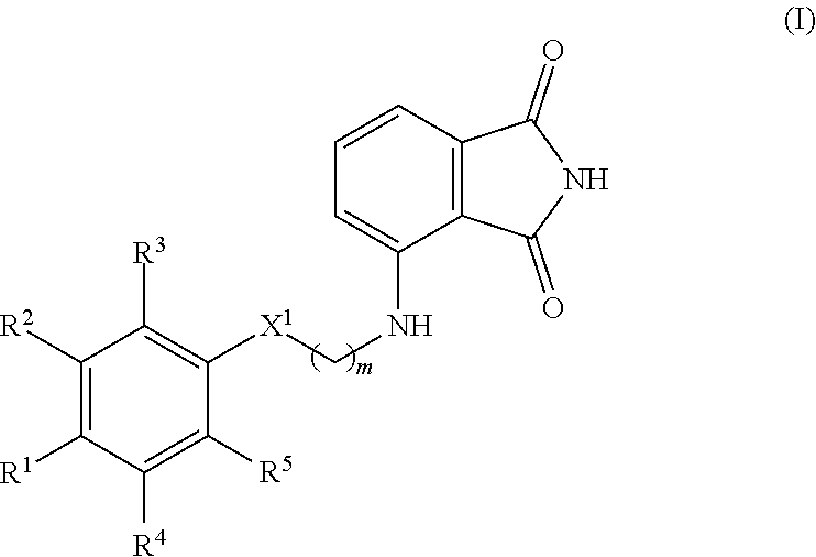
1. A method of inhibiting biofilm formation, treating bacteria within a biofilm, or remediating a biofilm in or on a subject, the method comprising administering to the subject an effective amount of a compound according to Formula I ##STR00034## or a pharmaceutically acceptable salt and/or a solvate thereof, wherein R.sup.1 is C.sub.1-C.sub.6 alkoxy, H, OH, or halo; R.sup.2 and R.sup.3 are each independently C.sub.1-C.sub.6 alkoxy, H, or OH; R.sup.4 and R.sup.5 are each independently H or halo; X.sup.1 is CH.sub.2 or O; and m is 0, 1, 2, 3, 4, or 5; provided that: at least one of R.sup.1, R.sup.2, and R.sup.3 is OH or C.sub.1-C.sub.6 alkoxy; when X.sup.1 is O, m is not 0; and when R.sup.2 is OH, R.sup.1, R.sup.3, R.sup.4, and R.sup.5 are each independently H, and X.sup.1 is CH.sub.2, then m is not 0; and wherein the subject is suffering from or at risk of suffering from a bacterial infection.
2. The method of claim 1, wherein the method comprises administering to the subject a pharmaceutical composition, wherein the pharmaceutical composition comprises a compound of Formula I and a pharmaceutically acceptable carrier.
3. The method of claim 2, wherein the pharmaceutical composition is formulated for topical administration.
4. The method of claim 1, the method further comprising administering an effective amount of fluoroquinolone antibiotic to the subject, administering an effective amount of aminoglycoside antibiotic to the subject, and/or administering an effective amount of polymyxin antibiotic to the subject.
5. The method of claim 1, wherein the bacterial infection comprises a Gram-negative bacterial infection.
6. The method of claim 1, wherein the bacterial infection comprises a Pseudomonas aeruginosa infection, an Acinetobacter baumannii infection, a Klebsiella pneumonia infection, a Yersinia pestis infection, a Shigella dysenteriae infection, an Enterobacter sp. infection, an Acinetobacter sp. infection, a Salmonella typhimurium infection, a Serratia sp. infection, or a combination of any two or more thereof.
7. The method of claim 1, wherein R.sup.1, R.sup.2, and R.sup.3 are each independently H or OH; R.sup.4 and R.sup.5 are each independently H or halo; X.sup.1 is CH.sub.2 or O; and m is 0, 1, 2, 3, 4, or 5; provided that at least one of R.sup.1, R.sup.2, and R.sup.3 is OH.
8. The method of claim 7, wherein X.sup.1 is CH.sub.2.
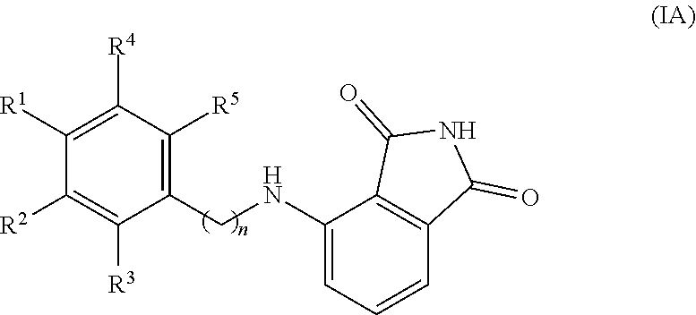
9. The method of claim 1, wherein the compound is of Formula IA ##STR00035## or a pharmaceutically acceptable salt and/or a solvate thereof, wherein n is 1, 2, or 3; provided that R.sup.2 is not OH when n is 1 and R.sup.1, R.sup.3, R.sup.4, and R.sup.5 are each independently H.
10. The method of claim 9, wherein one of R.sup.1 and R.sup.3 is OH, one of R.sup.1 and R.sup.3 is H, and R.sup.2 is H.
11. The method of claim 10, wherein R.sup.4 and R.sup.5 are each independently H, bromine, chlorine, or fluorine.
12. The method of claim 11, wherein R.sup.4 and R.sup.5 are each independently H or chlorine.
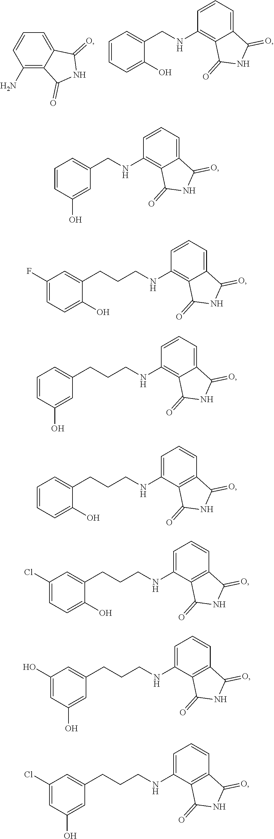
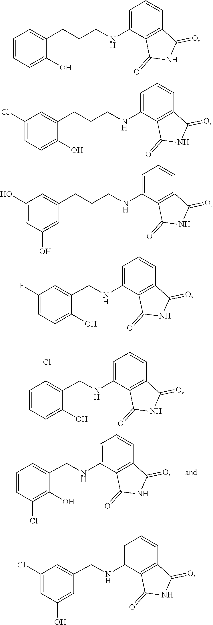
13. The method of claim 1, wherein the compound is selected from the group consisting of: ##STR00036## ##STR00037## or a pharmaceutically acceptable salt and/or solvate thereof.
14. The method of claim 13, wherein the compound is ##STR00038## a pharmaceutically acceptable salt and/or solvate thereof.
15. The method of claim 13, wherein the compound is ##STR00039## or a pharmaceutically acceptable salt and/or solvate thereof.
16. The method of claim 1, wherein the method inhibits biofilm formation in or on the subject.
17. The method of claim 1, wherein the method treats bacteria within a biofilm in or on the subject.
18. The method of claim 1, wherein the method remediates a biofilm in or on the subject.
19. The method of claim 1, wherein the subject is a human or a surface.
20. A compound selected from the group consisting of: ##STR00040## or a pharmaceutically acceptable salt and/or solvate thereof.
Description
CROSS-REFERENCE TO RELATED APPLICATIONS
[0001] The present application claims the benefit of and priority to U.S. Provisional Patent Application 63/092,571 filed on Oct. 16, 2020 and U.S. Provisional Patent Application 63/120,405 filed on Dec. 2, 2020, each of which is incorporated herein by reference in its entirety.
TECHNICAL FIELD
[0003] The present technology relates generally to methods useful for inhibiting biofilm formation and as well as treating bacterial cells embedded in mature films.
SUMMARY
[0004] In an aspect, the present invention includes methods of inhibiting biofilm formation, providing or increasing bacteriocidal activity within a biofilm, treating bacteria within a biofilm, or remediating a biofilm in or on a subject, the method comprising administering to the subject a compound according to Formula I
##STR00002##
or a pharmaceutically acceptable salt and/or a solvate thereof, wherein
[0005] R.sup.1 is C.sub.1-C.sub.6 alkoxy, H, OH, or halo;
[0006] R.sup.2 and R.sup.3 are each independently C.sub.1-C.sub.6 alkoxy, H, or OH;
[0007] R.sup.4 and R.sup.5 are each independently H or halo;
[0008] X.sup.1 is CH.sub.2 or O; and
[0009] m is 0, 1, 2, 3, 4, or 5;
[0010] provided that: [0011] at least one of R.sup.1, R.sup.2, and R.sup.3 is OH or C.sub.1-C.sub.6 alkoxy; [0012] when X.sup.1 is O, m is not 0; and [0013] when R.sup.2 is OH, R.sup.1, R.sup.3, R.sup.4, and R.sup.5 are each independently H, and X.sup.1 is CH.sub.2, then m is not 0; and wherein the subject is suffering from or at risk of suffering from a bacterial infection.
[0014] In an embodiment of the invention, the method includes administering to the subject a pharmaceutical composition. In this embodiment the pharmaceutical composition can include the compound of Formula I and a pharmaceutically acceptable carrier. In this embodiment the pharmaceutical composition can be formulated for topical administration. In this embodiment the subject can be a human or a surface.
[0015] In an embodiment of the invention, the method includes administering an effective amount of fluoroquinolone antibiotic to the subject, administering an effective amount of aminoglycoside antibiotic to the subject, or administering an effective amount of polymyxin antibiotic to the subject.
[0016] In an embodiment of the invention, the bacterial infection can be a Gram-negative bacterial infection. Bacterial infections include, but are not limited to, a Pseudomonas aeruginosa infection, an Acinetobacter baumannii infection, a Klebsiella pneumonia infection, a Yersinia pestis infection, a Shigella dysenteriae infection, an Enterobacter sp. infection, an Acinetobacter sp. infection, a Salmonella typhimurium infection, a Serratia sp. infection, or a combination of any two or more thereof.
[0017] In an embodiment of the invention, R.sup.1, R.sup.2, and R.sup.3 can each independently be H or OH; R.sup.4 and R.sup.5 can each independently be H or halo; X.sup.1 can be CH.sub.2 or O; and m can be 0, 1, 2, 3, 4, or 5; provided that at least one of R.sup.1, R.sup.2, and R.sup.3 is OH. In this embodiment, X.sup.1 can be CH.sub.2.
[0018] In an embodiment of the invention, the compound is of Formula IA
##STR00003##
or a pharmaceutically acceptable salt and/or a solvate thereof, wherein n is 1, 2, or 3; provided that R.sup.2 is not OH when n is 1 and R.sup.1, R.sup.3, R.sup.4, and R.sup.5 are each independently H. In this embodiment, one of R.sup.1 and R.sup.3 can be OH, one of R.sup.1 and R.sup.3 can be H, and R.sup.2 can be H. Also in this embodiment, R.sup.4 and R.sup.5 can each independently be H, bromine, chlorine, or fluorine. Also in this embodiment, R.sup.4 and R.sup.5 can each independently be H or chlorine.
[0019] In an embodiment of the invention, the compound can be selected from
##STR00004## ##STR00005##
or a pharmaceutically acceptable salt and/or solvate thereof.
[0020] In an embodiment of the invention, the compound can be
##STR00006##
or a pharmaceutically acceptable salt and/or solvate thereof.
[0021] In an embodiment of the invention, the compound can be
##STR00007##
or a pharmaceutically acceptable salt and/or solvate thereof.
[0022] In an aspect, the present invention also includes a compound selected from
##STR00008##
or a pharmaceutically acceptable salt and/or solvate thereof.
BRIEF DESCRIPTION OF THE DRAWINGS
[0023] The patent or application file contains at least one drawing executed in color. Copies of this patent or patent application publication with color drawing(s) will be provided by the Office upon request and payment of the necessary fee.
[0024] FIG. 1 illustrates that P. aeruginosa cells embedded in mature biofilms cultured in flow cells are tolerant to ciprofloxacin and tobramycin and susceptible to colistin. EYFP-expressing P. aeruginosa PAO1 biofilms were cultured for three days by flowing AB media supplemented with 15 .mu.M Fe and then treated for 24 h by flowing the same media containing antibiotic. Biofilms were counterstained with Sytox Red and imaged with the aid of CLSM. Top-down views (x-y plane) are depicted with side views (x-z plane) at the bottom. Viable cell mass is in yellow and dead cells and extracellular DNA in red. (A) shows the untreated (DMSO) control. (B) shows treatment at 25.times. the MIC of ciprofloxacin (19 .mu.M). (C) shows treatment at 25.times. the MIC of tobramycin (27 .mu.M). (D) shows treatment at 25.times. the MIC of colistin (20 .mu.M). (E) shows % survival obtained from viable biomass calculated with the aid of COMSTAT software. The scale of the bars represents 20 .mu.m. p<0.01 denoted by ** and p<0.001 by *** relative to untreated.
[0025] FIG. 2 shows that P. aeruginosa cells embedded in mature biofilms grown in flow cells are susceptible to 4-aminoisoindoline-1,3-dione analogs. EYFP-expressing P. aeruginosa PAO1 biofilms were cultured for three days by flowing AB media supplemented with 15 .mu.M Fe and then treated for 24 h with 4-aminoisoindoline-1,3-dione analog. Biofilms were counterstained with Sytox Red and imaged with the aid of CLSM. Top-down views (x-y plane) are depicted with side views (x-z plane) at the bottom. Viable cell mass is in yellow and dead cells and extracellular DNA in red. (A) shows the untreated (DMSO) control. (B) shows treatment at 0.6.times. the IC.sub.50 of KM-5-25 (40 .mu.M). (C) shows treatment at 1.2.times. the IC.sub.50 of KM-5-25 (80 .mu.M). (D) shows treatment at 0.36.times. the IC.sub.50 of KM-5-66 (15 .mu.M). (E) shows treatment at 0.7.times. the IC.sub.50 of KM-5-66 (30 .mu.M). (F) shows treatment at 1.2.times. the IC.sub.50 of KM-5-66 (50 .mu.M). (G) shows the % survival obtained from viable biomass calculated with the aid of COMSTAT software for cells treated with KM-5-25. (H) shows the % survival obtained from viable biomass calculated with the aid of COMSTAT software for cells treated with KM-5-66. The scale of the bars represents 20 .mu.m. p<0.01 denoted by ** and p<0.001 by *** relative to untreated.
[0026] FIG. 3 shows that pellicle biofilms are tolerant to ciprofloxacin and tobramycin, and susceptible to colistin. Pellicles of P. aeruginosa PAO1 expressing EYFP were cultured in PI media supplemented with 20 .mu.M Fe for 48 h, and then treated with antibiotics for 24 h. Pellicles were counterstained with Sytox Red and imaged with the aid of CLSM. Images depict top-down views (squares) and side views (rectangles) where viable cells are shown in yellow and dead cells and extracellular DNA in red. (A) shows the untreated (DMSO) control. (B) shows treatment at 25.times. the MIC of ciprofloxacin (19 .mu.M). (C) shows treatment at 50.times.MIC ciprofloxacin (38 .mu.M). (D) shows treatment at 25.times. the MIC of tobramycin (27 .mu.M). (E) shows treatment at 50.times. the MIC of tobramycin (54 .mu.M). (F) shows treatment at 12.5.times. the MIC of colistin (10 .mu.M). (G) shows treatment at 25.times. the MIC of colistin (20 .mu.M). (H) shows treatment at 50.times. the MIC of colistin (40 .mu.M), (I) shows % survival obtained from viable biomass calculated with the aid of COMSTAT software. The scale of the bars represents 20 .mu.m. p<0.001 denoted by *** relative to untreated.
[0027] FIG. 4 shows that 4-aminoisoindoline-1,3-dione analogs kill P. aeruginosa cells embedded in pellicle biofilms. Pellicles of P. aeruginosa PAO1 cells expressing EYFP were cultured for 48 h in PI media supplemented with 20 .mu.M Fe, and then treated with KM-5-25 or KM-5-66 for 24 h. Pellicles were counterstained with Sytox Red and imaged with the aid of CLSM. Images depict top-down views (squares) and side views (rectangles) where viable cells are shown in yellow and dead cells and extracellular DNA in red. (A) shows the untreated (DMSO) control. (B) shows treatment at 0.6.times. the IC.sub.50 of KM-5-25 (40 .mu.M). (C) shows treatment at 1.2.times. the IC.sub.50 of KM-5-25 (80 .mu.M). (D) shows treatment at 0.36.times. the IC.sub.50 of KM-5-66 (15 .mu.M). (E) shows treatment at 0.7.times. the IC.sub.50 of KM-5-66 (30 .mu.M). (F) shows treatment at 1.2.times. the IC.sub.50 of KM-5-66 (50 .mu.M). (G) shows % survival obtained from viable biomass calculated with the aid of COMSTAT software for pellicles treated with KM-5-25. (H) shows % survival obtained from viable biomass calculated with the aid of COMSTAT software for pellicles treated with KM-5-66. The scale of the bars represents 20 .mu.m. p<0.01 denoted by ** and p<0.001 by *** relative to untreated.
[0028] FIG. 5 shows the assessment of cell survival in pellicle biofilms by dispersing and counting viable cells. EYFP-expressing P. aeruginosa PAO1 cells embedded in two-day old pellicles treated for 24 h with antibiotic or 4-aminoisoindoline-1,3-dione derivatives were dispersed for enumeration of viable culturable cells (CFU/mL). % Survival is expressed as the ratio CFU/mL.sub.(after treatment)/CFU/mL.sub.(pre-treatment). (A) shows pellicles treated for 24 h with tobramycin (27 .mu.M or 54 .mu.M) or colistin (20 .mu.M or 40 .mu.M). (B) shows pellicles treated for 24 h with concentrations equivalent to 25.times. and 50.times. the corresponding MIC with compound KM-5-25 (40 .mu.M and 80 .mu.M), which are concentrations equivalent to 0.6.times. and 1.2.times. the IC.sub.50. (C) shows pellicles treated for 24 h with compound KM-5-66 (15 .mu.M, 30 .mu.M and 50 .mu.M), at concentrations equivalent to 0.36.times., 0.7.times. and 1.2.times. the IC.sub.50. p<0.001 denoted by *** relative to untreated.
[0029] FIG. 6 provides a comparison of the bacteriocidal activity of compounds 11, 16, KM-5-25, and KM-5-66 via assessment of cell survival in pellicle biofilms by dispersing and counting viable cells as performed in the experiments providing FIG. 5, except EYFP-expressing P. aeruginosa PAO1 cells embedded in two-day old pellicles treated for 24 h with 50 .mu.M of one of compound 11, compound 16, compound KM-5-25, or KM-5-66. p<0.001 denoted by *** relative to untreated.
[0030] FIG. 7 shows that 4-aminoisoindoline-1,3-dione analogs are active against two different P. aeruginosa strains. Two-day old pellicle biofilms formed by P. aeruginosa clinical isolates (PA_1081725 and PA_1076058) were challenged for 24 h with 4-aminoisoindoline-1,3-dione derivatives prior to dispersing the cells for enumeration of viable culturable cells (CFU/mL). % survival is expressed as the ratio of CFU/mL.sub.(aftertreatment)/CFU/mL.sub.(pre-treatment). (A) shows pellicles of PA_1081725 treated with concentrations equivalent to 1.2.times. the IC.sub.50 of KM-5-25 (80 .mu.M), or KM-5-66 (50 .mu.M). (B) shows pellicles of PA_1076058 treated with concentrations equivalent to 1.2.times. the IC.sub.50 of KM-5-25 (80 .mu.M), or KM-5-66 (50 .mu.M). (C) shows antibiotic susceptibility of clinical isolates PA_1081725 and PA_1076058. p<0.001 denoted by *** relative to untreated.
[0031] FIG. 8 shows that 4-aminoisoindoline-1,3-dione analogs penetrate the P. aeruginosa cell, bind BfrB and inhibit mobilization of BfrB-stored iron. (A) shows that treating pellicles with 0.6.times. the IC.sub.50 of KM-5-25 (40 .mu.M) or 0.5.times. the IC.sub.50 of KM-5-66 (20 .mu.M) for 24 h reduces the number of viable cells to <50%. (B) shows that the iron stored in BfrB in the viable cells was visualized with the aid of native PAGE gels stained with Ferene S, which stains the iron in the interior cavity of BfrB. Recombinant BfrB (BfrB.sub.rec) was used as a standard for the electrophoretic mobility of BfrB. The lane corresponding to untreated control was loaded with 0.5.times. the volume of the lanes loaded with lysates from treated pellicles to account for the .about.2-fold larger number of viable cells in the untreated pellicles. Lanes loaded with treated pellicle lysates show greater accumulation of iron in BfrB relative to untreated cells. (C) shows that peak areas obtained from densitometry analysis (Image J) of the bands in the native PAGE gel of FIG. 8(B) indicate that there is .about.3-fold more iron stored in BfrB in the treated cells relative to the untreated control. (D) shows that analysis of total intracellular iron levels normalized to CFU/mL indicates .about.2.5-fold higher iron levels in the pellicle-embedded cells treated with the 4-aminoisoindoline-1,3-dione analogs relative to untreated control. (B) shows results from a representative experiment from 3 biological replicates. (A), (C) and (D) show the average of results from 3 biological replicates, p<0.001 is denoted by *** relative to untreated.
[0032] FIG. 9 shows that 4-aminoisoindoline-1,3-dione derivatives enhance the efficacy of colistin and tobramycin against P. aeruginosa biofilms. (A) shows two-day old pellicles of EYFP-expressing P. aeruginosa PAO1 treated for 24 h with colistin alone 25.times. the MIC (20 .mu.M), or 50.times. the MIC (40 .mu.M), KM-5-25 (80 .mu.M) or KM-5-66 (50 .mu.M) alone, equivalent to 1.2.times. the IC.sub.50, or a combination of colistin and KM-5-25 or KM-5-66. (B) shows two-day old pellicles of EYFP-expressing P. aeruginosa PAO1 treated for 24 h with tobramycin alone 25.times. the MIC (27 .mu.M) or 50.times. the MIC (40 .mu.M), KM-5-25 or KM-5-66 alone at a concentration equivalent to 1.2.times. the IC.sub.50, or a combination of tobramycin and KM-5-25 or KM-5-66. The % survival is expressed as the ratio CFU/mL.sub.(aftertreatment)/CFU/mL.sub.(pre-treatment). p<0.001 is denoted by *** in the combination treatment relative to treatment with antibiotic alone.
[0033] FIG. 10 shows quantification of the affinity (K.sub.d) of 4-aminoisoindoline-1,3-dione derivatives. (A) shows quantification of the affinity (K.sub.d) of KM-5-25 evaluated by fluorescence polarization. (B) shows quantification of the affinity (K.sub.d) of KM-5-66 evaluated by fluorescence polarization. Values were obtained in 100 mM potassium phosphate buffer (pH 7.6) containing 1 mM TCEP and 0.5% DMSO. The initial concentrations of KM-5-25 and KM-5-66 were 5 .mu.M. The K.sub.d values are the average and standard deviation from three independent measurements. (C) shows quantification of half maximal inhibitory concentration (IC.sub.50) for KM-5-25 as described in Example 2. (D) shows quantification of half maximal inhibitory concentration (IC.sub.50) for KM-5-66 as described in Example 2. The IC.sub.50 values are the average and the standard derivations from three independent experiments.
[0034] FIG. 11 shows that P. aeruginosa cells treated 4-aminoisoindoline-1,3-dione derivatives overproduce pyoverdine. (A) shows that P. aeruginosa cultures treated with KM-5-25 (70 .mu.M) or KM-5-66 (50 .mu.M) for 27 h have .about.35% of the viable cells in the untreated control. (B) shows fluorescence spectra from cell-free supernatant corresponding to untreated cultures (solid line), treated with KM-5-25 (dashed line), or treated with KM-5-66 (dotted line). (C) shows fluorescence intensity normalized to the number of viable cells (CFU/mL), demonstrating that cells treated with KM-5-25 or KM-5-66 secrete significantly more pyoverdine than cells in the untreated control. Averages and standard deviations for 3 biological replicates are shown, p<0.01 denoted by ** and p<0.001 by *** relative to untreated.
[0035] FIG. 12 shows multiple sequence alignment of representative bacterioferritins. Annotated bacterioferritin sequences from different strains of Acintobacter baumannii (NCBI accession numbers in violet) and Klebsiella pneumoniae (NCBI accession numbers in orange) are placed below the Pseudomonas aeruginosa BfrB sequence (NCBI accession number in black). Residues buried at the P. aeruginosa BfrB-Bfd interface are denoted by (*) and hot spot residues at the BfrB-Bfd interface are denoted by (.DELTA.). Conserved residues across the alignment are in red, conservative substitutions in green and semi-conservative substitutions in blue.
[0036] FIG. 13 shows multiple sequence alignment of representative bacterioferritin-associated ferredoxins (Bfd). Annotated Bfd sequences from different strains of Acinetobacter baumannii (NCBI accession numbers in violet) and Klebsiella pneumoniae (NCBI accession numbers in orange) are placed below the Pseudomonas aeruginosa Bfd sequence (NCBI accession number in black). Conserved cysteine residues coordinating iron in the [2Fe-2S] cluster are highlighted by (.dwnarw.), residues buried at the P. aeruginosa BfrB-Bfd interface are denoted by (*) and hot spot residues at the BfrB-Bfd interface are denoted by (.DELTA.). Conserved residues across the alignment are in red, conservative substitutions in green and semi-conservative substitutions in blue.
DETAILED DESCRIPTION
[0037] The following terms are used throughout as defined below.
[0038] As used herein and in the appended claims, singular articles such as "a" and "an" and "the" and similar referents in the context of describing the elements (especially in the context of the following claims) are to be construed to cover both the singular and the plural, unless otherwise indicated herein or clearly contradicted by context. Recitation of ranges of values herein are merely intended to serve as a shorthand method of referring individually to each separate value falling within the range, unless otherwise indicated herein, and each separate value is incorporated into the specification as if it were individually recited herein. All methods described herein can be performed in any suitable order unless otherwise indicated herein or otherwise clearly contradicted by context. The use of any and all examples, or exemplary language (e.g., "such as") provided herein, is intended merely to better illuminate the embodiments and does not pose a limitation on the scope of the claims unless otherwise stated. No language in the specification should be construed as indicating any non-claimed element as essential.
[0039] As used herein, "about" will be understood by persons of ordinary skill in the art and will vary to some extent depending upon the context in which it is used. If there are uses of the term which are not clear to persons of ordinary skill in the art, given the context in which it is used, "about" will mean up to plus or minus 10% of the particular term--for example, "about 10 wt. %" would mean "9 wt. % to 11 wt. %." It is to be understood that when "about" precedes a term, the term is to be construed as disclosing "about" the term as well as the term without modification by "about"--for example, "about 10 wt. %" discloses "9 wt. % to 11 wt. %" as well as disclosing "10 wt. %."
[0040] The phrase "and/or" as used in the present disclosure will be understood to mean any one of the recited members individually or a combination of any two or more thereof--for example, "A, B, and/or C" would mean "A, B, C, A and B, A and C, or B and C."
[0041] The phrase "treating bacteria in a biofilm" as used in the present disclosure will be understood by persons of ordinary skill in the art to mean reducing the number of bacteria in a biofilm.
[0042] The phrase "remediating a biofilm" as used in the present disclosure will be understood by persons of ordinary skill in the art to mean retarding or eliminating biofilm formation, reversing biofilm formation, or dissipating a biofilm. "Effective amount" refers to the amount of a compound or composition required to produce a desired effect. One example of an effective amount includes amounts or dosages that yield acceptable toxicity and bioavailability levels for therapeutic (pharmaceutical) use including, but not limited to, the treatment of a bacterial infection. As used herein, a "subject" or "patient" is a mammal, such as a cat, dog, rodent or primate. Typically the subject is a human, and, preferably, a human suffering from or suspected of suffering from pain. The term "subject" and "patient" can be used interchangeably. As used herein, "subject" also includes surfaces. Surfaces include, but are not limited to, foods, food packaging, materials, medical devices (e.g., indwelling medical devices), tools, utensils, machines, such as food processing equipment, and devices used in processing foods, such as implements.
[0043] Generally, reference to a certain element such as hydrogen or H is meant to include all isotopes of that element. For example, if an R group is defined to include hydrogen or H, it also includes deuterium and tritium. Compounds comprising radioisotopes such as tritium, C.sup.14, P.sup.32 and S.sup.35 are thus within the scope of the present technology. Procedures for inserting such labels into the compounds of the present technology will be readily apparent to those skilled in the art based on the disclosure herein.
[0044] In general, "substituted" refers to an organic group as defined below (e.g., an alkyl group) in which one or more bonds to a hydrogen atom contained therein are replaced by a bond to non-hydrogen or non-carbon atoms. Substituted groups also include groups in which one or more bonds to a carbon(s) or hydrogen(s) atom are replaced by one or more bonds, including double or triple bonds, to a heteroatom. Thus, a substituted group is substituted with one or more substituents, unless otherwise specified. In some embodiments, a substituted group is substituted with 1, 2, 3, 4, 5, or 6 substituents. Examples of substituent groups include: halogens (i.e., F, Cl, Br, and I); hydroxyls; alkoxy, alkenoxy, aryloxy, aralkyloxy, heterocyclyl, heterocyclylalkyl, heterocyclyloxy, and heterocyclylalkoxy groups; carbonyls (oxo); carboxylates; esters; urethanes; oximes; hydroxylamines; alkoxyamines; aralkoxyamines; thiols; sulfides; sulfoxides; sulfones; sulfonyls; pentafluorosulfanyl (i.e., SF.sub.5), sulfonamides; amines; N-oxides; hydrazines; hydrazides; hydrazones; azides; amides; ureas; amidines; guanidines; enamines; imides; isocyanates; isothiocyanates; cyanates; thiocyanates; imines; nitro groups; and nitriles (i.e., CN).
[0045] Substituted ring groups such as substituted cycloalkyl, aryl, heterocyclyl and heteroaryl groups also include rings and ring systems in which a bond to a hydrogen atom is replaced with a bond to a carbon atom. Therefore, substituted cycloalkyl, aryl, heterocyclyl and heteroaryl groups may also be substituted with substituted or unsubstituted alkyl, alkenyl, and alkynyl groups as defined below.
[0046] Alkyl groups include straight chain and branched chain alkyl groups having from 1 to 12 carbon atoms, and typically from 1 to 10 carbons or, in some embodiments, from 1 to 8, 1 to 6, or 1 to 4 carbon atoms. Alkyl groups may be substituted or unsubstituted. Examples of straight chain alkyl groups include groups such as methyl, ethyl, n-propyl, n-butyl, n-pentyl, n-hexyl, n-heptyl, and n-octyl groups. Examples of branched alkyl groups include, but are not limited to, isopropyl, isobutyl, sec-butyl, tert-butyl, neopentyl, isopentyl, and 2,2-dimethylpropyl groups. Representative substituted alkyl groups may be substituted one or more times with substituents such as those listed above, and include without limitation haloalkyl (e.g., trifluoromethyl), hydroxyalkyl, thioalkyl, aminoalkyl, alkylaminoalkyl, dialkylaminoalkyl, alkoxyalkyl, carboxyalkyl, and the like.
[0047] Cycloalkyl groups include mono-, bi- or tricyclic alkyl groups having from 3 to 12 carbon atoms in the ring(s), or, in some embodiments, 3 to 10, 3 to 8, or 3 to 4, 5, or 6 carbon atoms. Cycloalkyl groups may be substituted or unsubstituted. Exemplary monocyclic cycloalkyl groups include, but not limited to, cyclopropyl, cyclobutyl, cyclopentyl, cyclohexyl, cycloheptyl, and cyclooctyl groups. In some embodiments, the cycloalkyl group has 3 to 8 ring members, whereas in other embodiments the number of ring carbon atoms range from 3 to 5, 3 to 6, or 3 to 7. Bi- and tricyclic ring systems include both bridged cycloalkyl groups and fused rings, such as, but not limited to, bicyclo[2.1.1]hexane, adamantyl, decalinyl, and the like. Substituted cycloalkyl groups may be substituted one or more times with, non-hydrogen and non-carbon groups as defined above. However, substituted cycloalkyl groups also include rings that are substituted with straight or branched chain alkyl groups as defined above. Representative substituted cycloalkyl groups may be monosubstituted or substituted more than once, such as, but not limited to, 2,2-, 2,3-, 2,4- 2,5- or 2,6-disubstituted cyclohexyl groups, which may be substituted with substituents such as those listed above.
[0048] Cycloalkylalkyl groups are alkyl groups as defined above in which a hydrogen or carbon bond of an alkyl group is replaced with a bond to a cycloalkyl group as defined above. Cycloalkylalkyl groups may be substituted or unsubstituted. In some embodiments, cycloalkylalkyl groups have from 4 to 16 carbon atoms, 4 to 12 carbon atoms, and typically 4 to 10 carbon atoms. Substituted cycloalkylalkyl groups may be substituted at the alkyl, the cycloalkyl or both the alkyl and cycloalkyl portions of the group. Representative substituted cycloalkylalkyl groups may be monosubstituted or substituted more than once, such as, but not limited to, mono-, di- or tri-substituted with substituents such as those listed above.
[0049] Alkenyl groups include straight and branched chain alkyl groups as defined above, except that at least one double bond exists between two carbon atoms. Alkenyl groups may be substituted or unsubstituted. Alkenyl groups have from 2 to 12 carbon atoms, and typically from 2 to 10 carbons or, in some embodiments, from 2 to 8, 2 to 6, or 2 to 4 carbon atoms. In some embodiments, the alkenyl group has one, two, or three carbon-carbon double bonds. Examples include, but are not limited to vinyl, allyl, CH.dbd.CH(CH3), CH.dbd.C(CH3)2, C(CH3)=CH2, C(CH3)=CH(CH3), C(CH2CH3)=CH2, among others. Representative substituted alkenyl groups may be mono substituted or substituted more than once, such as, but not limited to, mono-, di- or tri-substituted with substituents such as those listed above.
[0050] Cycloalkenyl groups include cycloalkyl groups as defined above, having at least one double bond between two carbon atoms. Cycloalkenyl groups may be substituted or unsubstituted. In some embodiments the cycloalkenyl group may have one, two or three double bonds but does not include aromatic compounds. Cycloalkenyl groups have from 4 to 14 carbon atoms, or, in some embodiments, 5 to 14 carbon atoms, 5 to 10 carbon atoms, or even 5, 6, 7, or 8 carbon atoms. Examples of cycloalkenyl groups include cyclohexenyl, cyclopentenyl, cyclohexadienyl, cyclobutadienyl, and cyclopentadienyl.
[0051] Cycloalkenylalkyl groups are alkyl groups as defined above in which a hydrogen or carbon bond of the alkyl group is replaced with a bond to a cycloalkenyl group as defined above. Cycloalkenylalkyl groups may be substituted or unsubstituted. Substituted cycloalkenylalkyl groups may be substituted at the alkyl, the cycloalkenyl or both the alkyl and cycloalkenyl portions of the group. Representative substituted cycloalkenylalkyl groups may be substituted one or more times with substituents such as those listed above.
[0052] Alkynyl groups include straight and branched chain alkyl groups as defined above, except that at least one triple bond exists between two carbon atoms. Alkynyl groups may be substituted or unsubstituted. Alkynyl groups have from 2 to 12 carbon atoms, and typically from 2 to 10 carbons or, in some embodiments, from 2 to 8, 2 to 6, or 2 to 4 carbon atoms. In some embodiments, the alkynyl group has one, two, or three carbon-carbon triple bonds. Examples include, but are not limited to --C.dbd.CH, --C.dbd.CCH.sub.3, --CH.sub.2C.dbd.CCH.sub.3, --C.dbd.CCH.sub.2CH(CH.sub.2CH.sub.3).sub.2, among others. Representative substituted alkynyl groups may be mono-substituted or substituted more than once, such as, but not limited to, mono-, di- or tri-substituted with substituents such as those listed above.
[0053] Aryl groups are cyclic aromatic hydrocarbons that do not contain heteroatoms. Aryl groups herein include monocyclic, bicyclic and tricyclic ring systems. Aryl groups may be substituted or unsubstituted. Thus, aryl groups include, but are not limited to, phenyl, azulenyl, heptalenyl, biphenyl, fluorenyl, phenanthrenyl, anthracenyl, indenyl, indanyl, pentalenyl, and naphthyl groups. In some embodiments, aryl groups contain 6-14 carbons, and in others from 6 to 12 or even 6-10 carbon atoms in the ring portions of the groups. In some embodiments, the aryl groups are phenyl or naphthyl. The phrase "aryl groups" includes groups containing fused rings, such as fused aromatic-aliphatic ring systems (e.g., indanyl, tetrahydronaphthyl, and the like). Representative substituted aryl groups may be mono-substituted (e.g., tolyl) or substituted more than once. For example, monosubstituted aryl groups include, but are not limited to, 2-, 3-, 4-, 5-, or 6-substituted phenyl or naphthyl groups, which may be substituted with substituents such as those listed above.
[0054] Aralkyl groups are alkyl groups as defined above in which a hydrogen or carbon bond of an alkyl group is replaced with a bond to an aryl group as defined above. Aralkyl groups may be substituted or unsubstituted. In some embodiments, aralkyl groups contain 7 to 16 carbon atoms, 7 to 14 carbon atoms, or 7 to 10 carbon atoms. Substituted aralkyl groups may be substituted at the alkyl, the aryl or both the alkyl and aryl portions of the group. Representative aralkyl groups include but are not limited to benzyl and phenethyl groups and fused (cycloalkylaryl)alkyl groups such as 4-indanylethyl. Representative substituted aralkyl groups may be substituted one or more times with substituents such as those listed above.
[0055] Heterocyclyl groups include aromatic (also referred to as heteroaryl) and non-aromatic ring compounds containing 3 or more ring atoms, of which one or more is a heteroatom such as, but not limited to, N, O, and S. Heterocyclyl groups may be substituted or unsubstituted. In some embodiments, the heterocyclyl group contains 1, 2, 3 or 4 heteroatoms. In some embodiments, heterocyclyl groups include mono-, bi- and tricyclic rings having 3 to 16 ring members, whereas other such groups have 3 to 6, 3 to 10, 3 to 12, or 3 to 14 ring members. Heterocyclyl groups encompass aromatic, partially unsaturated and saturated ring systems, such as, for example, imidazolyl, imidazolinyl and imidazolidinyl groups. The phrase "heterocyclyl group" includes fused ring species including those comprising fused aromatic and non-aromatic groups, such as, for example, benzotriazolyl, 2,3-dihydrobenzo[1,4]dioxinyl, and benzo[1,3]dioxolyl. The phrase also includes bridged polycyclic ring systems containing a heteroatom such as, but not limited to, quinuclidyl. The phrase includes heterocyclyl groups that have other groups, such as alkyl, oxo or halo groups, bonded to one of the ring members, referred to as "substituted heterocyclyl groups". Heterocyclyl groups include, but are not limited to, aziridinyl, azetidinyl, pyrrolidinyl, imidazolidinyl, pyrazolidinyl, thiazolidinyl, tetrahydrothiophenyl, tetrahydrofuranyl, dioxolyl, furanyl, thiophenyl, pyrrolyl, pyrrolinyl, imidazolyl, imidazolinyl, pyrazolyl, pyrazolinyl, triazolyl, tetrazolyl, oxazolyl, isoxazolyl, thiazolyl, thiazolinyl, isothiazolyl, thiadiazolyl, oxadiazolyl, piperidyl, piperazinyl, morpholinyl, thiomorpholinyl, tetrahydropyranyl, tetrahydrothiopyranyl, oxathiane, dioxyl, dithianyl, pyranyl, pyridyl, pyrimidinyl, pyridazinyl, pyrazinyl, triazinyl, dihydropyridyl, dihydrodithiinyl, dihydrodithionyl, homopiperazinyl, quinuclidyl, indolyl, indolinyl, isoindolyl, azaindolyl (pyrrolopyridyl), indazolyl, indolizinyl, benzotriazolyl, benzimidazolyl, benzofuranyl, benzothiophenyl, benzthiazolyl, benzoxadiazolyl, benzoxazinyl, benzodithiinyl, benzoxathiinyl, benzothiazinyl, benzoxazolyl, benzothiazolyl, benzothiadiazolyl, benzo[1,3]dioxolyl, pyrazolopyridyl, imidazopyridyl (azabenzimidazolyl), triazolopyridyl, isoxazolopyridyl, purinyl, xanthinyl, adeninyl, guaninyl, quinolinyl, isoquinolinyl, quinolizinyl, quinoxalinyl, quinazolinyl, cinnolinyl, phthalazinyl, naphthyridinyl, pteridinyl, thianaphthyl, dihydrobenzothiazinyl, dihydrobenzofuranyl, dihydroindolyl, dihydrobenzodioxinyl, tetrahydroindolyl, tetrahydroindazolyl, tetrahydrobenzimidazolyl, tetrahydrobenzotriazolyl, tetrahydropyrrolopyridyl, tetrahydropyrazolopyridyl, tetrahydroimidazopyridyl, tetrahydrotriazolopyridyl, and tetrahydroquinolinyl groups. Representative substituted heterocyclyl groups may be monosubstituted or substituted more than once, such as, but not limited to, pyridyl or morpholinyl groups, which are 2-, 3-, 4-, 5-, or 6-substituted, or disubstituted with various substituents such as those listed above.
[0056] Heteroaryl groups are aromatic ring compounds containing 5 or more ring atoms, of which, one or more is a heteroatom such as, but not limited to, N, O, and S. Heteroaryl groups may be substituted or unsubstituted. Heteroaryl groups include, but are not limited to, groups such as pyrrolyl, pyrazolyl, triazolyl, tetrazolyl, oxazolyl, isoxazolyl, thiazolyl, pyridinyl, pyridazinyl, pyrimidinyl, pyrazinyl, thiophenyl, benzothiophenyl, furanyl, benzofuranyl, indolyl, azaindolyl (pyrrolopyridinyl), indazolyl, benzimidazolyl, imidazopyridinyl (azabenzimidazolyl), pyrazolopyridinyl, triazolopyridinyl, benzotriazolyl, benzoxazolyl, benzothiazolyl, benzothiadiazolyl, imidazopyridinyl, isoxazolopyridinyl, thianaphthyl, purinyl, xanthinyl, adeninyl, guaninyl, quinolinyl, isoquinolinyl, tetrahydroquinolinyl, quinoxalinyl, and quinazolinyl groups. Heteroaryl groups include fused ring compounds in which all rings are aromatic such as indolyl groups and include fused ring compounds in which only one of the rings is aromatic, such as 2,3-dihydro indolyl groups. Representative substituted heteroaryl groups may be substituted one or more times with various substituents such as those listed above.
[0057] Heterocyclylalkyl groups are alkyl groups as defined above in which a hydrogen or carbon bond of an alkyl group is replaced with a bond to a heterocyclyl group as defined above. Heterocyclylalkyl groups may be substituted or unsubstituted. Substituted heterocyclylalkyl groups may be substituted at the alkyl, the heterocyclyl or both the alkyl and heterocyclyl portions of the group. Representative heterocyclyl alkyl groups include, but are not limited to, morpholin-4-yl-ethyl, furan-2-yl-methyl, imidazol-4-yl-methyl, pyridin-3-yl-methyl, tetrahydrofuran-2-yl-ethyl, and indol-2-yl-propyl. Representative substituted heterocyclylalkyl groups may be substituted one or more times with substituents such as those listed above.
[0058] Heteroaralkyl groups are alkyl groups as defined above in which a hydrogen or carbon bond of an alkyl group is replaced with a bond to a heteroaryl group as defined above. Heteroaralkyl groups may be substituted or unsubstituted. Substituted heteroaralkyl groups may be substituted at the alkyl, the heteroaryl or both the alkyl and heteroaryl portions of the group. Representative substituted heteroaralkyl groups may be substituted one or more times with substituents such as those listed above.
[0059] Groups described herein having two or more points of attachment (i.e., divalent, trivalent, or polyvalent) within the compound of the present technology are designated by use of the suffix, "ene." For example, divalent alkyl groups are alkylene groups, divalent aryl groups are arylene groups, divalent heteroaryl groups are divalent heteroarylene groups, and so forth. Substituted groups having a single point of attachment to the compound of the present technology are not referred to using the "ene" designation. Thus, e.g., chloroethyl is not referred to herein as chloroethylene.
[0060] Alkoxy groups are hydroxyl groups (--OH) in which the bond to the hydrogen atom is replaced by a bond to a carbon atom of a substituted or unsubstituted alkyl group as defined above. Alkoxy groups may be substituted or unsubstituted. Examples of linear alkoxy groups include but are not limited to methoxy, ethoxy, propoxy, butoxy, pentoxy, hexoxy, and the like. Examples of branched alkoxy groups include but are not limited to isopropoxy, sec-butoxy, tert-butoxy, isopentoxy, isohexoxy, and the like. Examples of cycloalkoxy groups include but are not limited to cyclopropyloxy, cyclobutyloxy, cyclopentyloxy, cyclohexyloxy, and the like. Representative substituted alkoxy groups may be substituted one or more times with substituents such as those listed above.
[0061] The terms "alkanoyl" and "alkanoyloxy" as used herein can refer, respectively, to --C(O)-alkyl groups and --O--C(O)-alkyl groups, each containing 2-5 carbon atoms. Similarly, "aryloyl" and "aryloyloxy" refer to --C(O)-aryl groups and --O--C(O)-aryl groups.
[0062] The terms "aryloxy" and "arylalkoxy" refer to, respectively, a substituted or unsubstituted aryl group bonded to an oxygen atom and a substituted or unsubstituted aralkyl group bonded to the oxygen atom at the alkyl. Examples include but are not limited to phenoxy, naphthyloxy, and benzyloxy. Representative substituted aryloxy and arylalkoxy groups may be substituted one or more times with substituents such as those listed above.
[0063] The term "carboxyl" as used herein refers to a --COOH group.
[0064] The term "ester" as used herein refers to --COOR and --C(O)O-G groups. R is independently a substituted or unsubstituted alkyl, cycloalkyl, alkenyl, alkynyl, aryl, aralkyl, heterocyclylalkyl or heterocyclyl group as defined herein. G is a carboxylate protecting group. Carboxylate protecting groups are well known to one of ordinary skill in the art. An extensive list of protecting groups for the carboxylate group functionality may be found in Protective Groups in Organic Synthesis, Greene, T. W.; Wuts, P. G. M., John Wiley & Sons, New York, N.Y., (3rd Edition, 1999) which can be added or removed using the procedures set forth therein and which is hereby incorporated by reference in its entirety and for any and all purposes as if fully set forth herein.
[0065] The term "amide" (or "amido") includes C- and N-amide groups, i.e., --C(O)NR.sub.2, and --NRC(O)R groups, respectively. Each R is independently hydrogen, or a substituted or unsubstituted alkyl, alkenyl, alkynyl, cycloalkyl, aryl, aralkyl, heterocyclylalkyl or heterocyclyl group as defined herein. Amido groups therefore include but are not limited to carbamoyl groups (--C(O)NH.sub.2) and formamide groups (--NHC(O)H). In some embodiments, the amide is --NRC(O)--(C.sub.1-5 alkyl) and the group is termed "carbonylamino," and in others the amide is --NHC(O)-alkyl and the group is termed "alkanoylamino."
[0066] The term "nitrile" or "cyano" as used herein refers to the --CN group.
[0067] Urethane groups include N- and O-urethane groups, i.e., --NRC(O)OR and --OC(O)NR.sub.2 groups, respectively. Each R is independently a substituted or unsubstituted alkyl, alkenyl, alkynyl, cycloalkyl, aryl, aralkyl, heterocyclylalkyl, or heterocyclyl group as defined herein. Any R directly attached to a N atom may also be H.
[0068] The term "amine" (or "amino") as used herein refers to --NR.sub.2 groups, wherein each R is independently hydrogen, or a substituted or unsubstituted alkyl, alkenyl, alkynyl, cycloalkyl, aryl, aralkyl, heterocyclylalkyl or heterocyclyl group as defined herein. In some embodiments, the amine is alkylamino, dialkylamino, arylamino, or alkylarylamino. In other embodiments, the amine is NH.sub.2, methylamino, dimethylamino, ethylamino, diethylamino, propylamino, isopropylamino, phenylamino, or benzylamino.
[0069] The term "sulfonamido" includes S- and N-sulfonamide groups, i.e., --SO.sub.2NR.sub.2 and --NRSO.sub.2R groups, respectively. Each R is independently hydrogen, or a substituted or unsubstituted alkyl, alkenyl, alkynyl, cycloalkyl, aryl, aralkyl, heterocyclylalkyl, or heterocyclyl group as defined herein. Sulfonamido groups therefore include but are not limited to sulfamoyl groups (--SO.sub.2NH.sub.2). In some embodiments herein, the sulfonamido is --NHSO.sub.2-alkyl and is referred to as the "alkylsulfonylamino" group.
[0070] The term "thiol" refers to --SH groups, while "sulfides" include --SR groups, "sulfoxides" include --S(O)R groups, "sulfones" include --SO.sub.2R groups, and "sulfonyls" include --SO.sub.2OR. Each R is independently a substituted or unsubstituted alkyl, cycloalkyl, alkenyl, alkynyl, aryl aralkyl, heterocyclyl or heterocyclylalkyl group as defined herein. In some embodiments the sulfide is an alkylthio group, --S-alkyl.
[0071] The term "urea" refers to --NR--C(O)--NR.sub.2 groups. Each R is independently hydrogen, or a substituted or unsubstituted alkyl, alkenyl, alkynyl, cycloalkyl, aryl, aralkyl, heterocyclyl, or heterocyclylalkyl group as defined herein.
[0072] The term "amidine" refers to --C(NR)NR.sub.2 and --NRC(NR)R, wherein each R is independently hydrogen, or a substituted or unsubstituted alkyl, cycloalkyl, alkenyl, alkynyl, aryl aralkyl, heterocyclyl or heterocyclylalkyl group as defined herein.
[0073] The term "guanidine" refers to --NRC(NR)NR.sub.2, wherein each R is independently hydrogen, or a substituted or unsubstituted alkyl, cycloalkyl, alkenyl, alkynyl, aryl aralkyl, heterocyclyl or heterocyclylalkyl group as defined herein.
[0074] The term "enamine" refers to --C(R).dbd.C(R)N.sub.2 and
--NRC(R).dbd.C(R)R, wherein each R is independently hydrogen, a substituted or unsubstituted alkyl, cycloalkyl, alkenyl, alkynyl, aryl aralkyl, heterocyclyl or heterocyclylalkyl group as defined herein.
[0075] The term "halogen" or "halo" as used herein refers to bromine, chlorine, fluorine, or iodine. In some embodiments, the halogen is fluorine. In other embodiments, the halogen is chlorine or bromine.
[0076] The term "hydroxyl" as used herein can refer to --OH or its ionized form, --O.sup.-. A "hydroxyalkyl" group is a hydroxyl-substituted alkyl group, such as HO--CH.sub.2--.
[0077] The term "imide" refers to --C(O)NRC(O)R, wherein each R is independently hydrogen, or a substituted or unsubstituted alkyl, cycloalkyl, alkenyl, alkynyl, aryl aralkyl, heterocyclyl or heterocyclylalkyl group as defined herein.
[0078] The term "imine" refers to --CR(NR) and --N(CR.sub.2) groups, wherein each R is independently hydrogen or a substituted or unsubstituted alkyl, cycloalkyl, alkenyl, alkynyl, aryl aralkyl, heterocyclyl or heterocyclylalkyl group as defined herein, with the proviso that both R groups are not both simultaneously hydrogen.
[0079] The term "nitro" as used herein refers to an --NO.sub.2 group.
[0080] The term "trifluoromethyl" as used herein refers to --CF.sub.3.
[0081] The term "trifluoromethoxy" as used herein refers to --OCF.sub.3.
[0082] The term "azido" refers to --N.sub.3.
[0083] The term "trialkylammonium" refers to a --N(alkyl).sub.3 group. A trialkylammonium group is positively charged and thus typically has an associated anion, such as halogen anion.
[0084] The term "isocyano" refers to --NC.
[0085] The term "isothiocyano" refers to --NCS.
[0086] The term "pentafluorosulfanyl" refers to --SF.sub.5.
[0087] As will be understood by one skilled in the art, for any and all purposes, particularly in terms of providing a written description, all ranges disclosed herein also encompass any and all possible subranges and combinations of subranges thereof. Any listed range can be easily recognized as sufficiently describing and enabling the same range being broken down into at least equal halves, thirds, quarters, fifths, tenths, etc. As a non-limiting example, each range discussed herein can be readily broken down into a lower third, middle third and upper third, etc. As will also be understood by one skilled in the art all language such as "up to," "at least," "greater than," "less than," and the like include the number recited and refer to ranges which can be subsequently broken down into subranges as discussed above. Finally, as will be understood by one skilled in the art, a range includes each individual member. Thus, for example, a group having 1-3 atoms refers to groups having 1, 2, or 3 atoms. Similarly, a group having 1-5 atoms refers to groups having 1, 2, 3, 4, or 5 atoms, and so forth.
[0088] Pharmaceutically acceptable salts of compounds described herein are within the scope of the present technology and include acid or base addition salts which retain the desired pharmacological activity and is not biologically undesirable (e.g., the salt is not unduly toxic, allergenic, or irritating, and is bioavailable). When the compound of the present technology has a basic group, such as, for example, an amino group, pharmaceutically acceptable salts can be formed with inorganic acids (such as hydrochloric acid, hydroboric acid, nitric acid, sulfuric acid, and phosphoric acid), organic acids (e.g. alginate, formic acid, acetic acid, benzoic acid, gluconic acid, fumaric acid, oxalic acid, tartaric acid, lactic acid, maleic acid, citric acid, succinic acid, malic acid, methanesulfonic acid, benzenesulfonic acid, naphthalene sulfonic acid, and p-toluenesulfonic acid) or acidic amino acids (such as aspartic acid and glutamic acid). When the compound of the present technology has an acidic group, such as for example, a carboxylic acid group, it can form salts with metals, such as alkali and earth alkali metals (e.g. Na.sup.+, Li.sup.+, K.sup.+, Ca.sup.2+, Mg.sup.2+, Zn.sup.2+), ammonia or organic amines (e.g. dicyclohexylamine, trimethylamine, triethylamine, pyridine, picoline, ethanolamine, diethanolamine, triethanolamine) or basic amino acids (e.g. arginine, lysine and ornithine). Such salts can be prepared in situ during isolation and purification of the compounds or by separately reacting the purified compound in its free base or free acid form with a suitable acid or base, respectively, and isolating the salt thus formed.
[0089] Those of skill in the art will appreciate that compounds of the present technology may exhibit the phenomena of tautomerism, conformational isomerism, geometric isomerism and/or stereoisomerism. As the formula drawings within the specification and claims can represent only one of the possible tautomeric, conformational isomeric, stereochemical or geometric isomeric forms, it should be understood that the present technology encompasses any tautomeric, conformational isomeric, stereochemical and/or geometric isomeric forms of the compounds having one or more of the utilities described herein, as well as mixtures of these various different forms.
[0090] "Tautomers" refers to isomeric forms of a compound that are in equilibrium with each other. The presence and concentrations of the isomeric forms will depend on the environment the compound is found in and may be different depending upon, for example, whether the compound is a solid or is in an organic or aqueous solution. For example, in aqueous solution, quinazolinones may exhibit the following isomeric forms, which are referred to as tautomers of each other:
##STR00009##
As another example, guanidines may exhibit the following isomeric forms in protic organic solution, also referred to as tautomers of each other:
##STR00010##
[0091] Because of the limits of representing compounds by structural formulas, it is to be understood that all chemical formulas of the compounds described herein represent all tautomeric forms of compounds and are within the scope of the present technology.
[0092] Stereoisomers of compounds (also known as optical isomers) include all chiral, diastereomeric, and racemic forms of a structure, unless the specific stereochemistry is expressly indicated. Thus, compounds used in the present technology include enriched or resolved optical isomers at any or all asymmetric atoms as are apparent from the depictions. Both racemic and diastereomeric mixtures, as well as the individual optical isomers can be isolated or synthesized so as to be substantially free of their enantiomeric or diastereomeric partners, and these stereoisomers are all within the scope of the present technology.
[0093] The compounds of the present technology may exist as solvates, especially hydrates. Hydrates may form during manufacture of the compounds or compositions comprising the compounds, or hydrates may form over time due to the hygroscopic nature of the compounds. Compounds of the present technology may exist as organic solvates as well, including DMF, ether, and alcohol solvates among others. The identification and preparation of any particular solvate is within the skill of the ordinary artisan of synthetic organic or medicinal chemistry.
The Present Technology
[0094] Antibiotic resistant infections are a worldwide threat to public health. The challenge posed by the emergence of antibiotic resistant strains is compounded by slow to nearly stalled development of new antibiotics and validation of new targets..sup.96-98 Hence, antibiotic resistant infections have the potential to undermine many achievements in modern medicine, such as organ transplantation, major surgery and cancer chemotherapy. The World Health Organization (WHO) published a priority list for research and development of new antibiotics to combat multi-drug resistant bacteria, and assigned critical priority to the Gram-negative carbapenem-resistant Acinetobacter baumanii and Pseudomonas aeruginosa, and third-generation cephalosporin resistant Enterobacteriaceae..sup.1 P. aeruginosa is one of the leading Gram-negative pathogens associated with hospital infections due to their propensity to colonize urinary catheters and endotracheal tubes,.sup.11-12 and accelerate lung function decay that lowers the survival of cystic fibrosis patients..sup.8-9 Multidrug resistant forms of A. baumannii, defined as resistant to three or more antibiotic drugs, account for approximately 63% of A. baumannii infections, and are a primary cause of pneumonia or blood stream infections among critically ill patients. The risk of mortality from both bacteria is high, especially among ventilator-associated pneumonia (VAP) patients and sepsis..sup.113-114 In addition to the presence of multi-drug resistant bacteria, biofilms have been implicated as a cause of antibiotic tolerant infections, even in cases where the bacteria within the biofilm have not developed a particular drug resistance.
[0095] A characteristic of biofilms is their high tolerance to antimicrobial agents. Tolerance is a physiological condition which does not involve mutation and enables bacteria to survive in the presence of antibiotics..sup.39-42 The persistent biofilm phenotype is thought to arise from several factors, including restricted penetration of antibiotic molecules due to interactions with components of the biofilm matrix, slow cell metabolism in the biofilm, differential expression of specific genes, and the presence of persister cells. In addition, biofilms are composed of distinct subpopulations that exhibit different physiological activity; cells in the biofilm interior exhibit low metabolic activity, distinct from the high metabolism of cells near the surface..sup.39, 43-44 The dissimilar metabolic activity is thought to result from a concentration gradient of O.sub.2 and nutrients, which are high at the biofilm surface and low in the deeper layers of the biofilm..sup.44-45 Commercial antibiotics that interfere with cell replication (e.g. ciprofloxacin), or protein translation (e.g. tobramycin), preferentially treat the metabolically active bacteria in the outer biofilm layers, whereas cells in the biofilm interior survive,.sup.43, 46-48 despite the ability of both antibiotics to diffuse into the inner regions of the biofilm..sup.43, 49 In contrast, some antimicrobials that affect membrane structure, such as colistin, a "last-line" therapy to treat multi-drug resistant infections,.sup.50-52 can treat cells in the deeper biofilm layers..sup.48 Therefore, there is an unmet need in the art for therapeutics that treat, inhibit, or remediate bacterial biofilms in order to increase the effectiveness of commercial antibiotics that typically are ineffective against biofilms. In turn, this also speaks to the continuing need for the development of new antibiotics that can treat bacteria that reside both within and outside of a biofilm.
[0096] A. baumannii and P. aeruginosa biofilms have been implicated in diseases such as cystic fibrosis, periodontitis and urinary tract infections, partly because of an ability to colonize indwelling medical devices. The hospital cost per patent-infection ranges between $16,000-65,000, with most expenses occurring in the upper part of this range. Worldwide, the infection rates in developing countries occur at a higher frequency than in European countries and the US, especially infections causing VAP and central venous catheter-related bloodstream infections. Infections due to MDR A. baumannii are also common in combat zones, after natural disasters and in instances of high hospital trauma. The CDC has stated that "This bacteria is a serious concern and requires prompt and sustained action to ensure the problem does not grow.".sup.96 A. baumannii has also been profiled in the mass media, most notably in a recent Frontline documentary entitled "Hunting the Nightmare Bacteria.".sup.115 According to a recent GlobalData report, a recognized leader in providing business information and analytics, "The global marketplace for healthcare-associated infections (HAIs) caused by Gram-negative bacteria across the seven major pharmaceutical markets (7MM) is projected to exceed $3.6 billion in sales by 2026, at a Compound Annual Growth Rate (CAGR) of 10.8% from 2016-2026.".sup.116 Responding to this call requires vibrant research and continued investment in the early stages of drug development, in order to ensure a pipeline of novel ideas and approaches..sup.11 In this context, strategies that interfere with bacterial iron acquisition and homeostasis are regarded as having potential as new therapeutic interventions..sup.55, 76, 99-100 Iron is essential for bacteria because of its involvement in multiple metabolic processes, including respiration and fundamental enzymatic reactions..sup.101 Pathogenic bacteria must obtain iron from the host, but host nutritional immunity maintains extremely low concentrations of free iron, thus denying the essential nutrient to invading pathogens..sup.22, 86, 102-103 In addition, the very low solubility of the ferric ion (Fe.sup.3+) severely limits its bioavailability, and the reactivity of the soluble ferrous iron (Fe.sup.2+) toward hydrogen peroxide and oxygen induces oxidative stress. Consequently, the processes of bacterial iron homeostasis (acquisition, storage and utilization) are highly regulated to ensure sufficiency for metabolic needs while preventing iron-induced toxicity..sup.23-24 Herein, the present technology provides an unprecedented approach to dysregulate iron homeostasis in P. aeruginosa and A. baumannii which utilizes small molecule probes designed to block the interaction between the iron storage protein bacterioferritin B (BfrB) and its cognate partner, the bacterioferritin-associated ferredoxin (Bfd).
[0097] Bacteria store iron reserves in bacterial ferritin (Ftn) and in bacterioferritin (Bfr)..sup.28, 104-105 roughly spherical and hollow structures of Bfr and bacterial Ftn, which are formed from 24 identical subunits, have an outer diameter of .about.120 .ANG., an inner diameter of .about.80 .ANG., and an interior cavity that can store up to .about.3,000 iron ions in the form of a Fe.sup.3+ mineral. Bfrs, which exist only in bacteria, bind 12 heme groups buried under the external protein surface, with the heme propionates protruding into the interior cavity..sup.104-105 Despite sharing a nearly identical subunit fold and quaternary structures, the eukaryotic Ftns and the Bfrs share less than 20% sequence similarity, which results in divergent subunit packing, 24-mer dynamics and function..sup.28, 106-108 Although in P. aeruginosa the ftnA and bfrB genes encode a bacterial ferritin (FtnA) and a bacterioferritin (BfrB), respectively,.sup.29, 31 BfrB functions as the main iron storage protein..sup.23 Importantly, the mobilization of iron stored in BfrB requires specific interactions with Bfd..sup.23, 28, 31 A crystal structure of the BfrB-Bfd complex revealed that up to 12 Bfd molecules can bind at identical sites on the BfrB surface, at the interface of subunit dimers, above a heme molecule..sup.30 Characterization of the complex in solution showed that the 12 Bfd binding sites are equivalent and independent, and that Bfd binds to BfrB with a K.sub.d of approximately 3 .mu.M..sup.110 These investigations also revealed that M1, Y2 and L5 in Bfd form a continuous set of interactions with L68 and E81 in BfrB, which contribute significantly to the stabilization of the BfrB-Bfd complex. In agreement, the K.sub.d values for the association between Bfd and the L68A or E81A mutants of BfrB are approximately 100-fold larger, and the association between Bfd and the BfrB L68A/E81A double mutant is undetectable..sup.110
[0098] Importantly, alignment of the P. aeruginosa BfrB and Bfd sequences against Bfir and Bfd sequences from E. coli 0157, Klebsiella pneumoniae, Yersinia pestis, Shigella dysenteriae, Enterobacter sp., Acinetobacter sp., Salmonella typhimurium and Serratia sp. shows that the key residues at the interface of the BfrB:Bfd complex in P. aeruginosa are conserved in the sequences of Bfr and Bfd proteins in the above-listed Gram-negative pathogens..sup.30, 110 Hence, inhibitors of the BfrB-Bfd complex in P. aeruginosa will inhibit the equivalent complex in these other Gram-negative organisms and be a target for small-molecule inhibition and intervention.
[0099] The repercussions of blocking the BfrB-Bfd interaction on P. aeruginosa iron metabolism have been investigated by deleting the bfd gene. These investigations, which showed an irreversible accumulation of Fe.sup.3+ in BfrB with concomitant iron deprivation in the cytosol, established the BfrB-Bfd interaction as a novel target to rationally induce iron homeostasis dysregulation in bacteria..sup.23 Consequently, it is important to discover small molecule inhibitors of the BfrB-Bfd interaction, which can (in addition to their use for treating bacterial infections) be used as chemical probes to study bacterial iron homeostasis and uncover additional vulnerabilities in the bacterial cell exposed by iron metabolism dysregulation..sup.28, 105
[0100] The present technology provides methods useful for inhibiting biofilm formation and as well as treating bacterial cells embedded in biofilms.
[0101] In an aspect, the present technology provides a method of inhibiting biofilm formation in or on a subject, the method comprising administering to the subject a compound according to Formula I
##STR00011##
or a pharmaceutically acceptable salt and/or a solvate thereof, wherein
[0102] R.sup.1 is C.sub.1-C.sub.6 alkoxy, H, OH, or halo;
[0103] R.sup.2 and R.sup.3 are each independently C.sub.1-C.sub.6 alkoxy, H, or OH;
[0104] R.sup.4 and R.sup.5 are each independently H or halo;
[0105] X.sup.1 is CH.sub.2 or O; and
[0106] m is 0, 1, 2, 3, 4, or 5;
[0107] provided that: [0108] at least one of R.sup.1, R.sup.2, and R.sup.3 is OH or C.sub.1-C.sub.6 alkoxy; [0109] when X.sup.1 is O, m is not 0; and [0110] when R.sup.2 is OH, R.sup.1, R.sup.3, R.sup.4, and R.sup.5 are each independently H, and X.sup.1 is CH.sub.2, then m is not 0; and wherein the subject is suffering from or at risk of suffering from a bacterial infection. Throughout the present disclosure, a compound according to Formula I (or a pharmaceutically acceptable salt and/or a solvate thereof) are also referred to as "a compound of the present technology," "compounds of the present technology," or the like. In any embodiment herein, the method may include administering an effective amount of the compound (wherein the effective amount is effective to inhibit biofilm formation). In any embodiment herein, the method may further include administering one or more of a fluoroquinolone antibiotic (e.g., ciprofloxacin), an aminoglycoside antibiotic (e.g., tobramycin), and a polymyxin antibiotic (e.g., colistin) to the subject, such as administering an effective amount of fluoroquinolone antibiotic to the subject, administering an effective amount of aminoglycoside antibiotic (e.g., tobramycin) to the subject, and/or administering an effective amount of polymyxin antibiotic (e.g., colistin) to the subject. In any embodiment herein, it may be that the bacterial infection comprises a Gram-negative bacterial infection. In any embodiment herein, it may be that the bacterial infection comprises a Pseudomonas aeruginosa infection, an Acinetobacter baumannii infection, a Klebsiella pneumonia infection, a Yersinia pestis infection, a Shigella dysenteriae infection, an Enterobacter sp. infection, an Acinetobacter sp. infection, a Salmonella typhimurium infection, a Serratia sp. infection, or a combination of any two or more thereof. In any embodiment herein, the administration may include oral administration, parenteral administration, nasal administration, or topical administration. In any of these embodiments, the administration may further include subcutaneous injections, intravenous injections, intraperitoneal injections, or intramuscular injections.
[0111] In an aspect, the present technology provides a method of remediating a biofilm in or on a subject, the method comprising administering to the subject a compound according to Formula I or a pharmaceutically acceptable salt and/or a solvate thereof, wherein the subject is suffering from a bacterial infection. In any embodiment herein, the method may include administering an effective amount of the compound (wherein the effective amount is effective to inhibit biofilm formation). In any embodiment herein, the method may further include administering one or more of a fluoroquinolone antibiotic, an aminoglycoside antibiotic (e.g., tobramycin), and a polymyxin antibiotic (e.g., colistin) to the subject, such as administering an effective amount of fluoroquinolone antibiotic to the subject, administering an effective amount of aminoglycoside antibiotic (e.g., tobramycin) to the subject, and/or administering an effective amount of polymyxin antibiotic (e.g., colistin) to the subject. In any embodiment herein, it may be that the bacterial infection comprises a Gram-negative bacterial infection. In any embodiment herein, it may be that the bacterial infection comprises a Pseudomonas aeruginosa infection, an Acinetobacter baumannii infection, a Klebsiella pneumonia infection, a Yersinia pestis infection, a Shigella dysenteriae infection, an Enterobacter sp. infection, an Acinetobacter sp. infection, a Salmonella typhimurium infection, a Serratia sp. infection, or a combination of any two or more thereof. In any embodiment herein, the administration may include oral administration, parenteral administration, nasal administration, or topical administration. In any of these embodiments, the administration may further include subcutaneous injections, intravenous injections, intraperitoneal injections, or intramuscular injections.
[0112] In an aspect, the present technology provides a method of increasing bacteriocidal activity within a biofilm in or on a subject, the method comprising administering to the subject a compound according to Formula I or a pharmaceutically acceptable salt and/or a solvate thereof. In an aspect, the present technology provides a method of inhibiting bacterial growth/proliferation/activity within a biofilm in or on a subject, the method comprising administering to the subject a compound according to Formula I or a pharmaceutically acceptable salt and/or a solvate thereof. In an aspect, the present technology provides a method of increasing bacterial lysis within a biofilm in or on a subject, the method comprising administering to the subject a compound according to Formula I or a pharmaceutically acceptable salt and/or a solvate thereof. In an aspect, the present technology provides a method of treating bacteria within a biofilm in or on a subject, the method comprising administering to the subject a compound according to Formula I or a pharmaceutically acceptable salt and/or a solvate thereof. In any aspect, it may be that the method includes administering an effective amount of the compound. In any embodiment herein, the method may further include administering one or more of a fluoroquinolone antibiotic, an aminoglycoside antibiotic (e.g., tobramycin), and a polymyxin antibiotic (e.g., colistin) to the subject, such as administering an effective amount of fluoroquinolone antibiotic to the subject, administering an effective amount of aminoglycoside antibiotic (e.g., tobramycin) to the subject, and/or administering an effective amount of polymyxin antibiotic (e.g., colistin) to the subject. In any aspect, it may be that the bacteria in the biofilm include Gram-negative bacteria. In any aspect, it may be that the bacteria in the biofilm include Pseudomonas aeruginosa, Acinetobacter baumannii, Klebsiella pneumonia, Yersinia pestis, Shigella dysenteriae, Enterobacter sp., Acinetobacter sp., Salmonella typhimurium, Serratia sp., or a combination of any two or more thereof. In any aspect and any embodiment herein, the administration may include oral administration, parenteral administration, nasal administration, or topical administration. In any of these embodiments, the administration may further include subcutaneous injections, intravenous injections, intraperitoneal injections, or intramuscular injections.
[0113] In any aspect and any embodiment herein (hereafter simply referred to as "in any embodiment herein" or "any embodiment disclosed herein" or the like), it may be at least one of R.sup.1, R.sup.2, and R.sup.3 is OH, and the remaining R.sup.1, R.sup.2, and R.sup.3 are each independently H or OH; R.sup.4 and R.sup.5 are each independently H or halo; X.sup.1 is CH.sub.2 or O; and m is 0, 1, 2, 3, 4, or 5; provided that when X.sup.1 is O, m is not 0; and provided that when R.sup.2 is OH, R.sup.1, R.sup.3, R.sup.4, and R.sup.5 are each independently H, and X.sup.1 is CH.sub.2, m is not 0.
[0114] In any embodiment disclosed herein, it may be that the compound of Formula I is of Formula IA
##STR00012##
or a pharmaceutically acceptable salt and/or a solvate thereof, wherein n is 1, 2, or 3; provided that R.sup.2 is not OH when n is 1 and R.sup.1, R.sup.3, R.sup.4, and R.sup.5 are each independently H. In any embodiment disclosed herein, it may be that one of R.sup.1 and R.sup.3 is OH, one of R.sup.1 and R.sup.3 is H, and R.sup.2 is H. In any embodiment disclosed herein, it may be that R.sup.4 and R.sup.5 are each independently H, bromine, chlorine, or fluorine. In any embodiment disclosed herein, it may be that R.sup.4 and R.sup.5 are each independently H or chlorine.
[0115] In any embodiment herein, it may be a composition is provided that includes any one of the herein-described embodiments of compounds of Formula I and also includes a pharmaceutically acceptable carrier. In any embodiment herein, it may be that a compound of the present technology is part of a pharmaceutical composition, the pharmaceutical composition including an effective amount of the compound of any one of the aspects and embodiments of compounds of Formula I and a pharmaceutically acceptable carrier.
[0116] Thus, the instant present technology provides compostions, pharmaceutical compositions and medicaments comprising any of the compounds disclosed herein (e.g., compounds of Formula I) and a pharmaceutically acceptable carrier or one or more excipients or fillers. The compositions may be used in the methods and treatments described herein. Such compositions and medicaments include a therapeutically effective amount of any compound as described herein, including but not limited to a compound of Formula I. The pharmaceutical composition may be packaged in unit dosage form. The unit dosage form is effective in treating a bacterial infection when administered to a subject in need thereof.
[0117] The pharmaceutical compositions and medicaments may be prepared by mixing one or more compounds of the present technology, pharmaceutically acceptable salts thereof, stereoisomers thereof, tautomers thereof, or solvates thereof, with pharmaceutically acceptable carriers, excipients, binders, diluents or the like. The compounds and compositions described herein may be used to prepare formulations and medicaments that prevent or treat a bacterial infection. Such compositions can be in the form of, for example, granules, powders, tablets, capsules, syrup, suppositories, injections, emulsions, elixirs, suspensions or solutions. The instant compositions can be formulated for various routes of administration, for example, by oral, parenteral, topical, rectal, nasal, vaginal administration, or via implanted reservoir. Parenteral or systemic administration includes, but is not limited to, subcutaneous, intravenous, intraperitoneal, and intramuscular, injections. The following dosage forms are given by way of example and should not be construed as limiting the instant present technology.
[0118] For oral, buccal, and sublingual administration, powders, suspensions, granules, tablets, pills, capsules, gelcaps, and caplets are acceptable as solid dosage forms. These can be prepared, for example, by mixing one or more compounds of the instant present technology, or pharmaceutically acceptable salts or tautomers thereof, with at least one additive such as a starch or other additive. Suitable additives are sucrose, lactose, cellulose sugar, mannitol, maltitol, dextran, starch, agar, alginates, chitins, chitosans, pectins, tragacanth gum, gum arabic, gelatins, collagens, casein, albumin, synthetic or semi-synthetic polymers or glycerides. Optionally, oral dosage forms can contain other ingredients to aid in administration, such as an inactive diluent, or lubricants such as magnesium stearate, or preservatives such as paraben or sorbic acid, or anti-oxidants such as ascorbic acid, tocopherol or cysteine, a disintegrating agent, binders, thickeners, buffers, sweeteners, flavoring agents or perfuming agents. Tablets and pills may be further treated with suitable coating materials known in the art.
[0119] Liquid dosage forms for oral administration may be in the form of pharmaceutically acceptable emulsions, syrups, elixirs, suspensions, and solutions, which may contain an inactive diluent, such as water. Pharmaceutical formulations and medicaments may be prepared as liquid suspensions or solutions using a sterile liquid, such as, but not limited to, an oil, water, an alcohol, and combinations of these. Pharmaceutically suitable surfactants, suspending agents, emulsifying agents, may be added for oral or parenteral administration.
[0120] As noted above, suspensions may include oils. Such oils include, but are not limited to, peanut oil, sesame oil, cottonseed oil, corn oil and olive oil. Suspension preparation may also contain esters of fatty acids such as ethyl oleate, isopropyl myristate, fatty acid glycerides and acetylated fatty acid glycerides. Suspension formulations may include alcohols, such as, but not limited to, ethanol, isopropyl alcohol, hexadecyl alcohol, glycerol and propylene glycol. Ethers, such as but not limited to, poly(ethyleneglycol), petroleum hydrocarbons such as mineral oil and petrolatum; and water may also be used in suspension formulations.
[0121] Injectable dosage forms generally include aqueous suspensions or oil suspensions which may be prepared using a suitable dispersant or wetting agent and a suspending agent. Injectable forms may be in solution phase or in the form of a suspension, which is prepared with a solvent or diluent. Acceptable solvents or vehicles include sterilized water, Ringer's solution, or an isotonic aqueous saline solution. Alternatively, sterile oils may be employed as solvents or suspending agents. Typically, the oil or fatty acid is non-volatile, including natural or synthetic oils, fatty acids, mono-, di- or tri-glycerides.
[0122] For injection, the pharmaceutical formulation and/or medicament may be a powder suitable for reconstitution with an appropriate solution as described above. Examples of these include, but are not limited to, freeze dried, rotary dried or spray dried powders, amorphous powders, granules, precipitates, or particulates. For injection, the formulations may optionally contain stabilizers, pH modifiers, surfactants, bioavailability modifiers and combinations of these.
[0123] Compounds of the present technology may be administered to the lungs by inhalation through the nose or mouth. Suitable pharmaceutical formulations for inhalation include solutions, sprays, dry powders, or aerosols containing any appropriate solvents and optionally other compounds such as, but not limited to, stabilizers, antimicrobial agents, antioxidants, pH modifiers, surfactants, bioavailability modifiers and combinations of these. The carriers and stabilizers vary with the requirements of the particular compound, but typically include nonionic surfactants (Tweens, Pluronics, or polyethylene glycol), innocuous proteins like serum albumin, sorbitan esters, oleic acid, lecithin, amino acids such as glycine, buffers, salts, sugars or sugar alcohols. Aqueous and nonaqueous (e.g., in a fluorocarbon propellant) aerosols are typically used for delivery of compounds of the present technology by inhalation.
[0124] Forms for the topical administration of compounds of the present technology on a surface include powders, sprayspastes, creams, lotions, gels, solutions, and patches. Dosage forms for the topical (including buccal and sublingual) or transdermal administration of compounds of the present technology include powders, sprays, ointments, pastes, creams, lotions, gels, solutions, and patches. The active component may be mixed under sterile conditions with a pharmaceutically-acceptable carrier or excipient, and with any preservatives, or buffers, which may be required. Powders and sprays can be prepared, for example, with excipients such as lactose, talc, silicic acid, aluminum hydroxide, calcium silicates and polyamide powder, or mixtures of these substances. The ointments, pastes, creams and gels may also contain excipients such as animal and vegetable fats, oils, waxes, paraffins, starch, tragacanth, cellulose derivatives, polyethylene glycols, silicones, bentonites, silicic acid, talc and zinc oxide, or mixtures thereof. Absorption enhancers can also be used to increase the flux of the compounds of the present technology across the skin. The rate of such flux can be controlled by either providing a rate controlling membrane (e.g., as part of a transdermal patch) or dispersing the compound in a polymer matrix or gel.
[0125] Besides those representative dosage forms described above, pharmaceutically acceptable excipients and carriers are generally known to those skilled in the art and are thus included in the instant present technology. Such excipients and carriers are described, for example, in "Remingtons Pharmaceutical Sciences" Mack Pub. Co., New Jersey (1991), which is incorporated herein by reference.
[0126] The formulations of the present technology may be designed to be short-acting, fast-releasing, long-acting, and sustained-releasing as described below. Thus, the pharmaceutical formulations may also be formulated for controlled release or for slow release.
[0127] The instant compositions may also comprise, for example, micelles or liposomes, or some other encapsulated form, or may be administered in an extended release form to provide a prolonged storage and/or delivery effect. Therefore, the pharmaceutical formulations and medicaments may be compressed into pellets or cylinders and implanted intramuscularly or subcutaneously as depot injections or as implants such as stents. Such implants may employ known inert materials such as silicones and biodegradable polymers.
[0128] Specific dosages may be adjusted depending on conditions of disease, the age, body weight, general health conditions, sex, and diet of the subject, dose intervals, administration routes, excretion rate, and combinations of drugs. Any of the above dosage forms containing effective amounts are well within the bounds of routine experimentation and therefore, well within the scope of the instant present technology.
[0129] Those skilled in the art are readily able to determine an effective amount by simply administering a compound of the present technology to a patient in increasing amounts until, for example, culture of the bacterial infection indicates a reduction in the number of bacteria and/or the symptoms of the bacterial infection decrease (e.g., as indicated by the patient). The compounds of the present technology can be administered to a patient at dosage levels in the range of about 0.1 to about 1,000 mg per day. For a normal human adult having a body weight of about 70 kg, a dosage in the range of about 0.01 to about 100 mg per kg of body weight per day may be sufficient (e.g., a dosage in the range of about 0.01 to about 10 mg per kg of body weight per day may be sufficient). The specific dosage used, however, can vary or may be adjusted as considered appropriate by those of ordinary skill in the art. For example, the dosage can depend on a number of factors including the requirements of the patient, the severity of the bacterial infection and the pharmacological activity of the compound being used. The determination of optimum dosages for a particular patient is well known to those skilled in the art.
[0130] Various assays and model systems can be readily employed to determine the therapeutic effectiveness of the treatment according to the present technology. Effectiveness of the compositions and methods of the present technology may also be demonstrated by a culture of the bacterial infection indicating a reduction in the number of bacteria subsequent to administering a compound and/or composition of the present technology and/or the symptoms of the bacterial infection decrease (e.g., as indicated by the patient).
[0131] For each of the indicated conditions described herein, test subjects will exhibit a 10%, 20%, 30%, 50% or greater reduction, up to a 75-90%, or 95% or greater, reduction, in one or more symptom(s) caused by, or associated with, the disorder in the subject, compared to placebo-treated or other suitable control subjects.
[0132] As indicated earlier in this disclosure, the compounds of the present technology can also be administered to a patient along with other conventional therapeutic agents that may be useful in the treatment of a bacterial infection, such as a fluoroquinolone antibiotic, an aminoglycoside antibiotic, and/or a polymyxin antibiotic. In any embodiment herein, a compound and/or composition of the present technology may be administered along with an effective amount of a fluoroquinolone antibiotic, an effective amount of a aminoglycoside antibiotic, and/or a polymyxin antibiotic. The administration may include oral administration, parenteral administration, nasal administration, or topical administration. In any of these embodiments, the administration may include subcutaneous injections, intravenous injections, intraperitoneal injections, or intramuscular injections. In any of these embodiments, the administration may include oral administration. The methods of the present technology can also comprise administering, either sequentially or in combination with one or more compounds of the present technology, a conventional therapeutic agent (e.g., a fluoroquinolone antibiotic, a aminoglycoside antibiotic, and/or a polymyxin antibiotic) in an amount that can potentially or synergistically be effective for the treatment of a bacterial infection.
[0133] In an aspect, a compound of the present technology is administered to a patient in an amount or dosage suitable for therapeutic use. Generally, a unit dosage comprising a compound of the present technology will vary depending on patient considerations. Such considerations include, for example, age, protocol, condition, sex, extent of disease, contraindications, concomitant therapies and the like. An exemplary unit dosage based on these considerations can also be adjusted or modified by a physician skilled in the art. For example, a unit dosage for a patient comprising a compound of the present technology can vary from 1.times.10.sup.-4 g/kg to 1 g/kg, preferably, 1.times.10.sup.-3 g/kg to 1.0 g/kg. Dosage of a compound of the present technology can also vary from 0.01 mg/kg to 100 mg/kg or, preferably, from 0.1 mg/kg to 10 mg/kg.
[0134] A compound of the present technology can also be modified, for example, by the covalent attachment of an organic moiety or conjugate to improve pharmacokinetic properties, toxicity or bioavailability (e.g., increased in vivo half-life). The conjugate can be a linear or branched hydrophilic polymeric group, fatty acid group or fatty acid ester group. A polymeric group can comprise a molecular weight that can be adjusted by one of ordinary skill in the art to improve, for example, pharmacokinetic properties, toxicity or bioavailability. Exemplary conjugates can include a polyalkane glycol (e.g., polyethylene glycol (PEG), polypropylene glycol (PPG)), carbohydrate polymer, amino acid polymer or polyvinyl pyrolidone and a fatty acid or fatty acid ester group, each of which can independently comprise from about eight to about seventy carbon atoms. Conjugates for use with a compound of the present technology can also serve as linkers to, for example, any suitable substituents or groups, radiolabels (marker or tags), halogens, proteins, enzymes, polypeptides, other therapeutic agents (for example, a pharmaceutical or drug), nucleosides, dyes, oligonucleotides, lipids, phospholipids and/or liposomes. In one aspect, conjugates can include polyethylene amine (PEI), polyglycine, hybrids of PEI and polyglycine, polyethylene glycol (PEG) or methoxypolyethylene glycol (mPEG). A conjugate can also link a compound of the present technology to, for example, a label (fluorescent or luminescent) or marker (radionuclide, radioisotope and/or isotope) to comprise a probe of the present technology. Conjugates for use with a compound of the present technology can, in one aspect, improve in vivo half-life. Other exemplary conjugates for use with a compound of the present technology as well as applications thereof and related techniques include those generally described by U.S. Pat. No. 5,672,662, which is hereby incorporated by reference herein.
[0135] The examples herein are provided to illustrate advantages of the present technology and to further assist a person of ordinary skill in the art with preparing or using the compounds of the present technology. The examples herein are also presented in order to more fully illustrate the preferred aspects of the present technology. The examples should in no way be construed as limiting the scope of the present technology, as defined by the appended claims. The examples can include or incorporate any of the variations, aspects, or embodiments of the present technology described above. The variations, aspects, or embodiments described above may also further each include or incorporate the variations of any or all other variations, aspects or embodiments of the present technology.
EXAMPLES
Example 1: Compounds
[0136] The following compounds were synthesized consistent with the procedures described in PCT International Publication No. WO 2020/117832, the entire contents of which are incorporated herein by reference.
##STR00013##
[0137] 4-((3-(2-Hydroxyphenyl)amino)isoindoline-1,3-dione (KM-5-28). Yellow solid, mp 132-133.degree. C. .sup.3H NMR (400 MHz, DMSO-d.sub.6): .delta. 10.93 (br s, 1H), 9.30 (s, 1H), 7.51 (t, J=7.6 Hz, 1H), 7.06 (d, J=7.6 Hz, 1H), 7.02-6.94 (complex, 2H), 6.92 (d, J=7.0 Hz, 1H), 6.78 (d, J=1H), Hz, 1H), 6.71 (t, J=7.4 Hz, 1H), 6.57 (br t, J=5.9 Hz, 1H), 3.27 (q, J=6.5 Hz, 2H), 2.60 (t, J=7.3 Hz, 2H), 1.82 (quintet, J=13 Hz, 2H); .sup.13C NMR (101 MHz, DMSO-d.sub.6): .delta. 171.9, 170.5, 155.6, 146.3, 136.3, 134.1, 130.2, 128.0, 127.4, 119.4, 116.9, 115.3, 112.7, 110.2, 41.9, 29.3, 27.3.
##STR00014##
[0138] 4-((3-(5-Chloro-2-hydroxyphenyl)amino)isoindoline-1,3-dione (KM-5-54). Yellow solid, mp 224-225.degree. C. .sup.1H NMR (400 MHz, DMSO-d.sub.6): .delta. 1093 (br s, 1H), 9.64 (s, 1H), 7.52 (dd, J=8.5, 7.2 Hz, 1H), 7.12 (d, J=2.7 Hz, 1H), 7.03 (dd, J=8.5, 2.7 Hz, 1H), 6.96 (d, J=8.5 Hz, 1H), 6.92 (d, J=7.0 Hz, 1H), 6.78 (d, J=8.6 Hz, 1H), 6.58 (br t, J=6.1 Hz, 1H), 3.28 (q, J=6.9 Hz, 2H), 2.59 (t, J=13 Hz, 2H), 1.82 (quintet, J=7.3 Hz, 2H); .sup.13C NMR (101 MHz, DMSO-d.sub.6): .delta. 171.9, 169.8, 154.6, 146.7, 136.3, 134.1, 130.4, 129.7, 127.0, 122.7, 116.9, 116.8, 111.2, 110.2, 41.9, 29.0, 27.1.
##STR00015##
[0139] 4-((3-(3,5-Diydroxyphenyl)amino)isoindoline-1,3-dione (KM-5-57). Yellow solid, mp 208-210.degree. C. .sup.1H NMR (400 MHz, DMSO-d.sub.6): .delta. 10.94 (br s, 1H), 9.05 (s, 2H), 7.52 (dd, J=8.3, 7.3 Hz, 1H), 6.97 (d, J=8.4 Hz, 1H), 6.93 (d, J=7.0 Hz, 1H), 6.51 (br t, J=5.9 Hz, 1H), 6.05 (d, J=2.0 Hz, 2H), 6.02 (t, J=2.0 Hz, 1H), 3.26 (q, J=6.7 Hz, 2H), 2.46 (t, J=7.6 Hz, 2H), 1.80 (quintet, J=7.6 Hz, 2H); .sup.13C NMR (101 MHz, DMSO-d.sub.6): .delta. 171.4, 169.3, 158.3, 146.2, 143.3, 135.9, 133.6, 116.4, 110.8, 109.8, 106.3, 100.2, 41.3, 32.4, 30.0.
##STR00016##
[0140] 4-((5-Fluoro-2-hydroxybenzyl)amino)isoindoline-1,3-dione (JAG-5-7). Prepared using Procedure A as described in WO 2020/117832. Orange solid, mp 216-217.degree. C. .sup.1H NMR (400 MHz, DMSO-d.sub.6): .delta. 10.97 (s, 1H), 9.75 (s, 1H), 7.50 (t, J=7.7 Hz, 1H), 7.07 (br t, J=6.4 Hz, 1H), 7.02 (dd, J=9.5, 3.2 Hz, 1H), 6.98-6.86 (complex, 3H), 6.82 (dd, J=8.8, 4.7 Hz, 1H), 4.43 (d, J=6.3 Hz, 2H); .sup.13C NMR (101 MHz, DMSO-d.sub.6): .delta. 171.8, 169.8, 155.9 (d, J=233.9 Hz), 151.8, 146.4, 136.3, 134.1, 126.9 (d, J=6.6 Hz), 117.1, 116.3 (d, J=8.0 Hz), 115.1 (d, J=23.3 Hz), 114.6 (d, J=22.5 Hz), 111.7, 110.6, 41.1.
##STR00017##
[0141] 4-((6-Chloro-2-hydroxybenzyl)amino)isoindoline-1,3-dione (KM-5-29). Prepared using Procedure A as described in WO 2020/117832. Yellow solid, mp 235-236.degree. C. .sup.1H NMR (400 MHz, DMSO-d.sub.6): .delta. 10.97 (br s, 1H), 10.43 (s, 1H), 7.56 (dd, J=8.4, 7.3 Hz, 1H), 7.29 (d, J=8.4 Hz, 1H), 7.15 (t, J=8.1 Hz, 1H), 6.95 (d, J=7.0 Hz, 1H), 6.92 (dd, J=8.1, 0.9 Hz, 1H), 6.86 (dd, J=8.1, 0.9 Hz, 1H), 6.75 (br t, J=6.2 Hz, 1H), 4.56 (d, J=6.2 Hz, 2H); .sup.13C NMR (101 MHz, DMSO-d.sub.6): .delta. 172.0, 169.7, 157.5, 146.4, 136.4, 134.5, 134.2, 130.2, 122.9, 120.5, 117.1, 114.9, 111.7, 110.8, 38.9.
##STR00018##
[0142] 4-((3-Chloro-2-hydroxybenzyl)amino)isoindoline-1,3-dione (KM-5-30). Prepared using Procedure A as described in WO 2020/117832. Yellow solid, mp 200-202.degree. C. .sup.1H NMR (400 MHz, DMSO-d.sub.6): .delta. 10.97 (br s, 1H), 9.56 (s, 1H), 7.48 (t, J=7.8 Hz, 1H), 7.26 (dd, J=7.9, 1.0 Hz, 1H), 7.19 (d, J=8.6 Hz, 1H), 7.06 (br t, J=6.3 Hz, 1H), 6.93 (d, J=7.7 Hz, 2H), 6.81 (t, J=7.8 Hz, 1H), 4.51 (d, J=6.3 Hz, 2H); .sup.13C NMR (101 MHz, DMSO-d.sub.6): .delta. 169.1, 169.0, 166.8, 156.9, 144.1, 136.1, 132.8, 125.1, 124.2, 121.9, 121.0, 120.5, 120.2, 110.8 (benzylic carbon coincident with a solvent peak).
##STR00019##
[0143] 4-((5-Chloro-3-hydroxybenzyl)amino)isoindoline-1,3-dione (KM-5-50). Prepared using Procedure A as described in WO 2020/117832. Orange solid, mp 223-224.degree. C.; .sup.1H NMR (400 MHz, DMSO-d.sub.6): .delta. 11.0 (s, 1H), 9.87 (s, 1H), 7.47 (dd, J=8.2, 7.5 Hz, 1H), 7.17 (t, J=6.5 Hz, 1H), 6.94 (d, J=7.0 Hz, 1H), 6.86 (d, J=8.3 Hz, 1H), 6.83 (s, 1H), 6.70 (s, 1H), 6.66 (t, J=2.0 Hz, 1H), 4.46 (d, J=6.5 Hz, 2H); .sup.13C NMR (101 MHz, DMSO-d.sub.6): .delta. 171.7, 169.8, 159.1, 146.2, 143.4, 136.3, 134.1, 133.9, 117.7, 117.4, 114.3, 113.0, 111.7, 110.8, 45.3.
Example 2: Materials and Methods for In Vitro and In Vivo Bioassays
Chemicals, Bacterial Strains, and Growth Media
[0144] Chemicals were purchased from Fisher Scientific (Waltham, Mass.) unless otherwise stated. P. aeruginosa PAO1 was obtained from the University of Washington Genome center..sup.80 The PAO1 strain expressing enhanced yellow fluorescent protein (EYFP) was prepared as described in Soldano, A.; Yao, H.; Chandler, J. R.; Rivera, M. (2020) Inhibiting Iron Mobilization from Bacterioferritin in Pseudomonas aeruginosa Impairs Biofilm Formation Irrespective of Environmental Iron Availability. ACS Infect. Dis. 6, 447-458. 10.1021/acsinfecdis.9b00398..sup.26 Clinical isolates of P. aeruginosa were purchased from JMI Laboratories (North Liberty, Iowa, USA). IC.sub.50 determinations were carried out in defined media (50 mM KH.sub.2PO.sub.4 (Sigma Aldrich, St. Louis, Mo.) 7.5 mM (NH.sub.4).sub.2SO.sub.4 (Sigma Aldrich, St. Louis, Mo.), 0.1% (w/v) glucose (Acros Organics, Fair Lawn, N.J., 99+%), 0.5 mMMgSO.sub.4.7H.sub.2O (Sigma Aldrich, St. Louis, Mo., 99+%), 5% v/v non-essential amino acids (Gibco, Thermo Fisher, Waltham, Mass.), 2% v/v essential amino acids (Gibco, Thermo Fisher, Waltham, Mass.), 4 .mu.M (NH.sub.4).sub.2Fe(SO.sub.4).sub.2, and 0.025% (w/v) hypromellose (HPMC, Sigma Aldrich, St. Louis, Mo.), pH 7.0. The media was filter-sterilized by passing through a 0.2 .mu.m cellulose acetate membrane syringe filter (VWR, Radnor, Pa.). Starter cultures of P. aeruginosa PAO1 in 5 mL LB media were grown for 13 hours in 50 mL conical tubes at 37.degree. C. and 220 rpm. For biofilm experiments, a EYFP-expressing P. aeruginosa strain was routinely grown in Pseudomonas Isolation (PI) media (20 g L.sup.-1 peptone, 0.3 g L.sup.-1 MgCl.sub.2.6H O, 10 g L.sup.-1 K.sub.2SO.sub.4, 25 mg L.sup.-1 irgasan, and 20 mL L.sup.-1 glycerol, pH 7.0). Starter cultures were grown from a single colony at 37.degree. C. and shaking at 220 rpm for 14 hours in 5 mL PI media supplemented with 10 .mu.M Fe. Pellicle biofilms were cultured for 48 hours at 30.degree. C. in PI media supplemented with 20 .mu.M Fe. Surface-attached biofilms were cultured in AB minimal media.sup.81 supplemented with trace metals [0.15 .mu.M (NH.sub.4).sub.2MoO.sub.4, 3 .mu.M CuSO.sub.4, 2 .mu.M Co(NO.sub.3).sub.2, 9.4 .mu.M Na.sub.2B.sub.4O.sub.7, and 7.6 .mu.M ZnSO.sub.4], 3 mM glucose and 15 .mu.M Fe. Iron supplementation was carried out by addition of a small volume of filter-sterilized 10 mM (NH.sub.4).sub.2Fe(SO.sub.4).sub.2 (pH .about.2.0) solution. The antibiotics ciprofloxacin, colistin and tobramycin were used at concentrations equivalent to 25.times. and 50.times. the reported MCI:.sup.82 ciprofloxacin MIC=0.25 .mu.g/mL=0.75 .mu.M; tobramycin MIC=0.5 .mu.g/mL=1.07 .mu.M; colistin MIC=1 .mu.g/mL=0.79 .mu.M. Compound stock solutions (100 mM or 10 mM) in DMSO (Sigma Aldrich, St. Louis, Mo.) were prepared weekly and stored at 4.degree. C. Solutions used to treat biofilms or planktonic cells include 0.025% (w/v) HPMC, and 1.5% or 2% DMSO (Sigma Aldrich, St. Louis, Mo.) to prevent aggregation of the analogs in aqueous solution.
Measurement of Dissociation Constant (K.sub.d)
[0145] Dissociation constants for the interaction between BfrB and 4-aminoisoindoline-1,3-dione derivatives (Table 1 below) were measured in vitro with a fluorescence polarization method based on the intrinsic fluorescence of the isoindoline-1,3-dione moiety, as described previously..sup.25
Measurement of Half Maximal Inhibitory Concentration (IC.sub.50)
[0146] IC.sub.50 values (Table 1 below) were determined as reported previously.sup.25 with small modifications. In brief: precultures of P. aeruginosa PAO1 (5 mL) were grown in LB media for 13 hours at 37.degree. C. and 220 rpm in 50 mL conical tubes (VWR International, PA). The cells were centrifuged for 5 min at 4,000 rpm and 4.degree. C., washed two times and then diluted in buffer (100 mM KH.sub.2PO.sub.4 and 15 mM (NH.sub.4).sub.2SO.sub.4 to an optical density at 600 nm (OD.sub.600) of 0.1. A small volume of compound stock solution (10 mM) was transferred to a microcentrifuge tube, initially diluted with DMSO to 20 .mu.L, and then diluted to 1 mL with preculture cell suspension in defined media with OD.sub.600=0.0001, so the final DMSO concentration is 2%. The resultant cell suspension (200 .mu.L) was transferred to a clear-bottom polystyrene 96-well plate (VWR International, PA) covered with a lid and incubated at 35.degree. C. and 205 cpm for 24 h in a Synergy H1 microplate reader (Biotek Instruments Inc., Winooski, Vt.). The cell cultures were serially diluted and then plated on PI Agar (PIA; BD Biosciences, San Jose, Calif.) plates for enumeration of viable cells (CFU/mL). The % growth was calculated from the ratio CFU/mL.sub.(treated)/CFU/mL.sub.(untreatedcontrol). To calculate the IC.sub.50 values, the % growth was plotted as a function of compound concentration, expressed as log[compound] (.mu.M), and fitted to the 4-parameter logistic model describing the sigmoid-shaped response pattern (equation 1),.sup.83 where b is the slope factor, max is the upper asymptote (plateau), and min is the lower asymptote. Values are the average and standard deviation from three independent experiments.
% .times. .times. growth = min + max - min 1 + 10 ( logIC 50 - x ) b ( Eq .times. .times. 1 ) ##EQU00001##
TABLE-US-00001 TABLE 1 Structure, Binding Affinity, and IC.sub.50 of 4-Aminoisoindoline-1,3-dione Derivatives Analog Structure K.sub.d (.mu.M) IC.sub.50 (.mu.M) Procedure 8 ##STR00020## 300 .+-. 50 Not active Example 2 from WO2020117832 11 ##STR00021## 11 .+-. 1 258 .+-. 23 Example 2 from WO2020117832 KM-5-29 ##STR00022## 6 .+-. 0.5 * Example 2 JAG-5-7 ##STR00023## 7 .+-. 2 128 .+-. 26 Example 2 KM-5-25 ##STR00024## 4 .+-. 0.6 69 .+-. 7 Example 2 KM-5-30 ##STR00025## 6 .+-. 1 156 .+-. 30 Example 2 12 (aka BN-XIV- 53) ##STR00026## 21 .+-. 3 Not active at 120 .mu.M Example 2 KM-5-50 ##STR00027## 11 .+-. 2 82 .+-. 16 Example 2 KM-5-28 ##STR00028## 1.4 .+-. 0.2 96 .+-. 1 Example 2 JAG-5-6 (aka JAG-005-006) ##STR00029## 0.43 .+-. 0.07 54 .+-. 9 Example 2 KM-5-54 ##STR00030## 0.22 .+-. 0.04 * Example 2 16 ##STR00031## 1.50 .+-. 0.25 121 .+-. 4 Example 2 from WO2020117832 KM-5-57 ##STR00032## 2.5 .+-. 0.4 Not active at 200 .mu.M Example 2 KM-5-66 ##STR00033## 0.35 .+-. 0.05 42 .+-. 6 Example 2 *Not determined because of low solubility (<30 .mu.M) in PBS buffer.
[0147] Prior to testing the effect that the 4-substituted isoindoline-1,3-dione derivatives might exert on P. aeruginosa cells, the strength of their interaction with BfrB was evaluated in vitro with a fluorescence polarization assay developed based on the intrinsic fluorescence of the isoindoline-1,3-dione moiety. Because initial fluorescence spectroscopic measurements revealed that the heme groups in BfrB interfere with the signal of the fluorescent ligand, apo-BfrB was utilized for these measurements, capitalizing on earlier findings that the Bfd-binding sites in apo-BfrB are nearly identical to those in BfrB, and that the K.sub.d for the interaction between apo-BfrB and Bfd is very similar to that measured for the interaction between BfrB and Bfd. Hence, the K.sub.d values were measured by titrating apo-BfrB into a fixed concentration of the appropriate fluorescent 4-aminoisoindoline-1,3-dione ligand while analyzing fluorescence polarization and intensity near the emission .lamda..sub.max.
Analysis of Secreted Pyoverdine in Planktonic Cultures
[0148] These experiments were conducted in 96-well plates as described above for the determination of IC.sub.50, except that the cells were cultured in M63 media (2 g/L (NH.sub.4).sub.2SO.sub.4, 13.6 g/L KH.sub.2PO.sub.4 (Sigma-Adlrich, St. Louis, Mo.), 2 g/L glucose, 4 g/L citric acid, 5 g/L technical grade casamino acids (BD Biosciences, San Jose, Calif.), 0.24 g/L MgSO.sub.4 (Alfa Aesar, Haverhill, Mass.), and 0.05% (w/v) HPMC, pH 7.0 adjusted with KOH). Cultures of P. aeruginosa PAO1 treated with KM-5-25 (70 .mu.M) or KM-5-66 (50 .mu.M) were grown for 27 h prior to diluting the contents of each well in PBS (pH 7.4) and plating the cells on PIA plates for enumeration of CFU/mL. The 500-fold diluted solution was clarified by centrifugation and the pyoverdine in the cell-free supernatant was analyzed by acquiring fluorescence emission spectra (430-550 nm) with excitation at 400 nm (10 nm slit width) and emission at .lamda..sub.max=460 nm (10 nm slit width) using a Perkin Elmer LS50B spectrophotometer.
Flow Cell Biofilm Assays
[0149] Surface-attached biofilms of P. aeruginosa PAO1 cells expressing EYFP were grown on flow cells with an 800 .mu.m channel depth (.mu.-slide I.sup.0.8 Luer, Ibidi) using an automated perfusion system (Ibidi, Munich, Germany), as described in Soldano, A.; Yao, H.; Chandler, J. R.; Rivera, M. (2020) Inhibiting Iron Mobilization from Bacterioferritin in Pseudomonas aeruginosa Impairs Biofilm Formation Irrespective of Environmental Iron Availability. ACS Infect. Dis. 6, 447-458. 10.1021/acsinfecdis.9b00398..sup.26 Briefly, the flow cell was inoculated with 200 .mu.L of an overnight culture diluted to OD.sub.600=0.5, followed by 1 h incubation at 30.degree. C. to allow bacterial cell attachment. The .rho.-slide was connected to the Ibidi Pump System and the biofilms were cultured for 3 days at 30.degree. C. while flowing AB minimal media containing 15 .mu.M Fe. The experimental shear stress was 0.14-dyn/cm.sup.2 (shear rate=14 s.sup.-1, pressure=7.1 mbar, flow rate=0.4 mL/min) and the switch time was set to 540 s. The biofilms were treated for 24 hours by flowing AB minimal media supplemented with 15 .mu.M Fe, 0.025% HPMC, 1.5% DMSO and commercial antibiotics or 4-aminoisoindoline-1,3-dione derivatives in the concentrations indicated in the corresponding figure captions. During biofilm growth and challenge with antibacterial, the culture medium in the reservoirs was removed every 12 hours and replaced with fresh pre-warmed medium. Prior to imaging with the aid of CLSM the biofilms were stained with 4 mL of 2.5 nM Sytox Red (Invitrogen, Carlsbad, Calif.), a cell impermeable fluorescent nucleic acid dye that stains dead cells and extracellular DNA,.sup.84 for 20 min (switch time=200 sec) and then washed with AB media for 20 min to remove excess fluorescent dye. The biofilms were imaged with the aid of a Leica TCS SP8 confocal microscope (Leica Microsystems, Germany) using a HC PL apo CS2 63.times./1.4 oil objective. For detecting the EYFP fluorescence the laser line was set at 506 nm and the emission range to 520-610 nm. Sytox Red fluorescence was detected with excitation at 631 nm and emission range 637-779 nm. Image stacks were acquired with a z-step size of 0.3 .mu.m at randomly chosen positions. The Leica Application Suite X (LAS-X) software was used for image stack processing..sup.54 Quantitative analysis of biofilm biomass was performed using the COMSTAT computer program.sup.54 and the Otsu method of automatic thresholding..sup.85
Pellicle Biofilm Assays
[0150] Pellicle biofilms of EYFP-expressing P. aeruginosa PAO1 or clinical isolates were grown in PI media supplemented with 20 .mu.M Fe. Starter cultures were diluted to OD.sub.600=0.001 in 4 mL media, placed in 35.times.10 mm petri dishes and incubated statically at 30.degree. C. for 48 hours. The pellicles were transferred onto circular (1.5 cm diameter) glass coverslips by gently allowing the surface of a coverslip to contact a pellicle. The pellicle-adhered coverslip was washed in PBS and then deposited on top of 1.5 mL of AB challenge media contained in a well of a 12-well microplate, with the pellicle exposed to the media. Challenge media consists of AB minimal media supplemented with 15 .mu.M Fe, 0.025% HPMC, 1.5% DMSO and commercial antibiotic or 4-aminoisoindoline-1,3-dione derivative, used in the concentrations specified in the figure captions. The 4-aminoisoindoline-1,3-dione derivatives were prepared as 10 mM stock solutions in DMSO and then diluted in culture media to the appropriate concentrations. The coverslip-adhered pellicles were exposed to challenge media at 30.degree. C. for 24 hours, changing the challenge media every 12 hours by transferring the pellicle-adhered coverslip to a new plate containing pre-warmed challenge media.
[0151] Prior to imaging with the aid of CLSM, pellicles formed by EYFP-expressing PAO1 cells were washed with PBS and then stained by placing the coverslip-adhered pellicles in 1 mL of PBS containing 2.5 nM Sytox Red (20 min). Excess fluorescent dye was washed with PBS, the coverslip was mounted on a glass slide using 5 .mu.L of SlowFade (Invitrogen, Carlsbad, Calif.) and the edges sealed with fingernail polish. CLSM image stacks (z-step size of 0.3 .mu.m) were acquired with the aid of a Leica TCS SP8 microscope, as described above. Quantitative analysis was performed by determination of pellicle biomass using COMSTAT.sup.54 and the Otsu method of automatic thresholding..sup.85
Determination of Viable Cells in Pellicle Biofilms
[0152] Pellicle biofilms were grown as described above. Planktonic and loosely attached cells were washed (3 times) by immersing the coverslip-adhered pellicles (biofilm facing up) into a well of a 12-well plate containing 3 mL of PBS, and incubating (5 min) with gentle rocking. To remove the pellicle from the coverslip, break the extracellular matrix and release cells from the biofilm, the coverslip-adhered pellicle was placed in a 50 mL conical tube containing a 2 mL suspension of zirconia beads (0.1 mm diameter, BioSpec Products), 10 mL PBS, 0.2 .mu.g/mL alginate lyase and 0.2 .mu.g/mL DNAse. The resultant mixture was incubated at room temperature for 3 min, followed by vigorous vortexing for 4 min. After sedimentation of the zirconia beads, a 100 .mu.L aliquot was used for serial dilution and plating on PIA plates for subsequent enumeration of viable cells (CFU/mL).
Imaging of Iron Stored in BfrB and Analysis of Total Intracellular Iron in Biofilm-Embedded Cells
[0153] Pellicle biofilms were grown for 48 hours as described above. The pellicles were transferred onto square (2 cm.times.2 cm) glass coverslips by gently contacting the pellicle with the coverslip. The pellicle-adhered coverslip was washed in PBS and then placed in a 50 mL conical tube containing 2 mL suspension of zirconia beads, 15 mL PBS, 0.2 .mu.g/mL alginate lyase and 0.2 .mu.g/mL DNAse; the resultant mixture was incubated (3 min) at room temperature, and then vortexed vigorously for 4 min. After the zirconia beads had sedimented, a 100 .mu.L aliquot was sampled from the cell suspension for plating and enumeration of viable cells and a 14 mL sample was used to harvest the cells by centrifugation (20 min, 400 rpm). The cell pellet was resuspended in 1 mL of PBS, transferred to a 1.5 mL microcentrifuge tube, centrifuged for 10 min at 12,500 rpm at 4.degree. C. and the cell pellet frozen at -80.degree. C. The frozen cells were subjected to three freeze-thaw cycles and then lysed by addition of 200 .mu.L of lysis buffer (50 mM Tris-HCl buffer (pH 8.0) containing 10% (v/v) glycerol, 20 mg/mL lysozyme, 0.2 mg/mL DNAse, 0.1 M NaCl, 1 mM MgSO.sub.4 and 1% (v/v) Triton-X100) and incubated at ambient temperature (30 min) and at 37.degree. C. (30 min). Imaging of iron in BfrB was carried out as previously reported:.sup.23 lysate suspensions were clarified by centrifugation (10 min at 12,500 rpm), mixed with 10 .mu.L of loading dye (5.9 mL deionized water, 0.5 mL glycerol, 0.4 mL .beta.-mercaptoethanol, 0.4 mL 1% (w/v) bromophenol blue, and 0.5 mL 1 M Tris-HCl (pH 6.8), and loaded onto 1.5 mm-thick native PAGE gels (4% stacking gel, 8% resolving gel). Electrophoresis was carried out at 60 V and 4.degree. C. for 9 hours, and the gels were stained in the dark by immersion (10 min) in a solution containing 0.049 g Ferene S, 250 .mu.L thioglycolic acid, 2.4 mL acetic acid and 100 mL deionized water. Levels of total intracellular iron were determined as reported previously:.sup.23, 86 the cell pellets were treated with 500 .mu.L of freshly prepared digestion reagent (0.6 N HCl, 2.25% (w/v) KMnO.sub.4 in water), thoroughly mixed by vortexing, and then incubated at 65.degree. C. for 3.5 hours. The resultant solutions were cooled to ambient temperature, treated with 100 .mu.L of iron detection reagent (6.5 mM Ferrene S, 15.4 mM neocuproine, 2 M ascorbic acid, and 5 M ammonium acetate), incubated for 30 min at ambient temperature and centrifuged for 5 min at 12,500 rpm. The iron concentration was measured from the absorbance of the Fe.sup.2+-Ferene S complex (.epsilon..sub.593=34.5 mM.sup.-1 cm.sup.-1),.sup.87 normalized by the viable cell counts and reported as Fe atoms per cell.
Statistical Analysis
[0154] Statistical significance between the means and standard deviation of values obtained in experiments comparing results from untreated vs. treated with antibiotic or analog conditions was determined using one-way ANOVA followed by Tukey's multiple post hoc test, with the aid of SigmaPlot (Systat Software, Inc., CA).
Example 3: 4-Aminoisoindoline-1,3-dione Derivatives Elicit a Bacteriostatic Effect in Planktonic P. aeruginosa Cultures
[0155] The relative strength of the association between the new analogs and BfrB was evaluated measuring the dissociation constant K.sub.d (Table 1). The results show that installing a halogen atom in the phenyl ring improves the binding affinity of the derivatives relative to the previously reported analogs 11 and 16. The K.sub.d values of halogen containing compounds with a --(CH.sub.2)-- linker are on average .about.2-fold lower when compared to the K.sub.d exhibited by 11, and the K.sub.d values of halogen-bearing analogs with a --(CH.sub.2).sub.3-- linker are on average .about.5-fold lower than the K.sub.d measured for analog 16. The relative efficacy of the analogs to inhibit P. aeruginosa planktonic growth was evaluated by measuring the half maximal inhibitory concentration (IC.sub.50). Inspection of the data in Table 1 shows that all the halogenated compounds are more active than analogs 11 and 16. The activity of KM-5-29 and KM-5-54 could not be evaluated because the relatively low aqueous solubility of these compounds prevented measurement of their IC.sub.50 values.
[0156] Although the compounds synthesized so far do not include all possible substitution isomers, some insights of the governing structure activity relationships have begun to emerge (Table 1): (i) Among the compounds with a --(CH.sub.2)-- linker, the data reveal that when the hydroxyl group is at position 2 relative to the linker (KM-5-29, JAG-5-7, KM-5-25 and KM-5-30) a bulkier Cl atom at position 5 (KM-5-25) imparts .about.2-fold higher binding affinity for BfrB than a smaller F atom at the same position (JAG-5-7). In line with the nearly 2-fold lower K.sub.d, the IC.sub.50 of KM-5-25 is .about.2-fold lower than that of JAG-5-7. In comparison, a Cl atom at position 3 (KM-5-30) imparts a K.sub.d similar that of KM-5-25, but an IC.sub.50 .about.2.5-fold larger, suggesting that KM-5-30 is less efficient at penetrating or accumulating in P. aeruginosa cells. Installing a Cl atom at position 6 (KM-5-29) renders the compound poorly soluble in aqueous solution. It is also interesting to compare the two analogs with a hydroxyl group at position 3. The presence of a Cl atom at position 5, KM-5-50, lowers the K.sub.d by a factor of 2 relative to BN-XIV-53 and improves the activity vs. planktonic cells significantly, (ii) Examining the compound series with a --(CH.sub.2).sub.3-- linker shows that when the hydroxyl group is at position 3, a Cl atom at position 5 (KM-5-66) decreases the K.sub.d .about.4-fold and the IC.sub.50 .about.3-fold relative to compound 16. In comparison, the presence of a second hydroxyl group at position 5 (KM-5-57) increases the K.sub.d .about.1.7-fold relative to 16 and renders the compound inactive. Given that the K.sub.d measured for KM-5-57 is similar or lower than K.sub.d values measured for other active compounds in Table 1, the immeasurable activity of KM-5-57 suggests that it cannot penetrate or accumulate in P. aeruginosa cells, (in) Comparison of compounds where the hydroxyl group is at position 2 (KM-5-28, JAG-5-6 and KM-5-54) also shows that a halogen at position 5 improves binding affinity. A bulkier Cl atom at position 5 increases the binding affinity of KM-5-54 2-fold relative to the compound with a F at the same position (JAG-5-6). Comparing the IC.sub.50 values corresponding to KM-5-28 and JAG-5-6 reveals that the .about.3-fold lowered caused by installing a F atom at position 5 is accompanied by .about.2-fold decrease in the IC.sub.50. Attempts to determine whether the lower K.sub.d obtained when a Cl atom at position 5 would bring an additional decrease in the IC.sub.50 were stymied by the poor water solubility of KM-5-54.
[0157] The structure-activity relationships (SAR) information available thus far indicates that a halogen in the aryl ring of the 4-aminoisoindoline-1,3-dione derivatives invariably improves binding affinity for BfrB. In compounds with a --(CH.sub.2)-- linker, when the hydroxyl group is at position 2, a Cl atom at position 5 imparts favorable properties, a Cl atom at position 3 renders the compound poorly active vs. P. aeruginosa, despite a relatively favorable K.sub.d, and a Cl atom at position 6 imparts poor aqueous solubility. The preparation of compounds with a --(CH.sub.2).sub.3-- linker is more elaborate and accessibility to suitable starting materials at reasonable prices is also more limited. The information available thus far indicates that although a Cl at position 5 is favorable whether the hydroxyl group is at position 2 or 3, poor aqueous solubility impairs the potential biological activity when the hydroxyl is at position 2 (KM-5-54), despite having the most favorable K.sub.d of all compounds in Table 1. Attempts to increase aqueous solubility by installing a hydroxyl group at position 5 succeeded in increasing solubility but rendered a compound (KM-5-57) with lower affinity for BfrB and inactive against P. aeruginosa cells. As new compounds become available, similar evaluation of binding affinity and activity against bacterial cells will continue to shed light on the structural requirements that simultaneously enhance target affinity in vitro and activity against P. aeruginosa cells. For the purposes of the studies described below, we chose to work with compounds KM-5-25, the most active of the analogs containing a --(CH.sub.2)-- linker, and KM-5-66, the most active of the analogs harboring a --(CH.sub.2).sub.3-- linker.
Example 4: Planktonic P. aeruginosa Cells Treated with 4-Aminoisoindoline-1,3-dione Derivatives Overproduce Pyoverdine
[0158] Previous studies directed at evaluating the repercussions of blocking the BfrB-Bfd complex in P. aeruginosa cells relied on deleting the bfd gene (.DELTA.bfd). These investigations showed that blockade of the BfrB-Bfd complex in planktonic .DELTA.bfd cells causes an irreversible accumulation of iron in BfrB and iron deficiency in the cytosol. The resultant phenotype is hyperproduction of pyoverdine relative to the wild type cells..sup.23 Pyoverdine is a siderophore produced by P. aeruginosa when the cells experience iron limitation..sup.39 A similar pyoverdine overproduction phenotype was observed when wild type P. aeruginosa cells were treated with small molecule inhibitors of the BfrB-Bfd complex (11 and 16)..sup.25 Therefore, to determine that compounds KM-5-25 and KM-5-66 inhibit iron mobilization from BfrB in the P. aeruginosa cytosol, whether cells treated with these compounds express the characteristic pyoverdine hyperproduction phenotype was investigated. To this end, planktonic cells were cultured in the presence of KM-5-25 (70 .mu.M) or KM-5-66 (50 .mu.M) for 27 h in M63 media and the content of the secreted pyoverdine in the cell-free spent media was analyzed by measuring the fluorescence intensity at 460 nm. Normalizing the intensity of pyoverdine fluorescence to CFU/mL shows that as expected, cells treated with KM-5-25 or KM-5-66 secrete .about. 5-fold more pyoverdine than the untreated control (FIGS. 11(A-C)), an overproduction level similar to that observed with the .DELTA.bfd mutant..sup.23 These observations indicate that both analogs bind BfrB in the P. aeruginosa cytosol, block the BfrB-Bfd interaction and inhibit iron mobilization from BfrB, resulting in cytosolic iron limitation that is manifested in a pyoverdine hyperproduction phenotype. The cytosolic iron limitation caused by treating planktonic cultures with KM-5-25 or KM-5-66 exerts a bacteriostatic effect on the cells, as indicated by the IC.sub.50 values in Table 1. In stark contrast, when the same compounds are used to treat P. aeruginosa biofilms, a bactericidal effect is observed. The results from these experiments are further described and discussed in the following examples.
Example 5: 4-Aminoisoindoline-1,3-dione Derivatives Kill P. aeruginosa Cells in Mature Biofilms
[0159] The susceptibility of mature biofilms to treatment with analogs of 4-aminoisoindoline 1,3-dione was tested using two platforms, biofilms cultured at the solid-liquid interface (flow cell biofilms) and biofilms cultured at the air-liquid interface (pellicles). Biofilms of P. aeruginosa cells expressing an enhanced yellow fluorescent protein (EYFP) were cultured in flow cells using AB minimal media supplemented with 15 .mu.M Fe. 3-Day old biofilms were treated for 24 h with commercial antibiotics or with 4-aminoisoindoline-1,3-dione analogs by flowing AB media containing analog or commercial antibiotic, 0.025% hypromellose (HPMC), 1.5% DMSO and 15 .mu.M Fe. In most experiments, the concentration of commercial antibiotics used was 25.times. or 50.times. the MIC, and the concentration of 4-aminoisoindoline-1,3-dione analogs was between 0.36.times. and 1.2.times. the IC.sub.50. The treated biofilms were counterstained with the cell impermeable fluorescent nucleic acid dye Sytox Red and then imaged with the aid of confocal laser scanning microscopy (CLSM). FIG. 1(A) depicts a representative image of the untreated control, illustrating yellow-fluorescent viable cells and red-stained dead cells and extracellular DNA. FIGS. 1(B-C) show representative images depicting 4-day old biofilms tolerant to 24 h treatment with ciprofloxacin or tobramycin, respectively, and FIG. 1(D) shows 4-day old biofilms susceptible to 24 hours treatment with colistin. In agreement to previously reported observations,.sup.48, 53 treatment with ciprofloxacin preferentially treats cells at the biofilm surface, leaving the interior of the biofilm almost unaffected (FIG. 1(B)). In contrast, treatment with colistin preferentially treats bacteria in the biofilm interior, leaving the biofilm surface less affected (FIG. 1(D)). COMSTAT software was used to attempt a quantitative comparison of the biofilm biomass by estimating the biovolume, which is calculated as the overall volume/substratum area (.mu.m.sup.3/.mu.m.sup.2)..sup.54 Comparing the biofilm biomass as the ratio of (untreated biomass)/(treated biomass) expressed as % survival (FIG. 1(E)) shows that .about.20% of biomass remains viable (yellow fluorescent) after treatment with colistin at 25.times. the MIC. In comparison, .about.60% and 70% of biomass remains viable after treatment with 25.times. the MIC ciprofloxacin or tobramycin, respectively.
[0160] With the establishment that the biofilms are susceptible to colistin and significantly tolerant to ciprofloxacin and tobramycin, similarly cultured 3-day old biofilms were treated with KM-5-25 and KM-5-66 for 24 hours. Compounds KM-5-25 and KM-5-66 are soluble in aqueous media to .about.110 .mu.M and .about.80 .mu.M, respectively. To ensure that the compounds remain soluble during the 24 h treatment period, KM-5-25 was used at concentrations 40 .mu.M and 80 .mu.M, equivalent to 0.6.times. and 1.2.times. the IC.sub.50, and KM-5-66 was used at concentrations 15 .mu.M, 30 .mu.M and 50 .mu.M, equivalent to 0.36.times., 0.7.times. and 1.2.times. the IC.sub.50 Table 1 insert back on p 36). Representative CLSM images obtained after treating 3-day old biofilms with each of the analogs for 24 hours show that both compounds treat biofilm cells in a concentration dependent manner (FIGS. 2(A-F)). Treatment with 50 .mu.M KM-5-66 elicits a similar level of treating as treatment with 80 .mu.M KM-5-25. The higher efficacy exhibited by KM-5-66 agrees with its higher binding affinity for BfrB and lower IC.sub.50. Inspection of the images obtained upon treatment with the higher concentrations of KM-5-25 or KM-5-66 clearly shows that the inhibitors of the BfrB-Bfd complex treat the cells in the interior of the biofilm, leaving most of the viable cells located at the biofilm surface (FIGS. 2(C, F-H)). This pattern of treating is reminiscent of previously reported observations showing that treatment of biofilms with Ga.sup.3+ preferentially treats cells in the inner portion of the biofilm..sup.55 The same authors concluded that cells in the biofilm interior are more sensitive to Ga.sup.3+ because this population experiences a more pronounced iron starvation. Therefore, wihout being bound by theory, it is speculated that in biofilms treated with KM-5-25 or KM-5-66 the internal biofilm population is more susceptible to iron limitation caused by the nearly irreversible accumulation of iron in BfrB. It is important to underscore that although the mechanisms whereby iron starvation contribute to cell death in the biofilm interior are not yet understood, the fact remains that perturbation of iron homeostasis, either by systemic replacement of Fe.sup.3+ with Ga.sup.3+, or by selective inhibition of the BfrB-Bfd complex, leads to bacterial cell death.
[0161] To expand the observations made with flow cell biofilms into a second biofilm model, the susceptibility of biofilms grown at the air-liquid interface (pellicles) was also studied..sup.56-57 Pellicle biofilms (henceforth pellicles) are an attractive alternative platform to study biofilms because pellicles are amenable to imaging by CLSM and to harvesting, which can be desirable for additional biofilm analysis..sup.26 To determine the susceptibility of pellicles to antibiotics or inhibitors of the BfrB-Bfd complex, 2-day old pellicles of EYFP-expressing P. aeruginosa cells were cultured in PI media supplemented with 20 .mu.M Fe. The pellicles were transferred onto glass coverslips by allowing the surface of the coverslip to contact a pellicle. The coverslip-adhered pellicles were subsequently exposed to treatment solution (AB media supplemented with 15 .mu.M Fe, 0.025% HPMC, 1.5% DMSO, and antibiotic or analog) for 24 h prior to staining with Sytox Red and imaging with the aid of CLSM. The pellicle biofilms are tolerant to ciprofloxacin and tobramycin at concentrations 25.times. and 50.times. the MIC (FIG. 3(B-E)), as is evident by the yellow fluorescence and near complete absence of red-stained dead cells. In contrast, the pellicle biofilms are susceptible to colistin at concentrations above 10.times. the MIC (FIG. 3(F-H)). Analysis of the images with COMSTAT, which allowed a more quantitative comparison of cell survival upon treatment with each of the antibiotics (FIG. 3(I)), confirms tolerance to ciprofloxacin and tobramycin, but sensitivity to colistin. Note that when the concentration of colistin is 50.times. the MIC the fluorescence signal from viable cells expressing EYFP is undetectable. When pellicle biofilms are challenged with compound KM-5-25 or KM-5-66 bacterial cell death occurs in a concentration dependent manner (FIGS. 14A-14H). These results agree with the idea that the 4-aminoisoindoline-1,3-derivatives penetrate the bacterial cell and bind to their target in the P. aeruginosa cytosol. Inspection of the images and analysis with COMSTAT (FIG. 4(G, H)) shows that compound KM-5-66 is more efficacious than KM-5-25, observations that are consistent with the lower K.sub.d and IC.sub.50 values measured for KM-5-66. It is also important to note that when compounds KM-5-25 and KM-5-66 are used at a concentration of 80 .mu.M and 50 .mu.M, respectively (FIG. 4(G, H)), which correspond approximately to 1.2.times. the IC.sub.50, nearly 85% of the cells in the pellicle are treated. This efficacy is similar that observed with colistin when used at 20 .mu.M, equivalent to 25.times. the MIC (FIG. 3(I)).
[0162] To assess the efficacy of antibiotics and compounds with an approach complementary to imaging with CLSM, dispersing biofilm cells for subsequent enumeration of viable cells (CFU/mL) was resorted to. To this end, pellicle biofilms were cultured for 48 hours in PI media containing 20 .mu.M Fe, and exposed to AB media containing 15 .mu.M Fe and antibiotic or compound for 24 hours. The biofilms were then harvested, and the cells dispersed into sterile PBS by vortexing in the presence of zirconia beads, prior to plating the cell suspensions for subsequent enumeration of CFU/mL. The results from these experiments are summarized in the plots of FIG. 5(A-C) which show the % cell survival of pellicle-embedded cells after challenges with antibiotic or compound, calculated from the fraction CFU/mL.sub.(aftertreatment)/CFU/mL.sub.(pre-treatment). When colistin is used to treat the pellicles at concentrations equivalent to 25.times. and 50.times. the MIC, the treated biofilms exhibit .about.10% and .about.1% cell survival, respectively, relative to the pre-treated biofilm (FIG. 5(A)), corroborating the sensitivity of the pellicles to colistin. In contrast, challenging the pellicles with tobramycin (25.times. and 50.times. the MIC) results in .about.70% survival relative to the pre-treated biofilm, and nearly identical cell survival relative to the untreated (DMSO control) pellicles (FIG. 5(A)). These observations, which are in good agreement with those made with the aid of CLSM (FIG. 3(D-H)) corroborate that the pellicles are tolerant to tobramycin and sensitive to colistin. Interestingly, attempts to enumerate cells after challenging the pellicles with ciprofloxacin (25.times. or 50.times. the MIC) resulted in extremely low CFU/mL, findings which at first glance appear to be in conflict with the tolerance of the biofilms to ciprofloxacin observed in the CLSM images (FIG. 3(B-C)). To reconcile these seemingly discrepant observations, it is important to consider that several stressors, including ciprofloxacin, can induce a dormant state in bacterial cells known as the viable but not culturable (VBNC) state. A characteristic of cells in the VBNC state is their inability to develop into colonies on routine culture media, even though the cells remain viable for long periods of time..sup.58-59 Evidence that bacterial cells can enter the VBNC has been obtained by several distinct methods,.sup.59 one of which is the utilization of bacteria engineered to constitutively express bioluminescent proteins, and using the bioluminescence as a reporter of metabolic activity. Studies conducted with P. aeruginosa showed that following treatment with ciprofloxacin the bioluminescence emitted by P. aeruginosa cells decreased significantly less than the viable cell counts (CFU/mL). The perceived reduction in viable cell counts, which did not correlate with the relatively high metabolic activity reported by the small decrease in bioluminescence, indicated that challenges with ciprofloxacin induce P. aeruginosa cells to enter a VBNC state..sup.60-61 The observations suggest a similar situation. Imaging the pellicles with CLSM following the 24 hours challenge with ciprofloxacin (FIG. 3(B-C)) shows that most of the cells are metabolically active (yellow fluorescent), but dispersion of the cells from the pellicles for enumeration of CFU/mL shows a large reduction in culturable cells relative to the untreated control. These observations strongly suggest that treating the pellicles with ciprofloxacin induces the cells to enter the VBNC state, thus rendering them tolerant to the antibiotic. These findings, which highlight the complexities associated with biofilm embedded cells, also underscore the importance of resorting to more than one platform to study the efficacy of antibiofilm agents.
[0163] Enumeration of CFU/mL was also carried out after challenging pellicles with 4-aminoisoindoline-1,3-dione derivatives. Treating the pellicles with KM-5-25 at concentrations equivalent to 0.6.times. and 1.2.times. the IC.sub.50 results in .about.38% and .about.25% survival relative to cells in the pre-treated biofilm (FIG. 5(B)), while treating with KM-5-66 at concentrations equivalent to 0.3.times., 0.6.times. or 1.2.times. the IC.sub.50 results in approximately 57%, 21% and 15% survival relative to cells in the pellicles prior to treatment (FIG. 5(C)). These observations, which are in good agreement with the efficacy of the compounds evaluated by COMSTAT analysis of the CLSM images, corroborate the bactericidal activity of the compounds against P. aeruginosa biofilms, and provide additional evidence indicating that KM-5-66 used at 50 .mu.M (1.2.times. the IC.sub.50) exhibits nearly the same efficacy as colistin used at 20 .mu.M (25.times. the MIC). The strategy of dispersing and counting viable cells was also used to compare the relative efficacy of analogs 11, 16, KM-5-25 and KM-5-66. To this end, pellicles formed by P. aeruginosa PAO1 cells were treated (24 h) with each of 11, 16, KM-5-25 and KM-5-66 at a concentration of 50 .mu.M. The results (FIG. 6) show that the compound activity (KM-5-66>KM-5-25 >16 >11) track with the K.sub.d and IC.sub.50 values (Table 1).
[0164] The results from experiments aimed at determining the efficacy of the 4-aminoisoindoline-1,3-dione derivatives presented so far have been conducted with the reference strain P. aeruginosa PAO1. To investigate whether the compounds are also active against other strains of P. aeruginosa, pellicles of several clinical isolates from JMI Laboratories were cultured. Isolates PA_1081725 and PA_1076058 were chosen for additional testing because these strains exhibit relatively high MIC values for several antibiotics (FIG. 7(C)) and form robust pellicles under the same culture conditions used to grow pellicle biofilms of P. aeruginosa PAO1. Challenging the pellicles formed by PA_1081725 and PA_1076058 with analogs KM-5-25 or KM-5-66 at concentrations equivalent to 1,2.times. the IC.sub.50 elicits approximately 80% reduction of viable cells (FIG. 7(A-B)), indicating that the activity of the 4-aminoisoindoline-1,3-dione analogs is not unique to biofilms formed by the P. aeruginosa PAO1 strain. To gain a broader understanding of the potential activity spectrum of the BfrB-Bfd inhibitors against P. aeruginosa strains, BLASTp.sup.62 was used to find homologs of BfrB and Bfd sequences in the >4,400 P. aeruginosa genomes in the Pseudomonas Genome Database..sup.63 The results reveal two important facts: (i) The bfrB and bfd genes are adjacent to one another in all the P. aeruginosa strains, (ii) There is an extremely high level of conservation among the bfd and bfrB sequences. These findings evidence that the compounds of the present technology are broadly active against P. aeruginosa.
Example 6: 4-Aminoisoindoline-1,3-dione Derivatives Inhibit Iron Mobilization from BfrB in P. aeruginosa Cells
[0165] To demonstrate that the bactericidal activity is likely a result of the compounds engaging BfrB in the P. aeruginosa cytosol, inhibiting the BfrB-Bfd complex and blocking iron mobilization from the bacterioferritin in the P. aeruginosa cytosol, experiments aimed at visualizing the iron stored in BfrB were carried out. These experiments capitalize on a strategy reported previously demonstrating that the A bfd mutant of P. aeruginosa irreversibly accumulates iron in BfrB.sup.23 and showing that analog 16 inhibits iron mobilization from BfrB in planktonic cells..sup.25 To visualize BfrB-stored iron in biofilm-embedded cells, 2-day old pellicles of P. aeruginosa PAO1 cells were treated for 24 h with analog KM-5-25 (40 .mu.M) or KM-5-66 (20 .mu.M), concentrations predicted to treat approximately 50% of the cells in the biofilm. The treated pellicles were dispersed in sterile PBS and the cell suspension was harvested by centrifugation after a small aliquot had been sampled to enumerate viable cells. To visualize iron stored in BfrB the harvested cells were lysed, the lysate solution supernatant was clarified by centrifugation and then loaded onto native PAGE gels for separation and visualized by subsequent staining with Ferene S, which reacts with iron to develop a blue color. Since the viable cell count dispersed from the pellicle biofilms treated with KM-5-25 or KM-5-66 was 42% and 37% of the cells in the untreated pellicle (FIG. 8(A)), the clarified lysate supernatants from the untreated control were diluted approximately 2-fold prior to loading the native gels. Results obtained with a representative gel are shown in FIG. 8(B), where it can be observed that lanes loaded with lysate solutions from pellicles treated with analogs KM-5-25 or KM-5-66 exhibit significantly higher Ferene S stain intensity than the lane loaded with lysate solution from the untreated pellicle. To enable quantitative comparison, the relative intensities of the Ferene S-stained bands were measured with the aid of Image J. Comparison of the resultant peak areas (FIG. 8(C)) shows that BfrB from the cells treated with KM-5-25 or KM-5-66 has .about.3-fold more iron relative to BfrB from cells in the untreated pellicles. These findings provide strong evidence indicating that compounds KM-5-25 and KM-5-66 bind BfrB in the P. aeruginosa cytosol, inhibit the formation of the BfrB-Bfd complex required to mobilize iron from BfrB, and lead to nearly irreversible iron accumulation in BfrB. Consistent with the nearly irreversible accumulation of iron in BfrB in cells treated with KM-5-25 or KM-5-66, quantification of the total intracellular iron and normalizing the values to viable cell counts demonstrates that P. aeruginosa cells dispersed from pellicles treated with KM-5-25 or KM-5-66 harbor .about.2.5-fold more intracellular iron than cells obtained from untreated biofilms (FIG. 8(D)). Taken together, these observations support the idea that 4-aminoisoindoline-1,3-dione derivatives dysregulate iron homeostasis by inhibiting the BfrB-Bfd complex, causing the accumulation of unusable iron in the bacterial cell.
Example 7: 4-Aminoisoindoline-1,3-dione Derivatives Enhance the Efficacy of Colistin and Tobramycin Against Biofilm-Embedded Cells
[0166] As demonstrated above and in previous reports,.sup.64-65 mature biofilms formed by P. aeruginosa cells are susceptible to colistin and tolerant to tobramycin. Since these biofilms are also susceptible to the 4-aminoisoindoline-1,3-dione inhibitors of the BfrB-Bfd complex, whether these compounds would enhance the efficacy of colistin and tobramycin were investigated. 2-Day old pellicle biofilms of EYFP-expressing P. aeruginosa PAO1 were cultured and treated for 24 h with colistin alone, compound alone (KM-5-25 or KM-5-66), or a combination of colistin and compound. In the combination treatment experiments, the concentration of compound was kept constant (1.2.times. the IC.sub.50), while colistin was used at two different concentrations, equivalent to 25.times. and 50.times. the MIC (FIG. 9(A)). Treatment with each of the compounds or with colistin alone caused a reduction of viable cells similar to that shown in FIG. 5(A). Challenging the pellicles with a combination of colistin and a compound of present technology, however, causes a significant additional reduction in the number of viable cells. As shown in FIG. 9(A), the combination treatment with KM-5-66 results in .about.0.3% survival when colistin is present at 25.times. the MIC, and .about.0.02% survival when colistin is used at 50.times. the MIC, which correspond to approximately 1.7 log and 1.9 log reduction of viable cells relative to treatment with colistin alone. In comparison, the combination treatment with KM-5-25 results in .about.1% survival when colistin is used at 25.times. the MIC and .about.0.5% survival when colistin is present at 50.times. the MIC, which correspond to nearly 1 log and 0.7 log reduction of viable cells, respectively, compared to colistin alone. Furthermore, the question of whether the 4-aminoisoindoline-1,3-dione derivatives of the present technology can also enhance the bactericidal activity of tobramycin was examined. As shown in FIG. 9(B), the combination treatment with KM-5-25 results in .about.0.5% survival when tobramycin is used at 25.times. the MIC and .about.0.003% survival when tobramycin is present at 50.times. the MIC, which correspond to approximately 2.5 log and 4.7 log reduction of viable cells when compared to treatment with tobramycin alone. The combination treatment with KM-5-66 results in 5% survival when tobramycin is present at 25.times. the MIC and 1% survival when tobramycin is used at 50.times. the MIC, which correspond to approximately 1.5 log and 2 log reduction in viable cells relative to treatment with tobramycin alone. It is interesting to note that compound KM-5-66 is more effective at enhancing the efficacy of colistin (FIG. 9(A)), whereas compound KM-5-25 is more effective at enhancing the efficacy of tobramycin (FIG. 9(B)). Additional studies are clearly required to understand the underlying reasons.
[0167] The observations above, which indicate that the iron limitation induced by inhibitors of the BfrB-Bfd complex can increase the efficacy of colistin and tobramycin against biofilms, are in good agreement with previous studies showing that the Fe chelator HBDE is an effective colistin adjunct against P. aeruginosa,.sup.66 and the iron chelators deferoxamine and deferasirox increase the efficacy of tobramycin against P. aeruginosa biofilms..sup.67 When taken together, the observations made in the presence of HBDE, deferoxamine, deferasirox or 4-aminoisoindoline-1,3-dione derivatives, strengthen the idea that inducing intracellular iron limitation is probably a viable strategy to enhance the efficacy of colistin or tobramycin against biofilms. Since colistin is often used as one of the very few therapeutic options available to combat multidrug resistant Gram-negative organisms such as Acinetobacter baumannii, Pseudomonas aeruginosa and Klebsiella pneumoniae,.sup.68-69 it is encouraging that small molecule inhibitors of the BfrB-Bfd complex are capable of increasing the effectiveness of colistin against P. aeruginosa. In this context, it is noteworthy that the bfr and bfd genes in A. baumannii and K. pneumoniae strains are contiguous, as is the case in the >4,000 P. aeruginosa genomes currently available in the Pseudomonas Genome Data Base, because it indicates that the function of the Bfr-Bfd complex in P. aeruginosa is conserved in A. baumannii and K. pneumoniae. Moreover, amino acid sequence alignment with the aid of Clustal Omega.sup.70 reveals very high conservation in the amino acid sequences of Bfr and Bfd proteins from P. aeruginosa, A. baumannii and K. pneumoniae (FIG. 12 and FIG. 13). Importantly, the amino acids identified as hot spot residues in the BfrB-Bfd complex of P. aeruginosa.sup.33 are highly conserved in the Bfr and Bfd sequences of A. baumannii and K. pneumoniae, so that compounds of the present technology, designed to inhibit the BfrB-Bfd interaction in P. aeruginosa, will act similarly in A. baumannii and if. pneumoniae.
Example 8: Compounds of the Present Technology Inhibit Bacterial Biofilm Formation in an In Vivo Murine Model of Wound Infection
[0168] Effects of vehicle, a positive control, or a compound of the present technology on biofilm formation of a luminescent bacteria strain, P. aeruginosa strain PA01, will be tested in an in vivo murine model of wound infection.
[0169] First, female CD1 mice at 8 to 12 weeks of age will be anesthetized via i.p. administration of ketamine and xylazine. A full thickness biopsy wound will be generated using a 5-mm biopsy punch (Integra Lifesciences, Plainsboro Township, N.J.) on depilated and chlorhexidine-scrubbed dorsal surfaces. A silicon ring (Invitrogen, Carlsbad, Calif.) 0.5 mm thick with an outer diameter of 10 mm and a hole with a 5-mm diameter will be placed over the wound and held to the skin with a surgical adhesive. The silicon ring will be covered with a Tegaderm transparent film dressing (3M, Saint Paul, Minn.), and further adhered using 4-0 braided silk interrupted sutures (Ethicon Inc., Somerville, N.J.). Mice will be given 0.05 mg/kg buprenorphine immediately following surgery as well as daily for the next 2 days to alleviate pain from the procedure. Wound beds will be infected by penetrating the Tegaderm with an insulin syringe and injecting 1.times.10.sup.4 CFUs of bioluminescent P. aeruginosa (PA01) suspended in 10 .mu.L of sterile PBS directly onto the wound bed. Four hours after infection, mice will be topically treated with vehicle, a positive control, or a compound of the present technology in a 20 .mu.L volume injecting directly into the wound bed. Treatment will be every 8 h for the first 5 days of infection. Mice will be imaged daily for 2 weeks using the in vivo imaging system (IVIS)-XMRS (PerkinElmer, Waltham, Mass.), and bioluminescence generated from the bacteria will be quantified in values of radiance (photons/sec/centimeter.sup.2/steradian).
[0170] The Tegaderm wound dressings will be removed after 3 days of infection and examined by electron microscopy for bacteria and bacterial biofilms. Briefly, a sample of the Tegaderm will be adhered to a hydroxyapatite disk (5 mm), and fixed in glutaraldehyde (2.5%, Electron Microscopy Sciences, Hatfield, Pa.) overnight at 4.degree. C. Thereafter, the disk will be washed 3.times. in distilled H.sub.2O, and then dehydrated in 5 min sequential washes of EtOH (25, 50, 75, 90, and 100%). Samples will be then critical point dried (AUTOSAMDRIR-814, Tousimis, Rockville, Md., United States), coated in carbon, and imaged using SEM (Hitachi S-4800 FEG CRYO-SEM). Representative images of each sample (n=10) at both 2K.times. and 10K.times. will be taken at an operational voltage of 3 Kv.
[0171] Tegaderm dressings from P. aeruginosa-infected animals will show evidence of individual Pseudomonas-like rods, and complex bacterial-containing three-dimensional (3D) matrices representing organized bacterial biofilms. The group treated with the compounds of the present technology, the Tegaderm dressings will show a similar or lower amount of organized biofilms compared to the positive control group, demonstrating that the compounds of the present technology possess similar or superior biofilm inhibitory activity in comparison to the positive control.
[0172] Accordingly, the compounds of the present technology are useful in inhibiting biofilm formation.
Example 9: Compounds of the Present Technology Remediate Bacterial Biofilms in an In Vivo Murine Model
[0173] Effects of vehicle, a positive control, or a compound of the present technology on biofilm formation of a luminescent bacteria strain, P. aeruginosa strain PA01, will be tested in an in vivo murine model of wound infection.
[0174] First, female CD1 mice at 8 to 12 weeks of age will be anesthetized via i.p. administration of ketamine and xylazine. A full thickness biopsy wound will be generated using a 5-mm biopsy punch (Integra Lifesciences, Plainsboro Township, N.J.) on depilated and chlorhexidine-scrubbed dorsal surfaces. A silicon ring (Invitrogen, Carlsbad, Calif.) 0.5 mm thick with an outer diameter of 10 mm and a hole with a 5-mm diameter will be placed over the wound and held to the skin with a surgical adhesive. The silicon ring will be covered with a Tegaderm transparent film dressing (3M, Saint Paul, Minn.), and further adhered using 4-0 braided silk interrupted sutures (Ethicon Inc., Somerville, N.J.). Mice will be given 0.05 mg/kg buprenorphine immediately following surgery as well as daily for the next 2 days to alleviate pain from the procedure. Wound beds will be infected by penetrating the Tegaderm with an insulin syringe and injecting 1.times.10.sup.4 CFUs of bioluminescent P. aeruginosa (PA01) suspended in 10 .mu.L sterile PBS directly onto the wound bed. At 24 hours or 48 hours after infection (thus allowing for establishment of a biofilm), mice will be topically treated with vehicle, a positive control, or a compound of the present technology in a 20 .mu.L volume injecting directly into the wound bed. Treatment will be every 8 h for 5 days. Mice will be imaged daily for 2 weeks using the in vivo imaging system (IVIS)-XMRS (PerkinElmer, Waltham, Mass.), and bioluminescence generated from the bacteria will be quantified in values of radiance (photons/sec/centimeter.sup.2/steradian).
[0175] The Tegaderm wound dressings will be removed after 3 days of infection and examined by electron microscopy for bacteria and bacterial biofilms. Briefly, a sample of the Tegaderm will be adhered to a hydroxyapatite disk (5 mm), and fixed in glutaraldehyde (2.5%, Electron Microscopy Sciences, Hatfield, Pa.) overnight at 4.degree. C. Thereafter, the disk will be washed 3.times. in distilled H.sub.2O, and then dehydrated in 5 min sequential washes of EtOH (25, 50, 75, 90, and 100%). Samples will be then critical point dried (AUTOSAMDRIR-814, Tousimis, Rockville, Md., United States), coated in carbon, and imaged using SEM (Hitachi S-4800 FEG CRYO-SEM). Representative images of each sample (n=10) at both 2K.times. and 10K.times. will be taken at an operational voltage of 3 Kv.
[0176] Tegaderm dressings from P. aeruginosa-infected animals will show evidence of individual Pseudomonas-like rods, and complex bacterial-containing three-dimensional (3D) matrices representing organized bacterial biofilms. The group treated with the compounds of the present technology, the Tegaderm dressings will show a similar or lower amount of organized biofilms compared to the positive control group, demonstrating that the compounds of the present technology possess similar or superior activity in remediating biofilms in comparison to the positive control.
[0177] Accordingly, the compounds of the present technology are useful in remediating biofilms.
REFERENCES
[0178] 1. Tacconelli, E.; Carrara, E.; Savoldi, A.; Harbarth, S.; Mendelson, M.; Monnet, D. L.; Pulcini, C.; Kahlmeter, G.; Kluytmans, J.; Carmeli, Y.; Ouellette, M.; Outterson, K.; Patel, J.; Cavaleri, M.; Cox, E. M.; Houchens, C. R.; Grayson, M. L.; Hansen, P.; Singh, N.; Theuretzbacher, U.; Magrini, N.; Group, W. H. O. P. P. L. W. (2018) Discovery, research, and development of new antibiotics: the WHO priority list of antibiotic-resistant bacteria and tuberculosis. Lancet Infect. Dis. 18, 318-327. 10.1016/S 1473-3099(17)30753-3 [0179] 2. Davies, D. (2003) Understanding biofilm resistance to antibacterial agents. Nat. Rev. Drug Discov. 2, 114-122. 10.1038/nrd1008 [0180] 3. Lawrence, J. R.; Korber, D. R.; Hoyle, B. D.; Costerton, J. W.; Caldwell, D. E. (1991) Optical sectioning of microbial biofilms. J. Bacteriol. 173, 6558-6567. 10.1128/jb.173.20.6558-6567.1991 [0181] 4. Sutherland, I. (2001) Biofilm exopolysaccharides: a strong and sticky framework. Microbiology 147, 3-9. 10.1099/00221287-147-1-3 [0182] 5. Lam, J.; Chan, R.; Lam, K.; Costerton, J. W. (1980) Production of Mucoid Microcolonies by Pseudomonas aeruginosa Within Infected Lungs in Cystic Fibrosis. Infect. Immun. 28, 546-556. PMCID PMC550970 [0183] 6. Costerton, J. W.; Stewart, P. S.; Greenberg, E. P. (1999) Bacterial Biofilms: A Common Cause of Persistent Infection. Science 284, 1318-1322. 10.1126/science.284.5418.1318 [0184] 7. Parsek, M. R.; Singh, P. K. (2003) Bacterial biofilms: an emerging link to disease pathogenesis. Ann. Rev. Microbiol. 57, 677-701. 10.1146/annurev.micro.57.030502.090720 [0185] 8. Konstan, M. W.; Morgan, W. J.; Butler, S. M.; Pasta, D. J.; Craib, M. L.; Silva, S. J.; Stokes, D. C.; Wohl, M. E.; Wagener, J. S.; Regelmann, W. E.; Johnson, C. A.; MBCHB for the Scientific Advisory Group and the Investigators and Coordinators of the Epidemiologic Study of Cystic Fibrosis (2007) Risk factors for rate of decline in forced expiratory volume in one second in children and adolescents with cystic fibrosis. J. Pediatr. 151, 134-139, 139 el. 10.1016/j.jpeds.2007.03.006 [0186] 9. Crull, M. R.; Ramos, K. J.; Caldwell, E.; Mayer-Hamblett, N.; Aitken, M. L.; Goss, C. H. (2016) Change in Pseudomonas aeruginosa prevalence in cystic fibrosis adults over time. BMC Pulm Med. 16, 176-page. 10.1186/sl2890-016-0333-y [0187] 10. Romling, U.; Balsalobre, C. (2012) Biofilm infections, their resilience to therapy and innovative treatment strategies. J. Intern. Med. 272, 541-561. 10.1111/joim.12004 [0188] 11. Burrows, L. L. (2018) The Therapeutic Pipeline for Pseudomonas aeruginosa Infections. ACS Infect. Dis. 4, 1041-1047. 10.1021/acsinfecdis.8b00112 [0189] 12. Boucher, H. W.; Talbot, G. H.; Bradley, J. S.; Edwards, J. E.; Gilbert, D.; Rice, L. B.; Scheld, M.; Spellberg, B.; Bartlett, J. (2009) Bad Bugs, No Drugs: No ESKAPE! An Update from the Infectious Diseases Society of America. Clin. Infect. Dis. 48, 1-11. 10.1086/595011 [0190] 13. James, G. A.; Swogger, E.; Wolcott, R.; Pulcini, E.; Secor, P.; Sestrich, J.; Costerton, J. W.; Stewart, P. S. (2008) Biofilms in chronic wounds. Wound Repair Regen. 16, 37-44. 10.1111/j.1524-475X.2007.00321.x [0191] 14. Kadam, S.; Shai, S.; Shahane, A.; Kaushik, K. S. (2019) Recent Advances in Non-Conventional Antimicrobial Approaches for Chronic Wound Biofilms: Have We Found the `Chink in the Armor`? Biomedicines 7, page or article number. 10.3390/biomedicines7020035 [0192] 15. Crabbe, A.; Jensen, P. O.; Bjamsholt, T.; Coenye, T. (2019) Antimicrobial Tolerance and Metabolic Adaptations in Microbial Biofilms. Trends Microbiol 27, 850-863. 10.1016/j.tim.2019.05.003 [0193] 16. Ceri, H.; Olson, M. E.; Stremick, C.; Read, R. R.; Morck, D.; Buret, A. (1999) The Calgary Biofilm Device: new technology for rapid determination of antibiotic susceptibilities of bacterial biofilms. J. Clin. Microbiol. 37, 1771-1776. 10.1128/JCM.37.6.1771-1776.1999 [0194] 17. Stewart, P. S.; Costerton, J. W. (2001) Antibiotic resistance of bacteria in biofilms. Lancet 358, 135-138. 10.1016/s0140-6736(01)05321-1 [0195] 18. Anwar, H.; Costerton, J. W. (1990) Enhanced activity of combination of tobramycin and piperacillin for eradication of sessile biofilm cells of Pseudomonas aeruginosa. Antimicrob. Agents Chemother. 34, 1666-1671. 10.1128/aac.34.9.1666 [0196] 19. Chellat, M. F.; Raguz, L.; Riedl, R. (2016) Targeting Antibiotic Resistance. Angew. Chem. Int. Ed. Engl. 55, 6600-6026. 10.1002/anie.201506818 [0197] 20. Lakemeyer, M.; Zhao, W.; Mandl, F. A.; Hammann, P.; Sieber, S. A. (2018) Thinking Outside the Box-Novel Antibacterials To Tackle the Resistance Crisis. Angew. Chem. Int. Ed. Engl. 57, 14440-14475. 10.1002/anie.201804971 [0198] 21. Verderosa, A. D.; Totsika, M.; Fairfull-Smith, K. E. (2019) Bacterial Biofilm Eradication Agents: A Current Review. Front. Chem. 7, 824-xxx. 10.3389/fchem.2019.00824 [0199] 22. Bullen, J. J.; Rogers, H. J.; Spalding, P. B.; Ward, C. G. (2005) Iron and Infection: The Heart of the Matter. FEMS Immunol. Med. Microbiol. 43, 325-330. 10.1016/j.femsim.2004.11.010 [0200] 23. Eshelman, K.; Yao, H.; Punchi Hewage, A. N. D.; Deay, J. J.; Chandler, J. R.; Rivera, M. (2017) Inhibiting the BfrB:Bfd Interaction in Pseudomonas aeruginosa Causes Irreversible Iron Accumulation in Bacterioferritin and Iron Deficiency in the Bacterial Cell. Metallomics 9, 646-659. DOI: 10.1039/C7MT00042A [0201] 24. Keyer, K.; Imlay, J. A. (1996) Superoxide Accelerates DNA-Damage by Elevating Free-Iron Levels. Proc. Natl. Acad. Sci. USA 93, 13635-13649. DOI: doi.org/10.1073/pnas.93.24.13635 [0202] 25. Punchi Hewage, A. N. D.; Yao, H.; Nammalwar, B.; Gnanasekaran, K. K.; Lovell, S.; Bunce, R. A.; Eshelman, K.; Phaniraj, S. M.; Lee, M. M.; Peterson, B. R.; Battaile, K. P.; Reitz, A. B.; Rivera, M. (2019) Small Molecule Inhibitors of the BfrB-Bfd Interaction Decrease Pseudomonas aeruginosa Fitness and Potentiate Fluoroquinolone Activity. J. Am. Chem. Soc. 141, 8171-8184. 10.1021/jacs.9b00394 [0203] 26. Soldano, A.; Yao, H.; Chandler, J. R.; Rivera, M. (2020) Inhibiting Iron Mobilization from Bacterioferritin in Pseudomonas aeruginosa Impairs Biofilm Formation Irrespective of Environmental Iron Availability. ACS Infect. Dis. 6, 447-458. 10.1021/acsinfecdis.9b00398 [0204] 27. Yao, H.; Jepkorir, G.; Lovell, S.; Nama, P. V.; Weeratunga, S. K.; Battaille, K. P.; Rivera, M. (2011) Two Disctinct Ferritin-Like Molecules in P. aeruginosa: The Product of the bfrA Gene is a Bacterial Ferritin (FtnA) not a bacterioferritin (Bfr). Biochemistry 50, 5236-5248. 10.102l/bi2004119 [0205] 28. Rivera, M. (2017) Bacterioferritin: Structure, Dynamics and Protein-Protein Interactions at Play in Iron Storage and Mobilization. Acc. Chem. Res. 50, 331-340. 10.1021/acs.accounts.6b00514 [0206] 29. Weeratunga, S.; Lovell, S.; Yao, H.; Battaile, K. P.; Fischer, C. J.; Gee, C. E.; Rivera, M. (2010) Structural Studies of Bacterioferritin B (BfrB) from Pseudomonas aeruginosa Suggest a Gating Mechanism for Iron Uptake via the Ferroxidase Center. Biochemistry 49, 1160-1175. 10.1021/bi9015204 [0207] 30. Yao, H.; Wang, Y.; Lovell, S.; Kumar, R.; Ruvinsky, A. M.; Battaile, K. P.; Vakser, I. A.; Rivera, M. (2012) The Structure of the BfrB-Bfd Complex Reveals Protein-Protein Interactions Enabling Iron Release from Bacterioferritin. J. Am. Chem. Soc. 134, 13470-13481. 10.1021/ja305180n [0208] 31. Weeratunga, S.; Gee, C. E.; Lovell, S.; Zeng, Y.; Woodin, C. L.; Rivera, M. (2009) Binding of Pseudomonas aeruginosa Apobacterioferritin-Associated Ferredoxin to Bacterioferritin B Promotes Heme Mediation of Electron Delivery and Mobilization of Core Mineral Iron. Biochemistry 48, 7420-7431. 10.1021/bi900561a [0209] 32. Wijerathne, H.; Yao, H.; Wang, Y.; Lovell, S.; Battaile, K. P.; Rivera, M. (2018) Bfd, a New Class of [2Fe-2S] Protein That Functions in Bacterial Iron Homeostasis, Requires a Structural Anion Binding Site. Biochemistry 57, 5533-5543. 10.1021/acs.biochem.8b00823 [0210] 33. Wang, Y.; Yao, H.; Cheng, Y.; Lovell, S.; Battaile, K. P.; Middaugh, C. R.; Rivera, M. (2015) Characterization of the Bacterioferritin/Bacterioferritin Associated Ferredoxin Protein-Protein Interactions in Solution and Determination of Binding Energy Hot Spots. Biochemistry 54, 6162-6175. 10.1021/acs.biochem.5b00937 [0211] 34. Banin, E.; Vasil, M. L.; Greenberg, E. P. (2005) Iron and Pseuodomonas aeruginosa biofilm formation. Proc. Natl. Acad. Sci. U.S.A. 102, 11076-11081. 10.1073/pnas.0504266102 [0212] 35. Kang, D.; Kirienko, N. V. (2018) Interdependence between iron acquisition and biofilm formation in Pseudomonas aeruginosa. J. Microbiol. 56, 449-457. 10.1007/s12275-018-8114-3 [0213] 36. Singh, P. K.; Parsek, M. R.; Greenberg, E. P.; Welsh, M. J. (2002) A Component of Innate Immunity Prevents Bacterial Biofilm Development. Nature 417, 552-555. 10.1038/417552a [0214] 37. Post, S. J.; Shapiro, J. A.; Wuest, W. M. (2019) Connecting iron acquisition and biofilm formation in the ESKAPE pathogens as a strategy for combatting antibiotic resistance. Medchemcomm 10, 505-512. 10.1039/c9md00032a [0215] 38. Gnanasekaran, K. K.; Rivera, M.; Bunce, R. A. (2018) 4,7-Diaminoisoindoline-1,3-dione. Org. Prep. Proced. Int. 50, 372-374. 10.1080/00304948.2018.1462072 [0216] 39. Harmsen, M.; Yang, L.; Pamp, S. J.; Tolker-Nielsen, T. (2010) An update on Pseudomonas aeruginosa biofilm formation, tolerance, and dispersal. FFMS Immunol. Med. Microbiol. 59, 253-268. 10.1111/j. 1574-695X.2010.00690.X [0217] 40. Brauner, A.; Fridman, O.; Gefen, O.; Balaban, N. Q. (2016) Distinguishing between resistance, tolerance and persistence to antibiotic treatment. Nat. Rev. Microbiol. 14, 320-330. 10.1038/nrmicro.2016.34 [0218] 41. Anwar, H.; Strap, J. L.; Chen, K.; Costerton, J. W. (1992) Dynamic interactions of biofilms of mucoid Pseudomonas aeruginosa with tobramycin and piperacillin. Antimicrob. Agents Chemother. 36, 1208-1214. 10.1128/aac.36.6.1208 [0219] 42. Anderl, J. N.; Zahller, J.; Roe, F.; Stewart, P. S. (2003) Role of nutrient limitation and stationary-phase existence in Klebsiella pneumoniae biofilm resistance to ampicillin and ciprofloxacin. Antimicrob. Agents Chemother. 47, 1251-1256. 10.1128/aac.47.4.1251-1256.2003 [0220] 43. Walters, M. C., 3rd; Roe, F.; Bugnicourt, A.; Franklin, M. J.; Stewart, P. S. (2003) Contributions of antibiotic penetration, oxygen limitation, and low metabolic activity to tolerance of Pseudomonas aeruginosa biofilms to ciprofloxacin and tobramycin. Antimicrob. Agents Chemother. 47, 317-323. 10.1128/aac.47.1.317-323.2003 [0221] 44. Xu, K. D.; Stewart, P. S.; Xia, F.; Huang, C. T.; McFeters, G. A. (1998) Spatial physiological heterogeneity in Pseudomonas aeruginosa biofilm is determined by oxygen availability. Appl. Environ. Microbiol. 64, 4035-4039. 10.1128/AEM.64.10.4035-4039.1998 [0222] 45. Werner, E.; Roe, F.; Bugnicourt, A.; Franklin, M. J.; Heydorn, A.; Molin, S.; Pitts, B.; Stewart, P. S. (2004) Stratified growth in Pseudomonas aeruginosa biofilms. Appl. Environ. Microbiol. 70, 6188-6196. 10.1128/AEM.70.10.6188-6196.2004 [0223] 46. Hentzer, M.; Wu, H.; Andersen, J. B.; Riedel, K.; Rasmussen, T. B.; Bagge, N.; Kumar, N.; Schembri, M. A.; Song, Z.; Kristoffersen, P.; Minefield, M.; Costerton, J. W.; Molin, S.; Eberl, L.; Steinberg, P.; Kjelleberg, S.; Hoiby, N.; Givskov, M. (2003) Attenuation of Pseudomonas aeruginosa virulence by quorum sensing inhibitors. EMBO J. 22, 3803-3815. 10.1093/emboj/cdg366 [0224] 47. Banin, E.; Brady, K. M.; Greenberg, E. P. (2006) Chelator-induced dispersal and killing of Pseudomonas aeruginosa cells in a biofilm. Appl. Environ. Microbiol. 72, 2064-2069. 10.1128/AEM.72.3.2064-2069.2006 [0225] 48. Pamp, S. J.; Gjermansen, M.; Johansen, H. K.; Tolker-Nielsen, T. (2008) Tolerance to the antimicrobial peptide colistin in Pseudomonas aeruginosa biofilms is linked to metabolically active cells, and depends on the pmr and mexAB-oprM genes. Mol. Microbiol. 68, 223-240. 10.1111/j 0.1365-2958.2008.06152.x [0226] 49. Vrany, J. D.; Stewart, P. S.; Suci, P. A. (1997) Comparison of recalcitrance to ciprofloxacin and levofloxacin exhibited by Pseudomonas aeruginosa biofilms displaying rapid-transport characteristics. Antimicrob. Agents Chemother. 41, 1352-1358. 10.1128/AAC.41.6.1352 [0227] 50. Nation, R. L.; Li, J. (2009) Colistin in the 21st century. Curr. Opin. Infect. Dis. 22, 535-543. 10.1097/QCO.0b013e328332e672 [0228] 51. Li, J.; Nation, R. L.; Tumidge, J. D.; Milne, R. W.; Coulthard, K.; Rayner, C. R.; Paterson, D. [0229] L. (2006) Colistin: the re-emerging antibiotic for multi drug-resistant Gram-negative bacterial infections. Lancet Infect. Dis. 6, 589-601. 10.1016/S1473-3099(06)70580-1 [0230] 52. Ezadi, F.; Ardebili, A.; Mirnejad, R. (2019) Antimicrobial Susceptibility Testing for Polymyxins: Challenges, Issues, and Recommendations. J. Clin. Microbiol. 57, pages or article number. 10.1128/JCM.01390-18 [0231] 53. Hoiby, N.; Bjarnsholt, T.; Givskov, M.; Molin, S.; Ciofu, O. (2010) Antibiotic resistance of bacterial biofilms. Int. J. Antimicrob. Agents 35, 322-332. 10.1016/j.ijantimicag.2009.12.011 [0232] 54. Heydorn, A.; Nielsen, A. T.; Hentzer, M.; Sternberg, C.; Givskov, M.; Ersboll, B. K.; Molin, S. (2000) Quantification of biofilm structures by the novel computer program COMSTAT. Microbiology 146 (Pt 10), 2395-2407. 10.1099/00221287-146-10-2395 [0233] 55. Goss, C. H.; Kaneko, Y.; Khuu, L.; Anderson, G. D.; Ravishankar, S.; Aitken, M. L.; Lechtzin, N.; Zhou, G.; Czyz, D. M.; McLean, K.; Olakanmi, O.; Shuman, H. A.; Teresi, M.; Wilhelm, E.; Caldwell, E.; Salipante, S. J.; Hornick, D. B.; Siehnel, R. J.; Becker, L.; Britigan, B. E.; Singh, P. K. (2018) Gallium disrupts bacterial iron metabolism and has therapeutic effects in mice and humans with lung infections. Sci. Transl. Med pages or article number. 10, 10.1126/scitranslmed.aat7520 [0234] 56. Yamamoto, K.; Arai, H.; Ishii, M.; Igarashi, Y. (2011) Trade-off between oxygen and iron acquisition in bacterial cells at the air-liquid interface. FEMS Microbiol. Ecol. 77, 83-94. 10.1111/j.1574-6941.2011.01087.x [0235] 57. Friedman, L.; Kolter, R. (2004) Genes involved in matrix formation in Pseudomonas aeruginosa PA14 biofilms. Mol. Microbiol. 51, 675-690. 10.1046/j.1365-2958.2003.03877.x [0236] 58. Oliver, J. D. (2010) Recent findings on the viable but nonculturable state in pathogenic bacteria. FEMS Microbiol. Rev 34, 415-425. 10.1111/j.1574-6976.2009.00200.x [0237] 59. Ayrapetyan, M.; Williams, T. C.; Oliver, J. D. (2015) Bridging the gap between viable but non-culturable and antibiotic persistent bacteria. Trends Microbiol. 23, 7-13. 10.1016/j.tim.2014.09.004 [0238] 60. Marques, C. N.; Salisbury, V. C.; Greenman, J.; Bowker, K. E.; Nelson, S. M. (2005) Discrepancy between viable counts and light output as viability measurements, following ciprofloxacin challenge of self-bioluminescent Pseudomonas aeruginosa biofilms. J. Antimicrob. Chemother. 56, 665-671. 10.1093/jac/dki285 [0239] 61. Marques, C. N. H.; Nelson, S. M. (2019) Pharmacodynamics of ciprofloxacin against
Pseudomonas aeruginosa planktonic and biofilm-derived cells. Lett. Appl. Microbiol. 68, 350-359. 10.1111/1 am.13126 [0240] 62. Johnson, M.; Zaretskaya, I.; Raytselis, Y.; Merezhuk, Y.; McGinnis, S.; Madden, T. L. (2008) NCBIBLAST: abetter web interface. Nucleic Acids Res. 36, W5-9. 10.1093/nar/gkn201 [0241] 63. Winsor, G. L.; Griffiths, E. J.; Lo, R.; Dhillon, B. K.; Shay, J. A.; Brinkman, F. S. (2016) Enhanced annotations and features for comparing thousands of Pseudomonas genomes in the Pseudomonas genome database. Nucleic Acids Res. 44, D646-653. 10.1093/nar/gkv1227 [0242] 64. Haagensen, J. A.; Klausen, M.; Ernst, R. K.; Miller, S. I.; Folkesson, A.; Tolker-Nielsen, T.; Molin, S. (2007) Differentiation and distribution of colistin- and sodium dodecyl sulfate-tolerant cells in Pseudomonas aeruginosa biofilms. J. Bacteriol. 189, 28-37. 10.1128/JB.00720-06 [0243] 65. Kolpen, M.; Appeldorff, C. F.; Brandt, S.; Mousavi, N.; Kragh, K. N.; Aydogan, S.; Uppal, H. A.; Bjamsholt, T.; Ciofu, O.; Hoiby, N.; Jensen, P. O. (2016) Increased bactericidal activity of colistin on Pseudomonas aeruginosa biofilms in anaerobic conditions. Pathog. Dis. 74, ftv086. 10.1093/femspd/ftv086 [0244] 66. Mettrick, K.; Hassan, K.; Lamont, I.; Reid, D. (2020) The Iron-chelator, N,N'-bis (2-hydroxybenzyl) Ethylenediamine-N,N'-Diacetic acid is an Effective Colistin Adjunct against Clinical Strains of Biofilm-Dwelling Pseudomonas aeruginosa. Antibiotics (Basel) 9, pages or article number. 10.3390/antibiotics9040144 [0245] 67. Moreau-Marquis, S.; O'Toole, G. A.; Stanton, B. A. (2009) Tobramycin and FDA-approved iron chelators eliminate Pseudomonas aeruginosa biofilms on cystic fibrosis cells. Am. J. Respir. Cell Mol Biol 41, 305-13. 10.1165/rcmb.2008-0299OC [0246] 68. Lora-Tamayo, J.; Murillo, O.; J., A. (2019) Clinical Use of Colisting in Biofilm-Associated Infections. In Polymyxin Antibiotics; From Laboratory Bench to Bedside. Advances in Experimental Medicine and Biology, Li, J.; Nation, R. L.; Kaye, K., Eds. Springer, Cham: 2019; Vol. 1145, pp 181-195. [0247] 69. Li, J. (2019) Reviving Polymyxins: Achievements, Lessons and the Road Ahead. In Polymyxin Antibiotics; From Laboratory Bench to Bedside. Advances in Experimental Medicine and Biology, Li, J.; Nation, R.; Kaye, K., Eds. Springer, Cham: 2019; Vol. 1145, pp 1-page. [0248] 70. Madeira, F.; Park, Y. M.; Lee, J.; Buso, N.; Gur, T.; Madhusoodanan, N.; Basutkar, P.; Tivey, A. R. N.; Potter, S. C.; Finn, R. D.; Lopez, R. (2019) The EMBL-EBI search and sequence analysis tools APIs in 2019. Nucleic Acids Res. 47, W636-W641. 10.1093/nar/gkz268 [0249] 71. Ji, C.; Miller, P. A.; Miller, M. J. (2012) Iron transport-mediated drug delivery: practical syntheses and in vitro antibacterial studies of tris-catecholate siderophore-aminopenicillin conjugates reveals selectively potent antipseudomonal activity. J. Am. Chem. Soc. 134, 9898-9901. 10.1021/ja303446w [0250] 72. Liu, R.; Miller, P. A.; Vakulenko, S. B.; Stewart, N. K.; Boggess, W. C.; Miller, M. J. (2018) A Synthetic Dual Drug Sideromycin Induces Gram-Negative Bacteria To Commit Suicide with a Gram-Positive Antibiotic. J. Med. Chem. 61, 3845-3854. 10.1021/acs.jmedchem.8b00218 [0251] 73. O'May, C. Y.; Sanderson, K.; Roddam, L. F.; Kirov, S. M.; Reid, D. W. (2009) Iron-binding compounds impair Pseudomonas aeruginosa biofilm formation, especially under anaerobic conditions. J. Med. Microbiol. 58, 765-773. DOL 10.1099/jmm.0.004416-0 [0252] 74. Windus, D. W.; Stokes, T. J.; Julian, B. A.; Fenves, A. Z. (1987) Fatal Rhizopus Infections in Hemodialysis Patients Receiving Deferoxamine. Ann. Intern. Med. 107, 678-680. 10.7326/0003-4819-107-5-678 [0253] 75. Visca, P.; Bonchi, C.; Minandri, F.; Frangipani, E.; Imperi, F. (2013) The dual personality of iron chelators: growth inhibitors or promoters? Antimicrob. Agents Chemother. 57, 2432-2433. 10.1128/AAC.02529-12 [0254] 76. Heinzl, G. A.; Huang, W.; Yu, W.; Giardina, B. J.; Zhou, Y.; MacKerell, A. D., Jr.; Wilks, A.; Xue, F. (2016) Iminoguanidines as Allosteric Inhibitors of the Iron-Regulated Heme Oxygenase (HemO) of Pseudomonas aeruginosa. J. Med. Chem. 59, 6929-6942. 10.1021/acs.jmedchem.6b00757 [0255] 77. Centola, G.; Deredge, D. J.; Horn, K.; Ai, Y.; Dent, A. T.; Xue, F.; Wilks, A. (2020) Gallium(III)-Salophen as a Dual Inhibitor of Pseudomonas aeruginosa Heme Sensing and Iron Acquisition. ACS Infect. Dis. 6, 2073-2085. 10.1021/acsinfecdis.0c00138 [0256] 78. Kaneko, Y.; Thoendel, M.; Olakanmi, O.; Britigan, B. E.; Singh, P. K. (2007) The Transition Metal Gallium Disrupts Pseudomonas aeruginosa Iron Metabolism and has Antimicrobial and Antibiofilm Activity. J. Clin. Invest. 117, 877-887. 10.1172/JCI30783 [0257] 79. Minandri, F.; Bonchi, C.; Frangipani, E.; Imperi, F.; Visca, P. (2014) Promises and failures of gallium as an antibacterial agent. Future Microbiol. 9, 379-397. 10.2217/fmb.14.3 [0258] 80. Stover, C. K.; Pham, X. Q.; Erwin, A. L.; Mizoguchi, S. D.; Warrener, P.; Hickey, M. J.; Brinkman, F. S. L.; Hufnagle, W. O.; Kowalik, D. J.; Lagrou, M.; Garber, R. L.; Goltry, L.; Tolentino, E.; Westbrock-Wadman, S.; Yuan, Y.; Brody, L. L.; Coulter, S. N.; Folger, K. R.; Kas, A.; Larbig, K.; Lim, R.; Simith, K.; Spencer, D.; Wong, G. K. S.; Wu, Z.; Paulsen, I. T.; Reizer, J.; Saler, M. H.; Hancock, R. E. W.; Lory, S.; Olson, M. V. (2000) Complete Genome Sequence of Pseudomonas aeruginosa PA01, an Opportunistic Pathogen. Nature 406, 959-964. 10.1038/35023079 [0259] 81. Clark, J.; Maaloe, O. (1967) DNA Replication and the Division Cycle in Escherichia coli. J. Mol. Biol. 23, 99-112. 10.1016/S0022-2836(67)80070-6 [0260] 82. Andrews, J. M. (2001) Determination of Minimum Inhibitory Concentrations. J. Antimicrob. Chemother. 48 Suppl. 1, 5-16. 10.1093/jac/48.suppl_1.5 [0261] 83. Sebaugh, J. L. (2011) Guidelines for accurate EC50/IC50 estimation. Pharm. Stat. 10, 128-134. 10.1002/pst.426 [0262] 84. Tawakoli, P. N.; Al-Ahmad, A.; Hoth-Hannig, W.; Hannig, M.; Hannig, C. (2013) Comparison of different live/dead stainings for detection and quantification of adherent microorganisms in the initial oral biofilm. Clin. Oral Investig. 17, 841-850. 10.1007/s00784-012-0792-3 [0263] 85. Otsu, N. (1979) A Threshold Selection Method for Gray-Level Histograms. IEEE Transactions on Systems, Map and Cybernetics 9, 62-66. [0264] 86. Weinberg, E. D. (2009) Iron Availability and Infection. Biochim. et Biophys. Acta 1790, 600-605. 10.1016/j.bbagen.2008.07.002 [0265] 87. Hennessy, D. J.; Reid, G. R.; Smith, F. E.; Thompson, S. L. (1984) Ferene--a new spectrophotometric reagent for iron. Can. J. Chem. 62, 721-724. 10.1139/v84-121 [0266] 88. Ciccone, L.; Vera, L.; Tepshi, L.; Rosalia, L.; Rossello, A.; Stura, E. A. (2015) Multicomponent mixtures for cryoprotection and ligand solubilization. Biotechnol. Rep. (Amst) 7, 120-127. 10.1016/j.btre.2015.05.008 [0267] 89. Kabsch, W. (1988) Automatic Indexing of Rotation Diffraction Patterns. J. Appl. Cryst. 21, 67-72. 10.1107/S002188988700937 [0268] 90. Vonrhein, C.; Flensburg, C.; Keller, P.; Sharff, A.; Smart, O.; Paciorek, W.; Womack, T.; Bricogne, G. (2011) Data Processing and Analysis with the AutoPROC Toolbox. Acta Cryst. D Biol Cryst. D67, 293-302. 10.1107/S0907444911007773 [0269] 91. Evans, P. R. (2011) An Introduction to Data Reduction: Space-Group Determination, scaling and intentisy statistics. Acta Cryst. D67, 282-292. 10.1107/S090744491003982X [0270] 92. McCoy, A. J.; Grosse-Kunstleve, R. W.; Adams, P. D.; Winn, M. D.; Storoni, L. C.; Read, R. J. (2007) Phaser crystallographic software. J. Appl. Cryst. 40, 658-674. 10.1107/S0021889807021206 [0271] 93. Adams, P. D.; Afonine, P. V.; Brunkozci, G.; Chen, V. B.; Davis, I. W.; Echols, N.; Headd, J. J.; Hung, L.-W.; Kapral, G. J.; Grosse-Kunstleve, R. W.; McCoy, A. J.; Moriarty, N. W.; Oeffner, R.; Read, R. J.; Richardson, D. C.; Richardson, J. S.; Terwilliger, T. C.; Zwart, P. H. (2010) PHENIX: A Comprehensive Python-Based System for Macromolecular Structure Solution. Acta Cryst. D66, 213-221. 10.1107/S0907444909052925 [0272] 94. Emsley, P.; Lohkamp, B.; Scott, W. G.; Cowan, K. (2010) Features and Development of Coot. Acta Cryst. D66, 486-501. 10.1107/S0907444910007493 [0273] 95. Chen, V. B.; Arendall, W. B. r.; Headd, J. J.; Keedy, D. A.; Immormino, R. M.; Kapral, G. J.; Murray, L. W.; Richardson, J. S.; Richardson, D. C. (2010) MolProbity: All-Atom Structure Validation for Macromolecular Crystallography. Acta Cryst. D 66, 12-21. 10.1107/S0907444909042073 [0274] 96. CDC. Antibiotic Resistance Threats in the United States 2013. www.cdc.gov/drugresistance/threat-report-2013/. [0275] 97. Blaskovich, M. A.; Butler, M. S.; Cooper, M. A., Polishing the tarnished silver bullet: the quest for new antibiotics. Essays Biochem. 2017, 61 (1), 103-114. [0276] 98. Laxminarayan, R.; Duse, A.; Wattal, C.; Zaidi, A. K.; Wertheim, H. F.; Sumpradit, N.; Vlieghe, E.; Hara, G. L.; Gould, I. M.; Goossens, H.; Greko, C.; So, A. D.; Bigdeli, M.; Tomson, G.; Woodhouse, W.; Ombaka, E.; Peralta, A. Q.; Qamar, F. N.; Mir, F.; Kariuki, S.; Bhutta, Z. A.; Coates, A.; Bergstrom, R.; Wright, G. D.; Brown, E. D.; Cars, O., Antibiotic resistance--the need for global solutions. Lancet Infect. Dis. 2013, 13 (12), 1057-1098. [0277] 99. Ballouche, M.; Cornelis, P.; Baysse, C., Iron Metabolism: A Promising Target for Antibacterial strategies. Recent Patents on Anti-Infective Drug Discovery 2009, 4, 190-205. [0278] 100. Foley, T. L.; Simeonov, A., Targeting iron assimilation to develop new antibacterials. Expert Opin. Drug Discov. 2012, 7 (9), 831-847. [0279] 101. Cornelis, P.; Wei, Q.; Andrews, S. C.; Vinckx, T., Iron homeostasis and management of oxidative stress response in bacteria. Metallomics 2011, 3 (6), 540-9. [0280] 102. Hood, M. I.; Skaar, E. P., Nutritional immunity: transition metals at the pathogen-host interface. Nat. Rev. Microbiol. 2012, 10 (8), 525-537. [0281] 103. Benson, D. R.; Rivera, M., Heme Uptake and Metabolism in Bacteria. Met. Ions Life Sci. 2013, 12, 279-332. [0282] 104. Rivera, M., Bacterioferritin: Structure Function and Protein-Protein Interactions. In Handbook of Porphyrin Science, Kadish, K. K.; Smith, K. M.; Guilard, R., Eds. 2014; Vol. 30, pp 136-179. [0283] 105. Andrews, S.; Norton, I.; Salunkhe, A. S.; Goodluck, H.; Aly, W. S.; Mourad-Agha, H.; Cornelis, P., Control of iron metabolism in bacteria. Met. Ions Life Sci. 2013, 12, 203-239. [0284] 106. Ruvinsky, A. M.; Vakser, I. A.; Rivera, M., Local packing modulates diversity of iron pathways and cooperative behavior in eukaryotic and prokaryotic ferritins. J. Chem. Phys. 2014, 140 (11), 115104. [0285] 107. Rui, H.; Rivera, M.; Im, W., Protein dynamics and ion traffic in bacterioferritin. Biochemistry 2012, 51 (49), 9900-9910. [0286] 108. Yao, H.; Rui, H.; Kumar, R.; Eshelman, K.; Lovell, S.; Battaile, K. P.; Im, W.; Rivera, M., Concerted motions networking pores and distant ferroxidase centers enable bacterioferritin function and iron traffic. Biochemistry 2015, 54 (8), 1611-1627. [0287] 109. Ma, J.-F.; Ochsner, U. A.; Klotz, M. G.; Nanayakkara, V. K.; Howell, M. L.; Johnson, Z.; Posey, J. E.; Vasil, M. L.; Monaco, J. J.; Hassett, D. J., Bacterioferritin A Modulates Catalase A (KatA) Activity and Resistance to Hydrogen Peroxide in Pseudomonas aeruginosa. J. Bacteriol. 1999, 757, 3730-3742. [0288] 110. Wang, Y.; Yao, H.; Cheng, Y.; Lovell, S.; Battaile, K. P.; Middaugh, C. R.; Rivera, M., Characterization of the Bacterioferritin/Bacterioferritin Associated Ferredoxin Protein-Protein Interactions in Solution and Determination of Binding Energy Hot Spots. Biochemistry 2015, 54, 6162-6175. [0289] 111. Spring, D. R., Chemical genetics to chemical genomics: small molecules offer big insights. Chem. Soc. Rev 2005, 34 (6), 472-482. [0290] 112. O'Connor, C. J.; Laraia, L.; Spring, D. R., Chemical genetics. Chem. Soc. Rev. 2011, 40 (8), 4332-4345. [0291] 113. Koenig, S. M.; Truwit, J. D. Ventilator-Associated Pneumonia: Diagnosis, Treatment, and Prevention. Clin. Microbio. Rev., 2006, 19, 637-657. [0292] 114. Koulenti, D.; Lisboa, T.; Brun-Buisson, C.; Krueger, W.; Macor, A.; Sole-Violan, J.; Diaz, E.; Topeli, A.; DeWaele, A.; Carneiro, A.; Martin-Loeches, I.; Armaganidis, A.; Rello, J. Spectrum of practice in the diagnosis of nosocomial pneumonia in patients requiring mechanical ventilation in European intensive care units. Crit. Care Med., 2009, 37, 2360-2368. [0293] 115. "Hunting the Nightmare Bacteria." Frontline. PBS. Season 2, episode 13. Television. [0294] 116. GlobalData, Healthcare-Associated Gram-Negative market to be worth $3.6 billion by 2026, Press Release, Sep. 20, 2017; <https://www.globaldata.com/healthcare-associated-gram-negative-market- -to-be-worth-3-6-billion-by-2026/>.
[0295] While certain embodiments have been illustrated and described, a person with ordinary skill in the art, after reading the foregoing specification, can effect changes, substitutions of equivalents and other types of alterations to the compounds of the present technology or salts, pharmaceutical compositions, derivatives, prodrugs, metabolites, tautomers or racemic mixtures thereof as set forth herein. Each aspect and embodiment described above can also have included or incorporated therewith such variations or aspects as disclosed in regard to any or all of the other aspects and embodiments.
[0296] The present technology is also not to be limited in terms of the particular aspects described herein, which are intended as single illustrations of individual aspects of the present technology. Many modifications and variations of this present technology can be made without departing from its spirit and scope, as will be apparent to those skilled in the art. Functionally equivalent methods within the scope of the present technology, in addition to those enumerated herein, will be apparent to those skilled in the art from the foregoing descriptions. Such modifications and variations are intended to fall within the scope of the appended claims. It is to be understood that this present technology is not limited to particular methods, reagents, compounds, compositions, labeled compounds or biological systems, which can, of course, vary. It is also to be understood that the terminology used herein is for the purpose of describing particular aspects only, and is not intended to be limiting. Thus, it is intended that the specification be considered as exemplary only with the breadth, scope and spirit of the present technology indicated only by the appended claims, definitions therein and any equivalents thereof.
[0297] The embodiments, illustratively described herein may suitably be practiced in the absence of any element or elements, limitation or limitations, not specifically disclosed herein. Thus, for example, the terms "comprising," "including," "containing," etc. shall be read expansively and without limitation. Additionally, the terms and expressions employed herein have been used as terms of description and not of limitation, and there is no intention in the use of such terms and expressions of excluding any equivalents of the features shown and described or portions thereof, but it is recognized that various modifications are possible within the scope of the claimed technology. Additionally, the phrase "consisting essentially of" will be understood to include those elements specifically recited and those additional elements that do not materially affect the basic and novel characteristics of the claimed technology. The phrase "consisting of" excludes any element not specified.
[0298] In addition, where features or aspects of the disclosure are described in terms of Markush groups, those skilled in the art will recognize that the disclosure is also thereby described in terms of any individual member or subgroup of members of the Markush group. Each of the narrower species and subgeneric groupings falling within the generic disclosure also form part of the invention. This includes the generic description of the invention with a proviso or negative limitation removing any subject matter from the genus, regardless of whether or not the excised material is specifically recited herein.
[0299] As will be understood by one skilled in the art, for any and all purposes, particularly in terms of providing a written description, all ranges disclosed herein also encompass any and all possible subranges and combinations of subranges thereof. Any listed range can be easily recognized as sufficiently describing and enabling the same range being broken down into at least equal halves, thirds, quarters, fifths, tenths, etc. As a non-limiting example, each range discussed herein can be readily broken down into a lower third, middle third and upper third, etc. As will also be understood by one skilled in the art all language such as "up to," "at least," "greater than," "less than," and the like, include the number recited and refer to ranges which can be subsequently broken down into subranges as discussed above. Finally, as will be understood by one skilled in the art, a range includes each individual member.
[0300] All publications, patent applications, issued patents, and other documents (for example, journals, articles and/or textbooks) referred to in this specification are herein incorporated by reference as if each individual publication, patent application, issued patent, or other document was specifically and individually indicated to be incorporated by reference in its entirety. Definitions that are contained in text incorporated by reference are excluded to the extent that they contradict definitions in this disclosure.
[0301] The present technology may include, but is not limited to, the features and combinations of features recited in the following claims.
Sequence CWU 1
1
331158PRTPseudomonas aeruginosa 1Met Lys Gly Asp Lys Lys Val Ile
Gln His Leu Asn Lys Ile Leu Gly1 5 10 15Asn Glu Leu Ile Ala Ile Asn
Gln Tyr Phe Leu His Ser Arg Met Trp 20 25 30Asn Asp Trp Gly Leu Lys
Arg Leu Gly Ala His Glu Tyr His Glu Ser 35 40 45Ile Asp Glu Met Lys
His Ala Asp Lys Leu Ile Glu Arg Ile Leu Phe 50 55 60Leu Glu Gly Leu
Pro Asn Leu Gln Asp Leu Gly Lys Leu Leu Ile Gly65 70 75 80Glu Asn
Thr Gln Glu Met Leu Gln Cys Asp Leu Asn Leu Glu Leu Lys 85 90 95Ala
Thr Lys Asp Leu Arg Glu Ala Ile Val His Cys Glu Gln Val His 100 105
110Asp Tyr Val Ser Arg Asp Leu Leu Lys Asp Ile Leu Glu Ser Glu Glu
115 120 125Glu His Ile Asp Tyr Leu Glu Thr Gln Leu Gly Leu Ile Gln
Lys Val 130 135 140Gly Leu Glu Asn Tyr Leu Gln Ser His Met His Glu
Asp Asp145 150 1552158PRTAcinetobacter baumannii 2Met Lys Gly Asp
Lys Lys Val Ile Gln His Leu Asn Lys Ile Leu Gly1 5 10 15Asn Glu Leu
Ile Ala Ile Asn Gln Tyr Phe Leu His Ser Arg Met Trp 20 25 30Asn Asp
Trp Gly Leu Lys Arg Leu Gly Thr His Glu Tyr His Glu Ser 35 40 45Ile
Asp Glu Met Lys His Ala Asp Lys Leu Ile Glu Arg Ile Leu Phe 50 55
60Leu Glu Gly Leu Pro Asn Leu Gln Asp Leu Gly Lys Leu Leu Ile Gly65
70 75 80Glu Asn Thr Gln Glu Met Leu Gln Cys Asp Leu Asn Leu Glu Leu
Lys 85 90 95Ala Thr Lys Asp Leu Arg Glu Ala Ile Val His Cys Glu Gln
Val His 100 105 110Asp Tyr Val Ser Arg Asp Leu Leu Lys Asp Ile Leu
Glu Ser Glu Glu 115 120 125Glu His Ile Asp Tyr Leu Glu Thr Gln Leu
Gly Leu Ile Gln Lys Val 130 135 140Gly Leu Glu Asn Tyr Leu Gln Ser
His Met His Glu Asp Asp145 150 1553158PRTAcinetobacter baumannii
3Met Lys Gly Asp Lys Lys Met Ile Ala His Leu Asn Lys Leu Leu Gly1 5
10 15Asn Glu Leu Val Ala Ile Asn Gln Tyr Phe Leu His Ala Arg Met
Phe 20 25 30Lys Asn Trp Gly Leu Thr Arg Leu Asn Glu Val Glu Tyr His
Glu Ser 35 40 45Ile Asp Glu Met Lys His Ala Asp Lys Tyr Ile Glu Arg
Ile Leu Phe 50 55 60Leu Glu Gly Ile Pro Asn Leu Gln Asp Leu Gly Lys
Leu Asn Ile Gly65 70 75 80Glu Asp Val Glu Glu Met Leu Ala Ser Asp
Leu Lys Leu Glu Leu Asp 85 90 95Gly Ala Lys Asn Leu Lys Glu Ala Ile
Ser Tyr Ala Asp Ser Ile His 100 105 110Asp Tyr Val Ser Arg Asp Leu
Met Ile Glu Ile Leu Ala Asp Glu Glu 115 120 125Asn His Ile Asp Trp
Ile Glu Thr Gln Leu Asp Leu Ile Lys Arg Met 130 135 140Gly Ile Gln
Asn Tyr Thr Gln Ala Gln Ile Ile Glu Glu Glu145 150
1554156PRTAcinetobacter baumannii 4Met Lys Gly Asp Thr Lys Val Ile
Glu Phe Leu Asn Lys Val Leu Tyr1 5 10 15Asn Glu Leu Thr Ala Ile Asn
Gln Tyr Phe Leu His Ala Lys Met Leu 20 25 30Lys Asn Trp Gly Ile Lys
Glu Leu Ala Glu His Glu Tyr Lys Glu Ser 35 40 45Ile Asp Glu Met Lys
His Ala Asp Met Leu Ala Asp Arg Ile Leu Phe 50 55 60Leu Glu Gly Leu
Pro Asn Phe Gln Ala Leu Gly Lys Leu Arg Ile Gly65 70 75 80Glu Asn
Pro Thr Glu Ile Leu Gln Cys Asp Leu Ser Leu Glu Arg Asp 85 90 95Gly
Val Val Thr Leu Arg Glu Ala Val Ala Tyr Ala Asp Ser Val Gly 100 105
110Asp Tyr Val Ser Arg Gln Leu Phe Val Lys Ile Leu Asp Ser Glu Glu
115 120 125Glu His Ile Asp Trp Leu Glu Thr Gln Leu Asp Leu Ile Glu
Arg Ile 130 135 140Gly Glu Pro Lys Tyr Leu Leu Ser Lys Leu Glu
Glu145 150 1555154PRTAcinetobacter baumannii 5Met Lys Gly Asn Arg
Asn Val Ile Asn Gln Leu Asn Gln Val Leu Tyr1 5 10 15His His Leu Thr
Ala Ile Asn Gln Tyr Phe Leu His Ser Arg Met Phe 20 25 30Asn Asp Trp
Gly Ile Glu Gln Leu Gly Ser Ala Glu Tyr Lys Glu Ser 35 40 45Ile Arg
Gln Met Lys His Ala Asp Lys Ile Ile Glu Arg Ile Leu Phe 50 55 60Leu
Glu Gly Leu Pro Asn Leu Gln His Leu Gly Lys Leu Tyr Ile Gly65 70 75
80Gln His Thr Glu Glu Val Leu Gln Cys Asp Ile Arg Lys Val Lys Glu
85 90 95Asn Ile Glu Ala Ile Gln Lys Ala Val Ala Leu Ala Glu Thr Glu
Gln 100 105 110Asp Tyr Val Thr Arg Asp Leu Val Gln Glu Ile Leu Glu
Lys Glu Glu 115 120 125Glu Tyr Trp Asp Trp Leu Asp Thr Gln Ile Asp
Leu Ile Gly Ser Val 130 135 140Gly Ile Glu Asn Tyr Ile Gln Ser Gln
Met145 1506154PRTAcinetobacter baumannii 6Met Lys Gly Asn Arg Asp
Val Ile Asn Gln Leu Asn Gln Val Leu Tyr1 5 10 15His His Leu Thr Ala
Ile Asn Gln Tyr Phe Leu His Ser Arg Met Phe 20 25 30Asn Asp Trp Gly
Ile Glu Gln Leu Gly Ser Ala Glu Tyr Lys Glu Ser 35 40 45Ile Arg Gln
Met Lys His Ala Asp Lys Ile Ile Glu Arg Ile Leu Phe 50 55 60Leu Glu
Gly Leu Pro Asn Leu Gln His Leu Gly Lys Leu Tyr Ile Gly65 70 75
80Gln His Thr Glu Glu Val Leu Gln Cys Asp Ile Arg Lys Val Lys Glu
85 90 95Asn Ile Glu Ala Ile Gln Lys Ala Val Ala Leu Ala Glu Thr Glu
Gln 100 105 110Asp Tyr Val Thr Arg Asp Leu Ile Gln Glu Ile Leu Glu
Lys Glu Glu 115 120 125Glu Tyr Trp Asp Trp Leu Asp Thr Gln Ile Asp
Leu Ile Gly Ser Val 130 135 140Gly Ile Glu Asn Tyr Ile Gln Ser Gln
Met145 1507154PRTAcinetobacter baumannii 7Met Lys Gly Asn Arg Asp
Val Ile Asn Gln Leu Asn Gln Val Leu Tyr1 5 10 15His His Leu Thr Ala
Ile Asn Gln Tyr Phe Leu His Ser Arg Met Phe 20 25 30Asn Asp Trp Gly
Ile Glu Gln Leu Gly Ser Ala Glu Tyr Lys Glu Ser 35 40 45Ile Arg Gln
Met Lys His Ala Asp Lys Ile Ile Glu Arg Ile Leu Phe 50 55 60Leu Glu
Gly Leu Pro Asn Leu Gln His Leu Gly Lys Leu Tyr Ile Gly65 70 75
80Gln His Thr Glu Glu Val Leu Gln Cys Asp Ile Arg Lys Val Lys Glu
85 90 95Asn Ile Glu Ala Ile Gln Lys Ala Val Thr Leu Ala Glu Thr Glu
Gln 100 105 110Asp Tyr Val Thr Arg Asp Leu Val Gln Glu Ile Leu Glu
Lys Glu Glu 115 120 125Glu Tyr Trp Asp Trp Leu Asp Thr Gln Ile Asp
Leu Ile Gly Ser Val 130 135 140Gly Ile Glu Asn Tyr Ile Gln Ser Gln
Met145 1508154PRTAcinetobacter baumannii 8Met Lys Gly Asn Arg Asp
Val Ile Asn Gln Leu Asn Gln Val Leu Tyr1 5 10 15His His Leu Thr Ala
Ile Asn Gln Tyr Phe Leu His Ser Arg Met Phe 20 25 30Asn Asp Trp Gly
Ile Glu Gln Leu Gly Ser Ala Glu Tyr Lys Glu Ser 35 40 45Ile Arg Gln
Met Lys His Ala Asp Lys Ile Ile Glu Arg Ile Leu Phe 50 55 60Leu Glu
Gly Leu Pro Asn Leu Gln His Leu Gly Lys Leu Tyr Ile Gly65 70 75
80Gln His Thr Glu Glu Val Leu Gln Cys Asp Ile Arg Lys Val Lys Glu
85 90 95Asn Ile Glu Ala Ile Gln Lys Ala Val Ala Leu Ala Glu Thr Glu
Gln 100 105 110Asp Tyr Val Thr Arg Asp Leu Val Gln Glu Ile Leu Glu
Lys Glu Glu 115 120 125Glu Tyr Trp Asp Trp Leu Asp Thr Gln Ile Asp
Leu Ile Gly Ser Val 130 135 140Gly Ile Glu Asn Tyr Ile Gln Ser Gln
Met145 1509154PRTAcinetobacter baumannii 9Met Lys Gly Asn Arg Asp
Val Ile Asn Gln Leu Asn Gln Val Leu Tyr1 5 10 15His His Leu Thr Ala
Ile Asn Gln Tyr Phe Leu His Ser Arg Met Phe 20 25 30Asn Asp Trp Gly
Ile Glu Gln Leu Gly Ser Ala Glu Tyr Lys Glu Ser 35 40 45Ile Arg Gln
Met Lys His Ala Asp Lys Ile Ile Glu Arg Ile Leu Phe 50 55 60Leu Glu
Gly Leu Pro Asn Leu Gln His Leu Gly Lys Leu Tyr Ile Gly65 70 75
80Gln His Thr Glu Glu Val Leu Gln Cys Asp Ile Arg Lys Val Lys Glu
85 90 95Asn Ile Glu Ala Ile Gln Lys Ala Val Ala Leu Ala Glu Thr Glu
Gln 100 105 110Asp Tyr Val Thr Arg Asp Leu Val Gln Glu Ile Leu Glu
Lys Glu Glu 115 120 125Glu Tyr Trp Asp Trp Leu Asp Thr Gln Ile Asp
Leu Ile Gly Ser Val 130 135 140Gly Ile Glu Asn Tyr Ile Gln Ser Arg
Met145 15010154PRTAcinetobacter baumannii 10Met Lys Gly Asn Arg Asp
Val Ile Asn Gln Leu Asn Gln Val Leu Tyr1 5 10 15His His Leu Thr Ala
Ile Asn Gln Tyr Phe Leu His Ser Arg Met Phe 20 25 30Asn Asp Trp Gly
Ile Glu Gln Leu Gly Ser Ala Glu Tyr Lys Glu Ser 35 40 45Ile Arg Gln
Met Lys His Ala Asp Lys Ile Ile Glu Arg Ile Leu Phe 50 55 60Leu Glu
Gly Leu Pro Asn Leu Gln His Leu Gly Lys Leu Tyr Ile Gly65 70 75
80Gln His Thr Glu Glu Val Leu Gln Cys Asp Ile Arg Lys Val Lys Glu
85 90 95Asn Ile Glu Ala Ile Gln Lys Ala Val Ala Leu Ala Glu Ala Glu
Gln 100 105 110Asp Tyr Val Thr Arg Asp Leu Val Gln Glu Ile Leu Glu
Lys Glu Glu 115 120 125Glu Tyr Trp Asp Trp Leu Asp Thr Gln Ile Asp
Leu Ile Gly Ser Val 130 135 140Gly Ile Glu Asn Tyr Ile Gln Ser Gln
Met145 15011157PRTKlebsiella pneumoniae 11Met Lys Gly Asp Lys Lys
Met Ile Ala His Leu Asn Lys Leu Leu Gly1 5 10 15Gly Glu Leu Val Ala
Ile Asn Gln Tyr Phe Leu His Ala Arg Met Phe 20 25 30Lys Asn Trp Gly
Phe Thr Arg Leu Asn Ala Met Glu Tyr His Glu Ser 35 40 45Ile Asp Glu
Met Lys His Ala Asp Lys Tyr Ile Glu Arg Ile Leu Phe 50 55 60Leu Glu
Gly Ile Pro Asn Leu Gln Asp Leu Gly Lys Leu Asn Ile Gly65 70 75
80Glu Asp Val Glu Glu Met Leu Arg Ser Asp Leu Glu Leu Glu Leu Gly
85 90 95Gly Ala Lys Asp Leu Arg Glu Ala Ile Ala Tyr Ala Asp Ser Ile
His 100 105 110Asp Tyr Val Ser Arg Asp Leu Met Leu Glu Ile Leu Thr
Asp Glu Glu 115 120 125Gly His Ile Asp Trp Ile Glu Thr Gln Leu Glu
Leu Ile Glu Arg Ile 130 135 140Gly Leu Gln Asn Tyr Leu Gln Ala Gln
Met Ile Glu Glu145 150 15512158PRTKlebsiella pneumoniae 12Met Lys
Gly Asp Thr Lys Val Ile Asn Tyr Leu Asn Lys Leu Leu Gly1 5 10 15Asn
Glu Leu Val Ala Ile Asn Gln Tyr Phe Leu His Ala Arg Met Phe 20 25
30Lys Asn Trp Gly Leu Lys Arg Leu Asn Asp Val Glu Tyr His Glu Ser
35 40 45Ile Asp Glu Met Lys His Ala Asp Arg Tyr Ile Glu Arg Ile Leu
Phe 50 55 60Leu Glu Gly Leu Pro Asn Leu Gln Asp Leu Gly Lys Leu Asn
Ile Gly65 70 75 80Glu Asp Val Glu Glu Met Leu Arg Ser Asp Leu Ala
Leu Glu Leu Asp 85 90 95Gly Ala Lys Asn Leu Arg Glu Ala Ile Gly Tyr
Ala Asp Ser Val His 100 105 110Asp Tyr Val Ser Arg Asp Met Met Ile
Glu Ile Leu Arg Asp Glu Glu 115 120 125Gly His Ile Asp Trp Leu Glu
Thr Glu Leu Asp Leu Ile Gln Lys Met 130 135 140Gly Leu Gln Asn Tyr
Leu Gln Ala Gln Ile Arg Glu Glu Gly145 150 15513158PRTKlebsiella
pneumoniae 13Met Lys Gly Asp Val Lys Ile Ile Asn Tyr Leu Asn Lys
Leu Leu Gly1 5 10 15Asn Glu Leu Val Ala Ile Asn Gln Tyr Phe Leu His
Ala Arg Met Phe 20 25 30Lys Asn Trp Gly Leu Met Arg Leu Asn Asp Val
Glu Tyr His Glu Ser 35 40 45Ile Asp Glu Met Lys His Ala Asp Lys Tyr
Ile Glu Arg Ile Leu Phe 50 55 60Leu Glu Gly Ile Pro Asn Leu Gln Asp
Leu Gly Lys Leu Gly Ile Gly65 70 75 80Glu Asp Val Glu Glu Met Leu
Gln Ser Asp Leu Arg Leu Glu Leu Glu 85 90 95Gly Ala Lys Asp Leu Arg
Glu Ala Ile Ala Tyr Ala Asp Ser Val His 100 105 110Asp Tyr Val Ser
Arg Asp Met Met Ile Glu Ile Leu Ala Asp Glu Glu 115 120 125Gly His
Ile Asp Trp Leu Glu Thr Glu Leu Asp Leu Ile Gly Lys Ile 130 135
140Gly Leu Gln Asn Tyr Leu Gln Ser Gln Ile Lys Val Lys Asp145 150
15514158PRTKlebsiella pneumoniae 14Met Lys Gly Asp Val Lys Ile Ile
Ser Tyr Leu Asn Lys Leu Leu Gly1 5 10 15Asn Glu Leu Val Ala Ile Asn
Gln Tyr Phe Leu His Ala Arg Met Phe 20 25 30Lys Asn Trp Gly Leu Met
Arg Leu Asn Asp Ile Glu Tyr His Glu Ser 35 40 45Ile Asp Glu Met Lys
His Ala Asp Lys Tyr Ile Glu Arg Ile Leu Phe 50 55 60Leu Glu Gly Ile
Pro Asn Leu Gln Asp Leu Gly Lys Leu Gly Ile Gly65 70 75 80Glu Asp
Val Glu Glu Met Leu Arg Ser Asp Leu Arg Leu Glu Leu Glu 85 90 95Gly
Ala Gln Asn Leu Arg Glu Ala Ile Ala Tyr Ala Asp Ser Val His 100 105
110Asp Tyr Val Ser Arg Asp Met Met Ile Glu Ile Leu Glu Asp Glu Glu
115 120 125Gly His Ile Asp Trp Leu Glu Thr Glu Leu Asp Leu Ile Gly
Lys Ile 130 135 140Gly Leu Gln Asn Tyr Leu Gln Ser Gln Ile Lys Val
Ser Asp145 150 15515158PRTKlebsiella pneumoniae 15Met Lys Gly Asp
Val Lys Ile Ile Ser Tyr Leu Asn Lys Leu Leu Gly1 5 10 15Asn Glu Leu
Val Ala Ile Asn Gln Tyr Phe Leu His Ala Arg Met Phe 20 25 30Lys Asn
Trp Gly Leu Met Arg Leu Asn Asp Thr Glu Tyr His Glu Ser 35 40 45Ile
Asp Glu Met Lys His Ala Asp Lys Tyr Ile Glu Arg Ile Leu Phe 50 55
60Leu Glu Gly Ile Pro Asn Leu Gln Asp Leu Gly Lys Leu Gly Ile Gly65
70 75 80Glu Asp Val Glu Glu Met Leu Arg Ser Asp Leu Arg Leu Glu Leu
Glu 85 90 95Gly Ala Gln Asn Leu Arg Glu Ala Ile Ala Tyr Ala Asp Ser
Val His 100 105 110Asp Tyr Val Ser Arg Asp Met Met Ile Glu Ile Leu
Ala Asp Glu Glu 115 120 125Gly His Ile Asp Trp Leu Glu Thr Glu Leu
Asp Leu Ile Gly Lys Ile 130 135 140Gly Leu Gln Asn Tyr Leu Gln Ser
Gln Ile Lys Val Ser Asp145 150 15516158PRTKlebsiella pneumoniae
16Met Lys Gly Asp Val Lys Ile Ile Ser Tyr Leu Asn Lys Leu Leu Gly1
5 10 15Asn Glu Leu Val Ala Ile Asn Gln Tyr Phe Leu His Ala Arg Met
Phe 20 25 30Lys Asn Trp Gly Leu Met Arg Leu Asn Asp Ile Glu Tyr His
Glu Ser 35 40 45Ile Asp Glu Met Lys His Ala Asp Lys Tyr Ile Glu Arg
Ile Leu Phe 50 55 60Leu Glu Gly Ile Pro Asn Leu Gln Asp Leu Gly Lys
Leu Ser Ile Gly65 70 75 80Glu Asp Val Glu Glu Met Leu Arg Ser Asp
Leu Arg Leu Glu Leu Glu 85 90
95Gly Ala Gln Asn Leu Arg Glu Ala Ile Ala Tyr Ala Asp Ser Val His
100 105 110Asp Tyr Val Ser Arg Asp Met Met Ile Glu Ile Leu Ala Asp
Glu Glu 115 120 125Gly His Ile Asp Trp Leu Glu Thr Glu Leu Asp Leu
Ile Gly Lys Ile 130 135 140Gly Leu Gln Asn Tyr Leu Gln Ser Gln Ile
Lys Val Ser Asp145 150 15517158PRTKlebsiella pneumoniae 17Met Lys
Gly Asp Val Lys Ile Ile Ser Tyr Leu Asn Lys Leu Leu Gly1 5 10 15Asn
Glu Leu Val Ala Ile Asn Gln Tyr Phe Leu His Ala Arg Met Phe 20 25
30Lys Asn Trp Gly Leu Met Arg Leu Asn Asp Ile Glu Tyr His Glu Ser
35 40 45Ile Asp Glu Met Lys His Ala Asp Lys Tyr Ile Glu Arg Ile Leu
Phe 50 55 60Leu Glu Gly Ile Pro Asn Leu Gln Asp Leu Gly Lys Leu Gly
Ile Gly65 70 75 80Glu Asp Val Glu Glu Met Leu Arg Ser Asp Leu Arg
Leu Glu Leu Glu 85 90 95Gly Ala Gln Asn Leu Arg Glu Ala Ile Ala Tyr
Ala Asp Arg Val His 100 105 110Asp Tyr Val Ser Arg Asp Met Met Ile
Glu Ile Leu Ala Asp Glu Glu 115 120 125Gly His Ile Asp Trp Leu Glu
Thr Glu Leu Asp Leu Ile Gly Lys Ile 130 135 140Gly Leu Gln Asn Tyr
Leu Gln Ser Gln Ile Lys Val Ser Asp145 150 15518158PRTKlebsiella
pneumoniae 18Met Lys Gly Asp Val Lys Ile Ile Ser Tyr Leu Asn Lys
Leu Leu Gly1 5 10 15Asn Glu Leu Val Ala Ile Asn Gln Tyr Phe Leu His
Ala Arg Met Phe 20 25 30Lys Asn Trp Gly Leu Leu Arg Leu Asn Asp Ile
Glu Tyr His Glu Ser 35 40 45Ile Asp Glu Met Lys His Ala Asp Lys Tyr
Ile Glu Arg Ile Leu Phe 50 55 60Leu Glu Gly Ile Pro Asn Leu Gln Asp
Leu Gly Lys Leu Gly Ile Gly65 70 75 80Glu Asp Val Glu Glu Met Leu
Arg Ser Asp Leu Arg Leu Glu Leu Glu 85 90 95Gly Ala Gln Asn Leu Arg
Glu Ala Ile Ala Tyr Ala Asp Ser Val His 100 105 110Asp Tyr Val Ser
Arg Asp Met Met Ile Glu Ile Leu Ala Asp Glu Glu 115 120 125Gly His
Ile Asp Trp Leu Glu Thr Glu Leu Asp Leu Ile Gly Lys Ile 130 135
140Gly Leu Gln Asn Tyr Leu Gln Ser Gln Ile Lys Val Ser Asp145 150
15519160PRTKlebsiella pneumoniae 19Met Lys Gly Asp Lys Lys Met Ile
Ser His Leu Asn Lys Leu Leu Gly1 5 10 15Asn Glu Leu Val Ala Ile Asn
Gln Tyr Phe Leu His Ala Arg Met Phe 20 25 30Lys Asn Trp Gly Leu Ile
Lys Leu Asn Asn Lys Glu Tyr Glu Glu Ser 35 40 45Ile Asp Glu Met Lys
His Ala Asp Glu Tyr Ile Glu Arg Ile Leu Phe 50 55 60Leu Glu Gly Ile
Pro Asn Leu Gln Asp Leu Gly Lys Leu His Ile Gly65 70 75 80Glu Asp
Val Glu Glu Met Leu Lys Ser Asp Leu Gln Leu Glu Leu Asp 85 90 95Gly
Ala Lys Asp Leu Arg Glu Ala Ile Ala Tyr Ala Asp Ser Lys His 100 105
110Asp Tyr Val Ser Arg Asp Leu Met Ile Lys Ile Leu Ala Glu Glu Glu
115 120 125Ser His Ile Asp Trp Leu Glu Thr Gln Leu Asp Leu Ile Ala
Arg Val 130 135 140Gly Leu Gln Asn Tyr Gln Gln Ala Gln Met Ala Asp
Ala Asp Thr Glu145 150 155 16020158PRTKlebsiella
pneumoniaeMOD_RES(88)..(88)Any amino acid 20Met Lys Gly Asp Val Lys
Ile Ile Ser Tyr Leu Asn Lys Leu Leu Gly1 5 10 15Asn Glu Leu Val Ala
Ile Asn Gln Tyr Phe Leu His Ala Arg Met Phe 20 25 30Lys Asn Trp Gly
Leu Met Arg Leu Asn Asp Ile Glu Tyr His Glu Ser 35 40 45Ile Asp Glu
Met Lys His Ala Asp Lys Tyr Ile Glu Arg Ile Leu Phe 50 55 60Leu Glu
Gly Ile Pro Asn Leu Gln Asp Leu Gly Lys Leu Gly Ile Gly65 70 75
80Glu Asp Val Glu Glu Met Leu Xaa Ser Asp Leu Arg Leu Glu Leu Glu
85 90 95Gly Ala Gln Asn Leu Arg Glu Ala Ile Ala Tyr Ala Asp Ser Val
His 100 105 110Asp Tyr Val Ser Arg Asp Met Met Ile Glu Ile Leu Ala
Asp Glu Glu 115 120 125Gly His Ile Asp Trp Leu Glu Thr Glu Leu Asp
Leu Ile Gly Lys Ile 130 135 140Gly Leu Gln Asn Tyr Leu Gln Ser Gln
Ile Lys Val Ser Asp145 150 1552173PRTPseudomonas aeruginosa 21Met
Tyr Val Cys Leu Cys Gln Gly Val Thr Asp Asn Gln Ile Arg Asp1 5 10
15Ala Ile Tyr Glu Gly Cys Cys Ser Tyr Arg Glu Val Arg Glu Ala Thr
20 25 30Gly Val Gly Thr Gln Cys Gly Lys Cys Ala Cys Leu Ala Lys Gln
Val 35 40 45Val Arg Glu Thr Leu Asn Asp Leu Gln Ser Ala Gln Pro Val
Pro Ala 50 55 60Phe Gly Thr Thr Ala Phe Val Ala Ala65
702266PRTAcinetobacter baumannii 22Met Tyr Val Cys Leu Cys Glu Ala
Val Thr Asp Lys Gln Ile Arg Ser1 5 10 15Ala Val Arg Gln Tyr His Val
Thr Ser Leu Lys Gly Leu Arg Gln Ile 20 25 30Leu Pro Val Gly Arg Glu
Cys Gly Lys Cys Ile Arg Gln Thr Arg Glu 35 40 45Ile Leu Asn Asp Glu
Leu Ala Leu Ala Glu His Ile Cys Ile Ala Glu 50 55 60Val
Ala652369PRTAcinetobacter baumannii 23Met Tyr Val Cys Ile Cys Asn
Gly Val Thr Asp His Gln Ile Arg Glu1 5 10 15Ala Ala Ser His Gly Val
Ser Thr Val Ala Glu Leu Thr Met Arg Thr 20 25 30Gly Cys Gly Ala Thr
Cys Gly Ser Cys Leu Asp Met Ala Gly Asp Leu 35 40 45Leu Ala Lys Ala
Arg Ala Thr His Asp Leu Pro Leu Pro Val Leu Gly 50 55 60Leu Ala Gln
Val Ala652464PRTAcinetobacter baumannii 24Met Tyr Val Cys Leu Cys
Arg Gly Ile Thr Asp Gln Asp Ile Lys Asp1 5 10 15Ala Ile Glu Asn Gly
Ala Glu Ser Tyr Arg Glu Ile Arg Asp Leu Leu 20 25 30Asp Leu Gly Thr
Cys Cys Gly Arg Cys Ala Pro Glu Ala Arg Ala Ile 35 40 45Ile Ser Glu
Glu Leu Ala Glu Ile Ala Ala Arg Ile Ser Val Ala Ala 50 55
602560PRTAcinetobacter baumannii 25Met Cys Arg Gly Ile Thr Asp Gln
Asp Ile Lys Asp Ala Ile Glu Asn1 5 10 15Gly Ala Glu Ser Tyr Arg Glu
Ile Arg Asp Leu Leu Asp Leu Gly Thr 20 25 30Cys Cys Gly Arg Cys Ala
Pro Glu Ala Arg Ala Ile Ile Ser Glu Glu 35 40 45Leu Ala Glu Ile Ala
Ala Arg Ile Ser Val Ala Ala 50 55 602665PRTKlebsiella pneumoniae
26Met Tyr Val Cys Leu Cys His Gly Val Ser Asp Lys Lys Ile Ile Ser1
5 10 15Thr Val His Lys His Gln Ile Arg Thr Ile Asn Gln Leu Arg Gln
Ile 20 25 30Leu Pro Val Gly Ser Cys Cys Gly Lys Cys Ile Arg Gln Ala
Arg Gln 35 40 45Leu Ile Asp Asp Glu Gln His Leu Leu Tyr Pro Gln Ile
Ser Glu Val 50 55 60Ala652764PRTKlebsiella pneumoniae 27Met Tyr Val
Cys Leu Cys Asn Gly Ile Ser Asp Lys Lys Ile Arg Gln1 5 10 15Ala Val
Arg Gln Phe Ser Pro His Ser Phe Gln Gln Leu Lys Lys Phe 20 25 30Ile
Pro Val Gly Asn Gln Cys Gly Lys Cys Val Arg Ala Ala Arg Glu 35 40
45Val Met Glu Asp Glu Leu Met Gln Leu Pro Glu Phe Lys Glu Ser Ala
50 55 602864PRTKlebsiella pneumoniae 28Met Tyr Val Cys Leu Cys Asn
Gly Val Ser Asp Lys Lys Ile Arg Gln1 5 10 15Ala Val Arg Gln Phe His
Pro Gln Ser Phe Gln Gln Leu Arg Lys Phe 20 25 30Ile Pro Val Gly Asn
Gln Cys Gly Lys Cys Ile Arg Ala Ala Arg Glu 35 40 45Val Met Gln Asp
Glu Leu Met Gln Met Pro Glu Phe Lys Glu Ile Ala 50 55
602964PRTKlebsiella pneumoniae 29Met Tyr Val Cys Leu Cys Asn Gly
Val Ser Asp Lys Lys Ile Arg Gln1 5 10 15Val Val Arg Gln Phe Gln Pro
Gln Ser Phe Gln Gln Leu Arg Lys Phe 20 25 30Val Pro Val Gly Asn Gln
Cys Gly Lys Cys Val Arg Ala Ala Arg Glu 35 40 45Val Met Glu Asp Glu
Leu Thr Thr Met Pro Glu Phe Lys Glu Ile Ala 50 55
603063PRTKlebsiella pneumoniae 30Met Tyr Val Cys Leu Cys Asn Ala
Ile Ser Asp Lys Ala Ile Arg Asn1 5 10 15Ala Val Arg Gln Asn Lys Val
His Ser Ile Gly Glu Leu Arg Lys Ile 20 25 30Ile Pro Val Gly Thr Glu
Cys Gly Lys Cys Ile Arg His Ala Lys Ala 35 40 45Val Ile Ala Glu Glu
His Met Ser Val Pro Val Asp Arg Val Ala 50 55 603164PRTKlebsiella
pneumoniaeMOD_RES(9)..(9)Any amino acid 31Met Tyr Val Cys Leu Cys
Asn Gly Xaa Ser Asp Lys Lys Ile Arg Gln1 5 10 15Val Val Arg Gln Phe
Gln Pro Gln Ser Phe Gln Gln Leu Arg Lys Phe 20 25 30Val Pro Val Gly
Asn Gln Cys Gly Lys Cys Val Arg Ala Ala Arg Glu 35 40 45Val Met Glu
Asp Glu Leu Thr Thr Met Pro Glu Phe Lys Glu Ile Ala 50 55
6032158PRTAcinetobacter baumannii 32Met Lys Gly Asp Lys Lys Val Ile
Gln His Leu Asn Lys Ile Leu Gly1 5 10 15Asn Glu Leu Ile Ala Ile Asn
Gln Tyr Phe Leu His Ser Arg Met Trp 20 25 30Asn Asp Trp Gly Leu Lys
Arg Leu Gly Ala His Glu Tyr His Glu Ser 35 40 45Ile Asp Glu Met Lys
His Ala Asp Lys Leu Ile Glu Arg Ile Leu Phe 50 55 60Leu Glu Gly Leu
Pro Asn Leu Gln Asp Leu Gly Lys Leu Leu Ile Gly65 70 75 80Glu Asn
Thr Gln Glu Met Leu Gln Cys Asp Leu Asn Leu Glu Leu Lys 85 90 95Ala
Thr Lys Asp Leu Arg Glu Ala Ile Val His Cys Glu Gln Val His 100 105
110Asp Tyr Val Ser Arg Asp Leu Leu Lys Asp Ile Leu Glu Ser Glu Glu
115 120 125Glu His Ile Asp Tyr Leu Glu Thr Gln Leu Gly Leu Ile Gln
Lys Val 130 135 140Gly Leu Glu Asn Tyr Leu Gln Ser His Met His Glu
Asp Asp145 150 1553373PRTAcinetobacter baumannii 33Met Tyr Val Cys
Leu Cys Gln Gly Val Thr Asp Asn Gln Ile Arg Asp1 5 10 15Ala Ile Tyr
Glu Gly Cys Cys Ser Tyr Arg Glu Val Arg Glu Ala Thr 20 25 30Gly Val
Gly Thr Gln Cys Gly Lys Cys Ala Cys Leu Ala Lys Gln Val 35 40 45Val
Arg Glu Thr Leu Asn Asp Leu Gln Ser Ala Gln Pro Val Pro Ala 50 55
60Phe Gly Thr Thr Ala Phe Val Ala Ala65 70
References
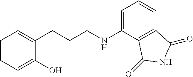

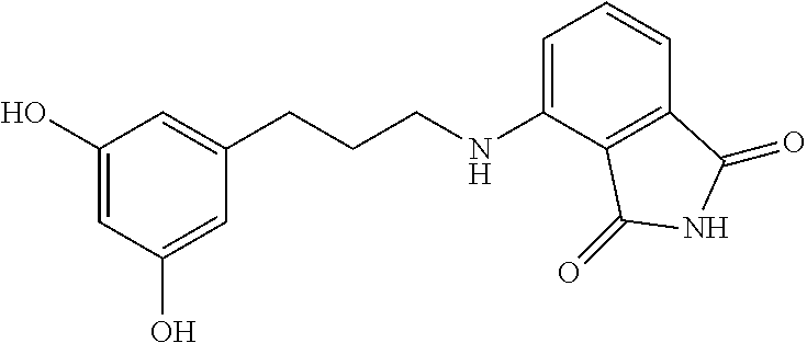


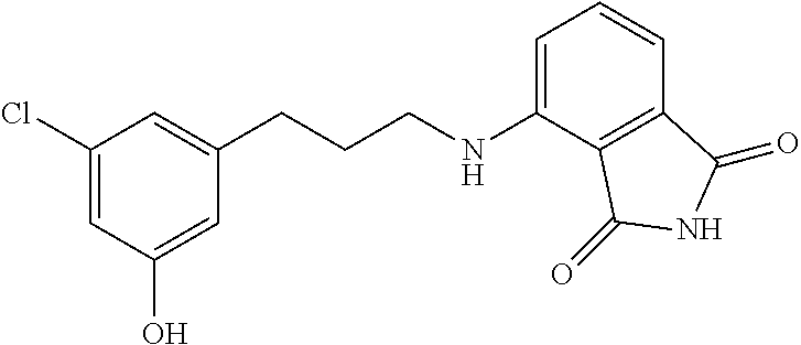


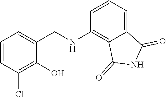


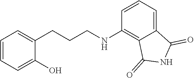


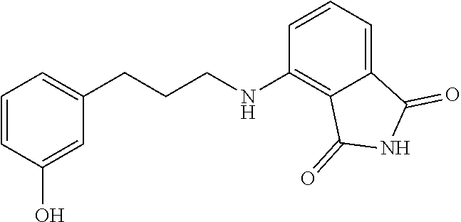
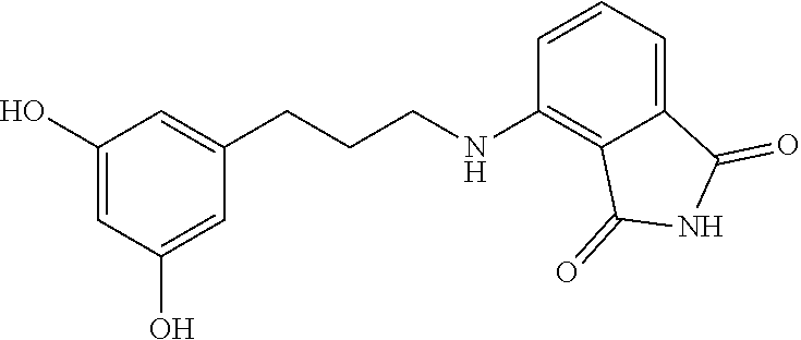


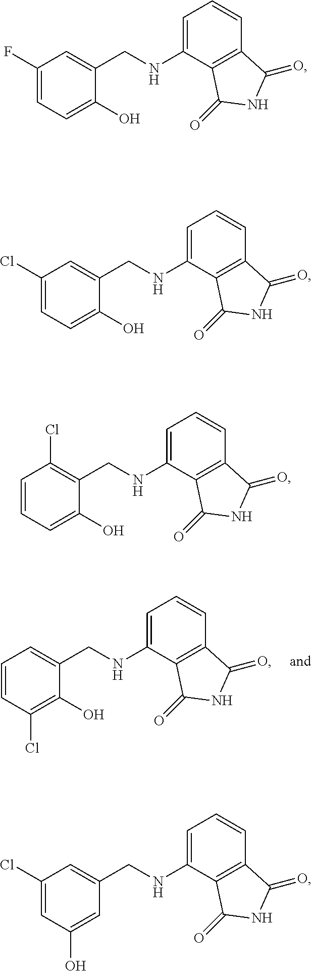


D00000
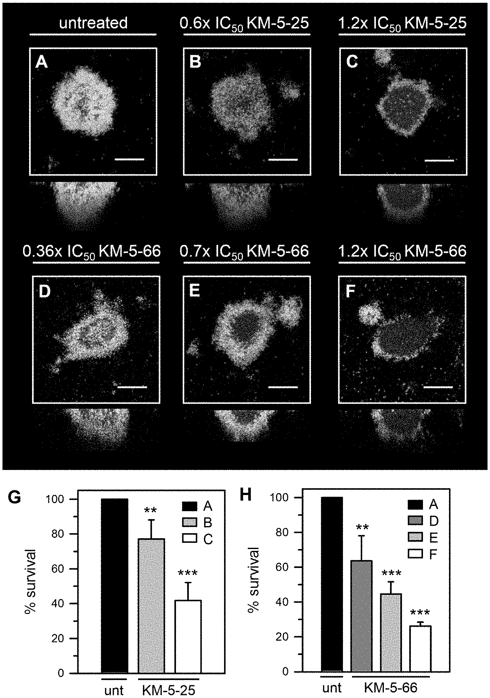
D00001

D00002

D00003
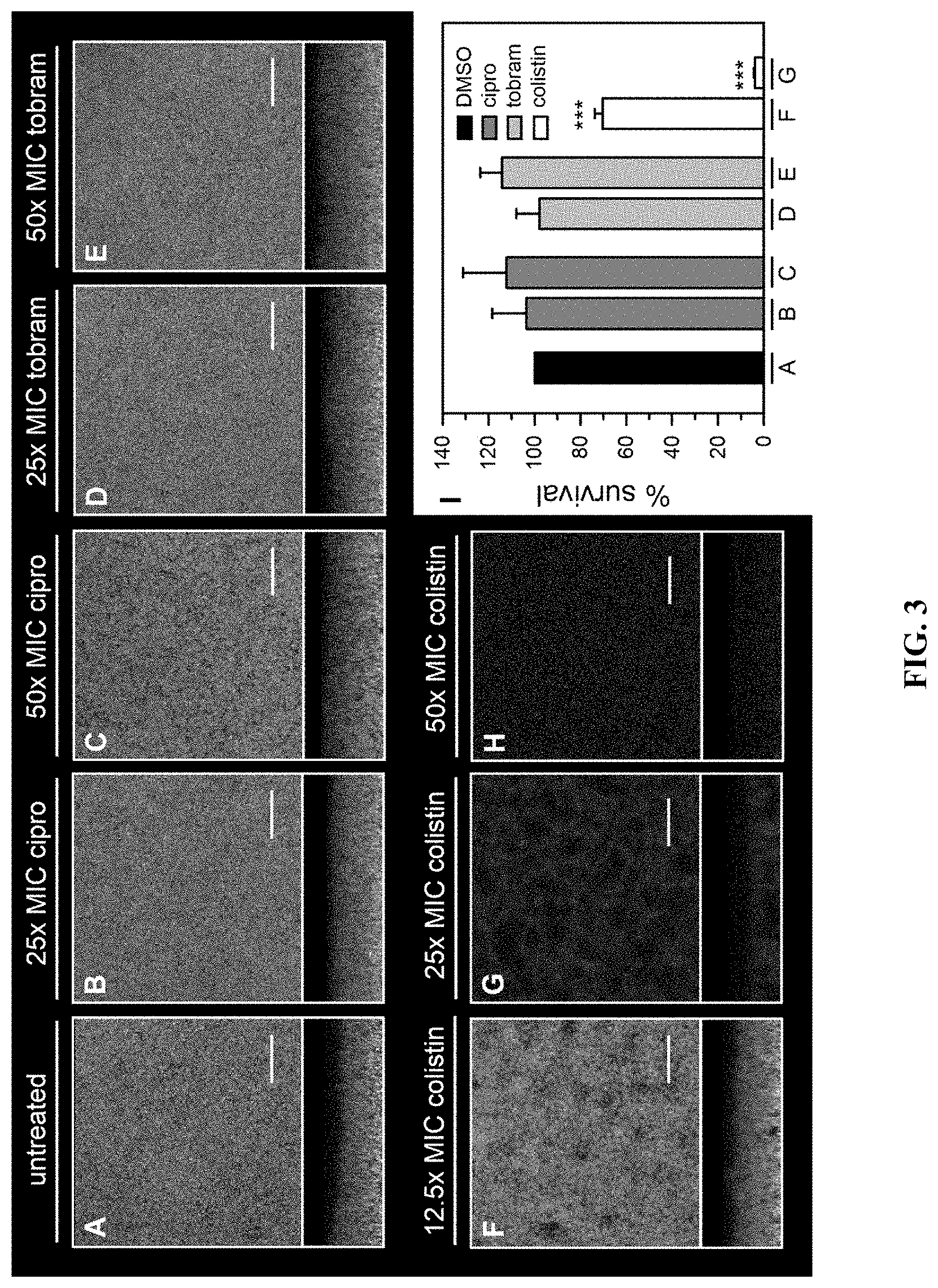
D00004

D00005

D00006

D00007
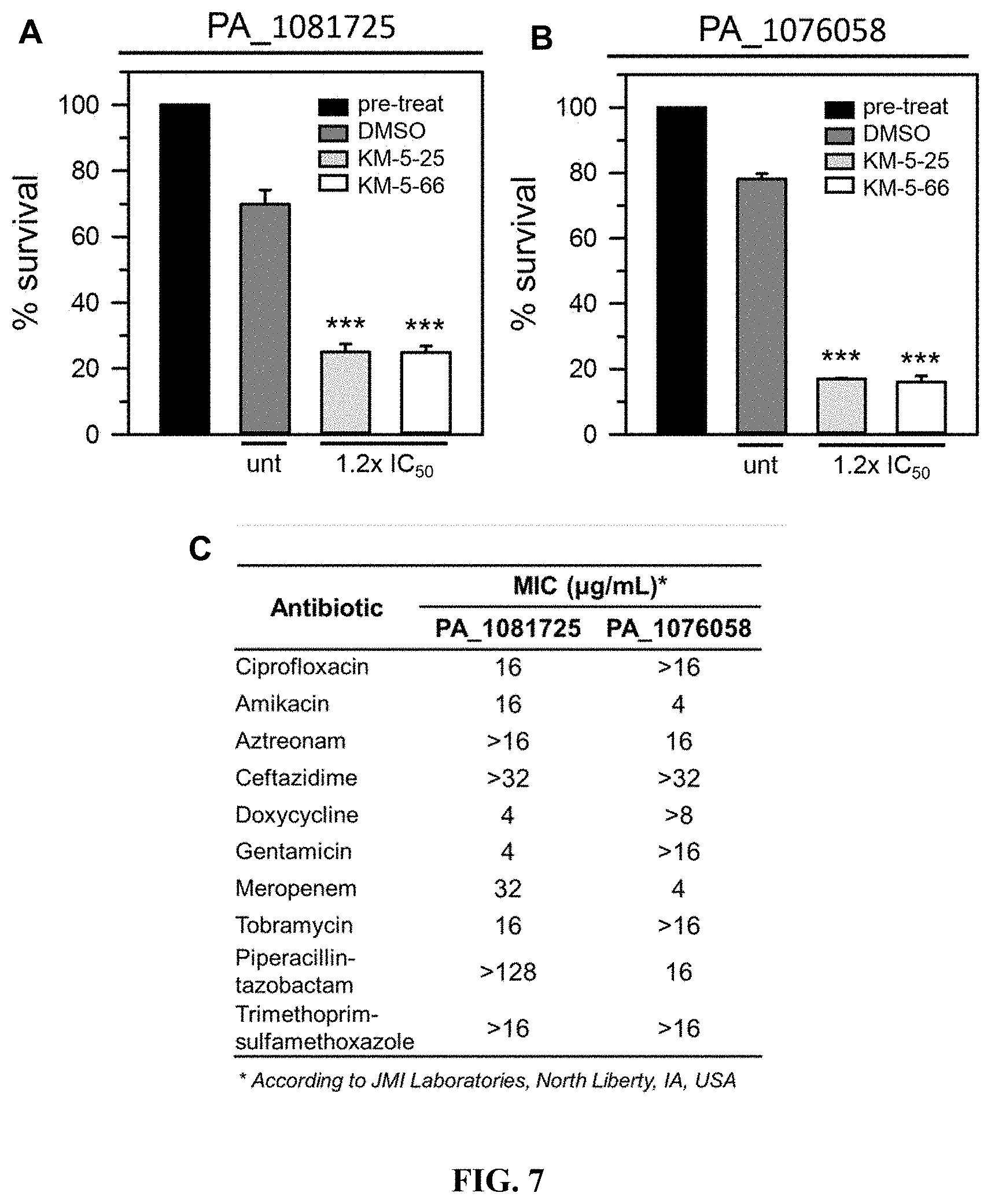
D00008

D00009

D00010

D00011
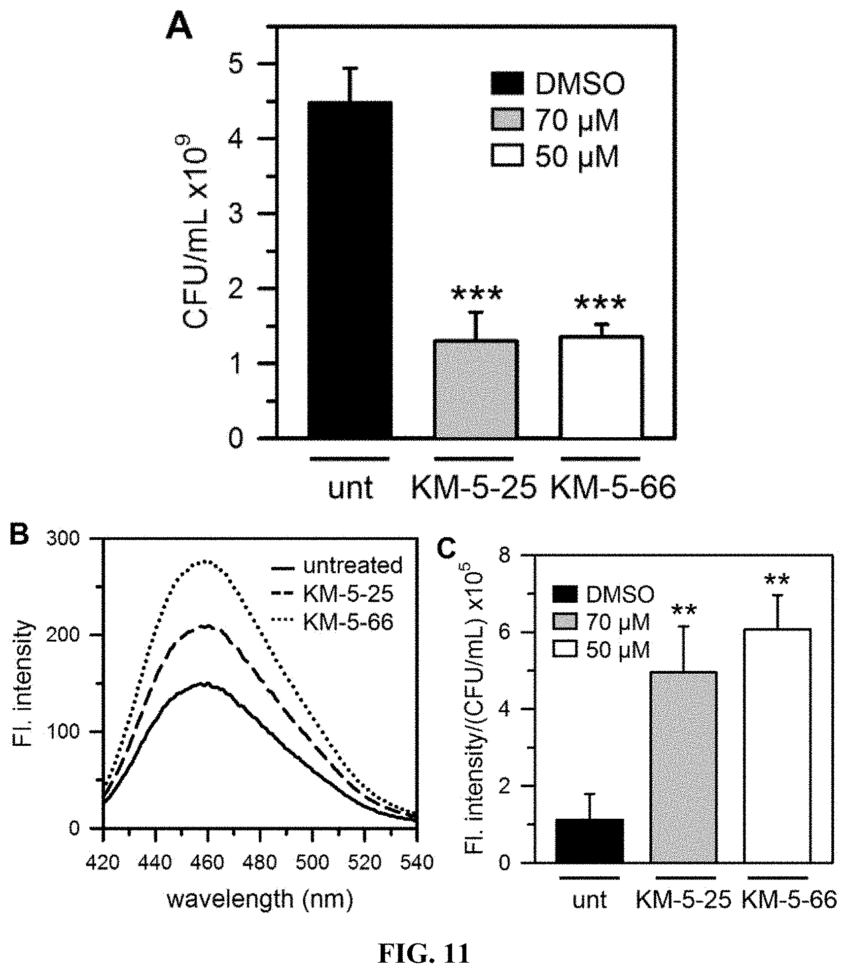
D00012
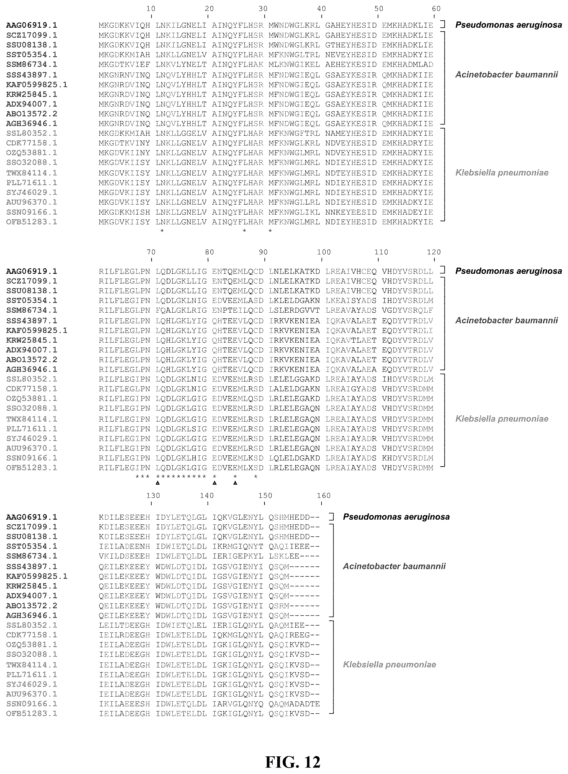
D00013
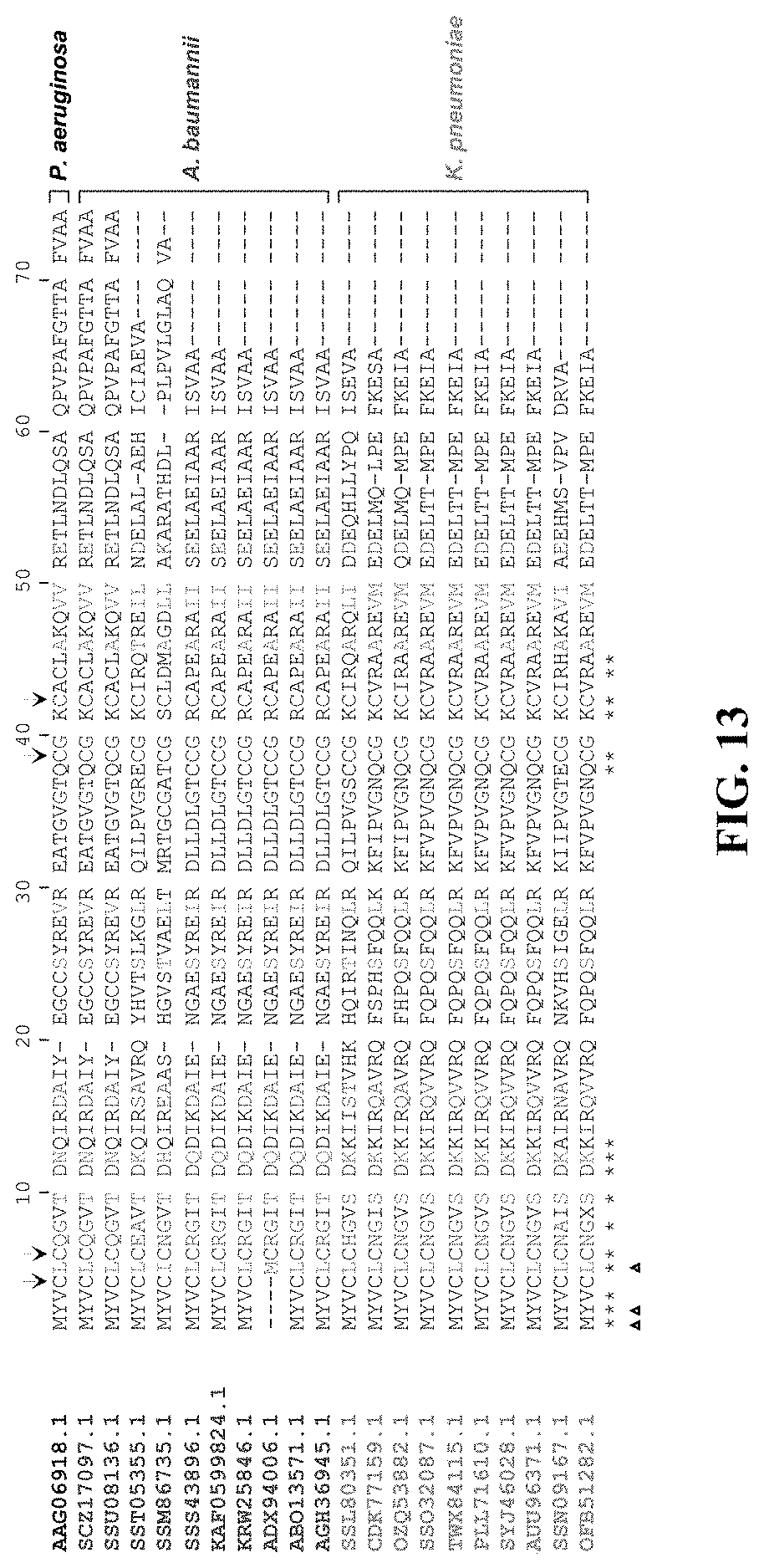

S00001
XML
uspto.report is an independent third-party trademark research tool that is not affiliated, endorsed, or sponsored by the United States Patent and Trademark Office (USPTO) or any other governmental organization. The information provided by uspto.report is based on publicly available data at the time of writing and is intended for informational purposes only.
While we strive to provide accurate and up-to-date information, we do not guarantee the accuracy, completeness, reliability, or suitability of the information displayed on this site. The use of this site is at your own risk. Any reliance you place on such information is therefore strictly at your own risk.
All official trademark data, including owner information, should be verified by visiting the official USPTO website at www.uspto.gov. This site is not intended to replace professional legal advice and should not be used as a substitute for consulting with a legal professional who is knowledgeable about trademark law.