Method For The In Vitro Or Ex Vivo Amplification Of Human Adipose Tissue Stem Cells
Dani; Christian ; et al.
U.S. patent application number 17/421840 was filed with the patent office on 2022-03-31 for method for the in vitro or ex vivo amplification of human adipose tissue stem cells. This patent application is currently assigned to UNIVERSITE COTE D'AZUR. The applicant listed for this patent is CENTRE NATIONAL DE LA RECHERCHE SCIENTIFIQUE (CNRS), CHU DE NICE, INSTITUT NATIONAL DE LA SANTE ET DE LA RECHERCHE MEDICALE, UNIVERSITE COTE D'AZUR. Invention is credited to Christian Dani, Vincent Dani-Davesne, Alain Doglio, Philippe Letertre.
| Application Number | 20220098552 17/421840 |
| Document ID | / |
| Family ID | |
| Filed Date | 2022-03-31 |


View All Diagrams
| United States Patent Application | 20220098552 |
| Kind Code | A1 |
| Dani; Christian ; et al. | March 31, 2022 |
METHOD FOR THE IN VITRO OR EX VIVO AMPLIFICATION OF HUMAN ADIPOSE TISSUE STEM CELLS
Abstract
The method for tin vitro or ex vivo amplification of human adipose tissue stem cells includes: --extracting a stromal vascular fraction of a human adipose tissue including endothelial cells of the human adipose tissue vascular network and human adipose tissue stem cells, and an extracellular matrix of the human adipose tissue, the extracellular matrix including endothelial cells of the human adipose tissue vascular network, human adipose tissue stem cells and collagen; --mixing the stromal vascular fraction and the extracellular matrix; and--culturing the mixture obtained in the preceding step, in suspension, in a culture medium.
| Inventors: | Dani; Christian; (Nice, FR) ; Doglio; Alain; (Saint Andre de la Roche, FR) ; Dani-Davesne; Vincent; (Nice, FR) ; Letertre; Philippe; (Colle sur Loup, FR) | ||||||||||
| Applicant: |
|
||||||||||
|---|---|---|---|---|---|---|---|---|---|---|---|
| Assignee: | UNIVERSITE COTE D'AZUR Nice FR INSTITUT NATIONAL DE LA SANTE ET DE LA RECHERCHE MEDICALE Paris FR CENTRE NATIONAL DE LA RECHERCHE SCIENTIFIQUE (CNRS) Paris FR CHU DE NICE Nice FR |
||||||||||
| Appl. No.: | 17/421840 | ||||||||||
| Filed: | January 13, 2020 | ||||||||||
| PCT Filed: | January 13, 2020 | ||||||||||
| PCT NO: | PCT/EP2020/050720 | ||||||||||
| 371 Date: | July 9, 2021 |
| International Class: | C12N 5/077 20060101 C12N005/077; C12M 1/00 20060101 C12M001/00; C12N 5/071 20060101 C12N005/071; A61L 27/36 20060101 A61L027/36; A61L 27/38 20060101 A61L027/38 |
Foreign Application Data
| Date | Code | Application Number |
|---|---|---|
| Jan 11, 2019 | FR | 1900287 |
Claims
1. A method for the in vitro or ex vivo amplification of human adipose tissue stem cells, comprising: extracting a stromal vascular fraction of a human adipose tissue comprising endothelial cells of the human adipose tissue vascular network and human adipose tissue stem cells, and an extracellular matrix of the human adipose tissue, the extracellular matrix comprising endothelial cells of the human adipose tissue vascular network, human adipose tissue stem cells and collagen; mixing the stromal vascular fraction and the extracellular matrix; and culturing the mixture obtained from the mixing, in suspension, in a culture medium.
2. A method for the in vitro or ex vivo amplification of differentiated cells, comprising: extracting a stromal vascular fraction of a human adipose tissue comprising endothelial cells of the human adipose tissue vascular network and human adipose tissue stem cells, and an extracellular matrix of the human adipose tissue, the extracellular matrix comprising endothelial cells of the human adipose tissue vascular network, human adipose tissue stem cells and collagen; mixing the stromal vascular fraction and the extracellular matrix; culturing the mixture obtained from the mixing, in suspension, in a culture medium; and inducing a differentiation of the adipose tissue stem cells to obtain differentiated cells.
3. The method according to claim 2, wherein the differentiated cells are at least one selected from the group consisting of adipocytes and osteoblasts.
4. The method according to claim 3, wherein the differentiated cells are adipocytes.
5. The method according to claim 1, wherein the extracting of the extracellular matrix comprises performing non-enzymatic separation.
6. The method according to claim 5, wherein the extracting of the extracellular matrix does not involve collagenase.
7. The method according to claim 1, wherein the extracting of the extracellular matrix comprises performing mechanical separation.
8. The method according to claim 1, wherein the extracting of the stromal vascular fraction and the extracellular matrix comprises: centrifuging human adipose tissue to obtain at least two separate fractions, a fraction A comprising a centrifuged extracellular matrix, and the stromal vascular fraction; and mechanically separating the fraction A to obtain the extracellular matrix.
9. The method according to claim 1, wherein the collagen of the extracellular matrix is type I collagen and type III collagen.
10. The method according to claim 1, wherein the culturing of the mixture of the stromal vascular fraction and the extracellular matrix comprises: transferring the mixture sterilely into a bag of suspension culture comprising culture medium; amplifying the mixture to form cellular aggregates; and mechanically separating the cellular aggregates.
11. An isolated extracellular matrix, comprising endothelial cells of the human adipose tissue vascular network, human adipose tissue stem cells, and collagen.
12. The matrix according to claim 11, wherein the collagen is type I and type III collagen.
13. The matrix according to claim 11, which further includes type IV collagen and/or elastin and/or fibronectin and/or laminin.
14. A composition comprising a mixture of the extracellular matrix according to claim 11 and a stromal vascular fraction, the stromal vascular fraction comprising endothelial cells of the adipose tissue vascular network and adipose tissue stem cells.
15. A method of screening pharmacological active substances, comprising: providing the extracellular matrix according to claim 11, and screening pharmacological active substances using the extracellular matrix.
16. A method of performing cell therapy, comprising: providing differentiated cells of the composition according to claim 14, and performing cell therapy using the differentiated cells.
17. The method according to claim 16, wherein the differentiated cells are osteoblast cells.
18. The method according to claim 16, wherein the differentiated cells are adipocyte cells.
19. The method according to claim 16, wherein the cell therapy is plastic or reparative surgery.
20. The method according to claim 16, wherein the cell therapy is lipofilling.
Description
TECHNICAL FIELD OF THE INVENTION
[0001] The present invention relates to a method for the in vitro or ex vivo amplification of human adipose tissue stem cells. It further relates to a method for the in vitro or ex vivo amplification of differentiated cells, an extracellular matrix, a composition comprising a mixture of an extracellular matrix and a stromal vascular fraction, a use of the extracellular matrix or of the composition comprising a mixture of the extracellular matrix and the stromal vascular fraction, and differentiated cells obtained according to the method of the invention for use thereof.
PRIOR ART
[0002] Cell therapy consists of a cell graft aimed at restoring the functions of a tissue or an organ when they are impaired by an accident, a disease or ageing. It enables the long-term treatment of a patient thanks to a single injection of so-called "therapeutic" cells. These cells are obtained, in particular, from multipotent stem cells from the patient themselves.
[0003] Lipofilling is a particular cell therapy technique for transferring fat cells, or adipocytes, from one area of the body to another in order to remodel the body or face.
[0004] To date, only the adipocytes present in certain adipose sites are used for cell therapy and, more particularly, for lipofilling. However, depending on the circumstances, the quantity of adipocytes available from a patient can be limiting. For example, when a patient has a body mass index that is too low or has undergone chemotherapy, they may not have enough adipose tissue to perform lipofilling.
[0005] In this context, there is a need to produce autologous adipocytes and, hence, to develop cell amplification methods for obtaining large quantities of therapeutic grade adipocytes with a view, in particular, to lipofilling.
[0006] The standard procedure, for isolating and amplifying adipocyte precursors from adipose tissue samples, involves enzymatic separation followed by the two-dimensional (2D) expansion thereof by binding to the plastic of culture dishes. This procedure is costly, time-consuming, and requires numerous handling operations which increase contamination risks. Furthermore, it induces a destruction of the three-dimensional structure of the tissue, as well as the loss of cell types of interest such as endothelial cells which play a key role both for graft vascularization and adipocyte physiology.
[0007] Non-enzymatic separation of adipose tissue, most frequently based on a mechanical process, makes it possible to isolate adipocyte precursors. This type of separation is emerging as a much less costly, quicker alternative method, which has indisputable advantages for the manufacture of a product according to therapeutic grade production standards (reduced exposure to external products or contaminants). On the other hand, the non-enzymatic separation methods described to date are not satisfactory as the number of adipocyte precursors obtained is low compared to enzymatic separation. This then requires the 2D amplification thereof on a culture dish. Furthermore, the endothelial cells, the extracellular matrix as well as the three-dimensional structure of adipose tissue are lost at the end of the process.
[0008] Different synthetic matrices have been proposed in order to inoculate the adipocyte precursors therein and thus attempt to best reconstitute the structure of adipose tissue. The matrix also serves to orient adipocyte precursors in vitro to a non-adipose (essentially bone or cartilaginous) cell type before implantation. Decellularized adipose tissue has also been proposed to increase precursor differentiation and better imitate the structure of adipose tissue. The manufacture of all these types of matrices requires numerous steps involving enzymatic reactions or long chemical treatments. Furthermore, decellularized tissue, by definition, loses these endogenous cells but also loses the factors of therapeutic interest which are anchored on the native matrix, which reduces the clinical value of this type of matrix. Non-decellularized adipose tissue enriched with adipocyte precursors (previously isolated by enzymatic separation) followed by 2D amplification has recently been proposed as a matrix for better bone reconstruction. The time to generate this biological matrix is long, requires three weeks of in vitro culture, and does not allow adipocyte precursor amplification. The authors only highlighted the benefit for bone repair.
[0009] Three-dimensional (3D) suspension culture represents an alternative method of choice to the standard 2D method as it essentially makes it possible to retain the structure and the intrinsic qualities of the tissue. This advantage is important because, for example, the lack of a relevant human model which best imitates adipose tissue in vitro is a major limitation during preclinical phase trials for the discovery of novel medicinal products effective in combatting obesity and associated metabolic diseases such as type 2 diabetes and cardiovascular diseases. Furthermore, 3D culture is feasible in a closed system which decreases handling operations and contamination risks.
SUMMARY OF THE INVENTION
[0010] In view of the above, a technical problem addressed by the present invention is that of obtaining in vitro or ex vivo a large quantity of human adipose tissue stems cells or differentiated cells, of therapeutic grade.
[0011] A first object of the solution of the invention to this technical problem is a method for the in vitro or ex vivo amplification of human adipose tissue stem cells, comprising the following steps: extracting a stromal vascular fraction of a human adipose tissue comprising endothelial cells of the human adipose tissue vascular network and human adipose tissue stem cells, and an extracellular matrix of said human adipose tissue, said extracellular matrix comprising endothelial cells of the human adipose tissue vascular network, human adipose tissue stem cells and collagen; mixing said stromal vascular fraction and said extracellular matrix; and culturing the mixture obtained in the preceding step, in suspension, in a culture medium.
[0012] Thus, the stromal vascular fraction suspension culture, enabled thanks to the presence of the extracellular matrix, enables a 3D amplification, giving access to a large number of cells and thus limiting handling operations which increase the contamination risks.
[0013] Advantageously, --the extraction of the extracellular matrix comprises a step of non-enzymatic separation and, in particular, the extraction of the extracellular matrix comprises a step of mechanical separation; --the extraction of the stromal vascular fraction and the extracellular matrix comprises the following steps: centrifuging the human adipose tissue to obtain at least two distinct fractions, a fraction A comprising a centrifuged extracellular matrix, and the stromal vascular fraction; and mechanically separating the fraction A to obtain the extracellular matrix; --the collagen of the extracellular matrix is type I collagen and type III collagen; and--the culture of the mixture of said stromal vascular fraction and said extracellular matrix comprises the following steps: transferring said mixture sterilely into a bag of suspension culture comprising culture medium; amplifying said mixture forming cellular aggregates; and mechanically separating said cellular aggregates.
[0014] According to a second object, the invention relates to a method for the in vitro or ex vivo amplification of differentiated cells comprising the following steps: extracting a stromal vascular fraction of a human adipose tissue comprising endothelial cells of the human adipose tissue vascular network and human adipose tissue stem cells, and an extracellular matrix of said human adipose tissue, said extracellular matrix comprising endothelial cells of the human adipose tissue vascular network, human adipose tissue stem cells and collagen; mixing said stromal vascular fraction and said extracellular matrix; culturing the mixture obtained in the preceding step, in suspension, in a culture medium; and inducing a differentiation of the adipose tissue stem cells to obtain differentiated cells.
[0015] Advantageously, --the differentiated cells are adipocytes or osteoblasts, preferably adipocytes; --the extraction of the extracellular matrix comprises a step of non-enzymatic separation, in particular the extraction of the extracellular matrix comprises a step of mechanical separation; --the extraction of the stromal vascular fraction and the extracellular matrix comprises the following steps: centrifuging the human adipose tissue to obtain at least two distinct fractions, a fraction A comprising a centrifuged extracellular matrix, and the stromal vascular fraction; and mechanically separating the fraction A to obtain the extracellular matrix; --the collagen of the extracellular matrix is type I collagen and type III collagen; and--the culture of the mixture of stromal vascular fraction and said extracellular matrix comprises the following steps: transferring said mixture sterilely into a bag of suspension culture comprising culture medium; amplifying said mixture forming cellular aggregates; and mechanically separating said cellular aggregates.
[0016] According to a third object, the invention relates to an isolated extracellular matrix capable of being obtained according to the method defined above, comprising endothelial cells of the human adipose tissue vascular network, human adipose tissue stem cells, and collagen.
[0017] Advantageously--the collagen is type I collagen and type III collagen; --the extracellular matrix further comprises fibronectin.
[0018] According to a fourth object, the invention relates to a composition comprising the mixture of the extracellular matrix and the stromal vascular fraction as defined above, the extracellular matrix comprising endothelial cells of the human adipose tissue vascular network, human adipose tissue stem cells, and collagen, and the stromal vascular fraction comprising endothelial cells of the adipose tissue vascular network and adipose tissue stem cells.
[0019] Advantageously--the collagen is type I collagen and type III collagen; --the extracellular matrix further comprises fibronectin.
[0020] According to a fifth object, the invention relates to the in vitro use of the extracellular matrix as defined above or the in vitro use of the composition as defined above for screening pharmacological active substances against obesity and associated metabolic diseases.
[0021] According to a fifth object, the invention relates to differentiated cells obtained according to the method defined above intended for use, or for the use thereof, in cell therapy, in particular in plastic and reparative surgery and, more particularly, for lipofilling.
BRIEF DESCRIPTION OF THE FIGURES
[0022] The invention will be better understood on reading the following non-restrictive description, drafted with reference to the appended drawings, wherein:
[0023] FIG. 1A represents schematically the necessary and sufficient steps for extracting an extracellular matrix and a stromal vascular fraction (steps 1 to 3), and placing them in coculture (step 4), according to the invention;
[0024] FIG. 1B is a more detailed schematic representation of the method for sequentially extracting extracellular matrices (M1-M4) and stromal vascular fraction cell populations (C1-C3) (steps 1 to 5), and placing them in coculture (step 6), according to the invention;
[0025] FIG. 2 represents the cell population C1 in Endothelial Growth Medium culture medium supplemented with growth factors (EGM+), in suspension, consisting of a majority of endothelial type cells;
[0026] FIG. 3A shows the CD31+ (endothelial cell marker) immunofluorescent labelling of the cell population C1 in adherent culture;
[0027] FIG. 3B shows the PDGFRa+ (adipocyte stem cell marker) immunofluorescent labelling of the cell population C1 in adherent culture;
[0028] FIG. 4 illustrates the cell population C2 in EGM+ culture medium in suspension showing the formation of a capillary type network consisting of CD31+ endothelial cells and presence of aggregates consisting of PDGFRa+ adipose tissue stem cells;
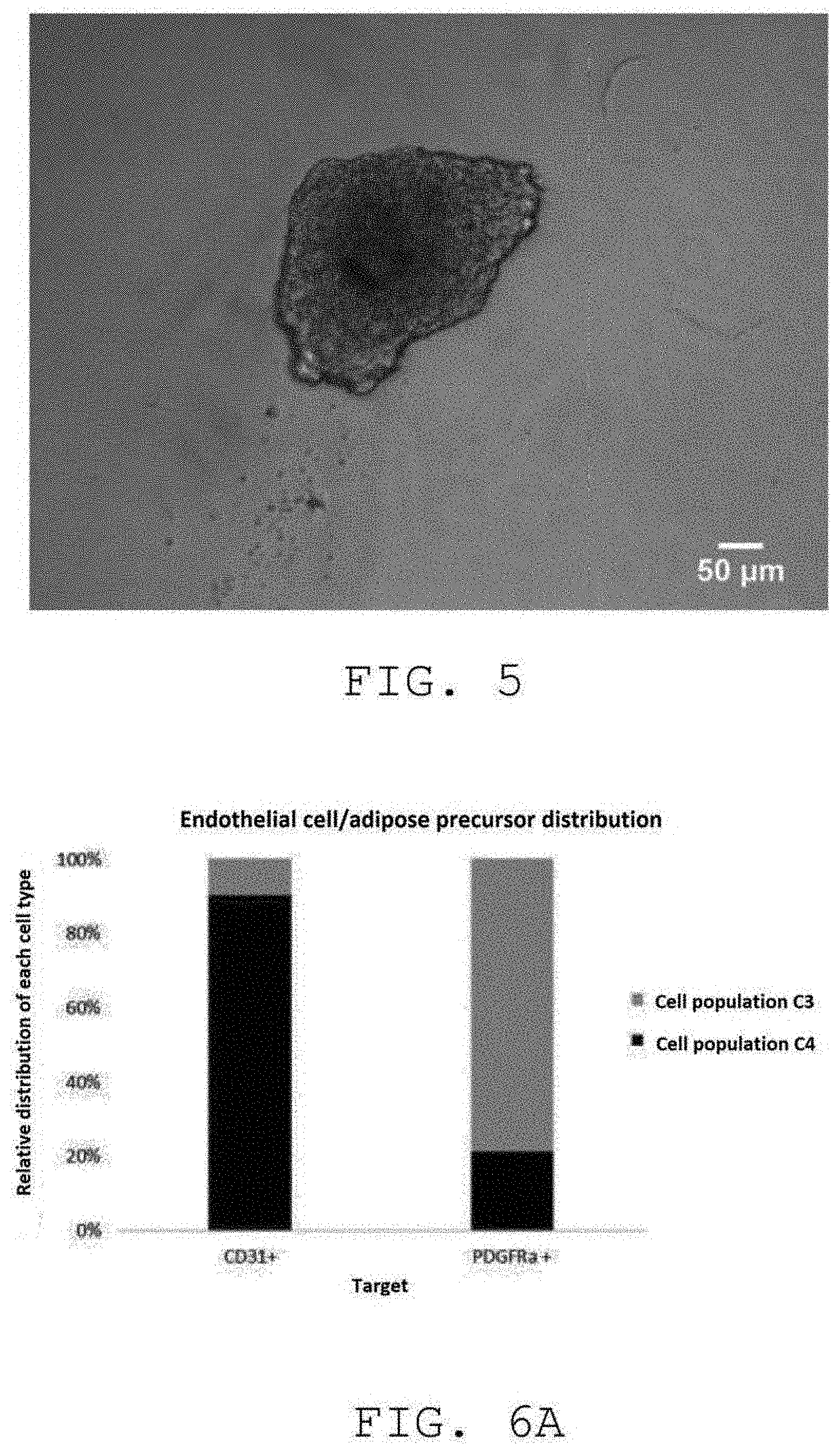
[0029] FIG. 5 is a microscopy image of the cell population C3 in EGM+ culture medium in suspension showing the presence of aggregates consisting of PDGFRa+ adipose tissue stem cells;
[0030] FIG. 6A represents a quantitative PCR of the cell populations C2 and C3 making it possible to determine the relative proportion of CD31+ endothelial cells and of PDGFRa+ adipose tissue stem cells;
[0031] FIG. 6B is a fluorescence microscopy image showing the adipocytic differentiation capacity of the cell populations C2; Nuclei (dark gray) Lipid droplets (light gray);
[0032] FIG. 6C illustrates, via a fluorescence microscopy image, the adipocytic differentiation capacity of the cell populations C3; Nuclei (dark gray) Lipid droplets (light gray);
[0033] FIG. 7 characterizes, in microscopy, the fibrous type of the matrix M1;
[0034] FIG. 8 shows, by microscopy, that the matrix M2 is heterogeneous in terms of matrix types: fibrous type and collagen-rich type;
[0035] FIG. 9 is a microscopy image illustrating the fibrous type of the matrix M3;
[0036] FIG. 10A shows, by fluorescence microscopy, that the matrix M2 is of the collagen-rich type; Picro-Sirius Red-labeled collagen (light gray) and nucleus labeling (white);
[0037] FIG. 10B shows, by fluorescence microscopy, that the matrix M3 is of the fibrous type; Picro-Sirius Red-labeled collagen (light gray and white fibers) and nucleus labeling (white);
[0038] FIG. 11A is a photograph of the centrifuged adipose tissue of the fraction A after mechanical separation containing the matrix M4;
[0039] FIG. 11B reveals by fluorescence microscopy, in the matrix M4, mature adipocytes by Oil red 0 staining (light gray) and a collagen-rich matrix by type I collagen labeling (very light gray);
[0040] FIG. 11C shows, by CD31 immunolabeling, the capillary structures formed by CD31+ endothelial cells (white) in the matrix M4; nucleus labeling (dark gray);
[0041] FIG. 11D illustrates the presence of the PDGFRa+ adipose tissue stem cell network (light gray dots) in the matrix M4; nucleus labeling (dark gray);
[0042] FIG. 12 shows, by incorporating Edu, 5-ethylenyl-2'-deoxyuridine, in the nucleus of the proliferating cells, that the endogenous cells, in the extracellular matrix according to the invention are kept proliferating in the EGM+ medium in suspension; nuclei (dark gray), proliferating cells (white) matrix auto-fluorescence (light gray);
[0043] FIG. 13 shows that exogenous adipose tissue stem cells, placed in coculture with the extracellular matrix of the invention, form structures composed of these adipose tissue stem cells and the endogenous cells present in the matrix; image taken after 3 days of coculture, nuclei (dark gray), collagen (light gray), exogenous adipose tissue stem cells (light gray/white);
[0044] FIG. 14A shows the formation of a cellular aggregate without cell proliferation during the cell culture of adipose tissue stem cells and endothelial cells in suspension without extracellular matrix; image taken after 10 days of coculture; nuclei (dark gray), nuclei of proliferating cells (very light gray);
[0045] FIG. 14B shows a proliferation capacity of the adipose tissue stem cells and endothelial cells placed in coculture, in suspension, with the extracellular matrix of the invention; image taken after 10 days of coculture; nuclei (dark gray), collagen matrix (light gray), nuclei of proliferating cells by Edu labeling (white);
[0046] FIG. 15A shows the level of expression of the CD31 endothelial cell marker in the differentiated cell populations obtained by culture, in suspension, with (right) and without (left) the extracellular matrix of the invention;
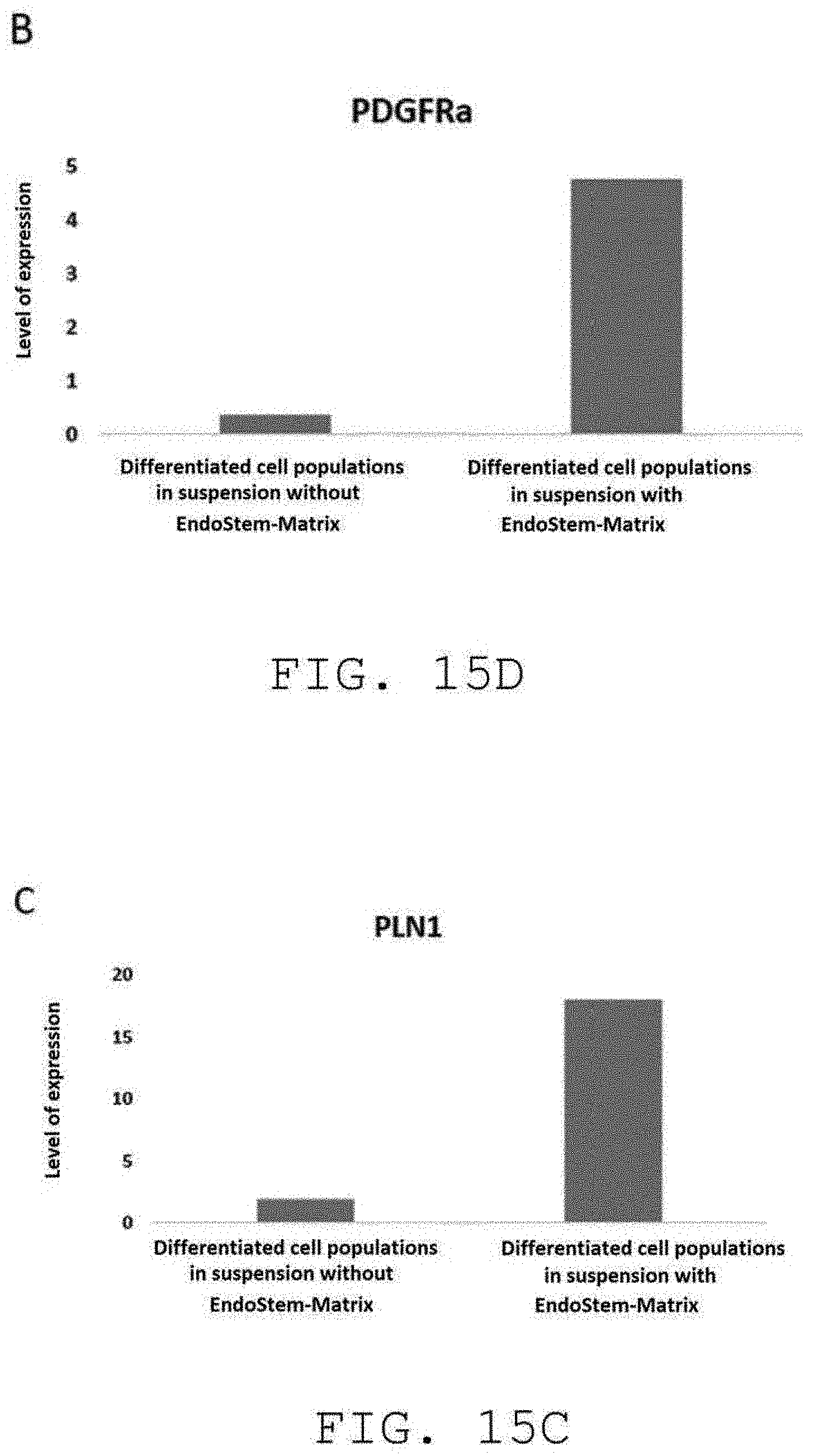
[0047] FIG. 15B shows the level of expression of the PDGFRa adipocytic stem cell marker in the differentiated cell populations obtained by culture, in suspension, with (right) and without (left) the extracellular matrix of the invention;
[0048] FIG. 15C shows the level of expression of the PLN1 mature adipocyte marker in the differentiated cell populations obtained by culture, in suspension, with (right) and without (left) the extracellular matrix of the invention;
[0049] FIG. 15D shows the level of expression of the Adiponectin mature adipocyte marker in the differentiated cell populations obtained by culture, in suspension, with (right) and without (left) the extracellular matrix of the invention;
[0050] FIG. 16 represents an image by fluorescence microscopy of the stromal vascular fraction after amplification and differentiation in the presence of the extracellular matrix of the invention; nucleus (dark gray), mature adipocyte (light gray), collagenic matrix (medium gray);
[0051] FIGS. 17A and 17B and images showing the activation of the proliferation capacities according to the invention. In FIG. 17A, a non-separated adipose tissue shows no proliferating cells. In FIG. 17B, the composition shows proliferating cells, the nuclei of the proliferating cells being represented in white in this figure;
[0052] FIGS. 18A, 18B, 18C and 18D, illustrate the expression of dipeptidyl peptidase-4 (DPP4), which is concentrated in the isolated stromal vascular fraction (SVF), and the expression of ICAM1, which is concentrated in the isolated matrix;
[0053] FIGS. 19A and 19B illustrate the presence of M1 type and M2 type macrophages respectively, in the amplified composition according to the invention; and
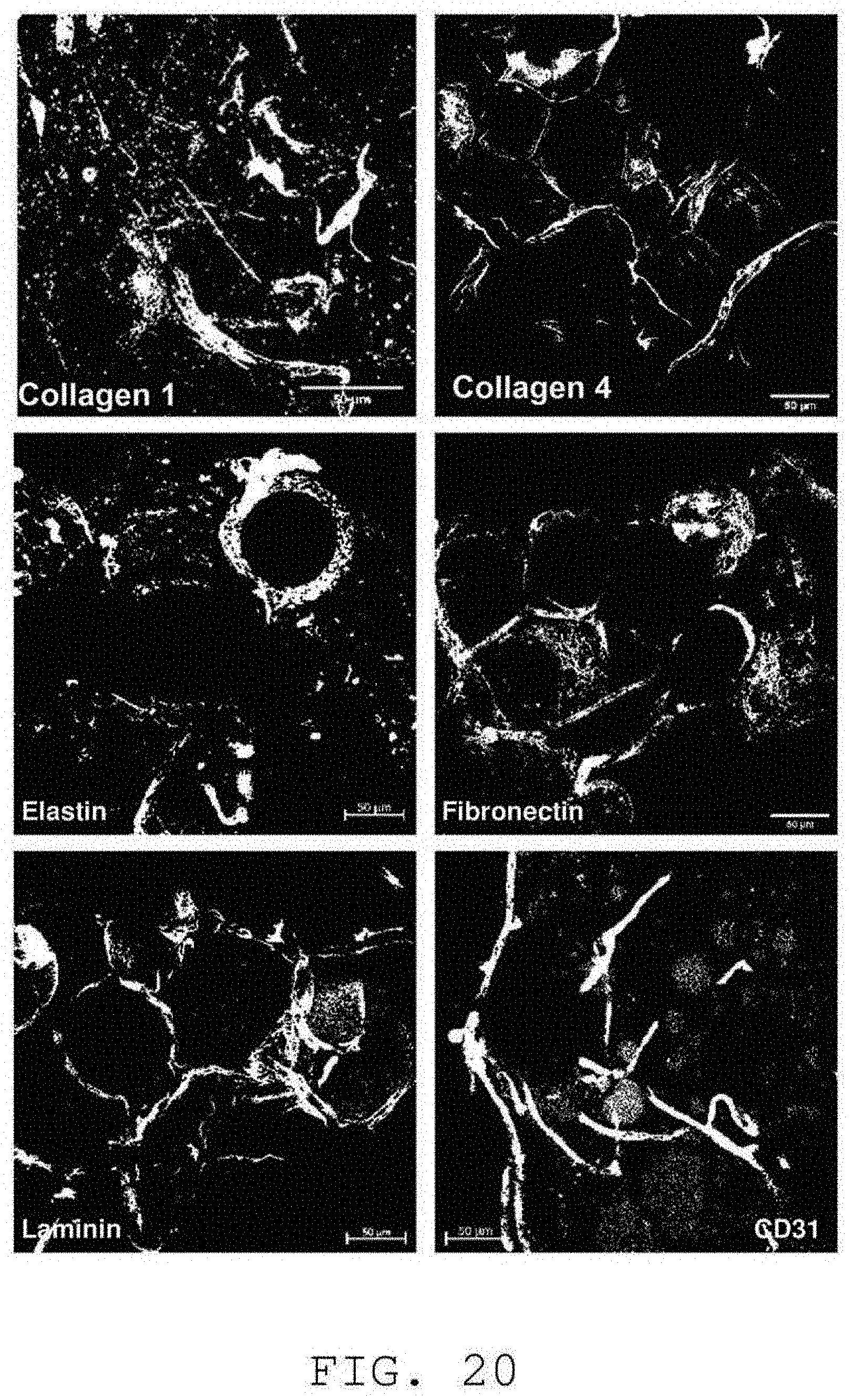
[0054] FIG. 20 comprises a set of photographs demonstrating the presence of certain proteins in the extracellular matrix according to the invention, and the preservation of a capillary network.
DETAILED DESCRIPTION OF THE INVENTION
[0055] Adipose tissue is supplied to carry out the Invention.
[0056] A first object of the invention is a method for the in vitro or ex vivo amplification of human adipose tissue stem cells, comprising the following steps: extracting a stromal vascular fraction of a human adipose tissue comprising endothelial cells of the human adipose tissue vascular network and human adipose tissue stem cells, and an extracellular matrix of said human adipose tissue, said extracellular matrix comprising endothelial cells of the human adipose tissue vascular network, human adipose tissue stem cells and collagen; mixing said stromal vascular fraction and said extracellular matrix; and culturing the mixture obtained in the preceding step, in suspension, in a culture medium. This method is also hereinafter referred to as the "ExAdEx method" (for Ex vivo Adipocytes Expansion).
[0057] According to the present invention, "stromal vascular fraction" denotes the cells present in a human adipose tissue sample. This stromal vascular fraction comprises endothelial cells of the human adipose tissue vascular network and human adipose tissue stem cells.
[0058] According to the invention, "extracellular matrix" denotes a bioactive matrix, i.e., a matrix which comprises different proteins of adipose tissue and endogenous cells. This extracellular matrix enables 3D cell amplification, i.e., three-dimensional cell proliferation. The extracellular matrix of the invention is also referenced hereinafter as "EndoStem-Matrix" or "EndoStem matrix".
[0059] The proteins of the extracellular matrix of the adipose tissue comprise collagen. This collagen is type I and type III. The proteins of the extracellular matrix of the adipose tissue further comprise fibronectin.
[0060] The extraction of the extracellular matrix comprises a step of non-enzymatic separation, in particular the extraction of the extracellular matrix comprises a step of mechanical separation. The "mechanical separation" of the invention makes it possible to keep the structure of the extracellular matrix intact whereas an enzymatic digestion generally involves collagenase which destroys it. The mechanical separation thus helps maintain the "vasculature" and the micro-structure of the extracellular matrix, which accordingly has a similar organization to the organization of adipose tissue in vivo.
[0061] The extraction of the stromal vascular fraction and the extracellular matrix comprises the following steps: centrifuging human adipose tissue to obtain at least two separate fractions, a fraction A comprising a centrifuged extracellular matrix, and the stromal vascular fraction; and mechanically separating the fraction A to obtain the extracellular matrix.
[0062] The step of centrifuging human adipose tissue furthermore makes it possible to remove oil, blood and anesthetic fluid contained in the human adipose tissue supplied. This step also makes it possible to remove saline solution obtained from preliminary washes of the human adipose tissue supplied.
[0063] In a particular embodiment, the extraction of the stromal vascular fraction and the extracellular matrix comprises the following steps: centrifuging human adipose tissue to obtain at least two separate fractions, a fraction A comprising a centrifuged extracellular matrix and a fraction B comprising endothelial cells of the human adipose tissue vascular network and human adipose tissue stem cells; mechanically separating the fraction A to obtain a fraction A' comprising a separated extracellular matrix; centrifuging the fraction A' to obtain at least the extracellular matrix and a fraction B' comprising endothelial cells of the human adipose tissue vascular network and human adipose tissue stem cells; and mixing the fractions B and B' to obtain the stromal vascular fraction.
[0064] In this embodiment, the step of centrifuging human adipose tissue furthermore makes it possible to remove oil, blood and anesthetic fluid contained in the human adipose tissue supplied. This step also makes it possible to remove saline solution obtained from preliminary washes of the human adipose tissue supplied. Centrifuging the fraction A' furthermore makes it possible to remove any oil and saline solution residues. This step of centrifuging the fraction A' is optional.
[0065] The culture of the mixture of said stromal vascular fraction and said extracellular matrix comprises the following steps: transferring said mixture sterilely into a bag of suspension culture comprising culture medium; amplifying said mixture forming cellular aggregates; and mechanically separating said cellular aggregates.
[0066] Transferring "sterilely", according to the invention, is a transfer, preferably, carried out in a closed system. This sterile transfer makes it possible to limit the number of contaminants during the cell culture. The mechanical separation of the aggregates formed during the amplification does not require opening the system, thus limiting the exposure of the cellular products to potential contamination of the culture by elements from the environment.
[0067] In an embodiment, the culture medium, in the bag of suspension culture, is an EGM+ medium. This culture medium comprises the base medium for proliferating the endothelial cells (EGM) enriched with Epidermal Growth Factor (EGF), Basic Growth Factor (FGF2), Insulin-like Growth Factor, Vascular Endothelial Growth Factor 165, ascorbic acid, heparin and hydrocortisone (EGM+). The EGM+ medium also enables the amplification of the adipocytic stem cells without altering their differentiation capacity into adipocytes.
[0068] The method of the invention enables an amplification of the number of adipose tissue stem cells with an amplification factor greater than 10, advantageously greater than 20, in particular greater than 30, preferably greater than 35. The amplification factor is the ratio between the number of cells obtained after culturing the isolated SVF in the presence of said extracellular matrix and the number of cells before the invention. In a particular embodiment described in example 2, the method of the invention has an amplification factor of 36 in 8 days.
[0069] According to a second object, the invention relates to a method for the in vitro or ex vivo amplification of differentiated cells comprising the following steps: in vitro or ex vivo amplification of human adipose tissue stem cells as defined above; and inducing a differentiation of the adipose tissue stem cells to obtain differentiated cells.
[0070] More specifically, the method for the in vitro or ex vivo amplification of differentiated cells therefore comprises the following steps: extracting a stromal vascular fraction of a human adipose tissue comprising endothelial cells of the human adipose tissue vascular network and human adipose tissue stem cells, and an extracellular matrix of said human adipose tissue, said extracellular matrix comprising endothelial cells of the human adipose tissue vascular network, human adipose tissue stem cells and collagen; mixing said stromal vascular fraction and said extracellular matrix; culturing the mixture obtained in the preceding step, in suspension, in a culture medium; and inducing a differentiation of the adipose tissue stem cells to obtain differentiated cells.
[0071] According to the invention, the differentiated cells are adipocytes or osteoblasts. Preferably, the differentiated cells are adipocytes.
[0072] The method for the in vitro or ex vivo amplification of differentiated cells comprising the steps associated with the in vitro or ex vivo amplification of adipose tissue stem cells, the details given above of the method for the in vitro or ex vivo amplification method of adipose tissue stem cells also apply for the method for the in vitro or ex vivo amplification of differentiated cells.
[0073] In particular, the extraction of the extracellular matrix comprises a step of non-enzymatic separation, in particular the extraction of the extracellular matrix comprises a step of mechanical separation.
[0074] In an embodiment, the extraction of the stromal vascular fraction and the extracellular matrix comprises the following steps: centrifuging human adipose tissue to obtain at least two separate fractions, a fraction A comprising a centrifuged extracellular matrix, and the stromal vascular fraction; and mechanically separating the fraction A to obtain the extracellular matrix.
[0075] In a further embodiment, the extraction of the stromal vascular fraction and the extracellular matrix comprises the following steps: centrifuging human adipose tissue to obtain at least two separate fractions, a fraction A comprising a centrifuged extracellular matrix and a fraction B comprising endothelial cells of the human adipose tissue vascular network and human adipose tissue stem cells; mechanically separating the fraction A to obtain a fraction A' comprising a separated extracellular matrix; centrifuging the fraction A' to obtain at least the extracellular matrix and a fraction B' comprising endothelial cells of the human adipose tissue vascular network and human adipose tissue stem cells; and mixing the fractions B and B' to obtain the stromal vascular fraction.
[0076] The collagen of the extracellular matrix comprises type I collagen and type III collagen detected by Picro-Sirius Red staining.
[0077] The culture of the mixture of said stromal vascular fraction and said extracellular matrix comprises the following steps: transferring said mixture sterilely into a bag of suspension culture comprising culture medium; amplifying said mixture forming cellular aggregates; and mechanically separating said cellular aggregates.
[0078] According to a third object, the invention relates to an isolated extracellular matrix capable of being obtained according to the method defined above, comprising endothelial cells of the human adipose tissue vascular network, human adipose tissue stem cells, and collagen.
[0079] The collagen is type I collagen and type III collagen. The extracellular matrix further comprises fibronectin.
[0080] According to a fourth object, the invention relates to a composition comprising the mixture of the extracellular matrix and the stromal vascular fraction as defined above, the extracellular matrix comprising endothelial cells of the human adipose tissue vascular network, human adipose tissue stem cells, and collagen, and the stromal vascular fraction comprising endothelial cells of the adipose tissue vascular network and adipose tissue stem cells.
[0081] The collagen is type I collagen and type III collagen. The extracellular matrix further comprises fibronectin.
[0082] According to a fifth object, the invention relates to the in vitro use of the extracellular matrix as defined above or the in vitro use of the composition as defined above for screening pharmacological active substances against obesity and associated metabolic diseases such as type 2 diabetes and cardiovascular diseases.
[0083] According to a fifth object, the invention relates to differentiated cells obtained according to the method defined above intended for use, or for the use thereof, in cell therapy, in particular in plastic and reparative surgery and more particularly for lipofilling.
[0084] In the case of use in plastic and reparative surgery, the extracellular matrix is an autologous matrix, which contains, by definition, cells specific to the patient from whom the adipose tissue is obtained.
[0085] For use for lipofilling, the differentiated cells obtained with the method of the invention are adipocytes.
EXAMPLES
Example 1. Mechanical Extraction
[0086] a) Mechanical Extraction Method with a View to Characterizing the Cell and Matrix Populations During the Process
[0087] The mechanical extraction of the stromal vascular fraction and the extracellular matrix, from a sample of adipose tissue from a human donor, can be carried out according to the following steps (FIG. 1B):
[0088] 1. Sampling adipose tissue by suction in a 10cc sterile syringe equipped with a 2 mm Coleman cannula under -20 kPa negative pressure.
[0089] 2. In order to separate the different phases, the syringe is centrifuged at 1600 rcf (relative centrifugal force), for 3 min in the collection tube. The oil fraction and the blood and anesthetic fluid fraction are removed. The pelleted fraction is retained.
[0090] 3. One unit of saline solution is injected into the syringe, followed by incubation for 30 min at 37.degree. C. with stirring. The syringe is centrifuged at 1600 rcf, for 3 min in the collection tube. The saline solution fraction and the oil fraction are removed. The pelleted fraction is retained.
[0091] 4. The syringe is connected to another make Luer-Lock type syringe by a Tulip.RTM. type connector in order to perform the separation of the tissue by emulsification. Three types of Tulip.RTM. connector, 2.4 mm, 1.4 mm and 1.2 mm, are successively used, on 30 passages.
[0092] 5. One unit of saline solution is injected into the syringe, followed by incubation for 30 min at 37.degree. C. with stirring. The syringe is centrifuged at 1600 rcf for 3 min in the collection tube. The saline solution fraction and the oil fraction are removed. The pelleted fraction is retained.
[0093] 6. The contents of the syringe and the contents of the collection tubes previously cleared of blood cells are transferred via sterile connection into a culture bag containing EGM+ culture medium at 37.degree. C. for the expansion phase.
[0094] In step 4 above of separating the tissue, a connector of a brand other than the Tulip.RTM. brand can be used. The number of connectors used is between 1 and 5. The number of passages via these connectors used is between 10 and 50.
[0095] b) Characterizing the Cell Populations Obtained
[0096] The method described above makes it possible to sequentially extract the stromal vascular fraction into 3 cell populations. These cell populations are characterized in particular by optical microscopy and by fluorescence microscopy. [0097] The cell population obtained in step 2, herein called C1, is composed of a majority of CD31+ endothelial type cells (FIG. 2 and FIG. 3). [0098] The cell population obtained in step 3, herein called C2, is composed of CD31+ endothelial type cells forming a capillary type network when maintained in 3D and of PDGFRa+ adipose tissue stem cells (FIG. 4 and FIG. 6A). The cell population C2 has the ability to differentiate into mature adipocytes (FIG. 6B). [0099] The cell population obtained in step 5, herein called C3, is composed of a majority of PDGFRa+ adipose tissue stem cells capable of forming spheres in suspension (FIG. 5 and FIG. 6A). The cell population C3 has the ability to differentiate into mature adipocytes (FIG. 6C).
[0100] c) Characterizing the Matrices M1 to M4 Obtained During the Different Steps of the Method [0101] The matrix obtained in step 2, herein called M1 is of the fibrous type (FIG. 7). [0102] The matrix obtained in step 3, herein called M2 is of the fibrous and collagen-rich type (FIG. 8). The collagen is detected with Picro-Sirius Red (FIG. 10A) which makes it possible, furthermore, to view a rod structure of the collagen. This matrix contains endogenous cells. [0103] The matrix obtained in step 5 and isolated in the collection tube, herein called M3 is of the fibrous type (FIG. 9 and FIG. 10B). This matrix also contains endogenous cells. The collagen from the isolated matrix is detected, in FIG. 10B, with Picro-Sirius Red which stains type I and type III collagen fibers. The red staining (in grayscale in FIG. 10B) obtained indicates that the collagen remains organized, namely that the collagen present always has an .alpha.-helix secondary structure and triple-helix quaternary structure. It is not degraded. Indeed, disorganized collagen is stained green by Picro-Sirius Red. [0104] The matrix obtained in step 5 and contained in the syringe, herein called M4, is composed of a majority of mature adipocytes and a type I collagen framework (FIG. 11B). The matrix M4 is also composed of capillary structures formed by CD31+ endothelial cells (FIG. 11C) and by a PDGFRa+ adipose tissue stem cell network (FIG. 11D).
[0105] FIG. 1A shows a method for collecting in step 2 the populations C1 and C2 as well as the matrices M1 and M2. In step 3, the population C3 and the matrices M3 and M4 are grouped together.
Example 2. Cell Expansion and Differentiation
[0106] A method for the ex vivo expansion of adipose tissue stem cells and for differentiation in an environment imitating adipose tissue comprises the following steps:
[0107] 1. The end product obtained in example 1 containing the populations C1-C3 as well as the so-called EndoStem-Matrix matrices M1-M4 are placed in culture in suspension in bags and kept in the EGM+ proliferation medium with stirring for 24 h at 37.degree. C. 5% CO.sub.2, then kept under the same conditions, preferably, with stirring.
[0108] 2. The EGM+ proliferation medium is 50% replaced every two days.
[0109] 3. A mechanical separation in a closed system, by passing through 2 syringes or two bags of culture in a tulip assembly, is performed on day 5 and day 10.
[0110] 4. On day 14, the EGM+ proliferation medium is replaced by the differentiation mixture I composed of EGM+ enriched with 250 .mu.M Dexamethasone; 500 .mu.M IBMX; 1 .mu.M Rosiglitazone; 2 .mu.M T3 and 2.5 .mu.g/ml insulin.
[0111] 5. On day 17, the differentiation medium I is replaced by the differentiation medium II composed of EGM+ enriched with 1 .mu.M Rosiglitazone; 2 .mu.M T3 and 2.5 .mu.g/ml insulin.
Example 3. Characterizing the Amplification Capacity of the Matrix
[0112] The so-called EndoStem-Matrix extracellular matrices of the invention were characterized, in particular, by fluorescence microscopy, in the presence of different specific markers. Proliferating cells were thus detected by incorporating, during the DNA replication phase, fluorescent Edu (5-ethylenyl-2'-deoxyuridine) in the EndoStem-Matrix matrices of the invention, as illustrated in FIG. 12, proving that the latter are bioactive. Indeed, FIG. 12 shows that the endogenous cells in the matrices are kept proliferating during the amplification phase.
[0113] Moreover, FIG. 13 shows the presence of adipose tissue stem cells after three days of co-culture with the extracellular matrix of the invention. The extracellular matrix therefore makes it possible to supply a substrate for proliferating the stromal vascular fraction: the adipose tissue stem cells added can bind to the EndoStem-Matrix, in suspension.
[0114] With reference to FIG. 14B, the stromal vascular fraction is amplified by its culture on the EndoStem Matrix of the invention. Conversely, with reference to FIG. 14A, when the stromal vascular fraction is placed in culture in suspension without the extracellular matrix, cellular aggregates without proliferation are observed. The extracellular matrix of the invention therefore has the ability to amplify the adipose tissue stem cells added.
[0115] The cellular amplification capacity of the different matrices M1 to M4 obtained in the steps of example 1 was verified. Thus, about 10.sup.4 adipose tissue stem cells were kept suspended in the presence of the different matrices M1 to M4 in Ultra-Low Attachment (ULA) wells. Eight days later, the cells are detached from the matrix with trypsin/EDTA then counted. The values obtained are shown in Table 1 below:
TABLE-US-00001 TABLE 1 Amplification Conditions Number of cells factor Adipose tissue stem 2 10.sup.4 1 cells without matrix Adipose tissue stem 5 10.sup.4 2.5 cells with matrix M1 Adipose tissue stem 53 10.sup.4 26.5 cells with matrix M2 Adipose tissue stem 7.4 10.sup.4 3.7 cells with matrix M3 Adipose tissue stem 72 10.sup.4 36 cells with matrix M4 Matrix M4 without 5 10.sup.4 -- adding adipose tissue stem cells
[0116] In the table above, for the specific case of the individual matrices M1 to M4, the amplification factor is the ratio between the number of cells obtained after culture in the presence of the extracellular matrix and the number of cells obtained in the absence of the extracellular matrix.
[0117] The matrices M2 and M4 have a high adipose tissue stem cell amplification potential. The volume of matrix M2 obtained is very low compared to the volume of M4 (FIG. 11A). The matrix M4 illustrates an extracellular matrix as defined in the invention.
[0118] The level of expression of different cell markers (CD31 endothelial cell marker, PDGFRa+ adipocytic stem cell marker, and two PLN1 and Adiponectin mature adipocyte markers) was analyzed after suspension culture of the stromal vascular fraction on the extracellular matrix of the invention. FIG. 15 shows a comparison of these levels of expression with those obtained from a suspension culture of the stromal vascular fraction without the extracellular matrix of the invention. This study shows an amplification of the endothelial cells of the human adipose tissue vascular network (FIG. 15A) and human adipose tissue stem cells (FIG. 15B). This study also makes it possible to demonstrate the superior differentiation capacity induced by the extracellular matrix of the invention (FIGS. 15C and 15D). Thus, the adipose tissue stem cells amplified in 3D on the extracellular matrix of the invention retain their ability to differentiate into adipocytes.
[0119] FIG. 16 demonstrates the presence of nuclei, of mature adipocytes and of a collagenic matrix after amplification and differentiation of the stromal vascular fraction in the presence of the extracellular matrix of the invention. Thus, the differentiation in the presence of the extracellular matrix of the invention makes it possible to retain the in vivo structural organization of the adipose tissue.
[0120] It should be noted that a non-separated adipose tissue, which can be equivalent to an explant, remains viable for a short time ex vivo. Thus, as shown particularly in FIG. 12, the matrix isolated by separation contains proliferating cells, unlike a non-separated tissue. FIGS. 17A and 17B make it possible to compare the cell proliferation in the non-separated tissue (FIG. 17A) and in the isolated matrix (FIG. 17B). In FIG. 17A, the non-separated adipose tissue shows no proliferating cells. In FIG. 17B, the composition shows proliferating cells. Indeed, this figure shows, in white, the nuclei of the proliferating cells.
[0121] It should be noted that the cells isolated according to the invention, by centrifuging the fluid form the washes, are characterized molecularly by the marker DPP4. DPP4 is a marker which labels ICAM1 pre-adipocyte precursor cells, which has a high proliferation capacity and which are located in the interstitial reticulum of the adipose tissue. These cells have the proliferation capacity in the composition according to the invention. It is important to note that these cells are removed following the washes carried out according to the methods of the prior art. As shown in FIGS. 18A and 18B, the expression of DPP4 is concentrated in the isolated SVF fraction. The matrix expresses a small amount. On the other hand, and as shown in FIGS. 18C and 18D, the expression of ICAM1 is concentrated in the isolated matrix. The cells carrying the amplification in the composition are the added cells expressing DPP4.
[0122] Moreover, it should be noted that in vivo adipose tissue contains macrophages and that the amplified composition according to the invention maintains the presence of M1 type macrophages, as shown in FIG. 19A and of M2 type macrophages as shown in FIG. 19B. In FIG. 19A, the M1 type macrophages are detected by the marker IL-1b and, in FIG. 19B, the M2 type macrophages are detected by the marker MRC1.
[0123] Finally, and as shown in FIG. 20, the isolated matrix according to the invention comprises extracellular matrix proteins, namely in particular, type I collagen, type IV collagen, elastin, fibronectin, laminin. The labeling of the CD31 endothelial cells shows that a capillary network is retained.
* * * * *
D00000
D00001
D00002
D00003
D00004
D00005
D00006
D00007
D00008
D00009
D00010
D00011
D00012

D00013
D00014

D00015

D00016

D00017
XML
uspto.report is an independent third-party trademark research tool that is not affiliated, endorsed, or sponsored by the United States Patent and Trademark Office (USPTO) or any other governmental organization. The information provided by uspto.report is based on publicly available data at the time of writing and is intended for informational purposes only.
While we strive to provide accurate and up-to-date information, we do not guarantee the accuracy, completeness, reliability, or suitability of the information displayed on this site. The use of this site is at your own risk. Any reliance you place on such information is therefore strictly at your own risk.
All official trademark data, including owner information, should be verified by visiting the official USPTO website at www.uspto.gov. This site is not intended to replace professional legal advice and should not be used as a substitute for consulting with a legal professional who is knowledgeable about trademark law.