Anti-pd-1 Antibody, Antigen-binding Fragment Thereof And Pharmaceutical Use Thereof
GU; Xiaoling ; et al.
U.S. patent application number 17/427625 was filed with the patent office on 2022-03-31 for anti-pd-1 antibody, antigen-binding fragment thereof and pharmaceutical use thereof. The applicant listed for this patent is JIANGSU HENGRUI MEDICINE CO., LTD., SHANGHAI HENGRUI PHARMACEUTICAL CO., LTD.. Invention is credited to Hu GE, Xiaoling GU, Weikang TAO, Xin YE.
| Application Number | 20220098304 17/427625 |
| Document ID | / |
| Family ID | 1000006074240 |
| Filed Date | 2022-03-31 |

| United States Patent Application | 20220098304 |
| Kind Code | A1 |
| GU; Xiaoling ; et al. | March 31, 2022 |
ANTI-PD-1 ANTIBODY, ANTIGEN-BINDING FRAGMENT THEREOF AND PHARMACEUTICAL USE THEREOF
Abstract
Provided are an anti-PD-1 antibody, an antigen-binding fragment thereof, and a pharmaceutical use thereof. Specifically, provided are a humanized anti-PD-1 antibody containing a specific CDR region and an antigen-binding fragment thereof, a pharmaceutical composition containing the anti-PD-1 antibody and the antigen-binding fragment thereof, and a used thereof as medicament. In particular, provided is a use of the humanized anti-PD-1 antibody in the preparation of the medicament for treating PD-1 associated diseases or disorders.
| Inventors: | GU; Xiaoling; (Shanghai, CN) ; YE; Xin; (Shanghai, CN) ; GE; Hu; (Shanghai, CN) ; TAO; Weikang; (Shanghai, CN) | ||||||||||
| Applicant: |
|
||||||||||
|---|---|---|---|---|---|---|---|---|---|---|---|
| Family ID: | 1000006074240 | ||||||||||
| Appl. No.: | 17/427625 | ||||||||||
| Filed: | January 31, 2020 | ||||||||||
| PCT Filed: | January 31, 2020 | ||||||||||
| PCT NO: | PCT/CN2020/074098 | ||||||||||
| 371 Date: | July 30, 2021 |
| Current U.S. Class: | 1/1 |
| Current CPC Class: | C07K 2317/565 20130101; C07K 2317/92 20130101; C07K 2317/52 20130101; C07K 16/2818 20130101; A61K 2039/505 20130101; C07K 2317/24 20130101; C07K 2317/73 20130101; C07K 2317/31 20130101; A61P 35/00 20180101 |
| International Class: | C07K 16/28 20060101 C07K016/28; A61P 35/00 20060101 A61P035/00 |
Foreign Application Data
| Date | Code | Application Number |
|---|---|---|
| Feb 3, 2019 | CN | 201910108743.3 |
| Jan 17, 2020 | CN | 202010052351.2 |
Claims
1. An anti-PD-1 antibody or antigen-binding fragment thereof, which comprises a heavy chain variable region and a light chain variable region, wherein: the heavy chain variable region comprises: HCDR1 as shown in SEQ ID NO: 65, HCDR2 as shown in SEQ ID NO: 66, and HCDR3 as shown in SEQ ID NO: 67; and the light chain variable region comprises: LCDR1 as shown in SEQ ID NO: 68, LCDR2 as shown in SEQ ID NO: 12, and LCDR3 as shown in SEQ ID NO: 69.
2. An anti-PD-1 antibody or antigen-binding fragment thereof, which comprises a heavy chain variable region and a light chain variable region, wherein the combination of the heavy chain variable region and the light chain variable region is selected from any one of the following (a) to (e): (a) the heavy chain variable region comprising HCDR1, HCDR2 and HCDR3 as shown in SEQ ID NO: 8, SEQ ID NO: 9 and SEQ ID NO: 10, respectively, and the light chain variable region comprising LCDR2 and LCDR3 as shown in SEQ ID NO: 12 and SEQ ID NO: 13, respectively, and LCDR1 as shown in SEQ ID NO: 11, 47, 48, 49, 50, 51 or 52; (b) the heavy chain variable region comprising HCDR1, HCDR2 and HCDR3 as shown in SEQ ID NO: 14, SEQ ID NO: 15 and SEQ ID NO: 16, respectively, and the light chain variable region comprising LCDR1, LCDR2 and LCDR3 as shown in SEQ ID NO: 17, SEQ ID NO: 12 and SEQ ID NO: 18, respectively; (c) the heavy chain variable region comprising HCDR1, HCDR2 and HCDR3 as shown in SEQ ID NO: 21, SEQ ID NO: 22 and SEQ ID NO: 23, respectively, and the light chain variable region comprising LCDR1, LCDR2 and LCDR3 as shown in SEQ ID NO: 24, SEQ ID NO: 25 and SEQ ID NO: 26; (d) the heavy chain variable region comprising HCDR1, HCDR2 and HCDR3 as shown in SEQ ID NO: 14, SEQ ID NO: 15 and SEQ ID NO: 16, respectively, and the light chain variable region comprising LCDR2 and LCDR3 as shown in SEQ ID NO: 12 and SEQ ID NO: 13, respectively, and LCDR1 as shown in SEQ ID NO: 11, 47, 48, 49, 50, 51 or 52; and (e) the heavy chain variable region comprising HCDR1, HCDR2 and HCDR3 as shown in SEQ ID NO: 8, SEQ ID NO: 9 and SEQ ID NO: 10, respectively, and the light chain variable region comprising LCDR1, LCDR2 and LCDR3 as shown in SEQ ID NO: 17, SEQ ID NO: 12 and SEQ ID NO: 18, respectively.
3. An anti-PD-1 antibody or antigen-binding fragment thereof, which comprises a combination of the heavy chain variable region and the light chain variable region selected from any one of the following iv) to vi): iv) the heavy chain variable region comprising HCDR1, HCDR2 and HCDR3 with the same sequences as those of the heavy chain variable region shown in sequence SEQ ID NO: 4, and the light chain variable region comprising LCDR1, LCDR2 and LCDR3 with the same sequences as those of the light chain variable region shown in sequence SEQ ID NO: 5; v) the heavy chain variable region comprising HCDR1, HCDR2 and HCDR3 with the same sequences as those of the heavy chain variable region shown in sequence SEQ ID NO: 6, and the light chain variable region comprising LCDR1, LCDR2 and LCDR3 with the same sequences as those of the light chain variable region shown in sequence SEQ ID NO: 7; vi) the heavy chain variable region comprising HCDR1, HCDR2 and HCDR3 with the same sequences as those of the heavy chain variable region shown in sequence SEQ ID NO: 19, and the light chain variable region comprising LCDR1, LCDR2 and LCDR3 with the same sequences as those of the light chain variable region shown in sequence SEQ ID NO: 20; vii) the heavy chain variable region comprising HCDR1, HCDR2 and HCDR3 with the same sequences as those of the heavy chain variable region shown in sequence SEQ ID NO: 70, and the light chain variable region comprising LCDR1, LCDR2 and LCDR3 with the same sequences as those of the light chain variable region shown in sequence SEQ ID NO: 71.
4. The anti-PD-1 antibody or antigen-binding fragment thereof according to claim 2, wherein the anti-PD-1 antibody or antigen-binding fragment thereof is a murine antibody or antigen-binding fragment thereof, a chimeric antibody or antigen-binding fragment thereof, a fully human antibody or antigen-binding fragment thereof, or a humanized antibody or antigen-binding fragment thereof.
5. The anti-PD-1 antibody or antigen-binding fragment thereof according to claim 4, wherein the anti-PD-1 antibody is a humanized antibody, said humanized antibody comprises a framework region derived from a human antibody or a framework region variant thereof, wherein: the framework region variant has at most 11 amino acid back mutations in each of the light chain framework region and/or the heavy chain framework region of the human antibody.
6. The anti-PD-1 antibody or antigen-binding fragment thereof according to claim 4, the combination of the heavy chain variable region and the light chain variable region of the antibody is selected from any one of the following (i) to (o): (i) a heavy chain variable region, the sequence of which is as shown in SEQ ID NO: 4 or has at least 90% sequence identity with SEQ ID NO: 4, and/or a light chain variable region, the sequence of which is as shown in SEQ ID NO: 5 or has at least 90% sequence identity with SEQ ID NO: 5; (j) a heavy chain variable region, the sequence of which is as shown in SEQ ID NO: 6 or has at least 90% sequence identity with SEQ ID NO: 6, and/or a light chain variable region, the sequence of which is as shown in SEQ ID NO: 7 or has at least 90% sequence identity with SEQ ID NO: 7; (k) a heavy chain variable region, the sequence of which is as shown in SEQ ID NO: 19 or has at least 90% sequence identity with SEQ ID NO: 19, and/or a light chain variable region, the sequence of which is as shown in SEQ ID NO: 20 or has at least 90% sequence identity with SEQ ID NO: 20; (l) a heavy chain variable region, the sequence of which is as shown in SEQ ID NO: 27, 30, 31 or 32, or has at least 90% sequence identity with SEQ ID NO: 27, 30, 31 or 32, and/or a light chain variable region, the sequence of which is as shown in SEQ ID NO: 28, 29, 34, 35, 53, 54, 55, 56, 57, 58, 59, 60, 61, 62, 63 or 64, or has at least 90% sequence identity with SEQ ID NO: 28, 29, 34, 35, 53, 54, 55, 56, 57, 58, 59, 60, 61, 62, 63 or 64; (m) a heavy chain variable region, the sequence of which is as shown in SEQ ID NO: 33, 36, 37, 38, 39 or 40, or has at least 90% sequence identity with SEQ ID NO: 33, 36, 37, 38, 39 or 40, and/or a light chain variable region, the sequence of which is as shown in SEQ ID NO: 34, 35, 28, 29, 53, 54, 55, 56, 57, 58, 59, 60, 61, 62, 63 or 64, or has at least 90% sequence identity with SEQ ID NO: 34, 35, 28, 29, 53, 54, 55, 56, 57, 58, 59, 60, 61, 62, 63 or 64; (n) a heavy chain variable region, the sequence of which is as shown in SEQ ID NO: 41, 45 or 46, or has at least 90% sequence identity with SEQ ID NO: 41, 45 or 46, and/or a light chain variable region, the sequence of which is as shown in SEQ ID NO: 42, 43 or 44, or has at least 90% sequence identity with SEQ ID NO: 42, 43 or 44, and (o) a heavy chain variable region, the sequence of which is as shown in SEQ ID NO: 70 or has at least 90% sequence identity with SEQ ID NO: 70, and/or a light chain variable region, the sequence of which is as shown in SEQ ID NO: 71 or has at least 90% sequence identity with SEQ ID NO: 71.
7. The anti-PD-1 antibody or antigen-binding fragment thereof according to claim 2, wherein the antibody further comprises an antibody constant region.
8. The anti-PD-1 antibody or antigen-binding fragment thereof according to claim 2, wherein the anti-PD-1 antibody comprises: a light chain as shown in SEQ ID NO: 78 and a heavy chain as shown in SEQ ID NO: 77 or 82; or the anti-PD-1 antibody comprises a light chain as shown in SEQ ID NO: 75 and a heavy chain as shown in SEQ ID NO: 74, 76, 80 or 81.
9. The anti-PD-1 antibody or antigen-binding fragment thereof according to claim 2, wherein the antigen-binding fragment is selected from the group consisting of Fab, Fab', F(ab').sub.2, single-chain antibody (scFv), dimerized V region (diabody) and disulfide bond stabilized V region (dsFv).
10. The anti-PD-1 antibody or antigen-binding fragment thereof according to claim 2, wherein the antibody is a bispecific antibody, a multispecific antibody or an antibody fusion protein.
11. An isolated monoclonal antibody or antigen-binding fragment thereof, said antibody competitively binds to human PD-1 with the anti-PD-1 antibody or antigen-binding fragment thereof according to claim 2.
12. A pharmaceutical composition comprising: a therapeutically effective amount of the anti-PD-1 antibody or antigen-binding fragment thereof according to claim 2, and one or more pharmaceutically acceptable carriers, diluents, buffers or excipients.
13. A nucleic acid molecule encoding the anti-PD-1 antibody or antigen-binding fragment thereof according to claim 2.
14. A host cell comprising the nucleic acid molecule according to claim 13.
15. A method for the immunodetection or determination of PD-1, comprising a step of using the anti-PD-1 antibody or antigen-binding fragment thereof according to claim 2.
16. A kit comprising the anti-PD-1 antibody or antigen-binding fragment thereof according to claim 2.
17. A method for treating diseases associated with PD-1, comprising administering to a subject a therapeutically effective amount of the anti-PD-1 antibody or antigen-binding fragment thereof according to claim 2.
18. The method of claim 17, wherein the disease is selected from the group consisting of: head and neck squamous cell carcinoma, head and neck cancer, brain cancer, glioma, glioblastoma multiforme, neuroblastoma, central nervous system cancer, neuroendocrine tumor, pharyngeal cancer, nasopharyngeal cancer, esophageal cancer, thyroid cancer, malignant pleural mesothelioma, lung cancer, breast cancer, liver cancer, hepatoma, hepatocellular carcinoma, hepatobiliary cancer, pancreatic cancer, gastric cancer, gastrointestinal cancer, bowel cancer, colon cancer, colorectal cancer, kidney cancer, clear cell renal cell carcinoma, ovarian cancer, endometrial cancer, cervical cancer, bladder cancer, prostate cancer, testicular cancer, skin cancer, melanoma, leukemia, lymphoma, bone cancer, chondrosarcoma, myeloma, multiple myeloma, myelodysplastic syndrome, myeloproliferative neoplasm, squamous cell carcinoma, Ewing's sarcoma, systemic light chain amyloidosis and Merkel cell carcinoma.
19. The anti-PD-1 antibody or antigen-binding fragment thereof according to claim 5, wherein the the framework region variant comprises mutation(s) selected from any one of the following (f) to (h): (f) 2G amino acid back mutation comprised in the light chain variable region, and/or one or more amino acid back mutations selected from the group consisting of 27Y, 48I, 67T, 69L, 82F and 93T comprised in the heavy chain variable region; (g) 2V amino acid back mutation comprised in the light chain variable region, and/or one or more amino acid back mutations selected from the group consisting of 26D, 27F, 30T, 38K, 43H, 48I, 66K, 67A, 69L, 82F and 93T comprised in the heavy chain variable region; and (h) one or more amino acid back mutations selected from the group consisting of 42G, 44V and 71Y comprised in the light chain variable region, and/or 1K and/or 94S amino acid back mutations comprised in the heavy chain variable region; wherein the amino acid positions are numbered and determined according to the Kabat Criteria.
20. The anti-PD-1 antibody or antigen-binding fragment thereof according to claim 7, wherein the antibody comprises the heavy chain constant region as shown in SEQ ID NO: 72 or 79, and the light chain constant region as shown in SEQ ID NO: 73.
Description
FIELD OF THE INVENTION
[0001] The present disclosure belongs to the field of biotechnology. More specifically, the present disclosure relates to anti-PD-1 antibodies and applications thereof.
BACKGROUND OF THE INVENTION
[0002] The statements herein only provide background information related to the present invention, and do not necessarily constitute prior art.
[0003] Tumor immunotherapy is a treatment method that fully utilizes and mobilizes the killer T cells in tumor patients to kill the tumor. At the same time, tumor cell evasion is a huge obstacle to tumor immunotherapy. Tumor cells use their own inhibitory effects on the immune system to promote the rapid growth of tumors. There is a very complicated relationship between the immune evasion mechanism of tumors and the immune response of the body to the tumors. In the early stage of tumor immunotherapy, tumor-specific killer T cells have their biological activity, but as the tumor grows, they lose their killing function in the later stage.
[0004] The activation of T cells in the human body adopts two signaling pathway systems. In addition to the first signal provided to T cells by the presentation of MHC-antigen peptides by antigen-presenting cells, a second signal provided by a series of co-stimulatory molecules is also required to allow T cells to produce normal immune responses. This dual-signaling pathway system plays a vital role in the balance of the immune system in the body. It strictly regulates different immune responses of the body to self and non-self antigens. If the second signal provided by the co-stimulatory molecule is absent, it will lead to a non-response or a sustained specific immune response of T cells, thus leading to tolerance. Therefore, the second signaling pathway plays a very critical regulatory role in the entire process of the immune response of the body.
[0005] Programmed death-1 (PD-1) is a protein receptor expressed on the surface of T cells discovered in 1992 and is involved in the process of cell apoptosis. PD-1 belongs to the CD28 family. It has 23% amino acid homology with cytotoxic T lymphocyte antigen 4 (CTLA-4), but its expression is different from that of CTLA, and it is mainly expressed on the activated T cells, B cells and myeloid cells. PD-1 has two ligands, PD-L1 and PD-L2, respectively. PD-L1 is mainly expressed on T cells, B cells, macrophages and dendritic cells (DCs), and the expression on cells can be up-regulated after being activated. The expression of PD-L2 is relatively constrained and is mainly expressed on antigen-presenting cells, such as the activated macrophages and dendritic cells.
[0006] PD-L1 inhibits the immune system by binding to PD-1 and B7-1. Many tumor cells and immune cells in the tumor tissue microenvironment express PD-L1. New research has found that high expression of PD-L1 protein was detected in breast cancer, lung cancer (for example, non-small cell lung cancer), gastric cancer, bowel cancer, kidney cancer, melanoma, colon cancer, bladder cancer, ovarian cancer, pancreatic cancer and liver cancer and other human tumor tissues, and the expression level of PD-L1 was closely related to the clinical manifestation and prognosis of patients.
[0007] Anti-PD-1 monoclonal antibody can maximize the patient's own immune system response to tumors by blocking the binding of PD-L1/PD-1, thus achieving the purpose of killing tumor cells. At present, the anti-PD-1 antibody Pembrolizumab (also known as Merck keytruda, keytruda, Merck-Pemb, Merck-keytruda, Merck-PD-1, pembrolizumab) and Novilumab (also known as BMS Opdivo, Opdivo, BMS-Nivolumab, Novilumab) has been approved by the FDA for the treatment of melanoma, Hodgkin's lymphoma patients, non-small cell lung cancer and other tumors. In addition, patent documents, such as WO200139722, WO2006121168, WO2010036959, WO2010089411, WO2011110604, WO2013173223, WO2013181634, US2014335093, U.S. Pat. No. 6,803,192B1, U.S. Pat. No. 8,617,546B2, and WO2015085847, also disclose a variety of anti-PD-1 monoclonal antibodies.
SUMMARY OF THE INVENTION
[0008] The present disclosure provides a new anti-PD-1 antibody, antigen-binding fragment thereof and medical use thereof.
[0009] In some alternative embodiments, the present disclosure provides an anti-PD-1 antibody or an antigen-binding fragment thereof, which is selected from any one of the following i) to iii):
[0010] i) an anti-PD-1 antibody or antigen-binding fragment thereof, the heavy chain variable region of which comprises HCDR1 as shown in SEQ ID NO: 8 or having at most 3, 2 or 1 amino acid mutation thereto, HCDR2 as shown in SEQ ID NO: 9 or having at most 3, 2, or 1 amino acid mutation thereto, and HCDR3 as shown in SEQ ID NO: 10 or having at most 8, 3, 2, or 1 amino acid mutation thereto; the light chain variable region of which comprises LCDR1 as shown in SEQ ID NO: 11 or having at most 4, 3, 2 or 1 amino acid mutation thereto, LCDR2 as shown in SEQ ID NO: 12 or having at most 3, 2 or 1 amino acid mutation thereto, and LCDR3 as shown in SEQ ID NO: 13 or having at most 3, 2 or 1 amino acid mutation thereto;
[0011] ii) an anti-PD-1 antibody or antigen-binding fragment thereof, the heavy chain variable region of which comprises HCDR1 as shown in SEQ ID NO: 14 or having at most 3, 2 or 1 amino acid mutation thereto, HCDR2 as shown in SEQ ID NO: 15 or having at most 3, 2, or 1 amino acid mutation thereto, and HCDR3 as shown in SEQ ID NO: 16 or having at most 8, 3, 2, or 1 amino acid mutation thereto; the light chain variable region of which comprises LCDR1 as shown in SEQ ID NO: 17 or having at most 4, 3, 2 or 1 amino acid mutation thereto, LCDR2 as shown in SEQ ID NO: 12 or having at most 3, 2 or 1 amino acid mutation thereto, and LCDR3 as shown in SEQ ID NO: 18 or having at most 3, 2 or 1 amino acid mutation thereto; and
[0012] iii) an anti-PD-1 antibody or antigen-binding fragment thereof, the heavy chain variable region of which comprises HCDR1 as shown in SEQ ID NO: 21 or having at most 3, 2 or 1 amino acid mutation thereto, HCDR2 as shown in SEQ ID NO: 22 or having at most 3, 2, or 1 amino acid mutation thereto, and HCDR3 as shown in SEQ ID NO: 23 or having at most 3, 2, or 1 amino acid mutation thereto; the light chain variable region of which comprises LCDR1 as shown in SEQ ID NO: 24 or having at most 3, 2 or 1 amino acid mutation thereto, LCDR2 as shown in SEQ ID NO: 25 or having at most 3, 2 or 1 amino acid mutation thereto, and LCDR3 as shown in SEQ ID NO: 26 or having at most 3, 2 or 1 amino acid mutation thereto.
[0013] In some embodiments, the aforementioned anti-PD-1 antibody or antigen-binding fragment thereof of the present disclosure binds to human PD-1 with a dissociation equilibrium constant of 10.sup.-7M or less. In some embodiments, it binds to human PD-1 with a dissociation equilibrium constant of equal to or less than 10.sup.-8M, 10.sup.-9M, 10.sup.-10 M or 10.sup.-11 M.
[0014] In some alternative embodiments, the present disclosure provides an anti-PD-1 antibody or antigen-binding fragment thereof, the heavy chain variable region of which comprises: HCDR1 as shown in SEQ ID NO: 65, HCDR2 as shown in SEQ ID NO: 66, and HCDR3 as shown in SEQ ID NO: 67; the light chain variable region of which comprises: LCDR1 as shown in SEQ ID NO: 68, LCDR2 as shown in SEQ ID NO: 12, and LCDR3 as shown in SEQ ID NO: 69; the sequences are shown in Table 1 below:
TABLE-US-00001 TABLE 1 Heavy chain Light chain HCDR1 DYE X.sub.1H, X.sub.1 is selected from I or LCDR1 RSSQSX.sub.13VHSX.sub.14X.sub.15X.sub.16 M; TYLE, wherein X.sub.13 is (SEQ ID NO: 65) selected from I or L, X.sub.14 is selected from N, Q, L, T or D, X.sub.15 is selected from G, A or V, and X.sub.16 is selected from N or K; (SEQ ID NO: 68) HCDR2 LX.sub.2DPETGGX.sub.3VYNQKFKX.sub.4, X.sub.2 LCDR2 KVSNRFS; is selected from F or I, X.sub.3 is selected (SEQ ID NO: 12) from I/T and X.sub.4 is selected from G or D; (SEQ ID NO: 66) HCDR3 EX.sub.5X.sub.6X.sub.7X.sub.8YX.sub.9X.sub.10X.sub.11X.sub.12DWYFD LCDR3 FQGSHVPYX.sub.17, X.sub.17 is V, X.sub.5 is selected from G or R, X.sub.6 is selected from A or T; selected from F or absent, X.sub.7 is S or (SEQ ID NO: 69) absent, X.sub.8 is Y or absent, X.sub.9 is G or absent, X.sub.10 is S or absent, X.sub.11 is selected from N or T and X.sub.12 is selected from R or S; (SEQ ID NO: 67)
[0015] In some alternative embodiments, in the aforementioned anti-PD-1 antibody or antigen-binding fragment thereof, the heavy chain variable region comprises HCDR1 as shown in SEQ ID NO: 8, HCDR2 as shown in SEQ ID NO: 9, and HCDR3 as shown in SEQ ID NO: 10; the light chain variable region comprises LCDR2 as shown in SEQ ID NO: 12, LCDR3 as shown in SEQ ID NO: 13, and LCDR1 as shown in the general formula RSSQSX.sub.13VHSX.sub.14X.sub.15X.sub.16TYLE (SEQ ID NO: 68), wherein X.sub.13 is selected from L, X.sub.14 is selected from N, Q, L, T or D, X.sub.15 is selected from G, A or V, and X.sub.16 is selected from N.
[0016] In some alternative embodiments, the anti-PD-1 antibody or antigen-binding fragment thereof is selected from any one of the following (a) to (e):
[0017] (a) an anti-PD-1 antibody or antigen-binding fragment thereof, which comprises HCDR1, HCDR2 and HCDR3 as shown in SEQ ID NO: 8, SEQ ID NO: 9 and SEQ ID NO: 10, respectively, and LCDR2 and LCDR3 as shown in SEQ ID NO: 12 and SEQ ID NO: 13, respectively, and LCDR1 as shown in SEQ ID NO: 11, 47, 48, 49, 50, 51 or 52;
[0018] (b) an anti-PD-1 antibody or antigen-binding fragment thereof, which comprises HCDR1, HCDR2 and HCDR3 as shown in SEQ ID NO: 14, SEQ ID NO: 15 and SEQ ID NO: 16, respectively, and LCDR1, LCDR2 and LCDR3 as shown in SEQ ID NO: 17, SEQ ID NO: 12 and SEQ ID NO: 18, respectively;
[0019] (c) an anti-PD-1 antibody or antigen-binding fragment thereof, which comprises HCDR1, HCDR2 and HCDR3 as shown in SEQ ID NO: 21, SEQ ID NO: 22 and SEQ ID NO: 23, respectively, and LCDR1, LCDR2 and LCDR3 as shown in SEQ ID NO: 24, SEQ ID NO: 25 and SEQ ID NO: 26, respectively;
[0020] (d) an anti-PD-1 antibody or antigen-binding fragment thereof, which comprises HCDR1, HCDR2 and HCDR3 as shown in SEQ ID NO: 14, SEQ ID NO: 15 and SEQ ID NO: 16, respectively, and LCDR2 and LCDR3 as shown in SEQ ID NO: 12 and SEQ ID NO: 13, respectively, and LCDR1 as shown in SEQ ID NO: 11, 47, 48, 49, 50, 51 or 52; and
[0021] (e) the heavy chain variable region comprises HCDR1, HCDR2 and HCDR3 as shown in SEQ ID NO: 8, SEQ ID NO: 9 and SEQ ID NO: 10, respectively, and the light chain variable region comprises LCDR1, LCDR2 and LCDR3 as shown in SEQ ID NO: 17, SEQ ID NO: 12 and SEQ ID NO: 18, respectively.
[0022] In some embodiments, the anti-PD-1 antibody or antigen-binding fragment thereof comprises a heavy chain variable region and a light chain variable region, wherein the heavy chain variable region comprises HCDR1, HCDR2 and HCDR3 as shown in SEQ ID NO: 8, SEQ ID NO: 9 and SEQ ID NO: 10, respectively, and the light chain variable region comprises LCDR1, LCDR2 and LCDR3 as shown in SEQ ID NO: 49, SEQ ID NO: 12 and SEQ ID NO: 13, respectively.
[0023] In some embodiments, the anti-PD-1 antibody or antigen-binding fragment thereof comprises a heavy chain variable region and a light chain variable region, wherein the heavy chain variable region comprises HCDR1, HCDR2 and HCDR3 as shown in SEQ ID NO: 21, SEQ ID NO: 22 and SEQ ID NO: 23, respectively, and the light chain variable region comprises LCDR1, LCDR2 and LCDR3 as shown in SEQ ID NO: 24, SEQ ID NO: 25 and SEQ ID NO: 26, respectively.
[0024] In some alternative embodiments, the present disclosure provides an anti-PD-1 antibody or an antigen-binding fragment thereof, which is selected from any one of the following iv) to vi):
[0025] iv) an anti-PD-1 antibody or antigen-binding fragment thereof, the heavy chain variable region of which comprises HCDR1, HCDR2 and HCDR3 with the same sequences as those of the heavy chain variable region shown in sequence SEQ ID NO: 4, and the light chain variable region comprises LCDR1, LCDR2 and LCDR3 with the same sequences as those of the light chain variable region shown in sequence SEQ ID NO: 5;
[0026] v) an anti-PD-1 antibody or antigen-binding fragment thereof, the heavy chain variable region of which comprises HCDR1, HCDR2 and HCDR3 with the same sequences as those of the heavy chain variable region shown in sequence SEQ ID NO: 6, and the light chain variable region comprises LCDR1, LCDR2 and LCDR3 with the same sequences as those of the light chain variable region shown in sequence SEQ ID NO: 7; and
[0027] vi) an anti-PD-1 antibody or antigen-binding fragment thereof, the heavy chain variable region of which comprises HCDR1, HCDR2 and HCDR3 with the same sequences as those of the heavy chain variable region shown in sequence SEQ ID NO: 19, and the light chain variable region comprises LCDR1, LCDR2 and LCDR3 with the same sequences as those of the light chain variable region shown in sequence SEQ ID NO: 20.
[0028] In some embodiments of the aforementioned anti-PD-1 antibody or antigen-binding fragment thereof, the anti-PD-1 antibody or antigen-binding fragment thereof is a murine antibody or antigen-binding fragment thereof, a chimeric antibody or antigen-binding fragment thereof, a fully human antibody or antigen-binding fragment thereof, or a humanized antibody or antigen-binding fragment thereof.
[0029] In some embodiments of the aforementioned anti-PD-1 antibody or antigen-binding fragment thereof, the anti-PD-1 antibody or antigen-binding fragment thereof is a humanized antibody or antigen-binding fragment thereof.
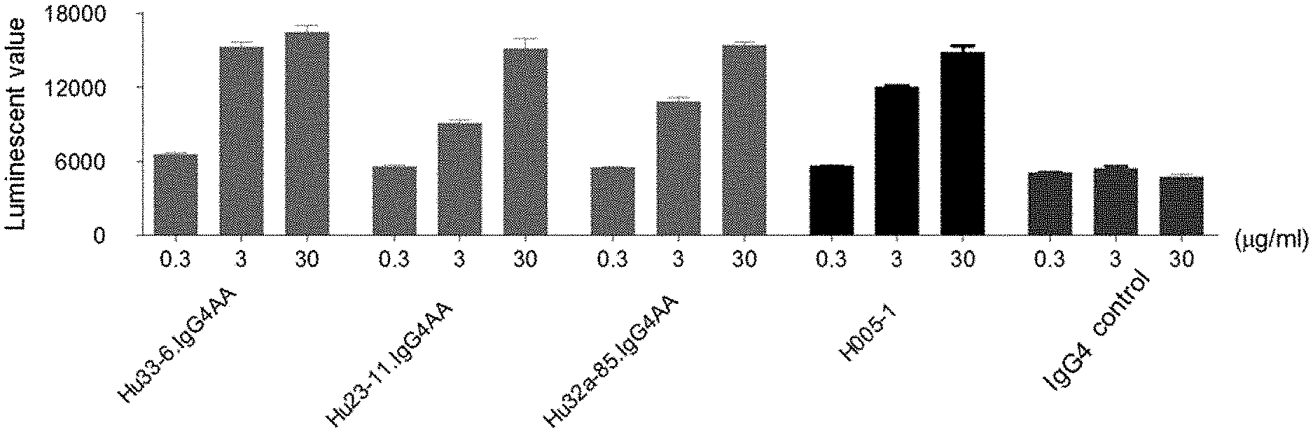
[0030] In some embodiments, the humanized antibody comprises a framework region derived from a human antibody or a framework region variant thereof.
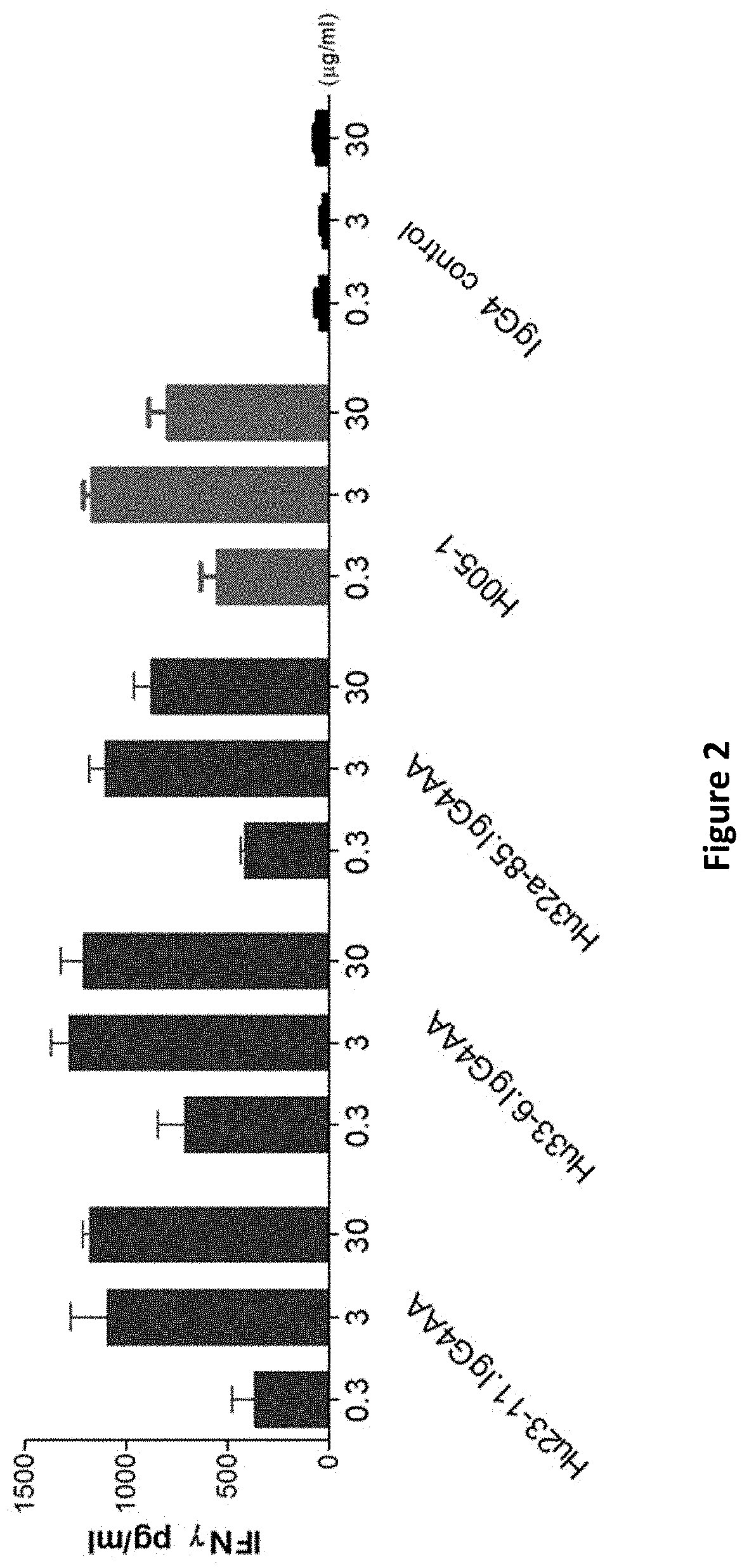
[0031] In some embodiments, the framework region variant has at most 11 amino acid back mutations on each of the light chain framework region and/or the heavy chain framework region of a human antibody.
[0032] In some embodiments, the framework region variant comprises mutation(s) selected from any one of the following (f) to (h):
[0033] (f) 2G amino acid back mutation comprised in the light chain variable region and/or one or more amino acid back mutations selected from 27Y, 48I, 67T, 69L, 82F and 93T comprised in the heavy chain variable region;
[0034] (g) 2V amino acid back mutation comprised in the light chain variable region, and/or one or more amino acid back mutations selected from 26D, 27F, 30T, 38K, 43H, 48I, 66K, 67A, 69L, 82F and 93T comprised in the heavy chain variable region; and
[0035] (h) one or more amino acid back mutations selected from 42G, 44V and 71Y comprised in the light chain variable region, and/or 1K and/or 94S amino acid back mutations comprised in the heavy chain variable region.
[0036] In some embodiments of the aforementioned anti-PD-1 antibody or antigen-binding fragment thereof, the anti-PD-1 antibody or antigen-binding fragment thereof comprises antibody variable regions selected from the group consisting of:
[0037] (a2) a heavy chain variable region comprising HCDR1, HCDR2 and HCDR3 as shown in SEQ ID NO: 8, SEQ ID NO: 9 and SEQ ID NO: 10, respectively, and one or more amino acid back mutations of 27Y, 48I, 67T, 69L, 82F and 93T comprised in the heavy chain framework region, and
[0038] a light chain variable region comprising LCDR2 and LCDR3 as shown in SEQ ID NO: 12 and SEQ ID NO: 13, respectively, and LCDR1 as shown in SEQ ID NO: 11, 47, 48, 49, 50, 51 or 52, and 2G amino acid back mutation comprised in the light chain framework region;
[0039] (b2) a heavy chain variable region comprising HCDR1, HCDR2 and HCDR3 as shown in SEQ ID NO: 14, SEQ ID NO: 15 and SEQ ID NO: 16, respectively, and one or more amino acid back mutations selected from 26D, 27F, 30T, 38K, 43H, 48I, 66K, 67A, 69L, 82F and 93T comprised in the heavy chain variable region; and
[0040] a light chain variable region comprising LCDR1, LCDR2 and LCDR3 as shown in SEQ ID NO: 17, SEQ ID NO: 12 and SEQ ID NO: 18, respectively, and 2V amino acid back mutation comprised in the light chain framework region;
[0041] (c2) a heavy chain variable region comprising HCDR1, HCDR2 and HCDR3 as shown in SEQ ID NO: 21, SEQ ID NO: 22 and SEQ ID NO: 23, respectively, and 1K and/or 94S amino acid back mutations comprised in the heavy chain framework region, and
[0042] a light chain variable region comprising LCDR1, LCDR2 and LCDR3 as shown in SEQ ID NO: 24, SEQ ID NO: 25 and SEQ ID NO: 26, respectively, and one or more amino acid back mutations selected from 42G, 44V and 71Y comprised in the light chain framework region.
[0043] In some embodiments of the aforementioned anti-PD-1 antibody or antigen-binding fragment thereof, the anti-PD-1 antibody or antigen-binding fragment thereof comprises antibody variable regions selected from any one of the following (i) to (o):
[0044] (i) a heavy chain variable region, the sequence of which is as shown in SEQ ID NO: 4 or has at least 90% sequence identity with SEQ ID NO: 4, and/or a light chain variable region, the sequence of which is as shown in SEQ ID NO: 5 or has at least 90% sequence identity with SEQ ID NO: 5;
[0045] (j) a heavy chain variable region, the sequence of which is as shown in SEQ ID NO: 6 or has at least 90% sequence identity with SEQ ID NO: 6, and/or a light chain variable region, the sequence of which is as shown in SEQ ID NO: 7 or has at least 90% sequence identity with SEQ ID NO: 7;
[0046] (k) a heavy chain variable region, the sequence of which is as shown in SEQ ID NO: 19 or has at least 90% sequence identity with SEQ ID NO: 19, and/or a light chain variable region, the sequence of which is as shown in SEQ ID NO: 20 or has at least 90% sequence identity with SEQ ID NO: 20;
[0047] (l) a heavy chain variable region, the sequence of which is as shown in SEQ ID NO: 27, 30, 31 or 32, or has at least 90% sequence identity with SEQ ID NO: 27, 30, 31 or 32, respectively, and/or
[0048] a light chain variable region, the sequence of which is as shown in SEQ ID NO: 28, 29, 53, 54, 55, 56, 57, 58, 59, 60, 61, 62, 63 or 64, or has at least 90% sequence identity with SEQ ID NO: 28, 29, 34, 35, 53, 54, 55, 56, 57, 58, 59, 60, 61, 62, 63 or 64, respectively;
[0049] (m) a heavy chain variable region, the sequence of which is as shown in SEQ ID NO: 33, 36, 37, 38, 39 or 40, or has at least 90% sequence identity with SEQ ID NO: 33, 36, 37, 38, 39 or 40, respectively, and/or
[0050] a light chain variable region, the sequence of which is as shown in SEQ ID NO: 34, 35, 28, 29, 53, 54, 55, 56, 57, 58, 59, 60, 61, 62, 63 or 64, or has at least 90% sequence identity with SEQ ID NO: 34, 35, 28, 29, 53, 54, 55, 56, 57, 58, 59, 60, 61, 62, 63 or 64, respectively;
[0051] (n) a heavy chain variable region, the sequence of which is as shown in SEQ ID NO: 41, 45 or 46, or has at least 90% sequence identity with SEQ ID NO: 41, 45 or 46, respectively, and/or
[0052] a light chain variable region, the sequence of which is as shown in SEQ ID NO: 42, 43 or 44, or has at least 90% sequence identity with SEQ ID NO: 42, 43 or 44, respectively;
[0053] (o) a heavy chain variable region, the sequence of which is as shown in SEQ ID NO: 70 or has at least 90% sequence identity with SEQ ID NO: 70, and/or
[0054] a light chain variable region, the sequence of which is as shown in SEQ ID NO: 71 or has at least 90% sequence identity with SEQ ID NO: 71; and
[0055] (p) a heavy chain variable region, the sequence of which is as shown in SEQ ID NO: 27, 30, 31 or 32, or has at least 90% sequence identity with SEQ ID NO: 27, 30, 31 or 32, respectively, and/or
[0056] a light chain variable region, the sequence of which is as shown in SEQ ID NO: 34 or 35, or has at least 90% sequence identity with SEQ ID NO: 34 or 35, respectively;
[0057] wherein the sequences SEQ ID NO: 70 and SEQ ID NO: 71 are represented by general formula sequences as shown in Table 2:
TABLE-US-00002 TABLE 2 EVQLVQSGAEVKKPGSSVKVSCKASX.sub.18X.sub.19TFX.sub.20D DX.sub.29VMTQTPLSLPVTP YEX.sub.1HWVX.sub.21QAPGX.sub.22GLEWX.sub.23GLX.sub.2DPETGGX.sub.3V GEPASISCRSSQSX.sub.13VH YNQKFKX.sub.4X.sub.24X.sub.25TX.sub.26TADKSTSTAYMEX.sub.27SSLRS SX.sub.14X.sub.15X.sub.16TYLEWYLQ EDTAVYYCX.sub.28REX.sub.5X.sub.6X.sub.7X.sub.8YX.sub.9X.sub.10X.sub.11X.su- b.12DWYFDV KPGQSPQLLIYKVSNRF WGQGTTVTVSS, wherein, X.sub.1 is selected from I or M, SGVPDRFSGSGSGTDFT X.sub.2 is selected from F or I, X.sub.3 is selected from I/T, X.sub.4 is LKISRVEAEDVGVYYC selected from G or D, X.sub.5 is selected from G or R, X.sub.6 is F FQGSHVPYX.sub.17FGGGTK or absent, X.sub.7 is S or absent, X.sub.8 is Y or absent, X.sub.9 is G or VEIK, wherein, X.sub.13 is absent, X.sub.10 is S or absent, X.sub.11 is selected from N or T, X.sub.12 selected from I or L, X.sub.14 is is selected from R or S, X.sub.18 is selected from G or D, X.sub.19 selected from N, Q, L, T or is selected from G, F or Y, X.sub.20 is selected from S or T, X.sub.21 D, X.sub.15 is selected from G, is selected from R or K, X.sub.22 is selected from Q or H, X.sub.23 A or V, X.sub.16 is selected from is selected from M or I, X.sub.24 is selected from R or K, X.sub.25 N or K, X.sub.17 is selected from is selected from V, A or T, X.sub.26 is selected from R or K, A or T and X.sub.29 is selected X.sub.27 is selected from L or F and X.sub.28 is selected from A or from I, V or G (SEQ ID T (SEQ ID NO: 70) NO: 71)
[0058] In some embodiments, the heavy chain variable region of the anti-PD-1 antibody is as shown in SEQ ID NO: 27 or has at least 90% identity with SEQ ID NO: 27, and the light chain variable region sequence of the anti-PD-1 antibody is as shown in SEQ ID NO: 55 or has at least 90% sequence identity with SEQ ID NO: 55.
[0059] In some embodiments, the heavy chain variable region of the anti-PD-1 antibody is as shown in SEQ ID NO: 46 or has at least 90% identity with SEQ ID NO: 46, and the light chain variable region sequence of the anti-PD-1 antibody is as shown in SEQ ID NO: 43 or has at least 90% sequence identity with SEQ ID NO: 43.
[0060] The aforementioned "at least 90% identity" includes having at least 90%, 91%, 92%, 93%, 94%, 95%, 96%, 97%, 98%, 99% or 100% identity.
[0061] In some other embodiments of the aforementioned anti-PD-1 antibody or antigen-binding fragment thereof, the antibody further comprises antibody constant regions; in some other embodiments, the heavy chain constant region of the antibody constant region is selected from human IgG1, IgG2, IgG3 and IgG4 constant regions and conventional variants thereof, the light chain constant region of the antibody constant region is selected from human antibody .kappa. and .lamda. chain constant regions and conventional variants thereof; in some other embodiments, the antibody constant regions comprise an IgG4 heavy chain constant region into which one or more mutations of S228P, F234A and L235A are introduced, for example, comprise an IgG4 heavy chain constant region having the three amino acid mutations of S228P, F234A and L235A; in some other embodiments, the antibody comprises a heavy chain constant region as shown in SEQ ID NO: 72 or SEQ ID NO: 79, and a light chain constant region as shown in SEQ ID NO: 73.
[0062] In some embodiments of the aforementioned anti-PD-1 antibody or antigen-binding fragment thereof, the anti-PD-1 antibody comprises a light chain as shown in SEQ ID NO: 78 and a heavy chain as shown in SEQ ID NO: 77 or 82; or
[0063] a light chain as shown in SEQ ID NO: 75 and a heavy chain as shown in SEQ ID NO: 74, 76, 80 or 81.
[0064] In some embodiments, the anti-PD-1 antibody comprises:
[0065] a heavy chain as shown in SEQ ID NO: 74 and a light chain as shown in SEQ ID NO: 75; or
[0066] a heavy chain as shown in SEQ ID NO: 77 and a light chain as shown in SEQ ID NO: 78.
[0067] In some embodiments, provided is an anti-PD-1 antibody or an antigen-binding fragment thereof, the antibody competitively binding to human PD-1 or binding to the same human PD-1 epitope with any one of the aforementioned anti-PD-1 antibodies or antigen-binding fragments thereof.
[0068] In some embodiments of the aforementioned anti-PD-1 antibody or antigen-binding fragment thereof, the antibody is a bispecific antibody or a multispecific antibody.
[0069] In some embodiments of the anti-PD-1 antibody or antigen-binding fragment thereof, the antigen-binding fragment is selected from the group consisting of Fab, Fab', F(ab).sub.2, single-chain antibody (scFv), dimerized V region (diabody) and disulfide bond stabilized V region (dsFv).
[0070] In some embodiments, disclosed is an isolated monoclonal antibody or antigen-binding fragment thereof, said antibody competitively binding to human PD-1 with the anti-PD-1 antibody or antigen-binding fragment thereof according to any one of the foregoing.
[0071] In some embodiments, the present disclosure also provides a pharmaceutical composition, which comprises a therapeutically effective amount of the anti-PD-1 antibody or antigen-binding fragment thereof according to any one of the foregoing, or a therapeutically effective amount of the aforementioned isolated monoclonal antibody or antigen-binding fragment thereof, and one or more pharmaceutically acceptable carriers, diluents, buffers or excipients. In some embodiments, the therapeutically effective amount refers to a unit dose of the composition containing 0.1 to 3000 mg of the aforementioned anti-PD-1 antibody or antigen-binding fragment thereof.
[0072] In some embodiments, the present disclosure also provides a nucleic acid molecule that encodes the anti-PD-1 antibody or antigen-binding fragment thereof according to any one of the foregoing, or encodes the aforementioned isolated monoclonal antibody or antigen-binding fragment thereof.
[0073] In some embodiments, the present disclosure also provides a host cell comprising the aforementioned nucleic acid molecule.
[0074] In some embodiments, the present disclosure also provides a method for immunodetection or determination of PD-1, which comprises a step of using the anti-PD-1 antibody or antigen-binding fragment thereof according to any one of the foregoing, or a step of using the aforementioned isolated monoclonal antibody or antigen-binding fragment thereof.
[0075] In some embodiments, the present disclosure also provides a kit comprising the aforementioned anti-PD-1 antibody or antigen-binding fragment thereof or the aforementioned isolated monoclonal antibody or antigen-binding fragment thereof.
[0076] In some embodiments, provided is use of the aforementioned anti-PD-1 antibody or antigen-binding fragment thereof in preparing diagnostic agents for PD-1 related diseases.
[0077] In some embodiments, the present disclosure also provides a method for treating diseases, which comprises administering to a subject a therapeutically effective amount of the anti-PD-1 antibody or antigen-binding fragment thereof according to any one of the foregoing, or the aforementioned isolated monoclonal antibody or antigen-binding fragment thereof, or the aforementioned pharmaceutical composition, or the aforementioned nucleic acid molecule.
[0078] In some embodiments, the disease is tumor.
[0079] In some other embodiments, the disease is selected from: head and neck squamous cell carcinoma, head and neck cancer, brain cancer, glioma, glioblastoma multiforme, neuroblastoma, central nervous system cancer, neuroendocrine tumor, pharyngeal cancer, nasopharyngeal cancer, esophageal cancer, thyroid cancer, malignant pleural mesothelioma, lung cancer, breast cancer, liver cancer, hepatoma, hepatocellular carcinoma, hepatobiliary cancer, pancreatic cancer, gastric cancer, gastrointestinal cancer, bowel cancer, colon cancer, colorectal cancer, kidney cancer, clear cell renal cell carcinoma, ovarian cancer, endometrial cancer, cervical cancer, bladder cancer, prostate cancer, testicular cancer, skin cancer, melanoma, leukemia, lymphoma, bone cancer, chondrosarcoma, myeloma, multiple myeloma, myelodysplastic syndrome, myeloproliferative neoplasm, squamous cell carcinoma, Ewing's sarcoma, systemic light chain amyloidosis and Merkel cell carcinoma; in some of these embodiments, the lymphoma is selected from: Hodgkin's lymphoma, non-Hodgkin's lymphoma, diffuse large B-cell lymphoma, follicular lymphoma, primary mediastinal large B-cell lymphoma, mantle cell lymphoma, small lymphocytic lymphoma, T-cell/histiocyte rich large B-cell lymphoma and lymphoplasmacytic lymphoma, the lung cancer is selected from: non-small cell lung cancer and small cell lung cancer, the leukemia is selected from: chronic myeloid leukemia, acute myeloid leukemia, lymphocytic leukemia, lymphoblastic leukemia, acute lymphoblastic leukemia, chronic lymphocytic leukemia and myeloid leukemia; in some other embodiments, the disease is selected from: PD-L1 positive melanoma, lung cancer, non-small cell lung cancer, breast cancer, gastric cancer, kidney cancer, bladder cancer, bowel cancer and colon cancer.
[0080] In some embodiments, the present disclosure also provides use of the anti-PD-1 antibody or antigen-binding fragment thereof according to any one of the foregoing, or the aforementioned isolated monoclonal antibody or antigen-binding fragment thereof, or the aforementioned pharmaceutical composition, or the aforementioned nucleic acid molecule, in preparing medicaments for the treatment or prevention of diseases.
[0081] In some embodiments, the disease is tumor.
[0082] In some other embodiments, the disease is selected from: head and neck squamous cell carcinoma, head and neck cancer, brain cancer, glioma, glioblastoma multiforme, neuroblastoma, central nervous system cancer, neuroendocrine tumor, pharyngeal cancer, nasopharyngeal cancer, esophageal cancer, thyroid cancer, malignant pleural mesothelioma, lung cancer, breast cancer, liver cancer, hepatoma, hepatocellular carcinoma, hepatobiliary cancer, pancreatic cancer, gastric cancer, gastrointestinal cancer, bowel cancer, colon cancer, colorectal cancer, kidney cancer, clear cell renal cell carcinoma, ovarian cancer, endometrial cancer, cervical cancer, bladder cancer, prostate cancer, testicular cancer, skin cancer, melanoma, leukemia, lymphoma, bone cancer, chondrosarcoma, myeloma, multiple myeloma, myelodysplastic syndrome, myeloproliferative neoplasm, squamous cell carcinoma, Ewing's sarcoma, systemic light chain amyloidosis and Merkel cell carcinoma; in some of these embodiments, the lymphoma is selected from: Hodgkin's lymphoma, non-Hodgkin's lymphoma, diffuse large B-cell lymphoma, follicular lymphoma, primary mediastinal large B-cell lymphoma, mantle cell lymphoma, small lymphocytic lymphoma, T-cell/histiocyte rich large B-cell lymphoma and lymphoplasmacytic lymphoma, the lung cancer is selected from: non-small cell lung cancer and small cell lung cancer, the leukemia is selected from: chronic myeloid leukemia, acute myeloid leukemia, lymphocytic leukemia, lymphoblastic leukemia, acute lymphoblastic leukemia, chronic lymphocytic leukemia and myeloid leukemia; in some other embodiments, the disease is selected from: PD-L1 positive melanoma, lung cancer, non-small cell lung cancer, breast cancer, gastric cancer, kidney cancer, bladder cancer, bowel cancer and colon cancer.
[0083] In some embodiments, the present disclosure also provides the anti-PD-1 antibody or antigen-binding fragment thereof according to any one of the foregoing, or the aforementioned isolated monoclonal antibody or antigen-binding fragment thereof, or the aforementioned nucleic acid molecule, or the aforementioned pharmaceutical composition, for use as medicaments.
[0084] In some embodiments, the medicament is used for the treatment or prevention of PD-1 related diseases.
[0085] In some embodiments, the disease is tumor.
[0086] In some other embodiments, the disease is selected from: head and neck squamous cell carcinoma, head and neck cancer, brain cancer, glioma, glioblastoma multiforme, neuroblastoma, central nervous system cancer, neuroendocrine tumor, pharyngeal cancer, nasopharyngeal cancer, esophageal cancer, thyroid cancer, malignant pleural mesothelioma, lung cancer, breast cancer, liver cancer, hepatoma, hepatocellular carcinoma, hepatobiliary cancer, pancreatic cancer, gastric cancer, gastrointestinal cancer, bowel cancer, colon cancer, colorectal cancer, kidney cancer, clear cell renal cell carcinoma, ovarian cancer, endometrial cancer, cervical cancer, bladder cancer, prostate cancer, testicular cancer, skin cancer, melanoma, leukemia, lymphoma, bone cancer, chondrosarcoma, myeloma, multiple myeloma, myelodysplastic syndrome, myeloproliferative neoplasm, squamous cell carcinoma, Ewing's sarcoma, systemic light chain amyloidosis and Merkel cell carcinoma; in some of these embodiments, the lymphoma is selected from: Hodgkin's lymphoma, non-Hodgkin's lymphoma, diffuse large B-cell lymphoma, follicular lymphoma, primary mediastinal large B-cell lymphoma, mantle cell lymphoma, small lymphocytic lymphoma, T-cell/histiocyte rich large B-cell lymphoma and lymphoplasmacytic lymphoma, the lung cancer is selected from: non-small cell lung cancer and small cell lung cancer, the leukemia is selected from: chronic myeloid leukemia, acute myeloid leukemia, lymphocytic leukemia, lymphoblastic leukemia, acute lymphoblastic leukemia, chronic lymphocytic leukemia and myeloid leukemia; in some other embodiments, the disease is selected from: PD-L1 positive melanoma, lung cancer, non-small cell lung cancer, breast cancer, gastric cancer, kidney cancer, bladder cancer, bowel cancer and colon cancer.
DESCRIPTION OF THE DRAWINGS
[0087] FIG. 1: the test results of anti-PD-1 antibodies blocking the binding of PD-1 to its ligand;
[0088] FIG. 2: the effect of anti-PD-1 antibodies on the secretion of IFN.gamma. from PBMC cells;
[0089] FIG. 3: the efficiency of anti-PD-1 antibodies on xenograft tumor of colon cancer MC38 in mice;
[0090] FIG. 4: the effect of anti-PD-1 antibodies on the tumor volume of colon cancer MC38 in mice.
DETAILED DESCRIPTION OF THE INVENTION
[0091] Overview
[0092] To make the present disclosure be understood easier, certain technical and scientific terms are specifically defined below. Unless clearly defined otherwise herein, all other technical and scientific terms used herein have the meanings commonly understood by those of ordinary skill in the art to which the present disclosure belongs.
[0093] The terms "programmed death 1", "programmed cell death 1", "protein PD-1", "PD-1", "PDCD1" and "hPD-1" are used interchangeably and include human PD-1 variants, isotypes, species homologs, and analogs that have at least one epitope in common with PD-1. The complete PD-1 sequence can be found with GenBank accession number U64863.
[0094] The term "programmed death ligand-1 (PD-L1)" is one of the two cell surface glycoprotein ligands of PD-1 (the other is PD-L2), which down-regulates T cell activation and cytokine secretion when it binds to PD-1. The term "PD-L1" as used herein includes human PD-L1 (hPD-L1), hPD-L1 variants, isotypes and interspecies homologs, and 5 analogs that have at least one epitope in common with hPD-1. The complete hPD-L1 sequence can be found with GenBank accession number Q9NZQ7.
[0095] The term "cytokine" is a general term for proteins released by a population of cells and acting on other cells as intercellular mediators. Examples of such cytokines include lymphokines, monokines, chemokines and traditional polypeptide hormones. Exemplary cytokines include: human IL-2, IFN-.gamma., IL-6, TNF.alpha., IL-17 and IL-5.
[0096] The three-letter codes and one-letter codes of amino acids used in the present disclosure are as described in J. biol. chem, 243, p 3558 (1968).
[0097] The "antibody" described in the present disclosure refers to an immunoglobulin generally. A natural intact antibody is a tetrapeptide chain structure composed of two identical heavy chains and two identical light chains linked by interchain disulfide bonds. The amino acid compositions and sequences of the immunoglobulin heavy chain constant regions are different, so their antigenicity is also different. Thereby, immunoglobulins can be divided into five classes, or named as isotypes of immunoglobulins, namely IgM, IgD, IgG, IgA and IgE, and their corresponding heavy chains are .mu. chain, .delta. chain, .gamma. chain, .alpha. chain and .epsilon. chain, respectively. The same class of Ig can be divided into different subclasses according to the difference in the amino acid composition of the hinge region and the number and position of heavy chain disulfide bonds. For example, IgG can be divided into IgG1, IgG2, IgG3 and IgG4. The light chain is divided into .kappa. chain or .lamda. chain by the difference of the constant region. Each of the five classes of Ig can have a .kappa. chain or a .lamda. chain. The antibodies referred to in the present disclosure include antibodies or antigen-binding fragments thereof, including antibodies or antigen-binding fragments thereof that have been modified on the basis of immunoglobulin while retaining the ability to bind antigen; including monospecific antibodies, bispecific antibodies or multispecific antibodies; also including monovalent antibodies, bivalent antibodies or multivalent antibodies. Antigen-binding fragments of antibodies, for example, can be those comprising at least one VH-CH1 and at least one VL-CL structure, in which the VH and VL structures can approach to each other based on the interchain interaction and retain antigen binding ability. In some embodiments, the antigen-binding fragment of the antibody is a monovalent Fab fragment (Fab 1 fragment), bivalent Fab fragment (F(ab)2), trivalent Fab fragment (F(ab)3), multivalent (two or more) Fab fragment, and can also be other monospecific, bispecific or multispecific antigen binding fragments comprising at least one Fab fragment.
[0098] "Bispecific antibody" refers to an antibody (including antibody or antigen-binding fragment thereof, such as single-chain antibody) that can specifically bind to two different antigens or two different epitopes of the same antigen. The prior art has disclosed bispecific antibodies with various structures. According to the integrity of the IgG molecule, they can be divided into IgG-like bispecific antibodies and antibody fragment-type bispecific antibodies. According to the number and configuration of the antigen binding regions, they can be divided into bivalent, trivalent, tetravalent or more-valent bispecific antibodies. According to whether the structure is symmetric, they can be divided into symmetric structure bispecific antibodies and asymmetric structure bispecific antibodies. Among them, antibody fragment-based bispecific antibodies, for example Fab fragments lacking Fc fragment, form bispecific antibodies by binding two or more Fab fragments in one molecule. These antibodies have lower immunogenicity and smaller molecular weight and higher tumor tissue permeability. Typical antibody structures of this type are bispecific antibodies such as F(ab)2, scFv-Fab, (scFv)2-Fab, etc.; For IgG-like bispecific antibodies (for example with Fc fragment), this type of antibody has larger molecular weight. The Fc fragment helps the purification of the antibody in later stages and improves its solubility and stability. The Fc moiety may also bind to the receptor FcRn and increase the serum half-life of the antibody. Typical bispecific antibody structure models are such as KiH, CrossMAb, Triomab quadroma, Fc.DELTA.Adp, ART-Ig, BiMAb, Biclonics, BEAT, DuoBody, Azymetric, XmAb, 2:1 TCBs, 1Fab-IgG TDB, FynomAb, two-in-one/DAF, scFv-Fab-IgG, DART-Fc, LP-DART, CODV-Fab-TL, HLE-BiTE, F(ab)2-CrossMAb, IgG-(scFv)2, Bs4Ab, DVD-Ig, Tetravalent-DART-Fc, (scFv)4-Fc, CODV-Ig, mAb2, F(ab)4-CrossMAb and other bispecific antibodies (see Aran F. Labrijn et al., Nature Reviews Drug Discovery volume 18, pages 585-608 (2019); Chen S1 et al., J Immunol Res. 2019 Feb. 11; 2019:4516041).
[0099] The term "monovalent", "bivalent", "trivalent" or "multivalent" refers to the presence of a specified number of antigen binding sites in an antibody or polypeptide complex. For example "monovalent antibody" means that there is one antigen binding site in the antibody, "Monovalent polypeptide complex" means that there is one antigen binding site in the polypeptide complex; "Bivalent antibody" means that there are two antigen binding sites in the antibody, "Bivalent polypeptide complex" means that there are two antigen binding sites in the polypeptide complex; "Trivalent antibody" means that there are three antigen binding sites in the antibody, "Trivalent polypeptide complex" means that there are three antigen binding sites in the polypeptide complex; "Multivalent antibody" means that there are multiple (three or more) antigen binding sites in the antibody, and "Multivalent polypeptide complex" means that there are multiple (two or more) antigen binding sites in the polypeptide complex.
[0100] The term "antibody fusion protein" refers to a biologically active fusion protein formed by linking a protein of interest (polypeptide) with an immunoglobulin. The fusion protein has the biological activity both of the linked protein and of the immunoglobulin.
[0101] The sequence of about 110 amino acids near the N-terminus of the antibody heavy and light chain varies greatly and is the variable region (Fv region); the remaining amino acid sequence near the C-terminus is relatively stable and is the constant region. The variable region includes 3 hypervariable regions (HVRs) and 4 framework regions (FRs) with relatively conservative sequences. The 3 hypervariable regions which determine the specificity of the antibody are also known as complementarity determining regions (CDRs). Each of the light chain variable region (VL) and heavy chain variable region (VH) consists of 3 CDR regions and 4 FR regions. The order from the amino terminus to the carboxyl terminus is: FR1, CDR1, FR2, CDR2, FR3, CDR3, FR4. The 3 CDR regions of the light chain refer to LCDR1, LCDR2 and LCDR3; and the 3 CDR regions of the heavy chain refer to HCDR1, HCDR2 and HCDR3.
[0102] The antibodies of the present disclosure include murine antibodies, chimeric antibodies, humanized antibodies and fully human antibodies, preferably humanized antibodies.
[0103] The term "murine antibody" in the present disclosure is a monoclonal antibody against human PD-1 prepared according to the knowledge and skills in the art. During preparation, the test subject is injected with PD-1 antigen, and then hybridomas expressing antibodies with the desired sequence or functional properties are isolated. In a preferred embodiment of the present disclosure, the murine anti-PD-1 antibody or antigen-binding fragment thereof can further comprise the light chain constant region of murine .kappa., .lamda. chain or variant thereof, or further comprise the heavy chain constant region of murine IgG1, IgG2, IgG3 or variant thereof.
[0104] The term "chimeric antibody" is an antibody formed by fusing the variable region of a murine antibody with the constant region of a human antibody, which can alleviate the immune response induced by murine antibody. Establishing a chimeric antibody requires first establishing a hybridoma secreting murine specific monoclonal antibodies, then cloning the variable region gene from the murine hybridoma cells, and then cloning the constant region gene of the human antibody as necessary, linking the murine variable region gene with the human constant region gene to form a chimeric gene, inserting the chimeric gene into an expression vector, and finally expressing the chimeric antibody molecule in a eukaryotic system or a prokaryotic system. In a preferred embodiment of the present disclosure, the antibody light chain of the PD-L1 chimeric antibody further comprises a light chain constant region of a human .kappa., .lamda. chain or variant thereof. The antibody heavy chain of the PD-1 chimeric antibody further comprises the heavy chain constant region of human IgG1, IgG2, IgG3, IgG4 or variant thereof, preferably comprises the heavy chain constant region of human IgG1, IgG2 or IgG4, or comprises IgG1, IgG2, or IgG4 variants with amino acid mutations (for example L234A and/or L235A mutations, and/or S228P mutations).
[0105] The term "humanized antibody", also known as CDR-grafted antibody, refers to an antibody produced by grafting murine CDR sequences into the framework of human antibody variable regions, that is, refers to an antibody produced in different types of human germline antibody framework sequences. It can overcome the heterogeneous reaction induced by the chimeric antibody as it carries a large amount of murine protein components. Such framework sequences can be obtained from public DNA databases or published references that include germline antibody gene sequences. For example, the germline DNA sequences of the human heavy chain and light chain variable region genes can be found in the "VBase" human germline sequence database (available on the Internet www.mrccpe.com.ac.uk/vbase), as well as in Kabat, E. A., et al., 1991, Sequences of Proteins of Immunological Interest, 5th edition. In order to avoid the decrease in activity at the same time caused by the decrease in immunogenicity, the human antibody variable region framework sequence can be subjected to minimal reverse mutations or back mutations to maintain activity. The humanized antibodies of the present disclosure also include humanized antibodies that have been further subjected to affinity maturation of the CDRs by yeast display.
[0106] Due to the residues in contact with the antigen, CDR grafting may result in reduced affinity of the produced antibody or antigen-binding fragment thereof to the antigen due to the framework residues in contact with the antigen. Such interactions may be the result of hypermutation of somatic cells. Therefore, it may still be necessary to graft such donor framework amino acids to the framework of the humanized antibody. The amino acid residues involved in antigen binding and from non-human antibodies or antigen-binding fragments thereof can be identified by examining the sequence and structure of the animal monoclonal antibody variable region. Residues in the CDR donor framework that differ from the germline can be considered related. If the closest germline cannot be determined, the sequence can be compared with the consensus sequence of a subclass or of animal antibody sequence with a high percentage of similarity. Rare framework residues are thought to be the result of hypermutation of somatic cells and thus play an important role in binding.
[0107] In an embodiment of the present disclosure, the antibody or antigen-binding fragment thereof can further comprise a light chain constant region of human or murine .kappa., .lamda. chain or variant thereof, or further comprise a heavy chain constant region of human or murine IgG1, IgG2, IgG3, IgG4 or variant thereof; preferably comprise a heavy chain constant region of human IgG1, IgG2 or IgG4, or IgG1, IgG2 or IgG4 variant with amino acid mutations (for example L234A and/or L235A mutation, and/or S228P mutation).
[0108] The "conventional variant" of the human antibody heavy chain constant region and the human antibody light chain constant region described in the present disclosure refers to the variant of heavy chain constant region or light chain constant region that has been disclosed in the prior art and does not change the structure and function of the antibody variable region. Exemplary variants include IgG1, IgG2, IgG3 or IgG4 heavy chain constant region variants with site-directed modifications and amino acid substitutions of the heavy chain constant region. Specific substitutions are such as YTE mutations, L234A and/or L235A mutations, S228P mutations, and/or mutations to obtain a knob-into-hole structure (making the antibody heavy chain have a combination of knob-Fc and hole-Fc) known in the art. These mutations have been confirmed to make the antibody have new properties, but do not change the function of the antibody variable region.
[0109] "HuMAb", "human antibody", "fully human antibody" and "complete human antibody" can be used interchangeably, and can refer to antibodies derived from humans or antibodies obtained from a genetically modified organism which is "engineered" to produce specific human antibodies in response to antigen stimulation and can be produced by any method known in the art. In some technologies, the elements of human heavy and light chain loci are introduced into cell lines of organisms derived from embryonic stem cell lines, in which the endogenous heavy chain and light chain loci have been target disrupted by what targets the endogenous heavy chain and light chain loci contained in these cell lines. Transgenic organisms can synthesize human antibodies specific to human antigens, and the organisms can be used to produce human antibody-secreting hybridomas. A human antibody can also be an antibody in which the heavy and light chains are encoded by nucleotide sequences derived from one or more human DNA sources. Fully human antibodies can also be constructed by gene or chromosome transfection methods and phage display technology, or constructed from B cells activated in vitro, all of which are known in the art.
[0110] The terms "full-length antibody", "intact antibody", "complete antibody" and "whole antibody" are used interchangeably herein and refer to an antibody in a substantially intact form, as distinguished from the antigen-binding fragments defined below. These terms specifically refer to an antibody in which the heavy chain comprises Fc region.
[0111] The term "antigen-binding fragment" or "functional fragment" of an antibody refers to one or more fragments of the antibody that retain the ability to specifically bind to an antigen (for example, PD-1). It has been shown that fragments of full-length antibodies can be used to perform the antigen-binding function of antibodies. Examples of the binding fragment included in the term "antigen-binding fragment" of the antibody include (i) Fab fragments, monovalent fragments consist of VL, VH, CL and CH1 domains; (ii) F(ab').sub.2 fragments, including bivalent fragments of two Fab fragments linked by a disulfide bridge in the hinge region, (iii) Fd fragments consist of VH and CH1 domains; (iv) Fv fragments composed of VH and VL domains of one arm of an antibody; (V) single domains or dAb fragments (Ward et al., (1989) Nature 341:544-546), which consist of a VH domain; and (vi) isolated complementarity determining regions (CDR) or (vii) combinations of two or more isolated CDRs, optionally linked by synthetic linkers. In addition, although the two domains VL and VH of the Fv fragment are encoded by separate genes, recombination methods can be used to link them by synthetic linkers so that it can be produced as a single protein chain in which the VL and VH regions pair to form a monovalent molecule (referred to as single-chain Fv (scFv); see, for example, Bird et al. (1988) Science 242:423-426; and Huston et al. (1988) Proc. Natl. Acad. Sci USA 85: 5879-5883). Such single chain antibodies are also intended to be included in the term "antigen-binding fragment" of antibody. Such antibody fragments are obtained by using conventional techniques known to those skilled in the art, and the functions of the fragments are screened in the same manner as for those of intact antibodies. The antigen binding moiety can be produced by recombinant DNA technology or by enzymatic or chemical cleavage of the intact immunoglobulin. The antibodies may be antibodies of different isotypes, for example, IgG (for example, IgG1, IgG2, IgG3 or IgG4 subtypes), IgA1, IgA2, IgD, IgE or IgM antibodies.
[0112] The antigen-binding fragments of the present disclosure include Fab, F(ab')2, Fab', single-chain antibody (scFv), dimerized V region (diabody), disulfide bond stabilized V region (dsFv), CDR-containing peptide and the like.
[0113] Fab is an antibody fragment that has a molecular weight of about 50,000 and has antigen-binding activity, and it is obtained by treating IgG antibody molecules with the protease papain (cleaves the amino acid residue at position 224 of the H chain), in which about half of the N-terminal side of the H chain and the entire L chain are joined together by disulfide bond(s).
[0114] The Fab of the present disclosure can be produced by using papain to treat the monoclonal antibody of the present disclosure which specifically recognizes human PD-1 and binds to the amino acid sequence of the extracellular region or three-dimensional structure thereof. In addition, the Fab can be produced by inserting the DNA encoding the Fab of the antibody into a prokaryotic expression vector or a eukaryotic expression vector and introducing the vector into a prokaryotic organism or eukaryotic organism to express the Fab.
[0115] F(ab')2 is an antibody fragment that has a molecular weight of about 100,000 and has antigen-binding activity and comprises two Fab regions linked at the hinge position, and it is obtained by digesting the lower part of the two disulfide bonds in the hinge region of IgG with the enzyme pepsin.
[0116] The F(ab')2 of the present disclosure can be produced by using pepsin to treat the monoclonal antibody of the present disclosure which specifically recognizes human PD-1 and binds to the amino acid sequence of the extracellular region or three-dimensional structure thereof. In addition, the F(ab')2 can be produced by linking Fab' described below with thioether bond or disulfide bond.
[0117] Fab' is an antibody fragment that has a molecular weight of about 50,000 and has antigen-binding activity obtained by cleaving the disulfide bond in the hinge region of the F(ab')2. The Fab' of the present disclosure can be produced by using reducing agents for example dithiothreitol to treat the F(ab')2 of the present disclosure which specifically recognizes PD-1 and binds to the amino acid sequence of the extracellular region or three-dimensional structure thereof.
[0118] In addition, the Fab' can be produced by inserting the DNA encoding the Fab' fragment of the antibody into a prokaryotic expression vector or a eukaryotic expression vector and then introducing the vector into a prokaryotic organism or eukaryotic organism to express the Fab'.
[0119] The term "single-chain antibody", "single-chain Fv" or "scFv" refers to molecules comprising an antibody heavy chain variable domain (or region; VH) and an antibody light chain variable domain (or region; VL) linked by a linker. Such scFv molecules can have the general structure: NH.sub.2-VL-linker-VH-COOH or NH.sub.2-VH-linker-VL-COOH. Suitable linkers in prior art consist of repeated GGGGS amino acid sequences or variants thereof, for example a variant with 1 to 4 repeated sequences (Holliger et al. (1993), Proc. Natl. Acad. Sci. USA 90: 6444-6448). Other linkers that can be used in the present disclosure are described in Alfthan et al. (1995), Protein Eng. 8:725-731, Choi et al. (2001), Eur. J. Immunol. 31:94-106, Hu et al. (1996), Cancer Res. 56:3055-3061, Kipriyanov et al. (1999), J. Mol. Biol. 293:41-56 and Roovers et al. (2001), Cancer Immunol.
[0120] The scFv of the present disclosure can be produced by the following steps: obtaining the cDNA encoding VH and VL of the monoclonal antibody of the present disclosure which specifically recognizes human PD-1 and binds to the amino acid sequence of the extracellular region or three-dimensional structure thereof, constructing the DNA encoding the scFv, inserting the DNA into a prokaryotic expression vector or a eukaryotic expression vector, and then introducing the expression vector into a prokaryotic organism or eukaryotic organism to express the scFv.
[0121] Diabody is a antibody fragment in which scFv is dimerized, and is a antibody fragment with bivalent antigen-binding activity. In the bivalent antigen binding activity, the two antigens can be the same or different.
[0122] The diabody of the present disclosure can be produced by the following steps: obtaining the cDNA encoding VH and VL of the monoclonal antibody of the present disclosure which specifically recognizes human PD-1 and binds to the amino acid sequence of the extracellular region or three-dimensional structure thereof, constructing the DNA encoding the scFv so that the peptide linker has 8 or less amino acid sequence residues in length, inserting the DNA into a prokaryotic expression vector or a eukaryotic expression vector, and then introducing the expression vector into a prokaryote organism or eukaryotic organism to express the diabody.
[0123] dsFv is obtained by linking polypeptides in which one amino acid residue in each of VH and VL is replaced by a cysteine residue via a disulfide bond between the cysteine residues. The amino acid residues substituted with cysteine residues can be selected according to a known method (Protein Engineering, 7, 697 (1994)) based on the prediction of the antibody three-dimensional structure.
[0124] The dsFv of the present disclosure can be produced by the following steps: obtaining the cDNA encoding VH and VL of the monoclonal antibody of the present disclosure which specifically recognizes human PD-1 and binds to the amino acid sequence of the extracellular region or three-dimensional structure thereof, constructing the DNA encoding the dsFv, inserting the DNA into a prokaryotic expression vector or a eukaryotic expression vector, and then introducing the expression vector into a prokaryotic organism or eukaryotic organism to express the dsFv.
[0125] The CDR-containing peptide is constructed by one or more regions including the CDR(s) of VH or VL. Peptides containing multiple CDR can be linked directly or via a suitable peptide linker.
[0126] The CDR-containing peptide of the present disclosure can be produced by the following steps: constructing the DNA(s) encoding the CDR(s) of VH and VL of the monoclonal antibody of the present disclosure which specifically recognizes human PD-1 and binds to the amino acid sequence of the extracellular region or three-dimensional structure thereof, inserting the DNA(s) into a prokaryotic expression vector or a eukaryotic expression vector, and then introducing the expression vector into a prokaryotic organism or eukaryotic organism to express the peptide. The CDR-containing peptide can also be produced by chemical synthesis methods for example the Fmoc method or the tBoc method.
[0127] The term "amino acid difference" or "amino acid mutation" refers to the presence of amino acid change(s) or mutation(s) in the protein or polypeptide variant compared with the original protein or polypeptide, including having 1, 2, 3 or more amino acid insertion, deletion or substitution on the basis of the original protein or polypeptide.
[0128] The term "antibody framework" or "FR region" refers to a moiety of the variable domain VL or VH, which serves as a scaffold for the antigen binding loop (CDR) of the variable domain. Essentially, it is a variable domain without CDR.
[0129] The term "complementarity determining region", "CDR" or "hypervariable region" refers to one of the six hypervariable regions in the variable domain of an antibody that mainly contribute to antigen binding. Generally, there are three CDRs (HCDR1, HCDR2 and HCDR3) in each heavy chain variable region, and three CDRs (LCDR1, LCDR2 and LCDR3) in each light chain variable region. Any one of well-known schemes can be used to determine the amino acid sequence boundaries of the CDRs, including the "Kabat" numbering criteria (see Kabat et al. (1991), "Sequences of Proteins of Immunological Interest", 5th edition, Public Health Service, National Institutes of Health, Bethesda, Md.), "Chothia" numbering criteria (see Al-Lazikani et al., (1997) JMB 273:927-948) and ImmunoGenTics (IMGT) numbering criteria (Lefranc M. P., Immunologist, 7, 132-136 (1999); Lefranc, M. P., et al., Dev. Comp. Immunol., 27, 55-77 (2003)), etc. For example, for the classical format, following the Kabat Criteria, the numbering of CDR amino acid residues in the heavy chain variable domain (VH) is 31-35 (HCDR1), 50-65 (HCDR2) and 95-102 (HCDR3); the numbering of CDR amino acid residues in the light chain variable domain (VL) is 24-34 (LCDR1), 50-56 (LCDR2) and 89-97 (LCDR3). Following the Chothia criteria, the numbering of CDR amino acid residues in VH is 26-32 (HCDR1), 52-56 (HCDR2) and 95-102 (HCDR3); and the numbering of amino acid residues in VL is 26-32 (LCDR1), 50-52 (LCDR2) and 91-96 (LCDR3). According to the combination of the CDR definitions of both Kabat and Chothia, the CDRs consist of amino acid residues 26-35 (HCDR1), 50-65 (HCDR2) and 95-102 (HCDR3) in human VH and amino acid residues 24-34 (LCDR1), 50-56 (LCDR2) and 89-97 (LCDR3) in human VL. Following IMGT criteria, the numbering of CDR amino acid residues in VH is roughly 26-35 (CDR1), 51-57 (CDR2) and 93-102 (CDR3), and the numbering of CDR amino acid residues in VL is roughly 27-32 (CDR1), 50-52 (CDR2) and 89-97 (CDR3). Following IMGT criteria, the CDR regions of an antibody can be determined using the program IMGT/DomainGap Align.
[0130] The term "epitope" or "antigenic determinant" refers to a site on an antigen to which an immunoglobulin or antibody specifically binds (for example, a specific site on PD-L1 molecules). Epitopes usually include at least 3, 4, 5, 6, 7, 8, 9, 10, 11, 12, 13, 14 or 15 consecutive or non-consecutive amino acids in a unique spatial conformation. See, for example, Epitope Mapping Protocols in Methods in Molecular Biology, Vol. 66, G. E. Morris, Ed. (1996).
[0131] The terms "specifically binds", "selectively binds", "binds selectively" and "binds specifically" refer to the binding of an antibody to an epitope on a predetermined antigen. Generally, antibodies bind with an affinity (KD) of about less than 10.sup.-8 M, for example about less than 10.sup.-9M, 10.sup.-10 M, 10.sup.-11M or less.
[0132] The term "KD" or "Kd" refers to the dissociation equilibrium constant of a specific antibody-antigen interaction. Generally, the antibodies of the present disclosure bind to PD-1 with a dissociation equilibrium constant (KD) of less than about 10.sup.-7 M, for example, less than about 10.sup.-8 M or 10.sup.-9 M, for example, as measured in a BIACORE instrument using surface plasma resonance (SPR) technology.
[0133] When the term "competition" is used in the context of antigen binding proteins that compete for the same epitope (for example neutralizing antigen binding proteins or neutralizing antibodies), it refers to the competition between the antigen binding proteins, and can be determined by the following assay: in the assay, the antigen-binding protein to be tested (for example an antibody or immunologically functional fragment thereof) prevents or inhibits (for example reduces) the specific binding of a reference antigen-binding protein (for example a ligand or a reference antibody) to a common antigen (for example PD-1 or fragment thereof). Numerous types of competitive binding assays can be used to determine whether one antigen-binding protein competes with another, these assays are for example: solid phase direct or indirect radioimmunoassay (RIA), solid phase direct or indirect enzyme immunoassay (EIA), sandwich competition assay (see for example Stahli et al., 1983, Methods in Enzymology 9:242-253); solid phase direct biotin-avidin EIA (see for example Kirkland et al., 1986, J. Immunol. 137:3614-3619), solid phase direct labeling assay, solid phase direct labeling sandwich assay (see for example Harlow and Lane, 1988, Antibodies, A Laboratory Manual, Cold Spring Harbor Press); solid phase direct labeling RIA with I-125 labels (see for example Morel et al., 1988, Molec. Immunol. 25: 7-15); solid-phase direct biotin-avidin EIA (see for example Cheung et al., 1990, Virology 176: 546-552); and direct labeling RIA (Moldenhauer et al., 1990, Scand. J. Immunol. 32:77-82). Generally, the assay involves using purified antigen bound to a solid surface or cell carrying either an unlabeled antigen binding protein being tested or a labeled reference antigen binding protein. Competitive inhibition is measured by measuring the amount of label bound to the solid surface or cell in the presence of the antigen binding protein being tested. Usually the antigen binding protein being tested is present in excess. The antigen binding proteins identified by competition assays (competitive antigen binding proteins) include: antigen binding proteins that bind to the same epitope as the reference antigen binding protein; and antigen binding proteins that binds to adjacent epitopes that are sufficiently close to the binding epitope of the reference antigen binding protein, the two epitopes sterically hinder each other from binding. Additional details on the methods used to determine competitive binding are provided in the examples herein. Usually when the competitive antigen binding protein is present in excess, it will inhibit (for example reduce) the specific binding of the reference antigen binding protein to the common antigen by at least 40-45%, 45-50%, 50-55%, 55-60%, 60-65%, 65-70%, 70%-75% or 75% or more. In some cases, the binding is inhibited by at least 80-85%, 85-90%, 90-95%, 95-97%, or 97% or more.
[0134] The term "nucleic acid molecule" used herein refers to DNA molecules and RNA molecules. The nucleic acid molecule can be single-stranded or double-stranded, and is preferably double-stranded DNA or single-stranded mRNA or modified mRNA. When a nucleic acid is placed in a functional relationship with another nucleic acid sequence, the nucleic acid is "operatively linked". For example, if a promoter or enhancer affects the transcription of a coding sequence, then the promoter or enhancer is operatively linked to the coding sequence.
[0135] The term "vector" refers to a nucleic acid molecule capable of transporting another nucleic acid to which it has been linked. In one embodiment, the vector is a "plasmid", which refers to a circular double-stranded DNA loop into which additional DNA segments can be linked. In another embodiment, the vector is a viral vector in which additional DNA segments can be linked into the viral genome. The vectors disclosed herein can replicate autonomously in the host cell into which they have been introduced (for example, bacterial vector with bacterial origin of replication and episomal mammalian vector) or can be integrated into the genome of the host cell after being introduced into the host cell, so as to replicate together with the host genome (for example, non-episomal mammalian vector).
[0136] The methods for producing and purifying antibodies and antigen-binding fragments are well known in the prior art, such as Antibodies: A Laboratory Manual, Cold Spring Harbor, Chapters 5-8 and 15. For example, mice can be immunized with human PD-1 or fragments thereof, and the obtained antibodies can be renatured, purified, and amino acid sequencing can be performed by conventional methods. Antigen-binding fragments can also be prepared by conventional methods. One or more human FR regions are added to the non-human CDR regions of the antibodies or antigen-binding fragments of the invention by using genetic engineering methods. The human FR germline sequences can be obtained from the ImmunoGeneTics (IMGT) website http://imgt.cines.fr by comparing the IMGT human antibody variable region germline gene database and MOE software, or be obtained from The Immunoglobulin FactsBook, 2001ISBN012441351.
[0137] The term "host cell" refers to a cell into which an expression vector has been introduced. Host cells can include bacteria, microorganisms, plant or animal cells. Bacteria that can be easily transformed include members of the enterobacteriaceae, for example Escherichia coli or Salmonella strains; Bacillaceae for example Bacillus subtilis; Pneumococcus; Streptococcus and Haemophilus influenzae. Suitable microorganisms include Saccharomyces cerevisiae and Pichia pastoris. Suitable animal host cell lines include CHO (Chinese Hamster Ovary Cell Line) and NS0 cells.
[0138] The engineered antibodies or antigen-binding fragments of the present disclosure can be prepared and purified by conventional methods. For example, the cDNA sequences encoding the heavy and light chains can be cloned and recombined into a GS expression vector. The recombinant immunoglobulin expression vector can be stably transfected into CHO cells. As a more recommended prior art, mammalian expression systems can lead to glycosylation of antibodies, especially in the highly conserved N-terminal sites of the Fc region. Stable clones are obtained by expressing antibodies that specifically bind to human PD-1. Positive clones are expanded in the serum-free medium of the bioreactor to produce antibodies. The culture medium into which the antibodies are secreted can be purified by conventional techniques. For example, using A or G Sepharose FF column with adjusted buffer for purification. Non-specifically binding components are washed away. Then the binding antibodies are eluted by the pH gradient method, and the antibody fragments are detected by SDS-PAGE and collected. The antibodies can be filtered and concentrated by conventional methods. Soluble mixtures and polymers can also be removed by conventional methods, for example molecular sieves and ion exchange. The resulting product needs to be frozen immediately, such as -70.degree. C., or lyophilized.
[0139] "Administering", "giving" and "treating", when applied to animals, humans, experimental subjects, cells, tissues, organs or biological fluids, refer to the contact of the exogenous medicament, therapeutic agent, diagnostic agent or composition with the animals, humans, subjects, cells, tissues, organs or biological fluids. "Administering", "giving" and "treating" can refer to for example treatment, pharmacokinetics, diagnosis, research and experimental methods. The treatment of cells includes contact of reagents with cells, and contact of reagents with fluids, in which the fluids are in contact with the cells. "Administering", "giving" and "treating" also refer to treating for example cells by reagents, diagnosis, binding compositions or by another cell in vitro and ex vivo. "Treating" when applied to human, veterinary or research subjects, refers to therapeutic treatment, preventing or preventive measures, research and diagnostic applications.
[0140] "Treatment" refers to administering an internal or external therapeutic agent, for example a composition comprising any one of the binding compounds of the present disclosure, to a patient who has one or more symptoms of a disease on which the therapeutic agent is known to have therapeutic effect. Generally, the therapeutic agent is administered in an amount effective to alleviate one or more symptoms of the disease in the treated patient or population to induce the regression of such symptoms or inhibit the development of such symptoms to any clinically measured extent. The amount of therapeutic agent that is effective to alleviate any specific disease symptom (also referred to as a "therapeutically effective amount") can vary according to a variety of factors, for example the patient's disease state, age and body weight, and the ability of the drug to produce the desired therapeutic effect in the patient. Whether the disease symptoms have been alleviated can be evaluated through any clinical testing methods commonly used by doctors or other health care professionals to evaluate the severity or progression of the symptoms. Although the embodiments of the present disclosure (for example treatment methods or products) may be ineffective in alleviating each target disease symptom, but as determined according to any statistical test methods known in the art such as Student t-test, chi-square test, Mann and Whitney's U test, Kruskal-Wallis test (H test), Jonckheere-Terpstra test and Wilcoxon test, they should reduce the target disease symptom in a statistically significant number of patients.
[0141] "Conservative modification" or "conservative replacement or substitution" refers to replacing the amino acids in a protein with other amino acids having similar characteristics (for example charge, side chain size, hydrophobicity/hydrophilicity, main chain conformation and rigidity, etc.) so that changes can be frequently made without changing the biological activity of the protein. Those skilled in the art know that, generally speaking, a single amino acid replacement in a non-essential region of a polypeptide does not substantially change the biological activity (see for example Watson et al. (1987) Molecular Biology of the Gene, The Benjamin/Cummings Pub. Co., Page 224, (4th edition)). In addition, the replacement of amino acids with similar structure or function is unlikely to disrupt the biological activity. Exemplary conservative substitutions are indicated in the table below "Exemplary amino acid conservative substitutions".
TABLE-US-00003 TABLE 3 Exemplary amino acid conservative substitutions Conservative Original residue substitution Ala(A) Gly; Ser Arg(R) Lys; His Asn(N) Gln; His; Asp Asp(D) Glu; Asn Cys(C) Ser; Ala; Val Gln(Q) Asn; Glu Glu(E) Asp; Gln Gly(G) Ala His(H) Asn; Gln Ile(I) Leu; Val Leu(L) Ile; Val Lys(K) Arg; His Met(M) Leu; Ile; Tyr Phe(F) Tyr; Met; Leu Pro(P) Ala Ser(S) Thr Thr(T) Ser Trp(W) Tyr; Phe Tyr(Y) Trp; Phe Val(V) Ile; Leu
[0142] "Effective amount" or "effective dose" refers to the amount of a drug, compound or pharmaceutical composition necessary to obtain any one or more beneficial or desired therapeutic results. For preventive use, the beneficial or desired results include elimination or reduction of risk, reduction of severity or delay of the disease onset, including the biochemistry, histology and behavioral symptoms of the disease, complications thereof and intermediate pathological phenotypes that occur during the developmental process of the disease. For therapeutic applications, the beneficial or desired results include clinical results, for example reducing the incidence of various target antigen-related disorders of the present disclosure or improving one or more symptoms of the disorder, reducing the dose of other agents required to treat the disorder, enhancing the therapeutic effect of another agent, and/or delaying the progression of target antigen-related disorder of the present disclosure of the patient.
[0143] "Exogenous" refers to substances produced outside organisms, cells or human bodies according to circumstances. "Endogenous" refers to substances produced inside cells, organisms or human bodies according to circumstances.
[0144] "Homology" refers to the sequence similarity between two polynucleotide sequences or between two polypeptides. When the positions in the two sequences compared are occupied by the same base or amino acid monomer subunit, for example if each position of two DNA molecules is occupied by adenine, then the molecules are homologous at that position. The homology percentage between two sequences is a function of the number of matching or homologous positions shared by the two sequences divided by the number of positions compared.times.100. For example, in the optimal sequence alignment, if there are 6 matches or homology in the 10 positions in the two sequences, then the two sequences are 60% homologous; if there are 95 matches or homology in the 100 positions in the two sequences, then the two sequences are 95% homologous. Generally, when two sequences are aligned, comparison is made to give the maximum percentage homology. For example, the comparison can be performed by the BLAST algorithm, in which the parameters of the algorithm are selected to give the maximum match between each sequence over the entire length of each reference sequence. The following references relate to the BLAST algorithm that is often used for sequence analysis: BLAST ALGORITHMS: Altschul, S. F. et al., (1990) J. Mol. Biol. 215:403-410; Gish, W. et al., (1993) Nature Genet. 3:266-272; Madden, T. L. et al., (1996) Meth. Enzymol. 266:131-141; Altschul, S. F. et al., (1997) Nucleic Acids Res. 25:3389-3402; Zhang, J. et al., (1997) Genome Res. 7:649-656. Other conventional BLAST algorithms such as provided by NCBI BLAST are also well known to those skilled in the art.
[0145] The expressions "cell", "cell line" and "cell culture" as used herein can be used interchangeably, and all such names include progeny. Therefore, the words "transformant" and "transformed cell" include primary test cells and cultures derived therefrom, regardless of the passage numbers. It should also be understood that due to deliberate or unintentional mutations, all offspring cannot be exactly the same in terms of DNA content. Mutant progeny with the same function or biological activity as screened in the original transformed cells is included. It will be clearly understood from the context when a different name is referred to.
[0146] "Polymerase chain reaction" or "PCR" as used herein refers to a procedure or technique in which a trace amount of a specific portion of nucleic acid, RNA and/or DNA is amplified as described in for example U.S. Pat. No. 4,683,195. Generally speaking, it is necessary to obtain sequence information from the end or outside of the target region so that oligonucleotide primers can be designed; these primers are the same or similar in terms of sequence to the corresponding strands of the template to be amplified. The 5' terminal nucleotides of the two primers can be identical to the ends of the material to be amplified. PCR can be used to amplify specific RNA sequences, specific DNA sequences from total genomic DNA, and cDNA, phage or plasmid sequences transcribed from total cellular RNA, etc. See generally Mullis et al. (1987) Cold Spring Harbor, Symp. Ouant. Biol. 51:263; Erlich ed., (1989) PCR TECHNOLOGY (Stockton Press, N.Y.). The PCR used herein is regarded as an example, but not the only example, of a nucleic acid polymerase reaction method for amplifying a nucleic acid test sample, and the method includes using known nucleic acids as primers and nucleic acid polymerases to amplify or produce a specific portion of the nucleic acid.
[0147] "Isolated" refers to a purified state, and in this case means that the designated molecule is substantially free of other biomolecules, for example nucleic acids, proteins, lipids, carbohydrates or other materials, for example cell debris and growth medium. Generally, the term "isolated" is not intended to mean the complete absence of these materials or the absence of water, buffers or salts, unless they are present in an amount that significantly interferes with the experimental or therapeutic use of the compound as described herein.
[0148] "Optional" or "optionally" means that the event or environment described later can, but not necessarily occur, and the description includes occasions where the event or environment occurs or does not occur. For example, "optionally comprising 1 to 3 antibody heavy chain variable regions" means that the antibody heavy chain variable regions of specific sequences may but does not have to be present.
[0149] "Pharmaceutical composition" means a mixture comprising one or more of the compounds described herein, or a physiologically/pharmaceutically acceptable salt or a prodrug thereof, and other chemical components, for example physiological/pharmaceutically acceptable carriers and excipients. The purpose of the pharmaceutical composition is to promote the administration to the organism, which facilitates the absorption of the active ingredient and thereby exerts biological activity.
[0150] The term "pharmaceutically acceptable carrier" refers to any inactive substance suitable for use in a formulation for the delivery of antibodies or antigen-binding fragments. The carrier can be an anti-adhesive agent, binder, coating, disintegrant, filler or diluent, preservative (such as antioxidant, antibacterial or antifungal agent), sweetener, absorption delaying agent, wetting agent, emulsifier, buffer, etc. Examples of suitable pharmaceutically acceptable carriers include water, ethanol, polyols (for example glycerol, propanediol, polyethylene glycol, etc.) dextrose, vegetable oils (for example olive oil), saline, buffer, buffered saline, and isotonic agents for example sugars, polyols, sorbitol and sodium chloride.
[0151] In addition, the present disclosure includes agents for treating diseases related to target antigen (for example PD-1) positive cells, the agents comprising the anti-PD-1 antibody or antigen-binding fragment thereof of the present disclosure as an active ingredient.
[0152] The disease related to PD-1 in the present disclosure is not limited, as long as it is a disease related to PD-1. For example, the therapeutic response induced by the molecule of the present disclosure can be achieved by binding to human PD-1, and then inhibiting the binding of PD-1 and its ligands PD-L1 and PD-L2, or killing tumor cells overexpressing PD-1. Therefore, when in preparations and formulations suitable for therapeutic applications, the molecules of the present disclosure are very useful for people who have tumor or cancer, preferably melanoma, colon cancer, breast cancer, lung cancer, gastric cancer, bowel cancer, kidney cancer, non-small cell lung cancer, bladder cancer, etc.
[0153] In addition, the present disclosure relates to methods for immunodetection or determination of target antigens (for example PD-1), reagents for immunodetection or determination of target antigens (for example PD-1), methods for immunodetection or determination of cells expressing target antigens (for example PD-1) and diagnostic agents for diagnosing diseases related to target antigen (for example PD-1) positive cells, which includes the antibody or antibody fragment of the present disclosure, which specifically recognizes target antigen (for example human PD-1) and binds with the amino acid sequence of the extracellular region or three-dimensional structure thereof, being used as an active ingredient.
[0154] In the present disclosure, the method used for detecting or measuring the amount of the target antigen (for example PD-1) may be any known method. For example, it includes immunodetection or measurement methods.
[0155] The immunodetection or measurement methods are methods of detecting or measuring the amount of antibody or antigen using labeled antigens or antibodies. Examples of immunodetection or measurement methods include radioimmunoassay (MA), enzyme immunoassay (EIA or ELISA), fluorescence immunoassay (FIA), luminescence immunoassay, western blotting, physicochemical methods, etc.
[0156] The aforementioned diseases related to PD-1 positive cells can be diagnosed by detecting or measuring PD-1 expressing cells using the antibodies or antibody fragments of the present disclosure.
[0157] In order to detect cells expressing the polypeptide, known immunodetection methods can be used, preferably using immunoprecipitation, fluorescent cell staining, immunohistochemical staining, etc. In addition, fluorescent antibody staining method using the FMAT8100HTS system (Applied Biosystem) can be used.
[0158] In the present disclosure, there is no particular limitation on the in vivo sample used for detection or measurement of the target antigen (for example PD-1), as long as it has the possibility of containing cells expressing the target antigen (for example PD-1), for example histocytes, blood, plasma, serum, pancreatic juice, urine, feces, tissue fluid or culture fluid.
[0159] According to the required diagnostic method, the diagnostic agent comprising the monoclonal antibody or antibody fragment thereof of the present disclosure can also comprise reagents for performing antigen-antibody reaction or reagents for detecting the reaction. The reagents used to perform the antigen-antibody reaction include buffers, salts, etc. The reagents used for detection include reagents commonly used in immunodetection or measurement methods, for example labeled second antibodies that recognize the monoclonal antibody, antibody fragment thereof or conjugate thereof, and a substrate corresponding to the label, etc.
[0160] Preparation of Antigens
[0161] 1. Construction of Antigens:
[0162] A human PD-1-IgG1Fc fusion protein is designed and synthesized, with 150 amino acids of the extracellular region of human PD-1 at the N-terminus and the Fc fragment of human IgG1 (hIgG1Fc) at the C-terminus. A recombinant PD-1-Fc protein with high-purity can be obtained after purifying with Protein A affinity column and is used to detect the binding of anti-PD-1 antibody to antigen.
TABLE-US-00004 Human PD-1-IgG1Fc (SEQ ID NO: 1): MEFGLSWLFLVAILKGVQCPGWFLDSPDRPWNPPTFSPALLVVTEGDNAT FTCSFSNTSESFVLNWYRMSPSNQTDKLAAFPEDRSQPGQDCRFRVTQLP NGRDFHMSVVRARRNDSGTYLCGAISLAPKAQIKESLRAELRVTERRAEV PTARPSPSPRPAGQFQTLVEPKSSDKTHTCPPCPAPELLGGPSVFLFPPK PKDTLMISRTPEVTCVVVDVSHEDPEVKFNWYVDGVEVHNAKTKPREEQY NSTYRVVSVLTVLHQDWLNGKEYKCKVSNKALPAPIEKTISKAKGQPREP QVYTLPPSRDELTKNQVSLTCLVKGFYPSDIAVEWESNGQPEN1VYKTTP PVLDSDGSFFLYSKLTVDKSRWQQGNVFSCSVMHEALHNHYTQKSLSLSP GK.
[0163] Note: The underlined part is the signal peptide, the normal part is the extracellular region of human PD-1, and the italicized part is hIgG1Fc (signal peptide+extracellular region+hIgG1Fc).
TABLE-US-00005 Human PD-1-his (SEQ ID NO: 2): MEFGLSWLFLVAILKGVQCPGWFLDSPDRPWNPPTFSPALLVVT EGDNATFTCSFSNTSESFVLNWYRMSPSNQTDKLAAFPEDRSQPGQDCR FRVTQLPNGRDFHMSVVRARRNDSGTYLCGAISLAPKAQIKESLRAELR VTERRAEVPTARPSPSPRPAGQFQTLVGSSDYKDDDDKHHHHHH. PD-1 antigen encoded by nucleic acids transfected into cells (SEQ ID NO: 3): MQIPQAPWPVVWAVLQLGWRPGWFLDSPDRPWNPPTFSPALLV VTEGDNATFTCSFSNTSESFVLNWYRMSPSNQTDKLAAFPEDRSQPGQD CRFRVTQLPNGRDFHMSVVRARRNDSGTYLCGAISLAPKAQIKESLRAE LRVTERRAEVPTAHPSPSPRPAGQFQTLVVGVVGGLLGSLVLLVWVLAV ICSRAARGTIGARRTGQPLKEDPSAVPVFSVDYGELDFQWREKTPEPPV PCVPEQTEYATIVFPSGMGTSSPARRGSADGPRSAQPLRPEDGHCSWP L.
[0164] Preparation of Antibodies
[0165] Anti-human PD-1 antibodies can be produced by immunizing mice, and can also be obtained from anti-human PD-1 phage mouse immunization library.
[0166] The method of preparing anti-human PD-1 antibody by immunizing mice is as follows:
[0167] 1. Immunization: laboratory SJL white mice, female, 6-8 weeks old and Balb/c white mice, female, 6-8 weeks old. Housing environment: SPF level. After the mice were purchased, they were housed in a laboratory environment for 1 week under adjustment of 12/12 hours light/dark cycle, at temperature 20-25.degree. C.; humidity 40-60%. The mice adapted to the environment were immunized according to different protocols, with 6-10 mice in each group. The immune antigen could be the purified recombinant protein PD-1-IgG1Fc (see SEQ ID NO: 1), PD-1-his (see SEQ ID NO: 2), or Jurkat/CHO-PD-1 cells transfected with PD-1 as the antigen (see SEQ ID NO: 3). A single antigen combined with different immune adjuvants or different types of immunogens could be used for cross-immunization. The immunization site could be the abdominal cavity or under the skin on the back, or alternately immunized at the two sites. The immune adjuvant TiterMax.RTM. Gold Adjuvant (hereinafter referred to as Titermax, purchased from Sigma, Cat. No. T2684) and Imject Alum Adjuvant (hereinafter referred to as Alum, purchased from Pierce, Cat. No. 77161) were used for cross-immunization. The ratio of antigen to adjuvant (Titermax) was 1:1, the ratio of antigen to adjuvant (Alum) was 3:1, 25-50 .mu.g/animal (first immunization), 50 .mu.g/animal (booster immunization), or 1.times.10.sup.7 Jurkat/CHO-PD-1 cells/animal. On day 0, 25-50 .mu.g/animal emulsified antigen was intraperitoneally injected, once a week or once every two weeks after the first immunization, Titermax and Alum were alternately used, 5-8 times in total.
[0168] 2. Cell fusion: mice with high antibody titers in the serum were selected for spleen cell fusion. The eyes of mice were bled 72 h after sprint immunization. The mice were sacrificed by pulling the neck and put in 75% ethanol for disinfection. Splenic lymphocytes were fused to myeloma cells Sp2/0 cells (Chinese Academy of Sciences) to obtain hybridoma cells by applying the optimized PEG-mediated fusion procedure. The fused hybridoma cells were re-suspended in HAT complete medium (RPMI-1640 medium containing 20% FBS, 1.times.HAT and 1.times.OPI), and aliquoted into 96-well cell culture plates (1.times.10.sup.5/150 .mu.l/well), incubated at 37.degree. C., 5% CO.sub.2, and seeded into totally about 10-30 plates. On day 5 after fusion, HAT complete medium was added at 50 .mu.l/well, and incubated at 37.degree. C., 5% CO.sub.2. From day 7 to day 8 after fusion, according to the cell growth density, the medium was completely changed at 200 .mu.l/well, and incubated at 37.degree. C., 5% CO.sub.2.
[0169] 3. Hybridoma cell screening: 7-9 days after fusion, according to the cell growth density, the ELISA method was carried out to detect the binding of antibody to PD-1, and the cells in positive wells were further detected with ELISA for detection of the blockade of PD-1/PDL1 binding. The medium in the positive wells were changed and the cells were expanded to 24-well plates in time according to the cell density. The cell lines transferred to 24-well plates were retested and then preserved and subcloned for the first time. Those positive in the first subcloning screening were preserved, and the second or third subcloning were performed until obtaining single-cell clones. Hybridoma cells with the effect of blocking the binding of PD-1 and PDL1 were obtained by multiple fusions.
[0170] The method of obtaining anti-human PD-1 antibodies through the anti-human PD-1 phage mouse immune library is as follows:
[0171] 1. Construction of an anti-human PD-1 phage mouse immune library: the spleens of mice with high antibody titer in the serum were selected, and the tissue total RNA was extracted by using Trizol (Invitrogen Cat No. 15596-018). The cDNA were obtained by using PrimeScript.TM. II 1st Strand cDNA Synthesis Kit (Takara Cat No. 6210A) for reverse transcription. Primers to construct the library were designed and synthesized according to the IMGT database. Single-chain antibody fragments were obtained through three rounds of PCR reactions. The single-chain antibody fragments and the modified library construction vector pCantab5E (Amersham Biosciences/GE Cat No. 27-9400-01) were digested with Sfi1 (NEB Cat No. #R0123L), and purified and recovered with E. Z. N. A..RTM. Gel Extraction Kit (Omega Cat No. D2500-02) after electrophoresis. Then T4 DNA ligase (NEB Cat No. #M0202L) was used for ligation at 16.degree. C. for 16-18 h, and the above kit was used for purification and recovery, and finally elution was performed with deionized water. 1 .mu.g of ligation product was taken and mixed with 1 vial of competent TG1 (Lucigen Cat No. 60502-2) for electro-transformation, and the electro-transformation was performed with the electroporator (Bio Rad Micropulser), with parameters set to 2.5 kV, 200.OMEGA. and 25 uF. The transformation was repeated for 10 times. The product was spread on the plates and cultured up-side-down at 37.degree. C. for 16-18 h. All the colonies were then detached and mixed together, added with glycerin at a final concentration of 15%, and stored at -80.degree. C. for use.
[0172] 2. Screening of the anti-human PD-1 phage mouse immune library: the packaged anti-human PD-1 phage immune library (1.times.10.sup.12-1.times.10.sup.13) and 100 .mu.l streptomycin microbeads (Milenvi Biotec, Auburn, Calif.) were added to 1 ml phosphate buffered saline containing 2% skimmed milk (MPBS) and incubated at room temperature for 1 h, placed on a magnetic stand, and the supernatant was collected. 10 .mu.g/ml biotinylated human PD-1-ECD-his protein (purchased from Sino Biological) was added to the supernatant and incubated for 1 h at room temperature. Then 100 .mu.l of streptavidin-coated magnetic beads (pre-incubated with 1 ml MPBS) was added and incubated for 1 h at room temperature. And it was loaded on the magnetic stand system for sorting, and the supernatant was aspirated. 1 ml PBST (phosphate buffer containing 0.1% Tween-20) was added and turned over several times. Fresh washing solution was added after complete aspiration, repeated 11 times to remove unbound antibody fragments, and 0.5 ml elution solution (50 .mu.l 10 mg/ml trypsin stock solution added into 450 .mu.l PBS) was added. It was shaken for 15 min at room temperature, placed on a magnetic stand, and the supernatant was aspirated into a new EP tube. TG1 was seeded into 2YT medium and expanded until the cultured bacterium density OD600=0.4. 1.75 ml of TG1 (0D600=0.4) was added to each tube, and 250 .mu.l of eluted phage (phage) was added, incubated in a 37.degree. C. water bath for 30 min, and spread on plates with gradient dilution for testing the titer. The remaining TG1 solution was centrifuged, spread on plates and incubated overnight at 37.degree. C.
[0173] The phage mouse immune library was screened 2-3 rounds by using the biotinylated human PD-1-ECD-his antigen, with MACS screening (streptomycin magnetic beads, Invitrogen), and monoclonal antibodies capable of binding to PD-1 and capable of blocking the binding of PD-1 to PD-L1 were finally obtained and verified by sequencing. The variable region sequences of the antibodies were obtained.
[0174] Purification of Recombinant Antigen Proteins/Antibodies
[0175] 1. Separation and purification of hybridoma supernatant/Protein G affinity chromatography:
[0176] For the purification of mouse hybridoma supernatant, Protein G for affinity chromatography was the first choice. The cultured hybridoma was centrifuged to collect the supernatant, and 10-15% volume of 1 M Tris-HCl (pH 8.0-8.5) was added according to the volume of the supernatant to adjust the pH of the supernatant. For the Protein G column, 6 M guanidine hydrochloride was used to wash 3-5 times the column volume, and then pure water was used to wash 3-5 times the column volume; a buffer system such as 1.times.PBS (pH 7.4) as an equilibration buffer was used to equilibrate the column for 3-5 times the column volume; The cell supernatant was loaded and bound by using a low flow rate, which was controlled so that the retention time was about 1 min or longer; 1.times.PBS (pH 7.4) was used to wash the chromatographic column 3-5 times the column volume until the UV absorption dropped to baseline: 0.1M acetic acid/sodium acetate (pH 3.0) buffer was used for sample elution, the elution peaks were collected according to UV detection, and 1 M Tris-HCl (pH 8.0) was used to quickly adjust the pH of the eluted product to 5-6 for temporary storage. For the eluted product, solution replacement could be performed by methods well known to those skilled in the art, such as using ultrafiltration tubes for ultrafiltration and concentration and replacing the solution to the required buffer system, or using molecular exclusion such as G-25 to desalt and replace to the required buffer system, or using high-resolution molecular exclusion columns such as Superdex 200 to remove the components aggregated in the eluted product to improve sample purity.
[0177] 2. Protein A affinity chromatography purification of proteins or antibodies:
[0178] First, the supernatant of the cell culture expressing the antigen protein or antibody was centrifuged at a high speed to collect the supernatant. For the Protein A affinity column, 6 M guanidine hydrochloride was used to wash 3-5 times the column volume, and then pure water was used to wash 3-5 times the column volume. A buffer system such as 1.times.PBS (pH 7.4) was used as an equilibration buffer to equilibrate the chromatography column for 3-5 times the column volume. The cell supernatant was loaded and bound by using a low flow rate, which was controlled so that the retention time was about 1 min or longer. After the binding was completed, 1.times.PBS (pH 7.4) was used to wash the chromatographic column 3-5 times the column volume until the UV absorption dropped to baseline. 0.1M acetic acid/sodium acetate (pH 3.0-3.5) buffer was used for sample elution, the elution peaks were collected according to UV detection, and 1 M Tris-HCl (pH 8.0) was used to quickly adjust the pH of the eluted product to 5-6 for temporary storage. For the eluted product, solution replacement could be performed by methods well known to those skilled in the art, such as using ultrafiltration tubes for ultrafiltration and concentration and replacing the solution to the required buffer system, or using molecular exclusion such as G-25 to desalt and replace to the required buffer system, or using high-resolution molecular exclusion columns such as Superdex 200 to remove the components aggregated in the eluted product to improve sample purity.
EXAMPLES
[0179] The present disclosure is further described below in combination with the examples, but these examples do not limit the scope of the present disclosure. The experimental methods for which the conditions are not specifically indicated in the examples of the present disclosure usually follow conventional conditions, such as Antibodies: A Laboratory Manual and Molecular Cloning: A Laboratory Manual, Cold Spring Harbor; or according to the conditions recommended by the raw material or product manufacturer. The reagents for which the sources are not specifically indicated are the conventional reagents available from the market.
Example 1. Obtaining Anti-Human PD-1 Murine Antibodies
[0180] The anti-human PD-1 murine antibodies obtained by the aforementioned method were subjected to antigen binding experiments and screened to obtain multiple strains of antibodies with good activity: wherein M23, M32 and M33 were included. The single cell clones were expanded and cultured, the RNA was extracted, and degenerate primers of mouse-Ig were used to perform reverse transcription amplification (RT-PCR) to obtain the variable region sequence of the antibody. The murine antibody variable region sequence was linked with the human antibody constant region sequence. The chimeric antibody of the murine monoclonal antibody was cloned and expressed recombinantly, and in vitro activity experiments were performed to confirm that the obtained monoclonal antibody variable region sequence was correct.
[0181] The variable region sequences of murine antibodies M23, M32 and M33 are determined as follows:
TABLE-US-00006 The heavy chain variable region of murine antibody M23 (SEQ ID NO: 4): QVQLQQSGAELVRPGASVTLSCKASGYTFTDYEMHWVKQTPIHGLEWIG LIDPETGGTVYNQKFKDKTILTADKSSSTAYMEFRSLTSEDSAVYHCTR ERFSYYGSTSDWYFDVWGTGTTVTVSS. The light chain variable region of murine antibody M23 (SEQ ID NO: 5): DGLMTQTPLSLPVSLGDHASISCRSSQSLVHSNGNTYLEWYLQKPGQSP KLLIYKVSNRFSGVPDRFSGSGSGTDFTLKISRVEAEDLGIYYCFQGSH VPYTFGGGTKLEIK. The heavy chain variable region of murine antibody M32 (SEQ ID NO: 6): QVQLQQSGAELVRPGASVTLSCKASDFTFTDYEIHWVKQTPVHGLEWIG LFDPETGGIVYNQKFKGKAILTADKSSNTAYMEFRSLTSEDSAVYYCTR EGYNRDWYFDVWGTGTTVTVSS. The light chain variable region of murine antibody M32 (SEQ ID NO: 7): DVLMTQTPLSLPVSLGDQASISCRSSQSIVHSNGNTYLEWYLQKPGQSP KLLIYKVSNRFSGVPDRFSGSGSGTDFTLKISRVEAEDLGIYYCFQGSH VPYAFGGGTKLEIK. The heavy chain variable region of murine antibody M33 (SEQ ID NO: 19): KVMLVESGGGLVKPGGSLKLSCAASGFTFSSYAMSWVRQTPEKRLEWVA TISGGGVDTYYQDNVQGRFTISRDNAKNTLYLQMSSLRSEDTALYYCAS PYGHGYFDVWGTGTTVTVSS. The light chain variable region of murine antibody M33 (SEQ ID NO: 20): DIQMTQTTSSLSASLGDRVTISCRASQDINNFLNWYQQKPDGTVKLLIY YTSSLHSGVPSRFSGSGSGTDYSLTISNLEQEDIATYFCQQGNTLPWTF GGGTKLEIK.
[0182] Note: in the heavy chain variable region and light chain variable region sequences of the above antibodies, underlined are the CDR sequences determined by the Kabat numbering system, in the order as follows: FR1-CDR1-FR2-CDR2-FR3-CDR3-FR4.
TABLE-US-00007 TABLE 4 Murine antibody M23, M32 and M33 heavy chain and light chain CDR region sequences Name of antibody Heavy chain Light chain M23 HCDR1 DYEMH LCDR1 RSSQSLVHSNGNTYLE (SEQ ID NO: 8) (SEQ ID NO: 11) HCDR2 LIDPETGGTVYNQKFKD LCDR2 KVSNRFS (SEQ ID NO: 9) (SEQ ID NO: 12) HCDR3 ERFSYYGSTSDWYFDV LCDR3 FQGSHVPYT (SEQ ID NO: 10) (SEQ ID NO: 13) M32 HCDR1 DYEIH LCDR1 RSSQSIVHSNGNTYLE (SEQ ID NO: 14) (SEQ ID NO: 17) HCDR2 LFDPETGGIVYNQKFKG LCDR2 KVSNRFS (SEQ ID NO: 15) (SEQ ID NO: 12) HCDR3 EGYNRDWYFDV LCDR3 FQGSHVPYA (SEQ ID NO: 16) (SEQ ID NO: 18) M33 HCDR1 SYAMS LCDR1 RASQDINNFLN (SEQ ID NO: 21) (SEQ ID NO: 24) HCDR2 TISGGGVDTYYQDNVQG LCDR2 YTSSLHS (SEQ ID NO: 22) (SEQ ID NO: 25) HCDR3 PYGHGYFDV LCDR3 QQGNTLPWT (SEQ ID NO: 23) (SEQ ID NO: 26)
[0183] Note: the antibody CDR sequences in the table are determined according to the Kabat numbering system.
Example 2. Humanization of Anti-Human PD-1 Monoclonal Antibodies
[0184] By aligning against the IMGT human antibody heavy and light chain variable region germline gene database and MOE software analysis, the human germline heavy and light chain variable region germline genes with high identity with M23, M32 and M33 light and heavy chain sequences were selected as templates. The CDR of these 3 murine antibodies were grafted into the corresponding human antibody templates to construct their corresponding humanized antibodies, respectively.
[0185] 1. Humanization of Murine Antibody M23
[0186] 1.1 Selection of Humanization Framework of Murine Antibody M23
[0187] The humanization light chain templates of the murine antibody M23 are IGKV2-40*01 and IGKJ4*01, and the humanization heavy chain templates are IGHV1-69*02 and IGHJ6*01. The sequences of the variable regions after humanization are as follows (underlined are the CDR sequences):
TABLE-US-00008 Hu23VH-CDR grafted: (SEQ ID NO: 27) EVQLVQSGAEVKKPGSSVKVSCKASGGTFSDYEMHWVRQAPGQGLEWMG LIDPETGGTVYNQKFKDRVTITADKSTSTAYMELSSLRSEDTAVYYCAR ERFSYYGSTSDWYFDVWGQGTTVTVSS. Hu23VL-CDR grafted: (SEQ ID NO: 28) DIVMTQTPLSLPVTPGEPASISCRSSQSLVHSNGNTYLEWYLQKPGQSP QLLIYKVSNRFSGVPDRFSGSGSGTDFTLKISRVEAEDVGVYYCFQGSH VPYTFGGGTKVEIK.
[0188] 1.2 Humanization Template Selection and Back Mutation Design of Murine Antibody M23
TABLE-US-00009 TABLE 5 Back mutations of murine antibody M23 humanized antibodies VL VH Hu23VL1 Grafted Hu23VH1 Grafted Hu23VL2 I2G Hu23VH2 G27Y I69L Hu23VH3 G27Y M48I V67T I69L L82F Hu23VH4 G27Y M48I V67T I69L L82F A93T
[0189] Note: grafted means that the murine antibody CDRs are implanted into the human germline FR region sequences. The amino acid residues are determined and annotated by the Kabat numbering system, for example I2G means that the I at position 2 of Kabat numbering is mutated back to G according to the Kabat numbering system.
[0190] The light/heavy chain variable region sequences of the humanized antibodies of M23 are as follows:
TABLE-US-00010 >Hu23VL1 (same as Hu23VL-CDR grafted): (SEQ ID NO: 28) DIVMTQTPLSLPVTPGEPASISCRSSQSLVHSNGNTYLEWYLQKPGQSP QLLIYKVSNRFSGVPDRFSGSGSGTDFTLKISRVEAEDVGVYYCFQGSH VPYTFGGGTKVEIK. >Hu23VL2 (SEQ ID NO: 29) DGVMTQTPLSLPVTPGEPASISCRSSQSLVHSNGNTYLEWYLQKPGQSP QLLIYKVSNRFSGVPDRFSGSGSGTDFTLKISRVEAEDVGVYYCFQGSH VPYTFGGGTKVEIK. >Hu23VH1 (same as Hu23VH-CDR grafted): (SEQ ID NO: 27) EVQLVQSGAEVKKPGSSVKVSCKASGGTFSDYEMHWVRQAPGQGLEWMG LIDPETGGTVYNQKFKDRVTITADKSTSTAYMELSSLRSEDTAVYYCAR ERFSYYGSTSDWYFDVWGQGTTVTVSS. >Hu23VH2 (SEQ ID NO: 30) EVQLVQSGAEVKKPGSSVKVSCKASGYTFSDYEMHWVRQAPGQGLEWMG LIDPETGGTVYNQKFKDRVTLTADKSTSTAYMELSSLRSEDTAVYYCAR ERFSYYGSTSDWYFDVWGQGTTVTVSS. >Hu23VH3 (SEQ ID NO: 31) EVQLVQSGAEVKKPGSSVKVSCKASGYTFSDYEMHWVRQAPGQGLEWIG LIDPETGGTVYNQKFKDRTTLTADKSTSTAYMEFSSLRSEDTAVYYCAR ERFSYYGSTSDWYFDVWGQGTTVTVSS. >Hu23VH4 (SEQ ID NO: 32) EVQLVQSGAEVKKPGSSVKVSCKASGYTFSDYEMHWVRQAPGQGLEWIG LIDPETGGTVYNQKFKDRTTLTADKSTSTAYMEFSSLRSEDTAVYYCTR ERFSYYGSTSDWYFDVWGQGTTVTVSS.
[0191] 1.3 Humanized Sequence Combination of Murine Antibody M23
[0192] The antibodies and variable region combinations thereof obtained by humanization of the murine antibody M23 are shown in the table below.
TABLE-US-00011 TABLE 6 Combinations of humanized Hu23 antibody variable regions VL VH Hu23VL1 Hu23VL2 Hu23VH1 Hu23-1 Hu23-5 Hu23VH2 Hu23-2 Hu23-6 Hu23VH3 Hu23-3 Hu23-7 Hu23VH4 Hu23-4 Hu23-8 Note: "Hu23-1" refers to an antibody wherein the light chain variable region is Hu23VL1 and heavy chain variable region is Hu23VH1, and so on.
[0193] Combinations of the antibody light/heavy chain variable regions (for example Hu23-1) referred to in the above table can be linked with the antibody light/heavy chain constant regions to form full-length antibodies, respectively; unless otherwise specified in the present disclosure, when forming a full-length antibody, the light chain variable region is linked to the Kappa chain constant region shown in SEQ ID NO: 73 to form the antibody light chain, and the heavy chain variable region is linked to the IgG4-AA heavy chain constant region shown in SEQ ID NO: 72 or to the IgG4-P heavy chain constant region shown in ID NO: 79 to form the antibody heavy chain, and the name in the table referring to the combination of the antibody light/heavy chain variable regions (for example Hu23-1) plus the suffix ".IgG4AA" means the full-length antibody formed by ligation with the IgG4-AA heavy chain constant region, plus the suffix ".IgG4P" means the full-length antibody formed by ligation with the IgG4-P heavy chain constant region, for example, "Hu23-1.IgG4AA" means the full-length antibody formed by linking the heavy chain, formed by linking Hu23VH1 heavy chain variable region and IgG4-AA heavy chain constant region as shown in SEQ ID NO: 72, and the light chain, formed by linking the Hu23VL1 light chain variable region and the Kappa chain constant region as shown in SEQ ID NO: 73. "Hu23-1.IgG4P" means the full-length antibody formed by linking the heavy chain, formed by linking Hu23VH1 heavy chain variable region and IgG4-P heavy chain constant region as shown in SEQ ID NO: 79, and the light chain, formed by linking the Hu23VL1 light chain variable region and the Kappa chain constant region as shown in SEQ ID NO: 73.
[0194] 2. Humanization of Murine Antibody M32
[0195] 2.1 Selection of Humanization Framework of Murine Antibody M32
[0196] The humanization light chain templates of the murine antibody M32 are IGKV2-40*01 and IGKJ4*01, and the humanization heavy chain templates are IGHV1-69*02 and IGHJ6*01. The sequences of the humanized variable regions are as follows (underlined are the CDR sequences):
TABLE-US-00012 Hu32VH-CDR grafted: IGHV1-69*02 and IGHJ6*01 (SEQ ID NO: 33) EVQLVQSGAEVKKPGSSVKVSCKASGGTFSDYEIHWVRQAPGQGLEWMG LFDPETGGIVYNQKFKGRVTITADKSTSTAYMELSSLRSEDTAVYYCAR EGYNRDWYFDVWGQGTTVTVSS. Hu32VL-CDR grafted: (SEQ ID NO: 34) DIVMTQTPLSLPVTPGEPASISCRSSQSIVHSNGNTYLEWYLQKPGQSP QLLIYKVSNRFSGVPDRFSGSGSGTDFTLKISRVEAEDVGVYYCFQGSH VPYAFGGGTKVEIK.
[0197] 2.2 Humanization Template Selection and Back Mutation Design of Murine Antibody M32
TABLE-US-00013 TABLE 7 Back mutations of murine antibody M32 humanized antibodies VL VH Hu32VL1 Grafted Hu32 Grafted VH1 Hu32VL2 I2V Hu32 G27F, I69L, A93T VH2 Hu32 G26D, G27F, I69L, A93T VH3 Hu32 G27F, M48I, V67A, I69L, L82F, A93T VH4 Hu32 G26D, G27F, S30T, M48I, V67A, I69L, VH5 L82F, A93T Hu32 G26D, G27F, S30T, R38K, Q43H, M48I, VH6 R66K, V67A, I69L, L82F, A93T
[0198] Note: grafted means that the murine antibody CDRs are implanted into the human germline FR region sequences. The amino acid residues are determined and annotated by the Kabat numbering system, for example I2V means that the I at position 2 of Kabat numbering is mutated back to V according to the Kabat numbering system.
[0199] The light and heavy chain variable region sequences of the humanized antibodies of murine antibody M32 are as follows:
TABLE-US-00014 >Hu32VL1 (same as Hu32VL-CDR grafted): (SEQ ID NO: 34) DIVMTQTPLSLPVTPGEPASISCRSSQSIVHSNGNTYLEWYLQKPGQ SPQLLIYKVSNRFSGVPDRFSGSGSGTDFTLKISRVEAEDVGVYYCF QGSHVPYAFGGGTKVEIK. >Hu32VL2 (SEQ ID NO: 35) DVVMTQTPLSLPVTPGEPASISCRSSQSIVHSNGNTYLEWYLQKPGQ SPQLLIYKVSNRFSGVPDRFSGSGSGTDFTLKISRVEAEDVGVYYCF QGSHVPYAFGGGTKVEIK. >Hu32VH1 (same as Hu23VH-CDR grafted): (SEQ ID NO: 33) EVQLVQSGAEVKKPGSSVKVSCKASGGTFSDYEIHWVRQAPGQGLEW MGLFDPETGGIVYNQKFKGRVTITADKSTSTAYMELSSLRSEDTAVY YCAREGYNRDWYFDVWGQGTTVTVSS. >Hu32VH2 (SEQ ID NO: 36) EVQLVQSGAEVKKPGSSVKVSCKASGFTFSDYEIHWVRQAPGQGLEW MGLFDPETGGIVYNQKFKGRVTLTADKSTSTAYMELSSLRSEDTAVY YCTREGYNRDWYFDVWGQGTTVTVSS. >Hu32VH3 (SEQ ID NO: 37) EVQLVQSGAEVKKPGSSVKVSCKASDFTFSDYEIHWVRQAPGQGLEW MGLFDPETGGIVYNQKFKGRVTLTADKSTSTAYMELSSLRSEDTAVY YCTREGYNRDWYFDVWGQGTTVTVSS. >Hu32VH4 (SEQ ID NO: 38) EVQLVQSGAEVKKPGSSVKVSCKASGFTFSDYEIHWVRQAPGQGLEW IGLFDPETGGIVYNQKFKGRATLTADKSTSTAYMEFSSLRSEDTAVY YCTREGYNRDWYFDVWGQGTTVTVSS. >Hu32VH5 (SEQ ID NO: 39) EVQLVQSGAEVKKPGSSVKVSCKASDFTFTDYEIHWVRQAPGQGLEW IGLFDPETGGIVYNQKFKGRATLTADKSTSTAYMEFSSLRSEDTAVY YCTREGYNRDWYFDVWGQGTTVTVSS. >Hu32VH6 (SEQ ID NO: 40) EVQLVQSGAEVKKPGSSVKVSCKASDFTFTDYEIHWVKQAPGHGLEW IGLFDPETGGIVYNQKFKGKATLTADKSTSTAYMEFSSLRSEDTAVY YCTREGYNRDWYFDVWGQGTTVTVSS.
[0200] 2.3 Humanized Sequence Combination of Murine Antibody M32
[0201] The antibodies and variable region combinations thereof obtained by humanization of the murine antibody M32.
TABLE-US-00015 TABLE 8 Combinations of the light/heavy chain variable regions of the humanized antibody Hu32 VL VH Hu32VL1 Hu32VL2 Hu32VH1 Hu32-1 Hu32-7 Hu32VH2 Hu32-2 Hu32-8 Hu32VH3 Hu32-3 Hu32-9 Hu32VH4 Hu32-4 Hu32-10 Hu32VH5 Hu32-5 Hu32-11 Hu32VH6 Hu32-6 Hu32-12
[0202] Note: in the table for example, "Hu32-1" refers to an antibody with the combination of antibody light chain variable region of Hu32VL1 and heavy chain variable region of Hu32VH1, and so on.
[0203] Combinations of the antibody light/heavy chain variable regions (for example Hu32-1) referred to in the above table can be linked with the antibody light/heavy chain constant regions to form full-length antibodies, respectively; unless otherwise specified in the present disclosure, when forming a full-length antibody, the light chain variable region is linked to the Kappa chain constant region shown in SEQ ID NO: 73 to form the antibody light chain, and the heavy chain variable region is linked to the IgG4-AA heavy chain constant region shown in SEQ ID NO: 72 or to the IgG4-P heavy chain constant region shown in ID NO: 79 to form the antibody heavy chain, and the name in the table referring to the combination of the antibody light/heavy chain variable regions (for example Hu32-1) plus the suffix ".IgG4AA" means the full-length antibody formed by ligation with the IgG4-AA heavy chain constant region, plus the suffix ".IgG4P" means the full-length antibody formed by ligation with the IgG4-P heavy chain constant region, for example, "Hu32-1.IgG4AA" means the full-length antibody formed by linking the heavy chain, formed by linking Hu32VH1 heavy chain variable region and IgG4-AA heavy chain constant region as shown in SEQ ID NO: 72, and the light chain, formed by linking the Hu32VL1 light chain variable region and the Kappa chain constant region as shown in SEQ ID NO: 73. "Hu32-1.IgG4P" means the full-length antibody formed by linking the heavy chain, formed by linking Hu32VH1 heavy chain variable region and IgG4-P heavy chain constant region as shown in SEQ ID NO: 79, and the light chain, formed by linking the Hu32VL1 light chain variable region and the Kappa chain constant region as shown in SEQ ID NO: 73.
[0204] 3. Humanization of Murine Antibody M33
[0205] 3.1 Selection of Humanization Framework of Murine Antibody M33
[0206] The humanization light chain templates of the murine antibody M33 are IGKV1-39*01 and IGKJ4*01, and the humanization heavy chain templates are IGHV3-7 and IGHJ6*01. The sequences of the humanized variable regions are as follows (underlined are the CDR sequences):
TABLE-US-00016 Hu33VH-CDR grafted (SEQ ID NO: 41): EVQLVESGGGLVQPGGSLRLSCAASGFTFSSYAMSWVRQAPGKGLEWVA TISGGGVDTYYQDNVQGRFTISRDNAKNSLYLQMNSLRAEDTAVYYCAR PYGHGYFDVWGQGTTVTVSS. Hu33VL-CDR grafted (SEQ ID NO: 42): DIQMTQSPSSLSASVGDRVTITCRASQDINNFLNWYQQKPGKAPKLLIY YTSSLHSGVPSRFSGSGSGTDFTLTISSLQPEDFATYYCQQGNTLPWTF GGGTKVEIK.
[0207] 3.2 Humanization Template Selection and Back Mutation Design of Murine Antibody M33
TABLE-US-00017 TABLE 9 Back mutations of murine antibody M33 humanized antibodies VL VH Hu33VL1 Grafted Hu33VH1 Grafted Hu33VL2 F71Y Hu33VH2 R94S Hu33VL3 K42G P44V F71Y Hu33VH3 E1K R94S
[0208] Note: grafted means that the murine antibody CDRs are implanted into the human germline FR region sequences. The amino acid residues are determined and annotated by the Kabat numbering system, for example F71Y means that F at position 71 of Kabat numbering is mutated back to Y according to the Kabat numbering system.
[0209] The light chain variable region and heavy chain variable region sequences of the humanized antibodies of murine antibody M33 are as follows:
TABLE-US-00018 >Hu33VL (same as Hu33VL-CDR grafted): (SEQ ID NO: 42) DIQMTQSPSSLSASVGDRVTITCRASQDINNFLNWYQQKPGKAPKLLIY YTSSLHSGVPSRFSGSGSGTDFTLTISSLQPEDFATYYCQQGNTLPWTF GGGTKVEIK. >Hu33VL2 (SEQ ID NO: 43) DIQMTQSPSSLSASVGDRVTITCRASQDINNFLNWYQQKPGKAPKLLIY YTSSLHSGVPSRFSGSGSGTDYTLTISSLQPEDFATYYCQQGNTLPWTF GGGTKVEIK. >Hu33VL3 (SEQ ID NO: 44) DIQMTQSPSSLSASVGDRVTITCRASQDINNFLNWYQQKPGGAVKLLIY YTSSLHSGVPSRFSGSGSGTDYTLTISSLQPEDFATYYCQQGNTLPWTF GGGTKVEIK. >Hu33VH1 (same as Hu33VH-CDR grafted): (SEQ ID NO: 41) EVQLVESGGGLVQPGGSLRLSCAASGFTFSSYAMSWVRQAPGKGLEWVA TISGGGVDTYYQDNVQGRFTISRDNAKNSLYLQMNSLRAEDTAVYYCAR PYGHGYFDVWGQGTTVTVSS. >Hu33VH2 (SEQ ID NO: 45) EVQLVESGGGLVQPGGSLRLSCAASGFTFSSYAMSWVRQAPGKGLEWVA TISGGGVDTYYQDNVQGRFTISRDNAKNSLYLQMNSLRAEDTAVYYCAS PYGHGYFDVWGQGTTVTVSS. >Hu33VH3 (SEQ ID NO: 46) KVQLVESGGGLVQPGGSLRLSCAASGFTFSSYAMSWVRQAPGKGLEWVA TISGGGVDTYYQDNVQGRFTISRDNAKNSLYLQMNSLRAEDTAVYYCAS PYGHGYFDVWGQGTTVTVSS.
[0210] 3.3 Humanized Sequence Combination of Murine Antibody M33
TABLE-US-00019 TABLE 10 Combinations of the light and heavy chain variable regions of the humanized antibody VL VH Hu33VL1 Hu33VL2 Hu33VL3 Hu33VH1 Hu33-1 Hu33-4 Hu33-7 Hu33VH2 Hu33-2 Hu33-5 Hu33-8 Hu33VH3 Hu33-3 Hu33-6 Hu33-9
[0211] Note: in the table for example, "Hu33-6" refers to an antibody with the combination of the antibody light chain variable region of Hu33VL2 and the heavy chain variable region of Hu33VH3, and so on.
[0212] Combinations of the antibody light/heavy chain variable regions (for example Hu33-6) referred to in the above table can be linked with the antibody light/heavy chain constant regions to form full-length antibodies, respectively; unless otherwise specified in the present disclosure, when forming a full-length antibody, the light chain variable region is linked to the Kappa chain constant region shown in SEQ ID NO: 73 to form the antibody light chain, and the heavy chain variable region is linked to the IgG4-AA heavy chain constant region shown in SEQ ID NO: 72 or the IgG4-P heavy chain constant region shown in ID NO: 79 to form the antibody heavy chain, and the name in the table referring to the combination of the antibody light/heavy chain variable regions (for example Hu33-6) plus the suffix ".IgG4AA" means the full-length antibody formed by ligation with the IgG4-AA heavy chain constant region, plus the suffix ".IgG4P" means the full-length antibody formed by ligation with the IgG4-P heavy chain constant region, for example, "Hu32-6.IgG4AA" means the full-length antibody formed by linking the heavy chain, formed by linking Hu33VH3 heavy chain variable region and IgG4-AA heavy chain constant region as shown in SEQ ID NO: 72, and the light chain, formed by linking the Hu33VL2 light chain variable region and the Kappa chain constant region as shown in SEQ ID NO: 73. "Hu33-6.IgG4P" means the full-length antibody formed by linking the heavy chain, formed by linking Hu33VH3 heavy chain variable region and IgG4-P heavy chain constant region as shown in SEQ ID NO: 79, and the light chain, formed by linking the Hu33VL2 light chain variable region and the Kappa chain constant region as shown in SEQ ID NO: 73.
[0213] 4. Mutants of Humanized Antibodies
[0214] 4.1 Mutant Antibodies of Hu23 Humanized Antibody
[0215] Through computer simulation, amino acids at specific sites of the light chain LCDR1 (SEQ ID NO: 11) of the Hu23 humanized antibody were subjected to site-directed mutations, and the specific mutations are shown in Table 11:
TABLE-US-00020 TABLE 11 Mutant sequences of Hu23 light chain LCDR1: Name Sequence (SEQ ID NO) Hu23LCDR1(N28Q) RSSQSLVHSQGNTYLE (SEQ ID NO: 47) Hu23LCDR1(N28L) RSSQSLVHSLGNTYLE (SEQ ID NO: 48) Hu23LCDR1(N28T) RSSQSLVHSTGNTYLE (SEQ ID NO: 49) Hu23LCDR1(N28D) RSSQSLVHSDGNTYLE (SEQ ID NO: 50) Hu23LCDR1(G29A) RSSQSLVHSNANTYLE (SEQ ID NO: 51) Hu23LCDR1(G29V) RSSQSLVHSNVNTYLE (SEQ ID NO: 52)
[0216] Note: Hu23LCDR1 (N28Q) means the LCDR1 mutation sequence in which N at position 28 according to the Kabat numbering criteria of the Hu23 humanized antibody light chain variable region Hu23VL1 or Hu23VL2 is mutated to Q, Hu23LCDR1 (G29A) means the LCDR1 mutation sequence in which G at position 29 according to the Kabat numbering criteria of the Hu23 humanized antibody light chain variable region Hu23VL1 or Hu23VL2 is mutated to A (the CDRs are determined by the Kabat numbering system).
[0217] The sequences of the Hu23 humanized antibody light chain variable region after LCDR1 mutation are as follows:
TABLE-US-00021 >Hu23VL1(N28Q) sequence is: (SEQ ID NO: 53) DIVMTQTPLSLPVTPGEPASISCRSSQSLVHSQGNTYLEWYLQKPGQSPQLLIYKVS NRFSGVPDRFSGSGSGTDFTLKISRVEAEDVGVYYCFQGSHVPYTFGGGTKVEIK >Hu23VL1(N28L) sequence is: (SEQ ID NO: 54) DIVMTQTPLSLPVTPGEPASISCRSSQSLVHSLGNTYLEWYLQKPGQSPQLLIYKVSN RFSGVPDRFSGSGSGTDFTLKISRVEAEDVGVYYCFQGSHVPYTFGGGTKVEIK >Hu23VL1(N28T) sequence is: (SEQ ID NO: 55) DIVMTQTPLSLPVTPGEPASISCRSSQSLVHSTGNTYLEWYLQKPGQSPQLLIYKVSN RFSGVPDRFSGSGSGTDFTLKISRVEAEDVGVYYCFQGSHVPYTFGGGTKVEIK >Hu23VL1(N28D) sequence is: (SEQ ID NO: 56) DIVMTQTPLSLPVTPGEPASISCRSSQSLVHSDGNTYLEWYLQKPGQSPQLLIYKVS NRFSGVPDRFSGSGSGTDFTLKISRVEAEDVGVYYCFQGSHVPYTFGGGTKVEIK >Hu23VL1(G29A) sequence is: (SEQ ID NO: 57) DIVMTQTPLSLPVTPGEPASISCRSSQSLVHSNANTYLEWYLQKPGQSPQLLIYKVS NRFSGVPDRFSGSGSGTDFTLKISRVEAEDVGVYYCFQGSHVPYTFGGGTKVEIK >Hu23VL1(G29V) sequence is: (SEQ ID NO: 58) DIVMTQTPLSLPVTPGEPASISCRSSQSLVHSNVNTYLEWYLQKPGQSPQLLIYKVS NRFSGVPDRFSGSGSGTDFTLKISRVEAEDVGVYYCFQGSHVPYTFGGGTKVEIK >Hu23VL2(N28Q) sequence is: (SEQ ID NO: 59) DGVMTQTPLSLPVTPGEPASISCRSSQSLVHSQGNTYLEWYLQKPGQSPQLLIYKVS NRFSGVPDRFSGSGSGTDFTLKISRVEAEDVGVYYCFQGSHVPYTFGGGTKVEIK >Hu23VL2(N28L) sequence is: (SEQ ID NO: 60) DGVMTQTPLSLPVTPGEPASISCRSSQSLVHSLGNTYLEWYLQKPGQSPQLLIYKVS NRFSGVPDRFSGSGSGTDFTLKISRVEAEDVGVYYCFQGSHVPYTFGGGTKVEIK >Hu23VL2(N28T) sequence is: (SEQ ID NO: 61) DGVMTQTPLSLPVTPGEPASISCRSSQSLVHSTGNTYLEWYLQKPGQSPQLLIYKVS NRFSGVPDRFSGSGSGTDFTLKISRVEAEDVGVYYCFQGSHVPYTFGGGTKVEIK >Hu23VL2(N28D) sequence is: (SEQ ID NO: 62) DGVMTQTPLSLPVTPGEPASISCRSSQSLVHSDGNTYLEWYLQKPGQSPQLLIYKVS NRFSGVPDRFSGSGSGTDFTLKISRVEAEDVGVYYCFQGSHVPYTFGGGTKVEIK >Hu23VL2(G29A) sequence is: (SEQ ID NO: 63) DGVMTQTPLSLPVTPGEPASISCRSSQSLVHSNANTYLEWYLQKPGQSPQLLIYKVS NRFSGVPDRFSGSGSGTDFTLKISRVEAEDVGVYYCFQGSHVPYTFGGGTKVEIK >Hu23VL2(G29V) sequence is: (SEQ ID NO: 64) DGVMTQTPLSLPVTPGEPASISCRSSQSLVHSNVNTYLEWYLQKPGQSPQLLIYKVS NRFSGVPDRFSGSGSGTDFTLKISRVEAEDVGVYYCFQGSHVPYTFGGGTKVEIK
TABLE-US-00022 TABLE 12 Combinations of the light and heavy chain variable regions of the Hu23 humanized antibody VH VL Hu23VH1 Hu23VH2 Hu23VH3 Hu23VH4 Hu23VL1(N28Q) Hu23-9 Hu23-21 Hu23-33 Hu23-45 Hu23VL1(N28L) Hu23-10 Hu23-22 Hu23-34 Hu23-46 Hu23VL1(N28T) Hu23-11 Hu23-23 Hu23-35 Hu23-47 Hu23VL1(N28D) Hu23-12 Hu23-24 Hu23-36 Hu23-48 Hu23VL1(G29A) Hu23-13 Hu23-25 Hu23-37 Hu23-49 Hu23VL1(G29V) Hu23-14 Hu23-26 Hu23-38 Hu23-50 Hu23VL2(N28Q) Hu23-15 Hu23-27 Hu23-39 Hu23-51 Hu23VL2(N28L) Hu23-16 Hu23-28 Hu23-40 Hu23-52 Hu23VL2(N28T) Hu23-17 Hu23-29 Hu23-41 Hu23-53 Hu23VL2(N28D) Hu23-18 Hu23-30 Hu23-42 Hu23-54 Hu23VL2(G29A) Hu23-19 Hu23-31 Hu23-43 Hu23-55 Hu23VL2(G29V) Hu23-20 Hu23-32 Hu23-44 Hu23-56
[0218] Note: in the table for example, "Hu23-11" refers to an antibody with the combination of the antibody light chain variable region of Hu23VL1(N28T) and heavy chain variable region of Hu23VH1, and so on.
[0219] Combinations of the antibody light/heavy chain variable regions (for example Hu23-11) referred to in the above table can be linked with the antibody light/heavy chain constant regions to form full-length antibodies, respectively; unless otherwise specified in the present disclosure, when forming a full-length antibody, the light chain variable region is linked to the Kappa chain constant region shown in SEQ ID NO: 73 to form the antibody light chain, and the heavy chain variable region is linked to the IgG4-AA heavy chain constant region shown in SEQ ID NO: 72 or the IgG4-P heavy chain constant region shown in ID NO: 79 to form the antibody heavy chain, and the name in the table referring to the combination of the antibody light/heavy chain variable regions (for example Hu23-11) plus the suffix ".IgG4AA" means the full-length antibody formed by ligation with the IgG4-AA heavy chain constant region, plus the suffix ".IgG4P" means the full-length antibody formed by ligation with the IgG4-P heavy chain constant region, for example, "Hu23-11.IgG4AA" means the full-length antibody formed by linking the heavy chain, formed by linking Hu23VH1 heavy chain variable region and IgG4-AA heavy chain constant region as shown in SEQ ID NO: 72, and the light chain, formed by linking the Hu23VL1(N28T) light chain variable region and the Kappa chain constant region as shown in SEQ ID NO: 73. "Hu23-11.IgG4P" means the full-length antibody formed by linking the heavy chain, formed by linking Hu23VH1 heavy chain variable region and IgG4-P heavy chain constant region as shown in SEQ ID NO: 79, and the light chain, formed by linking the Hu23VL1(N28T) light chain variable region and the Kappa chain constant region as shown in SEQ ID NO: 73.
[0220] The experimental results showed that after site mutations, the humanized antibodies Hu23LCDR1 (N28Q), Hu23LCDR1 (N28L), Hu23LCDR1 (N28T), Hu23LCDR1 (N28D), Hu23LCDR1 (G29A) and Hu23LCDR1 (G29V) all maintained the ability of binding to PD-1 (Table 16).
[0221] 4.2 Mutant Antibodies of Hu32 Humanized Antibody
[0222] The series of humanized antibodies Hu23 derived from M23 and the series of humanized antibodies Hu32 derived from M32 had high sequence identity by sequence analysis. The Hu23 light chain variable region and the Hu32 heavy chain variable region were combined into new light and heavy chain variable region combinations. The experimental results showed that the humanized antibodies comprising the new light and heavy chain variable region combinations maintain the ability of binding to the PD-1 antigen (Table 16).
TABLE-US-00023 TABLE 13 General formulas of Hu32 and Hu23 antibody variable region consensus sequences Heavy chain Light chain HCDR DYEX.sub.1H, wherein X.sub.1 is selected LCDR1 RSSQSX.sub.13VHSX.sub.14X.sub.15X.sub.16 1 from I or M; TYLE, wherein, X.sub.13 is (SEQ ID NO: 65) selected from I or L, X.sub.14 is selected from N, Q, L, T or D, X.sub.15 is selected from G, A or V, and X.sub.16 is selected from N or K; (SEQ ID NO: 68) HCDR LX.sub.2DPETGGX.sub.3VYNQKFKX.sub.4, LCDR2 KVSNRFS; 2 wherein X.sub.2 is selected from F or I, (SEQ ID NO: 12) X.sub.3 is selected from I/T and wherein X.sub.4 is selected from G or D; (SEQ ID NO: 66) HCDR EX.sub.5X.sub.6X.sub.7X.sub.8YX.sub.9X.sub.10X.sub.11X.sub.12DWYFD LCDR3 FQGSHVPYX.sub.17, wherein 3 V, wherein X.sub.5 is selected from G or X.sub.17 is selected from A or T; R, X.sub.6 is F or absent, X.sub.7 is S or (SEQ ID NO: 69) absent, X.sub.8 is Y or absent, X.sub.9 is G or absent, X.sub.10 is S or absent, X.sub.11 is selected from N or T and X.sub.12 is selected from R or S; (SEQ ID NO: 67) VH EVQLVQSGAEVKKPGSSVKVS VL DX.sub.29VMTQTPLSLPVTP CKASX.sub.18X.sub.19TFX.sub.20DYEX.sub.1HWVX GEPASISCRSSQSX.sub.13VH .sub.21QAPGX.sub.22GLEWX.sub.23GLX.sub.2DPET SX.sub.14X.sub.15X.sub.16TYLEWYLQ GGX.sub.3VYNQKFKX.sub.4X.sub.24X.sub.25TX.sub.26T KPGQSPQLLIYKVSNRF ADKSTSTAYMEX.sub.27SSLRSEDTA SGVPDRFSGSGSGTDFT VYYCX.sub.28REX.sub.5X.sub.6X.sub.7X.sub.8YX.sub.9X.sub.10X.sub.11 LKISRVEAEDVGVYYC X.sub.12DWYFDVWGQGTTVTVS5, FQGSHVPYX.sub.17FGGGTK wherein, X.sub.1 is selected from I or M, VEIK, wherein, X.sub.13 is X.sub.2 is selected from F or I, X.sub.3 is selected from I or L, X.sub.14 is selected from I/T, X.sub.4 is selected selected from N, Q, L, T or from G or D, X.sub.5 is selected from G D, X.sub.15 is selected from G, or R, X.sub.6 is F or absent, X.sub.7 is S or A or V, X.sub.16 is selected from absent, X.sub.8 is Y or absent, X.sub.9 is G or N or K, X.sub.17 is selected from absent, X.sub.10 is S or absent, X.sub.11 is A or T and X.sub.29 is selected selected from N or T, X.sub.12 is selected from I, V or G. from R or S, X.sub.18 is selected from G (SEQ ID NO: 71) or D, X.sub.19 is selected from G, F or Y, X.sub.20 is selected from S or T, X.sub.21 is selected from R or K, X.sub.22 is selected from Q or H, X.sub.23 is selected from M or I, X.sub.24 is selected from R or K, X.sub.25 is selected from V, A or T, X.sub.26 is selected from R or K, X.sub.27 is selected from L or F and X.sub.28 is selected from A or T (SEQ ID NO: 70)
TABLE-US-00024 TABLE 14 Combinations of Hu32 heavy chain variable regions and Hu23 light chain variable regions VH Hu32VH Hu32VH Hu32VH Hu32VH Hu32VH Hu32VH VL 1 2 3 4 5 6 Hu23VL1 Hu32a- Hu32a- Hu32a- Hu32a- Hu32a- Hu32a- (N28Q) 13 27 41 55 69 83 Hu23VL1 Hu32a- Hu32a- Hu32a- Hu32a- Hu32a- Hu32a- (N28L) 14 28 42 56 70 84 Hu23VL1 Hu32a- Hu32a- Hu32a- Hu32a- Hu32a- Hu32a- (N28T) 15 29 43 57 71 85 Hu23VL1 Hu32a- Hu32a- Hu32a- Hu32a- Hu32a- Hu32a- (N28D) 16 30 44 58 72 86 Hu23VL1 Hu32a- Hu32a- Hu32a- Hu32a- Hu32a- Hu32a- (G29A) 17 31 45 59 73 87 Hu23VL1 Hu32a- Hu32a- Hu32a- Hu32a- Hu32a- Hu32a- (G29V) 18 32 46 60 74 88 Hu23VL2 Hu32a- Hu32a- Hu32a- Hu32a- Hu32a- Hu32a- (N28Q) 19 33 47 61 75 89 Hu23VL2 Hu32a- Hu32a- Hu32a- Hu32a- Hu32a- Hu32a- (N28L) 20 34 48 62 76 90 Hu23VL2 Hu32a- Hu32a- Hu32a- Hu32a- Hu32a- Hu32a- (N28T) 21 35 49 63 77 91 Hu23VL2 Hu32a- Hu32a- Hu32a- Hu32a- Hu32a- Hu32a- (N28D) 22 36 50 64 78 92 Hu23VL2 Hu32a- Hu32a- Hu32a- Hu32a- Hu32a- Hu32a- (G29A) 23 37 51 65 79 93 Hu23VL2 Hu32a- Hu32a- Hu32a- Hu32a- Hu32a- Hu32a- (G29V) 24 38 52 66 80 94 Hu23VL1 Hu32a- Hu32a- Hu32a- Hu32a- Hu32a- Hu32a- 25 39 53 67 81 95 Hu23VL2 Hu32a- Hu32a- Hu32a- Hu32a- Hu32a- Hu32a- 26 40 54 68 82 96 Note: in the table for example, ''Hu32a-85'' refers to an antibody with antibody light chain variable region of Hu23VL1(N28T) and heavy chain variable region of Hu32VH6, and so on.
[0223] Combinations of the antibody light/heavy chain variable regions (for example Hu32a-85) referred to in the above table can be linked with the antibody light/heavy chain constant regions to form full-length antibodies, respectively; unless otherwise specified in the present disclosure, when forming a full-length antibody, the light chain variable region is linked to the Kappa chain constant region shown in SEQ ID NO: 73 to form the antibody light chain, and the heavy chain variable region is linked to the IgG4-AA heavy chain constant region shown in SEQ ID NO: 72 or the IgG4-P heavy chain constant region shown in ID NO: 79 to form the antibody heavy chain, and the name in the table referring to the combination of the antibody light/heavy chain variable regions (for example Hu32a-85) plus the suffix ".IgG4AA" means the full-length antibody formed by ligation with the IgG4-AA heavy chain constant region, plus the suffix ".IgG4P" means the full-length antibody formed by ligation with the IgG4-P heavy chain constant region, for example, "Hu32a-85.IgG4AA" means the full-length antibody formed by linking the heavy chain, formed by linking Hu32VH6 heavy chain variable region and IgG4-AA heavy chain constant region as shown in SEQ ID NO: 72, and the light chain, formed by linking the Hu23VL1(N28T) light chain variable region and the Kappa chain constant region as shown in SEQ ID NO: 73. "Hu32a-85.IgG4P" means the full-length antibody formed by linking the heavy chain, formed by linking Hu32VH6 heavy chain variable region and IgG4-P heavy chain constant region as shown in SEQ ID NO: 79, and the light chain, formed by linking the Hu23VL1(N28T) light chain variable region and the Kappa chain constant region as shown in SEQ ID NO: 73.
TABLE-US-00025 TABLE 15 Combinations of Hu23 heavy chain variable regions and Hu32 light chain variable regions VH VL Hu23VH1 Hu23VH2 Hu23VH3 Hu32VL1 Hu23a-57 Hu23a-59 Hu23a-61 Hu32VL2 Hu23a-58 Hu23a-60 Hu23a-62
[0224] Note: in the table for example, "Hu23a-57" refers to an antibody with the combination of the antibody light chain variable region of Hu32VL1 and the heavy chain variable region of Hu23VH1, and so on.
[0225] Combinations of the antibody light/heavy chain variable regions (for example Hu23a-57) referred to in the above table can be linked with the antibody light/heavy chain constant regions to form full-length antibodies, respectively; unless otherwise specified in the present disclosure, when forming a full-length antibody, the light chain variable region is linked to the Kappa chain constant region shown in SEQ ID NO: 73 to form the antibody light chain, and the heavy chain variable region is linked to the IgG4-AA heavy chain constant region shown in SEQ ID NO: 72 or the IgG4-P heavy chain constant region shown in ID NO: 79 to form the antibody heavy chain, and the name in the table referring to the combination of the antibody light/heavy chain variable regions (for example Hu32a-85) plus the suffix ".IgG4AA" means the full-length antibody formed by ligation with the IgG4-AA heavy chain constant region, plus the suffix ".IgG4P" means the full-length antibody formed by ligation with the IgG4-P heavy chain constant region, for example, "Hu23a-57.IgG4AA" means the full-length antibody formed by linking the heavy chain, formed by linking Hu23VH1 heavy chain variable region and IgG4-AA heavy chain constant region as shown in SEQ ID NO: 72, and the light chain, formed by linking the Hu32VL1 light chain variable region and the Kappa chain constant region as shown in SEQ ID NO: 73. "Hu23a-57.IgG4P" means the full-length antibody formed by linking the heavy chain, formed by linking Hu23VH1 heavy chain variable region and IgG4-P heavy chain constant region as shown in SEQ ID NO: 79, and the light chain, formed by linking the Hu32VL1 light chain variable region and the Kappa chain constant region as shown in SEQ ID NO: 73.
[0226] 5. Screening of Humanized Antibodies
[0227] The affinity detection of different humanized antibodies was carried out by Biacore (see Test Example 3 for the method) and the results are shown in Table 16. The results showed that different humanized antibodies maintain the ability of binding to PD-1, and some humanized antibodies have affinity even substantially close to those of murine antibodies thereof.
TABLE-US-00026 TABLE 16 Affinity of Hu23 humanized antibody to human PD-1 Antibody ka (1/Ms) kd (1/s) KD (M) M23 4.57E+04 1.32E-04 2.89E-09 Hu23- 3.43E+04 1.10E-04 3.20E-09 1.IgG4AA Hu23- 2.99E+04 1.14E-04 3.82E-09 5.IgG4AA Hu23- 2.74E+04 1.61E-04 5.86E-09 2.IgG4AA Hu23- 2.79E+04 1.55E-04 5.57E-09 6.IgG4AA Hu23- 2.93E+04 1.60E-04 5.46E-09 3.IgG4AA Hu23- 2.77E+04 1.66E-04 5.97E-09 7.IgG4AA Hu23- 4.24E+04 1.44E-04 3.39E-09 4.IgG4AA Hu23- 4.11E+04 1.47E-04 3.56E-09 8.IgG4AA Hu23- 6.16E+04 1.32E-04 2.15E-09 9.IgG4AA Hu23- 5.51E+04 1.53E-04 2.77E-09 10.IgG4AA Hu23- 4.22E+04 1.14E-04 2.71E-09 11.IgG4AA Hu23- 5.45E+04 1.10E-04 2.02E-09 12.IgG4AA Hu23- 4.24E+04 1.22E-04 2.88E-09 13.IgG4AA Hu23- 7.23E+04 1.61E-04 2.22E-09 14.IgG4AA M32 7.83E+04 4.15E-04 5.3E-09 Hu32a- 4.89E+04 2.52E-03 5.14E-08 15.IgG4AA Hu32a- 7.89E+04 6.20E-04 7.86E-09 29.IgG4AA Hu32a- 8.39E+04 6.85E-04 8.16E-09 43.IgG4AA Hu32a- 7.94E+04 6.24E-04 7.85E-09 57.IgG4AA Hu32a- 8.60E+04 3.96E-04 4.61E-09 71.IgG4AA Hu32a- 9.90E+04 3.15E-04 3.18E-09 85.IgG4AA M33 3.08E+05 2.27E-04 7.37E-10 Hu33- 6.61E+04 1.28E-03 1.93E-08 1.IgG4AA Hu33- 8.11E+04 2.55E-03 3.14E-08 4.IgG4AA Hu33- 7.69E+04 2.60E-03 3.38E-08 7.IgG4AA Hu33- 1.35E+05 1.43E-04 1.06E-09 2.IgG4AA Hu33- 1.46E+05 1.35E-04 9.26E-10 5.IgG4AA Hu33- 1.53E+05 1.62E-04 1.06E-09 8.IgG4AA Hu33- 1.25E+05 1.36E-04 1.09E-09 3.IgG4AA Hu33- 1.30E+05 1.40E-04 1.08E-09 6.IgG4AA Hu33- 1.40E+05 1.53E-04 1.09E-09 9.IgG4AA
Example 3. Construction and Expression of PD-1 Humanized Antibodies
[0228] Each humanized antibody VH/VK gene fragment was constructed with the designed primers via PCR, and then was used to construct full-length antibody expressing vector VH-CH1-Fc-pHr/VK-CL-pHr by homologous recombination with the expression vector pHr (with signal peptide and constant region gene (CH1-Fc/CL) fragment). IgG4-P represents S228P (corresponding to position 108 of SEQ ID NO: 72 or SEQ ID NO: 79) mutation, and IgG4-AA represents F234A (corresponding to position 114 of sequence SEQ ID NO: 72 or SEQ ID NO: 79), L235A (corresponding to position 115 of SEQ ID NO: 72 or SEQ ID NO: 79) and S228P (corresponding to position 108 of SEQ ID NO: 72 or SEQ ID NO: 79) mutations. IgG4-AA and IgG4-P antibody formats can be obtained by simple point mutations in the IgG4 antibody format.
TABLE-US-00027 The sequence of IgG4-AA heavy chain constant region is as follows (SEQ ID NO: 72): ASTKGPSVFPLAPCSRSTSESTAALGCLVKDYFPEPVTVSWNSGALTSG VHTFPAVLQSSGLYSLSSVVTVPSSSLGTKTYTCNVDHKPSNTKVDKRV ESKYGPPCPPCPAPEAAGGPSVFLFPPKPKDTLMISRTPEVTCVVVDVS QEDPEVQFNWYVDGVEVHNAKTKPREEQFNSTYRVVSVLTVLHQDWLNG KEYKCKVSNKGLPSSIEKTISKAKGQPREPQVYTLPPSQEEMTKNQVSL TCLVKGFYPSDIAVEWESNGQPENNYKTTPPVLDSDGSFFLYSRLTVDK SRWQEGNVFSCSVMHEALHNHYTQKSLSLSLGK. The sequence of the antibody light chain (Kappa chain) constant region is as follows (SEQ ID NO: 73): RTVAAPSVFIFPPSDEQLKSGTASVVCLLNNFYPREAKVQWKVDNALQS GNSQESVTEQDSKDSTYSLSSTLTLSKADYEKHKVYACEVTHQGLSSPV TKSFNRGEC.
[0229] The constructed IgG4AA form full-length antibody sequences are exemplified as follows:
TABLE-US-00028 Hu23-11.IgG4AA antibody heavy chain (SEQ ID NO: 74): EVQLVQSGAEVKKPGSSVKVSCKASGGTFSDYEMHWVRQAPGQGLEWMGLIDPE TGGTVYNQKFKDRVTITADKSTSTAYMELSSLRSEDTAVYYCARERFSYYGSTSDW YFDVWGQGTTVTVSSASTKGPSVFPLAPCSRSTSESTAALGCLVKDYFPEPVTVSW NSGALTSGVHTFPAVLQSSGLYSLSSVVTVPSSSLGTKTYTCNVDHKPSNTKVDKRV ESKYGPPCPPCPAPEAAGGPSVFLFPPKPKDTLMISRTPEVTCVVVDVSQEDPEVQF NWYVDGVEVHNAKTKPREEQFNSTYRVVSVLTVLHQDWLNGKEYKCKVSNKGL PSSIEKTISKAKGQPREPQVYTLPPSQEEMTKNQVSLTCLVKGFYPSDIAVEWESNG QPENNYKTTPPVLDSDGSFFLYSRLTVDKSRWQEGNVFSCSVMHEALHNHYTQKS LSLSLGK. Hu23-11.IgG4AA light chain (SEQ ID NO: 75): DIVMTQTPLSLPVTPGEPASISCRSSQSLVHSTGNTYLEWYLQKPGQSPQLLIYKVSN RFSGVPDRFSGSGSGTDFTLKISRVEAEDVGVYYCFQGSHVPYTEGGGTKVEIKRTV AAPSVFIFPPSDEQLKSGTASVVCLLNNEYPREAKVQWKVDNALQSGNSQESVTEQ DSKDSTYSLSSTLTLSKADYEKHKVYACEVTHQGLSSPVTKSFNRGEC. Hu32a-85.IgG4AA heavy chain (SEQ ID NO: 76): EVQLVQSGAEVKKPGSSVKVSCKASDFTFTDYEIHWVKQAPGHGLEWIGLFDPET GGIVYNQKFKGKATLTADKSTSTAYMEFSSLRSEDTAVYYCTREGYNRDWYFDVW GQGTTVTVSSASTKGPSVFPLAPCSRSTSESTAALGCLVKDYFPEPVTVSWNSGALT SGVHTFPAVLQSSGLYSLSSVVTVPSSSLGTKTYTCNVDHKPSNTKVDKRVESKYGP PCPPCPAPEAAGGPSVFLFPPKPKDTLMISRTPEVTCVVVDVSQEDPEVQFNWYVD GVEVHNAKTKPREEQFNSTYRVVSVLTVLHQDWLNGKEYKCKVSNKGLPSSIEKTI SKAKGQPREPQVYTLPPSQEEMTKNQVSLTCLVKGFYPSDIAVEWESNGQPENNYK TTPPVLDSDGSFFLYSRLTVDKSRWQEGNVFSCSVMHEALHNHYTQKSLSLSLGK. Hu32a-85.IgG4AA light chain (same as Hu23-11.IgG4AA light chain, SEQ ID NO: 75): DIVMTQTPLSLPVTPGEPASISCRSSQSLVHSTGNTYLEWYLQKPGQSPQLLIYKVSN RFSGVPDRFSGSGSGTDFTLKISRVEAEDVGVYYCFQGSHVPYTFGGGTKVEIKRTV AAPSVFIFPPSDEQLKSGTASVVCLLNNFYPREAKVQWKVDNALQSGNSQESVTEQ DSKDSTYSLSSTLTLSKADYEKHKVYACEVTHQGLSSPVTKSFNRGEC. Hu33-6.IgG4AA heavy chain (SEQ ID NO: 77): KVQLVESGGGLVQPGGSLRLSCAASGFTFSSYAMSWVRQAPGKGLEWVATISGGG VDTYYQDNVQGRFTISRDNAKNSLYLQMNSLRAEDTAVYYCASPYGHGYFDVWG QGTTVTVSSASTKGPSVFPLAPCSRSTSESTAALGCLVKDYFPEPVTVSWNSGALTS GVHTFPAVLQSSGLYSLSSVVTVPSSSLGTKTYTCNVDHKPSNTKVDKRVESKYGPP CPPCPAPEAAGGPSVFLFPPKPKDTLMISRTPEVTCVVVDVSQEDPEVQFNWYVDG VEVHNAKTKPREEQFNSTYRVVSVLTVLHQDWLNGKEYKCKVSNKGLPSSIEKTIS KAKGQPREPQVYTLPPSQEEMTKNQVSLTCLVKGFYPSDIAVEWESNGQPENNYKT TPPVLDSDGSFFLYSRLTVDKSRWQEGNVFSCSVMHEALHNHYTQKSLSLSLGK. Hu33-6.IgG4AA light chain (SEQ ID NO: 78): DIQMTQSPSSLSASVGDRVTITCRASQDINNFLNWYQQKPGKAPKLLIYYTSSLHSG VPSRFSGSGSGTDYTLTISSLQPEDFATYYCQQGNTLPWTFGGGTKVEIKRTVAAPS VFIFPPSDEQLKSGTASVVCLLNNFYPREAKVQWKVDNALQSGNSQESVTEQDSKD STYSLSSTLTLSKADYEKHKVYACEVTHQGLSSPVTKSFNRGEC. The sequence of IgG4-P heavy chain constant region is as follows (SEQ ID NO: 79): ASTKGPSVFPLAPCSRSTSESTAALGCLVKDYFPEPVTVSWNSGALTSGVHTFPAVL QSSGLYSLSSVVTVPSSSLGTKTYTCNVDHKPSNTKVDKRVESKYGPPCPPCPAPEF LGGPSVFLFPPKPKDTLMISRTPEVTCVVVDVSQEDPEVQFNWYVDGVEVHNAKT KPREEQFNSTYRVVSVLTVLHQDWLNGKEYKCKVSNKGLPSSIEKTISKAKGQPRE PQVYTLPPSQEEMTKNQVSLTCLVKGFYPSDIAVEWESNGQPENNYKTTPPVLDSD GSFFLYSRLTVDKSRWQEGNVFSCSVMHEALHNHYTQKSLSLSLGK.
[0230] The sequences of the constructed IgG4-P form full-length antibodies are exemplified as follows:
TABLE-US-00029 Hu23-11.IgG4P antibody heavy chain (SEQ ID NO: 80): EVQLVQSGAEVKKPGSSVKVSCKASGGTFSDYEMIHWVRQAPGQGLEWMGLIDPE TGGTVYNQKFKDRVTITADKSTSTAYMELSSLRSEDTAVYYCARERFSYYGSTSDW YFDVWGQGTTVTVSSASTKGPSVFPLAPCSRSTSESTAALGCLVKDYFPEPVTVSW NSGALTSGVHTFPAVLQSSGLYSLSSVVTVPSSSLGTKTYTCNVDHKPSNTKVDKRV ESKYGPPCPPCPAPEFLGGPSVFLFPPKPKDTLMISRTPEVTCVVVDVSQEDPEVQFN WYVDGVEVHNAKTKPREEQFNSTYRVVSVLTVLHQDWLNGKEYKCKVSNKGLPS SIEKTISKAKGQPREPQVYTLPPSQEEMTKNQVSLTCLVKGFYPSDIAVEWESNGQP ENNYKTTPPVLDSDGSFFLYSRLTVDKSRWQEGNVFSCSVMHEALHNHYTQKSLSL SLGK. Hu23-11.IgG4P light chain (same as Hu23-11.IgG4AA light chain, SEQ ID NO: 75): DIVMTQTPLSLPVTPGEPASISCRSSQSLVHSTGNTYLEWYLQKPGQSPQLLIYKVSN RFSGVPDRFSGSGSGTDFTLKISRVEAEDVGVYYCFQGSHVPYTFGGGTKVEIKRTV AAPSVFIFPPSDEQLKSGTASVVCLLNNFYPREAKVQWKVDNALQSGNSQESVTEQ DSKDSTYSLSSTLTLSKADYEKHKVYACEVTHQGLSSPVTKSFNRGEC. Hu32a-85.IgG4P heavy chain (SEQ ID NO: 81): EVQLVQSGAEVKKPGSSVKVSCKASDFTFTDYEIHWVKQAPGHGLEWIGLFDPET GGIVYNQKFKGKATLTADKSTSTAYMEFSSLRSEDTAVYYCTREGYNRDWYFDVW GQGTTVTVSSASTKGPSVFPLAPCSRSTSESTAALGCLVKDYFPEPVTVSWNSGALT SGVHTFPAVLQSSGLYSLSSVVTVPSSSLGTKTYTCNVDHKPSNTKVDKRVESKYGP PCPPCPAPEFLGGPSVFLFPPKPKDTLMISRTPEVTCVVVDVSQEDPEVQFNWYVDG VEVHNAKTKPREEQFNSTYRVVSVLTVLHQDWLNGKEYKCKVSNKGLPSSIEKTIS KAKGQPREPQVYTLPPSQEEMTKNQVSLTCLVKGFYPSDIAVEWESNGQPENNYKT TPPVLDSDGSFFLYSRLTVDKSRWQEGNVFSCSVMHEALHNHYTQKSLSLSLGK. Hu32a-85.IgG4P light chain (same as Hu23-11.IgG4AA light chain, SEQ ID NO: 75): DIVMTQTPLSLPVTPGEPASISCRSSQSLVHSTGNTYLEWYLQKPGQSPQLLIYKVSN RFSGVPDRFSGSGSGTDFTLKISRVEAEDVGVYYCFQGSHVPYTFGGGTKVEIKRTV AAPSVFIFPPSDEQLKSGTASVVCLLNNFYPREAKVQWKVDNALQSGNSQESVTEQ DSKDSTYSLSSTLTLSKADYEKHKVYACEVTHQGLSSPVTKSFNRGEC. Hu33-6.IgG4P heavy chain (SEQ ID NO: 82): KVQLVESGGGLVQPGGSLRLSCAASGFTFSSYAMSWVRQAPGKGLEWVATISGGG VDTYYQDNVQGRFTISRDNAKNSLYLQMNSLRAEDTAVYYCASPYGHGYFDVWG QGTTVTVSSASTKGPSVFPLAPCSRSTSESTAALGCLVKDYFPEPVTVSWNSGALTS GVHTFPAVLQSSGLYSLSSVVTVPSSSLGTKTYTCNVDHKPSNTKVDKRVESKYGPP CPPCPAPEFLGGPSVFLFPPKPKDTLMISRTPEVTCVVVDVSQEDPEVQFNWYVDG VEVHNAKTKPREEQFNSTYRVVSVLTVLHQDWLNGKEYKCKVSNKGLPSSIEKTIS KAKGQPREPQVYTLPPSQEEMTKNQVSLTCLVKGFYPSDIAVEWESNGQPENNYKT TPPVLDSDGSFFLYSRLTVDKSRWQEGNVFScSVMHEALHNHYTQKSLSLSLGK. Hu33-6.IgG4P light chain (same as Hu33-6.IgG4AA light chain, SEQ ID NO: 78): DIQMTQSPSSLSASVGDRVTITCRASQDINNFLNWYQQKPGKAPKLLIYYTSSLHSG VPSRFSGSGSGTDYTLTISSLQPEDFATYYCQQGNTLPWTFGGGTKVEIKRTVAAPS VFIFPPSDEQLKSGTASVVCLLNNFYPREAKVQWKVDNALQSGNSQESVTEQDSKD STYSLSSTLTLSKADYEKHKVYACEVTHQGLSSPVTKSFNRGEC.
TEST EXAMPLES
Test Example 1. ELISA Experiment of Anti-PD-1 Antibodies Binding to PD-1 Ligand and Binding Blockade In Vitro
[0231] PD-L1 on the surface of tumor cells binds to PD-1 on the surface of T cells, thereby inhibiting T cell proliferation. PD-1 antibodies can block the PD-L1/PD-1 signaling pathway by binding to PD-1, thereby stimulating T cell proliferation. The PD-1/PD-L1 binding blockade experiment was used to detect the blocking activity of anti-PD-1 antibodies on the signaling pathway.
[0232] In this experiment, after coating of PD-1-His protein (Cat. #10377H08H, Sino Biological) on 96-well plates, the anti-PD-1 antibodies to be tested (including antibodies: Hu23-11.IgG4AA, Hu32a-85.IgG4AA and Hu33-6.IgG4AA, positive control antibody: H005-1 (see H005-1 antibody in WO2015085847)) were separately added and the reaction was incubated; later, HRP-labeled goat anti-human IgG (H+L) antibody (Cat. #109-035-003, Jackson ImmunoResearch) was added and incubated. After washing the plates, the binding amounts of HRP-labeled goat anti-human IgG (H+L) were detected, and the EC.sub.50 values of the binding of anti-PD-1 antibodies to the ligand PD-1 were calculated.
[0233] In this experiment, after coating of PD-1 protein fused with Fc in extracellular region (PD-1-Fc, see SEQ ID NO: 1 for the sequence) on 96-well plates, the anti-PD-1 antibodies to be tested (including antibodies: Hu23-11.IgG4AA, Hu32a-85.IgG4AA and Hu33-6.IgG4AA, positive control antibody: H005-1 (see H005-1 antibody in WO2015085847)) were separately added and incubated; later, biotin-labeled PD-L1/PD-L2 was added and incubated. After washing the plates, the binding amounts of biotin-labeled PD-L1/PD-L2 were detected, and the IC.sub.50 values of anti-PD-1 antibodies to block the binding of the ligand to PD-L1/PD-L2 were calculated.
[0234] CB buffer pH 9.6 (1.59 g Na.sub.2CO.sub.3 and 2.93 g NaHCO.sub.3 dissolved in 1 L distilled water) was used to dilute PD-1-Fc to 1 .mu.g/ml, which was added to the 96-well plates at a volume of 100 .mu.l/well and allowed to stand at 4.degree. C. for 16 h-20 h. PBS buffer was aspirated from the 96-well plates, and the plates were washed with PBST (pH 7.4 PBS containing 0.05% tween20) buffer once. 120 .mu.l/well PB ST/1% milk was added and incubated at room temperature for 1 h for blocking. The blocking solution was removed and the plates were washed with PBST buffer once. 90 .mu.l anti-PD-1 antibody to be tested diluted to appropriate concentration with the sample diluent (pH 7.4 PBS containing 5% BSA, 0.05% Tween20) was added and pre-incubated at 4.degree. C. for 1 h. 10.times. concentration of biotin-labeled PD-L1/PD-L2 (Beijing Sino Biological Co., Ltd.) (10 .mu.g/ml) was added at a volume of 10 .mu.l/well, shaken and mixed well on a shaker, and then incubated at 37.degree. C. for 1 h. The reaction system was removed and the plates were washed with PBST 6 times. 100 .mu.l/well Streptavidin-Peroxidase Polymer 1:400 diluted with PBST buffer was added and incubated with shaking at room temperature for 50 min. The plates were washed with PBST 6 times. 100 .mu.l/well TMB was added and incubated at room temperature for 5-10 min. 100 .mu.l/well 1 M H.sub.2SO.sub.4 was added to stop the reaction. The absorbance values at 450 nm were read by using a microplate reader and the IC.sub.50 values of the anti-PD-1 antibodies to block the binding of the ligand to PD-L1/PD-L2 were calculated. The data is shown in Table 17 below in detail.
TABLE-US-00030 TABLE 17 ELISA of anti-PD-1 antibodies of the present disclosure to bind to PD-1 and to block the binding of the ligand to PD-L1/PD-L2 PD-1 and PD-L1/PD-L2 Antigen binding binding blockade hu hu Hu Hu PD-1-his PD-1-his PD-1-Fc/ PD-1-Fc/ OD value EC50 PD-L1 IC50 PD-L2 Antibody MAX (ng/ml) (ng/ml) IC50(ng/ml) Hu23-11.IgG4AA 1.46 237.3 92.35 245.1 Hu32a-85.IgG4AA 1.265 135.9 78.63 205.9 Hu33-6.IgG4AA 1.706 205.6 103.5 207.2 H005-1 1.21 113.1 109.6 225.3
[0235] The exemplary anti-PD-1 antibodies Hu23-11.IgG4AA, Hu32a-85.IgG4AA and Hu33-6.IgG4AA of the present disclosure can all effectively block the binding of PD-1 to PD-L1/PD-L2, and their blocking activity is similar to that of the positive control antibody.
Test Example 2. Ligand Blockade Test of Exemplary Antibodies
[0236] The blocking effect of antibodies on the binding of PD-1 and PD-L1 was studied. The experiment process is briefly described as follows:
[0237] CHOK1/PD-L1 cells (Promega) were digested, added to 96-well plates at 100 .rho.L/well and placed in a 37.degree. C., 5% CO.sub.2 incubator for incubation for 24 h. The control and samples were diluted to desired concentrations by using PBS. Jurkat/PD-1 cells (Jurkat cells stably transfected with PD-1) were counted and seeded at a certain proportion (90 .rho.L/well) to cell culture plates with CHOK1/PD-L1 cells, and 10 .rho.L/well diluted antibody (antibody: Hu23-11.IgG4AA, Hu32a-85.IgG4AA and Hu33-6.IgG4AA, positive control antibody: H005-1, negative control: IgG4 protein, antibody gradient dilution concentration: 0.3 mg/ml, 3 mg/ml, 30 mg/ml) was added and placed in a 37.degree. C., 5% CO.sub.2 incubator for incubation for 5 h. The cell culture plates were taken out and placed at room temperature for 5 min. Then 50 .mu.l Bio-Glo.TM. Reagent was added to each well and incubated at room temperature for 5 min before reading the plate. The experimental results are shown in FIG. 1.
[0238] The results showed that the exemplary anti-PD-1 antibodies Hu23-11.IgG4AA, Hu32a-85.IgG4AA and Hu33-6.IgG4AA in the present disclosure can effectively block the binding of PD-1 and PD-L1.
Test Example 3. BIAcore Antibody Affinity Experiment of Exemplary Antibodies
[0239] IgG was affinity captured by using a Protein A biosensor chip (Cat. #29127556, GE). Human PD-1 antigen (Cat. #10377H08H, Sino Biological) and Cyno PD-1 antigen (purchased from Sino Biological) flowed across the surface of the chip, and the binding and dissociation curves were obtained by real time detection of PD-1 antibody and antigen PD-1 reaction signals by a Biacore T200 instrument. After the dissociation of each experimental cycle was completed, the biosensor chip was washed and regenerated with 10 mM Glycine-HCl pH 1.5 buffer. The experimental buffer system was 1.times.HBS-EP buffer solution (Cat #BR-1001-88, GE). After the experiment, GE Biacore T200 Evaluation version 3.0 software was used to fit the data with a (1:1) Langmuir model, and the affinity value was obtained. The results are shown in Table 18.
TABLE-US-00031 TABLE 18 Affinity of anti-PD-1 antibodies to human PD1 and simian PD-1 Antibody Antigen ka (1/Ms) kd (1/s) KD (M) Hu23-11.IgG4AA hPD-1-His 6.06E+04 1.13E-04 1.86E-09 Cyno PD-1 4.79E+04 1.26E-04 2.62E-09 Hu32a-85.IgG4AA hPD-1-His 8.56E+04 2.78E-04 3.25E-09 Cyno PD-1 1.16E+05 4.75E-04 4.11E-09 Hu33-6.IgG4AA hPD-1-His 1.24E+05 1.14E-04 9.18E-10 Cyno PD-1 2.51E+05 2.41E-04 9.60E-10
[0240] The results showed that the exemplary anti-PD-1 antibodies Hu23-11.IgG4AA, Hu32a-85.IgG4AA and Hu33-6.IgG4AA of the present disclosure can all bind to human PD-1 and simian PD-1.
Test Example 4. The Effect of Antibodies on IFN.gamma. Secretion by Cells in PBMC-T Lymphocyte Activation Experiment
[0241] In order to study the effect of anti-PD-1 antibodies on the function of human primary T lymphocytes, human peripheral blood mononuclear cells (PBMC) were collected and purified, stimulated with tuberculin (TB) for 5 days, and the secretion level of cytokine IFN.gamma. was detected. The experiment process is briefly described as follows:
[0242] PBMCs were obtained from fresh blood using Ficoll-Hypaque (17-5442-02, GE) for density gradient centrifugation (Stem Cell Technologies), cultured in RPMI 1640 (SH30809.01, GE) medium supplemented with 10% (v/v) FBS (10099-141, Gibco) at 37.degree. C. and 5% CO.sub.2.
[0243] The freshly isolated and purified PBMCs were adjusted to a density of 2.times.10.sup.6 cells/ml with RPMI 1640 medium. 40 .mu.l tuberculin (97-8800, Synbiotics) was added to 20 mL cell suspension and cultured in a 37.degree. C., 5% CO.sub.2 incubator for 5 days. On day 5, the aforementioned cultured cells were collected by centrifugation, resuspended in fresh RPMI 1640 medium, adjusted to a density of 1.1.times.10.sup.6 cells/ml, and seeded into 96-well cell culture plates at 90 .mu.l per well. At the same time added were antibody samples (including the antibodies of the present disclosure: Hu23-11.IgG4AA, Hu32a-85.IgG4AA and Hu33-6.IgG4AA, positive control antibody H005-1, and negative control IgG4 protein, antibody gradient dilution concentration 0.3 mg/ml, 3 mg/ml and 30 mg/ml) gradient diluted with PBS (B320, Shanghai BasalMedia Technologies Co., Ltd.), 10 .mu.l/well. The cell culture plates were placed in a 37.degree. C., 5% CO.sub.2 incubator for incubation for 3 days. The cell culture plates were taken out and the cell culture supernatant was collected by centrifugation (4000 rpm, 10 min). IFN-.gamma. levels were detected by using ELISA method (human IFN-.gamma. detection kit (EHC102g.96, Neobioscience)). Instructions of the reagents were referred to for specific operations.
[0244] The test results are shown in FIG. 2. The results showed that the anti-PD-1 antibodies Hu23-11.IgG4AA, Hu32a-85.IgG4AA and Hu33-6.IgG4AA of the present disclosure can all effectively activate IFN-.gamma. secretion.
Test Example 5. The Effect of Anti-PD-1 Antibodies on Colon Cancer Model MC38 in Transgenic PD-1 Mice
[0245] MC38 cells were inoculated at 5.times.10.sup.5 cells/mouse/100 .mu.l into 90 hPD-1 TG mice (Biocytogen) subcutaneously at the right ribs. After 10 days, animals with too large or too small tumors were excluded. Based on the average tumor volume of about 120 mm.sup.3, the mice were randomly divided into: the blank control Vehicle (PBS), the positive control H005-1 3 mpk, Hu32a-85.IgG4AA 1 mpk, Hu32a-85.IgG4AA 3 mpk, Hu23-11.IgG4AA 1 mpk, Hu23-11.IgG4AA 3 mpk and Hu33-6.IgG4AA 3 mpk, a total of 7 groups, each with 8 animals. Antibody of each group was injected intraperitoneally three times a week starting from Day 0. After the first week of administration, the tumors were found to be significantly inhibited. In the second and third weeks, the frequency of administration was adjusted to once a week for a total of 5 administrations. The tumor volume and animal weight were monitored twice a week and the data was recorded. When the tumor volume exceeded 2000 mm.sup.3 or most tumors appeared ulcerated or the body weight was reduced by 20%, the tumor-bearing animals were euthanized as the experimental endpoint.
Tumor volume (TV)=1/2.times.L.sub.long.times.L.sub.short.sup.2
Tumor growth rate (T/C %)=(T-T0)/(C-C0).times.100%
Tumor growth inhibition rate (TGI %)=1-T/C %
[0246] Wherein T and T0 represent the tumor volumes at the end of the test and the start of the test in the antibody administration group, respectively, and C and C0 represent the tumor volumes at the end of the test and the start of the test in the blank control group, respectively.
[0247] The test results are shown in Table 19 and FIG. 3. The test results showed that compared with the blank control, the antibodies of the present disclosure can all significantly inhibit the growth of mouse colon cancer MC38 xenograft tumors. Among them, Hu32a-85.IgG4AA-3 mpk group had the highest tumor inhibition rate, and the tumor inhibition rate was 77.64% at the last measurement. When the dosing frequency was three times a week for 3 administrations, in the measurement on the seventh day, the results showed that the tumor inhibition rates of the antibodies of the present disclosure were all significantly better than that of the positive control antibody H005-1; after that, the dosing frequency was reduced to once a week, after two administrations (Day 21), the difference in the efficacy of the antibodies of the present disclosure gradually increased, and showed a dose-dependent manner, among which Hu32a-85.IgG4AA was significantly better than the same dose of H005-1 (p<0.05). In addition, the tumor-bearing mice can tolerate all the anti-PD-1 antibodies well, with body weight rising steadily during the whole administration process, and no obvious drug-induced body weight loss and other symptoms occurred.
TABLE-US-00032 TABLE 19 The effect of anti-PD-1 antibodies on the tumor growth inhibition rate of mouse colon cancer MC38 (mm.sup.3) Days after Hu32a-85. Hu23-11. Hu33-6. first H005-1 IgG4AA IgG4AA IgG4AA administration 3 mpk 1 mpk 3 mpk 1 mpk 3 mpk 3 mpk 21 46.82% 67.12% 77.64% 45.12% 75.32% 59.31%
Test Example 6. The Effect of Anti-PD-1 Antibodies on Colon Cancer Model MC38 in Transgenic PD-1 Mouse
[0248] The transgenic PD-1 mice were purchased from ISIS INNOVATION LIMITED, University Offices, Wellington Square, Oxford OX1 2JD, England, and bred in Cephrim Biosciences, Inc. to obtain the fifth generation of mice. MC38 cells were inoculated at 5.times.10.sup.5 cells/100 .mu.l/animal into hPD-1 transgenic mice (half females and half males) subcutaneously at posterior right ribs. When the average tumor volume of mice reached between 80-100 mm.sup.3, animals with too large or too small body weight or tumors were excluded. According to the tumor volume, the tumor-bearing mice were randomly divided into 5 groups (each with 8 animals): the negative control hIgG control 30 mpk, H005-1 10 mpk, H005-1 30 mpk, Hu33-6.IgG4AA 10 mpk and Hu33-6.IgG4AA 30 mpk. The grouping and administration date was set to Day 0. After grouping, each drug was administered intraperitoneally for a period of 22 days, once every two days, for a total of 11 times. The tumor volume was measured 2 times a week, the body weight was measured, and the data was recorded. The animal body weight and tumor volume of each group were all presented as mean.+-.standard deviation (Mean.+-.SEM), and Graphpad Prism 5 and Excel software were used for graphing, and student t test was used for statistical analysis.
Tumor volume (TV)=0.5236.times.L.sub.long.times.L.sub.short.sup.2
Tumor growth rate T/C %=(T-T0)/(C-C0).times.100%
Tumor growth inhibition rate % TGI=1-T/C %
[0249] Wherein T and T0 represent the tumor volumes at the end of the test and the start of the test in the antibody administration group, respectively, and C and C0 represent the tumor volumes at the end of the test and the start of the test in the blank control group, respectively.
[0250] The test results are shown in Table 20 and FIG. 4. The test results showed that compared with the control group, the antibodies of the present disclosure can significantly inhibit the growth of xenograft tumors of mouse colon cancer MC38. Among them, Hu33-6.IgG4AA-30 mpk group has the highest tumor inhibition rate, and the tumor inhibition rate is 80.4% at Day 20. In the low-dose group (10 mpk), the efficacy of Hu33-6.IgG4AA-10 mpk is better than that of the positive control H005-1-10 mpk.
TABLE-US-00033 TABLE 20 The effect of anti-PD-1 antibodies on tumor volume of mouse colon cancer MC38 Hu33- Hu33- Days after hIgG H005-1 H005-1 6.IgG4AA 6.IgG4AA first control 10 mpk 30 mpk 10 mpk 30 mpk administration group group group group group 0 96.2 96.1 95.3 94.7 94.5 20 1797.2 794.0 434.9 550.1 352.8 23 1034.2 534.1 632.4 387.6
[0251] Note: the unit of the average tumor volume of each group in the table is: mm.sup.3.
Test Example 7. Pharmacokinetic Test of Anti-PD-1 Antibodies in Cynomolgus Monkeys
[0252] Cynomolgus monkeys used in the experiment were 6 males, 2-5 years old, 2-5 kg, purchased from Guangdong Frontier Biotechnology Co., Ltd., license number: SOCK (Guangdong) 2015-0037, animal certificate number: 44613900000219.
[0253] Housing environment: the room temperature was controlled at 18.degree. C.-26.degree. C., the relative humidity was 40%-70%, and the lighting was alternately light and dark of 12 h. Mice were allowed free access to food and water, except in the case that fasting was required.
[0254] The animals were weighed before administration, and the body weight was between 2.81 to 3.52 kg. Administration was performed by using an injection pump for subcutaneous intravenous infusion at the forelimbs or hindlimbs. The dose of each group was all 1 mg/kg (1 mpk), administered by a single intravenous injection at a rate of 0.1 mL/kg/min for an administration period of about 30 min. Before administration and 5 min, 0.25 h, 0.5 h (immediately after the administration was completed), 1 h, 2 h, 4 h, 8 h, 1 d, 2 d, 3 d, 4 d, 5 d, 7 d, 10 d, 13 d, 14 d, 21 d and 28 d after the start of intravenous infusion, whole blood of the animal was collected at the hind limb veins and the serum was separated. Among them, about 2 mL whole blood was collected before administration and 14 d, 21 d and 28 d after the start of intravenous infusion, and about 1 mL whole blood was collected at the rest of blood sampling time points. The blood drug concentration in the serum was detected by ELISA and PK analysis was performed. The results are shown in Table 21.
TABLE-US-00034 TABLE 21 Pharmacokinetics of humanized anti-PD1 antibodies in cynomolgus monkeys Hu23- Hu33- 11.IgG4AA 6.IgG4AA t1/2 (day) 5.5 .+-. 0.7 4.6 .+-. 1.3 Cmax (.mu.g/mL) 23.75 .+-. 2.29 21.47 .+-. 2.13 AUC (h*.mu.g/m1) 2775 .+-. 241 2319 .+-. 518 CL (ml/day/kg) 8.7 .+-. 0.7 10.7 .+-. 2.3 Vz (mL/kg) 69 .+-. 3.6 67.9 .+-. 3.4
[0255] The results showed that Hu23-11.IgG4AA and Hu33-6.IgG4AA have good pharmacokinetic activities.
[0256] Although for a clear understanding, the aforementioned invention has been described in detail with the aid of figures and examples, the descriptions and examples should not be interpreted as limiting the scope of the present disclosure. The disclosures of all patents and scientific documents cited in this article are fully and clearly incorporated by reference.
Sequence CWU 1
1
821401PRTArtificial sequenceSynthetic Sequence_Human PD-1-IgG1Fc
sequence 1Met Glu Phe Gly Leu Ser Trp Leu Phe Leu Val Ala Ile Leu
Lys Gly1 5 10 15Val Gln Cys Pro Gly Trp Phe Leu Asp Ser Pro Asp Arg
Pro Trp Asn 20 25 30Pro Pro Thr Phe Ser Pro Ala Leu Leu Val Val Thr
Glu Gly Asp Asn 35 40 45Ala Thr Phe Thr Cys Ser Phe Ser Asn Thr Ser
Glu Ser Phe Val Leu 50 55 60Asn Trp Tyr Arg Met Ser Pro Ser Asn Gln
Thr Asp Lys Leu Ala Ala65 70 75 80Phe Pro Glu Asp Arg Ser Gln Pro
Gly Gln Asp Cys Arg Phe Arg Val 85 90 95Thr Gln Leu Pro Asn Gly Arg
Asp Phe His Met Ser Val Val Arg Ala 100 105 110Arg Arg Asn Asp Ser
Gly Thr Tyr Leu Cys Gly Ala Ile Ser Leu Ala 115 120 125Pro Lys Ala
Gln Ile Lys Glu Ser Leu Arg Ala Glu Leu Arg Val Thr 130 135 140Glu
Arg Arg Ala Glu Val Pro Thr Ala His Pro Ser Pro Ser Pro Arg145 150
155 160Pro Ala Gly Gln Phe Gln Thr Leu Val Glu Pro Lys Ser Ser Asp
Lys 165 170 175Thr His Thr Cys Pro Pro Cys Pro Ala Pro Glu Leu Leu
Gly Gly Pro 180 185 190Ser Val Phe Leu Phe Pro Pro Lys Pro Lys Asp
Thr Leu Met Ile Ser 195 200 205Arg Thr Pro Glu Val Thr Cys Val Val
Val Asp Val Ser His Glu Asp 210 215 220Pro Glu Val Lys Phe Asn Trp
Tyr Val Asp Gly Val Glu Val His Asn225 230 235 240Ala Lys Thr Lys
Pro Arg Glu Glu Gln Tyr Asn Ser Thr Tyr Arg Val 245 250 255Val Ser
Val Leu Thr Val Leu His Gln Asp Trp Leu Asn Gly Lys Glu 260 265
270Tyr Lys Cys Lys Val Ser Asn Lys Ala Leu Pro Ala Pro Ile Glu Lys
275 280 285Thr Ile Ser Lys Ala Lys Gly Gln Pro Arg Glu Pro Gln Val
Tyr Thr 290 295 300Leu Pro Pro Ser Arg Asp Glu Leu Thr Lys Asn Gln
Val Ser Leu Thr305 310 315 320Cys Leu Val Lys Gly Phe Tyr Pro Ser
Asp Ile Ala Val Glu Trp Glu 325 330 335Ser Asn Gly Gln Pro Glu Asn
Asn Tyr Lys Thr Thr Pro Pro Val Leu 340 345 350Asp Ser Asp Gly Ser
Phe Phe Leu Tyr Ser Lys Leu Thr Val Asp Lys 355 360 365Ser Arg Trp
Gln Gln Gly Asn Val Phe Ser Cys Ser Val Met His Glu 370 375 380Ala
Leu His Asn His Tyr Thr Gln Lys Ser Leu Ser Leu Ser Pro Gly385 390
395 400Lys2186PRTArtificial sequenceSynthetic Sequence_Human
PD-1-his sequence 2Met Glu Phe Gly Leu Ser Trp Leu Phe Leu Val Ala
Ile Leu Lys Gly1 5 10 15Val Gln Cys Pro Gly Trp Phe Leu Asp Ser Pro
Asp Arg Pro Trp Asn 20 25 30Pro Pro Thr Phe Ser Pro Ala Leu Leu Val
Val Thr Glu Gly Asp Asn 35 40 45Ala Thr Phe Thr Cys Ser Phe Ser Asn
Thr Ser Glu Ser Phe Val Leu 50 55 60Asn Trp Tyr Arg Met Ser Pro Ser
Asn Gln Thr Asp Lys Leu Ala Ala65 70 75 80Phe Pro Glu Asp Arg Ser
Gln Pro Gly Gln Asp Cys Arg Phe Arg Val 85 90 95Thr Gln Leu Pro Asn
Gly Arg Asp Phe His Met Ser Val Val Arg Ala 100 105 110Arg Arg Asn
Asp Ser Gly Thr Tyr Leu Cys Gly Ala Ile Ser Leu Ala 115 120 125Pro
Lys Ala Gln Ile Lys Glu Ser Leu Arg Ala Glu Leu Arg Val Thr 130 135
140Glu Arg Arg Ala Glu Val Pro Thr Ala His Pro Ser Pro Ser Pro
Arg145 150 155 160Pro Ala Gly Gln Phe Gln Thr Leu Val Gly Ser Ser
Asp Tyr Lys Asp 165 170 175Asp Asp Asp Lys His His His His His His
180 1853288PRTArtificial sequenceSynthetic Sequence_PD-1 antigen
sequence for cell transfection 3Met Gln Ile Pro Gln Ala Pro Trp Pro
Val Val Trp Ala Val Leu Gln1 5 10 15Leu Gly Trp Arg Pro Gly Trp Phe
Leu Asp Ser Pro Asp Arg Pro Trp 20 25 30Asn Pro Pro Thr Phe Ser Pro
Ala Leu Leu Val Val Thr Glu Gly Asp 35 40 45Asn Ala Thr Phe Thr Cys
Ser Phe Ser Asn Thr Ser Glu Ser Phe Val 50 55 60Leu Asn Trp Tyr Arg
Met Ser Pro Ser Asn Gln Thr Asp Lys Leu Ala65 70 75 80Ala Phe Pro
Glu Asp Arg Ser Gln Pro Gly Gln Asp Cys Arg Phe Arg 85 90 95Val Thr
Gln Leu Pro Asn Gly Arg Asp Phe His Met Ser Val Val Arg 100 105
110Ala Arg Arg Asn Asp Ser Gly Thr Tyr Leu Cys Gly Ala Ile Ser Leu
115 120 125Ala Pro Lys Ala Gln Ile Lys Glu Ser Leu Arg Ala Glu Leu
Arg Val 130 135 140Thr Glu Arg Arg Ala Glu Val Pro Thr Ala His Pro
Ser Pro Ser Pro145 150 155 160Arg Pro Ala Gly Gln Phe Gln Thr Leu
Val Val Gly Val Val Gly Gly 165 170 175Leu Leu Gly Ser Leu Val Leu
Leu Val Trp Val Leu Ala Val Ile Cys 180 185 190Ser Arg Ala Ala Arg
Gly Thr Ile Gly Ala Arg Arg Thr Gly Gln Pro 195 200 205Leu Lys Glu
Asp Pro Ser Ala Val Pro Val Phe Ser Val Asp Tyr Gly 210 215 220Glu
Leu Asp Phe Gln Trp Arg Glu Lys Thr Pro Glu Pro Pro Val Pro225 230
235 240Cys Val Pro Glu Gln Thr Glu Tyr Ala Thr Ile Val Phe Pro Ser
Gly 245 250 255Met Gly Thr Ser Ser Pro Ala Arg Arg Gly Ser Ala Asp
Gly Pro Arg 260 265 270Ser Ala Gln Pro Leu Arg Pro Glu Asp Gly His
Cys Ser Trp Pro Leu 275 280 2854125PRTMus musculus 4Gln Val Gln Leu
Gln Gln Ser Gly Ala Glu Leu Val Arg Pro Gly Ala1 5 10 15Ser Val Thr
Leu Ser Cys Lys Ala Ser Gly Tyr Thr Phe Thr Asp Tyr 20 25 30Glu Met
His Trp Val Lys Gln Thr Pro Ile His Gly Leu Glu Trp Ile 35 40 45Gly
Leu Ile Asp Pro Glu Thr Gly Gly Thr Val Tyr Asn Gln Lys Phe 50 55
60Lys Asp Lys Thr Ile Leu Thr Ala Asp Lys Ser Ser Ser Thr Ala Tyr65
70 75 80Met Glu Phe Arg Ser Leu Thr Ser Glu Asp Ser Ala Val Tyr His
Cys 85 90 95Thr Arg Glu Arg Phe Ser Tyr Tyr Gly Ser Thr Ser Asp Trp
Tyr Phe 100 105 110Asp Val Trp Gly Thr Gly Thr Thr Val Thr Val Ser
Ser 115 120 1255112PRTMus musculus 5Asp Gly Leu Met Thr Gln Thr Pro
Leu Ser Leu Pro Val Ser Leu Gly1 5 10 15Asp His Ala Ser Ile Ser Cys
Arg Ser Ser Gln Ser Leu Val His Ser 20 25 30Asn Gly Asn Thr Tyr Leu
Glu Trp Tyr Leu Gln Lys Pro Gly Gln Ser 35 40 45Pro Lys Leu Leu Ile
Tyr Lys Val Ser Asn Arg Phe Ser Gly Val Pro 50 55 60Asp Arg Phe Ser
Gly Ser Gly Ser Gly Thr Asp Phe Thr Leu Lys Ile65 70 75 80Ser Arg
Val Glu Ala Glu Asp Leu Gly Ile Tyr Tyr Cys Phe Gln Gly 85 90 95Ser
His Val Pro Tyr Thr Phe Gly Gly Gly Thr Lys Leu Glu Ile Lys 100 105
1106120PRTMus musculus 6Gln Val Gln Leu Gln Gln Ser Gly Ala Glu Leu
Val Arg Pro Gly Ala1 5 10 15Ser Val Thr Leu Ser Cys Lys Ala Ser Asp
Phe Thr Phe Thr Asp Tyr 20 25 30Glu Ile His Trp Val Lys Gln Thr Pro
Val His Gly Leu Glu Trp Ile 35 40 45Gly Leu Phe Asp Pro Glu Thr Gly
Gly Ile Val Tyr Asn Gln Lys Phe 50 55 60Lys Gly Lys Ala Ile Leu Thr
Ala Asp Lys Ser Ser Asn Thr Ala Tyr65 70 75 80Met Glu Phe Arg Ser
Leu Thr Ser Glu Asp Ser Ala Val Tyr Tyr Cys 85 90 95Thr Arg Glu Gly
Tyr Asn Arg Asp Trp Tyr Phe Asp Val Trp Gly Thr 100 105 110Gly Thr
Thr Val Thr Val Ser Ser 115 1207112PRTMus musculus 7Asp Val Leu Met
Thr Gln Thr Pro Leu Ser Leu Pro Val Ser Leu Gly1 5 10 15Asp Gln Ala
Ser Ile Ser Cys Arg Ser Ser Gln Ser Ile Val His Ser 20 25 30Asn Gly
Asn Thr Tyr Leu Glu Trp Tyr Leu Gln Lys Pro Gly Gln Ser 35 40 45Pro
Lys Leu Leu Ile Tyr Lys Val Ser Asn Arg Phe Ser Gly Val Pro 50 55
60Asp Arg Phe Ser Gly Ser Gly Ser Gly Thr Asp Phe Thr Leu Lys Ile65
70 75 80Ser Arg Val Glu Ala Glu Asp Leu Gly Ile Tyr Tyr Cys Phe Gln
Gly 85 90 95Ser His Val Pro Tyr Ala Phe Gly Gly Gly Thr Lys Leu Glu
Ile Lys 100 105 11085PRTMus musculus 8Asp Tyr Glu Met His1
5917PRTMus musculus 9Leu Ile Asp Pro Glu Thr Gly Gly Thr Val Tyr
Asn Gln Lys Phe Lys1 5 10 15Asp1016PRTMus musculus 10Glu Arg Phe
Ser Tyr Tyr Gly Ser Thr Ser Asp Trp Tyr Phe Asp Val1 5 10
151116PRTMus musculus 11Arg Ser Ser Gln Ser Leu Val His Ser Asn Gly
Lys Thr Tyr Leu Glu1 5 10 15127PRTMus musculus 12Lys Val Ser Asn
Arg Phe Ser1 5139PRTMus musculus 13Phe Gln Gly Ser His Val Pro Tyr
Thr1 5145PRTMus musculus 14Asp Tyr Glu Ile His1 51517PRTMus
musculus 15Leu Phe Asp Pro Glu Thr Gly Gly Ile Val Tyr Asn Gln Lys
Phe Lys1 5 10 15Gly1611PRTMus musculus 16Glu Gly Tyr Asn Arg Asp
Trp Tyr Phe Asp Val1 5 101716PRTMus musculus 17Arg Ser Ser Gln Ser
Ile Val His Ser Asn Gly Asn Thr Tyr Leu Glu1 5 10 15189PRTMus
musculus 18Phe Gln Gly Ser His Val Pro Tyr Ala1 519118PRTMus
musculus 19Lys Val Met Leu Val Glu Ser Gly Gly Gly Leu Val Lys Pro
Gly Gly1 5 10 15Ser Leu Lys Leu Ser Cys Ala Ala Ser Gly Phe Thr Phe
Ser Ser Tyr 20 25 30Ala Met Ser Trp Val Arg Gln Thr Pro Glu Lys Arg
Leu Glu Trp Val 35 40 45Ala Thr Ile Ser Gly Gly Gly Val Asp Thr Tyr
Tyr Gln Asp Asn Val 50 55 60Gln Gly Arg Phe Thr Ile Ser Arg Asp Asn
Ala Lys Asn Thr Leu Tyr65 70 75 80Leu Gln Met Ser Ser Leu Arg Ser
Glu Asp Thr Ala Leu Tyr Tyr Cys 85 90 95Ala Ser Pro Tyr Gly His Gly
Tyr Phe Asp Val Trp Gly Thr Gly Thr 100 105 110Thr Val Thr Val Ser
Ser 11520107PRTMus musculus 20Asp Ile Gln Met Thr Gln Thr Thr Ser
Ser Leu Ser Ala Ser Leu Gly1 5 10 15Asp Arg Val Thr Ile Ser Cys Arg
Ala Ser Gln Asp Ile Asn Asn Phe 20 25 30Leu Asn Trp Tyr Gln Gln Lys
Pro Asp Gly Thr Val Lys Leu Leu Ile 35 40 45Tyr Tyr Thr Ser Ser Leu
His Ser Gly Val Pro Ser Arg Phe Ser Gly 50 55 60Ser Gly Ser Gly Thr
Asp Tyr Ser Leu Thr Ile Ser Asn Leu Glu Gln65 70 75 80Glu Asp Ile
Ala Thr Tyr Phe Cys Gln Gln Gly Asn Thr Leu Pro Trp 85 90 95Thr Phe
Gly Gly Gly Thr Lys Leu Glu Ile Lys 100 105215PRTMus musculus 21Ser
Tyr Ala Met Ser1 52217PRTMus musculus 22Thr Ile Ser Gly Gly Gly Val
Asp Thr Tyr Tyr Gln Asp Asn Val Gln1 5 10 15Gly239PRTMus musculus
23Pro Tyr Gly His Gly Tyr Phe Asp Val1 52411PRTMus musculus 24Arg
Ala Ser Gln Asp Ile Asn Asn Phe Leu Asn1 5 10257PRTMus musculus
25Tyr Thr Ser Ser Leu His Ser1 5269PRTMus musculus 26Gln Gln Gly
Asn Thr Leu Pro Trp Thr1 527125PRTArtificial sequenceSynthetic
Sequence_Hu23VH1 /Hu23VH-CDR grafted sequence 27Glu Val Gln Leu Val
Gln Ser Gly Ala Glu Val Lys Lys Pro Gly Ser1 5 10 15Ser Val Lys Val
Ser Cys Lys Ala Ser Gly Gly Thr Phe Ser Asp Tyr 20 25 30Glu Met His
Trp Val Arg Gln Ala Pro Gly Gln Gly Leu Glu Trp Met 35 40 45Gly Leu
Ile Asp Pro Glu Thr Gly Gly Thr Val Tyr Asn Gln Lys Phe 50 55 60Lys
Asp Arg Val Thr Ile Thr Ala Asp Lys Ser Thr Ser Thr Ala Tyr65 70 75
80Met Glu Leu Ser Ser Leu Arg Ser Glu Asp Thr Ala Val Tyr Tyr Cys
85 90 95Ala Arg Glu Arg Phe Ser Tyr Tyr Gly Ser Thr Ser Asp Trp Tyr
Phe 100 105 110Asp Val Trp Gly Gln Gly Thr Thr Val Thr Val Ser Ser
115 120 12528112PRTArtificial sequenceSynthetic Sequence_Hu23VL1
/Hu23VL-CDR grafted sequence 28Asp Ile Val Met Thr Gln Thr Pro Leu
Ser Leu Pro Val Thr Pro Gly1 5 10 15Glu Pro Ala Ser Ile Ser Cys Arg
Ser Ser Gln Ser Leu Val His Ser 20 25 30Asn Gly Asn Thr Tyr Leu Glu
Trp Tyr Leu Gln Lys Pro Gly Gln Ser 35 40 45Pro Gln Leu Leu Ile Tyr
Lys Val Ser Asn Arg Phe Ser Gly Val Pro 50 55 60Asp Arg Phe Ser Gly
Ser Gly Ser Gly Thr Asp Phe Thr Leu Lys Ile65 70 75 80Ser Arg Val
Glu Ala Glu Asp Val Gly Val Tyr Tyr Cys Phe Gln Gly 85 90 95Ser His
Val Pro Tyr Thr Phe Gly Gly Gly Thr Lys Val Glu Ile Lys 100 105
11029112PRTArtificial sequenceSynthetic Sequence_Hu23VL2 sequence
29Asp Gly Val Met Thr Gln Thr Pro Leu Ser Leu Pro Val Thr Pro Gly1
5 10 15Glu Pro Ala Ser Ile Ser Cys Arg Ser Ser Gln Ser Leu Val His
Ser 20 25 30Asn Gly Asn Thr Tyr Leu Glu Trp Tyr Leu Gln Lys Pro Gly
Gln Ser 35 40 45Pro Gln Leu Leu Ile Tyr Lys Val Ser Asn Arg Phe Ser
Gly Val Pro 50 55 60Asp Arg Phe Ser Gly Ser Gly Ser Gly Thr Asp Phe
Thr Leu Lys Ile65 70 75 80Ser Arg Val Glu Ala Glu Asp Val Gly Val
Tyr Tyr Cys Phe Gln Gly 85 90 95Ser His Val Pro Tyr Thr Phe Gly Gly
Gly Thr Lys Val Glu Ile Lys 100 105 11030125PRTArtificial
sequenceSynthetic Sequence_Hu23VH2 sequence 30Glu Val Gln Leu Val
Gln Ser Gly Ala Glu Val Lys Lys Pro Gly Ser1 5 10 15Ser Val Lys Val
Ser Cys Lys Ala Ser Gly Tyr Thr Phe Ser Asp Tyr 20 25 30Glu Met His
Trp Val Arg Gln Ala Pro Gly Gln Gly Leu Glu Trp Met 35 40 45Gly Leu
Ile Asp Pro Glu Thr Gly Gly Thr Val Tyr Asn Gln Lys Phe 50 55 60Lys
Asp Arg Val Thr Leu Thr Ala Asp Lys Ser Thr Ser Thr Ala Tyr65 70 75
80Met Glu Leu Ser Ser Leu Arg Ser Glu Asp Thr Ala Val Tyr Tyr Cys
85 90 95Ala Arg Glu Arg Phe Ser Tyr Tyr Gly Ser Thr Ser Asp Trp Tyr
Phe 100 105 110Asp Val Trp Gly Gln Gly Thr Thr Val Thr Val Ser Ser
115 120 12531125PRTArtificial sequenceSynthetic Sequence_Hu23VH3
sequence 31Glu Val Gln Leu Val Gln Ser Gly Ala Glu Val Lys Lys Pro
Gly Ser1 5 10 15Ser Val Lys Val Ser Cys Lys Ala Ser Gly Tyr Thr Phe
Ser Asp Tyr 20 25 30Glu Met His Trp Val Arg Gln Ala Pro Gly Gln Gly
Leu Glu Trp Ile 35 40 45Gly Leu Ile Asp Pro Glu Thr Gly Gly Thr Val
Tyr Asn Gln Lys Phe 50 55 60Lys Asp Arg Thr Thr Leu Thr Ala Asp Lys
Ser Thr Ser Thr Ala Tyr65 70 75 80Met Glu Phe Ser Ser Leu Arg Ser
Glu Asp Thr Ala Val Tyr Tyr Cys 85 90 95Ala Arg Glu Arg Phe Ser Tyr
Tyr Gly Ser Thr Ser Asp Trp Tyr Phe
100 105 110Asp Val Trp Gly Gln Gly Thr Thr Val Thr Val Ser Ser 115
120 12532125PRTArtificial sequenceSynthetic Sequence_Hu23VH4
sequence 32Glu Val Gln Leu Val Gln Ser Gly Ala Glu Val Lys Lys Pro
Gly Ser1 5 10 15Ser Val Lys Val Ser Cys Lys Ala Ser Gly Tyr Thr Phe
Ser Asp Tyr 20 25 30Glu Met His Trp Val Arg Gln Ala Pro Gly Gln Gly
Leu Glu Trp Ile 35 40 45Gly Leu Ile Asp Pro Glu Thr Gly Gly Thr Val
Tyr Asn Gln Lys Phe 50 55 60Lys Asp Arg Thr Thr Leu Thr Ala Asp Lys
Ser Thr Ser Thr Ala Tyr65 70 75 80Met Glu Phe Ser Ser Leu Arg Ser
Glu Asp Thr Ala Val Tyr Tyr Cys 85 90 95Thr Arg Glu Arg Phe Ser Tyr
Tyr Gly Ser Thr Ser Asp Trp Tyr Phe 100 105 110Asp Val Trp Gly Gln
Gly Thr Thr Val Thr Val Ser Ser 115 120 12533120PRTArtificial
sequenceSynthetic Sequence_Hu32VH1 /Hu32VH-CDR grafted sequence
33Glu Val Gln Leu Val Gln Ser Gly Ala Glu Val Lys Lys Pro Gly Ser1
5 10 15Ser Val Lys Val Ser Cys Lys Ala Ser Gly Gly Thr Phe Ser Asp
Tyr 20 25 30Glu Ile His Trp Val Arg Gln Ala Pro Gly Gln Gly Leu Glu
Trp Met 35 40 45Gly Leu Phe Asp Pro Glu Thr Gly Gly Ile Val Tyr Asn
Gln Lys Phe 50 55 60Lys Gly Arg Val Thr Ile Thr Ala Asp Lys Ser Thr
Ser Thr Ala Tyr65 70 75 80Met Glu Leu Ser Ser Leu Arg Ser Glu Asp
Thr Ala Val Tyr Tyr Cys 85 90 95Ala Arg Glu Gly Tyr Asn Arg Asp Trp
Tyr Phe Asp Val Trp Gly Gln 100 105 110Gly Thr Thr Val Thr Val Ser
Ser 115 12034112PRTArtificial sequenceSynthetic Sequence_Hu32VL1
/Hu32VL-CDR grafted sequence 34Asp Ile Val Met Thr Gln Thr Pro Leu
Ser Leu Pro Val Thr Pro Gly1 5 10 15Glu Pro Ala Ser Ile Ser Cys Arg
Ser Ser Gln Ser Ile Val His Ser 20 25 30Asn Gly Asn Thr Tyr Leu Glu
Trp Tyr Leu Gln Lys Pro Gly Gln Ser 35 40 45Pro Gln Leu Leu Ile Tyr
Lys Val Ser Asn Arg Phe Ser Gly Val Pro 50 55 60Asp Arg Phe Ser Gly
Ser Gly Ser Gly Thr Asp Phe Thr Leu Lys Ile65 70 75 80Ser Arg Val
Glu Ala Glu Asp Val Gly Val Tyr Tyr Cys Phe Gln Gly 85 90 95Ser His
Val Pro Tyr Ala Phe Gly Gly Gly Thr Lys Val Glu Ile Lys 100 105
11035112PRTArtificial sequenceSynthetic Sequence_Hu32VL2 sequence
35Asp Val Val Met Thr Gln Thr Pro Leu Ser Leu Pro Val Thr Pro Gly1
5 10 15Glu Pro Ala Ser Ile Ser Cys Arg Ser Ser Gln Ser Ile Val His
Ser 20 25 30Asn Gly Asn Thr Tyr Leu Glu Trp Tyr Leu Gln Lys Pro Gly
Gln Ser 35 40 45Pro Gln Leu Leu Ile Tyr Lys Val Ser Asn Arg Phe Ser
Gly Val Pro 50 55 60Asp Arg Phe Ser Gly Ser Gly Ser Gly Thr Asp Phe
Thr Leu Lys Ile65 70 75 80Ser Arg Val Glu Ala Glu Asp Val Gly Val
Tyr Tyr Cys Phe Gln Gly 85 90 95Ser His Val Pro Tyr Ala Phe Gly Gly
Gly Thr Lys Val Glu Ile Lys 100 105 11036120PRTArtificial
sequenceSynthetic Sequence_Hu32VH2 sequence 36Glu Val Gln Leu Val
Gln Ser Gly Ala Glu Val Lys Lys Pro Gly Ser1 5 10 15Ser Val Lys Val
Ser Cys Lys Ala Ser Gly Phe Thr Phe Ser Asp Tyr 20 25 30Glu Ile His
Trp Val Arg Gln Ala Pro Gly Gln Gly Leu Glu Trp Met 35 40 45Gly Leu
Phe Asp Pro Glu Thr Gly Gly Ile Val Tyr Asn Gln Lys Phe 50 55 60Lys
Gly Arg Val Thr Leu Thr Ala Asp Lys Ser Thr Ser Thr Ala Tyr65 70 75
80Met Glu Leu Ser Ser Leu Arg Ser Glu Asp Thr Ala Val Tyr Tyr Cys
85 90 95Thr Arg Glu Gly Tyr Asn Arg Asp Trp Tyr Phe Asp Val Trp Gly
Gln 100 105 110Gly Thr Thr Val Thr Val Ser Ser 115
12037120PRTArtificial sequenceSynthetic Sequence_Hu32VH3 sequence
37Glu Val Gln Leu Val Gln Ser Gly Ala Glu Val Lys Lys Pro Gly Ser1
5 10 15Ser Val Lys Val Ser Cys Lys Ala Ser Asp Phe Thr Phe Ser Asp
Tyr 20 25 30Glu Ile His Trp Val Arg Gln Ala Pro Gly Gln Gly Leu Glu
Trp Met 35 40 45Gly Leu Phe Asp Pro Glu Thr Gly Gly Ile Val Tyr Asn
Gln Lys Phe 50 55 60Lys Gly Arg Val Thr Leu Thr Ala Asp Lys Ser Thr
Ser Thr Ala Tyr65 70 75 80Met Glu Leu Ser Ser Leu Arg Ser Glu Asp
Thr Ala Val Tyr Tyr Cys 85 90 95Thr Arg Glu Gly Tyr Asn Arg Asp Trp
Tyr Phe Asp Val Trp Gly Gln 100 105 110Gly Thr Thr Val Thr Val Ser
Ser 115 12038120PRTArtificial sequenceSynthetic Sequence_Hu32VH4
sequence 38Glu Val Gln Leu Val Gln Ser Gly Ala Glu Val Lys Lys Pro
Gly Ser1 5 10 15Ser Val Lys Val Ser Cys Lys Ala Ser Gly Phe Thr Phe
Ser Asp Tyr 20 25 30Glu Ile His Trp Val Arg Gln Ala Pro Gly Gln Gly
Leu Glu Trp Ile 35 40 45Gly Leu Phe Asp Pro Glu Thr Gly Gly Ile Val
Tyr Asn Gln Lys Phe 50 55 60Lys Gly Arg Ala Thr Leu Thr Ala Asp Lys
Ser Thr Ser Thr Ala Tyr65 70 75 80Met Glu Phe Ser Ser Leu Arg Ser
Glu Asp Thr Ala Val Tyr Tyr Cys 85 90 95Thr Arg Glu Gly Tyr Asn Arg
Asp Trp Tyr Phe Asp Val Trp Gly Gln 100 105 110Gly Thr Thr Val Thr
Val Ser Ser 115 12039120PRTArtificial sequenceSynthetic
Sequence_Hu32VH5 sequence 39Glu Val Gln Leu Val Gln Ser Gly Ala Glu
Val Lys Lys Pro Gly Ser1 5 10 15Ser Val Lys Val Ser Cys Lys Ala Ser
Asp Phe Thr Phe Thr Asp Tyr 20 25 30Glu Ile His Trp Val Arg Gln Ala
Pro Gly Gln Gly Leu Glu Trp Ile 35 40 45Gly Leu Phe Asp Pro Glu Thr
Gly Gly Ile Val Tyr Asn Gln Lys Phe 50 55 60Lys Gly Arg Ala Thr Leu
Thr Ala Asp Lys Ser Thr Ser Thr Ala Tyr65 70 75 80Met Glu Phe Ser
Ser Leu Arg Ser Glu Asp Thr Ala Val Tyr Tyr Cys 85 90 95Thr Arg Glu
Gly Tyr Asn Arg Asp Trp Tyr Phe Asp Val Trp Gly Gln 100 105 110Gly
Thr Thr Val Thr Val Ser Ser 115 12040120PRTArtificial
sequenceSynthetic Sequence_Hu32VH6 sequence 40Glu Val Gln Leu Val
Gln Ser Gly Ala Glu Val Lys Lys Pro Gly Ser1 5 10 15Ser Val Lys Val
Ser Cys Lys Ala Ser Asp Phe Thr Phe Thr Asp Tyr 20 25 30Glu Ile His
Trp Val Lys Gln Ala Pro Gly His Gly Leu Glu Trp Ile 35 40 45Gly Leu
Phe Asp Pro Glu Thr Gly Gly Ile Val Tyr Asn Gln Lys Phe 50 55 60Lys
Gly Lys Ala Thr Leu Thr Ala Asp Lys Ser Thr Ser Thr Ala Tyr65 70 75
80Met Glu Phe Ser Ser Leu Arg Ser Glu Asp Thr Ala Val Tyr Tyr Cys
85 90 95Thr Arg Glu Gly Tyr Asn Arg Asp Trp Tyr Phe Asp Val Trp Gly
Gln 100 105 110Gly Thr Thr Val Thr Val Ser Ser 115
12041118PRTArtificial sequenceSynthetic Sequence_Hu33VH1/Hu33VH-CDR
grafted sequence 41Glu Val Gln Leu Val Glu Ser Gly Gly Gly Leu Val
Gln Pro Gly Gly1 5 10 15Ser Leu Arg Leu Ser Cys Ala Ala Ser Gly Phe
Thr Phe Ser Ser Tyr 20 25 30Ala Met Ser Trp Val Arg Gln Ala Pro Gly
Lys Gly Leu Glu Trp Val 35 40 45Ala Thr Ile Ser Gly Gly Gly Val Asp
Thr Tyr Tyr Gln Asp Asn Val 50 55 60Gln Gly Arg Phe Thr Ile Ser Arg
Asp Asn Ala Lys Asn Ser Leu Tyr65 70 75 80Leu Gln Met Asn Ser Leu
Arg Ala Glu Asp Thr Ala Val Tyr Tyr Cys 85 90 95Ala Arg Pro Tyr Gly
His Gly Tyr Phe Asp Val Trp Gly Gln Gly Thr 100 105 110Thr Val Thr
Val Ser Ser 11542107PRTArtificial sequenceSynthetic
Sequence_Hu33VL1/Hu33VL-CDR grafted sequence 42Asp Ile Gln Met Thr
Gln Ser Pro Ser Ser Leu Ser Ala Ser Val Gly1 5 10 15Asp Arg Val Thr
Ile Thr Cys Arg Ala Ser Gln Asp Ile Asn Asn Phe 20 25 30Leu Asn Trp
Tyr Gln Gln Lys Pro Gly Lys Ala Pro Lys Leu Leu Ile 35 40 45Tyr Tyr
Thr Ser Ser Leu His Ser Gly Val Pro Ser Arg Phe Ser Gly 50 55 60Ser
Gly Ser Gly Thr Asp Phe Thr Leu Thr Ile Ser Ser Leu Gln Pro65 70 75
80Glu Asp Phe Ala Thr Tyr Tyr Cys Gln Gln Gly Asn Thr Leu Pro Trp
85 90 95Thr Phe Gly Gly Gly Thr Lys Val Glu Ile Lys 100
10543107PRTArtificial sequenceSynthetic Sequence_Hu33VL2 sequence
43Asp Ile Gln Met Thr Gln Ser Pro Ser Ser Leu Ser Ala Ser Val Gly1
5 10 15Asp Arg Val Thr Ile Thr Cys Arg Ala Ser Gln Asp Ile Asn Asn
Phe 20 25 30Leu Asn Trp Tyr Gln Gln Lys Pro Gly Lys Ala Pro Lys Leu
Leu Ile 35 40 45Tyr Tyr Thr Ser Ser Leu His Ser Gly Val Pro Ser Arg
Phe Ser Gly 50 55 60Ser Gly Ser Gly Thr Asp Tyr Thr Leu Thr Ile Ser
Ser Leu Gln Pro65 70 75 80Glu Asp Phe Ala Thr Tyr Tyr Cys Gln Gln
Gly Asn Thr Leu Pro Trp 85 90 95Thr Phe Gly Gly Gly Thr Lys Val Glu
Ile Lys 100 10544107PRTArtificial sequenceSynthetic
Sequence_Hu33VL3 sequence 44Asp Ile Gln Met Thr Gln Ser Pro Ser Ser
Leu Ser Ala Ser Val Gly1 5 10 15Asp Arg Val Thr Ile Thr Cys Arg Ala
Ser Gln Asp Ile Asn Asn Phe 20 25 30Leu Asn Trp Tyr Gln Gln Lys Pro
Gly Gly Ala Val Lys Leu Leu Ile 35 40 45Tyr Tyr Thr Ser Ser Leu His
Ser Gly Val Pro Ser Arg Phe Ser Gly 50 55 60Ser Gly Ser Gly Thr Asp
Tyr Thr Leu Thr Ile Ser Ser Leu Gln Pro65 70 75 80Glu Asp Phe Ala
Thr Tyr Tyr Cys Gln Gln Gly Asn Thr Leu Pro Trp 85 90 95Thr Phe Gly
Gly Gly Thr Lys Val Glu Ile Lys 100 10545118PRTArtificial
sequenceSynthetic Sequence_Hu33VH2 sequence 45Glu Val Gln Leu Val
Glu Ser Gly Gly Gly Leu Val Gln Pro Gly Gly1 5 10 15Ser Leu Arg Leu
Ser Cys Ala Ala Ser Gly Phe Thr Phe Ser Ser Tyr 20 25 30Ala Met Ser
Trp Val Arg Gln Ala Pro Gly Lys Gly Leu Glu Trp Val 35 40 45Ala Thr
Ile Ser Gly Gly Gly Val Asp Thr Tyr Tyr Gln Asp Asn Val 50 55 60Gln
Gly Arg Phe Thr Ile Ser Arg Asp Asn Ala Lys Asn Ser Leu Tyr65 70 75
80Leu Gln Met Asn Ser Leu Arg Ala Glu Asp Thr Ala Val Tyr Tyr Cys
85 90 95Ala Ser Pro Tyr Gly His Gly Tyr Phe Asp Val Trp Gly Gln Gly
Thr 100 105 110Thr Val Thr Val Ser Ser 11546118PRTArtificial
sequenceSynthetic Sequence_Hu33VH3 sequence 46Lys Val Gln Leu Val
Glu Ser Gly Gly Gly Leu Val Gln Pro Gly Gly1 5 10 15Ser Leu Arg Leu
Ser Cys Ala Ala Ser Gly Phe Thr Phe Ser Ser Tyr 20 25 30Ala Met Ser
Trp Val Arg Gln Ala Pro Gly Lys Gly Leu Glu Trp Val 35 40 45Ala Thr
Ile Ser Gly Gly Gly Val Asp Thr Tyr Tyr Gln Asp Asn Val 50 55 60Gln
Gly Arg Phe Thr Ile Ser Arg Asp Asn Ala Lys Asn Ser Leu Tyr65 70 75
80Leu Gln Met Asn Ser Leu Arg Ala Glu Asp Thr Ala Val Tyr Tyr Cys
85 90 95Ala Ser Pro Tyr Gly His Gly Tyr Phe Asp Val Trp Gly Gln Gly
Thr 100 105 110Thr Val Thr Val Ser Ser 1154716PRTArtificial
sequenceSynthetic Sequence_Hu23LCDR1(N33Q) sequence 47Arg Ser Ser
Gln Ser Leu Val His Ser Gln Gly Asn Thr Tyr Leu Glu1 5 10
154816PRTArtificial sequenceSynthetic Sequence_Hu23LCDR1(N33L)
sequence 48Arg Ser Ser Gln Ser Leu Val His Ser Leu Gly Asn Thr Tyr
Leu Glu1 5 10 154916PRTArtificial sequenceSynthetic
Sequence_Hu23LCDR1(N33T) sequence 49Arg Ser Ser Gln Ser Leu Val His
Ser Thr Gly Asn Thr Tyr Leu Glu1 5 10 155016PRTArtificial
sequenceSynthetic Sequence_Hu23LCDR1(N33D) sequence 50Arg Ser Ser
Gln Ser Leu Val His Ser Asp Gly Asn Thr Tyr Leu Glu1 5 10
155116PRTArtificial sequenceSynthetic Sequence_Hu23LCDR1(G34A)
sequence 51Arg Ser Ser Gln Ser Leu Val His Ser Asn Ala Asn Thr Tyr
Leu Glu1 5 10 155216PRTArtificial sequenceSynthetic
Sequence_Hu23LCDR1(G34V) sequence 52Arg Ser Ser Gln Ser Leu Val His
Ser Asn Val Asn Thr Tyr Leu Glu1 5 10 1553112PRTArtificial
sequenceSynthetic Sequence_Hu23VL1(N33Q) sequence 53Asp Ile Val Met
Thr Gln Thr Pro Leu Ser Leu Pro Val Thr Pro Gly1 5 10 15Glu Pro Ala
Ser Ile Ser Cys Arg Ser Ser Gln Ser Leu Val His Ser 20 25 30Gln Gly
Asn Thr Tyr Leu Glu Trp Tyr Leu Gln Lys Pro Gly Gln Ser 35 40 45Pro
Gln Leu Leu Ile Tyr Lys Val Ser Asn Arg Phe Ser Gly Val Pro 50 55
60Asp Arg Phe Ser Gly Ser Gly Ser Gly Thr Asp Phe Thr Leu Lys Ile65
70 75 80Ser Arg Val Glu Ala Glu Asp Val Gly Val Tyr Tyr Cys Phe Gln
Gly 85 90 95Ser His Val Pro Tyr Thr Phe Gly Gly Gly Thr Lys Val Glu
Ile Lys 100 105 11054112PRTArtificial sequenceSynthetic
Sequence_Hu23VL1(N33L) sequence 54Asp Ile Val Met Thr Gln Thr Pro
Leu Ser Leu Pro Val Thr Pro Gly1 5 10 15Glu Pro Ala Ser Ile Ser Cys
Arg Ser Ser Gln Ser Leu Val His Ser 20 25 30Leu Gly Asn Thr Tyr Leu
Glu Trp Tyr Leu Gln Lys Pro Gly Gln Ser 35 40 45Pro Gln Leu Leu Ile
Tyr Lys Val Ser Asn Arg Phe Ser Gly Val Pro 50 55 60Asp Arg Phe Ser
Gly Ser Gly Ser Gly Thr Asp Phe Thr Leu Lys Ile65 70 75 80Ser Arg
Val Glu Ala Glu Asp Val Gly Val Tyr Tyr Cys Phe Gln Gly 85 90 95Ser
His Val Pro Tyr Thr Phe Gly Gly Gly Thr Lys Val Glu Ile Lys 100 105
11055112PRTArtificial sequenceSynthetic Sequence_Hu23VL1(N33T)
sequence 55Asp Ile Val Met Thr Gln Thr Pro Leu Ser Leu Pro Val Thr
Pro Gly1 5 10 15Glu Pro Ala Ser Ile Ser Cys Arg Ser Ser Gln Ser Leu
Val His Ser 20 25 30Thr Gly Asn Thr Tyr Leu Glu Trp Tyr Leu Gln Lys
Pro Gly Gln Ser 35 40 45Pro Gln Leu Leu Ile Tyr Lys Val Ser Asn Arg
Phe Ser Gly Val Pro 50 55 60Asp Arg Phe Ser Gly Ser Gly Ser Gly Thr
Asp Phe Thr Leu Lys Ile65 70 75 80Ser Arg Val Glu Ala Glu Asp Val
Gly Val Tyr Tyr Cys Phe Gln Gly 85 90 95Ser His Val Pro Tyr Thr Phe
Gly Gly Gly Thr Lys Val Glu Ile Lys 100 105 11056112PRTArtificial
sequenceSynthetic Sequence_Hu23VL1(N33D) sequence 56Asp Ile Val Met
Thr Gln Thr Pro Leu Ser Leu Pro Val Thr Pro Gly1 5 10 15Glu Pro Ala
Ser Ile Ser Cys Arg Ser Ser Gln
Ser Leu Val His Ser 20 25 30Asp Gly Asn Thr Tyr Leu Glu Trp Tyr Leu
Gln Lys Pro Gly Gln Ser 35 40 45Pro Gln Leu Leu Ile Tyr Lys Val Ser
Asn Arg Phe Ser Gly Val Pro 50 55 60Asp Arg Phe Ser Gly Ser Gly Ser
Gly Thr Asp Phe Thr Leu Lys Ile65 70 75 80Ser Arg Val Glu Ala Glu
Asp Val Gly Val Tyr Tyr Cys Phe Gln Gly 85 90 95Ser His Val Pro Tyr
Thr Phe Gly Gly Gly Thr Lys Val Glu Ile Lys 100 105
11057112PRTArtificial sequenceSynthetic Sequence_Hu23VL1(G34A)
sequence 57Asp Ile Val Met Thr Gln Thr Pro Leu Ser Leu Pro Val Thr
Pro Gly1 5 10 15Glu Pro Ala Ser Ile Ser Cys Arg Ser Ser Gln Ser Leu
Val His Ser 20 25 30Asn Ala Asn Thr Tyr Leu Glu Trp Tyr Leu Gln Lys
Pro Gly Gln Ser 35 40 45Pro Gln Leu Leu Ile Tyr Lys Val Ser Asn Arg
Phe Ser Gly Val Pro 50 55 60Asp Arg Phe Ser Gly Ser Gly Ser Gly Thr
Asp Phe Thr Leu Lys Ile65 70 75 80Ser Arg Val Glu Ala Glu Asp Val
Gly Val Tyr Tyr Cys Phe Gln Gly 85 90 95Ser His Val Pro Tyr Thr Phe
Gly Gly Gly Thr Lys Val Glu Ile Lys 100 105 11058112PRTArtificial
sequenceSynthetic Sequence_Hu23VL1(G34V) sequence 58Asp Ile Val Met
Thr Gln Thr Pro Leu Ser Leu Pro Val Thr Pro Gly1 5 10 15Glu Pro Ala
Ser Ile Ser Cys Arg Ser Ser Gln Ser Leu Val His Ser 20 25 30Asn Val
Asn Thr Tyr Leu Glu Trp Tyr Leu Gln Lys Pro Gly Gln Ser 35 40 45Pro
Gln Leu Leu Ile Tyr Lys Val Ser Asn Arg Phe Ser Gly Val Pro 50 55
60Asp Arg Phe Ser Gly Ser Gly Ser Gly Thr Asp Phe Thr Leu Lys Ile65
70 75 80Ser Arg Val Glu Ala Glu Asp Val Gly Val Tyr Tyr Cys Phe Gln
Gly 85 90 95Ser His Val Pro Tyr Thr Phe Gly Gly Gly Thr Lys Val Glu
Ile Lys 100 105 11059112PRTArtificial sequenceSynthetic
Sequence_Hu23VL2(N33Q) sequence 59Asp Gly Val Met Thr Gln Thr Pro
Leu Ser Leu Pro Val Thr Pro Gly1 5 10 15Glu Pro Ala Ser Ile Ser Cys
Arg Ser Ser Gln Ser Leu Val His Ser 20 25 30Gln Gly Asn Thr Tyr Leu
Glu Trp Tyr Leu Gln Lys Pro Gly Gln Ser 35 40 45Pro Gln Leu Leu Ile
Tyr Lys Val Ser Asn Arg Phe Ser Gly Val Pro 50 55 60Asp Arg Phe Ser
Gly Ser Gly Ser Gly Thr Asp Phe Thr Leu Lys Ile65 70 75 80Ser Arg
Val Glu Ala Glu Asp Val Gly Val Tyr Tyr Cys Phe Gln Gly 85 90 95Ser
His Val Pro Tyr Thr Phe Gly Gly Gly Thr Lys Val Glu Ile Lys 100 105
11060112PRTArtificial sequenceSynthetic Sequence_Hu23VL2(N33L)
sequence 60Asp Gly Val Met Thr Gln Thr Pro Leu Ser Leu Pro Val Thr
Pro Gly1 5 10 15Glu Pro Ala Ser Ile Ser Cys Arg Ser Ser Gln Ser Leu
Val His Ser 20 25 30Leu Gly Asn Thr Tyr Leu Glu Trp Tyr Leu Gln Lys
Pro Gly Gln Ser 35 40 45Pro Gln Leu Leu Ile Tyr Lys Val Ser Asn Arg
Phe Ser Gly Val Pro 50 55 60Asp Arg Phe Ser Gly Ser Gly Ser Gly Thr
Asp Phe Thr Leu Lys Ile65 70 75 80Ser Arg Val Glu Ala Glu Asp Val
Gly Val Tyr Tyr Cys Phe Gln Gly 85 90 95Ser His Val Pro Tyr Thr Phe
Gly Gly Gly Thr Lys Val Glu Ile Lys 100 105 11061112PRTArtificial
sequenceSynthetic Sequence_Hu23VL2(N33T) sequence 61Asp Gly Val Met
Thr Gln Thr Pro Leu Ser Leu Pro Val Thr Pro Gly1 5 10 15Glu Pro Ala
Ser Ile Ser Cys Arg Ser Ser Gln Ser Leu Val His Ser 20 25 30Thr Gly
Asn Thr Tyr Leu Glu Trp Tyr Leu Gln Lys Pro Gly Gln Ser 35 40 45Pro
Gln Leu Leu Ile Tyr Lys Val Ser Asn Arg Phe Ser Gly Val Pro 50 55
60Asp Arg Phe Ser Gly Ser Gly Ser Gly Thr Asp Phe Thr Leu Lys Ile65
70 75 80Ser Arg Val Glu Ala Glu Asp Val Gly Val Tyr Tyr Cys Phe Gln
Gly 85 90 95Ser His Val Pro Tyr Thr Phe Gly Gly Gly Thr Lys Val Glu
Ile Lys 100 105 11062112PRTArtificial sequenceSynthetic
Sequence_Hu23VL2(N33D) sequence 62Asp Gly Val Met Thr Gln Thr Pro
Leu Ser Leu Pro Val Thr Pro Gly1 5 10 15Glu Pro Ala Ser Ile Ser Cys
Arg Ser Ser Gln Ser Leu Val His Ser 20 25 30Asp Gly Asn Thr Tyr Leu
Glu Trp Tyr Leu Gln Lys Pro Gly Gln Ser 35 40 45Pro Gln Leu Leu Ile
Tyr Lys Val Ser Asn Arg Phe Ser Gly Val Pro 50 55 60Asp Arg Phe Ser
Gly Ser Gly Ser Gly Thr Asp Phe Thr Leu Lys Ile65 70 75 80Ser Arg
Val Glu Ala Glu Asp Val Gly Val Tyr Tyr Cys Phe Gln Gly 85 90 95Ser
His Val Pro Tyr Thr Phe Gly Gly Gly Thr Lys Val Glu Ile Lys 100 105
11063112PRTArtificial sequenceSynthetic Sequence_Hu23VL2(G34A)
sequence 63Asp Gly Val Met Thr Gln Thr Pro Leu Ser Leu Pro Val Thr
Pro Gly1 5 10 15Glu Pro Ala Ser Ile Ser Cys Arg Ser Ser Gln Ser Leu
Val His Ser 20 25 30Asn Ala Asn Thr Tyr Leu Glu Trp Tyr Leu Gln Lys
Pro Gly Gln Ser 35 40 45Pro Gln Leu Leu Ile Tyr Lys Val Ser Asn Arg
Phe Ser Gly Val Pro 50 55 60Asp Arg Phe Ser Gly Ser Gly Ser Gly Thr
Asp Phe Thr Leu Lys Ile65 70 75 80Ser Arg Val Glu Ala Glu Asp Val
Gly Val Tyr Tyr Cys Phe Gln Gly 85 90 95Ser His Val Pro Tyr Thr Phe
Gly Gly Gly Thr Lys Val Glu Ile Lys 100 105 11064112PRTArtificial
sequenceSynthetic Sequence_Hu23VL2(G34V) sequence 64Asp Gly Val Met
Thr Gln Thr Pro Leu Ser Leu Pro Val Thr Pro Gly1 5 10 15Glu Pro Ala
Ser Ile Ser Cys Arg Ser Ser Gln Ser Leu Val His Ser 20 25 30Asn Val
Asn Thr Tyr Leu Glu Trp Tyr Leu Gln Lys Pro Gly Gln Ser 35 40 45Pro
Gln Leu Leu Ile Tyr Lys Val Ser Asn Arg Phe Ser Gly Val Pro 50 55
60Asp Arg Phe Ser Gly Ser Gly Ser Gly Thr Asp Phe Thr Leu Lys Ile65
70 75 80Ser Arg Val Glu Ala Glu Asp Val Gly Val Tyr Tyr Cys Phe Gln
Gly 85 90 95Ser His Val Pro Tyr Thr Phe Gly Gly Gly Thr Lys Val Glu
Ile Lys 100 105 110655PRTArtificial sequenceSynthetic Sequence_Hu32
and Hu23 antibody HCDR1 general formula
sequencemisc_feature(4)..(4)Xaa can be any naturally occurring
amino acid 65Asp Tyr Glu Xaa His1 56617PRTArtificial
sequenceSynthetic Sequence_Hu32 and Hu23 antibody HCDR2 general
formula sequencemisc_feature(2)..(2)Xaa can be any naturally
occurring amino acidmisc_feature(9)..(9)Xaa can be any naturally
occurring amino acidmisc_feature(17)..(17)Xaa can be any naturally
occurring amino acid 66Leu Xaa Asp Pro Glu Thr Gly Gly Xaa Val Tyr
Asn Gln Lys Phe Lys1 5 10 15Xaa6716PRTArtificial sequenceSynthetic
Sequence_Hu32 and Hu23 antibody HCDR3 general formula
sequencemisc_feature(2)..(5)Xaa can be any naturally occurring
amino acidmisc_feature(7)..(10)Xaa can be any naturally occurring
amino acid 67Glu Xaa Xaa Xaa Xaa Tyr Xaa Xaa Xaa Xaa Asp Trp Tyr
Phe Asp Val1 5 10 156816PRTArtificial sequenceSynthetic
Sequence_Hu32 and Hu23 antibody LCDR1 general formula
sequencemisc_feature(6)..(6)Xaa can be any naturally occurring
amino acidmisc_feature(10)..(12)Xaa can be any naturally occurring
amino acid 68Arg Ser Ser Gln Ser Xaa Val His Ser Xaa Xaa Xaa Thr
Tyr Leu Glu1 5 10 15699PRTArtificial sequenceSynthetic
Sequence_Hu32 and Hu23 antibody LCDR3 general formula
sequencemisc_feature(9)..(9)Xaa can be any naturally occurring
amino acid 69Phe Gln Gly Ser His Val Pro Tyr Xaa1
570125PRTArtificial sequenceSynthetic Sequence_Hu32 and Hu23
antibody heavy chain variable region general formula
sequencemisc_feature(26)..(27)Xaa can be any naturally occurring
amino acidmisc_feature(30)..(30)Xaa can be any naturally occurring
amino acidmisc_feature(34)..(34)Xaa can be any naturally occurring
amino acidmisc_feature(38)..(38)Xaa can be any naturally occurring
amino acidmisc_feature(43)..(43)Xaa can be any naturally occurring
amino acidmisc_feature(48)..(48)Xaa can be any naturally occurring
amino acidmisc_feature(51)..(51)Xaa can be any naturally occurring
amino acidmisc_feature(58)..(58)Xaa can be any naturally occurring
amino acidmisc_feature(66)..(68)Xaa can be any naturally occurring
amino acidmisc_feature(70)..(70)Xaa can be any naturally occurring
amino acidmisc_feature(83)..(83)Xaa can be any naturally occurring
amino acidmisc_feature(97)..(97)Xaa can be any naturally occurring
amino acidmisc_feature(100)..(103)Xaa can be any naturally
occurring amino acidmisc_feature(105)..(108)Xaa can be any
naturally occurring amino acid 70Glu Val Gln Leu Val Gln Ser Gly
Ala Glu Val Lys Lys Pro Gly Ser1 5 10 15Ser Val Lys Val Ser Cys Lys
Ala Ser Xaa Xaa Thr Phe Xaa Asp Tyr 20 25 30Glu Xaa His Trp Val Xaa
Gln Ala Pro Gly Xaa Gly Leu Glu Trp Xaa 35 40 45Gly Leu Xaa Asp Pro
Glu Thr Gly Gly Xaa Val Tyr Asn Gln Lys Phe 50 55 60Lys Xaa Xaa Xaa
Thr Xaa Thr Ala Asp Lys Ser Thr Ser Thr Ala Tyr65 70 75 80Met Glu
Xaa Ser Ser Leu Arg Ser Glu Asp Thr Ala Val Tyr Tyr Cys 85 90 95Xaa
Arg Glu Xaa Xaa Xaa Xaa Tyr Xaa Xaa Xaa Xaa Asp Trp Tyr Phe 100 105
110Asp Val Trp Gly Gln Gly Thr Thr Val Thr Val Ser Ser 115 120
12571112PRTArtificial sequenceSynthetic Sequence_Hu32 and Hu23
antibody light chain variable region general formula
sequencemisc_feature(2)..(2)Xaa can be any naturally occurring
amino acidmisc_feature(29)..(29)Xaa can be any naturally occurring
amino acidmisc_feature(33)..(35)Xaa can be any naturally occurring
amino acidmisc_feature(102)..(102)Xaa can be any naturally
occurring amino acid 71Asp Xaa Val Met Thr Gln Thr Pro Leu Ser Leu
Pro Val Thr Pro Gly1 5 10 15Glu Pro Ala Ser Ile Ser Cys Arg Ser Ser
Gln Ser Xaa Val His Ser 20 25 30Xaa Xaa Xaa Thr Tyr Leu Glu Trp Tyr
Leu Gln Lys Pro Gly Gln Ser 35 40 45Pro Gln Leu Leu Ile Tyr Lys Val
Ser Asn Arg Phe Ser Gly Val Pro 50 55 60Asp Arg Phe Ser Gly Ser Gly
Ser Gly Thr Asp Phe Thr Leu Lys Ile65 70 75 80Ser Arg Val Glu Ala
Glu Asp Val Gly Val Tyr Tyr Cys Phe Gln Gly 85 90 95Ser His Val Pro
Tyr Xaa Phe Gly Gly Gly Thr Lys Val Glu Ile Lys 100 105
11072327PRTArtificial sequenceSynthetic Sequence_IgG4-AA heavy
chain constant region sequence 72Ala Ser Thr Lys Gly Pro Ser Val
Phe Pro Leu Ala Pro Cys Ser Arg1 5 10 15Ser Thr Ser Glu Ser Thr Ala
Ala Leu Gly Cys Leu Val Lys Asp Tyr 20 25 30Phe Pro Glu Pro Val Thr
Val Ser Trp Asn Ser Gly Ala Leu Thr Ser 35 40 45Gly Val His Thr Phe
Pro Ala Val Leu Gln Ser Ser Gly Leu Tyr Ser 50 55 60Leu Ser Ser Val
Val Thr Val Pro Ser Ser Ser Leu Gly Thr Lys Thr65 70 75 80Tyr Thr
Cys Asn Val Asp His Lys Pro Ser Asn Thr Lys Val Asp Lys 85 90 95Arg
Val Glu Ser Lys Tyr Gly Pro Pro Cys Pro Pro Cys Pro Ala Pro 100 105
110Glu Ala Ala Gly Gly Pro Ser Val Phe Leu Phe Pro Pro Lys Pro Lys
115 120 125Asp Thr Leu Met Ile Ser Arg Thr Pro Glu Val Thr Cys Val
Val Val 130 135 140Asp Val Ser Gln Glu Asp Pro Glu Val Gln Phe Asn
Trp Tyr Val Asp145 150 155 160Gly Val Glu Val His Asn Ala Lys Thr
Lys Pro Arg Glu Glu Gln Phe 165 170 175Asn Ser Thr Tyr Arg Val Val
Ser Val Leu Thr Val Leu His Gln Asp 180 185 190Trp Leu Asn Gly Lys
Glu Tyr Lys Cys Lys Val Ser Asn Lys Gly Leu 195 200 205Pro Ser Ser
Ile Glu Lys Thr Ile Ser Lys Ala Lys Gly Gln Pro Arg 210 215 220Glu
Pro Gln Val Tyr Thr Leu Pro Pro Ser Gln Glu Glu Met Thr Lys225 230
235 240Asn Gln Val Ser Leu Thr Cys Leu Val Lys Gly Phe Tyr Pro Ser
Asp 245 250 255Ile Ala Val Glu Trp Glu Ser Asn Gly Gln Pro Glu Asn
Asn Tyr Lys 260 265 270Thr Thr Pro Pro Val Leu Asp Ser Asp Gly Ser
Phe Phe Leu Tyr Ser 275 280 285Arg Leu Thr Val Asp Lys Ser Arg Trp
Gln Glu Gly Asn Val Phe Ser 290 295 300Cys Ser Val Met His Glu Ala
Leu His Asn His Tyr Thr Gln Lys Ser305 310 315 320Leu Ser Leu Ser
Leu Gly Lys 32573107PRTArtificial sequenceSynthetic
Sequence_Antibody Kappa light chain constant region sequence 73Arg
Thr Val Ala Ala Pro Ser Val Phe Ile Phe Pro Pro Ser Asp Glu1 5 10
15Gln Leu Lys Ser Gly Thr Ala Ser Val Val Cys Leu Leu Asn Asn Phe
20 25 30Tyr Pro Arg Glu Ala Lys Val Gln Trp Lys Val Asp Asn Ala Leu
Gln 35 40 45Ser Gly Asn Ser Gln Glu Ser Val Thr Glu Gln Asp Ser Lys
Asp Ser 50 55 60Thr Tyr Ser Leu Ser Ser Thr Leu Thr Leu Ser Lys Ala
Asp Tyr Glu65 70 75 80Lys His Lys Val Tyr Ala Cys Glu Val Thr His
Gln Gly Leu Ser Ser 85 90 95Pro Val Thr Lys Ser Phe Asn Arg Gly Glu
Cys 100 10574452PRTArtificial sequenceSynthetic
Sequence_Hu23-11.IgG4AA antibody heavy chain sequence 74Glu Val Gln
Leu Val Gln Ser Gly Ala Glu Val Lys Lys Pro Gly Ser1 5 10 15Ser Val
Lys Val Ser Cys Lys Ala Ser Gly Gly Thr Phe Ser Asp Tyr 20 25 30Glu
Met His Trp Val Arg Gln Ala Pro Gly Gln Gly Leu Glu Trp Met 35 40
45Gly Leu Ile Asp Pro Glu Thr Gly Gly Thr Val Tyr Asn Gln Lys Phe
50 55 60Lys Asp Arg Val Thr Ile Thr Ala Asp Lys Ser Thr Ser Thr Ala
Tyr65 70 75 80Met Glu Leu Ser Ser Leu Arg Ser Glu Asp Thr Ala Val
Tyr Tyr Cys 85 90 95Ala Arg Glu Arg Phe Ser Tyr Tyr Gly Ser Thr Ser
Asp Trp Tyr Phe 100 105 110Asp Val Trp Gly Gln Gly Thr Thr Val Thr
Val Ser Ser Ala Ser Thr 115 120 125Lys Gly Pro Ser Val Phe Pro Leu
Ala Pro Cys Ser Arg Ser Thr Ser 130 135 140Glu Ser Thr Ala Ala Leu
Gly Cys Leu Val Lys Asp Tyr Phe Pro Glu145 150 155 160Pro Val Thr
Val Ser Trp Asn Ser Gly Ala Leu Thr Ser Gly Val His 165 170 175Thr
Phe Pro Ala Val Leu Gln Ser Ser Gly Leu Tyr Ser Leu Ser Ser 180 185
190Val Val Thr Val Pro Ser Ser Ser Leu Gly Thr Lys Thr Tyr Thr Cys
195 200 205Asn Val Asp His Lys Pro Ser Asn Thr Lys Val Asp Lys Arg
Val Glu 210 215 220Ser Lys Tyr Gly Pro Pro Cys Pro Pro Cys Pro Ala
Pro Glu Ala Ala225 230 235 240Gly Gly Pro Ser Val Phe Leu Phe Pro
Pro Lys Pro Lys Asp Thr Leu 245 250 255Met Ile Ser Arg Thr Pro Glu
Val Thr Cys Val Val Val Asp Val Ser 260
265 270Gln Glu Asp Pro Glu Val Gln Phe Asn Trp Tyr Val Asp Gly Val
Glu 275 280 285Val His Asn Ala Lys Thr Lys Pro Arg Glu Glu Gln Phe
Asn Ser Thr 290 295 300Tyr Arg Val Val Ser Val Leu Thr Val Leu His
Gln Asp Trp Leu Asn305 310 315 320Gly Lys Glu Tyr Lys Cys Lys Val
Ser Asn Lys Gly Leu Pro Ser Ser 325 330 335Ile Glu Lys Thr Ile Ser
Lys Ala Lys Gly Gln Pro Arg Glu Pro Gln 340 345 350Val Tyr Thr Leu
Pro Pro Ser Gln Glu Glu Met Thr Lys Asn Gln Val 355 360 365Ser Leu
Thr Cys Leu Val Lys Gly Phe Tyr Pro Ser Asp Ile Ala Val 370 375
380Glu Trp Glu Ser Asn Gly Gln Pro Glu Asn Asn Tyr Lys Thr Thr
Pro385 390 395 400Pro Val Leu Asp Ser Asp Gly Ser Phe Phe Leu Tyr
Ser Arg Leu Thr 405 410 415Val Asp Lys Ser Arg Trp Gln Glu Gly Asn
Val Phe Ser Cys Ser Val 420 425 430Met His Glu Ala Leu His Asn His
Tyr Thr Gln Lys Ser Leu Ser Leu 435 440 445Ser Leu Gly Lys
45075219PRTArtificial sequenceSynthetic
Sequence_Hu23-11.IgG4AA/Hu32a-85.IgG4AA/Hu23-11.IgG4P/Hu32a-85.Ig
G4P light chain sequence 75Asp Ile Val Met Thr Gln Thr Pro Leu Ser
Leu Pro Val Thr Pro Gly1 5 10 15Glu Pro Ala Ser Ile Ser Cys Arg Ser
Ser Gln Ser Leu Val His Ser 20 25 30Thr Gly Asn Thr Tyr Leu Glu Trp
Tyr Leu Gln Lys Pro Gly Gln Ser 35 40 45Pro Gln Leu Leu Ile Tyr Lys
Val Ser Asn Arg Phe Ser Gly Val Pro 50 55 60Asp Arg Phe Ser Gly Ser
Gly Ser Gly Thr Asp Phe Thr Leu Lys Ile65 70 75 80Ser Arg Val Glu
Ala Glu Asp Val Gly Val Tyr Tyr Cys Phe Gln Gly 85 90 95Ser His Val
Pro Tyr Thr Phe Gly Gly Gly Thr Lys Val Glu Ile Lys 100 105 110Arg
Thr Val Ala Ala Pro Ser Val Phe Ile Phe Pro Pro Ser Asp Glu 115 120
125Gln Leu Lys Ser Gly Thr Ala Ser Val Val Cys Leu Leu Asn Asn Phe
130 135 140Tyr Pro Arg Glu Ala Lys Val Gln Trp Lys Val Asp Asn Ala
Leu Gln145 150 155 160Ser Gly Asn Ser Gln Glu Ser Val Thr Glu Gln
Asp Ser Lys Asp Ser 165 170 175Thr Tyr Ser Leu Ser Ser Thr Leu Thr
Leu Ser Lys Ala Asp Tyr Glu 180 185 190Lys His Lys Val Tyr Ala Cys
Glu Val Thr His Gln Gly Leu Ser Ser 195 200 205Pro Val Thr Lys Ser
Phe Asn Arg Gly Glu Cys 210 21576447PRTArtificial sequenceSynthetic
Sequence_Hu32a-85.IgG4AA heavy chain sequence 76Glu Val Gln Leu Val
Gln Ser Gly Ala Glu Val Lys Lys Pro Gly Ser1 5 10 15Ser Val Lys Val
Ser Cys Lys Ala Ser Asp Phe Thr Phe Thr Asp Tyr 20 25 30Glu Ile His
Trp Val Lys Gln Ala Pro Gly His Gly Leu Glu Trp Ile 35 40 45Gly Leu
Phe Asp Pro Glu Thr Gly Gly Ile Val Tyr Asn Gln Lys Phe 50 55 60Lys
Gly Lys Ala Thr Leu Thr Ala Asp Lys Ser Thr Ser Thr Ala Tyr65 70 75
80Met Glu Phe Ser Ser Leu Arg Ser Glu Asp Thr Ala Val Tyr Tyr Cys
85 90 95Thr Arg Glu Gly Tyr Asn Arg Asp Trp Tyr Phe Asp Val Trp Gly
Gln 100 105 110Gly Thr Thr Val Thr Val Ser Ser Ala Ser Thr Lys Gly
Pro Ser Val 115 120 125Phe Pro Leu Ala Pro Cys Ser Arg Ser Thr Ser
Glu Ser Thr Ala Ala 130 135 140Leu Gly Cys Leu Val Lys Asp Tyr Phe
Pro Glu Pro Val Thr Val Ser145 150 155 160Trp Asn Ser Gly Ala Leu
Thr Ser Gly Val His Thr Phe Pro Ala Val 165 170 175Leu Gln Ser Ser
Gly Leu Tyr Ser Leu Ser Ser Val Val Thr Val Pro 180 185 190Ser Ser
Ser Leu Gly Thr Lys Thr Tyr Thr Cys Asn Val Asp His Lys 195 200
205Pro Ser Asn Thr Lys Val Asp Lys Arg Val Glu Ser Lys Tyr Gly Pro
210 215 220Pro Cys Pro Pro Cys Pro Ala Pro Glu Ala Ala Gly Gly Pro
Ser Val225 230 235 240Phe Leu Phe Pro Pro Lys Pro Lys Asp Thr Leu
Met Ile Ser Arg Thr 245 250 255Pro Glu Val Thr Cys Val Val Val Asp
Val Ser Gln Glu Asp Pro Glu 260 265 270Val Gln Phe Asn Trp Tyr Val
Asp Gly Val Glu Val His Asn Ala Lys 275 280 285Thr Lys Pro Arg Glu
Glu Gln Phe Asn Ser Thr Tyr Arg Val Val Ser 290 295 300Val Leu Thr
Val Leu His Gln Asp Trp Leu Asn Gly Lys Glu Tyr Lys305 310 315
320Cys Lys Val Ser Asn Lys Gly Leu Pro Ser Ser Ile Glu Lys Thr Ile
325 330 335Ser Lys Ala Lys Gly Gln Pro Arg Glu Pro Gln Val Tyr Thr
Leu Pro 340 345 350Pro Ser Gln Glu Glu Met Thr Lys Asn Gln Val Ser
Leu Thr Cys Leu 355 360 365Val Lys Gly Phe Tyr Pro Ser Asp Ile Ala
Val Glu Trp Glu Ser Asn 370 375 380Gly Gln Pro Glu Asn Asn Tyr Lys
Thr Thr Pro Pro Val Leu Asp Ser385 390 395 400Asp Gly Ser Phe Phe
Leu Tyr Ser Arg Leu Thr Val Asp Lys Ser Arg 405 410 415Trp Gln Glu
Gly Asn Val Phe Ser Cys Ser Val Met His Glu Ala Leu 420 425 430His
Asn His Tyr Thr Gln Lys Ser Leu Ser Leu Ser Leu Gly Lys 435 440
44577445PRTArtificial sequenceSynthetic Sequence_Hu33-6.IgG4AA
heavy chain sequence 77Lys Val Gln Leu Val Glu Ser Gly Gly Gly Leu
Val Gln Pro Gly Gly1 5 10 15Ser Leu Arg Leu Ser Cys Ala Ala Ser Gly
Phe Thr Phe Ser Ser Tyr 20 25 30Ala Met Ser Trp Val Arg Gln Ala Pro
Gly Lys Gly Leu Glu Trp Val 35 40 45Ala Thr Ile Ser Gly Gly Gly Val
Asp Thr Tyr Tyr Gln Asp Asn Val 50 55 60Gln Gly Arg Phe Thr Ile Ser
Arg Asp Asn Ala Lys Asn Ser Leu Tyr65 70 75 80Leu Gln Met Asn Ser
Leu Arg Ala Glu Asp Thr Ala Val Tyr Tyr Cys 85 90 95Ala Ser Pro Tyr
Gly His Gly Tyr Phe Asp Val Trp Gly Gln Gly Thr 100 105 110Thr Val
Thr Val Ser Ser Ala Ser Thr Lys Gly Pro Ser Val Phe Pro 115 120
125Leu Ala Pro Cys Ser Arg Ser Thr Ser Glu Ser Thr Ala Ala Leu Gly
130 135 140Cys Leu Val Lys Asp Tyr Phe Pro Glu Pro Val Thr Val Ser
Trp Asn145 150 155 160Ser Gly Ala Leu Thr Ser Gly Val His Thr Phe
Pro Ala Val Leu Gln 165 170 175Ser Ser Gly Leu Tyr Ser Leu Ser Ser
Val Val Thr Val Pro Ser Ser 180 185 190Ser Leu Gly Thr Lys Thr Tyr
Thr Cys Asn Val Asp His Lys Pro Ser 195 200 205Asn Thr Lys Val Asp
Lys Arg Val Glu Ser Lys Tyr Gly Pro Pro Cys 210 215 220Pro Pro Cys
Pro Ala Pro Glu Ala Ala Gly Gly Pro Ser Val Phe Leu225 230 235
240Phe Pro Pro Lys Pro Lys Asp Thr Leu Met Ile Ser Arg Thr Pro Glu
245 250 255Val Thr Cys Val Val Val Asp Val Ser Gln Glu Asp Pro Glu
Val Gln 260 265 270Phe Asn Trp Tyr Val Asp Gly Val Glu Val His Asn
Ala Lys Thr Lys 275 280 285Pro Arg Glu Glu Gln Phe Asn Ser Thr Tyr
Arg Val Val Ser Val Leu 290 295 300Thr Val Leu His Gln Asp Trp Leu
Asn Gly Lys Glu Tyr Lys Cys Lys305 310 315 320Val Ser Asn Lys Gly
Leu Pro Ser Ser Ile Glu Lys Thr Ile Ser Lys 325 330 335Ala Lys Gly
Gln Pro Arg Glu Pro Gln Val Tyr Thr Leu Pro Pro Ser 340 345 350Gln
Glu Glu Met Thr Lys Asn Gln Val Ser Leu Thr Cys Leu Val Lys 355 360
365Gly Phe Tyr Pro Ser Asp Ile Ala Val Glu Trp Glu Ser Asn Gly Gln
370 375 380Pro Glu Asn Asn Tyr Lys Thr Thr Pro Pro Val Leu Asp Ser
Asp Gly385 390 395 400Ser Phe Phe Leu Tyr Ser Arg Leu Thr Val Asp
Lys Ser Arg Trp Gln 405 410 415Glu Gly Asn Val Phe Ser Cys Ser Val
Met His Glu Ala Leu His Asn 420 425 430His Tyr Thr Gln Lys Ser Leu
Ser Leu Ser Leu Gly Lys 435 440 44578214PRTArtificial
sequenceSynthetic Sequence_Hu33-6.IgG4AA/Hu33-6.IgG4P light chain
sequence 78Asp Ile Gln Met Thr Gln Ser Pro Ser Ser Leu Ser Ala Ser
Val Gly1 5 10 15Asp Arg Val Thr Ile Thr Cys Arg Ala Ser Gln Asp Ile
Asn Asn Phe 20 25 30Leu Asn Trp Tyr Gln Gln Lys Pro Gly Lys Ala Pro
Lys Leu Leu Ile 35 40 45Tyr Tyr Thr Ser Ser Leu His Ser Gly Val Pro
Ser Arg Phe Ser Gly 50 55 60Ser Gly Ser Gly Thr Asp Tyr Thr Leu Thr
Ile Ser Ser Leu Gln Pro65 70 75 80Glu Asp Phe Ala Thr Tyr Tyr Cys
Gln Gln Gly Asn Thr Leu Pro Trp 85 90 95Thr Phe Gly Gly Gly Thr Lys
Val Glu Ile Lys Arg Thr Val Ala Ala 100 105 110Pro Ser Val Phe Ile
Phe Pro Pro Ser Asp Glu Gln Leu Lys Ser Gly 115 120 125Thr Ala Ser
Val Val Cys Leu Leu Asn Asn Phe Tyr Pro Arg Glu Ala 130 135 140Lys
Val Gln Trp Lys Val Asp Asn Ala Leu Gln Ser Gly Asn Ser Gln145 150
155 160Glu Ser Val Thr Glu Gln Asp Ser Lys Asp Ser Thr Tyr Ser Leu
Ser 165 170 175Ser Thr Leu Thr Leu Ser Lys Ala Asp Tyr Glu Lys His
Lys Val Tyr 180 185 190Ala Cys Glu Val Thr His Gln Gly Leu Ser Ser
Pro Val Thr Lys Ser 195 200 205Phe Asn Arg Gly Glu Cys
21079327PRTArtificial sequenceSynthetic Sequence_IgG4-P heavy chain
constant region sequence 79Ala Ser Thr Lys Gly Pro Ser Val Phe Pro
Leu Ala Pro Cys Ser Arg1 5 10 15Ser Thr Ser Glu Ser Thr Ala Ala Leu
Gly Cys Leu Val Lys Asp Tyr 20 25 30Phe Pro Glu Pro Val Thr Val Ser
Trp Asn Ser Gly Ala Leu Thr Ser 35 40 45Gly Val His Thr Phe Pro Ala
Val Leu Gln Ser Ser Gly Leu Tyr Ser 50 55 60Leu Ser Ser Val Val Thr
Val Pro Ser Ser Ser Leu Gly Thr Lys Thr65 70 75 80Tyr Thr Cys Asn
Val Asp His Lys Pro Ser Asn Thr Lys Val Asp Lys 85 90 95Arg Val Glu
Ser Lys Tyr Gly Pro Pro Cys Pro Pro Cys Pro Ala Pro 100 105 110Glu
Phe Leu Gly Gly Pro Ser Val Phe Leu Phe Pro Pro Lys Pro Lys 115 120
125Asp Thr Leu Met Ile Ser Arg Thr Pro Glu Val Thr Cys Val Val Val
130 135 140Asp Val Ser Gln Glu Asp Pro Glu Val Gln Phe Asn Trp Tyr
Val Asp145 150 155 160Gly Val Glu Val His Asn Ala Lys Thr Lys Pro
Arg Glu Glu Gln Phe 165 170 175Asn Ser Thr Tyr Arg Val Val Ser Val
Leu Thr Val Leu His Gln Asp 180 185 190Trp Leu Asn Gly Lys Glu Tyr
Lys Cys Lys Val Ser Asn Lys Gly Leu 195 200 205Pro Ser Ser Ile Glu
Lys Thr Ile Ser Lys Ala Lys Gly Gln Pro Arg 210 215 220Glu Pro Gln
Val Tyr Thr Leu Pro Pro Ser Gln Glu Glu Met Thr Lys225 230 235
240Asn Gln Val Ser Leu Thr Cys Leu Val Lys Gly Phe Tyr Pro Ser Asp
245 250 255Ile Ala Val Glu Trp Glu Ser Asn Gly Gln Pro Glu Asn Asn
Tyr Lys 260 265 270Thr Thr Pro Pro Val Leu Asp Ser Asp Gly Ser Phe
Phe Leu Tyr Ser 275 280 285Arg Leu Thr Val Asp Lys Ser Arg Trp Gln
Glu Gly Asn Val Phe Ser 290 295 300Cys Ser Val Met His Glu Ala Leu
His Asn His Tyr Thr Gln Lys Ser305 310 315 320Leu Ser Leu Ser Leu
Gly Lys 32580452PRTArtificial sequenceSynthetic Sequence_Hu23-11.
IgG4P antibody heavy chain sequence 80Glu Val Gln Leu Val Gln Ser
Gly Ala Glu Val Lys Lys Pro Gly Ser1 5 10 15Ser Val Lys Val Ser Cys
Lys Ala Ser Gly Gly Thr Phe Ser Asp Tyr 20 25 30Glu Met His Trp Val
Arg Gln Ala Pro Gly Gln Gly Leu Glu Trp Met 35 40 45Gly Leu Ile Asp
Pro Glu Thr Gly Gly Thr Val Tyr Asn Gln Lys Phe 50 55 60Lys Asp Arg
Val Thr Ile Thr Ala Asp Lys Ser Thr Ser Thr Ala Tyr65 70 75 80Met
Glu Leu Ser Ser Leu Arg Ser Glu Asp Thr Ala Val Tyr Tyr Cys 85 90
95Ala Arg Glu Arg Phe Ser Tyr Tyr Gly Ser Thr Ser Asp Trp Tyr Phe
100 105 110Asp Val Trp Gly Gln Gly Thr Thr Val Thr Val Ser Ser Ala
Ser Thr 115 120 125Lys Gly Pro Ser Val Phe Pro Leu Ala Pro Cys Ser
Arg Ser Thr Ser 130 135 140Glu Ser Thr Ala Ala Leu Gly Cys Leu Val
Lys Asp Tyr Phe Pro Glu145 150 155 160Pro Val Thr Val Ser Trp Asn
Ser Gly Ala Leu Thr Ser Gly Val His 165 170 175Thr Phe Pro Ala Val
Leu Gln Ser Ser Gly Leu Tyr Ser Leu Ser Ser 180 185 190Val Val Thr
Val Pro Ser Ser Ser Leu Gly Thr Lys Thr Tyr Thr Cys 195 200 205Asn
Val Asp His Lys Pro Ser Asn Thr Lys Val Asp Lys Arg Val Glu 210 215
220Ser Lys Tyr Gly Pro Pro Cys Pro Pro Cys Pro Ala Pro Glu Phe
Leu225 230 235 240Gly Gly Pro Ser Val Phe Leu Phe Pro Pro Lys Pro
Lys Asp Thr Leu 245 250 255Met Ile Ser Arg Thr Pro Glu Val Thr Cys
Val Val Val Asp Val Ser 260 265 270Gln Glu Asp Pro Glu Val Gln Phe
Asn Trp Tyr Val Asp Gly Val Glu 275 280 285Val His Asn Ala Lys Thr
Lys Pro Arg Glu Glu Gln Phe Asn Ser Thr 290 295 300Tyr Arg Val Val
Ser Val Leu Thr Val Leu His Gln Asp Trp Leu Asn305 310 315 320Gly
Lys Glu Tyr Lys Cys Lys Val Ser Asn Lys Gly Leu Pro Ser Ser 325 330
335Ile Glu Lys Thr Ile Ser Lys Ala Lys Gly Gln Pro Arg Glu Pro Gln
340 345 350Val Tyr Thr Leu Pro Pro Ser Gln Glu Glu Met Thr Lys Asn
Gln Val 355 360 365Ser Leu Thr Cys Leu Val Lys Gly Phe Tyr Pro Ser
Asp Ile Ala Val 370 375 380Glu Trp Glu Ser Asn Gly Gln Pro Glu Asn
Asn Tyr Lys Thr Thr Pro385 390 395 400Pro Val Leu Asp Ser Asp Gly
Ser Phe Phe Leu Tyr Ser Arg Leu Thr 405 410 415Val Asp Lys Ser Arg
Trp Gln Glu Gly Asn Val Phe Ser Cys Ser Val 420 425 430Met His Glu
Ala Leu His Asn His Tyr Thr Gln Lys Ser Leu Ser Leu 435 440 445Ser
Leu Gly Lys 45081447PRTArtificial sequenceSynthetic
Sequence_Hu32a-85. IgG4P heavy chain sequence 81Glu Val Gln Leu Val
Gln Ser Gly Ala Glu Val Lys Lys Pro Gly Ser1 5 10 15Ser Val Lys Val
Ser Cys Lys Ala Ser Asp Phe Thr Phe Thr Asp Tyr 20 25 30Glu Ile His
Trp Val Lys Gln Ala Pro Gly His Gly Leu Glu Trp Ile 35 40 45Gly Leu
Phe Asp Pro Glu Thr Gly Gly Ile Val Tyr Asn Gln Lys Phe 50 55 60Lys
Gly Lys Ala Thr Leu Thr Ala Asp Lys Ser Thr Ser Thr Ala Tyr65 70 75
80Met Glu Phe Ser Ser Leu Arg Ser Glu Asp Thr Ala Val Tyr Tyr Cys
85 90 95Thr
Arg Glu Gly Tyr Asn Arg Asp Trp Tyr Phe Asp Val Trp Gly Gln 100 105
110Gly Thr Thr Val Thr Val Ser Ser Ala Ser Thr Lys Gly Pro Ser Val
115 120 125Phe Pro Leu Ala Pro Cys Ser Arg Ser Thr Ser Glu Ser Thr
Ala Ala 130 135 140Leu Gly Cys Leu Val Lys Asp Tyr Phe Pro Glu Pro
Val Thr Val Ser145 150 155 160Trp Asn Ser Gly Ala Leu Thr Ser Gly
Val His Thr Phe Pro Ala Val 165 170 175Leu Gln Ser Ser Gly Leu Tyr
Ser Leu Ser Ser Val Val Thr Val Pro 180 185 190Ser Ser Ser Leu Gly
Thr Lys Thr Tyr Thr Cys Asn Val Asp His Lys 195 200 205Pro Ser Asn
Thr Lys Val Asp Lys Arg Val Glu Ser Lys Tyr Gly Pro 210 215 220Pro
Cys Pro Pro Cys Pro Ala Pro Glu Phe Leu Gly Gly Pro Ser Val225 230
235 240Phe Leu Phe Pro Pro Lys Pro Lys Asp Thr Leu Met Ile Ser Arg
Thr 245 250 255Pro Glu Val Thr Cys Val Val Val Asp Val Ser Gln Glu
Asp Pro Glu 260 265 270Val Gln Phe Asn Trp Tyr Val Asp Gly Val Glu
Val His Asn Ala Lys 275 280 285Thr Lys Pro Arg Glu Glu Gln Phe Asn
Ser Thr Tyr Arg Val Val Ser 290 295 300Val Leu Thr Val Leu His Gln
Asp Trp Leu Asn Gly Lys Glu Tyr Lys305 310 315 320Cys Lys Val Ser
Asn Lys Gly Leu Pro Ser Ser Ile Glu Lys Thr Ile 325 330 335Ser Lys
Ala Lys Gly Gln Pro Arg Glu Pro Gln Val Tyr Thr Leu Pro 340 345
350Pro Ser Gln Glu Glu Met Thr Lys Asn Gln Val Ser Leu Thr Cys Leu
355 360 365Val Lys Gly Phe Tyr Pro Ser Asp Ile Ala Val Glu Trp Glu
Ser Asn 370 375 380Gly Gln Pro Glu Asn Asn Tyr Lys Thr Thr Pro Pro
Val Leu Asp Ser385 390 395 400Asp Gly Ser Phe Phe Leu Tyr Ser Arg
Leu Thr Val Asp Lys Ser Arg 405 410 415Trp Gln Glu Gly Asn Val Phe
Ser Cys Ser Val Met His Glu Ala Leu 420 425 430His Asn His Tyr Thr
Gln Lys Ser Leu Ser Leu Ser Leu Gly Lys 435 440
44582445PRTArtificial sequenceSynthetic Sequence_Hu33-6. IgG4P
heavy chain sequence 82Lys Val Gln Leu Val Glu Ser Gly Gly Gly Leu
Val Gln Pro Gly Gly1 5 10 15Ser Leu Arg Leu Ser Cys Ala Ala Ser Gly
Phe Thr Phe Ser Ser Tyr 20 25 30Ala Met Ser Trp Val Arg Gln Ala Pro
Gly Lys Gly Leu Glu Trp Val 35 40 45Ala Thr Ile Ser Gly Gly Gly Val
Asp Thr Tyr Tyr Gln Asp Asn Val 50 55 60Gln Gly Arg Phe Thr Ile Ser
Arg Asp Asn Ala Lys Asn Ser Leu Tyr65 70 75 80Leu Gln Met Asn Ser
Leu Arg Ala Glu Asp Thr Ala Val Tyr Tyr Cys 85 90 95Ala Ser Pro Tyr
Gly His Gly Tyr Phe Asp Val Trp Gly Gln Gly Thr 100 105 110Thr Val
Thr Val Ser Ser Ala Ser Thr Lys Gly Pro Ser Val Phe Pro 115 120
125Leu Ala Pro Cys Ser Arg Ser Thr Ser Glu Ser Thr Ala Ala Leu Gly
130 135 140Cys Leu Val Lys Asp Tyr Phe Pro Glu Pro Val Thr Val Ser
Trp Asn145 150 155 160Ser Gly Ala Leu Thr Ser Gly Val His Thr Phe
Pro Ala Val Leu Gln 165 170 175Ser Ser Gly Leu Tyr Ser Leu Ser Ser
Val Val Thr Val Pro Ser Ser 180 185 190Ser Leu Gly Thr Lys Thr Tyr
Thr Cys Asn Val Asp His Lys Pro Ser 195 200 205Asn Thr Lys Val Asp
Lys Arg Val Glu Ser Lys Tyr Gly Pro Pro Cys 210 215 220Pro Pro Cys
Pro Ala Pro Glu Phe Leu Gly Gly Pro Ser Val Phe Leu225 230 235
240Phe Pro Pro Lys Pro Lys Asp Thr Leu Met Ile Ser Arg Thr Pro Glu
245 250 255Val Thr Cys Val Val Val Asp Val Ser Gln Glu Asp Pro Glu
Val Gln 260 265 270Phe Asn Trp Tyr Val Asp Gly Val Glu Val His Asn
Ala Lys Thr Lys 275 280 285Pro Arg Glu Glu Gln Phe Asn Ser Thr Tyr
Arg Val Val Ser Val Leu 290 295 300Thr Val Leu His Gln Asp Trp Leu
Asn Gly Lys Glu Tyr Lys Cys Lys305 310 315 320Val Ser Asn Lys Gly
Leu Pro Ser Ser Ile Glu Lys Thr Ile Ser Lys 325 330 335Ala Lys Gly
Gln Pro Arg Glu Pro Gln Val Tyr Thr Leu Pro Pro Ser 340 345 350Gln
Glu Glu Met Thr Lys Asn Gln Val Ser Leu Thr Cys Leu Val Lys 355 360
365Gly Phe Tyr Pro Ser Asp Ile Ala Val Glu Trp Glu Ser Asn Gly Gln
370 375 380Pro Glu Asn Asn Tyr Lys Thr Thr Pro Pro Val Leu Asp Ser
Asp Gly385 390 395 400Ser Phe Phe Leu Tyr Ser Arg Leu Thr Val Asp
Lys Ser Arg Trp Gln 405 410 415Glu Gly Asn Val Phe Ser Cys Ser Val
Met His Glu Ala Leu His Asn 420 425 430His Tyr Thr Gln Lys Ser Leu
Ser Leu Ser Leu Gly Lys 435 440 445
References
D00000
D00001
D00002
D00003
S00001
XML
uspto.report is an independent third-party trademark research tool that is not affiliated, endorsed, or sponsored by the United States Patent and Trademark Office (USPTO) or any other governmental organization. The information provided by uspto.report is based on publicly available data at the time of writing and is intended for informational purposes only.
While we strive to provide accurate and up-to-date information, we do not guarantee the accuracy, completeness, reliability, or suitability of the information displayed on this site. The use of this site is at your own risk. Any reliance you place on such information is therefore strictly at your own risk.
All official trademark data, including owner information, should be verified by visiting the official USPTO website at www.uspto.gov. This site is not intended to replace professional legal advice and should not be used as a substitute for consulting with a legal professional who is knowledgeable about trademark law.