ANTIBODY-TUMOR NECROSIS FACTOR alpha FUSION PROTEIN AND ITS PREPARATION AND APPLICATIONS
CAI; Zeling ; et al.
U.S. patent application number 17/413260 was filed with the patent office on 2022-03-31 for antibody-tumor necrosis factor alpha fusion protein and its preparation and applications. The applicant listed for this patent is Bang DING. Invention is credited to Zeling CAI, Yi CHEN.
| Application Number | 20220098262 17/413260 |
| Document ID | / |
| Family ID | 1000006065694 |
| Filed Date | 2022-03-31 |


| United States Patent Application | 20220098262 |
| Kind Code | A1 |
| CAI; Zeling ; et al. | March 31, 2022 |
ANTIBODY-TUMOR NECROSIS FACTOR alpha FUSION PROTEIN AND ITS PREPARATION AND APPLICATIONS
Abstract
Provided is an antibody-tumor necrosis factor .alpha. fusion protein and its preparation and applications. Specifically, the present disclosure relates to a fusion protein comprising an antibody moiety and a TNF.alpha. moiety, a nucleic acid molecule encoding same, a nucleic acid construct, a host cell and a method of production thereof, as well as applications of these materials in prevention and/or treatment of tumors.
| Inventors: | CAI; Zeling; (Shanghai, CN) ; CHEN; Yi; (Shanghai, CN) | ||||||||||
| Applicant: |
|
||||||||||
|---|---|---|---|---|---|---|---|---|---|---|---|
| Family ID: | 1000006065694 | ||||||||||
| Appl. No.: | 17/413260 | ||||||||||
| Filed: | December 13, 2018 | ||||||||||
| PCT Filed: | December 13, 2018 | ||||||||||
| PCT NO: | PCT/CN2018/120839 | ||||||||||
| 371 Date: | June 11, 2021 |
| Current U.S. Class: | 1/1 |
| Current CPC Class: | A61K 31/45 20130101; A61K 2039/505 20130101; C07K 14/525 20130101; A61K 45/06 20130101; A61P 35/00 20180101; C07K 2317/14 20130101; A61K 39/3955 20130101; A61K 38/191 20130101; A61K 38/12 20130101; C07K 2317/24 20130101; C07K 16/2863 20130101; C07K 2319/02 20130101; C07K 2317/73 20130101; C07K 2319/30 20130101; C07K 16/32 20130101; A61K 39/39558 20130101; A61K 31/704 20130101 |
| International Class: | C07K 14/525 20060101 C07K014/525; C07K 16/32 20060101 C07K016/32; C07K 16/28 20060101 C07K016/28; A61K 45/06 20060101 A61K045/06; A61K 38/19 20060101 A61K038/19; A61K 39/395 20060101 A61K039/395; A61P 35/00 20060101 A61P035/00; A61K 38/12 20060101 A61K038/12; A61K 31/704 20060101 A61K031/704; A61K 31/45 20060101 A61K031/45 |
Claims
1. A fusion protein of an antibody and TNF.alpha., comprising an antibody moiety and a TNF.alpha. moiety conjugated to the C-terminal of the heavy chain of said antibody, whereby said fusion protein comprises a heavy chain comprising the heavy chain of said antibody with said TNF.alpha. moiety linked at the C-terminal optionally through a peptide linker and optionally a signal peptide at N-terminal, and a light chain comprising the light chain of said antibody and optionally a signal peptide at N-terminal; Wherein, said antibody is specific to a tumor antigen; and wherein, said TNF.alpha. moiety is selected from the group consisting of TNF.alpha. and a functionally equivalent analog or derivative thereof.
2. The fusion protein according to claim 1, further characterized in one or more features selected from the followings: (a) Wherein, the tumor antigen is selected from the group consisting of members of the ErbB family, such as EGFR, Her-2, Her-3 and Her-4; (b) Wherein, said antibody is selected from the group consisting of Trastuzumab, Pertuzumab, T-DM1, Cetuximab, ABX-EGF, and functionally equivalent analogs or derivatives thereof; (c) Wherein, said antibody moiety is in the form of a full-length antibody; (d) Wherein, the heavy chain of said antibody comprises terminal modification to facilitate fusion, such as deletion of one or more lysine residues at the C-terminal; (e) Wherein, said TNF.alpha. is selected from the group consisting of human or mammalian TNF.alpha.; mature TNF.alpha., secreted TNF.alpha., and transmembrane TNF.alpha.; (f) Wherein, said linker peptide is absent or has a length of 1-50, 5-50, 5-40, 10-40 or 10-30 amino acid residues, or a length of an integer from 1 to 50 amino acid residues; and/or (g) Wherein, the molar ratio of said antibody moiety to said TNF.alpha. moiety is 1:2.
3. The fusion protein according to claim 1, wherein the antibody is a Her-2 antibody or an EGFR antibody, such as Trastuzumab, Cetuximab or a functionally equivalent analog or derivative thereof; and optionally, the TNF.alpha. moiety is human TNF.alpha., for example a TNF.alpha. molecule having the sequence of positions 469-625 in SEQ ID NO: 2; wherein, for example, the heavy chain of said fusion protein has the amino acid sequence of positions 20-625 or the amino acid sequence of positions 1-625 in SEQ ID NO: 2, and the light chain of said fusion protein has the amino acid sequence of positions 21-234 or the amino acid sequence of positions 1-234 in SEQ ID NO: 4; or the heavy chain of said fusion protein has the amino acid sequence of positions 17-621 or the amino acid sequence of positions 1-621 in SEQ ID NO: 20, and the light chain of said fusion protein has the amino acid sequence of positions 17-230 or the amino acid sequence of positions 1-230 in SEQ ID NO: 22.
4. A nucleic acid construct, comprising or consisting of a nucleic acid molecule encoding the heavy chain of the fusion protein according to claim 1 and/or a complement of said nucleic acid molecule, or comprising or consisting of a nucleic acid molecule selected from the following: A nucleic acid molecule encoding the amino acid sequence of positions 20-625 or positions 1-625 in SEQ ID NO: 2, and/or a complement thereof; or A nucleic acid molecule comprising the nucleotide sequence of positions 58-1875 or positions 1-1875 in SEQ ID NO: 1 and optionally a stop codon, and/or a complement thereof; or A nucleic acid molecule having the nucleotide sequence of SEQ ID NO: 1, and/or a complement thereof; or A nucleic acid molecule encoding the amino acid sequence of positions 17-621 or positions 1-621 in SEQ ID NO: 20, and/or a complement thereof; or A nucleic acid molecule comprising the nucleotide sequence of positions 49-1863 or positions 1-1863 in SEQ ID NO: 19 and optionally a stop codon, and/or a complement thereof; or A nucleic acid molecule having as the nucleotide sequence of SEQ ID NO: 19, and/or a complement thereof.
5. A nucleic acid construct or a combination of nucleic acid constructs, comprising or consisting of a nucleic acid molecule encoding the heavy chain of the fusion protein according to claim 1 and a nucleic acid molecule encoding the light chain of said fusion protein, and/or complements of said nucleic acid molecules; wherein the nucleic acid molecule encoding said heavy chain and/or the complement thereof and the nucleic acid molecule encoding said light chain and/or the complement thereof may be included in the same nucleic acid construct or in two or more nucleic acid constructs; wherein, for example, said nucleic acid construct or combination of nucleic acid constructs comprise: A nucleic acid molecule encoding the amino acid sequence of positions 20-625 or positions 1-625 in SEQ ID NO: 2, and/or the complement thereof, and a nucleic acid molecule encoding the amino acid sequence of positions 21-234 or positions 1-234 in SEQ ID NO: 4, and/or the complement thereof; or A nucleic acid molecule encoding the amino acid sequence of positions 17-621 or positions 1-621 in SEQ ID NO: 20, and/or the complement thereof, and a nucleic acid molecule encoding the amino acid sequence of positions 17-230 or positions 1-230 in SEQ ID NO: 22, and/or the complement thereof; or A nucleic acid molecule comprising the nucleotide sequence of positions 58-1875 or positions 1-1875 in SEQ ID NO: 1 and optionally a stop codon, and/or the complement thereof, and a nucleic acid molecule comprising the nucleotide sequence of positions 61-702 or positions 1-702 in SEQ ID NO: 3 and optionally a stop codon, and/or the complement thereof; or A nucleic acid molecule comprising the nucleotide sequence of positions 49-1863 or positions 1-1863 in SEQ ID NO: 19 and optionally a stop codon, and/or the complement thereof, and a nucleic acid molecule comprising the nucleotide sequence of positions 49-690 or positions 1-690 in SEQ ID NO: 21 and optionally a stop codon, and/or the complement thereof; or A nucleic acid molecule having the nucleotide sequence of SEQ ID NO: 1, and/or the complement thereof, and a nucleic acid molecule having the nucleotide sequence of SEQ ID NO: 3, and/or the complement thereof A nucleic acid molecule having the nucleotide sequence of SEQ ID NO: 19, and/or the complement thereof, and a nucleic acid molecule having the nucleotide sequence of SEQ ID NO: 21, and/or the complement thereof.
6. The nucleic acid construct according to claim 4, wherein said nucleic acid construct comprises or consists of a vector comprising the nucleic acid molecule(s), such as an expression vector.
7-10. (canceled)
11. The nucleic acid construct or the combination of nucleic acid constructs according to claim 5, wherein said nucleic acid construct comprises or consists of a vector comprising said nucleic encoding molecule(s), such as an expression vector.
12. A host cell comprising the nucleic acid construct according to claim 4; wherein, for example, said host cell is selected from the group consisting of bacteria, fungi, yeasts, plant cells and animal cells; wherein, for example, said host cell is a CHO cell.
13. A host cell comprising the nucleic acid construct or combination of nucleic acid constructs according to claim 5; wherein, for example, said host cell is selected from the group consisting of bacteria, fungi, yeasts, plant cells and animal cells; wherein, for example, said host cell is a CHO cell.
14. A method for producing the fusion protein, including: culturing the host cell according to claim 13 under a condition suitable for expression of said fusion protein to express said fusion protein; and isolating the fusion protein; and optionally one or more of the following steps: subjecting culture of the host cell to a stress to obtain a strain having a high expression of the fusion protein, such as culturing under the stress of methotrexate, preferably an increasing gradient of methotrexate stress; and separating and/or purifying the fusion protein.
15. A composition comprising (i) the fusion protein according to claim 1 and, optionally, (ii) one or more additional active agents for treating or for preventing tumors; Wherein, for example, said one or more additional active agents for treating or preventing tumors is(are) selected from chemotherapeutic agents and radiotherapeutic agents, such as alkylating agents, antimetabolites, antitumor antibiotics, botanical anticancer medicines, hormones and immunotherapeutics; specifically, mitosis inhibitors, transcription inhibitors and protein synthesis inhibitors; and more specifically, vinblastine, vincristine, vindesine, vinorelbine, colchicamine, colchicine, colchicamide, podophyllotoxin, etoposide, teniposide, paclitaxel or docetaxel, camptothecin, homoharringtonine, procarbazine, asparaginase, cisplatin, carboplatin, mitoxantrone, tamoxifen, cyclophosphamide, chlorambucil hydrochloride, lomustine, semustine, cetepa, busulfan, methamphetamine, chlorambucil, fluorouracil, Tegafur, efluidin, carmofur, mercaptopurine, methotrexate, cytarabine, cyclocytidine, mercaptoguanine, hexamethylmelamine, hydroxyurea, mitomycin, doxorubicin, epirubicin, bleomycin, pelomycin, atorvastatin, gleevec, gemcitabine, actinomycin D, cycloheximide, topotecan and/or leuprolide; especially actinomycin D, cycloheximide, doxorubicin; and analogs, derivatives, prodrugs or metabolites thereof.
16. A composition comprising (i) the nucleic acid construct or the combination of nucleic acid constructs according to claim 5 and, optionally, (ii) one or more additional active agents for treating or for preventing tumors; Wherein, for example, said one or more additional active agents for treating or preventing tumors is(are) selected from chemotherapeutic agents and radiotherapeutic agents, such as alkylating agents, antimetabolites, antitumor antibiotics, botanical anticancer medicines, hormones and immunotherapeutics; specifically, mitosis inhibitors, transcription inhibitors and protein synthesis inhibitors; and more specifically, vinblastine, vincristine, vindesine, vinorelbine, colchicamine, colchicine, colchicamide, podophyllotoxin, etoposide, teniposide, paclitaxel or docetaxel, camptothecin, homoharringtonine, procarbazine, asparaginase, cisplatin, carboplatin, mitoxantrone, tamoxifen, cyclophosphamide, chlorambucil hydrochloride, lomustine, semustine, cetepa, busulfan, methamphetamine, chlorambucil, fluorouracil, Tegafur, efluidin, carmofur, mercaptopurine, methotrexate, cytarabine, cyclocytidine, mercaptoguanine, hexamethylmelamine, hydroxyurea, mitomycin, doxorubicin, epirubicin, bleomycin, pelomycin, atorvastatin, gleevec, gemcitabine, actinomycin D, cycloheximide, topotecan and/or leuprolide; especially actinomycin D, cycloheximide, doxorubicin; and analogs, derivatives, prodrugs or metabolites thereof.
17. A composition comprising (i) the host cell according to claim 13; and, optionally (ii) one or more additional active agents for treating or for preventing tumors; Wherein, for example, said one or more additional active agents for treating or preventing tumors is(are) selected from chemotherapeutic agents and radiotherapeutic agents, such as alkylating agents, antimetabolites, antitumor antibiotics, botanical anticancer medicines, hormones and immunotherapeutics; specifically, mitosis inhibitors, transcription inhibitors and protein synthesis inhibitors; and more specifically, vinblastine, vincristine, vindesine, vinorelbine, colchicamine, colchicine, colchicamide, podophyllotoxin, etoposide, teniposide, paclitaxel or docetaxel, camptothecin, homoharringtonine, procarbazine, asparaginase, cisplatin, carboplatin, mitoxantrone, tamoxifen, cyclophosphamide, chlorambucil hydrochloride, lomustine, semustine, cetepa, busulfan, methamphetamine, chlorambucil, fluorouracil, Tegafur, efluidin, carmofur, mercaptopurine, methotrexate, cytarabine, cyclocytidine, mercaptoguanine, hexamethylmelamine, hydroxyurea, mitomycin, doxorubicin, epirubicin, bleomycin, pelomycin, atorvastatin, gleevec, gemcitabine, actinomycin D, cycloheximide, topotecan and/or leuprolide; especially actinomycin D, cycloheximide, doxorubicin; and analogs, derivatives, prodrugs or metabolites thereof.
18. A method for inducing apoptosis in tumor cells, inhibiting growth of tumor and/or treating a tumor, wherein said method comprises administering to a subject in need thereof a therapeutically effective amount of the fusion protein according to claim 1; wherein, said tumor is selected from tumors having high expression of a member of the ErbB family such as EGFR, Her-2, Her-3, Her-4 and tumors that are responsive to TNF.alpha. treatment, such as gastric cancer, breast cancer, ovarian cancer, melanoma, bowel cancer such as colon and rectal cancer, liver cancer, pancreatic cancer, kidney cancer, lung cancer, head and neck cancer, cervical cancer, skin cancer, esophageal cancer, bone cancer and leukemia.
19. A method for inducing apoptosis in tumor cells, inhibiting growth of tumor and/or treating a tumor, wherein said method comprises administering to a subject in need thereof a therapeutically effective amount of the nucleic acid construct or a combination of nucleic acid constructs according to claim 5; wherein, said tumor is selected from tumors having high expression of a member of the ErbB family such as EGFR, Her-2, Her-3, Her-4 and tumors that are responsive to TNF.alpha. treatment, such as gastric cancer, breast cancer, ovarian cancer, melanoma, bowel cancer such as colon and rectal cancer, liver cancer, pancreatic cancer, kidney cancer, lung cancer, head and neck cancer, cervical cancer, skin cancer, esophageal cancer, bone cancer and leukemia.
20. A method for inducing apoptosis in tumor cells, inhibiting growth of tumor and/or treating a tumor, wherein said method comprises administering to a subject in need thereof a therapeutically effective amount of the host cell according to claim 13; wherein, said tumor is selected from tumors having high expression of a member of the ErbB family such as EGFR, Her-2, Her-3, Her-4 and tumors that are responsive to TNF.alpha. treatment, such as gastric cancer, breast cancer, ovarian cancer, melanoma, bowel cancer such as colon and rectal cancer, liver cancer, pancreatic cancer, kidney cancer, lung cancer, head and neck cancer, cervical cancer, skin cancer, esophageal cancer, bone cancer and leukemia.
21. A method for inducing apoptosis in tumor cells, inhibiting growth of tumor and/or treating a tumor, wherein said method comprises administering to a subject in need thereof a therapeutically effective amount of composition according to claim 15; wherein, said tumor is selected from tumors having high expression of a member of the ErbB family such as EGFR, Her-2, Her-3, Her-4 and tumors that are responsive to TNF.alpha. treatment, such as gastric cancer, breast cancer, ovarian cancer, melanoma, bowel cancer such as colon and rectal cancer, liver cancer, pancreatic cancer, kidney cancer, lung cancer, head and neck cancer, cervical cancer, skin cancer, esophageal cancer, bone cancer and leukemia.
22. A method for inducing apoptosis in tumor cells, inhibiting growth of tumor and/or treating a tumor, wherein said method comprises administering to a subject in need thereof a therapeutically effective amount of composition according to claim 16; wherein, said tumor is selected from tumors having high expression of a member of the ErbB family such as EGFR, Her-2, Her-3, Her-4 and tumors that are responsive to TNF.alpha. treatment, such as gastric cancer, breast cancer, ovarian cancer, melanoma, bowel cancer such as colon and rectal cancer, liver cancer, pancreatic cancer, kidney cancer, lung cancer, head and neck cancer, cervical cancer, skin cancer, esophageal cancer, bone cancer and leukemia.
23. A method for inducing apoptosis in tumor cells, inhibiting growth of tumor and/or treating a tumor, wherein said method comprises administering to a subject in need thereof a therapeutically effective amount of composition according to claim 17; wherein, said tumor is selected from tumors having high expression of a member of the ErbB family such as EGFR, Her-2, Her-3, Her-4 and tumors that are responsive to TNF.alpha. treatment, such as gastric cancer, breast cancer, ovarian cancer, melanoma, bowel cancer such as colon and rectal cancer, liver cancer, pancreatic cancer, kidney cancer, lung cancer, head and neck cancer, cervical cancer, skin cancer, esophageal cancer, bone cancer and leukemia.
Description
FIELD OF THE INVENTION
[0001] The present invention belongs to the fields of biotechnology and medicine. Specifically, the present invention relates to an antibody based targeted tumor immunotherapy, wherein an antibody against a tumor antigen is conjugated with the tumor necrosis factor .alpha. (TNF.alpha.) having the activities of tumor killing and activating antitumor immunity to construct an antibody-TNF.alpha. fusion protein using technology of molecular cloning.
BACKGROUND
[0002] TNF.alpha. is a pleiotropic cytokine having a wide range of biological activities and immunoregulatory activities. It is known as capable of inhibiting murine and human tumor cells in mice. Therefore, it has been widely studied as a potential anti-tumor agent (N J Roberts et al., Systemic use of tumor necrosis factor alpha as an anticancer agent, Oncotarget, 2 (10): 739-51, 2011).
[0003] TNF.alpha. suppresses tumor development mainly by directly killing tumor cells and inducing necrosis or apoptosis in tumor cells. In addition, TNF.alpha., as a costimulatory molecule for T cell proliferation, can enhance immune response, induce expression of type I and type II MHC antigens on macrophages and induce secretion of TNF.alpha., IFNs and IL-1 (Balkwill F., "Tumor necrosis factor. Improving on the formula.", Nature, 361 (6409): 206-7,1993).
[0004] Unfortunately, systemic administration of TNF.alpha. to human patients was associated to strong pro-inflammatory response, which caused intolerable toxicity before reaching the therapeutic effective threshold (KJ. Tracey and A. Cerami, Tumor necrosis factor, other cytokines and disease. Ann. Rev. Cell Biol, 9:317-43, 1993). Furthermore, TNF.alpha. has a short half-life in vivo inherently. Thereby, clinical applications of TNF.alpha. in treating tumors are limited.
[0005] In exploration for solutions, methods were developed to reduce toxicity of TNF.alpha.. In one instance, the TNF.alpha. molecule was conjugated to a Fab or Fv fragment of an antibody specific to a tumor antigen, whereby the TNF.alpha. molecule was directed to the site of tumor (HR Hoogenboom et al., Construction and expression of antibody-tumor necrosis factor fusion proteins, Mol. Immunol., 28: 1027-37, 1991). Therein, a peptide linker was used between the antibody moiety and the TNF.alpha. moiety. In another instance, TNF.alpha. was conjugate to a mini-peptide that is specific to tumor angiogenesis, whereby TNF.alpha. destroys the vessels of tumor angiogenesis, without impacting on other tissues (P. Matteo et al., Anti-metastatic tumor activity of the vascular targeting agent of the NGR-TNF, Clin. Exp. Metastasis, 32 (3): 289-300, 2015). In a further instance, the TNF.alpha. molecule was modified at several positions along its amino acid sequence to reduce toxicity without influencing its anti-tumor activity (H. Loetscher et al, Human tumor necrosis factor alpha (TNF alpha) mutants with exclusive specificity for the 55-kDa or 75 kDa TNF receptors, J. Biol. Chem., 268:26350-7, 1993). Although these methods have made some progress in clinical trials, none have been approved for clinical use. Therefore, there remains the need for approaches to specifically enhance the anti-tumor toxicity of TNF.alpha. or to make tumor cells more sensitive to the toxicity of TNF.alpha. in combination with by some other agent, to thereby prevent normal tissues from being damaged by the toxicity of TNF.alpha..
[0006] Many tumor cells overexpress receptors of cell growth factors, such as members of the ErbB family, like EGFR and Her-2. These proteins are taken as targets of oncotherapy in clinic. Antibodies against these targets were used for binding to surface of tumor cells to induce antibody-dependent cell-mediated cytotoxicity (ADCC) and/or complement dependent cytotoxicity (CDC) or to block the signal pathways involved in tumor cell growth and/or metastasis to kill tumors (J. Baselga, C L. Arteaga, Critical update and emerging trends in epidermal growth factor receptor targeting in cancer, J. Clin. Oncol., 23 (11):2445-59 2005; C A. Hudis, Trastuzumab-mechanism of action and use in clinical practice, New Eng. J. Med., 357:39-51, 2007).
[0007] There exists the persistent need for anti-tumor drugs and treatments with improved targeting property, enhanced efficacy and decreased toxicity. This application provides solutions.
SUMMARY OF THE INVENTION
[0008] Herein below is summary of the invention.
[0009] In one aspect, the present invention provides a fusion protein of an antibody and TNF.alpha., comprising an antibody moiety and a TNF.alpha. moiety conjugated to the C-terminal of the heavy chain of said antibody, whereby said fusion protein comprises a heavy chain comprising the heavy chain of said antibody with said TNF.alpha. moiety linked at the C-terminal optionally through a peptide linker and optionally a signal peptide at N-terminal, and a light chain comprising the light chain of said antibody and optionally a signal peptide at N-terminal;
[0010] Wherein, said antibody is specific to a tumor antigen; and wherein, said TNF.alpha. moiety is selected from the group consisting of TNF.alpha. and a functionally equivalent analog or derivative thereof.
[0011] In another aspect, the present invention provides a nucleic acid construct, comprising or consisting of a nucleic acid molecule encoding the heavy chain of the fusion protein of the invention and/or a complement of said nucleic acid molecule. The nucleic acid construct may be a vector, such as an expression vector.
[0012] In another aspect, the present invention provides a nucleic acid construct or a combination of nucleic acid constructs, comprising or consisting of a nucleic acid molecule encoding the heavy chain of the fusion protein of the invention and a nucleic acid molecule encoding the light chain of the fusion protein, and/or complements of said nucleic acid molecules. The nucleic acid molecule encoding the heavy chain of the fusion protein and/or the complement thereof and the nucleic acid molecule encoding the light chain and/or the complements thereof may be included in the same nucleic acid construct or in two or more nucleic acid constructs. Said nucleic acid construct may be a vector, such as an expression vector.
[0013] In another aspect, the present invention provides a host cell comprising the nucleic acid construct or combination of nucleic acid constructs of the invention, such as a vector or a combination of vectors.
[0014] In another aspect, the present invention provides a method for producing the fusion protein of the invention, including: culturing the host cell of the invention under a condition suitable for expression of the fusion protein to express the fusion protein; isolating the fusion protein; and optionally one or more of the following steps: subjecting culture of the host cell to a stress to obtain a strain having a high expression of the fusion protein, such as culturing under the stress of methotrexate, preferably an increasing gradient of methotrexate stress; and separating and/or purifying the fusion protein.
[0015] In another aspect, the present invention provides a composition comprising the fusion protein, the nucleic acid construct, the combination of nucleic acid constructs and/or the host cell of the invention. The composition may optionally further comprise one or more additional active agents for treating or for preventing tumors.
[0016] In another aspect, the present invention provides use of the fusion protein, the nucleic acid construct, the combination of nucleic acid constructs, the host cell and the composition of the invention for manufacturing a product for inducing apoptosis in tumor cells, inhibiting growth of tumor and/or treating a tumor. Optionally, the product may further comprise one or more additional active agents for treating or for preventing tumors.
[0017] In another aspect, the present invention provides a method for inducing apoptosis in tumor cells and/or inhibiting growth of tumor and/or treating a tumor, wherein said method comprises administering to a subject in need thereof a therapeutically effective amount of the fusion protein, the nucleic acid construct, the combination of nucleic acid constructs, the host cell or the composition of the invention. Optionally, the method may further comprise administration of one or more additional active agents for treating and/or for preventing tumors simultaneously with, before or after administration of the fusion protein, the nucleic acid construct, the combination of nucleic acid constructs, the host cell or the composition of the invention.
[0018] In another aspect, the present invention provides the fusion protein, the nucleic acid construct, the combination of nucleic acid constructs, the host cell or the composition of the invention for use as a medicine. In another aspect, the present invention provides the fusion protein, the nucleic acid construct, the combination of nucleic acid constructs, the host cell or the composition of the invention for inducing apoptosis in tumor cells, inhibiting growth of tumor and/or treating a tumor. The use may combine with one or more additional active agents for treating and/or for preventing tumors.
BRIEF DESCRIPTION OF THE DRAWINGS
[0019] The drawings are provided for purpose of illustration and exemplification, with no effect of limiting the scope of the invention.
[0020] FIG. 1: Schematic depiction of an embodiment of the antibody-TNF.alpha. fusion protein according to the present invention.
[0021] FIGS. 2A-2C: SDS-PAGE electrophoresis and HPLC-SEC of Trastuzumab-TNF.alpha. fusion protein. The antibody-TNF.alpha. fusion protein was purified via Protein-A affinity chromatography followed by an anion exchange chromatography to collect the flow-through. FIG. 2A: 6% non-reducing SDS-PAGE electrophoresis, 3 .mu.g each sample; lane 1: the Trastuzumab-TNF.alpha. fusion protein, lane 2: Trastuzumab. FIG. 2B: 10% reducing SDS-PAGE electrophoresis, 5 .mu.g each sample; lane 1: the Trastuzumab-TNF.alpha. fusion protein, lane 2: Trastuzumab, the rightmost lane: the protein molecular weight ladder (kDa). FIG. 2C: HPLC-SEC of the Trastuzumab-TNF.alpha. fusion protein using TSKgel G3000SWXL, wherein, the line starting atop represents the protein molecular weight ladder, the line starting in the middle represents Trastuzumab, and the line lowest represents the antibody-TNF.alpha. fusion protein.
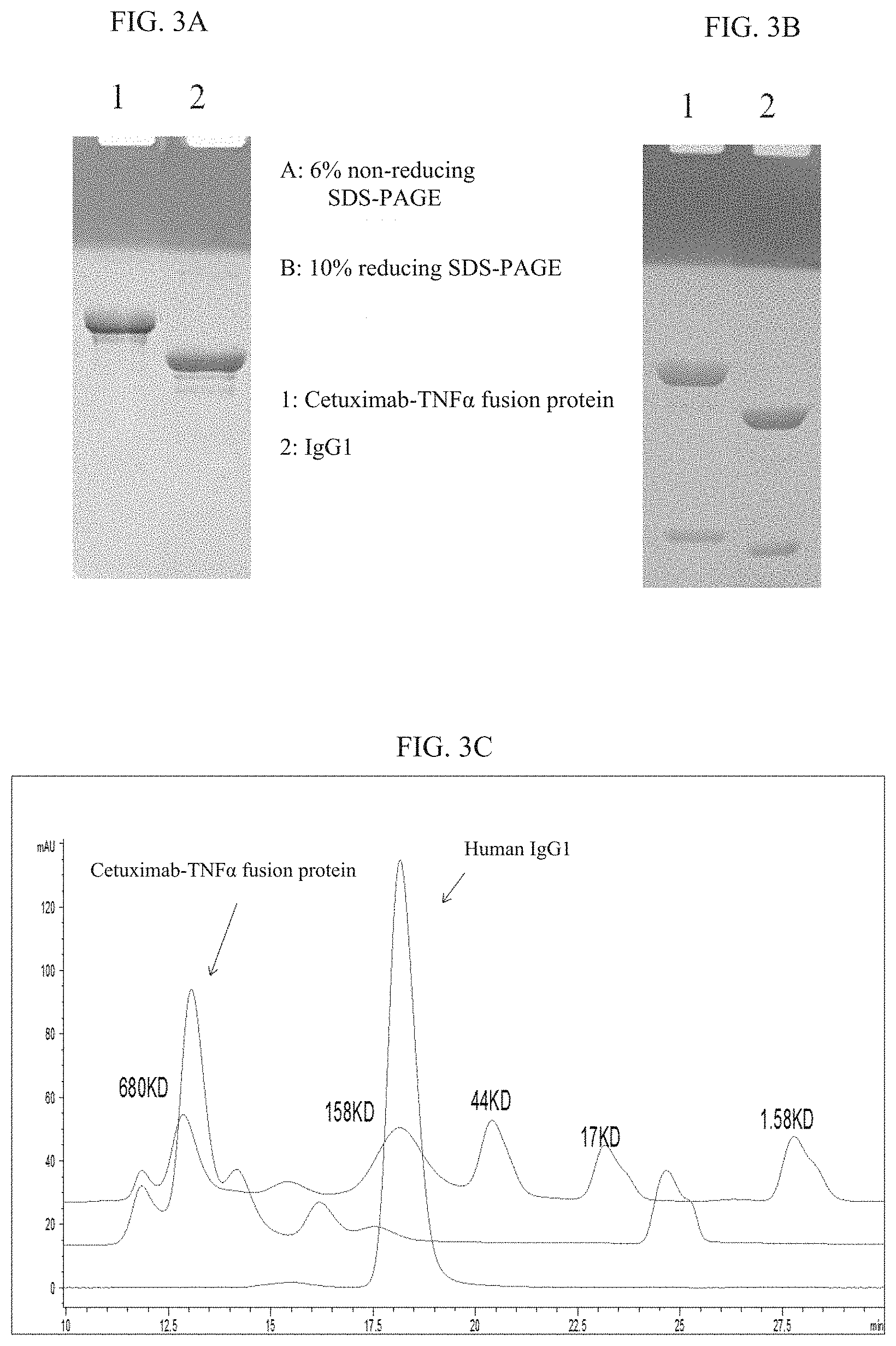
[0022] FIGS. 3A-3C: SDS-PAGE electrophoresis and HPLC-SEC of Cetuximab-TNF.alpha. fusion protein. The antibody-TNF.alpha. fusion protein was purified via Protein-A affinity chromatography followed by an anion exchange chromatography to collect the flow-through. FIG. 3A: 6% non-reducing SDS-PAGE electrophoresis, 3 .mu.g each sample; lane 1: the Cetuximab-TNF.alpha. fusion protein, lane 2: human IgG1. FIG. 3B: 10% reducing SDS-PAGE electrophoresis, 5 .mu.g each sample; lane 1: the Cetuximab-TNF.alpha. fusion protein, lane 2: human IgG1. FIG. 3C: HPLC-SEC of the Cetuximab-TNF.alpha. fusion protein using TSKgel G3000SWXL, wherein, the line starting atop represents the protein molecular weight ladder, the line starting in the middle represents the Cetuximab-TNF.alpha. fusion protein, and the line lowest represents human IgG1.
[0023] FIGS. 4A-4B: Her-2 antibody-TNF.alpha. fusion protein binding to Her-2 molecule in vitro. FIG. 4A: ELISA of binding to Her-2 ECD in vitro. A 96-well ELISA plate was coated with 25 nM recombinant human Her-2 ECD, and the antibody bound to Her-2 ECD was detected using alkaline phosphatase-labeled goat anti-human IgG1 Fc antibody. FIG. 4B: Flow cytometry of the antibody-TNF.alpha. fusion protein's binding to Her-2 on cell surface. Three Her-2 positive human tumor cell lines (PANC-1, SKBR-3 and SKOV-3) were incubated with the antibody-TNF.alpha. fusion protein at different concentrations (Y-axis); the antibody bound to Her-2 ECD was detected using FITC-labeled goat anti-human IgG1 Fc antibody, and the FITC fluorescence intensity was detected on a flow cytometer. Mock is the group without the antibodies.
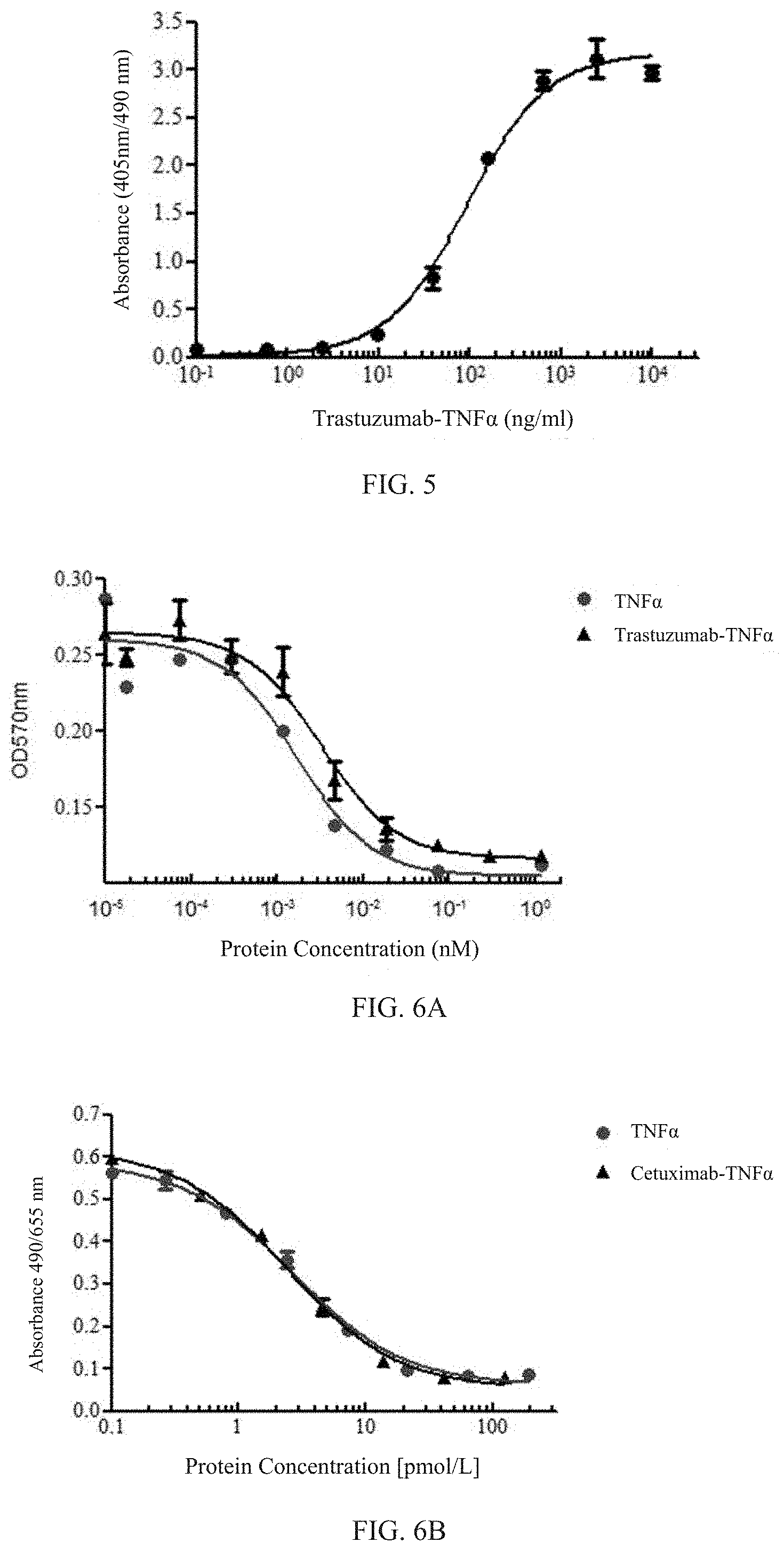
[0024] FIG. 5: ELISA of the Her-2 antibody-TNF.alpha. fusion protein's binding to TNFR type II (also known as "TNFR2") in vitro. A 96-well ELISA plate was coated with the Fc fusion protein of recombinant human TNFR2 ECD, and the antibody bound to TNFR2 ECD was detected using alkaline phosphatase-labeled goat anti-human IgG1 Fab antibody.
[0025] FIGS. 6A-6B: Apoptosis in L929 cells induced by the Her-2 antibody-TNF.alpha. fusion protein and by the EGFR antibody-TNF.alpha. fusion protein.
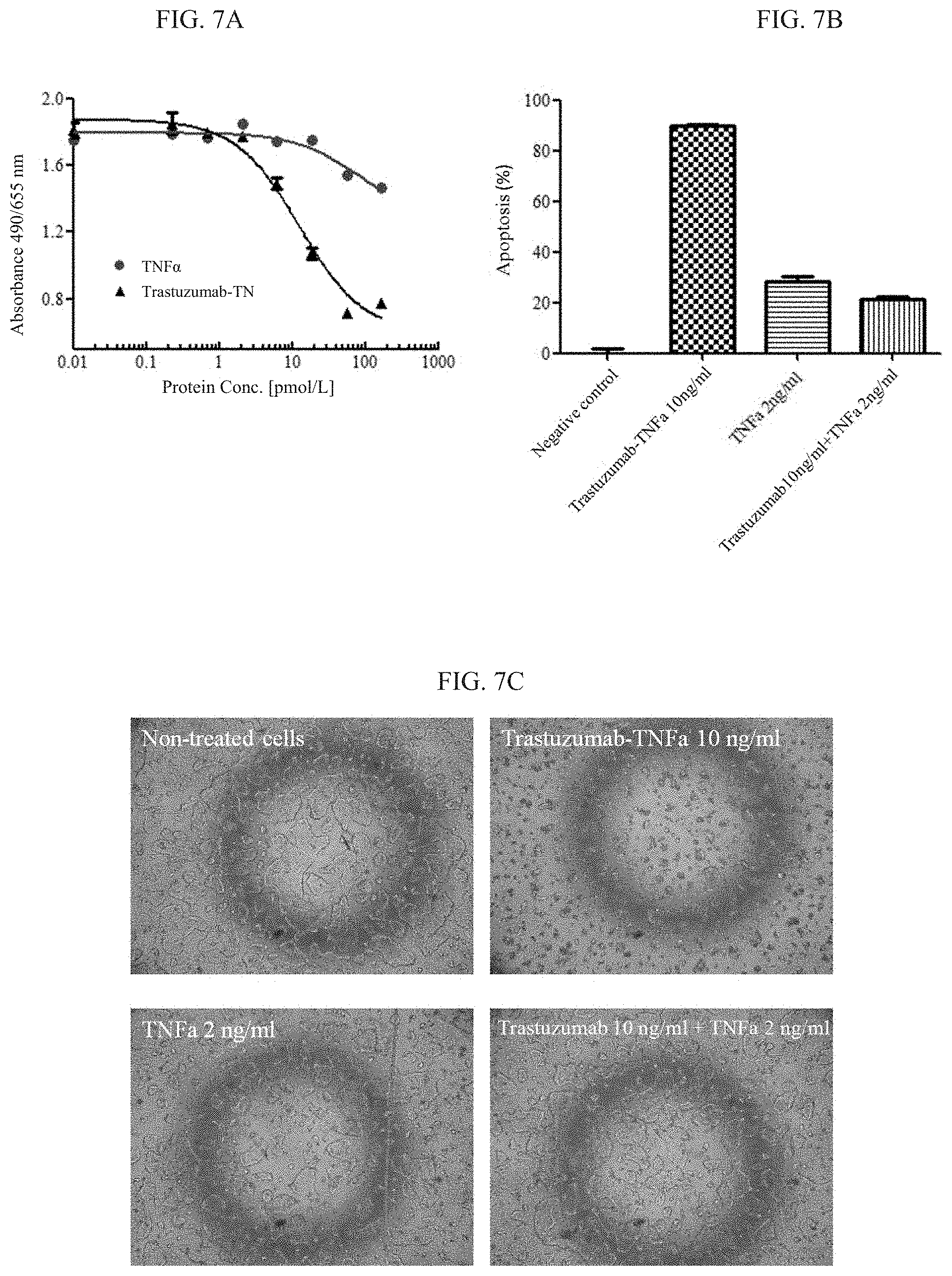
[0026] FIGS. 7A-7C: Apoptosis in Her-2 positive human gastric cancer cells NCI-N87 induced by the Her-2 antibody-TNF.alpha. fusion protein. FIG. 7A: the NCI-N87 cells were incubated with the antibody-TNF.alpha. fusion protein at different concentrations for 1 day; and cell survival was detected using a CCK-8 kit. FIG. 7B: the NCI-N87 cells were incubated with 10 ng/ml Trastuzumab-TNF.alpha. fusion protein, a mixture of 10 ng/ml Trastuzumab and 2 ng/ml of TNF.alpha., or 2 ng/ml of TNF.alpha. for 1 day, respectively; and cell survival was detected using a CCK-8 kit. FIG. 7C: a picture of apoptosis induced by the antibody-TNF.alpha. fusion protein in NCI-N87 cells under microscopy (40.times.).
[0027] FIG. 8: Apoptosis in EGFR-positive human gastric cancer NCI-N87 cells induced by the EGFR antibody-TNF.alpha. fusion protein.
[0028] FIGS. 9A-9B: Apoptosis in Her-2 positive human tumor cells induced synergistically by the Her-2 antibody-TNF.alpha. fusion protein and a tumor toxic chemotherapeutic agent. FIG. 9A: Her-2 positive human tumor cells (NCI-N87, SKBR-3 and SKOV-3) were cultured in the presence of the fusion protein, actinomycin D or both for 1 day; and cell viability was detected using a CCK-8 kit. FIG. 9B: Her-2 positive human tumor cells (BT-474, NCI-N87, SKBR-3 and SKOV-3) were cultured in the presence of the fusion protein, doxorubicin or both for 1 day; and cell viability was detected using a CCK-8 kit.
[0029] FIGS. 10A-10D: Actinomycin D (abbr. "ActD"), a transcription inhibitor, and cycloheximide (abbr. "CHX"), a protein synthesis inhibitor, enhanced toxicity of the Her-2 antibody-TNF.alpha. fusion protein in mouse forestomach carcinoma cells expressing human Her-2. FIG. 10A: Mouse forestomach carcinoma (MFC) cells stably expressing human Her-2 and wild-type MFC cells cultured in presence of 5 .mu.g/ml CHX were treated with the Trastuzumab-TNF.alpha. fusion protein at different concentrations or a mixture of Trastuzumab and TNF.alpha. for 20 hours, and cell viability was detected using a CCK-8 kit. FIG. 10B: Mouse forestomach carcinoma (MFC) cells stably expressing human Her-2 and wild-type MFC cells cultured in presence of 5 .mu.g/ml CHX were treated with the Trastuzumab-TNF.alpha. fusion protein at different concentrations for 20 hours, and cell viability was detected using a CCK-8 kit. FIG. 10C: Mouse forestomach carcinoma (MFC) cells stably expressing human Her-2 and wild-type MFC cells cultured in presence of 5 .mu.g/ml ActD were treated with the Trastuzumab-TNF.alpha. fusion protein at different concentrations for 20 hours, and cell viability was detected using a CCK-8 kit. FIG. 10D: Mouse forestomach carcinoma (MFC) cells stably expressing human Her-2 cultured in presence of 5 .mu.g/ml ActD were treated with the Trastuzumab-TNF.alpha. fusion protein at different concentrations or a mixture of Trastuzumab and TNF.alpha. for 20 hours, and cell viability was detected using a CCK-8 kit.
[0030] FIG. 11: A study of apoptosis in mouse forestomach carcinoma (MFC)/EGFR cells, which shows that actinomycin D (abbr. "ActD"), a transcription inhibitor, enhances toxicity of the EGFR antibody-TNF.alpha. fusion protein in mouse forestomach carcinoma cells expressing human EGFR.
[0031] FIG. 12: A study of apoptosis mouse melanoma B16/EGFR cells, which shows that actinomycin D (abbr. "ActD"), a transcription inhibitor, enhances toxicity of the EGFR antibody-TNF.alpha. fusion protein in mouse melanoma cells B16 expressing human EGFR.
[0032] FIG. 13: Her-2 antibody-TNF.alpha. fusion protein inhibited growth of mouse melanoma B16 expressing human Her-2 in mouse. C57/B6 mice were grafted with the mouse melanoma B16 expressing human Her-2 on back. The mice were treated via caudal intravenous injection. Tumor volume was measured and calculated as (length.times.width.times.width/2).
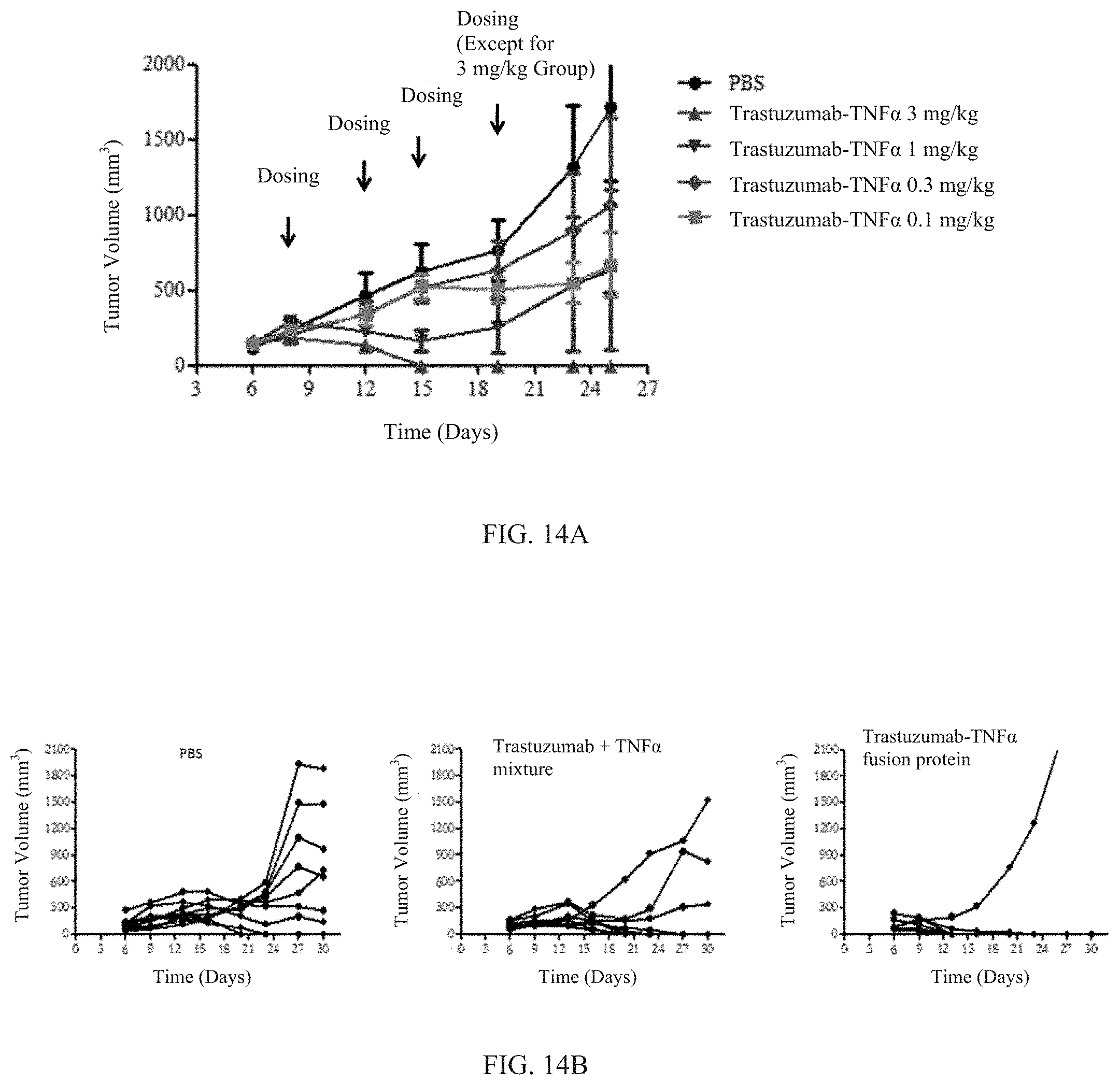
[0033] FIGS. 14A-14B: Her-2 antibody-TNF.alpha. fusion protein inhibited growth of mouse colon cancer cells CT26 expressing human Her-2 in mice. FIG. 14A: Balb/c mice were grafted with the CT26 tumor cells expressing human Her-2 on back. The mice were treated via caudal intravenous injection. FIG. 14B: Balb/c mice were grafted with the CT26 tumor cells expressing human Her-2 on back. The mice were treated with 3 mg/kg Trastuzumab-TNF.alpha. fusion protein or 3 mg/kg mixture of the Trastuzumab and TNF.alpha. via caudal intravenous injection. Tumor volume was measured and calculated as (length.times.width.times.width/2).
DETAILED DESCRIPTION OF THE INVENTION
[0034] In the following, the present invention will be described in details with reference to specific embodiments.
[0035] This application is based on the finding that TNF.alpha. can be conjugated to an antibody molecule to provide a dual-target antibody-TNF.alpha. fusion protein which exhibits strong tumor-specific effects including inhibition, killing and apoptosis-induction in vitro and in vivo. Furthermore, these fusion proteins in combination with an additional anti-tumor active, such as a chemotherapeutic agent, provide an effect of synergism in killing, inhibition and apoptosis-induction in tumor cells in vitro and in vivo.
[0036] The inventors designed and produced an antibody-TNF.alpha. fusion protein with multiple biological activities. Specifically, the inventors, using the technology of molecular cloning, constructed an antibody-TNF.alpha. fusion protein by conjugating an antibody against a tumor-specific molecule on cell surface or a molecule over-expressed by tumor cells (e.g. tumor-specific antigens and tumor-associated antigens) with tumor necrosis factor .alpha. (TNF.alpha.) which is capable of tumor-killing and activation of anti-tumor immunity. The antibody moiety recruits the fusion protein to the environment around the tumor cells, producing actions on the tumor cells including inhibition, killing and induced apoptosis. The fusion protein carries the TNF.alpha. molecule as conjugated with the antibody to the site of tumor, where the TNF.alpha. molecule forms a transmembrane format with the tumor-specific molecule on surface and kills the tumor cells. Further, in presence of an active agent that is cytotoxic to tumor (e.g., a chemotherapeutic agent), the antibody-TNF.alpha. fusion protein of the invention and the active agent provide an enhancement of synergism in killing tumor cells.
[0037] The invention has overcome various technical obstacles, including difficulties in recombinant protein expression, difficulties in obtaining a productive strain with high expression of the product, and the risk that the expressed recombinant protein fails to fold into the correct configuration and is thus insoluble or prone to aggregate or biologically inactive, and succeeded in a recombinant protein molecule that can be effectively expressed in mammalian cells.
[0038] The fusion protein of the invention is stable in expression, highly biologically active and is effective in treating and preventing tumors. For example, the fusion protein of the invention can effectively inhibit osteogenic and cartilage differentiation, and can be effectively used for treating cancers, such as gastric cancer, pancreatic cancer, breast cancer, ovarian cancer, melanoma and colon cancer. The fusion protein of the invention can also be used in combination with an additional anti-tumor agent to provide a synergistic anti-tumor effect.
[0039] As used herein, the open-ended terms such as "comprise", "have" and "include" and their grammatical variants encompass the scenarios as meant by the closed-ended terms such as "consist of . . . ", "composed of . . . " and "is . . . ". In the present application, a numerical range constitutes a disclosure including the values at two ends, every integer and fractions between the ends, and all ranges formed between values of said ends, integers and fractions. For multiple ranges described for a single parameter, ends of these ranges can be recombined the new ranges obtained thereby are also deemed as specifically disclosed herein. In the present application, the features and elements can be recombined across embodiments and examples and the embodiments and examples obtained thereby are also deemed as specifically disclosed herein.
[0040] Fusion Protein and its Components
[0041] The fusion proteins of the invention is a fusion protein between an antibody and TNF.alpha., which is also referred to herein below as "antibody-TNF.alpha. fusion protein" or "antibody-TNF.alpha." or "the fusion protein of the invention", as well as synonyms in context. The fusion protein of the invention comprises an antibody moiety and a TNF.alpha. moiety conjugated to the C-terminal of the heavy chain of said antibody, wherein the heavy chain of the antibody and the TNF.alpha. moiety are fused to form the heavy chain of the fusion protein, and the light chain of the antibody constitutes the light chain of the fusion protein. The heavy chain and the light chain of the fusion protein each may independently and optionally further comprise a signal peptide at N-terminal.
[0042] In a preferred embodiment, the antibody moiety in the fusion protein of the invention is in the form of a full-length antibody. The form of a full-length antibody refers to the complete structure of an immunoglobulin (Ig), except for optional C-terminal modification to facilitate fusion. In particular, the structure of an immunoglobulin refers to the structure of a monomeric Ig molecule such as IgG. In context of the present invention, immunoglobulins include IgG, IgD, IgE, IgA, IgM and sub-classifications and subunits thereof. For example, according to the present invention, the antibody may be an IgG antibody, including those of IgG1, IgG2, IgG3, and IgG4 sub-classes. In a preferred embodiment, as shown in FIG. 1, the TNF.alpha. molecule is connected to the C-terminal of the Fc fragment of the full-length antibody.
[0043] In the fusion protein of the invention, the antibody moiety provides specificity to a tumor antigen. Examples of tumor antigen include, but are not limited to, members of the ErbB family, such as EGFR, Her-2, Her-3, and Her-4. Correspondingly, the antibodies include EGFR antibodies, Her-2 antibodies, Her-3 antibodies and Her-4 antibodies. Specifically, examples of antibody include, but are not limited to Trastuzumab, Pertuzumab (Perjeta), T-DM1, Cetuximab, ABX-EGF, and functionally equivalent analogs or derivatives thereof.
[0044] As used herein, the term "functionally equivalent analog or derivative" should be understood in a broad sense as referring to an agent that is highly similar to a specified biomolecule as reference (such as a cellular element, a nucleic acid molecule, a cytokine (e.g., TNF.alpha.)) in structure and/or composition or that comprises the backbone structure of the reference and has no significant difference in biological activity as compared with the reference. For example, in the context of nucleic acid molecules and proteins such as antibodies, functionally equivalent analogs or derivatives thereof include molecules that are different from the reference molecule at one or more positions along the nucleotide sequence or the amino acid sequence via mutation of deletion, insertions and/or substitution while having a sequence identity of at least 60%, 70%, 80%, 90%, 95%, 97%, 98%, or even at least 99% to the reference. For example, in the context of TNF.alpha., the functionally equivalent analogs or derivatives thereof have substantially the same or equivalent effect in killing cells and inducing apoptosis.
[0045] In some embodiments, the antibody is a Her-2 antibody or an EGFR antibody. In some embodiments, the antibody is a Her-2 antibody, such as Trastuzumab or a functionally equivalent analog or derivative thereof. In some embodiments, the antibody is Trastuzumab.
[0046] In some embodiments, the antibody is an EGFR antibody, such as Cetuximab or a functional analog or derivative thereof. In some embodiments, the antibody is Cetuximab.
[0047] In some embodiments, the antibody molecule comprises C-terminal modification to facilitate fusion, such as deletion of one or more lysine residues at C-terminal of the heavy chain. For example, the antibody moiety derived from Trastuzumab or Cetuximab may comprise the lysine deletion at C-terminal.
[0048] In the fusion protein of the invention, the TNF.alpha. moiety refers to monomeric molecule(s) of TNF.alpha. or a functionally equivalent analog or derivative thereof. In some embodiments, the TNF.alpha. molecule is selected from: human or mammalian TNF.alpha.; mature TNF.alpha., secreted TNF.alpha., and transmembrane TNF.alpha.. In some embodiments, the TNF.alpha. moiety is a human TNF.alpha.. For example, in a fusion protein of the invention, the TNF.alpha. moiety has the amino acid sequence of positions 469-625 in SEQ ID NO: 2.
[0049] In a preferred embodiment, the TNF.alpha. moiety is directly conjugated to the C-terminal of the heavy chain of the antibody, without a peptide linker. Meanwhile, the TNF.alpha. portion can also be conjugated to the heavy chain of the antibody via a peptide linker. The peptide linker, if present, may have a length of 1-50, 5-50, 5-40, 10-40 or 10-30 amino acid residues, or a length of an integer from 1 to 50. A person of skills in the art knows how to determine the presence/absence and length of the peptide linker with conventional analysis and means, see for example PNAS, 95: 5929-5934, 1998; Protein Eng., 13 (5): 309-312, 2000; Protein Eng., 15 (11): 871-879, 2003; etc.
[0050] In a preferred embodiment, the antibody moiety is in the form of the tetramer of a monomeric Ig molecule, and the molar ratio of antibody to TNF.alpha. in the form of monomeric molecule(s) is 1:1 or preferably 1:2, as shown in FIG. 1.
[0051] In some embodiments, the fusion protein further comprises a signal peptide at N-terminal. As understood, a signal peptide refers to an amino acid sequence that functions to direct secretion, localization and/or transportation of proteins, which usually have a length of 5-30 amino acid residues. Generally, a mature protein harvested in recombinant production does not contain a signal peptide.
[0052] In an exemplary embodiment, the heavy chain of the fusion protein according to the present invention has the amino acid sequence of positions 20-625 (without signal peptide) or positions 1-625 (with signal peptide) in SEQ ID NO: 2, and the light chain of the fusion protein has the amino acid sequence of positions 21-234 (without signal peptide) or positions 1-234 (with signal peptide) in SEQ ID NO: 4. Along the amino acid sequence as set forth by SEQ ID NO: 2, the segment of amino acids (aa) at positions 1-19 is a transmembrane signal peptide, the segment of aa 20-468 is the heavy chain of Trastuzumab with the lysine-deletion at C-terminal, the segment of aa 469-625 is the amino acid sequence of human TNF.alpha.. Along the amino acid sequence as set forth by SEQ ID NO: 4, the segment of aa 1-20 is the transmembrane signal peptide, and the segment of aa 21-234 is the light chain of Trastuzumab. SEQ ID NO: 1 is an example of nucleotide sequence encoding the amino acid of SEQ ID NO: 2, wherein the last TGA is the stop codon. SEQ ID NO: 3 is an example of nucleotide sequence encoding SEQ ID NO: 4, wherein the last TGA is the stop codon.
[0053] In another exemplary embodiment, the heavy chain of the fusion protein according to the present invention has the amino acid sequence of positions 17-621 (without signal peptide) or positions 1-621 (with signal peptide) in SEQ ID NO: 20, and the light chain of the fusion protein has the amino acid sequence of positions 17-230 (without signal peptide) or positions 1-230 (with signal peptide) in SEQ ID NO: 22. Along the amino acid sequence as set forth by SEQ ID NO: 20, the segment of amino acids (aa) at positions 1-16 is the transmembrane signal peptide, the segment of aa 17-464 is the heavy chain of Cetuximab with the lysine-deletion at C-terminal, the segment of aa 165-621 is the amino acid sequence of human TNF.alpha.. Along the amino acid sequence as set forth by SEQ ID NO: 21, the segment of aa 1-16 is the transmembrane signal peptide, and the segment of aa 17-230 is the light chain of Cetuximab. SEQ ID NO: 19 is an example of nucleotide sequence encoding the amino acid of SEQ ID NO: 20, wherein the last TGA is the stop codon. SEQ ID NO: 21 is an example of nucleotide sequence encoding SEQ ID NO: 22, wherein the last TGA is the stop codon.
[0054] As shown by the examples in the present application, the fusion protein of the invention has one or more activities selected from the group consisting of binding to a tumor antigen, promoting apoptosis in fibroblasts, inducing apoptosis in tumor cells, inhibiting growth of tumor, and providing a synergistic anti-tumor effect in combination with an additional anti-tumor agent.
[0055] The Fusion Protein-Encoding Nucleic Acid Molecules, Nucleic Acid Constructs, Vectors and Host Cells
[0056] As used herein, the term "nucleic acid construct" is chemically synonymous with "nucleic acid molecule", which is artificial in origin and is not a natural product. Therefore, a nucleic acid construct, as in the case of a nucleic acid molecule, may include another encoding nucleic acid molecule, i.e., a coding sequence, as an integral component thereof, such as is the case of a vector containing a coding sequence. As used herein, the terms "nucleic acid molecule", "nucleic acid" and "nucleotide sequence" in relation to an encoding sequence or a specified nucleotide sequence are interchangeably synonymous, all encompass the nucleic acid molecule or sequence that is complementary to a specified nucleic acid molecule or sequence.
[0057] Provided herein is a nucleic acid construct, comprising a nucleotide sequence encoding the heavy chain of the fusion protein of the invention, such as a vector comprising said nucleotide sequence. Or, the nucleic acid construct is a nucleic acid molecule encoding the heavy chain of the fusion protein of the invention.
[0058] In an embodiment, a nucleic acid construct according to the invention may be a nucleic acid molecule encoding the fusion protein heavy chain consisting of Trastuzumab heavy chain-human TNF.alpha., or a construct such as a vector comprising said encoding nucleic acid molecule. For example, the nucleic acid construct may comprise or consist of a nucleic acid molecule selected from the followings:
[0059] A nucleic acid molecule encoding the amino acid sequence of aa 20-625 or aa 1-625 in SEQ ID NO: 2; or
[0060] A nucleic acid molecule comprising the nucleotide sequence of positions 58-1875 or positions 1-1875 in SEQ ID NO: 1 and optionally a stop codon; or
[0061] A nucleic acid molecule having the nucleotide sequence of SEQ ID NO: 1.
[0062] In another embodiment, a nucleic acid construct of the present invention may be a nucleic acid molecule encoding the fusion protein heavy chain consisting of Cetuximab heavy chain-human TNF.alpha., or a construct such as a vector comprising said encoding nucleic acid molecule. Specifically, the nucleic acid construct may comprise or consist of a nucleic acid molecule selected from the followings:
[0063] A nucleic acid molecule encoding the amino acid sequence of positions 17-621 or positions 1-621 in SEQ ID NO: 20; or
[0064] A nucleic acid molecule comprising the nucleotide sequence of positions 49-1863 or positions 1-1863 in SEQ ID NO: 19 and optionally a stop codon; or
[0065] A nucleic acid molecule having the nucleotide sequence of SEQ ID NO: 19.
[0066] Also provided herein is a nucleic acid construct or a combination of nucleic acid constructs, comprising or consisting of a nucleic acid molecule encoding the heavy chain of the fusion protein of the invention and a nucleic acid molecule encoding the light chain of the fusion protein of the invention. The nucleic acid molecule encoding the heavy chain of the fusion protein and the nucleic acid molecule encoding the light chain of the fusion protein may be included in the same nucleic acid construct (e.g., a vector) or in two or more nucleic acid constructs (e.g., vectors).
[0067] For example, in some embodiments, when the fusion protein is Trastuzumab-TNF.alpha., correspondingly, said nucleic acid construct or combination of nucleic acid constructs may comprise or consist of the following nucleic acid molecules:
[0068] A nucleic acid molecule encoding the amino acid sequence of positions 20-625 or positions 1-625 in SEQ ID NO: 2, and a nucleic acid molecule encoding the amino acid sequence of positions 21-234 or positions 1-234 in SEQ ID NO: 4; or
[0069] A nucleic acid molecule comprising the nucleotide sequence of positions 58-1875 or positions 1-1875 in SEQ ID NO: 1 and optionally a stop codon, and a nucleic acid molecule comprising the nucleotide sequence of positions 61-702 or positions 1-702 in SEQ ID NO: 3 and optionally a stop codon; or
[0070] A nucleic acid molecule having the nucleotide sequence of SEQ ID NO: 1 and a nucleic acid molecule having the nucleotide sequence of SEQ ID NO: 3.
[0071] In some other embodiments, when the fusion protein is Cetuximab-TNF.alpha., correspondingly, said nucleic acid construct or combination of nucleic acid constructs may comprise or consist of the following nucleic acid molecules:
[0072] A nucleic acid molecule encoding the amino acid sequence of positions 17-621 or positions 1-621 in SEQ ID NO: 20, and a nucleic acid molecule encoding the amino acid sequence of positions 17-230 or positions 1-230 in SEQ ID NO: 22; or
[0073] A nucleic acid molecule comprising the nucleotide sequence of positions 49-1863 or positions 1-1863 in SEQ ID NO: 19 and optionally a stop codon, and a nucleic acid molecule comprising the nucleotide sequence of positions 49-690 or positions 1-690 in SEQ ID NO: 21 and optionally a stop codon; or
[0074] A nucleic acid molecule having the nucleotide sequence of SEQ ID NO: 19 and a nucleic acid molecule having the nucleotide sequence of SEQ ID NO: 21.
[0075] As said above, a nucleic acid construct may comprise or consist of a vector. Accordingly, the present invention also provides a vector comprising an encoding nucleic acid molecule according to the present invention. In some embodiments, the vector may be an expression vector, which provides expression of the fusion protein in a host cell. In the context of present application, the term "vector" includes, for example, expression vectors, cloning vectors, plasmids, cosmids and viral vectors. Representative examples include, but are not limited to, vectors providing expression in eukaryotic cells (e.g., CHO cells and COS cells), vectors providing expression in Saccharomyces cerevisiae or Pichia pastoris cells, vectors providing expression in insect (e.g., silkworm) cells, and prokaryotic expression vector. In the context of the present invention, any vectors known in the art, such as those commercially available ones, can be used. For example, a nucleotide sequence encoding the novel fusion protein of the invention can be cloned into a vector as operably conjugate to a regulatory sequence for expression to thereby form an expression vector for the fusion protein.
[0076] Also provided herein is a host cell which is transformed to comprise the nucleic acid construct or combination of nucleic acid constructs as described above. In the context of the present application, the term "host cell" includes prokaryotic cells and eukaryotic cells. In some embodiments, the host cell may be selected from the group consisting of bacteria, fungi, yeasts, plant cells and animal cells. Examples of commonly used prokaryotic host cells include, for example, Escherichia coli and Bacillus subtilis. Commonly used eukaryotic host cells include yeast cells, insect cells and mammalian cells. In some embodiments, the host cell is a mammalian cell, such as a CHO cell, for example, CHO DG44 and CHO-K. In the context of the present application, the term "transform" refers to introducing a nucleic acid of interest or a vector comprising same into a host cell by any of the methods of transformation known in the art, whereby the obtained host cell is not a natural product. Methods of transformation may be selected according to types of the given host, and generally include, for example, electrotransformation, transfection using calcium chloride, DEAE-dextran or other reagents, particle bombardment, lipofection and infection (see Sambrook et al. Molecular Cloning Laboratory Guide, 2nd edition, 1989). A preferred method may be electrotransformation.
[0077] The present invention also provides a method for producing an antibody-TNF.alpha. fusion protein, wherein the method may include: culturing a transformed host cell as described above under a condition suitable for expression of the fusion protein to express the fusion protein; and isolating the fusion protein. The method may further include a step of subjecting culture of the host cell to a stress to obtain a strain having a high expression of the fusion protein, such as culturing under the stress of methotrexate, preferably an increasing gradient of methotrexate stress. The method may further include one or more steps of separating and/or purifying the fusion protein. Determination on parameters of culturing, such as medium, temperature and time, is within the normal skills of a person of skills in the art. Expression of the fusion protein can be detected using any of the conventional means of detection, such as SDS-PAGE and Western blotting. For purifying the fusion protein, any of the conventional separation and purification techniques can be used, including centrifugation, precipitation, filtration and chromatography. Specifically, useful chromatography techniques include, for example, affinity chromatography, gel filtration, ion exchange, hydrophobic chromatography and reverse chromatography. Specific examples include Protein-A affinity chromatography (e.g., POROS MabCapture A, Life Tech) and anion exchange chromatography (e.g., Q-600C, TOSOH).
[0078] Composition
[0079] A composition according to the present invention may comprise an effective amount of an antibody-TNF.alpha. fusion protein, a nucleic acid construct, a combination of nucleic acid constructs and/or a host cells according to the present invention. The composition may be a pharmaceutical composition, which may comprise a pharmaceutically acceptable carrier such as a solvent, excipient and adjuvant. In the context of the present application, the term "an effective amount" or "an effective dosage" refers to an amount providing to a human or animal subject a function or activity as intended and being tolerable to the human or animal subject. As used herein, the term "pharmaceutically acceptable" means that when a molecular or a composition is administered to an animal or human subject in an appropriate manner or route, it does not produce an undesired effect (such as toxicity, irritation and allergy), as demonstrated by a reasonable benefit/risk ratio. The term "a pharmaceutically acceptable carrier" is supposed to be compatible with the fusion protein of the invention, which can normally be blended with the protein without causing significant loss in efficacy of the pharmaceutical composition. Specific examples of substances that can be used as a pharmaceutically acceptable carrier or ingredient can be found in, for example, Remington: The Science and Practice of Pharmacy (2005), the 21st Century Edition, Lippincott Williams and Wilkins, Philadelphia, Pa.
[0080] The pharmaceutical compositions according to the present invention may be formulated into a dosage form as appropriate for a specific dosage or manner of administration.
[0081] A nucleic acid molecule may be delivered to a subject in the form of a naked nucleic acid molecule, a co-delivery preparation or a recombinant vector (e.g., a plasmids or a viral vector) comprising and/or expressing the nucleic acid molecule. Suitable delivery reagents for administration include the lipophilic reagents in the Minis Transit TKO, lipofectins, lipofectamines, cellfectins, cationic polymeric reagents (e.g., polylysin), or liposomes, as well as any other means known to a person of skills in the art.
[0082] In some embodiments, the composition of the invention may further comprise one or more additional active agents for treating or preventing tumors. In some embodiments, the additional active agent for treating or preventing tumors are selected from chemotherapeutic agents and radiotherapeutic agents, such as alkylating agents, antimetabolites, antitumor antibiotics, botanical anticancer medicines, hormones and immunotherapeutics. Examples include mitosis inhibitors, transcription inhibitors and protein synthesis inhibitors, and more specifically, vinblastine, vincristine, vindesine, vinorelbine, colchicamine, colchicine, colchicamide, podophyllotoxin, etoposide, teniposide, paclitaxel or docetaxel, camptothecin, homoharringtonine, procarbazine, asparaginase, cisplatin, carboplatin, mitoxantrone, tamoxifen, cyclophosphamide, chlorambucil hydrochloride, lomustine, semustine, cetepa, busulfan, methamphetamine, chlorambucil, fluorouracil, Tegafur, efluidin, carmofur, mercaptopurine, methotrexate, cytarabine, cyclocytidine, mercaptoguanine, hexamethylmelamine, hydroxyurea, mitomycin, doxorubicin (adriamycin), epirubicin, bleomycin, pelomycin, atorvastatin, gleevec, gemcitabine, actinomycin D, cycloheximide, topotecan and/or leuprolide; especially actinomycin D, cycloheximide, doxorubicin; and analogs, derivatives, prodrugs or metabolites thereof. The present invention found that such a composition provides an effect of synergism in inducing apoptosis in tumor cells, inhibiting growth of tumor and tumor treatment. This is unexpected.
[0083] In the combination, the antibody-TNF.alpha. fusion protein may, for example, be present at an amount of at least 10.sup.-3 ng/ml, at least 10.sup.-2 ng/ml, at least 10.sup.-1 ng/ml, at least 1 ng/ml, at least 10 ng/ml, at least 10.sup.2 ng/ml, at least 10.sup.3 ng/ml, at least 10.sup.4 ng/ml, at least 10.sup.5 ng/ml, at least 20 ng/ml, at least 40 .mu.g/ml, or a range formed from any two of the above as the lower and upper limits, such as 20 ng/ml to 40 .mu.g/ml, less than 30 ng/ml, less than 20 ng/ml; or an amount of at least 10.sup.-5 nM, at least 10.sup.-4 nM, at least 10.sup.-3 nM, at least 10.sup.-2 nM, at least 10.sup.-1 nM, at least 1 nM, at least 10 nM, at least 10.sup.2 nM, or a range formed from any two of the above as the lower and upper limits.
[0084] In the combination, the content of the one or more additional agents for treating or preventing tumors can be determined according to the relevant guidelines of medication in clinic and other conventional dosages. Or, the content may be, for example, at least 1 nM, at least 10 nM, at least 15 nM, at least 20 nM, at least 100 nM, at least 1 .mu.M, at least 2 .mu.M, at least 3 .mu.M, at least 4 .mu.M, at least 5 .mu.M, at least 10 .mu.M, at least 20 .mu.M, at least 30 .mu.M, at least 40 .mu.M, at least 50 .mu.M, at least 100 .mu.M, or a range formed from any two of the above as the lower and upper limits; or at least 1 .mu.g/ml, at least 2 .mu.g/ml, at least 3 .mu.g/ml, at least 4 .mu.g/ml, at least 5 .mu.g/ml, at least 6 .mu.g/ml, at least 7 .mu.g/ml, at least 8 .mu.g/ml, at least 9 .mu.g/ml, at least 10 .mu.g/ml, or a range formed from any two of the above as the lower and upper limits.
[0085] In context of the composition, the fusion protein and the additional active agent may be present at a ratio equivalent to the ratio between any one of the amounts specified for the fusion proteins and any one of the amounts specified for the active agent as in the above. For example, the ratio may be equivalent to: 1-100 ng/ml to 1 nM-100 .mu.M; such as 1-100 ng/ml to 1-20 nM, 1-100 ng/ml to 15 nM, 1 ng/ml to 1-20 nM, 100 ng/ml to 1-20 nM, 1 ng/ml to 15 nM, 100 ng/ml to 15 nM; such as 1-100 ng/ml to 1-100 .mu.M, 1-100 ng/ml to 3-50 .mu.M, 1-100 ng/ml to 4-40 .mu.M, 1-100 ng/ml to 10-30 .mu.M, 1 ng/ml to 1-100 .mu.M or 3-50 .mu.M or 4-40 .mu.M or 10-30 .mu.M, 100 ng/ml to 1-100 .mu.M or 3-50 .mu.M or 4-40 .mu.M or 10-30 .mu.M, such as 1 ng/ml to 33 .mu.M, 100 ng/ml to 33 .mu.M, 1 ng/ml to 11 .mu.M, 100 ng/ml to 11 .mu.M, 1 ng/ml to 3.7 .mu.M, 100 ng/ml to 3.7 .mu.M; or such as 10.sup.-3-10.sup.5 ng/ml to 1-10 .mu.g/ml, 10.sup.-3-10.sup.5 ng/ml to 5 .mu.g/ml, 10.sup.-3-10.sup.2 ng/ml to 5 .mu.g/ml, 10.sup.-2-10.sup.2 ng/ml to 5 .mu.g/ml. The phrase "equivalent to" here means that the ratio is the same, while the specific amounts of the two agents are not limited to the values as listed.
[0086] Notably, the term "composition" means that the ingredients act in cooperation, and it does not necessarily mean that the ingredients are always in the same mixture or system. For example, ingredients of a composition may be physically or spatially separated from one another, may be administered to a subject at the same time point after being mixed or not mixed, or sequentially administered to a subject at different time points, as long as they act in cooperation at the site of target. For example, according to the present application, embodiments of the composition include a kit, which may comprise one or more ingredients of the composition of the invention in one or more containers.
[0087] Applications
[0088] The antibody-TNF.alpha. fusion protein, the nucleic acid construct(s), the vector, the host cell and the composition according to the present invention can be used to induce apoptosis in tumor cells, to inhibit growth of tumor, to prevent and/or treat tumors, and can also be used for manufacturing a product, such as a medicament, a pharmaceutical formulation, a kit, etc., for these purposes.
[0089] In the context of the present application, examples of tumors include those expressing tumor-specific antigens or have high expression of tumor-associated antigens, including but not limited to tumors that are responsive to TNF.alpha. treatment. Examples of tumor-specific antigens and tumor-associated antigens include but are not limited to: ErbB family members, such as EGFR, Her-2, Her-3 and Her-4. Specifically, examples of tumors include, but are not limited to: gastric cancer, breast cancer, ovarian cancer, melanoma, bowel cancer such as colon and rectal cancer, liver cancer, pancreatic cancer, kidney cancer, lung cancer, head and neck cancer, cervical cancer, skin cancer, esophageal cancer, bone cancer and leukemia.
[0090] The antibody-TNF.alpha. fusion protein, the nucleic acid construct(s), the vector, the host cell and the composition according to the present invention can also be used in combination with an additional active agent or therapy for treating or preventing tumors. The additional active agent for treating or preventing tumors is as described in the section of "composition" above. The additional therapy may be, for example, surgery, radiotherapy and chemotherapy.
[0091] As for dosage of the fusion protein of the invention, a physician knows to determine a dosage appropriate for a patient depending on known factors including, for example, species, race, age, bodyweight, disease status and administration manner. For example, the dosage may be at least 0.1 mg/kg bodyweight, at least 0.3 mg/kg bodyweight, at least 1 mg/kg bodyweight, or at least 3 mg/kg bodyweight. The fusion protein or the composition of the present invention can be administered in various ways as appropriate, including oral administration and parenteral administration, such as injection and infusion.
[0092] As can be understood by a person of skills in the art, the features and approaches in the present disclosure can be recombined without departing from the spirit and scope of the present invention. More aspects of the invention would become obvious to a person of skills in the art from the present disclosure.
EXAMPLES
[0093] The present invention will be further illustrated with reference to the following specific examples. It will be appreciated by a person of skills in the art that various modifications and change can be made without departing from the spirit and scope of the invention.
[0094] In the following examples, unless otherwise specified, methods or processes can be conducted as conventional practiced in the art, for example, as described in "Molecular Cloning: A laboratory Manual" (3rd edition, 1989, Cold Spring Harbor Laboratory Press, New York), or by following manufacturer's instruction. DNA sequencing can be conducted using known methods or purchased from a service provider.
Example 1: Construction of Expression Plasmid for Antibody-TNF.alpha. Fusion Protein
[0095] 1. Construction of Expression Plasmid for her-2 Antibody-TNF.alpha. Fusion Protein
[0096] Trastuzumab was used as an example of Her-2 antibodies. The complete cDNAs encoding the heavy chain and the light chain of Trastuzumab were synthesized by GenScrip (USA), and were cloned into vector pUC57. The cDNA of human TNF.alpha. was obtained from OpenBiosystems (USA).
[0097] It has been shown in quite some reports that in expression and preparation of monoclonal antibodies, deletion of lysine residue(s) at C-terminal of heavy chains is common. Therefore, we removed this lysine residue to ensure integrity of the antibody fusion protein.
[0098] The gene encoding the heavy chain of Trastuzumab and the gene encoding TNF.alpha. was connected by a two-step polymerase chain reaction (PCR) process. In the first step, the gene of the heavy chain was amplified using synthetic DNA primers by PCR (High-Fi Polymerase Pfx, Invitrogen):
[0099] 5'-primer M13-F (SEQ ID NO: 5): 5'-TGTAAAACGACGGCCAGT-3', on vector;
[0100] 3'-primer KDP004 (SEQ ID NO: 6): 5'-TCCTGGGGACAGTGACAGTG-3', specific for the antibody heavy chain gene.
[0101] The gene encoding the TNF.alpha. moiety was amplified by PCR in the same way:
TABLE-US-00001 5'-primer KDP045 (SEQ ID NO: 7): 5'-CACTGTCACTGTCCCCAGGAGTCAGATCATCTTCTCGAACC-3'; 3'-primer BGH-R (SEQ ID NO: 8): 5'-AACTAGAAGGCACAGTCGAGGC-3', on vector.
[0102] Therein, sequence of the first 20 nucleotides in the TNF-F primer was complementary to the nucleotide sequence in the Her2-R primer, so that the two fragments from PCR could be connected in the second step of overlap extension PCR.
[0103] The two resultant PCR fragments were purified using DNA Gel Purification (Tiangen Biotech (Beijing) Co., Ltd.) before the second step of overlap PCR. The purified DNA fragments were 3-times diluted with eluent to facilitate ligation via overlap PCR:
TABLE-US-00002 5'-primer: still M13-F (SEQ ID NO: 5); 3'-primer BGH-R (SEQ ID NO: 8): 5'-TGGTGGTGTCTAGAGACTCACAGGGCAATGATCCC-3',
containing an XbaI restriction site for cloning.
[0104] A NotI restriction site was placed upstream to the transcription initiation site of the gene of Trastuzumab heavy chain. The fragment obtained from overlap PCR was gel purified and subjected to NotI/XbaI double digestion (Takara). The digested PCR fragment was cloned into the correspondingly digested expression vector for mammalian cells. The expression vector for mammalian cells was a modified pcDNA3.1 plasmid (Invitrogen), wherein the anti-neomycin gene was replaced by DHFR (dihydrofolate reductase) gene. The modified vector is useful for screening for mammalian cells having high expression of stably exogenous proteins. The obtained recombinant plasmid was transfected into competent bacterial strain DH5a. Positive colonies containing the correct recombinant plasmid were identified by colony PCR. The recombinant plasmid was purified, and correct sequence of the recombinant gene of Trastuzumab heavy chain-TNF.alpha. was confirmed by restriction digestion and sequencing.
[0105] The cDNA of Trastuzumab's light chain was sub-cloned into another pcDNA3.1 plasmid using enzymes NotI and XbaI.
[0106] 2. Construction of Expression Plasmid for EGFR Antibody-TNF.alpha. Fusion Protein
[0107] Cetuximab was used as an example of EGFR antibodies. The gene encoding the heavy chain variable region of Cetuximab (VH, with signal peptide) and the gene encoding the light chain variable region of Cetuximab (VL, with signal peptide) were synthesized by Genewiz (Beijing) Co. Ltd., and were cloned into vector pUC57.
[0108] To construct the gene of Cetuximab heavy chain-TNF.alpha., first, the Cetuximab VH gene and IgG1 Fc gene were connected by PCR to obtain gene of a complete heavy chain of Cetuximab; and then, the obtained gene of Cetuximab heavy chain was connected with the TNF.alpha. gene by PCR to obtain the gene of Cetuximab heavy chain-TNF.alpha.. The obtained PCR fragment was digested with NotI and XbaI, and cloned into correspondingly digested expression vector for mammalian cells. In this example, the expression vector for mammalian cells was a modified pcDNA3.1 plasmid (Invitrogen) wherein the anti-neomycin gene was replaced by the gene of rat glutamine synthetase (GS).
[0109] The primers for PCR amplification of Cetuximab VH gene were M13-R (Forward, 5'-CAGGAAACAGCTATGACC, SEQ ID NO: 9) and KDP077 (Reverse, 5'-GCTAGGCCCCTTTGTTGATGCGGCGGACACGGTCACGAGGG, SEQ ID NO: 10). The primers for amplifying IgG1 Fc fragment were KDP020 (5'-GCATCAACAAAGGGGCCTAGC, SEQ ID NO: 11) and BGH-R (5'-AACTAGAAGGCACAGTCGAGGC, SEQ ID NO: 12).
[0110] The PCR fragment of Cetuximab VH gene and the fragment of IgG1 Fc were connected by overlap extension PCR using primer KDP092 (Forward, 5'-ATGGAACGCGGCCGCCACC, SEQ ID NO: 13) and primer KDP093 (Reverse, 5'-TCTAGCATTTAGGTGACAC, SEQ ID NO: 14). The obtained fragment of full-length heavy chain was cloned into said expression vector using restriction enzymes NotI/XbaI.
[0111] Then gene encoding Cetuximab heavy chain-TNF.alpha. was cloned according to the procedure as described for the gene encoding Trastuzumab heavy chain-TNF.alpha.. The PCR primers for amplifying the gene of Cetuximab heavy chain were primer CMV-P (Forward, 5'-CGCAAATGGGCGGTAGGCGTG, SEQ ID NO: 15) and primer KDP004 (SEQ ID NO: 6). The primers for amplifying the TNF.alpha. gene were KDP045 (SEQ ID NO: 7) and BHG-R (SEQ ID NO: 12). The two genes were connected using PCR primers KDP033 (5'-AGCTTGGTACCCTCGAGG, SEQ ID NO: 16) and KDP093 (SEQ ID NO: 14). The obtained gene of Cetuximab heavy chain-TNF.alpha. was cloned into said expression vector using restriction enzymes NotI/XbaI.
[0112] To construct gene of the Cetuximab light chain, the gene of light chain variable region (VL) and the gene of constant region (kappa) were amplified by PCR. The PCR primers for the VL gene were M13-R (SEQ ID NO: 9) and KDP094 (GACAGATGGAGCGGCCACAGTTCGCTTCAGCTCCAGCTTTGTTCC, SEQ ID NO: 17). The PCR primers for the kappa gene were KDP010 (ACTGTGGCCGCTCCATCTGTC, SEQ ID NO: 18 and BGH-R (SEQ ID NO: 12). The PCR fragment of Cetuximab VL gene and the PCR fragment of the kappa gene were connected by overlap extension PCR using primer M13-R (SEQ ID NO: 9) and primer KDP093 (SEQ ID NO: 14). The obtained fragment of the full-length light chain gene was cloned into said expression vector using NotI/XbaI restriction enzymes.
Example 2: Establishment of Cell Line Stably Expressing Antibody-TNF.alpha. Fusion Protein
[0113] The host cell CHO DG44 was obtained from Invitrogen. The cells were cultured and subcultured according to the CHO DG44 manual from Invitrogen. Non-transfected cells were suspension cultured in CD DG44 medium (Invitrogen) supplemented with 8 mM L-glutamine (Sigma) and 5 .mu.g/ml recombinant human insulin. CHO-K cell was obtained from the Cell Bank of the Type Culture Collection Committee of Chinese Academy of Sciences (Shanghai). The primary cells exhibited adherent growth in a serum-supplemented medium, and were habituated to be capable of suspension growth in a serum-free medium (chemically defined medium, abbr. "CD"). In this example, the CHO DG44 cells were used to express the Trastuzumab-TNF.alpha. fusion protein, and the CHO-K cells were used to express the Cetuximab-TNF.alpha. fusion protein.
[0114] Briefly, the strain stably expressing the Trastuzumab-TNF.alpha. fusion protein was constructed as follows. The expression plasmid of the fusion protein's heavy chain and the expression plasmid of the fusion protein's light chain were prepared using the Plasmid Maxi Preparation Kit (TIANGEN, China). 100 .mu.g pcDNA3.1 comprising the antibody heavy chain-TNF.alpha. and 100 .mu.g pcDNA3.1 comprising the antibody light chain prepared according to Example 1 were linearized by digestion using restriction endonuclease EcoRI. The DG44 cells were subcultured for at least three passages before transfection. 1.times.10.sup.7 DG44 cells were mixed with the digested plasmids in 0.8 ml CD DG44 growth medium and then transferred in to a 0.4-cm pulser (Bio-Rad), wherein the cell/plasmid mixture was pulsed on Gene Pulser Xcell (Bio-Rad). The transfected cells were then cultured in a T-75 square flask containing 20 ml growth medium for 24 hours at 37.degree. C. in 8% CO.sub.2 in incubator.
[0115] After the 24-hour incubation, the transfected cells were plated on a 96-well plate by limiting dilution, using the OptiCHO medium as the medium for screen, wherein the medium was supplemented with 8 mM glutamine, 5 .mu.g/ml recombinant human insulin and 100 nM methotrexate (MTX, Sigma). The cells were incubated at 37.degree. C. in 8% CO.sub.2 in incubator. 3 weeks later, culture liquid from the wells observed with colony formation was examined by ELISA using alkaline phosphatase-conjugated goat anti-human IgG Fc antibody (Jackson ImmunoResearch Lab), whereby clones with the highest expression were picked and amplified, then assayed via ELISA and amplified again, which finally ended up with 12 cell lines with highest stable expression.
[0116] The 12 clones were cultured under stress using an increasing gradient of MTX concentrations for enhanced expression. Totally three rounds of incubation under stress (500 nM, 2 .mu.M and 10 .mu.M of MTX) were conducted, each for about 3 weeks. After the 3-rounds stress incubation, the 12 clones were tested for antibody expression, whereby 4-5 cell lines with increased antibody expression were obtained.
[0117] Without experiencing the MTX stress, the cell lines with stable antibody expression were low in expression level. After 3 rounds of stress treatment, with the MTX magnitude increasing from 0.1 .mu.M to 10 .mu.M in the culture medium, the antibody expression was significantly increased in most of the cell lines exposed to the stress. From these cell lines, one strain (#1G5) was pick and plated on a 96-well plate and cultured by limiting dilution to obtain a monoclonal strain with high expression of the antibody fusion protein. Cell strains stably expressing Cetuximab-TNF.alpha. fusion protein was constructed by the method and steps outlined as follows. CHO-K cells were co-transfected with plasmid encoding the chain of Cetuximab heavy chain-TNF.alpha. and plasmid encoding Cetuximab light chain via pulse-transfection. The transfected cells were plated on a 96-well plate and cultured by limiting dilution in medium containing L-aminosulfoxide methionine (MSX) to screen for strains stably transfected with the plasmids. Cell lines with positive antibody expression were picked by ELISA. Specifically, supernatant comprising said cells were added onto a ELISA plate coated with recombinant human EGFR protein; antibody was detected using alkaline phosphatase (AP) conjugated goat anti-human IgG1 Fc antibody; cell lines having high expression were picked and amplified, then subjected to the next round of screening; and thereby, CHO-K cell lines with stable and high expression of Cetuximab-TNF.alpha. fusion protein were obtained.
Example 3: Preparation, Purification and Identification of Antibody-TNF.alpha. Fusion Protein
[0118] The cell line with high expression of antibody-TNF.alpha. obtained according to Example 2 was cultured and amplified to 2 liters. Supernatant of the culture was harvested for purification and preparation of the antibody. The purification was Protein-A affinity chromatography (POROS MabCapture A, Life Tech) followed by anion exchange chromatography (flow through).
[0119] The purified antibody was analyzed by reducing and non-reducing SDS-PAGEs and HPLC-SEC (High Pressure Liquid--Molecular Sieve, TSKgel G3000SWXL, TOSOH Bioscience).
[0120] Results and Analysis
[0121] As shown by the gel of non-reducing SDS-PAGE in FIG. 2A, the intact Trastuzumab-TNF.alpha. fusion protein exhibited a molecular weight slightly below 200 kDa, which was very close to the theoretical value of 180 kDa. As shown by the gel of reducing SDS-PAGE in FIG. 2B, the chain of Trastuzumab heavy chain-TNF.alpha. in the fusion protein exhibited a molecular weight at 70 kDa, which was also consistent with the theoretical molecular weight (73 kDa).
[0122] The HPLC-SEC result of the Trastuzumab-TNF.alpha. fusion protein was shown in FIG. 2C. Therein, a peak of the fusion protein was detected at near 600 kDa, which was not consistent with the molecular weight shown by SDS-PAGE. This can be assumedly explained in two ways. First, the Trastuzumab-TNF.alpha. fusion protein molecules formed a trimer. Free TNF.alpha. molecule is trimeric in vivo, and the monomeric Trastuzumab-TNF.alpha. fusion protein in the described example comprised two TNF.alpha. molecules; then, Trastuzumab-TNF.alpha. fusion protein molecules need to aggregate into a trimer to allow formation of the TNF.alpha. trimer. Secondly, the Trastuzumab-TNF.alpha. fusion protein had a fairly loose structure, which exhibited the appearance of a large molecular weight in HPLC-SEC.
[0123] Non-reducing SDS-PAGE, reducing SDS-PAGE and HPLC-SEC results of the Cetuximab-TNF.alpha. fusion protein were the same, as shown in FIGS. 3A-3C. As shown by the gel of non-reducing SDS-PAGE, the intact Cetuximab-TNF.alpha. fusion protein exhibited a molecular weight slightly higher than native human IgG1, which was very close to the theoretical value of 180 kDa (FIG. 3A). As shown by the gel of reducing SDS-PAGE in FIG. 3B, the chain of Cetuximab heavy chain-TNF.alpha. in the fusion protein exhibited a molecular weight higher than that of the heavy chain of native human IgG1, which is also consistent with the theoretical molecular weight (73 kDa). The HPLC-SEC peak of the Cetuximab-TNF.alpha. fusion protein was also detected at 600 kDa (FIG. 3C), indicating a structure likely consistent the Trastuzumab-TNF.alpha. fusion protein.
Example 4: Tumor Antigen Binding of Antibody-TNF.alpha. Fusion Protein
[0124] Binding of the prepared Trastuzumab-TNF.alpha. fusion protein to extracellular domain (ECD) of the membrane protein Her-2 was studied via ELISA, in comparison with the antibody Trastuzumab (CTTQ Pharmaceutical Group Co., Ltd).
[0125] 25 nM recombinant human Her-2 ECD (donated by ANKEBIO (Group) Co., Ltd.) was dissolved in 50 mM NaHCO.sub.3 (pH 9.6). The recombinant protein (50 .mu.L) was added into wells of a 96-well ELISA plate and was stored at 4.degree. C. overnight in refrigerator. The next day, the ELISA plate was washed 3 times with PBST (PBS with 0.05% Tween-20), and then blocked with 100 .mu.l/well blocking buffer being PBST comprising 3% BSA. The ELISA plate was incubated at 37.degree. C. for 1 hour in incubator. The Trastuzumab-TNF.alpha. fusion protein and the Trastuzumab antibody were each prepared into 3-fold serial dilutions in binding buffer being PBST with 1% BSA. The blocking buffer was drained, and the fusion protein and Trastuzumab in the 3-fold serial dilutions were added (50 .mu.L/well) to react at 37.degree. C. for 1 hour in incubator. The dilutions were removed, the ELISA plates were washed 3 times with PBST, and the secondary antibody (alkaline phosphatase-conjugated goat anti-human IgG Fc antibody, Jackson ImmunoResearch Lab) was added (50 .mu.L/well) to react at 37.degree. C. for 1 hour in incubator. The staining antibody was removed, PBST wash buffer was added (200 .mu.L/well) and the ELISA plates were placed on an orbital shaker at 100 rpm for 5 minutes; then, the wash buffer was removed and the washing steps were repeated 4 times. The developer solution (PNPP) was added (50 .mu.l/well) and the ELISA plates were incubated at 37.degree. C. in incubator for development. The plates were read on a microplate reader at 405 nm/655 nm.
[0126] Binding of the prepared Trastuzumab-TNF.alpha. fusion protein to the Her-2 surface protein was also assayed via flow cytometry. Three Her-2 positive human tumor cell lines, i.e., human pancreatic cancer cell line PANC-1, human ovarian cancer cell line SKOV-3 and human breast cancer cell line SKBR-3 (all from Cell Bank of the Chinese Academy of Sciences) were used. The cells were prepared in pre-cooled FACS working buffer (PBS with 0.1% FBS) at 3.times.10.sup.6 cells/ml and divided into aliquots of 100 .mu.l/tube, which were blocked on ice for 1 hour. The Trastuzumab-TNF.alpha. fusion protein and Trastuzumab were each diluted to 10 .mu.g/ml in the FACS working buffer. The dilutions (10 .mu.L) were added into cell suspension (100 .mu.L) and incubated on ice for 30 minutes. At the end of incubation, to each tube, FACS working buffer (1 ml) was added and mixed with the cell suspension by vortex; the cell suspension was then centrifuged at 1200 rpm for 5 minutes; the supernatant was discarded and the washing steps were repeated once. FITC-labeled goat anti-human IgG Fc antibody (Jackson ImmunoResearch Lab) was diluted in the FACS working buffer, and then added into the cell suspension (10 .mu.L per tube) at a final concentration of 1 .mu.g/ml. The tubes were incubated on ice for 30 minutes in dark. At the end of incubation, to each tube, FACS working buffer (1 ml) was added and mixed with the cell suspension by vortex; the cell suspension was then centrifuged at 1200 rpm for 5 minutes; the supernatant was discarded and the washing steps were repeated once. The cells were assayed on flow cytometer C6 (BD Biosciences).
[0127] Results and Analysis
[0128] As shown by the ELISA result in FIG. 4A, the Trastuzumab-TNF.alpha. fusion protein was capable of specifically binding to the Her-2 ECD, with a EC.sub.50 of 0.018 nM, which was comparable to the binding capacity (EC.sub.50 of 0.008 nM) of Trastuzumab binding.
[0129] As shown by the flow cytometry results in FIG. 4B, the Trastuzumab-TNF.alpha. fusion protein was capable of binding to all the three Her-2 positive cell lines in test, exhibiting a binding capacity comparable to the antibody Trastuzumab. As seen, the fusion protein of the invention preserved the intact binding property of the antibody.
Example 5: Binding of Antibody-TNF.alpha. Fusion Protein to TNFR In Vitro
[0130] Binding of the Trastuzumab-TNF.alpha. fusion protein to the extracellular domain of human TNFR2 (sTNFR) was studied via ELISA. The fusion protein of sTNFR2 and IgG Fc (Celgen Biopharmaceutical Co., Ltd.) was diluted to 1 nM in 50 mM NaHCO.sub.3 (pH 9.6). The solution (50 .mu.L) was added into the wells of a 96-well ELISA plate and was stored at 4.degree. C. overnight in refrigerator. The next day, the ELISA plate was washed 3 times with PBST (PBS with 0.05% Tween-20), and then blocked with 100 .mu.l/well blocking buffer being PBST comprising 3% BSA. The ELISA plate was incubated at 37.degree. C. for 1 hour in incubator. The Trastuzumab-TNF.alpha. fusion protein obtained according to Example 3 was prepared into 4-fold serial dilutions in binding buffer being PBST+1% BSA. The blocking buffer was drained, and the fusion protein in the 4-fold serial dilutions was added (50 .mu.L/well) to react at 37.degree. C. for 1 hour in incubator. The dilutions of fusion protein were removed, the ELISA plates were washed 3 times with PBST, and the secondary antibody (alkaline phosphatase-conjugated goat anti-human IgG Fab antibody, Jackson ImmunoResearch Lab) was added (50 .mu.L/well) to react at 37.degree. C. for 1 hour in incubator. The staining antibody was removed, PBST wash buffer was added (200 .mu.L/well) and the ELISA plates were placed on an orbital shaker at 100 rpm for 5 minutes; then the wash buffer was removed and the washing steps were repeated 4 times. The developer solution (PNPP) was added (50 .mu.l/well) and the ELISA plates were incubated at 37.degree. C. in incubator for development. The plates were read on a microplate reader at 405 nm.
[0131] Results and Analysis
[0132] As shown in FIG. 5, the Trastuzumab-TNF.alpha. fusion protein bound to sTNFR2-Fc with an EC.sub.50 of 0.5 nM, which suggests that the Her-2 antibody-TNF.alpha. fusion protein of the invention specifically binds to TNFR with high affinity.
Example 6: Apoptosis in L929 Cells Induced by Antibody-TNF.alpha. Fusion Protein
[0133] The Trastuzumab-TNF.alpha. fusion protein and the Cetuximab-TNF.alpha. fusion protein were tested on cytotoxicity in mouse fibroblasts L929, in comparison with recombinant human TNF.alpha. protein.
[0134] L929 cells (Cell Bank of the Chinese Academy of Sciences (Shanghai)) were cultured in RPMI1640 medium with 10% FBS (Invitrogen). The day before experiment, the L929 cells (10000 cells/well) were added onto a 96-well plate and incubated at 37.degree. C. in a 5% CO.sub.2 in incubator. The next day, the antibody-TNF.alpha. fusion protein and the recombinant human TNF.alpha. in 3-fold serial dilutions in the cell growth medium were added into the cells to final concentrations from 0 to 1.18 nM (totally ten concentrations). After incubation for another 20 hours, the cells were stained with crystal violet, and the plates were read on a microplate reader at 570 nm.
[0135] Results and Analysis
[0136] As shown in FIGS. 6A and 6B, the recombinant human TNF.alpha. induced apoptosis in L929 cells with an IC50 of 0.4-1.7 pM, and the Trastuzumab-TNF.alpha. fusion protein induced apoptosis in L929 cells with an IC50 of 3.2 pM (FIG. 6A). The Cetuximab-TNF.alpha. fusion protein induced apoptosis in L929 cells with an IC50 of 0.6 pM (FIG. 6B). These suggest that the antibody-TNF.alpha. fusion proteins of the invention highly preserved the activity of TNF.alpha. to induce apoptosis.
Example 7: Cytotoxicity of her-2 Antibody-TNF.alpha. Fusion Protein on her-2 Positive Human Gastric Cancer Cells
[0137] Human gastric cancer cell NCI-N87 with high expression of Her-2 protein is less sensitive to Trastuzumab. Trastuzumab induces moderate apoptosis in NCI-N87 cells only at fairly high concentrations. To study activity of the Her-2 antibody-TNF.alpha. fusion protein of the invention in inducing apoptosis in NCI-N87 cells, (1) the Trastuzumab-TNF.alpha. fusion protein, (2) a (Trastuzumab+TNF.alpha.) mixture and (3) TNF.alpha. were compared for their impacts on viability in NCI-N87 cells.
[0138] The Her-2 positive human gastric cancer cells NCI-N87 (Cell Bank of the Chinese Academy of Sciences (Shanghai)) were cultured and subcultured in RPMI medium 1640 supplemented with 10% FBS (Gibco). The day before experiment, the cells (lx 10.sup.6 cells/well) in growth medium (150 .mu.l) were added onto a 96-well plate. The next day, the Trastuzumab-TNF.alpha. fusion protein, TNF.alpha. and the Trastuzumab+TNF.alpha. mixture (PeproTech, USA) (a mixture with the total amount of proteins substantially equivalent to that of the antibody-TNF.alpha. fusion protein by weight, with the antibody to TNF.alpha. (dimeric) ratio by weight being 5:1, approximately 2 molecules of monomeric TNF.alpha. per antibody molecule) prepared in serial dilutions in the growth medium were added (50 .mu.l/well) into the cells on the 96-well plate, mixed and cultured for another 60 hours in incubator. Cell viability was detected using MTT assay and cell viability using CCK-8.
[0139] Results and Analysis
[0140] First, we examined and compared the effects in killing NCI-N87 between the Trastuzumab-TNF.alpha. fusion protein and the Trastuzumab antibody. Three stages of effects were observed with the Trastuzumab-TNF.alpha. fusion protein in NCI-N87 cells: at concentrations below 30 ng/ml, the fusion protein effectively induced apoptosis in cells; at concentrations between 20 ng/ml and 40 .mu.g/ml, the killing decreased with increase in concentration; and at concentrations above 40 .mu.g/ml, the killing increased with increase in concentration (data not shown). Trastuzumab did not substantially impact survival of NCI-N87 cells at all concentrations in test (data not shown).
[0141] Next, we studied killing in NCI-N87 cells by the fusion protein at low concentrations, in comparison with TNF.alpha.. As shown in FIG. 7A, at concentrations below 20 ng/ml, the fusion protein exhibited increasing capability of inducing apoptosis with increase in concentration, with an IC.sub.50 of 1-2 ng/ml. In comparison, TNF.alpha. induced no or little apoptosis in NCI-N87 cells at corresponding concentrations.
[0142] Then, we studied the effect of the TNF.alpha.+Trastuzumab mixture on survival in NCI-N87 cells. NCI-N87 cells were treated with a mixture prepared by mixing 2 ng/ml TNF.alpha. and 10 ng/ml Trastuzumab, which corresponded to 10 ng/ml of Trastuzumab-TNF.alpha. fusion protein. Apoptosis was detected, in comparison with 10 ng/ml of the Her-2 antibody-TNF.alpha. fusion protein and with 2 ng/ml of TNF.alpha.. As shown in FIG. 7B, 10 ng/ml of the antibody-TNF.alpha. fusion protein induced an apoptosis of 90%, while the TNF.alpha.+Trastuzumab mixture an apoptosis of less than 30%. Morphology of the treated cells under microscopy was shown in FIG. 7C.
[0143] There results suggest that the Her-2 antibody-TNF.alpha. fusion protein of the invention has a unique profile of effects on cell survival, and exhibits an effect of synergism as compared with TNF.alpha. alone, and a simple blend of the Her-2 antibody and TNF.alpha..
Example 8: Cytotoxicity of EGFR Antibody-TNF.alpha. Fusion Protein on EGFR Positive Human Gastric Cancer Cells
[0144] Human gastric cancer cell NCI-N87 expresses EGFR protein. To study activity of the EGFR antibody-TNF.alpha. fusion protein of the invention in inducing apoptosis in NCI-N87 cells, (1) the Cetuximab-TNF.alpha. fusion protein and (2) a mixture of Cetuximab mAb+TNF.alpha. were compared for impacts on viability in NCI-N87 cells.
[0145] EGFR-positive human gastric cancer cells NCI-N87 (Cell Bank of the Chinese Academy of Sciences (Shanghai)) were cultured and subcultured in RPMI medium 1640 supplemented with 10% FBS (Gibco). The day before experiment, the cells (1.times.10.sup.6 cells/well) in growth medium (150 .mu.l) were added onto a 96-well plate. The next day, the Cetuximab-TNF.alpha. fusion protein and the Cetuximab+TNF.alpha. mixture (PeproTech, USA) prepared in serial dilutions in the growth medium were added (50 .mu.l/well) into the cells on the 96-well plate, mixed and cultured for another 48 hours in incubator. Cell viability was assayed using CCK-8.
[0146] Results and Analysis
[0147] As shown in FIG. 8, the Cetuximab-TNF.alpha. fusion protein induced apoptosis in NCI-N87 cells with an IC50 of about 5.7 pM, while no apoptosis was detected with the Cetuximab+TNF.alpha. mixture even at 110 pM.
Example 9: Synergistic Induction of Apoptosis by her-2 Antibody-TNF.alpha. Fusion Protein Combined with Anti-Tumor Chemotherapeutic in her-2 Positive Human Tumor Cells
[0148] In the following, the Her-2 positive tumor cells were all obtained from the Cell Bank of the Chinese Academy of Sciences. BT474 cell and NCI-N87 cells were cultured in RPMI 1640 medium, SKBR-3 in DMEM (Invitrogen) and SKOV-3 in McCOY's 5A medium (Invitrogen), all these media contained 10% FBS. The cells were incubated in the media added with different chemotherapeutics at different concentrations for 2 days: actinomycin D (MedChem Express) and doxorubicin (MedChem Express); then, the Trastuzumab-TNF.alpha. fusion protein was added at different concentrations and the cells were incubated for another 20 hours. Cell viability was detected by MTT assay.
[0149] Results and Analysis
[0150] As shown, an effect of synergism was observed with both the combination of Trastuzumab-TNF.alpha. fusion plus actinomycin D (FIG. 9A) and the combination of the fusion protein plus doxorubicin (FIG. 9B).
[0151] As shown in FIG. 9A, the Trastuzumab-TNF.alpha. fusion protein in combination with actinomycin D exhibited a very strong effect of synergism in inducing apoptosis in SKOV-3 cells. Neither 100 ng/ml of Trastuzumab-TNF.alpha. fusion protein nor 15 nM of actinomycin D, when used alone, had an impact on survival of SKOV-3 cells, while the two in combination induced an apoptosis of 80%. Similarly, in NCI-N87 cells, no apoptosis was observed with 1 ng/ml of the antibody-TNF.alpha. fusion protein and 15 nM actinomycin D induced an apoptosis of 25%, while the two in combination induced an apoptosis of 80%. These indicate a significant effect of synergism. A relatively weak effect of synergism was observed with the Trastuzumab-TNF.alpha. fusion protein in combination with actinomycin D in SKBR-3 cells, where 15 nM actinomycin D alone was capable of inducing a 70% apoptosis.
[0152] The Trastuzumab-TNF.alpha. fusion protein in combination with doxorubicin exhibited a fairly strong effect of synergism in inducing apoptosis in NCI-N87 and BT-474 cells. 1 ng/ml of Trastuzumab-TNF.alpha. fusion protein in combination with 33 .mu.M or 11 .mu.M doxorubicin induced apoptosis in 80% NCI-N87 and BT-474 cells, while they had little effect on viability in these cells at said concentrations when being used alone (FIG. 9 B). The same effect of synergism in inducing apoptosis was observed with the Trastuzumab-TNF.alpha. fusion protein in combination with doxorubicin in SKBR-3 and SKOV-3 cells (FIG. 9B).
Example 10: Construction of Mouse Tumor Cells Stably Expressing Human EGFR or Human Her-2
[0153] Mouse melanoma cells B16, mouse colon cancer cells CT26 and mouse forestomach carcinoma (MFC) cells were all obtained from the cell bank of the Type Culture Collection Committee of the Chinese Academy of Sciences. The cells were cultured in RPMI1640/10% FBS medium (Gibco).
[0154] The gene for human Her-2 expression was cloned in the expression vector pcDNA3.1 (Invitrogen); the recombinant plasmid was transfected into the three mouse tumor cells using Lipofectamine 3000 (Invitrogen); the transfected cells were cultured in RPMI/10% FBS medium supplemented with G418 (Sigma); and, stably transfected cells were obtained thereby. The stably transfected cells were sorted using a flow cytometer (Influx, BD Biosciences), whereby the monoclonal stable B16/Her-2, CT26/Her-2 and MFC/Her-2 cell lines with high Her-2 expression were picked.
[0155] The gene for human EGFR expression was cloned in the expression vector pCMV3 (Sino Biological Inc); the recombinant plasmid was transfected into the B16 mouse tumor cells and MFC mouse tumor cells using Lipofectamine 3000 (Invitrogen); the transfected cells were cultured in RPMI/10% FBS medium supplemented with Hygromycin B (Sigma); and, stably transfected cells were obtained thereby. The stably transfected cells were sorted using a flow cytometer (Influx, BD Biosciences), whereby the monoclonal stable B16/EGFR and MFC/EGFR cell lines with high EGFR expression were picked.
Example 11: Transcription Inhibitor and Protein Synthesis Inhibitor Enhancing Toxicity of her-2 Antibody-TNF.alpha. Fusion Protein in Mouse Forestomach Carcinoma Cells Expressing Human her-2 (MFC/her-2)
[0156] Toxicity of the Her-2 antibody-TNF.alpha. fusion protein on the MFC/Her-2 cells in the presence of actinomycin D (ActD, a transcription inhibitor) or cycloheximide (CHX, a protein synthesis inhibitor) was assayed, in comparison with the toxicity in absence of ActD and CHX. The MFC/Her-2 cells were plated on a 96-well plate and cultured for 1 day, then the medium was drained and to different wells were added different concentrations of the Trastuzumab-TNF.alpha. fusion protein in combination with ActD (5 .mu.g/ml) or CHX (.mu.g/ml) at identical concentrations. The cells were incubated for another 20 hours. The toxicity of the antibody-TNF.alpha. proteins on cells was detected by counting living cells using the Cell Counting Kit (CCK-8). The controls were the Trastuzumab+TNF.alpha. protein mixture, wherein the total amount of proteins was substantially equivalent to that in the Her-2 antibody-TNF.alpha. fusion protein, with the antibody to TNF.alpha. (dimeric) ratio by weight being 5:1, approximately 2 molecules of monomeric TNF.alpha. per antibody molecule as in the Her-2 antibody-TNF.alpha. fusion protein molecule.
[0157] The described experiment was repeated using wild-type (wt) MFC cells, and the result was compared with that of the MFC cells expressing human Her-2.
[0158] Results and Analysis
[0159] As shown in FIG. 10A, in the presence of protein synthesis inhibitor CHX (5 .mu.g/ml), the Trastuzumab-TNF.alpha. fusion protein was highly toxic to the MFC/Her-2 cells, with a IC.sub.50 of 0.3 ng/ml (1.7.times.10.sup.-12M); under the same condition, the TNF.alpha.+Trastuzumab mixture was also cytotoxic, with an IC.sub.50 of 341 ng/ml (1.9.times.10.sup.-9M), i.e., with a cytotoxic concentration 1000 times of that of the antibody-TNF.alpha. fusion protein. In absence of CHX, no cytotoxicity in MFC/Her-2 was observed either with the fusion protein or the mixture (data not shown).
[0160] As shown in FIG. 10B, in the presence of CHX (5 .mu.g/ml), the Trastuzumab-TNF.alpha. fusion protein induced slight cytotoxicity in wild-type MFC cells only at high concentrations. This suggests that Her-2 is the high sensitive molecule that mediates apoptosis by the Her-2 antibody-TNF.alpha. fusion protein.
[0161] As shown in FIG. 10C, in the presence of the DNA transcription inhibitor ActD, the Trastuzumab-TNF.alpha. fusion protein induced apoptosis in MFC/Her-2 cells with an IC.sub.50 of 0.41 ng/ml, which was far lower than the EC.sub.50 (38 ng/ml) of apoptosis induced in the wild-type MFC cells. This also suggests that the Her-2 molecule contributes to the sensitivity to apoptosis induced by the Her-2 antibody-TNF.alpha. fusion protein. Further, as shown in FIG. 10D, in the presence of ActD, no effect on viability of MFC/Her-2 was observed with the Trastuzumab+TNF.alpha. mixture at concentrations less than 30 ng/ml.
Example 12: EGFR Antibody-TNF.alpha. Fusion Protein in Combination with Transcription Inhibitor Inducing Synergistic Effect of Apoptosis in Mouse Forestomach Carcinoma Cells Expressing Human EGFR (MFC/EGFR)
[0162] The EGFR antibody-TNF.alpha. fusion proteins and the monoclonal antibody+TNF.alpha. mixture were compared on cytotoxicity in the MFC/EGFR cells in the presence of transcription inhibitor ActD at 5 .mu.g/ml. In the Cetuximab+TNF.alpha. mixture, the ratio between the two molecules was as specified in Example 7. At the same time, cytotoxicity of the Cetuximab-TNF.alpha. fusion protein in absence of ActD was examined. The cells were incubated with said antibodies for 20 hours, and then cell viability was detected using Cell Counting Kit CCK-8.
[0163] Results and Analysis
[0164] As shown in FIG. 11, in the presence of ActD, the Cetuximab-TNF.alpha. fusion protein induced apoptosis in the MFC/EGFR cells (IC.sub.50=2.4 ng/ml), while the Cetuximab+TNF.alpha. mixture was not observed active in inducing apoptosis even at a concentration as high as 30 ng/ml. ActD synergistically enhanced the apoptosis-inducing activity of the Cetuximab-TNF.alpha. fusion protein. In absence of ActD, no apoptosis-inducing activity was observed with the Cetuximab-TNF.alpha. fusion protein at concentrations below 30 ng/ml.
Example 13: EGFR Antibody-TNF.alpha. Fusion Protein in Combination with Transcription Inhibitor Synergistically Inducing Apoptosis in Mouse Melanoma Cells Expressing Human EGFR
[0165] The EGFR antibody-TNF.alpha. fusion protein and the Cetuximab+TNF.alpha. mixture were compared on cytotoxicity in B16/EGFR cells in the presence of transcription inhibitor ActD at 5 .mu.g/ml. The formulation of the Cetuximab+TNF.alpha. mixture was as specified in Example 7. Also examined is cytotoxicity of the EGFR antibody-TNF.alpha. fusion protein in absence of ActD. The cells were incubated with said antibodies for 20 hours, and then cell viability was detected using Cell Counting Kit CCK-8.
[0166] Results and Analysis
[0167] As shown in FIG. 12, in the presence of ActD, the Cetuximab-TNF.alpha. fusion protein induced apoptosis in B16/EGFR cells (IC.sub.50=1.0 ng/ml), while the Cetuximab+TNF.alpha. mixture was not observed active in inducing apoptosis even at a concentration as high as 30 ng/ml. ActD synergistically enhanced the apoptosis-inducing activity of the Cetuximab-TNF.alpha. fusion protein. In absence of ActD, no apoptosis-inducing activity was observed with the Cetuximab-TNF.alpha. fusion protein at concentrations below 30 ng/ml.
Example 14: In Vivo Anti-Tumor Activity of her-2 Antibody-TNF.alpha. Fusion Protein on her-2-Positive Mouse Melanoma B16 in Mice
[0168] The C57BL/6 mice were obtained from Shanghai SLAC Laboratory Animal Co., Ltd. The animals were maintained in SPF environment.
[0169] Twenty 6-7 weeks old male C57/B6 mice were divided into 4 groups of 5 animals/group. The animals were subcutaneously injected with B16/Her-2 cells (1.times.10.sup.6 cells/animal) at axilla. When the tumor reached 200-600 mm.sup.3, the mice were injected intravenously with the Trastuzumab-TNF.alpha. fusion protein or Trastuzumab at different dosages. There were two dosage groups for the Trastuzumab-TNF.alpha. fusion protein: 1 mg/kg and 3 mg/kg; one group for Trastuzumab: 3 mg/kg; and one group of control: injection of even volumes of PBS. The treatments were administrated twice a week for two weeks, i.e., totally of 4 dosings. Tumor volume was measured and animals weighed at each dosing. 4 days after the last dosing, the mice were sacrificed by cervical dislocation. Orbital vein blood was collected, tumor weight and spleen weight and size were recorded, and tumors were pictured via anatomy.
[0170] Results and Analysis
[0171] As shown in FIG. 13, the Trastuzumab-TNF.alpha. fusion protein effectively inhibited growth of B16/Her-2 tumor in mice, with an inhibition rate of 63% for the dosage of 1 mg/kg and 82% for the dosage of 3 mg/kg in a proportional relation to the dosages. Trastuzumab at 3 mg/kg exhibited a fairly low effect on tumor growth, with an inhibition rate of 16%, which is far lower than the efficacy of the fusion protein at corresponding dosage.
[0172] Meanwhile, change in bodyweight was taken as a measurement of toxicity of the fusion protein in mouse. Weight of the animals dropped by about 10% three days after the first administration, then stayed and began to recover during the continuous administration. This suggests that the toxicity of the fusion protein at 3 mg/kg is temporary and reversible.
Example 15: In Vivo Anti-Tumor Activity of her-2 Antibody-TNF.alpha. Fusion Protein on her-2-Positive Mouse Colon Carcinoma CT26 in Mice
[0173] The Balb/c mice were obtained from Shanghai SLAC Laboratory Animal Co., Ltd. Twenty one 6-7 weeks old male Balb/c mice were divided into 5 groups of 4 or 5 animals/group. The animals were subcutaneously grafted with CT26/Her-2 cells (1.times.10.sup.6 cells/animal) on back. When the tumor reached 100-400 mm.sup.3, the mice were injected intravenously with the antibody-TNF.alpha. fusion protein at different dosages: 0.1 mg/kg, 0.3 mg/kg, 1 mg/kg and 3 mg/kg. The control group was injected with even volumes of PBS. The treatments were administrated three or four times a week for two weeks. Tumor volume was measured and animals weighed at each dosing. 6 days after the fourth dosing, the mice were sacrificed by cervical dislocation, and bodyweight, tumor volume and weight were recorded.
[0174] The in vivo anti-tumor effect of the Trastuzumab-TNF.alpha. fusion protein on the CT26/Her-2 tumor was further compared with that of the Trastuzumab+TNF.alpha. mixture. Twenty four 6-7 weeks old male Balb/c mice were divided into 3 groups of 8 animals/group. The animals were subcutaneously grafted with CT26/Her-2 cells (1.times.10.sup.6 cells/animal) on back. When the tumor reached 100-400 mm.sup.3, the three groups were injected intravenously with 0.2 ml PBS, Trastuzumab-TNF.alpha. fusion protein (3 mg/kg) and the Trastuzumab+TNF.alpha. mixture (3 mg/kg, the weight ratio of Trastuzumab to TNF.alpha. (dimeric) being 4:1). In the Trastuzumab+TNF.alpha. mixture, the molar ratio of Trastuzumab to TNF.alpha. was 1:2, the same as in the Trastuzumab-TNF.alpha. fusion protein. Two doses were given, with an interval of three weeks. Tumor volume and bodyweight were recorded at each dosing.
[0175] Results and Analysis
[0176] As shown in FIG. 14A, in the mice treated with the fusion protein, hemolysis was observed in tumors. As few as 2 doses of 3 mg/kg Trastuzumab-TNF.alpha. fusion protein were need to produce hemolysis and necrosis in all tumors, and finally elimination of tumors. After four continuous dosings, in the group treated with the 1 mg/kg dosage, two animals were observed with elimination of tumors, one with a tumor volume 1/5 of that in the PBS group and the other with a tumor volume comparable with the PBS group. In the group treated with the 0.3 mg/kg dosage, two animals were observed with almost elimination of tumors, with the average of tumor volume being 1/10 to 1/20 of that in the PBS group, and the other two with a tumor volume comparable with the PBS group. In the group treated with the 0.1 mg/kg dosage, two animals were observed with tumor reduction, with an average of tumor volume being around 1/5 of that in the PBS group, and the other two with an average of tumor volume being around 1/2 of that in the PBS group.
[0177] As shown by these results, the Her-2 antibody-TNF.alpha. fusion protein can effectively inhibit growth of the CT26/Her-2 tumor and render a complete elimination of tumors in mice, producing an anti-tumor activity even at a dosage as low as 0.1 mg/kg.
[0178] The Trastuzumab-TNF.alpha. fusion protein was found far more efficacious than the Trastuzumab+TNF.alpha. mixture in inhibiting growth of the CT26/Her-2 tumor. The next day of second dosing (D13), in the group treated with 3 mg/kg of Trastuzumab-TNF.alpha. fusion protein, the average tumor volume was 33 mm.sup.3, while in the group of the Trastuzumab+TNF.alpha. mixture at a corresponding dosage, the average was 195 mm.sup.3, with inhibition rates being respectively 87% and 24% (relative to the average tumor volume in the PBS group being 257 mm.sup.3). The result was statistically significant, p<0.004 in both cases (FIG. 14B). At D30, in the group of Trastuzumab-TNF.alpha. fusion protein, complete elimination of tumor were was observed in all animals except for one; while in the group of Trastuzumab+TNF.alpha. mixture, tumors growth remained detectable in three animals.
Description of Sequences
TABLE-US-00003 [0179] SEQ ID NO: Name 1 Sequence encoding Trastuzumab's heavy chain-TNF.alpha. 2 Amino acid sequence of Trastuzumab's heavy chain-TNF.alpha. 3 Sequence encoding Trastuzumab's light chain 4 Amino acid sequence of Trastuzumab's light chain 5 Primer M13-F 6 Primer KDP004 7 Primer KDP045 8 Primer TNF-R 9 Primer M13-R 10 Primer KDP077 11 Primer KDP020 12 Primer BGH-R 13 Primer KDP092 14 Primer KDP093 15 Primer CMV-P 16 Primer KDP033 17 Primer KDP094 18 Primer KDP010 19 Sequence encoding Cetuximab's heavy chain-TNF.alpha. 20 Amino acid sequence of Cetuximab's heavy chain-TNF.alpha. 21 Sequence encoding Cetuximab's light chain 22 Amino acid sequence of Cetuximab's light chain
Sequence CWU 1
1
2211878DNAArtificialSequence encoding Trastuzumab's heavy chain-TNF
alpha 1atggagtttg gtctgtcctg gctgtttctg gtggctatcc tgaagggagt
gcagtgcgaa 60gtgcagctgg tcgaatctgg gggagggctg gtgcagccag gaggatcact
gaggctgtcc 120tgcgccgcta gcgggttcaa catcaaggac acctacattc
actgggtcag acaggctcct 180ggcaagggac tggagtgggt ggcacgcatc
tatccaacta atgggtacac cagatatgcc 240gactctgtga agggtcggtt
taccatttct gcagatacaa gtaaaaacac tgcctacctg 300cagatgaact
ccctgcgagc cgaagataca gccgtgtact attgcagtcg ttgggggggt
360gacggattct acgctatgga ttattggggg cagggcaccc tggtcacagt
gtccagcgca 420tcaacaaagg ggccttccgt gtttccactg gccccctcta
gtaaaagcac ctctggcgga 480acagcagccc tgggttgtct ggtgaaggac
tacttcccag agccagtcac cgtgtcctgg 540aacagcggcg ccctgacatc
cggagtccat acttttcctg ctgtgctgca gtcatccggg 600ctgtacagcc
tgagctctgt ggtcactgtc ccaagttcat ccctgggtac tcagacctat
660atctgcaacg tgaatcacaa gccatccaat accaaagtgg acaagaaagt
ggagcccaag 720agctgtgata aaacacatac ttgcccccct tgtcctgcac
cagaactgct gggaggtcca 780tccgtgttcc tgtttccacc caagcctaaa
gacaccctga tgatttctcg aactccagag 840gtcacctgcg tggtcgtgga
cgtgtcccac gaggaccccg aagtcaagtt caactggtac 900gtggatggcg
tcgaagtgca taatgctaag acaaaaccaa gagaggaaca gtacaacagc
960acttatcgcg tcgtgtctgt cctgaccgtg ctgcaccagg attggctgaa
cggcaaggag 1020tataagtgca aagtgagcaa taaggctctg cccgcaccta
tcgagaaaac aatttctaag 1080gctaaaggac agcctaggga accacaggtg
tacactctgc ctccatctcg ggaggaaatg 1140accaagaacc aggtcagtct
gacatgtctg gtgaaaggct tctatccctc cgacatcgca 1200gtggagtggg
aaagcaatgg acagcctgag aacaattaca agaccacacc ccctgtgctg
1260gactctgatg gcagtttctt tctgtatagt aagctgaccg tggataaatc
acggtggcag 1320cagggaaatg tctttagttg ttcagtgatg cacgaagcac
tgcacaatca ctacactcag 1380aaatcactgt cactgtcccc aggagtcaga
tcatcttctc gaaccccgag tgacaagcct 1440gtagcccatg ttgtagcaaa
ccctcaagct gaggggcagc tccagtggct gaaccgccgg 1500gccaatgccc
tcctggccaa tggcgtggag ctgagagata accagctggt ggtgccatca
1560gagggcctgt acctcatcta ctcccaggtc ctcttcaagg gccaaggctg
cccctccacc 1620catgtgctcc tcacccacac catcagccgc atcgccgtct
cctaccagac caaggtcaac 1680ctcctctctg ccatcaagag cccctgccag
agggagaccc cagagggggc tgaggccaag 1740ccctggtatg agcccatcta
tctgggaggg gtcttccagc tggagaaggg tgaccgactc 1800agcgctgaga
tcaatcggcc cgactatctc gactttgccg agtctgggca ggtctacttt
1860gggatcattg ccctgtga 18782625PRTArtificialAmino acid sequence of
Trastuzumab's heavy chain-TNF alpha 2Met Glu Phe Gly Leu Ser Trp
Leu Phe Leu Val Ala Ile Leu Lys Gly1 5 10 15Val Gln Cys Glu Val Gln
Leu Val Glu Ser Gly Gly Gly Leu Val Gln 20 25 30Pro Gly Gly Ser Leu
Arg Leu Ser Cys Ala Ala Ser Gly Phe Asn Ile 35 40 45Lys Asp Thr Tyr
Ile His Trp Val Arg Gln Ala Pro Gly Lys Gly Leu 50 55 60Glu Trp Val
Ala Arg Ile Tyr Pro Thr Asn Gly Tyr Thr Arg Tyr Ala65 70 75 80Asp
Ser Val Lys Gly Arg Phe Thr Ile Ser Ala Asp Thr Ser Lys Asn 85 90
95Thr Ala Tyr Leu Gln Met Asn Ser Leu Arg Ala Glu Asp Thr Ala Val
100 105 110Tyr Tyr Cys Ser Arg Trp Gly Gly Asp Gly Phe Tyr Ala Met
Asp Tyr 115 120 125Trp Gly Gln Gly Thr Leu Val Thr Val Ser Ser Ala
Ser Thr Lys Gly 130 135 140Pro Ser Val Phe Pro Leu Ala Pro Ser Ser
Lys Ser Thr Ser Gly Gly145 150 155 160Thr Ala Ala Leu Gly Cys Leu
Val Lys Asp Tyr Phe Pro Glu Pro Val 165 170 175Thr Val Ser Trp Asn
Ser Gly Ala Leu Thr Ser Gly Val His Thr Phe 180 185 190Pro Ala Val
Leu Gln Ser Ser Gly Leu Tyr Ser Leu Ser Ser Val Val 195 200 205Thr
Val Pro Ser Ser Ser Leu Gly Thr Gln Thr Tyr Ile Cys Asn Val 210 215
220Asn His Lys Pro Ser Asn Thr Lys Val Asp Lys Lys Val Glu Pro
Lys225 230 235 240Ser Cys Asp Lys Thr His Thr Cys Pro Pro Cys Pro
Ala Pro Glu Leu 245 250 255Leu Gly Gly Pro Ser Val Phe Leu Phe Pro
Pro Lys Pro Lys Asp Thr 260 265 270Leu Met Ile Ser Arg Thr Pro Glu
Val Thr Cys Val Val Val Asp Val 275 280 285Ser His Glu Asp Pro Glu
Val Lys Phe Asn Trp Tyr Val Asp Gly Val 290 295 300Glu Val His Asn
Ala Lys Thr Lys Pro Arg Glu Glu Gln Tyr Asn Ser305 310 315 320Thr
Tyr Arg Val Val Ser Val Leu Thr Val Leu His Gln Asp Trp Leu 325 330
335Asn Gly Lys Glu Tyr Lys Cys Lys Val Ser Asn Lys Ala Leu Pro Ala
340 345 350Pro Ile Glu Lys Thr Ile Ser Lys Ala Lys Gly Gln Pro Arg
Glu Pro 355 360 365Gln Val Tyr Thr Leu Pro Pro Ser Arg Glu Glu Met
Thr Lys Asn Gln 370 375 380Val Ser Leu Thr Cys Leu Val Lys Gly Phe
Tyr Pro Ser Asp Ile Ala385 390 395 400Val Glu Trp Glu Ser Asn Gly
Gln Pro Glu Asn Asn Tyr Lys Thr Thr 405 410 415Pro Pro Val Leu Asp
Ser Asp Gly Ser Phe Phe Leu Tyr Ser Lys Leu 420 425 430Thr Val Asp
Lys Ser Arg Trp Gln Gln Gly Asn Val Phe Ser Cys Ser 435 440 445Val
Met His Glu Ala Leu His Asn His Tyr Thr Gln Lys Ser Leu Ser 450 455
460Leu Ser Pro Gly Val Arg Ser Ser Ser Arg Thr Pro Ser Asp Lys
Pro465 470 475 480Val Ala His Val Val Ala Asn Pro Gln Ala Glu Gly
Gln Leu Gln Trp 485 490 495Leu Asn Arg Arg Ala Asn Ala Leu Leu Ala
Asn Gly Val Glu Leu Arg 500 505 510Asp Asn Gln Leu Val Val Pro Ser
Glu Gly Leu Tyr Leu Ile Tyr Ser 515 520 525Gln Val Leu Phe Lys Gly
Gln Gly Cys Pro Ser Thr His Val Leu Leu 530 535 540Thr His Thr Ile
Ser Arg Ile Ala Val Ser Tyr Gln Thr Lys Val Asn545 550 555 560Leu
Leu Ser Ala Ile Lys Ser Pro Cys Gln Arg Glu Thr Pro Glu Gly 565 570
575Ala Glu Ala Lys Pro Trp Tyr Glu Pro Ile Tyr Leu Gly Gly Val Phe
580 585 590Gln Leu Glu Lys Gly Asp Arg Leu Ser Ala Glu Ile Asn Arg
Pro Asp 595 600 605Tyr Leu Asp Phe Ala Glu Ser Gly Gln Val Tyr Phe
Gly Ile Ile Ala 610 615 620Leu6253705DNAArtificialAmino acid
sequence of Trastuzumab's heavy chain-TNF alpha 3atgcgtgtgc
ctgctcagct gctgggtctg ctgctgctgt ggctgcgtgg ggctcgttgt 60gacattcaga
tgactcagtc tccttcatca ctgtccgcta gcgtgggcga cagagtcact
120atcacctgcc gcgcatccca ggatgtgaac accgcagtcg cctggtatca
gcagaagcct 180ggcaaagctc caaagctgct gatctactct gcaagtttcc
tgtatagtgg agtgccctca 240aggttttcag ggtcccggag cggcaccgac
ttcacactga ctatctccag cctgcagcct 300gaggattttg ccacatacta
ttgccagcag cactatacca caccccctac tttcggccag 360ggaaccaaag
tggagatcaa gcgaactgtg gccgctccat ctgtcttcat ttttccaccc
420agtgacgaac agctgaagtc cgggacagct agcgtggtct gtctgctgaa
caatttttac 480cccagggaag ccaaagtgca gtggaaggtc gataacgctc
tgcagtctgg aaatagtcag 540gagtcagtga cagaacagga ctccaaagat
agcacttatt ctctgtctag taccctgaca 600ctgagcaagg cagactacga
gaagcataaa gtgtatgcct gtgaagtcac tcatcagggg 660ctgtccagtc
ccgtcacaaa atcctttaat cgtggcgaat gttga 7054234PRTArtificialAmino
acid sequence of Trastuzumab's light chain 4Met Arg Val Pro Ala Gln
Leu Leu Gly Leu Leu Leu Leu Trp Leu Arg1 5 10 15Gly Ala Arg Cys Asp
Ile Gln Met Thr Gln Ser Pro Ser Ser Leu Ser 20 25 30Ala Ser Val Gly
Asp Arg Val Thr Ile Thr Cys Arg Ala Ser Gln Asp 35 40 45Val Asn Thr
Ala Val Ala Trp Tyr Gln Gln Lys Pro Gly Lys Ala Pro 50 55 60Lys Leu
Leu Ile Tyr Ser Ala Ser Phe Leu Tyr Ser Gly Val Pro Ser65 70 75
80Arg Phe Ser Gly Ser Arg Ser Gly Thr Asp Phe Thr Leu Thr Ile Ser
85 90 95Ser Leu Gln Pro Glu Asp Phe Ala Thr Tyr Tyr Cys Gln Gln His
Tyr 100 105 110Thr Thr Pro Pro Thr Phe Gly Gln Gly Thr Lys Val Glu
Ile Lys Arg 115 120 125Thr Val Ala Ala Pro Ser Val Phe Ile Phe Pro
Pro Ser Asp Glu Gln 130 135 140Leu Lys Ser Gly Thr Ala Ser Val Val
Cys Leu Leu Asn Asn Phe Tyr145 150 155 160Pro Arg Glu Ala Lys Val
Gln Trp Lys Val Asp Asn Ala Leu Gln Ser 165 170 175Gly Asn Ser Gln
Glu Ser Val Thr Glu Gln Asp Ser Lys Asp Ser Thr 180 185 190Tyr Ser
Leu Ser Ser Thr Leu Thr Leu Ser Lys Ala Asp Tyr Glu Lys 195 200
205His Lys Val Tyr Ala Cys Glu Val Thr His Gln Gly Leu Ser Ser Pro
210 215 220Val Thr Lys Ser Phe Asn Arg Gly Glu Cys225
230518DNAArtificialPrimer M13-F 5tgtaaaacga cggccagt
18620DNAArtificialPrimer KDP004 6tcctggggac agtgacagtg
20741DNAArtificialPrimer KDP045 7cactgtcact gtccccagga gtcagatcat
cttctcgaac c 41822DNAArtificialPrimer TNF-R 8aactagaagg cacagtcgag
gc 22918DNAArtificialPrimer M13-R 9caggaaacag ctatgacc
181041DNAArtificialPrimer KDP077 10gctaggcccc tttgttgatg cggcggacac
ggtcacgagg g 411121DNAArtificialPrimer KDP020 11gcatcaacaa
aggggcctag c 211222DNAArtificialPrimer BGH-R 12aactagaagg
cacagtcgag gc 221319DNAArtificialPrimer KDP092 13atggaacgcg
gccgccacc 191419DNAArtificialPrimer KDP093 14tctagcattt aggtgacac
191521DNAArtificialPrimer CMV-P 15cgcaaatggg cggtaggcgt g
211618DNAArtificialPrimer KDP033 16agcttggtac cctcgagg
181745DNAArtificialPrimer KDP094 17gacagatgga gcggccacag ttcgcttcag
ctccagcttt gttcc 451821DNAArtificialPrimer KDP010 18actgtggccg
ctccatctgt c 21191866DNAArtificialSequence encoding Cetuximab's
heavy chain-TNF alpha 19atgggctggt cctgcatcat cctgtttctg gtggccacag
ccaccggcca ggtgcaactg 60aagcagtccg gacctggcct ggtgcagcct tcccagtccc
tgtccatcac ctgcaccgtg 120tccggcttca gcctgaccaa ctacggagtg
cactgggtga ggcagtcccc tggaaagggc 180ctggaatggc tgggcgtgat
ctggtccggc ggcaacaccg actacaacac ccccttcacc 240tccaggctgt
ccatcaacaa ggacaactcc aagtcccagg tcttcttcaa gatgaacagc
300ctgcagtcca atgacaccgc catctattac tgcgccaggg ccctgaccta
ctacgactac 360gagttcgcct actggggaca gggaaccctc gtgaccgtgt
ccgccgcatc aacaaagggg 420ccttccgtgt ttccactggc cccctctagt
aaaagcacct ctggcggaac agcagccctg 480ggttgtctgg tgaaggacta
cttcccagag ccagtcaccg tgtcctggaa cagcggcgcc 540ctgacatccg
gagtccatac ttttcctgct gtgctgcagt catccgggct gtacagcctg
600agctctgtgg tcactgtccc aagttcatcc ctgggtactc agacctatat
ctgcaacgtg 660aatcacaagc catccaatac caaagtggac aagaaagtgg
agcccaagag ctgtgataaa 720acacatactt gccccccttg tcctgcacca
gaactgctgg gaggtccatc cgtgttcctg 780tttccaccca agcctaaaga
caccctgatg atttctcgaa ctccagaggt cacctgcgtg 840gtcgtggacg
tgtcccacga ggaccccgaa gtcaagttca actggtacgt ggatggcgtc
900gaagtgcata atgctaagac aaaaccaaga gaggaacagt acaacagcac
ttatcgcgtc 960gtgtctgtcc tgaccgtgct gcaccaggat tggctgaacg
gcaaggagta taagtgcaaa 1020gtgagcaata aggctctgcc cgcacctatc
gagaaaacaa tttctaaggc taaaggacag 1080cctagggaac cacaggtgta
cactctgcct ccatctcggg aggaaatgac caagaaccag 1140gtcagtctga
catgtctggt gaaaggcttc tatccctccg acatcgcagt ggagtgggaa
1200agcaatggac agcctgagaa caattacaag accacacccc ctgtgctgga
ctctgatggc 1260agtttctttc tgtatagtaa gctgaccgtg gataaatcac
ggtggcagca gggaaatgtc 1320tttagttgtt cagtgatgca cgaagcactg
cacaatcact acactcagaa atcactgtca 1380ctgtccccag gagtcagatc
atcttctcga accccgagtg acaagcctgt agcccatgtt 1440gtagcaaacc
ctcaagctga ggggcagctc cagtggctga accgccgggc caatgccctc
1500ctggccaatg gcgtggagct gagagataac cagctggtgg tgccatcaga
gggcctgtac 1560ctcatctact cccaggtcct cttcaagggc caaggctgcc
cctccaccca tgtgctcctc 1620acccacacca tcagccgcat cgccgtctcc
taccagacca aggtcaacct cctctctgcc 1680atcaagagcc cctgccagag
ggagacccca gagggggctg aggccaagcc ctggtatgag 1740cccatctatc
tgggaggggt cttccagctg gagaagggtg accgactcag cgctgagatc
1800aatcggcccg actatctcga ctttgccgag tctgggcagg tctactttgg
gatcattgcc 1860ctgtga 186620621PRTArtificialAmino acid sequence of
Cetuximab's heavy chain-TNF alpha 20Met Gly Trp Ser Cys Ile Ile Leu
Phe Leu Val Ala Thr Ala Thr Gly1 5 10 15Gln Val Gln Leu Lys Gln Ser
Gly Pro Gly Leu Val Gln Pro Ser Gln 20 25 30Ser Leu Ser Ile Thr Cys
Thr Val Ser Gly Phe Ser Leu Thr Asn Tyr 35 40 45Gly Val His Trp Val
Arg Gln Ser Pro Gly Lys Gly Leu Glu Trp Leu 50 55 60Gly Val Ile Trp
Ser Gly Gly Asn Thr Asp Tyr Asn Thr Pro Phe Thr65 70 75 80Ser Arg
Leu Ser Ile Asn Lys Asp Asn Ser Lys Ser Gln Val Phe Phe 85 90 95Lys
Met Asn Ser Leu Gln Ser Asn Asp Thr Ala Ile Tyr Tyr Cys Ala 100 105
110Arg Ala Leu Thr Tyr Tyr Asp Tyr Glu Phe Ala Tyr Trp Gly Gln Gly
115 120 125Thr Leu Val Thr Val Ser Ala Ala Ser Thr Lys Gly Pro Ser
Val Phe 130 135 140Pro Leu Ala Pro Ser Ser Lys Ser Thr Ser Gly Gly
Thr Ala Ala Leu145 150 155 160Gly Cys Leu Val Lys Asp Tyr Phe Pro
Glu Pro Val Thr Val Ser Trp 165 170 175Asn Ser Gly Ala Leu Thr Ser
Gly Val His Thr Phe Pro Ala Val Leu 180 185 190Gln Ser Ser Gly Leu
Tyr Ser Leu Ser Ser Val Val Thr Val Pro Ser 195 200 205Ser Ser Leu
Gly Thr Gln Thr Tyr Ile Cys Asn Val Asn His Lys Pro 210 215 220Ser
Asn Thr Lys Val Asp Lys Lys Val Glu Pro Lys Ser Cys Asp Lys225 230
235 240Thr His Thr Cys Pro Pro Cys Pro Ala Pro Glu Leu Leu Gly Gly
Pro 245 250 255Ser Val Phe Leu Phe Pro Pro Lys Pro Lys Asp Thr Leu
Met Ile Ser 260 265 270Arg Thr Pro Glu Val Thr Cys Val Val Val Asp
Val Ser His Glu Asp 275 280 285Pro Glu Val Lys Phe Asn Trp Tyr Val
Asp Gly Val Glu Val His Asn 290 295 300Ala Lys Thr Lys Pro Arg Glu
Glu Gln Tyr Asn Ser Thr Tyr Arg Val305 310 315 320Val Ser Val Leu
Thr Val Leu His Gln Asp Trp Leu Asn Gly Lys Glu 325 330 335Tyr Lys
Cys Lys Val Ser Asn Lys Ala Leu Pro Ala Pro Ile Glu Lys 340 345
350Thr Ile Ser Lys Ala Lys Gly Gln Pro Arg Glu Pro Gln Val Tyr Thr
355 360 365Leu Pro Pro Ser Arg Glu Glu Met Thr Lys Asn Gln Val Ser
Leu Thr 370 375 380Cys Leu Val Lys Gly Phe Tyr Pro Ser Asp Ile Ala
Val Glu Trp Glu385 390 395 400Ser Asn Gly Gln Pro Glu Asn Asn Tyr
Lys Thr Thr Pro Pro Val Leu 405 410 415Asp Ser Asp Gly Ser Phe Phe
Leu Tyr Ser Lys Leu Thr Val Asp Lys 420 425 430Ser Arg Trp Gln Gln
Gly Asn Val Phe Ser Cys Ser Val Met His Glu 435 440 445Ala Leu His
Asn His Tyr Thr Gln Lys Ser Leu Ser Leu Ser Pro Gly 450 455 460Val
Arg Ser Ser Ser Arg Thr Pro Ser Asp Lys Pro Val Ala His Val465 470
475 480Val Ala Asn Pro Gln Ala Glu Gly Gln Leu Gln Trp Leu Asn Arg
Arg 485 490 495Ala Asn Ala Leu Leu Ala Asn Gly Val Glu Leu Arg Asp
Asn Gln Leu 500 505 510Val Val Pro Ser Glu Gly Leu Tyr Leu Ile Tyr
Ser Gln Val Leu Phe 515 520 525Lys Gly Gln Gly Cys Pro Ser Thr His
Val Leu Leu Thr His Thr Ile 530 535 540Ser Arg Ile Ala Val Ser Tyr
Gln Thr Lys Val Asn Leu Leu Ser Ala545 550 555 560Ile Lys Ser Pro
Cys Gln Arg Glu Thr Pro Glu Gly Ala Glu Ala Lys 565 570 575Pro Trp
Tyr Glu Pro Ile Tyr Leu Gly Gly Val Phe Gln Leu Glu Lys 580 585
590Gly Asp Arg Leu Ser Ala Glu Ile Asn Arg Pro Asp
Tyr Leu Asp Phe 595 600 605Ala Glu Ser Gly Gln Val Tyr Phe Gly Ile
Ile Ala Leu 610 615 62021693DNAArtificialSequence encoding
Cetuximab's light chain 21atgggctggt cctgcatcat cctgttcctg
gtggccacag ccaccggcga catcctgctg 60acccagtccc ctgtgattct gagcgtctcc
cccggcgaaa gggtgtcctt cagctgcagg 120gccagccagt ccatcggcac
caacatccac tggtaccagc agaggaccaa tggctccccc 180aggctgctca
tcaagtacgc ctccgagtcc atctccggca tcccctccag gttctccgga
240tccggatccg gcaccgactt caccctgtcc atcaactccg tggagtccga
ggacatcgcc 300gactactact gccagcagaa caacaactgg cccaccacct
tcggcgccgg aacaaagctg 360gagctgaagc gaactgtggc cgctccatct
gtcttcattt ttccacccag tgacgaacag 420ctgaagtccg ggacagctag
cgtggtctgt ctgctgaaca atttttaccc cagggaagcc 480aaagtgcagt
ggaaggtcga taacgctctg cagtctggaa atagtcagga gtcagtgaca
540gaacaggact ccaaagatag cacttattct ctgtctagta ccctgacact
gagcaaggca 600gactacgaga agcataaagt gtatgcctgt gaagtcactc
atcaggggct gtccagtccc 660gtcacaaaat cctttaatcg tggcgaatgt tga
69322230PRTArtificialAmino acid sequence of Cetuximab's light chain
22Met Gly Trp Ser Cys Ile Ile Leu Phe Leu Val Ala Thr Ala Thr Gly1
5 10 15Asp Ile Leu Leu Thr Gln Ser Pro Val Ile Leu Ser Val Ser Pro
Gly 20 25 30Glu Arg Val Ser Phe Ser Cys Arg Ala Ser Gln Ser Ile Gly
Thr Asn 35 40 45Ile His Trp Tyr Gln Gln Arg Thr Asn Gly Ser Pro Arg
Leu Leu Ile 50 55 60Lys Tyr Ala Ser Glu Ser Ile Ser Gly Ile Pro Ser
Arg Phe Ser Gly65 70 75 80Ser Gly Ser Gly Thr Asp Phe Thr Leu Ser
Ile Asn Ser Val Glu Ser 85 90 95Glu Asp Ile Ala Asp Tyr Tyr Cys Gln
Gln Asn Asn Asn Trp Pro Thr 100 105 110Thr Phe Gly Ala Gly Thr Lys
Leu Glu Leu Lys Arg Thr Val Ala Ala 115 120 125Pro Ser Val Phe Ile
Phe Pro Pro Ser Asp Glu Gln Leu Lys Ser Gly 130 135 140Thr Ala Ser
Val Val Cys Leu Leu Asn Asn Phe Tyr Pro Arg Glu Ala145 150 155
160Lys Val Gln Trp Lys Val Asp Asn Ala Leu Gln Ser Gly Asn Ser Gln
165 170 175Glu Ser Val Thr Glu Gln Asp Ser Lys Asp Ser Thr Tyr Ser
Leu Ser 180 185 190Ser Thr Leu Thr Leu Ser Lys Ala Asp Tyr Glu Lys
His Lys Val Tyr 195 200 205Ala Cys Glu Val Thr His Gln Gly Leu Ser
Ser Pro Val Thr Lys Ser 210 215 220Phe Asn Arg Gly Glu Cys225
230
D00001
D00002
D00003
D00004
D00005
D00006
D00007
D00008
D00009
S00001
XML
uspto.report is an independent third-party trademark research tool that is not affiliated, endorsed, or sponsored by the United States Patent and Trademark Office (USPTO) or any other governmental organization. The information provided by uspto.report is based on publicly available data at the time of writing and is intended for informational purposes only.
While we strive to provide accurate and up-to-date information, we do not guarantee the accuracy, completeness, reliability, or suitability of the information displayed on this site. The use of this site is at your own risk. Any reliance you place on such information is therefore strictly at your own risk.
All official trademark data, including owner information, should be verified by visiting the official USPTO website at www.uspto.gov. This site is not intended to replace professional legal advice and should not be used as a substitute for consulting with a legal professional who is knowledgeable about trademark law.