Novel Peptides And Combination Of Peptides For Use In Immunotherapy Against Nhl And Other Cancers
SCHOOR; Oliver ; et al.
U.S. patent application number 17/165457 was filed with the patent office on 2021-05-27 for novel peptides and combination of peptides for use in immunotherapy against nhl and other cancers. The applicant listed for this patent is Immatics Biotechnologies GmbH. Invention is credited to Jens FRITSCHE, Andrea MAHR, Oliver SCHOOR, Harpreet SINGH, Toni WEINSCHENK, Anita WIEBE.
| Application Number | 20210154284 17/165457 |
| Document ID | / |
| Family ID | 1000005374486 |
| Filed Date | 2021-05-27 |


View All Diagrams
| United States Patent Application | 20210154284 |
| Kind Code | A1 |
| SCHOOR; Oliver ; et al. | May 27, 2021 |
NOVEL PEPTIDES AND COMBINATION OF PEPTIDES FOR USE IN IMMUNOTHERAPY AGAINST NHL AND OTHER CANCERS
Abstract
The present invention relates to peptides, proteins, nucleic acids and cells for use in immunotherapeutic methods. In particular, the present invention relates to the immunotherapy of cancer. The present invention furthermore relates to tumor-associated T-cell peptide epitopes, alone or in combination with other tumor-associated peptides that can for example serve as active pharmaceutical ingredients of vaccine compositions that stimulate anti-tumor immune responses, or to stimulate T cells ex vivo and transfer into patients. Peptides bound to molecules of the major histocompatibility complex (MHC), or peptides as such, can also be targets of antibodies, soluble T-cell receptors, and other binding molecules.
| Inventors: | SCHOOR; Oliver; (Tuebingen, DE) ; MAHR; Andrea; (Tuebingen, DE) ; WEINSCHENK; Toni; (Aichwald, DE) ; WIEBE; Anita; (Ruebgarten, DE) ; FRITSCHE; Jens; (Dusslingen, DE) ; SINGH; Harpreet; (Muenchen, DE) | ||||||||||
| Applicant: |
|
||||||||||
|---|---|---|---|---|---|---|---|---|---|---|---|
| Family ID: | 1000005374486 | ||||||||||
| Appl. No.: | 17/165457 | ||||||||||
| Filed: | February 2, 2021 |
Related U.S. Patent Documents
| Application Number | Filing Date | Patent Number | ||
|---|---|---|---|---|
| 17039177 | Sep 30, 2020 | 10933125 | ||
| 17165457 | ||||
| 16891940 | Jun 3, 2020 | 10813986 | ||
| 17039177 | ||||
| 16415552 | May 17, 2019 | 10702592 | ||
| 16891940 | ||||
| 16192391 | Nov 15, 2018 | 10335475 | ||
| 16415552 | ||||
| 15436385 | Feb 17, 2017 | 10293036 | ||
| 16192391 | ||||
| 62297495 | Feb 19, 2016 | |||
| Current U.S. Class: | 1/1 |
| Current CPC Class: | C07K 2319/33 20130101; C07K 14/705 20130101; C12Q 2600/158 20130101; G01N 33/57492 20130101; C07K 14/4748 20130101; C12N 15/115 20130101; C07K 16/30 20130101; C12N 5/0638 20130101; C07K 14/70539 20130101; C07K 14/7051 20130101; C12Q 2600/106 20130101; A61K 2039/5158 20130101; C12N 2501/50 20130101; C12N 2310/16 20130101; A61K 39/0011 20130101; C12Q 1/6886 20130101; C12Q 2600/156 20130101 |
| International Class: | A61K 39/00 20060101 A61K039/00; C07K 14/47 20060101 C07K014/47; C07K 14/705 20060101 C07K014/705; C07K 14/725 20060101 C07K014/725; C07K 14/74 20060101 C07K014/74; C07K 16/30 20060101 C07K016/30; C12N 5/0783 20060101 C12N005/0783; C12N 15/115 20060101 C12N015/115; C12Q 1/6886 20060101 C12Q001/6886; G01N 33/574 20060101 G01N033/574 |
Foreign Application Data
| Date | Code | Application Number |
|---|---|---|
| Feb 19, 2016 | GB | 1602918.3 |
Claims
1. A peptide consisting of the amino acid sequence FVIDSFEEL (SEQ ID NO: 113) in the form of a pharmaceutically acceptable salt.
2. The peptide of claim 1, wherein said peptide has the ability to bind to an MHC class-1 molecule, and wherein said peptide, when bound to said MHC, is capable of being recognized by CD8 T cells.
3. The peptide of claim 1, wherein the pharmaceutically acceptable is chloride salt.
4. The peptide of claim 1, wherein the pharmaceutically acceptable is acetate salt.
5. A composition comprising the peptide of claim 1, wherein the composition comprises an adjuvant and a pharmaceutically acceptable carrier.
6. The composition of claim 5, wherein the peptide is in the form of a chloride salt.
7. The composition of claim 5, wherein the peptide is in the form of an acetate salt.
8. The composition of claim 5 wherein the adjuvant is selected from the group consisting of anti-CD40 antibody, imiquimod, resiquimod, GM-CSF, cyclophosphamide, sunitinib, bevacizumab, interferon-alpha, interferon-beta, CpG oligonucleotides and derivatives, poly-(I:C) and derivatives, RNA, sildenafil, particulate formulations with poly(lactide co-glycolide) (PLG), virosomes, interleukin (IL)-1, IL-2, IL-4, IL-7, IL-12, IL-13, IL-15, IL-21, and IL-23.
9. The composition of claim 8, wherein the adjuvant is IL-2.
10. The composition of claim 8, wherein the adjuvant is IL-7.
11. The composition of claim 8, wherein the adjuvant is IL-12.
12. The composition of claim 8, wherein the adjuvant is IL-15.
13. The composition of claim 8, wherein the adjuvant is IL-21.
14. A pegylated peptide consisting of the amino acid sequence of FVIDSFEEL (SEQ ID NO: 113) or a pharmaceutically acceptable salt thereof.
15. The peptide of claim 14, wherein the pharmaceutically acceptable is chloride salt.
16. The peptide of claim 14, wherein the pharmaceutically acceptable is acetate salt.
17. A composition comprising the pegylated peptide of claim 14 or pharmaceutically acceptable salt thereof, and a pharmaceutically acceptable carrier.
18. A peptide consisting of the amino acid sequence of FVIDSFEEL (SEQ ID NO: 113), wherein at least one amino acid of the peptide is a D-amino acid.
19. The peptide in the form of a pharmaceutically acceptable salt of claim 1, wherein said peptide is produced by solid phase peptide synthesis or produced by a yeast cell or bacterial cell expression system.
20. A composition comprising the peptide of claim 1, wherein the composition is a pharmaceutical composition and comprises water and a buffer.
Description
CROSS REFERENCE TO RELATED APPLICATIONS
[0001] This application is a continuation of U.S. patent application Ser. No. 17/039,177, filed 30 Sep. 2020, which is a continuation of U.S. patent application Ser. No. 16/891,940, filed 3 Jun. 2020 (now U.S. Pat. No. 10,813,986, issued 27 Oct. 2020), which is a continuation of U.S. patent application Ser. No. 16/415,552, filed 17 May 2019 (now U.S. Pat. No. 10,702,592, issued 7 Jul. 2020), which is a continuation of U.S. patent application Ser. No. 16/192,391, filed 15 Nov. 2018 (now U.S. Pat. No. 10,335,475, issued 2 Jul. 2019), which is a continuation of U.S. application Ser. No. 15/436,385, filed 17 Feb. 2017 (now U.S. Pat. No. 10,293,036, filed 21 May 2019), which claims the benefit of U.S. Provisional Application Ser. No. 62/297,495, filed 19 Feb. 2016, and Great Britain Application No. 1602918.3, filed 19 Feb. 2016, the content of each of these applications is herein incorporated by reference in their entirety.
[0002] This application also is related to PCT/EP2017/053704 filed 17 Feb. 2017, the content of which is incorporated herein by reference in its entirety.
REFERENCE TO SEQUENCE LISTING SUBMITTED AS A COMPLIANT ASCII TEXT FILE (.txt)
[0003] Pursuant to the EFS-Web legal framework and 37 CFR .sctn..sctn. 1.821-825 (see MPEP .sctn. 2442.03(a)), a Sequence Listing in the form of an ASCII-compliant text file (entitled "2912919-062009_Sequence_Listing_ST25.txt" created on 1 Feb. 2021, and 51,306 bytes in size) is submitted concurrently with the instant application, and the entire contents of the Sequence Listing are incorporated herein by reference.
FIELD
[0004] The present invention relates to peptides, proteins, nucleic acids and cells for use in immunotherapeutic methods. In particular, the present invention relates to the immunotherapy of cancer. The present invention furthermore relates to tumor-associated T-cell peptide epitopes, alone or in combination with other tumor-associated peptides that can for example serve as active pharmaceutical ingredients of vaccine compositions that stimulate anti-tumor immune responses, or to stimulate T cells ex vivo and transfer into patients. Peptides bound to molecules of the major histocompatibility complex (MHC), or peptides as such, can also be targets of antibodies, soluble T-cell receptors, and other binding molecules.
[0005] The present invention relates to several novel peptide sequences and their variants derived from HLA class I molecules of human tumor cells that can be used in vaccine compositions for eliciting anti-tumor immune responses, or as targets for the development of pharmaceutically/immunologically active compounds and cells.
BACKGROUND OF THE INVENTION
[0006] Non-Hodgkin lymphomas (NHLs) are a heterogeneous group of lymphoproliferative diseases. NHL usually originates in lymphoid tissues and can spread to other organs (National Cancer Institute, 2015).
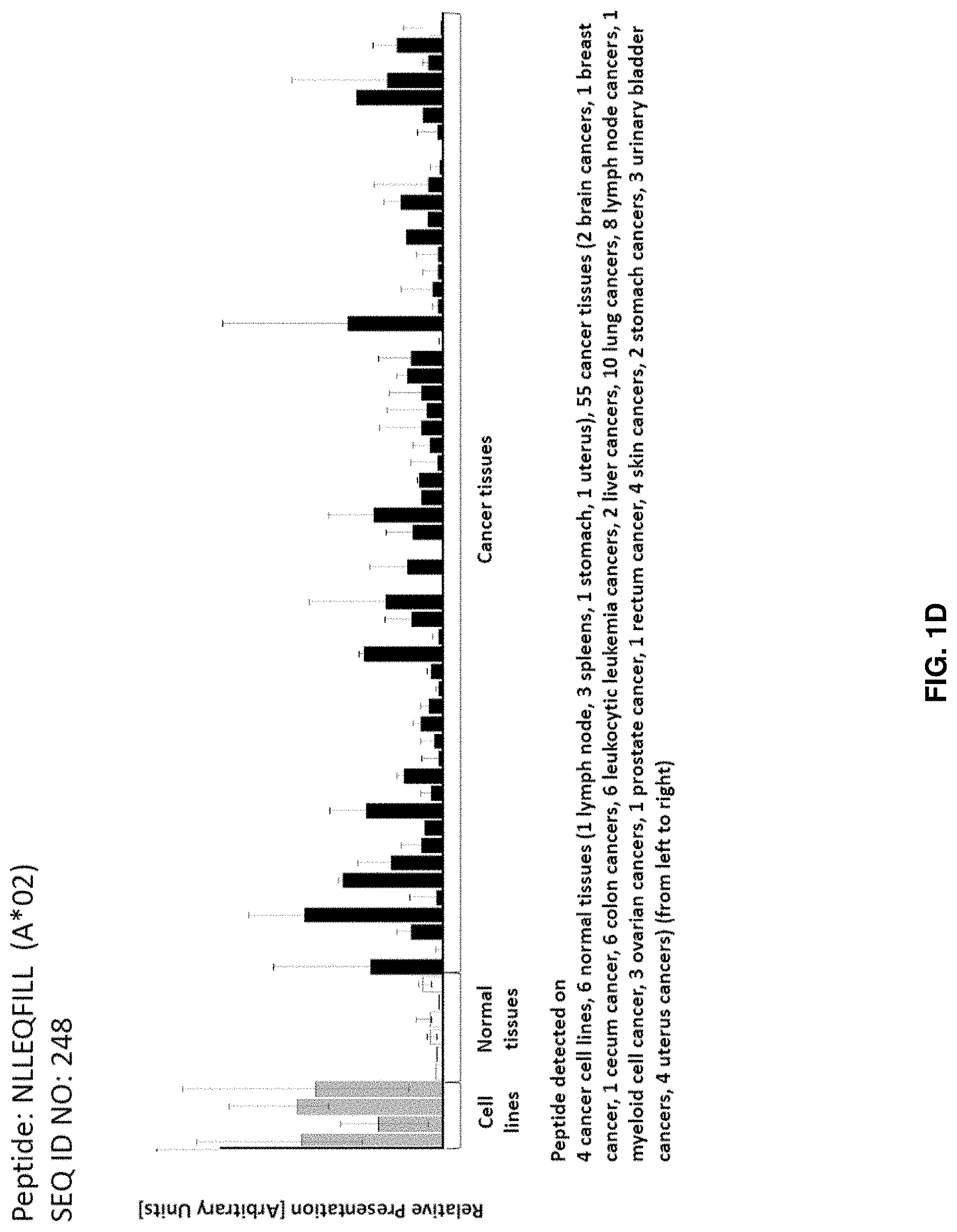
[0007] NHL is the seventh most common type of cancer and represents 4.3% of all new cancer cases in the U.S. (SEER Stat facts, 2014). It is the most common hematological malignancy both in Europe and the U.S. (Inoges et al., 2014).
[0008] The probability to develop NHL increases with age: The median age at the time point of diagnosis is 66 years. NHL is more common in people of Caucasian descent (21 cases per 100,000 persons), followed by Africans (15 cases per 100,000 persons) and Asians (14 cases per 100,000 persons). Men have a higher risk to develop NHL than women (23.9 cases per 100,000 males vs. 16.3 cases per 100,000 females) (SEER Stat facts, 2014).
[0009] The 5-year relative survival of NHL patients is 70% and varies with the cancer stage at the time point of diagnosis. For localized disease, the 5-year relative survival is 82%. If NHL has spread to different parts of the body, the 5-year relative survival decreases to 73.8% for regional and 62.4% for distant stage disease (SEER Stat facts, 2014). Risk factors include (high) age, male gender, ethnicity (Caucasian), exposure to benzene or radiation, HIV, autoimmune diseases, infections with HTLV-1, EBV or HHV8, infections with Helicobacter pylori, Chlamydophila psittaci, Campylobacter jejuni or HCV, (high) body weight and breast implants (American Cancer Society, 2015).
[0010] NHL has over 60 subtypes. The three most common subtypes are diffuse large B-cell lymphoma (DLBCL, the most common subtype), follicular lymphoma (FL, the second most common subtype) and small lymphocytic lymphoma/chronic lymphocytic lymphoma (SLL/CLL, the third most common subtype). DLBCL, FL and SLL/CLL account for about 85% of NHL (Li et al., 2015).
[0011] Diffuse large B-cell lymphoma (DLBCL) is the most common NHL type and comprises 30% of all NHLs. DLBCL belongs to the aggressive NHL subtypes and most patients show a quickly progressing disease. The International Prognostic Index (IPI) for aggressive NHL uses five significant risk factors prognostic for overall survival:
[0012] 1. Age (.ltoreq.60 years vs. >60 years)
[0013] 2. Serum lactate dehydrogenase (LDH) (normal vs. elevated)
[0014] 3. Performance status (0 or 1 vs. 2-4)
[0015] 4. Stage (stage I or II vs. stage III or IV)
[0016] 5. Extranodal site involvement (0 or 1 vs. 2-4).
[0017] Patients with two or more risk factors have a less than 50% chance of relapse-free survival and overall survival at 5 years. Patients with rearrangements of the bcl-2 and myc gene and/or overexpression of myc have a particularly poor prognosis. DLBCL patients co-expressing CD20 and CD30 have a more favorable prognosis and are predestined for an anti-CD30-specific therapy (National Cancer Institute, 2015).
[0018] Follicular lymphoma (FL) is the second most common NHL type and comprises 20% of all NHLs and 70% of all indolent lymphomas. More than 90% of the patients exhibit rearrangement of the bcl-2 gene. Most patients are 50 years or older at the time point of diagnosis and have advanced stage disease. The Follicular Lymphoma International Prognostic Index (FLIPI) uses five significant risk factors prognostic for overall survival:
[0019] 1. Age (.ltoreq.60 years vs. >60 years)
[0020] 2. Serum lactate dehydrogenase (LDH) (normal vs. elevated)
[0021] 3. Stage (stage I or II vs. stage III or IV)
[0022] 4. Hemoglobin level (.gtoreq.120 g/L vs. <120 g/L)
[0023] 5. Number of nodal areas (.ltoreq.4 vs. >4).
[0024] Patients with none or one risk factor have an 85% 10-year survival rate. Patients with three or more risk factors have a 40% 10-year survival rate (National Cancer Institute, 2015).
[0025] Diagnosis of NHL is done on an excisional biopsy of an abnormal lymph node or an incisional biopsy of an involved organ. Besides immunohistochemistry, cytogenetics, molecular genetics and fluorescent in situ hybridization (FISH) are used to clarify the diagnosis (Armitage, 2007).
[0026] Staging is done after the evaluation of the patients' history, physical examination and laboratory studies including hematologic parameters, screening chemistry studies and especially a test for serum lactate dehydrogenase (LDH) level. Imaging studies include computed tomograms of the chest, abdomen and pelvis and a PET scan (Armitage, 2007).
[0027] Determining for prognosis and treatment decision is the differentiation between indolent NHL types and aggressive NHLs. Indolent NHLs progress slowly, have a good prognosis and respond in early stages to radiation therapy, chemotherapy and immunotherapy, but are not curable in advanced stages. Aggressive NHLs progress quickly, but are responsive to intensive combination chemotherapy (National Cancer Institute, 2015).
[0028] Depending on the disease stage at the time point of diagnosis patients are classified into prognostic groups (National Cancer Institute, 2015) as follows:
TABLE-US-00001 Stage Prognostic groups I Involvement of a single lymphatic site (nodal region, Waldeyer ring, thymus or spleen (I). Localized involvement of a single extra-lymphatic organ or site in the absence of any lymph node involvement (IE). II Involvement of two or more lymph node regions on the same side of the diaphragm (II). Localized involvement of a single extra-lymphatic organ or site in association with regional lymph node involvement with or without involvement of other lymph node regions on the same side of the diaphragm (IIE). The number of regions involved may be indicated by a subscript Arabic numeral (for example II3). III Involvement of lymph node regions on both sides of the diaphragm (III), which also may be accompanied by extra-lymphatic extension in association with adjacent lymph node involvement (IIIE) or by involvement of the spleen (IIIS) or both (IIIE, IIIS). IV Diffuse or disseminated involvement of one or more extra-lymphatic organs, with or without associated lymph node involvement. Isolated extra-lymphatic organ involvement in the absence of adjacent regional lymph node involvement, but in conjunction with disease in distant site(s). Stage IV includes any involvement of the liver or bone marrow, lungs (other than by direct extension from another site), or cerebrospinal fluid.
[0029] The Ann Arbor staging system is usually used for patients with NHL. In this system, stage I, stage II, stage III and stage IV are sub-classified in to the categories A and B. Patients with well-defined generalized symptoms receive the designation B, while patients without these symptoms belong to category A. Category B symptoms include unexplained loss of more than 10% of body weight in the six months before diagnosis, unexplained fever with temperatures above 38.degree. C. and drenching night sweats. Specialized designations are used depending on the involvement of specific organs/sites (National Cancer Institute, 2015) as follows:
TABLE-US-00002 Designation Specific sites E Extranodal lymphoid malignancies near major lymphatic aggregates N Nodes H Liver L Lung M Bone marrow S Spleen P Pleura O Bone D Skin
[0030] To assign a precise stage, patients receive a clinical stage (CS) based on the findings of the clinical evaluation and a pathologic stage (PS) based on the findings of invasive procedures beyond the initial biopsy (National Cancer Institute, 2015).
[0031] Treatment of NHL depends on the histologic type and stage. Standard treatment options include (National Cancer Institute, 2015):
TABLE-US-00003 Stage Standard treatment option Indolent, stage I and contiguous Radiation therapy stage II NHL Rituximab .+-. chemotherapy Watchful waiting Other therapies as designated for patients with advanced-stage disease Indolent, non-contiguous stage Watchful waiting for asymptomatic patients II/III/IV NHL Rituximab Purine nucleoside analogs Alkylating agents .+-. steroids Combination chemotherapy Yttrium-90-labeled ibritumomab tiuxetan Maintenance rituximab Indolent, Recurrent NHL Chemotherapy (single agent or combination) Rituximab Lenalidomide Radiolabeled anti-CD20 monoclonal antibodies Palliative radiation therapy Aggressive, stage I and contiguous R-CHOP .+-. (involved-field radiation therapy) stage II NHL IF-XRT Aggressive, non-contiguous R-CHOP stage II/III/IV NHL Other combination chemotherapy Lymphoblastic lymphoma Intensive therapy Radiation therapy Diffuse, small, noncleaved-cell/Burkitt Aggressive multi-drug regimens lymphoma Central nervous system (CNS) prophylaxis Aggressive, recurrent NHL Bone marrow or stem cell transplantation Re-treatment with standard agents Palliative radiation therapy
[0032] Indolent, stage I and contiguous stage II NHL: Standard treatment options include radiation therapy, rituximab (anti-CD20 monoclonal antibody).+-.chemotherapy, watchful waiting and other therapies as designated for patients with advanced-stage disease.
[0033] Indolent, non-contiguous stage II/III/IV NHL: Standard treatment options include watchful waiting for asymptomatic patients, rituximab, obinutuzumab (anti-CD20 monoclonal antibody), purine nucleoside analogs (fludarabine, 2-chlorodeoxyadenosine), alkylating agents (cyclophosphamide, chlorambucil).+-.steroids, bendamustine, combination chemotherapy (CVP, C-MOPP (cyclophosphamide, vincristine, procarbazine, and prednisone), CHOP, FND (fludarabine, mitoxantrone.+-.dexamethasone)), yttrium-labeled ibritumomab tiuxetan and maintenance rituximab. Rituximab (R) is considered first-line therapy, either alone or in combination with other agents (R-Bendamustine, R-F (fludarabine), R-CVP (cyclophosphamide, vincristine, and prednisone), R-CHOP (cyclophosphamide, doxorubicin, vincristine, and prednisone), R-FM (fludarabine, mitoxantrone), R-FCM (fludarabine, cyclophosphamide, and mitoxantrone)). Under clinical evaluation are bone marrow transplantation (BMT) or peripheral stem cell transplantation (PSCT), idiotype vaccines and radiolabeled monoclonal antibodies (ofatumumab: anti-CD20 monoclonal antibody).
[0034] Indolent, recurrent NHL: Standard treatment options include chemotherapy (single agent or combination), rituximab, lenalidomide, radiolabeled anti-CD20 monoclonal antibodies (yttrium-90 ibritumomab) and palliative radiation therapy. Treatment options under clinical evaluation include SCTs.
[0035] Aggressive, stage I and contiguous stage II NHL: Standard treatment options include R-CHOP.+-.IF-XRT. Treatment options under clinical evaluation include R-ACVBP (rituximab+doxorubicin, cyclophosphamide, vindesine, bleomycin, prednisone).
[0036] Aggressive, non-contiguous stage II/III/IV NHL: Standard treatment options include combination chemotherapy.+-.local-field radiation therapy. Drug combinations include ACVBP, CHOP, CNOP (cyclophosphamide, mitoxantrone, vincristine, prednisone), m-BACOD (methotrexate, bleomycin, doxorubicin, cyclophosphamide, vincristine, dexamethasone, leucovorin), MACOP-B (methotrexate, doxorubicin, cyclophosphamide, vincristine, prednisone fixed dose, bleomycin, leucovorin), ProMACE CytaBOM (prednisone, doxorubicin, cyclophosphamide, etoposide, cytarabine, bleomycin, vincristine, methotrexate, leucovorin), R-CHOP. Under clinical evaluation are BMT and SCT.
[0037] Lymphoblastic lymphoma: Standard treatment options include intensive therapy and radiation therapy.
[0038] Diffuse, small noncleaved-cell/Burkitt lymphoma: Standard treatment options include aggressive multidrug regimens and CNS prophylaxis.
[0039] Aggressive, recurrent NHL: Standard treatment options include BMT or SCT, re-treatment with standard agents (rituximab, radiolabeled anti-CD20 monoclonal antibodies, denileukin diftitox (a fusion protein combining diphtheria toxin and interleukin-2)) and palliative radiation therapy. Treatment options under clinical evaluation include SCT (National Cancer Institute, 2015).
[0040] Spontaneous tumor regression can be observed in lymphoma patients. Therefore, active immunotherapy is a therapy option (Palomba, 2012). An important vaccination option includes Id vaccines. B lymphocytes express surface immunoglobulins with a specific amino acid sequence in the variable regions of their heavy and light chains, unique to each cell clone (=idiotype, Id). The idiotype functions as a tumor associated antigen.
[0041] Passive immunization includes the injection of recombinant murine anti-Id monoclonal antibodies alone or in combination with IFN alpha, IL2 or chlorambucil.
[0042] Active immunization includes the injection of recombinant protein (Id) conjugated to an adjuvant (KLH), given together with GM-CSF as an immune adjuvant. Tumor-specific Id is produced by hybridoma cultures or using recombinant DNA technology (plasmids) by bacterial, insect or mammalian cell culture.
[0043] Three phase III clinical trials have been conducted (Biovest, Genitope, Favrille). In two trials patients had received rituximab. GM-CSF was administered in all three trials. Biovest used hybridoma-produced protein, Genitope and Favrille used recombinant protein. In all three trials Id was conjugated to KLH. Only Biovest had a significant result.
[0044] Vaccines other than Id include the cancer-testis antigens MAGE, NY-ESO1 and PASD-1, the B-cell antigen CD20 or cellular vaccines. The vaccines consist of DCs pulsed with apoptotic tumor cells, tumor cell lysate, DC-tumor cell fusion or DCs pulsed with tumor-derived RNA. In situ vaccination involves the vaccination with intra-tumoral CpG in combination with chemotherapy or irradiated tumor cells grown in the presence of GM-CSF and collection/expansion/re-infusion of T cells.
[0045] Vaccinations with antibodies that alter immunologic checkpoints are comprised of anti-CD40, anti-OX40, anti-41 BB, anti-CD27, anti-GITR (agonist antibodies that directly enhance anti-tumor response) or anti-PD1, anti-CTLA-4 (blocking antibodies that inhibit the checkpoint that would hinder the immune response). Examples are ipilimumab (anti-CTLA-4) and CT-011 (anti-PD1) (Palomba, 2012).
[0046] Considering the severe side-effects and expense associated with treating cancer, there is a need to identify factors that can be used in the treatment of cancer in general and NHL in particular. There is also a need to identify factors representing biomarkers for cancer in general and NHL in particular, leading to better diagnosis of cancer, assessment of prognosis, and prediction of treatment success.
[0047] Immunotherapy of cancer represents an option of specific targeting of cancer cells while minimizing side effects. Cancer immunotherapy makes use of the existence of tumor associated antigens.
[0048] The current classification of tumor associated antigens (TAAs) comprises the following major groups:
[0049] a) Cancer-testis antigens: The first TAAs ever identified that can be recognized by T cells belong to this class, which was originally called cancer-testis (CT) antigens because of the expression of its members in histologically different human tumors and, among normal tissues, only in spermatocytes/spermatogonia of testis and, occasionally, in placenta. Since the cells of testis do not express class I and II HLA molecules, these antigens cannot be recognized by T cells in normal tissues and can therefore be considered as immunologically tumor-specific. Well-known examples for CT antigens are the MAGE family members and NY-ESO-1.
[0050] b) Differentiation antigens: These TAAs are shared between tumors and the normal tissue from which the tumor arose. Most of the known differentiation antigens are found in melanomas and normal melanocytes. Many of these melanocyte lineage-related proteins are involved in biosynthesis of melanin and are therefore not tumor specific but nevertheless are widely used for cancer immunotherapy. Examples include, but are not limited to, tyrosinase and Melan-A/MART-1 for melanoma or PSA for prostate cancer.
[0051] c) Over-expressed TAAs: Genes encoding widely expressed TAAs have been detected in histologically different types of tumors as well as in many normal tissues, generally with lower expression levels. It is possible that many of the epitopes processed and potentially presented by normal tissues are below the threshold level for T-cell recognition, while their over-expression in tumor cells can trigger an anticancer response by breaking previously established tolerance. Prominent examples for this class of TAAs are Her-2/neu, survivin, telomerase, or WT1.
[0052] d) Tumor-specific antigens: These unique TAAs arise from mutations of normal genes (such as .beta.-catenin, CDK4, etc.). Some of these molecular changes are associated with neoplastic transformation and/or progression. Tumor-specific antigens are generally able to induce strong immune responses without bearing the risk for autoimmune reactions against normal tissues. On the other hand, these TAAs are in most cases only relevant to the exact tumor on which they were identified and are usually not shared between many individual tumors. Tumor-specificity (or -association) of a peptide may also arise if the peptide originates from a tumor- (-associated) exon in case of proteins with tumor-specific (-associated) isoforms.
[0053] e) TAAs arising from abnormal post-translational modifications: Such TAAs may arise from proteins which are neither specific nor overexpressed in tumors but nevertheless become tumor associated by posttranslational processes primarily active in tumors. Examples for this class arise from altered glycosylation patterns leading to novel epitopes in tumors as for MUC1 or events like protein splicing during degradation which may or may not be tumor specific.
[0054] f) Oncoviral proteins: These TAAs are viral proteins that may play a critical role in the oncogenic process and, because they are foreign (not of human origin), they can evoke a T-cell response. Examples of such proteins are the human papilloma type 16 virus proteins, E6 and E7, which are expressed in cervical carcinoma.
[0055] T-cell based immunotherapy targets peptide epitopes derived from tumor-associated or tumor-specific proteins, which are presented by molecules of the major histocompatibility complex (MHC). The antigens that are recognized by the tumor specific T lymphocytes, that is, the epitopes thereof, can be molecules derived from all protein classes, such as enzymes, receptors, transcription factors, etc. which are expressed and, as compared to unaltered cells of the same origin, usually up-regulated in cells of the respective tumor.
[0056] There are two classes of MHC-molecules, MHC class I and MHC class II. MHC class I molecules are composed of an alpha heavy chain and beta-2-microglobulin, MHC class II molecules of an alpha and a beta chain. Their three-dimensional conformation results in a binding groove, which is used for non-covalent interaction with peptides.
[0057] MHC class I molecules can be found on most nucleated cells. They present peptides that result from proteolytic cleavage of predominantly endogenous proteins, defective ribosomal products (DRIPs) and larger peptides. However, peptides derived from endosomal compartments or exogenous sources are also frequently found on MHC class I molecules. This non-classical way of class I presentation is referred to as cross-presentation in the literature (Brossart and Bevan, 1997; Rock et al., 1990). MHC class II molecules can be found predominantly on professional antigen presenting cells (APCs), and primarily present peptides of exogenous or transmembrane proteins that are taken up by APCs e.g. during endocytosis, and are subsequently processed. Complexes of peptide and MHC class I are recognized by CD8-positive T cells bearing the appropriate T-cell receptor (TCR), whereas complexes of peptide and MHC class II molecules are recognized by CD4-positive-helper-T cells bearing the appropriate TCR. It is well known that the TCR, the peptide and the MHC are thereby present in a stoichiometric amount of 1:1:1.
[0058] CD4-positive helper T cells play an important role in inducing and sustaining effective responses by CD8-positive cytotoxic T cells. The identification of CD4-positive T-cell epitopes derived from tumor associated antigens (TAA) is of great importance for the development of pharmaceutical products for triggering anti-tumor immune responses (Gnjatic et al., 2003). At the tumor site, T helper cells, support a cytotoxic T cell- (CTL-) friendly cytokine milieu (Mortara et al., 2006) and attract effector cells, e.g. CTLs, natural killer (NK) cells, macrophages, and granulocytes (Hwang et al., 2007).
[0059] In the absence of inflammation, expression of MHC class II molecules is mainly restricted to cells of the immune system, especially professional antigen-presenting cells (APC), e.g., monocytes, monocyte-derived cells, macrophages, dendritic cells. In cancer patients, cells of the tumor have been found to express MHC class II molecules (Dengjel et al., 2006).
[0060] Elongated (longer) peptides of the invention can act as MHC class II active epitopes. T-helper cells, activated by MHC class II epitopes, play an important role in orchestrating the effector function of CTLs in anti-tumor immunity. T-helper cell epitopes that trigger a T-helper cell response of the TH1 type support effector functions of CD8-positive killer T cells, which include cytotoxic functions directed against tumor cells displaying tumor-associated peptide/MHC complexes on their cell surfaces. In this way tumor-associated T-helper cell peptide epitopes, alone or in combination with other tumor-associated peptides, can serve as active pharmaceutical ingredients of vaccine compositions that stimulate anti-tumor immune responses.
[0061] It was shown in mammalian animal models, e.g., mice, that even in the absence of CD8-positive T lymphocytes, CD4-positive T cells are sufficient for inhibiting manifestation of tumors via inhibition of angiogenesis by secretion of interferon-gamma (IFN.gamma.) (Beatty and Paterson, 2001; Mumberg et al., 1999). There is evidence for CD4 T cells as direct anti-tumor effectors (Braumuller et al., 2013; Tran et al., 2014).
[0062] Since the constitutive expression of HLA class II molecules is usually limited to immune cells, the possibility of isolating class II peptides directly from primary tumors was previously not considered possible. However, Dengjel et al. were successful in identifying a number of MHC Class II epitopes directly from tumors (WO 2007/028574, EP 1 760 088 B1).
[0063] Since both types of response, CD8 and CD4 dependent, contribute jointly and synergistically to the anti-tumor effect, the identification and characterization of tumor-associated antigens recognized by either CD8+ T cells (ligand: MHC class I molecule+peptide epitope) or by CD4-positive T-helper cells (ligand: MHC class II molecule+peptide epitope) is important in the development of tumor vaccines.
[0064] For an MHC class I peptide to trigger (elicit) a cellular immune response, it also must bind to an MHC-molecule. This process is dependent on the allele of the MHC-molecule and specific polymorphisms of the amino acid sequence of the peptide. MHC-class-1-binding peptides are usually 8-12 amino acid residues in length and usually contain two conserved residues ("anchors") in their sequence that interact with the corresponding binding groove of the MHC-molecule. In this way, each MHC allele has a "binding motif" determining which peptides can bind specifically to the binding groove.
[0065] In the MHC class I dependent immune reaction, peptides not only have to be able to bind to certain MHC class I molecules expressed by tumor cells, they subsequently also have to be recognized by T cells bearing specific T cell receptors (TCR).
[0066] For proteins to be recognized by T-lymphocytes as tumor-specific or -associated antigens, and to be used in a therapy, particular prerequisites must be fulfilled. The antigen should be expressed mainly by tumor cells and not, or in comparably small amounts, by normal healthy tissues. In a preferred embodiment, the peptide should be over-presented by tumor cells as compared to normal healthy tissues. It is furthermore desirable that the respective antigen is not only present in a type of tumor, but also in high concentrations (i.e. copy numbers of the respective peptide per cell). Tumor-specific and tumor-associated antigens are often derived from proteins directly involved in transformation of a normal cell to a tumor cell due to their function, e.g. in cell cycle control or suppression of apoptosis. Additionally, downstream targets of the proteins directly causative for a transformation may be up-regulated and thus may be indirectly tumor-associated. Such indirect tumor-associated antigens may also be targets of a vaccination approach (Singh-Jasuja et al., 2004). It is essential that epitopes are present in the amino acid sequence of the antigen, in order to ensure that such a peptide ("immunogenic peptide"), being derived from a tumor associated antigen, leads to an in vitro or in vivo T-cell-response.
[0067] Basically, any peptide able to bind an MHC molecule may function as a T-cell epitope. A prerequisite for the induction of an in vitro or in vivo T-cell-response is the presence of a T cell having a corresponding TCR and the absence of immunological tolerance for this particular epitope.
[0068] Therefore, TAAs are a starting point for the development of a T cell based therapy including but not limited to tumor vaccines. The methods for identifying and characterizing the TAAs are usually based on the use of T-cells that can be isolated from patients or healthy subjects, or they are based on the generation of differential transcription profiles or differential peptide expression patterns between tumors and normal tissues. However, the identification of genes over-expressed in tumor tissues or human tumor cell lines, or selectively expressed in such tissues or cell lines, does not provide precise information as to the use of the antigens being transcribed from these genes in an immune therapy. This is because only an individual subpopulation of epitopes of these antigens are suitable for such an application, since a T cell with a corresponding TCR has to be present and the immunological tolerance for this particular epitope needs to be absent or minimal. In a very preferred embodiment of the invention it is therefore important to select only those over- or selectively presented peptides against which a functional and/or a proliferating T cell can be found. Such a functional T cell is defined as a T cell, which upon stimulation with a specific antigen can be clonally expanded and is able to execute effector functions ("effector T cell").
[0069] In case of targeting peptide-MHC by specific TCRs (e.g. soluble TCRs) and antibodies or other binding molecules (scaffolds) according to the invention, the immunogenicity of the underlying peptides is secondary. In these cases, the presentation is the determining factor.
BRIEF SUMMARY OF THE INVENTION
[0070] In a first aspect of the present invention, the present invention relates to a peptide comprising an amino acid sequence selected from the group consisting of SEQ ID NO: 1 to SEQ ID NO: 311 or a variant sequence thereof which is at least 77%, preferably at least 88%, homologous (preferably at least 77% or at least 88% identical) to SEQ ID NO: 1 to SEQ ID NO: 311, wherein said variant binds to MHC and/or induces T cells cross-reacting with said peptide, or a pharmaceutical acceptable salt thereof, wherein said peptide is not the underlying full-length polypeptide.
[0071] The present invention further relates to a peptide of the present invention comprising a sequence that is selected from the group consisting of SEQ ID NO: 1 to SEQ ID NO: 311 or a variant thereof, which is at least 77%, preferably at least 88%, homologous (preferably at least 77% or at least 88% identical) to SEQ ID NO: 1 to SEQ ID NO: 311, wherein said peptide or variant thereof has an overall length of between 8 and 100, preferably between 8 and 30, and most preferred of between 8 and 14 amino acids.
[0072] The following tables show the peptides according to the present invention, their respective SEQ ID NOs, and the prospective source (underlying) genes for these peptides. All peptides in Table 1 and Table 2 bind to HLA-A*02. The peptides in Table 2 have been disclosed before in large listings as results of high-throughput screenings with high error rates or calculated using algorithms, but have not been associated with cancer at all before. The peptides in Table 3 are additional peptides that may be useful in combination with the other peptides of the invention. The peptides in Tables 4A and B are furthermore useful in the diagnosis and/or treatment of various other malignancies that involve an over-expression or over-presentation of the respective underlying polypeptide.
TABLE-US-00004 TABLE 1 Peptides according to the present invention. SEQ ID Official Gene No. Sequence GeneID(s) Symbol(s) 1 LLSGQLPTI 84969 TOX2 2 LLSEETPSA 10765 KDM5B 3 LTIDTQYYL 5422 POLA1 4 TLLGFFLAKV 5422 POLA1 5 VLQGLTFTL 6890 TAP1 6 TLITLPLLFL 6890 TAP1 7 NLLGMIFSM 51398 WDR83OS 8 ALYAVIEKA 5293 PIK3CD 9 FLLDLDPLL 7915 ALDH5A1 10 FLLVGTQIDL 643751, CDC42P6, CDC42 998 11 GLDTVVALL 23203 PMPCA 12 GLLLLVPLL 145864 HAPLN3 13 HLVPASWKL 3718 JAK3 14 LLSDPTPGA 3718 JAK3 15 IIIEDLLEA 10985 GCN1L1 16 TLIAAILYL 5355 PLP2 17 VIIPLLSSV 91526 ANKRD44 18 KLTDQPPLV 91526 ANKRD44 19 VLEAILPLV 2889 RAPGEF1 20 YLIAGGDRWL 2646 GCKR 21 ALFKEAYSL 55732 C1orf112 22 ALKKHLTSV 10773 ZBTB6 23 ALVEDIINL 92399 MRRF 24 AVLGFSFRL 80222 TARS2 25 FLDTSNQHLL 4064 CD180 26 FLGSFIDHV 91147 TMEM67 27 FLNQESFDL 6610 SMPD2 28 FLSNANPSL 7818 DAP3 29 ILSDVTQGL 55591 VEZT 30 ILSTLDVEL 10744, PTTG2, PTTG1 9232 31 KLYDEESLL 57680 CHD8 32 VLNEDELPSV 57680 CHD8 33 LLANIVPIAMLV 4539, MT-ND4L 86775071, 923201 34 LLWEDGVTEA 22916 NCBP2 35 SLSSERYYL 8320 EOMES 36 VILDIPLLFET 79877 DCAKD 37 VLGNALEGV 4678 NASP 38 YLTAEILELAGN 221613, HIST1H2AA, HIST1H2AE, 3012, HIST1H2AD, H2AFX, 3013, HIST2H2AB, H2AFJ, 3014, HIST2H2AA4, HIST1H2AI, 317772, HIST1H2AK, HIST1H2AJ, 55766, HIST1H2AL, HIST1H2AC, 723790, HIST1H2AB, HIST1H2AM, 8329, HIST2H2AA3, HIST2H2AC, 8330, HIST1H2AH, HIST1H2AG, 8331, HIST3H2A, H2AFY 8332, 8334, 8335, 8336, 8337, 8338, 85235, 8969, 92815, 9555 39 QLLPQGIVPAL 55374 TMCO6 40 FLNSVIVDL 6249 CLIP1 41 ILASIFETV 6574 SLC20A1 42 YLQDLVERA 10347 ABCA7 43 ALLEGVKNV 84678 KDM2B 44 FIIEEQSFL 10200 MPHOSPH6 45 FILDDSALYL 23130 ATG2A 46 FLVEEIFQT 8888 MCM3AP 47 GLLPKLTAL 22920 KIFAP3 48 KILDEDLYI 641 BLM 49 TILGDPQILL 23460 ABCA6 50 LLLDGLIYL 23460 ABCA6 51 SLLGNSPVL 23460 ABCA6 52 VLLEDVDAAFL 617 BCS1L 53 FLREYFERL 5573 PRKAR1A 54 DIFDAMFSV 5573 PRKAR1A 55 ILVEVDLVQA 4261 CIITA 56 GLQDLLFSL 4261 CIITA 57 LQIGDFVSV 51167 CYB5R4 58 QLAPFLPQL 23392 KIAA0368 59 RLHREVAQV 2802 GOLGA3 60 SLLIDVITV 51534 VTA1 61 SLLNKDLSL 1786 DNMT1 62 ALAPYLDLL 54093 SETD4 63 ALIEEAYGL 3836, KPNA1, KPNA5 3841 64 FLVEVSNDV 23224 SYNE2 65 NLTDVSPDL 23224 SYNE2 66 KLAPIPVEL 153241 CEP120 67 LLATVNVAL 23511 NUP188 68 QIAAFLFTV 56006 SMG9 69 TLLAFPLLL 84720 PIGO 70 VLIEILQKA 23633, KPNA6, KPNA5 3841 71 VLLDYVGNVQL 51676 ASB2 72 TLQEETAVYL 51676 ASB2 73 YLGEEYPEV 23451 SF3B1 74 SLDLRPLEV 43 ACHE 75 AALKYIPSV 1794 DOCK2 76 ALADLVPVDVVV 84188 FAR1 77 ALLDVSNNYGI 115752 DIS3L 78 AMEEAVAQV 22897 CEP164 79 AMKEEKEQL 9126 SMC3 80 YLFDEIDQA 9126 SMC3 81 FIFSYITAV 128338 DRAM2 82 FLIDGSSSV 1690 COCH 83 FLMDDNMSNTL 4603 MYBL1 84 FLQELQLEHA 8604 SLC25A12 85 GLAPAEVVVATVA 57591 MKL1 86 GLATIRAYL 2731 GLDC 87 GLFARIIMI 5250 SLC25A3 88 GLFDNRSGLPEA 79733 E2F8 89 GLTALHVAV 602 BCL3 90 HLDEVFLEL 55744 COA1 91 HLSSTTAQV 201633 TIGIT 92 KLLFEIASA 124460 SNX20 93 KLLGSLQLL 81603 TRIM8 94 LLAGQATTAYF 972 CD74 95 LLFDLIPVVSV 284114 TMEM102 96 LLLNENESLFL 26156 RSL1D1 97 LLNFSPGNL 3929 LBP 98 MLQDGIARL 79697 C14orf169 99 QLYDGATALFL 147463 ANKRD29 100 RLIRTIAAI 140461 ASB8 101 SLDQSTWNV 23240 KIAA0922 102 SLFAAISGMIL 931 MS4A1 103 SLQDHLEKV 1756 DMD 104 VLLGLPLLV 9674 KIAA0040 105 VLTPVILQV 100499483, C9orf174 100499484 106 VLYELLQYI 51513 ETV7 107 VQAVSIPEV 55755 CDK5RAP2 108 YLAPENGYLM 6625 SNRNP70 109 YLFQFSAAL 130367 SGPP2
110 YQYPFVLGL 130367 SGPP2 111 YLLDTLLSL 57448 BIRC6 112 FLAILPEEV 7762 ZNF215 113 FVIDSFEEL 147945 NLRP4 114 GLSDISPST 26005 C2CD3 115 LLIDIIHFL 25914 RTTN 116 SLLDNLLTI 25914 RTTN 117 VLATILAQL 26271 FBXO5 118 VLDGMIYAI 54813 KLHL28 119 ELCDIILRV 54813 KLHL28 120 VLLGTTWAL 221188 GPR114 121 YLTGYNFTL 9521 EEF1E1 122 AISEAQESV 79882 ZC3H14 123 ALLSAFVQL 8295 TRRAP 124 FLGVVVPTV 56996 SLC12A9 125 FVAPPTAAV 162 AP1B1 126 GLSIFIYRL 10075 HUWE1 127 HLMEENMIVYV 65220 NADK 128 KLFDASPTFFA 3992, FADS1, FADS3 3995 129 SLFEASQQL 23347 SMCHD1 130 VIFSYVLGV 79004 CUEDC2 131 VLIEETDQL 6924 TCEB3 132 VLQDQVDEL 51199 NIN 133 ALEELTGFREL 4288 MKI67 134 ALGRLGILSV 22828, SCAF8, TIAM2 26230 135 ALTGLQFQL 22797 TFEC 136 FIFGIVHLL 64066 MMP27 137 FIQQERFFL 4012 LNPEP 138 NLINNIFEL 4012 LNPEP 139 FLASPLVAI 3593 IL12B 140 FLFEDFVEV 140775 SMCR8 141 FLGELTLQL 257218 SHPRH 142 FLYEDSKSVRL 696 BTN1A1 143 TLHAVDVTL 696 BTN1A1 144 GLITQVDKL 9183 ZW10 145 GLLHEVVSL 163486 DENND1B 146 GLLQQPPAL 1871 E2F3 147 GLSEYQRNFL 56890 MDM1 148 ICAGHVPGV 79019 CENPM 149 ILNPVTTKL 81691 LOC81691 150 ILSEKEYKL 127254 C1orf173 151 ILVKQSPML 940 CD28 152 KIMYTLVSV 3709 ITPR2 153 KLLKGIYAI 1235 CCR6 154 KLMNIQQQL 11214 AKAP13 155 KLMTSLVKV 10734 STAG3 156 KMLEDDLKL 2334 AFF2 157 KVLEFLAKV 139422, MAGEB10, MAGEB2, 4113, MAGEB4 4115 158 KVQDVLHQV 83756 TAS1R3 159 LLLSDSGFYL 28557 TRBV30 160 LLPPPSPAA 83881 MIXL1 161 NLMLELETV 1063 CENPF 162 RLADLKVSI 2175 FANCA 163 SIFDAVLKGV 157680 VPS13B 164 SLFDGAVISTV 23049 SMG1 165 KLLEEIEFL 23049 SMG1 166 SLFSEVASL 22832 KIAA1009 167 SLFSITKSV 60468 BACH2 168 SLLSPLLSV 54949 SDHAF2 169 SSLEENLLHQV 80205 CHD9 170 STIELSENSL 55635 DEPDC1 171 TLLDVISAL 27340 UTP20 172 TLQDSLEFI 51735, RAPGEF6, FNIP1 96459 173 VILDSVASV 5890 RAD51B 174 VLVEITDVDFAA 79801 SHCBP1 175 VMESILLRL 342850 ANKRD62 176 YLHIYESQL 29851 ICOS 177 YLYEAEEATTL 22798 LAMB4 178 YVLQGEFFL 84541 KBTBD8 179 FVDTNLYFL 81037 CLPTM1L 180 GILQLVESV 6050 RNH1 181 LLFDQNDKV 100653071, CRTAP 10491 182 LLPPPPPVA 23091, ZC3H13, NFIX 4784 183 VLFETVLTI 8906 AP1G2 184 AVLGTSWQL 23041 MON2 185 FIAQLNNVEL 6509 SLC1A4 186 FLDVSRDFV 54461 FBXW5 187 FLNSFVFKM 89910 UBE3B 188 GLEDEMYEV 285905, INTS4L1, INTS4L2, 644619, INTS4 92105 189 SLSHLVPAL 285905, INTS4L1, INTS4L2, 644619, INTS4 92105 190 GLIELVDQL 90410 IFT20 191 GLSDISAQV 5989 RFX1 192 GMAAEVPKV 348378 FAM159A 193 SLADSMPSL 8945 BTRC 194 SLAPFDREPFTL 3937 LCP2 195 ALIPDLNQI 51361 HOOK1 196 TLALAMIYL 100134301, ANAPC1 285074, 64682, 730268 197 YLLTDNVVKL 79810 PTCD2 198 GLLSAVSSV 9894 TELO2 199 SLNSTTWKV 1233 CCR4 200 YLLDFEDRL 23207 PLEKHM2 201 YLNISQVNV 9262 STK17B 202 ALAAGGYDV 3009 HIST1H1B 203 ILDTIFHKV 2829 XCR1 204 RLCDIVVNV 84614 ZBTB37 205 TLFYESPHL 221908 PPP1R35 206 SAVSGQWEV 2326 FMO1 207 GLVGLLEQA 57572, DOCK6, DOCK8, DOCK7 81704, 85440 208 FLAVSLPLL 3071 NCKAP1L 209 FLLDTISGL 84864 MINA 210 FLAEQFEFL 55610 CCDC132 211 FIDDLFAFV 1209 CLPTM1 212 FLIGQGAHV 4659 PPP1R12A 213 YINEDEYEV 7874 USP7 214 FLFDGSMSL 3683 ITGAL 215 QLFEEEIEL 63906 GPATCH3 216 KVVSNLPAI 10199 MPHOSPH10 217 AQFGAVLEV 55131 RBM28 218 ALDQFLEGI 57169 ZNFX1 219 ALLELENSV 715, C1R, EPPK1 83481 220 FLAEAPTAL 9814 SFI1 221 FLAPDNSLLLA 22898 DENND3 222 FLIETGTLL 79705 LRRK1 223 FLQDIPDGLFL 206426, PIP5K1P1, PIPSL, 266971, PIP5K1A 8394 224 FLSPLLPLL 10961 ERP29 225 GTYQDVGSLNIGDV 973 CD79A
226 GVIDPVPEV 8879 SGPL1 227 IIAEGIPEA 47 ACLY 228 IIAEYLSYV 51667 NUB1 229 ILSPWGAEV 142 PARP1 230 IMDDDSYGV 9874 TLK1 231 IVMGAIPSV 1902 LPAR1 232 KVMEGTVAA 1445 CSK 233 MLEVHIPSV 79856 SNX22 234 NLQRTVVTV 4297 MLL 235 SLDVYELFL 79586 CHPF 236 SLFDGFFLTA 25920 COBRA1 237 YLDRLIPQA 115209 OMA1 238 YQYGAVVTL 1380 CR2 239 VLIDDTVLL 116138 KLHDC3 240 ALVPTPALFYL 51528 JKAMP 241 FIPDFIPAV 56912 IFT46 242 GILDFZVFL 100124692, MGAM 8972, 93432 243 GLPDLDIYL 23334 SZT2 244 ILEPFLPAV 6894 TARBP1 245 KLIQLPVVYV 9875 URB1 246 KLPVPLESV 285190, RGPD4, RGPD1, RANBP2, 400966, RGPD3, RGPD8, RGPD6, 5903, RGPD2, RGPD5 653489, 727851, 729540, 729857, 84220 247 KVLEMETTV 9810 RNF40 248 NLLEQFILL 64708 COPS7B 249 VLLESLVEI 149371 EXOC8 250 VLTNVGAAL 129285 PPP1R21 251 VLYELFTYI 3717 JAK2 252 YLGDLIMAL 3930, LBR, TM7SF2 7108
TABLE-US-00005 TABLE 2 Additional peptides according to the present invention with no prior known cancer association. SEQ ID No. Sequence GeneID(s) Official Gene Symbol(s) 253 YSDDDVPSV 29028 ATAD2 254 FLYSETWNI 4519, MT-CYB 8923205 255 GMWNPNAPVFL 9910 RABGAP1L 256 ALQETPPQV 146206 RLTPR 257 FLQEWEVYA 57001 ACN9 258 RIYPFLLMV 10299 MARCH6 259 TVLDGLEFKV 10592 SMC2 260 RLDEAFDFV 1844 DUSP2 261 FLPETRIMTSV 11319 ECD 262 LMGPVVHEV 5116 PCNT 263 GLMDNEIKV 8795 TNFRSF10B 264 ILTGTPPGV 151313, FAHD2B, FAHD2A 51011 265 ILWHFVASL 23077 MYCBP2 266 QLTEMLPSI 689 BTF3 267 SLLETGSDLLL 57176 VARS2 268 VLFPLPTPL 11184 MAP4K1 269 VLQNVAFSV 597 BCL2A1 270 VVVDSDSLAFV 122961 ISCA2 271 YLLDQPVLEQRL 81887 LAS1L 272 KLDHTLSQI 4863 NPAT 273 AILLPQPPK 1761, DMRT1, SDHD, KIF9, 6392, LINC00338, TMEM175, 64147, TRIM5 642204, 654434, 84286, 85363 274 KLLNLISKL 5366 PMAIP1 275 KLMDLEDCAL 23269 MGA 276 NMISYVVHL 204801 NLRP11 277 FLIDLNSTHGTFL 5511 PPP1R8 278 FLLFINHRL 4292 MLH1 279 NLAGENILNPL 56948 SDR39U1 280 SLLNHLPYL 201562 PTPLB 281 TLQTVPLTTV 1997 ELF1 282 YLLEQGAQV 55527 FEM1A 283 ALMPVTPQA 23683 PRKD3 284 KLQEQIHRV 196441 ZFC3H1 285 SITAVTPLL 63910 SLC17A9 286 HLTEDTPKV 50814 NSDHL 287 ILMGHSLYM 9786 KIAA0586 288 RLAPEIVSA 157285 SGK223 289 SLLAANNLL 9380 GRHPR 290 IASPVIAAV 127544 RNF19B 291 KIIDTAGLSEA 22954 TRIM32 292 KLINSQISL 5293 PIK3CD 293 GLAMVEAISYV 109 ADCY3 294 KLYGPEGLELV 3394 IRF8 295 SLAAVSQQL 7094 TLN1 296 FILEPLYKI 9343 EFTUD2 297 ILQNGLETL 89857 KLHL6 298 ALTDVILCV 89857 KLHL6 299 RLLEEEGVSL 64428 NARFL 300 IVLERNPEL 5257 PHKB 301 LQFDGIHVV 55294 FBXW7 302 SLAELDEKISA 51562 MBIP 303 FVWEASHYL 5442 POLRMT 304 ALIRLDDLFL 56902 PNO1 305 AMLAQQMQL 4154 MBNL1 306 AQVALVNEV 10075 HUWE1 307 FLLPVAVKL 3954 LETM1 308 SLLDQIPEM 9632 SEC24C 309 SLSFVSPSL 11108 PRDM4 310 VMAEAPPGV 9798 IST1 311 YLHRQVAAV 6890 TAP1
TABLE-US-00006 TABLE 3 Peptides useful for e.g. personalized cancer therapies. SEQ ID Official Gene No. Sequence GeneID(s) Symbol(s) 312 RLPDIPLRQV 55656 INT58 313 ALSVRISNV 3766 KCNJ10 314 LIDDKGTIKL 983 CDK1 315 SLYDSIAFI 56978 PRDM8 316 SLSAFLPSL 54757 FAM20A 317 GLSNLGIKSI 122553 TRAPPC6B 318 KIQEMQHFL 4321 MMP12 319 SLYKGLLSV 25788 RAD54B 320 LLWGNLPEI 729533, FAM72A, FAM72B 653820 321 KLLAVIHEL 25788 RAD54B 322 TLTNIIHNL 94101 ORMDL1 323 ILVDWLVQV 9133 CCNB2 324 LLYDAVHIV 2899 GRIK3 325 FLFVDPELV 146850 PIK3R6 326 KLTDVGIATL 115701 ALPK2 327 MLFGHPLLVSV 8237 USP11 328 ILFPDIIARA 64110 MAGEF1
[0073] The present invention furthermore generally relates to the peptides according to the present invention for use in the treatment of proliferative diseases, such as, for example, non-small cell lung cancer, small cell lung cancer, renal cell cancer, brain cancer, gastric cancer, colorectal cancer, hepatocellular cancer, pancreatic cancer, leukemia, breast cancer, melanoma, ovarian cancer, urinary bladder cancer, uterine cancer, gallbladder and bile duct cancer.
[0074] Particularly preferred are the peptides--alone or in combination--according to the present invention selected from the group consisting of SEQ ID NO: 1 to SEQ ID NO: 311. More preferred are the peptides--alone or in combination--selected from the group consisting of SEQ ID NO: 1 to SEQ ID NO: 217 (see Table 1), and their uses in the immunotherapy of NHL, non-small cell lung cancer, small cell lung cancer, renal cell cancer, brain cancer, gastric cancer, colorectal cancer, hepatocellular cancer, pancreatic cancer, leukemia, breast cancer, melanoma, ovarian cancer, urinary bladder cancer, uterine cancer, gallbladder and bile duct cancer, and preferably NHL.
[0075] As shown in the following Tables 4A and B, many of the peptides according to the present invention are also found on other tumor types and can, thus, also be used in the immunotherapy of other indications. Also, refer to FIGS. 1A-1P and Example 1.
[0076] The tables show for selected peptides on which additional tumor types they were found and either over-presented on more than 5% of the measured tumor samples, or presented on more than 5% of the measured tumor samples with a ratio of geometric means tumor vs normal tissues being larger than 3. Over-presentation is defined as higher presentation on the tumor sample as compared to the normal sample with highest presentation. Normal tissues against which over-presentation was tested were: adipose tissue, adrenal gland, artery, bone marrow, brain, central nerve, colon, duodenum, esophagus, eye, gallbladder, heart, kidney, liver, lung, lymph node, mononuclear white blood cells, pancreas, peripheral nerve, parathyroid gland, peritoneum, pituitary, pleura, rectum, salivary gland, skeletal muscle, skin, small intestine, spleen, stomach, thymus, thyroid gland, trachea, ureter, urinary bladder, and vein.
TABLE-US-00007 TABLE 4A Peptides according to the present invention and their specific uses in other proliferative diseases, especially in other cancerous diseases. SEQ ID No. Sequence Other relevant organs / diseases 1 LLSGQLPTI CLL, Uterine Cancer 2 LLSEETPSA NSCLC, SCLC, CLL, AML, BRCA, Melanoma, Urinary bladder cancer, Uterine Cancer 3 LTIDTQYYL CLL, Uterine Cancer 5 VLQGLTFTL SCLC, CLL, BRCA, Melanoma, OC, Urinary bladder cancer, Uterine Cancer, Gallbladder Cancer, Bile Duct Cancer 6 TLITLPLLFL CLL, Melanoma 7 NLLGMIFSM CLL, AML, Melanoma, Urinary bladder cancer 8 ALYAVIEKA CLL, AML 9 FLLDLDPLL CLL 10 FLLVGTQIDL CLL, BRCA, Uterine Cancer 11 GLDTVVALL CRC, CLL, AML, BRCA, Uterine Cancer 12 GLLLLVPLL Melanoma, Gallbladder Cancer, Bile Duct Cancer 13 HLVPASWKL CLL, Melanoma 15 IIIEDLLEA BRCA, Melanoma, Uterine Cancer 16 TLIAAILYL CLL, AML, Gallbladder Cancer, Bile Duct Cancer 17 VIIPLLSSV CLL, AML, BRCA, Melanoma 19 VLEAILPLV CLL 20 YLIAGGDRWL NSCLC, RCC, CLL, BRCA, Melanoma 21 ALFKEAYSL Esophageal Cancer 23 ALVEDIINL CRC, BRCA, Melanoma, Uterine Cancer 24 AVLGFSFRL CLL 25 FLDTSNQHLL CLL 26 FLGSFIDHV Melanoma, OC, Uterine Cancer 27 FLNQESFDL CLL, BRCA, Esophageal Cancer, Urinary bladder cancer, Uterine Cancer 28 FLSNANPSL CLL, BRCA, Uterine Cancer 29 ILSDVTQGL CLL, BRCA, Uterine Cancer 30 ILSTLDVEL CRC, Melanoma, Uterine Cancer 31 KLYDEESLL CLL, AML, Melanoma, Esophageal Cancer, Uterine Cancer 32 VLNEDELPSV CLL 33 LLANIVPIAMLV CLL 34 LLWEDGVTEA CRC, CLL, Melanoma, Esophageal Cancer, Urinary bladder cancer, Uterine Cancer, Gallbladder Cancer, Bile Duct Cancer 35 SLSSERYYL OC 36 VILDIPLLFET CLL, BRCA, Melanoma, Uterine Cancer 37 VLGNALEGV HCC, CLL, AML, Urinary bladder cancer, Uterine Cancer 38 YLTAEILELAGN NSCLC, SCLC, CRC, HCC, BRCA, Melanoma, Urinary bladder cancer, Uterine Cancer, Gallbladder Cancer, Bile Duct Cancer 39 QLLPQGIVPAL CLL, BRCA, OC, Urinary bladder cancer, Uterine Cancer 40 FLNSVIVDL CLL, Melanoma, Urinary bladder cancer 41 ILASIFETV NSCLC, SCLC, RCC, CLL, AML, BRCA, Melanoma, Urinary bladder cancer, Gallbladder Cancer, Bile Duct Cancer 42 YLQDLVERA CLL, Uterine Cancer 43 ALLEGVKNV CLL, Melanoma, OC 44 FIIEEQSFL CLL, Esophageal Cancer, Gallbladder Cancer, Bile Duct Cancer 45 FILDDSALYL CLL, Uterine Cancer 46 FLVEEIFQT SCLC, Gallbladder Cancer, Bile Duct Cancer 47 GLLPKLTAL RCC, Brain Cancer, CRC, HCC, AML, Melanoma, Esophageal Cancer, OC, Uterine Cancer 48 KILDEDLYI CLL, BRCA, Melanoma, Esophageal Cancer, Gallbladder Cancer, Bile Duct Cancer 50 LLLDGLIYL CLL 53 FLREYFERL CLL, Melanoma, Uterine Cancer 55 ILVEVDLVQA CLL, Uterine Cancer 56 GLQDLLFSL CLL, AML 57 LQIGDFVSV SCLC, CLL 58 QLAPFLPQL OC, Urinary bladder cancer 59 RLHREVAQV Esophageal Cancer 60 SLLIDVITV CLL, Melanoma, Urinary bladder cancer, Uterine Cancer 61 SLLNKDLSL Uterine Cancer 62 ALAPYLDLL AML, Melanoma, Urinary bladder cancer 63 ALIEEAYGL CLL 64 FLVEVSNDV CLL, Uterine Cancer 65 NLTDVSPDL CLL, Uterine Cancer 67 LLATVNVAL CLL, Uterine Cancer 68 QIAAFLFTV CLL, Urinary bladder cancer, Uterine Cancer 69 TLLAFPLLL HCC, CLL, AML, Melanoma, Gallbladder Cancer, Bile Duct Cancer 70 VLIEILQKA AML, BRCA, OC, Urinary bladder cancer, Uterine Cancer 73 YLGEEYPEV SCLC, CRC, CLL, Melanoma, Uterine Cancer 74 SLDLRPLEV RCC, GC 76 ALADLVPVDVVV SCLC, CLL, BRCA, Melanoma, Uterine Cancer 77 ALLDVSNNYGI HCC, CLL, Esophageal Cancer, OC, Urinary bladder cancer 78 AMEEAVAQV RCC, Gallbladder Cancer, Bile Duct Cancer 79 AMKEEKEQL AML 80 YLFDEIDQA CLL, AML, Uterine Cancer 81 FIFSYITAV CLL 82 FLIDGSSSV CLL 83 FLMDDNMSNTL Melanoma 84 FLQELQLEHA CLL 85 GLAPAEVVVATVA CLL, Melanoma 86 GLATIRAYL RCC, Melanoma, Uterine Cancer 87 GLFARIIMI Gallbladder Cancer, Bile Duct Cancer 88 GLFDNRSGLPEA Urinary bladder cancer, Uterine Cancer 90 HLDEVFLEL SCLC 92 KLLFEIASA CLL, AML 93 KLLGSLQLL RCC, BRCA 94 LLAGQATTAYF RCC 95 LLFDLIPVVSV AML, BRCA, Uterine Cancer 96 LLLNENESLFL HCC, CLL, BRCA, Melanoma, OC, Uterine Cancer 97 LLNFSPGNL CRC 98 MLQDGIARL CLL, Melanoma 100 RLIRTIAAI RCC 101 SLDQSTWNV CLL 102 SLFAAISGMIL CLL 103 SLQDHLEKV HCC, CLL 104 VLLGLPLLV CLL, AML 105 VLTPVILQV CLL, AML 106 VLYELLQYI Gallbladder Cancer, Bile Duct Cancer 108 YLAPENGYLM SCLC, CRC, HCC, BRCA, Melanoma, OC, Urinary bladder cancer, Gallbladder Cancer, Bile Duct Cancer 109 YLFQFSAAL RCC, PC 110 YQYPFVLGL Uterine Cancer 114 GLSDISPST CLL, Uterine Cancer 116 SLLDNLLTI HCC, CLL, AML, Melanoma 117 VLATILAQL SCLC, AML, Uterine Cancer 118 VLDGMIYAI Uterine Cancer 119 ELCDIILRV Melanoma 120 VLLGTTWAL AML 121 YLTGYNFTL Uterine Cancer 122 AISEAQESV RCC, CLL, BRCA, Uterine Cancer 124 FLGVVVPTV CLL, Melanoma, OC, Uterine Cancer 125 FVAPPTAAV Melanoma, Urinary bladder cancer,
Uterine Cancer 126 GLSIFIYRL Melanoma, Urinary bladder cancer 127 HLMEENMIVYV Melanoma 128 KLFDASPTFFA CLL, Gallbladder Cancer, Bile Duct Cancer 129 SLFEASQQL CLL, Melanoma, Uterine Cancer, Gallbladder Cancer, Bile Duct Cancer 130 VIFSYVLGV AML, Uterine Cancer 131 VLIEETDQL CLL, Melanoma 132 VLQDQVDEL CLL, AML, Melanoma 133 ALEELTGFREL Esophageal Cancer 138 NLINNIFEL CLL, AML, Urinary bladder cancer 141 FLGELTLQL Melanoma 144 GLITQVDKL AML 146 GLLQQPPAL AML 148 ICAGHVPGV AML, Uterine Cancer 149 ILNPVTTKL AML 152 KIMYTLVSV HCC 161 NLMLELETV Uterine Cancer 163 SIFDAVLKGV RCC, CRC, BRCA, Uterine Cancer 164 SLFDGAVISTV SCLC, Uterine Cancer 165 KLLEEIEFL RCC, AML, BRCA, Melanoma, Esophageal Cancer, Gallbladder Cancer, Bile Duct Cancer 166 SLFSEVASL Melanoma 169 SSLEENLLHQV HCC, CLL 171 TLLDVISAL AML 174 VLVEITDVDFAA Melanoma 179 FVDTNLYFL RCC, CLL, Melanoma, Uterine Cancer 180 GILQLVESV HCC, CLL, AML, Melanoma, OC 181 LLFDQNDKV RCC, HCC, BRCA, Melanoma, Urinary bladder cancer, Uterine Cancer 182 LLPPPPPVA SCLC, CLL, Melanoma 183 VLFETVLTI CLL, AML, Urinary bladder cancer, Uterine Cancer 184 AVLGTSWQL CRC, CLL, AML 185 FIAQLNNVEL Melanoma, OC 186 FLDVSRDFV SCLC, CLL 188 GLEDEMYEV CLL, Melanoma, Uterine Cancer, Gallbladder Cancer, Bile Duct Cancer 189 SLSHLVPAL CLL 190 GLIELVDQL HCC, CLL, AML, Melanoma, Uterine Cancer 191 GLSDISAQV CLL, Melanoma, Esophageal Cancer, OC 193 SLADSMPSL BRCA, Uterine Cancer 194 SLAPFDREPFTL NSCLC 195 ALIPDLNQI Uterine Cancer 197 YLLTDNVVKL RCC, BRCA 198 GLLSAVSSV AML, Gallbladder Cancer, Bile Duct Cancer 200 YLLDFEDRL CLL 201 YLNISQVNV CLL 203 ILDTIFHKV Melanoma 204 RLCDIVVNV Melanoma 206 SAVSGQWEV CLL 207 GLVGLLEQA SCLC, HCC, CLL, AML, BRCA, Melanoma, OC, Uterine Cancer, Gallbladder Cancer, Bile Duct Cancer 208 FLAVSLPLL CLL 209 FLLDTISGL CRC, HCC, CLL, AML, BRCA, Melanoma, Urinary bladder cancer, Uterine Cancer 210 FLAEQFEFL CLL 211 FIDDLFAFV HCC, CLL, AML, Melanoma 212 FLIGQGAHV CLL, AML, Melanoma 213 YINEDEYEV CLL, OC 214 FLFDGSMSL AML 215 QLFEEEIEL RCC, Esophageal Cancer, OC, Uterine Cancer, Gallbladder Cancer, Bile Duct Cancer 216 KVVSNLPAI AML, Gallbladder Cancer, Bile Duct Cancer 217 AQFGAVLEV AML, Melanoma 218 ALDQFLEGI CLL, BRCA, Urinary bladder cancer, Uterine Cancer 219 ALLELENSV HCC, Uterine Cancer, Gallbladder Cancer, Bile Duct Cancer 221 FLAPDNSLLLA Gallbladder Cancer, Bile Duct Cancer 222 FLIETGTLL CLL, BRCA, Uterine Cancer 223 FLQDIPDGLFL CLL 224 FLSPLLPLL HCC, CLL 225 GTYQDVGSLNIGDV CLL 226 GVIDPVPEV HCC, CLL, AML, Melanoma, OC, Gallbladder Cancer, Bile Duct Cancer 227 IIAEGIPEA SCLC, CLL, Melanoma, Uterine Cancer 228 IIAEYLSYV CLL 229 ILSPWGAEV CLL, AML, Melanoma, Urinary bladder cancer 230 IMDDDSYGV CLL 232 KVMEGTVAA CLL 233 MLEVHIPSV CLL 234 NLQRTVVTV RCC, CLL, Uterine Cancer 235 SLDVYELFL CRC, BRCA, Melanoma, Esophageal Cancer, OC, Urinary bladder cancer, Uterine Cancer, Gallbladder Cancer, Bile Duct Cancer 236 SLFDGFFLTA CLL, AML, Melanoma, Uterine Cancer 237 YLDRLIPQA HCC, AML, Melanoma 238 YQYGAVVTL CLL 239 VLIDDTVLL HCC, AML, Melanoma 240 ALVPTPALFYL BRCA 241 FIPDFIPAV SCLC 242 GILDFZVFL AML 243 GLPDLDIYL HCC, CLL, AML, Melanoma, Uterine Cancer 244 ILEPFLPAV Melanoma, Uterine Cancer 245 KLIQLPVVYV CLL, BRCA, OC, Urinary bladder cancer 246 KLPVPLESV CLL, Melanoma 247 KVLEMETTV Uterine Cancer 248 NLLEQFILL NSCLC, SCLC, RCC, Brain Cancer, CRC, HCC, CLL, AML, Melanoma, Urinary bladder cancer, Uterine Cancer 249 VLLESLVEI Melanoma, Gallbladder Cancer, Bile Duct Cancer 250 VLTNVGAAL CLL, Uterine Cancer 251 VLYELFTYI CLL 252 YLGDLIMAL CLL 253 YSDDDVPSV NSCLC, SCLC, CLL, Melanoma, Esophageal Cancer, OC, Urinary bladder cancer, Uterine Cancer, Gallbladder Cancer, Bile Duct Cancer 254 FLYSETWNI HCC, CLL, AML, Melanoma 255 GMWNPNAPVFL HCC, CLL, Uterine Cancer 257 FLQEWEVYA CLL, AML, Melanoma, Urinary bladder cancer 258 RIYPFLLMV NSCLC, SCLC, RCC, HCC, CLL, AML, Melanoma, Urinary bladder cancer, Gallbladder Cancer, Bile Duct Cancer 259 TVLDGLEFKV SCLC, CLL, AML, Melanoma, Uterine Cancer 260 RLDEAFDFV Melanoma, Urinary bladder cancer, Uterine Cancer 261 FLPETRIMTSV SCLC, CLL, Melanoma, OC, Urinary bladder cancer 263 GLMDNEIKV NSCLC, RCC, HCC, PC, Melanoma, Gallbladder Cancer, Bile Duct Cancer 264 ILTGTPPGV BRCA 265 ILWHFVASL CLL, Uterine Cancer 266 QLTEMLPSI SCLC, HCC, Melanoma, Gallbladder Cancer, Bile Duct Cancer 267 SLLETGSDLLL HCC, Esophageal Cancer 268 VLFPLPTPL CLL 270 VVVDSDSLAFV SCLC, CLL, Melanoma, Uterine Cancer, Gallbladder Cancer, Bile Duct Cancer 271 YLLDQPVLEQRL CLL, Melanoma
273 AILLPQPPK RCC, CLL, Melanoma, OC 274 KLLNLISKL AML 277 FLIDLNSTHGTFL CLL 278 FLLFINHRL CLL 279 NLAGENILNPL CLL, Urinary bladder cancer, Uterine Cancer 280 SLLNHLPYL CLL 281 TLQTVPLTTV CLL 282 YLLEQGAQV SCLC, HCC, CLL, Melanoma 283 ALMPVTPQA CLL 284 KLQEQIHRV AML 285 SITAVTPLL RCC, AML 287 ILMGHSLYM Gallbladder Cancer, Bile Duct Cancer 288 RLAPEIVSA HCC 289 SLLAANNLL HCC, Uterine Cancer, Gallbladder Cancer, Bile Duct Cancer 290 IASPVIAAV HCC, PC, CLL, AML, BRCA, Melanoma, Gallbladder Cancer, Bile Duct Cancer 291 KIIDTAGLSEA CLL 292 KLINSQISL CLL 293 GLAMVEAISYV CLL, Urinary bladder cancer, Uterine Cancer 294 KLYGPEGLELV CLL 296 FILEPLYKI CLL, Esophageal Cancer, OC, Uterine Cancer 299 RLLEEEGVSL CRC, AML, BRCA 301 LQFDGIHVV SCLC, Brain Cancer 302 SLAELDEKISA NSCLC, CLL, Melanoma, Esophageal Cancer, Urinary bladder cancer 303 FVWEASHYL NSCLC, CLL, Esophageal Cancer, Uterine Cancer, Gallbladder Cancer, Bile Duct Cancer 304 ALIRLDDLFL RCC, CLL, Melanoma 305 AMLAQQMQL CLL, BRCA 306 AQVALVNEV Urinary bladder cancer, Uterine Cancer 308 SLLDQIPEM RCC, CLL, AML, BRCA, Melanoma, OC, Uterine Cancer, Gallbladder Cancer, Bile Duct Cancer 309 SLSFVSPSL CLL, BRCA, Esophageal Cancer, Uterine Cancer 310 VMAEAPPGV Uterine Cancer 311 YLHRQVAAV SCLC, Melanoma, OC, Urinary bladder cancer NSCLC = non-small cell lung cancer, SCLC = small cell lung cancer, RCC = kidney cancer, CRC = colon or rectum cancer, GC = stomach cancer, HCC = liver cancer, PC = pancreatic cancer, BRCA =breast cancer, OC = ovarian cancer, AML = acute myelogenous leukemia, CLL = chronic lymphocytic leukemia.
TABLE-US-00008 TABLE 4B Peptides according to the present invention and their specific uses in other proliferative diseases, especially in other cancerous diseases (amendment of Table 4A). The table shows, like Table 4A, for selected peptides on which additional tumor types they were found showing over-presentation (including specific presentation) on more than 5% of the measured tumor samples, or presentation on more than 5% of the measured tumor samples with a ratio of geometric means tumor vs normal tissues being larger than 3. Over-presentation is defined as higher presentation on the tumor sample as compared to the normal sample with highest presentation. Normal tissues against which over-presentation was tested were: adipose tissue, adrenal gland, artery, bone marrow, brain, central nerve, colon, duodenum, esophagus, eye, gallbladder, heart, kidney, liver, lung, lymph node, mononuclear white blood cells, pancreas, parathyroid gland, peripheral nerve, peritoneum, pituitary, pleura, rectum, salivary gland, skeletal muscle, skin, small intestine, spleen, stomach, thyroid gland, trachea, ureter, urinary bladder, vein. SEQ ID NO. Sequence Other relevant organs/diseases 2 LLSEETPSA HNSCC 3 LTIDTQYYL HNSCC 5 VLQGLTFTL HNSCC 11 GLDTVVALL HNSCC 12 GLLLLVPLL OC, Esophageal Cancer, HNSCC 16 TLIAAILYL SCLC, HNSCC 23 ALVEDIINL Urinary Bladder Cancer, AML, HNSCC 24 AVLGFSFRL AML 26 FLGSFIDHV SCLC, AML 28 FLSNANPSL SCLC, HNSCC 30 ILSTLDVEL SCLC, Urinary Bladder Cancer, Gallbladder Cancer, Bile Duct Cancer, AML, HNSCC 33 LLANIVPIAMLV Melanoma 36 VILDIPLLFET SCLC, AML, HNSCC 37 VLGNALEGV SCLC 38 YLTAEILELAGN HNSCC 39 QLLPQGIVPAL HCC 41 ILASIFETV HNSCC 43 ALLEGVKNV SCLC, BRCA 44 FIIEEQSFL AML, HNSCC 46 FLVEEIFQT AML 47 GLLPKLTAL HNSCC 48 KILDEDLYI AML, HNSCC 54 DIFDAMFSV CLL 55 ILVEVDLVQA Esophageal Cancer 56 GLQDLLFSL Melanoma 60 SLLIDVITV NSCLC, SCLC, GC, CRC, PC, BRCA, AML 61 SLLNKDLSL Esophageal Cancer, AML, HNSCC 66 KLAPIPVEL CLL, AML 67 LLATVNVAL HNSCC 68 QIAAFLFTV AML 69 TLLAFPLLL HNSCC 70 VLIEILQKA SCLC, HNSCC 71 VLLDYVGNVQL HNSCC 73 YLGEEYPEV HNSCC 76 ALADLVPVDVV HNSCC V 88 GLFDNRSGLPE AML, HNSCC A 95 LLFDLIPVVSV HNSCC 96 LLLNENESLFL HNSCC 99 QLYDGATALFL HNSCC 103 SLQDHLEKV Uterine Cancer 106 VLYELLQYI HNSCC 107 VQAVSIPEV CLL, AML 108 YLAPENGYLM Uterine Cancer, AML, HNSCC 109 YLFQFSAAL HNSCC 110 YQYPFVLGL HNSCC 111 YLLDTLLSL AML, HNSCC 115 LLIDIIHFL AML 121 YLTGYNFTL AML 122 AISEAQESV HNSCC 124 FLGVVVPTV AML, HNSCC 128 KLFDASPTFFA OC, HNSCC 131 VLIEETDQL BRCA 144 GLITQVDKL Esophageal Cancer 146 GLLQQPPAL HNSCC 152 KIMYTLVSV AML 163 SIFDAVLKGV HCC, Urinary Bladder Cancer, HNSCC 166 SLFSEVASL AML 168 SLLSPLLSV HNSCC 171 TLLDVISAL HNSCC 179 FVDTNLYFL AML 182 LLPPPPPVA HNSCC 183 VLFETVLTI HNSCC 185 FIAQLNNVEL AML 188 GLEDEMYEV HNSCC 191 GLSDISAQV AML 194 SLAPFDREPFT Melanoma, Gallbladder Cancer, Bile Duct Cancer, L HNSCC 198 GLLSAVSSV HNSCC 201 YLNISQVNV AML 205 TLFYESPHL CLL 212 FLIGQGAHV HCC 213 YINEDEYEV HNSCC 214 FLFDGSMSL Urinary Bladder Cancer 216 KVVSNLPAI RCC 217 AQFGAVLEV RCC 218 ALDQFLEGI HNSCC 220 FLAEAPTAL AML 221 FLAPDNSLLLA AML 224 FLSPLLPLL AML 226 GVIDPVPEV HNSCC 227 IIAEGIPEA RCC, HNSCC 228 IIAEYLSYV AML, HNSCC 235 SLDVYELFL HNSCC 236 SLFDGFFLTA RCC, GC 239 VLIDDTVLL Uterine Cancer 240 ALVPTPALFYL HNSCC 244 ILEPFLPAV CLL, AML 246 KLPVPLESV AML 247 KVLEMETTV BRCA 248 NLLEQFILL HNSCC 251 VLYELFTYI AML, HNSCC 252 YLGDLIMAL AML 253 YSDDDVPSV AML, HNSCC 254 FLYSETWNI HNSCC 255 GMWNPNAPVF HNSCC L 256 ALQETPPQV AML 258 RIYPFLLMV CRC 259 TVLDGLEFKV HNSCC 260 RLDEAFDFV RCC, CLL 263 GLMDNEIKV HNSCC 265 ILWHFVASL AML 267 SLLETGSDLLL Urinary Bladder Cancer, AML 268 VLFPLPTPL AML 280 SLLNHLPYL HNSCC 281 TLQTVPLTTV AML 282 YLLEQGAQV AML, HNSCC 289 SLLAANNLL AML 290 IASPVIAAV NSCLC, SCLC, CRC, Uterine Cancer 291 KIIDTAGLSEA HNSCC
292 KLINSQISL AML 296 FILEPLYKI AML 297 ILQNGLETL Gallbladder Cancer, Bile Duct Cancer, AML 299 RLLEEEGVSL Melanoma 300 IVLERNPEL AML 301 LQFDGIHVV HNSCC 302 SLAELDEKISA Uterine Cancer, HNSCC 303 FVWEASHYL AML, HNSCC 306 AQVALVNEV Esophageal Cancer, AML 307 FLLPVAVKL HNSCC 308 SLLDQIPEM HNSCC 309 SLSFVSPSL AML, HNSCC 314 LIDDKGTIKL Urinary Bladder Cancer NSCLC = non-small cell lung cancer, SCLC = small cell lung cancer, RCC = kidney cancer, CRC = colon or rectum cancer, GC = stomach cancer, HCC = liver cancer, PC = pancreatic cancer, BRCA = breast cancer, CLL = chronic lymphocytic leukemia, AML = acute myeloid leukemia, OC = ovarian cancer, HNSCC = head and neck squamous cell carcinoma, head and neck cancer.
[0077] Thus, another aspect of the present invention relates to the use of at least one peptide according to the present invention according to any one of SEQ ID No. 1, 2, 3, 5, 6, 7, 8, 9, 10, 11, 13, 16, 17, 19, 20, 24, 25, 27, 28, 29, 31, 32, 33, 34, 36, 37, 39, 40, 41, 42, 43, 44, 45, 48, 50, 53, 54, 55, 56, 57, 59, 60, 63, 64, 65, 66, 67, 68, 69, 73, 76, 77, 80, 81, 82, 84, 85, 92, 96, 98, 101, 102, 103, 104, 105, 107, 114, 116, 122, 124, 128, 129, 131, 132, 138, 169, 179, 180, 182, 183, 184, 186, 188, 189, 190, 191, 195, 200, 201, 205, 206, 207, 208, 209, 210, 211, 212, 213, 218, 222, 223, 224, 225, 226, 227, 228, 229, 230, 232, 233, 234, 236, 238, 243, 244, 245, 246, 248, 250, 251, 252, 253, 254, 255, 257, 258, 259, 260, 261, 265, 268, 270, 271, 273, 277, 278, 279, 280, 281, 282, 283, 290, 291, 292, 293, 294, 296, 302, 303, 304, 305, 308, and 309 for the--in one preferred embodiment combined--treatment of CLL.
[0078] Thus, another aspect of the present invention relates to the use of at least one peptide according to the present invention according to any one of SEQ ID No. 1, 2, 3, 5, 10, 11, 15, 23, 26, 27, 28, 29, 30, 31, 34, 36, 37, 38, 39, 42, 45, 47, 53, 55, 60, 61, 64, 65, 67, 68, 70, 73, 76, 80, 86, 87, 88, 95, 96, 103, 108, 110, 114, 117, 118, 121, 122, 124, 125, 129, 130, 148, 161, 163, 164, 179, 181, 183, 188, 190, 193, 195, 207, 209, 215, 218, 219, 222, 227, 234, 235, 236, 239, 243, 244, 247, 248, 250, 253, 255, 259, 260, 265, 270, 279, 289, 290, 293, 296, 302, 303, 306, 308, 309, and 310 for the--in one preferred embodiment combined--treatment of uterine cancer.
[0079] Thus, another aspect of the present invention relates to the use of at least one peptide according to the present invention according to any one of SEQ ID No. 2, 20, 38, 41, 194, 248, 253, 258, 263, 302, and 303 for the--in one preferred embodiment combined--treatment of NSCLC.
[0080] Thus, another aspect of the present invention relates to the use of at least one peptide according to the present invention according to any one of SEQ ID No. 2, 7, 8, 11, 16, 17, 31, 37, 41, 47, 56, 62, 69, 70, 79, 80, 92, 95, 104, 105, 116, 117, 120, 130, 132, 138, 144, 146, 148, 149, 165, 171, 180, 183, 184, 190, 198, 207, 209, 211, 212, 214, 216, 217, 226, 229, 236, 237, 239, 242, 243, 248, 254, 257, 258, 259, 274, 284, 285, 290, 299, 23, 24, 26, 30, 36, 44, 46, 48, 60, 61, 66, 68, 88, 107, 108, 111, 115, 121, 124, 152, 166, 179, 185, 191, 201, 220, 221, 224, 228, 244, 246, 251, 252, 253, 256, 265, 267, 268, 281, 282, 289, 292, 296, 297, 300, 303, 306, 309 and 308 for the--in one preferred embodiment combined--treatment of AML.
[0081] Thus, another aspect of the present invention relates to the use of at least one peptide according to the present invention according to any one of SEQ ID No. 2, 5, 10, 11, 15, 17, 20, 23, 27, 28, 29, 36, 38, 39, 41, 43, 48, 60, 70, 76, 93, 95, 96, 108, 122, 131, 163, 165, 181, 193, 197, 207, 209, 218, 222, 235, 240, 245, 247, 264, 290, 299, 305, 308, and 309 for the--in one preferred embodiment combined--treatment of BRCA.
[0082] Thus, another aspect of the present invention relates to the use of at least one peptide according to the present invention according to any one of SEQ ID No. 2, 5, 6, 7, 12, 13, 15, 17, 20, 23, 26, 30, 31, 33, 34, 36, 38, 40, 41, 43, 47, 48, 53, 56, 60, 62, 69, 73, 76, 83, 85, 86, 96, 98, 108, 116, 119, 124, 125, 126, 127, 129, 131, 132, 141, 165, 166, 174, 179, 180, 181, 182, 185, 188, 190, 191, 194, 203, 204, 207, 209, 211, 212, 217, 226, 227, 229, 235, 236, 237, 239, 243, 244, 246, 248, 249, 253, 254, 257, 258, 259, 260, 261, 263, 266, 270, 271, 273, 282, 290, 299, 302, 304, 308, and 311 for the--in one preferred embodiment combined--treatment of melanoma.
[0083] Thus, another aspect of the present invention relates to the use of at least one peptide according to the present invention according to any one of SEQ ID No. 2, 5, 7, 27, 34, 35, 37, 38, 39, 40, 41, 58, 60, 62, 68, 70, 77, 88, 108, 125, 126, 138, 181, 183, 209, 218, 229, 235, 245, 248, 253, 257, 258, 260, 261, 279, 293, 302, 306, 23, 30, 163, 214, 267, 314 and 311 for the--in one preferred embodiment combined--treatment of urinary bladder cancer.
[0084] Thus, another aspect of the present invention relates to the use of at least one peptide according to the present invention according to any one of SEQ ID No. 5, 12, 16, 30, 34, 38, 41, 44, 46, 48, 69, 78, 87, 106, 108, 128, 129, 165, 188, 194, 198, 207, 215, 216, 219, 221, 226, 235, 249, 253, 258, 263, 266, 270, 287, 289, 290, 297, 303, and 308 for the--in one preferred embodiment combined--treatment of gallbladder cancer and/or bile duct cancer.
[0085] Thus, another aspect of the present invention relates to the use of at least one peptide according to the present invention according to any one of SEQ ID No. 5, 12, 26, 35, 39, 43, 47, 58, 70, 77, 96, 108, 124, 128, 180, 185, 191, 207, 213, 215, 226, 235, 245, 253, 261, 273, 296, 308, and 311 for the--in one preferred embodiment combined--treatment of OC.
[0086] Thus, another aspect of the present invention relates to the use of at least one peptide according to the present invention according to any one of SEQ ID No. 11, 23, 30, 34, 38, 47, 60, 73, 97, 108, 163, 184, 209, 235, 248, 258, 290, and 299 for the--in one preferred embodiment combined--treatment of CRC.
[0087] Thus, another aspect of the present invention relates to the use of at least one peptide according to the present invention according to any one of SEQ ID No. 12, 21, 27, 31, 34, 44, 47, 48, 55, 59, 61, 77, 133, 144, 165, 191, 215, 235, 253, 267, 296, 302, 303, 306, and 309 for the--in one preferred embodiment combined--treatment of esophageal cancer.
[0088] Thus, another aspect of the present invention relates to the use of at least one peptide according to the present invention according to any one of SEQ ID No. 20, 41, 47, 74, 78, 86, 93, 94, 100, 109, 122, 163, 165, 179, 181, 197, 215, 234, 248, 258, 263, 273, 285, 304, 216, 217, 227, 236, 260, and 308 for the--in one preferred embodiment combined--treatment of RCC.
[0089] Thus, another aspect of the present invention relates to the use of at least one peptide according to the present invention according to any one of SEQ ID No. 37, 38, 39, 47, 69, 77, 96, 103, 108, 116, 152, 163, 169, 180, 181, 190, 207, 209, 211, 212, 219, 224, 226, 237, 239, 243, 248, 254, 255, 258, 263, 266, 267, 282, 288, 289, and 290 for the--in one preferred embodiment combined--treatment of HCC.
[0090] Thus, another aspect of the present invention relates to the use of at least one peptide according to the present invention according to any one of SEQ ID No. 60, 109, 263, and 290 for the--in one preferred embodiment combined--treatment of PC.
[0091] Thus, another aspect of the present invention relates to the use of at least one peptide according to the present invention according to any one of SEQ ID No. 47, 248, and 301 for the--in one preferred embodiment combined--treatment of brain cancer.
[0092] Thus, another aspect of the present invention relates to the use of at least one peptide according to the present invention according to any one of SEQ ID No. 2, 3, 5, 11, 12, 16, 23, 28, 30, 36, 38, 41, 44, 47, 48, 61, 67, 69, 70, 71, 73, 76, 88, 95, 96, 99, 106, 108, 109, 110, 111, 122, 124, 128, 146, 163, 168, 171, 182, 183, 188, 194, 198, 213, 218, 226, 227, 228, 235, 240, 248, 251, 253, 254, 255, 259, 263, 280, 282, 291, 301, 302, 303, 307, 308, and 309 for the--in one preferred embodiment combined--treatment of HNSCC.
[0093] Thus, another aspect of the present invention relates to the use of at least one peptide according to the present invention according to any one of SEQ ID No. 2, 5, 16, 26, 28, 30, 36, 37, 38, 41, 43, 46, 60, 70, 73, 76, 90, 108, 117, 164, 182, 186, 207, 227, 241, 248, 253, 258, 259, 261, 266, 270, 282, 301, 311, and 290 for the--in one preferred embodiment combined--treatment of SCLC.
[0094] Thus, another aspect of the present invention relates to the use of at least one peptide according to the present invention according to any one of SEQ ID No. 60, 74 and 236 for the--in one preferred embodiment combined--treatment of GC.
[0095] Thus, another aspect of the present invention relates to the use of the peptides according to the present invention for the--preferably combined--treatment of a proliferative disease selected from the group of NHL, non-small cell lung cancer, small cell lung cancer, renal cell cancer, brain cancer, gastric cancer, colorectal cancer, hepatocellular cancer, pancreatic cancer, leukemia, breast cancer, melanoma, ovarian cancer, urinary bladder cancer, uterine cancer, gallbladder and bile duct cancer.
[0096] The present invention furthermore relates to peptides according to the present invention that have the ability to bind to a molecule of the human major histocompatibility complex (MHC) class-I or--in an elongated form, such as a length-variant-MHC class-II.
[0097] The present invention further relates to the peptides according to the present invention wherein said peptides (each) consist or consist essentially of an amino acid sequence according to SEQ ID NO: 1 to SEQ ID NO: 311.
[0098] The present invention further relates to the peptides according to the present invention, wherein said peptide is modified and/or includes non-peptide bonds.
[0099] The present invention further relates to the peptides according to the present invention, wherein said peptide is part of a fusion protein, in particular fused to the N-terminal amino acids of the HLA-DR antigen-associated invariant chain (Ii), or fused to (or into the sequence of) an antibody, such as, for example, an antibody that is specific for dendritic cells.
[0100] The present invention further relates to a nucleic acid, encoding the peptides according to the present invention. The present invention further relates to the nucleic acid according to the present invention that is DNA, cDNA, PNA, RNA or combinations thereof.
[0101] The present invention further relates to an expression vector capable of expressing and/or expressing a nucleic acid according to the present invention.
[0102] The present invention further relates to a peptide according to the present invention, a nucleic acid according to the present invention or an expression vector according to the present invention for use in the treatment of diseases and in medicine, in particular in the treatment of cancer.
[0103] The present invention further relates to antibodies that are specific against the peptides according to the present invention or complexes of said peptides according to the present invention with MHC, and methods of making these.
[0104] The present invention further relates to T-cell receptors (TCRs), in particular soluble TCR (sTCRs) and in particular cloned TCRs engineered into autologous or allogeneic T cells, and methods of making these, as well as NK cells or other cells expressing and/or bearing said TCR or cross-reacting with said TCRs.
[0105] The antibodies and TCRs are additional embodiments of the immunotherapeutic use of the peptides according to the invention at hand.
[0106] The present invention further relates to a host cell comprising a nucleic acid according to the present invention or an expression vector as described before. The present invention further relates to the host cell according to the present invention that is an antigen presenting cell, and preferably is a dendritic cell.
[0107] The present invention further relates to a method for producing a peptide according to the present invention, said method comprising culturing the host cell according to the present invention, and isolating the peptide from said host cell or its culture medium.
[0108] The present invention further relates to said method according to the present invention, wherein the antigen is loaded onto class I or II MHC molecules expressed on the surface of a suitable antigen-presenting cell or artificial antigen-presenting cell by contacting a sufficient amount of the antigen with an antigen-presenting cell.
[0109] The present invention further relates to the method according to the present invention, wherein the antigen-presenting cell comprises an expression vector capable of expressing or expressing said peptide containing SEQ ID No. 1 to SEQ ID No.: 311, preferably containing SEQ ID No. 1 to SEQ ID No. 217, or a variant amino acid sequence.
[0110] The present invention further relates to activated T cells, produced by the method according to the present invention, wherein said T cell selectively recognizes a cell which expresses a polypeptide comprising an amino acid sequence according to the present invention.
[0111] The present invention further relates to a method of killing target cells in a patient which target cells aberrantly express a polypeptide comprising any amino acid sequence according to the present invention, the method comprising administering to the patient an effective number of T cells as produced according to the present invention.
[0112] The present invention further relates to the use of any peptide as described, the nucleic acid according to the present invention, the expression vector according to the present invention, the cell according to the present invention, the activated T lymphocyte, the T cell receptor or the antibody or other peptide- and/or peptide-MHC-binding molecules according to the present invention as a medicament or in the manufacture of a medicament. Preferably, said medicament is active against cancer.
[0113] Preferably, said medicament is a cellular therapy, a vaccine or a protein based on a soluble TCR or antibody.
[0114] The present invention further relates to a use according to the present invention, wherein said cancer cells are NHL, non-small cell lung cancer, small cell lung cancer, renal cell cancer, brain cancer, gastric cancer, colorectal cancer, hepatocellular cancer, pancreatic cancer, leukemia, breast cancer, melanoma, ovarian cancer, urinary bladder cancer, uterine cancer, gallbladder and bile duct cancer, and preferably NHL cells.
[0115] The present invention further relates to biomarkers based on the peptides according to the present invention, herein called "targets" that can be used in the diagnosis of cancer, preferably NHL. The marker can be over-presentation of the peptide(s) themselves, or over-expression of the corresponding gene(s). The markers may also be used to predict the probability of success of a treatment, preferably an immunotherapy, and most preferred an immunotherapy targeting the same target that is identified by the biomarker. For example, an antibody or soluble TCR can be used to stain sections of the tumor to detect the presence of a peptide of interest in complex with MHC.
[0116] Optionally the antibody carries a further effector function such as an immune stimulating domain or toxin.
[0117] The present invention also relates to the use of these novel targets in the context of cancer treatment.
[0118] Both therapeutic and diagnostic uses against additional cancerous diseases are disclosed in the following description of the underlying expression products (polypeptides) of the peptides according to the invention.
[0119] ACHE encodes acetylcholinesterase which hydrolyzes the neurotransmitter, acetylcholine at neuromuscular junctions and brain cholinergic synapses, and thus terminates signal transmission (RefSeq, 2002). ACHE may be a marker and regulator of apoptosis. It is involved in cell adhesion, differentiation, and proliferation and it is a promising tumor suppressor (Greig et al., 2013; Xi et al., 2015). ACHE and BCHE are involved in tumorigenesis but their relationship is not clear yet (Shan, 2004a). ACHE is abnormally expressed in meningioma, glioma, acoustic neurinoma, lung cancer, colon cancer, fibrosarcoma and ovarian cancer (Russo et al., 2006; Shan, 2004a; Shan, 2004b). Treatment of the Lambert-Eaton myasthenic syndrome, which has an idiopathic and a tumor-associated form, includes the usage of acetylcholinesterase inhibitors (Mareska and Gutmann, 2004; Verschuuren et al., 2006). Peptides spliced from the ACHE parent molecule as well as the parent protein itself can act independently as signaling molecule (Bukowska, 2005; Halliday and Greenfield, 2012).
[0120] ACN9 (also known as succinate dehydrogenase complex assembly factor 3 (SDHAF3)) encodes ACN9 homolog and is located on chromosome 7q21.3 (RefSeq, 2002). Wrong or absent SDH complex assembly can result in cancer and neurodegenerative syndromes (Van Vranken et al., 2015). Various SNPs in ACN9 are associated with breast cancer (Kibriya et al., 2009).
[0121] CDCl42 encodes cell division cycle 42 which is a small GTPase of the Rho-subfamily, which regulates signaling pathways that control diverse cellular functions including cell morphology, migration, endocytosis and cell cycle progression (RefSeq, 2002). CDCl42 controls epithelial as well as migratory polarity in combination with other regulators (Gandalovicova et al., 2016). c-Cbl is inhibited in glioblastomas and basal-like breast cancer through alteration of Cool-1/betapix and CDCl42 (Noble et al., 2015). Exchange factors of CDCl42, so called Dock family proteins, are involved in cancer (Gadea and Blangy, 2014). CDCl42 is a RhoGTPase located at epithelial tight junctions (Lane et al., 2014; Zihni and Terry, 2015). CDCl42 is able to activate STAT3 which is over-expressed in a variety of cancers (Raptis et al., 2011). CDCl42 de-regulation is involved in cancer. It was shown that CDCl42 signaling is involved in cellular transformation, cell division, cell invasion, migration, invadopodia formation, enzyme activity, filopodia formation, actin cytoskeleton alteration, and cell polarity. CDCl42 regulates the invasion in glioblastoma (Stengel and Zheng, 2011; Albergaria et al., 2011; Kwiatkowska and Symons, 2013; Qadir et al., 2015; Lin and Zheng, 2015). Activated CDCl42-associate kinase 1 (ACK1/TNK2) is an oncogenic kinase. p21-activated kinase (PAK) 5 is a downstream effector kinase of CDCl42 and it is over-expressed in several cancer entities. PAK1 is up-regulated in cancer and is associated with tumor progression. Myotonic dystrophy-related CDCl42-binding kinases (MRCK) are associated with human cancer (Mahajan and Mahajan, 2010; Eswaran et al., 2012; Maruta, 2014; Unbekandt and Olson, 2014; Dammann et al., 2014; Wen et al., 2014; Mahajan and Mahajan, 2015). CDCl42 controls polarized atypical protein kinase C activity (Prehoda, 2009). The CDCl42-IQGAP1 axis may drive H. pylori-induced gastric carcinoma by negatively regulating the tumor suppressors E-cadherin and beta1-integrin (White et al., 2009; Osman et al., 2013). CDCl42 is regulated via mTORC2 signaling and maybe via Notch signaling (Dotto, 2008; Zhou and Huang, 2011). Tiam1, GEFs, and RhoA are activators of CDCl42 whereas Slit2 and Robo1 are inhibitors. CDCl42 is regulated by CXCL12 and DLC-1 (Boissier and Huynh-Do, 2014; Sinha and Yang, 2008; Kim et al., 2009; Ben-Baruch, 2009; Xu et al., 2010; O'Connor and Chen, 2013). CDCl42 is a downstream effector of CD44 and HMGB1 resulting in angiogenesis, unlimited replicative potential, tissue invasion, and metastasis (Bourguignon, 2008; Hu et al., 2014). CDCl42 is over-expressed in several cancer entities and may be correlated with poor prognosis. CDCl42 over-expression in breast cancer may contribute to ErbB1 accumulation (Hirsch and Wu, 2007; Arias-Romero and Chernoff, 2013). The Golgi pool of CDCl42 is regulated by a complex of GM130 and RasGRF. GM130 is progressively lost in colorectal cancer (Baschieri and Farhan, 2015).
[0122] DCAKD encodes dephospho-CoA kinase domain containing and is located on chromosome 17q21.31 (RefSeq, 2002). DCAKD is up-regulated in breast cancer (Riis et al., 2012).
[0123] HAPLN3 encodes hyaluronan and proteoglycan link protein 3 which may function in hyaluronic acid binding and cell adhesion (RefSeq, 2002). A three-gene signature including HAPLN3 can be used as methylation marker in prostate cancer (Strand et al., 2014). A gene fusion product of MFGE8 and HAPLN3 has been reported in breast cancer (Varley et al., 2014). HAPLN3 is hyper-methylated in prostate cancer (Haldrup et al., 2013). HAPLN3 is up-regulated in breast cancer and may be used as biomarker (Kuo et al., 2010).
[0124] JAK3 encodes Janus Kinase 3, a member of the Janus kinase (JAK) family of tyrosine kinases involved in cytokine receptor-mediated intracellular signal transduction. It is predominantly expressed in immune cells and transduces a signal in response to its activation via tyrosine phosphorylation by interleukin receptors (RefSeq, 2002). JAK3 is de-regulated in different cancer types including cutaneous T-cell lymphoma, extranodal nasal-type natural killer cell lymphoma, acute lymphoblastic leukemia, renal cell carcinoma and colon carcinoma (Lin et al., 2005; de et al., 2013; Bouchekioua et al., 2014; Sibbesen et al., 2015; Losdyck et al., 2015). JAK3 expression affects its down-stream targets STAT3, STATS, MAPK, pS6, the tumor suppressor microRNA miR-22, Bcl-2, Bcl-X, cyclin D2, p21 and p27. Therefore, JAK2 controls cell growth, apoptosis and cell cycle progression (Lin et al., 2005; Sibbesen et al., 2015; Agarwal et al., 2015).
[0125] KDM2B encodes lysine demethylase 2B, a member of the F-box protein family which is characterized by an approximately 40 amino acid motif, the F-box. The F-box proteins constitute one of the four subunits of ubiquitin protein ligase complex called SCFs (SKP1-cullin-F-box), which function in phosphorylation-dependent ubiquitination (RefSeq, 2002). KDM2B over-expression leads to enhanced cell migration by binding to migration-associated genes (Rohde et al., 2016). MiR-448, which is over-expressed in gastric cancer, down-regulates KDM2B. Myc is a key target of KDM2B (Hong et al., 2016). KDM2B mediates hematopoietic cell development and shows opposing roles in tumor progression (Andricovich et al., 2016). KDM2B is a co-repressor of BCL6 (Oliviero et al., 2015). KDM2B is involved in PI3K/mTOR pathway and promotes cell proliferation and inhibits cell apoptosis in nasopharyngeal carcinoma (Ren et al., 2015). Local generation of fumarate inhibits KDM2B resulting in the activation of DNA repair (Jiang et al., 2015). Depletion of KDM2B results in a p53-dependent growth arrest (Penzo et al., 2015). BCOR PFUD internal tandem duplications can be found in pediatric kidney and brain tumors. BCORL1 is part of the Polycomb Group Complex 1 (PRC1.1) which is recruited by KDM2B to facilitate gene repression. The PRC1.1 is important for leukemic stem cells and down-regulation of complex members like KDM2B reduces cell proliferation (Yamamoto et al., 2014; He et al., 2013; Blackledge et al., 2014; van den Boom et al., 2016; Wong et al., 2016). KDM2B is a non-Yamanaka factor involved in cell reprogramming (Liang et al., 2012; Liu et al., 2015). In bladder cancer, KDM2B is involved in cell proliferation, migration, and angiogenesis (Kottakis et al., 2011). KDM2B is over-expressed in several entities including basal-like triple-negative breast cancer and pancreatic cancer and regulates cell proliferation, chromatin structure, and senescence in HeLa cells. It is a positive regulator of glycolysis, glutaminolysis, and pyrimidine synthesis (Tzatsos et al., 2011; Tzatsos et al., 2013; Kottakis et al., 2014; Bacalini et al., 2015; Yu et al., 2015). KDM2B is an oncogene involved in leukemia development by impairing Nsg2-mediated differentiation (He et al., 2011; Nakamura et al., 2013; Ueda et al., 2015). KDM2B is a NF-kappaB-dependent anti-apoptotic protein. KDM2B-dependent degradation of c-Fos negatively regulates cell proliferation (Ge et al., 2011; Han et al., 2016).
[0126] KDMSB encodes the protein JARID1 B, a lysine-specific histone demethylase that is capable of repressing certain tumor suppressor genes by de-methylating lysine 4 of histone H3 (RefSeq, 2002). As epigenetic factor, KDMSB supports proliferation, migration and invasion of human OSCC, head and neck squamous cell carcinoma (HNSCC), breast cancer and lung cancer by suppressing p53 expression (Shen et al., 2015; Tang et al., 2015; Zhao and Liu, 2015; Lin et al., 2015). Also known as JARID1B, KDMSB promotes metastasis an epithelial-mesenchymal transition in various tumor types via PTEN/AKT signaling (Tang et al., 2015).
[0127] PTTG1 encodes pituitary tumor-transforming 1. The encoded protein is a homolog of yeast securing proteins, which prevent separins from promoting sister chromatid separation. It is an anaphase-promoting complex (APC) substrate that associates with a separin until activation of the APC (RefSeq, 2002). PTTG1 is over-expressed in different cancer types including oral cancer, cervical cancer, breast cancer, prostate cancer and skin cancer. High protein levels are associated with metastasis and poor clinical outcome (Noll et al., 2015; Yoon et al., 2012; Huang et al., 2012; Zhang et al., 2014; Chen et al., 2015). PTTG1 is up-regulated in an mTOR complex 1-dependent manner. PTTG1 inhibits TGFbeta1-dependent phosphorylation of SMAD3 to promote cell growth (Zhang et al., 2015; Chen et al., 2016).
[0128] PTTG2 encodes pituitary tumor-transforming 2 and it is located on chromosome 4p12 (RefSeq, 2002). Over-expression of the PTTG2 gene has been observed in high-grade glioma, whereas in liver cancer tissues from patients PTTG2 was not highly expressed (Yang et al., 2013; Cho-Rok et al., 2006). Elevated levels of PTTG2 were shown to promote cell proliferation and invasion during glioblastoma progression (Guo et al., 2016).
[0129] SMC2 (also called CAP-E or SMC2L1) encodes a member of the structural maintenance of chromosomes family which is critical for mitotic chromosome condensation and DNA repair (RefSeq, 2002). The SMC2 gene is altered by frameshift mutation and loss of expression in gastric and colorectal cancer with microsatellite instability suggesting that SMC2 might be involved in tumor pathogenesis (Je et al., 2014). SMC2 gene alterations can play a role in genome instability, which accelerates the accumulation of other alterations in pyothorax-associated lymphomas (Ham et al., 2007).
[0130] TMEM67 encodes transmembrane protein 67 and is localized to the primary cilium and to the plasma membrane. The gene functions in centriole migration to the apical membrane and formation of the primary cilium. Defects in this gene are a cause of Meckel syndrome type 3 (MKS3) and Joubert syndrome type 6 (JBTS6) (RefSeq, 2002). TMEM67 is involved in cilia formation and defective cilia may cause ocular coloboma, tongue tumors, and medulloblastoma (Han et al., 2010; Parisi, 2009; Yang et al., 2015; Han and Alvarez-Buylla, 2010).
[0131] Stimulation of an immune response is dependent upon the presence of antigens recognized as foreign by the host immune system. The discovery of the existence of tumor associated antigens has raised the possibility of using a host's immune system to intervene in tumor growth. Various mechanisms of harnessing both the humoral and cellular arms of the immune system are currently being explored for cancer immunotherapy.
[0132] Specific elements of the cellular immune response are capable of specifically recognizing and destroying tumor cells. The isolation of T-cells from tumor-infiltrating cell populations or from peripheral blood suggests that such cells play an important role in natural immune defense against cancer. CD8-positive T-cells in particular, which recognize class I molecules of the major histocompatibility complex (MHC)-bearing peptides of usually 8 to 10 amino acid residues derived from proteins or defect ribosomal products (DRIPS) located in the cytosol, play an important role in this response. The MHC-molecules of the human are also designated as human leukocyte-antigens (HLA).
[0133] The term "T-cell response" means the specific proliferation and activation of effector functions induced by a peptide in vitro or in vivo. For MHC class I restricted cytotoxic T cells, effector functions may be lysis of peptide-pulsed, peptide-precursor pulsed or naturally peptide-presenting target cells, secretion of cytokines, preferably Interferon-gamma, TNF-alpha, or IL-2 induced by peptide, secretion of effector molecules, preferably granzymes or perforins induced by peptide, or degranulation.
[0134] The term "peptide" is used herein to designate a series of amino acid residues, connected one to the other typically by peptide bonds between the alpha-amino and carbonyl groups of the adjacent amino acids. The peptides are preferably 9 amino acids in length, but can be as short as 8 amino acids in length, and as long as 10, 11, 12, 13, or 14 or longer, and in case of MHC class II peptides (elongated variants of the peptides of the invention) they can be as long as 14, 15, 16, 17, 18, 19 or 20 or more amino acids in length.
[0135] Furthermore, the term "peptide" shall include salts of a series of amino acid residues, connected one to the other typically by peptide bonds between the alpha-amino and carbonyl groups of the adjacent amino acids. Preferably, the salts are pharmaceutical acceptable salts of the peptides, such as, for example, the chloride or acetate (trifluoroacetate) salts. It has to be noted that the salts of the peptides according to the present invention differ substantially from the peptides in their state(s) in vivo, as the peptides are not salts in vivo.
[0136] The term "peptide" shall also include "oligopeptide". The term "oligopeptide" is used herein to designate a series of amino acid residues, connected one to the other typically by peptide bonds between the alpha-amino and carbonyl groups of the adjacent amino acids. The length of the oligopeptide is not critical to the invention, as long as the correct epitope or epitopes are maintained therein. The oligopeptides are typically less than about 30 amino acid residues in length, and greater than about 15 amino acids in length.
[0137] The term "polypeptide" designates a series of amino acid residues, connected one to the other typically by peptide bonds between the alpha-amino and carbonyl groups of the adjacent amino acids. The length of the polypeptide is not critical to the invention as long as the correct epitopes are maintained. In contrast to the terms peptide or oligopeptide, the term polypeptide is meant to refer to molecules containing more than about 30 amino acid residues.
[0138] A peptide, oligopeptide, protein or polynucleotide coding for such a molecule is "immunogenic" (and thus is an "immunogen" within the present invention), if it is capable of inducing an immune response. In the case of the present invention, immunogenicity is more specifically defined as the ability to induce a T-cell response. Thus, an "immunogen" would be a molecule that is capable of inducing an immune response, and in the case of the present invention, a molecule capable of inducing a T-cell response. In another aspect, the immunogen can be the peptide, the complex of the peptide with MHC, oligopeptide, and/or protein that is used to raise specific antibodies or TCRs against it.
[0139] A class I T cell "epitope" requires a short peptide that is bound to a class I MHC receptor, forming a ternary complex (MHC class I alpha chain, beta-2-microglobulin, and peptide) that can be recognized by a T cell bearing a matching T-cell receptor binding to the MHC/peptide complex with appropriate affinity. Peptides binding to MHC class I molecules are typically 8-14 amino acids in length, and most typically 9 amino acids in length.
[0140] In humans, there are three different genetic loci that encode MHC class I molecules (the MHC-molecules of the human are also designated human leukocyte antigens (HLA)): HLA-A, HLA-B, and HLA-C. HLA-A*01, HLA-A*02, and HLA-B*07 are examples of different MHC class I alleles that can be expressed from these loci.
TABLE-US-00009 TABLE 5 Expression frequencies F of HLA-A*02 and HLA-A*24 and the most frequent HLA-DR serotypes. Frequencies are deduced from haplotype frequencies Gf within the American population adapted from Mori et al. (Mori et al., 1997) employing the Hardy-Weinberg formula F = 1 - (1-Gf).sup.2. Combinations of A*02 or A*24 with certain HLA-DR alleles might be enriched or less frequent than expected from their single frequencies due to linkage disequilibrium. For details refer to Chanock et al. (Chanock et al., 2004). Calculated phenotype from Allele Population allele frequency A*02 Caucasian (North America) 49.1% A*02 African American (North America) 34.1% A*02 Asian American (North America) 43.2% A*02 Latin American (North American) 48.3% DR1 Caucasian (North America) 19.4% DR2 Caucasian (North America) 28.2% DR3 Caucasian (North America) 20.6% DR4 Caucasian (North America) 30.7% DR5 Caucasian (North America) 23.3% DR6 Caucasian (North America) 26.7% DR7 Caucasian (North America) 24.8% DR8 Caucasian (North America) 5.7% DR9 Caucasian (North America) 2.1% DR1 African (North) American 13.20% DR2 African (North) American 29.80% DR3 African (North) American 24.80% DR4 African (North) American 11.10% DR5 African (North) American 31.10% DR6 African (North) American 33.70% DR7 African (North) American 19.20% DR8 African (North) American 12.10% DR9 African (North) American 5.80% DR1 Asian (North) American 6.80% DR2 Asian (North) American 33.80% DR3 Asian (North) American 9.20% DR4 Asian (North) American 28.60% DR5 Asian (North) American 30.00% DR6 Asian (North) American 25.10% DR7 Asian (North) American 13.40% DR8 Asian (North) American 12.70% DR9 Asian (North) American 18.60% DR1 Latin (North) American 15.30% DR2 Latin (North) American 21.20% DR3 Latin (North) American 15.20% DR4 Latin (North) American 36.80% DR5 Latin (North) American 20.00% DR6 Latin (North) American 31.10% DR7 Latin (North) American 20.20% DR8 Latin (North) American 18.60% DR9 Latin (North) American 2.10% A*24 Philippines 65% A*24 Russia Nenets 61% A*24:02 Japan 59% A*24 Malaysia 58% A*24:02 Philippines 54% A*24 India 47% A*24 South Korea 40% A*24 Sri Lanka 37% A*24 China 32% A*24:02 India 29% A*24 Australia West 22% A*24 USA 22% A*24 Russia Samara 20% A*24 South America 20% A*24 Europe 18%
[0141] The peptides of the invention, preferably when included into a vaccine of the invention as described herein bind to A*02. A vaccine may also include pan-binding MHC class II peptides. Therefore, the vaccine of the invention can be used to treat cancer in patients that are A*02 positive, whereas no selection for MHC class II allotypes is necessary due to the pan-binding nature of these peptides.
[0142] If A*02 peptides of the invention are combined with peptides binding to another allele, for example A*24, a higher percentage of any patient population can be treated compared with addressing either MHC class I allele alone. While in most populations less than 50% of patients could be addressed by either allele alone, a vaccine comprising HLA-A*24 and HLA-A*02 epitopes can treat at least 60% of patients in any relevant population. Specifically, the following percentages of patients will be positive for at least one of these alleles in various regions: USA 61%, Western Europe 62%, China 75%, South Korea 77%, Japan 86% (calculated from www.allelefrequencies.net).
[0143] In a preferred embodiment, the term "nucleotide sequence" refers to a heteropolymer of deoxyribonucleotides.
[0144] The nucleotide sequence coding for a particular peptide, oligopeptide, or polypeptide may be naturally occurring or they may be synthetically constructed. Generally, DNA segments encoding the peptides, polypeptides, and proteins of this invention are assembled from cDNA fragments and short oligonucleotide linkers, or from a series of oligonucleotides, to provide a synthetic gene that is capable of being expressed in a recombinant transcriptional unit comprising regulatory elements derived from a microbial or viral operon.
[0145] As used herein the term "a nucleotide coding for (or encoding) a peptide" refers to a nucleotide sequence coding for the peptide including artificial (man-made) start and stop codons compatible for the biological system the sequence is to be expressed by, for example, a dendritic cell or another cell system useful for the production of TCRs.
[0146] As used herein, reference to a nucleic acid sequence includes both single stranded and double stranded nucleic acid. Thus, for example for DNA, the specific sequence, unless the context indicates otherwise, refers to the single strand DNA of such sequence, the duplex of such sequence with its complement (double stranded DNA) and the complement of such sequence.
[0147] The term "coding region" refers to that portion of a gene which either naturally or normally codes for the expression product of that gene in its natural genomic environment, i.e., the region coding in vivo for the native expression product of the gene.
[0148] The coding region can be derived from a non-mutated ("normal"), mutated or altered gene, or can even be derived from a DNA sequence, or gene, wholly synthesized in the laboratory using methods well known to those of skill in the art of DNA synthesis.
[0149] The term "expression product" means the polypeptide or protein that is the natural translation product of the gene and any nucleic acid sequence coding equivalents resulting from genetic code degeneracy and thus coding for the same amino acid(s).
[0150] The term "fragment", when referring to a coding sequence, means a portion of DNA comprising less than the complete coding region, whose expression product retains essentially the same biological function or activity as the expression product of the complete coding region.
[0151] The term "DNA segment" refers to a DNA polymer, in the form of a separate fragment or as a component of a larger DNA construct, which has been derived from DNA isolated at least once in substantially pure form, i.e., free of contaminating endogenous materials and in a quantity or concentration enabling identification, manipulation, and recovery of the segment and its component nucleotide sequences by standard biochemical methods, for example, by using a cloning vector. Such segments are provided in the form of an open reading frame uninterrupted by internal non-translated sequences, or introns, which are typically present in eukaryotic genes. Sequences of non-translated DNA may be present downstream from the open reading frame, where the same do not interfere with manipulation or expression of the coding regions.
[0152] The term "primer" means a short nucleic acid sequence that can be paired with one strand of DNA and provides a free 3'-OH end at which a DNA polymerase starts synthesis of a deoxyribonucleotide chain.
[0153] The term "promoter" means a region of DNA involved in binding of RNA polymerase to initiate transcription.
[0154] The term "isolated" means that the material is removed from its original environment (e.g., the natural environment, if it is naturally occurring). For example, a naturally-occurring polynucleotide or polypeptide present in a living animal is not isolated, but the same polynucleotide or polypeptide, separated from some or all of the coexisting materials in the natural system, is isolated. Such polynucleotides could be part of a vector and/or such polynucleotides or polypeptides could be part of a composition, and still be isolated in that such vector or composition is not part of its natural environment.
[0155] The polynucleotides, and recombinant or immunogenic polypeptides, disclosed in accordance with the present invention may also be in "purified" form. The term "purified" does not require absolute purity; rather, it is intended as a relative definition, and can include preparations that are highly purified or preparations that are only partially purified, as those terms are understood by those of skill in the relevant art. For example, individual clones isolated from a cDNA library have been conventionally purified to electrophoretic homogeneity. Purification of starting material or natural material to at least one order of magnitude, preferably two or three orders, and more preferably four or five orders of magnitude is expressly contemplated. Furthermore, a claimed polypeptide which has a purity of preferably 99.999%, or at least 99.99% or 99.9%; and even desirably 99% by weight or greater is expressly encompassed.
[0156] The nucleic acids and polypeptide expression products disclosed according to the present invention, as well as expression vectors containing such nucleic acids and/or such polypeptides, may be in "enriched form". As used herein, the term "enriched" means that the concentration of the material is at least about 2, 5, 10, 100, or 1000 times its natural concentration (for example), advantageously 0.01%, by weight, preferably at least about 0.1% by weight. Enriched preparations of about 0.5%, 1%, 5%, 10%, and 20% by weight are also contemplated. The sequences, constructs, vectors, clones, and other materials comprising the present invention can advantageously be in enriched or isolated form. The term "active fragment" means a fragment, usually of a peptide, polypeptide or nucleic acid sequence, that generates an immune response (i.e., has immunogenic activity) when administered, alone or optionally with a suitable adjuvant or in a vector, to an animal, such as a mammal, for example, a rabbit or a mouse, and also including a human, such immune response taking the form of stimulating a T-cell response within the recipient animal, such as a human. Alternatively, the "active fragment" may also be used to induce a T-cell response in vitro.
[0157] As used herein, the terms "portion", "segment" and "fragment", when used in relation to polypeptides, refer to a continuous sequence of residues, such as amino acid residues, which sequence forms a subset of a larger sequence. For example, if a polypeptide were subjected to treatment with any of the common endopeptidases, such as trypsin or chymotrypsin, the oligopeptides resulting from such treatment would represent portions, segments or fragments of the starting polypeptide. When used in relation to polynucleotides, these terms refer to the products produced by treatment of said polynucleotides with any of the endonucleases.
[0158] In accordance with the present invention, the term "percent identity" or "percent identical", when referring to a sequence, means that a sequence is compared to a claimed or described sequence after alignment of the sequence to be compared (the "Compared Sequence") with the described or claimed sequence (the "Reference Sequence"). The percent identity is then determined according to the following formula:
percent identity=100[1-(C/R)]
[0159] wherein C is the number of differences between the Reference Sequence and the Compared Sequence over the length of alignment between the Reference Sequence and the Compared Sequence, wherein
[0160] (i) each base or amino acid in the Reference Sequence that does not have a corresponding aligned base or amino acid in the Compared Sequence and
[0161] (ii) each gap in the Reference Sequence and
[0162] (iii) each aligned base or amino acid in the Reference Sequence that is different from an aligned base or amino acid in the Compared Sequence, constitutes a difference and
[0163] (iiii) the alignment has to start at position 1 of the aligned sequences;
[0164] and R is the number of bases or amino acids in the Reference Sequence over the length of the alignment with the Compared Sequence with any gap created in the Reference Sequence also being counted as a base or amino acid.
[0165] If an alignment exists between the Compared Sequence and the Reference Sequence for which the percent identity as calculated above is about equal to or greater than a specified minimum Percent Identity then the Compared Sequence has the specified minimum percent identity to the Reference Sequence even though alignments may exist in which the herein above calculated percent identity is less than the specified percent identity.
[0166] As mentioned above, the present invention thus provides a peptide comprising a sequence that is selected from the group of consisting of SEQ ID NO: 1 to SEQ ID NO: 311 or a variant thereof which is 88% homologous to SEQ ID NO: 1 to SEQ ID NO: 311, or a variant thereof that will induce T cells cross-reacting with said peptide. The peptides of the invention have the ability to bind to a molecule of the human major histocompatibility complex (MHC) class-I or elongated versions of said peptides to class II.
[0167] In the present invention, the term "homologous" refers to the degree of identity (see percent identity above) between sequences of two amino acid sequences, i.e. peptide or polypeptide sequences. The aforementioned "homology" is determined by comparing two sequences aligned under optimal conditions over the sequences to be compared. Such a sequence homology can be calculated by creating an alignment using, for example, the ClustalW algorithm. Commonly available sequence analysis software, more specifically, Vector NTI, GENETYX or other tools are provided by public databases.
[0168] A person skilled in the art will be able to assess, whether T cells induced by a variant of a specific peptide will be able to cross-react with the peptide itself (Appay et al., 2006; Colombetti et al., 2006; Fong et al., 2001; Zaremba et al., 1997).
[0169] By a "variant" of the given amino acid sequence the inventors mean that the side chains of, for example, one or two of the amino acid residues are altered (for example by replacing them with the side chain of another naturally occurring amino acid residue or some other side chain) such that the peptide is still able to bind to an HLA molecule in substantially the same way as a peptide consisting of the given amino acid sequence in consisting of SEQ ID NO: 1 to SEQ ID NO: 311. For example, a peptide may be modified so that it at least maintains, if not improves, the ability to interact with and bind to the binding groove of a suitable MHC molecule, such as HLA-A*02 or -DR, and in that way, it at least maintains, if not improves, the ability to bind to the TCR of activated T cells.
[0170] These T cells can subsequently cross-react with cells and kill cells that express a polypeptide that contains the natural amino acid sequence of the cognate peptide as defined in the aspects of the invention. As can be derived from the scientific literature and databases (Rammensee et al., 1999; Godkin et al., 1997), certain positions of HLA binding peptides are typically anchor residues forming a core sequence fitting to the binding motif of the HLA receptor, which is defined by polar, electrophysical, hydrophobic and spatial properties of the polypeptide chains constituting the binding groove. Thus, one skilled in the art would be able to modify the amino acid sequences set forth in SEQ ID NO: 1 to SEQ ID NO 311, by maintaining the known anchor residues, and would be able to determine whether such variants maintain the ability to bind MHC class I or II molecules. The variants of the present invention retain the ability to bind to the TCR of activated T cells, which can subsequently cross-react with and kill cells that express a polypeptide containing the natural amino acid sequence of the cognate peptide as defined in the aspects of the invention.
[0171] The original (unmodified) peptides as disclosed herein can be modified by the substitution of one or more residues at different, possibly selective, sites within the peptide chain, if not otherwise stated. Preferably those substitutions are located at the end of the amino acid chain. Such substitutions may be of a conservative nature, for example, where one amino acid is replaced by an amino acid of similar structure and characteristics, such as where a hydrophobic amino acid is replaced by another hydrophobic amino acid. Even more conservative would be replacement of amino acids of the same or similar size and chemical nature, such as where leucine is replaced by isoleucine. In studies of sequence variations in families of naturally occurring homologous proteins, certain amino acid substitutions are more often tolerated than others, and these are often show correlation with similarities in size, charge, polarity, and hydrophobicity between the original amino acid and its replacement, and such is the basis for defining "conservative substitutions."
[0172] Conservative substitutions are herein defined as exchanges within one of the following five groups: Group 1-small aliphatic, nonpolar or slightly polar residues (Ala, Ser, Thr, Pro, Gly); Group 2-polar, negatively charged residues and their amides (Asp, Asn, Glu, Gln); Group 3-polar, positively charged residues (His, Arg, Lys); Group 4-large, aliphatic, nonpolar residues (Met, Leu, Ile, Val, Cys); and Group 5-large, aromatic residues (Phe, Tyr, Trp).
[0173] Less conservative substitutions might involve the replacement of one amino acid by another that has similar characteristics but is somewhat different in size, such as replacement of an alanine by an isoleucine residue. Highly non-conservative replacements might involve substituting an acidic amino acid for one that is polar, or even for one that is basic in character. Such "radical" substitutions cannot, however, be dismissed as potentially ineffective since chemical effects are not totally predictable and radical substitutions might well give rise to serendipitous effects not otherwise predictable from simple chemical principles.
[0174] Of course, such substitutions may involve structures other than the common L-amino acids. Thus, D-amino acids might be substituted for the L-amino acids commonly found in the antigenic peptides of the invention and yet still be encompassed by the disclosure herein. In addition, non-standard amino acids (i.e., other than the common naturally occurring proteinogenic amino acids) may also be used for substitution purposes to produce immunogens and immunogenic polypeptides according to the present invention.
[0175] If substitutions at more than one position are found to result in a peptide with substantially equivalent or greater antigenic activity as defined below, then combinations of those substitutions will be tested to determine if the combined substitutions result in additive or synergistic effects on the antigenicity of the peptide. At most, no more than 4 positions within the peptide would be simultaneously substituted.
[0176] A peptide consisting essentially of the amino acid sequence as indicated herein can have one or two non-anchor amino acids (see below regarding the anchor motif) exchanged without that the ability to bind to a molecule of the human major histocompatibility complex (MHC) class-I or -II is substantially changed or is negatively affected, when compared to the non-modified peptide. In another embodiment, in a peptide consisting essentially of the amino acid sequence as indicated herein, one or two amino acids can be exchanged with their conservative exchange partners (see herein below) without that the ability to bind to a molecule of the human major histocompatibility complex (MHC) class-I or -II is substantially changed, or is negatively affected, when compared to the non-modified peptide.
[0177] The amino acid residues that do not substantially contribute to interactions with the T-cell receptor can be modified by replacement with other amino acids whose incorporation do not substantially affect T-cell reactivity and does not eliminate binding to the relevant MHC. Thus, apart from the proviso given, the peptide of the invention may be any peptide (by which term the inventors include oligopeptide or polypeptide), which includes the amino acid sequences or a portion or variant thereof as given.
TABLE-US-00010 TABLE 6 Variants and motif of the peptides according to SEQ ID NO: 2, 5, and 8. Position 1 2 3 4 5 6 7 8 9 SEQ ID NO. 2 L L S E E T P S A Variants V I L M M V M I M L A A V A I A L V V V V I V L T T V T I T L Q Q V Q I Q L Position 1 2 3 4 5 6 7 8 9 SEQ ID NO 5 V L G Q L T F T L Variants A I V M M A M I M V A A A A I A V V V A V I V V T T A T I T V Q Q A Q I Q V Position 1 2 3 4 5 6 7 8 9 SEQ ID NO. 8 A L Y A V I E K A Variants V I L M M V M I M L A A V A I A L V V V V I V L T T V T I T L Q Q V Q I Q L
[0178] Longer (elongated) peptides may also be suitable. It is possible that MHC class I epitopes, although usually between 8 and 11 amino acids long, are generated by peptide processing from longer peptides or proteins that include the actual epitope. It is preferred that the residues that flank the actual epitope are residues that do not substantially affect proteolytic cleavage necessary to expose the actual epitope during processing.
[0179] The peptides of the invention can be elongated by up to four amino acids, that is 1, 2, 3 or 4 amino acids can be added to either end in any combination between 4:0 and 0:4. Combinations of the elongations according to the invention can be found in Table 7.
TABLE-US-00011 TABLE 7 Combinations of the elongations of peptides of the invention C-terminus N-terminus 4 0 3 0 or 1 2 0 or 1 or 2 1 0 or 1 or 2 or 3 0 0 or 1 or 2 or 3 or 4 4 0 3 0 or 1 2 0 or 1 or 2 1 0 or 1 or 2 or 3 0 0 or 1 or 2 or 3 or 4
[0180] The amino acids for the elongation/extension can be the peptides of the original sequence of the protein or any other amino acid(s). The elongation can be used to enhance the stability or solubility of the peptides.
[0181] Thus, the epitopes of the present invention may be identical to naturally occurring tumor-associated or tumor-specific epitopes or may include epitopes that differ by no more than four residues from the reference peptide, as long as they have substantially identical antigenic activity.
[0182] In an alternative embodiment, the peptide is elongated on either or both sides by more than 4 amino acids, preferably to a total length of up to 30 amino acids. This may lead to MHC class II binding peptides. Binding to MHC class II can be tested by methods known in the art.
[0183] Accordingly, the present invention provides peptides and variants of MHC class I epitopes, wherein the peptide or variant has an overall length of between 8 and 100, preferably between 8 and 30, and most preferred between 8 and 14, namely 8, 9, 10, 11, 12, 13, 14 amino acids, in case of the elongated class II binding peptides the length can also be 15, 16, 17, 18, 19, 20, 21 or 22 amino acids.
[0184] Of course, the peptide or variant according to the present invention will have the ability to bind to a molecule of the human major histocompatibility complex (MHC) class I or II. Binding of a peptide or a variant to a MHC complex may be tested by methods known in the art.
[0185] Preferably, when the T cells specific for a peptide according to the present invention are tested against the substituted peptides, the peptide concentration at which the substituted peptides achieve half the maximal increase in lysis relative to background is no more than about 1 mM, preferably no more than about 1 .mu.M, more preferably no more than about 1 nM, and still more preferably no more than about 100 pM, and most preferably no more than about 10 pM. It is also preferred that the substituted peptide be recognized by T cells from more than one individual, at least two, and more preferably three individuals.
[0186] In a particularly preferred embodiment of the invention the peptide consists or consists essentially of an amino acid sequence according to SEQ ID NO: 1 to SEQ ID NO: 311.
[0187] "Consisting essentially of" shall mean that a peptide according to the present invention, in addition to the sequence according to any of SEQ ID NO: 1 to SEQ ID NO 311 or a variant thereof contains additional N- and/or C-terminally located stretches of amino acids that are not necessarily forming part of the peptide that functions as an epitope for MHC molecules epitope.
[0188] Nevertheless, these stretches can be important to provide an efficient introduction of the peptide according to the present invention into the cells. In one embodiment of the present invention, the peptide is part of a fusion protein which comprises, for example, the 80 N-terminal amino acids of the HLA-DR antigen-associated invariant chain (p33, in the following "Ii") as derived from the NCBI, GenBank Accession number X00497. In other fusions, the peptides of the present invention can be fused to an antibody as described herein, or a functional part thereof, in particular into a sequence of an antibody, so as to be specifically targeted by said antibody, or, for example, to or into an antibody that is specific for dendritic cells as described herein.
[0189] In addition, the peptide or variant may be modified further to improve stability and/or binding to MHC molecules in order to elicit a stronger immune response. Methods for such an optimization of a peptide sequence are well known in the art and include, for example, the introduction of reverse peptide bonds or non-peptide bonds.
[0190] In a reverse peptide bond amino acid residues are not joined by peptide (--CO--NH--) linkages but the peptide bond is reversed. Such retro-inverso peptidomimetics may be made using methods known in the art, for example such as those described in Meziere et al (1997) (Meziere et al., 1997), incorporated herein by reference. This approach involves making pseudopeptides containing changes involving the backbone, and not the orientation of side chains. Meziere et al. (Meziere et al., 1997) show that for MHC binding and T helper cell responses, these pseudopeptides are useful. Retro-inverse peptides, which contain NH--CO bonds instead of CO--NH peptide bonds, are much more resistant to proteolysis.
[0191] A non-peptide bond is, for example, --CH.sub.2--NH, --CH.sub.2S--, --CH.sub.2CH.sub.2--, --CH.dbd.CH--, --COCH.sub.2--, --CH(OH)CH.sub.2--, and --CH.sub.2SO--. U.S. Pat. No. 4,897,445 provides a method for the solid phase synthesis of non-peptide bonds (--CH.sub.2--NH) in polypeptide chains which involves polypeptides synthesized by standard procedures and the non-peptide bond synthesized by reacting an amino aldehyde and an amino acid in the presence of NaCNBH.sub.3.
[0192] Peptides comprising the sequences described above may be synthesized with additional chemical groups present at their amino and/or carboxy termini, to enhance the stability, bioavailability, and/or affinity of the peptides. For example, hydrophobic groups such as carbobenzoxyl, dansyl, or t-butyloxycarbonyl groups may be added to the peptides' amino termini. Likewise, an acetyl group or a 9-fluorenylmethoxy-carbonyl group may be placed at the peptides' amino termini. Additionally, the hydrophobic group, t-butyloxycarbonyl, or an amido group may be added to the peptides' carboxy termini.
[0193] Further, the peptides of the invention may be synthesized to alter their steric configuration. For example, the D-isomer of one or more of the amino acid residues of the peptide may be used, rather than the usual L-isomer. Still further, at least one of the amino acid residues of the peptides of the invention may be substituted by one of the well-known non-naturally occurring amino acid residues. Alterations such as these may serve to increase the stability, bioavailability and/or binding action of the peptides of the invention.
[0194] Similarly, a peptide or variant of the invention may be modified chemically by reacting specific amino acids either before or after synthesis of the peptide. Examples for such modifications are well known in the art and are summarized e.g. in R. Lundblad, Chemical Reagents for Protein Modification, 3rd ed. CRC Press, 2004 (Lundblad, 2004), which is incorporated herein by reference. Chemical modification of amino acids includes but is not limited to, modification by acylation, amidination, pyridoxylation of lysine, reductive alkylation, trinitrobenzylation of amino groups with 2,4,6-trinitrobenzene sulphonic acid (TNBS), amide modification of carboxyl groups and sulphydryl modification by performic acid oxidation of cysteine to cysteic acid, formation of mercurial derivatives, formation of mixed disulphides with other thiol compounds, reaction with maleimide, carboxymethylation with iodoacetic acid or iodoacetamide and carbamoylation with cyanate at alkaline pH, although without limitation thereto. In this regard, the skilled person is referred to Chapter 15 of Current Protocols In Protein Science, Eds. Coligan et al. (John Wiley and Sons NY 1995-2000) (Coligan et al., 1995) for more extensive methodology relating to chemical modification of proteins.
[0195] Briefly, modification of e.g. arginyl residues in proteins is often based on the reaction of vicinal dicarbonyl compounds such as phenylglyoxal, 2,3-butanedione, and 1,2-cyclohexanedione to form an adduct. Another example is the reaction of methylglyoxal with arginine residues. Cysteine can be modified without concomitant modification of other nucleophilic sites such as lysine and histidine. As a result, a large number of reagents are available for the modification of cysteine. The websites of companies such as Sigma-Aldrich (www.sigma-aldrich.com) provide information on specific reagents.
[0196] Selective reduction of disulfide bonds in proteins is also common. Disulfide bonds can be formed and oxidized during the heat treatment of biopharmaceuticals. Woodward's Reagent K may be used to modify specific glutamic acid residues. N-(3-(dimethylamino)propyl)-N'-ethylcarbodiimide can be used to form intra-molecular crosslinks between a lysine residue and a glutamic acid residue. For example, diethylpyrocarbonate is a reagent for the modification of histidyl residues in proteins. Histidine can also be modified using 4-hydroxy-2-nonenal. The reaction of lysine residues and other .alpha.-amino groups is, for example, useful in binding of peptides to surfaces or the cross-linking of proteins/peptides. Lysine is the site of attachment of poly(ethylene)glycol and the major site of modification in the glycosylation of proteins. Methionine residues in proteins can be modified with e.g. iodoacetamide, bromoethylamine, and chloramine T.
[0197] Tetranitromethane and N-acetylimidazole can be used for the modification of tyrosyl residues. Cross-linking via the formation of dityrosine can be accomplished with hydrogen peroxide/copper ions.
[0198] Recent studies on the modification of tryptophan have used N-bromosuccinimide, 2-hydroxy-5-nitrobenzyl bromide or 3-bromo-3-methyl-2-(2-nitrophenylmercapto)-3H-indole (BPNS-skatole).
[0199] Successful modification of therapeutic proteins and peptides with PEG is often associated with an extension of circulatory half-life while cross-linking of proteins with glutaraldehyde, polyethylene glycol diacrylate and formaldehyde is used for the preparation of hydrogels. Chemical modification of allergens for immunotherapy is often achieved by carbamylation with potassium cyanate.
[0200] A peptide or variant, wherein the peptide is modified or includes non-peptide bonds is a preferred embodiment of the invention.
[0201] Another embodiment of the present invention relates to a non-naturally occurring peptide wherein said peptide consists or consists essentially of an amino acid sequence according to SEQ ID No: 1 to SEQ ID No: 311 and has been synthetically produced (e.g. synthesized) as a pharmaceutically acceptable salt. Methods to synthetically produce peptides are well known in the art. The salts of the peptides according to the present invention differ substantially from the peptides in their state(s) in vivo, as the peptides as generated in vivo are no salts. The non-natural salt form of the peptide mediates the solubility of the peptide, in particular in the context of pharmaceutical compositions comprising the peptides, e.g. the peptide vaccines as disclosed herein. A sufficient and at least substantial solubility of the peptide(s) is required in order to efficiently provide the peptides to the subject to be treated. Preferably, the salts are pharmaceutically acceptable salts of the peptides. These salts according to the invention include alkaline and earth alkaline salts such as salts of the Hofmeister series comprising as anions PO.sub.4.sup.3-, SO.sub.4.sup.2-, CH.sub.3COO.sup.-, Cl.sup.-, Br.sup.-, NO.sub.3.sup.-, ClO.sub.4.sup.-, I.sup.-, SCN.sup.- and as cations NH.sub.4.sup.+, Rb.sup.+, K.sup.+, Na.sup.+, Cs.sup.+, Li.sup.+, Zn.sup.2+, Mg.sup.2+, Ca.sup.2+, Mn.sup.2+, Cu.sup.2+ and Ba.sup.2+. Particularly salts are selected from (NH.sub.4).sub.3PO.sub.4, (NH.sub.4).sub.2HPO.sub.4, (NH.sub.4)H.sub.2PO.sub.4, (NH.sub.4).sub.2SO.sub.4, NH.sub.4CH.sub.3COO, NH.sub.4Cl, NH.sub.4Br, NH.sub.4NO.sub.3, NH.sub.4ClO.sub.4, NH.sub.4I, NH.sub.4SCN, Rb.sub.3PO.sub.4, Rb.sub.2HPO.sub.4, RbH.sub.2PO.sub.4, Rb.sub.2SO.sub.4, Rb.sub.4CH.sub.3COO, Rb.sub.4Cl, Rb.sub.4Br, Rb.sub.4NO.sub.3, Rb.sub.4ClO.sub.4, Rb.sub.4I, Rb.sub.4SCN, K.sub.3PO.sub.4, K.sub.2HPO.sub.4, KH.sub.2PO.sub.4, K.sub.2SO.sub.4, KCH.sub.3COO, KCl, KBr, KNOB, KClO.sub.4, KI, KSCN, Na.sub.3PO.sub.4, Na.sub.2HPO.sub.4, NaH.sub.2PO.sub.4, Na.sub.2SO.sub.4, NaCH.sub.3COO, NaCl, NaBr, NaNO.sub.3, NaClO.sub.4, NaI, NaSCN, ZnCl.sub.2 Cs.sub.3PO.sub.4, Cs.sub.2HPO.sub.4, CsH.sub.2PO.sub.4, Cs.sub.2SO.sub.4, CsCH.sub.3COO, CsCl, CsBr, CsNO.sub.3, CsClO.sub.4, CsI, CsSCN, Li.sub.3PO.sub.4, Li.sub.2HPO.sub.4, LiH.sub.2PO.sub.4, Li.sub.2SO.sub.4, LiCH.sub.3COO, LiCl, LiBr, LiNO.sub.3, LiClO.sub.4, LiI, LiSCN, Cu.sub.2SO.sub.4, Mg.sub.3(PO.sub.4).sub.2, Mg.sub.2HPO.sub.4, Mg(H.sub.2PO.sub.4).sub.2, Mg.sub.2SO.sub.4, Mg(CH.sub.3COO).sub.2, MgCl.sub.2, MgBr.sub.2, Mg(NO.sub.3).sub.2, Mg(ClO.sub.4).sub.2, MgI.sub.2, Mg(BCN).sub.2, MnCl.sub.2, Ca.sub.3(PO.sub.4), Ca.sub.2HPO.sub.4, Ca(H.sub.2PO.sub.4).sub.2, CaSO.sub.4, Ca(CH.sub.3COO).sub.2, CaCl.sub.2, CaBr.sub.2, Ca(NO.sub.3).sub.2, Ca(ClO.sub.4).sub.2, CaI.sub.2, Ca(SCN).sub.2, Ba.sub.3(PO.sub.4).sub.2, Ba.sub.2HPO.sub.4, Ba(H.sub.2PO.sub.4).sub.2, BaSO.sub.4, Ba(CH.sub.3COO).sub.2, BaCl.sub.2, BaBr.sub.2, Ba(NO.sub.3).sub.2, Ba(ClO.sub.4).sub.2, BaI.sub.2, and Ba(SCN).sub.2. Particularly preferred are NH acetate, MgCl.sub.2, KH.sub.2PO.sub.4, Na.sub.2SO.sub.4, KCl, NaCl, and CaCl.sub.2), such as, for example, the chloride or acetate (trifluoroacetate) salts.
[0202] Generally, peptides and variants (at least those containing peptide linkages between amino acid residues) may be synthesized by the Fmoc-polyamide mode of solid-phase peptide synthesis as disclosed by Lukas et al. (Lukas et al., 1981) and by references as cited therein. Temporary N-amino group protection is afforded by the 9-fluorenylmethyloxycarbonyl (Fmoc) group. Repetitive cleavage of this highly base-labile protecting group is done using 20% piperidine in N, N-dimethylformamide. Side-chain functionalities may be protected as their butyl ethers (in the case of serine threonine and tyrosine), butyl esters (in the case of glutamic acid and aspartic acid), butyloxycarbonyl derivative (in the case of lysine and histidine), trityl derivative (in the case of cysteine) and 4-methoxy-2,3,6-trimethylbenzenesulphonyl derivative (in the case of arginine). Where glutamine or asparagine are C-terminal residues, use is made of the 4,4'-dimethoxybenzhydryl group for protection of the side chain amido functionalities. The solid-phase support is based on a polydimethyl-acrylamide polymer constituted from the three monomers dimethylacrylamide (backbone-monomer), bisacryloylethylene diamine (cross linker) and acryloylsarcosine methyl ester (functionalizing agent). The peptide-to-resin cleavable linked agent used is the acid-labile 4-hydroxymethyl-phenoxyacetic acid derivative. All amino acid derivatives are added as their preformed symmetrical anhydride derivatives with the exception of asparagine and glutamine, which are added using a reversed N, N-dicyclohexyl-carbodiimide/1hydroxybenzotriazole mediated coupling procedure. All coupling and deprotection reactions are monitored using ninhydrin, trinitrobenzene sulphonic acid or isotin test procedures. Upon completion of synthesis, peptides are cleaved from the resin support with concomitant removal of side-chain protecting groups by treatment with 95% trifluoroacetic acid containing a 50% scavenger mix. Scavengers commonly used include ethanedithiol, phenol, anisole and water, the exact choice depending on the constituent amino acids of the peptide being synthesized. Also a combination of solid phase and solution phase methodologies for the synthesis of peptides is possible (see, for example, (Bruckdorfer et al., 2004), and the references as cited therein).
[0203] Trifluoroacetic acid is removed by evaporation in vacuo, with subsequent trituration with diethyl ether affording the crude peptide. Any scavengers present are removed by a simple extraction procedure which on lyophilization of the aqueous phase affords the crude peptide free of scavengers. Reagents for peptide synthesis are generally available from e.g. Calbiochem-Novabiochem (Nottingham, UK).
[0204] Purification may be performed by any one, or a combination of, techniques such as re-crystallization, size exclusion chromatography, ion-exchange chromatography, hydrophobic interaction chromatography and (usually) reverse-phase high performance liquid chromatography using e.g. acetonitrile/water gradient separation.
[0205] Analysis of peptides may be carried out using thin layer chromatography, electrophoresis, in particular capillary electrophoresis, solid phase extraction (CSPE), reverse-phase high performance liquid chromatography, amino-acid analysis after acid hydrolysis and by fast atom bombardment (FAB) mass spectrometric analysis, as well as MALDI and ESI-Q-TOF mass spectrometric analysis.
[0206] In order to select over-presented peptides, a presentation profile is calculated showing the median sample presentation as well as replicate variation. The profile juxtaposes samples of the tumor entity of interest to a baseline of normal tissue samples. Each of these profiles can then be consolidated into an over-presentation score by calculating the p-value of a Linear Mixed-Effects Model (Pinheiro et al., 2015) adjusting for multiple testing by False Discovery Rate (Benjamini and Hochberg, 1995) (cf. Example 1, FIGS. 1A to 1P).
[0207] For the identification and relative quantitation of HLA ligands by mass spectrometry, HLA molecules from shock-frozen tissue samples were purified and HLA-associated peptides were isolated. The isolated peptides were separated and sequences were identified by online nano-electrospray-ionization (nanoESI) liquid chromatography-mass spectrometry (LC-MS) experiments. The resulting peptide sequences were verified by comparison of the fragmentation pattern of natural tumor-associated peptides (TUMAPs) recorded from NHL samples (N=18 A*02-positive samples) with the fragmentation patterns of corresponding synthetic reference peptides of identical sequences. Since the peptides were directly identified as ligands of HLA molecules of primary tumors, these results provide direct evidence for the natural processing and presentation of the identified peptides on primary cancer tissue obtained from 18 NHL patients.
[0208] The discovery pipeline XPRESIDENT.RTM. v2.1 (see, for example, US 2013-0096016, which is hereby incorporated by reference in its entirety) allows the identification and selection of relevant over-presented peptide vaccine candidates based on direct relative quantitation of HLA-restricted peptide levels on cancer tissues in comparison to several different non-cancerous tissues and organs. This was achieved by the development of label-free differential quantitation using the acquired LC-MS data processed by a proprietary data analysis pipeline, combining algorithms for sequence identification, spectral clustering, ion counting, retention time alignment, charge state deconvolution and normalization.
[0209] Presentation levels including error estimates for each peptide and sample were established. Peptides exclusively presented on tumor tissue and peptides over-presented in tumor versus non-cancerous tissues and organs have been identified.
[0210] HLA-peptide complexes from NHL tissue samples were purified and HLA-associated peptides were isolated and analyzed by LC-MS (see examples). All TUMAPs contained in the present application were identified with this approach on primary NHL samples confirming their presentation on primary NHL.
[0211] TUMAPs identified on multiple NHL and normal tissues were quantified using ion-counting of label-free LC-MS data. The method assumes that LC-MS signal areas of a peptide correlate with its abundance in the sample. All quantitative signals of a peptide in various LC-MS experiments were normalized based on central tendency, averaged per sample and merged into a bar plot, called presentation profile. The presentation profile consolidates different analysis methods like protein database search, spectral clustering, charge state deconvolution (decharging) and retention time alignment and normalization.
[0212] Besides over-presentation of the peptide, mRNA expression of the underlying gene was tested. mRNA data were obtained via RNASeq analyses of normal tissues and cancer tissues (cf. Example 2, FIGS. 2A-2C). An additional source of normal tissue data was a database of publicly available RNA expression data from around 3000 normal tissue samples (Lonsdale, 2013). Peptides which are derived from proteins whose coding mRNA is highly expressed in cancer tissue, but very low or absent in vital normal tissues, were preferably included in the present invention.
[0213] The present invention provides peptides that are useful in treating cancers/tumors, preferably NHL that over- or exclusively present the peptides of the invention. These peptides were shown by mass spectrometry to be naturally presented by HLA molecules on primary human NHL samples.
[0214] Many of the source gene/proteins (also designated "full-length proteins" or "underlying proteins") from which the peptides are derived were shown to be highly over-expressed in cancer compared with normal tissues--"normal tissues" in relation to this invention shall mean either healthy lymph node cells or other normal tissue cells, demonstrating a high degree of tumor association of the source genes (see Example 2). Moreover, the peptides themselves are strongly over-presented on tumor tissue--"tumor tissue" in relation to this invention shall mean a sample from a patient suffering from NHL, but not on normal tissues (see Example 1).
[0215] HLA-bound peptides can be recognized by the immune system, specifically T lymphocytes. T cells can destroy the cells presenting the recognized HLA/peptide complex, e.g. NHL cells presenting the derived peptides.
[0216] The peptides of the present invention have been shown to be capable of stimulating T cell responses and/or are over-presented and thus can be used for the production of antibodies and/or TCRs, such as soluble TCRs, according to the present invention (see Example 3, Example 4). Furthermore, the peptides when complexed with the respective MHC can be used for the production of antibodies and/or TCRs, in particular sTCRs, according to the present invention, as well. Respective methods are well known to the person of skill, and can be found in the respective literature as well. Thus, the peptides of the present invention are useful for generating an immune response in a patient by which tumor cells can be destroyed. An immune response in a patient can be induced by direct administration of the described peptides or suitable precursor substances (e.g. elongated peptides, proteins, or nucleic acids encoding these peptides) to the patient, ideally in combination with an agent enhancing the immunogenicity (i.e. an adjuvant). The immune response originating from such a therapeutic vaccination can be expected to be highly specific against tumor cells because the target peptides of the present invention are not presented on normal tissues in comparable copy numbers, preventing the risk of undesired autoimmune reactions against normal cells in the patient.
[0217] The present description further relates to T-cell receptors (TCRs) comprising an alpha chain and a beta chain ("alpha/beta TCRs"). Also provided are peptides according to the invention capable of binding to TCRs and antibodies when presented by an MHC molecule. The present description also relates to nucleic acids, vectors and host cells for expressing TCRs and peptides of the present description; and methods of using the same.
[0218] The term "T-cell receptor" (abbreviated TCR) refers to a heterodimeric molecule comprising an alpha polypeptide chain (alpha chain) and a beta polypeptide chain (beta chain), wherein the heterodimeric receptor is capable of binding to a peptide antigen presented by an HLA molecule. The term also includes so-called gamma/delta TCRs.
[0219] In one embodiment the description provides a method of producing a TCR as described herein, the method comprising culturing a host cell capable of expressing the TCR under conditions suitable to promote expression of the TCR.
[0220] The description in another aspect relates to methods according to the description, wherein the antigen is loaded onto class I or II MHC molecules expressed on the surface of a suitable antigen-presenting cell or artificial antigen-presenting cell by contacting a sufficient amount of the antigen with an antigen-presenting cell or the antigen is loaded onto class I or II MHC tetramers by tetramerizing the antigen/class I or II MHC complex monomers.
[0221] The alpha and beta chains of alpha/beta TCR's, and the gamma and delta chains of gamma/delta TCRs, are generally regarded as each having two "domains", namely variable and constant domains. The variable domain consists of a concatenation of variable region (V), and joining region (J). The variable domain may also include a leader region (L). Beta and delta chains may also include a diversity region (D). The alpha and beta constant domains may also include C-terminal transmembrane (TM) domains that anchor the alpha and beta chains to the cell membrane.
[0222] With respect to gamma/delta TCRs, the term "TCR gamma variable domain" as used herein refers to the concatenation of the TCR gamma V (TRGV) region without leader region (L), and the TCR gamma J (TRGJ) region, and the term TCR gamma constant domain refers to the extracellular TRGC region, or to a C-terminal truncated TRGC sequence. Likewise the term "TCR delta variable domain" refers to the concatenation of the TCR delta V (TRDV) region without leader region (L) and the TCR delta D/J (TRDD/TRDJ) region, and the term "TCR delta constant domain" refers to the extracellular TRDC region, or to a C-terminal truncated TRDC sequence.
[0223] TCRs of the present description preferably bind to an peptide-HLA molecule complex with a binding affinity (KD) of about 100 .mu.M or less, about 50 .mu.M or less, about 25 .mu.M or less, or about 10 .mu.M or less. More preferred are high affinity TCRs having binding affinities of about 1 .mu.M or less, about 100 nM or less, about 50 nM or less, about 25 nM or less. Non-limiting examples of preferred binding affinity ranges for TCRs of the present invention include about 1 nM to about 10 nM; about 10 nM to about 20 nM; about 20 nM to about 30 nM; about 30 nM to about 40 nM; about 40 nM to about 50 nM; about 50 nM to about 60 nM; about 60 nM to about 70 nM; about 70 nM to about 80 nM; about 80 nM to about 90 nM; and about 90 nM to about 100 nM.
[0224] As used herein in connect with TCRs of the present description, "specific binding" and grammatical variants thereof are used to mean a TCR having a binding affinity (KD) for a peptide-HLA molecule complex of 100 .mu.M or less.
[0225] Alpha/beta heterodimeric TCRs of the present description may have an introduced disulfide bond between their constant domains. Preferred TCRs of this type include those which have a TRAC constant domain sequence and a TRBC1 or TRBC2 constant domain sequence except that Thr 48 of TRAC and Ser 57 of TRBC1 or TRBC2 are replaced by cysteine residues, the said cysteines forming a disulfide bond between the TRAC constant domain sequence and the TRBC1 or TRBC2 constant domain sequence of the TCR.
[0226] With or without the introduced inter-chain bond mentioned above, alpha/beta hetero-dimeric TCRs of the present description may have a TRAC constant domain sequence and a TRBC1 or TRBC2 constant domain sequence, and the TRAC constant domain sequence and the TRBC1 or TRBC2 constant domain sequence of the TCR may be linked by the native disulfide bond between Cys4 of exon 2 of TRAC and Cys2 of exon 2 of TRBC1 or TRBC2.
[0227] TCRs of the present description may comprise a detectable label selected from the group consisting of a radionuclide, a fluorophore and biotin. TCRs of the present description may be conjugated to a therapeutically active agent, such as a radionuclide, a chemotherapeutic agent, or a toxin.
[0228] In an embodiment, a TCR of the present description having at least one mutation in the alpha chain and/or having at least one mutation in the beta chain has modified glycosylation compared to the unmutated TCR.
[0229] In an embodiment, a TCR comprising at least one mutation in the TCR alpha chain and/or TCR beta chain has a binding affinity for, and/or a binding half-life for, a peptide-HLA molecule complex, which is at least double that of a TCR comprising the unmutated TCR alpha chain and/or unmutated TCR beta chain. Affinity-enhancement of tumor-specific TCRs, and its exploitation, relies on the existence of a window for optimal TCR affinities. The existence of such a window is based on observations that TCRs specific for HLA-A2-restricted pathogens have KD values that are generally about 10-fold lower when compared to TCRs specific for HLA-A2-restricted tumor-associated self-antigens. It is now known, although tumor antigens have the potential to be immunogenic, because tumors arise from the individual's own cells only mutated proteins or proteins with altered translational processing will be seen as foreign by the immune system. Antigens that are upregulated or overexpressed (so called self-antigens) will not necessarily induce a functional immune response against the tumor: T-cells expressing TCRs that are highly reactive to these antigens will have been negatively selected within the thymus in a process known as central tolerance, meaning that only T-cells with low-affinity TCRs for self-antigens remain. Therefore, affinity of TCRs or variants of the present description to pepides can be enhanced by methods well known in the art.
[0230] The present description further relates to a method of identifying and isolating a TCR according to the present description, said method comprising incubating PBMCs from HLA-A*02-negative healthy donors with A2/peptide monomers, incubating the PBMCs with tetramer-phycoerythrin (PE) and isolating the high avidity T-cells by fluo-rescence activated cell sorting (FACS)--Calibur analysis.
[0231] The present description further relates to a method of identifying and isolating a TCR according to the present description, said method comprising obtaining a transgenic mouse with the entire human TCR.alpha..beta. gene loci (1.1 and 0.7 Mb), whose T-cells express a diverse human TCR repertoire that compensates for mouse TCR deficiency, immunizing the mouse with a peptide, incubating PBMCs obtained from the transgenic mice with tetramer-phycoerythrin (PE), and isolating the high avidity T-cells by fluorescence activated cell sorting (FACS)-Calibur analysis.
[0232] In one aspect, to obtain T-cells expressing TCRs of the present description, nucleic acids encoding TCR-alpha and/or TCR-beta chains of the present description are cloned into expression vectors, such as gamma retrovirus or lentivirus. The recombinant viruses are generated and then tested for functionality, such as antigen specificity and functional avidity. An aliquot of the final product is then used to transduce the target T-cell population (generally purified from patient PBMCs), which is expanded before infusion into the patient.
[0233] In another aspect, to obtain T-cells expressing TCRs of the present description, TCR RNAs are synthesized by techniques known in the art, e.g., in vitro transcription sys-tems. The in vitro-synthesized TCR RNAs are then introduced into primary CD8+ T-cells obtained from healthy donors by electroporation to re-express tumor specific TCR-alpha and/or TCR-beta chains.
[0234] To increase the expression, nucleic acids encoding TCRs of the present description may be operably linked to strong promoters, such as retroviral long terminal repeats (LTRs), cytomegalovirus (CMV), murine stem cell virus (MSCV) U3, phosphoglycerate kinase (PGK), .beta.-actin, ubiquitin, and a simian virus 40 (SV40)/CD43 composite promoter, elongation factor (EF)-1a and the spleen focus-forming virus (SFFV) promoter. In a preferred embodiment, the promoter is heterologous to the nucleic acid being expressed.
[0235] In addition to strong promoters, TCR expression cassettes of the present description may contain additional elements that can enhance transgene expression, including a central polypurine tract (cPPT), which promotes the nuclear translocation of lentiviral constructs (Follenzi et al., 2000), and the woodchuck hepatitis virus posttranscriptional regulatory element (wPRE), which increases the level of transgene expression by increasing RNA stability (Zufferey et al., 1999).
[0236] The alpha and beta chains of a TCR of the present invention may be encoded by nucleic acids located in separate vectors, or may be encoded by polynucleotides located in the same vector.
[0237] Achieving high-level TCR surface expression requires that both the TCR-alpha and TCR-beta chains of the introduced TCR be transcribed at high levels. To do so, the TCR-alpha and TCR-beta chains of the present description may be cloned into bi-cistronic constructs in a single vector, which has been shown to be capable of over-coming this obstacle. The use of a viral intraribosomal entry site (IRES) between the TCR-alpha and TCR-beta chains results in the coordinated expression of both chains, because the TCR-alpha and TCR-beta chains are generated from a single transcript that is broken into two proteins during translation, ensuring that an equal molar ratio of TCR-alpha and TCR-beta chains are produced. (Schmitt et al. 2009).
[0238] Nucleic acids encoding TCRs of the present description may be codon optimized to increase expression from a host cell. Redundancy in the genetic code allows some amino acids to be encoded by more than one codon, but certain codons are less "op-timal" than others because of the relative availability of matching tRNAs as well as other factors (Gustafsson et al., 2004). Modifying the TCR-alpha and TCR-beta gene sequences such that each amino acid is encoded by the optimal codon for mammalian gene expression, as well as eliminating mRNA instability motifs or cryptic splice sites, has been shown to significantly enhance TCR-alpha and TCR-beta gene expression (Scholten et al., 2006).
[0239] Furthermore, mispairing between the introduced and endogenous TCR chains may result in the acquisition of specificities that pose a significant risk for autoimmunity. For example, the formation of mixed TCR dimers may reduce the number of CD3 molecules available to form properly paired TCR complexes, and therefore can significantly decrease the functional avidity of the cells expressing the introduced TCR (Kuball et al., 2007).
[0240] To reduce mispairing, the C-terminus domain of the introduced TCR chains of the present description may be modified in order to promote interchain affinity, while de-creasing the ability of the introduced chains to pair with the endogenous TCR. These strategies may include replacing the human TCR-alpha and TCR-beta C-terminus domains with their murine counterparts (murinized C-terminus domain); generating a second interchain disulfide bond in the C-terminus domain by introducing a second cysteine residue into both the TCR-alpha and TCR-beta chains of the introduced TCR (cysteine modification); swapping interacting residues in the TCR-alpha and TCR-beta chain C-terminus domains ("knob-in-hole"); and fusing the variable domains of the TCR-alpha and TCR-beta chains directly to CD3 (CD3 fusion). (Schmitt et al. 2009).
[0241] In an embodiment, a host cell is engineered to express a TCR of the present description. In preferred embodiments, the host cell is a human T-cell or T-cell progenitor. In some embodiments the T-cell or T-cell progenitor is obtained from a cancer patient. In other embodiments the T-cell or T-cell progenitor is obtained from a healthy donor. Host cells of the present description can be allogeneic or autologous with respect to a patient to be treated. In one embodiment, the host is a gamma/delta T-cell transformed to express an alpha/beta TCR.
[0242] A "pharmaceutical composition" is a composition suitable for administration to a human being in a medical setting. Preferably, a pharmaceutical composition is sterile and produced according to GMP guidelines.
[0243] The pharmaceutical compositions comprise the peptides either in the free form or in the form of a pharmaceutically acceptable salt (see also above). As used herein, "a pharmaceutically acceptable salt" refers to a derivative of the disclosed peptides wherein the peptide is modified by making acid or base salts of the agent. For example, acid salts are prepared from the free base (typically wherein the neutral form of the drug has a neutral --NH.sub.2 group) involving reaction with a suitable acid. Suitable acids for preparing acid salts include both organic acids, e.g., acetic acid, propionic acid, glycolic acid, pyruvic acid, oxalic acid, malic acid, malonic acid, succinic acid, maleic acid, fumaric acid, tartaric acid, citric acid, benzoic acid, cinnamic acid, mandelic acid, methane sulfonic acid, ethane sulfonic acid, p-toluenesulfonic acid, salicylic acid, and the like, as well as inorganic acids, e.g., hydrochloric acid, hydrobromic acid, sulfuric acid, nitric acid phosphoric acid and the like. Conversely, preparation of basic salts of acid moieties which may be present on a peptide are prepared using a pharmaceutically acceptable base such as sodium hydroxide, potassium hydroxide, ammonium hydroxide, calcium hydroxide, trimethylamine or the like.
[0244] In an especially preferred embodiment, the pharmaceutical compositions comprise the peptides as salts of acetic acid (acetates), trifluoro acetates or hydrochloric acid (chlorides).
[0245] Preferably, the medicament of the present invention is an immunotherapeutic such as a vaccine. It may be administered directly into the patient, into the affected organ or systemically i.d., i.m., s.c., i.p. and i.v., or applied ex vivo to cells derived from the patient or a human cell line which are subsequently administered to the patient, or used in vitro to select a subpopulation of immune cells derived from the patient, which are then re-administered to the patient. If the nucleic acid is administered to cells in vitro, it may be useful for the cells to be transfected so as to co-express immune-stimulating cytokines, such as interleukin-2. The peptide may be substantially pure, or combined with an immune-stimulating adjuvant (see below) or used in combination with immune-stimulatory cytokines, or be administered with a suitable delivery system, for example liposomes. The peptide may also be conjugated to a suitable carrier such as keyhole limpet haemocyanin (KLH) or mannan (see WO 95/18145 and (Longenecker et al., 1993)). The peptide may also be tagged, may be a fusion protein, or may be a hybrid molecule. The peptides whose sequence is given in the present invention are expected to stimulate CD4 or CD8 T cells. However, stimulation of CD8 T cells is more efficient in the presence of help provided by CD4 T-helper cells. Thus, for MHC Class I epitopes that stimulate CD8 T cells the fusion partner or sections of a hybrid molecule suitably provide epitopes which stimulate CD4-positive T cells. CD4- and CD8-stimulating epitopes are well known in the art and include those identified in the present invention.
[0246] In one aspect, the vaccine comprises at least one peptide having the amino acid sequence set forth SEQ ID No. 1 to SEQ ID No. 311, and at least one additional peptide, preferably two to 50, more preferably two to 25, even more preferably two to 20 and most preferably two, three, four, five, six, seven, eight, nine, ten, eleven, twelve, thirteen, fourteen, fifteen, sixteen, seventeen or eighteen peptides. The peptide(s) may be derived from one or more specific TAAs and may bind to MHC class I molecules.
[0247] A further aspect of the invention provides a nucleic acid (for example a polynucleotide) encoding a peptide or peptide variant of the invention. The polynucleotide may be, for example, DNA, cDNA, PNA, RNA or combinations thereof, either single- and/or double-stranded, or native or stabilized forms of polynucleotides, such as, for example, polynucleotides with a phosphorothioate backbone and it may or may not contain introns so long as it codes for the peptide. Of course, only peptides that contain naturally occurring amino acid residues joined by naturally occurring peptide bonds are encodable by a polynucleotide. A still further aspect of the invention provides an expression vector capable of expressing a polypeptide according to the invention.
[0248] A variety of methods have been developed to link polynucleotides, especially DNA, to vectors for example via complementary cohesive termini. For instance, complementary homopolymer tracts can be added to the DNA segment to be inserted to the vector DNA. The vector and DNA segment are then joined by hydrogen bonding between the complementary homopolymeric tails to form recombinant DNA molecules.
[0249] Synthetic linkers containing one or more restriction sites provide an alternative method of joining the DNA segment to vectors. Synthetic linkers containing a variety of restriction endonuclease sites are commercially available from a number of sources including International Biotechnologies Inc. New Haven, Conn., USA.
[0250] A desirable method of modifying the DNA encoding the polypeptide of the invention employs the polymerase chain reaction as disclosed by Saiki R K, et al. (Saiki et al., 1988). This method may be used for introducing the DNA into a suitable vector, for example by engineering in suitable restriction sites, or it may be used to modify the DNA in other useful ways as is known in the art. If viral vectors are used, pox- or adenovirus vectors are preferred.
[0251] The DNA (or in the case of retroviral vectors, RNA) may then be expressed in a suitable host to produce a polypeptide comprising the peptide or variant of the invention. Thus, the DNA encoding the peptide or variant of the invention may be used in accordance with known techniques, appropriately modified in view of the teachings contained herein, to construct an expression vector, which is then used to transform an appropriate host cell for the expression and production of the polypeptide of the invention. Such techniques include those disclosed, for example, in U.S. Pat. Nos. 4,440,859, 4,530,901, 4,582,800, 4,677,063, 4,678,751, 4,704,362, 4,710,463, 4,757,006, 4,766,075, and 4,810,648.
[0252] The DNA (or in the case of retroviral vectors, RNA) encoding the polypeptide constituting the compound of the invention may be joined to a wide variety of other DNA sequences for introduction into an appropriate host. The companion DNA will depend upon the nature of the host, the manner of the introduction of the DNA into the host, and whether episomal maintenance or integration is desired.
[0253] Generally, the DNA is inserted into an expression vector, such as a plasmid, in proper orientation and correct reading frame for expression. If necessary, the DNA may be linked to the appropriate transcriptional and translational regulatory control nucleotide sequences recognized by the desired host, although such controls are generally available in the expression vector. The vector is then introduced into the host through standard techniques. Generally, not all of the hosts will be transformed by the vector. Therefore, it will be necessary to select for transformed host cells. One selection technique involves incorporating into the expression vector a DNA sequence, with any necessary control elements, that codes for a selectable trait in the transformed cell, such as antibiotic resistance.
[0254] Alternatively, the gene for such selectable trait can be on another vector, which is used to co-transform the desired host cell.
[0255] Host cells that have been transformed by the recombinant DNA of the invention are then cultured for a sufficient time and under appropriate conditions known to those skilled in the art in view of the teachings disclosed herein to permit the expression of the polypeptide, which can then be recovered.
[0256] Many expression systems are known, including bacteria (for example E. coli and Bacillus subtilis), yeasts (for example Saccharomyces cerevisiae), filamentous fungi (for example Aspergillus spec.), plant cells, animal cells and insect cells. Preferably, the system can be mammalian cells such as CHO cells available from the ATCC Cell Biology Collection.
[0257] A typical mammalian cell vector plasmid for constitutive expression comprises the CMV or SV40 promoter with a suitable poly A tail and a resistance marker, such as neomycin. One example is pSVL available from Pharmacia, Piscataway, N.J., USA. An example of an inducible mammalian expression vector is pMSG, also available from Pharmacia. Useful yeast plasmid vectors are pRS403-406 and pRS413-416 and are generally available from Stratagene Cloning Systems, La Jolla, Calif. 92037, USA. Plasmids pRS403, pRS404, pRS405 and pRS406 are Yeast Integrating plasmids (Ylps) and incorporate the yeast selectable markers HIS3, TRP1, LEU2 and URA3. Plasmids pRS413-416 are Yeast Centromere plasmids (Ycps). CMV promoter-based vectors (for example from Sigma-Aldrich) provide transient or stable expression, cytoplasmic expression or secretion, and N-terminal or C-terminal tagging in various combinations of FLAG, 3.times.FLAG, c-myc or MAT. These fusion proteins allow for detection, purification and analysis of recombinant protein. Dual-tagged fusions provide flexibility in detection.
[0258] The strong human cytomegalovirus (CMV) promoter regulatory region drives constitutive protein expression levels as high as 1 mg/L in COS cells. For less potent cell lines, protein levels are typically .about.0.1 mg/L. The presence of the SV40 replication origin will result in high levels of DNA replication in SV40 replication permissive COS cells. CMV vectors, for example, can contain the pMB1 (derivative of pBR322) origin for replication in bacterial cells, the b-lactamase gene for ampicillin resistance selection in bacteria, hGH polyA, and the f1 origin. Vectors containing the pre-pro-trypsin leader (PPT) sequence can direct the secretion of FLAG fusion proteins into the culture medium for purification using ANTI-FLAG antibodies, resins, and plates. Other vectors and expression systems are well known in the art for use with a variety of host cells.
[0259] In another embodiment two or more peptides or peptide variants of the invention are encoded and thus expressed in a successive order (similar to "beads on a string" constructs). In doing so, the peptides or peptide variants may be linked or fused together by stretches of linker amino acids, such as for example LLLLLL, or may be linked without any additional peptide(s) between them. These constructs can also be used for cancer therapy, and may induce immune responses both involving MHC I and MHC II.
[0260] The present invention also relates to a host cell transformed with a polynucleotide vector construct of the present invention. The host cell can be either prokaryotic or eukaryotic. Bacterial cells may be preferred prokaryotic host cells in some circumstances and typically are a strain of E. coli such as, for example, the E. coli strains DH5 available from Bethesda Research Laboratories Inc., Bethesda, Md., USA, and RR1 available from the American Type Culture Collection (ATCC) of Rockville, Md., USA (No ATCC 31343). Preferred eukaryotic host cells include yeast, insect and mammalian cells, preferably vertebrate cells such as those from a mouse, rat, monkey or human fibroblastic and colon cell lines. Yeast host cells include YPH499, YPH500 and YPH501, which are generally available from Stratagene Cloning Systems, La Jolla, Calif. 92037, USA. Preferred mammalian host cells include Chinese hamster ovary (CHO) cells available from the ATCC as CCL61, NIH Swiss mouse embryo cells NIH/3T3 available from the ATCC as CRL 1658, monkey kidney-derived COS-1 cells available from the ATCC as CRL 1650 and 293 cells which are human embryonic kidney cells. Preferred insect cells are Sf9 cells which can be transfected with baculovirus expression vectors. An overview regarding the choice of suitable host cells for expression can be found in, for example, the textbook of Paulina Balbas and Argelia Lorence "Methods in Molecular Biology Recombinant Gene Expression, Reviews and Protocols," Part One, Second Edition, ISBN 978-1-58829-262-9, and other literature known to the person of skill.
[0261] Transformation of appropriate cell hosts with a DNA construct of the present invention is accomplished by well-known methods that typically depend on the type of vector used. With regard to transformation of prokaryotic host cells, see, for example, Cohen et al. (Cohen et al., 1972) and (Green and Sambrook, 2012). Transformation of yeast cells is described in Sherman et al. (Sherman et al., 1986). The method of Beggs (Beggs, 1978) is also useful. With regard to vertebrate cells, reagents useful in transfecting such cells, for example calcium phosphate and DEAE-dextran or liposome formulations, are available from Stratagene Cloning Systems, or Life Technologies Inc., Gaithersburg, Md. 20877, USA. Electroporation is also useful for transforming and/or transfecting cells and is well known in the art for transforming yeast cell, bacterial cells, insect cells and vertebrate cells.
[0262] Successfully transformed cells, i.e. cells that contain a DNA construct of the present invention, can be identified by well-known techniques such as PCR. Alternatively, the presence of the protein in the supernatant can be detected using antibodies.
[0263] It will be appreciated that certain host cells of the invention are useful in the preparation of the peptides of the invention, for example bacterial, yeast and insect cells. However, other host cells may be useful in certain therapeutic methods. For example, antigen-presenting cells, such as dendritic cells, may usefully be used to express the peptides of the invention such that they may be loaded into appropriate MHC molecules. Thus, the current invention provides a host cell comprising a nucleic acid or an expression vector according to the invention.
[0264] In a preferred embodiment, the host cell is an antigen presenting cell, in particular a dendritic cell or antigen presenting cell. APCs loaded with a recombinant fusion protein containing prostatic acid phosphatase (PAP) were approved by the U.S. Food and Drug Administration (FDA) on Apr. 29, 2010, to treat asymptomatic or minimally symptomatic metastatic HRPC (Sipuleucel-T) (Rini et al., 2006; Small et al., 2006).
[0265] A further aspect of the invention provides a method of producing a peptide or its variant, the method comprising culturing a host cell and isolating the peptide from the host cell or its culture medium.
[0266] In another embodiment, the peptide, the nucleic acid or the expression vector of the invention are used in medicine. For example, the peptide or its variant may be prepared for intravenous (i.v.) injection, sub-cutaneous (s.c.) injection, intradermal (i.d.) injection, intraperitoneal (i.p.) injection, intramuscular (i.m.) injection. Preferred methods of peptide injection include s.c., i.d., i.p., i.m., and i.v. Preferred methods of DNA injection include i.d., i.m., s.c., i.p. and i.v. Doses of e.g. between 50 .mu.g and 1.5 mg, preferably 125 .mu.g to 500 .mu.g, of peptide or DNA may be given and will depend on the respective peptide or DNA. Dosages of this range were successfully used in previous trials (Walter et al., 2012).
[0267] The polynucleotide used for active vaccination may be substantially pure, or contained in a suitable vector or delivery system. The nucleic acid may be DNA, cDNA, PNA, RNA or a combination thereof. Methods for designing and introducing such a nucleic acid are well known in the art. An overview is provided by e.g. Teufel et al. (Teufel et al., 2005). Polynucleotide vaccines are easy to prepare, but the mode of action of these vectors in inducing an immune response is not fully understood. Suitable vectors and delivery systems include viral DNA and/or RNA, such as systems based on adenovirus, vaccinia virus, retroviruses, herpes virus, adeno-associated virus or hybrids containing elements of more than one virus. Non-viral delivery systems include cationic lipids and cationic polymers and are well known in the art of DNA delivery. Physical delivery, such as via a "gene-gun" may also be used. The peptide or peptides encoded by the nucleic acid may be a fusion protein, for example with an epitope that stimulates T cells for the respective opposite CDR as noted above.
[0268] The medicament of the invention may also include one or more adjuvants. Adjuvants are substances that non-specifically enhance or potentiate the immune response (e.g., immune responses mediated by CD8-positive T cells and helper-T (TH) cells to an antigen, and would thus be considered useful in the medicament of the present invention. Suitable adjuvants include, but are not limited to, 1018 ISS, aluminum salts, AMPLIVAX.RTM., AS15, BCG, CP-870,893, CpG7909, CyaA, dSLIM, flagellin or TLRS ligands derived from flagellin, FLT3 ligand, GM-CSF, IC30, IC31, Imiquimod (ALDARA.RTM.), resiquimod, ImuFact IMP321, Interleukins as IL-2, IL-13, IL-21, Interferon-alpha or -beta, or pegylated derivatives thereof, IS Patch, ISS, ISCOMATRIX, ISCOMs, JuvImmune.RTM., LipoVac, MALP2, MF59, monophosphoryl lipid A, Montanide IMS 1312, Montanide ISA 206, Montanide ISA 50V, Montanide ISA-51, water-in-oil and oil-in-water emulsions, OK-432, OM-174, OM-197-MP-EC, ONTAK, OspA, PepTel.RTM. vector system, poly(lactid co-glycolid) [PLG]-based and dextran microparticles, talactoferrin SRL172, Virosomes and other Virus-like particles, YF-17D, VEGF trap, R848, beta-glucan, Pam3Cys, Aquila's QS21 stimulon, which is derived from saponin, mycobacterial extracts and synthetic bacterial cell wall mimics, and other proprietary adjuvants such as Ribi's Detox, Quil, or Superfos. Adjuvants such as Freund's or GM-CSF are preferred. Several immunological adjuvants (e.g., MF59) specific for dendritic cells and their preparation have been described previously (Allison and Krummel, 1995). Also cytokines may be used. Several cytokines have been directly linked to influencing dendritic cell migration to lymphoid tissues (e.g., TNF-), accelerating the maturation of dendritic cells into efficient antigen-presenting cells for T-lymphocytes (e.g., GM-CSF, IL-1 and IL-4) (U.S. Pat. No. 5,849,589, specifically incorporated herein by reference in its entirety) and acting as immunoadjuvants (e.g., IL-12, IL-15, IL-23, IL-7, IFN-alpha. IFN-beta) (Gabrilovich et al., 1996).
[0269] CpG immunostimulatory oligonucleotides have also been reported to enhance the effects of adjuvants in a vaccine setting. Without being bound by theory, CpG oligonucleotides act by activating the innate (non-adaptive) immune system via Toll-like receptors (TLR), mainly TLR9. CpG triggered TLR9 activation enhances antigen-specific humoral and cellular responses to a wide variety of antigens, including peptide or protein antigens, live or killed viruses, dendritic cell vaccines, autologous cellular vaccines and polysaccharide conjugates in both prophylactic and therapeutic vaccines. More importantly it enhances dendritic cell maturation and differentiation, resulting in enhanced activation of TH1 cells and strong cytotoxic T-lymphocyte (CTL) generation, even in the absence of CD4 T cell help. The TH1 bias induced by TLR9 stimulation is maintained even in the presence of vaccine adjuvants such as alum or incomplete Freund's adjuvant (IFA) that normally promote a TH2 bias. CpG oligonucleotides show even greater adjuvant activity when formulated or co-administered with other adjuvants or in formulations such as microparticles, nanoparticles, lipid emulsions or similar formulations, which are especially necessary for inducing a strong response when the antigen is relatively weak. They also accelerate the immune response and enable the antigen doses to be reduced by approximately two orders of magnitude, with comparable antibody responses to the full-dose vaccine without CpG in some experiments (Krieg, 2006). U.S. Pat. No. 6,406,705 B1 describes the combined use of CpG oligonucleotides, non-nucleic acid adjuvants and an antigen to induce an antigen-specific immune response. A CpG TLR9 antagonist is dSLIM (double Stem Loop Immunomodulator) by Mologen (Berlin, Germany) which is a preferred component of the pharmaceutical composition of the present invention. Other TLR binding molecules such as RNA binding TLR 7, TLR 8 and/or TLR 9 may also be used.
[0270] Other examples for useful adjuvants include, but are not limited to chemically modified CpGs (e.g. CpR, Idera), dsRNA analogues such as Poly(I:C) and derivates thereof (e.g. AmpliGen.RTM., Hiltonal.RTM., poly-(ICLC), poly(IC-R), poly(I:C12U), non-CpG bacterial DNA or RNA as well as immunoactive small molecules and antibodies such as cyclophosphamide, sunitinib, Bevacizumab.RTM., celebrex, NCX-4016, sildenafil, tadalafil, vardenafil, sorafenib, temozolomide, temsirolimus, XL-999, CP-547632, pazopanib, VEGF Trap, ZD2171, AZD2171, anti-CTLA4, other antibodies targeting key structures of the immune system (e.g. anti-CD40, anti-TGFbeta, anti-TNFalpha receptor) and SC58175, which may act therapeutically and/or as an adjuvant. The amounts and concentrations of adjuvants and additives useful in the context of the present invention can readily be determined by the skilled artisan without undue experimentation.
[0271] Preferred adjuvants are anti-CD40, imiquimod, resiquimod, GM-CSF, cyclophosphamide, sunitinib, bevacizumab, interferon-alpha, CpG oligonucleotides and derivates, poly-(I:C) and derivates, RNA, sildenafil, and particulate formulations with PLG or virosomes.
[0272] In a preferred embodiment, the pharmaceutical composition according to the invention the adjuvant is selected from the group consisting of colony-stimulating factors, such as Granulocyte Macrophage Colony Stimulating Factor (GM-CSF, sargramostim), cyclophosphamide, imiquimod, resiquimod, and interferon-alpha.
[0273] In a preferred embodiment, the pharmaceutical composition according to the invention the adjuvant is selected from the group consisting of colony-stimulating factors, such as Granulocyte Macrophage Colony Stimulating Factor (GM-CSF, sargramostim), cyclophosphamide, imiquimod and resiquimod. In a preferred embodiment of the pharmaceutical composition according to the invention, the adjuvant is cyclophosphamide, imiquimod or resiquimod. Even more preferred adjuvants are Montanide IMS 1312, Montanide ISA 206, Montanide ISA 50V, Montanide ISA-51, poly-ICLC (Hiltonal.RTM.) and anti-CD40 mAB, or combinations thereof.
[0274] This composition is used for parenteral administration, such as subcutaneous, intradermal, intramuscular or oral administration. For this, the peptides and optionally other molecules are dissolved or suspended in a pharmaceutically acceptable, preferably aqueous carrier. In addition, the composition can contain excipients, such as buffers, binding agents, blasting agents, diluents, flavors, lubricants, etc. The peptides can also be administered together with immune stimulating substances, such as cytokines. An extensive listing of excipients that can be used in such a composition, can be, for example, taken from A. Kibbe, Handbook of Pharmaceutical Excipients (Kibbe, 2000). The composition can be used for a prevention, prophylaxis and/or therapy of adenomatous or cancerous diseases. Exemplary formulations can be found in, for example, EP2112253.
[0275] It is important to realize that the immune response triggered by the vaccine according to the invention attacks the cancer in different cell-stages and different stages of development. Furthermore, different cancer associated signaling pathways are attacked. This is an advantage over vaccines that address only one or few targets, which may cause the tumor to easily adapt to the attack (tumor escape). Furthermore, not all individual tumors express the same pattern of antigens. Therefore, a combination of several tumor-associated peptides ensures that every single tumor bears at least some of the targets. The composition is designed in such a way that each tumor is expected to express several of the antigens and cover several independent pathways necessary for tumor growth and maintenance. Thus, the vaccine can easily be used "off-the-shelf" for a larger patient population. This means that a pre-selection of patients to be treated with the vaccine can be restricted to HLA typing, does not require any additional biomarker assessments for antigen expression, but it is still ensured that several targets are simultaneously attacked by the induced immune response, which is important for efficacy (Banchereau et al., 2001; Walter et al., 2012).
[0276] As used herein, the term "scaffold" refers to a molecule that specifically binds to an (e.g. antigenic) determinant. In one embodiment, a scaffold is able to direct the entity to which it is attached (e.g. a (second) antigen binding moiety) to a target site, for example to a specific type of tumor cell or tumor stroma bearing the antigenic determinant (e.g. the complex of a peptide with MHC, according to the application at hand). In another embodiment a scaffold is able to activate signaling through its target antigen, for example a T cell receptor complex antigen. Scaffolds include but are not limited to antibodies and fragments thereof, antigen binding domains of an antibody, comprising an antibody heavy chain variable region and an antibody light chain variable region, binding proteins comprising at least one Ankyrin repeat motif and single domain antigen binding (SDAB) molecules, aptamers, (soluble) TCRs and (modified) cells such as allogenic or autologous T cells. To assess whether a molecule is a scaffold binding to a target, binding assays can be performed.
[0277] "Specific" binding means that the scaffold binds the peptide-MHC-complex of interest better than other naturally occurring peptide-MHC-complexes, to an extent that a scaffold armed with an active molecule that is able to kill a cell bearing the specific target is not able to kill another cell without the specific target but presenting other peptide-MHC complex(es). Binding to other peptide-MHC complexes is irrelevant if the peptide of the cross-reactive peptide-MHC is not naturally occurring, i.e. not derived from the human HLA-peptidome. Tests to assess target cell killing are well known in the art. They should be performed using target cells (primary cells or cell lines) with unaltered peptide-MHC presentation, or cells loaded with peptides such that naturally occurring peptide-MHC levels are reached.
[0278] Each scaffold can comprise a labelling which provides that the bound scaffold can be detected by determining the presence or absence of a signal provided by the label. For example, the scaffold can be labelled with a fluorescent dye or any other applicable cellular marker molecule. Such marker molecules are well known in the art. For example, a fluorescence-labelling, for example provided by a fluorescence dye, can provide a visualization of the bound aptamer by fluorescence or laser scanning microscopy or flow cytometry. Each scaffold can be conjugated with a second active molecule such as for example IL-21, anti-CD3, and anti-CD28. For further information on polypeptide scaffolds see for example the background section of WO 2014/071978A1 and the references cited therein.
[0279] The present invention further relates to aptamers. Aptamers (see for example WO 2014/191359 and the literature as cited therein) are short single-stranded nucleic acid molecules, which can fold into defined three-dimensional structures and recognize specific target structures. They have appeared to be suitable alternatives for developing targeted therapies. Aptamers have been shown to selectively bind to a variety of complex targets with high affinity and specificity.
[0280] Aptamers recognizing cell surface located molecules have been identified within the past decade and provide means for developing diagnostic and therapeutic approaches. Since aptamers have been shown to possess almost no toxicity and immunogenicity they are promising candidates for biomedical applications. Indeed aptamers, for example prostate-specific membrane-antigen recognizing aptamers, have been successfully employed for targeted therapies and shown to be functional in xenograft in vivo models. Furthermore, aptamers recognizing specific tumor cell lines have been identified.
[0281] DNA aptamers can be selected to reveal broad-spectrum recognition properties for various cancer cells, and particularly those derived from solid tumors, while non-tumorigenic and primary healthy cells are not recognized. If the identified aptamers recognize not only a specific tumor sub-type but rather interact with a series of tumors, this renders the aptamers applicable as so-called broad-spectrum diagnostics and therapeutics.
[0282] Further, investigation of cell-binding behavior with flow cytometry showed that the aptamers revealed very good apparent affinities that are within the nanomolar range.
[0283] Aptamers are useful for diagnostic and therapeutic purposes. Further, it could be shown that some of the aptamers are taken up by tumor cells and thus can function as molecular vehicles for the targeted delivery of anti-cancer agents such as si RNA into tumor cells.
[0284] Aptamers can be selected against complex targets such as cells and tissues and complexes of the peptides comprising, preferably consisting of, a sequence according to any of SEQ ID NO 1 to SEQ ID NO 311, according to the present invention with the MHC molecule, using the cell-SELEX (Systematic Evolution of Ligands by Exponential enrichment) technique.
[0285] The peptides of the present invention can be used to generate and develop specific antibodies against MHC/peptide complexes. These can be used for therapy, targeting toxins or radioactive substances to the diseased tissue. Another use of these antibodies can be targeting radionuclides to the diseased tissue for imaging purposes such as PET. This use can help to detect small metastases or to determine the size and precise localization of diseased tissues.
[0286] Therefore, it is a further aspect of the invention to provide a method for producing a recombinant antibody specifically binding to a human major histocompatibility complex (MHC) class I or II being complexed with a HLA-restricted antigen, the method comprising: immunizing a genetically engineered non-human mammal comprising cells expressing said human major histocompatibility complex (MHC) class I or II with a soluble form of a MHC class I or II molecule being complexed with said HLA-restricted antigen; isolating mRNA molecules from antibody producing cells of said non-human mammal; producing a phage display library displaying protein molecules encoded by said mRNA molecules; and isolating at least one phage from said phage display library, said at least one phage displaying said antibody specifically binding to said human major histocompatibility complex (MHC) class I or II being complexed with said HLA-restricted antigen.
[0287] It is a further aspect of the invention to provide an antibody that specifically binds to a human major histocompatibility complex (MHC) class I or II being complexed with a HLA-restricted antigen, wherein the antibody preferably is a polyclonal antibody, monoclonal antibody, bi-specific antibody and/or a chimeric antibody.
[0288] Respective methods for producing such antibodies and single chain class I major histocompatibility complexes, as well as other tools for the production of these antibodies are disclosed in WO 03/068201, WO 2004/084798, WO 01/72768, WO 03/070752, and in publications (Cohen et al., 2003a; Cohen et al., 2003b; Denkberg et al., 2003), which for the purposes of the present invention are all explicitly incorporated by reference in their entireties.
[0289] Preferably, the antibody is binding with a binding affinity of below 20 nanomolar, preferably of below 10 nanomolar, to the complex, which is also regarded as "specific" in the context of the present invention.
[0290] The present invention relates to a peptide comprising a sequence that is selected from the group consisting of SEQ ID NO: 1 to SEQ ID NO: 311, or a variant thereof which is at least 88% homologous (preferably identical) to SEQ ID NO: 1 to SEQ ID NO: 311 or a variant thereof that induces T cells cross-reacting with said peptide, wherein said peptide is not the underlying full-length polypeptide.
[0291] The present invention further relates to a peptide comprising a sequence that is selected from the group consisting of SEQ ID NO: 1 to SEQ ID NO: 311 or a variant thereof which is at least 88% homologous (preferably identical) to SEQ ID NO: 1 to SEQ ID NO: 311, wherein said peptide or variant has an overall length of between 8 and 100, preferably between 8 and 30, and most preferred between 8 and 14 amino acids.
[0292] The present invention further relates to the peptides according to the invention that have the ability to bind to a molecule of the human major histocompatibility complex (MHC) class-I or -II.
[0293] The present invention further relates to the peptides according to the invention wherein the peptide consists or consists essentially of an amino acid sequence according to SEQ ID NO: 1 to SEQ ID NO: 311.
[0294] The present invention further relates to the peptides according to the invention, wherein the peptide is (chemically) modified and/or includes non-peptide bonds.
[0295] The present invention further relates to the peptides according to the invention, wherein the peptide is part of a fusion protein, in particular comprising N-terminal amino acids of the HLA-DR antigen-associated invariant chain (Ii), or wherein the peptide is fused to (or into) an antibody, such as, for example, an antibody that is specific for dendritic cells.
[0296] The present invention further relates to a nucleic acid, encoding the peptides according to the invention, provided that the peptide is not the complete (full) human protein.
[0297] The present invention further relates to the nucleic acid according to the invention that is DNA, cDNA, PNA, RNA or combinations thereof.
[0298] The present invention further relates to an expression vector capable of expressing a nucleic acid according to the present invention.
[0299] The present invention further relates to a peptide according to the present invention, a nucleic acid according to the present invention or an expression vector according to the present invention for use in medicine, in particular in the treatment of NHL.
[0300] The present invention further relates to a host cell comprising a nucleic acid according to the invention or an expression vector according to the invention.
[0301] The present invention further relates to the host cell according to the present invention that is an antigen presenting cell, and preferably a dendritic cell.
[0302] The present invention further relates to a method of producing a peptide according to the present invention, said method comprising culturing the host cell according to the present invention, and isolating the peptide from said host cell or its culture medium.
[0303] The present invention further relates to the method according to the present invention, where-in the antigen is loaded onto class I or II MHC molecules expressed on the surface of a suitable antigen-presenting cell by contacting a sufficient amount of the antigen with an antigen-presenting cell.
[0304] The present invention further relates to the method according to the invention, wherein the antigen-presenting cell comprises an expression vector capable of expressing said peptide containing SEQ ID NO: 1 to SEQ ID NO: 311 or said variant amino acid sequence.
[0305] The present invention further relates to activated T cells, produced by the method according to the present invention, wherein said T cells selectively recognizes a cell which aberrantly expresses a polypeptide comprising an amino acid sequence according to the present invention.
[0306] The present invention further relates to a method of killing target cells in a patient which target cells aberrantly express a polypeptide comprising any amino acid sequence according to the present invention, the method comprising administering to the patient an effective number of T cells as according to the present invention.
[0307] The present invention further relates to the use of any peptide described, a nucleic acid according to the present invention, an expression vector according to the present invention, a cell according to the present invention, or an activated cytotoxic T lymphocyte according to the present invention as a medicament or in the manufacture of a medicament. The present invention further relates to a use according to the present invention, wherein the medicament is active against cancer.
[0308] The present invention further relates to a use according to the invention, wherein the medicament is a vaccine. The present invention further relates to a use according to the invention, wherein the medicament is active against cancer.
[0309] The present invention further relates to a use according to the invention, wherein said cancer cells are NHL cells or other solid or hematological tumor cells such as non-small cell lung cancer, small cell lung cancer, renal cell cancer, brain cancer, gastric cancer, colorectal cancer, hepatocellular cancer, pancreatic cancer, leukemia, breast cancer, melanoma, ovarian cancer, urinary bladder cancer, uterine cancer, gallbladder and bile duct cancer.
[0310] The present invention further relates to particular marker proteins and biomarkers based on the peptides according to the present invention, herein called "targets" that can be used in the diagnosis and/or prognosis of NHL. The present invention also relates to the use of these novel targets for cancer treatment.
[0311] The term "antibody" or "antibodies" is used herein in a broad sense and includes both polyclonal and monoclonal antibodies. In addition to intact or "full" immunoglobulin molecules, also included in the term "antibodies" are fragments (e.g. CDRs, Fv, Fab and Fc fragments) or polymers of those immunoglobulin molecules and humanized versions of immunoglobulin molecules, as long as they exhibit any of the desired properties (e.g., specific binding of a NHL marker (poly)peptide, delivery of a toxin to a NHL cell expressing a cancer marker gene at an increased level, and/or inhibiting the activity of a NHL marker polypeptide) according to the invention.
[0312] Whenever possible, the antibodies of the invention may be purchased from commercial sources. The antibodies of the invention may also be generated using well-known methods. The skilled artisan will understand that either full length NHL marker polypeptides or fragments thereof may be used to generate the antibodies of the invention. A polypeptide to be used for generating an antibody of the invention may be partially or fully purified from a natural source, or may be produced using recombinant DNA techniques.
[0313] For example, a cDNA encoding a peptide according to the present invention, such as a peptide according to SEQ ID NO: 1 to SEQ ID NO: 311 polypeptide, or a variant or fragment thereof, can be expressed in prokaryotic cells (e.g., bacteria) or eukaryotic cells (e.g., yeast, insect, or mammalian cells), after which the recombinant protein can be purified and used to generate a monoclonal or polyclonal antibody preparation that specifically bind the NHL marker polypeptide used to generate the antibody according to the invention.
[0314] One of skill in the art will realize that the generation of two or more different sets of monoclonal or polyclonal antibodies maximizes the likelihood of obtaining an antibody with the specificity and affinity required for its intended use (e.g., ELISA, immunohistochemistry, in vivo imaging, immunotoxin therapy). The antibodies are tested for their desired activity by known methods, in accordance with the purpose for which the antibodies are to be used (e.g., ELISA, immunohistochemistry, immunotherapy, etc.; for further guidance on the generation and testing of antibodies, see, e.g., Greenfield, 2014 (Greenfield, 2014)). For example, the antibodies may be tested in ELISA assays or, Western blots, immunohistochemical staining of formalin-fixed cancers or frozen tissue sections. After their initial in vitro characterization, antibodies intended for therapeutic or in vivo diagnostic use are tested according to known clinical testing methods.
[0315] The term "monoclonal antibody" as used herein refers to an antibody obtained from a substantially homogeneous population of antibodies, i.e.; the individual antibodies comprising the population are identical except for possible naturally occurring mutations that may be present in minor amounts. The monoclonal antibodies herein specifically include "chimeric" antibodies in which a portion of the heavy and/or light chain is identical with or homologous to corresponding sequences in antibodies derived from a particular species or belonging to a particular antibody class or subclass, while the remainder of the chain(s) is identical with or homologous to corresponding sequences in antibodies derived from another species or belonging to another antibody class or subclass, as well as fragments of such antibodies, so long as they exhibit the desired antagonistic activity (U.S. Pat. No. 4,816,567, which is hereby incorporated in its entirety).
[0316] Monoclonal antibodies of the invention may be prepared using hybridoma methods. In a hybridoma method, a mouse or other appropriate host animal is typically immunized with an immunizing agent to elicit lymphocytes that produce or are capable of producing antibodies that will specifically bind to the immunizing agent. Alternatively, the lymphocytes may be immunized in vitro.
[0317] The monoclonal antibodies may also be made by recombinant DNA methods, such as those described in U.S. Pat. No. 4,816,567. DNA encoding the monoclonal antibodies of the invention can be readily isolated and sequenced using conventional procedures (e.g., by using oligonucleotide probes that are capable of binding specifically to genes encoding the heavy and light chains of murine antibodies).
[0318] In vitro methods are also suitable for preparing monovalent antibodies. Digestion of antibodies to produce fragments thereof, particularly Fab fragments, can be accomplished using routine techniques known in the art. For instance, digestion can be performed using papain. Examples of papain digestion are described in WO 94/29348 and U.S. Pat. No. 4,342,566. Papain digestion of antibodies typically produces two identical antigen binding fragments, called Fab fragments, each with a single antigen binding site, and a residual Fc fragment. Pepsin treatment yields a F(ab').sub.2 fragment and a pFc' fragment.
[0319] The antibody fragments, whether attached to other sequences or not, can also include insertions, deletions, substitutions, or other selected modifications of particular regions or specific amino acids residues, provided the activity of the fragment is not significantly altered or impaired compared to the non-modified antibody or antibody fragment. These modifications can provide for some additional property, such as to remove/add amino acids capable of disulfide bonding, to increase its bio-longevity, to alter its secretory characteristics, etc. In any case, the antibody fragment must possess a bioactive property, such as binding activity, regulation of binding at the binding domain, etc. Functional or active regions of the antibody may be identified by mutagenesis of a specific region of the protein, followed by expression and testing of the expressed polypeptide. Such methods are readily apparent to a skilled practitioner in the art and can include site-specific mutagenesis of the nucleic acid encoding the antibody fragment.
[0320] The antibodies of the invention may further comprise humanized antibodies or human antibodies. Humanized forms of non-human (e.g., murine) antibodies are chimeric immunoglobulins, immunoglobulin chains or fragments thereof (such as Fv, Fab, Fab' or other antigen-binding subsequences of antibodies) which contain minimal sequence derived from non-human immunoglobulin. Humanized antibodies include human immunoglobulins (recipient antibody) in which residues from a complementary determining region (CDR) of the recipient are replaced by residues from a CDR of a non-human species (donor antibody) such as mouse, rat or rabbit having the desired specificity, affinity and capacity. In some instances, Fv framework (FR) residues of the human immunoglobulin are replaced by corresponding non-human residues. Humanized antibodies may also comprise residues which are found neither in the recipient antibody nor in the imported CDR or framework sequences. In general, the humanized antibody will comprise substantially all of at least one, and typically two, variable domains, in which all or substantially all of the CDR regions correspond to those of a non-human immunoglobulin and all or substantially all of the FR regions are those of a human immunoglobulin consensus sequence. The humanized antibody optimally also will comprise at least a portion of an immunoglobulin constant region (Fc), typically that of a human immunoglobulin.
[0321] Methods for humanizing non-human antibodies are well known in the art. Generally, a humanized antibody has one or more amino acid residues introduced into it from a source which is non-human. These non-human amino acid residues are often referred to as "import" residues, which are typically taken from an "import" variable domain. Humanization can be essentially performed by substituting rodent CDRs or CDR sequences for the corresponding sequences of a human antibody. Accordingly, such "humanized" antibodies are chimeric antibodies (U.S. Pat. No. 4,816,567), wherein substantially less than an intact human variable domain has been substituted by the corresponding sequence from a non-human species. In practice, humanized antibodies are typically human antibodies in which some CDR residues and possibly some FR residues are substituted by residues from analogous sites in rodent antibodies.
[0322] Transgenic animals (e.g., mice) that are capable, upon immunization, of producing a full repertoire of human antibodies in the absence of endogenous immunoglobulin production can be employed. For example, it has been described that the homozygous deletion of the antibody heavy chain joining region gene in chimeric and germ-line mutant mice results in complete inhibition of endogenous antibody production. Transfer of the human germ-line immunoglobulin gene array in such germ-line mutant mice will result in the production of human antibodies upon antigen challenge. Human antibodies can also be produced in phage display libraries.
[0323] Antibodies of the invention are preferably administered to a subject in a pharmaceutically acceptable carrier. Typically, an appropriate amount of a pharmaceutically-acceptable salt is used in the formulation to render the formulation isotonic. Examples of the pharmaceutically-acceptable carrier include saline, Ringer's solution and dextrose solution. The pH of the solution is preferably from about 5 to about 8, and more preferably from about 7 to about 7.5. Further carriers include sustained release preparations such as semipermeable matrices of solid hydrophobic polymers containing the antibody, which matrices are in the form of shaped articles, e.g., films, liposomes or microparticles. It will be apparent to those persons skilled in the art that certain carriers may be more preferable depending upon, for instance, the route of administration and concentration of antibody being administered.
[0324] The antibodies can be administered to the subject, patient, or cell by injection (e.g., intravenous, intraperitoneal, subcutaneous, intramuscular), or by other methods such as infusion that ensure its delivery to the bloodstream in an effective form. The antibodies may also be administered by intratumoral or peritumoral routes, to exert local as well as systemic therapeutic effects. Local or intravenous injection is preferred.
[0325] Effective dosages and schedules for administering the antibodies may be determined empirically, and making such determinations is within the skill in the art. Those skilled in the art will understand that the dosage of antibodies that must be administered will vary depending on, for example, the subject that will receive the antibody, the route of administration, the particular type of antibody used and other drugs being administered. A typical daily dosage of the antibody used alone might range from about 1 (.mu.g/kg to up to 100 mg/kg of body weight or more per day, depending on the factors mentioned above. Following administration of an antibody, preferably for treating NHL, the efficacy of the therapeutic antibody can be assessed in various ways well known to the skilled practitioner. For instance, the size, number, and/or distribution of cancer in a subject receiving treatment may be monitored using standard tumor imaging techniques. A therapeutically-administered antibody that arrests tumor growth, results in tumor shrinkage, and/or prevents the development of new tumors, compared to the disease course that would occurs in the absence of antibody administration, is an efficacious antibody for treatment of cancer.
[0326] It is a further aspect of the invention to provide a method for producing a soluble T-cell receptor (sTCR) recognizing a specific peptide-MHC complex. Such soluble T-cell receptors can be generated from specific T-cell clones, and their affinity can be increased by mutagenesis targeting the complementarity-determining regions. For the purpose of T-cell receptor selection, phage display can be used (US 2010/0113300, (Liddy et al., 2012)). For the purpose of stabilization of T-cell receptors during phage display and in case of practical use as drug, alpha and beta chain can be linked e.g. by non-native disulfide bonds, other covalent bonds (single-chain T-cell receptor), or by dimerization domains (Boulter et al., 2003; Card et al., 2004; Willcox et al., 1999). The T-cell receptor can be linked to toxins, drugs, cytokines (see, for example, US 2013/0115191), and domains recruiting effector cells such as an anti-CD3 domain, etc., in order to execute particular functions on target cells. Moreover, it could be expressed in T cells used for adoptive transfer. Further information can be found in WO 2004/033685A1 and WO 2004/074322A1. A combination of sTCRs is described in WO 2012/056407A1. Additional methods for the production are disclosed in WO 2013/057586A1.
[0327] In addition, the peptides and/or the TCRs or antibodies or other binding molecules of the present invention can be used to verify a pathologist's diagnosis of a cancer based on a biopsied sample.
[0328] The antibodies or TCRs may also be used for in vivo diagnostic assays. Generally, the antibody is labeled with a radionucleotide (such as .sup.111In, .sup.99Tc, .sup.14C, .sup.131I, .sup.3H, .sup.32P or .sup.35S) so that the tumor can be localized using immunoscintiography. In one embodiment, antibodies or fragments thereof bind to the extracellular domains of two or more targets of a protein selected from the group consisting of the above-mentioned proteins, and the affinity value (Kd) is less than 1.times.10 .mu.M.
[0329] Antibodies for diagnostic use may be labeled with probes suitable for detection by various imaging methods. Methods for detection of probes include, but are not limited to, fluorescence, light, confocal and electron microscopy; magnetic resonance imaging and spectroscopy; fluoroscopy, computed tomography and positron emission tomography. Suitable probes include, but are not limited to, fluorescein, rhodamine, eosin and other fluorophores, radioisotopes, gold, gadolinium and other lanthanides, paramagnetic iron, fluorine-18 and other positron-emitting radionuclides. Additionally, probes may be bi- or multi-functional and be detectable by more than one of the methods listed. These antibodies may be directly or indirectly labeled with said probes. Attachment of probes to the antibodies includes covalent attachment of the probe, incorporation of the probe into the antibody, and the covalent attachment of a chelating compound for binding of probe, amongst others well recognized in the art. For immunohistochemistry, the disease tissue sample may be fresh or frozen or may be embedded in paraffin and fixed with a preservative such as formalin. The fixed or embedded section contains the sample are contacted with a labeled primary antibody and secondary antibody, wherein the antibody is used to detect the expression of the proteins in situ.
[0330] Another aspect of the present invention includes an in vitro method for producing activated T cells, the method comprising contacting in vitro T cells with antigen loaded human MHC molecules expressed on the surface of a suitable antigen-presenting cell for a period of time sufficient to activate the T cell in an antigen specific manner, wherein the antigen is a peptide according to the invention. Preferably a sufficient amount of the antigen is used with an antigen-presenting cell.
[0331] Preferably the mammalian cell lacks or has a reduced level or function of the TAP peptide transporter. Suitable cells that lack the TAP peptide transporter include T2, RMA-S and Drosophila cells. TAP is the transporter associated with antigen processing.
[0332] The human peptide loading deficient cell line T2 is available from the American Type Culture Collection, 12301 Parklawn Drive, Rockville, Md. 20852, USA under Catalogue No CRL 1992; the Drosophila cell line Schneider line 2 is available from the ATCC under Catalogue No CRL 19863; the mouse RMA-S cell line is described in Ljunggren et al. (Ljunggren and Karre, 1985).
[0333] Preferably, before transfection the host cell expresses substantially no MHC class I molecules. It is also preferred that the stimulator cell expresses a molecule important for providing a co-stimulatory signal for T-cells such as any of B7.1, B7.2, ICAM-1 and LFA 3. The nucleic acid sequences of numerous MHC class I molecules and of the co-stimulator molecules are publicly available from the GenBank and EMBL databases.
[0334] In case of a MHC class I epitope being used as an antigen, the T cells are CD8-positive T cells.
[0335] If an antigen-presenting cell is transfected to express such an epitope, preferably the cell comprises an expression vector capable of expressing a peptide containing SEQ ID NO: 1 to SEQ ID NO: 311, or a variant amino acid sequence thereof.
[0336] A number of other methods may be used for generating T cells in vitro. For example, autologous tumor-infiltrating lymphocytes can be used in the generation of CTL. Plebanski et al. (Plebanski et al., 1995) made use of autologous peripheral blood lymphocytes (PLBs) in the preparation of T cells. Furthermore, the production of autologous T cells by pulsing dendritic cells with peptide or polypeptide, or via infection with recombinant virus is possible. Also, B cells can be used in the production of autologous T cells. In addition, macrophages pulsed with peptide or polypeptide, or infected with recombinant virus, may be used in the preparation of autologous T cells. S. Walter et al. (Walter et al., 2003) describe the in vitro priming of T cells by using artificial antigen presenting cells (aAPCs), which is also a suitable way for generating T cells against the peptide of choice. In the present invention, aAPCs were generated by the coupling of preformed MHC:peptide complexes to the surface of polystyrene particles (microbeads) by biotin:streptavidin biochemistry. This system permits the exact control of the MHC density on aAPCs, which allows to selectively eliciting high- or low-avidity antigen-specific T cell responses with high efficiency from blood samples. Apart from MHC:peptide complexes, aAPCs should carry other proteins with co-stimulatory activity like anti-CD28 antibodies coupled to their surface. Furthermore, such aAPC-based systems often require the addition of appropriate soluble factors, e. g. cytokines, like interleukin-12.
[0337] Allogeneic cells may also be used in the preparation of T cells and a method is described in detail in WO 97/26328, incorporated herein by reference. For example, in addition to Drosophila cells and T2 cells, other cells may be used to present antigens such as CHO cells, baculovirus-infected insect cells, bacteria, yeast, and vaccinia-infected target cells. In addition plant viruses may be used (see, for example, Porta et al. (Porta et al., 1994) which describes the development of cowpea mosaic virus as a high-yielding system for the presentation of foreign peptides.
[0338] The activated T cells that are directed against the peptides of the invention are useful in therapy. Thus, a further aspect of the invention provides activated T cells obtainable by the foregoing methods of the invention.
[0339] Activated T cells, which are produced by the above method, will selectively recognize a cell that aberrantly expresses a polypeptide that comprises an amino acid sequence of SEQ ID NO: 1 to SEQ ID NO 311.
[0340] Preferably, the T cell recognizes the cell by interacting through its TCR with the HLA/peptide-complex (for example, binding). The T cells are useful in a method of killing target cells in a patient whose target cells aberrantly express a polypeptide comprising an amino acid sequence of the invention wherein the patient is administered an effective number of the activated T cells. The T cells that are administered to the patient may be derived from the patient and activated as described above (i.e. they are autologous T cells). Alternatively, the T cells are not from the patient but are from another individual. Of course, it is preferred if the individual is a healthy individual. By "healthy individual" the inventors mean that the individual is generally in good health, preferably has a competent immune system and, more preferably, is not suffering from any disease that can be readily tested for, and detected.
[0341] In vivo, the target cells for the CD8-positive T cells according to the present invention can be cells of the tumor (which sometimes express MHC class II) and/or stromal cells surrounding the tumor (tumor cells) (which sometimes also express MHC class II; (Dengjel et al., 2006)).
[0342] The T cells of the present invention may be used as active ingredients of a therapeutic composition. Thus, the invention also provides a method of killing target cells in a patient whose target cells aberrantly express a polypeptide comprising an amino acid sequence of the invention, the method comprising administering to the patient an effective number of T cells as defined above.
[0343] By "aberrantly expressed" the inventors also mean that the polypeptide is over-expressed compared to levels of expression in normal tissues or that the gene is silent in the tissue from which the tumor is derived but in the tumor it is expressed. By "over-expressed" the inventors mean that the polypeptide is present at a level at least 1.2-fold of that present in normal tissue; preferably at least 2-fold, and more preferably at least 5-fold or 10-fold the level present in normal tissue.
[0344] T cells may be obtained by methods known in the art, e.g. those described above.
[0345] Protocols for this so-called adoptive transfer of T cells are well known in the art. Reviews can be found in: Gattioni et al. and Morgan et al. (Gattinoni et al., 2006; Morgan et al., 2006).
[0346] Another aspect of the present invention includes the use of the peptides complexed with MHC to generate a T-cell receptor whose nucleic acid is cloned and is introduced into a host cell, preferably a T cell. This engineered T cell can then be transferred to a patient for therapy of cancer.
[0347] Any molecule of the invention, i.e. the peptide, nucleic acid, antibody, expression vector, cell, activated T cell, T-cell receptor or the nucleic acid encoding it, is useful for the treatment of disorders, characterized by cells escaping an immune response. Therefore, any molecule of the present invention may be used as medicament or in the manufacture of a medicament. The molecule may be used by itself or combined with other molecule(s) of the invention or (a) known molecule(s).
[0348] The present invention is further directed at a kit comprising:
[0349] (a) a container containing a pharmaceutical composition as described above, in solution or in lyophilized form;
[0350] (b) optionally a second container containing a diluent or reconstituting solution for the lyophilized formulation; and
[0351] (c) optionally, instructions for (i) use of the solution or (ii) reconstitution and/or use of the lyophilized formulation.
[0352] The kit may further comprise one or more of (iii) a buffer, (iv) a diluent, (v) a filter, (vi) a needle, or (v) a syringe. The container is preferably a bottle, a vial, a syringe or test tube; and it may be a multi-use container. The pharmaceutical composition is preferably lyophilized.
[0353] Kits of the present invention preferably comprise a lyophilized formulation of the present invention in a suitable container and instructions for its reconstitution and/or use. Suitable containers include, for example, bottles, vials (e.g. dual chamber vials), syringes (such as dual chamber syringes) and test tubes. The container may be formed from a variety of materials such as glass or plastic. Preferably the kit and/or container contain/s instructions on or associated with the container that indicates directions for reconstitution and/or use. For example, the label may indicate that the lyophilized formulation is to be reconstituted to peptide concentrations as described above. The label may further indicate that the formulation is useful or intended for subcutaneous administration.
[0354] The container holding the formulation may be a multi-use vial, which allows for repeat administrations (e.g., from 2-6 administrations) of the reconstituted formulation. The kit may further comprise a second container comprising a suitable diluent (e.g., sodium bicarbonate solution).
[0355] Upon mixing of the diluent and the lyophilized formulation, the final peptide concentration in the reconstituted formulation is preferably at least 0.15 mg/mL/peptide (=75 .mu.g) and preferably not more than 3 mg/mL/peptide (=1500 .mu.g). The kit may further include other materials desirable from a commercial and user standpoint, including other buffers, diluents, filters, needles, syringes, and package inserts with instructions for use.
[0356] Kits of the present invention may have a single container that contains the formulation of the pharmaceutical compositions according to the present invention with or without other components (e.g., other compounds or pharmaceutical compositions of these other compounds) or may have distinct container for each component.
[0357] Preferably, kits of the invention include a formulation of the invention packaged for use in combination with the co-administration of a second compound (such as adjuvants (e.g. GM-CSF), a chemotherapeutic agent, a natural product, a hormone or antagonist, an anti-angiogenesis agent or inhibitor, an apoptosis-inducing agent or a chelator) or a pharmaceutical composition thereof. The components of the kit may be pre-complexed or each component may be in a separate distinct container prior to administration to a patient. The components of the kit may be provided in one or more liquid solutions, preferably, an aqueous solution, more preferably, a sterile aqueous solution. The components of the kit may also be provided as solids, which may be converted into liquids by addition of suitable solvents, which are preferably provided in another distinct container.
[0358] The container of a therapeutic kit may be a vial, test tube, flask, bottle, syringe, or any other means of enclosing a solid or liquid. Usually, when there is more than one component, the kit will contain a second vial or other container, which allows for separate dosing. The kit may also contain another container for a pharmaceutically acceptable liquid. Preferably, a therapeutic kit will contain an apparatus (e.g., one or more needles, syringes, eye droppers, pipette, etc.), which enables administration of the agents of the invention that are components of the present kit.
[0359] The present formulation is one that is suitable for administration of the peptides by any acceptable route such as oral (enteral), nasal, ophthal, subcutaneous, intradermal, intramuscular, intravenous or transdermal. Preferably, the administration is s.c., and most preferably i.d. administration may be by infusion pump.
[0360] Since the peptides of the invention were isolated from NHL, the medicament of the invention is preferably used to treat NHL.
[0361] The present invention further relates to a method for producing a personalized pharmaceutical (composition) for an individual patient comprising manufacturing a pharmaceutical composition comprising at least one peptide selected from a warehouse of pre-screened TUMAPs, wherein the at least one peptide used in the pharmaceutical composition is selected for suitability in the individual patient. In one embodiment, the pharmaceutical composition is a vaccine. The method could also be adapted to produce T cell clones for down-stream applications, such as TCR isolations, or soluble antibodies, and other treatment options.
[0362] A "personalized pharmaceutical" shall mean specifically tailored therapies for one individual patient that will only be used for therapy in such individual patient, including actively personalized cancer vaccines and adoptive cellular therapies using autologous patient tissue.
[0363] As used herein, the term "warehouse" shall refer to a group or set of peptides that have been pre-screened for immunogenicity and/or over-presentation in a particular tumor type. The term "warehouse" is not intended to imply that the particular peptides included in the vaccine have been pre-manufactured and stored in a physical facility, although that possibility is contemplated. It is expressly contemplated that the peptides may be manufactured de novo for each individualized vaccine produced, or may be pre-manufactured and stored. The warehouse (e.g. in the form of a database) is composed of tumor-associated peptides which were highly overexpressed in the tumor tissue of NHL patients with various HLA-A HLA-B and HLA-C alleles. It may contain MHC class I and MHC class II peptides or elongated MHC class I peptides. In addition to the tumor associated peptides collected from several NHL tissues, the warehouse may contain HLA-A*02 and HLA-A*24 marker peptides. These peptides allow comparison of the magnitude of T-cell immunity induced by TUMAPS in a quantitative manner and hence allow important conclusion to be drawn on the capacity of the vaccine to elicit anti-tumor responses. Secondly, they function as important positive control peptides derived from a "non-self" antigen in the case that any vaccine-induced T-cell responses to TUMAPs derived from "self" antigens in a patient are not observed. And thirdly, it may allow conclusions to be drawn, regarding the status of immunocompetence of the patient.
[0364] TUMAPs for the warehouse are identified by using an integrated functional genomics approach combining gene expression analysis, mass spectrometry, and T-cell immunology (XPresident.RTM.). The approach assures that only TUMAPs truly present on a high percentage of tumors but not or only minimally expressed on normal tissue, are chosen for further analysis. For initial peptide selection, NHL samples from patients and blood from healthy donors were analyzed in a stepwise approach:
[0365] 1. HLA ligands from the malignant material were identified by mass spectrometry
[0366] 2. Genome-wide messenger ribonucleic acid (mRNA) expression analysis was used to identify genes over-expressed in the malignant tissue (NHL) compared with a range of normal organs and tissues
[0367] 3. Identified HLA ligands were compared to gene expression data. Peptides over-presented or selectively presented on tumor tissue, preferably encoded by selectively expressed or over-expressed genes as detected in step 2 were considered suitable TUMAP candidates for a multi-peptide vaccine.
[0368] 4. Literature research was performed in order to identify additional evidence supporting the relevance of the identified peptides as TUMAPs
[0369] 5. The relevance of over-expression at the mRNA level was confirmed by redetection of selected TUMAPs from step 3 on tumor tissue and lack of (or infrequent) detection on healthy tissues.
[0370] 6. In order to assess, whether an induction of in vivo T-cell responses by the selected peptides may be feasible, in vitro immunogenicity assays were performed using human T cells from healthy donors as well as from NHL patients.
[0371] In an aspect, the peptides are pre-screened for immunogenicity before being included in the warehouse. By way of example, and not limitation, the immunogenicity of the peptides included in the warehouse is determined by a method comprising in vitro T-cell priming through repeated stimulations of CD8+ T cells from healthy donors with artificial antigen presenting cells loaded with peptide/MHC complexes and anti-CD28 antibody.
[0372] This method is preferred for rare cancers and patients with a rare expression profile. In contrast to multi-peptide cocktails with a fixed composition as currently developed, the warehouse allows a significantly higher matching of the actual expression of antigens in the tumor with the vaccine. Selected single or combinations of several "off-the-shelf" peptides will be used for each patient in a multitarget approach. In theory, an approach based on selection of e.g. 5 different antigenic peptides from a library of 50 would already lead to approximately 17 million possible drug product (DP) compositions.
[0373] In an aspect, the peptides are selected for inclusion in the vaccine based on their suitability for the individual patient based on the method according to the present invention as described herein, or as below.
[0374] The HLA phenotype, transcriptomic and peptidomic data is gathered from the patient's tumor material, and blood samples to identify the most suitable peptides for each patient containing "warehouse" and patient-unique (i.e. mutated) TUMAPs. Those peptides will be chosen, which are selectively or over-expressed in the patients' tumor and, where possible, show strong in vitro immunogenicity if tested with the patients' individual PBMCs.
[0375] Preferably, the peptides included in the vaccine are identified by a method comprising: (a) identifying tumor-associated peptides (TUMAPs) presented by a tumor sample from the individual patient; (b) comparing the peptides identified in (a) with a warehouse (database) of peptides as described above; and (c) selecting at least one peptide from the warehouse (database) that correlates with a tumor-associated peptide identified in the patient. For example, the TUMAPs presented by the tumor sample are identified by: (a1) comparing expression data from the tumor sample to expression data from a sample of normal tissue corresponding to the tissue type of the tumor sample to identify proteins that are over-expressed or aberrantly expressed in the tumor sample; and (a2) correlating the expression data with sequences of MHC ligands bound to MHC class I and/or class II molecules in the tumor sample to identify MHC ligands derived from proteins over-expressed or aberrantly expressed by the tumor. Preferably, the sequences of MHC ligands are identified by eluting bound peptides from MHC molecules isolated from the tumor sample, and sequencing the eluted ligands. Preferably, the tumor sample and the normal tissue are obtained from the same patient.
[0376] In addition to, or as an alternative to, selecting peptides using a warehousing (database) model, TUMAPs may be identified in the patient de novo, and then included in the vaccine. As one example, candidate TUMAPs may be identified in the patient by (a1) comparing expression data from the tumor sample to expression data from a sample of normal tissue corresponding to the tissue type of the tumor sample to identify proteins that are over-expressed or aberrantly expressed in the tumor sample; and (a2) correlating the expression data with sequences of MHC ligands bound to MHC class I and/or class II molecules in the tumor sample to identify MHC ligands derived from proteins over-expressed or aberrantly expressed by the tumor. As another example, proteins may be identified containing mutations that are unique to the tumor sample relative to normal corresponding tissue from the individual patient, and TUMAPs can be identified that specifically target the mutation. For example, the genome of the tumor and of corresponding normal tissue can be sequenced by whole genome sequencing: For discovery of non-synonymous mutations in the protein-coding regions of genes, genomic DNA and RNA are extracted from tumor tissues and normal non-mutated genomic germline DNA is extracted from peripheral blood mononuclear cells (PBMCs). The applied NGS approach is confined to the re-sequencing of protein coding regions (exome re-sequencing). For this purpose, exonic DNA from human samples is captured using vendor-supplied target enrichment kits, followed by sequencing with e.g. a HiSeq2000 (Illumina). Additionally, tumor mRNA is sequenced for direct quantification of gene expression and validation that mutated genes are expressed in the patients' tumors. The resultant millions of sequence reads are processed through software algorithms. The output list contains mutations and gene expression. Tumor-specific somatic mutations are determined by comparison with the PBMC-derived germline variations and prioritized. The de novo identified peptides can then be tested for immunogenicity as described above for the warehouse, and candidate TUMAPs possessing suitable immunogenicity are selected for inclusion in the vaccine.
[0377] In one exemplary embodiment, the peptides included in the vaccine are identified by: (a) identifying tumor-associated peptides (TUMAPs) presented by a tumor sample from the individual patient by the method as described above; (b) comparing the peptides identified in a) with a warehouse of peptides that have been prescreened for immunogenicity and overpresentation in tumors as compared to corresponding normal tissue; (c) selecting at least one peptide from the warehouse that correlates with a tumor-associated peptide identified in the patient; and (d) optionally, selecting at least one peptide identified de novo in (a) confirming its immunogenicity.
[0378] In one exemplary embodiment, the peptides included in the vaccine are identified by: (a) identifying tumor-associated peptides (TUMAPs) presented by a tumor sample from the individual patient; and (b) selecting at least one peptide identified de novo in (a) and confirming its immunogenicity.
[0379] Once the peptides for a personalized peptide based vaccine are selected, the vaccine is produced. The vaccine preferably is a liquid formulation consisting of the individual peptides dissolved in between 20-40% DMSO, preferably about 30-35% DMSO, such as about 33% DMSO.
[0380] Each peptide to be included into a product is dissolved in DMSO. The concentration of the single peptide solutions has to be chosen depending on the number of peptides to be included into the product. The single peptide-DMSO solutions are mixed in equal parts to achieve a solution containing all peptides to be included in the product with a concentration of .about.2.5 mg/ml per peptide. The mixed solution is then diluted 1:3 with water for injection to achieve a concentration of 0.826 mg/ml per peptide in 33% DMSO. The diluted solution is filtered through a 0.22 .mu.m sterile filter. The final bulk solution is obtained.
[0381] Final bulk solution is filled into vials and stored at -20.degree. C. until use. One vial contains 700 .mu.L solution, containing 0.578 mg of each peptide. Of this, 500 .mu.L (approx. 400 .mu.g per peptide) will be applied for intradermal injection.
[0382] In addition to being useful for treating cancer, the peptides of the present invention are also useful as diagnostics. Since the peptides were generated from NHL cells and since it was determined that these peptides are not or at lower levels present in normal tissues, these peptides can be used to diagnose the presence of a cancer.
[0383] The presence of claimed peptides on tissue biopsies in blood samples can assist a pathologist in diagnosis of cancer. Detection of certain peptides by means of antibodies, mass spectrometry or other methods known in the art can tell the pathologist that the tissue sample is malignant or inflamed or generally diseased, or can be used as a biomarker for NHL. Presence of groups of peptides can enable classification or sub-classification of diseased tissues.
[0384] The detection of peptides on diseased tissue specimen can enable the decision about the benefit of therapies involving the immune system, especially if T-lymphocytes are known or expected to be involved in the mechanism of action. Loss of MHC expression is a well described mechanism by which infected of malignant cells escape immuno-surveillance. Thus, presence of peptides shows that this mechanism is not exploited by the analyzed cells.
[0385] The peptides of the present invention might be used to analyze lymphocyte responses against those peptides such as T cell responses or antibody responses against the peptide or the peptide complexed to MHC molecules. These lymphocyte responses can be used as prognostic markers for decision on further therapy steps. These responses can also be used as surrogate response markers in immunotherapy approaches aiming to induce lymphocyte responses by different means, e.g. vaccination of protein, nucleic acids, autologous materials, adoptive transfer of lymphocytes. In gene therapy settings, lymphocyte responses against peptides can be considered in the assessment of side effects. Monitoring of lymphocyte responses might also be a valuable tool for follow-up examinations of transplantation therapies, e.g. for the detection of graft versus host and host versus graft diseases.
[0386] The present invention will now be described in the following examples which describe preferred embodiments thereof, and with reference to the accompanying figures, nevertheless, without being limited thereto. For the purposes of the present invention, all references as cited herein are incorporated by reference in their entireties.
BRIEF DESCRIPTION OF THE DRAWINGS
[0387] The patent or application file contains at least one drawing executed in color. Copies of this patent or patent application publication with color drawing(s) will be provided by the Office upon request and payment of the necessary fee.
[0388] FIGS. 1A to 1P show the over-presentation of various peptides in normal tissues (white bars) and NHL (black bars). FIG. 1A) Gene symbol: TOX2, Peptide: LLSGQLPTI (SEQ ID NO.: 1); Tissues from left to right: 3 adipose tissues, 3 adrenal glands, 15 blood cell samples, 12 blood vessels, 10 bone marrows, 7 brains, 8 breasts, 2 cartilages, 2 eyes, 3 gallbladders, 6 hearts, 14 kidneys, 19 large intestines, 20 livers, 45 lungs, 8 lymph nodes, 7 nerves, 3 ovaries, 10 pancreases, 3 parathyroid glands, 1 peritoneum, 5 pituitary glands, 6 placentas, 3 pleuras, 3 prostates, 7 salivary glands, 5 skeletal muscles, 11 skins, 3 small intestines, 11 spleens, 5 stomachs, 6 testes, 2 thymi, 2 thyroid glands, 9 tracheas, 7 ureters, 8 urinary bladders, 5 uteri, 6 esophagi, 18 NHL samples. The peptide has additionally been detected on 1/84 lung cancers, 1/17 chronic lymphocytic leukemias, 1/20 pancreatic cancer cell lines, 1/20 ovarian cancers and 1/16 uterus cancers. FIG. 1B) Gene symbol: TAP1, Peptide: VLQGLTFTL (SEQ ID NO.: 5); Tissues from left to right: 3 adipose tissues, 3 adrenal glands, 15 blood cell samples, 12 blood vessels, 10 bone marrows, 7 brains, 8 breasts, 2 cartilages, 2 eyes, 3 gallbladders, 6 hearts, 14 kidneys, 19 large intestines, 20 livers, 45 lungs, 8 lymph nodes, 7 nerves, 3 ovaries, 10 pancreases, 3 parathyroid glands, 1 peritoneum, 5 pituitary glands, 6 placentas, 3 pleuras, 3 prostates, 7 salivary glands, 5 skeletal muscles, 11 skins, 3 small intestines, 11 spleens, 5 stomachs, 6 testes, 2 thymi, 2 thyroid glands, 9 tracheas, 7 ureters, 8 urinary bladders, 5 uteri, 6 esophagi, 18 NHL samples. The peptide has additionally been detected on 4/101 lung cancers, 1/18 breast cancers, 1/17 chronic lymphocytic leukemias, 2/17 bile duct and gallbladder cancers, 2/16 melanomas, 2/20 ovarian cancers and 1/15 urinary bladder cancers. FIG. 1C) Gene symbol: SLC20A1, Peptide: ILASIFETV (SEQ ID NO.: 41); Tissues from left to right: 3 adipose tissues, 3 adrenal glands, 15 blood cell samples, 12 blood vessels, 10 bone marrows, 7 brains, 8 breasts, 2 cartilages, 2 eyes, 3 gallbladders, 6 hearts, 14 kidneys, 19 large intestines, 20 livers, 45 lungs, 8 lymph nodes, 7 nerves, 3 ovaries, 10 pancreases, 3 parathyroid glands, 1 peritoneum, 5 pituitary glands, 6 placentas, 3 pleuras, 3 prostates, 7 salivary glands, 5 skeletal muscles, 11 skins, 3 small intestines, 11 spleens, 5 stomachs, 6 testes, 2 thymi, 2 thyroid glands, 9 tracheas, 7 ureters, 8 urinary bladders, 5 uteri, 6 esophagi, 18 NHL samples. The peptide has additionally been detected on 10/101 lung cancers, 4/18 acute myelogenous leukemias, 1/18 breast cancers, 1/17 chronic lymphocytic leukemias, 3/20 pancreatic cancer cell lines, 2/17 bile duct and gallbladder cancers, 4/16 melanomas, 1/20 ovarian cancers, 2/19 pancreas cancers, 1/38 prostate cancers, 2/22 kidney cancers and 1/15 urinary bladder cancers. FIG. 1D) Gene symbol: COPS7B, Peptide: NLLEQFILL (SEQ ID NO.: 248); Samples from left to right: 4 cancer cell lines, 6 normal tissues (1 lymph node, 3 spleens, 1 stomach, 1 uterus), 55 cancer tissues (2 brain cancers, 1 breast cancer, 1 cecum cancer, 6 colon cancers, 6 leukocytic leukemia cancers, 2 liver cancers, 10 lung cancers, 8 lymph node cancers, 1 myeloid cell cancer, 3 ovarian cancers, 1 prostate cancer, 1 rectum cancer, 4 skin cancers, 2 stomach cancers, 3 urinary bladder cancers, 4 uterus cancers). Discrepancies regarding the list of tumor types between FIG. 1D and Table 4A might be due to the more stringent selection criteria applied in Table 4A (for details please refer to Table 4A). The normal tissue panel and the cancer cell lines and xenografts tested were the same as in FIGS. 1A to 1C. FIG. 1E) Gene symbol: KDMSB, Peptide: LLSEETPSA (SEQ ID NO.: 2); Samples from left to right: 1 primary culture, 40 cancer tissues (1 bone marrow cancer, 1 brain cancer, 2 breast cancers, 8 head and neck cancers, 4 leukocytic leukemia cancers, 1 liver cancer, 7 lung cancers, 6 lymph node cancers, 2 myeloid cell cancers, 1 ovarian cancer, 3 skin cancers, 3 urinary bladder cancers, 1 uterus cancer). FIG. 1F) Gene symbol: CDCl42, Peptide: FLLVGTQIDL (SEQ ID NO.: 10); Samples from left to right: 2 cell lines, 10 cancer tissues (2 breast cancers, 1 head and neck cancer, 1 leukocytic leukemia cancer, 1 lung cancer, 4 lymph node cancers, 1 uterus cancer). FIG. 1G) Gene symbol: HAPLN3, Peptide: GLLLLVPLL (SEQ ID NO.: 12); Samples from left to right: 16 cancer tissues (1 breast cancer, 1 colon cancer, 1 colorectal cancer, 1 esophageal cancer, 1 gallbladder cancer, 1 head and neck cancer, 2 lung cancers, 5 lymph node cancers, 2 ovarian cancers, 1 skin cancer). FIG. 1H) Gene symbol: JAK3, Peptide: HLVPASWKL (SEQ ID NO.: 13); Samples from left to right: 10 cancer tissues (1 leukocytic leukemia cancer, 1 lung cancer, 5 lymph node cancers, 1 ovarian cancer, 1 skin cancer, 1 testis cancer). FIG. 1I) Gene symbol: TMEM67, Peptide: FLGSFIDHV (SEQ ID NO.: 26); Samples from left to right: 1 cell line, 9 cancer tissues (1 brain cancer, 1 lung cancer, 1 lymph node cancer, 1 myeloid cell cancer, 2 ovarian cancers, 2 skin cancers, 1 uterus cancer). FIG. 1J) Gene symbols: PTTG1, PTTG2, Peptide: ILSTLDVEL (SEQ ID NO.: 30); Samples from left to right: 29 cancer tissues (1 bone marrow cancer, 2 colon cancers, 1 gallbladder cancer, 3 head and neck cancers, 1 kidney cancer, 5 lung cancers, 7 lymph node cancers, 1 ovarian cancer, 5 skin cancers, 2 urinary bladder cancers, 1 uterus cancer). FIG. 1K) Gene symbol: DCAKD, Peptide: VILDIPLLFET (SEQ ID NO.: 36); Samples from left to right: 2 cell lines, 20 cancer tissues (1 brain cancer, 1 breast cancer, 1 colorectal cancer, 1 head and neck cancer, 1 leukocytic leukemia cancer, 1 liver cancer, 3 lung cancers, 4 lymph node cancers, 1 myeloid cell cancer, 1 ovarian cancer, 4 skin cancers, 1 uterus cancer). FIG. 1L) Gene symbol: KDM2B, Peptide: ALLEGVKNV (SEQ ID NO.: 43); Samples from left to right: 13 cancer tissues (1 breast cancer, 1 leukocytic leukemia cancer, 1 lung cancer, 6 lymph node cancers, 3 ovarian cancers, 1 rectum cancer). FIG. 1M) Gene symbol: ACHE, Peptide: SLDLRPLEV (SEQ ID NO.: 74); Samples from left to right: 1 cell line, 2 normal tissues (1 lymph node, 1 spleen), 24 cancer tissues (3 brain cancers, 1 colon cancer, 1 gallbladder cancer, 1 kidney cancer, 1 lung cancer, 12 lymph node cancers, 1 ovarian cancer, 1 skin cancer, 2 stomach cancers, 1 testis cancer). FIG. 1N) Gene symbol: CYTB, Peptide: FLYSETWNI (SEQ ID NO.: 254); Samples from left to right: 7 cell lines, 15 cancer tissues (1 colon cancer, 2 head and neck cancers, 3 leukocytic leukemia cancers, 1 liver cancer, 6 lymph node cancers, 1 myeloid cell cancer, 1 skin cancer). FIG. 1O) Gene symbol: ACN9, Peptide: FLQEWEVYA (SEQ ID NO.: 257); Samples from left to right: 2 cell lines, 11 cancer tissues (2 leukocytic leukemia cancers, 1 liver cancer, 4 lymph node cancers, 1 myeloid cells cancer, 2 skin cancers, 1 urinary bladder cancer). FIG. 1P) Gene symbol: SMC2, Peptide: TVLDGLEFKV (SEQ ID NO.: 259); Samples from left to right: 1 primary culture, 14 cancer tissues (1 head and neck cancer, 3 leukocytic leukemia cancers, 3 lung cancers, 3 lymph node cancers, 1 myeloid cell cancer, 1 ovarian cancer, 2 skin cancers).
[0389] FIGS. 2A to 2C show exemplary expression profiles of source genes of the present invention that are highly over-expressed or exclusively expressed in NHL in a panel of normal tissues (white bars) and 10 NHL samples (black bars). Tissues from left to right: 6 arteries, 2 blood cell samples, 2 brains, 1 heart, 2 livers, 3 lungs, 2 veins, 1 adipose tissue, 1 adrenal gland, 5 bone marrows, 1 cartilage, 1 colon, 1 esophagus, 2 eyes, 2 gallbladders, 1 kidney, 6 lymph nodes, 4 pancreases, 2 peripheral nerves, 2 pituitary glands, 1 rectum, 2 salivary glands, 2 skeletal muscles, 1 skin, 1 small intestine, 1 spleen, 1 stomach, 1 thyroid gland, 7 tracheas, 1 urinary bladder, 1 breast, 5 ovaries, 5 placentas, 1 prostate, 1 testis, 1 thymus, 1 uterus, 10 NHL samples. FIG. 2A) Gene symbol: MIXL1. FIG. 2B) Gene symbol: CCR4. FIG. 2C) Gene symbol: HIST1H1B.
[0390] FIG. 3 shows exemplary immunogenicity data: flow cytometry results after peptide-specific multimer staining.
[0391] FIGS. 4A to 4C show exemplary results of peptide-specific in vitro CD8+ T cell responses of a healthy HLA-A*02+ donor. CD8+ T cells were primed using artificial APCs coated with anti-CD28 mAb and HLA-A*02 in complex with Seq ID No 253 peptide (FIG. 4A, left panel), Seq ID No 258 peptide (FIG. 4B, left panel) and Seq ID No 260 peptide (FIG. 4C, left panel), respectively. After three cycles of stimulation, the detection of peptide-reactive cells was performed by 2D multimer staining with A*02/Seq ID No 253 (FIG. 4A), A*02/Seq ID No 258 (FIG. 4B) or A*02/Seq ID No 260 (FIG. 4C). Right panels (FIGS. 4A, 4B and 4C) show control staining of cells stimulated with irrelevant A*02/peptide complexes. Viable singlet cells were gated for CD8+ lymphocytes. Boolean gates helped excluding false-positive events detected with multimers specific for different peptides. Frequencies of specific multimer+ cells among CD8+ lymphocytes are indicated.
DETAILED DESCRIPTION OF THE INVENTION
Examples
Example 1
[0392] Identification and quantitation of tumor associated peptides presented on the cell surface Tissue samples Patients' tumor tissues were obtained from: Asterand (Detroit, Mich., USA & Royston, Herts, UK); ProteoGenex Inc. (Culver City, Calif., USA).
[0393] Normal tissues were obtained from Asterand (Detroit, Mich., USA & Royston, Herts, UK); Bio-Options Inc. (Brea, Calif., USA); BioServe (Beltsville, Md., USA); Capital BioScience Inc. (Rockville, Md., USA); Geneticist Inc. (Glendale, Calif., USA); Kyoto Prefectural University of Medicine (KPUM) (Kyoto, Japan); ProteoGenex Inc. (Culver City, Calif., USA); Tissue Solutions Ltd (Glasgow, UK); University Hospital Geneva (Geneva, Switzerland); University Hospital Heidelberg (Heidelberg, Germany); University Hospital Munich (Munich, Germany); and University Hospital Tubingen (Tubingen, Germany).
[0394] Written informed consents of all patients had been given before surgery or autopsy. Tissues were shock-frozen immediately after excision and stored until isolation of TUMAPs at -70.degree. C. or below.
Isolation of HLA Peptides from Tissue Samples
[0395] HLA peptide pools from shock-frozen tissue samples were obtained by immune precipitation from solid tissues according to a slightly modified protocol (Falk et al., 1991; Seeger et al., 1999) using the HLA-A*02-specific antibody BB7.2, the HLA-A, --B, C-specific antibody W6/32, CNBr-activated sepharose, acid treatment, and ultrafiltration.
Mass Spectrometry Analyses
[0396] The HLA peptide pools as obtained were separated according to their hydrophobicity by reversed-phase chromatography (nanoAcquity UPLC system, Waters) and the eluting peptides were analyzed in LTQ-velos and fusion hybrid mass spectrometers (ThermoElectron) equipped with an ESI source. Peptide pools were loaded directly onto the analytical fused-silica micro-capillary column (75 .mu.m i.d..times.250 mm) packed with 1.7 .mu.m C18 reversed-phase material (Waters) applying a flow rate of 400 nL per minute. Subsequently, the peptides were separated using a two-step 180 minute-binary gradient from 10% to 33% B at a flow rate of 300 nL per minute. The gradient was composed of Solvent A (0.1% formic acid in water) and solvent B (0.1% formic acid in acetonitrile). A gold coated glass capillary (PicoTip, New Objective) was used for introduction into the nanoESI source. The LTQ-Orbitrap mass spectrometers were operated in the data-dependent mode using a TOP5 strategy. In brief, a scan cycle was initiated with a full scan of high mass accuracy in the Orbitrap (R=30 000), which was followed by MS/MS scans also in the Orbitrap (R=7500) on the 5 most abundant precursor ions with dynamic exclusion of previously selected ions. Tandem mass spectra were interpreted by SEQUEST and additional manual control. The identified peptide sequence was assured by comparison of the generated natural peptide fragmentation pattern with the fragmentation pattern of a synthetic sequence-identical reference peptide.
[0397] Label-free relative LC-MS quantitation was performed by ion counting i.e. by extraction and analysis of LC-MS features (Mueller et al., 2007). The method assumes that the peptide's LC-MS signal area correlates with its abundance in the sample. Extracted features were further processed by charge state deconvolution and retention time alignment (Mueller et al., 2008; Sturm et al., 2008). Finally, all LC-MS features were cross-referenced with the sequence identification results to combine quantitative data of different samples and tissues to peptide presentation profiles. The quantitative data were normalized in a two-tier fashion according to central tendency to account for variation within technical and biological replicates. Thus, each identified peptide can be associated with quantitative data allowing relative quantification between samples and tissues. In addition, all quantitative data acquired for peptide candidates was inspected manually to assure data consistency and to verify the accuracy of the automated analysis. For each peptide a presentation profile was calculated showing the mean sample presentation as well as replicate variations. The profiles juxtapose NHL samples to a baseline of normal tissue samples. Presentation profiles of exemplary over-presented peptides are shown in FIGS. 1A-1P. Presentation scores for exemplary peptides are shown in Table 8.
TABLE-US-00012 TABLE 8 Presentation scores. The table lists peptides that are very highly over-presented on tumors compared to a panel of normal tissues (+++), highly over- presented on tumors compared to a panel of normal tissues (++) or over-presented on tumors compared to a panel of normal tissues (+).The panel of normal tissues considered relevant for comparison with tumors consisted of: adipose tissue, adrenal gland, artery, vein, bone marrow, brain, central and peripheral nerve, colon, rectum, small intestine incl. duodenum, esophagus, eye, gallbladder, heart, kidney, liver, lung, lymph node, mononuclear white blood cells, pancreas, parathyroid gland, peritoneum, pituitary, pleura, salivary gland, skeletal muscle, skin, spleen, stomach, thymus, thyroid gland, trachea, ureter, urinary bladder. SEQ ID Peptide No. Sequence Presentation 1 LLSGQLPTI +++ 2 LLSEETPSA +++ 3 LTIDTQYYL +++ 5 VLQGLTFTL +++ 6 TLITLPLLFL +++ 7 NLLGMIFSM +++ 8 ALYAVIEKA +++ 9 FLLDLDPLL +++ 10 FLLVGTQIDL +++ 11 GLDTVVALL +++ 12 GLLLLVPLL +++ 13 HLVPASWKL +++ 15 IIIEDLLEA +++ 16 TLIAAILYL +++ 17 VIIPLLSSV +++ 18 KLTDQPPLV +++ 19 VLEAILPLV +++ 20 YLIAGGDRWL +++ 21 ALFKEAYSL +++ 22 ALKKHLTSV +++ 23 ALVEDIINL +++ 24 AVLGFSFRL +++ 25 FLDTSNQHLL +++ 26 FLGSFIDHV +++ 27 FLNQESFDL +++ 28 FLSNANPSL +++ 29 ILSDVTQGL +++ 30 ILSTLDVEL +++ 31 KLYDEESLL +++ 32 VLNEDELPSV +++ 33 LLANIVPIAMLV +++ 34 LLWEDGVTEA +++ 35 SLSSERYYL +++ 36 VILDIPLLFET +++ 37 VLGNALEGV +++ 38 YLTAEILELAGN +++ 40 FLNSVIVDL + 41 ILASIFETV +++ 43 ALLEGVKNV + 44 FIIEEQSFL +++ 45 FILDDSALYL + 46 FLVEEIFQT ++ 47 GLLPKLTAL + 49 TILGDPQILL +++ 50 LLLDGLIYL + 53 FLREYFERL +++ 54 DIFDAMFSV +++ 55 ILVEVDLVQA ++ 56 GLQDLLFSL ++ 57 LQIGDFVSV + 60 SLLIDVITV +++ 61 SLLNKDLSL + 62 ALAPYLDLL +++ 64 FLVEVSNDV ++ 65 NLTDVSPDL +++ 67 LLATVNVAL +++ 69 TLLAFPLLL + 71 VLLDYVGNVQL +++ 72 TLQEETAVYL +++ 74 SLDLRPLEV + 75 AALKYIPSV +++ 76 ALADLVPVDVVV +++ 77 ALLDVSNNYGI +++ 78 AMEEAVAQV +++ 79 AMKEEKEQL +++ 80 YLFDEIDQA +++ 81 FIFSYITAV +++ 82 FLIDGSSSV +++ 83 FLMDDNMSNTL +++ 84 FLQELQLEHA +++ 85 GLAPAEVVVATVA +++ 86 GLATIRAYL +++ 87 GLFARIIMI +++ 88 GLFDNRSGLPEA +++ 89 GLTALHVAV +++ 90 HLDEVFLEL +++ 91 HLSSTTAQV +++ 92 KLLFEIASA +++ 93 KLLGSLQLL +++ 94 LLAGQATTAYF +++ 95 LLFDLIPVVSV +++ 96 LLLNENESLFL +++ 97 LLNFSPGNL +++ 98 MLQDGIARL +++ 99 QLYDGATALFL +++ 100 RLIRTIAAI +++ 101 SLDQSTWNV +++ 102 SLFAAISGMIL +++ 103 SLQDHLEKV +++ 104 VLLGLPLLV +++ 105 VLTPVILQV +++ 106 VLYELLQYI +++ 107 VQAVSIPEV +++ 108 YLAPENGYLM +++ 109 YLFQFSAAL +++ 110 YQYPFVLGL +++ 111 YLLDTLLSL +++ 112 FLAILPEEV +++ 113 FVIDSFEEL +++ 114 GLSDISPST +++ 115 LLIDIIHFL +++ 116 SLLDNLLTI + 117 VLATILAQL +++ 118 VLDGMIYAI +++ 119 ELCDIILRV +++ 120 VLLGTTWAL +++ 121 YLTGYNFTL +++ 122 AISEAQESV + 123 ALLSAFVQL ++ 124 FLGVVVPTV +++ 125 FVAPPTAAV +++ 126 GLSIFIYRL +++ 128 KLFDASPTFFA ++ 131 VLIEETDQL +++
132 VLQDQVDEL +++ 133 ALEELTGFREL +++ 134 ALGRLGILSV +++ 135 ALTGLQFQL +++ 136 FIFGIVHLL +++ 137 FIQQERFFL +++ 138 NLINNIFEL + 139 FLASPLVAI +++ 140 FLFEDFVEV +++ 141 FLGELTLQL +++ 142 FLYEDSKSVRL +++ 143 TLHAVDVTL +++ 144 GLITQVDKL +++ 145 GLLHEVVSL +++ 146 GLLQQPPAL +++ 147 GLSEYQRNFL +++ 148 ICAGHVPGV +++ 149 ILNPVTTKL +++ 150 ILSEKEYKL +++ 151 ILVKQSPML +++ 152 KIMYTLVSV +++ 153 KLLKGIYAI +++ 154 KLMNIQQQL +++ 155 KLMTSLVKV +++ 156 KMLEDDLKL +++ 157 KVLEFLAKV +++ 158 KVQDVLHQV +++ 159 LLLSDSGFYL +++ 160 LLPPPSPAA +++ 161 NLMLELETV +++ 162 RLADLKVSI +++ 163 SIFDAVLKGV +++ 164 SLFDGAVISTV +++ 165 KLLEEIEFL ++ 166 SLFSEVASL +++ 167 SLFSITKSV +++ 168 SLLSPLLSV +++ 169 SSLEENLLHQV +++ 170 STIELSENSL +++ 171 TLLDVISAL +++ 172 TLQDSLEFI +++ 173 VILDSVASV +++ 174 VLVEITDVDFAA +++ 175 VMESILLRL +++ 176 YLHIYESQL +++ 177 YLYEAEEATTL +++ 178 YVLQGEFFL +++ 179 FVDTNLYFL +++ 180 GILQLVESV ++ 182 LLPPPPPVA + 183 VLFETVLTI + 185 FIAQLNNVEL + 186 FLDVSRDFV + 188 GLEDEMYEV ++ 189 SLSHLVPAL + 190 GLIELVDQL ++ 191 GLSDISAQV +++ 194 SLAPFDREPFTL +++ 195 ALIPDLNQI +++ 196 TLALAMIYL ++ 200 YLLDFEDRL + 201 YLNISQVNV ++ 203 ILDTIFHKV +++ 204 RLCDIVVNV +++ 207 GLVGLLEQA ++ 211 FIDDLFAFV +++ 212 FLIGQGAHV + 213 YINEDEYEV + 214 FLFDGSMSL ++ 215 QLFEEEIEL + 216 KVVSNLPAI +++ 217 AQFGAVLEV + 218 ALDQFLEGI + 219 ALLELENSV +++ 220 FLAEAPTAL ++ 221 FLAPDNSLLLA +++ 222 FLIETGTLL + 224 FLSPLLPLL + 225 GTYQDVGSLNIGDV +++ 226 GVIDPVPEV + 227 IIAEGIPEA + 231 IVMGAIPSV + 232 KVMEGTVAA ++ 233 MLEVHIPSV ++ 236 SLFDGFFLTA + 237 YLDRLIPQA ++ 239 VLIDDTVLL ++ 242 GILDFZVFL + 243 GLPDLDIYL +++ 244 ILEPFLPAV + 246 KLPVPLESV + 249 VLLESLVEI +++ 252 YLGDLIMAL + 253 YSDDDVPSV +++ 254 FLYSETWNI +++ 255 GMWNPNAPVFL +++ 256 ALQETPPQV +++ 257 FLQEWEVYA +++ 258 RIYPFLLMV +++ 259 TVLDGLEFKV +++ 260 RLDEAFDFV ++ 263 GLMDNEIKV +++ 264 ILTGTPPGV +++ 265 ILWHFVASL +++ 266 QLTEMLPSI +++ 267 SLLETGSDLLL +++ 268 VLFPLPTPL +++ 269 VLQNVAFSV +++ 270 VVVDSDSLAFV +++ 271 YLLDQPVLEQRL +++ 272 KLDHTLSQI +++ 273 AILLPQPPK +++ 274 KLLNLISKL +++ 275 KLMDLEDCAL +++ 276 NMISYVVHL +++ 277 FLIDLNSTHGTFL + 279 NLAGENILNPL ++ 280 SLLNHLPYL +++ 285 SITAVTPLL + 287 ILMGHSLYM ++ 289 SLLAANNLL +++ 290 IASPVIAAV +++ 291 KIIDTAGLSEA +++ 292 KLINSQISL ++ 294 KLYGPEGLELV + 296 FILEPLYKI ++
298 ALTDVILCV + 299 RLLEEEGVSL + 302 SLAELDEKISA + 303 FVWEASHYL ++ 305 AMLAQQMQL + 307 FLLPVAVKL ++ 308 SLLDQIPEM +
Example 2
[0398] Expression profiling of genes encoding the peptides of the invention Over-presentation or specific presentation of a peptide on tumor cells compared to normal cells is sufficient for its usefulness in immunotherapy, and some peptides are tumor-specific despite their source protein occurring also in normal tissues. Still, mRNA expression profiling adds an additional level of safety in selection of peptide targets for immunotherapies. Especially for therapeutic options with high safety risks, such as affinity-matured TCRs, the ideal target peptide will be derived from a protein that is unique to the tumor and not found on normal tissues.
RNA Sources and Preparation
[0399] Surgically removed tissue specimens were provided as indicated above (see Example 1) after written informed consent had been obtained from each patient. Tumor tissue specimens were snap-frozen immediately after surgery and later homogenized with mortar and pestle under liquid nitrogen. Total RNA was prepared from these samples using TRI Reagent (Ambion, Darmstadt, Germany) followed by a cleanup with RNeasy (QIAGEN, Hilden, Germany); both methods were performed according to the manufacturer's protocol.
[0400] Total RNA from healthy human tissues for RNASeq experiments was obtained from: Asterand (Detroit, Mich., USA & Royston, Herts, UK); BioCat GmbH (Heidelberg, Germany); BioServe (Beltsville, Md., USA); Capital BioScience Inc. (Rockville, Md., USA); Geneticist Inc. (Glendale, Calif., USA); Istituto Nazionale Tumori "Pascale" (Naples, Italy); ProteoGenex Inc. (Culver City, Calif., USA); University Hospital Heidelberg (Heidelberg, Germany). Total RNA from tumor tissues for RNASeq experiments was obtained from: Asterand (Detroit, Mich., USA & Royston, Herts, UK); ProteoGenex Inc. (Culver City, Calif., USA).
[0401] Quality and quantity of all RNA samples were assessed on an Agilent 2100 Bioanalyzer (Agilent, Waldbronn, Germany) using the RNA 6000 Pico LabChip Kit (Agilent).
RNAseq Experiments
[0402] Gene expression analysis of--tumor and normal tissue RNA samples was performed by next generation sequencing (RNAseq) by CeGaT (Tubingen, Germany). Briefly, sequencing libraries are prepared using the Illumina HiSeq v4 reagent kit according to the provider's protocol (Illumina Inc., San Diego, Calif., USA), which includes RNA fragmentation, cDNA conversion and addition of sequencing adaptors. Libraries derived from multiple samples are mixed equimolar and sequenced on the Illumina HiSeq 2500 sequencer according to the manufacturer's instructions, generating 50 bp single end reads. Processed reads are mapped to the human genome (GRCh38) using the STAR software. Expression data are provided on transcript level as RPKM (Reads Per Kilobase per Million mapped reads, generated by the software Cufflinks) and on exon level (total reads, generated by the software Bedtools), based on annotations of the ensembl sequence database (Ensembl77). Exon reads are normalized for exon length and alignment size to obtain RPKM values. Exemplary expression profiles of source genes of the present invention that are highly over-expressed or exclusively expressed in NHL are shown in FIGS. 2A-2C. Expression scores for further exemplary genes are shown in Table 9.
TABLE-US-00013 TABLE 9 Expression scores. The table lists peptides from genes that are very highly over-expressed in tumors compared to a panel of normal tissues (+++), highly over-expressed in tumors compared to a panel of normal tissues (++) or over-expressed in tumors compared to a panel of normal tissues (+). The baseline for this score was calculated from measurements of the following relevant normal tissues: adipose tissue, adrenal gland, artery, blood cells, bone marrow, brain, cartilage, colon, esophagus, eye, gallbladder, heart, kidney, liver, lung, lymph node, pancreas, pituitary, rectum, salivary gland, skeletal muscle, skin, small intestine, spleen, stomach, thyroid gland, trachea, urinary bladder, and vein. In case expression data for several samples of the same tissue type were available, the arithmetic mean of all respective samples was used for the calculation. Gene SEQ ID No Sequence Expression 9 FLLDLDPLL ++ 21 ALFKEAYSL + 25 FLDTSNQHLL ++ 30 ILSTLDVEL ++ 38 YLTAEILELAGN ++ 43 ALLEGVKNV + 55 ILVEVDLVQA + 56 GLQDLLFSL + 61 SLLNKDLSL + 91 HLSSTTAQV ++ 102 SLFAAISGMIL +++ 106 VLYELLQYI +++ 112 FLAILPEEV ++ 113 FVIDSFEEL +++ 116 SLLDNLLTI + 133 ALEELTGFREL + 135 ALTGLQFQL +++ 142 FLYEDSKSVRL +++ 143 TLHAVDVTL +++ 146 GLLQQPPAL + 148 ICAGHVPGV +++ 155 KLMTSLVKV +++ 157 KVLEFLAKV +++ 158 KVQDVLHQV +++ 159 LLLSDSGFYL ++ 160 LLPPPSPAA +++ 161 NLMLELETV +++ 162 RLADLKVSI +++ 167 SLFSITKSV +++ 170 STIELSENSL ++ 174 VLVEITDVDFAA + 175 VMESILLRL +++ 178 YVLQGEFFL +++ 183 VLFETVLTI + 192 GMAAEVPKV + 199 SLNSTTWKV +++ 202 ALAAGGYDV +++ 222 FLIETGTLL ++ 225 GTYQDVGSLNIGDV ++ 229 ILSPWGAEV ++ 238 YQYGAVVTL ++ 256 ALQETPPQV + 260 RLDEAFDFV ++ 268 VLFPLPTPL + 276 NMISYVVHL +++ 294 KLYGPEGLELV +++ 297 ILQNGLETL +++ 298 ALTDVILCV +++
Example 3
[0403] In vitro immunogenicity for MHC class I presented peptides In order to obtain information regarding the immunogenicity of the TUMAPs of the present invention, the inventors performed investigations using an in vitro T-cell priming assay based on repeated stimulations of CD8+ T cells with artificial antigen presenting cells (aAPCs) loaded with peptide/MHC complexes and anti-CD28 antibody. This way the inventors could show immunogenicity for HLA-A*0201 restricted TUMAPs of the invention, demonstrating that these peptides are T-cell epitopes against which CD8+ precursor T cells exist in humans (Table 10A).
In Vitro Priming of CD8+ T Cells
[0404] In order to perform in vitro stimulations by artificial antigen presenting cells loaded with peptide-MHC complex (pMHC) and anti-CD28 antibody, the inventors first isolated CD8+ T cells from fresh HLA-A*02 leukapheresis products via positive selection using CD8 microbeads (Miltenyi Biotec, Bergisch-Gladbach, Germany) of healthy donors obtained from the University clinics Mannheim, Germany, after informed consent.
[0405] PBMCs and isolated CD8+ lymphocytes were incubated in T-cell medium (TCM) until use consisting of RPMI-Glutamax (Invitrogen, Karlsruhe, Germany) supplemented with 10% heat inactivated human AB serum (PAN-Biotech, Aidenbach, Germany), 100 U/ml Penicillin/100 .mu.g/ml Streptomycin (Cambrex, Cologne, Germany), 1 mM sodium pyruvate (CC Pro, Oberdorla, Germany), 20 .mu.g/ml Gentamycin (Cambrex). 2.5 ng/ml IL-7 (PromoCell, Heidelberg, Germany) and 10 U/ml IL-2 (Novartis Pharma, Nurnberg, Germany) were also added to the TCM at this step.
[0406] Generation of pMHC/anti-CD28 coated beads, T-cell stimulations and readout was performed in a highly defined in vitro system using four different pMHC molecules per stimulation condition and 8 different pMHC molecules per readout condition.
[0407] The purified co-stimulatory mouse IgG2a anti human CD28 Ab 9.3 (Jung et al., 1987) was chemically biotinylated using Sulfo-N-hydroxysuccinimidobiotin as recommended by the manufacturer (Perbio, Bonn, Germany). Beads used were 5.6 .mu.m diameter streptavidin coated polystyrene particles (Bangs Laboratories, Illinois, USA).
[0408] pMHC used for positive and negative control stimulations were A*0201/MLA-001 (peptide ELAGIGILTV (SEQ ID NO. 329) from modified Melan-A/MART-1) and A*0201/DDX5-001 (YLLPAIVHI from DDX5, SEQ ID NO. 330), respectively.
[0409] 800.000 beads/200 .mu.l were coated in 96-well plates in the presence of 4.times.12.5 ng different biotin-pMHC, washed and 600 ng biotin anti-CD28 were added subsequently in a volume of 200 .mu.I. Stimulations were initiated in 96-well plates by co-incubating 1.times.10.sup.6 CD8+ T cells with 2.times.10.sup.5 washed coated beads in 200 .mu.l TCM supplemented with 5 ng/ml IL-12 (PromoCell) for 3 days at 37.degree. C. Half of the medium was then exchanged by fresh TCM supplemented with 80 U/ml IL-2 and incubating was continued for 4 days at 37.degree. C. This stimulation cycle was performed for a total of three times. For the pMHC multimer readout using 8 different pMHC molecules per condition, a two-dimensional combinatorial coding approach was used as previously described (Andersen et al., 2012) with minor modifications encompassing coupling to 5 different fluorochromes. Finally, multimeric analyses were performed by staining the cells with Live/dead near IR dye (Invitrogen, Karlsruhe, Germany), CD8-FITC antibody clone SK1 (BD, Heidelberg, Germany) and fluorescent pMHC multimers. For analysis, a BD LSRII SORP cytometer equipped with appropriate lasers and filters was used. Peptide specific cells were calculated as percentage of total CD8+ cells. Evaluation of multimeric analysis was done using the FlowJo software (Tree Star, Oregon, USA). In vitro priming of specific multimer+ CD8+ lymphocytes was detected by comparing to negative control stimulations. Immunogenicity for a given antigen was detected if at least one evaluable in vitro stimulated well of one healthy donor was found to contain a specific CD8+ T-cell line after in vitro stimulation (i.e. this well contained at least 1% of specific multimer+ among CD8+ T-cells and the percentage of specific multimer+ cells was at least 10.times. the median of the negative control stimulations).
In Vitro Immunogenicity for NHL Peptides
[0410] For tested HLA class I peptides, in vitro immunogenicity could be demonstrated by generation of peptide specific T-cell lines. Exemplary flow cytometry results after TUMAP-specific multimer staining for 2 peptides of the invention are shown in FIG. 3 together with corresponding negative controls. Results for ten peptides from the invention are summarized in Tables 10A and 10B.
TABLE-US-00014 TABLE 10A in vitro immunogenicity of HLA class I peptides of the invention Exemplary results of in vitro immunogenicity experiments conducted by the applicant for the peptides of the invention. <20% = +; 20%-49% = ++; 50%-69% = +++; > = 70% = ++++ Seq ID Sequence wells 319 SLYKGLLSV ++ 320 LLWGNLPEI ++ 321 KLLAVIHEL ++ 322 TLTNIIHNL ++ 323 ILVDWLVQV ++ 324 LLYDAVHIV ++ 325 FLFVDPELV +++ 326 KLTDVGIATL ++++ 327 MLFGHPLLVSV ++ 328 ILFPDIIARA ++++
TABLE-US-00015 TABLE 10B In vitro immunogenicity of HLA class I peptides of the invention Exemplary results of in vitro immunogenicity experiments conducted by the applicant for HLA-A*02 restricted peptides of the invention. Results of in vitro immunogenicity experiments are indicated. Percentage of positive wells and donors (among evaluable) are summarized as indicated <20% = +; 20%-49% = ++; 50%-69% = +++; > = 70% = ++++ SEQ ID NO: Sequence Wells positive [%] 1 LLSGQLPTI ''+++'' 2 LLSEETPSA ''+'' 3 LTIDTQYYL ''+'' 5 VLQGLTFTL ''+++'' 7 NLLGMIFSM ''++++'' 8 ALYAVIEKA ''+'' 9 FLLDLDPLL ''++'' 12 GLLLLVPLL ''+++'' 13 HLVPASWKL ''+++'' 17 VIIPLLSSV ''++'' 19 VLEAILPLV ''+'' 21 ALFKEAYSL ''+'' 22 ALKKHLTSV ''++++'' 24 AVLGFSFRL ''++++'' 25 FLDTSNQHLL ''+'' 26 FLGSFIDHV ''+'' 27 FLNQESFDL ''+'' 28 FLSNANPSL ''++++'' 29 ILSDVTQGL ''+'' 30 ILSTLDVEL ''++'' 33 LLANIVPIAMLV ''+'' 35 SLSSERYYL ''++++'' 36 VILDIPLLFET ''++'' 37 VLGNALEGV ''+'' 40 FLNSVIVDL ''+++'' 41 ILASIFETV ''+++'' 42 YLQDLVERA ''+++'' 43 ALLEGVKNV ''++'' 44 FIIEEQSFL ''+'' 46 FLVEEIFQT ''+'' 47 GLLPKLTAL ''++'' 51 SLLGNSPVL ''+++'' 52 VLLEDVDAAFL ''+'' 53 FLREYFERL ''+++'' 57 LQIGDFVSV ''++++'' 59 RLHREVAQV ''+'' 60 SLLIDVITV ''+++'' 61 SLLNKDLSL ''+'' 62 ALAPYLDLL ''++'' 66 KLAPIPVEL ''++'' 67 LLATVNVAL ''+'' 68 QIAAFLFTV ''+++'' 73 YLGEEYPEV ''+'' 74 SLDLRPLEV ''++'' 253 YSDDDVPSV ''+++'' 254 FLYSETWNI ''+'' 256 ALQETPPQV ''+'' 258 RIYPFLLMV ''++++'' 260 RLDEAFDFV ''++++'' 261 FLPETRIMTSV ''+'' 262 LMGPVVHEV ''++''
Example 4
Synthesis of Peptides
[0411] All peptides were synthesized using standard and well-established solid phase peptide synthesis using the Fmoc-strategy. Identity and purity of each individual peptide have been determined by mass spectrometry and analytical RP-HPLC. The peptides were obtained as white to off-white lyophilizates (trifluoro acetate salt) in purities of >50%. All TUMAPs are preferably administered as trifluoro-acetate salts or acetate salts, other salt-forms are also possible.
Example 5
MHC Binding Assays
[0412] Candidate peptides for T cell based therapies according to the present invention were further tested for their MHC binding capacity (affinity). The individual peptide-MHC complexes were produced by UV-ligand exchange, where a UV-sensitive peptide is cleaved upon UV-irradiation, and exchanged with the peptide of interest as analyzed. Only peptide candidates that can effectively bind and stabilize the peptide-receptive MHC molecules prevent dissociation of the MHC complexes. To determine the yield of the exchange reaction, an ELISA was performed based on the detection of the light chain (.beta.2m) of stabilized MHC complexes. The assay was performed as generally described in Rodenko et al. (Rodenko et al., 2006).
[0413] 96 well MAXISorp plates (NUNC) were coated over night with 2 ug/ml streptavidin in PBS at room temperature, washed 4.times. and blocked for 1 h at 37.degree. C. in 2% BSA containing blocking buffer. Refolded HLA-A*02:01/MLA-001 monomers served as standards, covering the range of 15-500 ng/ml. Peptide-MHC monomers of the UV-exchange reaction were diluted 100 fold in blocking buffer. Samples were incubated for 1 h at 37.degree. C., washed four times, incubated with 2 ug/ml HRP conjugated anti-.beta.2m for 1 h at 37.degree. C., washed again and detected with TMB solution that is stopped with NH.sub.2SO.sub.4. Absorption was measured at 450 nm. Candidate peptides that show a high exchange yield (preferably higher than 50%, most preferred higher than 75%) are generally preferred for a generation and production of antibodies or fragments thereof, and/or T cell receptors or fragments thereof, as they show sufficient avidity to the MHC molecules and prevent dissociation of the MHC complexes.
TABLE-US-00016 TABLE 11 MHC class I binding scores. Binding of HLA-class I restricted peptides to HLA-A*02:01 was ranged by peptide exchange yield: .gtoreq.10% = +; .gtoreq.20% = ++; .gtoreq.50 = +++; .gtoreq.75% = ++++ SEQ ID NO: Sequence Peptide exchange 1 LLSGQLPTI ''+++'' 2 LLSEETPSA ''+++'' 3 LTIDTQYYL ''+++'' 4 TLLGFFLAKV ''++'' 5 VLQGLTFTL ''+++'' 6 TLITLPLLFL ''+++'' 7 NLLGMIFSM ''++++'' 8 ALYAVIEKA ''+++'' 9 FLLDLDPLL ''+++'' 10 FLLVGTQIDL ''+++'' 11 GLDTVVALL ''+++'' 12 GLLLLVPLL ''+++'' 13 HLVPASWKL ''+++'' 14 LLSDPTPGA ''++'' 15 IIIEDLLEA ''++++'' 16 TLIAAILYL ''++'' 17 VIIPLLSSV ''+++'' 18 KLTDQPPLV ''+++'' 19 VLEAILPLV ''++++'' 21 ALFKEAYSL ''+++'' 22 ALKKHLTSV ''+++'' 23 ALVEDIINL ''++++'' 24 AVLGFSFRL ''+++'' 25 FLDTSNQHLL ''+++'' 26 FLGSFIDHV ''+++'' 27 FLNQESFDL ''+++'' 28 FLSNANPSL ''+++'' 29 ILSDVTQGL ''+++'' 30 ILSTLDVEL ''+++'' 31 KLYDEESLL ''++++'' 32 VLNEDELPSV ''+++'' 33 LLANIVPIAMLV ''++++'' 34 LLWEDGVTEA ''+++'' 35 SLSSERYYL ''+++'' 36 VILDIPLLFET ''+++'' 37 VLGNALEGV ''+++'' 38 YLTAEILELAGN ''++'' 39 QLLPQGIVPAL ''+++'' 40 FLNSVIVDL ''++++'' 41 ILASIFETV ''++++'' 42 YLQDLVERA ''++++'' 43 ALLEGVKNV ''+++'' 44 FIIEEQSFL ''+++'' 45 FILDDSALYL ''+++'' 46 FLVEEIFQT ''+++'' 47 GLLPKLTAL ''+++'' 48 KILDEDLYI ''+++'' 49 TILGDPQILL ''++++'' 50 LLLDGLIYL ''+++'' 51 SLLGNSPVL ''++++'' 52 VLLEDVDAAFL ''++++'' 53 FLREYFERL ''++++'' 54 DIFDAMFSV ''+'' 55 ILVEVDLVQA ''++++'' 56 GLQDLLFSL ''+++'' 57 LQIGDFVSV ''++++'' 58 QLAPFLPQL ''+++'' 59 RLHREVAQV ''+++'' 60 SLLIDVITV ''+++'' 61 SLLNKDLSL ''++++'' 62 ALAPYLDLL ''++++'' 63 ALIEEAYGL ''+++'' 64 FLVEVSNDV ''++++'' 65 NLTDVSPDL ''+++'' 66 KLAPIPVEL ''++++'' 67 LLATVNVAL ''++++'' 68 QIAAFLFTV ''++++'' 69 TLLAFPLLL ''++++'' 70 VLIEILQKA ''++++'' 71 VLLDYVGNVQL ''++++'' 72 TLQEETAVYL ''++'' 73 YLGEEYPEV ''+++'' 74 SLDLRPLEV ''++++'' 75 AALKYIPSV ''+++'' 76 ALADLVPVDVVV ''++++'' 77 ALLDVSNNYGI ''++++'' 78 AMEEAVAQV ''+++'' 79 AMKEEKEQL ''++'' 80 YLFDEIDQA ''+++'' 81 FIFSYITAV ''++'' 82 FLIDGSSSV ''+++'' 83 FLMDDNMSNTL ''+++'' 84 FLQELQLEHA ''+++'' 85 GLAPAEVVVATVA ''+++'' 86 GLATIRAYL ''+++'' 87 GLFARIIMI ''++'' 88 GLFDNRSGLPEA ''+++'' 89 GLTALHVAV ''+++'' 90 HLDEVFLEL ''+++'' 91 HLSSTTAQV ''++'' 92 KLLFEIASA ''+++'' 93 KLLGSLQLL ''++++'' 94 LLAGQATTAYF ''+++'' 95 LLFDLIPVVSV ''+++'' 96 LLLNENESLFL ''+++'' 97 LLNFSPGNL ''+'' 98 MLQDGIARL ''+++'' 99 QLYDGATALFL ''++'' 100 RLIRTIAAI ''+++'' 101 SLDQSTWNV ''++++'' 102 SLFAAISGMIL ''+++'' 103 SLQDHLEKV ''+++'' 104 VLLGLPLLV ''+++'' 105 VLTPVILQV ''+++'' 106 VLYELLQYI ''++++'' 107 VQAVSIPEV ''+++'' 108 YLAPENGYLM ''+++'' 109 YLFQFSAAL ''+++'' 110 YQYPFVLGL ''++++'' 111 YLLDTLLSL ''+++'' 112 FLAILPEEV ''+++'' 113 FVIDSFEEL ''+++'' 114 GLSDISPST ''++'' 115 LLIDIIHFL ''++++'' 116 SLLDNLLTI ''+++'' 117 VLATILAQL ''++++'' 118 VLDGMIYAI ''+++'' 119 ELCDIILRV ''+++'' 120 VLLGTTWAL ''+++'' 121 YLTGYNFTL ''+++'' 122 AISEAQESV ''++''
123 ALLSAFVQL ''+++'' 124 FLGVVVPTV ''+++'' 125 FVAPPTAAV ''+++'' 127 HLMEENMIVYV ''+++'' 128 KLFDASPTFFA ''+++'' 129 SLFEASQQL ''+++'' 130 VIFSYVLGV ''+++'' 131 VLIEETDQL ''++'' 132 VLQDQVDEL ''++'' 133 ALEELTGFREL ''++'' 134 ALGRLGILSV ''+++'' 135 ALTGLQFQL ''+++'' 136 FIFGIVHLL ''+++'' 137 FIQQERFFL ''+++'' 138 NLINNIFEL ''++++'' 139 FLASPLVAI ''++++'' 140 FLFEDFVEV ''+++'' 141 FLGELTLQL ''+++'' 142 FLYEDSKSVRL ''+++'' 143 TLHAVDVTL ''+++'' 144 GLITQVDKL ''+++'' 145 GLLHEVVSL ''+++'' 146 GLLQQPPAL ''+++'' 147 GLSEYQRNFL ''+++'' 148 ICAGHVPGV ''+++'' 149 ILNPVTTKL ''+++'' 150 ILSEKEYKL ''+++'' 151 ILVKQSPML ''+++'' 152 KIMYTLVSV ''++'' 153 KLLKGIYAI ''+++'' 154 KLMNIQQQL ''+++'' 155 KLMTSLVKV ''+++'' 156 KMLEDDLKL ''+++'' 157 KVLEFLAKV ''+++'' 158 KVQDVLHQV ''+++'' 159 LLLSDSGFYL ''+++'' 160 LLPPPSPAA ''+++'' 161 NLMLELETV ''+++'' 162 RLADLKVSI ''++++'' 163 SIFDAVLKGV ''++++'' 164 SLFDGAVISTV ''+++'' 165 KLLEEIEFL ''+++'' 167 SLFSITKSV ''+++'' 168 SLLSPLLSV ''+++'' 169 SSLEENLLHQV ''+++'' 171 TLLDVISAL ''++++'' 172 TLQDSLEFI ''++++'' 173 VILDSVASV ''++++'' 174 VLVEITDVDFAA ''++++'' 175 VMESILLRL ''+++'' 176 YLHIYESQL ''+++'' 177 YLYEAEEATTL ''+++'' 178 YVLQGEFFL ''+++'' 179 FVDTNLYFL ''+++'' 180 GILQLVESV ''+++'' 181 LLFDQNDKV ''+++'' 182 LLPPPPPVA ''++++'' 183 VLFETVLTI ''+++'' 184 AVLGTSWQL ''+++'' 185 FIAQLNNVEL ''+'' 186 FLDVSRDFV ''+++'' 187 FLNSFVFKM ''++'' 188 GLEDEMYEV ''++'' 189 SLSHLVPAL ''+++'' 190 GLIELVDQL ''+++'' 191 GLSDISAQV ''+++'' 192 GMAAEVPKV ''++'' 193 SLADSMPSL ''++'' 194 SLAPFDREPFTL ''++'' 195 ALIPDLNQI ''+++'' 197 YLLTDNVVKL ''++'' 198 GLLSAVSSV ''+++'' 199 SLNSTTWKV ''+++'' 200 YLLDFEDRL ''++++'' 201 YLNISQVNV ''+++'' 202 ALAAGGYDV ''++'' 203 ILDTIFHKV ''+++'' 204 RLCDIVVNV ''++'' 205 TLFYESPHL ''+++'' 206 SAVSGQWEV ''++'' 207 GLVGLLEQA ''++++'' 208 FLAVSLPLL ''+++'' 209 FLLDTISGL ''+++'' 210 FLAEQFEFL ''+++'' 211 FIDDLFAFV ''+++'' 212 FLIGQGAHV ''+++'' 213 YINEDEYEV ''++'' 214 FLFDGSMSL ''+++'' 215 QLFEEEIEL ''++'' 216 KVVSNLPAI ''++'' 217 AQFGAVLEV ''+++'' 218 ALDQFLEGI ''+++'' 219 ALLELENSV ''++'' 220 FLAEAPTAL ''++'' 221 FLAPDNSLLLA ''++++'' 222 FLIETGTLL ''+++'' 223 FLQDIPDGLFL ''++'' 224 FLSPLLPLL ''++'' 226 GVIDPVPEV ''++'' 227 IIAEGIPEA ''++'' 228 IIAEYLSYV ''++'' 229 ILSPWGAEV ''++++'' 230 IMDDDSYGV ''++'' 231 IVMGAIPSV ''+++'' 232 KVMEGTVAA ''++'' 233 MLEVHIPSV ''+++'' 234 NLQRTVVTV ''++'' 235 SLDVYELFL ''+++'' 236 SLFDGFFLTA ''++++'' 237 YLDRLIPQA ''+++'' 238 YQYGAVVTL ''+++'' 239 VLIDDTVLL ''+++'' 240 ALVPTPALFYL ''+++'' 241 FIPDFIPAV ''++'' 242 GILDFZVFL ''++++'' 243 GLPDLDIYL ''++++'' 244 ILEPFLPAV ''+++'' 245 KLIQLPVVYV ''+++'' 246 KLPVPLESV ''+++'' 247 KVLEMETTV ''+++'' 248 NLLEQFILL ''+++'' 249 VLLESLVEI ''++++'' 250 VLTNVGAAL ''+++'' 251 VLYELFTYI ''+++'' 252 YLGDLIMAL ''+++''
253 YSDDDVPSV ''++++'' 254 FLYSETWNI ''++++'' 255 GMWNPNAPVFL ''++++'' 256 ALQETPPQV ''+++'' 257 FLQEWEVYA ''++++'' 258 RIYPFLLMV ''+++'' 259 TVLDGLEFKV ''++++'' 260 RLDEAFDFV ''++++'' 261 FLPETRIMTSV ''++++'' 262 LMGPVVHEV ''++++'' 263 GLMDNEIKV ''+++'' 264 ILTGTPPGV ''+++'' 265 ILWHFVASL ''++++'' 266 QLTEMLPSI ''++++'' 267 SLLETGSDLLL ''+++'' 268 VLFPLPTPL ''++++'' 269 VLQNVAFSV ''++++'' 270 VVVDSDSLAFV ''++++'' 271 YLLDQPVLEQRL ''++++'' 272 KLDHTLSQI ''++++'' 273 AILLPQPPK ''++'' 274 KLLNLISKL ''++++'' 275 KLMDLEDCAL ''++++'' 276 NMISYVVHL ''++'' 277 FLIDLNSTHGTFL ''+++'' 278 FLLFINHRL ''+++'' 279 NLAGENILNPL ''+++'' 280 SLLNHLPYL ''++++'' 281 TLQTVPLTTV ''++++'' 282 YLLEQGAQV ''++++'' 283 ALMPVTPQA ''+++'' 284 KLQEQIHRV ''+++'' 285 SITAVTPLL ''+++'' 286 HLTEDTPKV ''+++'' 287 ILMGHSLYM ''++++'' 288 RLAPEIVSA ''+++'' 289 SLLAANNLL ''++++'' 290 IASPVIAAV ''+++'' 291 KIIDTAGLSEA ''+++'' 292 KLINSQISL ''+++'' 293 GLAMVEAISYV ''++++'' 294 KLYGPEGLELV ''++++'' 295 SLAAVSQQL ''+++'' 296 FILEPLYKI ''++++'' 297 ILQNGLETL ''+++'' 298 ALTDVILCV ''++++'' 299 RLLEEEGVSL ''+++'' 300 IVLERNPEL ''+++'' 301 LQFDGIHVV ''+++'' 302 SLAELDEKISA ''+++'' 303 FVWEASHYL ''++++'' 304 ALIRLDDLFL ''+++'' 305 AMLAQQMQL ''+++'' 306 AQVALVNEV ''+++'' 307 FLLPVAVKL ''+++'' 308 SLLDQIPEM ''+++'' 309 SLSFVSPSL ''+++'' 310 VMAEAPPGV ''+++'' 311 YLHRQVAAV ''+++'' 314 LIDDKGTIKL ''++''
REFERENCE LIST
[0414] Allison, J. P. et al., Science 270 (1995): 932-933 [0415] American Cancer Society, (2015), www.cancer.org [0416] Andersen, R. S. et al., Nat. Protoc. 7 (2012): 891-902 [0417] Appay, V. et al., Eur. J Immunol. 36 (2006): 1805-1814 [0418] Armitage, J. O., Blood 110 (2007): 29-36 [0419] Banchereau, J. et al., Cell 106 (2001): 271-274 [0420] Beatty, G. et al., J Immunol 166 (2001): 2276-2282 [0421] Beggs, J. D., Nature 275 (1978): 104-109 [0422] Benjamini, Y. et al., Journal of the Royal Statistical Society. Series B (Methodological), Vol. 57 (1995): 289-300 [0423] Boulter, J. M. et al., Protein Eng 16 (2003): 707-711 [0424] Braumuller, H. et al., Nature (2013) [0425] Brossart, P. et al., Blood 90 (1997): 1594-1599 [0426] Bruckdorfer, T. et al., Curr. Pharm. Biotechnol. 5 (2004): 29-43 [0427] Card, K. F. et al., Cancer Immunol Immunother. 53 (2004): 345-357 [0428] Chanock, S. J. et al., Hum. Immunol. 65 (2004): 1211-1223 [0429] Cohen, C. J. et al., J Mol Recognit. 16 (2003a): 324-332 [0430] Cohen, C. J. et al., J Immunol 170 (2003b): 4349-4361 [0431] Cohen, S. N. et al., Proc. Natl. Acad. Sci. U.S.A 69 (1972): 2110-2114 [0432] Coligan, J. E. et al., Current Protocols in Protein Science (1995) [0433] Colombetti, S. et al., J Immunol. 176 (2006): 2730-2738 [0434] Dengjel, J. et al., Clin Cancer Res 12 (2006): 4163-4170 [0435] Denkberg, G. et al., J Immunol 171 (2003): 2197-2207 [0436] Falk, K. et al., Nature 351 (1991): 290-296 [0437] Fong, L. et al., Proc. Natl. Acad. Sci. U.S.A 98 (2001): 8809-8814 [0438] Gabrilovich, D. I. et al., Nat Med. 2 (1996): 1096-1103 [0439] Gattinoni, L. et al., Nat Rev. Immunol 6 (2006): 383-393 [0440] Gnjatic, S. et al., Proc Natl. Acad. Sci. U.S.A 100 (2003): 8862-8867 [0441] Godkin, A. et al., Int. Immunol 9 (1997): 905-911 [0442] Green, M. R. et al., Molecular Cloning, A Laboratory Manual 4th (2012) [0443] Greenfield, E. A., Antibodies: A Laboratory Manual 2nd (2014) [0444] Hwang, M. L. et al., J Immunol. 179 (2007): 5829-5838 [0445] Inoges, S. et al., Methods Mol. Biol 1139 (2014): 367-387 [0446] Jung, G. et al., Proc Natl Acad Sci USA 84 (1987): 4611-4615 [0447] Kibbe, A. H., Handbook of Pharmaceutical Excipients rd (2000) [0448] Krieg, A. M., Nat Rev. Drug Discov. 5 (2006): 471-484 [0449] Li, Y. et al., Cancer Epidemiol. 39 (2015): 8-13 [0450] Liddy, N. et al., Nat Med. 18 (2012): 980-987 [0451] Ljunggren, H. G. et al., J Exp. Med. 162 (1985): 1745-1759 [0452] Longenecker, B. M. et al., Ann N.Y. Acad. Sci. 690 (1993): 276-291 [0453] Lonsdale, J., Nat. Genet. 45 (2013): 580-585 [0454] Lukas, T. J. et al., Proc. Natl. Acad. Sci. U.S.A 78 (1981): 2791-2795 [0455] Lundblad, R. L., Chemical Reagents for Protein Modification 3rd (2004) [0456] Meziere, C. et al., J Immunol 159 (1997): 3230-3237 [0457] Morgan, R. A. et al., Science 314 (2006): 126-129 [0458] Mori, M. et al., Transplantation 64 (1997): 1017-1027 [0459] Mortara, L. et al., Clin Cancer Res. 12 (2006): 3435-3443 [0460] Mueller, L. N. et al., J Proteome. Res 7 (2008): 51-61 [0461] Mueller, L. N. et al., Proteomics. 7 (2007): 3470-3480 [0462] Mumberg, D. et al., Proc. Natl. Acad. Sci. U.S.A 96 (1999): 8633-8638 [0463] National Cancer Institute, (Jun. 5, 2015), www.cancer.gov [0464] Palomba, M. L., Curr. Oncol Rep. 14 (2012): 433-440 [0465] Pinheiro, J. et al., nlme: Linear and Nonlinear Mixed Effects Models (CRAN.R-project.org/packe=nlme) (2015) [0466] Plebanski, M. et al., Eur. J Immunol 25 (1995): 1783-1787 [0467] Porta, C. et al., Virology 202 (1994): 949-955 [0468] Rammensee, H. et al., Immunogenetics 50 (1999): 213-219 [0469] Rini, B. I. et al., Cancer 107 (2006): 67-74 [0470] Rock, K. L. et al., Science 249 (1990): 918-921 [0471] Saiki, R. K. et al., Science 239 (1988): 487-491 [0472] Seeger, F. H. et al., Immunogenetics 49 (1999): 571-576 [0473] SEER Stat facts, (2014), seer.cancer.gov [0474] Sherman, F. et al., Laboratory Course Manual for Methods in Yeast Genetics (1986) [0475] Singh-Jasuja, H. et al., Cancer Immunol. Immunother. 53 (2004): 187-195 [0476] Small, E. J. et al., J Clin Oncol. 24 (2006): 3089-3094 [0477] Sturm, M. et al., BMC. Bioinformatics. 9 (2008): 163 [0478] Teufel, R. et al., Cell Mol Life Sci. 62 (2005): 1755-1762 [0479] Tran, E. et al., Science 344 (2014): 641-645 [0480] Walter, S. et al., J Immunol 171 (2003): 4974-4978 [0481] Walter, S. et al., Nat Med. 18 (2012): 1254-1261 [0482] Willcox, B. E. et al., Protein Sci. 8 (1999): 2418-2423 [0483] Zaremba, S. et al., Cancer Res. 57 (1997): 4570-4577
Sequence CWU 1
1
33019PRTHomo sapiens 1Leu Leu Ser Gly Gln Leu Pro Thr Ile1
529PRTHomo sapiens 2Leu Leu Ser Glu Glu Thr Pro Ser Ala1 539PRTHomo
sapiens 3Leu Thr Ile Asp Thr Gln Tyr Tyr Leu1 5410PRTHomo sapiens
4Thr Leu Leu Gly Phe Phe Leu Ala Lys Val1 5 1059PRTHomo sapiens
5Val Leu Gln Gly Leu Thr Phe Thr Leu1 5610PRTHomo sapiens 6Thr Leu
Ile Thr Leu Pro Leu Leu Phe Leu1 5 1079PRTHomo sapiens 7Asn Leu Leu
Gly Met Ile Phe Ser Met1 589PRTHomo sapiens 8Ala Leu Tyr Ala Val
Ile Glu Lys Ala1 599PRTHomo sapiens 9Phe Leu Leu Asp Leu Asp Pro
Leu Leu1 51010PRTHomo sapiens 10Phe Leu Leu Val Gly Thr Gln Ile Asp
Leu1 5 10119PRTHomo sapiens 11Gly Leu Asp Thr Val Val Ala Leu Leu1
5129PRTHomo sapiens 12Gly Leu Leu Leu Leu Val Pro Leu Leu1
5139PRTHomo sapiens 13His Leu Val Pro Ala Ser Trp Lys Leu1
5149PRTHomo sapiens 14Leu Leu Ser Asp Pro Thr Pro Gly Ala1
5159PRTHomo sapiens 15Ile Ile Ile Glu Asp Leu Leu Glu Ala1
5169PRTHomo sapiens 16Thr Leu Ile Ala Ala Ile Leu Tyr Leu1
5179PRTHomo sapiens 17Val Ile Ile Pro Leu Leu Ser Ser Val1
5189PRTHomo sapiens 18Lys Leu Thr Asp Gln Pro Pro Leu Val1
5199PRTHomo sapiens 19Val Leu Glu Ala Ile Leu Pro Leu Val1
52010PRTHomo sapiens 20Tyr Leu Ile Ala Gly Gly Asp Arg Trp Leu1 5
10219PRTHomo sapiens 21Ala Leu Phe Lys Glu Ala Tyr Ser Leu1
5229PRTHomo sapiens 22Ala Leu Lys Lys His Leu Thr Ser Val1
5239PRTHomo sapiens 23Ala Leu Val Glu Asp Ile Ile Asn Leu1
5249PRTHomo sapiens 24Ala Val Leu Gly Phe Ser Phe Arg Leu1
52510PRTHomo sapiens 25Phe Leu Asp Thr Ser Asn Gln His Leu Leu1 5
10269PRTHomo sapiens 26Phe Leu Gly Ser Phe Ile Asp His Val1
5279PRTHomo sapiens 27Phe Leu Asn Gln Glu Ser Phe Asp Leu1
5289PRTHomo sapiens 28Phe Leu Ser Asn Ala Asn Pro Ser Leu1
5299PRTHomo sapiens 29Ile Leu Ser Asp Val Thr Gln Gly Leu1
5309PRTHomo sapiens 30Ile Leu Ser Thr Leu Asp Val Glu Leu1
5319PRTHomo sapiens 31Lys Leu Tyr Asp Glu Glu Ser Leu Leu1
53210PRTHomo sapiens 32Val Leu Asn Glu Asp Glu Leu Pro Ser Val1 5
103312PRTHomo sapiens 33Leu Leu Ala Asn Ile Val Pro Ile Ala Met Leu
Val1 5 103410PRTHomo sapiens 34Leu Leu Trp Glu Asp Gly Val Thr Glu
Ala1 5 10359PRTHomo sapiens 35Ser Leu Ser Ser Glu Arg Tyr Tyr Leu1
53611PRTHomo sapiens 36Val Ile Leu Asp Ile Pro Leu Leu Phe Glu Thr1
5 10379PRTHomo sapiens 37Val Leu Gly Asn Ala Leu Glu Gly Val1
53812PRTHomo sapiens 38Tyr Leu Thr Ala Glu Ile Leu Glu Leu Ala Gly
Asn1 5 103911PRTHomo sapiens 39Gln Leu Leu Pro Gln Gly Ile Val Pro
Ala Leu1 5 10409PRTHomo sapiens 40Phe Leu Asn Ser Val Ile Val Asp
Leu1 5419PRTHomo sapiens 41Ile Leu Ala Ser Ile Phe Glu Thr Val1
5429PRTHomo sapiens 42Tyr Leu Gln Asp Leu Val Glu Arg Ala1
5439PRTHomo sapiens 43Ala Leu Leu Glu Gly Val Lys Asn Val1
5449PRTHomo sapiens 44Phe Ile Ile Glu Glu Gln Ser Phe Leu1
54510PRTHomo sapiens 45Phe Ile Leu Asp Asp Ser Ala Leu Tyr Leu1 5
10469PRTHomo sapiens 46Phe Leu Val Glu Glu Ile Phe Gln Thr1
5479PRTHomo sapiens 47Gly Leu Leu Pro Lys Leu Thr Ala Leu1
5489PRTHomo sapiens 48Lys Ile Leu Asp Glu Asp Leu Tyr Ile1
54910PRTHomo sapiens 49Thr Ile Leu Gly Asp Pro Gln Ile Leu Leu1 5
10509PRTHomo sapiens 50Leu Leu Leu Asp Gly Leu Ile Tyr Leu1
5519PRTHomo sapiens 51Ser Leu Leu Gly Asn Ser Pro Val Leu1
55211PRTHomo sapiens 52Val Leu Leu Glu Asp Val Asp Ala Ala Phe Leu1
5 10539PRTHomo sapiens 53Phe Leu Arg Glu Tyr Phe Glu Arg Leu1
5549PRTHomo sapiens 54Asp Ile Phe Asp Ala Met Phe Ser Val1
55510PRTHomo sapiens 55Ile Leu Val Glu Val Asp Leu Val Gln Ala1 5
10569PRTHomo sapiens 56Gly Leu Gln Asp Leu Leu Phe Ser Leu1
5579PRTHomo sapiens 57Leu Gln Ile Gly Asp Phe Val Ser Val1
5589PRTHomo sapiens 58Gln Leu Ala Pro Phe Leu Pro Gln Leu1
5599PRTHomo sapiens 59Arg Leu His Arg Glu Val Ala Gln Val1
5609PRTHomo sapiens 60Ser Leu Leu Ile Asp Val Ile Thr Val1
5619PRTHomo sapiens 61Ser Leu Leu Asn Lys Asp Leu Ser Leu1
5629PRTHomo sapiens 62Ala Leu Ala Pro Tyr Leu Asp Leu Leu1
5639PRTHomo sapiens 63Ala Leu Ile Glu Glu Ala Tyr Gly Leu1
5649PRTHomo sapiens 64Phe Leu Val Glu Val Ser Asn Asp Val1
5659PRTHomo sapiens 65Asn Leu Thr Asp Val Ser Pro Asp Leu1
5669PRTHomo sapiens 66Lys Leu Ala Pro Ile Pro Val Glu Leu1
5679PRTHomo sapiens 67Leu Leu Ala Thr Val Asn Val Ala Leu1
5689PRTHomo sapiens 68Gln Ile Ala Ala Phe Leu Phe Thr Val1
5699PRTHomo sapiens 69Thr Leu Leu Ala Phe Pro Leu Leu Leu1
5709PRTHomo sapiens 70Val Leu Ile Glu Ile Leu Gln Lys Ala1
57111PRTHomo sapiens 71Val Leu Leu Asp Tyr Val Gly Asn Val Gln Leu1
5 107210PRTHomo sapiens 72Thr Leu Gln Glu Glu Thr Ala Val Tyr Leu1
5 10739PRTHomo sapiens 73Tyr Leu Gly Glu Glu Tyr Pro Glu Val1
5749PRTHomo sapiens 74Ser Leu Asp Leu Arg Pro Leu Glu Val1
5759PRTHomo sapiens 75Ala Ala Leu Lys Tyr Ile Pro Ser Val1
57612PRTHomo sapiens 76Ala Leu Ala Asp Leu Val Pro Val Asp Val Val
Val1 5 107711PRTHomo sapiens 77Ala Leu Leu Asp Val Ser Asn Asn Tyr
Gly Ile1 5 10789PRTHomo sapiens 78Ala Met Glu Glu Ala Val Ala Gln
Val1 5799PRTHomo sapiens 79Ala Met Lys Glu Glu Lys Glu Gln Leu1
5809PRTHomo sapiens 80Tyr Leu Phe Asp Glu Ile Asp Gln Ala1
5819PRTHomo sapiens 81Phe Ile Phe Ser Tyr Ile Thr Ala Val1
5829PRTHomo sapiens 82Phe Leu Ile Asp Gly Ser Ser Ser Val1
58311PRTHomo sapiens 83Phe Leu Met Asp Asp Asn Met Ser Asn Thr Leu1
5 108410PRTHomo sapiens 84Phe Leu Gln Glu Leu Gln Leu Glu His Ala1
5 108513PRTHomo sapiens 85Gly Leu Ala Pro Ala Glu Val Val Val Ala
Thr Val Ala1 5 10869PRTHomo sapiens 86Gly Leu Ala Thr Ile Arg Ala
Tyr Leu1 5879PRTHomo sapiens 87Gly Leu Phe Ala Arg Ile Ile Met Ile1
58812PRTHomo sapiens 88Gly Leu Phe Asp Asn Arg Ser Gly Leu Pro Glu
Ala1 5 10899PRTHomo sapiens 89Gly Leu Thr Ala Leu His Val Ala Val1
5909PRTHomo sapiens 90His Leu Asp Glu Val Phe Leu Glu Leu1
5919PRTHomo sapiens 91His Leu Ser Ser Thr Thr Ala Gln Val1
5929PRTHomo sapiens 92Lys Leu Leu Phe Glu Ile Ala Ser Ala1
5939PRTHomo sapiens 93Lys Leu Leu Gly Ser Leu Gln Leu Leu1
59411PRTHomo sapiens 94Leu Leu Ala Gly Gln Ala Thr Thr Ala Tyr Phe1
5 109511PRTHomo sapiens 95Leu Leu Phe Asp Leu Ile Pro Val Val Ser
Val1 5 109611PRTHomo sapiens 96Leu Leu Leu Asn Glu Asn Glu Ser Leu
Phe Leu1 5 10979PRTHomo sapiens 97Leu Leu Asn Phe Ser Pro Gly Asn
Leu1 5989PRTHomo sapiens 98Met Leu Gln Asp Gly Ile Ala Arg Leu1
59911PRTHomo sapiens 99Gln Leu Tyr Asp Gly Ala Thr Ala Leu Phe Leu1
5 101009PRTHomo sapiens 100Arg Leu Ile Arg Thr Ile Ala Ala Ile1
51019PRTHomo sapiens 101Ser Leu Asp Gln Ser Thr Trp Asn Val1
510211PRTHomo sapiens 102Ser Leu Phe Ala Ala Ile Ser Gly Met Ile
Leu1 5 101039PRTHomo sapiens 103Ser Leu Gln Asp His Leu Glu Lys
Val1 51049PRTHomo sapiens 104Val Leu Leu Gly Leu Pro Leu Leu Val1
51059PRTHomo sapiens 105Val Leu Thr Pro Val Ile Leu Gln Val1
51069PRTHomo sapiens 106Val Leu Tyr Glu Leu Leu Gln Tyr Ile1
51079PRTHomo sapiens 107Val Gln Ala Val Ser Ile Pro Glu Val1
510810PRTHomo sapiens 108Tyr Leu Ala Pro Glu Asn Gly Tyr Leu Met1 5
101099PRTHomo sapiens 109Tyr Leu Phe Gln Phe Ser Ala Ala Leu1
51109PRTHomo sapiens 110Tyr Gln Tyr Pro Phe Val Leu Gly Leu1
51119PRTHomo sapiens 111Tyr Leu Leu Asp Thr Leu Leu Ser Leu1
51129PRTHomo sapiens 112Phe Leu Ala Ile Leu Pro Glu Glu Val1
51139PRTHomo sapiens 113Phe Val Ile Asp Ser Phe Glu Glu Leu1
51149PRTHomo sapiens 114Gly Leu Ser Asp Ile Ser Pro Ser Thr1
51159PRTHomo sapiens 115Leu Leu Ile Asp Ile Ile His Phe Leu1
51169PRTHomo sapiens 116Ser Leu Leu Asp Asn Leu Leu Thr Ile1
51179PRTHomo sapiens 117Val Leu Ala Thr Ile Leu Ala Gln Leu1
51189PRTHomo sapiens 118Val Leu Asp Gly Met Ile Tyr Ala Ile1
51199PRTHomo sapiens 119Glu Leu Cys Asp Ile Ile Leu Arg Val1
51209PRTHomo sapiens 120Val Leu Leu Gly Thr Thr Trp Ala Leu1
51219PRTHomo sapiens 121Tyr Leu Thr Gly Tyr Asn Phe Thr Leu1
51229PRTHomo sapiens 122Ala Ile Ser Glu Ala Gln Glu Ser Val1
51239PRTHomo sapiens 123Ala Leu Leu Ser Ala Phe Val Gln Leu1
51249PRTHomo sapiens 124Phe Leu Gly Val Val Val Pro Thr Val1
51259PRTHomo sapiens 125Phe Val Ala Pro Pro Thr Ala Ala Val1
51269PRTHomo sapiens 126Gly Leu Ser Ile Phe Ile Tyr Arg Leu1
512711PRTHomo sapiens 127His Leu Met Glu Glu Asn Met Ile Val Tyr
Val1 5 1012811PRTHomo sapiens 128Lys Leu Phe Asp Ala Ser Pro Thr
Phe Phe Ala1 5 101299PRTHomo sapiens 129Ser Leu Phe Glu Ala Ser Gln
Gln Leu1 51309PRTHomo sapiens 130Val Ile Phe Ser Tyr Val Leu Gly
Val1 51319PRTHomo sapiens 131Val Leu Ile Glu Glu Thr Asp Gln Leu1
51329PRTHomo sapiens 132Val Leu Gln Asp Gln Val Asp Glu Leu1
513311PRTHomo sapiens 133Ala Leu Glu Glu Leu Thr Gly Phe Arg Glu
Leu1 5 1013410PRTHomo sapiens 134Ala Leu Gly Arg Leu Gly Ile Leu
Ser Val1 5 101359PRTHomo sapiens 135Ala Leu Thr Gly Leu Gln Phe Gln
Leu1 51369PRTHomo sapiens 136Phe Ile Phe Gly Ile Val His Leu Leu1
51379PRTHomo sapiens 137Phe Ile Gln Gln Glu Arg Phe Phe Leu1
51389PRTHomo sapiens 138Asn Leu Ile Asn Asn Ile Phe Glu Leu1
51399PRTHomo sapiens 139Phe Leu Ala Ser Pro Leu Val Ala Ile1
51409PRTHomo sapiens 140Phe Leu Phe Glu Asp Phe Val Glu Val1
51419PRTHomo sapiens 141Phe Leu Gly Glu Leu Thr Leu Gln Leu1
514211PRTHomo sapiens 142Phe Leu Tyr Glu Asp Ser Lys Ser Val Arg
Leu1 5 101439PRTHomo sapiens 143Thr Leu His Ala Val Asp Val Thr
Leu1 51449PRTHomo sapiens 144Gly Leu Ile Thr Gln Val Asp Lys Leu1
51459PRTHomo sapiens 145Gly Leu Leu His Glu Val Val Ser Leu1
51469PRTHomo sapiens 146Gly Leu Leu Gln Gln Pro Pro Ala Leu1
514710PRTHomo sapiens 147Gly Leu Ser Glu Tyr Gln Arg Asn Phe Leu1 5
101489PRTHomo sapiens 148Ile Cys Ala Gly His Val Pro Gly Val1
51499PRTHomo sapiens 149Ile Leu Asn Pro Val Thr Thr Lys Leu1
51509PRTHomo sapiens 150Ile Leu Ser Glu Lys Glu Tyr Lys Leu1
51519PRTHomo sapiens 151Ile Leu Val Lys Gln Ser Pro Met Leu1
51529PRTHomo sapiens 152Lys Ile Met Tyr Thr Leu Val Ser Val1
51539PRTHomo sapiens 153Lys Leu Leu Lys Gly Ile Tyr Ala Ile1
51549PRTHomo sapiens 154Lys Leu Met Asn Ile Gln Gln Gln Leu1
51559PRTHomo sapiens 155Lys Leu Met Thr Ser Leu Val Lys Val1
51569PRTHomo sapiens 156Lys Met Leu Glu Asp Asp Leu Lys Leu1
51579PRTHomo sapiens 157Lys Val Leu Glu Phe Leu Ala Lys Val1
51589PRTHomo sapiens 158Lys Val Gln Asp Val Leu His Gln Val1
515910PRTHomo sapiens 159Leu Leu Leu Ser Asp Ser Gly Phe Tyr Leu1 5
101609PRTHomo sapiens 160Leu Leu Pro Pro Pro Ser Pro Ala Ala1
51619PRTHomo sapiens 161Asn Leu Met Leu Glu Leu Glu Thr Val1
51629PRTHomo sapiens 162Arg Leu Ala Asp Leu Lys Val Ser Ile1
516310PRTHomo sapiens 163Ser Ile Phe Asp Ala Val Leu Lys Gly Val1 5
1016411PRTHomo sapiens 164Ser Leu Phe Asp Gly Ala Val Ile Ser Thr
Val1 5 101659PRTHomo sapiens 165Lys Leu Leu Glu Glu Ile Glu Phe
Leu1 51669PRTHomo sapiens 166Ser Leu Phe Ser Glu Val Ala Ser Leu1
51679PRTHomo sapiens 167Ser Leu Phe Ser Ile Thr Lys Ser Val1
51689PRTHomo sapiens 168Ser Leu Leu Ser Pro Leu Leu Ser Val1
516911PRTHomo sapiens 169Ser Ser Leu Glu Glu Asn Leu Leu His Gln
Val1 5 1017010PRTHomo sapiens 170Ser Thr Ile Glu Leu Ser Glu Asn
Ser Leu1 5 101719PRTHomo sapiens 171Thr Leu Leu Asp Val Ile Ser Ala
Leu1 51729PRTHomo sapiens 172Thr Leu Gln Asp Ser Leu Glu Phe Ile1
51739PRTHomo sapiens 173Val Ile Leu Asp Ser Val Ala Ser Val1
517412PRTHomo sapiens 174Val Leu Val Glu Ile Thr Asp Val Asp Phe
Ala Ala1 5 101759PRTHomo sapiens 175Val Met Glu Ser Ile Leu Leu Arg
Leu1 51769PRTHomo sapiens 176Tyr Leu His Ile Tyr Glu Ser Gln Leu1
517711PRTHomo sapiens 177Tyr Leu Tyr Glu Ala Glu Glu Ala Thr Thr
Leu1 5 101789PRTHomo sapiens 178Tyr Val Leu Gln Gly Glu Phe Phe
Leu1 51799PRTHomo sapiens 179Phe Val Asp Thr Asn Leu Tyr Phe Leu1
51809PRTHomo sapiens 180Gly Ile Leu Gln Leu Val Glu Ser Val1
51819PRTHomo sapiens 181Leu Leu Phe Asp Gln Asn Asp Lys Val1
51829PRTHomo sapiens 182Leu Leu Pro Pro Pro Pro Pro Val Ala1
51839PRTHomo sapiens 183Val Leu Phe Glu Thr Val Leu Thr Ile1
51849PRTHomo sapiens 184Ala Val Leu Gly Thr Ser Trp Gln Leu1
518510PRTHomo sapiens 185Phe Ile Ala Gln Leu Asn Asn Val Glu Leu1 5
101869PRTHomo sapiens 186Phe Leu Asp Val Ser Arg Asp Phe Val1
51879PRTHomo sapiens 187Phe Leu Asn Ser Phe Val Phe Lys Met1
51889PRTHomo sapiens 188Gly Leu Glu Asp Glu Met Tyr Glu Val1
51899PRTHomo sapiens 189Ser Leu Ser His Leu Val Pro Ala Leu1
51909PRTHomo sapiens 190Gly Leu Ile Glu Leu Val Asp Gln Leu1
51919PRTHomo sapiens 191Gly Leu Ser Asp Ile Ser Ala Gln Val1
51929PRTHomo sapiens 192Gly Met Ala Ala Glu Val Pro Lys Val1
51939PRTHomo sapiens 193Ser Leu Ala Asp Ser Met Pro Ser Leu1
519412PRTHomo sapiens 194Ser Leu Ala Pro Phe Asp Arg Glu Pro Phe
Thr Leu1 5 101959PRTHomo sapiens 195Ala Leu Ile Pro Asp Leu Asn Gln
Ile1 51969PRTHomo sapiens 196Thr Leu Ala Leu Ala Met Ile Tyr Leu1
519710PRTHomo sapiens 197Tyr Leu Leu Thr Asp Asn Val Val Lys Leu1 5
101989PRTHomo sapiens 198Gly Leu Leu Ser Ala Val Ser Ser Val1
51999PRTHomo sapiens 199Ser Leu Asn Ser Thr Thr Trp Lys Val1
52009PRTHomo sapiens 200Tyr Leu Leu Asp Phe Glu Asp Arg Leu1
52019PRTHomo sapiens 201Tyr Leu Asn Ile Ser Gln Val Asn Val1
52029PRTHomo sapiens 202Ala Leu Ala Ala Gly Gly Tyr Asp Val1
52039PRTHomo sapiens 203Ile Leu Asp Thr Ile Phe His Lys Val1
52049PRTHomo sapiens 204Arg Leu Cys Asp Ile Val Val Asn Val1
52059PRTHomo sapiens 205Thr Leu Phe Tyr Glu Ser Pro His Leu1
52069PRTHomo sapiens 206Ser Ala Val Ser Gly Gln Trp Glu Val1
52079PRTHomo sapiens 207Gly Leu Val Gly Leu Leu Glu Gln Ala1
52089PRTHomo sapiens 208Phe Leu Ala Val Ser Leu Pro Leu Leu1
52099PRTHomo sapiens 209Phe Leu Leu Asp Thr Ile Ser Gly Leu1
52109PRTHomo sapiens 210Phe Leu Ala Glu Gln Phe Glu Phe Leu1
52119PRTHomo sapiens 211Phe Ile Asp Asp Leu Phe Ala Phe Val1
52129PRTHomo sapiens 212Phe Leu Ile Gly Gln Gly Ala His Val1
52139PRTHomo sapiens 213Tyr Ile Asn Glu Asp Glu Tyr Glu Val1
52149PRTHomo sapiens 214Phe Leu Phe Asp Gly Ser Met Ser Leu1
52159PRTHomo sapiens 215Gln Leu Phe Glu Glu Glu Ile Glu Leu1
52169PRTHomo sapiens 216Lys Val Val Ser Asn Leu Pro Ala Ile1
52179PRTHomo sapiens 217Ala Gln Phe Gly Ala Val Leu Glu Val1
52189PRTHomo sapiens 218Ala Leu Asp Gln Phe Leu Glu Gly Ile1
52199PRTHomo sapiens 219Ala Leu Leu Glu Leu Glu Asn Ser Val1
52209PRTHomo sapiens 220Phe Leu Ala Glu Ala Pro Thr Ala Leu1
522111PRTHomo sapiens 221Phe Leu Ala Pro Asp Asn Ser Leu Leu Leu
Ala1 5 102229PRTHomo sapiens 222Phe Leu Ile Glu Thr Gly Thr Leu
Leu1 522311PRTHomo sapiens 223Phe Leu Gln Asp Ile Pro Asp Gly Leu
Phe Leu1 5 102249PRTHomo sapiens 224Phe Leu Ser Pro Leu Leu Pro Leu
Leu1 522514PRTHomo sapiens 225Gly Thr Tyr Gln Asp Val Gly Ser Leu
Asn Ile Gly Asp Val1 5 102269PRTHomo sapiens
226Gly Val Ile Asp Pro Val Pro Glu Val1 52279PRTHomo sapiens 227Ile
Ile Ala Glu Gly Ile Pro Glu Ala1 52289PRTHomo sapiens 228Ile Ile
Ala Glu Tyr Leu Ser Tyr Val1 52299PRTHomo sapiens 229Ile Leu Ser
Pro Trp Gly Ala Glu Val1 52309PRTHomo sapiens 230Ile Met Asp Asp
Asp Ser Tyr Gly Val1 52319PRTHomo sapiens 231Ile Val Met Gly Ala
Ile Pro Ser Val1 52329PRTHomo sapiens 232Lys Val Met Glu Gly Thr
Val Ala Ala1 52339PRTHomo sapiens 233Met Leu Glu Val His Ile Pro
Ser Val1 52349PRTHomo sapiens 234Asn Leu Gln Arg Thr Val Val Thr
Val1 52359PRTHomo sapiens 235Ser Leu Asp Val Tyr Glu Leu Phe Leu1
523610PRTHomo sapiens 236Ser Leu Phe Asp Gly Phe Phe Leu Thr Ala1 5
102379PRTHomo sapiens 237Tyr Leu Asp Arg Leu Ile Pro Gln Ala1
52389PRTHomo sapiens 238Tyr Gln Tyr Gly Ala Val Val Thr Leu1
52399PRTHomo sapiens 239Val Leu Ile Asp Asp Thr Val Leu Leu1
524011PRTHomo sapiens 240Ala Leu Val Pro Thr Pro Ala Leu Phe Tyr
Leu1 5 102419PRTHomo sapiens 241Phe Ile Pro Asp Phe Ile Pro Ala
Val1 52429PRTHomo sapiens 242Gly Ile Leu Asp Phe Glx Val Phe Leu1
52439PRTHomo sapiens 243Gly Leu Pro Asp Leu Asp Ile Tyr Leu1
52449PRTHomo sapiens 244Ile Leu Glu Pro Phe Leu Pro Ala Val1
524510PRTHomo sapiens 245Lys Leu Ile Gln Leu Pro Val Val Tyr Val1 5
102469PRTHomo sapiens 246Lys Leu Pro Val Pro Leu Glu Ser Val1
52479PRTHomo sapiens 247Lys Val Leu Glu Met Glu Thr Thr Val1
52489PRTHomo sapiens 248Asn Leu Leu Glu Gln Phe Ile Leu Leu1
52499PRTHomo sapiens 249Val Leu Leu Glu Ser Leu Val Glu Ile1
52509PRTHomo sapiens 250Val Leu Thr Asn Val Gly Ala Ala Leu1
52519PRTHomo sapiens 251Val Leu Tyr Glu Leu Phe Thr Tyr Ile1
52529PRTHomo sapiens 252Tyr Leu Gly Asp Leu Ile Met Ala Leu1
52539PRTHomo sapiens 253Tyr Ser Asp Asp Asp Val Pro Ser Val1
52549PRTHomo sapiens 254Phe Leu Tyr Ser Glu Thr Trp Asn Ile1
525511PRTHomo sapiens 255Gly Met Trp Asn Pro Asn Ala Pro Val Phe
Leu1 5 102569PRTHomo sapiens 256Ala Leu Gln Glu Thr Pro Pro Gln
Val1 52579PRTHomo sapiens 257Phe Leu Gln Glu Trp Glu Val Tyr Ala1
52589PRTHomo sapiens 258Arg Ile Tyr Pro Phe Leu Leu Met Val1
525910PRTHomo sapiens 259Thr Val Leu Asp Gly Leu Glu Phe Lys Val1 5
102609PRTHomo sapiens 260Arg Leu Asp Glu Ala Phe Asp Phe Val1
526111PRTHomo sapiens 261Phe Leu Pro Glu Thr Arg Ile Met Thr Ser
Val1 5 102629PRTHomo sapiens 262Leu Met Gly Pro Val Val His Glu
Val1 52639PRTHomo sapiens 263Gly Leu Met Asp Asn Glu Ile Lys Val1
52649PRTHomo sapiens 264Ile Leu Thr Gly Thr Pro Pro Gly Val1
52659PRTHomo sapiens 265Ile Leu Trp His Phe Val Ala Ser Leu1
52669PRTHomo sapiens 266Gln Leu Thr Glu Met Leu Pro Ser Ile1
526711PRTHomo sapiens 267Ser Leu Leu Glu Thr Gly Ser Asp Leu Leu
Leu1 5 102689PRTHomo sapiens 268Val Leu Phe Pro Leu Pro Thr Pro
Leu1 52699PRTHomo sapiens 269Val Leu Gln Asn Val Ala Phe Ser Val1
527011PRTHomo sapiens 270Val Val Val Asp Ser Asp Ser Leu Ala Phe
Val1 5 1027112PRTHomo sapiens 271Tyr Leu Leu Asp Gln Pro Val Leu
Glu Gln Arg Leu1 5 102729PRTHomo sapiens 272Lys Leu Asp His Thr Leu
Ser Gln Ile1 52739PRTHomo sapiens 273Ala Ile Leu Leu Pro Gln Pro
Pro Lys1 52749PRTHomo sapiens 274Lys Leu Leu Asn Leu Ile Ser Lys
Leu1 527510PRTHomo sapiens 275Lys Leu Met Asp Leu Glu Asp Cys Ala
Leu1 5 102769PRTHomo sapiens 276Asn Met Ile Ser Tyr Val Val His
Leu1 527713PRTHomo sapiens 277Phe Leu Ile Asp Leu Asn Ser Thr His
Gly Thr Phe Leu1 5 102789PRTHomo sapiens 278Phe Leu Leu Phe Ile Asn
His Arg Leu1 527911PRTHomo sapiens 279Asn Leu Ala Gly Glu Asn Ile
Leu Asn Pro Leu1 5 102809PRTHomo sapiens 280Ser Leu Leu Asn His Leu
Pro Tyr Leu1 528110PRTHomo sapiens 281Thr Leu Gln Thr Val Pro Leu
Thr Thr Val1 5 102829PRTHomo sapiens 282Tyr Leu Leu Glu Gln Gly Ala
Gln Val1 52839PRTHomo sapiens 283Ala Leu Met Pro Val Thr Pro Gln
Ala1 52849PRTHomo sapiens 284Lys Leu Gln Glu Gln Ile His Arg Val1
52859PRTHomo sapiens 285Ser Ile Thr Ala Val Thr Pro Leu Leu1
52869PRTHomo sapiens 286His Leu Thr Glu Asp Thr Pro Lys Val1
52879PRTHomo sapiens 287Ile Leu Met Gly His Ser Leu Tyr Met1
52889PRTHomo sapiens 288Arg Leu Ala Pro Glu Ile Val Ser Ala1
52899PRTHomo sapiens 289Ser Leu Leu Ala Ala Asn Asn Leu Leu1
52909PRTHomo sapiens 290Ile Ala Ser Pro Val Ile Ala Ala Val1
529111PRTHomo sapiens 291Lys Ile Ile Asp Thr Ala Gly Leu Ser Glu
Ala1 5 102929PRTHomo sapiens 292Lys Leu Ile Asn Ser Gln Ile Ser
Leu1 529311PRTHomo sapiens 293Gly Leu Ala Met Val Glu Ala Ile Ser
Tyr Val1 5 1029411PRTHomo sapiens 294Lys Leu Tyr Gly Pro Glu Gly
Leu Glu Leu Val1 5 102959PRTHomo sapiens 295Ser Leu Ala Ala Val Ser
Gln Gln Leu1 52969PRTHomo sapiens 296Phe Ile Leu Glu Pro Leu Tyr
Lys Ile1 52979PRTHomo sapiens 297Ile Leu Gln Asn Gly Leu Glu Thr
Leu1 52989PRTHomo sapiens 298Ala Leu Thr Asp Val Ile Leu Cys Val1
529910PRTHomo sapiens 299Arg Leu Leu Glu Glu Glu Gly Val Ser Leu1 5
103009PRTHomo sapiens 300Ile Val Leu Glu Arg Asn Pro Glu Leu1
53019PRTHomo sapiens 301Leu Gln Phe Asp Gly Ile His Val Val1
530211PRTHomo sapiens 302Ser Leu Ala Glu Leu Asp Glu Lys Ile Ser
Ala1 5 103039PRTHomo sapiens 303Phe Val Trp Glu Ala Ser His Tyr
Leu1 530410PRTHomo sapiens 304Ala Leu Ile Arg Leu Asp Asp Leu Phe
Leu1 5 103059PRTHomo sapiens 305Ala Met Leu Ala Gln Gln Met Gln
Leu1 53069PRTHomo sapiens 306Ala Gln Val Ala Leu Val Asn Glu Val1
53079PRTHomo sapiens 307Phe Leu Leu Pro Val Ala Val Lys Leu1
53089PRTHomo sapiens 308Ser Leu Leu Asp Gln Ile Pro Glu Met1
53099PRTHomo sapiens 309Ser Leu Ser Phe Val Ser Pro Ser Leu1
53109PRTHomo sapiens 310Val Met Ala Glu Ala Pro Pro Gly Val1
53119PRTHomo sapiens 311Tyr Leu His Arg Gln Val Ala Ala Val1
531210PRTHomo sapiens 312Arg Leu Pro Asp Ile Pro Leu Arg Gln Val1 5
103139PRTHomo sapiens 313Ala Leu Ser Val Arg Ile Ser Asn Val1
531410PRTHomo sapiens 314Leu Ile Asp Asp Lys Gly Thr Ile Lys Leu1 5
103159PRTHomo sapiens 315Ser Leu Tyr Asp Ser Ile Ala Phe Ile1
53169PRTHomo sapiens 316Ser Leu Ser Ala Phe Leu Pro Ser Leu1
531710PRTHomo sapiens 317Gly Leu Ser Asn Leu Gly Ile Lys Ser Ile1 5
103189PRTHomo sapiens 318Lys Ile Gln Glu Met Gln His Phe Leu1
53199PRTHomo sapiens 319Ser Leu Tyr Lys Gly Leu Leu Ser Val1
53209PRTHomo sapiens 320Leu Leu Trp Gly Asn Leu Pro Glu Ile1
53219PRTHomo sapiens 321Lys Leu Leu Ala Val Ile His Glu Leu1
53229PRTHomo sapiens 322Thr Leu Thr Asn Ile Ile His Asn Leu1
53239PRTHomo sapiens 323Ile Leu Val Asp Trp Leu Val Gln Val1
53249PRTHomo sapiens 324Leu Leu Tyr Asp Ala Val His Ile Val1
53259PRTHomo sapiens 325Phe Leu Phe Val Asp Pro Glu Leu Val1
532610PRTHomo sapiens 326Lys Leu Thr Asp Val Gly Ile Ala Thr Leu1 5
1032711PRTHomo sapiens 327Met Leu Phe Gly His Pro Leu Leu Val Ser
Val1 5 1032810PRTHomo sapiens 328Ile Leu Phe Pro Asp Ile Ile Ala
Arg Ala1 5 1032910PRTHomo sapiens 329Glu Leu Ala Gly Ile Gly Ile
Leu Thr Val1 5 103309PRTHomo sapiens 330Tyr Leu Leu Pro Ala Ile Val
His Ile1 5
References
D00001
D00002
D00003
D00004
D00005
D00006
D00007
D00008
D00009
D00010
D00011
D00012
D00013

D00014

D00015

D00016

D00017

D00018

D00019

D00020

D00021

D00022

S00001
XML
uspto.report is an independent third-party trademark research tool that is not affiliated, endorsed, or sponsored by the United States Patent and Trademark Office (USPTO) or any other governmental organization. The information provided by uspto.report is based on publicly available data at the time of writing and is intended for informational purposes only.
While we strive to provide accurate and up-to-date information, we do not guarantee the accuracy, completeness, reliability, or suitability of the information displayed on this site. The use of this site is at your own risk. Any reliance you place on such information is therefore strictly at your own risk.
All official trademark data, including owner information, should be verified by visiting the official USPTO website at www.uspto.gov. This site is not intended to replace professional legal advice and should not be used as a substitute for consulting with a legal professional who is knowledgeable about trademark law.