MARROW INFILTRATING LYMPHOCYTES (MILs) EXPRESSING CHIMERIC ANTIGEN RECEPTORS (CAR), METHOD OF MANUFACTURING SAME, AND METHOD OF USING IN THERAPY
Noonan; Kimberly A. ; et al.
U.S. patent application number 17/164334 was filed with the patent office on 2021-05-27 for marrow infiltrating lymphocytes (mils) expressing chimeric antigen receptors (car), method of manufacturing same, and method of using in therapy. This patent application is currently assigned to WINDMIL THERAPEUTICS, INC.. The applicant listed for this patent is WINDMIL THERAPEUTICS, INC.. Invention is credited to Ivan Borrello, Valentina Hoyos, Srikanta Jana, Eric R. Lutz, Kimberly A. Noonan, Lakshmi Rudraraju, Ido Weiss.
| Application Number | 20210154233 17/164334 |
| Document ID | / |
| Family ID | 1000005419083 |
| Filed Date | 2021-05-27 |


View All Diagrams
| United States Patent Application | 20210154233 |
| Kind Code | A1 |
| Noonan; Kimberly A. ; et al. | May 27, 2021 |
MARROW INFILTRATING LYMPHOCYTES (MILs) EXPRESSING CHIMERIC ANTIGEN RECEPTORS (CAR), METHOD OF MANUFACTURING SAME, AND METHOD OF USING IN THERAPY
Abstract
Marrow-infiltrating lymphocytes ("MILs") comprising a chimeric antigen receptor ("CAR") are provided. In some aspects, the embodiments relate to a method for making a recombinant MIL, comprising obtaining bone marrow comprising MILs; and transfecting, transforming, or transducing the MILs with a nucleic acid encoding a chimeric antigen receptor, resulting in a CAR-MIL. In some aspects, the embodiments relate to a method for treating a condition in a subject, comprising administering to the subject a MIL comprising a CAR. In some aspects, the condition is cancer, such as prostate cancer.
| Inventors: | Noonan; Kimberly A.; (Philadelphia, PA) ; Borrello; Ivan; (Philadelphia, PA) ; Lutz; Eric R.; (Philadelphia, PA) ; Rudraraju; Lakshmi; (Philadelphia, PA) ; Jana; Srikanta; (Philadelphia, PA) ; Weiss; Ido; (Philadelphia, PA) ; Hoyos; Valentina; (Philadelphia, PA) | ||||||||||
| Applicant: |
|
||||||||||
|---|---|---|---|---|---|---|---|---|---|---|---|
| Assignee: | WINDMIL THERAPEUTICS, INC. Philadelphia PA |
||||||||||
| Family ID: | 1000005419083 | ||||||||||
| Appl. No.: | 17/164334 | ||||||||||
| Filed: | February 1, 2021 |
Related U.S. Patent Documents
| Application Number | Filing Date | Patent Number | ||
|---|---|---|---|---|
| PCT/US2019/063605 | Nov 27, 2019 | |||
| 17164334 | ||||
| 62773384 | Nov 30, 2018 | |||
| 62828592 | Apr 3, 2019 | |||
| 62930886 | Nov 5, 2019 | |||
| Current U.S. Class: | 1/1 |
| Current CPC Class: | C12N 2501/2302 20130101; C07K 2317/622 20130101; C07K 16/2803 20130101; C07K 14/70532 20130101; C07K 16/3069 20130101; C07K 16/2896 20130101; C12N 2500/02 20130101; C12N 5/0636 20130101; A61P 35/00 20180101; A61K 35/17 20130101; C07K 14/70521 20130101; C07K 16/2878 20130101; C07K 14/7051 20130101 |
| International Class: | A61K 35/17 20060101 A61K035/17; C12N 5/0783 20060101 C12N005/0783; C07K 16/28 20060101 C07K016/28; C07K 14/725 20060101 C07K014/725; C07K 14/705 20060101 C07K014/705; A61P 35/00 20060101 A61P035/00; C07K 16/30 20060101 C07K016/30 |
Claims
1. A cell, comprising a chimeric antigen receptor ("CAR"), wherein: the cell is a marrow infiltrating lymphocyte ("MIL"); the CAR comprises an extracellular domain that can bind a ligand; and the CAR comprises an intracellular domain that can initiate an intracellular signaling cascade.
2. The cell of claim 1, wherein the cell is selected from the group consisting of CD3.sup.+, CD4.sup.+, CD8.sup.+, CD45RO+, CD62L+, CXCR4+, 4-1BB.sup.+, interferon .gamma..sup.+, CD138.sup.+, CD33.sup.+, CD34.sup.-, and combinations thereof.
3. The cell of claim 1, wherein the ligand is a molecule expressed on a neoplastic cell.
4. The cell of claim 13, wherein the ligand is selected from the group consisting of glioma-associated antigen, carcinoembryonic antigen (CEA), .beta.-human chorionic gonadotropin, alpha-fetoprotein (AFP), lectin-reactive AFP, thyroglobulin, RAGE-1, MN-CA IX, human telomerase reverse transcriptase, RU1, RU2 (AS), intestinal carboxyl esterase, mutant hsp70-2, M-CSF, prostase, prostate-specific antigen ("PSA"), prostatic acid phosphatase ("PAP"), NY-ESO-1, LAGE-1a, p53, prostein, PSMA, Her2/neu, survivin, telomerase, prostate-carcinoma tumor antigen-1 (PCTA-1), MAGE, ELF2M, neutrophil elastase, ephrinB2, CD22, insulin growth factor (IGF)-I, IGF-II, IGF-I receptor, mesothelin, MART-1, tyrosinase, GP 100, HER-2/Neu/ErbB-2, CD19, CD20, CD37, MART-1/MelanA ("MART-I"), gp100 (Pmel 17), TRP-1, TRP-2, MAGE-1, MAGE-3, BAGE, GAGE-1, GAGE-2, p15, p53, Ras, BCR-ABL, E2A-PRL, H4-RET, IGH-IGK, MYL-RAR, EBVA, E6, E7, TSP-180, MAGE-4, MAGE-5, MAGE-6, RAGE, NY-ESO, p185erbB2, p180erbB-3, c-met, nm-23H1, PSA, TAG-72, CA 19-9, CA 72-4, CAM 17.1, NuMa, K-ras, beta-Catenin, CDK4, Mum-1, p 15, p 16, 43-9F, 5T4, 791Tgp72, alpha-fetoprotein, beta-HCG, BCA225, BTAA, CA 125, CA 15-3\CA 27.29\BCAA, CA 195, CA 242, CA-50, CAM43, CD68\P1, CO-029, FGF-5, G250, Ga733\EpCAM, HTgp-175, M344, MA-50, MG7-Ag, MOV18, NB/70K, NY-CO-1, RCAS1, SDCCAG16, TA-90\Mac-2 binding protein\cyclophilin C-associated protein, TAAL6, TAG72, TLP, TPS, and combinations thereof.
5. The cell of claim 3, wherein the ligand is selected from the group consisting of CD19, CD20, CD22, ROR1, Mesothelin, CD33/IL3Ra, c-Met, BCMA, PSMA, Glycolipid F77, EGFRvIII, GD-2, MY-ESO-1 TCR, MAGE A3 TCR, and combinations thereof.
6. The cell of claim 3, wherein the ligand is selected from the group consisting of .alpha.-folate receptor, carbonic anhydrase 9 ("CAIX"), CD19, CD20, CD22, CD30, CD33, CD38, CD44, CD44v6, CD44v7, CD44v7, carcinoembryonic antigen ("CEA"), epidermal growth factor-2 ("EGF-2"), epithelial glycoprotein 40 ("EGF-40"), receptor tyrosine-protein kinase erbB-2 (HER2; Neu; CD340), receptor tyrosine-protein kinase erbB-3 (HER3), receptor tyrosine-protein kinase erbB-4 (HER4), folate-binding protein ("FBP"), fetal acetylcholine receptor, GD2, GD3, interleukin-13 receptor subunit alpha-2 ("IL-13R.alpha.2"), kinase insert domain receptor ("KDR"; CD309), .kappa.-light chain, Lewis Yantigen ("LeY"), L1 cell adhesion molecule, MAGE-A1, mesothelin, mucin 1, cell surface associated ("MUC1"), prostate stem cell antigen, prostate-specific membrane antigen, tumor-associated glycoprotein 72 ("TAG-72"), VEGF-R2, and combinations thereof.
7. The cell of claim 3, wherein the ligand is a molecule expressed by a pathogen.
8. The cell of claim 7, wherein the pathogen is selected from the group consisting of a virus, bacterium, fungus, parasite, viroid, and combinations thereof.
9. The cell of claim 3, wherein the extracellular domain of the CAR comprises a single-chain variable fragment ("scFv") domain.
10. The cell of claim 3, wherein the intracellular domain of the CAR comprises the intracellular signaling domain of CD3.zeta., 4-1BB, and/or CD28.
11. A method for treating a condition in a subject, comprising administering to the subject the cell of claim 3.
12. A method for making a recombinant MIL, comprising: obtaining bone marrow comprising MILs; and transfecting, transforming, or transducing the MILs with a nucleic acid encoding a chimeric antigen receptor.
13. The method of claim 12, wherein the bone marrow is obtained from a subject.
14. The method of claim 13, wherein the subject has a neoplasm.
15. The method of claim 13, wherein the subject has an autoimmune disease.
16. The method of claim 13, wherein the subject has an infection caused by a pathogen.
17. The method of claim 12, further comprising activating the MILs.
18. The method of claim 12, further comprising expanding the MILs.
19. The method of claim 12, wherein the method comprises making a plurality of recombinant MILs.
20. The method of claim 13, further comprising incubating the MILs under hypoxic conditions prior to transfecting, transforming, or transducing the MILs with the nucleic acid encoding the chimeric antigen receptor.
21. The method of claim 10, wherein the hypoxic conditions comprise about 0.5% to about 5% oxygen gas.
22. The method of claim 21, wherein the hypoxic conditions comprise about 1% to about 2% oxygen gas.
23. The method of 20, further comprising incubating the MILs under normoxic conditions after transfecting, transforming, or transducing the MILs with a nucleic acid encoding a chimeric antigen receptor.
24. The method of claim 20, further comprising contacting the MILs with anti-CD3/anti-CD28 beads while incubating the MILS under hypoxic conditions.
25. The method of claim 11, wherein the subject has a neoplasm.
26. The method of claim 11, wherein the subject has an autoimmune disease.
27. The method of claim 11, wherein the subject has an infection caused by a pathogen.
28. The method of claim 11, further comprising activating the MILs.
29. The method of claim 11, further comprising expanding the MILs.
30. The method of claim 11, wherein the method comprises making a plurality of recombinant MILs.
31. The method of claim 11, wherein the condition is cancer.
32. The method of claim 31, wherein the cancer comprises a solid tumor.
33. The method of claim 31, wherein the cancer is prostate cancer.
Description
[0001] This application is a Continuation-in-part of, and claims priority under 35 U.S.C. .sctn. 120 to, International Application No. PCT/US2019/063605, filed Nov. 27, 2019, and claims priority therethrough under 35 U.S.C. .sctn. 119 to U.S. provisional applications 62/930,886, filed Nov. 6, 2019, 62/828,592, filed Apr. 3, 2019, and 62/773,384, filed Nov. 30, 2018, the entireties of which are incorporated by reference herein.
BACKGROUND
[0002] The large majority of patients with malignancies will die from their disease. One approach to treating these patients is to genetically modify MILs to target antigens expressed on tumor cells through the expression of chimeric antigen receptors ("CARs"). CARs are antigen receptors that are designed to recognize cell surface antigens in a human leukocyte antigen-independent manner. Outside of the successes with CD19-targeted approaches, attempts at using genetically modified cells expressing CARs to treat other malignancies have met with limited success.
[0003] Bone marrow (BM) is a central component of the lymphatic system where lymphocytes are generated. It is well known that B cells originate and mature in the bone marrow. Studies have shown that bone marrow microenvironment display features that resemble secondary lymphoid organs (Zhao et al., Cell Mol Immunol., 7(51) (2016)) and provides appropriate support for T cell development in the absence of the thymus (Dejbakhsh-Jones et al., J. Immunol., 155:338-3344 (1995)). Although less is known with reference to T cell biology in the bone marrow, substantial evidence confers that bone marrow serves as a reservoir of antigen-experienced memory T cells (Mazo et al., Immunity, 22: 259-270 (2005)). Tumor antigen-specific memory T cells have been identified in the bone marrow of patients with hematological cancers as well as solid tumors (Feuerer et al., Int'l J. of Cancer; 92:96-105 (2001); Schmitz-Winnenthal et al., Cancer Res., 65:10079-10087 (2005); Letsch et al., Can. Res. 63:5582-5586 (2003)). Because memory T cells in the bone marrow occur as the result of an immune response to a patient's cancer, T cells derived from bone marrow are primed to target and kill the patients' cancer cells.
SUMMARY
[0004] A novel platform of adoptive cell therapy utilizing autologous T cells from marrow-infiltrating lymphocytes (MILs) is described. MILs induced myeloma-specific immunity in the bone marrow of multiple myeloma patients is demonstrated, and offers a significant increase in progression-free survival (Noonan et al., Sci. Transl. Med., 7:288ra278 (2015)). The clinical efficacy of MILs is attributed to their broad antigen-specificity, superior functionality and long-term persistence that are not generally observed in T cells derived from peripheral blood. MILs used in CAR-T cell therapy can overcome some of the challenges of conventional CAR-T cell therapy: namely CAR-T cell exhaustion, lack of persistence and antigen escape.
[0005] In some embodiments, marrow-infiltrating lymphocytes ("MIL" or "MIL.TM.") having a chimeric antigen receptor ("CAR") are provided, indicated throughout this disclosure as "CAR-MIL" or "CAR-MIL.TM.". In some embodiments, the CAR comprises an extracellular domain that can bind a ligand. In some embodiments, the CAR comprises an intracellular domain that can initiate an intracellular signaling cascade (e.g., in the MIL).
[0006] In some embodiments, methods for treating a condition in a subject, comprising administering to the subject a MIL comprising a CAR are provided. In some embodiments, the method comprises administering to the subject a composition comprising a population of MILs, wherein each MIL of the population of MILs comprises a CAR.
[0007] In some embodiments, methods for making a recombinant MIL, comprising obtaining bone marrow comprising MILs; and transfecting, transforming, or transducing the MILs with a nucleic acid encoding a chimeric antigen receptor are provided. The bone marrow may be obtained from a subject, such as a subject with a neoplasm. The subject may be a human or a mouse.
BRIEF DESCRIPTION OF THE FIGURES
[0008] FIG. 1 shows the correlation between GFP and CAR surface expression.
[0009] FIG. 2 shows that MILs can be effectively transduced to express a CAR, specifically a CD38, BCMA, and PSMA CAR.
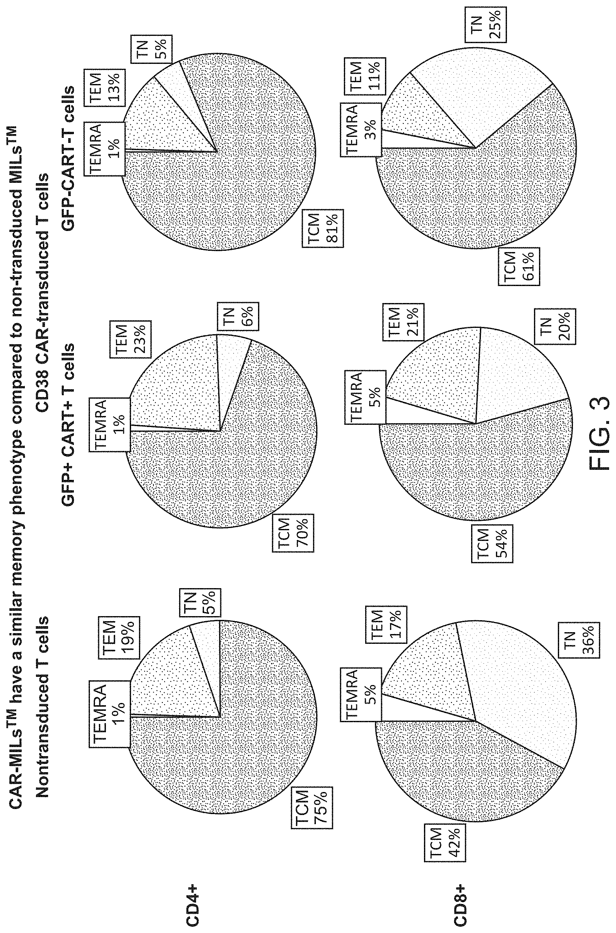
[0010] FIG. 3 shows that CAR-MILs have a similar memory phenotype compared to non-transduced MILs.
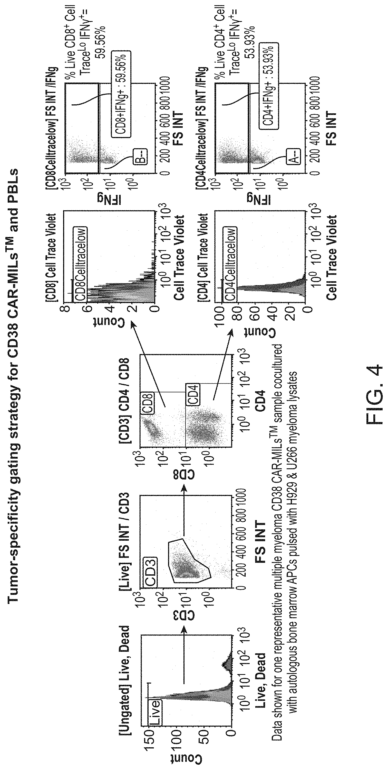
[0011] FIG. 4 shows tumor-specificity gating strategy for CD38 CAR-MILs and PBLs.
[0012] FIG. 5 shows CD38 CAR-MILs retain their inherent tumor-specificity and functionality.
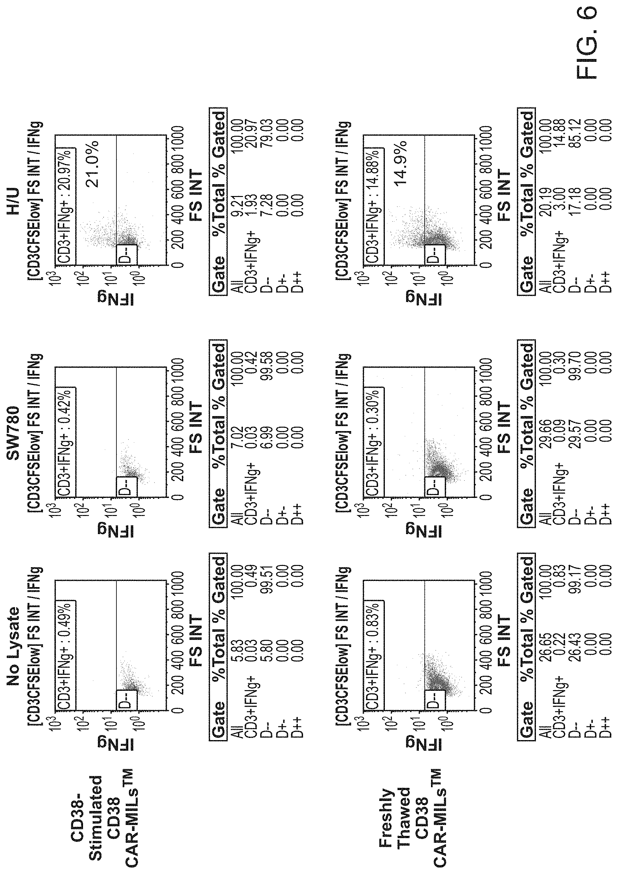
[0013] FIG. 6 shows CD38 CAR-MILs retain their inherent tumor-specificity and functionality after being co-cultured and stimulated with CD38-expressing 8226 tumor cells.
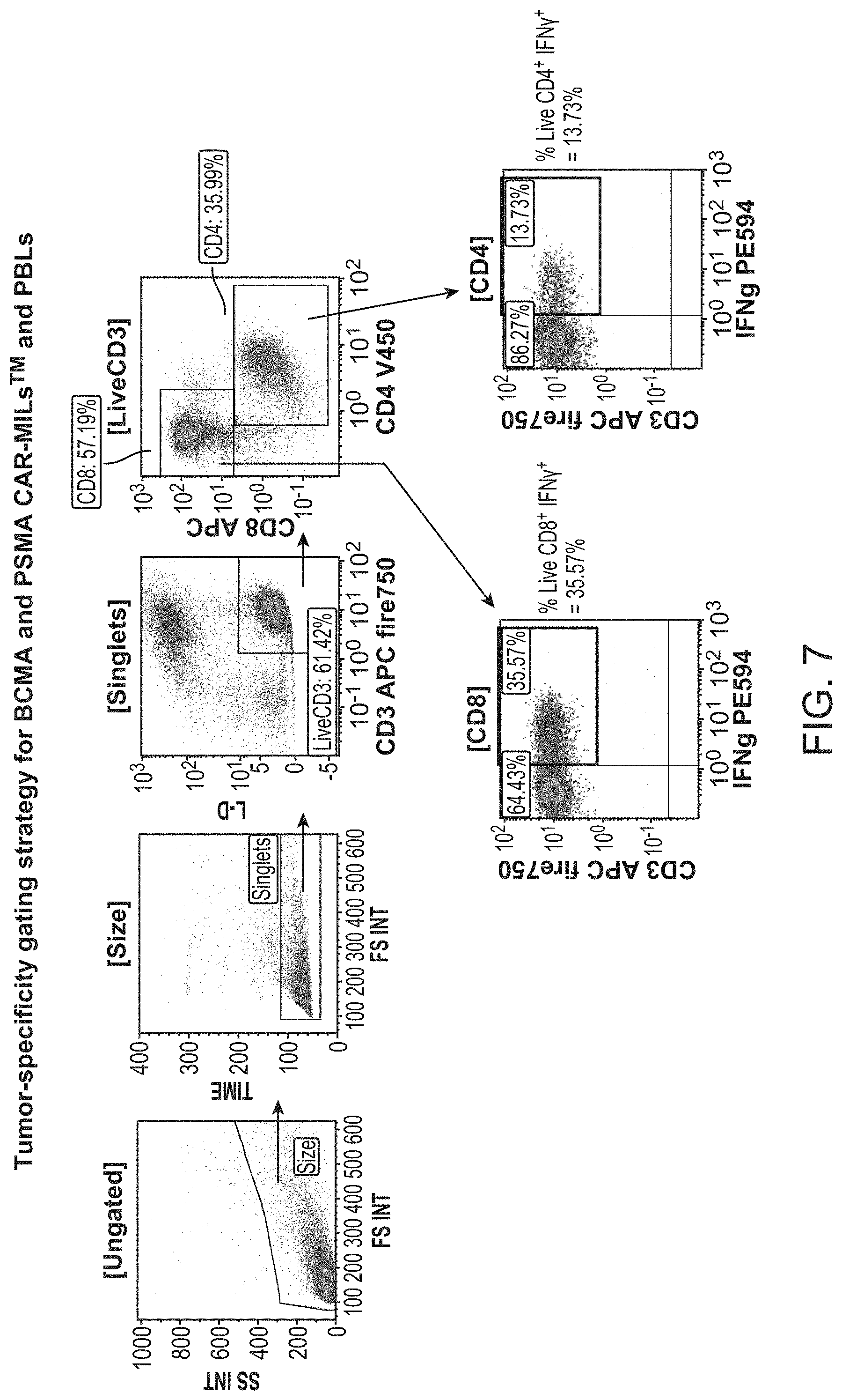
[0014] FIG. 7 shows the tumor-specificity gating strategy for BCMA and PSMA CAR-MILs and PBLs.
[0015] FIG. 8 shows BCMA CAR-MILs retain their inherent tumor-specificity and functionality.
[0016] FIG. 9 shows PSMA CAR-MILs retain their inherent tumor-specificity and functionality after being co-cultured and stimulated with PSMA-expressing LNCAP tumor cells.
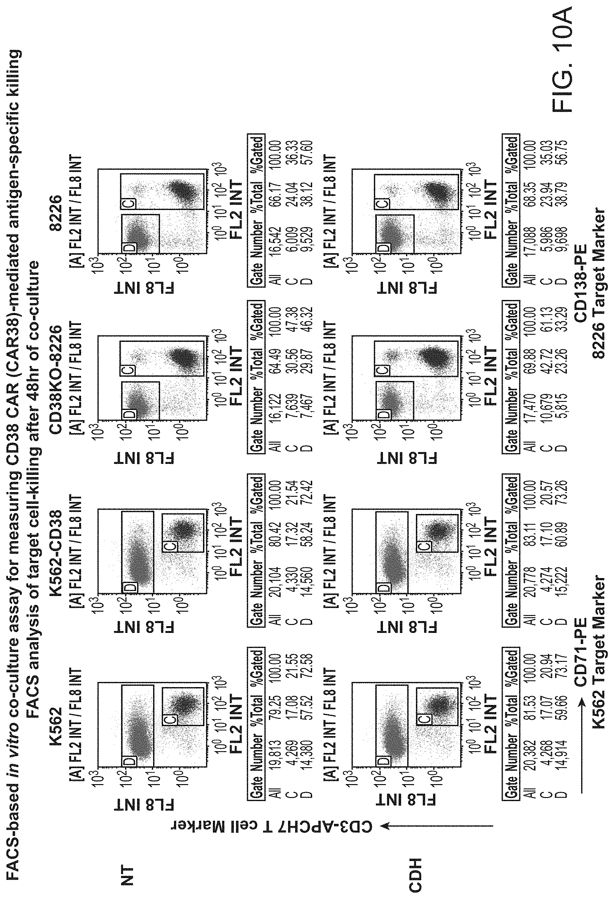
[0017] FIG. 10 shows FACS-based vitro co-culture assay for measuring CD38 CAR (CAR38)-mediated antigen-specific killing.
[0018] FIG. 11 shows CD38 outperform CAR-PBLs in primary 48 hr co-culture killing assays.
[0019] FIG. 12 shows that CD38 CAR-MILS have superior killing in vitro compared to CAR-PBLs at low E:T ratios in primary 48 hr killing assays.
[0020] FIG. 13 shows that CD38 CAR-MILs outperform CAR-PBLs in primary and. secondary re-challenge killing assays.
[0021] FIG. 14 shows that CAR38 CAR-MILs have superior killing in vitro compared to CAR-PBLs at low E:T ratios in secondary re-challenge killing assays.
[0022] FIG. 15 shows that CD38 CAR-MILs have superior killing vs. CAR-PBLs over repeated challenges (every 48 hours).
[0023] FIG. 16 shows that CD38 CAR-MILs have superior killing vs. CAR-PBLs in delayed secondary re-challenge killing assays (7 days after primary challenge).
[0024] FIG. 17 shows ACEA in vitro co-culture assay for measuring BCMA CAR antigen-specific killing in real-time.
[0025] FIG. 18 shows that BCMA CAR-MILs have superior killing in vitro compared to CAR-PBLs at low ET ratios in ACEA real-time killing assays.
[0026] FIG. 19 shows that BCMA CAR-MILs have superior killing in vitro compared to CAR-PBLs at low FT ratios in ACEA killing assays.
[0027] FIG. 20 shows FACS-based in vitro co-culture assay for detecting BCMA CAR-mediated antigen-specific killing.
[0028] FIG. 21 shows that BCMA CAR-MILs have superior killing vs. CAR-PBLs after repeated challenges.
[0029] FIG. 22 shows PSMA staining on four human prostate cancer cells lines and SW780 bladder cancer cell line.
[0030] FIG. 23 shows ACEA in vitro co-culture assay for measuring PSMA CAR antigen-specific killing in real-time.
[0031] FIG. 24 shows that PSMA CAR-MILs outperform CAR-PBL in secondary challenges even when primary challenge favors CAR-PBL.
[0032] FIG. 25 shows measuring CAR antigen stimulation-specific cytokine production by intracellular cytokine-staining.
[0033] FIG. 26 shows that BCMA CAR-MILS have increased IFS.gamma. and TNF.alpha. cytokine production compared to CAR-PBLs.
[0034] FIG. 27 shows that CD38 CAR-MiLs show increased IFN.gamma. and TNF.alpha. cytokine production compared to CAR-PBLs.
[0035] FIG. 28 shows the procedure for measuring production of 32 cytokines following BCMA antigen-stimulation at the single-cell level.
[0036] FIG. 29 shows that BCMA antigen-stimulation induces polyfunctional cytokine-secreting CD4 and CD8 CART cells in both CAR-MILS and CAR-PBLs.
[0037] FIG. 30 shows increased polyfunctionality following BCMA antigen-stimulation due to increased effector, stimulatory, & chemoattractive cytokines.
[0038] FIG. 31 shows granzyme B, IFN.gamma., IL-8, MIP-1a, and MIP-1b are the predominant cytokines produced by CAR-MILs and CAR-PBLs following BCMA stimulation.
[0039] FIG. 32 shows that CAR-MILS have a stronger upregulation of PSI and produce more effector and chemoattractive cytokines than CAR-PBLs following BCMA-stimulation.
[0040] FIG. 33 shows that CAR-MILs show a greater increase of polyfunctional cell subsets following BCMA-stimulation compared to CAR-PBLs.
[0041] FIG. 34 shows that CAR-MILs maintain CD27 whereas CAR-PBLs lose CD27 expression following antigen-stimulation.
[0042] FIG. 35 shows that CAR-MILs express less PD1 and TIM-3 compared to CAR-PBLs following antigen-stimulation.
[0043] FIG. 36 show the baseline pre-T cell infusion serum human IgE levels for all 67 treated mice versus 46 mice with more similar baseline U266 tumor burden selected for analysis.
[0044] FIG. 37 shows serum human IgE kinetics (marker of U266 in vivo tumor burden) for each of the 46 mice.
[0045] FIG. 38 shows that BCMA CAR-MILS are more potent in vivo than matched CAR-PBLs.
[0046] FIG. 39 shows the measurement of the levels of human CD3+ T cell and CD138+ U266 tumor cells in bone marrow from control and treated mice by flow cytometry (FACS).
[0047] FIG. 40 shows the measurement of the levels of human CD3+ cell and CD138+ tumor cells in spleens from control and treated mice by flow cytometry (FACS).
[0048] FIG. 41 shows that higher percentages of human CD3 T cells and lower percentages of U266 tumor cells are detected in bone marrow of mice treated with MILs compared to PBLs.
[0049] FIG. 42 shows that higher percentages of human CD3 T cells and lower percentages of U266 tumor cells are detected in spleens of mice treated with MILs compared to PBLs.
[0050] FIGS. 43A-F show an illustration of generation of matched CAR-MILs and CAR-PBLs from bone marrow (BM) and peripheral blood obtained from multiple myeloma or prostate cancer patients (FIG. 43A), a schematic illustration of lentiviral vectors encoding CD38-, BMCA- and PSMA CARs (FIG. 43B), transduction efficiency of matched CAR-MILs and CAR-PBLs based on GFP reporter gene expression (FIG. 43C), surface expression of CARs on transduced MILs (the transduced MILs were stained with biotin-CD38, biotin-BCMA, or anti-mouse F(ab').sub.2 to determine the CAR surface expression by flow cytometry; anti-mouse F(ab').sub.2 stains positive for both BCMA- and PSMA CARs (FIG. 43D), the percentage of CD4.sup.+ and CD8.sup.+ population (the matched non-transduced or CAR transduced MILs or -PBLs were prepared from 5 multiple myeloma patients; cells were gated on live singlets and CD3.sup.+; data presented is mean.+-.SEM (FIG. 43E), memory phenotype analysis for the samples described in FIG. 43E (data presented is mean of n=5. Memory phenotype is specified as T.sub.CM (CCR7.sup.+CD45RO.sup.+), T.sub.EM (CCR7.sup.-CD45RO.sup.+), T.sub.E (CCR7.sup.-CD45RO.sup.-) and T.sub.N (CCR7.sup.+CD45RO.sup.-)) (FIG. 43E).
[0051] FIG. 44A-44B shows the percentage of IFN-.gamma..sup.+ cells in response to TCR-mediated antigen-specific stimulation. The cells were gated on live CD3.sup.+ singlets for NT MILs/PBLs and CAR transduced MILs/PBLs. Data shown is mean.+-.SEM from 3 different patient samples (FIG. 44A). FIG. 44B shows polyfunctional cytokine response profile of NT and BCMA CAR-MILs with respect to expression of IFN-.gamma., TNF-.alpha. and GrB. The x-axis represents the distinct functional cell populations of possible combinations of cytokine responses. The y-axis represents the percentage of those cell populations among CD3.sup.+CD4.sup.+ or CD3.sup.+CD8.sup.+. Data shown is mean.+-.SEM from 3 different patient samples.
[0052] FIG. 45 shows representative dot plots showing IFN-.gamma..sup.+ cells in NT or CAR-MILs following TCR-mediated activation. Non-transduced or CAR modified MILs or PBLs were co-cultured for 5 days with target tumor cell lysate or control tumor cell lysate pulsed autologous APCs. Cells were then stained intracellularly for IFN-.gamma..sup.+, and samples were acquired on Navios EX flow cytometer and data analyzed using Kaluza software.
[0053] FIG. 46 shows that CAR-MILs retain endogenous TCR-mediated tumor specificity following activation through the engineered CAR. Freshly thawed PSMA CAR modified MILs or PBLs prepared from three multiple myeloma patients were first stimulated by coculturing with LNCaP cells. Following 6 days of primary co-culture, a secondary stimulation was set up using autologous APCs pulsed with H929+U266 Myeloma target cell lysates. IFN-.gamma..sup.+ expression was determined 5 days later by flow cytometry. Secondary co-culture using APCs pulsed with SW780 Bladder cell lysate or DU145+PC3 Prostate cell lysates served as negative controls. Data shown is mean.+-.SEM.
[0054] FIGS. 47A-47C shows that CAR-MILs display superior CAR-mediated killing activities in vitro than their matched CAR-PBLs. CAR-MILs and CAR-PBLs were prepared from multiple myeloma patients as described in FIG. 43. In FIG. 47A, the cytotoxicity of CD38 CAR-MILs versus CAR-PBLs measured by flow cytometry-based assay. The effector cells were cultured with RPMI 8226 target cells at the ratio of 1E:10T for 3 days (primary challenge, n=8). At the end of 3-day culture, the effector cells were re-challenged with RPMI 8226 target cells at the same E:T ratio for 2 more days (secondary challenge, n=5). Representative dot plots (top panel) and the graph for individual patient samples (bottom panel) are shown. The statistical difference between the 2 groups was evaluated by 2-tailed paired t test. **p<0.01. FIG. 47B shows the cytolysis of RPMI 8226 cells by BCMA CAR-MILs (solid line) and CAR-PBLs (dash line) measured by real-time cell analysis (RTCA). Mean percent cytolysis of samples from 6 individual patients ran in triplicates is shown. The statistical difference between the 2 groups over the time course was evaluated by 2-tailed 2-way ANOVA. **p<0.01. FIG. 47C shows the mean percent killing of K562-BCMA target tumor cells by BCMA CAR-MILs and CAR-PBLs following tertiary challenge. The effectors were challenged with RPMI 8226 cells at indicated E:T ratio on day 1 and then again on day 3. On day 9, the effector cells from the previous culture were challenged with violet cell proliferation dye labeled K562-BCMA target cells. The percent killing was measured by flow cytometry. Data for paired samples from individual patients (n=4) ran in triplicates are shown.
[0055] FIG. 48A-B shows that CD38-CAR displays specificity towards its target antigen. CD38 CAR-MILs, CDH vector control transduced MILs or NT MILs were co-cultured with RPMI 8226 cells that had CD38 knocked out (CD38KO-8226) or RPMI 8226 target cells (8226) at an E:T ratio of 1:10. Target cell killing was analyzed 48 hrs later by flow cytometry. The percentage of target cells killed was calculated by normalizing to NT MILs. FIG. 48A shows the representative flow cytometric analysis plots. FIG. 48B shows the percentage of CD38KO-8226 or 8226 target cells killed by CD38 CAR-MILs compared to CDH vector control transduced effector cells.
[0056] FIG. 49A-C shows that CAR-MILs are more effective than their PBL counterparts at clearing tumors in vivo. FIG. 49A shows an illustration of the in vivo experimental protocol. FIG. 49B shows serum human IgE levels on day -1 (pre-treatment) measured by ELISA (n=7-8 per group). FIG. 49C shows serum human IgE levels over the course of the entire experiment. The difference between BCMA CAR-MILs and BCMA CAR-PBLs groups at each time point was evaluated by 2-tailed t test. *p<0.01.
[0057] FIG. 50A-C shows that CAR-MILs are more effective than their PBL counterparts at clearing multiple myeloma in vivo. FIG. 50A shows the serum human IgE levels on day -1 (pre-treatment) measured by ELISA (n=4-7 per group). FIG. 50 B shows the serum human IgE levels over the course of the entire experiment. The data is shown as mean.+-.SEM in FIG. 50A and FIG. 50B. FIG. 50 C is a graph showing serum human IgE levels for each individual mouse. The data was pooled from two independent in vivo experiments.
[0058] FIG. 51A-D shows expression of PD-1, TIM-3 and TIGIT was determined following generation of the CAR-MILs and CAR-PBLs products. FIG. 51A shows representative histograms of PD-1, TIM-3 or TIGIT expression on CD4.sup.+ or CD8.sup.+s of CD38 CARs (top row) or BCMA CARs (bottom row). Percentage of cells that express total PD-1, TIM-3 or TIGIT only (FIG. 51B) and combination of these markers (FIG. 51C) on CD4.sup.+ or CD8.sup.+s of CAR-MILs or CAR-PBLs are shown. Data presented is mean.+-.SEM of results pooled from either CD38 CAR or BCMA CAR transduced MILs or PBLs from 3 individual patients. FIG. 51D shows CAR-MILs or -PBLs stimulated with RPMI 8226 target cells for 24 h. Polyfunctional cytokine-response was profiled based on IFN-.gamma., TNF-.alpha. and GrB expression. Data shown is the results pooled from 3 different CAR-MILs or -PBLs prepared from 9 individual patients. Pie charts shows the average frequencies of each functional sub-population out of all cytokine-producing cells either in CAR-MILs or CAR-PBLs, separately. Paired 2-way ANOVA was used to calculate statistical significance. * represents p<0.05 and ** represents p<0.01.
[0059] FIG. 52 shows that all the 3 CAR-MILs have comparable polyfunctionality. CAR-MILs or CAR-PBLs were stimulated with their respective target cells for 24 h. Polyfunctional cytokine-response was profiled based on IFN-.gamma., TNF-.alpha. and GrB expression. Pie charts shows the average frequencies of each functional sub-population out of all cytokine-producing cells either in CAR-MILs or CAR-PBLs, separately. Data shown are from 3 different patients for each of the CAR constructs.
[0060] FIG. 53A-F shows PSMA CAR-MILs generated from prostate cancer patients are more effective than their PSMA CAR-PBL counterpart. FIG. 53A shows the percentage of IFN-.gamma..sup.+ cells in NT/PSMA CAR-MILs/PBLs in response to TCR-mediated antigen-specific stimulation. FIG. 53B shows polyfunctional cytokine response profile of NT MILs and PSMA CAR-MILs with respect to IFN-.gamma., TNF-.alpha. and GrB expression. Data shown is mean.+-.SEM from 3 different patient samples. FIGS. 53C-D shows expression of exhaustion markers PD1 and TIM-3 on CAR-MILs and CAR-PBLs analyzed by flow cytometry at baseline prior to primary stimulation within CD4.sup.+ and CD8.sup.+ subpopulation gated on live GFP.sup.+CD3.sup.+ T cells. The percentage of total PD1 or TIM-3 expression (FIG. 53C) and the combination of PD1 and TIM-3 expression (FIG. 53D) are shown. FIG. 53E shows percent cytolysis of LNCaP cells by PSMA CAR-MILs (solid lines) and PSMA CAR-PBLs (dash lines) at indicated effector to target (E:T) ratios following primary challenge (top panel) and secondary challenge (bottom panel) determined using RTCA. The data shown is the average of 3 independent experiments, each experiment was performed using separate patient samples ran in triplicates. FIG. 53F shows the fold expansion of PSMA CAR-MILs and PSMA CAR-PBLs at the end of the primary and secondary challenges. Paired 2 tailed t-test (A, B, and D) and 2-way ANOVA (C) were performed. *p<0.05, **p<0.01.
[0061] FIG. 54 shows that PSMA CAR-MILs express less exhaustion markers. Histograms of PD1.sup.+ and TIM3.sup.+ expression on PSMA CAR-MILs and PSMA CAR-PBLs prepared from a representative metastatic prostate cancer patient.
[0062] FIG. 55A-B shows target-specific cytotoxicity of PSMA CAR-MILs by RTCA. Cell proliferation index was calculated by measuring electrical impedance of target cells in representative RTCA experimental conditions. Cell index of LNCaP (FIG. 55A) and PC-3 (FIG. 55B) target cells alone along with a representative positive control (CAR-MIL) or negative control (NT MIL).
DETAILED DESCRIPTION
[0063] The present disclosure provides compositions and methods for treating cancer among other diseases, including but not limited to hematologic malignancies and solid tumors. The cancer may be a hematological malignancy, such as Chronic Lymphocytic Leukemia ("CLL"). Other diseases treatable using the compositions and methods described and provided for herein include viral, bacterial, and parasitic infections as well as autoimmune diseases.
[0064] Aspects relate to, but are not limited to, a strategy of adoptive cell transfer of marrow-infiltrating lymphocytes (MILs) transduced to express a chimeric antigen receptor (CAR). CARs are molecules that combine antibody-based specificity for a desired antigen (e.g., tumor antigen) with a MIL receptor-activating intracellular domain to generate a chimeric protein that exhibits a specific anti-tumor cellular immune activity.
[0065] In some embodiments, a cell (i.e., MIL) engineered to express a CAR wherein the CAR-MIL exhibits an antitumor property is provided. MILs expressing a CAR are referred to herein as CAR-MILs or CAR-modified MILs. In some embodiments, the cell can be genetically modified to stably express an antibody binding domain on its surface, conferring novel antigen specificity that is MHC independent. In some embodiments, the MIL is genetically modified to stably express a CAR that combines an antigen recognition domain of a specific antibody with an intracellular domain of the CD3.zeta. chain or Fc.gamma.RI protein into a single chimeric protein. The CAR can, for example, be engineered to comprise an extracellular domain having an antigen-binding domain fused to an intracellular signaling domain of the MIL antigen receptor complex .zeta. chain (e.g., CD3.zeta.). The CAR, for example, when expressed in a MIL, is able to redirect antigen recognition based on the antigen-binding specificity. In some embodiments, the antigen is CD19 because this antigen is expressed on malignant B cells. In some embodiments, the antigen is CD38, prostate specific membrane antigen ("PSMA"), or B-cell maturation antigen ("BCMA"). However, the embodiments are not limited to targeting these domains. Rather, the embodiments include any antigen-binding moiety that when bound to its cognate antigen, affects a tumor cell so that the tumor cell fails to grow, is prompted to die, or otherwise is affected so that the tumor burden in a patient is diminished or eliminated. The antigen-binding moiety may be fused with an intracellular domain from one or more of costimulatory molecules and a .zeta. chain. In some embodiments, the antigen-binding moiety is fused with one or more intracellular domains selected from the group of a CD137 (4-1BB) signaling domain, a CD28 signaling domain, a CD3.zeta. signal domain, and any combination thereof. The antigen-binding moiety may also be fused with an intracellular domain such as CD134 (0X40). In some embodiments, the CAR comprises a CD137 (4-1BB) signaling domain. Without being bound to any particular theory, this is because the embodiments are partly based on the discovery that CAR-mediated T-cell responses can be further enhanced with the addition of costimulatory domains.
[0066] In some embodiments, the CAR includes an extracellular domain having an antigen recognition domain, a transmembrane domain, and a cytoplasmic domain. In some embodiments, the transmembrane domain that naturally is associated with one of the domains in the CAR is used. In some embodiments, the transmembrane domain can be selected or modified by amino acid substitution to avoid binding of such domains to the transmembrane domains of the same or different surface membrane proteins to minimize interactions with other members of the receptor complex. For example, the transmembrane domain may be a CD8.alpha. hinge domain.
[0067] In some embodiments, the CAR comprises an extracellular ligand binding domain that binds to CD19; a transmembrane domain; a 4-1BB costimulatory signaling domain; and an intracellular CD3.zeta. signaling domain. In some embodiments, the CAR comprises an extracellular ligand binding domain that binds to PSMA; a transmembrane domain; a 4-1BB costimulatory signaling domain; and an intracellular CD3.zeta. signaling domain. In some embodiments, the CAR comprises an extracellular ligand binding domain that binds to BCMA; a transmembrane domain; a 4-1BB costimulatory signaling domain; and an intracellular CD3.zeta. signaling domain. In some embodiments, the CAR comprises an extracellular ligand binding domain that binds CD38; a transmembrane domain; a 4-1BB costimulatory signaling domain; and an intracellular CD3.zeta. signaling domain. In some embodiments, the transmembrane domain is the transmembrane domain of CD3.zeta., CD4, CD8, or CD28.
[0068] With respect to the cytoplasmic domain, a CAR, for example, can be designed to comprise the CD28 and/or 4-1BB signaling domain by itself or be combined with any other desired cytoplasmic domain(s) useful in the context of the CAR. In some embodiments, the cytoplasmic domain of the CAR can be designed to further comprise the signaling domain of CD3.zeta.. For example, the cytoplasmic domain of the CAR can include but is not limited to CD3.zeta., 4-1BB, and CD28 signaling modules, and combinations thereof. Accordingly, the embodiments provide CAR-MILs and methods of their use for adoptive therapy.
[0069] In some embodiments, the CAR-MILs can be generated by introducing a lentiviral vector comprising a desired CAR (e.g., a CAR comprising anti-CD19, transmembrane domain, and human 4-1BB) into the cells. The CAR-MILs are, for example, able to replicate in vivo resulting in long-term persistence that can lead to sustained tumor control.
[0070] In some embodiments, administering a genetically modified MIL expressing a CAR for the treatment of a patient having a neoplasm using an infusion of CAR-MILs are provided. In some embodiments, autologous infusions are used in the treatment. Autologous MILs are collected from a patient in need of treatment, and are activated and expanded using methods described herein and known in the art and then infused back into the patient.
[0071] In some embodiments, MILs expressing an anti-CD19, anti-CD38, anti-PSMA, or anti-BCMA CAR, including both CD3.zeta. and the 4-1BB costimulatory domains are used. In some instances, the CAR MILs infused into a patient can eliminate leukemia cells in vivo in patients. However, the embodiments are not limited to MILs that target CD19, CD38, BSMA, or PSMA, or signal through CD3.zeta. and/or 4-1BB mediated pathways. For example, the embodiments include any antigen-binding moiety fused with one or more intracellular domains such as a CD137 (4-1BB) signaling domain, a CD28 signaling domain, a CD3.zeta. signal domain, and any combination thereof.
[0072] Definitions
[0073] The articles "a" and "an" are used herein to refer to one or to more than one (i.e., to at least one) of the grammatical object of the article. By way of example, "an element" means one element or more than one element.
[0074] As used in this document, terms "comprise," "have," "has," and "include" and their conjugates, as used herein, mean "including but not limited to." While various compositions, and methods are described in terms of "comprising" various components or steps (interpreted as meaning "including, but not limited to"), the compositions, methods, and devices can also "consist essentially of" or "consist of" the various components and steps, and such terminology should be interpreted as defining essentially closed-member groups.
[0075] "Activation", as used herein, refers to the state of a MIL that has been sufficiently stimulated to induce detectable cellular proliferation. Activation can also be associated with induced cytokine production, and detectable effector functions. The term "activated MILs" refers to, among other things, MILs that are undergoing cell division.
[0076] The term "antibody," as used herein, refers to an immunoglobulin molecule which specifically binds with an antigen. Antibodies can be intact immunoglobulins derived from natural sources or from recombinant sources and can be immunoreactive portions of intact immunoglobulins. The antibodies may exist in a variety of forms including, for example, polyclonal antibodies, monoclonal antibodies, Fv, Fab and F(ab)2, as well as single chain antibodies and humanized antibodies.
[0077] The term "antibody fragment" refers to a portion of an intact antibody and refers to the antigenic determining variable regions of an intact antibody. Examples of antibody fragments include, but are not limited to, Fab, Fab', F(ab')2, and Fv fragments, linear antibodies, scFv antibodies, and multispecific antibodies formed from antibody fragments.
[0078] The term "antigen" as used herein is defined as a molecule that provokes an immune response. This immune response may involve either antibody production, or the activation of specific immunologically-competent cells, or both. The skilled artisan will understand that any macromolecule, including virtually all proteins or peptides, can serve as an antigen. Furthermore, antigens can be derived from recombinant or genomic DNA. A skilled artisan will understand that any DNA, which comprises a nucleotide sequences or a partial nucleotide sequence encoding a protein that elicits an immune response therefore encodes an "antigen" as that term is used herein. Furthermore, one skilled in the art will understand that an antigen need not be encoded solely by a full-length nucleotide sequence of a gene. It is readily apparent that the embodiments include, but are not limited to, the use of partial nucleotide sequences of more than one gene and that these nucleotide sequences are arranged in various combinations to elicit the desired immune response. Moreover, a skilled artisan will understand that an antigen need not be encoded by a "gene" at all. It is readily apparent that an antigen can be generated synthesized or can be derived from a biological sample. Such a biological sample can include, but is not limited to a tissue sample, a tumor sample, a cell or a biological fluid.
[0079] The term "anti-tumor effect" as used herein, refers to a biological effect that can be manifested by a decrease in tumor volume, a decrease in the number of tumor cells, a decrease in the number of metastases, an increase in life expectancy, or amelioration of various physiological symptoms associated with the cancerous condition. An "anti-tumor effect" can also be manifested by the ability of the peptides, polynucleotides, cells and antibodies to prevent the occurrence of tumor in the first place.
[0080] The term "auto-antigen" means any self-antigen which is mistakenly recognized by the immune system as being foreign. Auto-antigens comprise, but are not limited to, cellular proteins, phosphoproteins, cellular surface proteins, cellular lipids, nucleic acids, glycoproteins, including cell surface receptors.
[0081] The term "autoimmune disease" as used herein is defined as a disorder that results from an autoimmune response. An autoimmune disease is the result of an inappropriate and excessive response to a self-antigen. Examples of autoimmune diseases include but are not limited to, Addision's disease, alopecia greata, ankylosing spondylitis, autoimmune hepatitis, autoimmune parotitis, Crohn's disease, diabetes (Type I), dystrophic epidermolysis bullosa, epididymitis, glomerulonephritis, Graves' disease, Guillain-Barr syndrome, Hashimoto's disease, hemolytic anemia, systemic lupus erythematosus, multiple sclerosis, myasthenia gravis, pemphigus vulgaris, psoriasis, rheumatic fever, rheumatoid arthritis, sarcoidosis, scleroderma, Sjogren's syndrome, spondyloarthropathies, thyroiditis, vasculitis, vitiligo, myxedema, pernicious anemia, ulcerative colitis, among others.
[0082] As used herein, the term "autologous" is meant to refer to any material derived from the same individual to which it is later to be re-introduced into the individual.
[0083] "Allogeneic" refers to a graft derived from a different animal of the same species.
[0084] "Xenogeneic" refers to a graft derived from an animal of a different species.
[0085] The term "cancer" as used herein is defined as disease characterized by the rapid and uncontrolled growth of aberrant cells. Cancer cells can spread locally or through the bloodstream and lymphatic system to other parts of the body. Examples of various cancers include but are not limited to, breast cancer, prostate cancer, ovarian cancer, cervical cancer, skin cancer, pancreatic cancer, colorectal cancer, renal cancer, liver cancer, brain cancer, lymphoma, leukemia, lung cancer and the like. Cancers that may be treated include tumors that are not vascularized, or not yet substantially vascularized, as well as vascularized tumors. The cancers may include non-solid tumors (such as hematological tumors, for example, myeloma, leukemias and lymphomas) or may include solid tumors. Types of cancers to be treated with the CARs as described herein include, but are not limited to, carcinoma, blastoma, and sarcoma, and certain leukemia or lymphoid malignancies, benign and malignant tumors, and malignancies e.g., sarcomas, carcinomas, and melanomas. Adult tumors/cancers and pediatric tumors/cancers are also included.
[0086] "Co-stimulatory ligand," as the term is used herein, includes a molecule on an antigen presenting cell (e.g., an aAPC, dendritic cell, B cell, and the like) that specifically binds a cognate co-stimulatory molecule on a MIL, thereby providing a signal which, in addition to the primary signal provided by, for instance, binding of a TCR/CD3 complex with an MHC molecule loaded with peptide, mediates a MIL response, including, but not limited to, proliferation, activation, differentiation, and the like. A co-stimulatory ligand can include, but is not limited to, CD7, B7-1 (CD80), B7-2 (CD86), PD-L1, PD-L2, 4-1BBL, OX40L, inducible costimulatory ligand (ICOS-L), intercellular adhesion molecule (ICAM), CD30L, CD40, CD70, CD83, HLA-G, MICA, MICB, HVEM, lymphotoxin beta receptor, 3/TR6, ILT3, ILT4, HVEM, an agonist or antibody that binds Toll ligand receptor and a ligand that specifically binds with B7-H3. A co-stimulatory ligand also encompasses, inter alia, an antibody that specifically binds with a co-stimulatory molecule present on a MIL, such as, but not limited to, CD27, CD28, 4-1BB, OX40, CD30, CD40, PD-1, ICOS, lymphocyte function-associated antigen-1 (LFA-1), CD2, CD7, LIGHT, NKG2C, B7-H3, and a ligand that specifically binds with CD83.
[0087] A "co-stimulatory molecule" refers to the cognate binding partner on a MIL that specifically binds with a co-stimulatory ligand, thereby mediating a co-stimulatory response by the MIL, such as, but not limited to, proliferation. Co-stimulatory molecules include, but are not limited to an MHC class I molecule, BTLA and a Toll ligand receptor.
[0088] A "co-stimulatory signal", as used herein, refers to a signal, which in combination with a primary signal, such as TCR/CD3 ligation, leads to MIL proliferation and/or upregulation or downregulation of key molecules.
[0089] A "disease" is a state of health of a subject wherein the subject cannot maintain homeostasis, and wherein if the disease is not ameliorated then the animal's health continues to deteriorate. In contrast, a "disorder" in a subject is a state of health in which the subject is able to maintain homeostasis, but in which the subject's state of health is less favorable than it would be in the absence of the disorder. Left untreated, a disorder does not necessarily cause a further decrease in the subject's state of health.
[0090] An "effective amount" as used herein, means an amount which provides a therapeutic or prophylactic benefit.
[0091] "Encoding" refers to the inherent property of specific sequences of nucleotides in a polynucleotide, such as a gene, a cDNA, or an mRNA, to serve as templates for synthesis of other polymers and macromolecules in biological processes having either a defined sequence of nucleotides (i.e., rRNA, tRNA and mRNA) or a defined sequence of amino acids and the biological properties resulting therefrom. Thus, a gene encodes a protein if transcription and translation of mRNA corresponding to that gene produces the protein in a cell or other biological system. Both the coding strand, the nucleotide sequence of which is identical to the mRNA sequence and is usually provided in sequence listings, and the non-coding strand, used as the template for transcription of a gene or cDNA, can be referred to as encoding the protein or other product of that gene or cDNA.
[0092] As used herein "endogenous" refers to any material from or produced inside an organism, cell, tissue or system.
[0093] As used herein, the term "exogenous" refers to any material introduced from or produced outside an organism, cell, tissue or system.
[0094] The term "expression" as used herein is defined as the transcription and/or translation of a particular nucleotide sequence driven by its promoter.
[0095] "Expression vector" refers to a vector comprising a recombinant polynucleotide comprising expression control sequences operatively linked to a nucleotide sequence to be expressed. An expression vector comprises sufficient cis-acting elements for expression; other elements for expression can be supplied by the host cell or in an in vitro expression system. Expression vectors include all those known in the art, such as cosmids, plasmids (e.g., naked or contained in liposomes) and viruses (e.g., lentiviruses, retroviruses, adenoviruses, and adeno-associated viruses) that incorporate the recombinant polynucleotide.
[0096] "Homologous" refers to the sequence similarity or sequence identity between two polypeptides or between two nucleic acid molecules. When a position in both of the two compared sequences is occupied by the same base or amino acid monomer subunit, e.g., if a position in each of two DNA molecules is occupied by adenine, then the molecules are homologous at that position. The percent of homology between two sequences is a function of the number of matching or homologous positions shared by the two sequences divided by the number of positions compared .times.100. For example, if 6 of 10 of the positions in two sequences are matched or homologous then the two sequences are 60% homologous. By way of example, the DNA sequences ATTGCC and TATGGC share 50% homology. Generally, a comparison is made when two sequences are aligned to give maximum homology.
[0097] The term "immunoglobulin" or "Ig," as used herein is defined as a class of proteins, which function as antibodies. Antibodies expressed by B cells are sometimes referred to as the BCR (B cell receptor) or antigen receptor. The five members included in this class of proteins are IgA, IgG, IgM, IgD, and IgE.
[0098] "Isolated" means altered or removed from the natural state. For example, a nucleic acid or a peptide naturally present in a living animal is not "isolated," but the same nucleic acid or peptide partially or completely separated from the coexisting materials of its natural state is "isolated." An isolated nucleic acid or protein can exist in substantially purified form, or can exist in a non-native environment such as, for example, a host cell.
[0099] As used herein, the following abbreviations for the commonly occurring nucleic acid bases are used. "A" refers to adenosine, "C" refers to cytosine, "G" refers to guanosine, "T" refers to thymidine, and "U" refers to uridine.
[0100] A "lentivirus" as used herein refers to a genus of the Retroviridae family. Lentiviruses are unique among the retroviruses in being able to infect non-dividing cells; they can deliver a significant amount of genetic information into the DNA of the host cell, so they are one of the most efficient methods of a gene delivery vector. HIV, SIV, and FIV are all examples of lentiviruses. Vectors derived from lentiviruses offer the means to achieve significant levels of gene transfer in vivo.
[0101] The term "marrow infiltrating lymphocyte" ("MIL") as used herein refers to a lymphocyte derived from the bone marrow. Marrow infiltrating lymphocytes ("MILs") have many distinguishable differences from peripheral blood lymphocytes ("PBLs")as well as tumor infiltrating lymphocytes ("TILs"). The bone marrow ("BM") microenvironment is a special immunologic niche due to the richness of antigen presenting cells ("APC"). The presence of these antigen presenting cells allows for the processing and presenting of antigen to sustain the higher levels of central memory cells that are found in the bone marrow compartment. (Li JM et al J Immunol. 2009 Dec. 15; 183(12):7799-809). These MILs express memory markers such as CD45RO+ and CD62L+ and there are more memory MILs than memory cells found in the PBL. (Noonan K et al Clin Cancer Res. 2012 Mar. 1; 18(5):1426-34). Furthermore, MILs are not just the "TILs" of hematologic malignancies because of their ability to continuously prime memory cells to antigen (Beckhove Pet al J Clin Invest. 2004 Jul. 1; 114(1): 67-76; Castiglioni Pet al 6 J Immunol 2008; 180:4956-4964). MILs also express more CXCR4 than their PBL counterparts due to the cognate antigen stromal derived factor type 1 ("SDF1") that is expressed in great amounts in the bone marrow stroma (Noonan K et al Cancer Res. 2005 Mar. 1; 65(5):2026-34). The expression of 41BB is also increased in MILs compared to PBLs, likely due to the hypoxic nature of the BM micro-environment. Further, MILs can be harvested and expanded from all patients, in contrast with TILs (Noonan, K et al Sci Transl Med. 2015 May 20; 7(288):288ra78). TILs are found in only about 50% of patients, and only about 25% of patients possess expandable TILs. In contrast to peripheral blood lymphocytes (PBLs), MILs possess a broad endogenous antigenic repertoire which account for their intrinsic tumor specificity--a feature which is completely absent in PBLs (Noonan et al Clin Cancer Res).
[0102] Unless otherwise specified, a "nucleotide sequence encoding an amino acid sequence" includes all nucleotide sequences that are degenerate versions of each other and that encode the same amino acid sequence. Nucleotide sequences that encode proteins and RNA may include introns.
[0103] The term "operably linked" refers to functional linkage between a regulatory sequence and a heterologous nucleic acid sequence resulting in expression of the latter. For example, a first nucleic acid sequence is operably linked with a second nucleic acid sequence when the first nucleic acid sequence is placed in a functional relationship with the second nucleic acid sequence. For instance, a promoter is operably linked to a coding sequence if the promoter affects the transcription or expression of the coding sequence. Generally, operably linked DNA sequences are contiguous and, where necessary to join two protein coding regions, in the same reading frame.
[0104] The term "overexpressed" tumor antigen or "overexpression" of the tumor antigen is intended to indicate an abnormal level of expression of the tumor antigen in a cell from a disease area like a solid tumor within a specific tissue or organ of the patient relative to the level of expression in a normal cell from that tissue or organ. Patients having solid tumors or a hematological malignancy characterized by overexpression of the tumor antigen can be determined by standard assays known in the art.
[0105] "Parenteral" administration of an immunogenic composition includes, e.g., subcutaneous (s.c.), intravenous (i.v.), intramuscular (i.m.), or intrasternal injection, or infusion techniques.
[0106] The terms "patient," "subject," "individual," and the like are used interchangeably herein, and refer to any animal, or cells thereof whether in vitro or in situ, amenable to the methods described herein. In certain non-limiting embodiments, the patient, subject or individual is a human.
[0107] As used herein, the terms "peptide," "polypeptide," and "protein" are used interchangeably, and refer to a compound comprised of amino acid residues covalently linked by peptide bonds. A protein or peptide must contain at least two amino acids, and no limitation is placed on the maximum number of amino acids that can comprise a protein's or peptide's sequence. Polypeptides include any peptide or protein comprising two or more amino acids joined to each other by peptide bonds. As used herein, the term refers to both short chains, which also commonly are referred to in the art as peptides, oligopeptides and oligomers, for example, and to longer chains, which generally are referred to in the art as proteins, of which there are many types. "Polypeptides" include, for example, biologically active fragments, substantially homologous polypeptides, oligopeptides, homodimers, heterodimers, variants of polypeptides, modified polypeptides, derivatives, analogs, fusion proteins, among others. The polypeptides include natural peptides, recombinant peptides, synthetic peptides, or a combination thereof.
[0108] The term "promoter" as used herein is defined as a DNA sequence recognized by the synthetic machinery of the cell, or introduced synthetic machinery, required to initiate the specific transcription of a polynucleotide sequence.
[0109] As used herein, the term "promoter/regulatory sequence" means a nucleic acid sequence which is required for expression of a gene product operably linked to the promoter/regulatory sequence. In some instances, this sequence may be the core promoter sequence and in other instances, this sequence may also include an enhancer sequence and other regulatory elements which are required for expression of the gene product. The promoter/regulatory sequence may, for example, be one which expresses the gene product in a tissue specific manner.
[0110] A "constitutive" promoter is a nucleotide sequence which, when operably linked with a polynucleotide which encodes or specifies a gene product, causes the gene product to be produced in a cell under most or all physiological conditions of the cell.
[0111] An "inducible" promoter is a nucleotide sequence which, when operably linked with a polynucleotide which encodes or specifies a gene product, causes the gene product to be produced in a cell substantially only when an inducer which corresponds to the promoter is present in the cell.
[0112] A "tissue-specific" promoter is a nucleotide sequence which, when operably linked with a polynucleotide encodes or specified by a gene, causes the gene product to be produced in a cell substantially only if the cell is a cell of the tissue type corresponding to the promoter.
[0113] By the term "stimulation," is meant a primary response induced by binding of a stimulatory molecule (e.g., a TCR/CD3 complex) with its cognate ligand thereby mediating a signal transduction event, such as, but not limited to, signal transduction via the TCR/CD3 complex. Stimulation can mediate altered expression of certain molecules, such as downregulation of TGF-.beta., and/or reorganization of cytoskeletal structures, and the like.
[0114] A "stimulatory molecule," as the term is used herein, means a molecule on a MIL that specifically binds with a cognate stimulatory ligand present on an antigen presenting cell.
[0115] A "stimulatory ligand," as used herein, means a ligand that when present on an antigen presenting cell (e.g., an aAPC, a dendritic cell, a B-cell, and the like) can specifically bind with a cognate binding partner (referred to herein as a "stimulatory molecule") on a MIL, thereby mediating a primary response by the MIL, including, but not limited to, activation, initiation of an immune response, proliferation, and the like. Stimulatory ligands are well-known in the art and encompass, inter alia, an MEW Class I molecule loaded with a peptide, an anti-CD3 antibody, a superagonist anti-CD28 antibody, and a superagonist anti-CD2 antibody.
[0116] The term "subject" is intended to include living organisms in which an immune response can be elicited (e.g., mammals). Examples of subjects include humans, dogs, cats, mice, rats, and transgenic species thereof.
[0117] The term "therapeutic" as used herein means a treatment and/or prophylaxis. A therapeutic effect is obtained by suppression, remission, or eradication of a disease state.
[0118] The term "therapeutically effective amount" refers to the amount of the subject compound that will elicit the biological or medical response of a tissue, system, or subject that is being sought by the researcher, veterinarian, medical doctor, or other clinician. The term "therapeutically effective amount" includes that amount of a compound that, when administered, is sufficient to prevent development of, or alleviate to some extent, one or more of the signs or symptoms of the disorder or disease being treated. The therapeutically effective amount will vary depending on the compound, the disease and its severity and the age, weight, etc., of the subject to be treated.
[0119] To "treat" a disease as the term is used herein, means to reduce the frequency or severity of at least one sign or symptom of a disease or disorder experienced by a subject.
[0120] The term "transfected" or "transformed" or "transduced" as used herein refers to a process by which exogenous nucleic acid is transferred or introduced into the host cell. A "transfected" or "transformed" or "transduced" cell is one which has been transfected, transformed or transduced with exogenous nucleic acid. The cell includes the primary subject cell and its progeny.
[0121] The phrase "under transcriptional control" or "operatively linked" as used herein means that the promoter is in the correct location and orientation in relation to a polynucleotide to control the initiation of transcription by RNA polymerase and expression of the polynucleotide.
[0122] A "vector" is a composition of matter which comprises an isolated nucleic acid and which can be used to deliver the isolated nucleic acid to the interior of a cell. Numerous vectors are known in the art including, but not limited to, linear polynucleotides, polynucleotides associated with ionic or amphiphilic compounds, plasmids, and viruses. Thus, the term "vector" includes an autonomously replicating plasmid or a virus. The term should also be construed to include non-plasmid and non-viral compounds which facilitate transfer of nucleic acid into cells, such as, for example, polylysine compounds, liposomes, and the like. Examples of viral vectors include, but are not limited to, adenoviral vectors, adeno-associated virus vectors, retroviral vectors, and the like.
[0123] I. Chimeric Antigen Receptors
[0124] Provided herein are chimeric antigen receptors (CARs) that include an extracellular and intracellular domain. The extracellular domain includes a target-specific binding element otherwise referred to as an antigen-binding moiety. The intracellular domain or otherwise the cytoplasmic domain may include a costimulatory signaling region and/or a portion of a chain. The costimulatory signaling region refers to a portion of the CAR including the intracellular domain of a costimulatory molecule. Costimulatory molecules are cell surface molecules other than antigens receptors or their ligands that are required for an efficient response of lymphocytes to antigen.
[0125] A spacer domain may be incorporated between the extracellular domain and the transmembrane domain of the CAR or between the cytoplasmic domain and the transmembrane domain of the CAR. As used herein, the term "spacer domain" generally means a stretch of amino acids that functions to link the transmembrane domain to either the extracellular domain or the cytoplasmic domain in the polypeptide chain. A spacer domain may include up to 300 amino acids, or 2 to 100 amino acids, such as 25 to 50 amino acids.
[0126] II. Extracellular Domains
[0127] In some embodiments, the CAR includes a target-specific binding element otherwise referred to as an antigen-binding moiety. The choice of moiety depends upon the type and number of ligands that define the surface of a target cell. For example, the antigen-binding domain may be chosen to recognize a ligand that acts as a cell surface marker on target cells associated with a particular disease state. Thus, examples of cell surface markers that may act as ligands for the antigen-binding domain in a CAR include those associated with viral, bacterial, and parasitic infections, autoimmune disease, and cancer cells. For example, the ligand may be the protein of a bacterium, virus, or parasite. Similarly, the ligand may be a protein that is upregulated on the surface of a cancer cell.
[0128] In some embodiments, a CAR can be engineered to target a tumor antigen of interest by way of engineering a desired antigen-binding moiety that specifically binds to an antigen on a tumor cell. As used herein, "tumor antigen" or "hyperporoliferative disorder antigen" or "antigen associated with a hyperproliferative disorder," refers to antigens that are common to specific hyperproliferative disorders such as cancer. The antigens discussed herein are merely included by way of example. The list is not intended to be exclusive and further examples will be readily apparent to those of skill in the art.
[0129] Tumor antigens are proteins that are produced by tumor cells that elicit an immune response, particularly T-cell mediated immune responses. The selection of the antigen-binding moiety will depend on the particular type of cancer to be treated. Tumor antigens are well known in the art and include, for example, a glioma-associated antigen, carcinoembryonic antigen (CEA), .beta.-human chorionic gonadotropin, alpha-fetoprotein (AFP), lectin-reactive AFP, thyroglobulin, RAGE-1, MN-CA IX, human telomerase reverse transcriptase, RU1, RU2 (AS), intestinal carboxyl esterase, mutant hsp70-2, M-CSF, prostase, prostate-specific antigen (PSA), PAP, NY-ESO-1, LAGE-1a, p53, prostein, PSMA, Her2/neu, survivin, telomerase, prostate-carcinoma tumor antigen-1 (PCTA-1), MAGE, ELF2M, neutrophil elastase, ephrinB2, CD22, insulin growth factor (IGF)-I, IGF-II, IGF-I receptor, and mesothelin.
[0130] In some embodiments, the tumor antigen includes one or more antigenic cancer epitopes associated with a malignant tumor. Malignant tumors express a number of proteins that can serve as target antigens for an immune attack. These molecules include but are not limited to tissue-specific antigens such as MART-1, tyrosinase, and GP 100 in melanoma and prostatic acid phosphatase (PAP) and prostate-specific antigen (PSA) in prostate cancer. Other target molecules belong to the group of transformation-related molecules such as the oncogene HER-2/Neu/ErbB-2. Yet another group of target antigens are onco-fetal antigens such as carcinoembryonic antigen (CEA). In B-cell lymphoma, the tumor-specific idiotype immunoglobulin constitutes a truly tumor-specific immunoglobulin antigen that is unique to the individual tumor. B-cell differentiation antigens such as CD19, CD20 and CD37 are other candidates for target antigens in B-cell lymphoma. Some of these antigens (e.g., CEA, HER-2, CD19, CD20, idiotype) have been used as targets for passive immunotherapy with monoclonal antibodies with limited success.
[0131] The type of tumor antigen referred to may also be a tumor-specific antigen (TSA) or a tumor-associated antigen (TAA). A TSA is unique to tumor cells and does not occur on other cells in the body. A TAA associated antigen is not unique to a tumor cell and instead is also expressed on a normal cell under conditions that fail to induce a state of immunologic tolerance to the antigen. The expression of the antigen on the tumor may occur under conditions that enable the immune system to respond to the antigen. TAAs may be antigens that are expressed on normal cells during fetal development when the immune system is immature and unable to respond or they may be antigens that are normally present at extremely low levels on normal cells but which are expressed at much higher levels on tumor cells.
[0132] Non-limiting examples of TSA or TAA antigens include the following: Differentiation antigens such as MART-1/MelanA (MART-I), gp100 (Pmel 17), tyrosinase, TRP-1, TRP-2 and tumor-specific multilineage antigens such as MAGE-1, MAGE-3, BAGE, GAGE-1, GAGE-2, p15; overexpressed embryonic antigens such as CEA; overexpressed oncogenes and mutated tumor-suppressor genes such as p53, Ras, HER-2/neu; unique tumor antigens resulting from chromosomal translocations; such as BCR-ABL, E2A-PRL, H4-RET, IGH-IGK, MYL-RAR; and viral antigens, such as the Epstein Barr virus antigens EBVA and the human papillomavirus (HPV) antigens E6 and E7. Other large, protein-based antigens include TSP-180, MAGE-4, MAGE-5, MAGE-6, RAGE, NY-ESO, p185erbB2, p180erbB-3, c-met, nm-23H1, PSA, TAG-72, CA 19-9, CA 72-4, CAM 17.1, NuMa, K-ras, beta-Catenin, CDK4, Mum-1, p 15, p 16, 43-9F, 5T4, 791Tgp72, alpha-fetoprotein, beta-HCG, BCA225, BTAA, CA 125, CA 15-3\CA 27.29\BCAA, CA 195, CA 242, CA-50, CAM43, CD68\P1, CO-029, FGF-5, G250, Ga733\EpCAM, HTgp-175, M344, MA-50, MG7-Ag, MOV18, NB/70K, NY-CO-1, RCAS1, SDCCAG16, TA-90\Mac-2 binding protein\cyclophilin C-associated protein, TAAL6, TAG72, TLP, and TPS.
[0133] In a some embodiments, the antigen-binding moiety portion of the CAR targets an antigen that includes but is not limited to CD19, CD20, CD22, ROR1, Mesothelin, CD33/IL3Ra, c-Met, PSMA, Glycolipid F77, EGFRvIII, GD-2, MY-ESO-1 TCR, MAGE A3 TCR, and the like.
[0134] Depending on the desired antigen to be targeted, a CAR can be engineered to include the appropriate antigen bind moiety that is specific to the desired antigen target. For example, if CD19 is the desired antigen that is to be targeted, an antibody for CD19 can be used as the antigen bind moiety for incorporation into the CAR. Thus, in some embodiments, the antigen-binding moiety portion of the CAR targets CD19.
[0135] The extracellular domain of a CAR may comprise, for example, a single-chain variable fragment ("scFv") that binds to any one of the targets described herein.
[0136] The extracellular domain can be any antigen-binding polypeptide, a wide variety of which are known in the art. In some instances, the antigen-binding domain is a single chain Fv ("scFv"). Other antibody-based recognition domains (cAb VHH (camelid antibody variable domains) and humanized versions, IgNAR VH (shark antibody variable domains) and humanized versions, sdAb VH (single domain antibody variable domains) and "camelized" antibody variable domains are suitable for use. In some instances, T-cell receptor (TCR) based recognition domains such as single chain TCR (scTv, single chain two-domain TCR containing v v.beta.) are also suitable for use.
[0137] Other extracellular domains known in the art may also be used in embodiments (see, e.g., PCT Patent Application Publication No. WO 2014/127261; U.S. Pat. No. 8,975,071, hereby incorporated by reference).
[0138] III. Transmembrane Domains
[0139] With respect to the transmembrane domain, the CAR can be designed to comprise a transmembrane domain that is fused to the extracellular domain of the CAR. In some embodiments, the transmembrane domain that naturally is associated with one of the domains in the CAR is used. In some instances, the transmembrane domain can be selected or modified by amino acid substitution to avoid binding of such domains to the transmembrane domains of the same or different surface membrane proteins to minimize interactions with other members of the receptor complex.
[0140] The transmembrane domain may be derived either from a natural source or the transmembrane domain may be designed (e.g., from a stretch of 18 to 30 hydrophobic amino acids, such as alanine, valine, leucine, and isoleucine, which form an .alpha.-helix). Where the source is natural, the domain may be derived from any membrane-bound or transmembrane protein. Transmembrane regions of particular use may be derived from (i.e., comprise at least the transmembrane region(s) of) the .alpha., .beta., or .zeta. chain of the T-cell receptor, CD28, CD3 epsilon, CD45, CD4, CD5, CD8, CD9, CD16, CD22, CD33, CD37, CD64, CD80, CD86, CD134, CD137, or CD154. Alternatively, the transmembrane domain may be designed, in which case it will include predominantly hydrophobic residues such as leucine and valine. For a designed transmembrane domain, phenylalanine, tryptophan, and/or tyrosine may be found near the membrane/water interface. Optionally, a short oligo- or polypeptide linker between 2 and 10 amino acids in length may link the transmembrane domain and the cytoplasmic signaling domain of the CAR. A glycine-serine spacer provides a particularly suitable linker.
[0141] IV. Intracellular Domain
[0142] The cytoplasmic domain or otherwise the intracellular signaling domain of the CAR is responsible for activation of at least one of the normal effector functions of a MIL. The term "effector function" refers to a specialized function of a cell. An effector function of a MIL, for example, may be cytolytic activity or helper activity including the secretion of cytokines. Thus the term "intracellular signaling domain" refers to the portion of a protein which transduces the effector function signal and directs the cell to perform a specialized function. While an entire intracellular signaling domain can be employed, in many cases it is not necessary to use the entire intracellular domain. To the extent that a truncated portion of the intracellular signaling domain is used, such truncated portion may be used in place of the intact chain as long as it transduces the effector function signal. The term intracellular signaling domain is thus meant to include any truncated portion of the intracellular signaling domain sufficient to transduce the effector function signal.
[0143] Some non-limiting examples of intracellular signaling domains for use in the CAR include the cytoplasmic sequences of the T-cell receptor (TCR) and co-receptors that act in concert to initiate signal transduction following antigen receptor engagement, as well as any derivative or variant of these sequences and any synthetic sequence that has the same functional capability.
[0144] Signals generated through the TCR alone are insufficient for full activation of a lymphocyte, and a secondary or co-stimulatory signal is also required. Thus, MIL activation is mediated by two distinct classes of cytoplasmic signaling: those that initiate antigen-dependent primary activation through the TCR (primary cytoplasmic signaling sequences) and those that act in an antigen-independent manner to provide a secondary or co-stimulatory signal (secondary cytoplasmic signaling sequences).
[0145] Primary cytoplasmic signaling sequences regulate primary activation of the TCR complex either in a stimulatory way, or in an inhibitory way. Primary cytoplasmic signaling sequences that act in a stimulatory manner may contain signaling motifs that are known as immunoreceptor tyrosine-based activation motifs or ITAMs.
[0146] Examples of ITAM containing primary cytoplasmic signaling sequences that can be used include, but are not limited to, those derived from TCR .zeta. FcR gamma, FcR beta, CD3.gamma., CD3.delta., CD3.epsilon., CD5, CD22, CD79a, CD79b, and CD66d. In some embodiments, the cytoplasmic signaling molecule of the CAR comprises a cytoplasmic signaling sequence derived from CD3.zeta..
[0147] In some embodiments, the cytoplasmic domain of the CAR can be designed to comprise the CD3.zeta. signaling domain by itself or combined with any other desired cytoplasmic domain(s) useful in the context of a CAR. For example, the cytoplasmic domain of the CAR may comprise a portion of a CD3.zeta. chain and a costimulatory signaling region. The costimulatory signaling region refers to a portion of the CAR comprising the intracellular domain of a costimulatory molecule. In some embodiments, the costimulatory molecule is a cell surface molecule other than an antigen receptor or their ligands that is required for an efficient response by lymphocytes to an antigen. Examples of such molecules include CD27, CD28, 4-1BB (CD137), OX40, CD30, CD40, PD-1, ICOS, lymphocyte function-associated antigen-1 (LFA-1), CD2, CD7, LIGHT, NKG2C, B7-H3, and the like. Thus, while some embodiments may be exemplified with 4-1BB as the co-stimulatory signaling element, other costimulatory elements can also be used.
[0148] The cytoplasmic signaling sequences within the cytoplasmic signaling portion of a CAR may be linked to each other in a random or specified order. Optionally, short oligo- or polypeptide linkers, preferably between 2 and 10 amino acids in length may form the linkage. A glycine-serine spacer provides a particularly suitable linker.
[0149] In some embodiments, the cytoplasmic domain is designed to comprise the signaling domain of CD3.zeta. and the signaling domain of CD28. In some embodiments, the cytoplasmic domain is designed to comprise the signaling domain of CD3.zeta. and the signaling domain of 4-1BB. In some embodiments, the cytoplasmic domain is designed to comprise the signaling domain of CD3.zeta. and the signaling domain of CD28 and 4-1BB.
[0150] In some embodiments, the cytoplasmic domain in the CAR is designed to comprise the signaling domain of 4-1BB and the signaling domain of CD3.zeta.. [000151] In some embodiments, the CAR comprises an extracellular domain, a transmembrane domain, an intracellular domain that comprises a costimulatory domain and an signaling domain. The extracellular domain is a domain that binds to a tumor antigen. Examples of such antigens include, but are not limited to, BCMA, PSMA, CD19, and CD38. The extracellular domain can be an antibody, such as scFV, or other type of antibody as described herein or known to one of skill in the art. In some embodiments, the extracellular domain is a scFv derived from daratumumab. In some embodiments, the extracellular domain is a FN3 domain that can bind to one or more of the antigens described herein. In some embodiments, the extracellular domain is a protein or a portion of a protein that naturally binds to the antigen.
[0151] In some embodiments, the transmembrane domain is the transmembrane domain of human CD8. In some embodiments, the transmembrane domain is the transmembrane domain of CD3 zeta, CD4, CD8, or CD28.
[0152] In some embodiments, the co-stimulatory domain is the 4-1BB co-stimulatory domain (intracellular signaling domain). Other co-stimulatory domains can be also be used. For example, the intracellular signaling domain of CD28 can be used.
[0153] In some embodiments, the signaling domain is the signaling domain from CD3.zeta..
[0154] In some embodiments, the CAR comprises a construct as illustrated in the following table, Table 1:
TABLE-US-00001 TABLE 1 CAR Design Extracellular Transmembrane Co-Stimulatory Signaling Domain Domain Domain Domain BCMA CD8, CD3 zeta, CD4, 4-1BB CD3.zeta. or CD28 PSMA CD8, CD3 zeta, CD4, 4-1BB CD3.zeta. or CD28 CD19 CD8, CD3 zeta, CD4, 4-1BB CD3.zeta. or CD28 CD38 CD8, CD3 zeta, CD4, 4-1BB CD3.zeta. or CD28
[0155] V. Vectors
[0156] The expression of natural or synthetic nucleic acids encoding CARs is typically achieved by operably linking a nucleic acid encoding the CAR polypeptide or portions thereof to a promoter, and incorporating the construct into an expression vector. The vectors can be suitable for replication and integration eukaryotes. Typical cloning vectors contain transcription and translation terminators, initiation sequences, and promoters useful for regulation of the expression of the desired nucleic acid sequence.
[0157] Vectors derived from retroviruses such as the lentivirus are suitable tools to achieve long-term gene transfer since they allow long-term, stable integration of a transgene and its propagation in daughter cells. Lentiviral vectors have the added advantage over vectors derived from onco-retroviruses such as murine leukemia viruses in that they can transduce non-proliferating cells, such as hepatocytes. They also have the added advantage of low immunogenicity.
[0158] The nucleic acid sequences coding for the desired molecules can be obtained using recombinant methods known in the art, such as, for example by screening libraries from cells expressing the gene, by deriving the gene from a vector known to include the same, or by isolating directly from cells and tissues containing the same, using standard techniques. Alternatively, the gene of interest can be produced synthetically, rather than cloned.
[0159] The expression constructs may also be used for nucleic acid immunization and gene therapy, using standard gene delivery protocols. Methods for gene delivery are known in the art (see, e.g., U.S. Pat. Nos. 5,399,346, 5,580,859, 5,589,466, hereby incorporated by reference). In some embodiments, the embodiments provide a gene therapy vector. The nucleic acid sequence may also be inserted using gene editing techniques such as, but not limited to, CRISPR.
[0160] The nucleic acid can be cloned into a number of types of vectors. For example, the nucleic acid can be cloned into a vector including, but not limited to a plasmid, a phagemid, a phage derivative, an animal virus, and a cosmid. Vectors of particular interest include expression vectors, replication vectors, probe generation vectors, and sequencing vectors.
[0161] An expression vector may be provided to a cell in the form of a viral vector. Viral vector technology is well known in the art and is described, for example, in Green & Sambrook (Molecular Cloning: A Laboratory Manual, (4th ed., 2012)), and in other virology and molecular biology manuals. Viruses, which are useful as vectors include, but are not limited to, retroviruses, adenoviruses, adeno-associated viruses, herpes viruses, and lentiviruses. In general, a suitable vector contains an origin of replication functional in at least one organism, a promoter sequence, convenient restriction endonuclease sites, and one or more selectable markers, (e.g., WO 01/96584; WO 01/29058; and U.S. Pat. No. 6,326,193).
[0162] A number of viral based systems have been developed for gene transfer into mammalian cells. For example, retroviruses provide a convenient platform for gene delivery systems. A selected gene can be inserted into a vector and packaged in retroviral particles using techniques known in the art. The recombinant virus can then be isolated and delivered to cells of the subject either in vivo or ex vivo. In some embodiments, adenovirus vectors are used. In some embodiments, lentivirus vectors are used.
[0163] Additional regulatory elements (e.g., promoters and enhancers) regulate the frequency of transcriptional initiation. Typically, these are located 30-100,000 bp upstream of the start site, although a number of promoters have recently been shown to contain functional elements downstream of the start site as well. The spacing between promoter elements is frequently flexible, so that promoter function is preserved when elements are inverted or moved relative to one another. In the thymidine kinase (tk) promoter, the spacing between promoter elements can be increased by 50 bp apart before activity begins to decline. Depending on the promoter, individual elements can function either cooperatively or independently to activate transcription.
[0164] One example of a suitable promoter is the immediate early cytomegalovirus (CMV) promoter sequence. This promoter sequence is a strong constitutive promoter sequence capable of driving high levels of expression of any polynucleotide sequence operatively linked thereto. Another example of a suitable promoter is Elongation Growth Factor-1.alpha. (EF-1.alpha.). However, other constitutive promoter sequences may also be used, including, but not limited to the simian virus 40 (SV40) early promoter, mouse mammary tumor virus (MMTV), human immunodeficiency virus (HIV) long terminal repeat (LTR) promoter, MoMuLV promoter, an avian leukemia virus promoter, an Epstein-Barr virus immediate early promoter, a Rous sarcoma virus promoter, as well as human gene promoters such as, but not limited to, the actin promoter, the myosin promoter, the hemoglobin promoter, and the creatine kinase promoter. The promoters are not limited to constitutive promoters. Inducible promoters can also be used. The use of an inducible promoter provides a molecular switch capable of turning on expression of the polynucleotide sequence that is operatively linked when such expression is desired, or turning off the expression when expression is not desired. Examples of inducible promoters include, but are not limited to a metallothionine promoter, a glucocorticoid promoter, a progesterone promoter, and a tetracycline promoter.
[0165] In order to assess the expression of a CAR polypeptide or portions thereof, the expression vector to be introduced into a cell can also contain either a selectable marker gene or a reporter gene or both to facilitate identification and selection of expressing cells from the population of cells sought to be transfected or infected through viral vectors. In other aspects, the selectable marker may be carried on a separate piece of DNA and used in a co-transfection procedure. Both selectable markers and reporter genes may be flanked with appropriate regulatory sequences to enable expression in the host cells. Useful selectable markers include, for example, antibiotic-resistance genes, such as neo and the like.
[0166] Reporter genes are used for identifying potentially transfected cells and for evaluating the functionality of regulatory sequences. In general, a reporter gene is a gene that is not present in or expressed by the recipient organism or tissue and that encodes a polypeptide whose expression is manifested by some easily detectable property, e.g., enzymatic activity. Expression of the reporter gene is assayed at a suitable time after the DNA has been introduced into the recipient cells. Suitable reporter genes may include genes encoding luciferase, beta-galactosidase, chloramphenicol acetyl transferase, secreted alkaline phosphatase, or the green fluorescent protein gene (e.g., Ui-Tei et al., 2000 FEBS Letters 479: 79-82). In some embodiments, the reporter gene is mCherry.
[0167] Methods of introducing and expressing genes into a cell are well known in the art. In the context of an expression vector, the vector can be readily introduced into a host cell, e.g., mammalian, bacterial, yeast, or insect cell by any method in the art. For example, the expression vector can be transferred into a host cell by physical, chemical, or biological means.
[0168] Physical methods for introducing a polynucleotide into a host cell include calcium phosphate precipitation, lipofection, particle bombardment, microinjection, electroporation, and the like. Methods for producing cells comprising vectors and/or exogenous nucleic acids are well-known in the art (see, e.g., Green & Sambrook, Molecular Cloning: A Laboratory Manual, (4th ed., 2012)).
[0169] Biological methods for introducing a polynucleotide of interest into a host cell include the use of DNA and RNA vectors. Viral vectors, and especially retroviral vectors, have become a widely used method for inserting genes into mammalian cells. Other viral vectors can be derived from lentivirus, poxviruses, herpes simplex virus I, adenoviruses, adeno-associated viruses, and the like (see, e.g., U.S. Pat. Nos. 5,350,674 and 5,585,362).
[0170] Chemical means for introducing a nucleic acid into a host cell include colloidal dispersion systems, such as macromolecule complexes, nanocapsules, microspheres, beads, and lipid-based systems including oil-in-water emulsions, micelles, mixed micelles, and liposomes. An exemplary colloidal system for use as a delivery vehicle in vitro and in vivo is a liposome (e.g., an artificial membrane vesicle).
[0171] In the case where a non-viral delivery system is utilized, an exemplary delivery vehicle is a liposome. The use of lipid formulations is contemplated for the introduction of the nucleic acids into a host cell (in vitro, ex vivo, or in vivo). In another aspect, the nucleic acid may be associated with a lipid. The nucleic acid associated with a lipid may be encapsulated in the aqueous interior of a liposome, interspersed within the lipid bilayer of a liposome, attached to a liposome via a linking molecule that is associated with both the liposome and the oligonucleotide, entrapped in a liposome, complexed with a liposome, dispersed in a solution containing a lipid, mixed with a lipid, combined with a lipid, contained as a suspension in a lipid, contained, or complexed with a micelle, or otherwise associated with a lipid. Lipid, lipid/DNA, or lipid/expression vector associated compositions are not limited to any particular structure in solution. For example, they may be present in a bilayer structure, as micelles, or with a "collapsed" structure. They may also simply be interspersed in a solution, possibly forming aggregates that are not uniform in size or shape.
[0172] VI. Marrow Infiltrating Lymphocytes
[0173] Prior to expansion and genetic modification of the MILs, a source of MILs is obtained from a subject. In patients with any of a number of types of cancer, including hematologic malignancies and solid tumors, T cells can easily be obtained from the bone marrow microenvironment with heightened tumor specificity as compared to peripheral blood (see, e.g., U.S. Patent Application Publication No. U.S. 2011/0223146, hereby incorporated by reference). By comparing T cells obtained from these two different compartments from a subject having a hematological malignancy, oligoclonal restriction of marrow infiltrating lymphocytes (MILs) obtained from marrow aspirates is observed. Methods, such as those including anti-CD3/CD28 antibody-conjugated magnetic beads, may be used to activate and expand the bone marrow cells in vitro to generate activated MILs. The activated MILs show a greater expansion and enhanced tumor activity as compared to peripheral blood lymphocytes in all patients examined. These findings suggest that: 1) the marrow is a reservoir of tumor-specific T cells; 2) MILs can be activated and expanded in all patients studied (as compared to the limited numbers observed in metastatic melanoma); 3) these cells traffic to the bone marrow upon infusion; 4) persist for up to 200 days following adoptive transfer in NOD/SCID mice; and that 5) activated MILs are capable of eradicating pre-established disease and targeting myeloma stem cell precursors thus implying a broad antigenic recognition.
[0174] The T-cells, which represent a minority of the total bone marrow cell population may be expanded in the presence of almost complete bone marrow. To assure maximal tumor-T cell contact, the aspirated bone marrow may be fractionated on a Lymphocyte Separation Medium density gradient and cells may be collected almost to the level of the red cell pellet. This separation method removes substantially only the red blood cells and the neutrophils, providing nearly complete bone marrow, and results in the collection of both T cells as well as tumor cells. T-cells may be expanded without a T-cell specific separation step, and without a tumor cell separation step. Cell type specific separation steps include, for example, cell labeling using antibodies or other cell-type specific detectable labels, and sorting using fluorescence activated cell sorting (FACS). In some embodiments, the methods can be practiced without such labeling and cell sorting methods.
[0175] For activation with beads, bead-T cell contact is preferably maximized during the first 24-48 hours of culture. As the T-cells represent only a minority of the total cells in the population, contact of the T-cells with the antibody coated beads is promoted by the use of a sufficient number of beads to cells, in the range of about 1:1 to about 5:1 beads to cells, preferably about 2:1 to 4:1 beads to cells, more preferably about 2.5:1 to 3.5: 1 beads to cells. These ratios are applicable for the disclosed beads, and a change in the size of the beads and/or the density of antibodies on the beads can alter the bead:cell ratio.
[0176] In some embodiments, a device may be utilized for culturing the cells, providing a smooth, rigid, rounded bottom surface to promote collection of the cells and beads by gravity in close proximity (see, e.g., U.S. Patent Application Publication No. U.S. 2011/0223146, hereby incorporated by reference). The device includes an enclosed cell container that rests on a support. During at least the first 3 days of culture in the presence of the beads, the container is preferably stationary (i.e., no rocking or rotation) to further promote contact between the beads and the cells. These steps and conditions are preferable for maximizing the expansion of tumor-specific MILs using beads, to allow for the production of sufficient cells to be therapeutically useful. Further, these culture conditions promote growth of the T cells without promoting growth of the tumor cells.
[0177] Several attributes of MILs make them suitable candidates for immunotherapy. Specifically, under the conditions described herein, they expand more rapidly upon stimulation than PBLs and often maintain a skewed T-cell repertoire upon activation, possibly suggesting augmented tumor specificity. Whereas the unactivated MILs show profound hyporesponsiveness toward autologous tumor, the ability to activate and expand T cells and markedly enhance their tumor reactivity argues against deletional tolerance as a presumptive mechanism mediating T-cell unresponsiveness in this setting. Furthermore, activated MILs show tumor specificity with little cross-reactivity towards nonmalignant hematopoietic elements, have a higher expression of CXCR-4, and possess a greater responsiveness to SDF-I, suggesting an increased migratory ability of MILs to the bone marrow. Taken together, these findings show the ability to activate and expand marrow-infiltrating T cells with a memory/effector phenotype that seem to target the broad range of tumor antigens present on both mature terminally differentiated plasma cells as well as their precursors and possess chemokine receptors that would seem to facilitate trafficking to the bone marrow compartment--features that would be necessary for maximizing antitumor immunity of adoptive immunotherapy.
[0178] Activation and expansion of MILs was based on two previously reported phenomena: the enhanced tumor specificity of tumor-infiltrating lymphocytes (Rosenberg et al. Science 1986; 233:1318) and the demonstration of tumor-reactive T cells in the bone marrow of patients with melanoma (Letsch et al. Cancer Res 2003; 63:5582-6), breast cancer (Feuerer et al. Nat Med 2001; 7:452), and multiple myeloma--a disease in which the bone marrow also represents the tumor microenvironment (Dhodapkar et al. Proc Natl Acad Sci USA 2002; 99:13009). [000180] The ability to activate and expand MILs as a means of overcoming their unresponsiveness and significantly increasing their tumor specificity compared with activated PBLs is provided herein. The presence of tumor in the bone marrow microenvironment may play a critical role in preserving the antigen specificity of activated MILs. Several hypotheses may explain the increased reactivity of activated MILs over activated PBLs. Without being bound by mechanism, it is suggested that the persistence of antigen in the bone marrow may be essential for the maintenance of a memory response. Anti-CD3/CD28 antibody-coated bead activation may be reversing tolerance in the bone marrow T-cell population. Similarly, plate bound and/or soluble CD3 and/or CD28 may be used for activation. The means of activating MILs, however, is not particularly limiting, and any suitable method of activation may be used in various embodiments. As demonstrated herein, the tumor specificity of activated MILs was dependent on the presence of antigen during T-cell activation. Further, the bone marrow is a functional lymphoid organ capable of mounting both a primary immune response and secondary responses via reactive lymphoid follicles in the presence of danger signals (infection, inflammation, autoimmunity, and cancer).
[0179] T cells in myeloma patients show considerable skewing of the VB T-cell receptor repertoire. Such skewing suggests either the selective outgrowth of T cells with marked tumor specificity or results from the profound underlying T-cell defects characteristic of patients with a significant tumor burden. In the latter case, a benefit of polyclonal stimulation of PBLs with the anti-CD3/CD28 antibody-conjugated magnetic beads is the ability to restore a normal T-cell repertoire and thus correct any underlying T-cell defects. In contrast, if the oligoclonal expression of specific VB families reflects the presence of T cells with tumor specificity, activation and expansion of this pool of T cells with maintained antitumor activity and T-cell receptor repertoire skewing may be preferable. As demonstrated herein, PBLs normalized their VB T-cell repertoire upon activation and expansion with anti-CD3/CD28 antibody-conjugated magnetic beads, whereas MILs maintained the VB restriction. Considering the enhanced tumor-specific response of activated MILs, their skewed T-cell repertoire may be suggestive of greater tumor recognition. Without being bound by mechanism, it may be important to conserve and possibly increase the degree of VB skewing during T-cell expansion.
[0180] The activation and expansion of MILs with anti-CD3/CD28 antibody-conjugated magnetic beads generates potent antitumor activity and the persistence of antigen during this expansion may be of significant importance in maintaining (and augmenting) the tumor specificity. Dhodapkar et al. (2002) have also studied the role of MILs in myeloma patients. Similar to our findings, freshly isolated MILs or PBLs showed no activity upon stimulation with autologous tumor or tumor peptides. However, whereas that study saw no significant differences between T cells obtained from the peripheral blood and the marrow compartment in the enzyme-linked immunospot assay following 12 to 16 days of incubation with tumor-pulsed dendritic cells, a 10-fold greater antitumor response of activated MILs over activated PBLs was observed in our system in all assays examined. These discrepant results may be related to potency of anti-CD3/CD28 bead stimulation as compared with dendritic cell activation of MILs. Without being bound by mechanism, what seems to be an increase in frequency of tumor-reactive T cells in the activated and expanded MILs cultures may reflect the breaking of tolerance and restoration of function of tumor-reactive T cells. Furthermore, stimulation of MILs within the bone marrow microenvironment is another important factor that may explain these results.
[0181] Enrichment of a MIL population by negative selection can be accomplished with a combination of antibodies directed to surface markers unique to the negatively selected cells. One method is cell sorting and/or selection via negative magnetic immunoadherence or flow cytometry that uses a cocktail of monoclonal antibodies directed to cell surface markers present on the cells negatively selected. For example, to enrich for CD4+ cells by negative selection, a monoclonal antibody cocktail typically includes antibodies to CD14, CD20, CD11b, CD16, HLA-DR, and CD8.
[0182] For isolation of a desired population of cells by positive or negative selection, the concentration of cells and surface (e.g., particles such as beads) can be varied. In certain embodiments, it may be desirable to significantly decrease the volume in which beads and cells are mixed together (i.e., increase the concentration of cells), to ensure maximum contact of cells and beads.
[0183] In some embodiments, it may be desirable to use lower concentrations of cells. By significantly diluting the mixture of MILs and surface (e.g., particles such as beads), interactions between the particles and cells is minimized. This selects for cells that express high amounts of desired antigens to be bound to the particles.
[0184] Also provided herein is the collection of samples comprising MILs from a subject at a time period prior to when the expanded cells as described herein might be needed. As such, the source of the cells to be expanded can be collected at any time point necessary, and desired cells, such as MILs, isolated and frozen for later use in MIL therapy for any number of diseases or conditions that would benefit from MIL therapy, such as those described herein. In some embodiments a sample comprising MILs is taken from a generally healthy subject. In some embodiments, a sample comprising MILs is taken from a generally healthy subject who is at risk of developing a disease, but who has not yet developed a disease, and the cells of interest are isolated and frozen for later use. In some embodiments, the MILs may be expanded, frozen, and used at a later time. In some embodiments, samples are collected from a patient shortly after diagnosis of a particular disease as described herein but prior to any treatments. In some embodiments, the cells are isolated from a sample comprising MILs from a subject prior to any number of relevant treatment modalities, including but not limited to treatment with agents such as natalizumab, efalizumab, antiviral agents, chemotherapy, radiation, immunosuppressive agents, such as cyclosporin, azathioprine, methotrexate, mycophenolate, and FK506, antibodies, or other immunoablative agents such as CAMPATH, anti-CD3 antibodies, cytoxan, fludarabine, cyclosporin, FK506, rapamycin, mycophenolic acid, steroids, FR901228, and irradiation. These drugs inhibit either the calcium dependent phosphatase calcineurin (cyclosporine and FK506) or inhibit the p70S6 kinase that is important for growth factor induced signaling (rapamycin). In some embodiments, the cells are isolated for a patient and frozen for later use in conjunction with (e.g., before, simultaneously or following) bone marrow or stem cell transplantation, MIL ablative therapy using either chemotherapy agents such as, fludarabine, external-beam radiation therapy (XRT), cyclophosphamide, or antibodies such as OKT3 or CAMPATH. In some embodiments, the cells are isolated prior to and can be frozen for later use for treatment following B-cell ablative therapy such as agents that react with CD20, e.g., Rituxan.
[0185] In some embodiments, MILs are obtained from a patient directly following treatment. In this regard, it has been observed that following certain cancer treatments, in particular treatments with drugs that damage the immune system, shortly after treatment during the period when patients would normally be recovering from the treatment, the quality of MILs obtained may be optimal or improved for their ability to expand ex vivo. Likewise, following ex vivo manipulation using the methods described herein, these cells may be in a preferred state for enhanced engraftment and in vivo expansion. Thus, the MILs may be collected during this recovery phase.
[0186] The MILs can be activated and expanded generally using methods as described, for example, in U.S. Pat. Nos. 6,352,694; 6,534,055; 6,905,680; 6,692,964; 5,858,358; 6,887,466; 6,905,681; 7,144,575; 7,067,318; 7,172,869; 7,232,566; 7,175,843; 5,883,223; 6,905,874; 6,797,514; 6,867,041; and U.S. Patent Application Publication No. 20060121005 (hereby incorporated by reference).
[0187] In some embodiments, the MILs are expanded by contact with a surface having attached thereto an agent that stimulates a CD3/TCR complex associated signal and a ligand that stimulates a co-stimulatory molecule on the surface of the MILs. In particular, MIL populations may be stimulated as described herein, such as by contact with an anti-CD3 antibody, or antigen-binding fragment thereof, or an anti-CD2 antibody immobilized on a surface, or by contact with a protein kinase C activator (e.g., bryostatin) in conjunction with a calcium ionophore. For co-stimulation of an accessory molecule on the surface of the MILs, a ligand that binds the accessory molecule is used. For example, a population of MILs can be contacted with an anti-CD3 antibody and an anti-CD28 antibody, under conditions appropriate for stimulating proliferation of the MILs. To stimulate proliferation of either CD4+ MILs or CD8+ MILs, an anti-CD3 antibody and an anti-CD28 antibody. Examples of an anti-CD28 antibody include 9.3, B-T3, XR-CD28 (Diaclone, Besancon, France) can be used as can other methods commonly known in the art (Berg et al., Transplant Proc. 30(8):3975-3977, 1998; Haanen et al., J. Exp. Med. 190(9):13191328, 1999; Garland et al., J. Immunol Meth. 227(1-2):53-63, 1999).
[0188] In certain embodiments, the primary stimulatory signal and the co-stimulatory signal for the MIL may be provided by different protocols. For example, the agents providing each signal may be in solution or coupled to a surface. When coupled to a surface, the agents may be coupled to the same surface (i.e., in "cis" formation) or to separate surfaces (i.e., in "trans" formation). Alternatively, one agent may be coupled to a surface and the other agent in solution. In some embodiments, the agent providing the co-stimulatory signal is bound to a cell surface and the agent providing the primary activation signal is in solution or coupled to a surface. In certain embodiments, both agents can be in solution. In some embodiments, the agents may be in soluble form, and then cross-linked to a surface, such as a cell expressing Fc receptors or an antibody or other binding agent which will bind to the agents. (see generally U.S. Patent Application Publication Nos. 20040101519 and 20060034810, hereby incorporated by reference, especially for artificial antigen presenting cells (aAPCs) that are contemplated for use in activating and expanding MILs).
[0189] In some embodiments, the two agents are immobilized on beads, either on the same bead, i.e., "cis," or to separate beads, i.e., "trans." By way of example, the agent providing the primary activation signal is an anti-CD3 antibody or an antigen-binding fragment thereof and the agent providing the co-stimulatory signal is an anti-CD28 antibody or antigen-binding fragment thereof; and both agents are co-immobilized to the same bead in equivalent molecular amounts. In some embodiments, a 1:1 ratio of each antibody bound to the beads for CD4+MIL expansion and MIL growth is used. In some embodiments, a ratio of anti CD3:CD28 antibodies bound to the beads is used such that an increase in MIL expansion is observed as compared to the expansion observed using a ratio of 1:1. In some embodiments an increase of from about 1 to about 3 fold is observed as compared to the expansion observed using a ratio of 1:1. In some embodiments, the ratio of CD3:CD28 antibody bound to the beads ranges from 100:1 to 1:100 and all integer values there between. In some embodiments, more anti-CD28 antibody is bound to the particles than anti-CD3 antibody, i.e., the ratio of CD3:CD28 is less than one. In some embodiments, the ratio of anti CD28 antibody to anti CD3 antibody bound to the beads is greater than 2:1. In some embodiments, a 1:100 CD3:CD28 ratio of antibody bound to beads is used. In some embodiments, a 1:75 CD3:CD28 ratio of antibody bound to beads is used. In some embodiments, a 1:50 CD3:CD28 ratio of antibody bound to beads is used. In some embodiments, a 1:30 CD3:CD28 ratio of antibody bound to beads is used. In some embodiments, a 1:10 CD3:CD28 ratio of antibody bound to beads is used. In some embodiments, a 1:3 CD3:CD28 ratio of antibody bound to the beads is used. In some embodiments, a 3:1 CD3:CD28 ratio of antibody bound to the beads is used.
[0190] Ratios of particles to cells from 1:500 to 500:1 and any integer values in between may be used to stimulate MILs. As those of ordinary skill in the art can readily appreciate, the ratio of particles to cells may depend on particle size relative to the target cell. For example, small sized beads could only bind a few cells, while larger beads could bind many. In certain embodiments the ratio of cells to particles ranges from 1:100 to 100:1 and any integer values in-between and in further embodiments the ratio comprises 1:9 to 9:1 and any integer values in between, can also be used to stimulate MILs. The ratio of anti-CD3- and anti-CD28-coupled particles to cell that result in MIL stimulation can vary as noted above, however certain values include, but are not limited to, 1:100, 1:50, 1:40, 1:30, 1:20, 1:10, 1:9, 1:8, 1:7, 1:6, 1:5, 1:4, 1:3, 1:2, 1:1, 2:1, 3:1, 4:1, 5:1, 6:1, 7:1, 8:1, 9:1, 10:1, and 15:1. In some embodiments, the ratio is at least 1:1 particles per cell. In some embodiments, a ratio of particles to cells of 1:1 or less is used. In some embodiments, a particle: cell ratio is 1:5. In some embodiments, the ratio of particles to cells can be varied depending on the day of stimulation. For example, in some embodiments, the ratio of particles to cells is from 1:1 to 10:1 on the first day and additional particles are added to the cells every day or every other day thereafter for up to 10 days, at final ratios of from 1:1 to 1:10 (based on cell counts on the day of addition). In some embodiments, the ratio of particles to cells is 1:1 on the first day of stimulation and adjusted to 1:5 on the third and fifth days of stimulation. In some embodiments, particles are added on a daily or every other day basis to a final ratio of 1:1 on the first day, and 1:5 on the third and fifth days of stimulation. In some embodiments, the ratio of particles to cells is 2:1 on the first day of stimulation and adjusted to 1:10 on the third and fifth days of stimulation. In some embodiments, particles are added on a daily or every other day basis to a final ratio of 1:1 on the first day, and 1:10 on the third and fifth days of stimulation. One of skill in the art will appreciate that a variety of other ratios may also be used. For example, ratios will vary depending on particle size and on cell size and type.
[0191] In some embodiments, the MILs are combined with agent-coated beads, the beads and the cells are subsequently separated, and then the cells are cultured. In some embodiments, prior to culture, the agent-coated beads and cells are not separated but are cultured together. In some embodiments, the beads and cells are first concentrated by application of a force, such as a magnetic force, resulting in increased ligation of cell surface markers, thereby inducing cell stimulation.
[0192] By way of example, cell surface proteins may be ligated by allowing paramagnetic beads to which anti-CD3 and anti-CD28 are attached (3.times.28 beads) to contact the MILs. In some embodiments the cells and beads (for example, DYNABEADS.RTM. M-450 CD3/CD28 T paramagnetic beads at a ratio of 1:1) are combined in a buffer, preferably PBS (without divalent cations such as, calcium and magnesium). Those of ordinary skill in the art can readily appreciate any cell concentration may be used. For example, the target cell may be very rare in the sample and comprise only 0.01% of the sample or the entire sample (i.e., 100%) may comprise the target cell of interest. Accordingly, any cell number can be used. In certain embodiments, it may be desirable to significantly decrease the volume in which particles and cells are mixed together (i.e., increase the concentration of cells), to ensure maximum contact of cells and particles. For example, in some embodiments, a concentration of about 2 billion cells/ml is used. In some embodiments, greater than 100 million cells/ml is used. In some embodiments, a concentration of cells of 10, 15, 20, 25, 30, 35, 40, 45, or 50 million cells/ml is used. In some embodiments, a concentration of cells from 75, 80, 85, 90, 95, or 100 million cells/ml is used. In some embodiments, concentrations of 125 or 150 million cells/ml can be used. Using high concentrations can result in increased cell yield, cell activation, and cell expansion. Further, use of high cell concentrations allows more efficient capture of cells that may weakly express target antigens of interest, such as CD28-negative cells. Such populations of cells may have therapeutic value and would be desirable to obtain in certain embodiments.
[0193] In some embodiments, the mixture may be cultured for several hours (about 3 hours) to about 14 days or any hourly integer value in between. In some embodiments, the mixture may be cultured for 21 days. In some embodiments the beads and the MILs are cultured together for about eight days. In some embodiments, the beads and MILs are cultured together for 2-3 days. Several cycles of stimulation may also be desired such that culture time of MILs can be 60 days or more. Conditions appropriate for MIL culture include an appropriate media (e.g., Minimal Essential Media or RPMI Media 1640 or, X-vivo 15, (Lonza)) that may contain factors necessary for proliferation and viability, including serum (e.g., fetal bovine or human serum), interleukin-2 (IL-2), insulin, IFN-.gamma., IL-4, IL-7, GM-CSF, IL-10, IL-12, IL-15, TGF.beta., and TNF-.alpha. or any other additives for the growth of cells known to the skilled artisan. Other additives for the growth of cells include, but are not limited to, surfactant, plasmanate, and reducing agents such as N-acetyl-cysteine and 2-mercaptoethanol. Media can include RPMI 1640, AIM-V, DMEM, MEM, .alpha.-MEM, F-12, X-Vivo 15, and X-Vivo 20, Optimizer, with added amino acids, sodium pyruvate, and vitamins, either serum-free or supplemented with an appropriate amount of serum (or plasma) or a defined set of hormones, and/or an amount of cytokine(s) sufficient for the growth and expansion of MILs. Antibiotics, e.g., penicillin and streptomycin, are included only in experimental cultures, not in cultures of cells that are to be infused into a subject. The target cells are maintained under conditions necessary to support growth, for example, an appropriate temperature (e.g., 37.degree. C.) and atmosphere (e.g., air plus 5% CO.sub.2).
[0194] In addition to CD4 and CD8 markers, other phenotypic markers vary significantly, but in large part, reproducibly during the course of the cell expansion process. Thus, such reproducibility enables the ability to tailor an activated MIL product for specific purposes.
[0195] Additionally, methods for preparing tumor infiltrating lymphocytes may be used to prepare MILs. For example, high does IL-2 growth conditions may be used to generate "young" TILs, and these methods are applicable to preparing MILs (see, e.g., U.S. Pat. No. 8,383,099, hereby incorporated by reference).
[0196] In some embodiments, the MILs can also be activated and/or expanded under hypoxic conditions. An example of growing the MILs under hypoxic conditions can found, for example, in WO2016037054, which is hereby incorporated by reference in its entirety.
[0197] In some embodiments, the method may include removing cells in the bone marrow, lymphocytes, and/or marrow infiltrating lymphocytes ("MILs") from the subject; incubating the cells in a hypoxic environment, thereby producing activated MILs; and administering the activated MILs to the subject. The cells can also be activated in the presence of anti-CD3/anti-CD28 antibodies and cytokines as described herein. Cytokines can also be used to activate the MILs as described herein. A nucleic acid molecule encoding the CAR, such as one of those described herein, can be transfected or infected into a cell before or after the MIL is incubated in a hypoxic environment.
[0198] The hypoxic environment may comprise less than about 7% oxygen, such as less than about 7%, 6%, 5%, 4%, 3%, 2%, or 1% oxygen. For example, the hypoxic environment may comprise about 0% oxygen to about 7% oxygen, such as about 0% oxygen to about 6% oxygen, about 0% oxygen to about 3% oxygen, about 0% oxygen to about 2% oxygen, about 0% oxygen to about 1% oxygen. In some embodiments, the hypoxic environment comprises about 1% to about 7% oxygen. In some embodiments, the hypoxic environment is about 1% to about 5% oxygen. In some embodiments, the hypoxic environment is about 0.5% to about 1.5% oxygen. In some embodiments, the hypoxic environment is about 0.5% to about 2% oxygen. The hypoxic environment may comprise about 7%, 6%, 5%, 4%, 3%, 2%, 1%, or about 0% oxygen, and all fractions thereof in between these amounts.
[0199] Incubating MILs in a hypoxic environment may include incubating the MILs, e.g., in tissue culture medium, for at least about 1 hour, such as at least about 12 hours, 18 hours, 24 hours, 30 hours, 36 hours, 42 hours, 48 hours, 60 hours, 3 days, 4 days, 5 days, 6 days, 7 days, 8 days, 9 days, 10 days, 1 1 days, 12 days, 13 days, or even at least about 14 days. Incubating may comprise incubating the MILs for about 1 hour to about 30 days, such as about 1 day to about 20 days, about 1 day to about 14 days, or about 1 day to about 12 days. In some embodiments, incubating MILs in a hypoxic environment includes incubating the MILs in a hypoxic environment for about 2 days to about 5 days. The method may include incubating MILs in a hypoxic environment for about 1 day, 2 days, 3 days, 4 days, 5 days, 6 days, 7 days, 8 day, 9 days, 10 days, 11 days, 12 days, 13 days, or 14 days. In some embodiments, the method includes incubating the MILs in a hypoxic environment for about 3 days. In some embodiments, the method includes incubating the MILs in a hypoxic environment for about 2 days to about 4 days. In some embodiments, the method includes incubating the MILs in a hypoxic environment for about 3 days to about 4 days.
[0200] In some embodiments, the method further includes incubating the MILs in a normoxic environment, e.g., after incubating the MILs in a hypoxic environment.
[0201] The normoxic environment may be at least about 7% oxygen. The normoxic environment may be about 8% oxygen to about 30% oxygen, about 10% oxygen to about 30% oxygen, about 15% oxygen to about 25% oxygen, about 18% oxygen to about 24% oxygen, about 19% oxygen to about 23% oxygen, or about 20% oxygen to about 22% oxygen. In some embodiments, the normoxic environment can be about 21% oxygen.
[0202] Incubating MILs in a normoxic environment may include incubating the MILs, e.g., in tissue culture medium, for at least about 1 hour, such as at least about 12 hours, 18 hours, 24 hours, 30 hours, 36 hours, 42 hours, 48 hours, 60 hours, 3 days, 4 days, 5 days, 6 days, 7 days, 8 days, 9 days, 10 days, 11 days, 12 days, 13 days, or even at least about 14 days. Incubating may include incubating the MILs for about 1 hour to about 30 days, such as about 1 day to about 20 days, about 1 day to about 14 days, about 1 day to about 12 days, or about 2 days to about 12 days.
[0203] In certain embodiments, MILs expressing recombinant chimeric antigen receptors ("CARs") are provided. MILs can be modified to express a CAR before, during, or after expansion and activation. In certain embodiments, the CAR includes BCMA, PSMA, CD19, or CD38 as the target receptor. In certain aspects, embodiments relate to a method for making a recombinant MIL, including the steps of obtaining bone marrow including MILs; and transfecting, transforming, or transducing the MILs with a nucleic acid encoding a chimeric antigen receptor. In additional aspects, embodiments relate to a method for treating a condition in a subject, by administering to the subject an effective amount of the recombinant MIL including a CAR.
[0204] In some embodiments, the MILs and/or peripheral blood lymphocytes (PBLs) can be activated and expanded from patient bone marrow and blood samples, respectively, using the methods described herein. T cell phenotypic markers (CD3, CD4 and CD8) will be characterized by flow cytometry pre- and post-expansion. Methods of activation are known in the art, including those that are described in U.S. Publication Nos. 20180200367, 20180185434, 20170266232, 20170258838, 20160331781, 20150320798, 20110223146, and 20140255433, each of which is hereby incorporated by reference in its entirety.
[0205] In some embodiments, tumor-specific T cells can be quantitated in expanded MILs and/or PBLs using a functional assay. For example, in some embodiments, autologous antigen-presenting cells (APCs) can be pulsed with lysates from selected cancer cell lines and co-cultured with CFSE-labelled MILs or PBLs. APCs pulsed with selected cell line lysates or media alone can be used as negative controls. Tumor-specific cells can, for example, defined as the IFN.gamma.-producing CFSE-low, CD3+ population.
[0206] The CAR-modified MIL cells described herein may also serve as a type of vaccine for ex vivo immunization and/or in vivo therapy in a mammal. The mammal can be a human.
[0207] With respect to ex vivo immunization, at least one of the following occurs in vitro prior to administering the cell into a mammal: i) expansion of the cells, ii) introducing a nucleic acid encoding a CAR to the cells, and/or iii) cryopreservation of the cells.
[0208] Ex vivo procedures are well known in the art and are discussed more fully below. Briefly, cells are isolated from a mammal, such as a human, and genetically modified, that is, transduced or transfected in vitro, with a vector expressing a CAR as disclosed herein. The CAR-modified cell can be administered to a mammalian recipient to provide a therapeutic benefit. The mammalian recipient may be a human and the CAR-modified cell can be autologous with respect to the recipient. Alternatively, the cells can be allogeneic, syngeneic or xenogeneic with respect to the recipient.
[0209] In some embodiments, the cell is transfected or infected with a nucleic acid molecule encoding a CAR described herein after being placed in a normoxic environment or before it is placed in a normoxic environment.
[0210] In some embodiments, bone marrow samples are collected from select cancer patients with varying amounts of bone marrow involvement. From a subset of patients matched peripheral blood will also be collected at the time of bone marrow aspiration. In some embodiments, the bone marrow sample is centrifuged to remove red blood cells. In some embodiments, the bone marrow sample is not subject to, or obtained by, apheresis. In some embodiments, the bone marrow sample does not comprise peripheral blood lymphocytes ("PBL") or the bone marrow sample is substantially free of PBLs. MILs can be isolated by, for example, following the procedures described in U.S. Publication Nos. 20180200367, 20180185434, 20170266232, 20170258838, 20160331781, 20150320798, 20110223146, and 20140255433, each of which is hereby incorporated by reference in its entirety. These methods select for cells that are not the same as what have become to be known as TILs. Thus, a MIL is not a TIL.
[0211] In some embodiments, the cells can then be plated in a plate, flask, or bag. In some embodiments, hypoxic conditions can be achieved by flushing either the hypoxic chamber or cell culture bag for 3 minutes with a 95% Nitrogen and 5% CO.sub.2 gas mixture. This can lead to, for example, 1-2% or less O.sub.2 gas in the receptacle. Cells can be then cultured as described herein or as in the examples of WO2016037054, which is hereby incorporated by reference.
[0212] In some embodiments, a hypoxic MIL comprising a CAR as described herein is provided. In some embodiments, the hypoxic MIL is in an environment of about 0.5% to about 5% oxygen gas. In some embodiments, the hypoxic MIL is in an environment of about 1% to about 2% oxygen gas. In some embodiments, the hypoxic MIL is in an environment of about 1% to about 3% oxygen gas. In some embodiments, the hypoxic MIL is in an environment of about 1% to about 4% oxygen gas. A hypoxic MIL is a MIL that has been incubated in a hypoxic environment, such as those described herein, for a period of time, such as those described herein. As described herein, the hypoxic MIL can also be activated in the presence of anti-CD3/anti-CD28 beads or other similar activating reagents. Thus, a hypoxic MIL including a CAR can also be an activated-hypoxic MIL.
[0213] The cell (or a parent cell) may be transfected, transformed, or transduced with a vector comprising a nucleotide sequence encoding the CAR. The vector may be a lentiviral vector (LV). For example, the LV encodes a CAR that combines an antigen recognition domain of a specific antibody with an intracellular domain of CD3.zeta., CD28, 4-1BB, or any combinations thereof. Therefore, in some instances, the transduced MIL can elicit a CAR-mediated T-cell response.
[0214] The vector carrying the CAR is transfected into the MIL by usual transfection methods. Any viral vector can be used, as long as it can be infected or transfected into a MIL. The transfection, transformation, or transduction can take place on day 0 relative to expansion/activation of the MILs or on day 1, day 2, day 3, day 4, day 5, day 6, or day 7. On day 10 to day 14 following expansion, MILs express the CAR. These MILs are CD3 positive and express IFN.gamma.. The activated MILs also express CD4 and CD8 at different ratios depending on the method of activation.
[0215] MILs are harvested on day 7, day 8, day 9, day 10, day 11, day 12, day 13, or day 14 following expansion/activation. Activated and harvested MILs can be washed, counted, and phenotyped for CD3, CD4, CD8, and GFP. They can be aliquoted and frozen for future use.
[0216] VII. Methods of Treatment
[0217] Provided herein are the uses of a CAR to redirect the specificity of a primary MIL to a tumor antigen. Thus, in some embodiments, methods for stimulating a MIL-mediated immune response to a target cell population or tissue in a mammal including the step of administering to the subject a MIL that expresses a CAR, wherein the CAR includes a binding moiety that specifically interacts with a predetermined target, a .zeta. chain portion comprising for example the intracellular domain of human CD3.zeta., and a costimulatory signaling region are provided.
[0218] In some embodiments, cellular therapies are provided where MILs are genetically modified to express a CAR and the CAR-MIL is infused to a recipient in need thereof. The infused cell is able to kill tumor cells (or other targets) in the recipient. Unlike antibody therapies, CAR-MILs are able to replicate in vivo resulting in long-term persistence that can lead to sustained tumor control.
[0219] In some embodiments, the CAR-MILs can undergo robust in vivo MIL expansion and can persist for an extended amount of time.
[0220] Cancers that may be treated include tumors that are not vascularized, or not yet substantially vascularized, as well as vascularized tumors. The cancers may be non-solid tumors (such as hematological tumors, for example, leukemias and lymphomas) or may be solid tumors. Types of cancers to be treated with the CARs include, but are not limited to, carcinoma, blastoma, and sarcoma, and certain leukemia or lymphoid malignancies, benign and malignant tumors, and malignancies e.g., sarcomas, carcinomas, and melanomas. Adult tumors/cancers and pediatric tumors/cancers are also included.
[0221] Hematologic cancers are cancers of the blood or bone marrow. Examples of hematological (or hematogenous) cancers include leukemias, including acute leukemias (such as acute lymphocytic leukemia, acute lymphoblastic leukemia ("ALL"), acute myelocytic leukemia, acute myelogenous leukemia and myeloblastic, promyelocytic, myelomonocytic, monocytic and erythroleukemia), chronic leukemias (such as chronic myelocytic (granulocytic) leukemia, chronic myelogenous leukemia, and chronic lymphocytic leukemia), polycythemia vera, lymphoma, Hodgkin's disease, non-Hodgkin's lymphoma (indolent and high grade forms), multiple myeloma, Waldenstrom's macroglobulinemia, heavy chain disease, myelodysplastic syndrome, hairy cell leukemia and myelodysplasia.
[0222] Solid tumors are abnormal masses of tissue that usually do not contain cysts or liquid areas. Solid tumors can be benign or malignant. Different types of solid tumors are named for the type of cells that form them (such as sarcomas, carcinomas, and lymphomas). Examples of solid tumors, such as sarcomas and carcinomas, include fibrosarcoma, myxosarcoma, liposarcoma, chondrosarcoma, osteosarcoma, and other sarcomas, synovioma, mesothelioma, Ewing's tumor, leiomyosarcoma, rhabdomyosarcoma, colon carcinoma, lymphoid malignancy, pancreatic cancer, breast cancer, lung cancers, ovarian cancer, prostate cancer, hepatocellular carcinoma, squamous cell carcinoma, basal cell carcinoma, adenocarcinoma, sweat gland carcinoma, medullary thyroid carcinoma, papillary thyroid carcinoma, pheochromocytomas sebaceous gland carcinoma, papillary carcinoma, papillary adenocarcinomas, medullary carcinoma, bronchogenic carcinoma, renal cell carcinoma, hepatoma, bile duct carcinoma, choriocarcinoma, Wilms' tumor, cervical cancer, testicular tumor, seminoma, bladder carcinoma, melanoma, and CNS tumors (such as a glioma (such as brainstem glioma and mixed gliomas), glioblastoma (also known as glioblastoma multiforme) astrocytoma, CNS lymphoma, germinoma, medulloblastoma, Schwannoma craniopharyogioma, ependymoma, pinealoma, hemangioblastoma, acoustic neuroma, oligodendroglioma, menangioma, neuroblastoma, retinoblastoma and brain metastases).
[0223] In some embodiments, the antigen bind moiety portion of the CAR is designed to treat a particular cancer. For example, the CAR designed to target CD19 can be used to treat cancers and disorders including but are not limited to pre-B acute lymphoblastic leukemia ("ALL") (pediatric indication), adult ALL, mantle cell lymphoma, diffuse large B-cell lymphoma, salvage post allogenic bone marrow transplantation, and the like.
[0224] In some embodiments, the CAR can be designed to target CD22 to treat diffuse large B-cell lymphoma.
[0225] In some embodiments, cancers and disorders include but are not limited to pre-B ALL (pediatric indication), adult ALL, mantle cell lymphoma, diffuse large B-cell lymphoma, salvage post allogenic bone marrow transplantation, and the like can be treated using a combination of CARs that target CD19, CD20, CD22, and ROR1.
[0226] In some embodiments, the CAR can be designed to target mesothelin to treat mesothelioma, pancreatic cancer, ovarian cancer, and the like.
[0227] In some embodiments, the CAR can be designed to target CD33/IL3Ra to treat acute myelogenous leukemia and the like.
[0228] In some embodiments, the CAR can be designed to target c-Met to treat triple negative breast cancer, non-small cell lung cancer, and the like.
[0229] In some embodiments, the CAR can be designed to target PSMA to treat prostate cancer and the like.
[0230] In some embodiments, the CAR can be designed to target Glycolipid F77 to treat prostate cancer and the like.
[0231] In some embodiments, the CAR can be designed to target EGFRvIII to treat gliobastoma and the like.
[0232] In some embodiments, the CAR can be designed to target GD-2 to treat neuroblastoma, melanoma, and the like.
[0233] In some embodiments, the CAR can be designed to target NY-ESO-1 TCR to treat myeloma, sarcoma, melanoma, and the like.
[0234] In some embodiments, the CAR can be designed to target MAGE A3 TCR to treat myeloma, sarcoma, melanoma, and the like.
[0235] However, the embodiments should not be construed to be limited to solely to the antigen targets and diseases disclosed herein. Rather, the embodiments should be construed to include any antigenic target that is associated with a disease where a CAR can be used to treat the disease.
[0236] The CAR-modified MILs may also serve as a type of vaccine for ex vivo immunization and/or in vivo therapy in a subject, such as a human.
[0237] With respect to ex vivo immunization, at least one of the following occurs in vitro prior to administering the cell into a mammal: i) expansion of the cells, ii) introducing a nucleic acid encoding a CAR to the cells, and/or iii) cryopreservation of the cells. In some embodiments, all of the steps are performed prior to administering the cells into a mammal.
[0238] Ex vivo procedures are well known in the art and are discussed more fully below. Briefly, cells are isolated from a mammal (such as a human) and genetically modified (i.e., transduced or transfected in vitro) with a vector expressing a CAR disclosed herein. The CAR-MIL can be administered to a mammalian recipient to provide a therapeutic benefit. The mammalian recipient may be a human and the CAR-MIL can be autologous with respect to the recipient. Alternatively, the cells can be allogeneic, syngeneic or xenogeneic with respect to the recipient.
[0239] In addition to using a cell-based vaccine in terms of ex vivo immunization, also provided herein are compositions and methods for in vivo immunization to elicit an immune response directed against an antigen in a patient.
[0240] Generally, the cells activated and expanded as described herein may be utilized in the treatment and prevention of diseases that arise in individuals who are immunocompromised. In some embodiments, the CAR-modified MILs are used in the treatment of CCL. In some embodiments, the cells are used in the treatment of patients at risk for developing CCL. Thus, methods are provided for the treatment or prevention of CCL comprising administering to a subject in need thereof, a therapeutically effective amount of the CAR-modified MILs.
[0241] The CAR-modified MILs may be administered either alone, or as a pharmaceutical composition in combination with diluents and/or with other components such as IL-2 or other cytokines or cell populations. Briefly, pharmaceutical compositions may comprise a target cell population as described herein, in combination with one or more pharmaceutically or physiologically acceptable carriers, diluents or excipients. Such compositions may comprise buffers such as neutral buffered saline, phosphate buffered saline and the like; carbohydrates such as glucose, mannose, sucrose or dextrans, mannitol; proteins; polypeptides or amino acids such as glycine; antioxidants; chelating agents such as EDTA or glutathione; adjuvants (e.g., aluminum hydroxide); and preservatives. In some embodiments, compositions are formulated for intravenous administration.
[0242] Pharmaceutical compositions may be administered in a manner appropriate to the disease to be treated (or prevented). The quantity and frequency of administration will be determined by such factors as the condition of the patient, and the type and severity of the patient's disease, although appropriate dosages may be determined by clinical trials.
[0243] When "an immunologically effective amount", "an anti-tumor effective amount", "an tumor-inhibiting effective amount", or "therapeutic amount" is indicated, the precise amount of the compositions to be administered can be determined by a physician with consideration of individual differences in age, weight, tumor size, extent of infection or metastasis, and condition of the patient (subject). It can generally be stated that a pharmaceutical composition comprising the MILs described herein may be administered at a dosage of 10.sup.4 to 10.sup.9 cells/kg body weight, preferably 10.sup.5 to 10.sup.6 cells/kg body weight, including all integer values within those ranges. MIL compositions may also be administered multiple times at these dosages. The cells can be administered by using infusion techniques that are commonly known in immunotherapy (see, e.g., Rosenberg et al., New Eng. J. of Med. 319:1676, 1988). The optimal dosage and treatment regime for a particular patient can readily be determined by one skilled in the art of medicine by monitoring the patient for signs of disease and adjusting the treatment accordingly.
[0244] The administration of the subject compositions may be carried out in any convenient manner, including by aerosol inhalation, injection, ingestion, transfusion, implantation or transplantation. The compositions described herein may be administered to a patient subcutaneously, intradermally, intratumorally, intranodally, intramedullary, intramuscularly, by intravenous (i.v.) injection, or intraperitoneally. In some embodiments, the MIL compositions are administered to a patient by intradermal or subcutaneous injection. In some embodiments, the MIL compositions are administered by intravenous injection. The compositions of MILs may, for example, be injected directly into a tumor, lymph node, or site of infection.
[0245] In some embodiments, cells activated and expanded using the methods described herein, or other methods known in the art where MILs are expanded to therapeutic levels, are administered to a patient in conjunction with (e.g., before, simultaneously or following) any number of relevant treatment modalities, including but not limited to treatment with agents such as antiviral therapy, cidofovir and interleukin-2, Cytarabine (also known as ARA-C) or natalizumab treatment for MS patients or efalizumab treatment for psoriasis patients or other treatments for PML patients. In some embodiments, the MILs may be used in combination with chemotherapy, radiation, immunosuppressive agents, such as cyclosporin, azathioprine, methotrexate, mycophenolate, and FK506, antibodies, or other immunoablative agents such as CAM PATH, anti-CD3 antibodies or other antibody therapies, cytoxin, fludaribine, cyclosporin, FK506, rapamycin, mycophenolic acid, steroids, FR901228, cytokines, and irradiation. These drugs inhibit either the calcium dependent phosphatase calcineurin (cyclosporine and FK506) or inhibit the p70S6 kinase that is important for growth factor induced signaling (rapamycin) (Liu et al., Cell 66:807-815, 1991; Henderson et al., Immun 73:316-321, 1991; Bierer et al., Curr. Opin. Immun 5:763-773, 1993). In some embodiments, the cell compositions are administered to a patient in conjunction with (e.g., before, simultaneously or following) bone marrow transplantation, MIL ablative therapy using either chemotherapy agents such as, fludarabine, external-beam radiation therapy (XRT), cyclophosphamide, or antibodies such as OKT3 or CAMPATH. In some embodiments, the cell compositions are administered following B-cell ablative therapy such as agents that react with CD20, e.g., Rituxan. For example, in some embodiments, subjects may undergo standard treatment with high dose chemotherapy followed by peripheral blood stem cell transplantation. In some embodiments, following the transplant, subjects receive an infusion of the expanded immune cells described herein. In some embodiments, expanded cells are administered before or following surgery.
[0246] The dosage for treatments to be administered to a patient will vary with the precise nature of the condition being treated and the recipient of the treatment. The scaling of dosages for human administration can be performed according to art-accepted practices. The dose for CAMPATH, for example, will generally be in the range 1 to about 100 mg for an adult patient, usually administered daily for a period between 1 and 30 days. In some embodiments, the daily dose is 1 to 10 mg per day although in some instances larger doses of up to 40 mg per day may be used (described in U.S. Pat. No. 6,120,766).
[0247] VIII. Subjects
[0248] The subject may be any organism that has MILs. For example, the subject may be selected from rodents, canines, felines, porcines, ovines, bovines, equines, and primates. The subject may be a mouse or a human. The subject may have a neoplasm. The neoplasm may be a benign neoplasm, a malignant neoplasm, or a secondary neoplasm. The neoplasm may be cancer. The neoplasm may be a lymphoma or a leukemia, such as chronic lymphocytic leukemia ("CLL") or acute lymphoblastic leukemia ("ALL"). The subject may have a glioblastoma, medulloblastoma, breast cancer, head and neck cancer, kidney cancer, ovarian cancer, Kaposi's sarcoma, acute myelogenous leukemia, and B-lineage malignancies. The subject may have multiple myeloma.
[0249] The subject may have acute myelogenous leukemia, adenocarcinoma, osteosarcoma, lymphoblastic leukemia, lymphoma, B-cell lymphomas, B-cell Non-Hodgkin's Lymphoma, a B-lineage lymphoid malignancy, breast cancer, ovarian cancer, cervical cancer, colorectal cancer, epithelial cancer, a glioblastoma, glioma, Hodgkin lymphoma, indolent B-cell lymphoma, leukemia, lymphoma, lung cancer, mantel cell lymphoma, medulloblastoma, melanoma, neuroblastoma, prostate cancer, follicular lymphoma, renal cell carcinoma, rhabdomyosarcoma.
[0250] IX. Conclusions
[0251] The data presented herein demonstrates that MILs can be transduced efficiently. The transduction efficiency between MILs and PBLs are comparable. A higher number of CD8.sup.+ T cells both in NT MILs and CAR-MILs is reported. Also, the CAR-MILs displayed higher central memory T cell phenotype compared to PBL counterparts, both in CD4 and CD8 compartments. Additionally, myeloma-specific T cells are found in the MILs but not in PBLs derived from multiple myeloma patients. Most importantly, this inherent tumor antigen-specificity was retained in CAR transduced MILs as well. Interestingly, a higher number of tumor-specific T cells are CD8.sup.+ T cells both in NT and CAR transduced MILs which correlates with the observation that high numbers of antigen specific CD8.sup.+ T cells persist in the bone marrow following infection or tumor development (Masopust et al., Science 291:2413-2417 (2001); Marshall et al., Proc. Natl. Acad. Sci. USA; 98:6313-6318 (2001)). The tumor-specific MILs have a distinct polyfunctional response mediated through their TCR. Importantly, the polyfunctional profile was comparable between NT MILs and CAR-MILs. Taken together, the data demonstrates CAR-MILs can be generated efficiently and they retain the characteristic properties of NT MILs. Most importantly, CAR-MILs have a key advantage over CAR-PBL. They possess tumor-specific activity through their endogenous TCRs that CAR-PBLs do not have.
[0252] The expression of immune checkpoint/exhaustion molecules has been used as an indicator of the activation or exhaustion status of T cells. Prior to examining the functional activity of CAR-MILs, the expression profile of immune checkpoint/exhaustion molecules on the surface of CAR transduced MILs and PBLs was evaluated and compared. The expression levels of PD-1, TIM-3 and TIGIT on CAR-MILs upon generation, prior to subjecting the cells to antigen-stimulation, were lower than on CAR-PBLs. This suggests that CAR-MILs express less exhaustion markers than CAR-PBLs and retain greater potential to respond to antigen-stimulation, making them more fit for functional activity. We next addressed the functionality and cytotoxicity of CD38 and BCMA CAR-MILs compared to CAR-PBLs. CAR-MILs upon direct CAR-mediated stimulation with CD38- or BCMA-expressing target cells tended to release more cytokines than their respective CAR-PBLs. The increased polyfunctionality of CAR-MILs was mainly due to increased production of IFN-.gamma.. However, there were higher numbers of INF-.gamma..sup.+TNF-.alpha..sup.+GrB.sup.+ and INF-.gamma..sup.+TNF-.alpha..sup.-GrB.sup.+ cells in CAR-MILs compared to CAR-PBLs. These results strengthen the idea that CAR transduced MILs can induce a better functional antitumor response than that of CAR-PBLs. The data show that CAR-MILs express a CAR and a set of endogenous TCRs capable of producing a tumor-specific polyfunctional response. Interestingly, the cytokine profiles of CAR-mediated stimulation and TCR-mediated stimulation were not equivalent. Notably, TNF-a expression was considerably higher following TCR-mediated stimulation compared to CAR-mediated stimulation of CAR-MILs. This difference may be explained by possible difference in the TCR- and CAR-mediated downstream signaling within the MILs.
[0253] In comparing in vitro cytotoxicity, CD38 and BCMA CAR-MILs exhibited superior cytolytic activity compared to CD38 and BCMA CAR-PBLs, against RPMI 8226 and K562-BCMA target cells at primary, secondary and tertiary challenges. Also, we observed differences in the kinetics of RPMI 8226 killing by BCMA CAR-MILs compared to BCMA CAR-PBLs using RTCA. CAR-MILs killed target cells more rapidly than CAR-PBLs. Collectively, the data show that CAR-MILs have significantly better cytotoxic effects than CAR-PBLs. Furthermore, maintenance of superior activity by CAR-MILs in serial in vitro killing assay, even at tertiary challenge, accounts for better persistence of CAR-MILs over CAR-PBLs. To validate the superior functional activity of CAR-MILs over CAR-PBLs observed in in vitro settings, we evaluated and compared the antitumor activity of BCMA CAR-MILs and BCMA CAR-PBLs in vivo using a U266 multiple myeloma xenograft model. Like the RTCA in vitro studies, BCMA CAR-MILs were able to clear the U266 tumors more rapidly than BCMA CAR-PBLs. CAR-MILs completed the clearance of tumors significantly faster than CAR-PBLs.
[0254] MILs and CAR-MILs containing prostate cancer specific T cells are polyfunctional following tumor antigen-specific TCR stimulation, express less exhaustion markers, and exhibit superior target cell killing activity. These results coupled with the general characteristic features of MILs support the feasibility of utilizing CAR-MILs for the treatment of many different solid tumors.
[0255] The below depiction shows that CAR-MILs have several advantages over CAR-PBLs:
[0256] They express lower levels of exhaustion markers, are more polyfunctional following CAR-stimulation, and possess endogenous activity through their endogenous tumor-specific TCR repertoire that CAR-PBL do not possess. The added inherent tumor-specificity of CAR-MILs provides a crucial advantage in tackling antigen escape.
[0257] The data demonstrates that using MILs as a T-cell source for CAR-T therapy enhances their clinical potential in two important ways. First, it increases the overall tumor antigen-specificity through the endogenous T cell repertoire of MILs. Second, it improves the fitness and polyfunctionality resulting in enhanced anti-tumor activity. This innovative approach is applicable for both hematological and solid tumors, and potentially for other cell engineering platforms as well.
[0258] As described herein, the following has been demonstrated 1) the feasibility of producing CAR-modified MILs, 2) that CAR-MILs retain their inherent tumor antigen-specificity and functional capacity--something that was lacking with CAR-PBLs, and 3) that CAR-MILs kill more efficiently in vitro than matched CAR-PBLs. These data suggest that CAR-MILs may provide both better antigen specific killing but more importantly by targeting the endogenous antigenic repertoire, they could also prevent or minimize the risk of relapse via antigen escape variants and thus increase the overall efficacy of CAR adoptive T cell therapy.
EXAMPLES
[0259] The following examples are illustrative, but not limiting, of the methods and compositions described herein. Other suitable modifications and adaptations of the variety of conditions and parameters normally encountered in therapy and that are obvious to those skilled in the art are within the spirit and scope of the embodiments.
Example I
Collection of Bone Marrow
[0260] Bone marrow samples were collected from the iliac crest under Institutional Review Board ("IRB")-approved informed consent from cancer patients. Red blood cells were removed from the bone marrow using a ficoll gradient. For some experiments, bone marrow samples were collected from multiple myeloma patients (n=11). For a subset of patients (n=8), matched peripheral blood was also collected at the time of bone marrow aspiration. The MILs and PBLs were activated, expanded and transduced as provided for herein. The MILs and PBLs were derived from patient bone marrow and blood samples, respectively. Bone marrow mononuclear cells and peripheral blood mononuclear cells (PBMC) were frozen at a 10.times.10.sup.6 cells/ml in liquid nitrogen until the time of activation and expansion.
[0261] In other experiments, matched bone marrow (BM) and peripheral blood samples were collected under IRB-approved informed consent from 15 multiple myeloma patients (John's Hopkins Oncology Center) and from 5 patients with hormone-naive and castration-resistant metastatic prostate cancer (Moffitt Cancer Center), as indicated. BM mononuclear cells (BMMCs) and peripheral blood mononuclear cells (PBMCs) were isolated from BM and blood samples, respectively, using Lymphocyte separation medium (Lonza). The BMMCs and PBMCs were suspended in human AB serum (Valley Biomedical) supplemented with 10% DMSO (Sigma) and stored in liquid nitrogen until use.
[0262] Cell source used for ACT can have significant clinical implications. In this study, matched CAR-T cells were generated from both the bone marrow (CAR-MILs) and peripheral blood (CAR-PBLs) collected from the same patients diagnosed with multiple myeloma (FIG. 43A). The cells were activated via anti-CD3/CD28 beads, transduced to express specific CARs and expanded in vitro. Second generation 4-1BB-CD3.zeta.CAR constructs were used to target multiple myeloma (CD38 or BCMA) or a solid tumor (PSMA) associated antigens (FIG. 43B). In each of these constructs, the CAR was linked to a GFP reporter by a T2A self-cleavage peptide, and lentiviral vectors were used to transduce and express these CARs in the cells. To confirm the surface expression of the CARs, the transduced MILs and PBLs were analyzed by flow cytometry. Lentivirus transduction efficiency was found to be between 30-75% of CD3.sup.+ T cells. Although there was considerable variation between samples, the transduction efficiency was comparable between MILs and PBLs (FIG. 43C). FIG. 43D shows that successful CAR surface expression was achieved on all the 3 CAR-transduced MILs (CD38 CAR-MILs, BCMA CAR-MILs, and PSMA CAR-MILs), and CAR expression correlated with GFP expression. It is of note that the anti-murine IgG F(ab')2 reagent recognizes the scFv domain, and both the BCMA CAR and PSMA CAR transduced MILs stained positive as expected. Similar results were found in the CAR transduced PBLs (data not shown).
[0263] T cells in the BM have been reported to have higher CD8:CD4 ratio than T cells in the peripheral blood (Di Rosa et al., Front. Immunol. 7:51 (2016)). Indeed, in both non-transduced (NT) MILs and CAR transduced MILs, the average CD8:CD4 ratios were higher than the ratios for NT PBLs and CAR-PBLs, albeit this difference was not statistically significant (FIG. 43E). We also looked at the memory phenotypes of the prepared CAR-MILs and CAR-PBLs, based on CCR7 and CD45RO expression. The memory phenotype was similar between NT and transduced cells of the same sample type. There was a trend of more T.sub.CM in both CD4 and CD8 populations of the CAR-MILs than their CAR-PBL counterparts, but the difference was not statistically significant (FIG. 43F).
Example 2
Car Design
[0264] A 2nd generation 4-1BB-CD3.zeta.CD38 CAR was generated using an scFv derived from Daratumumab, referred to as CAR38:
The BCMA and PSMA CAR constructs are as follows:
[0265] The BCMA-specific CAR uses an scFv derived from the murine anti-human BCMA antibody clone C11D5.3. A CAR using this BCMA scFv was originally reported in: Carpenter R O, Evbuomwan M O, Pittaluga S, et al. Clin. Cancer Res. 2013 19(8):2048-2060.
[0266] The PSMA-specific CAR uses an scFv derived from the human anti-human PSMA antibody J591. A CAR using this PSMA scFv was originally reported in: Santoro S P, Kim S, Motz G T, et al. Cancer Immunol Res. 2105 3(1):68-84.
[0267] In other experiments, and as illustrated in FIG. 43B, scFvs specific for human CD38 (derived from daratumumab), BCMA (Carpenter et al., Clin. Cancer Res. 19:2048-2060 (2013)), or PSMA (Santoro et al., Cancer Immunol Res 3:68-84 (2015)) were connected to transmembrane and signaling domains 4-1BB-CD3z. The respective CD38, BCMA, and PSMA CAR constructs were linked to a downstream GFP reporter by a self-cleaving T2A peptide sequence. The entire sequences for these three 2.sup.nd generation CARs were synthesized by Creative Biolabs (Shirley, N.Y.), and cloned into a lentiviral vector. The lentivirus encoding each of these CARs were propagated through transfecting 293 T packaging cells (ATCC) using polyethyleneimine (PEI) reagent, and titrated using SupT1 cells (ATCC) based on the GFP reporter gene expression. The lentivirus aliquots were stored at -80.degree. C. until use.
[0268] BMMCs and PBMCs were activated by anti-CD3/CD28 beads (Thermo Fisher) and transduced by CAR-encoding lentiviral vectors. The transduced cells were further expanded and maintained for 10 days. Non-transduced (NT) MILs and PBLs were expanded in parallel. The expanded cells were suspended in human AB serum supplemented with 10% DMSO and stored in liquid nitrogen until use.
Example 3
Expansion and Activation of MILs
[0269] On Day 0 ("D0") or first day of expansion, previously frozen bone marrow was slow thawed into media, washed, and cell counts obtained along with viability and recovery. To activate, MILS are cultured in the presence of IL-2 alone or a combination of IL-7, IL-15, IL-21. The MILs are cultured in the presence of antibody-conjugated magnetic beads using the following combinations of antibodies:
[0270] anti-CD3/anti-CD28,
[0271] and-CD2/anti-CD3/anti-CD28 tetramer, or
[0272] Hyper-Act CD3/CD28 polymer.
[0273] Depending on the type of cancer to be treated with the CAR-MIL, the activation method can be optimized. IL-2 is added to culture media at 200 IU/ml. Cells are then plated in a U-bottom plate or bag, and incubated at 37.degree. C. under hypoxic conditions until day 3 ("D+3" or "D3").
[0274] Hypoxic conditions are achieved by flushing either the hypoxic chamber or cell culture bag for 3 minutes with a 95% Nitrogen and 5% CO2 gas mixture. This results in, for example, 1-2% or less O.sub.2 gas in the receptacle. Cells are then incubated in the hypoxic environment (1%-3% oxygen) with cell culture medium for three days, that is, until D+3.
[0275] On D+3, cells/plates are transferred to a normoxic environment in the presence of IL-2 at 200 IU/ml. and the other cytokines, that is, IL-7, IL-15, IL-21, or a combination, each at an amount that can vary between 0-500 IU/ml.
[0276] Depending on the growth conditions, after a visual examination in the microscope and/or cell counts, actively growing samples with blasts are split 1:1 with fresh media+cytokines. Samples that do not need to be split will require a media change, wherein half of the media volume is replaced with fresh medium+cytokine.
[0277] MILs are harvested anywhere between D+7 to D+14, depending on the cell concentration, size and viability.
[0278] Once harvested, MILs are de-beaded on a magnet, if CD3/28 beads were used to activate, or washed with cell culture medium, if expansion was tetramer based, or washed with Hyper act Cloudz reagent, if expansion was polymer based.
Example 4
CAR Transduction into MILs
[0279] MILs are transduced or infected with Lentiviral CARs on D0, D+3, or D+5. CARs are either CD38 or CD19 or BCMA or PSMA construct as a target receptor. The amount of Lentivirus added is predetermined depending on the concentration and titer of the lot. For example, a 96-well U bottom plate with 200K MILs in 200 ul media can receive between 2 ul-20 ul virus per well depending on the type of virus and the lot titer being used.
[0280] For each CAR construct, GFP was linked to the CAR using a T2a cleavage sequence so that GFP and the CAR were expressed equally at a 1:1 molecular ratio, and so that GFP could be used as a marker of transduction and CAR expression. The BCMA-CAR surface expression was analyzed by labelling cells in a first step with biotinylated BCMA (BCMA-biotin) followed by a second step with streptavidin-PE-Cy7 (SA-PE-Cy7). One set of non-transduced and BCMA-CAR-transduced PBL is shown as an example (see FIG. 1).
[0281] CAR transduction efficiencies for matched MILs and PBLs are shown in FIG. 2 for each of the three CARs. Mean transduction efficiencies and p values were calculated using paired T-tests. Transduction efficiencies were not significantly different between MILs and PBLs for any of the CARs. Specifically, CAR38-transduction efficiencies for eight matched pairs of MILs and PBLs are shown, wherein the mean is 50.5% and 61.1%, respectively, for CAR-MILs and CAR-PBLs. The p value is 0.20. BCMA transduction efficiencies for 12 matched pairs were 46.4% and 47.4%, respectively, with a p value of 0.86, and PSMA transduction efficiencies for 11 matched pairs were 37.3% and 40.1%, respectively, with a p value of 0.68.
[0282] A portion of growing MILs are left untransduced, that is, not infected with a CAR, to serve as control or untransduced (NT) MILs. This is possible when growing in plates only. When comparing transduced CAR-transduced MILs to non-transduced MILs, similar memory phenotypes were found. FIG. 3 shows data from matched non-transduced and CD38 CAR-transduced MILs from 3 multiple myeloma patients. The data show that engineering MILs to express a CAR does not significantly change their memory phenotype.
[0283] From Day 4 until the chosen day of harvest, which can be any day between D+7 to D+14, an aliquot of MILs is taken for cell counts, viability, and phenotype analysis by flow cytometry. if MILs are growing actively, media with cytokines will be administered as needed to keep the cell concentration ideal. On D+7, MILs are split 1:4 or 4-fold with enough cytokines and media sufficient until D+10. After the 10-14 day MILs expansion, MILs express the CAR and hence are called CAR-MILs. These activated CD3 positive CAR-MILs express IFN.gamma.. Activated MILs also express CD4 and CD8 at different ratios depending on the subject and the method of activation.
[0284] Activated MILs are washed, counted and phenotyped for CD3, CD4, CD8, and GFP for the percentage of transduced MILs and T cell memory markers by flow cytometry. Activated MILs are then aliquoted in freeze medium and frozen in tubes or bags and stored in LN2 freezer until further use.
[0285] In other experiments, the cells were stained first for viability using a viability dye and then for cell surface marker expression. CD3, CD4, CD8.alpha. were used as T cell markers in all flow cytometric analysis. The CAR expression on the surface of MILs or PBLs was detected using biotinylated CD38 or BCMA followed by streptavidin-conjugated fluorophore. PSMA CAR expression was detected by using anti-mouse IgG F(ab')2 which recognizes the scFv portion of both the PSMA and BCMA CARs. Monoclonal antibodies for CCR7, CD45RO were used for memory phenotype, PD-1, TIM-3 and TIGIT for exhaustion markers analysis. For intracellular cytokines (INF-.gamma., TNF-.alpha. and GrB) staining, cells were fixed, permeabilized, and then stained for 30 min at room temperature. Acquisition of cell samples was performed on Navios EX (Beckman Coulter) and data analyzed using Kaluza (Beckman Coulter) or FlowJo (TreeStar) software. All the monoclonal antibodies used were purchased from commercial sources.
Example 5
Endogenous TCR-Mediated Tumor Specificity of CAR-MILs
[0286] Tumor-specific T cells were quantitated in non-transduced unmodified and transduced CAR-modified MILs and PBLs using a previously described functional assay (See Noonan et al., Sci. Transl. Med. (2015) 7(288)). Briefly, autologous antigen-presenting cells (APCs) were pulsed with lysates from multiple myeloma cancer cell lines and co-cultured with CFSE-labelled MILs or PBLs. APCs pulsed with bladder cancer cell line lysates or media alone were used as negative controls. The tumor-specificity gating strategy for CD38 is shown in FIG. 4, while the strategy for BCMA and PSMA are shown in FIG. 7. Tumor-specific T cells were defined as the IFN.gamma.-producing CFSE-low, CD3.sup.+ populations. An equal or greater number of tumor antigen-specific IFN.gamma.-producing T cells were measured in CD38 CAR-MILs compared to matched unmodified MILs (see FIGS. 5 and 6), showing that CD38 CAR-MILs retain their inherent tumor-specificity and functionality before (FIGS. 5 and 6) and after (FIG. 6) being co-cultured and stimulated with CD38-expressing 8226 tumor cells. Data shown in FIG. 7 is for one representative multiple myeloma PSMA CAR-MILs sample co-cultured with autologous bone marrow APCs pulsed with H929 and U266 myeloma lysates. Similarly, FIGS. 8 and 9 demonstrate the results for BCMA and PSMA CAR MILs. Tumor antigen-specific T cells were not detected in unmodified or modified PBLs. Hence, regardless of the antigen-specificity of the CAR, CAR-MILs retain their inherent tumor-specificity and functionality both before and after stimulation through the CAR.
[0287] The results demonstrate the feasibility of expressing a CAR in MILs. The data demonstrate that CAR-MILs retain their inherent tumor antigen-specificity and capacity to respond through their endogenous tumor antigen-specific T cell receptors--a property that PBL-CARs do not appear to possess. Therefore, the MILs have superior killing ability over similarly situated PBLs.
[0288] In other experiments, tumor antigen-specific T cell response was measured in non-transduced and CAR-transduced MILs and PBLs using a modified cytokine-secretion assay as previously described (Noonan et al., Sci. Transl. Med. 7:288ra278 (2015)). Briefly, autologous APCs were pulsed with myeloma cell lysates (U266 and H929) or with prostate cancer cell lysates (PC-3 and DU 145), and co-cultured with non-transduced or CAR-modified MILs or PBLs. Non-specific cell lysates were used as negative control. After 5 days, protein transporter inhibitors cocktail (Monensin and Brefeldin A, eBioscience) was added, and the cells were cultured for another 4 hours. Cells were then stained, and cytokine expression analyzed by flow cytometry.
Example 6
FACS-Based Cytotoxicity for Measuring CAR-Mediated Antigen Killing
[0289] In vitro cytolytic potential of CAR-MILs is assessed by co-culture killing assays that is analyzed via flow cytometry (FACS) or in real-time using the ACEA xCelligence RTCA platform. FIGS. 10-24 describe the co-culture killing assays performed and show the ability of the CAR-MILs to kill under various conditions in comparison to matched CAR-PBLs.
[0290] CD38 CAR-mediated antigen-specific killing was measured using a FACS-based cytotoxicity assay (FIG. 10). For primary challenges, non-transduced (NT) and CD38 CAR-transduced MILs and PBLs were co-incubated with target cells at CART:Target ratios ranging from 1:1 to 1:10. FACS was used to measure the % of target cells killed at 48 hrs (see FIGS. 10 and 11). For re-challenge, the same number of target cells used for the primary challenge were added 48hrs after initiation of the primary challenge and killing was analyzed by FACS 48 hrs later (96 hrs after start of the primary challenge) (FIG. 13). Four target cells were used: two CD38-expressing cell lines: 8226 and K562 genetically modified to express CD38 (K562-CD38); and two CD38-negative cell lines: a modified 8226 cell line that had CD38 knocked-out using CRISPR-cas9 (CD38K08226) and wild-type unmodified K562 (K562). FIGS. 10 and 11 show that by this assay, only CD38 CAR-expressing effectors kill and they only kill CD38.sup.+ targets.
[0291] FIG. 14-16 also shows the results of a cytolytic killing assay under re-challenge. These results show that CD38 CAR-MILs demonstrate superior killing compared to PBL when re-challenged 2 days after primary challenge (FIG. 14), 7 days after primary challenge (FIG. 16), and after repeated challenges every 2 days (FIG. 15). The effector to target ratio (E:T ratio) for the primary challenge was 1:10 (FIG. 14 and 15) or 1:1 (FIG. 16); wells were re-challenged with 5.times.10.sup.5 8226 cells 2 days (FIGS. 14 and 15) or 7 days (FIG. 10 after the primary 8226 challenge, and killing was measured by FACS 2 days following re-challenge.
[0292] The results of an ACEA xCelligence RTCA co-culture assay are shown in FIGS. 17-19. This assay measures CAR-specific killing in real-time. In this assay, a drop in impedance occurs if the target cells are killed. FIG. 17 shows that killing is dependent on expression of the BCMA CAR. BCMA CAR-MILs kill the target cells (red lines) whereas non-transduced MILs do not (green lines) (see FIG. 17). The difference in impedance between targets alone compared to targets plus effectors is used to calculate % killing over time. FIG. 18 shows percent killing over time for one representative matched pair of BCMA CAR-MILs and PBLs. FIG. 19 shows co-culture assay results comparing BCMA CAR-MILs to CAR-PBLs ability to kill at low E:T ratios, wherein the targets are K562 cells genetically modified to express BCMA (K562-BCMA) and RPMI8226 cells. The ratios for both was 1:10 E:T for 10 matched pairs. The matched pairs are connected by the lines. The CAR-MILs kill better in 7 out of the 10 pairs.
[0293] BCMA CAR-mediated antigen-specific killing was measured using a FACS-based cytotoxicity assay (FIG. 20). In this assay, K562-BCMA target cells are labelled with CellTrace Violet prior to co-culture with effector cells. As demonstrated in FIG. 20, the difference between the number of live CellTrace Violet-labeled K562-BCMA target cells following co-culture with CAR-MILs compared to non-transduced MILs is used to calculate CAR-specific % killing. In FIG. 21, challenges were made at on day 0, then 2 days after that, and again 7 days after that before measuring killing. The results are shown in FIG. 21.
[0294] The results of PSMA staining and ACEA co-culture killing assay for PSMA are shown in FIGS. 22-24. The FACS staining results are shown in FIG. 22 for four human prostate cancer cells lines and SW780 bladder cancer cells line. ACEA co-culture assay results for measuring PSMA CAR antigen-specific killing in real time is shown in FIG. 23. Again, a drop in impedance occurs if the target cells are killed. Killing is CAR and antigen-dependent. Only PSMA CAR-MILs kill and they only kill LnCap PSMA+ targets. The difference in impedance between targets alone compared to targets plus effectors is used to calculate % killing over time. FIG. 24 shows killing over time for two matched pairs of PSMA CAR-MILs and PBLs challenged three times five days apart. The data show that PSMA CAR-MILs outperform CAR-PBL in secondary challenges even when primary challenge favors CAR-PBL.
[0295] Flow cytometry-based cytotoxicity was determined as follows. For CD38 CAR-specific cytotoxicity, CD38 CAR-MILs or -PBLs were co-cultured with 1.times.10.sup.6 RPMI 8226 target cells at a CD3.sup.+GFP.sup.+CAR T (E) to target (T) ratio (E:T) of 1E:10T in 24-well plates in a total volume of 2 ml cRPMI media for 2 days (primary challenge; n=8). Parallel wells were set up using an equal number of NT MILs or PBLs as effectors. At the end of primary culture, the effector cells were re-challenged with 1.times.10.sup.6 RPMI-8226 target cells for another 2 days (secondary challenge; n=5). Effector cells cultured with CD38KO-8226 targets, or RPMI 8226 target cells cultured with mock transduced (CDH) effector cells, served as controls. Flow cytometric analysis was performed at the end of primary and the secondary cultures. Where, the cells were stained for CD3 and CD138. The percentage of target cells killed was determined by normalizing to the percentage of live CD138.sup.+ target cells detected in matched CAR versus NT effector conditions (FIG. 48).
[0296] For BCMA CAR-specific cytotoxicity, BCMA CAR-MILs or -PBLs were co-cultured with 2.times.10.sup.4 RPMI 8226 targets cells at E:T ratios ranging from 1:2.5 to 1:20 in 96-well plates in a total volume of 200 ml cRPMI media. All conditions were set up in triplicates. After 2 days of culture, the effector cells were challenged a 2.sup.nd time with 2.times.10.sup.4 RPMI 8226 target cells. Six days later, the effector cells were challenged again with 2.times.10.sup.4 cell-trace violet dye labeled K562-BCMA target cells. 24 hrs later, counting beads were added and wells were stained for viability and CD3. Flow cytometry was used to quantify residual live K562-BCMA target cells. The median number of live target cells for each condition was normalized to wells seeded with K562-BCMA targets alone to calculate the percentage of target cells killed.
[0297] In vitro cytotoxic activity was monitored by real-time cell analysis (RTCA) using the xCELLigence RTCA MP instrument (ACEA Biosciences). For the determination of cytotoxicity of BCMA CAR-MILs or -PBLs prepared from multiple myeloma patients, a 96-well E-plate was coated with .alpha.-CD9 (4 .mu.g/mL in PBS) at 37.degree. C. for 3 h. RPMI-8226 (ATCC) myeloma target cells were seeded on the plate at a density of 5.0.times.10.sup.4 cells/well and allowed to adhere for 30 minutes at room temperature. The plate was then loaded onto the machine and the cell impedance was recorded every 15 minutes for 20-22 hrs until the cell index reached optimal levels. BCMA CAR-MIL and BCMA CAR-PBL (GFP adjusted accounting for CAR positive cells) effector cells were added into the wells at different effector to target cell ratios. The cell impedance recording was resumed after adding effectors. Target cells cultured alone or with non-transduced MILs or PBLs served as controls.
[0298] For the determination of cytotoxicity of PSMA CAR-MILs or PBLs prepared from metastatic prostate cancer patients, LNCaP target cells (ATCC) were seeded in a 96-well E-plate at a cell density of 5.0.times.10.sup.4/well in a total volume of 75 uL of cRPMI (Gibco) and the primary challenge was setup as described above. At day 3 of the primary culture, effector cells were harvested, counted, and analyzed by flow cytometry. The same effectors from the primary challenge were used to setup a secondary challenge. Parallel experiments were setup using a PSMA negative prostate cancer cell line (PC-3, from ATCC) as the target. Percent cytolysis of target cells was calculated by normalizing to target alone condition using RTCA Software Pro (ACEA Biosciences).
[0299] In other experiments, we sought to compare the cytotoxicity between CAR-MILs and CAR-PBLs in vitro using either FACS-based cytotoxicity assays or real-time cell analysis (RTCA). For samples transduced with the CD38-targeting CAR, a pre-determined CAR-T effector to target cell (E:T) ratio of 1:10 was used. In FIG. 47A the top panel shows representative FACS dot plots for the CD38 CAR-MILs and PBLs generated from one of the patients. A greater percentage of RPMI 8226 target cells were killed by CAR-MILs compared to their matched CAR-PBLs in both the primary and the secondary challenges. In FIG. 47A the bottom panel shows that CD38 CAR-MILs prepared from 7 out of 8 patients performed better in the primary challenge compared to their matched CD38 CAR-PBLs. Whereas 5 out of 5 patients' samples performed better in the secondary challenge. The difference between CAR-MILs and CAR-PBLs in both the primary and secondary challenges were statistically significant (p<0.01) (FIG. 47A, bottom panel). The specificity of the in-house made CD38 CAR construct was confirmed by showing that neither CD38 CAR-MILs or CAR-PBLs were activated when CD38 was knocked out of the RPMI 8226 target cells (FIG. 48).
[0300] For the samples transduced with the BCMA CAR, we utilized the RTCA based killing assay with the benefit of real-time detection. CAR-T effectors were co-cultured with a fixed number of RPMI 8226 target cells at the E:T ratios of 1:2.5, 1:5, 1:10 and 1:20. The total CAR-T cell number in each well in the RTCA plate was adjusted with NT cells to maintain a similar cell number across all the conditions. Six matched pairs of BCMA CAR-MILs and CAR-PBLs were analyzed. Mean percent cytolysis of RPMI 8226 target cells over time are shown in FIG. 47B. As expected, as the E:T ratio was lowered, less target cell killing was observed by both CAR-MILs and CAR-PBLs. Importantly, at each E:T ratio, the BCMA CAR-MILs killed target cells with more rapid kinetics and at a higher percentage than the matched BCMA CAR-PBLs, and the difference was statistically significant at the 1:5 (E:T) ratio (p<0.05, by 2-tailed 2-way ANOVA analysis).
[0301] To test the efficacy of CAR-MILs upon repeated challenges, 4 matched pairs of BCMA CAR-MILs and CAR-PBLs were challenged twice, two days apart, with RPMI 8226 cells at E:T ratios ranging from 1:2.5 to 1:20. Following another 6 days, the effector cells were challenged a third time with K562-BCMA cells. Target cell killing was measured 24 hours following the tertiary challenge. Higher target cell killing was seen in BCMA CAR-MILs compared to CAR-PBLs in all 4 patient pairs at E:T ratios of 1:2.5 and 1:5, and 3 of 4 pairs at 1:10 and 1:20 (FIG. 47C).
[0302] Then, we showed that multiple myeloma associated antigen-targeting CAR-MILs demonstrate superior in vivo cytotoxicity and antitumor efficacy compared to CAR-PBLs. Following the establishment of the increased in vitro activity of CAR-MILs over CAR-PBLs, in vivo cytotoxicity and antitumor efficacy were compared between BCMA CAR-MILs and BCMA CAR-PBLs. The experimental protocol is illustrated in FIG. 49A. In this model, 5'10.sup.6 U266 human multiple myeloma cells were injected intravenously into irradiated NSG mice. Tumor establishment/progression was monitored by measuring serum levels of human IgE (hIgE). Fourteen days after U266 injection (day -1) hIgE serum levels ranged from 31.5-420.0 ng/ml. The following day, mice were irradiated a 2.sup.nd time and randomized into 4 treatment groups and a no-treatment control group (n=8-11). Day -1 pre-treatment serum hIgE levels were similar between each group of mice (FIG. 49B). Four hours after irradiation, the mice were treated with intravenous injections of either CAR-MILs, CAR-PBLs, NT MILs or NT PBLs expanded from the same multiple myeloma patient. A total CD3.sup.+ T cell dose of 5.times.10.sup.6 cells per mouse was used for each treatment group. The CAR-MILs and CAR-PBLs were adjusted with NT cells so that 20% of the CD3.sup.+ T cells were GFP.sup.+ CAR-T cells resulting in a CAR-T cell dose of 1.times.10.sup.6 cells per mouse. The tumor in all the mice that were treated with NT MILs or PBLs and non-treated control mice rapidly progressed. Most of these mice had to be euthanized within 4 weeks of treatment due to severe paralysis. Although all mice treated with CAR-MILs and CAR-PBLs eventually cleared their U266 tumors, the mice treated with CAR-MILs cleared tumors more rapidly. The mice treated with CAR-MILs had significantly lower serum levels of hIgE compared to mice treated with CAR-PBLs (p<0.01, at day 7 and day 11). The in vivo experiment was repeated a second time with nearly identical results obtained (FIGS. 50A & B). In the repeat experiment, two additional control groups were included: mice treated with CAR-MILs (n=6) or CAR-PBLs (n=4) expressing the irrelevant PSMA CAR construct. Like NT MILs and PBLs, treatment with PSMA CAR-MILs and CAR-PBLs showed no anti-tumor efficacy. FIG. 50C shows individual mouse hIgE levels pooled from both in vivo experiments. All mice treated with BCMA CAR-MILs cleared tumor more rapidly than the mice treated with BCMA CAR-PBLs.
Example 7
Characterization of CAR-MILs
[0303] The majority of CAR-expressing MiLs are T cells, by virtue of being CD3. FIG. 34 shows the percentage of CD27+CD4+ and CD27+CD8+ cells in 3 matched pairs of CD38 CAR-MILs and CAR-PBLs from 3 multiple myeloma patients prior to (Day 0), 2 days following and 7 days following co-culture with CD38+RPMI8226 tumor cells. CD27 expression increases on CD4+ and CD8+ T cells in CAR38-MILs but decreases on PBLs after antigen exposure. These results suggest that CAR-MILs have increased stern cell-like qualities as compared to CAR-PBLs. FIG. 35 shows the percentage of PD1+TIM3+CD4+ and CD8+ T cells in 3 matched pairs of CD38 CAR-MILs and CAR-PBLs from 3 multiple myeloma patients prior to (Day 0), 2 days following and 7 days following co-culture with CD38+ RPMI8226 tumor cells. Fewer CD4+ and CD8+ T cells co-express PD-1 and TIM-3 in CAR-MILs compared to PBLs after antigen exposure. These results suggest that CAR-MILs are less exhausted following antigen exposure compared to CAR-PBLs. CAR antigen stimulation-specific cytokine production was measured by intracellular cytokine-staining for BCMA and CD38; representative results are shown in FIG. 25. BCMA CAR-MILs and CD38 CAR-MILs show increased IFN.gamma. and TNF.alpha. cytokine production as compared to CAR-PBLs (FIGS. 26 and 27). IFN.gamma. and TNF.alpha. production was measured in CAR T cells after 24 hours of co-culture with BCMA or CD38 expressing K562 cells. In FIG. 27, matched pairs are color coded and connected with lines. The percentage of antigen-specific IFN.gamma. CD3 equals the percentage of live CD3+IFN.gamma.+TNF.alpha.+.
[0304] We have previously shown that autologous MILs generated from multiple myeloma patients possess tumor-specific T cells (Noonan et al., Sci. Transl. Med. 7:277ra278 (2015)). To determine whether the engineered CAR-MILs retain their inherent tumor specificity, we performed a tumor-specificity assay based on measuring IFN-.gamma. production. Specifically, inherent tumor specificity and polyfunctional cytokine response was determined by measuring IFN-.gamma., TNF-.alpha. and GrB response to TCR mediated tumor antigen-specific stimulation (FIG. 44A-B). Freshly thawed non-transduced (NT) or CAR modified MILs or PBLs were co-cultured for 5 days with autologous BMMCs that are pulsed with target cell lysates or negative control lysates. Then, the cells were surface stained for CD3, CD4 and CD8 and then intracellularly stained for IFN-.gamma., TNF-.alpha. and GrB. As expected, a significant number of IFN-.gamma. producing cells were found in both CD4.sup.+ and CD8.sup.+ populations of all the 3 different CAR-MILs, in response to stimulation with target myeloma cell lysates (H929+U266) presented via autologous antigen presenting cells (APC). The percentage of IFN-.gamma..sup.+ cells were comparable between the CAR-MILs and their respective NT MILs. In contrast, there were almost no IFN-.gamma..sup.+ cells detected in NT PBLs or CAR-PBLs. Moreover, when non-myeloma control cell lysates (SW780, or DU 145+PC-3) were used, no significant IFN-.gamma. response was found in any of the cell samples (FIG. 44A & 45). Furthermore, the TCR-mediated inherent tumor-specificity in CAR-MILs was detected even when they were previously stimulated through the CAR (FIG. 46). These results strongly suggest that CAR-MILs possess their inherent tumor-specificity.
[0305] In addition to IFN-.gamma., we also measured the expression of other important functional cytokines including TNF-.alpha. and Granzyme B (GrB) to assess the polyfunctionality of these effector cells. As shown in FIG. 44B, among the CD4.sup.+ population of the BCMA CAR-MILs, 14.1% of the cells were IFN-.gamma..sup.+TNF-.alpha..sup.+GrB.sup.+, 15.1% were IFN-.gamma..sup.+TNF-.alpha..sup.-GrB.sup.+, and 13.0% were IFN-.gamma..sup.-TNF-.alpha..sup.-GrB.sup.+ (FIG. 44B, top panel) in response to target myeloma cell lysate stimulation. Whereas among the CD8.sup.+, the major populations (>10%) were IFN-.gamma..sup.+INF-.alpha..sup.-GrB.sup.- and IFN-.gamma..sup.-TNF-.alpha..sup.-GrB.sup.+. However, the IFN-.gamma..sup.+TNF-.alpha..sup.+GrB.sup.+ as well as the IFN-.gamma..sup.+TNF-.alpha..sup.-GrB.sup.+ populations were also clearly detectible (FIG. 44B, bottom panel). The cytokine response profile from NT MILs was comparable to the profile in CAR-MILs (FIG. 44A & 44B). The results shown were from three independent experiments using cells prepared from 3 different patients. Similar results were obtained for the CD38 CAR-MILs and PSMA CAR-MILs (data not shown). Due to the lack of IFN-.gamma. detection in both the CAR-PBLs and NT PBLs, the polyfunctionality of these cells was not further analyzed.
Example 8
Isoplexis Data
[0306] CART-engineered MILs were found to be more polyfunctional than their matched peripheral blood counterparts through the use of Isoplexis single cell technology. Using this technique, the measurement of the production of 32 cytokines following BCMA antigen-stimulation at the single-cell level was conducted (See FIG. 28). The first step is to enrich the samples for CD4+ and CD8+ T cells isolated from matched CAR-MILs and CAR-PBLs using Miltenyi beads. The next step is to stimulate with antigen. The isolated CD4+ and CD8+ CART cells are stimulated with K562-BCMA or K562-NGFR control target cells at 37.degree., 5% CO.sub.2 for 20 hours. Finally, the samples are loaded and IsoLight is run. The K562 target cells are removed using Miltenyi beads and samples are loaded onto the IsoCode Chip for IsoLight analysis.
[0307] As a result, a 32-plex single-cell, T cell cytokine response panel is generated. The 32 cytokines color-coded by functional group are illustrated below:
[0308] The IsoPlexis IsoLight 32-plex assay was run on six matched pairs of BCMA CAR-MILs and PBLs. As a result, it was found that BCMA antigen-stimulation induces polyfunctional cytokine-producing CD4 and CD8 CART cells in both CAR-MILs and CAR-PBLs (see FIG. 29). Polyfunctionality, defined as the percentage of cells secreting 2 or more cytokines per cell, is shown for CD4+ (top graph of FIG. 29) and CD8+ (bottom graph of FIG. 29) CART cells from each sample. Samples that showed BCMA antigen-specific induction of polyfunctionality above the NGFR control are indicated with arrows. The Polyfunctional Strength Index (PSI), defined as the percentage of polyfunctional cells multiplied by the intensities of the secreted cytokines, is shown for CD4+ (top of FIG. 30) and CD8+ (bottom of FIG. 30) CART cells from each sample. Again, samples that showed BCMA antigen-specific induction of polyfunctionality above the NGFR control are indicated with arrows.
[0309] FIG. 31 shows that Granzyme B, IFN.gamma., IL-8, MIP-1a, and MIP-1b are the predominant cytokines produced by CAR-MILs and CAR-PBLs following BCMA The PSI composition breaks down each cytokine's contribution to the total polyfunctional strength of the sample, indicating the cytokines that are driving the sample's PSI. Data for one representative matched pair of CAR-MILs and CAR-PBLs is shown in FIG. 31.
[0310] FIG. 32 shows that CAR-MILs produce more effector and chemoattractive cytokines than CAR-PBLs, although they have a similar production of regulatory, inflammatory, and stimulatory cytokines. CD4 T cells have higher PSI than CD8 T cells in both MILs and PBLs.
[0311] FIG. 33 shows that CAR-MILs have a greater increase of polyfunctional cell subsets following BCMA-stimulation compared to CAR-PBLs. Dots represent single-cells, and broader circles are color weighted to dominance of a subset. Highly upregulated polyfunctional subsets that are more abundant in MILs (blue) compared to PBL (orange) are circled. MILs- and PBLs-derived CART polyfunctional subsets are differentiated based on a variety of cytokines including Granzyme B, IFN-g, IL-8, MIP-la and MIP-lb.
[0312] In summary, CD4 and CD8 T cells from both CAR-MILs and CAR-PBLs demonstrated an antigen-specific increase in polyfunctionality (secretion of 2+ cytokines per cell) and polyfunctional strength index (PSI) in response to BCMA stimulation compared to NGFR control. When compared to CAR-PBLs, CAR-MILs demonstrated increased polyfunctionality and increased PSI in both CD4 and CD8 T cells. CAR-MILs demonstrated significant polyfunctionality even when matched CAR-PBLs failed to do so (matched pairs 3319/3320 and 3873/3874). The enhanced PSI in CAR-MILs was predominated by effector, stimulatory and chemoattractive proteins associated with antitumor activity including Granzyme B, IFN-g, IL-8, MIP-1a and MIP-1b. Increased PSI and enhanced secretion of similar proteins was reported to be associated with improved clinical responses in patients with Non-Hodgkin lymphoma treated with CD19-specific CAR-T therapy.
Example 9
In Vivo BCMA CAR-MIL vs CAR-PBL
[0313] 67 NSG mice were challenged with 5.times.10.sup.6 U266 cells (Day 0). One day prior to the U266 challenge, the mice had been irradiated with 200 rads (Day -1). On Day 24, the mice were given 100 rads on the morning of MILs/PBLs infusion. Mice were randomized and treatments were administered in late-afternoon 24 days following U266 tumor challenge (Day 24). U266 tumor-progression was monitored by measuring human serum IgE levels by ELISA. Baseline pre-treatment human IgE serum measurements were taken 4 days prior to treatment (Day 20). Post-treatment human IgE measurements were taken 7 days following treatment (Day 31) (see FIGS. 36-8), and every 7 days thereafter until tumor-clearance or euthanasia. Mice were euthanized when tumor-progression caused signs of paralysis. At time of euthanasia (3-6 weeks following T cell infusion), bone marrow and spleens were harvested and flow cytometry was used to quantitate levels of human CD3.sup.+ T cells and CD138.sup.+ tumor cells (see FIGS. 39-42).
TABLE-US-00002 Per mouse Per mouse total cell CART dose (.times.million) after Total # Cell Effector dose normalizing for Mice per type type (.times.million) transduction (~20%) group MILs BCMA-CAR 0.2 1.0 8 BCMA-CAR 1 5.0 8 PSMA-CAR 1 5.0 7 NT 0 5.0 7 PBL BCMA-CAR 0.2 1.0 7 BCMA-CAR 1 5.0 8 PSMA-CAR 1 5.0 7 NT 0 5.0 7 No treatment none 0 0 8 7-8 mice per group .times. 9 groups = 67 mice total
[0314] These data show that BCMA CAR-MILs are more potent in vivo than matched CAR-PBLs. Serum hIgE was significantly lower in BCMA CAR-MIL high dose treated mice as compared to CAR-PBL high dose treated mice at Days 38, 45, and 52 following U266 iv challenge or 14, 21, and 28 days following T cell infusion. The low dose of CAR-MILs was not significantly better than low dose CAR-PBLs, but performed comparably to high dose CAR-PBLs (See FIG. 38).
[0315] Using FACS, the levels of human CD3+ T cell and CD138+ tumor cells in bone marrow and spleen from control and treated mice were measured (see FIGS. 39 and 40). These results show that higher percentages of human CD3 T cells and lower percentages of U266 tumor cells are detected in bone marrow (see FIG. 41) and spleen (see FIG. 42) of mice treated with MILs as compared to PBLs.
[0316] In other experiments, NSG mice were purchased from Jacksons laboratory (Bar Harbor, Me.) and maintained at the John's Hopkins University School of Medicine. Animal procedures were approved by the Institutional Animal Care and Use Committee at the John's Hopkins University. To measure the anti-tumor activity of CAR-MILs versus CAR-PBLs in vivo, as illustrated in FIG. 4A, the mice were .gamma.-irradiated (200 rad) on day -16. The next day (day -15), the mice were intravenously injected with 5.times.10.sup.6 U266 human myeloma cells (ATCC) and rested for two weeks to allow tumor establishment. The mice were tail bled on day -1. Serum human IgE secreted by U266 cells was measured by ELISA as an indicator of tumor engraftment. On day 0, the mice were randomly divided into 5 groups (n=8-11) and .gamma.-irradiated (100 rad). Four hours later, the mice were intravenously treated via tail vein with 5.times.10.sup.6 non-transduced MILs or PBLs, or 5.times.10.sup.6 BCMA CAR transduced MILs or PBLs (.about.20% were GFP.sup.+). A group of mice were injected with PBS only to serve as the vehicle (no-T cells) control. The mice were tail bled on day 7, 11, and then weekly thereafter. U266 tumor progression/clearance was monitored by measuring serum human IgE levels. The mice were euthanized if the tumor progression caused signs of paralysis, or at the completion of the experiment.
Example 10
CAR-MIL is Used to Treat a CD19 Expressing Cancer (CD19+4-1BB+CD3.zeta.)
[0317] A MIL is obtained from a subject with a cancer expressing CD19, such as lymphoma or acute lymphoblastic leukemia. Briefly, after the marrow sample is obtained from the subject, the cells are transfected/infected with a lentivirus encoding the CAR construct with a CD19 specific extracellular domain, as illustrated in Table 1. The cells are also activated and expanded under hypoxic/normoxic conditions in the presence of anti-CD.sup.3/.sub.anti-CD28 beads and cytokines as described in WO2016037054, which is hereby incorporated by reference. The activated and expanded MILs are administered to the subject with cancer expressing CD19. The subject's cancer is treated.
Example 11
CAR-MIL is Used to Treat a PSMA Expressing Cancer (PSMA+4-1BB+CD3.zeta.)
[0318] A MIL is obtained from a subject with a cancer expressing PSMA, such as prostate cancer. Briefly, after the marrow sample is obtained from the subject, the cells are transfected/infected with a lentivirus encoding the CAR construct with a PSMA specific extracellular domain, as illustrated in Tablel. The cells are also activated and expanded under hypoxic/normoxic conditions in the presence of anti-CD3/anti-CD28 beads and cytokines as described in WO2016037054, which is hereby incorporated by reference. The activated and expanded MILs are administered to the subject with cancer expressing PSMA. The subject's cancer is treated.
Example 12
CAR-MIL is Used to Treat a BCMA Expressing Cancer (BCMA+4-1BB+CD3.zeta.)
[0319] A MIL is obtained from a subject with a cancer expressing BCMA, such as multiple myeloma. Briefly, after the marrow sample is obtained from the subject, the cells are transfected/infected with a lentivirus encoding the CAR construct with a BCMA specific extracellular domain, as illustrated in Tablel. The cells are also activated and expanded under hypoxic/normoxic conditions in the presence of anti-CD3/anti-CD28 beads and cytokines as described in WO2016037054, which is hereby incorporated by reference. The activated and expanded MILs are administered to the subject with cancer expressing BCMA. The subject's cancer is treated.
Example 13
CAR-MIL is Used to Treat B-Cell Lymphoma
[0320] A MIL is obtained from a subject with B-Cell Lymphoma. Briefly, after the marrow sample is obtained from the subject, the cells are incubated under hypoxic conditions in the presence of anti-CD3/anti-CD28 beads and cytokines as described in WO2016037054, which is hereby incorporated by reference. A nucleic acid molecule encoding a CAR, comprising the extracellular domain of CD19, the transmembrane domain of CD19, and the intracellular domains of CD3.zeta. and 4-1BB is transfected into the MIL. The cells are then grown under normoxic conditions and allowed to expand. The activated and expanded MILs are administered to the subject with B-Cell Lymphoma. The subject's B-Cell Lymphoma is put into remission. In summary, the embodiments and examples provided herein demonstrate that cells expressing a CAR can be effectively used to treat cancer.
Example 14
CAR-MIL is Used to Treat Multiple Myeloma
[0321] A MIL is obtained from a subject with multiple myeloma. Briefly, after the marrow sample is obtained from the subject, the cells are incubated under hypoxic conditions in the presence of anti-CD3/anti-CD28 beads and cytokines as described in WO2016037054, which is hereby incorporated by reference. A nucleic acid molecule encoding a CAR, comprising the extracellular domain of CD38, the transmembrane domain of CD8, and the intracellular domains of CD3.zeta. and 4-1BB is transfected into the MIL. The cells are then grown under normoxic conditions and allowed to expand. The activated and expanded MILs are administered to the subject with multiple myeloma. The subject's multiple myeloma is put into remission.
Example 15
CAR-MILs Express Less Exhaustion Markers, and are More Polyfunctional After Activation Through CAR
[0322] To explore the underlying mechanisms for the superior in vitro and in vivo anti-tumor activities of the CAR-MILs over CAR-PBLs, the cell surface expression of inhibitory checkpoint receptors were examined to compare their exhaustion status. A representative histogram shows the expression of PD-1, TIM-3 and TIGIT on CD38 or BCMA targeting CAR-MILs and CAR-PBLs (FIG. 51A). For the purpose of statistical analysis, the results from both CD38 CAR and BCMA CAR transduced cell samples from 3 different patients were pooled. Interestingly, the expression of PD-1.sup.+, TIM-3.sup.+, and TIGIT.sup.+ were all lower in CAR-MILs than in CAR-PBLs in both the CD4.sup.+ and CD8.sup.+ T cell subsets. The mean percentage of PD-1.sup.+, TIM-3.sup.+, and TIGIT.sup.+ were 44%, 20%, and 31% lower in CD4.sup.+CAR-MILs than that of CAR-PBLs, respectively (p<0.01 for PD1 and TIM-3). Whereas among the CD8.sup.+ population, the mean percentage of PD-1.sup.+, TIM-3.sup.+, and TIGIT.sup.+ were 72%, 12%, and 27% lower in CAR-MILs than that of CAR-PBLs, respectively (p<0.01 for PD1 and TIM-3) (FIG. 51B). Furthermore, in both CD4.sup.+ and CD8.sup.+ populations, the PD-1.sup.+TIM-3.sup.+TIGIT.sup.+, PD-1.sup.+TIM-3.sup.+TIGIT.sup.- and PD-1.sup.+TIM-3.sup.-TIGIT.sup.- were all significantly lower in the CAR-MILs than that of CAR-PBLs (FIG. 51C). While the average total percentage of TIM-3.sup.+ was lower in CAR-MILs than CAR-PBLs (FIG. 51B), the percentage of PD-1.sup.-TIM-3.sup.-TIGIT.sup.- population was significantly higher in CAR-MILs than that of CAR-PBLs. This is because more TIM3.sup.+ CAR-PBLs co-expressed other exhaustion marker(s) (FIG. 51C).
[0323] Next, as shown in FIG. 44B for the TCR-mediated cytokine response, the expression of IFN-.gamma., TNF-.alpha. and GrB following CAR mediated-activation was compared between matched CAR-MILs and CAR-PBLs expressing each of the three CAR constructs. There were significantly higher levels of polyfunctional T cells secreting all three cytokines in CAR-MILs compared to CAR-PBLs in both CD4.sup.+ and CD8.sup.+ T cell subsets 24 hours following CAR-mediated stimulation (FIG. 51D). The mean percentage of IFN-.gamma..sup.+TNF-.alpha..sup.+GrB.sup.+ (12.5 versus 5.7, p<0.01) and IFN-.gamma..sup.+TNF-.alpha..sup.-GrB.sup.+ (25.0 versus 15.9) cells were detected in CAR-MILs compared to CAR-PBLs in the CD4.sup.+ subsets. Similarly, the mean percentage of IFN-.gamma..sup.+TNF-.alpha..sup.+GrB.sup.+ (7.4 versus 4.5, p<0.05) and IFN-.gamma..sup.+TNF-.alpha..sup.-GrB.sup.+ (34.0 versus 24.7) cells were detected in CAR-MILs compared to CAR-PBLs in the CD8.sup.+ T cell subsets. The percentage of IFN-.gamma..sup.- TNF-.alpha..sup.-GrB.sup.+ population was less in CAR-MILs than that of CAR-PBLs because more GrB.sup.+ CAR-MILs co-expressed other functional cytokine(s). The pie charts show the average proportions of different cytokine profiles. The data shown are the pooled results of 3 different CAR-transduced cells prepared from 9 different patients (n=3 for each CAR) (FIG. 51D). The cytokine response profiles were similar regardless of which CAR was expressed (FIG. 52).
Example 16
PSMA CAR-MILs Generated from Metastatic Prostate Cancer Patients are More Effective than Their Matched CAR-PBLs
[0324] Following the evaluation of CAR-MILs and CAR-PBLs prepared from Multiple Myeloma patients, we sought to further compare CAR-MILs and CAR-PBLs generated using samples from patients with a solid tumor. NT MILs generated from patients with metastatic prostate cancer, with no known bone marrow involvement, contained tumor-specific T cells as determined by IFN-.gamma. response following simulation with matched tumor cell lysate (DU 145+PC-3) (FIG. 53A). In both CD4.sup.+ and CD8.sup.+ T cell subsets, the PSMA CAR-MILs generated from the same sets of patients' samples retained their inherent tumor specificity, and the percentage of IFN-.gamma..sup.+ cells were comparable to their respective NT MILs (FIG. 53A). In contrast, the IFN-.gamma..sup.+ cells in NT PBLs or PSMA CAR-PBLs were not detected above background in conditions of non-prostate control cell lysates (U266+H299) (FIG. 53A).
[0325] As shown in FIG. 53B, multiple cytokines were detected in PSMA CAR-MILs in response to target tumor cell lysates presented by autologous APC. In both CD4.sup.+ and CD8.sup.+ T cell subsets, the major cytokine-expressing cell populations found were IFN-.gamma..sup.+TNF-.alpha..sup.+GrB.sup.+ (9.1% and 10.7%), IFN-.gamma..sup.+TNF-.alpha..sup.+GrB.sup.- (8.0% and 6.1%), IFN-.gamma..sup.+TNF-.alpha..sup.+GrB.sup.+ (6.3% and 10.1%), IFN-.gamma..sup.-TNF-.alpha..sup.+GrB.sup.- (16.9% and 9.7%), and IFN-.gamma..sup.-TNF-.alpha..sup.-GrB.sup.+ (7.4% and 13.1%) (FIG. 53B). The cytokine response profile from NT MILs was comparable to the profile in CAR-MILs (FIG. 53B). The results shown were from 3 different patients' samples.
[0326] In terms of the expression of check point regulators, the mean percentage of PD-1.sup.+ (30.4 vs 63.2 for CD4.sup.+, p<0.05; 10.8 vs 37.8 for CD8.sup.+, p<0.05) as well as TIM-3.sup.+ cells (41.6 vs 44.3 for CD4.sup.+; 47.9 vs 64.9 for CD8.sup.+) were lower in PSMA CAR-MILs than PSMA CAR-PBLs (FIG. 53C, & FIG. 54). Moreover, in both CD4.sup.+ and CD8.sup.+ populations, the PD-1.sup.+TIM-3.sup.+ and PD-1.sup.+TIM-3.sup.- cells were lower in the CAR-MILs than that of CAR-PBLs (p<0.05 for PD-1.sup.+TIM-3.sup.+, FIG. 53D).
[0327] Next, using RTCA based killing assay, we evaluated the in vitro anti-tumor activity of PSMA CAR-MILs versus PSMA CAR-PBLs prepared from metastatic prostate cancer patients. NT cells had minimal cytotoxicity effect on LNCaP tumor cells (FIG. 53E, top panel). Also, neither NT MILs or PSMA CAR-MILs demonstrated cytotoxicity against PSMA negative PC-3 tumor cells (FIG. 55). Remarkably, PSMA CAR-MILs expanded more robustly (FIG. 53F) and killed LNCaP target cells significantly faster and more effectively (p<0.01) than their PSMA CAR-PBL counterparts at multiple E:T ratios (1:5, 1:10, and 1:20) in both the primary and secondary challenges (FIG. 53F).
[0328] Statistical Analysis
[0329] Statistical analysis was performed with Prism software (GraphPad). For analysis of 2 groups, either non-paired or paired 2-tailed t test was used as indicated. Two-way ANOVA analysis with post hoc tests was performed to evaluate the difference between 2 groups over the entire time-course in the real-time cell analysis (RTCA). Ap-value of <0.05 was considered as statistically significant.
[0330] Any U.S. patents, U.S. patent application publications, U.S. patent applications, foreign patents, foreign patent applications and non-patent publications, including CAS numbers, referred to in this specification and/or listed in the Application Data Sheet are incorporated herein by reference, in their entirety.
* * * * *
D00001
D00002
D00003
D00004
D00005
D00006
D00007
D00008
D00009
D00010
D00011
D00012
D00013
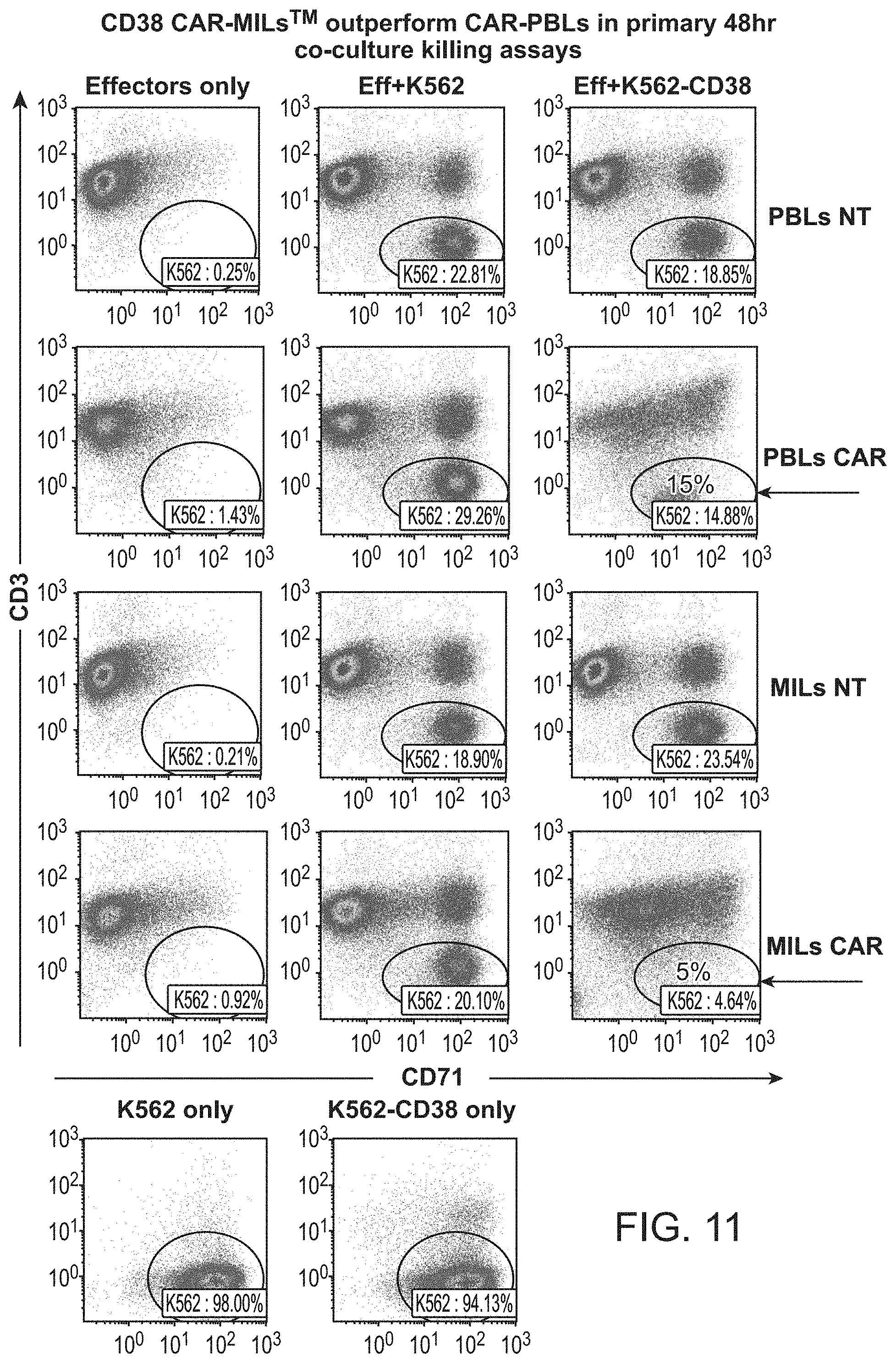
D00014
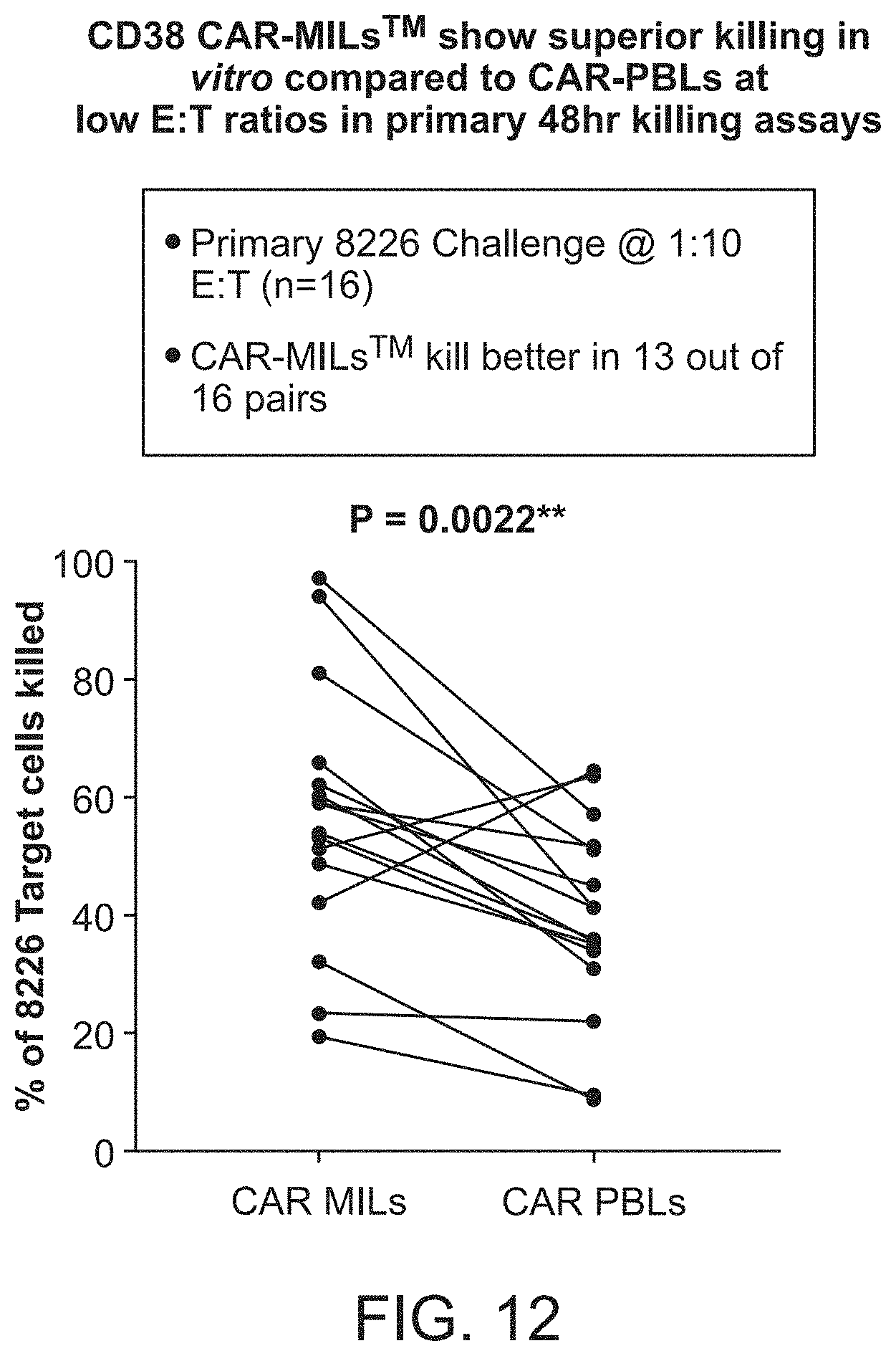
D00015
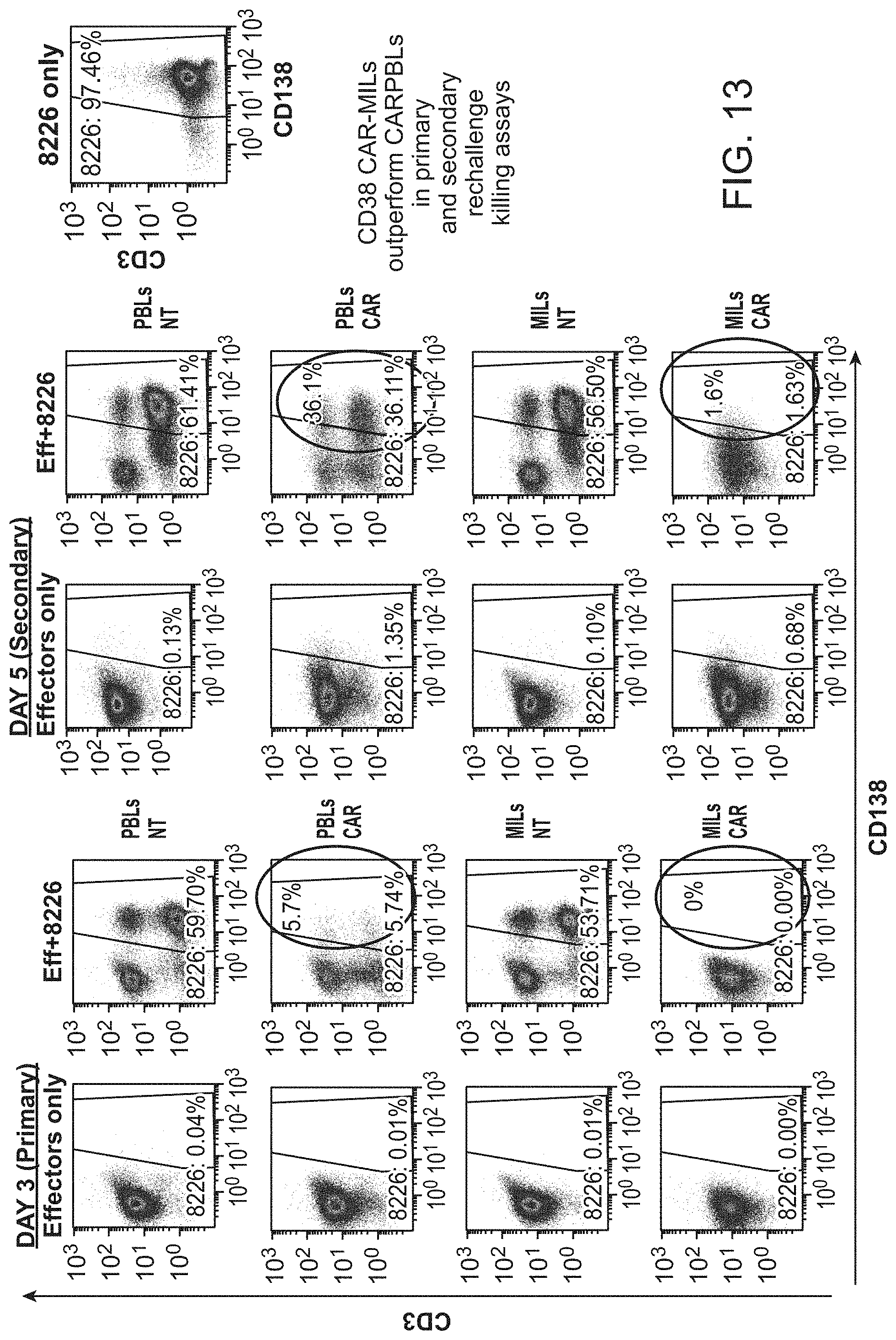
D00016

D00017

D00018

D00019

D00020

D00021

D00022

D00023

D00024

D00025
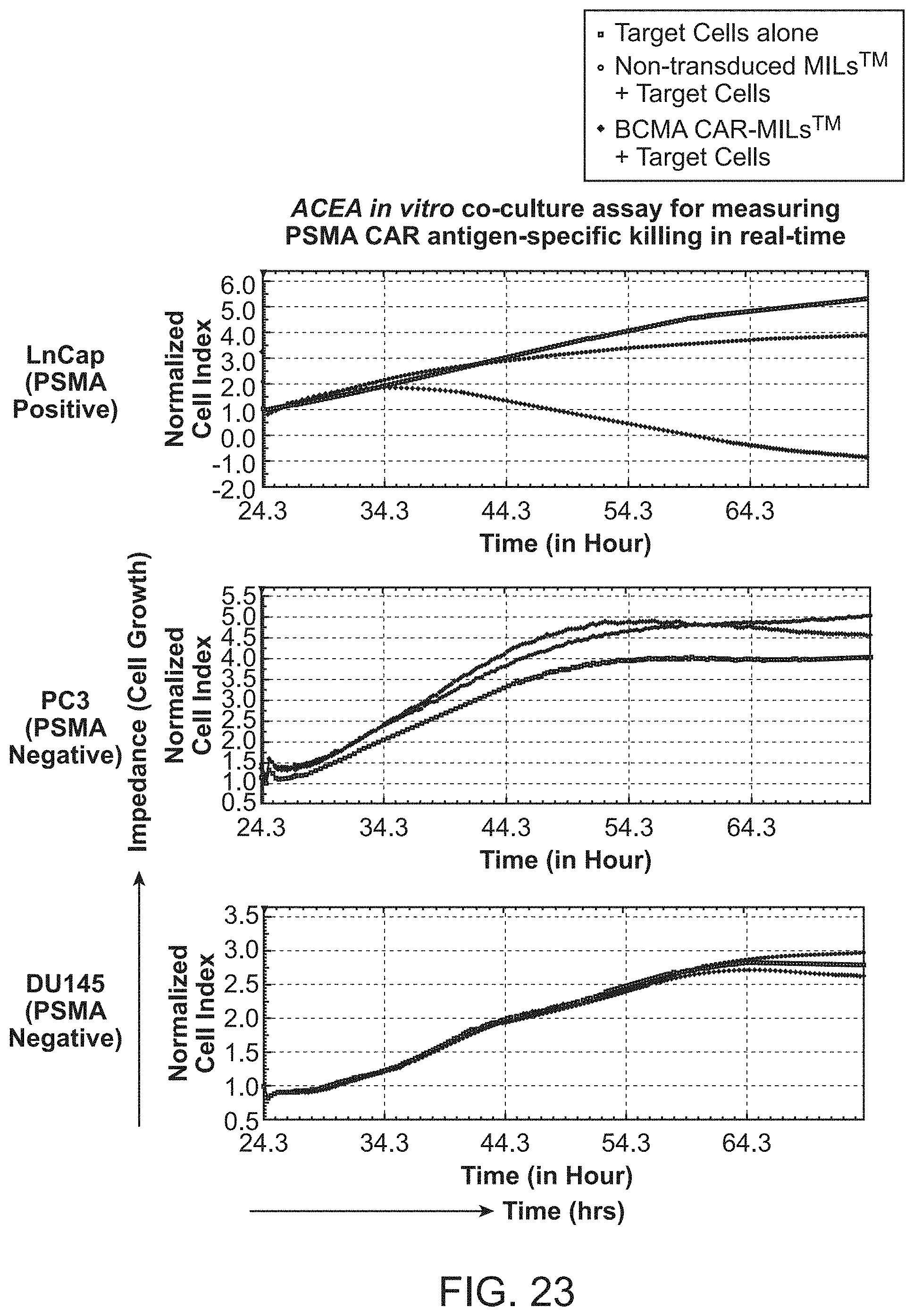
D00026

D00027

D00028

D00029

D00030

D00031
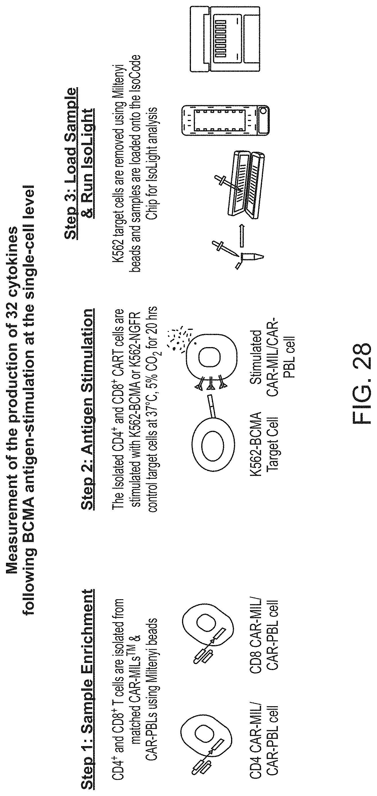
D00032
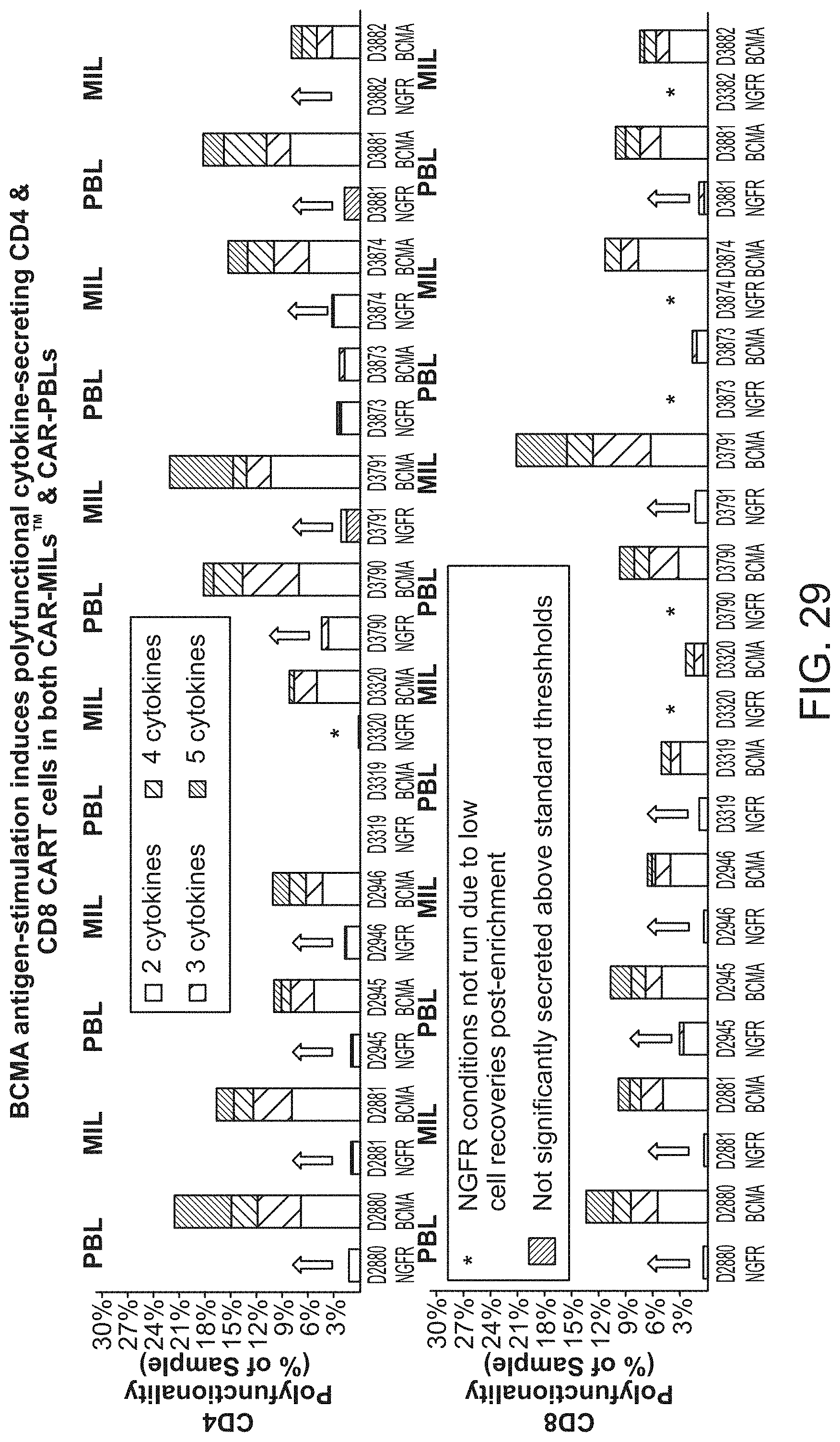
D00033

D00034
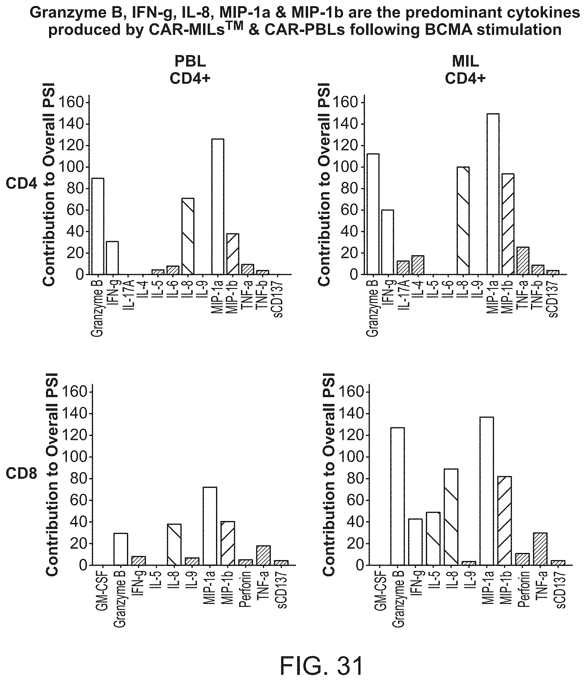
D00035

D00036
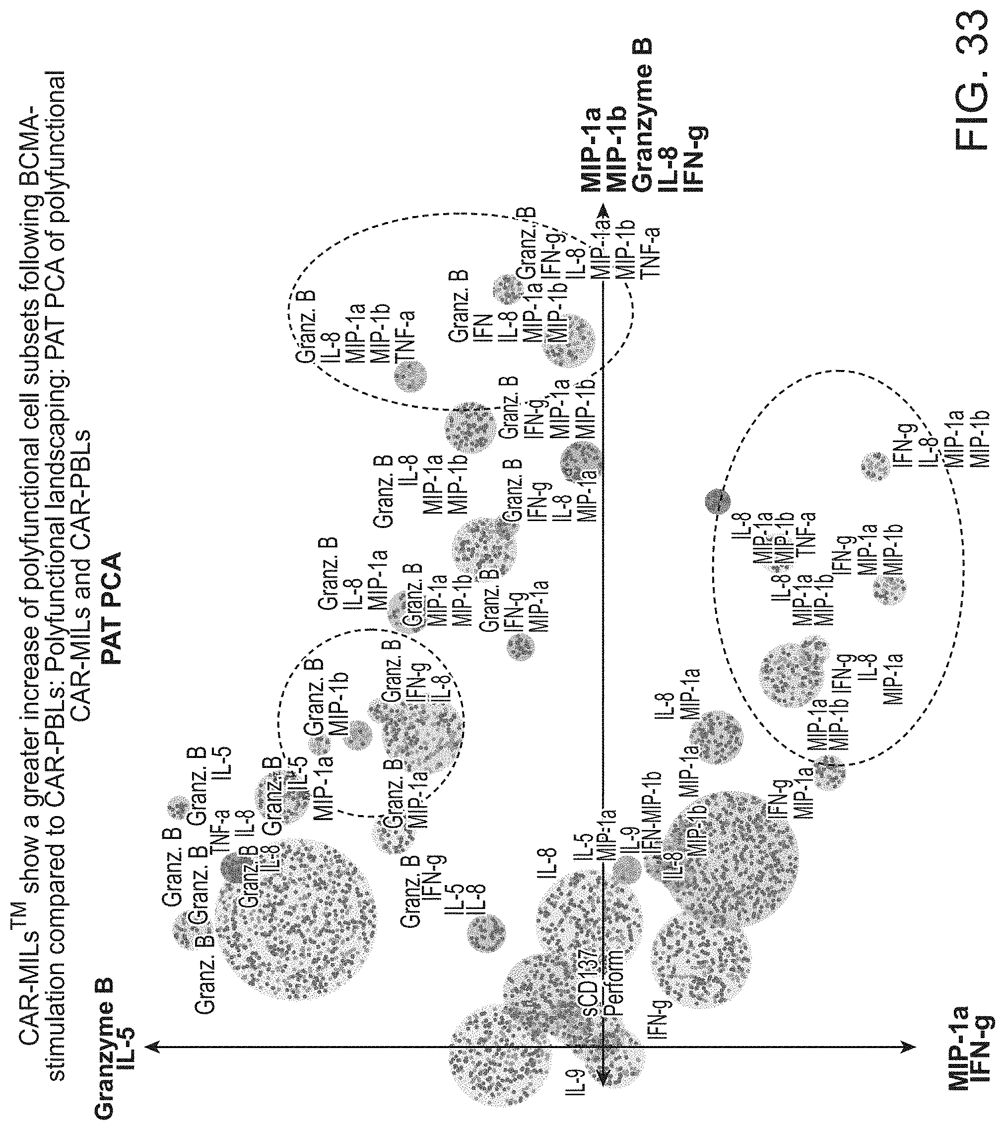
D00037

D00038
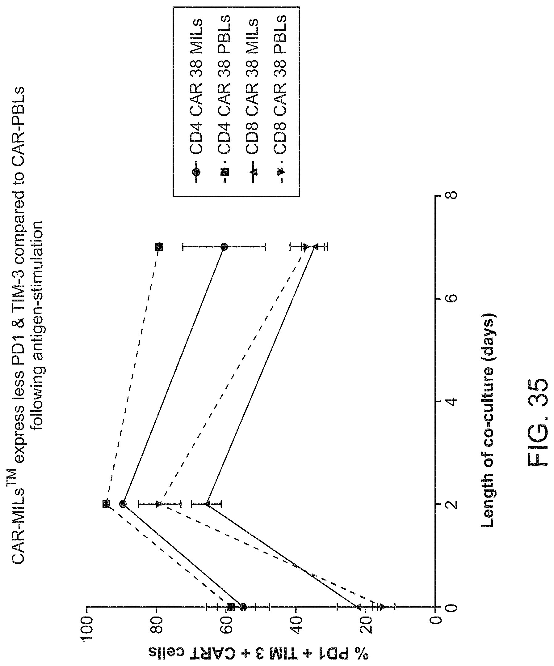
D00039

D00040
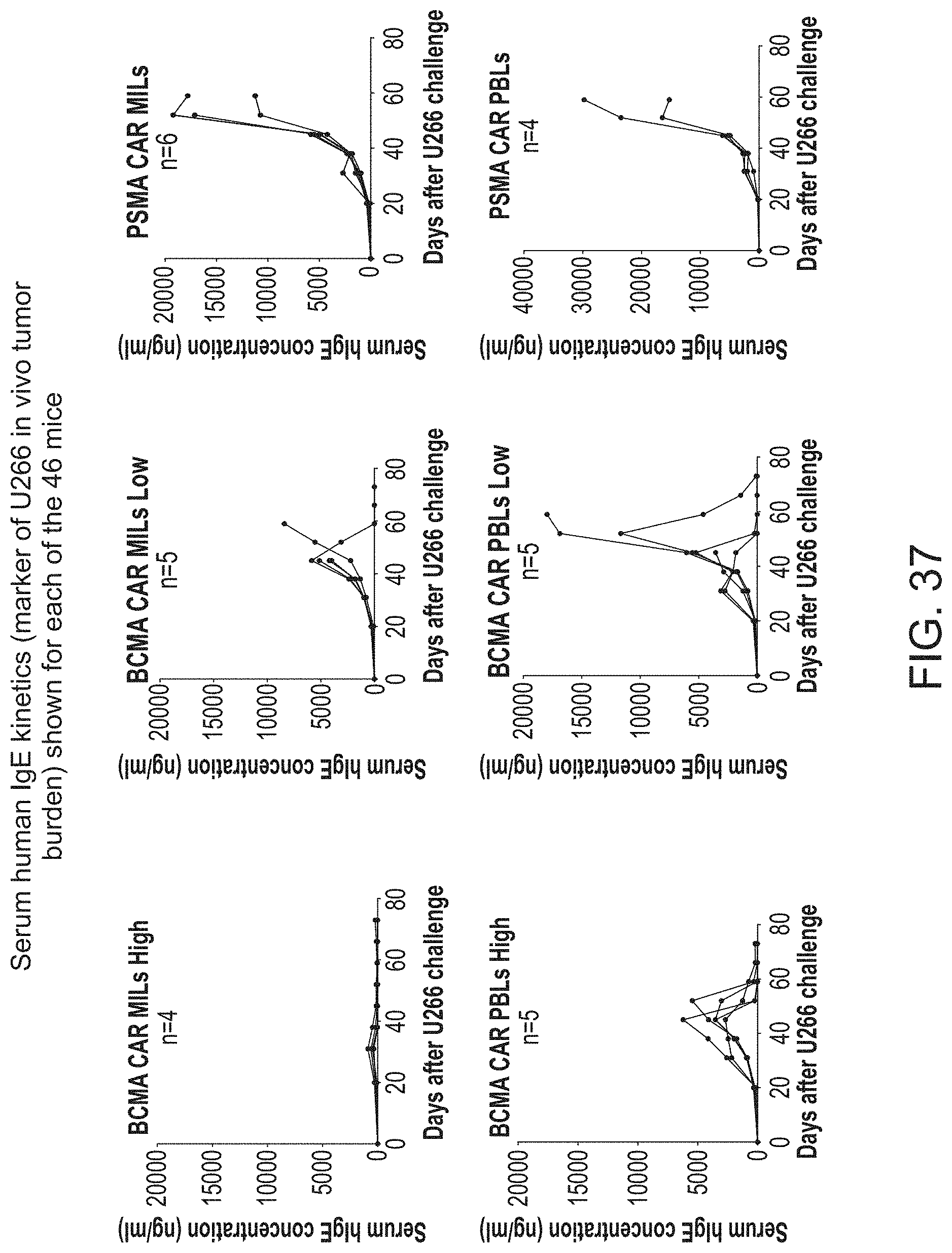
D00041
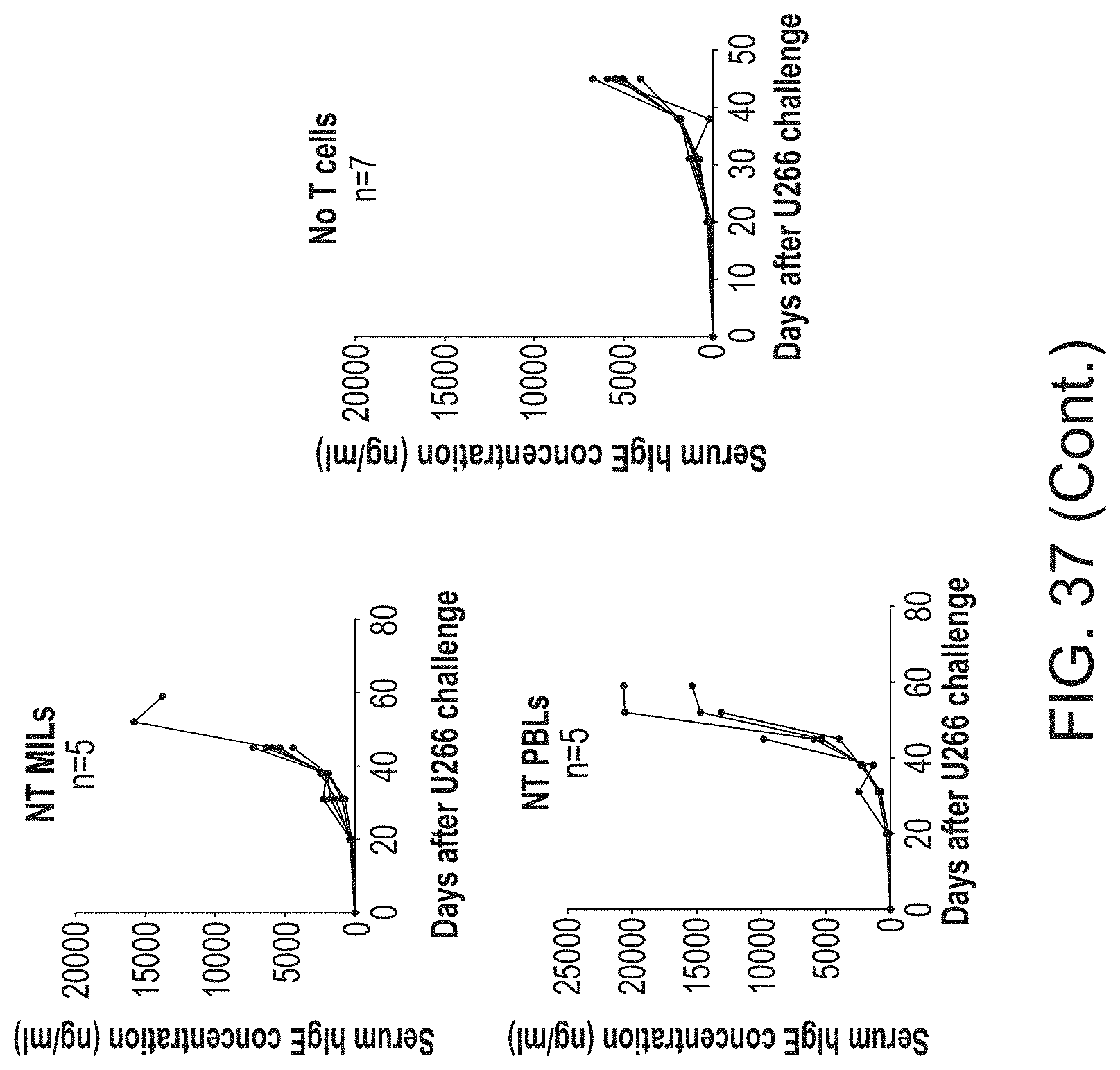
D00042

D00043

D00044

D00045

D00046
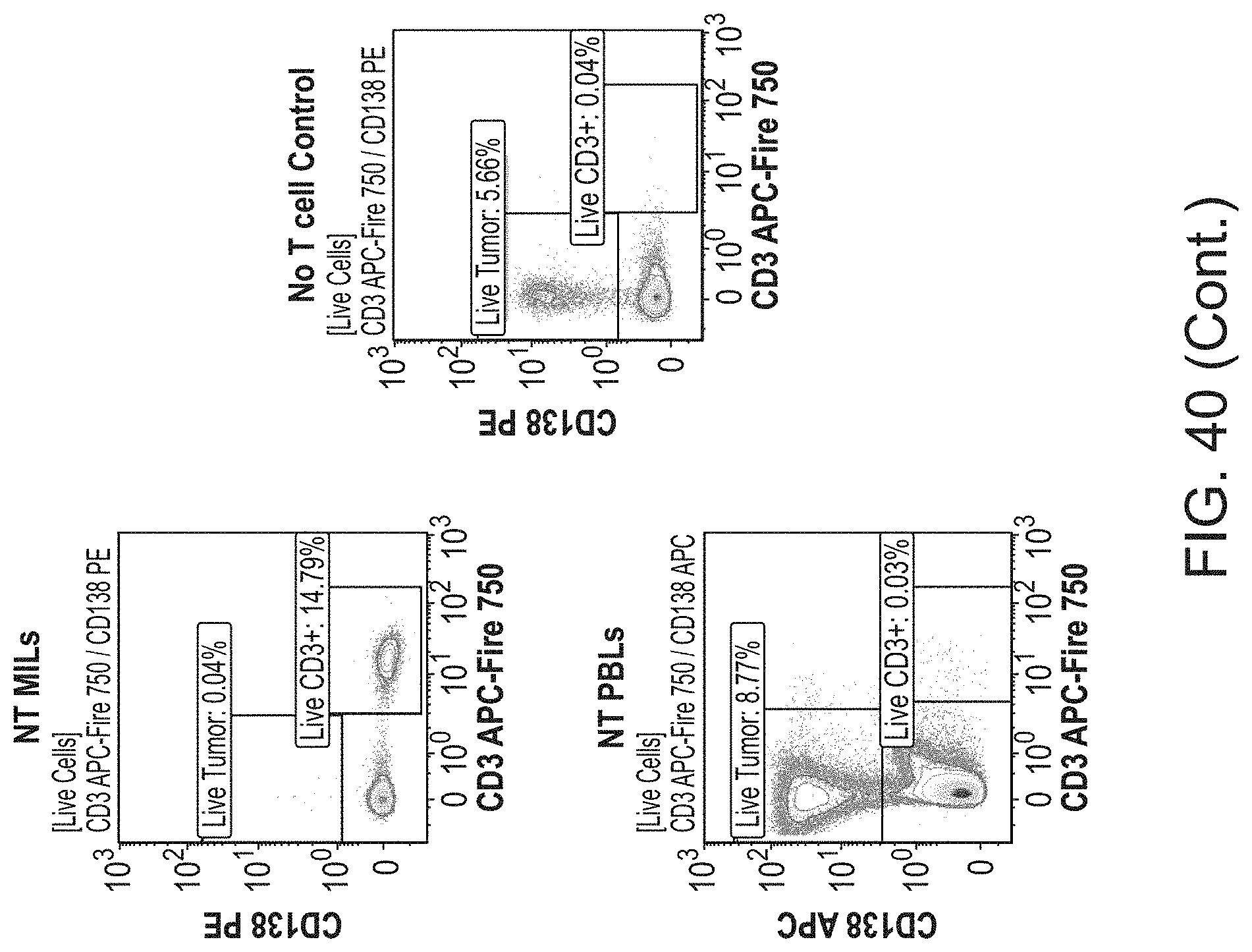
D00047
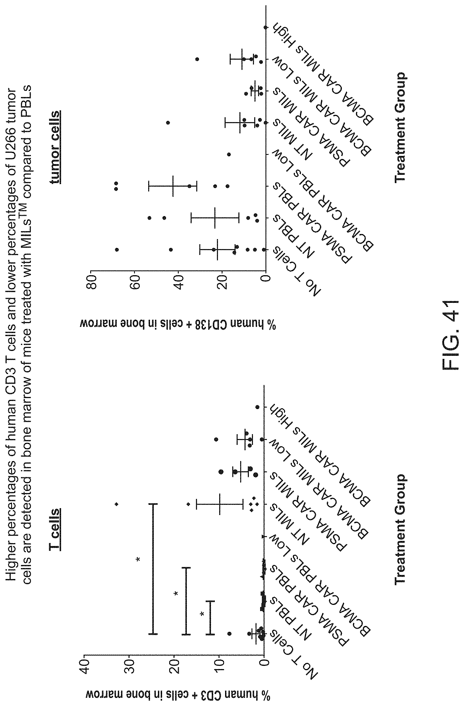
D00048
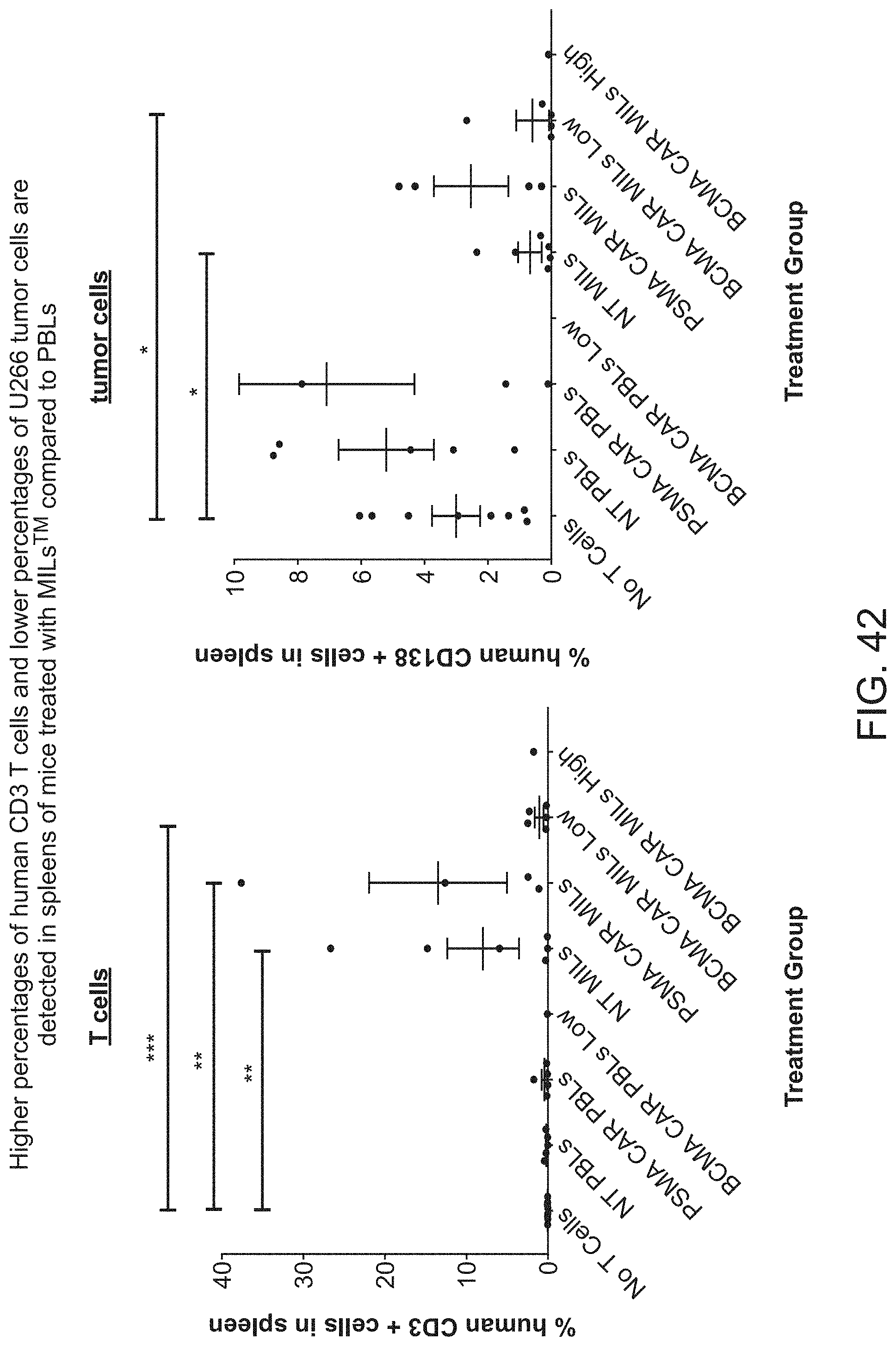
D00049

D00050
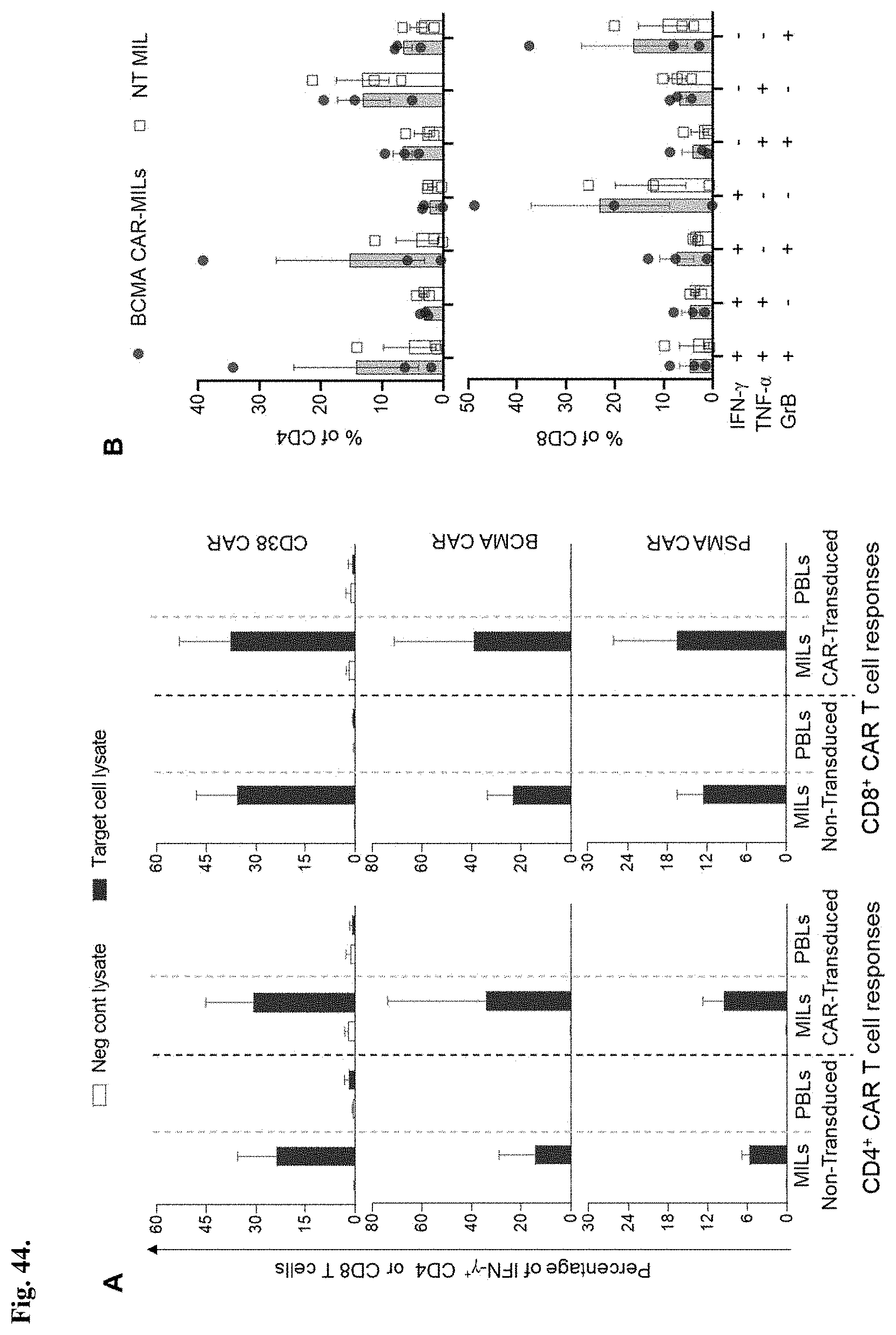
D00051

D00052

D00053

D00054

D00055

D00056
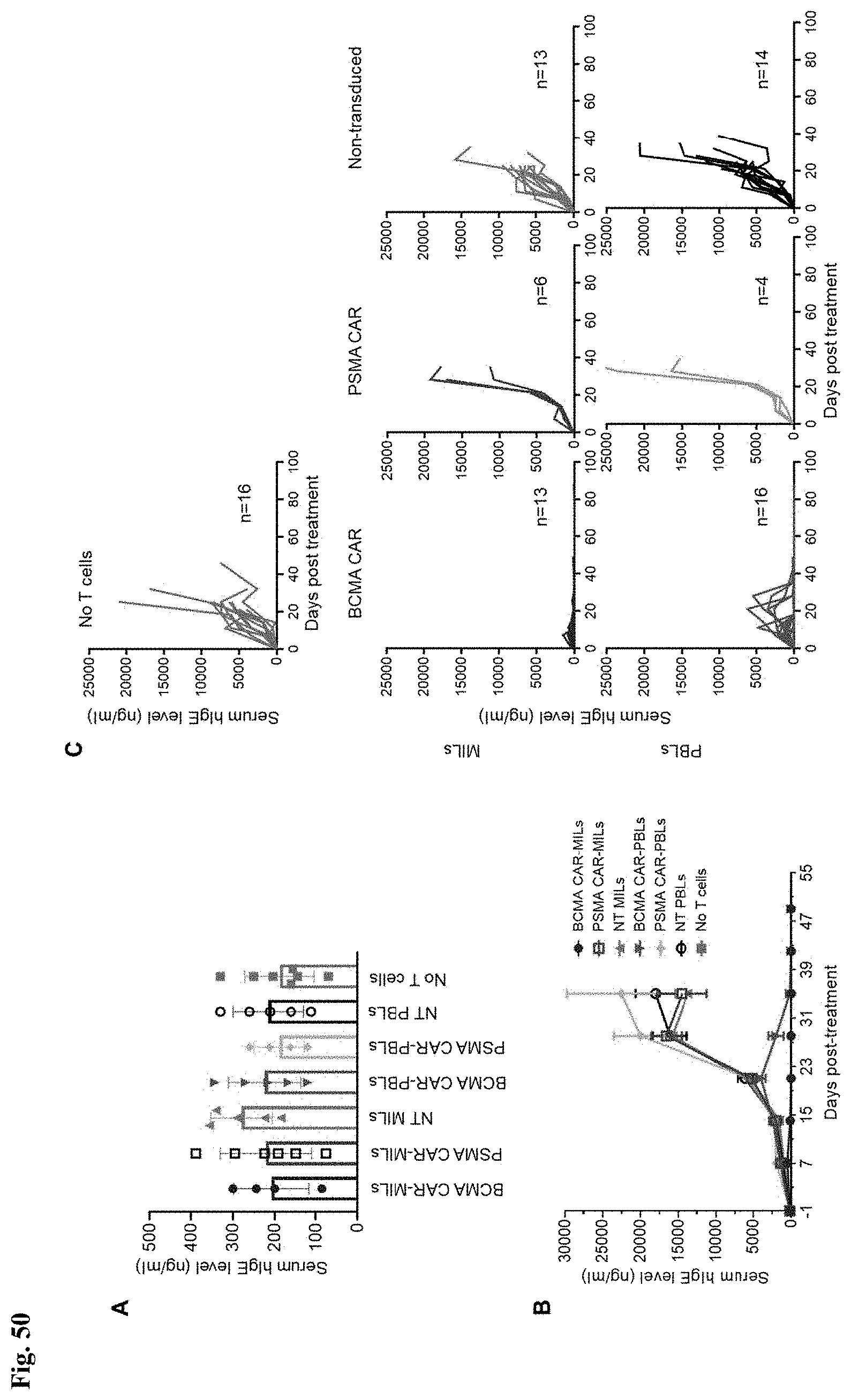
D00057
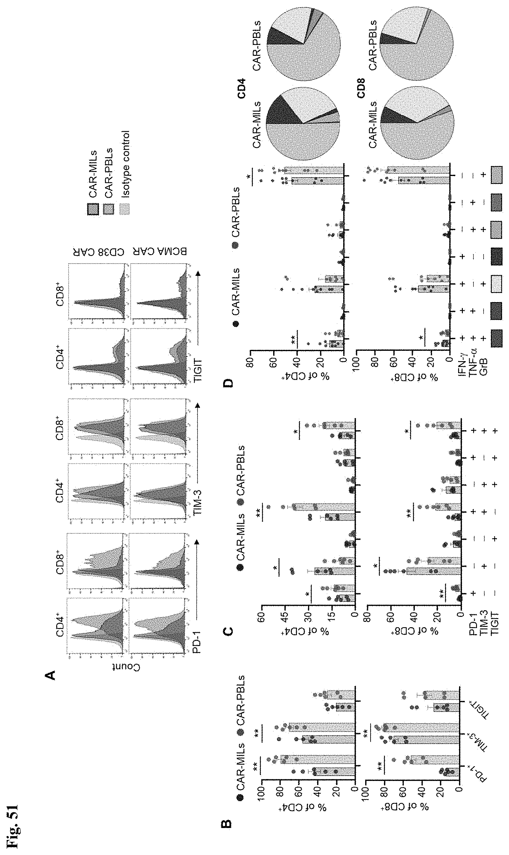
D00058

D00059
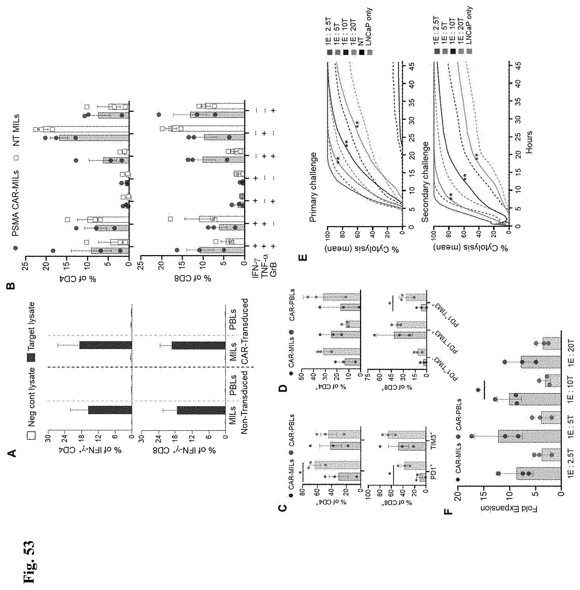
D00060
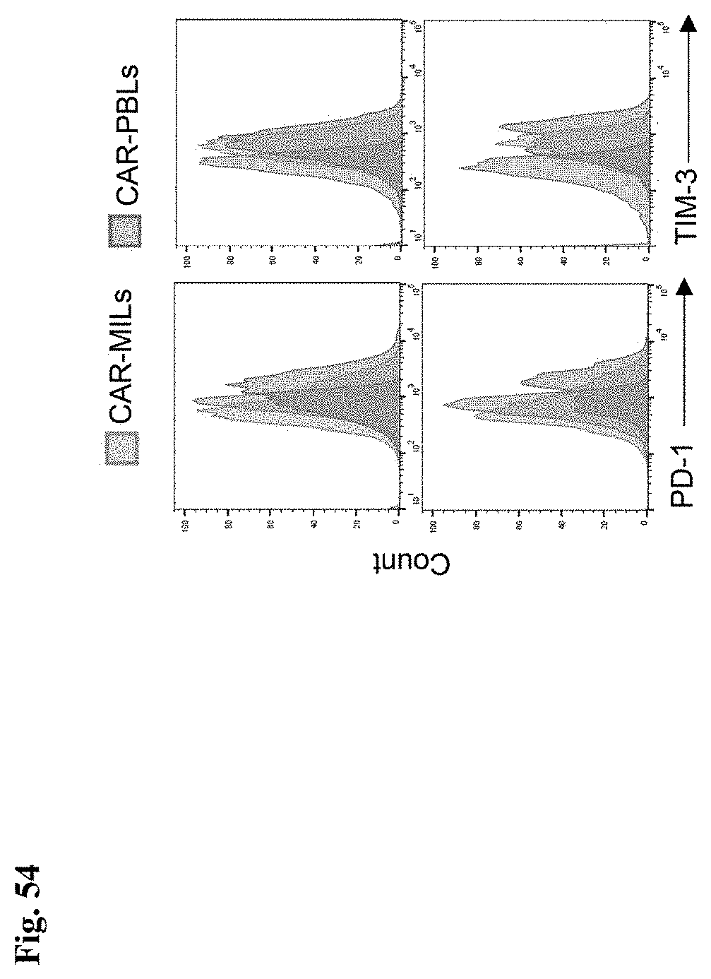
D00061

P00001
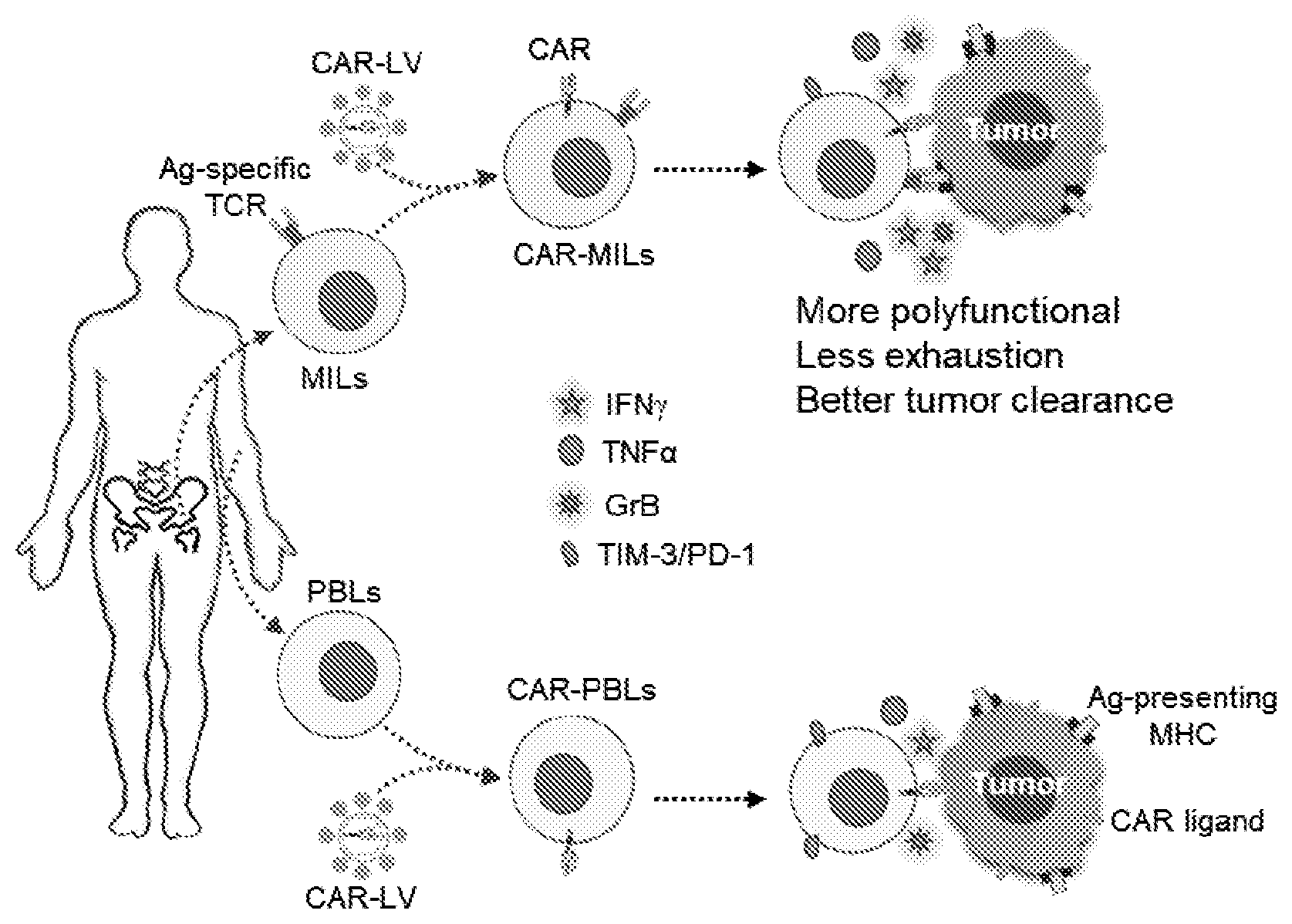
P00002

P00003

P00004

XML
uspto.report is an independent third-party trademark research tool that is not affiliated, endorsed, or sponsored by the United States Patent and Trademark Office (USPTO) or any other governmental organization. The information provided by uspto.report is based on publicly available data at the time of writing and is intended for informational purposes only.
While we strive to provide accurate and up-to-date information, we do not guarantee the accuracy, completeness, reliability, or suitability of the information displayed on this site. The use of this site is at your own risk. Any reliance you place on such information is therefore strictly at your own risk.
All official trademark data, including owner information, should be verified by visiting the official USPTO website at www.uspto.gov. This site is not intended to replace professional legal advice and should not be used as a substitute for consulting with a legal professional who is knowledgeable about trademark law.