Multispecific Antibodies And Use Thereof
DENGL; Stefan ; et al.
U.S. patent application number 17/072549 was filed with the patent office on 2021-05-20 for multispecific antibodies and use thereof. This patent application is currently assigned to Hoffmann-La Roche Inc.. The applicant listed for this patent is Hoffmann-La Roche Inc.. Invention is credited to Alexander BUJOTZEK, Stefan DENGL, Sebastian FENN, Jens FISCHER, Silke KIRCHNER, Claudia KIRSTENPFAD, Stefan KLOSTERMANN, Meher MAJETY, Joerg MOELLEKEN, Georg TIEFENTHALER.
| Application Number | 20210147554 17/072549 |
| Document ID | / |
| Family ID | 1000005362152 |
| Filed Date | 2021-05-20 |


View All Diagrams
| United States Patent Application | 20210147554 |
| Kind Code | A1 |
| DENGL; Stefan ; et al. | May 20, 2021 |
MULTISPECIFIC ANTIBODIES AND USE THEREOF
Abstract
The present invention relates to multispecific antibodies that bind to HLA-G ant to a T cell activating antigen, their preparation, formulations and methods of using the same.
| Inventors: | DENGL; Stefan; (Penzberg, DE) ; FENN; Sebastian; (Penzberg, DE) ; FISCHER; Jens; (Penzberg, DE) ; KIRSTENPFAD; Claudia; (Penzberg, DE) ; KLOSTERMANN; Stefan; (Penzberg, DE) ; MOELLEKEN; Joerg; (Penzberg, DE) ; TIEFENTHALER; Georg; (Penzberg, DE) ; BUJOTZEK; Alexander; (Penzberg, DE) ; MAJETY; Meher; (Penzberg, DE) ; KIRCHNER; Silke; (Penzberg, DE) | ||||||||||
| Applicant: |
|
||||||||||
|---|---|---|---|---|---|---|---|---|---|---|---|
| Assignee: | Hoffmann-La Roche Inc. Little Falls NJ |
||||||||||
| Family ID: | 1000005362152 | ||||||||||
| Appl. No.: | 17/072549 | ||||||||||
| Filed: | October 16, 2020 |
Related U.S. Patent Documents
| Application Number | Filing Date | Patent Number | ||
|---|---|---|---|---|
| PCT/EP2019/060008 | Apr 17, 2019 | |||
| 17072549 | ||||
| Current U.S. Class: | 1/1 |
| Current CPC Class: | C07K 2317/55 20130101; C07K 2317/33 20130101; C07K 2317/92 20130101; C07K 16/2833 20130101; A61K 2039/505 20130101; C07K 16/2809 20130101; C07K 2317/31 20130101; A61P 35/00 20180101; C07K 2317/56 20130101; C07K 2317/94 20130101; C07K 2317/76 20130101 |
| International Class: | C07K 16/28 20060101 C07K016/28; A61P 35/00 20060101 A61P035/00 |
Foreign Application Data
| Date | Code | Application Number |
|---|---|---|
| Apr 18, 2018 | EP | 18168053.9 |
Claims
1. A multispecific antibody that binds to human HLA-G and to human CD3, comprising a first antigen binding moiety that binds to human HLA-G and a second antigen binding moiety that binds to human CD3, wherein the multispecific antibody does not cross-react with a modified human HLA-G 82M MHC I complex (wherein the HLA-G specific amino acids have been replaced by HLA-A consensus amino acids) comprising SEQ ID NO:44.
2. The multispecific antibody according to claim 1, wherein the antibody is bispecific; and wherein the first antigen binding moiety antibody that binds to human HLA-G comprises A) (a) a VH domain comprising (i) HVR-H1 comprising an amino acid sequence of SEQ ID NO:1, (ii) HVR-H2 comprising an amino acid sequence of SEQ ID NO:2, and (iii) HVR-H3 comprising an amino acid sequence of SEQ ID NO:3; and (b) a VL domain comprising (i) HVR-L1 comprising an amino acid sequence of SEQ ID NO:4; (ii) HVR-L2 comprising an amino acid sequence of SEQ ID NO:5 and (iii) HVR L3 20 comprising an amino acid sequence of SEQ ID NO:6; or B) (a) a VH domain comprising (i) HVR-H1 comprising an amino acid sequence of SEQ ID NO:9, (ii) HVR-H2 comprising an amino acid sequence of SEQ ID NO:10, and (iii) HVR-H3 comprising an amino acid of SEQ ID NO:11; and (b) a VL domain comprising (i) HVR-L1 comprising an amino acid sequence of SEQ ID NO:12; (ii) HVR-L2 comprising an amino acid sequence of SEQ ID NO:13 and (iii) HVR-L3 comprising an amino acid sequence of SEQ ID NO:14; or C) (a) a VH domain comprising (i) HVR-H1 comprising an amino acid sequence of SEQ ID NO:17, (ii) HVR-H2 comprising an amino acid sequence of SEQ ID NO:18, and (iii) HVR-H3 comprising an amino acid sequence of SEQ ID NO:19; and (b) a VL domain comprising (i) HVR-L1 comprising an amino acid sequence of SEQ ID NO:20; (ii) HVR-L2 comprising an amino acid sequence of SEQ ID NO:21 and (iii) HVR-L3 comprising an amino acid sequence of SEQ ID NO:22; or D) (a) a VH domain comprising (i) HVR-H1 comprising an amino acid sequence of SEQ ID NO:25, (ii) HVR-H2 comprising an amino acid sequence of SEQ ID NO:26, and (iii) HVR-H3 comprising an amino acid sequence of SEQ ID NO:27; and (b) a VL domain comprising (i) HVR-L1 comprising an amino acid sequence of SEQ ID NO:28; (ii) HVR-L2 comprising an amino acid sequence of SEQ ID NO:29 and (iii) HVR-L3 comprising an amino acid sequence of SEQ ID NO:30; and wherein the second antigen binding moiety, that binds to a T cell activating antigen binds to human CD3, and comprises E) (a) a VH domain comprising (i) HVR-H1 comprising an amino acid sequence of SEQ ID NO:56, (ii) HVR-H2 comprising an amino acid sequence of SEQ ID NO:57, and (iii) HVR-H3 comprising an amino acid sequence of SEQ ID NO:58; and (b) a VL domain comprising (i) HVR-L1 comprising an amino acid sequence of SEQ ID NO:59; (ii) HVR-L2 comprising an amino acid sequence of SEQ ID NO:60 and (iii) HVR-L3 comprising an amino acid sequence of SEQ ID NO:61.
3. The bispecific antibody according to claim 2, wherein the first antigen binding moiety A) vii) comprises a VH sequence of SEQ ID NO:7 and a VL sequence of SEQ ID NO:8; viii) or humanized variant of the VH and VL of the antibody under i); or ix) comprises a VH sequence of SEQ ID NO:33 and a VL sequence of SEQ ID NO:34; or B) comprises a VH sequence of SEQ ID NO:15 and a VL sequence of SEQ ID NO:16; or C) comprises a VH sequence of SEQ ID NO:23 and a VL sequence of SEQ ID NO:24; or D) comprises a VH sequence of SEQ ID NO:31 and a VL sequence of SEQ ID NO:32; and wherein the second antigen binding moiety E) comprises a VH sequence of SEQ ID NO:62 and a VL sequence of SEQ ID NO:63.
4. The bispecific antibody according to claim 3, wherein the first antigen binding moiety comprises i) a VH sequence of SEQ ID NO:31 and a VL sequence of SEQ ID NO:32; or ii) a VH sequence of SEQ ID NO:33 and a VL sequence of SEQ ID NO:34; and wherein the second antigen binding moiety comprises a VH sequence of SEQ ID NO:62 and a VL sequence of SEQ ID NO:63.
5. The multispecific antibody according to claim 1, wherein the antibody comprises at least one of the following properties: a. does not cross-react with human HLA-A2 .beta.2M MHC I complex comprising SEQ ID NO:39 and SEQ ID NO: 37; b. does not cross-react with a mouse H2Kd .beta.2M MHC I complex comprising SEQ ID NO:45; c. does not cross-react with rat RT1A .beta.2M MHC I complex comprising SEQ ID NO:47; d. inhibits ILT2 binding to monomeric HLA-G 82M MHC I complex comprising SEQ ID NO: 43; e. inhibits ILT2 binding to trimeric HLA-G 82M MHC I complex comprising SEQ ID NO: 43, by more than 50% or by more than 60% when compared to the binding without antibody; f. inhibits ILT2 binding to monomeric and/or dimeric and/or trimeric HLA-G 82M MHC I complex comprising SEQ ID NO: 43, by more than 50% or by more than 80% when compared to the binding without antibody; g. inhibits ILT2 binding to HLA-G on JEG3 cells (ATCC No. HTB36) by more than 50% or by more than 80% when compared to the binding without antibody); h. binds to HLA-G on JEG3 cells (ATCC No. HTB36) and inhibits ILT2 binding to HLA-G on JEG-3 cells (ATCC No. HTB36) by more than 50% or by more than 80% when compared to the binding without antibody; i. inhibits CD8a binding to HLAG by more than 80% when compared to the binding without antibody; j. restores HLA-G specific suppressed immune response by monocytes co-cultured with JEG-3 cells (ATCC HTB36); or k. induces T cell mediated cytotoxicity in the presence of HLAG expressing tumor.
6. The multispecific antibody of claim 1, wherein the first and the second antigen binding moiety is a Fab molecule.
7. The multispecific antibody of claim 1, wherein the second antigen binding moiety is a Fab molecule wherein the variable domains VL and VH or the constant domains CL and CH1, particularly the variable domains VL and VH, of the Fab light chain and the Fab heavy chain are replaced by each other.
8. The multispecific antibody of claim 1, wherein the first antigen binding moiety is a Fab molecule wherein in the constant domain an amino acid at position 124 is substituted independently by lysine (K), arginine (R) or histidine (H) (numbering according to Kabat) and the amino acid at position 123 is substituted independently by lysine (K), arginine (R) or histidine (H) (numbering according to Kabat), and in the constant domain CH1 the amino acid at position 147 is substituted independently by glutamic acid (E), or aspartic acid (D) (numbering according to Kabat EU index) and the amino acid at position 213 is substituted independently by glutamic acid (E), or aspartic acid (D) (numbering according to Kabat EU index).
9. The multispecific antibody of claim 1, wherein the first and the second antigen binding moiety are fused to each other.
10. The multispecific antibody of claim 1, wherein the first and the second antigen binding moiety are each a Fab molecule and wherein either (i) the second antigen binding moiety is fused at the C-terminus of the Fab heavy chain to the N-terminus of the Fab heavy chain of the first antigen binding moiety, or (ii) the first antigen binding moiety is fused at the C-terminus of the Fab heavy chain to the N-terminus of the Fab heavy chain of the second antigen binding moiety.
11. The multispecific antibody of claim 10 comprising a third antigen binding moiety.
12. The multispecific antibody of claim 11, wherein the third antigen moiety is identical to the first antigen binding moiety.
13. Isolated nucleic acid encoding the multispecific antibody according to claim 1 or 11.
14. A pharmaceutical formulation comprising the multispecific antibody according claim 1 or 11 and a pharmaceutically acceptable carrier.
15. A method of treating cancer in a patient, comprising administering to the patient an effective amount of the multispecific antibody according to claim 1 or 11.
16. The multispecific antibody of claim 9, wherein the first and the second antigen binding moiety are fused to each other by a peptide linker.
Description
CROSS-REFERENCE TO RELATED APPLICATIONS
[0001] This application is a continuation of International Application No. PCT/EP2019/060008, filed Apr. 17, 2019, which claims priority to European Patent Application No. 18168053.9 filed Apr. 18, 2018, the disclosures of which are incorporated herein by reference in their entireties.
SEQUENCE LISTING
[0002] The instant application contains a Sequence Listing which has been submitted electronically in ASCII format and is hereby incorporated by reference in its entirety. Said ASCII copy, created on Oct. 14, 2020, is named P34774-US_Sequence Listing_ST25.txt and is 114 kilobytes in size.
FIELD OF THE INVENTION
[0003] The present invention relates to multispecific antibodies that bind to HLA-G ant to a Tcell activating antigen, their preparation, formulations and methods of using the same.
BACKGROUND OF THE INVENTION
[0004] The human major histocompatability complex, class I, 6, also known as human leukocyte antigen G (HLA-G), is a protein that in humans is encoded by the HLA-G gene. HLA-G belongs to the HLA nonclassical class I heavy chain paralogues. This class I molecule is a heterodimer consisting of a heavy chain and a light chain (beta-2 microglobulin). The heavy chain is anchored in the membrane but can also be shedded/secreted. [0005] The heavy chain consists of three domains: alpha 1, alpha 2 and alpha 3. The alpha 1 and alpha 2 domains form a peptide binding groove flanked by two alpha helices. Small peptides (approximately 9-mers) can bind to this groove akin to other MHC I proteins. [0006] The second chain is beta 2 microglobulin which binds to the heavy chain similar to other MHC I proteins.
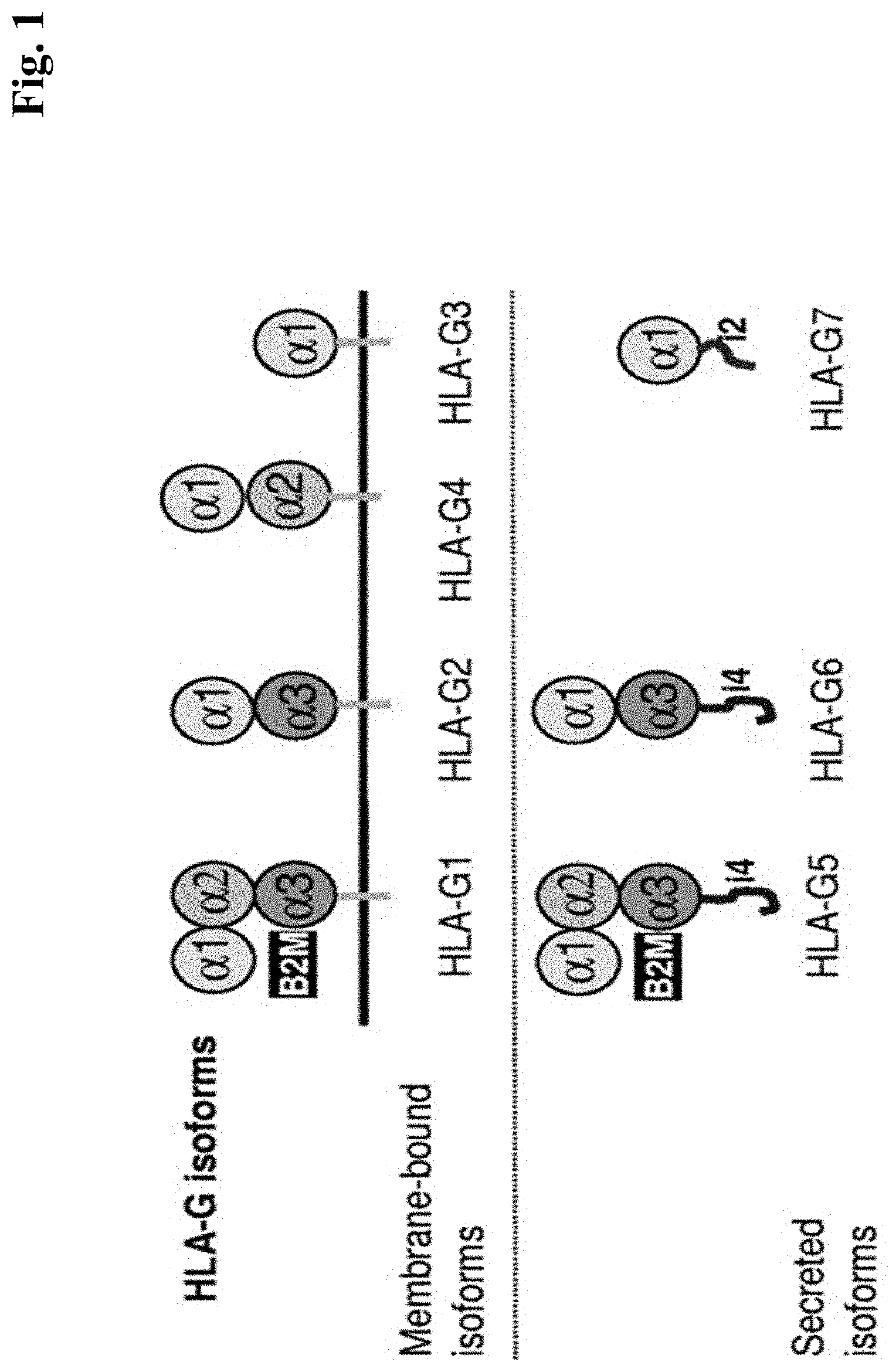
[0007] For HLA-G there exist 7 isoforms, 3 secreted and 4 membrane bound forms (as schematically shown in FIG. 1).
[0008] HLA-G can form functionally active complex oligomeric structures (Kuroki, K et al. Eur J Immunol. 37 (2007) 1727-1729). Disulfide-linked dimers are formed between Cys 42 of two HLA-G molecules. (Shiroishi M et al., J Biol Chem 281 (2006) 10439-10447. Trimers and Tetrameric complexes have also been described e.g. in Kuroki, K et al. Eur J Immunol. 37 (2007) 1727-1729, Allan D. S., et al. J Immunol Methods. 268 (2002) 43-50 and T Gonen-Gross et al., J Immunol 171 (2003)1343-1351).
[0009] HLA-G is predominantly expressed on cytotrophoblasts in the placenta. Several tumors (including pancreatic, breast, skin, colorectal, gastric & ovarian) express HLA-G (Lin, A. et al., Mol Med. 21 (2015) 782-791; Amiot, L., et al., Cell Mol Life Sci. 68 (2011) 417-431). The expression has also been reported to be associated with pathological conditions like inflammatory diseases, GvHD and cancer. Expression of HLA-G has been reported to be associated with poor prognosis in cancer. Tumor cells escape host immune surveillance by inducing immune tolerance/suppression via HLA-G expression.
TABLE-US-00001 Overview polymorphisms HLA family HLA-A: 2579 seqs HLA-B: 3283 seqs {close oversize brace} classical class I MHC HLA-C: 2133 seqs HLA-E: 15 seqs HLA-F: 22 seqs {close oversize brace} non-classical class I MHC HLA-G: 50 seqs
[0010] HLA-G shares high homology (>98%) with other MHC I molecules, therefore truly HLA-G specific antibodies with no crossreactivity to other MHC I molecules are difficult to generate.
[0011] Certain antibodies which interact in different ways with HLA-G were described previously: Tissue Antigens, 55 (2000) 510-518 relates to monoclonal antibodies e.g. 87G, and MEM-G/9; Neoplasma 50 (2003) 331-338 relates to certain monoclonal antibodies recognizing both, intact HLA-G oligomeric complex (e.g. 87G and MEM-G/9) as well as HLA-G free heavy chain (e.g. 4H84, MEM-G/1 and MEM-G/2); Hum Immunol. 64 (2003) 315-326 relates to several antibodies tested on HLA-G expressing JEG3 tumor cells (e.g. MEM-G/09 and -G/13 which react exclusively with native HLA-G1 molecules. MEM-G/01 recognizes (similar to the 4H84 mAb) the denatured HLA-G heavy chain of all isoforms, whereas MEM-G/04 recognizes selectively denatured HLA-G1, -G2, and -G5 isoforms; Wiendl et al Brain 2003 176-85 relates to different monoclonal HLA-G antibodies as e.g. 87G, 4H84, MEM-G/9.
[0012] The above publications report antibodies, which bind to human HLA-G or the human HLA-G/.beta.2M MHC complex. However, due to the high polymorphism and high homology of the HLA family most of the antibodies lack either truly specific HLA-G binding properties and often also bind or crossreact with other HLA family members (either as MHC complex with .beta.2M or in its .beta.2M-free form) or they simply do not inhibit binding of HLA-G .beta.2M MHC complex to its receptors ILT2 and/or ILT4 (and are regarded as non-antagonistic antibodies).
[0013] Bispecific antibodies that bind to a surface antigen on target cells and an activating T cell antigen such as CD3 on T-cells (also called herein T cell bispecific antibodies or "TCBs") hold great promise for the treatment of various cancers. The simultaneous binding of such an antibody to both of its targets will force a temporary interaction between target cell and T cell, causing crosslinking of the T cell receptor and subsequent activation of any cytotoxic T cell and subsequent lysis of the target cell. Given their potency in target cell killing, the choice of target and the specificity of the targeting antibody is of utmost importance for T cell bispecific antibodies to avoid on- and off-target toxicities. Intracellular proteins such as WT1 represent attractive targets, but are only accessible to T cell receptor (TCR)-like antibodies that bind major histocompatibility complex (MHC) presenting peptide antigens derived from the intracellular protein on the cell surface. An inherent issue of TCR-like antibodies is potential cross-reactivity with MHC molecules per se, or MHC molecules presenting peptides other than the desired one, which could compromise organ or tissue selectivity.
SUMMARY OF THE INVENTION
[0014] The invention provides a multispecific antibody that binds to human HLA-G and to a T cell activating antigen (particularly human CD3), comprising a first antigen binding moiety that binds to human HLA-G and a second antigen binding moiety that binds to a T cell activating antigen (particularly human CD3).
[0015] In one one aspect the multispecific antibody that binds to human HLA-G and to human CD3, comprising a first antigen binding moiety that binds to human HLA-G and a second antigen binding moiety that binds to human CD3, does not crossreact with a modified human HLA-G .beta.2M MHC I complex (wherein the HLA-G specific amino acids have been replaced by HLA-A consensus amino acids) comprising SEQ ID NO:44.
[0016] In one embodiment of the invention the multispecific antibody is bispecific; and
the first antigen binding moiety antibody that binds to human HLA-G comprises [0017] A) (a) a VH domain comprising (i) HVR-H1 comprising the amino acid sequence of SEQ ID NO:1, (ii) HVR-H2 comprising the amino acid sequence of SEQ ID NO:2, and (iii) HVR-H3 comprising an amino acid sequence selected from SEQ ID NO:3; and (b) a VL domain comprising (i) HVR-L1 comprising the amino acid sequence of SEQ ID NO:4; (ii) HVR-L2 comprising the amino acid sequence of SEQ ID NO:5 and (iii) HVR-L3 comprising the amino acid sequence of SEQ ID NO:6; or [0018] B) (a) a VH domain comprising (i) HVR-H1 comprising the amino acid sequence of SEQ ID NO:9, (ii) HVR-H2 comprising the amino acid sequence of SEQ ID NO:10, and (iii) HVR-H3 comprising an amino acid sequence selected from SEQ ID NO:11; and (b) a VL domain comprising (i) HVR-L1 comprising the amino acid sequence of SEQ ID NO:12; (ii) HVR-L2 comprising the amino acid sequence of SEQ ID NO:13 and (iii) HVR-L3 comprising the amino acid sequence of SEQ ID NO:14; or [0019] C) (a) a VH domain comprising (i) HVR-H1 comprising the amino acid sequence of SEQ ID NO:17, (ii) HVR-H2 comprising the amino acid sequence of SEQ ID NO:18, and (iii) HVR-H3 comprising an amino acid sequence selected from SEQ ID NO:19; and (b) a VL domain comprising (i) HVR-L1 comprising the amino acid sequence of SEQ ID NO:20; (ii) HVR-L2 comprising the amino acid sequence of SEQ ID NO:21 and (iii) HVR-L3 comprising the amino acid sequence of SEQ ID NO:22; or [0020] D) (a) a VH domain comprising (i) HVR-H1 comprising the amino acid sequence of SEQ ID NO:25, (ii) HVR-H2 comprising the amino acid sequence of SEQ ID NO:26, and (iii) HVR-H3 comprising an amino acid sequence selected from SEQ ID NO:27; and (b) a VL domain comprising (i) HVR-L1 comprising the amino acid sequence of SEQ ID NO:28; (ii) HVR-L2 comprising the amino acid sequence of SEQ ID NO:29 and (iii) HVR-L3 comprising the amino acid sequence of SEQ ID NO:30; and the second antigen binding moiety, that binds to a T cell activating antigen binds to human CD3, and comprises [0021] E) (a) a VH domain comprising (i) HVR-H1 comprising the amino acid sequence of SEQ ID NO:56, (ii) HVR-H2 comprising the amino acid sequence of SEQ ID NO:57, and (iii) HVR-H3 comprising an amino acid sequence selected from SEQ ID NO:58; and (b) a VL domain comprising (i) HVR-L1 comprising the amino acid sequence of SEQ ID NO:59; (ii) HVR-L2 comprising the amino acid sequence of SEQ ID NO:60 and (iii) HVR-L3 comprising the amino acid sequence of SEQ ID NO:61.
[0022] In one embodiment of the invention the first antigen binding moiety [0023] A) [0024] i) comprises a VH sequence of SEQ ID NO:7 and a VL sequence of SEQ ID NO:8; [0025] ii) or humanized variant of the VH and VL of the antibody under i); or [0026] iii) comprises a VH sequence of SEQ ID NO:33 and a VL sequence of SEQ ID NO:34; or [0027] B) [0028] comprises a VH sequence of SEQ ID NO:15 and a VL sequence of SEQ ID NO:16; or [0029] C) [0030] comprises a VH sequence of SEQ ID NO:23 and a VL sequence of SEQ ID NO:24; or [0031] D) [0032] comprises a VH sequence of SEQ ID NO:31 and a VL sequence of SEQ ID NO:32; [0033] and the second antigen binding moiety [0034] E) [0035] comprises a VH sequence of SEQ ID NO:62 and a VL sequence of SEQ ID NO:63.
[0036] In one embodiment of the invention the [0037] the first antigen binding moiety comprises i) a VH sequence of SEQ ID NO:31 and a VL sequence of SEQ ID NO:32; or ii) a VH sequence of SEQ ID NO:33 and a VL sequence of SEQ ID NO:34; [0038] and the second antigen binding moiety [0039] comprises a VH sequence of SEQ ID NO:62 and a VL sequence of SEQ ID NO:63.
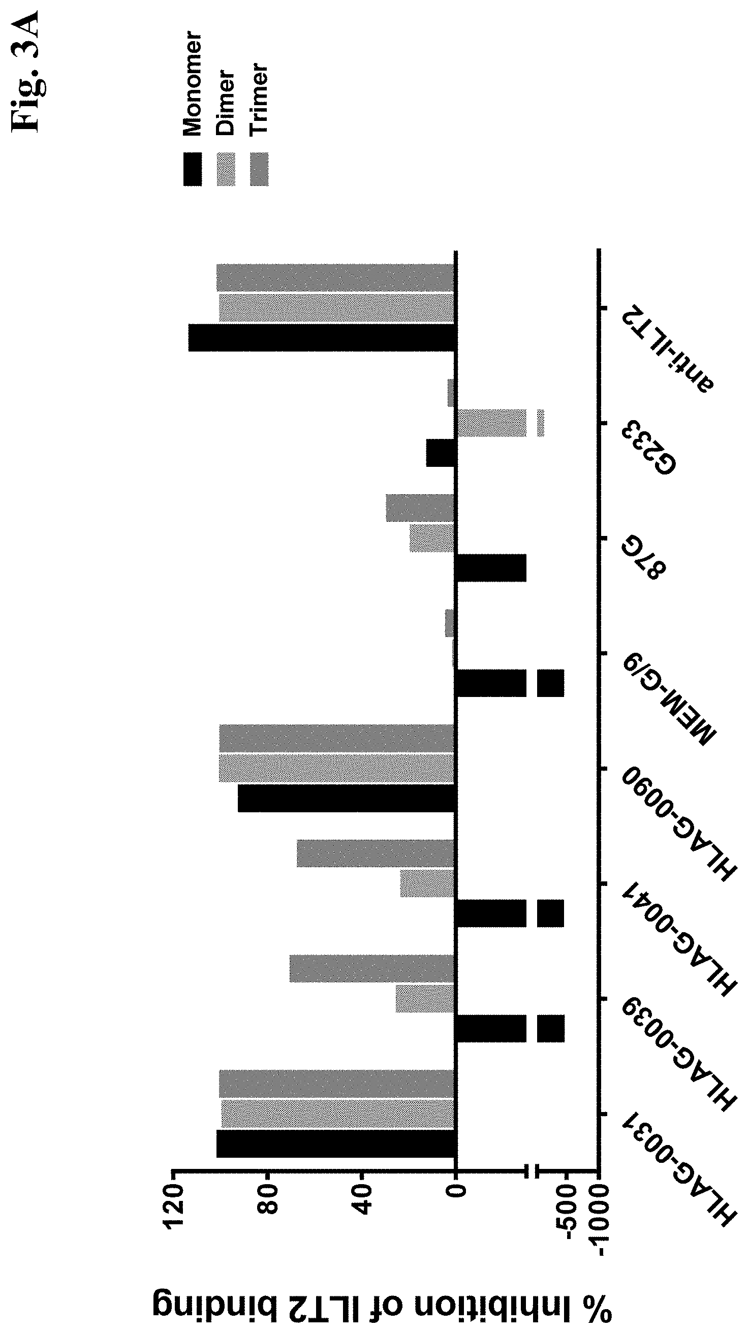
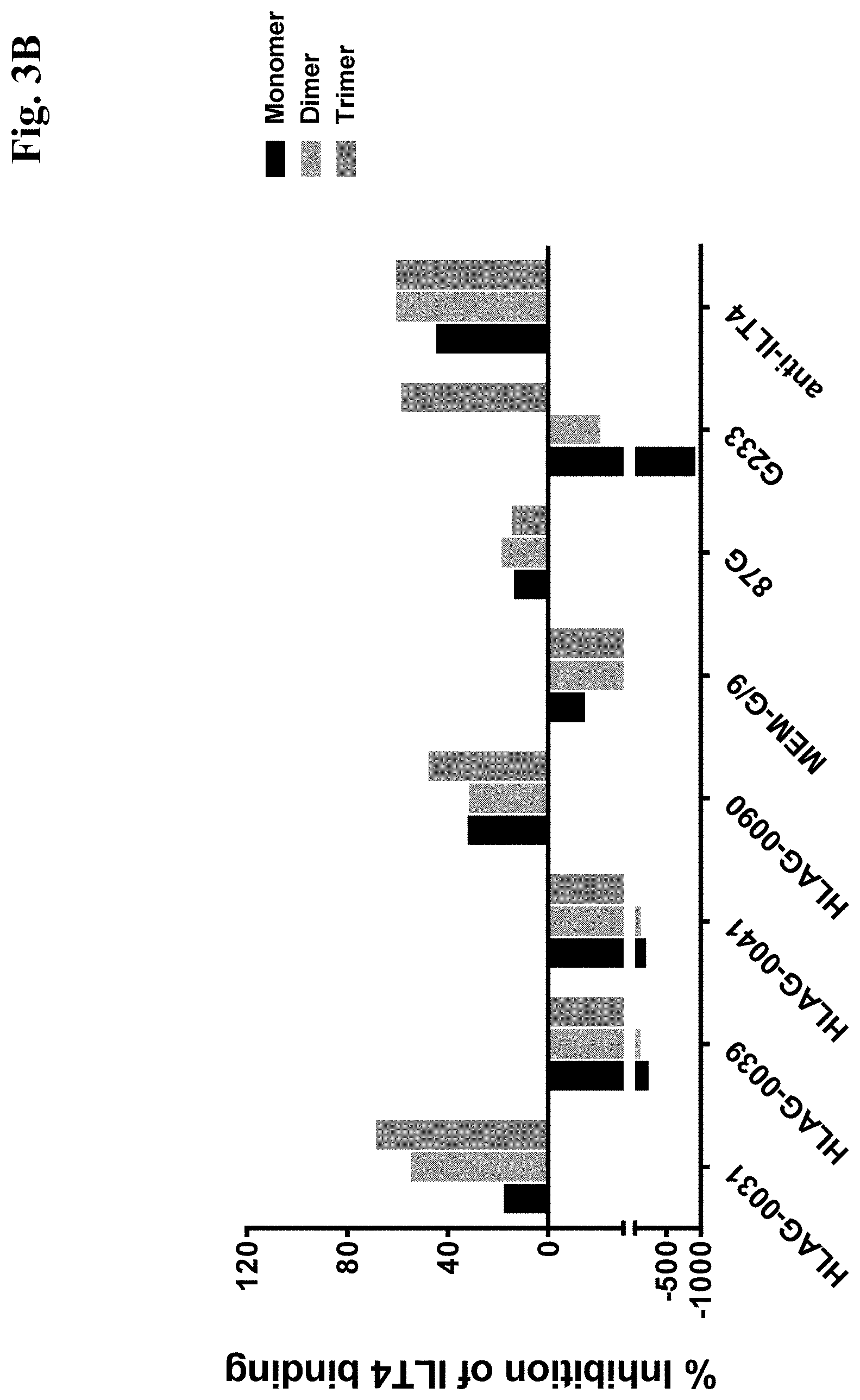
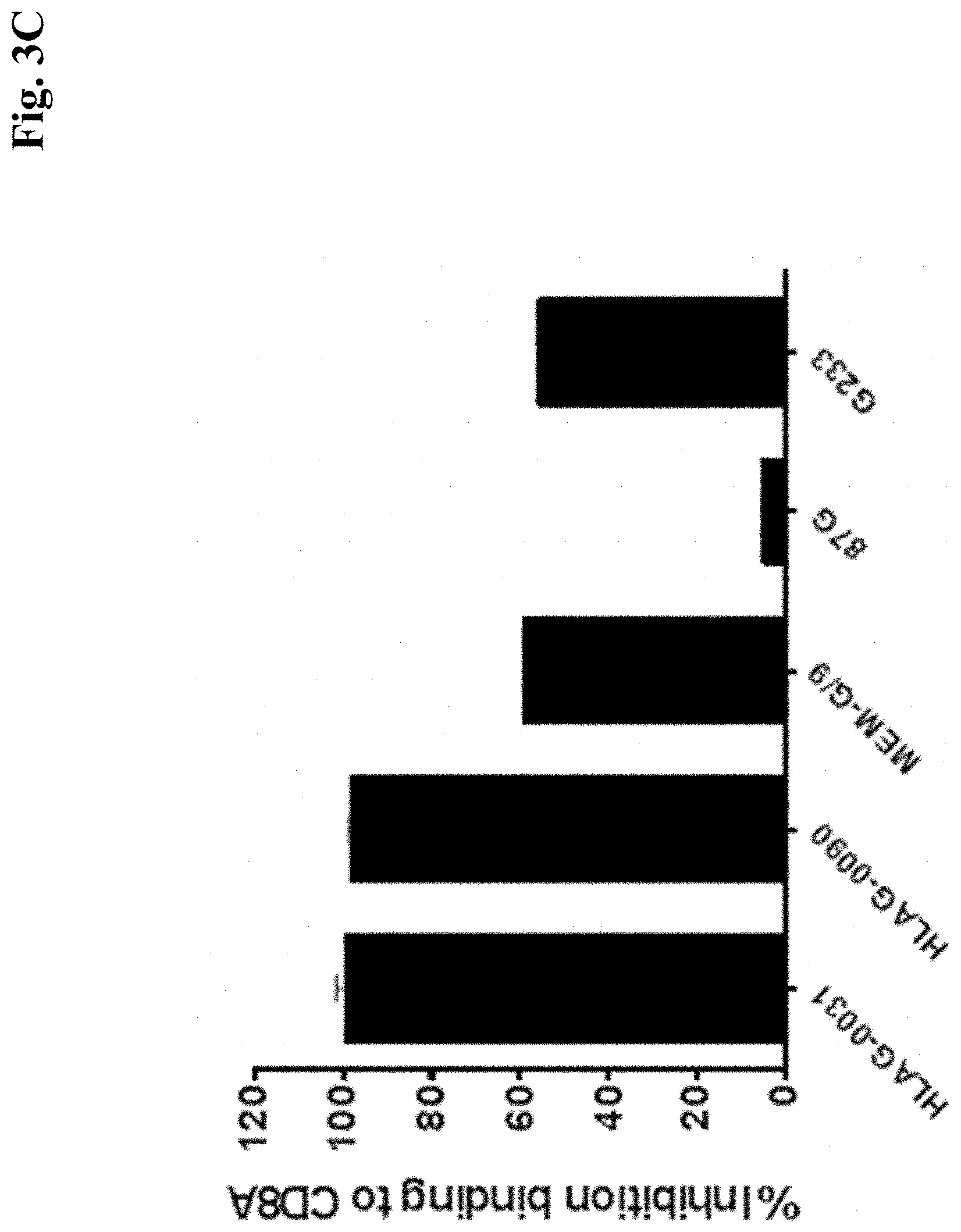
[0040] In one embodiment of the invention the multispecific antibody [0041] a) does not crossreact with a modified human HLA-G .beta.2M MHC I complex comprising SEQ ID NO:44; and/or [0042] b) does not crossreact with human HLA-A2 .beta.2M MHC I complex comprising SEQ ID NO:39 and SEQ ID NO: 37; and/or [0043] c) does not crossreact with a mouse H2Kd .beta.2M MHC I complex comprising SEQ ID NO:45; and/or [0044] d) does not crossreact with rat RT1A .beta.2M MHC I complex comprising SEQ ID NO:47; and/or [0045] e) inhibits ILT2 binding to monomeric HLA-G .beta.2M MHC I complex (comprising SEQ ID NO: 43); and/or [0046] f) inhibits ILT2 binding to trimeric HLA-G .beta.2M MHC I complex (comprising SEQ ID NO: 43), by more than 50% (in one embodiment by more than 60%) (when compared to the binding without antibody) (see Example 4b); and/or [0047] g) inhibits ILT2 binding to monomeric and/or dimeric and/or trimeric HLA-G .beta.2M MHC I complex (comprising SEQ ID NO: 43), by more than 50% (in on embodiment by more than 80%) (when compared to the binding without antibody) (see Example 4b); and/or [0048] h) inhibits ILT2 binding to (HLA-G on) JEG3 cells (ATCC No. HTB36) (by more than 50% (in one embodiment by more than 80%)) (when compared to the binding without antibody) (see Example 6); and/or [0049] i) binds to (HLA-G on) JEG3 cells (ATCC No. HTB36) (see Example 5), and inhibits ILT2 binding to (HLA-G on) JEG-3 cells (ATCC No. HTB36) (by more than 50% (in one embodiment by more than 80%)) (when compared to the binding without antibody) (see Example 6); and/or [0050] j) inhibits CD8a binding to HLAG by more than 80% (when compared to the binding without antibody) (see e.g Example 4c); and/or [0051] k) restores HLA-G specific suppressed immune response (e.g.. suppressed Tumor necrose factor (TNF) alpha release) by monocytes co-cultured with JEG-3 cells (ATCC HTB36); and/or [0052] l) induces T cell mediated cytotoxicity in the presence of HLAG expressing tumor cells (e.g. JEG-3 cells (ATCC HTB36) (see Example 12).
[0053] In one embodiment of the invention the first and the second antigen binding moiety is a Fab molecule (are each a Fab molecule).
[0054] In one embodiment of the invention the the second antigen binding moiety is a Fab molecule wherein the variable domains VL and VH or the constant domains CL and CH1, particularly the variable domains VL and VH, of the Fab light chain and the Fab heavy chain are replaced by each other.
[0055] In one embodiment of the invention the the first antigen binding moiety is a Fab molecule wherein in the constant domain the amino acid at position 124 is substituted independently by lysine (K), arginine (R) or histidine (H) (numbering according to Kabat) and the amino acid at position 123 is substituted independently by lysine (K), arginine (R) or histidine (H) (numbering according to Kabat), and in the constant domain CH1 the amino acid at position 147 is substituted independently by glutamic acid (E), or aspartic acid (D) (numbering according to Kabat EU index) and the amino acid at position 213 is substituted independently by glutamic acid (E), or aspartic acid (D) (numbering according to Kabat EU index).
[0056] In one embodiment of the invention the the first and the second antigen binding moiety are fused to each other, optionally via a peptide linker.
[0057] In one embodiment of the invention the the first and the second antigen binding moiety are each a Fab molecule and wherein either (i) the second antigen binding moiety is fused at the C-terminus of the Fab heavy chain to the N-terminus of the Fab heavy chain of the first antigen binding moiety, or (ii) the first antigen binding moiety is fused at the C-terminus of the Fab heavy chain to the N-terminus of the Fab heavy chain of the second antigen binding moiety.
[0058] In one embodiment of the invention the multispecific antibody comprises a third antigen binding moiety.
[0059] In one embodiment of the invention such third antigen moiety is identical to the first antigen binding moiety.
[0060] In one embodiment of the invention the multispecific antibody comprise an Fc domain composed of a first and a second subunit.
[0061] In one embodiment of the invention the the first, the second and, where present, the third antigen binding moiety are each a Fab molecule; and wherein either (i) the second antigen binding moiety is fused at the C-terminus of the Fab heavy chain to the N-terminus of the Fab heavy chain of the first antigen binding moiety and the first antigen binding moiety is fused at the C-terminus of the Fab heavy chain to the N-terminus of the first subunit of the Fc domain, or (ii) the first antigen binding moiety is fused at the C-terminus of the Fab heavy chain to the N-terminus of the Fab heavy chain of the second antigen binding moiety and the second antigen binding moiety is fused at the C-terminus of the Fab heavy chain to the N-terminus of the first subunit of the Fc domain; and wherein the third antigen binding moiety, where present, is fused at the C-terminus of the Fab heavy chain to the N-terminus of the second subunit of the Fc domain.
[0062] The invention provides an isolated nucleic acid encoding the antibody according to any one of the preceding claims.
[0063] The invention provides a host cell comprising such nucleic acid.
[0064] The invention provides a method of producing an antibody comprising culturing the host cell so that the antibody is produced.
[0065] The invention provides such method of producing an antibody, further comprising recovering the antibody from the host cell.
[0066] The invention provides a pharmaceutical formulation comprising the antibody described herein and a pharmaceutically acceptable carrier.
[0067] The invention provides the antibody described herein for use as a medicament.
[0068] The invention provides the antibody described herein for use in treating cancer.
[0069] The invention provides the use of the antibody described herein in the manufacture of a medicament. In one embodiment the medicament is for treatment of cancer.
[0070] The invention provides a method of treating an individual having cancer comprising administering to the individual an effective amount of the antibody described herein.
[0071] With the screening methods described herein new anti-HLA-G antibodies could be selected. These antibodies show highly valuable properties like strong inhibition of ILT2 binding to HLA-G expressed on JEG3 cells or inhibition of ILT2 binding to monomeric and/or dimeric and/or trimeric HLA-G .beta.2M MHC I complex.
[0072] Furthermore, the antibodies according to the invention are able to restore a HLA-G specific suppressed immune response, i.e. restoration of LPS-induced TNFa production by monocytes in co-culture with HLA-G-expressing cells.
[0073] In addition, the antibodies are highly specific and to not show cross reactivity with HLA-A MHC I complexes or MHC I complexes from mouse or rat origin.
DESCRIPTION OF THE FIGURES
[0074] FIG. 1: Different isoforms of HLA-G
[0075] FIG. 2: FIG. 2A: Schematic representation of HLA-G with molecule in association with .beta.2M [0076] FIG. 2B: Structure of HLA-G molecule in association with certain receptors: HLA-G structure in complex with given receptors such as ILT4 and KIR2DL1. ILT4 structure (PDB code: 2DYP). The KIR2DL1 structure is taken from PDB code 11M9 (KIR2DL1: HLA-Cw4 complex structure) and was positioned on HLA-G by superposition of the HLA-Cw4 and HLA-G structures. Receptors are shown in a ribbon representation, HLA-G is shown in a molecular surface representation. HLA-G residues that are unique or conserved in other HLA paralogs are colored in white and gray, respectively. Unique surface residues were replaced by a HLA consensus sequence in the chimeric counter antigen.
[0077] FIG. 3: HLA-G antibodies which inhibit (or stimulate) HLA-G interaction/binding with ILT2 and ILT4 as well as CD8: [0078] FIG. 3A: ILT2 inhibition [0079] FIG. 3B: ILT4 inhibition [0080] FIG. 3C: CD8 inhibition
[0081] FIG. 4: Flow cytometric analysis of cell surface expression of HLA-G using HLA-G antibodies on JEG3 (cells naturally expressing HLA-G), SKOV-3 cells (wild-type (wt) versus HLAG transfected cells (HLAG+)), and PA-TU-8902 cells (wild-type (wt) versus HLAG transfected cells (HLAG+)): [0082] FIG. 4A: HLA-G-0031 (#0031); FIG. 4B: HLA-G-0039 (#0039); [0083] FIG. 4C: HLA-G-0041 (#0041); FIG. 4D: HLA-G-0090 (#0090)
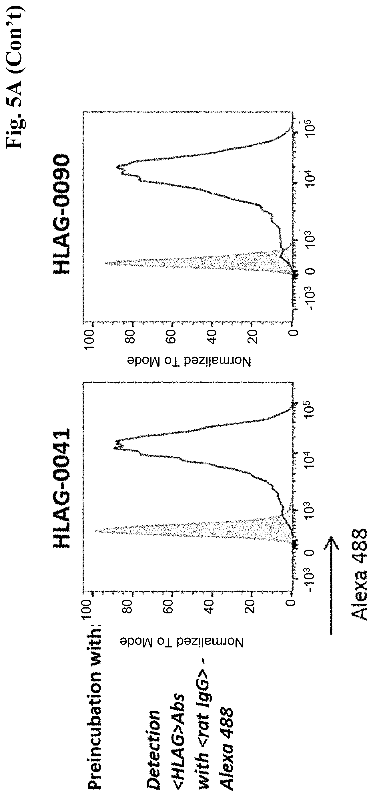
[0084] FIG. 5: FIG. 5A: Anti-HLA-G antibodies (0031, 0039, 0041 and 0090) block/modulate interaction of human ILT2 Fc chimera with HLA-G expressed on JEG3 cells: [0085] The staining of cell surface HLA-G with the novel anti-HLA-G antibodies was assessed by using an anti-rat IgG secondary antibody conjugated to Alexa488 (upper row). Shown in the FACS histograms are cells stained with secondary antibody alone (grey dotted lines) and cell stained with anti-HLA-G antibodies (black solid lines). In the lower row human ILT2-Fc bound to HLA-G on JEG3 cells is depicted (black dotted line) in comparison to cells stained with secondary antibody alone (grey dotted line). The impact of pre-incubating JEG3 cells with HLA-G antibodies on ILT2 Fc chimera binding can been seen (black solid line): HLA-G-0031 and HLA-G-0090 showed nearly complete inhibition of binding of ILT2-Fc chimera to JEG3 cells. Interestingly, the two antibodies 0039 and 0041 even increase ILT2:fc binding to the cells. [0086] FIG. 5B: Impact of commercial/reference anti-HLA-G antibodies on ILT2 Fc chimera binding to HLA-G on JEG3 cells. [0087] The staining of cell surface HLA-G with commercial/reference anti-HLA-G antibodies was assessed by using a species-specific secondary antibody conjugated to Alexa488 (upper row). Shown in the FACS histograms are cells stained with secondary antibody alone (grey dotted lines) and cell stained with anti-HLA-G antibodies (black solid lines). In the lower row human ILT2 Fc chimera bound to HLA-G on JEG3 cells is depicted (black dotted line) in comparison to cells stained with secondary antibody alone (grey dotted line). The impact of pre-incubating JEG3 cells with reference antibodies on ILT2 Fc chimera binding can been seen (black solid line). None of the tested reference antibodies could block the interaction of ILT2 Fc chimera with cell surface HLA-G on JEG3 cells.
[0088] FIG. 6: The impact of the blockade of HLA-G with inhibitory anti-HLA-G antibodies on the restoration of TNF.alpha. production assessed on different donors. [0089] FIG. 6A: Anti-HLAG antibodies HLA-G-0031 (#0031), HLA-G-0039 (#0039), and HLA-G-0041 (#0041) evaluated on a representative monocyte donor. [0090] FIG. 6B: Anti-HLAG antibody HLA-G-0090 (#0090)] evaluated on a different monocyte donor. [0091] FIG. 6C: Western blot analysis of HLAG expression in wt JEG-3 cells and knock down variants.
[0092] FIG. 7: Binding of HLA-G TCB antibody to natural or recombinant HLA-G expressed on cells (as assessed by FACS analysis) of anti-HLA-G/anti-CD3 bispecific antibodies (P1AA1185 and P1AD9924)
[0093] FIG. 8: HLAG TCB mediated T cell activation (anti-HLA-G/anti-CD3 bispecific TCB antibodies (P1AA1185 and P1AD9924))
[0094] FIG. 9: HLAG TCB mediated IFN gamma secretion by T cells (anti-HLA-G/anti-CD3 bispecific TCB antibodies P1AA1185 and P1AD9924)
[0095] FIG. 10: Induction of T cell mediated cytotoxicity/tumor cell killing by of anti-HLA-G/anti-CD3 bispecific TCB antibodies (P1AA1185 and P1AD9924)
[0096] FIG. 11: Exemplary configurations of the bispecific antigen binding molecules of the invention. (A, D) Illustration of the "1+1 CrossMab" molecule. (B, E) Illustration of the "2+1 IgG Crossfab" molecule with alternative order of Crossfab and Fab components ("inverted"). (C, F) Illustration of the "2+1 IgG Crossfab" molecule. (G, K) Illustration of the "1+1 IgG Crossfab" molecule with alternative order of Crossfab and Fab components ("inverted"). (H, L) Illustration of the "1+1 IgG Crossfab" molecule. (I, M) Illustration of the "2+1 IgG Crossfab" molecule with two CrossFabs. (J, N) Illustration of the "2+1 IgG Crossfab" molecule with two CrossFabs and alternative order of Crossfab and Fab components ("inverted"). (0, S) Illustration of the "Fab-Crossfab" molecule. (P, T) Illustration of the "Crossfab-Fab" molecule. (Q, U) Illustration of the "(Fab)2-Crossfab" molecule. (R, V) Illustration of the "Crossfab-(Fab)2" molecule. (W, Y) Illustration of the "Fab-(Crossfab)2" molecule. (X, Z) Illustration of the "(Crossfab)2-Fab" molecule. Black dot: optional modification in the Fc domain promoting heterodimerization. ++, --: amino acids of opposite charges optionally introduced in the CH1 and CL domains. Crossfab molecules are depicted as comprising an exchange of VH and VL regions, but may--in embodiments wherein no charge modifications are introduced in CH1 and CL domains--alternatively comprise an exchange of the CH1 and CL domains.
[0097] FIG. 12: In vivo anti-tumor efficacy of of anti-HLA-G/anti-CD3 bispecific TCB antibodies (P1AA1185 and P1AD9924)
DETAILED DESCRIPTION OF THE INVENTION
[0098] When used herein, the term "HLA-G", "human HLA-G", refers to the HLA-G human major histocompatability complex, class I, G, also known as human leukocyte antigen G (HLA-G) (exemplary SEQ ID NO: 35). Typically, HLA-G forms a MHC class I complex together with .beta.2 microglobulin (.beta.2M or .beta.2m). In one embodiment HLA-G refers to the MHC class I complex of HLA-G and .beta.2 microglobulin.
[0099] As used herein, an antibody "binding to human HLA-G", "specifically binding to human HLA-G", "that binds to human HLA-G" or "anti-HLA-G antibody" refers to an antibody specifically binding to the human HLA-G antigen or its extracellular domain (ECD) with a binding affinity of a KD-value of 5.0.times.10.sup.-8 mol/l or lower, in one embodiment of a KD-value of 1.0.times.10.sup.-9 mol/l or lower, in one embodiment of a KD-value of 5.0.times.10.sup.-8 mol/l to 1.0.times.10.sup.-13 mol/l. In one embodiment the antibody binds to HLA-G .beta.2M MHC I complex comprising SEQ ID NO: 43)
[0100] The binding affinity is determined with a standard binding assay, such as surface plasmon resonance technique (BIAcore.RTM., GE-Healthcare Uppsala, Sweden) e.g. using constructs comprising HLA-G extracellular domain (e.g. in its natural occurring 3 dimensional structure). In one embodiment binding affinity is determined with a standard binding assay using exemplary soluble HLA-G comprising MHC class I complex comprising SEQ ID NO: 43.
[0101] HLA-G has the regular MHC I fold and consists of two chains: Chain 1 consists of three domains: alpha 1, alpha 2 and alpha 3. The alpha 1 and alpha 2 domains form a peptide binding groove flanked by two alpha helices. Small peptides (approximately 9mers) can bind to this groove akin to other MHCI proteins. Chain 2 is beta 2 microglobulin which is shared with various other MHCI proteins.
[0102] HLA-G can form functionally active complex oligomeric structures (Kuroki, K et al. Eur J Immunol. 37 (2007) 1727-1729). Disulfide-linked dimers are formed between Cys 42 of two HLA-G molecules. (Shiroishi M et al., J Biol Chem 281 (2006) 10439-10447. Trimers and Tetrameric complexes have also been described e.g. in Kuroki, K et al. Eur J Immunol. 37 (2007) 1727-1729, Allan D. S., et al. J Immunol Methods. 268 (2002) 43-50 and T Gonen-Gross et al., J Immunol 171 (2003)1343-1351). HLA-G has several free cysteine residues, unlike most of the other MHC class I molecules. Boyson et al., Proc Nat Acad Sci USA, 99: 16180 (2002) reported that the recombinant soluble form of HLA-G5 could form a disulfide-linked dimer with the intermolecular Cys42-Cys42 disulfide bond. In addition, the membrane-bound form of HLA-G1 can also form a disulfide-linked dimer on the cell surface of the Jeg3 cell line, which endogenously expresses HLA-G. Disulfide-linked dimer forms of HLA-G1 and HLA-G5 have been found on the cell surface of trophoblast cells as well (Apps, R., Tissue Antigens, 68:359 (2006)).
[0103] HLA-G is predominantly expressed on cytotrophoblasts in the placenta. Several tumors (including pancreatic, breast, skin, colorectal, gastric & ovarian) express HLA-G (Lin, A. et al., Mol Med. 21 (2015) 782-791; Amiot, L., et al., Cell Mol Life Sci. 68 (2011) 417-431). The expression has also been reported to be associated with pathological conditions like inflammatory diseases, GvHD and cancer. Expression of HLA-G has been reported to be associated with poor prognosis in cancer. Tumor cells escape host immune surveillance by inducing immune tolerance/suppression via HLA-G expression.
[0104] For HLA-G there exist 7 isoforms, 3 secreted and 4 membrane bound forms (as schematically shown in FIG. 1). The most important functional isoforms of HLA-G include b2-microglobulin-associated HLA-G1 and HLA-G5. However, the tolerogenic immunological effect of these isoforms is different and is dependent on the form (monomer, dimer) of ligands and the affinity of the ligand-receptor interaction.
[0105] HLA-G protein can be produced using standard molecular biology techniques. The nucleic acid sequence for HLA-G isoforms is known in the art. See for example GENBANK Accession No. AY359818.
[0106] The HLA-G isomeric forms promote signal transduction through ILTs, in particular ILT2, ILT4, or a combination thereof.
[0107] ILTs: ILTs represent Ig types of activating and inhibitory receptors that are involved in regulation of immune cell activation and control the function of immune cells (Borges, L., et al., Curr Top Microbial Immunol, 244:123-136 (1999)). ILTs are categorized into three groups: (i) inhibitory, those containing a cytoplasmic immunoreceptor tyrosine-based inhibitory motif (ITIM) and transducing an inhibitory signal (ILT2, ILT3, ILT4, ILT5, and LIR8); (ii) activating, those containing a short cytoplasmic tail and a charged amino acid residue in the transmembrane domain (ILT1, ILT7, ILT8, and LIR6alpha) and delivering an activating signal through the cytoplasmic immunoreceptor tyrosine-based activating motif (ITAM) of the associated common gamma chain of Fc receptor; and (iii) the soluble molecule ILT6 lacking the transmembrane domain. A number of recent studies have highlighted immunoregulatory roles for ILTs on the surface of antigen presenting cells (APC). ILT2, ILT3, and ILT4 receptors, the most characterized immune inhibitory receptors, are expressed predominantly on myeloid and plasmacytoid DC. ILT3 and ILT4 are upregulated by exposing immature DC to known immunosuppressive factors, including IL-10, vitamin D3, or suppressor CD8 T cells (Chang, C. C., et al., Nat Immunol, 3:237-243 (2002)). The expression of ILTs on DC is tightly controlled by inflammatory stimuli, cytokines, and growth factors, and is down-regulated following DC activation (Ju, X. S., et al., Gene, 331:159-164 (2004)). The expression of ILT2 and ILT4 receptors is highly regulated by histone acetylation, which contributes to strictly controlled gene expression exclusively in the myeloid lineage of cells (Nakajima, H., J Immunol, 171:6611-6620 (2003)).
[0108] Engagement of the inhibitory receptors ILT2 and ILT4 alters the cytokine and chemokine secretion/release profile of monocytes and can inhibit Fc receptor signaling (Colonna, M., et al. J Leukoc Biol, 66:375-381 (1999)). The role and function of ILT3 on DC have been precisely described by the Suciu-Foca group (Suciu-Foca, N., Int Immunopharmacol, 5:7-11 (2005)). Although the ligand for ILT3 is unknown, ILT4 is known to bind to the third domain of HLA class I molecules (HLA-A, HLA-B, HLA-C, and HLA-G), competing with CD8 for MHC class I binding (Shiroishi, M., Proc Natl Acad Sci USA, 100:8856-8861 (2003)). The preferential ligand for several inhibitory ILT receptors is HLA-G. HLA-G plays a potential role in maternal-fetal tolerance and in the mechanisms of escape of tumor cells from immune recognition and destruction (Hunt, J. S., et al., Faseb J, 19:681-693 (2005)). It is most likely that regulation of DC function by HLA-G-ILT interactions is an important pathway in the biology of DC. It has been determined that human monocyte-derived DC that highly express ILT2 and ILT4 receptors, when treated with HLA-G and stimulated with allogeneic T cells, still maintain a stable tolerogenic-like phenotype (CD80low, CD86low, HLA-DRlow) with the potential to induce T cell anergy (Ristich, V., et al., Eur J Immunol, 35:1133-1142 (2005)). Moreover, the HLA-G interaction with DC that highly express ILT2 and ILT4 receptors resulted in down-regulation of several genes involved in the MHC class II presentation pathway. A lysosomal thiol reductase, IFN-gamma inducible lysosomal thiol reductase (GILT), abundantly expressed by professional APC, was greatly reduced in HLA-G-modified DC. The repertoire of primed CD4+ T cells can be influenced by DC expression of GILT, as in vivo T cell responses to select antigens were reduced in animals lacking GILT after targeted gene disruption (Marie, M., et al., Science, 294:1361-1365 (2001)). The HLA-G/ILT interaction on DC interferes with the assembly and transport of MHC class II molecules to the cell surface, which might result in less efficient presentation or expression of structurally abnormal MHC class II molecules. It was determined that HLA-G markedly decreased the transcription of invariant chain (CD74), HLA-DMA, and HLA-DMB genes on human monocyte-derived DC highly expressing ILT inhibitory receptors (Ristich, V., et al; Eur J Immunol 35:1133-1142 (2005)).
[0109] Another receptor of HLA-G is KIR2DL4 because KIR2DL4 binds to cells expressing HLA-G (US2003232051; Cantoni, C. et al. Eur J Immunol 28 (1998) 1980; Rajagopalan, S. and E. O. Long. [published erratum appears in J Exp Med 191 (2000) 2027] J Exp Med 189 (1999) 1093; Ponte, M. et al. PNAS USA 96 (1999) 5674). KIR2DL4 (also referred to as 2DL4) is a MR family member (also designated CD158d) that shares structural features with both activating and inhibitory receptors (Selvakumar, A. et al. Tissue Antigens 48 (1996) 285). 2DL4 has a cytoplasmic ITIM, suggesting inhibitory function, and a positively charged amino acid in the transmembrane region, a feature typical of activating MR. Unlike other clonally distributed KIRs, 2DL4 is transcribed by all NK cells (Valiante, N. M. et al. Immunity 7 (1997) 739; Cantoni, C. et al. Eur J Immunol 28 (1998) 1980; Rajagopalan, S. and E. O. Long. [published erratum appears in J Exp Med 191 (2000) 2027] J Exp Med 189 (1999) 1093).
[0110] HLA-G has also been shown to interact with CD8 (Sanders et al, J. Exp. Med., 1991) on cytotoxic T cells and induce CD95 mediated apoptosis in activated CD8 positive cytotoxic T cells (Foumel et al, J. Immun., 2000). This mechanism of elimination of cytotoxic T cells has been reported to one of the mechanisms of immune escape and induction of tolerance in pregnancy, inflammatory diseases and cancer (Amodio G. et al, Tissue Antigens, 2014).
[0111] As used herein an anti-HLA-G antibody that "does not crossreact with" or that "does not specifically bind to" a modified human HLA-G .beta.2M MHC I complex comprising SEQ ID NO:44; a mouse H2Kd .beta.2M MHC I complex comprising SEQ ID NO:45 rat RT1A .beta.2M MHC I complex comprising SEQ ID NO:47, human HLA-A2 .beta.2M MHC I complex comprising SEQ ID NO:39 and SEQ ID NO: 37 refers to an anti-HLA-G antibody that does substantially not bind to any of these counterantigens. In one embodiment an anti-HLA-G antibody that "does not crossreact with" or that "does not specifically bind to" a modified human HLA-G .beta.2M MHC I complex comprising SEQ ID NO:44; a mouse H2Kd .beta.2M MHC I complex comprising SEQ ID NO:45, a rat RT1A .beta.2M MHC I complex comprising SEQ ID NO:47, and/or a human HLA-A2 .beta.2M MHC I complex comprising SEQ ID NO:39 and SEQ ID NO: 37 refers to an anti-HLA-G antibody that shows only unspecific binding with a binding affinity of a KD-value of 5.0.times.10.sup.-6 mol/l or higher (until no more binding affinity is detectable). The binding affinity is determined with a standard binding assay, such as surface plasmon resonance technique (BIAcore.RTM., GE-Healthcare Uppsala, Sweden) with the respective antigen: a modified human HLA-G .beta.2M MHC I complex comprising SEQ ID NO:44; a mouse H2Kd .beta.2M MHC I complex comprising SEQ ID NO:45 rat RT1A .beta.2M MHC I complex comprising SEQ ID NO:47, and/or a human HLA-A2 .beta.2M MHC I complex comprising SEQ ID NO:39 and SEQ ID NO: 37 The assay setup as well as the construction/preparation of the antigens is described in the Examples.
[0112] The term "inhibits ILT2 binding to HLAG on JEG-3 cells (ATCC HTB36)" refers to the inhibition of binding interaction of recomninat ILT2 in an assay as described e.g. in Example 6.
[0113] The terms "restoration of HLA-G specific suppressed immune response" or to "restore HLA-G specific suppressed immune response" refers to a restoration of Lipopolysaccharide (LPS)-induced TNFalpha production by monocytes in co-culture with HLA-G-expressing cells in particular JEG-3 cells. Thus the antibodies of the invention restore a HLAG specific release of TNF alpha in Lipopolysaccharide (LPS) stimulated co-cultures of HLA-G expressing JEG-3 cells (ATCC HTB36) and monocytes compared to untreated co-cultured JEG-3 cells (untreated co-cultures are taken 0% negative reference; monocyte only cultures are taken as 100% positive reference, in which TNF alpha section is not suppressed by any HLA-G/IL-T2 specific effects((see Example 7). In this context "HLA-G specific suppressed immune response" refers to a immune suppression of monocytes due to the HLA-G expression on JEG-3 cells. In contrast, the anti-HLA-G antibodies of the present invention are not able to restore the immune response by monocytes co-cultured with JEG3 cell with an HLA-G knock out. As other commercial anti-HLA-G s are able to induce TNF alpha by monocytes co-cultured with JEG3 cell with an HLA-G knock out, these antibodies, there is a non-HLA-G specific TNF alpha release by these antibodies.
[0114] An "activating T cell antigen" as used herein refers to an antigenic determinant expressed on the surface of a T lymphocyte, particularly a cytotoxic T lymphocyte, which is capable of inducing T cell activation upon interaction with an antibody. Specifically, interaction of an antibody with an activating T cell antigen may induce T cell activation by triggering the signaling cascade of the T cell receptor complex. In a particular embodiment the activating T cell antigen is CD3, particularly the epsilon subunit of CD3 (see UniProt no. P07766 (version 189), NCBI RefSeq no. NP_000724.1, SEQ ID NO: 76 for the human sequence; or UniProt no. Q95LI5 (version 49), NCBI GenBank no. BAB71849.1, SEQ ID NO: 77 for the cynomolgus [Macaca fascicularis] sequence).
[0115] "CD3" refers to any native CD3 from any vertebrate source, including mammals such as primates (e.g. humans), non-human primates (e.g. cynomolgus monkeys) and rodents (e.g. mice and rats), unless otherwise indicated. The term encompasses "full-length," unprocessed CD3 as well as any form of CD3 that results from processing in the cell. The term also encompasses naturally occurring variants of CD3, e.g., splice variants or allelic variants. In one embodiment, CD3 is human CD3, particularly the epsilon subunit of human CD3 (CD3c). The amino acid sequence of human CD3c is shown in UniProt (www.uniprot.org) accession no. P07766 (version 189), or NCBI (www.ncbi.nlm.nih.gov/) RefSeq NP_000724.1. See also SEQ ID NO: 76. The amino acid sequence of cynomolgus [Macaca fascicularis] CD3c is shown in NCBI GenBank no. BAB71849.1. See also SEQ ID NO: 77.
[0116] As used herein, an antibody "binding to human CD3", "specifically binding to human CD3", "that binds to human v" or "anti-HLA-G antibody" refers to an antibody specifically binding to the human CD3 antigen or its extracellular domain (ECD) with a binding affinity of a KD-value of 5.0.times.10.sup.-8 mol/l or lower, in one embodiment of a KD-value of 1.0.times.10.sup.-9 mol/l or lower, in one embodiment of a KD-value of 5.0.times.10.sup.-8 mol/l to 1.0.times.10.sup.-13 mol/l. In one embodiment the antibody binds to CD3 comprising SEQ ID NO: 76)
[0117] The binding affinity is determined with a standard binding assay, such as surface plasmon resonance technique (BIAcore.RTM., GE-Healthcare Uppsala, Sweden) e.g. using constructs comprising HLA-G extracellular domain (e.g. in its natural occurring 3 dimensional structure). In one embodiment binding affinity is determined with a standard binding assay using exemplary CD3 comprising SEQ ID NO: 76.
[0118] "T cell activation" as used herein refers to one or more cellular response of a T lymphocyte, particularly a cytotoxic T lymphocyte, selected from: proliferation, differentiation, cytokine secretion, cytotoxic effector molecule release, cytotoxic activity, and expression of activation markers. Suitable assays to measure T cell activation are known in the art and described herein.
[0119] An "acceptor human framework" for the purposes herein is a framework comprising the amino acid sequence of a light chain variable domain (VL) framework or a heavy chain variable domain (VH) framework derived from a human immunoglobulin framework or a human consensus framework, as defined below. An acceptor human framework "derived from" a human immunoglobulin framework or a human consensus framework may comprise the same amino acid sequence thereof, or it may contain amino acid sequence changes. In some embodiments, the number of amino acid changes are 10 or less, 9 or less, 8 or less, 7 or less, 6 or less, 5 or less, 4 or less, 3 or less, or 2 or less. In some embodiments, the VL acceptor human framework is identical in sequence to the VL human immunoglobulin framework sequence or human consensus framework sequence. A preferred VH acceptor human framework for a humanized variant of the obtained antibody HLAG-0031 is HUMAN_IGHV1-3. A preferred VL acceptor human framework for a humanized variant of the obtained antibody HLAG-0031 are HUMAN_IGKV1-17 (V-domain, with one additional back-mutation at position R46F, Kabat numbering).
[0120] The term "antibody" herein is used in the broadest sense and encompasses various antibody structures, including but not limited to monoclonal antibodies, polyclonal antibodies, multispecific antibodies (e.g., bispecific antibodies), and antibody fragments so long as they exhibit the desired antigen-binding activity.
[0121] An "antibody fragment" refers to a molecule other than an intact antibody that comprises a portion of an intact antibody that binds the antigen to which the intact antibody binds. Examples of antibody fragments include but are not limited to Fv, Fab, Fab', Fab'-SH, F(ab').sub.2; diabodies; linear antibodies; single-chain antibody molecules (e.g. scFv); and multispecific antibodies formed from antibody fragments.
[0122] An "antibody that binds to the same epitope" as a reference antibody refers to an antibody that blocks binding of the reference antibody to its antigen in a competition assay by 50% or more, and conversely, the reference antibody blocks binding of the antibody to its antigen in a competition assay by 50% or more. An exemplary competition assay is provided herein.
[0123] The term "bispecific" means that the antibody is able to specifically bind to at least two distinct antigenic determinants. Typically, a bispecific antibody comprises two antigen binding sites, each of which is specific for a different antigenic determinant. In certain embodiments the bispecific antibody is capable of simultaneously binding two antigenic determinants, particularly two antigenic determinants expressed on two distinct cells.
[0124] The term "valent" as used herein denotes the presence of a specified number of antigen binding sites in an antibody. As such, the term "monovalent binding to an antigen" denotes the presence of one (and not more than one) antigen binding site specific for the antigen in the antibody.
[0125] An "antigen binding site" refers to the site, i.e. one or more amino acid residues, of an antibody which provides interaction with the antigen. For example, the antigen binding site of an antibody comprises amino acid residues from the complementarity determining regions (CDRs). A native immunoglobulin molecule typically has two antigen binding sites, a Fab molecule typically has a single antigen binding site.
[0126] As used herein, the term "antigen binding moiety" refers to a polypeptide molecule that specifically binds to an antigenic determinant. In one embodiment, an antigen binding moiety is able to direct the entity to which it is attached (e.g. a second antigen binding moiety) to a target site, for example to a specific type of tumor cell bearing the antigenic determinant. In another embodiment an antigen binding moiety is able to activate signaling through its target antigen, for example a T cell receptor complex antigen. Antigen binding moieties include antibodies and fragments thereof as further defined herein. Particular antigen binding moieties include an antigen binding domain of an antibody, comprising an antibody heavy chain variable region and an antibody light chain variable region. In certain embodiments, the antigen binding moieties may comprise antibody constant regions as further defined herein and known in the art. Useful heavy chain constant regions include any of the five isotypes: .alpha., .delta., .epsilon., .gamma., or .mu.. Useful light chain constant regions include any of the two isotypes: .kappa. and .lamda..
[0127] As used herein, the term "antigenic determinant" or "antigen" refers to a site on a polypeptide macromolecule to which an antigen binding moiety binds, forming an antigen binding moiety-antigen complex. Useful antigenic determinants can be found, for example, on the surfaces of tumor cells, on the surfaces of virus-infected cells, on the surfaces of other diseased cells, on the surface of immune cells, free in blood serum, and/or in the extracellular matrix (ECM).
[0128] The term "chimeric" antibody refers to an antibody in which a portion of the heavy and/or light chain is derived from a particular source or species, while the remainder of the heavy and/or light chain is derived from a different source or species.
[0129] The "class" of an antibody refers to the type of constant domain or constant region possessed by its heavy chain. There are five major classes of antibodies: IgA, IgD, IgE, IgG, and IgM, and several of these may be further divided into subclasses (isotypes), e.g., IgG.sub.1, IgG.sub.2, IgG.sub.3, IgG.sub.4, IgA.sub.1, and IgA.sub.2. The heavy chain constant domains that correspond to the different classes of immunoglobulins are called .alpha., .delta., .epsilon., .gamma., and .mu., respectively.
[0130] An "effective amount" of an agent, e.g., a pharmaceutical formulation, refers to an amount effective, at dosages and for periods of time necessary, to achieve the desired therapeutic or prophylactic result.
[0131] The term "Fc domain" or "Fc region" herein is used to define a C-terminal region of an immunoglobulin heavy chain that contains at least a portion of the constant region. The term includes native sequence Fc regions and variant Fc regions. Although the boundaries of the Fc region of an IgG heavy chain might vary slightly, the human IgG heavy chain Fc region is usually defined to extend from Cys226, or from Pro230, to the carboxyl-terminus of the heavy chain. However, antibodies produced by host cells may undergo post-translational cleavage of one or more, particularly one or two, amino acids from the C-terminus of the heavy chain. Therefore an antibody produced by a host cell by expression of a specific nucleic acid molecule encoding a full-length heavy chain may include the full-length heavy chain, or it may include a cleaved variant of the full-length heavy chain (also referred to herein as a "cleaved variant heavy chain"). This may be the case where the final two C-terminal amino acids of the heavy chain are glycine (G446) and lysine (K447, numbering according to Kabat EU index). Therefore, the C-terminal lysine (Lys447), or the C-terminal glycine (Gly446) and lysine (K447), of the Fc region may or may not be present. Amino acid sequences of heavy chains including Fc domains (or a subunit of an Fc domain as defined herein) are denoted herein without C-terminal glycine-lysine dipeptide if not indicated otherwise. In one embodiment of the invention, a heavy chain including a subunit of an Fc domain as specified herein, comprised in an antibody or bispecific antibody according to the invention, comprises an additional C-terminal glycine-lysine dipeptide (G446 and K447, numbering according to EU index of Kabat). In one embodiment of the invention, a heavy chain including a subunit of an Fc domain as specified herein, comprised in an antibody or bispecific antibody according to the invention, comprises an additional C-terminal glycine residue (G446, numbering according to EU index of Kabat). Compositions of the invention, such as the pharmaceutical compositions described herein, comprise a population of antibodies or bispecific antibodies of the invention. The population of antibodies or bispecific antibodies may comprise molecules having a full-length heavy chain and molecules having a cleaved variant heavy chain. The population of antibodies or bispecific antibodies may consist of a mixture of molecules having a full-length heavy chain and molecules having a cleaved variant heavy chain, wherein at least 50%, at least 60%, at least 70%, at least 80% or at least 90% of the antibodies or bispecific antibodies have a cleaved variant heavy chain. In one embodiment of the invention a composition comprising a population of antibodies or bispecific antibodies of the invention comprises an antibody or bispecific antibody comprising a heavy chain including a subunit of an Fc domain as specified herein with an additional C-terminal glycine-lysine dipeptide (G446 and K447, numbering according to EU index of Kabat). In one embodiment of the invention a composition comprising a population of antibodies or bispecific antibodies of the invention comprises an antibody or bispecific antibody comprising a heavy chain including a subunit of an Fc domain as specified herein with an additional C-terminal glycine residue (G446, numbering according to EU index of Kabat). In one embodiment of the invention such a composition comprises a population of antibodies or bispecific antibodies comprised of molecules comprising a heavy chain including a subunit of an Fc domain as specified herein; molecules comprising a heavy chain including a subunit of a Fc domain as specified herein with an additional C-terminal glycine residue (G446, numbering according to EU index of Kabat); and molecules comprising a heavy chain including a subunit of an Fc domain as specified herein with an additional C-terminal glycine-lysine dipeptide (G446 and K447, numbering according to EU index of Kabat). Unless otherwise specified herein, numbering of amino acid residues in the Fc region or constant region is according to the EU numbering system, also called the EU index, as described in Kabat et al., Sequences of Proteins of Immunological Interest, 5th Ed. Public Health Service, National Institutes of Health, Bethesda, Md., 1991 (see also above). A "subunit" of an Fc domain as used herein refers to one of the two polypeptides forming the dimeric Fc domain, i.e. a polypeptide comprising C-terminal constant regions of an immunoglobulin heavy chain, capable of stable self-association. For example, a subunit of an IgG Fc domain comprises an IgG CH2 and an IgG CH3 constant domain.
[0132] "Framework" or "FR" refers to variable domain residues other than hypervariable region (HVR) residues. The FR of a variable domain generally consists of four FR domains: FR1, FR2, FR3, and FR4. Accordingly, the HVR and FR sequences generally appear in the following sequence in VH (or VL): FR1-H1(L1)-FR2-H2(L2)-FR3-H3(L3)-FR4.
[0133] The terms "full length antibody", "intact antibody", and "whole antibody" are used herein interchangeably to refer to an antibody having a structure substantially similar to a native antibody structure or having heavy chains that contain an Fc region as defined herein.
[0134] By "fused" is meant that the components (e.g. a Fab molecule and an Fc domain subunit) are linked by peptide bonds, either directly or via one or more peptide linkers.
[0135] A "Fab molecule" refers to a protein consisting of the VH and CH1 domain of the heavy chain (the "Fab heavy chain") and the VL and CL domain of the light chain (the "Fab light chain") of an immunoglobulin.
[0136] By a "crossover" Fab molecule (also termed "Crossfab") is meant a Fab molecule wherein the variable domains or the constant domains of the Fab heavy and light chain are exchanged (i.e. replaced by each other), i.e. the crossover Fab molecule comprises a peptide chain composed of the light chain variable domain VL and the heavy chain constant domain 1 CH1 (VL-CH1, in N- to C-terminal direction), and a peptide chain composed of the heavy chain variable domain VH and the light chain constant domain CL (VH-CL, in N- to C-terminal direction). For clarity, in a crossover Fab molecule wherein the variable domains of the Fab light chain and the Fab heavy chain are exchanged, the peptide chain comprising the heavy chain constant domain 1 CH1 is referred to herein as the "heavy chain" of the (crossover) Fab molecule. Conversely, in a crossover Fab molecule wherein the constant domains of the Fab light chain and the Fab heavy chain are exchanged, the peptide chain comprising the heavy chain variable domain VH is referred to herein as the "heavy chain" of the (crossover) Fab molecule.
[0137] In contrast thereto, by a "conventional" Fab molecule is meant a Fab molecule in its natural format, i.e. comprising a heavy chain composed of the heavy chain variable and constant domains (VH-CH1, in N- to C-terminal direction), and a light chain composed of the light chain variable and constant domains (VL-CL, in N- to C-terminal direction). The terms "host cell," "host cell line," and "host cell culture" are used interchangeably and refer to cells into which exogenous nucleic acid has been introduced, including the progeny of such cells. Host cells include "transformants" and "transformed cells," which include the primary transformed cell and progeny derived therefrom without regard to the number of passages. Progeny may not be completely identical in nucleic acid content to a parent cell, but may contain mutations. Mutant progeny that have the same function or biological activity as screened or selected for in the originally transformed cell are included herein.
[0138] A "human antibody" is one which possesses an amino acid sequence which corresponds to that of an antibody produced by a human or a human cell or derived from a non-human source that utilizes human antibody repertoires or other human antibody-encoding sequences. This definition of a human antibody specifically excludes a humanized antibody comprising non-human antigen-binding residues.
[0139] A "human consensus framework" is a framework which represents the most commonly occurring amino acid residues in a selection of human immunoglobulin VL or VH framework sequences. Generally, the selection of human immunoglobulin VL or VH sequences is from a subgroup of variable domain sequences. Generally, the subgroup of sequences is a subgroup as in Kabat, E. A. et al., Sequences of Proteins of Immunological Interest, 5th ed., Bethesda Md. (1991), NIH Publication 91-3242, Vols. 1-3. In one embodiment, for the VL, the subgroup is subgroup kappa I as in Kabat et al., supra. In one embodiment, for the VH, the subgroup is subgroup III as in Kabat et al., supra.
[0140] A "humanized" antibody refers to a chimeric antibody comprising amino acid residues from non-human HVRs and amino acid residues from human FRs. In certain embodiments, a humanized antibody will comprise substantially all of at least one, and typically two, variable domains, in which all or substantially all of the HVRs (e.g., CDRs) correspond to those of a non-human antibody, and all or substantially all of the FRs correspond to those of a human antibody. A humanized antibody optionally may comprise at least a portion of an antibody constant region derived from a human antibody. A "humanized form" of an antibody, e.g., a non-human antibody, refers to an antibody that has undergone humanization.
[0141] The term "hypervariable region" or "HVR" as used herein refers to each of the regions of an antibody variable domain which are hypervariable in sequence ("complementarity determining regions" or "CDRs") and/or form structurally defined loops ("hypervariable loops") and/or contain the antigen-contacting residues ("antigen contacts"). Generally, antibodies comprise six HVRs: three in the VH (H1, H2, H3), and three in the VL (L1, L2, L3). Exemplary HVRs herein include: [0142] (a) hypervariable loops occurring at amino acid residues 26-32 (L1), 50-52 (L2), 91-96 (L3), 26-32 (H1), 53-55 (H2), and 96-101 (H3) (Chothia and Lesk, J. Mol. Biol. 196:901-917 (1987)); [0143] (b) CDRs occurring at amino acid residues 24-34 (L1), 50-56 (L2), 89-97 (L3), 31-35b (H1), 50-65 (H2), and 95-102 (H3) (Kabat et al., Sequences of Proteins of Immunological Interest, 5th Ed. Public Health Service, National Institutes of Health, Bethesda, Md. (1991)); [0144] (c) antigen contacts occurring at amino acid residues 27c-36 (L1), 46-55 (L2), 89-96 (L3), 30-35b (H1), 47-58 (H2), and 93-101 (H3) (MacCallum et al. J. Mol. Biol. 262: 732-745 (1996)); and [0145] (d) combinations of (a), (b), and/or (c), including HVR amino acid residues 24-34 (L1), 50-56 (L2), 89-97 (L3), 31-35 (H1), 50-63 (H2), and 95-102 (H3).
[0146] Unless otherwise indicated, HVR residues and other residues in the variable domain (e.g., FR residues) are numbered herein according to Kabat et al., Kabat et al., Sequences of Proteins of Immunological Interest, 5th Ed. Public Health Service, National Institutes of Health, Bethesda, Md. (1991).
[0147] An "immunoconjugate" is an antibody conjugated to one or more heterologous molecule(s), including but not limited to a cytotoxic agent.
[0148] An "individual" or "subject" is a mammal. Mammals include, but are not limited to, domesticated animals (e.g., cows, sheep, cats, dogs, and horses), primates (e.g., humans and non-human primates such as monkeys), rabbits, and rodents (e.g., mice and rats). In certain embodiments, the individual or subject is a human.
[0149] An "isolated" antibody is one which has been separated from a component of its natural environment. In some embodiments, an antibody is purified to greater than 95% or 99% purity as determined by, for example, electrophoretic (e.g., SDS-PAGE, isoelectric focusing (IEF), capillary electrophoresis) or chromatographic (e.g., ion exchange or reverse phase HPLC). For review of methods for assessment of antibody purity see, e.g., Flatman, S. et al., J. Chromatogr. B 848 (2007) 79-87.
[0150] An "isolated" nucleic acid refers to a nucleic acid molecule that has been separated from a component of its natural environment. An isolated nucleic acid includes a nucleic acid molecule contained in cells that ordinarily contain the nucleic acid molecule, but the nucleic acid molecule is present extrachromosomally or at a chromosomal location that is different from its natural chromosomal location.
[0151] "Isolated nucleic acid encoding an anti-HLA-G antibody" refers to one or more nucleic acid molecules encoding antibody heavy and light chains (or fragments thereof), including such nucleic acid molecule(s) in a single vector or separate vectors, and such nucleic acid molecule(s) present at one or more locations in a host cell.
[0152] The term "monoclonal antibody" as used herein refers to an antibody obtained from a population of substantially homogeneous antibodies, i.e., the individual antibodies comprising the population are identical and/or bind the same epitope, except for possible variant antibodies, e.g., containing naturally occurring mutations or arising during production of a monoclonal antibody preparation, such variants generally being present in minor amounts. In contrast to polyclonal antibody preparations, which typically include different antibodies directed against different determinants (epitopes), each monoclonal antibody of a monoclonal antibody preparation is directed against a single determinant on an antigen. Thus, the modifier "monoclonal" indicates the character of the antibody as being obtained from a substantially homogeneous population of antibodies, and is not to be construed as requiring production of the antibody by any particular method. For example, the monoclonal antibodies to be used in accordance with the present invention may be made by a variety of techniques, including but not limited to the hybridoma method, recombinant DNA methods, phage-display methods, and methods utilizing transgenic animals containing all or part of the human immunoglobulin loci, such methods and other exemplary methods for making monoclonal antibodies being described herein.
[0153] A "modification promoting the association of the first and the second subunit of the Fc domain" is a manipulation of the peptide backbone or the post-translational modifications of an Fc domain subunit that reduces or prevents the association of a polypeptide comprising the Fc domain subunit with an identical polypeptide to form a homodimer. A modification promoting association as used herein particularly includes separate modifications made to each of the two Fc domain subunits desired to associate (i.e. the first and the second subunit of the Fc domain), wherein the modifications are complementary to each other so as to promote association of the two Fc domain subunits. For example, a modification promoting association may alter the structure or charge of one or both of the Fc domain subunits so as to make their association sterically or electrostatically favorable, respectively. Thus, (hetero)dimerization occurs between a polypeptide comprising the first Fc domain subunit and a polypeptide comprising the second Fc domain subunit, which might be non-identical in the sense that further components fused to each of the subunits (e.g. antigen binding moieties) are not the same. In some embodiments the modification promoting association comprises an amino acid mutation in the Fc domain, specifically an amino acid substitution. In a particular embodiment, the modification promoting association comprises a separate amino acid mutation, specifically an amino acid substitution, in each of the two subunits of the Fc domain.
[0154] "Native antibodies" refer to naturally occurring immunoglobulin molecules with varying structures. For example, native IgG antibodies are heterotetrameric glycoproteins of about 150,000 daltons, composed of two identical light chains and two identical heavy chains that are disulfide-bonded. From N- to C-terminus, each heavy chain has a variable region (VH), also called a variable heavy domain or a heavy chain variable domain, followed by three constant domains (CH1, CH2, and CH3). Similarly, from N- to C-terminus, each light chain has a variable region (VL), also called a variable light domain or a light chain variable domain, followed by a constant light (CL) domain. The light chain of an antibody may be assigned to one of two types, called kappa (.kappa.) and lambda (.lamda.), based on the amino acid sequence of its constant domain.
[0155] The term "package insert" is used to refer to instructions customarily included in commercial packages of therapeutic products, that contain information about the indications, usage, dosage, administration, combination therapy, contraindications and/or warnings concerning the use of such therapeutic products.
[0156] "Percent (%) amino acid sequence identity" with respect to a reference polypeptide sequence is defined as the percentage of amino acid residues in a candidate sequence that are identical with the amino acid residues in the reference polypeptide sequence, after aligning the sequences and introducing gaps, if necessary, to achieve the maximum percent sequence identity, and not considering any conservative substitutions as part of the sequence identity. Alignment for purposes of determining percent amino acid sequence identity can be achieved in various ways that are within the skill in the art, for instance, using publicly available computer software such as BLAST, BLAST-2, ALIGN or Megalign (DNASTAR) software. Those skilled in the art can determine appropriate parameters for aligning sequences, including any algorithms needed to achieve maximal alignment over the full length of the sequences being compared. For purposes herein, however, % amino acid sequence identity values are generated using the sequence comparison computer program ALIGN-2. The ALIGN-2 sequence comparison computer program was authored by Genentech, Inc., and the source code has been filed with user documentation in the U.S. Copyright Office, Washington D.C., 20559, where it is registered under U.S. Copyright Registration No. TXU510087. The ALIGN-2 program is publicly available from Genentech, Inc., South San Francisco, Calif., or may be compiled from the source code. The ALIGN-2 program should be compiled for use on a UNIX operating system, including digital UNIX V4.0D. All sequence comparison parameters are set by the ALIGN-2 program and do not vary.
[0157] In situations where ALIGN-2 is employed for amino acid sequence comparisons, the % amino acid sequence identity of a given amino acid sequence A to, with, or against a given amino acid sequence B (which can alternatively be phrased as a given amino acid sequence A that has or comprises a certain % amino acid sequence identity to, with, or against a given amino acid sequence B) is calculated as follows:
100 times the fraction X/Y
where X is the number of amino acid residues scored as identical matches by the sequence alignment program ALIGN-2 in that program's alignment of A and B, and where Y is the total number of amino acid residues in B. It will be appreciated that where the length of amino acid sequence A is not equal to the length of amino acid sequence B, the % amino acid sequence identity of A to B will not equal the % amino acid sequence identity of B to A. Unless specifically stated otherwise, all % amino acid sequence identity values used herein are obtained as described in the immediately preceding paragraph using the ALIGN-2 computer program.
[0158] The term "pharmaceutical formulation" refers to a preparation which is in such form as to permit the biological activity of an active ingredient contained therein to be effective, and which contains no additional components which are unacceptably toxic to a subject to which the formulation would be administered.
[0159] A "pharmaceutically acceptable carrier" refers to an ingredient in a pharmaceutical formulation, other than an active ingredient, which is nontoxic to a subject. A pharmaceutically acceptable carrier includes, but is not limited to, a buffer, excipient, stabilizer, or preservative.
[0160] As used herein, "treatment" (and grammatical variations thereof such as "treat" or "treating") refers to clinical intervention in an attempt to alter the natural course of the individual being treated, and can be performed either for prophylaxis or during the course of clinical pathology. Desirable effects of treatment include, but are not limited to, preventing occurrence or recurrence of disease, alleviation of symptoms, diminishment of any direct or indirect pathological consequences of the disease, preventing metastasis, decreasing the rate of disease progression, amelioration or palliation of the disease state, and remission or improved prognosis. In some embodiments, antibodies of the invention are used to delay development of a disease or to slow the progression of a disease.
[0161] The term "variable region" or "variable domain" refers to the domain of an antibody heavy or light chain that is involved in binding the antibody to antigen. The variable domains of the heavy chain and light chain (VH and VL, respectively) of a native antibody generally have similar structures, with each domain comprising four conserved framework regions (FRs) and three hypervariable regions (HVRs). (See, e.g., Kindt, T. J. et al. Kuby Immunology, 6th ed., W.H. Freeman and Co., N.Y. (2007), page 91) A single VH or VL domain may be sufficient to confer antigen-binding specificity. Furthermore, antibodies that bind a particular antigen may be isolated using a VH or VL domain from an antibody that binds the antigen to screen a library of complementary VL or VH domains, respectively. See e.g., Portolano, S. et al., J. Immunol. 150 (1993) 880-887; Clackson, T. et al., Nature 352 (1991) 624-628).
[0162] The term "vector," as used herein, refers to a nucleic acid molecule capable of propagating another nucleic acid to which it is linked. The term includes the vector as a self-replicating nucleic acid structure as well as the vector incorporated into the genome of a host cell into which it has been introduced. Certain vectors are capable of directing the expression of nucleic acids to which they are operatively linked. Such vectors are referred to herein as "expression vectors".
I. Compositions and Methods
[0163] In one aspect, the invention is based, in part, on the finding that the multispecific antibodies (e.g. the bispecific antibodies) as described herein use the selected anti-HLA-G antibodies as first antigen binding site/moiety. These anti-HLA-G antibodies bind to certain epitopes of HLA-G with high specificity (no crossreactivity with other species and human HLA-A consensus sequences), and have ability to specifically inhibit ILT2 and or ILT4 binding to HLA-G. They inhibit e.g. ILT2 binding to HLA-G and revert specifically HLA-G mediated immune suppression of monocytes by increased secretion of immunomodulatory cytokines like TNF alpha upon appropriate stimulation (with e.g. Lipopolysaccharide (LPS)), and show no effect on HLAG knockout cells.
[0164] At the same time the the multispecific antibodies (e.g. the bispecific antibodies) as described herein bind with a second antigen binding site (moiety) to a T cell activating antigen (in particular CD3, especially CD3epsilon)
A. Exemplary Multispecific Anti-HLA-G/Anti CD3 Antibodies
[0165] In one embodiment of the invention the multispecific antibody is a bispecific antibody that binds to human HLA-G and to human CD3, comprising a first antigen binding moiety that binds to human HLA-G and a second antigen binding moiety that binds to human CD3.
[0166] In one embodiment the first antigen binding moiety antibody that binds to human HLA-G comprises [0167] A) (a) a VH domain comprising (i) HVR-H1 comprising the amino acid sequence of SEQ ID NO:1, (ii) HVR-H2 comprising the amino acid sequence of SEQ ID NO:2, and (iii) HVR-H3 comprising an amino acid sequence selected from SEQ ID NO:3; and (b) a VL domain comprising (i) HVR-L1 comprising the amino acid sequence of SEQ ID NO:4; (ii) HVR-L2 comprising the amino acid sequence of SEQ ID NO:5 and (iii) HVR-L3 comprising the amino acid sequence of SEQ ID NO:6; or [0168] B) (a) a VH domain comprising (i) HVR-H1 comprising the amino acid sequence of SEQ ID NO:25, (ii) HVR-H2 comprising the amino acid sequence of SEQ ID NO:26, and (iii) HVR-H3 comprising an amino acid sequence selected from SEQ ID NO:27; and (b) a VL domain comprising (i) HVR-L1 comprising the amino acid sequence of SEQ ID NO:28; (ii) HVR-L2 comprising the amino acid sequence of SEQ ID NO:29 and (iii) HVR-L3 comprising the amino acid sequence of SEQ ID NO:30; and the second antigen binding moiety, that binds to a T cell activating antigen binds to human CD3, and comprises [0169] C) (a) a VH domain comprising (i) HVR-H1 comprising the amino acid sequence of SEQ ID NO:56, (ii) HVR-H2 comprising the amino acid sequence of SEQ ID NO:57, and (iii) HVR-H3 comprising an amino acid sequence selected from SEQ ID NO:58; and (b) a VL domain comprising (i) HVR-L1 comprising the amino acid sequence of SEQ ID NO:59; (ii) HVR-L2 comprising the amino acid sequence of SEQ ID NO:60 and (iii) HVR-L3 comprising the amino acid sequence of SEQ ID NO:61.
[0170] In one embodiment of the invention the first antigen binding moiety [0171] A) [0172] comprises a VH sequence of SEQ ID NO:33 and a VL sequence of SEQ ID NO:34; or [0173] B) [0174] comprises a VH sequence of SEQ ID NO:31 and a VL sequence of SEQ ID NO:32; [0175] and the second antigen binding moiety [0176] C) [0177] comprises a VH sequence of SEQ ID NO:62 and a VL sequence of SEQ ID NO:63.
[0178] In one embodiment of the invention the [0179] the first antigen binding moiety comprises a VH sequence of SEQ ID NO:33 and a VL sequence of SEQ ID NO:34; [0180] and the second antigen binding moiety [0181] comprises a VH sequence of SEQ ID NO:62 and a VL sequence of SEQ ID NO:63.
[0182] In one embodiment of the invention the [0183] the first antigen binding moiety comprises i a VH sequence of SEQ ID NO:31 and a VL sequence of SEQ ID NO:32; [0184] and the second antigen binding moiety [0185] comprises a VH sequence of SEQ ID NO:62 and a VL sequence of SEQ ID NO:63. [0186] In one embodiment the first binding moiety that binds to human HLA-G (in one embodiment to HLA-G .beta.2M MHC I complex comprising SEQ ID NO: 43), comprises [0187] A) (a) a VH domain comprising (i) HVR-H1 comprising the amino acid sequence of SEQ ID NO:1, (ii) HVR-H2 comprising the amino acid sequence of SEQ ID NO:2, and (iii) HVR-H3 comprising an amino acid sequence SEQ ID NO:3; and wherein the VH domain comprises an amino acid sequence of at least 95%, 96%, 97%, 98%, 99% or 100% (in one preferred embodiment 98% or 99% or 100%) sequence identity to the amino acid sequence of SEQ ID NO: 33; and (b) a VL domain comprising (i) HVR-L1 comprising the amino acid sequence of SEQ ID NO:4; (ii) HVR-L2 comprising the amino acid sequence of SEQ ID NO:5 and (iii) HVR-L3 comprising the amino acid sequence of SEQ ID NO:6; and wherein the VL domain comprises an amino acid sequence of at least 95%, 96%, 97%, 98%, 99% or 100% (in one preferred embodiment 98% or 99% or 100%) sequence identity to the amino acid sequence of SEQ ID NO: 34; or [0188] B) (a) a VH domain comprising (i) HVR-H1 comprising the amino acid sequence of SEQ ID NO:9, (ii) HVR-H2 comprising the amino acid sequence of SEQ ID NO:10, and (iii) HVR-H3 comprising an amino acid sequence selected from SEQ ID NO:11; and wherein the VH domain comprises an amino acid sequence of at least 95%, 96%, 97%, 98%, 99% or 100% (in one preferred embodiment 98% or 99% or 100%) sequence identity to the amino acid sequence of SEQ ID NO: 15; and (b) a VL domain comprising (i) HVR-L1 comprising the amino acid sequence of SEQ ID NO:12; (ii) HVR-L2 comprising the amino acid sequence of SEQ ID NO:13 and (iii) HVR-L3 comprising the amino acid sequence of SEQ ID NO:14; and wherein the VL domain comprises an amino acid sequence of at least 95%, 96%, 97%, 98%, 99% or 100% (in one preferred embodiment 98% or 99% or 100%) sequence identity to the amino acid sequence of SEQ ID NO: 16; or [0189] C) (a) a VH domain comprising (i) HVR-H1 comprising the amino acid sequence of SEQ ID NO:17, (ii) HVR-H2 comprising the amino acid sequence of SEQ ID NO:18, and (iii) HVR-H3 comprising an amino acid sequence selected from SEQ ID NO:19; and wherein the VH domain comprises an amino acid sequence of at least 95%, 96%, 97%, 98%, 99% or 100% (in one preferred embodiment 98% or 99% or 100%) sequence identity to the amino acid sequence of SEQ ID NO: 23; and (b) a VL domain comprising (i) HVR-L1 comprising the amino acid sequence of SEQ ID NO:20; (ii) HVR-L2 comprising the amino acid sequence of SEQ ID NO:21 and (iii) HVR-L3 comprising the amino acid sequence of SEQ ID NO:22; and wherein the VL domain comprises an amino acid sequence of at least 95%, 96%, 97%, 98%, 99% or 100% (in one preferred embodiment 98% or 99% or 100%) sequence identity to the amino acid sequence of SEQ ID NO: 14; or [0190] D) (a) a VH domain comprising (i) HVR-H1 comprising the amino acid sequence of SEQ ID NO:25, (ii) HVR-H2 comprising the amino acid sequence of SEQ ID NO:26, and (iii) HVR-H3 comprising an amino acid sequence selected from SEQ ID NO:27; and wherein the VH domain comprises an amino acid sequence of at least 95%, 96%, 97%, 98%, 99% or 100% (in one preferred embodiment 98% or 99% or 100%) sequence identity to the amino acid sequence of SEQ ID NO: 31; and (b) a VL domain comprising (i) HVR-L1 comprising the amino acid sequence of SEQ ID NO:28; (ii) HVR-L2 comprising the amino acid sequence of SEQ ID NO:29 and (iii) HVR-L3 comprising the amino acid sequence of SEQ ID NO:30; and wherein the VL domain comprises an amino acid sequence of at least 95%, 96%, 97%, 98%, 99% or 100% (in one preferred embodiment 98% or 99% or 100%) sequence identity to the amino acid sequence of SEQ ID NO: 32. [0191] In one embodiment the first binding moiety that binds to human HLA-G (in one embodiment to HLA-G .beta.2M MHC I complex comprising SEQ ID NO: 43), comprises [0192] a) a VH domain comprising (i) HVR-H1 comprising the amino acid sequence of SEQ ID NO:1, (ii) HVR-H2 comprising the amino acid sequence of SEQ ID NO:2, and (iii) HVR-H3 comprising an amino acid sequence SEQ ID NO:3; and (b) a VL domain comprising (i) HVR-L1 comprising the amino acid sequence of SEQ ID NO:4; (ii) HVR-L2 comprising the amino acid sequence of SEQ ID NO:5 and (iii) HVR-L3 comprising the amino acid sequence of SEQ ID NO:6; and [0193] wherein the antibody binds to HLA-G .beta.2M MHC I complex comprising SEQ ID NO: 43 with a binding affinity which is substantially the same as (in one embodiment with a KD value of the binding affinity is reduced at most 10-fold compared to, in one embodiment with a KD value of the binding affinity is reduced at most 5-fold compared to) an antibody comprising a VH sequence of SEQ ID NO:33 and a VL sequence of SEQ ID NO:34 (as determined in surface plasmon resonance assay). [0194] In one embodiment the first binding moiety that binds to human HLA-G (in one embodiment to HLA-G .beta.2M MHC I complex comprising SEQ ID NO: 43), comprises [0195] a) a VH domain comprising (i) HVR-H1 comprising the amino acid sequence of SEQ ID NO:1, (ii) HVR-H2 comprising the amino acid sequence of SEQ ID NO:2, and (iii) HVR-H3 comprising an amino acid sequence SEQ ID NO:3; and wherein the VH domain comprises an amino acid sequence of at least 95%, 96%, 97%, 98%, 99% or 100% (in one preferred embodiment 98% or 99% or 100%) sequence identity to the amino acid sequence of SEQ ID NO: 33; and (b) a VL domain comprising (i) HVR-L1 comprising the amino acid sequence of SEQ ID NO:4; (ii) HVR-L2 comprising the amino acid sequence of SEQ ID NO:5 and (iii) HVR-L3 comprising the amino acid sequence of SEQ ID NO:6; and wherein the VL domain comprises an amino acid sequence of at least 95%, 96%, 97%, 98%, 99% or 100% (in one preferred embodiment 98% or 99% or 100%) sequence identity to the amino acid sequence of SEQ ID NO: 34; [0196] and wherein the antibody binds to HLA-G .beta.2M MHC I complex comprising SEQ ID NO: 43 with a binding affinity which is substantially the same as (in one embodiment with a KD value of the binding affinity is reduced at most 10-fold compared to, in one embodiment with a KD value of the binding affinity is reduced at most 5-fold compared to) an antibody comprising a VH sequence of SEQ ID NO:33 and a VL sequence of SEQ ID NO:34 (as determined in surface plasmon resonance assay); and or [0197] wherein the antibody is characterized independently by the following properties: the anti-HLA-G antibody [0198] a) does not crossreact with a modified human HLA-G .beta.2M MHC I complex comprising SEQ ID NO:44; and/or [0199] b) does not crossreact with human HLA-A2 .beta.2M MHC I complex comprising SEQ ID NO:39 and SEQ ID NO: 37; and/or [0200] c) does not crossreact with a mouse H2Kd .beta.2M MHC I complex comprising SEQ ID NO:45; and/or [0201] d) does not crossreact with rat RT1A .beta.2M MHC I complex comprising SEQ ID NO:47; and/or [0202] e) inhibits ILT2 binding to monomeric HLA-G .beta.2M MHC I complex (comprising SEQ ID NO: 43); and/or [0203] f) inhibits ILT2 binding to trimeric HLA-G .beta.2M MHC I complex (comprising SEQ ID NO: 43), by more than 50% (in one embodiment by more than 60%) (when compared to the binding without antibody) (see Example 4b); and/or [0204] g) inhibits ILT2 binding to monomeric and/or dimeric and/or trimeric HLA-G .beta.2M MHC I complex (comprising SEQ ID NO: 43), by more than 50% (in on embodiment by more than 80%) (when compared to the binding without antibody) (see Example 4b); and/or [0205] h) inhibits ILT2 binding to (HLA-G on) JEG3 cells (ATCC No. HTB36) (by more than 50% (in one embodiment by more than 80%)) (when compared to the binding without antibody) (see Example 6); and/or [0206] i) binds to (HLA-G on) JEG3 cells (ATCC No. HTB36) (see Example 5), and inhibits ILT2 binding to (HLA-G on) JEG-3 cells (ATCC No. HTB36) (by more than 50% (in one embodiment by more than 80%)) (when compared to the binding without antibody) (see Example 6); and/or [0207] j) inhibits CD8a binding to HLAG by more than 80% (when compared to the binding without antibody) (see e.g Example 4c); and/or [0208] k) restores HLA-G specific suppressed immune response (e.g.. suppressed Tumor necrose factor (TNF) alpha release) by monocytes co-cultured with JEG-3 cells (ATCC HTB36. [0209] In one embodiment the first binding moiety that binds to human HLA-G (in one embodiment to HLA-G .beta.2M MHC I complex comprising SEQ ID NO: 43), binds to the same epitope as an antibody comprising a VH sequence of SEQ ID NO:33 and a VL sequence of SEQ ID NO:34. [0210] In one embodiment the first binding moiety that binds to human HLA-G (in one embodiment to HLA-G .beta.2M MHC I complex comprising SEQ ID NO: 43), comprises [0211] a) a VH domain comprising (i) HVR-H1 comprising the amino acid sequence of SEQ ID NO:9, (ii) HVR-H2 comprising the amino acid sequence of SEQ ID NO:10, and (iii) HVR-H3 comprising an amino acid sequence SEQ ID NO:11; and (b) a VL domain comprising (i) HVR-L1 comprising the amino acid sequence of SEQ ID NO:12; (ii) HVR-L2 comprising the amino acid sequence of SEQ ID NO:13 and (iii) HVR-L3 comprising the amino acid sequence of SEQ ID NO:14; and [0212] wherein the antibody binds to HLA-G .beta.2M MHC I complex comprising SEQ ID NO: 43 with a binding affinity which is substantially the same as (in one embodiment with a KD value of the binding affinity is reduced at most 10-fold compared to, in one embodiment with a KD value of the binding affinity is reduced at most 5-fold compared to) an antibody comprising a VH sequence of SEQ ID NO:15 and a VL sequence of SEQ ID NO:16 (as determined in surface plasmon resonance assay). [0213] In one embodiment the first binding moiety that binds to human HLA-G (in one embodiment to HLA-G .beta.2M MHC I complex comprising SEQ ID NO: 43), comprises [0214] a) a VH domain comprising (i) HVR-H1 comprising the amino acid sequence of SEQ ID NO:9, (ii) HVR-H2 comprising the amino acid sequence of SEQ ID NO:10, and (iii) HVR-H3 comprising an amino acid sequence SEQ ID NO:11; and wherein the VH domain comprises an amino acid sequence of at least 95%, 96%, 97%, 98%, 99% or 100% (in one preferred embodiment 98% or 99% or 100%) sequence identity to the amino acid sequence of SEQ ID NO: 15; and (b) a VL domain comprising (i) HVR-L1 comprising the amino acid sequence of SEQ ID NO:12; (ii) HVR-L2 comprising the amino acid sequence of SEQ ID NO:13 and (iii) HVR-L3 comprising the amino acid sequence of SEQ ID NO:14; and wherein the VL domain comprises an amino acid sequence of at least 95%, 96%, 97%, 98%, 99% or 100% (in one preferred embodiment 98% or 99% or 100%) sequence identity to the amino acid sequence of SEQ ID NO: 16; [0215] and wherein the antibody binds to HLA-G .beta.2M MHC I complex comprising SEQ ID NO: 43 with a binding affinity which is substantially the same as (in one embodiment with a KD value of the binding affinity is reduced at most 10-fold compared to, in one embodiment with a KD value of the binding affinity is reduced at most 5-fold compared to) an antibody comprising a VH sequence of SEQ ID NO:15 and a VL sequence of SEQ ID NO:16 (as determined in surface plasmon resonance assay); and/or [0216] wherein the antibody is characterized independently by the following properties: the anti-HLA-G antibody [0217] a) does not crossreact with a modified human HLA-G .beta.2M MHC I complex comprising SEQ ID NO:44; and/or [0218] b) does not crossreact with human HLA-A2 .beta.2M MHC I complex comprising SEQ ID NO:39 and SEQ ID NO: 37; and/or [0219] c) does not crossreact with a mouse H2Kd .beta.2M MHC I complex comprising SEQ ID NO:45; and/or [0220] d) does not crossreact with rat RT1A .beta.2M MHC I complex comprising SEQ ID NO:47; and/or [0221] e) inhibits ILT2 binding to monomeric HLA-G .beta.2M MHC I complex (comprising SEQ ID NO: 43); and/or [0222] f) inhibits ILT2 binding to trimeric HLA-G .beta.2M MHC I complex (comprising SEQ ID NO: 43), by more than 50% (in one embodiment by more than 60%) (when compared to the binding without antibody) (see Example 4b); and/or [0223] g) inhibits ILT2 binding to monomeric and/or dimeric and/or trimeric HLA-G .beta.2M MHC I complex (comprising SEQ ID NO: 43), by more than 50% (in on embodiment by more than 80%) (when compared to the binding without antibody) (see Example 4b); and/or [0224] h) inhibits ILT2 binding to (HLA-G on) JEG3 cells (ATCC No. HTB36) (by more than 50% (in one embodiment by more than 80%)) (when compared to the binding without antibody) (see Example 6); and/or [0225] i) binds to (HLA-G on) JEG3 cells (ATCC No. HTB36) (see Example 5), and inhibits ILT2 binding to (HLA-G on) JEG-3 cells (ATCC No. HTB36) (by more than 50% (in one embodiment by more than 80%)) (when compared to the binding without antibody) (see Example 6); and/or [0226] j) inhibits CD8a binding to HLAG by more than 80% (when compared to the binding without antibody) (see e.g Example 4c); and/or [0227] k) restores HLA-G specific suppressed immune response (e.g.. suppressed Tumor necrose factor (TNF) alpha release) by monocytes co-cultured with JEG-3 cells (ATCC HTB36. [0228] In one embodiment the first binding moiety that binds to human HLA-G (in one embodiment to HLA-G .beta.2M MHC I complex comprising SEQ ID NO: 43), binds to the same epitope as an antibody comprising a VH sequence of SEQ ID NO:15 and a VL sequence of SEQ ID NO:16. [0229] In one embodiment the first binding moiety that binds to human HLA-G (in one embodiment to HLA-G .beta.2M MHC I complex comprising SEQ ID NO: 43), comprises [0230] a) a VH domain comprising (i) HVR-H1 comprising the amino acid sequence of SEQ ID NO:17, (ii) HVR-H2 comprising the amino acid sequence of SEQ ID NO:18, and (iii) HVR-H3 comprising an amino acid sequence SEQ ID NO:19; and (b) a VL domain comprising (i) HVR-L1 comprising the amino acid sequence of SEQ ID NO:20; (ii) HVR-L2 comprising the amino acid sequence of SEQ ID NO:21 and (iii) HVR-L3 comprising the amino acid sequence of SEQ ID NO:22; and [0231] wherein the antibody binds to HLA-G .beta.2M MHC I complex comprising SEQ ID NO: 43 with a binding affinity which is substantially the same as (in one embodiment with a KD value of the binding affinity is reduced at most 10-fold compared to, in one embodiment with a KD value of the binding affinity is reduced at most 5-fold compared to) an antibody comprising a VH sequence of SEQ ID NO:23 and a VL sequence of SEQ ID NO:24 (as determined in surface plasmon resonance assay). [0232] In one embodiment the first binding moiety that binds to human HLA-G (in one embodiment to HLA-G .beta.2M MHC I complex comprising SEQ ID NO: 43), comprises [0233] a) a VH domain comprising (i) HVR-H1 comprising the amino acid sequence of SEQ ID NO:17, (ii) HVR-H2 comprising the amino acid sequence of SEQ ID NO:18, and (iii) HVR-H3 comprising an amino acid sequence SEQ ID NO:19; and wherein the VH domain comprises an amino acid sequence of at least 95%, 96%, 97%, 98%, 99% or 100% (in one preferred embodiment 98% or 99% or 100%) sequence identity to the amino acid sequence of SEQ ID NO: 23; and (b) a VL domain comprising (i) HVR-L1 comprising the amino acid sequence of SEQ ID NO:20; (ii) HVR-L2 comprising the amino acid sequence of SEQ ID NO:21 and (iii) HVR-L3 comprising the amino acid sequence of SEQ ID NO:22; and wherein the VL domain comprises an amino acid sequence of at least 95%, 96%, 97%, 98%, 99% or 100% (in one preferred embodiment 98% or 99% or 100%) sequence identity to the amino acid sequence of SEQ ID NO: 24;
[0234] and wherein the antibody binds to HLA-G .beta.2M MHC I complex comprising SEQ ID NO: 43 with a binding affinity which is substantially the same as (in one embodiment with a KD value of the binding affinity is reduced at most 10-fold compared to, in one embodiment with a KD value of the binding affinity is reduced at most 5-fold compared to) an antibody comprising a VH sequence of SEQ ID NO:23 and a VL sequence of SEQ ID NO:24 (as determined in surface plasmon resonance assay); and/or [0235] wherein the antibody is characterized independently by the following properties: the anti-HLA-G antibody [0236] a) does not crossreact with a modified human HLA-G .beta.2M MHC I complex comprising SEQ ID NO:44; and/or [0237] b) does not crossreact with human HLA-A2 .beta.2M MHC I complex comprising SEQ ID NO:39 and SEQ ID NO: 37; and/or [0238] c) does not crossreact with a mouse H2Kd .beta.2M MHC I complex comprising SEQ ID NO:45; and/or [0239] d) does not crossreact with rat RT1A .beta.2M MHC I complex comprising SEQ ID NO:47; and/or [0240] e) inhibits ILT2 binding to monomeric HLA-G .beta.2M MHC I complex (comprising SEQ ID NO: 43); and/or [0241] f) inhibits ILT2 binding to trimeric HLA-G .beta.2M MHC I complex (comprising SEQ ID NO: 43), by more than 50% (in one embodiment by more than 60%) (when compared to the binding without antibody) (see Example 4b); and/or [0242] g) inhibits ILT2 binding to monomeric and/or dimeric and/or trimeric HLA-G .beta.2M MHC I complex (comprising SEQ ID NO: 43), by more than 50% (in on embodiment by more than 80%) (when compared to the binding without antibody) (see Example 4b); and/or [0243] h) inhibits ILT2 binding to (HLA-G on) JEG3 cells (ATCC No. HTB36) (by more than 50% (in one embodiment by more than 80%)) (when compared to the binding without antibody) (see Example 6); and/or [0244] i) binds to (HLA-G on) JEG3 cells (ATCC No. HTB36) (see Example 5), and inhibits ILT2 binding to (HLA-G on) JEG-3 cells (ATCC No. HTB36) (by more than 50% (in one embodiment by more than 80%)) (when compared to the binding without antibody) (see Example 6); and/or [0245] j) inhibits CD8a binding to HLAG by more than 80% (when compared to the binding without antibody) (see e.g Example 4c); and/or [0246] k) restores HLA-G specific suppressed immune response (e.g.. suppressed Tumor necrose factor (TNF) alpha release) by monocytes co-cultured with JEG-3 cells (ATCC HTB36. [0247] In one embodiment the first binding moiety that binds to human HLA-G (in one embodiment to HLA-G .beta.2M MHC I complex comprising SEQ ID NO: 43), binds to the same epitope as an antibody comprising a VH sequence of SEQ ID NO:23 and a VL sequence of SEQ ID NO:24. [0248] In one embodiment the first binding moiety that binds to human HLA-G (in one embodiment to HLA-G .beta.2M MHC I complex comprising SEQ ID NO: 43), comprises [0249] a) a VH domain comprising (i) HVR-H1 comprising the amino acid sequence of SEQ ID NO:25, (ii) HVR-H2 comprising the amino acid sequence of SEQ ID NO:26, and (iii) HVR-H3 comprising an amino acid sequence SEQ ID NO:27; and (b) a VL domain comprising (i) HVR-L1 comprising the amino acid sequence of SEQ ID NO:28; (ii) HVR-L2 comprising the amino acid sequence of SEQ ID NO:29 and (iii) HVR-L3 comprising the amino acid sequence of SEQ ID NO:30; and [0250] wherein the antibody binds to HLA-G .beta.2M MHC I complex comprising SEQ ID NO: 43 with a binding affinity which is substantially the same as (in one embodiment with a KD value of the binding affinity is reduced at most 10-fold compared to, in one embodiment with a KD value of the binding affinity is reduced at most 5-fold compared to) an antibody comprising a VH sequence of SEQ ID NO:31 and a VL sequence of SEQ ID NO:32 (as determined in surface plasmon resonance assay). [0251] In one embodiment the first binding moiety that binds to human HLA-G (in one embodiment to HLA-G .beta.2M MHC I complex comprising SEQ ID NO: 43), comprises [0252] a) a VH domain comprising (i) HVR-H1 comprising the amino acid sequence of SEQ ID NO:25, (ii) HVR-H2 comprising the amino acid sequence of SEQ ID NO:26, and (iii) HVR-H3 comprising an amino acid sequence SEQ ID NO:27; and wherein the VH domain comprises an amino acid sequence of at least 95%, 96%, 97%, 98%, 99% or 100% (in one preferred embodiment 98% or 99% or 100%) sequence identity to the amino acid sequence of SEQ ID NO: 31; and (b) a VL domain comprising (i) HVR-L1 comprising the amino acid sequence of SEQ ID NO:28; (ii) HVR-L2 comprising the amino acid sequence of SEQ ID NO:29 and (iii) HVR-L3 comprising the amino acid sequence of SEQ ID NO:30; and wherein the VL domain comprises an amino acid sequence of at least 95%, 96%, 97%, 98%, 99% or 100% (in one preferred embodiment 98% or 99% or 100%) sequence identity to the amino acid sequence of SEQ ID NO: 32; [0253] and wherein the antibody binds to HLA-G .beta.2M MHC I complex comprising SEQ ID NO: 43 with a binding affinity which is substantially the same as (in one embodiment with a KD value of the binding affinity is reduced at most 10-fold compared to, in one embodiment with a KD value of the binding affinity is reduced at most 5-fold compared to) an antibody comprising a VH sequence of SEQ ID NO:31 and a VL sequence of SEQ ID NO:32 (as determined in surface plasmon resonance assay); and/or [0254] wherein the antibody is characterized independently by the following properties: the anti-HLA-G antibody [0255] a) does not crossreact with a modified human HLA-G .beta.2M MHC I complex comprising SEQ ID NO:44; and/or [0256] b) does not crossreact with human HLA-A2 .beta.2M MHC I complex comprising SEQ ID NO:39 and SEQ ID NO: 37; and/or [0257] c) does not crossreact with a mouse H2Kd .beta.2M MHC I complex comprising SEQ ID NO:45; and/or [0258] d) does not crossreact with rat RT1A .beta.2M MHC I complex comprising SEQ ID NO:47; and/or [0259] e) inhibits ILT2 binding to monomeric HLA-G .beta.2M MHC I complex (comprising SEQ ID NO: 43); and/or [0260] f) inhibits ILT2 binding to trimeric HLA-G .beta.2M MHC I complex (comprising SEQ ID NO: 43), by more than 50% (in one embodiment by more than 60%) (when compared to the binding without antibody) (see Example 4b); and/or [0261] g) inhibits ILT2 binding to monomeric and/or dimeric and/or trimeric HLA-G .beta.2M MHC I complex (comprising SEQ ID NO: 43), by more than 50% (in on embodiment by more than 80%) (when compared to the binding without antibody) (see Example 4b); and/or [0262] h) inhibits ILT2 binding to (HLA-G on) JEG3 cells (ATCC No. HTB36) (by more than 50% (in one embodiment by more than 80%)) (when compared to the binding without antibody) (see Example 6); and/or [0263] i) binds to (HLA-G on) JEG3 cells (ATCC No. HTB36) (see Example 5), and inhibits ILT2 binding to (HLA-G on) JEG-3 cells (ATCC No. HTB36) (by more than 50% (in one embodiment by more than 80%)) (when compared to the binding without antibody) (see Example 6); and/or [0264] j) inhibits CD8a binding to HLAG by more than 80% (when compared to the binding without antibody) (see e.g Example 4c); and/or [0265] k) restores HLA-G specific suppressed immune response (e.g.. suppressed Tumor necrose factor (TNF) alpha release) by monocytes co-cultured with JEG-3 cells (ATCC HTB36. [0266] In one embodiment the first binding moiety that binds to human HLA-G (in one embodiment to HLA-G .beta.2M MHC I complex comprising SEQ ID NO: 43), binds to the same epitope as an antibody comprising a VH sequence of SEQ ID NO:31 and a VL sequence of SEQ ID NO:32. [0267] In one embodiment the second binding moiety that binds to human CD3 (in one embodiment to CD3 comprising SEQ ID NO: 76), comprises [0268] (a) a VH domain comprising (i) HVR-H1 comprising the amino acid sequence of SEQ ID NO:56, (ii) HVR-H2 comprising the amino acid sequence of SEQ ID NO:57, and (iii) HVR-H3 comprising an amino acid sequence selected from SEQ ID NO:58; and (b) a VL domain comprising (i) HVR-L1 comprising the amino acid sequence of SEQ ID NO:59; (ii) HVR-L2 comprising the amino acid sequence of SEQ ID NO:60 and (iii) HVR-L3 comprising the amino acid sequence of SEQ ID NO:61. [0269] In one embodiment the second binding moiety that binds to human CD3 (in one embodiment to CD3 comprising SEQ ID NO: 76), comprises comprises a VH sequence of SEQ ID NO:62 and a VL sequence of SEQ ID NO:63. [0270] In one embodiment the first binding moiety that binds to human HLA-G (in one embodiment to HLA-G .beta.2M MHC I complex comprising SEQ ID NO: 43), comprises [0271] a) VH domain comprising (i) HVR-H1 comprising the amino acid sequence of SEQ ID NO:56, (ii) HVR-H2 comprising the amino acid sequence of SEQ ID NO:57, and (iii) HVR-H3 comprising an amino acid sequence selected from SEQ ID NO:58; and wherein the VH domain comprises an amino acid sequence of at least 95%, 96%, 97%, 98%, 99% or 100% (in one preferred embodiment 98% or 99% or 100%) sequence identity to the amino acid sequence of SEQ ID NO: 62; and (b) a VL domain comprising (i) HVR-L1 comprising the amino acid sequence of SEQ ID NO:59; (ii) HVR-L2 comprising the amino acid sequence of SEQ ID NO:60 and (iii) HVR-L3 comprising the amino acid sequence of SEQ ID NO:61; and wherein the VL domain comprises an amino acid sequence of at least 95%, 96%, 97%, 98%, 99% or 100% (in one preferred embodiment 98% or 99% or 100%) sequence identity to the amino acid sequence of SEQ ID NO: 63. [0272] In one embodiment the first binding moiety that binds to human HLA-G (in one embodiment to HLA-G .beta.2M MHC I complex comprising SEQ ID NO: 43), comprises [0273] a) VH domain comprising (i) HVR-H1 comprising the amino acid sequence of SEQ ID NO:56, (ii) HVR-H2 comprising the amino acid sequence of SEQ ID NO:57, and (iii) HVR-H3 comprising an amino acid sequence selected from SEQ ID NO:58; and wherein the VH domain comprises an amino acid sequence of at least 95%, 96%, 97%, 98%, 99% or 100% (in one preferred embodiment 98% or 99% or 100%) sequence identity to the amino acid sequence of SEQ ID NO: 62; and (b) a VL domain comprising (i) HVR-L1 comprising the amino acid sequence of SEQ ID NO:59; (ii) HVR-L2 comprising the amino acid sequence of SEQ ID NO:60 and (iii) HVR-L3 comprising the amino acid sequence of SEQ ID NO:61; and wherein the VL domain comprises an amino acid sequence of at least 95%, 96%, 97%, 98%, 99% or 100% (in one preferred embodiment 98% or 99% or 100%) sequence identity to the amino acid sequence of SEQ ID NO: 63.; [0274] and wherein the antibody binds to HLA-G .beta.2M MHC I complex comprising SEQ ID NO: 43 with a binding affinity which is substantially the same as (in one embodiment with a KD value of the binding affinity is reduced at most 10-fold compared to, in one embodiment with a KD value of the binding affinity is reduced at most 5-fold compared to) an antibody comprising a VH sequence of SEQ ID NO:62 and a VL sequence of SEQ ID NO:63 (as determined in surface plasmon resonance assay);
Multispecific Antibodies
[0275] In a preferred embodiment the multispecific antibody provided herein is a bispecific antibody. Multispecific antibodies are monoclonal antibodies that have binding specificities for at least two different sites, i.e., different epitopes on different antigens or different epitopes on the same antigen. In certain embodiments, the multispecific antibody has three or more binding specificities. In certain embodiments, one of the binding specificities is for HLA-G and the other (two or more) specificity is for CD3. In certain embodiments, bispecific antibodies may bind to two (or more) different epitopes of HLA-G. Multispecific antibodies can be prepared as full length antibodies or antibody fragments.
[0276] Techniques for making multispecific antibodies include, but are not limited to, recombinant co-expression of two immunoglobulin heavy chain-light chain pairs having different specificities (see Milstein and Cuello, Nature 305: 537 (1983)) and "knob-in-hole" engineering (see, e.g., U.S. Pat. No. 5,731,168, and Atwell et al., J. Mol. Biol. 270:26 (1997)). Multi-specific antibodies may also be made by engineering electrostatic steering effects for making antibody Fc-heterodimeric molecules (see, e.g., WO 2009/089004); cross-linking two or more antibodies or fragments (see, e.g., U.S. Pat. No. 4,676,980, and Brennan et al., Science, 229: 81 (1985)); using leucine zippers to produce bi-specific antibodies (see, e.g., Kostelny et al., J. Immunol., 148(5):1547-1553 (1992) and WO 2011/034605); using the common light chain technology for circumventing the light chain mis-pairing problem (see, e.g., WO 98/50431); using "diabody" technology for making bispecific antibody fragments (see, e.g., Hollinger et al., Proc. Natl. Acad. Sci. USA, 90:6444-6448 (1993)); and using single-chain Fv (sFv) dimers (see, e.g. Gruber et al., J. Immunol., 152:5368 (1994)); and preparing trispecific antibodies as described, e.g., in Tutt et al. J. Immunol. 147: 60 (1991).
[0277] Engineered antibodies with three or more antigen binding sites, including for example, "Octopus antibodies," or DVD-Ig are also included herein (see, e.g. WO 2001/77342 and WO 2008/024715). Other examples of multispecific antibodies with three or more antigen binding sites can be found in WO 2010/115589, WO 2010/112193, WO 2010/136172, WO2010/145792, and WO 2013/026831. The bispecific antibody or antigen binding fragment thereof also includes a "Dual Acting FAb" or "DAF" comprising an antigen binding site that binds to HLA-G as well as another different antigen, or two different epitopes of HLA-G (see, e.g., US 2008/0069820 and WO 2015/095539).
[0278] Multi-specific antibodies may also be provided in an asymmetric form with a domain crossover in one or more binding arms of the same antigen specificity, i.e. by exchanging the VH/VL domains (see e.g., WO 2009/080252 and WO 2015/150447), the CH1/CL domains (see e.g., WO 2009/080253) or the complete Fab arms (see e.g., WO 2009/080251, WO 2016/016299, also see Schaefer et al, PNAS, 108 (2011) 1187-1191, and Klein at al., MAbs 8 (2016) 1010-20). Asymmetrical Fab arms can also be engineered by introducing charged or non-charged amino acid mutations into domain interfaces to direct correct Fab pairing. See e.g., WO 2016/172485.
[0279] Various further molecular formats for multispecific antibodies are known in the art and are included herein (see e.g., Spiess et al., Mol Immunol 67 (2015) 95-106).
[0280] A particular type of multispecific antibodies, also included herein, are bispecific antibodies designed to simultaneously bind to a surface antigen on a target cell, e.g., a tumor cell, and to an activating, invariant component of the T cell receptor (TCR) complex, such as CD3, for retargeting of T cells to kill target cells. Hence, in certain embodiments, an antibody provided herein is a multispecific antibody, particularly a bispecific antibody, wherein one of the binding specificities is for HLA-G and the other is for CD3.
[0281] Examples of bispecific antibody formats that may be useful for this purpose include, but are not limited to, the so-called "BiTE" (bispecific T cell engager) molecules wherein two scFv molecules are fused by a flexible linker (see, e.g., WO2004/106381, WO2005/061547, WO2007/042261, and WO2008/119567, Nagorsen and Bauerle, Exp Cell Res 317, 1255-1260 (2011)); diabodies (Holliger et al., Prot Eng 9, 299-305 (1996)) and derivatives thereof, such as tandem diabodies ("TandAb"; Kipriyanov et al., J Mol Biol 293, 41-56 (1999)); "DART" (dual affinity retargeting) molecules which are based on the diabody format but feature a C-terminal disulfide bridge for additional stabilization (Johnson et al., J Mol Biol 399, 436-449 (2010)), and so-called triomabs, which are whole hybrid mouse/rat IgG molecules (reviewed in Seimetz et al., Cancer Treat Rev 36, 458-467 (2010)). Particular T cell bispecific antibody formats included herein are described in WO 2013/026833, WO2013/026839, WO 2016/020309; Bacac et al., Oncoimmunology 5(8) (2016) e1203498.
Bispecific Antibodies that Bind to HLA-G and to CD3
[0282] The invention also provides a bispecific antibody, i.e. an antibody that comprises at least two antigen binding moieties capable of specific binding to two distinct antigenic determinants (a first and a second antigen).
[0283] Based on the HLA-G antibodies they developed, the present inventors have developed bispecific antibodies that bind to HLA-G and a further antigen, particularly an activating T cell antigen such as CD3.
[0284] As shown in the Examples, these bispecific antibodies have a number of remarkable properties, including good efficacy and low toxicity.
[0285] Thus, in certain aspects, the invention provides a bispecific antibody, comprising (a) a first antigen binding moiety that binds to a first antigen, wherein the first antigen is HLA-G, and (b) a second antigen binding moiety which specifically binds to a second antigen, wherein the bispecific antibody has any of the following features.
[0286] The bispecific antibody of the invention specifically induces T-cell mediated killing of cells expressing HLA-G. In some embodiments, the bispecific antibody of the invention specifically induces T-cell mediated killing of cells expressing HLA-G. In a more specific embodiment, the bispecific antibody specifically induces T-cell mediated killing of cells expressing HLA-G.
[0287] In one embodiment, induction of T-cell mediated killing by the bispecific antibody is determined using HLA-G-expressing cells.
[0288] In one embodiment, activation of T cells by the bispecific antibody is determined by measuring, particularly by flow cytometry, expression of CD25 and/or CD69 by T cells after incubation with the bispecific antibody in the presence of HLA-G-expressing cells, particularly peptide-pulsed T2 cells
[0289] In a specific embodiment, induction of T-cell mediated killing by the bispecific antibody is determined as follows:
[0290] Ability of anti HLA-G/anti CD3 TCB to activate T cells in the presence of HLAG expressing tumor cells is tested on SKOV3 cells transfected with recombinant HLAG (SKOV3HLAG). Activation of T cells is assessed by FACS analysis of cell surface activation markers CD25 and early activation marker CD69 on T cells. Briefly, PBMCs are isolated from human peripheral blood by density gradient centrifugation using Lymphocyte Separating Medium Tubes (PAN #P04-60125). PBMC's and SKOV3HLAG cells are seeded at a ratio of 10:1 in 96-well U bottom plates. The co-culture is then incubated with HLAG-TCB at different concentrations as described in the Example 10 and incubated for 24 h at 37.degree. C. in an incubator with 5% Co2. On the next day, expression of CD25 and CD69 is measured by flow cytometry.
[0291] For flow cytometry analysis, cells are stained with with PerCP-Cy5.5 Mouse Anti-Human CD8 (BD Pharmingen #565310), PE Mouse Anti-Human CD25 (eBioscience #9012-0257) and APC Mouse Anti-Human CD69 (BD Pharmingen #555533) at 4.degree. C. Briefly, antibodies are diluted to a 2-fold concentration and 25 .mu.l of antibody dilution are added in each well with 25 .mu.l of pre-washed co-cultures. Cells are stained for 30 min at 4.degree. C. and washed twice with 200 .mu.l/well staining buffer and centrifugation at 300 g for 5 min. Cell pellets are resuspended in 200 .mu.l of staining buffer and stained with DAPI for live dead discrimination at a final concentration of 2 .mu.g/ml. Samples are then measured using BD LSR flow cytometer. Data analysis is performed using FlowJo V.10.1 software. Geomeans of the mean fluorescent intensities are exported and ratio of the Geomeans for Isotype and the antibody is calculated.
[0292] The bispecific antibody of the invention specifically activates T cells in the presence of cells expressing HLA-G. In some embodiments, the bispecific antibody of the invention specifically activates T cells in the presence of cells expressing HLA-G. In a more specific embodiment, the bispecific antibody specifically activates T cells in the presence of cells expressing HLA-G.
[0293] In one embodiment, the bispecific antigen binding does not significantly induce T cell mediated killing of, or activate T cells in the presence of, cells expressing HLA-G. In one embodiment, the bispecific antibody induces T cell mediated killing of, and/or activates T cells in the presence of, cells expressing HLA-G with an EC50 that is at least 5, at least 10, at least 15, at least 20, at least 25, at least 50, at least 75 or at least 100 times lower than the EC50 for induction of T cell mediated killing of, or activation of T cells in the presence of, cells expressing HLA-G
[0294] According to particular embodiments of the invention, the antigen binding moieties comprised in the bispecific antibody are Fab molecules (i.e. antigen binding domains composed of a heavy and a light chain, each comprising a variable and a constant domain). In one embodiment, the first and/or the second antigen binding moiety is a Fab molecule. In one embodiment, said Fab molecule is human. In a particular embodiment, said Fab molecule is humanized. In yet another embodiment, said Fab molecule comprises human heavy and light chain constant domains.
[0295] Preferably, at least one of the antigen binding moieties is a crossover Fab molecule. Such modification reduces mispairing of heavy and light chains from different Fab molecules, thereby improving the yield and purity of the bispecific antibody of the invention in recombinant production. In a particular crossover Fab molecule useful for the bispecific antibody of the invention, the variable domains of the Fab light chain and the Fab heavy chain (VL and VH, respectively) are exchanged. Even with this domain exchange, however, the preparation of the bispecific antibody may comprise certain side products due to a so-called Bence Jones-type interaction between mispaired heavy and light chains (see Schaefer et al, PNAS, 108 (2011) 11187-11191). To further reduce mispairing of heavy and light chains from different Fab molecules and thus increase the purity and yield of the desired bispecific antibody, charged amino acids with opposite charges may be introduced at specific amino acid positions in the CH1 and CL domains of either the Fab molecule(s) binding to the first antigen (HLA-G), or the Fab molecule binding to the second antigen an activating T cell antigen such as CD3, as further described herein. Charge modifications are made either in the conventional Fab molecule(s) comprised in the bispecific antibody (such as shown e.g. in FIGS. 11 A-C, G-J), or in the VH/VL crossover Fab molecule(s) comprised in the bispecific antibody (such as shown e.g. in FIGS. 11 D-F, K-N) (but not in both). In particular embodiments, the charge modifications are made in the conventional Fab molecule(s) comprised in the bispecific antibody (which in particular embodiments bind(s) to the first antigen, i.e. HLA-G).
[0296] In a particular embodiment according to the invention, the bispecific antibody is capable of simultaneous binding to the first antigen (i.e. HLA-G), and the second antigen (e.g. an activating T cell antigen, particularly CD3). In one embodiment, the bispecific antibody is capable of crosslinking a T cell and a target cell by simultaneous binding HLA-G and an activating T cell antigen. In an even more particular embodiment, such simultaneous binding results in lysis of the target cell, particularly a HLA-G expressing tumor cell. In one embodiment, such simultaneous binding results in activation of the T cell. In other embodiments, such simultaneous binding results in a cellular response of a T lymphocyte, particularly a cytotoxic T lymphocyte, selected from the group of: proliferation, differentiation, cytokine secretion, cytotoxic effector molecule release, cytotoxic activity, and expression of activation markers. In one embodiment, binding of the bispecific antibody to the activating T cell antigen, particularly CD3, without simultaneous binding to HLA-G does not result in T cell activation.
[0297] In one embodiment, the bispecific antibody is capable of re-directing cytotoxic activity of a T cell to a target cell. In a particular embodiment, said re-direction is independent of MHC-mediated peptide antigen presentation by the target cell and and/or specificity of the T cell.
[0298] Particularly, a T cell according to any of the embodiments of the invention is a cytotoxic T cell. In some embodiments the T cell is a CD4.sup.+ or a CD8.sup.+ T cell, particularly a CD8.sup.+ T cell.
First Antigen Binding Moiety
[0299] The bispecific antibody of the invention comprises at least one antigen binding moiety, particularly a Fab molecule, that binds to HLA-G (first antigen). In certain embodiments, the bispecific antibody comprises two antigen binding moieties, particularly Fab molecules, which bind to HLA-G. In a particular such embodiment, each of these antigen binding moieties binds to the same antigenic determinant. In an even more particular embodiment, all of these antigen binding moieties are identical, i.e. they comprise the same amino acid sequences including the same amino acid substitutions in the CH1 and CL domain as described herein (if any). In one embodiment, the bispecific antibody comprises not more than two antigen binding moieties, particularly Fab molecules, which bind to HLA-G.
[0300] In particular embodiments, the antigen binding moiety(ies) which bind to HLA-G is/are a conventional Fab molecule. In such embodiments, the antigen binding moiety(ies) that binds to a second antigen is a crossover Fab molecule as described herein, i.e. a Fab molecule wherein the variable domains VH and VL or the constant domains CH1 and CL of the Fab heavy and light chains are exchanged/replaced by each other.
[0301] In alternative embodiments, the antigen binding moiety(ies)which bind to HLA-G is/are a crossover Fab molecule as described herein, i.e. a Fab molecule wherein the variable domains VH and VL or the constant domains CH1 and CL of the Fab heavy and light chains are exchanged/replaced by each other. In such embodiments, the antigen binding moiety(ies) that binds a second antigen is a conventional Fab molecule.
[0302] The HLA-G binding moiety is able to direct the bispecific antibody to a target site, for example to a specific type of tumor cell that expresses HLA-G.
[0303] The first antigen binding moiety of the bispecific antibody may incorporate any of the features, singly or in combination, described herein in relation to the antibody that binds HLA-G, unless scientifically clearly unreasonable or impossible.
[0304] Thus, in one aspect, the invention provides a bispecific antibody, comprising (a) a first antigen binding moiety that binds to a first antigen, wherein the first antigen is HLA-G and the first antigen binding moiety comprises [0305] One embodiment of the invention is an (isolated) antibody that binds to human HLA-G (in one embodiment the antibody binds to HLA-G .beta.2M MHC I complex comprising SEQ ID NO: 43), wherein the antibody comprises [0306] A) (a) a VH domain comprising (i) HVR-H1 comprising the amino acid sequence of SEQ ID NO:1, (ii) HVR-H2 comprising the amino acid sequence of SEQ ID NO:2, and (iii) HVR-H3 comprising an amino acid sequence selected from SEQ ID NO:3; and (b) a VL domain comprising (i) HVR-L1 comprising the amino acid sequence of SEQ ID NO:4; (ii) HVR-L2 comprising the amino acid sequence of SEQ ID NO:5 and (iii) HVR-L3 comprising the amino acid sequence of SEQ ID NO:6; or [0307] B) (a) a VH domain comprising (i) HVR-H1 comprising the amino acid sequence of SEQ ID NO:9, (ii) HVR-H2 comprising the amino acid sequence of SEQ ID NO:10, and (iii) HVR-H3 comprising an amino acid sequence selected from SEQ ID NO:11; and (b) a VL domain comprising (i) HVR-L1 comprising the amino acid sequence of SEQ ID NO:12; (ii) HVR-L2 comprising the amino acid sequence of SEQ ID NO:13 and (iii) HVR-L3 comprising the amino acid sequence of SEQ ID NO:14; or [0308] C) (a) a VH domain comprising (i) HVR-H1 comprising the amino acid sequence of SEQ ID NO:17, (ii) HVR-H2 comprising the amino acid sequence of SEQ ID NO:18, and (iii) HVR-H3 comprising an amino acid sequence selected from SEQ ID NO:19; and (b) a VL domain comprising (i) HVR-L1 comprising the amino acid sequence of SEQ ID NO:20; (ii) HVR-L2 comprising the amino acid sequence of SEQ ID NO:21 and (iii) HVR-L3 comprising the amino acid sequence of SEQ ID NO:22; or [0309] D) (a) a VH domain comprising (i) HVR-H1 comprising the amino acid sequence of SEQ ID NO:25, (ii) HVR-H2 comprising the amino acid sequence of SEQ ID NO:26, and (iii) HVR-H3 comprising an amino acid sequence selected from SEQ ID NO:27; and (b) a VL domain comprising (i) HVR-L1 comprising the amino acid sequence of SEQ ID NO:28; (ii) HVR-L2 comprising the amino acid sequence of SEQ ID NO:29 and (iii) HVR-L3 comprising the amino acid sequence of SEQ ID NO:30. [0310] One embodiment of the invention is an isolated antibody that binds to human HLA-G (in one embodiment the antibody binds to HLA-G .beta.2M MHC I complex comprising SEQ ID NO: 43), wherein the antibody [0311] A) [0312] i) comprises a VH sequence of SEQ ID NO:7 and a VL sequence of SEQ ID NO:8; [0313] ii) or humanized variant of the VH and VL of the antibody under i); or [0314] iii) comprises a VH sequence of SEQ ID NO:33 and a VL sequence of SEQ ID NO:34; or [0315] B) [0316] comprises a VH sequence of SEQ ID NO:15 and a VL sequence of SEQ ID NO:16; or [0317] C) [0318] i) comprises a VH sequence of SEQ ID NO:23 and a VL sequence of SEQ ID NO:24; or [0319] D) [0320] i) comprises a VH sequence of SEQ ID NO:31 and a VL sequence of SEQ ID NO:32.
[0321] One embodiment of the invention is an (isolated) antibody that binds to human HLA-G (in one embodiment the antibody binds to HLA-G .beta.2M MHC I complex comprising SEQ ID NO: 43), wherein the antibody comprises [0322] (a) HVR-H1 comprising the amino acid sequence of SEQ ID NO:1; (b) HVR-H2 comprising the amino acid sequence of SEQ ID NO:2; (c) HVR-H3 comprising the amino acid sequence of SEQ ID NO:3; (d) HVR-L1 comprising the amino acid sequence of SEQ ID NO:4; (e) HVR-L2 comprising the amino acid sequence of SEQ ID NO:5; and (0 HVR-L3 comprising the amino acid sequence of SEQ ID NO:6.
[0323] One embodiment of the invention is an (isolated) antibody that binds to human HLA-G (in one embodiment the antibody binds to HLA-G .beta.2M MHC I complex comprising SEQ ID NO: 43), wherein the antibody comprises [0324] (a) HVR-H1 comprising the amino acid sequence of SEQ ID NO:9; (b) HVR-H2 comprising the amino acid sequence of SEQ ID NO:10; (c) HVR-H3 comprising the amino acid sequence of SEQ ID NO:11; (d) HVR-L1 comprising the amino acid sequence of SEQ ID NO:12; (e) HVR-L2 comprising the amino acid sequence of SEQ ID NO:13; and (0 HVR-L3 comprising the amino acid sequence of SEQ ID NO:14.
[0325] One embodiment of the invention is an (isolated) antibody that binds to human HLA-G (in one embodiment the antibody binds to HLA-G .beta.2M MHC I complex comprising SEQ ID NO: 43), wherein the antibody comprises [0326] (a) HVR-H1 comprising the amino acid sequence of SEQ ID NO:17; (b) HVR-H2 comprising the amino acid sequence of SEQ ID NO:18; (c) HVR-H3 comprising the amino acid sequence of SEQ ID NO:19; (d) HVR-L1 comprising the amino acid sequence of SEQ ID NO:20; (e) HVR-L2 comprising the amino acid sequence of SEQ ID NO:21; and (0 HVR-L3 comprising the amino acid sequence of SEQ ID NO:22. [0327] One embodiment of the invention is an (isolated) antibody that binds to human HLA-G (in one embodiment the antibody binds to HLA-G .beta.2M MHC I complex comprising SEQ ID NO: 43), wherein the antibody comprises [0328] (a) HVR-H1 comprising the amino acid sequence of SEQ ID NO:25; (b) HVR-H2 comprising the amino acid sequence of SEQ ID NO:26; (c) HVR-H3 comprising the amino acid sequence of SEQ ID NO:27; (d) HVR-L1 comprising the amino acid sequence of SEQ ID NO:28; (e) HVR-L2 comprising the amino acid sequence of SEQ ID NO:29; and (0 HVR-L3 comprising the amino acid sequence of SEQ ID NO:30.
[0329] One embodiment of the invention is an (isolated) antibody that binds to human HLA-G (in one embodiment the antibody binds to HLA-G .beta.2M MHC I complex comprising SEQ ID NO: 43), wherein the antibody comprises
i) a VH sequence of SEQ ID NO:7 and a VL sequence of SEQ ID NO:8; ii) or humanized variant of the VH and VL of the antibody under i).
[0330] One embodiment of the invention is an (isolated) antibody that binds to human HLA-G (in one embodiment the antibody binds to HLA-G .beta.2M MHC I complex comprising SEQ ID NO: 43), wherein the antibody comprises
i) a VH sequence of SEQ ID NO:33 and a VL sequence of SEQ ID NO:34.
[0331] One embodiment of the invention is an (isolated) antibody that binds to human HLA-G (in one embodiment the antibody binds to HLA-G .beta.2M MHC I complex comprising SEQ ID NO: 43), wherein the antibody comprises
a VH sequence of SEQ ID NO:15 and a VL sequence of SEQ ID NO:16.
[0332] One embodiment of the invention is an (isolated) antibody that binds to human HLA-G (in one embodiment the antibody binds to HLA-G .beta.2M MHC I complex comprising SEQ ID NO: 43), wherein the antibody comprises
a VH sequence of SEQ ID NO:23 and a VL sequence of SEQ ID NO:24.
[0333] One embodiment of the invention is an (isolated) antibody that binds to human HLA-G (in one embodiment the antibody binds to HLA-G .beta.2M MHC I complex comprising SEQ ID NO: 43), wherein the antibody comprises
a VH sequence of SEQ ID NO:31 and a VL sequence of SEQ ID NO:32. [0334] One embodiment of the invention is an (isolated) antibody that binds to human HLA-G (in one embodiment the antibody binds to HLA-G .beta.2M MHC I complex comprising SEQ ID NO: 43), wherein the antibody comprises [0335] A) (a) a VH domain comprising (i) HVR-H1 comprising the amino acid sequence of SEQ ID NO:1, (ii) HVR-H2 comprising the amino acid sequence of SEQ ID NO:2, and (iii) HVR-H3 comprising an amino acid sequence SEQ ID NO:3; and wherein the VH domain comprises an amino acid sequence of at least 95%, 96%, 97%, 98%, 99% or 100% (in one preferred embodiment 98% or 99% or 100%) sequence identity to the amino acid sequence of SEQ ID NO: 33; and (b) a VL domain comprising (i) HVR-L1 comprising the amino acid sequence of SEQ ID NO:4; (ii) HVR-L2 comprising the amino acid sequence of SEQ ID NO:5 and (iii) HVR-L3 comprising the amino acid sequence of SEQ ID NO:6; and wherein the VL domain comprises an amino acid sequence of at least 95%, 96%, 97%, 98%, 99% or 100% (in one preferred embodiment 98% or 99% or 100%) sequence identity to the amino acid sequence of SEQ ID NO: 34; or [0336] B) (a) a VH domain comprising (i) HVR-H1 comprising the amino acid sequence of SEQ ID NO:9, (ii) HVR-H2 comprising the amino acid sequence of SEQ ID NO:10, and (iii) HVR-H3 comprising an amino acid sequence selected from SEQ ID NO:11; and wherein the VH domain comprises an amino acid sequence of at least 95%, 96%, 97%, 98%, 99% or 100% (in one preferred embodiment 98% or 99% or 100%) sequence identity to the amino acid sequence of SEQ ID NO: 15; and (b) a VL domain comprising (i) HVR-L1 comprising the amino acid sequence of SEQ ID NO:12; (ii) HVR-L2 comprising the amino acid sequence of SEQ ID NO:13 and (iii) HVR-L3 comprising the amino acid sequence of SEQ ID NO:14; and wherein the VL domain comprises an amino acid sequence of at least 95%, 96%, 97%, 98%, 99% or 100% (in one preferred embodiment 98% or 99% or 100%) sequence identity to the amino acid sequence of SEQ ID NO: 16; or [0337] C) (a) a VH domain comprising (i) HVR-H1 comprising the amino acid sequence of SEQ ID NO:17, (ii) HVR-H2 comprising the amino acid sequence of SEQ ID NO:18, and (iii) HVR-H3 comprising an amino acid sequence selected from SEQ ID NO:19; and wherein the VH domain comprises an amino acid sequence of at least 95%, 96%, 97%, 98%, 99% or 100% (in one preferred embodiment 98% or 99% or 100%) sequence identity to the amino acid sequence of SEQ ID NO: 23; and (b) a VL domain comprising (i) HVR-L1 comprising the amino acid sequence of SEQ ID NO:20; (ii) HVR-L2 comprising the amino acid sequence of SEQ ID NO:21 and (iii) HVR-L3 comprising the amino acid sequence of SEQ ID NO:22; and wherein the VL domain comprises an amino acid sequence of at least 95%, 96%, 97%, 98%, 99% or 100% (in one preferred embodiment 98% or 99% or 100%) sequence identity to the amino acid sequence of SEQ ID NO: 14; or [0338] D) (a) a VH domain comprising (i) HVR-H1 comprising the amino acid sequence of SEQ ID NO:25, (ii) HVR-H2 comprising the amino acid sequence of SEQ ID NO:26, and (iii) HVR-H3 comprising an amino acid sequence selected from SEQ ID NO:27; and wherein the VH domain comprises an amino acid sequence of at least 95%, 96%, 97%, 98%, 99% or 100% (in one preferred embodiment 98% or 99% or 100%) sequence identity to the amino acid sequence of SEQ ID NO: 31; and (b) a VL domain comprising (i) HVR-L1 comprising the amino acid sequence of SEQ ID NO:28; (ii) HVR-L2 comprising the amino acid sequence of SEQ ID NO:29 and (iii) HVR-L3 comprising the amino acid sequence of SEQ ID NO:30; and wherein the VL domain comprises an amino acid sequence of at least 95%, 96%, 97%, 98%, 99% or 100% (in one preferred embodiment 98% or 99% or 100%) sequence identity to the amino acid sequence of SEQ ID NO: 32.
[0339] In one embodiment, the first antigen binding moiety comprises a human constant region. In one embodiment, the first antigen binding moiety is a Fab molecule comprising a human constant region, particularly a human CH1 and/or CL domain. Exemplary sequences of human constant domains are given in SEQ ID NOs 51 and 52 (human kappa and lambda CL domains, respectively) and SEQ ID NO: 53 (human IgG.sub.1 heavy chain constant domains CH1-CH2-CH3). In some embodiments, the first antigen binding moiety comprises a light chain constant region comprising an amino acid sequence that is at least about 95%, 96%, 97%, 98%, 99% or 100% identical to the amino acid sequence of SEQ ID NO: 51 or SEQ ID NO: 52, particularly the amino acid sequence of SEQ ID NO: 51. Particularly, the light chain constant region may comprise amino acid mutations as described herein under "charge modifications" and/or may comprise deletion or substitutions of one or more (particularly two) N-terminal amino acids if in a crossover Fab molecule. In some embodiments, the first antigen binding moiety comprises a heavy chain constant region comprising an amino acid sequence that is at least about 95%, 96%, 97%, 98%, 99% or 100% identical to the CH1 domain sequence comprised in the amino acid sequence of SEQ ID NO: 53. Particularly, the heavy chain constant region (specifically CH1 domain) may comprise amino acid mutations as described herein under "charge modifications".
Second Antigen Binding Moiety that Binds to a T Cell Activating Antigen, Particularly CD3
[0340] The bispecific antibody of the invention comprises at least one antigen binding moiety, particularly a Fab molecule, that binds to a T cell activating antigen, particularly CD3.
[0341] In particular embodiments, the antigen binding moiety that binds a T cell activating antigen, particularly human CD3, is a crossover Fab molecule as described herein, i.e. a Fab molecule wherein the variable domains VH and VL or the constant domains CH1 and CL of the Fab heavy and light chains are exchanged/replaced by each other. In such embodiments, the antigen binding moiety(ies) that binds to HLA-G is preferably a conventional Fab molecule. In embodiments where there is more than one antigen binding moiety, particularly Fab molecule, that binds to a Tcell activating antigen, particularly CD3 comprised in the bispecific antibody, the antigen binding moiety that binds to a T cell activating antigen, particularly CD3 preferably is a crossover Fab molecule and the antigen binding moieties that bind to HLA-G are conventional Fab molecules.
[0342] In alternative embodiments, the antigen binding moiety that binds to the second antigen is a conventional Fab molecule. In such embodiments, the antigen binding moiety(ies) that binds to the first antigen (i.e. HLA-G) is a crossover Fab molecule as described herein, i.e. a Fab molecule wherein the variable domains VH and VL or the constant domains CH1 and CL of the Fab heavy and light chains are exchanged/replaced by each other. In embodiments where there is more than one antigen binding moiety, particularly Fab molecule, that binds to a second antigen comprised in the bispecific antibody, the antigen binding moiety that binds to HLA-G preferably is a crossover Fab molecule and the antigen binding moieties that bind to CD3 are conventional Fab molecules.
[0343] In some embodiments, the second antigen is an activating T cell antigen (also referred to herein as an "activating T cell antigen binding moiety, or activating T cell antigen binding Fab molecule"). In a particular embodiment, the bispecific antibody comprises not more than one antigen binding moiety capable of specific binding to an activating T cell antigen. In one embodiment the bispecific antibody provides monovalent binding to the activating T cell antigen.
[0344] In particular embodiments, the second antigen is CD3, particularly human CD3 (SEQ ID NO: 76) or cynomolgus CD3 (SEQ ID NO: 77), most particularly human CD3. In one embodiment the second antigen binding moiety is cross-reactive for (i.e. specifically binds to) human and cynomolgus CD3. In some embodiments, the second antigen is the epsilon subunit of CD3 (CD3 epsilon).
[0345] In one embodiment, the second antigen binding moiety that binds to human CD3 comprises a VH domain comprising (i) HVR-H1 comprising the amino acid sequence of SEQ ID NO:56, (ii) HVR-H2 comprising the amino acid sequence of SEQ ID NO:57, and (iii) HVR-H3 comprising an amino acid sequence SEQ ID NO:58; and (b) a VL domain comprising (i) HVR-L1 comprising the amino acid sequence of SEQ ID NO:59; (ii) HVR-L2 comprising the amino acid sequence of SEQ ID NO:60 and (iii) HVR-L3 comprising the amino acid sequence of SEQ ID NO:61.
[0346] In one embodiment, the second antigen binding moiety that binds to human CD3 comprises a VH domain comprising (i) HVR-H1 comprising the amino acid sequence of SEQ ID NO:56, (ii) HVR-H2 comprising the amino acid sequence of SEQ ID NO:57, and (iii) HVR-H3 comprising an amino acid sequence SEQ ID NO:58; and wherein the VH domain comprises an amino acid sequence of at least 95%, 96%, 97%, 98%, 99% or 100% (in one preferred embodiment 98% or 99% or 100%) sequence identity to the amino acid sequence of SEQ ID NO: 62; and (b) a VL domain comprising (i) HVR-L1 comprising the amino acid sequence of SEQ ID NO:59; (ii) HVR-L2 comprising the amino acid sequence of SEQ ID NO:60 and (iii) HVR-L3 comprising the amino acid sequence of SEQ ID NO:61 and wherein the VL domain comprises an amino acid sequence of at least 95%, 96%, 97%, 98%, 99% or 100% (in one preferred embodiment 98% or 99% or 100%) sequence identity to the amino acid sequence of SEQ ID NO: 63.
[0347] In some embodiments, the second antigen binding moiety is (derived from) a humanized antibody. In one embodiment, the VH is a humanized VH and/or the VL is a humanized VL. In one embodiment, the second antigen binding moiety comprises CDRs as in any of the above embodiments, and further comprises an acceptor human framework, e.g. a human immunoglobulin framework or a human consensus framework.
[0348] In one embodiment, the second antigen binding moiety that binds to human CD3 comprises a VH sequence that is at least about 95%, 96%, 97%, 98%, 99% or 100% identical to the amino acid sequence of SEQ ID NO: 62. In one embodiment, the second antigen binding moiety comprises a VL sequence that is at least about 95%, 96%, 97%, 98%, 99% or 100% identical to the amino acid sequence of SEQ ID NO: 63.
[0349] In one embodiment, the second antigen binding moiety that binds to human CD3 comprises a VH comprising the amino acid sequence of SEQ ID NO: 62, and a VL comprising the amino acid sequence of SEQ ID NO: 63.
[0350] In one embodiment, the second antigen binding moiety that binds to human CD3 comprises a human constant region. In one embodiment, the second antigen binding moiety is a Fab molecule comprising a human constant region, particularly a human CH1 and/or CL domain. Exemplary sequences of human constant domains are given in SEQ ID NOs 51 and 52 (human kappa and lambda CL domains, respectively) and SEQ ID NO: 53 (human IgG.sub.1 heavy chain constant domains CH1-CH2-CH3). In some embodiments, the second antigen binding moiety comprises a light chain constant region comprising an amino acid sequence that is at least about 95%, 96%, 97%, 98%, 99% or 100% identical to the amino acid sequence of SEQ ID NO: 51 or SEQ ID NO: 52, particularly the amino acid sequence of SEQ ID NO: 51. Particularly, the light chain constant region may comprise amino acid mutations as described herein under "charge modifications" and/or may comprise deletion or substitutions of one or more (particularly two) N-terminal amino acids if in a crossover Fab molecule.. In some embodiments, the second antigen binding moiety comprises a heavy chain constant region comprising an amino acid sequence that is at least about 95%, 96%, 97%, 98%, 99% or 100% identical to the CH1 domain sequence comprised in the amino acid sequence of SEQ ID NO: 53. Particularly, the heavy chain constant region (specifically CH1 domain) may comprise amino acid mutations as described herein under "charge modifications".
[0351] In some embodiments, the second antigen binding moiety is a Fab molecule wherein the variable domains VL and VH or the constant domains CL and CH1, particularly the variable domains VL and VH, of the Fab light chain and the Fab heavy chain are replaced by each other (i.e. according to such embodiment, the second antigen binding moiety is a crossover Fab molecule wherein the variable or constant domains of the Fab light chain and the Fab heavy chain are exchanged). In one such embodiment, the first (and the third, if any) antigen binding moiety is a conventional Fab molecule.
[0352] In one embodiment, not more than one antigen binding moiety that binds to the second antigen (e.g. an activating T cell antigen such as CD3) is present in the bispecific antibody (i.e. the bispecific antibody provides monovalent binding to the second antigen).
Charge Modifications
[0353] The bispecific antibodies of the invention may comprise amino acid substitutions in Fab molecules comprised therein which are particularly efficient in reducing mispairing of light chains with non-matching heavy chains (Bence-Jones-type side products), which can occur in the production of Fab-based bi-/antibodies with a VH/VL exchange in one (or more, in case of molecules comprising more than two antigen-binding Fab molecules) of their binding arms (see also PCT publication no.
[0354] WO 2015/150447, particularly the examples therein, incorporated herein by reference in its entirety). The ratio of a desired bispecific antibody compared to undesired side products, in particular Bence Jones-type side products occurring in bispecific antibodies with a VH/VL domain exchange in one of their binding arms, can be improved by the introduction of charged amino acids with opposite charges at specific amino acid positions in the CH1 and CL domains (sometimes referred to herein as "charge modifications").
[0355] Accordingly, in some embodiments wherein the first and the second antigen binding moiety of the bispecific antibody are both Fab molecules, and in one of the antigen binding moieties (particularly the second antigen binding moiety) the variable domains VL and VH of the Fab light chain and the Fab heavy chain are replaced by each other,
i) in the constant domain CL of the first antigen binding moiety the amino acid at position 124 is substituted by a positively charged amino acid (numbering according to Kabat), and wherein in the constant domain CH1 of the first antigen binding moiety the amino acid at position 147 or the amino acid at position 213 is substituted by a negatively charged amino acid (numbering according to Kabat EU index); or ii) in the constant domain CL of the second antigen binding moiety the amino acid at position 124 is substituted by a positively charged amino acid (numbering according to Kabat), and wherein in the constant domain CH1 of the second antigen binding moiety the amino acid at position 147 or the amino acid at position 213 is substituted by a negatively charged amino acid (numbering according to Kabat EU index).
[0356] The bispecific antibody does not comprise both modifications mentioned under i) and ii). The constant domains CL and CH1 of the antigen binding moiety having the VH/VL exchange are not replaced by each other (i.e. remain unexchanged).
[0357] In a more specific embodiment,
i) in the constant domain CL of the first antigen binding moiety the amino acid at position 124 is substituted independently by lysine (K), arginine (R) or histidine (H) (numbering according to Kabat), and in the constant domain CH1 of the first antigen binding moiety the amino acid at position 147 or the amino acid at position 213 is substituted independently by glutamic acid (E), or aspartic acid (D) (numbering according to Kabat EU index); or ii) in the constant domain CL of the second antigen binding moiety the amino acid at position 124 is substituted independently by lysine (K), arginine (R) or histidine (H) (numbering according to Kabat), and in the constant domain CH1 of the second antigen binding moiety the amino acid at position 147 or the amino acid at position 213 is substituted independently by glutamic acid (E), or aspartic acid (D) (numbering according to Kabat EU index).
[0358] In one such embodiment, in the constant domain CL of the first antigen binding moiety the amino acid at position 124 is substituted independently by lysine (K), arginine (R) or histidine (H) (numbering according to Kabat), and in the constant domain CH1 of the first antigen binding moiety the amino acid at position 147 or the amino acid at position 213 is substituted independently by glutamic acid (E), or aspartic acid (D) (numbering according to Kabat EU index).
[0359] In a further embodiment, in the constant domain CL of the first antigen binding moiety the amino acid at position 124 is substituted independently by lysine (K), arginine (R) or histidine (H) (numbering according to Kabat), and in the constant domain CH1 of the first antigen binding moiety the amino acid at position 147 is substituted independently by glutamic acid (E), or aspartic acid (D) (numbering according to Kabat EU index).
[0360] In a particular embodiment, in the constant domain CL of the first antigen binding moiety the amino acid at position 124 is substituted independently by lysine (K), arginine (R) or histidine (H) (numbering according to Kabat) and the amino acid at position 123 is substituted independently by lysine (K), arginine (R) or histidine (H) (numbering according to Kabat), and in the constant domain CH1 of the first antigen binding moiety the amino acid at position 147 is substituted independently by glutamic acid (E), or aspartic acid (D) (numbering according to Kabat EU index) and the amino acid at position 213 is substituted independently by glutamic acid (E), or aspartic acid (D) (numbering according to Kabat EU index).
[0361] In a more particular embodiment, in the constant domain CL of the first antigen binding moiety the amino acid at position 124 is substituted by lysine (K) (numbering according to Kabat) and the amino acid at position 123 is substituted by lysine (K) (numbering according to Kabat), and in the constant domain CH1 of the first antigen binding moiety the amino acid at position 147 is substituted by glutamic acid (E) (numbering according to Kabat EU index) and the amino acid at position 213 is substituted by glutamic acid (E) (numbering according to Kabat EU index).
[0362] In an even more particular embodiment, in the constant domain CL of the first antigen binding moiety the amino acid at position 124 is substituted by lysine (K) (numbering according to Kabat) and the amino acid at position 123 is substituted by arginine (R) (numbering according to Kabat), and in the constant domain CH1 of the first antigen binding moiety the amino acid at position 147 is substituted by glutamic acid (E) (numbering according to Kabat EU index) and the amino acid at position 213 is substituted by glutamic acid (E) (numbering according to Kabat EU index).
[0363] In particular embodiments, if amino acid substitutions according to the above embodiments are made in the constant domain CL and the constant domain CH1 of the first antigen binding moiety, the constant domain CL of the first antigen binding moiety is of kappa isotype.
[0364] Alternatively, the amino acid substitutions according to the above embodiments may be made in the constant domain CL and the constant domain CH1 of the second antigen binding moiety instead of in the constant domain CL and the constant domain CH1 of the first antigen binding moiety. In particular such embodiments, the constant domain CL of the second antigen binding moiety is of kappa isotype.
[0365] Accordingly, in one embodiment, in the constant domain CL of the second antigen binding moiety the amino acid at position 124 is substituted independently by lysine (K), arginine (R) or histidine (H) (numbering according to Kabat), and in the constant domain CH1 of the second antigen binding moiety the amino acid at position 147 or the amino acid at position 213 is substituted independently by glutamic acid (E), or aspartic acid (D) (numbering according to Kabat EU index).
[0366] In a further embodiment, in the constant domain CL of the second antigen binding moiety the amino acid at position 124 is substituted independently by lysine (K), arginine (R) or histidine (H) (numbering according to Kabat), and in the constant domain CH1 of the second antigen binding moiety the amino acid at position 147 is substituted independently by glutamic acid (E), or aspartic acid (D) (numbering according to Kabat EU index).
[0367] In still another embodiment, in the constant domain CL of the second antigen binding moiety the amino acid at position 124 is substituted independently by lysine (K), arginine (R) or histidine (H) (numbering according to Kabat) and the amino acid at position 123 is substituted independently by lysine (K), arginine (R) or histidine (H) (numbering according to Kabat), and in the constant domain CH1 of the second antigen binding moiety the amino acid at position 147 is substituted independently by glutamic acid (E), or aspartic acid (D) (numbering according to Kabat EU index) and the amino acid at position 213 is substituted independently by glutamic acid (E), or aspartic acid (D) (numbering according to Kabat EU index).
[0368] In one embodiment, in the constant domain CL of the second antigen binding moiety the amino acid at position 124 is substituted by lysine (K) (numbering according to Kabat) and the amino acid at position 123 is substituted by lysine (K) (numbering according to Kabat), and in the constant domain CH1 of the second antigen binding moiety the amino acid at position 147 is substituted by glutamic acid (E) (numbering according to Kabat EU index) and the amino acid at position 213 is substituted by glutamic acid (E) (numbering according to Kabat EU index).
[0369] In another embodiment, in the constant domain CL of the second antigen binding moiety the amino acid at position 124 is substituted by lysine (K) (numbering according to Kabat) and the amino acid at position 123 is substituted by arginine (R) (numbering according to Kabat), and in the constant domain CH1 of the second antigen binding moiety the amino acid at position 147 is substituted by glutamic acid (E) (numbering according to Kabat EU index) and the amino acid at position 213 is substituted by glutamic acid (E) (numbering according to Kabat EU index).
[0370] In a particular embodiment, the bispecific antibody of the invention comprises [0371] I) a first antigen binding moiety that binds to a HLAG, and the first antigen binding moiety is a Fab molecule comprising [0372] A) (a) a VH domain comprising (i) HVR-H1 comprising the amino acid sequence of SEQ ID NO:1, (ii) HVR-H2 comprising the amino acid sequence of SEQ ID NO:2, and (iii) HVR-H3 comprising an amino acid sequence selected from SEQ ID NO:3; and (b) a VL domain comprising (i) HVR-L1 comprising the amino acid sequence of SEQ ID NO:4; (ii) HVR-L2 comprising the amino acid sequence of SEQ ID NO:5 and (iii) HVR-L3 comprising the amino acid sequence of SEQ ID NO:6; or [0373] B) (a) a VH domain comprising (i) HVR-H1 comprising the amino acid sequence of SEQ ID NO:9, (ii) HVR-H2 comprising the amino acid sequence of SEQ ID NO:10, and (iii) HVR-H3 comprising an amino acid sequence selected from SEQ ID NO:11; and (b) a VL domain comprising (i) HVR-L1 comprising the amino acid sequence of SEQ ID NO:12; (ii) HVR-L2 comprising the amino acid sequence of SEQ ID NO:13 and (iii) HVR-L3 comprising the amino acid sequence of SEQ ID NO:14; or [0374] C) (a) a VH domain comprising (i) HVR-H1 comprising the amino acid sequence of SEQ ID NO:17, (ii) HVR-H2 comprising the amino acid sequence of SEQ ID NO:18, and (iii) HVR-H3 comprising an amino acid sequence selected from SEQ ID NO:19; and (b) a VL domain comprising (i) HVR-L1 comprising the amino acid sequence of SEQ ID NO:20; (ii) HVR-L2 comprising the amino acid sequence of SEQ ID NO:21 and (iii) HVR-L3 comprising the amino acid sequence of SEQ ID NO:22; or [0375] D) (a) a VH domain comprising (i) HVR-H1 comprising the amino acid sequence of SEQ ID NO:25, (ii) HVR-H2 comprising the amino acid sequence of SEQ ID NO:26, and (iii) HVR-H3 comprising an amino acid sequence selected from SEQ ID NO:27; and (b) a VL domain comprising (i) HVR-L1 comprising the amino acid sequence of SEQ ID NO:28; (ii) HVR-L2 comprising the amino acid sequence of SEQ ID NO:29 and (iii) HVR-L3 comprising the amino acid sequence of SEQ ID NO:30; and [0376] II) a second antigen binding moiety, that binds to human CD3, [0377] wherein the second antigen binding moiety is a Fab molecule wherein the variable domains VL and VH of the Fab light chain and the Fab heavy chain are replaced by each other, comprising [0378] E) (a) a VH domain comprising (i) HVR-H1 comprising the amino acid sequence of SEQ ID NO:56, (ii) HVR-H2 comprising the amino acid sequence of SEQ ID NO:57, and (iii) HVR-H3 comprising an amino acid sequence selected from SEQ ID NO:58; and (b) a VL domain comprising (i) HVR-L1 comprising the amino acid sequence of SEQ ID NO:59; (ii) HVR-L2 comprising the amino acid sequence of SEQ ID NO:60 and (iii) HVR-L3 comprising the amino acid sequence of SEQ ID NO:61; and [0379] wherein in the constant domain CL of the first antigen binding moiety the amino acid at position 124 is substituted independently by lysine (K), arginine (R) or histidine (H) (numbering according to Kabat) (in a particular embodiment independently by lysine (K) or arginine (R)) and the amino acid at position 123 is substituted independently by lysine (K), arginine (R) or histidine (H) (numbering according to Kabat) (in a particular embodiment independently by lysine (K) or arginine (R)), and in the constant domain CH1 of the first antigen binding moiety the amino acid at position 147 is substituted independently by glutamic acid (E), or aspartic acid (D) (numbering according to Kabat EU index) and the amino acid at position 213 is substituted independently by glutamic acid (E), or aspartic acid (D) (numbering according to Kabat EU index).
[0380] In a particular embodiment, the bispecific antibody of the invention comprises [0381] II) a first antigen binding moiety that binds to a HLAG, and the first antigen binding moiety is a Fab molecule comprising [0382] A) [0383] i) comprises a VH sequence of SEQ ID NO:7 and a VL sequence of SEQ ID NO:8; [0384] ii) or humanized variant of the VH and VL of the antibody under i); or [0385] iii) comprises a VH sequence of SEQ ID NO:33 and a VL sequence of SEQ ID NO:34; or [0386] B) [0387] comprises a VH sequence of SEQ ID NO:15 and a VL sequence of SEQ ID NO:16; or [0388] C) [0389] comprises a VH sequence of SEQ ID NO:23 and a VL sequence of SEQ ID NO:24; or [0390] D) [0391] comprises a VH sequence of SEQ ID NO:31 and a VL sequence of SEQ ID NO:32; and [0392] II) a second antigen binding moiety that binds to human CD3, [0393] wherein the second antigen binding moiety is a Fab molecule wherein the variable domains VL and VH of the Fab light chain and the Fab heavy chain are replaced by each other, comprising [0394] E) a VH sequence of SEQ ID NO:62 and a VL sequence of SEQ ID NO:63; and [0395] wherein in the constant domain CL of the first antigen binding moiety the amino acid at position 124 is substituted independently by lysine (K), arginine (R) or histidine (H) (numbering according to Kabat) (in a particular embodiment independently by lysine (K) or arginine (R)) and the amino acid at position 123 is substituted independently by lysine (K), arginine (R) or histidine (H) (numbering according to Kabat) (in a particular embodiment independently by lysine (K) or arginine (R)), and in the constant domain CH1 of the first antigen binding moiety the amino acid at position 147 is substituted independently by glutamic acid (E), or aspartic acid (D) (numbering according to Kabat EU index) and the amino acid at position 213 is substituted independently by glutamic acid (E), or aspartic acid (D) (numbering according to Kabat EU index).
Bispecific Antibody Formats
[0396] The components of the bispecific antibody according to the present invention can be fused to each other in a variety of configurations. Exemplary configurations are depicted in FIG. 11.
[0397] In particular embodiments, the antigen binding moieties comprised in the bispecific antibody are Fab molecules. In such embodiments, the first, second, third etc. antigen binding moiety may be referred to herein as first, second, third etc. Fab molecule, respectively.
[0398] In one embodiment, the first and the second antigen binding moiety of the bispecific antibody are fused to each other, optionally via a peptide linker. In particular embodiments, the first and the second antigen binding moiety are each a Fab molecule. In one such embodiment, the second antigen binding moiety is fused at the C-terminus of the Fab heavy chain to the N-terminus of the Fab heavy chain of the first antigen binding moiety. In another such embodiment, the first antigen binding moiety is fused at the C-terminus of the Fab heavy chain to the N-terminus of the Fab heavy chain of the second antigen binding moiety. In embodiments wherein either (i) the second antigen binding moiety is fused at the C-terminus of the Fab heavy chain to the N-terminus of the Fab heavy chain of the first antigen binding moiety or (ii) the first antigen binding moiety is fused at the C-terminus of the Fab heavy chain to the N-terminus of the Fab heavy chain of the second antigen binding moiety, additionally the Fab light chain of the first antigen binding moiety and the Fab light chain of the second antigen binding moiety may be fused to each other, optionally via a peptide linker.
[0399] A bispecific antibody with a single antigen binding moiety (such as a Fab molecule) capable of specific binding to a target cell antigen such as HLA-G (for example as shown in FIG. 11A, D, G, H, K, L) is useful, particularly in cases where internalization of the target cell antigen is to be expected following binding of a high affinity antigen binding moiety. In such cases, the presence of more than one antigen binding moiety specific for the target cell antigen may enhance internalization of the target cell antigen, thereby reducing its availability.
[0400] In other cases, however, it will be advantageous to have a bispecific antibody comprising two or more antigen binding moieties (such as Fab molecules) specific for a target cell antigen (see examples shown in FIG. 11B, 11C, 11E, 11F, 11I, 11J, 11M or 11N), for example to optimize targeting to the target site or to allow crosslinking of target cell antigens.
[0401] Accordingly, in particular embodiments, the bispecific antibody according to the present invention comprises a third antigen binding moiety.
[0402] In one embodiment, the third antigen binding moiety binds to the first antigen, i.e. HLA-G. In one embodiment, the third antigen binding moiety is a Fab molecule.
[0403] In particular embodiments, the third antigen moiety is identical to the first antigen binding moiety.
[0404] The third antigen binding moiety of the bispecific antibody may incorporate any of the features, singly or in combination, described herein in relation to the first antigen binding moiety and/or the antibody that binds HLA-G, unless scientifically clearly unreasonable or impossible.
[0405] In one embodiment, the third antigen binding moiety binds to HLA-G and comprises [0406] A) (a) a VH domain comprising (i) HVR-H1 comprising the amino acid sequence of SEQ ID NO:1, (ii) HVR-H2 comprising the amino acid sequence of SEQ ID NO:2, and (iii) HVR-H3 comprising an amino acid sequence selected from SEQ ID NO:3; and (b) a VL domain comprising (i) HVR-L1 comprising the amino acid sequence of SEQ ID NO:4; (ii) HVR-L2 comprising the amino acid sequence of SEQ ID NO:5 and (iii) HVR-L3 comprising the amino acid sequence of SEQ ID NO:6; or [0407] B) (a) a VH domain comprising (i) HVR-H1 comprising the amino acid sequence of SEQ ID NO:9, (ii) HVR-H2 comprising the amino acid sequence of SEQ ID NO:10, and (iii) HVR-H3 comprising an amino acid sequence selected from SEQ ID NO:11; and (b) a VL domain comprising (i) HVR-L1 comprising the amino acid sequence of SEQ ID NO:12; (ii) HVR-L2 comprising the amino acid sequence of SEQ ID NO:13 and (iii) HVR-L3 comprising the amino acid sequence of SEQ ID NO:14; or [0408] C) (a) a VH domain comprising (i) HVR-H1 comprising the amino acid sequence of SEQ ID NO:17, (ii) HVR-H2 comprising the amino acid sequence of SEQ ID NO:18, and (iii) HVR-H3 comprising an amino acid sequence selected from SEQ ID NO:19; and (b) a VL domain comprising (i) HVR-L1 comprising the amino acid sequence of SEQ ID NO:20; (ii) HVR-L2 comprising the amino acid sequence of SEQ ID NO:21 and (iii) HVR-L3 comprising the amino acid sequence of SEQ ID NO:22; or [0409] D) (a) a VH domain comprising (i) HVR-H1 comprising the amino acid sequence of SEQ ID NO:25, (ii) HVR-H2 comprising the amino acid sequence of SEQ ID NO:26, and (iii) HVR-H3 comprising an amino acid sequence selected from SEQ ID NO:27; and (b) a VL domain comprising (i) HVR-L1 comprising the amino acid sequence of SEQ ID NO:28; (ii) HVR-L2 comprising the amino acid sequence of SEQ ID NO:29 and (iii) HVR-L3 comprising the amino acid sequence of SEQ ID NO:30.
[0410] In one embodiment, the third antigen binding moiety binds to HLA-G and comprises [0411] A) (a) a VH domain comprising (i) HVR-H1 comprising the amino acid sequence of SEQ ID NO:1, (ii) HVR-H2 comprising the amino acid sequence of SEQ ID NO:2, and (iii) HVR-H3 comprising an amino acid sequence SEQ ID NO:3; and wherein the VH domain comprises an amino acid sequence of at least 95%, 96%, 97%, 98%, 99% or 100% (in one preferred embodiment 98% or 99% or 100%) sequence identity to the amino acid sequence of SEQ ID NO: 33; and (b) a VL domain comprising (i) HVR-L1 comprising the amino acid sequence of SEQ ID NO:4; (ii) HVR-L2 comprising the amino acid sequence of SEQ ID NO:5 and (iii) HVR-L3 comprising the amino acid sequence of SEQ ID NO:6; and wherein the VL domain comprises an amino acid sequence of at least 95%, 96%, 97%, 98%, 99% or 100% (in one preferred embodiment 98% or 99% or 100%) sequence identity to the amino acid sequence of SEQ ID NO: 34; or [0412] B) (a) a VH domain comprising (i) HVR-H1 comprising the amino acid sequence of SEQ ID NO:9, (ii) HVR-H2 comprising the amino acid sequence of SEQ ID NO:10, and (iii) HVR-H3 comprising an amino acid sequence selected from SEQ ID NO:11; and wherein the VH domain comprises an amino acid sequence of at least 95%, 96%, 97%, 98%, 99% or 100% (in one preferred embodiment 98% or 99% or 100%) sequence identity to the amino acid sequence of SEQ ID NO: 15; and (b) a VL domain comprising (i) HVR-L1 comprising the amino acid sequence of SEQ ID NO:12; (ii) HVR-L2 comprising the amino acid sequence of SEQ ID NO:13 and (iii) HVR-L3 comprising the amino acid sequence of SEQ ID NO:14; and wherein the VL domain comprises an amino acid sequence of at least 95%, 96%, 97%, 98%, 99% or 100% (in one preferred embodiment 98% or 99% or 100%) sequence identity to the amino acid sequence of SEQ ID NO: 16; or [0413] C) (a) a VH domain comprising (i) HVR-H1 comprising the amino acid sequence of SEQ ID NO:17, (ii) HVR-H2 comprising the amino acid sequence of SEQ ID NO:18, and (iii) HVR-H3 comprising an amino acid sequence selected from SEQ ID NO:19; and wherein the VH domain comprises an amino acid sequence of at least 95%, 96%, 97%, 98%, 99% or 100% (in one preferred embodiment 98% or 99% or 100%) sequence identity to the amino acid sequence of SEQ ID NO: 23; and (b) a VL domain comprising (i) HVR-L1 comprising the amino acid sequence of SEQ ID NO:20; (ii) HVR-L2 comprising the amino acid sequence of SEQ ID NO:21 and (iii) HVR-L3 comprising the amino acid sequence of SEQ ID NO:22; and wherein the VL domain comprises an amino acid sequence of at least 95%, 96%, 97%, 98%, 99% or 100% (in one preferred embodiment 98% or 99% or 100%) sequence identity to the amino acid sequence of SEQ ID NO: 14; or [0414] D) (a) a VH domain comprising (i) HVR-H1 comprising the amino acid sequence of SEQ ID NO:25, (ii) HVR-H2 comprising the amino acid sequence of SEQ ID NO:26, and (iii) HVR-H3 comprising an amino acid sequence selected from SEQ ID NO:27; and wherein the VH domain comprises an amino acid sequence of at least 95%, 96%, 97%, 98%, 99% or 100% (in one preferred embodiment 98% or 99% or 100%) sequence identity to the amino acid sequence of SEQ ID NO: 31; and (b) a VL domain comprising (i) HVR-L1 comprising the amino acid sequence of SEQ ID NO:28; (ii) HVR-L2 comprising the amino acid sequence of SEQ ID NO:29 and (iii) HVR-L3 comprising the amino acid sequence of SEQ ID NO:30; and wherein the VL domain comprises an amino acid sequence of at least 95%, 96%, 97%, 98%, 99% or 100% (in one preferred embodiment 98% or 99% or 100%) sequence identity to the amino acid sequence of SEQ ID NO: 32.
[0415] In one embodiment, the third antigen binding moiety [0416] A) [0417] iv) comprises a VH sequence of SEQ ID NO:7 and a VL sequence of SEQ ID NO:8; [0418] v) or humanized variant of the VH and VL of the antibody under i); or [0419] vi) comprises a VH sequence of SEQ ID NO:33 and a VL sequence of SEQ ID NO:34; or [0420] B) [0421] comprises a VH sequence of SEQ ID NO:15 and a VL sequence of SEQ ID NO:16; or [0422] C) [0423] comprises a VH sequence of SEQ ID NO:23 and a VL sequence of SEQ ID NO:24; or [0424] D) [0425] comprises a VH sequence of SEQ ID NO:31 and a VL sequence of SEQ ID NO:32.
[0426] In some embodiments, the third antigen binding moiety is (derived from) a human antibody. In one embodiment, the VH is a human VH and/or the VL is a human VL. In one embodiment, the third antigen binding moiety comprises CDRs as in any of the above embodiments, and further comprises a human framework, e.g. a human immunoglobulin framework.
[0427] In one embodiment, the third antigen binding moiety comprises (i) a VH comprising an amino acid sequence that is at least about 95%, 96%, 97%, 98%, 99% or 100% identical to the amino acid sequence of SEQ ID NO: 7, and a VL comprising an amino acid sequence that is at least about 95%, 96%, 97%, 98%, 99% or 100% identical to the amino acid sequence of SEQ ID NO: 8;
(ii) a VH comprising an amino acid sequence that is at least about 95%, 96%, 97%, 98%, 99% or 100% identical to the amino acid sequence of SEQ ID NO: 15, and a VL comprising an amino acid sequence that is at least about 95%, 96%, 97%, 98%, 99% or 100% identical to the amino acid sequence of SEQ ID NO: 16; (iii) a VH comprising an amino acid sequence that is at least about 95%, 96%, 97%, 98%, 99% or 100% identical to the amino acid sequence of SEQ ID NO: 23, and a VL comprising an amino acid sequence that is at least about 95%, 96%, 97%, 98%, 99% or 100% identical to the amino acid sequence of SEQ ID NO: 24; (iv) a VH comprising an amino acid sequence that is at least about 95%, 96%, 97%, 98%, 99% or 100% identical to the amino acid sequence of SEQ ID NO: 31, and a VL comprising an amino acid sequence that is at least about 95%, 96%, 97%, 98%, 99% or 100% identical to the amino acid sequence of SEQ ID NO: 32, or (v) a VH comprising an amino acid sequence that is at least about 95%, 96%, 97%, 98%, 99% or 100% identical to the amino acid sequence of SEQ ID NO: 33, and a VL comprising an amino acid sequence that is at least about 95%, 96%, 97%, 98%, 99% or 100% identical to the amino acid sequence of SEQ ID NO: 34.
[0428] In one embodiment, the third antigen binding moiety comprises
(i) a VH comprising the amino acid sequence of SEQ ID NO: 7, and a VL comprising the amino acid sequence of SEQ ID NO: 8; (ii) a VH comprising the amino acid sequence of SEQ ID NO: 15, and a VL comprising the amino acid sequence of SEQ ID NO: 16; (iii) a VH comprising the amino acid sequence of SEQ ID NO: 23, and a VL comprising the amino acid sequence of SEQ ID NO: 24; (iv) a VH comprising the amino acid sequence of SEQ ID NO: 31, and a VL comprising the amino acid sequence of SEQ ID NO: 32; or (iv) a VH comprising the amino acid sequence of SEQ ID NO: 33, and a VL comprising the amino acid sequence of SEQ ID NO: 34.
[0429] In one embodiment, the third antigen binding moiety comprises
a VH comprising the amino acid sequence of SEQ ID NO: 7, and a VL comprising the amino acid sequence of SEQ ID NO: 8.
[0430] In one embodiment, the third antigen binding moiety comprises
a VH comprising the amino acid sequence of SEQ ID NO: 15, and a VL comprising the amino acid sequence of SEQ ID NO: 16.
[0431] In one embodiment, the third antigen binding moiety comprises
a VH comprising the amino acid sequence of SEQ ID NO: 23, and a VL comprising the amino acid sequence of SEQ ID NO: 24.
[0432] In one embodiment, the third antigen binding moiety comprises
a VH comprising the amino acid sequence of SEQ ID NO: 31, and a VL comprising the amino acid sequence of SEQ ID NO: 32.
[0433] In one embodiment, the third antigen binding moiety comprises
a VH comprising the amino acid sequence of SEQ ID NO: 33, and a VL comprising the amino acid sequence of SEQ ID NO: 34.
[0434] In one embodiment, the third antigen binding moiety comprises a human constant region. In one embodiment, the third antigen binding moiety is a Fab molecule comprising a human constant region, particularly a human CH1 and/or CL domain. Exemplary sequences of human constant domains are given in SEQ ID NOs 51 and 522 (human kappa and lambda CL domains, respectively) and SEQ ID NO: 53 (human IgG.sub.1 heavy chain constant domains CH1-CH2-CH3). In some embodiments, the third antigen binding moiety comprises a light chain constant region comprising an amino acid sequence that is at least about 95%, 96%, 97%, 98%, 99% or 100% identical to the amino acid sequence of SEQ ID NO: 51 or SEQ ID NO: 52, particularly the amino acid sequence of SEQ ID NO: 51. Particularly, the light chain constant region may comprise amino acid mutations as described herein under "charge modifications" and/or may comprise deletion or substitutions of one or more (particularly two) N-terminal amino acids if in a crossover Fab molecule. In some embodiments, the third antigen binding moiety comprises a heavy chain constant region comprising an amino acid sequence that is at least about 95%, 96%, 97%, 98%, 99% or 100% identical to the CH1 domain sequence comprised in the amino acid sequence of SEQ ID NO: 51. Particularly, the heavy chain constant region (specifically CH1 domain) may comprise amino acid mutations as described herein under "charge modifications".
[0435] In particular embodiments, the third and the first antigen binding moiety are each a Fab molecule and the third antigen binding moiety is identical to the first antigen binding moiety. Thus, in these embodiments the first and the third antigen binding moiety comprise the same heavy and light chain amino acid sequences and have the same arrangement of domains (i.e. conventional or crossover)). Furthermore, in these embodiments, the third antigen binding moiety comprises the same amino acid substitutions, if any, as the first antigen binding moiety. For example, the amino acid substitutions described herein as "charge modifications" will be made in the constant domain CL and the constant domain CH1 of each of the first antigen binding moiety and the third antigen binding moiety. Alternatively, said amino acid substitutions may be made in the constant domain CL and the constant domain CH1 of the second antigen binding moiety (which in particular embodiments is also a Fab molecule), but not in the constant domain CL and the constant domain CH1 of the first antigen binding moiety and the third antigen binding moiety.
[0436] Like the first antigen binding moiety, the third antigen binding moiety particularly is a conventional Fab molecule. Embodiments wherein the first and the third antigen binding moieties are crossover Fab molecules (and the second antigen binding moiety is a conventional Fab molecule) are, however, also contemplated. Thus, in particular embodiments, the first and the third antigen binding moieties are each a conventional Fab molecule, and the second antigen binding moiety is a crossover Fab molecule as described herein, i.e. a Fab molecule wherein the variable domains VH and VL or the constant domains CL and CH1 of the Fab heavy and light chains are exchanged/replaced by each other. In other embodiments, the first and the third antigen binding moieties are each a crossover Fab molecule and the second antigen binding moiety is a conventional Fab molecule.
[0437] If a third antigen binding moiety is present, in a particular embodiment the first and the third antigen moiety bind to HLA-G, and the second antigen binding moiety binds to a second antigen, particularly an activating T cell antigen, more particularly CD3, most particularly CD3 epsilon.
[0438] In particular embodiments, the bispecific antibody comprises an Fc domain composed of a first and a second subunit. The first and the second subunit of the Fc domain are capable of stable association.
[0439] The bispecific antibody according to the invention can have different configurations, i.e. the first, second (and optionally third) antigen binding moiety may be fused to each other and to the Fc domain in different ways. The components may be fused to each other directly or, preferably, via one or more suitable peptide linkers. Where fusion of a Fab molecule is to the N-terminus of a subunit of the Fc domain, it is typically via an immunoglobulin hinge region.
[0440] In some embodiments, the first and the second antigen binding moiety are each a Fab molecule and the second antigen binding moiety is fused at the C-terminus of the Fab heavy chain to the N-terminus of the first or the second subunit of the Fc domain. In such embodiments, the first antigen binding moiety may be fused at the C-terminus of the Fab heavy chain to the N-terminus of the Fab heavy chain of the second antigen binding moiety or to the N-terminus of the other one of the subunits of the Fc domain. In particular such embodiments, said first antigen binding moiety is a conventional Fab molecule, and the second antigen binding moiety is a crossover Fab molecule as described herein, i.e. a Fab molecule wherein the variable domains VH and VL or the constant domains CL and CH1 of the Fab heavy and light chains are exchanged/replaced by each other. In other such embodiments, said first Fab molecule is a crossover Fab molecule and the second Fab molecule is a conventional Fab molecule.
[0441] In one embodiment, the first and the second antigen binding moiety are each a Fab molecule, the second antigen binding moiety is fused at the C-terminus of the Fab heavy chain to the N-terminus of the first or the second subunit of the Fc domain, and the first antigen binding moiety is fused at the C-terminus of the Fab heavy chain to the N-terminus of the Fab heavy chain of the second antigen binding moiety. In a specific embodiment, the bispecific antibody essentially consists of the first and the second Fab molecule, the Fc domain composed of a first and a second subunit, and optionally one or more peptide linkers, wherein the first Fab molecule is fused at the C-terminus of the Fab heavy chain to the N-terminus of the Fab heavy chain of the second Fab molecule, and the second Fab molecule is fused at the C-terminus of the Fab heavy chain to the N-terminus of the first or the second subunit of the Fc domain. Such a configuration is schematically depicted in FIGS. 11G and 11K (with the second antigen binding domain in these examples being a VH/VL crossover Fab molecule). Optionally, the Fab light chain of the first Fab molecule and the Fab light chain of the second Fab molecule may additionally be fused to each other.
[0442] In another embodiment, the first and the second antigen binding moiety are each a Fab molecule and the first and the second antigen binding moiety are each fused at the C-terminus of the Fab heavy chain to the N-terminus of one of the subunits of the Fc domain. In a specific embodiment, the bispecific antibody essentially consists of the first and the second Fab molecule, the Fc domain composed of a first and a second subunit, and optionally one or more peptide linkers, wherein the first and the second Fab molecule are each fused at the C-terminus of the Fab heavy chain to the N-terminus of one of the subunits of the Fc domain. Such a configuration is schematically depicted in FIGS. 11A and 11D (in these examples with the second antigen binding domain being a VH/VL crossover Fab molecule and the first antigen binding moiety being a conventional Fab molecule). The first and the second Fab molecule may be fused to the Fc domain directly or through a peptide linker. In a particular embodiment the first and the second Fab molecule are each fused to the Fc domain through an immunoglobulin hinge region. In a specific embodiment, the immunoglobulin hinge region is a human IgG.sub.1 hinge region, particularly where the Fc domain is an IgG.sub.1 Fc domain.
[0443] In some embodiments, the first and the second antigen binding moiety are each a Fab molecule and the first antigen binding moiety is fused at the C-terminus of the Fab heavy chain to the N-terminus of the first or the second subunit of the Fc domain. In such embodiments, the second antigen binding moiety may be fused at the C-terminus of the Fab heavy chain to the N-terminus of the Fab heavy chain of the second antigen binding moiety or (as described above) to the N-terminus of the other one of the subunits of the Fc domain. In particular such embodiments, said first antigen binding moiety is a conventional Fab molecule, and the second antigen binding moiety is a crossover Fab molecule as described herein, i.e. a Fab molecule wherein the variable domains VH and VL or the constant domains CL and CH1 of the Fab heavy and light chains are exchanged/replaced by each other. In other such embodiments, said first Fab molecule is a crossover Fab molecule and the second Fab molecule is a conventional Fab molecule.
[0444] In one embodiment, the first and the second antigen binding moiety are each a Fab molecule, the first antigen binding moiety is fused at the C-terminus of the Fab heavy chain to the N-terminus of the first or the second subunit of the Fc domain, and the second antigen binding moiety is fused at the C-terminus of the Fab heavy chain to the N-terminus of the Fab heavy chain of the first antigen binding moiety. In a specific embodiment, the bispecific antibody essentially consists of the first and the second Fab molecule, the Fc domain composed of a first and a second subunit, and optionally one or more peptide linkers, wherein the second Fab molecule is fused at the C-terminus of the Fab heavy chain to the N-terminus of the Fab heavy chain of the first Fab molecule, and the first Fab molecule is fused at the C-terminus of the Fab heavy chain to the N-terminus of the first or the second subunit of the Fc domain. Such a configuration is schematically depicted in FIGS. 11H and 11L (in these examples with the second antigen binding domain being a VH/VL crossover Fab molecule and the first antigen binding moiety being a conventional Fab molecule). Optionally, the Fab light chain of the first Fab molecule and the Fab light chain of the second Fab molecule may additionally be fused to each other.
[0445] In some embodiments, a third antigen binding moiety, particularly a third Fab molecule, is fused at the C-terminus of the Fab heavy chain to the N-terminus of the first or second subunit of the Fc domain. In particular such embodiments, said first and third Fab molecules are each a conventional Fab molecule, and the second Fab molecule is a crossover Fab molecule as described herein, i.e. a Fab molecule wherein the variable domains VH and VL or the constant domains CL and CH1 of the Fab heavy and light chains are exchanged/replaced by each other. In other such embodiments, said first and third Fab molecules are each a crossover Fab molecule and the second Fab molecule is a conventional Fab molecule.
[0446] In a particular such embodiment, the second and the third antigen binding moiety are each fused at the C-terminus of the Fab heavy chain to the N-terminus of one of the subunits of the Fc domain, and the first antigen binding moiety is fused at the C-terminus of the Fab heavy chain to the N-terminus of the Fab heavy chain of the second Fab molecule. In a specific embodiment, the bispecific antibody essentially consists of the first, the second and the third Fab molecule, the Fc domain composed of a first and a second subunit, and optionally one or more peptide linkers, wherein the first Fab molecule is fused at the C-terminus of the Fab heavy chain to the N-terminus of the Fab heavy chain of the second Fab molecule, and the second Fab molecule is fused at the C-terminus of the Fab heavy chain to the N-terminus of the first subunit of the Fc domain, and wherein the third Fab molecule is fused at the C-terminus of the Fab heavy chain to the N-terminus of the second subunit of the Fc domain. Such a configuration is schematically depicted in FIGS. 11B and 11E (in these examples with the second antigen binding moiety being a VH/VL crossover Fab molecule, and the first and the third antigen binding moiety being a conventional Fab molecule), and FIGS. 11J and 11N (in these examples with the second antigen binding moiety being a conventional Fab molecule, and the first and the third antigen binding moiety being a VH/VL crossover Fab molecule). The second and the third Fab molecule may be fused to the Fc domain directly or through a peptide linker. In a particular embodiment the second and the third Fab molecule are each fused to the Fc domain through an immunoglobulin hinge region. In a specific embodiment, the immunoglobulin hinge region is a human IgG.sub.1 hinge region, particularly where the Fc domain is an IgG.sub.1 Fc domain. Optionally, the Fab light chain of the first Fab molecule and the Fab light chain of the second Fab molecule may additionally be fused to each other.
[0447] In another such embodiment, the first and the third antigen binding moiety are each fused at the C-terminus of the Fab heavy chain to the N-terminus of one of the subunits of the Fc domain, and the second antigen binding moiety is fused at the C-terminus of the Fab heavy chain to the N-terminus of the Fab heavy chain of the first antigen binding moiety. In a specific embodiment, the bispecific antibody essentially consists of the first, the second and the third Fab molecule, the Fc domain composed of a first and a second subunit, and optionally one or more peptide linkers, wherein the second Fab molecule is fused at the C-terminus of the Fab heavy chain to the N-terminus of the Fab heavy chain of the first Fab molecule, and the first Fab molecule is fused at the C-terminus of the Fab heavy chain to the N-terminus of the first subunit of the Fc domain, and wherein the third Fab molecule is fused at the C-terminus of the Fab heavy chain to the N-terminus of the second subunit of the Fc domain. Such a configuration is schematically depicted in FIGS. 11C and 11F (in these examples with the second antigen binding moiety being a VH/VL crossover Fab molecule, and the first and the third antigen binding moiety being a conventional Fab molecule) and in FIGS. 11I and 11M (in these examples with the second antigen binding moiety being a conventional Fab molecule, and the first and the third antigen binding moiety being a VH/VL crossover Fab molecule). The first and the third Fab molecule may be fused to the Fc domain directly or through a peptide linker. In a particular embodiment the first and the third Fab molecule are each fused to the Fc domain through an immunoglobulin hinge region. In a specific embodiment, the immunoglobulin hinge region is a human IgG.sub.1 hinge region, particularly where the Fc domain is an IgG.sub.1 Fc domain. Optionally, the Fab light chain of the first Fab molecule and the Fab light chain of the second Fab molecule may additionally be fused to each other.
[0448] In configurations of the bispecific antibody wherein a Fab molecule is fused at the C-terminus of the Fab heavy chain to the N-terminus of each of the subunits of the Fc domain through an immunoglobulin hinge regions, the two Fab molecules, the hinge regions and the Fc domain essentially form an immunoglobulin molecule. In a particular embodiment the immunoglobulin molecule is an IgG class immunoglobulin. In an even more particular embodiment the immunoglobulin is an IgG.sub.1 subclass immunoglobulin. In another embodiment the immunoglobulin is an IgG.sub.4 subclass immunoglobulin. In a further particular embodiment the immunoglobulin is a human immunoglobulin. In other embodiments the immunoglobulin is a chimeric immunoglobulin or a humanized immunoglobulin. In one embodiment, the immunoglobulin comprises a human constant region, particularly a human Fc region.
[0449] In some of the bispecific antibody of the invention, the Fab light chain of the first Fab molecule and the Fab light chain of the second Fab molecule are fused to each other, optionally via a peptide linker. Depending on the configuration of the first and the second Fab molecule, the Fab light chain of the first Fab molecule may be fused at its C-terminus to the N-terminus of the Fab light chain of the second Fab molecule, or the Fab light chain of the second Fab molecule may be fused at its C-terminus to the N-terminus of the Fab light chain of the first Fab molecule. Fusion of the Fab light chains of the first and the second Fab molecule further reduces mispairing of unmatched Fab heavy and light chains, and also reduces the number of plasmids needed for expression of some of the bispecific antibodies of the invention.
[0450] The antigen binding moieties may be fused to the Fc domain or to each other directly or through a peptide linker, comprising one or more amino acids, typically about 2-20 amino acids. Peptide linkers are known in the art and are described herein. Suitable, non-immunogenic peptide linkers include, for example, (G.sub.4S).sub.n, (SG.sub.4).sub.n, (G.sub.4S).sub.n or G.sub.4(SG.sub.4).sub.n peptide linkers. "n" is generally an integer from 1 to 10, typically from 2 to 4. In one embodiment said peptide linker has a length of at least 5 amino acids, in one embodiment a length of 5 to 100, in a further embodiment of 10 to 50 amino acids. In one embodiment said peptide linker is (GxS).sub.n or (GxS).sub.n G.sub.m with G=glycine, S=serine, and (x=3, n=3, 4, 5 or 6, and m=0, 1, 2 or 3) or (x=4, n=2, 3, 4 or 5 and m=0, 1, 2 or 3), in one embodiment x=4 and n=2 or 3, in a further embodiment x=4 and n=2. In one embodiment said peptide linker is (G.sub.4S).sub.2. A particularly suitable peptide linker for fusing the Fab light chains of the first and the second Fab molecule to each other is (G.sub.4S).sub.2. An exemplary peptide linker suitable for connecting the Fab heavy chains of the first and the second Fab fragments comprises the sequence (D)-(G.sub.4S).sub.2 (SEQ ID NOs 110 and 111). Another suitable such linker comprises the sequence (G.sub.4S).sub.4. Additionally, linkers may comprise (a portion of) an immunoglobulin hinge region. Particularly where a Fab molecule is fused to the N-terminus of an Fc domain subunit, it may be fused via an immunoglobulin hinge region or a portion thereof, with or without an additional peptide linker.
[0451] In certain embodiments the bispecific antibody according to the invention comprises a polypeptide wherein the Fab light chain variable region of the second Fab molecule shares a carboxy-terminal peptide bond with the Fab heavy chain constant region of the second Fab molecule (i.e. the second Fab molecule comprises a crossover Fab heavy chain, wherein the heavy chain variable region is replaced by a light chain variable region), which in turn shares a carboxy-terminal peptide bond with an Fc domain subunit (VL.sub.(2)-CH1.sub.(2)-CH2-CH3(-CH4)), and a polypeptide wherein the Fab heavy chain of the first Fab molecule shares a carboxy-terminal peptide bond with an Fc domain subunit (VH.sub.(1)-CH1.sub.(1)-CH2-CH3(-CH4)). In some embodiments the bispecific antibody further comprises a polypeptide wherein the Fab heavy chain variable region of the second Fab molecule shares a carboxy-terminal peptide bond with the Fab light chain constant region of the second Fab molecule (VH.sub.(2)-CL.sub.(2)) and the Fab light chain polypeptide of the first Fab molecule (VL.sub.(1)-CL.sub.(1). In certain embodiments the polypeptides are covalently linked, e.g., by a disulfide bond.
[0452] In certain embodiments the bispecific antibody according to the invention comprises a polypeptide wherein the Fab heavy chain variable region of the second Fab molecule shares a carboxy-terminal peptide bond with the Fab light chain constant region of the second Fab molecule (i.e. the second Fab molecule comprises a crossover Fab heavy chain, wherein the heavy chain constant region is replaced by a light chain constant region), which in turn shares a carboxy-terminal peptide bond with an Fc domain subunit (VH.sub.(2)-CL.sub.(2)-CH2-CH3(-CH4)), and a polypeptide wherein the Fab heavy chain of the first Fab molecule shares a carboxy-terminal peptide bond with an Fc domain subunit (VH.sub.(1)-CH1.sub.(1)-CH2-CH3(-CH4)). In some embodiments the bispecific antibody further comprises a polypeptide wherein the Fab light chain variable region of the second Fab molecule shares a carboxy-terminal peptide bond with the Fab heavy chain constant region of the second Fab molecule (VL.sub.(2)-CH1.sub.(2)) and the Fab light chain polypeptide of the first Fab molecule (VL.sub.(1)-CL.sub.(1). In certain embodiments the polypeptides are covalently linked, e.g., by a disulfide bond.
[0453] In some embodiments, the bispecific antibody comprises a polypeptide wherein the Fab light chain variable region of the second Fab molecule shares a carboxy-terminal peptide bond with the Fab heavy chain constant region of the second Fab molecule (i.e. the second Fab molecule comprises a crossover Fab heavy chain, wherein the heavy chain variable region is replaced by a light chain variable region), which in turn shares a carboxy-terminal peptide bond with the Fab heavy chain of the first Fab molecule, which in turn shares a carboxy-terminal peptide bond with an Fc domain subunit (VL.sub.(2)-CH1.sub.(2)-VH.sub.(1)-CH1.sub.(1)-CH2-CH3(-CH4)). In other embodiments, the bispecific antibody comprises a polypeptide wherein the Fab heavy chain of the first Fab molecule shares a carboxy-terminal peptide bond with the Fab light chain variable region of the second Fab molecule which in turn shares a carboxy-terminal peptide bond with the Fab heavy chain constant region of the second Fab molecule (i.e. the second Fab molecule comprises a crossover Fab heavy chain, wherein the heavy chain variable region is replaced by a light chain variable region), which in turn shares a carboxy-terminal peptide bond with an Fc domain subunit (VH.sub.(1)-CH1.sub.(1)-VL.sub.(2)-CH1.sub.(2)-CH2-CH3 (-CH4)).
[0454] In some of these embodiments the bispecific antibody further comprises a crossover Fab light chain polypeptide of the second Fab molecule, wherein the Fab heavy chain variable region of the second Fab molecule shares a carboxy-terminal peptide bond with the Fab light chain constant region of the second Fab molecule (VH.sub.(2)-CL.sub.(2)), and the Fab light chain polypeptide of the first Fab molecule (VL.sub.(1)-CL.sub.(1). In others of these embodiments the bispecific antibody further comprises a polypeptide wherein the Fab heavy chain variable region of the second Fab molecule shares a carboxy-terminal peptide bond with the Fab light chain constant region of the second Fab molecule which in turn shares a carboxy-terminal peptide bond with the Fab light chain polypeptide of the first Fab molecule (VH.sub.(2)-CL.sub.(2)-VL.sub.(1)-CL.sub.(1)), or a polypeptide wherein the Fab light chain polypeptide of the first Fab molecule shares a carboxy-terminal peptide bond with the Fab heavy chain variable region of the second Fab molecule which in turn shares a carboxy-terminal peptide bond with the Fab light chain constant region of the second Fab molecule (VL.sub.(1)-CL.sub.(1)-VH.sub.(2)-CL.sub.(2)), as appropriate.
[0455] The bispecific antibody according to these embodiments may further comprise (i) an Fc domain subunit polypeptide (CH2-CH3(-CH4)), or (ii) a polypeptide wherein the Fab heavy chain of a third Fab molecule shares a carboxy-terminal peptide bond with an Fc domain subunit (VH.sub.(3)-CH1.sub.(3)-CH2-CH3(-CH4)) and the Fab light chain polypeptide of a third Fab molecule (VL.sub.(3)-CL.sub.(3)). In certain embodiments the polypeptides are covalently linked, e.g., by a disulfide bond.
[0456] In some embodiments, the bispecific antibody comprises a polypeptide wherein the Fab heavy chain variable region of the second Fab molecule shares a carboxy-terminal peptide bond with the Fab light chain constant region of the second Fab molecule (i.e. the second Fab molecule comprises a crossover Fab heavy chain, wherein the heavy chain constant region is replaced by a light chain constant region), which in turn shares a carboxy-terminal peptide bond with the Fab heavy chain of the first Fab molecule, which in turn shares a carboxy-terminal peptide bond with an Fc domain subunit (VH.sub.(2)-CL.sub.(2)-VH.sub.(1)-CH1.sub.(1)-CH2-CH3(-CH4)). In other embodiments, the bispecific antibody comprises a polypeptide wherein the Fab heavy chain of the first Fab molecule shares a carboxy-terminal peptide bond with the Fab heavy chain variable region of the second Fab molecule which in turn shares a carboxy-terminal peptide bond with the Fab light chain constant region of the second Fab molecule (i.e. the second Fab molecule comprises a crossover Fab heavy chain, wherein the heavy chain constant region is replaced by a light chain constant region), which in turn shares a carboxy-terminal peptide bond with an Fc domain subunit (VH.sub.(1)-CH1.sub.(1)-VH.sub.(2)-CL.sub.(2)-CH2-CH3(-CH4)).
[0457] In some of these embodiments the bispecific antibody further comprises a crossover Fab light chain polypeptide of the second Fab molecule, wherein the Fab light chain variable region of the second Fab molecule shares a carboxy-terminal peptide bond with the Fab heavy chain constant region of the second Fab molecule (VL.sub.(2)-CH1.sub.(2)), and the Fab light chain polypeptide of the first Fab molecule (VL.sub.(1)-CL.sub.(1)). In others of these embodiments the bispecific antibody further comprises a polypeptide wherein the Fab light chain variable region of the second Fab molecule shares a carboxy-terminal peptide bond with the Fab heavy chain constant region of the second Fab molecule which in turn shares a carboxy-terminal peptide bond with the Fab light chain polypeptide of the first Fab molecule (VL.sub.(2)-CH1.sub.(2)-VL.sub.(1)-CL.sub.(1)), or a polypeptide wherein the Fab light chain polypeptide of the first Fab molecule shares a carboxy-terminal peptide bond with the Fab heavy chain variable region of the second Fab molecule which in turn shares a carboxy-terminal peptide bond with the Fab light chain constant region of the second Fab molecule (VL.sub.(1)-CL.sub.(1)-VH.sub.(2)-CL.sub.(2)), as appropriate.
[0458] The bispecific antibody according to these embodiments may further comprise (i) an Fc domain subunit polypeptide (CH2-CH3(-CH4)), or (ii) a polypeptide wherein the Fab heavy chain of a third Fab molecule shares a carboxy-terminal peptide bond with an Fc domain subunit (VH.sub.(3)-CH1.sub.(3)-CH2-CH3(-CH4)) and the Fab light chain polypeptide of a third Fab molecule (VL.sub.(3)-CL.sub.(3)). In certain embodiments the polypeptides are covalently linked, e.g., by a disulfide bond.
[0459] In certain embodiments, the bispecific antibody does not comprise an Fc domain. In particular such embodiments, said first and, if present third Fab molecules are each a conventional Fab molecule, and the second Fab molecule is a crossover Fab molecule as described herein, i.e. a Fab molecule wherein the variable domains VH and VL or the constant domains CL and CH1 of the Fab heavy and light chains are exchanged/replaced by each other. In other such embodiments, said first and, if present third Fab molecules are each a crossover Fab molecule and the second Fab molecule is a conventional Fab molecule.
[0460] In one such embodiment, the bispecific antibody essentially consists of the first and the second antigen binding moiety, and optionally one or more peptide linkers, wherein the first and the second antigen binding moiety are both Fab molecules and the first antigen binding moiety is fused at the C-terminus of the Fab heavy chain to the N-terminus of the Fab heavy chain of the second antigen binding moiety. Such a configuration is schematically depicted in FIGS. 11O and 11S (in these examples with the second antigen binding domain being a VH/VL crossover Fab molecule and the first antigen binding moiety being a conventional Fab molecule).
[0461] In another such embodiment, the bispecific antibody essentially consists of the first and the second antigen binding moiety, and optionally one or more peptide linkers, wherein the first and the second antigen binding moiety are both Fab molecules and the second antigen binding moiety is fused at the C-terminus of the Fab heavy chain to the N-terminus of the Fab heavy chain of the first antigen binding moiety. Such a configuration is schematically depicted in FIGS. 11P and 11T (in these examples with the second antigen binding domain being a VH/VL crossover Fab molecule and the first antigen binding moiety being a conventional Fab molecule).
[0462] In some embodiments, the first Fab molecule is fused at the C-terminus of the Fab heavy chain to the N-terminus of the Fab heavy chain of the second Fab molecule, and the bispecific antibody further comprises a third antigen binding moiety, particularly a third Fab molecule, wherein said third Fab molecule is fused at the C-terminus of the Fab heavy chain to the N-terminus of the Fab heavy chain of the first Fab molecule. In certain such embodiments, the bispecific antibody essentially consists of the first, the second and the third Fab molecule, and optionally one or more peptide linkers, wherein the first Fab molecule is fused at the C-terminus of the Fab heavy chain to the N-terminus of the Fab heavy chain of the second Fab molecule, and the third Fab molecule is fused at the C-terminus of the Fab heavy chain to the N-terminus of the Fab heavy chain of the first Fab molecule. Such a configuration is schematically depicted in FIGS. 11Q and 11U (in these examples with the second antigen binding domain being a VH/VL crossover Fab molecule and the first and the antigen binding moiety each being a conventional Fab molecule), or FIGS. 11X and 11Z (in these examples with the second antigen binding domain being a conventional Fab molecule and the first and the third antigen binding moiety each being a VH/VL crossover Fab molecule).
[0463] In some embodiments, the second Fab molecule is fused at the C-terminus of the Fab heavy chain to the N-terminus of the Fab heavy chain of the first Fab molecule, and the bispecific antibody further comprises a third antigen binding moiety, particularly a third Fab molecule, wherein said third Fab molecule is fused at the N-terminus of the Fab heavy chain to the C-terminus of the Fab heavy chain of the first Fab molecule. In certain such embodiments, the bispecific antibody essentially consists of the first, the second and the third Fab molecule, and optionally one or more peptide linkers, wherein the second Fab molecule is fused at the C-terminus of the Fab heavy chain to the N-terminus of the Fab heavy chain of the first Fab molecule, and the third Fab molecule is fused at the N-terminus of the Fab heavy chain to the C-terminus of the Fab heavy chain of the first Fab molecule. Such a configuration is schematically depicted in FIGS. 11R and 11V (in these examples with the second antigen binding domain being a VH/VL crossover Fab molecule and the first and the antigen binding moiety each being a conventional Fab molecule), or FIGS. 11W and 11Y (in these examples with the second antigen binding domain being a conventional Fab molecule and the first and the third antigen binding moiety each being a VH/VL crossover Fab molecule).
[0464] In certain embodiments the bispecific antibody according to the invention comprises a polypeptide wherein the Fab heavy chain of the first Fab molecule shares a carboxy-terminal peptide bond with the Fab light chain variable region of the second Fab molecule, which in turn shares a carboxy-terminal peptide bond with the Fab heavy chain constant region of the second Fab molecule (i.e. the second Fab molecule comprises a crossover Fab heavy chain, wherein the heavy chain variable region is replaced by a light chain variable region) (VH.sub.(1)-CH1.sub.(1)-VL.sub.(2)-CH1.sub.(2)). In some embodiments the bispecific antibody further comprises a polypeptide wherein the Fab heavy chain variable region of the second Fab molecule shares a carboxy-terminal peptide bond with the Fab light chain constant region of the second Fab molecule (VH.sub.(2)-CL.sub.(2)) and the Fab light chain polypeptide of the first Fab molecule (VL.sub.(1)-CL.sub.(1)).
[0465] In certain embodiments the bispecific antibody according to the invention comprises a polypeptide wherein the Fab light chain variable region of the second Fab molecule shares a carboxy-terminal peptide bond with the Fab heavy chain constant region of the second Fab molecule (i.e. the second Fab molecule comprises a crossover Fab heavy chain, wherein the heavy chain variable region is replaced by a light chain variable region), which in turn shares a carboxy-terminal peptide bond with the Fab heavy chain of the first Fab molecule (VL.sub.(2)-CH1.sub.(2)-VH.sub.(1)-CH1.sub.(1)). In some embodiments the bispecific antibody further comprises a polypeptide wherein the Fab heavy chain variable region of the second Fab molecule shares a carboxy-terminal peptide bond with the Fab light chain constant region of the second Fab molecule (VH.sub.(2)-CL.sub.(2)) and the Fab light chain polypeptide of the first Fab molecule (VL.sub.(1)-CL.sub.(1)).
[0466] In certain embodiments the bispecific antibody according to the invention comprises a polypeptide wherein the Fab heavy chain variable region of the second Fab molecule shares a carboxy-terminal peptide bond with the Fab light chain constant region of the second Fab molecule (i.e. the second Fab molecule comprises a crossover Fab heavy chain, wherein the heavy chain constant region is replaced by a light chain constant region), which in turn shares a carboxy-terminal peptide bond with the Fab heavy chain of the first Fab molecule (VH.sub.(2)-CL.sub.(2)-VH.sub.(1)-CH1.sub.(1)). In some embodiments the bispecific antibody further comprises a polypeptide wherein the Fab light chain variable region of the second Fab molecule shares a carboxy-terminal peptide bond with the Fab heavy chain constant region of the second Fab molecule (VL.sub.(2)-CH1.sub.(2)) and the Fab light chain polypeptide of the first Fab molecule (VL.sub.(1)-CL.sub.(1)).
[0467] In certain embodiments the bispecific antibody according to the invention comprises a polypeptide wherein the Fab light chain variable region of the second Fab molecule shares a carboxy-terminal peptide bond with the Fab heavy chain constant region of the second Fab molecule (i.e. the second Fab molecule comprises a crossover Fab heavy chain, wherein the heavy chain variable region is replaced by a light chain variable region), which in turn shares a carboxy-terminal peptide bond with the Fab heavy chain of the first Fab molecule (VL.sub.(2)-CH1.sub.(2)-VH.sub.(1)-CH1.sub.(1)). In some embodiments the bispecific antibody further comprises a polypeptide wherein the Fab heavy chain variable region of the second Fab molecule shares a carboxy-terminal peptide bond with the Fab light chain constant region of the second Fab molecule (VH.sub.(2)-CL.sub.(2)) and the Fab light chain polypeptide of the first Fab molecule (VL.sub.(1)-CL.sub.(1)).
[0468] In certain embodiments the bispecific antibody according to the invention comprises a polypeptide wherein the Fab heavy chain of a third Fab molecule shares a carboxy-terminal peptide bond with the Fab heavy chain of the first Fab molecule, which in turn shares a carboxy-terminal peptide bond with the Fab light chain variable region of the second Fab molecule, which in turn shares a carboxy-terminal peptide bond with the Fab heavy chain constant region of the second Fab molecule (i.e. the second Fab molecule comprises a crossover Fab heavy chain, wherein the heavy chain variable region is replaced by a light chain variable region) (VH.sub.(3)-CH1.sub.(3)-VH.sub.(1)-CH1.sub.(1)-VL.sub.(2)-CH1.sub.(2)). In some embodiments the bispecific antibody further comprises a polypeptide wherein the Fab heavy chain variable region of the second Fab molecule shares a carboxy-terminal peptide bond with the Fab light chain constant region of the second Fab molecule (VH.sub.(2)-CL.sub.(2)) and the Fab light chain polypeptide of the first Fab molecule (VL.sub.(1)-CL.sub.(1)). In some embodiments the bispecific antibody further comprises the Fab light chain polypeptide of a third Fab molecule (VL.sub.(3)-CL.sub.(3)).
[0469] In certain embodiments the bispecific antibody according to the invention comprises a polypeptide wherein the Fab heavy chain of a third Fab molecule shares a carboxy-terminal peptide bond with the Fab heavy chain of the first Fab molecule, which in turn shares a carboxy-terminal peptide bond with the Fab heavy chain variable region of the second Fab molecule, which in turn shares a carboxy-terminal peptide bond with the Fab light chain constant region of the second Fab molecule (i.e. the second Fab molecule comprises a crossover Fab heavy chain, wherein the heavy chain constant region is replaced by a light chain constant region) (VH.sub.(3)-CH1.sub.(3)-VH.sub.(1)-CH1.sub.(1)-VH.sub.(2)-CL.sub.(2)). In some embodiments the bispecific antibody further comprises a polypeptide wherein the Fab light chain variable region of the second Fab molecule shares a carboxy-terminal peptide bond with the Fab heavy chain constant region of the second Fab molecule (VL.sub.(2)-CH1.sub.(2)) and the Fab light chain polypeptide of the first Fab molecule (VL.sub.(1)-CL.sub.(1)). In some embodiments the bispecific antibody further comprises the Fab light chain polypeptide of a third Fab molecule (VL.sub.(3)-CL.sub.(3)).
[0470] In certain embodiments the bispecific antibody according to the invention comprises a polypeptide wherein the Fab light chain variable region of the second Fab molecule shares a carboxy-terminal peptide bond with the Fab heavy chain constant region of the second Fab molecule (i.e. the second Fab molecule comprises a crossover Fab heavy chain, wherein the heavy chain variable region is replaced by a light chain variable region), which in turn shares a carboxy-terminal peptide bond with the Fab heavy chain of the first Fab molecule, which in turn shares a carboxy-terminal peptide bond with the Fab heavy chain of a third Fab molecule (VL.sub.(2)-CH1.sub.(2)-VH.sub.(1)-CH1.sub.(1)-VH.sub.(3)-CH1.su- b.(3)). In some embodiments the bispecific antibody further comprises a polypeptide wherein the Fab heavy chain variable region of the second Fab molecule shares a carboxy-terminal peptide bond with the Fab light chain constant region of the second Fab molecule (VH.sub.(2)-CL.sub.(2)) and the Fab light chain polypeptide of the first Fab molecule (VL.sub.(1)-CL.sub.(1)). In some embodiments the bispecific antibody further comprises the Fab light chain polypeptide of a third Fab molecule (VL.sub.(3)-CL.sub.(3)).
[0471] In certain embodiments the bispecific antibody according to the invention comprises a polypeptide wherein the Fab heavy chain variable region of the second Fab molecule shares a carboxy-terminal peptide bond with the Fab light chain constant region of the second Fab molecule (i.e. the second Fab molecule comprises a crossover Fab heavy chain, wherein the heavy chain constant region is replaced by a light chain constant region), which in turn shares a carboxy-terminal peptide bond with the Fab heavy chain of the first Fab molecule, which in turn shares a carboxy-terminal peptide bond with the Fab heavy chain of a third Fab molecule (VH.sub.(2)-CL.sub.(2)-VH.sub.(1)-CH1.sub.(1)-VH.sub.(3)-CH1.sub- .(3)). In some embodiments the bispecific antibody further comprises a polypeptide wherein the Fab light chain variable region of the second Fab molecule shares a carboxy-terminal peptide bond with the Fab heavy chain constant region of the second Fab molecule (VL.sub.(2)-CH1.sub.(2)) and the Fab light chain polypeptide of the first Fab molecule (VL.sub.(1)-CL.sub.(1)). In some embodiments the bispecific antibody further comprises the Fab light chain polypeptide of a third Fab molecule (VL.sub.(3)-CL.sub.(3)).
[0472] In certain embodiments the bispecific antibody according to the invention comprises a polypeptide wherein the Fab heavy chain of the second Fab molecule shares a carboxy-terminal peptide bond with the Fab light chain variable region of the first Fab molecule, which in turn shares a carboxy-terminal peptide bond with the Fab heavy chain constant region of the first Fab molecule (i.e. the first Fab molecule comprises a crossover Fab heavy chain, wherein the heavy chain variable region is replaced by a light chain variable region), which in turn shares a carboxy-terminal peptide bond with the Fab light chain variable region of a third Fab molecule, which in turn shares a carboxy-terminal peptide bond with the Fab heavy chain constant region of a third Fab molecule (i.e. the third Fab molecule comprises a crossover Fab heavy chain, wherein the heavy chain variable region is replaced by a light chain variable region) (VH.sub.(2)-CH1.sub.(2)-VL.sub.(1)-CH1.sub.(1)-VL.sub.(3)-CH1.sub.(3)). In some embodiments the bispecific antibody further comprises a polypeptide wherein the Fab heavy chain variable region of the first Fab molecule shares a carboxy-terminal peptide bond with the Fab light chain constant region of the first Fab molecule (VH.sub.(1)-CL.sub.(1)) and the Fab light chain polypeptide of the second Fab molecule (VL.sub.(2)-CL.sub.(2)). In some embodiments the bispecific antibody further comprises a polypeptide wherein the Fab heavy chain variable region of a third Fab molecule shares a carboxy-terminal peptide bond with the Fab light chain constant region of a third Fab molecule (VH.sub.(3)-CL.sub.(3)).
[0473] In certain embodiments the bispecific antibody according to the invention comprises a polypeptide wherein the Fab heavy chain of the second Fab molecule shares a carboxy-terminal peptide bond with the Fab heavy chain variable region of the first Fab molecule, which in turn shares a carboxy-terminal peptide bond with the Fab light chain constant region of the first Fab molecule (i.e. the first Fab molecule comprises a crossover Fab heavy chain, wherein the heavy chain constant region is replaced by a light chain constant region), which in turn shares a carboxy-terminal peptide bond with the Fab heavy chain variable region of a third Fab molecule, which in turn shares a carboxy-terminal peptide bond with the Fab light chain constant region of a third Fab molecule (i.e. the third Fab molecule comprises a crossover Fab heavy chain, wherein the heavy chain constant region is replaced by a light chain constant region) (VH.sub.(2)-CH1.sub.(2)-VH.sub.(1)-CL.sub.(1)-VH.sub.(3)-CL.sub.(3)). In some embodiments the bispecific antibody further comprises a polypeptide wherein the Fab light chain variable region of the first Fab molecule shares a carboxy-terminal peptide bond with the Fab heavy chain constant region of the first Fab molecule (VL.sub.(1)-CH1.sub.(1)) and the Fab light chain polypeptide of the second Fab molecule (VL.sub.(2)-CL.sub.(2)). In some embodiments the bispecific antibody further comprises a polypeptide wherein the Fab light chain variable region of a third Fab molecule shares a carboxy-terminal peptide bond with the Fab heavy chain constant region of a third Fab molecule (VL.sub.(3)-CH1.sub.(3)).
[0474] In certain embodiments the bispecific antibody according to the invention comprises a polypeptide wherein the Fab light chain variable region of a third Fab molecule shares a carboxy-terminal peptide bond with the Fab heavy chain constant region of a third Fab molecule (i.e. the third Fab molecule comprises a crossover Fab heavy chain, wherein the heavy chain variable region is replaced by a light chain variable region), which in turn shares a carboxy-terminal peptide bond with the Fab light chain variable region of the first Fab molecule, which in turn shares a carboxy-terminal peptide bond with the Fab heavy chain constant region of the first Fab molecule (i.e. the first Fab molecule comprises a crossover Fab heavy chain, wherein the heavy chain variable region is replaced by a light chain variable region), which in turn shares a carboxy-terminal peptide bond with the Fab heavy chain of the second Fab molecule (VL.sub.(3)-CH1.sub.(3)-VL.sub.(1)-CH1.sub.(1)-VH.sub.(2)-CH1.su- b.(2)). In some embodiments the bispecific antibody further comprises a polypeptide wherein the Fab heavy chain variable region of the first Fab molecule shares a carboxy-terminal peptide bond with the Fab light chain constant region of the first Fab molecule (VH.sub.(1)-CL.sub.(1)) and the Fab light chain polypeptide of the second Fab molecule (VL.sub.(2)-CL.sub.(2)). In some embodiments the bispecific antibody further comprises a polypeptide wherein the Fab heavy chain variable region of a third Fab molecule shares a carboxy-terminal peptide bond with the Fab light chain constant region of a third Fab molecule (VH.sub.(3)-CL.sub.(3)).
[0475] In certain embodiments the bispecific antibody according to the invention comprises a polypeptide wherein the Fab heavy chain variable region of a third Fab molecule shares a carboxy-terminal peptide bond with the Fab light chain constant region of a third Fab molecule (i.e. the third Fab molecule comprises a crossover Fab heavy chain, wherein the heavy chain constant region is replaced by a light chain constant region), which in turn shares a carboxy-terminal peptide bond with the Fab heavy chain variable region of the first Fab molecule, which in turn shares a carboxy-terminal peptide bond with the Fab light chain constant region of the first Fab molecule (i.e. the first Fab molecule comprises a crossover Fab heavy chain, wherein the heavy chain constant region is replaced by a light chain constant region), which in turn shares a carboxy-terminal peptide bond with the Fab heavy chain of the second Fab molecule (VH.sub.(3)-CL.sub.(3)-VH.sub.(1)-CL.sub.(1)-VH.sub.(2)-CH1.sub.- (2)). In some embodiments the bispecific antibody further comprises a polypeptide wherein the Fab light chain variable region of the first Fab molecule shares a carboxy-terminal peptide bond with the Fab heavy chain constant region of the first Fab molecule (VL.sub.(1)-CH1.sub.(1)) and the Fab light chain polypeptide of the second Fab molecule (VL.sub.(2)-CL.sub.(2)). In some embodiments the bispecific antibody further comprises a polypeptide wherein the Fab light chain variable region of a third Fab molecule shares a carboxy-terminal peptide bond with the Fab heavy chain constant region of a third Fab molecule (VL.sub.(3)-CH1.sub.(3)).
[0476] In one embodiment, the invention provides a bispecific antibody comprising
a) a first antigen binding moiety that binds to a HLAG, wherein the first antigen binding moiety is a Fab molecule comprising [0477] A) a VH domain comprising HVR-H1 comprising the amino acid sequence of SEQ ID NO:1, HVR-H2 comprising the amino acid sequence of SEQ ID NO:2, and HVR-H3 comprising an amino acid sequence selected from SEQ ID NO:3; and a VL domain comprising HVR-L1 comprising the amino acid sequence of SEQ ID NO:4; HVR-L2 comprising the amino acid sequence of SEQ ID NO:5 and HVR-L3 comprising the amino acid sequence of SEQ ID NO:6; or [0478] B) a VH domain comprising HVR-H1 comprising the amino acid sequence of SEQ ID NO:9, HVR-H2 comprising the amino acid sequence of SEQ ID NO:10, and HVR-H3 comprising an amino acid sequence selected from SEQ ID NO:11; and a VL domain comprising HVR-L1 comprising the amino acid sequence of SEQ ID NO:12; HVR-L2 comprising the amino acid sequence of SEQ ID NO:13 and HVR-L3 comprising the amino acid sequence of SEQ ID NO:14; or [0479] C) a VH domain comprising HVR-H1 comprising the amino acid sequence of SEQ ID NO:17, HVR-H2 comprising the amino acid sequence of SEQ ID NO:18, and HVR-H3 comprising an amino acid sequence selected from SEQ ID NO:19; and a VL domain comprising HVR-L1 comprising the amino acid sequence of SEQ ID NO:20; HVR-L2 comprising the amino acid sequence of SEQ ID NO:21 and HVR-L3 comprising the amino acid sequence of SEQ ID NO:22; or [0480] D) a VH domain comprising HVR-H1 comprising the amino acid sequence of SEQ ID NO:25; HVR-H2 comprising the amino acid sequence of SEQ ID NO:26, and HVR-H3 comprising an amino acid sequence selected from SEQ ID NO:27; and a VL domain comprising HVR-L1 comprising the amino acid sequence of SEQ ID NO:28; HVR-L2 comprising the amino acid sequence of SEQ ID NO:29 and HVR-L3 comprising the amino acid sequence of SEQ ID NO:30; and b) a second antigen binding moiety, that binds to human CD3, [0481] wherein the second antigen binding moiety is a Fab molecule wherein the variable domains VL and VH or the constant domains CL and CH1 of the Fab light chain and the Fab heavy chain are replaced by each other, comprising [0482] E) a VH domain comprising HVR-H1 comprising the amino acid sequence of SEQ ID NO:56, HVR-H2 comprising the amino acid sequence of SEQ ID NO:57, and HVR-H3 comprising an amino acid sequence selected from SEQ ID NO:58; and a VL domain comprising HVR-L1 comprising the amino acid sequence of SEQ ID NO:59; HVR-L2 comprising the amino acid sequence of SEQ ID NO:60 and HVR-L3 comprising the amino acid sequence of SEQ ID NO:61; and c) an Fc domain composed of a first and a second subunit; wherein (i) the first antigen binding moiety under a) is fused at the C-terminus of the Fab heavy chain to the N-terminus of the Fab heavy chain of the second antigen binding moiety under b), and the second antigen binding moiety under b) is fused at the C-terminus of the Fab heavy chain to the N-terminus of one of the subunits of the Fc domain under c), or (ii) the second antigen binding moiety under b) is fused at the C-terminus of the Fab heavy chain to the N-terminus of the Fab heavy chain of the first antigen binding moiety under a), and the first antigen binding moiety under a) is fused at the C-terminus of the Fab heavy chain to the N-terminus of one of the subunits of the Fc domain under c).
[0483] In a particular embodiment, the invention provides a bispecific antibody comprising
a) a first antigen binding moiety that binds to a HLAG, wherein the first antigen binding moiety is a Fab molecule comprising [0484] A) a VH domain comprising HVR-H1 comprising the amino acid sequence of SEQ ID NO:1, HVR-H2 comprising the amino acid sequence of SEQ ID NO:2, and HVR-H3 comprising an amino acid sequence selected from SEQ ID NO:3; and a VL domain comprising HVR-L1 comprising the amino acid sequence of SEQ ID NO:4; HVR-L2 comprising the amino acid sequence of SEQ ID NO:5 and HVR-L3 comprising the amino acid sequence of SEQ ID NO:6; or [0485] B) a VH domain comprising HVR-H1 comprising the amino acid sequence of SEQ ID NO:9, HVR-H2 comprising the amino acid sequence of SEQ ID NO:10, and HVR-H3 comprising an amino acid sequence selected from SEQ ID NO:11; and a VL domain comprising HVR-L1 comprising the amino acid sequence of SEQ ID NO:12; HVR-L2 comprising the amino acid sequence of SEQ ID NO:13 and HVR-L3 comprising the amino acid sequence of SEQ ID NO:14; or [0486] C) a VH domain comprising HVR-H1 comprising the amino acid sequence of SEQ ID NO:17, HVR-H2 comprising the amino acid sequence of SEQ ID NO:18, and HVR-H3 comprising an amino acid sequence selected from SEQ ID NO:19; and a VL domain comprising HVR-L1 comprising the amino acid sequence of SEQ ID NO:20; HVR-L2 comprising the amino acid sequence of SEQ ID NO:21 and HVR-L3 comprising the amino acid sequence of SEQ ID NO:22; or [0487] D) a VH domain comprising HVR-H1 comprising the amino acid sequence of SEQ ID NO:25; HVR-H2 comprising the amino acid sequence of SEQ ID NO:26, and HVR-H3 comprising an amino acid sequence selected from SEQ ID NO:27; and a VL domain comprising HVR-L1 comprising the amino acid sequence of SEQ ID NO:28; HVR-L2 comprising the amino acid sequence of SEQ ID NO:29 and HVR-L3 comprising the amino acid sequence of SEQ ID NO:30; and b) a second antigen binding moiety, that binds to human CD3, [0488] wherein the second antigen binding moiety is a Fab molecule wherein the variable domains VL and VH or the constant domains CL and CH1 of the Fab light chain and the Fab heavy chain are replaced by each other, comprising [0489] E) a VH domain comprising HVR-H1 comprising the amino acid sequence of SEQ ID NO:56, HVR-H2 comprising the amino acid sequence of SEQ ID NO:57, and HVR-H3 comprising an amino acid sequence selected from SEQ ID NO:58; and a VL domain comprising HVR-L1 comprising the amino acid sequence of SEQ ID NO:59; HVR-L2 comprising the amino acid sequence of SEQ ID NO:60 and HVR-L3 comprising the amino acid sequence of SEQ ID NO:61; and c) a third antigen binding moiety that binds to the first antigen and is identical to the first antigen binding moiety; and d) an Fc domain composed of a first and a second subunit; wherein (i) the first antigen binding moiety under a) is fused at the C-terminus of the Fab heavy chain to the N-terminus of the Fab heavy chain of the second antigen binding moiety under b), and the second antigen binding moiety under b) and the third antigen binding moiety under c) are each fused at the C-terminus of the Fab heavy chain to the N-terminus of one of the subunits of the Fc domain under d), or (ii) the second antigen binding moiety under b) is fused at the C-terminus of the Fab heavy chain to the N-terminus of the Fab heavy chain of the first antigen binding moiety under a), and the first antigen binding moiety under a) and the third antigen binding moiety under c) are each fused at the C-terminus of the Fab heavy chain to the N-terminus of one of the subunits of the Fc domain under d).
[0490] In another embodiment, the invention provides a bispecific antibody comprising
a) a first antigen binding moiety that binds to a HLAG, wherein the first antigen binding moiety is a Fab molecule comprising [0491] A) a VH domain comprising HVR-H1 comprising the amino acid sequence of SEQ ID NO:1, HVR-H2 comprising the amino acid sequence of SEQ ID NO:2, and HVR-H3 comprising an amino acid sequence selected from SEQ ID NO:3; and a VL domain comprising HVR-L1 comprising the amino acid sequence of SEQ ID NO:4; HVR-L2 comprising the amino acid sequence of SEQ ID NO:5 and HVR-L3 comprising the amino acid sequence of SEQ ID NO:6; or [0492] B) a VH domain comprising HVR-H1 comprising the amino acid sequence of SEQ ID NO:9, HVR-H2 comprising the amino acid sequence of SEQ ID NO:10, and HVR-H3 comprising an amino acid sequence selected from SEQ ID NO:11; and a VL domain comprising HVR-L1 comprising the amino acid sequence of SEQ ID NO:12; HVR-L2 comprising the amino acid sequence of SEQ ID NO:13 and HVR-L3 comprising the amino acid sequence of SEQ ID NO:14; or [0493] C) a VH domain comprising HVR-H1 comprising the amino acid sequence of SEQ ID NO:17, HVR-H2 comprising the amino acid sequence of SEQ ID NO:18, and HVR-H3 comprising an amino acid sequence selected from SEQ ID NO:19; and a VL domain comprising HVR-L1 comprising the amino acid sequence of SEQ ID NO:20; HVR-L2 comprising the amino acid sequence of SEQ ID NO:21 and HVR-L3 comprising the amino acid sequence of SEQ ID NO:22; or [0494] D) a VH domain comprising HVR-H1 comprising the amino acid sequence of SEQ ID NO:25; HVR-H2 comprising the amino acid sequence of SEQ ID NO:26, and HVR-H3 comprising an amino acid sequence selected from SEQ ID NO:27; and a VL domain comprising HVR-L1 comprising the amino acid sequence of SEQ ID NO:28; HVR-L2 comprising the amino acid sequence of SEQ ID NO:29 and HVR-L3 comprising the amino acid sequence of SEQ ID NO:30; and b) a second antigen binding moiety, that binds to human CD3, [0495] wherein the second antigen binding moiety is a Fab molecule wherein the variable domains VL and VH or the constant domains CL and CH1 of the Fab light chain and the Fab heavy chain are replaced by each other, comprising [0496] E) a VH domain comprising HVR-H1 comprising the amino acid sequence of SEQ ID NO:56, HVR-H2 comprising the amino acid sequence of SEQ ID NO:57, and HVR-H3 comprising an amino acid sequence selected from SEQ ID NO:58; and a VL domain comprising HVR-L1 comprising the amino acid sequence of SEQ ID NO:59; HVR-L2 comprising the amino acid sequence of SEQ ID NO:60 and HVR-L3 comprising the amino acid sequence of SEQ ID NO:61; and c) an Fc domain composed of a first and a second subunit; wherein the first antigen binding moiety under a) and the second antigen binding moiety under b) are each fused at the C-terminus of the Fab heavy chain to the N-terminus of one of the subunits of the Fc domain under c).
[0497] In all of the different configurations of the bispecific antibody according to the invention, the amino acid substitutions described herein, if present, may either be in the CH1 and CL domains of the first and (if present) the third antigen binding moiety/Fab molecule, or in the CH1 and CL domains of the second antigen binding moiety/Fab molecule. Preferably, they are in the CH1 and CL domains of the first and (if present) the third antigen binding moiety/Fab molecule. In accordance with the concept of the invention, if amino acid substitutions as described herein are made in the first (and, if present, the third) antigen binding moiety/Fab molecule, no such amino acid substitutions are made in the second antigen binding moiety/Fab molecule. Conversely, if amino acid substitutions as described herein are made in the second antigen binding moiety/Fab molecule, no such amino acid substitutions are made in the first (and, if present, the third) antigen binding moiety/Fab molecule. Amino acid substitutions are particularly made in bispecific antibodies comprising a Fab molecule wherein the variable domains VL and VH1 of the Fab light chain and the Fab heavy chain are replaced by each other.
[0498] In particular embodiments of the bispecific antibody according to the invention, particularly wherein amino acid substitutions as described herein are made in the first (and, if present, the third) antigen binding moiety/Fab molecule, the constant domain CL of the first (and, if present, the third) Fab molecule is of kappa isotype. In other embodiments of the bispecific antibody according to the invention, particularly wherein amino acid substitutions as described herein are made in the second antigen binding moiety/Fab molecule, the constant domain CL of the second antigen binding moiety/Fab molecule is of kappa isotype. In some embodiments, the constant domain CL of the first (and, if present, the third) antigen binding moiety/Fab molecule and the constant domain CL of the second antigen binding moiety/Fab molecule are of kappa isotype.
[0499] In one embodiment, the invention provides a bispecific antibody comprising
a) a first antigen binding moiety that binds to a HLAG, wherein the first antigen binding moiety is a Fab molecule comprising [0500] A) a VH domain comprising HVR-H1 comprising the amino acid sequence of SEQ ID NO:1, HVR-H2 comprising the amino acid sequence of SEQ ID NO:2, and HVR-H3 comprising an amino acid sequence selected from SEQ ID NO:3; and a VL domain comprising HVR-L1 comprising the amino acid sequence of SEQ ID NO:4; HVR-L2 comprising the amino acid sequence of SEQ ID NO:5 and HVR-L3 comprising the amino acid sequence of SEQ ID NO:6; or [0501] B) a VH domain comprising HVR-H1 comprising the amino acid sequence of SEQ ID NO:25; HVR-H2 comprising the amino acid sequence of SEQ ID NO:26, and HVR-H3 comprising an amino acid sequence selected from SEQ ID NO:27; and a VL domain comprising HVR-L1 comprising the amino acid sequence of SEQ ID NO:28; HVR-L2 comprising the amino acid sequence of SEQ ID NO:29 and HVR-L3 comprising the amino acid sequence of SEQ ID NO:30; and b) a second antigen binding moiety, that binds to human CD3, [0502] wherein the second antigen binding moiety is a Fab molecule wherein the variable domains VL and VH of the Fab light chain and the Fab heavy chain are replaced by each other, comprising [0503] E) a VH domain comprising HVR-H1 comprising the amino acid sequence of SEQ ID NO:56, HVR-H2 comprising the amino acid sequence of SEQ ID NO:57, and HVR-H3 comprising an amino acid sequence selected from SEQ ID NO:58; and a VL domain comprising HVR-L1 comprising the amino acid sequence of SEQ ID NO:59; HVR-L2 comprising the amino acid sequence of SEQ ID NO:60 and HVR-L3 comprising the amino acid sequence of SEQ ID NO:61; and c) an Fc domain composed of a first and a second subunit; wherein in the constant domain CL of the first antigen binding moiety under a) the amino acid at position 124 is substituted by lysine (K) (numbering according to Kabat) and the amino acid at position 123 is substituted by lysine (K) or arginine (R) (numbering according to Kabat) (most particularly by arginine (R)), and wherein in the constant domain CH1 of the first antigen binding moiety under a) the amino acid at position 147 is substituted by glutamic acid (E) (numbering according to Kabat EU index) and the amino acid at position 213 is substituted by glutamic acid (E) (numbering according to Kabat EU index); and wherein (i) the first antigen binding moiety under a) is fused at the C-terminus of the Fab heavy chain to the N-terminus of the Fab heavy chain of the second antigen binding moiety under b), and the second antigen binding moiety under b) is fused at the C-terminus of the Fab heavy chain to the N-terminus of one of the subunits of the Fc domain under c), or (ii) the second antigen binding moiety under b) is fused at the C-terminus of the Fab heavy chain to the N-terminus of the Fab heavy chain of the first antigen binding moiety under a), and the first antigen binding moiety under a) is fused at the C-terminus of the Fab heavy chain to the N-terminus of one of the subunits of the Fc domain under c).
[0504] In a particular embodiment, the invention provides a bispecific antibody comprising
a) a first antigen binding moiety that binds to a HLAG, wherein the first antigen binding moiety is a Fab molecule comprising [0505] A) a VH domain comprising HVR-H1 comprising the amino acid sequence of SEQ ID NO:1, HVR-H2 comprising the amino acid sequence of SEQ ID NO:2, and HVR-H3 comprising an amino acid sequence selected from SEQ ID NO:3; and a VL domain comprising HVR-L1 comprising the amino acid sequence of SEQ ID NO:4; HVR-L2 comprising the amino acid sequence of SEQ ID NO:5 and HVR-L3 comprising the amino acid sequence of SEQ ID NO:6; or [0506] B) a VH domain comprising HVR-H1 comprising the amino acid sequence of SEQ ID NO:25; HVR-H2 comprising the amino acid sequence of SEQ ID NO:26, and HVR-H3 comprising an amino acid sequence selected from SEQ ID NO:27; and a VL domain comprising HVR-L1 comprising the amino acid sequence of SEQ ID NO:28; HVR-L2 comprising the amino acid sequence of SEQ ID NO:29 and HVR-L3 comprising the amino acid sequence of SEQ ID NO:30; and b) a second antigen binding moiety, that binds to human CD3, [0507] wherein the second antigen binding moiety is a Fab molecule wherein the variable domains VL and VH of the Fab light chain and the Fab heavy chain are replaced by each other, comprising [0508] E) a VH domain comprising HVR-H1 comprising the amino acid sequence of SEQ ID NO:56, HVR-H2 comprising the amino acid sequence of SEQ ID NO:57, and HVR-H3 comprising an amino acid sequence selected from SEQ ID NO:58; and a VL domain comprising HVR-L1 comprising the amino acid sequence of SEQ ID NO:59; HVR-L2 comprising the amino acid sequence of SEQ ID NO:60 and HVR-L3 comprising the amino acid sequence of SEQ ID NO:61; and c) a third antigen binding moiety that binds to the first antigen and is identical to the first antigen binding moiety; and d) an Fc domain composed of a first and a second subunit; wherein in the constant domain CL of the first antigen binding moiety under a) and the third antigen binding moiety under c) the amino acid at position 124 is substituted by lysine (K) (numbering according to Kabat) and the amino acid at position 123 is substituted by lysine (K) or arginine (R) (numbering according to Kabat) (most particularly by arginine (R)), and wherein in the constant domain CH1 of the first antigen binding moiety under a) and the third antigen binding moiety under c) the amino acid at position 147 is substituted by glutamic acid (E) (numbering according to Kabat EU index) and the amino acid at position 213 is substituted by glutamic acid (E) (numbering according to Kabat EU index); and wherein (i) the first antigen binding moiety under a) is fused at the C-terminus of the Fab heavy chain to the N-terminus of the Fab heavy chain of the second antigen binding moiety under b), and the second antigen binding moiety under b) and the third antigen binding moiety under c) are each fused at the C-terminus of the Fab heavy chain to the N-terminus of one of the subunits of the Fc domain under d), or (ii) the second antigen binding moiety under b) is fused at the C-terminus of the Fab heavy chain to the N-terminus of the Fab heavy chain of the first antigen binding moiety under a), and the first antigen binding moiety under a) and the third antigen binding moiety under c) are each fused at the C-terminus of the Fab heavy chain to the N-terminus of one of the subunits of the Fc domain under d).
[0509] In another embodiment, the invention provides a bispecific antibody comprising
a) a first antigen binding moiety that binds to a HLAG, wherein the first antigen binding moiety is a Fab molecule comprising [0510] A) a VH domain comprising HVR-H1 comprising the amino acid sequence of SEQ ID NO:1, HVR-H2 comprising the amino acid sequence of SEQ ID NO:2, and HVR-H3 comprising an amino acid sequence selected from SEQ ID NO:3; and a VL domain comprising HVR-L1 comprising the amino acid sequence of SEQ ID NO:4; HVR-L2 comprising the amino acid sequence of SEQ ID NO:5 and HVR-L3 comprising the amino acid sequence of SEQ ID NO:6; or [0511] B) a VH domain comprising HVR-H1 comprising the amino acid sequence of SEQ ID NO:25; HVR-H2 comprising the amino acid sequence of SEQ ID NO:26, and HVR-H3 comprising an amino acid sequence selected from SEQ ID NO:27; and a VL domain comprising HVR-L1 comprising the amino acid sequence of SEQ ID NO:28; HVR-L2 comprising the amino acid sequence of SEQ ID NO:29 and HVR-L3 comprising the amino acid sequence of SEQ ID NO:30; and b) a second antigen binding moiety, that binds to human CD3, [0512] wherein the second antigen binding moiety is a Fab molecule wherein the variable domains VL and VH of the Fab light chain and the Fab heavy chain are replaced by each other, comprising [0513] E) a VH domain comprising HVR-H1 comprising the amino acid sequence of SEQ ID NO:56, HVR-H2 comprising the amino acid sequence of SEQ ID NO:57, and HVR-H3 comprising an amino acid sequence selected from SEQ ID NO:58; and a VL domain comprising HVR-L1 comprising the amino acid sequence of SEQ ID NO:59; HVR-L2 comprising the amino acid sequence of SEQ ID NO:60 and HVR-L3 comprising the amino acid sequence of SEQ ID NO:61; and c) an Fc domain composed of a first and a second subunit; wherein in the constant domain CL of the first antigen binding moiety under a) the amino acid at position 124 is substituted by lysine (K) (numbering according to Kabat) and the amino acid at position 123 is substituted by lysine (K) or arginine (R) (numbering according to Kabat) (most particularly by arginine (R)), and wherein in the constant domain CH1 of the first antigen binding moiety under a) the amino acid at position 147 is substituted by glutamic acid (E) (numbering according to Kabat EU index) and the amino acid at position 213 is substituted by glutamic acid (E) (numbering according to Kabat EU index); and wherein the first antigen binding moiety under a) and the second antigen binding moiety under b) are each fused at the C-terminus of the Fab heavy chain to the N-terminus of one of the subunits of the Fc domain under c).
[0514] In one embodiment, the invention provides a bispecific antibody comprising
a) a first antigen binding moiety that binds to a HLAG, wherein the first antigen binding moiety is a Fab molecule comprising [0515] A) a VH domain comprising HVR-H1 comprising the amino acid sequence of SEQ ID NO:1, HVR-H2 comprising the amino acid sequence of SEQ ID NO:2, and HVR-H3 comprising an amino acid sequence selected from SEQ ID NO:3; and a VL domain comprising HVR-L1 comprising the amino acid sequence of SEQ ID NO:4; HVR-L2 comprising the amino acid sequence of SEQ ID NO:5 and HVR-L3 comprising the amino acid sequence of SEQ ID NO:6; or [0516] B) a VH domain comprising HVR-H1 comprising the amino acid sequence of SEQ ID NO:25; HVR-H2 comprising the amino acid sequence of SEQ ID NO:26, and HVR-H3 comprising an amino acid sequence selected from SEQ ID NO:27; and a VL domain comprising HVR-L1 comprising the amino acid sequence of SEQ ID NO:28; HVR-L2 comprising the amino acid sequence of SEQ ID NO:29 and HVR-L3 comprising the amino acid sequence of SEQ ID NO:30; and b) a second antigen binding moiety, that binds to human CD3, [0517] wherein the second antigen binding moiety is a Fab molecule wherein the variable domains VL and VH of the Fab light chain and the Fab heavy chain are replaced by each other, comprising [0518] E) a VH domain comprising HVR-H1 comprising the amino acid sequence of SEQ ID NO:56, HVR-H2 comprising the amino acid sequence of SEQ ID NO:57, and HVR-H3 comprising an amino acid sequence selected from SEQ ID NO:58; and a VL domain comprising HVR-L1 comprising the amino acid sequence of SEQ ID NO:59; HVR-L2 comprising the amino acid sequence of SEQ ID NO:60 and HVR-L3 comprising the amino acid sequence of SEQ ID NO:61; and c) an Fc domain composed of a first and a second subunit; wherein in the constant domain CL of the first antigen binding moiety under a) the amino acid at position 124 is substituted by lysine (K) (numbering according to Kabat) and the amino acid at position 123 is substituted by lysine (K) or arginine (R) (numbering according to Kabat) (most particularly by arginine (R)), and wherein in the constant domain CH1 of the first antigen binding moiety under a) the amino acid at position 147 is substituted by glutamic acid (E) (numbering according to Kabat EU index) and the amino acid at position 213 is substituted by glutamic acid (E) (numbering according to Kabat EU index); and wherein (i) the first antigen binding moiety under a) is fused at the C-terminus of the Fab heavy chain to the N-terminus of the Fab heavy chain of the second antigen binding moiety under b), and the second antigen binding moiety under b) is fused at the C-terminus of the Fab heavy chain to the N-terminus of one of the subunits of the Fc domain under c), or (ii) the second antigen binding moiety under b) is fused at the C-terminus of the Fab heavy chain to the N-terminus of the Fab heavy chain of the first antigen binding moiety under a), and the first antigen binding moiety under a) is fused at the C-terminus of the Fab heavy chain to the N-terminus of one of the subunits of the Fc domain under c).
[0519] In a particular embodiment, the invention provides a bispecific antibody comprising
[0520] In a particular aspect, the invention provides a bispecific antibody comprising
a) a first and a third antigen binding moiety that binds to a first antigen; wherein the first antigen is HLA-G, and wherein the first and the second antigen binding moiety are each a (conventional) Fab molecule comprising (i) a heavy chain variable region comprising the amino acid sequence of SEQ ID NO: 31 and a light chain variable region comprising the amino acid sequence of SEQ ID NO: 32, or (ii) a heavy chain variable region comprising the amino acid sequence of SEQ ID NO: 33 and a light chain variable region comprising the amino acid sequence of SEQ ID NO: 34; b) a second antigen binding moiety that binds to a second antigen; wherein the second antigen is CD3 and wherein the second antigen binding moiety is Fab molecule wherein the variable domains VL and VH of the Fab light chain and the Fab heavy chain are replaced by each other, comprising a heavy chain variable region comprising the amino acid sequence of SEQ ID NO: 62 and a light chain variable region comprising the amino acid sequence of SEQ ID NO: 63; c) an Fc domain composed of a first and a second subunit; wherein in the constant domain CL of the first and the third antigen binding moiety under a) the amino acid at position 124 is substituted by lysine (K) (numbering according to Kabat) and the amino acid at position 123 is substituted by lysine (K) or arginine (R) (numbering according to Kabat) (most particularly by arginine (R)), and wherein in the constant domain CH1 of the first and the third antigen binding moiety under a) the amino acid at position 147 is substituted by glutamic acid (E) (numbering according to Kabat EU index) and the amino acid at position 213 is substituted by glutamic acid (E) (numbering according to Kabat EU index); and wherein further the first antigen binding moiety under a) is fused at the C-terminus of the Fab heavy chain to the N-terminus of the Fab heavy chain of the second antigen binding moiety under b), and the second antigen binding moiety under b) and the third antigen binding moiety under a) are each fused at the C-terminus of the Fab heavy chain to the N-terminus of one of the subunits of the Fc domain under c).
[0521] In one embodiment according to these aspects of the invention, in the first subunit of the Fc domain the threonine residue at position 366 is replaced with a tryptophan residue (T366W), and in the second subunit of the Fc domain the tyrosine residue at position 407 is replaced with a valine residue (Y407V) and optionally the threonine residue at position 366 is replaced with a serine residue (T366S) and the leucine residue at position 368 is replaced with an alanine residue (L368A) (numberings according to Kabat EU index).
[0522] In a further embodiment according to these aspects of the invention, in the first subunit of the Fc domain additionally the serine residue at position 354 is replaced with a cysteine residue (S354C) or the glutamic acid residue at position 356 is replaced with a cysteine residue (E356C) (particularly the serine residue at position 354 is replaced with a cysteine residue), and in the second subunit of the Fc domain additionally the tyrosine residue at position 349 is replaced by a cysteine residue (Y349C) (numberings according to Kabat EU index).
[0523] In still a further embodiment according to these aspects of the invention, in each of the first and the second subunit of the Fc domain the leucine residue at position 234 is replaced with an alanine residue (L234A), the leucine residue at position 235 is replaced with an alanine residue (L235A) and the proline residue at position 329 is replaced by a glycine residue (P329G) (numbering according to Kabat EU index).
[0524] In still a further embodiment according to these aspects of the invention, the Fc domain is a human IgG.sub.1 Fc domain.
[0525] A specific embodiment of the invention is bispecific antibody that binds to human HLA-G and to human CD3 wherein the antibody comprises a polypeptide comprising an amino acid sequence that is at least 95%, 96%, 97%, 98%, or 99% identical to the sequence of SEQ ID NO: 64, a polypeptide comprising an amino acid sequence that is at least 95%, 96%, 97%, 98%, or 99% identical to the sequence of SEQ ID NO: 65, a polypeptide comprising an amino acid sequence that is at least 95%, 96%, 97%, 98%, or 99% identical to the sequence of SEQ ID NO: 66, and a polypeptide comprising an amino acid sequence that is at least 95%, 96%, 97%, 98%, or 99% identical to the sequence of SEQ ID NO: 67.
[0526] In a further specific embodiment, the bispecific antibody comprises a polypeptide comprising the amino acid sequence of SEQ ID NO: 64, a polypeptide comprising the amino acid sequence of SEQ ID NO: 65, a polypeptide comprising the amino acid sequence of SEQ ID NO: 66 and a polypeptide comprising the amino acid sequence of SEQ ID NO: 67.
[0527] A specific embodiment of the invention is bispecific antibody that binds to human HLA-G and to human CD3 wherein the antibody comprises a polypeptide comprising an amino acid sequence that is at least 95%, 96%, 97%, 98%, or 99% identical to the sequence of SEQ ID NO: 68, a polypeptide comprising an amino acid sequence that is at least 95%, 96%, 97%, 98%, or 99% identical to the sequence of SEQ ID NO: 69, a polypeptide comprising an amino acid sequence that is at least 95%, 96%, 97%, 98%, or 99% identical to the sequence of SEQ ID NO: 70, and a polypeptide comprising an amino acid sequence that is at least 95%, 96%, 97%, 98%, or 99% identical to the sequence of SEQ ID NO: 71.
[0528] In a further specific embodiment, the bispecific antibody comprises a polypeptide comprising the amino acid sequence of SEQ ID NO: 68, a polypeptide comprising the amino acid sequence of SEQ ID NO: 69, a polypeptide comprising the amino acid sequence of SEQ ID NO: 70 and a polypeptide comprising the amino acid sequence of SEQ ID NO: 71.
[0529] A specific embodiment of the invention is bispecific antibody that binds to human HLA-G and to human CD3 wherein the antibody comprises a polypeptide comprising an amino acid sequence that is at least 95%, 96%, 97%, 98%, or 99% identical to the sequence of SEQ ID NO: 72, a polypeptide comprising an amino acid sequence that is at least 95%, 96%, 97%, 98%, or 99% identical to the sequence of SEQ ID NO: 73, a polypeptide comprising an amino acid sequence that is at least 95%, 96%, 97%, 98%, or 99% identical to the sequence of SEQ ID NO: 74, and a polypeptide comprising an amino acid sequence that is at least 95%, 96%, 97%, 98%, or 99% identical to the sequence of SEQ ID NO: 75.
[0530] In a further specific embodiment, the bispecific antibody comprises a polypeptide comprising the amino acid sequence of SEQ ID NO: 72, a polypeptide comprising the amino acid sequence of SEQ ID NO: 73, a polypeptide comprising the amino acid sequence of SEQ ID NO: 74 and a polypeptide comprising the amino acid sequence of SEQ ID NO: 75.
Fc Domain
[0531] In particular embodiments, the bispecific antibody of the invention comprises an Fc domain composed of a first and a second subunit. It is understood, that the features of the Fc domain described herein in relation to the bispecific antibody can equally apply to an Fc domain comprised in an antibody of the invention.
[0532] The Fc domain of the bispecific antibody consists of a pair of polypeptide chains comprising heavy chain domains of an immunoglobulin molecule. For example, the Fc domain of an immunoglobulin G (IgG) molecule is a dimer, each subunit of which comprises the CH2 and CH3 IgG heavy chain constant domains. The two subunits of the Fc domain are capable of stable association with each other. In one embodiment, the bispecific antibody of the invention comprises not more than one Fc domain.
[0533] In one embodiment, the Fc domain of the bispecific antibody is an IgG Fc domain. In a particular embodiment, the Fc domain is an IgG.sub.1 Fc domain. In another embodiment the Fc domain is an IgG4 Fc domain. In a more specific embodiment, the Fc domain is an IgG4 Fc domain comprising an amino acid substitution at position 5228 (Kabat EU index numbering), particularly the amino acid substitution S228P. This amino acid substitution reduces in vivo Fab arm exchange of IgG4 antibodies (see Stubenrauch et al., Drug Metabolism and Disposition 38, 84-91 (2010)). In a further particular embodiment, the Fc domain is a human Fc domain. In an even more particular embodiment, the Fc domain is a human IgG.sub.1 Fc domain.
Fc Domain Modifications Promoting Heterodimerization
[0534] Bispecific antibodies according to the invention comprise different antigen binding moieties, which may be fused to one or the other of the two subunits of the Fc domain, thus the two subunits of the Fc domain are typically comprised in two non-identical polypeptide chains. Recombinant co-expression of these polypeptides and subsequent dimerization leads to several possible combinations of the two polypeptides. To improve the yield and purity of bispecific antibodies in recombinant production, it will thus be advantageous to introduce in the Fc domain of the bispecific antibody a modification promoting the association of the desired polypeptides.
[0535] Accordingly, in particular embodiments, the Fc domain of the bispecific antibody according to the invention comprises a modification promoting the association of the first and the second subunit of the Fc domain. The site of most extensive protein-protein interaction between the two subunits of a human IgG Fc domain is in the CH3 domain of the Fc domain. Thus, in one embodiment said modification is in the CH3 domain of the Fc domain.
[0536] There exist several approaches for modifications in the CH3 domain of the Fc domain in order to enforce heterodimerization, which are well described e.g. in WO 96/27011, WO 98/050431, EP 1870459, WO 2007/110205, WO 2007/147901, WO 2009/089004, WO 2010/129304, WO 2011/90754, WO 2011/143545, WO 2012058768, WO 2013157954, WO 2013096291. Typically, in all such approaches the CH3 domain of the first subunit of the Fc domain and the CH3 domain of the second subunit of the Fc domain are both engineered in a complementary manner so that each CH3 domain (or the heavy chain comprising it) can no longer homodimerize with itself but is forced to heterodimerize with the complementarily engineered other CH3 domain (so that the first and second CH3 domain heterodimerize and no homdimers between the two first or the two second CH3 domains are formed). These different approaches for improved heavy chain heterodimerization are contemplated as different alternatives in combination with the heavy-light chain modifications (e.g. VH and VL exchange/replacement in one binding arm and the introduction of substitutions of charged amino acids with opposite charges in the CH1/CL interface) in the bispecific antibody which reduce heavy/light chain mispairing and Bence Jones-type side products.
[0537] In a specific embodiment said modification promoting the association of the first and the second subunit of the Fc domain is a so-called "knob-into-hole" modification, comprising a "knob" modification in one of the two subunits of the Fc domain and a "hole" modification in the other one of the two subunits of the Fc domain.
[0538] The knob-into-hole technology is described e.g. in U.S. Pat. Nos. 5,731,168; 7,695,936; Ridgway et al., Prot Eng 9, 617-621 (1996) and Carter, J Immunol Meth 248, 7-15 (2001). Generally, the method involves introducing a protuberance ("knob") at the interface of a first polypeptide and a corresponding cavity ("hole") in the interface of a second polypeptide, such that the protuberance can be positioned in the cavity so as to promote heterodimer formation and hinder homodimer formation. Protuberances are constructed by replacing small amino acid side chains from the interface of the first polypeptide with larger side chains (e.g. tyrosine or tryptophan). Compensatory cavities of identical or similar size to the protuberances are created in the interface of the second polypeptide by replacing large amino acid side chains with smaller ones (e.g. alanine or threonine).
[0539] Accordingly, in a particular embodiment, in the CH3 domain of the first subunit of the Fc domain of the bispecific antibody an amino acid residue is replaced with an amino acid residue having a larger side chain volume, thereby generating a protuberance within the CH3 domain of the first subunit which is positionable in a cavity within the CH3 domain of the second subunit, and in the CH3 domain of the second subunit of the Fc domain an amino acid residue is replaced with an amino acid residue having a smaller side chain volume, thereby generating a cavity within the CH3 domain of the second subunit within which the protuberance within the CH3 domain of the first subunit is positionable.
[0540] Preferably said amino acid residue having a larger side chain volume is selected from the group consisting of arginine (R), phenylalanine (F), tyrosine (Y), and tryptophan (W).
[0541] Preferably said amino acid residue having a smaller side chain volume is selected from the group consisting of alanine (A), serine (S), threonine (T), and valine (V).
[0542] The protuberance and cavity can be made by altering the nucleic acid encoding the polypeptides, e.g. by site-specific mutagenesis, or by peptide synthesis.
[0543] In a specific embodiment, in (the CH3 domain of) the first subunit of the Fc domain (the "knobs" subunit) the threonine residue at position 366 is replaced with a tryptophan residue (T366W), and in (the CH3 domain of) the second subunit of the Fc domain (the "hole" subunit) the tyrosine residue at position 407 is replaced with a valine residue (Y407V). In one embodiment, in the second subunit of the Fc domain additionally the threonine residue at position 366 is replaced with a serine residue (T366S) and the leucine residue at position 368 is replaced with an alanine residue (L368A) (numberings according to Kabat EU index).
[0544] In yet a further embodiment, in the first subunit of the Fc domain additionally the serine residue at position 354 is replaced with a cysteine residue (S354C) or the glutamic acid residue at position 356 is replaced with a cysteine residue (E356C) (particularly the serine residue at position 354 is replaced with a cysteine residue), and in the second subunit of the Fc domain additionally the tyrosine residue at position 349 is replaced by a cysteine residue (Y349C) (numberings according to Kabat EU index). Introduction of these two cysteine residues results in formation of a disulfide bridge between the two subunits of the Fc domain, further stabilizing the dimer (Carter, J Immunol Methods 248, 7-15 (2001)).
[0545] In a particular embodiment, the first subunit of the Fc domain comprises the amino acid substitutions S354C and T366W, and the second subunit of the Fc domain comprises the amino acid substitutions Y349C, T366S, L368A and Y407V (numbering according to Kabat EU index).
[0546] In a particular embodiment the antigen binding moiety that binds to the second antigen (e.g. an activating T cell antigen) is fused (optionally via the first antigen binding moiety, which binds to HLA-G, and/or a peptide linker) to the first subunit of the Fc domain (comprising the "knob" modification). Without wishing to be bound by theory, fusion of the antigen binding moiety that binds a second antigen, such as an activating T cell antigen, to the knob-containing subunit of the Fc domain will (further) minimize the generation of antibodies comprising two antigen binding moieties that bind to an activating T cell antigen (steric clash of two knob-containing polypeptides).
[0547] Other techniques of CH3-modification for enforcing the heterodimerization are contemplated as alternatives according to the invention and are described e.g. in WO 96/27011, WO 98/050431, EP 1870459, WO 2007/110205, WO 2007/147901, WO 2009/089004, WO 2010/129304, WO 2011/90754, WO 2011/143545, WO 2012/058768, WO 2013/157954, WO 2013/096291.
[0548] In one embodiment, the heterodimerization approach described in EP 1870459, is used alternatively. This approach is based on the introduction of charged amino acids with opposite charges at specific amino acid positions in the CH3/CH3 domain interface between the two subunits of the Fc domain. One preferred embodiment for the bispecific antibody of the invention are amino acid mutations R409D; K370E in one of the two CH3 domains (of the Fc domain) and amino acid mutations D399K; E357K in the other one of the CH3 domains of the Fc domain (numbering according to Kabat EU index).
[0549] In another embodiment, the bispecific antibody of the invention comprises amino acid mutation T366W in the CH3 domain of the first subunit of the Fc domain and amino acid mutations T366S, L368A, Y407V in the CH3 domain of the second subunit of the Fc domain, and additionally amino acid mutations R409D; K370E in the CH3 domain of the first subunit of the Fc domain and amino acid mutations D399K; E357K in the CH3 domain of the second subunit of the Fc domain (numberings according to Kabat EU index).
[0550] In another embodiment, the bispecific antibody of the invention comprises amino acid mutations S354C, T366W in the CH3 domain of the first subunit of the Fc domain and amino acid mutations Y349C, T366S, L368A, Y407V in the CH3 domain of the second subunit of the Fc domain, or said bispecific antibody comprises amino acid mutations Y349C, T366W in the CH3 domain of the first subunit of the Fc domain and amino acid mutations S354C, T366S, L368A, Y407V in the CH3 domains of the second subunit of the Fc domain and additionally amino acid mutations R409D; K370E in the CH3 domain of the first subunit of the Fc domain and amino acid mutations D399K; E357K in the CH3 domain of the second subunit of the Fc domain (all numberings according to Kabat EU index).
[0551] In one embodiment, the heterodimerization approach described in WO 2013/157953 is used alternatively. In one embodiment, a first CH3 domain comprises amino acid mutation T366K and a second CH3 domain comprises amino acid mutation L351D (numberings according to Kabat EU index). In a further embodiment, the first CH3 domain comprises further amino acid mutation L351K. In a further embodiment, the second CH3 domain comprises further an amino acid mutation selected from Y349E, Y349D and L368E (preferably L368E) (numberings according to Kabat EU index).
[0552] In one embodiment, the heterodimerization approach described in WO 2012/058768 is used alternatively. In one embodiment a first CH3 domain comprises amino acid mutations L351Y, Y407A and a second CH3 domain comprises amino acid mutations T366A, K409F. In a further embodiment the second CH3 domain comprises a further amino acid mutation at position T411, D399, 5400, F405, N390, or K392, e.g. selected from a) T411N, T411R, T411Q, T411K, T411D, T411E or T411W, b) D399R, D399W, D399Y or D399K, c) S400E, 5400D, 5400R, or 5400K, d) F4051, F405M, F405T, F4055, F405V or F405W, e) N390R, N390K or N390D, f) K392V, K392M, K392R, K392L, K392F or K392E (numberings according to Kabat EU index). In a further embodiment a first CH3 domain comprises amino acid mutations L351Y, Y407A and a second CH3 domain comprises amino acid mutations T366V, K409F. In a further embodiment, a first CH3 domain comprises amino acid mutation Y407A and a second CH3 domain comprises amino acid mutations T366A, K409F. In a further embodiment, the second CH3 domain further comprises amino acid mutations K392E, T411E, D399R and 5400R (numberings according to Kabat EU index).
[0553] In one embodiment, the heterodimerization approach described in WO 2011/143545 is used alternatively, e.g. with the amino acid modification at a position selected from the group consisting of 368 and 409 (numbering according to Kabat EU index).
[0554] In one embodiment, the heterodimerization approach described in WO 2011/090762, which also uses the knobs-into-holes technology described above, is used alternatively. In one embodiment a first CH3 domain comprises amino acid mutation T366W and a second CH3 domain comprises amino acid mutation Y407A. In one embodiment, a first CH3 domain comprises amino acid mutation T366V and a second CH3 domain comprises amino acid mutation Y407T (numberings according to Kabat EU index).
[0555] In one embodiment, the bispecific antibody or its Fc domain is of IgG2 subclass and the heterodimerization approach described in WO 2010/129304 is used alternatively.
[0556] In an alternative embodiment, a modification promoting association of the first and the second subunit of the Fc domain comprises a modification mediating electrostatic steering effects, e.g. as described in PCT publication WO 2009/089004. Generally, this method involves replacement of one or more amino acid residues at the interface of the two Fc domain subunits by charged amino acid residues so that homodimer formation becomes electrostatically unfavorable but heterodimerization electrostatically favorable. In one such embodiment, a first CH3 domain comprises amino acid substitution of K392 or N392 with a negatively charged amino acid (e.g. glutamic acid (E), or aspartic acid (D), preferably K392D or N392D) and a second CH3 domain comprises amino acid substitution of D399, E356, D356, or E357 with a positively charged amino acid (e.g. lysine (K) or arginine (R), preferably D399K, E356K, D356K, or E357K, and more preferably D399K and E356K). In a further embodiment, the first CH3 domain further comprises amino acid substitution of K409 or R409 with a negatively charged amino acid (e.g. glutamic acid (E), or aspartic acid (D), preferably K409D or R409D). In a further embodiment the first CH3 domain further or alternatively comprises amino acid substitution of K439 and/or K370 with a negatively charged amino acid (e.g. glutamic acid (E), or aspartic acid (D)) (all numberings according to Kabat EU index).
[0557] In yet a further embodiment, the heterodimerization approach described in WO 2007/147901 is used alternatively. In one embodiment, a first CH3 domain comprises amino acid mutations K253E, D282K, and K322D and a second CH3 domain comprises amino acid mutations D239K, E240K, and K292D (numberings according to Kabat EU index).
[0558] In still another embodiment, the heterodimerization approach described in WO 2007/110205 can be used alternatively.
[0559] In one embodiment, the first subunit of the Fc domain comprises amino acid substitutions K392D and K409D, and the second subunit of the Fc domain comprises amino acid substitutions D356K and D399K (numbering according to Kabat EU index).
Fc Domain Modifications Reducing Fc Receptor Binding and/or Effector Function
[0560] The Fc domain confers to the bispecific antibody (or the antibody) favorable pharmacokinetic properties, including a long serum half-life which contributes to good accumulation in the target tissue and a favorable tissue-blood distribution ratio. At the same time it may, however, lead to undesirable targeting of the bispecific antibody (or the antibody) to cells expressing Fc receptors rather than to the preferred antigen-bearing cells. Moreover, the co-activation of Fc receptor signaling pathways may lead to cytokine release which, in combination with the T cell activating properties (e.g. in embodiments of the bispecific antibody wherein the second antigen binding moiety binds to an activating T cell antigen) and the long half-life of the bispecific antibody, results in excessive activation of cytokine receptors and severe side effects upon systemic administration. Activation of (Fc receptor-bearing) immune cells other than T cells may even reduce efficacy of the bispecific antibody (particularly a bispecific antibody wherein the second antigen binding moiety binds to an activating T cell antigen) due to the potential destruction of T cells e.g. by NK cells.
[0561] Accordingly, in particular embodiments, the Fc domain of the bispecific antibody according to the invention exhibits reduced binding affinity to an Fc receptor and/or reduced effector function, as compared to a native IgG.sub.1 Fc domain. In one such embodiment the Fc domain (or the bispecific antibody comprising said Fc domain) exhibits less than 50%, preferably less than 20%, more preferably less than 10% and most preferably less than 5% of the binding affinity to an Fc receptor, as compared to a native IgG.sub.1 Fc domain (or a bispecific antibody comprising a native IgG.sub.1 Fc domain), and/or less than 50%, preferably less than 20%, more preferably less than 10% and most preferably less than 5% of the effector function, as compared to a native IgG.sub.1 Fc domain domain (or a bispecific antibody comprising a native IgG.sub.1 Fc domain). In one embodiment, the Fc domain domain (or the bispecific antibody comprising said Fc domain) does not substantially bind to an Fc receptor and/or induce effector function. In a particular embodiment the Fc receptor is an Fc.gamma. receptor. In one embodiment the Fc receptor is a human Fc receptor. In one embodiment the Fc receptor is an activating Fc receptor. In a specific embodiment the Fc receptor is an activating human Fc.gamma. receptor, more specifically human Fc.gamma.RIIIa, Fc.gamma.RI or Fc.gamma.RIIa, most specifically human Fc.gamma.RIIIa. In one embodiment the effector function is one or more selected from the group of CDC, ADCC, ADCP, and cytokine secretion. In a particular embodiment, the effector function is ADCC. In one embodiment, the Fc domain domain exhibits substantially similar binding affinity to neonatal Fc receptor (FcRn), as compared to a native IgG.sub.1 Fc domain domain. Substantially similar binding to FcRn is achieved when the Fc domain (or the bispecific antibody comprising said Fc domain) exhibits greater than about 70%, particularly greater than about 80%, more particularly greater than about 90% of the binding affinity of a native IgG.sub.1 Fc domain (or the bispecific antibody comprising a native IgG.sub.1 Fc domain) to FcRn.
[0562] In certain embodiments the Fc domain is engineered to have reduced binding affinity to an Fc receptor and/or reduced effector function, as compared to a non-engineered Fc domain. In particular embodiments, the Fc domain of the bispecific antibody comprises one or more amino acid mutation that reduces the binding affinity of the Fc domain to an Fc receptor and/or effector function. Typically, the same one or more amino acid mutation is present in each of the two subunits of the Fc domain. In one embodiment, the amino acid mutation reduces the binding affinity of the Fc domain to an Fc receptor. In one embodiment, the amino acid mutation reduces the binding affinity of the Fc domain to an Fc receptor by at least 2-fold, at least 5-fold, or at least 10-fold. In embodiments where there is more than one amino acid mutation that reduces the binding affinity of the Fc domain to the Fc receptor, the combination of these amino acid mutations may reduce the binding affinity of the Fc domain to an Fc receptor by at least 10-fold, at least 20-fold, or even at least 50-fold. In one embodiment the bispecific antibody comprising an engineered Fc domain exhibits less than 20%, particularly less than 10%, more particularly less than 5% of the binding affinity to an Fc receptor as compared to a bispecific antibody comprising a non-engineered Fc domain. In a particular embodiment, the Fc receptor is an Fc.gamma. receptor. In some embodiments, the Fc receptor is a human Fc receptor. In some embodiments, the Fc receptor is an activating Fc receptor. In a specific embodiment, the Fc receptor is an activating human Fc.gamma. receptor, more specifically human Fc.gamma.RIIIa, Fc.gamma.RI or Fc.gamma.RIIa, most specifically human Fc.gamma.RIIIa. Preferably, binding to each of these receptors is reduced. In some embodiments, binding affinity to a complement component, specifically binding affinity to C1q, is also reduced. In one embodiment, binding affinity to neonatal Fc receptor (FcRn) is not reduced. Substantially similar binding to FcRn, i.e. preservation of the binding affinity of the Fc domain to said receptor, is achieved when the Fc domain (or the bispecific antibody comprising said Fc domain) exhibits greater than about 70% of the binding affinity of a non-engineered form of the Fc domain (or the bispecific antibody comprising said non-engineered form of the Fc domain) to FcRn. The Fc domain, or bispecific antibodies of the invention comprising said Fc domain, may exhibit greater than about 80% and even greater than about 90% of such affinity. In certain embodiments, the Fc domain of the bispecific antibody is engineered to have reduced effector function, as compared to a non-engineered Fc domain. The reduced effector function can include, but is not limited to, one or more of the following: reduced complement dependent cytotoxicity (CDC), reduced antibody-dependent cell-mediated cytotoxicity (ADCC), reduced antibody-dependent cellular phagocytosis (ADCP), reduced cytokine secretion, reduced immune complex-mediated antigen uptake by antigen-presenting cells, reduced binding to NK cells, reduced binding to macrophages, reduced binding to monocytes, reduced binding to polymorphonuclear cells, reduced direct signaling inducing apoptosis, reduced crosslinking of target-bound antibodies, reduced dendritic cell maturation, or reduced T cell priming. In one embodiment, the reduced effector function is one or more selected from the group of reduced CDC, reduced ADCC, reduced ADCP, and reduced cytokine secretion. In a particular embodiment, the reduced effector function is reduced ADCC. In one embodiment the reduced ADCC is less than 20% of the ADCC induced by a non-engineered Fc domain (or a bispecific antibody comprising a non-engineered Fc domain).
[0563] In one embodiment, the amino acid mutation that reduces the binding affinity of the Fc domain to an Fc receptor and/or effector function is an amino acid substitution. In one embodiment, the Fc domain comprises an amino acid substitution at a position selected from the group of E233, L234, L235, N297, P331 and P329 (numberings according to Kabat EU index). In a more specific embodiment, the Fc domain comprises an amino acid substitution at a position selected from the group of L234, L235 and P329 (numberings according to Kabat EU index). In some embodiments, the Fc domain comprises the amino acid substitutions L234A and L235A (numberings according to Kabat EU index). In one such embodiment, the Fc domain is an IgG.sub.1 Fc domain, particularly a human IgG.sub.1 Fc domain. In one embodiment, the Fc domain comprises an amino acid substitution at position P329. In a more specific embodiment, the amino acid substitution is P329A or P329G, particularly P329G (numberings according to Kabat EU index). In one embodiment, the Fc domain comprises an amino acid substitution at position P329 and a further amino acid substitution at a position selected from E233, L234, L235, N297 and P331 (numberings according to Kabat EU index). In a more specific embodiment, the further amino acid substitution is E233P, L234A, L235A, L235E, N297A, N297D or P331S. In particular embodiments, the Fc domain comprises amino acid substitutions at positions P329, L234 and L235 (numberings according to Kabat EU index). In more particular embodiments, the Fc domain comprises the amino acid mutations L234A, L235A and P329G ("P329G LALA", "PGLALA" or "LALAPG"). Specifically, in particular embodiments, each subunit of the Fc domain comprises the amino acid substitutions L234A, L235A and P329G (Kabat EU index numbering), i.e. in each of the first and the second subunit of the Fc domain the leucine residue at position 234 is replaced with an alanine residue (L234A), the leucine residue at position 235 is replaced with an alanine residue (L235A) and the proline residue at position 329 is replaced by a glycine residue (P329G) (numbering according to Kabat EU index).
[0564] In one such embodiment, the Fc domain is an IgG.sub.1 Fc domain, particularly a human IgG.sub.1 Fc domain. The "P329G LALA" combination of amino acid substitutions almost completely abolishes Fc.gamma. receptor (as well as complement) binding of a human IgG.sub.1 Fc domain, as described in PCT publication no. WO 2012/130831, which is incorporated herein by reference in its entirety. WO 2012/130831 also describes methods of preparing such mutant Fc domains and methods for determining its properties such as Fc receptor binding or effector functions.
[0565] IgG4 antibodies exhibit reduced binding affinity to Fc receptors and reduced effector functions as compared to IgG.sub.1 antibodies. Hence, in some embodiments, the Fc domain of the bispecific antibodies of the invention is an IgG.sub.4 Fc domain, particularly a human IgG4 Fc domain. In one embodiment, the IgG4 Fc domain comprises amino acid substitutions at position 5228, specifically the amino acid substitution S228P (numberings according to Kabat EU index). To further reduce its binding affinity to an Fc receptor and/or its effector function, in one embodiment, the IgG4 Fc domain comprises an amino acid substitution at position L235, specifically the amino acid substitution L235E (numberings according to Kabat EU index). In another embodiment, the IgG4 Fc domain comprises an amino acid substitution at position P329, specifically the amino acid substitution P329G (numberings according to Kabat EU index). In a particular embodiment, the IgG4 Fc domain comprises amino acid substitutions at positions S228, L235 and P329, specifically amino acid substitutions S228P, L235E and P329G (numberings according to Kabat EU index). Such IgG4 Fc domain mutants and their Fc.gamma. receptor binding properties are described in PCT publication no. WO 2012/130831, incorporated herein by reference in its entirety.
[0566] In a particular embodiment, the Fc domain exhibiting reduced binding affinity to an Fc receptor and/or reduced effector function, as compared to a native IgG.sub.1 Fc domain, is a human IgG.sub.1 Fc domain comprising the amino acid substitutions L234A, L235A and optionally P329G, or a human IgG4 Fc domain comprising the amino acid substitutions S228P, L235E and optionally P329G (numberings according to Kabat EU index).
[0567] In certain embodiments, N-glycosylation of the Fc domain has been eliminated. In one such embodiment, the Fc domain comprises an amino acid mutation at position N297, particularly an amino acid substitution replacing asparagine by alanine (N297A) or aspartic acid (N297D) (numberings according to Kabat EU index).
[0568] In addition to the Fc domains described hereinabove and in PCT publication no. WO 2012/130831, Fc domains with reduced Fc receptor binding and/or effector function also include those with substitution of one or more of Fc domain residues 238, 265, 269, 270, 297, 327 and 329 (U.S. Pat. No. 6,737,056) (numberings according to Kabat EU index). Such Fc mutants include Fc mutants with substitutions at two or more of amino acid positions 265, 269, 270, 297 and 327, including the so-called "DANA" Fc mutant with substitution of residues 265 and 297 to alanine (U.S. Pat. No. 7,332,581).
[0569] Mutant Fc domains can be prepared by amino acid deletion, substitution, insertion or modification using genetic or chemical methods well known in the art. Genetic methods may include site-specific mutagenesis of the encoding DNA sequence,
[0570] PCR, gene synthesis, and the like. The correct nucleotide changes can be verified for example by sequencing.
[0571] Binding to Fc receptors can be easily determined e.g. by ELISA, or by Surface Plasmon Resonance (SPR) using standard instrumentation such as a BIAcore instrument (GE Healthcare), and Fc receptors such as may be obtained by recombinant expression. Alternatively, binding affinity of Fc domains or bispecific antibodies comprising an Fc domain for Fc receptors may be evaluated using cell lines known to express particular Fc receptors, such as human NK cells expressing Fc.gamma.IIIa receptor.
[0572] Effector function of an Fc domain, or a bispecific antibody comprising an Fc domain, can be measured by methods known in the art. Examples of in vitro assays to assess ADCC activity of a molecule of interest are described in U.S. Pat. No. 5,500,362; Hellstrom et al. Proc Natl Acad Sci USA 83, 7059-7063 (1986) and Hellstrom et al., Proc Natl Acad Sci USA 82, 1499-1502 (1985); U.S. Pat. No. 5,821,337; Bruggemann et al., J Exp Med 166, 1351-1361 (1987). Alternatively, non-radioactive assays methods may be employed (see, for example, ACTI.TM. non-radioactive cytotoxicity assay for flow cytometry (CellTechnology, Inc. Mountain View, Calif.); and CytoTox 96.RTM. non-radioactive cytotoxicity assay (Promega, Madison, Wis.)). Useful effector cells for such assays include peripheral blood mononuclear cells (PBMC) and Natural Killer (NK) cells. Alternatively, or additionally, ADCC activity of the molecule of interest may be assessed in vivo, e.g. in a animal model such as that disclosed in Clynes et al., Proc Natl Acad Sci USA 95, 652-656 (1998).
[0573] In some embodiments, binding of the Fc domain to a complement component, specifically to C1q, is reduced. Accordingly, in some embodiments wherein the Fc domain is engineered to have reduced effector function, said reduced effector function includes reduced CDC. C1q binding assays may be carried out to determine whether the Fc domain, or the bispecific antibody comprising the Fc domain, is able to bind C1q and hence has CDC activity. See e.g., C1q and C3c binding ELISA in WO 2006/029879 and WO 2005/100402. To assess complement activation, a CDC assay may be performed (see, for example, Gazzano-Santoro et al., J Immunol Methods 202, 163 (1996); Cragg et al., Blood 101, 1045-1052 (2003); and Cragg and Glennie, Blood 103, 2738-2743 (2004)).
[0574] FcRn binding and in vivo clearance/half life determinations can also be performed using methods known in the art (see, e.g., Petkova, S. B. et al., Int'l. Immunol. 18(12):1759-1769 (2006); WO 2013/120929).
[0575] In a further aspect, an anti-HLA-G antibody according to any of the above embodiments may incorporate any of the features, singly or in combination, as described in Sections 1-6 below:
1. Antibody Affinity
[0576] In certain embodiments, an antibody provided herein has a dissociation constant KD of .ltoreq.1 .mu.M, .ltoreq.100 nM, .ltoreq.10 nM, .ltoreq.1 nM, .ltoreq.0.1 nM, .ltoreq.0.01 nM, or .ltoreq.0.001 nM (e.g. 10.sup.-8 M or less, e.g. from 10 .sup.-8 M to 10.sup.-13M, e.g., from 10.sup.-9M to 10.sup.-13 M).
[0577] In one preferred embodiment, KD is measured using surface plasmon resonance assays using a) BIACORE.RTM. at 25.degree. C. with immobilized antigen CMS chips at .about.10 response units (RU). Briefly, carboxymethylated dextran biosensor chips (CMS, BIACORE, Inc.) are activated with N-ethyl-N'-(3-dimethylaminopropyl)-carbodiimide hydrochloride (EDC) and N-hydroxysuccinimide (NHS) according to the supplier's instructions. Antigen is diluted with 10 mM sodium acetate, pH 4.8, to 5 .mu.g/ml (.about.0.2 .mu.M) before injection at a flow rate of 5 .mu.l/minute to achieve approximately 10 response units (RU) of coupled protein. Following the injection of antigen, 1 M ethanolamine is injected to block unreacted groups. For kinetics measurements, two-fold serial dilutions of Fab (0.78 nM to 500 nM) are injected in PBS with 0.05% polysorbate 20 (TWEEN-20.TM.) surfactant (PBST) at 25.degree. C. at a flow rate of approximately 25 .mu.l/min. Association rates (k.sub.on or ka) and dissociation rates (k.sub.off or kd) are calculated using a simple one-to-one Langmuir binding model (BIACORE .degree. Evaluation Software version 3.2) by simultaneously fitting the association and dissociation sensorgrams. The equilibrium dissociation constant KD is calculated as the ratio kd/ka (k.sub.off/k.sub.on.) See, e.g., Chen, Y. et al., J. Mol. Biol. 293 (1999) 865-881. If the on-rate exceeds 10.sup.6 M.sup.-1 s.sup.-1 by the surface plasmon resonance assay above, then the on-rate can be determined by using a fluorescent quenching technique that measures the increase or decrease in fluorescence emission intensity (excitation=295 nm; emission=340 nm, 16 nm band-pass) at 25.degree. C. of a 20 nM anti-antigen antibody (Fab form) in PBS, pH 7.2, in the presence of increasing concentrations of antigen as measured in a spectrometer, such as a stop-flow equipped spectrophotometer (Aviv Instruments) or a 8000-series SLM-AMINCO.TM. spectrophotometer (ThermoSpectronic) with a stirred cuvette.
2. Antibody Fragments
[0578] In certain embodiments, an antibody provided herein is an antibody fragment. Antibody fragments include, but are not limited to, Fab, Fab', Fab'-SH, F(ab')2, Fv, and scFv fragments, and other fragments described below. For a review of certain antibody fragments, see Hudson, P. J. et al., Nat. Med. 9 (2003) 129-134. For a review of scFv fragments, see, e.g., Plueckthun, A., In; The Pharmacology of Monoclonal Antibodies, Vol. 113, Rosenburg and Moore (eds.), Springer-Verlag, New York (1994), pp. 269-315; see also WO 93/16185; and U.S. Pat. Nos. 5,571,894 and 5,587,458. For discussion of Fab and F(ab')2 fragments comprising salvage receptor binding epitope residues and having increased in vivo half-life, see U.S. Pat. No. 5,869,046.
[0579] Diabodies are antibody fragments with two antigen-binding sites that may be bivalent or bispecific. See, for example, EP 0 404 097; WO 1993/01161; Hudson, P. J. et al., Nat. Med. 9 (2003) 129-134; and Holliger, P. et al., Proc. Natl. Acad. Sci. USA 90 (1993) 6444-6448. Triabodies and tetrabodies are also described in Hudson, P. J. et al., Nat. Med. 9 (20039 129-134).
[0580] Single-domain antibodies are antibody fragments comprising all or a portion of the heavy chain variable domain or all or a portion of the light chain variable domain of an antibody. In certain embodiments, a single-domain antibody is a human single-domain antibody (Domantis, Inc., Waltham, Mass.; see, e.g., U.S. Pat. No. 6,248,516 B1).
[0581] Antibody fragments can be made by various techniques, including but not limited to proteolytic digestion of an intact antibody as well as production by recombinant host cells (e.g. E. coli or phage), as described herein.
3. Chimeric and Humanized Antibodies
[0582] In certain embodiments, an antibody provided herein is a chimeric antibody. Certain chimeric antibodies are described, e.g., in U.S. Pat. No. 4,816,567; and Morrison, S. L. et al., Proc. Natl. Acad. Sci. USA 81 (1984) 6851-6855). In one example, a chimeric antibody comprises a non-human variable region (e.g., a variable region derived from a mouse, rat, hamster, rabbit, or non-human primate, such as a monkey) and a human constant region. In a further example, a chimeric antibody is a "class switched" antibody in which the class or subclass has been changed from that of the parent antibody. Chimeric antibodies include antigen-binding fragments thereof
[0583] In certain embodiments, a chimeric antibody is a humanized antibody. Typically, a non-human antibody is humanized to reduce immunogenicity to humans, while retaining the specificity and affinity of the parental non-human antibody. Generally, a humanized antibody comprises one or more variable domains in which HVRs, e.g., CDRs, (or portions thereof) are derived from a non-human antibody, and FRs (or portions thereof) are derived from human antibody sequences. A humanized antibody optionally will also comprise at least a portion of a human constant region. In some embodiments, some FR residues in a humanized antibody are substituted with corresponding residues from a non-human antibody (e.g., the antibody from which the HVR residues are derived), e.g., to restore or improve antibody specificity or affinity.
[0584] Humanized antibodies and methods of making them are reviewed, e.g., in Almagro, J. C. and Fransson, J., Front. Biosci. 13 (2008) 1619-1633, and are further described, e.g., in Riechmann, I. et al., Nature 332 (1988) 323-329; Queen, C. et al., Proc. Natl. Acad. Sci. USA 86 (1989) 10029-10033; U.S. Pat. Nos. 5,821,337, 7,527,791, 6,982,321, and 7,087,409; Kashmiri, S. V. et al., Methods 36 (2005) 25-34 (describing SDR (a-CDR) grafting); Padlan, E. A., Mol. Immunol. 28 (1991) 489-498 (describing "resurfacing"); Dall'Acqua, W. F. et al., Methods 36 (2005) 43-60 (describing "FR shuffling"); and Osbourn, J. et al., Methods 36 (2005) 61-68 and Klimka, A. et al., Br. J. Cancer 83 (2000) 252-260 (describing the "guided selection" approach to FR shuffling).
[0585] Human framework regions that may be used for humanization include but are not limited to: framework regions selected using the "best-fit" method (see, e.g., Sims, M. J. et al., J. Immunol. 151 (1993) 2296-2308; framework regions derived from the consensus sequence of human antibodies of a particular subgroup of light or heavy chain variable regions (see, e.g., Carter, P. et al., Proc. Natl. Acad. Sci. USA 89 (1992) 4285-4289; and Presta, L. G. et al., J. Immunol. 151 (1993) 2623-2632); human mature (somatically mutated) framework regions or human germline framework regions (see, e.g., Almagro, J. C. and Fransson, J., Front. Biosci. 13 (2008) 1619-1633); and framework regions derived from screening FR libraries (see, e.g., Baca, M. et al., J. Biol. Chem. 272 (1997) 10678-10684 and Rosok, M. J. et al., J. Biol. Chem. 271 (19969 22611-22618).
4. Human Antibodies
[0586] In certain embodiments, an antibody provided herein is a human antibody. Human antibodies can be produced using various techniques known in the art. Human antibodies are described generally in van Dijk, M. A. and van de Winkel, J. G., Curr. Opin. Pharmacol. 5 (2001) 368-374 and Lonberg, N., Curr. Opin. Immunol. 20 (2008) 450-459.
[0587] Human antibodies may be prepared by administering an immunogen to a transgenic animal that has been modified to produce intact human antibodies or intact antibodies with human variable regions in response to antigenic challenge. Such animals typically contain all or a portion of the human immunoglobulin loci, which replace the endogenous immunoglobulin loci, or which are present extrachromosomally or integrated randomly into the animal's chromosomes. In such transgenic mice, the endogenous immunoglobulin loci have generally been inactivated. For review of methods for obtaining human antibodies from transgenic animals, see Lonberg, N., Nat. Biotech. 23 (2005) 1117-1125. See also, e.g., U.S. Pat. Nos. 6,075,181 and 6,150,584 describing XENOMOUSE' technology; U.S. Pat. No. 5,770,429 describing HuMAB.RTM. technology; U.S. Pat. No. 7,041,870 describing K-M MOUSE.RTM. technology, and U.S. Patent Application Publication No. US 2007/0061900, describing VELOCIMOUSE.RTM. technology). Human variable regions from intact antibodies generated by such animals may be further modified, e.g., by combining with a different human constant region.
[0588] Human antibodies can also be made by hybridoma-based methods. Human myeloma and mouse-human heteromyeloma cell lines for the production of human monoclonal antibodies have been described. (See, e.g., Kozbor, D., J. Immunol. 133 (1984) 3001-3005; Brodeur, B. R. et al., Monoclonal Antibody Production Techniques and Applications, Marcel Dekker, Inc., New York (1987), pp. 51-63; and Boerner, P. et al., J. Immunol. 147 (1991) 86-95) Human antibodies generated via human B-cell hybridoma technology are also described in Li, J. et al., Proc. Natl. Acad. Sci. USA 103 (2006) 3557-3562. Additional methods include those described, for example, in U.S. Pat. No. 7,189,826 (describing production of monoclonal human IgM antibodies from hybridoma cell lines) and Ni, J., Xiandai Mianyixue 26 (2006) 265-268 (describing human-human hybridomas). Human hybridoma technology (Trioma technology) is also described in Vollmers, H. P. and Brandlein, S., Histology and Histopathology 20 (2005) 927-937 and Vollmers, H. P. and Brandlein, S., Methods and Findings in Experimental and Clinical Pharmacology 27 (2005) 185-191.
[0589] Human antibodies may also be generated by isolating Fv clone variable domain sequences selected from human-derived phage display libraries. Such variable domain sequences may then be combined with a desired human constant domain. Techniques for selecting human antibodies from antibody libraries are described below.
5. Library-Derived Antibodies
[0590] Antibodies of the invention may be isolated by screening combinatorial libraries for antibodies with the desired activity or activities. For example, a variety of methods are known in the art for generating phage display libraries and screening such libraries for antibodies possessing the desired binding characteristics. Such methods are reviewed, e.g., in Hoogenboom, H. R. et al., Methods in Molecular Biology 178 (2001) 1-37 and further described, e.g., in the McCafferty, J. et al., Nature 348 (1990) 552-554; Clackson, T. et al., Nature 352 (1991) 624-628; Marks, J. D. et al., J. Mol. Biol. 222 (1992) 581-597; Marks, J. D. and Bradbury, A., Methods in Molecular Biology 248 (2003) 161-175; Sidhu, S. S. et al., J. Mol. Biol. 338 (2004) 299-310; Lee, C. V. et al., J. Mol. Biol. 340 (2004) 1073-1093; Fellouse, F. A., Proc. Natl. Acad. Sci. USA 101 (2004) 12467-12472; and Lee, C. V. et al., J. Immunol. Methods 284 (2004) 119-132.
[0591] In certain phage display methods, repertoires of VH and VL genes are separately cloned by polymerase chain reaction (PCR) and recombined randomly in phage libraries, which can then be screened for antigen-binding phage as described in Winter, G. et al., Ann. Rev. Immunol. 12 (1994) 433-455. Phage typically display antibody fragments, either as single-chain Fv (scFv) fragments or as Fab fragments. Libraries from immunized sources provide high-affinity antibodies to the immunogen without the requirement of constructing hybridomas. Alternatively, the naive repertoire can be cloned (e.g., from human) to provide a single source of antibodies to a wide range of non-self and also self antigens without any immunization as described by Griffiths, A. D. et al., EMBO J. 12 (1993) 725-734. Finally, naive libraries can also be made synthetically by cloning non-rearranged V-gene segments from stem cells, and using PCR primers containing random sequence to encode the highly variable CDR3 regions and to accomplish rearrangement in vitro, as described by Hoogenboom, H. R. and Winter, G., J. Mol. Biol. 227 (1992) 381-388. Patent publications describing human antibody phage libraries include, for example: U.S. Pat. No. 5,750,373, and US Patent Publication Nos. 2005/0079574, 2005/0119455, 2005/0266000, 2007/0117126, 2007/0160598, 2007/0237764, 2007/0292936, and 2009/0002360.
[0592] Antibodies or antibody fragments isolated from human antibody libraries are considered human antibodies or human antibody fragments herein.
6. Antibody Variants
[0593] In certain embodiments, amino acid sequence variants of the antibodies provided herein are contemplated. For example, it may be desirable to improve the binding affinity and/or other biological properties of the antibody. Amino acid sequence variants of an antibody may be prepared by introducing appropriate modifications into the nucleotide sequence encoding the antibody, or by peptide synthesis. Such modifications include, for example, deletions from, and/or insertions into and/or substitutions of residues within the amino acid sequences of the antibody. Any combination of deletion, insertion, and substitution can be made to arrive at the final construct, provided that the final construct possesses the desired characteristics, e.g., antigen-binding.
a) Substitution, Insertion, and Deletion Variants
[0594] In certain embodiments, antibody variants having one or more amino acid substitutions are provided. Sites of interest for substitutional mutagenesis include the HVRs and FRs. Exemplary changes are provided in Table 1 under the heading of "exemplary substitutions", and as further described below in reference to amino acid side chain classes. Conservative substitutions are shown in Table 1 under the heading of "preferred substitutions". Amino acid substitutions may be introduced into an antibody of interest and the products screened for a desired activity, e.g., retained/improved antigen binding, decreased immunogenicity, or improved ADCC or CDC.
TABLE-US-00002 TABLE 1 Original Exemplary Preferred Residue Substitutions Substitutions Ala (A) Val; Leu; Ile Val Arg (R) Lys; Gln; Asn Lys Asn (N) Gln; His; Asp, Lys; Arg Gln Asp (D) Glu; Asn Glu Cys (C) Ser; Ala Ser Gln (Q) Asn; Glu Asn Glu (E) Asp; Gln Asp Gly (G) Ala Ala His (H) Asn; Gln; Lys; Arg Arg Ile (I) Leu; Val; Met; Ala; Phe; Norleucine Leu Leu (L) Norleucine; Ile; Val; Met; Ala; Phe Ile Lys (K) Arg; Gln; Asn Arg Met (M) Leu; Phe; Ile Leu Phe (F) Trp; Leu; Val; Ile; Ala; Tyr Tyr Pro (P) Ala Ala Ser (S) Thr Thr Thr (T) Val; Ser Ser Trp (W) Tyr; Phe Tyr Tyr (Y) Trp; Phe; Thr; Ser Phe Val (V) Ile; Leu; Met; Phe; Ala; Norleucine Leu
[0595] Amino acids may be grouped according to common side-chain properties: [0596] (1) hydrophobic: Norleucine, Met, Ala, Val, Leu, Ile; [0597] (2) neutral hydrophilic: Cys, Ser, Thr, Asn, Gln; [0598] (3) acidic: Asp, Glu; [0599] (4) basic: His, Lys, Arg; [0600] (5) residues that influence chain orientation: Gly, Pro; [0601] (6) aromatic: Trp, Tyr, Phe.
[0602] Non-conservative substitutions will entail exchanging a member of one of these classes for another class.
[0603] One type of substitutional variant involves substituting one or more hypervariable region residues of a parent antibody (e.g. a humanized or human antibody). Generally, the resulting variant(s) selected for further study will have modifications (e.g., improvements) in certain biological properties (e.g., increased affinity, reduced immunogenicity) relative to the parent antibody and/or will have substantially retained certain biological properties of the parent antibody. An exemplary substitutional variant is an affinity matured antibody, which may be conveniently generated, e.g., using phage display-based affinity maturation techniques such as those described herein. Briefly, one or more HVR residues are mutated and the variant antibodies displayed on phage and screened for a particular biological activity (e.g. binding affinity).
[0604] Alterations (e.g., substitutions) may be made in HVRs, e.g., to improve antibody affinity. Such alterations may be made in HVR "hotspots," i.e., residues encoded by codons that undergo mutation at high frequency during the somatic maturation process (see, e.g., Chowdhury, P. S., Methods Mol. Biol. 207 (2008) 179-196), and/or SDRs (a-CDRs), with the resulting variant VH or VL being tested for binding affinity. Affinity maturation by constructing and reselecting from secondary libraries has been described, e.g., in Hoogenboom, H. R. et al. in Methods in Molecular Biology 178 (2002) 1-37. In some embodiments of affinity maturation, diversity is introduced into the variable genes chosen for maturation by any of a variety of methods (e.g., error-prone PCR, chain shuffling, or oligonucleotide-directed mutagenesis). A secondary library is then created. The library is then screened to identify any antibody variants with the desired affinity. Another method to introduce diversity involves HVR-directed approaches, in which several HVR residues (e.g., 4-6 residues at a time) are randomized. HVR residues involved in antigen binding may be specifically identified, e.g., using alanine scanning mutagenesis or modeling. CDR-H3 and CDR-L3 in particular are often targeted.
[0605] In certain embodiments, substitutions, insertions, or deletions may occur within one or more HVRs so long as such alterations do not substantially reduce the ability of the antibody to bind antigen. For example, conservative alterations (e.g., conservative substitutions as provided herein) that do not substantially reduce binding affinity may be made in HVRs. Such alterations may be outside of HVR "hotspots" or SDRs. In certain embodiments of the variant VH and VL sequences provided above, each HVR either is unaltered, or contains no more than one, two or three amino acid substitutions.
[0606] A useful method for identification of residues or regions of an antibody that may be targeted for mutagenesis is called "alanine scanning mutagenesis" as described by Cunningham, B. C. and Wells, J. A., Science 244 (1989) 1081-1085. In this method, a residue or group of target residues (e.g., charged residues such as arg, asp, his, lys, and glu) are identified and replaced by a neutral or negatively charged amino acid (e.g., alanine or polyalanine) to determine whether the interaction of the antibody with antigen is affected. Further substitutions may be introduced at the amino acid locations demonstrating functional sensitivity to the initial substitutions. Alternatively, or additionally, a crystal structure of an antigen-antibody complex to identify contact points between the antibody and antigen. Such contact residues and neighboring residues may be targeted or eliminated as candidates for substitution. Variants may be screened to determine whether they contain the desired properties.
[0607] Amino acid sequence insertions include amino- and/or carboxyl-terminal fusions ranging in length from one residue to polypeptides containing a hundred or more residues, as well as intrasequence insertions of single or multiple amino acid residues. Examples of terminal insertions include an antibody with an N-terminal methionyl residue. Other insertional variants of the antibody molecule include the fusion to the N- or C-terminus of the antibody to an enzyme (e.g. for ADEPT) or a polypeptide which increases the serum half-life of the antibody.
b) Fc Region Variants
[0608] In certain embodiments, one or more amino acid modifications may be introduced into the Fc region of an antibody provided herein, thereby generating an Fc region variant. The Fc region variant may comprise a human Fc region sequence (e.g., a human IgG1, IgG2, IgG3 or IgG4 Fc region) comprising an amino acid modification (e.g. a substitution) at one or more amino acid positions.
[0609] Antibodies with reduced effector function include those with substitution of one or more of Fc region residues 238, 265, 269, 270, 297, 327 and 329 (U.S. Pat. No. 6,737,056). Such Fc mutants include Fc mutants with substitutions at two or more of amino acid positions 265, 269, 270, 297 and 327, including the so-called "DANA" Fc mutant with substitution of residues 265 and 297 to alanine (U.S. Pat. No. 7,332,581).
[0610] Certain antibody variants with improved or diminished binding to FcRs are described. (See, e.g., U.S. Pat. No. 6,737,056; WO 2004/056312, and Shields, R. L. et al., J. Biol. Chem. 276 (2001) 6591-6604)
[0611] In one embodiment the invention such antibody is a IgG1 with mutations L234A and L235A or with mutations L234A, L235A and P329G. In another embodiment or IgG4 with mutations S228P and L235E or S228P, L235E or and P329G (numbering according to EU index of Kabat et al, Kabat et al., Sequences of Proteins of Immunological Interest, 5th Ed. Public Health Service, National Institutes of Health, Bethesda, Md., 1991)
[0612] Antibodies with increased half lives and improved binding to the neonatal Fc receptor (FcRn), which is responsible for the transfer of maternal IgGs to the fetus (Guyer, R. L. et al., J. Immunol. 117 (1976) 587-593, and Kim, J. K. et al., J. Immunol. 24 (1994) 2429-2434), are described in US 2005/0014934. Those antibodies comprise an Fc region with one or more substitutions therein which improve binding of the Fc region to FcRn. Such Fc variants include those with substitutions at one or more of Fc region residues: 238, 256, 265, 272, 286, 303, 305, 307, 311, 312, 317, 340, 356, 360, 362, 376, 378, 380, 382, 413, 424 or 434, e.g., substitution of Fc region residue 434 (U.S. Pat. No. 7,371,826).
[0613] See also Duncan, A. R. and Winter, G., Nature 322 (1988) 738-740; U.S. Pat. Nos. 5,648,260; 5,624,821; and WO 94/29351 concerning other examples of Fc region variants.
c) Cysteine Engineered Antibody Variants
[0614] In certain embodiments, it may be desirable to create cysteine engineered antibodies, e.g., "thioMAbs," in which one or more residues of an antibody are substituted with cysteine residues. In particular embodiments, the substituted residues occur at accessible sites of the antibody. By substituting those residues with cysteine, reactive thiol groups are thereby positioned at accessible sites of the antibody and may be used to conjugate the antibody to other moieties, such as drug moieties or linker-drug moieties, to create an immunoconjugate, as described further herein. In certain embodiments, any one or more of the following residues may be substituted with cysteine: V205 (Kabat numbering) of the light chain; A118 (EU numbering) of the heavy chain; and 5400 (EU numbering) of the heavy chain Fc region. Cysteine engineered antibodies may be generated as described, e.g., in U.S. Pat. No. 7,521,541.
d) Antibody Derivatives
[0615] In certain embodiments, an antibody provided herein may be further modified to contain additional non-proteinaceous moieties that are known in the art and readily available. The moieties suitable for derivatization of the antibody include but are not limited to water soluble polymers. Non-limiting examples of water soluble polymers include, but are not limited to, polyethylene glycol (PEG), copolymers of ethylene glycol/propylene glycol, carboxymethylcellulose, dextran, polyvinyl alcohol, polyvinyl pyrrolidone, poly-1, 3-dioxolane, poly-1,3,6-trioxane, ethylene/maleic anhydride copolymer, polyaminoacids (either homopolymers or random copolymers), and dextran or poly(n-vinyl pyrrolidone)polyethylene glycol, propropylene glycol homopolymers, prolypropylene oxide/ethylene oxide co-polymers, polyoxyethylated polyols (e.g., glycerol), polyvinyl alcohol, and mixtures thereof. Polyethylene glycol propionaldehyde may have advantages in manufacturing due to its stability in water. The polymer may be of any molecular weight, and may be branched or unbranched. The number of polymers attached to the antibody may vary, and if more than one polymer is attached, they can be the same or different molecules. In general, the number and/or type of polymers used for derivatization can be determined based on considerations including, but not limited to, the particular properties or functions of the antibody to be improved, whether the antibody derivative will be used in a therapy under defined conditions, etc.
[0616] In another embodiment, conjugates of an antibody and non-proteinaceous moiety that may be selectively heated by exposure to radiation are provided. In one embodiment, the non-proteinaceous moiety is a carbon nanotube (Kam, N. W. et al., Proc. Natl. Acad. Sci. USA 102 (2005) 11600-11605). The radiation may be of any wavelength, and includes, but is not limited to, wavelengths that do not harm ordinary cells, but which heat the non-proteinaceous moiety to a temperature at which cells proximal to the antibody-non-proteinaceous moiety are killed.
B. Recombinant Methods and Compositions
[0617] Antibodies may be produced using recombinant methods and compositions, e.g., as described in U.S. Pat. No. 4,816,567. In one embodiment, isolated nucleic acid encoding an anti-HLA-G antibody described herein is provided. Such nucleic acid may encode an amino acid sequence comprising the VL and/or an amino acid sequence comprising the VH of the antibody (e.g., the light and/or heavy chains of the antibody). In a further embodiment, one or more vectors (e.g., expression vectors) comprising such nucleic acid are provided. In a further embodiment, a host cell comprising such nucleic acid is provided. In one such embodiment, a host cell comprises (e.g., has been transformed with): (1) a vector comprising a nucleic acid that encodes an amino acid sequence comprising the VL of the antibody and an amino acid sequence comprising the VH of the antibody, or (2) a first vector comprising a nucleic acid that encodes an amino acid sequence comprising the VL of the antibody and a second vector comprising a nucleic acid that encodes an amino acid sequence comprising the VH of the antibody. In one embodiment, the host cell is eukaryotic, e.g. a Chinese Hamster Ovary (CHO) cell, a HEK293 cell or lymphoid cell (e.g., Y0, NS0, Sp20 cell). In one embodiment, a method of making an anti-HLA-G antibody is provided, wherein the method comprises culturing a host cell comprising a nucleic acid encoding the antibody, as provided above, under conditions suitable for expression of the antibody, and optionally recovering the antibody from the host cell (or host cell culture medium).
[0618] For recombinant production of an anti-HLA-G antibody, nucleic acid encoding an antibody, e.g., as described above, is isolated and inserted into one or more vectors for further cloning and/or expression in a host cell. Such nucleic acid may be readily isolated and sequenced using conventional procedures (e.g., by using oligonucleotide probes that are capable of binding specifically to genes encoding the heavy and light chains of the antibody).
[0619] Suitable host cells for cloning or expression of antibody-encoding vectors include prokaryotic or eukaryotic cells described herein. For example, antibodies may be produced in bacteria, in particular when glycosylation and Fc effector function are not needed. For expression of antibody fragments and polypeptides in bacteria, see, e.g., U.S. Pat. Nos. 5,648,237, 5,789,199, and 5,840,523. (See also Charlton, K. A., In: Methods in Molecular Biology, Vol. 248, Lo, B. K. C. (ed.), Humana Press, Totowa, N.J. (2003), pp. 245-254, describing expression of antibody fragments in E. coli.) After expression, the antibody may be isolated from the bacterial cell paste in a soluble fraction and can be further purified.
[0620] In addition to prokaryotes, eukaryotic microbes such as filamentous fungi or yeast are suitable cloning or expression hosts for antibody-encoding vectors, including fungi and yeast strains whose glycosylation pathways have been "humanized," resulting in the production of an antibody with a partially or fully human glycosylation pattern. See Gerngross, T. U., Nat. Biotech. 22 (2004) 1409-1414; and Li, H. et al., Nat. Biotech. 24 (2006) 210-215.
[0621] Suitable host cells for the expression of glycosylated antibody are also derived from multicellular organisms (invertebrates and vertebrates). Examples of invertebrate cells include plant and insect cells. Numerous baculoviral strains have been identified which may be used in conjunction with insect cells, particularly for transfection of Spodoptera frugiperda cells.
[0622] Plant cell cultures can also be utilized as hosts. See, e.g., U.S. Pat. Nos. 5,959,177, 6,040,498, 6,420,548, 7,125,978, and 6,417,429 (describing PLANTIBODIES.TM. technology for producing antibodies in transgenic plants).
[0623] Vertebrate cells may also be used as hosts. For example, mammalian cell lines that are adapted to grow in suspension may be useful. Other examples of useful mammalian host cell lines are monkey kidney CV1 line transformed by SV40 (COS-7); human embryonic kidney line (293 or 293 cells as described, e.g., in Graham, F. L. et al., J. Gen Virol. 36 (1977) 59-74); baby hamster kidney cells (BHK); mouse sertoli cells (TM4 cells as described, e.g., in Mather, J. P., Biol. Reprod. 23 (1980) 243-252); monkey kidney cells (CV1); African green monkey kidney cells (VERO-76); human cervical carcinoma cells (HELA); canine kidney cells (MDCK; buffalo rat liver cells (BRL 3A); human lung cells (W138); human liver cells (Hep G2); mouse mammary tumor (MMT 060562); TRI cells, as described, e.g., in Mather, J. P. et al., Annals N.Y. Acad. Sci. 383 (1982) 44-68; MRC 5 cells; and FS4 cells. Other useful mammalian host cell lines include Chinese hamster ovary (CHO) cells, including DHFR.sup.- CHO cells (Urlaub, G. et al., Proc. Natl. Acad. Sci. USA 77 (1980) 4216-4220); and myeloma cell lines such as Y0, NS0 and Sp2/0. For a review of certain mammalian host cell lines suitable for antibody production, see, e.g., Yazaki, P. and Wu, A. M., Methods in Molecular Biology, Vol. 248, Lo, B. K. C. (ed.), Humana Press, Totowa, N.J. (2004), pp. 255-268.
C. Assays
[0624] Anti-HLA-G antibodies provided herein may be identified, screened for, or characterized for their physical/chemical properties and/or biological activities by various assays known in the art.
1. Binding Assays and Other Assays
[0625] In one aspect, an antibody of the invention is tested for its antigen binding activity, e.g., by known methods such as ELISA, Western blot, etc.
[0626] In another aspect, competition assays may be used to identify an antibody that competes with HLA-G-0032 (comprising a VH sequence of SEQ ID NO:7 and a VL sequence of SEQ ID NO:8) for binding to HLA-G. One embodiment of the invention is an antibody which competes for binding to human HLA-G with an anti-HLA-G antibody comprising all 3 HVRs of VH sequence of SEQ ID NO:7 and all 3 HVRs of VL sequence of SEQ ID NO:8. In certain embodiments, such a competing antibody binds to the same epitope (e.g., a linear or a conformational epitope) that is bound by anti-HLA-G antibody HLA-G-0032. In one embodiment an anti-HLA-G antibody is provide which binds to the same epitope on HLA-G as an antibody comprising a VH sequence of SEQ ID NO:7 and a VL sequence of SEQ ID NO:8. In another aspect, competition assays may be used to identify an antibody that competes with HLA-G-0037 (comprising a VH sequence of SEQ ID NO:15 and a VL sequence of SEQ ID NO:16) for binding to HLA-G. One embodiment of the invention is an antibody which competes for binding to human HLA-G with an anti-HLA-G antibody comprising all 3 HVRs of VH sequence of SEQ ID NO:15 and all 3 HVRs of VL sequence of SEQ ID NO:16. In certain embodiments, such a competing antibody binds to the same epitope (e.g., a linear or a conformational epitope) that is bound by anti-HLA-G antibody HLA-G-0037. In one embodiment an anti-HLA-G antibody is provide which binds to the same epitope on HLA-G as an antibody comprising a VH sequence of SEQ ID NO:15 and a VL sequence of SEQ ID NO:16. Detailed exemplary methods for mapping an epitope to which an antibody binds are provided in Morris, G. E. (ed.), Epitope Mapping Protocols, In: Methods in Molecular Biology, Vol. 66, Humana Press, Totowa, N.J. (1996).
[0627] In an exemplary competition assay, immobilized HLA-G is incubated in a solution comprising a first labeled antibody that binds to HLA-G (e.g., anti-HLA-G antibody HLA-G-0032 or HLA-G.0037) and a second unlabeled antibody that is being tested for its ability to compete with the first antibody for binding to HLA-G. The second antibody may be present in a hybridoma supernatant. As a control, immobilized HLA-G is incubated in a solution comprising the first labeled antibody but not the second unlabeled antibody. After incubation under conditions permissive for binding of the first antibody to HLA-G, excess unbound antibody is removed, and the amount of label associated with immobilized HLA-G is measured. If the amount of label associated with immobilized HLA-G is substantially reduced in the test sample relative to the control sample, then that indicates that the second antibody is competing with the first antibody for binding to HLA-G. See Harlow, E. and Lane, D., Antibodies: A Laboratory Manual, Chapter 14, Cold Spring Harbor Laboratory, Cold Spring Harbor, N.Y. (1988). For another exemplary competition assay see Example 2 (Epitope mapping ELISA/Binding competition assay).
2. Activity Assays
[0628] In one aspect, assays are provided for identifying anti-HLA-G antibodies thereof having biological activity. Biological activity may include, e.g., the ability to enhance the activation and/or proliferation of different immune cells including T-cells. E.g. they enhance secretion of immunomodulating cytokines (e.g. interferon-gamma (IFN-gamma) and/or tumor necrosis factor alpha (TNF alpha)). Other immunomodulating cytokines which are or can be enhance are e.g IL1B, IL6, IL12, Granzyme B etc. binding to different cell types. Antibodies having such biological activity in vivo and/or in vitro are also provided.
[0629] In certain embodiments, an antibody of the invention is tested for such biological activity as described e.g. in Examples below.
D. Methods and Compositions for Diagnostics and Detection
[0630] In certain embodiments, any of the anti-HLA-G antibodies provided herein is useful for detecting the presence of HLA-G in a biological sample. The term "detecting" as used herein encompasses quantitative or qualitative detection. In certain embodiments, a biological sample comprises a cell or tissue, such as immune cell or T cell infiltrates and or tumor cells.
[0631] In one embodiment, an anti-HLA-G antibody for use in a method of diagnosis or detection is provided. In a further aspect, a method of detecting the presence of HLA-G in a biological sample is provided. In certain embodiments, the method comprises contacting the biological sample with an anti-HLA-G antibody as described herein under conditions permissive for binding of the anti-HLA-G antibody to HLA-G, and detecting whether a complex is formed between the anti-HLA-G antibody and HLA-G. Such method may be an in vitro or in vivo method. In one embodiment, an anti-HLA-G antibody is used to select subjects eligible for therapy with an anti-HLA-G antibody, e.g. where HLA-G is a biomarker for selection of patients.
[0632] In certain embodiments, labeled anti-HLA-G antibodies are provided. Labels include, but are not limited to, labels or moieties that are detected directly (such as fluorescent, chromophoric, electron-dense, chemiluminescent, and radioactive labels), as well as moieties, such as enzymes or ligands, that are detected indirectly, e.g., through an enzymatic reaction or molecular interaction. Exemplary labels include, but are not limited to, the radioisotopes .sup.32P, .sup.14C, .sup.125I, .sup.3H, and .sup.131I, fluorophores such as rare earth chelates or fluorescein and its derivatives, rhodamine and its derivatives, dansyl, umbelliferone, luceriferases, e.g., firefly luciferase and bacterial luciferase (U.S. Pat. No. 4,737,456), luciferin, 2,3-dihydrophthalazinediones, horseradish peroxidase (HRP), alkaline phosphatase, .beta.-galactosidase, glucoamylase, lysozyme, saccharide oxidases, e.g., glucose oxidase, galactose oxidase, and glucose-6-phosphate dehydrogenase, heterocyclic oxidases such as uricase and xanthine oxidase, coupled with an enzyme that employs hydrogen peroxide to oxidize a dye precursor such as HRP, lactoperoxidase, or microperoxidase, biotin/avidin, spin labels, bacteriophage labels, stable free radicals, and the like.
E. Pharmaceutical Formulations
[0633] Pharmaceutical formulations of an anti-HLA-G antibody as described herein are prepared by mixing such antibody having the desired degree of purity with one or more optional pharmaceutically acceptable carriers (Remington's Pharmaceutical Sciences, 16th edition, Osol, A. (ed.) (1980)), in the form of lyophilized formulations or aqueous solutions. Pharmaceutically acceptable carriers are generally nontoxic to recipients at the dosages and concentrations employed, and include, but are not limited to: buffers such as phosphate, citrate, and other organic acids; antioxidants including ascorbic acid and methionine; preservatives (such as octadecyl dimethylbenzyl ammonium chloride; hexamethonium chloride; benzalkonium chloride; benzethonium chloride; phenol, butyl or benzyl alcohol; alkyl parabens such as methyl or propyl paraben; catechol; resorcinol; cyclohexanol; 3-pentanol; and m-cresol); low molecular weight (less than about 10 residues) polypeptides; proteins, such as serum albumin, gelatin, or immunoglobulins; hydrophilic polymers such as poly(vinylpyrrolidone); amino acids such as glycine, glutamine, asparagine, histidine, arginine, or lysine; monosaccharides, disaccharides, and other carbohydrates including glucose, mannose, or dextrins; chelating agents such as EDTA; sugars such as sucrose, mannitol, trehalose or sorbitol; salt-forming counter-ions such as sodium; metal complexes (e.g. Zn-protein complexes); and/or non-ionic surfactants such as polyethylene glycol (PEG). Exemplary pharmaceutically acceptable carriers herein further include interstitial drug dispersion agents such as soluble neutral-active hyaluronidase glycoproteins (sHASEGP), for example, human soluble PH-20 hyaluronidase glycoproteins, such as rhuPH20 (HYLENEX.RTM., Baxter International, Inc.). Certain exemplary sHASEGPs and methods of use, including rhuPH20, are described in US Patent Publication Nos. 2005/0260186 and 2006/0104968. In one aspect, a sHASEGP is combined with one or more additional glycosaminoglycanases such as chondroitinases.
[0634] Exemplary lyophilized antibody formulations are described in U.S. Pat. No. 6,267,958. Aqueous antibody formulations include those described in U.S. Pat. No. 6,171,586 and WO 2006/044908, the latter formulations including a histidine-acetate buffer.
[0635] The formulation herein may also contain more than one active ingredients as necessary for the particular indication being treated, preferably those with complementary activities that do not adversely affect each other. For example, it may be desirable to further provide. Such active ingredients are suitably present in combination in amounts that are effective for the purpose intended.
[0636] Active ingredients may be entrapped in microcapsules prepared, for example, by coacervation techniques or by interfacial polymerization, for example, hydroxymethylcellulose or gelatin-microcapsules and poly-(methyl methacrylate) microcapsules, respectively, in colloidal drug delivery systems (for example, liposomes, albumin microspheres, microemulsions, nano-particles and nanocapsules) or in macroemulsions. Such techniques are disclosed in Remington's Pharmaceutical Sciences, 16th edition, Osol, A. (ed.) (1980).
[0637] Sustained-release preparations may be prepared. Suitable examples of sustained-release preparations include semi-permeable matrices of solid hydrophobic polymers containing the antibody, which matrices are in the form of shaped articles, e.g. films, or microcapsules.
[0638] The formulations to be used for in vivo administration are generally sterile. Sterility may be readily accomplished, e.g., by filtration through sterile filtration membranes.
F. Therapeutic Methods and Compositions
[0639] Any of the anti-HLA-G antibodies (or antigen binding proteins) provided herein may be used in therapeutic methods.
[0640] In one aspect, an anti-HLA-G antibody for use as a medicament is provided. In further aspects, an anti-HLA-G antibody or use in treating cancer is provided. In certain embodiments, an anti-HLA-G antibody for use in a method of treatment is provided. In certain embodiments, the invention provides an anti-HLA-G antibody for use in a method of treating an individual having cancer comprising administering to the individual an effective amount of the anti-HLA-G antibody.
[0641] In further embodiments, the invention provides an anti-HLA-G antibody for use as immunomodulatory agent/to directly or indirectly induce proliferation, activation of immune cells (like ????? e.g. by secretion of immunostimulatory cytokines like TNFalpha (TNFa) and IFNgamma (IFNg) or further recruitment of immune cells. In certain embodiments, the invention provides an anti-HLA-G antibody for use in a method of immunomodulatory agent/to directly or indirectly induce proliferation, activation of immune cells e.g. by secretion of immunostimulatory cytokines like TNFa and IFNgamma or further recruitment of immune cells in an individual comprising administering to the individual an effective of the anti-HLA-G antibody for immunomodulation/or directly or indirectly induce proliferation, activation of immune cells e.g. by secretion of immunostimulatory cytokines like TNFa and IFNgamma or further recruitment of immune cells.
[0642] In further embodiments, the invention provides an anti-HLA-G antibody for use as immunostimmulatory agent/or stimulating tumor necrosis factor alpha (TNF alpha) secretion. In certain embodiments, the invention provides an anti-HLA-G antibody for use in a method of immunomodulation to directly or indirectly induce proliferation, activation e.g. by secretion of immunostimulatory cytokines like TNFa and IFNg or further recruitment of immune cells in an individual comprising administering to the individual an effective of the anti-HLA-G antibody immunomodulation to directly or indirectly induce proliferation, activation e.g. by secretion of immunostimulatory cytokines like TNFa and IFNg or further recruitment of immune cells
As inhibits immunosuppression in tumor.
[0643] An "individual" according to any of the above embodiments is preferably a human. In a further aspect, the invention provides for the use of an anti-HLA-G antibody in the manufacture or preparation of a medicament. In one embodiment, the medicament is for treatment of cancer. In a further embodiment, the medicament is for use in a method of treating cancer comprising administering to an individual having cancer an effective amount of the medicament. In a further embodiment, the medicament is for inducing cell mediated lysis of cancer cells In a further embodiment, the medicament is for use in a method of inducing cell mediated lysis of cancer cells in an individual suffering from cancer comprising administering to the individual an amount effective of the medicament to induce apoptosis in a cancer cell/or to inhibit cancer cell proliferation. An "individual" according to any of the above embodiments may be a human.
[0644] In a further aspect, the invention provides a method for treating cancer. In one embodiment, the method comprises administering to an individual having cancer an effective amount of an anti-HLA-G. An "individual" according to any of the above embodiments may be a human.
[0645] In a further aspect, the invention provides a method for inducing cell mediated lysis of cancer cells in an individual suffering from cancer. In one embodiment, the method comprises administering to the individual an effective amount of an anti-HLA-G to induce cell mediated lysis of cancer cells in the individual suffering from cancer. In one embodiment, an "individual" is a human.
[0646] In a further aspect, the invention provides pharmaceutical formulations comprising any of the anti-HLA-G antibodies provided herein, e.g., for use in any of the above therapeutic methods. In one embodiment, a pharmaceutical formulation comprises any of the anti-HLA-G antibodies provided herein and a pharmaceutically acceptable carrier.
[0647] An antibody of the invention (and any additional therapeutic agent) can be administered by any suitable means, including parenteral, intrapulmonary, and intranasal, and, if desired for local treatment, intralesional administration. Parenteral infusions include intramuscular, intravenous, intra-arterial, intraperitoneal, or subcutaneous administration. Dosing can be by any suitable route, e.g. by injections, such as intravenous or subcutaneous injections, depending in part on whether the administration is brief or chronic. Various dosing schedules including but not limited to single or multiple administrations over various time-points, bolus administration, and pulse infusion are contemplated herein.
[0648] Antibodies of the invention would be formulated, dosed, and administered in a fashion consistent with good medical practice. Factors for consideration in this context include the particular disorder being treated, the particular mammal being treated, the clinical condition of the individual patient, the cause of the disorder, the site of delivery of the agent, the method of administration, the scheduling of administration, and other factors known to medical practitioners. The antibody need not be, but is optionally formulated with one or more agents currently used to prevent or treat the disorder in question. The effective amount of such other agents depends on the amount of antibody present in the formulation, the type of disorder or treatment, and other factors discussed above. These are generally used in the same dosages and with administration routes as described herein, or about from 1 to 99% of the dosages described herein, or in any dosage and by any route that is empirically/clinically determined to be appropriate.
[0649] For the prevention or treatment of disease, the appropriate dosage of an antibody of the invention (when used alone or in combination with one or more other additional therapeutic agents) will depend on the type of disease to be treated, the type of antibody, the severity and course of the disease, whether the antibody is administered for preventive or therapeutic purposes, previous therapy, the patient's clinical history and response to the antibody, and the discretion of the attending physician. The antibody is suitably administered to the patient at one time or over a series of treatments. Depending on the type and severity of the disease, about 1 .mu.g/kg to 15 mg/kg (e.g. 0.5 mg/kg-10 mg/kg) of antibody can be an initial candidate dosage for administration to the patient, whether, for example, by one or more separate administrations, or by continuous infusion. One typical daily dosage might range from about 1 .mu.g/kg to 100 mg/kg or more, depending on the factors mentioned above. For repeated administrations over several days or longer, depending on the condition, the treatment would generally be sustained until a desired suppression of disease symptoms occurs. One exemplary dosage of the antibody would be in the range from about 0.05 mg/kg to about 10 mg/kg. Thus, one or more doses of about 0.5 mg/kg, 2.0 mg/kg, 4.0 mg/kg or 10 mg/kg (or any combination thereof) may be administered to the patient. Such doses may be administered intermittently, e.g. every week or every three weeks (e.g. such that the patient receives from about two to about twenty, or e.g. about six doses of the antibody). An initial higher loading dose, followed by one or more lower doses may be administered. An exemplary dosing regimen comprises administering an initial loading dose of about 4 mg/kg, followed by a weekly maintenance dose of about 2 mg/kg of the antibody. However, other dosage regimens may be useful.
[0650] The progress of this therapy is easily monitored by conventional techniques and assays.
[0651] It is understood that any of the above formulations or therapeutic methods may be carried out using an immunoconjugate of the invention in place of or in addition to an anti-HLA-G antibody.
[0652] It is understood that any of the above formulations or therapeutic methods may be carried out using an immunoconjugate of the invention in place of or in addition to an anti-HLA-G antibody.
II. Articles of Manufacture
[0653] In another aspect of the invention, an article of manufacture containing materials useful for the treatment, prevention and/or diagnosis of the disorders described above is provided. The article of manufacture comprises a container and a label or package insert on or associated with the container. Suitable containers include, for example, bottles, vials, syringes, IV solution bags, etc. The containers may be formed from a variety of materials such as glass or plastic. The container holds a composition which is by itself or combined with another composition effective for treating, preventing and/or diagnosing the condition and may have a sterile access port (for example the container may be an intravenous solution bag or a vial having a stopper pierceable by a hypodermic injection needle). At least one active agent in the composition is an antibody of the invention. The label or package insert indicates that the composition is used for treating the condition of choice. Moreover, the article of manufacture may comprise (a) a first container with a composition contained therein, wherein the composition comprises an antibody of the invention; and (b) a second container with a composition contained therein, wherein the composition comprises a further cytotoxic or otherwise therapeutic agent. The article of manufacture in this embodiment of the invention may further comprise a package insert indicating that the compositions can be used to treat a particular condition. Alternatively, or additionally, the article of manufacture may further comprise a second (or third) container comprising a pharmaceutically-acceptable buffer, such as bacteriostatic water for injection (BWFI), phosphate-buffered saline, Ringer's solution and dextrose solution. It may further include other materials desirable from a commercial and user standpoint, including other buffers, diluents, filters, needles, and syringes.
[0654] The following examples and figures are provided to aid the understanding of the present invention, the true scope of which is set forth in the appended claims. It is understood that modifications can be made in the procedures set forth without departing from the spirit of the invention.
Description of the Amino Acid Sequences
[0655] Anti-HLAG antigen binding sites (variable regions and hypervariable regions (HVRs)): [0656] SEQ ID NO: 1 heavy chain HVR-H1, HLA-G-0031 [0657] SEQ ID NO: 2 heavy chain HVR-H2, HLA-G-0031 [0658] SEQ ID NO: 3 heavy chain HVR-H3, HLA-G-0031 [0659] SEQ ID NO: 4 light chain HVR-L1, HLA-G-0031 [0660] SEQ ID NO: 5 light chain HVR-L2, HLA-G-0031 [0661] SEQ ID NO: 6 light chain HVR-L3, HLA-G-0031 [0662] SEQ ID NO: 7 heavy chain variable domain VH, HLA-G-0031 [0663] SEQ ID NO: 8 light chain variable domain VL, HLA-G-0031 [0664] SEQ ID NO: 9 heavy chain HVR-H1, HLA-G-0039 [0665] SEQ ID NO: 10 heavy chain HVR-H2, HLA-G-0039 [0666] SEQ ID NO: 11 heavy chain HVR-H3, HLA-G-0039 [0667] SEQ ID NO: 12 light chain HVR-L1, HLA-G-0039 [0668] SEQ ID NO: 13 light chain HVR-L2, HLA-G-0039 [0669] SEQ ID NO: 14 light chain HVR-L3, HLA-G-0039 [0670] SEQ ID NO: 15 heavy chain variable domain VH, HLA-G-0039 [0671] SEQ ID NO: 16 light chain variable domain VL, HLA-G-0039 [0672] SEQ ID NO: 17 heavy chain HVR-H1, HLA-G-0041 [0673] SEQ ID NO: 18 heavy chain HVR-H2, HLA-G-0041 [0674] SEQ ID NO: 19 heavy chain HVR-H3, HLA-G-0041 [0675] SEQ ID NO: 20 light chain HVR-L1, HLA-G-0041 [0676] SEQ ID NO: 21 light chain HVR-L2, HLA-G-0041 [0677] SEQ ID NO: 22 light chain HVR-L3, HLA-G-0041 [0678] SEQ ID NO: 23 heavy chain variable domain VH, HLA-G-0041 [0679] SEQ ID NO: 24 light chain variable domain VL, HLA-G-0041 [0680] SEQ ID NO: 25 heavy chain HVR-H1, HLA-G-0090 [0681] SEQ ID NO: 26 heavy chain HVR-H2, HLA-G-0090 [0682] SEQ ID NO: 27 heavy chain HVR-H3, HLA-G-0090 [0683] SEQ ID NO: 28 light chain HVR-L1, HLA-G-0090 [0684] SEQ ID NO: 29 light chain HVR-L2, HLA-G-0090 [0685] SEQ ID NO: 30 light chain HVR-L3, HLA-G-0090 [0686] SEQ ID NO: 31 heavy chain variable domain VH, HLA-G-0090 [0687] SEQ ID NO: 32 light chain variable domain VL, HLA-G-0090 [0688] SEQ ID NO: 33 humanized variant heavy chain variable domain VH, HLA-G-0031-0104 (HLA-G-0104) [0689] SEQ ID NO: 34 humanized variant light chain variable domain VL, HLA-G-0031-0104 (HLA-G-0104)
Further Sequences
[0689] [0690] SEQ ID NO: 35 exemplary human HLA-G [0691] SEQ ID NO: 36 exemplary human HLA-G extracellular domain (ECD) [0692] SEQ ID NO: 37 exemplary human .beta.2M [0693] SEQ ID NO: 38 modified human HLA-G (wherein the HLA-G specific amino acids have been replaced by HLA-A consensus amino acids (=degrafted HLA-G see also FIG. 1) ECD) [0694] SEQ ID NO: 39 exemplary human HLA-A2 [0695] SEQ ID NO: 40 exemplary human HLA-A2 ECD [0696] SEQ ID NO: 41 exemplary mouse H2Kd ECD [0697] SEQ ID NO: 42 exemplary rat RT1A ECD [0698] SEQ ID NO: 43 exemplary human HLA-G .beta.2M MHC class I complex [0699] SEQ ID NO: 44 exemplary modified human HLA-G .beta.2M MHC class I complex (wherein the HLA-G specific amino acids have been replaced by HLA-A consensus amino acids (=degrafted HLA-G) see also FIG. 1) [0700] SEQ ID NO: 45 exemplary mouse H2Kd .beta.2M MHC class I complex [0701] SEQ ID NO: 46 exemplary human HLA-G/mouse H2Kd .beta.2M MHC class I complex wherein the positions specific for human HLA-G are grafted onto the mouse H2Kd framework [0702] SEQ ID NO: 47 exemplary rat RT1A .beta.2M MHC class I complex [0703] SEQ ID NO: 48 exemplary human HLA-G/rat RT1A .beta.2M MHC class I complex wherein the positions specific for human HLA-G are grafted onto the rat RT1A framework [0704] SEQ ID NO: 49 linker and his-Tag [0705] SEQ ID NO: 50 peptide [0706] SEQ ID NO: 51 human kappa light chain constant region [0707] SEQ ID NO: 52 human lambda light chain constant region [0708] SEQ ID NO: 53 human heavy chain constant region derived from IgG1 [0709] SEQ ID NO: 54 human heavy chain constant region derived from IgG1 with mutations L234A, L235A and P329G [0710] SEQ ID NO: 55 human heavy chain constant region derived from IgG4
[0711] Anti-CD3 antigen binding sites (variable regions and hypervariable regions (HVRs)): [0712] SEQ ID NO: 56 heavy chain HVR-H1, CH2527 [0713] SEQ ID NO: 57 heavy chain HVR-H2, CH2527 [0714] SEQ ID NO: 58 heavy chain HVR-H3, CH2527 [0715] SEQ ID NO: 59 light chain HVR-L1, CH2527 [0716] SEQ ID NO: 60 light chain HVR-L2, CH2527 [0717] SEQ ID NO: 61 light chain HVR-L3, CH2527 [0718] SEQ ID NO: 62 heavy chain variable domain VH, CH2527 [0719] SEQ ID NO: 63 light chain variable domain VL, CH2527 [0720] Bispecific anti-HLA-G/anti-CD3 T cell bispecific (TCB) antibodies: [0721] P1AA1185 (based on HLA-G-003 land CH2527): [0722] SEQ ID NO: 64 light chain 1 P1AA1185 [0723] SEQ ID NO: 65 light chain 2 P1AA1185 [0724] SEQ ID NO: 66 heavy chain 1 P1AA1185 [0725] SEQ ID NO: 67 heavy chain 2 P1AA1185 [0726] P1AA1185-104 (based on HLA-G-0031-0104 and CH2527) [0727] SEQ ID NO: 68 light chain 1 P1AA1185-104 [0728] SEQ ID NO: 69 light chain 2 P1AA1185-104 [0729] SEQ ID NO: 70 heavy chain 1 P1AA1185-104 [0730] SEQ ID NO: 71 heavy chain 2 P1AA1185-104 [0731] P1AD9924 (based on HLA-G-0090 and CH2527) [0732] SEQ ID NO: 72 light chain 1 P1AD992 [0733] SEQ ID NO: 73 light chain 2 P1AD992 [0734] SEQ ID NO: 74 heavy chain 1 P1AD992 [0735] SEQ ID NO: 75 heavy chain 2 P1AD992
Further Sequences
[0735] [0736] SEQ ID NO: 76 exemplary human CD3 [0737] SEQ ID NO: 77 exemplary cynomolgus CD3 The Amino Acid Sequences of Anti-HLAG Binding Moieties (Variable Regions with Underlined and Bold Hypervariable Regions (HVRs)):
TABLE-US-00003 [0737] SEQ ID NO: 7: heavy chain variable domain VH, HLA- G-0031: QVKLMQSGAALVKPGTSVKMSCNASGYTFTDYWVSWVKQSHGKRLEWV GEISPNSGASNFDENFKDKATLTVDKSTSTAYMELSRLTSEDSAIYYCTR SSHGSFRWFAYWGQGTLVTVSS SEQ ID NO: 8: light chain variable domain VL, HLA- G-0031: AIVLNQSPSSIVASQGEKVTITCRASSSVSSNHLHWYQQKPGAFPKFVIY STSQRASGIPSRFSGSGSGTSYSFTISRVEAEDVATYYCQQGSSNPYTFG AGTKLELK SEQ ID NO: 33: humanized variant heavy chain variable domain VH, HLA-G-0031-0104 (HLA-G-0104): QVQLVQSGAEVKKPGASVKVSCKASGYTFTDYWVSWVRQAPGQRLEWM GEISPNSGASNFDENFQGRVTITRDTSASTAYMELSSLRSEDTAVYYCTR SSHGSFRWFAYWGQGTLVTVSS SEQ ID NO: 34: humanized variant light chain variable domain VL, HLA-G-0031-0104 (HLA-G-0104): DIQMTQSPSSLSASVGDRVTITCRASSSVSSNHLHWYQQKPGKAPKFLIY STSQRASGVPSRFSGSGSGTEFTLTISSLQPEDFATYYCQQGSSNPYTFG QGTKLEIK SEQ ID NO: 15: heavy chain variable domain VH, HLA-G-0039: EVQLLESGGGLVQPGGSLRLSCAASGFTFSSYAMNWVRQAPGKGLEWVS VISGSGVSTYYADSVKGRFTISRDNSRNTLSLQMNSLRAEDTAVYYCAKD GSYNYGYGDYFDYVVGQGTLVTVSS SEQ ID NO: 16: light chain variable domain VL, HLA-G-0039 DIVMTQSPDSLAVSLGERATINCKSSQSVLYSSKNKNYLAWYQQKPGQPP KLFIYWASTRESGVPDRFSGSGSGTDFTLTISSLQAEDVAVYYCQQYYNT PRTFGQGTKVEIK SEQ ID NO: 23: heavy chain variable domain VH, HLA-G-0041: EVQLLESGGGLVQPGGSLRLSCAASGFTFSTYGMSWVRQAPGKGLEWVS VISGGGVSTYYADSVKGRFTISRDNSKNTLYLQMNRLRAEDTAVYYCAK DGSYNYGYGDYFDYVVGQGTLVTVSS SEQ ID NO: 24: light chain variable domain VL, HLA-G-0041 DIVMTQSPDSLAVSLGERATINCKSSQNVLYSSNNKNYLAWYQQKPGQPP KLLIYWASTRESGVPDRFSGSGSGTDFTLTISSLQAEDVAVYYCQQYYNT PRTFGQGTKVEIK SEQ ID NO: 31: heavy chain variable domain VH, HLA-G-0090: QVQLQQSGPGLLKPSQTLSLTCAISGDSVSSNRAAWNWIRQSPSRGLEWL GRTYYRSKVVYNDYAVSVQGRITLIPDTSKNQFSLRLNSVTPEDTAVYYC ASVRAVAPFDYWGQGVLVTVSS SEQ ID NO: 32: light chain variable domain VL, HLA-G-0090 DIVMTQSPDSLAVSLGERATINCKSSQSVLNSSNNICNNLAWYQQQPGQP PKLLIYWASTRESGVPDRFSGSGSGTDFTLTISSLQAEDVAVYFCQQYYR TPWTFGQGTKVEIK
The Amino Acid Sequences of Anti-CD3 Binding Moieties (Variable Regions with Underlined and Bold Hypervariable Regions (HVRs)):
TABLE-US-00004 SEQ ID NO: 62 heavy chain variable domain VH, CH2527 EVQLVESGGGLVQPKGSLKLSCAASGFTFNTYAMNWVRQAPGKGLEWV ARIRSKYNNYATYYADSVKDRFTISRDDSQSILYLQMNNLKTEDTAMYYC VRHGNFGNSYVSWFAYWGQGTLVTVS SEQ ID NO: 63 light chain variable domain VL, CH2527 QAVVTQESALTTSPGETVTLTCRSSTGAVTTSNYANWVQEKPDHLFTGLI GGTNKRAPGVPARFSGSLIGDKAALTITGAQTEDEAIYFCALWYSNLWVF GGGTKLTVLSSASTK
The Amino Acid Sequences of Anti-HLA-G/Anti-CD3 Bispecific Antibodies:
TABLE-US-00005 [0738] P1AA1185 (based on HLA-G-0031and CH2527): SEQ ID NO: 64 light chain 1 P1AA1185 EVQLVESGGGLVQPKGSLKLSCAASGFTFNTYAMNWVRQAPGKGLEWVAR IRSKYNNYATYYADSVKDRFTISRDDSQSILYLQMNNLKTEDTAMYYCVR HGNFGNSYVSWFAYWGQGTLVTVSAASVAAPSVFIFPPSDEQLKSGTASV VCLLNNFYPREAKVQWKVDNALQSGNSQESVTEQDSKDSTYSLSSTLTLS KADYEKHKVYACEVTHQGLSSPVTKSFNRGEC SEQ ID NO: 65 light chain 2 P1AA1185 AIVLNQSPSSIVASQGEKVTITCRASSSVSSNHLHWYQQKPGAFPKFVIY STSQRASGIPSRFSGSGSGTSYSFTISRVEAEDVATYYCQQGSSNPYTFG AGTKLELKRTVAAPSVFIFPPSDRKLKSGTASVVCLLNNFYPREAKVQWK VDNALQSGNSQESVTEQDSKDSTYSLSSTLTLSKADYEKHKVYACEVTHQ GLSSPVTKSFNRGEC SEQ ID NO: 66 heavy chain 1 P1AA1185 QVKLMQSGAALVKPGTSVKMSCNASGYTFTDYWVSWVKQSHGKRLEWVGE ISPNSGASNFDENFKDKATLTVDKSTSTAYMELSRLTSEDSAIYYCTRSS HGSFRWFAYWGQGTLVTVSSASTKGPSVFPLAPSSKSTSGGTAALGCLVE DYFPEPVTVSWNSGALTSGVHTFPAVLQSSGLYSLSSVVTVPSSSLGTQT YICNVNHKPSNTKVDEKVEPKSCDKTHTCPPCPAPEAAGGPSVFLFPPKP KDTLMISRTPEVTCVVVDVSHEDPEVKFNWYVDGVEVHNAKTKPREEQYN STYRVVSVLTVLHQDWLNGKEYKCKVSNKALGAPIEKTISKAKGQPREPQ VCTLPPSRDELTKNQVSLSCAVKGFYPSDIAVEWESNGQPENNYKTTPPV LDSDGSFFLVSKLTVDKSRWQQGNVFSCSVMHEALHNHYTQKSLSLSP SEQ ID NO: 67 heavy chain 2 P1AA1185 QVKLMQSGAALVKPGTSVKMSCNASGYTFTDYWVSWVKQSHGKRLEWVGE ISPNSGASNFDENFKDKATLTVDKSTSTAYMELSRLTSEDSAIYYCTRSS HGSFRWFAYWGQGTLVTVSSASTKGPSVFPLAPSSKSTSGGTAALGCLVE DYFPEPVTVSWNSGALTSGVHTFPAVLQSSGLYSLSSVVTVPSSSLGTQT YICNVNHKPSNTKVDEKVEPKSCDGGGGSGGGGSQAVVTQESALTTSPGE TVTLTCRSSTGAVTTSNYANWVQEKPDHLFTGLIGGTNKRAPGVPARFSG SLIGDKAALTITGAQTEDEAIYFCALWYSNLWVFGGGTKLTVLSSASTKG PSVFPLAPSSKSTSGGTAALGCLVKDYFPEPVTVSWNSGALTSGVHTFPA VLQSSGLYSLSSVVTVPSSSLGTQTYICNVNHKPSNTKVDKKVEPKSCDK THTCPPCPAPEAAGGPSVFLFPPKPKDTLMISRTPEVTCVVVDVSHEDPE VKFNWYVDGVEVHNAKTKPREEQYNSTYRVVSVLTVLHQDWLNGKEYKCK VSNKALGAPIEKTISKAKGQPREPQVYTLPPCRDELTKNQVSLWCLVKGF YPSDIAVEWESNGQPENNYKTTPPVLDSDGSFFLYSKLTVDKSRWQQGNV FSCSVMHEALHNHYTQKSLSLSP P1AA1185-104 (based on HLA-G-0031-0104 and CH2527) SEQ ID NO: 68 light chain 1 P1AA1185-104 EVQLVESGGGLVQPKGSLKLSCAASGFTFNTYAMNWVRQAPGKGLEWVAR IRSKYNNYATYYADSVKDRFTISRDDSQSILYLQMNNLKTEDTAMYYCVR HGNFGNSYVSWFAYWGQGTLVTVSAASVAAPSVFIFPPSDEQLKSGTASV VCLLNNFYPREAKVQWKVDNALQSGNSQESVTEQDSKDSTYSLSSTLTLS KADYEKHKVYACEVTHQGLSSPVTKSFNRGEC SEQ ID NO: 69 light chain 2 P1AA1185-104 DIQMTQSPSSLSASVGDRVTITCRASSSVSSNHLHWYQQKPGKAPKFLIY STSQRASGVPSRFSGSGSGTEFTLTISSLQPEDFATYYCQQGSSNPYTFG QGTKLEIKRTVAAPSVFIFPPSDRKLKSGTASVVCLLNNFYPREAKVQWK VDNALQSGNSQESVTEQDSKDSTYSLSSTLTLSKADYEKHKVYACEVTHQ GLSSPVTKSFNRGEC SEQ ID NO: 70 heavy chain 1 P1AA1185-104 QVQLVQSGAEVKKPGASVKVSCKASGYTFTDYWVSWVRQAPGQRLEWMGE ISPNSGASNFDENFQGRVTITRDTSASTAYMELSSLRSEDTAVYYCTRSS HGSFRWFAYWGQGTLVTVSSASTKGPSVFPLAPSSKSTSGGTAALGCLVE DYFPEPVTVSWNSGALTSGVHTFPAVLQSSGLYSLSSVVTVPSSSLGTQT YICNVNHKPSNTKVDEKVEPKSCDKTHTCPPCPAPEAAGGPSVFLFPPKP KDTLMISRTPEVTCVVVDVSHEDPEVKFNWYVDGVEVHNAKTKPREEQYN STYRVVSVLTVLHQDWLNGKEYKCKVSNKALGAPIEKTISKAKGQPREPQ VCTLPPSRDELTKNQVSLSCAVKGFYPSDIAVEWESNGQPENNYKTTPPV LDSDGSFFLVSKLTVDKSRWQQGNVFSCSVMHEALHNHYTQKSLSLSP SEQ ID NO: 71 heavy chain 2 P1AA1185-104 QVQLVQSGAEVKKPGASVKVSCKASGYTFTDYWVSWVRQAPGQRLEWMGE ISPNSGASNFDENFQGRVTITRDTSASTAYMELSSLRSEDTAVYYCTRSS HGSFRWFAYWGQGTLVTVSSASTKGPSVFPLAPSSKSTSGGTAALGCLVE DYFPEPVTVSWNSGALTSGVHTFPAVLQSSGLYSLSSVVTVPSSSLGTQT YICNVNHKPSNTKVDEKVEPKSCDGGGGSGGGGSQAVVTQESALTTSPGE TVTLTCRSSTGAVTTSNYANWVQEKPDHLFTGLIGGTNKRAPGVPARFSG SLIGDKAALTITGAQTEDEAIYFCALWYSNLWVFGGGTKLTVLSSASTKG PSVFPLAPSSKSTSGGTAALGCLVKDYFPEPVTVSWNSGALTSGVHTFPA VLQSSGLYSLSSVVTVPSSSLGTQTYICNVNHKPSNTKVDKKVEPKSCDK THTCPPCPAPEAAGGPSVFLFPPKPKDTLMISRTPEVTCVVVDVSHEDPE VKFNWYVDGVEVHNAKTKPREEQYNSTYRVVSVLTVLHQDWLNGKEYKCK VSNKALGAPIEKTISKAKGQPREPQVYTLPPCRDELTKNQVSLWCLVKGF YPSDIAVEWESNGQPENNYKTTPPVLDSDGSFFLYSKLTVDKSRWQQGNV FSCSVMHEALHNHYTQKSLSLSP P1AD9924 (based on HLA-G-0090 and CH2527) SEQ ID NO: 72 light chain 1 P1AD992 EVQLVESGGGLVQPKGSLKLSCAASGFTFNTYAMNWVRQAPGKGLEWVAR IRSKYNNYATYYADSVKDRFTISRDDSQSILYLQMNNLKTEDTAMYYCVR HGNFGNSYVSWFAYWGQGTLVTVSAASVAAPSVFIFPPSDEQLKSGTASV VCLLNNFYPREAKVQWKVDNALQSGNSQESVTEQDSKDSTYSLSSTLTLS KADYEKHKVYACEVTHQGLSSPVTKSFNRGEC SEQ ID NO: 73 light chain 2 P1AD992 DIVMTQSPDSLAVSLGERATINCKSSQSVLNSSNNKNNLAWYQQQPGQPP KLLIYWASTRESGVPDRFSGSGSGTDFTLTISSLQAEDVAVYFCQQYYRT PWTFGQGTKVEIKRTVAAPSVFIFPPSDRKLKSGTASVVCLLNNFYPREA KVQWKVDNALQSGNSQESVTEQDSKDSTYSLSSTLTLSKADYEKHKVYAC EVTHQGLSSPVTKSFNRGEC SEQ ID NO: 74 heavy chain 1 P1AD992 QVQLQQSGPGLLKPSQTLSLTCAISGDSVSSNRAAWNWIRQSPSRGLEWL GRTYYRSKWYNDYAVSVQGRITLIPDTSKNQFSLRLNSVTPEDTAVYYCA SVRAVAPFDYWGQGVLVTVSSASTKGPSVFPLAPSSKSTSGGTAALGCLV EDYFPEPVTVSWNSGALTSGVHTFPAVLQSSGLYSLSSVVTVPSSSLGTQ TYICNVNHKPSNTKVDEKVEPKSCDKTHTCPPCPAPEAAGGPSVFLFPPK PKDTLMISRTPEVTCVVVDVSHEDPEVKFNWYVDGVEVHNAKTKPREEQY NSTYRVVSVLTVLHQDWLNGKEYKCKVSNKALGAPIEKTISKAKGQPREP QVCTLPPSRDELTKNQVSLSCAVKGFYPSDIAVEWESNGQPENNYKTTPP VLDSDGSFFLVSKLTVDKSRWQQGNVFSCSVMHEALHNHYTQKSLSLSP SEQ ID NO: 75 heavy chain 2 P1AD992 QVQLQQSGPGLLKPSQTLSLTCAISGDSVSSNRAAWNWIRQSPSRGLEWL GRTYYRSKWYNDYAVSVQGRITLIPDTSKNQFSLRLNSVTPEDTAVYYCA SVRAVAPFDYWGQGVLVTVSSASTKGPSVFPLAPSSKSTSGGTAALGCLV EDYFPEPVTVSWNSGALTSGVHTFPAVLQSSGLYSLSSVVTVPSSSLGTQ TYICNVNHKPSNTKVDEKVEPKSCDGGGGSGGGGSQAVVTQESALTTSPG ETVTLTCRSSTGAVTTSNYANWVQEKPDHLFTGLIGGTNKRAPGVPARFS GSLIGDKAALTITGAQTEDEAIYFCALWYSNLWVFGGGTKLTVLSSASTK GPSVFPLAPSSKSTSGGTAALGCLVKDYFPEPVTVSWNSGALTSGVHTFP AVLQSSGLYSLSSVVTVPSSSLGTQTYICNVNHKPSNTKVDKKVEPKSCD KTHTCPPCPAPEAAGGPSVFLFPPKPKDTLMISRTPEVTCVVVDVSHEDP EVKFNWYVDGVEVHNAKTKPREEQYNSTYRVVSVLTVLHQDWLNGKEYKC KVSNKALGAPIEKTISKAKGQPREPQVYTLPPCRDELTKNQVSLWCLVKG FYPSDIAVEWESNGQPENNYKTTPPVLDSDGSFFLYSKLTVDKSRWQQGN VFSCSVMHEALHNHYTQKSLSLSP
In the Following Specific Embodiments of the Invention are Listed:
[0739] 1. A multispecific antibody that binds to human HLA-G and to a T cell activating antigen (particularly human CD3), comprising a first antigen binding moiety that binds to human HLA-G and a second antigen binding moiety that binds to a T cell activating antigen (particularly human CD3). 2. The multispecific antibody according to embodiment 1, wherein the antibody is bispecific; and wherein the first antigen binding moiety antibody that binds to human HLA-G comprises [0740] A) (a) a VH domain comprising (i) HVR-H1 comprising the amino acid sequence of SEQ ID NO:1, (ii) HVR-H2 comprising the amino acid sequence of SEQ ID NO:2, and (iii) HVR-H3 comprising an amino acid sequence selected from SEQ ID NO:3; and (b) a VL domain comprising (i) HVR-L1 comprising the amino acid sequence of SEQ ID NO:4; (ii) HVR-L2 comprising the amino acid sequence of SEQ ID NO:5 and (iii) HVR-L3 comprising the amino acid sequence of SEQ ID NO:6; or [0741] B) (a) a VH domain comprising (i) HVR-H1 comprising the amino acid sequence of SEQ ID NO:9, (ii) HVR-H2 comprising the amino acid sequence of SEQ ID NO:10, and (iii) HVR-H3 comprising an amino acid sequence selected from SEQ ID NO:11; and (b) a VL domain comprising (i) HVR-L1 comprising the amino acid sequence of SEQ ID NO:12; (ii) HVR-L2 comprising the amino acid sequence of SEQ ID NO:13 and (iii) HVR-L3 comprising the amino acid sequence of SEQ ID NO:14; or [0742] C) (a) a VH domain comprising (i) HVR-H1 comprising the amino acid sequence of SEQ ID NO:17, (ii) HVR-H2 comprising the amino acid sequence of SEQ ID NO:18, and (iii) HVR-H3 comprising an amino acid sequence selected from SEQ ID NO:19; and (b) a VL domain comprising (i) HVR-L1 comprising the amino acid sequence of SEQ ID NO:20; (ii) HVR-L2 comprising the amino acid sequence of SEQ ID NO:21 and (iii) HVR-L3 comprising the amino acid sequence of SEQ ID NO:22; or [0743] D) (a) a VH domain comprising (i) HVR-H1 comprising the amino acid sequence of SEQ ID NO:25, (ii) HVR-H2 comprising the amino acid sequence of SEQ ID NO:26, and (iii) HVR-H3 comprising an amino acid sequence selected from SEQ ID NO:27; and (b) a VL domain comprising (i) HVR-L1 comprising the amino acid sequence of SEQ ID NO:28; (ii) HVR-L2 comprising the amino acid sequence of SEQ ID NO:29 and (iii) HVR-L3 comprising the amino acid sequence of SEQ ID NO:30; and wherein the second antigen binding moiety, that binds to a T cell activating antigen binds to human CD3, and comprises [0744] E) (a) a VH domain comprising (i) HVR-H1 comprising the amino acid sequence of SEQ ID NO:56, (ii) HVR-H2 comprising the amino acid sequence of SEQ ID NO:57, and (iii) HVR-H3 comprising an amino acid sequence selected from SEQ ID NO:58; and (b) a VL domain comprising (i) HVR-L1 comprising the amino acid sequence of SEQ ID NO:59; (ii) HVR-L2 comprising the amino acid sequence of SEQ ID NO:60 and (iii) HVR-L3 comprising the amino acid sequence of SEQ ID NO:61. 3. The bispecific antibody according to embodiment 2, wherein the first antigen binding moiety [0745] A) [0746] iv) comprises a VH sequence of SEQ ID NO:7 and a VL sequence of SEQ ID NO:8; [0747] v) or humanized variant of the VH and VL of the antibody under i); or [0748] vi) comprises a VH sequence of SEQ ID NO:33 and a VL sequence of SEQ ID NO:34; or [0749] B) [0750] comprises a VH sequence of SEQ ID NO:15 and a VL sequence of SEQ ID NO:16; or [0751] C) [0752] comprises a VH sequence of SEQ ID NO:23 and a VL sequence of SEQ ID NO:24; or [0753] D) [0754] comprises a VH sequence of SEQ ID NO:31 and a VL sequence of SEQ ID NO:32; [0755] and wherein the second antigen binding moiety [0756] E) [0757] comprises a VH sequence of SEQ ID NO:62 and a VL sequence of SEQ ID NO:63. 4. The bispecific antibody according to embodiment 3, [0758] wherein the first antigen binding moiety comprises i) a VH sequence of SEQ ID NO:31 and a VL sequence of SEQ ID NO:32; or ii) a VH sequence of SEQ ID NO:33 and a VL sequence of SEQ ID NO:34; [0759] and wherein the second antigen binding moiety [0760] comprises a VH sequence of SEQ ID NO:62 and a VL sequence of SEQ ID NO:63. 5. The multispecific antibody according to any one of embodiments 1 to 4, wherein the antibody [0761] a) does not crossreact with a modified human HLA-G .beta.2M MHC I complex comprising SEQ ID NO:44; and/or [0762] b) does not crossreact with human HLA-A2 .beta.2M MHC I complex comprising SEQ ID NO:39 and SEQ ID NO: 37; and/or [0763] c) does not crossreact with a mouse H2Kd .beta.2M MHC I complex comprising SEQ ID NO:45; and/or [0764] d) does not crossreact with rat RT1A .beta.2M MHC I complex comprising SEQ ID NO:47; and/or [0765] e) inhibits ILT2 binding to monomeric HLA-G .beta.2M MHC I complex (comprising SEQ ID NO: 43); and/or [0766] f) inhibits ILT2 binding to trimeric HLA-G .beta.2M MHC I complex (comprising SEQ ID NO: 43), by more than 50% (in one embodiment by more than 60%) (when compared to the binding without antibody) (see Example 4b); and/or [0767] g) inhibits ILT2 binding to monomeric and/or dimeric and/or trimeric HLA-G .beta.2M MHC I complex (comprising SEQ ID NO: 43), by more than 50% (in on embodiment by more than 80%) (when compared to the binding without antibody) (see Example 4b); and/or [0768] h) inhibits ILT2 binding to (HLA-G on) JEG3 cells (ATCC No. HTB36) (by more than 50% (in one embodiment by more than 80%)) (when compared to the binding without antibody) (see Example 6); and/or [0769] i) binds to (HLA-G on) JEG3 cells (ATCC No. HTB36) (see Example 5), and inhibits ILT2 binding to (HLA-G on) JEG-3 cells (ATCC No. HTB36) (by more than 50% (in one embodiment by more than 80%)) (when compared to the binding without antibody) (see Example 6); and/or [0770] j) inhibits CD8a binding to HLAG by more than 80% (when compared to the binding without antibody) (see e.g Example 4c); and/or [0771] k) restores HLA-G specific suppressed immune response (e.g.. suppressed Tumor necrose factor (TNF) alpha release) by monocytes co-cultured with JEG-3 cells (ATCC HTB36); and/or [0772] l) induces T cell mediated cytotoxicity in the presence of HLAG expressing tumor cells (e.g. JEG-3 cells (ATCC HTB36) (see Example 12). 6. The multispecific antibody of any one of embodiments 1 to 5, wherein the first and the second antigen binding moiety is a Fab molecule. 7. The multispecific antibody of any one of embodiments 1 to 6, wherein the second antigen binding moiety is a Fab molecule wherein the variable domains VL and VH or the constant domains CL and CH1, particularly the variable domains VL and VH, of the Fab light chain and the Fab heavy chain are replaced by each other. 8. The multispecific antibody of any one of embodiments 1 to 7, wherein the first antigen binding moiety is a Fab molecule wherein in the constant domain the amino acid at position 124 is substituted independently by lysine (K), arginine (R) or histidine (H) (numbering according to Kabat) and the amino acid at position 123 is substituted independently by lysine (K), arginine (R) or histidine (H) (numbering according to Kabat), and in the constant domain CH1 the amino acid at position 147 is substituted independently by glutamic acid (E), or aspartic acid (D) (numbering according to Kabat EU index) and the amino acid at position 213 is substituted independently by glutamic acid (E), or aspartic acid (D) (numbering according to Kabat EU index). 9. The multispecific antibody of any one of embodiments 1 to 8, wherein the first and the second antigen binding moiety are fused to each other, optionally via a peptide linker. 10. The multispecific antibody of any one of embodiments 1 to 9, wherein the first and the second antigen binding moiety are each a Fab molecule and wherein either (i) the second antigen binding moiety is fused at the C-terminus of the Fab heavy chain to the N-terminus of the Fab heavy chain of the first antigen binding moiety, or (ii) the first antigen binding moiety is fused at the C-terminus of the Fab heavy chain to the N-terminus of the Fab heavy chain of the second antigen binding moiety. 11. The multispecific antibody of any one of embodiments 1 to 10, comprising a third antigen binding moiety. 12. The multispecific antibody of embodiment 11, wherein the third antigen moiety is identical to the first antigen binding moiety. 13. The multispecific antibody of any one of embodiments 1 to 12, comprising an Fc domain composed of a first and a second subunit. 14. The multispecific antibody of embodiment 13, wherein the first, the second and, where present, the third antigen binding moiety are each a Fab molecule; [0773] and wherein either (i) the second antigen binding moiety is fused at the C-terminus of the Fab heavy chain to the N-terminus of the Fab heavy chain of the first antigen binding moiety and the first antigen binding moiety is fused at the C-terminus of the Fab heavy chain to the N-terminus of the first subunit of the Fc domain, or (ii) the first antigen binding moiety is fused at the C-terminus of the Fab heavy chain to the N-terminus of the Fab heavy chain of the second antigen binding moiety and the second antigen binding moiety is fused at the C-terminus of the Fab heavy chain to the N-terminus of the first subunit of the Fc domain; [0774] and wherein the third antigen binding moiety, where present, is fused at the C-terminus of the Fab heavy chain to the N-terminus of the second subunit of the Fc domain. 15. The multispecific antibody of embodiment 13 or 14, wherein the Fc domain is an IgG, particularly an IgG.sub.1, Fc domain. 16. The multispecific antibody of any one of embodiments 13 to 15, wherein the Fc domain is a human Fc domain. 17. The multispecific antibody of any one of embodiments 13 to 16, wherein the Fc domain comprises one or more amino acid substitution that reduces binding to an Fc receptor and/or effector function. 18. The multispecific antibody according embodiment 17, wherein the antibody is of IgG1 isotype with mutations L234A, L235A and P329G (numbering according to the EU index of Kabat). 19. The multispecific antibody of any one of embodiments 13 to 18, wherein an amino acid residue in the CH3 domain of the first subunit of the Fc domain is replaced with an amino acid residue having a larger side chain volume, thereby generating a protuberance within the CH3 domain of the first subunit which is positionable in a cavity within the CH3 domain of the second subunit, and an amino acid residue in the CH3 domain of the second subunit of the Fc domain is replaced with an amino acid residue having a smaller side chain volume, thereby generating a cavity within the CH3 domain of the second subunit within which the protuberance within the CH3 domain of the first subunit is positionable. 20. The multispecific antibody according embodiment 19, wherein the antibody is of IgG1 isotype with mutation T366W in the first subunit of the Fc domain and with mutations Y407V, T366S and L368A in the second subunit of the Fc domain (numberings according to Kabat EU index). 21. The multispecific antibody according embodiment 20, wherein the antibody comprises an additional mutation S354C in the first subunit of the Fc domain and an additional mutation Y349C in the second subunit of the Fc domain (numberings according to Kabat EU index). 22. The multispecific antibody according embodiment 20, wherein the antibody comprises an additional mutation Y349C in the first subunit of the Fc domain and an additional S354C mutation in the second subunit of the Fc domain (numberings according to Kabat EU index). 23. Isolated nucleic acid encoding the multispecific antibody according to any one of the preceding embodiments. 24. A host cell comprising the nucleic acid of embodiment 23. 25. A method of producing an multispecific antibody comprising culturing the host cell of embodiment 24 so that the antibody is produced. 26. The method of embodiment 25, further comprising recovering the multispecific antibody from the host cell. 27. A pharmaceutical formulation comprising the multispecific antibody according any one of embodiments 1 to 22 and a pharmaceutically acceptable carrier. 28. The multispecific antibody according any one of embodiments 1 to 22 for use as a medicament. 29. The multispecific antibody according any one of embodiments 1 to 22 for use in treating cancer. 30. Use of the multispecific antibody according any one of embodiments 1 to 22 in the manufacture of a medicament. 31. The use of embodiment 30, wherein the medicament is for treatment of cancer. 32. A method of treating an individual having cancer comprising administering to the individual an effective amount of the multispecific antibody of embodiments 1 to 22.
EXAMPLES
Recombinant DNA Techniques
[0775] Standard methods were used to manipulate DNA as described in Sambrook, J. et al., Molecular cloning: A laboratory manual; Cold Spring Harbor Laboratory Press, Cold Spring Harbor, N.Y., 1989. The molecular biological reagents were used according to the manufacturer's instructions.
Gene and Oligonucleotide Synthesis
[0776] Desired gene segments were prepared by chemical synthesis at Geneart GmbH (Regensburg, Germany). The synthesized gene fragments were cloned into an E. coli plasmid for propagation/amplification. The DNA sequences of subcloned gene fragments were verified by DNA sequencing. Alternatively, short synthetic DNA fragments were assembled by annealing chemically synthesized oligonucleotides or via PCR. The respective oligonucleotides were prepared by metabion GmbH (Planegg-Martinsried, Germany)
Description of the Basic/Standard Mammalian Expression Plasmid
[0777] For the expression of a desired gene/protein (e.g. full length antibody heavy chain, full length antibody light chain, or an MHC class I molecule, e.g. HLA-G, or an MHC class I molecule fused to peptide and beta-2 microglobulin, e.g. HLA-G fused to HLA-G binding peptide and or beta-2 microglobulin) a transcription unit comprising the following functional elements is used: [0778] the immediate early enhancer and promoter from the human cytomegalovirus (P-CMV) including intron A, [0779] a human heavy chain immunoglobulin 5'-untranslated region (5'UTR), [0780] a murine immunoglobulin heavy chain signal sequence, [0781] a gene/protein to be expressed (e.g. full length antibody heavy chain or MHC class I molecule), and [0782] the bovine growth hormone polyadenylation sequence (BGH pA).
[0783] Beside the expression unit/cassette including the desired gene to be expressed the basic/standard mammalian expression plasmid contains [0784] an origin of replication from the vector pUC18 which allows replication of this plasmid in E. coli, and [0785] a beta-lactamase gene which confers ampicillin resistance in E. coli.
Protein Determination
[0786] The protein concentration of purified polypeptides was determined by determining the optical density (OD) at 280 nm, using the molar extinction coefficient calculated on the basis of the amino acid sequence of the polypeptide.
Example 1
Generation of HLA-G Chimeric Molecules for Screening and Counterscreening
[0787] Due to high homology (>98%) with other MHC I molecules, immunisation with HLA-G molecules results in generation of polyclonal sera, composed of a mixture of MHC-I crossreactive antibodies as well as truly HLA-G specific antibodies.
[0788] So far no tools have been provided to select truly HLA-G specific antibodies without crossreactivity to other human MHC-I (e.g. HLA-A), and to further select those with receptor blocking function.
[0789] We identified unique HLA-G positions in combination to positions necessary for structural conformity and receptor interaction (ILT2/4 and KIR2DL4.)
[0790] Unique and proximal positions of human HLA-G were then "grafted" on MHC class I complex molecules from different rodent species (such as rat RT1A and mouse H2kd) to generate "chimeric" immunogen/screening antigens.
[0791] Antibodies generated were subjected to stringent screening for binding/specificity, (and no binding/specificity to counterantigens, respectively)
Screening Antigens:
[0792] rec. HLA-G expressed as human HLA-G .beta.2M MHC complex comprising SEQ ID NO: 43 [0793] HLA-G specific sequences grafted onto rat RT-1 and mouse H2kd (SEQ ID NO: 46: human HLA-G/mouse H2Kd .beta.2M MHC class I complex wherein the positions specific for human HLA-G are grafted onto the mouse H2Kd framework and SEQ ID NO: 48: human HLA-G/rat RT1A .beta.2M MHC class I complex wherein the positions specific for human HLA-G are grafted onto the rat RT1A framework) [0794] Natural HLA-G MHC class I complex expressing cells (e.g. Jeg3 cells), or human HLA-G transfected cell lines SKOV3 HLA-G+ and PA-TU-8902 HLA-G+
Screening Counter Antigens:
[0794] [0795] Counter antigens (MHC class I complexes) with other HLA-A sequences (HLA-A2 and HLA-G.sup.degrafted with H1A-A consensus sequence) combined with different peptides) (see e.g. SEQ ID NO 40 (HLA-A2) and SEQ ID NO: 44 HLA-A consensus sequence on HLA-G framework) [0796] Counter antigens (MHC class I complexes) from other species such as rat RT-1 and mouse H2kd (SEQ ID NO: 45 and SEQ ID NO: 47) [0797] Unmodified tumor cell lines SKOV3 and PA-TU-8902, which are characterized by absence of HLA-G expression.
Design of Chimeric HLA-G Antigens for Use in Immunization and Screening for the Generation of HLA-Specific Antibodies (See FIG. 1):
[0798] Design of a chimeric rat MHC I molecule (RT1-A) carrying HLA-G unique positions (SEQ ID NO: 48) for use in immunization of wildtype (wt) and transgenic rats, or rabbits and mice etc., and/or for use screening assays:
[0799] HLA-G unique positions were identified by the alignment of 2579 HLA-A, 3283 HLA-B, 2133 HLA-C, 15 HLA-E, 22 HLA-F, and 50 HLA-G sequences from IMGT (as available on 6. Feb. 2014). Those residues of HLA-G that occur in less than 1% (mostly .about.0%) of the sequences of any of the 3 sequence sets HLA-A, HLA-B, and a combined set of HLA-C+HLA-E+HLA-F are called HLA-G unique positions.
[0800] The 4 core HLA-G unique positions (2 in alpha-1 and 2 in alpha-3) show no polymorphism in the set of HLA-G sequences and none of the other HLA genes contain the HLA-G specific residues at these positions (except 1.times.HLA-A for M100, 1.times.HLA-B for Q103, and 1.times.HLA-C for Q103).
[0801] The crystal structure of rat RT1-A (Rudolph, M. G. et al. J. Mol. Biol. 324: 975-990 (2002); PDB code: 1KJM) was superimposed on the crystal structure of human HLA-G (Clements, C. S. et al. PROC. NATL. ACAD. SCI. USA 102: 3360-3365 (2005); PDB code: 1YDP). The overall structure of the alpha-chain and the associated beta-2-microglobulin is conserved.
[0802] HLA-G unique positions were identified in the RT1-A structure by comparison of the sequence and structural alignments. In a first step, unique HLA-G positions were identified that are exposed on the molecular surface of HLA-G and RT1-A and thus accessible for an antibody. Unique positions that are buried within the protein fold were excluded for engineering. In a second step, structurally proximal residues were identified, that also need to be exchanged to make the corresponding region "HLA-G-like", i.e. to generate real HLA-G epitopes containing the unique positions rather than generating HLA-G/rat RT1-A chimeric epitopes that would be artificial. All the positions that were thus selected for mutation were analyzed for structural fit of the respective residue from HLA-G to avoid possible local disturbances of the molecular structure upon mutation.
[0803] A chimeric mouse MHC I molecule (H2Kd) carrying HLA-G unique positions (SEQ ID NO: 46) for use in immunization and/or for use screening assays was generated analogously.
Design of HLA-A Based Counter Antigens by "De-Grafting" of HLA-G Unique Positions Towards a HLA-A Consensus Sequence for Use as a Counter-Antigen in Screening (SEQ ID NO:44)
[0804] Unique positions derived from the multiple sequence alignment were analyzed in a crystal structure of human HLA-G (PDB code: 1YDP). First, positions that are not exposed on the HLA-G surface and are thus not accessible for an antibody were excluded for engineering. Second, the surface exposed residues were analyzed for feasibility of amino acid exchange (i.e. exclusion of possible local disturbances of the molecular structure upon mutation of the relevant position). In total, 14 positions were validated for exchange. The amino acids in the validated positions were mutated towards a HLA-A consensus sequence derived from a multiple sequence alignment of 2579 HLA-A sequences downloaded from IMGT (as available on 6. Feb. 2014).
Generation of Expression Plasmids for Soluble Classical and Non-Classical MHC Class I Molecules
[0805] The recombinant MHC class I genes encode N-terminally extended fusion molecules consisting of a peptide know to be bound by the respective MHC class I molecule, beta-2 microglobulin, and the respective MHC class I molecule.
[0806] The expression plasmids for the transient expression of soluble MHC class I molecules comprised besides the soluble MHC class I molecule expression cassette an origin of replication from the vector pUC18, which allows replication of this plasmid in E. coli, and a beta-lactamase gene which confers ampicillin resistance in E. coli.
[0807] The transcription unit of the soluble MHC class I molecule comprised the following functional elements: [0808] the immediate early enhancer and promoter from the human cytomegalovirus (P-CMV) including intron A, [0809] a human heavy chain immunoglobulin 5'-untranslated region (5'UTR), [0810] a murine immunoglobulin heavy chain signal sequence, [0811] an N-terminally truncated S. aureus sortase A encoding nucleic acid, and [0812] the bovine growth hormone polyadenylation sequence (BGH pA).
[0813] The amino acid sequences of the mature soluble MHC class I molecules derived from the various species are:
SEQ ID NO: 43: exemplary human HLA-G .beta.2M MHC class I complex SEQ ID NO: 44: exemplary modified human HLA-G .beta.2M MHC class I complex (wherein the HLA-G specific amino acids have been replaced by HLA consensus amino acids (=degrafted HLA-G see also FIG. 1) SEQ ID NO: 45: exemplary mouse H2Kd .beta.2M MHC class I complex SEQ ID NO: 46: exemplary human HLA-G/mouse H2Kd .beta.2M MHC complex wherein the positions specific for human HLA-G are grafted onto the mouse H2Kd framework SEQ ID NO: 47: exemplary rat RT1A .beta.2M MHC class I complex SEQ ID NO: 48: exemplary human HLA-G/rat RT1A .beta.2M MHC complex wherein the positions specific for human HLA-G are grafted onto the rat RT1A framework
[0814] For the exemplary HLA-A2 .beta.2M MHC class I complex used in screening the following components were used and the complex was expressed in E. Coli and purified.
[0815] MHCI complex HLA-A2/b2M (SEQ ID NOs 40 and 37) (both with an additional N-terminal methionine)+VLDFAPPGA peptide (SEQ ID NO: 50)+linker and his-Tag (SEQ ID NO: 49)
Example 2
Immunization Campaigns
A) Immunization of Mice and Rats
[0816] a. Chimeric Proteins (for Tolerance Against Unspecific MHC-I/HLA and Direction to Unique HLA-G Positions)
[0817] Balb/C mice obtained from Charles River Laboratories International, Inc. were used for immunization. The animals were housed according to the Appendix A "Guidelines for accommodation and care of animals" in an AAALACi accredited animal facility. All animal immunization protocols and experiments were approved by the Government of Upper Bavaria (permit number 55.2-1-54-2531-19-10 and 55.2-1-54-2532-51-11) and performed according to the German Animal Welfare Act and the Directive 2010/63 of the European Parliament and Council.
[0818] Balb/C mice (n=5), 6-8 week old, received five rounds of immunization with a chimeric H2Kd/HLA-G molecule (SEQ ID NO: 46 ("HLA-G-0006")) over a course of 4 weeks. Before each immunization, mice were anesthetized with a gas mixture of oxygen and isoflurane. For the first immunization, 15 .mu.g protein dissolved in 20 mM His/HisCl, 140 mM NaCl, pH 6.0, were mixed with an equal volume of CFA (BD Difco, #263810) and administered subcutaneously (s.c.) to six sites proximal to draining lymph nodes, along the back of the mice, with two sites at the nape of the neck and two sites bilaterally to the groin and calf Another 15 .mu.g of protein emulsified in RIBI adjuvant (Sigma-Aldrich, #S6322) was administered to six juxtaposed sites along the abdomen, with two sites each bilaterally to the axilla, groin, and thigh. Descending antigen doses of booster immunizations were given on days 7 (10 .mu.g), 14 (5 .mu.g), 21 (5 .mu.g), and 28 (5 .mu.g) in a similar fashion except RIBI adjuvant was used throughout, and only along the abdomen. Three days after the final immunization, mice were euthanized and the bilateral popliteal, superficial inguinal, axillary, and branchial lymph nodes were isolated aseptically and prepared for hybridoma generation. Serum was tested for recombinant human HLA-G and immunogen-specific total IgG antibody production by ELISA after the third and fifth immunization.
[0819] Another set of Balb/C mice (n=5), 6-8 week old, received three immunizations with the chimeric H2Kd/HLA-G molecule (HLA-G-0006) over a course of 3 months. For the first immunization, 100 .mu.g protein dissolved in 20 mM His/HisCl, 140 mM NaCl, pH 6.0, were mixed with an equal volume of CFA (BD Difco, #263810) and administered intraperitoneally (i.p.). Booster immunizations were given on days 28 and 56 in a similar fashion, except that incompletes Freund's adjuvant (IFA from BD Difco, #DIFC263910) was used. Four to five weeks after the final immunization, mice received approximately 25 .mu.g of the immunogen intravenously (i.v.) in sterile PBS and 72 h later, spleens were aseptically harvested and prepared for hybridoma generation. Serum was tested for recombinant human HLA-G (SEQ ID NO: 43 ("HLA-G-0003")), and immunogen-specific chimeric H2Kd/HLA-G molecule (SEQ ID NO: 46 ("HLA-G-0006")) and counterscreened with"degrafted" human HLA-G with consensus HLA-A specific positions (SEQ ID NO: 44 ("HLA-G-0007")) and murine H2kd protein (SEQ ID NO: 45 "HLA-G-0009")) total IgG antibody production by ELISA after the third immunization.
b. Wt HLA-G Protein
[0820] CD rats obtained from Charles River Laboratories International, Inc. were used for immunization. The animals were housed according to the Appendix A "Guidelines for accommodation and care of animals" in an AAALACi accredited animal facility. All animal immunization protocols and experiments were approved by the Government of Upper Bavaria (permit number 55.2-1-54-2532-51-11) and performed according to the German Animal Welfare Act and the Directive 2010/63 of the European Parliament and Council.
[0821] CD rats (n=4), 6-8 week old, received four immunizations with recombinant human HLA-G protein (SEQ ID NO: 43 ("HLA-G-0003")) over a course of 4 months. For the first immunization, 100 .mu.g protein dissolved in 20 mM His/HisCl, 140 mM NaCl, pH 6.0, were mixed with an equal volume of CFA (BD Difco, #263810) and administered intraperitoneally. Booster immunizations were given on days 28, 56 and 84 in a similar fashion, except that incompletes Freund's adjuvant (IFA from BD Difco, #DIFC263910) was used throughout. Three to four weeks after the final immunization, rats received approximately 75 .mu.g of the immunogen i.v. in sterile PBS; and 72 h later, spleens were aseptically harvested and prepared for hybridoma generation. Serum was tested for recombinant HLA-G (SEQ ID NO: 43 ("HLA-G-0003"))-specific IgG1, IgG1 a, IgG2b and IgG2c antibody production by ELISA after the third and fourth immunization and counterscreened with "degrafted" human HLA-G with consensus HLA-A specific positions (SEQ ID NO: 44 ("HLA-G-0007")).
c. JEG3 Cells (ATCC No. HTB36) (Naturally Expressing HLA-G)
[0822] CD rats obtained from Charles River Laboratories International, Inc. were used for immunization. The animals were housed according to the Appendix A "Guidelines for accommodation and care of animals" in an AAALACi accredited animal facility. All animal immunization protocols and experiments were approved by the Government of Upper Bavaria (permit number AZ. 55.2-1-54-2531-83-13) and performed according to the German Animal Welfare Act and the Directive 2010/63 of the European Parliament and Council.
[0823] Two groups of CD rats (n=2), 6-8 week old, received either five (group A) or seven (group B) immunizations using JEG-3 cells (ATCC HTB36) over a course of five (A) to seven (B) months, respectively. For the first immunization, 1.times.10{circumflex over ( )}7 cells dissolved in sterile PBS, were mixed with an equal volume of CFA (BD Difco, #263810) and administered intraperitoneally. Booster immunizations were given to A and B on days 28, 56, 84, 112, 140 (B only) and 168 (B only) in a similar fashion, except that incompletes Freund's adjuvant (IFA from BD Difco, #DIFC263910) was used throughout. Three weeks after the final immunization, rats received 100 .mu.g of recombinant human HLA-G protein (SEQ ID NO: 43 ("HLA-G-0003")) i.v. in sterile PBS; and 72 h later, spleens were aseptically harvested and prepared for hybridoma generation. Serum was tested for for recombinant HLA-G (SEQ ID NO: 43 ("HLA-G-0003"))-specific IgG1, IgG1 a, IgG2b and IgG2c antibody production-specific IgG1, IgG2a, IgG2b and IgG2c antibody production by ELISA after the third, fifth and seventh immunization, respectively and counterscreened with "degrafted" human HLA-G with consensus HLA-A specific positions (SEQ ID NO: 44 ("HLA-G-0007")).
d. JEG3/DNA IMS (for Boosting Effect)
[0824] CD rats obtained from Charles River Laboratories International, Inc. were used for immunization. The animals were housed according to the Appendix A "Guidelines for accommodation and care of animals" in an AAALACi accredited animal facility. All animal immunization protocols and experiments were approved by the Government of Upper Bavaria (permit number AZ. 55.2-1-54-2531-83-13) and performed according to the German Animal Welfare Act and the Directive 2010/63 of the European Parliament and Council.
[0825] CD rats (n=5), 6-8 week old, received plasmid DNA and cell-based immunizations in an alternating regime over a course of three months. The plasmid DNA HLA-G-0030 (p17747) encoding for human HLA-G as a single chain molecule as well as the naturally HLA-G expressing JEG-3 cells (ATCC HTB36) were used for this purpose, respectively.
[0826] For the first immunization, animals were isoflurane-anesthetized and intradermally (i.d.) immunized with 100 .mu.g plasmid DNA in sterile H.sub.2O applied to one spot at the shaved back, proximal to the animal's tail. After i.d. application, the spot was electroporated using following parameters on an ECM 830 electroporation system (BTX Harvard Apparatus): two times 1000V/cm for 0.1 ms each, separated by an interval of 125 ms, followed by four times 287.5V/cm for 10 ms, separated also by intervals of 125 ms. For the second immunization on day 14, animals received 1.times.10{circumflex over ( )}7 cells dissolved in sterile PBS, that were mixed with an equal volume of CFA (BD Difco, #263810) and, after generation of a stable emulsion, administered intraperitoneally. Booster immunizations were given on days 28 (DNA), 42 (cells), 56 (DNA), 70 (cells) in a similar fashion, except that incompletes Freund's adjuvant (IFA from BD Difco, #DIFC263910) was used for cell immunizations throughout. Four weeks after the final immunization, rats received 100 .mu.g of soluble recombinant human HLA-G MHC class I protein (SEQ ID NO: 43 ("HLA-G-0003")) i.v. in sterile PBS; and 72 h later, spleens were aseptically harvested and prepared for hybridoma generation. Serum was tested for soluble recombinant human HLA-G MHC class I protein (SEQ ID NO: 43 ("HLA-G-0003"))-specific IgG1, IgG2a, IgG2b and IgG2c antibody production by ELISA after the third, fifth and sixth immunization, respectively and counterscreened with "degrafted" human HLA-G with consensus HLA-A specific positions (SEQ ID NO: 44 ("HLA-G-0007")).
[0827] In all immunization strategies a highly polyreactive humoral immune response was induced, recognizing HLA-G, as well as proteins used for counterscreening (e.g. recombinant "degrafted" human HLA-G, chimeric H2Kd/HLA-G molecule or related human HLA-A2 molecules) as analyzed in an ELISA format using polyclonal sera from immunized animals (no data shown)
B) Immunization of Humanized OMNIRAT Line 7 Rats
[0828] OmniRat Line 7 rats were partnered from Open Monoclonal Technology, Inc. (2747 Ross Road, Palo Alto, Calif. 94303, USA) and were bred and obtained from Charles River Laboratories International, Inc. The animals were housed according to the Appendix A "Guidelines for accommodation and care of animals" in an AAALACi accredited animal facility. All animal immunization protocols and experiments were approved by the Government of Upper Bavaria (permit number 55.2-1-54-2532-51-11 and 55.2-1-54-2531-83-13) and performed according to the German Animal Welfare Act and the Directive 2010/63 of the European Parliament and Council.
[0829] OmniRat Line 7 rats (n=4), 6-8 week old, received four immunizations with recombinant chimeric HLA-G protein (SEQ ID NO: 48 ("HLA-G-0011")) over a course of 4 months. For the first immunization, 100 .mu.g protein dissolved in 20 mM His/HisCl, 140 mM NaCl, pH 6.0, were mixed with an equal volume of CFA (BD Difco, #263810) and administered intraperitoneally. Booster immunizations were given on days 28, 56 and 84 in a similar fashion, except that incompletes Freund's adjuvant (IFA from BD Difco, #DIFC263910) was used throughout. Three to four weeks after the final immunization, rats received approximately 50 .mu.g of the immunogen i.v. and 25 .mu.g of the immunogen i.p. in sterile PBS and 72 hrs later, spleens were aseptically harvested and prepared for hybridoma generation. Serum was tested for recombinant HLA-G (SEQ ID NO: 48 ("HLA-G-0011"))-specific IgG1, IgG2a, IgG2b and IgG2c antibody production by ELISA after the third and fourth immunization and counterscreened with "degrafted" human HLA-G with consensus HLA-A specific positions (SEQ ID NO: 44 ("HLA-G-0007")).
[0830] Alternatively, OmniRat Line 7 rats (n=5), 6-8 week old, received plasmid DNA and cell-based immunizations in an alternating regime over a course of three months. The plasmid DNA encoding for human HLA-G as a single chain molecule (human HLA-G MHC class I protein (SEQ ID NO: 43 ("HLA-G-0003")) as well as the naturally HLA-G expressing JEG-3 cells (ATCC HTB36) were used for this purpose, respectively.
[0831] For the first immunization, animals were isoflurane-anesthetized and intradermally (i.d.) immunized with 100 .mu.g plasmid DNA in sterile H.sub.2O applied to one spot at the shaved back, proximal to the animal's tail. After i.d. application, the spot was electroporated using following parameters on an ECM 830 electroporation system (BTX Harvard Apparatus): two times 1000V/cm for 0.1 ms each, separated by an interval of 125 ms, followed by four times 287.5V/cm for 10 ms, separated also by intervals of 125 ms. For the second immunization on day 14, animals received 1.times.10{circumflex over ( )}7 cells dissolved in sterile PBS, that were mixed with an equal volume of CFA (BD Difco, #263810) and, after generation of a stable emulsion, administered intraperitoneally. Booster immunizations were given on days 28 (DNA), 42 (cells), 56 (DNA), 70 (cells) in a similar fashion, except that incompletes Freund's adjuvant (IFA from BD Difco, #DIFC263910) was used for cell immunizations throughout. Four weeks after the final immunization, rats received 100 .mu.g of soluble recombinant human HLA-G MHC class I protein (SEQ ID NO: 43 ("HLA-G-0003")) i.v. in sterile PBS; and 72 h later, spleens were aseptically harvested and prepared for hybridoma generation. Serum was tested for soluble recombinant human HLA-G MHC class I protein (SEQ ID NO: 43 ("HLA-G-0003"))-specific IgG1, IgG2a, IgG2b and IgG2c antibody production by ELISA after the third, fifth and sixth immunization, respectively and counterscreened with "degrafted" human HLA-G with consensus HLA-A specific positions (SEQ ID NO: 44 ("HLA-G-0007")).
[0832] In all immunization strategies a highly polyreactive humoral immune response was induced, recognizing HLA-G, as well as proteins used for counterscreening (e.g. recombinant "degrafted" human HLA-G, chimeric H2Kd/HLA-G molecule or related human HLA-A2 molecules) as analyzed in an ELISA format using polyclonal sera from immunized animals (no data shown)
Obtained Antibodies
[0833] Using above methods the following antibodies which specifically bind to human anti-HLA-G were obtained: rat HLA-G 0031 from CD rats, human HLAG 0039, HLA-G 0041 and HLA-G 0090 from humanized rats
[0834] Binding properties of the obtained anti-HLA-G specific antibodies and biological activities were determined as described in the following Examples and compared to known reference antibodies. Antibody HLA-G-0031 was humanized using its HVRs and VH acceptor human framework of HUMAN_IGHV1-3 and VL acceptor human frameworks HUMAN_IGKV1-17 (V-domain, with one additional back-mutation at position R46F, Kabat numbering)
[0835] For the identification of a suitable human acceptor framework during the humanization of the HLAG binder HLAG-0031 a combination of two methodologies was used. On the one hand a classical approach was taken by searching for an acceptor framework with high sequence homology to the parental antibody and subsequent in silico grafting of the CDR regions onto this acceptor framework. Each amino acid difference of the identified frameworks to the parental antibody was judged for impact on the structural integrity of the binder and backmutations towards the parental sequence were considered whenever appropriate.
[0836] On the other hand, an in silico tool described in WO 2016/062734 was used to predict the orientation of the VH and VL domains of the humanized versions towards each other This was carried out for the virtual grafts of the CDRs on all possible human germline combinations. The results were compared to the VH VL domain orientation of the parental binder to select for framework combinations which are close in geometry to the starting antibody.
Anti-HLAG Antibody Antibodies (SEQ ID Nos of Variable Regions and Hypervariable Regions (HVRs)):
TABLE-US-00006 [0837] Anti-HLAG antibody HVR-H1 HVR-H2 HVR-H3 HVR-L1 HVR-L2 HVR- L3 VH VL HLA-G- SEQ ID SEQ ID SEQ ID SEQ ID SEQ ID SEQ ID SEQ ID SEQ ID 0031 NO: 1 NO: 2 NO: 3 NO: 4 NO: 5 NO: 6 NO: 7 NO: 8 HLA-G- SEQ ID SEQ ID SEQ ID SEQ ID SEQ ID SEQ ID SEQ ID SEQ ID 0031-0104 NO: 1 NO: 2 NO: 3 NO: 4 NO: 5 NO: 6 NO: 33 NO: 34 (humanized variant of HLA-G- 0031) (HLA-G- 0104) HLA-G- SEQ ID SEQ ID SEQ ID SEQ ID SEQ ID SEQ ID SEQ ID SEQ ID 0039 NO: 9 NO: 10 NO: 11 NO: 12 NO: 13 NO: 14 NO: 15 NO: 16 HLA-G- SEQ ID SEQ ID SEQ ID SEQ ID SEQ ID SEQ ID SEQ ID SEQ ID 0041 NO: 17 NO: 18 NO: 19 NO: 20 NO: 21 NO: 22 NO: 23 NO: 24 HLA-G- SEQ ID SEQ ID SEQ ID SEQ ID SEQ ID SEQ ID SEQ ID SEQ ID 0090 NO: 25 NO: 26 NO: 27 NO: 28 NO: 29 NO: 30 NO: 31 NO: 32
Example 3
[0838] A) Binding of Anti HLA-G Antibodies to Soluble Human HLA-G, Soluble Degrafted Human HLA-G with HLA-A Specific Sequence, Human HLA-A2, and Rat/Mouse H2-Kd
[0839] Antibodies obtained from immunisation were screened for their binding properties to human, HLA-G, chimeric, degrafted HLA-G, HLA-A2 and rat/mouse H2-Kd. The respective assays are described below. For the testing of human HLA-G either monomeric, as well as dimeric and trimeric forms were used (see preparation below).
Dimerization/Trimerization of Human HLA-G MHC Class I Protein
[0840] Supernatant containing monomeric His tagged soluble human HLA-G MHC class I protein (SEQ ID NO: 23) was loaded on to a HisTrap HP column (GE Healthcare #17-5248-02) with 5 ml Ni-Sepharose at the flow rate of 0.2 ml/min overnight at room temperature using an .ANG.KTA-FPLC. Column was then washed with 2% DPBS containing 0.5M Imidazole (Merck #8.14223.025) until baseline was reached. Column was then equilibrated with 10 mM DTT in 2% DPBS containing 0.5M Imidazole and incubated for 30 min at room temperature. DTT was washed out from the column with PBS/10 mM Imidazole and the protein was eluted at a gradient of 2-100% DPBS with 0.5 mM Imidazole. After concentrating the eluate using Amicon-Ultra 15 M/Ultracel 10K, the protein was incubated for 24 hours at room temperature followed by 48 hours at 4.degree. C. to allow dimer/multimerization. Separation of the dimers and trimers was then performed using SEC in Superdex 200 HiLoad 16/60 (GE Healthcare #17-5175-01) and washed with 0.5M NaOH overnight. The column was equilibrated with PBS followed by saturation with 10 mg/ml BSA. The dimers (fraction A9) and the trimers (fraction A8) were then collected, aliquoted and stored at -80.degree. C. till further use.
Human Wt HLA-G Binding ELISA
[0841] Streptavidin coated plates (Nunc, MicroCoat #11974998001) were coated with 25 biotinylated human wt HLA-G at a concentration of 250 ng/ml and incubated at 4.degree. C. overnight. After washing (3.times.90 .mu.l/well with PBST-buffer) 25 .mu.l anti-HLA-G samples (1:3 dilution in OSEP buffer) or reference antibody (G233, Thermo/Pierce #MA1-19449, 500 ng/ml) were added and incubated 1 h at RT. After washing (3.times.90 .mu.l/well with PBST-buffer) 25 .mu.l/well goat-anti-mouse H+L-POD (Biorad #170-6561, 1:2000 in OSEP) or donkey-anti-rabbit IgG POD (GE #NA9340V, 1:5000 in OSE) was added and incubated at RT for 1 h on shaker. For detection of rat IgGs a mixture of goat-anti-rat IgG1-POD (Bethyl #A110-106P), goat-anti-rat IgG2a-POD (Bethyl #A110-109P) and goat-anti-rat IgG2b-POD (Bethyl #A110-111P) 1:10000 in OSEP was added and incubated at RT for 1 h on shaker. After washing (6.times.90 .mu.l/well with PBST-buffer) 25 .mu.l/well TMB substrate (Roche, 11835033001) was added and incubated until OD 2-3. Measurement took place on a Tecan Safire 2 instrument at 370/492 nm.
Human Degrafted HLA-G with HLA-A Specific Sequences Binding ELISA
[0842] Streptavidin coated plates (Nunc, MicroCoat #11974998001) were coated with 25 .mu.l/well biotinylated human degrafted HLA-G at a concentration of 250 ng/ml and incubated at 4.degree. C. overnight. After washing (3.times.90 .mu.l/well with PBST-buffer) 25 .mu.l anti-HLA-G samples (1:3 dilution in OSEP buffer) or rat serum (1:600 dilution in OSEP) were added and incubated 1 h at RT. After washing (3.times.90 .mu.l/well with PBST-buffer) 25 .mu.l/well of a mixture of goat-anti-rat IgG1-POD (Bethyl #A110-106P), goat-anti-rat IgG2a-POD (Bethyl #A110-109P) and goat-anti-rat IgG2b-POD (Bethyl #A110-111P) 1:10000 in OSEP was added and incubated at RT for 1 h on shaker. After washing (6.times.90 .mu.l/well with PBST-buffer) 25 .mu.l/well TMB substrate (Roche, 11835033001) was added and incubated until OD 2-3. Measurement took place on a Tecan Safire 2 instrument at 370/492 nm.
Rat MHC I (RT1-A) Binding ELISA
[0843] Streptavidin coated plates (Nunc, MicroCoat #11974998001) were coated with 25 .mu.l/well biotinylated rat MHC I (RT1-A) at a concentration of 250 ng/ml and incubated at 4.degree. C. overnight. After washing (3.times.90 .mu.l/well with PBST-buffer) 25 .mu.l anti-HLA-G samples (1:3 dilution in OSEP buffer) or rat serum (1:600 dilution in OSEP) were added and incubated 1 h at RT. After washing (3.times.90 .mu.l/well with PBST-buffer) 25 .mu.l/well of a mixture of goat-anti-rat IgG1-POD (Bethyl #A110-106P), goat-anti-rat IgG2a-POD (Bethyl #A110-109P) and goat-anti-rat IgG2b-POD (Bethyl #A110-111P) 1:10000 in OSEP was added and incubated at RT for 1 h on shaker. After washing (6.times.90 .mu.l/well with PBST-buffer) 25 .mu.l/well TMB substrate (Roche, 11835033001) was added and incubated until OD 2-3. Measurement took place on a Tecan Safire 2 instrument at 370/492 nm.
HLA-A2 Binding ELISA
[0844] Streptavidin coated plates (Nunc, MicroCoat #11974998001) were coated with 25 .mu.l/well biotinylated human HLA-A2 at a concentration of 250 ng/ml and incubated at 4.degree. C. overnight. After washing (3.times.90 .mu.l/well with PBST-buffer) 25 .mu.l anti-HLA-G samples (1:3 dilution in OSEP buffer) or rat serum (1:600 dilution in OSEP) were added and incubated 1 h at RT. After washing (3.times.90 .mu.l/well with PBST-buffer) 25 .mu.l/well of a mixture of goat-anti-rat IgG1-POD (Bethyl #A110-106P), goat-anti-rat IgG2a-POD (Bethyl #A110-109P) and goat-anti-rat IgG2b-POD (Bethyl #A110-111P) 1:10000 in OSEP was added and incubated at RT for 1 h on a shaker. After washing (6.times.90 .mu.l/well with PBST-buffer) 25 .mu.l/well TMB substrate (Roche, 11835033001) was added and incubated until OD 2-3. Measurement took place on a Tecan Safire 2 instrument at 370/492 nm.
Binding Kinetics of Anti-HLA-G Antibodies
[0845] Binding kinetics of anti-HLA-G antibodies to human HLA-G, human HLA-G degrafted and human HLA-A2 were investigated by surface plasmon resonance using a BIACORE T200 instrument (GE Healthcare). All experiments were performed at 25.degree. C. using PBS Buffer (pH 7.4+0.05% Tween20) as running buffer and PBS Buffer (+0.1% BSA) as dilution buffer. Anti-human Fc (JIR009-005-098, Jackson) or anti-rat Fc (JIR112-005-071, Jackson) or anti-Mouse Fc (JIR115-005-071, Jackson) antibodies were immobilized on a Series S CMS Sensor Chip (GE Healthcare) at pH 5.0 by using an amine coupling kit supplied by GE Healthcare. Anti-HLA-G antibodies were captured on the surface leading to a capturing response of 50-200 RU. HLA-G molecules were injected for 180 s at 30 .mu.l/min with concentrations from 2.5 up to 800 nM (2.times.1:2 and 4.times.1:3 dilution series) onto the surface (association phase). The dissociation phase was monitored for 300-600 sec by washing with running buffer. The surface was regenerated by injecting H3PO4 (0.85%) for 60+30 seconds for anti-human Fc capturing antibodies, glycine pH1,5 for 60 seconds and glycine pH2,0 for 60 seconds for anti-rat Fc capturing antibodies, H3PO4 (0.85%) for 80+60 seconds for anti-mouse Fc capturing antibodies. Bulk refractive index differences were corrected by subtracting the response obtained from a mock surface. Blank injections were subtracted (double referencing). The derived curves were fitted to a 1:1 Langmuir binding model using the BIAevaluation software.
Cross-Blocking of Anti-HLA-G Antibodies
[0846] Cross-blocking experiments of anti-HLA-G antibodies binding to human HLA-G were investigated by surface plasmon resonance using a BIACORE T200 or B4000 instrument (GE Healthcare). All experiments were performed at 25.degree. C. using PBS Buffer (pH 7.4+0.05% Tween20) as running buffer.
[0847] Anti-human Fab (GE-Healthcare, 28-9583-25) antibodies were immobilized on a Series S CMS Sensor Chip (GE Healthcare) according to the protocol of the provider, to capture antibodies from OMT rats that contain a human Ck Domain. Anti-HLA-G antibodies were captured for 70 s at a concentration of 15 .mu.g/ml. Wt HLA-G was injected (30 .mu.l/min) at a concentration of 500 or 1000 nM for 60 seconds. Wt rat-antibody was then injected for 90 seconds at a concentration of 30 .mu.g/ml. The dissociation phase was monitored for 60 or 240 sec by washing with running buffer. The surface was regenerated by injecting Glycine pH 1,5 for 60 seconds and an additional stabilization period of 90 sec.
[0848] In another assay setup, Anti-human Fab (GE-Healthcare, 28-9583-25) antibodies were immobilized on a Series S CMS Sensor Chip (GE Healthcare) according to the protocol of the provider, to capture antibodies from OMT rats that contain a human Ck Domain. Anti-HLA-G antibodies were captured for 90 s at a concentration of 30 .mu.g/ml. Unoccupied binding sites on the capture antibodies were blocked by 4.times.120 sec. injection of human IgG (JIR009-000-003) at a concentration of 500 .mu.g/ml and a flow rate of 30 .mu.l/min. Wt HLA-G was injected (30 .mu.l/min) at a concentration of 500 nM for 90 seconds. The second antibody from OMT rats (human Ck Domain) was then injected for 90 seconds at a concentration of 30 .mu.g/ml. The dissociation phase was monitored for 240 sec by washing with running buffer. The surface was regenerated by injecting Glycine pH 1,5 for 60 seconds and an additional a stabilization period of 90 sec.
TABLE-US-00007 TABLE Binding of HLA-G antibodies to recombinant soluble HLA-G MHC class 1 complex, in its monomeric, dimeric and trimeric form (ELISA) HLA-G Monomer HLA-G Dimer HLA-G Trimer antibody EC50 [nM] EC50 [nM] EC50 [nM] HLA-G-0031 7.19 1.87 1.86 HLA-G-0039 7.35 4.10 5.29 HLA-G-0041 4.95 5.31 4.87 HLA-G-0090 n.a. n.a. n.a.
[0849] The above table summarizes the binding of different rat anti-human HLA-G monoclonal antibodies, derived from wt protein IMS. Shown are the relative EC50 values [ng/ml] of the respective binding to rec. wt monomeric, dimeric and trimeric HLA-G proteins as assessed by ELISA. The ELISA was set up by coating the biotinylated wt HLA-G antigen to strepdavidin plates. After incubation and washing steps, the respective antibodies were bound in a concentration range from 10-0 .mu.g in 1:2 dilution steps. Detection of bound antibodies was carried out by anti-Fc-antibody-POD conjugates. EC50 values were determined from the resulting binding curves at the antibody concentrations generating the half-maximal signal. In case of the non-biotinylated HLA-G dimer and trimer antigens, immobilization was carried out by random coating on assay plates.
HLA-G Wt Versus HLA-G Degraft Binding ELISA:
TABLE-US-00008 [0850] wt HLA-G (SEQ ID NO: 43) HLA-A consensus on HLA-G (monomer) degraft (SEQ ID NO: 44) Anti- EC50 rel EC50 rel body [ng/ml] Max. OD [ng/ml] Max. OD HLA-G- 7.19 1.6 -- 0.13 0031 HLA-G- 7.35 1.4 -- 0.13 0039 HLA-G- 8.60 2.3 -- 0.15 0041 HLA-G- 10.37 3.4 -- 0.2 0090
[0851] The above table summarizes the binding of different rat anti-human HLA-G monoclonal antibodies, derived from wt protein IMS both of wt as well as OMT rats. Shown are the relative EC50 values [ng/ml] and maximal OD of the respective binding to rec. wt monomeric HLA-G protein or the socalled gegrafted HLA-G (HLA-A consensus sequence on HLA-G backbone) protein as assessed by ELISA. The ELISA was set up by coating the biotinylated wt HLA-G or consensus antigen to strepdavidin plates. After incubation and washing steps, the respective antibodies were bound in a concentration range from 10-0 .mu.g in 1:2 dilution steps. Detection of bound antibodies was carried out by anti-Fc-antibody-POD conjugates. EC50 values were determined from the resulting binding curves at the antibody concentrations generating the half-maximal signal.
HLA-G Wt Versus HLA-G Degraft Binding--Surface Plasmon Resonance
[0852] Binding affinities for HLA-G antibodies to recombinant HLA-G (SEQ ID NO:43) and control modified human HLA-G .beta.2M MHC class I complex (wherein the HLA-G specific amino acids have been replaced by HLA-A consensus amino acids (=degrafted HLA-G SEQ ID NO: 44:) ("-" indicates no detectable binding)
TABLE-US-00009 HLA-A consensus on HLA-G wt HLA-G (SEQ ID NO: 25) (monomer) degraft (SEQ ID NO: 26) t 1/2 ka kd t 1/2 KD Anti-body ka (1/Ms) kd (1/s) (min) KD (M) (1/Ms) (1/s) (min) (M) HLA-G- 4.9E+04 3.7E-03 3 7.5E-08 -- -- -- -- 0031 HLA-G- 8.3E+04 2.0E-03 6 2.4E-08 0031-0104 (humanized) HLA-G- 4.6E+05 4.4E-04 27 9.5E-10 -- -- -- -- 0039 HLA-G- 3.8E+05 4.9E-04 23 1.3E-09 -- -- -- -- 0041 HLA-G- 2.3E+05 8.5E-04 14 3.6E-09 -- -- -- -- 0090
[0853] The above table summarizes the antibody affinities and t1/2 values against wt and degrafted HLA-G as assessed by Surface plasmon resonance (Biacore) analysis. Binding kinetics of anti-HLA-G antibodies to human HLA-G and human HLA-G degrafted were investigated by surface plasmon resonance using a BIACORE T200 instrument (GE Healthcare). All experiments were performed at 25.degree. C. using PBS Buffer (pH 7.4+0.05% Tween20) as running buffer and PBS Buffer (+0.1% BSA) as dilution buffer. Anti-human Fc (JIR009-005-098, Jackson) or anti-rat Fc (JIR112-005-071, Jackson) or anti-Mouse Fc (JIR115-005-071, Jackson) antibodies were immobilized on a Series S CMS Sensor Chip (GE Healthcare) at pH 5.0 by using an amine coupling kit supplied by GE Healthcare. Anti-HLA-G antibodies were captured on the surface leading to a capturing response of 50-200 RU. Non-biotinylated HLA-G molecules were injected for 180 s at 30 .mu.l/min with concentrations from 2.5 up to 800 nM (2.times.1:2 and 4.times.1:3 dilution series) onto the surface (association phase). The dissociation phase was monitored for 300-600 sec by washing with running buffer. The surface was regenerated by injecting H3PO4 (0.85%) for 60+30 seconds for anti-human Fc capturing antibodies, glycine pH1,5 for 60 seconds and glycine pH2,0 for 60 seconds for anti-rat Fc capturing antibodies, H3PO4 (0.85%) for 80+60 seconds for anti-mouse Fc capturing antibodies. Bulk refractive index differences were corrected by subtracting the response obtained from a mock surface. Blank injections were subtracted (double referencing). The derived curves were fitted to a 1:1 Langmuir binding model using the BIAevaluation software (- in the table above indicates that no binding could be detected).
[0854] In a further experiment the following reference antibodies (obtained from different commercial vendors) were compared for binding to monomeric human HLA-G MHC I (SEQ ID NO: 43 ("HLA-G-0003")) and "degrafted" human HLA-G with consensus HLA-A specific positions (SEQ ID NO: 44 ("HLA-G-0007")):
[0855] MEM/G9, 87G, G233, 2A12, 4H84, 5A6G7, 6D463, 9-1F10, MEM-G/1, MEM-G/11, MEM-G/2 and MEM-G/4 ("-" indicates no detectable binding).
TABLE-US-00010 t 1/2 Antigen Antibody ka (1/Ms) kd (1/s) (Min) KD (M) wt HLA-G MEM/G9 1.5E+05 1.1E-03 10 7.7E-09 (SEQ ID 87G -- -- -- -- NO: 43) G233 1.8E+05 3.7E-03 3 2.0E-08 (monomer) 2A12 -- -- -- -- 4H84 -- -- -- -- 5A6G7 -- -- -- -- 6D463 -- -- -- -- 9-1F10 -- -- -- -- MEM-G/1 -- -- -- -- MEM- 7.4E+04 8.5E-04 14 1.2E-08 G/11 MEM-G/2 -- -- -- -- MEM-G/4 -- -- -- -- HLA-A MEM/G9 1.2E+05 3.6E-02 0.3 3.0E-07 consensus on 87G -- -- -- -- HLA-G G233 -- -- -- -- degraft (SEQ 2A12 -- -- -- -- ID NO: 44) 4H84 -- -- -- -- 5A6G7 -- -- -- -- 6D463 -- -- -- -- 9-1F10 -- -- -- -- MEM-G/1 -- -- -- -- MEM- 8.9E+04 1.2E-03 10 1.3E-08 G/11 MEM-G/2 -- -- -- -- MEM-G/4 -- -- -- --
[0856] Interestingly, most of the measured antibodies did not show any specific binding to monomeric human HLA-G MHC I (SEQ ID NO: 43 ("HLA-G-0003")) including also antibody 87G. The binding to oligomeric forms of HLA-G as described in literature might be avidity driven due to the increased binding sites of oligomeric forms.
[0857] Only antibody MEM/G9 with a KD value of the binding affinity of 7.7E.sup.-09M, antibody G233 with a KD value of 2.0E.sup.-08 M and MEM-G/11 with a KD value of the binding affinity of 1.2E' M showed binding to monomeric wt human HLA-G MHC I complex. However, one of these antibodies MEM-G/11 also showed some binding/crossreactivity to HLA-A consensus on HLA-G degraft (SEQ ID NO:44). In addition, another antibody (MEM/G9) also showed stronger unspecific binding to HLA-A consensus on HLA-G degraft (SEQ ID NO:44).
Example 4
[0858] a) Receptor Binding Inhibition (with Mono-, Di- and Trimeric HLA-G): ILT-2 and ILT-4 Blocking ELISA
[0859] Streptavidin coated plates (Nunc, MicroCoat #11974998001) were coated with 25 .mu.l/well biotinylated human wt HLA-G at a concentration of 500-1000 ng/ml and incubated at 4.degree. C. overnight. After washing (3.times.90 .mu.l/well with PBST-buffer) 25 .mu.l anti-HLA-G samples were added in decreasing concentrations starting at 10 or 3 .mu.g/ml, then diluted in 1:3 or 1:2 steps and incubated 1 h at RT. After washing (3.times.90 .mu.l/well with PBST-buffer) 25 .mu.l/well c-myc-tagged recombinant ILT-2 receptor was added at a concentration of 200 ng/ml and incubated for 1 h at room temperature. After washing (3.times.90 .mu.l/well with PBST-buffer) 25 .mu.l/well goat-anti-c-myc-POD (Bethyl #A190-104P 1:7000 in PBST+0.5% BSA) or anti humanFcgPOD (JIR, 109-036-098, 1:8000 in PBST+0.5% BSA) was added and incubated at RT for 1 h on a shaker. After washing (3.times.90 .mu.l/well with PBST-buffer), 25 .mu.l/well TMB substrate (Roche, 11835033001) was added and incubated until OD 2-3. Measurement took place on a Tecan Safire 2 instrument at 370/492 nm
TABLE-US-00011 % inh. ILT2 % inh. ILT4 Candidate (3 .mu.g/ml antibody) (3 .mu.g/ml antibody) HLA-G-0031 72.8 39.8 HLA-G-0039 14.0 23.9 HLA-G-0041 17.4 18.4 HLA-G-0090 100 Not tested
[0860] The table above summarizes the extent of ILT-2 and ILT-4 blocking of different antibodies bound to HLA-G at a concentration of 3 .mu.g/ml, relative to an HLA-G:receptor interaction that is not blocked. HLA-G-0090 was tested in a separate experiment for ILT2 blockade, ILT4 blocking was not assessed
b). Biochemical Comparison of Anti-HLA-G Antibodies for their ILT2 and -4 Binding Inhibition Properties Using a Different Assay Set-Up
[0861] The ELISA was set up by coating the Fc tagged ILT2 and ILT4 respectively to Maxisorp microtiter plates. After incubation and washing steps, the respective antibodies were added at a concentration of 100 nM. Soluble His tagged monomeric, dimeric or trimeric HLA-G was added to the wells. After incubation and washing steps, detection of bound receptor was carried out by anti-His-antibody-POD conjugates. Percentage inhibition (%) was calculated in comparison to values obtained from wells with ILT2/4+HLA-G (mono-, di-, or Trimer) without anti HLA-G or ILT2/4 antibodies (100% binding=0% inhibition).
TABLE-US-00012 % inhibition of ILT2 % inhibition of ILT4 binding binding Antibody Monomer Dimer Trimer Monomer Dimer Trimer HLAG-0031 101 99 100 17 54 68 HLAG-0039 -450 25 70 -224 -105 -43 HLAG-0041 -437 23 67 -184 -113 -39 HLAG-0090* 92 100 99 31 31 47 MEM-G/9 -442 1 4 -14 -44 -40 87G -49 19 29 13 18 14 G233 12 -132 3 -898 -20 58 anti-ILT2/ILT4 113 100 101 44 60 60
[0862] The above tables summarize the blocking of interaction between rec. HLA-G proteins (monomer and oligomers) to its receptors ILT2 and ILT4 by the described HLAG antibodies at a concentration of 110 nM (*HLAG-0090 was tested at a concentration of 44 nM) as assessed by ELISA. Shown are the % inhibitions of the HLA-G/receptor interaction (for ILT2 and ILT4). The less pronounced ILT4 inhibition depends on the major .beta.2M dependent interaction of this receptor.
[0863] The bar graphs in FIGS. 4A and 4B show % inhibition achieved by the described anti-HLA-G antibodies in comparison to commercially available antibodies. Commercially available HLA-G antibodies 87G, MEM/G09 and G233 do not block HLA-G/ILT2 or ILT4 interaction as efficiently as the described antibodies. Further, the commercially available antibodies lead to increased binding of HLA-G to ILT2 or ILT4 upon binding in some cases.
c) Inhibition of CD8a Binding to HLAG by Anti-HLAG Antibodies
[0864] Streptavidin coated 384 well plates were blocked with 30 .mu.l/well of blocking solution. Blocking solution prepared by diluting 5% Polyvinylalcohol (PVA, Sigma #P8136) and 8% Polyvinylpyrrolidone (PVP, Sigma #PVP360) 1:10 in Starting block T20 (Thermo Scientific #37543) by adding 3.5 ml PVA+3.5 ml and PVP to 35 ml Starting Block T20. 30 .mu.l of Biotinylated HLAG (3 .mu.g/ml) diluted in blocking solution were added to each well and incubated at room temperature for 1 hour on a shaker. Wells were washed 3 times with 100 .mu.l of PBS (PAN Biotech # PO4-36500) containing 0.1% Tween-20 (Merck #8.22184.500). The wells were then incubated with 30 .mu.l of anti-HLAG antibodies diluted in blocking buffer in triplicates for 1 hour at room temperature on a shaker and then washed 3 times with 100 .mu.l of PBS containing 0.1% Tween-20. Recombinant CD8a (Sino Biological #10980-H08H, reconstituted at stored for 1 week at 4.degree. C.) was diluted in blocking solution (1.25 .mu.g/ml), and 30 .mu.l were added to all the wells and incubated for 2 hours at room temperature on a shaker. Wells were washed 3 times with 100 .mu.l of PBS containing 0.1% Tween-20. HRP conjugated polyclonal anti-CD8a rat IgG antibody (USBiological #033547-HRP) was diluted in 3% Bovine Serum Albumin Fraction V (Roche #10735086001)/PBS 0.2% Tween20 and 30 .mu.l of this dilution was added to each well. The plate was then incubated for 1 hour at room temperature on a shaker and washed 3 times with 100 .mu.l of PBS containing 0.1% Tween-20. 30 .mu.l of TMB substrate (BM-Blue, soluble HRP substrate, Roche #11484281001) was then added to each well followed by 25 minutes of incubation at room temperature on a shaker. The reaction was then stopped by adding 25 .mu.l of sulfuric acid to each well and the absorbance as measured at 450 nM in a plate reader. Specific binding of CD8a to HLAG was calculated by subtracting the average of the background values form the average of the binding values. Total binding of CD8 to HLAG in the absence of antibodies was considered 100% binding or 0% inhibition.
[0865] The bar graph in FIG. 4C shows % inhibition achieved by the described anti-HLA-G antibodies in comparison to commercially available antibodies. Commercially available HLA-G antibodies 87G does not block HLA-G/CD8a interaction where as MEM/G09 and G233 partially inhibit HLAG interaction with CD8a compared to described antibodies in this set up.
Example 5
Binding of Anti HLA-G Antibodies to Cells
a) Cell-Surface HLA-G Binding ELISA
[0866] 25 .mu.l/well of JEG3 cells (naturally expressing HLA-G, 20000 cells/well), Skov-3 cells or Skov-3 cells expressing recombinant HLA-G on the cell surface (both 10000 cells/well) were seeded into tissue culture treated 384-well plates (Corning, 3701) and incubated at 37.degree. C. overnight. The next day 12.5 .mu.l of anti-HLA-G samples (final dilution 1:3) were added and incubated for 2 h at 4.degree. C. Cells were fixed by addition of 50 .mu.l/well glutaraldehyde to a final concentration of 0.05% (Sigma Cat.No: G5882; Lot No.: 056K5318). After washing (3.times.90 .mu.l/well with PBST-buffer) 25 .mu.l/well goat-anti-mouse H+L-POD (Biorad #170-6561 1:2000 in OSEP) or donkey-anti-rabbit IgG POD (GE #NA9340V, 1:5000 in OSE) was added and incubated at RT for 1 h on shaker. For detection of rat IgGs a mixture of goat-anti-rat IgG1-POD (Bethyl #A110-106P), goat-anti-rat IgG2a-POD (Bethyl #A110-109P) and goat-anti-rat IgG2b-POD (Bethyl #A110-111P) 1:10000 in OSEP was added and incubated at RT for 1 h on shaker. After washing (4.times.90 .mu.l/well with PBST-buffer) 25 .mu.l/well TMB substrate (Roche, 11835033001) was added and incubated until OD 2-3. Measurement took place on a Tecan Safire 2 instrument at 370/492 nm.
TABLE-US-00013 HLA-G.sup.+ wt PA-TU- HLA-G.sup.+ PA- Antibody Jeg3 wt Skov3 Skov3 8902 TU-8902 HLA-G-0031 +++ - +++ - +++ HLA-G-0039 +++ + +++ - +++ HLA-G-0041 +++ ++ +++ - +++ HLA-G-0090 +++ - +++ - +++
[0867] The above table summarizes the binding of different rat anti-human HLA-G monoclonal antibodies to HLA-G expressed on different cells and cell lines as assessed by FACS analysis. Either the binding to naturally HLA-G expressing JEG3 tumor cells or Skov3 or PA-TU-8902 transfectants and respective parental, untransfected cells is described.
b) Binding of HLA-G Antibodies to Natural or Recombinant HLA-G Expressed on Cells (as Assessed by FACS Analysis)
[0868] For flow cytometry analysis, cells were stained with anti HLA-G mAbs at 4.degree. C. Briefly, 25 .mu.l/well of each cell suspension (5.times.10.sup.4 cells/well) was transferred into a polypropylene 96-Well V-bottom plate and prechilled in the fridge at 5.degree. C. for 10 min. Anti-HLA-G samples were diluted in staining buffer to a 2-fold starting concentration of 80 .mu.g/ml. A 4-fold serial dilution of the antibodies was performed and 25 .mu.l/well of the antibody solution was added to the prepared cells and incubated for 1 h at 5.degree. C. Cells were washed twice with 200 .mu.l/well staining buffer and centrifugation at 300 g for 3 min. For detection fluorescent labeled anti-species antibody (goat anti rat IgG (H+L) conjugated to Alexa 488, Life technologies # A11006; or goat anti-mouse IgG (H+L), Life technologies # A11001) or goat anti-human IgG (H+L) conjugated to Alexa 488, Life technologies # A11013) was diluted to 20 .mu.g/ml in staining buffer and cell pellets were resuspended in 50 .mu.l/well detection antibody. After a 1 hour incubation at 5.degree. C. cells were again washed twice with staining buffer, resuspended in 70 .mu.l of staining buffer and measured at a FACS Canto II.
[0869] An exemplary FACS staining for anti-HLA-G antibodies HLA-G 0031, HLAG 0039, HLA-G 0041 and HLA-G 0090 is given in the FACS overlays of FIG. 4:
Example 6
Anti HLA-G Antibodies Inhibit/Modulate the Binding of ILT2 to HLA-G Expressed on JEG3 Cells
[0870] For analysis, JEG3 cells (ATCC HTB36) were stained with ILT2-Fc fusion proteins (control=no inhibition) with or without pre-incubation with different anti-HLA-G antibodies. For the pre-incubation with anti-HLA-G antibodies 25 .mu.l/well of the cell suspension was transferred into a polypropylene 96-Well V-bottom plate and prechilled in the at 4.degree. C. for 10 min. Anti HLA-G antibodies or reference antibodies (G233, MEM-G/9 or 87G) were diluted in staining buffer to a 2-fold concentration of 80 .mu.g/ml and 25 .mu.l/well of the antibody solution was added to the prepared cells and incubated for 1 h at 5.degree. C. Cells were washed twice with 200 .mu.l/well staining buffer with centrifugation at 300 g for 3 min and finally resuspended in 25 .mu.l/well staining buffer.
[0871] The detection of human ILT2-Fc Chimera protein (RD #2017-T2-050) to a) JEG3 cells pre-incubated anti HLA-G mAb or b) untreated JEG3 cells as reference was determined as follows: Briefly, the ILT2-Fc or control human IgG (Jackson-Immuno-Research #009-000-003) were diluted in staining buffer to a 2-fold concentration of 20 .mu.g/ml (ILT2) and 25 .mu.l/well of the ILT2-Fc protein solution was added to the prepared cells and incubated for 2 h at 5.degree. C. Cells were again washed twice with 200 .mu.l/well staining buffer the human ILT2-Fc protein was detected with fluorescent labeled anti human IgG Fc-gamma specific antibody (F(ab').sub.2 Fragment Goat Anti-Human IgG, Fc.gamma. fragment specific-FITC, Jackson-Immuno-Research #109-096-008) at a dilution of 10 .mu.g/ml in staining buffer. Cell pellets were resuspended in 50 .mu.l/well detection antibody. After a 1-hour incubation at 5.degree. C. cells were washed twice with staining buffer, resuspended in 70 ml and measured at a FACS Canto II to determine ILT2 binding to JEG 3 cells.
[0872] As control, the anti-HLA-G antibodies bound to JEG-3 pre-incubated cells were detected by using anti-species antibody (goat anti-rat IgG (H+L) conjugated to Alexa 488, (Life technologies # A11006), or goat-anti mouse IgG (H+L)-Alexa 488, (Life technologies, # A11001) at a concentration of 10 .mu.g/ml.
[0873] The graph in FIG. 5 shows the respective ability of different HLA-G antibodies to modify the interaction and binding of recombinant ILT2 to HLA-G naturally expressed on JEG3 tumor cells.
[0874] The following table summarizes the results from the experiments. The binding of the anti-HLA-G antibodies to JEG3 cells is depicted as +=weak binding -+++=strong binding. The ability of the anti-HLA-G antibodies either to inhibit/block or increase the binding of ILT2 to the HLA-G expressing JEG3 cells. In the last column, the binding of the recombinant ILT2 to the cells or the inhibition/blockade thereof is shown/quantified (staining of ILT2-Fc in the absence of an anti-HLA-G antibody was set to 100% binding which 0% inhibition, a negative value indicates an even increased binding; staining signal differences below 5% were not significant as categorizes with no effect):
TABLE-US-00014 Bind- ing on JEG-3 Inhibition of ILT2 Antibody cells HLA-G:ILT2 interaction binding to Jeg3 cells no mAb (ctrl) - - 0% inhibition =100% binding HLA-G-0031 +++ inhibits binding of ILT2 95.1% inhibition HLA-G-0039 +++ increased binding of ILT2 -72.9% (=increase/stimulation of ILT2 binding) HLA-G-0041 +++ increased binding of ILT2 -76.7% (=increase/stimulation of ILT2 binding) HLA-G-0090 +++ inhibits binding of ILT2 91.8% inhibition 87G ++ no significant effect 2.3% inhibition MEM-G/9 +++ inhibits binding of ILT2 -27.9% (=increase/stimulation of ILT2 binding) G233 +++ inhibits binding of ILT2 -55.8% (=increase/stimulation of ILT2 binding)
Example 7
[0875] Monocyte Cytokine Restoration Assay (after HLA-G Mediated Suppression)
[0876] The following co-culture assay of HLA-G-expressing cells with Monocytes was used for the functional characterization of the different rat anti-human HLA-G monoclonal antibodies. Peripheral human Monocytes were isolated from blood of healthy donors. Briefly, blood was collected in tubes containing an anticoagulant agent and diluted 1:2 in PBS. To isolate peripheral blood mononuclear cells (PBMCs) 30 ml of the mixture was transferred to each Leucosep tube with prefilled separation medium. The PBMC specific band was collected after 12 min centrifugation (1200.times.g without brake), washed three times with PBS and centrifuged for 10 min at 300.times.g. Finally, cell pellets were resuspended in MACS buffer from Miltenyi and human monocytes were isolated from the PBMCs via magnetic separation with the human Monocyte Isolation Kit II from Miltenyi (#130-091-153) according to the manufacturer's instructions (negative selection). The isolated monocytes were resuspended in primary cell culture medium (RPMI 1640, PAN #PO4-17500 supplemented with 10% FCS, Gibco #10500; 2 mM L-glutamine, Sigma #G7513; 1 mM Sodium Pyruvate, Gibco #11360; MEM Non-Essential Amino Acids, Gibco #11140; 0.1 mM 2-Mercaptoethanol, Gibco #31350; MEM Vitamins, Gibco #11120; Penicillin Streptomycin, Gibco #15140) at a density of 5.times.10e5 cells/ml. The enrichment of CD14.sup.+CD16.sup.+ cells was monitored by flow cytometry and ILT2 and ILT4 expression of the cells was analyzed. For the co-culture assay of the enriched monocytes with HLA-G-expressing cells, JEG-3 cells ((ATCC HTB36) were seeded one day prior to the assay in a 96-well-flat bottom tissue culture plate with 8.times.10e3 cells/well in 100 .mu.l in JEG-3 culture medium (MEM Eagle with EBSS and L-glutamine, PAN #PO4-00509 supplemented with 10% FCS, Gibco #10500; 1 mM Sodium Pyruvate, Gibco #11360; MEM Non-Essential Amino Acids Gibco #11140) to form a confluent layer on the day of the assay. In some experiments a JEG-3 HLAG knockout cell line was used and seeded as the JEG-3 wt cells as described above. The adherent JEG-3 cells were pre-incubated with a 4 fold serial dilution of anti HLA-G antibodies in primary cell culture medium. Therefore the supernatant from the adherent JEG-3 cells was removed and 50 .mu.l/Well of the prepared antibody solution was added and incubated at 37.degree. C. and 5% CO2 in a humidified atmosphere for 1 h. Human monocytes were added to the anti HLA-G antibodies pre-incubated JEG-3 cells with 2.5.times.10e4 human monocytes/Well in 50 .mu.l primary cell culture medium and co-culture was incubated at 37.degree. C. and 5% CO2 in a humidified atmosphere overnight (approx. 18-20 hours). On the next day a LPS stimulation with 50 ng/ml LPS was performed for 7 h and afterwards the supernatant of the co-culture was harvested. The concentration of TNF alpha of the co-culture supernatant was determined using the Human TNF alpha ELISA Ready-SET-Go!.RTM. from eBioscience (#88-7346-88).
[0877] The below tables summarizes the functional characteristics of given HLA-G antibodies for a specific donor at different antibody characteristics.
Tables: Functional Anti-HLA-G Antibodies are Able to Restore a HLA-G Specific Suppressed Immune Response, i.e. Restoration of LPS-Induced TNFa Production by Monocytes in Co-Culture with HLA-G-Expressing Cells: Percentage % TNF Release (Restoration) of Functional Anti-HLA-G Antibodies
[0878] Functional anti-HLA-G antibodies are able to induce (restore a suppressed) immune response, i.e. restoration of LPS-induced TNFa production by monocytes in co-culture with HLA-G-expressing cells (for negative control for a HLAG specific TNF induction a HLAG knock-out cell line was used, to distinguish whether antibodies show either no TNF induction (truly HLA-G specific ones) or show an TNF induction on the knock-out cell lines (which cannot be HLAG specific)
[0879] The values of the % TNF induction of anti-HLA-G antibodies are calculated using the following condition: untreated co-culture of JEG3 cells and monocytes=0%, monocyte only culture (without HLA-G induced suppression)=100%
TABLE-US-00015 JEG-3 HLAG JEG-3 JEG-3 JEG-3 knock-out wild type JEG-3 HLAG HLAG JEG-3 Cell line (ko) (wt) HLAG ko JEG-3 wt ko JEG-3 wt ko wt Anti- HLAG- HLAG- HLAG- HLAG- 87G 87G G223 G223 HLA-G 0031 0031 0041 0041 antibody 40 .mu.g/ml -20% 275% 12% 53% 86% 150% 154% 144% 10 .mu.g/ml 6% 216% 16% 41% 40% 85% 50% 104% 2.5 .mu.g/ml -40% 170% -13% 63% 3% 38% 29% 63% 0.63 -23% 83% -18% 34% -8% 20% 5% 33% .mu.g/ml 0.16 -29% 23% -1% 43% -12% 25% 0% 20% .mu.g/ml untreat 0% 0% 0% 0% 0% 0% 0% 0% Monocytes 100% 100% 100% 100% 100% 100% 100% 100% only
[0880] From above table it becomes clear that the antibodies of the present invention were able to induce a TNF alpha release in monocytes coculture with HLA-G expressing JEG-3 cells, while they were not able to induce a TNF alpha release in monocytes cocultured with JEG-3 cells cells with a HLA-G knock-out
[0881] From the table it becomes clear that the reference antibodies are not truly HLA-G specific, as they induce strong TNF alpha release also in HLA-G knock out cell lines.
[0882] Dependent on the donor (different donor below) the percentage % TNF release (restoration) varies.
TABLE-US-00016 JEG-3 wild type JEG-3 wild type JEG-3 wild type Cell line (wt) (wt) (wt) Anti-HLA-G HLAG-0090 HLAG-0031 HLAG-0041 antibody 40 .mu.g/ml 214% 77% 10 .mu.g/ml 221% 74% 40% 2.5 .mu.g/ml 233% 67% 59% 0.63 .mu.g/ml 219% 44% 66% 0.16 .mu.g/ml 198% 14% 44% untreat 0% 0% 0% Monocytes only 100% 100% 100%
Example 8
[0883] Generation of Bispecific Antibodies that Bind to Human HLA-G and to Human CD3 (Anti-HLA-G/CD3)
Recombinant DNA Techniques
[0884] Standard methods were used to manipulate DNA as described in Sambrook, J. et al., Molecular cloning: A laboratory manual; Cold Spring Harbor Laboratory Press, Cold Spring Harbor, N.Y., 1989. The molecular biological reagents were used according to the manufacturer's instructions.
Gene and Oligonucleotide Synthesis
[0885] Desired gene segments were prepared by chemical synthesis at Geneart GmbH (Regensburg, Germany). The synthesized gene fragments were cloned into an E. coli plasmid for propagation/amplification. The DNA sequences of subcloned gene fragments were verified by DNA sequencing. Alternatively, short synthetic DNA fragments were assembled by annealing chemically synthesized oligonucleotides or via PCR. The respective oligonucleotides were prepared by metabion GmbH (Planegg-Martinsried, Germany)
Description of the Basic/Standard Mammalian Expression Plasmid
[0886] For the expression of a desired gene/protein (e.g. antibody heavy chain or antibody light chain) a transcription unit comprising the following functional elements is used: [0887] the immediate early enhancer and promoter from the human cytomegalovirus (P-CMV) including intron A, [0888] a human heavy chain immunoglobulin 5'-untranslated region (5'UTR), [0889] a murine immunoglobulin heavy chain signal sequence, [0890] a gene/protein to be expressed (e.g. full length antibody heavy chain or MHC class I molecule), and [0891] the bovine growth hormone polyadenylation sequence (BGH pA).
[0892] Beside the expression unit/cassette including the desired gene to be expressed the basic/standard mammalian expression plasmid contains [0893] an origin of replication from the vector pUC18 which allows replication of this plasmid in E. coli, and [0894] a beta-lactamase gene which confers ampicillin resistance in E. coli.
Protein Determination
[0895] The protein concentration of purified polypeptides was determined by determining the optical density (OD) at 280 nm, using the molar extinction coefficient calculated on the basis of the amino acid sequence of the polypeptide.
Generation of Expression Plasmids for Recombinant Monoclonal Bispecific Antibodies
[0896] The recombinant monoclonal antibody genes encode the respective immunoglobulin heavy and light chains.
[0897] The expression plasmids for the transient expression monoclonal antibody molecules comprised besides the immunoglobulin heavy or light chain expression cassette an origin of replication from the vector pUC18, which allows replication of this plasmid in E. coli, and a beta-lactamase gene which confers ampicillin resistance in E. coli.
[0898] The transcription unit of a respective antibody heavy or light chain comprised the following functional elements: [0899] the immediate early enhancer and promoter from the human cytomegalovirus (P-CMV) including intron A, [0900] a human heavy chain immunoglobulin 5'-untranslated region (5'UTR), [0901] a murine immunoglobulin heavy chain signal sequence, [0902] an N-terminally truncated S. aureus sortase A encoding nucleic acid, and [0903] the bovine growth hormone polyadenylation sequence (BGH pA).
Transient Expression and Analytical Characterization
[0904] The recombinant production was performed by transient transfection of HEK293 cells (human embryonic kidney cell line 293-derived) cultivated in F17 Medium (Invitrogen Corp.). For the production of monoclonal antibodies, cells were co-transfected with plasmids containing the respective immunoglobulin heavy- and light chain. For transfection "293-Fectin" Transfection Reagent (Invitrogen) was used. Transfection was performed as specified in the manufacturer's instructions. Cell culture supernatants were harvested three to seven (3-7) days after transfection. Supernatants were stored at reduced temperature (e.g. -80.degree. C.).
[0905] General information regarding the recombinant expression of human immunoglobulins in e.g. HEK293 cells is given in: Meissner, P. et al., Biotechnol. Bioeng. 75 (2001) 197-203.
[0906] Using the above described methods for recombinant DNA techniques, the generation of expression plasmids for recombinant monoclonal antibodies and transient expression and analytical characterization, the following bispecific antibodies that bind to human HLA-G and to human CD3 were produced and analyzed:
Bispecific Antibodies that Bind to Human HLA-G and to Human CD3 (Anti-HLA-G/CD3) (SEQ ID Nos of Variable Regions VH/VL and Hypervariable Regions (HVRs) of Antigen Binding Sites Binding Human HLA-G and of Antigen Binding Sites Binding Human CD3):
TABLE-US-00017 Anti- HLAG anigen HVR- HVR- HVR- HVR- HVR- HVR- binding site H1 H2 H3 Ll L2 L3 VH VL HLA-G- SEQ SEQ SEQ SEQ SEQ SEQ SEQ SEQ 0031 ID ID ID ID ID ID ID ID NO: NO: NO: NO: NO: NO: NO: NO: 1 2 3 4 5 6 7 8 HLA-G- SEQ SEQ SEQ SEQ SEQ SEQ SEQ SEQ 0031-0104 ID ID ID ID ID ID ID ID (humanized NO: NO: NO: NO: NO: NO: NO: NO: variant of 1 2 3 4 5 6 33 34 HLA-G- 0031) (HLA-G- 0104) HLA-G- SEQ SEQ SEQ SEQ SEQ SEQ SEQ SEQ 0039 ID ID ID ID ID ID ID ID NO: NO: NO: NO: NO: NO: NO: NO: 9 10 11 12 13 14 15 16 HLA-G- SEQ SEQ SEQ SEQ SEQ SEQ SEQ SEQ 0041 ID ID ID ID ID ID ID ID NO: NO: NO: NO: NO: NO: NO: NO: 17 18 19 20 21 22 23 24 HLA-G- SEQ SEQ SEQ SEQ SEQ SEQ SEQ SEQ 0090 ID ID ID ID ID ID ID ID NO: NO: NO: NO: NO: NO: NO: NO: 25 26 27 28 29 30 31 32 Anti-CD3 anigen HVR- HVR- HVR- HVR- HVR- HVR- binding site H1 H2 H3 Ll L2 L3 VH VL CH2527 SEQ SEQ SEQ SEQ SEQ SEQ SEQ SEQ ID ID ID ID ID ID ID ID NO: NO: NO: NO: NO: NO: NO: NO: 56 57 58 59 60 61 62 63
[0907] Bispecific antibodies that bind to human HLA-G and to human CD3 (Anti-HLA-G/anti-CD3 bispecific antibody): SEQ ID Nos of the bispecific antibody chains comprised in such Anti-HLA-G/anti-CD3 bispecific antibody (based on the respective variable regions VH/VL of antigen binding sites binding human HLA-G and of antigen binding sites binding human CD3):
P1AA1185 (based on HLA-G-0031 and CH2527): SEQ ID NO: 64 light chain 1 P1AA1185 SEQ ID NO: 65 light chain 2 P1AA1185 SEQ ID NO: 66 heavy chain 1 P1AA1185 SEQ ID NO: 67 heavy chain 2 P1AA1185 P1AA1185-104 (based on HLA-G-0031-0104 and CH2527) SEQ ID NO: 68 light chain 1 P1AA1185-104 SEQ ID NO: 69 light chain 2 P1AA1185-104 SEQ ID NO: 70 heavy chain 1 P1AA1185-104 SEQ ID NO: 71 heavy chain 2 P1AA1185-104 P1AD9924 (based on HLA-G-0090 and CH2527) SEQ ID NO: 72 light chain 1 P1AD992 SEQ ID NO: 73 light chain 2 P1AD992 SEQ ID NO: 74 heavy chain 1 P1AD992 SEQ ID NO:75 heavy chain 2 P1AD992
Example 9
Binding of Bispecific Anti-HLA-G/Anti-CD3 T Cell Bispecific (TCB) Antibody to Natural or Recombinant HLA-G Expressed on Cells (as Assessed by FACS Analysis)
[0908] Binding ability of anti HLA-G TCB mAb to HLA-G expressed on different cells and cell lines was assessed by FACS analysis. Either the binding to naturally HLA-G expressing JEG3 tumor cells or Skov3 or PA-TU-8902 transfectants and respective parental, untransfected cells is described.
[0909] For flow cytometry analysis, cells were stained with anti HLA-G TCB mAb at 4.degree. C. Briefly, 25 .mu.l/well of each cell suspension (5.times.104 cells/well) was transferred into a polypropylene 96-Well V-bottom plate and prechilled in the fridge at 5.degree. C. for 10 min. Anti-HLA-G samples were diluted in staining buffer to a 2-fold starting concentration of 80 .mu.g/ml. A 4-fold serial dilution of the antibodies was performed and 25 .mu.l/well of the antibody solution was added to the prepared cells and incubated for 1 h at 5.degree. C. Cells were washed twice with 200 .mu.l/well staining buffer and centrifugation at 300 g for 5 min. Cell pellets were resuspended in 25 .mu.l of staining buffer afterwards. For detection fluorescent labeled anti-species antibody (donkey anti human IgG (H+L) conjugated to PE, Jackson Immuno Research #709-116-149) was diluted 1:100 in staining buffer and 25 .mu.l/well detection antibody was added to the cell suspension. After a 1 hour incubation at 5.degree. C. cells were again washed twice with staining buffer, resuspended in 70 .mu.l of staining buffer and measured at a FACS Canto II. Results of binding of P1AA1185 are shown in FIG. 7.
Example 10
Bispecific Anti-HLA-G/Anti-CD3 T Cell Bispecific (TCB) Antibody Mediated T Cell Activation
[0910] Ability of anti HLA-G TCB to activate T cells in the presence of HLAG expressing tumor cells was tested on SKOV3 cells transfected with recombinant HLAG (SKOV3HLAG). Activation of T cells was assessed by FACS analysis of cell surface activation markers CD25 and early activation marker CD69 on T cells. Briefly, PBMCs were isolated from human peripheral blood by density gradient centrifugation using Lymphocyte Separating Medium Tubes (PAN #PO4-60125). PBMC's and SKOV3HLAG cells were seeded at a ratio of 10:1 in 96-well U bottom plates. The co-culture was then incubated with HLAG-TCB at different concentrations as shown in the figure (FIG. 8) and incubated for 24 h at 37.degree. C. in an incubator with 5% Co2. On the next day, expression of CD25 and CD69 was measured by flow cytometry.
[0911] For flow cytometry analysis, cells were stained with with PerCP-Cy5.5 Mouse Anti-Human CD8 (BD Pharmingen #565310), PE Mouse Anti-Human CD25 (eBioscience #9012-0257) and APC Mouse Anti-Human CD69 (BD Pharmingen #555533) at 4.degree. C. Briefly, antibodies were diluted to a 2-fold concentration and 25 .mu.l of antibody dilution was added in each well with 25 .mu.l of pre-washed co-cultures. Cells were stained for 30 min at 4.degree. C. and washed twice with 200 .mu.l/well staining buffer and centrifugation at 300 g for 5 min. Cell pellets were resuspended in 200 .mu.l of staining buffer and stained with DAPI for live dead discrimination at a final concentration of 2 .mu.g/ml. Samples were then measured using BD LSR flow cytometer. Data analysis was performed using FlowJo V.10.1 software. Geomeans of the mean fluorescent intensities were exported and ratio of the Geomeans for Isotype and the antibody was calculated. Both bispecific anti-HLA-G/anti-CD3 T cell bispecific (TCB) antibodies P1AA1185 and P1AD9924 induced T cell activation.
Example 11
Bispecific Anti-HLA-G/Anti-CD3 T Cell Bispecific (TCB) Antibody Mediated IFN Gamma Secretion by T Cells
[0912] Ability of anti HLA-G TCB to induce IFN gamma secretion by T cells in the presence of HLAG expressing tumor cells was tested on SKOV3 cells transfected with recombinant HLAG (SKOV3HLAG) and JEG3 cells expressing endogenous HLAG. IFN gamma secretion was detected by Luminex technology. For measurement of IFN gamma secretion by T cells after TCB treatment, co-cultures of PBMCs and SKOV3HLAG cells or JEG3 cells were incubated with anti-HLAG TCB. Briefly, PBMCs were isolated from human peripheral blood by density gradient centrifugation using Lymphocyte Separating Medium Tubes (PAN #PO4-60125). PBMC's and SKOV3HLAG cells were seeded at a ratio of 10:1 in 96-well U bottom plates. The co-culture was then incubated with HLAG-TCB at different concentrations as shown in the figure (FIG. 9) and incubated for 24 h at 37.degree. C. in an incubator with 5% Co2. On the next day, supernatants were collected and IFN gamma secretion was measured using Milliplex MAP kit (Luminex technology) according to the manufacturer's instructions. Both bispecific anti-HLA-G/anti-CD3 T cell bispecific (TCB) antibodies P1AA1185 and P1AD9924 induced IFN gamma secretion by T cells.
Example 12
Induction of T Cell Mediated Cytotoxicity/Tumor Cell Killing by Bispecific Anti-HLA-G/Anti-CD3 T Cell Bispecific (TCB) Antibody
[0913] Ability of anti HLA-G TCB to induce T cell mediated cytotoxicity in the presence of HLAG expressing tumor cells was tested on SKOV3 cells transfected with recombinant HLAG (SKOV3HLAG) and JEG3 cells expressing endogenous HLAG. Cytotoxicity was detected by measuring Caspase 8 activation in cells after treatment with HLAG TCB. For measurement of Caspase 8 activation after TCB treatment, co-cultures of PBMCs and SKOV3HLAG cells or JEG3 cells were incubated with anti-HLAG TCB for 24 or 48 hours and caspase8 activation was measured using the Caspase8Glo kit (Promega, #G8200). Briefly, PBMCs were isolated from human peripheral blood by density gradient centrifugation using Lymphocyte Separating Medium Tubes (PAN #PO4-60125). PBMC's and SKOV3HLAG cells were seeded at a ratio of 10:1 (100 .mu.l per well) in black clear bottom 96-well plates. The co-culture was then incubated with HLAG-TCB at different concentrations as shown in the figure (FIG. 10) and incubated for 24 h or 48 h at 37.degree. C. in an incubator with 5% Co2. On the next day, 100 .mu.l of Caspase8 Glo substrate was added to each well and placed on a shaker for 1 hour at room temperature. The luminescence was measured on a BioTek Synergy 2 machine. The relative luminescence units (RLUs) correspond to the Caspase8 activation/cytotoxicity are plotted in the graph (FIG. 10). Both bispecific anti-HLA-G/anti-CD3 T cell bispecific (TCB) antibodies P1AA1185 and P1AD9924 induced T cell mediated cytotoxicity/tumor cell killing by of anti-HLA-G/anti-CD3 bispecific TCB antibodies (P1AA1185 and P1AD9924) in HLAG expressing SKOV3 and JEG3 cells
Example 13
[0914] In Vivo Anti-Tumor Efficacy of by Bispecific Anti-HLA-G/Anti-CD3 T Cell Bispecific (TCB) Antibody in SKOV3 Human Ovarian Carcinoma Transfected with Recombinant HLAG (SKOV3 HLAG) Co-Grafted with Human PBMCs
[0915] NSG (NOD/scid/IL-2R.gamma.null) mice (n=10) were injected subcutaneously with 5.times.10.sup.6 SKOV3 HLAG cells in total volume of 100 .mu.l . Once the tumors reached an average volume of 300 mm.sup.3, 1.times.10.sup.7 human PBMCs were injected per mouse in 200 .mu.l total volume. Seven days after PBMC injection mice were randomized and treated with HLAG TCB(5 mg/kg) twice weekly. As a control, one group of mice received bi-weekly i.v. injections of histidine buffer (vehicle). Tumor volume was measured twice weekly till study termination. The results of the experiment are shown in FIG. 12. Results show Median and Inter quartile range (IQR) of tumor volume from 10 mice as measured by caliper in the different study groups. Both bispecific anti-HLA-G/anti-CD3 T cell bispecific (TCB) antibodies P1AA1185 and P1AD9924 showed strong tumor growth inhibition, with P1AD9924 showing complete remission.
Sequence CWU 1
1
7715PRTrat 1Asp Tyr Trp Val Ser1 5215PRTrat 2Glu Ile Ser Pro Asn
Ser Gly Ala Ser Asn Phe Asp Glu Asn Phe1 5 10 15311PRTrat 3Ser Ser
His Gly Ser Phe Arg Trp Phe Ala Tyr1 5 10412PRTrat 4Arg Ala Ser Ser
Ser Val Ser Ser Asn His Leu His1 5 1057PRTrat 5Ser Thr Ser Gln Arg
Ala Ser1 569PRTrat 6Gln Gln Gly Ser Ser Asn Pro Tyr Thr1
57120PRTrat 7Gln Val Lys Leu Met Gln Ser Gly Ala Ala Leu Val Lys
Pro Gly Thr1 5 10 15Ser Val Lys Met Ser Cys Asn Ala Ser Gly Tyr Thr
Phe Thr Asp Tyr 20 25 30Trp Val Ser Trp Val Lys Gln Ser His Gly Lys
Arg Leu Glu Trp Val 35 40 45Gly Glu Ile Ser Pro Asn Ser Gly Ala Ser
Asn Phe Asp Glu Asn Phe 50 55 60Lys Asp Lys Ala Thr Leu Thr Val Asp
Lys Ser Thr Ser Thr Ala Tyr65 70 75 80Met Glu Leu Ser Arg Leu Thr
Ser Glu Asp Ser Ala Ile Tyr Tyr Cys 85 90 95Thr Arg Ser Ser His Gly
Ser Phe Arg Trp Phe Ala Tyr Trp Gly Gln 100 105 110Gly Thr Leu Val
Thr Val Ser Ser 115 1208108PRTrat 8Ala Ile Val Leu Asn Gln Ser Pro
Ser Ser Ile Val Ala Ser Gln Gly1 5 10 15Glu Lys Val Thr Ile Thr Cys
Arg Ala Ser Ser Ser Val Ser Ser Asn 20 25 30His Leu His Trp Tyr Gln
Gln Lys Pro Gly Ala Phe Pro Lys Phe Val 35 40 45Ile Tyr Ser Thr Ser
Gln Arg Ala Ser Gly Ile Pro Ser Arg Phe Ser 50 55 60Gly Ser Gly Ser
Gly Thr Ser Tyr Ser Phe Thr Ile Ser Arg Val Glu65 70 75 80Ala Glu
Asp Val Ala Thr Tyr Tyr Cys Gln Gln Gly Ser Ser Asn Pro 85 90 95Tyr
Thr Phe Gly Ala Gly Thr Lys Leu Glu Leu Lys 100 10595PRThuman 9Ser
Tyr Ala Met Asn1 51017PRThuman 10Val Ile Ser Gly Ser Gly Val Ser
Thr Tyr Tyr Ala Asp Ser Val Lys1 5 10 15Gly1114PRThuman 11Asp Gly
Ser Tyr Asn Tyr Gly Tyr Gly Asp Tyr Phe Asp Tyr1 5 101217PRThuman
12Lys Ser Ser Gln Ser Val Leu Tyr Ser Ser Lys Asn Lys Asn Tyr Leu1
5 10 15Ala137PRThuman 13Trp Ala Ser Thr Arg Glu Ser1 5149PRThuman
14Gln Gln Tyr Tyr Asn Thr Pro Arg Thr1 515123PRThuman 15Glu Val Gln
Leu Leu Glu Ser Gly Gly Gly Leu Val Gln Pro Gly Gly1 5 10 15Ser Leu
Arg Leu Ser Cys Ala Ala Ser Gly Phe Thr Phe Ser Ser Tyr 20 25 30Ala
Met Asn Trp Val Arg Gln Ala Pro Gly Lys Gly Leu Glu Trp Val 35 40
45Ser Val Ile Ser Gly Ser Gly Val Ser Thr Tyr Tyr Ala Asp Ser Val
50 55 60Lys Gly Arg Phe Thr Ile Ser Arg Asp Asn Ser Arg Asn Thr Leu
Ser65 70 75 80Leu Gln Met Asn Ser Leu Arg Ala Glu Asp Thr Ala Val
Tyr Tyr Cys 85 90 95Ala Lys Asp Gly Ser Tyr Asn Tyr Gly Tyr Gly Asp
Tyr Phe Asp Tyr 100 105 110Trp Gly Gln Gly Thr Leu Val Thr Val Ser
Ser 115 12016113PRThuman 16Asp Ile Val Met Thr Gln Ser Pro Asp Ser
Leu Ala Val Ser Leu Gly1 5 10 15Glu Arg Ala Thr Ile Asn Cys Lys Ser
Ser Gln Ser Val Leu Tyr Ser 20 25 30Ser Lys Asn Lys Asn Tyr Leu Ala
Trp Tyr Gln Gln Lys Pro Gly Gln 35 40 45Pro Pro Lys Leu Phe Ile Tyr
Trp Ala Ser Thr Arg Glu Ser Gly Val 50 55 60Pro Asp Arg Phe Ser Gly
Ser Gly Ser Gly Thr Asp Phe Thr Leu Thr65 70 75 80Ile Ser Ser Leu
Gln Ala Glu Asp Val Ala Val Tyr Tyr Cys Gln Gln 85 90 95Tyr Tyr Asn
Thr Pro Arg Thr Phe Gly Gln Gly Thr Lys Val Glu Ile 100 105
110Lys175PRThuman 17Thr Tyr Gly Met Ser1 51817PRThuman 18Val Ile
Ser Gly Gly Gly Val Ser Thr Tyr Tyr Ala Asp Ser Val Lys1 5 10
15Gly1914PRThuman 19Asp Gly Ser Tyr Asn Tyr Gly Tyr Gly Asp Tyr Phe
Asp Tyr1 5 102017PRThuman 20Lys Ser Ser Gln Asn Val Leu Tyr Ser Ser
Asn Asn Lys Asn Tyr Leu1 5 10 15Ala217PRThuman 21Trp Ala Ser Thr
Arg Glu Ser1 5229PRThuman 22Gln Gln Tyr Tyr Asn Thr Pro Arg Thr1
523123PRThuman 23Glu Val Gln Leu Leu Glu Ser Gly Gly Gly Leu Val
Gln Pro Gly Gly1 5 10 15Ser Leu Arg Leu Ser Cys Ala Ala Ser Gly Phe
Thr Phe Ser Thr Tyr 20 25 30Gly Met Ser Trp Val Arg Gln Ala Pro Gly
Lys Gly Leu Glu Trp Val 35 40 45Ser Val Ile Ser Gly Gly Gly Val Ser
Thr Tyr Tyr Ala Asp Ser Val 50 55 60Lys Gly Arg Phe Thr Ile Ser Arg
Asp Asn Ser Lys Asn Thr Leu Tyr65 70 75 80Leu Gln Met Asn Arg Leu
Arg Ala Glu Asp Thr Ala Val Tyr Tyr Cys 85 90 95Ala Lys Asp Gly Ser
Tyr Asn Tyr Gly Tyr Gly Asp Tyr Phe Asp Tyr 100 105 110Trp Gly Gln
Gly Thr Leu Val Thr Val Ser Ser 115 12024113PRThuman 24Asp Ile Val
Met Thr Gln Ser Pro Asp Ser Leu Ala Val Ser Leu Gly1 5 10 15Glu Arg
Ala Thr Ile Asn Cys Lys Ser Ser Gln Asn Val Leu Tyr Ser 20 25 30Ser
Asn Asn Lys Asn Tyr Leu Ala Trp Tyr Gln Gln Lys Pro Gly Gln 35 40
45Pro Pro Lys Leu Leu Ile Tyr Trp Ala Ser Thr Arg Glu Ser Gly Val
50 55 60Pro Asp Arg Phe Ser Gly Ser Gly Ser Gly Thr Asp Phe Thr Leu
Thr65 70 75 80Ile Ser Ser Leu Gln Ala Glu Asp Val Ala Val Tyr Tyr
Cys Gln Gln 85 90 95Tyr Tyr Asn Thr Pro Arg Thr Phe Gly Gln Gly Thr
Lys Val Glu Ile 100 105 110Lys257PRThuman 25Ser Asn Arg Ala Ala Trp
Asn1 52618PRThuman 26Arg Thr Tyr Tyr Arg Ser Lys Trp Tyr Asn Asp
Tyr Ala Val Ser Val1 5 10 15Gln Gly279PRThuman 27Val Arg Ala Val
Ala Pro Phe Asp Tyr1 52817PRThuman 28Lys Ser Ser Gln Ser Val Leu
Asn Ser Ser Asn Asn Lys Asn Asn Leu1 5 10 15Ala297PRThuman 29Trp
Ala Ser Thr Arg Glu Ser1 5309PRThuman 30Gln Gln Tyr Tyr Arg Thr Pro
Trp Thr1 531121PRThuman 31Gln Val Gln Leu Gln Gln Ser Gly Pro Gly
Leu Leu Lys Pro Ser Gln1 5 10 15Thr Leu Ser Leu Thr Cys Ala Ile Ser
Gly Asp Ser Val Ser Ser Asn 20 25 30Arg Ala Ala Trp Asn Trp Ile Arg
Gln Ser Pro Ser Arg Gly Leu Glu 35 40 45Trp Leu Gly Arg Thr Tyr Tyr
Arg Ser Lys Trp Tyr Asn Asp Tyr Ala 50 55 60Val Ser Val Gln Gly Arg
Ile Thr Leu Ile Pro Asp Thr Ser Lys Asn65 70 75 80Gln Phe Ser Leu
Arg Leu Asn Ser Val Thr Pro Glu Asp Thr Ala Val 85 90 95Tyr Tyr Cys
Ala Ser Val Arg Ala Val Ala Pro Phe Asp Tyr Trp Gly 100 105 110Gln
Gly Val Leu Val Thr Val Ser Ser 115 12032113PRThuman 32Asp Ile Val
Met Thr Gln Ser Pro Asp Ser Leu Ala Val Ser Leu Gly1 5 10 15Glu Arg
Ala Thr Ile Asn Cys Lys Ser Ser Gln Ser Val Leu Asn Ser 20 25 30Ser
Asn Asn Lys Asn Asn Leu Ala Trp Tyr Gln Gln Gln Pro Gly Gln 35 40
45Pro Pro Lys Leu Leu Ile Tyr Trp Ala Ser Thr Arg Glu Ser Gly Val
50 55 60Pro Asp Arg Phe Ser Gly Ser Gly Ser Gly Thr Asp Phe Thr Leu
Thr65 70 75 80Ile Ser Ser Leu Gln Ala Glu Asp Val Ala Val Tyr Phe
Cys Gln Gln 85 90 95Tyr Tyr Arg Thr Pro Trp Thr Phe Gly Gln Gly Thr
Lys Val Glu Ile 100 105 110Lys33120PRTartificialhumanized variant
heavy chain variable domain VH, HLA-G-0031-0104 (HLA-G-0104) 33Gln
Val Gln Leu Val Gln Ser Gly Ala Glu Val Lys Lys Pro Gly Ala1 5 10
15Ser Val Lys Val Ser Cys Lys Ala Ser Gly Tyr Thr Phe Thr Asp Tyr
20 25 30Trp Val Ser Trp Val Arg Gln Ala Pro Gly Gln Arg Leu Glu Trp
Met 35 40 45Gly Glu Ile Ser Pro Asn Ser Gly Ala Ser Asn Phe Asp Glu
Asn Phe 50 55 60Gln Gly Arg Val Thr Ile Thr Arg Asp Thr Ser Ala Ser
Thr Ala Tyr65 70 75 80Met Glu Leu Ser Ser Leu Arg Ser Glu Asp Thr
Ala Val Tyr Tyr Cys 85 90 95Thr Arg Ser Ser His Gly Ser Phe Arg Trp
Phe Ala Tyr Trp Gly Gln 100 105 110Gly Thr Leu Val Thr Val Ser Ser
115 12034108PRTartificialhumanized variant light chain variable
domain VL, HLA-G-0031-0104 (HLA-G-0104) 34Asp Ile Gln Met Thr Gln
Ser Pro Ser Ser Leu Ser Ala Ser Val Gly1 5 10 15Asp Arg Val Thr Ile
Thr Cys Arg Ala Ser Ser Ser Val Ser Ser Asn 20 25 30His Leu His Trp
Tyr Gln Gln Lys Pro Gly Lys Ala Pro Lys Phe Leu 35 40 45Ile Tyr Ser
Thr Ser Gln Arg Ala Ser Gly Val Pro Ser Arg Phe Ser 50 55 60Gly Ser
Gly Ser Gly Thr Glu Phe Thr Leu Thr Ile Ser Ser Leu Gln65 70 75
80Pro Glu Asp Phe Ala Thr Tyr Tyr Cys Gln Gln Gly Ser Ser Asn Pro
85 90 95Tyr Thr Phe Gly Gln Gly Thr Lys Leu Glu Ile Lys 100
10535314PRThomo sapiens 35Gly Ser His Ser Met Arg Tyr Phe Ser Ala
Ala Val Ser Arg Pro Gly1 5 10 15Arg Gly Glu Pro Arg Phe Ile Ala Met
Gly Tyr Val Asp Asp Thr Gln 20 25 30Phe Val Arg Phe Asp Ser Asp Ser
Ala Cys Pro Arg Met Glu Pro Arg 35 40 45Ala Pro Trp Val Glu Gln Glu
Gly Pro Glu Tyr Trp Glu Glu Glu Thr 50 55 60Arg Asn Thr Lys Ala His
Ala Gln Thr Asp Arg Met Asn Leu Gln Thr65 70 75 80Leu Arg Gly Tyr
Tyr Asn Gln Ser Glu Ala Ser Ser His Thr Leu Gln 85 90 95Trp Met Ile
Gly Cys Asp Leu Gly Ser Asp Gly Arg Leu Leu Arg Gly 100 105 110Tyr
Glu Gln Tyr Ala Tyr Asp Gly Lys Asp Tyr Leu Ala Leu Asn Glu 115 120
125Asp Leu Arg Ser Trp Thr Ala Ala Asp Thr Ala Ala Gln Ile Ser Lys
130 135 140Arg Lys Cys Glu Ala Ala Asn Val Ala Glu Gln Arg Arg Ala
Tyr Leu145 150 155 160Glu Gly Thr Cys Val Glu Trp Leu His Arg Tyr
Leu Glu Asn Gly Lys 165 170 175Glu Met Leu Gln Arg Ala Asp Pro Pro
Lys Thr His Val Thr His His 180 185 190Pro Val Phe Asp Tyr Glu Ala
Thr Leu Arg Cys Trp Ala Leu Gly Phe 195 200 205Tyr Pro Ala Glu Ile
Ile Leu Thr Trp Gln Arg Asp Gly Glu Asp Gln 210 215 220Thr Gln Asp
Val Glu Leu Val Glu Thr Arg Pro Ala Gly Asp Gly Thr225 230 235
240Phe Gln Lys Trp Ala Ala Val Val Val Pro Ser Gly Glu Glu Gln Arg
245 250 255Tyr Thr Cys His Val Gln His Glu Gly Leu Pro Glu Pro Leu
Met Leu 260 265 270Arg Trp Lys Gln Ser Ser Leu Pro Thr Ile Pro Ile
Met Gly Ile Val 275 280 285Ala Gly Leu Val Val Leu Ala Ala Val Val
Thr Gly Ala Ala Val Ala 290 295 300Ala Val Leu Trp Arg Lys Lys Ser
Ser Asp305 31036274PRThomo sapiens 36Gly Ser His Ser Met Arg Tyr
Phe Ser Ala Ala Val Ser Arg Pro Gly1 5 10 15Arg Gly Glu Pro Arg Phe
Ile Ala Met Gly Tyr Val Asp Asp Thr Gln 20 25 30Phe Val Arg Phe Asp
Ser Asp Ser Ala Cys Pro Arg Met Glu Pro Arg 35 40 45Ala Pro Trp Val
Glu Gln Glu Gly Pro Glu Tyr Trp Glu Glu Glu Thr 50 55 60Arg Asn Thr
Lys Ala His Ala Gln Thr Asp Arg Met Asn Leu Gln Thr65 70 75 80Leu
Arg Gly Tyr Tyr Asn Gln Ser Glu Ala Ser Ser His Thr Leu Gln 85 90
95Trp Met Ile Gly Cys Asp Leu Gly Ser Asp Gly Arg Leu Leu Arg Gly
100 105 110Tyr Glu Gln Tyr Ala Tyr Asp Gly Lys Asp Tyr Leu Ala Leu
Asn Glu 115 120 125Asp Leu Arg Ser Trp Thr Ala Ala Asp Thr Ala Ala
Gln Ile Ser Lys 130 135 140Arg Lys Cys Glu Ala Ala Asn Val Ala Glu
Gln Arg Arg Ala Tyr Leu145 150 155 160Glu Gly Thr Cys Val Glu Trp
Leu His Arg Tyr Leu Glu Asn Gly Lys 165 170 175Glu Met Leu Gln Arg
Ala Asp Pro Pro Lys Thr His Val Thr His His 180 185 190Pro Val Phe
Asp Tyr Glu Ala Thr Leu Arg Cys Trp Ala Leu Gly Phe 195 200 205Tyr
Pro Ala Glu Ile Ile Leu Thr Trp Gln Arg Asp Gly Glu Asp Gln 210 215
220Thr Gln Asp Val Glu Leu Val Glu Thr Arg Pro Ala Gly Asp Gly
Thr225 230 235 240Phe Gln Lys Trp Ala Ala Val Val Val Pro Ser Gly
Glu Glu Gln Arg 245 250 255Tyr Thr Cys His Val Gln His Glu Gly Leu
Pro Glu Pro Leu Met Leu 260 265 270Arg Trp3799PRThomo sapiens 37Ile
Gln Arg Thr Pro Lys Ile Gln Val Tyr Ser Arg His Pro Ala Glu1 5 10
15Asn Gly Lys Ser Asn Phe Leu Asn Cys Tyr Val Ser Gly Phe His Pro
20 25 30Ser Asp Ile Glu Val Asp Leu Leu Lys Asn Gly Glu Arg Ile Glu
Lys 35 40 45Val Glu His Ser Asp Leu Ser Phe Ser Lys Asp Trp Ser Phe
Tyr Leu 50 55 60Leu Tyr Tyr Thr Glu Phe Thr Pro Thr Glu Lys Asp Glu
Tyr Ala Cys65 70 75 80Arg Val Asn His Val Thr Leu Ser Gln Pro Lys
Ile Val Lys Trp Asp 85 90 95Arg Asp Met38275PRTArtificialmodified
human HLA-G (wherein the HLA-G specific amino acids have been
replaced by HLA-A consensus amino acids (= degrafted HLA-G) ECD
38Gly Ser His Ser Met Arg Tyr Phe Ser Ala Ala Val Ser Arg Pro Gly1
5 10 15Arg Gly Glu Pro Arg Phe Ile Ala Met Gly Tyr Val Asp Asp Thr
Gln 20 25 30Phe Val Arg Phe Asp Ser Asp Ala Ala Ser Pro Arg Met Glu
Pro Arg 35 40 45Ala Pro Trp Val Glu Gln Glu Gly Pro Glu Tyr Trp Asp
Glu Glu Thr 50 55 60Arg Asn Thr Lys Ala His Ala Gln Thr Asp Arg Val
Asn Leu Gly Thr65 70 75 80Leu Arg Gly Cys Tyr Asn Gln Ser Glu Ala
Gly Ser His Thr Leu Gln 85 90 95Trp Met Ile Gly Cys Asp Val Gly Ser
Asp Gly Arg Leu Leu Arg Gly 100 105 110Tyr Glu Gln Tyr Ala Tyr Asp
Gly Lys Asp Tyr Leu Ala Leu Asn Glu 115 120 125Asp Leu Arg Ser Trp
Thr Ala Ala Asp Thr Ala Ala Gln Ile Ser Lys 130 135 140Arg Lys Cys
Glu Ala Ala His Val Ala Glu Gln Arg Arg Ala Tyr Leu145 150 155
160Glu Gly Thr Cys Val Glu Trp Leu Arg Arg Tyr Leu Glu Asn Gly Lys
165 170 175Glu Thr Leu Gln Arg Ala Asp Pro Pro Lys Thr His Val Thr
His His 180 185 190Pro Val Ser Asp His Glu Ala Thr Leu Arg Cys Trp
Ala Leu Gly Phe 195 200 205Tyr Pro Ala Glu Ile Thr Leu Thr Trp Gln
Arg Asp Gly Glu Asp Gln 210 215 220Thr Gln Asp Val Glu Leu Val Glu
Thr Arg Pro Ala Gly Asp Gly Thr225 230 235 240Phe Gln Lys Trp Ala
Ala Val Val Val Pro Ser Gly Glu Glu Gln Arg 245 250 255Tyr Thr Cys
His Val Gln His
Glu Gly Leu Pro Glu Pro Leu Thr Leu 260 265 270Arg Trp Lys
27539341PRThomo sapiens 39Gly Ser His Ser Met Arg Tyr Phe Phe Thr
Ser Val Ser Arg Pro Gly1 5 10 15Arg Gly Glu Pro Arg Phe Ile Ala Val
Gly Tyr Val Asp Asp Thr Gln 20 25 30Phe Val Arg Phe Asp Ser Asp Ala
Ala Ser Gln Arg Met Glu Pro Arg 35 40 45Ala Pro Trp Ile Glu Gln Glu
Gly Pro Glu Tyr Trp Asp Gly Glu Thr 50 55 60Arg Lys Val Lys Ala His
Ser Gln Thr His Arg Val Asp Leu Gly Thr65 70 75 80Leu Arg Gly Tyr
Tyr Asn Gln Ser Glu Ala Gly Ser His Thr Val Gln 85 90 95Arg Met Tyr
Gly Cys Asp Val Gly Ser Asp Trp Arg Phe Leu Arg Gly 100 105 110Tyr
His Gln Tyr Ala Tyr Asp Gly Lys Asp Tyr Ile Ala Leu Lys Glu 115 120
125Asp Leu Arg Ser Trp Thr Ala Ala Asp Met Ala Ala Gln Thr Thr Lys
130 135 140His Lys Trp Glu Ala Ala His Val Ala Glu Gln Leu Arg Ala
Tyr Leu145 150 155 160Glu Gly Thr Cys Val Glu Trp Leu Arg Arg Tyr
Leu Glu Asn Gly Lys 165 170 175Glu Thr Leu Gln Arg Thr Asp Ala Pro
Lys Thr His Met Thr His His 180 185 190Ala Val Ser Asp His Glu Ala
Thr Leu Arg Cys Trp Ala Leu Ser Phe 195 200 205Tyr Pro Ala Glu Ile
Thr Leu Thr Trp Gln Arg Asp Gly Glu Asp Gln 210 215 220Thr Gln Asp
Thr Glu Leu Val Glu Thr Arg Pro Ala Gly Asp Gly Thr225 230 235
240Phe Gln Lys Trp Ala Ala Val Val Val Pro Ser Gly Gln Glu Gln Arg
245 250 255Tyr Thr Cys His Val Gln His Glu Gly Leu Pro Lys Pro Leu
Thr Leu 260 265 270Arg Trp Glu Pro Ser Ser Gln Pro Thr Ile Pro Ile
Val Gly Ile Ile 275 280 285Ala Gly Leu Val Leu Phe Gly Ala Val Ile
Thr Gly Ala Val Val Ala 290 295 300Ala Val Met Trp Arg Arg Lys Ser
Ser Asp Arg Lys Gly Gly Ser Tyr305 310 315 320Ser Gln Ala Ala Ser
Ser Asp Ser Ala Gln Gly Ser Asp Val Ser Leu 325 330 335Thr Ala Cys
Lys Val 34040275PRThomo sapiens 40Gly Ser His Ser Met Arg Tyr Phe
Phe Thr Ser Val Ser Arg Pro Gly1 5 10 15Arg Gly Glu Pro Arg Phe Ile
Ala Val Gly Tyr Val Asp Asp Thr Gln 20 25 30Phe Val Arg Phe Asp Ser
Asp Ala Ala Ser Gln Arg Met Glu Pro Arg 35 40 45Ala Pro Trp Ile Glu
Gln Glu Gly Pro Glu Tyr Trp Asp Gly Glu Thr 50 55 60Arg Lys Val Lys
Ala His Ser Gln Thr His Arg Val Asp Leu Gly Thr65 70 75 80Leu Arg
Gly Tyr Tyr Asn Gln Ser Glu Ala Gly Ser His Thr Val Gln 85 90 95Arg
Met Tyr Gly Cys Asp Val Gly Ser Asp Trp Arg Phe Leu Arg Gly 100 105
110Tyr His Gln Tyr Ala Tyr Asp Gly Lys Asp Tyr Ile Ala Leu Lys Glu
115 120 125Asp Leu Arg Ser Trp Thr Ala Ala Asp Met Ala Ala Gln Thr
Thr Lys 130 135 140His Lys Trp Glu Ala Ala His Val Ala Glu Gln Leu
Arg Ala Tyr Leu145 150 155 160Glu Gly Thr Cys Val Glu Trp Leu Arg
Arg Tyr Leu Glu Asn Gly Lys 165 170 175Glu Thr Leu Gln Arg Thr Asp
Ala Pro Lys Thr His Met Thr His His 180 185 190Ala Val Ser Asp His
Glu Ala Thr Leu Arg Cys Trp Ala Leu Ser Phe 195 200 205Tyr Pro Ala
Glu Ile Thr Leu Thr Trp Gln Arg Asp Gly Glu Asp Gln 210 215 220Thr
Gln Asp Thr Glu Leu Val Glu Thr Arg Pro Ala Gly Asp Gly Thr225 230
235 240Phe Gln Lys Trp Ala Ala Val Val Val Pro Ser Gly Gln Glu Gln
Arg 245 250 255Tyr Thr Cys His Val Gln His Glu Gly Leu Pro Lys Pro
Leu Thr Leu 260 265 270Arg Trp Glu 27541275PRTmus musculus 41Gly
Pro His Ser Leu Arg Tyr Phe Val Thr Ala Val Ser Arg Pro Gly1 5 10
15Leu Gly Glu Pro Arg Phe Ile Ala Val Gly Tyr Val Asp Asp Thr Gln
20 25 30Phe Val Arg Phe Asp Ser Asp Ala Asp Asn Pro Arg Phe Glu Pro
Arg 35 40 45Ala Pro Trp Met Glu Gln Glu Gly Pro Glu Tyr Trp Glu Glu
Gln Thr 50 55 60Gln Arg Ala Lys Ser Asp Glu Gln Trp Phe Arg Val Ser
Leu Arg Thr65 70 75 80Ala Gln Arg Cys Tyr Asn Gln Ser Lys Gly Gly
Ser His Thr Phe Gln 85 90 95Arg Met Phe Gly Cys Asp Val Gly Ser Asp
Trp Arg Leu Leu Arg Gly 100 105 110Tyr Gln Gln Phe Ala Tyr Asp Gly
Arg Asp Tyr Ile Ala Leu Asn Glu 115 120 125Asp Leu Lys Thr Trp Thr
Ala Ala Asp Thr Ala Ala Leu Ile Thr Arg 130 135 140Arg Lys Trp Glu
Gln Ala Gly Asp Ala Glu Tyr Tyr Arg Ala Tyr Leu145 150 155 160Glu
Gly Glu Cys Val Glu Trp Leu Arg Arg Tyr Leu Glu Leu Gly Asn 165 170
175Glu Thr Leu Leu Arg Thr Asp Ser Pro Lys Ala His Val Thr Tyr His
180 185 190Pro Arg Ser Gln Val Asp Val Thr Leu Arg Cys Trp Ala Leu
Gly Phe 195 200 205Tyr Pro Ala Asp Ile Thr Leu Thr Trp Gln Leu Asn
Gly Glu Asp Leu 210 215 220Thr Gln Asp Met Glu Leu Val Glu Thr Arg
Pro Ala Gly Asp Gly Thr225 230 235 240Phe Gln Lys Trp Ala Ala Val
Val Val Pro Leu Gly Lys Glu Gln Asn 245 250 255Tyr Thr Cys His Val
His His Lys Gly Leu Pro Glu Pro Leu Thr Leu 260 265 270Arg Trp Lys
27542274PRTrat 42Gly Ser His Ser Leu Arg Tyr Phe Tyr Thr Ala Val
Ser Arg Pro Gly1 5 10 15Leu Gly Glu Pro Arg Phe Ile Ala Val Gly Tyr
Val Asp Asp Thr Glu 20 25 30Phe Val Arg Phe Asp Ser Asp Ala Glu Asn
Pro Arg Met Glu Pro Arg 35 40 45Ala Arg Trp Met Glu Arg Glu Gly Pro
Glu Tyr Trp Glu Gln Gln Thr 50 55 60Arg Ile Ala Lys Glu Trp Glu Gln
Ile Tyr Arg Val Asp Leu Arg Thr65 70 75 80Leu Arg Gly Cys Tyr Asn
Gln Ser Glu Gly Gly Ser His Thr Ile Gln 85 90 95Glu Met Tyr Gly Cys
Asp Val Gly Ser Asp Gly Ser Leu Leu Arg Gly 100 105 110Tyr Arg Gln
Asp Ala Tyr Asp Gly Arg Asp Tyr Ile Ala Leu Asn Glu 115 120 125Asp
Leu Lys Thr Trp Thr Ala Ala Asp Phe Ala Ala Gln Ile Thr Arg 130 135
140Asn Lys Trp Glu Arg Ala Arg Tyr Ala Glu Arg Leu Arg Ala Tyr
Leu145 150 155 160Glu Gly Thr Cys Val Glu Trp Leu Ser Arg Tyr Leu
Glu Leu Gly Lys 165 170 175Glu Thr Leu Leu Arg Ser Asp Pro Pro Glu
Ala His Val Thr Leu His 180 185 190Pro Arg Pro Glu Gly Asp Val Thr
Leu Arg Cys Trp Ala Leu Gly Phe 195 200 205Tyr Pro Ala Asp Ile Thr
Leu Thr Trp Gln Leu Asn Gly Glu Asp Leu 210 215 220Thr Gln Asp Met
Glu Leu Val Glu Thr Arg Pro Ala Gly Asp Gly Thr225 230 235 240Phe
Gln Lys Trp Ala Ser Val Val Val Pro Leu Gly Lys Glu Gln Asn 245 250
255Tyr Thr Cys Arg Val Glu His Glu Gly Leu Pro Lys Pro Leu Ser Gln
260 265 270Arg Trp43440PRThomo sapiens 43Arg Ile Ile Pro Arg His
Leu Gln Leu Gly Cys Gly Gly Ser Gly Gly1 5 10 15Gly Gly Ser Gly Gly
Gly Gly Ser Ile Gln Arg Thr Pro Lys Ile Gln 20 25 30Val Tyr Ser Arg
His Pro Ala Glu Asn Gly Lys Ser Asn Phe Leu Asn 35 40 45Cys Tyr Val
Ser Gly Phe His Pro Ser Asp Ile Glu Val Asp Leu Leu 50 55 60Lys Asn
Gly Glu Arg Ile Glu Lys Val Glu His Ser Asp Leu Ser Phe65 70 75
80Ser Lys Asp Trp Ser Phe Tyr Leu Leu Tyr Tyr Thr Glu Phe Thr Pro
85 90 95Thr Glu Lys Asp Glu Tyr Ala Cys Arg Val Asn His Val Thr Leu
Ser 100 105 110Gln Pro Lys Ile Val Lys Trp Asp Arg Asp Met Gly Gly
Gly Gly Ser 115 120 125Gly Gly Gly Gly Ser Gly Gly Gly Gly Ser Gly
Gly Gly Gly Ser Gly 130 135 140Ser His Ser Met Arg Tyr Phe Ser Ala
Ala Val Ser Arg Pro Gly Arg145 150 155 160Gly Glu Pro Arg Phe Ile
Ala Met Gly Tyr Val Asp Asp Thr Gln Phe 165 170 175Val Arg Phe Asp
Ser Asp Ser Ala Cys Pro Arg Met Glu Pro Arg Ala 180 185 190Pro Trp
Val Glu Gln Glu Gly Pro Glu Tyr Trp Glu Glu Glu Thr Arg 195 200
205Asn Thr Lys Ala His Ala Gln Thr Asp Arg Met Asn Leu Gln Thr Leu
210 215 220Arg Gly Cys Tyr Asn Gln Ser Glu Ala Ser Ser His Thr Leu
Gln Trp225 230 235 240Met Ile Gly Cys Asp Leu Gly Ser Asp Gly Arg
Leu Leu Arg Gly Tyr 245 250 255Glu Gln Tyr Ala Tyr Asp Gly Lys Asp
Tyr Leu Ala Leu Asn Glu Asp 260 265 270Leu Arg Ser Trp Thr Ala Ala
Asp Thr Ala Ala Gln Ile Ser Lys Arg 275 280 285Lys Cys Glu Ala Ala
Asn Val Ala Glu Gln Arg Arg Ala Tyr Leu Glu 290 295 300Gly Thr Cys
Val Glu Trp Leu His Arg Tyr Leu Glu Asn Gly Lys Glu305 310 315
320Met Leu Gln Arg Ala Asp Pro Pro Lys Thr His Val Thr His His Pro
325 330 335Val Phe Asp Tyr Glu Ala Thr Leu Arg Cys Trp Ala Leu Gly
Phe Tyr 340 345 350Pro Ala Glu Ile Ile Leu Thr Trp Gln Arg Asp Gly
Glu Asp Gln Thr 355 360 365Gln Asp Val Glu Leu Val Glu Thr Arg Pro
Ala Gly Asp Gly Thr Phe 370 375 380Gln Lys Trp Ala Ala Val Val Val
Pro Ser Gly Glu Glu Gln Arg Tyr385 390 395 400Thr Cys His Val Gln
His Glu Gly Leu Pro Glu Pro Leu Met Leu Arg 405 410 415Trp Gly Ser
Gly Leu Asn Asp Ile Phe Glu Ala Gln Lys Ile Glu Trp 420 425 430His
Glu His His His His His His 435 44044441PRTArtificialexemplary
modified human HLA-G 2M MHC class I complex (wherein the HLA-G
specific amino acids have been replaced by HLA-A consensus amino
acids (= degrafted HLA-G) 44Arg Ile Ile Pro Arg His Leu Gln Leu Gly
Cys Gly Gly Ser Gly Gly1 5 10 15Gly Gly Ser Gly Gly Gly Gly Ser Ile
Gln Arg Thr Pro Lys Ile Gln 20 25 30Val Tyr Ser Arg His Pro Ala Glu
Asn Gly Lys Ser Asn Phe Leu Asn 35 40 45Cys Tyr Val Ser Gly Phe His
Pro Ser Asp Ile Glu Val Asp Leu Leu 50 55 60Lys Asn Gly Glu Arg Ile
Glu Lys Val Glu His Ser Asp Leu Ser Phe65 70 75 80Ser Lys Asp Trp
Ser Phe Tyr Leu Leu Tyr Tyr Thr Glu Phe Thr Pro 85 90 95Thr Glu Lys
Asp Glu Tyr Ala Cys Arg Val Asn His Val Thr Leu Ser 100 105 110Gln
Pro Lys Ile Val Lys Trp Asp Arg Asp Met Gly Gly Gly Gly Ser 115 120
125Gly Gly Gly Gly Ser Gly Gly Gly Gly Ser Gly Gly Gly Gly Ser Gly
130 135 140Ser His Ser Met Arg Tyr Phe Ser Ala Ala Val Ser Arg Pro
Gly Arg145 150 155 160Gly Glu Pro Arg Phe Ile Ala Met Gly Tyr Val
Asp Asp Thr Gln Phe 165 170 175Val Arg Phe Asp Ser Asp Ala Ala Ser
Pro Arg Met Glu Pro Arg Ala 180 185 190Pro Trp Val Glu Gln Glu Gly
Pro Glu Tyr Trp Asp Glu Glu Thr Arg 195 200 205Asn Thr Lys Ala His
Ala Gln Thr Asp Arg Val Asn Leu Gly Thr Leu 210 215 220Arg Gly Cys
Tyr Asn Gln Ser Glu Ala Gly Ser His Thr Leu Gln Trp225 230 235
240Met Ile Gly Cys Asp Val Gly Ser Asp Gly Arg Leu Leu Arg Gly Tyr
245 250 255Glu Gln Tyr Ala Tyr Asp Gly Lys Asp Tyr Leu Ala Leu Asn
Glu Asp 260 265 270Leu Arg Ser Trp Thr Ala Ala Asp Thr Ala Ala Gln
Ile Ser Lys Arg 275 280 285Lys Cys Glu Ala Ala His Val Ala Glu Gln
Arg Arg Ala Tyr Leu Glu 290 295 300Gly Thr Cys Val Glu Trp Leu Arg
Arg Tyr Leu Glu Asn Gly Lys Glu305 310 315 320Thr Leu Gln Arg Ala
Asp Pro Pro Lys Thr His Val Thr His His Pro 325 330 335Val Ser Asp
His Glu Ala Thr Leu Arg Cys Trp Ala Leu Gly Phe Tyr 340 345 350Pro
Ala Glu Ile Thr Leu Thr Trp Gln Arg Asp Gly Glu Asp Gln Thr 355 360
365Gln Asp Val Glu Leu Val Glu Thr Arg Pro Ala Gly Asp Gly Thr Phe
370 375 380Gln Lys Trp Ala Ala Val Val Val Pro Ser Gly Glu Glu Gln
Arg Tyr385 390 395 400Thr Cys His Val Gln His Glu Gly Leu Pro Glu
Pro Leu Thr Leu Arg 405 410 415Trp Lys Gly Gly Gly Leu Asn Asp Ile
Phe Glu Ala Gln Lys Ile Glu 420 425 430Trp His Glu His His His His
His His 435 44045441PRTmus musculus 45Thr Tyr Gln Arg Thr Arg Ala
Leu Val Gly Cys Gly Gly Ser Gly Gly1 5 10 15Gly Gly Ser Gly Gly Gly
Gly Ser Ile Gln Lys Thr Pro Gln Ile Gln 20 25 30Val Tyr Ser Arg His
Pro Pro Glu Asn Gly Lys Pro Asn Ile Leu Asn 35 40 45Cys Tyr Val Thr
Gln Phe His Pro Pro His Ile Glu Ile Gln Met Leu 50 55 60Lys Asn Gly
Lys Lys Ile Pro Lys Val Glu Met Ser Asp Met Ser Phe65 70 75 80Ser
Lys Asp Trp Ser Phe Tyr Ile Leu Ala His Thr Glu Phe Thr Pro 85 90
95Thr Glu Thr Asp Thr Tyr Ala Cys Arg Val Lys His Asp Ser Met Ala
100 105 110Glu Pro Lys Thr Val Tyr Trp Asp Arg Asp Met Gly Gly Gly
Gly Ser 115 120 125Gly Gly Gly Gly Ser Gly Gly Gly Gly Ser Gly Gly
Gly Gly Ser Gly 130 135 140Pro His Ser Leu Arg Tyr Phe Val Thr Ala
Val Ser Arg Pro Gly Leu145 150 155 160Gly Glu Pro Arg Phe Ile Ala
Val Gly Tyr Val Asp Asp Thr Gln Phe 165 170 175Val Arg Phe Asp Ser
Asp Ala Asp Asn Pro Arg Phe Glu Pro Arg Ala 180 185 190Pro Trp Met
Glu Gln Glu Gly Pro Glu Tyr Trp Glu Glu Gln Thr Gln 195 200 205Arg
Ala Lys Ser Asp Glu Gln Trp Phe Arg Val Ser Leu Arg Thr Ala 210 215
220Gln Arg Cys Tyr Asn Gln Ser Lys Gly Gly Ser His Thr Phe Gln
Arg225 230 235 240Met Phe Gly Cys Asp Val Gly Ser Asp Trp Arg Leu
Leu Arg Gly Tyr 245 250 255Gln Gln Phe Ala Tyr Asp Gly Arg Asp Tyr
Ile Ala Leu Asn Glu Asp 260 265 270Leu Lys Thr Trp Thr Ala Ala Asp
Thr Ala Ala Leu Ile Thr Arg Arg 275 280 285Lys Trp Glu Gln Ala Gly
Asp Ala Glu Tyr Tyr Arg Ala Tyr Leu Glu 290 295 300Gly Glu Cys Val
Glu Trp Leu Arg Arg Tyr Leu Glu Leu Gly Asn Glu305 310 315 320Thr
Leu Leu Arg Thr Asp Ser Pro Lys Ala His Val Thr Tyr His Pro 325 330
335Arg Ser Gln Val Asp Val Thr Leu Arg Cys Trp Ala Leu Gly Phe Tyr
340 345 350Pro Ala Asp Ile Thr Leu Thr Trp Gln Leu Asn Gly Glu Asp
Leu Thr 355 360 365Gln Asp Met Glu Leu Val Glu Thr Arg Pro Ala Gly
Asp Gly
Thr Phe 370 375 380Gln Lys Trp Ala Ala Val Val Val Pro Leu Gly Lys
Glu Gln Asn Tyr385 390 395 400Thr Cys His Val His His Lys Gly Leu
Pro Glu Pro Leu Thr Leu Arg 405 410 415Trp Lys Gly Gly Gly Leu Asn
Asp Ile Phe Glu Ala Gln Lys Ile Glu 420 425 430Trp His Glu His His
His His His His 435 44046441PRTArtificialexemplary human HLA-G/
mouse H2Kd 2M MHC class I complex wherein the positions specific
for human HLA-G are grafted onto the mouse H2Kd framework 46Thr Tyr
Gln Arg Thr Arg Ala Leu Val Gly Cys Gly Gly Ser Gly Gly1 5 10 15Gly
Gly Ser Gly Gly Gly Gly Ser Ile Gln Lys Thr Pro Gln Ile Gln 20 25
30Val Tyr Ser Arg His Pro Pro Glu Asn Gly Lys Pro Asn Ile Leu Asn
35 40 45Cys Tyr Val Thr Gln Phe His Pro Pro His Ile Glu Ile Gln Met
Leu 50 55 60Lys Asn Gly Lys Lys Ile Pro Lys Val Glu Met Ser Asp Met
Ser Phe65 70 75 80Ser Lys Asp Trp Ser Phe Tyr Ile Leu Ala His Thr
Glu Phe Thr Pro 85 90 95Thr Glu Thr Asp Thr Tyr Ala Cys Arg Val Lys
His Asp Ser Met Ala 100 105 110Glu Pro Lys Thr Val Tyr Trp Asp Arg
Asp Met Gly Gly Gly Gly Ser 115 120 125Gly Gly Gly Gly Ser Gly Gly
Gly Gly Ser Gly Gly Gly Gly Ser Gly 130 135 140Pro His Ser Leu Arg
Tyr Phe Val Thr Ala Val Ser Arg Pro Gly Leu145 150 155 160Gly Glu
Pro Arg Phe Ile Ala Val Gly Tyr Val Asp Asp Thr Gln Phe 165 170
175Val Arg Phe Asp Ser Asp Ser Ala Ser Pro Arg Phe Glu Pro Arg Ala
180 185 190Pro Trp Val Glu Gln Glu Gly Pro Glu Tyr Trp Glu Glu Gln
Thr Gln 195 200 205Arg Ala Lys Ser Asp Glu Gln Trp Phe Arg Met Ser
Leu Gln Thr Ala 210 215 220Arg Gly Cys Tyr Asn Gln Ser Glu Ala Ser
Ser His Thr Phe Gln Arg225 230 235 240Met Phe Gly Cys Asp Leu Gly
Ser Asp Gly Arg Leu Leu Arg Gly Tyr 245 250 255Gln Gln Phe Ala Tyr
Asp Gly Arg Asp Tyr Ile Ala Leu Asn Glu Asp 260 265 270Leu Arg Ser
Trp Thr Ala Ala Asp Thr Ala Ala Leu Ile Thr Lys Arg 275 280 285Lys
Trp Glu Ala Ala Asn Asp Ala Glu Tyr Tyr Arg Ala Tyr Leu Glu 290 295
300Gly Glu Cys Val Glu Trp Leu His Arg Tyr Leu Glu Asn Gly Lys
Glu305 310 315 320Met Leu Gln Arg Thr Asp Ser Pro Lys Ala His Val
Thr His His Pro 325 330 335Val Phe Asp Tyr Glu Ala Thr Leu Arg Cys
Trp Ala Leu Gly Phe Tyr 340 345 350Pro Ala Glu Ile Ile Leu Thr Trp
Gln Leu Asn Gly Glu Asp Leu Thr 355 360 365Gln Asp Val Glu Leu Val
Glu Thr Arg Pro Ala Gly Asp Gly Thr Phe 370 375 380Gln Lys Trp Ala
Ala Val Val Val Pro Ser Gly Lys Glu Gln Asn Tyr385 390 395 400Thr
Cys His Val Gln His Glu Gly Leu Pro Glu Pro Leu Met Leu Arg 405 410
415Trp Lys Gly Gly Gly Leu Asn Asp Ile Phe Glu Ala Gln Lys Ile Glu
420 425 430Trp His Glu His His His His His His 435 44047440PRTrat
47Ala Gln Phe Ser Ala Ser Ala Ser Arg Gly Cys Gly Gly Ser Gly Gly1
5 10 15Gly Gly Ser Gly Gly Gly Gly Ser Ile Gln Lys Thr Pro Gln Ile
Gln 20 25 30Val Tyr Ser Arg His Pro Pro Glu Asn Gly Lys Pro Asn Phe
Leu Asn 35 40 45Cys Tyr Val Ser Gln Phe His Pro Pro Gln Ile Glu Ile
Glu Leu Leu 50 55 60Lys Asn Gly Lys Lys Ile Pro Asn Ile Glu Met Ser
Asp Leu Ser Phe65 70 75 80Ser Lys Asp Trp Ser Phe Tyr Ile Leu Ala
His Thr Glu Phe Thr Pro 85 90 95Thr Glu Thr Asp Val Tyr Ala Cys Arg
Val Lys His Val Thr Leu Lys 100 105 110Glu Pro Lys Thr Val Thr Trp
Asp Arg Asp Met Gly Gly Gly Gly Ser 115 120 125Gly Gly Gly Gly Ser
Gly Gly Gly Gly Ser Gly Gly Gly Gly Ser Gly 130 135 140Ser His Ser
Leu Arg Tyr Phe Tyr Thr Ala Val Ser Arg Pro Gly Leu145 150 155
160Gly Glu Pro Arg Phe Ile Ala Val Gly Tyr Val Asp Asp Thr Glu Phe
165 170 175Val Arg Phe Asp Ser Asp Ala Glu Asn Pro Arg Met Glu Pro
Arg Ala 180 185 190Arg Trp Met Glu Arg Glu Gly Pro Glu Tyr Trp Glu
Gln Gln Thr Arg 195 200 205Ile Ala Lys Glu Trp Glu Gln Ile Tyr Arg
Val Asp Leu Arg Thr Leu 210 215 220Arg Gly Cys Tyr Asn Gln Ser Glu
Gly Gly Ser His Thr Ile Gln Glu225 230 235 240Met Tyr Gly Cys Asp
Val Gly Ser Asp Gly Ser Leu Leu Arg Gly Tyr 245 250 255Arg Gln Asp
Ala Tyr Asp Gly Arg Asp Tyr Ile Ala Leu Asn Glu Asp 260 265 270Leu
Lys Thr Trp Thr Ala Ala Asp Phe Ala Ala Gln Ile Thr Arg Asn 275 280
285Lys Trp Glu Arg Ala Arg Tyr Ala Glu Arg Leu Arg Ala Tyr Leu Glu
290 295 300Gly Thr Cys Val Glu Trp Leu Ser Arg Tyr Leu Glu Leu Gly
Lys Glu305 310 315 320Thr Leu Leu Arg Ser Asp Pro Pro Glu Ala His
Val Thr Leu His Pro 325 330 335Arg Pro Glu Gly Asp Val Thr Leu Arg
Cys Trp Ala Leu Gly Phe Tyr 340 345 350Pro Ala Asp Ile Thr Leu Thr
Trp Gln Leu Asn Gly Glu Asp Leu Thr 355 360 365Gln Asp Met Glu Leu
Val Glu Thr Arg Pro Ala Gly Asp Gly Thr Phe 370 375 380Gln Lys Trp
Ala Ser Val Val Val Pro Leu Gly Lys Glu Gln Asn Tyr385 390 395
400Thr Cys Arg Val Glu His Glu Gly Leu Pro Lys Pro Leu Ser Gln Arg
405 410 415Trp Gly Ser Gly Leu Asn Asp Ile Phe Glu Ala Gln Lys Ile
Glu Trp 420 425 430His Glu His His His His His His 435
44048440PRTArtificialexemplary human HLA-G/ rat RT1A 2M MHC class I
complex wherein the positions specific for human HLA-G are grafted
onto the rat RT1A framework 48Ala Gln Phe Ser Ala Ser Ala Ser Arg
Gly Cys Gly Gly Ser Gly Gly1 5 10 15Gly Gly Ser Gly Gly Gly Gly Ser
Ile Gln Lys Thr Pro Gln Ile Gln 20 25 30Val Tyr Ser Arg His Pro Pro
Glu Asn Gly Lys Pro Asn Phe Leu Asn 35 40 45Cys Tyr Val Ser Gln Phe
His Pro Pro Gln Ile Glu Ile Glu Leu Leu 50 55 60Lys Asn Gly Lys Lys
Ile Pro Asn Ile Glu Met Ser Asp Leu Ser Phe65 70 75 80Ser Lys Asp
Trp Ser Phe Tyr Ile Leu Ala His Thr Glu Phe Thr Pro 85 90 95Thr Glu
Thr Asp Val Tyr Ala Cys Arg Val Lys His Val Thr Leu Lys 100 105
110Glu Pro Lys Thr Val Thr Trp Asp Arg Asp Met Gly Gly Gly Gly Ser
115 120 125Gly Gly Gly Gly Ser Gly Gly Gly Gly Ser Gly Gly Gly Gly
Ser Gly 130 135 140Ser His Ser Leu Arg Tyr Phe Tyr Thr Ala Val Ser
Arg Pro Gly Leu145 150 155 160Gly Glu Pro Arg Phe Ile Ala Val Gly
Tyr Val Asp Asp Thr Glu Phe 165 170 175Val Arg Phe Asp Ser Asp Ser
Ala Ser Pro Arg Met Glu Pro Arg Ala 180 185 190Pro Trp Val Glu Gln
Glu Gly Pro Glu Tyr Trp Glu Gln Gln Thr Arg 195 200 205Ile Ala Lys
Glu Trp Glu Gln Ile Tyr Arg Met Asp Leu Gln Thr Leu 210 215 220Arg
Gly Cys Tyr Asn Gln Ser Glu Ala Ser Ser His Thr Ile Gln Glu225 230
235 240Met Tyr Gly Cys Asp Leu Gly Ser Asp Gly Arg Leu Leu Arg Gly
Tyr 245 250 255Arg Gln Asp Ala Tyr Asp Gly Arg Asp Tyr Ile Ala Leu
Asn Glu Asp 260 265 270Leu Arg Ser Trp Thr Ala Ala Asp Phe Ala Ala
Gln Ile Thr Lys Arg 275 280 285Lys Trp Glu Ala Ala Asn Tyr Ala Glu
Arg Leu Arg Ala Tyr Leu Glu 290 295 300Gly Thr Cys Val Glu Trp Leu
His Arg Tyr Leu Glu Asn Gly Lys Glu305 310 315 320Met Leu Gln Arg
Ala Asp Pro Pro Glu Ala His Val Thr His His Pro 325 330 335Val Phe
Asp Tyr Glu Ala Thr Leu Arg Cys Trp Ala Leu Gly Phe Tyr 340 345
350Pro Ala Glu Ile Ile Leu Thr Trp Gln Leu Asn Gly Glu Asp Leu Thr
355 360 365Gln Asp Val Glu Leu Val Glu Thr Arg Pro Ala Gly Asp Gly
Thr Phe 370 375 380Gln Lys Trp Ala Ser Val Val Val Pro Ser Gly Lys
Glu Gln Asn Tyr385 390 395 400Thr Cys Arg Val Gln His Glu Gly Leu
Pro Lys Pro Leu Met Leu Arg 405 410 415Trp Gly Ser Gly Leu Asn Asp
Ile Phe Glu Ala Gln Lys Ile Glu Trp 420 425 430His Glu His His His
His His His 435 4404933PRTArtificiallinker and his-Tag 49Gly Gly
Gly Gly Ser Gly Gly Gly Gly Ser Gly Ser Gly Leu Asn Asp1 5 10 15Ile
Phe Glu Ala Gln Lys Ile Glu Trp His Glu His His His His His 20 25
30His509PRTArtificialpeptide 50Val Leu Asp Phe Ala Pro Pro Gly Ala1
551107PRTHomo Sapiens 51Arg Thr Val Ala Ala Pro Ser Val Phe Ile Phe
Pro Pro Ser Asp Glu1 5 10 15Gln Leu Lys Ser Gly Thr Ala Ser Val Val
Cys Leu Leu Asn Asn Phe 20 25 30Tyr Pro Arg Glu Ala Lys Val Gln Trp
Lys Val Asp Asn Ala Leu Gln 35 40 45Ser Gly Asn Ser Gln Glu Ser Val
Thr Glu Gln Asp Ser Lys Asp Ser 50 55 60Thr Tyr Ser Leu Ser Ser Thr
Leu Thr Leu Ser Lys Ala Asp Tyr Glu65 70 75 80Lys His Lys Val Tyr
Ala Cys Glu Val Thr His Gln Gly Leu Ser Ser 85 90 95Pro Val Thr Lys
Ser Phe Asn Arg Gly Glu Cys 100 10552105PRTHomo Sapiens 52Gln Pro
Lys Ala Ala Pro Ser Val Thr Leu Phe Pro Pro Ser Ser Glu1 5 10 15Glu
Leu Gln Ala Asn Lys Ala Thr Leu Val Cys Leu Ile Ser Asp Phe 20 25
30Tyr Pro Gly Ala Val Thr Val Ala Trp Lys Ala Asp Ser Ser Pro Val
35 40 45Lys Ala Gly Val Glu Thr Thr Thr Pro Ser Lys Gln Ser Asn Asn
Lys 50 55 60Tyr Ala Ala Ser Ser Tyr Leu Ser Leu Thr Pro Glu Gln Trp
Lys Ser65 70 75 80His Arg Ser Tyr Ser Cys Gln Val Thr His Glu Gly
Ser Thr Val Glu 85 90 95Lys Thr Val Ala Pro Thr Glu Cys Ser 100
10553328PRTHomo Sapiens 53Ala Ser Thr Lys Gly Pro Ser Val Phe Pro
Leu Ala Pro Ser Ser Lys1 5 10 15Ser Thr Ser Gly Gly Thr Ala Ala Leu
Gly Cys Leu Val Lys Asp Tyr 20 25 30Phe Pro Glu Pro Val Thr Val Ser
Trp Asn Ser Gly Ala Leu Thr Ser 35 40 45Gly Val His Thr Phe Pro Ala
Val Leu Gln Ser Ser Gly Leu Tyr Ser 50 55 60Leu Ser Ser Val Val Thr
Val Pro Ser Ser Ser Leu Gly Thr Gln Thr65 70 75 80Tyr Ile Cys Asn
Val Asn His Lys Pro Ser Asn Thr Lys Val Asp Lys 85 90 95Lys Val Glu
Pro Lys Ser Cys Asp Lys Thr His Thr Cys Pro Pro Cys 100 105 110Pro
Ala Pro Glu Leu Leu Gly Gly Pro Ser Val Phe Leu Phe Pro Pro 115 120
125Lys Pro Lys Asp Thr Leu Met Ile Ser Arg Thr Pro Glu Val Thr Cys
130 135 140Val Val Val Asp Val Ser His Glu Asp Pro Glu Val Lys Phe
Asn Trp145 150 155 160Tyr Val Asp Gly Val Glu Val His Asn Ala Lys
Thr Lys Pro Arg Glu 165 170 175Glu Gln Tyr Asn Ser Thr Tyr Arg Val
Val Ser Val Leu Thr Val Leu 180 185 190His Gln Asp Trp Leu Asn Gly
Lys Glu Tyr Lys Cys Lys Val Ser Asn 195 200 205Lys Ala Leu Pro Ala
Pro Ile Glu Lys Thr Ile Ser Lys Ala Lys Gly 210 215 220Gln Pro Arg
Glu Pro Gln Val Tyr Thr Leu Pro Pro Ser Arg Asp Glu225 230 235
240Leu Thr Lys Asn Gln Val Ser Leu Thr Cys Leu Val Lys Gly Phe Tyr
245 250 255Pro Ser Asp Ile Ala Val Glu Trp Glu Ser Asn Gly Gln Pro
Glu Asn 260 265 270Asn Tyr Lys Thr Thr Pro Pro Val Leu Asp Ser Asp
Gly Ser Phe Phe 275 280 285Leu Tyr Ser Lys Leu Thr Val Asp Lys Ser
Arg Trp Gln Gln Gly Asn 290 295 300Val Phe Ser Cys Ser Val Met His
Glu Ala Leu His Asn His Tyr Thr305 310 315 320Gln Lys Ser Leu Ser
Leu Ser Pro 32554328PRThomo sapiens 54Ala Ser Thr Lys Gly Pro Ser
Val Phe Pro Leu Ala Pro Ser Ser Lys1 5 10 15Ser Thr Ser Gly Gly Thr
Ala Ala Leu Gly Cys Leu Val Lys Asp Tyr 20 25 30Phe Pro Glu Pro Val
Thr Val Ser Trp Asn Ser Gly Ala Leu Thr Ser 35 40 45Gly Val His Thr
Phe Pro Ala Val Leu Gln Ser Ser Gly Leu Tyr Ser 50 55 60Leu Ser Ser
Val Val Thr Val Pro Ser Ser Ser Leu Gly Thr Gln Thr65 70 75 80Tyr
Ile Cys Asn Val Asn His Lys Pro Ser Asn Thr Lys Val Asp Lys 85 90
95Lys Val Glu Pro Lys Ser Cys Asp Lys Thr His Thr Cys Pro Pro Cys
100 105 110Pro Ala Pro Glu Ala Ala Gly Gly Pro Ser Val Phe Leu Phe
Pro Pro 115 120 125Lys Pro Lys Asp Thr Leu Met Ile Ser Arg Thr Pro
Glu Val Thr Cys 130 135 140Val Val Val Asp Val Ser His Glu Asp Pro
Glu Val Lys Phe Asn Trp145 150 155 160Tyr Val Asp Gly Val Glu Val
His Asn Ala Lys Thr Lys Pro Arg Glu 165 170 175Glu Gln Tyr Asn Ser
Thr Tyr Arg Val Val Ser Val Leu Thr Val Leu 180 185 190His Gln Asp
Trp Leu Asn Gly Lys Glu Tyr Lys Cys Lys Val Ser Asn 195 200 205Lys
Ala Leu Gly Ala Pro Ile Glu Lys Thr Ile Ser Lys Ala Lys Gly 210 215
220Gln Pro Arg Glu Pro Gln Val Tyr Thr Leu Pro Pro Ser Arg Asp
Glu225 230 235 240Leu Thr Lys Asn Gln Val Ser Leu Thr Cys Leu Val
Lys Gly Phe Tyr 245 250 255Pro Ser Asp Ile Ala Val Glu Trp Glu Ser
Asn Gly Gln Pro Glu Asn 260 265 270Asn Tyr Lys Thr Thr Pro Pro Val
Leu Asp Ser Asp Gly Ser Phe Phe 275 280 285Leu Tyr Ser Lys Leu Thr
Val Asp Lys Ser Arg Trp Gln Gln Gly Asn 290 295 300Val Phe Ser Cys
Ser Val Met His Glu Ala Leu His Asn His Tyr Thr305 310 315 320Gln
Lys Ser Leu Ser Leu Ser Pro 32555325PRTHomo Sapiens 55Ala Ser Thr
Lys Gly Pro Ser Val Phe Pro Leu Ala Pro Cys Ser Arg1 5 10 15Ser Thr
Ser Glu Ser Thr Ala Ala Leu Gly Cys Leu Val Lys Asp Tyr 20 25 30Phe
Pro Glu Pro Val Thr Val Ser Trp Asn Ser Gly Ala Leu Thr Ser 35 40
45Gly Val His Thr Phe Pro Ala Val Leu Gln Ser Ser Gly Leu Tyr Ser
50 55 60Leu Ser Ser Val Val Thr Val Pro Ser Ser Ser Leu Gly Thr Lys
Thr65 70 75 80Tyr Thr Cys Asn Val Asp His Lys Pro Ser Asn Thr Lys
Val Asp Lys 85 90 95Arg Val Glu Ser Lys Tyr Gly Pro Pro Cys Pro Ser
Cys Pro Ala Pro 100 105 110Glu Phe Leu Gly Gly Pro
Ser Val Phe Leu Phe Pro Pro Lys Pro Lys 115 120 125Asp Thr Leu Met
Ile Ser Arg Thr Pro Glu Val Thr Cys Val Val Val 130 135 140Asp Val
Ser Gln Glu Asp Pro Glu Val Gln Phe Asn Trp Tyr Val Asp145 150 155
160Gly Val Glu Val His Asn Ala Lys Thr Lys Pro Arg Glu Glu Gln Phe
165 170 175Asn Ser Thr Tyr Arg Val Val Ser Val Leu Thr Val Leu His
Gln Asp 180 185 190Trp Leu Asn Gly Lys Glu Tyr Lys Cys Lys Val Ser
Asn Lys Gly Leu 195 200 205Pro Ser Ser Ile Glu Lys Thr Ile Ser Lys
Ala Lys Gly Gln Pro Arg 210 215 220Glu Pro Gln Val Tyr Thr Leu Pro
Pro Ser Gln Glu Glu Met Thr Lys225 230 235 240Asn Gln Val Ser Leu
Thr Cys Leu Val Lys Gly Phe Tyr Pro Ser Asp 245 250 255Ile Ala Val
Glu Trp Glu Ser Asn Gly Gln Pro Glu Asn Asn Tyr Lys 260 265 270Thr
Thr Pro Pro Val Leu Asp Ser Asp Gly Ser Phe Phe Leu Tyr Ser 275 280
285Arg Leu Thr Val Asp Lys Ser Arg Trp Gln Glu Gly Asn Val Phe Ser
290 295 300Cys Ser Val Met His Glu Ala Leu His Asn His Tyr Thr Gln
Lys Ser305 310 315 320Leu Ser Leu Ser Leu 325565PRTArtificialheavy
chain HVR-H1, CH2527 56Thr Tyr Ala Met Asn1 55719PRTArtificialheavy
chain HVR-H2, CH2527 57Arg Ile Arg Ser Lys Tyr Asn Asn Tyr Ala Thr
Tyr Tyr Ala Asp Ser1 5 10 15Val Lys Asp5814PRTArtificialheavy chain
HVR-H3, CH2527 58His Gly Asn Phe Gly Asn Ser Tyr Val Ser Trp Phe
Ala Tyr1 5 105914PRTArtificiallight chain HVR-L1, CH2527 59Arg Ser
Ser Thr Gly Ala Val Thr Thr Ser Asn Tyr Ala Asn1 5
10607PRTArtificiallight chain HVR-L2, CH2527 60Gly Thr Asn Lys Arg
Ala Pro1 5619PRTArtificiallight chain HVR-L3, CH2527 61Ala Leu Trp
Tyr Ser Asn Leu Trp Val1 562124PRTArtificialheavy chain variable
domain VH, CH2527 62Glu Val Gln Leu Val Glu Ser Gly Gly Gly Leu Val
Gln Pro Lys Gly1 5 10 15Ser Leu Lys Leu Ser Cys Ala Ala Ser Gly Phe
Thr Phe Asn Thr Tyr 20 25 30Ala Met Asn Trp Val Arg Gln Ala Pro Gly
Lys Gly Leu Glu Trp Val 35 40 45Ala Arg Ile Arg Ser Lys Tyr Asn Asn
Tyr Ala Thr Tyr Tyr Ala Asp 50 55 60Ser Val Lys Asp Arg Phe Thr Ile
Ser Arg Asp Asp Ser Gln Ser Ile65 70 75 80Leu Tyr Leu Gln Met Asn
Asn Leu Lys Thr Glu Asp Thr Ala Met Tyr 85 90 95Tyr Cys Val Arg His
Gly Asn Phe Gly Asn Ser Tyr Val Ser Trp Phe 100 105 110Ala Tyr Trp
Gly Gln Gly Thr Leu Val Thr Val Ser 115 12063115PRTArtificiallight
chain variable domain VL, CH2527 63Gln Ala Val Val Thr Gln Glu Ser
Ala Leu Thr Thr Ser Pro Gly Glu1 5 10 15Thr Val Thr Leu Thr Cys Arg
Ser Ser Thr Gly Ala Val Thr Thr Ser 20 25 30Asn Tyr Ala Asn Trp Val
Gln Glu Lys Pro Asp His Leu Phe Thr Gly 35 40 45Leu Ile Gly Gly Thr
Asn Lys Arg Ala Pro Gly Val Pro Ala Arg Phe 50 55 60Ser Gly Ser Leu
Ile Gly Asp Lys Ala Ala Leu Thr Ile Thr Gly Ala65 70 75 80Gln Thr
Glu Asp Glu Ala Ile Tyr Phe Cys Ala Leu Trp Tyr Ser Asn 85 90 95Leu
Trp Val Phe Gly Gly Gly Thr Lys Leu Thr Val Leu Ser Ser Ala 100 105
110Ser Thr Lys 11564232PRTArtificiallight chain 1 P1AA1185 64Glu
Val Gln Leu Val Glu Ser Gly Gly Gly Leu Val Gln Pro Lys Gly1 5 10
15Ser Leu Lys Leu Ser Cys Ala Ala Ser Gly Phe Thr Phe Asn Thr Tyr
20 25 30Ala Met Asn Trp Val Arg Gln Ala Pro Gly Lys Gly Leu Glu Trp
Val 35 40 45Ala Arg Ile Arg Ser Lys Tyr Asn Asn Tyr Ala Thr Tyr Tyr
Ala Asp 50 55 60Ser Val Lys Asp Arg Phe Thr Ile Ser Arg Asp Asp Ser
Gln Ser Ile65 70 75 80Leu Tyr Leu Gln Met Asn Asn Leu Lys Thr Glu
Asp Thr Ala Met Tyr 85 90 95Tyr Cys Val Arg His Gly Asn Phe Gly Asn
Ser Tyr Val Ser Trp Phe 100 105 110Ala Tyr Trp Gly Gln Gly Thr Leu
Val Thr Val Ser Ala Ala Ser Val 115 120 125Ala Ala Pro Ser Val Phe
Ile Phe Pro Pro Ser Asp Glu Gln Leu Lys 130 135 140Ser Gly Thr Ala
Ser Val Val Cys Leu Leu Asn Asn Phe Tyr Pro Arg145 150 155 160Glu
Ala Lys Val Gln Trp Lys Val Asp Asn Ala Leu Gln Ser Gly Asn 165 170
175Ser Gln Glu Ser Val Thr Glu Gln Asp Ser Lys Asp Ser Thr Tyr Ser
180 185 190Leu Ser Ser Thr Leu Thr Leu Ser Lys Ala Asp Tyr Glu Lys
His Lys 195 200 205Val Tyr Ala Cys Glu Val Thr His Gln Gly Leu Ser
Ser Pro Val Thr 210 215 220Lys Ser Phe Asn Arg Gly Glu Cys225
23065215PRTArtificiallight chain 2 P1AA1185 65Ala Ile Val Leu Asn
Gln Ser Pro Ser Ser Ile Val Ala Ser Gln Gly1 5 10 15Glu Lys Val Thr
Ile Thr Cys Arg Ala Ser Ser Ser Val Ser Ser Asn 20 25 30His Leu His
Trp Tyr Gln Gln Lys Pro Gly Ala Phe Pro Lys Phe Val 35 40 45Ile Tyr
Ser Thr Ser Gln Arg Ala Ser Gly Ile Pro Ser Arg Phe Ser 50 55 60Gly
Ser Gly Ser Gly Thr Ser Tyr Ser Phe Thr Ile Ser Arg Val Glu65 70 75
80Ala Glu Asp Val Ala Thr Tyr Tyr Cys Gln Gln Gly Ser Ser Asn Pro
85 90 95Tyr Thr Phe Gly Ala Gly Thr Lys Leu Glu Leu Lys Arg Thr Val
Ala 100 105 110Ala Pro Ser Val Phe Ile Phe Pro Pro Ser Asp Arg Lys
Leu Lys Ser 115 120 125Gly Thr Ala Ser Val Val Cys Leu Leu Asn Asn
Phe Tyr Pro Arg Glu 130 135 140Ala Lys Val Gln Trp Lys Val Asp Asn
Ala Leu Gln Ser Gly Asn Ser145 150 155 160Gln Glu Ser Val Thr Glu
Gln Asp Ser Lys Asp Ser Thr Tyr Ser Leu 165 170 175Ser Ser Thr Leu
Thr Leu Ser Lys Ala Asp Tyr Glu Lys His Lys Val 180 185 190Tyr Ala
Cys Glu Val Thr His Gln Gly Leu Ser Ser Pro Val Thr Lys 195 200
205Ser Phe Asn Arg Gly Glu Cys 210 21566448PRTArtificialheavy chain
1 P1AA1185 66Gln Val Lys Leu Met Gln Ser Gly Ala Ala Leu Val Lys
Pro Gly Thr1 5 10 15Ser Val Lys Met Ser Cys Asn Ala Ser Gly Tyr Thr
Phe Thr Asp Tyr 20 25 30Trp Val Ser Trp Val Lys Gln Ser His Gly Lys
Arg Leu Glu Trp Val 35 40 45Gly Glu Ile Ser Pro Asn Ser Gly Ala Ser
Asn Phe Asp Glu Asn Phe 50 55 60Lys Asp Lys Ala Thr Leu Thr Val Asp
Lys Ser Thr Ser Thr Ala Tyr65 70 75 80Met Glu Leu Ser Arg Leu Thr
Ser Glu Asp Ser Ala Ile Tyr Tyr Cys 85 90 95Thr Arg Ser Ser His Gly
Ser Phe Arg Trp Phe Ala Tyr Trp Gly Gln 100 105 110Gly Thr Leu Val
Thr Val Ser Ser Ala Ser Thr Lys Gly Pro Ser Val 115 120 125Phe Pro
Leu Ala Pro Ser Ser Lys Ser Thr Ser Gly Gly Thr Ala Ala 130 135
140Leu Gly Cys Leu Val Glu Asp Tyr Phe Pro Glu Pro Val Thr Val
Ser145 150 155 160Trp Asn Ser Gly Ala Leu Thr Ser Gly Val His Thr
Phe Pro Ala Val 165 170 175Leu Gln Ser Ser Gly Leu Tyr Ser Leu Ser
Ser Val Val Thr Val Pro 180 185 190Ser Ser Ser Leu Gly Thr Gln Thr
Tyr Ile Cys Asn Val Asn His Lys 195 200 205Pro Ser Asn Thr Lys Val
Asp Glu Lys Val Glu Pro Lys Ser Cys Asp 210 215 220Lys Thr His Thr
Cys Pro Pro Cys Pro Ala Pro Glu Ala Ala Gly Gly225 230 235 240Pro
Ser Val Phe Leu Phe Pro Pro Lys Pro Lys Asp Thr Leu Met Ile 245 250
255Ser Arg Thr Pro Glu Val Thr Cys Val Val Val Asp Val Ser His Glu
260 265 270Asp Pro Glu Val Lys Phe Asn Trp Tyr Val Asp Gly Val Glu
Val His 275 280 285Asn Ala Lys Thr Lys Pro Arg Glu Glu Gln Tyr Asn
Ser Thr Tyr Arg 290 295 300Val Val Ser Val Leu Thr Val Leu His Gln
Asp Trp Leu Asn Gly Lys305 310 315 320Glu Tyr Lys Cys Lys Val Ser
Asn Lys Ala Leu Gly Ala Pro Ile Glu 325 330 335Lys Thr Ile Ser Lys
Ala Lys Gly Gln Pro Arg Glu Pro Gln Val Cys 340 345 350Thr Leu Pro
Pro Ser Arg Asp Glu Leu Thr Lys Asn Gln Val Ser Leu 355 360 365Ser
Cys Ala Val Lys Gly Phe Tyr Pro Ser Asp Ile Ala Val Glu Trp 370 375
380Glu Ser Asn Gly Gln Pro Glu Asn Asn Tyr Lys Thr Thr Pro Pro
Val385 390 395 400Leu Asp Ser Asp Gly Ser Phe Phe Leu Val Ser Lys
Leu Thr Val Asp 405 410 415Lys Ser Arg Trp Gln Gln Gly Asn Val Phe
Ser Cys Ser Val Met His 420 425 430Glu Ala Leu His Asn His Tyr Thr
Gln Lys Ser Leu Ser Leu Ser Pro 435 440 44567673PRTArtificialheavy
chain 2 P1AA1185 67Gln Val Lys Leu Met Gln Ser Gly Ala Ala Leu Val
Lys Pro Gly Thr1 5 10 15Ser Val Lys Met Ser Cys Asn Ala Ser Gly Tyr
Thr Phe Thr Asp Tyr 20 25 30Trp Val Ser Trp Val Lys Gln Ser His Gly
Lys Arg Leu Glu Trp Val 35 40 45Gly Glu Ile Ser Pro Asn Ser Gly Ala
Ser Asn Phe Asp Glu Asn Phe 50 55 60Lys Asp Lys Ala Thr Leu Thr Val
Asp Lys Ser Thr Ser Thr Ala Tyr65 70 75 80Met Glu Leu Ser Arg Leu
Thr Ser Glu Asp Ser Ala Ile Tyr Tyr Cys 85 90 95Thr Arg Ser Ser His
Gly Ser Phe Arg Trp Phe Ala Tyr Trp Gly Gln 100 105 110Gly Thr Leu
Val Thr Val Ser Ser Ala Ser Thr Lys Gly Pro Ser Val 115 120 125Phe
Pro Leu Ala Pro Ser Ser Lys Ser Thr Ser Gly Gly Thr Ala Ala 130 135
140Leu Gly Cys Leu Val Glu Asp Tyr Phe Pro Glu Pro Val Thr Val
Ser145 150 155 160Trp Asn Ser Gly Ala Leu Thr Ser Gly Val His Thr
Phe Pro Ala Val 165 170 175Leu Gln Ser Ser Gly Leu Tyr Ser Leu Ser
Ser Val Val Thr Val Pro 180 185 190Ser Ser Ser Leu Gly Thr Gln Thr
Tyr Ile Cys Asn Val Asn His Lys 195 200 205Pro Ser Asn Thr Lys Val
Asp Glu Lys Val Glu Pro Lys Ser Cys Asp 210 215 220Gly Gly Gly Gly
Ser Gly Gly Gly Gly Ser Gln Ala Val Val Thr Gln225 230 235 240Glu
Ser Ala Leu Thr Thr Ser Pro Gly Glu Thr Val Thr Leu Thr Cys 245 250
255Arg Ser Ser Thr Gly Ala Val Thr Thr Ser Asn Tyr Ala Asn Trp Val
260 265 270Gln Glu Lys Pro Asp His Leu Phe Thr Gly Leu Ile Gly Gly
Thr Asn 275 280 285Lys Arg Ala Pro Gly Val Pro Ala Arg Phe Ser Gly
Ser Leu Ile Gly 290 295 300Asp Lys Ala Ala Leu Thr Ile Thr Gly Ala
Gln Thr Glu Asp Glu Ala305 310 315 320Ile Tyr Phe Cys Ala Leu Trp
Tyr Ser Asn Leu Trp Val Phe Gly Gly 325 330 335Gly Thr Lys Leu Thr
Val Leu Ser Ser Ala Ser Thr Lys Gly Pro Ser 340 345 350Val Phe Pro
Leu Ala Pro Ser Ser Lys Ser Thr Ser Gly Gly Thr Ala 355 360 365Ala
Leu Gly Cys Leu Val Lys Asp Tyr Phe Pro Glu Pro Val Thr Val 370 375
380Ser Trp Asn Ser Gly Ala Leu Thr Ser Gly Val His Thr Phe Pro
Ala385 390 395 400Val Leu Gln Ser Ser Gly Leu Tyr Ser Leu Ser Ser
Val Val Thr Val 405 410 415Pro Ser Ser Ser Leu Gly Thr Gln Thr Tyr
Ile Cys Asn Val Asn His 420 425 430Lys Pro Ser Asn Thr Lys Val Asp
Lys Lys Val Glu Pro Lys Ser Cys 435 440 445Asp Lys Thr His Thr Cys
Pro Pro Cys Pro Ala Pro Glu Ala Ala Gly 450 455 460Gly Pro Ser Val
Phe Leu Phe Pro Pro Lys Pro Lys Asp Thr Leu Met465 470 475 480Ile
Ser Arg Thr Pro Glu Val Thr Cys Val Val Val Asp Val Ser His 485 490
495Glu Asp Pro Glu Val Lys Phe Asn Trp Tyr Val Asp Gly Val Glu Val
500 505 510His Asn Ala Lys Thr Lys Pro Arg Glu Glu Gln Tyr Asn Ser
Thr Tyr 515 520 525Arg Val Val Ser Val Leu Thr Val Leu His Gln Asp
Trp Leu Asn Gly 530 535 540Lys Glu Tyr Lys Cys Lys Val Ser Asn Lys
Ala Leu Gly Ala Pro Ile545 550 555 560Glu Lys Thr Ile Ser Lys Ala
Lys Gly Gln Pro Arg Glu Pro Gln Val 565 570 575Tyr Thr Leu Pro Pro
Cys Arg Asp Glu Leu Thr Lys Asn Gln Val Ser 580 585 590Leu Trp Cys
Leu Val Lys Gly Phe Tyr Pro Ser Asp Ile Ala Val Glu 595 600 605Trp
Glu Ser Asn Gly Gln Pro Glu Asn Asn Tyr Lys Thr Thr Pro Pro 610 615
620Val Leu Asp Ser Asp Gly Ser Phe Phe Leu Tyr Ser Lys Leu Thr
Val625 630 635 640Asp Lys Ser Arg Trp Gln Gln Gly Asn Val Phe Ser
Cys Ser Val Met 645 650 655His Glu Ala Leu His Asn His Tyr Thr Gln
Lys Ser Leu Ser Leu Ser 660 665 670Pro68232PRTArtificiallight chain
1 P1AA1185-104 68Glu Val Gln Leu Val Glu Ser Gly Gly Gly Leu Val
Gln Pro Lys Gly1 5 10 15Ser Leu Lys Leu Ser Cys Ala Ala Ser Gly Phe
Thr Phe Asn Thr Tyr 20 25 30Ala Met Asn Trp Val Arg Gln Ala Pro Gly
Lys Gly Leu Glu Trp Val 35 40 45Ala Arg Ile Arg Ser Lys Tyr Asn Asn
Tyr Ala Thr Tyr Tyr Ala Asp 50 55 60Ser Val Lys Asp Arg Phe Thr Ile
Ser Arg Asp Asp Ser Gln Ser Ile65 70 75 80Leu Tyr Leu Gln Met Asn
Asn Leu Lys Thr Glu Asp Thr Ala Met Tyr 85 90 95Tyr Cys Val Arg His
Gly Asn Phe Gly Asn Ser Tyr Val Ser Trp Phe 100 105 110Ala Tyr Trp
Gly Gln Gly Thr Leu Val Thr Val Ser Ala Ala Ser Val 115 120 125Ala
Ala Pro Ser Val Phe Ile Phe Pro Pro Ser Asp Glu Gln Leu Lys 130 135
140Ser Gly Thr Ala Ser Val Val Cys Leu Leu Asn Asn Phe Tyr Pro
Arg145 150 155 160Glu Ala Lys Val Gln Trp Lys Val Asp Asn Ala Leu
Gln Ser Gly Asn 165 170 175Ser Gln Glu Ser Val Thr Glu Gln Asp Ser
Lys Asp Ser Thr Tyr Ser 180 185 190Leu Ser Ser Thr Leu Thr Leu Ser
Lys Ala Asp Tyr Glu Lys His Lys 195 200 205Val Tyr Ala Cys Glu Val
Thr His Gln Gly Leu Ser Ser Pro Val Thr 210 215 220Lys Ser Phe Asn
Arg Gly Glu Cys225 23069215PRTArtificiallight chain 2 P1AA1185-104
69Asp Ile Gln Met Thr Gln Ser Pro Ser Ser Leu Ser Ala Ser Val Gly1
5 10 15Asp Arg Val Thr Ile Thr Cys Arg Ala Ser Ser Ser Val Ser Ser
Asn 20 25 30His Leu His Trp Tyr Gln Gln Lys Pro Gly Lys Ala Pro Lys
Phe Leu 35 40 45Ile Tyr Ser Thr Ser Gln Arg Ala Ser Gly Val Pro Ser
Arg Phe Ser 50 55 60Gly Ser Gly Ser Gly Thr Glu Phe Thr Leu Thr Ile
Ser Ser Leu Gln65 70 75
80Pro Glu Asp Phe Ala Thr Tyr Tyr Cys Gln Gln Gly Ser Ser Asn Pro
85 90 95Tyr Thr Phe Gly Gln Gly Thr Lys Leu Glu Ile Lys Arg Thr Val
Ala 100 105 110Ala Pro Ser Val Phe Ile Phe Pro Pro Ser Asp Arg Lys
Leu Lys Ser 115 120 125Gly Thr Ala Ser Val Val Cys Leu Leu Asn Asn
Phe Tyr Pro Arg Glu 130 135 140Ala Lys Val Gln Trp Lys Val Asp Asn
Ala Leu Gln Ser Gly Asn Ser145 150 155 160Gln Glu Ser Val Thr Glu
Gln Asp Ser Lys Asp Ser Thr Tyr Ser Leu 165 170 175Ser Ser Thr Leu
Thr Leu Ser Lys Ala Asp Tyr Glu Lys His Lys Val 180 185 190Tyr Ala
Cys Glu Val Thr His Gln Gly Leu Ser Ser Pro Val Thr Lys 195 200
205Ser Phe Asn Arg Gly Glu Cys 210 21570448PRTArtificialheavy chain
1 P1AA1185-104 70Gln Val Gln Leu Val Gln Ser Gly Ala Glu Val Lys
Lys Pro Gly Ala1 5 10 15Ser Val Lys Val Ser Cys Lys Ala Ser Gly Tyr
Thr Phe Thr Asp Tyr 20 25 30Trp Val Ser Trp Val Arg Gln Ala Pro Gly
Gln Arg Leu Glu Trp Met 35 40 45Gly Glu Ile Ser Pro Asn Ser Gly Ala
Ser Asn Phe Asp Glu Asn Phe 50 55 60Gln Gly Arg Val Thr Ile Thr Arg
Asp Thr Ser Ala Ser Thr Ala Tyr65 70 75 80Met Glu Leu Ser Ser Leu
Arg Ser Glu Asp Thr Ala Val Tyr Tyr Cys 85 90 95Thr Arg Ser Ser His
Gly Ser Phe Arg Trp Phe Ala Tyr Trp Gly Gln 100 105 110Gly Thr Leu
Val Thr Val Ser Ser Ala Ser Thr Lys Gly Pro Ser Val 115 120 125Phe
Pro Leu Ala Pro Ser Ser Lys Ser Thr Ser Gly Gly Thr Ala Ala 130 135
140Leu Gly Cys Leu Val Glu Asp Tyr Phe Pro Glu Pro Val Thr Val
Ser145 150 155 160Trp Asn Ser Gly Ala Leu Thr Ser Gly Val His Thr
Phe Pro Ala Val 165 170 175Leu Gln Ser Ser Gly Leu Tyr Ser Leu Ser
Ser Val Val Thr Val Pro 180 185 190Ser Ser Ser Leu Gly Thr Gln Thr
Tyr Ile Cys Asn Val Asn His Lys 195 200 205Pro Ser Asn Thr Lys Val
Asp Glu Lys Val Glu Pro Lys Ser Cys Asp 210 215 220Lys Thr His Thr
Cys Pro Pro Cys Pro Ala Pro Glu Ala Ala Gly Gly225 230 235 240Pro
Ser Val Phe Leu Phe Pro Pro Lys Pro Lys Asp Thr Leu Met Ile 245 250
255Ser Arg Thr Pro Glu Val Thr Cys Val Val Val Asp Val Ser His Glu
260 265 270Asp Pro Glu Val Lys Phe Asn Trp Tyr Val Asp Gly Val Glu
Val His 275 280 285Asn Ala Lys Thr Lys Pro Arg Glu Glu Gln Tyr Asn
Ser Thr Tyr Arg 290 295 300Val Val Ser Val Leu Thr Val Leu His Gln
Asp Trp Leu Asn Gly Lys305 310 315 320Glu Tyr Lys Cys Lys Val Ser
Asn Lys Ala Leu Gly Ala Pro Ile Glu 325 330 335Lys Thr Ile Ser Lys
Ala Lys Gly Gln Pro Arg Glu Pro Gln Val Cys 340 345 350Thr Leu Pro
Pro Ser Arg Asp Glu Leu Thr Lys Asn Gln Val Ser Leu 355 360 365Ser
Cys Ala Val Lys Gly Phe Tyr Pro Ser Asp Ile Ala Val Glu Trp 370 375
380Glu Ser Asn Gly Gln Pro Glu Asn Asn Tyr Lys Thr Thr Pro Pro
Val385 390 395 400Leu Asp Ser Asp Gly Ser Phe Phe Leu Val Ser Lys
Leu Thr Val Asp 405 410 415Lys Ser Arg Trp Gln Gln Gly Asn Val Phe
Ser Cys Ser Val Met His 420 425 430Glu Ala Leu His Asn His Tyr Thr
Gln Lys Ser Leu Ser Leu Ser Pro 435 440 44571673PRTArtificialheavy
chain 2 P1AA1185-104 71Gln Val Gln Leu Val Gln Ser Gly Ala Glu Val
Lys Lys Pro Gly Ala1 5 10 15Ser Val Lys Val Ser Cys Lys Ala Ser Gly
Tyr Thr Phe Thr Asp Tyr 20 25 30Trp Val Ser Trp Val Arg Gln Ala Pro
Gly Gln Arg Leu Glu Trp Met 35 40 45Gly Glu Ile Ser Pro Asn Ser Gly
Ala Ser Asn Phe Asp Glu Asn Phe 50 55 60Gln Gly Arg Val Thr Ile Thr
Arg Asp Thr Ser Ala Ser Thr Ala Tyr65 70 75 80Met Glu Leu Ser Ser
Leu Arg Ser Glu Asp Thr Ala Val Tyr Tyr Cys 85 90 95Thr Arg Ser Ser
His Gly Ser Phe Arg Trp Phe Ala Tyr Trp Gly Gln 100 105 110Gly Thr
Leu Val Thr Val Ser Ser Ala Ser Thr Lys Gly Pro Ser Val 115 120
125Phe Pro Leu Ala Pro Ser Ser Lys Ser Thr Ser Gly Gly Thr Ala Ala
130 135 140Leu Gly Cys Leu Val Glu Asp Tyr Phe Pro Glu Pro Val Thr
Val Ser145 150 155 160Trp Asn Ser Gly Ala Leu Thr Ser Gly Val His
Thr Phe Pro Ala Val 165 170 175Leu Gln Ser Ser Gly Leu Tyr Ser Leu
Ser Ser Val Val Thr Val Pro 180 185 190Ser Ser Ser Leu Gly Thr Gln
Thr Tyr Ile Cys Asn Val Asn His Lys 195 200 205Pro Ser Asn Thr Lys
Val Asp Glu Lys Val Glu Pro Lys Ser Cys Asp 210 215 220Gly Gly Gly
Gly Ser Gly Gly Gly Gly Ser Gln Ala Val Val Thr Gln225 230 235
240Glu Ser Ala Leu Thr Thr Ser Pro Gly Glu Thr Val Thr Leu Thr Cys
245 250 255Arg Ser Ser Thr Gly Ala Val Thr Thr Ser Asn Tyr Ala Asn
Trp Val 260 265 270Gln Glu Lys Pro Asp His Leu Phe Thr Gly Leu Ile
Gly Gly Thr Asn 275 280 285Lys Arg Ala Pro Gly Val Pro Ala Arg Phe
Ser Gly Ser Leu Ile Gly 290 295 300Asp Lys Ala Ala Leu Thr Ile Thr
Gly Ala Gln Thr Glu Asp Glu Ala305 310 315 320Ile Tyr Phe Cys Ala
Leu Trp Tyr Ser Asn Leu Trp Val Phe Gly Gly 325 330 335Gly Thr Lys
Leu Thr Val Leu Ser Ser Ala Ser Thr Lys Gly Pro Ser 340 345 350Val
Phe Pro Leu Ala Pro Ser Ser Lys Ser Thr Ser Gly Gly Thr Ala 355 360
365Ala Leu Gly Cys Leu Val Lys Asp Tyr Phe Pro Glu Pro Val Thr Val
370 375 380Ser Trp Asn Ser Gly Ala Leu Thr Ser Gly Val His Thr Phe
Pro Ala385 390 395 400Val Leu Gln Ser Ser Gly Leu Tyr Ser Leu Ser
Ser Val Val Thr Val 405 410 415Pro Ser Ser Ser Leu Gly Thr Gln Thr
Tyr Ile Cys Asn Val Asn His 420 425 430Lys Pro Ser Asn Thr Lys Val
Asp Lys Lys Val Glu Pro Lys Ser Cys 435 440 445Asp Lys Thr His Thr
Cys Pro Pro Cys Pro Ala Pro Glu Ala Ala Gly 450 455 460Gly Pro Ser
Val Phe Leu Phe Pro Pro Lys Pro Lys Asp Thr Leu Met465 470 475
480Ile Ser Arg Thr Pro Glu Val Thr Cys Val Val Val Asp Val Ser His
485 490 495Glu Asp Pro Glu Val Lys Phe Asn Trp Tyr Val Asp Gly Val
Glu Val 500 505 510His Asn Ala Lys Thr Lys Pro Arg Glu Glu Gln Tyr
Asn Ser Thr Tyr 515 520 525Arg Val Val Ser Val Leu Thr Val Leu His
Gln Asp Trp Leu Asn Gly 530 535 540Lys Glu Tyr Lys Cys Lys Val Ser
Asn Lys Ala Leu Gly Ala Pro Ile545 550 555 560Glu Lys Thr Ile Ser
Lys Ala Lys Gly Gln Pro Arg Glu Pro Gln Val 565 570 575Tyr Thr Leu
Pro Pro Cys Arg Asp Glu Leu Thr Lys Asn Gln Val Ser 580 585 590Leu
Trp Cys Leu Val Lys Gly Phe Tyr Pro Ser Asp Ile Ala Val Glu 595 600
605Trp Glu Ser Asn Gly Gln Pro Glu Asn Asn Tyr Lys Thr Thr Pro Pro
610 615 620Val Leu Asp Ser Asp Gly Ser Phe Phe Leu Tyr Ser Lys Leu
Thr Val625 630 635 640Asp Lys Ser Arg Trp Gln Gln Gly Asn Val Phe
Ser Cys Ser Val Met 645 650 655His Glu Ala Leu His Asn His Tyr Thr
Gln Lys Ser Leu Ser Leu Ser 660 665 670Pro72232PRTArtificiallight
chain 1 P1AD992 72Glu Val Gln Leu Val Glu Ser Gly Gly Gly Leu Val
Gln Pro Lys Gly1 5 10 15Ser Leu Lys Leu Ser Cys Ala Ala Ser Gly Phe
Thr Phe Asn Thr Tyr 20 25 30Ala Met Asn Trp Val Arg Gln Ala Pro Gly
Lys Gly Leu Glu Trp Val 35 40 45Ala Arg Ile Arg Ser Lys Tyr Asn Asn
Tyr Ala Thr Tyr Tyr Ala Asp 50 55 60Ser Val Lys Asp Arg Phe Thr Ile
Ser Arg Asp Asp Ser Gln Ser Ile65 70 75 80Leu Tyr Leu Gln Met Asn
Asn Leu Lys Thr Glu Asp Thr Ala Met Tyr 85 90 95Tyr Cys Val Arg His
Gly Asn Phe Gly Asn Ser Tyr Val Ser Trp Phe 100 105 110Ala Tyr Trp
Gly Gln Gly Thr Leu Val Thr Val Ser Ala Ala Ser Val 115 120 125Ala
Ala Pro Ser Val Phe Ile Phe Pro Pro Ser Asp Glu Gln Leu Lys 130 135
140Ser Gly Thr Ala Ser Val Val Cys Leu Leu Asn Asn Phe Tyr Pro
Arg145 150 155 160Glu Ala Lys Val Gln Trp Lys Val Asp Asn Ala Leu
Gln Ser Gly Asn 165 170 175Ser Gln Glu Ser Val Thr Glu Gln Asp Ser
Lys Asp Ser Thr Tyr Ser 180 185 190Leu Ser Ser Thr Leu Thr Leu Ser
Lys Ala Asp Tyr Glu Lys His Lys 195 200 205Val Tyr Ala Cys Glu Val
Thr His Gln Gly Leu Ser Ser Pro Val Thr 210 215 220Lys Ser Phe Asn
Arg Gly Glu Cys225 23073220PRTArtificiallight chain 2 P1AD992 73Asp
Ile Val Met Thr Gln Ser Pro Asp Ser Leu Ala Val Ser Leu Gly1 5 10
15Glu Arg Ala Thr Ile Asn Cys Lys Ser Ser Gln Ser Val Leu Asn Ser
20 25 30Ser Asn Asn Lys Asn Asn Leu Ala Trp Tyr Gln Gln Gln Pro Gly
Gln 35 40 45Pro Pro Lys Leu Leu Ile Tyr Trp Ala Ser Thr Arg Glu Ser
Gly Val 50 55 60Pro Asp Arg Phe Ser Gly Ser Gly Ser Gly Thr Asp Phe
Thr Leu Thr65 70 75 80Ile Ser Ser Leu Gln Ala Glu Asp Val Ala Val
Tyr Phe Cys Gln Gln 85 90 95Tyr Tyr Arg Thr Pro Trp Thr Phe Gly Gln
Gly Thr Lys Val Glu Ile 100 105 110Lys Arg Thr Val Ala Ala Pro Ser
Val Phe Ile Phe Pro Pro Ser Asp 115 120 125Arg Lys Leu Lys Ser Gly
Thr Ala Ser Val Val Cys Leu Leu Asn Asn 130 135 140Phe Tyr Pro Arg
Glu Ala Lys Val Gln Trp Lys Val Asp Asn Ala Leu145 150 155 160Gln
Ser Gly Asn Ser Gln Glu Ser Val Thr Glu Gln Asp Ser Lys Asp 165 170
175Ser Thr Tyr Ser Leu Ser Ser Thr Leu Thr Leu Ser Lys Ala Asp Tyr
180 185 190Glu Lys His Lys Val Tyr Ala Cys Glu Val Thr His Gln Gly
Leu Ser 195 200 205Ser Pro Val Thr Lys Ser Phe Asn Arg Gly Glu Cys
210 215 22074449PRTArtificialheavy chain 1 P1AD992 74Gln Val Gln
Leu Gln Gln Ser Gly Pro Gly Leu Leu Lys Pro Ser Gln1 5 10 15Thr Leu
Ser Leu Thr Cys Ala Ile Ser Gly Asp Ser Val Ser Ser Asn 20 25 30Arg
Ala Ala Trp Asn Trp Ile Arg Gln Ser Pro Ser Arg Gly Leu Glu 35 40
45Trp Leu Gly Arg Thr Tyr Tyr Arg Ser Lys Trp Tyr Asn Asp Tyr Ala
50 55 60Val Ser Val Gln Gly Arg Ile Thr Leu Ile Pro Asp Thr Ser Lys
Asn65 70 75 80Gln Phe Ser Leu Arg Leu Asn Ser Val Thr Pro Glu Asp
Thr Ala Val 85 90 95Tyr Tyr Cys Ala Ser Val Arg Ala Val Ala Pro Phe
Asp Tyr Trp Gly 100 105 110Gln Gly Val Leu Val Thr Val Ser Ser Ala
Ser Thr Lys Gly Pro Ser 115 120 125Val Phe Pro Leu Ala Pro Ser Ser
Lys Ser Thr Ser Gly Gly Thr Ala 130 135 140Ala Leu Gly Cys Leu Val
Glu Asp Tyr Phe Pro Glu Pro Val Thr Val145 150 155 160Ser Trp Asn
Ser Gly Ala Leu Thr Ser Gly Val His Thr Phe Pro Ala 165 170 175Val
Leu Gln Ser Ser Gly Leu Tyr Ser Leu Ser Ser Val Val Thr Val 180 185
190Pro Ser Ser Ser Leu Gly Thr Gln Thr Tyr Ile Cys Asn Val Asn His
195 200 205Lys Pro Ser Asn Thr Lys Val Asp Glu Lys Val Glu Pro Lys
Ser Cys 210 215 220Asp Lys Thr His Thr Cys Pro Pro Cys Pro Ala Pro
Glu Ala Ala Gly225 230 235 240Gly Pro Ser Val Phe Leu Phe Pro Pro
Lys Pro Lys Asp Thr Leu Met 245 250 255Ile Ser Arg Thr Pro Glu Val
Thr Cys Val Val Val Asp Val Ser His 260 265 270Glu Asp Pro Glu Val
Lys Phe Asn Trp Tyr Val Asp Gly Val Glu Val 275 280 285His Asn Ala
Lys Thr Lys Pro Arg Glu Glu Gln Tyr Asn Ser Thr Tyr 290 295 300Arg
Val Val Ser Val Leu Thr Val Leu His Gln Asp Trp Leu Asn Gly305 310
315 320Lys Glu Tyr Lys Cys Lys Val Ser Asn Lys Ala Leu Gly Ala Pro
Ile 325 330 335Glu Lys Thr Ile Ser Lys Ala Lys Gly Gln Pro Arg Glu
Pro Gln Val 340 345 350Cys Thr Leu Pro Pro Ser Arg Asp Glu Leu Thr
Lys Asn Gln Val Ser 355 360 365Leu Ser Cys Ala Val Lys Gly Phe Tyr
Pro Ser Asp Ile Ala Val Glu 370 375 380Trp Glu Ser Asn Gly Gln Pro
Glu Asn Asn Tyr Lys Thr Thr Pro Pro385 390 395 400Val Leu Asp Ser
Asp Gly Ser Phe Phe Leu Val Ser Lys Leu Thr Val 405 410 415Asp Lys
Ser Arg Trp Gln Gln Gly Asn Val Phe Ser Cys Ser Val Met 420 425
430His Glu Ala Leu His Asn His Tyr Thr Gln Lys Ser Leu Ser Leu Ser
435 440 445Pro75674PRTArtificialheavy chain 2 P1AD992 75Gln Val Gln
Leu Gln Gln Ser Gly Pro Gly Leu Leu Lys Pro Ser Gln1 5 10 15Thr Leu
Ser Leu Thr Cys Ala Ile Ser Gly Asp Ser Val Ser Ser Asn 20 25 30Arg
Ala Ala Trp Asn Trp Ile Arg Gln Ser Pro Ser Arg Gly Leu Glu 35 40
45Trp Leu Gly Arg Thr Tyr Tyr Arg Ser Lys Trp Tyr Asn Asp Tyr Ala
50 55 60Val Ser Val Gln Gly Arg Ile Thr Leu Ile Pro Asp Thr Ser Lys
Asn65 70 75 80Gln Phe Ser Leu Arg Leu Asn Ser Val Thr Pro Glu Asp
Thr Ala Val 85 90 95Tyr Tyr Cys Ala Ser Val Arg Ala Val Ala Pro Phe
Asp Tyr Trp Gly 100 105 110Gln Gly Val Leu Val Thr Val Ser Ser Ala
Ser Thr Lys Gly Pro Ser 115 120 125Val Phe Pro Leu Ala Pro Ser Ser
Lys Ser Thr Ser Gly Gly Thr Ala 130 135 140Ala Leu Gly Cys Leu Val
Glu Asp Tyr Phe Pro Glu Pro Val Thr Val145 150 155 160Ser Trp Asn
Ser Gly Ala Leu Thr Ser Gly Val His Thr Phe Pro Ala 165 170 175Val
Leu Gln Ser Ser Gly Leu Tyr Ser Leu Ser Ser Val Val Thr Val 180 185
190Pro Ser Ser Ser Leu Gly Thr Gln Thr Tyr Ile Cys Asn Val Asn His
195 200 205Lys Pro Ser Asn Thr Lys Val Asp Glu Lys Val Glu Pro Lys
Ser Cys 210 215 220Asp Gly Gly Gly Gly Ser Gly Gly Gly Gly Ser Gln
Ala Val Val Thr225 230 235 240Gln Glu Ser Ala Leu Thr Thr Ser Pro
Gly Glu Thr Val Thr Leu Thr 245 250 255Cys Arg Ser Ser Thr Gly Ala
Val Thr Thr Ser Asn Tyr Ala Asn Trp 260 265 270Val Gln Glu Lys Pro
Asp His Leu Phe Thr Gly Leu
Ile Gly Gly Thr 275 280 285Asn Lys Arg Ala Pro Gly Val Pro Ala Arg
Phe Ser Gly Ser Leu Ile 290 295 300Gly Asp Lys Ala Ala Leu Thr Ile
Thr Gly Ala Gln Thr Glu Asp Glu305 310 315 320Ala Ile Tyr Phe Cys
Ala Leu Trp Tyr Ser Asn Leu Trp Val Phe Gly 325 330 335Gly Gly Thr
Lys Leu Thr Val Leu Ser Ser Ala Ser Thr Lys Gly Pro 340 345 350Ser
Val Phe Pro Leu Ala Pro Ser Ser Lys Ser Thr Ser Gly Gly Thr 355 360
365Ala Ala Leu Gly Cys Leu Val Lys Asp Tyr Phe Pro Glu Pro Val Thr
370 375 380Val Ser Trp Asn Ser Gly Ala Leu Thr Ser Gly Val His Thr
Phe Pro385 390 395 400Ala Val Leu Gln Ser Ser Gly Leu Tyr Ser Leu
Ser Ser Val Val Thr 405 410 415Val Pro Ser Ser Ser Leu Gly Thr Gln
Thr Tyr Ile Cys Asn Val Asn 420 425 430His Lys Pro Ser Asn Thr Lys
Val Asp Lys Lys Val Glu Pro Lys Ser 435 440 445Cys Asp Lys Thr His
Thr Cys Pro Pro Cys Pro Ala Pro Glu Ala Ala 450 455 460Gly Gly Pro
Ser Val Phe Leu Phe Pro Pro Lys Pro Lys Asp Thr Leu465 470 475
480Met Ile Ser Arg Thr Pro Glu Val Thr Cys Val Val Val Asp Val Ser
485 490 495His Glu Asp Pro Glu Val Lys Phe Asn Trp Tyr Val Asp Gly
Val Glu 500 505 510Val His Asn Ala Lys Thr Lys Pro Arg Glu Glu Gln
Tyr Asn Ser Thr 515 520 525Tyr Arg Val Val Ser Val Leu Thr Val Leu
His Gln Asp Trp Leu Asn 530 535 540Gly Lys Glu Tyr Lys Cys Lys Val
Ser Asn Lys Ala Leu Gly Ala Pro545 550 555 560Ile Glu Lys Thr Ile
Ser Lys Ala Lys Gly Gln Pro Arg Glu Pro Gln 565 570 575Val Tyr Thr
Leu Pro Pro Cys Arg Asp Glu Leu Thr Lys Asn Gln Val 580 585 590Ser
Leu Trp Cys Leu Val Lys Gly Phe Tyr Pro Ser Asp Ile Ala Val 595 600
605Glu Trp Glu Ser Asn Gly Gln Pro Glu Asn Asn Tyr Lys Thr Thr Pro
610 615 620Pro Val Leu Asp Ser Asp Gly Ser Phe Phe Leu Tyr Ser Lys
Leu Thr625 630 635 640Val Asp Lys Ser Arg Trp Gln Gln Gly Asn Val
Phe Ser Cys Ser Val 645 650 655Met His Glu Ala Leu His Asn His Tyr
Thr Gln Lys Ser Leu Ser Leu 660 665 670Ser Pro76207PRThomo sapiens
76Met Gln Ser Gly Thr His Trp Arg Val Leu Gly Leu Cys Leu Leu Ser1
5 10 15Val Gly Val Trp Gly Gln Asp Gly Asn Glu Glu Met Gly Gly Ile
Thr 20 25 30Gln Thr Pro Tyr Lys Val Ser Ile Ser Gly Thr Thr Val Ile
Leu Thr 35 40 45Cys Pro Gln Tyr Pro Gly Ser Glu Ile Leu Trp Gln His
Asn Asp Lys 50 55 60Asn Ile Gly Gly Asp Glu Asp Asp Lys Asn Ile Gly
Ser Asp Glu Asp65 70 75 80His Leu Ser Leu Lys Glu Phe Ser Glu Leu
Glu Gln Ser Gly Tyr Tyr 85 90 95Val Cys Tyr Pro Arg Gly Ser Lys Pro
Glu Asp Ala Asn Phe Tyr Leu 100 105 110Tyr Leu Arg Ala Arg Val Cys
Glu Asn Cys Met Glu Met Asp Val Met 115 120 125Ser Val Ala Thr Ile
Val Ile Val Asp Ile Cys Ile Thr Gly Gly Leu 130 135 140Leu Leu Leu
Val Tyr Tyr Trp Ser Lys Asn Arg Lys Ala Lys Ala Lys145 150 155
160Pro Val Thr Arg Gly Ala Gly Ala Gly Gly Arg Gln Arg Gly Gln Asn
165 170 175Lys Glu Arg Pro Pro Pro Val Pro Asn Pro Asp Tyr Glu Pro
Ile Arg 180 185 190Lys Gly Gln Arg Asp Leu Tyr Ser Gly Leu Asn Gln
Arg Arg Ile 195 200 20577198PRTCynomolgus 77Met Gln Ser Gly Thr Arg
Trp Arg Val Leu Gly Leu Cys Leu Leu Ser1 5 10 15Ile Gly Val Trp Gly
Gln Asp Gly Asn Glu Glu Met Gly Ser Ile Thr 20 25 30Gln Thr Pro Tyr
Gln Val Ser Ile Ser Gly Thr Thr Val Ile Leu Thr 35 40 45Cys Ser Gln
His Leu Gly Ser Glu Ala Gln Trp Gln His Asn Gly Lys 50 55 60Asn Lys
Glu Asp Ser Gly Asp Arg Leu Phe Leu Pro Glu Phe Ser Glu65 70 75
80Met Glu Gln Ser Gly Tyr Tyr Val Cys Tyr Pro Arg Gly Ser Asn Pro
85 90 95Glu Asp Ala Ser His His Leu Tyr Leu Lys Ala Arg Val Cys Glu
Asn 100 105 110Cys Met Glu Met Asp Val Met Ala Val Ala Thr Ile Val
Ile Val Asp 115 120 125Ile Cys Ile Thr Leu Gly Leu Leu Leu Leu Val
Tyr Tyr Trp Ser Lys 130 135 140Asn Arg Lys Ala Lys Ala Lys Pro Val
Thr Arg Gly Ala Gly Ala Gly145 150 155 160Gly Arg Gln Arg Gly Gln
Asn Lys Glu Arg Pro Pro Pro Val Pro Asn 165 170 175Pro Asp Tyr Glu
Pro Ile Arg Lys Gly Gln Gln Asp Leu Tyr Ser Gly 180 185 190Leu Asn
Gln Arg Arg Ile 195
D00001
D00002
D00003
D00004
D00005
D00006
D00007
D00008
D00009
D00010
D00011
D00012
D00013
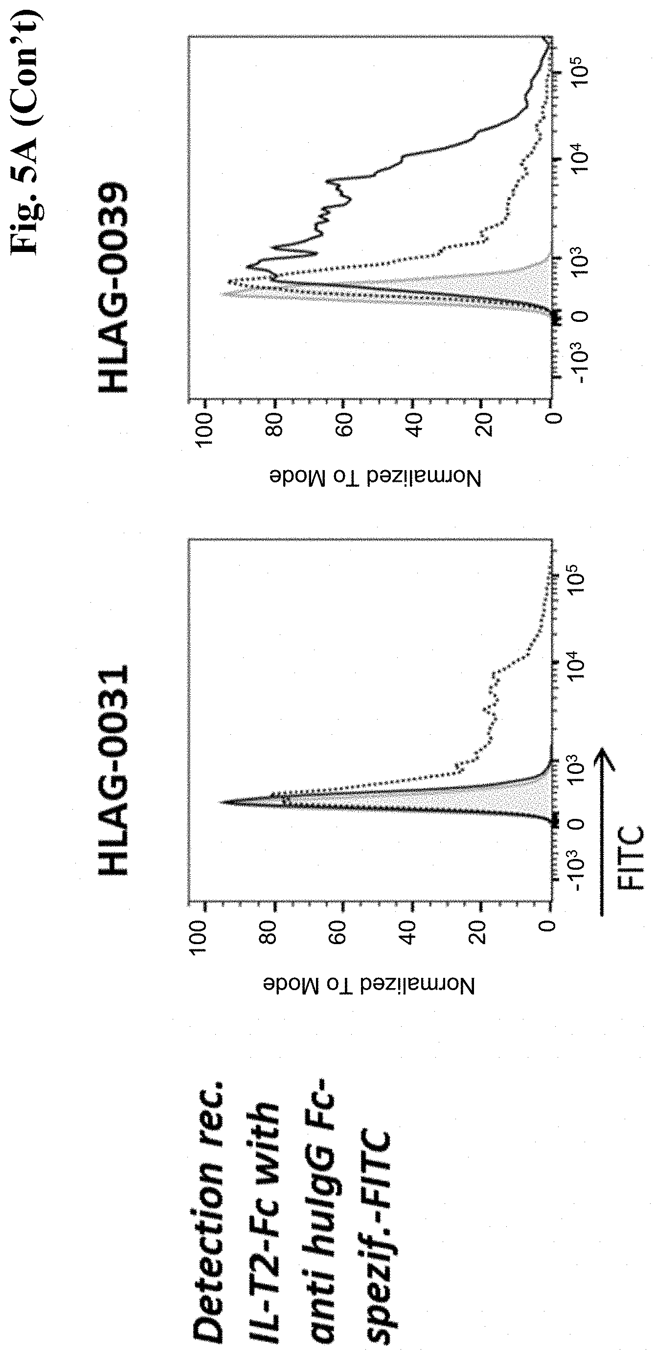
D00014
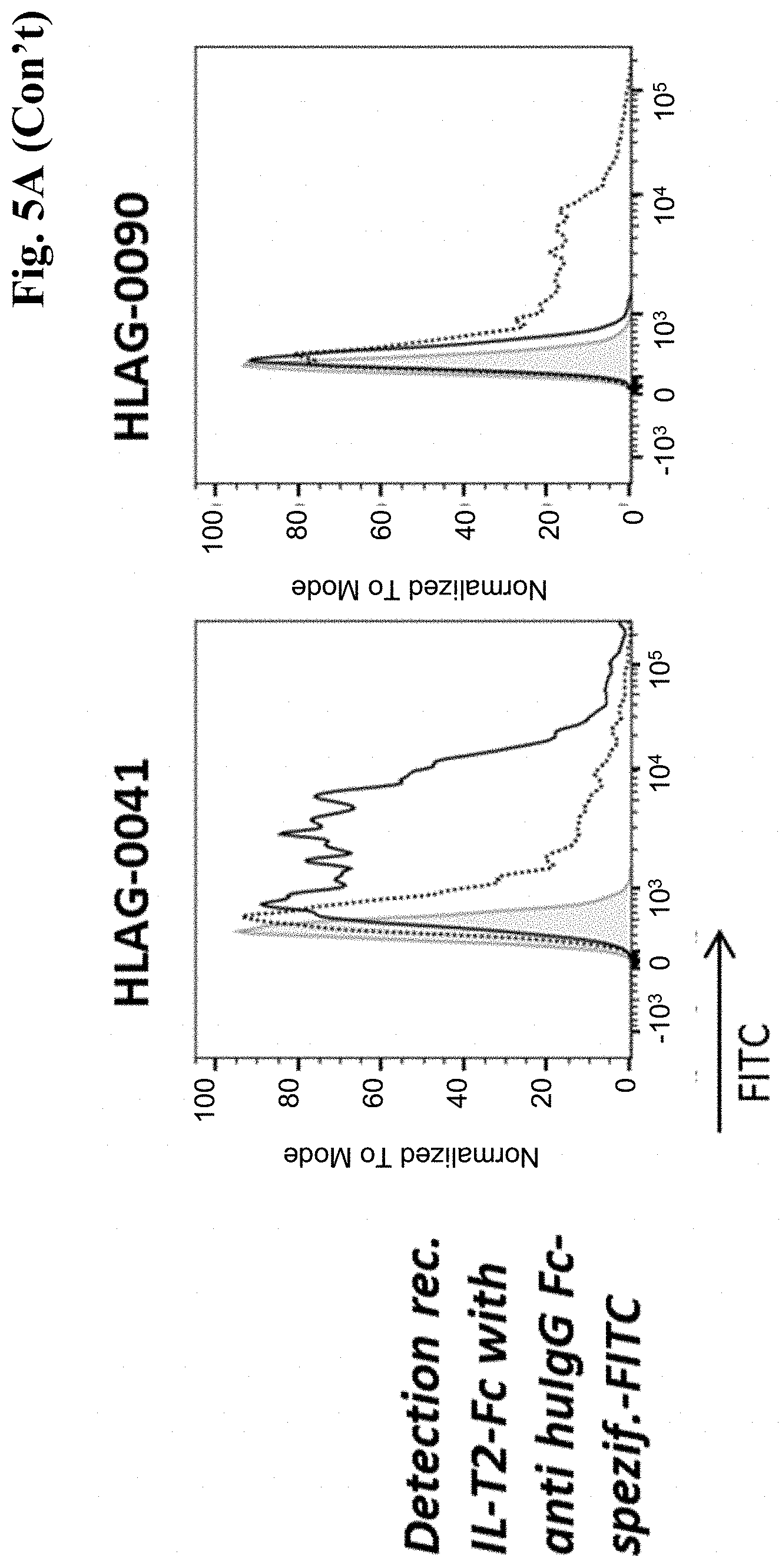
D00015
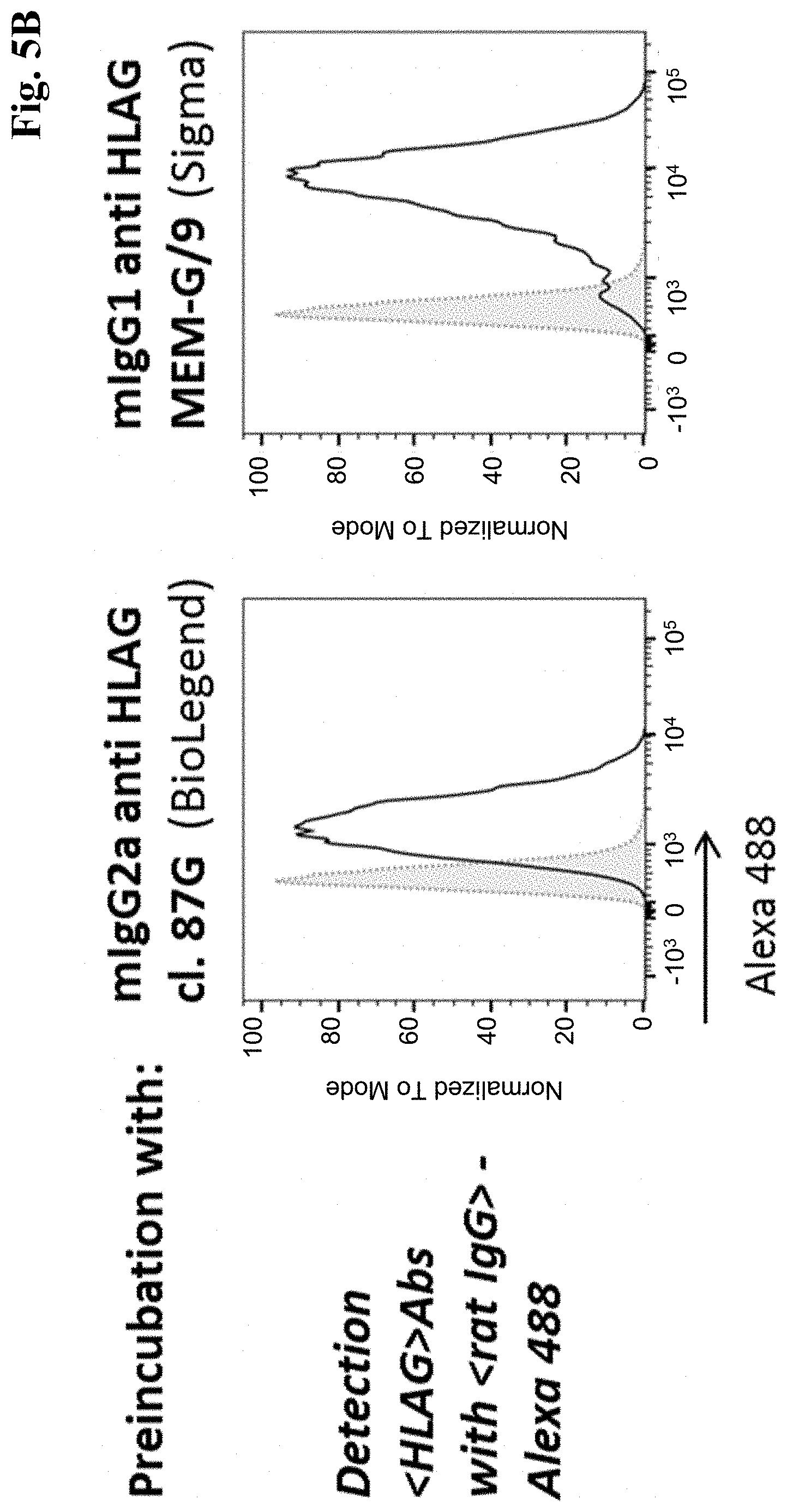
D00016
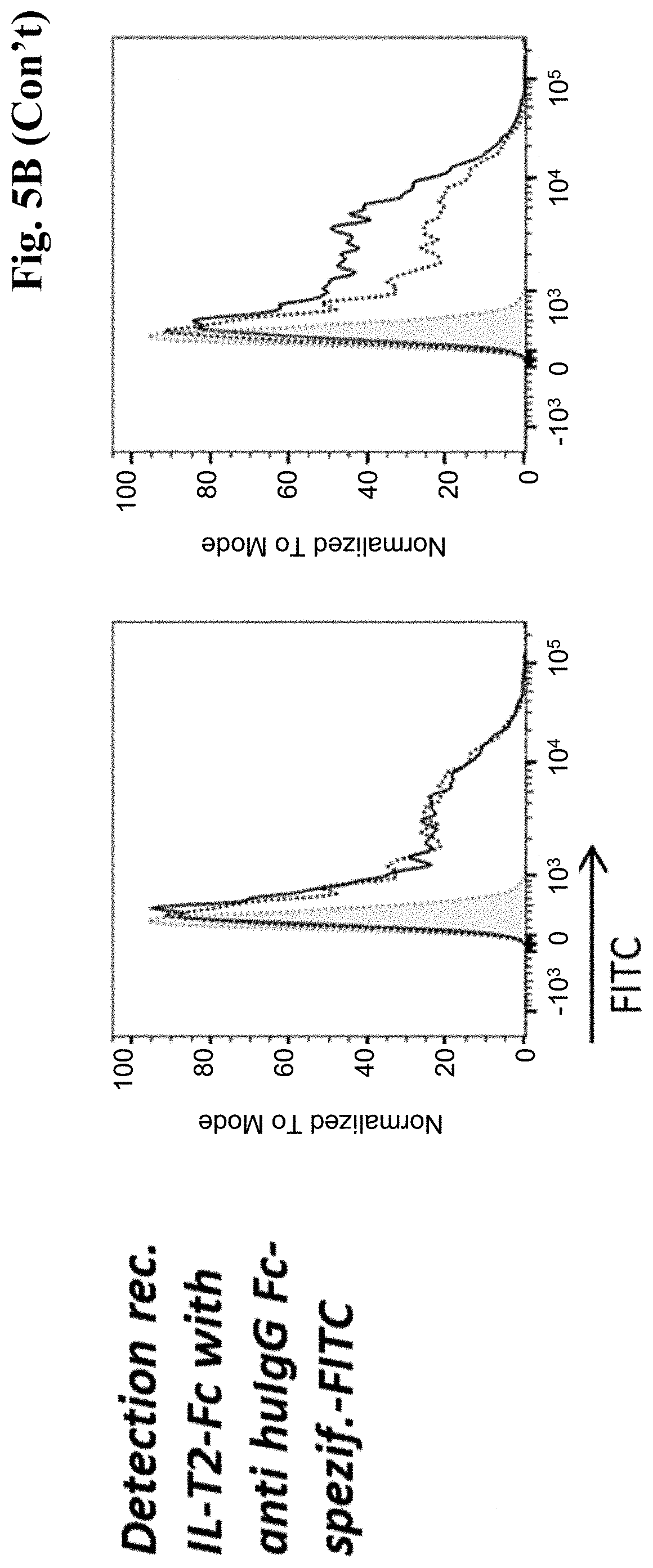
D00017
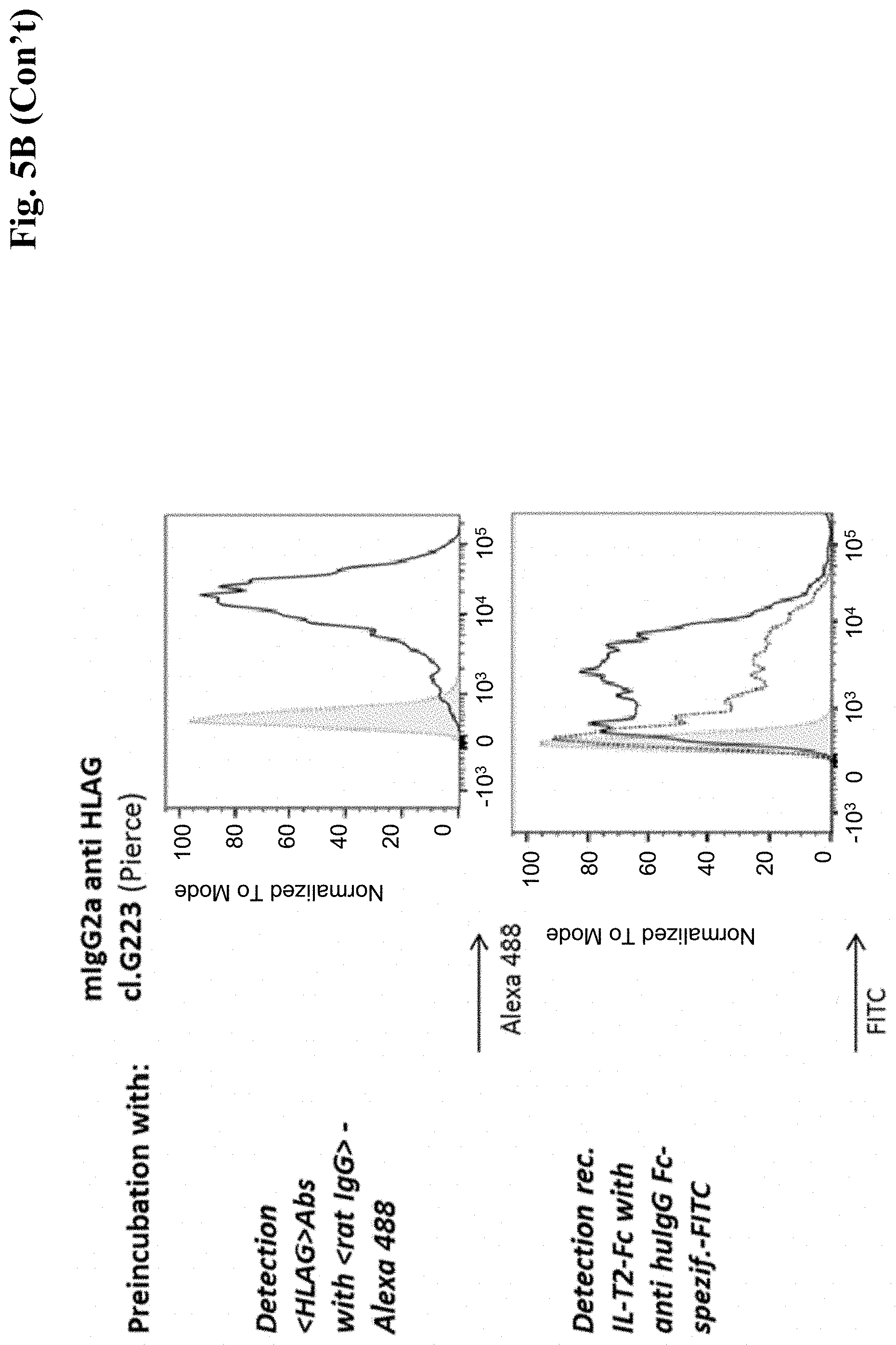
D00018
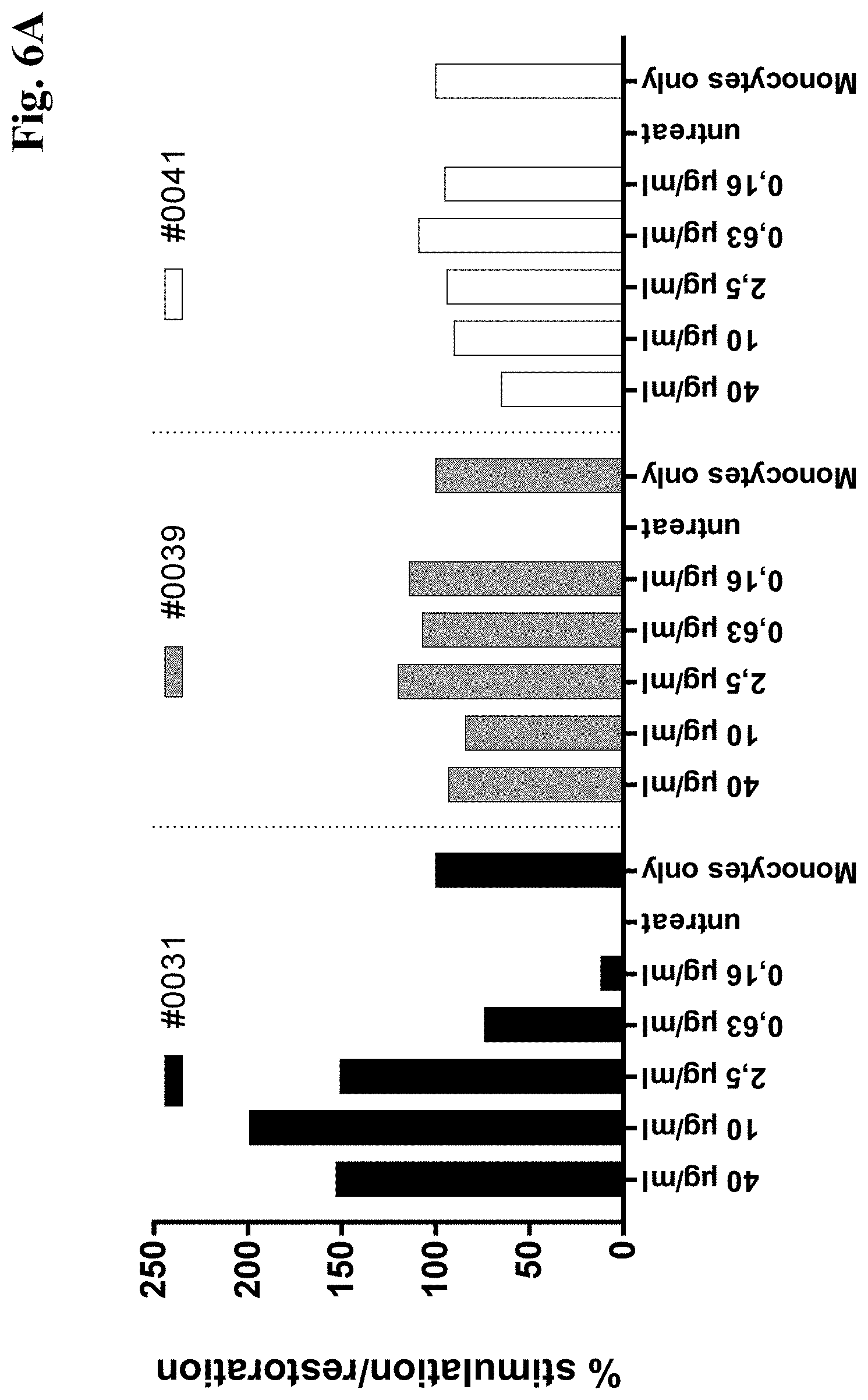
D00019
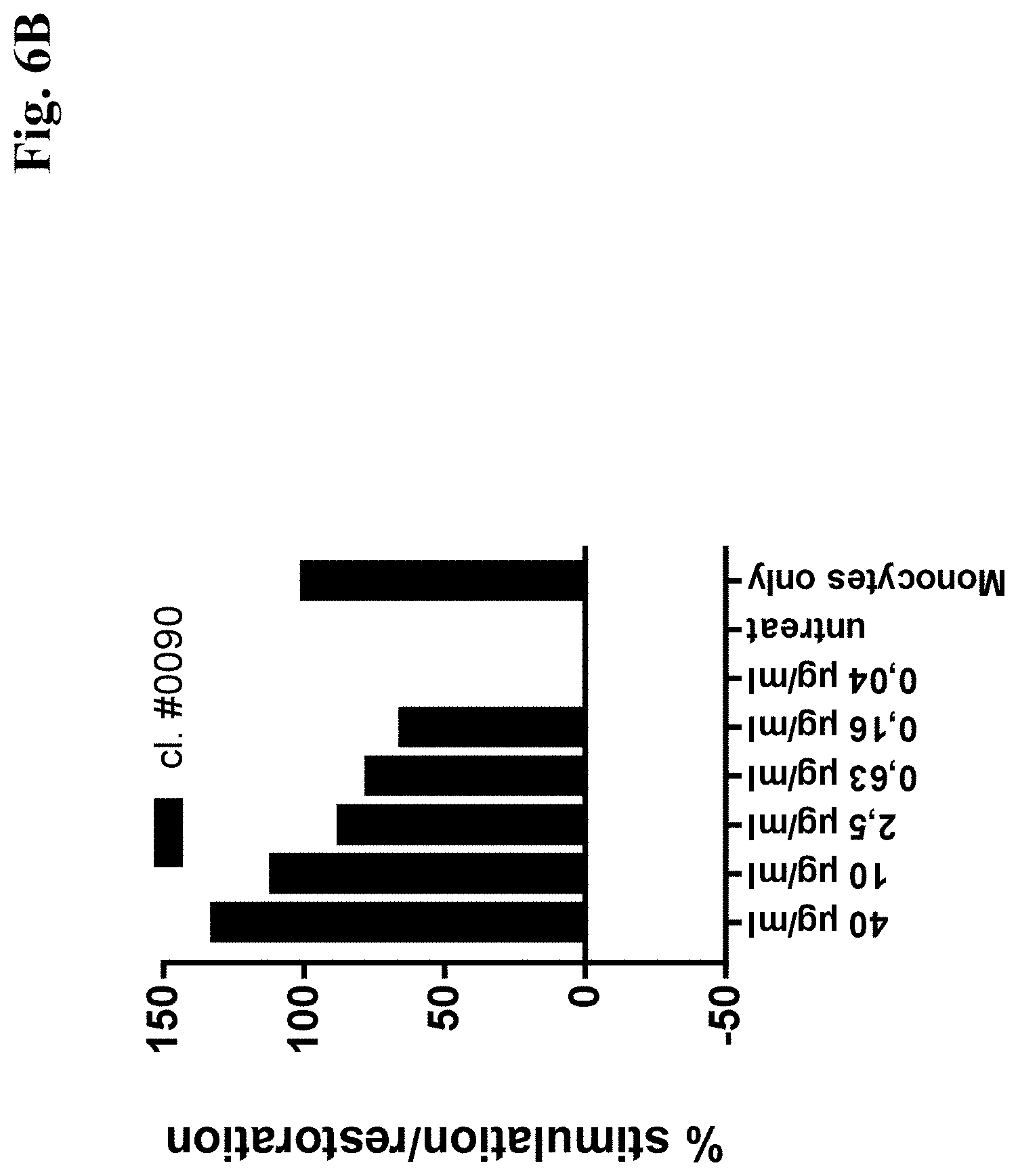
D00020

D00021
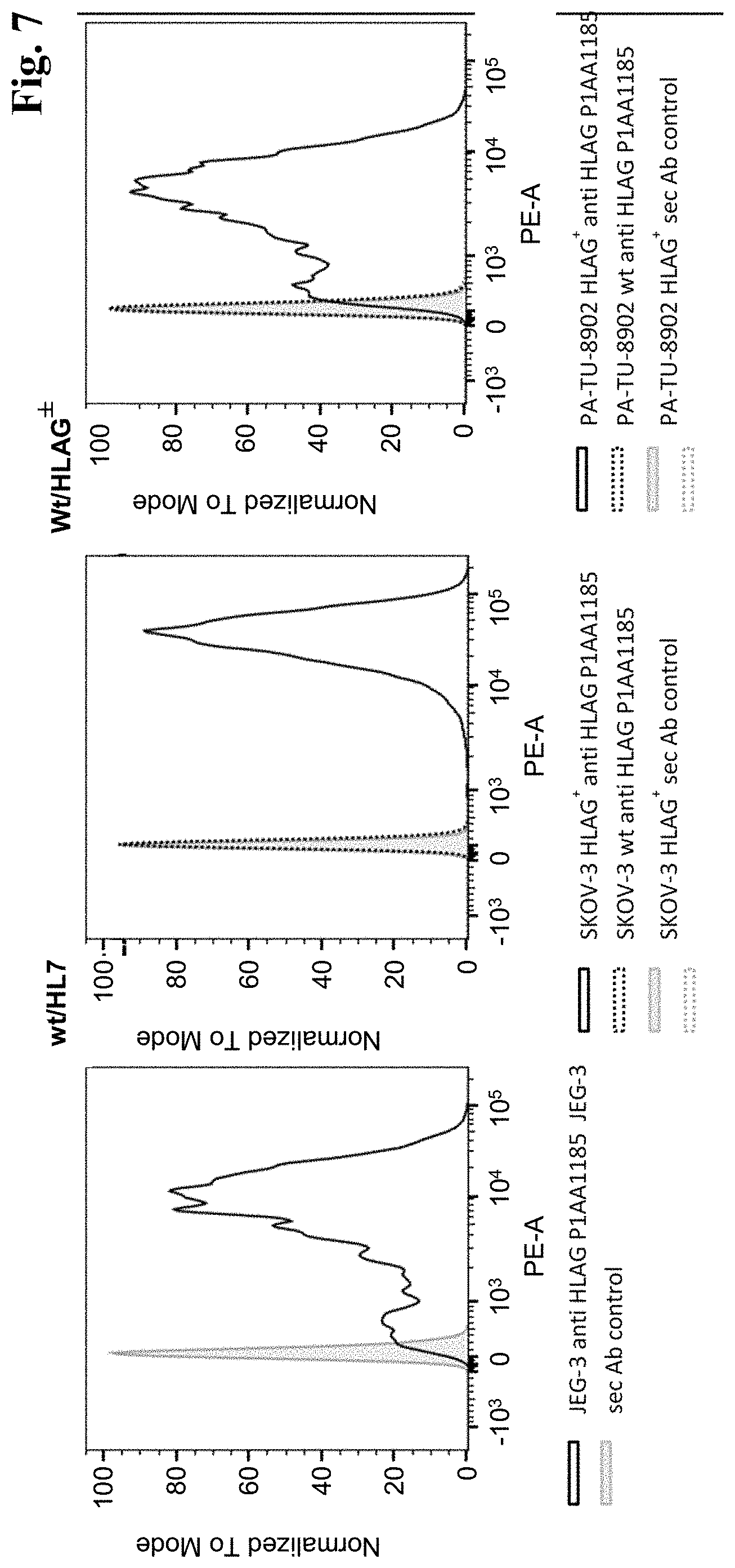
D00022
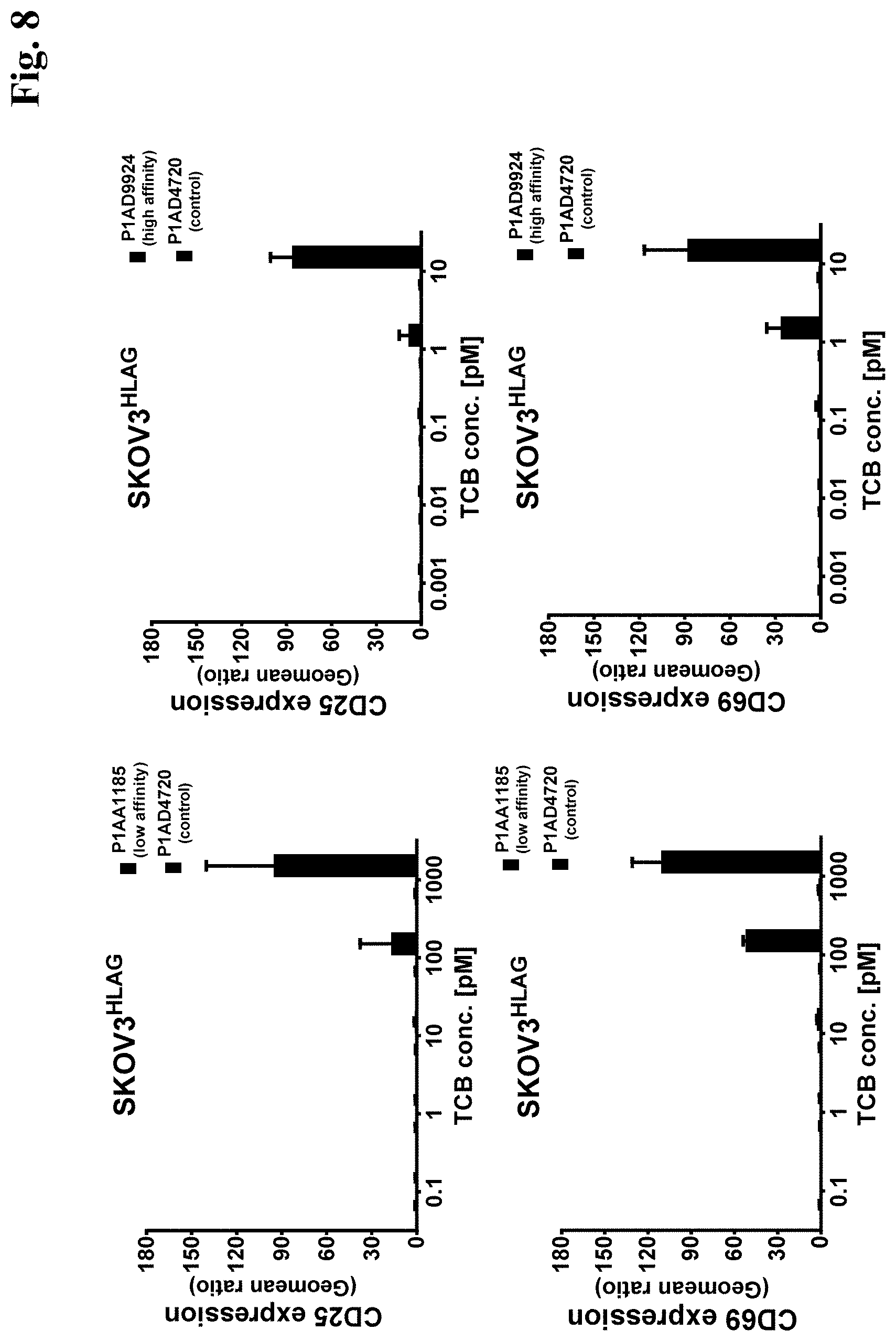
D00023

D00024
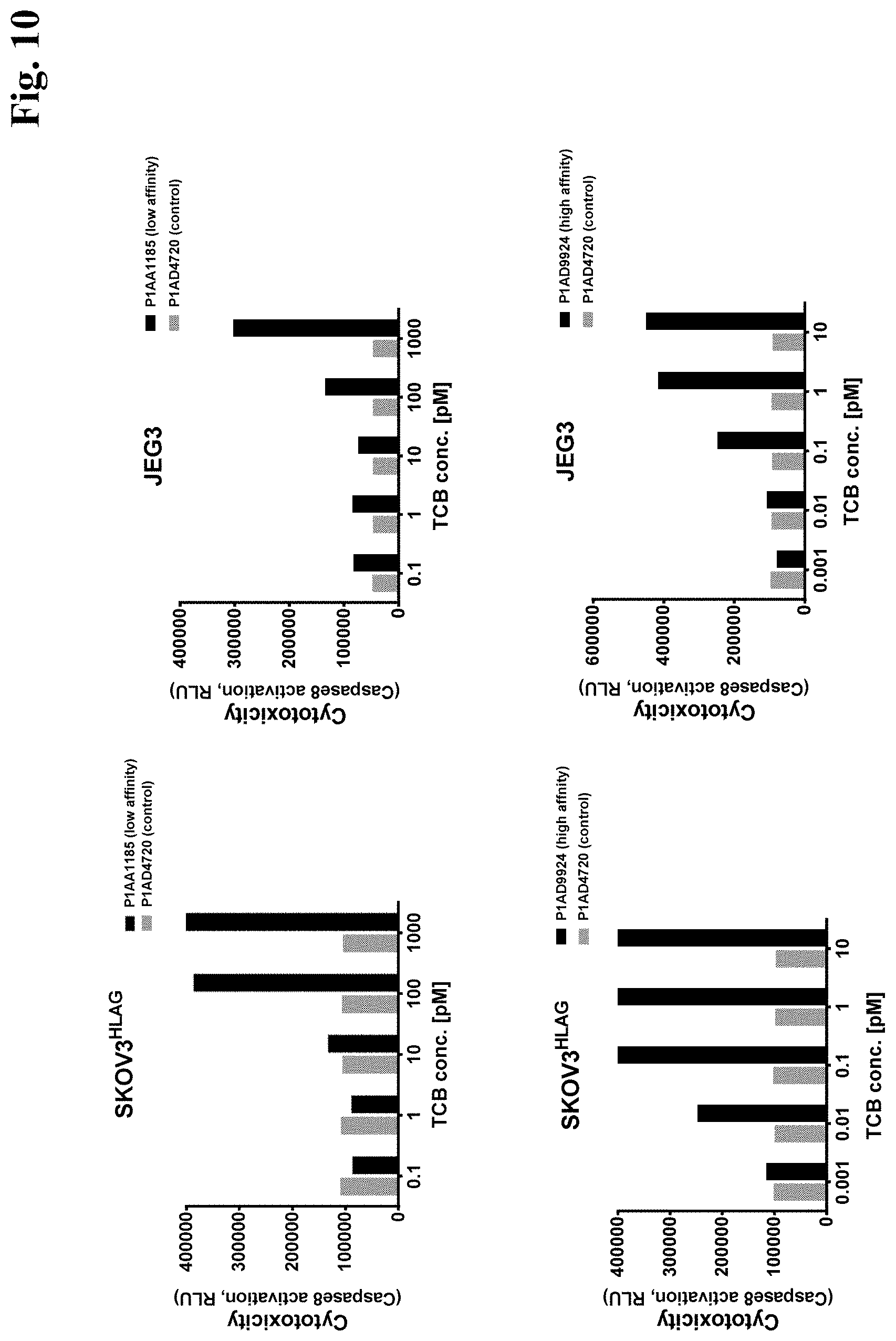
D00025
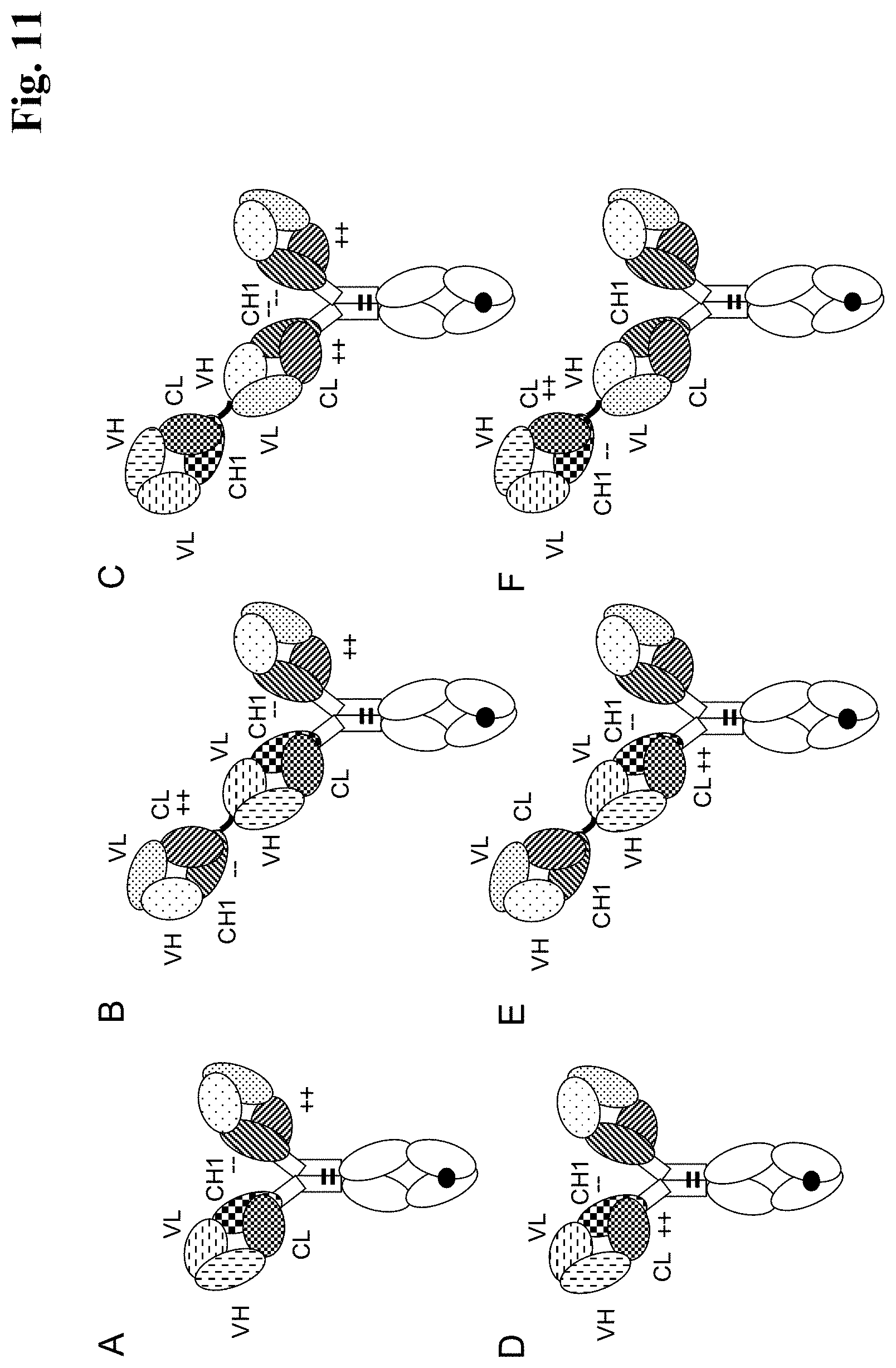
D00026
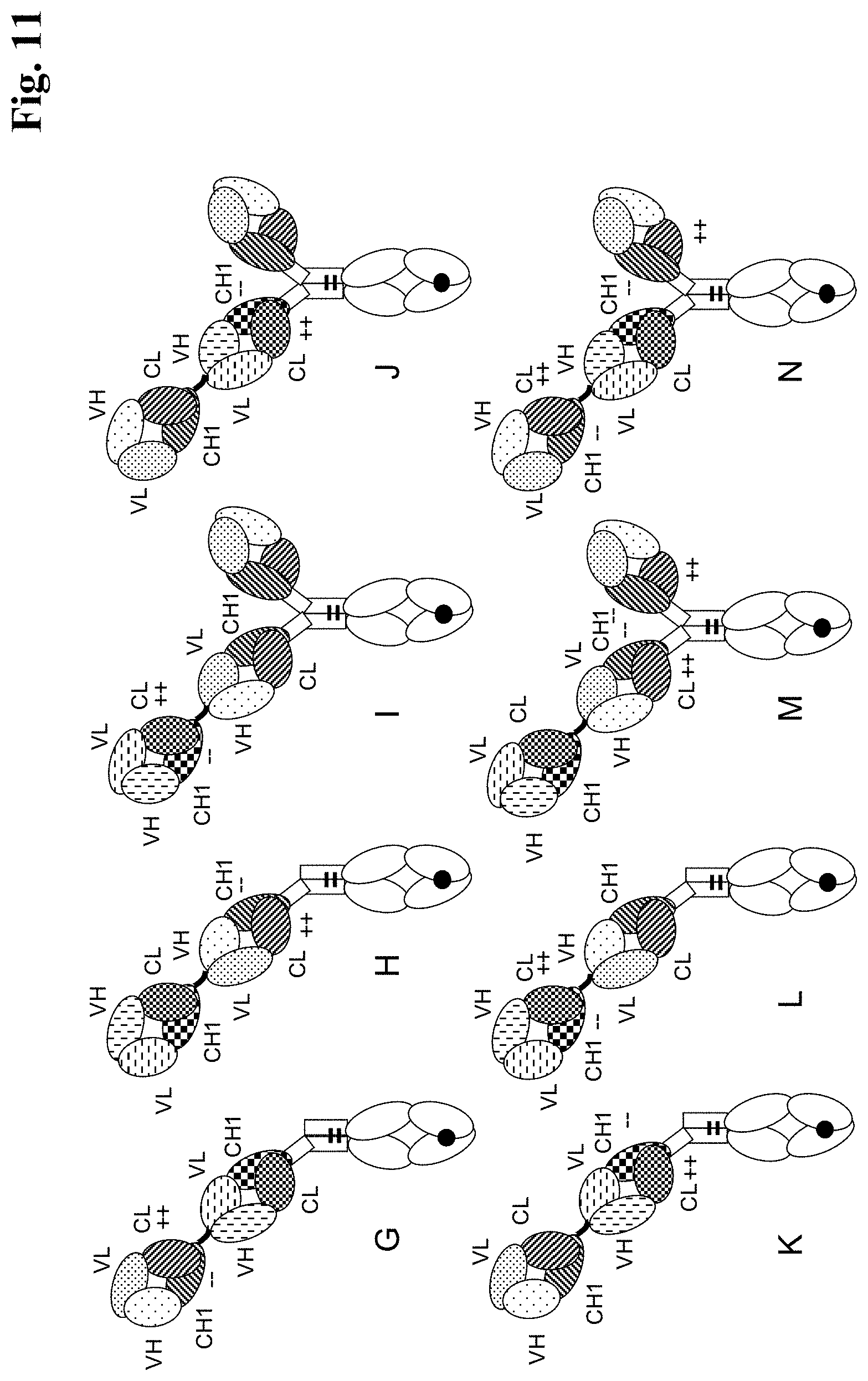
D00027
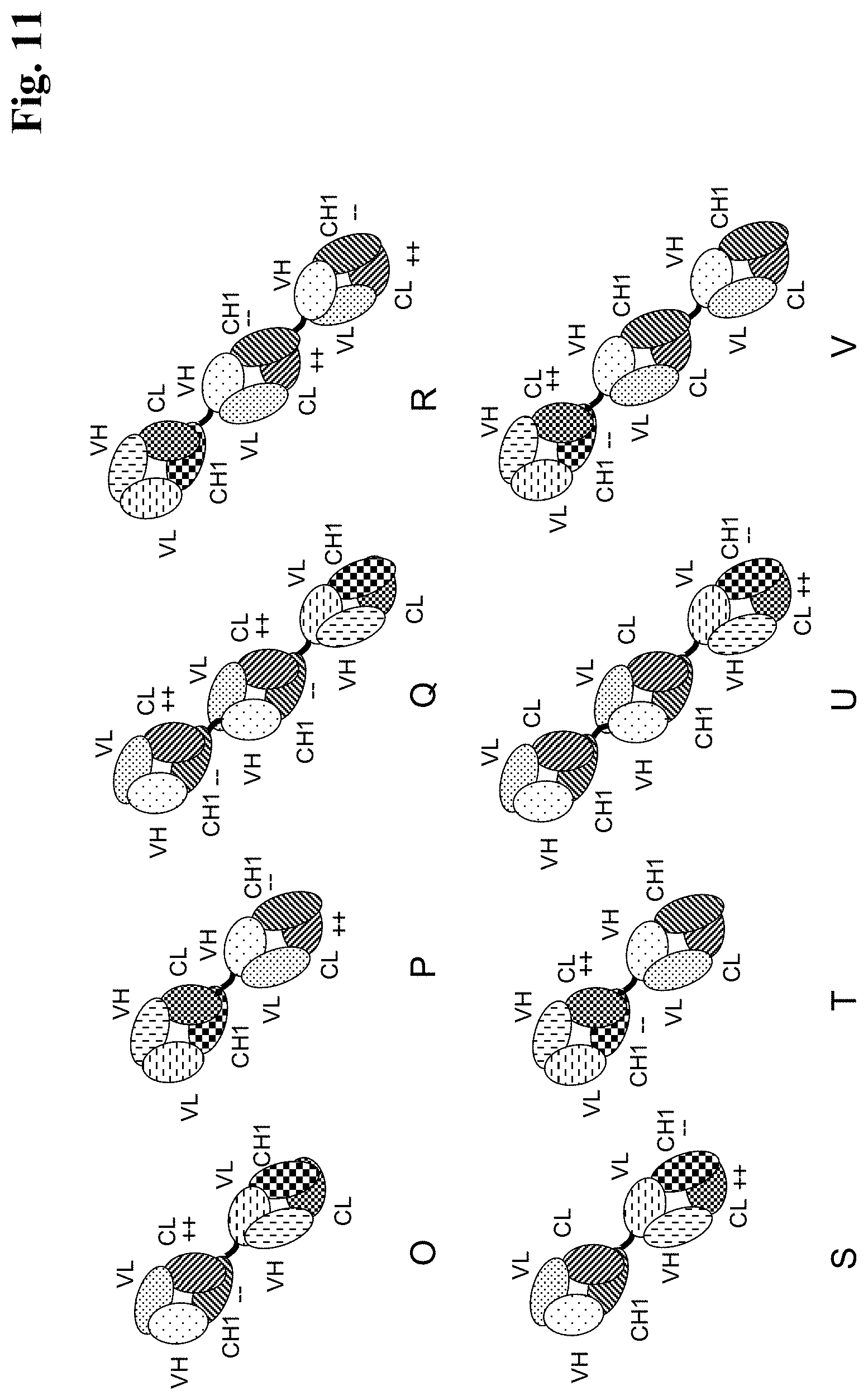
D00028
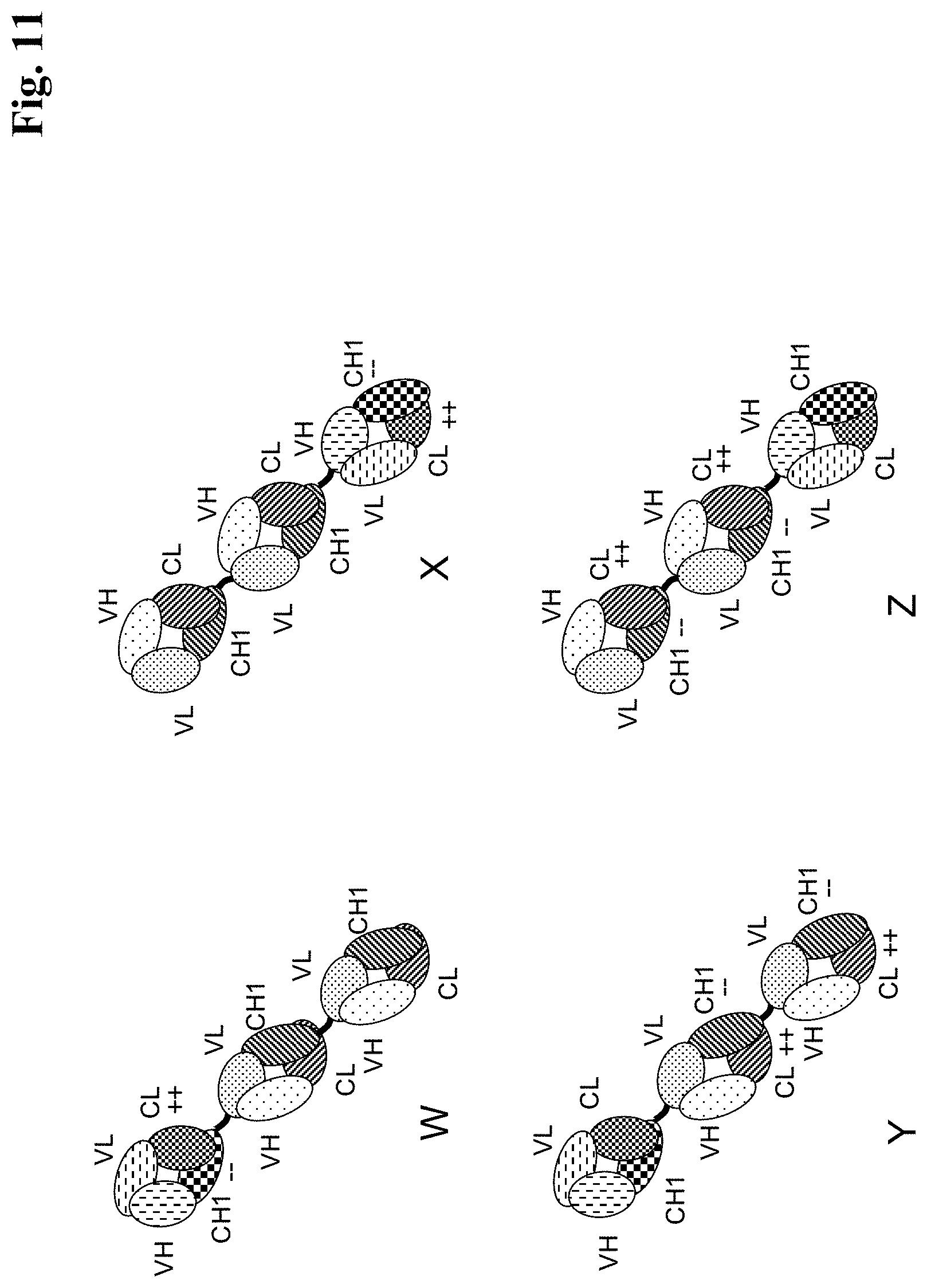
D00029
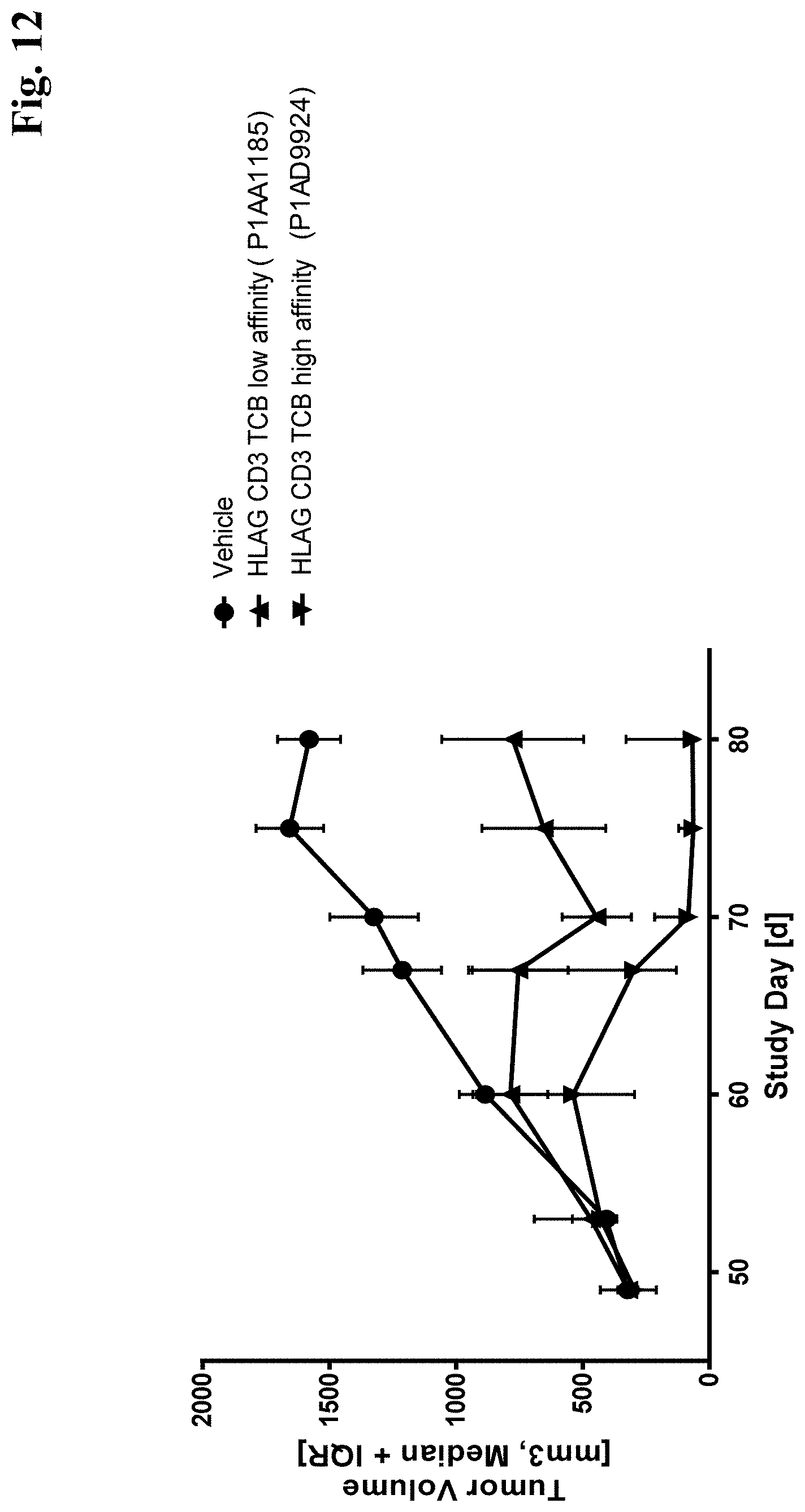
S00001
XML
uspto.report is an independent third-party trademark research tool that is not affiliated, endorsed, or sponsored by the United States Patent and Trademark Office (USPTO) or any other governmental organization. The information provided by uspto.report is based on publicly available data at the time of writing and is intended for informational purposes only.
While we strive to provide accurate and up-to-date information, we do not guarantee the accuracy, completeness, reliability, or suitability of the information displayed on this site. The use of this site is at your own risk. Any reliance you place on such information is therefore strictly at your own risk.
All official trademark data, including owner information, should be verified by visiting the official USPTO website at www.uspto.gov. This site is not intended to replace professional legal advice and should not be used as a substitute for consulting with a legal professional who is knowledgeable about trademark law.