Specificity Assay For Novel Target Antigen Binding Moieties
BRINKMANN; Ulrich ; et al.
U.S. patent application number 17/007479 was filed with the patent office on 2021-04-22 for specificity assay for novel target antigen binding moieties. This patent application is currently assigned to Hoffmann-La Roche Inc.. The applicant listed for this patent is Hoffmann-La Roche Inc.. Invention is credited to Ulrich BRINKMANN, Diana DAROWSKI, Steffen DICKOPF, Christian JOST, Christian KLEIN.
| Application Number | 20210116455 17/007479 |
| Document ID | / |
| Family ID | 1000005346619 |
| Filed Date | 2021-04-22 |


View All Diagrams
| United States Patent Application | 20210116455 |
| Kind Code | A1 |
| BRINKMANN; Ulrich ; et al. | April 22, 2021 |
SPECIFICITY ASSAY FOR NOVEL TARGET ANTIGEN BINDING MOIETIES
Abstract
The present invention generally relates to specificity assays using cell cultures, in particular to chimeric antigen receptor (CAR) expressing reporter T (CAR-T) cell assays to test novel target antigen binding moieties in different formats. Furthermore, the present invention relates to the use of reporter CAR-T cells, transfected/transduced with an engineered CAR capable of specific binding to a recognition domain comprising a tag.
| Inventors: | BRINKMANN; Ulrich; (Weilheim, DE) ; DAROWSKI; Diana; (Gerbrunn, DE) ; DICKOPF; Steffen; (Penzberg, DE) ; JOST; Christian; (Zurich, CH) ; KLEIN; Christian; (Bonstetten, CH) | ||||||||||
| Applicant: |
|
||||||||||
|---|---|---|---|---|---|---|---|---|---|---|---|
| Assignee: | Hoffmann-La Roche Inc. Little Falls NJ |
||||||||||
| Family ID: | 1000005346619 | ||||||||||
| Appl. No.: | 17/007479 | ||||||||||
| Filed: | August 31, 2020 |
Related U.S. Patent Documents
| Application Number | Filing Date | Patent Number | ||
|---|---|---|---|---|
| PCT/EP2019/054786 | Feb 27, 2019 | |||
| 17007479 | ||||
| Current U.S. Class: | 1/1 |
| Current CPC Class: | C07K 14/70521 20130101; G01N 2333/70596 20130101; C07K 16/44 20130101; C07K 14/7051 20130101; C07K 2317/622 20130101; C07K 2319/03 20130101; C07K 16/2887 20130101; G01N 33/57492 20130101 |
| International Class: | G01N 33/574 20060101 G01N033/574; C07K 14/725 20060101 C07K014/725; C07K 16/44 20060101 C07K016/44; C07K 14/705 20060101 C07K014/705; C07K 16/28 20060101 C07K016/28 |
Foreign Application Data
| Date | Code | Application Number |
|---|---|---|
| Mar 1, 2018 | EP | 18159401.1 |
Claims
1. A method for assessing the specificity of a target antigen binding moiety capable of specific binding to a target antigen, the method comprising the steps of: a) providing an antigen binding molecule comprising an antigen binding domain and a recognition domain, wherein the antigen binding domain comprises the target antigen binding moiety, and wherein the recognition domain comprises a tag; b) contacting the antigen binding molecule with a target cell comprising the target antigen on the surface, particularly wherein the target cell is a cancer cell; c) contacting the antigen binding molecule with a chimeric antigen receptor (CAR) expressing reporter T (CAR-T) cell wherein the reporter CAR-T cell comprises: i. a CAR capable of specific binding to the recognition domain comprising the tag, wherein the CAR is operationally coupled to a response element; ii. a reporter gene under the control of the response element; and d) determining T cell activation by measuring the expression of the reporter gene to establish the specificity of the target antigen binding moiety.
2. The method of claim 1, wherein the antigen binding molecule is an IgG class antibody, particularly an IgG1 or IgG4 isotype antibody, or a fragment thereof.
3. The method of claim 1, wherein the antigen binding domain is a Fab fragment and the recognition domain is an Fc domain.
4. The method of claim 1, wherein the antigen binding domain and the recognition domain are the same domain, in particular a Fab fragment.
5. The method of claim 1, wherein the tag is a hapten molecule.
6. The method of claim 1, wherein the hapten molecule is Digoxigenin (DIG).
7. The method of claim 1, wherein the tag is a polypeptide tag.
8. The method of claim 7, wherein the polypeptide tag is selected from the group consisting of myc-tag, HA-tag, AviTag, FLAG-tag, His-tag, GCN4-tag, and NE-tag.
9. The method of claim 1, wherein the target antigen is a cell surface antigen or receptor.
10. The method of claim 1, wherein the target antigen is a peptide bound to a molecule of the human major histocompatibility complex (MHC), wherein the target antigen binding moiety is a T cell receptor like (TCRL) antigen binding moiety.
11. A method for generating a TCB antibody, wherein the TCB antibody comprises a first antigen binding moiety specific for a target antigen and a second antigen binding moiety capable of specific binding to a T cell activating receptor, wherein the first antigen binding moiety is selected according to the method of any one of claims 1 to 10.
12. The method of claim 11, wherein the T cell activating receptor is CD3.
13. A chimeric antigen receptor (CAR) comprising an anchoring transmembrane domain and an extracellular domain comprising an antigen binding moiety, wherein the antigen binding moiety is capable of specific binding to a recognition domain comprising a tag but not capable of specific binding to the recognition domain not comprising the tag.
14. The CAR of claim 13, wherein the tag is a hapten molecule.
15. The CAR of claim 14, wherein the hapten molecule is Digoxigenin (DIG).
Description
CROSS REFERENCE TO RELATED APPLICATIONS
[0001] This application is a continuation of International Application No. PCT/EP2019/054786 having an International filing date of Feb. 27, 2019, which claims benefit of priority to European Patent Application No. 18159401.1, filed Mar. 1, 2018, all of which are incorporated by reference in their entirety.
SEQUENCE LISTING
[0002] This application contains a Sequence Listing that has been submitted via EFS-Web and is hereby expressly incorporated by reference in its entirety. Said ASCII copy, created on Aug. 26, 2020, is named P34622_US_Sequence_Listing.txt and is 123,197 bytes in size.
FIELD OF THE INVENTION
[0003] The present invention generally relates to specificity assays using cell cultures, in particular to chimeric antigen receptor (CAR) expressing reporter T (CAR-T) cell assays to test novel target antigen binding moieties in different formats. Furthermore, the present invention relates to the use of reporter CAR-T cells, transfected/transduced with an engineered CAR capable of specific binding to a recognition domain comprising a tag.
BACKGROUND
[0004] Over the last 15 years, antibody based therapies have evolved and represent now a valuable combination or alternative to chemotherapeutic approaches in the treatment of hematological malignancies and solid tumors. Unlike chemotherapy, antibody therapies target specific antigens on cancer cells thus allowing a more site-directed treatment thereby reducing the side effects on healthy tissue. In the process of developing an antibody-based therapeutic reagent, various assays are required to identify the best candidates to bring into clinical trials and eventually to the market. In a first early preclinical phase, the antibodies have to be generated and analyzed for their target-specificity, as well as their affinity to the target.
[0005] Binding properties can be analyzed using various protein-protein interaction assays, such as FRET-based methods, Surface Plasmon Resonance (SPR) or fluorescence-activated cell sorting (FACS). However, available assay formats might not always reproduce the in vivo situation comprehensively and integrative. For example targeting of cancer cells with therapeutic antibodies binding to cell surface receptors can have impacts on multiple levels, e.g., intracellular signaling via the binding and cross-linking of surface molecules as well as marking the tumor cells to engage immune cells. Furthermore, the recognition cascade from antigen binding to establishing of an effector function, e.g., T cell cytotoxicity, requires a well-orchestrated sequence of cell surface interactions, wherein binding affinity of an antigen binding moiety is one among several factors. Plain protein-protein affinity interaction assays may therefore not provide the complete picture, although these assays are a very valuable tool for early candidate development.
[0006] Still, there remains a need to develop binding assays which do provide meaningful predictions for the in vivo interactions in a more comprehensive setup minimizing non-specific effects on target-antibody binding as far as possible.
[0007] The inventors of the present invention developed a novel assay which is applicable to a wide variety of different cancer cell types to assess binding of antibodies to their target. The innovative assay includes modified T-cells as reporter cells combining straight-forward readout with a comprehensive and inclusive result.
[0008] Furthermore, the present invention provides assays which combine the assessment of binding and functionality of antibodies and antibody-like constructs (e.g., ligands). The novel assay is useful for example for screening or characterization purposes of therapeutic antibody drug candidates, i.e., in high-throughput formats.
[0009] This new assay represents a valuable tool for early and late stage screening and characterization of antibody binding to the native target and assessing functionality which will allow identifying the best binders in the development of the drug candidate.
SUMMARY OF THE INVENTION
[0010] The present invention generally relates to a method for assessing and selecting novel antigen binding moieties, particularly in the drug development process, and combines the determination of binding to a target antigen, e.g., on a tumor cell, with the activation of T cells in response to the antibody-target binding. Herein provided is a method for assessing the specificity of a target antigen binding moiety capable of specific binding to a target antigen, the method comprising the steps of: [0011] a) providing an antigen binding molecule comprising an antigen binding domain and a recognition domain, wherein the antigen binding domain comprises the target antigen binding moiety, and wherein the recognition domain comprises a tag; [0012] b) contacting the antigen binding molecule with a target cell comprising the target antigen on the surface, particularly wherein the target cell is a cancer cell; [0013] c) contacting the antigen binding molecule with a chimeric antigen receptor (CAR) expressing reporter T (CAR-T) cell wherein the reporter CAR-T cell comprises: [0014] i. a CAR capable of specific binding to the recognition domain comprising the tag, wherein the CAR is operationally coupled to a response element; [0015] ii. a reporter gene under the control of the response element; and [0016] d) determining T cell activation by measuring the expression of the reporter gene to establish the specificity of the target antigen binding moiety.
[0017] In one embodiment, the antigen binding molecule is an IgG class antibody, particularly an IgG1 or IgG4 isotype antibody, or a fragment thereof.
[0018] In one embodiment, the antigen binding domain is a Fab fragment and the recognition domain is an Fc domain.
[0019] In one embodiment, the antigen binding domain and the recognition domain are the same domain, in particular a Fab fragment.
[0020] In one embodiment, the tag is a hapten molecule.
[0021] In one embodiment, the hapten molecule is Digoxigenin (DIG).
[0022] In one embodiment, the tag is a polypeptide tag.
[0023] In one embodiment, the polypeptide tag is selected from the group consisting of myc-tag, HA-tag, AviTag, FLAG-tag, His-tag, GCN4-tag and NE-tag.
[0024] In one embodiment, the target antigen binding moiety is a Fab fragment, in particular a Fab fragment deriving from a phage display library screening.
[0025] In one embodiment, binding of the target antigen binding moiety to the target antigen and binding of the reporter CAR-T cell to the antigen binding molecule comprising the target antigen binding moiety leads to expression of the reporter gene.
[0026] In one embodiment, the target antigen is a cell surface antigen or receptor.
[0027] In one embodiment, the target antigen is a peptide bound to a molecule of the human major histocompatibility complex (MHC).
[0028] In one embodiment, the target antigen binding moiety is a T cell receptor like (TCRL) antigen binding moiety.
[0029] In one embodiment, provided is a chimeric antigen receptor (CAR) comprising an anchoring transmembrane domain and an extracellular domain comprising an antigen binding moiety, wherein the antigen binding moiety is capable of specific binding to a recognition domain comprising a tag but not capable of specific binding to the recognition domain not comprising the tag.
[0030] In one embodiment, the antigen binding moiety is a scFv, a Fab, a crossFab or a scFab, in particular a Fab or a crossFab.
[0031] In one embodiment, the tag is a hapten.
[0032] In one embodiment, the hapten molecule is Digoxigenin (DIG).
[0033] In one embodiment, the tag is a polypeptide tag.
[0034] In one embodiment, the polypeptide tag is selected from the group consisting of myc-tag, HA-tag, AviTag, FLAG-tag, His-tag, GCN4-tag and NE-tag.
SHORT DESCRIPTION OF THE FIGURES
[0035] FIG. 1 depicts the architecture of exemplary antigen binding receptors (CARs) used according to the invention. FIG. 1A shows the architecture of the scFv format. The antigen binding moiety capable of specific binding to the recognition domain consists of a variable heavy (VH) and a variable light (VL) chain. Attached to the VL chain, a Gly4Ser linker connects the antigen recognition domain with the CD28 transmembrane domain (TM) which is fused to the intracellular costimulatory signaling domain (CSD) of CD28 which in turn is fused to the stimulatory signaling domain (SSD) of CD3z. FIGS. 1B and 1C show the architecture of the Fab (FIG. 1B) and crossFab (FIG. 1C) formats. The antigen binding moiety consists of an Ig heavy chain and an Ig light chain. Attached to the heavy chain, a Gly4Ser linker connects the antigen recognition domain with the CD28 transmembrane domain which is fused to the intracellular co-stimulatory signaling domain of CD28 which in turn is fused to the stimulatory signaling domain of CD3z.
[0036] FIG. 2 depicts a schematic representation illustrating the modular composition of exemplary expression constructs CARs used according to the invention. FIG. 2A depicts the scFv format. FIG. 2B depicts the Fab format. FIG. 2C depicts a crossFab format.
[0037] FIG. 3 depicts the structural formula of the Digoxigenin (DIG) molecule.
[0038] FIG. 4 depicts an exemplary digoxigeninylated IgG1 molecule which can be specifically recognized by an anti-Digoxigenin CAR.
[0039] FIG. 5 depicts alternative digoxigenylated antigen binding molecules which are recognized by an anti-Digoxigenin CAR. In this embodiment, the target antigen binding domain and the recognition domain are the same domain, i.e., the Digoxigenin hapten tag is coupled to the antigen binding domain wherein the antigen binding domain exerts also the function of the recognition domain. FIG. 5A depicts an digoxigeninylated Fab molecule which can be recognized by an anti-Digoxigenin CAR. FIG. 5B depicts an digoxigenylated scFv molecule which can be recognized by an anti-Digoxigenin CAR.
[0040] FIG. 6 depicts a Western Blot confirming successful digoxigenylation of the anti-CD20 targeting antibody GA101. Digoxigeninylation was detected by anti-Digoxigenin-AP Fab fragments by Western Blot analysis.
[0041] FIG. 7 depicts surface detection of anti-Digoxigenin-ds-scFv on Jurkat NFAT reporter cells.
[0042] FIG. 8 depicts a schematic representation of a Jurkat NFAT reporter CAR-T cell assay. The target antigen bound IgG which is digoxigeninylated at the Fc (recognition domain) can be recognized by the anti-Digoxigenin CAR expressing Jurkat NFAT reporter T cell. This recognition leads to the activation of the cell which can be detected by measuring luciferase luminescence (CPS/RLU).
[0043] FIG. 9 depicts the Jurkat NFAT reporter CAR-T cell assay using CD20 expressing SUDHDL4 tumor cells as target cells and an anti-CD20 IgG antibody (GA101) digoxigeninylated with a ten times molar excess of Digoxigenin-3-O-methylcarbonyl-e-aminocaproic acid-N-hydroxysuccinimide ester. The antibody recognizes on the one hand the tumor associated antigen and on the other hand is recognized by Jurkat NFAT reporter CAR-T cells. A sorted pool of anti-Digoxigenin-ds-scFv-CD28ATDCD28CSD-CD3zSSD expressing Jurkat NFAT reporter CAR-T cells was used as effector cells.
[0044] FIG. 10 depicts activation of anti-Digoxigenin-ds-scFv-CD28ATDCD28CSD-CD3zSSD expressing Jurkat NFAT reporter CAR-T cells. Activation is dependent on an anti-CD20 IgG antibody (GA101), coupled with different amounts of digoxigeninylated molecules.
[0045] FIG. 11 depicts the Jurkat NFAT reporter CAR-T cell assay using CD20 expressing SUDHDL4 tumor cells as target cells. An anti-CD20 IgG antibody (GA101) digoxygeninylated at the Fc with approximately one Digoxigenin (equimolar Dig-NHS:antibody ratio) molecule on average was used. The antibody recognizes on the one hand the tumor associated antigen and on the other hand is recognized by Jurkat NFAT reporter CAR-T cells. A sorted pool of anti-Digoxigenin-ds-scFv-CD28ATDCD28CSD-CD3zSSD expressing Jurkat NFAT reporter CAR-T cells was used as effector cells.
[0046] FIG. 12 depicts a schematic representation of an alternative Jurkat NFAT reporter CAR-T cell assay using a bridging Biotin-Digoxigenin adapter. The Biotin-Digoxigenin adapter bound to the Fc domain via a Biotin binding moiety forms the recognition domain in this setup. The Digoxigenin moiety can be recognized by the anti-Digoxigenin CAR expressing Jurkat NFAT reporter CAR-T cell.
[0047] FIG. 13 depicts the Jurkat NFAT reporter CAR-T cell assay using MCF7 cells as target cells. An anti-LeY/Biotin antibody and a bridging Biotin-Digoxigenin adapter was used. The antibody recognizes on one hand the tumor associated antigen (LeY) and on the other hand the Biotin of the adapter molecule. The adapter-bound Digoxigenin is recognized by the Jurkat NFAT reporter CAR-T cells. A sorted pool of anti-Digoxigenin-ds-scFv-CD28ATDCD28CSD-CD3zSSD expressing Jurkat NFAT reporter CAR-T cells was used as effector cells. As negative control a non-targeting Biotin-coupled anti-CD33 antibody was used.
DETAILED DESCRIPTION
Definitions
[0048] "Affinity" refers to the strength of the sum total of non-covalent interactions between a single binding site of a molecule (e.g., an antibody or a CAR) and its binding partner (e.g., a ligand). Unless indicated otherwise, as used herein, "binding affinity" refers to intrinsic binding affinity which reflects a 1:1 interaction between members of a binding pair (e.g., an antigen binding moiety and an antigen and/or a receptor and its ligand). The affinity of a molecule X for its partner Y can generally be represented by the dissociation constant (K.sub.D), which is the ratio of dissociation and association rate constants (k.sub.off and k.sub.on, respectively). Thus, equivalent affinities may comprise different rate constants, as long as the ratio of the rate constants remains the same. Affinity can be measured by well-established methods known in the art, including those described herein. A preferred method for measuring affinity is Surface Plasmon Resonance (SPR) and a preferred temperature for the measurement is 25.degree. C.
[0049] The term "amino acid" ("aa") refers to naturally occurring and synthetic amino acids, as well as amino acid analogs and amino acid mimetics that function in a manner similar to the naturally occurring amino acids. Naturally occurring amino acids are those encoded by the genetic code, as well as those amino acids that are later modified, e.g., hydroxyproline, .gamma.-carboxyglutamate, and O-phosphoserine. Amino acid analogs refer to compounds that have the same basic chemical structure as a naturally occurring amino acid, i.e., an a carbon that is bound to a hydrogen, a carboxyl group, an amino group, and an R group, e.g., homoserine, norleucine, methionine sulfoxide, methionine methyl sulfonium. Such analogs have modified R groups (e.g., norleucine) or modified peptide backbones, but retain the same basic chemical structure as a naturally occurring amino acid. Amino acid mimetics refers to chemical compounds that have a structure that is different from the general chemical structure of an amino acid, but that function in a manner similar to a naturally occurring amino acid. Amino acids may be referred to herein by either their commonly known three letter symbols or by the one-letter symbols recommended by the IUPAC-IUB Biochemical Nomenclature Commission.
[0050] The term "amino acid mutation" as used herein is meant to encompass amino acid substitutions, deletions, insertions, and modifications. Any combination of substitution, deletion, insertion, and modification can be made to arrive at the final construct, provided that the final construct possesses the desired characteristics. Amino acid sequence deletions and insertions include amino- and/or carboxy-terminal deletions and insertions of amino acids. Particular amino acid mutations are amino acid substitutions. Amino acid substitutions include replacement by non-naturally occurring amino acids or by naturally occurring amino acid derivatives of the twenty standard amino acids (e.g., 4-hydroxyproline, 3-methylhistidine, ornithine, homoserine, 5-hydroxylysine). Amino acid mutations can be generated using genetic or chemical methods well known in the art. Genetic methods may include site-directed mutagenesis, PCR, gene synthesis and the like. It is contemplated that methods of altering the side chain group of an amino acid by methods other than genetic engineering, such as chemical modification, may also be useful.
[0051] The term "antibody" herein is used in the broadest sense and encompasses various antibody structures, including but not limited to monoclonal antibodies, polyclonal antibodies, and antibody fragments so long as they exhibit the desired antigen-binding activity. Accordingly, in the context of the present invention, the term antibody relates to full immunoglobulin molecules as well as to parts of such immunoglobulin molecules. Furthermore, the term relates, as discussed herein, to modified and/or altered antibody molecules, in particular to modified antibody molecules. The term also relates to recombinantly or synthetically generated/synthesized antibodies. In the context of the present invention the term antibody is used interchangeably with the term immunoglobulin.
[0052] An "antibody fragment" refers to a molecule other than an intact antibody that comprises a portion of an intact antibody that binds the antigen to which the intact antibody binds. Examples of antibody fragments include but are not limited to Fv, Fab, crossover Fab, Fab', Fab'-SH, F(ab').sub.2, diabodies, linear antibodies, single-domain antibodies, single-chain antibody molecules (e.g., scFv, scFab), and single-domain antibodies. For a review of certain antibody fragments, see Hudson et al., Nat Med 9, 129-134 (2003). For a review of scFv fragments, see e.g., Pluckthun, in The Pharmacology of Monoclonal Antibodies, vol. 113, Rosenburg and Moore eds., Springer-Verlag, New York, pp. 269-315 (1994); see also WO 93/16185; and U.S. Pat. Nos. 5,571,894 and 5,587,458. Diabodies are antibody fragments with two antigen-binding sites that may be bivalent or bispecific. See, for example, EP 404,097; WO 1993/01161; Hudson et al., Nat Med 9, 129-134 (2003); and Hollinger et al., Proc Natl Acad Sci USA 90, 6444-6448 (1993). Triabodies and tetrabodies are also described in Hudson et al., Nat Med 9, 129-134 (2003). Single-domain antibodies are antibody fragments comprising all or a portion of the heavy chain variable domain or all or a portion of the light chain variable domain of an antibody (Domantis, Inc., Waltham, Mass.; see e.g., U.S. Pat. No. 6,248,516 B1). Antibody fragments can be made by various techniques, including but not limited to proteolytic digestion of an intact antibody as well as production by recombinant host cells (e.g., E. coli or phage), as described herein.
[0053] As used herein, the term "antigen binding molecule" refers in its broadest sense to a molecule that specifically binds an antigenic determinant. Examples of antigen binding molecules are antibodies/immunoglobulins and derivatives, e.g., fragments, thereof. Furthermore, the term relates, as discussed herein, to modified and/or altered antigen binding molecules, in particular to modified antibody molecules. The term also relates to recombinantly or synthetically generated/synthesized antibodies. In the context of the present invention the antigen binding molecule is preferably an antibody or fragment thereof.
[0054] As used herein, the term "antigen binding moiety" refers to a polypeptide molecule that specifically binds to an antigenic determinant. In one embodiment, an antigen binding moiety is able to direct the entity to which it is attached (e.g., an immunoglobulin or a CAR) to a target site, for example to a specific type of tumor cell or tumor stroma bearing the antigenic determinant or to an immunoglobulin binding to the antigenic determinant on a tumor cell. In another embodiment an antigen binding moiety is able to activate signaling through its target antigen, for example signaling is activated upon binding of an antigenic determinant to a CAR on a T cell. In the context of the present invention, antigen binding moieties may be included in antibodies and fragments thereof as well as in antigen binding receptors (e.g., CARs) and fragments thereof as further defined herein. Antigen binding moieties include an antigen binding domain, e.g., comprising an immunoglobulin heavy chain variable region and an immunoglobulin light chain variable region.
[0055] In the context of the present invention the term "antigen binding receptor" relates to an molecule comprising an anchoring transmembrane domain and an extracellular domain comprising at least one antigen binding moiety. An antigen binding receptor (e.g., a CAR) can be made of polypeptide parts from different sources. Accordingly, it may be also understood as a "fusion protein" and/or a "chimeric protein". Usually, fusion proteins are proteins created through the joining of two or more genes (or preferably cDNAs) that originally coded for separate proteins. Translation of this fusion gene (or fusion cDNA) results in a single polypeptide, preferably with functional properties derived from each of the original proteins. Recombinant fusion proteins are created artificially by recombinant DNA technology for use in biological research or therapeutics. In the context of the present invention a CAR (chimeric antigen receptor) is understood to be an antigen binding receptor comprising an extracellular portion comprising an antigen binding moiety fused by a spacer sequence to an anchoring transmembrane domain which is itself fused to the intracellular signaling domains of e.g., CD3z and CD28.
[0056] An "antigen binding site" refers to the site, i.e., one or more amino acid residues, of an antigen binding molecule which provides interaction with the antigen. A native immunoglobulin molecule typically has two antigen binding sites, a Fab or a scFv molecule typically has a single antigen binding site.
[0057] The term "antigen binding domain" refers to the part of an antibody or an antigen binding receptor (e.g., a CAR) that comprises the area which specifically binds to and is complementary to part or all of an antigen. An antigen binding domain may be provided by, for example, one or more immunoglobulin variable domains (also called variable regions). Particularly, an antigen binding domain comprises an immunoglobulin light chain variable region (VL) and an immunoglobulin heavy chain variable region (VH).
[0058] The term "variable region" or "variable domain" refers to the domain of an immunoglobulin heavy or light chain that is involved in binding the antigen. The variable domains of the heavy chain and light chain (VH and VL, respectively) of a native antibody generally have similar structures, with each domain comprising four conserved framework regions (FRs) and three hypervariable regions (HVRs). See, e.g., Kindt et al., Kuby Immunology, 6.sup.th ed., W.H. Freeman and Co, page 91 (2007). A single VH or VL domain is usually sufficient to confer antigen-binding specificity.
[0059] The term "ATD" as used herein refers to "anchoring transmembrane domain" which defines a polypeptide stretch capable of integrating in (the) cellular membrane(s) of a cell. The ATD can be fused to further extracellular and/or intracellular polypeptide domains wherein these extracellular and/or intracellular polypeptide domains will be confined to the cell membrane as well. In the context of the antigen binding receptors as used in the present invention the ATD confers membrane attachment and confinement of the antigen binding receptor, e.g., a CAR used according to the present invention.
[0060] The term "binding to" as used in the context of the antigen binding receptors (e.g., CARs) used according to the present invention defines a binding (interaction) of an "antigen-interaction-site" and an antigen with each other. The term "antigen-interaction-site" defines a motif of a polypeptide which shows the capacity of specific interaction with a specific antigen or a specific group of antigens. Said binding/interaction is also understood to define a "specific recognition". The term "specifically recognizing" means in accordance with this invention that the antigen binding receptor is capable of specifically interacting with and/or binding to the recognition domain, i.e., a modified molecule as defined herein whereas the non-modified molecule is not recognized. The antigen binding moiety of an antigen binding receptor (e.g., a CAR) can recognize, interact and/or bind to different epitopes on the same molecule. This term relates to the specificity of the antigen binding receptor, i.e., to its ability to discriminate between the specific regions of a modified molecule, i.e., a modified Fc domain, as defined herein. The specific interaction of the antigen-interaction-site with its specific antigen may result in an initiation of a signal, e.g., due to the induction of a change of the conformation of the polypeptide comprising the antigen, an oligomerization of the polypeptide comprising the antigen, an oligomerization of the antigen binding receptor, etc. Thus, a specific motif in the amino acid sequence of the antigen-interaction-site and the antigen bind to each other as a result of their primary, secondary or tertiary structure as well as the result of secondary modifications of said structure. The term binding to does not only relate to a linear epitope but may also relate to a conformational epitope, a structural epitope or a discontinuous epitope consisting of two regions of the target molecules or parts thereof. In the context of this invention, a conformational epitope is defined by two or more discrete amino acid sequences separated in the primary sequence which comes together on the surface of the molecule when the polypeptide folds to the native protein (Sela, Science 166 (1969), 1365 and Laver, Cell 61 (1990), 553-536). Moreover, the term "binding to" is interchangeably used in the context of the present invention with the term "interacting with". The ability of the antigen binding moiety (e.g., a Fab or scFv domain) of a CAR or an antibody to bind to a specific target antigenic determinant can be measured either through an enzyme-linked immunosorbent assay (ELISA) or other techniques familiar to one of skill in the art, e.g., surface plasmon resonance (SPR) technique (analyzed on a BIAcore instrument) (Liljeblad et al., Glyco J 17, 323-329 (2000)), and traditional binding assays (Heeley, Endocr Res 28, 217-229 (2002)). In one embodiment, the extent of binding of an antigen binding moiety to an unrelated protein is less than about 10% of the binding of the antigen binding moiety to the target antigen as measured, in particular by SPR. In certain embodiments, an antigen binding moiety that binds to the target antigen, has a dissociation constant (K.sub.D) of .ltoreq.1 .mu.M, .ltoreq.100 nM, .ltoreq.10 nM, .ltoreq.1 nM, .ltoreq.0.1 nM, .ltoreq.0.01 nM, or .ltoreq.0.001 nM (e.g., 10.sup.-8M or less, e.g., from 10.sup.-8M to 10.sup.-13 M, e.g., from 10.sup.-9 M to 10.sup.-13 M). The term "specific binding" as used in accordance with the present invention means that the molecules used in the invention do not or do not essentially cross-react with (poly-) peptides of similar structures, i.e., with a non-modified Fc domain. Accordingly, the antigen binding receptor (e.g., the CAR) used according to the invention specifically binds to/interacts with a recognition domain, e.g., an Fc domain, preferably a modified Fc domain. Cross-reactivity of a panel of constructs under investigation may be tested, for example, by assessing binding of a panel of antigen binding moieties under conventional conditions (see, e.g., Harlow and Lane, Antibodies: A Laboratory Manual, Cold Spring Harbor Laboratory Press, (1988) and Using Antibodies: A Laboratory Manual, Cold Spring Harbor Laboratory Press, (1999)) to the recognition domain of interest, e.g., a modified Fc domain as well as to parent non-modified Fc domain. Only those constructs (i.e., Fab fragments, scFvs and the like) that bind to the domain of interest but do not or do not essentially bind to structurally closely related domain, e.g., a non-modified Fc domain, are considered specific for the recognition domain of interest and selected for further studies in accordance with the method provided herein. These methods may comprise, inter alia, binding studies, blocking and competition studies with structurally and/or functionally closely related domains. The binding studies also comprise FACS analysis, surface plasmon resonance (SPR, e.g., with BIAcore.quadrature.), analytical ultracentrifugation, isothermal titration calorimetry, fluorescence anisotropy, fluorescence spectroscopy or by radiolabeled ligand binding assays. The term "CDR" as employed herein relates to "complementary determining region", which is well known in the art. The CDRs are parts of immunoglobulins or antigen binding receptors that determine the specificity of said molecules and make contact with a specific ligand. The CDRs are the most variable part of the molecule and contribute to the antigen binding diversity of these molecules. There are three CDR regions CDR1, CDR2 and CDR3 in each V domain. CDR-H depicts a CDR region of a variable heavy chain and CDR-L relates to a CDR region of a variable light chain. VH means the variable heavy chain and VL means the variable light chain. The CDR regions of an Ig-derived region may be determined as described in "Kabat" (Sequences of Proteins of Immunological Interest", 5th edit. NIH Publication no. 91-3242 U.S. Department of Health and Human Services (1991); Chothia J. Mol. Biol. 196 (1987), 901-917) or "Chothia" (Nature 342 (1989), 877-883).
[0061] The term "CD3z" refers to T-cell surface glycoprotein CD3 zeta chain, also known as "T-cell receptor T3 zeta chain" and "CD247".
[0062] The term "chimeric antigen receptor" or "chimeric receptor" or "CAR" refers to an antigen binding receptor constituted of an extracellular portion of an antigen binding moiety (e.g., a scFv or a Fab) fused by a spacer sequence to intracellular signaling domains (e.g., of CD3z and CD28).
[0063] The "class" of an antibody or immunoglobulin refers to the type of constant domain or constant region possessed by its heavy chain. There are five major classes of antibodies: IgA, IgD, IgE, IgG, and IgM, and several of these may be further divided into subclasses (isotypes), e.g., IgG.sub.1, IgG.sub.2, IgG.sub.3, IgG.sub.4, IgA.sub.1, and IgA.sub.2. The heavy chain constant domains that correspond to the different classes of immunoglobulins are called .alpha., .delta., .epsilon., .gamma., and .mu. respectively.
[0064] By a "crossover Fab molecule" (also termed "crossFab" or "crossover Fab fragment") is meant a Fab molecule wherein either the variable regions or the constant regions of the Fab heavy and light chain are exchanged, i.e., the crossFab fragment comprises a peptide chain composed of the light chain variable region and the heavy chain constant region, and a peptide chain composed of the heavy chain variable region and the light chain constant region. For clarity, in a crossFab fragment wherein the variable regions of the Fab light chain and the Fab heavy chain are exchanged, the peptide chain comprising the heavy chain constant region is referred to herein as the heavy chain of the crossover Fab molecule. Conversely, in a crossFab fragment wherein the constant regions of the Fab light chain and the Fab heavy chain are exchanged, the peptide chain comprising the heavy chain variable region is referred to herein as the heavy chain of the crossFab fragment. Accordingly, a crossFab fragment comprises a heavy or light chain composed of the heavy chain variable and the light chain constant regions (VH-CL), and a heavy or light chain composed of the light chain variable and the heavy chain constant regions (VL-CH1). In contrast thereto, by a "Fab" or "conventional Fab molecule" is meant a Fab molecule in its natural format, i.e., comprising a heavy chain composed of the heavy chain variable and constant regions (VH-CH1), and a light chain composed of the light chain variable and constant regions (VL-CL).
[0065] The term "CSD" as used herein refers to co-stimulatory signaling domain.
[0066] The term "effector functions" refers to those biological activities attributable to the Fc region of an antibody, which vary with the antibody isotype. Examples of antibody effector functions include: C1q binding and complement dependent cytotoxicity (CDC), Fc receptor binding, antibody-dependent cell-mediated cytotoxicity (ADCC), antibody-dependent cellular phagocytosis (ADCP), cytokine secretion, immune complex-mediated antigen uptake by antigen presenting cells, down regulation of cell surface receptors (e.g., B cell receptor), and B cell activation.
[0067] As used herein, the terms "engineer", "engineered", "engineering", are considered to include any manipulation of the peptide backbone or the post-translational modifications of a naturally occurring or recombinant polypeptide or fragment thereof. Engineering includes modifications of the amino acid sequence, of the glycosylation pattern, or of the side chain group of individual amino acids, as well as combinations of these approaches.
[0068] The term "expression cassette" refers to a polynucleotide generated recombinantly or synthetically, with a series of specified nucleic acid elements that permit transcription of a particular nucleic acid in a target cell. The recombinant expression cassette can be incorporated into a plasmid, chromosome, mitochondrial DNA, plastid DNA, virus, or nucleic acid fragment. Typically, the recombinant expression cassette portion of an expression vector includes, among other sequences, a nucleic acid sequence to be transcribed and a promoter.
[0069] A "Fab molecule" refers to a protein consisting of the VH and CH1 domain of the heavy chain (the "Fab heavy chain") and the VL and CL domain of the light chain (the "Fab light chain") of an antigen binding molecule.
[0070] The term "Fc domain" or "Fc region" herein is used to define a C-terminal region of an immunoglobulin heavy chain that contains at least a portion of the constant region. The term includes native sequence Fc regions and variant Fc regions. Although the boundaries of the Fc region of an IgG heavy chain might vary slightly, the human IgG heavy chain Fc region is usually defined to extend from Cys226, or from Pro230, to the carboxyl-terminus of the heavy chain. However, the C-terminal lysine (Lys447) of the Fc region may or may not be present. Unless otherwise specified herein, numbering of amino acid residues in the Fc region or constant region is according to the "EU numbering" system, also called the EU index, as described in Kabat et al., Sequences of Proteins of Immunological Interest, 5th Ed. Public Health Service, National Institutes of Health, Bethesda, Md., 1991. A subunit of an Fc domain as used herein refers to one of the two polypeptides forming the dimeric Fc domain, i.e., a polypeptide comprising C-terminal constant regions of an immunoglobulin heavy chain, capable of stable self-association. For example, a subunit of an IgG Fc domain comprises an IgG CH2 and an IgG CH3 constant domain.
[0071] "Framework" or "FR" refers to variable domain residues other than hypervariable region (HVR) residues. The FR of a variable domain generally consists of four FR domains: FR1, FR2, FR3, and FR4. Accordingly, the HVR and FR sequences generally appear in the following sequence in VH (or VL): FR1-H1(L1)-FR2-H2(L2)-FR3-H3(L3)-FR4.
[0072] The term "full length antibody" denotes an antibody consisting of two "full length antibody heavy chains" and two "full length antibody light chains". A "full length antibody heavy chain" is a polypeptide consisting in N-terminal to C-terminal direction of an antibody heavy chain variable domain (VH), an antibody constant heavy chain domain 1 (CH1), an antibody hinge region (HR), an antibody heavy chain constant domain 2 (CH2), and an antibody heavy chain constant domain 3 (CH3), abbreviated as VH-CH1-HR-CH2-CH3; and optionally an antibody heavy chain constant domain 4 (CH4) in case of an antibody of the subclass IgE. Preferably the "full length antibody heavy chain" is a polypeptide consisting in N-terminal to C-terminal direction of VH, CH1, HR, CH2 and CH3. A "full length antibody light chain" is a polypeptide consisting in N-terminal to C-terminal direction of an antibody light chain variable domain (VL), and an antibody light chain constant domain (CL), abbreviated as VL-CL. The antibody light chain constant domain (CL) can be .kappa. (kappa) or .lamda. (lambda). The two full length antibody chains are linked together via inter-polypeptide disulfide bonds between the CL domain and the CH1 domain and between the hinge regions of the full length antibody heavy chains. Examples of typical full length antibodies are natural antibodies like IgG (e.g., IgG 1 and IgG2), IgM, IgA, IgD, and IgE.) The full length antibodies used according to the invention can be from a single species e.g., human, or they can be chimerized or humanized antibodies. In some embodiments, the full length antibodies used according to the invention, comprise two antigen binding sites each formed by a pair of VH and VL, which both specifically bind to the same antigen. In further embodiments, the full length antibodies used according to the invention comprise two antigen binding sites each formed by a pair of VH and VL, wherein the two antigen binding sites bind to different antigens, e.g., wherein the antibodies are bispecific. The C-terminus of the heavy or light chain of said full length antibody denotes the last amino acid at the C-terminus of said heavy or light chain.
[0073] By "fused" is meant that the components (e.g., a Fab and a transmembrane domain) are linked by peptide bonds, either directly or via one or more peptide linkers.
[0074] The terms "host cell", "host cell line" and "host cell culture" are used interchangeably and refer to cells into which exogenous nucleic acid has been introduced, including the progeny of such cells. Host cells include "transformants" and "transformed cells" which include the primary transformed cell and progeny derived therefrom without regard to the number of passages. Progeny may not be completely identical in nucleic acid content to a parent cell, but may contain mutations. Mutant progeny that have the same function or biological activity as screened or selected for in the originally transformed cell are included herein. A host cell is any type of cellular system that can be used to generate an antibody used according to the present invention. Host cells include cultured cells, e.g., mammalian cultured cells, such as CHO cells, BHK cells, NSO cells, SP2/0 cells, YO myeloma cells, P3X63 mouse myeloma cells, PER cells, PER.C6 cells or hybridoma cells, yeast cells, insect cells, and plant cells, to name only a few, but also cells comprised within a transgenic animal, transgenic plant or cultured plant or animal tissue.
[0075] The term "hypervariable region" or "HVR", as used herein, refers to each of the regions of an antibody variable domain which are hypervariable in sequence and/or form structurally defined loops ("hypervariable loops"). Generally, native four-chain antibodies comprise six HVRs; three in the VH (H1, H2, H3), and three in the VL (L1, L2, L3). HVRs generally comprise amino acid residues from the hypervariable loops and/or from the complementarity determining regions (CDRs), the latter being of highest sequence variability and/or involved in antigen recognition. With the exception of CDR1 in VH, CDRs generally comprise the amino acid residues that form the hypervariable loops. Hypervariable regions (HVRs) are also referred to as complementarity determining regions (CDRs), and these terms are used herein interchangeably in reference to portions of the variable region that form the antigen binding regions. This particular region has been described by Kabat et al., U.S. Dept. of Health and Human Services, Sequences of Proteins of Immunological Interest (1983) and by Chothia et al., J Mol Biol 196:901-917 (1987), where the definitions include overlapping or subsets of amino acid residues when compared against each other. Nevertheless, application of either definition to refer to a CDR of an antibody and/or an antigen binding receptor or variants thereof is intended to be within the scope of the term as defined and used herein. The appropriate amino acid residues which encompass the CDRs as defined by each of the above cited references are set forth below in Table 1 as a comparison. The exact residue numbers which encompass a particular CDR will vary depending on the sequence and size of the CDR. Those skilled in the art can routinely determine which residues comprise a particular CDR given the variable region amino acid sequence of the antibody.
TABLE-US-00001 TABLE 1 CDR Definitions.sup.1 CDR Kabat Chothia AbM.sup.2 V.sub.H CDR1 31-35 26-32 26-35 V.sub.H CDR2 50-65 52-58 50-58 V.sub.H CDR3 95-102 95-102 95-102 V.sub.L CDR1 24-34 26-32 24-34 V.sub.L CDR2 50-56 50-52 50-56 V.sub.L CDR3 89-97 91-96 89-97 .sup.1Numbering of all CDR definitions in Table 1 is according to the numbering conventions set forth by Kabat et al. (see below). .sup.2''AbM'' with a lowercase "b" as used in Table 1 refers to the CDRs as defined by Oxford Molecular's ''AbM'' antibody modeling software.
[0076] Kabat et al. also defined a numbering system for variable region sequences that is applicable to any antibody. One of ordinary skill in the art can unambiguously assign this system of Kabat numbering to any variable region sequence, without reliance on any experimental data beyond the sequence itself. As used herein, "Kabat numbering" refers to the numbering system set forth by Kabat et al., U.S. Dept. of Health and Human Services, "Sequence of Proteins of Immunological Interest" (1983). Unless otherwise specified, references to the numbering of specific amino acid residue positions in an antigen binding moiety variable region are according to the Kabat numbering system. The polypeptide sequences of the sequence listing are not numbered according to the Kabat numbering system. However, it is well within the ordinary skill of one in the art to convert the numbering of the sequences of the Sequence Listing to Kabat numbering.
[0077] An "individual" or "subject" is a mammal. Mammals include, but are not limited to, domesticated animals (e.g., cows, sheep, cats, dogs, and horses), primates (e.g., humans and non-human primates such as monkeys), rabbits, and rodents (e.g., mice and rats). Particularly, the individual or subject is a human.
[0078] By "isolated nucleic acid" molecule or polynucleotide is intended a nucleic acid molecule, DNA or RNA, which has been removed from its native environment. For example, a recombinant polynucleotide encoding a polypeptide contained in a vector is considered isolated for the purposes of the present invention. Further examples of an isolated polynucleotide include recombinant polynucleotides maintained in heterologous host cells or purified (partially or substantially) polynucleotides in solution. An isolated polynucleotide includes a polynucleotide molecule contained in cells that ordinarily contain the polynucleotide molecule, but the polynucleotide molecule is present extrachromosomally or at a chromosomal location that is different from its natural chromosomal location. Isolated RNA molecules include in vivo or in vitro RNA transcripts of the present invention, as well as positive and negative strand forms, and double-stranded forms. Isolated polynucleotides or nucleic acids according to the present invention further include such molecules produced synthetically. In addition, a polynucleotide or a nucleic acid may be or may include a regulatory element such as a promoter, ribosome binding site, or a transcription terminator.
[0079] By a nucleic acid or polynucleotide having a nucleotide sequence at least, for example, 95% "identical" to a reference nucleotide sequence of the present invention, it is intended that the nucleotide sequence of the polynucleotide is identical to the reference sequence except that the polynucleotide sequence may include up to five point mutations per each 100 nucleotides of the reference nucleotide sequence. In other words, to obtain a polynucleotide having a nucleotide sequence at least 95% identical to a reference nucleotide sequence, up to 5% of the nucleotides in the reference sequence may be deleted or substituted with another nucleotide, or a number of nucleotides up to 5% of the total nucleotides in the reference sequence may be inserted into the reference sequence. These alterations of the reference sequence may occur at the 5' or 3' terminal positions of the reference nucleotide sequence or anywhere between those terminal positions, interspersed either individually among residues in the reference sequence or in one or more contiguous groups within the reference sequence. As a practical matter, whether any particular polynucleotide sequence is at least 80%, 85%, 90%, 95%, 96%, 97%, 98% or 99% identical to a nucleotide sequence of the present invention can be determined conventionally using known computer programs, such as the ones discussed below for polypeptides (e.g., ALIGN-2).
[0080] By an "isolated polypeptide" or a variant, or derivative thereof is intended a polypeptide that is not in its natural milieu. No particular level of purification is required. For example, an isolated polypeptide can be removed from its native or natural environment. Recombinantly produced polypeptides and proteins expressed in host cells are considered isolated for the purpose of the invention, as are native or recombinant polypeptides which have been separated, fractionated, or partially or substantially purified by any suitable technique.
[0081] "Percent (%) amino acid sequence identity" with respect to a reference polypeptide sequence is defined as the percentage of amino acid residues in a candidate sequence that are identical with the amino acid residues in the reference polypeptide sequence, after aligning the sequences and introducing gaps, if necessary, to achieve the maximum percent sequence identity, and not considering any conservative substitutions as part of the sequence identity. Alignment for purposes of determining percent amino acid sequence identity can be achieved in various ways that are within the skill in the art, for instance, using publicly available computer software such as BLAST, BLAST-2, ALIGN or Megalign (DNASTAR) software. Those skilled in the art can determine appropriate parameters for aligning sequences, including any algorithms needed to achieve maximal alignment over the full length of the sequences being compared. For purposes herein, however, % amino acid sequence identity values are generated using the sequence comparison computer program ALIGN-2. The ALIGN-2 sequence comparison computer program was authored by Genentech, Inc., and the source code has been filed with user documentation in the U.S. Copyright Office, Washington D.C., 20559, where it is registered under U.S. Copyright Registration No. TXU510087. The ALIGN-2 program is publicly available from Genentech, Inc., South San Francisco, Calif., or may be compiled from the source code. The ALIGN-2 program should be compiled for use on a UNIX operating system, including digital UNIX V4.0D. All sequence comparison parameters are set by the ALIGN-2 program and do not vary. In situations where ALIGN-2 is employed for amino acid sequence comparisons, the % amino acid sequence identity of a given amino acid sequence A to, with, or against a given amino acid sequence B (which can alternatively be phrased as a given amino acid sequence A that has or comprises a certain % amino acid sequence identity to, with, or against a given amino acid sequence B) is calculated as follows: 100 times the fraction X/Y; where X is the number of amino acid residues scored as identical matches by the sequence alignment program ALIGN-2 in that program's alignment of A and B, and where Y is the total number of amino acid residues in B. It will be appreciated that where the length of amino acid sequence A is not equal to the length of amino acid sequence B, the % amino acid sequence identity of A to B will not equal the % amino acid sequence identity of B to A. Unless specifically stated otherwise, all % amino acid sequence identity values used herein are obtained as described in the immediately preceding paragraph using the ALIGN-2 computer program.
[0082] The term "nucleic acid molecule" relates to the sequence of bases comprising purine- and pyrimidine bases which are comprised by polynucleotides, whereby said bases represent the primary structure of a nucleic acid molecule. Herein, the term nucleic acid molecule includes DNA, cDNA, genomic DNA, RNA, synthetic forms of DNA and mixed polymers comprising two or more of these molecules. In addition, the term nucleic acid molecule includes both, sense and antisense strands. Moreover, the herein described nucleic acid molecule may contain non-natural or derivatized nucleotide bases, as will be readily appreciated by those skilled in the art.
[0083] As used herein "NFAT" refers to the "nuclear factor of activated T-cells" and is a family of transcription factors which is expressed in most immune cells. Activation of transcription factors of the NFAT family is dependent on calcium signaling. As an example, T cell activation through the T cell synapse results in calcium influx. Increased intracellular calcium levels activate the calcium-sensitive phosphatase, calcineurin, which rapidly dephosphorylates the serine-rich region (SRR) and SP-repeats in the amino termini of NFAT proteins. This results in a conformational change that exposes a nuclear localization signal promoting NFAT nuclear import and activation of target genes.
[0084] As used herein "NFAT pathway" refers to the stimuli that lead to modulation of activity of member of the NFAT family of transcription factors. NFAT DNA elements are known to the art and are herein also referred to as "response element of the NFAT pathway". Hence, a "receptor of the NFAT pathway" refers to a receptor which can trigger the modulation of activity of NFAT. Examples of a "receptor of the NFAT pathway" are e.g., T cell receptor and B cell receptor.
[0085] As used herein "NF-.kappa.B" refers to the "nuclear factor kappa-light-chain-enhancer of activated B cells" and is a transcription factor which is implicated in the regulation of many genes that code for mediators of apoptosis, viral replication, tumorigenesis, various autoimmune diseases and inflammatory responses. NF.kappa.B is present in almost all eukaryotic cells. Generally, it is located in the cytosol in an inactive state, since it forms a complex with inhibitory kappa B (I.kappa.B) proteins. Through the binding of ligands to integral membrane receptors (also referred to as "receptors of the NF-.kappa.B pathway", the I.kappa.B kinase (IKK) is activated. IKK is an enzyme complex which consists of two kinases and a regulatory subunit. This complex phosphorylates the I.kappa.B proteins, which leads to ubiquitination and therefore degradation of those proteins by the proteasome. Finally, the free NF.kappa.B is in an active state, translocates to the nucleus and binds to the .kappa.B DNA elements and induces transcription of target genes.
[0086] As used herein "NF-.kappa.B pathway" refers to the stimuli that lead to modulation of activity of NF-.kappa.B. For example activation of the Toll-like receptor signaling, TNF receptor signaling, T cell receptor and B cell receptor signaling through either binding of a ligand or an antibody result in activation of NF-.kappa.B. Subsequently, phosphorylated NF-.kappa.B dimers bind to .kappa.B DNA elements and induce transcription of target genes. .kappa.B DNA elements are known in the art and herein also referred to as "response element of the NF-.kappa.B pathway". Hence, a "receptor of the NF-.kappa.B pathway" refers to a receptor which can trigger the modulation of activity of NF-.kappa.B. Examples of a "receptor of the NF-.kappa.B pathway" are Toll-like receptors, TNF receptors, T cell receptor and B cell receptor.
[0087] As used herein "AP-1" refers to the "activator protein 1" and is a transcription factor which is involved a number of cellular processes including differentiation, proliferation, and apoptosis. AP-1 functions are dependent on the specific Fos and Jun subunits contributing to AP-1 dimers. AP-1 binds to a palindromic DNA motif (5'-TGA G/C TCA-3') to regulate gene expression.
[0088] The term "pharmaceutical composition" refers to a preparation which is in such form as to permit the biological activity of an active ingredient contained therein to be effective, and which contains no additional components which are unacceptably toxic to a subject to which the formulation would be administered. A pharmaceutical composition usually comprises one or more pharmaceutically acceptable carrier(s).
[0089] A "pharmaceutically acceptable carrier" refers to an ingredient in a pharmaceutical composition, other than an active ingredient, which is nontoxic to a subject. A pharmaceutically acceptable carrier includes, but is not limited to, a buffer, excipient, stabilizer, or preservative.
[0090] As used herein, the term "polypeptide" refers to a molecule composed of monomers (amino acids) linearly linked by amide bonds (also known as peptide bonds). The term polypeptide refers to any chain of two or more amino acids, and does not refer to a specific length of the product. Thus, peptides, dipeptides, tripeptides, oligopeptides, protein, amino acid chain, or any other term used to refer to a chain of two or more amino acids, are included within the definition of polypeptide, and the term polypeptide may be used instead of, or interchangeably with any of these terms. The term polypeptide is also intended to refer to the products of post-expression modifications of the polypeptide, including without limitation glycosylation, acetylation, phosphorylation, amidation, derivatization by known protecting/blocking groups, proteolytic cleavage, or modification by non-naturally occurring amino acids. A polypeptide may be derived from a natural biological source or produced by recombinant technology, but is not necessarily translated from a designated nucleic acid sequence. It may be generated in any manner, including by chemical synthesis. A polypeptide of the invention may be of a size of about 3 or more, 5 or more, 10 or more, 20 or more, 25 or more, 50 or more, 75 or more, 100 or more, 200 or more, 500 or more, 1,000 or more, or 2,000 or more amino acids. Polypeptides may have a defined three-dimensional structure, although they do not necessarily have such structure. Polypeptides with a defined three-dimensional structure are referred to as folded, and polypeptides which do not possess a defined three-dimensional structure, but rather can adopt a large number of different conformations, and are referred to as unfolded.
[0091] The term "polynucleotide" refers to an isolated nucleic acid molecule or construct, e.g., messenger RNA (mRNA), virally-derived RNA, or plasmid DNA (pDNA). A polynucleotide may comprise a conventional phosphodiester bond or a non-conventional bond (e.g., an amide bond, such as found in peptide nucleic acids (PNA). The term nucleic acid molecule refers to any one or more nucleic acid segments, e.g., DNA or RNA fragments, present in a polynucleotide.
[0092] The term "protein with intrinsic fluorescence" refers to a protein capable of forming a highly fluorescent, intrinsic chromophore either through the cyclization and oxidation of internal amino acids within the protein or via the enzymatic addition of a fluorescent co-factor. The term "protein with intrinsic fluorescence" includes wild-type fluorescent proteins and mutants that exhibit altered spectral or physical properties. The term does not include proteins that exhibit weak fluorescence by virtue only of the fluorescence contribution of non-modified tyrosine, tryptophan, histidine and phenylalanine groups within the protein. Proteins with intrinsic fluorescence are known in the art, e.g., green fluorescent protein (GFP),), red fluorescent protein (RFP), Blue fluorescent protein (BFP, Heim et al. 1994, 1996), a cyan fluorescent variant known as CFP (Heim et al. 1996; Tsien 1998); a yellow fluorescent variant known as YFP (Oruro et al. 1996; Wachter et al. 1998); a violet-excitable green fluorescent variant known as Sapphire (Tsien 1998; Zapata-Hommer et al. 2003); and a cyan-excitable green fluorescing variant known as enhanced green fluorescent protein or EGFP (Yang et al. 1996) and can be measured e.g., by live cell imaging (e.g., Incucyte) or fluorescent spectrophotometry.
[0093] "Reduced binding" refers to a decrease in affinity for the respective interaction, as measured for example by SPR. For clarity the term includes also reduction of the affinity to zero (or below the detection limit of the analytic method), i.e., complete abolishment of the interaction. Conversely, "increased binding" refers to an increase in binding affinity for the respective interaction.
[0094] The term "regulatory sequence" refers to DNA sequences, which are necessary to effect the expression of coding sequences to which they are ligated. The nature of such control sequences differs depending upon the organism. In prokaryotes, control sequences generally include promoter, ribosomal binding site, and terminators. In eukaryotes generally control sequences include promoters, terminators and, in some instances, enhancers, transactivators or transcription factors. The term "control sequence" is intended to include, at a minimum, all components the presence of which are necessary for expression, and may also include additional advantageous components.
[0095] As used herein, a "reporter gene" means a gene whose expression can be assayed. In one preferred embodiment a "reporter gene" is a gene that encodes a protein the production and detection of which is used as a surrogate to detect indirectly the activity of the antibody or ligand to be tested. The reporter protein is the protein encoded by the reporter gene. Preferably, the reporter gene encodes an enzyme whose catalytic activity can be detected by a simple assay method or a protein with a property such as intrinsic fluorescence or luminescence so that expression of the reporter gene can be detected in a simple and rapid assay requiring minimal sample preparation. Non-limiting examples of enzymes whose catalytic activity can be detected are Luciferase, beta Galactosidase, Alkaline Phosphatase. Luciferase is a monomeric enzyme with a molecular weight (MW) of 61 kDa. It acts as a catalysator and is able to convert D-luciferin in the presence of Adenosine triphosphate (ATP) and Mg2+ to luciferyl adenylate. In addition, pyrophosphate (PPi) and adenosine monophosphate (AMP) are generated as byproducts. The intermediate luciferyl adenylate is then oxidized to oxyluciferin, carbon dioxide (CO.sub.2) and light. Oxyluciferin is a bioluminescent product which can be quantitatively measured in a luminometer by the light released from the reaction. Luciferase reporter assays are commercially available and known in the art, e.g., Luciferase 1000 Assay System and ONE-Glo.TM. Luciferase Assay System.
[0096] A "response element" refers to a specific transcription factor binding element, or cis acting element which can be activated or silenced on binding of a certain transcription factor. In one embodiment the response element is a cis-acting enhancer element located upstream of a minimal promotor (e.g., a TATA box promotor) which drives expression of the reporter gene upon transcription factor binding.
[0097] As used herein, the term "single-chain" refers to a molecule comprising amino acid monomers linearly linked by peptide bonds. In certain embodiments, one of the antigen binding moieties is a scFv fragment, i.e., a VH domain and a VL domain connected by a peptide linker. In certain embodiments, one of the antigen binding moieties is a single-chain Fab molecule, i.e., a Fab molecule wherein the Fab light chain and the Fab heavy chain are connected by a peptide linker to form a single peptide chain. In a particular such embodiment, the C-terminus of the Fab light chain is connected to the N-terminus of the Fab heavy chain in the single-chain Fab molecule.
[0098] The term "SSD" as used herein refers to stimulatory signaling domain.
[0099] As used herein, "treatment" (and grammatical variations thereof such as "treat" or "treating") refers to clinical intervention in an attempt to alter the natural course of a disease in the individual being treated, and can be performed either for prophylaxis or during the course of clinical pathology. Desirable effects of treatment include, but are not limited to, preventing occurrence or recurrence of disease, alleviation of symptoms, diminishment of any direct or indirect pathological consequences of the disease, preventing metastasis, decreasing the rate of disease progression, amelioration or palliation of the disease state, and remission or improved prognosis.
[0100] In the context of the present invention, the term "tag" refers to a molecule attached or engrafted to or onto a biomolecule such as a protein, particularly an antigen binding molecule. The function of a tag is to mark or label the "tagged" protein (e.g., an immunoglobulin or fragment thereof) such that it can be recognized by a specific antigen binding moiety capable of binding to the tag but not capable of binding to the untagged protein. The term is synonymous to "molecular tag" and comprises without being limited to fluorescent tags, protein tags, affinity tags, solubilization tags, chromatography tags, epitope tags and small molecule tags such as hapten tags. Small molecule tags, e.g., haptens, can be chemically coupled covalently or non-covalently to the biomolecule whereas "protein tags" or "polypeptide tags" are peptide sequences which can be genetically grafted onto a protein and subsequently be recognized by specific antigen binding moieties capable of binding to the tag but not capable of binding to the untagged protein. Hapten tags are able to elicit an immune response when attached to a carrier protein, and, therefore, are suitable to generate specific antigen binding moieties capable of recognizing the tag on a carrier such as a protein. In preferred embodiments of the present invention, the tag is a hapten tag or a polypeptide tag.
[0101] As used herein, the term "target antigenic determinant" is synonymous with "target antigen", "target epitope" and "target cell antigen" and refers to a site (e.g., a contiguous stretch of amino acids or a conformational configuration made up of different regions of non-contiguous amino acids) on a polypeptide macromolecule to which an antibody binds, forming an antigen binding moiety-antigen complex. Useful antigenic determinants can be found, for example, on the surfaces of tumor cells, on the surfaces of virus-infected cells, on the surfaces of other diseased cells, on the surface of immune cells, free in blood serum, and/or in the extracellular matrix (ECM). The proteins referred to as antigens herein (e.g., CD20, CD38, CD138, CEA, EGFR, Fo1R1, HER2, LeY, MCSP, STEAP1, TYRP, and WT1) can be any native form of the proteins from any vertebrate source, including mammals such as primates (e.g., humans) and rodents (e.g., mice and rats), unless otherwise indicated. In a particular embodiment the target antigen is a human protein. Where reference is made to a specific target protein herein, the term encompasses the "full-length", unprocessed target protein as well as any form of the target protein that results from processing in the target cell. The term also encompasses naturally occurring variants of the target protein, e.g., splice variants or allelic variants. Exemplary human target proteins useful as antigens include, but are not limited to: CD20, CD38, CD138, CEA, EGFR, Fo1R1, HER2, LeY, MCSP, STEAP1, TYRP, and WT1. Antibodies may have one, two, three or more binding domains and may be monospecific, bispecific or multispecific. The antibodies can be full length from a single species, or be chimerized or humanized. For an antibody with more than two antigen binding domains, some binding domains may be identical and/or have the same specificity.
[0102] "T cell activation" as used herein refers to one or more cellular response of a T lymphocyte, particularly a cytotoxic T lymphocyte, selected from: proliferation, differentiation, cytokine secretion, cytotoxic effector molecule release, cytotoxic activity, and expression of activation markers. Suitable assays to measure T cell activation are known in the art and described herein.
[0103] In accordance with this invention, the term "T cell receptor" or "TCR" is commonly known in the art. In particular, herein the term "T cell receptor" refers to any T cell receptor, provided that the following three criteria are fulfilled: (i) tumor specificity, (ii) recognition of (most) tumor cells, which means that an antigen or target should be expressed in (most) tumor cells and (iii) that the TCR matches to the HLA-type of the subjected to be treated. In this context, suitable T cell receptors which fulfill the above mentioned three criteria are known in the art such as receptors recognizing NY-ESO-1 (for sequence information(s) see, e.g., PCT/GB2005/001924) and/or HER2neu (for sequence information(s) see WO-A1 2011/0280894). Major histocompatibility complex (MHC) class I molecules present peptides from endogenous antigens to CD8+ cytotoxic T cells, and therefore, MHC-peptide complexes are a suitable target for immunotherapeutic approaches. The MHC-peptide complexes can be targeted by recombinant T-cell receptors (TCRs). However, most TCRs may have affinities which are too low for immunotherapy whereas high affinity binding moieties with TCR specificity would be beneficial. Towards this end, high-affinity soluble antibody molecules with TCR-like specificity can be generated, e.g., by generating phage display libraries (e.g., combinatorial libraries) and screening such libraries as further described herein. These soluble antigen binding moieties e.g., scFv or Fab, with TCR-like specificity as described herein are referred to as "T cell receptor like antigen binding moieties" or "TCRL antigen binding moieties".
[0104] A "therapeutically effective amount" of an agent, e.g., a pharmaceutical composition, refers to an amount effective, at dosages and for periods of time necessary, to achieve the desired therapeutic or prophylactic result. A therapeutically effective amount of an agent for example eliminates, decreases, delays, minimizes or prevents adverse effects of a disease.
[0105] The term "vector" or "expression vector" is synonymous with "expression construct" and refers to a DNA molecule that is used to introduce and direct the expression of a specific gene to which it is operably associated in a target cell. The term includes the vector as a self-replicating nucleic acid structure as well as the vector incorporated into the genome of a host cell into which it has been introduced. The expression vector of the present invention comprises an expression cassette. Expression vectors allow transcription of large amounts of stable mRNA. Once the expression vector is inside the target cell, the ribonucleic acid molecule or protein that is encoded by the gene is produced by the cellular transcription and/or translation machinery. In one embodiment, the expression vector of the invention comprises an expression cassette that comprises polynucleotide sequences that encode antigen binding receptors of the invention or fragments thereof.
[0106] In this context, provided herein are methods, particularly in vitro methods, for selecting novel target antigen binding moieties for further development according to their specificity, in particular in relation to activation of reporter cells (e.g., T cells) upon contact to a target cell. In the herein described methods and assays, the target antigen binding moiety mediates the contact between a target cell, in particular a cancer cell, and a reporter cell, in particular a T cell. In this context, the methods as described herein are useful to select a candidate target antigen binding moiety according to specificity of binding to the target, e.g., on cancer cells, and activation of effector cells, e.g., T cells.
[0107] Accordingly, in one embodiment, provided is a method for assessing the specificity of a target antigen binding moiety capable of specific binding to a target antigen, the method comprising the steps of: [0108] a) providing an antigen binding molecule comprising an antigen binding domain and a recognition domain, wherein the antigen binding domain comprises the target antigen binding moiety, and wherein the recognition domain comprises a tag; [0109] b) contacting the antigen binding molecule with a target cell comprising the target antigen on the surface, particularly wherein the target cell is a cancer cell; [0110] c) contacting the antigen binding molecule with a chimeric antigen receptor (CAR) expressing reporter T (CAR-T) cell wherein the reporter CAR-T cell comprises: [0111] i. a CAR capable of specific binding to the recognition domain comprising the tag, wherein the CAR is operationally coupled to a response element; [0112] ii. a reporter gene under the control of the response element; and [0113] d) determining T cell activation by measuring the expression of the reporter gene to establish the specificity of the target antigen binding moiety.
[0114] In this context further described and used for the methods of the present invention are antigen binding receptors (e.g., CARs) capable of specific binding to the recognition domain of the antigen binding molecule comprising the (candidate) target antigen binding moiety. The recognition domain can be any polypeptide domain capable of stable folding into a protein domain which can be tagged by a molecular tag, e.g., a hapten tag or a polypeptide tag. In certain embodiments, the recognition domain is an immunoglobulin domain. Immunoglobulins typically comprise variable and constant domain capable of stable folding wherein the variable domains confer the specificity of the immunoglobulin molecule towards a target antigen. Accordingly, the variable domains are the parts of an immunoglobulin with the highest degree of sequence variance. On the other hand, the constant domains are parts of minimal variance among immunoglobulins of the same class and, therefore, are particularly suited in the context of this invention as recognition domain for methods of the present invention. However, it may also be favorable to reduce the size of the antigen binding molecule as far as possible, in such embodiments, the variable domain of an immunoglobulin, which confer the specificity to a target antigen, can also exert the function of the recognition domain, i.e., the antigen binding domain and the recognition domain can be the same domain, e.g., the variable domain can be coupled with, e.g., a hapten tag or a polypeptide tag, or alternatively, the hapten tag is coupled to the constant region of a Fab fragment.
[0115] The antigen binding molecule comprising the (novel) target antigen binding moiety preferably is an IgG class antibody, particularly an IgG1 or IgG4 isotype antibody, or a fragment thereof. However, the antigen binding molecule can be of any class of immunoglobulins or other antigen binding proteins as long as it is capable of providing a stable scaffold for the antigen binding domain comprising the target antigen binding moiety and the recognition domain. In one embodiment, the antigen binding molecule comprises an Fc domain, particularly an IgG Fc region, most particularly an IgG1 Fc region. In a preferred embodiment, the antigen binding molecule comprises a modified Fc region, particularly an Fc region comprising a tag (e.g., a hapten tag or a polypeptide tag) for specific recognition by the CAR. In such embodiments, the CAR used according to the present invention is capable of specific binding to the modified Fc region, i.e., the Fc region comprising the tag.
[0116] In another embodiment, the antigen binding molecule comprises a Fab domain, particularly an IgG Fab domain, most particularly an IgG1 Fab domain. In a preferred embodiment, the antigen binding molecule comprises a modified Fab domain, particularly a Fab domain comprising a tag (e.g., a hapten tag or a polypeptide tag) for specific recognition by the CAR. In exemplary embodiments, the antigen binding domain comprising the target antigen binding moiety and the recognition domain are the same domain and the CAR used according to the present invention is capable of specific binding to the modified Fab domain, i.e., the Fab domain comprising the tag.
[0117] The present invention further describes the transduction and use of T cells, such as CD8+ T cells, CD4+ T cells, CD3+ T cells, .gamma..delta. T cells or natural killer (NK) T cells and immortalized cell lines, e.g., Jurkat cells, to introduce a reporter system as described herein and (a) CAR(s) as described herein and their targeted recruitment and activation by the antigen binding molecule, comprising the target antigen binding moiety and the recognition domain, preferably an Fc domain or a Fab domain, e.g., a modified (tagged) Fc domain or Fab fragment as herein described. In one embodiment, the antigen binding molecule, e.g., the tagged IgG1 antibody or tagged Fab fragment, is capable of specific binding to a tumor-specific antigen that is naturally occurring on the surface of a target cell, e.g., a cancer cell. Upon binding of the antigen binding molecule comprising the target antigen binding moiety (e.g., deriving from a phage display screening) to the target cell and binding of the CAR to the recognition domain (e.g., the tagged IgG1 antibody or tagged Fab fragment), the reporter CAR-T cell becomes activated and the reporter gene is expressed. Expression of the reporter gene is therefore indicative for (specific) binding of the target antigen binding moiety in the context of T cell activation induced by an antigen binding molecule directed against a target antigen, e.g., on a tumor cell.
[0118] The approach of the present invention bears significant advantages over conventional binding assays, as the T cell based in vitro method as described herein, without being bound by theory, more closely resembles the in vivo situation encountered for or with, e.g., therapeutic antibodies engaging T cells (e.g., T cell bispecific antibodies).
[0119] Accordingly, the invention provides a versatile screening platform wherein antibodies, in particular IgG type antibodies comprising a target antigen binding moiety, may be used to mark or label target cells (e.g., tumor cells) as a guidance for immune cells (e.g., T cells), in particular wherein T cells are specifically targeted toward the tumor cells by the antibody comprising the target antigen binding moiety. After binding of the CAR to the tag on the recognition domain and binding of the target antigen binding moiety to the target antigen on the surface of a tumor cell, the reporter T cell becomes activated wherein the activation can be measured, e.g., by read-out of a fluorescent or luminescent signal. The platform is flexible and specific by allowing the use of diverse newly developed target antigen binding moieties or co-application of multiple antibodies with different antigen specificity but comprising the same recognition domain.
[0120] In certain embodiment, the target antigen binding moiety is a conventional Fab fragment, i.e., a Fab molecule consisting of a Fab light chain and a Fab heavy chain. A particular advantage of this screening formats is the straight-forward integration of novel library derived target antigen binding moieties without changing the format, e.g., a Fab antigen binder deriving from screening a phage display library can be included in the Fab and/or crossFab antigen binding molecule immunoglobulin format as described herein. Accordingly, target antigen binding moieties deriving from Fab displaying phage libraries can be included in an antibody for screening without changing the format which might affect the binding properties of the library derived binder negatively. In a preferred embodiment, the target antigen binding moiety is a Fab fragment, in particular a Fab fragment deriving from a phage display library screening. In a preferred embodiment, the target antigen binding moiety is a Fab fragment, in particular a Fab fragment deriving from a phage display library screening. Such embodiment, as indicated above, has the advantage that the format of the binder (Fab) does not have to be changed from the Fab fragment (format) phage display library screening throughout the assessment for specificity, and ultimately to therapeutic antibodies engaging T cells (e.g., T cell bispecific antibodies).
[0121] In further embodiments, the target antigen binding moieties is a crossFab fragment, i.e., a Fab molecule consisting of a Fab light chain and a Fab heavy chain, wherein either the variable regions or the constant regions of the Fab heavy and light chain are exchanged.
[0122] In the context of the present invention, the CAR comprises an extracellular domain that does not naturally occur in or on T cells. Thus, the CAR is capable of providing tailored binding specificity to the recognition domain, e.g., a (modified) Fc domain of a therapeutic antibody format used for screening according to the invention. Cells, e.g., T cells, transduced with a CAR and used according to the invention become capable of specific binding to the recognition domain. Specificity for the recognition domain is provided by the extracellular domain of the CAR comprising an antigen binding moiety capable of specific binding to the recognition domain. In a preferred embodiment, the recognition domain is a fragment crystalizable (Fc) region. In specific embodiments, the recognition domain is an IgG1 or an IgG4 Fc domain. In one embodiment, the recognition domain is a human IgG1 Fc domain. In further embodiments, the recognition domain is a modified Fc domain, e.g., comprising a tag. In such embodiments, the CAR as described herein is capable of specific binding to the recognition domain comprising the tag but not capable of specific binding to the recognition domain not comprising the tag.
[0123] Accordingly, the present invention also relates to the use of CARs comprising an extracellular domain comprising at least one antigen binding moiety capable of specific binding to a modified Fc domain, wherein the at least one antigen binding moiety is not capable of specific binding to the non-modified Fc domain. In such embodiments, the CAR is capable of specific binding to the modified Fc domain of an antigen binding molecule, e.g., an antibody. In a preferred embodiment, the modified Fc domain comprises at tag, e.g., a hapten tag coupled to the Fc domain or a protein tag comprised in the Fc domain.
[0124] The CAR is capable of specific binding to a modified immunoglobulin domain comprising the tag but not capable of specific binding to the non-modified parent immunoglobulin domain not comprising the tag, wherein the modification is introduction of a hapten tag or a polypeptide tag.
[0125] In one aspect of the invention, provided herein is the use of CARs comprising at least one antigen binding moiety capable of specific binding to a modified immunoglobulin domain. Transduced cells, e.g., T cells, expressing such a CAR are capable of specific binding to the modified immunoglobulin domain of an antigen binding molecule, i.e., of a therapeutic antibody. The present invention inter alia provides a straight-forward screening platform to assess specificity of novel target antigen binding moieties in a therapeutically meaningful antigen binding molecule format. The methods according to the invention integrate relevant cellular and molecular components of activation cascades of known or potential effector cells in a high-throughput assay format.
[0126] Antigen binding moieties capable of specific binding to a target antigen, e.g., a tumor antigen or a recognition domain, e.g., a modified Fc domain, may be generated by immunization of e.g., a mammalian immune system. Such methods are known in the art and e.g., are described in Burns in Methods in Molecular Biology 295:1-12 (2005). Alternatively, antigen binding moieties of desired activity may be isolated by screening combinatorial libraries for antibodies with the desired activity or activities. Methods for screening combinatorial libraries are reviewed, e.g., in Lerner et al. in Nature Reviews 16:498-508 (2016). For example, a variety of methods are known in the art for generating phage display libraries and screening such libraries for antigen binding moieties possessing the desired binding characteristics. Such methods are reviewed, e.g., in Frenzel et al. in mAbs 8:1177-1194 (2016); Bazan et al. in Human Vaccines and Immunotherapeutics 8:1817-1828 (2012) and Zhao et al. in Critical Reviews in Biotechnology 36:276-289 (2016) as well as in Hoogenboom et al. in Methods in Molecular Biology 178:1-37 (O'Brien et al., ed., Human Press, Totowa, N.J., 2001) and further described, e.g., in the McCafferty et al., Nature 348:552-554; Clackson et al., Nature 352: 624-628 (1991); Marks et al., J. Mol. Biol. 222: 581-597 (1992) and in Marks and Bradbury in Methods in Molecular Biology 248:161-175 (Lo, ed., Human Press, Totowa, N.J., 2003); Sidhu et al., J. Mol. Biol. 338(2): 299-310 (2004); Lee et al., J. Mol. Biol. 340(5): 1073-1093 (2004); Fellouse, Proc. Natl. Acad. Sci. USA 101(34): 12467-12472 (2004); and Lee et al., J. Immunol. Methods 284(1-2): 119-132(2004). In certain phage display methods, repertoires of VH and VL genes are separately cloned by polymerase chain reaction (PCR) and recombined randomly in phage libraries, which can then be screened for antigen-binding phage as described in Winter et al. in Annual Review of Immunology 12: 433-455 (1994). Phage typically display antibody fragments, either as single-chain Fv (scFv) fragments or as Fab fragments. Libraries from immunized sources provide high-affinity antigen binding moieties to the immunogen without the requirement of constructing hybridomas. Alternatively, the naive repertoire can be cloned (e.g., from human) to provide a single source of antigen binding moieties to a wide range of non-self and also self antigens without any immunization as described by Griffiths et al. in EMBO Journal 12: 725-734 (1993). Finally, naive libraries can also be made synthetically by cloning unrearranged V-gene segments from stem cells, and using PCR primers containing random sequence to encode the highly variable CDR3 regions and to accomplish rearrangement in vitro, as described by Hoogenboom and Winter in Journal of Molecular Biology 227: 381-388 (1992). Patent publications describing human antibody phage libraries include, for example: U.S. Pat. Nos. 5,750,373; 7,985,840; 7,785,903 and 8,679,490 as well as US Patent Publication Nos. 2005/0079574, 2007/0117126, 2007/0237764, 2007/0292936 and 2009/0002360. Further examples of methods known in the art for screening combinatorial libraries for antigen binding moieties with a desired activity or activities include ribosome and mRNA display, as well as methods for antibody display and selection on bacteria, mammalian cells, insect cells or yeast cells. Methods for yeast surface display are reviewed, e.g., in Scholler et al. in Methods in Molecular Biology 503:135-56 (2012) and in Cherf et al. in Methods in Molecular biology 1319:155-175 (2015) as well as in the Zhao et al. in Methods in Molecular Biology 889:73-84 (2012). Methods for ribosome display are described, e.g., in He et al. in Nucleic Acids Research 25:5132-5134 (1997) and in Hanes et al. in PNAS 94:4937-4942 (1997).
[0127] In illustrative embodiments of the present invention, as a proof of concept, CARs are provided comprising an extracellular domain comprising at least one antigen binding moiety, wherein the at least one antigen binding moiety is capable of specific binding to a modified immunoglobulin domain (e.g., a Fc domain or a Fab domain) but not capable of specific binding to the non-modified immunoglobulin domain, wherein the modified immunoglobulin domain comprises a hapten tag or a polypeptide tag.
[0128] In an illustrative embodiment of the present invention, as a proof of concept, provided are CARs capable of specific binding to a modified immunoglobulin domain comprising the hapten tag Digoxigenin (DIG).
[0129] In one embodiment the CAR capable of specific binding to an immunoglobulin domain comprising the hapten tag DIG comprises the heavy chain complementarity determining regions (CDRs) of SEQ ID NO:1, SEQ ID NO:2 and SEQ ID NO:3 and the light chain CDRs of SEQ ID NO:4, SEQ ID NO:5 and SEQ ID NO:6.
[0130] In one preferred embodiment the CAR capable of specific binding to an immunoglobulin domain comprising the hapten tag DIG comprises a heavy chain variable region comprising: [0131] (a) a heavy chain complementarity determining region (CDR H) 1 amino acid sequence of DYAMS (SEQ ID NO:1); [0132] (b) a CDR H2 amino acid sequence of SINIGATYIYYADSVKG (SEQ ID NO:2); [0133] (c) a CDR H3 amino acid sequence of PGSPYEYDKAYYSMAY (SEQ ID NO:3); and a light chain variable region comprising: [0134] (d) a light chain (CDR L).sub.1 amino acid sequence of RASQDIKNYLN (SEQ ID NO:4); [0135] (e) a CDR L2 amino acid sequence of YSSTLLS (SEQ ID NO:5); and [0136] (f) a CDR L3 amino acid sequence of QQSITLPPT (SEQ ID NO:6).
[0137] In one embodiment the CAR capable of specific binding to an immunoglobulin domain comprising the hapten tag DIG comprises a heavy chain variable region (VH) comprising an amino acid sequence that is at least about 95%, 96%, 97%, 98%, 99% or 100% identical to an amino acid sequence selected from SEQ ID NO:8 and SEQ ID NO:32 and a light chain variable region (VL) comprising an amino acid sequence that is at least about 95%, 96%, 97%, 98%, 99% or 100% identical to an amino acid sequence selected from SEQ ID NO:9 and SEQ ID NO:33.
[0138] In one embodiment the CAR capable of specific binding to an immunoglobulin domain comprising the hapten tag DIG comprises a heavy chain variable region (VH) comprising an amino acid sequence selected from SEQ ID NO:8 and SEQ ID NO:32, and a light chain variable region (VL) comprising an amino acid sequence selected from SEQ ID NO:9 and SEQ ID NO:33.
[0139] In one embodiment the CAR capable of specific binding to an immunoglobulin domain comprising the hapten tag DIG comprises a heavy chain variable region (VH) comprising the amino acid sequence of SEQ ID NO:32 and a light chain variable region (VL) comprising the amino acid sequence of SEQ ID NO:33.
[0140] In one preferred embodiment the CAR capable of specific binding to an immunoglobulin domain comprising the hapten tag DIG comprises a heavy chain variable region (VH) comprising the amino acid sequence of SEQ ID NO:8 and a light chain variable region (VL) comprising the amino acid sequence of SEQ ID NO:9.
[0141] In one embodiment, the at least one antigen binding moiety is a scFv, a Fab, a crossFab or a scFab fragment. In one embodiment the CAR capable of specific binding to an immunoglobulin domain comprising the hapten tag DIG comprises a Fab fragment. In a preferred embodiment the CAR capable of specific binding to an immunoglobulin domain comprising the hapten tag DIG comprises a Fab fragment comprising a heavy chain of SEQ ID NO:30 and a light chain of SEQ ID NO:31.
[0142] In one embodiment the CAR capable of specific binding to an immunoglobulin domain comprising the hapten tag DIG comprises a scFv fragment which is a polypeptide consisting of an heavy chain variable domain (VH), an light chain variable domain (VL) and a linker, wherein said variable domains and said linker have one of the following configurations in N-terminal to C-terminal direction: a) VH-linker-VL or b) VL-linker-VH. In a preferred embodiment, the scFv fragment has the configuration VH-linker-VL.
[0143] In a preferred embodiment the CAR capable of specific binding to an immunoglobulin domain comprising the hapten tag DIG comprises an scFv fragment comprising the amino acid sequence of SEQ ID NO:10.
[0144] In another illustrative embodiment of the present invention, as a proof of concept, provided are CARs capable of specific binding to a modified immunoglobulin domain comprising the hapten tag Fluorescein isothiocyanate (FITC).
[0145] In one embodiment the CAR capable of specific binding to an immunoglobulin domain comprising the hapten tag FITC comprises the heavy chain complementarity determining regions (CDRs) of SEQ ID NO:42, SEQ ID NO:43 and SEQ ID NO:44 and the light chain CDRs of SEQ ID NO:45, SEQ ID NO:46 and SEQ ID NO:47.
[0146] In one preferred embodiment the CAR capable of specific binding to an immunoglobulin domain comprising the hapten tag FITC comprises a heavy chain variable region comprising: [0147] (a) a heavy chain complementarity determining region (CDR H) 1 amino acid sequence of HYWMN (SEQ ID NO:42); [0148] (b) a CDR H2 amino acid sequence of QFRNKPYNYETYYSDSVKG (SEQ ID NO:43); [0149] (c) a CDR H3 amino acid sequence of ASYGMEY (SEQ ID NO:44); and a light chain variable region comprising: [0150] (d) a light chain (CDR L).sub.1 amino acid sequence of RSSQSLVHSNGNTYLR (SEQ ID NO:45); [0151] (e) a CDR L2 amino acid sequence of KVSNRVS (SEQ ID NO:46); and [0152] (f) a CDR L3 amino acid sequence of SQSTHVPWT (SEQ ID NO:47).
[0153] In one embodiment the CAR capable of specific binding to an immunoglobulin domain comprising the hapten tag FITC comprises a heavy chain variable region (VH) comprising an amino acid sequence that is at least about 95%, 96%, 97%, 98%, 99% or 100% identical to the amino acid sequence of SEQ ID NO:60 and a light chain variable region (VL) comprising an amino acid sequence that is at least about 95%, 96%, 97%, 98%, 99% or 100% identical to the amino acid sequence of SEQ ID NO:61.
[0154] In one embodiment the CAR capable of specific binding to an immunoglobulin domain comprising the hapten tag FITC comprises a heavy chain variable region (VH) comprising the amino acid sequence of SEQ ID NO:60, and a light chain variable region (VL) comprising the amino acid sequence of SEQ ID NO:61.
[0155] In one embodiment, the at least one antigen binding moiety is a scFv, a Fab, a crossFab or a scFab fragment.
[0156] In one embodiment the CAR capable of specific binding to an immunoglobulin domain comprising the hapten tag FITC comprises a scFv fragment which is a polypeptide consisting of an heavy chain variable domain (VH), an light chain variable domain (VL) and a linker, wherein said variable domains and said linker have one of the following configurations in N-terminal to C-terminal direction: a) VH-linker-VL or b) VL-linker-VH. In a preferred embodiment, the scFv fragment has the configuration VH-linker-VL.
[0157] In a preferred embodiment the CAR capable of specific binding to an immunoglobulin domain comprising the hapten tag FITC comprises an scFv fragment comprising the amino acid sequence of SEQ ID NO:49.
[0158] In another illustrative embodiment of the present invention, as a proof of concept, provided are CARs capable of specific binding to a modified immunoglobulin domain comprising a polypeptide tag from the influenza hemagglutinin (HA) glycoprotein. In one embodiment, the polypeptide tag from the HA protein comprises the amino acid sequence of YPYDVPDYA (SEQ ID NO:100).
[0159] In one embodiment the CAR capable of specific binding to an immunoglobulin domain comprising the HA tag comprises the heavy chain complementarity determining regions (CDRs) of SEQ ID NO:52, SEQ ID NO:53 and SEQ ID NO:54 and the light chain CDRs of SEQ ID NO:55, SEQ ID NO:56 and SEQ ID NO:57.
[0160] In one preferred embodiment the CAR capable of specific binding to an immunoglobulin domain comprising the HA tag comprises a heavy chain variable region comprising: [0161] (a) a heavy chain complementarity determining region (CDR H) 1 amino acid sequence of NYDMA (SEQ ID NO:52); [0162] (b) a CDR H2 amino acid sequence of TISHDGRNTNYRDSVKG (SEQ ID NO:53); [0163] (c) a CDR H3 amino acid sequence of PGFAH (SEQ ID NO:54); and a light chain variable region comprising: [0164] (d) a light chain (CDR L).sub.1 amino acid sequence of RSSKTLLNTRGITSLY (SEQ ID NO:55); [0165] (e) a CDR L2 amino acid sequence of RMSNLAS (SEQ ID NO:56); and [0166] (f) a CDR L3 amino acid sequence of AQFLEFPLT (SEQ ID NO:57).
[0167] In one embodiment the CAR capable of specific binding to an immunoglobulin domain comprising the HA tag comprises a heavy chain variable region (VH) comprising an amino acid sequence that is at least about 95%, 96%, 97%, 98%, 99% or 100% identical to an amino acid sequence selected from SEQ ID NO:60 and SEQ ID NO:65 and a light chain variable region (VL) comprising an amino acid sequence that is at least about 95%, 96%, 97%, 98%, 99% or 100% identical to an amino acid sequence selected from SEQ ID NO:61 and SEQ ID NO:66.
[0168] In one embodiment the CAR capable of specific binding to an immunoglobulin domain comprising the HA tag comprises a heavy chain variable region (VH) comprising an amino acid sequence selected from SEQ ID NO:60 and SEQ ID NO:65, and a light chain variable region (VL) comprising an amino acid sequence selected from SEQ ID NO:61 and SEQ ID NO:66.
[0169] In one embodiment the CAR capable of specific binding to an immunoglobulin domain comprising the HA tag comprises a heavy chain variable region (VH) comprising the amino acid sequence of SEQ ID NO:60 and a light chain variable region (VL) comprising the amino acid sequence of SEQ ID NO:61.
[0170] In one preferred embodiment the CAR capable of specific binding to an immunoglobulin domain comprising the HA tag comprises a heavy chain variable region (VH) comprising the amino acid sequence of SEQ ID NO:65 and a light chain variable region (VL) comprising the amino acid sequence of SEQ ID NO:66.
[0171] In one embodiment, the at least one antigen binding moiety is a scFv, a Fab, a crossFab or a scFab fragment. In one embodiment the CAR capable of specific binding to an immunoglobulin domain comprising the HA tag comprises a Fab fragment. In a preferred embodiment the CAR capable of specific binding to an immunoglobulin domain comprising the HA tag comprises a Fab fragment comprising a heavy chain of SEQ ID NO:63 and a light chain of SEQ ID NO:64.
[0172] In one embodiment the CAR capable of specific binding to an immunoglobulin domain comprising the HA tag comprises a scFv fragment which is a polypeptide consisting of an heavy chain variable domain (VH), an light chain variable domain (VL) and a linker, wherein said variable domains and said linker have one of the following configurations in N-terminal to C-terminal direction: a) VH-linker-VL or b) VL-linker-VH. In a preferred embodiment, the scFv fragment has the configuration VH-linker-VL.
[0173] In a preferred embodiment the CAR capable of specific binding to an immunoglobulin domain comprising the HA tag comprises an scFv fragment comprising the amino acid sequence of SEQ ID NO:59.
[0174] In another illustrative embodiment of the present invention, as a proof of concept, provided are CARs capable of specific binding to a modified immunoglobulin domain comprising a polypeptide tag from the human c-myc protein. In one embodiment, the polypeptide tag from the human c-myc protein comprises the amino acid sequence of EQKLISEEDL (SEQ ID NO:101).
[0175] In one embodiment the CAR capable of specific binding to an immunoglobulin domain comprising the myc tag comprises the heavy chain complementarity determining regions (CDRs) of SEQ ID NO:77, SEQ ID NO:78 and SEQ ID NO:79 and the light chain CDRs of SEQ ID NO:80, SEQ ID NO:81 and SEQ ID NO:82.
[0176] In one preferred embodiment the CAR capable of specific binding to an immunoglobulin domain comprising the myc tag comprises a heavy chain variable region comprising: [0177] (a) a heavy chain complementarity determining region (CDR H) 1 amino acid sequence of HYGMS (SEQ ID NO:77); [0178] (b) a CDR H2 amino acid sequence of TIGSRGTYTHYPDSVKG (SEQ ID NO:78); [0179] (c) a CDR H3 amino acid sequence of RSEFYYYGNTYYYSAMDY (SEQ ID NO:79); and a light chain variable region comprising: [0180] (d) a light chain (CDR L).sub.1 amino acid sequence of RASESVDNYGFSFMN (SEQ ID NO:80); [0181] (e) a CDR L2 amino acid sequence of AISNRGS (SEQ ID NO:81); and [0182] (f) a CDR L3 amino acid sequence of QQTKEVPWT (SEQ ID NO:82).
[0183] In one embodiment the CAR capable of specific binding to an immunoglobulin domain comprising the myc tag comprises a heavy chain variable region (VH) comprising an amino acid sequence that is at least about 95%, 96%, 97%, 98%, 99% or 100% identical to the amino acid sequence selected of SEQ ID NO:86 and a light chain variable region (VL) comprising an amino acid sequence that is at least about 95%, 96%, 97%, 98%, 99% or 100% identical to the amino acid sequence of SEQ ID NO:87.
[0184] In one embodiment the CAR capable of specific binding to an immunoglobulin domain comprising the myc tag comprises a heavy chain variable region (VH) comprising the amino acid sequence of SEQ ID NO:86, and a light chain variable region (VL) comprising the amino acid sequence of SEQ ID NO:87.
[0185] In one embodiment, the at least one antigen binding moiety is a scFv, a Fab, a crossFab or a scFab fragment. In one embodiment the CAR capable of specific binding to an immunoglobulin domain comprising the myc tag comprises a Fab fragment. In a preferred embodiment the CAR capable of specific binding to an immunoglobulin domain comprising the myc tag comprises a Fab fragment comprising a heavy chain of SEQ ID NO:84 and a light chain of SEQ ID NO:85.
[0186] In one embodiment the CAR capable of specific binding to an immunoglobulin domain comprising the myc tag comprises a scFv fragment which is a polypeptide consisting of an heavy chain variable domain (VH), an light chain variable domain (VL) and a linker, wherein said variable domains and said linker have one of the following configurations in N-terminal to C-terminal direction: a) VH-linker-VL or b) VL-linker-VH. In a preferred embodiment, the scFv fragment has the configuration VH-linker-VL.
[0187] In another illustrative embodiment of the present invention, as a proof of concept, provided are CARs capable of specific binding to a modified immunoglobulin domain comprising the hapten tag Biotin.
[0188] In one embodiment the CAR capable of specific binding to an immunoglobulin domain comprising the hapten tag Biotin comprises the heavy chain complementarity determining regions (CDRs) of SEQ ID NO:67, SEQ ID NO:68 and SEQ ID NO:69 and the light chain CDRs of SEQ ID NO:70, SEQ ID NO:71 and SEQ ID NO:72.
[0189] In one preferred embodiment the CAR capable of specific binding to an immunoglobulin domain comprising the hapten tag Biotin comprises a heavy chain variable region comprising: [0190] (a) a heavy chain complementarity determining region (CDR H) 1 amino acid sequence of GFNNKDTFFQ (SEQ ID NO:67); [0191] (b) a CDR H2 amino acid sequence of RIDPANGFTKYAQKFQG (SEQ ID NO:68); [0192] (c) a CDR H3 amino acid sequence of WDTYGAAWFAY (SEQ ID NO:69); and a light chain variable region comprising: [0193] (d) a light chain (CDR L).sub.1 amino acid sequence of RASGNIHNYLS (SEQ ID NO:70); [0194] (e) a CDR L2 amino acid sequence of SAKTLAD (SEQ ID NO:71); and [0195] (f) a CDR L3 amino acid sequence of QHFWSSIYT (SEQ ID NO:72).
[0196] In one embodiment the CAR capable of specific binding to an immunoglobulin domain comprising the hapten tag Biotin comprises a heavy chain variable region (VH) comprising an amino acid sequence that is at least about 95%, 96%, 97%, 98%, 99% or 100% identical to the amino acid sequence of SEQ ID NO:75 and a light chain variable region (VL) comprising an amino acid sequence that is at least about 95%, 96%, 97%, 98%, 99% or 100% identical to the amino acid sequence of SEQ ID NO:76.
[0197] In one embodiment the CAR capable of specific binding to an immunoglobulin domain comprising the hapten tag Biotin comprises a heavy chain variable region (VH) comprising the amino acid sequence of SEQ ID NO:75, and a light chain variable region (VL) comprising the amino acid sequence of SEQ ID NO:76.
[0198] In one embodiment, the at least one antigen binding moiety is a scFv, a Fab, a crossFab or a scFab fragment.
[0199] In one embodiment the CAR capable of specific binding to an immunoglobulin domain comprising the hapten tag Biotin comprises a scFv fragment which is a polypeptide consisting of an heavy chain variable domain (VH), an light chain variable domain (VL) and a linker, wherein said variable domains and said linker have one of the following configurations in N-terminal to C-terminal direction: a) VH-linker-VL or b) VL-linker-VH. In a preferred embodiment, the scFv fragment has the configuration VH-linker-VL.
[0200] In a preferred embodiment the CAR capable of specific binding to an immunoglobulin domain comprising the hapten tag Biotin comprises an scFv fragment comprising the amino acid sequence of SEQ ID NO:74.
[0201] In another illustrative embodiment of the present invention, as a proof of concept, provided are CARs capable of specific binding to a modified immunoglobulin domain comprising a polypeptide tag from the human GCN4 protein. In one embodiment, the polypeptide tag from the human GCN4 protein comprises the amino acid sequence of YHLENEVARLKK (SEQ ID NO:102).
[0202] In one embodiment the CAR capable of specific binding to an immunoglobulin domain comprising the GCN4 tag comprises the heavy chain complementarity determining regions (CDRs) of SEQ ID NO:90, SEQ ID NO:91 and SEQ ID NO:92 and the light chain CDRs of SEQ ID NO:93, SEQ ID NO:94 and SEQ ID NO:95.
[0203] In one preferred embodiment the CAR capable of specific binding to an immunoglobulin domain comprising the GCN4 tag comprises a heavy chain variable region comprising: [0204] (a) a heavy chain complementarity determining region (CDR H) 1 amino acid sequence of DYGVN (SEQ ID NO:90); [0205] (b) a CDR H2 amino acid sequence of VIWGDGITDHNSALKS (SEQ ID NO:91); [0206] (c) a CDR H3 amino acid sequence of GLFDY (SEQ ID NO:92); and a light chain variable region comprising: [0207] (d) a light chain (CDR L)1 amino acid sequence of RSSTGAVTTSNYAS (SEQ ID NO:93); [0208] (e) a CDR L2 amino acid sequence of GTNNRAP (SEQ ID NO:94); and [0209] (f) a CDR L3 amino acid sequence of VLWYSNHWV (SEQ ID NO:95).
[0210] In one embodiment the CAR capable of specific binding to an immunoglobulin domain comprising the GCN4 tag comprises a heavy chain variable region (VH) comprising an amino acid sequence that is at least about 95%, 96%, 97%, 98%, 99% or 100% identical to the amino acid sequence selected of SEQ ID NO:98 and a light chain variable region (VL) comprising an amino acid sequence that is at least about 95%, 96%, 97%, 98%, 99% or 100% identical to the amino acid sequence of SEQ ID NO:99.
[0211] In one embodiment the CAR capable of specific binding to an immunoglobulin domain comprising the GCN4 tag comprises a heavy chain variable region (VH) comprising the amino acid sequence of SEQ ID NO:98, and a light chain variable region (VL) comprising the amino acid sequence of SEQ ID NO:99.
[0212] In one embodiment, the at least one antigen binding moiety is a scFv, a Fab, a crossFab or a scFab fragment. In one embodiment the CAR capable of specific binding to an immunoglobulin domain comprising the GCN4 tag comprises a Fab fragment
[0213] In one embodiment the CAR capable of specific binding to an immunoglobulin domain comprising the GCN4 tag comprises a scFv fragment which is a polypeptide consisting of an heavy chain variable domain (VH), an light chain variable domain (VL) and a linker, wherein said variable domains and said linker have one of the following configurations in N-terminal to C-terminal direction: a) VH-linker-VL or b) VL-linker-VH. In a preferred embodiment, the scFv fragment has the configuration VL-linker-VH.
[0214] Fab and scFab fragments are stabilized via the natural disulfide bond between the CL domain and the CH1 domain. Antigen binding moieties comprising a heavy chain variable domain (VH) and a light chain variable domain (VL), such as the Fab, crossFab, scFv and scFab fragments as described herein might be further stabilized by introducing interchain disulfide bridges between the VH and the VL domain. Accordingly, in one embodiment, the Fab fragment(s), the crossFab fragment(s), the scFv fragment(s) and/or the scFab fragment(s) comprised in the antigen binding receptors according to the invention might be further stabilized by generation of interchain disulfide bonds via insertion of cysteine residues (e.g., position 44 in the variable heavy chain and position 100 in the variable light chain according to Kabat numbering). Such stabilized antigen binding moieties are herein referred to by the term "ds".
[0215] Haptens can be coupled covalently or non-covalently to the recognition domain according to methods known in the art. For example biotinylation is widely used in the art to couple the hapten Biotin to a polypeptide, e.g., an immunoglobulin. Biotin is typically conjugated to proteins via primary amines (e.g., lysine). For IgG antibodies, usually, between 3 and 6 biotin molecules are conjugated per antibody molecule. Alternatively, carbohydrates can be biotinylated according to methods known in the art.
[0216] In one embodiment, the hapten molecule is coupled to the recognition domain using site-directed coupling. In one embodiment, introduction of a polypeptide tag in the antigen binding molecule is combined with site-directed coupling of a hapten molecule to the polypeptide tag. In such embodiments of the present invention, the number of hapten molecules coupled to the antigen binding molecule can be controlled, e.g., by providing a defined number of coupling sites in the polypeptide tag. An example of a site-directed coupling technology is the AviTag system which is known in the art. The polypeptide tag AviTag of GLNDIFEAQKIEWH (SEQ ID NO:103) comprises a natural biotinylation site which can be selectively biotinylated using the BirA Biotin-protein ligase. In one embodiment, the recognition domain comprises a defined number of hapten molecules. In one embodiment, the recognition domain does not comprise more than 1, 2, 3 or 4 hapten molecules. In a preferred embodiment, the recognition domain comprises two hapten molecules, e.g., the recognition domain is a Fc domain composed of two polypeptide molecules each comprising one hapten molecule. In another preferred embodiment, the recognition domain comprises one hapten molecule, e.g., the recognition domain is a Fab fragment comprising a hapten molecule either coupled to the heavy chain or the light chain fragment.
[0217] In one embodiment, the recognition domain comprises more than one species of hapten molecules. In such embodiments, the more than one species of hapten molecules can be either coupled separately to the recognition domain or as one unit, e.g., a bridging adapter, comprising more than one hapten molecule. A non-limiting example of such embodiments of the invention, as a proof of concept, is provided in Example 6 and FIG. 13. In such embodiments, one of the hapten molecules can be recognized by the respective anti-hapten CAR provided and used according to the invention, whereas the other hapten molecule can be coupled, non-covalently or covalently to the recognition domain. In one embodiment, the hapten molecule is non-covalently coupled to the recognition domain via an antigen binding moiety capable of specific binding to the hapten molecule. In one embodiment, the bridging adapter comprises a first hapten molecule and a second hapten molecule, wherein the first hapten molecule is capable of interacting with a CAR capable of specific binding to the first hapten molecule, and wherein the second hapten molecule is capable of interacting with to the recognition domain, wherein the recognition domain comprises an antigen binding moiety capable of specific binding to the second hapten molecule. In one embodiment, upon or after binding of the target antigen binding moiety to the target antigen, the CAR binds to the first hapten molecule and the antigen binding moiety attached to the recognition domain binds to the second hapten molecule. Accordingly, upon binding of the antigen binding molecule comprising the target antigen binding moiety to the target cell and binding of the CAR to the recognition domain via the bridging domain (e.g., the Biotin-Digoxigenin bridge), the reporter CAR-T cell becomes activated and the reporter gene is expressed. Expression of the reporter gene is therefore indicative for (specific) binding of the target antigen binding moiety in the context of T cell activation induced by an antigen binding molecule directed against a target antigen, e.g., on a tumor cell. Accordingly, in one embodiment, provided is a bridging Biotin-Digoxigenin adapter for use according to the invention.
[0218] The CARs as provided and used herein comprise an extracellular domain comprising an antigen binding moiety capable of specific binding to the recognition domain, an anchoring transmembrane domain and at least one intracellular signaling and/or at least one co-stimulatory signaling domain. The anchoring transmembrane domain mediates confinement of the CAR to the cell membrane of the effector cell, e.g., the T cell. The intracellular signaling and/or at least one co-stimulatory signaling domain transfer the binding of the CAR to the recognition domain to an intracellular signal, e.g., T cell activation, which can be assessed by measuring reporter gene expression. In the context of the present invention, expression of the reporter gene as described herein is indicative for binding of the target antigen binding moiety to the target antigen and resulting T cell activation as described herein.
[0219] The anchoring transmembrane domain of the CAR may be characterized by not having a cleavage site for mammalian proteases. Proteases refer to proteolytic enzymes that are able to hydrolyze the amino acid sequence of a transmembrane domain comprising a cleavage site for the protease. The term proteases include both endopeptidases and exopeptidases. In the context of the present invention any anchoring transmembrane domain of a transmembrane protein as laid down among others by the CD-nomenclature may be used to generate a CAR suitable according to the invention, which activates T cells, upon binding to a recognition domain, e.g., a modified immunoglobulin domain, as defined herein.
[0220] Accordingly, in the context of the present invention, the anchoring transmembrane domain may comprise part of a murine/mouse or preferably of a human transmembrane domain. An example for such an anchoring transmembrane domain is a transmembrane domain of CD28, for example, having the amino acid sequence as shown herein in SEQ ID NO:11 (as encoded by the DNA sequence shown in SEQ ID NO:24). In the context of the present invention, the transmembrane domain of the CAR may comprise/consist of an amino acid sequence as shown in SEQ ID NO:11 (as encoded by the DNA sequence shown in SEQ ID NO:24).
[0221] In an illustrative embodiment of the present invention, as a proof of concept, a CAR is used which comprises an antigen binding moiety comprising an amino acid sequence of SEQ ID NO:10 (as encoded by the DNA sequence shown in SEQ ID NO:22), and a fragment/polypeptide part of CD28 (the Uniprot Entry number of the human CD28 is P10747 (with the version number 173 and version 1 of the sequence)) as shown herein as SEQ ID NO:95 (as encoded by the DNA sequence shown in SEQ ID NO:108). Alternatively, any protein having a transmembrane domain, as provided among others by the CD nomenclature, may be used as an anchoring transmembrane domain of the CAR provided and used in the invention. As described above, the CAR may comprise the anchoring transmembrane domain of CD28 which is located at amino acids 153 to 179, 154 to 179, 155 to 179, 156 to 179, 157 to 179, 158 to 179, 159 to 179, 160 to 179, 161 to 179, 162 to 179, 163 to 179, 164 to 179, 165 to 179, 166 to 179, 167 to 179, 168 to 179, 169 to 179, 170 to 179, 171 to 179, 172 to 179, 173 to 179, 174 to 179, 175 to 179, 176 to 179, 177 to 179 or 178 to 179 of the human full length CD28 protein as shown in SEQ ID NO:109 (as encoded by the cDNA shown in SEQ ID NO:108). Accordingly, in context of the present invention the anchoring transmembrane domain may comprise or consist of an amino acid sequence as shown in SEQ ID NO:11 (as encoded by the DNA sequence shown in SEQ ID NO:24).
[0222] As described herein, the CAR used according to the invention comprises at least one stimulatory signaling and/or co-stimulatory signaling domain. The stimulatory signaling and/or co-stimulatory signaling domain transduce the binding of the antigen binding molecule comprising the target antigen binding moiety to an intracellular signal in the reporter CAR-T cell. Accordingly, the CAR preferably comprises a stimulatory signaling domain, which provides T cell activation. In a preferred embodiment, binding of the target antigen binding moiety to the target antigen and binding of the reporter CAR-T cell to the antigen binding molecule comprising the target antigen binding moiety leads to activation of the intracellular signaling and/or co-signaling domain. In certain embodiments, the herein provided CAR comprises a stimulatory signaling domain which is a fragment/polypeptide part of murine/mouse or human CD3z (the UniProt Entry of the human CD3z is P20963 (version number 177 with sequence number 2; the UniProt Entry of the murine/mouse CD3z is P24161 (primary citable accession number) or Q9D3G3 (secondary citable accession number) with the version number 143 and the sequence number 1)), FCGR3A (the UniProt Entry of the human FCGR3A is P08637 (version number 178 with sequence number 2)), or NKG2D (the UniProt Entry of the human NKG2D is P26718 (version number 151 with sequence number 1); the UniProt Entry of the murine/mouse NKG2D is 054709 (version number 132 with sequence number 2)). Thus, the stimulatory signaling domain which is comprised in the CAR may be a fragment/polypeptide part of the full length of CD3z, FCGR3A or NKG2D. The amino acid sequence of the murine/mouse full length of CD3z is shown herein as SEQ ID NO:106 (murine/mouse as encoded by the DNA sequence shown in SEQ ID NO:107). The amino acid sequence of the human full length CD3z is shown herein as SEQ ID NO:104 (human as encoded by the DNA sequence shown in SEQ ID NO:105). The CAR provided and used according to the present invention may comprise fragments of CD3z, FCGR3A or NKG2D as stimulatory domain, provided that at least one signaling domain is comprised. In particular, any part/fragment of CD3z, FCGR3A, or NKG2D is suitable as stimulatory domain as long as at least one signaling motive is comprised. However, more preferably, the CAR comprises polypeptides which are derived from human origin. Preferably, the CAR comprises the amino acid sequence as shown herein as SEQ ID NO:104 (CD3z) (human as encoded by the DNA sequences shown in SEQ ID NO:105 (CD3z)). For example, the fragment/polypeptide part of the human CD3z which may be comprised in the CAR may comprise or consist of the amino acid sequence shown in SEQ ID NO:13 (as encoded by the DNA sequence shown in SEQ ID NO:26). Accordingly, in one embodiment the CAR comprises the sequence as shown in SEQ ID NO:13 or a sequence which has up to 1, 2, 3, 4, 5, 6, 7, 8, 9, 10, 11, 12, 13, 14, 15, 16, 17, 18, 19, 20, 21, 22, 23, 23, 24, 25, 26, 27, 28, 29 or 30 substitutions, deletions or insertions in comparison to SEQ ID NO:13 and which is characterized by having a stimulatory signaling activity. Specific configurations of CARs comprising a stimulatory signaling domain are provided herein below and in the Examples and Figures. The stimulatory signaling activity can be determined; e.g., by enhanced cytokine release, as measured by ELISA (IL-2, IFN.gamma., TNF.alpha.), enhanced proliferative activity (as measured by enhanced cell numbers), or enhanced lytic activity as measured by LDH release assays.
[0223] The CAR preferably comprises at least one co-stimulatory signaling domain which provides additional activity to the reporter CAR-T cell. The CAR may comprise a co-stimulatory signaling domain which is a fragment/polypeptide part of murine/mouse or human CD28 (the UniProt Entry of the human CD28 is P10747 (version number 173 with sequence number 1); the UniProt Entry of the murine/mouse CD28 is P31041 (version number 134 with sequence number 2)), CD137 (the UniProt Entry of the human CD137 is Q07011 (version number 145 with sequence number 1); the UniProt Entry of murine/mouse CD137 is P20334 (version number 139 with sequence number 1)), OX40 (the UniProt Entry of the human OX40 is P23510 (version number 138 with sequence number 1); the UniProt Entry of murine/mouse OX40 is P43488 (version number 119 with sequence number 1)), ICOS (the UniProt Entry of the human ICOS is Q9Y6W8 (version number 126 with sequence number 1)); the UniProt Entry of the murine/mouse ICOS is Q9WV40 (primary citable accession number) or Q9JL17 (secondary citable accession number) with the version number 102 and sequence version 2)), CD27 (the UniProt Entry of the human CD27 is P26842 (version number 160 with sequence number 2); the Uniprot Entry of the murine/mouse CD27 is P41272 (version number 137 with sequence version 1)), 4-1-BB (the UniProt Entry of the murine/mouse 4-1-BB is P20334 (version number 140 with sequence version 1); the UniProt Entry of the human 4-1-BB is Q07011 (version number 146 with sequence version)), DAP10 (the UniProt Entry of the human DAP10 is Q9UBJ5 (version number 25 with sequence number 1); the UniProt entry of the murine/mouse DAP10 is Q9QUJ0 (primary citable accession number) or Q9R1E7 (secondary citable accession number) with the version number 101 and the sequence number 1)) or DAP12 (the UniProt Entry of the human DAP12 is 043914 (version number 146 and the sequence number 1); the UniProt entry of the murine/mouse DAP12 is 0054885 (primary citable accession number) or Q9R1E7 (secondary citable accession number) with the version number 123 and the sequence number 1). In certain embodiments the CAR may comprise one or more, i.e., 1, 2, 3, 4, 5, 6 or 7 of the herein defined co-stimulatory signaling domains. Accordingly, in the context of the present invention, the CAR may comprise a fragment/polypeptide part of a murine/mouse or preferably of a human CD28 as first co-stimulatory signaling domain and the second co-stimulatory signaling domain is selected from the group consisting of the murine/mouse or preferably of the human CD27, CD28, CD137, OX40, ICOS, DAP10 and DAP12, or fragments thereof. Preferably, the CAR comprises a co-stimulatory signaling domain which is derived from a human origin. Thus, more preferably, the co-stimulatory signaling domain(s) which is (are) comprised in the CAR may comprise or consist of the amino acid sequence as shown in SEQ ID NO:12 (as encoded by the DNA sequence shown in SEQ ID NO:25).
[0224] Thus, the co-stimulatory signaling domain which may be optionally comprised in the CAR is a fragment/polypeptide part of the full length CD27, CD28, CD137, OX40, ICOS, DAP10 and DAP12. The amino acid sequence of the murine/mouse full length CD28 is shown herein as SEQ ID NO:111 (murine/mouse as encoded by the DNA sequences shown in SEQ ID NO:110). However, because human sequences are most preferred in the context of the present invention, the co-stimulatory signaling domain which may be optionally comprised in the CAR protein is a fragment/polypeptide part of the human full length CD27, CD28, CD137, OX40, ICOS, DAP10 or DAP12. The amino acid sequence of the human full length CD28 is shown herein as SEQ ID NO:109 (human as encoded by the DNA sequence shown in SEQ ID NO:108).
[0225] In one preferred embodiment, the CAR comprises CD28 or a fragment thereof as co-stimulatory signaling domain. The CAR may comprise a fragment of CD28 as co-stimulatory signaling domain, provided that at least one signaling domain of CD28 is comprised. In particular, any part/fragment of CD28 is suitable for the CAR as long as at least one of the signaling motives of CD28 is comprised. For example, the CD28 polypeptide which is comprised in the CAR may comprise or consist of the amino acid sequence shown in SEQ ID NO:12 (as encoded by the DNA sequence shown in SEQ ID NO:25). In the present invention the intracellular domain of CD28, which functions as a co-stimulatory signaling domain, may comprise a sequence derived from the intracellular domain of the CD28 polypeptide having the sequence(s) YMNM (SEQ ID NO:112) and/or PYAP (SEQ ID NO:113). Preferably, the CAR comprises polypeptides which are derived from human origin. For example, the fragment/polypeptide part of the human CD28 which may be comprised in the CAR may comprise or consist of the amino acid sequence shown in SEQ ID NO:12 (as encoded by the DNA sequence shown in SEQ ID NO:25). Accordingly, in one embodiment, the CAR comprises the sequence as shown in SEQ ID NO:12 or a sequence which has up to 1, 2, 3, 4, 5, 6, 7, 8, 9 or 10 substitutions, deletions or insertions in comparison to SEQ ID NO:12 and which is characterized by having a co-stimulatory signaling activity. Specific configurations of CARs comprising a co-stimulatory signaling domain (CSD) are provided herein below and in the Examples and Figures. The co-stimulatory signaling activity can be determined; e.g., by enhanced cytokine release, as measured by ELISA (IL-2, IFN.gamma., TNF.alpha.), enhanced proliferative activity (as measured by enhanced cell numbers), or enhanced lytic activity as measured by LDH release assays.
[0226] As mentioned above, in an embodiment of the present invention, the co-stimulatory signaling domain of the CAR may be derived from the human CD28 gene (Uni Prot Entry No: P10747 (accession number with the entry version: 173 and version 1 of the sequence)) and provides CD28 activity, defined as cytokine production, proliferation and lytic activity of the transduced cell described herein, like a transduced T cell. CD28 activity can be measured by release of cytokines by ELISA or flow cytometry of cytokines such as interferon-gamma (IFN-.gamma.) or interleukin 2 (IL-2), proliferation of T cells measured e.g., by ki67-measurement, cell quantification by flow cytometry, or lytic activity as assessed by real time impedence measurement of the target cell (by using e.g., an ICELLligence instrument as described e.g., in Thakur et al., Biosens Bioelectron. 35(1) (2012), 503-506; Krutzik et al., Methods Mol Biol. 699 (2011), 179-202; Ekkens et al., Infect Immun. 75(5) (2007), 2291-2296; Ge et al., Proc Natl Acad Sci USA. 99(5) (2002), 2983-2988; Duwell et al., Cell Death Differ. 21(12) (2014), 1825-1837, Erratum in: Cell Death Differ. 21(12) (2014), 161). The co-stimulatory signaling domains PYAP and YMNM are beneficial for the function of the CD28 polypeptide and the functional effects enumerated above. The amino acid sequence of the YMNM domain is shown in SEQ ID NO:112; the amino acid sequence of the PYAP domain is shown in SEQ ID NO:113. Accordingly, in the CAR as provided and used herein, the CD28 polypeptide preferably comprises a sequence derived from intracellular domain of a CD28 polypeptide having the sequences YMNM (SEQ ID NO:112) and/or PYAP (SEQ ID NO:113). These signaling motives may, be present at any site within the intracellular domain of the CARs.
[0227] The extracellular domain comprising at least one antigen binding moiety capable of specific binding to the recognition domain, the anchoring transmembrane domain that does not have a cleavage site for mammalian proteases, the co-stimulatory signaling domain and the stimulatory signaling domain may be comprised in a single-chain multi-functional polypeptide. A single-chain fusion construct e.g., may consist of (a) polypeptide(s) comprising (an) extracellular domain(s) comprising at least one antigen binding moiety capable of specific binding to a modified immunoglobulin domain, (an) anchoring transmembrane domain(s), (a) co-stimulatory signaling domain(s) and/or (a) stimulatory signaling domain(s). In alternative embodiments, the CAR comprises an antigen binding moiety which is not a single chain fusion construct, i.e., the antigen binding moiety capable of specific binding to a modified immunoglobulin is a Fab or a crossFab fragment. In such embodiments the CAR is not a single chain fusion construct comprising only one polypeptide chain. Preferably such constructs will comprise a single chain heavy chain fusion polypeptide combined with an immunoglobulin light chain, e.g., the heavy chain fusion polypeptide comprises (an) immunoglobulin heavy chain(s), (an) anchoring transmembrane domain(s), (a) co-stimulatory signaling domain(s) and/or (a) stimulatory signaling domain(s) and is combined with (an) immunoglobulin light chain(s). Accordingly, the extracellular domain, the anchoring transmembrane domain, the co-stimulatory signaling domain and the stimulatory signaling domain may be connected by one or more identical or different peptide linker. For example, the linker between the extracellular domain comprising at least one antigen binding moiety capable of specific binding to the recognition domain and the anchoring transmembrane domain may comprise or consist of the amino acid sequence as shown in SEQ ID NO:17. Accordingly, the anchoring transmembrane domain, the co-stimulatory signaling domain and/or the stimulatory domain may be connected to each other by peptide linkers or alternatively, by direct fusion of the domains.
[0228] In some embodiments, the antigen binding moiety comprised in the extracellular domain is a single-chain variable fragment (scFv) which is a fusion protein of the variable regions of the heavy (VH) and light chains (VL) of an antibody, connected with a short linker peptide of ten to about 25 amino acids. The linker is usually rich in glycine for flexibility, as well as serine or threonine for solubility, and can either connect the N-terminus of the VH with the C-terminus of the VL, or vice versa. For example, the linker may have the amino and amino acid sequence as shown in SEQ ID NO:16. A scFv antigen binding moiety retains the specificity of the original antibody, despite removal of the constant regions and the introduction of the linker. scFv antibodies are, e.g., described in Houston, J. S., Methods in Enzymol. 203 (1991) 46-96).
[0229] The CAR or parts thereof may comprise a signal peptide. Such a signal peptide will bring the protein to the surface of the T cell membrane. For example, the signal peptide may have the amino and amino acid sequence as shown in SEQ ID NO:114 (as encoded by the DNA sequence shown in SEQ ID NO:115).
[0230] The components of the CARs can be fused to each other in a variety of configurations to generate T cell activating CARs.
[0231] In some embodiments, the CAR comprises an extracellular domain composed of a heavy chain variable domain (VH) and a light chain variable domain (VL) connected to an anchoring transmembrane domain. In some embodiments, the VH domain is fused at the C-terminus to the N-terminus of the VL domain, optionally through a peptide linker. In other embodiments, the CAR further comprises a stimulatory signaling domain and/or a co-stimulatory signaling domain. In a specific such embodiment, the CAR essentially consists of a VH domain and a VL domain, an anchoring transmembrane domain, and optionally a stimulatory signaling domain connected by one or more peptide linkers, wherein the VH domain is fused at the C-terminus to the N-terminus of the VL domain, and the VL domain is fused at the C-terminus to the N-terminus of the anchoring transmembrane domain, wherein the anchoring transmembrane domain is fused at the C-terminus to the N-terminus of the stimulatory signaling domain. Optionally, the CAR further comprises a co-stimulatory signaling domain. In one such specific embodiment, the antigen binding receptor essentially consists of a VH domain and a VL domain, an anchoring transmembrane domain, a stimulatory signaling domain and a co-stimulatory signaling domain connected by one or more peptide linkers, wherein the VH domain is fused at the C-terminus to the N-terminus of the VL domain, and the VL domain is fused at the C-terminus to the N-terminus of the anchoring transmembrane domain, wherein the anchoring transmembrane domain is fused at the C-terminus to the N-terminus of the stimulatory signaling domain, wherein the stimulatory signaling domain is fused at the C-terminus to the N-terminus of the co-stimulatory signaling domain. In an alternative embodiment, the co-stimulatory signaling domain is connected to the anchoring transmembrane domain instead of the stimulatory signaling domain. In a preferred embodiment, the CAR essentially consists of a VH domain and a VL domain, an anchoring transmembrane domain, a co-stimulatory signaling domain and a stimulatory signaling domain connected by one or more peptide linkers, wherein the VH domain is fused at the C-terminus to the N-terminus of the VL domain, and the VL domain is fused at the C-terminus to the N-terminus of the anchoring transmembrane domain, wherein the anchoring transmembrane domain is fused at the C-terminus to the N-terminus of the co-stimulatory signaling domain, wherein the co-stimulatory signaling domain is fused at the C-terminus to the N-terminus of the stimulatory signaling domain.
[0232] In preferred embodiments, one of the binding moieties is a Fab fragment or a crossFab fragment. In one preferred embodiment, the antigen binding moiety is fused at the C-terminus of the Fab or crossFab heavy chain to the N-terminus of the anchoring transmembrane domain, optionally through a peptide linker. In an alternative embodiment, the antigen binding moiety is fused at the C-terminus of the Fab or crossFab light chain to the N-terminus of the anchoring transmembrane domain, optionally through a peptide linker. In other embodiments, the CAR further comprises a stimulatory signaling domain and/or a co-stimulatory signaling domain. In a specific such embodiment, the CAR essentially consists of a Fab or crossFab fragment, an anchoring transmembrane domain, and optionally a stimulatory signaling domain connected by one or more peptide linkers, wherein the Fab or crossFab fragment is fused at the C-terminus of the heavy or light chain to the N-terminus of the anchoring transmembrane domain, wherein the anchoring transmembrane domain is fused at the C-terminus to the N-terminus of the stimulatory signaling domain. Preferably, the CAR further comprises a co-stimulatory signaling domain. In one such embodiment, the CAR essentially consists of a Fab or crossFab fragment, an anchoring transmembrane domain, a stimulatory signaling domain and a co-stimulatory signaling domain connected by one or more peptide linkers, wherein the Fab or crossFab fragment is fused at the C-terminus of the heavy or light chain to the N-terminus of the anchoring transmembrane domain, wherein the stimulatory signaling domain is fused at the C-terminus to the N-terminus of the co-stimulatory signaling domain. In a preferred embodiment, the co-stimulatory signaling domain is connected to the anchoring transmembrane domain instead of the stimulatory signaling domain. In a most preferred embodiment, the CAR essentially consists of a Fab or crossFab fragment, an anchoring transmembrane domain, a co-stimulatory signaling domain and a stimulatory signaling domain, wherein the Fab or crossFab fragment is fused at the C-terminus of the heavy chain to the N-terminus of the anchoring transmembrane domain through a peptide linker, wherein the anchoring transmembrane domain is fused at the C-terminus to the N-terminus of the co-stimulatory signaling domain, wherein the co-stimulatory signaling domain is fused at the C-terminus to N-terminus of the stimulatory signaling domain.
[0233] The antigen binding moiety, the anchoring transmembrane domain and the stimulatory signaling and/or co-stimulatory signaling domains may be fused to each other directly or through one or more peptide linker, comprising one or more amino acids, typically about 2-20 amino acids. Peptide linkers are known in the art and are described herein. Suitable, non-immunogenic peptide linkers include, for example, (G.sub.4S).sub.n, (SG.sub.4).sub.n, (G.sub.4S).sub.n or G.sub.4(SG.sub.4).sub.n peptide linkers, wherein "n" is generally a number between 1 and 10, typically between 2 and 4. A preferred peptide linker for connecting the antigen binding moiety and the anchoring transmembrane moiety is GGGGS (G.sub.4S) according to SEQ ID NO 17. An exemplary peptide linker suitable for connecting variable heavy chain (VH) and the variable light chain (VL) is GGGSGGGSGGGSGGGS (G.sub.4S).sub.4 according to SEQ ID NO 16.
[0234] Additionally, linkers may comprise (a portion of) an immunoglobulin hinge region. Particularly where an antigen binding moiety is fused to the N-terminus of an anchoring transmembrane domain, it may be fused via an immunoglobulin hinge region or a portion thereof, with or without an additional peptide linker.
[0235] As described herein, the CARs provided and used according to the present invention comprise an extracellular domain comprising at least one antigen binding moiety. A CAR with a single antigen binding moiety capable of specific binding to a recognition domain is useful and preferred, particularly in cases where high expression of the CAR is needed. In such cases, the presence of more than one antigen binding moiety specific for the target cell antigen may limit the expression efficiency of the CAR. In other cases, however, it will be advantageous to have a CAR comprising two or more antigen binding moieties specific for a target cell antigen, for example to optimize targeting to the target site or to allow crosslinking of target cell antigens.
[0236] In the context of the methods according to the invention, contacting the antigen binding molecule comprising the target antigen binding moiety with a target cell comprising the target antigen on the surface and contacting the antigen binding molecule with the CAR comprising an antigen binding moiety capable of specific binding to the recognition domain leads to expression of the reporter gene as described herein. Accordingly, in one embodiment, activation of the intracellular signaling and/or co-signaling domain as described herein leads to activation of a response element as herein described. In a preferred embodiment, the response element controls the expression of the reporter gene. In one embodiment, upon or after binding of the target antigen binding moiety to the target antigen, the CAR binds to the recognition domain, e.g., the modified immunoglobulin domain, wherein the response element activates the expression of a reporter gene as described herein. In a preferred embodiment, activation of the response element leads to expression of the reporter gene. Accordingly, the reporter gene in the reporter cells (e.g., the reporter CAR-T cell) is expressed upon binding of the target antigen binding moiety to the target antigen and binding of the CAR to the recognition domain of the molecule comprising the (candidate) target antigen binding moiety. In one embodiment, the expression of the reporter gene is indicative for binding of the target antigen binding moiety to the target antigen. In this context, the binding of the antigen binding molecule to the CAR elicits a cellular response which results in a modulation of the activity of the response element, either directly or through a cascade of cell signaling. The response element is a DNA element which can be silenced or activated by transcription factors or the like. Response elements are known in the art and are commercially available, e.g., in reporter vectors. Usually the response element comprises DNA repeat elements and is a cis-acting enhancer element located upstream of a minimal promotor which drives expression of a reporter gene upon transcription factor binding.
[0237] Binding of the CAR to the recognition domain, e.g., the modified Fc domain or Fab fragment, activates the response element. In one embodiment the response element is a nuclear response element located in the nucleus of the cell. In another embodiment said response element is located on a plasmid in the reporter cell. In one embodiment the assay comprises the preliminary step of transfection of the reporter cells, e.g., a CAR-T cell, with an expression vector comprising the DNA sequence coding for the reporter gene under the control of the response element. Additionally, the reporter cells can be transfected with an expression vector comprising the DNA sequence coding for the CAR. The reporter cells can be transfected with an expression vector comprising all elements of the signaling cascade or with different vectors individually expressing the different components. In one embodiment, the reporter cells comprise the DNA sequence coding for the reporter gene under the control of the response element, and the DNA sequence coding for the CAR.
[0238] Accordingly, as described herein, the CAR is functionally linked to a response element. In one embodiment, the response element controls the expression of the reporter gene. In one embodiment the response element is part of the NFAT pathway, the NF-.kappa.B pathway or the AP-1 pathway, preferably, the NFAT pathway.
[0239] In one embodiment the reporter gene is selected from a gene coding for a fluorescent protein or a gene coding for an enzyme whose catalytic activity can be detected. In one embodiment, the reporter gene is coding for a luminescent protein. In further embodiments the fluorescent protein is selected from the group consisting of green fluorescent protein (GFP), yellow fluorescent protein (YFP), red fluorescent protein (RFP), Blue fluorescent protein (BFP, Heim et al. 1994, 1996), a cyan fluorescent variant known as CFP (Heim et al. 1996; Tsien 1998); a yellow fluorescent variant known as YFP (Oruro et al. 1996; Wachter et al. 1998); a violet-excitable green fluorescent variant known as Sapphire (Tsien 1998; Zapata-Hommer et al. 2003); and a cyan-excitable green fluorescing variant known as enhanced green fluorescent protein or EGFP (Yang et al. 1996) enhanced green fluorescent protein (EGFP) and can be measured e.g., by live cell imaging (e.g., Incucyte) or fluorescent spectrophotometry. In one embodiment the enzyme whose catalytic activity can be detected is selected from the group consisting of luciferase, beta Galactosidase and Alkaline Phosphatase. In one embodiment the reporter gene is encoding for GFP. In a preferred embodiment the reporter gene is encoding for luciferase. The activity of luciferase can be detected by commercially available assays, e.g., by the Luciferase 1000 Assay System or the ONE-Glo.TM. Luciferase Assay System (both Promega). The Luciferase 1000 Assay System contains coenzyme A (CoA) besides luciferin as a substrate, resulting in a strong light intensity lasting for at least one minute. For assaying the intracellular luciferase, it is necessary to lyse the cells prior to detection. The light which is produced as a by-product of the reaction is collected by the luminometer from the entire visible spectrum. In the examples shown herein the signal was proportional to the amount of produced luciferase and therefore proportional to the strength of the activation of the NFAT promotor. In another embodiment a Luciferase assay is used wherein the luciferase is secreted from the cells. Hence the assay can be performed without lysis of the cells.
[0240] As described herein, the expression of the reporter gene can be directly correlated with the binding of the target antigen binding moiety to be tested and the resulting activation of the T cell, e.g., the reporter CAR-T cell. For example when using a gene encoding for luciferase as a reporter gene, the amount of light detected from the cells correlates directly with the target antigen binding and is indicative for the specificity of the target antigen binding moiety to be tested when compared to appropriate control situations. In one embodiment the antigen binding molecule comprising the target antigen binding moiety is applied in different concentrations and the half maximal effective concentration (EC50) of reporter gene activation is determined. EC50 refers to the concentration of the antigen binding molecule (e.g. the antibody) or ligand at which the antigen binding molecule activates or inhibits the reporter gene halfway between the baseline and maximum after a specified exposure time. The EC50 of the dose response curve therefore represents the concentration of the target antigen binding moiety where 50% of its maximal activating or inhibitory effect on the target antigen is observed.
[0241] In one embodiment, the target antigen is a cell surface antigen or receptor. In one embodiment, the target antigen is selected from the group consisting of CD20, CD38, CD138, CEA, EGFR, Fo1R1, HER2, LeY, MCSP, STEAP1, TYRP, and WT1, or a fragment thereof. However, the target antigen is not limited to proteins located on the cell surface but may also derive from polypeptides or proteins which are temporarily or permanently located intracellularly. In such cases, the target antigen deriving from an intracellular polypeptide or protein can be presented on the cell surface by one or several molecules of the major histocompatibility complex (MHC). In one embodiment, the target antigen is a peptide bound to a molecule of the MHC. In one embodiment, the MHC is a human MHC. In one embodiment, the peptide bound to a molecule of the MHC has an overall length of between 8 and 100, preferably between 8 and 30, and more preferred between 8 and 16 amino acids. In one embodiment, the target antigen derives from a protein which is exclusively or mainly expressed in tumor tissue. In one embodiment, the protein is an intracellular protein and the peptide is generated by the MHC-I or MHC-II pathway and presented by a MHC class I or MHC class II complex. In one embodiment, the peptide is generated by the MHC-I pathway and presented by a MHC class I complex. In one embodiment, the target antigen binding moiety is a T cell receptor like (TCRL) antigen binding moiety. A TCRL antigen binding moiety is capable of specific binding to a peptide antigen which is exclusively or mainly expressed in tumor tissue, wherein the peptide antigen is bound to a molecule of the MHC located on the surface of a cell, particularly a cancer cell. In this context, the methods of the present invention are suitable to assess specificity of established or novel TCRL target antigen binding moieties in a high-throughput assay format.
[0242] The binding of the antigen binding molecule comprising the target antigen binding moiety to the target antigen can be determined qualitatively or qualitatively, i.e., by the presence or absence of the expression of the reporter gene; with the absence of any fluorescence or luminescense being indicative of no binding. For quantitative measurement of binding and activation the amount of reporter gene activation can be compared to a reference. Accordingly, the method as described herein may additionally comprise the step of comparing the level of expression of the reporter gene to a reference. A suitable reference usually comprises a negative control which is substantially identical to the referenced assay omitting one or several essential component(s) of the assay or method. For the methods of the invention the omitted component may be, e.g., omitting addition of the antigen binding molecule or omitting the target cell. Alternatively, a reporter CAR-T cell not capable of binding to the recognition domain of the antigen binding molecule can be used. In a preferred embodiment, the reference is expression of the reporter gene in absence of the antigen binding molecule. In specific embodiments, the expression of the reporter gene is at least 2.times., 3.times., 4.times., 5.times., 10.times., 100.times., 1000.times., or 10000.times., higher compared to the expression of the reporter gene in absence of the antigen binding molecule.
[0243] Alternatively, the absence of reporter gene expression can be defined by a certain threshold, i.e., after deduction of a background signal. The background signal is usually determined by performing the assay with all reagents but the antigen binding molecule to be tested or in absence of the target cells. A novel target antigen binding moiety can, e.g., be selected according to the method of the invention by defining a threshold for baseline activation of the reporter gene expression and selecting the novel target antigen binding moiety if the level of expression of the reporter gene in the presence of the antigen binding molecule in relation to the expression of the reporter gene in absence of the target cell is higher than a predefined threshold value. Accordingly, the method as described herein may additionally comprise the step of selecting the novel target antigen binding moiety if the level of expression of the reporter gene in the presence of the antigen binding molecule in relation to the expression of the reporter gene in absence of the antigen binding molecule is higher than a predefined threshold value. In specific embodiments, the threshold value is 2, 3, 4, 5, 10, 100, 1000, or 10000.
[0244] The novel assay as described herein is robust, suitable for use in high-throughput format and efficient in terms of hands-on time needed to accomplish the assay. Furthermore, the assay of the present invention tolerates the presence of dead cells in the sample to be analyzed. This is in contrast to cell assays wherein the binding and functionality of an antigen binding molecule is determined by measuring cell viability or cell death, e.g., a killing assay.
[0245] One further advantage of the new assay described herein is that no washing steps are required. The antigen binding molecule to be tested and the reporter cells can be added to the target cells, e.g., tumor cells, in either order or at the same time. In one embodiment, the antigen binding molecule is diluted in cell culture medium and the tumor sample is added to the cell culture medium containing the diluted antigen binding molecule in a suitable cell culture format, e.g., in a well of a 24 well plate or in a well of a 96 well plate. Preferably the testing medium is a medium that provides conditions for cells to be viable for up to 48 hours. In one embodiment the assay is performed in a microtiter plate. In one embodiment the microtiter plate is suitable for high throughput screening. The assay of the present invention can be performed in any format that allows for rapid preparation, processing, and analysis of multiple reactions. This can be, for example, in multi-well assay plates (e.g., 24 wells, 96 wells or 384 wells). Stock solutions for various agents can be made manually or robotically, and all subsequent pipetting, diluting, mixing, distribution, washing, incubating, sample readout, data collection and analysis can be done robotically using commercially available analysis software, robotics, and detection instrumentation capable of detecting fluorescent and/or luminescent signals.
[0246] In one embodiment about 100000 to about 1000000 reporter CAR-T cells per well of a 24-well plate are provided in step c). In a preferred embodiment about 300000 to about 700000 cells or about 400000 to about 600000 reporter CAR-T cells per well of a 24-well plate are provided in step c). In one embodiment about 500000 reporter CAR-T cells per well of a 24-well plate are provided in step c). In one embodiment about 10000 to about 100000 reporter CAR-T per well of a 96-well plate are provided in step c). In a preferred embodiment about 30000 to about 70000 reporter CAR-T or about 40000 to about 60000 reporter CAR-T per well of a 96-well plate are provided in step c). In one embodiment about 50000 reporter CAR-T per well of a 96-well plate are provided in step c). In one embodiment about 3000 to about 30000 reporter CAR-T cells per well of a 384-well plate are provided in step c). In a preferred embodiment about 5000 to about 15000 cells or about 8000 to about 12000 reporter CAR-T cells per well of a 384-well plate are provided in step c). In one embodiment about 10000 reporter CAR-T cells per well of a 384-well plate are provided in step c). In one embodiment about 200000 to about 2000000 reporter CAR-T per ml of cell culture medium are provided in step c). In a preferred embodiment about 600000 to about 1400000 reporter CAR-T or about 800000 to about 1200000 reporter CAR-T per ml of cell culture medium are provided in step c). In one embodiment about 1000000 reporter CAR-T per ml of cell culture medium are provided in step c).
[0247] In one embodiment the antigen binding molecule is provided in step b) to achieve a final concentration of about 0.1 fg/ml to 10 .mu.g/ml. In further embodiments the antigen binding molecule is provided in step b) to achieve a final concentration of about 1 fg/ml to about 1 .mu.g/ml or about 1 pg/ml to about 1 .mu.g/ml. In further embodiments the antigen binding molecule is provided in step b) to achieve a final concentration of about 0.1 ng/ml. In one embodiment the antigen binding molecule is provided in step b) to achieve a final concentration of about 1 nM to about 1000 nM. In further embodiments the antigen binding molecule is provided in step b) to achieve a final concentration of about 5 nM to about 200 nM or about 10 nM to about 100 nM. In further embodiments the antigen binding molecule is provided in step b) to achieve a final concentration of about 50 nM. The antigen binding molecule can be diluted in cell culture medium. The antigen binding molecule diluted to the final concentration as described herein is added to the target cells before or after adding the reporter cells. In one embodiment, the antigen binding molecule diluted to the final concentration as described herein is added to the target cells before adding the reporter cells. In one embodiment, the target cells are provided in cell culture inserts. In one embodiment, the target cells, e.g., tumor cells, are embedded in Matrigel.
[0248] In certain embodiments methods of the invention can be used to assess specificity of a novel target antigen binding moiety to be included in a T cell bispecific (TCB) format. The methods according to the present invention are particularly suitable to assess and select novel target antigen binding moieties for TCBs because the methods of the present invention measure T cell activation. It is a drawback of assays known to the art (e.g., binding assays) that the measured affinity does not always reflect the specificity in the TCB format. TCBs are highly potent molecules able to mediate T cell activation and killing already through binding affinities in the micromolar range. TCBs comprising a novel target antigen binding moiety therefore need to be highly selective to avoid unspecific reactivity, e.g., killing of target cells or alloreactivity. The methods as described in the present invention satisfy the high demands of such formats since the assay is based on T cell activation, i.e., a comparable mechanism of action. Accordingly, provided is a method as described herein, wherein high level of expression of the reporter gene in the presence of the antigen binding molecule and low level of expression of the reporter gene in the absence of the antigen binding molecule is indicative for high specificity of the target antigen binding moiety, in particular when the target antigen binding moiety is transferred into a T cell bispecific (TCB) antibody format. Furthermore, provided is a method for generating a TCB antibody, wherein the TCB antibody format comprises a first antigen binding moiety specific for a target antigen and a second antigen binding moiety capable of specific binding to a T cell activating receptor, wherein the first antigen binding moiety is selected according to the method as described herein, i.e., the first antigen binding moiety is assayed and selected as (candidate) target antigen binding moiety in the method of the present invention. In preferred embodiments, the T cell activating receptor is CD3.
[0249] In one such embodiment the TCB antibody comprises [0250] (a) a first antigen binding moiety which is a Fab molecule capable of specific binding to a target cell antigen; [0251] (b) a second antigen binding moiety which is a Fab molecule capable of specific binding to CD3.
[0252] In one exemplary embodiment, as a proof of concept, the TCB antibody comprises [0253] (a) a first antigen binding moiety which is a Fab molecule capable of specific binding to a target cell antigen; [0254] (b) a second antigen binding moiety which is a Fab molecule capable of specific binding to CD3, and which comprises the heavy chain complementarity determining regions (CDRs) of SEQ ID NO:118, SEQ ID NO:119 and SEQ ID NO:120 and the light chain CDRs of SEQ ID NO:121, SEQ ID NO:122, SEQ ID NO:123.
[0255] A TCB antibody with a single antigen binding moiety capable of specific binding to a target cell antigen is useful, particularly in cases where internalization of the target cell antigen is to be expected following binding of a high affinity antigen binding moiety. In such cases, the presence of more than one antigen binding moiety specific for the target cell antigen may enhance internalization of the target cell antigen, thereby reducing its availability.
[0256] In many other cases, however, it will be advantageous to have a bispecific antibody comprising two or more antigen binding moieties specific for a target cell antigen, for example to optimize targeting to the target site.
[0257] Accordingly, in certain embodiments, the TCB antibody comprises a third antigen binding moiety capable of specific binding to a target cell antigen. In further embodiments, the third antigen binding moiety is a conventional Fab molecule, or a crossover Fab molecule wherein either the variable or the constant regions of the Fab light chain and the Fab heavy chain are exchanged. In one embodiment, the third antigen binding moiety is capable of specific binding to the same target cell antigen as the first antigen binding moiety. In a particular embodiment, the second antigen binding moiety is capable of specific binding to CD3, and the first and third antigen binding moieties are capable of specific binding to a target cell antigen. In a particular embodiment, the first and the third antigen binding moiety are identical (i.e., they comprise the same amino acid sequences) and are selected according to the method as described herein.
[0258] Furthermore provided are transduced T cells, i.e., reporter CAR-T cells, capable of expressing a CAR as described herein and their use in the methods according to the invention. The CAR relates to a molecule which is naturally not comprised in and/or on the surface of T cells and which is not (endogenously) expressed in or on normal (non-transduced) T cells. Thus, the CAR as used herein in and/or on T cells is artificially introduced into T cells. The CAR molecule, artificially introduced and subsequently presented in and/or on the surface of said T cells, e.g., reporter CAR-T cells, comprises domains comprising one or more antigen binding moiety accessible (in vitro or in vivo) to (Ig-derived) immunoglobulins, preferably antibodies, in particular to the Fc domain or Fab fragment of the antigen binding molecules used according to the invention. In this context, these artificially introduced molecules are presented in and/or on the surface of said T cells after transduction as described herein below. Accordingly, after transduction, T cells according to the disclosure can be activated by immunoglobulins, preferably (therapeutic) antibodies comprising an antigen binding domain and a recognition domain.
[0259] Herein provided are also transduced T cells expressing a CAR encoded by (a) nucleic acid molecule(s) encoding the CAR as described herein. Accordingly, in the context of the present invention, the transduced cell may comprise a nucleic acid molecule encoding the CAR as provided and used herein.
[0260] In the context of the present invention, the term "transduced T cell" relates to a genetically modified T cell (i.e., a T cell wherein a nucleic acid molecule has been introduced deliberately). In particular, the nucleic acid molecule encoding the CAR as described herein can be stably integrated into the genome of the T cell by using a retroviral or lentiviral transduction. The extracellular domain of the CAR may comprise the complete extracellular domain of an antigen binding moiety as described herein but also parts thereof. The minimal size required being the antigen binding site of the antigen binding moiety in the CAR. The extracellular portion of the CAR (i.e., the extracellular domain comprising the antigen binding moiety) can be detected on the cell surface, while the intracellular portion (i.e., the co-stimulatory signaling domain(s) and the stimulatory signaling domain) are not detectable on the cell surface. The detection of the extracellular domain of the CAR can be carried out by using an antibody which specifically binds to this extracellular domain or by the recognition domain, e.g., the modified immunoglobulin domain, which the extracellular domain is capable to bind. The extracellular domain can be detected using these antibodies or recognition domains by flow cytometry or microscopy.
[0261] The transduced cells may be any immune cell. These include but are not limited to B-cells, T cells, Natural Killer (NK) cells, Natural Killer (NK) T cells, .gamma..delta. T cells, innate lymphoid cells, macrophages, monocytes, dendritic cells, or neutrophils and immortalized cell lines thereof. Preferentially, said immune cell would be a lymphocyte, preferentially a NK or T cells. The said T cells include CD4 T cells and CD8 T cells. Triggering of the CAR on the surface of the leukocyte will render the cell responsive against a target cell in conjunction with an antigen binding molecule, e.g., a therapeutic antibody, comprising the recognition domain, e.g., a modified immunoglobulin domain, irrespective of the lineage the cell originated from. Activation will happen irrespective of the stimulatory signaling domain or co-stimulatory signaling domain chosen for the CAR and is not dependent on the exogenous supply of additional cytokines.
[0262] The transduced cell may be co-transduced with further nucleic acid molecules, e.g., with a nucleic acid molecule encoding a response element as described herein.
[0263] Specifically, the present disclosure relates to a method for the production of a reporter CAR-T cell expressing one or more CAR(s) and one or more response elements and reporter genes, comprising the steps of transducing a T cell with one or several vectors as described herein and culturing the transduced T cell under conditions allowing the expressing of the antigen binding receptor in or on said transduced cell.
[0264] Methods for transducing cells are known in the art and include, without being limited, in a case where nucleic acid or a recombinant nucleic acid is transduced, for example, an electroporation method, calcium phosphate method, cationic lipid method or liposome method. The nucleic acid to be transduced can, e.g., be transduced by using a commercially available transfection reagent, for example, Lipofectamine (manufactured by Invitrogen, catalogue no.: 11668027). In a case where a vector is used, the vector can be transduced in the same manner as the above-mentioned nucleic acid as long as the vector is a plasmid vector (i.e., a vector which is not a viral vector).
[0265] The transduced cell/cells is/are preferably grown under controlled conditions, outside of their natural environment. In particular, the term "culturing" means that cells (e.g., the transduced cell(s)) are in vitro. Culturing cells is a laboratory technique of keeping cells alive which are separated from their original tissue source. Herein, the transduced cell used according to the present invention is cultured under conditions allowing the expression of the introduce gene in or on said transduced cells. Conditions which allow the expression of a transgene are commonly known in the art.
[0266] A further aspect of the present disclosure is nucleic acids and vectors encoding one or several CARs used according to the present invention. The nucleic acid molecules may be under the control of regulatory sequences. For example, promoters, transcriptional enhancers and/or sequences which allow for induced expression of the CARs may be employed. In the context of the present invention, the nucleic acid molecules are expressed under the control of constitutive or inducible promoter. Suitable promoters are e.g., the CMV promoter (Qin et al., PLoS One 5(5) (2010), e10611), the UBC promoter (Qin et al., PLoS One 5(5) (2010), e10611), PGK (Qin et al., PLoS One 5(5) (2010), e10611), the EF1A promoter (Qin et al., PLoS One 5(5) (2010), e10611), the CAGG promoter (Qin et al., PLoS One 5(5) (2010), e10611), the SV40 promoter (Qin et al., PLoS One 5(5) (2010), e10611), the COPIA promoter (Qin et al., PLoS One 5(5) (2010), e10611), the ACTSC promoter (Qin et al., PLoS One 5(5) (2010), e10611), the TRE promoter (Qin et al., PLoS One. 5(5) (2010), e10611), the Oct3/4 promoter (Chang et al., Molecular Therapy 9 (2004), S367-S367 (doi: 10.1016/j.ymthe.2004.06.904)), or the Nanog promoter (Wu et al., Cell Res. 15(5) (2005), 317-24). Herein the term vector relates to a circular or linear nucleic acid molecule which can autonomously replicate in a cell (i.e., in a transduced cell) into which it has been introduced. Many suitable vectors are known to those skilled in molecular biology, the choice of which would depend on the function desired and include plasmids, cosmids, viruses, bacteriophages and other vectors used conventionally in genetic engineering. Methods which are well known to those skilled in the art can be used to construct various plasmids and vectors; see, for example, the techniques described in Sambrook et al. (loc cit.) and Ausubel, Current Protocols in Molecular Biology, Green Publishing Associates and Wiley Interscience, N.Y. (1989), (1994). Alternatively, the polynucleotides and vectors of the invention can be reconstituted into liposomes for delivery to target cells. As discussed in further details below, a cloning vector was used to isolate individual sequences of DNA. Relevant sequences can be transferred into expression vectors where expression of a particular polypeptide is required. Typical cloning vectors include pBluescript SK, pGEM, pUC9, pBR322, pGA18 and pGBT9. Typical expression vectors include pTRE, pCAL-n-EK, pESP-1, pOP13CAT.
[0267] In the context of the present invention the vector can be polycistronic. Such regulatory sequences (control elements) are known to the skilled person and may include a promoter, a splice cassette, translation initiation codon, translation and insertion site for introducing an insert into the vector(s). In the context of the present invention, said nucleic acid molecule(s) is (are) operatively linked to said expression control sequences allowing expression in eukaryotic or prokaryotic cells. It is envisaged that said vector(s) is (are) an expression vector(s) comprising the nucleic acid molecule(s) encoding the CAR as defined herein. Operably linked refers to a juxtaposition wherein the components so described are in a relationship permitting them to function in their intended manner. A control sequence operably linked to a coding sequence is ligated in such a way that expression of the coding sequence is achieved under conditions compatible with the control sequences. In case the control sequence is a promoter, it is obvious for a skilled person that double-stranded nucleic acid is preferably used.
[0268] In the context of the present invention the recited vector(s) is (are) an expression vector(s). An expression vector is a construct that can be used to transform a selected cell and provides for expression of a coding sequence in the selected cell. An expression vector(s) can for instance be cloning (a) vector(s), (a) binary vector(s) or (a) integrating vector(s). Expression comprises transcription of the nucleic acid molecule preferably into a translatable mRNA. Regulatory elements ensuring expression in prokaryotes and/or eukaryotic cells are well known to those skilled in the art. In the case of eukaryotic cells they comprise normally promoters ensuring initiation of transcription and optionally poly-A signals ensuring termination of transcription and stabilization of the transcript. Possible regulatory elements permitting expression in prokaryotic host cells comprise, e.g., the PL, lac, trp or tac promoter in E. coli, and examples of regulatory elements permitting expression in eukaryotic host cells are the AOX1 or GAL1 promoter in yeast or the CMV-, SV40, RSV-promoter (Rous sarcoma virus), CMV-enhancer, SV40-enhancer or a globin intron in mammalian and other animal cells.
[0269] Beside elements which are responsible for the initiation of transcription such regulatory elements may also comprise transcription termination signals, such as the SV40-poly-A site or the tk-poly-A site, downstream of the polynucleotide. Furthermore, depending on the expression system used leader sequences encoding signal peptides capable of directing the polypeptide to a cellular compartment or secreting it into the medium may be added to the coding sequence of the recited nucleic acid sequence and are well known in the art; see also, e.g., appended Examples.
[0270] The leader sequence(s) is (are) assembled in appropriate phase with translation, initiation and termination sequences, and preferably, a leader sequence capable of directing secretion of translated protein, or a portion thereof, into the periplasmic space or extracellular medium. Optionally, the heterologous sequence can encode a CAR including an N-terminal identification peptide imparting desired characteristics, e.g., stabilization or simplified purification of expressed recombinant product; see supra. In this context, suitable expression vectors are known in the art such as Okayama-Berg cDNA expression vector pcDV1 (Pharmacia), pCDM8, pRc/CMV, pcDNA1, pcDNA3 (Invitrogene), pEF-DHFR, pEF-ADA or pEF-neo (Raum et al. Cancer Immunol Immunother 50 (2001), 141-150) or pSPORT1 (GIBCO BRL).
[0271] The described nucleic acid molecule(s) or vector(s) which is (are) introduced in the T cell or its precursor cell may either integrate into the genome of the cell or it may be maintained extrachromosomally.
EXEMPLARY EMBODIMENTS
[0272] 1. A method for assessing the specificity of a target antigen binding moiety capable of specific binding to a target antigen, the method comprising the steps of: [0273] a) providing an antigen binding molecule comprising an antigen binding domain and a recognition domain, wherein the antigen binding domain comprises the target antigen binding moiety, and wherein the recognition domain comprises a tag; [0274] b) contacting the antigen binding molecule with a target cell comprising the target antigen on the surface, particularly wherein the target cell is a cancer cell; [0275] c) contacting the antigen binding molecule with a chimeric antigen receptor (CAR) expressing reporter T (CAR-T) cell wherein the reporter CAR-T cell comprises: [0276] i. a CAR capable of specific binding to the recognition domain comprising the tag, wherein the CAR is operationally coupled to a response element; [0277] ii. a reporter gene under the control of the response element; and [0278] d) determining T cell activation by measuring the expression of the reporter gene to establish the specificity of the target antigen binding moiety. [0279] 2. The method of embodiment 1, wherein the antigen binding domain and the recognition domain are immunoglobulin domains, or fragments thereof [0280] 3. The method of any one of embodiments 1 or 2, wherein the antigen binding domain and the recognition domain are individually selected from the group consisting of an antibody, an Fc domain, a Fab fragment, a crossover Fab fragment, a single chain Fab fragment, a Fv fragment, a scFv fragment, a single-domain antibody, an aVH, or fragments thereof. [0281] 4. The method of any one of embodiments 1 to 3, wherein the antigen binding molecule is an IgG class antibody, particularly an IgG1 or IgG4 isotype antibody, or a fragment thereof [0282] 5. The method of any one of embodiments 1 to 4, wherein the antigen binding domain is a Fab fragment and the recognition domain is an Fc domain. [0283] 6. The method of any one of embodiments 1 to 5, wherein the antigen binding domain and the recognition domain are the same domain, in particular a Fab fragment. [0284] 7. The method of any one of embodiments 1 to 6, wherein the CAR is capable of specific binding to the recognition domain comprising the tag but not capable of specific binding to the recognition domain not comprising the tag.
[0285] 8. The method of any one of embodiments 1 to 7, wherein the tag is a hapten molecule. [0286] 9. The method of embodiment 8, wherein the hapten molecule is coupled to the recognition domain. [0287] 10. The method of any one of embodiments 1 to 9, wherein the hapten molecule is covalently coupled to the recognition domain. [0288] 11. The method of any one of embodiments 1 to 10, wherein the hapten molecule is non-covalently coupled to the recognition domain. [0289] 12. The method of any one of embodiments 1 to 11, wherein the recognition domain comprises a defined number of hapten molecules. [0290] 13. The method of any one of embodiments 1 to 12, wherein the recognition domain does not comprise more than 1, 2, 3 or 4 hapten molecules. [0291] 14. The method of any one of embodiments 1 to 13, wherein the recognition domain comprises more than one species of hapten molecules. [0292] 15. The method of any one of embodiments 1 to 14, wherein the hapten molecule is selected from the group consisting of Biotin, Digoxigenin (DIG) and Fluorescein (FITC). [0293] 16. The method of any one of embodiments 1 to 7, wherein the tag is a polypeptide tag. [0294] 17. The method of embodiment 16, wherein the polypeptide tag has a length of from 1 to [0295] 30 amino acids, from 1 to 25 amino acids, from 1 to 20 amino acids, from 1 to 15 amino acids or from 1 to 10 amino acids. [0296] 18. The method of any one of embodiments 16 or 17, wherein the polypeptide tag is connected at the C-terminus to the N-terminus of the recognition domain, optionally through a peptide linker. [0297] 19. The method of any one of embodiments 16 or 17, wherein the polypeptide tag is connected at the N-terminus to the C-terminus of the recognition domain, optionally through a peptide linker. [0298] 20. The method of any one of embodiments 16 to 19, wherein the polypeptide tag is selected from the group consisting of myc-tag, HA-tag, AviTag, FLAG-tag, His-tag, GCN4-tag, and NE-tag. [0299] 21. The method of any one of embodiments 1 to 20, wherein the target antigen binding moiety is a Fab fragment, in particular a Fab fragment deriving from a phage display library screening. [0300] 22. The method of any one of embodiments 1 to 21, wherein the CAR comprises at least one intracellular stimulatory signaling and/or co-stimulatory signaling domain. [0301] 23. The method of embodiment 22, wherein binding of the target antigen binding moiety to the target antigen and binding of the reporter CAR-T cell to the antigen binding molecule comprising the target antigen binding moiety leads to expression of the reporter gene. [0302] 24. The method of embodiment 22, wherein binding of the target antigen binding moiety to the target antigen and binding of the reporter CAR-T cell to the antigen binding molecule comprising the target antigen binding moiety leads to activation of the intracellular signaling and/or co-signaling domain. [0303] 25. The method of any one of embodiments 22 or 24, wherein activation of the intracellular signaling and/or co-signaling domain leads to activation of the response element. [0304] 26. The method of any one of embodiments 1 to 25, wherein the response element controls the expression of the reporter gene. [0305] 27. The method of any one of embodiments 1 to 26, wherein activation of the response element leads to expression of the reporter gene. [0306] 28. The method of any one of embodiments 1 to 27, wherein the response element is part of the NFAT pathway, the NF-.kappa.B pathway or the AP-1 pathway. [0307] 29. The method of any one of embodiments 1 to 28, wherein the reporter gene is coding for a luminescent protein. [0308] 30. The method of any one of embodiments 1 to 29, wherein the reporter gene is coding for green fluorescent protein (GFP) or luciferase. [0309] 31. The method of any one of embodiments 1 to 30, wherein the target antigen is a cell surface antigen or receptor. [0310] 32. The method of any one of embodiments 1 to 31, wherein the target antigen is selected from the group consisting of CD20, CD38, CD138, CEA, EGFR, Fo1R1, HER2, LeY, MCSP, STEAP1, TYRP, and WT1, or a fragment thereof [0311] 33. The method of any one of embodiments 1 to 32, wherein the target antigen is a peptide bound to a molecule of the human major histocompatibility complex (MHC). [0312] 34. The method of embodiment 33, wherein the target antigen binding moiety is a T cell receptor like (TCRL) antigen binding moiety. [0313] 35. The method of any one of embodiments 1 to 34, additionally comprising the step of: [0314] e) comparing the expression of the reporter gene to a reference. [0315] 36. The method of embodiment 35, wherein the reference is expression of the reporter gene in absence of the antigen binding molecule. [0316] 37. The method of embodiment 36, wherein the expression of the reporter gene in the presence of the antigen binding molecule is at least 2.times., 3.times., 4.times., 5.times., 10.times., 100.times., 1000.times., or 10000.times., higher compared to the expression of the reporter gene in absence of the antigen binding molecule. [0317] 38. The method of embodiment 35, additionally comprising the step of: [0318] f) selecting the target antigen binding moiety if the expression of the reporter gene in the presence of the antigen binding molecule in relation to the expression of the reporter gene in absence of the antigen binding molecule is higher than a predefined threshold value. [0319] 39. The method of embodiment 38, wherein the threshold value is 2, 3, 4, 5, 10, 100, 1000, or 10000. [0320] 40. The method of any one of embodiments 1 to 39, wherein high level of expression of the reporter gene in the presence of the antigen binding molecule and low level of expression of the reporter gene in the absence of the antigen binding molecule is indicative for high specificity of the target antigen binding moiety. [0321] 41. The method of any one of embodiments 1 to 40, wherein high level of expression of the reporter gene in the presence of the antigen binding molecule and low level of expression of the reporter gene in the absence of the antigen binding molecule is indicative for high specificity of a T cell bispecific (TCB) antibody comprising the target antigen binding moiety. [0322] 42. A method for generating a TCB antibody, wherein the TCB antibody comprises a first antigen binding moiety specific for a target antigen and a second antigen binding moiety capable of specific binding to a T cell activating receptor, wherein the first antigen binding moiety is selected according to the method of any one of embodiments 1 to 41. [0323] 43. The method of embodiment 42, wherein the T cell activating receptor is CD3. [0324] 44. The method of any one of embodiment 1 to 43, wherein the method is an in vitro method. [0325] 45. A chimeric antigen receptor (CAR) comprising an anchoring transmembrane domain and an extracellular domain comprising an antigen binding moiety, wherein the antigen binding moiety is capable of specific binding to a recognition domain comprising a tag but not capable of specific binding to the recognition domain not comprising the tag. [0326] 46. The CAR of embodiment 45, wherein the tag is a hapten molecule. [0327] 47. The CAR of embodiment 46, wherein the hapten molecule is selected from the group consisting of Biotin, Digoxigenin (DIG) and Fluorescein (FITC). [0328] 48. The CAR of embodiment 46, wherein the hapten molecule is Biotin or DIG. [0329] 49. The CAR of embodiment 46, wherein the hapten molecule is DIG. [0330] 50. The CAR of embodiment 45, wherein the tag is a polypeptide tag. [0331] 51. The CAR of embodiment 50, wherein the polypeptide tag is selected from the group consisting of myc-tag, HA-tag, AviTag, FLAG-tag, his-tag, GCN4-tag, and NE-tag. [0332] 52. The CAR of embodiment 51, wherein the polypeptide tag is selected from the group consisting of myc-tag, HA-tag, GCN4-tag and his-tag. [0333] 53. The CAR of any one of embodiments 45 to 52, wherein the anchoring transmembrane domain is a transmembrane domain selected from the group consisting of the CD8, the CD3z, the FCGR3A, the NKG2D, the CD27, the CD28, the CD137, the OX40, the ICOS, the DAP10 or the DAP12 transmembrane domain or a fragment thereof, in particular wherein the anchoring transmembrane domain is the CD28 transmembrane domain or a fragment thereof [0334] 54. The CAR of any one of embodiments 45 to 53 further comprising at least one stimulatory signaling domain and/or at least one co-stimulatory signaling domain. [0335] 55. The CAR of any one of embodiments 45 to 54, wherein the at least one stimulatory signaling domain is individually selected from the group consisting of the intracellular domain of CD3z, of FCGR3A and of NKG2D, or fragments thereof, in particular wherein the at least one stimulatory signaling domain is the CD3z intracellular domain or a fragment thereof [0336] 56. The CAR of any one of embodiments 45 to 55, wherein the at least one co-stimulatory signaling domain is individually selected from the group consisting of the intracellular domain of CD27, of CD28, of CD137, of OX40, of ICOS, of DAP10 and of DAP12, or fragments thereof, in particular wherein the at least one co-stimulatory signaling domain is the CD28 intracellular domain or a fragment thereof [0337] 57. The CAR of any one of embodiments 45 to 56, wherein the antigen binding receptor comprises one stimulatory signaling domain comprising the intracellular domain of CD28, or a fragment thereof, and wherein the antigen binding receptor comprises one co-stimulatory signaling domain comprising the intracellular domain of CD3z, or a fragment thereof [0338] 58. The CAR of any one of embodiments 45 to 57, wherein the antigen binding moiety is a scFv fragment, wherein the scFv fragment is connected at the C-terminus to the N-terminus of the anchoring transmembrane domain, optionally through a peptide linker. [0339] 59. The CAR of any one of embodiments 45 to 57, wherein the antigen binding moiety is a Fab or a crossFab fragment, wherein the Fab or crossFab fragment is connected at the C-terminus of the heavy chain to the N-terminus of the anchoring transmembrane domain, optionally through a peptide linker. [0340] 60. The CAR of any one of embodiments 45 to 49 or 53 to 59, wherein the hapten molecule is DIG. [0341] 61. The CAR of embodiment 60, wherein the CAR capable of specific binding to the recognition domain comprising DIG but not capable of specific binding to the recognition domain not comprising DIG comprises: [0342] (i) a heavy chain variable region (VH) comprising [0343] (a) the heavy chain complementarity-determining region (CDR H) 1 amino acid sequence DYAMS (SEQ ID NO:1); [0344] (b) the CDR H2 amino acid sequence SINIGATYIYYADSVKG (SEQ ID NO:2); and [0345] (c) the CDR H3 amino acid sequence PGSPYEYDKAYYSMAY (SEQ ID NO:3); and [0346] (ii) a light chain variable region (VL) comprising [0347] (d) the light chain complementary-determining region (CDR L) 1 amino acid sequence RASQDIKNYLN (SEQ ID NO:4); [0348] (e) the CDR L2 amino acid sequence YSSTLLS (SEQ ID NO:5); and [0349] (f) the CDR L3 amino acid sequence QQSITLPPT (SEQ ID NO:6). [0350] 62. The CAR of any one of embodiments 45 to 47 or 53 to 59, wherein the hapten molecule is FITC. [0351] 63. The CAR of embodiment 62, wherein the CAR capable of specific binding to the recognition domain comprising FITC but not capable of specific binding to the recognition domain not comprising FITC comprises: [0352] (i) a heavy chain variable region (VH) comprising [0353] (a) the heavy chain complementarity-determining region (CDR H) 1 amino acid sequence HYWMN (SEQ ID NO:42); [0354] (b) the CDR H2 amino acid sequence QFRNKPYNYETYYSDSVKG (SEQ ID NO:43); and [0355] (c) the CDR H3 amino acid sequence ASYGMEY (SEQ ID NO:44); and [0356] (ii) a light chain variable region (VL) comprising [0357] (d) the light chain complementary-determining region (CDR L) 1 amino acid sequence RSSQSLVHSNGNTYLR (SEQ ID NO:45); [0358] (e) the CDR L2 amino acid sequence KVSNRVS (SEQ ID NO:46); and [0359] (f) the CDR L3 amino acid sequence SQSTHVPWT (SEQ ID NO:47). [0360] 64. The CAR of any one of embodiments 45 to 48 or 53 to 59, wherein the hapten molecule is Biotin. [0361] 65. The CAR of embodiment 64, wherein the CAR capable of specific binding to the recognition domain comprising Biotin but not capable of specific binding to the recognition domain not comprising Biotin comprises: [0362] (i) a heavy chain variable region (VH) comprising [0363] (a) the heavy chain complementarity-determining region (CDR H) 1 amino acid sequence GFNNKDTFFQ (SEQ ID NO:67); [0364] (b) the CDR H2 amino acid sequence RIDPANGFTKYAQKFQG (SEQ ID NO:68); and [0365] (c) the CDR H3 amino acid sequence WDTYGAAWFAY (SEQ ID NO:69); and [0366] (ii) a light chain variable region (VL) comprising [0367] (d) the light chain complementary-determining region (CDR L) 1 amino acid sequence RASGNIHNYLS (SEQ ID NO:70); [0368] (e) the CDR L2 amino acid sequence SAKTLAD (SEQ ID NO:71); and [0369] (f) the CDR L3 amino acid sequence QHFWSSIYT (SEQ ID NO:72). [0370] 66. The CAR of any one of embodiments 45 or 50 to 59, wherein the polypeptide tag is the HA tag. [0371] 67. The CAR of embodiment 66, wherein the CAR capable of specific binding to the recognition domain comprising the HA tag but not capable of specific binding to the recognition domain not comprising the HA tag comprises: [0372] (i) a heavy chain variable region (VH) comprising [0373] (a) the heavy chain complementarity-determining region (CDR H) 1 amino acid sequence NYDMA (SEQ ID NO:52); [0374] (b) the CDR H2 amino acid sequence TISHDGRNTNYRDSVKG (SEQ ID NO:53); and [0375] (c) the CDR H3 amino acid sequence PGFAH (SEQ ID NO:54); and [0376] (ii) a light chain variable region (VL) comprising [0377] (d) the light chain complementary-determining region (CDR L) 1 amino acid sequence RSSKTLLNTRGITSLY (SEQ ID NO:55); [0378] (e) the CDR L2 amino acid sequence RMSNLAS (SEQ ID NO:56); and [0379] (f) the CDR L3 amino acid sequence AQFLEFPLT (SEQ ID NO:57). [0380] 68. The CAR of any one of embodiments 45 or 50 to 59, wherein the polypeptide tag is the myc tag. [0381] 69. The CAR of embodiment 68, wherein the CAR capable of specific binding to the recognition domain comprising the myc tag but not capable of specific binding to the recognition domain not comprising the myc tag comprises: [0382] (i) a heavy chain variable region (VH) comprising [0383] (a) the heavy chain complementarity-determining region (CDR H) 1 amino acid sequence HYGMS (SEQ ID NO:77); [0384] (b) the CDR H2 amino acid sequence TIGSRGTYTHYPDSVKG (SEQ ID NO:78); and [0385] (c) the CDR H3 amino acid sequence RSEFYYYGNTYYYSAMDY (SEQ ID NO:79); and [0386] (ii) a light chain variable region (VL) comprising [0387] (d) the light chain complementary-determining region (CDR L) 1 amino acid sequence RASESVDNYGFSFMN (SEQ ID NO:80); [0388] (e) the CDR L2 amino acid sequence AISNRGS (SEQ ID NO:81); and
[0389] (f) the CDR L3 amino acid sequence QQTKEVPWT (SEQ ID NO:82). [0390] 70. The CAR of any one of embodiments 45 or 50 to 59, wherein the polypeptide tag is the GCN4 tag (SEQ ID NO:102). [0391] 71. The CAR of embodiment 70, wherein the CAR capable of specific binding to the recognition domain comprising the GCN4 tag but not capable of specific binding to the recognition domain not comprising the GCN4 tag comprises: [0392] (i) a heavy chain variable region (VH) comprising [0393] (a) the heavy chain complementarity-determining region (CDR H) 1 amino acid sequence DYGVN (SEQ ID NO:90); [0394] (b) the CDR H2 amino acid sequence VIWGDGITDHNSALKS (SEQ ID NO:91); and [0395] (c) the CDR H3 amino acid sequence GLFDY (SEQ ID NO:92); and [0396] (ii) a light chain variable region (VL) comprising [0397] (d) the light chain complementary-determining region (CDR L) 1 amino acid sequence RSSTGAVTTSNYAS (SEQ ID NO:93); [0398] (e) the CDR L2 amino acid sequence GTNNRAP (SEQ ID NO:94); and [0399] (f) the CDR L3 amino acid sequence VLWYSNHWV (SEQ ID NO:95). [0400] 72. The methods as hereinbefore described.
EXAMPLES
[0401] The following are examples of methods and compositions of the invention. It is understood that various other embodiments may be practiced, given the general description provided above.
Recombinant DNA Techniques
[0402] Standard methods were used to manipulate DNA as described in Sambrook et al., Molecular cloning: A laboratory manual; Cold Spring Harbor Laboratory Press, Cold Spring Harbor, N.Y., 1989. The molecular biological reagents were used according to the manufacturer's instructions. General information regarding the nucleotide sequences of human immunoglobulin light and heavy chains is given in: Kabat, E. A. et al., (1991) Sequences of Proteins of Immunological Interest, Fifth Ed., NIH Publication No 91-3242.
DNA Sequencing
[0403] DNA sequences were determined by double strand sequencing.
Gene Synthesis
[0404] Desired gene segments were either generated by PCR using appropriate templates or were synthesized by Geneart AG (Regensburg, Germany) from synthetic oligonucleotides and PCR products by automated gene synthesis. The gene segments flanked by singular restriction endonuclease cleavage sites were cloned into standard cloning/sequencing vectors. The plasmid DNA was purified from transformed bacteria and concentration determined by UV spectroscopy. The DNA sequence of the subcloned gene fragments was confirmed by DNA sequencing. Gene segments were designed with suitable restriction sites to allow sub-cloning into the respective expression vectors. All constructs were designed with a 5'-end DNA sequence coding for a leader peptide which targets proteins for secretion in eukaryotic cells.
Protein Purification
[0405] Proteins were purified from filtered cell culture supernatants referring to standard protocols. In brief, antibodies were applied to a Protein A Sepharose column (GE healthcare) and washed with PBS. Elution of antibodies was achieved at pH 2.8 followed by immediate neutralization of the sample. Aggregated protein was separated from monomeric antibodies by size exclusion chromatography (Superdex 200, GE Healthcare) in PBS or in 20 mM Histidine, 150 mM NaCl pH 6.0. Monomeric antibody fractions were pooled, concentrated (if required) using e.g., a MILLIPORE Amicon Ultra (30 MWCO) centrifugal concentrator, frozen and stored at -20.degree. C. or -80.degree. C. Part of the samples were provided for subsequent protein analytics and analytical characterization e.g., by SDS-PAGE and size exclusion chromatography (SEC).
SDS-PAGE
[0406] The NuPAGE.RTM. Pre-Cast gel system (Invitrogen) was used according to the manufacturer's instruction. In particular, 10% or 4-12% NuPAGE.RTM. Novex.RTM. Bis-TRIS Pre-Cast gels (pH 6.4) and a NuPAGE.RTM. MES (reduced gels, with NuPAGE.RTM. Antioxidant running buffer additive) or MOPS (non-reduced gels) running buffer was used.
Analytical Size Exclusion Chromatography
[0407] Size exclusion chromatography (SEC) for the determination of the aggregation and oligomeric state of antibodies was performed by HPLC chromatography. Briefly, Protein A purified antibodies were applied to a Tosoh TSKgel G3000SW column in 300 mM NaCl, 50 mM KH.sub.2PO.sub.4/K.sub.2HPO.sub.4, pH 7.5 on an Agilent HPLC 1100 system or to a Superdex 200 column (GE Healthcare) in 2.times.PBS on a Dionex HPLC-System. The eluted protein was quantified by UV absorbance and integration of peak areas. BioRad Gel Filtration Standard 151-1901 served as a standard.
Antibody Production
[0408] The respective antibodies were produced by co-transfecting HEK293-EBNA cells with the mammalian expression vectors using polyethylenimine. The cells were transfected with the corresponding expression vectors for heavy and light chains in a 1:1 ratio.
Lentiviral Transduction of Jurkat NFAT CAR-T Cells
[0409] To produce lentiviral vectors, respective DNA sequences for the correct assembly of the CAR were cloned in frame in a lentiviral polynucleotide vector under a constitutively active human cytomegalovirus immediate early promoter (CMV). The retroviral vector contained a woodchuck hepatitis virus posttranscriptional regulatory element (WPRE), a central polypurine tract (cPPT) element, a pUC origin of replication and a gene encoding for antibiotic resistance facilitating the propagation and selection in bacteria.
[0410] To produce functional virus particles, Lipofectamine LTX.TM. based transfection was performed using 60-70% confluent Hek293T cells (ATCC CRL3216) and CAR containing vectors as well as pCMV-VSV-G:pRSV-REV:pCgpV transfer vectors at 3:1:1:1 ratio. After 48 h supernatant was collected, centrifuged for 5 minutes at 250 g to remove cell debris and filtrated through 0.45 .mu.m or 0.22 .mu.m polyethersulfon filters. Concentrated virus particles (Lenti-x-Concentrator, Takara) were used to transduce Jurkat NFAT cells (Signosis). Positive transduced cells were sorted as pool or single clones using a FACS-ARIA sorter (BD Bioscience). After cell expansion to appropriate density Jurkat NFAT reporter CAR-T cells were used for experiments.
Example 1
[0411] The anti-CD20 antibody GA101 was digoxigenylated and the incorporation of Digoxigenin (DIG) molecules verified by Western Blot analysis. For the coupling reaction of antibody and Digoxigenin, the antibody, which was dissolved in 20 mM His 140 mM NaCl, pH6 was first de-salted and the buffer exchanged to 0.1M sodium bicarbonate (pH8) buffer using Zeba.TM. Spin Desalting Columns (ThermoFisher Cat.-No 89889). Equimolar or higher (1:3 ratio) amounts of antibody and Digoxigenin-3-O-methylcarbonyl-e-aminocaproic acid-N-hydroxysuccinimide ester (Sigma Aldrich Cat-No. 11333054001) were incubated for 1 hour at room temperature on a shaker at 300 rpm. Antibody-Digoxigenin conjugates were desalted again and the buffer was exchanged to 20 mM His 140 mM NaCl pH6. Unconjugated Dig-NHS was removed in the same step (cut-off 7 kDa).
[0412] Digoxigeninylation was detected by anti-Digoxigenin-AP Fab fragments (Sigma Aldrich Cat.-No 11093274910) in a Western Blot. 1 .mu.g of the respective (un-)conjugated antibody was mixed with NuPAGE.TM. LDS Sample Buffer (4.times. (ThermoFisher Cat.-No. NP0007) in a total volume of 20 .mu.l and boiled for 5 min at 95.degree. C. 10 .mu.l were loaded on a NuPAGE.TM. 4-12% Bis-Tris Protein Gel, 1.0 mm, 10-well (ThermoFisher Cat.-No. NP0321) and run for 1 hour at 170V in 1.times. NuPAGE.TM. MES SDS Running Buffer (Cat. No. NP0002). Subsequently, the gel was blotted onto a 0.2 .mu.m PVDF membrane (Trans-Blot.RTM. Turbo.TM. Pack, Bio-Rad Cat.-No. 1704156) using the Trans-Blot.RTM. Turbo.TM. Transfer System (Bio-Rad, Cat.-No 1704150, mixed molecular weight standard protocol). The membrane was blocked with 5% milk in 1.times. TBS-T buffer for 1 hour at RT on an orbital shaker. Anti-Digoxigenin-AP Fab fragments were diluted 1:2000 in 5% milk/TBS-T and incubated for 1 hour on an orbital shaker at room temperature. The membrane was washed three times with 1.times.TBS-T for 10 minutes each. The membrane was then incubated for 1 min in 2 ml of BCIP.RTM./NBT-Blue Liquid Substrate System for Membranes (Sigma Aldrich Cat.-No. B3804). After washing three times with bidistilled, the membrane was dried and documented (FIG. 6).
Example 2
[0413] The expression of anti-Digoxigenin-ds-scFv-CD28ATD-CD28CSDCD3zSSD in Jurkat NFAT reporter CAR-T cells and the binding of Digoxigenin-Cy5 to the CAR was confirmed by FACS. Anti-Digoxigenin-ds-scFv-CD28ATD-CD28CSDCD3zSSD transduced Jurkat NFAT reporter CAR-T cells were pelleted at 300 g for 3 min at room temperature and resuspended in fresh RPMI-1640+10% FCS+1% Glutamax (growth medium) in an appropriate volume. 3.times.10.sup.5 cells were then added to each well in a 96-well plate, spun down once at 300 g for 5 min and resuspended in 100 .mu.l in PBS with 2% FCS. Dig-Cy5 was added to a final concentration of 20 nM and incubated on ice for 45 minutes. Cells were then pelleted and resuspended in ice-cold PBS. The washing step was repeated two more times. Cells were then analyzed for Cy5 signal (APC channel) via flow cytometry (FIG. 7). As a negative control, untransduced Jurkat NFAT cells were treated and analyzed equally.
Example 3
[0414] Described herein is a reporter CAR-T cell assay using CD20 expressing SUDHDL4 tumor cells as target cells and a sorted pool of anti-Digoxigenin-ds-scFv-CD28ATD-CD28CSDCD3zSSD expressing Jurkat NFAT reporter CAR-T cells as reporter cells (FIG. 9). Digoxygeninylated GA101 IgG (antibody:Dig-NHS ratio 1:10) was used as IgG, which on one hand recognizes the tumor antigen and on the other hand is recognized by the transduced Jurkat NFAT reporter CAR-T cells. As positive control a 96 well plate (Cellstar Greiner-bio-one, CAT-No. 655185) was coated with 10 .mu.g/ml CD3 antibody (from Biolegend.RTM.) in phosphate buffered saline (PBS) either for 4.degree. C. over night or for at least 1 h at 37.degree. C. The CD3 coated wells were washed twice with PBS, after the final washing step PBS was fully removed. Reporter cells or Jurkat NFAT wild type cells were counted and checked for their viability using Cedex HiRes. The cell number was adjusted to 1.times.10.sup.6 viable cells/ml. Therefore, an appropriate aliquot of the cell suspension was pelleted at 210 g for 5 min at room temperature (RT) and resuspended in fresh RPMI-1640+10% FCS+1% Glutamax (growth medium). Target cells expressing the antigen of interest, were counted and checked for their viability as well. The cell number was adjusted to 1.times.10.sup.6 viable cells/ml in growth medium. Target cells and reporter (effector) cells were plated in either 5:1 E:T ratio (110.000 cells per well in total) in triplicates in a 96-well suspension culture plate (Greiner-bio one). As a next step a serial dilution of digoxigeninylated GA101 antibody, targeting the antigen of interest, was prepared in growth medium using a 2 ml deep well plate (Axygen.RTM.). To obtain final concentrations ranging from 1 .mu.g/ml to 0.01 pg/ml in a final volume of 200 .mu.l per well, a 50 .mu.l aliquot of the different dilutions was pipetted to the respective wells. The 96 well plate was centrifuged for 2 min at 190 g and RT. Sealed with Parafilm.RTM., the plate was incubated at 37.degree. C. and 5% CO.sub.2 in a humidity atmosphere. After 20 hours incubation the content of each well was mixed by pipetting up and down 10 times using a multichannel pipette. 100 .mu.l cell suspension was transferred to a new white flat clear bottom 96 well plate (Greiner-bio-one) and 100 .mu.l ONE-Glo.TM. Luciferase Assay (Promega) was added. After 15 min incubation in the dark on a rotary shaker at 300 rpm and room temperature, luminescence was measured using a Tecan.RTM. Spark10M plate reader, at 1 sec/well as detection time. Upon co-cultivation of target and reporter cells in a ratio 5:1 (grey dots) for 20 hours, the graphs show a dose-dependent activation of anti-Digoxigenin-ds-scFv-CD28ATD-CD28CSDCD3zSSD expressing Jurkat NFAT reporter CAR-T cells when digoxigenylated GA101 IgG was used as antibody (FIG. 9). When the GA101 IgG without Digoxigeninylation (FIG. 9, depicted in grey squares) was used, no activation of the transduced Jurkat NFAT reporter CAR-T cells was detectable. Further Jurkat NFAT wild type cells incubated with 1 .mu.g/ml digoxigeninylated GA101 but without target cells did not show any activation (FIG. 9 black square). In constrast, anti-Digoxigenin-ds-scFv-CD28ATD-CD28CSDCD3zSSD expressing Jurkat NFAT reporter CAR-T cells incubated with 1 .mu.g/ml digoxigeninylated GA101 IgG but without target cells showed activation (FIG. 9 black triangle).
[0415] Each point represents the mean value of technical triplicates. All values are depicted as baseline corrected. Standard deviation is indicated by error bars.
Example 4
[0416] Described herein is a reporter CAR-T cell assay using a sorted pool of anti-Digoxigenin-ds-scFv-CD28ATD-CD28CSDCD3zSSD expressing Jurkat NFAT reporter CAR-T cells (FIG. 10) as reporter cells and anti-CD20 IgG antibody (GA101) coupled with different amounts of Digoxigenin (DIG) molecules. Effector cells were counted and checked for their viability using Cedex HiRes. The cell number was adjusted to 1.times.10.sup.6 viable cells/ml. Therefore, an appropriate aliquot of the cell suspension was pelleted at 210 g for 5 min at room temperature (RT) and resuspended in fresh RPMI-1640+10% FCS+1% Glutamax (growth medium). 1.times.10.sup.5 reporter cells were plated in triplicates in a 96-well suspension culture plate (Greiner-bio one). A serial dilution of anti GA101 antibodies was prepared in growth medium using a 2 ml deep well plate (Axygen.RTM.). The anti GA101 antibodies used feature either one (1:1 GA101-Dig), three (1:3 GA101-Dig) or ten (1:10 GA101-Dig) Digoxigenin molecules on average. As a control, a non-digoxigeninylated antibody (GA101 wt) was used.
[0417] Final antibody concentrations were ranging from 1 .mu.g/ml to 1 pg/ml in a final volume of 200 .mu.l per well, a 100 .mu.l aliquot of the different dilutions was pipetted to the respective wells containing the reporter cells. The 96 well plate was centrifuged for 2 min at 190 g and room temperature. Sealed with Parafilm.RTM., the plate was incubated at 37.degree. C. and 5% CO.sub.2 in a humidity atmosphere. After 20 hours incubation the content of each well was mixed by pipetting up and down 10 times using a multichannel pipette. 100 .mu.l cell suspension was transferred to a new white flat clear bottom 96 well plate (Greiner-bio-one) and 100 .mu.l ONE-Glo.TM. Luciferase Assay (Promega) was added. After 15 min incubation in the dark on a rotary shaker at 300 rpm and room temperature, luminescence was measured using a Tecan.RTM. Spark10M plate reader, at 1 sec/well as detection time.
[0418] The graphs show a dose-dependent activation of anti-Digoxigenin-ds-scFv-CD28ATD-CD28CSDCD3zSSD expressing Jurkat NFAT reporter CAR-T cells when digoxigeninylated GA101 IgG was used as antibody (FIG. 10). The graph further shows, the more Digoxigenin molecules are coupled to the antibody, the stronger the activation of anti-Digoxigenin-ds-scFv-CD28ATD-CD28CSDCD3zSSD expressing Jurkat NFAT reporter CAR-T cells is. If the GA101 IgG without Digoxigeninylation (FIG. 10, depicted in black triangle) was used, no activation of the transduced Jurkat NFAT reporter CAR-T cells was detectable.
[0419] Each point represents the mean value of technical triplicates. All values are depicted as baseline corrected.
Example 5
[0420] Described herein is a reporter CAR-T cell assay using CD20 expressing SUDHDL4 tumor cells as target cells and a sorted pool of anti-Digoxigenin-ds-scFv-CD28ATD-CD28CSDCD3zSSD expressing Jurkat NFAT reporter CAR-T cells (FIG. 11) as target cells. 1:1 digoxigeninylated GA101 IgG was used as IgG, which on one hand recognizes the tumor antigen and on the other hand is recognized by the transduced Jurkat NFAT reporter CAR-T cells. Effector cells were counted and checked for their viability using Cedex HiRes. The cell number was adjusted to 1.times.10.sup.6 viable cells/ml. An appropriate aliquot of the cell suspension was pelleted at 210 g for 5 min at room temperature and resuspended in fresh RPMI-1640+10% FCS+1% Glutamax (growth medium). Target cells expressing the antigen of interest, were counted and checked for their viability as well. The cell number was adjusted, analogous as described for the reporter cells, to 1.times.10.sup.6 viable cells/ml in growth medium. Target cells and reporter (effector) cells were plated in 5:1 E:T ratio (110.000 cells per well in total) in triplicates in a 96-well suspension culture plate (Greiner-bio one). As a next step a serial dilution of digoxigenylated GA101 antibody, targeting the antigen of interest, was prepared in growth medium using a 2 ml deep well plate (Axygen.RTM.). To obtain final concentrations ranging from 0.01 .mu.g/ml to 0.1 fg/ml in a final volume of 200 .mu.l per well, a 50 .mu.l aliquot of the different dilutions was pipetted to the respective wells. The 96 well plate was centrifuged for 2 min at 190 g and room temperature. Sealed with Parafilm.RTM., the plate was incubated at 37.degree. C. and 5% CO.sub.2 in a humidity atmosphere. After 20 hours incubation the content of each well was mixed by pipetting up and down 10 times using a multichannel pipette. 100 .mu.l cell suspension was transferred to a new white flat clear bottom 96 well plate (Greiner-bio-one) and 100 ul ONE-Glo.TM. Luciferase Assay (Promega) was added. After 15 min incubation in the dark on a rotary shaker at 300 rpm and room temperature, luminescence was measured using Tecan.RTM. Spark10M plate reader, at 1 sec/well as detection time. Upon co-cultivation of target and reporter cells in a ratio 5:1 (FIG. 11, black triangle) for 20 hours the graphs show a dose-dependent activation of anti-digenylated-ds-scFv-CD28ATD-CD28CSDCD3zSSD expressing Jurkat NFAT reporter CAR-T cells when 1:1 digoxigeninylated GA101 IgG was used as antibody. If the GA101 IgG without Digoxigeninylation (FIG. 11, depicted in black dots) was used, no activation of the transduced Jurkat NFAT reporter CAR-T cells was detectable.
[0421] Each point represents the mean value of technical triplicates. All values are depicted as baseline corrected. Standard deviation is indicated by error bars.
Example 6
[0422] Described herein is reporter CAR-T cell assay using LeY expressing MCF7 tumor cells as target cells and a sorted pool of anti-Digoxigenin-ds-scFv-CD28ATD-CD28CSDCD3zSSD expressing Jurkat NFAT reporter CAR-T cells. Two anti-LeY/Biotin antibodies (2+1, 2+2) and a bridging Biotin-Digoxigenin adapter were used. The antibody recognizes on one hand the tumor associated antigen and on the other hand the Biotinylated adapter molecule. The adapter-bound Digoxigenin is recognized by the Jurkat NFAT reporter CAR-T cells expressing CARs according to the invention (FIG. 13).
[0423] On day 0 target cells were counted and checked for their viability using CASY.RTM. Model TTC. Cells were plated in 96 well plates (20.000 cells/well in 100 .mu.l). An appropriate aliquot of the cell suspension was pelleted at 300 g for 3 min at room temperature (RT) and resuspended in the appropriate amount of fresh RPMI-1640+10% FCS+1% Glutamax (growth medium).
[0424] On day 1 a serial dilution of the LeY/Biotin antibody derivatives, targeting the antigen of interest, and equimolar amounts of Biotin-Digoxigenin adapter molecules were prepared in growth medium using a 2 ml deep well plate (Axygen.RTM.). To obtain final concentrations ranging from 0.01 nM-10 nM, the required volume was pipetted to the respective wells. After 1 hour incubation at 37.degree. C. and 5% CO.sub.2, media including unbound antibodies/adapters was removed. Reporter (effector) cells were counted and checked for their viability using CASY.RTM. Modell TTC and cell suspension adjusted to 1.times.10.sup.6 cells/ml. An appropriate aliquot of the cell suspension was pelleted at 300 g for 3 min at room temperature (RT) and resuspended in the appropriate amount of fresh RPMI-1640+10% FCS+1% Glutamax (growth medium). 100 .mu.l of reporter cell suspension (1.times.10.sup.5 cells/well (5:1 E:T ratio)) were added to each well.
[0425] The plate was incubated at 37.degree. C. and 5% CO.sub.2 in a humidity atmosphere. After 20 hours incubation 100 .mu.l of ONE-Glo.TM. Luciferase Assay (Promega) was added. After 5 min incubation in the dark on a rotary shaker at 300 rpm and room temperature, luminescence was measured using a Tecan.RTM. Infinite F200 Pro, at 1 sec/well as detection time. Upon co-cultivation of target and reporter cells in a ratio 5:1 (FIG. 13) for 20 hours the graphs show a dose-dependent activation of anti-Digoxigenin-ds-scFv-CD28ATD-CD28CSDCD3zSSD expressing Jurkat NFAT reporter CAR-T cells when the adapter molecule and the targeting LeY antibody derivative was used. If the non-targeting CD33/Biotin antibody+adapter, or the adapter alone was used, no activation of the transduced Jurkat NFAT reporter CAR-T cells was detectable.
Exemplary Sequences
TABLE-US-00002 [0426] TABLE 2 Anti-DIG-ds-scFv amino acid sequences: Construct Amino acid sequence SEQ ID NO Anti-DIG CDR H1 Kabat DYAMS 1 Anti-DIG CDR H2 Kabat SINIGATYIYYADSVKG 2 Anti-DIG CDR H3 Kabat PGSPYEYDKAYYSMAY 3 Anti-DIG CDR L1 Kabat RASQDIKNYLN 4 Anti-DIG CDR L2 Kabat YSSTLLS 5 Anti-DIG CDR L3 Kabat QQSITLPPT 6 Anti-DIG-ds-scFv- QVQLVESGGGLVKPGGSLRLSCAASGFTFSDYAMSWIR 7 CD28ATD-CD28CSD- QAPGKCLEWVSSINIGATYIYYADSVKGRFTISRDNAK CD3zSSD fusion NSLYLQMNSLRAEDTAVYYCARPGSPYEYDKAYYSM AYWGQGTTVTVSSGGGGSGGGGSGGGGSGGGGSDIQ MTQSPSSLSASVGDRVTITCRASQDIKNYLNWYQQKPG KAPKLLIYYSSTLLSGVPSRFSGSGSGTDFTLTISSLQPE DFATYYCQQSITLPPTFGCGTKVEIKGGGGSFWVLVVV GGVLACYSLLVTVAFIIFWVRSKRSRLLHSDYMNMTPR RPGPTRKHYQPYAPPRDFAAYRSRVKFSRSADAPAYQ QGQNQLYNELNLGRREEYDVLDKRRGRDPEMGGKPR RKNPQEGLYNELQKDKMAEAYSEIGMKGERRRGKGH DGLYQGLSTATKDTYDALHMQALPPR Anti-DIG-ds VH QVQLVESGGGLVKPGGSLRLSCAASGFTFSDYAMSWIR 8 QAPGKCLEWVSSINIGATYIYYADSVKGRFTISRDNAK NSLYLQMNSLRAEDTAVYYCARPGSPYEYDKAYYSM AYWGQGTTVTVSS Anti-DIG-ds VL DIQMTQSPSSLSASVGDRVTITCRASQDIKNYLNWYQQ 9 KPGKAPKLLIYYSSTLLSGVPSRFSGSGSGTDFTLTISSL QPEDFATYYCQQSITLPPTFGCGTKVEIK Anti-DIG-ds-scFv QVQLVESGGGLVKPGGSLRLSCAASGFTFSDYAMSWIR 10 QAPGKCLEWVSSINIGATYIYYADSVKGRFTISRDNAK NSLYLQMNSLRAEDTAVYYCARPGSPYEYDKAYYSM AYWGQGTTVTVSSGGGGSGGGGSGGGGSGGGGSDIQ MTQSPSSLSASVGDRVTITCRASQDIKNYLNWYQQKPG KAPKLLIYYSSTLLSGVPSRFSGSGSGTDFTLTISSLQPE DFATYYCQQSITLPPTFGCGTKVEIK CD28ATD FWVLVVVGGVLACYSLLVTVAFIIFWV 11 CD28CSD RSKRSRLLHSDYMNMTPRRPGPTRKHYQPYAPPRDFA 12 AYRS CD3zSSD RVKFSRSADAPAYQQGQNQLYNELNLGRREEYDVLDK 13 RRGRDPEMGGKPRRKNPQEGLYNELQKDKMAEAYSEI GMKGERRRGKGHDGLYQGLSTATKDTYDALHMQALP PR CD28ATD-CD28CSD- FWVLVVVGGVLACYSLLVTVAFIIFWVRSKRSRLLHSD 14 CD3zSSD YMNMTPRRPGPTRKHYQPYAPPRDFAAYRSRVKFSRS ADAPAYQQGQNQLYNELNLGRREEYDVLDKRRGRDP EMGGKPRRKNPQEGLYNELQKDKMAEAYSEIGMKGE RRRGKGHDGLYQGLSTATKDTYDALHMQALPPR eGFP VSKGEELFTGVVPILVELDGDVNGHKFSVSGEGEGDAT 15 YGKLTLKFICTTGKLPVPWPTLVTTLTYGVQCFSRYPD HMKQHDFFKSAMPEGYVQERTIFFKDDGNYKTRAEVK FEGDTLVNRIELKGIDFKEDGNILGHKLEYNYNSHNVYI MADKQKNGIKVNFKIRHNIEDGSVQLADHYQQNTPIG DGPVLLPDNHYLSTQSALSKDPNEKRDHMVLLEFVTA AGITLGMDELYK (G4S)4 linker GGGGSGGGGSGGGGSGGGGS 16 G4S linker GGGGS 17 T2A linker GEGRGSLLTCGDVEENPGP 18
TABLE-US-00003 TABLE 3 anti-DIG-ds-scFv DNA sequences: Construct DNA sequence SEQ ID NO Anti-DIG-ds-scFv- ATGGGATGGAGCTGTATCATCCTCTTCTTGGTAGCAA 19 CD28ATD- CAGCTACCGGTGTGCATTCCCAGGTGCAGCTCGTGG CD28CSD- AGTCAGGGGGAGGCCTGGTCAAGCCTGGCGGCTCCC CD3zSSD fusion TGAGACTGTCTTGCGCCGCCTCTGGCTTCACATTCTC CGACTACGCCATGAGCTGGATCAGACAGGCTCCCGG CAAATGCCTCGAGTGGGTGTCCAGCATCAACATCGG CGCCACCTACATCTACTATGCCGACTCCGTGAAGGG CCGGTTCACCATCTCCAGAGACAACGCCAAGAATAG CCTCTATCTCCAGATGAACTCCCTGCGGGCCGAAGAT ACCGCTGTGTATTACTGCGCCAGACCCGGCAGCCCCT ACGAGTACGACAAGGCCTACTACAGCATGGCCTACT GGGGCCAGGGCACCACCGTGACAGTGTCATCTGGAG GGGGCGGAAGTGGTGGCGGGGGAAGCGGCGGGGGT GGCAGCGGAGGGGGCGGATCTGACATCCAGATGACC CAGTCCCCAAGCAGCCTGAGCGCCAGCGTGGGCGAC AGAGTGACCATCACCTGTCGGGCCAGCCAGGACATC AAGAACTACCTGAATTGGTATCAGCAGAAACCTGGC AAAGCCCCTAAGCTGCTCATCTACTACAGCTCCACCC TGCTGAGCGGCGTGCCCAGCAGATTTTCCGGCAGCG GGAGCGGCACAGATTTCACACTGACAATCTCCAGCC TGCAGCCTGAGGACTTCGCCACCTACTATTGTCAGCA GAGCATCACCCTGCCCCCCACCTTTGGCTGTGGCACA AAAGTCGAGATCAAGGGAGGGGGCGGATCCTTCTGG GTGCTGGTGGTGGTGGGCGGCGTGCTGGCCTGCTAC AGCCTGCTGGTGACCGTGGCCTTCATCATCTTCTGGG TGAGGGTGAAGTTCAGCAGGAGCGCCGACGCCCCCG CCTACCAGCAGGGCCAGAACCAGCTGTATAACGAGC TGAACCTGGGCAGGAGGGAGGAGTACGACGTGCTGG ACAAGAGGAGGGGCAGGGACCCCGAGATGGGCGGC AAGCCCAGGAGGAAGAACCCCCAGGAGGGCCTGTAT AACGAGCTGCAGAAGGACAAGATGGCCGAGGCCTA CAGCGAGATCGGCATGAAGGGCGAGAGGAGGAGGG GCAAGGGCCACGACGGCCTGTACCAGGGCCTGAGCA CCGCCACCAAGGACACCTACGACGCCCTGCACATGC AGGCCCTGCCCCCCAGG Anti-DIG-ds VH AGGTGCAGCTCGTGGAGTCAGGGGGAGGCCTGGTCA 20 AGCCTGGCGGCTCCCTGAGACTGTCTTGCGCCGCCTC TGGCTTCACATTCTCCGACTACGCCATGAGCTGGATC AGACAGGCTCCCGGCAAATGCCTCGAGTGGGTGTCC AGCATCAACATCGGCGCCACCTACATCTACTATGCC GACTCCGTGAAGGGCCGGTTCACCATCTCCAGAGAC AACGCCAAGAATAGCCTCTATCTCCAGATGAACTCC CTGCGGGCCGAAGATACCGCTGTGTATTACTGCGCC AGACCCGGCAGCCCCTACGAGTACGACAAGGCCTAC TACAGCATGGCCTACTGGGGCCAGGGCACCACCGTG ACAGTGTCATCT Anti-DIG-ds VL GACATCCAGATGACCCAGTCCCCAAGCAGCCTGAGC 21 GCCAGCGTGGGCGACAGAGTGACCATCACCTGTCGG GCCAGCCAGGACATCAAGAACTACCTGAATTGGTAT CAGCAGAAACCTGGCAAAGCCCCTAAGCTGCTCATC TACTACAGCTCCACCCTGCTGAGCGGCGTGCCCAGC AGATTTTCCGGCAGCGGGAGCGGCACAGATTTCACA CTGACAATCTCCAGCCTGCAGCCTGAGGACTTCGCC ACCTACTATTGTCAGCAGAGCATCACCCTGCCCCCCA CCTTTGGCTGTGGCACAAAAGTCGAGATCAAG Anti-DIG-ds-scFv ATGGGATGGAGCTGTATCATCCTCTTCTTGGTAGCAA 22 CAGCTACCGGTGTGCATTCCCAGGTGCAGCTCGTGG AGTCAGGGGGAGGCCTGGTCAAGCCTGGCGGCTCCC TGAGACTGTCTTGCGCCGCCTCTGGCTTCACATTCTC CGACTACGCCATGAGCTGGATCAGACAGGCTCCCGG CAAATGCCTCGAGTGGGTGTCCAGCATCAACATCGG CGCCACCTACATCTACTATGCCGACTCCGTGAAGGG CCGGTTCACCATCTCCAGAGACAACGCCAAGAATAG CCTCTATCTCCAGATGAACTCCCTGCGGGCCGAAGAT ACCGCTGTGTATTACTGCGCCAGACCCGGCAGCCCCT ACGAGTACGACAAGGCCTACTACAGCATGGCCTACT GGGGCCAGGGCACCACCGTGACAGTGTCATCTGGAG GGGGCGGAAGTGGTGGCGGGGGAAGCGGCGGGGGT GGCAGCGGAGGGGGCGGATCTGACATCCAGATGACC CAGTCCCCAAGCAGCCTGAGCGCCAGCGTGGGCGAC AGAGTGACCATCACCTGTCGGGCCAGCCAGGACATC AAGAACTACCTGAATTGGTATCAGCAGAAACCTGGC AAAGCCCCTAAGCTGCTCATCTACTACAGCTCCACCC TGCTGAGCGGCGTGCCCAGCAGATTTTCCGGCAGCG GGAGCGGCACAGATTTCACACTGACAATCTCCAGCC TGCAGCCTGAGGACTTCGCCACCTACTATTGTCAGCA GAGCATCACCCTGCCCCCCACCTTTGGCTGTGGCACA AAAGTCGAGATCAAG eGFP GTGAGCAAGGGCGAGGAGCTGTTCACCGGGGTGGTG 23 CCCATCCTGGTCGAGCTGGACGGCGACGTAAACGGC CACAAGTTCAGCGTGTCCGGCGAGGGCGAGGGCGAT GCCACCTACGGCAAGCTGACCCTGAAGTTCATCTGC ACCACCGGCAAGCTGCCCGTGCCCTGGCCCACCCTC GTGACCACCCTGACCTACGGCGTGCAGTGCTTCAGC CGCTACCCCGACCACATGAAGCAGCACGACTTCTTC AAGTCCGCCATGCCCGAAGGCTACGTCCAGGAGCGC ACCATCTTCTTCAAGGACGACGGCAACTACAAGACC CGCGCCGAGGTGAAGTTCGAGGGCGACACCCTGGTG AACCGCATCGAGCTGAAGGGCATCGACTTCAAGGAG GACGGCAACATCCTGGGGCACAAGCTGGAGTACAAC TACAACAGCCACAACGTCTATATCATGGCCGACAAG CAGAAGAACGGCATCAAGGTGAACTTCAAGATCCGC CACAACATCGAGGACGGCAGCGTGCAGCTCGCCGAC CACTACCAGCAGAACACCCCCATCGGCGACGGCCCC GTGCTGCTGCCCGACAACCACTACCTGAGCACCCAG TCCGCCCTGAGCAAAGACCCCAACGAGAAGCGCGAT CACATGGTCCTGCTGGAGTTCGTGACCGCCGCCGGG ATCACTCTCGGCATGGACGAGCTGTACAAGTGA CD28ATD TTTTGGGTGCTGGTGGTGGTTGGTGGAGTCCTGGCTT 24 GCTATAGCTTGCTAGTAACAGTGGCCTTTATTATTTT CTGGGTG CD28CSD AGGAGTAAGAGGAGCAGGCTCCTGCACAGTGACTAC 25 ATGAACATGACTCCCCGCCGCCCCGGGCCCACCCGC AAGCATTACCAGCCCTATGCCCCACCACGCGACTTC GCAGCCTATCGCTCC CD3zSSD AGAGTGAAGTTCAGCAGGAGCGCAGACGCCCCCGCG 26 TACCAGCAGGGCCAGAACCAGCTCTATAACGAGCTC AATCTAGGACGAAGAGAGGAGTACGATGTTTTGGAC AAGAGACGTGGCCGGGACCCTGAGATGGGGGGAAA GCCGAGAAGGAAGAACCCTCAGGAAGGCCTGTACA ATGAACTGCAGAAAGATAAGATGGCGGAGGCCTACA GTGAGATTGGGATGAAAGGCGAGCGCCGGAGGGGC AAGGGGCACGATGGCCTTTACCAGGGTCTCAGTACA GCCACCAAGGACACCTACGACGCCCTTCACATGCAG GCCCTGCCCCCTCGC CD28ATD-CD28CSD- TTCTGGGTGCTGGTGGTGGTGGGCGGCGTGCTGGCCT 27 CD3zSSD GCTACAGCCTGCTGGTGACCGTGGCCTTCATCATCTT CTGGGTGAGGAGCAAGAGGAGCAGGCTGCTGCACA GCGACTACATGAACATGACCCCCAGGAGGCCCGGCC CCACCAGGAAGCACTACCAGCCCTACGCCCCCCCCA GGGACTTCGCCGCCTACAGGAGCAGGGTGAAGTTCA GCAGGAGCGCCGACGCCCCCGCCTACCAGCAGGGCC AGAACCAGCTGTATAACGAGCTGAACCTGGGCAGGA GGGAGGAGTACGACGTGCTGGACAAGAGGAGGGGC AGGGACCCCGAGATGGGCGGCAAGCCCAGGAGGAA GAACCCCCAGGAGGGCCTGTATAACGAGCTGCAGAA GGACAAGATGGCCGAGGCCTACAGCGAGATCGGCAT GAAGGGCGAGAGGAGGAGGGGCAAGGGCCACGACG GCCTGTACCAGGGCCTGAGCACCGCCACCAAGGACA CCTACGACGCCCTGCACATGCAGGCCCTGCCCCCCA GG T2A element TCCGGAGAGGGCAGAGGAAGTCTTCTAACATGCGGT 28 GACGTGGAGGAGAATCCCGGCCCTAGG
TABLE-US-00004 TABLE 4 Anti-DIG-Fab amino acid sequences: Construct Amino acid sequence SEQ ID NO Anti-DIG CDR H1 Kabat see Table 2 1 Anti-DIG CDR H2 Kabat see Table 2 2 Anti-DIG CDR H3 Kabat see Table 2 3 Anti-DIG CDR L1 Kabat see Table 2 4 Anti-DIG CDR L2 Kabat see Table 2 5 Anti-DIG CDR L3 Kabat see Table 2 6 Anti-DIG-Fab- QVQLVESGGGLVKPGGSLRLSCAASGFTFSDYAMSWIR 29 heavy chain- QAPGKGLEWVSSINIGATYIYYADSVKGRFTISRDNAK CD28ATD- NSLYLQMNSLRAEDTAVYYCARPGSPYEYDKAYYSM CD28CSD- AYWGQGTTVTVSSASTKGPSVFPLAPSSKSTSGGTAAL CD3zSSD fusion GCLVKDYFPEPVTVSWNSGALTSGVHTFPAVLQSSGLY SLSSVVTVPSSSLGTQTYICNVNHKPSNTKVDKKVEPKS CGGGGSFWVLVVVGGVLACYSLLVTVAFIIFWVRSKR SRLLHSDYMNMTPRRPGPTRKHYQPYAPPRDFAAYRS RVKFSRSADAPAYQQGQNQLYNELNLGRREEYDVLDK RRGRDPEMGGKPRRKNPQEGLYNELQKDKMAEAYSEI GMKGERRRGKGHDGLYQGLSTATKDTYDALHMQALP PR Anti-DIG-Fab heavy QVQLVESGGGLVKPGGSLRLSCAASGFTFSDYAMSWIR 30 chain QAPGKGLEWVSSINIGATYIYYADSVKGRFTISRDNAK NSLYLQMNSLRAEDTAVYYCARPGSPYEYDKAYYSM AYWGQGTTVTVSSASTKGPSVFPLAPSSKSTSGGTAAL GCLVKDYFPEPVTVSWNSGALTSGVHTFPAVLQSSGLY SLSSVVTVPSSSLGTQTYICNVNHKPSNTKVDKKVEPKS C Anti-DIG-Fab light DIQMTQSPSSLSASVGDRVTITCRASQDIKNYLNWYQQ 31 chain KPGKAPKLLIYYSSTLLSGVPSRFSGSGSGTDFTLTISSL QPEDFATYYCQQSITLPPTFGGGTKVEIKRTVAAPSVFIF PPSDEQLKSGTASVVCLLNNFYPREAKVQWKVDNALQ SGNSQESVTEQDSKDSTYSLSSTLTLSKADYEKHKVYA CEVTHQGLSSPVTKSFNRGEC Anti-DIG VH QVQLVESGGGLVKPGGSLRLSCAASGFTFSDYAMSWIR 32 QAPGKGLEWVSSINIGATYIYYADSVKGRFTISRDNAK NSLYLQMNSLRAEDTAVYYCARPGSPYEYDKAYYSM AYWGQGTTVTVSS Anti-DIG VL DIQMTQSPSSLSASVGDRVTITCRASQDIKNYLNWYQQ 33 KPGKAPKLLIYYSSTLLSGVPSRFSGSGSGTDFTLTISSL QPEDFATYYCQQSITLPPTFGGGTKVEIK CL RTVAAPSVFIFPPSDEQLKSGTASVVCLLNNFYPREAKV 34 QWKVDNALQSGNSQESVTEQDSKDSTYSLSSTLTLSKA DYEKHKVYACEVTHQGLSSPVTKSFNRGEC CH1 (human) ASTKGPSVFPLAPSSKSTSGGTAALGCLVKDYFPEPVTV 35 SWNSGALTSGVHTFPAVLQSSGLYSLSSVVTVPSSSLGT QTYICNVNHKPSNTKVDKKVEPKSC
TABLE-US-00005 TABLE 5 Anti-DIG-Fab DNA sequences: Construct DNA Sequenz SEQ ID NO Anti-DIG-Fab- ATGGGATGGAGCTGTATCATCCTCTTCTTGGTAGCAA 36 heavy chain- CAGCTACCGGTGTGCACTCCGATATTCAGATGACCC CD28ATD- AGAGCCCGAGCAGCCTGAGCGCGAGCGTGGGCGATC CD28CSD- GCGTGACCATTACCTGCCGCGCGAGCCAGGATATTA CD3zSSD fusion AAAACTATCTGAACTGGTATCAGCAGAAACCGGGCA AAGCGCCGAAACTGCTGATTTATTATAGCAGCACCC TGCTGAGCGGCGTGCCGAGCCGCTTTAGCGGCAGCG GCAGCGGCACCGATTTTACCCTGACCATTAGCAGCCT GCAGCCGGAAGATTTTGCGACCTATTATTGCCAGCA GAGCATTACCCTGCCGCCGACCTTTGGCGGCGGCAC CAAAGTGGAAATTAAACGCACTGTCGCCGCTCCCTC TGTGTTCATTTTTCCTCCAAGTGATGAGCAGCTCAAA AGCGGTACCGCATCCGTTGTGTGCCTGCTTAACAACT TCTATCCCCGGGAAGCCAAGGTCCAATGGAAGGTGG ACAATGCTCTGCAGTCAGGAAACAGTCAGGAGAGCG TAACCGAGCAGGATTCCAAAGACTCTACTTACTCATT GAGCTCCACCCTGACACTCTCTAAGGCAGACTATGA AAAGCATAAAGTGTACGCCTGTGAGGTTACCCACCA GGGCCTGAGTAGCCCTGTGACAAAGTCCTTCAATAG GGGAGAGTGCTAGAATAGAATTCCCCGAAGTAACTT AGAAGCTGTAAATCAACGATCAATAGCAGGTGTGGC ACACCAGTCATACCTTGATCAAGCACTTCTGTTTCCC CGGACTGAGTATCAATAGGCTGCTCGCGCGGCTGAA GGAGAAAACGTTCGTTACCCGACCAACTACTTCGAG AAGCTTAGTACCACCATGAACGAGGCAGGGTGTTTC GCTCAGCACAACCCCAGTGTAGATCAGGCTGATGAG TCACTGCAACCCCCATGGGCGACCATGGCAGTGGCT GCGTTGGCGGCCTGCCCATGGAGAAATCCATGGGAC GCTCTAATTCTGACATGGTGTGAAGTGCCTATTGAGC TAACTGGTAGTCCTCCGGCCCCTGATTGCGGCTAATC CTAACTGCGGAGCACATGCTCACAAACCAGTGGGTG GTGTGTCGTAACGGGCAACTCTGCAGCGGAACCGAC TACTTTGGGTGTCCGTGTTTCCTTTTATTCCTATATTG GCTGCTTATGGTGACAATCAAAAAGTTGTTACCATAT AGCTATTGGATTGGCCATCCGGTGTGCAACAGGGCA ACTGTTTACCTATTTATTGGTTTTGTACCATTATCACT GAAGTCTGTGATCACTCTCAAATTCATTTTGACCCTC AACACAATCAAACGCCACCATGGGATGGAGCTGTAT CATCCTCTTCTTGGTAGCAACAGCTACTGGTGTGCAT TCCCAGGTGCAGCTGGTGGAAAGCGGCGGCGGCCTG GTGAAACCGGGCGGCAGCCTGCGCCTGAGCTGCGCG GCGAGCGGCTTTACCTTTAGCGATTATGCGATGAGCT GGATTCGCCAGGCGCCGGGCAAAGGCCTGGAATGGG TGAGCAGCATTAACATTGGCGCGACCTATATTTATTA TGCGGATAGCGTGAAAGGCCGCTTTACCATTAGCCG CGATAACGCGAAAAACAGCCTGTATCTGCAGATGAA CAGCCTGCGCGCGGAAGATACCGCGGTGTATTATTG CGCGCGCCCGGGCAGCCCGTATGAATATGATAAAGC GTATTATAGCATGGCGTATTGGGGCCAGGGCACCAC CGTGACCGTGAGCAGCGCGTCGACTAAGGGCCCTTC AGTTTTTCCACTCGCCCCCAGTAGCAAGTCCACATCT GGGGGTACCGCTGCCCTGGGCTGCCTTGTGAAAGAC TATTTCCCTGAACCAGTCACTGTGTCATGGAATAGCG GAGCCCTGACCTCCGGTGTACACACATTCCCCGCTGT GTTGCAGTCTAGTGGCCTGTACAGCCTCTCCTCTGTT GTGACCGTCCCTTCAAGCTCCCTGGGGACACAGACC TATATCTGTAACGTGAATCATAAGCCATCTAACACTA AAGTAGATAAAAAAGTGGAGCCCAAGAGTTGCGGA GGGGGCGGATCCTTCTGGGTGCTGGTGGTGGTGGGC GGCGTGCTGGCCTGCTACAGCCTGCTGGTGACCGTG GCCTTCATCATCTTCTGGGTGAGGGTGAAGTTCAGCA GGAGCGCCGACGCCCCCGCCTACCAGCAGGGCCAGA ACCAGCTGTATAACGAGCTGAACCTGGGCAGGAGGG AGGAGTACGACGTGCTGGACAAGAGGAGGGGCAGG GACCCCGAGATGGGCGGCAAGCCCAGGAGGAAGAA CCCCCAGGAGGGCCTGTATAACGAGCTGCAGAAGGA CAAGATGGCCGAGGCCTACAGCGAGATCGGCATGAA GGGCGAGAGGAGGAGGGGCAAGGGCCACGACGGCC TGTACCAGGGCCTGAGCACCGCCACCAAGGACACCT ACGACGCCCTGCACATGCAGGCCCTGCCCCCCAGGT CCGGAGAGGGCAGAGGAAGTCTTCTAACATGCGGTG ACGTGGAGGAGAATCCCGGCCCTAGGGTGAGCAAGG GCGAGGAGCTGTTCACCGGGGTGGTGCCCATCCTGG TCGAGCTGGACGGCGACGTAAACGGCCACAAGTTCA GCGTGTCCGGCGAGGGCGAGGGCGATGCCACCTACG GCAAGCTGACCCTGAAGTTCATCTGCACCACCGGCA AGCTGCCCGTGCCCTGGCCCACCCTCGTGACCACCCT GACCTACGGCGTGCAGTGCTTCAGCCGCTACCCCGA CCACATGAAGCAGCACGACTTCTTCAAGTCCGCCAT GCCCGAAGGCTACGTCCAGGAGCGCACCATCTTCTT CAAGGACGACGGCAACTACAAGACCCGCGCCGAGGT GAAGTTCGAGGGCGACACCCTGGTGAACCGCATCGA GCTGAAGGGCATCGACTTCAAGGAGGACGGCAACAT CCTGGGGCACAAGCTGGAGTACAACTACAACAGCCA CAACGTCTATATCATGGCCGACAAGCAGAAGAACGG CATCAAGGTGAACTTCAAGATCCGCCACAACATCGA GGACGGCAGCGTGCAGCTCGCCGACCACTACCAGCA GAACACCCCCATCGGCGACGGCCCCGTGCTGCTGCC CGACAACCACTACCTGAGCACCCAGTCCGCCCTGAG CAAAGACCCCAACGAGAAGCGCGATCACATGGTCCT GCTGGAGTTCGTGACCGCCGCCGGGATCACTCTCGG CATGGACGAGCTGTACAAGTGAT Anti-DIG VL GATATTCAGATGACCCAGAGCCCGAGCAGCCTGAGC 37 GCGAGCGTGGGCGATCGCGTGACCATTACCTGCCGC GCGAGCCAGGATATTAAAAACTATCTGAACTGGTAT CAGCAGAAACCGGGCAAAGCGCCGAAACTGCTGATT TATTATAGCAGCACCCTGCTGAGCGGCGTGCCGAGC CGCTTTAGCGGCAGCGGCAGCGGCACCGATTTTACC CTGACCATTAGCAGCCTGCAGCCGGAAGATTTTGCG ACCTATTATTGCCAGCAGAGCATTACCCTGCCGCCGA CCTTTGGCGGCGGCACCAAAGTGGAAATTAAA CL CGCACTGTCGCCGCTCCCTCTGTGTTCATTTTTCCTCC 38 AAGTGATGAGCAGCTCAAAAGCGGTACCGCATCCGT TGTGTGCCTGCTTAACAACTTCTATCCCCGGGAAGCC AAGGTCCAATGGAAGGTGGACAATGCTCTGCAGTCA GGAAACAGTCAGGAGAGCGTAACCGAGCAGGATTCC AAAGACTCTACTTACTCATTGAGCTCCACCCTGACAC TCTCTAAGGCAGACTATGAAAAGCATAAAGTGTACG CCTGTGAGGTTACCCACCAGGGCCTGAGTAGCCCTG TGACAAAGTCCTTCAATAGGGGAGAGTGC Anti-DIG VH CAGGTGCAGCTGGTGGAAAGCGGCGGCGGCCTGGTG 39 AAACCGGGCGGCAGCCTGCGCCTGAGCTGCGCGGCG AGCGGCTTTACCTTTAGCGATTATGCGATGAGCTGGA TTCGCCAGGCGCCGGGCAAAGGCCTGGAATGGGTGA GCAGCATTAACATTGGCGCGACCTATATTTATTATGC GGATAGCGTGAAAGGCCGCTTTACCATTAGCCGCGA TAACGCGAAAAACAGCCTGTATCTGCAGATGAACAG CCTGCGCGCGGAAGATACCGCGGTGTATTATTGCGC GCGCCCGGGCAGCCCGTATGAATATGATAAAGCGTA TTATAGCATGGCGTATTGGGGCCAGGGCACCACCGT GACCGTGAGCAGC CH1 GCGTCGACTAAGGGCCCTTCAGTTTTTCCACTCGCCC 40 CCAGTAGCAAGTCCACATCTGGGGGTACCGCTGCCC TGGGCTGCCTTGTGAAAGACTATTTCCCTGAACCAGT CACTGTGTCATGGAATAGCGGAGCCCTGACCTCCGG TGTACACACATTCCCCGCTGTGTTGCAGTCTAGTGGC CTGTACAGCCTCTCCTCTGTTGTGACCGTCCCTTCAA GCTCCCTGGGGACACAGACCTATATCTGTAACGTGA ATCATAAGCCATCTAACACTAAAGTAGATAAAAAAG TGGAGCCCAAGAGTTGC IRES EV71, internal CCCGAAGTAACTTAGAAGCTGTAAATCAACGATCAA 41 ribosomal entry TAGCAGGTGTGGCACACCAGTCATACCTTGATCAAG side CACTTCTGTTTCCCCGGACTGAGTATCAATAGGCTGC TCGCGCGGCTGAAGGAGAAAACGTTCGTTACCCGAC CAACTACTTCGAGAAGCTTAGTACCACCATGAACGA GGCAGGGTGTTTCGCTCAGCACAACCCCAGTGTAGA TCAGGCTGATGAGTCACTGCAACCCCCATGGGCGAC CATGGCAGTGGCTGCGTTGGCGGCCTGCCCATGGAG AAATCCATGGGACGCTCTAATTCTGACATGGTGTGA AGTGCCTATTGAGCTAACTGGTAGTCCTCCGGCCCCT GATTGCGGCTAATCCTAACTGCGGAGCACATGCTCA CAAACCAGTGGGTGGTGTGTCGTAACGGGCAACTCT GCAGCGGAACCGACTACTTTGGGTGTCCGTGTTTCCT TTTATTCCTATATTGGCTGCTTATGGTGACAATCAAA AAGTTGTTACCATATAGCTATTGGATTGGCCATCCGG TGTGCAACAGGGCAACTGTTTACCTATTTATTGGTTT TGTACCATTATCACTGAAGTCTGTGATCACTCTCAAA TTCATTTTGACCCTCAACACAATCAAAC
TABLE-US-00006 TABLE 6 Anti-FITC-scFv amino acid sequences Construct Amino acid sequence SEQ ID NO Anti-FITC CDR H1 Kabat HYWMN 42 Anti-FITC CDR H2 Kabat QFRNKPYNYETYYSDSVKG 43 Anti-FITC CDR H3 Kabat ASYGMEY 44 Anti-FITC CDR L1 Kabat RSSQSLVHSNGNTYLR 45 Anti-FITC CDR L2 Kabat KVSNRVS 46 Anti-FITC CDR L3 Kabat SQSTHVPWT 47 Anti-FITC-scFv- GVKLDETGGGLVQPGGAMKLSCVTSGFTFGHYWMNW 48 CD28ATD- VRQSPEKGLEWVAQFRNKPYNYETYYSDSVKGRFTISR CD28CSD- DDSKSSVYLQMNNLRVEDTGIYYCTGASYGMEYLGQG CD3zSSD fusion TSVTVSSGGGGSGGGGSGGGGSGGGGSDVVMTQTPLS LPVSLGDQASISCRSSQSLVHSNGNTYLRWYLQKPGQS PKVLIYKVSNRVSGVPDRFSGSGSGTDFTLKINRVEAED LGVYFCSQSTHVPWTFGGGTKLEIKRGGGGSFWVLVV VGGVLACYSLLVTVAFIIFWVRSKRSRLLHSDYMNMTP RRPGPTRKHYQPYAPPRDFAAYRSRVKFSRSADAPAYQ QGQNQLYNELNLGRREEYDVLDKRRGRDPEMGGKPR RKNPQEGLYNELQKDKMAEAYSEIGMKGERRRGKGH DGLYQGLSTATKDTYDALHMQALPPR Anti-FITC-scFv GVKLDETGGGLVQPGGAMKLSCVTSGFTFGHYWMNW 49 VRQSPEKGLEWVAQFRNKPYNYETYYSDSVKGRFTISR DDSKSSVYLQMNNLRVEDTGIYYCTGASYGMEYLGQG TSVTVSSGGGGSGGGGSGGGGSGGGGSDVVMTQTPLS LPVSLGDQASISCRSSQSLVHSNGNTYLRWYLQKPGQS PKVLIYKVSNRVSGVPDRFSGSGSGTDFTLKINRVEAED LGVYFCSQSTHVPWTFGGGTKLEIK Anti-FITC VH GVKLDETGGGLVQPGGAMKLSCVTSGFTFGHYWMNW 50 VRQSPEKGLEWVAQFRNKPYNYETYYSDSVKGRFTISR DDSKSSVYLQMNNLRVEDTGIYYCTGASYGMEYLGQG TSVTVSS Anti-FITC VL DVVMTQTPLSLPVSLGDQASISCRSSQSLVHSNGNTYL 51 RWYLQKPGQSPKVLIYKVSNRVSGVPDRFSGSGSGTDF TLKINRVEAEDLGVYFCSQSTHVPWTFGGGTKLEIK
TABLE-US-00007 TABLE 7 Anti-HA-scFv amino acid sequences Construct Amino acid sequence SEQ ID NO Anti-HA CDR H1 Kabat NYDMA 52 Anti-HA CDR H2 Kabat TISHDGRNTNYRDSVKG 53 Anti-HA CDR H3 Kabat PGFAH 54 Anti-HA CDR L1 Kabat RSSKTLLNTRGITSLY 55 Anti-HA CDR L2 Kabat RMSNLAS 56 Anti-HA CDR L3 Kabat AQFLEFPLT 57 Anti-HA-scFv- EVQLVESGGGLVQPGRSMKLSCAVSGFIFSNYDMAWV 58 CD28ATD- RQAPKKCLEWVATISHDGRNTNYRDSVKGRFTGSRDS CD28CSD- AQSTLYLQMDSLRSEDTAIYFCAGPGFAHWGQGTLVT CD3zSSD fusion VSSGGGGSGGGGSGGGGSGGGGSDIVLTQAPLSVSVSP GESASISCRSSKTLLNTRGITSLYWYLQKPGKSPQLLIYR MSNLASGIPDRFSGSGSETHFTLQISKVETEDVGIYYCA QFLEFPLTFGCGTKLEIKGGGGSFWVLVVVGGVLACYS LLVTVAFIIFWVRSKRSRLLHSDYMNMTPRRPGPTRKH YQPYAPPRDFAAYRSRVKFSRSADAPAYQQGQNQLYN ELNLGRREEYDVLDKRRGRDPEMGGKPRRKNPQEGLY NELQKDKMAEAYSEIGMKGERRRGKGHDGLYQGLST ATKDTYDALHMQALPPR Anti-HA-scFv EVQLVESGGGLVQPGRSMKLSCAVSGFIFSNYDMAWV 59 RQAPKKCLEWVATISHDGRNTNYRDSVKGRFTGSRDS AQSTLYLQMDSLRSEDTAIYFCAGPGFAHWGQGTLVT VSSGGGGSGGGGSGGGGSGGGGSDIVLTQAPLSVSVSP GESASISCRSSKTLLNTRGITSLYWYLQKPGKSPQLLIYR MSNLASGIPDRFSGSGSETHFTLQISKVETEDVGIYYCA QFLEFPLTFGCGTKLEIK Anti-HA VH EVQLVESGGGLVQPGRSMKLSCAVSGFIFSNYDMAWV 60 RQAPKKCLEWVATISHDGRNTNYRDSVKGRFTGSRDS AQSTLYLQMDSLRSEDTAIYFCAGPGFAHWGQGTLVT VSS Anti-HA VL DIVLTQAPLSVSVSPGESASISCRSSKTLLNTRGITSLYW 61 YLQKPGKSPQLLIYRMSNLASGIPDRFSGSGSETHFTLQI SKVETEDVGIYYCAQFLEFPLTFGCGTKLEIK
TABLE-US-00008 TABLE 8 Anti-HA-Fab amino acid sequences Construct Protein Sequence SEQ ID NO Anti-HA CDR H1 Kabat see table 7 52 Anti-HA CDR H2 Kabat see table 7 53 Anti-HA CDR H3 Kabat see table 7 54 Anti-HA CDR L1 Kabat see table 7 55 Anti-HA CDR L2 Kabat see table 7 56 Anti-HA CDR L3 Kabat see table 7 57 Anti-HA-Fab- EVQLVESGGGLVQPGRSMKLSCAVSGFIFSNYDMAWV 62 heavy chain- RQAPKKGLEWVATISHDGRNTNYRDSVKGRFTGSRDS CD28ATD- AQSTLYLQMDSLRSEDTAIYFCAGPGFAHWGQGTLVT CD28CSD- VSSASTKGPSVFPLAPSSKSTSGGTAALGCLVKDYFPEP CD3zSSD fusion VTVSWNSGALTSGVHTFPAVLQSSGLYSLSSVVTVPSS SLGTQTYICNVNHKPSNTKVDKKVEPKSCGGGGSFWV LVVVGGVLACYSLLVTVAFIIFWVRSKRSRLLHSDYMN MTPRRPGPTRKHYQPYAPPRDFAAYRSRVKFSRSADAP AYQQGQNQLYNELNLGRREEYDVLDKRRGRDPEMGG KPRRKNPQEGLYNELQKDKMAEAYSEIGMKGERRRGK GHDGLYQGLSTATKDTYDALHMQALPPR Anti-HA-Fab heavy EVQLVESGGGLVQPGRSMKLSCAVSGFIFSNYDMAWV 63 chain RQAPKKGLEWVATISHDGRNTNYRDSVKGRFTGSRDS AQSTLYLQMDSLRSEDTAIYFCAGPGFAHWGQGTLVT VSSASTKGPSVFPLAPSSKSTSGGTAALGCLVKDYFPEP VTVSWNSGALTSGVHTFPAVLQSSGLYSLSSVVTVPSS SLGTQTYICNVNHKPSNTKVDKKVEPKSC Anti-HA-Fab light DIVLTQAPLSVSVSPGESASISCRSSKTLLNTRGITSLYW 64 chain YLQKPGKSPQLLIYRMSNLASGIPDRFSGSGSETHFTLQI SKVETEDVGIYYCAQFLEFPLTFGSGTKLEIKRTVAAPS VFIFPPSDEQLKSGTASVVCLLNNFYPREAKVQWKVDN ALQSGNSQESVIEQDSKDSTYSLSSTLTLSKADYEKHK VYACEVTHQGLSSPVTKSFNRGEC Anti-HA VH EVQLVESGGGLVQPGRSMKLSCAVSGFIFSNYDMAWV 65 RQAPKKGLEWVATISHDGRNTNYRDSVKGRFTGSRDS AQSTLYLQMDSLRSEDTAIYFCAGPGFAHWGQGTLVT VSS Anti-HA VL DIVLTQAPLSVSVSPGESASISCRSSKTLLNTRGITSLYW 66 YLQKPGKSPQLLIYRMSNLASGIPDRFSGSGSETHFTLQI SKVETEDVGIYYCAQFLEFPLTFGSGTKLEIK CL see Table 4 33 CH1 (human) see Table 4 35
TABLE-US-00009 TABLE 9 Anti-Biotin-scFv amino acid sequences Construct Amino acid sequence SEQ ID NO Anti-Biotin CDR H1 Kabat GFNNKDTFFQ 67 Anti-Biotin CDR H2 Kabat RIDPANGFTKYAQKFQG 68 Anti-Biotin CDR H3 Kabat WDTYGAAWFAY 69 Anti-Biotin CDR L1 Kabat RASGNIHNYLS 70 Anti-Biotin CDR L2 Kabat SAKTLAD 71 Anti-Biotin CDR L3 Kabat QHFWSSIYT 72 Anti-Biotin-scFv- QVQLVQSGAEVKKPGSSVKVSCKSSGFNNKDTFFQWV 73 CD28ATD- RQAPGQCLEWMGRIDPANGFTKYAQKFQGRVTITADT CD28CSD- STSTAYMELSSLRSEDTAVYYCARWDTYGAAWFAYW CD3zS SD fusion GQGTLVTVSSGGGGSGGGGSGGGGSGGGGSDIQMTQS PSSLSASVGDRVTITCRASGNIHNYLSWYQQKPGKVPK LLIYSAKTLADGVPSRFSGSGSGTDFTLTISSLQPEDVAT YYCQHFWSSIYTFGCGTKLEIKRGGGGSFWVLVVVGG VLACYSLLVTVAFIIFWVRSKRSRLLHSDYMNMTPRRP GPTRKHYQPYAPPRDFAAYRSRVKFSRSADAPAYQQG QNQLYNELNLGRREEYDVLDKRRGRDPEMGGKPRRK NPQEGLYNELQKDKMAEAYSEIGMKGERRRGKGHDG LYQGLSTATKDTYDALHMQALPPR Anti-Biotin-scFv QVQLVQSGAEVKKPGSSVKVSCKSSGFNNKDTFFQWV 74 RQAPGQCLEWMGRIDPANGFTKYAQKFQGRVTITADT STSTAYMELSSLRSEDTAVYYCARWDTYGAAWFAYW GQGTLVTVSSGGGGSGGGGSGGGGSGGGGSDIQMTQS PSSLSASVGDRVTITCRASGNIHNYLSWYQQKPGKVPK LLIYSAKTLADGVPSRFSGSGSGTDFTLTISSLQPEDVAT YYCQHFWSSIYTFGCGTKLEIK Anti-Biotin VH QVQLVQSGAEVKKPGSSVKVSCKSSGFNNKDTFFQWV 75 RQAPGQCLEWMGRIDPANGFTKYAQKFQGRVTITADT STSTAYMELS SLRSEDTAVYYCARWDTYGAAWFAYW GQGTLVTVSS Anti-Biotin VL DIQMTQSPSSLSASVGDRVTITCRASGNIHNYLSWYQQ 76 KPGKVPKLLIYSAKTLADGVPSRFSGSGSGTDFTLTISSL QPEDVATYYCQHFWSSIYTFGCGTKLEIK
TABLE-US-00010 TABLE 10 Anti-myc-Fab amino acid sequences Construct Protein Sequence SEQ ID NO Anti-myc CDR H1 Kabat HYGMS 77 Anti-myc CDR H2 Kabat TIGSRGTYTHYPDSVKG 78 Anti-myc CDR H3 Kabat RSEFYYYGNTYYYSAMDY 79 Anti-myc CDR L1 Kabat RASESVDNYGFSFMN 80 Anti-myc CDR L2 Kabat AISNRGS 81 Anti-myc CDR L3 Kabat QQTKEVPWT 82 Anti-myc-Fab- EVHLVESGGDLVKPGGSLKLSCAASGFTFSHYGMSWV 83 heavy chain- RQTPDKRLEWVATIGSRGTYTHYPDSVKGRFTISRDND CD28ATD- KNALYLQMNSLKSEDTAMYYCARRSEFYYYGNTYYY CD28CSD- SAMDYWGQGASVTVSSAKTTPPSVYPLAPGSAAQTNS CD3zSSD fusion MVTLGCLVKGYFPEPVTVTWNSGSLSSGVHTFPAVLQS DLYTLSSSVTVPSSTWPSETVTCNVAHPASSTKVDKKIV PRDCGGGGSFWVLVVVGGVLACYSLLVTVAFIIFWVR SKRSRLLHSDYMNMTPRRPGPTRKHYQPYAPPRDFAA YRSRVKFSRSADAPAYQQGQNQLYNELNLGRREEYDV LDKRRGRDPEMGGKPRRKNPQEGLYNELQKDKMAEA YSEIGMKGERRRGKGHDGLYQGLSTATKDTYDALHM QALPPR Anti-myc-Fab heavy EVHLVESGGDLVKPGGSLKLSCAASGFTFSHYGMSWV 84 chain RQTPDKRLEWVATIGSRGTYTHYPDSVKGRFTISRDND KNALYLQMNSLKSEDTAMYYCARRSEFYYYGNTYYY SAMDYWGQGASVTVSSAKTTPPSVYPLAPGSAAQTNS MVTLGCLVKGYFPEPVTVTWNSGSLSSGVHTFPAVLQS DLYTLSSSVTVPSSTWPSETVTCNVAHPASSTKVDKKIV PRDC Anti-myc-Fab light DIVLTQSPASLAVSLGQRATISCRASESVDNYGFSFMN 85 chain WFQQKPGQPPKLLIYAISNRGSGVPARFSGSGSGTDFSL NIHPVEEDDPAMYFCQQTKEVPWTFGGGTKLEIKRAD AAPTVSIFPPSSEQLTSGGASVVCFLNNFYPKDINVKWK IDGSERQNGVLNSWTDQDSKDSTYSMSSTLTLTKDEYE RHNSYTCEATHKTSTSPIVKSFNRNEC Anti-myc VH EVHLVESGGDLVKPGGSLKLSCAASGFTFSHYGMSWV 86 RQTPDKRLEWVATIGSRGTYTHYPDSVKGRFTISRDND KNALYLQMNSLKSEDTAMYYCARRSEFYYYGNTYYY SAMDYWGQGASVTVSS Anti-myc VL DIVLTQSPASLAVSLGQRATISCRASESVDNYGFSFMN 87 WFQQKPGQPPKLLIYAISNRGSGVPARFSGSGSGTDFSL NIHPVEEDDPAMYFCQQTKEVPWTFGGGTKLEIK C kappa RADAAPTVSIFPPSSEQLTSGGASVVCFLNNFYPKDINV 88 KWKIDGSERQNGVLNSWTDQDSKDSTYSMSSTLTLTK DEYERHNSYTCEATHKTSTSPIVKSFNRNEC CH1 (mouse) AKTTPPSVYPLAPGSAAQTNSMVTLGCLVKGYFPEPVT 89 VTWNSGSLSSGVHTFPAVLQSDLYTLSSSVTVPSSTWPS ETVTCNVAHPASSTKVDKKIVPRDC
TABLE-US-00011 TABLE 11 Anti-GCN4-scFv amino acid sequences Construct Amino acid sequence SEQ ID NO Anti-GCN4 CDR H1 Kabat DYGVN 90 Anti-GCN4 CDR H2 Kabat VIWGDGITDHNSALKS 91 Anti-GCN4 CDR H3 Kabat GLFDY 92 Anti-GCN4 CDR L1 Kabat RSSTGAVTTSNYAS 93 Anti-GCN4 CDR L2 Kabat GTNNRAP 94 Anti-GCN4 CDR L3 Kabat VLWYSNHWV 95 Anti-GCN4-scFv- DAVVTQESALTSSPGETVTLTCRSSTGAVTTSNYASWV 96 CD28ATD- QEKPDHLFTGLIGGTNNRAPGVPARFSGSLIGDKAALTI CD28CSD- TGAQTEDEAIYFCVLWYSNHWVLGGGTKLTVLGGGG CD3zSSD fusion GSGGGGSGGGGSGGGGSDVQLQQSGPGLVAPSQSLSIT CTVSGFSLTDYGVNWVRQSPGKGLEWLGVIWGDGITD HNSALKSRLSVTKDNSKSQVFLKMSSLQSGDSARYYC VTGLFDYWGQGTTLTVSSGGGGSFWVLVVVGGVLAC YSLLVTVAFIIFWVRSKRSRLLHSDYMNMTPRRPGPTR KHYQPYAPPRDFAAYRSRVKFSRSADAPAYQQGQNQL YNELNLGRREEYDVLDKRRGRDPEMGGKPRRKNPQEG LYNELQKDKMAEAYSEIGMKGERRRGKGHDGLYQGL STATKDTYDALHMQALPPR Anti-GCN4-scFv DAVVTQESALTSSPGETVTLTCRSSTGAVTTSNYASWV 97 QEKPDHLFTGLIGGTNNRAPGVPARFSGSLIGDKAALTI TGAQTEDEAIYFCVLWYSNHWVLGGGTKLTVLGGGG GSGGGGSGGGGSGGGGSDVQLQQSGPGLVAPSQSLSIT CTVSGFSLTDYGVNWVRQSPGKGLEWLGVIWGDGITD HNSALKSRLSVTKDNSKSQVFLKMSSLQSGDSARYYC VTGLFDYWGQGTTLTVSS Anti-GCN4 VH DVQLQQSGPGLVAPSQSLSITCTVSGFSLTDYGVNWVR 98 QSPGKGLEWLGVIWGDGITDHNSALKSRLSVTKDNSKS QVFLKMSSLQSGDSARYYCVTGLFDYWGQGTTLTVSS Anti-GCN4 VL DAVVTQESALTSSPGETVTLTCRSSTGAVTTSNYASWV 99 QEKPDHLFTGLIGGTNNRAPGVPARFSGSLIGDKAALTI TGAQTEDEAIYFCVLWYSNHWVLGGGTKLTVL
TABLE-US-00012 TABLE 12 Polypeptide tag sequences Anti-GCN4-scFv amino acid sequences Construct Amino acid sequence SEQ ID NO HA tag YPYDVPDYA 100 Myc tag EQKLISEEDL 101 GCN4 tag YHLENEVARLKK 102 AviTag GLNDIFEAQKIEWH 103
TABLE-US-00013 TABLE 13 Construct Amino acid sequence SEQ ID NO Human CD3z MKWKALFTAAILQAQLPITEAQSFGLLDPKLCYLLDGI 104 LFIYGVILTALFLRVKFSRSADAPAYQQGQNQLYNELN LGRREEYDVLDKRRGRDPEMGGKPQRRKNPQEGLYNE LQKDKMAEAYSEIGMKGERRRGKGHDGLYQGLSTAT KDTYDALHMQALPPR Human CD3z ATGAAGTGGAAGGCGCTTTTCACCGCGGCCATCCTG 105 CAGGCACAGTTGCCGATTACAGAGGCACAGAGCTTT GGCCTGCTGGATCCCAAACTCTGCTACCTGCTGGATG GAATCCTCTTCATCTATGGTGTCATTCTCACTGCCTT GTTCCTGAGAGTGAAGTTCAGCAGGAGCGCAGAGCC CCCCGCGTACCAGCAGGGCCAGAACCAGCTCTATAA CGAGCTCAATCTAGGACGAAGAGAGGAGTACGATGT TTTGGACAAGAGACGTGGCCGGGACCCTGAGATGGG GGGAAAGCCGAGAAGGAAGAACCCTCAGGAAGGCC TGTACAATGAACTGCAGAAAGATAAGATGGCGGAGG CCTACAGTGAGATTGGGATGAAAGGCGAGCGCCGGA GGGGCAAGGGGCACGATGGCCTTTACCAGGGTCTCA GTACAGCCACCAAGGACACCTACGACGCCCTTCACA TGCAGGCCCTGCCCCCTCGCTAA Murine CD3z MKWKVSVLACILHVRFPGAEAQSFGLLDPKLCYLLDGI 106 LFIYGVIITALYLRAKFSRSAETAANLQDPNQLYNELNL GRREEYDVLEKKRARDPEMGGKQQRRRNPQEGVYNA LQKDKMAEAYSEIGTKGERRRGKGHDGLYQGLSTATK DTYDALHMQTLAPR Murine CD3z ATGAAGTGGAAAGTGTCTGTTCTCGCCTGCATCCTCC 107 ACGTGCGGTTCCCAGGAGCAGAGGCACAGAGCTTTG GTCTGCTGGATCCCAAACTCTGCTACTTGCTAGATGG AATCCTCTTCATCTACGGAGTCATCATCACAGCCCTG TACCTGAGAGCAAAATTCAGCAGGAGTGCAGAGACT GCTGCCAACCTGCAGGACCCCAACCAGCTCTACAAT GAGCTCAATCTAGGGCGAAGAGAGGAATATGACGTC TTGGAGAAGAAGCGGGCTCGGGATCCAGAGATGGG AGGCAAACAGCAGAGGAGGAGGAACCCCCAGGAAG GCGTATACAATGCACTGCAGAAAGACAAGATGGCAG AAGCCTACAGTGAGATCGGCACAAAAGGCGAGAGG CGGAGAGGCAAGGGGCACGATGGCCTTTACCAGGGT CTCAGCACTGCCACCAAGGACACCTATGATGCCCTG CATATGCAGACCCTGGCCCCTCGCTAA Human CD28 ATGCTGCGCCTGCTGCTGGCGCTGAACCTGTTTCCGA 108 GCATTCAGGTGACCGGCAACAAAATTCTGGTGAAAC AGAGCCCGATGCTGGTGGCGTATGATAACGCGGTGA ACCTGAGCTGCAAATATAGCTATAACCTGTTTAGCCG CGAATTTCGCGCGAGCCTGCATAAAGGCCTGGATAG CGCGGTGGAAGTGTGCGTGGTGTATGGCAACTATAG CCAGCAGCTGCAGGTGTATAGCAAAACCGGCTTTAA CTGCGATGGCAAACTGGGCAACGAAAGCGTGACCTT TTATCTGCAGAACCTGTATGTGAACCAGACCGATATT TATTTTTGCAAAATTGAAGTGATGTATCCGCCGCCGT ATCTGGATAACGAAAAAAGCAACGGCACCATTATTC ATGTGAAAGGCAAACATCTGTGCCCGAGCCCGCTGT TTCCGGGCCCGAGCAAACCGTTTTGGGTGCTGGTGGT GGTGGGCGGCGTGCTGGCGTGCTATAGCCTGCTGGT GACCGTGGCGTTTATTATTTTTTGGGTGCGCAGCAAA CGCAGCCGCCTGCTGCATAGCGATTATATGAACATG ACCCCGCGCCGCCCGGGCCCGACCCGCAAACATTAT CAGCCGTATGCGCCGCCGCGCGATTTTGCGGCGTATC GCAGC Human CD28 MLRLLLALNLFPSIQVTGNKILVKQSPMLVAYDNAVNL 109 SCKYSYNLFSREFRASLHKGLDSAVEVCVVYGNYSQQ LQVYSKTGFNCDGKLGNESVTFYLQNLYVNQTDIYFC KIEVMYPPPYLDNEKSNGTIIHVKGKHLCPSPLFPGPSK PFWVLVVVGGVLACYSLLVTVAFIIFWVRSKRSRLLHS DYMNMTPRRPGPTRKHYQPYAPPRDFAAYRS Murine CD28 ATGACCCTGCGCCTGCTGTTTCTGGCGCTGAACTTTT 110 TTAGCGTGCAGGTGACCGAAAACAAAATTCTGGTGA AACAGAGCCCGCTGCTGGTGGTGGATAGCAACGAAG TGAGCCTGAGCTGCCGCTATAGCTATAACCTGCTGGC GAAAGAATTTCGCGCGAGCCTGTATAAAGGCGTGAA CAGCGATGTGGAAGTGTGCGTGGGCAACGGCAACTT TACCTATCAGCCGCAGTTTCGCAGCAACGCGGAATTT AACTGCGATGGCGATTTTGATAACGAAACCGTGACC TTTCGCCTGTGGAACCTGCATGTGAACCATACCGATA TTTATTTTTGCAAAATTGAATTTATGTATCCGCCGCC GTATCTGGATAACGAACGCAGCAACGGCACCATTAT TCATATTAAAGAAAAACATCTGTGCCATACCCAGAG CAGCCCGAAACTGTTTTGGGCGCTGGTGGTGGTGGC GGGCGTGCTGTTTTGCTATGGCCTGCTGGTGACCGTG GCGCTGTGCGTGATTTGGACCAACAGCCGCCGCAAC CGCCTGCTGCAGAGCGATTATATGAACATGACCCCG CGCCGCCCGGGCCTGACCCGCAAACCGTATCAGCCG TATGCGCCGGCGCGCGATTTTGCGGCGTATCGCCCG Murine CD28 MTLRLLFLALNFFSVQVIENKILVKQSPLLVVDSNEVSL 111 SCRYSYNLLAKEFRASLYKGVNSDVEVCVGNGNFTYQ PQFRSNAEFNCDGDFDNETVTFRLWNLHVNHTDIYFCK IEFMYPPPYLDNERSNGTIIHIKEKHLCHTQSSPKLFWAL VVVAGVLFCYGLLVTVALCVIWTNSRRNRLLQSDYMN MTPRRPGLTRKPYQPYAPARDFAAYRP CD28 YMNM YMNM 112 CD28 PYAP PYAP 113 Signal peptide ATMGWSCIILFLVATATGVHS 114 Signal peptide DNA ATGGGATGGAGCTGTATCATCCTCTTCTTGGTAGCAA 115 sequence CAGCTACCGGTGTGCACTCC Anti-CD20 (GA101) QVQLVQSGAEVKKPGSSVKVSCKASGYAFSYSWINWV 116 heavy chain RQAPGQGLEWMGRIFPGDGDTDYNGKFKGRVTITADK STSTAYMELSSLRSEDTAVYYCARNVFDGYWLVYWG QGTLVTVSSASTKGPSVFPLAPSSKSTSGGTAALGCLVK DYFPEPVTVSWNSGALTSGVHTFPAVLQSSGLYSLSSV VTVPSSSLGTQTYICNVNHKPSNTKVDKKVEPKSCDKT HTCPPCPAPELLGGPSVFLFPPKPKDTLMISRTPEVTCV VVDVSHEDPEVKFNWYVDGVEVHNAKTKPREEQYNS TYRVVSVLTVLHQDWLNGKEYKCKVSNKALPAPIEKTI SKAKGQPREPQVYTLPPSRDELTKNQVSLTCLVKGFYP SDIAVEWESNGQPENNYKTTPPVLDSDGSFFLYSKLTV DKSRWQQGNVFSCSVMHEALHNHYTQKSLSLSPGK Anti-CD20 (GA101) DIVMTQTPLSLPVTPGEPASISCRSSKSLLHSNGITYLYW 117 light chain YLQKPGQSPQLLIYQMSNLVSGVPDRFSGSGSGTDFTL KISRVEAEDVGVYYCAQNLELPYTFGGGTKVEIKRTVA APSVFIFPPSDEQLKSGTASVVCLLNNFYPREAKVQWK VDNALQSGNSQESVTEQDSKDSTYSLSSTLTLSKADYE KHKVYACEVTHQGLSSPVTKSFNRGEC Anti-CD3 HCDR1 Kabat TYAMN 118 Anti-CD3 HCDR2 Kabat RIRSKYNNYATYYADSVKG 119 Anti-CD3 HCDR3 Kabat HGNFGNSYVSWFAY 120 Anti-CD3 LCDR1 Kabat GSSTGAVTTSNYAN 121 Anti-CD3 LCDR2 Kabat GTNKRAP 122 Anti-CD3 LCDR3 Kabat ALWYSNLWV 123
Sequence CWU 1
1
12315PRTArtificial sequenceAnti-DIG CDR H1 Kabat 1Asp Tyr Ala Met
Ser1 5217PRTArtificial sequenceAnti-DIG CDR H2 Kabat 2Ser Ile Asn
Ile Gly Ala Thr Tyr Ile Tyr Tyr Ala Asp Ser Val Lys1 5 10
15Gly316PRTArtificial sequenceAnti-DIG CDR H3 Kabat 3Pro Gly Ser
Pro Tyr Glu Tyr Asp Lys Ala Tyr Tyr Ser Met Ala Tyr1 5 10
15411PRTArtificial sequenceAnti-DIG CDR L1 Kabat 4Arg Ala Ser Gln
Asp Ile Lys Asn Tyr Leu Asn1 5 1057PRTArtificial sequenceAnti-DIG
CDR L2 Kabat 5Tyr Ser Ser Thr Leu Leu Ser1 569PRTArtificial
sequenceAnti-DIG CDR L3 Kabat 6Gln Gln Ser Ile Thr Leu Pro Pro Thr1
57437PRTArtificial sequenceAnti-DIG-ds-scFv-CD28ATD-CD28CSD-CD3zSSD
fusion 7Gln Val Gln Leu Val Glu Ser Gly Gly Gly Leu Val Lys Pro Gly
Gly1 5 10 15Ser Leu Arg Leu Ser Cys Ala Ala Ser Gly Phe Thr Phe Ser
Asp Tyr 20 25 30Ala Met Ser Trp Ile Arg Gln Ala Pro Gly Lys Cys Leu
Glu Trp Val 35 40 45Ser Ser Ile Asn Ile Gly Ala Thr Tyr Ile Tyr Tyr
Ala Asp Ser Val 50 55 60Lys Gly Arg Phe Thr Ile Ser Arg Asp Asn Ala
Lys Asn Ser Leu Tyr65 70 75 80Leu Gln Met Asn Ser Leu Arg Ala Glu
Asp Thr Ala Val Tyr Tyr Cys 85 90 95Ala Arg Pro Gly Ser Pro Tyr Glu
Tyr Asp Lys Ala Tyr Tyr Ser Met 100 105 110Ala Tyr Trp Gly Gln Gly
Thr Thr Val Thr Val Ser Ser Gly Gly Gly 115 120 125Gly Ser Gly Gly
Gly Gly Ser Gly Gly Gly Gly Ser Gly Gly Gly Gly 130 135 140Ser Asp
Ile Gln Met Thr Gln Ser Pro Ser Ser Leu Ser Ala Ser Val145 150 155
160Gly Asp Arg Val Thr Ile Thr Cys Arg Ala Ser Gln Asp Ile Lys Asn
165 170 175Tyr Leu Asn Trp Tyr Gln Gln Lys Pro Gly Lys Ala Pro Lys
Leu Leu 180 185 190Ile Tyr Tyr Ser Ser Thr Leu Leu Ser Gly Val Pro
Ser Arg Phe Ser 195 200 205Gly Ser Gly Ser Gly Thr Asp Phe Thr Leu
Thr Ile Ser Ser Leu Gln 210 215 220Pro Glu Asp Phe Ala Thr Tyr Tyr
Cys Gln Gln Ser Ile Thr Leu Pro225 230 235 240Pro Thr Phe Gly Cys
Gly Thr Lys Val Glu Ile Lys Gly Gly Gly Gly 245 250 255Ser Phe Trp
Val Leu Val Val Val Gly Gly Val Leu Ala Cys Tyr Ser 260 265 270Leu
Leu Val Thr Val Ala Phe Ile Ile Phe Trp Val Arg Ser Lys Arg 275 280
285Ser Arg Leu Leu His Ser Asp Tyr Met Asn Met Thr Pro Arg Arg Pro
290 295 300Gly Pro Thr Arg Lys His Tyr Gln Pro Tyr Ala Pro Pro Arg
Asp Phe305 310 315 320Ala Ala Tyr Arg Ser Arg Val Lys Phe Ser Arg
Ser Ala Asp Ala Pro 325 330 335Ala Tyr Gln Gln Gly Gln Asn Gln Leu
Tyr Asn Glu Leu Asn Leu Gly 340 345 350Arg Arg Glu Glu Tyr Asp Val
Leu Asp Lys Arg Arg Gly Arg Asp Pro 355 360 365Glu Met Gly Gly Lys
Pro Arg Arg Lys Asn Pro Gln Glu Gly Leu Tyr 370 375 380Asn Glu Leu
Gln Lys Asp Lys Met Ala Glu Ala Tyr Ser Glu Ile Gly385 390 395
400Met Lys Gly Glu Arg Arg Arg Gly Lys Gly His Asp Gly Leu Tyr Gln
405 410 415Gly Leu Ser Thr Ala Thr Lys Asp Thr Tyr Asp Ala Leu His
Met Gln 420 425 430Ala Leu Pro Pro Arg 4358125PRTArtificial
sequenceAnti-DIG-ds VH 8Gln Val Gln Leu Val Glu Ser Gly Gly Gly Leu
Val Lys Pro Gly Gly1 5 10 15Ser Leu Arg Leu Ser Cys Ala Ala Ser Gly
Phe Thr Phe Ser Asp Tyr 20 25 30Ala Met Ser Trp Ile Arg Gln Ala Pro
Gly Lys Cys Leu Glu Trp Val 35 40 45Ser Ser Ile Asn Ile Gly Ala Thr
Tyr Ile Tyr Tyr Ala Asp Ser Val 50 55 60Lys Gly Arg Phe Thr Ile Ser
Arg Asp Asn Ala Lys Asn Ser Leu Tyr65 70 75 80Leu Gln Met Asn Ser
Leu Arg Ala Glu Asp Thr Ala Val Tyr Tyr Cys 85 90 95Ala Arg Pro Gly
Ser Pro Tyr Glu Tyr Asp Lys Ala Tyr Tyr Ser Met 100 105 110Ala Tyr
Trp Gly Gln Gly Thr Thr Val Thr Val Ser Ser 115 120
1259107PRTArtificial sequenceAnti-DIG-ds VL 9Asp Ile Gln Met Thr
Gln Ser Pro Ser Ser Leu Ser Ala Ser Val Gly1 5 10 15Asp Arg Val Thr
Ile Thr Cys Arg Ala Ser Gln Asp Ile Lys Asn Tyr 20 25 30Leu Asn Trp
Tyr Gln Gln Lys Pro Gly Lys Ala Pro Lys Leu Leu Ile 35 40 45Tyr Tyr
Ser Ser Thr Leu Leu Ser Gly Val Pro Ser Arg Phe Ser Gly 50 55 60Ser
Gly Ser Gly Thr Asp Phe Thr Leu Thr Ile Ser Ser Leu Gln Pro65 70 75
80Glu Asp Phe Ala Thr Tyr Tyr Cys Gln Gln Ser Ile Thr Leu Pro Pro
85 90 95Thr Phe Gly Cys Gly Thr Lys Val Glu Ile Lys 100
10510252PRTArtificial sequenceAnti-DIG-ds-scFv 10Gln Val Gln Leu
Val Glu Ser Gly Gly Gly Leu Val Lys Pro Gly Gly1 5 10 15Ser Leu Arg
Leu Ser Cys Ala Ala Ser Gly Phe Thr Phe Ser Asp Tyr 20 25 30Ala Met
Ser Trp Ile Arg Gln Ala Pro Gly Lys Cys Leu Glu Trp Val 35 40 45Ser
Ser Ile Asn Ile Gly Ala Thr Tyr Ile Tyr Tyr Ala Asp Ser Val 50 55
60Lys Gly Arg Phe Thr Ile Ser Arg Asp Asn Ala Lys Asn Ser Leu Tyr65
70 75 80Leu Gln Met Asn Ser Leu Arg Ala Glu Asp Thr Ala Val Tyr Tyr
Cys 85 90 95Ala Arg Pro Gly Ser Pro Tyr Glu Tyr Asp Lys Ala Tyr Tyr
Ser Met 100 105 110Ala Tyr Trp Gly Gln Gly Thr Thr Val Thr Val Ser
Ser Gly Gly Gly 115 120 125Gly Ser Gly Gly Gly Gly Ser Gly Gly Gly
Gly Ser Gly Gly Gly Gly 130 135 140Ser Asp Ile Gln Met Thr Gln Ser
Pro Ser Ser Leu Ser Ala Ser Val145 150 155 160Gly Asp Arg Val Thr
Ile Thr Cys Arg Ala Ser Gln Asp Ile Lys Asn 165 170 175Tyr Leu Asn
Trp Tyr Gln Gln Lys Pro Gly Lys Ala Pro Lys Leu Leu 180 185 190Ile
Tyr Tyr Ser Ser Thr Leu Leu Ser Gly Val Pro Ser Arg Phe Ser 195 200
205Gly Ser Gly Ser Gly Thr Asp Phe Thr Leu Thr Ile Ser Ser Leu Gln
210 215 220Pro Glu Asp Phe Ala Thr Tyr Tyr Cys Gln Gln Ser Ile Thr
Leu Pro225 230 235 240Pro Thr Phe Gly Cys Gly Thr Lys Val Glu Ile
Lys 245 2501127PRTArtificial sequenceCD28ATD 11Phe Trp Val Leu Val
Val Val Gly Gly Val Leu Ala Cys Tyr Ser Leu1 5 10 15Leu Val Thr Val
Ala Phe Ile Ile Phe Trp Val 20 251241PRTArtificial sequenceCD28CSD
12Arg Ser Lys Arg Ser Arg Leu Leu His Ser Asp Tyr Met Asn Met Thr1
5 10 15Pro Arg Arg Pro Gly Pro Thr Arg Lys His Tyr Gln Pro Tyr Ala
Pro 20 25 30Pro Arg Asp Phe Ala Ala Tyr Arg Ser 35
4013112PRTArtificial sequenceCD3zSSD 13Arg Val Lys Phe Ser Arg Ser
Ala Asp Ala Pro Ala Tyr Gln Gln Gly1 5 10 15Gln Asn Gln Leu Tyr Asn
Glu Leu Asn Leu Gly Arg Arg Glu Glu Tyr 20 25 30Asp Val Leu Asp Lys
Arg Arg Gly Arg Asp Pro Glu Met Gly Gly Lys 35 40 45Pro Arg Arg Lys
Asn Pro Gln Glu Gly Leu Tyr Asn Glu Leu Gln Lys 50 55 60Asp Lys Met
Ala Glu Ala Tyr Ser Glu Ile Gly Met Lys Gly Glu Arg65 70 75 80Arg
Arg Gly Lys Gly His Asp Gly Leu Tyr Gln Gly Leu Ser Thr Ala 85 90
95Thr Lys Asp Thr Tyr Asp Ala Leu His Met Gln Ala Leu Pro Pro Arg
100 105 11014180PRTArtificial sequenceCD28ATD-CD28CSD-CD3zSSD 14Phe
Trp Val Leu Val Val Val Gly Gly Val Leu Ala Cys Tyr Ser Leu1 5 10
15Leu Val Thr Val Ala Phe Ile Ile Phe Trp Val Arg Ser Lys Arg Ser
20 25 30Arg Leu Leu His Ser Asp Tyr Met Asn Met Thr Pro Arg Arg Pro
Gly 35 40 45Pro Thr Arg Lys His Tyr Gln Pro Tyr Ala Pro Pro Arg Asp
Phe Ala 50 55 60Ala Tyr Arg Ser Arg Val Lys Phe Ser Arg Ser Ala Asp
Ala Pro Ala65 70 75 80Tyr Gln Gln Gly Gln Asn Gln Leu Tyr Asn Glu
Leu Asn Leu Gly Arg 85 90 95Arg Glu Glu Tyr Asp Val Leu Asp Lys Arg
Arg Gly Arg Asp Pro Glu 100 105 110Met Gly Gly Lys Pro Arg Arg Lys
Asn Pro Gln Glu Gly Leu Tyr Asn 115 120 125Glu Leu Gln Lys Asp Lys
Met Ala Glu Ala Tyr Ser Glu Ile Gly Met 130 135 140Lys Gly Glu Arg
Arg Arg Gly Lys Gly His Asp Gly Leu Tyr Gln Gly145 150 155 160Leu
Ser Thr Ala Thr Lys Asp Thr Tyr Asp Ala Leu His Met Gln Ala 165 170
175Leu Pro Pro Arg 18015238PRTArtificial sequenceeGFP 15Val Ser Lys
Gly Glu Glu Leu Phe Thr Gly Val Val Pro Ile Leu Val1 5 10 15Glu Leu
Asp Gly Asp Val Asn Gly His Lys Phe Ser Val Ser Gly Glu 20 25 30Gly
Glu Gly Asp Ala Thr Tyr Gly Lys Leu Thr Leu Lys Phe Ile Cys 35 40
45Thr Thr Gly Lys Leu Pro Val Pro Trp Pro Thr Leu Val Thr Thr Leu
50 55 60Thr Tyr Gly Val Gln Cys Phe Ser Arg Tyr Pro Asp His Met Lys
Gln65 70 75 80His Asp Phe Phe Lys Ser Ala Met Pro Glu Gly Tyr Val
Gln Glu Arg 85 90 95Thr Ile Phe Phe Lys Asp Asp Gly Asn Tyr Lys Thr
Arg Ala Glu Val 100 105 110Lys Phe Glu Gly Asp Thr Leu Val Asn Arg
Ile Glu Leu Lys Gly Ile 115 120 125Asp Phe Lys Glu Asp Gly Asn Ile
Leu Gly His Lys Leu Glu Tyr Asn 130 135 140Tyr Asn Ser His Asn Val
Tyr Ile Met Ala Asp Lys Gln Lys Asn Gly145 150 155 160Ile Lys Val
Asn Phe Lys Ile Arg His Asn Ile Glu Asp Gly Ser Val 165 170 175Gln
Leu Ala Asp His Tyr Gln Gln Asn Thr Pro Ile Gly Asp Gly Pro 180 185
190Val Leu Leu Pro Asp Asn His Tyr Leu Ser Thr Gln Ser Ala Leu Ser
195 200 205Lys Asp Pro Asn Glu Lys Arg Asp His Met Val Leu Leu Glu
Phe Val 210 215 220Thr Ala Ala Gly Ile Thr Leu Gly Met Asp Glu Leu
Tyr Lys225 230 2351620PRTArtificial sequence(G4S)4 linker 16Gly Gly
Gly Gly Ser Gly Gly Gly Gly Ser Gly Gly Gly Gly Ser Gly1 5 10 15Gly
Gly Gly Ser 20175PRTArtificial sequenceG4S linker 17Gly Gly Gly Gly
Ser1 51819PRTArtificial sequenceT2A linker 18Gly Glu Gly Arg Gly
Ser Leu Leu Thr Cys Gly Asp Val Glu Glu Asn1 5 10 15Pro Gly
Pro191245DNAArtificial
sequenceAnti-DIG-ds-scFv-CD28ATD-CD28CSD-CD3zSSD fusion
19atgggatgga gctgtatcat cctcttcttg gtagcaacag ctaccggtgt gcattcccag
60gtgcagctcg tggagtcagg gggaggcctg gtcaagcctg gcggctccct gagactgtct
120tgcgccgcct ctggcttcac attctccgac tacgccatga gctggatcag
acaggctccc 180ggcaaatgcc tcgagtgggt gtccagcatc aacatcggcg
ccacctacat ctactatgcc 240gactccgtga agggccggtt caccatctcc
agagacaacg ccaagaatag cctctatctc 300cagatgaact ccctgcgggc
cgaagatacc gctgtgtatt actgcgccag acccggcagc 360ccctacgagt
acgacaaggc ctactacagc atggcctact ggggccaggg caccaccgtg
420acagtgtcat ctggaggggg cggaagtggt ggcgggggaa gcggcggggg
tggcagcgga 480gggggcggat ctgacatcca gatgacccag tccccaagca
gcctgagcgc cagcgtgggc 540gacagagtga ccatcacctg tcgggccagc
caggacatca agaactacct gaattggtat 600cagcagaaac ctggcaaagc
ccctaagctg ctcatctact acagctccac cctgctgagc 660ggcgtgccca
gcagattttc cggcagcggg agcggcacag atttcacact gacaatctcc
720agcctgcagc ctgaggactt cgccacctac tattgtcagc agagcatcac
cctgcccccc 780acctttggct gtggcacaaa agtcgagatc aagggagggg
gcggatcctt ctgggtgctg 840gtggtggtgg gcggcgtgct ggcctgctac
agcctgctgg tgaccgtggc cttcatcatc 900ttctgggtga gggtgaagtt
cagcaggagc gccgacgccc ccgcctacca gcagggccag 960aaccagctgt
ataacgagct gaacctgggc aggagggagg agtacgacgt gctggacaag
1020aggaggggca gggaccccga gatgggcggc aagcccagga ggaagaaccc
ccaggagggc 1080ctgtataacg agctgcagaa ggacaagatg gccgaggcct
acagcgagat cggcatgaag 1140ggcgagagga ggaggggcaa gggccacgac
ggcctgtacc agggcctgag caccgccacc 1200aaggacacct acgacgccct
gcacatgcag gccctgcccc ccagg 124520374DNAArtificial
sequenceAnti-DIG-ds VH 20aggtgcagct cgtggagtca gggggaggcc
tggtcaagcc tggcggctcc ctgagactgt 60cttgcgccgc ctctggcttc acattctccg
actacgccat gagctggatc agacaggctc 120ccggcaaatg cctcgagtgg
gtgtccagca tcaacatcgg cgccacctac atctactatg 180ccgactccgt
gaagggccgg ttcaccatct ccagagacaa cgccaagaat agcctctatc
240tccagatgaa ctccctgcgg gccgaagata ccgctgtgta ttactgcgcc
agacccggca 300gcccctacga gtacgacaag gcctactaca gcatggccta
ctggggccag ggcaccaccg 360tgacagtgtc atct 37421321DNAArtificial
sequenceAnti-DIG-ds VL 21gacatccaga tgacccagtc cccaagcagc
ctgagcgcca gcgtgggcga cagagtgacc 60atcacctgtc gggccagcca ggacatcaag
aactacctga attggtatca gcagaaacct 120ggcaaagccc ctaagctgct
catctactac agctccaccc tgctgagcgg cgtgcccagc 180agattttccg
gcagcgggag cggcacagat ttcacactga caatctccag cctgcagcct
240gaggacttcg ccacctacta ttgtcagcag agcatcaccc tgccccccac
ctttggctgt 300ggcacaaaag tcgagatcaa g 32122813DNAArtificial
sequenceAnti-DIG-ds-scFv 22atgggatgga gctgtatcat cctcttcttg
gtagcaacag ctaccggtgt gcattcccag 60gtgcagctcg tggagtcagg gggaggcctg
gtcaagcctg gcggctccct gagactgtct 120tgcgccgcct ctggcttcac
attctccgac tacgccatga gctggatcag acaggctccc 180ggcaaatgcc
tcgagtgggt gtccagcatc aacatcggcg ccacctacat ctactatgcc
240gactccgtga agggccggtt caccatctcc agagacaacg ccaagaatag
cctctatctc 300cagatgaact ccctgcgggc cgaagatacc gctgtgtatt
actgcgccag acccggcagc 360ccctacgagt acgacaaggc ctactacagc
atggcctact ggggccaggg caccaccgtg 420acagtgtcat ctggaggggg
cggaagtggt ggcgggggaa gcggcggggg tggcagcgga 480gggggcggat
ctgacatcca gatgacccag tccccaagca gcctgagcgc cagcgtgggc
540gacagagtga ccatcacctg tcgggccagc caggacatca agaactacct
gaattggtat 600cagcagaaac ctggcaaagc ccctaagctg ctcatctact
acagctccac cctgctgagc 660ggcgtgccca gcagattttc cggcagcggg
agcggcacag atttcacact gacaatctcc 720agcctgcagc ctgaggactt
cgccacctac tattgtcagc agagcatcac cctgcccccc 780acctttggct
gtggcacaaa agtcgagatc aag 81323717DNAArtificial sequenceeGFP
23gtgagcaagg gcgaggagct gttcaccggg gtggtgccca tcctggtcga gctggacggc
60gacgtaaacg gccacaagtt cagcgtgtcc ggcgagggcg agggcgatgc cacctacggc
120aagctgaccc tgaagttcat ctgcaccacc ggcaagctgc ccgtgccctg
gcccaccctc 180gtgaccaccc tgacctacgg cgtgcagtgc ttcagccgct
accccgacca catgaagcag 240cacgacttct tcaagtccgc catgcccgaa
ggctacgtcc aggagcgcac catcttcttc 300aaggacgacg gcaactacaa
gacccgcgcc gaggtgaagt tcgagggcga caccctggtg 360aaccgcatcg
agctgaaggg catcgacttc aaggaggacg gcaacatcct ggggcacaag
420ctggagtaca actacaacag ccacaacgtc tatatcatgg ccgacaagca
gaagaacggc 480atcaaggtga acttcaagat ccgccacaac atcgaggacg
gcagcgtgca gctcgccgac 540cactaccagc agaacacccc catcggcgac
ggccccgtgc tgctgcccga caaccactac 600ctgagcaccc agtccgccct
gagcaaagac cccaacgaga agcgcgatca catggtcctg 660ctggagttcg
tgaccgccgc cgggatcact ctcggcatgg acgagctgta caagtga
7172481DNAArtificial sequenceCD28ATD 24ttttgggtgc tggtggtggt
tggtggagtc ctggcttgct atagcttgct agtaacagtg 60gcctttatta ttttctgggt
g 8125123DNAArtificial sequenceCD28CSD 25aggagtaaga ggagcaggct
cctgcacagt gactacatga acatgactcc ccgccgcccc 60gggcccaccc gcaagcatta
ccagccctat gccccaccac gcgacttcgc agcctatcgc 120tcc
12326336DNAArtificial sequenceCD3zSSD 26agagtgaagt tcagcaggag
cgcagacgcc cccgcgtacc agcagggcca gaaccagctc 60tataacgagc tcaatctagg
acgaagagag gagtacgatg ttttggacaa gagacgtggc 120cgggaccctg
agatgggggg aaagccgaga aggaagaacc ctcaggaagg cctgtacaat
180gaactgcaga aagataagat ggcggaggcc tacagtgaga ttgggatgaa
aggcgagcgc
240cggaggggca aggggcacga tggcctttac cagggtctca gtacagccac
caaggacacc 300tacgacgccc ttcacatgca ggccctgccc cctcgc
33627540DNAArtificial sequenceCD28ATD-CD28CSD-CD3zSSD 27ttctgggtgc
tggtggtggt gggcggcgtg ctggcctgct acagcctgct ggtgaccgtg 60gccttcatca
tcttctgggt gaggagcaag aggagcaggc tgctgcacag cgactacatg
120aacatgaccc ccaggaggcc cggccccacc aggaagcact accagcccta
cgcccccccc 180agggacttcg ccgcctacag gagcagggtg aagttcagca
ggagcgccga cgcccccgcc 240taccagcagg gccagaacca gctgtataac
gagctgaacc tgggcaggag ggaggagtac 300gacgtgctgg acaagaggag
gggcagggac cccgagatgg gcggcaagcc caggaggaag 360aacccccagg
agggcctgta taacgagctg cagaaggaca agatggccga ggcctacagc
420gagatcggca tgaagggcga gaggaggagg ggcaagggcc acgacggcct
gtaccagggc 480ctgagcaccg ccaccaagga cacctacgac gccctgcaca
tgcaggccct gccccccagg 5402863DNAArtificial sequenceT2A element
28tccggagagg gcagaggaag tcttctaaca tgcggtgacg tggaggagaa tcccggccct
60agg 6329413PRTArtificial sequenceAnti-DIG-Fab-heavy chain-
CD28ATD-CD28CSD-CD3zSSD fusion pETR17594 29Gln Val Gln Leu Val Glu
Ser Gly Gly Gly Leu Val Lys Pro Gly Gly1 5 10 15Ser Leu Arg Leu Ser
Cys Ala Ala Ser Gly Phe Thr Phe Ser Asp Tyr 20 25 30Ala Met Ser Trp
Ile Arg Gln Ala Pro Gly Lys Gly Leu Glu Trp Val 35 40 45Ser Ser Ile
Asn Ile Gly Ala Thr Tyr Ile Tyr Tyr Ala Asp Ser Val 50 55 60Lys Gly
Arg Phe Thr Ile Ser Arg Asp Asn Ala Lys Asn Ser Leu Tyr65 70 75
80Leu Gln Met Asn Ser Leu Arg Ala Glu Asp Thr Ala Val Tyr Tyr Cys
85 90 95Ala Arg Pro Gly Ser Pro Tyr Glu Tyr Asp Lys Ala Tyr Tyr Ser
Met 100 105 110Ala Tyr Trp Gly Gln Gly Thr Thr Val Thr Val Ser Ser
Ala Ser Thr 115 120 125Lys Gly Pro Ser Val Phe Pro Leu Ala Pro Ser
Ser Lys Ser Thr Ser 130 135 140Gly Gly Thr Ala Ala Leu Gly Cys Leu
Val Lys Asp Tyr Phe Pro Glu145 150 155 160Pro Val Thr Val Ser Trp
Asn Ser Gly Ala Leu Thr Ser Gly Val His 165 170 175Thr Phe Pro Ala
Val Leu Gln Ser Ser Gly Leu Tyr Ser Leu Ser Ser 180 185 190Val Val
Thr Val Pro Ser Ser Ser Leu Gly Thr Gln Thr Tyr Ile Cys 195 200
205Asn Val Asn His Lys Pro Ser Asn Thr Lys Val Asp Lys Lys Val Glu
210 215 220Pro Lys Ser Cys Gly Gly Gly Gly Ser Phe Trp Val Leu Val
Val Val225 230 235 240Gly Gly Val Leu Ala Cys Tyr Ser Leu Leu Val
Thr Val Ala Phe Ile 245 250 255Ile Phe Trp Val Arg Ser Lys Arg Ser
Arg Leu Leu His Ser Asp Tyr 260 265 270Met Asn Met Thr Pro Arg Arg
Pro Gly Pro Thr Arg Lys His Tyr Gln 275 280 285Pro Tyr Ala Pro Pro
Arg Asp Phe Ala Ala Tyr Arg Ser Arg Val Lys 290 295 300Phe Ser Arg
Ser Ala Asp Ala Pro Ala Tyr Gln Gln Gly Gln Asn Gln305 310 315
320Leu Tyr Asn Glu Leu Asn Leu Gly Arg Arg Glu Glu Tyr Asp Val Leu
325 330 335Asp Lys Arg Arg Gly Arg Asp Pro Glu Met Gly Gly Lys Pro
Arg Arg 340 345 350Lys Asn Pro Gln Glu Gly Leu Tyr Asn Glu Leu Gln
Lys Asp Lys Met 355 360 365Ala Glu Ala Tyr Ser Glu Ile Gly Met Lys
Gly Glu Arg Arg Arg Gly 370 375 380Lys Gly His Asp Gly Leu Tyr Gln
Gly Leu Ser Thr Ala Thr Lys Asp385 390 395 400Thr Tyr Asp Ala Leu
His Met Gln Ala Leu Pro Pro Arg 405 41030228PRTArtificial
sequenceAnti-DIG-Fab heavy chain 30Gln Val Gln Leu Val Glu Ser Gly
Gly Gly Leu Val Lys Pro Gly Gly1 5 10 15Ser Leu Arg Leu Ser Cys Ala
Ala Ser Gly Phe Thr Phe Ser Asp Tyr 20 25 30Ala Met Ser Trp Ile Arg
Gln Ala Pro Gly Lys Gly Leu Glu Trp Val 35 40 45Ser Ser Ile Asn Ile
Gly Ala Thr Tyr Ile Tyr Tyr Ala Asp Ser Val 50 55 60Lys Gly Arg Phe
Thr Ile Ser Arg Asp Asn Ala Lys Asn Ser Leu Tyr65 70 75 80Leu Gln
Met Asn Ser Leu Arg Ala Glu Asp Thr Ala Val Tyr Tyr Cys 85 90 95Ala
Arg Pro Gly Ser Pro Tyr Glu Tyr Asp Lys Ala Tyr Tyr Ser Met 100 105
110Ala Tyr Trp Gly Gln Gly Thr Thr Val Thr Val Ser Ser Ala Ser Thr
115 120 125Lys Gly Pro Ser Val Phe Pro Leu Ala Pro Ser Ser Lys Ser
Thr Ser 130 135 140Gly Gly Thr Ala Ala Leu Gly Cys Leu Val Lys Asp
Tyr Phe Pro Glu145 150 155 160Pro Val Thr Val Ser Trp Asn Ser Gly
Ala Leu Thr Ser Gly Val His 165 170 175Thr Phe Pro Ala Val Leu Gln
Ser Ser Gly Leu Tyr Ser Leu Ser Ser 180 185 190Val Val Thr Val Pro
Ser Ser Ser Leu Gly Thr Gln Thr Tyr Ile Cys 195 200 205Asn Val Asn
His Lys Pro Ser Asn Thr Lys Val Asp Lys Lys Val Glu 210 215 220Pro
Lys Ser Cys22531214PRTArtificial sequenceAnti-DIG-Fab light chain
31Asp Ile Gln Met Thr Gln Ser Pro Ser Ser Leu Ser Ala Ser Val Gly1
5 10 15Asp Arg Val Thr Ile Thr Cys Arg Ala Ser Gln Asp Ile Lys Asn
Tyr 20 25 30Leu Asn Trp Tyr Gln Gln Lys Pro Gly Lys Ala Pro Lys Leu
Leu Ile 35 40 45Tyr Tyr Ser Ser Thr Leu Leu Ser Gly Val Pro Ser Arg
Phe Ser Gly 50 55 60Ser Gly Ser Gly Thr Asp Phe Thr Leu Thr Ile Ser
Ser Leu Gln Pro65 70 75 80Glu Asp Phe Ala Thr Tyr Tyr Cys Gln Gln
Ser Ile Thr Leu Pro Pro 85 90 95Thr Phe Gly Gly Gly Thr Lys Val Glu
Ile Lys Arg Thr Val Ala Ala 100 105 110Pro Ser Val Phe Ile Phe Pro
Pro Ser Asp Glu Gln Leu Lys Ser Gly 115 120 125Thr Ala Ser Val Val
Cys Leu Leu Asn Asn Phe Tyr Pro Arg Glu Ala 130 135 140Lys Val Gln
Trp Lys Val Asp Asn Ala Leu Gln Ser Gly Asn Ser Gln145 150 155
160Glu Ser Val Thr Glu Gln Asp Ser Lys Asp Ser Thr Tyr Ser Leu Ser
165 170 175Ser Thr Leu Thr Leu Ser Lys Ala Asp Tyr Glu Lys His Lys
Val Tyr 180 185 190Ala Cys Glu Val Thr His Gln Gly Leu Ser Ser Pro
Val Thr Lys Ser 195 200 205Phe Asn Arg Gly Glu Cys
21032125PRTArtificial sequenceAnti-DIG VH 32Gln Val Gln Leu Val Glu
Ser Gly Gly Gly Leu Val Lys Pro Gly Gly1 5 10 15Ser Leu Arg Leu Ser
Cys Ala Ala Ser Gly Phe Thr Phe Ser Asp Tyr 20 25 30Ala Met Ser Trp
Ile Arg Gln Ala Pro Gly Lys Gly Leu Glu Trp Val 35 40 45Ser Ser Ile
Asn Ile Gly Ala Thr Tyr Ile Tyr Tyr Ala Asp Ser Val 50 55 60Lys Gly
Arg Phe Thr Ile Ser Arg Asp Asn Ala Lys Asn Ser Leu Tyr65 70 75
80Leu Gln Met Asn Ser Leu Arg Ala Glu Asp Thr Ala Val Tyr Tyr Cys
85 90 95Ala Arg Pro Gly Ser Pro Tyr Glu Tyr Asp Lys Ala Tyr Tyr Ser
Met 100 105 110Ala Tyr Trp Gly Gln Gly Thr Thr Val Thr Val Ser Ser
115 120 12533107PRTArtificial sequenceAnti-DIG VL 33Asp Ile Gln Met
Thr Gln Ser Pro Ser Ser Leu Ser Ala Ser Val Gly1 5 10 15Asp Arg Val
Thr Ile Thr Cys Arg Ala Ser Gln Asp Ile Lys Asn Tyr 20 25 30Leu Asn
Trp Tyr Gln Gln Lys Pro Gly Lys Ala Pro Lys Leu Leu Ile 35 40 45Tyr
Tyr Ser Ser Thr Leu Leu Ser Gly Val Pro Ser Arg Phe Ser Gly 50 55
60Ser Gly Ser Gly Thr Asp Phe Thr Leu Thr Ile Ser Ser Leu Gln Pro65
70 75 80Glu Asp Phe Ala Thr Tyr Tyr Cys Gln Gln Ser Ile Thr Leu Pro
Pro 85 90 95Thr Phe Gly Gly Gly Thr Lys Val Glu Ile Lys 100
10534107PRTArtificial sequenceCL 34Arg Thr Val Ala Ala Pro Ser Val
Phe Ile Phe Pro Pro Ser Asp Glu1 5 10 15Gln Leu Lys Ser Gly Thr Ala
Ser Val Val Cys Leu Leu Asn Asn Phe 20 25 30Tyr Pro Arg Glu Ala Lys
Val Gln Trp Lys Val Asp Asn Ala Leu Gln 35 40 45Ser Gly Asn Ser Gln
Glu Ser Val Thr Glu Gln Asp Ser Lys Asp Ser 50 55 60Thr Tyr Ser Leu
Ser Ser Thr Leu Thr Leu Ser Lys Ala Asp Tyr Glu65 70 75 80Lys His
Lys Val Tyr Ala Cys Glu Val Thr His Gln Gly Leu Ser Ser 85 90 95Pro
Val Thr Lys Ser Phe Asn Arg Gly Glu Cys 100 10535103PRTArtificial
sequenceCH1 (human) 35Ala Ser Thr Lys Gly Pro Ser Val Phe Pro Leu
Ala Pro Ser Ser Lys1 5 10 15Ser Thr Ser Gly Gly Thr Ala Ala Leu Gly
Cys Leu Val Lys Asp Tyr 20 25 30Phe Pro Glu Pro Val Thr Val Ser Trp
Asn Ser Gly Ala Leu Thr Ser 35 40 45Gly Val His Thr Phe Pro Ala Val
Leu Gln Ser Ser Gly Leu Tyr Ser 50 55 60Leu Ser Ser Val Val Thr Val
Pro Ser Ser Ser Leu Gly Thr Gln Thr65 70 75 80Tyr Ile Cys Asn Val
Asn His Lys Pro Ser Asn Thr Lys Val Asp Lys 85 90 95Lys Val Glu Pro
Lys Ser Cys 100363319DNAArtificial sequenceAnti-DIG-Fab-heavy
chain- CD28ATD-CD28CSD-CD3zSSD fusion pETR17176 36atgggatgga
gctgtatcat cctcttcttg gtagcaacag ctaccggtgt gcactccgat 60attcagatga
cccagagccc gagcagcctg agcgcgagcg tgggcgatcg cgtgaccatt
120acctgccgcg cgagccagga tattaaaaac tatctgaact ggtatcagca
gaaaccgggc 180aaagcgccga aactgctgat ttattatagc agcaccctgc
tgagcggcgt gccgagccgc 240tttagcggca gcggcagcgg caccgatttt
accctgacca ttagcagcct gcagccggaa 300gattttgcga cctattattg
ccagcagagc attaccctgc cgccgacctt tggcggcggc 360accaaagtgg
aaattaaacg cactgtcgcc gctccctctg tgttcatttt tcctccaagt
420gatgagcagc tcaaaagcgg taccgcatcc gttgtgtgcc tgcttaacaa
cttctatccc 480cgggaagcca aggtccaatg gaaggtggac aatgctctgc
agtcaggaaa cagtcaggag 540agcgtaaccg agcaggattc caaagactct
acttactcat tgagctccac cctgacactc 600tctaaggcag actatgaaaa
gcataaagtg tacgcctgtg aggttaccca ccagggcctg 660agtagccctg
tgacaaagtc cttcaatagg ggagagtgct agaatagaat tccccgaagt
720aacttagaag ctgtaaatca acgatcaata gcaggtgtgg cacaccagtc
ataccttgat 780caagcacttc tgtttccccg gactgagtat caataggctg
ctcgcgcggc tgaaggagaa 840aacgttcgtt acccgaccaa ctacttcgag
aagcttagta ccaccatgaa cgaggcaggg 900tgtttcgctc agcacaaccc
cagtgtagat caggctgatg agtcactgca acccccatgg 960gcgaccatgg
cagtggctgc gttggcggcc tgcccatgga gaaatccatg ggacgctcta
1020attctgacat ggtgtgaagt gcctattgag ctaactggta gtcctccggc
ccctgattgc 1080ggctaatcct aactgcggag cacatgctca caaaccagtg
ggtggtgtgt cgtaacgggc 1140aactctgcag cggaaccgac tactttgggt
gtccgtgttt ccttttattc ctatattggc 1200tgcttatggt gacaatcaaa
aagttgttac catatagcta ttggattggc catccggtgt 1260gcaacagggc
aactgtttac ctatttattg gttttgtacc attatcactg aagtctgtga
1320tcactctcaa attcattttg accctcaaca caatcaaacg ccaccatggg
atggagctgt 1380atcatcctct tcttggtagc aacagctact ggtgtgcatt
cccaggtgca gctggtggaa 1440agcggcggcg gcctggtgaa accgggcggc
agcctgcgcc tgagctgcgc ggcgagcggc 1500tttaccttta gcgattatgc
gatgagctgg attcgccagg cgccgggcaa aggcctggaa 1560tgggtgagca
gcattaacat tggcgcgacc tatatttatt atgcggatag cgtgaaaggc
1620cgctttacca ttagccgcga taacgcgaaa aacagcctgt atctgcagat
gaacagcctg 1680cgcgcggaag ataccgcggt gtattattgc gcgcgcccgg
gcagcccgta tgaatatgat 1740aaagcgtatt atagcatggc gtattggggc
cagggcacca ccgtgaccgt gagcagcgcg 1800tcgactaagg gcccttcagt
ttttccactc gcccccagta gcaagtccac atctgggggt 1860accgctgccc
tgggctgcct tgtgaaagac tatttccctg aaccagtcac tgtgtcatgg
1920aatagcggag ccctgacctc cggtgtacac acattccccg ctgtgttgca
gtctagtggc 1980ctgtacagcc tctcctctgt tgtgaccgtc ccttcaagct
ccctggggac acagacctat 2040atctgtaacg tgaatcataa gccatctaac
actaaagtag ataaaaaagt ggagcccaag 2100agttgcggag ggggcggatc
cttctgggtg ctggtggtgg tgggcggcgt gctggcctgc 2160tacagcctgc
tggtgaccgt ggccttcatc atcttctggg tgagggtgaa gttcagcagg
2220agcgccgacg cccccgccta ccagcagggc cagaaccagc tgtataacga
gctgaacctg 2280ggcaggaggg aggagtacga cgtgctggac aagaggaggg
gcagggaccc cgagatgggc 2340ggcaagccca ggaggaagaa cccccaggag
ggcctgtata acgagctgca gaaggacaag 2400atggccgagg cctacagcga
gatcggcatg aagggcgaga ggaggagggg caagggccac 2460gacggcctgt
accagggcct gagcaccgcc accaaggaca cctacgacgc cctgcacatg
2520caggccctgc cccccaggtc cggagagggc agaggaagtc ttctaacatg
cggtgacgtg 2580gaggagaatc ccggccctag ggtgagcaag ggcgaggagc
tgttcaccgg ggtggtgccc 2640atcctggtcg agctggacgg cgacgtaaac
ggccacaagt tcagcgtgtc cggcgagggc 2700gagggcgatg ccacctacgg
caagctgacc ctgaagttca tctgcaccac cggcaagctg 2760cccgtgccct
ggcccaccct cgtgaccacc ctgacctacg gcgtgcagtg cttcagccgc
2820taccccgacc acatgaagca gcacgacttc ttcaagtccg ccatgcccga
aggctacgtc 2880caggagcgca ccatcttctt caaggacgac ggcaactaca
agacccgcgc cgaggtgaag 2940ttcgagggcg acaccctggt gaaccgcatc
gagctgaagg gcatcgactt caaggaggac 3000ggcaacatcc tggggcacaa
gctggagtac aactacaaca gccacaacgt ctatatcatg 3060gccgacaagc
agaagaacgg catcaaggtg aacttcaaga tccgccacaa catcgaggac
3120ggcagcgtgc agctcgccga ccactaccag cagaacaccc ccatcggcga
cggccccgtg 3180ctgctgcccg acaaccacta cctgagcacc cagtccgccc
tgagcaaaga ccccaacgag 3240aagcgcgatc acatggtcct gctggagttc
gtgaccgccg ccgggatcac tctcggcatg 3300gacgagctgt acaagtgat
331937321DNAArtificial sequenceAnti-DIG VL 37gatattcaga tgacccagag
cccgagcagc ctgagcgcga gcgtgggcga tcgcgtgacc 60attacctgcc gcgcgagcca
ggatattaaa aactatctga actggtatca gcagaaaccg 120ggcaaagcgc
cgaaactgct gatttattat agcagcaccc tgctgagcgg cgtgccgagc
180cgctttagcg gcagcggcag cggcaccgat tttaccctga ccattagcag
cctgcagccg 240gaagattttg cgacctatta ttgccagcag agcattaccc
tgccgccgac ctttggcggc 300ggcaccaaag tggaaattaa a
32138321DNAArtificial sequenceCL 38cgcactgtcg ccgctccctc tgtgttcatt
tttcctccaa gtgatgagca gctcaaaagc 60ggtaccgcat ccgttgtgtg cctgcttaac
aacttctatc cccgggaagc caaggtccaa 120tggaaggtgg acaatgctct
gcagtcagga aacagtcagg agagcgtaac cgagcaggat 180tccaaagact
ctacttactc attgagctcc accctgacac tctctaaggc agactatgaa
240aagcataaag tgtacgcctg tgaggttacc caccagggcc tgagtagccc
tgtgacaaag 300tccttcaata ggggagagtg c 32139375DNAArtificial
sequenceAnti-DIG VH 39caggtgcagc tggtggaaag cggcggcggc ctggtgaaac
cgggcggcag cctgcgcctg 60agctgcgcgg cgagcggctt tacctttagc gattatgcga
tgagctggat tcgccaggcg 120ccgggcaaag gcctggaatg ggtgagcagc
attaacattg gcgcgaccta tatttattat 180gcggatagcg tgaaaggccg
ctttaccatt agccgcgata acgcgaaaaa cagcctgtat 240ctgcagatga
acagcctgcg cgcggaagat accgcggtgt attattgcgc gcgcccgggc
300agcccgtatg aatatgataa agcgtattat agcatggcgt attggggcca
gggcaccacc 360gtgaccgtga gcagc 37540309DNAArtificial sequenceCH1
40gcgtcgacta agggcccttc agtttttcca ctcgccccca gtagcaagtc cacatctggg
60ggtaccgctg ccctgggctg ccttgtgaaa gactatttcc ctgaaccagt cactgtgtca
120tggaatagcg gagccctgac ctccggtgta cacacattcc ccgctgtgtt
gcagtctagt 180ggcctgtaca gcctctcctc tgttgtgacc gtcccttcaa
gctccctggg gacacagacc 240tatatctgta acgtgaatca taagccatct
aacactaaag tagataaaaa agtggagccc 300aagagttgc 30941647DNAArtificial
sequenceIRES EV71, internal ribosomal entry side 41cccgaagtaa
cttagaagct gtaaatcaac gatcaatagc aggtgtggca caccagtcat 60accttgatca
agcacttctg tttccccgga ctgagtatca ataggctgct cgcgcggctg
120aaggagaaaa cgttcgttac ccgaccaact acttcgagaa gcttagtacc
accatgaacg 180aggcagggtg tttcgctcag cacaacccca gtgtagatca
ggctgatgag tcactgcaac 240ccccatgggc gaccatggca gtggctgcgt
tggcggcctg cccatggaga aatccatggg 300acgctctaat tctgacatgg
tgtgaagtgc ctattgagct aactggtagt cctccggccc 360ctgattgcgg
ctaatcctaa ctgcggagca catgctcaca aaccagtggg tggtgtgtcg
420taacgggcaa ctctgcagcg gaaccgacta ctttgggtgt ccgtgtttcc
ttttattcct 480atattggctg cttatggtga caatcaaaaa gttgttacca
tatagctatt ggattggcca 540tccggtgtgc aacagggcaa ctgtttacct
atttattggt tttgtaccat tatcactgaa 600gtctgtgatc actctcaaat
tcattttgac cctcaacaca atcaaac 647425PRTArtificial sequenceAnti-FITC
CDR H1 Kabat 42His Tyr Trp Met Asn1 54319PRTArtificial
sequenceAnti- FITC CDR H2 Kabat 43Gln Phe Arg Asn Lys Pro Tyr Asn
Tyr Glu Thr Tyr Tyr Ser Asp Ser1
5 10 15Val Lys Gly447PRTArtificial sequenceAnti-FITC CDR H3 Kabat
44Ala Ser Tyr Gly Met Glu Tyr1 54516PRTArtificial sequenceAnti-FITC
CDR L1 Kabat 45Arg Ser Ser Gln Ser Leu Val His Ser Asn Gly Asn Thr
Tyr Leu Arg1 5 10 15467PRTArtificial sequenceAnti-FITC CDR L2 Kabat
46Lys Val Ser Asn Arg Val Ser1 5479PRTArtificial sequenceAnti-FITC
CDR L3 Kabat 47Ser Gln Ser Thr His Val Pro Trp Thr1
548436PRTArtificial sequenceAnti-FITC-scFv-CD28ATD-CD28CSD-CD3zSSD
fusion 48Gly Val Lys Leu Asp Glu Thr Gly Gly Gly Leu Val Gln Pro
Gly Gly1 5 10 15Ala Met Lys Leu Ser Cys Val Thr Ser Gly Phe Thr Phe
Gly His Tyr 20 25 30Trp Met Asn Trp Val Arg Gln Ser Pro Glu Lys Gly
Leu Glu Trp Val 35 40 45Ala Gln Phe Arg Asn Lys Pro Tyr Asn Tyr Glu
Thr Tyr Tyr Ser Asp 50 55 60Ser Val Lys Gly Arg Phe Thr Ile Ser Arg
Asp Asp Ser Lys Ser Ser65 70 75 80Val Tyr Leu Gln Met Asn Asn Leu
Arg Val Glu Asp Thr Gly Ile Tyr 85 90 95Tyr Cys Thr Gly Ala Ser Tyr
Gly Met Glu Tyr Leu Gly Gln Gly Thr 100 105 110Ser Val Thr Val Ser
Ser Gly Gly Gly Gly Ser Gly Gly Gly Gly Ser 115 120 125Gly Gly Gly
Gly Ser Gly Gly Gly Gly Ser Asp Val Val Met Thr Gln 130 135 140Thr
Pro Leu Ser Leu Pro Val Ser Leu Gly Asp Gln Ala Ser Ile Ser145 150
155 160Cys Arg Ser Ser Gln Ser Leu Val His Ser Asn Gly Asn Thr Tyr
Leu 165 170 175Arg Trp Tyr Leu Gln Lys Pro Gly Gln Ser Pro Lys Val
Leu Ile Tyr 180 185 190Lys Val Ser Asn Arg Val Ser Gly Val Pro Asp
Arg Phe Ser Gly Ser 195 200 205Gly Ser Gly Thr Asp Phe Thr Leu Lys
Ile Asn Arg Val Glu Ala Glu 210 215 220Asp Leu Gly Val Tyr Phe Cys
Ser Gln Ser Thr His Val Pro Trp Thr225 230 235 240Phe Gly Gly Gly
Thr Lys Leu Glu Ile Lys Arg Gly Gly Gly Gly Ser 245 250 255Phe Trp
Val Leu Val Val Val Gly Gly Val Leu Ala Cys Tyr Ser Leu 260 265
270Leu Val Thr Val Ala Phe Ile Ile Phe Trp Val Arg Ser Lys Arg Ser
275 280 285Arg Leu Leu His Ser Asp Tyr Met Asn Met Thr Pro Arg Arg
Pro Gly 290 295 300Pro Thr Arg Lys His Tyr Gln Pro Tyr Ala Pro Pro
Arg Asp Phe Ala305 310 315 320Ala Tyr Arg Ser Arg Val Lys Phe Ser
Arg Ser Ala Asp Ala Pro Ala 325 330 335Tyr Gln Gln Gly Gln Asn Gln
Leu Tyr Asn Glu Leu Asn Leu Gly Arg 340 345 350Arg Glu Glu Tyr Asp
Val Leu Asp Lys Arg Arg Gly Arg Asp Pro Glu 355 360 365Met Gly Gly
Lys Pro Arg Arg Lys Asn Pro Gln Glu Gly Leu Tyr Asn 370 375 380Glu
Leu Gln Lys Asp Lys Met Ala Glu Ala Tyr Ser Glu Ile Gly Met385 390
395 400Lys Gly Glu Arg Arg Arg Gly Lys Gly His Asp Gly Leu Tyr Gln
Gly 405 410 415Leu Ser Thr Ala Thr Lys Asp Thr Tyr Asp Ala Leu His
Met Gln Ala 420 425 430Leu Pro Pro Arg 43549250PRTArtificial
sequenceAnti-FITC-scFv 49Gly Val Lys Leu Asp Glu Thr Gly Gly Gly
Leu Val Gln Pro Gly Gly1 5 10 15Ala Met Lys Leu Ser Cys Val Thr Ser
Gly Phe Thr Phe Gly His Tyr 20 25 30Trp Met Asn Trp Val Arg Gln Ser
Pro Glu Lys Gly Leu Glu Trp Val 35 40 45Ala Gln Phe Arg Asn Lys Pro
Tyr Asn Tyr Glu Thr Tyr Tyr Ser Asp 50 55 60Ser Val Lys Gly Arg Phe
Thr Ile Ser Arg Asp Asp Ser Lys Ser Ser65 70 75 80Val Tyr Leu Gln
Met Asn Asn Leu Arg Val Glu Asp Thr Gly Ile Tyr 85 90 95Tyr Cys Thr
Gly Ala Ser Tyr Gly Met Glu Tyr Leu Gly Gln Gly Thr 100 105 110Ser
Val Thr Val Ser Ser Gly Gly Gly Gly Ser Gly Gly Gly Gly Ser 115 120
125Gly Gly Gly Gly Ser Gly Gly Gly Gly Ser Asp Val Val Met Thr Gln
130 135 140Thr Pro Leu Ser Leu Pro Val Ser Leu Gly Asp Gln Ala Ser
Ile Ser145 150 155 160Cys Arg Ser Ser Gln Ser Leu Val His Ser Asn
Gly Asn Thr Tyr Leu 165 170 175Arg Trp Tyr Leu Gln Lys Pro Gly Gln
Ser Pro Lys Val Leu Ile Tyr 180 185 190Lys Val Ser Asn Arg Val Ser
Gly Val Pro Asp Arg Phe Ser Gly Ser 195 200 205Gly Ser Gly Thr Asp
Phe Thr Leu Lys Ile Asn Arg Val Glu Ala Glu 210 215 220Asp Leu Gly
Val Tyr Phe Cys Ser Gln Ser Thr His Val Pro Trp Thr225 230 235
240Phe Gly Gly Gly Thr Lys Leu Glu Ile Lys 245
25050118PRTArtificial sequenceAnti-FITC VH 50Gly Val Lys Leu Asp
Glu Thr Gly Gly Gly Leu Val Gln Pro Gly Gly1 5 10 15Ala Met Lys Leu
Ser Cys Val Thr Ser Gly Phe Thr Phe Gly His Tyr 20 25 30Trp Met Asn
Trp Val Arg Gln Ser Pro Glu Lys Gly Leu Glu Trp Val 35 40 45Ala Gln
Phe Arg Asn Lys Pro Tyr Asn Tyr Glu Thr Tyr Tyr Ser Asp 50 55 60Ser
Val Lys Gly Arg Phe Thr Ile Ser Arg Asp Asp Ser Lys Ser Ser65 70 75
80Val Tyr Leu Gln Met Asn Asn Leu Arg Val Glu Asp Thr Gly Ile Tyr
85 90 95Tyr Cys Thr Gly Ala Ser Tyr Gly Met Glu Tyr Leu Gly Gln Gly
Thr 100 105 110Ser Val Thr Val Ser Ser 11551112PRTArtificial
sequenceAnti-FITC VL 51Asp Val Val Met Thr Gln Thr Pro Leu Ser Leu
Pro Val Ser Leu Gly1 5 10 15Asp Gln Ala Ser Ile Ser Cys Arg Ser Ser
Gln Ser Leu Val His Ser 20 25 30Asn Gly Asn Thr Tyr Leu Arg Trp Tyr
Leu Gln Lys Pro Gly Gln Ser 35 40 45Pro Lys Val Leu Ile Tyr Lys Val
Ser Asn Arg Val Ser Gly Val Pro 50 55 60Asp Arg Phe Ser Gly Ser Gly
Ser Gly Thr Asp Phe Thr Leu Lys Ile65 70 75 80Asn Arg Val Glu Ala
Glu Asp Leu Gly Val Tyr Phe Cys Ser Gln Ser 85 90 95Thr His Val Pro
Trp Thr Phe Gly Gly Gly Thr Lys Leu Glu Ile Lys 100 105
110525PRTArtificial sequenceAnti-HA CDR H1 Kabat 52Asn Tyr Asp Met
Ala1 55317PRTArtificial sequenceAnti-HA CDR H2 Kabat 53Thr Ile Ser
His Asp Gly Arg Asn Thr Asn Tyr Arg Asp Ser Val Lys1 5 10
15Gly545PRTArtificial sequenceAnti-HA CDR H3 Kabat 54Pro Gly Phe
Ala His1 55516PRTArtificial sequenceAnti-HA CDR L1 Kabat 55Arg Ser
Ser Lys Thr Leu Leu Asn Thr Arg Gly Ile Thr Ser Leu Tyr1 5 10
15567PRTArtificial sequenceAnti-HA CDR L2 Kabat 56Arg Met Ser Asn
Leu Ala Ser1 5579PRTArtificial sequenceAnti-HA CDR L3 Kabat 57Ala
Gln Phe Leu Glu Phe Pro Leu Thr1 558431PRTArtificial
sequenceAnti-HA-scFv-CD28ATD-CD28CSD-CD3zSSD fusion 58Glu Val Gln
Leu Val Glu Ser Gly Gly Gly Leu Val Gln Pro Gly Arg1 5 10 15Ser Met
Lys Leu Ser Cys Ala Val Ser Gly Phe Ile Phe Ser Asn Tyr 20 25 30Asp
Met Ala Trp Val Arg Gln Ala Pro Lys Lys Cys Leu Glu Trp Val 35 40
45Ala Thr Ile Ser His Asp Gly Arg Asn Thr Asn Tyr Arg Asp Ser Val
50 55 60Lys Gly Arg Phe Thr Gly Ser Arg Asp Ser Ala Gln Ser Thr Leu
Tyr65 70 75 80Leu Gln Met Asp Ser Leu Arg Ser Glu Asp Thr Ala Ile
Tyr Phe Cys 85 90 95Ala Gly Pro Gly Phe Ala His Trp Gly Gln Gly Thr
Leu Val Thr Val 100 105 110Ser Ser Gly Gly Gly Gly Ser Gly Gly Gly
Gly Ser Gly Gly Gly Gly 115 120 125Ser Gly Gly Gly Gly Ser Asp Ile
Val Leu Thr Gln Ala Pro Leu Ser 130 135 140Val Ser Val Ser Pro Gly
Glu Ser Ala Ser Ile Ser Cys Arg Ser Ser145 150 155 160Lys Thr Leu
Leu Asn Thr Arg Gly Ile Thr Ser Leu Tyr Trp Tyr Leu 165 170 175Gln
Lys Pro Gly Lys Ser Pro Gln Leu Leu Ile Tyr Arg Met Ser Asn 180 185
190Leu Ala Ser Gly Ile Pro Asp Arg Phe Ser Gly Ser Gly Ser Glu Thr
195 200 205His Phe Thr Leu Gln Ile Ser Lys Val Glu Thr Glu Asp Val
Gly Ile 210 215 220Tyr Tyr Cys Ala Gln Phe Leu Glu Phe Pro Leu Thr
Phe Gly Cys Gly225 230 235 240Thr Lys Leu Glu Ile Lys Gly Gly Gly
Gly Ser Phe Trp Val Leu Val 245 250 255Val Val Gly Gly Val Leu Ala
Cys Tyr Ser Leu Leu Val Thr Val Ala 260 265 270Phe Ile Ile Phe Trp
Val Arg Ser Lys Arg Ser Arg Leu Leu His Ser 275 280 285Asp Tyr Met
Asn Met Thr Pro Arg Arg Pro Gly Pro Thr Arg Lys His 290 295 300Tyr
Gln Pro Tyr Ala Pro Pro Arg Asp Phe Ala Ala Tyr Arg Ser Arg305 310
315 320Val Lys Phe Ser Arg Ser Ala Asp Ala Pro Ala Tyr Gln Gln Gly
Gln 325 330 335Asn Gln Leu Tyr Asn Glu Leu Asn Leu Gly Arg Arg Glu
Glu Tyr Asp 340 345 350Val Leu Asp Lys Arg Arg Gly Arg Asp Pro Glu
Met Gly Gly Lys Pro 355 360 365Arg Arg Lys Asn Pro Gln Glu Gly Leu
Tyr Asn Glu Leu Gln Lys Asp 370 375 380Lys Met Ala Glu Ala Tyr Ser
Glu Ile Gly Met Lys Gly Glu Arg Arg385 390 395 400Arg Gly Lys Gly
His Asp Gly Leu Tyr Gln Gly Leu Ser Thr Ala Thr 405 410 415Lys Asp
Thr Tyr Asp Ala Leu His Met Gln Ala Leu Pro Pro Arg 420 425
43059246PRTArtificial sequenceAnti-HA-scFv 59Glu Val Gln Leu Val
Glu Ser Gly Gly Gly Leu Val Gln Pro Gly Arg1 5 10 15Ser Met Lys Leu
Ser Cys Ala Val Ser Gly Phe Ile Phe Ser Asn Tyr 20 25 30Asp Met Ala
Trp Val Arg Gln Ala Pro Lys Lys Cys Leu Glu Trp Val 35 40 45Ala Thr
Ile Ser His Asp Gly Arg Asn Thr Asn Tyr Arg Asp Ser Val 50 55 60Lys
Gly Arg Phe Thr Gly Ser Arg Asp Ser Ala Gln Ser Thr Leu Tyr65 70 75
80Leu Gln Met Asp Ser Leu Arg Ser Glu Asp Thr Ala Ile Tyr Phe Cys
85 90 95Ala Gly Pro Gly Phe Ala His Trp Gly Gln Gly Thr Leu Val Thr
Val 100 105 110Ser Ser Gly Gly Gly Gly Ser Gly Gly Gly Gly Ser Gly
Gly Gly Gly 115 120 125Ser Gly Gly Gly Gly Ser Asp Ile Val Leu Thr
Gln Ala Pro Leu Ser 130 135 140Val Ser Val Ser Pro Gly Glu Ser Ala
Ser Ile Ser Cys Arg Ser Ser145 150 155 160Lys Thr Leu Leu Asn Thr
Arg Gly Ile Thr Ser Leu Tyr Trp Tyr Leu 165 170 175Gln Lys Pro Gly
Lys Ser Pro Gln Leu Leu Ile Tyr Arg Met Ser Asn 180 185 190Leu Ala
Ser Gly Ile Pro Asp Arg Phe Ser Gly Ser Gly Ser Glu Thr 195 200
205His Phe Thr Leu Gln Ile Ser Lys Val Glu Thr Glu Asp Val Gly Ile
210 215 220Tyr Tyr Cys Ala Gln Phe Leu Glu Phe Pro Leu Thr Phe Gly
Cys Gly225 230 235 240Thr Lys Leu Glu Ile Lys 24560114PRTArtificial
sequenceAnti-HA VH 60Glu Val Gln Leu Val Glu Ser Gly Gly Gly Leu
Val Gln Pro Gly Arg1 5 10 15Ser Met Lys Leu Ser Cys Ala Val Ser Gly
Phe Ile Phe Ser Asn Tyr 20 25 30Asp Met Ala Trp Val Arg Gln Ala Pro
Lys Lys Cys Leu Glu Trp Val 35 40 45Ala Thr Ile Ser His Asp Gly Arg
Asn Thr Asn Tyr Arg Asp Ser Val 50 55 60Lys Gly Arg Phe Thr Gly Ser
Arg Asp Ser Ala Gln Ser Thr Leu Tyr65 70 75 80Leu Gln Met Asp Ser
Leu Arg Ser Glu Asp Thr Ala Ile Tyr Phe Cys 85 90 95Ala Gly Pro Gly
Phe Ala His Trp Gly Gln Gly Thr Leu Val Thr Val 100 105 110Ser
Ser61112PRTArtificial sequenceAnti-HA VL 61Asp Ile Val Leu Thr Gln
Ala Pro Leu Ser Val Ser Val Ser Pro Gly1 5 10 15Glu Ser Ala Ser Ile
Ser Cys Arg Ser Ser Lys Thr Leu Leu Asn Thr 20 25 30Arg Gly Ile Thr
Ser Leu Tyr Trp Tyr Leu Gln Lys Pro Gly Lys Ser 35 40 45Pro Gln Leu
Leu Ile Tyr Arg Met Ser Asn Leu Ala Ser Gly Ile Pro 50 55 60Asp Arg
Phe Ser Gly Ser Gly Ser Glu Thr His Phe Thr Leu Gln Ile65 70 75
80Ser Lys Val Glu Thr Glu Asp Val Gly Ile Tyr Tyr Cys Ala Gln Phe
85 90 95Leu Glu Phe Pro Leu Thr Phe Gly Cys Gly Thr Lys Leu Glu Ile
Lys 100 105 11062402PRTArtificial sequenceAnti-HA-Fab-heavy chain-
CD28ATD-CD28CSD-CD3zSSD fusion 62Glu Val Gln Leu Val Glu Ser Gly
Gly Gly Leu Val Gln Pro Gly Arg1 5 10 15Ser Met Lys Leu Ser Cys Ala
Val Ser Gly Phe Ile Phe Ser Asn Tyr 20 25 30Asp Met Ala Trp Val Arg
Gln Ala Pro Lys Lys Gly Leu Glu Trp Val 35 40 45Ala Thr Ile Ser His
Asp Gly Arg Asn Thr Asn Tyr Arg Asp Ser Val 50 55 60Lys Gly Arg Phe
Thr Gly Ser Arg Asp Ser Ala Gln Ser Thr Leu Tyr65 70 75 80Leu Gln
Met Asp Ser Leu Arg Ser Glu Asp Thr Ala Ile Tyr Phe Cys 85 90 95Ala
Gly Pro Gly Phe Ala His Trp Gly Gln Gly Thr Leu Val Thr Val 100 105
110Ser Ser Ala Ser Thr Lys Gly Pro Ser Val Phe Pro Leu Ala Pro Ser
115 120 125Ser Lys Ser Thr Ser Gly Gly Thr Ala Ala Leu Gly Cys Leu
Val Lys 130 135 140Asp Tyr Phe Pro Glu Pro Val Thr Val Ser Trp Asn
Ser Gly Ala Leu145 150 155 160Thr Ser Gly Val His Thr Phe Pro Ala
Val Leu Gln Ser Ser Gly Leu 165 170 175Tyr Ser Leu Ser Ser Val Val
Thr Val Pro Ser Ser Ser Leu Gly Thr 180 185 190Gln Thr Tyr Ile Cys
Asn Val Asn His Lys Pro Ser Asn Thr Lys Val 195 200 205Asp Lys Lys
Val Glu Pro Lys Ser Cys Gly Gly Gly Gly Ser Phe Trp 210 215 220Val
Leu Val Val Val Gly Gly Val Leu Ala Cys Tyr Ser Leu Leu Val225 230
235 240Thr Val Ala Phe Ile Ile Phe Trp Val Arg Ser Lys Arg Ser Arg
Leu 245 250 255Leu His Ser Asp Tyr Met Asn Met Thr Pro Arg Arg Pro
Gly Pro Thr 260 265 270Arg Lys His Tyr Gln Pro Tyr Ala Pro Pro Arg
Asp Phe Ala Ala Tyr 275 280 285Arg Ser Arg Val Lys Phe Ser Arg Ser
Ala Asp Ala Pro Ala Tyr Gln 290 295 300Gln Gly Gln Asn Gln Leu Tyr
Asn Glu Leu Asn Leu Gly Arg Arg Glu305 310 315 320Glu Tyr Asp Val
Leu Asp Lys Arg Arg Gly Arg Asp Pro Glu Met Gly 325 330 335Gly Lys
Pro Arg Arg Lys Asn Pro Gln Glu Gly Leu Tyr Asn Glu Leu 340 345
350Gln Lys Asp Lys Met Ala Glu Ala Tyr Ser Glu Ile Gly Met Lys Gly
355 360 365Glu Arg Arg Arg Gly Lys Gly His Asp Gly Leu Tyr Gln Gly
Leu Ser 370 375 380Thr Ala Thr Lys Asp Thr Tyr Asp Ala Leu His Met
Gln Ala Leu Pro385 390 395 400Pro Arg63217PRTArtificial
sequenceAnti-HA-Fab heavy chain 63Glu Val Gln Leu Val Glu Ser Gly
Gly Gly
Leu Val Gln Pro Gly Arg1 5 10 15Ser Met Lys Leu Ser Cys Ala Val Ser
Gly Phe Ile Phe Ser Asn Tyr 20 25 30Asp Met Ala Trp Val Arg Gln Ala
Pro Lys Lys Gly Leu Glu Trp Val 35 40 45Ala Thr Ile Ser His Asp Gly
Arg Asn Thr Asn Tyr Arg Asp Ser Val 50 55 60Lys Gly Arg Phe Thr Gly
Ser Arg Asp Ser Ala Gln Ser Thr Leu Tyr65 70 75 80Leu Gln Met Asp
Ser Leu Arg Ser Glu Asp Thr Ala Ile Tyr Phe Cys 85 90 95Ala Gly Pro
Gly Phe Ala His Trp Gly Gln Gly Thr Leu Val Thr Val 100 105 110Ser
Ser Ala Ser Thr Lys Gly Pro Ser Val Phe Pro Leu Ala Pro Ser 115 120
125Ser Lys Ser Thr Ser Gly Gly Thr Ala Ala Leu Gly Cys Leu Val Lys
130 135 140Asp Tyr Phe Pro Glu Pro Val Thr Val Ser Trp Asn Ser Gly
Ala Leu145 150 155 160Thr Ser Gly Val His Thr Phe Pro Ala Val Leu
Gln Ser Ser Gly Leu 165 170 175Tyr Ser Leu Ser Ser Val Val Thr Val
Pro Ser Ser Ser Leu Gly Thr 180 185 190Gln Thr Tyr Ile Cys Asn Val
Asn His Lys Pro Ser Asn Thr Lys Val 195 200 205Asp Lys Lys Val Glu
Pro Lys Ser Cys 210 21564219PRTArtificial sequenceAnti-HA-Fab light
chain 64Asp Ile Val Leu Thr Gln Ala Pro Leu Ser Val Ser Val Ser Pro
Gly1 5 10 15Glu Ser Ala Ser Ile Ser Cys Arg Ser Ser Lys Thr Leu Leu
Asn Thr 20 25 30Arg Gly Ile Thr Ser Leu Tyr Trp Tyr Leu Gln Lys Pro
Gly Lys Ser 35 40 45Pro Gln Leu Leu Ile Tyr Arg Met Ser Asn Leu Ala
Ser Gly Ile Pro 50 55 60Asp Arg Phe Ser Gly Ser Gly Ser Glu Thr His
Phe Thr Leu Gln Ile65 70 75 80Ser Lys Val Glu Thr Glu Asp Val Gly
Ile Tyr Tyr Cys Ala Gln Phe 85 90 95Leu Glu Phe Pro Leu Thr Phe Gly
Ser Gly Thr Lys Leu Glu Ile Lys 100 105 110Arg Thr Val Ala Ala Pro
Ser Val Phe Ile Phe Pro Pro Ser Asp Glu 115 120 125Gln Leu Lys Ser
Gly Thr Ala Ser Val Val Cys Leu Leu Asn Asn Phe 130 135 140Tyr Pro
Arg Glu Ala Lys Val Gln Trp Lys Val Asp Asn Ala Leu Gln145 150 155
160Ser Gly Asn Ser Gln Glu Ser Val Thr Glu Gln Asp Ser Lys Asp Ser
165 170 175Thr Tyr Ser Leu Ser Ser Thr Leu Thr Leu Ser Lys Ala Asp
Tyr Glu 180 185 190Lys His Lys Val Tyr Ala Cys Glu Val Thr His Gln
Gly Leu Ser Ser 195 200 205Pro Val Thr Lys Ser Phe Asn Arg Gly Glu
Cys 210 21565114PRTArtificial sequenceAnti-HA VH 65Glu Val Gln Leu
Val Glu Ser Gly Gly Gly Leu Val Gln Pro Gly Arg1 5 10 15Ser Met Lys
Leu Ser Cys Ala Val Ser Gly Phe Ile Phe Ser Asn Tyr 20 25 30Asp Met
Ala Trp Val Arg Gln Ala Pro Lys Lys Gly Leu Glu Trp Val 35 40 45Ala
Thr Ile Ser His Asp Gly Arg Asn Thr Asn Tyr Arg Asp Ser Val 50 55
60Lys Gly Arg Phe Thr Gly Ser Arg Asp Ser Ala Gln Ser Thr Leu Tyr65
70 75 80Leu Gln Met Asp Ser Leu Arg Ser Glu Asp Thr Ala Ile Tyr Phe
Cys 85 90 95Ala Gly Pro Gly Phe Ala His Trp Gly Gln Gly Thr Leu Val
Thr Val 100 105 110Ser Ser66112PRTArtificial sequenceAnti-HA VL
66Asp Ile Val Leu Thr Gln Ala Pro Leu Ser Val Ser Val Ser Pro Gly1
5 10 15Glu Ser Ala Ser Ile Ser Cys Arg Ser Ser Lys Thr Leu Leu Asn
Thr 20 25 30Arg Gly Ile Thr Ser Leu Tyr Trp Tyr Leu Gln Lys Pro Gly
Lys Ser 35 40 45Pro Gln Leu Leu Ile Tyr Arg Met Ser Asn Leu Ala Ser
Gly Ile Pro 50 55 60Asp Arg Phe Ser Gly Ser Gly Ser Glu Thr His Phe
Thr Leu Gln Ile65 70 75 80Ser Lys Val Glu Thr Glu Asp Val Gly Ile
Tyr Tyr Cys Ala Gln Phe 85 90 95Leu Glu Phe Pro Leu Thr Phe Gly Ser
Gly Thr Lys Leu Glu Ile Lys 100 105 1106710PRTArtificial
sequenceAnti-Biotin CDR H1 Kabat 67Gly Phe Asn Asn Lys Asp Thr Phe
Phe Gln1 5 106817PRTArtificial sequenceAnti-Biotin CDR H2 Kabat
68Arg Ile Asp Pro Ala Asn Gly Phe Thr Lys Tyr Ala Gln Lys Phe Gln1
5 10 15Gly6911PRTArtificial sequenceAnti- Biotin CDR H3 Kabat 69Trp
Asp Thr Tyr Gly Ala Ala Trp Phe Ala Tyr1 5 107011PRTArtificial
sequenceAnti- Biotin CDR L1 Kabat 70Arg Ala Ser Gly Asn Ile His Asn
Tyr Leu Ser1 5 10717PRTArtificial sequenceAnti- Biotin CDR L2 Kabat
71Ser Ala Lys Thr Leu Ala Asp1 5729PRTArtificial sequenceAnti-
Biotin CDR L3 Kabat 72Gln His Phe Trp Ser Ser Ile Tyr Thr1
573433PRTArtificial sequenceAnti- Biotin
-scFv-CD28ATD-CD28CSD-CD3zSSD fusion 73Gln Val Gln Leu Val Gln Ser
Gly Ala Glu Val Lys Lys Pro Gly Ser1 5 10 15Ser Val Lys Val Ser Cys
Lys Ser Ser Gly Phe Asn Asn Lys Asp Thr 20 25 30Phe Phe Gln Trp Val
Arg Gln Ala Pro Gly Gln Cys Leu Glu Trp Met 35 40 45Gly Arg Ile Asp
Pro Ala Asn Gly Phe Thr Lys Tyr Ala Gln Lys Phe 50 55 60Gln Gly Arg
Val Thr Ile Thr Ala Asp Thr Ser Thr Ser Thr Ala Tyr65 70 75 80Met
Glu Leu Ser Ser Leu Arg Ser Glu Asp Thr Ala Val Tyr Tyr Cys 85 90
95Ala Arg Trp Asp Thr Tyr Gly Ala Ala Trp Phe Ala Tyr Trp Gly Gln
100 105 110Gly Thr Leu Val Thr Val Ser Ser Gly Gly Gly Gly Ser Gly
Gly Gly 115 120 125Gly Ser Gly Gly Gly Gly Ser Gly Gly Gly Gly Ser
Asp Ile Gln Met 130 135 140Thr Gln Ser Pro Ser Ser Leu Ser Ala Ser
Val Gly Asp Arg Val Thr145 150 155 160Ile Thr Cys Arg Ala Ser Gly
Asn Ile His Asn Tyr Leu Ser Trp Tyr 165 170 175Gln Gln Lys Pro Gly
Lys Val Pro Lys Leu Leu Ile Tyr Ser Ala Lys 180 185 190Thr Leu Ala
Asp Gly Val Pro Ser Arg Phe Ser Gly Ser Gly Ser Gly 195 200 205Thr
Asp Phe Thr Leu Thr Ile Ser Ser Leu Gln Pro Glu Asp Val Ala 210 215
220Thr Tyr Tyr Cys Gln His Phe Trp Ser Ser Ile Tyr Thr Phe Gly
Cys225 230 235 240Gly Thr Lys Leu Glu Ile Lys Arg Gly Gly Gly Gly
Ser Phe Trp Val 245 250 255Leu Val Val Val Gly Gly Val Leu Ala Cys
Tyr Ser Leu Leu Val Thr 260 265 270Val Ala Phe Ile Ile Phe Trp Val
Arg Ser Lys Arg Ser Arg Leu Leu 275 280 285His Ser Asp Tyr Met Asn
Met Thr Pro Arg Arg Pro Gly Pro Thr Arg 290 295 300Lys His Tyr Gln
Pro Tyr Ala Pro Pro Arg Asp Phe Ala Ala Tyr Arg305 310 315 320Ser
Arg Val Lys Phe Ser Arg Ser Ala Asp Ala Pro Ala Tyr Gln Gln 325 330
335Gly Gln Asn Gln Leu Tyr Asn Glu Leu Asn Leu Gly Arg Arg Glu Glu
340 345 350Tyr Asp Val Leu Asp Lys Arg Arg Gly Arg Asp Pro Glu Met
Gly Gly 355 360 365Lys Pro Arg Arg Lys Asn Pro Gln Glu Gly Leu Tyr
Asn Glu Leu Gln 370 375 380Lys Asp Lys Met Ala Glu Ala Tyr Ser Glu
Ile Gly Met Lys Gly Glu385 390 395 400Arg Arg Arg Gly Lys Gly His
Asp Gly Leu Tyr Gln Gly Leu Ser Thr 405 410 415Ala Thr Lys Asp Thr
Tyr Asp Ala Leu His Met Gln Ala Leu Pro Pro 420 425
430Arg74247PRTArtificial sequenceAnti-Biotin-scFv 74Gln Val Gln Leu
Val Gln Ser Gly Ala Glu Val Lys Lys Pro Gly Ser1 5 10 15Ser Val Lys
Val Ser Cys Lys Ser Ser Gly Phe Asn Asn Lys Asp Thr 20 25 30Phe Phe
Gln Trp Val Arg Gln Ala Pro Gly Gln Cys Leu Glu Trp Met 35 40 45Gly
Arg Ile Asp Pro Ala Asn Gly Phe Thr Lys Tyr Ala Gln Lys Phe 50 55
60Gln Gly Arg Val Thr Ile Thr Ala Asp Thr Ser Thr Ser Thr Ala Tyr65
70 75 80Met Glu Leu Ser Ser Leu Arg Ser Glu Asp Thr Ala Val Tyr Tyr
Cys 85 90 95Ala Arg Trp Asp Thr Tyr Gly Ala Ala Trp Phe Ala Tyr Trp
Gly Gln 100 105 110Gly Thr Leu Val Thr Val Ser Ser Gly Gly Gly Gly
Ser Gly Gly Gly 115 120 125Gly Ser Gly Gly Gly Gly Ser Gly Gly Gly
Gly Ser Asp Ile Gln Met 130 135 140Thr Gln Ser Pro Ser Ser Leu Ser
Ala Ser Val Gly Asp Arg Val Thr145 150 155 160Ile Thr Cys Arg Ala
Ser Gly Asn Ile His Asn Tyr Leu Ser Trp Tyr 165 170 175Gln Gln Lys
Pro Gly Lys Val Pro Lys Leu Leu Ile Tyr Ser Ala Lys 180 185 190Thr
Leu Ala Asp Gly Val Pro Ser Arg Phe Ser Gly Ser Gly Ser Gly 195 200
205Thr Asp Phe Thr Leu Thr Ile Ser Ser Leu Gln Pro Glu Asp Val Ala
210 215 220Thr Tyr Tyr Cys Gln His Phe Trp Ser Ser Ile Tyr Thr Phe
Gly Cys225 230 235 240Gly Thr Lys Leu Glu Ile Lys
24575120PRTArtificial sequenceAnti-Biotin VH 75Gln Val Gln Leu Val
Gln Ser Gly Ala Glu Val Lys Lys Pro Gly Ser1 5 10 15Ser Val Lys Val
Ser Cys Lys Ser Ser Gly Phe Asn Asn Lys Asp Thr 20 25 30Phe Phe Gln
Trp Val Arg Gln Ala Pro Gly Gln Cys Leu Glu Trp Met 35 40 45Gly Arg
Ile Asp Pro Ala Asn Gly Phe Thr Lys Tyr Ala Gln Lys Phe 50 55 60Gln
Gly Arg Val Thr Ile Thr Ala Asp Thr Ser Thr Ser Thr Ala Tyr65 70 75
80Met Glu Leu Ser Ser Leu Arg Ser Glu Asp Thr Ala Val Tyr Tyr Cys
85 90 95Ala Arg Trp Asp Thr Tyr Gly Ala Ala Trp Phe Ala Tyr Trp Gly
Gln 100 105 110Gly Thr Leu Val Thr Val Ser Ser 115
12076107PRTArtificial sequenceAnti-Biotin VL 76Asp Ile Gln Met Thr
Gln Ser Pro Ser Ser Leu Ser Ala Ser Val Gly1 5 10 15Asp Arg Val Thr
Ile Thr Cys Arg Ala Ser Gly Asn Ile His Asn Tyr 20 25 30Leu Ser Trp
Tyr Gln Gln Lys Pro Gly Lys Val Pro Lys Leu Leu Ile 35 40 45Tyr Ser
Ala Lys Thr Leu Ala Asp Gly Val Pro Ser Arg Phe Ser Gly 50 55 60Ser
Gly Ser Gly Thr Asp Phe Thr Leu Thr Ile Ser Ser Leu Gln Pro65 70 75
80Glu Asp Val Ala Thr Tyr Tyr Cys Gln His Phe Trp Ser Ser Ile Tyr
85 90 95Thr Phe Gly Cys Gly Thr Lys Leu Glu Ile Lys 100
105775PRTArtificial sequenceAnti-myc CDR H1 Kabat 77His Tyr Gly Met
Ser1 57817PRTArtificial sequenceAnti-myc CDR H2 Kabat 78Thr Ile Gly
Ser Arg Gly Thr Tyr Thr His Tyr Pro Asp Ser Val Lys1 5 10
15Gly7918PRTArtificial sequenceAnti-myc CDR H3 Kabat 79Arg Ser Glu
Phe Tyr Tyr Tyr Gly Asn Thr Tyr Tyr Tyr Ser Ala Met1 5 10 15Asp
Tyr8015PRTArtificial sequenceAnti-myc CDR L1 Kabat 80Arg Ala Ser
Glu Ser Val Asp Asn Tyr Gly Phe Ser Phe Met Asn1 5 10
15817PRTArtificial sequenceAnti-myc CDR L2 Kabat 81Ala Ile Ser Asn
Arg Gly Ser1 5829PRTArtificial sequenceAnti-myc CDR L3 Kabat 82Gln
Gln Thr Lys Glu Val Pro Trp Thr1 583414PRTArtificial
sequenceAnti-myc-Fab-heavy chain- CD28ATD-CD28CSD-CD3zSSD fusion
83Glu Val His Leu Val Glu Ser Gly Gly Asp Leu Val Lys Pro Gly Gly1
5 10 15Ser Leu Lys Leu Ser Cys Ala Ala Ser Gly Phe Thr Phe Ser His
Tyr 20 25 30Gly Met Ser Trp Val Arg Gln Thr Pro Asp Lys Arg Leu Glu
Trp Val 35 40 45Ala Thr Ile Gly Ser Arg Gly Thr Tyr Thr His Tyr Pro
Asp Ser Val 50 55 60Lys Gly Arg Phe Thr Ile Ser Arg Asp Asn Asp Lys
Asn Ala Leu Tyr65 70 75 80Leu Gln Met Asn Ser Leu Lys Ser Glu Asp
Thr Ala Met Tyr Tyr Cys 85 90 95Ala Arg Arg Ser Glu Phe Tyr Tyr Tyr
Gly Asn Thr Tyr Tyr Tyr Ser 100 105 110Ala Met Asp Tyr Trp Gly Gln
Gly Ala Ser Val Thr Val Ser Ser Ala 115 120 125Lys Thr Thr Pro Pro
Ser Val Tyr Pro Leu Ala Pro Gly Ser Ala Ala 130 135 140Gln Thr Asn
Ser Met Val Thr Leu Gly Cys Leu Val Lys Gly Tyr Phe145 150 155
160Pro Glu Pro Val Thr Val Thr Trp Asn Ser Gly Ser Leu Ser Ser Gly
165 170 175Val His Thr Phe Pro Ala Val Leu Gln Ser Asp Leu Tyr Thr
Leu Ser 180 185 190Ser Ser Val Thr Val Pro Ser Ser Thr Trp Pro Ser
Glu Thr Val Thr 195 200 205Cys Asn Val Ala His Pro Ala Ser Ser Thr
Lys Val Asp Lys Lys Ile 210 215 220Val Pro Arg Asp Cys Gly Gly Gly
Gly Ser Phe Trp Val Leu Val Val225 230 235 240Val Gly Gly Val Leu
Ala Cys Tyr Ser Leu Leu Val Thr Val Ala Phe 245 250 255Ile Ile Phe
Trp Val Arg Ser Lys Arg Ser Arg Leu Leu His Ser Asp 260 265 270Tyr
Met Asn Met Thr Pro Arg Arg Pro Gly Pro Thr Arg Lys His Tyr 275 280
285Gln Pro Tyr Ala Pro Pro Arg Asp Phe Ala Ala Tyr Arg Ser Arg Val
290 295 300Lys Phe Ser Arg Ser Ala Asp Ala Pro Ala Tyr Gln Gln Gly
Gln Asn305 310 315 320Gln Leu Tyr Asn Glu Leu Asn Leu Gly Arg Arg
Glu Glu Tyr Asp Val 325 330 335Leu Asp Lys Arg Arg Gly Arg Asp Pro
Glu Met Gly Gly Lys Pro Arg 340 345 350Arg Lys Asn Pro Gln Glu Gly
Leu Tyr Asn Glu Leu Gln Lys Asp Lys 355 360 365Met Ala Glu Ala Tyr
Ser Glu Ile Gly Met Lys Gly Glu Arg Arg Arg 370 375 380Gly Lys Gly
His Asp Gly Leu Tyr Gln Gly Leu Ser Thr Ala Thr Lys385 390 395
400Asp Thr Tyr Asp Ala Leu His Met Gln Ala Leu Pro Pro Arg 405
41084229PRTArtificial sequenceAnti-myc-Fab heavy chain 84Glu Val
His Leu Val Glu Ser Gly Gly Asp Leu Val Lys Pro Gly Gly1 5 10 15Ser
Leu Lys Leu Ser Cys Ala Ala Ser Gly Phe Thr Phe Ser His Tyr 20 25
30Gly Met Ser Trp Val Arg Gln Thr Pro Asp Lys Arg Leu Glu Trp Val
35 40 45Ala Thr Ile Gly Ser Arg Gly Thr Tyr Thr His Tyr Pro Asp Ser
Val 50 55 60Lys Gly Arg Phe Thr Ile Ser Arg Asp Asn Asp Lys Asn Ala
Leu Tyr65 70 75 80Leu Gln Met Asn Ser Leu Lys Ser Glu Asp Thr Ala
Met Tyr Tyr Cys 85 90 95Ala Arg Arg Ser Glu Phe Tyr Tyr Tyr Gly Asn
Thr Tyr Tyr Tyr Ser 100 105 110Ala Met Asp Tyr Trp Gly Gln Gly Ala
Ser Val Thr Val Ser Ser Ala 115 120 125Lys Thr Thr Pro Pro Ser Val
Tyr Pro Leu Ala Pro Gly Ser Ala Ala 130 135 140Gln Thr Asn Ser Met
Val Thr Leu Gly Cys Leu Val Lys Gly Tyr Phe145 150 155 160Pro Glu
Pro Val Thr Val Thr Trp Asn Ser Gly Ser Leu Ser Ser Gly 165 170
175Val His Thr Phe Pro Ala Val Leu Gln Ser Asp Leu Tyr Thr Leu Ser
180 185 190Ser Ser Val Thr Val Pro Ser Ser Thr Trp Pro Ser Glu Thr
Val Thr 195 200 205Cys Asn Val Ala His Pro Ala Ser Ser Thr Lys Val
Asp Lys
Lys Ile 210 215 220Val Pro Arg Asp Cys22585218PRTArtificial
sequenceAnti-myc-Fab light chain 85Asp Ile Val Leu Thr Gln Ser Pro
Ala Ser Leu Ala Val Ser Leu Gly1 5 10 15Gln Arg Ala Thr Ile Ser Cys
Arg Ala Ser Glu Ser Val Asp Asn Tyr 20 25 30Gly Phe Ser Phe Met Asn
Trp Phe Gln Gln Lys Pro Gly Gln Pro Pro 35 40 45Lys Leu Leu Ile Tyr
Ala Ile Ser Asn Arg Gly Ser Gly Val Pro Ala 50 55 60Arg Phe Ser Gly
Ser Gly Ser Gly Thr Asp Phe Ser Leu Asn Ile His65 70 75 80Pro Val
Glu Glu Asp Asp Pro Ala Met Tyr Phe Cys Gln Gln Thr Lys 85 90 95Glu
Val Pro Trp Thr Phe Gly Gly Gly Thr Lys Leu Glu Ile Lys Arg 100 105
110Ala Asp Ala Ala Pro Thr Val Ser Ile Phe Pro Pro Ser Ser Glu Gln
115 120 125Leu Thr Ser Gly Gly Ala Ser Val Val Cys Phe Leu Asn Asn
Phe Tyr 130 135 140Pro Lys Asp Ile Asn Val Lys Trp Lys Ile Asp Gly
Ser Glu Arg Gln145 150 155 160Asn Gly Val Leu Asn Ser Trp Thr Asp
Gln Asp Ser Lys Asp Ser Thr 165 170 175Tyr Ser Met Ser Ser Thr Leu
Thr Leu Thr Lys Asp Glu Tyr Glu Arg 180 185 190His Asn Ser Tyr Thr
Cys Glu Ala Thr His Lys Thr Ser Thr Ser Pro 195 200 205Ile Val Lys
Ser Phe Asn Arg Asn Glu Cys 210 21586127PRTArtificial
sequenceAnti-myc VH 86Glu Val His Leu Val Glu Ser Gly Gly Asp Leu
Val Lys Pro Gly Gly1 5 10 15Ser Leu Lys Leu Ser Cys Ala Ala Ser Gly
Phe Thr Phe Ser His Tyr 20 25 30Gly Met Ser Trp Val Arg Gln Thr Pro
Asp Lys Arg Leu Glu Trp Val 35 40 45Ala Thr Ile Gly Ser Arg Gly Thr
Tyr Thr His Tyr Pro Asp Ser Val 50 55 60Lys Gly Arg Phe Thr Ile Ser
Arg Asp Asn Asp Lys Asn Ala Leu Tyr65 70 75 80Leu Gln Met Asn Ser
Leu Lys Ser Glu Asp Thr Ala Met Tyr Tyr Cys 85 90 95Ala Arg Arg Ser
Glu Phe Tyr Tyr Tyr Gly Asn Thr Tyr Tyr Tyr Ser 100 105 110Ala Met
Asp Tyr Trp Gly Gln Gly Ala Ser Val Thr Val Ser Ser 115 120
12587111PRTArtificial sequenceAnti-myc VL 87Asp Ile Val Leu Thr Gln
Ser Pro Ala Ser Leu Ala Val Ser Leu Gly1 5 10 15Gln Arg Ala Thr Ile
Ser Cys Arg Ala Ser Glu Ser Val Asp Asn Tyr 20 25 30Gly Phe Ser Phe
Met Asn Trp Phe Gln Gln Lys Pro Gly Gln Pro Pro 35 40 45Lys Leu Leu
Ile Tyr Ala Ile Ser Asn Arg Gly Ser Gly Val Pro Ala 50 55 60Arg Phe
Ser Gly Ser Gly Ser Gly Thr Asp Phe Ser Leu Asn Ile His65 70 75
80Pro Val Glu Glu Asp Asp Pro Ala Met Tyr Phe Cys Gln Gln Thr Lys
85 90 95Glu Val Pro Trp Thr Phe Gly Gly Gly Thr Lys Leu Glu Ile Lys
100 105 11088107PRTArtificial sequenceC kappa 88Arg Ala Asp Ala Ala
Pro Thr Val Ser Ile Phe Pro Pro Ser Ser Glu1 5 10 15Gln Leu Thr Ser
Gly Gly Ala Ser Val Val Cys Phe Leu Asn Asn Phe 20 25 30Tyr Pro Lys
Asp Ile Asn Val Lys Trp Lys Ile Asp Gly Ser Glu Arg 35 40 45Gln Asn
Gly Val Leu Asn Ser Trp Thr Asp Gln Asp Ser Lys Asp Ser 50 55 60Thr
Tyr Ser Met Ser Ser Thr Leu Thr Leu Thr Lys Asp Glu Tyr Glu65 70 75
80Arg His Asn Ser Tyr Thr Cys Glu Ala Thr His Lys Thr Ser Thr Ser
85 90 95Pro Ile Val Lys Ser Phe Asn Arg Asn Glu Cys 100
10589102PRTArtificial sequenceCH1 (mouse) 89Ala Lys Thr Thr Pro Pro
Ser Val Tyr Pro Leu Ala Pro Gly Ser Ala1 5 10 15Ala Gln Thr Asn Ser
Met Val Thr Leu Gly Cys Leu Val Lys Gly Tyr 20 25 30Phe Pro Glu Pro
Val Thr Val Thr Trp Asn Ser Gly Ser Leu Ser Ser 35 40 45Gly Val His
Thr Phe Pro Ala Val Leu Gln Ser Asp Leu Tyr Thr Leu 50 55 60Ser Ser
Ser Val Thr Val Pro Ser Ser Thr Trp Pro Ser Glu Thr Val65 70 75
80Thr Cys Asn Val Ala His Pro Ala Ser Ser Thr Lys Val Asp Lys Lys
85 90 95Ile Val Pro Arg Asp Cys 100905PRTArtificial
sequenceAnti-GCN4 CDR H1 Kabat 90Asp Tyr Gly Val Asn1
59116PRTArtificial sequenceAnti-GCN4 CDR H2 Kabat 91Val Ile Trp Gly
Asp Gly Ile Thr Asp His Asn Ser Ala Leu Lys Ser1 5 10
15925PRTArtificial sequenceAnti-GCN4 CDR H3 Kabat 92Gly Leu Phe Asp
Tyr1 59314PRTArtificial sequenceAnti-GCN4 CDR L1 Kabat 93Arg Ser
Ser Thr Gly Ala Val Thr Thr Ser Asn Tyr Ala Ser1 5
10947PRTArtificial sequenceAnti-GCN4 CDR L2 Kabat 94Gly Thr Asn Asn
Arg Ala Pro1 5959PRTArtificial sequenceAnti-GCN4 CDR L3 Kabat 95Val
Leu Trp Tyr Ser Asn His Trp Val1 596428PRTArtificial
sequenceAnti-GCN4-scFv-CD28ATD-CD28CSD-CD3zSSD fusion 96Asp Ala Val
Val Thr Gln Glu Ser Ala Leu Thr Ser Ser Pro Gly Glu1 5 10 15Thr Val
Thr Leu Thr Cys Arg Ser Ser Thr Gly Ala Val Thr Thr Ser 20 25 30Asn
Tyr Ala Ser Trp Val Gln Glu Lys Pro Asp His Leu Phe Thr Gly 35 40
45Leu Ile Gly Gly Thr Asn Asn Arg Ala Pro Gly Val Pro Ala Arg Phe
50 55 60Ser Gly Ser Leu Ile Gly Asp Lys Ala Ala Leu Thr Ile Thr Gly
Ala65 70 75 80Gln Thr Glu Asp Glu Ala Ile Tyr Phe Cys Val Leu Trp
Tyr Ser Asn 85 90 95His Trp Val Leu Gly Gly Gly Thr Lys Leu Thr Val
Leu Gly Gly Gly 100 105 110Gly Gly Ser Gly Gly Gly Gly Ser Gly Gly
Gly Gly Ser Gly Gly Gly 115 120 125Gly Ser Asp Val Gln Leu Gln Gln
Ser Gly Pro Gly Leu Val Ala Pro 130 135 140Ser Gln Ser Leu Ser Ile
Thr Cys Thr Val Ser Gly Phe Ser Leu Thr145 150 155 160Asp Tyr Gly
Val Asn Trp Val Arg Gln Ser Pro Gly Lys Gly Leu Glu 165 170 175Trp
Leu Gly Val Ile Trp Gly Asp Gly Ile Thr Asp His Asn Ser Ala 180 185
190Leu Lys Ser Arg Leu Ser Val Thr Lys Asp Asn Ser Lys Ser Gln Val
195 200 205Phe Leu Lys Met Ser Ser Leu Gln Ser Gly Asp Ser Ala Arg
Tyr Tyr 210 215 220Cys Val Thr Gly Leu Phe Asp Tyr Trp Gly Gln Gly
Thr Thr Leu Thr225 230 235 240Val Ser Ser Gly Gly Gly Gly Ser Phe
Trp Val Leu Val Val Val Gly 245 250 255Gly Val Leu Ala Cys Tyr Ser
Leu Leu Val Thr Val Ala Phe Ile Ile 260 265 270Phe Trp Val Arg Ser
Lys Arg Ser Arg Leu Leu His Ser Asp Tyr Met 275 280 285Asn Met Thr
Pro Arg Arg Pro Gly Pro Thr Arg Lys His Tyr Gln Pro 290 295 300Tyr
Ala Pro Pro Arg Asp Phe Ala Ala Tyr Arg Ser Arg Val Lys Phe305 310
315 320Ser Arg Ser Ala Asp Ala Pro Ala Tyr Gln Gln Gly Gln Asn Gln
Leu 325 330 335Tyr Asn Glu Leu Asn Leu Gly Arg Arg Glu Glu Tyr Asp
Val Leu Asp 340 345 350Lys Arg Arg Gly Arg Asp Pro Glu Met Gly Gly
Lys Pro Arg Arg Lys 355 360 365Asn Pro Gln Glu Gly Leu Tyr Asn Glu
Leu Gln Lys Asp Lys Met Ala 370 375 380Glu Ala Tyr Ser Glu Ile Gly
Met Lys Gly Glu Arg Arg Arg Gly Lys385 390 395 400Gly His Asp Gly
Leu Tyr Gln Gly Leu Ser Thr Ala Thr Lys Asp Thr 405 410 415Tyr Asp
Ala Leu His Met Gln Ala Leu Pro Pro Arg 420 42597243PRTArtificial
sequenceAnti-GCN4-scFv 97Asp Ala Val Val Thr Gln Glu Ser Ala Leu
Thr Ser Ser Pro Gly Glu1 5 10 15Thr Val Thr Leu Thr Cys Arg Ser Ser
Thr Gly Ala Val Thr Thr Ser 20 25 30Asn Tyr Ala Ser Trp Val Gln Glu
Lys Pro Asp His Leu Phe Thr Gly 35 40 45Leu Ile Gly Gly Thr Asn Asn
Arg Ala Pro Gly Val Pro Ala Arg Phe 50 55 60Ser Gly Ser Leu Ile Gly
Asp Lys Ala Ala Leu Thr Ile Thr Gly Ala65 70 75 80Gln Thr Glu Asp
Glu Ala Ile Tyr Phe Cys Val Leu Trp Tyr Ser Asn 85 90 95His Trp Val
Leu Gly Gly Gly Thr Lys Leu Thr Val Leu Gly Gly Gly 100 105 110Gly
Gly Ser Gly Gly Gly Gly Ser Gly Gly Gly Gly Ser Gly Gly Gly 115 120
125Gly Ser Asp Val Gln Leu Gln Gln Ser Gly Pro Gly Leu Val Ala Pro
130 135 140Ser Gln Ser Leu Ser Ile Thr Cys Thr Val Ser Gly Phe Ser
Leu Thr145 150 155 160Asp Tyr Gly Val Asn Trp Val Arg Gln Ser Pro
Gly Lys Gly Leu Glu 165 170 175Trp Leu Gly Val Ile Trp Gly Asp Gly
Ile Thr Asp His Asn Ser Ala 180 185 190Leu Lys Ser Arg Leu Ser Val
Thr Lys Asp Asn Ser Lys Ser Gln Val 195 200 205Phe Leu Lys Met Ser
Ser Leu Gln Ser Gly Asp Ser Ala Arg Tyr Tyr 210 215 220Cys Val Thr
Gly Leu Phe Asp Tyr Trp Gly Gln Gly Thr Thr Leu Thr225 230 235
240Val Ser Ser98113PRTArtificial sequenceAnti-GCN4 VH 98Asp Val Gln
Leu Gln Gln Ser Gly Pro Gly Leu Val Ala Pro Ser Gln1 5 10 15Ser Leu
Ser Ile Thr Cys Thr Val Ser Gly Phe Ser Leu Thr Asp Tyr 20 25 30Gly
Val Asn Trp Val Arg Gln Ser Pro Gly Lys Gly Leu Glu Trp Leu 35 40
45Gly Val Ile Trp Gly Asp Gly Ile Thr Asp His Asn Ser Ala Leu Lys
50 55 60Ser Arg Leu Ser Val Thr Lys Asp Asn Ser Lys Ser Gln Val Phe
Leu65 70 75 80Lys Met Ser Ser Leu Gln Ser Gly Asp Ser Ala Arg Tyr
Tyr Cys Val 85 90 95Thr Gly Leu Phe Asp Tyr Trp Gly Gln Gly Thr Thr
Leu Thr Val Ser 100 105 110Ser99109PRTArtificial sequenceAnti-GCN4
VL 99Asp Ala Val Val Thr Gln Glu Ser Ala Leu Thr Ser Ser Pro Gly
Glu1 5 10 15Thr Val Thr Leu Thr Cys Arg Ser Ser Thr Gly Ala Val Thr
Thr Ser 20 25 30Asn Tyr Ala Ser Trp Val Gln Glu Lys Pro Asp His Leu
Phe Thr Gly 35 40 45Leu Ile Gly Gly Thr Asn Asn Arg Ala Pro Gly Val
Pro Ala Arg Phe 50 55 60Ser Gly Ser Leu Ile Gly Asp Lys Ala Ala Leu
Thr Ile Thr Gly Ala65 70 75 80Gln Thr Glu Asp Glu Ala Ile Tyr Phe
Cys Val Leu Trp Tyr Ser Asn 85 90 95His Trp Val Leu Gly Gly Gly Thr
Lys Leu Thr Val Leu 100 1051009PRTArtificial sequenceHA tag 100Tyr
Pro Tyr Asp Val Pro Asp Tyr Ala1 510110PRTArtificial sequenceMyc
tag 101Glu Gln Lys Leu Ile Ser Glu Glu Asp Leu1 5
1010212PRTArtificial sequenceGCN4 tag 102Tyr His Leu Glu Asn Glu
Val Ala Arg Leu Lys Lys1 5 1010314PRTArtificial sequenceAviTag
103Gly Leu Asn Asp Ile Phe Glu Ala Gln Lys Ile Glu Trp His1 5
10104164PRTHomo sapiens 104Met Lys Trp Lys Ala Leu Phe Thr Ala Ala
Ile Leu Gln Ala Gln Leu1 5 10 15Pro Ile Thr Glu Ala Gln Ser Phe Gly
Leu Leu Asp Pro Lys Leu Cys 20 25 30Tyr Leu Leu Asp Gly Ile Leu Phe
Ile Tyr Gly Val Ile Leu Thr Ala 35 40 45Leu Phe Leu Arg Val Lys Phe
Ser Arg Ser Ala Asp Ala Pro Ala Tyr 50 55 60Gln Gln Gly Gln Asn Gln
Leu Tyr Asn Glu Leu Asn Leu Gly Arg Arg65 70 75 80Glu Glu Tyr Asp
Val Leu Asp Lys Arg Arg Gly Arg Asp Pro Glu Met 85 90 95Gly Gly Lys
Pro Gln Arg Arg Lys Asn Pro Gln Glu Gly Leu Tyr Asn 100 105 110Glu
Leu Gln Lys Asp Lys Met Ala Glu Ala Tyr Ser Glu Ile Gly Met 115 120
125Lys Gly Glu Arg Arg Arg Gly Lys Gly His Asp Gly Leu Tyr Gln Gly
130 135 140Leu Ser Thr Ala Thr Lys Asp Thr Tyr Asp Ala Leu His Met
Gln Ala145 150 155 160Leu Pro Pro Arg105492DNAHomo sapiens
105atgaagtgga aggcgctttt caccgcggcc atcctgcagg cacagttgcc
gattacagag 60gcacagagct ttggcctgct ggatcccaaa ctctgctacc tgctggatgg
aatcctcttc 120atctatggtg tcattctcac tgccttgttc ctgagagtga
agttcagcag gagcgcagag 180ccccccgcgt accagcaggg ccagaaccag
ctctataacg agctcaatct aggacgaaga 240gaggagtacg atgttttgga
caagagacgt ggccgggacc ctgagatggg gggaaagccg 300agaaggaaga
accctcagga aggcctgtac aatgaactgc agaaagataa gatggcggag
360gcctacagtg agattgggat gaaaggcgag cgccggaggg gcaaggggca
cgatggcctt 420taccagggtc tcagtacagc caccaaggac acctacgacg
cccttcacat gcaggccctg 480ccccctcgct aa 492106164PRTMus musculus
106Met Lys Trp Lys Val Ser Val Leu Ala Cys Ile Leu His Val Arg Phe1
5 10 15Pro Gly Ala Glu Ala Gln Ser Phe Gly Leu Leu Asp Pro Lys Leu
Cys 20 25 30Tyr Leu Leu Asp Gly Ile Leu Phe Ile Tyr Gly Val Ile Ile
Thr Ala 35 40 45Leu Tyr Leu Arg Ala Lys Phe Ser Arg Ser Ala Glu Thr
Ala Ala Asn 50 55 60Leu Gln Asp Pro Asn Gln Leu Tyr Asn Glu Leu Asn
Leu Gly Arg Arg65 70 75 80Glu Glu Tyr Asp Val Leu Glu Lys Lys Arg
Ala Arg Asp Pro Glu Met 85 90 95Gly Gly Lys Gln Gln Arg Arg Arg Asn
Pro Gln Glu Gly Val Tyr Asn 100 105 110Ala Leu Gln Lys Asp Lys Met
Ala Glu Ala Tyr Ser Glu Ile Gly Thr 115 120 125Lys Gly Glu Arg Arg
Arg Gly Lys Gly His Asp Gly Leu Tyr Gln Gly 130 135 140Leu Ser Thr
Ala Thr Lys Asp Thr Tyr Asp Ala Leu His Met Gln Thr145 150 155
160Leu Ala Pro Arg107495DNAMus musculus 107atgaagtgga aagtgtctgt
tctcgcctgc atcctccacg tgcggttccc aggagcagag 60gcacagagct ttggtctgct
ggatcccaaa ctctgctact tgctagatgg aatcctcttc 120atctacggag
tcatcatcac agccctgtac ctgagagcaa aattcagcag gagtgcagag
180actgctgcca acctgcagga ccccaaccag ctctacaatg agctcaatct
agggcgaaga 240gaggaatatg acgtcttgga gaagaagcgg gctcgggatc
cagagatggg aggcaaacag 300cagaggagga ggaaccccca ggaaggcgta
tacaatgcac tgcagaaaga caagatggca 360gaagcctaca gtgagatcgg
cacaaaaggc gagaggcgga gaggcaaggg gcacgatggc 420ctttaccagg
gtctcagcac tgccaccaag gacacctatg atgccctgca tatgcagacc
480ctggcccctc gctaa 495108660DNAHomo sapiens 108atgctgcgcc
tgctgctggc gctgaacctg tttccgagca ttcaggtgac cggcaacaaa 60attctggtga
aacagagccc gatgctggtg gcgtatgata acgcggtgaa cctgagctgc
120aaatatagct ataacctgtt tagccgcgaa tttcgcgcga gcctgcataa
aggcctggat 180agcgcggtgg aagtgtgcgt ggtgtatggc aactatagcc
agcagctgca ggtgtatagc 240aaaaccggct ttaactgcga tggcaaactg
ggcaacgaaa gcgtgacctt ttatctgcag 300aacctgtatg tgaaccagac
cgatatttat ttttgcaaaa ttgaagtgat gtatccgccg 360ccgtatctgg
ataacgaaaa aagcaacggc accattattc atgtgaaagg caaacatctg
420tgcccgagcc cgctgtttcc gggcccgagc aaaccgtttt gggtgctggt
ggtggtgggc 480ggcgtgctgg cgtgctatag cctgctggtg accgtggcgt
ttattatttt ttgggtgcgc 540agcaaacgca gccgcctgct gcatagcgat
tatatgaaca tgaccccgcg ccgcccgggc 600ccgacccgca aacattatca
gccgtatgcg ccgccgcgcg attttgcggc gtatcgcagc 660109220PRTHomo
sapiens 109Met Leu Arg Leu Leu Leu Ala Leu Asn Leu Phe Pro Ser Ile
Gln Val1 5 10 15Thr Gly Asn Lys Ile Leu Val Lys Gln Ser Pro Met Leu
Val Ala Tyr 20 25 30Asp Asn Ala Val Asn Leu Ser Cys Lys Tyr Ser Tyr
Asn Leu Phe Ser 35 40 45Arg Glu Phe Arg Ala Ser Leu His Lys Gly Leu
Asp Ser Ala Val Glu 50 55 60Val Cys Val Val Tyr Gly
Asn Tyr Ser Gln Gln Leu Gln Val Tyr Ser65 70 75 80Lys Thr Gly Phe
Asn Cys Asp Gly Lys Leu Gly Asn Glu Ser Val Thr 85 90 95Phe Tyr Leu
Gln Asn Leu Tyr Val Asn Gln Thr Asp Ile Tyr Phe Cys 100 105 110Lys
Ile Glu Val Met Tyr Pro Pro Pro Tyr Leu Asp Asn Glu Lys Ser 115 120
125Asn Gly Thr Ile Ile His Val Lys Gly Lys His Leu Cys Pro Ser Pro
130 135 140Leu Phe Pro Gly Pro Ser Lys Pro Phe Trp Val Leu Val Val
Val Gly145 150 155 160Gly Val Leu Ala Cys Tyr Ser Leu Leu Val Thr
Val Ala Phe Ile Ile 165 170 175Phe Trp Val Arg Ser Lys Arg Ser Arg
Leu Leu His Ser Asp Tyr Met 180 185 190Asn Met Thr Pro Arg Arg Pro
Gly Pro Thr Arg Lys His Tyr Gln Pro 195 200 205Tyr Ala Pro Pro Arg
Asp Phe Ala Ala Tyr Arg Ser 210 215 220110654DNAMus musculus
110atgaccctgc gcctgctgtt tctggcgctg aactttttta gcgtgcaggt
gaccgaaaac 60aaaattctgg tgaaacagag cccgctgctg gtggtggata gcaacgaagt
gagcctgagc 120tgccgctata gctataacct gctggcgaaa gaatttcgcg
cgagcctgta taaaggcgtg 180aacagcgatg tggaagtgtg cgtgggcaac
ggcaacttta cctatcagcc gcagtttcgc 240agcaacgcgg aatttaactg
cgatggcgat tttgataacg aaaccgtgac ctttcgcctg 300tggaacctgc
atgtgaacca taccgatatt tatttttgca aaattgaatt tatgtatccg
360ccgccgtatc tggataacga acgcagcaac ggcaccatta ttcatattaa
agaaaaacat 420ctgtgccata cccagagcag cccgaaactg ttttgggcgc
tggtggtggt ggcgggcgtg 480ctgttttgct atggcctgct ggtgaccgtg
gcgctgtgcg tgatttggac caacagccgc 540cgcaaccgcc tgctgcagag
cgattatatg aacatgaccc cgcgccgccc gggcctgacc 600cgcaaaccgt
atcagccgta tgcgccggcg cgcgattttg cggcgtatcg cccg 654111218PRTMus
musculus 111Met Thr Leu Arg Leu Leu Phe Leu Ala Leu Asn Phe Phe Ser
Val Gln1 5 10 15Val Thr Glu Asn Lys Ile Leu Val Lys Gln Ser Pro Leu
Leu Val Val 20 25 30Asp Ser Asn Glu Val Ser Leu Ser Cys Arg Tyr Ser
Tyr Asn Leu Leu 35 40 45Ala Lys Glu Phe Arg Ala Ser Leu Tyr Lys Gly
Val Asn Ser Asp Val 50 55 60Glu Val Cys Val Gly Asn Gly Asn Phe Thr
Tyr Gln Pro Gln Phe Arg65 70 75 80Ser Asn Ala Glu Phe Asn Cys Asp
Gly Asp Phe Asp Asn Glu Thr Val 85 90 95Thr Phe Arg Leu Trp Asn Leu
His Val Asn His Thr Asp Ile Tyr Phe 100 105 110Cys Lys Ile Glu Phe
Met Tyr Pro Pro Pro Tyr Leu Asp Asn Glu Arg 115 120 125Ser Asn Gly
Thr Ile Ile His Ile Lys Glu Lys His Leu Cys His Thr 130 135 140Gln
Ser Ser Pro Lys Leu Phe Trp Ala Leu Val Val Val Ala Gly Val145 150
155 160Leu Phe Cys Tyr Gly Leu Leu Val Thr Val Ala Leu Cys Val Ile
Trp 165 170 175Thr Asn Ser Arg Arg Asn Arg Leu Leu Gln Ser Asp Tyr
Met Asn Met 180 185 190Thr Pro Arg Arg Pro Gly Leu Thr Arg Lys Pro
Tyr Gln Pro Tyr Ala 195 200 205Pro Ala Arg Asp Phe Ala Ala Tyr Arg
Pro 210 2151124PRTArtificial sequenceCD28 YMNM 112Tyr Met Asn
Met11134PRTArtificial sequenceCD28 PYAP 113Pro Tyr Ala
Pro111421PRTArtificial sequenceSignal peptide 114Ala Thr Met Gly
Trp Ser Cys Ile Ile Leu Phe Leu Val Ala Thr Ala1 5 10 15Thr Gly Val
His Ser 2011557DNAArtificial sequenceSignal peptide DNA sequence
115atgggatgga gctgtatcat cctcttcttg gtagcaacag ctaccggtgt gcactcc
57116449PRTArtificial sequenceAnti-CD20 (GA101) heavy chain 116Gln
Val Gln Leu Val Gln Ser Gly Ala Glu Val Lys Lys Pro Gly Ser1 5 10
15Ser Val Lys Val Ser Cys Lys Ala Ser Gly Tyr Ala Phe Ser Tyr Ser
20 25 30Trp Ile Asn Trp Val Arg Gln Ala Pro Gly Gln Gly Leu Glu Trp
Met 35 40 45Gly Arg Ile Phe Pro Gly Asp Gly Asp Thr Asp Tyr Asn Gly
Lys Phe 50 55 60Lys Gly Arg Val Thr Ile Thr Ala Asp Lys Ser Thr Ser
Thr Ala Tyr65 70 75 80Met Glu Leu Ser Ser Leu Arg Ser Glu Asp Thr
Ala Val Tyr Tyr Cys 85 90 95Ala Arg Asn Val Phe Asp Gly Tyr Trp Leu
Val Tyr Trp Gly Gln Gly 100 105 110Thr Leu Val Thr Val Ser Ser Ala
Ser Thr Lys Gly Pro Ser Val Phe 115 120 125Pro Leu Ala Pro Ser Ser
Lys Ser Thr Ser Gly Gly Thr Ala Ala Leu 130 135 140Gly Cys Leu Val
Lys Asp Tyr Phe Pro Glu Pro Val Thr Val Ser Trp145 150 155 160Asn
Ser Gly Ala Leu Thr Ser Gly Val His Thr Phe Pro Ala Val Leu 165 170
175Gln Ser Ser Gly Leu Tyr Ser Leu Ser Ser Val Val Thr Val Pro Ser
180 185 190Ser Ser Leu Gly Thr Gln Thr Tyr Ile Cys Asn Val Asn His
Lys Pro 195 200 205Ser Asn Thr Lys Val Asp Lys Lys Val Glu Pro Lys
Ser Cys Asp Lys 210 215 220Thr His Thr Cys Pro Pro Cys Pro Ala Pro
Glu Leu Leu Gly Gly Pro225 230 235 240Ser Val Phe Leu Phe Pro Pro
Lys Pro Lys Asp Thr Leu Met Ile Ser 245 250 255Arg Thr Pro Glu Val
Thr Cys Val Val Val Asp Val Ser His Glu Asp 260 265 270Pro Glu Val
Lys Phe Asn Trp Tyr Val Asp Gly Val Glu Val His Asn 275 280 285Ala
Lys Thr Lys Pro Arg Glu Glu Gln Tyr Asn Ser Thr Tyr Arg Val 290 295
300Val Ser Val Leu Thr Val Leu His Gln Asp Trp Leu Asn Gly Lys
Glu305 310 315 320Tyr Lys Cys Lys Val Ser Asn Lys Ala Leu Pro Ala
Pro Ile Glu Lys 325 330 335Thr Ile Ser Lys Ala Lys Gly Gln Pro Arg
Glu Pro Gln Val Tyr Thr 340 345 350Leu Pro Pro Ser Arg Asp Glu Leu
Thr Lys Asn Gln Val Ser Leu Thr 355 360 365Cys Leu Val Lys Gly Phe
Tyr Pro Ser Asp Ile Ala Val Glu Trp Glu 370 375 380Ser Asn Gly Gln
Pro Glu Asn Asn Tyr Lys Thr Thr Pro Pro Val Leu385 390 395 400Asp
Ser Asp Gly Ser Phe Phe Leu Tyr Ser Lys Leu Thr Val Asp Lys 405 410
415Ser Arg Trp Gln Gln Gly Asn Val Phe Ser Cys Ser Val Met His Glu
420 425 430Ala Leu His Asn His Tyr Thr Gln Lys Ser Leu Ser Leu Ser
Pro Gly 435 440 445Lys117219PRTArtificial sequenceAnti-CD20 (GA101)
light chain 117Asp Ile Val Met Thr Gln Thr Pro Leu Ser Leu Pro Val
Thr Pro Gly1 5 10 15Glu Pro Ala Ser Ile Ser Cys Arg Ser Ser Lys Ser
Leu Leu His Ser 20 25 30Asn Gly Ile Thr Tyr Leu Tyr Trp Tyr Leu Gln
Lys Pro Gly Gln Ser 35 40 45Pro Gln Leu Leu Ile Tyr Gln Met Ser Asn
Leu Val Ser Gly Val Pro 50 55 60Asp Arg Phe Ser Gly Ser Gly Ser Gly
Thr Asp Phe Thr Leu Lys Ile65 70 75 80Ser Arg Val Glu Ala Glu Asp
Val Gly Val Tyr Tyr Cys Ala Gln Asn 85 90 95Leu Glu Leu Pro Tyr Thr
Phe Gly Gly Gly Thr Lys Val Glu Ile Lys 100 105 110Arg Thr Val Ala
Ala Pro Ser Val Phe Ile Phe Pro Pro Ser Asp Glu 115 120 125Gln Leu
Lys Ser Gly Thr Ala Ser Val Val Cys Leu Leu Asn Asn Phe 130 135
140Tyr Pro Arg Glu Ala Lys Val Gln Trp Lys Val Asp Asn Ala Leu
Gln145 150 155 160Ser Gly Asn Ser Gln Glu Ser Val Thr Glu Gln Asp
Ser Lys Asp Ser 165 170 175Thr Tyr Ser Leu Ser Ser Thr Leu Thr Leu
Ser Lys Ala Asp Tyr Glu 180 185 190Lys His Lys Val Tyr Ala Cys Glu
Val Thr His Gln Gly Leu Ser Ser 195 200 205Pro Val Thr Lys Ser Phe
Asn Arg Gly Glu Cys 210 2151185PRTArtificial sequenceAnti-CD3 HCDR1
Kabat 118Thr Tyr Ala Met Asn1 511919PRTArtificial sequenceAnti-CD3
HCDR2 Kabat 119Arg Ile Arg Ser Lys Tyr Asn Asn Tyr Ala Thr Tyr Tyr
Ala Asp Ser1 5 10 15Val Lys Gly12014PRTArtificial sequenceAnti-CD3
HCDR3 Kabat 120His Gly Asn Phe Gly Asn Ser Tyr Val Ser Trp Phe Ala
Tyr1 5 1012114PRTArtificial sequenceAnti-CD3 LCDR1 Kabat 121Gly Ser
Ser Thr Gly Ala Val Thr Thr Ser Asn Tyr Ala Asn1 5
101227PRTArtificial sequenceAnti-CD3 LCDR2 Kabat 122Gly Thr Asn Lys
Arg Ala Pro1 51239PRTArtificial sequenceAnti-CD3 LCDR3 Kabat 123Ala
Leu Trp Tyr Ser Asn Leu Trp Val1 5
D00000
D00001
D00002
D00003
D00004
D00005
D00006
D00007
D00008
D00009
D00010
D00011
D00012
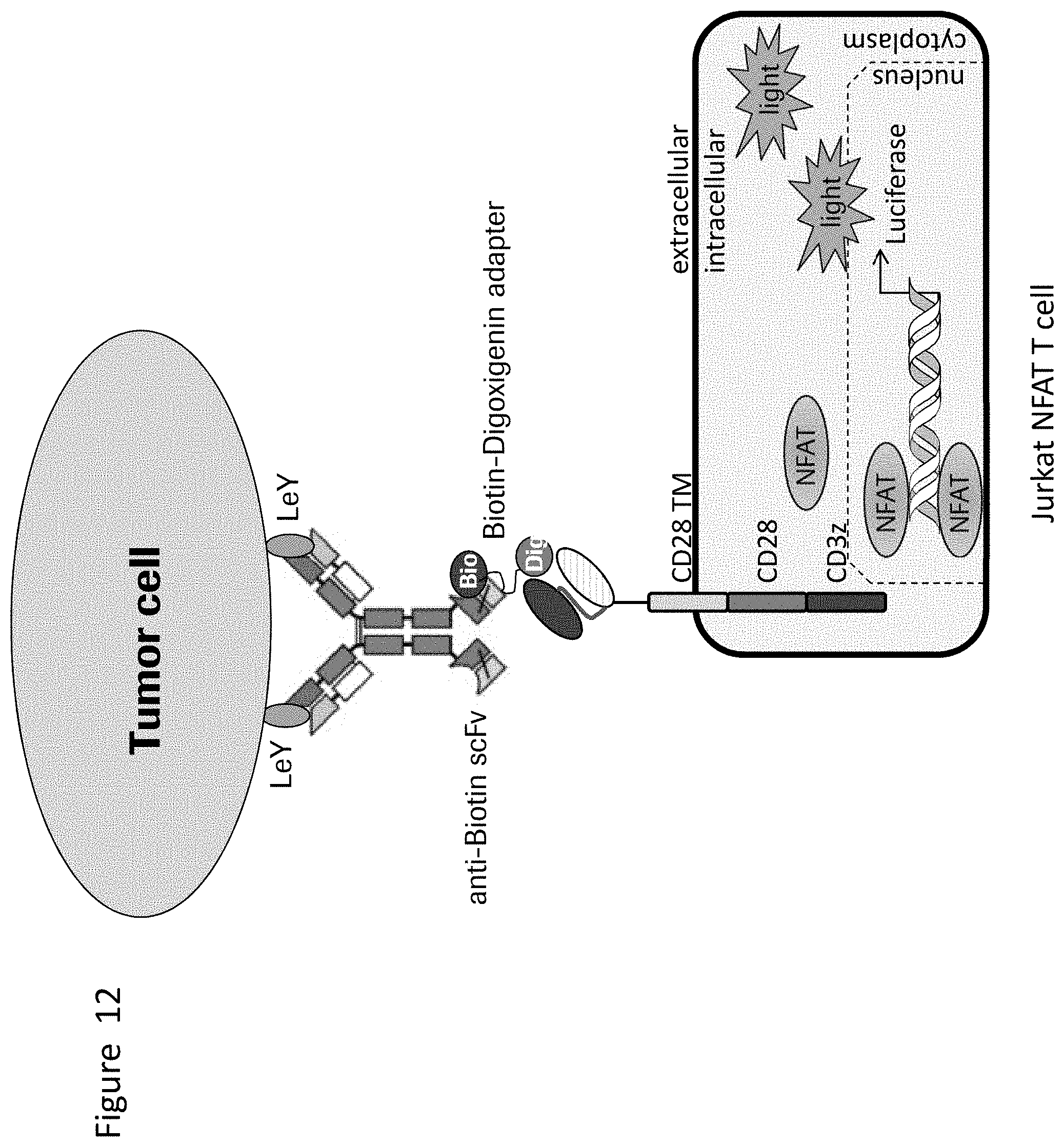
D00013

S00001
XML
uspto.report is an independent third-party trademark research tool that is not affiliated, endorsed, or sponsored by the United States Patent and Trademark Office (USPTO) or any other governmental organization. The information provided by uspto.report is based on publicly available data at the time of writing and is intended for informational purposes only.
While we strive to provide accurate and up-to-date information, we do not guarantee the accuracy, completeness, reliability, or suitability of the information displayed on this site. The use of this site is at your own risk. Any reliance you place on such information is therefore strictly at your own risk.
All official trademark data, including owner information, should be verified by visiting the official USPTO website at www.uspto.gov. This site is not intended to replace professional legal advice and should not be used as a substitute for consulting with a legal professional who is knowledgeable about trademark law.