Human Antibodies That Bind And Are Internalized By Mesothelioma And Other Cancer Cells
Liu; Bin ; et al.
U.S. patent application number 15/733202 was filed with the patent office on 2021-04-08 for human antibodies that bind and are internalized by mesothelioma and other cancer cells. The applicant listed for this patent is The Regents of the University of California. Invention is credited to Scott Bidlingmaier, Bin Liu, Yang Su.
| Application Number | 20210100838 15/733202 |
| Document ID | / |
| Family ID | 1000005288964 |
| Filed Date | 2021-04-08 |


View All Diagrams
| United States Patent Application | 20210100838 |
| Kind Code | A1 |
| Liu; Bin ; et al. | April 8, 2021 |
HUMAN ANTIBODIES THAT BIND AND ARE INTERNALIZED BY MESOTHELIOMA AND OTHER CANCER CELLS
Abstract
In certain embodiments internalizing anti-CD146 antibodies and conjugates thereof are provided. It was discovered that anti-CD146 antibodies are capable of targeting both epithelioid and sarcamatous subtypes of mesothelioma cells. In certain embodiments methods of detecting and/or treating mesothelioma are provided.
| Inventors: | Liu; Bin; (San Francisco, CA) ; Bidlingmaier; Scott; (San Francisco, CA) ; Su; Yang; (South San Francisco, CA) | ||||||||||
| Applicant: |
|
||||||||||
|---|---|---|---|---|---|---|---|---|---|---|---|
| Family ID: | 1000005288964 | ||||||||||
| Appl. No.: | 15/733202 | ||||||||||
| Filed: | December 26, 2018 | ||||||||||
| PCT Filed: | December 26, 2018 | ||||||||||
| PCT NO: | PCT/US18/67544 | ||||||||||
| 371 Date: | June 9, 2020 |
Related U.S. Patent Documents
| Application Number | Filing Date | Patent Number | ||
|---|---|---|---|---|
| 62610497 | Dec 26, 2017 | |||
| Current U.S. Class: | 1/1 |
| Current CPC Class: | A61K 47/6801 20170801; C07K 16/2809 20130101; A61P 35/00 20180101; A61K 2039/505 20130101; A61K 9/0029 20130101; A61K 47/6911 20170801; C07K 16/3092 20130101; A61K 35/17 20130101 |
| International Class: | A61K 35/17 20060101 A61K035/17; C07K 16/28 20060101 C07K016/28; A61P 35/00 20060101 A61P035/00; C07K 16/30 20060101 C07K016/30; A61K 47/68 20060101 A61K047/68; A61K 47/69 20060101 A61K047/69 |
Goverment Interests
STATEMENT OF GOVERNMENTAL SUPPORT
[0002] This invention was made with government support under Grant Nos. R01 CA118919 and CA95671 from the National Institutes of Health. The Government has certain rights in the invention.
Claims
1. An isolated human antibody, said antibody comprising: i) an isolated internalizing human antibody that binds to a mesothelioma-associated, clinically represented cell surface antigen and is internalized into a mesothelioma cell that displays said antigen, wherein said antibody is an antibody that specifically binds to CD146; or ii) an isolated human antibody that binds to a mesothelioma cell, but does not bind to CD146.
2. The antibody of claim 1, wherein said antibody comprises an isolated internalizing human antibody that binds to a mesothelioma-associated, clinically represented cell surface antigen and is internalized into a mesothelioma cell that displays said antigen, wherein said antibody is an antibody that specifically binds to CD146.
3. The antibody of claim 2, wherein said antibody specifically binds in vivo to cells displaying CD146.
4. The antibody of claim 1, wherein said antibody comprises an isolated human antibody that binds to a mesothelioma cell, but does not bind to CD146.
5. The antibody according to any one of claims 1-4, wherein said antibody binds to an epithelioid subtype of mesothelioma cells.
6. The antibody according to any one of claims 1-5, wherein said antibody binds to a sarcomatous subtype of mesothelioma cells.
7. The antibody according to any one of claims 1, 2, 3, and 5-6, wherein said antibody specifically binds cells of a cell line selected from the group consisting of M28, and VAMT-1 cells.
8. The antibody according to any one of claims 1-7, wherein said antibody comprises at least one heavy chain variable region (VH) and at least one light chain variable region (VL), wherein said heavy chain variable region contains VH CDR1, and/or VH CDR2, and/or VH CDR3 of an antibody selected from the group consisting of M40_EVQ, M40, M1_EVQ, M1, M2_EVQ, M2, M3, M3_QVQ, M4_EVQ, M4_EVQ_WGQ, M4, M4_WGQ, ORG_Rd3I51 (aka M9), ORG_Rd3I53, ORG_Rd3I53_LC_P2SD2G, ORG_Rd3I55 (aka M10), ORG_Rd3I70, ORG_Rd2I115 (aka brain endo#86), ORG_Rd2I159, ORG_Rd2IV33, ORG_Rd2IV33_HC_R2Q, VAMTII16 (aka M8), ORG_Rd2I18, M28I122_HC_G2SR2Q (aka M6 like), VAMTII16 (aka M8), ORG_Rd2I18_LC_D2E, ORG_Rd3I31, ORG_Rd3I89 (aka GH9), ORG_Rd3I38, ORG_Rd3I38_V2AK2Q, M-PC_1, M-PC_2, M-PC_3, M-PC_4, M-PC_5, M-PC_7, M-PC_10, M-PC_11, M-PC_13, M-PC_14, M-PC_15, M-PC_17, M-PC_19, M-PC_20, M-PC_21, M-PC_22, M-PC_23, M-PC_25, M-PC_30, M-PC_33, M-PC_34, M-PC_36, M-PC_37, M-PC_39, M-PC_40, AF9, Rd2VAMT-CaPPL2_13, MS40Rd3 (aka MS38), MS2, MS3, MS37, MS57, MS60, MS64, #8 cdnameso, #17 cdnameso, and #87 cdnameso.
9. The antibody of claim 8, wherein said antibody comprises at least one heavy chain variable region (VH) and at least one light chain variable region (VL), wherein said heavy chain variable region contains VH CDR1, and/or VH CDR2, and/or VH CDR3 of an antibody selected from the group consisting of M40_EVQ, M40, M1_EVQ, M1, M2_EVQ, M2, M3, M3_QVQ, M4_EVQ, M4_EVQ_WGQ, M4, and M4_WGQ.
10. The antibody of claim 8, wherein said antibody comprises at least one heavy chain variable region (VH) and at least one light chain variable region (VL), wherein said heavy chain variable region contains VH CDR1, and/or VH CDR2, and/or VH CDR3 of an antibody selected from the group consisting of ORG_Rd3I51 (aka M9), ORG_Rd3I53, ORG_Rd3I53_LC_P2SD2G, ORG_Rd3I55 (aka M10), ORG_Rd3I70, ORG_Rd2I115 (aka brain endo#86), ORG_Rd2I159, ORG_Rd2IV33, ORG_Rd2IV33_HC_R2Q, VAMTII16 (aka M8), ORG_Rd2I18, M28I122_HC_G2SR2Q (aka M6 like), VAMTII16 (aka M8), ORG_Rd2I18_LC_D2E, ORG_Rd3I31, ORG_Rd3I89 (aka GH9), ORG_Rd3I38, ORG_Rd3I38_V2AK2Q, M-PC_1, M-PC_2, M-PC_3, M-PC_4, M-PC_5, M-PC_7, M-PC_10, M-PC_11, M-PC_13, M-PC_14, M-PC_15, M-PC_17, M-PC_19, M-PC_20, M-PC_21, M-PC_22, M-PC_23, M-PC_25, M-PC_30, M-PC_33, M-PC_34, M-PC_36, M-PC_37, M-PC_39, M-PC_40, AF9, Rd2VAMT-CaPPL2_13, MS40Rd3 (aka MS38), MS2, MS3, MS37, MS57, MS60, MS64, #8 cdnameso, #17 cdnameso, and #87 cdnameso.
11. The antibody according to any one of claims 1-10, wherein said antibody comprises at least one heavy chain variable region (VH) and at least one light chain variable region (VL), wherein said light chain variable region contains VL CDR1, and/or VL CDR2, and/or VL CDR3 of an antibody selected from the group consisting of M40_EVQ, M40, M1_EVQ, M1, M2_EVQ, M2, M3, M3_QVQ, M4_EVQ, M4_EVQ_WGQ, M4, M4_WGQ, ORG_Rd3I51 (aka M9), ORG_Rd3I53, ORG_Rd3I53_LC_P2SD2G, ORG_Rd3I55 (aka M10), ORG_Rd3I70, ORG_Rd2I115 (aka brain endo#86), ORG_Rd2II59, ORG_Rd2IV33, ORG_Rd2IV33_HC_R2Q, VAMTII16 (aka M8), ORG_Rd2I18, M28I122_HC_G2SR2Q (aka M6 like), VAMTII16 (aka M8), ORG_Rd2I18_LC_D2E, ORG_Rd3I31, ORG_Rd3I89 (aka GH9), ORG_Rd3I38, ORG_Rd3I38_V2AK2Q, M-PC_1, M-PC_2, M-PC_3, M-PC_4, M-PC_5, M-PC_7, M-PC_10, M-PC_11, M-PC_13, M-PC_14, M-PC_15, M-PC_17, M-PC_19, M-PC_20, M-PC_21, M-PC_22, M-PC_23, M-PC_25, M-PC_30, M-PC_33, M-PC_34, M-PC_36, M-PC_37, M-PC_39, M-PC_40, AF9, Rd2VAMT-CaPPL2_13, MS40Rd3 (aka MS38), MS2, MS3, MS37, MS57, MS60, MS64, #8 cdnameso, #17 cdnameso, and #87 cdnameso.
12. The antibody of claim 11, wherein said antibody comprises at least one heavy chain variable region (VH) and at least one light chain variable region (VL), wherein said light chain variable region contains VL CDR1, and/or VL CDR2, and/or VL CDR3 of an antibody selected from the group consisting of M40_EVQ, M40, M1_EVQ, M1, M2_EVQ, M2, M3, M3_QVQ, M4_EVQ, M4_EVQ_WGQ, M4, M4_WGQ.
13. The antibody of claim 11, wherein said antibody comprises at least one heavy chain variable region (VH) and at least one light chain variable region (VL), wherein said light chain variable region contains VL CDR1, and/or VL CDR2, and/or VL CDR3 of an antibody selected from the group consisting of ORG_Rd3I51 (aka M9), ORG_Rd3I53, ORG_Rd3I53_LC_P2SD2G, ORG_Rd3I55 (aka M10), ORG_Rd3I70, ORG_Rd2I115 (aka brain endo#86), ORG_Rd2II59, ORG_Rd2IV33, ORG_Rd2IV33_HC_R2Q, VAMTII16 (aka M8), ORG_Rd2I18, M28I122_HC_G2SR2Q (aka M6 like), VAMTII16 (aka M8), ORG_Rd2I18_LC_D2E, ORG_Rd3I31, ORG_Rd3I89 (aka GH9), ORG_Rd3I38, ORG_Rd3I38_V2AK2Q, M-PC_1, M-PC_2, M-PC_3, M-PC_4, M-PC_5, M-PC_7, M-PC_10, M-PC_11, M-PC_13, M-PC_14, M-PC_15, M-PC_17, M-PC_19, M-PC_20, M-PC_21, M-PC_22, M-PC_23, M-PC_25, M-PC_30, M-PC_33, M-PC_34, M-PC_36, M-PC_37, M-PC_39, M-PC_40, AF9, Rd2VAMT-CaPPL2_13, MS40Rd3 (aka MS38), MS2, MS3, MS37, MS57, MS60, MS64, #8 cdnameso, #17 cdnameso, and #87 cdnameso.
14. The antibody according to any one of claims 1-8, wherein said antibody comprises at least one heavy chain variable region (VH) and at least one light chain variable region (VL), wherein said heavy chain variable region comprises VH CDR1, VH CDR2, and VH CDR3 of an antibody selected from the group consisting of ORG_Rd3I51 (aka M9), ORG_Rd3I53, ORG_Rd3I53_LC_P2SD2G, ORG_Rd3I55 (aka M10), ORG_Rd3I70, ORG_Rd2I115 (aka brain endo#86), ORG_Rd2II59, ORG_Rd2IV33, ORG_Rd2IV33_HC_R2Q, VAMTII16 (aka M8), ORG_Rd2I18, M28I122_HC_G2SR2Q (aka M6 like), VAMTII16 (aka M8), ORG_Rd2I18_LC_D2E, ORG_Rd3I31, ORG_Rd3I89 (aka GH9), ORG_Rd3I38, ORG_Rd3I38_V2AK2Q, M-PC_1, M-PC_2, M-PC_3, M-PC_4, M-PC_5, M-PC_7, M-PC_10, M-PC_11, M-PC_13, M-PC_14, M-PC_15, M-PC_17, M-PC_19, M-PC_20, M-PC_21, M-PC_22, M-PC_23, M-PC_25, M-PC_30, M-PC_33, M-PC_34, M-PC_36, M-PC_37, M-PC_39, M-PC_40, AF9, Rd2VAMT-CaPPL2_13, MS40Rd3 (aka MS38), MS2, MS3, MS37, MS57, MS60, MS64, #8 cdnameso, #17 cdnameso, and #87 cdnameso.
15. The antibody according to any one of claims 1-8, wherein said antibody comprises at least one heavy chain variable region (VH) and at least one light chain variable region (VL), wherein said light chain variable region comprises VL CDR1, VL CDR2, and VL CDR3 of an antibody selected from the group consisting of M40_EVQ, M40, M1_EVQ, M1, M2_EVQ, M2, M3, M3_QVQ, M4_EVQ, M4_EVQ_WGQ, M4, M4_WGQ, ORG_Rd3I51 (aka M9), ORG_Rd3I53, ORG_Rd3I53_LC_P2SD2G, ORG_Rd3I55 (aka M10), ORG_Rd3I70, ORG_Rd2II15 (aka brain endo#86), ORG_Rd2II59, ORG_Rd2IV33, ORG_Rd2IV33_HC_R2Q, VAMTII16 (aka M8), ORG_Rd2I18, M28I122_HC_G2SR2Q (aka M6 like), VAMTII16 (aka M8), ORG_Rd2I18_LC_D2E, ORG_Rd3I31, ORG_Rd3I89 (aka GH9), ORG_Rd3I38, ORG_Rd3I38_V2AK2Q, M-PC_1, M-PC_2, M-PC_3, M-PC_4, M-PC_5, M-PC_7, M-PC_10, M-PC_11, M-PC_13, M-PC_14, M-PC_15, M-PC_17, M-PC_19, M-PC_20, M-PC_21, M-PC_22, M-PC_23, M-PC_25, M-PC_30, M-PC_33, M-PC_34, M-PC_36, M-PC_37, M-PC_39, M-PC_40, AF9, Rd2VAMT-CaPPL2_13, MS40Rd3 (aka MS38), MS2, MS3, MS37, MS57, MS60, MS64, #8 cdnameso, #17 cdnameso, and #87 cdnameso.
16. The antibody according to any one of claims 1-8, wherein said antibody comprises at least one heavy chain variable region (VH) and at least one light chain variable region (VL), wherein: said heavy chain variable region comprises VH CDR1, VH CDR2, and VH CDR3 of an antibody selected from the group consisting of ORG_Rd3I51 (aka M9), ORG_Rd3I53, ORG_Rd3I53_LC_P2SD2G, ORG_Rd3I55 (aka M10), ORG_Rd3I70, ORG_Rd2I115 (aka brain endo#86), ORG_Rd2II59, ORG_Rd2IV33, ORG_Rd2IV33_HC_R2Q, VAMTII16 (aka M8), ORG_Rd2I18, M28I122_HC_G2SR2Q (aka M6 like), VAMTII16 (aka M8), ORG_Rd2I18_LC_D2E, ORG_Rd3I31, ORG_Rd3I89 (aka GH9), ORG_Rd3I38, ORG_Rd3I38_V2AK2Q, M-PC_1, M-PC_2, M-PC_3, M-PC_4, M-PC_5, M-PC_7, M-PC_10, M-PC_11, M-PC_13, M-PC_14, M-PC_15, M-PC_17, M-PC_19, M-PC_20, M-PC_21, M-PC_22, M-PC_23, M-PC_25, M-PC_30, M-PC_33, M-PC_34, M-PC_36, M-PC_37, M-PC_39, M-PC_40, AF9, Rd2VAMT-CaPPL2_13, MS40Rd3 (aka MS38), MS2, MS3, MS37, MS57, MS60, MS64, #8 cdnameso, #17 cdnameso, and #87 cdnameso; and said light chain variable region comprises VL CDR1, VL CDR2, and VL CDR3 of an antibody selected from the group consisting of M40_EVQ, M40, M1_EVQ, M1, M2_EVQ, M2, M3, M3_QVQ, M4_EVQ, M4_EVQ_WGQ, M4, M4_WGQ, ORG_Rd3I51 (aka M9), ORG_Rd3I53, ORG_Rd3I53_LC_P2SD2G, ORG_Rd3I55 (aka M10), ORG_Rd3I70, ORG_Rd2II15 (aka brain endo#86), ORG_Rd2II59, ORG_Rd2IV33, ORG_Rd2IV33_HC_R2Q, VAMTII16 (aka M8), ORG_Rd2I18, M28I122_HC_G2SR2Q (aka M6 like), VAMTII16 (aka M8), ORG_Rd2I18_LC_D2E, ORG_Rd3I31, ORG_Rd3I89 (aka GH9), ORG_Rd3I38, ORG_Rd3I38_V2AK2Q, M-PC_1, M-PC_2, M-PC_3, M-PC_4, M-PC_5, M-PC_7, M-PC_10, M-PC_11, M-PC_13, M-PC_14, M-PC_15, M-PC_17, M-PC_19, M-PC_20, M-PC_21, M-PC_22, M-PC_23, M-PC_25, M-PC_30, M-PC_33, M-PC_34, M-PC_36, M-PC_37, M-PC_39, M-PC_40, AF9, Rd2VAMT-CaPPL2_13, MS40Rd3 (aka MS38), MS2, MS3, MS37, MS57, MS60, MS64, #8 cdnameso, #17 cdnameso, and #87 cdnameso.
17. The antibody according to any one of claims 1-16, wherein said antibody comprises a VH domain of an antibody selected from the group consisting of M40_EVQ, M40, M1_EVQ, M1, M2_EVQ, M2, M3, M3_QVQ, M4_EVQ, M4_EVQ_WGQ, M4, M4_WGQ, ORG_Rd3I51 (aka M9), ORG_Rd3I53, ORG_Rd3I53_LC_P2SD2G, ORG_Rd3I55 (aka M10), ORG_Rd3I70, ORG_Rd2I115 (aka brain endo#86), ORG_Rd2II59, ORG_Rd2IV33, ORG_Rd2IV33_HC_R2Q, VAMTII16 (aka M8), ORG_Rd2I18, M28I122_HC_G2SR2Q (aka M6 like), VAMTII16 (aka M8), ORG_Rd2I18_LC_D2E, ORG_Rd3I31, ORG_Rd3I89 (aka GH9), ORG_Rd3I38, ORG_Rd3I38_V2AK2Q, M-PC_1, M-PC_2, M-PC_3, M-PC_4, M-PC_5, M-PC_7, M-PC_10, M-PC_11, M-PC_13, M-PC_14, M-PC_15, M-PC_17, M-PC_19, M-PC_20, M-PC_21, M-PC_22, M-PC_23, M-PC_25, M-PC_30, M-PC_33, M-PC_34, M-PC_36, M-PC_37, M-PC_39, M-PC_40, AF9, Rd2VAMT-CaPPL2_13, MS40Rd3 (aka MS38), MS2, MS3, MS37, MS57, MS60, MS64, #8 cdnameso, #17 cdnameso, and #87 cdnameso.
18. The antibody of claim 17, wherein said antibody comprises a VH domain of an antibody selected from the group consisting of M40_EVQ, M40, M1_EVQ, M1, M2_EVQ, M2, M3, M3_QVQ, M4_EVQ, M4_EVQ_WGQ, M4, and M4_WGQ.
19. The antibody according to any one of claims 1-16, wherein said antibody comprises a VL domain of an antibody selected from the group consisting of M40_EVQ, M40, M1_EVQ, M1, M2_EVQ, M2, M3, M3_QVQ, M4_EVQ, M4_EVQ_WGQ, M4, M4_WGQ, ORG_Rd3I51 (aka M9), ORG_Rd3I53, ORG_Rd3I53_LC_P2SD2G, ORG_Rd3I55 (aka M10), ORG_Rd3I70, ORG_Rd2I115 (aka brain endo#86), ORG_Rd2I159, ORG_Rd2IV33, ORG_Rd2IV33_HC_R2Q, VAMTII16 (aka M8), ORG_Rd2I18, M28I122_HC_G2SR2Q (aka M6 like), VAMTII16 (aka M8), ORG_Rd2I18_LC_D2E, ORG_Rd3I31, ORG_Rd3I89 (aka GH9), ORG_Rd3I38, ORG_Rd3I38_V2AK2Q, M-PC_1, M-PC_2, M-PC_3, M-PC_4, M-PC_5, M-PC_7, M-PC_10, M-PC_11, M-PC_13, M-PC_14, M-PC_15, M-PC_17, M-PC_19, M-PC_20, M-PC_21, M-PC_22, M-PC_23, M-PC_25, M-PC_30, M-PC_33, M-PC_34, M-PC_36, M-PC_37, M-PC_39, M-PC_40, AF9, Rd2VAMT-CaPPL2_13, MS40Rd3 (aka MS38), MS2, MS3, MS37, MS57, MS60, MS64, #8 cdnameso, #17 cdnameso, and #87 cdnameso.
20. The antibody of claim 19, wherein said antibody comprises a VL domain of an antibody selected from the group consisting of M40_EVQ, M40, M1_EVQ, M1, M2_EVQ, M2, M3, M3_QVQ, M4_EVQ, M4_EVQ_WGQ, M4, and M4_WGQ.
21. The antibody according to any one of claims 1-16, wherein said antibody comprises a VL domain and a VH domain of an antibody selected from the group consisting of M40_EVQ, M40, M1_EVQ, M1, M2_EVQ, M2, M3, M3_QVQ, M4_EVQ, M4_EVQ_WGQ, M4, M4_WGQ, ORG_Rd3I51 (aka M9), ORG_Rd3I53, ORG_Rd3I53_LC_P2SD2G, ORG_Rd3I55 (aka M10), ORG_Rd3I70, ORG_Rd2I115 (aka brain endo#86), ORG_Rd2I159, ORG_Rd2IV33, ORG_Rd2IV33_HC_R2Q, VAMTII16 (aka M8), ORG_Rd2I18, M281I22 HC G2SR2Q (aka M6 like), VAMTII16 (aka M8), ORG_Rd2I18_LC_D2E, ORG_Rd3I31, ORG_Rd3I89 (aka GH9), ORG_Rd3I38, ORG_Rd3I38_V2AK2Q, M-PC_1, M-PC_2, M-PC_3, M-PC_4, M-PC_5, M-PC_7, M-PC_10, M-PC_11, M-PC_13, M-PC_14, M-PC_15, M-PC_17, M-PC_19, M-PC_20, M-PC_21, M-PC_22, M-PC_23, M-PC_25, M-PC_30, M-PC_33, M-PC_34, M-PC_36, M-PC_37, M-PC_39, M-PC_40, AF9, Rd2VAMT-CaPPL2_13, MS40Rd3 (aka MS38), MS2, MS3, MS37, MS57, MS60, MS64, #8 cdnameso, #17 cdnameso, and #87 cdnameso.
22. The antibody of claim 21, wherein said antibody comprises a VL domain and a VH domain of an antibody selected from the group consisting of M40_EVQ, M40, M1_EVQ, M1, M2_EVQ, M2, M3, M3_QVQ, M4_EVQ, M4_EVQ_WGQ, M4, and M4_WGQ.
23. The antibody according to any one of claims 1-22, wherein said antibody comprises a VH and a VL domain joined by a peptide linker ranging in length from about 4 up to about 20 amino acids, or from about 8 up to about 16 amino acids, or wherein said linker is about 12 amino acids in length.
24. The antibody of claim 23, wherein said heavy chain variable region is joined to said light chain variable region by a linker comprising or consisting of the amino acid sequence (Gly.sub.4Ser).sub.3 (SEQ ID NO:112).
25. The antibody according to any one of claims 1-24, wherein said antibody is a single chain antibody.
26. The antibody of claim 25, wherein said antibody is a human scFv.
27. The antibody according to any one of claims 1-22, wherein said antibody is an antibody fragment selected from the group consisting of Fv, Fab, (Fab').sub.2, (Fab').sub.3, IgG.DELTA.CH2, and a minibody.
28. The antibody according to any one of claims The antibody according to any one of claims 1-22, wherein said antibody is a substantially intact immunoglobulin.
29. The antibody of claim 28, wherein said antibody comprises an IgA, IgE, or IgG.
30. An immunoconjugate comprising a first antibody according to any one of claims 1-29 attached to an effector wherein said effector is selected from the group consisting of a second antibody, a detectable label, a cytotoxin or cytostatic agent, a liposome containing a drug, a radionuclide, a drug, a prodrug, an immune modulator, a viral particle, a cytokine, a second antibody, and a chelate.
31. The immunoconjugate of claim 30, wherein said first antibody is attached to a cytotoxic and/or cytostatic drug.
32. The immunoconjugate of claim 30, wherein said first antibody is attached directly or through a linker to one or more of the following: said drug; a lipid or liposome containing said drug; a polymeric drug carrier comprising said drug; and a nanoparticle drug carrier comprising said drug.
33. The immunoconjugate according to any one of claims 31-32, wherein said drug is an anti-cancer drug.
34. The immunoconjugate according to any one of claims 31-32, wherein said drug is selected from the group consisting of a microtubule inhibitor, a DNA-damaging agent, and a polymerase inhibitor.
35. The immunoconjugate of claim 34, wherein the drug comprises a tubulin inhibitor.
36. The immunoconjugate of claim 35, wherein the drug comprises a drug selected from the group consisting of an auristatin, Dolastatin-10, synthetic derivatives of the natural product Dolastatin-10, and maytansine or a maytansine derivative.
37. The immunoconjugate of claim 35, wherein the drug comprises a drug selected from the group consisting Monomethylauristatin F (MMAF), Auristatin E (AE), Monomethylauristatin E (MMAE), and tubulysin.
38. The immunoconjugate of claim 35, wherein the drug comprises a maytansine selected from the group consisting of Mertansine (DM1), DM3, and DM4.
39. The immunoconjugate of claim 30, wherein said first antibody is attached to a second antibody.
40. The immunoconjugate of claim 39, wherein said second antibody comprises an anti-CD3 antibody.
41. The immunoconjugate according to any one of claims 39-40, wherein said second antibody is selected from the group consisting of a full-length antibody (e.g., IgG), an Fv, an Fab, a (Fab').sub.2, a (Fab').sub.3, an IgG.DELTA.CH2), a minibody, and an scFv.
42. The immunoconjugate according to any one of claims 39-40, wherein said second antibody is selected from the group consisting of a bispecific T-cell engager (BiTE), a crossMab, a DAF, a dutaMab, a dual-targeted IgG (DT-IgG), a knob-in-hole (KIH) bispecific, an Fab-arm exchange bsAb, a SEEDbody, an LUZ-Y bsAb, an Fcab bsAb, a kappa-alpha-body bsAb, an orthogonal Fab, a DVD-IgG, an IgG(H)-scFv, an scFv-(H)IgG, an IgG(L)-scFv, an scFv-(L)IgG, an IgG(L,H)-Fv, an IgG(H)-V, a VH-IgG, an IgG(L)-V, a V(L)-IgG, a KIH IgG-scFav, a 2scFv-IgG, an IgG-2scFv, an scFv4-Ig, a zybody, a DIV-IgG, a bi-nanobody, a nanobody-HAS, a diabody, a dual-affinity retargeted (DART) bsAb, a TandAb, an scdiabody, an scDiabody-CH3, a diabody-CH3, a miniantibody, a minibody, TriBi minibody, an scFv-CH3 KIH, a Fab-scFv, an scFv-CH-CL-scFv, a F(ab')2, a F(ab')2-scFv2, an scFv-KIH, a Fab-scFv-Fc, an scDiabody-Fc, a diabody-Fc, a tandem scFv-Fc, an intrabody, a dock and lock, an ImmTac, an HSAbody, an IgG-IgG, a Cov-X-Body, and an scFv1-PEG-scFv2.
43. The immunoconjugate according to any one of claims 39-40, wherein said first antibody is an scFv.
44. The immunoconjugate of claim 43, wherein said first antibody and said anti-CD3 antibody are both scFv.
45. The immunoconjugate of claim 44, wherein said first antibody and said anti-CD3 antibody are joined by a peptide linker.
46. The immunoconjugate of claim 45 wherein said first antibody and said anti-CD3 antibody are joined by a peptide linker comprising or consisting of the amino acid sequence GGGGS (SEQ ID NO:70).
47. The immunoconjugate according to any one of claims 40-46, wherein said anti-CD3 antibody comprises a VH and/or a VL region shown in the anti-CD3 scFV in Table 3.
48. The immunoconjugate of claim 47, wherein said immunoconjugate comprises an immunoconjugate selected from the group consisting of M40_EVQ_blina, M40_blina, M1_EVQ_blina, M1_blina, M2_EVQ_blina, M2_blina, M3_blina, M3_QVQ_blina, M4_EVQ_blina, M4_EVQ_WGQ blina, M4_blina, and M4_WGQ_blina, (as shown in Table in Table 3).
49. The immunoconjugate of claim 30, wherein said first antibody is attached to an immunmodulator.
50. The immunoconjugate of claim 49, wherein said immunomodulator is an immunomodulatory is one that blocks immune checkpoints.
51. The immunoconjugate of claim 50, wherein said immunomodulator comprises a second antibody that is selected from the group consisting of an anti-CTLA4 antibody, an anti-PDL1 antibody, an anti-PDL2 antibody, an anti-ICOS antibody, and an anti-BTLA antibody.
52. The immunoconjugate of claim 51, wherein said second antibody is an antibody that comprise the VH and VL domains of an antibody selected from the group consisting of ipilimumab, nivolumab, and pembrolizumab.
53. The immunoconjugate of claim 51, wherein said second antibody is an antibody selected from the group consisting of ipilimumab, nivolumab, and pembrolizumab.
54. The immunoconjugate of claim 30, wherein said first antibody is attached to a chelate comprising an isotope selected from the group consisting .sup.99Tc, .sup.99Tc, .sup.97Ru, .sup.95Ru, 94Tc, .sup.90Y, .sup.90Y, .sup.89Zr, .sup.86Y, .sup.77Br, .sup.77As, .sup.76Br, .sup.75Se, .sup.72As, .sup.68Ga, .sup.68Ga, .sup.67Ga, .sup.67Ga, .sup.67Cu, .sup.67Cu, .sup.64Cu, .sup.62Cu, .sup.62Cu, .sup.59Fe, .sup.58Co, .sup.57Co, .sup.52Mn, .sup.52Fe, .sup.51Cr, .sup.47Sc, .sup.3H, .sup.35S, .sup.33P, .sup.32P, .sup.225Ac, .sup.224Ac, .sup.223Ra, .sup.213Bi, .sup.212Pb, .sup.212Bi, .sup.211At, .sup.203Pb, .sup.203Hg, .sup.201Tl, .sup.199Au, .sup.198Au, .sup.198Au, .sup.197Pt, .sup.18F, .sup.189Re, .sup.188Re, .sup.188Re, .sup.186Re, .sup.186Re, .sup.177Lu, .sup.177Lu, .sup.175Yb, .sup.172Tm, .sup.169Yb, .sup.169Yb, .sup.169Er, .sup.168Tm, .sup.167Tm, .sup.166Ho, .sup.166Dy, .sup.165Tm, .sup.165Dy, .sup.161Tb, .sup.15O, .sup.15N, .sup.159Gd, .sup.157Gd, .sup.153Sm, .sup.153Pb, .sup.151Pm, .sup.14C, .sup.149Pm, .sup.143Pr, .sup.142Pr, .sup.13N, .sup.133I, .sup.131In, .sup.131I, .sup.127Te, .sup.126I, .sup.125Te, .sup.125I, .sup.124I, .sup.123I, .sup.122Te, .sup.121Te, .sup.121Sn, .sup.11C, .sup.113In, .sup.111In, .sup.111In, .sup.111Ag, .sup.111Ag, .sup.109Pd, .sup.109Pd, .sup.107Hg, .sup.105Ru, .sup.105Rh, .sup.105Rh, and .sup.103Ru.
55. The immunoconjugate of claim 30, wherein said first antibody is attached to a lipid or a liposome complexed with or containing an anti-cancer drug.
56. The immunoconjugate of claim 30, wherein said first antibody is attached to a detectable label.
57. A pharmaceutical formulation said formulation comprising: a pharmaceutically acceptable carrier and an antibody according to any one of claims 1-29; and/or a pharmaceutically acceptable carrier and a immunoconjugate according to any one of claims 30-56.
58. A method of inhibiting the growth and/or proliferation of mesothelioma cell and/or a cell that expresses CD146, said method comprising: contacting said cell with an antibody according to any one of claims 1-29; and/or contacting said cell with an immunoconjugate according to any one of claims 30-55, wherein the immunoconjugate comprises an effector that has cytostatic and/or cytotoxic activity and/or immunomodulatory activity.
59. The method of claim 58, wherein said cell is a cancer cell.
60. The method of claim 59, wherein cancer cell of a cancer selected from the group consisting of mesothelioma, melanoma, head and neck cancer, lung cancer, glioblastoma multiforme, pancreatic cancer, ovarian cancer, breast cancer, prostate cancer, cervical cancer, skin cancer (e.g., squamous cell carcinoma), and testicular cancer.
61. The method of claim 59, wherein said cancer cell is a mesothelioma cancer cell or a cell derived therefrom.
62. The method of claim 61, wherein said cancer cell comprises an epithelioid subtype of mesothelioma cells and/or a sarcamatous subtype of mesothelioma cells.
63. The method according to any one of claims 58-62, wherein said effector comprises a radionuclide and/or a cytostatic drug.
64. The method of claim 63, wherein said effector comprises one or more of the following: a cytotoxic and/or cytostatic drug; a lipid or liposome containing a cytotoxic and/or cytostatic drug; a polymeric drug carrier comprising a cytotoxic and/or cytostatic drug; and a nanoparticle drug carrier comprising a cytotoxic and/or cytostatic drug.
65. The method of claim 64, wherein said drug is an anti-cancer drug.
66. The method of claim 65, wherein said drug is selected from the group consisting of auristatin, dolastatin, colchicine, combretastatin, and mTOR/PI3K inhibitors.
67. The method of claim 65, wherein said drug is monomethyl auristatin F.
68. The method according to any one of claims 58-67, wherein said administering comprises administering to a human or to a non-human mammal.
69. The method according to any one of claims 58-68, wherein said administering comprises: administering parenterally; and/or administering into a tumor or a surgical site.
70. The method according to any one of claims 58-69, wherein said antibody and/or immunoconjugate is administered as an adjunct therapy to surgery and/or radiotherapy.
71. The method according to any one of claims 58-70, wherein said antibody and/or immunoconjugate is administered in conjunction with another anti-cancer drug and/or a hormone.
72. A method of detecting a cancer cell of a cancer that expresses CD146, said method comprising: contacting said cancer cell with a immunoconjugate comprising an antibody according to any one of claims 1-29 attached to a detectable label; and detecting the presence and/or location of said detectable label where the presence and/or location is an indicator of the location and/or presence of a cancer cell.
73. The method of claim 72, wherein said label comprises a label selected from the group consisting of a radioactive label, a radio-opaque label, an MRI label, a PET label, and an SPECT label.
74. A nucleic acid encoding an antibody or a fragment of an antibody according to any of claims 1-29 or a fragment thereof.
75. An expression vector comprising the nucleic acid of claim 74.
76. A cell comprising the expression vector of claim 75.
77. A chimeric antigen receptor (CAR) comprising an antibody according to any one of claims 1-29, or a mesothelioma cell binding region thereof and/or a CD146 binding region thereof.
78. The chimeric antigen receptor of claim 77, wherein said receptor comprises: said antibody; a transmembrane domain; at least one costimulatory signaling region; and a CD3 zeta signaling domain.
79. The chimeric antigen receptor of claim 78, wherein said costimulatory signaling region comprises the intracellular domain of a costimulatory molecule selected from the group consisting of CD27, CD2S, 4-1BB, OX40, CD30, CD40, PD-1, ICOS, lymphocyte function-associated antigen-1 (LFA-1), CD2, CD7, LIGHT, NKG2C, B7-H3, a ligand that specifically binds with CD83, and any combination thereof.
80. An isolated nucleic acid sequence encoding a chimeric antigen receptor (CAR) according to any one of claims 77-79.
81. A cell comprising a nucleic acid sequence encoding a chimeric antigen receptor (CAR), according to any one of claims 77-79.
82. The cell of claim 81, wherein said cell is selected from the group consisting of a T cell, a Natural Killer (NK) cell, a cytotoxic T lymphocyte (CTL), and a regulatory T cell.
83. The cell according to any one of claims 81-82, wherein the cell exhibits an anti-cancer immune response when the antigen binding domain binds to a cell that expresses CD146.
84. A pharmaceutical composition for treatment of cancer in a mammal, said formulation comprising a genetically engineered cell (CAR-T cell) according to any one of claims 81-83, and a pharmaceutically acceptable carrier.
85. The composition of claim 84, wherein said formulation comprises an anti-tumor effective amount of cells, wherein the anti-tumor effective amount of cells ranges from about 10.sup.4 up to about 10.sup.7 cells per kg body weight of a mammal in need of such cells.
86. A vector comprising a nucleic acid sequence encoding a chimeric antigen receptor (CAR) according to any one of claims 77-79.
87. A method for stimulating a T cell-mediated immune response to a target cell population or tissue in a mammal, wherein said target cell population and/or tissue express CD146 and/or is a mesothelioma cell, said method comprising: administering to a mammal an effective amount of a cell genetically modified to express a chimeric antigen receptor (CAR) according to any one of claims 77-79.
88. A method of providing an anti-tumor immunity against tumors that comprise mesothelioma cells and/or that express CD146 in a mammal, the method comprising: administering to the mammal an effective amount of a cell genetically modified to express a chimeric antigen receptor (CAR) according to any one of claims 77-79, thereby providing an antitumor immunity in the mammal.
89. A method of treating a mammal with a cancer comprising mesothelioma cells and/or cells that express CD146, said method comprising: administering to a mammal an effective amount of a cell genetically modified to express a chimeric antigen receptor (CAR) according to any one of claims 77-79.
90. A method of generating a persisting population of genetically engineered T cells in a mammal diagnosed with cancer, said method comprising administering to said mammal a T cell genetically modified to express a chimeric antigen receptor (CAR) according to any one of claims 77-79, wherein the persisting population of genetically engineered T cells persists in the human for at least one month after administration.
91. A method of expanding a population of genetically engineered T cells in a mammal diagnosed with cancer, said method comprising: administering to said mammal administering to said mammal a T cell genetically modified to express a chimeric antigen receptor (CAR) according to any one of claims 77-79, wherein the administered genetically engineered T cell produces a population of progeny T cells in the human.
92. A method for treatment of cancer comprising the steps of contacting a genetically engineered T cell (CAR-T cell) according to any one of claims 77-79, with a cancer cell of a mammal, and inducing apoptosis of the cancer cell.
93. The method of claim 92, wherein said cancer comprises a mesothelioma.
Description
CROSS-REFERENCE TO RELATED APPLICATIONS
[0001] This application claims priority to and benefit of U.S. Ser. No. 62/610,497, filed on Dec. 26, 2017, which is incorporated herein by reference in its entirety for all purposes.
BACKGROUND
[0003] Mesothelioma is a deadly disease caused by malignant transformation of the mesothelium, the protective lining surrounding most of the internal organs of the body. Mesothelioma is almost always associated with previous exposure to asbestos, and symptoms may not appear until 20 to 50 years after exposure (Bertazzi (2005) Med. Lav. 96: 287-303). There is no generally accepted method for screening patients who have been exposed to asbestos, and diagnosis can be difficult because the symptoms of mesothelioma are similar to those caused by other conditions (Dunleavey (2004) Nurs. Times, 100: 40-43). There are three main types of mesothelioma: epithelioid, sarcomatoid, and mixed (Corson (2004) Thorac. Surg. Clin. 14: 447-460; Scherpereel (2007) Curr. Opin. Pulm. Med. 13: 339-443). Epithelioid mesothelioma is the most common form, comprising between 50% and 70% of mesothelioma cases, and the most likely to respond to treatment (Scherpereel (2007) Curr. Opin. Pulm. Med. 13: 339-443). Sarcomatoid mesothelioma accounts for 10% to 20% of mesothelioma cases and rarely responds to treatment (Scherpereel (2007) Curr. Opin. Pulm. Med. 13: 339-443; Lucas et al. (2003) Histopathology, 42: 270-279). Approximately 20% to 35% of mesothelioma cases are mixed type, which contains both epithelioid and sarcomatoid features and has an intermediate outlook (Scherpereel (2007) Curr. Opin. Pulm. Med. 13: 339-443; Pass et al. (1997) Ann. Surg. Oncol. 4: 215-222). Regardless of subtype, because diagnosis often occurs at a late stage of disease, the prognosis for malignant mesothelioma is generally poor, with median survival ranging from 8 to 14 months, and treatments are generally ineffective, especially in the case of sarcomatoid mesothelioma (Tomek & Manegold (2004) Lung Cancer, 45(Suppl 1): S103-S119; Kindler (2004) Lung Cancer, 45(Suppl 1): S125-S127).
[0004] One promising area of antineoplastic drug development is to explore tumor susceptibility to targeted therapy (Nielsen et al. (2002) Biochim. Biophys. Acta. 1591: 109-118; Li et al. (2001) Cancer Gene Ther. 8: 555-565; King et al. (2005) Curr. Gene Ther. 5:535-557; Zheng et al. (2005) Proc. Natl. Acad. Sci. USA, 102: 17757-11762). In principle, a variety of antitumor agents can be attached to tumor recognition molecules that target tumor-associated internalizing cell surface molecules to achieve intracellular delivery and targeted tumor killing (Nielsen et al. (2002) Biochim. Biophys. Acta. 1591: 109-118; King et al. (2005) Curr. Gene Ther. 5:535-557; Garnett (2001) Adv. Drug Deliv. Rev. 53: 171-216). Currently, very few mesothelioma-associated cell surface markers that are expressed by all subtypes of mesothelioma are known (Zeng et al. (1994) Hum. Pathol. 25: 227-234). For example, mesothelin, a cell surface glycoprotein, has been shown to be a useful marker for epithelioid mesothelioma (Hassan et al. (2004) Clin. Cancer Res. 10: 3937-3942), but it is not expressed by the sarcomatous subtype of this disease (Ordonez (2005) Arch. Pathol. Lab. Med. 129: 1407-1414). In addition, mesothelin is also expressed on normal mesothelial cells (Id.). Thus, the development of targeted therapies against mesothelioma will benefit from the identification of additional cell surface markers with more restricted expression on normal tissues and more specific associations with both epithelioid and sarcomatoid mesotheliomas.
[0005] Monoclonal antibodies (mAb) are able to recognize antigenic determinants of diverse chemical composition with high affinity and specificity and are, therefore, promising candidates for the development of targeted cancer therapies. Antibodies targeting tumor-associated epitopes could be used in applications such as induction of antibody-dependent cell cytotoxicity or inhibition of signaling pathways involved in tumor cell migration, growth, and survival. In addition, antibodies targeting internalizing tumor epitopes could be exploited to achieve specific intracellular delivery of therapeutic agents (Nielsen et al. (2002) Biochim. Biophys. Acta. 1591: 109-118; Roth et al. (2007) Mol. Cancer Ther. 6: 2737-2746; Mamot et al. (2005) Cancer Res. 65: 11631-11638).
SUMMARY
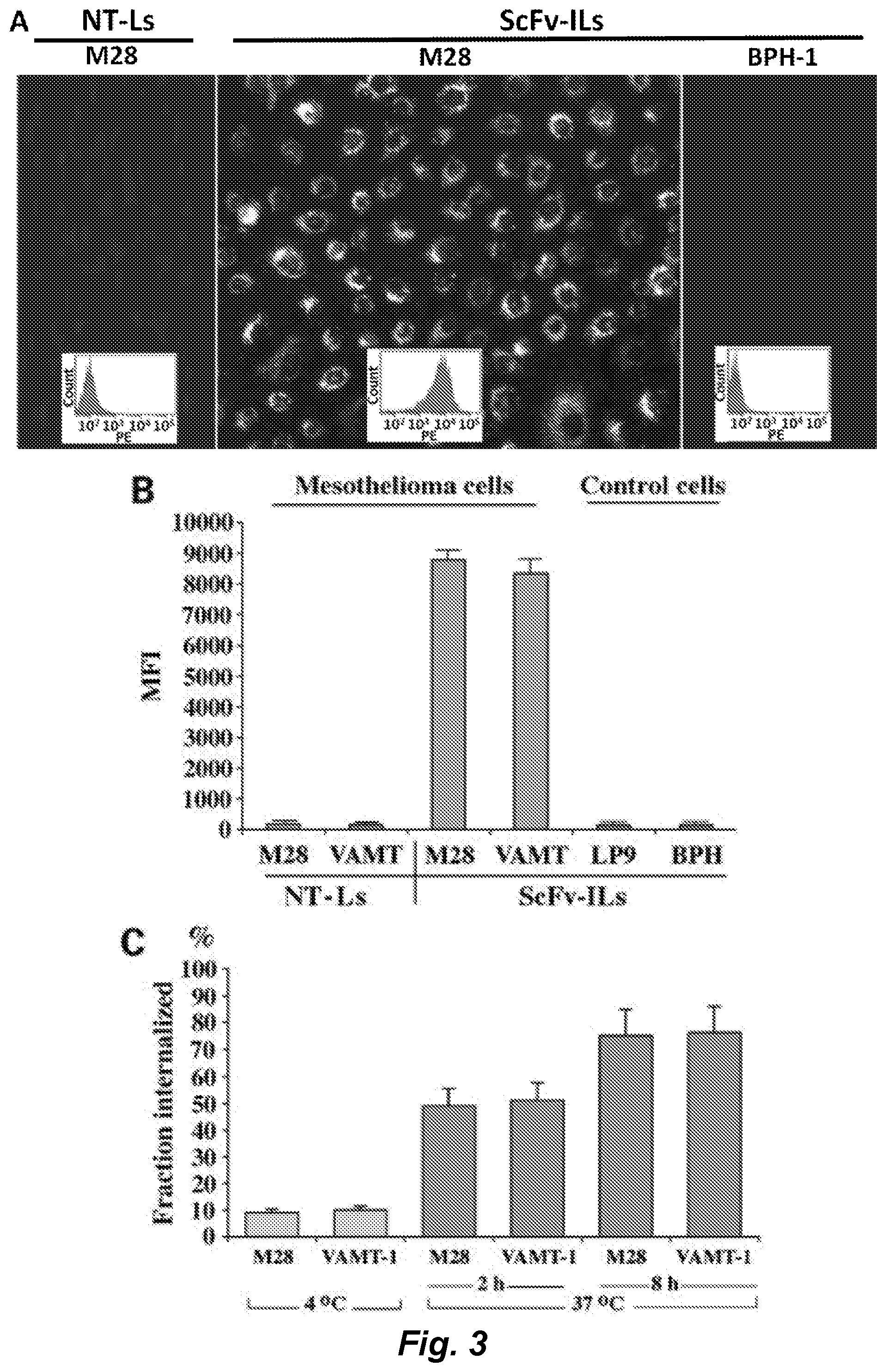
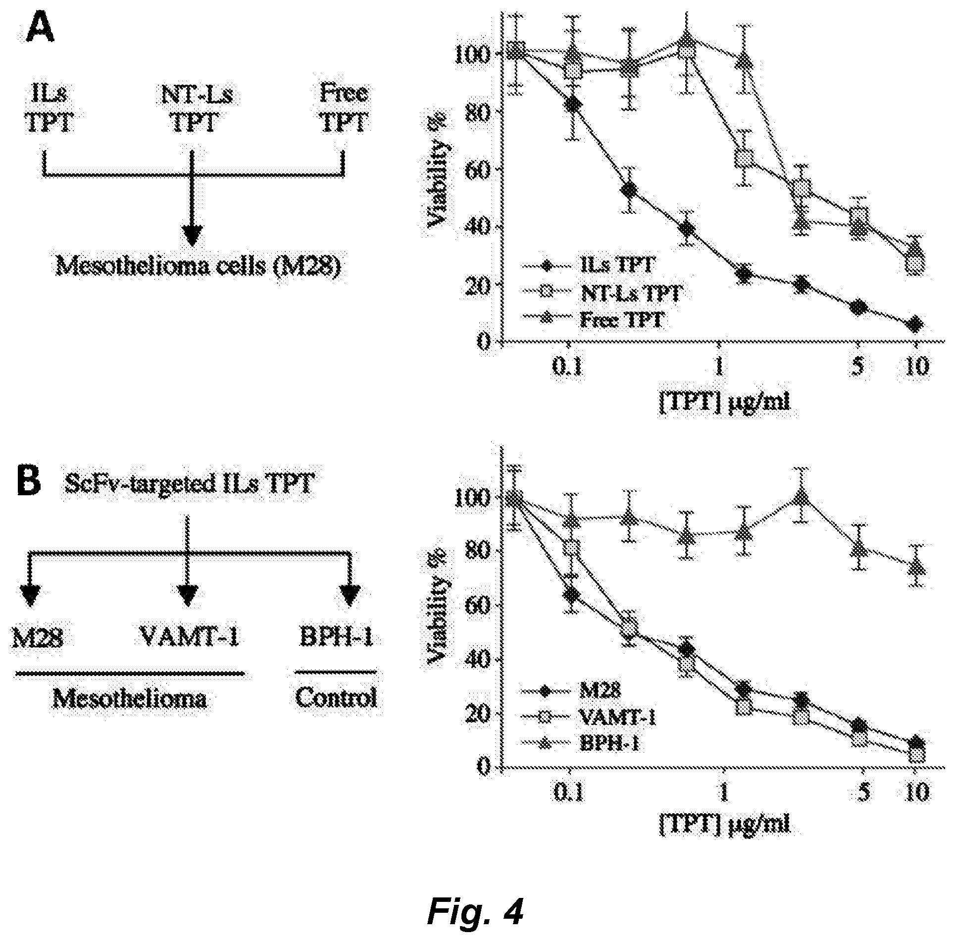
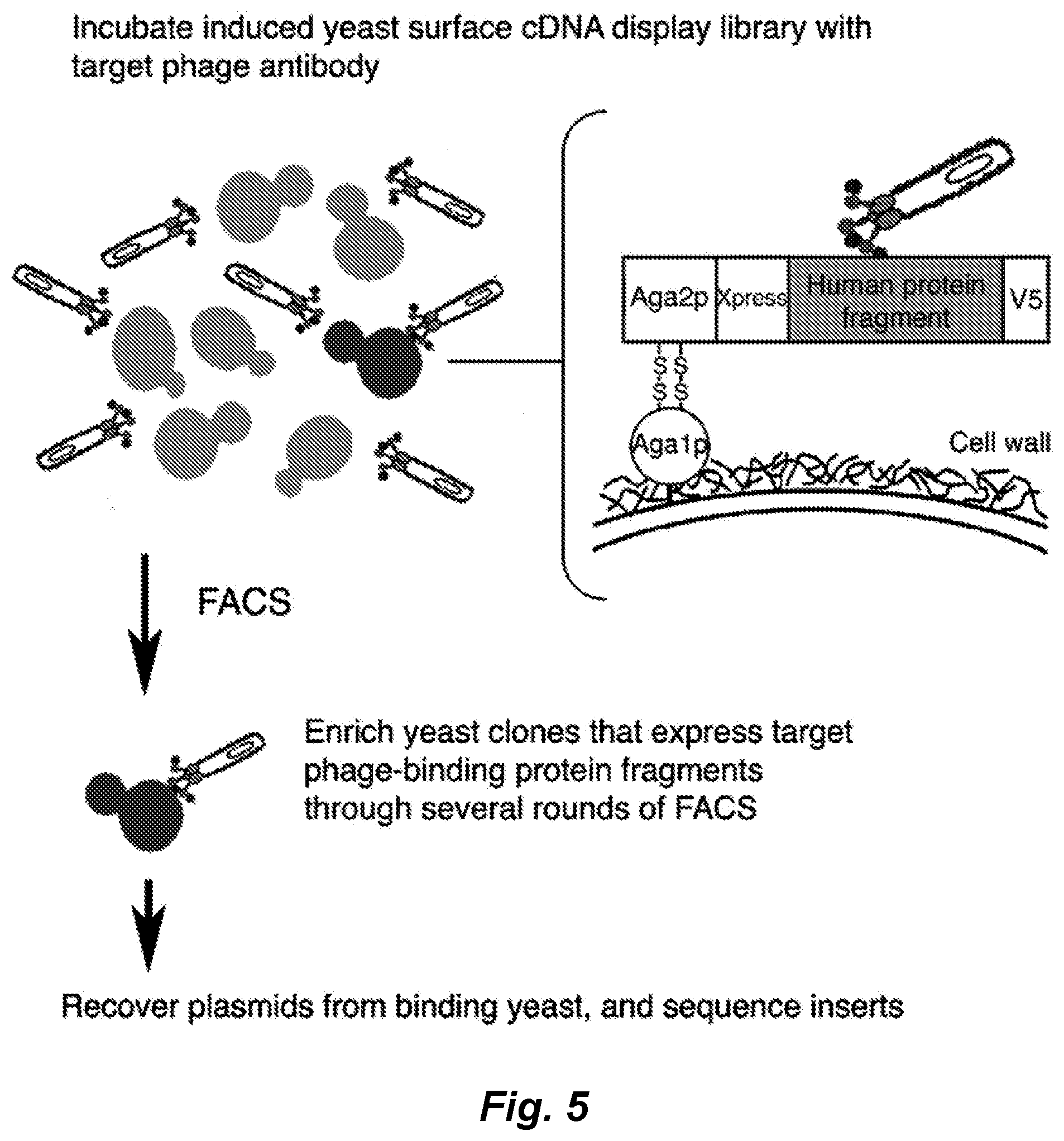
[0006] A naive phage antibody display library was selected on mesothelioma cell lines derived from both epithelioid and sarcomatous subtypes a panel of internalizing mAbs that target cell surface antigens associated with both subtypes of mesothelioma was identified. Immunohistochemistry studies showed that these scFvs bind to mesothelioma cells in situ, thereby recognizing clinically represented tumor antigens. We have further exploited the internalizing function of these scFvs to deliver immunoliposomes encapsulating the small molecule drug topotecan specifically to mesothelioma cells and showed targeted killing of both epithelioid and sarcomatous mesothelioma cells in vitro. To facilitate further therapeutic development, we have identified antigens recognized by this panel of phage antibodies. We have previously reported the construction of a large yeast surface-displayed human cDNA library, which was used to identify cellular proteins binding to posttranslational modifications (Bidlingmaier and Liu (2006) Mol. Cell Proteomics, 5: 533-540) and small signaling molecules (Bidlingmaier and Liu (2007) Mol. Cell Proteomics, 6: 2012-2020). One of the target antigens, MCAM/CD146/MUC18, was identified by screening the yeast surface human cDNA display library with a mesothelioma-targeting phage antibody. Mesothelioma tissue microarray studies showed that MCAM is overexpressed on >80% of both epithelioid and sarcomatous mesothelioma tissues, but not normal mesothelium. Finally, using single-photon emission computed tomography/computed tomography (SPECT/CT), we showed that the technetium (.sup.99mTc)-labeled anti-MCAM scFv was able to detect tumor cells in mesothelioma organ xenografts in vivo, indicating that this scFv can be useful for the development of targeted immunotherapies against mesothelioma, or other cancers expressing CD146 (e.g., melanoma, head and neck cancer, lung cancer, glioblastoma multiforme, pancreatic cancer, ovarian cancer, breast cancer, prostate cancer, cervical cancer, skin cancer (e.g., squamous cell carcinoma), and testicular cancer).
[0007] We have also identified a number of antibodies that bind to mesothelioma cells (see, e.g., Table 2). In certain embodiments these antibodies do not bind to CD146.
[0008] Accordingly, various embodiments contemplated herein may include, but need not be limited to, one or more of the following:
[0009] Embodiment 1: An isolated human antibody, said antibody comprising: [0010] i) an isolated internalizing human antibody that binds to a mesothelioma-associated, clinically represented cell surface antigen and is internalized into a mesothelioma cell that displays said antigen, wherein said antibody is an antibody that specifically binds to CD146; or [0011] ii) an isolated human antibody that binds to a mesothelioma cell, but does not bind to CD146.
[0012] Embodiment 2: The antibody of embodiment 1, wherein said antibody comprises an isolated internalizing human antibody that binds to a mesothelioma-associated, clinically represented cell surface antigen and is internalized into a mesothelioma cell that displays said antigen, wherein said antibody is an antibody that specifically binds to CD146.
[0013] Embodiment 3: The antibody of embodiment 2, wherein said antibody specifically binds in vivo to cells displaying CD146.
[0014] Embodiment 4: The antibody of embodiment 1, wherein said antibody comprises an isolated human antibody that binds to a mesothelioma cell, but does not bind to CD146.
[0015] Embodiment 5: The antibody according to any one of embodiments 1-4, wherein said antibody binds to an epithelioid subtype of mesothelioma cells.
[0016] Embodiment 6: The antibody according to any one of embodiments 1-5, wherein said antibody binds to a sarcomatous subtype of mesothelioma cells.
[0017] Embodiment 7: The antibody according to any one of embodiments 1, 2, 3, and 5-6, wherein said antibody specifically binds cells of a cell line selected from the group consisting of M28, and VAMT-1 cells.
[0018] Embodiment 8: The antibody according to any one of embodiments 1-7, wherein said antibody comprises at least one heavy chain variable region (VH) and at least one light chain variable region (VL), wherein said heavy chain variable region contains VH CDR1, and/or VH CDR2, and/or VH CDR3 of an antibody as shown in Table 1 or in Table 2, e.g., an antibody selected from the group consisting of M40_EVQ, M40, M1_EVQ, M1, M2_EVQ, M2, M3, M3_QVQ, M4_EVQ, M4_EVQ_WGQ, M4, M4_WGQ, ORG_Rd3I51 (aka M9), ORG_Rd3I53, ORG_Rd3I53_LC_P2SD2G, ORG_Rd3I55 (aka M10), ORG_Rd3I70, ORG_Rd2II15 (aka brain endo#86), ORG_Rd2II59, ORG_Rd2IV33, ORG_Rd2IV33_HC_R2Q, VAMTII16 (aka M8), ORG_Rd2I18, M28I122_HC_G2SR2Q (aka M6 like), VAMTII16 (aka M8), ORG_Rd2I18_LC_D2E, ORG_Rd3I31, ORG_Rd3I89 (aka GH9), ORG_Rd3I38, ORG_Rd3I38_V2AK2Q, M-PC_1, M-PC_2, M-PC_3, M-PC_4, M-PC_5, M-PC_7, M-PC_10, M-PC_11, M-PC_13, M-PC_14, M-PC_15, M-PC_17, M-PC_19, M-PC_20, M-PC_21, M-PC_22, M-PC_23, M-PC_25, M-PC_30, M-PC_33, M-PC_34, M-PC_36, M-PC_37, M-PC_39, M-PC_40, AF9, Rd2VAMT-CaPPL2_13, MS40Rd3 (aka MS38), MS2, MS3, MS37, MS57, MS60, MS64, #8 cdnameso, #17 cdnameso, and #87 cdnameso.
[0019] Embodiment 9: The antibody of embodiment 8, wherein said antibody comprises at least one heavy chain variable region (VH) and at least one light chain variable region (VL), wherein said heavy chain variable region contains VH CDR1, and/or VH CDR2, and/or VH CDR3 of an antibody selected from the group consisting of M40_EVQ, M40, M1_EVQ, M1, M2_EVQ, M2, M3, M3_QVQ, M4_EVQ, M4_EVQ_WGQ, M4, and M4_WGQ.
[0020] Embodiment 10: The antibody of embodiment 8, wherein said antibody comprises at least one heavy chain variable region (VH) and at least one light chain variable region (VL), wherein said heavy chain variable region contains VH CDR1, and/or VH CDR2, and/or VH CDR3 of an antibody as shown in Table 1 or in Table 2, e.g., an antibody selected from the group consisting of ORG_Rd3I51 (aka M9), ORG_Rd3I53, ORG_Rd3I53_LC_P2SD2G, ORG_Rd3I55 (aka M10), ORG_Rd3I70, ORG_Rd2II15 (aka brain endo#86), ORG_Rd2II59, ORG_Rd2IV33, ORG_Rd2IV33_HC_R2Q, VAMTII16 (aka M8), ORG_Rd2I18, M28I122_HC_G2SR2Q (aka M6 like), VAMTII16 (aka M8), ORG_Rd2I18_LC_D2E, ORG_Rd3I31, ORG_Rd3I89 (aka GH9), ORG_Rd3I38, ORG_Rd3I38_V2AK2Q, M-PC_1, M-PC_2, M-PC_3, M-PC_4, M-PC_5, M-PC_7, M-PC_10, M-PC_11, M-PC_13, M-PC_14, M-PC_15, M-PC_17, M-PC_19, M-PC_20, M-PC_21, M-PC_22, M-PC_23, M-PC_25, M-PC_30, M-PC_33, M-PC_34, M-PC_36, M-PC_37, M-PC_39, M-PC_40, AF9, Rd2VAMT-CaPPL2_13, MS40Rd3 (aka MS38), MS2, MS3, MS37, MS57, MS60, MS64, #8 cdnameso, #17 cdnameso, and #87 cdnameso.
[0021] Embodiment 11: The antibody according to any one of embodiments 1-10, wherein said antibody comprises at least one heavy chain variable region (VH) and at least one light chain variable region (VL), wherein said light chain variable region contains VL CDR1, and/or VL CDR2, and/or VL CDR3 of an antibody as shown in Table 1 or in Table 2, e.g., an antibody selected from the group consisting of M40_EVQ, M40, M1_EVQ, M1, M2_EVQ, M2, M3, M3_QVQ, M4_EVQ, M4_EVQ_WGQ, M4, M4_WGQ, ORG_Rd3I51 (aka M9), ORG_Rd3I53, ORG_Rd3I53_LC_P2SD2G, ORG_Rd3I55 (aka M10), ORG_Rd3I70, ORG_Rd2II15 (aka brain endo#86), ORG_Rd2II59, ORG_Rd2IV33, ORG_Rd2IV33_HC_R2Q, VAMTII16 (aka M8), ORG_Rd2I18, M28I122_HC_G2SR2Q (aka M6 like), VAMTII16 (aka M8), ORG_Rd2I18_LC_D2E, ORG_Rd3I31, ORG_Rd3I89 (aka GH9), ORG_Rd3I38, ORG_Rd3I38_V2AK2Q, M-PC_1, M-PC_2, M-PC_3, M-PC_4, M-PC_5, M-PC_7, M-PC_10, M-PC_11, M-PC_13, M-PC_14, M-PC_15, M-PC_17, M-PC_19, M-PC_20, M-PC_21, M-PC_22, M-PC_23, M-PC_25, M-PC_30, M-PC_33, M-PC_34, M-PC_36, M-PC_37, M-PC_39, M-PC_40, AF9, Rd2VAMT-CaPPL2_13, MS40Rd3 (aka MS38), MS2, MS3, MS37, MS57, MS60, MS64, #8 cdnameso, #17 cdnameso, and #87 cdnameso.
[0022] Embodiment 12: The antibody of embodiment 11, wherein said antibody comprises at least one heavy chain variable region (VH) and at least one light chain variable region (VL), wherein said light chain variable region contains VL CDR1, and/or VL CDR2, and/or VL CDR3 of an antibody selected from the group consisting of M40_EVQ, M40, M1_EVQ, M1, M2_EVQ, M2, M3, M3_QVQ, M4_EVQ, M4_EVQ_WGQ, M4, M4_WGQ.
[0023] Embodiment 13: The antibody of embodiment 11, wherein said antibody comprises at least one heavy chain variable region (VH) and at least one light chain variable region (VL), wherein said light chain variable region contains VL CDR1, and/or VL CDR2, and/or VL CDR3 of an antibody as shown in Table 1 or in Table 2, e.g., an antibody selected from the group consisting of ORG_Rd3I51 (aka M9), ORG_Rd3I53, ORG_Rd3I53_LC_P2SD2G, ORG_Rd3I55 (aka M10), ORG_Rd3I70, ORG_Rd2II15 (aka brain endo#86), ORG_Rd2II59, ORG_Rd2IV33, ORG_Rd2IV33_HC_R2Q, VAMTII16 (aka M8), ORG_Rd2I18, M28I122_HC_G2SR2Q (aka M6 like), VAMTII16 (aka M8), ORG_Rd2I18_LC_D2E, ORG_Rd3I31, ORG_Rd3I89 (aka GH9), ORG_Rd3I38, ORG_Rd3I38_V2AK2Q, M-PC_1, M-PC_2, M-PC_3, M-PC_4, M-PC_5, M-PC_7, M-PC_10, M-PC_11, M-PC_13, M-PC_14, M-PC_15, M-PC_17, M-PC_19, M-PC_20, M-PC_21, M-PC_22, M-PC_23, M-PC_25, M-PC_30, M-PC_33, M-PC_34, M-PC_36, M-PC_37, M-PC_39, M-PC_40, AF9, Rd2VAMT-CaPPL2_13, MS40Rd3 (aka MS38), MS2, MS3, MS37, MS57, MS60, MS64, #8 cdnameso, #17 cdnameso, and #87 cdnameso.
[0024] Embodiment 14: The antibody according to any one of embodiments 1-8, wherein said antibody comprises at least one heavy chain variable region (VH) and at least one light chain variable region (VL), wherein said heavy chain variable region contains VH CDR1, VH CDR2, and VH CDR3 of an antibody as shown in Table 1 or in Table 2, e.g., an antibody selected from the group consisting of ORG_Rd3I51 (aka M9), ORG_Rd3I53, ORG_Rd3I53_LC_P2SD2G, ORG_Rd3I55 (aka M10), ORG_Rd3I70, ORG_Rd2II15 (aka brain endo#86), ORG_Rd2II59, ORG_Rd2IV33, ORG_Rd2IV33_HC_R2Q, VAMTII16 (aka M8), ORG_Rd2I18, M28I122_HC_G2SR2Q (aka M6 like), VAMTII16 (aka M8), ORG_Rd2I18_LC_D2E, ORG_Rd3I31, ORG_Rd3I89 (aka GH9), ORG_Rd3I38, ORG_Rd3I38_V2AK2Q, M-PC_1, M-PC_2, M-PC_3, M-PC_4, M-PC_5, M-PC_7, M-PC_10, M-PC_11, M-PC_13, M-PC_14, M-PC_15, M-PC_17, M-PC_19, M-PC_20, M-PC_21, M-PC_22, M-PC_23, M-PC_25, M-PC_30, M-PC_33, M-PC_34, M-PC_36, M-PC_37, M-PC_39, M-PC_40, AF9, Rd2VAMT-CaPPL2_13, MS40Rd3 (aka MS38), MS2, MS3, MS37, MS57, MS60, MS64, #8 cdnameso, #17 cdnameso, and #87 cdnameso.
[0025] Embodiment 15: The antibody according to any one of embodiments 1-8, wherein said antibody comprises at least one heavy chain variable region (VH) and at least one light chain variable region (VL), wherein said light chain variable region contains VL CDR1, VL CDR2, and VL CDR3 of an antibody as shown in Table 1 or in Table 2, e.g., an antibody selected from the group consisting of M40_EVQ, M40, M1_EVQ, M1, M2_EVQ, M2, M3, M3_QVQ, M4_EVQ, M4_EVQ_WGQ, M4, M4_WGQ, ORG_Rd3I51 (aka M9), ORG_Rd3I53, ORG_Rd3I53_LC_P2SD2G, ORG_Rd3I55 (aka M10), ORG_Rd3I70, ORG_Rd2I115 (aka brain endo#86), ORG_Rd2II59, ORG_Rd2IV33, ORG_Rd2IV33_HC_R2Q, VAMTII16 (aka M8), ORG_Rd2I18, M28I122_HC_G2SR2Q (aka M6 like), VAMTII16 (aka M8), ORG_Rd2I18_LC_D2E, ORG_Rd3I31, ORG_Rd3I89 (aka GH9), ORG_Rd3I38, ORG_Rd3I38_V2AK2Q, M-PC_1, M-PC_2, M-PC_3, M-PC_4, M-PC_5, M-PC_7, M-PC_10, M-PC_11, M-PC_13, M-PC_14, M-PC_15, M-PC_17, M-PC_19, M-PC_20, M-PC_21, M-PC_22, M-PC_23, M-PC_25, M-PC_30, M-PC_33, M-PC_34, M-PC_36, M-PC_37, M-PC_39, M-PC_40, AF9, Rd2VAMT-CaPPL2_13, MS40Rd3 (aka MS38), MS2, MS3, MS37, MS57, MS60, MS64, #8 cdnameso, #17 cdnameso, and #87 cdnameso.
[0026] Embodiment 16: The antibody according to any one of embodiments 1-8, wherein said antibody comprises at least one heavy chain variable region (VH) and at least one light chain variable region (VL), wherein:
[0027] said heavy chain variable region contains VH CDR1, VH CDR2, and VH CDR3 of an antibody as shown in Table 1 or in Table 2, e.g., an antibody selected from the group consisting of ORG_Rd3I51 (aka M9), ORG_Rd3I53, ORG_Rd3I53_LC_P2SD2G, ORG_Rd3I55 (aka M10), ORG_Rd3I70, ORG_Rd2II15 (aka brain endo#86), ORG_Rd2II59, ORG_Rd2IV33, ORG_Rd2IV33_HC_R2Q, VAMTII16 (aka M8), ORG_Rd2I18, M28I122_HC_G2SR2Q (aka M6 like), VAMTII16 (aka M8), ORG_Rd2I18_LC_D2E, ORG_Rd3I31, ORG_Rd3I89 (aka GH9), ORG_Rd3I38, ORG_Rd3I38_V2AK2Q, M-PC_1, M-PC_2, M-PC_3, M-PC_4, M-PC_5, M-PC_7, M-PC_10, M-PC_11, M-PC_13, M-PC_14, M-PC_15, M-PC_17, M-PC_19, M-PC_20, M-PC_21, M-PC_22, M-PC_23, M-PC_25, M-PC_30, M-PC_33, M-PC_34, M-PC_36, M-PC_37, M-PC_39, M-PC_40, AF9, Rd2VAMT-CaPPL2_13, MS40Rd3 (aka MS38), MS2, MS3, MS37, MS57, MS60, MS64, #8 cdnameso, #17 cdnameso, and #87 cdnameso; and
[0028] said light chain variable region contains VL CDR1, VL CDR2, and VL CDR3 of an antibody as shown in Table 1 or in Table 2, e.g., an antibody selected from the group consisting of M40_EVQ, M40, M1_EVQ, M1, M2_EVQ, M2, M3, M3_QVQ, M4_EVQ, M4_EVQ_WGQ, M4, M4_WGQ, ORG_Rd3I51 (aka M9), ORG_Rd3I53, ORG_Rd3I53_LC_P2SD2G, ORG_Rd3I55 (aka M10), ORG_Rd3I70, ORG_Rd2II15 (aka brain endo#86), ORG_Rd2II59, ORG_Rd2IV33, ORG_Rd2IV33_HC_R2Q, VAMTII16 (aka M8), ORG_Rd2I18, M28I122_HC_G2SR2Q (aka M6 like), VAMTII16 (aka M8), ORG_Rd2I18_LC_D2E, ORG_Rd3I31, ORG_Rd3I89 (aka GH9), ORG_Rd3I38, ORG_Rd3I38_V2AK2Q, M-PC_1, M-PC_2, M-PC_3, M-PC_4, M-PC_5, M-PC_7, M-PC_10, M-PC_11, M-PC_13, M-PC_14, M-PC_15, M-PC_17, M-PC_19, M-PC_20, M-PC_21, M-PC_22, M-PC_23, M-PC_25, M-PC_30, M-PC_33, M-PC_34, M-PC_36, M-PC_37, M-PC_39, M-PC_40, AF9, Rd2VAMT-CaPPL2_13, MS40Rd3 (aka MS38), MS2, MS3, MS37, MS57, MS60, MS64, #8 cdnameso, #17 cdnameso, and #87 cdnameso.
[0029] Embodiment 17: The antibody of embodiment 16, wherein said antibody comprises VH CDR1, VH CDR2, VH CDR3, VL CDR1, VL CDR2, and VL CDR3 of the M40_EVQ antibody.
[0030] Embodiment 18: The antibody of embodiment 16, wherein said antibody comprises VH CDR1, VH CDR2, VH CDR3, VL CDR1, VL CDR2, and VL CDR3 of the M40 antibody.
[0031] Embodiment 19: The antibody of embodiment 16, wherein said antibody comprises VH CDR1, VH CDR2, VH CDR3, VL CDR1, VL CDR2, and VL CDR3 of the M1_EVQ antibody.
[0032] Embodiment 20: The antibody of embodiment 16, wherein said antibody comprises VH CDR1, VH CDR2, VH CDR3, VL CDR1, VL CDR2, and VL CDR3 of the M1 antibody.
[0033] Embodiment 21: The antibody of embodiment 16, wherein said antibody comprises VH CDR1, VH CDR2, VH CDR3, VL CDR1, VL CDR2, and VL CDR3 of the M2_EVQ antibody.
[0034] Embodiment 22: The antibody of embodiment 16, wherein said antibody comprises VH CDR1, VH CDR2, VH CDR3, VL CDR1, VL CDR2, and VL CDR3 of the M2 antibody.
[0035] Embodiment 23: The antibody of embodiment 16, wherein said antibody comprises VH CDR1, VH CDR2, VH CDR3, VL CDR1, VL CDR2, and VL CDR3 of the M3 antibody.
[0036] Embodiment 24: The antibody of embodiment 16, wherein said antibody comprises VH CDR1, VH CDR2, VH CDR3, VL CDR1, VL CDR2, and VL CDR3 of the M3_QVQ antibody.
[0037] Embodiment 25: The antibody of embodiment 16, wherein said antibody comprises VH CDR1, VH CDR2, VH CDR3, VL CDR1, VL CDR2, and VL CDR3 of the M4_EVQ antibody.
[0038] Embodiment 26: The antibody of embodiment 16, wherein said antibody comprises VH CDR1, VH CDR2, VH CDR3, VL CDR1, VL CDR2, and VL CDR3 of the M4_EVQ_WGQ antibody.
[0039] Embodiment 27: The antibody of embodiment 16, wherein said antibody comprises VH CDR1, VH CDR2, VH CDR3, VL CDR1, VL CDR2, and VL CDR3 of the M4 antibody.
[0040] Embodiment 28: The antibody of embodiment 16, wherein said antibody comprises VH CDR1, VH CDR2, VH CDR3, VL CDR1, VL CDR2, and VL CDR3 of the M4_WGQ antibody.
[0041] Embodiment 29: The antibody of embodiment 16, wherein said antibody comprises VH CDR1, VH CDR2, VH CDR3, VL CDR1, VL CDR2, and VL CDR3 of the ORG_Rd3I51 (aka M9) antibody.
[0042] Embodiment 30: The antibody of embodiment 16, wherein said antibody comprises VH CDR1, VH CDR2, VH CDR3, VL CDR1, VL CDR2, and VL CDR3 of the ORG_Rd3I53 antibody.
[0043] Embodiment 31: The antibody of embodiment 16, wherein said antibody comprises VH CDR1, VH CDR2, VH CDR3, VL CDR1, VL CDR2, and VL CDR3 of the ORG_Rd3I53_LC_P2SD2G antibody.
[0044] Embodiment 32: The antibody of embodiment 16, wherein said antibody comprises VH CDR1, VH CDR2, VH CDR3, VL CDR1, VL CDR2, and VL CDR3 of the ORG_Rd3I55 (aka M10) antibody.
[0045] Embodiment 33: The antibody of embodiment 16, wherein said antibody comprises VH CDR1, VH CDR2, VH CDR3, VL CDR1, VL CDR2, and VL CDR3 of the ORG_Rd3I70 antibody.
[0046] Embodiment 34: The antibody of embodiment 16, wherein said antibody comprises VH CDR1, VH CDR2, VH CDR3, VL CDR1, VL CDR2, and VL CDR3 of the ORG_Rd2II15 (aka brain endo#86) antibody.
[0047] Embodiment 35: The antibody of embodiment 16, wherein said antibody comprises VH CDR1, VH CDR2, VH CDR3, VL CDR1, VL CDR2, and VL CDR3 of the ORG_Rd2II59 antibody.
[0048] Embodiment 36: The antibody of embodiment 16, wherein said antibody comprises VH CDR1, VH CDR2, VH CDR3, VL CDR1, VL CDR2, and VL CDR3 of the ORG_Rd2IV33 antibody.
[0049] Embodiment 37: The antibody of embodiment 16, wherein said antibody comprises VH CDR1, VH CDR2, VH CDR3, VL CDR1, VL CDR2, and VL CDR3 of the ORG_Rd2IV33_HC_R2Q antibody.
[0050] Embodiment 38: The antibody of embodiment 16, wherein said antibody comprises VH CDR1, VH CDR2, VH CDR3, VL CDR1, VL CDR2, and VL CDR3 of the VAMTII16 (aka M8) antibody.
[0051] Embodiment 39: The antibody of embodiment 16, wherein said antibody comprises VH CDR1, VH CDR2, VH CDR3, VL CDR1, VL CDR2, and VL CDR3 of the ORG_Rd2I18 antibody.
[0052] Embodiment 40: The antibody of embodiment 16, wherein said antibody comprises VH CDR1, VH CDR2, VH CDR3, VL CDR1, VL CDR2, and VL CDR3 of the M28I122_HC_G2SR2Q (aka M6 like) antibody.
[0053] Embodiment 41: The antibody of embodiment 16, wherein said antibody comprises VH CDR1, VH CDR2, VH CDR3, VL CDR1, VL CDR2, and VL CDR3 of the VAMTII16 (aka M8) antibody.
[0054] Embodiment 42: The antibody of embodiment 16, wherein said antibody comprises VH CDR1, VH CDR2, VH CDR3, VL CDR1, VL CDR2, and VL CDR3 of the ORG_Rd2I18_LC_D2E antibody.
[0055] Embodiment 43: The antibody of embodiment 16, wherein said antibody comprises VH CDR1, VH CDR2, VH CDR3, VL CDR1, VL CDR2, and VL CDR3 of the ORG_Rd3I31 antibody.
[0056] Embodiment 44: The antibody of embodiment 16, wherein said antibody comprises VH CDR1, VH CDR2, VH CDR3, VL CDR1, VL CDR2, and VL CDR3 of the ORG_Rd3I89 (aka GH9) antibody.
[0057] Embodiment 45: The antibody of embodiment 16, wherein said antibody comprises VH CDR1, VH CDR2, VH CDR3, VL CDR1, VL CDR2, and VL CDR3 of the ORG_Rd3I38 antibody.
[0058] Embodiment 46: The antibody of embodiment 16, wherein said antibody comprises VH CDR1, VH CDR2, VH CDR3, VL CDR1, VL CDR2, and VL CDR3 of the ORG_Rd3I38_V2AK2Q antibody.
[0059] Embodiment 47: The antibody of embodiment 16, wherein said antibody comprises VH CDR1, VH CDR2, VH CDR3, VL CDR1, VL CDR2, and VL CDR3 of the M-PC_1 antibody.
[0060] Embodiment 48: The antibody of embodiment 16, wherein said antibody comprises VH CDR1, VH CDR2, VH CDR3, VL CDR1, VL CDR2, and VL CDR3 of the M-PC_2 antibody.
[0061] Embodiment 49: The antibody of embodiment 16, wherein said antibody comprises VH CDR1, VH CDR2, VH CDR3, VL CDR1, VL CDR2, and VL CDR3 of the M-PC_3 antibody.
[0062] Embodiment 50: The antibody of embodiment 16, wherein said antibody comprises VH CDR1, VH CDR2, VH CDR3, VL CDR1, VL CDR2, and VL CDR3 of the M-PC_4 antibody.
[0063] Embodiment 51: The antibody of embodiment 16, wherein said antibody comprises VH CDR1, VH CDR2, VH CDR3, VL CDR1, VL CDR2, and VL CDR3 of the antibody.
[0064] Embodiment 52: The antibody of embodiment 16, wherein said antibody comprises VH CDR1, VH CDR2, VH CDR3, VL CDR1, VL CDR2, and VL CDR3 of the M-PC_5 antibody.
[0065] Embodiment 53: The antibody of embodiment 16, wherein said antibody comprises VH CDR1, VH CDR2, VH CDR3, VL CDR1, VL CDR2, and VL CDR3 of the M-PC_7 antibody.
[0066] Embodiment 54: The antibody of embodiment 16, wherein said antibody comprises VH CDR1, VH CDR2, VH CDR3, VL CDR1, VL CDR2, and VL CDR3 of the M-PC_10 antibody.
[0067] Embodiment 55: The antibody of embodiment 16, wherein said antibody comprises VH CDR1, VH CDR2, VH CDR3, VL CDR1, VL CDR2, and VL CDR3 of the M-PC_11 antibody.
[0068] Embodiment 56: The antibody of embodiment 16, wherein said antibody comprises VH CDR1, VH CDR2, VH CDR3, VL CDR1, VL CDR2, and VL CDR3 of the M-PC_13 antibody.
[0069] Embodiment 57: The antibody of embodiment 16, wherein said antibody comprises VH CDR1, VH CDR2, VH CDR3, VL CDR1, VL CDR2, and VL CDR3 of the M-PC_14 antibody.
[0070] Embodiment 58: The antibody of embodiment 16, wherein said antibody comprises VH CDR1, VH CDR2, VH CDR3, VL CDR1, VL CDR2, and VL CDR3 of the M-PC_15 antibody.
[0071] Embodiment 59: The antibody of embodiment 16, wherein said antibody comprises VH CDR1, VH CDR2, VH CDR3, VL CDR1, VL CDR2, and VL CDR3 of the M-PC_17 antibody.
[0072] Embodiment 60: The antibody of embodiment 16, wherein said antibody comprises VH CDR1, VH CDR2, VH CDR3, VL CDR1, VL CDR2, and VL CDR3 of the M-PC_19 antibody.
[0073] Embodiment 61: The antibody of embodiment 16, wherein said antibody comprises VH CDR1, VH CDR2, VH CDR3, VL CDR1, VL CDR2, and VL CDR3 of the M-PC_20 antibody.
[0074] Embodiment 62: The antibody of embodiment 16, wherein said antibody comprises VH CDR1, VH CDR2, VH CDR3, VL CDR1, VL CDR2, and VL CDR3 of the M-PC_21 antibody.
[0075] Embodiment 63: The antibody of embodiment 16, wherein said antibody comprises VH CDR1, VH CDR2, VH CDR3, VL CDR1, VL CDR2, and VL CDR3 of the M-PC_22 antibody.
[0076] Embodiment 64: The antibody of embodiment 16, wherein said antibody comprises VH CDR1, VH CDR2, VH CDR3, VL CDR1, VL CDR2, and VL CDR3 of the M-PC_23 antibody.
[0077] Embodiment 65: The antibody of embodiment 16, wherein said antibody comprises VH CDR1, VH CDR2, VH CDR3, VL CDR1, VL CDR2, and VL CDR3 of the M-PC_25 antibody.
[0078] Embodiment 66: The antibody of embodiment 16, wherein said antibody comprises VH CDR1, VH CDR2, VH CDR3, VL CDR1, VL CDR2, and VL CDR3 of the M-PC_30 antibody.
[0079] Embodiment 67: The antibody of embodiment 16, wherein said antibody comprises VH CDR1, VH CDR2, VH CDR3, VL CDR1, VL CDR2, and VL CDR3 of the M-PC_33 antibody.
[0080] Embodiment 68: The antibody of embodiment 16, wherein said antibody comprises VH CDR1, VH CDR2, VH CDR3, VL CDR1, VL CDR2, and VL CDR3 of the M-PC_34 antibody.
[0081] Embodiment 69: The antibody of embodiment 16, wherein said antibody comprises VH CDR1, VH CDR2, VH CDR3, VL CDR1, VL CDR2, and VL CDR3 of the M-PC_36 antibody.
[0082] Embodiment 70: The antibody of embodiment 16, wherein said antibody comprises VH CDR1, VH CDR2, VH CDR3, VL CDR1, VL CDR2, and VL CDR3 of the M-PC_37 antibody.
[0083] Embodiment 71: The antibody of embodiment 16, wherein said antibody comprises VH CDR1, VH CDR2, VH CDR3, VL CDR1, VL CDR2, and VL CDR3 of the M-PC_39 antibody.
[0084] Embodiment 72: The antibody of embodiment 16, wherein said antibody comprises VH CDR1, VH CDR2, VH CDR3, VL CDR1, VL CDR2, and VL CDR3 of the M-PC_40 antibody.
[0085] Embodiment 73: The antibody of embodiment 16, wherein said antibody comprises VH CDR1, VH CDR2, VH CDR3, VL CDR1, VL CDR2, and VL CDR3 of the AF9 antibody.
[0086] Embodiment 74: The antibody of embodiment 16, wherein said antibody comprises VH CDR1, VH CDR2, VH CDR3, VL CDR1, VL CDR2, and VL CDR3 of the Rd2VAMT-CaPPL2_13 antibody.
[0087] Embodiment 75: The antibody of embodiment 16, wherein said antibody comprises VH CDR1, VH CDR2, VH CDR3, VL CDR1, VL CDR2, and VL CDR3 of the MS40Rd3 (aka MS38) antibody.
[0088] Embodiment 76: The antibody of embodiment 16, wherein said antibody comprises VH CDR1, VH CDR2, VH CDR3, VL CDR1, VL CDR2, and VL CDR3 of the MS2 antibody.
[0089] Embodiment 77: The antibody of embodiment 16, wherein said antibody comprises VH CDR1, VH CDR2, VH CDR3, VL CDR1, VL CDR2, and VL CDR3 of the MS3 antibody.
[0090] Embodiment 78: The antibody of embodiment 16, wherein said antibody comprises VH CDR1, VH CDR2, VH CDR3, VL CDR1, VL CDR2, and VL CDR3 of the MS37 antibody.
[0091] Embodiment 79: The antibody of embodiment 16, wherein said antibody comprises VH CDR1, VH CDR2, VH CDR3, VL CDR1, VL CDR2, and VL CDR3 of the MS57 antibody.
[0092] Embodiment 80: The antibody of embodiment 16, wherein said antibody comprises VH CDR1, VH CDR2, VH CDR3, VL CDR1, VL CDR2, and VL CDR3 of the MS60 antibody.
[0093] Embodiment 81: The antibody of embodiment 16, wherein said antibody comprises VH CDR1, VH CDR2, VH CDR3, VL CDR1, VL CDR2, and VL CDR3 of the MS64 antibody.
[0094] Embodiment 82: The antibody of embodiment 16, wherein said antibody comprises VH CDR1, VH CDR2, VH CDR3, VL CDR1, VL CDR2, and VL CDR3 of the #8 cdnameso antibody.
[0095] Embodiment 83: The antibody of embodiment 16, wherein said antibody comprises VH CDR1, VH CDR2, VH CDR3, VL CDR1, VL CDR2, and VL CDR3 of the #17 cdnameso antibody.
[0096] Embodiment 84: The antibody of embodiment 16, wherein said antibody comprises VH CDR1, VH CDR2, VH CDR3, VL CDR1, VL CDR2, and VL CDR3 of the #87 cdnameso antibody.
[0097] Embodiment 85: The antibody according to any one of embodiments 1-16, wherein said antibody comprises a VH domain of an antibody selected from the group consisting of M40_EVQ, M40, M1_EVQ, M1, M2_EVQ, M2, M3, M3_QVQ, M4_EVQ, M4_EVQ_WGQ, M4, M4_WGQ, ORG_Rd3I51 (aka M9), ORG_Rd3I53, ORG_Rd3I53_LC_P2SD2G, ORG_Rd3I55 (aka M10), ORG_Rd3I70, ORG_Rd2I115 (aka brain endo#86), ORG_Rd2I159, ORG_Rd2IV33, ORG_Rd2IV33_HC_R2Q, VAMTII16 (aka M8), ORG_Rd2I18, M28I122_HC_G2SR2Q (aka M6 like), VAMTII16 (aka M8), ORG_Rd2I18_LC_D2E, ORG_Rd3I31, ORG_Rd3I89 (aka GH9), ORG_Rd3I38, ORG_Rd3I38_V2AK2Q, M-PC_1, M-PC_2, M-PC_3, M-PC_4, M-PC_5, M-PC_7, M-PC_10, M-PC_11, M-PC_13, M-PC_14, M-PC_15, M-PC_17, M-PC_19, M-PC_20, M-PC_21, M-PC_22, M-PC_23, M-PC_25, M-PC_30, M-PC_33, M-PC_34, M-PC_36, M-PC_37, M-PC_39, M-PC_40, AF9, Rd2VAMT-CaPPL2_13, MS40Rd3 (aka MS38), MS2, MS3, MS37, MS57, MS60, MS64, #8 cdnameso, #17 cdnameso, and #87 cdnameso.
[0098] Embodiment 86: The antibody of embodiment 85, wherein said antibody comprises a VH domain of an antibody selected from the group consisting of M40_EVQ, M40, M1_EVQ, M1, M2_EVQ, M2, M3, M3_QVQ, M4_EVQ, M4_EVQ_WGQ, M4, and M4_WGQ.
[0099] Embodiment 87: The antibody of embodiment 85, wherein said antibody comprises a VH domain of the M40_EVQ antibody.
[0100] Embodiment 88: The antibody of embodiment 85, wherein said antibody comprises a VH domain of the M40 antibody.
[0101] Embodiment 89: The antibody of embodiment 85, wherein said antibody comprises a VH domain of the M1_EVQ antibody.
[0102] Embodiment 90: The antibody of embodiment 85, wherein said antibody comprises a VH domain of the M1 antibody.
[0103] Embodiment 91: The antibody of embodiment 85, wherein said antibody comprises a VH domain of the M2_EVQ antibody.
[0104] Embodiment 92: The antibody of embodiment 85, wherein said antibody comprises a VH domain of the M2 antibody.
[0105] Embodiment 93: The antibody of embodiment 85, wherein said antibody comprises a VH domain of the M3 antibody.
[0106] Embodiment 94: The antibody of embodiment 85, wherein said antibody comprises a VH domain of the M3_QVQ antibody.
[0107] Embodiment 95: The antibody of embodiment 85, wherein said antibody comprises a VH domain of the M4_EVQ antibody.
[0108] Embodiment 96: The antibody of embodiment 85, wherein said antibody comprises a VH domain of the M4_EVQ_WGQ antibody.
[0109] Embodiment 97: The antibody of embodiment 85, wherein said antibody comprises a VH domain of the M4 antibody.
[0110] Embodiment 98: The antibody of embodiment 85, wherein said antibody comprises a VH domain of the M4_WGQ antibody.
[0111] Embodiment 99: The antibody of embodiment 85, wherein said antibody comprises a VH domain of the ORG_Rd3I51 (aka M9) antibody.
[0112] Embodiment 100: The antibody of embodiment 85, wherein said antibody comprises a VH domain of the ORG_Rd3I53 antibody.
[0113] Embodiment 101: The antibody of embodiment 85, wherein said antibody comprises a VH domain of the ORG_Rd3I53_LC_P2SD2G antibody.
[0114] Embodiment 102: The antibody of embodiment 85, wherein said antibody comprises a VH domain of the ORG_Rd3I55 (aka M10) antibody.
[0115] Embodiment 103: The antibody of embodiment 85, wherein said antibody comprises a VH domain of the ORG_Rd3I70 antibody.
[0116] Embodiment 104: The antibody of embodiment 85, wherein said antibody comprises a VH domain of the ORG_Rd2II15 (aka brain endo#86) antibody.
[0117] Embodiment 105: The antibody of embodiment 85, wherein said antibody comprises a VH domain of the ORG_Rd2II59 antibody.
[0118] Embodiment 106: The antibody of embodiment 85, wherein said antibody comprises a VH domain of the ORG_Rd2IV33 antibody.
[0119] Embodiment 107: The antibody of embodiment 85, wherein said antibody comprises a VH domain of the ORG_Rd2IV33_HC_R2Q antibody.
[0120] Embodiment 108: The antibody of embodiment 85, wherein said antibody comprises a VH domain of the VAMTII16 (aka M8) antibody.
[0121] Embodiment 109: The antibody of embodiment 85, wherein said antibody comprises a VH domain of the ORG_Rd2I18 antibody.
[0122] Embodiment 110: The antibody of embodiment 85, wherein said antibody comprises a VH domain of the M28I122_HC_G2SR2Q (aka M6 like) antibody.
[0123] Embodiment 111: The antibody of embodiment 85, wherein said antibody comprises a VH domain of the VAMTII16 (aka M8) antibody.
[0124] Embodiment 112: The antibody of embodiment 85, wherein said antibody comprises a VH domain of the ORG_Rd2I18_LC_D2E antibody.
[0125] Embodiment 113: The antibody of embodiment 85, wherein said antibody comprises a VH domain of the ORG_Rd3I31 antibody.
[0126] Embodiment 114: The antibody of embodiment 85, wherein said antibody comprises a VH domain of the ORG_Rd3I89 (aka GH9) antibody.
[0127] Embodiment 115: The antibody of embodiment 85, wherein said antibody comprises a VH domain of the ORG_Rd3I38 antibody.
[0128] Embodiment 116: The antibody of embodiment 85, wherein said antibody comprises a VH domain of the ORG_Rd3I38_V2AK2Q antibody.
[0129] Embodiment 117: The antibody of embodiment 85, wherein said antibody comprises a VH domain of the M-PC_1 antibody.
[0130] Embodiment 118: The antibody of embodiment 85, wherein said antibody comprises a VH domain of the M-PC_2 antibody.
[0131] Embodiment 119: The antibody of embodiment 85, wherein said antibody comprises a VH domain of the M-PC_3 antibody.
[0132] Embodiment 120: The antibody of embodiment 85, wherein said antibody comprises a VH domain of the M-PC_4 antibody.
[0133] Embodiment 121: The antibody of embodiment 85, wherein said antibody comprises a VH domain of the antibody.
[0134] Embodiment 122: The antibody of embodiment 85, wherein said antibody comprises a VH domain of the M-PC_5 antibody.
[0135] Embodiment 123: The antibody of embodiment 85, wherein said antibody comprises a VH domain of the M-PC_7 antibody.
[0136] Embodiment 124: The antibody of embodiment 85, wherein said antibody comprises a VH domain of the M-PC_10 antibody.
[0137] Embodiment 125: The antibody of embodiment 85, wherein said antibody comprises a VH domain of the M-PC_11 antibody.
[0138] Embodiment 126: The antibody of embodiment 85, wherein said antibody comprises a VH domain of the M-PC_13 antibody.
[0139] Embodiment 127: The antibody of embodiment 85, wherein said antibody comprises a VH domain of the M-PC_14 antibody.
[0140] Embodiment 128: The antibody of embodiment 85, wherein said antibody comprises a VH domain of the M-PC_15 antibody.
[0141] Embodiment 129: The antibody of embodiment 85, wherein said antibody comprises a VH domain of the M-PC_17 antibody.
[0142] Embodiment 130: The antibody of embodiment 85, wherein said antibody comprises a VH domain of the M-PC_19 antibody.
[0143] Embodiment 131: The antibody of embodiment 85, wherein said antibody comprises a VH domain of the M-PC_20 antibody.
[0144] Embodiment 132: The antibody of embodiment 85, wherein said antibody comprises a VH domain of the M-PC_21 antibody.
[0145] Embodiment 133: The antibody of embodiment 85, wherein said antibody comprises a VH domain of the M-PC_22 antibody.
[0146] Embodiment 134: The antibody of embodiment 85, wherein said antibody comprises a VH domain of the M-PC_23 antibody.
[0147] Embodiment 135: The antibody of embodiment 85, wherein said antibody comprises a VH domain of the M-PC_25 antibody.
[0148] Embodiment 136: The antibody of embodiment 85, wherein said antibody comprises a VH domain of the M-PC_30 antibody.
[0149] Embodiment 137: The antibody of embodiment 85, wherein said antibody comprises a VH domain of the M-PC_33 antibody.
[0150] Embodiment 138: The antibody of embodiment 85, wherein said antibody comprises a VH domain of the M-PC_34 antibody.
[0151] Embodiment 139: The antibody of embodiment 85, wherein said antibody comprises a VH domain of the M-PC_36 antibody.
[0152] Embodiment 140: The antibody of embodiment 85, wherein said antibody comprises a VH domain of the M-PC_37 antibody.
[0153] Embodiment 141: The antibody of embodiment 85, wherein said antibody comprises a VH domain of the M-PC_39 antibody.
[0154] Embodiment 142: The antibody of embodiment 85, wherein said antibody comprises a VH domain of the M-PC_40 antibody.
[0155] Embodiment 143: The antibody of embodiment 85, wherein said antibody comprises a VH domain of the AF9 antibody.
[0156] Embodiment 144: The antibody of embodiment 85, wherein said antibody comprises a VH domain of the Rd2VAMT-CaPPL2_13 antibody.
[0157] Embodiment 145: The antibody of embodiment 85, wherein said antibody comprises a VH domain of the MS40Rd3 (aka MS38) antibody.
[0158] Embodiment 146: The antibody of embodiment 85, wherein said antibody comprises a VH domain of the MS2 antibody.
[0159] Embodiment 147: The antibody of embodiment 85, wherein said antibody comprises a VH domain of the MS3 antibody.
[0160] Embodiment 148: The antibody of embodiment 85, wherein said antibody comprises a VH domain of the MS37 antibody.
[0161] Embodiment 149: The antibody of embodiment 85, wherein said antibody comprises a VH domain of the MS57 antibody.
[0162] Embodiment 150: The antibody of embodiment 85, wherein said antibody comprises a VH domain of the MS60 antibody.
[0163] Embodiment 151: The antibody of embodiment 85, wherein said antibody comprises a VH domain of the MS64 antibody.
[0164] Embodiment 152: The antibody of embodiment 85, wherein said antibody comprises a VH domain of the #8 cdnameso antibody.
[0165] Embodiment 153: The antibody of embodiment 85, wherein said antibody comprises a VH domain of the #17 cdnameso antibody.
[0166] Embodiment 154: The antibody of embodiment 85, wherein said antibody comprises a VH domain of the #87 cdnameso antibody.
[0167] Embodiment 155: The antibody according to any one of embodiments 1-16, wherein said antibody comprises a VL domain of an antibody selected from the group consisting of M40_EVQ, M40, M1_EVQ, M1, M2_EVQ, M2, M3, M3_QVQ, M4_EVQ, M4_EVQ_WGQ, M4, M4_WGQ, ORG_Rd3I51 (aka M9), ORG_Rd3I53, ORG_Rd3I53_LC_P2SD2G, ORG_Rd3I55 (aka M10), ORG_Rd3I70, ORG_Rd2II15 (aka brain endo#86), ORG_Rd2II59, ORG_Rd2IV33, ORG_Rd2IV33_HC_R2Q, VAMTII16 (aka M8), ORG_Rd2I18, M281122 HC G2SR2Q (aka M6 like), VAMTII16 (aka M8), ORG_Rd2I18_LC_D2E, ORG_Rd3I31, ORG_Rd3I89 (aka GH9), ORG_Rd3I38, ORG_Rd3I38_V2AK2Q, M-PC_1, M-PC_2, M-PC_3, M-PC_4, M-PC_5, M-PC_7, M-PC_10, M-PC_11, M-PC_13, M-PC_14, M-PC_15, M-PC_17, M-PC_19, M-PC_20, M-PC_21, M-PC_22, M-PC_23, M-PC_25, M-PC_30, M-PC_33, M-PC_34, M-PC_36, M-PC_37, M-PC_39, M-PC_40, AF9, Rd2VAMT-CaPPL2_13, MS40Rd3 (aka MS38), MS2, MS3, MS37, MS57, MS60, MS64, #8 cdnameso, #17 cdnameso, and #87 cdnameso.
[0168] Embodiment 156: The antibody of embodiment 155, wherein said antibody comprises a VL domain of an antibody selected from the group consisting of M40_EVQ, M40, M1_EVQ, M1, M2_EVQ, M2, M3, M3_QVQ, M4_EVQ, M4_EVQ_WGQ, M4, and M4_WGQ.
[0169] Embodiment 157: The antibody of embodiment 155, wherein said antibody comprises a VL domain of the M40_EVQ antibody.
[0170] Embodiment 158: The antibody of embodiment 155, wherein said antibody comprises a VL domain of the M40 antibody.
[0171] Embodiment 159: The antibody of embodiment 155, wherein said antibody comprises a VL domain of the M1_EVQ antibody.
[0172] Embodiment 160: The antibody of embodiment 155, wherein said antibody comprises a VL domain of the M1 antibody.
[0173] Embodiment 161: The antibody of embodiment 155, wherein said antibody comprises a VL domain of the M2_EVQ antibody.
[0174] Embodiment 162: The antibody of embodiment 155, wherein said antibody comprises a VL domain of the M2 antibody.
[0175] Embodiment 163: The antibody of embodiment 155, wherein said antibody comprises a VL domain of the M3 antibody.
[0176] Embodiment 164: The antibody of embodiment 155, wherein said antibody comprises a VL domain of the M3_QVQ antibody.
[0177] Embodiment 165: The antibody of embodiment 155, wherein said antibody comprises a VL domain of the M4_EVQ antibody.
[0178] Embodiment 166: The antibody of embodiment 155, wherein said antibody comprises a VL domain of the M4_EVQ_WGQ antibody.
[0179] Embodiment 167: The antibody of embodiment 155, wherein said antibody comprises a VL domain of the M4 antibody.
[0180] Embodiment 168: The antibody of embodiment 155, wherein said antibody comprises a VL domain of the M4_WGQ antibody.
[0181] Embodiment 169: The antibody of embodiment 155, wherein said antibody comprises a VL domain of the ORG_Rd3I51 (aka M9) antibody.
[0182] Embodiment 170: The antibody of embodiment 155, wherein said antibody comprises a VL domain of the ORG_Rd3I53 antibody.
[0183] Embodiment 171: The antibody of embodiment 155, wherein said antibody comprises a VL domain of the ORG_Rd3I53_LC_P2SD2G antibody.
[0184] Embodiment 172: The antibody of embodiment 155, wherein said antibody comprises a VL domain of the ORG_Rd3I55 (aka M10) antibody.
[0185] Embodiment 173: The antibody of embodiment 155, wherein said antibody comprises a VL domain of the ORG_Rd3I70 antibody.
[0186] Embodiment 174: The antibody of embodiment 155, wherein said antibody comprises a VL domain of the ORG_Rd2II15 (aka brain endo#86) antibody.
[0187] Embodiment 175: The antibody of embodiment 155, wherein said antibody comprises a VL domain of the ORG_Rd2II59 antibody.
[0188] Embodiment 176: The antibody of embodiment 155, wherein said antibody comprises a VL domain of the ORG_Rd2IV33 antibody.
[0189] Embodiment 177: The antibody of embodiment 155, wherein said antibody comprises a VL domain of the ORG_Rd2IV33_HC_R2Q antibody.
[0190] Embodiment 178: The antibody of embodiment 155, wherein said antibody comprises a VL domain of the VAMTII16 (aka M8) antibody.
[0191] Embodiment 179: The antibody of embodiment 155, wherein said antibody comprises a VL domain of the ORG_Rd2I18 antibody.
[0192] Embodiment 180: The antibody of embodiment 155, wherein said antibody comprises a VL domain of the M28I122_HC_G2SR2Q (aka M6 like) antibody.
[0193] Embodiment 181: The antibody of embodiment 155, wherein said antibody comprises a VL domain of the VAMTII16 (aka M8) antibody.
[0194] Embodiment 182: The antibody of embodiment 155, wherein said antibody comprises a VL domain of the ORG_Rd2I18_LC_D2E antibody.
[0195] Embodiment 183: The antibody of embodiment 155, wherein said antibody comprises a VL domain of the ORG_Rd3I31 antibody.
[0196] Embodiment 184: The antibody of embodiment 155, wherein said antibody comprises a VL domain of the ORG_Rd3I89 (aka GH9) antibody.
[0197] Embodiment 185: The antibody of embodiment 155, wherein said antibody comprises a VL domain of the ORG_Rd3I38 antibody.
[0198] Embodiment 186: The antibody of embodiment 155, wherein said antibody comprises a VL domain of the ORG_Rd3I38_V2AK2Q antibody.
[0199] Embodiment 187: The antibody of embodiment 155, wherein said antibody comprises a VL domain of the M-PC_1 antibody.
[0200] Embodiment 188: The antibody of embodiment 155, wherein said antibody comprises a VL domain of the M-PC_2 antibody.
[0201] Embodiment 189: The antibody of embodiment 155, wherein said antibody comprises a VL domain of the M-PC_3 antibody.
[0202] Embodiment 190: The antibody of embodiment 155, wherein said antibody comprises a VL domain of the M-PC_4 antibody.
[0203] Embodiment 191: The antibody of embodiment 155, wherein said antibody comprises a VL domain of the antibody.
[0204] Embodiment 192: The antibody of embodiment 155, wherein said antibody comprises a VL domain of the M-PC_5 antibody.
[0205] Embodiment 193: The antibody of embodiment 155, wherein said antibody comprises a VL domain of the M-PC_7 antibody.
[0206] Embodiment 194: The antibody of embodiment 155, wherein said antibody comprises a VL domain of the M-PC_10 antibody.
[0207] Embodiment 195: The antibody of embodiment 155, wherein said antibody comprises a VL domain of the M-PC_11 antibody.
[0208] Embodiment 196: The antibody of embodiment 155, wherein said antibody comprises a VL domain of the M-PC_13 antibody.
[0209] Embodiment 197: The antibody of embodiment 155, wherein said antibody comprises a VL domain of the M-PC_14 antibody.
[0210] Embodiment 198: The antibody of embodiment 155, wherein said antibody comprises a VL domain of the M-PC_15 antibody.
[0211] Embodiment 199: The antibody of embodiment 155, wherein said antibody comprises a VL domain of the M-PC_17 antibody.
[0212] Embodiment 200: The antibody of embodiment 155, wherein said antibody comprises a VL domain of the M-PC_19 antibody.
[0213] Embodiment 201: The antibody of embodiment 155, wherein said antibody comprises a VL domain of the M-PC_20 antibody.
[0214] Embodiment 202: The antibody of embodiment 155, wherein said antibody comprises a VL domain of the M-PC_21 antibody.
[0215] Embodiment 203: The antibody of embodiment 155, wherein said antibody comprises a VL domain of the M-PC_22 antibody.
[0216] Embodiment 204: The antibody of embodiment 155, wherein said antibody comprises a VL domain of the M-PC_23 antibody.
[0217] Embodiment 205: The antibody of embodiment 155, wherein said antibody comprises a VL domain of the M-PC_25 antibody.
[0218] Embodiment 206: The antibody of embodiment 155, wherein said antibody comprises a VL domain of the M-PC_30 antibody.
[0219] Embodiment 207: The antibody of embodiment 155, wherein said antibody comprises a VL domain of the M-PC_33 antibody.
[0220] Embodiment 208: The antibody of embodiment 155, wherein said antibody comprises a VL domain of the M-PC_34 antibody.
[0221] Embodiment 209: The antibody of embodiment 155, wherein said antibody comprises a VL domain of the M-PC_36 antibody.
[0222] Embodiment 210: The antibody of embodiment 155, wherein said antibody comprises a VL domain of the M-PC_37 antibody.
[0223] Embodiment 211: The antibody of embodiment 155, wherein said antibody comprises a VL domain of the M-PC_39 antibody.
[0224] Embodiment 212: The antibody of embodiment 155, wherein said antibody comprises a VL domain of the M-PC_40 antibody.
[0225] Embodiment 213: The antibody of embodiment 155, wherein said antibody comprises a VL domain of the AF9 antibody.
[0226] Embodiment 214: The antibody of embodiment 155, wherein said antibody comprises a VL domain of the Rd2VAMT-CaPPL2_13 antibody.
[0227] Embodiment 215: The antibody of embodiment 155, wherein said antibody comprises a VL domain of the MS40Rd3 (aka MS38) antibody.
[0228] Embodiment 216: The antibody of embodiment 155, wherein said antibody comprises a VL domain of the MS2 antibody.
[0229] Embodiment 217: The antibody of embodiment 155, wherein said antibody comprises a VL domain of the MS3 antibody.
[0230] Embodiment 218: The antibody of embodiment 155, wherein said antibody comprises a VL domain of the MS37 antibody.
[0231] Embodiment 219: The antibody of embodiment 155, wherein said antibody comprises a VL domain of the MS57 antibody.
[0232] Embodiment 220: The antibody of embodiment 155, wherein said antibody comprises a VL domain of the MS60 antibody.
[0233] Embodiment 221: The antibody of embodiment 155, wherein said antibody comprises a VL domain of the MS64 antibody.
[0234] Embodiment 222: The antibody of embodiment 155, wherein said antibody comprises a VL domain of the #8 cdnameso antibody.
[0235] Embodiment 223: The antibody of embodiment 155, wherein said antibody comprises a VL domain of the #17 cdnameso antibody.
[0236] Embodiment 224: The antibody of embodiment 155, wherein said antibody comprises a VL domain of the #87 cdnameso antibody.
[0237] Embodiment 225: The antibody according to any one of embodiments 1-16, wherein said antibody comprises a VL domain and a VH domain of an antibody selected from the group consisting of M40_EVQ, M40, M1_EVQ, M1, M2_EVQ, M2, M3, M3_QVQ, M4_EVQ, M4_EVQ_WGQ, M4, M4_WGQ, ORG_Rd3I51 (aka M9), ORG_Rd3I53, ORG_Rd3I53_LC_P2SD2G, ORG_Rd3I55 (aka M10), ORG_Rd3I70, ORG_Rd2I115 (aka brain endo#86), ORG_Rd2I159, ORG_Rd2IV33, ORG_Rd2IV33_HC_R2Q, VAMTII16 (aka M8), ORG_Rd2I18, M28I122_HC_G2SR2Q (aka M6 like), VAMTII16 (aka M8), ORG_Rd2I18_LC_D2E, ORG_Rd3I31, ORG_Rd3I89 (aka GH9), ORG_Rd3I38, ORG_Rd3I38_V2AK2Q, M-PC_1, M-PC_2, M-PC_3, M-PC_4, M-PC_5, M-PC_7, M-PC_10, M-PC_11, M-PC_13, M-PC_14, M-PC_15, M-PC_17, M-PC_19, M-PC_20, M-PC_21, M-PC_22, M-PC_23, M-PC_25, M-PC_30, M-PC_33, M-PC_34, M-PC_36, M-PC_37, M-PC_39, M-PC_40, AF9, Rd2VAMT-CaPPL2_13, MS40Rd3 (aka MS38), MS2, MS3, MS37, MS57, MS60, MS64, #8 cdnameso, #17 cdnameso, and #87 cdnameso.
[0238] Embodiment 226: The antibody of embodiment 225, wherein said antibody comprises a VL domain and a VH domain of an antibody selected from the group consisting of M40_EVQ, M40, M1_EVQ, M1, M2_EVQ, M2, M3, M3_QVQ, M4_EVQ, M4_EVQ_WGQ, M4, and M4_WGQ.
[0239] Embodiment 227: The antibody of embodiment 225, wherein said antibody comprises a VL domain and a VH domain of the M40_EVQ antibody.
[0240] Embodiment 228: The antibody of embodiment 225, wherein said antibody comprises a VL domain and a VH domain of the M40 antibody.
[0241] Embodiment 229: The antibody of embodiment 225, wherein said antibody comprises a VL domain and a VH domain of the M1_EVQ antibody.
[0242] Embodiment 230: The antibody of embodiment 225, wherein said antibody comprises a VL domain and a VH domain of the M1 antibody.
[0243] Embodiment 231: The antibody of embodiment 225, wherein said antibody comprises a VL domain and a VH domain of the M2_EVQ antibody.
[0244] Embodiment 232: The antibody of embodiment 225, wherein said antibody comprises a VL domain and a VH domain of the M2 antibody.
[0245] Embodiment 233: The antibody of embodiment 225, wherein said antibody comprises a VL domain and a VH domain of the M3 antibody.
[0246] Embodiment 234: The antibody of embodiment 225, wherein said antibody comprises a VL domain and a VH domain of the M3_QVQ antibody.
[0247] Embodiment 235: The antibody of embodiment 225, wherein said antibody comprises a VL domain and a VH domain of the M4_EVQ antibody.
[0248] Embodiment 236: The antibody of embodiment 225, wherein said antibody comprises a VL domain and a VH domain of the M4_EVQ_WGQ antibody.
[0249] Embodiment 237: The antibody of embodiment 225, wherein said antibody comprises a VL domain and a VH domain of the M4 antibody.
[0250] Embodiment 238: The antibody of embodiment 225, wherein said antibody comprises a VL domain and a VH domain of the M4_WGQ antibody.
[0251] Embodiment 239: The antibody of embodiment 225, wherein said antibody comprises a VL domain and a VH domain of the ORG_Rd3I51 (aka M9) antibody.
[0252] Embodiment 240: The antibody of embodiment 225, wherein said antibody comprises a VL domain and a VH domain of the ORG_Rd3I53 antibody.
[0253] Embodiment 241: The antibody of embodiment 225, wherein said antibody comprises a VL domain and a VH domain of the ORG_Rd3I53_LC_P2SD2G antibody.
[0254] Embodiment 242: The antibody of embodiment 225, wherein said antibody comprises a VL domain and a VH domain of the ORG_Rd3I55 (aka M10) antibody.
[0255] Embodiment 243: The antibody of embodiment 225, wherein said antibody comprises a VL domain and a VH domain of the ORG_Rd3I70 antibody.
[0256] Embodiment 244: The antibody of embodiment 225, wherein said antibody comprises a VL domain and a VH domain of the ORG_Rd2II15 (aka brain endo#86) antibody.
[0257] Embodiment 245: The antibody of embodiment 225, wherein said antibody comprises a VL domain and a VH domain of the ORG_Rd2I159 antibody.
[0258] Embodiment 246: The antibody of embodiment 225, wherein said antibody comprises a VL domain and a VH domain of the ORG_Rd2IV33 antibody.
[0259] Embodiment 247: The antibody of embodiment 225, wherein said antibody comprises a VL domain and a VH domain of the ORG_Rd2IV33_HC_R2Q antibody.
[0260] Embodiment 248: The antibody of embodiment 225, wherein said antibody comprises a VL domain and a VH domain of the VAMTII16 (aka M8) antibody.
[0261] Embodiment 249: The antibody of embodiment 225, wherein said antibody comprises a VL domain and a VH domain of the ORG_Rd2I18 antibody.
[0262] Embodiment 250: The antibody of embodiment 225, wherein said antibody comprises a VL domain and a VH domain of the M28I122_HC_G2SR2Q (aka M6 like) antibody.
[0263] Embodiment 251: The antibody of embodiment 225, wherein said antibody comprises a VL domain and a VH domain of the VAMTII16 (aka M8) antibody.
[0264] Embodiment 252: The antibody of embodiment 225, wherein said antibody comprises a VL domain and a VH domain of the ORG_Rd2I18_LC_D2E antibody.
[0265] Embodiment 253: The antibody of embodiment 225, wherein said antibody comprises a VL domain and a VH domain of the ORG_Rd3I31 antibody.
[0266] Embodiment 254: The antibody of embodiment 225, wherein said antibody comprises a VL domain and a VH domain of the ORG_Rd3I89 (aka GH9) antibody.
[0267] Embodiment 255: The antibody of embodiment 225, wherein said antibody comprises a VL domain and a VH domain of the ORG_Rd3I38 antibody.
[0268] Embodiment 256: The antibody of embodiment 225, wherein said antibody comprises a VL domain and a VH domain of the ORG_Rd3I38_V2AK2Q antibody.
[0269] Embodiment 257: The antibody of embodiment 225, wherein said antibody comprises a VL domain and a VH domain of the M-PC_1 antibody.
[0270] Embodiment 258: The antibody of embodiment 225, wherein said antibody comprises a VL domain and a VH domain of the M-PC_2 antibody.
[0271] Embodiment 259: The antibody of embodiment 225, wherein said antibody comprises a VL domain and a VH domain of the M-PC_3 antibody.
[0272] Embodiment 260: The antibody of embodiment 225, wherein said antibody comprises a VL domain and a VH domain of the M-PC_4 antibody.
[0273] Embodiment 261: The antibody of embodiment 225, wherein said antibody comprises a VL domain and a VH domain of the antibody.
[0274] Embodiment 262: The antibody of embodiment 225, wherein said antibody comprises a VL domain and a VH domain of the M-PC_5 antibody.
[0275] Embodiment 263: The antibody of embodiment 225, wherein said antibody comprises a VL domain and a VH domain of the M-PC_7 antibody.
[0276] Embodiment 264: The antibody of embodiment 225, wherein said antibody comprises a VL domain and a VH domain of the M-PC_10 antibody.
[0277] Embodiment 265: The antibody of embodiment 225, wherein said antibody comprises a VL domain and a VH domain of the M-PC_11 antibody.
[0278] Embodiment 266: The antibody of embodiment 225, wherein said antibody comprises a VL domain and a VH domain of the M-PC_13 antibody.
[0279] Embodiment 267: The antibody of embodiment 225, wherein said antibody comprises a VL domain and a VH domain of the M-PC_14 antibody.
[0280] Embodiment 268: The antibody of embodiment 225, wherein said antibody comprises a VL domain and a VH domain of the M-PC_15 antibody.
[0281] Embodiment 269: The antibody of embodiment 225, wherein said antibody comprises a VL domain and a VH domain of the M-PC_17 antibody.
[0282] Embodiment 270: The antibody of embodiment 225, wherein said antibody comprises a VL domain and a VH domain of the M-PC_19 antibody.
[0283] Embodiment 271: The antibody of embodiment 225, wherein said antibody comprises a VL domain and a VH domain of the M-PC_20 antibody.
[0284] Embodiment 272: The antibody of embodiment 225, wherein said antibody comprises a VL domain and a VH domain of the M-PC_21 antibody.
[0285] Embodiment 273: The antibody of embodiment 225, wherein said antibody comprises a VL domain and a VH domain of the M-PC_22 antibody.
[0286] Embodiment 274: The antibody of embodiment 225, wherein said antibody comprises a VL domain and a VH domain of the M-PC_23 antibody.
[0287] Embodiment 275: The antibody of embodiment 225, wherein said antibody comprises a VL domain and a VH domain of the M-PC_25 antibody.
[0288] Embodiment 276: The antibody of embodiment 225, wherein said antibody comprises a VL domain and a VH domain of the M-PC_30 antibody.
[0289] Embodiment 277: The antibody of embodiment 225, wherein said antibody comprises a VL domain and a VH domain of the M-PC_33 antibody.
[0290] Embodiment 278: The antibody of embodiment 225, wherein said antibody comprises a VL domain and a VH domain of the M-PC_34 antibody.
[0291] Embodiment 279: The antibody of embodiment 225, wherein said antibody comprises a VL domain and a VH domain of the M-PC_36 antibody.
[0292] Embodiment 280: The antibody of embodiment 225, wherein said antibody comprises a VL domain and a VH domain of the M-PC_37 antibody.
[0293] Embodiment 281: The antibody of embodiment 225, wherein said antibody comprises a VL domain and a VH domain of the M-PC_39 antibody.
[0294] Embodiment 282: The antibody of embodiment 225, wherein said antibody comprises a VL domain and a VH domain of the M-PC_40 antibody.
[0295] Embodiment 283: The antibody of embodiment 225, wherein said antibody comprises a VL domain and a VH domain of the AF9 antibody.
[0296] Embodiment 284: The antibody of embodiment 225, wherein said antibody comprises a VL domain and a VH domain of the Rd2VAMT-CaPPL2_13 antibody.
[0297] Embodiment 285: The antibody of embodiment 225, wherein said antibody comprises a VL domain and a VH domain of the MS40Rd3 (aka MS38) antibody.
[0298] Embodiment 286: The antibody of embodiment 225, wherein said antibody comprises a VL domain and a VH domain of the MS2 antibody.
[0299] Embodiment 287: The antibody of embodiment 225, wherein said antibody comprises a VL domain and a VH domain of the MS3 antibody.
[0300] Embodiment 288: The antibody of embodiment 225, wherein said antibody comprises a VL domain and a VH domain of the MS37 antibody.
[0301] Embodiment 289: The antibody of embodiment 225, wherein said antibody comprises a VL domain and a VH domain of the MS57 antibody.
[0302] Embodiment 290: The antibody of embodiment 225, wherein said antibody comprises a VL domain and a VH domain of the MS60 antibody.
[0303] Embodiment 291: The antibody of embodiment 225, wherein said antibody comprises a VL domain and a VH domain of the MS64 antibody.
[0304] Embodiment 292: The antibody of embodiment 225, wherein said antibody comprises a VL domain and a VH domain of the #8 cdnameso antibody.
[0305] Embodiment 293: The antibody of embodiment 225, wherein said antibody comprises a VL domain and a VH domain of the #17 cdnameso antibody.
[0306] Embodiment 294: The antibody of embodiment 225, wherein said antibody comprises a VL domain and a VH domain of the #87 cdnameso antibody.
[0307] Embodiment 295: The antibody according to any one of embodiments 1-294, wherein said antibody comprises a VH and a VL domain joined by a peptide linker ranging in length from about 4 up to about 20 amino acids, or from about 8 up to about 16 amino acids, or wherein said linker is about 12 amino acids in length.
[0308] Embodiment 296: The antibody of embodiment 295, wherein said heavy chain variable region is joined to said light chain variable region by a linker comprising or consisting of the amino acid sequence (Gly.sub.4Ser).sub.3 (SEQ ID NO:112).
[0309] Embodiment 297: The antibody according to any one of embodiments 1-296, wherein said antibody is a single chain antibody.
[0310] Embodiment 298: The antibody of embodiment 297, wherein said antibody is a human scFv.
[0311] Embodiment 299: The antibody according to any one of embodiments 1-294, wherein said antibody is an antibody fragment selected from the group consisting of Fv, Fab, (Fab').sub.2, (Fab').sub.3, IgG.DELTA.CH2, and a minibody.
[0312] Embodiment 300: The antibody according to any one of embodiments The antibody according to any one of embodiments 1-294, wherein said antibody is a substantially intact immunoglobulin.
[0313] Embodiment 301: The antibody of embodiment 300, wherein said antibody comprises an IgA, IgE, or IgG.
[0314] Embodiment 302: The antibody of embodiment 300, wherein said antibody comprises an IgG.
[0315] Embodiment 303: The antibody of embodiment 300, wherein said antibody comprises an IgG1.
[0316] Embodiment 304: An immunoconjugate comprising a first antibody according to any one of embodiments 1-303 attached to an effector wherein said effector is selected from the group consisting of a second antibody, a detectable label, a cytotoxin or cytostatic agent, a liposome containing a drug, a radionuclide, a drug, a prodrug, an immune modulator, a viral particle, a cytokine, a second antibody, and a chelate.
[0317] Embodiment 305: The immunoconjugate of embodiment 304, wherein said first antibody is attached to a cytotoxic and/or cytostatic drug.
[0318] Embodiment 306: The immunoconjugate of embodiment 304, wherein said first antibody is attached directly or through a linker to one or more of the following: [0319] said drug; [0320] a lipid or liposome containing said drug; [0321] a polymeric drug carrier comprising said drug; and [0322] a nanoparticle drug carrier comprising said drug.
[0323] Embodiment 307: The immunoconjugate according to any one of embodiments 305-306, wherein said drug is an anti-cancer drug.
[0324] Embodiment 308: The immunoconjugate according to any one of embodiments 305-306, wherein said drug is selected from the group consisting of a microtubule inhibitor, a DNA-damaging agent, and a polymerase inhibitor.
[0325] Embodiment 309: The immunoconjugate of embodiment 308, wherein the drug comprises a tubulin inhibitor.
[0326] Embodiment 310: The immunoconjugate of embodiment 309, wherein the drug comprises a drug selected from the group consisting of an auristatin, Dolastatin-10, synthetic derivatives of the natural product Dolastatin-10, and maytansine or a maytansine derivative.
[0327] Embodiment 311: The immunoconjugate of embodiment 309, wherein the drug comprises a drug selected from the group consisting Monomethylauristatin F (MMAF), Auristatin E (AE), Monomethylauristatin E (MMAE), and tubulysin.
[0328] Embodiment 312: The immunoconjugate of embodiment 309, wherein the drug comprises a maytansine selected from the group consisting of Mertansine (DM1), DM3, and DM4.
[0329] Embodiment 313: The immunoconjugate of embodiment 308, wherein the drug comprises a DNA-damaging agent.
[0330] Embodiment 314: The immunoconjugate of embodiment 313, wherein the drug comprises a drug selected from the group consisting of a calicheamicin, a duocarmycin, and a pyrrolobenzodiazepines.
[0331] Embodiment 315: The immunoconjugate of embodiment 314, wherein the drug comprises a calicheamicin or a calicheamicin analog.
[0332] Embodiment 316: The immunoconjugate of embodiment 314, wherein the drug comprises a duocarmycin.
[0333] Embodiment 317: The immunoconjugate of embodiment 316, wherein the drug comprises a duocarmycin, selected from the group consisting of duocarmycin A, duocarmycin B1, duocarmycin B2, duocarmycin C1, duocarmycin C2, duocarmycin D, duocarmycin SA, Cyclopropylbenzoindole duocarmycin (CC-1065), Centanamycin, Rachelmycin, Adozelesin, Bizelesin, and Carzelesin.
[0334] Embodiment 318: The immunoconjugate of embodiment 314, wherein the drug comprises a pyrrolobenzodiazepine or a pyrrolobenzodiazepine dimer.
[0335] Embodiment 319: The immunoconjugate of embodiment 318, wherein the drug comprise a drug selected from the group consisting of Anthramycin (and dimers thereof), Mazethramycin (and dimers thereof), Tomaymycin (and dimers thereof), Prothracarcin (and dimers thereof), Chicamycin (and dimers thereof), Neothramycin A (and dimers thereof), Neothramycin B (and dimers thereof), DC-81 (and dimers thereof), Sibiromycin (and dimers thereof), Porothramycin A (and dimers thereof), Porothramycin B (and dimers thereof), Sibanomycin (and dimers thereof), Abbeymycin (and dimers thereof), SG2000, and SG2285.
[0336] Embodiment 320: The immunoconjugate according to any one of embodiments 305-306, wherein said drug is selected from the group consisting of auristatin, dolastatin, colchicine, combretastatin, and mTOR/PI3K inhibitors.
[0337] Embodiment 321: The immunoconjugate according to any one of embodiments 305-306, wherein said drug is selected from the group consisting of flourouracil (5-FU), capecitabine, 5-trifluoromethyl-2'-deoxyuridine, methotrexate sodium, raltitrexed, pemetrexed, cytosine Arabinoside, 6-mercaptopurine, azathioprine, 6-thioguanine (6-TG), pentostatin, fludarabine phosphate, cladribine, floxuridine (5-fluoro-2), ribonucleotide reductase inhibitor (RNR), cyclophosphamide, neosar, ifosfamide, thiotepa, 1,3-bis(2-chloroethyl)-1-nitosourea (BCNU), 1,-(2-chloroethyl)-3-cyclohexyl-1nitrosourea, methyl (CCNU), hexamethylmelamine, busulfan, procarbazine HCL, dacarbazine (DTIC), chlorambucil, melphalan, cisplatin, carboplatin, oxaliplatin, bendamustine, carmustine, chloromethine, dacarbazine (DTIC), fotemustine, lomustine, mannosulfan, nedaplatin, nimustine, prednimustine, ranimustine, satraplatin, semustine, streptozocin, temozolomide, treosulfan, triaziquone, triethylene melamine, thioTEPA, triplatin tetranitrate, trofosfamide, uramustine, doxorubicin, daunorubicin citrate, mitoxantrone, actinomycin D, etoposide, topotecan HCL, teniposide (VM-26), irinotecan HCL (CPT-11), camptothecin, belotecan, rubitecan, vincristine, vinblastine sulfate, vinorelbine tartrate, vindesine sulphate, paclitaxel, docetaxel, nanoparticle paclitaxel, abraxane, ixabepilone, larotaxel, ortataxel, tesetaxel, vinflunine, retinoic acid, a retinoic acid derivative, doxirubicin, vinblastine, vincristine, cyclophosphamide, ifosfamide, cisplatin, 5-fluorouracil, a camptothecin derivative, interferon, tamoxifen, and taxol. In certain embodiments the anti-cancer compound is selected from the group consisting of abraxane, doxorubicin, pamidronate disodium, anastrozole, exemestane, cyclophosphamide, epirubicin, toremifene, letrozole, trastuzumab, megestroltamoxifen, paclitaxel, docetaxel, capecitabine, goserelin acetate, and zoledronic acid.
[0338] Embodiment 322: The immunoconjugate of embodiment 304, wherein said first antibody is attached to a cytotoxin.
[0339] Embodiment 323: The immunoconjugate of embodiment 322, wherein said first antibody is attached to a cytotoxin selected from the group consisting of a Diphtheria toxin, a Pseudomonas exotoxin, a ricin, an abrin, saporin, and a thymidine kinase.
[0340] Embodiment 324: The immunoconjugate of embodiment 304, wherein said first antibody is attached to a second antibody.
[0341] Embodiment 325: The immunoconjugate of embodiment 324, wherein said second antibody comprises an anti-CD3 antibody.
[0342] Embodiment 326: The immunoconjugate according to any one of embodiments 324-325, wherein said second antibody is selected from the group consisting of a full-length antibody (e.g., IgG), an Fv, an Fab, a (Fab').sub.2, a (Fab').sub.3, an IgG.DELTA.CH2), a minibody, and an scFv.
[0343] Embodiment 327: The immunoconjugate according to any one of embodiments 324-325, wherein said second antibody is selected from the group consisting of a bispecific T-cell engager (BiTE), a crossMab, a DAF, a dutaMab, a dual-targeted IgG (DT-IgG), a knob-in-hole (KIH) bispecific, an Fab-arm exchange bsAb, a SEEDbody, an LUZ-Y bsAb, an Fcab bsAb, a kappa-alpha-body bsAb, an orthogonal Fab, a DVD-IgG, an IgG(H)-scFv, an scFv-(H)IgG, an IgG(L)-scFv, an scFv-(L)IgG, an IgG(L,H)-Fv, an IgG(H)-V, a VH-IgG, an IgG(L)-V, a V(L)-IgG, a KIH IgG-scFav, a 2scFv-IgG, an IgG-2scFv, an scFv4-Ig, a zybody, a DIV-IgG, a bi-nanobody, a nanobody-HAS, a diabody, a dual-affinity retargeted (DART) bsAb, a TandAb, an scdiabody, an scDiabody-CH3, a diabody-CH3, a miniantibody, a minibody, TriBi minibody, an scFv-CH3 KIH, a Fab-scFv, an scFv-CH-CL-scFv, a F(ab')2, a F(ab')2-scFv2, an scFv-KIH, a Fab-scFv-Fc, an scDiabody-Fc, a diabody-Fc, a tandem scFv-Fc, an intrabody, a dock and lock, an ImmTac, an HSAbody, an IgG-IgG, a Cov-X-Body, and an scFv1-PEG-scFv2.
[0344] Embodiment 328: The immunoconjugate according to any one of embodiments 324-325, wherein said first antibody is an scFv.
[0345] Embodiment 329: The immunoconjugate of embodiment 328, wherein said first antibody and said anti-CD3 antibody are both scFv.
[0346] Embodiment 330: The immunoconjugate of embodiment 329, wherein said first antibody and said anti-CD3 antibody are joined by a peptide linker.
[0347] Embodiment 331: The immunoconjugate of embodiment 330 wherein said first antibody and said anti-CD3 antibody are joined by a peptide linker comprising or consisting of the amino acid sequence GGGGS (SEQ ID NO:70).
[0348] Embodiment 332: The immunoconjugate according to any one of embodiments 325-331, wherein said anti-CD3 antibody comprises a VH and/or a VL region shown in the anti-CD3 scFV in Table 3.
[0349] Embodiment 333: The immunoconjugate of embodiment 332, wherein said immunoconjugate comprises an immunoconjugate selected from the group consisting of M40_EVQ_blina, M40_blina, M1_EVQ_blina, M1_blina, M2_EVQ_blina, M2_blina, M3_blina, M3_QVQ_blina, M4_EVQ_blina, M4_EVQ_WGQ blina, M4_blina, and M4_WGQ_blina, (as shown in Table in Table 3).
[0350] Embodiment 334: The immunoconjugate of embodiment 304, wherein said first antibody is attached to an immunomodulator.
[0351] Embodiment 335: The immunoconjugate of embodiment 334, wherein said immunomodulator is an immunomodulatory is one that blocks immune checkpoints.
[0352] Embodiment 336: The immunoconjugate of embodiment 335, wherein said immunomodulator comprises a second antibody that is selected from the group consisting of an anti-CTLA4 antibody, an anti-PDL1 antibody, an anti-PDL2 antibody, an anti-ICOS antibody, and an anti-BTLA antibody.
[0353] Embodiment 337: The immunoconjugate of embodiment 336, wherein said second antibody is an antibody that comprise the VH and VL domains of an antibody selected from the group consisting of ipilimumab, nivolumab, and pembrolizumab.
[0354] Embodiment 338: The immunoconjugate of embodiment 336, wherein said second antibody is an antibody selected from the group consisting of ipilimumab, nivolumab, and pembrolizumab.
[0355] Embodiment 339: The immunoconjugate of embodiment 304, wherein said first antibody is attached to a chelate comprising an isotope selected from the group consisting .sup.99Tc, .sup.99Tc, .sup.97Ru, .sup.95Ru, .sup.94Tc, .sup.90Y, .sup.90Y, .sup.89Zr, .sup.86Y, .sup.77Br, .sup.77As, .sup.76Br, .sup.75Se, .sup.72As, .sup.68Ga, .sup.68Ga, .sup.67Ga, .sup.67Ga, .sup.67Cu, .sup.67Cu, .sup.64Cu, .sup.62Cu, .sup.62Cu, .sup.59Fe, .sup.58Co, .sup.57Co, .sup.52Mn, .sup.52Fe, .sup.51Cr, .sup.47Sc, .sup.3H, .sup.35S, .sup.33P, .sup.32P, .sup.225Ac, .sup.224Ac, .sup.223Ra, .sup.213Bi, .sup.212Pb, .sup.212Bi, .sup.211At, .sup.203Pb, .sup.203Hg, .sup.201Tl, .sup.199Au, .sup.198Au, .sup.198Au, .sup.197Pt, .sup.18F, .sup.189Re, .sup.188Re, .sup.188Re, .sup.186Re, .sup.186Re, .sup.177Lu, .sup.177Lu, .sup.175Yb, .sup.172Tm, .sup.169Yb, .sup.169Yb, .sup.169Er, .sup.168Tm, .sup.167Tm, .sup.166Ho, .sup.166Dy, .sup.165Tm, .sup.165Dy, .sup.161Tb, .sup.15O, .sup.15N, .sup.159Gd, .sup.157Gd, .sup.153Sm, .sup.153Pb, .sup.151Pm, .sup.14C, .sup.149Pm, .sup.143Pr, .sup.142Pr, .sup.13N, .sup.133I, .sup.131In, .sup.131I, .sup.127Te, .sup.126I, .sup.125Te, .sup.125I, .sup.124I, .sup.123I, .sup.122Te, .sup.121Te, .sup.121Sn, .sup.11C, .sup.113In, .sup.111In, .sup.111In, .sup.111Ag, .sup.111Ag, .sup.109Pd, .sup.109Pd, .sup.107Hg, .sup.105Ru, .sup.105Rh, .sup.105Rh, and .sup.103Ru.
[0356] Embodiment 340: The immunoconjugate of embodiment 304, wherein said first antibody is attached to a lipid or a liposome complexed with or containing an anti-cancer drug.
[0357] Embodiment 341: The immunoconjugate of embodiment 304, wherein said first antibody is attached to a detectable label.
[0358] Embodiment 342: A pharmaceutical formulation said formulation comprising:
[0359] a pharmaceutically acceptable carrier and an antibody according to any one of embodiments 1-303; and/or [0360] a pharmaceutically acceptable carrier and a immunoconjugate according to any one of embodiments 304-341.
[0361] Embodiment 343: The pharmaceutical formulation of embodiment 342, wherein said formulation is a unit dosage formulation.
[0362] Embodiment 344: The formulation according to any one of embodiments 342-343, wherein said formulation is formulated for administration via a route selected from the group consisting of oral administration, nasal administration, rectal administration, intraperitoneal injection, intravascular injection, subcutaneous injection, transcutaneous administration, and intramuscular injection.
[0363] Embodiment 345: A method of inhibiting the growth and/or proliferation of a mesothelioma cell and/or a cell that expresses CD146, said method comprising: [0364] contacting said cell with an antibody according to any one of embodiments 1-303; and/or [0365] contacting said cell with an immunoconjugate according to any one of embodiments 304-340, wherein the immunoconjugate comprises an effector that has cytostatic and/or cytotoxic activity and/or immunomodulatory activity.
[0366] Embodiment 346: The method of embodiment 345, wherein said cell is a cancer cell.
[0367] Embodiment 347: The method of embodiment 346, wherein cancer cell of a cancer selected from the group consisting of mesothelioma, melanoma, head and neck cancer, lung cancer, glioblastoma multiforme, pancreatic cancer, ovarian cancer, breast cancer, prostate cancer, cervical cancer, skin cancer (e.g., squamous cell carcinoma), and testicular cancer.
[0368] Embodiment 348: The method of embodiment 346, wherein said cancer cell is a mesothelioma cancer cell or a cell derived therefrom.
[0369] Embodiment 349: The method of embodiment 348, wherein said cancer cell comprises an epithelioid subtype of mesothelioma cells.
[0370] Embodiment 350: The method according to any one of embodiments 348-349, wherein said cancer cell comprises a sarcomatous subtype of mesothelioma cells.
[0371] Embodiment 351: The method according to any one of embodiments 346-350, wherein said cell is a metastatic cell.
[0372] Embodiment 352: The method according to any one of embodiments 346-351, wherein said cell is a solid tumor cell.
[0373] Embodiment 353: The method according to any one of embodiments 345-352, wherein said effector comprises a radionuclide and/or a cytostatic drug.
[0374] Embodiment 354: The method of embodiment 353, wherein said effector comprises one or more of the following: [0375] a cytotoxic and/or cytostatic drug; [0376] a lipid or liposome containing a cytotoxic and/or cytostatic drug; [0377] a polymeric drug carrier comprising a cytotoxic and/or cytostatic drug; and [0378] a nanoparticle drug carrier comprising a cytotoxic and/or cytostatic drug.
[0379] Embodiment 355: The method of embodiment 354, wherein said drug is an anti-cancer drug.
[0380] Embodiment 356: The method of embodiment 355, wherein said drug is selected from the group consisting of auristatin, dolastatin, colchicine, combretastatin, and mTOR/PI3K inhibitors.
[0381] Embodiment 357: The method of embodiment 355, wherein said drug is monomethyl auristatin F.
[0382] Embodiment 358: The method of embodiment 355, wherein said drug is selected from the group consisting of flourouracil (5-FU), capecitabine, 5-trifluoromethyl-2'-deoxyuridine, methotrexate sodium, raltitrexed, pemetrexed, cytosine Arabinoside, 6-mercaptopurine, azathioprine, 6-thioguanine (6-TG), pentostatin, fludarabine phosphate, cladribine, floxuridine (5-fluoro-2), ribonucleotide reductase inhibitor (RNR), cyclophosphamide, neosar, ifosfamide, thiotepa, 1,3-bis(2-chloroethyl)-1-nitosourea (BCNU), 1,-(2-chloroethyl)-3-cyclohexyl-1nitrosourea, methyl (CCNU), hexamethylmelamine, busulfan, procarbazine HCL, dacarbazine (DTIC), chlorambucil, melphalan, cisplatin, carboplatin, oxaliplatin, bendamustine, carmustine, chloromethine, dacarbazine (DTIC), fotemustine, lomustine, mannosulfan, nedaplatin, nimustine, prednimustine, ranimustine, satraplatin, semustine, streptozocin, temozolomide, treosulfan, triaziquone, triethylene melamine, thioTEPA, triplatin tetranitrate, trofosfamide, uramustine, doxorubicin, daunorubicin citrate, mitoxantrone, actinomycin D, etoposide, topotecan HCL, teniposide (VM-26), irinotecan HCL (CPT-11), camptothecin, belotecan, rubitecan, vincristine, vinblastine sulfate, vinorelbine tartrate, vindesine sulphate, paclitaxel, docetaxel, nanoparticle paclitaxel, abraxane, ixabepilone, larotaxel, ortataxel, tesetaxel, vinflunine, retinoic acid, a retinoic acid derivative, doxirubicin, vinblastine, vincristine, cyclophosphamide, ifosfamide, cisplatin, 5-fluorouracil, a camptothecin derivative, interferon, tamoxifen, and taxol. In certain embodiments the anti-cancer compound is selected from the group consisting of abraxane, doxorubicin, pamidronate disodium, anastrozole, exemestane, cyclophosphamide, epirubicin, toremifene, letrozole, trastuzumab, megestroltamoxifen, paclitaxel, docetaxel, capecitabine, goserelin acetate, and zoledronic acid.
[0383] Embodiment 359: The method according to any one of embodiments 354-358, wherein: [0384] said drug is conjugated directly to said antibody; or [0385] said drug is contained in a lipid or liposome attached to said antibody; or [0386] said drug is contained in a polymeric and/or nanoparticle carrier attached to said antibody.
[0387] Embodiment 360: The method according to any one of embodiments 345-352, wherein said effector comprises a cytotoxin.
[0388] Embodiment 361: The method of embodiment 345, wherein said effector comprises a radionuclide.
[0389] Embodiment 362: The method according to any one of embodiments 345-361, wherein said immunoconjugate or antibody is administered in a pharmaceutical composition comprising a pharmaceutical acceptable carrier.
[0390] Embodiment 363: The method according to any one of embodiments 345-362, wherein said administering comprises administering to a human or to a non-human mammal.
[0391] Embodiment 364: The method according to any one of embodiments 345-363, wherein said administering comprises: [0392] administering parenterally; and/or [0393] administering into a tumor or a surgical site.
[0394] Embodiment 365: The method according to any one of embodiments 345-364, wherein said antibody and/or immunoconjugate is administered as an adjunct therapy to surgery and/or radiotherapy.
[0395] Embodiment 366: The method according to any one of embodiments 345-365, wherein said antibody and/or immunoconjugate is administered in conjunction with another anti-cancer drug and/or a hormone.
[0396] Embodiment 367: A method of detecting a cancer cell of a cancer that expresses CD146, said method comprising: [0397] contacting said cancer cell with a immunoconjugate comprising an antibody according to any one of embodiments 1-303 attached to a detectable label; and [0398] detecting the presence and/or location of said detectable label where the presence and/or location is an indicator of the location and/or presence of a cancer cell.
[0399] Embodiment 368: The method of embodiment 367, wherein said label comprises a label selected from the group consisting of a radioactive label, a radio-opaque label, an MRI label, a PET label, and an SPECT label.
[0400] Embodiment 369: The method according to any one of embodiments 367-368, wherein said cancer cell is a mesothelioma cell.
[0401] Embodiment 370: The method according to any one of embodiments 367-369, wherein said contacting comprises administering said immunoconjugate to a non-human mammal or to a human.
[0402] Embodiment 371: The method according to any one of embodiments 367-370, wherein said detecting comprises detecting said label in vivo.
[0403] Embodiment 372: The method of embodiment 371, wherein said detecting comprises using a detection method selected from the group consisting of X-ray, PET, SPECT, MRI, and CAT.
[0404] Embodiment 373: The method according to any one of embodiments 367-370, wherein said detecting comprises detecting said label ex vivo in a biopsy or a sample derived from a biopsy.
[0405] Embodiment 374: A nucleic acid encoding an antibody or a fragment of an antibody according to any of embodiments 1-303.
[0406] Embodiment 375: An expression vector comprising the nucleic acid of embodiment 374.
[0407] Embodiment 376: A cell comprising the expression vector of embodiment 375.
[0408] Embodiment 377: A chimeric antigen receptor (CAR) comprising an antibody according to any one of embodiments 1-303, or a CD146 binding region thereof.
[0409] Embodiment 378: The chimeric antigen receptor of embodiment 377, wherein said receptor comprises: [0410] said antibody; [0411] a transmembrane domain; [0412] at least one costimulatory signaling region; and [0413] a CD3 zeta signaling domain.
[0414] Embodiment 379: The chimeric antigen receptor of embodiment 378, wherein said costimulatory signaling region comprises the intracellular domain of a costimulatory molecule selected from the group consisting of CD27, CD2S, 4-I BB, OX40, CD30, CD40, PD-1, ICOS, lymphocyte function-associated antigen-1 (LFA-1), CD2, CD7, LIGHT, NKG2C, B7-H3, a ligand that specifically binds with CD83, and any combination thereof.
[0415] Embodiment 380: The chimeric antigen receptor of embodiment 378, wherein said costimulatory signaling region comprises 4-1BB.
[0416] Embodiment 381: The chimeric antigen receptor according to any one of embodiments 378-380, wherein said transmembrane domain comprise the CD8 hinge domain or a fragment thereof.
[0417] Embodiment 382: An isolated nucleic acid sequence encoding a chimeric antigen receptor (CAR) according to any one of embodiments 377-381.
[0418] Embodiment 383: A cell comprising a nucleic acid sequence encoding a chimeric antigen receptor (CAR), according to any one of embodiments 377-381.
[0419] Embodiment 384: The cell of embodiment 383, wherein said cell is selected from the group consisting of a T cell, a Natural Killer (NK) cell, a cytotoxic T lymphocyte (CTL), and a regulatory T cell.
[0420] Embodiment 385: The cell according to any one of embodiments 383-384, wherein the cell exhibits an anti-cancer immune response when the antigen binding domain binds to a cell that expresses CD146.
[0421] Embodiment 386: A pharmaceutical composition for treatment of cancer in a mammal, said formulation comprising a genetically engineered cell (CAR-T cell) according to any one of embodiments 383-385, and a pharmaceutically acceptable carrier.
[0422] Embodiment 387: The composition of embodiment 386, wherein said formulation comprises an anti-tumor effective amount of cells, wherein the anti-tumor effective amount of cells ranges from about 10.sup.4 up to about 10.sup.7 cells per kg body weight of a mammal in need of such cells.
[0423] Embodiment 388: A vector comprising a nucleic acid sequence encoding a chimeric antigen receptor (CAR) according to any one of embodiments 377-381.
[0424] Embodiment 389: A method for stimulating a T cell-mediated immune response to a target cell population or tissue in a mammal, wherein said target cell population and/or tissue express CD146 and/or is a mesothelioma cell, said method comprising: [0425] administering to a mammal an effective amount of a cell genetically modified to express a chimeric antigen receptor (CAR) according to any one of embodiments 377-381.
[0426] Embodiment 390: A method of providing an anti-tumor immunity against tumors that comprise mesothelioma cells and/or that express CD146 in a mammal, the method comprising: [0427] administering to the mammal an effective amount of a cell genetically modified to express a chimeric antigen receptor (CAR) according to any one of embodiments 377-381, thereby providing an antitumor immunity in the mammal.
[0428] Embodiment 391: A method of treating a mammal with a cancer comprising mesothelioma cells and/or cells that express CD146, said method comprising: [0429] administering to a mammal an effective amount of a cell genetically modified to express a chimeric antigen receptor (CAR) according to any one of embodiments 377-381.
[0430] Embodiment 392: A method of generating a persisting population of genetically engineered T cells in a mammal diagnosed with cancer, said method comprising administering to said mammal a T cell genetically modified to express a chimeric antigen receptor (CAR) according to any one of embodiments 377-381, wherein the persisting population of genetically engineered T cells persists in the human for at least one month after administration.
[0431] Embodiment 393: The method of embodiment 392, wherein the persisting population of genetically engineered T cells comprises a memory T cell.
[0432] Embodiment 394: The method according to any one of embodiments 392-393, wherein the persisting population of genetically engineered T cells persists in the human for at least three months, or for at least four months, or for at least five months, or for at least six months, or for at least seven months, or for at least eight months, or for at least nine months, or for at least ten months, or for at least eleven months, or for at least twelve months, or for at least two years, or for at least three years after administration.
[0433] Embodiment 395: The method according to any one of embodiments 389-391, wherein said cell is a T cell.
[0434] Embodiment 396: The method according to any one of embodiments 389-391, wherein said cell is an autologous T cell.
[0435] Embodiment 397: The method according to any one of embodiments 389-391, wherein said cell is an allogenic T cell.
[0436] Embodiment 398: A method of expanding a population of genetically engineered T cells in a mammal diagnosed with cancer, said method comprising: [0437] administering to said mammal administering to said mammal a T cell genetically modified to express a chimeric antigen receptor (CAR) according to any one of embodiments 377-381, wherein the administered genetically engineered T cell produces a population of progeny T cells in the human.
[0438] Embodiment 399: The method according to any one of embodiments 389-398, wherein said mammal is a human.
[0439] Embodiment 400: The method according to any one of embodiments 389-398, wherein said mammal is a non-human mammal.
[0440] Embodiment 401: The method according to any one of embodiments 389-400, wherein said cancer comprises a cancer selected from the group consisting of mesothelioma, melanoma, head and neck cancer, lung cancer, glioblastoma multiforme, pancreatic cancer, ovarian cancer, breast cancer, prostate cancer, cervical cancer, skin cancer (e.g., squamous cell carcinoma), and testicular cancer.
[0441] Embodiment 402: The method according to any one of embodiments 389-401, wherein the administered cell is a T cell.
[0442] Embodiment 403: The method according to any one of embodiments 389-402, wherein the administered cell is an autologous T cell.
[0443] Embodiment 404: A method for treatment of cancer comprising the steps of contacting a genetically engineered T cell (CAR-T cell) according to any one of embodiments 377-381, with a cancer cell of a mammal, and inducing apoptosis of the cancer cell.
[0444] Embodiment 405: The method of embodiment 404, wherein said cancer comprises a mesothelioma.
Definitions
[0445] The terms "subject," "individual," and "patient" may be used interchangeably and typically a mammal, in certain embodiments a human or a non-human primate. While the compositions and methods are described herein with respect to use in humans, they are also suitable for animal, e.g., veterinary use. Thus certain illustrative organisms include, but are not limited to humans, non-human primates, canines, equines, felines, porcines, ungulates, lagomorphs, and the like. Accordingly, certain embodiments contemplate the compositions and methods described herein for use with domesticated mammals (e.g., canine, feline, equine), laboratory mammals (e.g., mouse, rat, rabbit, hamster, guinea pig), and agricultural mammals (e.g., equine, bovine, porcine, ovine), and the like. The term "subject" does not require one to have any particular status with respect to a hospital, clinic, or research facility (e.g., as an admitted patient, a study participant, or the like). Accordingly, in various embodiments, the subject can be a human (e.g., adult male, adult female, adolescent male, adolescent female, male child, female child) under the care of a physician or other health worker in a hospital, psychiatric care facility, as an outpatient, or other, clinical context. In certain embodiments, the subject may not be under the care or prescription of a physician, or other, health worker. In certain embodiments the subject may not be under the care a physician or health worker and, in certain embodiments, may self-prescribe and/or self-administer the compounds described herein.
[0446] As used herein, the phrase "a subject in need thereof" refers to a subject, as described infra, that suffers or is at a risk of suffering (e.g., pre-disposed such as genetically pre-disposed, or subjected to environmental conditions that pre-dispose, etc.) from the diseases or conditions listed herein (e.g., mesothelioma).
[0447] The terms "polypeptide", "peptide" and "protein" are used interchangeably herein to refer to a polymer of amino acid residues. The terms apply to amino acid polymers in which one or more amino acid residue is an artificial chemical analogue of a corresponding naturally occurring amino acid, as well as to naturally occurring amino acid polymers. The term also includes variants on the traditional peptide linkage joining the amino acids making up the polypeptide.
[0448] The terms "nucleic acid" or "oligonucleotide" or grammatical equivalents herein refer to at least two nucleotides covalently linked together. A nucleic acid of the present invention is preferably single-stranded or double stranded and will generally contain phosphodiester bonds, although in some cases, as outlined below, nucleic acid analogs are included that may have alternate backbones, comprising, for example, phosphoramide (Beaucage et al. (1993) Tetrahedron 49(10):1925) and references therein; Letsinger (1970) J. Org. Chem. 35:3800; Sprinzl et al. (1977) Eur. J. Biochem. 81: 579; Letsinger et al. (1986) Nucl. Acids Res. 14: 3487; Sawai et al. (1984) Chem. Lett. 805, Letsinger et al. (1988) J. Am. Chem. Soc. 110: 4470; and Pauwels et al. (1986) Chemica Scripta 26: 1419), phosphorothioate (Mag et al. (1991) Nucleic Acids Res. 19:1437; and U.S. Pat. No. 5,644,048), phosphorodithioate (Briu et al. (1989) J. Am. Chem. Soc. 111:2321, O-methylphophoroamidite linkages (see Eckstein, Oligonucleotides and Analogues: A Practical Approach, Oxford University Press), and peptide nucleic acid backbones and linkages (see Egholm (1992) J. Am. Chem. Soc. 114:1895; Meier et al. (1992) Chem. Int. Ed. Engl. 31: 1008; Nielsen (1993) Nature, 365: 566; Carlsson et al. (1996) Nature 380: 207). Other analog nucleic acids include those with positive backbones (Denpcy et al. (1995) Proc. Natl. Acad. Sci. USA 92: 6097; non-ionic backbones (U.S. Pat. Nos. 5,386,023, 5,637,684, 5,602,240, 5,216,141 and 4,469,863; Angew. (1991) Chem. Intl. Ed. English 30: 423; Letsinger et al. (1988) J. Am. Chem. Soc. 110:4470; Letsinger et al. (1994) Nucleoside & Nucleotide 13:1597; Chapters 2 and 3, ASC Symposium Series 580, "Carbohydrate Modifications in Antisense Research", Ed. Y. S. Sanghui and P. Dan Cook; Mesmaeker et al. (1994), Bioorganic & Medicinal Chem. Lett. 4: 395; Jeffs et al. (1994) J. Biomolecular NMR 34:17; Tetrahedron Lett. 37:743 (1996)) and non-ribose backbones, including those described in U.S. Pat. Nos. 5,235,033 and 5,034,506, and Chapters 6 and 7, ASC Symposium Series 580, Carbohydrate Modifications in Antisense Research, Ed. Y. S. Sanghui and P. Dan Cook. Nucleic acids containing one or more carbocyclic sugars are also included within the definition of nucleic acids (see Jenkins et al. (1995), Chem. Soc. Rev. pp 169-176). Several nucleic acid analogs are described in Rawls, C & E News Jun. 2, 1997 page 35. These modifications of the ribose-phosphate backbone may be done to facilitate the addition of additional moieties such as labels, or to increase the stability and half-life of such molecules in physiological environments.
[0449] The term "residue" as used herein refers to natural, synthetic, or modified amino acids.
[0450] As used herein, an "antibody" refers to a protein consisting of one or more polypeptides substantially encoded by immunoglobulin genes or fragments of immunoglobulin genes. The recognized immunoglobulin genes include the kappa, lambda, alpha, gamma, delta, epsilon and mu constant region genes, as well as myriad immunoglobulin variable region genes. Light chains are classified as either kappa or lambda.
[0451] Heavy chains are classified as gamma, mu, alpha, delta, or epsilon, which in turn define the immunoglobulin classes, IgG, IgM, IgA, IgD, and IgE, respectively.
[0452] A typical immunoglobulin (antibody) structural unit is known to comprise a tetramer. Each tetramer is composed of two identical pairs of polypeptide chains, each pair having one "light" (about 25 kD) and one "heavy" chain (about 50-70 kD). The N-terminus of each chain defines a variable region of about 100 to 110 or more amino acids primarily responsible for antigen recognition. The terms variable light chain (V.sub.L) and variable heavy chain (V.sub.H) refer to these light and heavy chains respectively.
[0453] Antibodies exist as intact immunoglobulins or as a number of well-characterized fragments produced by digestion with various peptidases. Thus, for example, pepsin digests an antibody below the disulfide linkages in the hinge region to produce F(ab)'.sub.2, a dimer of Fab which itself is a light chain joined to V.sub.H-C.sub.H1 by a disulfide bond. The F(ab)'.sub.2 may be reduced under mild conditions to break the disulfide linkage in the hinge region thereby converting the (Fab').sub.2 dimer into a Fab' monomer. The Fab' monomer is essentially a Fab with part of the hinge region (see, Fundamental Immunology, W. E. Paul, ed., Raven Press, N.Y. (1993), for a more detailed description of other antibody fragments). While various antibody fragments are defined in terms of the digestion of an intact antibody, one of skill will appreciate that such Fab' fragments may be synthesized de novo either chemically or by utilizing recombinant DNA methodology. Thus, the term antibody, as used herein also includes antibody fragments either produced by the modification of whole antibodies or synthesized de novo using recombinant DNA methodologies. Certain preferred antibodies include single chain antibodies (antibodies that exist as a single polypeptide chain), more preferably single chain Fv antibodies (sFv or scFv) in which a variable heavy and a variable light chain are joined together (directly or through a peptide linker) to form a continuous polypeptide. The single chain Fv antibody is a covalently linked V.sub.H-V.sub.L heterodimer which may be expressed from a nucleic acid including V.sub.H- and V.sub.L-encoding sequences either joined directly or joined by a peptide-encoding linker. Huston, et al. (1988) Proc. Nat. Acad. Sci. USA, 85: 5879-5883. While the V.sub.H and V.sub.L are connected to each as a single polypeptide chain, the V.sub.H and V.sub.L domains associate non-covalently. The first functional antibody molecules to be expressed on the surface of filamentous phage were single-chain Fv's (scFv), however, alternative expression strategies have also been successful. For example, Fab molecules can be displayed on phage if one of the chains (heavy or light) is fused to g3 capsid protein and the complementary chain exported to the periplasm as a soluble molecule. The two chains can be encoded on the same or on different replicons; the important point is that the two antibody chains in each Fab molecule assemble post-translationally and the dimer is incorporated into the phage particle via linkage of one of the chains to, e.g., g3p (see, e.g., U.S. Pat. No. 5,733,743). The scFv antibodies and a number of other structures converting the naturally aggregated, but chemically separated light and heavy polypeptide chains from an antibody V region into a molecule that folds into a three-dimensional structure substantially similar to the structure of an antigen-binding site are known to those of skill in the art (see e.g., U.S. Pat. Nos. 5,091,513, 5,132,405, and 4,956,778). In various embodiments antibodies should include all that have been displayed on phage (e.g., scFv, Fv, Fab and disulfide linked Fv (see, e.g., Reiter et al. (1995) Protein Eng. 8: 1323-1331) or yeast. In certain embodiments antibodies include diabodies, minibodies, nanobodies a triabodies, tetrabodies, disulfide stabilized Fv proteins (dsFv), single-domain antibodies (sdAb), Ig NAR, camelid antibodies or binding fragment thereof, and/or a chemically modified derivative thereof.
[0454] The term "specifically binds", as used herein, when referring to a biomolecule (e.g., protein, nucleic acid, antibody, etc.), refers to a binding reaction that is determinative of the presence biomolecule in heterogeneous population of molecules (e.g., proteins and other biologics). Thus, under designated conditions (e.g., immunoassay conditions in the case of an antibody or stringent hybridization conditions in the case of a nucleic acid), the specified ligand or antibody binds to its particular "target" molecule and does not bind in a significant amount to other molecules present in the sample.
[0455] The phrase "inhibition of proliferation of a cell expressing CD146" as used herein, refers to the ability of an anti-CD146 antibody or immunoconjugate described herein to decrease, preferably to statistically significantly decrease proliferation of a cell expressing CD146 or a fragment thereof relative to the proliferation in the absence of the antibody or immunoconjugate. In one embodiment, the proliferation of a cell expressing CD146 or a fragment thereof (e.g., a cancer cell) may be decreased by at least 10%, or at least 20%, or at least 30%, or at least 40%, or at least 50%, or at least 60%, or at least 70%, or at least 80%, or at least 90%, or 100% when the cells are contacted with the antibody or antigen binding portion thereof or an immunoconjugate described herein, relative to the proliferation measured in the absence of the antibody or antigen binding portion thereof or immunoconjugate (control). Cellular proliferation can be assayed using art recognized techniques which measure rate of cell division, the fraction of cells within a cell population undergoing cell division, and/or rate of cell loss from a cell population due to terminal differentiation or cell death (e.g., using a cell titer glow assay or thymidine incorporation).
[0456] The phrase "inhibition of the migration of cells expressing CD146" as used herein, refers to the ability of an anti-CD146 antibody or an antigen-binding portion thereof or an immunoconjugate described herein to decrease, preferably to statistically significantly decrease the migration of a cell expressing CD146 and/or a fragment thereof relative to the migration of the cell in the absence of the antibody. In one embodiment, the migration of a cell expressing CD146 (e.g., a mesothelioma cancer cell) may be decreased by at least 10%, or at least 20%, or at least 30%, or at least 40%, or at least 50%, or at least 60%, or at least 70%, or at least 80%, or at least 90%, or 100% when the cells are contacted with the antibody or antigen binding portion thereof or immunoconjugate thereof, relative to cell migration measured in the absence of the antibody or antigen binding portion thereof or immunoconjugate thereof (control). Cell migration can be assayed using art recognized techniques. In various embodiments, it is contemplated that the antibodies and/or the immunoconjugates thereof described herein can inhibit the migration of cells (e.g., cancer cells as described herein) expressing or overexpressing CD146, and/or a domain of CD146.
[0457] The term "antigen-binding portion" of an antibody (or simply "antibody portion"), as used herein, refers to one or more fragments of an antibody that retain the ability to specifically bind to an antigen (e.g., CD146 (aka Muc18 or MCAM)). It has been shown that the antigen-binding function of an antibody can be performed by fragments of a full-length antibody. Examples of binding fragments encompassed within the term "antigen-binding portion" of an antibody include (i) a Fab fragment, a monovalent fragment consisting of the V.sub.L, V.sub.H, CL and CH1 domains; (ii) a F(ab').sub.2 fragment, a bivalent fragment comprising two Fab fragments linked by a disulfide bridge at the hinge region; (iii) a Fd fragment consisting of the V.sub.H and CH1 domains; (iv) a Fv fragment consisting of the V.sub.L and V.sub.H domains of a single arm of an antibody, (v) a dAb including VH and VL domains; (vi) a dAb fragment (see, e.g., Ward et al. (1989) Nature 341: 544-546), which consists of a V.sub.H domain; (vii) a dAb which consists of a V.sub.H or a V.sub.L domain; and (viii) an isolated complementarity determining region (CDR) or (ix) a combination of two or more isolated CDRs which may optionally be joined by a synthetic linker. Furthermore, although the two domains of the Fv fragment, V.sub.L and V.sub.H, can be coded for by separate genes, they can be joined, using recombinant methods, by a synthetic linker that enables them to be made as a single protein chain in which the V.sub.L and V-regions pair to form monovalent molecules (known as single chain Fv (scFv); see e.g., Bird et al. (1988) Science 242: 423-426; and Huston et al. (1988) Proc. Natl. Acad. Sci. USA 85: 5879-5883). Such single chain antibodies are also intended to be encompassed within the term "antigen-binding portion" of an antibody. These antibody fragments are obtained using conventional techniques known to those with skill in the art, and the fragments are screened for utility in the same manner as are intact antibodies. Antigen-binding portions can be produced by recombinant DNA techniques, or by enzymatic or chemical cleavage of intact immunoglobulins.
[0458] The term "monoclonal antibody" as used herein refers to an antibody obtained from a population of substantially homogeneous antibodies, i.e., the individual antibodies comprising the population are identical except for possible naturally occurring mutations that may be present in minor amounts. Monoclonal antibodies are highly specific, being directed against a single antigenic site. Furthermore, in contrast to conventional (polyclonal) antibody preparations which typically include different antibodies directed against different determinants (epitopes), each monoclonal antibody is directed against a single determinant on the antigen. Monoclonal antibodies can be prepared using any art recognized technique and those described herein such as, for example, a hybridoma method, as described by Kohler et al. (1975) Nature, 256: 495, a transgenic animal, as described by, for example, (see e.g., Lonberg, et al. (1994) Nature 368(6474): 856-859), recombinant DNA methods (see, e.g., U.S. Pat. No. 4,816,567), or using phage antibody libraries using the techniques described in, for example, Clackson et al. (1991) Nature, 352: 624-628, and Marks et al. (1991) J. Mol. Biol., 222: 581-597. Monoclonal antibodies include chimeric antibodies, human antibodies and humanized antibodies and may occur naturally or be recombinantly produced.
[0459] The term "recombinant antibody," refers to antibodies that are prepared, expressed, created or isolated by recombinant means, such as (a) antibodies isolated from an animal (e.g., a mouse) that is transgenic or transchromosomal for immunoglobulin genes (e.g., human immunoglobulin genes) or a hybridoma prepared therefrom, (b) antibodies isolated from a host cell transformed to express the antibody, e.g., from a transfectoma, (c) antibodies isolated from a recombinant, combinatorial antibody library (e.g., containing human antibody sequences) using phage display, and (d) antibodies prepared, expressed, created or isolated by any other means that involve splicing of immunoglobulin gene sequences (e.g., human immunoglobulin genes) to other DNA sequences. Such recombinant antibodies may have variable and constant regions derived from human germline immunoglobulin sequences. In certain embodiments, however, such recombinant human antibodies can be subjected to in vitro mutagenesis and thus the amino acid sequences of the V.sub.H and V.sub.L regions of the recombinant antibodies are sequences that, while derived from and related to human germline V- and V.sub.L sequences, may not naturally exist within the human antibody germline repertoire in vivo.
[0460] The term "chimeric immunoglobulin" or antibody refers to an immunoglobulin or antibody whose variable regions derive from a first species and whose constant regions derive from a second species. Chimeric immunoglobulins or antibodies can be constructed, for example by genetic engineering, from immunoglobulin gene segments belonging to different species.
[0461] The term "human antibody," as used herein, is intended to include antibodies having variable regions in which both the framework and CDR regions are derived from human germline immunoglobulin sequences as described, for example, by Kabat et al. (See Kabat, et al. (1991) Sequences of proteins of Immunological Interest, Fifth Edition, U.S. Department of Health and Human Services, NIH Publication No. 91-3242). Furthermore, if the antibody contains a constant region, the constant region also is derived from human germline immunoglobulin sequences. The human antibodies may include amino acid residues not encoded by human germline immunoglobulin sequences (e.g., mutations introduced by random or site-specific mutagenesis in vitro or by somatic mutation in vivo). However, the term "human antibody", as used herein, is not intended to include antibodies in which CDR sequences derived from the germline of another mammalian species, such as a mouse, have been grafted onto human framework sequences.
[0462] The human antibody can have at least one or more amino acids replaced with an amino acid residue, e.g., an activity enhancing amino acid residue which is not encoded by the human germline immunoglobulin sequence. Typically, the human antibody can have up to twenty positions replaced with amino acid residues which are not part of the human germline immunoglobulin sequence. In one particular embodiment, these replacements are within the CDR regions as described in detail below.
[0463] The term "humanized immunoglobulin" or "humanized antibody" refers to an immunoglobulin or antibody that includes at least one humanized immunoglobulin or antibody chain (i.e., at least one humanized light or heavy chain). The term "humanized immunoglobulin chain" or "humanized antibody chain" (i.e., a "humanized immunoglobulin light chain" or "humanized immunoglobulin heavy chain") refers to an immunoglobulin or antibody chain (i.e., a light or heavy chain, respectively) having a variable region that includes a variable framework region substantially from a human immunoglobulin or antibody and complementarity determining regions (CDRs) (e.g., at least one CDR, preferably two CDRs, more preferably three CDRs) substantially from a non-human immunoglobulin or antibody, and further includes constant regions (e.g., at least one constant region or portion thereof, in the case of a light chain, and preferably three constant regions in the case of a heavy chain). The term "humanized variable region" (e.g., "humanized light chain variable region" or "humanized heavy chain variable region") refers to a variable region that includes a variable framework region substantially from a human immunoglobulin or antibody and complementarity determining regions (CDRs) substantially from a non-human immunoglobulin or antibody.
[0464] As used herein, a "heterologous antibody" is defined in relation to the transgenic non-human organism or plant producing such an antibody.
[0465] An "isolated antibody," as used herein, is intended to refer to an antibody that is substantially free of other antibodies having different antigenic specificities (e.g., an isolated antibody that specifically binds to CD146 (aka Muc18 or MCAM) is substantially free of antibodies that specifically bind antigens other than CD146. In addition, an isolated antibody is typically substantially free of other cellular material and/or chemicals. In one embodiment, a combination of "isolated" monoclonal antibodies having different CD146 binding specificities are combined in a well-defined composition.
[0466] As used herein, "isotype" refers to the antibody class (e.g., IgM or IgG1) that is encoded by heavy chain constant region genes. In one embodiment, an antibody or antigen binding portion thereof is of an isotype selected from an IgG1, an IgG2, an IgG3, an IgG4, an IgM, an IgA1, an IgA2, an IgAsec, an IgD, or an IgE antibody isotype. In some embodiments, a monoclonal antibody of the invention is of the IgG1 isotype. In other embodiments, a monoclonal antibody of the invention is of the IgG2 isotype.
[0467] An "antigen" is an entity (e.g., a proteinaceous entity or peptide) to which an antibody or antigen-binding portion thereof binds. In various embodiments an antigen is CD146 (aka Muc18 or MCAM) and/or a domain of CD146 bound by M40_EVQ, M40, M1_EVQ, M1, M2_EVQ, M2, M3, M3_QVQ, M4_EVQ, M4_EVQ_WGQ, M4, and/or M4_WGQ, e.g., as presented on a cell (e.g., an CD146 positive cancer cell).
[0468] The term "epitope" or "antigenic determinant" refers to a site on an antigen to which an immunoglobulin or antibody specifically binds. Epitopes can be formed both from contiguous amino acids or noncontiguous amino acids juxtaposed by tertiary folding of a protein. Epitopes formed from contiguous amino acids are typically retained on exposure to denaturing solvents, whereas epitopes formed by tertiary folding are typically lost on treatment with denaturing solvents. An epitope typically includes at least 3, 4, 5, 6, 7, 8, 9, 10, 11, 12, 13, 14 or 15 amino acids in a unique spatial conformation. Methods of determining spatial conformation of epitopes include techniques in the art and those described herein, for example, x-ray crystallography and 2-dimensional nuclear magnetic resonance (see, e.g., Epitope Mapping Protocols in Methods in Molecular Biology, Vol. 66, G. E. Morris, Ed. (1996)).
[0469] Also contemplated herein are antibodies that bind the same or an overlapping epitope as the M40_EVQ, M40, M1_EVQ, M1, M2_EVQ, M2, M3, M3_QVQ, M4_EVQ, M4_EVQ_WGQ, M4, and/or M4_WGQ antibodies described herein. Antibodies that recognize the same epitope can be identified using routine techniques such as an immunoassay, for example, by showing the ability of one antibody to block the binding of another antibody to a target antigen, i.e., a competitive binding assay. Competitive binding is determined in an assay in which the immunoglobulin under test inhibits specific binding of a reference antibody to a common antigen, such as CD146. Numerous types of competitive binding assays are known, for example: solid phase direct or indirect radioimmunoassay (RIA), solid phase direct or indirect enzyme immunoassay (EIA), sandwich competition assay (see, e.g., Stahli et al. (1983) Meth. Enzymol., 9: 242); solid phase direct biotin-avidin EIA (see Kirkland et al., (1986) J. Immunol. 137: 3614); solid phase direct labeled assay, solid phase direct labeled sandwich assay (see, e.g., Harlow and Lane (1988) Antibodies: A Laboratory Manual, Cold Spring Harbor Press); solid phase direct label RIA using, e.g., .sup.125I label (see, e.g., Morel et al., (1988) Mol. Immunol. 25(1): 7); solid phase direct biotin-avidin EIA (Cheung et al. (1990) Virology 176: 546); and direct labeled RIA. (Moldenhauer et al. (1990) Scand. J. Immunol. 32: 77). Typically, such an assay involves the use of purified antigen (e.g., APPL and/or APPL2) bound to a solid surface or cells bearing either of these, an unlabeled test immunoglobulin and a labeled reference immunoglobulin. Competitive inhibition is measured by determining the amount of label bound to the solid surface or cells in the presence of the test immunoglobulin. Usually the test immunoglobulin is present in excess. Usually, when a competing antibody is present in excess, it will inhibit specific binding of a reference antibody to a common antigen by at least 50-55%, 55-60%, 60-65%, 65-70% 70-75% or more.
[0470] As used herein, the terms "specific binding," "specifically binds," "selective binding," and "selectively binds," mean that an antibody or antigen-binding portion thereof, exhibits appreciable affinity for a particular antigen or epitope and, generally, does not exhibit significant cross-reactivity with other antigens and epitopes. "Appreciable" or preferred binding includes binding with an affinity of at least (KD equal to or less than) 10.sup.-6 M, 10.sup.-7 M, 10.sup.-8 M, 10.sup.-9 M, 10.sup.-10 M, or 10.sup.-11 M. Affinities greater than 10.sup.-9 M, preferably greater than 10.sup.-10 M are more preferred. Values intermediate of those set forth herein are also intended to be within the scope of the present invention and a preferred binding affinity can be indicated as a range of affinities, for example, 10.sup.-6 M to 10.sup.-11 M, preferably 10.sup.-7 M or 10.sup.-8 M to 10.sup.-10 M. An antibody that "does not exhibit significant cross-reactivity" is one that will not appreciably bind to an undesirable entity (e.g., an undesirable proteinaceous entity). For example, in one embodiment, an antibody or antigen-binding portion thereof that specifically binds to CD146 but will not significantly react with other molecules proteins or peptides. Specific or selective binding can be determined according to any art-recognized means for determining such binding, including, for example, according to Scatchard analysis and/or competitive binding assays.
[0471] The term "K.sub.D," as used herein, is intended to refer to the dissociation equilibrium constant of a particular antibody-antigen interaction or the affinity of an antibody for an antigen. In one embodiment, the antibody or antigen binding portion thereof binds an antigen (e.g., CD146) or a cell expressing the antigen with an affinity (K.sub.D) of about 30 pM to about 20 nM, depending on the cell tested for IgG, and about 0.5 nM to about 100 nM for scFv depending on the cell tested, as measured using a surface plasmon resonance assay or a cell binding assay.
[0472] The term "K.sub.off," as used herein, is intended to refer to the off rate constant for the dissociation of an antibody from the antibody/antigen complex.
[0473] The term "EC50," as used herein, refers to the concentration of an antibody or an antigen-binding portion thereof or an immunoconjugate described herein, that induces a response, either in an in vitro or an in vivo assay, which is 50% of the maximal response, i.e., halfway between the maximal response and the baseline.
[0474] The term "naturally-occurring" as used herein as applied to an object refers to the fact that an object can be found in nature. For example, a polypeptide or polynucleotide sequence that is present in an organism (including viruses) that can be isolated from a source in nature and which has not been intentionally modified by man in the laboratory is naturally-occurring.
[0475] The term "modifying," or "modification," as used herein, is intended to refer to changing one or more amino acids in the antibodies or antigen-binding portions thereof. The change can be produced by adding, substituting or deleting an amino acid at one or more positions. The change can be produced using known techniques, such as PCR mutagenesis. For example, in some embodiments, an antibody or an antigen-binding portion thereof identified using the methods of the invention can be modified, to thereby modify the binding affinity of the antibody or antigen-binding portion thereof to CD146.
[0476] In certain embodiments "conservative amino acid substitutions" in the sequences of the anti-CD146 antibodies described herein, i.e., nucleotide and amino acid sequence modifications that do not abrogate the binding of the antibody encoded by the nucleotide sequence or containing the amino acid sequence, to the antigen, e.g., CD146 are contemplated. Conservative amino acid substitutions include the substitution of an amino acid in one class by an amino acid of the same class, where a class is defined by common physicochemical amino acid side chain properties and high substitution frequencies in homologous proteins found in nature, as determined, for example, by a standard Dayhoff frequency exchange matrix or BLOSUM matrix. Six general classes of amino acid side chains have been categorized and include: Class I (Cys); Class II (Ser, Thr, Pro, Ala, Gly); Class III (Asn, Asp, Gln, Glu); Class IV (His, Arg, Lys); Class V (Ile, Leu, Val, Met); and Class VI (Phe, Tyr, Trp). For example, substitution of an Asp for another class III residue such as Asn, Gln, or Glu, is a conservative substitution. Thus, a predicted nonessential amino acid residue in an anti-CD146 antibody is preferably replaced with another amino acid residue from the same class. Methods of identifying nucleotide and amino acid conservative substitutions that do not eliminate antigen binding are well-known in the art (see, e.g., Brummell et al. (1993) Biochem. 32: 1180-1187; Kobayashi et al. (1999) Protein Eng. 12(10): 879-884; and Burks et al. (1997) Proc. Natl. Acad. Sci. USA 94: 412-417).
[0477] The term "non-conservative amino acid substitution" refers to the substitution of an amino acid in one class with an amino acid from another class; for example, substitution of an Ala, a class II residue, with a class III residue such as Asp, Asn, Glu, or Gln.
[0478] In another embodiment, mutations (conservative or non-conservative) can be introduced randomly along all or part of an anti-CD146 antibody coding sequence, such as by saturation mutagenesis, and the resulting modified antibodies can be screened for binding activity.
[0479] A "consensus sequence" is a sequence formed from the most frequently occurring amino acids (or nucleotides) in a family of related sequences (See e.g., Winnaker, From Genes to Clones (Verlagsgesellschaft, Weinheim, Germany 1987). In a family of proteins, each position in the consensus sequence is occupied by the amino acid occurring most frequently at that position in the family. If two amino acids occur equally frequently, either can be included in the consensus sequence. A "consensus framework" of an immunoglobulin refers to a framework region in the consensus immunoglobulin sequence.
[0480] Similarly, the consensus sequence for the CDRs of can be derived by optimal alignment of the CDR amino acid sequences of the anti-CD146 antibodies described herein.
[0481] For nucleic acids, the term "substantial homology" indicates that two nucleic acids, or designated sequences thereof, when optimally aligned and compared, are identical, with appropriate nucleotide insertions or deletions, in at least about 80% of the nucleotides, usually at least about 90% to 95%, and more preferably at least about 98% to 99.5% of the nucleotides. Alternatively, substantial homology exists when the segments will hybridize under selective hybridization conditions, to the complement of the strand.
[0482] The percent identity between two sequences is a function of the number of identical positions shared by the sequences (i.e., % homology=# of identical positions/total # of positions.times.100), taking into account the number of gaps, and the length of each gap, which need to be introduced for optimal alignment of the two sequences. The comparison of sequences and determination of percent identity between two sequences can be accomplished using a mathematical algorithm, as described in the non-limiting examples below.
[0483] The percent identity between two nucleotide sequences can be determined using the GAP program in the GCG software, using a NWSgapdna.CMP matrix and a gap weight of 40, 50, 60, 70, or 80 and a length weight of 1, 2, 3, 4, 5, or 6. The percent identity between two nucleotide or amino acid sequences can also be determined using the algorithm of Meyers and Miller (1989) CABIOS, 4: 11-17, which has been incorporated into the ALIGN program (version 2.0), using a PAM120 weight residue table, a gap length penalty of 12 and a gap penalty of 4. In addition, the percent identity between two amino acid sequences can be determined using the Needleman and Wunsch (1970) J. Mol. Biol. 48: 444-453 algorithm which has been incorporated into the GAP program in the GCG software package, using either a Blossum 62 matrix or a PAM250 matrix, and a gap weight of 16, 14, 12, 10, 8, 6, or 4 and a length weight of 1, 2, 3, 4, 5, or 6.
[0484] The nucleic acid and protein sequences of the contemplated herein can further be used as a "query sequence" to perform a search against public databases to, for example, identify related sequences. Such searches can be performed using the NBLAST and XBLAST programs (version 2.0) of Altschul, et al. (1990) J. Mol. Biol. 215:403-10. BLAST nucleotide searches can be performed with the NBLAST program, score=100, wordlength=12 to obtain nucleotide sequences homologous to the nucleic acid molecules of the invention. BLAST protein searches can be performed with the XBLAST program, score=50, wordlength=3 to obtain amino acid sequences homologous to the protein molecules of the invention. To obtain gapped alignments for comparison purposes, Gapped BLAST can be utilized as described in Altschul et al., (1997) Nucleic Acids Res. 25(17):3389-3402. When utilizing BLAST and Gapped BLAST programs, the default parameters of the respective programs (e.g., XBLAST and NBLAST) can be used.
[0485] The nucleic acid compositions described herein (e.g., nucleic acids encoding all or a portion of an anti-CD146 antibody or immunoconjugate) while often in a native sequence (except for modified restriction sites and the like), from either cDNA, genomic or mixtures thereof may be mutated, in accordance with standard techniques to provide variant sequences. For coding sequences, these mutations, may affect amino acid sequence as desired. In particular, DNA sequences substantially homologous to or derived from native V, D, J, constant, switches and other such sequences described herein are contemplated (where "derived" indicates that a sequence is identical or modified from another sequence).
[0486] The term "operably linked" refers to a nucleic acid sequence placed into a functional relationship with another nucleic acid sequence. For example, DNA for a pre-sequence or secretory leader is operably linked to DNA for a polypeptide if it is expressed as a preprotein that participates in the secretion of the polypeptide; a promoter or enhancer is operably linked to a coding sequence if it affects the transcription of the sequence; or a ribosome binding site is operably linked to a coding sequence if it is positioned so as to facilitate translation. Generally, "operably linked" means that the DNA sequences being linked are contiguous, and, in the case of a secretory leader, contiguous and in reading phase. However, enhancers do not have to be contiguous. Linking is accomplished by ligation at convenient restriction sites. If such sites do not exist, the synthetic oligonucleotide adaptors or linkers are used in accordance with conventional practice. A nucleic acid is "operably linked" when it is placed into a functional relationship with another nucleic acid sequence. For instance, a promoter or enhancer is operably linked to a coding sequence if it affects the transcription of the sequence. With respect to transcription regulatory sequences, operably linked means that the DNA sequences being linked are contiguous and, where necessary to join two protein coding regions, contiguous and in reading frame. For switch sequences, operably linked indicates that the sequences are capable of effecting switch recombination.
[0487] The term "vector," as used herein, is intended to refer to a nucleic acid molecule capable of transporting another nucleic acid to which it has been linked. One type of vector is a "plasmid," which refers to a circular double stranded DNA loop into which additional DNA segments may be ligated. Another type of vector is a viral vector, wherein additional DNA segments may be ligated into the viral genome. Certain vectors are capable of autonomous replication in a host cell into which they are introduced (e.g., bacterial vectors having a bacterial origin of replication and episomal mammalian vectors). Other vectors (e.g., non-episomal mammalian vectors) can be integrated into the genome of a host cell upon introduction into the host cell, and thereby are replicated along with the host genome. Moreover, certain vectors are capable of directing the expression of genes to which they are operatively linked. Such vectors are referred to herein as "recombinant expression vectors" (or simply, "expression vectors"). In general, expression vectors of utility in recombinant DNA techniques are often in the form of plasmids. The terms, "plasmid" and "vector" may be used interchangeably. However, the invention is intended to include such other forms of expression vectors, such as viral vectors (e.g., replication defective retroviruses, adenoviruses and adeno-associated viruses), that serve equivalent functions.
[0488] The term "recombinant host cell" (or simply "host cell"), as used herein, is intended to refer to a cell into which an expression vector has been introduced. It should be understood that such terms are intended to refer not only to the particular subject cell but to the progeny of such a cell. Because certain modifications may occur in succeeding generations due to either mutation or environmental influences, such progeny may not, in fact, be identical to the parent cell, but are still included within the scope of the term "host cell" as used herein.
[0489] The terms "treat," "treating," and "treatment," as used herein, refer to therapeutic or preventative measures described herein. The methods of "treatment" employ administration to a subject (e.g., a subject in need thereof), an anti-CD146 antibody or antigen binding portion or an immunoconjugate comprising such an antibody or antigen binding portion described herein. In certain embodiments the subject is a subject diagnosed with and/or under treatment for a CD146 positive cancer (e.g., mesothelioma) in order to prevent, cure, delay, reduce the severity of, or ameliorate one or more symptoms of the disease or disorder or recurring disease or disorder, or in order to prolong the survival of a subject beyond that expected in the absence of such treatment.
[0490] The terms "cancer" and "cancerous" refer to or describe the physiological condition in mammals that is typically characterized by unregulated cell growth. A CD146-positive cancer refers to a cancer characterized by cells that express or overexpress CD146 or a fragment thereof bound by the M40_EVQ, M40, M1_EVQ, M1, M2_EVQ, M2, M3, M3_QVQ, M4_EVQ, M4_EVQ_WGQ, M4, and/or M4_WGQ antibodies described herein. One illustrative CD146-positive cancers is mesothelioma.
[0491] The term "effective amount," as used herein, refers to that amount of an anti-CD146 antibody or an antigen binding portion thereof and/or an immunoconjugate thereof, that is sufficient to effect treatment, prognosis or diagnosis of a disease associated with the growth and/or proliferation of CD146-positive cells (e.g., a CD146-positive cancer), as described herein, when administered to a subject. A therapeutically effective amount will vary depending upon the subject and disease condition being treated, the weight and age of the subject, the severity of the disease condition, the manner of administration and the like, which can readily be determined by one of ordinary skill in the art. The dosages for administration can range from, for example, about 1 ng to about 10,000 mg, about 5 ng to about 9,500 mg, about 10 ng to about 9,000 mg, about 20 ng to about 8,500 mg, about 30 ng to about 7,500 mg, about 40 ng to about 7,000 mg, about 50 ng to about 6,500 mg, about 100 ng to about 6,000 mg, about 200 ng to about 5,500 mg, about 300 ng to about 5,000 mg, about 400 ng to about 4,500 mg, about 500 ng to about 4,000 mg, about 1 .mu.g to about 3,500 mg, about 5 .mu.g to about 3,000 mg, about 10 .mu.g to about 2,600 mg, about 20 .mu.g to about 2,575 mg, about 30 .mu.g to about 2,550 mg, about 40 .mu.g to about 2,500 mg, about 50 .mu.g to about 2,475 mg, about 100 .mu.g to about 2,450 mg, about 200 .mu.g to about 2,425 mg, about 300 .mu.g to about 2,000, about 400 .mu.g to about 1,175 mg, about 500 .mu.g to about 1,150 mg, about 0.5 mg to about 1,125 mg, about 1 mg to about 1,100 mg, about 1.25 mg to about 1,075 mg, about 1.5 mg to about 1,050 mg, about 2.0 mg to about 1,025 mg, about 2.5 mg to about 1,000 mg, about 3.0 mg to about 975 mg, about 3.5 mg to about 950 mg, about 4.0 mg to about 925 mg, about 4.5 mg to about 900 mg, about 5 mg to about 875 mg, about 10 mg to about 850 mg, about 20 mg to about 825 mg, about 30 mg to about 800 mg, about 40 mg to about 775 mg, about 50 mg to about 750 mg, about 100 mg to about 725 mg, about 200 mg to about 700 mg, about 300 mg to about 675 mg, about 400 mg to about 650 mg, about 500 mg, or about 525 mg to about 625 mg, of an anti-CD146 antibody described herein and/or antigen binding portion thereof, and/or immunoconjugate thereof as described herein. Dosage regiments may be adjusted to provide the optimum therapeutic response. An effective amount is also one in which any toxic or detrimental effects (i.e., side effects) of an antibody or antigen binding portion thereof are minimized and/or outweighed by the beneficial effects.
[0492] An "effector" refers to any molecule or combination of molecules whose activity it is desired to deliver/into and/or localize at cell. Effectors include, but are not limited to labels, cytotoxins, enzymes, growth factors, transcription factors, antibodies, drugs, etc.
[0493] The phrase "inhibiting the growth and/or proliferation", e.g. of cancer cells includes inter alfa inducing cellular apoptosis or other cell killing mechanisms, reducing the invasiveness of the cells, stalling the cells at a point in the cell cycle, and the like.
[0494] The term "immunoconjugate" refers to an antibody attached to one or more effectors or to a plurality of antibodies attached to one or more effectors. The term "immunoconjugate" is intended to include effectors chemically conjugated to the antibodies as well as antibodies expresses as a fusion protein where the antibody (or a portion thereof) is directly attached or attached through a linker to a peptide effector or to an effector comprising a peptide.
[0495] The term "anti-tumor effect" as used herein, refers to a biological effect that can be manifested by a decrease in tumor volume, a decrease in the number of tumor cells, a decrease in the number of metastases, an increase in life expectancy, or amelioration of various physiological symptoms associated with the cancerous condition. An "anti-tumor effect" can also be manifested by the ability of the antibodies, immunoconjugates, CAR-cells described herein in prevention of the occurrence of tumor in the first place.
[0496] The term "autologous" is meant to refer to any material derived from the same individual to which it is later to be re-introduced into the individual.
[0497] The term "allogeneic" refers to a cell or graft derived from a different animal of the same species.
[0498] The term "xenogeneic" refers to a cell or graft derived from an animal of a different species.
[0499] The term "co-stimulatory ligand," as the term is used herein, includes a molecule on an antigen presenting cell (e.g., an APC, dendritic cell, B cell, and the like) that specifically binds a cognate co-stimulatory molecule on a T cell, thereby providing a signal which, in addition to the primary signal provided by, for instance, binding of a TCR/CD3 complex with an MHC molecule loaded with peptide, mediates a T cell response, including, but not limited to, proliferation, activation, differentiation, and the like. A co-stimulatory ligand can include, but is not limited to, CD7, B7-1 (CD80), B7-2 (CD86), PD-L1, PD-L2, 4-1BBL, OX40L, inducible costimulatory ligand (ICOS-L), intercellular adhesion molecule (ICAM), CD30L, CD40, CD70, CD83, HLA-G, MICA, MICB, HVEM, lymphotoxin beta receptor, TR6, ILT3, ILT4, an agonist or antibody that binds Toll ligand receptor and a ligand that specifically binds with B7-H3. A co-stimulatory ligand also encompasses, inter alia, an antibody that specifically binds with a co-stimulatory molecule present on a T cell, such as, but not limited to, CD27, CD28, 4-IBB, OX40, CD30, CD40, PD-1, ICOS, lymphocyte function-associated antigen-1 (LFA-1), CD2, CD7, LIGHT, NKG2C, B7-H3, and a ligand that specifically binds with CD83.
[0500] A "co-stimulatory molecule" refers to the cognate binding partner on a T cell that specifically binds with a co-stimulatory ligand, thereby mediating a co-stimulatory response by the T cell, such as, but not limited to, proliferation. Co-stimulatory molecules include, but are not limited to an MHC class I molecule, BTLA and a Toll ligand receptor.
[0501] A "co-stimulatory signal", as used herein, refers to a signal, that in combination with a primary signal, such as TCR/CD3 ligation, leads to T cell proliferation and/or upregulation or downregulation of key molecules.
[0502] By the term "stimulation," is meant a primary response induced by binding of a stimulatory molecule (e.g., a TCR/CD3 complex) with its cognate ligand thereby mediating a signal transduction event, such as, but not limited to, signal transduction via the TCR/CD3 complex. Stimulation can mediate altered expression of certain molecules, such as downregulation of TGF-.beta., and/or reorganization of cytoskeletal structures, and the like.
[0503] A "stimulatory molecule," as the term is used herein, means a molecule on a T cell that specifically binds with a cognate stimulatory ligand present on an antigen presenting cell.
[0504] A "stimulatory ligand," as used herein, means a ligand that when present on an antigen presenting cell (e.g., an APC, a dendritic cell, a B-cell, and the like) can specifically bind with a cognate binding partner (referred to herein as a "stimulatory molecule") on a T cell, thereby mediating a primary response by the T cell, including, but not limited to, activation, initiation of an immune response, proliferation, and the like. Stimulatory ligands are well-known in the art and encompass, inter alia, an WIC Class I molecule loaded with a peptide, an anti-CD3 antibody, a superagonist anti-CD28 antibody, and a superagonist anti-CD2 antibody.
BRIEF DESCRIPTION OF THE DRAWINGS
[0505] FIG. 1, panels A and B, shows the results of epitope analysis by competition between scFvs and phage antibodies. Panel A) Specific competition of phage antibody binding by corresponding soluble scFv. M1 scFv but not M25 scFv competed with the M1 phage binding to M28 cells, indicating that M1 and M25 scFvs bind to different cell surface epitopes. Panel B) Patterns of competition between scFvs and their corresponding phage antibodies. Partial competitions were observed between M1 and M2 and M3 and M4, indicating overlapping epitopes.
[0506] FIG. 2, panels A and B, shows results of immunohistochemistry studies. Panel A) Biotin-labeled soluble scFvs were used to stain mesothelioma tissues of epithelioid (cases 1-3) and sarcomatoid types (case 4). Cases 1, 3, and 4 were stained with the M1 scFv; case 2 was stained with the M25 scFv. Arrows, representative mesothelioma cells. Panel B)
[0507] Normal pleural mesothelium (arrow) stained with the M1 scFv. Neither M1 nor M25 scFv (data not shown) stain normal pleural mesothelium (data not shown). Bar, 50 .mu.m
[0508] FIG. 3, panels A-C, illustrates internalization and targeted payload delivery. Panel A) Uptake of M1 scFv-targeted immunoliposomes and nontargeted liposomes by M28 cells was studied by fluorescence microscopy and FACS (insets) after 4-h incubation at 37.degree. C. Panel B) Quantification of FACS-based uptake analysis of immunoliposomes and NT-Ls on a panel of mesothelioma and control cells. The experiment was done in duplicates. Bars, SE. Panel C) Analysis of fraction internalized. After incubation at 37.degree. C. for the indicated periods, the fraction internalized was calculated from immunoliposomes associated with target cells after a glycine wash, which removed about 90% of noninternalized, surface-bound immunoliposomes (based on data obtained at 4.degree. C.). The experiment was done in duplicates. Bars, SE.
[0509] FIG. 4, panels A and B, shows the cytotoxicity of scFv-targeted immunoliposomes encapsulating the small-molecule drug topotecan (TPT). Panel A) scFv-mediated efficient intracellular delivery of liposomal drugs. Left, experimental scheme; right, viability curve, showing the benefit of a targeting mechanism, provided by the M1 scFv, in the targeted killing of mesothelioma cells (M28). NT-Ls TPT, nontargeted liposomal topotecan. Panel B) Targeted drug delivery leads to tumor-specific cytotoxicity. Left, experimental scheme; right, viability curve. Both mesothelioma cell lines but not the control BPH-1 cells were killed by the immunoliposome topotecan, showing the specificity of targeted cell killing.
[0510] FIG. 5 illustrates and outline of an antigen identification strategy based on yeast surface cDNA display. A yeast library displaying human protein fragments on the cell surface was incubated with the target M1 phage antibody. Yeast that bind specifically to the M1 phage antibody were identified by FACS-based screening, and the plasmids carried by these yeast were harvested and sequenced to identify the human cDNA fragments.
[0511] FIG. 6, panels A-C, shows that a yeast surface cDNA display screen identifies MCAM as target antigen of M1 phage antibody. Panel A) enrichment of yeast clones displaying protein fragments with affinity for the M1 phage antibody through several rounds of FACS. PE and Alexa-647 labeled detection agents were alternated between rounds to reduce the chance of selecting binders to detection agents. The FITC channel is included to indicate autofluorescence. The P3 gate indicates the population selected in each round. Panel B) M1 phage antibody binding to yeast displaying a fragment of the MCAM extracellular domain. Control, yeast transfected with vector pYD1. Panel C) Diagram of the M1 phage-binding MCAM protein fragment. Black bar indicates the region in the MCAM protein corresponding to the recovered cDNA. SP, signal peptide; IGcam, immunoglobulin superfamily cell adhesion molecule domain; TM, transmembrane domain.
[0512] FIG. 7, panels A and B, shows that M1 phage antibody binds to ectopically expressed MCAM. BPH-1 cells were transfected with pCMV-MCAM or pCMV-GLG1 as a negative control and binding of the commercial anti-MCAM antibody and the M1 phage was analyzed by FACS after 72 h. Bars, SDs. Panel A) A quality control study of MCAM transfection. The anti-MCAM antibody binds specifically to BPH-1 transfected with pCMV-MCAM, but not the control pCMV-GLG1 (*, P<0.05). Control, secondary antibodies only, no binding is detected compared with the anti-MCAM antibody (**, P<0.05). Panel B) The M1 scFv binds to ectopically expressed MCAM. The M1 but not the control helper phage bound to BPH-1 cells transfected with the pCMV-MCAM expression plasmid (**, P<0.05). No binding of M1 phage to BPH-1 cells transfected with the pCMV-GLG1 control plasmid was observed (*, P<0.05).
[0513] FIG. 8 illustrates staining of mesothelioma tissue arrays with anti-MCAM antibody. An anti-MCAM antibody was used to stain mesothelioma tissue arrays containing sarcomatous, epithelioid, and mixed subtypes. Representative images are shown. Boxed regions are shown at higher magnification (right). The anti-MCAM antibody stains tumor but not normal lung mesothelium. Two staining examples are shown for epithelioid mesothelioma. The first row shows an example of strong staining. The second row shows an example of moderate staining. The bottom row shows MCAM expression on tumor-associated blood vessels. The anti-CD34 mAb was used to mark blood vessels (arrows) surrounded by mesothelioma cell
[0514] FIG. 9, panels A-C, shows that anti-MCAM scFv targets human mesothelioma tissue fragments ex vivo and in vivo. Panel A) Qdot 705-labeled anti-MCAM scFv targets tumor fragments ex vivo. A case of the mixed mesothelioma subtype is shown. Top, immunohistochemistry image. Mesothelioma cells (brown, arrows) are stained with anti-cytokeratin AE1/AE3 mAb; bottom, fluorescence image showing accumulation of Qdot-labeled scFvs in mesothelioma cells. Scale bar, 10 .mu.m. Panel B) SPECT/CT imaging of the anti-MCAM scFv targeting to human mesothelioma tissues grafted into the peritoneal space of nude mice. For comparison, animals in the control group were injected with the control nonbinding scFv (N3M2). Images were taken at 8 h after injection. SPECT and CT scans were performed separately, and the images were digitally overlaid. The SPECT field of view (set by the instrument with radius of rotation=4.01 cm) is indicated. The CT field of view spans the entire animal and is not indicated. Panel C) Immunohistochemistry study on excised xenografts to confirm that grafted tissues contain mesothelioma cells. An antihuman cytokeratin AE1/AE3 mAb was used to stain the mesothelioma cells. The tissue was also stained with an anti-MCAM antibody to confirm the expression of MCAM. Antihuman heavy and light chain antibody was used to stain scFvs. Arrows indicate examples of stained tumor cells (brown).
[0515] FIG. 10, panels A and B, shows the biodistribution study of i.v. injected anti-MCAM M1 scFv. Tissues were collected 8 h after injection. Panel A) The average % ID/g tissue of the anti-MCAM scFv (n=10, gray columns) and the control scFv (n=10, white columns) was plotted for tumor, blood, and other organs/tissues. The values for kidney (not plotted) are 52.4% ID/g tissue for the anti-MCAM scFv and 53.0% ID/g tissue for the control scFv. SDs are indicated. lg.Int., large intestine. *, P<0.05, significant difference between % ID/g of the M1 scFv and that of the control scFv. Panel B) The ratio of % ID/g tissue (anti-MCAM over control scFv) was plotted for tumor, blood, and other organs/tissues. The dashed line indicates ratio of 1. SDs are indicated. *, P<0.05, significant difference between tumor and each of the normal organ sites.
DETAILED DESCRIPTION
[0516] In various embodiments antibodies are provided that bind to cell surface antigens that are expressed or overexpressed by mesothelioma cells. In certain embodiments, the antibodies are antibodies that bind to CD146 (aka Muc18 or MCAM). In certain embodiments the antibodies specifically bind to CD146 and to cell expressing CD146 in vitro and in vivo, and in various embodiments, the antibodies are internalizing antibodies (e.g., they are internalized by the target cell). Moreover, it was a surprising discovery that the antibodies can bind and internalize in both epithelioid and sarcomatous mesothelioma subtypes. In certain embodiments the antibodies (anti-CD146 antibodies) can be used alone in the treatment of cancers (e.g., mesothelioma), or in various embodiments the antibodies can be attached to an effector to provide immunoconjugates.
[0517] In certain embodiments the antibodies are used for payload delivery (e.g., drug, siRNA, mRNA, cytokine, radionuclide) to a tumor cell. This can be accomplished by attaching (e.g., conjugating) the antibody to the desired payload (e.g., effector) whereby the antibody acts as a targeting moiety that delivers/associates the payload with the target cell (e.g., a mesothelioma cell). In certain embodiments the anti-CD146 antibodies described herein are used as components of a bispecific or oligospecific antibodies that selectively activate the immune system at the site of the cancer. In certain embodiments the anti-CD146 antibodies can be used in the construction of chimeric antigen receptors (CAR-T) for cell based therapies. In certain embodiments the anti-CD146 antibodies can be used in the construction bispecific antibodies. In certain embodiments the anti-CD146 antibodies described herein can be used as diagnostic/staging tools for tumor detection/quantification and for patient stratification and outcome analysis.
[0518] Through phage antibody display library selection on live tumor cells and cancer specimens, we have identified a novel anti-CD146 antibodies. It was discovered that CD146 (aka Muc18 or MCAM) is expressed or overexpressed by mesothelioma cells, including both epithelioid and sarcamatous subtype mesothelioma cells. The exquisite specificity of the anti-CD146 antibodies facilitates the preparation of highly specific targeted therapy and immunotherapy against cancers that overexpress CD146 (e.g., mesothelioma) this antigen. As illustrated in the Examples herein, the targetability of the antigen has been demonstrated in vitro and in vivo with antibody-drug conjugates (ADCs) including immunoliposomes.
[0519] Accordingly in various embodiments, isolated anti-CD146 are provided as well as chimeric moieties comprising the anti-CD146 antibodies joined to an effector. In certain embodiments antibody-drug conjugates (ADCs) are provided that comprise an anti-CD146 antibody attached to a cytotoxic/cytostatic drug, for example a drug that has activity against both dividing and resting tumor cells, such as DNA chelating agents.
[0520] Additionally chimeric constructs are provided that expand beyond targeted chemotherapy to immunotherapy by incorporating, for example, providing bispecific antibodies comprising an anti-CD146 antibody attached to a second antibody that is capable of recruiting and activating immune system components or attached to a moiety that is a checkpoint inhibitor (e.g., anti-CTLA4 (e.g., comprising an ipilimumab variable region), and/or antibodies directed against PD-L1 (e.g., comprising an nivolumab, or pembrolizumab variable region), and/or antibodies directed against PD-L2. In certain embodiments the anti-CD146 antibodies are used in other platforms including, but not limited to, platforms such as chimeric antigen receptor engineered T cells (CAR-T) and immunocytokines.
Antibodies that Bind Mesotheliomas (or Other Cancers Expressing CD146).
[0521] Antibodies that Bind to CD146 (aka Muc18 or MCAM).
[0522] Antibodies were discovered that specifically bind CD146 in vitro and in situ, e.g., when a cancer cell expressing CD146 is in a tissue microenvironment. As indicated above, such antibodies are useful for targeting/treating mesothelioma (including both epithelioid and sarcamatous subtypes) when used alone, or when attached to an effector to form a "targeted effector". Such antibodies can also be used in the formation of CAR-T cells that target cells expressing or overexpressing CD146.
[0523] Accordingly in certain embodiments, an isolated antibody is provided that that specifically binds CD146 and that specifically binds to a cell that expresses or overexpresses CD146 (e.g., a mesothelioma cell). In certain embodiments the antibody is an antibody that is internalized by the target cell (e.g., cell expressing CD146).
[0524] The antibodies designated herein as M40_EVQ, M40, M1_EVQ, M1, M2_EVQ, M2, M3, M3_QVQ, M4_EVQ, M4_EVQ_WGQ, M4, and M4_WGQ (see, e.g., Table 1) are illustrative prototypical antibodies. In certain embodiments antibodies that comprise VL CDR1 and/or VL CDR2, and/or VL CDR3, and/or VH CDR1 and/or VH CDR2, and/or VH CDR3 of one or more of these antibodies are contemplated. In certain embodiments antibodies that comprise the VH domain and/or the VL domain of one or more of these antibodies are contemplated. Also contemplated are antibodies that compete for binding at CD146, particularly when expressed and displayed at the cell surface, with one or more of M40_EVQ, M40, M1_EVQ, M1, M2_EVQ, M2, M3, M3_QVQ, M4_EVQ, M4_EVQ_WGQ, M4, and/or M4_WGQ antibodies.
[0525] The amino acid sequences of the VH and VL domains of M40_EVQ, M40, M1_EVQ, M1, M2_EVQ, M2, M3, M3_QVQ, M4_EVQ, M4_EVQ_WGQ, M4, and M4_WGQ antibodies are shown in Table 1.
TABLE-US-00001 TABLE 1 Amino acid sequences ScFv that bind to CD146 (aka Muc18 or MCAM). CDRs identified using the North method (see, e.g., North et al. (2011) J. Mol. Biol., 406(2): 228-256. SEQ ID Name VH Linker VL NO. M40_EVQ EVQLLQSGGGLVQPGGSLR GGGGSGGG HVILTQDPAVSVALGQT 1 LSCAASGFTFSSYAMSWVR GSGGGGS VRITCQGDSLKSYYASW QAPGKGLEWVSaisgsggs YQQKPGQAPVLVIygkn tyYTDSVKGRFTISRDNSK nrpsGIPDRFSGSSSGT NTLYLQMNSLRAEDTAVYY TASLTITGAQAEDEADY CAKSHDYGDYAGFDYWGQG YCHSRDSSGTHLRVFGG TLVTVSS GTKLTVL M40 QVQLLQSGGGLVQPGGSLR GGGGSGGG HVILTQDPAVSVALGQT 2 LSCAASGFTFSSYAMSWVR GSGGGGS VRITCQGDSLKSYYASW QAPGKGLEWVSaisgsggs YQQKPGQAPVLVIygkn tyYTDSVKGRFTISRDNSK nrpsGIPDRFSGSSSGT NTLYLQMNSLRAEDTAVYY TASLTITGAQAEDEADY CAKSHDYGDYAGFDYWGQG YCHSRDSSGTHLRVFGG TLVTVSS GTKLTVL M1_EVQ EVQLVESGGGLVQPGGSLR GGGGSGGG SELTQDPAVSVALGQTV 3 LSCAASGFTFSSYAMSWVR GSGGGGS RITCQGDSLRSYYASWY QAPGKGLEWVSaisgsggs QQKPGQAPVLVIygknn tyYADSVKGRFTISRDNSK rpsGIPDRFSGSSSGNT NTLYLQMNSLRAEDTAVYY ASLTITGAQAEDEADYY CARGSNWGTIDYWGQGTLV CNSRDSSGNHLGVVFGG TVSSS GTKVTVL M1 QVQLVESGGGLVQPGGSLR GGGGSGGG SELTQDPAVSVALGQTV 4 LSCAASGFTFSSYAMSWVR GSGGGGS RITCQGDSLRSYYASWY QAPGKGLEWVSaisgsggs QQKPGQAPVLVIygknn tyYADSVKGRFTISRDNSK rpsGIPDRFSGSSSGNT NTLYLQMNSLRAEDTAVYY ASLTITGAQAEDEADYY CARGSNWGTIDYWGQGTLV CNSRDSSGNHLGVVFGG TVSSS GTKVTVL M2_EVQ EVQLVESGGGLVQPGGSLR GGGGSGGG SELTQDPAVSVALGQTV 5 LSCAASGFTFSSYAMSWVR GSGGGGS RITCQGDSLRSYYASWY QAPGKGLEWVSaisgsggs QQKPGQAPVLVVfgknn tyYADSVKGRFTISRDNSK rpsGIPDRFSGSSSGNT NTLYLQMNSLRAEDTAVYY ASLTITGAQAEDEADYY CAKDHDYGGFIDYWGQGTL CHSRDSSGTHLRVFGGG VTVSS TKLTVL M2 QVQLVESGGGLVQPGGSLR GGGGSGGG SELTQDPAVSVALGQTV 6 LSCAASGFTFSSYAMSWVR GSGGGGS RITCQGDSLRSYYASWY QAPGKGLEWVSaisgsggs QQKPGQAPVLVVfgknn tyYADSVKGRFTISRDNSK rpsGIPDRFSGSSSGNT NTLYLQMNSLRAEDTAVYY ASLTITGAQAEDEADYY CAKDHDYGGFIDYWGQGTL CHSRDSSGTHLRVFGGG VTVSS TKLTVL M3 EVQLVESGGSLVQPGGSLR GGGGSGGG NFMLTQDPAVSVALGQT 7 LSCEASGFTFSSYAMSWVR GSGGGGS VRITCQGDSLRSYYASW QAPGKGLEWVSiisgsggs YQQKPGQSPVLVIygkn tsYADSVKGRFTISRDSSK nrpsGIPDRFSGSSSGN NMLYLQMNSLRAEDTAVYY TASLTITGAQAEDEADY CARDKYGYNPFDYWGQGTL YCNSRDSSGNHPLYVFG VTVSS TGTKLTVL M3_QVQ QVQLVESGGSLVQPGGSLR GGGGSGGG NFMLTQDPAVSVALGQT 8 LSCEASGFTFSSYAMSWVR GSGGGGS VRITCQGDSLRSYYASW QAPGKGLEWVSiisgsggs YQQKPGQSPVLVIygkn tsYADSVKGRFTISRDSSK nrpsGIPDRFSGSSSGN NMLYLQMNSLRAEDTAVYY TASLTITGAQAEDEADY CARDKYGYNPFDYWGQGTL YCNSRDSSGNHPLYVFG VTVSS TGTKLTVL M4_EVQ EVQLVESGGGLVQPGGSLR GGGGSGGG NFMLTQDPAVSVALGQT 9 LSCAASGFPFSNYAMTWVR GSGGGGS VRITCQGDSLKSYYASW QAPGKGLEWVSaisgsgvn YQQKPGQAPVLVIygen tyYADSVKGRFTISRDNSK krpsGIPDRFSGSSSGN NTLYLQMNSLRAEDTAVYY TASLTITGAQAEDEADY CAKDRYGGNSGVFDYWDQG YCNSRDSSGNHHVVFGG TLVTVSS GTKLTVL M4_EVQ_ EVQLVESGGGLVQPGGSLR GGGGSGGG NFMLTQDPAVSVALGQT 10 WGQ LSCAASGFPFSNYAMTWVR GSGGGGS VRITCQGDSLKSYYASW QAPGKGLEWVSaisgsgvn YQQKPGQAPVLVIygen tyYADSVKGRFTISRDNSK krpsGIPDRFSGSSSGN NTLYLQMNSLRAEDTAVYY TASLTITGAQAEDEADY CAKDRYGGNSGVFDYWGQG YCNSRDSSGNHHVVFGG TLVTVSS GTKLTVL M4 QVQLVESGGGLVQPGGSLR GGGGSGGG NFMLTQDPAVSVALGQT 11 LSCAASGFPFSNYAMTWVR GSGGGGS VRITCQGDSLKSYYASW QAPGKGLEWVSaisgsgvn YQQKPGQAPVLVIygen tyYADSVKGRFTISRDNSK krpsGIPDRFSGSSSGN NTLYLQMNSLRAEDTAVYY TASLTITGAQAEDEADY CAKDRYGGNSGVFDYWDQG YCNSRDSSGNHHVVFGG TLVTVSS GTKLTVL M4_WGQ QVQLVESGGGLVQPGGSLR GGGGSGGG NFMLTQDPAVSVALGQT 12 LSCAASGFPFSNYAMTWVR GSGGGGS VRITCQGDSLKSYYASW QAPGKGLEWVSaisgsgvn YQQKPGQAPVLVIygen tyYADSVKGRFTISRDNSK krpsGIPDRFSGSSSGN NTLYLQMNSLRAEDTAVYY TASLTITGAQAEDEADY CAKDRYGGNSGVFDYWGQG YCNSRDSSGNHHVVFGG TLVTVSS GTKLTVL CDR1-single underline; CDR2-bold lower case; CDR3-double underline. SEQ ID NOs for entire scFv sequence.
[0526] Antibodies that Bind to CD146 (aka Muc18 or MCAM).
[0527] Antibodies were also discovered that bind to mesothelioma cells in vitro and in situ. As indicated above, such antibodies are useful for targeting/treating mesothelioma (including both epithelioid and sarcamatous subtypes) when used alone, or when attached to an effector to form a "targeted effector". Such antibodies can also be used in the formation of CAR-T cells that target mesothelioma cells expressing.
[0528] Accordingly in certain embodiments, an isolated antibody is provided that that binds to mesothelioma cells. In certain embodiments the antibody is an antibody that is internalized by the target cell (e.g., a mesothelioma cell).
[0529] The antibodies designated herein as ORG_Rd3I51 (aka M9), ORG_Rd3I53, ORG_Rd3I53_LC_P2SD2G, ORG_Rd3I55 (aka M10), ORG_Rd3I70, ORG_Rd2I115 (aka brain endo#86), ORG_Rd2I159, ORG_Rd2IV33, ORG_Rd2IV33_HC_R2Q, VAMTII16 (aka M8), ORG_Rd2I18, M28I122_HC_G2SR2Q (aka M6 like), VAMTII16 (aka M8), ORG_Rd2I18_LC_D2E, ORG_Rd3I31, ORG_Rd3I89 (aka GH9), ORG_Rd3I38, ORG_Rd3I38_V2AK2Q, M-PC_1, M-PC_2, M-PC_3, M-PC_4, M-PC_5, M-PC_7, M-PC_10, M-PC_11, M-PC_13, M-PC_14, M-PC_15, M-PC_17, M-PC_19, M-PC_20, M-PC_21, M-PC_22, M-PC_23, M-PC_25, M-PC_30, M-PC_33, M-PC_34, M-PC_36, M-PC_37, M-PC_39, M-PC_40, AF9, Rd2VAMT-CaPPL2_13, MS40Rd3 (aka MS38), MS2, MS3, MS37, MS57, MS60, MS64, #8 cdnameso, #17 cdnameso, #87 cdnameso (see, e.g., Table 2) are illustrative prototypical antibodies. In certain embodiments antibodies that comprise VL CDR1 and/or VL CDR2, and/or VL CDR3, and/or VH CDR1 and/or VH CDR2, and/or VH CDR3 of one or more of these antibodies are contemplated. In certain embodiments antibodies that comprise the VH domain and/or the VL domain of one or more of these antibodies are contemplated. Also contemplated are antibodies that compete for binding of mesothelioma cells with one or more of ORG_Rd3I51 (aka M9), ORG_Rd3I53, ORG_Rd3I53_LC_P2SD2G, ORG_Rd3I55 (aka M10), ORG_Rd3I70, ORG_Rd2I115 (aka brain endo#86), ORG_Rd2I159, ORG_Rd2IV33, ORG_Rd2IV33_HC_R2Q, VAMTII16 (aka M8), ORG_Rd2I18, M28I122_HC_G2SR2Q (aka M6 like), VAMTII16 (aka M8), ORG_Rd2I18_LC_D2E, ORG_Rd3I31, ORG_Rd3I89 (aka GH9), ORG_Rd3I38, ORG_Rd3I38_V2AK2Q, M-PC_1, M-PC_2, M-PC_3, M-PC_4, M-PC_5, M-PC_7, M-PC_10, M-PC_11, M-PC_13, M-PC_14, M-PC_15, M-PC_17, M-PC_19, M-PC_20, M-PC_21, M-PC_22, M-PC_23, M-PC_25, M-PC_30, M-PC_33, M-PC_34, M-PC_36, M-PC_37, M-PC_39, M-PC_40, AF9, Rd2VAMT-CaPPL2_13, MS40Rd3 (aka MS38), MS2, MS3, MS37, MS57, MS60, MS64, #8 cdnameso, #17 cdnameso, and/or #87 cdnameso antibodies.
[0530] The amino acid sequences of the VH and VL domains of ORG_Rd3I51 (aka M9), ORG_Rd3I53, ORG_Rd3I53_LC_P2SD2G, ORG_Rd3I55 (aka M10), ORG_Rd3I70, ORG_Rd2II15 (aka brain endo#86), ORG_Rd2II59, ORG_Rd2IV33, ORG_Rd2IV33_HC_R2Q, VAMTII16 (aka M8), ORG_Rd2I18, M28I122_HC_G2SR2Q (aka M6 like), VAMTII16 (aka M8), ORG_Rd2I18_LC_D2E, ORG_Rd3I31, ORG_Rd3I89 (aka GH9), ORG_Rd3I38, ORG_Rd3I38_V2AK2Q, M-PC_1, M-PC_2, M-PC_3, M-PC_4, M-PC_5, M-PC_7, M-PC_10, M-PC_11, M-PC_13, M-PC_14, M-PC_15, M-PC_17, M-PC_19, M-PC_20, M-PC_21, M-PC_22, M-PC_23, M-PC_25, M-PC_30, M-PC_33, M-PC_34, M-PC_36, M-PC_37, M-PC_39, M-PC_40, AF9, Rd2VAMT-CaPPL2_13, MS40Rd3 (aka MS38), MS2, MS3, MS37, MS57, MS60, MS64, #8 cdnameso, #17 cdnameso, #87 cdnameso antibodies are shown in Table 2.
TABLE-US-00002 TABLE 2 Amino acid sequences ScFv that bind to mesothelioma, but do not bind to CD146. CDRs identified using the North method (see, e.g., North et al. (2011) J. Mol. Biol., 406(2): 228-256. SEQ Name VH Linker VL ID NO. ORG_ QVQLQESGGDLVQPGGSLR GGGGSG NFMLTQPPSVSVAPGKTAR 13 Rd3I51 LSCAASGFTLSTYSMTWVR GGGSGG ITCGGNNIGSKSVHWYQQK aka M9 QAPGKGLEWVStisgggda GGS PGQAPVLVVyddsdrpsGI tdYADSVKGRFTISRDTSK PDRFSGSNSGSTATLTISR NTLYLQMNSLRAEDTAVYY VEAGDEADYYCQVWDSINE CAKTRGPSAYHPMYWGPRT HVVFGGGTKVTVL LVTVSS ORG_ QVQLVESGGGLIQPGGSLR GGGGSG PYVLTQPPSVSVAPGKTAR 14 Rd3I53 LSCAASGFTVSSNYMSWVR GGGSGG ITCGGNNFGTKNVHWYQQR QAPGKGLEWVAnikqdgsa GGS PGQAPVLVVyddqdrpsGI kNYGDSVKGRFTISRDNAK PDRFSGSNSGSTATLTISR NSLYLQMNSLRAEDTALYY VEAGDEADYYCQVWDSINE CAKDKHPFLAAAGDTDHNW HVVFGGGTKLTVL FDPWGQGTLVTVSS ORG_ QVQLVESGGGLIQPGGSLR GGGGSG SYVLTQPPSVSVAPGKTAR 15 Rd3I53_ LSCAASGFTVSSNYMSWVR GGGSGG ITCGGNNFGTKNVHWYQQR LC_P2SD2G QAPGKGLEWVAnikqdgsa GGS PGQAPVLVVyddsdrpsGI kNYGDSVKGRFTISRDNAK PDRFSGSNSGSTATLTISR NSLYLQMNSLRAEDTALYY VEAGDEADYYCQVWDSINE CAKDKHPFLAAAGDTDHNW HVVFGGGTKLTVL FDPWGQGTLVTVSS ORG_ QVQLVESGAEVKKPGESLK GGGGSG SYVLTQPPSVSVAPGQTAR 16 Rd3I55 ISCKGSGYSFISYWIGWVR GGGSGG ITCGGNNIGRESVHWYQQK aka M10 QMPGKGLEWMGiiyppdsd GGS SGQAPVLVVyddsdrpsGI trYSPSFQGQVTISADKSI PERFSGSNSGTTATLTISG STAYLQWSSLKASDTAMYY VEAGDEADYYCQAWDSISE CARWVADYWGQGTLVTVSS EVVFGGGTKLTVL ORG_ QVQLQESGGGLVQPGGSLR GGGGSG SYVLTQPSSVSVAPGQTAR 17 Rd3I70 LSCADSGITFSQNNMNWVR GGGSGG ITCGSHNTRIESVNWYQQK QAPGKGLEWVSyistrssn GGS PGQAPVLVVhddtdrpsGI iyYADSVKGRFTISRDDAK PERFSGSNSGNTATLTIGR NSPYLQMNSLRDEDTAVYY VEAGDEADYYCQAWDSTSD CAREGYSGSYCHYWGQGTL HVVFGGGTKVTVL VTVSS ORG_ QVQLVESGGGLVKPGGSLR GGGGSG QSVLTQPPSVSGAPGQRVT 18 Rd2II15 LSCAASGFTFSSYAMSWVR GGGSGG ISCTGSSSNIGAYDVHWYQ aka QAPGKGLEWVSaisgsggs GGS QLPGTAPKLLIygdsnrps brain tyYADSVKGRFTISRDNSK GVPDRFSGSRSGTSASLAI endo#86 NTLYLQMNSLRAEDTAVYY TGLQAEDEADYYCQSYDSS CAKDSRFTSGWRAADYWGQ LRGSVFGGGTKVTVL GTLVTVSS ORG_ QVQLVESGGGLVKPGGSLR GGGGSG SELTQDPAVSVALGQTVRI 19 Rd2II59 LSCTASGFTFSNYGMHWVR GGGSGG TCQGDSLRSYYASWYQQKP QAPGKGLEWVAtishdgsn GGS GQAPVLVIygknnrpsGIP rnYADSVKGRFTISRDNSK DRFSGSSSGNTASLTITGA NSLYLQMNSLRADDTAMYY QAEDEADYYCSSRDNRGTH CARVSYGSVGYVFDSWGQG RWVFGGGTKVTVL TLVTVSS ORG_ QVQPQQSGGGLVKPGGSLR GGGGSG NFMLTQPPSVSVAPGQTAR 20 Rd2IV33 LSCAASEFTFSSYSMNWVR GGGSGG ITCGGNNFRIESVHWYQQR QAPGKGLEWVSyisggsgt GGS SGQAPVLVVfddadrpsGI iyYADSVKGRFTISRDNAK PERFSGSNSGITATLTISR NSLYLQMNSLRDEDTAVYY VEAGDEADYYCQAWDSTTD CAREIVGATHSGDWYFDLW HVIFGGGTKLTVL GRGTLVTVSS ORG_ QVQPQQSGGGLVKPGGSLR GGGGSG NFMLTQPPSVSVAPGQTAR 21 Rd2IV33_ LSCAASEFTFSSYSMNWVR GGGSGG ITCGGNNFRIESVHWYQQR HC_R2Q QAPGKGLEWVSyisggsgt GGS SGQAPVLVVfddadrpsGI iyYADSVKGRFTISRDNAK PERFSGSNSGITATLTISR NSLYLQMNSLRDEDTAVYY VEAGDEADYYCQAWDSTTD CAREIVGATHSGDWYFDLW HVIFGGGTKLTVL GQGTLVTVSS VAMTII1 QVQLQESGGGLIQPGGSLR GGGGSG SYVLTQPPSVSVAPGQTAR 22 6aka M8 LSCaasgftvisnymsWVR GGGSGG IICVGNNIESKSVHWYQQK QAPGKGLEWVSVLYSDGST GGS PGQAPVVVVhddsdrpsGI YYADSVKGRFTISRDSSKN PERFSGSNSGTTATLTISR ALYLQMESLRVEDTAVYYC VEAGDEADYYCQAWDSISE AKNKDDYGDYALPDYWGQG EVVFGGGTKLTVL TLVTVSS ORG_ QVQLVESGGGLVQPGGSLR GGGGSG NFMLTQPPSVSVAPGQTAR 23 Rd2I18 LSCAASGFTFSSYAMSWVR GGGSGG ITCGGNNFRIESVHWYQQR QAPGKGLEWVsaisgsggs GGS SGQAPVLVVfddadrpsGI tyYADSVKGRFTISRDNSK PERFSGSNSGITATLTISR NTLYLQMNSLRAEDTAVYY VEAGDEADYYCQAWDSTTD CAKEVRTPNSGYLDYWGQG HVIFGGGTKLTVL TLVTVSS M28I122_ QVQLQESGGGLVQPGGSLR GGGGSG SYVLTQPPSVSVALGQTVR 24 HC_G2S LSCSASGFTFSTYAMRWVR GGGSGG ITCQGDNIGSKSVWYQQKP R2Q aka QTSGKGLEWVSgigvsgda GGS GQAPVLVVyddsdrpsGIP M6 like yYTDSVRGRFTISRDNSKN ERFSGSNSGTTATLTISSV TLYLQMNTLRAEDTATYYC EAGDEADYYCQAWDSISEH ARKSSTTSNDYWGQGTLVT VIFGGGTKVTVL VSS VAMTII16 QVQLQESGGGLIQPGGSLR GGGGSG SYVLTQPPSVSVAPGQTAR 25 akaM8 LSCAASGFTVISNYMSWVR GGGSGG IICVGNNIESKSVHWYQQK QAPGKGLEWVSvlysdgst GGS PGQAPVVVVhddsdrpsGI yYADSVKGRFTISRDSSKN PERFSGSNSGTTATLTISR ALYLQMESLRVEDTAVYYC VEAGDEADYYCQAWDSISE AKNKDDYGDYALPDYWGQG EVVFGGGTKLTVL TLVTVSS ORG_ QVQLVESGGGLVQPGGSLR GGGGSG DIVMTQSPDSLAVSLGERA 26 Rd2I18_ LSCAASGFTFSSYAMSWVR GGGSGG TINCKSSQSVLYSSNNKNY LC_D2E QAPGKGLEWVSaisgsggs GGS LAWYQQKPGQPPKLLIywa tyYADSVKGRFTISRDNSK stresGVPDRFSGSGSGTD NTLYLQMNSLRAEDTAVYY FTLTISSLQAEDVAVYYCQ CAKEVRTPNSGYLDYWGQG QYYSPPYAFGQGTKVEIK TLVTVSS ORG_ QVQLQESGGGLVQPGGSLR GGGGSG NFMLTQPPSVSVAPGKTAS 27 Rd3I31 LSCAASGFTFSYYAMTWVR GGGSGG LTCGGYNIGTKSVHWYQQK QAPGKGLEWVStingygdd GGS PGQAPVVVVhddsdrpsGI tyYADSVKGRFTISRDNSK PERFSGSNSGTTATLTISR NTLYLQMNSLRAEDTAVYY VEAGDEADYYCQAWDSISE CAKEGSSIEVTIPGSWGQG EVVFGGGTKLTVL TLVTVSS ORG_ QVQLQESGGGLVKPGGSLR GGGGSG NFMLTQPPSVSVAPGKTAS 28 Rd3I89 LSCAASGFTFSSYSMNWVR GGGSGG LTCGGYNIGTKSVHWYQQK aka GH9 QAPGQGLEWVSsissrnsd GGS PGQAPVVVVhddsdrpsGI iyYADSVRGRFTISRDNAK PERFSGSNSGTTATLTISR NSLYLQMNSLRAEDTAVYY VEAGDEADYYCQAWDSISE CARDSSGYSSSPSDYWGQG EVVFGGGTKLTVL TLVTVSS ORG_ QVQLQESGGGVVQPGSSLR GGGGSG NFMLTQPPSVSVAPGQTAK 29 Rd3I38 LSCAASGFTFSNYGVHWVR GGGSGG ITCDGYSIRTESVHWYQQK QAPGKGLEWVAviwpdggn GGS PGQAPVLVVhddtdrpsGI kiYAESVEGRFTISRDNFN PERFSGSNSENTATLTIGR NALFLQMNSLGAEDTAVYY VEAGDEADYYCQAWDSTSD CVRDALGSGPDNDAFDAWG HVVFGGGTKLTVL KGTTVTVSS ORG_ QVQLQESGGGVVQPGSSLR GGGGSG NFMLTQPPSVSVAPGQTAK 30 Rd3I38_ LSCAASGFTFSNYGVHWVR GGGSGG ITCDGYSIRTESVHWYQQK V2AK2Q QAPGKGLEWVAviwpdggn GGS PGQAPVLVVhddtdrpsGI kiYAESVEGRFTISRDNFN PERFSGSNSENTATLTIGR NALFLQMNSLGAEDTAVYY VEAGDEADYYCQAWDSTSD CARDALGSGPDNDAFDAWG HVVFGGGTKLTVL QGTTVTVSS M-PC_1 QVQLVESGAEVKKPGSSVK GGGGSG NFMLTQDPAVSVALGQTVR 31 VSCKASGGTFSSYAISWVR GGGSGG ITCQGDSLRSYYASWYQEK QAPGQGLEWMGgiipifgt GGS PGQAPVLVIygknnrpsGI anYAQKFQGRVTITADEST PDRFSGSNSGSTATLTISR STAYMELSSLRSEDTAVYY VEAGDEADYYCQVWDSINE CASRTGGFDYWGQGTLVTV HVVFGGGTKLTVL SS M-PC_2 QVELVESGGGLIQPGGSLR GGGGSG SYVLTQPPSVSVAPGKTTR 32 LSCAASGFTVSNSYMSWVR GGGSGG ITCGGNNIGSKSVHWYQQK QAPGKGLEWLSdisssgsa GGS PGQAPVLVIyddsdrpsGI tyYADSVKGRFTISRDNAY PERFSGSNSGTAATLTISR NSLYLQMNSLRAEDTAMYY VEAGDEADYYCQAWDSISE CARDLHRKSWYNPDWYFDL HVVFGGGTKLTVL WGRGTLVTVSS M-PC_3 QVQLQQSGGGLVQPGGSLR GGGGSG NFMLTQDPAVSVALGQTVR 33 LSCAASGFTFSNYAMTWVR GGGSGG ITCQGDSLRSYYASWYQQR QAPEKGLEGVSsisgsdgr GGS PGQAPVLVVsddsdrpsGI tyYADSVKGRFTISRDNSQ PERFSGSNAGDTATLTISR NTVYLQMNSLRAEDTAMYY VEAGDEADYYCQVWDSINE CARDSDSSALSHWGQGTLV HVVFGGGTKLTVL TVSS M-PC_4 QVQLQQSGGGLVKPGGSLR GGGGSG QSALTQPASVSGSPGQSIT 34 LSCAASGFTFSSYAMHWVR GGGSGG ISCTGTSSDVGGYNYVSWY QAPGKGLEWVAvisydgsn GGS QQHPGKAPKVMIydvtnrp kyYADSVKGRFTISRDNSK sGVSNRFSGSKSGNTASLT NTLYLQMDSLRAEDTAVYF ISGLQAEDEADYYCSSYTS CAKEGDSSRWSYDLWGRGT TSTLVVFGGGTKLTVL LVTVSS M-PC_5 QVQLVESGGGLVKPGGSLR GGGSGG SYVLTQPPSVSVAPGQTAR 35 LSCAASGFTFSSYSMNWVR GGGGSG IACGGYSIATKSVHWYQQK QAPGKGLEWVSsisssssy GGS PGQAPVLVVhddsdrpsGI iyYADSVKGRFTISRDNAK PERFSGSNSGNTATLTISR NSLYLQMNSLRAEDTAVYY VEAGDEADYYCQAWDSITE CAPDPVGYYYDSSGYLPHD HVIFGGGTKLTVL YWGQGTLVTVSS M-PC_7 QVQLVESGGGLIQSGGSLR GGGGSG SELTQDPAVSVALGQTVRI 36 LSCAASGFTVSNNYMSWVR GGGSGG TCQGDSLRSYYASWYQERP QAPGKGLEWVSviysggst GGS GQAPLLVIygrnerpsGIP yYADSVKGRFTISRDNSKN DRFSASSSGNTASLTITGA TLFLQMNSLRAEDTAVYYC QADDEADYYCQVWDSINEH ARGSVAGNAAIDNWGQGTL VVFGGGTKLTVL VTVSS M-PC_10 QVQLQESGGVVQPGRSLRL GGGGSG SYVLTQPPSVSVAPGQTAT 37 SCAASGFTFSSYGMHWVRQ GGGSGG ISCDGKNIGTKSVHWYQQK APGKGLEWVAvishdgnli GGS PGQAPVLVVyddddrpsGI yYADSVKGRFTISRDNSKN PERFSGSNSGKTATLTISR TLYLQMNSLRAEDTAVYYC VEAGDEADYYCQGWDSTTD ARGDTVVTPPTDYWGQGTL HVVFGGGTKLTVL VTVSS M-PC_11 QVQLQESGGGVVQPGRSLR GGGGSG NFMLTQPPSVSVAPGKTAR 38 LSCVASGFTFSGYFMGWVR GGGSGG ITCGGNNIGTKSVHWYQQR QAPGKGLEWVSgirdsgvt GGS PGQSPVLVVyddddrpsGI thYADSVKGRFTISRDNSK PERFSGSNSGITATLTISR NTLYLQMNSLRAEDTAEYY VEAGDEADYYCQAWDSTTD CAKYGGYYLDYWGQGTLVT HVIFGGGTKLTVL VSS M-PC_13 QVQLQQSGAEVKKPGSSVK GGGGSG SYVLTQDPAVSVALGQTVR 39 VSCKASGYTFTDYYMYWVR GGGSGG ITCQGDSLRSYYASWYQER QAPGQGLEWMGgiipifgt GGS PGQAPLLVIygrnerpsGI anYAQRLQGRVTMTTDTST PDRFSASSSGSTASLTITG STAYMELRGLRSDDTAVYY AQAEDEADYYCQVWDSIND CARPGQWQVRDDAFDIWGQ QVVFGGGTKLTVL GTLVTVSS M-PC_14 QVQLQESGGGLVQPGGSLR GGGGSG NFMLTQPPSVSVAPGKTAR 40 LSCAASGFTFSSYWMTWVR GGGSGG ITCGGNNIGSKSVHWYQQK QAPGKGLEWVVnikedgsv GGS PGQAPVLVVyddsdrpsGI enYVGSVRGRFTISRDNVQ PERFSGSNSGKTATLTISR NSLSLQMNSLRAEDTALYY VEAGDEADYYCQGWDSTTD CARESCSGGCSSQLVQWGQ HVVFGGGTKLTVL GTLVTVSS M-PC_15 QVQLQESGGGVVQPGRSLR GGGGSG HVILTQDPAVSVALGQTVR 41 LSCAASGFTFSSYGMHWVR GGGSGG ITCRGDSLGTYYATWYQQK QAPGKGLEWVAviwydgsn GGS PGQAPVLVIygqnsrpsGV kyYADSVKGRFTISRDNSK PDRFSASKSGTSASLAITG NTLYLQMNSLRAEDTAVYY LQAEDEADYYCQSYDSSLS CARDRFWKGPFDYWGQGTL SVVFGGGTKLTVL VTVSS M-PC_17 QVQLVESGGGVVQPGRSLR GGGGSG NFMLTQPPSVSVAPGQTAT 42 LSCAASGFTFSSYAMSWVR GGGSGG ISCDGKNIGTKSVHWYQQK QAPGKGLEWVSaisgsggs GGS PGQAPVLVVyddddrpsGI tyYADSVKGRFTIPRDNSK PERFSGSNSGKTATLTISR NTLYLQMNSLRAEDTAVYY VEAGDEADYYCQGWDSTTD CAKDSPMGEGSSQLAGLPD HVVFGGGTKLTVL YYYGMDVWGQGTLVTVSS M-PC_19 QVQLQESGGGLVQPGGSLR GGGGSG DFMLTQDPAVSVALGQTVR 43
PSCSASGFTFSSYAMTWIR GGGSGG ITCQGDSLRSYYASWYQER QAPGKGLEWVSeisggggg GGS PGQAPLLVIygrnerpsGI psYADSVKGRFTISRDNSK PERFSGSNSGNTATLTISR NTLYLQMNSLRAEDTAVYY VEAGDEADYYCQVWDSSSD CAKSGYGGVSDYWGQGTLV HVVFGGGTKLTVL TVSS M-PC_20 QVQLVESGGGLVKPGGSLR GGGGSG NFMLTQPPSVSVAPGQTAT 44 LSCAASGFTFSDYYMSWIR GGGSGG ISCDGKNIGTKSVHWYQQK QAPGKGLEWVSyisssgst GGS PGQAPVLVVyddddrpsGI iyYADSVKGRFTISRDNAK PERFSGSNSGKTATLTISR NSLYLQMNSLRAEDTAVYY VEAGDEADYYCQGWDSTTD CAREIQYAVAGFDYWGQGT HVVFGGGTKVTVL LVTVSS M-PC_21 QVQLVESGGGVVQPGRSLR GGGGSG NFMLTQPPSVSVAPGQTAR 45 LSCAASEFSLTSYAVNWVR GGGSGG ITCGGNNIGSKSVHWYQQK QVPGKGLEWVSdirgigdg GGS PGQAPVLVVyddsdrpsGI stdYADSVKGRFTISRDNS PERFSGSNSGNTATLTISR KNTLYLQMNSLRAEDTAVY VEAGDEADYYCQAWDSISE YCARDGENDFWSGYSVGLD EVVFGGGTKVTVL YWGQGTLVTVSS M-PC_22 QVNLRESGGGLVKPGGSLR GGGGSG SELTQPPSVSVAPGKTARI 46 LSCAASGFTFSSYSMNWVC GGGSGG TCGGNNIGSKSVHWYQQKP QAPGKGLEWVSsisssssy GGS GQAPVLVVyddsdrpsGIP iyYADSVKGRFTISRDNAK ERFSGSNSGNTATLTISRV NSLYLQMNSLRAEDTAVYY EAGDEAVYYCQGWDSISEH CATPSSSEVDYWGQGTLVT VVFGGGTKVTVL VSS M-PC_23 QVQLQESGGGLVQPGGSLR GGGGSG SELTQDPAVSVALGQTVRI 47 LSCAASGFAFSTYAMSWVC GGGSGG TCQGDSLRSYYASWYQERP QAPGKGLEWVSattgsggs GGS GQAPLLVIygrnerpsGIP tyYADSVKGRFTISRDNSK DRFSGSNSGSTATLTISRV NTLYLQMNSLRAEDTAVYY EAGDEADYYCQVWDSINEH CARGGTGDYEWGQGTLVTV VVFGGGTKVTVL SS M-PC_25 QVQLQESGGGLVQPGGSLR GGGGSG DIQMTQSPSSLSASVGDRV 48 LSCAASGFTFSSYAMSWVR GGGSGG TITCRASQGIGTDLGWYQQ QAPGKGLEWVSaisgsggs GGS KPGKAPKLPIyaasslqsG iyYADSVKGRFTISRDNAK VPSRFSGSGSGTDFTLTIS NSLYLQMNSLRAEETAVYY SLQPEDFATYYCQQSYSTP CAREGTRGWPRGGMDVWGQ PWTFGQGTKVDIK GTTVTVSS M-PC_30 QVQLVESGGSLVQPGGSLR GGGGSG SYVLTQPPSVSVAPGKTAS 49 LSCAASGFTFSSHAISWVR GGGSGG LTCGGYNIGTKSVHWYQQK QAPGKGLAWVSaiggsgia GGS PGQAPVVVVhddsdrpsGI tyYAETVQGRFTVSRDNSK PERFSGSNSGTTATLTISR NTVYLQMNSLRAEDTAVYY VEAGDEADYYCQAWDSISE CARDSSPGVDYWGQGTLVT EVVFGGGTKLTVL VSS M-PC_33 QVQLVESGGGLVQPGGSLR GGGGSG NFMLTQPPSVSVAPGKTAS 50 LSCAGSGFTFSRNRMSWVR GGGSGG LTCGGYNIGTKSVHWYQQK QAPGKGLEWVSfirskasg GGS PGQAPVVVVhddsdrpsGI gtteYAASVQDRFTISRDD PERFSGSNSGTTATLTISR SRSIAYLQMNSLKTEDTAV VEAGDEADYYCQAWDSISE YYCTRDRRVESGYDLADFW EVVFGGGTKLTVL GQGTLVTVSS M-PC_34 QVQLQESGAEVKKPGASVK GGGGSG QYALTQPPSVSVAPGKTAS 51 VSCKASGYTFTGYYMHWVR GGGSGG LTCGGYNIGTKSVHWYQQK QAPGQGLEWMGwinpnsgg GGS PGQAPVVVVHDDSDRPSGI tnYAQKFQGWVTMTRDTSI PERFSGSNSGTTATLTISR STAYMEMSRLRSDDTAVYY VEAGDEADYYCQAWDSISE CGRDQGGGADYWGQGTLVT EVVFGGGTKLTVL VSS M-PC_36 QVQLVESGGGLVQPGGSLR GGGGSG NFMLTQDPAVSVALGQTVR 52 LSCAASGFTFSSYAMSWVR GGGSGG ITCQGDSLRSYYASWYQER QTPGKGLEYVSaisgsgvs GGS PGQAPVLVVhddtdrpsGI tyYADSVKGRFTISRDNSK PERFSGSNSGNTATLTIGR NTLYLQMSSLRAEDTAVYY VEAGDEADYYCQVWDSINE CATNRPPRWEQIDYWGQGT QVVFGGGTKLTVL LVTVSS M-PC_37 QVQLQESGGGLVQPGGSLR GGGGSG NFMLTQPPSVSVAPGKTAS 53 LSCAASGFTFSTYAMSWVR GGGSGG LPCGGYNIGTKSVHWYQQK QAPGKGLEWVSaisgrsgl GGS PGQAPVVVVhddsdrpsGI tyYADSVKGRFTISRDNSK PERFSGSNSGTTATLTISR NTLYLQMNSLRAEDTAVYY VEAGDEADYYCQAWDSISE CARDLIQLWLRSGMDVWGQ EVVFGGGTKLTVL GTTVTVSS M-PC_39 QVQLQESGGGLIQPGGSPR GGGGSG SYVLTQPPSVSVAPGKTAS 54 LSCAASGFTVSSNYMSWVR GGGSGG LPCGGYNIGTKSVHWYQQK QAPGKGLEWVSviysggnt GGS PGQAPVVVVhddsdrpsGI yYADSVKGRFTISRDNSKN PERFSGSNSGTTATLTISR TLYLQMNSLRAEDTAVYYC VEAGDEADYYCQAWDSISE ARYSSGLLTPVRDDYWGQG EVVFGGGTKLTVL TLVTVSS M-PC_40 QVQLQESGGGLVQPGGSLR GGGGSG NFMLTQPPSVSVAPGKTAR 55 LSCAASEVTFTDYAMNWVR GGGSGG ITCGGDNIGNKSVHWYQQK QAPGKGLEWVSgisgsgth GGS PGQAPVLVVyddsdrpsGI tyYADSVKGRFTISRDNSK PDRFSGSNSGKTATLTINR NTLDLQMNSLRAEDTAIYY VEAGDEADYYCQGWDSTTD CGKDRHATSLVHFDNWGQG HVVFGGGTKVTVL TLVTVSS AF9 QVQLQESGGGLVQPGGSLR GGGGSG NFMLTQPPSVSVAPGQTAT 56 LFCAASGFTFSTYTMNWVR GGGSGG ISCDGKNIGTKSVHWYQQK QAPGKGLEWVSyissgsst GGS PGQAPVLVVyddddrpsGI iyYADSVKGRFTISRDNAK PERFSGSNSGKTATLTISR NSLYLQMNSLRDEDTAVYY VEAGDEADYYCQGWDSTTD CARGWSSGWRTFDYWGQGT HVVFGGGTKVTVL LVTVSS Rd2VAMT- QVQLVESGGGVVQPGGSLR GGGGSG QSVLTQDPAVSVALGQIVR 57 CaPPL2_ LSCAASGFSFSNYAMHWVR GGGSGG ITCQGDSLRSYYASWYQQK 13 QAPGKGLEWVSviysggst GGS PGQSPVLVIyqdskrpsGI yYADSVKGRFTISRDNSKN PERFSGSSSGNTASLTITG TVYLQMNSLRAEDTAVYYC AQAEDEADYYCNSWDSSGN VKDLTGSSWYFQHWGQGTL HVVFGGGTKLTVL VTVSS MS40Rd3 QVQLVESGGGVVQPGRSLR GGGGSG SYVLTQPASVSGSPGQSIT 58 akaMS38 LSCAASGFTFSSYGMHWVR GGGSGG ISCTGTSSDVGRYNYVSWY QAPGKGLEWVAaisndggs GGS QQHPGKAPKLMIydvsnrp kyYADSVKGRFTISRDNSR sGVSNRFSGSKSGNTASLT HTLYLQMNSLRAEDTALYY ISGLQPEDEADYYCSSYTS CARDIGSGYGDYWGQGTLV SSSVVFGGGTKLTVL TVSS MS2 QVQLVQSGGGLVQPGGSLR GGGGSG DIVMTQSPSTLSASIGDRV 59 LSCAASGFTFSSYDMGWVR GGGSGG TITCRASQGISNYLAWYQQ QAPGKGLEWVSsisgsggs GGS KPGKAPELLIyaastlqsG thYADSVKGRFTISRDNSK VPSRFSGSGYGTEFTLTIG NALYLQMDSLRSEDTAVYY GLQPEDFATYYCQKLISYP CVVNWNADFWGQGTLVTVS LTFGGGTKLEIK S MS3 QVQLQESGGGLVQPGGSLR GGGGSG SYVLTQDPAVSVALGQTVR 60 LSCAASGFAFGNTAMTWLR GGGSGG ITCQGDSLRSYYASWYQQK QAPGKGLEWVTvisydgsn GGS PGQAPVLVIygknnrpsGI kyYADSVKGRFTISRDNSK PDRFSGSSSGNTASLTITG NTLYLQMNSLRAEDTAVYY AQAEDEADYYCNSRDGNGN CARSYGSGSYGGMDVWGQG HVFGGGTKVTVL TTVTVSS MS37 QVQLQESGGGLVQPGGSLR GGGGSG NFMLTQPLSVSVALGQTAR 61 LSCAASGFTFSSYAMNWVR GGGSGG ITCGGNNIGTKNVYWYQQK QAPGKGLEWVSgfgrsggs GGS PGQAPVLVIrddsdrpsGI tyYADSVKGRFTIYRDNSE PERFSGSNSGTTATLTITR STLFLQMNSLRVDDTAVYF VEAGDEADYYCQAWDSRSD CAKGSGGDRPYYFDKWGQG QVVFGGGTKVTVL TLVTVSS MS57 QVQLVESGGGLVQPGGSLR GGGGSG SELTQDPGVSVALGQTAKI 62 LSCAVSGFTFSSSWMTWVR GGGSGG TCQGDSLGTYYASWYQQKP QAPGKGLEWVAninedgse GGS GQAPVLVIyaqnnrpsGIP knYVDSAKGRFTISRDNAK DRFSGSTSGDTASLTITGA NSLYLQINSLRAEDTAVYY QAEDEADYYCHSRDSSGDL CARIGYSSSSWDYWGQGTL VFGTGTKLTVL VTVSS MS60 QVQLVESGGGLVKPGGSLT GGGGSG QSALTQDPAVSVALGQTVR 63 LSCAASGFTFSTYTMNWVR GGGSGG ITCQGDSLRSYYASWFQQK QAPGKALEWVSsisssdsn GGS PGQSPVLVIyqdskrpsGI tnYADSLKGRFTISRDNAK PDRFSGSSSGNTASLTITG NSLYLQMNSLRAEDTAVYY AQAEDEADYYCYSRDTIGN CARDSINTWRNMDFWGQGT QKVFGTGTKVTVL LVTVSS MS64 QVQLVDSGAEVKKPGSSVK GGGGSG NFMLTQDPVVSVALGQTVR 64 VSCKASGGTFSSYAISWVR GGGSGG ITCQGDSLRSYYVSWYQQK QAPGQGLEWMGgiipifgt GGS PGQAPLLVLygknnrpsGI anYAQKFQGRVTITADEST PDRFSGPTSGNTASLTITG STAYMELRSLRSDDTAVYY AQAEDEADYYCNSRDTSGN CAREGSGSYSDYWGQGTLV HPNVIFGGGTKLTVL TVSS #8cdnam QVQLQESGGGLVQPGGSLR GGGGSG HVILTQPPSVSVAPGKTAS 65 eso LSCSASGFTLSSYAMNWVR GGGSGG LTCGGHNIGTKSVHWYQQK QAPGKGLEWASaisysddt GGS PGQAPVVVVhddsdrpsGI thYADSVKGRFTILRDNSK PERFSGSNSGTTATLTISR NTLYLQMNSLRAEDTAVYY VEAGDEADYYCQAWDSISE CARDSGGWNOFDNWGQGTL EVVFGGGTKLTVL VTVSS #17cdna QVQLVESGGGLVKPGGSLR GGGGSG NFMLTQDPAVSVAPGQTAR 66 meso LSCAASGFTFSDSYFSWIR GGGSGG ITCGGYNIGTKSVYWYQQK QAPGKGLEWVSyissgsty GGS PGQAPVLVVyddsdrpsGI tnSADSVKGRFTISRDNAK PERFSGSNSGNTATLTISR NSLYLQMNSLRDEDTAVYY VEAGDEADYYCQAWDSISE CARDAITIFGVVINWGQGT EVVFGGGTKVTVL LVTVPS #87cdna QVQLQESGGGVVQPGTSLR GGGGSG NFMLTQPPSVSVAPGQTAR 67 meso LSCAASGLTFSTYGMHWVR GGGSGG ITCGSHNIRIESVHWYQQK QAPGKGLEWVAvvsedgnt GGS PGQAPVLVVyddsdrpsGI knYADSVKGRFTISRDNSK PERFSGSNSGNTATLTISR NTLYLQLNSLRSEDTAVYY VEAGDEADYYCQVWDSSSD CGGSDSWGQGTLVTVSS HVVFGGGTKLTVL CDR1-single underline; CDR2-bold lower case; CDR3-double underline. SEQ ID NOs for entire scFv sequence.
[0531] Other Antibody Forms.
[0532] Using the amino acid sequences provided for the M40_EVQ, M40, M1_EVQ, M1, M2_EVQ, M2, M3, M3_QVQ, M4_EVQ, M4_EVQ_WGQ, M4, M4_WGQ, ORG_Rd3I51 (aka M9), ORG_Rd3I53, ORG_Rd3I53_LC_P2SD2G, ORG_Rd3I55 (aka M10), ORG_Rd3I70, ORG_Rd2I115 (aka brain endo#86), ORG_Rd2II59, ORG_Rd2IV33, ORG_Rd2IV33_HC_R2Q, VAMTII16 (aka M8), ORG_Rd2I18, M28I122_HC_G2SR2Q (aka M6 like), VAMTII16 (aka M8), ORG_Rd2I18_LC_D2E, ORG_Rd3I31, ORG_Rd3I89 (aka GH9), ORG_Rd3I38, ORG_Rd3I38_V2AK2Q, M-PC_1, M-PC_2, M-PC_3, M-PC_4, M-PC_5, M-PC_7, M-PC_10, M-PC_11, M-PC_13, M-PC_14, M-PC_15, M-PC_17, M-PC_19, M-PC_20, M-PC_21, M-PC_22, M-PC_23, M-PC_25, M-PC_30, M-PC_33, M-PC_34, M-PC_36, M-PC_37, M-PC_39, M-PC_40, AF9, Rd2VAMT-CaPPL2_13, MS40Rd3 (aka MS38), MS2, MS3, MS37, MS57, MS60, MS64, #8 cdnameso, #17 cdnameso, and #87 cdnameso antibodies, numerous antibody forms can be prepared, e.g., as described below. Such forms include, but are not limited to a substantially intact (e.g., full length) immunoglobulin (e.g., an IgA, IgE, IgG, and the like), an antibody fragment (e.g., Fv, Fab, (Fab').sub.2, (Fab').sub.3, IgG.DELTA.CH.sub.2, a minibody, and the like), a single chain antibody (e.g., scFv), a diabody, a unibody, an affibody, and the like.
[0533] It will be recognized, that in certain embodiments, e.g., where the antibodies are single chain antibodies, the VH and VL domains comprising such antibody can be joined directly together or by a peptide linker. Illustrative peptide linkers include, but are not limited to GGGGS GGGGS GGGGS (SEQ ID NO:68), GGGGS GGGGS (SEQ ID NO:69), GGGGS (SEQ ID NO:70), GS GGGGS GGGGS GGS GGGGS (SEQ ID NO:71), SGGGGS (SEQ ID NO:72), GGGS (SEQ ID NO:73), VPGV (SEQ ID NO:74), VPGVG (SEQ ID NO:75), GVPGVG (SEQ ID NO:76), GVG VP GVG (SEQ ID NO:77), VP GVG VP GVG (SEQ ID NO:78), GGSSRSS (SEQ ID NO:79), and GGSSRSSSSGGGGSGGGG (SEQ ID NO:80), and the like.
[0534] As indicated above, in various embodiments, the antibody binds (e.g., specifically binds CD146 (aka Muc18 or MCAM). Typically, anti-MUC-18 antibodies contemplated herein will specifically bind cancer cells (e.g., mesothelioma cells) that express CD146 or a domain thereof that is bound by the M40_EVQ, M40, M1_EVQ, M1, M2_EVQ, M2, M3, M3_QVQ, M4_EVQ, M4_EVQ_WGQ, M4, and/or M4_WGQ antibodies described herein. Typically anti-mesothelioma antibodies contemplated herein will bind to mesothelioma cells at a target that is bound by ORG_Rd3I51 (aka M9), ORG_Rd3I53, ORG_Rd3I53_LC_P2SD2G, ORG_Rd3I55 (aka M10), ORG_Rd3I70, ORG_Rd2II15 (aka brain endo#86), ORG_Rd2II59, ORG_Rd2IV33, ORG_Rd2IV33_HC_R2Q, VAMTII16 (aka M8), ORG_Rd2I18, M28I122_HC_G2SR2Q (aka M6 like), VAMTII16 (aka M8), ORG_Rd2I18_LC_D2E, ORG_Rd3I31, ORG_Rd3I89 (aka GH9), ORG_Rd3I38, ORG_Rd3I38_V2AK2Q, M-PC_1, M-PC_2, M-PC_3, M-PC_4, M-PC_5, M-PC_7, M-PC_10, M-PC_11, M-PC_13, M-PC_14, M-PC_15, M-PC_17, M-PC_19, M-PC_20, M-PC_21, M-PC_22, M-PC_23, M-PC_25, M-PC_30, M-PC_33, M-PC_34, M-PC_36, M-PC_37, M-PC_39, M-PC_40, AF9, Rd2VAMT-CaPPL2_13, MS40Rd3 (aka MS38), MS2, MS3, MS37, MS57, MS60, MS64, #8 cdnameso, #17 cdnameso, and/or #87 cdnameso
[0535] In certain embodiments the antibody binds to a cell expressing CD146 with an affinity greater than (K.sub.D less than) about 30 pM for IgG, or less than about 0.5 nM for scFv. In certain embodiments the antibody binds to a cell expressing CD146 with an affinity (K.sub.D) of about 30 pM to about 20 nM for IgG, depending on the cell tested, or about 0.5 nM to about 100 nM for scFv depending on the cell tested, as measured using a surface plasmon resonance assay or a cell binding assay. In certain embodiments the antibody binds to a mesothelioma cell with an affinity greater than (K.sub.D less than) about 30 pM for IgG, or less than about 0.5 nM for scFv. In certain embodiments the antibody binds to a cell expressing CD146 with an affinity (K.sub.D) of about 30 pM to about 20 nM for IgG, depending on the cell tested, or about 0.5 nM to about 100 nM for scFv depending on the cell tested, as measured using a surface plasmon resonance assay or a cell binding assay.
[0536] Using the sequence information provided herein antibodies comprising one or more of the CDRs comprising, e.g., M40_EVQ, M40, M1_EVQ, M1, M2_EVQ, M2, M3, M3_QVQ, M4_EVQ, M4_EVQ_WGQ, M4, M4_WGQ, ORG_Rd3I51 (aka M9), ORG_Rd3I53, ORG_Rd3I53_LC_P2SD2G, ORG_Rd3I55 (aka M10), ORG_Rd3I70, ORG_Rd2I115 (aka brain endo#86), ORG_Rd2II59, ORG_Rd2IV33, ORG_Rd2IV33_HC_R2Q, VAMTII16 (aka M8), ORG_Rd2I18, M28I122_HC_G2SR2Q (aka M6 like), VAMTII16 (aka M8), ORG_Rd2I18_LC_D2E, ORG_Rd3I31, ORG_Rd3I89 (aka GH9), ORG_Rd3I38, ORG_Rd3I38_V2AK2Q, M-PC_1, M-PC_2, M-PC_3, M-PC_4, M-PC_5, M-PC_7, M-PC_10, M-PC_11, M-PC_13, M-PC_14, M-PC_15, M-PC_17, M-PC_19, M-PC_20, M-PC_21, M-PC_22, M-PC_23, M-PC_25, M-PC_30, M-PC_33, M-PC_34, M-PC_36, M-PC_37, M-PC_39, M-PC_40, AF9, Rd2VAMT-CaPPL2_13, MS40Rd3 (aka MS38), MS2, MS3, MS37, MS57, MS60, MS64, #8 cdnameso, #17 cdnameso, and/or #87 cdnameso, or antibodies comprising the VH CDRs and/or the VL CDRs, and/or the VH and/or VL domain(s) of these antibodies can readily be prepared using standard methods (e.g. chemical synthesis methods and/or recombinant expression methods) well known to those of skill in the art, e.g., as described below.
[0537] In addition, other "related" anti-CD146 antibodies can be identified by screening for antibodies that bind to the same epitope (e.g. that compete with one or more of M40_EVQ, M40, M1_EVQ, M1, M2_EVQ, M2, M3, M3_QVQ, M4_EVQ, M4_EVQ_WGQ, M4, and/or M4_WGQ antibodies for binding to CD146 and/or to a cell expressing or overexpressing C146, and/or by modification of the M40_EVQ, M40, M1_EVQ, M1, M2_EVQ, M2, M3, M3_QVQ, M4_EVQ, M4_EVQ_WGQ, M4, and/or M4_WGQ antibodies or that compete for binding mesothelioma cells with ORG_Rd3I51 (aka M9), ORG_Rd3I53, ORG_Rd3I53_LC_P2SD2G, ORG_Rd3I55 (aka M10), ORG_Rd3I70, ORG_Rd2II15 (aka brain endo#86), ORG_Rd2II59, ORG_Rd2IV33, ORG_Rd2IV33_HC_R2Q, VAMTII16 (aka M8), ORG_Rd2I18, M28I122_HC_G2SR2Q (aka M6 like), VAMTII16 (aka M8), ORG_Rd2I18_LC_D2E, ORG_Rd3I31, ORG_Rd3I89 (aka GH9), ORG_Rd3I38, ORG_Rd3I38_V2AK2Q, M-PC_1, M-PC_2, M-PC_3, M-PC_4, M-PC_5, M-PC_7, M-PC_10, M-PC_11, M-PC_13, M-PC_14, M-PC_15, M-PC_17, M-PC_19, M-PC_20, M-PC_21, M-PC_22, M-PC_23, M-PC_25, M-PC_30, M-PC_33, M-PC_34, M-PC_36, M-PC_37, M-PC_39, M-PC_40, AF9, Rd2VAMT-CaPPL2_13, MS40Rd3 (aka MS38), MS2, MS3, MS37, MS57, MS60, MS64, #8 cdnameso, #17 cdnameso, and/or #87 cdnameso antibodies to produce libraries of modified antibody and then rescreening antibodies in the library for improved binding to cells expressing or overexpressing CD146 or a domain thereof.
[0538] Identification of Other Antibodies Binding the Same Epitope(s) as the Antibodies Shown in Table 1 and/or Table 2.
[0539] Having identified CD146 as a useful antibody target(s), particularly for targeting mesothelioma cells, and the antibodies shown in in Table 1, e.g., M40_EVQ, M40, M1_EVQ, M1, M2_EVQ, M2, M3, M3_QVQ, M4_EVQ, M4_EVQ_WGQ, M4, and/or M4_WGQ antibodies as useful prototypical antibodies, other "related" antibodies that bind CD146 can readily be identified, for example, by screening for antibodies that bind CD146 and that cross-react with one or more of M40_EVQ, M40, M1_EVQ, M1, M2_EVQ, M2, M3, M3_QVQ, M4_EVQ, M4_EVQ_WGQ, M4, and/or M4_WGQ, and/or for antibodies that cross-react with one or more of M40_EVQ, M40, M1_EVQ, M1, M2_EVQ, M2, M3, M3_QVQ, M4_EVQ, M4_EVQ_WGQ, M4, and/or M4_WGQ for binding to a mesothelioma cell (e.g., M28, and/or VAMT-1 cell lines).
[0540] Similarly having identified additional antibodies that bind to mesothelioma cells, and the antibodies shown in Table 2, e.g., ORG_Rd3I51 (aka M9), ORG_Rd3I53, ORG_Rd3I53_LC_P2SD2G, ORG_Rd3I55 (aka M10), ORG_Rd3I70, ORG_Rd2I115 (aka brain endo#86), ORG_Rd2II59, ORG_Rd2IV33, ORG_Rd2IV33_HC_R2Q, VAMTII16 (aka M8), ORG_Rd2I18, M28I122_HC_G2SR2Q (aka M6 like), VAMTII16 (aka M8), ORG_Rd2I18_LC_D2E, ORG_Rd3I31, ORG_Rd3I89 (aka GH9), ORG_Rd3I38, ORG_Rd3I38_V2AK2Q, M-PC_1, M-PC_2, M-PC_3, M-PC_4, M-PC_5, M-PC_7, M-PC_10, M-PC_11, M-PC_13, M-PC_14, M-PC_15, M-PC_17, M-PC_19, M-PC_20, M-PC_21, M-PC_22, M-PC_23, M-PC_25, M-PC_30, M-PC_33, M-PC_34, M-PC_36, M-PC_37, M-PC_39, M-PC_40, AF9, Rd2VAMT-CaPPL2_13, MS4ORd3 (aka MS38), MS2, MS3, MS37, MS57, MS60, MS64, #8 cdnameso, #17 cdnameso, and #87 cdnameso as useful prototypical antibodies, other "related" antibodies can readily be identified, for example, by screening for antibodies that bind mesothelioma cells (e.g., M28, and/or VAMT-1 cell lines) and cross-react with one or more of the antibodies shown in Table 2.
[0541] Monoclonal Antibodies.
[0542] Monoclonal antibodies that bind CD146, preferably binding the epitope bound by one or more of the antibodies shown in Table 1, e.g., M40_EVQ, M40, M1_EVQ, M1, M2_EVQ, M2, M3, M3_QVQ, M4_EVQ, M4_EVQ_WGQ, M4, and/or M4_WGQ and/or one or more of the antibodies shown in Table 2, e.g., ORG_Rd3I51 (aka M9), ORG_Rd3I53, ORG_Rd3I53_LC_P2SD2G, ORG_Rd3I55 (aka M10), ORG_Rd3I70, ORG_Rd2II15 (aka brain endo#86), ORG_Rd2II59, ORG_Rd2IV33, ORG_Rd2IV33_HC_R2Q, VAMTII16 (aka M8), ORG_Rd2I18, M28I122_HC_G2SR2Q (aka M6 like), VAMTII16 (aka M8), ORG_Rd2I18_LC_D2E, ORG_Rd3I31, ORG_Rd3I89 (aka GH9), ORG_Rd3I38, ORG_Rd3I38_V2AK2Q, M-PC_1, M-PC_2, M-PC_3, M-PC_4, M-PC_5, M-PC_7, M-PC_10, M-PC_11, M-PC_13, M-PC_14, M-PC_15, M-PC_17, M-PC_19, M-PC_20, M-PC_21, M-PC_22, M-PC_23, M-PC_25, M-PC_30, M-PC_33, M-PC_34, M-PC_36, M-PC_37, M-PC_39, M-PC_40, AF9, Rd2VAMT-CaPPL2_13, MS40Rd3 (aka MS38), MS2, MS3, MS37, MS57, MS60, MS64, #8 cdnameso, #17 cdnameso, and #87 cdnameso can be produced using a variety of known techniques, such as the standard somatic cell hybridization technique described by Kohler and Milstein (1975) Nature 256: 495, viral or oncogenic transformation of B lymphocytes or phage display technique using libraries of human antibody genes. In particular embodiments, the antibodies are fully human monoclonal antibodies.
[0543] Accordingly, in one embodiment, a hybridoma method is used for producing an antibody that binds to mesothelioma cells and/or to CD146. In this method, a mouse or other appropriate host animal can be immunized with a suitable antigen in order to elicit lymphocytes that produce or are capable of producing antibodies that will specifically bind to the antigen used for immunization. Alternatively, lymphocytes may be immunized in vitro. Lymphocytes can then be fused with myeloma cells using a suitable fusing agent, such as polyethylene glycol, to form a hybridoma cell (Goding (1986) Monoclonal Antibodies: Principles and Practice, pp. 59-103 (Academic Press)). Culture medium in which hybridoma cells are growing is assayed for production of monoclonal antibodies directed against the antigen. After hybridoma cells are identified that produce antibodies of the desired specificity, affinity, and/or activity, the clones may be subcloned by limiting dilution procedures and grown by standard methods (Id.). Suitable culture media for this purpose include, for example, D-MEM or RPMI-1640 medium. In addition, the hybridoma cells may be grown in vivo as ascites tumors in an animal. The monoclonal antibodies secreted by the subclones can be separated from the culture medium, ascites fluid, or serum by conventional immunoglobulin purification procedures such as, for example, protein A-Sepharose, hydroxylapatite chromatography, gel electrophoresis, dialysis, or affinity chromatography.
[0544] In another embodiment, antibodies and antibody portions that bind to mesothelioma cells and/or to CD146 can be isolated from antibody phage libraries generated using the techniques described in, for example, McCafferty et al. (1990) Nature, 348: 552-554, Clackson et al. (1991) Nature, 352:624-628, Marks et al. (1991) J. Mol. Biol., 222: 581-597, Hoet et al (2005) Nature Biotechnol., 23: 344-348; U.S. Pat. Nos. 5,223,409; 5,403,484; and 5,571,698 to Ladner et al.; U.S. Pat. Nos. 5,427,908 and 5,580,717 to Dower et al.; U.S. Pat. Nos. 5,969,108 and 6,172,197 to McCafferty et al.; and U.S. Pat. Nos. 5,885,793; 6,521,404; 6,544,731; 6,555,313; 6,582,915 and 6,593,081 to Griffiths et al. Additionally, production of high affinity (nM range) human antibodies by chain shuffling (Marks et al. (1992) Bio/Technology, 10:779-783), as well as combinatorial infection and in vivo recombination as a strategy for constructing very large phage libraries (Waterhouse et al. (1993) Nucl. Acids. Res., 21: 2265-2266) may also be used.
[0545] In one illustrative, but non-limiting embodiment, the monoclonal antibody or antigen binding portion thereof that binds mesothelioma cells and/or to CD146, preferably binding the epitope of bound by one or more of M40_EVQ, M40, M1_EVQ, M1, M2_EVQ, M2, M3, M3_QVQ, M4_EVQ, M4_EVQ_WGQ, M4, M4_WGQ, ORG_Rd3I51 (aka M9), ORG_Rd3I53, ORG_Rd3I53_LC_P2SD2G, ORG_Rd3I55 (aka M10), ORG_Rd3I70, ORG_Rd2I115 (aka brain endo#86), ORG_Rd2II59, ORG_Rd2IV33, ORG_Rd2IV33_HC_R2Q, VAMTII16 (aka M8), ORG_Rd2I18, M28I122_HC_G2SR2Q (aka M6 like), VAMTII16 (aka M8), ORG_Rd2I18_LC_D2E, ORG_Rd3I31, ORG_Rd3I89 (aka GH9), ORG_Rd3I38, ORG_Rd3I38_V2AK2Q, M-PC_1, M-PC_2, M-PC_3, M-PC_4, M-PC_5, M-PC_7, M-PC_10, M-PC_11, M-PC_13, M-PC_14, M-PC_15, M-PC_17, M-PC_19, M-PC_20, M-PC_21, M-PC_22, M-PC_23, M-PC_25, M-PC_30, M-PC_33, M-PC_34, M-PC_36, M-PC_37, M-PC_39, M-PC_40, AF9, Rd2VAMT-CaPPL2_13, MS40Rd3 (aka MS38), MS2, MS3, MS37, MS57, MS60, MS64, #8 cdnameso, #17 cdnameso, and/or #87 cdnameso is produced using the phage display technique described by Hoet et al., supra. This technique involves the generation of a human Fab library having a unique combination of immunoglobulin sequences isolated from human donors and having synthetic diversity in the heavy-chain CDRs is generated. The library is then screened for Fabs that bind to mesothelioma cells and/or to CD146, and in certain embodiments, competing for binding with one or more of M40_EVQ, M40, M1_EVQ, M1, M2_EVQ, M2, M3, M3_QVQ, M4_EVQ, M4_EVQ_WGQ, M4, M4_WGQ, ORG_Rd3I51 (aka M9), ORG_Rd3I53, ORG_Rd3I53_LC_P2SD2G, ORG_Rd3I55 (aka M10), ORG_Rd3I70, ORG_Rd2I115 (aka brain endo#86), ORG_Rd2I159, ORG_Rd2IV33, ORG_Rd2IV33_HC_R2Q, VAMTII16 (aka M8), ORG_Rd2I18, M28I122_HC_G2SR2Q (aka M6 like), VAMTII16 (aka M8), ORG_Rd2I18_LC_D2E, ORG_Rd3I31, ORG_Rd3I89 (aka GH9), ORG_Rd3I38, ORG_Rd3I38_V2AK2Q, M-PC_1, M-PC_2, M-PC_3, M-PC_4, M-PC_5, M-PC_7, M-PC_10, M-PC_11, M-PC_13, M-PC_14, M-PC_15, M-PC_17, M-PC_19, M-PC_20, M-PC_21, M-PC_22, M-PC_23, M-PC_25, M-PC_30, M-PC_33, M-PC_34, M-PC_36, M-PC_37, M-PC_39, M-PC_40, AF9, Rd2VAMT-CaPPL2_13, MS40Rd3 (aka MS38), MS2, MS3, MS37, MS57, MS60, MS64, #8 cdnameso, #17 cdnameso, and/or #87 cdnameso.
[0546] In yet another embodiment, human monoclonal antibodies directed against mesothelioma cells and/or to CD146, comprising the epitope bound by one or more of M40_EVQ, M40, M1_EVQ, M1, M2_EVQ, M2, M3, M3_QVQ, M4_EVQ, M4_EVQ_WGQ, M4, M4_WGQ, ORG_Rd3I51 (aka M9), ORG_Rd3I53, ORG_Rd3I53_LC_P2SD2G, ORG_Rd3I55 (aka M10), ORG_Rd3I70, ORG_Rd2I115 (aka brain endo#86), ORG_Rd2I159, ORG_Rd2IV33, ORG_Rd2IV33_HC_R2Q, VAMTII16 (aka M8), ORG_Rd2I18, M28I122_HC_G2SR2Q (aka M6 like), VAMTII16 (aka M8), ORG_Rd2I18_LC_D2E, ORG_Rd3I31, ORG_Rd3I89 (aka GH9), ORG_Rd3I38, ORG_Rd3I38_V2AK2Q, M-PC_1, M-PC_2, M-PC_3, M-PC_4, M-PC_5, M-PC_7, M-PC_10, M-PC_11, M-PC_13, M-PC_14, M-PC_15, M-PC_17, M-PC_19, M-PC_20, M-PC_21, M-PC_22, M-PC_23, M-PC_25, M-PC_30, M-PC_33, M-PC_34, M-PC_36, M-PC_37, M-PC_39, M-PC_40, AF9, Rd2VAMT-CaPPL2_13, MS40Rd3 (aka MS38), MS2, MS3, MS37, MS57, MS60, MS64, #8 cdnameso, #17 cdnameso, and/or #87 cdnameso can be generated using transgenic or transchromosomic mice carrying parts of the human immune system rather than the mouse system (see e.g., Lonberg, et al. (1994) Nature 368(6474): 856-859; Lonberg and Huszar, (1995) Intern. Rev. Immunol. 13: 65-93, Harding and Lonberg (1995) Ann. NY. Acad. Sci. 764: 536-546, and U.S. Pat. Nos. 5,545,806; 5,569,825; 5,625,126; 5,633,425; 5,789,650; 5,877,397; 5,661,016; 5,814,318; 5,874,299; and 5,770,429; all to Lonberg and Kay; U.S. Pat. No. 5,545,807 to Surani et al.; PCT Publication Nos. WO 92/03918, WO 93/12227, WO 94/25585, WO 97/13852, WO 98/24884 and WO 99/45962, all to Lonberg and Kay; and PCT Publication No. WO 01/14424 to Korman et al.).
[0547] In another embodiment, human antibodies directed against mesothelioma cells and/or CD146, for example, binding the epitope bound by one or more of M40_EVQ, M40, M1_EVQ, M1, M2_EVQ, M2, M3, M3_QVQ, M4_EVQ, M4_EVQ_WGQ, M4, M4_WGQ, ORG_Rd3I51 (aka M9), ORG_Rd3I53, ORG_Rd3I53_LC_P2SD2G, ORG_Rd3I55 (aka M10), ORG_Rd3I70, ORG_Rd2II15 (aka brain endo#86), ORG_Rd2II59, ORG_Rd2IV33, ORG_Rd2IV33_HC_R2Q, VAMTII16 (aka M8), ORG_Rd2I18, M28I122_HC_G2SR2Q (aka M6 like), VAMTII16 (aka M8), ORG_Rd2I18_LC_D2E, ORG_Rd3I31, ORG_Rd3I89 (aka GH9), ORG_Rd3I38, ORG_Rd3I38_V2AK2Q, M-PC_1, M-PC_2, M-PC_3, M-PC_4, M-PC_5, M-PC_7, M-PC_10, M-PC_11, M-PC_13, M-PC_14, M-PC_15, M-PC_17, M-PC_19, M-PC_20, M-PC_21, M-PC_22, M-PC_23, M-PC_25, M-PC_30, M-PC_33, M-PC_34, M-PC_36, M-PC_37, M-PC_39, M-PC_40, AF9, Rd2VAMT-CaPPL2_13, MS40Rd3 (aka MS38), MS2, MS3, MS37, MS57, MS60, MS64, #8 cdnameso, #17 cdnameso, and/or #87 cdnameso can be raised using a mouse that carries human immunoglobulin sequences on transgenes and transchomosomes, such as a mouse that carries a human heavy chain transgene and a human light chain transchromosome (see, e.g., PCT Publication WO 02/43478 to Ishida et al.).
[0548] Alternative transgenic animal systems expressing human immunoglobulin genes are available in the art and can be used to raise anti-CD146 antibodies of the invention. For example, an alternative transgenic system referred to as the Xenomouse (Abgenix, Inc.) can be used; such mice are described in, for example, U.S. Pat. Nos. 5,939,598; 6,075,181; 6,114,598; 6,150,584 and 6,162,963 to Kucherlapati et al.
[0549] Alternative transchromosomic animal systems expressing human immunoglobulin genes are available in the art and can be used to raise anti-CD146 antibodies contemplated herein. For example, mice carrying both a human heavy chain transchromosome and a human light chain tranchromosome can be used; as described in Tomizuka et al. (2000) Proc. Natl. Acad. Sci. USA 97: 722-727. Furthermore, cows carrying human heavy and light chain transchromosomes have been described in the art (see, e.g., Kuroiwa et al. (2002) Nature Biotechnology 20: 889-894) and can be used to raise anti-CD146 antibodies.
[0550] In yet another embodiment, antibodies that bind mesothelioma cells and/or that specifically bind CD146, in certain embodiments binding an epitope bound by one or more of M40_EVQ, M40, M1_EVQ, M1, M2_EVQ, M2, M3, M3_QVQ, M4_EVQ, M4_EVQ_WGQ, M4, M4_WGQ, ORG_Rd3I51 (aka M9), ORG_Rd3I53, ORG_Rd3I53_LC_P2SD2G, ORG_Rd3I55 (aka M10), ORG_Rd3I70, ORG_Rd2II15 (aka brain endo#86), ORG_Rd2II59, ORG_Rd2IV33, ORG_Rd2IV33_HC_R2Q, VAMTII16 (aka M8), ORG_Rd2I18, M28I122_HC_G2SR2Q (aka M6 like), VAMTII16 (aka M8), ORG_Rd2I18_LC_D2E, ORG_Rd3I31, ORG_Rd3I89 (aka GH9), ORG_Rd3I38, ORG_Rd3I38_V2AK2Q, M-PC_1, M-PC_2, M-PC_3, M-PC_4, M-PC_5, M-PC_7, M-PC_10, M-PC_11, M-PC_13, M-PC_14, M-PC_15, M-PC_17, M-PC_19, M-PC_20, M-PC_21, M-PC_22, M-PC_23, M-PC_25, M-PC_30, M-PC_33, M-PC_34, M-PC_36, M-PC_37, M-PC_39, M-PC_40, AF9, Rd2VAMT-CaPPL2_13, MS40Rd3 (aka MS38), MS2, MS3, MS37, MS57, MS60, MS64, #8 cdnameso, #17 cdnameso, and/or #87 cdnameso can be prepared using a transgenic plant and/or cultured plant cells (such as, for example, tobacco, maize and duckweed) that produce such antibodies. For example, transgenic tobacco leaves expressing antibodies or antigen binding portions thereof can be used to produce such antibodies by, for example, using an inducible promoter (see, e.g., Cramer et al. (1999) Curr. Top. Microbol. Immunol. 240: 95-118). Also, transgenic maize can be used to express such antibodies and antigen binding portions thereof (see, e.g., Hood et al. (1999) Adv. Exp. Med. Biol. 464: 127-147). Antibodies can also be produced in large amounts from transgenic plant seeds including antibody portions, such as single chain antibodies (scFv's), for example, using tobacco seeds and potato tubers (see, e.g., Conrad et al. (1998) Plant Mol. Biol. 38: 101-109). Methods of producing antibodies or antigen binding portions in plants can also be found in, e.g., Fischer et al. (1999) Biotechnol. Appl. Biochem. 30: 99-108, Ma et al. (1995) Trends Biotechnol. 13: 522-527, Ma et al. (1995) Plant Physiol. 109: 341-346; Whitelam et al. (1994) Biochem. Soc. Trans. 22: 940-944, and U.S. Pat. Nos. 6,040,498 and 6,815,184.
[0551] The binding specificity of monoclonal antibodies or portions thereof that bind mesothelioma cells and/or that specifically bind CD146, in certain embodiments binding the epitope bound by one or more of the antibodies shown in Table 1 and/or Table 2 can be prepared using any technique including those disclosed here, can be determined by immunoprecipitation or by an in vitro binding assay, such as radioimmunoassay (RIA) or enzyme-linked immunoabsorbent assay (ELISA). The binding affinity of a monoclonal antibody or portion thereof also can be determined by the Scatchard analysis of Munson et al. (1980) Anal. Biochem., 107:220.
[0552] Cross-Reactivity with Antibodies Shown in Table 1 and/or Table 2.
[0553] In another approach, antibodies that bind to mesothelioma cells and/or that specifically bind CD146 can be identified by the fact that they bind the same epitope as the "prototypic" antibodies described herein (e.g., the antibodies shown in Table 1 and/or Table 2). To identify such antibodies, it is not necessary to isolate the subject epitope. In certain embodiments, one can screen, e.g. antibody libraries for antibodies that compete with the prototypic antibodies of this invention for binding by a cell that expresses CD146 (e.g. mesothelioma cell such as M28, etc.), and/or for binding to CD146.
[0554] Methods of screening libraries for epitope binding and/or cell binding and/or internalization are well known to those of skill in the art. In certain embodiments, cross-reactive anti-mesothelioma cell and/or anti-CD146 antibodies show at least 60%, preferably 80%, more preferably 90%, and most preferably at least 95% or at least 99% cross-reactivity with the one or more of the antibodies shown in Table 1 and/or Table 2.
[0555] Phage Display Methods to Select Other "Related" Mesothelioma Cell and/or Anti-CD146 Antibodies.
[0556] Using the known sequences for the antibodies shown in Table 1 and/or Table 2, a variety of phage display (or yeast display) methods can be used to generate other antibodies that antibodies that bind to mesothelioma cells and/or that specifically bind CD146, in certain embodiments, binding mesothelioma cells and/or binding CD146 or the epitope bound by the antibodies shown in Table 1 and/or Table 2 with the same or even greater affinity.
[0557] Chain Shuffling Methods.
[0558] One approach to creating antibody variants has been to replace the original V.sub.H or V.sub.L gene with a repertoire of V-genes to create new partners (chain shuffling) (Clackson et al. (1991) Nature. 352: 624-628) in a phage display or yeast display library. Using chain shuffling and phage display, the affinity of a human scFv antibody fragment that bound the hapten phenyloxazolone (phOx) was increased from 300 nM to 1 nM (300 fold) (Marks et al. (1992) Bio/Technology 10: 779-783).
[0559] Thus, for example, to alter the affinity of an antibody described herein, a mutant scFv gene repertoire can be created containing a V.sub.H gene of the prototypic antibody shown in Table 1 and/or Table 2 and a human V.sub.L gene repertoire (light chain shuffling). The scFv gene repertoire can be cloned into a phage display vector, e.g., pHEN-1 (Hoogenboom et al. (1991) Nucleic Acids Res., 19: 4133-4137) or other vectors, and after transformation a library of transformants is obtained.
[0560] Similarly, for heavy chain shuffling, a mutant scFv gene repertoire can be created containing a V.sub.L gene of the prototypical antibody shown in Table 1 and/or Table 2 and a human V.sub.H gene repertoire (heavy chain shuffling). The scFv gene repertoire can be cloned into a phage display vector, e.g., pHEN-1 (Hoogenboom et al. (1991) Nucleic Acids Res., 19: 4133-4137) or other vectors, and after transformation a library of transformants is obtained.
[0561] The resulting libraries can be screened against the relevant target (e.g., mesothelioma cells, and/or CD146, cells expressing CD146) and/or for cross-reactivity with one or more antibodies shown in Table 1 and/or Table 2.
[0562] Site-Directed Mutagenesis to Improve Binding Affinity.
[0563] The majority of antigen contacting amino acid side chains are typically located in the complementarity determining regions (CDRs), three in the V.sub.H (CDR1, CDR2, and CDR3) and three in the V.sub.L (CDR1, CDR2, and CDR3) (Chothia et al. (1987) J. Mol. Biol., 196: 901-917; Chothia et al. (1986) Science, 233: 755-8; Nhan et al. (1991) J. Mol. Biol., 217: 133-151). These residues contribute the majority of binding energetics responsible for antibody affinity for antigen. In other molecules, mutating amino acids which contact ligand has been shown to be an effective means of increasing the affinity of one protein molecule for its binding partner (Lowman et al. (1993) J. Mol. Biol., 234: 564-578; Wells (1990) Biochemistry, 29: 8509-8516). Site-directed mutagenesis of CDRs and screening against cells/cell lines that express CD146 e.g. as described herein can produce antibodies having improved binding affinity.
[0564] CDR Randomization to Produce Higher Affinity Human scFv.
[0565] In an extension of simple site-directed mutagenesis, mutant antibody libraries can be created where partial or entire CDRs are randomized (V.sub.L CDR1 CDR2 and/or CDR3 and/or V.sub.H CDR1, CDR2 and/or CDR3). In one embodiment, each CDR is randomized in a separate library, using a known antibody (e.g., an antibody shown in Table 1 and/or Table 2) as a template. The CDR sequences of the highest affinity mutants from each CDR library are combined to obtain an additive increase in affinity. A similar approach has been used to increase the affinity of human growth hormone (hGH) for the growth hormone receptor over 1500-fold from 3.4.times.10.sup.-10 to 9.0.times.10.sup.-13 M (Lowman et al. (1993) J. Mol. Biol., 234: 564-578).
[0566] V.sub.H CDR3 often occupies the center of the binding pocket, and thus mutations in this region are likely to result in an increase in affinity (Clackson et al. (1995) Science, 267: 383-386). In one embodiment, V.sub.H CDR3 residues are randomized (see, e.g., Schier et al. (1996) Gene, 169: 147-155; Schier and Marks (1996) Human Antibodies and Hybridomas. 7: 97-105, 1996; and Schier et al. (1996) J. Mol. Biol. 263: 551-567).
[0567] Other Antibody Modifications.
[0568] In one embodiment, partial antibody sequences derived from the one or more antibodies shown in Table 1 and/or Table 2 can be used to produce structurally and functionally related antibodies. For example, antibodies interact with target antigens predominantly through amino acid residues that are located in the six heavy and light chain complementarity determining regions (CDRs). For this reason, the amino acid sequences within CDRs are more diverse between individual antibodies than sequences outside of CDRs. Because CDR sequences are responsible for most antibody-antigen interactions, it is possible to express recombinant antibodies that mimic the properties of specific naturally occurring antibodies by constructing expression vectors that include CDR sequences from the specific naturally occurring antibody grafted onto framework sequences from a different antibody with different properties (see, e.g., Riechmann et al. (1998) Nature 332: 323-327; Jones et al., (1986) Nature 321: 522-525; and Queen et al. (1989) Proc. Natl. Acad. Sci. USA, 86: 10029-10033). Such framework sequences can be obtained from public DNA databases that include germline antibody gene sequences.
[0569] Thus, one or more structural features of an anti-mesothelioma cell and/or anti-CD146 antibody, such as the CDRs, can be used to create structurally related antibodies that retain at least one functional property of, for example, the antibodies shown in Table 1 and/or Table 2, e.g., binding to tumor cells that express CD146.
[0570] In a particular embodiment, one or more CDR regions (e.g. VH CDR1, and/or CDR2, and/or CDR3, and/or VL CDR1, and/or CDR2, and/or CDR3) of the antibodies shown in Table 1 and/or Table 2 is combined recombinantly with known human framework regions and CDRs to create additional, recombinantly-engineered, anti-mesothelioma cell and/or anti-CD146 antibodies. The heavy and light chain variable framework regions can be derived from the same or different antibody sequences.
[0571] It is well known in the art that antibody heavy and light chain CDR3 domains play a particularly important role in the binding specificity/affinity of an antibody for an antigen (see, e.g., Hall et al. (1992) J. Immunol., 149: 1605-1612; Polymenis et al. (1994) J. Immunol., 152: 5318-5329; Jahn et al. (1995) Immunobiol., 193 :400-419; Klimka et al. (2000) Brit. J. Cancer, 83: 252-260; Beiboer et al. (2000) J. Mol. Biol, 296: 833-849; Rader et al. (1998) Proc. Natl. Acad. Sci. USA, 95: 8910-8915; Barbas et al. (1994) J. Am. Chem. Soc., 116: 2161-2162; Ditzel et al. (1996) J. Immunol., 157: 739-749). Accordingly, in certain embodiments, antibodies are generated that include the heavy and/or light chain CDR3s of the particular antibodies described herein (e.g., one or more antibodies shown in in Table 1 and/or Table 2). Accordingly, in certain embodiments, antibodies are generated that include the heavy and/or light chain CDR1s of the particular antibodies described herein (e.g., one or more antibodies shown in in Table 1 and/or Table 2). The antibodies can further include the other heavy and/or light chain CDRs of the antibodies described herein (e.g., one or more antibodies shown in in Table 1 and/or Table 2).
[0572] In certain embodiments the CDR1, 2, and/or 3 regions of the engineered antibodies described above can comprise the exact amino acid sequence(s) as those disclosed herein (e.g., CDRs of one or more antibodies shown in in Table 1 and/or Table 2). However, the ordinarily skilled artisan will appreciate that some deviation from the exact CDR sequences may be possible while still retaining the ability of the antibody to bind mesothelioma cells and/or to bind CD146 effectively (e.g., conservative amino acid substitutions). Accordingly, in another embodiment, the engineered antibody may be composed of one or more CDRs that are, for example, 90%, 95%, 98%, 99% or 99.5% identical to one or more CDRs of the antibodies shown in in Table 1 and/or Table 2.
[0573] In another embodiment, one or more residues of a CDR may be altered to modify binding to achieve a more favored on-rate of binding. Using this strategy, an antibody having ultra-high binding affinity of, for example, 10.sup.10 M.sup.-1 or more, can be achieved. Affinity maturation techniques, well known in the art and those described herein, can be used to alter the CDR region(s) followed by screening of the resultant binding molecules for the desired change in binding. Accordingly, as CDR(s) are altered, changes in binding affinity as well as immunogenicity can be monitored and scored such that an antibody optimized for the best combined binding and low immunogenicity are achieved.
[0574] In addition to, or instead of, modifications within the CDRs, modifications can also be made within one or more of the framework regions, FR1, FR2, FR3 and FR4, of the heavy and/or the light chain variable regions of an antibody, so long as these modifications do not eliminate the binding affinity of the antibody.
[0575] In another embodiment, the antibody is further modified with respect to effector function, so as to enhance the effectiveness of the antibody in treating cancer, for example. For example cysteine residue(s) may be introduced in the Fc region, thereby allowing interchain disulfide bond formation in this region. The homodimeric antibody thus generated may have increased complement-mediated cell killing and antibody-dependent cellular cytotoxicity (ADCC) (see, e.g., Caron et al. (1992) J. Exp Med. 176: 1191-1195; Shopes (1992) J. Immunol. 148: 2918-2922). Homodimeric antibodies with enhanced anti-tumor activity may also be prepared using heterobifunctional cross-linkers (see, e.g., Wolff et al. (1993) Cancer Res. 53:2560-2565). Alternatively, an antibody can be engineered which has dual Fc regions and may thereby have enhanced complement lysis and ADCC capabilities (see, e.g., Stevenson et al. (1989) Anti-Cancer Drug Design 3: 219-230).
[0576] In some instances, Fv framework region (FR) residues of the human immunoglobulin are replaced by corresponding non-human residues. In certain embodiments the antibody may include residues that are found neither a human framework nor in a non-human framework, but are included to further refine and optimize antibody performance. In certain embodiments the antibodies can have Fc regions modified as described in PCT International Publication No. WO 99/58572.
[0577] Antibody Production.
[0578] In various embodiments antibodies described herein can be produced by chemical synthesis or can be recombinantly expressed.
[0579] Chemical Synthesis.
[0580] Using the sequence information provided herein, the antibodies described herein (e.g., M40_EVQ, M40, M1_EVQ, M1, M2_EVQ, M2, M3, M3_QVQ, M4_EVQ, M4_EVQ_WGQ, M4, M4_WGQ, ORG_Rd3I51 (aka M9), ORG_Rd3I53, ORG_Rd3I53_LC_P2SD2G, ORG_Rd3I55 (aka M10), ORG_Rd3I70, ORG_Rd2II15 (aka brain endo#86), ORG_Rd2II59, ORG_Rd2IV33, ORG_Rd2IV33_HC_R2Q, VAMTII16 (aka M8), ORG_Rd2I18, M28I122_HC_G2SR2Q (aka M6 like), VAMTII16 (aka M8), ORG_Rd2I18_LC_D2E, ORG_Rd3I31, ORG_Rd3I89 (aka GH9), ORG_Rd3I38, ORG_Rd3I38_V2AK2Q, M-PC_1, M-PC_2, M-PC_3, M-PC_4, M-PC_5, M-PC_7, M-PC_10, M-PC_11, M-PC_13, M-PC_14, M-PC_15, M-PC_17, M-PC_19, M-PC_20, M-PC_21, M-PC_22, M-PC_23, M-PC_25, M-PC_30, M-PC_33, M-PC_34, M-PC_36, M-PC_37, M-PC_39, M-PC_40, AF9, Rd2VAMT-CaPPL2_13, MS4ORd3 (aka MS38), MS2, MS3, MS37, MS57, MS60, MS64, #8 cdnameso, #17 cdnameso, and/or #87 cdnameso), or variants thereof, can be chemically synthesized using well known methods of peptide synthesis. Solid phase synthesis in which the C-terminal amino acid of the sequence is attached to an insoluble support followed by sequential addition of the remaining amino acids in the sequence is one preferred method for the chemical synthesis of single chain antibodies. Techniques for solid phase synthesis are described by Barany and Merrifield, Solid Phase Peptide Synthesis; pp. 3-284 in The Peptides: Analysis, Synthesis, Biology. Vol. 2: Special Methods in Peptide Synthesis, Part A., Merrifield et al. (1963) J. Am. Chem. Soc., 85: 2149-2156, and Stewart et al. (1984) Solid Phase Peptide Synthesis, 2nd ed. Pierce Chem. Co., Rockford, Ill.
[0581] Recombinant Expression of Anti-CD146 Antibodies.
[0582] In certain embodiments, the antibodies described herein (e.g., M40_EVQ, M40, M1_EVQ, M1, M2_EVQ, M2, M3, M3_QVQ, M4_EVQ, M4_EVQ_WGQ, M4, M4_WGQ, ORG_Rd3I51 (aka M9), ORG_Rd3I53, ORG_Rd3I53_LC_P2SD2G, ORG_Rd3I55 (aka M10), ORG_Rd3I70, ORG_Rd2II15 (aka brain endo#86), ORG_Rd2II59, ORG_Rd2IV33, ORG_Rd2IV33_HC_R2Q, VAMTII16 (aka M8), ORG_Rd2I18, M28II22 HC G2SR2Q (aka M6 like), VAMTII16 (aka M8), ORG_Rd2I18_LC_D2E, ORG_Rd3I31, ORG_Rd3I89 (aka GH9), ORG_Rd3I38, ORG_Rd3I38_V2AK2Q, M-PC_1, M-PC_2, M-PC_3, M-PC_4, M-PC_5, M-PC_7, M-PC_10, M-PC_11, M-PC_13, M-PC_14, M-PC_15, M-PC_17, M-PC_19, M-PC_20, M-PC_21, M-PC_22, M-PC_23, M-PC_25, M-PC_30, M-PC_33, M-PC_34, M-PC_36, M-PC_37, M-PC_39, M-PC_40, AF9, Rd2VAMT-CaPPL2_13, MS40Rd3 (aka MS38), MS2, MS3, MS37, MS57, MS60, MS64, #8 cdnameso, #17 cdnameso, and #87 cdnameso), or variants thereof, are recombinantly expressed using methods well known to those of skill in the art. For example, using the antibody sequence information provided in Tables 1 and 2, nucleic acids encoding the desired antibody can be prepared according to a number of standard methods known to those of skill in the art. The nucleic acids are transfected into host cells that then express the desired antibody or a chain thereof.
[0583] Molecular cloning techniques to achieve these ends are known in the art. A wide variety of cloning and in vitro amplification methods are suitable for the construction of recombinant nucleic acids. Examples of these techniques and instructions sufficient to direct persons of skill through many cloning exercises are found in Berger and Kimmel, Guide to Molecular Cloning Techniques, Methods in Enzymology volume 152 Academic Press, Inc., San Diego, Calif. (Berger); Sambrook et al. (1989) Molecular Cloning--A Laboratory Manual (2nd ed.) Vol. 1-3, Cold Spring Harbor Laboratory, Cold Spring Harbor Press, N.Y., (Sambrook); and Current Protocols in Molecular Biology, F. M. Ausubel et al., eds., Current Protocols, a joint venture between Greene Publishing Associates, Inc. and John Wiley & Sons, Inc., (1994 Supplement) (Ausubel). Methods of producing recombinant immunoglobulins are also known in the art. See, Cabilly, U.S. Pat. No. 4,816,567; and Queen et al. (1989) Proc. Natl Acad. Sci. USA 86: 10029-10033. In addition, detailed protocols for the expression of antibodies are also provided by Liu et al. (2004) Cancer Res. 64: 704-710, Poul et al. (2000) J. Mol. Biol. 301: 1149-1161, and the like.
[0584] Creation of Other Antibody Forms.
[0585] Using the known and/or identified sequences (e.g. V.sub.H and/or V.sub.L sequences) of the single chain antibodies provided herein other antibody forms can readily be created. Such forms include, but are not limited to multivalent antibodies, full antibodies, scFv, (scFv').sub.2, Fab, (Fab').sub.2, chimeric antibodies, and the like.
[0586] Creation of Homodimers.
[0587] For example, to create (scFv').sub.2 antibodies, two anti-CD146 antibodies are joined, either through a linker (e.g., a carbon linker, a peptide, etc.) or through a disulfide bond between, for example, two cysteins. Thus, for example, to create disulfide linked scFv, a cysteine residue can be introduced by site directed mutagenesis at the carboxy-terminus of the antibodies described herein.
[0588] An scFv can be expressed from this construct, purified by IMAC, and analyzed by gel filtration. To produce (scFv').sub.2 dimers, the cysteine is reduced by incubation with 1 mM 3-mercaptoethanol, and half of the scFv blocked by the addition of DTNB. Blocked and unblocked scFvs are incubated together to form (scFv').sub.2 and the resulting material can be analyzed by gel filtration. The affinity of the resulting dimmer can be determined using standard methods, e.g. by BIAcore.
[0589] In one illustrative embodiment, the (scFv').sub.2 dimer is created by joining the scFv' fragments through a linker, e.g., through a peptide linker. This can be accomplished by a wide variety of means well known to those of skill in the art. For example, one approach is described by Holliger et al. (1993) Proc. Natl. Acad. Sci. USA, 90: 6444-6448 (see also WO 94/13804).
[0590] It is noted that using the V.sub.H and/or V.sub.L sequences provided herein Fabs and (Fab').sub.2 dimers can also readily be prepared. Fab is a light chain joined to V.sub.H-C.sub.H1 by a disulfide bond and can readily be created using standard methods known to those of skill in the art. The F(ab)'.sub.2 can be produced by dimerizing the Fab, e.g. as described above for the (scFv').sub.2 dimer.
[0591] Chimeric Antibodies.
[0592] The antibodies contemplated herein also include "chimeric" antibodies in which a portion of the heavy and/or light chain is identical with or homologous to corresponding sequences in antibodies derived from a particular species or belonging to a particular antibody class or subclass, while the remainder of the chain(s) is identical with or homologous to corresponding sequences in antibodies derived from another species or belonging to another antibody class or subclass, as well as fragments of such antibodies, so long as they exhibit the desired biological activity (see, e.g., U .S. Pat. No. 4,816,567; Morrison et al. (1984) Proc. Natl. Acad. Sci. 81: 6851-6855, etc.).
[0593] While the prototypic antibodies provided herein (e.g., M40_EVQ, M40, M1_EVQ, M1, M2_EVQ, M2, M3, M3_QVQ, M4_EVQ, M4_EVQ_WGQ, M4, M4_WGQ, ORG_Rd3I51 (aka M9), ORG_Rd3I53, ORG_Rd3I53_LC_P2SD2G, ORG_Rd3I55 (aka M10), ORG_Rd3I70, ORG_Rd2II15 (aka brain endo#86), ORG_Rd2II59, ORG_Rd2IV33, ORG_Rd2IV33_HC_R2Q, VAMTII16 (aka M8), ORG_Rd2I18, M28I122_HC_G2SR2Q (aka M6 like), VAMTII16 (aka M8), ORG_Rd2I18_LC_D2E, ORG_Rd3I31, ORG_Rd3I89 (aka GH9), ORG_Rd3I38, ORG_Rd3I38_V2AK2Q, M-PC_1, M-PC_2, M-PC_3, M-PC_4, M-PC_5, M-PC_7, M-PC_10, M-PC_11, M-PC_13, M-PC_14, M-PC_15, M-PC_17, M-PC_19, M-PC_20, M-PC_21, M-PC_22, M-PC_23, M-PC_25, M-PC_30, M-PC_33, M-PC_34, M-PC_36, M-PC_37, M-PC_39, M-PC_40, AF9, Rd2VAMT-CaPPL2_13, MS40Rd3 (aka MS38), MS2, MS3, MS37, MS57, MS60, MS64, #8 cdnameso, #17 cdnameso, and #87 cdnameso) are fully human antibodies, chimeric antibodies are contemplated, particularly when such antibodies are to be used in species other than humans (e.g., in veterinary applications). Chimeric antibodies are antibodies comprising portions from two different species (e.g. a human and non-human portion). Typically, the antigen combining region (or variable region) of a chimeric antibody is derived from a one species source and the constant region of the chimeric antibody (which confers biological effector function to the immunoglobulin) is derived from another source. A large number of methods of generating chimeric antibodies are well known to those of skill in the art (see, e.g., U.S. Pat. Nos. 5,502,167, 5,500,362, 5,491,088, 5,482,856, 5,472,693, 5,354,847, 5,292,867, 5,231,026, 5,204,244, 5,202,238, 5,169,939, 5,081,235, 5,075,431, and 4,975,369, and PCT Application WO 91/0996).
[0594] In general, the procedures used to produce chimeric antibodies consist of the following steps (the order of some steps may be interchanged): (a) identifying and cloning the correct gene segment encoding the antigen binding portion of the antibody molecule; this gene segment (known as the VDJ, variable, diversity and joining regions for heavy chains or VJ, variable, joining regions for light chains, or simply as the V or variable region or V.sub.H and V.sub.L regions) may be in either the cDNA or genomic form; (b) cloning the gene segments encoding the human constant region or desired part thereof; (c) ligating the variable region to the constant region so that the complete chimeric antibody is encoded in a transcribable and translatable form; (d) ligating this construct into a vector containing a selectable marker and gene control regions such as promoters, enhancers and poly(A) addition signals; (e) amplifying this construct in a host cell (e.g., bacteria); (f) introducing the DNA into eukaryotic cells (transfection) most often mammalian lymphocytes; and culturing the host cell under conditions suitable for expression of the chimeric antibody.
[0595] Antibodies of several distinct antigen binding specificities have been manipulated by these protocols to produce chimeric proteins (e.g., anti-TNP: Boulianne et al. (1984) Nature, 312: 643) and anti-tumor antigens (see, e.g., Sahagan et al. (1986) J. Immunol., 137: 1066). Likewise, several different effector functions have been achieved by linking new sequences to those encoding the antigen binding region. Some of these include enzymes (Neuberger et al. (1984) Nature 312: 604), immunoglobulin constant regions from another species and constant regions of another immunoglobulin chain (Sharon et al. (1984) Nature 309: 364; Tan et al., (1985) J. Immunol. 135: 3565-3567).
[0596] In certain embodiments, a recombinant DNA vector is used to transfect a cell line that produces an antibody described herein (e.g., M40_EVQ, M40, M1_EVQ, M1, M2_EVQ, M2, M3, M3_QVQ, M4_EVQ, M4_EVQ_WGQ, M4, M4_WGQ, and/or ORG_Rd3I51 (aka M9), ORG_Rd3I53, ORG_Rd3I53_LC_P2SD2G, ORG_Rd3I55 (aka M10), ORG_Rd3I70, ORG_Rd2II15 (aka brain endo#86), ORG_Rd2II59, ORG_Rd2IV33, ORG_Rd2IV33_HC_R2Q, VAMTII16 (aka M8), ORG_Rd2I18, M28I122_HC_G2SR2Q (aka M6 like), VAMTII16 (aka M8), ORG_Rd2I18_LC_D2E, ORG_Rd3I31, ORG_Rd3I89 (aka GH9), ORG_Rd3I38, ORG_Rd3I38_V2AK2Q, M-PC_1, M-PC_2, M-PC_3, M-PC_4, M-PC_5, M-PC_7, M-PC_10, M-PC_11, M-PC_13, M-PC_14, M-PC_15, M-PC_17, M-PC_19, M-PC_20, M-PC_21, M-PC_22, M-PC_23, M-PC_25, M-PC_30, M-PC_33, M-PC_34, M-PC_36, M-PC_37, M-PC_39, M-PC_40, AF9, Rd2VAMT-CaPPL2_13, MS40Rd3 (aka MS38), MS2, MS3, MS37, MS57, MS60, MS64, #8 cdnameso, #17 cdnameso, and/or #87 cdnameso). The novel recombinant DNA vector contains a "replacement gene" to replace all or a portion of the gene encoding the immunoglobulin constant region in the cell line (e.g., a replacement gene may encode all or a portion of a constant region of a human immunoglobulin, a specific immunoglobulin class, or an enzyme, a toxin, a biologically active peptide, a growth factor, inhibitor, or a linker peptide to facilitate conjugation to a drug, toxin, or other molecule, etc.), and a "target sequence" that allows for targeted homologous recombination with immunoglobulin sequences within the antibody producing cell.
[0597] In another embodiment, a recombinant DNA vector is used to transfect a cell line that produces an antibody having a desired effector function, (e.g., a constant region of a human immunoglobulin) in which case, the replacement gene contained in the recombinant vector may encode all or a portion of a region of a CD146-specific antibody and the target sequence contained in the recombinant vector allows for homologous recombination and targeted gene modification within the antibody producing cell. In either embodiment, when only a portion of the variable or constant region is replaced, the resulting chimeric antibody can define the same antigen and/or have the same effector function yet be altered or improved so that the chimeric antibody may demonstrate a greater antigen specificity, greater affinity binding constant, increased effector function, or increased secretion and production by the transfected antibody producing cell line, etc.
[0598] Regardless of the embodiment practiced, the processes of selection for integrated DNA (via a selectable marker), screening for chimeric antibody production, and cell cloning, can be used to obtain a clone of cells producing the chimeric antibody.
[0599] Thus, a piece of DNA that encodes a modification for a monoclonal antibody can be targeted directly to the site of the expressed immunoglobulin gene within a B-cell or hybridoma cell line. DNA constructs for any particular modification can be made to alter the protein product of any monoclonal cell line or hybridoma. The level of expression of chimeric antibody should be higher when the gene is at its natural chromosomal location rather than at a random position. Detailed methods for preparation of chimeric (humanized) antibodies can be found in U.S. Pat. No. 5,482,856.
[0600] Intact Human Antibodies.
[0601] In another embodiment, this invention provides for intact, fully human anti-CD146 antibodies. Such antibodies can readily be produced in a manner analogous to making chimeric human antibodies. In this instance, the VH and VL domains described herein are fully human and can readily be engineered into a substantially complete antibody (e.g., IgG, IgA, IgM, etc.).
[0602] For example, methods of converting scFv into fully human substantially full-length immunoglobulins are described, inter alia, by Braren et al. (2007) Clin. Chem., 53(5): 837-844). In one approach described by Braren et al. (Id.) the human immunoglobulin constant regions are synthesized from cDNA derived from human peripheral blood mononuclear cells employing standard reaction conditions. The genes for human IgG1 and IgG4 heavy chain constant regions (IGHG1 and IGHG44) are amplified using PCR primers containing an AscI and a KpnI site (.gamma.1: GAT CGG TAC CGA TCG GCG CGC CCA AAT CTT GTG ACA AAA CT CAC (SEQ ID NO:81), .gamma.4: GAT CGG CGC GCC TTC CAC CAA GGG CCC ATC CGT CTT CCC CCT (SEQ ID NO:82)) and a SfiI site (.gamma.1: GAT CGG CCC AGC CGG CCT CAT TTA CCC GGA GAC AGG GAG AGG CTC TTC (SEQ ID NO:83), .gamma.4: GAT CGG CCC AGC CGG CCT CAT TTA CCC AGA GAC AGG GA (SEQ ID NO:84)), the .gamma.1 C.sub.H2-3 and .gamma.4 C.sub.H2-3 domains using primers containing an AscI and a KpnI site (.gamma.1: GAT CTC TAG ATC ATT TAC CCG GAG ACA GGG AGA GGC TCT TC (SEQ ID NO:85), .gamma.1: GAT CGG CGC GCC CAG CAA CAC CAA GGT GGA CA (SEQ ID NO:86)) and a XbaI site (.gamma.1: GAT CGG CGC GCC AGC CTC CAC CAA GGG CCC AT (SEQ ID NO:87), .gamma.1: GAT CTC TAG ATC ATT TAC CCA GAG ACA GGG A .gamma.).
[0603] For amplification of the genes for the human IgE heavy chain constant regions (IGHE) primers can be used containing an AscI site (GAT CGG CGC GCC CAT CCG TCT TCC CCT TGA (SEQ ID NO:88)), an SfiI site, a 4.times. his-tag (GAT CGG CCC AGC CGG CCT CAT TTA CCG GGA TTT ACA GAC AC (SEQ ID NO:89)), and for the .epsilon. C.sub.H2-4 domains primers containing an AscI site (GAT CGG CGC GCC CAC CGT GAA GAT CTT AC (SEQ ID NO:90)), an XbaI site, and a 4.times. his-tag (GAT CTC TAG ATC AAT GGT GGT GAT GTT TAC CGG GAT TTA CAG ACA CCG (SEQ ID NO:91)) can be used. The signal sequence of a gene for rat .kappa. light chain is synthesized by PCR primers containing a NheI site (GTA CGC TAG CAA GAT GGA ATC ACA GAC CCA GGT CCT CAT GTC CCT GCT GCT CTG GAT TTC (SEQ ID NO:92)) and a KpnI site (CAT GTC CCT GCT GCT CTG GAT TTC TGG TAC CTG TGG GGT GAG TCC TTA CAA CGC GTG TAC (SEQ ID NO:93)). After introduction of the leader sequence into the mammalian expression vector, e.g., pcDNA3.1-zeo (Invitrogen Life Technologies), one can insert the individual Ig domains, the Fc domains, and the entire heavy chains .gamma.1, .gamma.4, and .epsilon. via the XbaI and the AscI sites. Transfer of the particular scFv into the scFv-C.sub.H2-3 or scFv-C.sub.H2-4 format can be performed by introduction by PCR of a BsiWI site at the N-terminus and an AscI site at the C-terminus. Subsequently, the DNA can be ligated into the vectors containing the signal sequence and the particular constant regions.
[0604] For expression of the heterotetrameric IgG and IgE formats the mammalian expression vector pBudCE4.1 (Invitrogen Life Technologies) can be used. The human light chain constant domain (IGKC) can be amplified using one PCR primer containing an XbaI site and another primer containing an SfiI site (GAT CTC TAG ACT AAC ACT CTC CCC TGT TGA AGC (SEQ ID NO:94) and GAT CGC GAT CGC ACG AAC TGT GGC TGC ACC ATC TGT C (SEQ ID NO:95)). The two human signal sequences VH3-64 and V.kappa.I can be synthesized by PCR using primers with an NotI and an internal SwaI or an SalI and an internal SbfI site for insertion of the variable regions (AGA ATG CGG CCG CTA TGG AAT TGG GGC TGA GCT GGG TTT TCC TTG TTG C TAT ATT TAAA TGT GTC CAG TGT (SEQ ID NO:96) and GAT CGT CGA CAT GGA CAT GAG GGT CCC CGC TCA GCT CCT GGG GCT CCT GCT ACT CTG CCT GCA GGG TGC CAG ATG T (SEQ ID NO:97)). After assembly of the leader sequences and the constant regions, the variable regions can then be introduced via the SgfI and the SbfI sites, or the AscI and SwaI sites, respectively. Finally, the entire light chain sequence including the leader sequence can be introduced via the XbaI and the SalI sites and the entire heavy chain including the leader sequence via the NotI site and the SfiI site into the expression vector, e.g., pBudCE4.1.
[0605] These approaches are illustrative and not limiting. Numerous other methods of converting scFv and other antibody fragments into full length immunoglobulins are known to those of skill in the art.
[0606] Diabodies.
[0607] In certain embodiments, diabodies comprising one or more of the V.sub.H and V.sub.L domains described herein are contemplated. The term "diabodies" refers to antibody fragments typically having two antigen-binding sites. The fragments typically comprise a heavy chain variable domain (V.sub.H) connected to a light chain variable domain (V.sub.L) in the same polypeptide chain (V.sub.H-V.sub.L). By using a linker that is too short to allow pairing between the two domains on the same chain, the domains are forced to pair with the complementary domains of another chain and create two antigen-binding sites. Diabodies are described more fully in, for example, EP 404,097; WO 93/11161, and Holliger et al. (1993) Proc. Natl. Acad. Sci. USA 90: 6444-6448.
[0608] Unibodies.
[0609] In certain embodiments using the sequence information provided herein, the anti-CD146 antibodies can be constructed as unibodies. UniBody are antibody technology that produces a stable, smaller antibody format with an anticipated longer therapeutic window than certain small antibody formats. In certain embodiments unibodies are produced from IgG4 antibodies by eliminating the hinge region of the antibody. Unlike the full size IgG4 antibody, the half molecule fragment is very stable and is termed a uniBody. Halving the IgG4 molecule leaves only one area on the UniBody that can bind to a target. Methods of producing unibodies are described in detail in PCT Publication WO2007/059782, which is incorporated herein by reference in its entirety (see, also, Kolfschoten et al. (2007) Science 317: 1554-1557).
[0610] Affibodies.
[0611] In certain embodiments the sequence information provided herein is used to construct affibody molecules that bind CD146 and cells expressing CD146. Affibody molecules are class of affinity proteins based on a 58-amino acid residue protein domain, derived from one of the IgG-binding domains of staphylococcal protein A. This three helix bundle domain has been used as a scaffold for the construction of combinatorial phagemid libraries, from which affibody variants that target the desired molecules can be selected using phage display technology (see, e.g., Nord et al. (1997) Nat. Biotechnol. 15: 772-777; Ronmark et al. (2002) Eur. J. Biochem., 269: 2647-2655.). Details of Affibodies and methods of production are known to those of skill (see, e.g., U.S. Pat. No. 5,831,012 which is incorporated herein by reference in its entirety).
[0612] It will be recognized that the antibodies described above can be provided as whole intact antibodies (e.g., IgG), antibody fragments, or single chain antibodies, using methods well known to those of skill in the art. In addition, while the antibody can be from essentially any mammalian species, to reduce immunogenicity, it is desirable to use an antibody that is of the species in which the antibody and/or immunoconjugate is to be used. In other words, for use in a human, it is desirable to use a human, humanized, or chimeric human antibody.
[0613] Measurement of Antibody/Polypeptide Binding Affinity.
[0614] As explained above, selection for increased avidity can involves measuring the affinity of the antibody for the target antigen (e.g., CD146). Methods of making such measurements are well known to those of skill in the art. Briefly, for example, the K.sub.d of the antibody is determined from the kinetics of binding to, e.g. the target cell in a BIAcore, a biosensor based on surface plasmon resonance. For this technique, the antigen or cell (e.g., a cell that expresses CD146) is coupled to a derivatized sensor chip capable of detecting changes in mass. When antibody is passed over the sensor chip, antibody binds to the antigen resulting in an increase in mass that is quantifiable. Measurement of the rate of association as a function of antibody concentration can be used to calculate the association rate constant (k.sub.on). After the association phase, buffer is passed over the chip and the rate of dissociation of antibody (k.sub.off) determined. K.sub.on is typically measured in the range 1.0.times.10.sup.2 to 5.0.times.10.sup.6 and k.sub.off in the range 1.0.times.10.sup.-1 to 1.0.times.10.sup.-6. The equilibrium constant K.sub.d is often calculated as k.sub.off/k.sub.on and thus is typically measured in the range 10.sup.-5 to 10.sup.-12. Affinities measured in this manner correlate well with affinities measured in solution by fluorescence quench titration.
Immunoconiugates Comprising Antibodies that Specifically Bind CD146.
[0615] The prototypical anti-CD146 antibodies (e.g., M40_EVQ, M40, M1_EVQ, M1, M2_EVQ, M2, M3, M3_QVQ, M4_EVQ, M4_EVQ_WGQ, M4, and/or M4_WGQ) described herein specifically bind to cancer cells expressing CD146 (e.g., mesothelioma cells). The antibodies can be used alone as therapeutics (e.g., to inhibit growth and/or proliferation of a cancer cell expressing CD146 or they can be coupled to an effector forming immunoconjugates that provide efficient and specific delivery of the effector (e.g. cytotoxins, labels, radionuclides, ligands, antibodies, drugs, liposomes, nanoparticles, viral particles, cytokines, immunomodulatory molecules, and the like) to various cancer cells that express CD146 (e.g., isolated cancer cells, cancer stem cells, metastatic cells, solid tumor cells, etc.).
[0616] Anti-CD146 immunoconjugates can be formed by conjugating the antibodies or antigen binding portions thereof described herein to an effector (e.g., a detectable label, another therapeutic agent, etc.). Illustrative therapeutic agents include, but are not limited to, for example, a cytotoxic or cytostatic agent (e.g., a chemotherapeutic agent), a toxin (e.g. an enzymatically active toxin of bacterial, fungal, plant or animal origin, or fragments thereof), and/or a radioactive isotope (e.g., a radioconjugate), a second antibody.
[0617] Illustrative Effectors.
[0618] Detectable Labels--Imaging Compositions.
[0619] In certain embodiments, the anti-CD146 immunoconjugates can be used to direct detectable labels to a tumor site. This can facilitate tumor detection and/or localization. It can be effective for detecting primary tumors, or, in certain embodiments, secondary tumors produced by cancers that express CD146 (e.g., mesothelioma).
[0620] Thus, in certain embodiments, the effector comprises a detectable label. Suitable detectable labels include, but are not limited to radio-opaque labels, nanoparticles, PET labels, MRI labels, radioactive labels, and the like. Among the radionuclides and useful in various embodiments, gamma-emitters, positron-emitters, x-ray emitters and fluorescence-emitters are suitable for localization, diagnosis and/or staging, and/or therapy, while beta and alpha-emitters and electron and neutron-capturing agents, such as boron and uranium, also can be used for therapy.
[0621] In various embodiments the detectable labels can be used in conjunction with an external detector and/or an internal detector and provide a means of effectively localizing and/or visualizing cancer cells expressing CD146. Such detection/visualization can be useful in various contexts including, but not limited to pre-operative and intraoperative settings. Thus, in certain embodiments methods are provided for intraoperatively detecting cancers that express CD146 in the body of a mammal. These methods typically involve administering to the mammal a composition comprising, in a quantity sufficient for detection by a detector (e.g. a gamma detecting probe), an anti-CD146 antibody labeled with a detectable label as described herein, and, after allowing the active substance to be taken up by the target tissue, and preferably after blood clearance of the label, subjecting the mammal to a radioimmunodetection technique in the relevant area of the body, e.g. by using a gamma detecting probe.
[0622] In certain embodiments the label-bound antibody can be used in the technique of radioguided surgery, wherein relevant tissues in the body of a subject can be detected and located intraoperatively by means of a detector, e.g. a gamma detecting probe. The surgeon can, intraoperatively, use this probe to find the tissues in which uptake of the compound labeled with a radioisotope, that is, e.g. a low-energy gamma photon emitter, has taken place. In certain embodiments such methods are particularly useful in localizing and removing secondary cancers produced by metastatic cells from a primary tumor.
[0623] The anti-CD146 antibodies described herein can be coupled directly to the radio-opaque moiety (e.g., at an available cysteine) or they can be attached to a "package" (e.g., a chelate, a liposome, a polymer microbead, a nanoparticle, etc.) carrying, containing, or comprising the radio-opaque material, e.g., as described below.
[0624] In addition to radio-opaque labels, other labels are also suitable for use. Detectable labels suitable for use in immunoconjugates include any composition detectable by spectroscopic, photochemical, biochemical, immunochemical, electrical, optical or chemical means. Useful labels in the include magnetic beads (e.g., DYNABEADS.TM.), fluorescent dyes (e.g., fluorescein isothiocyanate, texas red, rhodamine, green fluorescent protein, and the like), radiolabels (e.g., .sup.3H, .sup.125I, .sup.35S, .sup.14C, or .sup.32P), enzymes (e.g., horse radish peroxidase, alkaline phosphatase and others commonly used in an ELISA), and colorimetric labels such as colloidal gold or colored glass or plastic (e.g. polystyrene, polypropylene, latex, etc.) beads, nanoparticles, quantum dots, and the like.
[0625] In certain embodiments, suitable radiolabels include, but are not limited to .sup.99Tc, .sup.99Tc, .sup.97Ru, .sup.95Ru, .sup.94Tc, .sup.90Y, .sup.90Y, .sup.89Zr, .sup.86Y, .sup.77Br, .sup.77As, .sup.76Br, .sup.75Se, .sup.72As, .sup.68Ga, .sup.68Ga, .sup.67Ga, .sup.67Ga, .sup.67Cu, .sup.67Cu, .sup.64Cu, .sup.62Cu, .sup.62Cu, .sup.59Fe, .sup.58Co, .sup.57Co, .sup.52Mn, .sup.52Fe, .sup.51Cr, .sup.47Sc, .sup.3H, .sup.35S, .sup.33P, .sup.32P, .sup.225Ac, .sup.224Ac, .sup.223Ra, .sup.213Bi, .sup.212Pb, .sup.212Bi, .sup.211At, .sup.203Pb, .sup.203Hg, .sup.201Tl, .sup.199Au, .sup.198Au, .sup.198Au, .sup.197Pt, .sup.18F, .sup.189Re, .sup.188Re, .sup.186Re, .sup.186Re, .sup.177Lu, .sup.177Lu, .sup.175Yb, .sup.172Tm, .sup.169Yb, .sup.169Yb, .sup.169Er, .sup.168Tm, .sup.167Tm, .sup.166Ho, .sup.166Dy, .sup.165Tm, .sup.165Dy, .sup.161Tb, 15O, .sup.15N, .sup.159Gd, .sup.157Gd, .sup.153Sm, .sup.153Pb, .sup.151Pm, .sup.14C, .sup.149Pm, .sup.143Pr, .sup.142Pr, .sup.13N, .sup.133I, .sup.131In, .sup.131I, .sup.127Te, .sup.126I, .sup.125Te, .sup.125I, .sup.124I, .sup.123I, .sup.122Te, .sup.121Te, .sup.121Sn, .sup.11C, .sup.113In, .sup.111In, .sup.111In, .sup.111Ag, .sup.111Ag, .sup.109Pd, .sup.109Pd, .sup.107Hg, .sup.105Ru, .sup.105Rh, .sup.105Rh, and .sup.103Ru.
[0626] Means of detecting such labels are well known to those of skill in the art. Thus, for example, certain radiolabels may be detected using photographic film, scintillation detectors, PET imaging, MRI, and the like. Fluorescent markers can be detected using a photodetector to detect emitted illumination. Enzymatic labels are typically detected by providing the enzyme with a substrate and detecting the reaction product produced by the action of the enzyme on the substrate, and colorimetric labels are detected by simply visualizing the colored label.
[0627] Radiosensitizers.
[0628] In another embodiment, the effector can comprise a radiosensitizer that enhances the cytotoxic effect of ionizing radiation (e.g., such as might be produced by .sup.60Co or an x-ray source) on a cell. Numerous radiosensitizing agents are known and include, but are not limited to benzoporphyrin derivative compounds (see, e.g., U.S. Pat. No. 5,945,439), 1,2,4-benzotriazine oxides (see, e.g., U.S. Pat. No. 5,849,738), compounds containing certain diamines (see, e.g., U.S. Pat. No. 5,700,825), BCNT (see, e.g., U.S. Pat. No. 5,872,107), radiosensitizing nitrobenzoic acid amide derivatives (see, e.g., U.S. Pat. No. 4,474,814), various heterocyclic derivatives (see, e.g., U.S. Pat. No. 5,064,849), platinum complexes (see, e.g., U.S. Pat. No. 4,921,963), and the like.
[0629] Alpha Emitters.
[0630] In certain embodiments, the effector can include an alpha emitter, i.e. a radioactive isotope that emits alpha particles. Alpha-emitters have recently been shown to be effective in the treatment of cancer (see, e.g., McDevitt et al. (2001) Science 294:1537-1540; Ballangrud et al. (2001) Cancer Res. 61: 2008-2014; Borchardt et al. (2003) Cancer Res. 63: 5084-50). Suitable alpha emitters include, but are not limited to Bi, .sup.213Bi, .sup.211At, and the like.
[0631] Chelates
[0632] Many of the pharmaceuticals and/or radiolabels described herein can be provided as a chelate. The chelating molecule is typically coupled to a molecule (e.g. biotin, avidin, streptavidin, etc.) that specifically binds an epitope tag attached to an anti-CD146 antibody (e.g., M40_EVQ, M40, M1_EVQ, M1, M2_EVQ, M2, M3, M3_QVQ, M4_EVQ, M4_EVQ_WGQ, M4, and/or M4_WGQ described herein).
[0633] Chelating groups are well known to those of skill in the art. In certain embodiments, chelating groups are derived from ethylene diamine tetra-acetic acid (EDTA), diethylene triamine penta-acetic acid (DTPA), cyclohexyl 1,2-diamine tetra-acetic acid (CDTA), ethyleneglycol-O,O'-bis(2-aminoethyl)-N,N,N',N'-tetra-acetic acid (EGTA), N,N-bis(hydroxybenzyl)-ethylenediamine-N,N'-diacetic acid (HBED), triethylene tetramine hexa-acetic acid (TTHA), 1,4,7,10-tetraazacyclododecane-N,N'-,N'',N'''-tetra-acetic acid (DOTA), hydroxyethyldiamine triacetic acid (HEDTA), 1,4,8,11-tetra-azacyclotetradecane-N,N',N'',N'''-tetra-acetic acid (TETA), substituted DTPA, substituted EDTA, and the like.
[0634] Examples of certain preferred chelators include unsubstituted or, substituted 2-iminothiolanes and 2-iminothiacyclohexanes, in particular 2-imino-4-mercaptomethylthiolane.
[0635] One chelating agent, 1,4,7,10-tetraazacyclododecane-N,N,N'',N'''-tetraacetic acid (DOTA), is of particular interest because of its ability to chelate a number of diagnostically and therapeutically important metals, such as radionuclides and radiolabels.
[0636] Conjugates of DOTA and proteins such as antibodies have been described. For example, U.S. Pat. No. 5,428,156 teaches a method for conjugating DOTA to antibodies and antibody fragments. To make these conjugates, one carboxylic acid group of DOTA is converted to an active ester which can react with an amine or sulfhydryl group on the antibody or antibody fragment. Lewis et al. (1994) Bioconjugate Chem. 5: 565-576, describes a similar method wherein one carboxyl group of DOTA is converted to an active ester, and the activated DOTA is mixed with an antibody, linking the antibody to DOTA via the epsilon-amino group of a lysine residue of the antibody, thereby converting one carboxyl group of DOTA to an amide moiety.
[0637] In certain embodiments the chelating agent can be coupled, directly or through a linker, to an epitope tag or to a moiety that binds an epitope tag. Conjugates of DOTA and biotin have been described (see, e.g., Su (1995) J. Nucl. Med., 36 (5 Suppl):154P, which discloses the linkage of DOTA to biotin via available amino side chain biotin derivatives such as DOTA-LC-biotin or DOTA-benzyl-4-(6-amino-caproamide)-biotin). Yau et al., WO 95/15335, disclose a method of producing nitro-benzyl-DOTA compounds that can be conjugated to biotin. The method comprises a cyclization reaction via transient projection of a hydroxy group; tosylation of an amine; deprotection of the transiently protected hydroxy group; tosylation of the deprotected hydroxy group; and intramolecular tosylate cyclization. Wu et al. (1992) Nucl. Med. Biol., 19(2): 239-244 discloses a synthesis of macrocylic chelating agents for radiolabeling proteins with .sup.111IN and .sup.90Y. Wu et al. makes a labeled DOTA-biotin conjugate to study the stability and biodistribution of conjugates with avidin, a model protein for studies. This conjugate was made using a biotin hydrazide which contained a free amino group to react with an in situ generated activated DOTA derivative.
[0638] Cytotoxins/Cytostatic Agents.
[0639] The anti-CD146 antibodies described herein (e.g., M40_EVQ, M40, M1_EVQ, M1, M2_EVQ, M2, M3, M3_QVQ, M4_EVQ, M4_EVQ_WGQ, M4, and/or M4_WGQ) can be used to deliver a variety of cytotoxic and/or cytostatic drugs including therapeutic drugs, a compound emitting radiation, cytotoxic molecules of plant, fungal, or bacterial origin, biological proteins, and mixtures thereof. In certain embodiments the cytotoxic drugs can comprise intracellularly acting cytotoxic drugs that are, e.g., small organic molecules, cytotoxic proteins or peptides, radiation emitters, including, for example, short-range, high-energy a-emitters as described above, and the like. Additional representative therapeutic agents include radioisotopes, chemotherapeutic agents, immunomodulatory agents, anti-angiogenic agents, anti-proliferative agents, pro-apoptotic agents, and cytolytic enzymes (e.g., RNAses). An agent may also include a therapeutic nucleic acid, such as a gene encoding an immunomodulatory agent, an anti-angiogenic agent, an anti-proliferative agent, or a pro-apoptotic agent. These drug descriptors are not mutually exclusive, and thus a therapeutic agent may be described using one or more of the above-noted terms. For example, selected radioisotopes are also cytotoxins. In various embodiments therapeutic agents may be prepared as pharmaceutically acceptable salts, acids or derivatives of any of the above.
[0640] In certain embodiments, the anti-CD146 antibody is attached to a therapeutic cytotoxic/cytostatic drug. In various embodiments the drugs being used to construct ADCs include, but are not limited to microtubule inhibitors and DNA-damaging agents, polymerase inhibitors (e.g., the polymerase II inhibitor, .alpha.-amanitin), and the like. In certain embodiments the antibody is conjugated to the drug directly or through a linker, while in other embodiments, the antibody is conjugated to a drug carrier (e.g., a liposome containing the drug, a polymeric drug carrier, a nanoparticle drug carrier, a lipid drug carrier, a dendrimeric drug carrier, and the like).
[0641] In certain embodiments the drug comprises a tubulin inhibitor, including, but not limited to auristatin, Dolastatin-10, synthetic derivatives of the natural product Dolastatin-10, and maytansine or a maytansine derivative.
[0642] In certain embodiments the drug comprises an auristatin. In certain embodiments the auristatin is selected from the group consisting of auristatin E (AE), auristatin EB (AEB), auristatin EFP (AEFP), Monomethyl Auristatin D (MMAD) or monomethyl dolastatin 10, Monomethyl Auristatin F (MMAF) or N-methylvaline-valine-dolaisoleuine-dolaproine-phenylalanine), Monomethyl Auristatin E (MMAE) or N-methylvaline-valine-dolaisoleuine-dolaproine-norephedrine, 5-benzoylvaleric acid-AE ester (AEVB), vcMMAE, and vcMMAF.
[0643] In certain embodiments the drug comprises an enediyne. Enediynes are a class of anti-tumor bacterial products characterized by either nine- and ten-membered rings or the presence of a cyclic system of conjugated triple-double-triple bonds. Exemplary enediynes include, but are not limited to, calicheamicin, esperamicin, and dynemicin. Calicheamicin is an enediyne antibiotic that was originally isolated as a natural product from the soil organism Micromonospora echinospora ssp. calichensis (Zein et al. Science 27; 240(4856):1198-1201, 1988). It generates double-strand DNA breaks and subsequently induces apoptosis in target cells (Zein et al. Science 27; 240(4856):1198-1201, 1988; Nicolaou et al. Chem. Biol. September; 1(1):57-66, 1994; Prokop et al. Oncogene 22:9107-9120, 2003). In certain embodiments the drug comprises calicheamicin or a calicheamicin analog. Examples of calicheamicins and analogs thereof suitable for use anti-CD146 immunoconjugates are disclosed, for example, in U.S. Pat. Nos. 4,671,958 4,970,198, 5,053,394, 5,037,651, 5,079,233, 5,264,586, and 5,108,912, which are incorporated herein by reference in their entirety. In certain embodiments these compounds contain a methyltrisulfide that can be reacted with appropriate thiols to form disulfides, at the same time introducing a functional group such as a hydrazide or other functional group that is useful for conjugating calicheamicin to an anti-CD146 antibody. Disulfide analogs of calicheamicin can also be used, for example, analogs described in U.S. Pat. Nos. 5,606,040 and 5,770,710, which are incorporated herein by reference in its entirety. In certain embodiments the disulfide analog is N-acetyl-gamma-calicheamicin dimethyl hydrazide.
[0644] In certain embodiments the drug comprises a geldanamycin. Geldanamycins are benzoquinone ansamycin antibiotic that bind to Hsp90 (Heat Shock Protein 90) and have been used antitumor drugs. Exemplary geldanamycins include, but are not limited to, 17-AAG (17-N-Allylamino-17-Demethoxygeldanamycin), and 17-DMAG (17-Dimethylaminoethylamino-17-demethoxygeldanamycin).
[0645] In certain embodiments the drug comprises a maytansine. Maytansines or their derivatives maytansinoids inhibit cell proliferation by inhibiting the microtubules formation during mitosis through inhibition of polymerization of tubulin (see, e.g., Remillard et al. 91975) Science 189: 1002-1005). Illustrative maytansines include, but are not limited to, Mertansine (DM1); and an analogue of maytansine such as DM3 or DM4, as well as ansamitocin.
[0646] In certain embodiments the drug comprises a taxane. Taxanes are diterpenes that act as anti-tubulin agents or mitotic inhibitors. Exemplary taxanes include, but are not limited to, paclitaxel and docetaxel.
[0647] In certain embodiments the drug comprises a DNA interacting agent. In certain embodiments the DNA interacting agent includes, but is not limited to calicheamicins, duocarmycins, pyrrolobenzodiazepines (PBDs), and the like.
[0648] In another illustrative, but non-limiting embodiment, the drug comprises a duocarmycin. Duocarmycins are DNA damaging agents able to exert their mode of action at any phase in the cellular cycle. Agents that are part of this class of duocarmycins typically have potency in the low picomolar range. Illustrative duocarmyhcins (e.g., duocarmycin analogues) that can be used as effectors in the chimeric constructs contemplated herein include, but are not limited to duocarmycin A, duocarmycin B1, duocarmycin B2, duocarmycin C1, duocarmycin C2, duocarmycin D, duocarmycin SA, Cyclopropylbenzoindole duocarmycin (CC-1065), Centanamycin, Rachelmycin, Adozelesin, Bizelesin, Carzelesin, and the like.
[0649] In another illustrative, but non-limiting embodiment, the drug comprises a pyrrolobenzodiazepine. In certain embodiments the drug comprises a synthetic derivative of two pyrrolobenzodiazepines linked by a flexible polymethylene tether. Pyrrolobenzodiazepines (PBDs) and PBD dimers are described in U.S. Pat. No. 7,528,126 B2, which is incorporated herein by reference for the Pyrrolobenzodiazepines and PBD dimers described therein. In certain embodiments the pyrrolobenzodiazepine is selected from the group consisting of: Anthramycin (and dimers thereof), Mazethramycin (and dimers thereof), Tomaymycin (and dimers thereof), Prothracarcin (and dimers thereof), Chicamycin (and dimers thereof), Neothramycin A (and dimers thereof), Neothramycin B (and dimers thereof), DC-81 (and dimers thereof), Sibiromycin (and dimers thereof), Porothramycin A (and dimers thereof), Porothramycin B (and dimers thereof), Sibanomycin (and dimers thereof), Abbeymycin (and dimers thereof), SG2000, and SG2285.
[0650] In certain embodiments the drug comprises a polymerase inhibitor, including, but not limited to polymerase II inhibitors such as a-amanitin, and poly(ADP-ribose) polymerase (PARP) inhibitors. Illustrative PARP inhibitors include, but are not limited to Iniparib (BSI 201), Talazoparib (BMN-673), Olaparib (AZD-2281), Olaparib, Rucaparib (AG014699, PF-01367338), Veliparib (ABT-888), CEP 9722, MK 4827, BGB-290, 3-aminobenzamide, and the like.
[0651] In certain embodiments the drug comprises a vinca alkyloid. Vinca alkyloids are also anti-tubulin agents. Exemplary vinca alkyloids include, but are not limited to, vincristine, vinblastine, vindesine, and vinorelbine.
[0652] The foregoing drugs are illustrative and not limiting. In various embodiments other anti-cancer drugs can be utilized including but not limited to anti-cancer antibodies (e.g., HERCEPTIN.RTM.), antimetabolites, alkylating agents, topoisomerase inhibitors, microtubule targeting agents, kinase inhibitors, protein synthesis inhibitors, somatostatin analogs, glucocorticoids, aromatose inhibitors, mTOR inhibitors, protein Kinase B (PKB) inhibitors, phosphatidylinositol, 3-Kinase (PI3K) Inhibitors, cyclin dependent kinase inhibitors, anti-TRAIL molecules, MEK inhibitors, and the like. In certain embodiments the anti-cancer compounds include, but are not limited to flourouracil (5-FU), capecitabine/XELODA, 5-Trifluoromethyl-2'-deoxyuridine, methotrexate sodium, raltitrexed/Tomudex, pemetrexed/Alimta.RTM., cytosine Arabinoside (Cytarabine, Ara-C)/Thioguanine, 6-mercaptopurine (Mercaptopurine, 6-MP), azathioprine/Azasan, 6-thioguanine (6-TG)/Purinethol (TEVA), pentostatin/Nipent, fludarabine phosphate/Fludara.RTM., cladribine (2-CdA, 2-chlorodeoxyadenosine)/Leustatin, floxuridine (5-fluoro-2)/FUDR (Hospira, Inc.), ribonucleotide Reductase Inhibitor (RNR), cyclophosphamide/Cytoxan (BMS), neosar, ifosfamide/Mitoxana, thiotepa, BCNU--1,3-bis(2-chloroethyl)-1-nitosourea, 1,-(2-chloroethyl)-3-cyclohexyl-1nitrosourea, methyl CCNU, hexamethylmelamine, busulfan/Myleran, procarbazine HCL/Matulane, dacarbazine (DTIC), chlorambucil/Leukaran.RTM., melphalan/Alkeran, cisplatin (Cisplatinum, CDDP)/Platinol, carboplatin/Paraplatin, oxaliplatin/Eloxitan, bendamustine, carmustine, chloromethine, dacarbazine (DTIC), fotemustine, lomustine, mannosulfan, nedaplatin, nimustine, prednimustine, ranimustine, satraplatin, semustine, streptozocin, temozolomide, treosulfan, triaziquone, triethylene melamine, thioTEPA, triplatin tetranitrate, trofosfamide, uramustine, doxorubicin HCL/Doxil, daunorubicin citrate/Daunoxome.RTM., mitoxantrone HCL/Novantrone, actinomycin D, etoposide/Vepesid, topotecan HCL/Hycamtin, teniposide (VM-26), irinotecan HCL(CPT-11)/, camptosar.RTM., camptothecin, Belotecan, rubitecan, vincristine, vinblastine sulfate, vinorelbine tartrate, vindesine sulphate, paclitaxel/Taxol, docetaxel/Taxotere, nanoparticle paclitaxel, abraxane, ixabepilone, larotaxel, ortataxel, tesetaxel, vinflunine, and the like. In certain embodiments the anti-cancer drug(s) comprise one or more drugs selected from the group consisting of carboplatin(e.g., PARAPLATIN.RTM.), Cisplatin (e.g., PLATINOL.RTM., PLATINOL-AQ.RTM.), Cyclophosphamide (e.g., CYTOXAN.RTM., NEOSAR.RTM.), Docetaxel (e.g., TAXOTERE.RTM.), Doxorubicin (e.g., ADRIAMYCIN.RTM.), Erlotinib (e.g., TARCEVA.RTM.), Etoposide (e.g., VEPESID.RTM.), Fluorouracil (e.g., 5-FU.RTM.), Gemcitabine (e.g., GEMZAR.RTM.), imatinib mesylate (e.g., GLEEVEC.RTM.), Irinotecan (e.g., CAMPTOSAR.RTM.), Methotrexate (e.g., FOLEX.RTM., MEXATE.RTM., AMETHOPTERIN.RTM.), Paclitaxel (e.g., TAXOL.RTM., ABRAXANE.RTM.), Sorafinib (e.g., NEXAVAR.RTM.), Sunitinib (e.g., SUTENT.RTM.), Topotecan (e.g., HYCAMTIN.RTM.), Vinblastine (e.g., VELBAN.RTM.), Vincristine (e.g., ONCOVIN.RTM., VINCASAR PFS.RTM.). In certain embodiments the anti-cancer drug comprises one or more drugs selected from the group consisting of retinoic acid, a retinoic acid derivative, doxirubicin, vinblastine, vincristine, cyclophosphamide, ifosfamide, cisplatin, 5-fluorouracil, a camptothecin derivative, interferon, tamoxifen, and taxol. In certain embodiments the anti-cancer compound is selected from the group consisting of abraxane, doxorubicin, pamidronate disodium, anastrozole, exemestane, cyclophosphamide, epirubicin, toremifene, letrozole, trastuzumab, megestroltamoxifen, paclitaxel, docetaxel, capecitabine, goserelin acetate, zoledronic acid, vinblastine, etc.), an antisense molecule, an SiRNA, and the like.
[0653] In certain embodiments the cytotoxic/cytostatic agent comprises a protein or peptide toxin or fragment thereof. Enzymatically active toxins and fragments thereof are exemplified by diphtheria toxin A fragment, nonbinding active fragments of diphtheria toxin, exotoxin A (from Pseudomonas aeruginosa), ricin A chain, abrin A chain, modeccin A chain, .alpha.-sacrin, certain Aleurites fordii proteins, certain Dianthin proteins, Phytolacca americana proteins (PAP, PAPII and PAP-S), Morodica charantia inhibitor, curcin, crotin, Saponaria officinalis inhibitor, gelonin, mitogillin, restrictocin, phenomycin, enomycin, and the tricothecenes, for example.
[0654] In certain embodiments the cytotoxins can include, but are not limited to Pseudomonas exotoxins, Diphtheria toxins, ricin, abrin and derivatives thereof. Pseudomonas exotoxin A (PE) is an extremely active monomeric protein (molecular weight 66 kD), secreted by Pseudomonas aeruginosa, which inhibits protein synthesis in eukaryotic cells through the inactivation of elongation factor 2 (EF-2) by catalyzing its ADP-ribosylation (catalyzing the transfer of the ADP ribosyl moiety of oxidized NAD onto EF-2).
[0655] The toxin contains three structural domains that act in concert to cause cytotoxicity. Domain Ia (amino acids 1-252) mediates cell binding. Domain II (amino acids 253-364) is responsible for translocation into the cytosol and domain III (amino acids 400-613) mediates ADP ribosylation of elongation factor 2, which inactivates the protein and causes cell death. The function of domain Ib (amino acids 365-399) remains undefined, although a large part of it, amino acids 365-380, can be deleted without loss of cytotoxicity. See Siegall et al. (1989) J. Biol. Chem. 264: 14256-14261.
[0656] In certain embodiments the antibody is attached to a preferred molecule in which domain Ia (amino acids 1 through 252) is deleted and amino acids 365 to 380 have been deleted from domain Ib. In certain embodiments all of domain Ib and a portion of domain II (amino acids 350 to 394) can be deleted, particularly if the deleted sequences are replaced with a linking peptide.
[0657] In addition, the PE and other cytotoxic proteins can be further modified using site-directed mutagenesis or other techniques known in the art, to alter the molecule for a particular desired application. For example, means to alter the PE molecule in a manner that does not substantially affect the functional advantages provided by the PE molecules described here can also be used and such resulting molecules are intended to be covered herein.
[0658] Methods of cloning genes encoding PE fused to various ligands are well known to those of skill in the art (see, e.g., Siegall et al. (1989) FASEB J., 3: 2647-2652; and Chaudhary et al. (1987) Proc. Natl. Acad. Sci. USA, 84: 4538-4542).
[0659] Like PE, diphtheria toxin (DT) kills cells by ADP-ribosylating elongation factor 2 thereby inhibiting protein synthesis. Diphtheria toxin, however, is divided into two chains, A and B, linked by a disulfide bridge. In contrast to PE, chain B of DT, which is on the carboxyl end, is responsible for receptor binding and chain A, which is present on the amino end, contains the enzymatic activity (Uchida et al. (1972) Science, 175: 901-903; Uchida et al. (1973) J. Biol. Chem., 248: 3838-3844).
[0660] In certain embodiments, the antibody-Diphtheria toxin immunoconjugates of have the native receptor-binding domain removed by truncation of the Diphtheria toxin B chain. One illustrative modified Dipththeria toxin is DT388, a DT in which the carboxyl terminal sequence beginning at residue 389 is removed (see, e.g., Chaudhary et al. (1991) Bioch. Biophys. Res. Comm., 180: 545-551). Like the PE chimeric cytotoxins, the DT molecules can be chemically conjugated to the anti-CD146 antibody, but, in certain preferred embodiments, the antibody will be fused to the Diphtheria toxin by recombinant means (see, e.g., Williams et al. (1990) J. Biol. Chem. 265: 11885-11889).
[0661] Antibodies.
[0662] In certain embodiments the effector comprises another antibody (e.g., a second) antibody. Attachment of an antibody effector to an anti-CD146 antibody described herein can provide a bi-specific antibody. In certain embodiments the antibody effector comprises an antibody that binds a different epitope of CD146 (than that bound by the anti-CD146 antibody), or a different target (e.g., mesothelin) on a cell that expresses. Thus, in certain embodiments the effector comprises an antibody that binds a marker expressed on the surface of a cancer cell such as a mesothelioma cell.
[0663] A wide number of bispecific antibody (bsAB) formats exist. These formats range from whole IgG-like molecules to small recombinant formats, such as tandem single chain variable fragment molecules (taFvs), diabodies (Dbs), single chain diabodies (scDbs), and various other derivatives of these. In certain embodiments bispecific tetravalent molecules can be produced using Fc-mediated dimerization and these molecules possess two binding sites for each antigen which results in increased. A frequent approach to produce a tetravalent bispecific molecule is through the fusion of a single-chain Fv fragment to the C-terminus of an antibody heavy chain or by substituting the Fab arm with a bispecific single-chain antibody fragment such as a tandem scFv or an scDb (see, e.g., Milner & Kontermann (2010) BioDrugs, 24: 89-98). Certain other approaches fuse two different scFvs to the N terminus of constant heavy and light chain domains. It is also possible to fuse a second variable heavy (VH) and variable light (VL) domain to the heavy and light chains of an antibody, therefore leading to the production of a dual-variable-domain (DVD) antibody (see, e.g., Wu et al. (2007) Nat. Biotechnol. 25: 1290-1297). In certain embodiments recombinant strategies can produce small bsAb fragments. One illustrative, but non-limiting, approach is the fusion of two different scFv molecules. This strategy forms the basis of the bispecific T cell engager (BiTE) developed for cancer immunotherapy. In certain embodiments the two scFv molecules can be fused directly together or by a peptide linker. In certain embodiments the two scFv molecules are conjugated together, e.g., by a PEG linker. A further expansion of the BiTE strategy is the fusion of an additional scFv fragment molecule, leading to the formation of a trivalent or trispecific antibody (see, e.g., Milner & Kontermann (2010) BioDrugs, 24: 89-98; Kellner et al. (2008) J. Immunother. 31: 871-884; and the like). ScFv fragments expressed in bacteria are known to exist in both monomeric and dimeric forms (Griffiths et al. (1993) EMBO J. 12: 725-734) and this can be exploited to form Dbs, that can be generated by linking the VH domain of one antibody to the VL domain of another. In typical embodiments, the linker is deliberately short (e.g., 3-12 amino acids in length), which induces the two domains to pair with the complementary domain of another chain, thus creating two different antigen-binding sites. scDbs are a derivative of the Db approach and are produced by introducing an additional peptide linker to join the two antibody fragments, hence, the domains are expressed as a single polypeptide chain (see, e.g., Muller & Kontermann (2010) BioDrugs, 24: 89-98). Single domain antibodies (sdAbs) occur in the natural repertoire of both camelid and cartilaginous fish. These single V domain constructs, known as VHH in camelids and V-NAR in sharks, are of minimal size (15 kDa). In addition, they demonstrate high expression levels, and exhibit high stability and solubility in vitro, which has made them attractive entities for bsAb generation (see, e.g., Els et al. (2001) J. Biol. Chem. 276: 7346-7350). SdAbs can be produced in bacteria (or yeast) and their properties support facile conversion to bispecific formats through linkage of two sdAbs directed against two different antigens. The positive attributes associated with sdAbs have made them a key point of therapeutic interest (see, e.g., Els et al. (2001) J. Biol. Chem. 276: 7346-7350; Holliger & Hudson (2005) Nat. Biotechnol. 23: 1126-1136; Chames & Baty (2009) MAbs, 1: 539-547, etc.). The `dock-and-lock` construction method involves homo- and heterodimerization of the dimerization and docking domain (DDD) of human cAMP-dependent protein kinase A (PKA) with the anchoring domain (AD) from A-kinase anchor protein (AKAP) (see, e.g., Muller & Kontermann (2010) BioDrugs, 24: 89-98; Rossi et al. (2006) Proc. Natl. Acad. Sci. USA, 103: 6841-6846). Therefore, fusion of a Fab fragment directed against the first antigen to AD and fusion of a Fab fragment directed against the second antigen to the DDD domain (homodimer) and subsequent in vitro assembly of the two protein preparations results in a trivalent molecule composed of one Fab-AD and two Fab-DDD moieties (see, e.g., Gold, et al. (2008) Cancer Res. 68: 4819-4826). In certain embodiments a disulfide-stabilized scFv can fused to the C terminus of an IgG light chain creating an IgG-scFv bsAb, expressed, e.g., in mammalian cells and purified by one-step protein A chromatography. In this format, the bsAb typically exhibits IgG-like stability, and demonstrates IgG-like tumor targeting and blood clearance in vivo. This format has been suggested as a standardized platform for the construction of functional bsAbs (see, e.g., Orcutt et al. (2010) Protein Eng. Des. Sel. 23: 221-228) and several such IgG-scFv formats are described in the literature (see, e.g., Kontermann, (2005) Acta Pharmacol. Sin. 26: 1-92).
[0664] Accordingly, in various embodiments, bispecific antibody (bsAb) formats contemplated herein include, but are not limited to, crossMabs, DAF, DutaMabs, dual-targeted igG (DT-IgG), knob-in-hole (KIH) bispecifics, Fab-arm exchange bsAbs, SEEDbodies (fusion proteins based on strand-exchange engineered domain (SEED, see, e.g., Davis et al. (2010) Prot. Eng. Des. Sel., 23(4): 195-202), LUZ-Y bsAbs produced by the addition of a leucine zipper to the C terminus of the C(H)3 domain of the antibodies (see, e.g., Wranik et al. (2012) J. Biol. Chem., 287(52): 43331-43339), Fcab bsAbs, kappa-alpha-body bsAbs, orthogonal Fabs, DVD-IgG, IgG(H)-scFv, ScFv-(H)IgG, IgG(L)-scFv, scFv-(L)IgG, IgG(L,H)-Fv, IgG(H)-V, VH-IgG, IgG(L)-V, V(L)-IgG, KIH IgG-scFAv, 2scFv-IgG, IgG-2scFv, scFv4-Ig, Zybody, DIV-IgG, bi-nanobodies, nanobody-HAS, bispecific T-cell engagers (BiTE), diabodies, dual-affinity retargeted (DART) bsAbs, TandAbs, scdiabodies, scDiabody-CH3, diabody-CH3, miniantibody, minibody, TriBi minibody, scFv-CH3 KIH, Fab-scFv, scFv-CH-CL-scFv, F(ab')2, F(ab')2-scFv2, scFv-KIH, Fab-scFv-Fc, scDiabody-Fc, Diabody-Fc, Tandem scFv-Fc, intrabody, dock and lock, ImmTac, HSAbodies, and the like (see, e.g., Spiess et al. (2015) Mol. Immunol., 67: 95-106; Byrne et al. (2013) Cell, 31(11): 621-632, and the like). In various embodiments chemically conjugated bispecifics are contemplated. These include, but are not limited to IgG-IgG, Cov-X-Body, scFv1-PEG-scFv2, and the like (Id.).
[0665] In certain illustrative, but non-limiting embodiments, the effector is an antibody that binds CD3 (e.g., an anti-CD3 antibody). Anti-CD3 monoclonal antibodies induce the proliferation of human T-cells cells in vitro and activate specific and nonspecific cytolysis by human T-cell clones and human peripheral blood lymphocytes.
[0666] In certain embodiments the bispecific antibody (bsAb) comprise an anti-CD146 antibody described herein where the anti-CD146 antibody is a full-length antibody (e.g., IgG), an antibody fragment (e.g., Fv, Fab, (Fab').sub.2, (Fab').sub.3, IgG.DELTA.CH2), a minibody, or a single-chain antibody (e.g., scFv), attached to an anti-CD3 antibody where the anti-CD3 antibody is a full-length antibody (e.g., IgG), an antibody fragment (e.g., Fv, Fab, (Fab').sub.2, (Fab').sub.3, IgG.DELTA.CH2), a minibody, or a single-chain antibody (e.g., scFv). In certain embodiments this bispecific antibody comprises any one of the bsAb formats described above.
[0667] In certain embodiments the bsAb comprises an anti-CD146 scFv described herein attached to an anti-CD3 scFv. In certain embodiments the two antibodies are attached to each other by a peptide linker forming a bispecific T-cell engager (BiTE). Any of a number of linkers can be used to join the two antibodies. Illustrative linkers include, but are not limited to GGGGS GGGGS GGGGS (SEQ ID NO:68), GGGGS GGGGS (SEQ ID NO:69), GGGGS (SEQ ID NO:70), GS GGGGS GGGGS GGS GGGGS (SEQ ID NO:71), SGGGGS (SEQ ID NO:72), GGGS (SEQ ID NO:73), VPGV (SEQ ID NO:74), VPGVG (SEQ ID NO:75), GVPGVG (SEQ ID NO:76), GVG VP GVG (SEQ ID NO:77), VP GVG VP GVG (SEQ ID NO:78), GGSSRSS (SEQ ID NO:79), and GGSSRSSSSGGGGSGGGG (SEQ ID NO:80). In certain embodiments, the linker ranges in length up to about 8 amino acids, or up to about 7 amino acids, or up to about 6 amino acids, or up to about 5 amino acids, or up to about 4 amino acids, or up to about 3 amino acids, or up to about 2 amino acids, or is 1 amino acids. In certain embodiments the two scFv are attached directly to each other. In certain embodiments the linker comprises or consists of the amino acid sequence GGGGS (SEQ ID NO:70).
[0668] Any of a number of anti-CD3 scFv can be used as the second antibody. One illustrative anti-CD3 scFv is the anti-CD3 antibody that forms a component of blinatumomab (an anti-CD19-anti-CD3 bsAb). A number of illustrative BiTEs comprising an anti-CD145 scFv attached by a GGGGS (SEQ ID NO:70) linker to an anti-CD3 scFv are shown in Table 3. These embodiments are illustrative and non-limiting.
TABLE-US-00003 TABLE 3 Amino acid sequences of anti-CD146/CD3 bispecific. The anti-CD146 scFv is in VL-VH configuration and is listed below. The anti-CD3 comprises the anti-CD3 antibody from Blinatumomab. Linkers in the anti-CD146 and anti-CD3 scFvs are shaded gray. The two antibodies can be joined by a peptide linker, e.g., a GGGGS (SEQ ID NO: 70) linker as illustrated in the Table below. In certain embodiments the two antibodies are joined by a polyethylene glycol (PEG) linker. Anti-CD146 Anti-CD3 SEQ ID Name (VL-linker-VH) Linker (VH-linker-VL) NO M40_EVQ_ HVILTQDPAVSVALGQTVRIT GGGGS DIKLQQSGAELARPGASVKMS 98 blina CQGDSLKSYYASWYQQKPGQA CKTSGYTFTRYTMHWVKQRPG PVLVIYgknNRPSGIPDRFSG QGLEWIGYINPSRGYTNYNQK SSSGTTASLTITGAQAEDEAD FKDKATLTTDKSSSTAYMQLS YYCHSRDSSGTHLRVFGGGTK SLTSEDSAVYYCARYYDDHYC LTVLGGGGSGGGGSGGGGSEV LDYWGQGTTLTVSSVEGGSGG QLLQSGGGLVQPGGSLRLSCA SGGSGGSGGVDDIQLTQSPAI ASGFTFSSYAMSWVRQAPGKG MSASPGEKVTMTCRASSSVSY LEWVSAisgsggstYYTDSVK MNWYQQKSGTSPKRWIYDTSK GRFTISRDNSKNTLYLQMNSL VASGVPYRFSGSGSGTSYSLT RAEDTAVYYCAKSHDYGDYAG ISSMEAEDAATYYCQQWSSNP FDYWGQGTLVTVSS LTFGAGTKLELK M40_ HVILTQDPAVSVALGQTVRIT GGGGS DIKLQQSGAELARPGASVKMS 99 blina CQGDSLKSYYASWYQQKPGQA CKTSGYTFTRYTMHWVKQRPG PVLVIYgknNRPSGIPDRFSG QGLEWIGYINPSRGYTNYNQK SSSGTTASLTITGAQAEDEAD FKDKATLTTDKSSSTAYMQLS YYCHSRDSSGTHLRVFGGGTK SLTSEDSAVYYCARYYDDHYC LTVLGGGGSGGGGSGGGGSQV LDYWGQGTTLTVSSVEGGSGG QLLQSGGGLVQPGGSLRLSCA SGGSGGSGGVDDIQLTQSPAI ASGFTFSSYAMSWVRQAPGKG MSASPGEKVTMTCRASSSVSY LEWVSAisgsggstYYTDSVK MNWYQQKSGTSPKRWIYDTSK GRFTISRDNSKNTLYLQMNSL VASGVPYRFSGSGSGTSYSLT RAEDTAVYYCAKSHDYGDYAG ISSMEAEDAATYYCQQWSSNP FDYWGQGTLVTVSS LTFGAGTKLELK M1_EVQ_ SELTQDPAVSVALGQTVRITC GGGGS DIKLQQSGAELARPGASVKMS 100 blina QGDSLRSYYASWYQQKPGQAP CKTSGYTFTRYTMHWVKQRPG VLVIYgknNRPSGIPDRFSGS QGLEWIGYINPSRGYTNYNQK SSGNTASLTITGAQAEDEADY FKDKATLTTDKSSSTAYMQLS YCNSRDSSGNHLGVVFGGGTK SLTSEDSAVYYCARYYDDHYC VTVLGGGGSGGGGSGGGGS LDYWGQGTTLTVSSVEGGSGG EVQLVESGGGLVQPGGSLRLS SGGSGGSGGVDDIQLTQSPAI CAASGFTFSSYAMSWVRQAPG MSASPGEKVTMTCRASSSVSY KGLEWVSAisgsggstYYADS MNWYQQKSGTSPKRWIYDTSK VKGRFTISRDNSKNTLYLQMN VASGVPYRFSGSGSGTSYSLT SLRAEDTAVYYCARGSNWGTI ISSMEAEDAATYYCQQWSSNP DYWGQGTLVTVSSS LTFGAGTKLELK M1_ SELTQDPAVSVALGQTVRITC GGGGS DIKLQQSGAELARPGASVKMS 101 blina QGDSLRSYYASWYQQKPGQAP CKTSGYTFTRYTMHWVKQRPG VLVIYgknNRPSGIPDRFSGS QGLEWIGYINPSRGYTNYNQK SSGNTASLTITGAQAEDEADY FKDKATLTTDKSSSTAYMQLS YCNSRDSSGNHLGVVFGGGTK SLTSEDSAVYYCARYYDDHYC VTVLGGGGSGGGGSGGGGS LDYWGQGTTLTVSSVEGGSGG QVQLVESGGGLVQPGGSLRLS SGGSGGSGGVDDIQLTQSPAI CAASGFTFSSYAMSWVRQAPG MSASPGEKVTMTCRASSSVSY KGLEWVSAisgsggstYYADS MNWYQQKSGTSPKRWIYDTSK VKGRFTISRDNSKNTLYLQMN VASGVPYRFSGSGSGTSYSLT SLRAEDTAVYYCARGSNWGTI ISSMEAEDAATYYCQQWSSNP DYWGQGTLVTVSSS LTFGAGTKLELK M2_EVQ_ SELTQDPAVSVALGQTVRITC GGGGS DIKLQQSGAELARPGASVKMS 102 blina QGDSLRSYYASWYQQKPGQAP CKTSGYTFTRYTMHWVKQRPG VLVVFgknNRPSGIPDRFSGS QGLEWIGYINPSRGYTNYNQK SSGNTASLTITGAQAEDEADY FKDKATLTTDKSSSTAYMQLS YCHSRDSSGTHLRVFGGGTKL SLTSEDSAVYYCARYYDDHYC TVLGGGGSGGGGSGGGGS LDYWGQGTTLTVSSVEGGSGG EVQLVESGGGLVQPGGSLRLS SGGSGGSGGVDDIQLTQSPAI CAASGFTFSSYAMSWVRQAPG MSASPGEKVTMTCRASSSVSY KGLEWVSAisgsggstYYADS MNWYQQKSGTSPKRWIYDTSK VKGRFTISRDNSKNTLYLQMN VASGVPYRFSGSGSGTSYSLT SLRAEDTAVYYCAKDHDYGGF ISSMEAEDAATYYCQQWSSNP IDYWGQGTLVTVSS LTFGAGTKLELK M2_ SELTQDPAVSVALGQTVRITC GGGGS DIKLQQSGAELARPGASVKMS 103 blina QGDSLRSYYASWYQQKPGQAP CKTSGYTFTRYTMHWVKQRPG VLVVFgknNRPSGIPDRFSGS QGLEWIGYINPSRGYTNYNQK SSGNTASLTITGAQAEDEADY FKDKATLTTDKSSSTAYMQLS YCHSRDSSGTHLRVFGGGTKL SLTSEDSAVYYCARYYDDHYC TVLGGGGSGGGGSGGGGS LDYWGQGTTLTVSSVEGGSGG QVQLVESGGGLVQPGGSLRLS SGGSGGSGGVDDIQLTQSPAI CAASGFTFSSYAMSWVRQAPG MSASPGEKVTMTCRASSSVSY KGLEWVSAisgsggstYYADS MNWYQQKSGTSPKRWIYDTSK VKGRFTISRDNSKNTLYLQMN VASGVPYRFSGSGSGTSYSLT SLRAEDTAVYYCAKDHDYGGF ISSMEAEDAATYYCQQWSSNP IDYWGQGTLVTVSS LTFGAGTKLELK M3_ NFMLTQDPAVSVALGQTVRIT GGGGS DIKLQQSGAELARPGASVKMS 104 blina CQGDSLRSYYASWYQQKPGQS CKTSGYTFTRYTMHWVKQRPG PVLVIYgknNRPSGIPDRFSG QGLEWIGYINPSRGYTNYNQK SSSGNTASLTITGAQAEDEAD FKDKATLTTDKSSSTAYMQLS YYCNSRDSSGNHPLYVFGTGT SLTSEDSAVYYCARYYDDHYC KLTVLGGGGSGGGGSGGGGS LDYWGQGTTLTVSSVEGGSGG EVQLVESGGSLVQPGGSLRLS SGGSGGSGGVDDIQLTQSPAI CEASGFTFSSYAMSWVRQAPG MSASPGEKVTMTCRASSSVSY KGLEWVSIisgsggstSYADS MNWYQQKSGTSPKRWIYDTSK VKGRFTISRDSSKNMLYLQMN VASGVPYRFSGSGSGTSYSLT SLRAEDTAVYYCARDKYGYNP ISSMEAEDAATYYCQQWSSNP FDYWGQGTLVTVSS LTFGAGTKLELK M3_QVQ_ NFMLTQDPAVSVALGQTVRIT GGGGS DIKLQQSGAELARPGASVKMS 105 blina CQGDSLRSYYASWYQQKPGQS CKTSGYTFTRYTMHWVKQRPG PVLVIYgknNRPSGIPDRFSG QGLEWIGYINPSRGYTNYNQK SSSGNTASLTITGAQAEDEAD FKDKATLTTDKSSSTAYMQLS YYCNSRDSSGNHPLYVFGTGT SLTSEDSAVYYCARYYDDHYC KLTVLGGGGSGGGGSGGGGS LDYWGQGTTLTVSSVEGGSGG QVQLVESGGSLVQPGGSLRLS SGGSGGSGGVDDIQLTQSPAI CEASGFTFSSYAMSWVRQAPG MSASPGEKVTMTCRASSSVSY KGLEWVSIisgsggstSYADS MNWYQQKSGTSPKRWIYDTSK VKGRFTISRDSSKNMLYLQMN VASGVPYRFSGSGSGTSYSLT SLRAEDTAVYYCARDKYGYNP ISSMEAEDAATYYCQQWSSNP FDYWGQGTLVTVSS LTFGAGTKLELK M4_EVQ_ NFMLTQDPAVSVALGQTVRIT GGGGS DIKLQQSGAELARPGASVKMS 106 blina CQGDSLKSYYASWYQQKPGQA CKTSGYTFTRYTMHWVKQRPG PVLVIYgenKRPSGIPDRFSG QGLEWIGYINPSRGYTNYNQK SSSGNTASLTITGAQAEDEAD FKDKATLTTDKSSSTAYMQLS YYCNSRDSSGNHHVVFGGGTK SLTSEDSAVYYCARYYDDHYC LTVLGGGGSGGGGSGGGGS LDYWGQGTTLTVSSVEGGSGG EVQLVESGGGLVQPGGSLRLS SGGSGGSGGVDDIQLTQSPAI CAASGFPFSNYAMTWVRQAPG MSASPGEKVTMTCRASSSVSY KGLEWVSAisgsgvntYYADS MNWYQQKSGTSPKRWIYDTSK VKGRFTISRDNSKNTLYLQMN VASGVPYRFSGSGSGTSYSLT SLRAEDTAVYYCAKDRYGGNS ISSMEAEDAATYYCQQWSSNP GVFDYWDQGTLVTVSS LTFGAGTKLELK M4_EVQ_ NFMLTQDPAVSVALGQTVRIT GGGGS DIKLQQSGAELARPGASVKMS 107 WGQ_ CQGDSLKSYYASWYQQKPGQA CKTSGYTFTRYTMHWVKQRPG blina PVLVIYgenKRPSGIPDRFSG QGLEWIGYINPSRGYTNYNQK SSSGNTASLTITGAQAEDEAD FKDKATLTTDKSSSTAYMQLS YYCNSRDSSGNHHVVFGGGTK SLTSEDSAVYYCARYYDDHYC LTVLGGGGSGGGGSGGGGS LDYWGQGTTLTVSSVEGGSGG EVQLVESGGGLVQPGGSLRLS SGGSGGSGGVDDIQLTQSPAI CAASGFPFSNYAMTWVRQAPG MSASPGEKVTMTCRASSSVSY KGLEWVSAisgsgvntYYADS MNWYQQKSGTSPKRWIYDTSK VKGRFTISRDNSKNTLYLQMN VASGVPYRFSGSGSGTSYSLT SLRAEDTAVYYCAKDRYGGNS ISSMEAEDAATYYCQQWSSNP GVFDYWGQGTLVTVSS LTFGAGTKLELK M4_ NFMLTQDPAVSVALGQTVRIT GGGGS DIKLQQSGAELARPGASVKMS 108 blina CQGDSLKSYYASWYQQKPGQA CKTSGYTFTRYTMHWVKQRPG PVLVIYgenKRPSGIPDRFSG QGLEWIGYINPSRGYTNYNQK SSSGNTASLTITGAQAEDEAD FKDKATLTTDKSSSTAYMQLS YYCNSRDSSGNHHVVFGGGTK SLTSEDSAVYYCARYYDDHYC LTVLGGGGSGGGGSGGGGS LDYWGQGTTLTVSSVEGGSGG QVQLVESGGGLVQPGGSLRLS SGGSGGSGGVDDIQLTQSPAI CAASGFPFSNYAMTWVRQAPG MSASPGEKVTMTCRASSSVSY KGLEWVSAisgsgvntYYADS MNWYQQKSGTSPKRWIYDTSK VKGRFTISRDNSKNTLYLQMN VASGVPYRFSGSGSGTSYSLT SLRAEDTAVYYCAKDRYGGNS ISSMEAEDAATYYCQQWSSNP GVFDYWDQGTLVTVSS LTFGAGTKLELK M4_WGQ_ NFMLTQDPAVSVALGQTVRIT GGGGS DIKLQQSGAELARPGASVKMS 109 blina CQGDSLKSYYASWYQQKPGQA CKTSGYTFTRYTMHWVKQRPG PVLVIYgenKRPSGIPDRFSG QGLEWIGYINPSRGYTNYNQK SSSGNTASLTITGAQAEDEAD FKDKATLTTDKSSSTAYMQLS YYCNSRDSSGNHHVVFGGGTK SLTSEDSAVYYCARYYDDHYC LTVLGGGGSGGGGSGGGGS LDYWGQGTTLTVSSVEGGSGG QVQLVESGGGLVQPGGSLRLS SGGSGGSGGVDDIQLTQSPAI CAASGFPFSNYAMTWVRQAPG MSASPGEKVTMTCRASSSVSY KGLEWVSAisgsgvntYYADS MNWYQQKSGTSPKRWIYDTSK VKGRFTISRDNSKNTLYLQMN VASGVPYRFSGSGSGTSYSLT SLRAEDTAVYYCAKDRYGGNS ISSMEAEDAATYYCQQWSSNP GVFDYWGQGTLVTVSS LTFGAGTKLELK
[0669] Other illustrative, but non-limiting effector antibodies include, antibodies directed against Fc.gamma.RI (CD64), which is notably expressed on monocytes and macrophages and upregulated upon activation on neutrophils, antibodies directed against EpCAM (CD326), and the like.
[0670] The foregoing bispecific antibodies are illustrative and non-limiting and it will be recognized that essentially any antibody can be coupled to the anti-CD146 antibodies described herein depending on the desired application.
[0671] Immunomodulators
[0672] In certain embodiments the anti-CD146 antibodies are attached to an immunomodulatory and function to localize the immunomodulatory at the cancer cell/tumor site. Numerous immunomodulators that can activate an immune response are known to those of skill in the art. In one illustrative, but non-limiting embodiment the immunomodulator comprises an anti-CD3 antibody, e.g., as described above (see, e.g., Table 3, for illustrative BiTEs). Anti-CD3 monoclonal antibodies induce the proliferation of human T-cells cells in vitro and activate specific and nonspecific cytolysis by human T-cell clones and human peripheral blood lymphocytes. In vivo administration of anti-CD3 prevents tumor growth of a UV-induced mouse fibro sarcoma.
[0673] In certain embodiments the immunomodulators comprise agents that blockade immune checkpoints. Immune checkpoints refer to a plethora of inhibitory pathways hardwired into the immune system that are crucial for maintaining self-tolerance and modulating the duration and amplitude of physiological immune responses in peripheral tissues in order to minimize collateral tissue damage. It is now clear that tumors co-opt certain immune-checkpoint pathways as a major mechanism of immune resistance, particularly against T cells that are specific for tumor antigens. Because many of the immune checkpoints are initiated by ligand-receptor interactions, they can be readily blocked by antibodies or modulated by recombinant forms of ligands or receptors.
[0674] Cytotoxic T-lymphocyte-associated antigen 4 (CTLA4) antibodies were the first of this class of immunotherapeutics to achieve US Food and Drug Administration (FDA) approval. The first such drug to receive approval, ipilimumab (YERVOY.RTM.), for the treatment of advanced melanoma, blocks the activity of a checkpoint protein known as CTLA4, which is expressed on the surface of activated immune cells called cytotoxic T lymphocytes. CTLA4 acts as a "switch" to inactivate these T cells, thereby reducing the strength of immune responses; ipilimumab binds to CTLA4 and prevents it from sending its inhibitory signal. Two other FDA-approved checkpoint inhibitors, nivolumab (Opdivo.RTM.) and pembrolizumab (Keytruda.RTM.), work in a similar way, but they target a different checkpoint protein on activated T cells known as PD-1. Nivolumab is approved to treat some patients with advanced melanoma or advanced lung cancer, and pembrolizumab is approved to treat some patients with advanced melanoma.
[0675] Accordingly in certain embodiments the immunomodulators comprise antibodies directed against CTLA4 (e.g., ipilimumab), and/or antibodies directed against PD-L1 (e.g., nivolumab, pembrolizumab), and/or antibodies directed against PD-L2.
[0676] Other examples of immune modulators that can be attached to the anti-CD146 antibody include, but are not limited to, gancyclovier, etanercept, tacrolimus, sirolimus, voclosporin, cyclosporine, rapamycin, cyclophosphamide, azathioprine, mycophenolgate mofetil, methotrextrate, glucocorticoid and its analogs, cytokines, xanthines, stem cell growth factors, lymphotoxins, tumor necrosis factor (TNF), hematopoietic factors, interleukins (e.g., interleukin-1 (IL-1), IL-2, IL-3, IL-6, IL-10, IL-12, IL-18, and IL-21), colony stimulating factors (e.g., granulocyte-colony stimulating factor (G-CSF) and granulocyte macrophage-colony stimulating factor (GM-CSF)), interferons (e.g., interferons-alpha, interferon-beta, interferon-gamma), the stem cell growth factor designated "S 1 factor," erythropoietin and thrombopoietin, or a combination thereof.
[0677] Useful immunomodulatory agents also include anti-hormones that block hormone action on tumors and immunosuppressive agents that suppress cytokine production, down-regulate self-antigen expression, or mask MHC antigens. Representative anti-hormones include anti-estrogens including, for example, tamoxifen, raloxifene, aromatase inhibiting 4(5)-imidazoles, 4-hydroxytamoxifen, trioxifene, keoxifene, LY 117018, onapnstone, and toremifene; and anti-androgens such as flutamide, nilutamide, bicalutamide, leuprolide, and goserelin; and anti-adrenal agents. Illustrative immunosuppressive agents include, but are not limited to 2-amino-6-aryl-5-substituted pyrimidines, azathioprine, cyclophosphamide, bromocryptine, danazol, dapsone, glutaraldehyde, anti-idiotypic antibodies for MHC antigens and MHC fragments, cyclosporin A, steroids such as glucocorticosteroids, cytokine or cytokine receptor antagonists (e.g., anti-interferon antibodies, anti-IL10 antibodies, anti-TNF.alpha. antibodies, anti-IL2 antibodies), streptokinase, TGFP, rapamycin, T-cell receptor, T-cell receptor fragments, and T cell receptor antibodies.
[0678] Viral Particles.
[0679] In certain embodiments, the effector comprises a viral particle (e.g., a filamentous phage, an adeno-associated virus (AAV), a lentivirus, and the like). The antibody can be conjugated to the viral particle and/or can be expressed on the surface of the viral particle (e.g. a filamentous phage). The viral particle can additionally include a nucleic acid that is to be delivered to the target (e.g., a cancer cell that expresses CD146) cell. The use of viral particles to deliver nucleic acids to cells is described in detail in WO 99/55720, U.S. Pat. Nos. 6,670,188, 6,642,051, and 6,669,936.
[0680] Attachment of the Antibody to the Effector.
[0681] One of skill will appreciate that the anti-CD146 antibodies described herein (e.g., M40_EVQ, M40, M1_EVQ, M1, M2_EVQ, M2, M3, M3_QVQ, M4_EVQ, M4_EVQ_WGQ, M4, and/or M4_WGQ) and the effector molecule(s) can be joined together in any order. Thus, where antibody is a single chain polypeptide, the effector molecule can be joined to either the amino or carboxy termini of the targeting molecule. Where the antibody comprises more than one amino acid chain, the effector molecule can be joined to either the amino or carboxyl terminal of any peptide comprising the antibody. The antibody can also be joined to an internal region of the effector molecule, or conversely, the effector molecule can be joined to an internal location of the antibody, as long as the attachment does not interfere with the respective activities of the molecules.
[0682] The antibody and the effector can be attached by any of a number of means well known to those of skill in the art. Typically, the effector is conjugated, either directly or through a linker (spacer), to the antibody. However, in certain embodiments, where the effector is or comprises a polypeptide it is possible to recombinantly express the chimeric molecule as a single-chain fusion of the effector to a single chain antibody, or as a fusion of the effector to one chain of an antibody comprising more than one chain.
[0683] Conjugation of the Effector Molecule to the Antibody.
[0684] In certain embodiments, the anti-CD146 antibodies described herein (e.g., M40_EVQ, M40, M1_EVQ, M1, M2_EVQ, M2, M3, M3_QVQ, M4_EVQ, M4_EVQ_WGQ, M4, and/or M4_WGQ) can be chemically conjugated to the effector molecule (e.g., a cytotoxin, a label, a ligand, a drug, a liposome, etc.). Means of chemically conjugating molecules are well known to those of skill.
[0685] The procedure for attaching an effector to an antibody will vary according to the chemical structure of the effector and/or antibody. Polypeptides typically contain variety of functional groups; e.g., carboxylic acid (COOH) or free amine (--NH.sub.2) groups, that are available for reaction with a suitable functional group on an effector molecule to bind the effector thereto.
[0686] Alternatively, the antibody and/or the effector can be derivatized to expose or attach additional reactive functional groups. The derivatization can involve attachment of any of a number of linker molecules such as those available from Pierce Chemical Company, Rockford Ill.
[0687] A "linker", as used herein, is a molecule that is used to join the targeting molecule to the effector molecule. The linker is capable of forming covalent bonds to both the targeting molecule and to the effector molecule. Suitable linkers are well known to those of skill in the art and include, but are not limited to, straight or branched-chain carbon linkers, heterocyclic carbon linkers, or peptide linkers. Where the targeting molecule and the effector molecule are polypeptides, the linkers may be joined to the constituent amino acids through their side groups (e.g., through a disulfide linkage to cysteine). However, in a preferred embodiment, the linkers will be joined to the alpha carbon amino or carboxyl groups of the terminal amino acids.
[0688] The immunoconjugates can be made using a variety of bifunctional protein coupling agents such as N-succinimidyl-3-(2-pyridyldithiol) propionate (SPDP), iminothiolane (IT), bifunctional derivatives of imidoesters (such as dimethyl adipimidate HCL), active esters (such as disuccinimidyl suberate), aldehydes (such as glutareldehyde), bis-azido compounds (such as bis (p-azidobenzoyl) hexanediamine), bis-diazonium derivatives (such as bis-(p-diazoniumbenzoyl)-ethylenediamine), diisocyanates (such as tolyene 2,6-diisocyanate), and bis-active fluorine compounds (such as 1,5-difluoro-2,4-dinitrobenzene). For example, a ricin immunotoxin can be prepared as described in Vitetta et al. (1987) Science 238: 1098. Carbon-14-labeled 1-isothiocyanatobenzyl-3-methyldiethylene triaminepentaacetic acid (MX-DTPA) is an illustrative, but non-limiting, chelating agent for conjugation of, e.g., a radionucleotide to the antibody (see, e.g., WO1994/011026 (PCT/US1993/010953)).
[0689] In certain embodiments conjugation of effectors (e.g., drugs, liposomes, etc.). or linkers attached to effectors, to an antibody takes place at solvent accessible reactive amino acids such as lysines or cysteines that can be derived from the reduction of inter-chain disulfide bonds in the antibody. In certain embodiments cysteine conjugation can occur after reduction of four inter-chain disulfide bonds.
[0690] In certain embodiments site-specific conjugation, in which a known number of linker-drugs are consistently conjugated to defined sites in the antibody can be performed to produce a highly homogenous construct. Drug-to-antibody ratio (DAR) can precisely controlled and can be tailored to various linker-drugs, producing, for example, either 2- or 4-DAR site-specific ADCs.
[0691] A number of methods are known to achieve sites-specific conjugation. For example, the amino acid cysteine contains a reactive thiol group that serves essential roles in the structure and function of many proteins. Conjugation of thio-reactive probes to proteins through cysteine residues has long been a method for protein labeling, and it has also been applied to the generation of antibody drug conjugates (ADCs). In certain illustrative, but non-limiting embodiments, this process involves partial reduction of existing disulfide bonds (e.g., interchain disulfide bonds).
[0692] In certain embodiments to maintain disulfide bonds, cysteine residues can be engineered into proteins. The success of using introduced cysteine residues for site-specific conjugation relies on the ability to select proper sites in which cysteine-substitution does not alter protein structure or function. To accomplish this, the Phage Elisa for Selection of Reactive Thiols (PHESELECTOR) was developed by introducing reactive cysteine residues into an antibody-Fab (trastuzumab-Fab 4D5) at various sites, displaying the Fab on phage, and screening to identify reactive cysteines that do not interfere with antigen binding (see, e.g., Junutula et al. (2008) J. Immunol. Meth. 332: 41-52).
[0693] The PHESELECTOR approach has been demonstrated to be efficient and specific, especially compared with conventional cysteine conjugation. It has been demonstrated that the optimal sites for cysteine found using, e.g., an antibody fragment (e.g., Fab) and the PHESELECTOR method can also be applied to full-length antibodies, and data indicate that these sites work well for site-specific conjugation to other mAbs (see, e.g., Boswell et al. (2011) Bioconjug. Chem. 22: 1994-2004; Boswell et al. (2012) Soc. Nuclear Med. 53: 1454-1461; Shen et al. (2012) Nat. Biotechnol. 30:184-189).
[0694] Another illustrative, but non-limiting strategy for site-specific conjugation centers on the insertion of amino acids with bio-orthogonal reactive handles such as the amino acid selenocysteine and the unnatural amino acid, acetylphenylalanine (pAcPhe). Two methods have been developed to employ these amino acids and both utilize stop codons. However, one method incorporates selenocysteine (Sec) by pairing the opal stop codon, UGA, with a Sec insertion sequence and the other method incorporates acetylphenylalanine at the amber stop codon, UAG, using a tRNA/aminoacyltRNA synthetase pair. Selenocysteine, employed by the first method, is very similar to the amino acid, cysteine, but contains a selenium atom in place of the sulfur atom. The selenolate group is a more reactive nucleophile than the thiolate counterpart, rendering it amenable to conjugation with electrophilic compounds under conditions in which selenocysteine is selectively activated. There are approximately 25 known selenium-containing proteins in mammals, including proteins such as glutathione peroxidases and thioreductases (Kryukov et al. 92003) Science, 300: 1439-1443). Under normal conditions, UGA codes for transcriptional termination; however, in the presence of a Sec insertion sequence (SECIS) located in the 3' UTR of Sec containing proteins, termination is prevented by the formation of an mRNA secondary structure and Sec is inserted at the UGA codon (Caban and Copeland (2006) Cell Mol. Life Sci. 63: 73-81). Sec insertion can be engineered into non-Sec coding genes by insertion of the UGA codon and a SECIS at the 3' end of the gene. This technique has been used, inter alia, in the Sec labeling and subsequent site-specific conjugation of mAbs (see, e.g., Hofer et al. (2009) Biochem. 48: 12047-12057).
[0695] Still another illustrative method for site-specific conjugation utilizes the unnatural amino acid, p-acetylphenylalanine (pAcPhe). pAcPhe contains a keto group that can be selectively conjugated to a drug containing an alkoxy-amine through an oxime ligation. To incorporate pAcPhe into an antibody, the amber stop codon is substituted into the antibody at the desired location. The antibody cDNA is then co-expressed with an amber suppressor tRNA and the properly paired mutant tRNA sythetase. The tRNA sythetase loads pAcPhe onto the amber tRNA and thus pAcPhe is incorporated into the antibody at the amber site UAG (see, e.g., Liu et al. 92007) Nat. Meth. 4: 239-244; Wang et al. (2003) Proc. Natl. Acad. Sci. USA, 100: 56-61; Axup (2012) Proc. Natl. Acad. Sci. USA, 109: 16101-16116).
[0696] In addition to pAcPhe, other unnatural amino acids can be exploited for use in site-specific conjugation using similar processes involving matching tRNA/aminoacyl-tRNA synthetase pairs (see, e.g., Young (2002) J. Mol. Biol. 395: 361-374; Kiick et al. (2002) Proc. Natl. Acad. Sci. USA; 99: 19-24).
[0697] In various embodiments the use of enzymes to catalyze bond formation can be exploited for use in site-specific conjugation. For example, the glycotransferase platform uses a mutant glycotransferase to attach a chemically active sugar moiety to a glycosylation site on an antibody. Molecules of choice can then be conjugated to the chemical handle on the sugar moiety. In another illustrative, but non-limiting approach transglutaminase is used to form a bond between an amine group on the linker/drug and an engineered glutamine residue on the antibody.
[0698] Glycotransferases are a large family of proteins involved in the synthesis of oligosaccharides and are responsible for the transfer of a sugar residue from an activated sugar nucleotide to a sugar acceptor or glycoprotein/lipid. The structures of several glycotransferases are known and reveal that sugar donor specificity is determined by a few amino acids in the catalytic pocket (Qasba et al. (2005) Trends Biochem. Sci. 30: 53-62), Using this knowledge, residues have been mutated in the pocket of the glycotransferase, e.g., B4Gal-T1, to broaden donor specificity and allow the transfer of the chemically reactive sugar residue, 2-keto-Gal (see, e.g., Ramakrishnan et al. (2002) J. Biol. Chem. 277: 20833-20839). This technology allows for the ability to transfer a chemically reactive sugar to any lipid or protein containing a glycosylation site. Human IgG antibodies contain an N-glycosylation site at the conserved Asn-297 of the Fc fragment. The glycans attached to this site are generally complex, but can be degalactosylated down to G0, onto which a mutant glycotransferase is capable of transferring C2-keto-Gal with high efficiency (see, e.g., Boeggeman et al. (2009) Bioconjug. Chem. 20: 1228-1236). The active chemical handle of C2-keto Gal can then be coupled to biomolecules with an orthogonal reactive group. This approach has been used successfully for the site-specific conjugation of the anti-Her2 antibody, trastuzumab, with Alexa Fluor 488 aminooxyacetamide and is a viable technique for sitespecific ADC generation (Id.).
[0699] The second platform utilizes transglutaminase to catalyze the formation of a covalent bond between a free amine group and a glutamine side chain. Transglutaminase from Streptoverticillium mobaraense (mTG) is commercially available and has been used extensively as a protein crosslinking agent (see, e.g., Yokoyama et al. (2004) Appl. Microbiol. Biotechnol. 64: 447-454). mTG does not recognize any of the natural occurring glutamine residues in the Fc region of glycosylated antibodies, but does recognize a "glutamine tag" that can be engineered into an antibody (see, e.g., Jeger et al. (2010) Angew. Chem. Int. Ed. Engl. 49: 9995-9997). By way of illustration, the glutamine tag, LLQG, has been engineered into different sites in the constant domain of an antibody targeting the epidermal growth factor receptor. mTG was then used to conjugate these sites with fluorophores or monomethyl dolastatin 10 (MMAD) and several sites where found to have good biophysical properties and a high degree of conjugation. mTG was also able to conjugate to glutamine tags on anti-Her2 and anti-M1S1 antibodies. An antiM1S1-vc-MMAD conjugate displayed strong in vitro and in vivo activity, suggesting that conjugation using this method does not alter antibody binding or affinity and demonstrates the utility of this approach in the site-specific conjugation of ADCs (see, e.g., Strop et al. (2013) Chem. Biol. 20: 161-167).
[0700] In addition to glycotransferases and transglutaminases, other enzymes have been explored for use in protein labeling (Sunbul and Yin (2009) Org. Biomol. Chem. 7: 3361-3371). One such enzyme, formylglycine generating enzyme, recognizes the sequence CxPxR and oxidizes a cysteine residue to form formylglycine, thus generating a protein with an aldehyde tag. The aldehyde group can then be conjugated to molecule of choice through, e.g., hydrozino-Pictet-Spengler chemistry.
[0701] Many other procedures and linker molecules for attachment of various compounds including radionuclide metal chelates, toxins and drugs to proteins such as antibodies are known (see, e.g., European Patent Application No. 188,256; U.S. Pat. Nos. 4,671,958, 4,659,839, 4,414,148, 4,699,784; 4,680,338; 4,569,789; and 4,589,071; and Borlinghaus et al. (1987) Cancer Res. 47: 4071-4075). In particular, production of various immunotoxins is well-known within the art and can be found, for example in "Monoclonal Antibody-Toxin Conjugates: Aiming the Magic Bullet," Thorpe et al., Monoclonal Antibodies in Clinical Medicine, Academic Press, pp. 168-190 (1982), Waldmann (1991) Science, 252: 1657, U.S. Pat. Nos. 4,545,985 and 4,894,443.
[0702] In some circumstances, it is desirable to free the effector from the antibody when the immunoconjugate has reached its target site. Therefore, immunoconjugates comprising linkages that are cleavable in the vicinity of the target site may be used when the effector is to be released at the target site. Cleaving of the linkage to release the agent from the antibody may be prompted by enzymatic activity or conditions to which the immunoconjugate is subjected either inside the target cell or in the vicinity of the target site. When the target site is a tumor, a linker which is cleavable under conditions present at the tumor site (e.g. when exposed to tumor-associated enzymes or acidic pH) may be used.
[0703] A number of different cleavable linkers are known to those of skill in the art. See U.S. Pat. Nos. 4,618,492; 4,542,225, and 4,625,014. Illustrative cleavable linkers include, but are not limited to, acid-labile linkers, protease cleavable linkers, disulfide linkers, and the like. Acid-labile linkers are designed to be stable at pH levels encountered in the blood, but become unstable and degrade when the low pH environment in lysosomes is encountered. Protease-cleavable linkers are also designed to be stable in blood/plasma, but rapidly release free drug inside lysosomes in cancer cells upon cleavage by lysosomal enzymes. They take advantage of the high levels of protease activity inside lysosomes and typically include a peptide sequence that is recognized and cleaved by these proteases, e.g., as occurs with a dipeptide Val-Cit linkage that is rapidly hydrolyzed by cathepsins. Disulfide linkers exploit the high level of intracellular reduced glutathione to release free drug inside the cell.
[0704] Thus, in various embodiments the linker can be stable (non-cleavable) or hydrolysable (cleavable), whereby it is released following cellular entry. The major mechanisms by which the drug is cleaved from the antibody include hydrolysis in the acidic pH of the lysosomes (hydrazones, acetals, and cis-aconitate-like amides), peptide cleavage by lysosomal enzymes (the cathepsins and other lysosomal enzymes), and reduction of disulfides. As a result of these varying mechanisms for cleavage, mechanisms of linking the drug to the antibody also vary widely and any suitable linker can be used.
[0705] An example of a suitable conjugation procedure relies on the conjugation of hydrazides and other nucleophiles to the aldehydes generated by oxidation of the carbohydrates that naturally occur on antibodies. Hydrazone-containing conjugates can be made with introduced carbonyl groups that provide the desired drug-release properties. Conjugates can also be made with a linker that has a disulfide at one end, an alkyl chain in the middle, and a hydrazine derivative at the other end. The anthracyclines are one example of cytotoxins that can be conjugated to antibodies using this technology.
[0706] Linkers containing functional groups other than hydrazones have the potential to be cleaved in the acidic milieu of the lysosomes. For example, conjugates can be made from thiol-reactive linkers that contain a site other than a hydrazone that is cleavable intracellularly, such as esters, amides, and acetals/ketals. Camptothecin is one cytotoxic agent that can be conjugated using these linkers. Ketals made from a 5 to 7-member ring ketone and that has one of the oxygens attached to the cytotoxic agent and the other to a linker for antibody attachment also can be used. The anthracyclines are also an example of a suitable cytotoxin for use with these linkers.
[0707] Another example of a class of pH sensitive linkers are the cis-aconitates, which have a carboxylic acid juxtaposed to an amide bond. The carboxylic acid accelerates amide hydrolysis in the acidic lysosomes. Linkers that achieve a similar type of hydrolysis rate acceleration with several other types of structures can also be used. The maytansinoids are an example of a cytotoxin that can be conjugated with linkers attached at C-9.
[0708] Another potential release method for drug conjugates is the enzymatic hydrolysis of peptides by the lysosomal enzymes. In one example, a peptide is attached via an amide bond to para-aminobenzyl alcohol and then a carbamate or carbonate is made between the benzyl alcohol and the cytotoxic agent. Cleavage of the peptide leads to the collapse, or self-immolation, of the aminobenzyl carbamate or carbonate. The cytotoxic agents exemplified with this strategy include anthracyclines, taxanes, mitomycin C, and the auristatins. In one example, a phenol can also be released by collapse of the linker instead of the carbamate. In another variation, disulfide reduction is used to initiate the collapse of a para-mercaptobenzyl carbamate or carbonate.
[0709] In certain embodiments cytotoxic agents conjugated to antibodies have little, if any, solubility in water and that can limit drug loading on the conjugate due to aggregation of the conjugate. One approach to overcoming this is to add solublizing groups to the linker. Conjugates made with a linker consisting of PEG and a dipeptide can been used, including those having a PEG di-acid, thiol-acid, or maleimide-acid attached to the antibody, a dipeptide spacer, and an amide bond to the amine of an anthracycline or a duocarmycin analogue. Another example is a conjugate prepared with a PEG-containing linker disulfide bonded to a cytotoxic agent and amide bonded to an antibody. Approaches that incorporate PEG groups can be beneficial in overcoming aggregation and limits in drug loading.
[0710] In certain embodiments linkers for the preparation of the antibody-drug conjugates described herein include, but are not limited to, linkers having the formula:
(CO-Alk.sup.1-Sp.sup.1-Ar-Sp.sup.2-Alk.sup.2-C(Z.sup.1=Q-Sp)
where Alk.sup.1 and Alk.sup.2 are independently a bond or branched or unbranched (C.sub.1-C.sub.10) alkylene chain; Sp.sup.1 is a bond, --S--, --O--, --CONH--, --NHCO--, --NR'--, --N(CH.sub.2CH.sub.2).sub.2N--, or --X--Ar--Y--(CH.sub.2).sub.n-Z wherein X, Y, and Z are independently a bond, --NR'--, --S--, or --O--, with the proviso that when n=0, then at least one of Y and Z must be a bond and Ar' is 1,2-, 1,3-, or 1,4-phenylene optionally substituted with one, two, or three groups of (C.sub.1-C.sub.5) alkyl, (C.sub.1-C.sub.4) alkoxy, (C.sub.1-C.sub.4) thioalkoxy, halogen, nitro, --COOR', --CONHR', --(CH.sub.2).sub.nCOOR', --S(CH.sub.2).sub.nCOOR', --O(CH.sub.2).sub.nCONHR', or --S(CH.sub.2).sub.nCONHR', with the proviso that when Alk' is a bond, Sp.sub.1 is a bond; n is an integer from 0 to 5; R' is a branched or unbranched (C.sub.1-C.sub.5) chain optionally substituted by one or two groups of --OH, (C.sub.1-C.sub.4) alkoxy, (C.sub.1-C.sub.4) thioalkoxy, halogen, nitro, (C.sub.1-C.sub.3) dialkylamino, or (C.sub.1-C.sub.3) trialkylammonium -A.sup.- where A.sup.- is a pharmaceutically acceptable anion completing a salt; Ar is 1,2-, 1,3-, or 1,4-phenylene optionally substituted with one, two, or three groups of (C.sub.1-C.sub.6) alkyl, (C.sub.1-C.sub.5) alkoxy, (C.sub.1-C.sub.4) thioalkoxy, halogen, nitro, --COOR', --CONHR', --O(CH.sub.2).sub.nCOOR', --S(CH.sub.2).sub.nCOOR', --O(CH.sub.2).sub.nCONHR', or --S(CH.sub.2).sub.nCONHR' where n and R' are as hereinbefore defined or a 1,2-, 1,3-, 1,4-, 1,5-, 1,6-, 1,7-, 1,8-, 2,3-, 2,6-, or 2,7-naphthylidene or
##STR00001##
with each naphthylidene or phenothiazine optionally substituted with one, two, three, or four groups of (C.sub.1-C.sub.6) alkyl, (C.sub.1-C.sub.5) alkoxy, (C.sub.1-C.sub.4) thioalkoxy, halogen, nitro, --COOR', --CONHR', --O(CH.sub.2).sub.nCOOR', --S(CH.sub.2).sub.nCOOR', or --S(CH.sub.2).sub.nCONHR' wherein n and R' are as defined above, with the proviso that when Ar is phenothiazine, Sp.sup.1 is a bond only connected to nitrogen; Sp.sup.2 is a bond, --S--, or --O--, with the proviso that when Alk.sup.2 is a bond, Sp.sup.2 is a bond; Z.sup.1 is H, (C.sub.1-C.sub.5) alkyl, or phenyl optionally substituted with one, two, or three groups of (C.sub.1-C.sub.5) alkyl, (C.sub.1-C.sub.5) alkoxy, (C.sub.1-C.sub.4) thioalkoxy, halogen, nitro, --COOR', --ONHR', --O(CH.sub.2).sub.nCOOR', --S(CH.sub.2).sub.nCOOR', --O(CH.sub.2).sub.nCONHR', or --S(CH.sub.2).sub.nCONHR' wherein n and R' are as defined above; Sp is a straight or branched-chain divalent or trivalent (C.sub.1-C.sub.18) radical, divalent or trivalent aryl or heteroaryl radical, divalent or trivalent (C.sub.3-C.sub.18) cycloalkyl or heterocycloalkyl radical, divalent or trivalent aryl- or heteroaryl-aryl (C.sub.1-C.sub.18) radical, divalent or trivalent cycloalkyl- or heterocycloalkyl-alkyl (C.sub.1-C.sub.18) radical or divalent or trivalent (C.sub.2-C.sub.18) unsaturated alkyl radical, wherein heteroaryl is preferably furyl, thienyl, N-methylpyrrolyl, pyridinyl, N-methylimidazolyl, oxazolyl, pyrimidinyl, quinolyl, isoquinolyl, N-methylcarbazoyl, aminocourmarinyl, or phenazinyl and where if Sp is a trivalent radical, Sp may be additionally substituted by lower (C.sub.1-C.sub.5) dialkylamino, lower (C.sub.1-C.sub.5) alkoxy, hydroxy, or lower (C.sub.1-C.sub.5) alkylthio groups; and Q is .dbd.NHNCO--, .dbd.NHNCS--, .dbd.NHNCONH--, .dbd.NHNCSNH--, or .dbd.NHO--.
[0711] In certain embodiments Alk.sup.1 is a branched or unbranched (C.sub.1-C.sub.10) alkylene chain; Sp' is a bond, --S--, --O--, --CONH--, --NHCO--, or --NR' wherein R' is as hereinbefore defined, with the proviso that when Alk' is a bond, Sp.sup.1 is a bond;
[0712] Ar is 1,2-, 1,3-, or 1,4-phenylene optionally substituted with one, two, or three groups of (C.sub.1-C.sub.6) alkyl, (C.sub.1-C.sub.5) alkoxy, (C.sub.1-C.sub.4) thioalkoxy, halogen, nitro, --COOR', --CONHR', --O(CH.sub.2).sub.nCOOR', --S(CH.sub.2).sub.nCOOR', --O(CH.sub.2).sub.nCONHR', or --S(CH.sub.2).sub.nCONHR' wherein n and R' are as hereinbefore defined, or Ar is a 1,2-, 1,3-, 1,4-, 1,5-, 1,6-, 1,7-, 1,8-, 2,3-, 2,6-, or 2,7-naphthylidene each optionally substituted with one, two, three, or four groups of (C.sub.1-C.sub.6) alkyl, (C.sub.1-C.sub.5) alkoxy, (C.sub.1-C.sub.4) thioalkoxy, halogen, nitro, --COOR', --CONHR', --O(CH.sub.2).sub.nCOOR', --S(CH.sub.2).sub.nCOOR', --O(CH.sub.2).sub.nCONHR', or --S(CH.sub.2).sub.nCONHR';
[0713] Z.sup.1 is (C.sub.1-C.sub.5) alkyl, or phenyl optionally substituted with one, two, or three groups of (C.sub.1-C.sub.5) alkyl, (C.sub.1-C.sub.4) alkoxy, (C.sub.1-C.sub.4) thioalkoxy, halogen, nitro, --COOR', --CONHR', --O(CH.sub.2).sub.nCOOR', --S(CH.sub.2).sub.nCOOR', --O(CH.sub.2).sub.nCONHR', or --S(CH.sub.2).sub.nCONHR'; Alk.sup.2 and Sp.sup.2 are together a bond; and Sp and Q are as immediately defined above.
[0714] U.S. Pat. No. 5,773,001, incorporated herein by reference for the linkers and linking methods described therein, discloses linkers that can be used with nucleophilic drugs, particularly hydrazides and related nucleophiles, prepared from the calicheamicins. These linkers are especially useful in those cases where better activity is obtained when the linkage formed between the drug and the linker is hydrolysable. These linkers contain two functional groups, including (1) a group for reaction with an antibody (e.g., carboxylic acid), and (2) a carbonyl group (e.g., an aldehyde or a ketone) for reaction with a drug. The carbonyl groups may react with a hydrazide group on the drug to form a hydrazone linkage. This linkage is cleavable hydrolysable, allowing for release of the therapeutic agent from the conjugate after binding to the target cells.
[0715] In certain embodiments, N-hydroxysuccinimide (OSu) esters or other comparably activated esters can be used to generate an activated hydrolyzable linker-drug moiety. Examples of other suitable activating esters include, but are not limited to NHS (N-hydroxysuccinimide), sulfo-NHS (sulfonated NHS), PFP (pentafluorophenyl), TFP (tetrafluorophenyl), and DNP (dinitrophenyl).
[0716] In certain embodiments the linker is a hydrolysable linker such as a maleimidocaproyl-valine-citrulline-p-aminobenzyloxycarbonyl (MC-vc-PAB-MMAE) or 4-(4-acetylphenoxy)butanoic acid (AcBut). In certain embodiments the linker is a non-hydrolysable linker such as maleimidocaproyl (MC-MMAF). In certain illustrative, but non-limiting embodiments, antibody-drug conjugates can be prepared using, for example, (3-Acetylphenyl)acetic acid (AcPAc) or 4-mercapto-4-methyl-pentanoic acid (Amide) as the linker molecule.
[0717] In certain embodiments the linker can be a dipeptide linker, such as a valine-citrulline (val-cit), a phenylalanine-lysine (phe-lys) linker, or maleimidocapronic-valine-citruline-p-aminobenzyloxycarbonyl (vc) linker, a tripeptide linker such as GGG and the like, a tetrapeptide linker such as GGGG (SEQ ID NO:110), a pentapeptide linker such as GGGGS (SEQ ID NO:111), and the like. In certain embodiments, the linker is Sulfosuccinimidyl-4-[N-maleimidomethyl]cyclohexane-1-carboxylate (smcc). Sulfo-smcc conjugation occurs via a maleimide group which reacts with sulfhydryls (thiols, --SH), while its Sulfo-NHS ester is reactive toward primary amines (as found in Lysine and the protein or peptide N-terminus). Further, in certain embodiments, the linker may be maleimidocaproyl (mc).
[0718] The foregoing linkers are illustrative and non-limiting. In view of the large number of methods that have been reported for attaching a variety of radiodiagnostic compounds, radiotherapeutic compounds, drugs, toxins, and other agents to antibodies one skilled in the art will be able to determine a suitable method for attaching a given agent to an antibody or other polypeptide.
[0719] Conjugated Encapsulation Systems.
[0720] While, in various embodiments the therapeutic agents are chemically conjugated to the antibody, e.g., as described above, in other embodiments, the effector can comprise an encapsulation system, such as a viral capsid, a liposome, or micelle that contains a therapeutic composition such as a drug, a nucleic acid (e.g. an antisense nucleic acid, and RNAi, or another nucleic acid to be delivered to the cell), or another therapeutic moiety that is preferably shielded from direct exposure to the circulatory system. Means of preparing liposomes attached to antibodies are well known to those of skill in the art (see, e.g., U.S. Pat. No. 4,957,735, Connor et al. (1985) Pharm. Ther., 28: 341-365, and the like).
[0721] Conjugation of Chelates.
[0722] In certain embodiments, the effector comprises a chelate that is attached to an antibody or to an epitope tag. The anti-CD146 antibody bears a corresponding epitope tag or antibody so that simple contacting of the antibody to the chelate results in attachment of the antibody with the effector. The combining step can be performed before the moiety is used (targeting strategy) or the target tissue can be bound to the antibody before the chelate is delivered. Methods of producing chelates suitable for coupling to various targeting moieties are well known to those of skill in the art (see, e.g., U.S. Pat. Nos. 6,190,923, 6,187,285, 6,183,721, 6,177,562, 6,159,445, 6,153,775, 6,149,890, 6,143,276, 6,143,274, 6,139,819, 6,132,764, 6,123,923, 6,123,921, 6,120,768, 6,120,751, 6,117,412, 6,106,866, 6,096,290, 6,093,382, 6,090,800, 6,090,408, 6,088,613, 6,077,499, 6,075,010, 6,071,494, 6,071,490, 6,060,040, 6,056,939, 6,051,207, 6,048,979, 6,045,821, 6,045,775, 6,030,840, 6,028,066, 6,022,966, 6,022,523, 6,022,522, 6,017,522, 6,015,897, 6,010,682, 6,010,681, 6,004,533, and 6,001,329).
[0723] Representative linkers useful for conjugation of radioisotopes include, but are not limited to, diethylenetriamine pentaacetate (DTPA)-isothiocyanate, succinimidyl 6-hydrazinium nicotinate hydrochloride (SHNH), and hexamethylpropylene amine oxime (HMPAO) (see, e.g., Bakker et al. (1990) J. Nucl. Med. 31: 1501-1509, Chattopadhyay et al. (2001) Nucl. Med. Biol. 28: 741-744, Dewanjee et al. (1994) J. Nucl. Med. 35: 1054-63, Krenning et al. (1989) Lancet 1: 242-244, Sagiuchi et al. (2001) Ann. Nucl. Med. 15: 267-270); U.S. Pat. No. 6,024,938). Alternatively, in certain embodiments, the antibody may be derivatized so that a radioisotope may be bound directly to it (see, e.g., Yoo et al. (1997) J. Nucl. Med. 38: 294-300). Iodination methods are also known in the art, and representative protocols may be found, for example, in Krenning et al. (1989) Lancet 1:242-244 and in Bakker et al. (1990) J. Nucl. Med. 31:1501-1509.
[0724] Production of Fusion Proteins.
[0725] Where the antibody and/or the effector is relatively short (e.g., less than about 50 amino acids) they can be synthesized using standard chemical peptide synthesis techniques. Where both molecules are relatively short the chimeric molecule may be synthesized as a single contiguous polypeptide. Alternatively, the targeting molecule and the effector molecule may be synthesized separately and then fused by condensation of the amino terminus of one molecule with the carboxyl terminus of the other molecule thereby forming a peptide bond. Alternatively, the targeting and effector molecules can each be condensed with one end of a peptide spacer molecule thereby forming a contiguous fusion protein.
[0726] Solid phase synthesis in which the C-terminal amino acid of the sequence is attached to an insoluble support followed by sequential addition of the remaining amino acids in the sequence is the preferred method for the chemical synthesis of the polypeptides of this invention. Techniques for solid phase synthesis are described by Barany and Merrifield, Solid-Phase Peptide Synthesis; pp. 3-284 in The Peptides: Analysis, Synthesis, Biology. Vol. 2: Special Methods in Peptide Synthesis, Part A., Merrifield, et al. J. Am. Chem. Soc., 85: 2149-2156 (1963), and Stewart et al., Solid Phase Peptide Synthesis, 2nd ed. Pierce Chem. Co., Rockford, Ill. (1984).
[0727] In certain embodiments, the chimeric fusion proteins of the present invention are synthesized using recombinant DNA methodology. Generally this involves creating a DNA sequence that encodes the fusion protein, placing the DNA in an expression cassette under the control of a particular promoter, expressing the protein in a host, isolating the expressed protein and, if required, renaturing the protein.
[0728] DNA encoding the fusion proteins of this invention can be prepared by any suitable method, including, for example, cloning and restriction of appropriate sequences, or direct chemical synthesis by methods such as the phosphotriester method of Narang et al. (1979) Meth. Enzymol. 68: 90-99; the phosphodiester method of Brown et al. (1979) Meth. Enzymol. 68: 109-151; the diethylphosphoramidite method of Beaucage et al. (1981) Tetra. Lett., 22: 1859-1862; and the solid support method of U.S. Pat. No. 4,458,066.
[0729] Chemical synthesis produces a single stranded oligonucleotide. This can be converted into double stranded DNA by hybridization with a complementary sequence, or by polymerization with a DNA polymerase using the single strand as a template. One of skill would recognize that while chemical synthesis of DNA is limited to sequences of about 100 bases, longer sequences can be obtained by the ligation of shorter sequences.
[0730] Alternatively, in certain embodiments subsequences can be cloned and the appropriate subsequences cleaved using appropriate restriction enzymes. The fragments can then be ligated to produce the desired DNA sequence.
[0731] In certain embodiments DNA encoding fusion proteins of the present invention can be cloned using PCR cloning methods.
[0732] While the antibody and the effector are, in certain embodiments, essentially joined directly together, one of skill will appreciate that the molecules can be separated by a spacer, e.g., a peptide spacer consisting of one or more amino acids (e.g., (Gly.sub.4Ser).sub.3, SEQ ID NO:112). Generally, the spacer will have no specific biological activity other than to join the proteins or to preserve some minimum distance or other spatial relationship between them. However, the constituent amino acids of the spacer may be selected to influence some property of the molecule such as the folding, net charge, or hydrophobicity.
[0733] The nucleic acid sequences encoding the fusion proteins can be expressed in a variety of host cells, including E. coli, other bacterial hosts, yeast, and various higher eukaryotic cells such as the COS, CHO and HeLa cells lines and myeloma cell lines. The recombinant protein gene will be operably linked to appropriate expression control sequences for each host.
[0734] The plasmids of the invention can be transferred into the chosen host cell by well-known methods such as calcium chloride transformation for E. coli and calcium phosphate treatment or electroporation for mammalian cells. Cells transformed by the plasmids can be selected by resistance to antibiotics conferred by genes contained on the plasmids, such as the amp, gpt, neo and hyg genes.
[0735] Once expressed, the recombinant fusion proteins can be purified according to standard procedures of the art, including ammonium sulfate precipitation, affinity columns, column chromatography, gel electrophoresis and the like (see, generally, R. Scopes (1982) Protein Purification, Springer-Verlag, N.Y.; Deutscher (1990) Methods in Enzymology Vol. 182: Guide to Protein Purification., Academic Press, Inc. N.Y.). Substantially pure compositions of at least about 90 to 95% homogeneity are preferred, and 98 to 99% or more homogeneity are most preferred for pharmaceutical uses. Once purified, partially or to homogeneity as desired, the polypeptides may then be used therapeutically.
[0736] One of skill in the art would recognize that after chemical synthesis, biological expression, or purification, the fusion protein may possess a conformation substantially different than the native conformations of the constituent polypeptides. In this case, it may be necessary to denature and reduce the polypeptide and then to cause the polypeptide to re-fold into the preferred conformation. Methods of reducing and denaturing proteins and inducing re-folding are well known to those of skill in the art (see, e.g. , Debinski et al. (1993) J. Biol. Chem., 268: 14065-14070; Kreitman and Pastan (1993) Bioconjug. Chem., 4: 581-585; and Buchner, et al. (1992) Anal. Biochem., 205: 263-270).
[0737] One of skill would recognize that modifications can be made to the fusion proteins without diminishing their biological activity. Some modifications may be made to facilitate the cloning, expression, or incorporation of the targeting molecule into a fusion protein. Such modifications are well known to those of skill in the art and include, for example, a methionine added at the amino terminus to provide an initiation site, or additional amino acids placed on either terminus to create conveniently located restriction sites or termination codons.
Pharmaceutical Compositions.
[0738] The anti-CD146 antibodies described herein (e.g., M40_EVQ, M40, M1_EVQ, M1, M2_EVQ, M2, M3, M3_QVQ, M4_EVQ, M4_EVQ_WGQ, M4, and/or M4_WGQ) and/or immunoconjugates thereof are useful for parenteral, topical, oral, or local administration (e.g. injected into a tumor site), aerosol administration, or transdermal administration, for prophylactic, but principally for therapeutic treatment. The pharmaceutical compositions can be administered in a variety of unit dosage forms depending upon the method of administration. For example, unit dosage forms suitable for oral administration include powder, tablets, pills, capsules and lozenges. It is recognized that the antibodies described herein and/or immunoconjugates thereof and pharmaceutical compositions comprising antibodies described herein and/or immunoconjugates thereof, when administered orally, are preferably protected from digestion. This can be accomplished by a number of means known to those of skill in the art, e.g., by complexing the protein with a composition to render it resistant to acidic and enzymatic hydrolysis or by packaging the protein in an appropriately resistant carrier such as a liposome. Means of protecting proteins from digestion are well known in the art.
[0739] In various embodiments a composition, e.g., a pharmaceutical composition, containing one or a combination of anti-CD146 antibodies, or antigen-binding portion(s) thereof, or immunoconjugates thereof, formulated together with a pharmaceutically acceptable carrier are provided.
[0740] As used herein, "pharmaceutically acceptable carrier" includes any and all solvents, dispersion media, coatings, antibacterial and antifungal agents, isotonic and absorption delaying agents, and the like that are physiologically compatible. Preferably, the carrier is suitable for intravenous, intramuscular, subcutaneous, parenteral, spinal or epidermal administration (e.g., by injection or infusion). Depending on the route of administration, the active compound, i.e., antibody, immunoconjugate, may be coated in a material to protect the compound from the action of acids and other natural conditions that may inactivate the compound.
[0741] In certain embodiments the antibody and/or immunoconjugate can be administered in the "native" form or, if desired, in the form of salts, esters, amides, prodrugs, derivatives, and the like, provided the salt, ester, amide, prodrug or derivative is suitable pharmacologically, i.e., effective in the present method(s). Salts, esters, amides, prodrugs and other derivatives of the active agents can be prepared using standard procedures known to those skilled in the art of synthetic organic chemistry and described, for example, by March (1992) Advanced Organic Chemistry; Reactions, Mechanisms and Structure, 4th Ed. N.Y. Wiley-Interscience, and as described above.
[0742] By way of illustration, a pharmaceutically acceptable salt can be prepared for any of the antibodies and/or immunoconjugates described herein having a functionality capable of forming a salt. A pharmaceutically acceptable salt is any salt that retains the activity of the parent compound and does not impart any deleterious or untoward effect on the subject to which it is administered and in the context in which it is administered.
[0743] In various embodiments pharmaceutically acceptable salts may be derived from organic or inorganic bases. The salt may be a mono or polyvalent ion. Of particular interest are the inorganic ions, lithium, sodium, potassium, calcium, and magnesium. Organic salts may be made with amines, particularly ammonium salts such as mono-, di- and trialkyl amines or ethanol amines. Salts may also be formed with caffeine, tromethamine and similar molecules.
[0744] Methods of formulating pharmaceutically active agents as salts, esters, amide, prodrugs, and the like are well known to those of skill in the art. For example, salts can be prepared from the free base using conventional methodology that typically involves reaction with a suitable acid. Generally, the base form of the drug is dissolved in a polar organic solvent such as methanol or ethanol and the acid is added thereto. The resulting salt either precipitates or can be brought out of solution by addition of a less polar solvent. Suitable acids for preparing acid addition salts include, but are not limited to both organic acids, e.g., acetic acid, propionic acid, glycolic acid, pyruvic acid, oxalic acid, malic acid, malonic acid, succinic acid, maleic acid, fumaric acid, tartaric acid, citric acid, benzoic acid, cinnamic acid, mandelic acid, methanesulfonic acid, ethanesulfonic acid, p-toluenesulfonic acid, salicylic acid, and the like, as well as inorganic acids, e.g., hydrochloric acid, hydrobromic acid, sulfuric acid, nitric acid, phosphoric acid, and the like. An acid addition salt can be reconverted to the free base by treatment with a suitable base. Certain particularly preferred acid addition salts of the active agents herein include halide salts, such as may be prepared using hydrochloric or hydrobromic acids. Conversely, preparation of basic salts of the active agents of this invention are prepared in a similar manner using a pharmaceutically acceptable base such as sodium hydroxide, potassium hydroxide, ammonium hydroxide, calcium hydroxide, trimethylamine, or the like. Particularly preferred basic salts include alkali metal salts, e.g., the sodium salt, and copper salts.
[0745] For the preparation of salt forms of basic drugs, the pKa of the counterion is preferably at least about 2 pH units lower than the pKa of the drug. Similarly, for the preparation of salt forms of acidic drugs, the pKa of the counterion is preferably at least about 2 pH units higher than the pKa of the drug. This permits the counterion to bring the solution's pH to a level lower than the pH.sub.max to reach the salt plateau, at which the solubility of salt prevails over the solubility of free acid or base. The generalized rule of difference in pKa units of the ionizable group in the active pharmaceutical ingredient (API) and in the acid or base is meant to make the proton transfer energetically favorable. When the pKa of the API and counterion are not significantly different, a solid complex may form but may rapidly disproportionate (i.e., break down into the individual entities of drug and counterion) in an aqueous environment.
[0746] Preferably, the counterion is a pharmaceutically acceptable counterion. Suitable anionic salt forms include, but are not limited to acetate, benzoate, benzylate, bitartrate, bromide, carbonate, chloride, citrate, edetate, edisylate, estolate, fumarate, gluceptate, gluconate, hydrobromide, hydrochloride, iodide, lactate, lactobionate, malate, maleate, mandelate, mesylate, methyl bromide, methyl sulfate, mucate, napsylate, nitrate, pamoate (embonate), phosphate and diphosphate, salicylate and disalicylate, stearate, succinate, sulfate, tartrate, tosylate, triethiodide, valerate, and the like, while suitable cationic salt forms include, but are not limited to aluminum, benzathine, calcium, ethylene diamine, lysine, magnesium, meglumine, potassium, procaine, sodium, tromethamine, zinc, and the like.
[0747] Preparation of esters typically involves functionalization of hydroxyl and/or carboxyl groups that are present within the molecular structure of the antibody and/or immunoconjugate. In certain embodiments, the esters are typically acyl-substituted derivatives of free alcohol groups, i.e., moieties that are derived from carboxylic acids of the formula RCOOH where R is alky, and preferably is lower alkyl. Esters can be reconverted to the free acids, if desired, by using conventional hydrogenolysis or hydrolysis procedures.
[0748] Amides can also be prepared using techniques known to those skilled in the art or described in the pertinent literature. For example, amides may be prepared from esters, using suitable amine reactants, or they may be prepared from an anhydride or an acid chloride by reaction with ammonia or a lower alkyl amine.
[0749] Pharmaceutical compositions comprising the antibodies and/or immunoconjugates described herein can be administered alone or in combination therapy, i.e., combined with other agents. For example, the combination therapy can include a an antibody or immunoconjugate with at least one or more additional therapeutic agents, such as the anti-cancer agents described infra. The pharmaceutical compositions can also be administered in conjunction with radiation therapy and/or surgery.
[0750] A composition comprising the antibodies and/or immunoconjugates described herein can be administered by a variety of methods known in the art. As will be appreciated by the skilled artisan, the route and/or mode of administration will vary depending upon the desired results. The active compounds can be prepared with carriers that will protect the compound against rapid release, such as a controlled release formulation, including implants, transdermal patches, and microencapsulated delivery systems. Biodegradable, biocompatible polymers can be used, such as ethylene vinyl acetate, polyanhydrides, polyglycolic acid, collagen, polyorthoesters, and polylactic acid. Many methods for the preparation of such formulations are patented or generally known to those skilled in the art (see, e.g., Sustained and Controlled Release Drug Delivery Systems, J. R. Robinson, ed., Marcel Dekker, Inc., New York, 1978).
[0751] In certain embodiments administration of an anti-CD146 antibody or immunoconjugate may be facilitated by coating the antibody or immunoconjugate composition, or co-administering the antibody or immunoconjugate, a material to prevent its inactivation. For example, the compound may be administered to a subject in an appropriate carrier, for example, liposomes, or a diluent. Pharmaceutically acceptable diluents include, but are not limited to, saline and aqueous buffer solutions. Liposomes include, but are not limited to, water-in-oil-in-water CGF emulsions as well as conventional liposomes (Strejan et al. (1984) J. Neuroimmunol, 7: 27).
[0752] Pharmaceutically acceptable carriers include sterile aqueous solutions or dispersions and sterile powders for the extemporaneous preparation of sterile injectable solutions or dispersion. The use of such media and agents for pharmaceutically active substances is known in the art. Except insofar as any conventional media or agent is incompatible with the active compound, use thereof in the pharmaceutical compositions of is contemplated. Supplementary active compounds can also be incorporated into the compositions.
[0753] In various embodiments the therapeutic compositions are typically sterile and stable under the conditions of manufacture and storage. The composition(s) can be formulated as a solution, a microemulsion, in a lipid or liposome, or other ordered structure suitable to contain high drug concentration(s). In certain embodiments the carrier can be a solvent or dispersion medium containing, for example, water, ethanol, polyol (for example, glycerol, propylene glycol, and liquid polyethylene glycol, and the like), and suitable mixtures thereof. The proper fluidity can be maintained, for example, by the use of a coating such as lecithin, by the maintenance of the required particle size in the case of dispersion and by the use of surfactants. In many cases, it will be preferable to include isotonic agents, for example, sugars, polyalcohols such as mannitol, sorbitol, or sodium chloride in the composition. Prolonged absorption of the injectable compositions can be brought about by including in the composition an agent that delays absorption, for example, monostearate salts and gelatin.
[0754] Sterile injectable solutions can be prepared by incorporating the active compound (e.g., antibodies and/or immunoconjugates described herein) in the required amount in an appropriate solvent with one or a combination of ingredients enumerated above, as required, followed by sterilization microfiltration. Generally, dispersions are prepared by incorporating the active compound into a sterile vehicle that contains a basic dispersion medium and the required other ingredients from those enumerated above. In the case of sterile powders for the preparation of sterile injectable solutions, illustrative methods of preparation include vacuum drying, and freeze-drying (lyophilization) that yield a powder of the active ingredient plus any additional desired ingredient from a previously sterile-filtered solution thereof.
[0755] Dosage regimens are adjusted to provide the optimum desired response (e.g., a therapeutic response). For example, a single bolus may be administered, several divided doses may be administered over time or the dose may be proportionally reduced or increased as indicated by the exigencies of the therapeutic situation. For example, in certain embodiments, the antibodies and/or immunoconjugates described herein may be administered once or twice daily, or once or twice weekly, or once or twice monthly by subcutaneous injection.
[0756] It is especially advantageous to formulate parenteral compositions in unit dosage form for ease of administration and uniformity of dosage. Unit dosage form as used herein refers to physically discrete units suited as unitary dosages for the subjects to be treated. Each unit contains a predetermined quantity of active compound calculated to produce the desired therapeutic effect in association with the required pharmaceutical carrier. The specifications for the unit dosage forms are dictated by and directly dependent on (a) the unique characteristics of the active compound and the particular therapeutic effect to be achieved, and (b) the limitations inherent in the art of compounding such an active compound for the treatment of individuals.
[0757] In certain embodiments the formulation comprises a pharmaceutically anti-oxidant. Examples of pharmaceutically-acceptable antioxidants include: (1) water soluble antioxidants, such as ascorbic acid, cysteine hydrochloride, sodium bisulfate, sodium metabisulfite, sodium sulfite and the like; (2) oil-soluble antioxidants, such as ascorbyl palmitate, butylated hydroxyanisole (BHA), butylated hydroxytoluene (BHT), lecithin, propyl gallate, alpha-tocopherol, and the like; and (3) metal chelating agents, such as citric acid, ethylenediamine tetraacetic acid (EDTA), sorbitol, tartaric acid, phosphoric acid, and the like.
[0758] For the therapeutic compositions, formulations of the antibodies and/or immunoconjugates described herein include those suitable for oral, nasal, topical (including buccal and sublingual), rectal, vaginal and/or parenteral administration. The formulations may conveniently be presented in unit dosage form and may be prepared by any methods known in the art of pharmacy. The amount of active ingredient which can be combined with a carrier material to produce a single dosage form will vary depending upon the subject being treated, and the particular mode of administration. The amount of active ingredient that can be combined with a carrier material to produce a single dosage form will generally be that amount of the composition which produces a therapeutic effect. Generally, out of one hundred percent, this amount will range from about 0.001 percent to about ninety percent of active ingredient, preferably from about 0.005 percent to about 70 percent, most preferably from about 0.01 percent to about 30 percent.
[0759] Formulations of antibodies and/or immunoconjugates described herein that are suitable for vaginal administration also include pessaries, tampons, creams, gels, pastes, foams or spray formulations containing such carriers as are known in the art to be appropriate. Dosage forms for the topical or transdermal administration of antibodies and/or immunoconjugates described herein include powders, sprays, ointments, pastes, creams, lotions, gels, solutions, patches and inhalants. In certain embodiments the active compound may be mixed under sterile conditions with a pharmaceutically acceptable carrier, and with any preservatives, buffers, or propellants that may be required.
[0760] The phrases "parenteral administration" and "administered parenterally" as used herein means modes of administration other than enteral and topical administration, usually by injection, and include, without limitation, intravenous, intramuscular, intraarterial, intrathecal, intracapsular, intraorbital, intracardiac, intradermal, intraperitoneal, transtracheal, subcutaneous, subcuticular, intraarticular, subcapsular, subarachnoid, intraspinal, epidural and intrasternal injection, and infusion.
[0761] Examples of suitable aqueous and nonaqueous carriers that may be employed in the pharmaceutical compositions comprising antibodies and/or immunoconjugates described herein include, but are not limited to water, ethanol, polyols (such as glycerol, propylene glycol, polyethylene glycol, and the like), and suitable mixtures thereof, vegetable oils, such as olive oil, and injectable organic esters, such as ethyl oleate, and the like. Proper fluidity can be maintained, for example, by the use of coating materials, such as lecithin, by the maintenance of the required particle size in the case of dispersions, and by the use of surfactants.
[0762] In various embodiments these compositions may also contain adjuvants such as preservatives, wetting agents, emulsifying agents and dispersing agents. Particular examples of adjuvants that are well-known in the art include, for example, inorganic adjuvants (such as aluminum salts, e.g., aluminum phosphate and aluminum hydroxide), organic adjuvants (e.g., squalene), oil-based adjuvants, virosomes (e.g., virosomes that contain a membrane-bound hemagglutinin and neuraminidase derived from the influenza virus).
[0763] Prevention of presence of microorganisms in formulations may be ensured both by sterilization procedures, and/or by the inclusion of various antibacterial and antifungal agents, for example, paraben, chlorobutanol, phenol sorbic acid, and the like. It may also be desirable to include isotonic agents, such as sugars, sodium chloride, and the like into the compositions. In addition, prolonged absorption of the injectable pharmaceutical form may be brought about by the inclusion of agents that delay absorption such as aluminum monostearate and gelatin.
[0764] When the antibodies and/or immunoconjugates described herein are administered as pharmaceuticals, to humans and animals, they can be given alone or as a pharmaceutical composition containing, for example, 0.001 to 90% (more preferably, 0.005 to 70%, such as 0.01 to 30%) of active ingredient in combination with a pharmaceutically acceptable carrier.
[0765] Regardless of the route of administration selected, the antibodies and/or immunoconjugates described herein, that may be used in a suitable hydrated form, and/or the pharmaceutical compositions, are formulated into pharmaceutically acceptable dosage forms by conventional methods known to those of skill in the art.
[0766] Actual dosage levels of the active ingredients (e.g., antibodies and/or immunoconjugates described herein) in the pharmaceutical compositions of the present invention may be varied so as to obtain an amount of the active ingredient which is effective to achieve the desired therapeutic response for a particular patient, composition, and mode of administration, without being toxic to the patient. The selected dosage level will depend upon a variety of pharmacokinetic factors including the activity of the particular compositions of the present invention employed, or the ester, salt or amide thereof, the route of administration, the time of administration, the rate of excretion of the particular compound being employed, the duration of the treatment, other drugs, compounds and/or materials used in combination with the particular compositions employed, the age, sex, weight, condition, general health and prior medical history of the patient being treated, and like factors well known in the medical arts. A physician or veterinarian having ordinary skill in the art can readily determine and prescribe the effective amount of the pharmaceutical composition required. For example, the physician or veterinarian could start doses of the compounds of the invention employed in the pharmaceutical composition at levels lower than that required in order to achieve the desired therapeutic effect and gradually increase the dosage until the desired effect is achieved. In general, a suitable daily dose of antibodies and/or immunoconjugates described herein will be that amount of the compound which is the lowest dose effective to produce a therapeutic effect. Such an effective dose will generally depend upon the factors described above. In certain embodiments, it is preferred that administration be intravenous, intramuscular, intraperitoneal, or subcutaneous, preferably administered proximal to the site of the target. If desired, the effective daily dose of a therapeutic composition may be administered a single dosage, or as two, three, four, five, six or more sub-doses administered separately at appropriate intervals throughout the day, optionally, in unit dosage forms. While it is possible for antibodies and/or immunoconjugates described herein to be administered alone, it is typically preferable to administer the compound(s) as a pharmaceutical formulation (composition).
[0767] In certain embodiments the therapeutic compositions can be administered with medical devices known in the art. For example, in a illustrative embodiment, antibodies and/or immunoconjugates described herein can be administered with a needleless hypodermic injection device, such as the devices disclosed in U.S. Pat. Nos. 5,399,163, 5,383,851, 5,312,335, 5,064,413, 4,941,880, 4,790,824, or 4,596,556. Examples of useful well-known implants and modules are described for example in U.S. Pat. No. 4,487,603, which discloses an implantable micro-infusion pump for dispensing medication at a controlled rate, in U.S. Pat. No. 4,486,194, which discloses a therapeutic device for administering medications through the skin, in U.S. Pat. No. 4,447,233, which discloses a medication infusion pump for delivering medication at a precise infusion rate, in U.S. Pat. No. 4,447,224, which discloses a variable flow implantable infusion apparatus for continuous drug delivery, in U.S. Pat. No. 4,439,196, which discloses an osmotic drug delivery system having multi-chamber compartments, and in U.S. Pat. No. 4,475,196, which discloses an osmotic drug delivery system. Many other such implants, delivery systems, and modules are known to those skilled in the art.
[0768] In certain embodiments, the anti-CD146 antibodies and/or immunoconjugates described herein can be formulated to ensure proper distribution in vivo. For example, the blood-brain barrier (BBB) excludes many highly hydrophilic compounds. To ensure that the therapeutic compounds of the invention cross the BBB (if desired), they can be formulated, for example, in liposomes. For methods of manufacturing liposomes, see, e.g., U.S. Pat. Nos. 4,522,811; 5,374,548; and 5,399,331. The liposomes may comprise one or more moieties which are selectively transported into specific cells or organs, thus enhance targeted drug delivery (see, e.g., Ranade (1989) J. Clin. Pharmacol. 29: 685). Illustrative targeting moieties include, but are not limited to folate or biotin (see, e.g., U.S. Pat. No. 5,416,016); mannosides (Umezawa et al., (1988) Biochem. Biophys. Res. Commun. 153: 1038); antibodies (Bloeman et al. (1995) FEBS Lett. 357:140; Owais et al. (1995) Antimicrob. Agents Chemother. 39:180); surfactant protein A receptor (Briscoe et al. (1995) Am. J. Physiol. 1233:134).
Kits.
[0769] Where a radioactive, or other, effector is used as a diagnostic and/or therapeutic agent, it is frequently impossible to put the ready-for-use composition at the disposal of the user, because of the often poor shelf life of the radiolabeled compound and/or the short half-life of the radionuclide used. In such cases the user can carry out the labeling reaction with the radionuclide in the clinical hospital, physician's office, or laboratory. For this purpose, or other purposes, the various reaction ingredients can then be offered to the user in the form of a so-called "kit". The kit is preferably designed so that the manipulations necessary to perform the desired reaction should be as simple as possible to enable the user to prepare from the kit the desired composition by using the facilities that are at his disposal. Therefore, the invention also relates to a kit for preparing a composition according to this invention.
[0770] In certain embodiments, such a kit comprises one or more antibodies or immumoconjugates described herein. The antibodies or immumoconjugates can be provided, if desired, with inert pharmaceutically acceptable carrier and/or formulating agents and/or adjuvants is/are added. In addition, the kit optionally includes a solution of a salt or chelate of a suitable radionuclide (or other active agent), and (iii) instructions for use with a prescription for administering and/or reacting the ingredients present in the kit.
[0771] The kit to be supplied to the user may also comprise the ingredient(s) defined above, together with instructions for use, whereas the solution of a salt or chelate of the radionuclide, defined sub (ii) above, which solution has a limited shelf life, may be put to the disposal of the user separately.
[0772] The kit can optionally, additionally comprise a reducing agent and/or, if desired, a chelator, and/or instructions for use of the composition and/or a prescription for reacting the ingredients of the kit to form the desired product(s). If desired, the ingredients of the kit may be combined, provided they are compatible.
[0773] In certain embodiments, the immunoconjugate can simply be produced by combining the components in a neutral medium and causing them to react. For that purpose the effector may be presented to the antibody, for example, in the form of a chelate.
[0774] When kit constituent(s) are used as component(s) for pharmaceutical administration (e.g. as an injection liquid) they are preferably sterile. When the constituent(s) are provided in a dry state, the user should preferably use a sterile physiological saline solution as a solvent. If desired, the constituent(s) may be stabilized in the conventional manner with suitable stabilizers, for example, ascorbic acid, gentisic acid or salts of these acids, or they may comprise other auxiliary agents, for example, fillers, such as glucose, lactose, mannitol, and the like.
[0775] While the instructional materials, when present, typically comprise written or printed materials they are not limited to such. Any medium capable of storing such instructions and communicating them to an end user is contemplated by this invention. Such media include, but are not limited to electronic storage media (e.g., magnetic discs, tapes, cartridges, chips), optical media (e.g., CD ROM), and the like. Such media may include addresses to interne sites that provide such instructional materials.
Chimeric Antigen Receptor (CAR) Constructs and Therapy.
[0776] In certain embodiments, the antibodies described herein can be utilized in the creation of constructs/cells for CAR-T cell therapy (e.g., CAR-T cell therapy directed against mesothelioma (or other) cells displaying CD146). CAR-T cell therapy is a cellular immunotherapy that involves administration to a mammal having cancer (e.g., a cancer patient) genetically engineered cells (e.g., T cells, a natural killer (NK) cells, a cytotoxic T lymphocytes (CTLs), regulatory T cells, and the like) that express a chimeric antigen receptor (CAR) and that that act on tumor cells (that interact with the CAR) and cause apoptosis of the tumor cells.
[0777] Typically, the genetically engineered cells are prepared by expressing on a cell (e.g., a T cell) a CAR having variable regions of an antibody (VL and VH) combined with a CD3 chain (intracellular domain) using gene transfer technique. CAR is a general term for a chimeric protein in which a light chain (VL) and a heavy chain (VH) of a variable region of a monoclonal antibody specific for a tumor antigen (e.g., an anti-CD146 antibody described herein) are linked in series, which are then linked to a T-cell receptor (TCR) chain at the C-terminal side. More details of CAR-T cell therapy are described, inter alia, by Nakazawa et al. (2013) Shinshu Med. J. 61(4): 197-203.
[0778] In certain embodiments the chimeric antigen receptor (CAR) comprises an extracellular and intracellular domain. The extracellular domain comprises a target-specific binding element otherwise referred to as an antigen binding moiety that specifically binds to CD146 (aka Muc18 or MCAM) or a domain thereof bound by M40_EVQ, M40, M1_EVQ, M1, M2_EVQ, M2, M3, M3_QVQ, M4_EVQ, M4_EVQ_WGQ, M4, and/or M4_WGQ antibodies. In various embodiments the target specific binding element comprise an anti-CD146 antibody.
[0779] In various embodiments the intracellular domain or otherwise the cytoplasmic domain comprises, one or more costimulatory signaling region(s), and in various embodiments, a zeta chain portion. The costimulatory signaling region refers to a portion of the CAR comprising the intracellular domain of a costimulatory molecule. In various embodiments costimulatory molecules are cell surface molecules other than antigen receptors or their ligands that are required for an efficient response of lymphocytes to antigen.
[0780] Between the extracellular domain and the transmembrane domain of the CAR, or between the cytoplasmic domain and the transmembrane domain of the CAR, there may be incorporated a spacer domain. As used herein, the term "spacer domain" generally means any oligo- or polypeptide that functions to link the transmembrane domain to, either the extracellular domain or, the cytoplasmic domain in the polypeptide chain. In various embodiments the spacer domain may comprise up to 300 amino acids, or in various embodiments about 10 to about 100 amino acids, and in certain embodments about 25 to about 50 amino acids.
[0781] CAR Antigen Binding Moiety
[0782] In various embodiments the chimeric antigen receptor constructs will comprises a target-specific binding element otherwise referred to as an antigen binding moiety that specifically binds to CD146, and/or to a domain of CD146 that is bound by M40_EVQ, M40, M1_EVQ, M1, M2_EVQ, M2, M3, M3_QVQ, M4_EVQ, M4_EVQ_WGQ, M4, and/or M4_WGQ antibodies. In certain embodiments the target-specific binding element comprises a binding domain from a M40_EVQ, M40, M1_EVQ, M1, M2_EVQ, M2, M3, M3_QVQ, M4_EVQ, M4_EVQ_WGQ, M4, or M4_WGQ antibody. In certain embodiments the target-specific binding element comprises an M40_EVQ, M40, M1_EVQ, M1, M2_EVQ, M2, M3, M3_QVQ, M4_EVQ, M4_EVQ_WGQ, M4, or M4_WGQ antibody.
[0783] Transmembrane Domain
[0784] With respect to the transmembrane domain, the CAR can be designed to comprise a transmembrane domain that is fused to the extracellular domain of the CAR. In one embodiment, the transmembrane domain that naturally is associated with one of the domains in the CAR is used. In some instances, the transmembrane domain can be selected or modified by amino acid substitution to avoid binding of such domains to the transmembrane domains of the same or different surface membrane proteins to minimize interactions with other members of the receptor complex.
[0785] In various embodiments the transmembrane domain can be derived either from a natural or from a synthetic source. Where the source is natural, the domain may be derived from any membrane-bound or transmembrane protein. Illustrative, but non-limiting, examples of transmembrane regions of particular use in the CAR constructs contemplated here can be derived from (i.e. comprise at least the transmembrane region(s) of) the alpha, beta or zeta chain of the T-cell receptor, CD28, CD3 epsilon, CD45, CD4, CDS, CD8, CD9, CD16, CD22, CD33, CD37, CD64, CD80, CD86, CD134, CD137, CD154. Alternatively, the transmembrane domain can be synthetic, in which case it can comprise predominantly hydrophobic residues such as leucine and valine. In certain embodiments aa triplet of phenylalanine, tryptophan and valine will be found at each end of a synthetic transmembrane domain. Optionally, a short oligo- or polypeptide linker, e.g., between 2 and about 10 amino acids in length may form the linkage between the transmembrane domain and the cytoplasmic signaling domain of the CAR. In certain embodiments a glycine-serine doublet provides a particularly suitable linker.
[0786] In certain embodiment, the transmembrane domain of the CAR comprises a CD8 transmembrane domain. In one illustrative, but non-limiting, embodiment, the CD8 transmembrane domain comprises or consists of the amino acid sequence Ile Trp Ala Pro Leu Ala Gly Thr Cys Gly Val Leu Leu Leu Ser Leu Val Ile Thr Leu Tyr Cys (SEQ ID NO:113). In certain illustrative, but non-limiting embodiments the CD8 transmembrane domain can be encoded by the nucleic acid sequence ATCTACATCT GGGCGCCCTT GGCCGGGACT TGTGGGGTCC TTCTCCTGTC ACTGGTTATC ACCCTTTACT GC (SEQ ID NO:114).
[0787] In certain embodiments the transmembrane domain of the CAR can comprise or consist of the CD8.alpha. hinge domain. In one illustrative, but non-limiting, embodiment, the CD8.alpha. hinge domain comprises or consists of the amino acid sequence Thr Thr Thr Pro Ala Pro Arg Pro Pro Thr Pro Ala Pro Thr Ile Ala Ser Gln Pro Leu Ser Leu Arg Pro Glu Ala Cys Arg Pro Ala Ala Gly Glyl Ala Val Hhis Thr Arg Gly Leu Asp Phe Ala Cys Asp Ile Tyr (SEQ ID NO:115). In certain illustrative, but non-limiting embodiments the CD8.alpha. hinge domain can be encoded by the nucleic acid sequence ACCACGACGC CAGCGCCGCG ACCACCAACA CCGGCGCCCA CCATCGCGTC GCAGCCCCTG TCCCTGCGCC CAGAGGCGTG CCGGCCAGCG GCGGGGGGCG CAGTGCACAC GAGGGGGCTG GACTTCGCCT GTGAT (SEQ ID NO:116).
[0788] Cytoplasmic Domain
[0789] The cytoplasmic domain or otherwise the intracellular signaling domain of the CAR is responsible for activation of at least one of the normal effector functions of the immune cell in which the CAR has been placed. The term "effector function" refers to a specialized function of a cell. An effector function of a T cell, for example, may be cytolytic activity, or helper activity including the secretion of cytokines. Thus the term "intracellular signaling domain" refers to the portion of a protein that transduces the effector function signal and directs the cell to perform a specialized function. While usually the entire intracellular signaling domain can be employed, in many cases it is not necessary to use the entire chain. To the extent that a truncated portion of the intracellular signaling domain is used, such truncated portion can be used in place of the intact chain as long as it transduces the effector function signal. The term intracellular signaling domain is thus meant to include any truncated portion of the intracellular signaling domain sufficient to transduce the effector function signal.
[0790] Illustrative, but non-limiting examples of intracellular signaling domains for use in the CAR can include a cytoplasmic sequence of the T cell receptor (TCR) and co-receptors that act in concert to initiate signal transduction following antigen receptor engagement, as well as any derivative or variant of these sequences and any synthetic sequence that has the same functional capability.
[0791] It is known that signals generated through the TCR alone are often insufficient for full activation of the T cell and that a secondary or co-stimulatory signal is also required. Thus, T cell activation can be said to be mediated by two distinct classes of cytoplasmic signaling sequence: those that initiate antigen-dependent primary activation through the TCR (primary cytoplasmic signaling sequences) and those that act in an antigen-independent manner to provide a secondary or co-stimulatory signal (secondary cytoplasmic signaling sequences).
[0792] Primary cytoplasmic signaling sequences regulate primary activation of the TCR complex either in a stimulatory way, or in an inhibitory way. Primary cytoplasmic signaling sequences that act in a stimulatory manner may contain signaling motifs that are known as immunoreceptor tyrosine-based activation motifs or ITAMs.
[0793] Illustrative, but non-limiting examples of ITAM containing primary cytoplasmic signaling sequences that are of particular use in the CARs contemplated herein invention include those derived from TCR zeta, FcR gamma, FcR beta, CD3 gamma, CD3 delta, CD3 epsilon, CDS, CD22, CD79a, CD79b, and CD66d. It is particularly preferred that cytoplasmic signaling molecule in the CAR of the invention comprises a cytoplasmic signaling sequence derived from CD3 zeta.
[0794] In one illustrative, but non-limiting embodiment, the cytoplasmic domain of the CAR can be designed to comprise the CD3-zeta signaling domain by itself or combined with any other desired cytoplasmic domain(s) useful in the context of the CAR. For example, the cytoplasmic domain of the CAR can comprise a CD3 zeta chain portion and a costimulatory signaling region. The costimulatory signaling region refers to a portion of the CAR comprising the intracellular domain of a costimulatory molecule. A costimulatory molecule is a cell surface molecule other than an antigen receptor or their ligands that is required for an efficient response of lymphocytes to an antigen. Examples of such molecules include, but are not limited to, CD27, CD28, 4-1BB (CD137), OX40, CD30, CD40, PD-1, ICOS, lymphocyte function-associated antigen-1 (LFA-1), CD2, CD7, LIGHT, NKG2C, B7-H3, and a ligand that specifically binds with CD83, and the like. In one illustratie embodiment, the co-stimulatory signaling element comprises 4-1BB.
[0795] The cytoplasmic signaling sequences within the cytoplasmic signaling portion of the CAR can be linked to each other in a random or specified order. Optionally, a short oligo- or polypeptide linker, e.g., between 2 and about 10 amino acids in length can form the linkage. In certain embodiments a glycine-serine doublet provides a particularly suitable linker.
[0796] In one illustrative but non-limiting embodiment, the cytoplasmic domain is designed to comprise the signaling domain of CD3-zeta and the signaling domain of CD28. In another embodiment, the cytoplasmic domain is designed to comprise the signaling domain of CD3-zeta and the signaling domain of 4-1BB. In yet another embodiment, the cytoplasmic domain is designed to comprise the signaling domain of CD3-zeta and the signaling domain of CD28 and 4-1BB.
[0797] In one embodiment, the cytoplasmic domain in the CAR of the invention is designed to comprise the signaling domain of 4-1BB and the signaling domain of CD3-zeta, wherein the signaling domain of 4-1BB comprises or consists of the amino acid sequence Lys Arg Gly Arg Lys Lys Leu Leu Tyr Ile Phe Lys Gln Pro Phe Met Arg Pro Val Gln Thr Thr Gln Glu Glu Asp Gly Cys Ser Cys Arg Phe Pro Glu Glu Glu Glu Gly gly cys Glu Leu (SEQ ID NO:117) and/or the signaling domain of CD3-zeta comprises or consists of the amino acid sequence Arg Val Lys Phe Ser Arg Ser Ala Asp Ala Pro Ala Tyr Lys Gln Gly Gln Asn Gln Leu Tyr Asn Glu Leu Asn Leu Gly ARg Arg Glu Glu Tyr Asp Val Leu Asp Lys Arg Arg Gly Arg Asp Pro Glu Met Gly Gly Lys Pro Arg Arg Lys Asn Pro Gln Glu Gly Leu Tyr Asn Glu Leu Gln Lys Asp Lys Met Ala glu Ala Tyr Ser Glu Ile Gly Met Lys Gly Glu Arg Arg Arg Gly Lys Gly His Asp Gly Leu Tyr Gln Gly Leu Ser Thr Ala Thr Lys Asp Thr Tyr Asp Ala Leu His Met Gln Ala Leu Pro Pro Arg (SEQ ID NO:118.
[0798] In one illustrative, but non-limiting embodiment, the signaling domain of 4-1BB is encoded by a nucleic acid sequence that comprises or consists of the sequence AAACGGGGCA GAAAGAAACT CCTGTATATA TTCAAACAAC CATTTATGAG ACCAGTACAA ACTACTCAAG AGGAAGATGG CTGTAGCTGC CGATTTCCAG AAGAAGAAGA AGGAGGATGT GAACTG (SEQ ID NO:119). In one illustrative, but non-limiting embodiment, the signaling domain of CD3-zeta is encoded by a nucleic acid that comprises or consists of the sequence AGAGTGAAGT TCAGCAGGAG CGCAGACGCC CCCGCGTACA AGCAGGGCCA GAACCAGCTC TATAACGAGC TCAATCTAGG ACGAAGAGAG GAGTACGATG TTTTGGACAA GAGACGTGGC CGGGACCCTG AGATGGGGGG AAAGCCGAGA AGGAAGAACC CTCAGGAAGG CCTGTACAAT GAACTGCAGA AAGATAAGAT GGCGGAGGCC TACAGTGAGA TTGGGATGAA AGGCGAGCGC (SEQ ID NO:120).
[0799] The foregoing embodiments are illustrative and non-limiting. Using the teachings provided herein numerous CARs directed against CD146 (aka Muc18 or MCAM) will be available to one of skill in the art.
[0800] Vectors
[0801] In various embodiments a DNA construct comprising sequences of a CAR as described herein is provided. In certain embodiments the CAR comprising an antigen binding moiety that specifically binds to CD146 (aka Mucl8 or MCAM), and/or to a domain of CD146 bound by antibody M40_EVQ, M40, M1_EVQ, M1, M2_EVQ, M2, M3, M3_QVQ, M4_EVQ, M4_EVQ_WGQ, M4, and/or M4_WGQ, where the nucleic acid sequence of the antigen binding moiety is operably linked to the nucleic acid sequence of an intracellular domain. An exemplary intracellular domain that can be used in the CAR of the invention includes but is not limited to the intracellular domain of CD3-zeta, CD28, 4-1BB, and the like. In some instances, the CAR can comprise any combination of CD3-zeta, CD28, 4-1BB, and the like.
[0802] In one embodiment, the CAR of the invention comprises an anti-CD146 scFv (e.g., M40_EVQ, M40, M1_EVQ, M1, M2_EVQ, M2, M3, M3_QVQ, M4_EVQ, M4_EVQ_WGQ, M4, or M4_WGQ, etc.), a human CD8 hinge and transmembrane domain, and human 4-1BB and CD3zeta signaling domains.
[0803] The nucleic acid sequences coding for the desired molecules can be obtained using recombinant methods known in the art, such as, for example by screening libraries from cells expressing the gene, by deriving the gene from a vector known to include the same, or by isolating directly from cells and tissues containing the same, using standard techniques. Alternatively, the gene of interest can be produced synthetically, rather than cloned.
[0804] In certain embodiments vectors are provided in which a nucleic acid sequence encoding a CAR as described herein is inserted. Vectors derived from retroviruses such as the lentivirus are suitable tools to achieve long-term gene transfer since they allow long-term, stable integration of a transgene and its propagation in daughter cells. Lentiviral vectors have the added advantage over vectors derived from onco-retroviruses such as murine leukemia viruses in that they can transduce non-proliferating cells, such as hepatocytes. They also have the added advantage of low immunogenicity.
[0805] In brief summary, the expression of natural or synthetic nucleic acids encoding CARs can be achieved by operably linking a nucleic acid encoding the CAR polypeptide or portions thereof to a promoter, and incorporating the construct into an expression vector. The vectors can be suitable for replication and integration eukaryotes. Typical cloning vectors contain transcription and translation terminators, initiation sequences, and promoters useful for regulation of the expression of the desired nucleic acid sequence.
[0806] The expression constructs described herein can also be used for nucleic acid immunization and gene therapy, using standard gene delivery protocols. Methods for gene delivery are known in the art (see, e.g., U.S. Pat. Nos. 5,399,346, 5,580,859, and 5,589,466). In certain embodiments gene therapy vectors are provided.
[0807] The nucleic acid encoding the CAR can be cloned into a number of types of vectors. For example, the nucleic acid can be cloned into a vector including, but not limited to a plasmid, a phagemid, a phage derivative, an animal virus, and a cosmid. Vectors of particular interest include expression vectors, replication vectors, probe generation vectors, and sequencing vectors.
[0808] In certain embodiments the expression vector may be provided to a cell in the form of a viral vector. Viral vector technology is well known in the art and is described, for example, in Sambrook et al. (2001) Molecular Cloning: A Laboratory Manual, Cold Spring Harbor Laboratory, New York), and in other virology and molecular biology manuals. Viruses that are useful as vectors include, but are not limited to, retroviruses, adenoviruses, adeno-associated viruses, herpes viruses, and lentiviruses (including self-inactivating lentivirus vectors). In general, a suitable vector contains an origin of replication functional in at least one organism, a promoter sequence, convenient restriction endonuclease sites, and one or more selectable markers (see, e.g., WO 01/96584; WO 01/29058; and U.S. Pat. No. 6,326,193).
[0809] A number of viral based systems have been developed for gene transfer into mammalian cells. For example, retroviruses provide a convenient platform for gene delivery systems. A selected gene can be inserted into a vector and packaged in retroviral particles using techniques known in the art. The recombinant virus can then be isolated and delivered to cells of the subject either in vivo or ex vivo. A number of retroviral systems are known in the art. In some embodiments, adenovirus vectors are used. A number of adenovirus vectors are known in the art. In one embodiment, lentivirus vectors are used.
[0810] Additional promoter elements, e.g., enhancers, regulate the frequency of transcriptional initiation. Typically, these are located in the region 30-110 bp upstream of the start site, although a number of promoters have recently been shown to contain functional elements downstream of the start site as well. The spacing between promoter elements frequently is flexible, so that promoter function is preserved when elements are inverted or moved relative to one another. In the thymidine kinase (tk) promoter, the spacing between promoter elements can be increased to 50 bp apart before activity begins to decline. Depending on the promoter, it appears that individual elements can function either cooperatively or independently to activate transcription.
[0811] One example of a suitable promoter is the immediate early cytomegalovirus (CMV) promoter sequence. This promoter sequence is a strong constitutive promoter sequence capable of driving high levels of expression of any polynucleotide sequence operatively linked thereto. Another example of a suitable promoter is Elongation Growth Factor-1alpha (EF-1.alpha.). However, other constitutive promoter sequences may also be used, including, but not limited to the simian virus 40 (SV40) early promoter, mouse mammary tumor virus (MMTV), human immunodeficiency virus (HIV) long terminal repeat (LTR) promoter, MoMuLV promoter, an avian leukemia virus promoter, an Epstein-Barr virus immediate early promoter, a Rous sarcoma virus promoter, as well as human gene promoters such as, but not limited to, the actin promoter, the myosin promoter, the hemoglobin promoter, and the creatine kinase promoter. Moreover, the constructs are not be limited to the use of constitutive promoters and inducible and/or tissue-specific promoters are also contemplated. The use of an inducible promoter provides a molecular switch capable of turning on expression of the polynucleotide sequence which it is operatively linked when such expression is desired, or turning off the expression when expression is not desired. Examples of inducible promoters include, but are not limited to a metallothionine promoter, a glucocorticoid promoter, a progesterone promoter, and a tetracycline promoter.
[0812] In certain embodiments, in order to assess the expression of a CAR polypeptide or portions thereof, the expression vector to be introduced into a cell can also contain either a selectable marker gene or a reporter gene or both to facilitate identification and selection of expressing cells from the population of cells sought to be transfected or infected through viral vectors. In other aspects, the selectable marker may be carried on a separate piece of DNA and used in a co-transfection procedure. Both selectable markers and reporter genes may be flanked with appropriate regulatory sequences to enable expression in the host cells. Useful selectable markers include, for example, antibiotic-resistance genes, such as neo and the like.
[0813] Reporter genes can be used for identifying potentially transfected cells and for evaluating the functionality of regulatory sequences. In general, a reporter gene is a gene that is not present in or expressed by the recipient organism or tissue and that encodes a polypeptide whose expression is manifested by some easily detectable property, e.g., enzymatic activity. Expression of the reporter gene is assayed at a suitable time after the DNA has been introduced into the recipient cells. Suitable reporter genes may include genes encoding luciferase, beta-galactosidase, chloramphenicol acetyl transferase, secreted alkaline phosphatase, or the green fluorescent protein gene (e.g., Ui-Tei et al. (2000) FEBS Letts. 479: 79-82). Suitable expression systems are well known and may be prepared using known techniques or obtained commercially. In general, the construct with the minimal 5' flanking region showing the highest level of expression of reporter gene is identified as the promoter. Such promoter regions can be linked to a reporter gene and used to evaluate agents for the ability to modulate promoter-driven transcription.
[0814] Methods of introducing and expressing genes into a cell are known in the art. In the context of an expression vector, the vector can be readily introduced into a host cell, e.g., mammalian, bacterial, yeast, or insect cell by any method in the art. For example, the expression vector can be transferred into a host cell by physical, chemical, or biological means.
[0815] Physical methods for introducing a polynucleotide into a host cell include calcium phosphate precipitation, lipofection, particle bombardment, microinjection, electroporation, and the like. Methods for producing cells comprising vectors and/or exogenous nucleic acids are well-known in the art (see, e.g., Sambrook et al. (2001) Molecular Cloning: A Laboratory Manual, Cold Spring Harbor Laboratory, New York). One illustrative, but non-limiting method for the introduction of a polynucleotide into a host cell is calcium phosphate transfection.
[0816] Biological methods for introducing a polynucleotide of interest into a host cell can include the use of DNA and RNA vectors. Viral vectors, and especially retroviral vectors, have become the most widely used method for inserting genes into mammalian, e.g., human cells. Other viral vectors can be derived from lentivirus, poxviruses, herpes simplex virus I, adenoviruses and adeno-associated viruses, and the like (see, e.g,. U.S. Pat. Nos. 5,350,674 and 5,585,362, and the like).
[0817] Chemical means for introducing a polynucleotide into a host cell include colloidal dispersion systems, such as macromolecule complexes, nanocapsules, microspheres, beads, and lipid-based systems including oil-in-water emulsions, micelles, mixed micelles, and liposomes. An illustrative colloidal system for use as a delivery vehicle in vitro and in vivo is a liposome (e.g., an artificial membrane vesicle).
[0818] In the case where a non-viral delivery system is utilized, one illustrative delivery vehicle is a lipid and/or a liposome. The use of lipid formulations is contemplated for the introduction of the nucleic acids into a host cell (in vitro, ex vivo or in vivo). In another aspect, the nucleic acid may be associated with a lipid. The nucleic acid associated with a lipid may be encapsulated in the aqueous interior of a liposome, interspersed within the lipid bilayer of a liposome, attached to a liposome via a linking molecule that is associated with both the liposome and the oligonucleotide, entrapped in a liposome, complexed with a liposome, dispersed in a solution containing a lipid, mixed with a lipid, combined with a lipid, contained as a suspension in a lipid, contained or complexed with a micelle, or otherwise associated with a lipid. Lipid, lipid/DNA or lipid/expression vector associated compositions are not limited to any particular structure in solution. For example, they may be present in a bilayer structure, as micelles, or with a "collapsed" structure. They may also simply be interspersed in a solution, possibly forming aggregates that are not uniform in size or shape. Lipids are fatty substances which may be naturally occurring or synthetic lipids. For example, lipids include the fatty droplets that naturally occur in the cytoplasm as well as the class of compounds which contain long-chain aliphatic hydrocarbons and their derivatives, such as fatty acids, alcohols, amines, amino alcohols, and aldehydes.
[0819] In various embodiments lipids suitable for use can be obtained from commercial sources. For example, dimyristyl phosphatidylcholine ("DMPC") can be obtained from Sigma, St. Louis, Mo.; dicetyl phosphate ("DCP") can be obtained from K & K Laboratories (Plainview, N.Y.); cholesterol ("Choi") can be obtained from Calbiochem-Behring; dimyristyl phosphatidylglycerol ("DMPG") and other lipids may be obtained from Avanti Polar Lipids, Inc. (Birmingham, Ala.). Stock solutions of lipids in chloroform or chloroform/methanol can be stored at about -20.degree. C. Chloroform can be used as the only solvent since it is more readily evaporated than methanol. "Liposome" is a generic term encompassing a variety of single and multilamellar lipid vehicles formed by the generation of enclosed lipid bilayers or aggregates. Liposomes can be characterized as having vesicular structures with a phospholipid bilayer membrane and an inner aqueous medium. Multilamellar liposomes have multiple lipid layers separated by aqueous medium. They form spontaneously when phospholipids are suspended in an excess of aqueous solution. The lipid components undergo self-rearrangement before the formation of closed structures and entrap water and dissolved solutes between the lipid bilayers (Ghosh et al. (1991) Glycobiology 5: 505-510). However, compositions that have different structures in solution than the normal vesicular structure are also encompassed. For example, the lipids may assume a micellar structure or merely exist as nonuniform aggregates of lipid molecules. Also contemplated are lipofectamine-nucleic acid complexes.
[0820] Regardless of the method used to introduce exogenous nucleic acids into a host cell or otherwise expose a cell to the inhibitor of the present invention, in order to confirm the presence of the recombinant DNA sequence in the host cell, a variety of assays may be performed. Such assays include, for example, "molecular biological" assays well known to those of skill in the art, such as Southern and Northern blotting, RT-PCR and PCR; "biochemical" assays, such as detecting the presence or absence of a particular peptide, e.g., by immunological means (ELISAs and Western blots) or by assays described herein to identify agents falling within the scope of the invention.
Sources of Immune Cells
[0821] In certain embodiments prior to expansion and genetic modification of the immune cells (e.g. T cells) described herein of the invention, a source of T cells is obtained from a subject. T cells can be obtained from a number of sources, including peripheral blood mononuclear cells, bone marrow, lymph node tissue, cord blood, thymus tissue, tissue from a site of infection, ascites, pleural effusion, spleen tissue, and tumors. In certain embodiments of the present invention, any number of T cell lines available in the art, may be used. In certain embodiments of the present invention, T cells can be obtained from a unit of blood collected from a subject using any number of techniques known to the skilled artisan, such as FICOLL.TM. separation. In one illustrative embodiment, cells from the circulating blood of an individual are obtained by apheresis. The apheresis product typically contains lymphocytes, including T cells, monocytes, granulocytes, B cells, other nucleated white blood cells, red blood cells, and platelets. In one embodiment, the cells collected by apheresis may be washed to remove the plasma fraction and to place the cells in an appropriate buffer or media for subsequent processing steps. In one embodiment of the invention, the cells are washed with phosphate buffered saline (PBS). In an alternative embodiment, the wash solution lacks calcium and may lack magnesium or may lack many if not all divalent cations. Again, surprisingly, initial activation steps in the absence of calcium can lead to magnified activation. As those of ordinary skill in the art would readily appreciate a washing step may be accomplished by methods known to those in the art, such as by using a semi-automated "flow-through" centrifuge (for example, the Cobe 2991 cell processor, the Baxter CytoMate, or the Haemonetics Cell Saver 5) according to the manufacturer's instructions. After washing, the cells may be resuspended in a variety of biocompatible buffers, such as, for example, Ca.sup.2+-free, Mg.sup.2+-free PBS, PlasmaLyte A, or other saline solution with or without buffer. Alternatively, the undesirable components of the apheresis sample may be removed, and the cells directly resuspended in culture media.
[0822] In another illustrative embodiment, T cells are isolated from peripheral blood lymphocytes by lysing the red blood cells and depleting the monocytes, for example, by centrifugation through a PERCOLL.TM. gradient or by counterflow centrifugal elutriation. A specific subpopulation of T cells, such as CD3.sup.+, CD28.sup.+, CD4.sup.+, CD8.sup.+, CD45RA.sup.+, and CD45RO.sup.+ T cells, can be further isolated by positive or negative selection techniques. For example, in one embodiment, T cells are isolated by incubation with anti-CD.sup.3/anti-CD28-conjugated beads, such as DYNABEADS.RTM. M-450 CD3/CD28 T, for a time period sufficient for positive selection of the desired T cells. In one illustrative embodiment, the time period is about 30 minutes. In certain illustrative embodiments, the time period ranges from 30 minutes to 36 hours or longer and all integer values there between. In certain embodiments the time period is at least 1, 2, 3, 4, 5, or 6 hours. In yet another embodiment, the time period is 10 to 24 hours. In one embodiment, the incubation time period is 24 hours. Longer incubation times may be used to isolate T cells in any situation where there are few T cells as compared to other cell types, such in isolating tumor infiltrating lymphocytes (TIL) from tumor tissue or from immune-compromised individuals. Further, use of longer incubation times can increase the efficiency of capture of CD8+ T cells. Thus, by simply shortening or lengthening the time T cells are allowed to bind to the CD3/CD28 beads and/or by increasing or decreasing the ratio of beads to T cells (as described further herein), subpopulations of T cells can be preferentially selected for or against at culture initiation or at other time points during the process. Additionally, by increasing or decreasing the ratio of anti-CD3 and/or anti-CD28 antibodies on the beads or other surface, subpopulations of T cells can be preferentially selected for or against at culture initiation or at other desired time points. The skilled artisan would recognize that multiple rounds of selection can also be used in the context of this invention. In certain embodiments, it may be desirable to perform the selection procedure and use the "unselected" cells in the activation and expansion process. "Unselected" cells can also be subjected to further rounds of selection.
[0823] Enrichment of a T cell population by negative selection can be accomplished with a combination of antibodies directed to surface markers unique to the negatively selected cells. One method is cell sorting and/or selection via negative magnetic immunoadherence or flow cytometry that uses a cocktail of monoclonal antibodies directed to cell surface markers present on the cells negatively selected. For example, to enrich for CD4.sup.+ cells by negative selection, a monoclonal antibody cocktail typically includes antibodies to CD14, CD20, CD11b, CD16, HLA-DR, and CD8. In certain embodiments, it may be desirable to enrich for or positively select for regulatory T cells that typically express CD4.sup.+, CD25.sup.+, CD62L.sup.hi, GITR.sup.+, and FoxP3.sup.+. Alternatively, in certain embodiments, T regulatory cells are depleted by anti-C25 conjugated beads or other similar method of selection.
[0824] For isolation of a desired population of cells by positive or negative selection, the concentration of cells and surface (e.g., particles such as beads) can be varied. In certain embodiments, it may be desirable to significantly decrease the volume in which beads and cells are mixed together (i.e., increase the concentration of cells), to ensure maximum contact of cells and beads. For example, in one embodiment, a concentration of 2 billion cells/ml is used. In one illustrative embodiment, a concentration of 1 billion cells/ml is used. In another embodiment, greater than 100 million cells/ml is used. In another illustrative embodiment, a concentration of cells of 10, 15, 20, 25, 30, 35, 40, 45, or 50 million cells/ml is used. In yet another embodiment, a concentration of cells from 75, 80, 85, 90, 95, or 100 million cells/ml is used. In further embodiments, concentrations of 125 or 150 million cells/ml can be used. Using high concentrations can result in increased cell yield, cell activation, and cell expansion. Further, use of high cell concentrations allows more efficient capture of cells that may weakly express target antigens of interest, such as CD28-negative T cells, or from samples where there are many tumor cells present (i.e., leukemic blood, tumor tissue, etc.). Such populations of cells may have therapeutic value and would be desirable to obtain. For example, using high concentration of cells allows more efficient selection of CD8.sup.+ T cells that normally have weaker CD28 expression.
[0825] In another embodiment, it may be desirable to use lower concentrations of cells. By significantly diluting the mixture of T cells and surface (e.g., particles such as beads), interactions between the particles and cells is minimized. This selects for cells that express high amounts of desired antigens to be bound to the particles. For example, CD4.sup.+ T cells express higher levels of CD28 and are more efficiently captured than CD8.sup.+ T cells in dilute concentrations. In one embodiment, the concentration of cells used is 5.times.10.sup.6/ml. In another embodiment, the concentration used can be from about 1.times.10.sup.5/ml to 1.times.10.sup.6/ml, and any integer value in between.
[0826] In certain embodiments, the cells may be incubated on a rotator for varying lengths of time at varying speeds at either 2-10.degree. C. or at room temperature.
[0827] T cells for stimulation can also be frozen after a washing step. Wishing not to be bound by theory, the freeze and subsequent thaw step provides a more uniform product by removing granulocytes and to some extent monocytes in the cell population. After the washing step that removes plasma and platelets, the cells may be suspended in a freezing solution. While many freezing solutions and parameters are known in the art and will be useful in this context, one method involves using PBS containing 20% DMSO and 8% human serum albumin, or culture media containing 10% Dextran 40 and 5% Dextrose, 20% Human Serum Albumin and 7.5% DMSO, or 31.25% Plasmalyte-A, 31.25% Dextrose 5%, 0.45% NaCl, 10% Dextran 40 and 5% Dextrose, 20% Human Serum Albumin, and 7.5% DMSO or other suitable cell freezing media containing for example, Hespan and PlasmaLyte A, the cells then are frozen to -80.degree. C., e.g., at a rate of 1.degree. C. per minute and stored in the vapor phase of a liquid nitrogen storage tank. Other methods of controlled freezing may be used as well as uncontrolled freezing immediately at -20.degree. C. or in liquid nitrogen.
[0828] In certain embodiments, cryopreserved cells are thawed and washed as described herein and allowed to rest for one hour at room temperature prior to activation using the methods of the present invention.
[0829] Also contemplated is the collection of blood samples or apheresis product from a subject at a time period prior to when the expanded cells as described herein might be needed. As such, the source of the cells to be expanded can be collected at any time point necessary, and desired cells, such as T cells, isolated and frozen for later use in T cell therapy for any number of diseases or conditions that would benefit from T cell therapy, such as those described herein. In one embodiment a blood sample or an apheresis is taken from a generally healthy subject. In certain embodiments, the T cells may be expanded, frozen, and used at a later time. In certain embodiments, samples are collected from a patient shortly after diagnosis of a particular disease (e.g., a cancer such as mesothelioma) as described herein but prior to any treatments. In a further embodiment, the cells are isolated from a blood sample or an apheresis from a subject prior to any number of relevant treatment modalities, including but not limited chemotherapy, surgery, and/or radiotherapy.
[0830] In certain embodiments T cells are obtained from a subject directly following treatment. In this regard, it has been observed that following certain cancer treatments, in particular treatments with drugs that damage the immune system, shortly after treatment during the period when patients would normally be recovering from the treatment, the quality of T cells obtained may be optimal or improved for their ability to expand ex vivo. Likewise, following ex vivo manipulation using the methods described herein, these cells may be in a preferred state for enhanced engraftment and in vivo expansion. Thus, it is contemplated within the context of the present invention to collect blood cells, including T cells, dendritic cells, or other cells of the hematopoietic lineage, during this recovery phase. Further, in certain embodiments, mobilization (for example, mobilization with GM-CSF) and conditioning regimens can be used to create a condition in a subject wherein repopulation, recirculation, regeneration, and/or expansion of particular cell types is favored, especially during a defined window of time following therapy. Illustrative cell types include T cells, B cells, dendritic cells, and other cells of the immune system.
[0831] Activation and Expansion of T Cells
[0832] Whether prior to or after genetic modification of the T cells to express a desirable CAR (e.g., a CAR described herein), the T cells can be activated and expanded generally using methods as described, for example, in U.S. Pat. Nos. 6,352,694; 6,534,055; 6,905,680; 6,692,964; 5,858,358; 6,887,466; 6,905,681; 7,144,575; 7,067,318; 7,172,869; 7,232,566; 7,175,843; 5,883,223; 6,905,874; 6,797,514; 6,867,041; and U.S. Patent Publcation No: 2006/0121005.
[0833] In various embodiments the T cells are expanded by contact with a surface having attached thereto an agent that stimulates a CD3/TCR complex associated signal and a ligand that stimulates a co-stimulatory molecule on the surface of the T cells. In particular, T cell populations may be stimulated as described herein, such as by contact with an anti-CD3 antibody, or antigen-binding fragment thereof, or an anti-CD2 antibody immobilized on a surface, or by contact with a protein kinase C activator (e.g., bryostatin) in conjunction with a calcium ionophore. For costimulation of an accessory molecule on the surface of the T cells, a ligand that binds the accessory molecule can be used. For example, a population of T cells can be contacted with an anti-CD3 antibody and an anti-CD28 antibody, under conditions appropriate for stimulating proliferation of the T cells. To stimulate proliferation of either CD4.sup.+ T cells or CD8.sup.+ T cells, an anti-CD3 antibody and an anti-CD28 antibody. Examples of an anti-CD28 antibody include 9.3, B-T3, XR-CD28 (Diaclone, Besancon, France) can be used as can other methods commonly known in the art (see, e.g., Berg et al. (1998) Transplant Proc. 30(8): 3975-3977, 1998; Haanen et al. (1999) J. Exp. Med. 190(9): 1319-1328; Garland et al. (1999) J. Immunol Meth. 227(1-2): 53-63, and the like).
[0834] In certain embodiments, the primary stimulatory signal and the co-stimulatory signal for the T cell may be provided by different protocols. For example, the agents providing each signal may be in solution or coupled to a surface. When coupled to a surface, the agents may be coupled to the same surface (i.e., in "cis" formation) or to separate surfaces (i.e., in "trans" formation). Alternatively, one agent may be coupled to a surface and the other agent in solution. In one embodiment, the agent providing the co-stimulatory signal is bound to a cell surface and the agent providing the primary activation signal is in solution or coupled to a surface. In certain embodiments, both agents can be in solution. In another embodiment, the agents may be in soluble form, and then cross-linked to a surface, such as a cell expressing Fc receptors or an antibody or other binding agent that will bind to the agents (see, e.g., U.S. Patent Pub. Nos. 2004/0101519 and 2006/0034810 for artificial antigen presenting cells (aAPCs) that are contemplated for use in activating and expanding T cells in the present invention).
[0835] In one embodiment, the two agents are immobilized on beads, either on the same bead, i.e., "cis," or to separate beads, i.e., "trans." By way of example, the agent providing the primary activation signal is an anti-CD3 antibody or an antigen-binding fragment thereof and the agent providing the co-stimulatory signal is an anti-CD28 antibody or antigen-binding fragment thereof; and both agents are co-immobilized to the same bead in equivalent molecular amounts. In one embodiment, a 1:1 ratio of each antibody bound to the beads for CD4.sup.+ T cell expansion and T cell growth is used. In certain embodiments, a ratio of anti CD3:CD28 antibodies bound to the beads is used such that an increase in T cell expansion is observed as compared to the expansion observed using a ratio of 1:1. In one particular embodiment an increase of from about 1 to about 3 fold is observed as compared to the expansion observed using a ratio of 1:1. In one embodiment, the ratio of CD3:CD28 antibody bound to the beads ranges from 100:1 to 1:100 and all integer values there between. In one aspect, more anti-CD28 antibody is bound to the particles than anti-CD3 antibody, i.e., the ratio of CD3:CD28 is less than one. In certain embodiments, the ratio of anti CD28 antibody to anti CD3 antibody bound to the beads is greater than 2:1. In one particular embodiment, a 1:100 CD3:CD28 ratio of antibody bound to beads is used. In another embodiment, a 1:75 CD3:CD28 ratio of antibody bound to beads is used. In a further embodiment, a 1:50 CD3:CD28 ratio of antibody bound to beads is used. In another embodiment, a 1:30 CD3:CD28 ratio of antibody bound to beads is used. In one preferred embodiment, a 1:10 CD3:CD28 ratio of antibody bound to beads is used. In another embodiment, a 1:3 CD3:CD28 ratio of antibody bound to the beads is used. In yet another embodiment, a 3:1 CD3:CD28 ratio of antibody bound to the beads is used.
[0836] In certain embodiments ratios of particles to cells from 1:500 to 500:1 and any integer values in between may be used to stimulate T cells or other target cells. As those of ordinary skill in the art can readily appreciate, the ratio of particles to cells may depend on particle size relative to the target cell. For example, small sized beads could only bind a few cells, while larger beads could bind many. In certain embodiments the ratio of cells to particles ranges from 1:100 to 100:1 and any integer values in-between and in further embodiments the ratio comprises 1:9 to 9:1 and any integer values in between, can also be used to stimulate T cells. The ratio of anti-CD3- and anti-CD28-coupled particles to T cells that result in T cell stimulation can vary as noted above, however certain preferred values include 1:100, 1:50, 1:40, 1:30, 1:20, 1:10, 1:9, 1:8, 1:7, 1:6, 1:5, 1:4, 1:3, 1:2, 1:1, 2:1, 3:1, 4:1, 5:1, 6:1, 7:1, 8:1, 9:1, 10:1, and 15:1 with one preferred ratio being at least 1:1 particles per T cell. In one embodiment, a ratio of particles to cells of 1:1 or less is used. In one particular embodiment, a preferred particle: cell ratio is 1:5. In further embodiments, the ratio of particles to cells can be varied depending on the day of stimulation. For example, in one embodiment, the ratio of particles to cells is from 1:1 to 10:1 on the first day and additional particles are added to the cells every day or every other day thereafter for up to 10 days, at final ratios of from 1:1 to 1:10 (based on cell counts on the day of addition). In one particular embodiment, the ratio of particles to cells is 1:1 on the first day of stimulation and adjusted to 1:5 on the third and fifth days of stimulation. In another embodiment, particles are added on a daily or every other day basis to a final ratio of 1:1 on the first day, and 1:5 on the third and fifth days of stimulation. In another embodiment, the ratio of particles to cells is 2:1 on the first day of stimulation and adjusted to 1:10 on the third and fifth days of stimulation. In another embodiment, particles are added on a daily or every other day basis to a final ratio of 1:1 on the first day, and 1:10 on the third and fifth days of stimulation. One of skill in the art will appreciate that a variety of other ratios may be suitable for use in the present invention. In particular, ratios will vary depending on particle size and on cell size and type.
[0837] In certain embodiments the cells, such as T cells, are combined with agent-coated beads, the beads and the cells are subsequently separated, and then the cells are cultured. In an alternative embodiment, prior to culture, the agent-coated beads and cells are not separated but are cultured together. In a further embodiment, the beads and cells are first concentrated by application of a force, such as a magnetic force, resulting in increased ligation of cell surface markers, thereby inducing cell stimulation.
[0838] By way of example, cell surface proteins may be ligated by allowing paramagnetic beads to which anti-CD3 and anti-CD28 are attached (3.times.28 beads) to contact the T cells. In one embodiment the cells (for example, 10.sup.4 to 10.sup.9 T cells) and beads (for example, DYNABEADS.RTM. M-450 CD3/CD28 T paramagnetic beads at a ratio of 1:1) are combined in a buffer, e.g., PBS (without divalent cations such as, calcium and magnesium).
[0839] Again, those of ordinary skill in the art can readily appreciate any cell concentration may be used. For example, the target cell may be very rare in the sample and comprise only 0.01% of the sample or the entire sample (i.e., 100%) may comprise the target cell of interest. Accordingly, any cell number is within the context of the present invention. In certain embodiments, it may be desirable to significantly decrease the volume in which particles and cells are mixed together (i.e., increase the concentration of cells), to ensure maximum contact of cells and particles. For example, in one embodiment, a concentration of about 2 billion cells/ml is used. In another embodiment, greater than 100 million cells/ml is used. In a further embodiment, a concentration of cells of 10, 15, 20, 25, 30, 35, 40, 45, or 50 million cells/ml is used. In yet another embodiment, a concentration of cells from 75, 80, 85, 90, 95, or 100 million cells/ml is used. In further embodiments, concentrations of 125 or 150 million cells/ml can be used. Using high concentrations can result in increased cell yield, cell activation, and cell expansion. Further, use of high cell concentrations allows more efficient capture of cells that may weakly express target antigens of interest, such as CD28-negative T cells. Such populations of cells may have therapeutic value and would be desirable to obtain in certain embodiments. For example, using high concentration of cells allows more efficient selection of CD8+ T cells that normally have weaker CD28 expression.
[0840] In one illustrative embodiment, the mixture may be cultured for several hours (about 3 hours) to about 14 days or any hourly integer value in between. In another embodiment, the mixture may be cultured for 21 days. In one embodiment the beads and the T cells are cultured together for about eight days. In another embodiment, the beads and T cells are cultured together for 2-3 days. Several cycles of stimulation may also be desired such that culture time of T cells can be 60 days or more. Conditions appropriate for T cell culture include an appropriate media (e.g., Minimal Essential Media or RPMI Media 1640 or, X-vivo 15, (Lonza)) that may contain factors necessary for proliferation and viability, including serum (e.g., fetal bovine or human serum), interleukin-2 (IL-2), insulin, IFN-.gamma., IL-4, IL-7, GM-CSF, IL-10, IL-12, IL-15, TGF-.beta., and TNF-.alpha. or any other additives for the growth of cells known to the skilled artisan. Other additives for the growth of cells include, but are not limited to, surfactant, plasmanate, and reducing agents such as N-acetyl-cysteine and 2-mercaptoethanol. In certain embodiments media can include RPMI 1640, AIM-V, DMEM, MEM, .alpha.-MEM, F-12, X-Vivo 15, X-Vivo 20, and the like. Optimizer, with added amino acids, sodium pyruvate, and vitamins, either serum-free or supplemented with an appropriate amount of serum (or plasma) or a defined set of hormones, and/or an amount of cytokine(s) sufficient for the growth and expansion of T cells. Antibiotics, e.g., penicillin and streptomycin, can be included only in experimental cultures, not in cultures of cells that are to be infused into a subject. The target cells are maintained under conditions necessary to support growth, for example, an appropriate temperature (e.g., 37.degree. C.) and atmosphere (e.g., air plus 5% CO.sub.2).
[0841] T cells that have been exposed to varied stimulation times may exhibit different characteristics. For example, typical blood or apheresed peripheral blood mononuclear cell products have a helper T cell population (T.sub.H, CD4.sup.+) that is greater than the cytotoxic or suppressor T cell population (T.sub.C, CD8.sup.+). Ex vivo expansion of T cells by stimulating CD3 and CD28 receptors produces a population of T cells that prior to about days 8-9 consists predominately of T.sub.H cells, while after about days 8-9, the population of T cells comprises an increasingly greater population of T.sub.C cells. Accordingly, depending on the purpose of treatment, infusing a subject with a T cell population comprising predominately of T.sub.H cells may be advantageous. Similarly, if an antigen-specific subset of T-cells has been isolated it may be beneficial to expand this subset to a greater degree.
[0842] Further, in addition to CD4 and CD8 markers, other phenotypic markers vary significantly, but in large part, reproducibly during the course of the cell expansion process. Thus, such reproducibility enables the ability to tailor an activated T cell product for specific purposes.
[0843] Therapeutic Application of CARs
[0844] In various embodiments cells transduced with a vector encoding the CARs described herein are provided. In one illustrative embodiment T cells transduced with a lentiviral vector (LV) are provided where the LV encodes an anti-CD146 CAR as described herein. Therefore, in some instances, the transduced T cell can elicit a CAR-mediated T-cell response.
[0845] In certain embodiments the use of a CAR to redirect the specificity of a primary T cell to a tumor antigen is provided. Thus, methods for stimulating a T cell-mediated immune response to a target cell population or tissue in a mammal comprising the step of administering to the mammal a T cell that expresses a CAR as described herein are provided.
[0846] In certain embodiments methods of cellular therapy are provided where the cellular therapy utilizes cells (e.g., immunomodulatory cells such as T cells) genetically modified to express a CAR as described herein and the CAR expressing cell (e.g., CAR T cell) is infused to a recipient in need thereof. The infused cell is able to kill cancer cells in the recipient, particularly cancer cells expressing CD146 (e.g., mesothelioma). Unlike antibody therapies, CAR-T cells are able to replicate in vivo resulting in long-term persistence that can lead to sustained tumor control.
[0847] In one embodiment, the CAR T cells described herein can undergo robust in vivo T cell expansion and can persist for an extended amount of time. In another embodiment, the CAR T cells described herein evolve into specific memory T cells that can be reactivated to inhibit any additional tumor formation or growth. For example, in certain embodiments the CAR T cells can undergo robust in vivo T cell expansion and persist at high levels for an extended amount of time in blood and bone marrow and form specific memory T cells. Without wishing to be bound by any particular theory, CAR T cells may differentiate in vivo into a central memory-like state upon encounter and subsequent elimination of target cells expressing the surrogate antigen.
[0848] Without wishing to be bound by any particular theory, the anti-tumor immunity response elicited by the CAR-modified T cells may be an active or a passive immune response. In addition, the CAR mediated immune response may be part of an adoptive immunotherapy approach in which CAR-modified T cells induce an immune response specific to the antigen binding moiety in the CAR. For example, the anti-CD146 CAR cells elicit an immune response specific against cancer cells displaying CD146 (e.g., mesothelioma cells).
[0849] In various embodiments, the cancers that may be treated include any cancer that expresses or overexpresses CD146 or a fragment thereof to which an M40_EVQ, M40, M1_EVQ, M1, M2_EVQ, M2, M3, M3_QVQ, M4_EVQ, M4_EVQ_WGQ, M4, and/or M4_WGQ antibody specifically binds.
[0850] Cancers that may be treated include tumors that are not vascularized, or not yet substantially vascularized, as well as vascularized tumors. The cancers may comprise non-solid tumors or may comprise solid tumors, or may comprise cancer cells (e.g., cancer stem cells). Types of cancers to be treated with the CARs of the invention include, but are not limited to mesothelioma, testicular cancer, endometrial cancer, and subsets of ovarian, pancreatic, and non-small cell lung cancers.
[0851] In certain embodiments the CAR-modified T cells described herein can also serve as a type of vaccine for ex vivo immunization and/or in vivo therapy in a mammal In certain embodiments the mammal is a non-human mammal and in other embodiments the mammal is a human.
[0852] With respect to ex vivo immunization, at least one of the following can occur in vitro prior to administering the cell into a mammal: i) expansion of the cells, ii) introducing a nucleic acid encoding a CAR to the cells, and/or iii) cryopreservation of the cells.
[0853] Ex vivo procedures are well known in the art and are discussed more fully below. Briefly, cells are isolated from a mammal (preferably a human) and genetically modified (i.e., transduced or transfected in vitro) with a vector expressing a CAR disclosed herein. The CAR-modified cell can be administered to a mammalian recipient to provide a therapeutic benefit. In certain embodiments the CAR-modified cell can be autologous with respect to the recipient. Alternatively, the cells can be allogeneic, syngeneic or xenogeneic with respect to the recipient.
[0854] A suitable, but non-limiting procedure for ex vivo expansion of hematopoietic stem and progenitor cells is described in U.S. Pat. No. 5,199,942 and can be applied to the cells described herein. Other suitable methods are known in the art and the methods are not limited to any particular method of ex vivo expansion of the cells. Briefly in certain embodiments ex vivo culture and expansion of T cells comprises: (1) collecting CD34+ hematopoietic stem and progenitor cells from a mammal from peripheral blood harvest or bone marrow explants; and (2) expanding such cells ex vivo. In addition to the cellular growth factors described in U.S. Pat. No. 5,199,942, other factors such as flt3-L, IL-1, IL-3 and c-kit ligand, can be used for culturing and expansion of the cells.
[0855] In certain embodiments, in addition to using a cell-based vaccine in terms of ex vivo immunization, compositions and methods are also provided for in vivo immunization to elicit an immune response directed against cells displaying CD146 in a subject.
[0856] In various embodiments the CAR-modified cells described herein can be administered either alone, or as a pharmaceutical composition in combination with diluents and/or with other components such as IL-2 or other cytokines or cell populations. Briefly, in certain embodiments pharmaceutical compositions can comprise a target cell population as described herein, in combination with one or more pharmaceutically or physiologically acceptable carriers, diluents or excipients. Such compositions may comprise buffers such as neutral buffered saline, phosphate buffered saline and the like; carbohydrates such as glucose, mannose, sucrose or dextrans, mannitol; proteins; polypeptides or amino acids such as glycine; antioxidants; chelating agents such as EDTA or glutathione; adjuvants (e.g., aluminum hydroxide); and preservatives. In certain embodiments compositions comprising CAR modified cells are formulated for intravenous administration.
[0857] Pharmaceutical compositions of the present invention may be administered in a manner appropriate to the disease to be treated (or prevented). The quantity and frequency of administration will be determined by such factors as the condition of the patient, and the type and severity of the patient's disease, although appropriate dosages may be determined by clinical trials.
[0858] When "an immunologically effective amount", "an anti-tumor effective amount", "an tumor-inhibiting effective amount", or "therapeutic amount" is indicated, the precise amount of the compositions of the present invention to be administered can be determined by a physician with consideration of individual differences in age, weight, tumor size, extent of infection or metastasis, and condition of the patient (subject). It can generally be stated that a pharmaceutical composition comprising the T cells described herein may be administered at a dosage of 10.sup.4 to 10.sup.9 cells/kg body weight, preferably 10.sup.5 to 10.sup.6 cells/kg body weight, including all integer values within those ranges. T cell compositions may also be administered multiple times at these dosages. The cells can be administered by using infusion techniques that are commonly known in immunotherapy (see, e.g., Rosenberg et al. (1988) New Eng. J. Med. 319: 1676). The optimal dosage and treatment regime for a particular patient can readily be determined by one skilled in the art of medicine by monitoring the patient for signs of disease and adjusting the treatment accordingly.
[0859] In certain embodiments, it may be desired to administer activated T cells to a subject and then subsequently redraw blood (or have an apheresis performed), activate T cells therefrom as described herein, and reinfuse the patient with these activated and expanded T cells. In certain embodiments this process can be carried out multiple times every few weeks. In certain embodiments, T cells can be activated from blood draws of from 10 cc to 400 cc. In certain embodiments, T cells are activated from blood draws of 20 cc, 30 cc, 40 cc, 50 cc, 60 cc, 70 cc, 80 cc, 90 cc, or 100 cc. Not to be bound by theory, using this multiple blood draw/multiple reinfusion protocol may serve to select out certain populations of T cells.
[0860] The administration of the subject compositions may be carried out in any convenient manner, including by aerosol inhalation, injection, ingestion, transfusion, implantation or transplantation. The compositions described herein may be administered to a patient subcutaneously, intradermally, intratumorally, intranodally, intramedullary, intramuscularly, by intravenous (i.v.) injection, or intraperitoneally. In one embodiment, the T cell compositions of the present invention are administered to a subject by intradermal or subcutaneous injection. In another embodiment, the T cell compositions of the present invention are preferably administered by i.v. injection. In certain embodiments the compositions of T cells may be injected directly into a tumor, lymph node, or site of infection.
[0861] The dosage of the above treatments to be administered to a subject will vary with the precise nature of the condition being treated and the recipient of the treatment. The scaling of dosages for human administration can be performed according to art-accepted practices.
EXAMPLES
[0862] The following examples are offered to illustrate, but not to limit the claimed invention.
Example 1
Targeted Drug Delivery to Mesothelioma Cells Using Functionally Selected Internalizing Human Single-Chain Antibodies Materials and Methods
Materials and Methods.
[0863] Materials
[0864] Reagents for scFv purification and characterization: nitrilotriacetic acid-nickel agarose beads (Qiagen) and EZ-Link Sulfo-NHS-LC-Biotin (Pierce). Reagents for fluorescence-activated cell sorting (FACS): streptavidin-phycoerythrin (Invitrogen/BioSource) and biotin-labeled polyclonal anti-Fd antibody (Sigma-Aldrich). Reagents for immunohistochemistry: streptavidin-horseradish peroxidase (Sigma-Aldrich), 3,3'-diaminobenzidine (Sigma-Aldrich), and hematoxylin (Vector Laboratories). Reagents for immunoliposomes and cytotoxicity study: 1,1'-dioctadecyl-3,3,3',3'-tetramethylindocarbocyanine-5,5'-disulf- onic acid (Invitrogen/Molecular Probes); 1-2-distearoyl-3-sn-glycerophosphocholine and methoxy polyethylene glycol-distearoyl phosphatidylethanolamine (Avanti Polar Lipids); and cholesterol (EMD/Calbiochem) and .beta.-(N-maleimido)propionyl polyethylene glycol-1,2-distearoyl-3-sn-phosphoethanolamine (Nektar Therapeutics) and Cell Counting Kit-8 (Dojindo). Topotecan was a kind gift of the Taiwan Liposome Company.
[0865] Cell Lines and Primary Cells
[0866] All cell lines were obtained from the American Type Culture Collection unless otherwise indicated. The benign prostatic hyperplasia line (BPH-1) was obtained from Dr. Jerry Cunha (University of California-San Francisco; Hayward et al. (2001) Cancer Res. 61: 8135-8142). This line is easy to grow in vitro and is therefore often used as a control in our high-throughput phage antibody screening experiments (Liu et al. (2004) Cancer Res. 64: 704-710; Ruan et al. (2006) Mol. Cell Proteom. 5: 2364-2375). The M28 and VAMT-1 cell lines were obtained from Dr. Brenda Gerwin (National Cancer Institute; Metcalf et al. (1992) Cancer Res. 52: 2610-2615). The nonmalignant primary mesothelial cells were generated from benign ascites from patients under an approval (as below; Broaddus et al. (2005) J. Biol. Chem. 280: 12486-12493). The hTERT-transduced LP9 cell line (LP9/hTERT) was obtained from Brigham and Women's Hospital (Dickson et al. (2000) Mol. Cell Biol. 20: 1436-1447) and cultured in DMEM/F-12 supplemented with 10% bovine calf serum, 10 ng/mL EGF, 100 IU/mL penicillin, and 100 .mu.g/mL streptomycin. All other cell lines were maintained in RPMI 1640 supplemented with 10% bovine calf serum, 100 IU/mL penicillin, and 100 .mu.g/mL streptomycin in a humidified atmosphere of 95% air and 5% CO.sub.2 at 37.degree. C.
[0867] Human Tissues
[0868] Informed consent was obtained from each subject or subject's guardian. The protocol for tissue acquisitions was approved by the institutional review board and in accordance with an assurance filed with and approved by the Department of Health and Human Services. Surgically removed mesothelioma tissues were fast frozen with liquid nitrogen and processed for immunohistochemistry studies.
[0869] Phage Antibody Selection and Characterization
[0870] A naive phage antibody display library containing 5.times.10.sup.8 members was used in this study (O'Connell et al. (2002) J. Mol. Biol. 321: 49-56). The library was created by subcloning human scFv gene repertoires from a naive phagemid (Sheets et al. (1998) Proc. Natl. Acad. Sci. USA, 95: 6157-6162) into a phage vector for multivalent display (O'Connell et al. (2002) J. Mol. Biol. 321: 49-56; Liu and Marks (2000) Anal. Biochem. 286: 119-128). The library was preincubated with control cells (BPH-1 and LP9/hTERT) at 4.degree. C. for 4 h to reduce binders to common cell surface antigens as described (Liu et al. (2004) Cancer Res. 64: 704-710). The depleted library was further incubated with 10.sup.6 M28 cells at 37.degree. C. for 1 h in medium containing 10% FCS, washed thrice with PBS, once with 100 mmol/L glycine/150 mmol/L NaCl (pH 2.8), lysed with 100 mmol/L triethylamine, neutralized with 1 mol/L Tris-HCl (pH 7.0), and used to infect log-phase TG1 and to produce polyclonal phage antibodies (Liu et al. (2004) Cancer Res. 64: 704-710). Polyclonal phage antibodies from the first round of selection were further selected on VAMT-1 cells (round 2) using procedures described above and used to produce polyclonal phage antibodies that were selected again on live M28 cells (round 3). Output of this round 3 selection was screened by FACS on M28 and VAMT-1 cells, respectively, to identify binders to both cell lines (Id.). ScFvs were sequenced to determine the number of unique clones as described (Id.).
[0871] To further study binding specificity, a panel of tumor cell lines and control cells (described in Results) were incubated with 21 monoclonal phage antibodies. Bound phage antibodies were detected with biotin-labeled anti-M13 and streptavidin-phycoerythrin and analyzed by FACS (Id.). Hierarchical analysis of mean fluorescence intensity values was done using GeneCluster 3.0 (Eisen et al. (1998) Proc. Natl. Acad. Sci. USA, 95: 14863-14868), and the results were visualized in Java Treeview (Saldanha (2004) Bioinformatics, 20: 3246-3248).
Production of scFvs
[0872] To produce soluble scFvs, genes encoding scFvs were spliced into an expression vector imparting a c-myc and a hexahistidine tag at the COOH terminus (Liu et al. (2004) Cancer Res. 64: 704-710). To produce soluble (scFv).sub.2, a second vector was used to impart a cysteine and a hexahistidine tag at the COOH terminus (Id.). Following IPTG induction, antibody fragments were purified from bacterial periplasmic space on nitrilotriacetic acid-nickel beads (Id.). For FACS and immunohistochemistry studies, scFvs were biotinylated using EZ-Link Sulfo-NHS-LC-Biotin (Pierce) according to the manufacturer's instructions.
K.sub.d Measurement
[0873] Mesothelioma cell lines (M28 and VAMT-1) were grown to 90% confluency in RPMI 1640 supplemented with 10% FCS. Cells were harvested by brief digestion with trypsin (0.2%) in 2 mmol/L EDTA/PBS. Biotinylated scFvs were incubated with 10.sup.5 cells for 4 h at 4.degree. C. in PBS/0.25% bovine serum albumin. Bound scFvs were detected by streptavidin-phycoerythrin and analyzed by FACS as described previously (Henderikx et al. (2002) Am. J. Pathol. 160: 1597-1608; Benedict et al. (1997) J. Immunol. Meth. 201: 223-231). Data was curve fitted and K.sub.d values were calculated using GraphPad Prism (Graph-Pad Software).
Immunohistochemistry Study
[0874] Frozen sections of mesothelioma and control tissues were stained with biotinylated scFvs (250 nmol/L) at room temperature for 1 h. A scFv (N3M2) that does not bind to mesothelioma cell lines by FACS was used as a control for all experiments. Bound scFvs were detected by streptavidin-horseradish peroxidase using 3,3'-diaminobenzidine substrate as described (Liu et al. (2004) Cancer Res. 64: 704-710). The stained tissues were counter-stained with hematoxylin, dried in 70%, 95% and 100% ethanol, mounted and analyzed by a board-certified pathologist (S.L.N.).
Liposome and Immunoliposome Preparation
[0875] Fluorescently labeled unilamellar liposomes were prepared according to the repeated freeze-thawing method of Szoka et al. (Szoka et al. (1980) Biochim. Biophys. Acta. 601: 559-571). Liposomes were composed of the diacylphospholipid, 1-2-distearoyl-3-sn-glycerophosphocholine, cholesterol, methoxy polyethylene glycol-distearoyl phosphatidylethanolamine, and the lipophilic fluorescent marker, 1,1'-dioctadecyl-3,3,3',3'-tetramethylindocarbocyanine-5,5'-disulfonic acid, combined in a 200:133:1:1 molar ratio. Liposomal topotecan of an identical lipid composition was prepared using a modified gradient loading and stabilization procedure, with sucrose octasulfate employed as an intraliposomal trapping agent (Drummond et al. (2006) Cancer Res. 66: 3271-3277). One modification from the published method (Id.) was that the drug entrapping solution was diethylammonium sucrose octasulfate (0.65 mol/L SO.sub.4, pH 5.5). Topotecan (molecular weight, 421.45 Da) was added at a ratio of 350 g (0.830 mol) drug/mol phospholipids and the pH was adjusted to 6.5 with 1 N HCl before initiating loading at 60.degree. C. for 30 min. The resulting liposomal topotecan was subsequently placed on ice for 15 min and purified on a Sephadex G-75 column to remove unencapsulated drug.
[0876] To construct immunoliposomes, (scFv).sub.2 were reduced with 20 mmol/L mercaptoethylamine, purified using a Sephadex G-25 gel filtration column, and eluted with HEPES-buffered saline [5 mmol/L HEPES, 145 mmol/L NaCl, 3.4 mmol/L EDTA (pH 7.0)]. To create an active surface for conjugation, micellar solutions of .beta.-(N-maleimido)propionyl polyethylene glycol-1,2-distearoyl-3-sn-phosphoethanolamine were inserted into liposomes by incubation at 60.degree. C. for 30 min at the ratio of 0.5 mol % of the liposomal phospholipids (Nellis et al. (2005) Biotechnol. Prog. 21: 221-232). Reduced scFv were incubated with the activated liposomes overnight at room temperature at 30 .mu.g/.mu.mol phospholipids, corresponding to .about.60 scFv per liposome (Mamot et al. (2003) Cancer Res. 63: 3154-3161). An excess of 2-mercaptoethanol (2 mmol/L final concentration) was added to derivatize all unreacted maleimide groups, and scFv-conjugated immunoliposomes were purified on a Sepharose CL-4B gel filtration column. To quantify encapsulated topotecan, the liposome samples (5-20 .mu.L) were dissolved in 1 mL acidic methanol [90% methanol (v/v) and 10% 0.1 mol/L H.sub.3PO.sub.4 (v/v)] and the absorbance was read at 375 nm. Samples were analyzed in triplicate.
Internalization Study
[0877] For fluorescence microscopy experiments, cells were grown to 80% confluency in 24-well plates and coincubated with nontargeted or targeted liposomes labeled with 1,1'-dioctadecyl-3,3,3',3'-tetramethylindocarbocyanine-5,5'-disulfonic acid (15 .mu.mol/L phospholipids) for 4 h at 37.degree. C. The cells were washed with PBS and examined with a Nikon Eclipse TE300 fluorescence microscope. For FACS analysis, cells were incubated with 1,1'-dioctadecyl-3,3,3',3'-tetramethylindocarbocyanine-5,5'-disulfonic acid-labeled liposomes or immunoliposomes at 37.degree. C. for 2 h, removed from the dish by trypsin digestion (we did not observe cell membrane damage caused by trypsin treatment using the trypan blue exclusion assay), exposed to glycine buffer (pH 2.8; 150 mmol/L NaCl) at room temperature for 5 min to remove surface-bound liposomes, and analyzed by FACS (LSRII; BD Biosciences). Mean fluorescence intensity values were used to calculate the percentage of internalized liposomes (resistant to glycine treatment) over total cell-associated liposomes (before glycine treatment).
In Vitro Cytotoxicity Study
[0878] Cells were plated at 6,000 per well in 96-well plates and incubated with liposomal drugs or free drug at varying concentrations (0-10 .mu.g/mL) for 2 h at 37.degree. C. After removal of the drug, the cells were washed once with RPMI 1640 supplemented with 10% FCS and incubated for an additional 70 h at 37.degree. C. The cell viability was assayed using Cell Counting Kit-8 (Dojindo) according to the manufacturer's instructions. The data are expressed as the percent of viable cells compared with that of untreated control cells.
Results
[0879] Selection of scFvs Targeting Mesothelioma
[0880] We used a nonimmune, multivalent phage display library that contains >500 million different scFvs (O'Connell et al. (2002) J. Mol. Biol. 321: 49-56; Sheets et al. (1998) Proc. Natl. Acad. Sci. USA, 95: 6157-6162) as a source of a random-shaped affinity repertoire to define the antigenic profile characteristic of the mesothelioma cell surface. The phage display library was preabsorbed against a panel of normal cell lines to remove binders to common cell surface molecules (Liu et al. (2004) Cancer Res. 64: 704-710). Two mesothelioma cell lines were used as targets for selection: M28, which is derived from tumors of the epithelioid type, and VAMT-1, which is derived from tumors of the sarcomatoid type (Metcalf et al. (1992) Cancer Res. 52: 2610-2615; Narasimhan et al. 91998) Am. J. Physiol. 275: L165-171). The preabsorbed naive phage antibody library was incubated with live M28 and VAMT-1 cells. To recover internalized phage antibodies preferentially, surface-bound phage that failed to internalize were removed by a low pH glycine solution (Liu et al. (2004) Cancer Res. 64: 704-710; Becerril et al. (1999) Biochem. Biophys. Res. Commun. 255: 386-39). Internalized phages were recovered by lysing the cells and were amplified in Escherichia coli. Because we are interested in developing therapeutics against all subtypes of mesothelioma, we alternated M28 and VAMT-1 as targets for selection to identify antibodies targeting both tumor subtypes. Outputs of the second and third rounds of selections were screened on the mesothelioma cells to identify binding antibodies. Ninety-five unique scFvs that recognized both M28 and VAMT-1 cells. Twentyone of these scFvs were chosen for further study.
[0881] Tumor Recognition and Specificity
[0882] To further study tumor reactivity and specificity of these phage antibodies, we did comparative FACS analysis using a panel of tumor and control human cell lines. In addition to mesothelioma lines (M28 and VAMT-1), the tumor cell lines used were two prostate cancer lines (PC3 and DU-145), two ovarian cancer lines (OVCAR3 and SKOV3), and two breast cancer lines (MDA231 and MCF7). The control cells used were BPH-1 cells, which serves as a general control for cell surface expression of markers involved in growth in culture, nonmalignant primary mesothelial cells, and LP9/hTERT, an immortalized mesothelial cell line derived from normal human mesothelium (Dickson et al. (2000) Mol. Cell Biol. 20: 1436-1447). The FACS data were compiled and the binding patterns were studied by cluster analysis. All 21 phage antibodies bound strongly to both mesothelioma cell lines studied, whereas none bound to the control BPH-1 cells. Fifteen of 21 phage antibodies did not bind to either BPH-1 or nonmalignant primary mesothelial cells, and 7 of 21 did not bind to any of the three control cell lines, including LP9/hTERT. One antibody, M25, binds exclusively to mesothelioma cells but not any of the control cells or other tumor cells. Thus, this antibody may recognize a mesothelioma-specific cell surface antigen. Cluster analysis of phage antibody binding patterns suggests that this panel of scFvs bind to diverse cell surface receptors, with varying degrees of tumor association and mesothelioma specificity.
[0883] Affinity Measurement and Epitope Profiling
[0884] For biological and therapeutic applications, it is often required to convert phage antibody into soluble antibody fragments such as the scFvs. Soluble scFvs can be used to determine binding affinity to target cells and to conjugate to effector molecules or nanoparticles to achieve therapeutic effects. We converted all 21 phage antibodies into (His.sub.6)-tagged scFvs by splicing the scFv genes into a bacterial expression vector (Liu et al. (2004) Cancer Res. 64: 704-710). We produced and purified monomeric scFvs and used them for affinity and epitope studies.
[0885] We used FACS analysis to determine binding affinities for seven of the scFvs on live mesothelioma cells. Soluble monomeric M1 and M25 scFvs bind to M28 cells with affinities of 30 nmol/L and 50 nmol/L (data not shown), respectively. For the seven scFvs studied, the measured binding affinities on M28 cells ranged from 20 to 240 nmol/L.
[0886] To determine if these scFvs bind to distinct epitopes, we did competition experiments using 300 nmol/L soluble scFvs to compete with phage binding. As shown in FIG. 1, panel A, soluble scFvs were able to compete off binding by the corresponding parental phage, indicating that the soluble scFvs have the same binding specificity as that of the phage antibody and that it is feasible to use the competition experiment to determine nonoverlapping epitopes. The full results of the competition experiments are shown in FIG. 1, panel B. With the exception of 4 phage antibodies (two pairs of near neighbors by cluster analysis), the remaining 17 antibodies bind to distinct epitopes. Two pairs of scFvs bind to overlapping but not identical epitopes (FIG. 1, panel B) as evidenced by partial competition. We conclude that the 21 scFvs recognize at least 19 unique epitopes, 17 of which are unique and 2 partially overlapping.
[0887] Binding to Mesothelioma Cells In Situ
[0888] To determine if scFvs selected on mesothelioma cell lines recognize tumor cells in situ in clinical specimens, we did immunohistochemistry studies using biotin-labeled scFvs. We studied all three subtypes of mesothelioma, that is, epithelioid, sarcomatoid, and mixed subtype. All scFvs bind to the three subtypes of mesothelioma tissue. This is consistent with our selection scheme that was designed to identify scFvs targeting both M28 and VAMT-1 cell lines. There was an intense staining of mesothelioma cells (FIG. 2, panel A), with minimal staining of the control normal mesothelium (FIG. 2, panel B). These experiments show that scFvs selected on tumor cell lines bind to mesothelioma antigens present in actual cases, which may be attractive targets for therapeutic intervention.
[0889] Mesothelioma Cell-Selective Intracellular Payload Delivery
[0890] Our phage antibodies were selected for their internalizing functions. One of the therapeutic applications of internalizing antibodies is targeted delivery of payloads to tumor cells. To study targeted delivery of nanoparticles, we constructed M1 and M25 scFv-targeted immunoliposomes labeled with a fluorescent lipid molecule, 1,1'-dioctadecyl-3,3,3',3'-tetramethylindocarbocyanine-5,5'-disulfonic acid, and monitored internalization by both epithelioid and sarcomatous mesothelioma cell lines. Intracellular uptake was determined by fluorescence microscopy, as evidenced by the punctuate perinuclear staining pattern (FIG. 3, panel A), and FACS (FIG. 3, panels B-C) following surface stripping of noninternalized liposomes. The M1 scFv-targeted immunoliposomes were efficiently delivered intracellularly to both subtypes of mesothelioma cells, whereas nontargeted liposomes were not (FIG. 3, panel B). The fraction internalized was about 40% at 2 h and 60% to 70% at 8 h (FIG. 3, panel C). The delivery was mesothelioma cell specific; there was no uptake of immunoliposomes by control BPH-1 cells that were not recognized by the M1 scFv (FIG. 3, panel B). Similar results were obtained with the M25 scFv-targeted immunoliposomes (data not shown). These experiments show that internalizing scFvs are indeed capable of mediating targeted payload delivery to both epithelioid and sarcomatous mesothelioma cells and, as such, may be suited for the development of targeted therapeutics.
[0891] Targeted Killing of Mesothelioma Cells by Immunoliposomal Topotecan
[0892] To evaluate the therapeutic potential of internalizing scFvs further, we constructed M1 and M25 scFv-targeted immunoliposomes encapsulating the anticancer drug topotecan and studied their cytotoxic effects on mesothelioma and control cells. Compared with drug-loaded, nontargeted liposomes, the M1 scFv-targeted immunoliposomes showed significantly increased cytotoxicity toward mesothelioma cells (FIG. 4, panel A). The scFv-targeted and the non-targeted drug-loaded liposomes showed no significant cytotoxicity toward control BPH-1 cells (FIG. 4, panel B). Tested on M28 cells, the half-maximal effective concentration (EC.sub.50) estimated for the M1 and M25 scFv-targeted immunoliposomal topotecan was 0.625 .mu.g drug/mL (1.483 .mu.mol/L) and 0.750 .mu.g drug/mL (1.780 .mu.mol/L), respectively, whereas the EC.sub.50 of the nontargeted liposomal topotecan was 2.50 .mu.g/mL (5.93 .mu.mol/L) (see, Table 4). Similar results were obtained with VAMT-1 cells (see, Table 4).
TABLE-US-00004 TABLE 4 Targeted killing of mesothelioma cells by internalizing scFv-targeted immunoliposomes encapsulating topotecan. EC50 values (in .mu.g/ml and .mu.M) are shown for M1 and M25 scFv-targeted ILs, and the control NT-Ls. The experiments were done in triplicates. Standard errors are indicated. M1-ILs M25-ILs NT-Ls M28 .mu.g/ml 0.625 .+-. 0.065 0.750 .+-. 0.090 2.50 .+-. 0.40 (.mu.M) (1.483 .+-. 0.154) (1.780 .+-. 0.214) (5.93 .+-. 0.95) VAMT-1 .mu.g/ml 0.590 .+-. 0.070 0.650 .+-. 0.085 2.80 .+-. 0.40 (.mu.M) (1.400 .+-. 0.166) (1.542 .+-. 0.202) (6.64 .+-. 0.95)
[0893] Thus, scFv-mediated targeted delivery of liposome-encapsulated topotecan to mesothelioma cells improves both the potency and the specificity of the cytotoxic activity. This result shows the potential value of a targeting mechanism in payload delivery to tumor cells and in improving the specificity of conventional chemotherapeutics.
Discussion
[0894] Mesothelioma is an intractable tumor with no curative treatment to date. In a first step toward developing targeted therapeutics against mesothelioma, we sought to identify internalizing antibodies that target mesothelioma-associated cell surface antigens. Taking a functional approach, we have used a nonimmune phage antibody library as an unbiased random-shaped affinity repertoire to select for tumor-targeting scFvs on live mesothelioma cells. The selection methodology was optimized to enrich for scFvs that efficiently target internalizing epitopes (Liu et al. (2004) Cancer Res. 64: 704-710; Becerril et al. (1999) Biochem. Biophys. Res. Commun. 255: 386-393; Poul et al. (2000) J. Mol. Biol. 301: 1149-1161), providing a means of efficient intracellular payload delivery to mesothelioma cells. We identified 95 unique mesothelioma-targeting scFvs, 21 of which were further characterized by FACS profiling on tumor cell lines, immunohistochemistry on mesothelioma tissue samples, and in vitro internalization/payload delivery assays. All 21 scFvs bind to both epithelioid and sarcomatoid type mesothelioma cell lines. In addition, all 21 scFvs stain mesothelioma cells in situ and therefore recognize clinically represented tumor antigens expressed on all mesothelioma subtypes. Two of the scFvs, M1 and M25, were shown to be capable of targeted intracellular payload delivery into mesothelioma cells. Cluster analysis and competition experiments indicate that the 21 scFvs bind to 17 unique epitopes and two pairs of overlapping epitopes. These properties make this panel of scFvs attractive candidates for therapeutic development.
[0895] A novel feature of this panel of scFvs is that they recognize all subtypes of mesothelioma. Many previously identified markers, such as mesothelin, recognize only the epithelioid mesothelioma, but not the sarcomatoid subtype, a particularly recalcitrant form of this disease (Ordonez (2004) Hum. Pathol. 35: 697-710). Because we selected mesothelioma-targeting antibodies from an antibody library, selection conditions could be manipulated to enrich for scFvs with desired properties. By alternating the selection target between epithelioid and sarcomatoid cell lines, we were able to select for scFvs targeting both subtypes, therefore broadening therapeutic applicability.
[0896] Our study also shows that although mesothelioma is notorious for resistance to conventional chemotherapy (Tomek et al. (2004) Lung Cancer, 45(Suppl 1): S103-119' Vogelzang (2006) J. Thorac. Oncol. 1: 177-179), it may nonetheless be susceptible to targeted therapy. Immunoliposomes encapsulating the small-molecule drug topotecan and targeted by the M1 or M25 scFvs showed efficient and selective killing of mesothelioma but not control cells. Topotecan, an inhibitor of the nuclear enzyme topoisomerase I, exists in two forms. At acidic pH, topotecan is mainly in the active ring-closed lactone form (Fassberg et al. (1992) J. Pharm. Sci. 81: 676-684). At neutral (physiologic) or alkaline pH, the drug is converted to a ring-open carboxylate form, which has poor membrane permeability, and thus poor cellular uptake and cytotoxicity (Flowers et al. (2003) Canc. Chemother. Pharmacol. 52: 253-261; Gabr et al. (1997) Canc. Res. 57: 4811-4816). Therefore, the use of a liposome carrier for topotecan is particularly relevant to its therapeutic effects (Roth et al. (2007) Mol. Cancer Ther. 6: 2737-2746). Immunoliposomes can be constructed to have a long circulating half-life and to be nonimmunogenic (Noble et al. (2004) Expert Opin. Ther. Targets. 8: 335-353). As such, immunoliposomes represent one form of targeted therapy that can be used to exploit the internalizing function of this panel of scFvs.
[0897] For therapeutic development, it is important to identify antibodies binding to clinically represented tumor antigens. In this study, we selected the phage antibody library on mesothelioma cell lines and further studied their binding patterns to mesothelioma tissues to identify scFvs that target tumor cells in situ. A very high percentage of our scFvs selected on mesothelioma cell lines were found to bind to mesothelioma tissues. This is rather surprising as our previous studies on other tumors such as prostate cancer have indicated that selection on tumor cell lines often generates antibodies that do not bind to tumor cells in situ, and novel selection methods such as laser capture microdissection are required for identification of antibodies binding to tumor cells in situ. There are several possible explanations for this discrepancy. (a) The mesothelioma cell lines used in this study were obtained relatively recently (Metcalf et al. (1992) Cancer Res. 52: 2610-2615), whereas the prostate cancer lines have been cultured for nearly 30 years (Stone et al. (1978) Int. J. Canc. 21: 274-281). As such, the mesothelioma cell lines may have fewer culture artifacts and resemble more closely mesothelioma cells in situ (Zeng et al. (1994) Hum. Pathol. 25: 227-234). (b) We focused our study on scFvs that bind to both epithelioid and sarcomatoid cell lines, further reducing the chance of selecting for scFvs binding to artifacts caused by culture conditions. Regardless of the exact cause, we have taken advantage of the cell surface antigen similarity between mesothelioma cell lines and mesothelioma cells in tissues and identified a panel of scFvs that targets clinically relevant tumor markers.
Example 2
Identification of MCAM/CD146 as the Target Antigen of a Human Monoclonal Antibody that Recognizes Both Epithelioid and Sarcomatoid Types of Mesothelioma
[0898] The prognosis for patients diagnosed with mesothelioma is generally poor, and currently available treatments are usually ineffective. Therapies that specifically target tumor cells hold much promise for the treatment of cancers that are resistant to current approaches. We have selected phage antibody display libraries on mesothelioma cell lines to identify a panel of internalizing human single chain (scFv) antibodies that target mesothelioma-associated, clinically represented cell surface antigens and further exploited the internalizing function of these scFvs to specifically deliver lethal doses of liposome-encapsulated small molecule drugs to both epithelioid and sarcomatous subtypes of mesothelioma cells (see, e.g., An et al. (2008) Mol. Canc. Ther., 7(3): 569-578), however the sequences of the scFvs have not been previously disclosed. In this example, we report the identification of MCAM/MUC18/CD146 as the surface antigen bound by one of the mesothelioma-targeting scFvs using a novel cloning strategy based on yeast surface human proteome display. Immunohistochemical analysis of mesothelioma tissue microarrays confirmed that MCAM is widely expressed by both epithelioid and sarcomatous types of mesothelioma tumor cells in situ but not by normal mesothelial cells. In addition, quantum dot-labeled anti-MCAM scFv targets primary meosthelioma cells in tumor fragment spheroids cultured ex vivo. As the first step in evaluating the therapeutic potential of MCAM-targeting antibodies, we performed single-photon emission computed tomography studies using the anti-MCAM scFv and found that it recognizes mesothelioma organotypic xenografts in vivo. The combination of phage antibody library selection on tumor cells and rapid target antigen identification by screening the yeast surface-displayed human proteome provides a powerful method for mapping the targetable tumor cell surface epitope space.
Materials and Methods
[0899] Materials.
[0900] Reagents used for mammalian cell transfection are Lipofectamine 2000 and Opti-MEM (Invitrogen). Reagents used for scFv purification and characterization are nitrilotriacetic acid-nickel (Ni-NTA) agarose beads (Qiagen), EZ-Link Sulfo-NHS-LC-Biotin (Pierce), and streptavidin Qdot 705 conjugate (Invitrogen). Reagents used for fluorescence-activated cell sorting (FACS) and immunohistochemistry are streptavidin-phycoerythrin (SA-PE; Invitrogen/BioSource), streptavidin-Alexa 488 and 647 (SA-488 and SA-647; Invitrogen/Molecular Probes), affinity-purified anti-MCAM/CD146 antibody (Invitrogen), antihuman cytokeratin mAb AE1/AE3 (Dako), anti-CD34 mAb (Chemicon/Millipore), biotin-labeled rabbit anti-fd bacteriophage (Sigma-Aldrich), streptavidin horseradish peroxidase (SA-HRP; Sigma-Aldrich), HRP-conjugated goat anti-mouse and HRP-conjugated goat anti-human (heavy and light chain) antibodies (Jackson ImmunoResearch), 2,2'-azinobis(3-ethylbenzothiazoline-6-sulfonate), diaminobenzedine tetrahydrochloride (DAB; Sigma-Aldrich), antigen unmasking solution and hematoxylin (Vector Laboratories), and optimal cutting temperature (OCT) compound (Sakura Finetec USA).
[0901] Human Tissues.
[0902] The protocol for tissue acquisitions was approved by the institutional review board and in accordance with an assurance filed with and approved by the Department of Health and Human Services. Surgically removed mesothelioma tissues were either embedded in paraffin to create tissue microarrays (Battifora (1986) Lab Invest. 55: 244-248; Kononen et al. (1998) Nat. Med. 4: 844-847) or maintained as organ cultures (tumor fragment spheroids), as previously described (Kim et al. (2005) Am. J. Respir. Cell Mol. Biol. 33: 541-548).
[0903] Production of scFvs.
[0904] To produce soluble scFvs, genes encoding scFvs were cloned into an expression vector imparting a c-myc and a hexahistidine tag at the COOH terminus (Schier et al. (1995) Immunotechnology, 1: 73-81; Liu et al. (2004 Cancer Res. 64: 704-710). After isopropyl-L-thio-B-D-galactopyranoside induction, bacterial cells were harvested by centrifugation, resuspended in 200 mg/mL sucrose, 1 mmol/L EDTA, 30 mmol/L Tris-HCl (pH 8.0), on ice for 30 min, and centrifuged again to collect the supernatant. The pellet was resuspended in 5 mmol/L MgSO.sub.4 on ice for 30 min and centrifuged to collect the supernatant. Both supernatants were pooled and loaded on a Ni.sup.+-NTA column preequilibrated with 15 mmol/L imidazole/PBS and washed with 20 mmol/L imidazole/PBS (Liu et al. (2004 Cancer Res. 64: 704-710; Poul et al. (2000) J. Mol. Biol. 301: 1149-1161). Bound scFvs were eluted with 250 mmol/L imidazole/PBS, dialyzed against PBS, and analyzed by spectrophotometry (BioMini).
[0905] Tissue Microarray Study.
[0906] Mesothelioma tissue microarrays were treated with xylene to remove paraffin, rehydrated in 100%, 95%, and 70% ethanol, and boiled in a pressure cooker in antigen unmasking solution for 5 min. The slides were then stained with an anti-MCAM rabbit antibody at room temperature for 1 h, and washed thrice with PBS, further incubated sequentially with biotin-labeled goat anti-rabbit antibody and SA-HRP, and bound antibodies were detected using DAB substrate (Liu et al. (2004 Cancer Res. 64: 704-710). To detect blood vessels, some slides were incubated separately with mouse anti-CD34 mAb followed by goat anti-mouse HRP and bound antibodies were detected using DAB. The stained tissues were counterstained with hematoxylin, dried in 70%, 95%, and 100% ethanol, mounted, and analyzed. A scFv (N3M2) with no detectable binding to mesothelioma cells by FACS analysis was used as the control for all experiments.
[0907] Antigen Identification by Screening a Yeast Surface-Displayed Human cDNA Library.
[0908] The yeast surface human cDNA display library (Bidlingmaier and Liu (2006) Mol. Cell Proteomics, 5: 533-540; Bidlingmaier and Liu (2007) Mol. Cell Proteomics, 6: 2012-2020) was grown in SR-CAA (2% raffinose, 0.67% yeast nitrogen base, and 0.5% casamino acids) at 30.degree. C. to an A.sub.600 of .about.5. To induce expression of cDNA products on the yeast surface, the yeast were reinoculated at an A.sub.600 of 0.5 in SRG-CAA (SR-CAA+2% galactose) and grown at 30.degree. C. for 16 to 36 h. Induction was monitored by an anti-Xpress mAb (Invitrogen). For the first round of sorting, .about.10.sup.8 induced yeast cells in 500 .mu.L PBS were incubated with biotinylated phage antibodies for 4 h at 4.degree. C. Unlabeled helper phage was added to compete away nonspecific binding to phage particles. Cells were washed twice with PBS, incubated with 500 .mu.L of 1:500 diluted SA-PE for 20 min at 4.degree. C., sorted by FACS (FACSAria, BD Biosciences), and recovered on SD-CAA plates. Approximately 5.times.10.sup.7 cells were analyzed in the first round selection. In subsequent rounds, SA-647 was alternated with SA-PE to minimize the selection of clones that bind the detection reagent. After three rounds of sorting, individual clones were picked, induced, and tested for phage binding by FACS. Plasmids were recovered from yeast clones exhibiting phage antibody binding using a modified QIAprep Spin Miniprep protocol that incorporates a glass bead cell lysis step (Qiagen). Isolated plasmids were transformed into DH5.alpha. cells and purified, and the cDNA inserts were sequenced. Public gene and protein databases were searched for matches to each cDNA insert.
[0909] Ectopic Expression of MCAM in Mammalian Cells.
[0910] Plasmids containing full-length human MCAM cDNA (pCMV-MCAM, OriGene) or a control human cDNA, GLG1 (pCMV-GLG1), under control of the CMV promoter were mixed with Lipofectamine 2000 and Opti-MEM, according to the manufacturer's instructions, and incubated with BPH-1 cells growing at 80% confluency in 24-well plates. All experiments were done in triplicate. Expression of MCAM was checked at day 3 using an anti-MCAM antibody (Invitrogen). After confirmation, M1 phage antibody and control helper phage were incubated with transfected cells, and binding was detected by biotin-labeled anti-fd bacteriophage followed by SA-PE.
[0911] Labeling of scFv with Near IR Emitting Quantum Dots.
[0912] A near IR fluorescent nanometer crystal with a polymer shell directly coupled to streptavidin (Qdot streptavidin 705 conjugate, Invitrogen) was conjugated to the anti-MCAM scFv or control scFv in two steps. First, the scFv was biotin-labeled with Sulfo-NHS-LC-Biotin for 30 min at room temperature according to manufacture's instructions and purified by elution with PBS (pH 7.2) through a gel filtration PD-10 column containing Superdex-G25 (GE Healthcare). Next, the purified biotin-labeled scFvs were incubated with the streptavidin-Qdot 705 for 30 min at room temperature to form the final conjugates, which were purified by eluting with PBS through a PD-10 column containing Superdex-200. By measuring the molar extinction coefficient at 280 and 705 nm, the final concentration of scFvs and Qdot 705 was estimated at 0.8 and 0.5 .mu.mol/L, respectively.
[0913] Incubation of scFv with Human Tumor Fragment Spheroids Ex Vivo.
[0914] Tumor fragment spheroids (Kim et al. (2005) Am. J. Respir. Cell Mol. Biol. 33: 541-548) were incubated with Qdot 705-conjugated anti-MCAM or control scFvs at 50 nmol/L for 4 h at 37.degree. C. Tumor fragments from three tumors were used (two epithelial, one mixed). Ten spheroids were incubated with each antibody. After 4 h, the spheroids were washed with media, allowed to sediment, embedded in OCT, and frozen in liquid nitrogen for later sectioning. Cryosectioned specimens (10-.mu.m thickness) were viewed by confocal microscopy in the near IR spectrum using a Zeiss LSM510 microscope (Carl Zeiss Microimaging). In separate staining, the tumor fragments were stained with antihuman cytokeratin AE1/AE3 antibodies to confirm the presence of mesothelioma cells.
[0915] Preparation of [.sup.99mTc(CO).sub.3(OH.sub.2).sub.3].sup.+.
[0916] The IsoLink kit (Tyco/Mallinckrodt) was used to prepare the [.sup.99mTc(CO).sub.3] moiety. A 10-mL penicillin vial containing potassium boranocarbonate (8.5 mg, 63 .mu.mol), sodium tetraborate.10H.sub.2O (2.9 mg, 8.0 .mu.mol), Na-tartrate (15.0 mg, 53 .mu.mol), and Na.sub.2CO.sub.3 (4.0 mg, 38 .mu.mol) was fitted with a rubber septum, and the vial flushed with N.sub.2(g) for 15 min. .sup.99mTcO.sub.4.sup.- eluted from the .sup.99Mo/.sup.99mTc generator (GE Healthcare; 370 MBq, 10-20 mCi) in 1,000 .mu.L of saline was added by a syringe, and the solution was heated to 100.degree. C. for 30 min. After cooling on ice, the alkaline solution was neutralized to final pH 6.0 to 6.5 by the addition of 180 to 200 .mu.L of 1 mol/L HCl. Quality control was performed by reverse-phase high-performance liquid chromatography.
[0917] Radiolabeling of scFv.
[0918] An aliquot (20-30 .mu.L) of scFv solution at 5 mg/mL was mixed with 100 to 500 .mu.L [.sup.99mTc(CO).sub.3(OH.sub.2).sub.3].sup.+ solution, and the mixture was heated at 37.degree. C. for 60 min. The reaction mixture was cooled down to room temperature, and the product was isolated using a PD-10 column containing Superdex-G25 with PBS (pH 7.2) as eluant (Waibel et al. (1999) Nat. Biotechnol. 17: 897-901). Both the anti-MCAM scFv (M1) and the control scFv (N3M2), which was randomly picked from the unselected naive phage antibody library and tested for lack of binding to tumor cell lines by FACS, were labeled and purified in the same way. The specific activities of these labeled scFvs were similar (within SDs).
[0919] In Vivo SPECT/CT and Biodistribution Studies.
[0920] Animal studies were approved by the institutional review board and adhered to the USPHS policy on humane care and use of laboratory animals. Tumor fragment spheroids (1.times.2.times.2 mm.sup.3 size) generated from human mesothelioma tissues were injected into the peritoneal space of the nude mice (NCr nu/nu, Taconic) .about.4 wk before the imaging experiment (Kim et al. (2005) Am. J. Respir. Cell Mol. Biol. 33: 541-548). Ten nude tumor-bearing mice were each injected via the tail vein with 18.5 MBq of the .sup.99mTc anti-MCAM scFv (50 .mu.g) in 100 .mu.L PBS. As a control, 10 nude tumor-bearing mice were each injected with 18.5 MBq of the .sup.99mTc-labeled control scFv. The mice were imaged with a combined modality SPECT/CT (X-SPECT, Gamma Medica) at 2, 4, 6, and 8 h and then sacrificed and dissected for immunohistochemistry and biodistribution studies. For immunohistochemistry, a fraction of the excised tumor was embedded in paraffin and analyzed by immunohistochemistry using anti-AE1/AE3 mAb and an anti-MCAM antibody (Invitrogen) to confirm the presence of tumors and HRP-conjugated goat anti-human antibodies to confirm the presence of human scFvs. For biodistribution studies, tumors, blood, and major organs were collected and weighed wet. The radioactivity in these samples was measured using a Gamma counter, calibrated against a known quantity of the injected dose, and presented as percentage of injected dose per gram (% ID/g).
[0921] Statistics.
[0922] The two-tailed Student's t test was used to analyze a pair of variables, and a P value of <0.05 was considered statistically significant. Where appropriate, the data are presented as mean.+-.SD.
Results.
[0923] The mesothelioma-targeting M1 phage antibody binds MCAM. Using our recently developed expression cloning strategy based on yeast surface human proteome display (Bidlingmaier and Liu (2006) Mol. Cell Proteomics, 5: 533-540; Bidlingmaier and Liu (2007) Mol. Cell Proteomics, 6: 2012-2020), we have begun to systematically identify mesothelioma cell surface antigens bound by our panel of internalizing phage antibodies. We initially focused our identification efforts on the scFv M1, which binds to a broad panel of tumor cell lines and may thus recognize a commonly expressed tumor cell surface antigen. We have previously constructed an inducible library of human protein fragments displayed on the yeast surface as COOH terminal fusions to the yeast a-agglutinin subunit Aga2p and showed utility of this library in mapping protein-ligand interactions (Id.). We used a similar strategy (FIG. 5) to identify the Ml-targeted mesothelioma antigen using the M1 phage antibody as the "bait" to select binding clones from the yeast surface cDNA display library by FACS (Id.).
[0924] The induced yeast surface-displayed human cDNA library was incubated with biotin-labeled phage antibody, and binding clones were enriched through three rounds of
[0925] FACS. Very few binding clones (<0.5%) were present in the initial library population (FIG. 6, panel A, Rd1). After two rounds of selection, >15% of the yeast population bound the phage antibody (FIG. 6, panel A, Rd3). Individual yeast clones from the third round output population were screened by FACS. Plasmids from M1 phage-binding clones were recovered, retransformed into yeast to verify the results of the primary screen, and sequenced to determine the identity of their cDNA inserts. One unique cDNA insert was identified from four clones that bind to the M1 phage antibody (FIG. 6, panel B). This cDNA sequence matched perfectly with a portion of the extracellular domain of MCAM (FIG. 6, panel C), also known as MUC18 or CD146.
[0926] To confirm that MCAM is indeed the antigen bound by the M1 phage antibody, we transiently transfected mammalian cells (BPH-1) that do not express MCAM with a mammalian expression vector containing full-length MCAM cDNA (pCMV-MCAM). After confirming surface expression of MCAM by FACS using an anti-MCAM antibody (FIG. 7, panel A), we stained transfected cells with the M1 phage antibody and showed that the M1 phage binds MCAM expressed on the surface of mammalian cells (FIG. 7, panel B), confirming that MCAM is the tumor antigen recognized by our M1 phage antibody.
[0927] MCAM is Expressed in Mesothelioma Tissues.
[0928] To determine how widely MCAM is expressed by mesothelioma, we performed immunohistochemistry studies on mesothelioma tissue arrays. MCAM was found to be expressed in >80% of mesothelioma specimens of all subtypes (epithelioid (28 of 31), sarcomatoid (8 of 10), and mixed type (14 of 14); examples are shown in FIG. 8). MCAM is not expressed on normal mesothelium (FIG. 8). In addition to tumor cells, MCAM was found to be expressed strongly on tumor-associated blood vessels (FIG. 8), consistent with previous reports that MCAM is a marker for angiogenesis (Sers et al. (1994) Cancer Res. 54: 5689-5694; Yan et al. (2003) Blood, 102: 184-191). These experiments show that MCAM is widely expressed by all subtypes of mesothelioma and tumor-associated blood vessels and may, thus, be an attractive therapeutic target.
[0929] The Anti-MCAM scFv Targets Human Mesothelioma Cells in Ex Vivo Cultured Tumor Fragments.
[0930] To determine whether the anti-MCAM scFv would target primary human mesothelioma cells, we labeled the anti-MCAM scFv with a near IR quantum dot (Qdot 705) and incubated the labeled scFv with tumor fragment spheroids grown from mesothelioma obtained from surgical resection (Kim et al. (2005) Am. J. Respir. Cell Mol. Biol. 33: 541-548). After a 4-h incubation at 37.degree. C. with labeled anti-MCAM scFv, tumor spheroids were frozen and cryosectioned for viewing by confocal microscopy. In tumor fragments from five different mesotheliomas, the anti-MCAM scFv was found to stain tumor cells in all cases (an example is shown in FIG. 9, panel A). The cells bound by the anti-MCAM scFv were confirmed to be mesothelioma cells by cytokeratin stain (Id.; FIG. 9, panel A). The sections incubated with a Qdot 705-labeled control scFv showed no binding. These data show that the anti-MCAM scFv can specifically target primary mesothelioma cells ex vivo in mesothelioma organ culture spheroids.
[0931] The Anti-MCAM scFv Targets Xenografted Mesothelioma Tissues In Vivo.
[0932] To determine the efficiency of the anti-MCAM scFv in tumor targeting in vivo, we performed molecular imaging studies with technetium (.sup.99mTc)-labeled scFv and a combined modality SPECT/CT, which allows simultaneous tomographic imaging of .gamma.-emitting radiopharmaceuticals and anatomic imaging with CT. To increase clinical relevance, we used a novel xenograft model that uses peritoneally implanted fragments of human mesothelioma (Id.). Mice carrying peritoneally implanted human mesothelioma tissues were injected with either .sup.99mTc-labeled anti-MCAM M1 scFv or a .sup.99mTc-labeled control scFv and imaged with SPECT/CT. As shown in FIG. 9, panel B, peritoneally grafted human mesothelioma tissues were recognized by .sup.99mTc-labeled anti-MCAM scFv but not the control scFv, demonstrating the targeting specificity in vivo. The other organs that showed the greatest contrast were the kidneys and the bladder, consistent with the known route of scFv excretion from the body. After imaging, the tumor fragment spheroid tissues were removed from the mice, sectioned, and stained for human cytokeratin (a mesothelioma marker) to identify the tumor cells and for MCAM to confirm tumor expression of this molecule (FIG. 9, panel C). We further used antihuman (heavy and light chains) antibodies to confirm scFvs in the tissue sections (FIG. 9, panel C). Next, we performed biodistribution studies using the .sup.99mTc-labeled anti-MCAM and control scFvs. Antibody accumulation in tumor, blood, and major organs was determined at 8 h after injection (FIG. 10, panel A). The anti-MCAM scFv showed higher tumor accumulation in mice carrying mesothelioma tissue xenografts than the control scFv (FIG. 10, panel A), demonstrating targeting specificity of the anti-MCAM scFv. The relative uptake ratios (M1/control) were higher for tumor xenografts compared with other organ sites studied (FIG. 10, panel B).
Discussion
[0933] We have previously selected a panel of human scFvs from a phage antibody library that bind to clinically represented, internalizing epitopes on the mesothelioma cell surface (An et al. (2008) Mol. Cancer Ther. 7: 569-578). We have further shown that these scFvs can mediate tumor-specific intracellular delivery of small molecule drugs, which selectively kill mesothelioma cells in vitro (Id.). In this study, we sought to identify the target antigen bound by one of these antibodies, the M1 scFv. We focused our initial identification efforts on the M1 scFv because it has shown payload delivery function and binds to several tumor cell lines in addition to mesothelioma cell lines, suggesting that it may be broadly useful as a tumor-targeting agent (Id.). We used a novel expression cloning strategy based on yeast surface display of human protein fragments (Bidlingmaier and Liu (2006) Mol. Cell Proteomics, 5: 533-540; Bidlingmaier and Liu (2007) Mol. Cell Proteomics, 6: 2012-2020) to identify MCAM as the antigen bound by the M1 phage antibody. MCAM is a transmembrane glycoprotein that belongs to the immunoglobulin superfamily (Lehmann et al. (1989) Proc. Natl. Acad. Sci. USA, 86: 9891-9895; Sers et al. (1993) Proc. Natl. Acad. Sci. USA, 90: 8514-8518) and functions as a Ca.sup.2+-independent adhesion molecule. It was originally described as a marker for advanced melanoma (Denton et al. (1992) J. Pathol. 167: 187-191; Kraus et al. (1997) Melanoma Res. 7(Suppl 2): S75-S81; Shih et al. (1994) Am. J. Pathol. 145: 837-845; Pacifico et al. (2005) Plast. Reconstr. Surg. 115: 367-375). In immunohistochemistry studies using a large panel of tissues, MCAM expression was observed in a relatively limited spectrum (9 of 42) of normal human adult tissues (endothelium, smooth muscle, Schwann cells, ganglion cells, myofibroblasts, cerebellar cortex, breast, hair follicles, and dendritic cells; Shih et al. (1998) Mod. Pathol. 11: 1098-1106). Notably, CD146 expression was not observed in normal mesothelium nor any of the endocrine tissues tested and was only present in 1 of 12 epithelial tissues tested [breast; Shih et al. (1998) Mod. Pathol. 11: 1098-1106]. The expression on endothelium is limited to certain tissues. Among the 14 normal tissues studied, MCAM expression was found in five endothelium (stomach, colon, breast, ovary, and lung; Yan et al. (2003) Blood, 102: 184-191).
[0934] The discovery of MCAM expression in mesothelioma tissues is significant for therapeutic development against this disease for several reasons. First, our study showed that MCAM is expressed by all subtypes of mesothelioma. In contrast, mesothelin, a currently used marker for mesothelioma, recognizes the epithelioid but not the sarcomatous subtype of mesothelioma (Ordonez (2005) Arch. Pathol. Lab. Med. 129: 1407-1414), a particularly recalcitrant form of this disease. Second, consistent with previous reports of MCAM expression on blood vessels (Sers et al. (1994) Cancer Res. 54: 5689-5694; Yan et al. (2003) Blood, 102: 184-191), our study using mesothelioma tissue microarrays showed that MCAM is expressed on both mesothelioma cells and tumor-associated blood vessels, making MCAM a potentially attractive target for a combined antitumor and antiangiogenesis therapy (Mills et al. (2002) Cancer Res. 62: 5106-5114). Finally, our results show that overlapping sets of cell surface antigens exist between tumors of diverse tissue origins. Whereas the etiology of mesothelioma may be unique, it nevertheless shares characteristics with other commonly occurring tumors, such as melanoma. Treatment of mesothelioma may, thus, benefit from ongoing therapeutic development for other oncological indications.
[0935] Using human tumor fragments cultured ex vivo, we showed that the anti-MCAM scFv penetrates the tumor fragments and homes specifically to primary mesothelioma cells. To be useful for targeted therapy, antibodies or antibody fragments must be able to accumulate in tumor tissues in vivo after systemic administration. The in vivo biodistribution of the anti-MCAM M1 scFv was evaluated in a novel mesothelioma organotypic xenograft model using SPECT/CT. SPECT/CT combines functional imaging [SPECT] and structural imaging [CT] to achieve accurate and sensitive tumor detection in vivo. We found that the anti-MCAM M1 scFv, but not the control scFv, preferentially accumulated in mesothelioma xenografts compared with surrounding soft tissues, demonstrating its potential in noninvasive imaging and targeted immunotherapy. This result is most impressive because the organotypic xenograft model is more clinically relevant compared with models based on cell lines (Kim et al. (2005) Am. J. Respir. Cell Mol. Biol. 33: 541-548).
[0936] We have selected scFvs on live mesothelioma cells to identify those that target novel internalizing epitopes. These scFv-targeted epitopes are in their native conformation as opposed to MHC-presented ones. As such, these scFvs are well suited for targeting live tumor cells ex vivo and in vivo, as we have shown in this study, but may have limitations in detecting denatured epitopes, such as those in paraffin-embedded tissues. For example, the M1 scFv binds to live mesothelioma cells and mesothelioma cells in situ in frozen tissues, as we have shown previously, (An et al. (2008) Mol. Cancer Ther. 7: 569-578) but does not stain paraffin-embedded tissues. As such, we have used the commercial anti-MCAM antibody to stain the paraffin-embedded mesothelioma tissue arrays.
[0937] We used a novel, FACS-based expression cloning strategy based on yeast surface cDNA display to identify the target antigen. The yeast display technology was originally developed by Wittrup and colleagues to study eukaryotic protein functions (Boder and Wittrup (1997) Nat. Biotechnol. 15: 553-557; Feldhaus et al. (2003) Nat. Biotechnol. 21: 163-170; Shusta et al. (2000) Nat. Biotechnol. 18: 754-759). We have previously adapted this technology for human proteome display and constructed a large yeast surface display human cDNA fragment library (Bidlingmaier and Liu (2006) Mol. Cell Proteomics, 5: 533-540; Bidlingmaier and Liu (2007) Mol. Cell Proteomics, 6: 2012-2020). We screened the library by FACS to identify cellular proteins binding to posttranslational modifications (Bidlingmaier and Liu (2006) Mol. Cell Proteomics, 5: 533-540) and small molecules (Bidlingmaier and Liu (2007) Mol. Cell Proteomics, 6: 2012-2020). A major advantage of this cloning system is that the bait can be of diverse chemical and molecular composition, as long as it can be fluorescently detected (Bidlingmaier and Liu (2006) Mol. Cell Proteomics, 5: 533-540; Bidlingmaier and Liu (2007) Mol. Cell Proteomics, 6: 2012-2020). In this study, we used phage particles displaying the M1 scFv as the bait, greatly simplifying the identification process. Because 50,000 to 70,000 cells can be sorted per second, the FACS-based method allows the full diversity of large libraries to be practically screened. The combination of phage antibody library selection on the surface of living tumor cells and rapid target antigen identification by screening the yeast surface-displayed human proteome could be a powerful method for mapping the tumor cell surface epitope space.
[0938] It is understood that the examples and embodiments described herein are for illustrative purposes only and that various modifications or changes in light thereof will be suggested to persons skilled in the art and are to be included within the spirit and purview of this application and scope of the appended claims. All publications, patents, and patent applications cited herein are hereby incorporated by reference in their entirety for all purposes.
* * * * *
D00000
D00001
D00002
D00003
D00004
D00005
D00006
D00007
D00008
D00009
D00010
XML
uspto.report is an independent third-party trademark research tool that is not affiliated, endorsed, or sponsored by the United States Patent and Trademark Office (USPTO) or any other governmental organization. The information provided by uspto.report is based on publicly available data at the time of writing and is intended for informational purposes only.
While we strive to provide accurate and up-to-date information, we do not guarantee the accuracy, completeness, reliability, or suitability of the information displayed on this site. The use of this site is at your own risk. Any reliance you place on such information is therefore strictly at your own risk.
All official trademark data, including owner information, should be verified by visiting the official USPTO website at www.uspto.gov. This site is not intended to replace professional legal advice and should not be used as a substitute for consulting with a legal professional who is knowledgeable about trademark law.