Computed Tomography Medical Imaging Spine Model
Dutta; Sandeep ; et al.
U.S. patent application number 16/587923 was filed with the patent office on 2021-04-01 for computed tomography medical imaging spine model. The applicant listed for this patent is The Brigham and Women's Hospital, Inc., GE Precision Healthcare LLC, The General Hospital Corporation, Partners HealthCare System, Inc.. Invention is credited to Sandeep Dutta, Mitchel Harris, Bharti Khurana, Ryan Christian King, Robert Kevin Moreland, Bradley Wright.
| Application Number | 20210097678 16/587923 |
| Document ID | / |
| Family ID | 1000004377699 |
| Filed Date | 2021-04-01 |


View All Diagrams
| United States Patent Application | 20210097678 |
| Kind Code | A1 |
| Dutta; Sandeep ; et al. | April 1, 2021 |
COMPUTED TOMOGRAPHY MEDICAL IMAGING SPINE MODEL
Abstract
Systems and techniques for generating and/or employing a computed tomography (CT) medical imaging fracture model are presented. In one example, a system employs a first convolutional neural network associated with vertebrae segmentation to generate learned vertebrae segmentation data regarding a spine anatomical region related to a CT image. The system also employs a second convolutional neural network associated with fracture segmentation to generate, based on the learned vertebrae segmentation data, learned fracture segmentation data regarding the spine anatomical region. Furthermore, the system detects presence or absence of a medical fracture condition in the CT image based on the learned vertebrae segmentation data and the learned fracture segmentation data.
| Inventors: | Dutta; Sandeep; (Celebration, FL) ; King; Ryan Christian; (New Orleans, LA) ; Wright; Bradley; (Boston, MA) ; Harris; Mitchel; (Boston, MA) ; Khurana; Bharti; (Boston, MA) ; Moreland; Robert Kevin; (Boston, MA) | ||||||||||
| Applicant: |
|
||||||||||
|---|---|---|---|---|---|---|---|---|---|---|---|
| Family ID: | 1000004377699 | ||||||||||
| Appl. No.: | 16/587923 | ||||||||||
| Filed: | September 30, 2019 |
| Current U.S. Class: | 1/1 |
| Current CPC Class: | G06T 7/0012 20130101; G06N 3/08 20130101; G06N 20/20 20190101; G06T 7/11 20170101; A61B 6/505 20130101; G06T 2207/30012 20130101; G06T 2207/10081 20130101; A61B 6/032 20130101; G06N 3/04 20130101; A61B 6/5205 20130101; G06N 20/10 20190101; G06T 2210/41 20130101 |
| International Class: | G06T 7/00 20060101 G06T007/00; G06N 20/20 20060101 G06N020/20; A61B 6/03 20060101 A61B006/03; G06N 3/08 20060101 G06N003/08; G06T 7/11 20060101 G06T007/11; A61B 6/00 20060101 A61B006/00 |
Claims
1. A system, comprising: a memory that stores computer executable components; and a processor that executes computer executable components stored in the memory, wherein the computer executable components comprise: a vertebrae segmentation component that employs a first convolutional neural network associated with vertebrae segmentation to generate learned vertebrae segmentation data regarding a spine anatomical region related to a computed tomography (CT) image; a fracture segmentation component that employs a second convolutional neural network associated with fracture segmentation to generate, based on the learned vertebrae segmentation data, learned fracture segmentation data regarding the spine anatomical region; and a medical diagnosis component that detects presence or absence of a medical fracture condition in the CT image based on the learned vertebrae segmentation data and the learned fracture segmentation data.
2. The system of claim 1, wherein the medical diagnosis component determines a probability of the presence of the medical fracture condition in the CT image based on the learned vertebrae segmentation data and the learned fracture segmentation data.
3. The system of claim 1, wherein the medical diagnosis component determines a localization of the medical fracture condition in the CT image based on the learned vertebrae segmentation data and the learned fracture segmentation data.
4. The system of claim 1, wherein the medical diagnosis component employs a third convolutional neural network associated with fracture classification to detect the presence or the absence of the medical fracture condition in the CT image based on the learned vertebrae segmentation data and the learned fracture segmentation data.
5. The system of claim 1, wherein the fracture segmentation component employs the second convolutional neural network associated with the fracture segmentation to generate a pixelwise label for one or more segmentations included in the learned vertebrae segmentation data.
6. The system of claim 1, wherein the fracture segmentation component employs the second convolutional neural network associated with the fracture segmentation to generate a first classification for a first pixel included in the learned vertebrae segmentation data and a second classification for a second pixel included in the learned vertebrae segmentation data.
7. The system of claim 1, further comprising: a display component that generates display data associated with the presence or the absence of the medical fracture condition in a human-interpretable format.
8. The system of claim 7, wherein the display component generates a multi-dimensional visualization associated with the presence or the absence of the medical fracture condition.
9. The system of claim 1, wherein the vertebrae segmentation component receives the CT image from a CT scanner device.
10. A method, comprising: employing, by a system comprising a processor, a first convolutional neural network associated with vertebrae segmentation to generate learned vertebrae segmentation data regarding a spine anatomical region related to a computed tomography (CT) image; employing, by the system, a second convolutional neural network associated with fracture segmentation to generate, based on the learned vertebrae segmentation data, learned fracture segmentation data regarding the spine anatomical region; and detecting, by the system, presence or absence of a medical fracture condition in the CT image based on the learned vertebrae segmentation data and the learned fracture segmentation data.
11. The method of claim 10, further comprising: determining, by the system, a localization of the medical fracture condition in the CT image based on the learned vertebrae segmentation data and the learned fracture segmentation data.
12. The method of claim 10, wherein the detecting comprises employing a third convolutional neural network associated with fracture classification to detect the presence or the absence of the medical fracture condition in the CT image based on the learned vertebrae segmentation data and the learned fracture segmentation data.
13. The method of claim 10, wherein the employing the second convolutional neural network comprises generating a pixelwise label for one or more segmentations included in the learned vertebrae segmentation data.
14. The method of claim 10, wherein the employing the second convolutional neural network comprises generating a first classification for a first pixel included in the learned vertebrae segmentation data and generating a second classification for a second pixel included in the learned vertebrae segmentation data.
15. The method of claim 10, further comprising: generating, by the system, display data associated with the presence or the absence of the medical fracture condition in a human-interpretable format.
16. The method of claim 10, further comprising: generating, by the system, display data that includes a multi-dimensional visualization associated with the medical fracture condition.
17. A computer readable storage device comprising instructions that, in response to execution, cause a system comprising a processor to perform operations, comprising: generating, using a first convolutional neural network associated with vertebrae segmentation, learned vertebrae segmentation data regarding a spine anatomical region related to a computed tomography (CT) image; generating, using a second convolutional neural network associated with fracture segmentation, learned fracture segmentation data regarding the spine anatomical region based on the learned vertebrae segmentation data; and detecting presence or absence of a medical fracture condition in the CT image based on the learned vertebrae segmentation data and the learned fracture segmentation data.
18. The computer readable storage device of claim 17, wherein the operations further comprise: determining a localization of the medical fracture condition in the CT image based on the learned vertebrae segmentation data and the learned fracture segmentation data.
19. The computer readable storage device of claim 17, wherein the detecting comprises employing a third convolutional neural network associated with fracture classification to detect the presence or the absence of the medical fracture condition in the CT image based on the learned vertebrae segmentation data and the learned fracture segmentation data.
20. The computer readable storage device of claim 17, wherein the operations further comprise: generating a pixelwise label for one or more segmentations included in the learned vertebrae segmentation data.
Description
TECHNICAL FIELD
[0001] This disclosure relates generally to machine learning and/or artificial intelligence related to medical imaging.
BACKGROUND
[0002] A medical imaging device such as a computed tomography (CT) device is often employed to generate medical images to facilitate detection and/or diagnosis of a medical condition for a patient. For example, a CT scan can be performed to acquire medical images regarding an anatomical region to facilitate detection and/or diagnosis of a medical condition associated with the anatomical region. However, using human analysis to analyze CT images for the presence of a certain medical condition such as, for example, a cervical spine fracture, is generally difficult and/or time consuming. Furthermore, human analysis of CT images is generally error prone. As such, conventional medical imaging techniques can be improved.
SUMMARY
[0003] The following presents a simplified summary of the specification in order to provide a basic understanding of some aspects of the specification. This summary is not an extensive overview of the specification. It is intended to neither identify key or critical elements of the specification, nor delineate any scope of the particular implementations of the specification or any scope of the claims. Its sole purpose is to present some concepts of the specification in a simplified form as a prelude to the more detailed description that is presented later.
[0004] According to an embodiment, a system comprises a memory that stores computer executable components. The system also comprises a processor that executes the computer executable components stored in the memory. The computer executable components comprise a vertebrae segmentation component, a fracture segmentation component, and a medical diagnosis component. The vertebrae segmentation component employs a first convolutional neural network associated with vertebrae segmentation to generate learned vertebrae segmentation data regarding a spine anatomical region related to a computed tomography (CT) image. The fracture segmentation component employs a second convolutional neural network associated with fracture segmentation to generate, based on the learned vertebrae segmentation data, learned fracture segmentation data regarding the spine anatomical region. The medical diagnosis component that detects presence or absence of a medical fracture condition in the CT image based on the learned vertebrae segmentation data and the learned fracture segmentation data.
[0005] According to another embodiment, a method is provided. The method provides for employing, by a system comprising a processor, a first convolutional neural network associated with vertebrae segmentation to generate learned vertebrae segmentation data regarding a spine anatomical region related to a computed tomography (CT) image. The method also provides for employing, by the system, a second convolutional neural network associated with fracture segmentation to generate, based on the learned vertebrae segmentation data, learned fracture segmentation data regarding the spine anatomical region. Furthermore, the method provides for detecting, by the system, presence or absence of a medical fracture condition in the CT image based on the learned vertebrae segmentation data and the learned fracture segmentation data.
[0006] According to yet another embodiment, a computer readable storage device comprising instructions that, in response to execution, cause a system comprising a processor to perform operations. The operations comprise generating, using a first convolutional neural network associated with vertebrae segmentation, learned vertebrae segmentation data regarding a spine anatomical region related to a computed tomography (CT) image. The operations also comprise generating, using a second convolutional neural network associated with fracture segmentation, learned fracture segmentation data regarding the spine anatomical region based on the learned vertebrae segmentation data. Furthermore, the operations comprise detecting presence or absence of a medical fracture condition in the CT image based on the learned vertebrae segmentation data and the learned fracture segmentation data.
[0007] The following description and the annexed drawings set forth certain illustrative aspects of the specification. These aspects are indicative, however, of but a few of the various ways in which the principles of the specification may be employed. Other advantages and novel features of the specification will become apparent from the following detailed description of the specification when considered in conjunction with the drawings.
BRIEF DESCRIPTION OF THE DRAWINGS
[0008] Numerous aspects, implementations, objects and advantages of the present invention will be apparent upon consideration of the following detailed description, taken in conjunction with the accompanying drawings, in which like reference characters refer to like parts throughout, and in which:
[0009] FIG. 1 illustrates a high-level block diagram of an example medical imaging component, in accordance with one or more embodiments described herein;
[0010] FIG. 2 illustrates a high-level block diagram of another example medical imaging component, in accordance with one or more embodiments described herein;
[0011] FIG. 3 illustrates an example system that facilitates generating and/or employing a computed tomography medical imaging fracture model, in accordance with one or more embodiments described herein;
[0012] FIG. 4 illustrates another example system that facilitates generating and/or employing a computed tomography medical imaging fracture model, in accordance with one or more embodiments described herein;
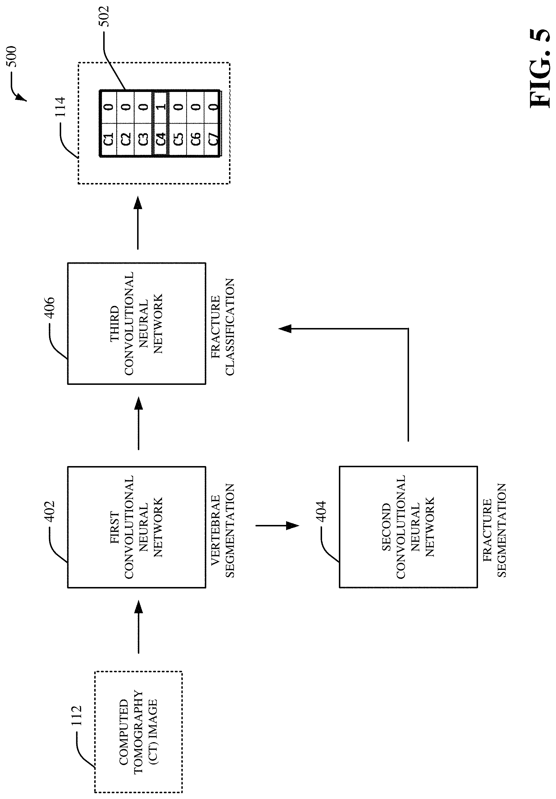
[0013] FIG. 5 illustrates yet another example system that facilitates generating and/or employing a computed tomography medical imaging fracture model, in accordance with one or more embodiments described herein;
[0014] FIG. 6 illustrates yet another example system that facilitates generating and/or employing a computed tomography medical imaging fracture model, in accordance with one or more embodiments described herein;
[0015] FIG. 7 illustrates yet another example system that facilitates generating and/or employing a computed tomography medical imaging fracture model, in accordance with one or more embodiments described herein;
[0016] FIG. 8 illustrates an example user interface, in accordance with one or more embodiments described herein;
[0017] FIG. 9 depicts a flow diagram of an example method for generating and/or employing a computed tomography medical imaging fracture model, in accordance with one or more embodiments described herein;
[0018] FIG. 10 is a schematic block diagram illustrating a suitable operating environment; and
[0019] FIG. 11 is a schematic block diagram of a sample-computing environment.
DETAILED DESCRIPTION
[0020] Various aspects of this disclosure are now described with reference to the drawings, wherein like reference numerals are used to refer to like elements throughout. In the following description, for purposes of explanation, numerous specific details are set forth in order to provide a thorough understanding of one or more aspects. It should be understood, however, that certain aspects of this disclosure may be practiced without these specific details, or with other methods, components, materials, etc. In other instances, well-known structures and devices are shown in block diagram form to facilitate describing one or more aspects.
[0021] Systems and techniques for generating and/or employing a computed tomography (CT) medical imaging spine model are presented. For instance, a deep learning architecture can be provided to facilitate detection of a medical fracture condition (e.g., a spine fracture, a cervical spine fracture, a vertebrae fracture, etc.) based on a vertebrae segmentation model and a fracture segmentation model. In an embodiment, a CT image of a cervical spine can be analyzed by the deep learning architecture to automatically label vertebrae and/to automatically detect one or more fractures in the vertebrae. In certain embodiments, the CT image can be a non-contrast CT (NCCT) image. For example, an axial NCCT image and/or a sagittal NCCT of a cervical spine can be analyzed by the deep learning architecture to automatically label vertebrae and/to automatically detect one or more fractures in the vertebrae. In another embodiment, the fracture segmentation model can be employed by the deep learning architecture to initialize and/or train a fracture classification model that detects presence or absence of a medical fracture condition (e.g., a spine fracture, a cervical spine fracture, a vertebrae fracture, etc.) in a CT image. In yet another embodiment, a first convolutional neural network (e.g., a first U-net model, a first 3D convolutional neural network model, etc.) can be trained using sagittal spine CT images and/or vertebrae segmentation outlines for vertebrae segmentation. Additionally or alternatively, a second convolutional neural network (e.g., a second U-net model, a second 3D convolutional neural network model, etc.) can be trained using axial spine CT images and/or fracture segmentation outlines for fracture segmentation. Additionally or alternatively, a third convolutional neural network (e.g., a third U-net model, a third 3D convolutional neural network model, etc.) can be trained to indicate presence of a fracture by bootstrapping the second convolutional neural network associated with fracture segmentation. In certain embodiments, an ensemble of machine learning models can be employed for vertebrae segmentation. For instance, a first machine learning model associated with axial CT images can generate first output related to vertebrae segmentation, a second machine learning model associated with sagittal CT images can generate second output related to vertebrae segmentation, and a third machine learning model associated with coronal CT images can generate third output related to vertebrae segmentation. Furthermore, a voting scheme can be employed to select the first output related to vertebrae segmentation, the second output related to vertebrae segmentation, or the third output related to vertebrae segmentation as a final prediction related to vertebrae segmentation. Additionally or alternatively, an ensemble of machine learning models can be employed for fracture segmentation. For instance, a first machine learning model associated with axial CT images can generate first output related to fracture segmentation, a second machine learning model associated with sagittal CT images can generate second output related to fracture segmentation, and a third machine learning model associated with coronal CT images can generate third output related to fracture segmentation. Furthermore, a voting scheme can be employed to select the first output related to fracture segmentation, the second output related to fracture segmentation, or the third output related to fracture segmentation as a final prediction related to fracture segmentation. Additionally or alternatively, an ensemble of machine learning models can be employed for fracture classification. For instance, a first machine learning model associated with axial CT images can generate first output related to fracture classification, a second machine learning model associated with sagittal CT images can generate second output related to fracture classification, and a third machine learning model associated with coronal CT images can generate third output related to fracture classification. Furthermore, a voting scheme can be employed to select the first output related to fracture classification, the second output related to fracture classification, or the third output related to fracture classification as a final prediction related to fracture classification.
[0022] In an embodiment, outputs of vertebrae segmentation and fracture segmentation can be combined to detect presence of a fracture in vertebrae and/or to label vertebrae with a fracture. In certain embodiments, a text output can be provided to a user device to indicate presence or absence of a medical fracture condition (e.g., a spine fracture, a cervical spine fracture, a vertebrae fracture, etc.) in a CT image. Additionally or alternatively, display data that includes a bounding box can be provided to a user device to indicate presence or absence of a medical fracture condition (e.g., a spine fracture, a cervical spine fracture, a vertebrae fracture, etc.) in a CT image. Additionally or alternatively, display data that includes a heat map can be provided to a user device to indicate presence or absence of a medical fracture condition (e.g., a spine fracture, a cervical spine fracture, a vertebrae fracture, etc.) in a CT image. Additionally or alternatively, display data that includes a probability representation for fracture classification can be provided to a user device to indicate presence or absence of a medical fracture condition (e.g., a spine fracture, a cervical spine fracture, a vertebrae fracture, etc.) in a CT image. As such, by employing systems and/or techniques associated with the medical imaging spine model disclosed herein, diagnosis speed and/or accuracy for a medical condition can be improved. A treatment decision for a medical condition can also be improved. Additionally, detection and/or localization of medical conditions for a patient associated with medical imaging data can also be improved. Accordingly, earlier intervention and/or improved outcome in treatment of a medical condition (e.g., a medical fracture condition) can be provided. Accuracy and/or efficiency for classification and/or analysis of medical imaging data can also be improved. Moreover, effectiveness of a machine learning model for classification and/or analysis of medical imaging data can be improved, performance of one or more processors that execute a machine learning model for classification and/or analysis of medical imaging data can be improved, and/or efficiency of one or more processors that execute a machine learning model for classification and/or analysis of medical imaging data can be improved.
[0023] Referring initially to FIG. 1, there is illustrated an example system 100 that facilitates generating and/or employing a CT medical imaging fracture model, according to one or more embodiments of the subject disclosure. The system 100 can be employed by various systems, such as, but not limited to medical device systems, medical imaging systems, medical diagnostic systems, medical systems, medical modeling systems, enterprise imaging solution systems, advanced diagnostic tool systems, simulation systems, image management platform systems, care delivery management systems, artificial intelligence systems, machine learning systems, neural network systems, modeling systems, aviation systems, power systems, distributed power systems, energy management systems, thermal management systems, transportation systems, oil and gas systems, mechanical systems, machine systems, device systems, cloud-based systems, heating systems, HVAC systems, medical systems, automobile systems, aircraft systems, water craft systems, water filtration systems, cooling systems, pump systems, engine systems, prognostics systems, machine design systems, and the like. In certain embodiments, the system 100 can be associated with a viewer system to facilitate visualization and/or interpretation of medical imaging data. Moreover, the system 100 and/or the components of the system 100 can be employed to use hardware and/or software to solve problems that are highly technical in nature (e.g., related to processing digital data, related to processing medical imaging data, related to medical modeling, related to medical imaging, related to artificial intelligence, etc.), that are not abstract and that cannot be performed as a set of mental acts by a human.
[0024] The system 100 can include a medical imaging component 102 that can include a vertebrae segmentation component 104, a fracture segmentation component 105 and a medical diagnosis component 106. Aspects of the systems, apparatuses or processes explained in this disclosure can constitute machine-executable component(s) embodied within machine(s), e.g., embodied in one or more computer readable mediums (or media) associated with one or more machines. Such component(s), when executed by the one or more machines, e.g., computer(s), computing device(s), virtual machine(s), etc. can cause the machine(s) to perform the operations described. The system 100 (e.g., the medical imaging component 102) can include memory 110 for storing computer executable components and instructions. The system 100 (e.g., the medical imaging component 102) can further include a processor 108 to facilitate operation of the instructions (e.g., computer executable components and instructions) by the system 100 (e.g., the medical imaging component 102).
[0025] The medical imaging component 102 (e.g., the vertebrae segmentation component 104) can receive a computed tomography (CT) image 112. The CT image 112 can be a CT image (e.g., a CT scan) generated by a medical imaging device. For example, the CT image 112 can be a CT image generated by a CT scanner device. The CT image 112 can be related to an anatomical region (e.g., a spine anatomical region, a cervical spine anatomical region, etc.) of a patient body scanned by the medical imaging device. For example, the CT image 112 can be related to an anatomical region (e.g., a spine anatomical region, a cervical spine anatomical region, etc.) of a patient body scanned by the CT scanner device. In aspect, the CT image 112 can be a two-dimensional CT image or a three-dimensional CT image. In another aspect, the CT image 112 can be represented as a series of X-ray images captured via a set of X-ray detectors (e.g., a set of X-ray detects associated with a medical imaging device) of the medical imaging device (e.g., the CT scanner device). The CT image 112 can be received directly from the medical imaging device (e.g., the CT scanner device). Alternatively, the CT image 112 can be stored in one or more databases that receives and/or stores the CT image 112 associated with the medical imaging device (e.g., the CT scanner device). In an embodiment, the CT image 112 can be a NCCT image generated without use of contrast medication by the patient associated with the anatomical region (e.g., the spine anatomical region, the cervical spine anatomical region, etc.).
[0026] The vertebrae segmentation component 104 can employ a first convolutional neural network associated with vertebrae segmentation to generate learned vertebrae segmentation data regarding the anatomical region (e.g., the spine anatomical region, the cervical spine anatomical region, etc.) related to the CT image 112. In an aspect, the vertebrae segmentation component 104 can analyze the CT image 112 using deep learning and/or one or more machine learning techniques associated with the first convolutional neural network to generate the learned vertebrae segmentation data. The learned vertebrae segmentation data can include one or more segmentation masks associated with the vertebrae included in the CT image 112. For instance, the one or more segmentation masks associated with the vertebrae included in the CT image 112 can correspond to a vertebrae C1 region, a vertebrae C2 region, a vertebrae C3 region, a vertebrae C4 region, a vertebrae C5 region, a vertebrae C6 region and/or a vertebrae C7 region of the anatomical region (e.g., a spine anatomical region, a cervical spine anatomical region, etc.) related to the CT image 112. For example, the one or more segmentation masks associated with the learned vertebrae segmentation data can be related to a location of a vertebrae C1 region, a vertebrae C2 region, a vertebrae C3 region, a vertebrae C4 region, a vertebrae C5 region, a vertebrae C6 region and/or a vertebrae C7 region included in the CT image 112. The learned vertebrae segmentation data can be, for example, deep learning data related vertebrae segmentation. For instance, the learned vertebrae segmentation data can segment a location of a vertebrae C1 region, a vertebrae C2 region, a vertebrae C3 region, a vertebrae C4 region, a vertebrae C5 region, a vertebrae C6 region and/or a vertebrae C7 region of the anatomical region (e.g., the spine anatomical region, the cervical spine anatomical region, etc.) related to the CT image 112.
[0027] In an embodiment, the first convolutional neural network employed by the vertebrae segmentation component 104 can include a set of convolutional layers associated with upsampling and/or downsampling. Furthermore, in certain embodiments, the first convolutional neural network employed by the vertebrae segmentation component 104 can include a contracting path of convolutional layers and/or an expansive path of convolutional layers. In certain embodiments, the first convolutional neural network employed by the vertebrae segmentation component 104 can employ context data associated with previous inputs provided to the first convolutional neural network and/or previous outputs provided by the first convolutional neural network to analyze the CT image 112. In a non-limiting embodiment, the first convolutional neural network employed by the vertebrae segmentation component 104 can be an adapted U-net model for analyzing the CT image 112. For instance, the first convolutional neural network employed by the vertebrae segmentation component 104 can be a fully convolutional network that employs successive convolutional layers associated with downsampling followed by successive convolutional layers associated with upsampling. The first convolutional neural network employed by the vertebrae segmentation component 104 can also employ a segmentation loss function to modify one or more portions of the first convolutional neural network. Additionally or alternatively, the first convolutional neural network employed by the vertebrae segmentation component 104 can employ a classification loss function to modify one or more portions of the first convolutional neural network. However, it is to be appreciated that the first convolutional neural network employed by the vertebrae segmentation component 104 can be a different type of convolutional neural network. In an embodiment, the first convolutional neural network employed by the vertebrae segmentation component 104 can be a medical imaging vertebrae segmentation model that is trained to segment one or more vertebras with respect to the anatomical region (e.g., the spine anatomical region, the cervical spine anatomical region, etc.) of the patient body. In certain embodiments, the vertebrae segmentation component 104 can generate the learned vertebrae segmentation data and/or other data during a training phase for the first convolutional neural network. For instance, the vertebrae segmentation component 104 can employ a set of CT images (e.g., a set of sagittal CT images) as training data for the first convolutional neural network to train the first convolutional neural network to segment vertebrae associated with an anatomical region (e.g., a spine anatomical region, a cervical spine anatomical region, etc.). In certain embodiments, the vertebrae segmentation component 104 can modify one or more portions of the first convolutional neural network during the training phase to facilitate segmenting vertebrae associated with an anatomical region (e.g., a spine anatomical region, a cervical spine anatomical region, etc.).
[0028] In certain embodiments, the vertebrae segmentation component 104 can extract information that is indicative of correlations, inferences and/or expressions from the CT image 112 based on the first convolutional neural network (e.g., a network of convolutional layers of the first convolutional neural network). Additionally or alternatively, the vertebrae segmentation component 104 can generate the learned vertebrae segmentation data based on the correlations, inferences and/or expressions. The vertebrae segmentation component 104 can generate the learned vertebrae segmentation data based on a network of convolutional layers associated with the first convolutional neural network. In an aspect, the vertebrae segmentation component 104 can perform learning with respect to the CT image 112 explicitly or implicitly using a network of convolutional layers associated with the first convolutional neural network. The vertebrae segmentation component 104 can also employ an automatic classification system and/or an automatic classification process to facilitate analysis of the CT image 112. For example, the vertebrae segmentation component 104 can employ a probabilistic and/or statistical-based analysis (e.g., factoring into the analysis utilities and costs) to learn and/or generate inferences with respect to the CT image 112. The vertebrae segmentation component 104 can employ, for example, a support vector machine (SVM) classifier to learn and/or generate inferences for the CT image 112. Additionally or alternatively, the vertebrae segmentation component 104 can employ other classification techniques associated with Bayesian networks, decision trees and/or probabilistic classification models. Classifiers employed by the vertebrae segmentation component 104 can be explicitly trained (e.g., via a generic training data) as well as implicitly trained (e.g., via receiving extrinsic information). For example, with respect to SVM's, SVM's can be configured via a learning or training phase within a classifier constructor and feature selection module. A classifier can be a function that maps an input attribute vector, x=(x1, x2, x3, x4, xn), to a confidence that the input belongs to a class--that is, f(x)=confidence(class).
[0029] The fracture segmentation component 105 can employ a second convolutional neural network associated with fracture segmentation to generate learned fracture segmentation data regarding the anatomical region (e.g., the spine anatomical region, the cervical spine anatomical region, etc.) related to the CT image 112. In an aspect, the fracture segmentation component 105 can analyze the learned vertebrae segmentation data using deep learning and/or one or more machine learning techniques associated with the second convolutional neural network to generate the learned fracture segmentation data. The learned fracture segmentation data can include one or more segmentation masks associated with one or more fractures included in the vertebrae associated with the CT image 112. For instance, in an embodiment, the learned fracture segmentation data can include a pixelwise label associated with one or more fractures included in the vertebrae associated with the CT image 112. The pixelwise label can include a set of pixel classifications regarding whether or not pixels in the CT image 112 is associated with a fracture or no fracture. For instance, every pixel in the CT image 112 can be classified as a fracture or not fracture. As an example, the fracture segmentation component 105 can employ the second convolutional neural network associated with the fracture segmentation to generate a first classification for a first pixel included in the learned vertebrae segmentation data, a second classification for a second pixel included in the learned vertebrae segmentation data, etc. In an aspect, a size of the pixelwise label can correspond to a size of the CT image 112. In another aspect, the learned fracture segmentation data can segment a fracture located in a vertebrae C1 region, a vertebrae C2 region, a vertebrae C3 region, a vertebrae C4 region, a vertebrae C5 region, a vertebrae C6 region and/or a vertebrae C7 region of the anatomical region (e.g., a spine anatomical region, a cervical spine anatomical region, etc.) related to the CT image 112. Furthermore, the learned fracture segmentation data can be, for example, deep learning data related to fracture segmentation. For instance, the learned fracture segmentation data can segment a fracture in a vertebrae C1 region, a vertebrae C2 region, a vertebrae C3 region, a vertebrae C4 region, a vertebrae C5 region, a vertebrae C6 region and/or a vertebrae C7 region of the anatomical region (e.g., the spine anatomical region, the cervical spine anatomical region, etc.) related to the CT image 112.
[0030] In an embodiment, the second convolutional neural network employed by the fracture segmentation component 105 can include a set of convolutional layers associated with upsampling and/or downsampling. Furthermore, in certain embodiments, the second convolutional neural network employed by the fracture segmentation component 105 can include a contracting path of convolutional layers and/or an expansive path of convolutional layers. In certain embodiments, the second convolutional neural network employed by the fracture segmentation component 105 can employ context data associated with previous inputs provided to the second convolutional neural network and/or previous outputs provided by the second convolutional neural network to analyze the learned vertebrae segmentation data and/or the CT image 112. In a non-limiting embodiment, the second convolutional neural network employed by the fracture segmentation component 105 can be an adapted U-net model for analyzing the learned vertebrae segmentation data and/or the CT image 112. For instance, the second convolutional neural network employed by the fracture segmentation component 105 can be a fully convolutional network that employs successive convolutional layers associated with downsampling followed by successive convolutional layers associated with upsampling. The second convolutional neural network employed by the fracture segmentation component 105 can also employ a segmentation loss function to modify one or more portions of the second convolutional neural network. Additionally or alternatively, the second convolutional neural network employed by the fracture segmentation component 105 can employ a classification loss function to modify one or more portions of the second convolutional neural network. However, it is to be appreciated that the second convolutional neural network employed by the fracture segmentation component 105 can be a different type of convolutional neural network. In an embodiment, the second convolutional neural network employed by the fracture segmentation component 105 can be a medical imaging fracture segmentation model that is trained to segment one or more fractures with respect to the anatomical region (e.g., the spine anatomical region, the cervical spine anatomical region, etc.) of the patient body. In certain embodiments, the fracture segmentation component 105 can generate the learned fracture segmentation data and/or other data during a training phase for the second convolutional neural network. For instance, the fracture segmentation component 105 can employ a set of CT images (e.g., a set of axial CT images) as training data for the second convolutional neural network to train the second convolutional neural network to segment one or more fractures associated with an anatomical region (e.g., a spine anatomical region, a cervical spine anatomical region, etc.). In certain embodiments, the fracture segmentation component 105 can modify one or more portions of the second convolutional neural network during the training phase to facilitate segmenting one or more fractures associated with an anatomical region (e.g., a spine anatomical region, a cervical spine anatomical region, etc.).
[0031] In certain embodiments, the fracture segmentation component 105 can extract information that is indicative of correlations, inferences and/or expressions from the learned vertebrae segmentation data and/or the CT image 112 based on the second convolutional neural network (e.g., a network of convolutional layers of the second convolutional neural network). Additionally or alternatively, the fracture segmentation component 105 can generate the learned fracture segmentation data based on the correlations, inferences and/or expressions. The fracture segmentation component 105 can generate the learned fracture segmentation data based on a network of convolutional layers associated with the second convolutional neural network. In an aspect, the fracture segmentation component 105 can perform learning with respect to the learned vertebrae segmentation data and/or the CT image 112 explicitly or implicitly using a network of convolutional layers associated with the second convolutional neural network. The fracture segmentation component 105 can also employ an automatic classification system and/or an automatic classification process to facilitate analysis of the learned vertebrae segmentation data and/or the CT image 112. For example, the fracture segmentation component 105 can employ a probabilistic and/or statistical-based analysis (e.g., factoring into the analysis utilities and costs) to learn and/or generate inferences with respect to the learned vertebrae segmentation data and/or the CT image 112. The fracture segmentation component 105 can employ, for example, a SVM classifier to learn and/or generate inferences for the learned vertebrae segmentation data and/or the CT image 112. Additionally or alternatively, the fracture segmentation component 105 can employ other classification techniques associated with Bayesian networks, decision trees and/or probabilistic classification models. Classifiers employed by the fracture segmentation component 105 can be explicitly trained (e.g., via a generic training data) as well as implicitly trained (e.g., via receiving extrinsic information). For example, with respect to SVM's, SVM's can be configured via a learning or training phase within a classifier constructor and feature selection module. A classifier can be a function that maps an input attribute vector, x=(x1, x2, x3, x4, xn), to a confidence that the input belongs to a class--that is, f(x)=confidence(class).
[0032] The medical diagnosis component 106 can employ information provided by the vertebrae segmentation component 104 (e.g., the learned vertebrae segmentation data) and/or information provided by the fracture segmentation component 105 (e.g., the learned fracture segmentation data) to generate medical diagnosis data 114. For instance, the medical diagnosis component 106 can employ information provided by the vertebrae segmentation component 104 (e.g., the learned vertebrae segmentation data) and/or information provided by the fracture segmentation component 105 (e.g., the learned fracture segmentation data) to classify and/or localize a medical fracture condition (e.g., a spine fracture, a cervical spine fracture, a vertebrae fracture, etc.) associated with the CT image 112. In an embodiment, the medical diagnosis component 106 can employ information provided by the vertebrae segmentation component 104 (e.g., the learned vertebrae segmentation data) and/or information provided by the fracture segmentation component 105 (e.g., the learned fracture segmentation data) to detect presence or absence of a medical fracture condition (e.g., a spine fracture, a cervical spine fracture, a vertebrae fracture, etc.) in the CT image 112. In an embodiment, the medical diagnosis component 106 can employ a third convolutional neural network associated with fracture classification to generate the medical diagnosis data 114. For instance, in an embodiment, the medical diagnosis component 106 can employ a third convolutional neural network associated with fracture classification to detect presence or absence of a medical fracture condition (e.g., a spine fracture, a cervical spine fracture, a vertebrae fracture, etc.) in the CT image 112. In an aspect, the medical diagnosis component 106 can analyze the learned vertebrae segmentation data and/or the learned fracture segmentation data using deep learning and/or one or more machine learning techniques associated with the third convolutional neural network to generate the medical diagnosis data 114. The medical diagnosis data 114 can include one or more classifications associated with one or more fractures included in the vertebrae associated with the CT image 112. For example, the medical diagnosis data 114 can detect presence or absence of a medical fracture condition (e.g., a spine fracture, a cervical spine fracture, a vertebrae fracture, etc.) in the anatomical region (e.g., the spine anatomical region, the cervical spine anatomical region, etc.) related to the CT image 112.
[0033] In aspect, the medical diagnosis data 114 can classify a fracture located in a vertebrae C1 region, a vertebrae C2 region, a vertebrae C3 region, a vertebrae C4 region, a vertebrae C5 region, a vertebrae C6 region and/or a vertebrae C7 region of the anatomical region (e.g., a spine anatomical region, a cervical spine anatomical region, etc.) related to the CT image 112. Furthermore, the medical diagnosis data 114 can be, for example, deep learning data related to fracture classification. For instance, the medical diagnosis data 114 can classify and/or determine a location of a fracture in a vertebrae C1 region, a vertebrae C2 region, a vertebrae C3 region, a vertebrae C4 region, a vertebrae C5 region, a vertebrae C6 region and/or a vertebrae C7 region of the anatomical region (e.g., the spine anatomical region, the cervical spine anatomical region, etc.) related to the CT image 112. In certain embodiments, the medical diagnosis component 106 can determine a probability of the presence of the medical fracture condition in the CT image based on the learned vertebrae segmentation data and/or the learned fracture segmentation data. For example, the medical diagnosis component 106 can determine a probability of a medical fracture condition being located in a vertebrae C1 region, a vertebrae C2 region, a vertebrae C3 region, a vertebrae C4 region, a vertebrae C5 region, a vertebrae C6 region and/or a vertebrae C7 region of the anatomical region (e.g., the spine anatomical region, the cervical spine anatomical region, etc.) related to the CT image 112. In certain embodiments, the medical diagnosis component 106 can additionally or alternatively determine a localization of the medical fracture condition in the CT image based on the learned vertebrae segmentation data and/or the learned fracture segmentation data. For example, the medical diagnosis component 106 can localize a medical fracture condition in a vertebrae C1 region, a vertebrae C2 region, a vertebrae C3 region, a vertebrae C4 region, a vertebrae C5 region, a vertebrae C6 region and/or a vertebrae C7 region of the anatomical region (e.g., the spine anatomical region, the cervical spine anatomical region, etc.) related to the CT image 112.
[0034] In an embodiment, the third convolutional neural network employed by the medical diagnosis component 106 can include a set of convolutional layers associated with upsampling and/or downsampling. Furthermore, in certain embodiments, the third convolutional neural network employed by the medical diagnosis component 106 can include a contracting path of convolutional layers and/or an expansive path of convolutional layers. In certain embodiments, the third convolutional neural network employed by the medical diagnosis component 106 can employ context data associated with previous inputs provided to the third convolutional neural network and/or previous outputs provided by the third convolutional neural network to analyze the learned vertebrae segmentation data, the learned fracture segmentation data and/or the CT image 112. In a non-limiting embodiment, the third convolutional neural network employed by the medical diagnosis component 106 can be an adapted U-net model for analyzing the learned vertebrae segmentation data, the learned fracture segmentation data and/or the CT image 112. For instance, the third convolutional neural network employed by the medical diagnosis component 106 can be a fully convolutional network that employs successive convolutional layers associated with downsampling followed by successive convolutional layers associated with upsampling. The third convolutional neural network employed by the medical diagnosis component 106 can also employ a segmentation loss function to modify one or more portions of the third convolutional neural network. Additionally or alternatively, the third convolutional neural network employed by the medical diagnosis component 106 can employ a classification loss function to modify one or more portions of the third convolutional neural network. However, it is to be appreciated that the third convolutional neural network employed by the medical diagnosis component 106 can be a different type of convolutional neural network. In an embodiment, the third convolutional neural network employed by the medical diagnosis component 106 can be a medical imaging fracture classification model that is trained to classify and/or locate one or more fractures with respect to the anatomical region (e.g., the spine anatomical region, the cervical spine anatomical region, etc.) of the patient body. In certain embodiments, the medical diagnosis component 106 can generate the medical diagnosis data 114 and/or other data during a training phase for the third convolutional neural network. For instance, the medical diagnosis component 106 can employ a set of CT images (e.g., a set of axial CT images) as training data for the third convolutional neural network to train the third convolutional neural network to classify and/or identify one or more fractures associated with an anatomical region (e.g., a spine anatomical region, a cervical spine anatomical region, etc.). In certain embodiments, the medical diagnosis component 106 can modify one or more portions of the third convolutional neural network during the training phase to facilitate classifying and/or identifying one or more fractures associated with an anatomical region (e.g., a spine anatomical region, a cervical spine anatomical region, etc.).
[0035] In certain embodiments, the medical diagnosis component 106 can extract information that is indicative of correlations, inferences and/or expressions from the learned vertebrae segmentation data, the learned fracture segmentation data and/or the CT image 112 based on the third convolutional neural network (e.g., a network of convolutional layers of the third convolutional neural network). Additionally or alternatively, the medical diagnosis component 106 can generate the medical diagnosis data 114 based on the correlations, inferences and/or expressions. The medical diagnosis component 106 can generate the medical diagnosis data 114 based on a network of convolutional layers associated with the third convolutional neural network. In an aspect, the medical diagnosis component 106 can perform learning with respect to the learned vertebrae segmentation data, the learned fracture segmentation data and/or the CT image 112 explicitly or implicitly using a network of convolutional layers associated with the third convolutional neural network. The medical diagnosis component 106 can also employ an automatic classification system and/or an automatic classification process to facilitate analysis of the learned vertebrae segmentation data, the learned fracture segmentation data and/or the CT image 112. For example, the medical diagnosis component 106 can employ a probabilistic and/or statistical-based analysis (e.g., factoring into the analysis utilities and costs) to learn and/or generate inferences with respect to the learned vertebrae segmentation data, the learned fracture segmentation data and/or the CT image 112. The medical diagnosis component 106 can employ, for example, a SVM classifier to learn and/or generate inferences for the learned vertebrae segmentation data, the learned fracture segmentation data and/or the CT image 112. Additionally or alternatively, the medical diagnosis component 106 can employ other classification techniques associated with Bayesian networks, decision trees and/or probabilistic classification models. Classifiers employed by the medical diagnosis component 106 can be explicitly trained (e.g., via a generic training data) as well as implicitly trained (e.g., via receiving extrinsic information). For example, with respect to SVM's, SVM's can be configured via a learning or training phase within a classifier constructor and feature selection module. A classifier can be a function that maps an input attribute vector, x=(x1, x2, x3, x4, xn), to a confidence that the input belongs to a class--that is, f(x)=confidence(class). In certain embodiments, the medical diagnosis component 106 can generate a contour mask associated with the medical fracture condition (e.g., the spine fracture, the cervical spine fracture, the vertebrae fracture, etc.) based on the learned vertebrae segmentation data and/or the learned fracture segmentation data. The contour mask can be, for example, an image that classifies a segmentation for the medical fracture condition. For instance, the contour mask can be an image that identifies a location of the area of the anatomical region (e.g., the spine anatomical region, the cervical spine anatomical region, etc.).
[0036] In an aspect, the medical diagnosis component 106 can determine a prediction for the medical fracture condition (e.g., the spine fracture, the cervical spine fracture, the vertebrae fracture, etc.) associated with the CT image 112. For example, the medical diagnosis component 106 can determine a probability score for the medical fracture condition (e.g., the spine fracture, the cervical spine fracture, the vertebrae fracture, etc.) associated with the CT image 112. In certain embodiments, the medical diagnosis component 106 can determine one or more confidence scores for the classification and/or the localization of the medical fracture condition (e.g., the spine fracture, the cervical spine fracture, the vertebrae fracture, etc.). For example, a first portion of the anatomical region (e.g., the spine anatomical region, the cervical spine anatomical region, etc.) with a greatest likelihood of the medical fracture condition can be assigned a first confidence score, a second portion of the anatomical region (e.g., the spine anatomical region, the cervical spine anatomical region, etc.) with a lesser degree of likelihood of the medical fracture condition can be assigned a second confidence score, etc. A medical condition classified and/or localized by the medical diagnosis component 106 can additionally or alternatively include, for example, a bone disease, a tumor, a cancer, or another type of medical condition associated with the anatomical region (e.g., the spine anatomical region, the cervical spine anatomical region, etc.). In certain embodiments, the medical diagnosis data 114 can be employed for a treatment decision associated with a patient body related to the CT image 112. For example, the medical diagnosis data 114 can be employed for a determining a particular fracture treatment associated with a patient body related to the CT image 112.
[0037] It is to be appreciated that technical features of the medical imaging component 102 (e.g., the vertebrae segmentation component 104, the fracture segmentation component 105 and/or the medical diagnosis component 106) are highly technical in nature and not abstract ideas. Processing threads of the medical imaging component 102 (e.g., the vertebrae segmentation component 104, the fracture segmentation component 105 and/or the medical diagnosis component 106) that process and/or analyze the CT image 112, perform a machine learning process, generate the medical diagnosis data 114, etc. cannot be performed by a human (e.g., are greater than the capability of a single human mind). For example, the amount of the CT image 112 processed, the speed of processing of the CT image 112, and/or the data types of the CT image 112 processed by the medical imaging component 102 (e.g., the vertebrae segmentation component 104, the fracture segmentation component 105 and/or the medical diagnosis component 106) over a certain period of time can be respectively greater, faster and different than the amount, speed and data type that can be processed by a single human mind over the same period of time. Furthermore, the CT image 112 processed by the medical imaging component 102 (e.g., the vertebrae segmentation component 104, the fracture segmentation component 105 and/or the medical diagnosis component 106) can be one or more medical images generated by sensors of a medical imaging device. Moreover, the medical imaging component 102 (e.g., the vertebrae segmentation component 104, the fracture segmentation component 105 and/or the medical diagnosis component 106) can be fully operational towards performing one or more other functions (e.g., fully powered on, fully executed, etc.) while also analyzing the CT image 112.
[0038] FIG. 2 illustrates an example, non-limiting system 200 in accordance with one or more embodiments described herein. Repetitive description of like elements employed in other embodiments described herein is omitted for sake of brevity. The system 200 includes the medical imaging component 102. In the embodiment shown in FIG. 2, the medical imaging component 102 can include the vertebrae segmentation component 104, the fracture segmentation component 105, the medical diagnosis component 106, a display component 202, the processor 108 and/or the memory 110. The display component 202 can generate display data associated with the medical diagnosis data 114. Furthermore, the display component 202 can provide the display data to a user device in a human-interpretable format. In an embodiment, the display component 202 can generate display data associated with the presence or the absence of the medical fracture condition (e.g., the spine fracture, the cervical spine fracture, the vertebrae fracture, etc.) in a human-interpretable format. Additionally or alternatively, the display component 202 can generate display data associated with the contour mask associated with the medical fracture condition (e.g., the spine fracture, the cervical spine fracture, the vertebrae fracture, etc.) in a human-interpretable format. Additionally or alternatively, the display component 202 can generate display data associated with other information regarding the medical fracture condition (e.g., the spine fracture, the cervical spine fracture, the vertebrae fracture, etc.) in a human-interpretable format. In certain embodiments, the display component 202 can generate a multi-dimensional visualization associated with the medical diagnosis data 114. For example, the display component 202 can generate a multi-dimensional visualization associated with the presence or the absence of the medical fracture condition (e.g., the spine fracture, the cervical spine fracture, the vertebrae fracture, etc.). Additionally or alternatively, the display component 202 can generate a multi-dimensional visualization associated with the contour mask associated with the medical fracture condition (e.g., the spine fracture, the cervical spine fracture, the vertebrae fracture, etc.). Additionally or alternatively, the display component 202 can generate a multi-dimensional visualization associated with the CT image 112. Additionally or alternatively, the display component 202 can generate a multi-dimensional visualization associated with other information regarding the medical fracture condition (e.g., the spine fracture, the cervical spine fracture, the vertebrae fracture, etc.). The multi-dimensional visualization can be a graphical representation of the CT image 112 and/or other medical imaging data that shows a classification and/or a location of the medical fracture condition (e.g., the spine fracture, the cervical spine fracture, the vertebrae fracture, etc.) with respect to a patient body. In certain embodiments, the display component 202 can generate a localization queue associated with the medical fracture condition (e.g., the spine fracture, the cervical spine fracture, the vertebrae fracture, etc.). Furthermore, the localization queue can be rendered and/or overlaid onto the multi-dimensional visualization and/or the CT image 112. In certain embodiments, the display component 202 can generate a bounding box associated with the medical fracture condition (e.g., the spine fracture, the cervical spine fracture, the vertebrae fracture, etc.). Furthermore, the bounding box can be rendered and/or overlaid onto the multi-dimensional visualization and/or the CT image 112. In certain embodiments, the display component 202 can generate a heat map associated with the medical fracture condition (e.g., the spine fracture, the cervical spine fracture, the vertebrae fracture, etc.). Furthermore, a visual indictor associated with the heat map can be rendered and/or overlaid onto the multi-dimensional visualization and/or the CT image 112. In certain embodiments, the display component 202 can generate a probability representation associated with the medical fracture condition (e.g., the spine fracture, the cervical spine fracture, the vertebrae fracture, etc.).
[0039] The display component 202 can also generate, in certain embodiments, a graphical user interface of the multi-dimensional visualization of the medical diagnosis data 114. For example, the display component 202 can render a 2D visualization of the portion of the anatomical region (e.g., the spine anatomical region, the cervical spine anatomical region, etc.) on a graphical user interface associated with a display of a user device such as, but not limited to, a computing device, a computer, a desktop computer, a laptop computer, a monitor device, a smart device, a smart phone, a mobile device, a handheld device, a tablet, a portable computing device, a virtual reality device, a wearable device, or another type of user device associated with a display. In an aspect, the multi-dimensional visualization can include the medical diagnosis data 114. In certain embodiments, the medical diagnosis data 114 associated with the multi-dimensional visualization can be indicative of a visual representation of the classification and/or the localization for the anatomical region (e.g., the spine anatomical region, the cervical spine anatomical region, etc.). In certain embodiments, the medical diagnosis data 114 can be rendered on the CT image 112 and/or a 3D model associated with the CT image 112 as one or more dynamic visual elements. In an aspect, the display component 202 can alter visual characteristics (e.g., color, size, hues, shading, etc.) of at least a portion of the medical diagnosis data 114 associated with the multi-dimensional visualization based on the classification and/or the localization for the portion of the anatomical region (e.g., the spine anatomical region, the cervical spine anatomical region, etc.). For example, the classification and/or the localization for the medical fracture condition (e.g., the spine fracture, the cervical spine fracture, the vertebrae fracture, etc.) can be presented as different visual characteristics (e.g., colors, sizes, hues or shades, etc.), based on a result of deep learning and/or medical imaging diagnosis by the vertebrae segmentation component 104, the fracture segmentation component 105 and/or the medical diagnosis component 106. As such, a user can view, analyze and/or interact with the medical diagnosis data 114 associated with the multi-dimensional visualization. In certain embodiments, the display component 202 can generate and/or transmit one or more alerts based on the medical diagnosis data 114. An alert generated and/or transmitted by the display component 202 can be a message and/or a notification to provide machine-to-person communication related to the medical diagnosis data 114. Furthermore, an alert generated and/or transmitted by the display component 202 can include textual data, audio data, video data, graphic data, graphical user interface data, and/or other data related to the medical diagnosis data 114.
[0040] FIG. 3 illustrates an example, non-limiting system 300 in accordance with one or more embodiments described herein. Repetitive description of like elements employed in other embodiments described herein is omitted for sake of brevity. The system 300 can be, for example, a network environment (e.g., a network computing environment, a healthcare network environment, etc.) to facilitate generating and/or employing a medical imaging fracture model. The system 300 includes a server 302, one or more medical imaging devices 304 and/or a user device 306. The server 302 can include the medical imaging component 102. The medical imaging component 102 can include the vertebrae segmentation component 104, the fracture segmentation component 105, the medical diagnosis component 106, the display component 202, the processor 108 and/or the memory 110. In certain embodiments, the medical imaging component 102 can be alternatively included in the medical imaging device 304. In certain embodiments, a portion of the medical imaging component 102 can be included in the server 302 and another portion of the medical imaging component 102 can be included in the one or more medical imaging devices 304. The one or more medical imaging devices 304 can generate, capture and/or process at least a portion of the CT image 112. The one or more medical imaging devices 304 can include, for example, one or more CT scanner devices. In certain embodiments, the one or more medical imaging devices 304 can additionally or alternatively include one or more magnetic resonance imaging (MRI) scanner devices, one or more computerized axial tomography (CAT) devices, one or more X-ray devices, one or more positron emission tomography (PET) devices, one or more ultrasound devices, and/or one or more other types of medical imaging devices. In an embodiment, one or more CT scanner devices from the one or more medical imaging devices 304 can generate the CT image 112. For example, a set of X-ray detectors of one or more CT scanner devices from the one or more medical imaging devices 304 can facilitate generating, capturing and/or processing at least a portion of the CT image 112.
[0041] The user device 306 can be an electronic device associated with a display. For example, the user device 306 can be a screen, a monitor, a projector wall, a computing device, an electronic device, a desktop computer, a laptop computer, a smart device, a smart phone, a mobile device, a handheld device, a tablet device, a virtual reality device, a portable computing device, a wearable device, or another display device associated with a display configured to present information associated with the medical diagnosis data 114 in a human-interpretable format. In an embodiment, the user device 306 can include a graphical user interface to facilitate display of information associated with the medical diagnosis data 114 in a human-interpretable format. In certain embodiments, the user device 306 can receive one or more alerts from the medical imaging component 102 (e.g., the display component 202) of the server 302. Additionally or alternatively, in certain embodiments, the one or more medical imaging devices 304 can receive one or more alerts from the medical imaging component 102 (e.g., the display component 202) of the server 302. In an embodiment, the server 302 can be in communication with the one or more medical imaging devices 304 and/or the user device 306 via a network 308. The network 308 can be a communication network, a wireless network, a wired network, an internet protocol (IP) network, a voice over IP network, an internet telephony network, a mobile telecommunications network or another type of network. In certain embodiments, visual characteristics (e.g., color, size, hues, shading, etc.) of a visual element associated with the medical diagnosis data 114 and/or presented via the user device 306 can be altered based on a value of the medical diagnosis data 114. In certain embodiments, a user can view, analyze and/or interact with the CT image 112 and/or the medical diagnosis data 114 via the user device 306.
[0042] FIG. 4 illustrates an example, non-limiting system 400 in accordance with one or more embodiments described herein. Repetitive description of like elements employed in other embodiments described herein is omitted for sake of brevity. The system 400 includes a first convolutional neural network 402, a second convolutional neural network 404 and a third convolutional neural network 406. The first convolutional neural network 402 can be associated with vertebrae segmentation, the second convolutional neural network 404 can be associated with fracture segmentation, and the third convolutional neural network 406 can be associated with fracture classification. For example, the first convolutional neural network 402 can be employed by the vertebrae segmentation component 104, the second convolutional neural network 404 can be employed by the fracture segmentation component 105, and the third convolutional neural network 406 can be employed by the medical diagnosis component 106. In an embodiment, the first convolutional neural network 402 can perform vertebrae segmentation with respect to a sagittal plane view of the CT image 112 to segment cervical spine vertebrae included in the CT image 112. For instance, the first convolutional neural network 402 can perform vertebrae segmentation with respect to a sagittal plane view of the CT image 112 to segment a vertebrae C1 region, a vertebrae C2 region, a vertebrae C3 region, a vertebrae C4 region, a vertebrae C5 region, a vertebrae C6 region and/or a vertebrae C7 region of the anatomical region (e.g., a spine anatomical region, a cervical spine anatomical region, etc.) included in the CT image 112. Furthermore, in an embodiment, the first convolutional neural network 402 can provide one or more downstream vertebrae specific models for vertebrae segmentation. In certain embodiments, the first convolutional neural network 402 can employ an ensemble of convolutional neural network models for vertebrae segmentation. For instance, a first convolutional neural network model for the first convolutional neural network 402 can perform vertebrae segmentation with respect to a sagittal plane view of the CT image 112 to segment a vertebrae C1 region, a vertebrae C2 region, a vertebrae C3 region, a vertebrae C4 region, a vertebrae C5 region, a vertebrae C6 region and/or a vertebrae C7 region of the anatomical region (e.g., a spine anatomical region, a cervical spine anatomical region, etc.) included in the CT image 112. Additionally or alternatively, a second convolutional neural network model for the first convolutional neural network 402 can perform vertebrae segmentation with respect to an axial plane view of the CT image 112 to segment a vertebrae C1 region, a vertebrae C2 region, a vertebrae C3 region, a vertebrae C4 region, a vertebrae C5 region, a vertebrae C6 region and/or a vertebrae C7 region of the anatomical region (e.g., a spine anatomical region, a cervical spine anatomical region, etc.) included in the CT image 112. Additionally or alternatively, a third convolutional neural network model for the first convolutional neural network 402 can perform vertebrae segmentation with respect to a coronal plane view of the CT image 112 to segment a vertebrae C1 region, a vertebrae C2 region, a vertebrae C3 region, a vertebrae C4 region, a vertebrae C5 region, a vertebrae C6 region and/or a vertebrae C7 region of the anatomical region (e.g., a spine anatomical region, a cervical spine anatomical region, etc.) included in the CT image 112. Furthermore, the first convolutional neural network 402 can select data associated with the first convolutional neural network model, the second convolutional neural network model, or the convolutional neural network model as vertebrae segmentation data related to the CT image 112.
[0043] In another embodiment, the second convolutional neural network 404 can perform fracture segmentation with respect to an axial plane view of the CT image 112 to segment cervical spine fractures. For instance, the second convolutional neural network 404 can perform fracture segmentation with respect to an axial plane view of the CT image 112 to segment a fracture included in a vertebrae C1 region, a vertebrae C2 region, a vertebrae C3 region, a vertebrae C4 region, a vertebrae C5 region, a vertebrae C6 region and/or a vertebrae C7 region of the anatomical region (e.g., a spine anatomical region, a cervical spine anatomical region, etc.). In certain embodiments, the second convolutional neural network 404 can employ an ensemble of convolutional neural network models for fracture segmentation. For instance, a first convolutional neural network model for the second convolutional neural network 404 can perform fracture segmentation with respect to an axial plane view of the CT image 112 to segment a fracture included in a vertebrae C1 region, a vertebrae C2 region, a vertebrae C3 region, a vertebrae C4 region, a vertebrae C5 region, a vertebrae C6 region and/or a vertebrae C7 region of the anatomical region (e.g., a spine anatomical region, a cervical spine anatomical region, etc.) included in the CT image 112. Additionally or alternatively, a second convolutional neural network model for the second convolutional neural network 404 can perform fracture segmentation with respect to a sagittal plane view of the CT image 112 to segment a fracture included in a vertebrae C1 region, a vertebrae C2 region, a vertebrae C3 region, a vertebrae C4 region, a vertebrae C5 region, a vertebrae C6 region and/or a vertebrae C7 region of the anatomical region (e.g., a spine anatomical region, a cervical spine anatomical region, etc.) included in the CT image 112. Additionally or alternatively, a third convolutional neural network model for the second convolutional neural network 404 can perform fracture segmentation with respect to a coronal plane view of the CT image 112 to segment a fracture included in a vertebrae C1 region, a vertebrae C2 region, a vertebrae C3 region, a vertebrae C4 region, a vertebrae C5 region, a vertebrae C6 region and/or a vertebrae C7 region of the anatomical region (e.g., a spine anatomical region, a cervical spine anatomical region, etc.) included in the CT image 112. Furthermore, the second convolutional neural network 404 can select data associated with the first convolutional neural network model, the second convolutional neural network model, or the convolutional neural network model as fracture segmentation data related to the CT image 112. Furthermore, in an embodiment, the second convolutional neural network 404 can be employed to initialize (e.g., bootstrap) the third convolutional neural network 406. Moreover, in certain embodiments, the second convolutional neural network 404 can individually analyze vertebrae to generate a prediction regarding fracture segmentation. Alternatively, in certain embodiments, the second convolutional neural network 404 can group two or more vertebrae together to generate a prediction regarding fracture segmentation. For example, in certain embodiments, the second convolutional neural network 404 can group at least a vertebrae C1 region and a vertebrae C2 region together to determine a fracture segmentation for the group that includes at least the vertebrae C1 region and the vertebrae C2 region.
[0044] The third convolutional neural network 406 can perform fracture classification with respect to an axial plane view of the CT image 112 to classify a cervical spine as fractured or non-fractured. For instance, the third convolutional neural network 406 can perform fracture classification with respect to an axial plane view of the CT image 112 to classify a vertebrae C1 region as fractured or non-fractured, a vertebrae C2 region as fractured or non-fractured, a vertebrae C3 region as fractured or non-fractured, a vertebrae C4 region as fractured or non-fractured, a vertebrae C5 region as fractured or non-fractured, a vertebrae C6 region as fractured or non-fractured, and/or a vertebrae C7 region as fractured or non-fractured. Furthermore, in an embodiment, the third convolutional neural network 406 can be employed to generate the medical diagnosis data 114. In certain embodiments, the third convolutional neural network 406 can employ an ensemble of convolutional neural network models for fracture classification. For instance, a first convolutional neural network model for the third convolutional neural network 406 can perform fracture classification with respect to an axial plane view of the CT image 112 to classify a vertebrae C1 region as fractured or non-fractured, a vertebrae C2 region as fractured or non-fractured, a vertebrae C3 region as fractured or non-fractured, a vertebrae C4 region as fractured or non-fractured, a vertebrae C5 region as fractured or non-fractured, a vertebrae C6 region as fractured or non-fractured, and/or a vertebrae C7 region as fractured or non-fractured. Additionally or alternatively, a second convolutional neural network model for the third convolutional neural network 406 can perform fracture classification with respect to a sagittal plane view of the CT image 112 to classify a vertebrae C1 region as fractured or non-fractured, a vertebrae C2 region as fractured or non-fractured, a vertebrae C3 region as fractured or non-fractured, a vertebrae C4 region as fractured or non-fractured, a vertebrae C5 region as fractured or non-fractured, a vertebrae C6 region as fractured or non-fractured, and/or a vertebrae C7 region as fractured or non-fractured. Additionally or alternatively, a third convolutional neural network model for the third convolutional neural network 406 can perform fracture classification with respect to a coronal plane view of the CT image 112 to classify a vertebrae C1 region as fractured or non-fractured, a vertebrae C2 region as fractured or non-fractured, a vertebrae C3 region as fractured or non-fractured, a vertebrae C4 region as fractured or non-fractured, a vertebrae C5 region as fractured or non-fractured, a vertebrae C6 region as fractured or non-fractured, and/or a vertebrae C7 region as fractured or non-fractured. Furthermore, the third convolutional neural network 406 can select data associated with the first convolutional neural network model, the second convolutional neural network model, or the convolutional neural network model as fracture classification data related to the CT image 112. Moreover, in certain embodiments, the third convolutional neural network 406 can individually analyze vertebrae to generate a prediction regarding fracture classification. Alternatively, in certain embodiments, the third convolutional neural network 406 can group two or more vertebrae together to generate a prediction regarding fracture classification. For example, in certain embodiments, the third convolutional neural network 406 can group at least a vertebrae C1 region and a vertebrae C2 region together to determine a fracture classification for the group that includes at least the vertebrae C1 region and the vertebrae C2 region (e.g., to determine whether a fracture is included in the vertebrae C1 region and/or the vertebrae C2 region).
[0045] In certain embodiments, the first convolutional neural network 402 can include a set of convolutional layers associated with upsampling and/or downsampling. Furthermore, in certain embodiments, the first convolutional neural network 402 can include a contracting path of convolutional layers and/or an expansive path of convolutional layers. In a non-limiting embodiment, the first convolutional neural network 402 can be an adapted U-net model for analyzing the CT image 112. For instance, the first convolutional neural network 402 can be a fully convolutional network that employs successive convolutional layers associated with downsampling followed by successive convolutional layers associated with upsampling. In another non-limiting embodiment, the first convolutional neural network 402 can be a 3D network model (e.g., a 3D convolutional neural network model, a 3D U-net model, etc.) for analyzing the CT image 112. Additionally or alternatively, the second convolutional neural network 404 can include a set of convolutional layers associated with upsampling and/or downsampling. Furthermore, in certain embodiments, the second convolutional neural network 404 can include a contracting path of convolutional layers and/or an expansive path of convolutional layers. In a non-limiting embodiment, the second convolutional neural network 404 can be an adapted U-net model for analyzing the CT image 112. For instance, the second convolutional neural network 404 can be a fully convolutional network that employs successive convolutional layers associated with downsampling followed by successive convolutional layers associated with upsampling. In another non-limiting embodiment, the second convolutional neural network 404 can be a 3D network model (e.g., a 3D convolutional neural network model, a 3D U-net model, etc.) for analyzing the CT image 112. Additionally or alternatively, the third convolutional neural network 406 can include a set of convolutional layers associated with upsampling and/or downsampling. Furthermore, in certain embodiments, the third convolutional neural network 406 can include a contracting path of convolutional layers and/or an expansive path of convolutional layers. In a non-limiting embodiment, the third convolutional neural network 406 can be an adapted U-net model for analyzing the CT image 112. For instance, the third convolutional neural network 406 can be a fully convolutional network that employs successive convolutional layers associated with downsampling followed by successive convolutional layers associated with upsampling. In another non-limiting embodiment, the third convolutional neural network 406 can be a 3D network model (e.g., a 3D convolutional neural network model, a 3D U-net model, etc.) for analyzing the CT image 112.
[0046] FIG. 5 illustrates an example, non-limiting system 500 in accordance with one or more embodiments described herein. Repetitive description of like elements employed in other embodiments described herein is omitted for sake of brevity. The system 500 includes the first convolutional neural network 402, the second convolutional neural network 404 and the third convolutional neural network 406. In an embodiment, the third convolutional neural network 406 can generate the medical diagnosis data 114 based on the first convolutional neural network 402 and/or the second convolutional neural network 404. Furthermore, in an embodiment, the medical diagnosis data 114 can include fracture classification data 502. The fracture classification data 502 of the medical diagnosis data 114 can be, for example, a binary score related to fracture classification of the CT image 112. For example, the fracture classification data 502 can include a first binary score (e.g., fracture or non-fracture) for a vertebrae C1 region, a second binary score (e.g., fracture or non-fracture) for a vertebrae C2 region, a third binary score (e.g., fracture or non-fracture) for a vertebrae C3 region, a fourth binary score (e.g., fracture or non-fracture) for a vertebrae C4 region, a fifth binary score (e.g., fracture or non-fracture) for a vertebrae C5 region, a sixth binary score (e.g., fracture or non-fracture) for a vertebrae C6 region and/or a seventh binary score (e.g., fracture or non-fracture) for a vertebrae C7 region. In a non-limiting example shown in FIG. 5, the fracture classification data 502 can indicate that a fracture is located at a vertebrae C4 region of the CT image 112. Furthermore, the fracture classification data 502 can indicate that a fracture is not located at a vertebrae C1 region of the CT image 112, a vertebrae C2 region of the CT image 112, a vertebrae C3 region of the CT image 112, a vertebrae C5 region of the CT image 112, a vertebrae C6 region of the CT image 112, and a vertebrae C7 region of the CT image 112.
[0047] FIG. 6 illustrates an example, non-limiting system 600 in accordance with one or more embodiments described herein. Repetitive description of like elements employed in other embodiments described herein is omitted for sake of brevity. The system 600 includes the first convolutional neural network 402, the second convolutional neural network 404 and the third convolutional neural network 406. In an embodiment, the third convolutional neural network 406 can generate the medical diagnosis data 114 based on the first convolutional neural network 402 and/or the second convolutional neural network 404. Furthermore, in an embodiment, the medical diagnosis data 114 can include fracture classification data 602. The fracture classification data 602 of the medical diagnosis data 114 can include, for example, heat map data related to fracture classification of the CT image 112. For example, the fracture classification data 602 can include heat map data that employs different visual indicators to visually identify a location and/or a probability of a fracture in a vertebra of the CT image 112. In certain embodiments, the fracture classification data 602 can include heat map data that employs different visual indicators to visually identify a location and/or a probability of a fracture in a vertebrae C1 region of the CT image 112, a vertebrae C2 region of the CT image 112, a vertebrae C3 region of the CT image 112, a vertebrae C4 region of the CT image 112, a vertebrae C5 region of the CT image 112, a vertebrae C6 region of the CT image 112, or a vertebrae C7 region of the CT image 112. In an aspect, a first visual indicator (e.g., a red color element) can be overlaid on the CT image to indicate a high likelihood of a fracture in a region of a vertebrae of the CT image 112, a second visual indicator (e.g., a yellow color element) can be overlaid on the CT image to indicate a moderate likelihood of a fracture in a region of a vertebrae of the CT image 112, or a third visual indicator (e.g., a green color element) can be overlaid on the CT image to indicate no fracture in a region of a vertebrae of the CT image 112.
[0048] FIG. 7 illustrates an example, non-limiting system 700 in accordance with one or more embodiments described herein. Repetitive description of like elements employed in other embodiments described herein is omitted for sake of brevity. The system 700 includes a set of convolutional layers 702a-k that generates the medical diagnosis data 114 based on learned vertebrae segmentation data 702 and/or learned fracture segmentation data 704. In an embodiment, the set of convolutional layers 702a-k can be a set of convolutional layers for the third convolutional neural network (e.g., the third convolutional neural network 406) employed by the medical diagnosis component 106 for fracture classification of the CT image 112. The learned vertebrae segmentation data 702 can be generated by the vertebrae segmentation component 104. In an embodiment, the learned vertebrae segmentation data 702 can include one or more segmentation masks associated with the vertebrae included in the CT image 112. For instance, the one or more segmentation masks associated with the vertebrae included in the CT image 112 can correspond to a vertebrae C1 region, a vertebrae C2 region, a vertebrae C3 region, a vertebrae C4 region, a vertebrae C5 region, a vertebrae C6 region and/or a vertebrae C7 region of the anatomical region (e.g., a spine anatomical region, a cervical spine anatomical region, etc.) related to the CT image 112. For example, the one or more segmentation masks associated with the learned vertebrae segmentation data can be related to a location of a vertebrae C1 region, a vertebrae C2 region, a vertebrae C3 region, a vertebrae C4 region, a vertebrae C5 region, a vertebrae C6 region and/or a vertebrae C7 region included in the CT image 112. The learned vertebrae segmentation data 702 can be, for example, deep learning data related vertebrae segmentation. For instance, the learned vertebrae segmentation data 702 can segment a location of a vertebrae C1 region, a vertebrae C2 region, a vertebrae C3 region, a vertebrae C4 region, a vertebrae C5 region, a vertebrae C6 region and/or a vertebrae C7 region of the anatomical region (e.g., the spine anatomical region, the cervical spine anatomical region, etc.) related to the CT image 112. The learned fracture segmentation data 704 can be generated by the fracture segmentation component 105. In an embodiment, the learned fracture segmentation data 704 can include one or more segmentation masks associated with one or more fractures included in the vertebrae associated with the CT image 112. For instance, in an embodiment, the learned fracture segmentation data 704 can include a pixelwise label associated with one or more fractures included in the vertebrae associated with the CT image 112. The pixelwise label can include a set of pixel classifications regarding whether or not pixels in the CT image 112 is associated with a fracture or no fracture. For example, every pixel in the CT image 112 can be classified as a fracture or a non-fracture. In an aspect, a size of the pixelwise label can correspond to a size of the CT image 112. In another aspect, the learned fracture segmentation data 704 can segment a fracture located in a vertebrae C1 region, a vertebrae C2 region, a vertebrae C3 region, a vertebrae C4 region, a vertebrae C5 region, a vertebrae C6 region and/or a vertebrae C7 region of the anatomical region (e.g., a spine anatomical region, a cervical spine anatomical region, etc.) related to the CT image 112. Furthermore, the learned fracture segmentation data 704 can be, for example, deep learning data related to fracture segmentation. For instance, the learned fracture segmentation data 704 can segment a fracture in a vertebrae C1 region, a vertebrae C2 region, a vertebrae C3 region, a vertebrae C4 region, a vertebrae C5 region, a vertebrae C6 region and/or a vertebrae C7 region of the anatomical region (e.g., the spine anatomical region, the cervical spine anatomical region, etc.) related to the CT image 112.
[0049] The set of convolutional layers 702a-k can be, for example, a convolutional neural network (e.g., the third convolutional neural network 406) employed by the medical diagnosis component 106 to generate the medical diagnosis data 114. The medical diagnosis data 114 can be related to a medical fracture condition for the anatomical region (e.g., the spine anatomical region, the cervical spine anatomical region, etc.) associated with the CT image 112. For example, the medical diagnosis data 114 can provide a location and/or a classification of a fracture for the anatomical region (e.g., the spine anatomical region, the cervical spine anatomical region, etc.) associated with the CT image 112. The set of convolutional layers 702a-k can analyze the learned vertebrae segmentation data 702, the learned fracture segmentation data 704 and/or the CT image 112 using deep learning and/or one or more machine learning techniques to generate the medical diagnosis data 114. In an embodiment, the set of convolutional layers 702a-k can include a first set of convolutional layers 702a-e associated with downsampling and a second set of convolutional layers 702f-j associated with upsampling. For instance, the set of convolutional layers 702a-k can be a contracting path of convolutional layers and the second set of convolutional layers 702f-j can be an expansive path of convolutional layers. In another aspect, a convolutional layer 702k can include a size that corresponds to the convolutional layer 702j to, for example, facilitate batch normalization and/or a rectified linear activation function. In a non-limiting embodiment, the set of convolutional layers 702a-k can be an adapted U-net model for analyzing the learned vertebrae segmentation data 702, the learned fracture segmentation data 704 and/or the CT image 112. For instance, the set of convolutional layers 702a-k can be a fully convolutional network that employs successive convolutional layers associated with downsampling followed by successive convolutional layers associated with upsampling. In an embodiment, the set of convolutional layers 702a-k can be a medical imaging fracture model that is trained to classify and/or locate a medical fracture condition (e.g., a spine fracture, a cervical spine fracture, a vertebrae fracture, etc.) with respect to an anatomical region (e.g., a spine anatomical region, a cervical spine anatomical region, etc.) of a patient body. In certain embodiments, the medical diagnosis data 114 can include an output mask employed to generate a segmented CT image. The segmented CT image can be, for example, a segmented CT image where the medical diagnosis data 114 (e.g., the output mask) generated by the set of convolutional layers 702a-k is overlaid on the CT image 112. The segmented CT image 706 can include, for example, one or more segmentations related to an area of the medical fracture condition for the anatomical region (e.g., the spine anatomical region, the cervical spine anatomical region, etc.) associated with the CT image 112. In an embodiment, the output mask and/or the segmented CT image can be included in the medical diagnosis data 114.
[0050] FIG. 8 illustrates an example user interface 800, in accordance with various aspects and implementations described herein. Repetitive description of like elements employed in other embodiments described herein is omitted for sake of brevity. The user interface 800 can be a display environment for medical imaging data and/or medical diagnosis data (e.g., the medical diagnosis data 114). Furthermore, in an embodiment, the user interface 800 can be a graphical user interface presented on a display. In certain embodiments, the user interface 800 can be displayed via a user device (e.g., the user device 306). The user interface 800 can include medical imaging data 802. In one embodiment, the medical imaging data 802 can include a multi-dimensional visualization 801 associated with the presence or the absence of a medical fracture condition (e.g., a spine fracture, a cervical spine fracture, a vertebrae fracture, etc.) determined by the medical imaging component 102. For example, the multi-dimensional visualization 801 can be displayed as a segmented CT image associated with an anatomical region (e.g., a spine anatomical region, a cervical spine anatomical region, etc.) of a patient where a contour mask associated with a segmentation for a medical fracture condition (e.g., a spine fracture, a cervical spine fracture, a vertebrae fracture, etc.) is overlaid on a CT image (e.g., the CT image 112). In an aspect, the multi-dimensional visualization 801 can include heat map data associated with the presence or the absence of a medical fracture condition (e.g., a spine fracture, a cervical spine fracture, a vertebrae fracture, etc.). Additionally or alternatively, the medical imaging data 802 can include a binary score 803 associated with the presence or the absence of a medical fracture condition (e.g., a spine fracture, a cervical spine fracture, a vertebrae fracture, etc.) in a vertebrae C1 region, a vertebrae C2 region, a vertebrae C3 region, a vertebrae C4 region, a vertebrae C5 region, a vertebrae C6 region and/or a vertebrae C7 region of the anatomical region (e.g., a spine anatomical region, a cervical spine anatomical region, etc.) related to the CT image 112.
[0051] The user interface 800 can also include patient information 804, in certain embodiments. The patient information 804 can include information regarding a patient (e.g., a patient body) associated with the medical imaging data 802. For example, the patient information 804 can include patient identification data, patient medical record data, patient medical chart data, patient medical history data, patient medical monitoring data, and/or other patient data. In an embodiment, the patient information 804 can include information regarding a patient (e.g., a patient body) associated with the CT image 112. The user interface 800 can additionally or alternatively include presence/absence data 806. The presence/absence data 806 can include an indication as to whether a medical fracture condition (e.g., a spine fracture, a cervical spine fracture, a vertebrae fracture, etc.) is present or absent in the medical imaging data 802. For instance, the presence/absence data 806 can include an indication as to whether a medical fracture condition (e.g., a spine fracture, a cervical spine fracture, a vertebrae fracture, etc.) is present or absent in the CT image 112. In certain embodiments, the presence/absence data 806 can correspond to the binary score 803. Alternatively, the presence/absence data 806 can include another indicator as to whether a medical fracture condition (e.g., a spine fracture, a cervical spine fracture, a vertebrae fracture, etc.) is present or absent in the CT image 112. In certain embodiments, the presence/absence data 806 can be determined by the medical imaging component 102 (e.g., the medical diagnosis component 106). For example, the presence/absence data 806 can be included in the medical diagnosis data 114. In an embodiment, the presence/absence data 806 can be presented as textual data and/or visual data via the user interface 800.
[0052] FIG. 9 illustrates a flow diagram of an example, non-limiting computer-implemented method 900 for generating and/or employing a CT medical imaging spine model in accordance with one or more embodiments described herein. Repetitive description of like elements employed in other embodiments described herein is omitted for sake of brevity. At 902, a first convolutional neural network associated with vertebrae segmentation is employed, by a system comprising a processor (e.g., by the vertebrae segmentation component 104), to generate learned vertebrae segmentation data regarding a spine anatomical region related to a computed tomography (CT) image. The CT image can be a CT image (e.g., a CT scan) generated by a medical imaging device. For example, the CT image can be a CT image generated by a CT scanner device. The CT image can be related to the spine anatomical region of a patient body scanned by the medical imaging device. For example, the CT image can be related to the spine anatomical region of a patient body scanned by the CT scanner device. In aspect, the CT image can be a two-dimensional CT image or a three-dimensional CT image. In another aspect, the CT image can be represented as a series of X-ray images captured via a set of X-ray detectors (e.g., a set of X-ray detects associated with a medical imaging device) of the medical imaging device (e.g., the CT scanner device). The CT image can be received directly from the medical imaging device (e.g., the CT scanner device). Alternatively, the CT image can be stored in one or more databases that receives and/or stores the CT image associated with the medical imaging device (e.g., the CT scanner device). In an embodiment, the CT image can be a NCCT image generated without use of contrast medication by the patient associated with the spine anatomical region.
[0053] The learned vertebrae segmentation data can include one or more segmentation masks associated with the vertebrae included in the CT image. For instance, the one or more segmentation masks associated with the vertebrae included in the CT image can correspond to a vertebrae C1 region, a vertebrae C2 region, a vertebrae C3 region, a vertebrae C4 region, a vertebrae C5 region, a vertebrae C6 region and/or a vertebrae C7 region of the spine anatomical region related to the CT image. For example, the one or more segmentation masks associated with the learned vertebrae segmentation data can be related to a location of a vertebrae C1 region, a vertebrae C2 region, a vertebrae C3 region, a vertebrae C4 region, a vertebrae C5 region, a vertebrae C6 region and/or a vertebrae C7 region included in the CT image. The learned vertebrae segmentation data can be, for example, deep learning data related vertebrae segmentation. For instance, the learned vertebrae segmentation data can classify and/or determine a location of a vertebrae C1 region, a vertebrae C2 region, a vertebrae C3 region, a vertebrae C4 region, a vertebrae C5 region, a vertebrae C6 region and/or a vertebrae C7 region of the spine anatomical region related to the CT image. In certain embodiments, the first convolutional neural network can include a set of convolutional layers associated with upsampling and/or downsampling. Furthermore, in certain embodiments, the first convolutional neural network can include a contracting path of convolutional layers and/or an expansive path of convolutional layers. In a non-limiting embodiment, the first convolutional neural network can be an adapted U-net model for analyzing the CT image. For instance, the first convolutional neural network can be a fully convolutional network that employs successive convolutional layers associated with downsampling followed by successive convolutional layers associated with upsampling.
[0054] At 904, a second convolutional neural network associated with fracture segmentation is employed, by the system (e.g., by the fracture segmentation component 105) to generate, based on the learned vertebrae segmentation data, learned fracture segmentation data regarding the spine anatomical region. The learned fracture segmentation data can include one or more segmentation masks associated with one or more fractures included in the vertebrae associated with the CT image. For instance, in an embodiment, the learned fracture segmentation data can include a pixelwise label associated with one or more fractures included in the vertebrae associated with the CT image. The pixelwise label can include a set of pixel classifications regarding whether or not pixels in the CT image is associated with a fracture or no fracture. For example, every pixel in the CT image can be classified as a fracture or not fracture. In an aspect, a size of the pixelwise label can correspond to a size of the CT image. In another aspect, the learned fracture segmentation data can segment a fracture located in a vertebrae C1 region, a vertebrae C2 region, a vertebrae C3 region, a vertebrae C4 region, a vertebrae C5 region, a vertebrae C6 region and/or a vertebrae C7 region of the anatomical region (e.g., a spine anatomical region, a cervical spine anatomical region, etc.) related to the CT image. Furthermore, the learned fracture segmentation data can be, for example, deep learning data related to fracture segmentation. For instance, the learned fracture segmentation data can segment a fracture in a vertebrae C1 region, a vertebrae C2 region, a vertebrae C3 region, a vertebrae C4 region, a vertebrae C5 region, a vertebrae C6 region and/or a vertebrae C7 region of the anatomical region (e.g., the spine anatomical region, the cervical spine anatomical region, etc.) related to the CT image. In certain embodiments, a first classification for a first pixel included in the learned vertebrae segmentation data can be generated. Furthermore, a second classification for a second pixel included in the learned vertebrae segmentation data can be generated.
[0055] In certain embodiments, the second convolutional neural network can include a set of convolutional layers associated with upsampling and/or downsampling. Furthermore, in certain embodiments, the second convolutional neural network can include a contracting path of convolutional layers and/or an expansive path of convolutional layers. In a non-limiting embodiment, the second convolutional neural network can be an adapted U-net model for analyzing the CT image. For instance, the second convolutional neural network can be a fully convolutional network that employs successive convolutional layers associated with downsampling followed by successive convolutional layers associated with upsampling.
[0056] At 906, presence or absence of a medical fracture condition in the CT image is detected, by the system (e.g., by the medical diagnosis component 106), based on the learned vertebrae segmentation data and the learned fracture segmentation data. For instance, the medical fracture condition associated with the CT image can be classified and/or localized based on the learned vertebrae segmentation data and the learned fracture segmentation data. In certain embodiments, a third convolutional neural network can be employed to detect presence or absence of a medical fracture condition in the CT image based on the learned vertebrae segmentation data and the learned fracture segmentation data. The third convolutional neural network can include a set of convolutional layers associated with upsampling and/or downsampling. Furthermore, in certain embodiments, the third convolutional neural network can include a contracting path of convolutional layers and/or an expansive path of convolutional layers. In a non-limiting embodiment, the third convolutional neural network can be an adapted U-net model for analyzing the CT image. For instance, the third convolutional neural network can be a fully convolutional network that employs successive convolutional layers associated with downsampling followed by successive convolutional layers associated with upsampling.
[0057] At 908, display data associated with the presence or the absence of medical fracture condition is generated, by the system (e.g., by the display component 202) in a human-interpretable format. In an example, the display data can be provided to a user device in a human-interpretable format. In certain embodiments, the display data can include a multi-dimensional visualization associated the presence or the absence of the medical fracture condition. In certain embodiments, a multi-dimensional visualization that overlays a segmentation associated with the medical fracture condition onto the CT image can be generated. In certain embodiments, the display data can include a localization queue associated with the medical fracture condition. Furthermore, the localization queue can be rendered and/or overlaid onto the multi-dimensional visualization and/or the CT image. In certain embodiments, the display data can include a bounding box associated with the medical fracture condition. Furthermore, the bounding box can be rendered and/or overlaid onto the multi-dimensional visualization and/or the CT image. In certain embodiments, the display data can include a heat map associated with the medical fracture condition. Furthermore, a visual indictor associated with the heat map can be rendered and/or overlaid onto the multi-dimensional visualization and/or the CT image.
[0058] At 910, it is determined whether new medical imaging data is available. If yes, the computer-implemented method 900 returns to 902. If no, the computer-implemented method 900 returns to 910 to further determine whether new medical imaging data is available. In certain embodiments, the computer-implemented method 900 can additionally or alternatively include determining, by the system (e.g., by the medical diagnosis component 106), a localization of the medical fracture condition in the CT image based on the learned vertebrae segmentation data and the learned fracture segmentation data
[0059] For simplicity of explanation, the computer-implemented methodologies are depicted and described as a series of acts. It is to be understood and appreciated that the subject innovation is not limited by the acts illustrated and/or by the order of acts, for example acts can occur in various orders and/or concurrently, and with other acts not presented and described herein. Furthermore, not all illustrated acts can be required to implement the computer-implemented methodologies in accordance with the disclosed subject matter. In addition, those skilled in the art will understand and appreciate that the computer-implemented methodologies could alternatively be represented as a series of interrelated states via a state diagram or events. Additionally, it should be further appreciated that the computer-implemented methodologies disclosed hereinafter and throughout this specification are capable of being stored on an article of manufacture to facilitate transporting and transferring such computer-implemented methodologies to computers. The term article of manufacture, as used herein, is intended to encompass a computer program accessible from any computer-readable device or storage media.
[0060] Moreover, because at least employing a convolutional neural network, etc. is established from a combination of electrical and mechanical components and circuitry, a human is unable to replicate or perform processing performed by the medical imaging component 102 (e.g., the vertebrae segmentation component 104, the fracture segmentation component 105, the medical diagnosis component 106 and/or the display component 202) disclosed herein. For example, a human is unable to perform machine learning associated with a convolutional neural network, etc.
[0061] The aforementioned systems and/or devices have been described with respect to interaction between several components. It should be appreciated that such systems and components can include those components or sub-components specified therein, some of the specified components or sub-components, and/or additional components. Sub-components could also be implemented as components communicatively coupled to other components rather than included within parent components. Further yet, one or more components and/or sub-components may be combined into a single component providing aggregate functionality. The components may also interact with one or more other components not specifically described herein for the sake of brevity, but known by those of skill in the art.
[0062] In order to provide additional context for various embodiments described herein, FIG. 10 and the following discussion are intended to provide a brief, general description of a suitable computing environment 1000 in which the various embodiments of the embodiment described herein can be implemented. While the embodiments have been described above in the general context of computer-executable instructions that can run on one or more computers, those skilled in the art will recognize that the embodiments can be also implemented in combination with other program modules and/or as a combination of hardware and software.
[0063] Generally, program modules include routines, programs, components, data structures, etc., that perform particular tasks or implement particular abstract data types. Moreover, those skilled in the art will appreciate that the inventive methods can be practiced with other computer system configurations, including single-processor or multiprocessor computer systems, minicomputers, mainframe computers, Internet of Things (IoT) devices, distributed computing systems, as well as personal computers, hand-held computing devices, microprocessor-based or programmable consumer electronics, and the like, each of which can be operatively coupled to one or more associated devices.
[0064] The illustrated embodiments of the embodiments herein can be also practiced in distributed computing environments where certain tasks are performed by remote processing devices that are linked through a communications network. In a distributed computing environment, program modules can be located in both local and remote memory storage devices.
[0065] Computing devices typically include a variety of media, which can include computer-readable storage media, machine-readable storage media, and/or communications media, which two terms are used herein differently from one another as follows. Computer-readable storage media or machine-readable storage media can be any available storage media that can be accessed by the computer and includes both volatile and nonvolatile media, removable and non-removable media. By way of example, and not limitation, computer-readable storage media or machine-readable storage media can be implemented in connection with any method or technology for storage of information such as computer-readable or machine-readable instructions, program modules, structured data or unstructured data.
[0066] Computer-readable storage media can include, but are not limited to, random access memory (RAM), read only memory (ROM), electrically erasable programmable read only memory (EEPROM), flash memory or other memory technology, compact disk read only memory (CD-ROM), digital versatile disk (DVD), Blu-ray disc (BD) or other optical disk storage, magnetic cassettes, magnetic tape, magnetic disk storage or other magnetic storage devices, solid state drives or other solid state storage devices, or other tangible and/or non-transitory media which can be used to store desired information. In this regard, the terms "tangible" or "non-transitory" herein as applied to storage, memory or computer-readable media, are to be understood to exclude only propagating transitory signals per se as modifiers and do not relinquish rights to all standard storage, memory or computer-readable media that are not only propagating transitory signals per se.
[0067] Computer-readable storage media can be accessed by one or more local or remote computing devices, e.g., via access requests, queries or other data retrieval protocols, for a variety of operations with respect to the information stored by the medium.
[0068] Communications media typically embody computer-readable instructions, data structures, program modules or other structured or unstructured data in a data signal such as a modulated data signal, e.g., a carrier wave or other transport mechanism, and includes any information delivery or transport media. The term "modulated data signal" or signals refers to a signal that has one or more of its characteristics set or changed in such a manner as to encode information in one or more signals. By way of example, and not limitation, communication media include wired media, such as a wired network or direct-wired connection, and wireless media such as acoustic, RF, infrared and other wireless media.
[0069] With reference again to FIG. 10, the example environment 1000 for implementing various embodiments of the aspects described herein includes a computer 1002, the computer 1002 including a processing unit 1004, a system memory 1006 and a system bus 1008. The system bus 1008 couples system components including, but not limited to, the system memory 1006 to the processing unit 1004. The processing unit 1004 can be any of various commercially available processors. Dual microprocessors and other multi-processor architectures can also be employed as the processing unit 1004.
[0070] The system bus 1008 can be any of several types of bus structure that can further interconnect to a memory bus (with or without a memory controller), a peripheral bus, and a local bus using any of a variety of commercially available bus architectures. The system memory 1006 includes ROM 1010 and RAM 1012. A basic input/output system (BIOS) can be stored in a non-volatile memory such as ROM, erasable programmable read only memory (EPROM), EEPROM, which BIOS contains the basic routines that help to transfer information between elements within the computer 1002, such as during startup. The RAM 1012 can also include a high-speed RAM such as static RAM for caching data.
[0071] The computer 1002 further includes an internal hard disk drive (HDD) 1014 (e.g., EIDE, SATA), one or more external storage devices 1016 (e.g., a magnetic floppy disk drive (FDD) 1016, a memory stick or flash drive reader, a memory card reader, etc.) and an optical disk drive 1020 (e.g., which can read or write from a CD-ROM disc, a DVD, a BD, etc.). While the internal HDD 1014 is illustrated as located within the computer 1002, the internal HDD 1014 can also be configured for external use in a suitable chassis (not shown). Additionally, while not shown in environment 1000, a solid state drive (SSD) could be used in addition to, or in place of, an HDD 1014. The HDD 1014, external storage device(s) 1016 and optical disk drive 1020 can be connected to the system bus 1008 by an HDD interface 1024, an external storage interface 1026 and an optical drive interface 1028, respectively. The interface 1024 for external drive implementations can include at least one or both of Universal Serial Bus (USB) and Institute of Electrical and Electronics Engineers (IEEE) 1394 interface technologies. Other external drive connection technologies are within contemplation of the embodiments described herein.
[0072] The drives and their associated computer-readable storage media provide nonvolatile storage of data, data structures, computer-executable instructions, and so forth. For the computer 1002, the drives and storage media accommodate the storage of any data in a suitable digital format. Although the description of computer-readable storage media above refers to respective types of storage devices, it should be appreciated by those skilled in the art that other types of storage media which are readable by a computer, whether presently existing or developed in the future, could also be used in the example operating environment, and further, that any such storage media can contain computer-executable instructions for performing the methods described herein.
[0073] A number of program modules can be stored in the drives and RAM 1012, including an operating system 1030, one or more application programs 1032, other program modules 1034 and program data 1036. All or portions of the operating system, applications, modules, and/or data can also be cached in the RAM 1012. The systems and methods described herein can be implemented utilizing various commercially available operating systems or combinations of operating systems.
[0074] Computer 1002 can optionally comprise emulation technologies. For example, a hypervisor (not shown) or other intermediary can emulate a hardware environment for operating system 1030, and the emulated hardware can optionally be different from the hardware illustrated in FIG. 10. In such an embodiment, operating system 1030 can comprise one virtual machine (VM) of multiple VMs hosted at computer 1002. Furthermore, operating system 1030 can provide runtime environments, such as the Java runtime environment or the .NET framework, for applications 1032. Runtime environments are consistent execution environments that allow applications 1032 to run on any operating system that includes the runtime environment. Similarly, operating system 1030 can support containers, and applications 1032 can be in the form of containers, which are lightweight, standalone, executable packages of software that include, e.g., code, runtime, system tools, system libraries and settings for an application.
[0075] Further, computer 1002 can be enable with a security module, such as a trusted processing module (TPM). For instance with a TPM, boot components hash next in time boot components, and wait for a match of results to secured values, before loading a next boot component. This process can take place at any layer in the code execution stack of computer 1002, e.g., applied at the application execution level or at the operating system (OS) kernel level, thereby enabling security at any level of code execution.
[0076] A user can enter commands and information into the computer 1002 through one or more wired/wireless input devices, e.g., a keyboard 1038, a touch screen 1040, and a pointing device, such as a mouse 1042. Other input devices (not shown) can include a microphone, an infrared (IR) remote control, a radio frequency (RF) remote control, or other remote control, a joystick, a virtual reality controller and/or virtual reality headset, a game pad, a stylus pen, an image input device, e.g., camera(s), a gesture sensor input device, a vision movement sensor input device, an emotion or facial detection device, a biometric input device, e.g., fingerprint or iris scanner, or the like. These and other input devices are often connected to the processing unit 1004 through an input device interface 1044 that can be coupled to the system bus 1008, but can be connected by other interfaces, such as a parallel port, an IEEE 1394 serial port, a game port, a USB port, an IR interface, a BLUETOOTH.RTM. interface, etc.
[0077] A monitor 1046 or other type of display device can be also connected to the system bus 1008 via an interface, such as a video adapter 1048. In addition to the monitor 1046, a computer typically includes other peripheral output devices (not shown), such as speakers, printers, etc.
[0078] The computer 1002 can operate in a networked environment using logical connections via wired and/or wireless communications to one or more remote computers, such as a remote computer(s) 1050. The remote computer(s) 1050 can be a workstation, a server computer, a router, a personal computer, portable computer, microprocessor-based entertainment appliance, a peer device or other common network node, and typically includes many or all of the elements described relative to the computer 1002, although, for purposes of brevity, only a memory/storage device 1052 is illustrated. The logical connections depicted include wired/wireless connectivity to a local area network (LAN) 1054 and/or larger networks, e.g., a wide area network (WAN) 1056. Such LAN and WAN networking environments are commonplace in offices and companies, and facilitate enterprise-wide computer networks, such as intranets, all of which can connect to a global communications network, e.g., the Internet.
[0079] When used in a LAN networking environment, the computer 1002 can be connected to the local network 1054 through a wired and/or wireless communication network interface or adapter 1058. The adapter 1058 can facilitate wired or wireless communication to the LAN 1054, which can also include a wireless access point (AP) disposed thereon for communicating with the adapter 1058 in a wireless mode.
[0080] When used in a WAN networking environment, the computer 1002 can include a modem 1060 or can be connected to a communications server on the WAN 1056 via other means for establishing communications over the WAN 1056, such as by way of the Internet. The modem 1060, which can be internal or external and a wired or wireless device, can be connected to the system bus 1008 via the input device interface 1044. In a networked environment, program modules depicted relative to the computer 1002 or portions thereof, can be stored in the remote memory/storage device 1052. It will be appreciated that the network connections shown are example and other means of establishing a communications link between the computers can be used.
[0081] When used in either a LAN or WAN networking environment, the computer 1002 can access cloud storage systems or other network-based storage systems in addition to, or in place of, external storage devices 1016 as described above. Generally, a connection between the computer 1002 and a cloud storage system can be established over a LAN 1054 or WAN 1056 e.g., by the adapter 1058 or modem 1060, respectively. Upon connecting the computer 1002 to an associated cloud storage system, the external storage interface 1026 can, with the aid of the adapter 1058 and/or modem 1060, manage storage provided by the cloud storage system as it would other types of external storage. For instance, the external storage interface 1026 can be configured to provide access to cloud storage sources as if those sources were physically connected to the computer 1002.
[0082] The computer 1002 can be operable to communicate with any wireless devices or entities operatively disposed in wireless communication, e.g., a printer, scanner, desktop and/or portable computer, portable data assistant, communications satellite, any piece of equipment or location associated with a wirelessly detectable tag (e.g., a kiosk, news stand, store shelf, etc.), and telephone. This can include Wireless Fidelity (Wi-Fi) and BLUETOOTH.RTM. wireless technologies. Thus, the communication can be a predefined structure as with a conventional network or simply an ad hoc communication between at least two devices.
[0083] FIG. 11 is a schematic block diagram of a sample-computing environment 1100 with which the subject matter of this disclosure can interact. The system 1100 includes one or more client(s) 1110. The client(s) 1110 can be hardware and/or software (e.g., threads, processes, computing devices). The system 1100 also includes one or more server(s) 1130. Thus, system 1100 can correspond to a two-tier client server model or a multi-tier model (e.g., client, middle tier server, data server), amongst other models. The server(s) 1130 can also be hardware and/or software (e.g., threads, processes, computing devices). The servers 1130 can house threads to perform transformations by employing this disclosure, for example. One possible communication between a client 1110 and a server 1130 may be in the form of a data packet transmitted between two or more computer processes.
[0084] The system 1100 includes a communication framework 1150 that can be employed to facilitate communications between the client(s) 1110 and the server(s) 1130. The client(s) 1110 are operatively connected to one or more client data store(s) 1120 that can be employed to store information local to the client(s) 1110. Similarly, the server(s) 1130 are operatively connected to one or more server data store(s) 1140 that can be employed to store information local to the servers 1130.
[0085] It is to be noted that aspects or features of this disclosure can be exploited in substantially any wireless telecommunication or radio technology, e.g., Wi-Fi; Bluetooth; Worldwide Interoperability for Microwave Access (WiMAX); Enhanced General Packet Radio Service (Enhanced GPRS); Third Generation Partnership Project (3GPP) Long Term Evolution (LTE); Third Generation Partnership Project 2 (3GPP2) Ultra Mobile Broadband (UMB); 3GPP Universal Mobile Telecommunication System (UMTS); High Speed Packet Access (HSPA); High Speed Downlink Packet Access (HSDPA); High Speed Uplink Packet Access (HSUPA); GSM (Global System for Mobile Communications) EDGE (Enhanced Data Rates for GSM Evolution) Radio Access Network (GERAN); UMTS Terrestrial Radio Access Network (UTRAN); LTE Advanced (LTE-A); etc. Additionally, some or all of the aspects described herein can be exploited in legacy telecommunication technologies, e.g., GSM. In addition, mobile as well non-mobile networks (e.g., the Internet, data service network such as internet protocol television (IPTV), etc.) can exploit aspects or features described herein.
[0086] While the subject matter has been described above in the general context of computer-executable instructions of a computer program that runs on a computer and/or computers, those skilled in the art will recognize that this disclosure also can or may be implemented in combination with other program modules. Generally, program modules include routines, programs, components, data structures, etc. that perform particular tasks and/or implement particular abstract data types. Moreover, those skilled in the art will appreciate that the inventive methods may be practiced with other computer system configurations, including single-processor or multiprocessor computer systems, mini-computing devices, mainframe computers, as well as personal computers, hand-held computing devices (e.g., PDA, phone), microprocessor-based or programmable consumer or industrial electronics, and the like. The illustrated aspects may also be practiced in distributed computing environments where tasks are performed by remote processing devices that are linked through a communications network. However, some, if not all aspects of this disclosure can be practiced on stand-alone computers. In a distributed computing environment, program modules may be located in both local and remote memory storage devices.
[0087] As used in this application, the terms "component," "system," "platform," "interface," and the like, can refer to and/or can include a computer-related entity or an entity related to an operational machine with one or more specific functionalities. The entities disclosed herein can be either hardware, a combination of hardware and software, software, or software in execution. For example, a component may be, but is not limited to being, a process running on a processor, a processor, an object, an executable, a thread of execution, a program, and/or a computer. By way of illustration, both an application running on a server and the server can be a component. One or more components may reside within a process and/or thread of execution and a component may be localized on one computer and/or distributed between two or more computers.
[0088] In another example, respective components can execute from various computer readable media having various data structures stored thereon. The components may communicate via local and/or remote processes such as in accordance with a signal having one or more data packets (e.g., data from one component interacting with another component in a local system, distributed system, and/or across a network such as the Internet with other systems via the signal). As another example, a component can be an apparatus with specific functionality provided by mechanical parts operated by electric or electronic circuitry, which is operated by a software or firmware application executed by a processor. In such a case, the processor can be internal or external to the apparatus and can execute at least a part of the software or firmware application. As yet another example, a component can be an apparatus that provides specific functionality through electronic components without mechanical parts, wherein the electronic components can include a processor or other means to execute software or firmware that confers at least in part the functionality of the electronic components. In an aspect, a component can emulate an electronic component via a virtual machine, e.g., within a cloud computing system.
[0089] In addition, the term "or" is intended to mean an inclusive "or" rather than an exclusive "or." That is, unless specified otherwise, or clear from context, "X employs A or B" is intended to mean any of the natural inclusive permutations. That is, if X employs A; X employs B; or X employs both A and B, then "X employs A or B" is satisfied under any of the foregoing instances. Moreover, articles "a" and "an" as used in the subject specification and annexed drawings should generally be construed to mean "one or more" unless specified otherwise or clear from context to be directed to a singular form.
[0090] As used herein, the terms "example" and/or "exemplary" are utilized to mean serving as an example, instance, or illustration. For the avoidance of doubt, the subject matter disclosed herein is not limited by such examples. In addition, any aspect or design described herein as an "example" and/or "exemplary" is not necessarily to be construed as preferred or advantageous over other aspects or designs, nor is it meant to preclude equivalent exemplary structures and techniques known to those of ordinary skill in the art.
[0091] Various aspects or features described herein can be implemented as a method, apparatus, system, or article of manufacture using standard programming or engineering techniques. In addition, various aspects or features disclosed in this disclosure can be realized through program modules that implement at least one or more of the methods disclosed herein, the program modules being stored in a memory and executed by at least a processor. Other combinations of hardware and software or hardware and firmware can enable or implement aspects described herein, including a disclosed method(s). The term "article of manufacture" as used herein can encompass a computer program accessible from any computer-readable device, carrier, or storage media. For example, computer readable storage media can include but are not limited to magnetic storage devices (e.g., hard disk, floppy disk, magnetic strips . . . ), optical discs (e.g., compact disc (CD), digital versatile disc (DVD), blu-ray disc (BD) . . . ), smart cards, and flash memory devices (e.g., card, stick, key drive . . . ), or the like.
[0092] As it is employed in the subject specification, the term "processor" can refer to substantially any computing processing unit or device comprising, but not limited to, single-core processors; single-processors with software multithread execution capability; multi-core processors; multi-core processors with software multithread execution capability; multi-core processors with hardware multithread technology; parallel platforms; and parallel platforms with distributed shared memory. Additionally, a processor can refer to an integrated circuit, an application specific integrated circuit (ASIC), a digital signal processor (DSP), a field programmable gate array (FPGA), a programmable logic controller (PLC), a complex programmable logic device (CPLD), a discrete gate or transistor logic, discrete hardware components, or any combination thereof designed to perform the functions described herein. Further, processors can exploit nano-scale architectures such as, but not limited to, molecular and quantum-dot based transistors, switches and gates, in order to optimize space usage or enhance performance of user equipment. A processor may also be implemented as a combination of computing processing units.
[0093] In this disclosure, terms such as "store," "storage," "data store," data storage," "database," and substantially any other information storage component relevant to operation and functionality of a component are utilized to refer to "memory components," entities embodied in a "memory," or components comprising a memory. It is to be appreciated that memory and/or memory components described herein can be either volatile memory or nonvolatile memory, or can include both volatile and nonvolatile memory.
[0094] By way of illustration, and not limitation, nonvolatile memory can include read only memory (ROM), programmable ROM (PROM), electrically programmable ROM (EPROM), electrically erasable ROM (EEPROM), flash memory, or nonvolatile random access memory (RAM) (e.g., ferroelectric RAM (FeRAM). Volatile memory can include RAM, which can act as external cache memory, for example. By way of illustration and not limitation, RAM is available in many forms such as synchronous RAM (SRAM), dynamic RAM (DRAM), synchronous DRAM (SDRAM), double data rate SDRAM (DDR SDRAM), enhanced SDRAM (ESDRAM), Synchlink DRAM (SLDRAM), direct Rambus RAM (DRRAM), direct Rambus dynamic RAM (DRDRAM), and Rambus dynamic RAM (RDRAM). Additionally, the disclosed memory components of systems or methods herein are intended to include, without being limited to including, these and any other suitable types of memory.
[0095] It is to be appreciated and understood that components, as described with regard to a particular system or method, can include the same or similar functionality as respective components (e.g., respectively named components or similarly named components) as described with regard to other systems or methods disclosed herein.
[0096] What has been described above includes examples of systems and methods that provide advantages of this disclosure. It is, of course, not possible to describe every conceivable combination of components or methods for purposes of describing this disclosure, but one of ordinary skill in the art may recognize that many further combinations and permutations of this disclosure are possible. Furthermore, to the extent that the terms "includes," "has," "possesses," and the like are used in the detailed description, claims, appendices and drawings such terms are intended to be inclusive in a manner similar to the term "comprising" as "comprising" is interpreted when employed as a transitional word in a claim.
* * * * *
D00000
D00001
D00002
D00003
D00004
D00005
D00006
D00007
D00008
D00009
D00010
D00011
XML
uspto.report is an independent third-party trademark research tool that is not affiliated, endorsed, or sponsored by the United States Patent and Trademark Office (USPTO) or any other governmental organization. The information provided by uspto.report is based on publicly available data at the time of writing and is intended for informational purposes only.
While we strive to provide accurate and up-to-date information, we do not guarantee the accuracy, completeness, reliability, or suitability of the information displayed on this site. The use of this site is at your own risk. Any reliance you place on such information is therefore strictly at your own risk.
All official trademark data, including owner information, should be verified by visiting the official USPTO website at www.uspto.gov. This site is not intended to replace professional legal advice and should not be used as a substitute for consulting with a legal professional who is knowledgeable about trademark law.