Red Fluorescent Protein-based Biosensor For Measuring Activity Of Dopamine Receptor D1
SEONG; Jihye ; et al.
U.S. patent application number 16/778306 was filed with the patent office on 2021-04-01 for red fluorescent protein-based biosensor for measuring activity of dopamine receptor d1. The applicant listed for this patent is KOREA INSTITUTE OF SCIENCE AND TECHNOLOGY. Invention is credited to Hyun-bin KIM, Jihye SEONG.
| Application Number | 20210096133 16/778306 |
| Document ID | / |
| Family ID | 1000004627379 |
| Filed Date | 2021-04-01 |


| United States Patent Application | 20210096133 |
| Kind Code | A1 |
| SEONG; Jihye ; et al. | April 1, 2021 |
RED FLUORESCENT PROTEIN-BASED BIOSENSOR FOR MEASURING ACTIVITY OF DOPAMINE RECEPTOR D1
Abstract
Provided are a red fluorescent protein-based biosensor for measuring the activity of dopamine receptor D1, a method of measuring the activity of dopamine receptor D1 using the biosensor, and a method of detecting a ligand binding to dopamine receptor D1.
| Inventors: | SEONG; Jihye; (Seoul, KR) ; KIM; Hyun-bin; (Seoul, KR) | ||||||||||
| Applicant: |
|
||||||||||
|---|---|---|---|---|---|---|---|---|---|---|---|
| Family ID: | 1000004627379 | ||||||||||
| Appl. No.: | 16/778306 | ||||||||||
| Filed: | January 31, 2020 |
| Current U.S. Class: | 1/1 |
| Current CPC Class: | C07K 14/70571 20130101; C07K 14/43504 20130101; G01N 33/582 20130101 |
| International Class: | G01N 33/58 20060101 G01N033/58; C07K 14/705 20060101 C07K014/705; C07K 14/435 20060101 C07K014/435 |
Foreign Application Data
| Date | Code | Application Number |
|---|---|---|
| Sep 30, 2019 | KR | 10-2019-0120561 |
Claims
1. A red fluorescent protein-based biosensor for measuring the activity of dopamine receptor D1, the biosensor comprising: a recombinant gene comprising a dopamine receptor D1 gene, and a gene encoding red fluorescent protein or modified red fluorescent protein; a recombinant expression vector comprising the recombinant gene; or a transformant transformed with the recombinant expression vector.
2. The biosensor of claim 1, wherein the red fluorescent protein or the modified red fluorescent protein is circularly permuted red fluorescent protein (cpRFP).
3. The biosensor of claim 1, wherein the dopamine receptor D1 gene encodes an amino acid sequence represented by SEQ ID NO: 1.
4. The biosensor of claim 1, wherein the gene encoding the modified red fluorescent protein comprises a gene encoding an amino acid sequence of the following Formula 1: L.sub.1-cpRFP-L.sub.2 [Formula 1] wherein, L.sub.1 comprises a linker peptide having LSS and any 2 independently selected amino acid residues at the N-terminus of cpRFP, and L.sub.2 comprises a linker peptide having any 2 independently selected amino acid residues and DDL at the C-terminus of cpRFP.
5. The biosensor of claim 4, wherein L.sub.1 comprises LSSX.sub.1X.sub.2, and L.sub.2 comprises X.sub.3X.sub.4DDL, wherein X.sub.1, X.sub.2, X.sub.3, and X.sub.4 are each independently any amino acid.
6. The biosensor of claim 5, wherein X.sub.1X.sub.2 is any one selected from the group consisting of glutamic acid-arginine (ER), glutamine-arginine (QR), and arginine-arginine (RR), and X.sub.3X.sub.4 is any one selected from the group consisting of tyrosine-aspartic acid (YD), threonine-serine (TS), and histidine-proline (HP).
7. The biosensor of claim 5, wherein X.sub.1X.sub.2 is glutamic acid-arginine (ER), and X.sub.3X.sub.4 is tyrosine-aspartic acid (YD).
8. The biosensor of claim 1, wherein the gene encoding the modified red fluorescent protein comprises a gene encoding an amino acid sequence represented by SEQ ID NO: 2.
9. The biosensor of claim 1, wherein the amino acid sequence of the red fluorescent protein or the modified red fluorescent protein binds to Intracellular loop 3 (ICL3) of dopamine receptor D1.
10. The biosensor of claim 1, wherein the amino acid sequence of the red fluorescent protein or the modified red fluorescent protein binds immediately after any one amino acid of the 1.sup.st to 54.sup.th amino acids from the N-terminus of Intracellular loop 3 (ICL3) of dopamine receptor D1.
11. The biosensor of claim 1, wherein the amino acid sequence of the red fluorescent protein or the modified red fluorescent protein binds immediately after the 9th amino acid from the N-terminus of ICL3 of dopamine receptor D1.
12. The biosensor of claim 1, wherein the dopamine receptor D1 gene encodes an amino acid sequence in which phenylalanine (F) at the 129th position of an amino acid sequence represented by SEQ ID NO: 1 is substituted with alanine (A).
13. The biosensor of claim 1, wherein the recombinant gene further comprises a gene encoding a signal peptide.
14. The biosensor of claim 13, wherein the signal peptide comprises an amino acid sequence represented by SEQ ID NO: 3.
15. The biosensor of claim 13, wherein the recombinant gene comprises an amino acid sequence represented by SEQ ID NO: 4.
16. A method of measuring the activity of dopamine receptor D1 using the biosensor of claim 1, the method comprising: introducing the biosensor of claim 1 into cells; adding a test sample to the biosensor-introduced cells; and measuring the activity of dopamine receptor D1 according to the addition of the test sample to the biosensor.
17. A method of detecting a ligand binding to dopamine receptor D1 using the biosensor of claim 1, the method comprising: introducing the biosensor of claim 1 into cells; adding a test sample to the biosensor-introduced cells; and detecting the ligand binding to dopamine receptor D1 according to the addition of the test sample to the biosensor.
Description
CROSS-REFERENCE TO RELATED APPLICATION
[0001] This application claims the benefit of Korean Patent Application No. 10-2019-0120561, filed on Sep. 30, 2019, in the Korean Intellectual Property Office, the disclosure of which is incorporated herein in its entirety by reference.
SEQUENCE LISTING
[0002] This application includes a sequence listing submitted as an ASCII text file created Jan. 30, 2020, named 8H39247.TXT and having a file size of 14.8 Kb; the sequence listing is incorporated by reference herein.
BACKGROUND
1. Field
[0003] The present disclosure relates to a red fluorescent protein-based biosensor for measuring the activity of a dopamine receptor, and more specifically, a red fluorescent protein-based biosensor capable of selectively and sensitively measuring the activity of dopamine receptor D1, a method of measuring the activity of a dopamine receptor D1 using the biosensor, and a method of detecting a ligand binding to dopamine receptor D1.
2. Description of Related Art
[0004] Dopamine plays a very important role in the central nervous system, endocrine system, and cardiovascular system. Dopamine is, the most well-known catecholamine, one of the neurotransmitters that modulates signaling in the nervous system in vivo. A lack of dopamine in neurons causes serious diseases such as Parkinson's disease, schizophrenia, etc.
[0005] Dopamine is a neurotransmitter that is secreted to transmit certain signals between cranial nerve cells, and activates five known dopamine receptors (D1, D2, D3, D4, and D5) and variants thereof. Abnormalities in dopamine concentration and transmission cause many neurological diseases. Therefore, there is a need to develop a device for or a method of sensitively measuring the activity of dopamine receptors. In addition, depending on the subtypes or variants of the dopamine receptors, the mechanism of action in the body differs, and therefore, it is necessary to develop a device or a method capable of selectively measuring the dopamine receptor subtypes.
[0006] Patent document 1 describes a chemical sensor for detecting dopamine, in which a conductive polymer is coated with gold nanoparticles. As described, existing sensors exhibit high sensitivity, but have poor selectivity for dopamine receptor subtypes. In particular, it is difficult to measure biological analytes.
SUMMARY
[0007] An aspect provides a red fluorescent protein-based biosensor for measuring the activity of dopamine receptor D1.
[0008] Another aspect provides a method of measuring the activity of dopamine receptor D1.
[0009] Still another aspect provides a method of detecting a ligand binding to dopamine receptor D1.
[0010] Additional aspects will be set forth in part in the description which follows and, in part, will be apparent from the description, or may be learned by practice of the presented embodiments of the disclosure.
[0011] An aspect provides a red fluorescent protein-based biosensor for measuring the activity of dopamine receptor D1, the red fluorescent protein-based biosensor including: [0012] a recombinant gene including a dopamine receptor D1 gene, and a gene encoding a red fluorescent protein or a modified red fluorescent protein; [0013] a recombinant expression vector including the recombinant gene; or [0014] a transformant transformed with the recombinant expression vector.
[0015] Another aspect provides a method of measuring the activity of dopamine receptor D1 using the biosensor, the method including: [0016] introducing the biosensor into cells; [0017] adding a test sample to the biosensor-introduced cells; and [0018] measuring the activity of dopamine receptor D1 according to the addition of the test sample to the biosensor.
[0019] Still another aspect provides a method of detecting a ligand binding to dopamine receptor D1 using the biosensor, the method including: introducing the biosensor into cells; [0020] adding a test sample to the biosensor-introduced cells; and [0021] detecting the ligand binding to dopamine receptor D1 according to the addition of the test sample to the biosensor.
BRIEF DESCRIPTION OF THE DRAWINGS
[0022] The patent or application file contains at least one drawing executed in color. Copies of this patent or patent application publication with color drawing(s) will be provided by the Office upon request and payment of the necessary fee.
[0023] The above and other aspects, features, and advantages of certain embodiments of the disclosure will be more apparent from the following description taken in conjunction with the accompanying drawings, in which:
[0024] FIG. 1A is a schematic diagram illustrating a working principle and a mechanism of a red fluorescent biosensor for measuring the activity of dopamine receptor D1 (DRD1) and detecting a ligand (e.g., dopamine, DA) binding to DRD1;
[0025] FIG. 1B is a schematic diagram illustrating modification of red fluorescent protein according to the kind and position change of a linker peptide (linker amino acids) introduced to optimize the biosensor for measuring the activity of dopamine receptor D1, wherein LSSxx represents a linker peptide composed of amino acids of leucine(L)-serine(S)-serine(S) linked to the N-terminus of cpRFP(cp-mApple) and any two amino acids (x)(x) linked thereto, and xxDDL represents a linker peptide composed of amino acids of aspartic acid (D)-aspartic acid (D)-leucine(L) linked to the C-terminus of cpRFP and any two amino acids (x)(x) linked thereto;
[0026] FIG. 2 is a schematic diagram illustrating a procedure of constructing candidate plasmids for preparing the biosensor for measuring the activity of dopamine receptor D1;
[0027] FIG. 3A is a graph showing changes (F/Fo) in fluorescence intensity of the biosensor candidates for measuring the activity of dopamine receptor D1 with respect to dopamine as an agonist, wherein the X-axis represents the number of the biosensor candidates (number of variants) tested, the fluorescence intensity of the biosensor candidate corresponding to each number was measured, and their effects on measuring the activity of dopamine receptor D1 were evaluated;
[0028] FIG. 3B is a graph showing results of measuring the degree of increase of the maximum fluorescence intensity (y axis: normalized maximum intensity) according to the addition of dopamine by using three candidates (DRD1 Red 1.1, 1.2, and 1.3) having excellent efficiency among the biosensor candidates for measuring the activity of dopamine receptor D1;
[0029] FIG. 3C is a graph showing results of measuring the change in the fluorescence intensity (y axis: normalized intensity) over time by using three candidates (DRD1 Red 1.1, 1.2, and 1.3) having excellent efficiency among the biosensor candidates for measuring the activity of dopamine receptor D1;
[0030] FIG. 4A shows images of a cell in which activation of a DRD1 Red 1.3 biosensor selected based on the results of FIG. 3 exhibited a color change when the biosensor was treated with dopamine at a concentration of 10 .mu.M (left: before dopamine treatment, right: after dopamine treatment);
[0031] FIG. 4B is a graph showing results of measuring the increase in fluorescence intensity (y-axis: normalized intensity) over time when the fluorescent biosensor for measuring the activity of dopamine receptor D1 was treated with dopamine which is a dopamine receptor D1 ligand and quinpirole which is a dopamine receptor D2-selective ligand;
[0032] FIG. 4C is a graph showing maximum fluorescence intensity (y-axis: [0033] normalized maximum intensity) measured after adding, to the biosensor for measuring the activity of dopamine receptor D1, dopamine which is a dopamine receptor D1 ligand at a final concentration of 0.05 .mu.M, 0.1 .mu.M, 0.5 .mu.M, 1 .mu.M, 1.5 .mu.M, 5 .mu.M, 6.25 .mu.M, 10 .mu.M, 12.5 .mu.M, 50 .mu.M, or 100 .mu.M in cell media;
[0034] FIG. 4D is a graph showing fluorescence intensity (y-axis: normalized intensity) of the biosensor for measuring the activity of dopamine receptor D1 over time, when the fluorescent biosensor for measuring the activity of dopamine receptor D1 was co-treated with dopamine (DA) which is a dopamine receptor D1 ligand and a DA inhibitor (Haloperidol) (red plot) or treated with only dopamine (DA) (blue plot);
[0035] FIG. 5A shows results of performing co-fluorescent imaging using a green fluorescent biosensor (GRAB-DA1m) for measuring the activity of dopamine receptor D2 which was separately prepared for comparison with the red fluorescent biosensor for measuring the activity of dopamine receptor D1 (DRD1 Red 1.3), wherein the graph shows the increase in fluorescence by selective response of each fluorescent biosensor to each ligand, when first treated with quinpirole which is a D2-selective ligand, and then serially treated with SKF38393 which is a D1-selective ligand;
[0036] FIG. 5B shows results of performing fluorescent imaging before adding the ligand (Before), 5 minutes after treatment with quinpirole which a D2-selective ligand (+Quinpirole, 5 min), and 5 minutes after treatment with SKF38393 which is a D1-selective ligand (+SKF38393, 5 min) in the experiment explained in FIG. 5A; wherein the fluorescent images of cells each expressing the red fluorescent biosensor for measuring the activity of dopamine D1-type receptor (DRD1 Red 1.3) or the green fluorescent biosensor for measuring the activity of dopamine receptor D2 (GRAB-DA1m) are shown;
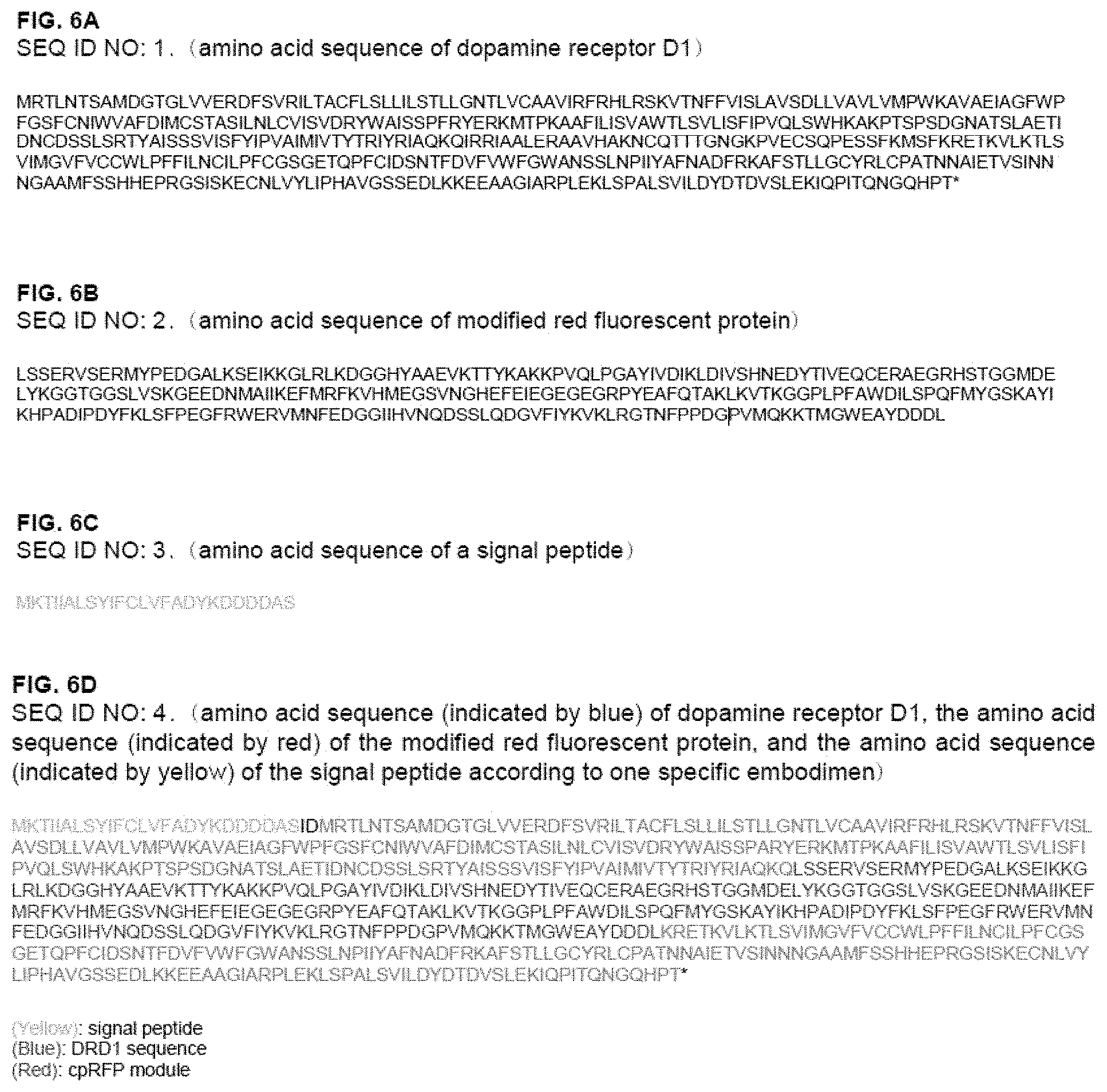
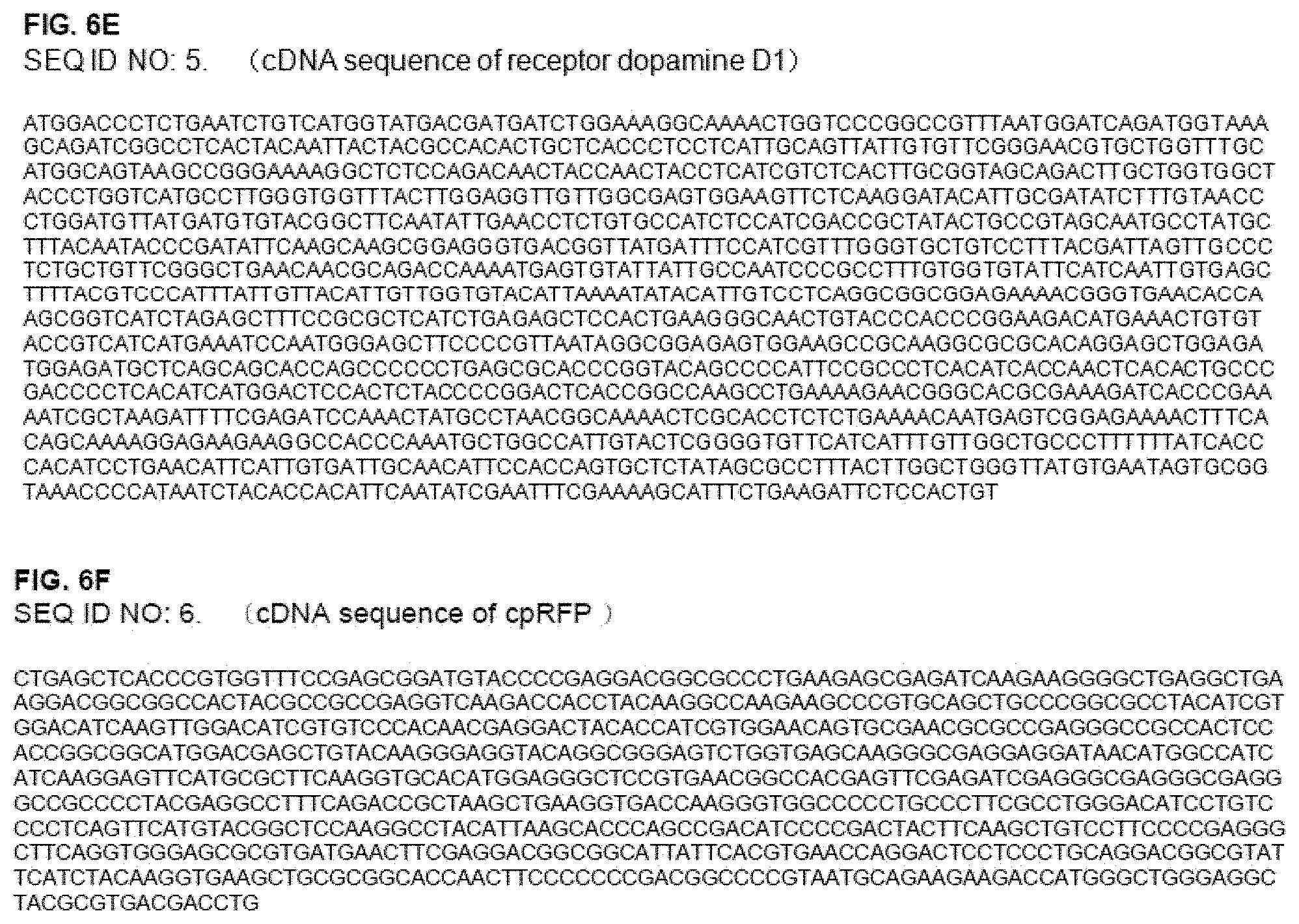
[0037] FIGS. 6A to 6F show amino acid sequences used in the preparation of the red fluorescent protein-based biosensor for measuring the activity of dopamine receptor D1 according to one specific embodiment; FIG. 6A shows an amino acid sequence (SEQ ID NO: 1) of dopamine receptor D1, FIG. 6B shows an amino acid sequence (SEQ ID NO: 2) of modified red fluorescent protein according to one specific embodiment; FIG. 6C shows an amino acid sequence (SEQ ID NO: 3) of a signal peptide according to one specific embodiment; FIG. 6D shows a recombinant amino acid sequence (SEQ ID NO: 4) including the amino acid sequence (indicated by blue) of dopamine receptor D1, the amino acid sequence (indicated by red) of the modified red fluorescent protein, and the amino acid sequence (indicated by yellow) of the signal peptide according to one specific embodiment; FIG. 6E shows a cDNA sequence (SEQ ID NO: 5) of receptor dopamine D1 according to one specific embodiment; and FIG. 6F shows a cDNA sequence (SEQ ID NO: 6) of cpRFP according to one specific embodiment.
DETAILED DESCRIPTION
[0038] Reference will now be made in detail to embodiments, examples of which are illustrated in the accompanying drawings, wherein like reference numerals refer to like elements throughout. In this regard, the present embodiments may have different forms and should not be construed as being limited to the descriptions set forth herein. Accordingly, the embodiments are merely described below, by referring to the figures, to explain aspects of the present description. As used herein, the term "and/or" includes any and all combinations of one or more of the associated listed items. Expressions such as "at least one of," when preceding a list of elements, modify the entire list of elements and do not modify the individual elements of the list.
[0039] Unless defined otherwise, all technical terms used herein have the same meanings as those generally understood by one of ordinary skill in the art to which the present disclosure belongs. Further, although methods or samples are described herein, those similar or equivalent thereto are also incorporated in the scope of the present disclosure. Processes described below are only one example according to the present disclosure. In the present disclosure, part of each process may be omitted or repeatedly performed, and in some cases, the order of the processes may be appropriately changed. The numerical values described herein are considered to include the meaning of "about", unless otherwise specified. The contents of all the publications disclosed as references herein are incorporated in the present disclosure.
[0040] An aspect provides a red fluorescent protein-based biosensor for measuring the activity of dopamine receptor D1, the red fluorescent protein-based biosensor including: [0041] a recombinant gene including a dopamine receptor D1 gene, and a gene encoding a red fluorescent protein or a modified red fluorescent protein; [0042] a recombinant expression vector including the recombinant gene; or [0043] a transformant transformed with the recombinant expression vector.
[0044] FIG. 1A is a schematic diagram illustrating a working principle and a mechanism of a red fluorescent biosensor for measuring the activity of dopamine receptor D1 (DRD1) and detecting a ligand (e.g., dopamine, DA) binding to DRD1.
[0045] As used herein, "dopamine (DA or 3,4-dihydroxyphenethylamine, C.sub.8H.sub.11NO.sub.2)", a catecholamine-based organic compound, refers to a hormone or neurotransmitter found in the central nervous system of various animals. Dopamine is one of the neurotransmitters secreted to transmit certain signals between cranial nerve cells, and activates dopamine receptors.
[0046] As used herein, the "dopamine receptor" is a type of G-protein coupled receptor (GPCR), and five subtypes (D1, D2, D3, D4, and D5 types) are known. The subtypes of the dopamine receptors may be further classified into D1-like receptors (D1 and D5) and D2-like receptors (D2, D3, and D4). The receptors are each unique in terms of intimacy with dopamine, binding to G proteins, signaling methods, distribution of specific neurons, etc. For example, D1-like receptors transduce signals to the cAMP pathway through separation of Gas in the G protein, whereas D2-like receptors inhibit adenylate cyclase (AC) activity through separation of G.alpha.i, resulting in reduction of cAMP levels.
[0047] In one specific embodiment, the dopamine receptor may be dopamine receptor D1. In another specific embodiment, the dopamine receptor D1 gene may encode an amino acid represented by SEQ ID NO: 1 (FIG. 6A).
[0048] As used herein, the "fluorescent protein" refers to a protein that exhibits fluorescence when exposed to light. Examples of the fluorescent protein may include a red fluorescent protein (RFP), a blue fluorescent protein (BFP), an enhanced blue fluorescent protein (EBFP), a cyan fluorescent protein (CFP), an enhanced cyan fluorescent protein (ECFP), a yellow fluorescent protein (YFP), an enhanced yellow fluorescent protein (EYFP), a green fluorescent protein (GFP), a modified green fluorescent protein, an enhanced green fluorescent protein (EGFP), etc.
[0049] In one specific embodiment, the fluorescent protein may be a red fluorescent protein or a modified red fluorescent protein. In one specific embodiment, the red fluorescent protein may be circularly permuted red fluorescent protein (cpRFP).
[0050] As used herein, the term "circular permutation" means a modification whereby new N- and C-termini are created in a protein (i.e., the protein is split into two parts), and original N- and C-termini of the protein are linked through an appropriate linker sequence.
[0051] In one specific embodiment, the circular permutation modification is induced in the fluorescent protein to change wavelength characteristics (e.g., absorption wavelength and/or fluorescence wavelength). In the circularly permuted fluorescent protein according to one specific embodiment, new N- and C-termini are located in close proximity to a chromophore which is necessary for fluorescence, and arbitrary proteins x and y are linked thereto such that fluorescence intensity is changed in response to their interaction. Based on this, a biosensor was developed.
[0052] FIG. 1B is a schematic diagram illustrating modification of red fluorescent protein according to the kind and position change of a linker peptide (linker amino acids) introduced to optimize the biosensor for measuring the activity of dopamine receptor D1. LSSxx represents a linker peptide composed of amino acids of leucine(L)-serine(S)-serine(S) linked to the N-terminus of cpRFP and any two amino acids (x)(x) linked thereto. xxDDL represents a linker peptide composed of amino acids of aspartic acid (D)-aspartic acid (D)-leucine(L) linked to the C-terminus of cpRFP and any two amino acids (x)(x) linked thereto.
[0053] In one specific embodiment, the gene encoding the modified red fluorescent protein may include a gene encoding an amino acid sequence according to the following Formula 1:
L.sub.1-cpRFP-L.sub.2 [Formula 1] [0054] in Formula 1, L.sub.1 includes a linker peptide having LSS and 1 to 5 amino acid residues at the N-terminus of cpRFP, and L.sub.2 includes a linker peptide having 1 to 5 amino acid residues and DDL at the C-terminus of cpRFP. The amino acid residues may be any amino acid residues each independently selected. The amino acid residue may be selected from naturally occurring amino acids.
[0055] In one specific embodiment, in Formula 1, L.sub.1 includes a linker peptide having LSS and any 2 independently selected amino acid residues at the N-terminus of cpRFP, and L.sub.2 includes a linker peptide having any 2 independently selected amino acid residues and DDL at the C-terminus of cpRFP.
[0056] In one specific embodiment, L.sub.1 includes LSSX.sub.1X.sub.2, and L.sub.2 includes X.sub.3X.sub.4DDL, wherein X.sub.1, X.sub.2, X.sub.3, and X.sub.4 may be each independently any amino acid. For example, X.sub.1, X.sub.2, X.sub.3, and X.sub.4 may be each independently any one amino acid selected from the group consisting of alanine (A), cysteine (C), aspartic acid (D), glutamic acid (E), phenylalanine (F), glycine (G), histidine (H), isoleucine (I), lysine (K), leucine (L), methionine (M), asparagine (N), pyrrolysine (O), proline (P), glutamine (Q), arginine (R), serine (S), threonine (T), selenocysteine (U), valine (V), tryptophan (W), and tyrosine (Y).
[0057] In one specific embodiment, X.sub.1X.sub.2 may be any one selected from the group consisting of glutamic acid-arginine (ER), glutamine-arginine (QR), and arginine-arginine (RR), and X.sub.3X.sub.4 may be any one selected from the group consisting of tyrosine-aspartic acid (YD), threonine-serine (TS), and histidine-proline (HP).
[0058] In one specific embodiment, X.sub.1X.sub.2 may be glutamic acid-arginine (ER), and X.sub.3X.sub.4 may be tyrosine-aspartic acid (YD). The biosensor according to one specific embodiment may include a sequence of "LSS-ER-cpRFP-YD-DDL".
[0059] The receptor D1 biosensor including cpRFP having the substituted sequence exhibits high fluorescence intensity by reacting with a ligand binding to dopamine receptor D1, and has the excellent effect of measuring the activity of dopamine receptor D1.
[0060] In one specific embodiment, the gene encoding the modified red fluorescent protein may include a gene encoding an amino acid sequence represented by SEQ ID NO: 2 (FIG. 6(B)).
[0061] It is known that when G protein binding receptor (GPCR) is activated by ligands, its structural changes are most frequently found in Intracellular loop 3 (ICL3). In one specific embodiment, the amino acid sequence of the red fluorescent protein or the modified red fluorescent protein may bind to ICL3 of the dopamine receptor D1.
[0062] In one specific embodiment, the amino acid sequence of the red fluorescent protein or the modified red fluorescent protein may bind immediately after any one of the 1.sup.st to 54.sup.th amino acids from the N-terminus of ICL3 of dopamine receptor D1.
[0063] In one specific embodiment, the amino acid sequence of the red fluorescent protein or the modified red fluorescent protein may bind immediately after the 9th amino acid from the N-terminus of ICL3 of dopamine receptor D1.
[0064] In one specific embodiment, the gene of the dopamine receptor D1 may encode a sequence in which any one amino acid of 120th to 140th amino acids of the amino acid sequence represented by SEQ ID NO: 1 is substituted with another amino acid.
[0065] In one specific embodiment, the gene of the dopamine receptor D1 may encode an amino acid sequence in which phenylalanine (F) at the 129.sup.th position of the amino acid sequence represented by SEQ ID NO: 1 is substituted with alanine (A).
[0066] The receptor D1 biosensor including the amino acid sequence in which the amino acid at the 129.sup.th position in SEQ ID NO: 1 is substituted with alanine (A) exhibits high fluorescence intensity by reacting with the ligand binding to dopamine receptor D1, and has the excellent effect of measuring the activity of dopamine receptor D1.
[0067] The fluorescent biosensor for measuring the activity of dopamine receptor D1 according to one specific embodiment may measure the activity of dopamine receptor ligand at concentration of 0.05 .mu.M, 0.1 .mu.M, 0.5 .mu.M, 1 .mu.M, 1.5 .mu.M, 5 .mu.M, 6.25 .mu.M, 10 .mu.M, 12.5 .mu.M, 50 .mu.M, or 100 .mu.M. The fluorescent biosensor for measuring the activity of dopamine receptor D1 according to one specific embodiment may effectively detect the dopamine receptor ligand at a concentration of 10 .mu.M or more.
[0068] In one specific embodiment, the recombinant gene may further include a gene encoding a signal peptide. In one specific embodiment, the signal peptide may include an amino acid sequence represented by SEQ ID NO: 3 (FIG. 6C).
[0069] In one specific embodiment, the recombinant gene may include an amino acid sequence represented by SEQ ID NO: 4 (FIG. 6D).
[0070] FIG. 2 is a schematic diagram illustrating a procedure of constructing candidate plasmids for preparing the biosensor for measuring the activity of dopamine receptor D1. To insert the nucleotide sequence of cpRFP into ICL3 of the dopamine receptor D1, based on cpRFP, any two independent linker amino acids were added after LSS linked to the N-terminus of cpRFP and before DDL linked to C-terminus of cpRFP. Random mutations were induced in two amino acids each immediately before and after cpRFP to construct candidate plasmids for the dopamine receptor D1 biosensor (FIG. 2).
[0071] Another aspect provides a method of measuring the activity of dopamine receptor D1 using the red fluorescent protein-based biosensor, the method including: [0072] introducing the biosensor into cells; [0073] adding a test sample to the biosensor-introduced cells; and [0074] measuring the activity of the dopamine receptor D1 according to the addition of the test sample to the biosensor.
[0075] Still another aspect provides a method of detecting a ligand binding to the dopamine receptor D1 using the red fluorescent protein-based biosensor, the method including: introducing the biosensor into cells; [0076] adding a test sample to the biosensor-introduced cells; and [0077] detecting the ligand binding to the dopamine receptor D1 according to the addition of the test sample to the biosensor.
[0078] In one specific embodiment, provided is a method of detecting dopamine or dopamine agonists using the red fluorescent protein-based biosensor.
[0079] As used herein, the term "gene" refers to any nucleotide sequence or part thereof that has a functional role in protein coding or transcription or regulation of other gene expression. The gene may consist of the entire of nucleotides encoding a functional protein or only a part of nucleotides encoding or expressing a protein. The nucleotide sequence may include exons, introns, initiation or termination regions, promoter sequences, other regulatory sequences, or gene abnormalities within a unique sequence adjacent to the gene.
[0080] As used herein, the "protein" also includes fragments, analogs, and derivatives thereof that retain biological activity or function substantially identical to that of a reference protein.
[0081] As used herein, the "transfection" refers to a process whereby extracellular DNA enters a host cell in the presence or absence of an accompanying substance. Transfected cells refer to cells having extracellular DNA by introducing the extracellular DNA to the cells. DNA may be introduced into cells so that it may be inserted into chromosome or it may replicate as an extrachromosomal material.
[0082] As used herein, the "transduction" is a phenomenon in which DNA of a bacterium is transferred to another bacterium via a virus. Transduction, even when a foreign DNA is introduced into another cell via a viral vector, is often used by molecular biologists to introduce a foreign gene into the host cell's genome.
[0083] As used herein, the "transformation" refers to a molecular biological phenomenon in which a DNA chain fragment or plasmid containing a gene of a different kind from that of original cells penetrate the cells and binds to DNA present in the original cells, and as a result, genetic characteristics of the cell are changed. Cells introduced with a foreign DNA, etc. are called `transformant`.
[0084] As used herein, the term "vector" refers to any nucleic acid including a competent nucleotide sequence that is inserted into a host cell to be incorporated into the genome of the host cell by recombination, or to autonomously replicate as an episome. Such a vector may include a linear nucleic acid, a plasmid, a phagemid, a cosmid, an RNA vector, a viral vector, etc.
[0085] As used herein, the expression "about" or "approximately" means that a mentioned value may vary to some degree. For example, the value may vary to 10%, 5%, 2%, or 1%.
[0086] As used herein, the term "have", "may have", "include", or "may include" indicates existence of corresponding features (e.g., elements such as numeric values or components) but do not exclude presence of additional features. As used herein, the term "including" does not limit the present disclosure to exclude any modification or addition. Although described using the term "including", the method, substance, and composition described herein may be described using "consisting substantially of" or "consisting of". As used herein, the singular forms include plural forms unless the context clearly dictates otherwise.
EXAMPLES
[0087] Hereinafter, the present disclosure will be described in more detail with reference to the following Examples. However, the following Examples are only for illustrating the present disclosure, and the scope of the present disclosure is not limited by these Examples. As used herein, the term "to" includes the endpoint of the range and all midpoints therebetween. Those skilled in the art will understand that the numerical amount of deviation is possible. Therefore, it is understood that whenever a numerical value is mentioned in the specification or claims, the numerical value or any additional value relating to such a numerical value is also within the scope of the present disclosure.
Example 1. Preparation of Red Fluorescent Protein-Based Biosensor for Measuring Activity of Dopamine Receptor D1
[0088] (1) Construction of Genetic Recombinant Plasmid
[0089] cDNA of human dopamine receptor D1 amplified by PCR was obtained (SEQ ID NO: 5). The gene of human dopamine receptor D1 encodes an amino acid represented by SEQ ID NO: 1. The amplified cDNA of dopamine receptor D1 thus obtained was fused into a pcDNA5/FRT plasmid vector digested with Cla1/Xho1 restriction enzymes using an infusion technique.
[0090] Further, cDNA of cpRFP which is circularly permuted red fluorescent protein was amplified by PCR (cDNA of cpRFP, SEQ ID NO: 6).
[0091] To optimize the biosensor for measuring the activity of dopamine receptor D1, a genetic recombinant plasmid was prepared, in which the amplified cpRFP was inserted into a specific site of ICL3 (immediately after the 9th amino acid from the N-terminus of ICL3) which is an intracellular loop 3 of dopamine receptor D1 (FIG. 1B).
[0092] To insert the nucleotide sequence of cpRFP into ICL3 of the dopamine receptor D1, 5 linker peptides (linker amino acids) were added before and after cpRFP, respectively such that LSSPV-cpRFP-TDDDL was prepared.
[0093] (2) Random Mutagenesis and Transfection
[0094] In the recombinant amino acid sequence (LSSX.sub.aX.sub.b-cpRFP-X.sub.cX.sub.dDDL) including the linker peptides each linked to the N- and C-termini of cpRFP, random mutation was induced in two amino acids each immediately before and after cpRFP to prepare candidate plasmids for the dopamine receptor D1 biosensor (FIG. 2).
[0095] A variety of genetic recombinant dopamine receptor D1 plasmids were constructed using a principle of the cpRFP-based biosensor, in which the brightness of the sensor varies depending on the linker peptide arrangement. In the linker peptides (LSSX.sub.aX.sub.b and X.sub.cX.sub.dDDL) each linked to the N- and C-termini of cpRFP, random mutation was induced in two amino acids (X.sub.aX.sub.b and X.sub.cX.sub.d) each immediately before and after cpRFP to substitute X.sub.aX.sub.b and X.sub.cX.sub.d with glutamic acid-arginine(ER) and tyrosine-aspartic acid (YD), respectively.
[0096] (3) Selection of Candidate Plasmids for Dopamine Receptor D1 Biosensor and Cell Culture
[0097] After transformation into Escherichia coli as a competent cell, a large amount of genetic recombinant plasmids were obtained through cloning. Subsequently, isolation and purification were performed, and then plasmids of the sensor for measuring the activity of dopamine receptor D1 were obtained through sequencing.
[0098] Each plasmid was transfected into an animal cell HEK293A, followed by cell culture. The HEK293A cell line was seeded on a cell culture dish at an equal density, and cultured at a temperature of 37.degree. C. and 5% CO.sub.2 for about 16 hours. Thereafter, 2 .mu.l of Lipofectamine 2000 and the prepared dopamine receptor D1 biosensor (1 .mu.g) were stabilized in an Opti-MEM medium (ThermoFisher Scientific) for 20 minutes, and then added to subcultured cells.
[0099] 6 hours after transfection, cells were subcultured in a mini-confocal dish coated with fibronectin, and DMEM (Hyclone, SH30604.01) containing 10% (v/v) fetal bovine serum (FBS) (Hyclone, SH30084.03) and 0.5% penicillin/streptomycin (Corning, 30-002-CI) was added, followed by culturing overnight. Next day, observation was performed using a live fluorescence microscope. Through the live fluorescence microscope, HEK293A single cells transfected with the biosensor for measuring the activity of dopamine receptor D1 were observed.
[0100] To measure the activity of dopamine receptor D1, the dopamine receptor D1 biosensor is required to locate in the cell membrane of animal cell HEK293A. Thus, whether the biosensor located in the cell membrane of animal cell HEK293A was examined through a fluorescence microscope. Further, according to the principle of the cpRFP-based biosensor, in which the brightness of the sensor varies depending on the linker peptide arrangement, sensor candidates with high expression efficiency were imaged using the fluorescence microscope, after treatment of the medium with dopamine.
[0101] (4) Measurement of Activity of Dopamine Receptor D1 Using Dopamine Receptor D1 Biosensor
[0102] The prepared dopamine receptor D1 biosensor was treated with a ligand of dopamine receptor D1 (agonist), and fluorescence intensity before and after the treatment was measured in real time to determine intensity change.
[0103] First, dopamine which is a ligand of dopamine receptor D1 was prepared at a final concentration of 10 .mu.M and added to a system. To minimize a photobleaching phenomenon, the live fluorescence microscope was set. When stimulated with light at a wavelength of 560 nm in the live fluorescence microscope, intensity at a cpRFP fluorescence wavelength of 610 nm was measured.
[0104] To induce fluorescence intensity suitable for experiments, 50 msec exposure and ND16 intensity were maintained. The fluorescence cycle was performed for total 10 minutes by measuring cpRFP images for 1 minute using the fluorescence microscope.
[0105] The saved images were analyzed using an NIS program (Nikon). cpRFP fluorescence intensity of the entire single cells in the regions of interest (ROI) was analyzed in the NIS program. Change of the corresponding fluorescence intensity means efficiency of the biosensor for measuring the activity of dopamine receptor D1 in the single cell. After acquiring the change of fluorescence intensity over time, the fluorescence intensity before adding the ligand dopamine was averaged and set as Fo, and the change of fluorescence intensity over time was set as F, and then F/Fo data were made and normalized.
[0106] FIG. 3A is a graph showing changes (F/Fo) in fluorescence intensity of the biosensor candidates for measuring the activity of dopamine receptor D1 with respect to dopamine as an agonist. The X-axis represents the number of the biosensor candidates (number of variants) tested. The fluorescence intensity of the biosensor candidate corresponding to each number was measured, and their effects on measuring the activity of dopamine receptor D1 were evaluated. FIG. 3B is a graph showing results of measuring the degree of increase of the maximum fluorescence intensity (y axis: normalized maximum intensity) according to the addition of 10 .mu.M dopamine by using three candidates (DRD1 Red 1.1, 1.2, and 1.3) having excellent efficiency among the biosensor candidates for measuring the activity of dopamine receptor D1.
[0107] DRD1 Red 1.1 represents a prototype sensor prepared by cloning using the dopamine receptor D1 gene and the original red fluorescent protein. As shown in FIG. 2, DRD1 Red 1.2 and DRD1 Red 1.3 represent two candidates that exhibited high fluorescence intensity at the time of measuring the effects of the biosensors using the candidate plasmids for the dopamine receptor D1 biosensor which were prepared by inducing random mutation in two amino acids each immediately before and after cpRFP. DRD1 Red 1.2 and DRD1 Red 1.3 include, in common, "LSS-ER-cpRFP-YD-DDL" sequence, which was prepared by substituting two amino acids each immediately before and after cpRFP in the linker peptides (LSSX.sub.aX.sub.b and X.sub.cX.sub.dDDL) linked to each of the N- and C-termini of cpRFP with glutamic acid-arginine(ER) and tyrosine-aspartic acid (YD), respectively. As shown in FIGS. 3B and 3C, DRD1 Red 1.2 and DRD1 Red 1.3 exhibited higher fluorescence intensity than DRD1 Red 1.1, indicating that they are excellent red fluorescent biosensors for measuring the activity of dopamine receptor D1.
[0108] DRD1 Red 1.3 includes a substitution of alanine (A) for phenylalanine (F) at position 129 of the amino acid sequence (SEQ ID NO: 1) of dopamine receptor D1 in DRD1 Red 1.2.
[0109] As shown in FIGS. 3B and 3C, DRD1 Red 1.3 exhibited the highest fluorescence intensity, indicating that it is the most effective red fluorescent biosensor for measuring the activity of dopamine receptor D1 in terms of measuring the activity of dopamine receptor D1.
[0110] FIG. 3C is a graph showing results of measuring the change in the fluorescence intensity (y axis: normalized intensity) over time by using three candidates (DRD1 Red 1.1, 1.2, and 1.3) having excellent efficiency among the biosensor candidates for measuring the activity of dopamine receptor D1.
[0111] As shown in FIG. 3C, the change of fluorescence intensity was measured in real time, and as a result, candidate exhibiting a larger change in the fluorescence intensity and a high reaction rate were selected by inducing mutation and testing a larger number of candidates.
Example 2. Characterization of Red Fluorescent Biosensor for Measuring Activity of Dopamine Receptor D1
[0112] The red fluorescent biosensor for dopamine receptor D1 prepared in Example 1 was transfected into HEK293A cell line as in Example 1, and expressed therein, and expression of the dopamine receptor D1 biosensor on the membrane of HEK293A was examined using the live fluorescence microscope.
[0113] FIG. 4A shows images of a cell in which activation of the biosensor exhibited a color change when the biosensor was treated with dopamine which is a dopamine receptor D1 ligand at a concentration of 10 .mu.M (left: before dopamine treatment, right: after dopamine treatment).
[0114] FIG. 4B is a graph showing results of measuring normalized intensity when the fluorescent biosensor for measuring the activity of dopamine receptor D1 was treated with dopamine which is a dopamine receptor D1 agonist and quinpirole which is a dopamine D2 receptor selective ligand. FIG. 4B shows that the dopamine receptor D1 fluorescent biosensor did not response to quinpirole which is a dopamine D2 receptor selective ligand.
[0115] FIG. 4C is a graph showing fluorescence intensity (y-axis: normalized intensity) of the biosensor for measuring the activity of dopamine receptor D1, measured after adding dopamine which is a dopamine receptor D1 ligand at a final concentration of 0.05 .mu.M, 0.1 .mu.M, 0.5 .mu.M, 1 .mu.M, 1.5 .mu.M, 5 .mu.M, 6.25 .mu.M, 10 .mu.M, 12.5 .mu.M, 50 .mu.M, or 100 .mu.M in cell media. The intensity of the red channel that was specified by ROI through the NIS program (Nikon) performed in Example 1 was measured and analyzed.
[0116] As shown in FIG. 4C, the fluorescent biosensor for measuring the activity of dopamine receptor D1 according to one specific embodiment may detect activity for the dopamine receptor ligand at a concentration of 0.05 .mu.M, 0.1 .mu.M, 0.5 .mu.M, 1 .mu.M, 1.5 .mu.M, 5 .mu.M, 6.25 .mu.M, 10 .mu.M, 12.5 .mu.M, 50 .mu.M, or 100 .mu.M. Further, the biosensor may effectively detect the dopamine receptor ligand at a concentration of 10 .mu.M or more.
[0117] FIG. 4D is a graph showing fluorescence intensity (y-axis: normalized intensity) of the biosensor for measuring the activity of dopamine receptor D1 over time, when the fluorescent biosensor for measuring the activity of dopamine receptor D1 was co-treated with dopamine (DA) which is a dopamine receptor D1 ligand and an inhibitor (Haloperidol) (red plot) or treated with only dopamine (DA) (blue plot).
[0118] FIG. 5A shows results of performing co-fluorescent imaging using a green fluorescent biosensor (GRAB-DA1m) for measuring the activity of dopamine receptor D2 which was separately prepared for comparison with the red fluorescent biosensor for measuring activity of dopamine receptor D1 (DRD1 Red 1.3). The graph shows the increase in fluorescence by selective response of each fluorescent biosensor to each ligand, when first treated with quinpirole which is a D2 selective ligand, and then serially treated with SKF38393 which is a D1 selective ligand.
[0119] FIG. 5B shows results of performing fluorescent imaging before adding the ligand (Before), 5 minutes after treatment with quinpirole which a D2 selective ligand (+Quinpirole, 5 min), and 5 minutes after treatment with SKF38393 which is a D1 selective ligand (+SKF38393, 5 min) in the experiment explained in FIG. 5A. The fluorescent images of cells each expressing the red fluorescent biosensor for measuring the activity of dopamine receptor D1 (DRD1 Red 1.3) or the green fluorescent biosensor for measuring the activity of dopamine receptor D2 (GRAB-DA1m) are shown.
[0120] The red fluorescent protein-based biosensor according to an aspect may have selectivity depending on the dopamine receptor subtype, and may detect the activity of dopamine receptor D1 with high accuracy and resolution. Unlike the green fluorescent protein-based biosensor, the red fluorescent protein-based biosensor may detect the activity of dopamine receptor D1 with high accuracy without overlapping with excitation wavelength.
[0121] Further, when the red fluorescent protein-based biosensor and another fluorescent biosensor of different wavelength (e.g., green fluorescent protein biosensor) are used in combination, the dopamine activity may be measured by changes and interaction of two kinds of signals.
[0122] A method of measuring the activity of dopamine receptor D1 according to an aspect may sensitively measure activity of dopamine receptor D1 in live cells.
[0123] A method of detecting a ligand binding to dopamine receptor D1 according to another aspect may effectively and reversibly detect the ligand binding to dopamine receptor D1, e.g., dopamine or dopamine agonist.
[0124] It should be understood that embodiments described herein should be considered in a descriptive sense only and not for purposes of limitation. Descriptions of features or aspects within each embodiment should typically be considered as available for other similar features or aspects in other embodiments. While one or more embodiments have been described with reference to the figures, it will be understood by those of ordinary skill in the art that various changes in form and details may be made therein without departing from the spirit and scope of the disclosure as defined by the following claims.
Sequence CWU 1
1
61446PRTArtificial SequenceThe amino acid sequence of dopamine
receptor D1 type 1Met Arg Thr Leu Asn Thr Ser Ala Met Asp Gly Thr
Gly Leu Val Val1 5 10 15Glu Arg Asp Phe Ser Val Arg Ile Leu Thr Ala
Cys Phe Leu Ser Leu 20 25 30Leu Ile Leu Ser Thr Leu Leu Gly Asn Thr
Leu Val Cys Ala Ala Val 35 40 45Ile Arg Phe Arg His Leu Arg Ser Lys
Val Thr Asn Phe Phe Val Ile 50 55 60Ser Leu Ala Val Ser Asp Leu Leu
Val Ala Val Leu Val Met Pro Trp65 70 75 80Lys Ala Val Ala Glu Ile
Ala Gly Phe Trp Pro Phe Gly Ser Phe Cys 85 90 95Asn Ile Trp Val Ala
Phe Asp Ile Met Cys Ser Thr Ala Ser Ile Leu 100 105 110Asn Leu Cys
Val Ile Ser Val Asp Arg Tyr Trp Ala Ile Ser Ser Pro 115 120 125Phe
Arg Tyr Glu Arg Lys Met Thr Pro Lys Ala Ala Phe Ile Leu Ile 130 135
140Ser Val Ala Trp Thr Leu Ser Val Leu Ile Ser Phe Ile Pro Val
Gln145 150 155 160Leu Ser Trp His Lys Ala Lys Pro Thr Ser Pro Ser
Asp Gly Asn Ala 165 170 175Thr Ser Leu Ala Glu Thr Ile Asp Asn Cys
Asp Ser Ser Leu Ser Arg 180 185 190Thr Tyr Ala Ile Ser Ser Ser Val
Ile Ser Phe Tyr Ile Pro Val Ala 195 200 205Ile Met Ile Val Thr Tyr
Thr Arg Ile Tyr Arg Ile Ala Gln Lys Gln 210 215 220Ile Arg Arg Ile
Ala Ala Leu Glu Arg Ala Ala Val His Ala Lys Asn225 230 235 240Cys
Gln Thr Thr Thr Gly Asn Gly Lys Pro Val Glu Cys Ser Gln Pro 245 250
255Glu Ser Ser Phe Lys Met Ser Phe Lys Arg Glu Thr Lys Val Leu Lys
260 265 270Thr Leu Ser Val Ile Met Gly Val Phe Val Cys Cys Trp Leu
Pro Phe 275 280 285Phe Ile Leu Asn Cys Ile Leu Pro Phe Cys Gly Ser
Gly Glu Thr Gln 290 295 300Pro Phe Cys Ile Asp Ser Asn Thr Phe Asp
Val Phe Val Trp Phe Gly305 310 315 320Trp Ala Asn Ser Ser Leu Asn
Pro Ile Ile Tyr Ala Phe Asn Ala Asp 325 330 335Phe Arg Lys Ala Phe
Ser Thr Leu Leu Gly Cys Tyr Arg Leu Cys Pro 340 345 350Ala Thr Asn
Asn Ala Ile Glu Thr Val Ser Ile Asn Asn Asn Gly Ala 355 360 365Ala
Met Phe Ser Ser His His Glu Pro Arg Gly Ser Ile Ser Lys Glu 370 375
380Cys Asn Leu Val Tyr Leu Ile Pro His Ala Val Gly Ser Ser Glu
Asp385 390 395 400Leu Lys Lys Glu Glu Ala Ala Gly Ile Ala Arg Pro
Leu Glu Lys Leu 405 410 415Ser Pro Ala Leu Ser Val Ile Leu Asp Tyr
Asp Thr Asp Val Ser Leu 420 425 430Glu Lys Ile Gln Pro Ile Thr Gln
Asn Gly Gln His Pro Thr 435 440 4452252PRTArtificial SequenceThe
amino acid sequence of the modified red fluorescent protein 2Leu
Ser Ser Glu Arg Val Ser Glu Arg Met Tyr Pro Glu Asp Gly Ala1 5 10
15Leu Lys Ser Glu Ile Lys Lys Gly Leu Arg Leu Lys Asp Gly Gly His
20 25 30Tyr Ala Ala Glu Val Lys Thr Thr Tyr Lys Ala Lys Lys Pro Val
Gln 35 40 45Leu Pro Gly Ala Tyr Ile Val Asp Ile Lys Leu Asp Ile Val
Ser His 50 55 60Asn Glu Asp Tyr Thr Ile Val Glu Gln Cys Glu Arg Ala
Glu Gly Arg65 70 75 80His Ser Thr Gly Gly Met Asp Glu Leu Tyr Lys
Gly Gly Thr Gly Gly 85 90 95Ser Leu Val Ser Lys Gly Glu Glu Asp Asn
Met Ala Ile Ile Lys Glu 100 105 110Phe Met Arg Phe Lys Val His Met
Glu Gly Ser Val Asn Gly His Glu 115 120 125Phe Glu Ile Glu Gly Glu
Gly Glu Gly Arg Pro Tyr Glu Ala Phe Gln 130 135 140Thr Ala Lys Leu
Lys Val Thr Lys Gly Gly Pro Leu Pro Phe Ala Trp145 150 155 160Asp
Ile Leu Ser Pro Gln Phe Met Tyr Gly Ser Lys Ala Tyr Ile Lys 165 170
175His Pro Ala Asp Ile Pro Asp Tyr Phe Lys Leu Ser Phe Pro Glu Gly
180 185 190Phe Arg Trp Glu Arg Val Met Asn Phe Glu Asp Gly Gly Ile
Ile His 195 200 205Val Asn Gln Asp Ser Ser Leu Gln Asp Gly Val Phe
Ile Tyr Lys Val 210 215 220Lys Leu Arg Gly Thr Asn Phe Pro Pro Asp
Gly Pro Val Met Gln Lys225 230 235 240Lys Thr Met Gly Trp Glu Ala
Tyr Asp Asp Asp Leu 245 250325PRTArtificial SequenceThe amino acid
sequence of the signal peptide 3Met Lys Thr Ile Ile Ala Leu Ser Tyr
Ile Phe Cys Leu Val Phe Ala1 5 10 15Asp Tyr Lys Asp Asp Asp Asp Ala
Ser 20 254685PRTArtificial SequenceRecombinant amino acid sequence
comprising the amino acid sequence of dopamine receptor D1 type,
the amino acid sequence of the modified red fluorescent protein,
and the amino acid sequence of the signal peptide 4Met Lys Thr Ile
Ile Ala Leu Ser Tyr Ile Phe Cys Leu Val Phe Ala1 5 10 15Asp Tyr Lys
Asp Asp Asp Asp Ala Ser Ile Asp Met Arg Thr Leu Asn 20 25 30Thr Ser
Ala Met Asp Gly Thr Gly Leu Val Val Glu Arg Asp Phe Ser 35 40 45Val
Arg Ile Leu Thr Ala Cys Phe Leu Ser Leu Leu Ile Leu Ser Thr 50 55
60Leu Leu Gly Asn Thr Leu Val Cys Ala Ala Val Ile Arg Phe Arg His65
70 75 80Leu Arg Ser Lys Val Thr Asn Phe Phe Val Ile Ser Leu Ala Val
Ser 85 90 95Asp Leu Leu Val Ala Val Leu Val Met Pro Trp Lys Ala Val
Ala Glu 100 105 110Ile Ala Gly Phe Trp Pro Phe Gly Ser Phe Cys Asn
Ile Trp Val Ala 115 120 125Phe Asp Ile Met Cys Ser Thr Ala Ser Ile
Leu Asn Leu Cys Val Ile 130 135 140Ser Val Asp Arg Tyr Trp Ala Ile
Ser Ser Pro Ala Arg Tyr Glu Arg145 150 155 160Lys Met Thr Pro Lys
Ala Ala Phe Ile Leu Ile Ser Val Ala Trp Thr 165 170 175Leu Ser Val
Leu Ile Ser Phe Ile Pro Val Gln Leu Ser Trp His Lys 180 185 190Ala
Lys Pro Thr Ser Pro Ser Asp Gly Asn Ala Thr Ser Leu Ala Glu 195 200
205Thr Ile Asp Asn Cys Asp Ser Ser Leu Ser Arg Thr Tyr Ala Ile Ser
210 215 220Ser Ser Val Ile Ser Phe Tyr Ile Pro Val Ala Ile Met Ile
Val Thr225 230 235 240Tyr Thr Arg Ile Tyr Arg Ile Ala Gln Lys Gln
Leu Ser Ser Glu Arg 245 250 255Val Ser Glu Arg Met Tyr Pro Glu Asp
Gly Ala Leu Lys Ser Glu Ile 260 265 270Lys Lys Gly Leu Arg Leu Lys
Asp Gly Gly His Tyr Ala Ala Glu Val 275 280 285Lys Thr Thr Tyr Lys
Ala Lys Lys Pro Val Gln Leu Pro Gly Ala Tyr 290 295 300Ile Val Asp
Ile Lys Leu Asp Ile Val Ser His Asn Glu Asp Tyr Thr305 310 315
320Ile Val Glu Gln Cys Glu Arg Ala Glu Gly Arg His Ser Thr Gly Gly
325 330 335Met Asp Glu Leu Tyr Lys Gly Gly Thr Gly Gly Ser Leu Val
Ser Lys 340 345 350Gly Glu Glu Asp Asn Met Ala Ile Ile Lys Glu Phe
Met Arg Phe Lys 355 360 365Val His Met Glu Gly Ser Val Asn Gly His
Glu Phe Glu Ile Glu Gly 370 375 380Glu Gly Glu Gly Arg Pro Tyr Glu
Ala Phe Gln Thr Ala Lys Leu Lys385 390 395 400Val Thr Lys Gly Gly
Pro Leu Pro Phe Ala Trp Asp Ile Leu Ser Pro 405 410 415Gln Phe Met
Tyr Gly Ser Lys Ala Tyr Ile Lys His Pro Ala Asp Ile 420 425 430Pro
Asp Tyr Phe Lys Leu Ser Phe Pro Glu Gly Phe Arg Trp Glu Arg 435 440
445Val Met Asn Phe Glu Asp Gly Gly Ile Ile His Val Asn Gln Asp Ser
450 455 460Ser Leu Gln Asp Gly Val Phe Ile Tyr Lys Val Lys Leu Arg
Gly Thr465 470 475 480Asn Phe Pro Pro Asp Gly Pro Val Met Gln Lys
Lys Thr Met Gly Trp 485 490 495Glu Ala Tyr Asp Asp Asp Leu Lys Arg
Glu Thr Lys Val Leu Lys Thr 500 505 510Leu Ser Val Ile Met Gly Val
Phe Val Cys Cys Trp Leu Pro Phe Phe 515 520 525Ile Leu Asn Cys Ile
Leu Pro Phe Cys Gly Ser Gly Glu Thr Gln Pro 530 535 540Phe Cys Ile
Asp Ser Asn Thr Phe Asp Val Phe Val Trp Phe Gly Trp545 550 555
560Ala Asn Ser Ser Leu Asn Pro Ile Ile Tyr Ala Phe Asn Ala Asp Phe
565 570 575Arg Lys Ala Phe Ser Thr Leu Leu Gly Cys Tyr Arg Leu Cys
Pro Ala 580 585 590Thr Asn Asn Ala Ile Glu Thr Val Ser Ile Asn Asn
Asn Gly Ala Ala 595 600 605Met Phe Ser Ser His His Glu Pro Arg Gly
Ser Ile Ser Lys Glu Cys 610 615 620Asn Leu Val Tyr Leu Ile Pro His
Ala Val Gly Ser Ser Glu Asp Leu625 630 635 640Lys Lys Glu Glu Ala
Ala Gly Ile Ala Arg Pro Leu Glu Lys Leu Ser 645 650 655Pro Ala Leu
Ser Val Ile Leu Asp Tyr Asp Thr Asp Val Ser Leu Glu 660 665 670Lys
Ile Gln Pro Ile Thr Gln Asn Gly Gln His Pro Thr 675 680
68551329DNAArtificial SequenceCDNA sequence of dopamine receptor D1
type 5atggaccctc tgaatctgtc atggtatgac gatgatctgg aaaggcaaaa
ctggtcccgg 60ccgtttaatg gatcagatgg taaagcagat cggcctcact acaattacta
cgccacactg 120ctcaccctcc tcattgcagt tattgtgttc gggaacgtgc
tggtttgcat ggcagtaagc 180cgggaaaagg ctctccagac aactaccaac
tacctcatcg tctcacttgc ggtagcagac 240ttgctggtgg ctaccctggt
catgccttgg gtggtttact tggaggttgt tggcgagtgg 300aagttctcaa
ggatacattg cgatatcttt gtaaccctgg atgttatgat gtgtacggct
360tcaatattga acctctgtgc catctccatc gaccgctata ctgccgtagc
aatgcctatg 420ctttacaata cccgatattc aagcaagcgg agggtgacgg
ttatgatttc catcgtttgg 480gtgctgtcct ttacgattag ttgccctctg
ctgttcgggc tgaacaacgc agaccaaaat 540gagtgtatta ttgccaatcc
cgcctttgtg gtgtattcat caattgtgag cttttacgtc 600ccatttattg
ttacattgtt ggtgtacatt aaaatataca ttgtcctcag gcggcggaga
660aaacgggtga acaccaagcg gtcatctaga gctttccgcg ctcatctgag
agctccactg 720aagggcaact gtacccaccc ggaagacatg aaactgtgta
ccgtcatcat gaaatccaat 780gggagcttcc ccgttaatag gcggagagtg
gaagccgcaa ggcgcgcaca ggagctggag 840atggagatgc tcagcagcac
cagcccccct gagcgcaccc ggtacagccc cattccgccc 900tcacatcacc
aactcacact gcccgacccc tcacatcatg gactccactc taccccggac
960tcaccggcca agcctgaaaa gaacgggcac gcgaaagatc acccgaaaat
cgctaagatt 1020ttcgagatcc aaactatgcc taacggcaaa actcgcacct
ctctgaaaac aatgagtcgg 1080agaaaacttt cacagcaaaa ggagaagaag
gccacccaaa tgctggccat tgtactcggg 1140gtgttcatca tttgttggct
gccctttttt atcacccaca tcctgaacat tcattgtgat 1200tgcaacattc
caccagtgct ctatagcgcc tttacttggc tgggttatgt gaatagtgcg
1260gtaaacccca taatctacac cacattcaat atcgaatttc gaaaagcatt
tctgaagatt 1320ctccactgt 13296756DNAArtificial SequencecDNA
sequence of cpRFP 6ctgagctcac ccgtggtttc cgagcggatg taccccgagg
acggcgccct gaagagcgag 60atcaagaagg ggctgaggct gaaggacggc ggccactacg
ccgccgaggt caagaccacc 120tacaaggcca agaagcccgt gcagctgccc
ggcgcctaca tcgtggacat caagttggac 180atcgtgtccc acaacgagga
ctacaccatc gtggaacagt gcgaacgcgc cgagggccgc 240cactccaccg
gcggcatgga cgagctgtac aagggaggta caggcgggag tctggtgagc
300aagggcgagg aggataacat ggccatcatc aaggagttca tgcgcttcaa
ggtgcacatg 360gagggctccg tgaacggcca cgagttcgag atcgagggcg
agggcgaggg ccgcccctac 420gaggcctttc agaccgctaa gctgaaggtg
accaagggtg gccccctgcc cttcgcctgg 480gacatcctgt cccctcagtt
catgtacggc tccaaggcct acattaagca cccagccgac 540atccccgact
acttcaagct gtccttcccc gagggcttca ggtgggagcg cgtgatgaac
600ttcgaggacg gcggcattat tcacgtgaac caggactcct ccctgcagga
cggcgtattc 660atctacaagg tgaagctgcg cggcaccaac ttcccccccg
acggccccgt aatgcagaag 720aagaccatgg gctgggaggc tacgcgtgac gacctg
756
D00000
D00001
D00002
D00003
D00004
S00001
XML
uspto.report is an independent third-party trademark research tool that is not affiliated, endorsed, or sponsored by the United States Patent and Trademark Office (USPTO) or any other governmental organization. The information provided by uspto.report is based on publicly available data at the time of writing and is intended for informational purposes only.
While we strive to provide accurate and up-to-date information, we do not guarantee the accuracy, completeness, reliability, or suitability of the information displayed on this site. The use of this site is at your own risk. Any reliance you place on such information is therefore strictly at your own risk.
All official trademark data, including owner information, should be verified by visiting the official USPTO website at www.uspto.gov. This site is not intended to replace professional legal advice and should not be used as a substitute for consulting with a legal professional who is knowledgeable about trademark law.