Prevention Of Disulfide Bond Reduction During Recombinant Production Of Polypeptides
KAO; Yung-Hsiang ; et al.
U.S. patent application number 17/124314 was filed with the patent office on 2021-04-01 for prevention of disulfide bond reduction during recombinant production of polypeptides. This patent application is currently assigned to Genentech, Inc.. The applicant listed for this patent is Genentech, Inc.. Invention is credited to Daniel P. HEWITT, Yung-Hsiang KAO, Michael W. LAIRD, Melody Trexler SCHMIDT, Rita L. WONG.
| Application Number | 20210095040 17/124314 |
| Document ID | / |
| Family ID | 1000005276137 |
| Filed Date | 2021-04-01 |


View All Diagrams
| United States Patent Application | 20210095040 |
| Kind Code | A1 |
| KAO; Yung-Hsiang ; et al. | April 1, 2021 |
PREVENTION OF DISULFIDE BOND REDUCTION DURING RECOMBINANT PRODUCTION OF POLYPEPTIDES
Abstract
The invention concerns methods and means for preventing the reduction of disulfide bonds during the recombinant production of disulfide-containing polypeptides. In particular, the invention concerns the prevention of disulfide bond reduction during harvesting of disulfide-containing polypeptides, including antibodies, from recombinant host cell cultures.
| Inventors: | KAO; Yung-Hsiang; (San Mateo, CA) ; LAIRD; Michael W.; (San Ramon, CA) ; SCHMIDT; Melody Trexler; (Danville, CA) ; WONG; Rita L.; (Redwood City, CA) ; HEWITT; Daniel P.; (Sunnyvale, CA) | ||||||||||
| Applicant: |
|
||||||||||
|---|---|---|---|---|---|---|---|---|---|---|---|
| Assignee: | Genentech, Inc. South San Francisco CA |
||||||||||
| Family ID: | 1000005276137 | ||||||||||
| Appl. No.: | 17/124314 | ||||||||||
| Filed: | December 16, 2020 |
Related U.S. Patent Documents
| Application Number | Filing Date | Patent Number | ||
|---|---|---|---|---|
| 16847317 | Apr 13, 2020 | 10906986 | ||
| 17124314 | ||||
| 16240592 | Jan 4, 2019 | 10759866 | ||
| 16847317 | ||||
| 15488917 | Apr 17, 2017 | |||
| 16240592 | ||||
| 14043758 | Oct 1, 2013 | |||
| 15488917 | ||||
| 13354223 | Jan 19, 2012 | 8574869 | ||
| 14043758 | ||||
| 12217745 | Jul 8, 2008 | |||
| 13354223 | ||||
| 60948677 | Jul 9, 2007 | |||
| Current U.S. Class: | 1/1 |
| Current CPC Class: | C07K 2317/24 20130101; C07K 16/2887 20130101; C07K 1/14 20130101; A61K 39/39591 20130101 |
| International Class: | C07K 16/28 20060101 C07K016/28; A61K 39/395 20060101 A61K039/395; C07K 1/14 20060101 C07K001/14 |
Claims
1-63. (canceled)
64. A method for producing an antibody, comprising expressing the antibody in a Chinese Hamster Ovary (CHO) recombinant host cell culture, and following a production phase of the cell culture, sparging the pre-harvest cell culture fluid of the recombinant host cell with air to inhibit reduction of a disulfide bond in the antibody during processing, wherein the antibody is a therapeutic monoclonal antibody, and wherein the air sparging is continued until the amount of dissolved oxygen (dO.sub.2) in the pre-harvest cell culture fluid is at least 10%.
65. The method of claim 64, wherein the antibody expressed in the recombinant host cell is produced at a scale of greater than 5,000 L.
66. The method of claim 64, wherein the air sparging is continued until the pre-harvest cell culture fluid is at least 30% saturated with air.
67. The method of claim 66, wherein the antibody expressed in the recombinant host cell is produced at a scale of greater than 5,000 L.
68. The method of claim 64 wherein the air sparging is continued until the pre-harvest cell culture fluid is between 100% saturated to 30% saturated with air.
69. The method of claim 65, wherein the amount of dissolved oxygen (dO.sub.2) in the pre-harvest cell culture fluid is at least 30%.
70. The method of claim 64, further comprising preparing the harvested cell culture fluid of the recombinant host cell and recovering the antibody from the harvested cell culture fluid.
71. The method of claim 64, further comprising preparing the harvested cell culture fluid and purifying the antibody from the harvested cell culture fluid.
Description
CROSS REFERENCE TO RELATED APPLICATIONS
[0001] This application is a continuation of U.S. application Ser. No. 14/043,758, filed Oct. 1, 2013, which is a divisional of U.S. application Ser. No. 13/354,223, filed Jan. 19, 2012, now U.S. Pat. No. 8,574,869, issued Nov. 5, 2013, which is a continuation of U.S. application Ser. No. 12/217,745, filed Jul. 8, 2008, now abandoned, which is a non-provisional application filed under 37 CFR 1.53(b)(1), claiming priority under 35 USC 119(e) to provisional application No. 60/948,677, filed Jul. 9, 2007, the contents of which applications are herein incorporated by reference in their entirety.
SUBMISSION OF SEQUENCE LISTING ON ASCII TEXT FILE
[0002] The content of the following submission on ASCII text file is incorporated herein by reference in its entirety: a computer readable form (CRF) of the Sequence Listing (file name: 146392013903SeqList.txt, date recorded: Apr. 6, 2017, size 67 KB).
FIELD OF THE INVENTION
[0003] The invention concerns methods and means for preventing the reduction of disulfide bonds during the recombinant production of disulfide-containing polypeptides. In particular, the invention concerns the prevention of disulfide bond reduction during harvesting of disulfide-containing polypeptides, including antibodies, from recombinant host cell cultures.
BACKGROUND OF THE INVENTION
[0004] In the biotechnology industry, pharmaceutical applications require a variety of proteins produced using recombinant DNA techniques. Generally, recombinant proteins are produced by cell culture, using either eukaryotic cells, such as mammalian cells, or prokaryotic cells, such as bacterial cells, engineered to produce the protein of interest by insertion of a recombinant plasmid containing the nucleic acid encoding the desired protein. For a protein to remain biologically active, the conformation of the protein, including its tertiary structure, must be maintained during its purification and isolation, and the protein's multiple functional groups must be protected from degradation.
[0005] Mammalian cells have become the dominant system for the production of mammalian proteins for clinical applications, primarily due to their ability to produce properly folded and assembled heterologous proteins, and their capacity for post-translational modifications. Chinese hamster ovary (CHO) cells, and cell lines obtained from various other mammalian sources, such as, for example, mouse myeloma (NS0), baby hamster kidney (BHK), human embryonic kidney (HEK-293) and human retinal cells, such as the PER.C6.RTM. cell line isolated from a human retinal cell, which provides human glycosylation characteristics, and is able to naturally produce antibodies that match human physiology, have been approved by regulatory agencies for the production of biopharmaceutical products.
[0006] Usually, to begin the production cycle, a small number of transformed recombinant host cells are allowed to grow in culture for several days (see, e.g., FIG. 23). Once the cells have undergone several rounds of replication, they are transferred to a larger container where they are prepared to undergo fermentation. The media in which the cells are grown and the levels of oxygen, nitrogen and carbon dioxide that exist during the production cycle may have a significant impact on the production process. Growth parameters are determined specifically for each cell line and these parameters are measured frequently to assure optimal growth and production conditions.
[0007] When the cells grow to sufficient numbers, they are transferred to large-scale production tanks and grown for a longer period of time. At this point in the process, the recombinant protein can be harvested. Typically, the cells are engineered to secrete the polypeptide into the cell culture media, so the first step in the purification process is to separate the cells from the media. Typically, harvesting includes centrifugation and filtration to produce a Harvested Cell Culture Fluid (HCCF). The media is then subjected to several additional purification steps that remove any cellular debris, unwanted proteins, salts, minerals or other undesirable elements. At the end of the purification process, the recombinant protein is highly pure and is suitable for human therapeutic use.
[0008] Although this process has been the subject of much study and improvements over the past several decades, the production of recombinant proteins is still not without difficulties. Thus, for example, during the recombinant production of polypeptides comprising disulfide bonds, especially multi-chain polypeptides comprising inter-chain disulfide bonds such as antibodies, it is essential to protect and retain the disulfide bonds throughout the manufacturing, recovery and purification process, in order to produce properly folded polypeptides with the requisite biological activity.
SUMMARY OF THE INVENTION
[0009] The instant invention generally relates to a method for preventing reduction of a disulfide bond in a polypeptide expressed in a recombinant host cell, comprising supplementing the pre-harvest or harvested culture fluid of the recombinant host cell with an inhibitor of thioredoxin or a thioredoxin-like protein.
[0010] In one embodiment, the thioredoxin inhibitor is added to the pre-harvest culture fluid.
[0011] In another embodiment, the thioredoxin inhibitor is added to the harvested culture fluid.
[0012] In a further embodiment, the thioredoxin inhibitor is a direct inhibitor of thioredoxin.
[0013] In all embodiments, the thioredoxin inhibitor may, for example, be an alkyl-2-imidazolyl disulfide or a naphthoquinone spiroketal derivative.
[0014] In a further embodiment, the thioredoxin inhibitor is a specific inhibitor of thioredoxin reductase.
[0015] In a still further embodiment, the thioredoxin inhibitor is a gold complex, where the gold complex may, for example, be aurothioglucose (ATG) or aurothiomalate (ATM). While the effective inhibitory concentration may vary, it typically is between about 0.1 mM and 1 mM. Similarly, the minimum effective inhibitory concentration varies depending on the nature of the polypeptide and overall circumstances, and is typically reached when the ATG or ATG concentration is at least about four-times of thioreduxin concentration in the pre-harvest or harvested culture fluid.
[0016] In another embodiment of this aspect of the invention, the thioredoxin inhibitor is a metal ion, where the metal ion, without limitation, may be selected from the group consisting of Hg.sup.2+, Cu.sup.2+, Zn.sup.2+, Co.sup.2+, and Mn.sup.2+. When the metal ion is added in the form of cupric sulfate, the effective inhibitory concentration generally is between about 5 .mu.M and about 100 .mu.M, or between about 10 .mu.M and about 80 .mu.M, or between about 15 .mu.M and about 50 .mu.M. The minimum inhibitory concentration of cupric sulfate also varies, but typically is reached when cupric sulfate is added at a concentration at least about two-times of thioredoxin concentration in the pre-harvest or harvested culture fluid.
[0017] In different embodiment, the thioredoxin inhibitor is an oxidizing agent, e.g., an inhibitor of G6PD, such as, for example, pyridoxal 5'-phosphate, 1 fluoro-2,4 dinitrobenzene, dehydroepiandrosterone (DHEA) or epiandrosterone (EA); cystine or cysteine. Typical effective inhibitor concentrations of DHEA are between about 0.05 mM and about 5 mM, or between about 0.1 mM and about 2.5 mM.
[0018] In a further embodiment, the thioredoxin inhibitor is an inhibitor of hexokinase activity, including, without limitation, chelators of metal ions, such as, for example, ethylenediamine tetraacetic acid (EDTA). EDTA is typically added and effective at a concentration between about 5 mM and about 60 mM, or about 10 mM and about 50 mM, or about 20 mM and about 40 mM.
[0019] In other preferred embodiments, the inhibitor of hexokinase activity is selected from the group consisting of sorbose-1-phosphate, polyphosphates, 6-deoxy-6-fluoroglucose, 2-C-hydroxy-methylglucose, xylose, and lyxose.
[0020] Other inhibitors include cysteine, cysteine, and oxidized glutathione which are typically added at a concentration at least about 40-times of the concentration of the polypeptide in question in the pre-harvest or harvested culture fluid.
[0021] In a still further embodiment, the thioredoxin inhibitor is an siRNA, an antisense nucleotide, or an antibody specifically binding to a thioredoxin reductase.
[0022] In another embodiment, the thioredoxin inhibitor is a measure indirectly resulting in the inhibition of thioredoxin activity. This embodiment includes, for example, air sparging the harvested culture fluid of the recombinant host cell, and/or lowering the pH of the harvested culture fluid of the recombinant host cell.
[0023] In various embodiments, indirect means for inhibiting thioredoxin activity, such as air sparging and/or lowering of the pH, can be combined with the use of direct thioredoxin inhibitors, such as those listed above.
[0024] In all embodiments, the polypeptide may, for example, be an antibody, or a biologically functional fragment of an antibody. Representative antibody fragments include Fab, Fab', F(ab').sub.2, scFv, (scFv).sub.2, dAb, complementarity determining region (CDR) fragments, linear antibodies, single-chain antibody molecules, minibodies, diabodies, and multispecific antibodies formed from antibody fragments.
[0025] Therapeutic antibodies include, without limitation, anti-HER2 antibodies anti-CD20 antibodies; anti-IL-8 antibodies; anti-VEGF antibodies; anti-CD40 antibodies, anti-CD11a antibodies; anti-CD18 antibodies; anti-IgE antibodies; anti-Apo-2 receptor antibodies; anti-Tissue Factor (TF) antibodies; anti-human .alpha..sub.4.gamma..sub.7 integrin antibodies; anti-EGFR antibodies; anti-CD3 antibodies; anti-CD25 antibodies; anti-CD4 antibodies; anti-CD52 antibodies; anti-Fc receptor antibodies; anti-carcinoembryonic antigen (CEA) antibodies; antibodies directed against breast epithelial cells; antibodies that bind to colon carcinoma cells; anti-CD38 antibodies; anti-CD33 antibodies; anti-CD22 antibodies; anti-EpCAM antibodies; anti-GpIIb/IIIa antibodies; anti-RSV antibodies; anti-CMV antibodies; anti-HIV antibodies; anti-hepatitis antibodies; anti-CA 125 antibodies; anti-.alpha.v.beta.3 antibodies; anti-human renal cell carcinoma antibodies; anti-human 17-IA antibodies; anti-human colorectal tumor antibodies; anti-human melanoma antibody R24 directed against GD3 ganglioside; anti-human squamous-cell carcinoma; and anti-human leukocyte antigen (HLA) antibodies, and anti-HLA DR antibodies.
[0026] In other embodiments, the therapeutic antibody is an antibody binding to a HER receptor, VEGF, IgE, CD20, CD11a, CD40, or DR5.
[0027] In a further embodiment, the HER receptor is HER1 and/or HER2, preferably HER2. The HER2 antibody may, for example, comprise a heavy and/or light chain variable domain sequence selected from the group consisting of SEQ ID NO: 16, 17, 18, and 19.
[0028] In another embodiment, the therapeutic antibody is an antibody that binds to CD20. The anti-CD20 antibody may, for example, comprise a heavy and/or light chain variable domain sequence selected from the group consisting of SEQ ID NOS: 1 through 15.
[0029] In yet another embodiment, the therapeutic antibody is an antibody that binds to VEGF. The anti-VEGF antibody may, for example, comprise a heavy and/or light chain variable domain sequence selected from the group consisting of SEQ ID NOS: 20 through 25.
[0030] In an additional embodiment, the therapeutic antibody is an antibody that binds CD11a. The anti-CD11a antibody may, for example, comprise a heavy and/or light chain variable domain sequence selected from the group consisting of SEQ ID NOS: 26 through 29.
[0031] In a further embodiment, the therapeutic antibody binds to a DR5 receptor. The anti-DR5 antibody may, for example, be selected from the group consisting of Apomabs 1.1, 2.1, 3.1, 4.1, 5.1, 5.2, 5.3, 6.1, 6.2, 6.3, 7.1, 7.2, 7.3, 8.1, 8.3, 9.1, 1.2, 2.2, 3.2, 4.2, 5.2, 6.2, 7.2, 8.2, 9.2, 1.3, 2.2, 3.3, 4.3, 5.3, 6.3, 7.3, 8.3, 9.3, and 25.3, and preferably is Apomab 8.3 or Apomab 7.3, and most preferably Apomab 7.3.
[0032] In other embodiments of the method of the present invention, the polypeptide expressed in the recombinant host cell is a therapeutic polypeptide. For example, the therapeutic polypeptide can be selected from the group consisting of a growth hormone, including human growth hormone and bovine growth hormone; growth hormone releasing factor; parathyroid hormone; thyroid stimulating hormone; lipoproteins; alpha-1-antitrypsin; insulin A-chain; insulin B-chain; proinsulin; follicle stimulating hormone; calcitonin; luteinizing hormone; glucagon; clotting factors such as factor VIIIC, factor IX, tissue factor, and von Willebrands factor, anti-clotting factors such as Protein C; atrial natriuretic factor; lung surfactant; a plasminogen activator, such as urokinase or human urine or tissue-type plasminogen activator (t-PA); bombesin; thrombin; hemopoietic growth factor; tumor necrosis factor-alpha and -beta; enkephalinase; RANTES (regulated on activation normally T-cell expressed and secreted); human macrophage inflammatory protein (MIP-1-alpha); a serum albumin such as human serum albumin; Muellerian-inhibiting substance; relaxin A-chain; relaxin B-chain; prorelaxin; mouse gonadotropin-associated peptide; a microbial protein, such as beta-lactamase; DNase; IgE; a cytotoxic T-lymphocyte associated antigen (CTLA), such as CTLA-4; inhibin; activin; vascular endothelial growth factor (VEGF); receptors for hormones or growth factors; Protein A or D; rheumatoid factors; a neurotrophic factor such as bone-derived neurotrophic factor (BDNF), neurotrophin-3, -4, -5, or -6 (NT-3, NT-4, NT-5, or NT-6), or a nerve growth factor such as NGF-.beta.; platelet-derived growth factor (PDGF); fibroblast growth factor such as aFGF and bFGF; epidermal growth factor (EGF); transforming growth factor (TGF) such as TGF-alpha and TGF-beta, including TGF-.beta.1, TGF-.beta.2, TGF-.beta.3, TGF-.beta.4, or TGF-.beta.5; insulin-like growth factor-I and -II (IGF-I and IGF-II): des(1-3)-IGF-I (brain IGF-I), insulin-like growth factor binding proteins; CD proteins such as CD3, CD4, CD8, CD19, CD20, CD34, and CD40; erythropoietin; osteoinductive factors; immunotoxins; a bone morphogenetic protein (BMP); an interferon such as interferon-alpha, -beta, and -gamma; colony stimulating factors (CSFs), e.g., M-CSF, GM-CSF, and G-CSF; interleukins (ILs), e.g., IL-1 to IL-10; superoxide dismutase; T-cell receptors; surface membrane proteins; decay accelerating factor, viral antigen such as, for example, a portion of the AIDS envelope; transport proteins; homing receptors; addressins; regulatory proteins; integrins such as CD11a, CD11b, CD11c, CD18, an ICAM, VLA-4 and VCAM; a tumor associated antigen such as HER2, HER3 or HER4 receptor, and fragments of said polypeptides.
[0033] In all embodiments, the recombinant host cell can be an eukaryotic host cell. such as a mammalian host cell, including, for example, Chinese Hamster Ovary (CHO) cells.
[0034] In all embodiments, the recombinant host cell can also be a prokaryotic host cell, such as a bacterial cell, including, without limitation, E. coli cells.
BRIEF DESCRIPTION OF THE DRAWINGS
[0035] The file of this patent contains at least one drawing executed in color. Copies of this patent with color drawing(s) will be provided by the Patent and Trademark Office upon request and payment of the necessary fees.
[0036] FIG. 1. Dialysis Experiment: Digital gel-like imaging obtained from Bioanalyzer analysis (each lane representing a time point) demonstrating that ocrelizumab (rhuMAb 2H7--Variant A) inside the dialysis bag remained intact during the incubation period.
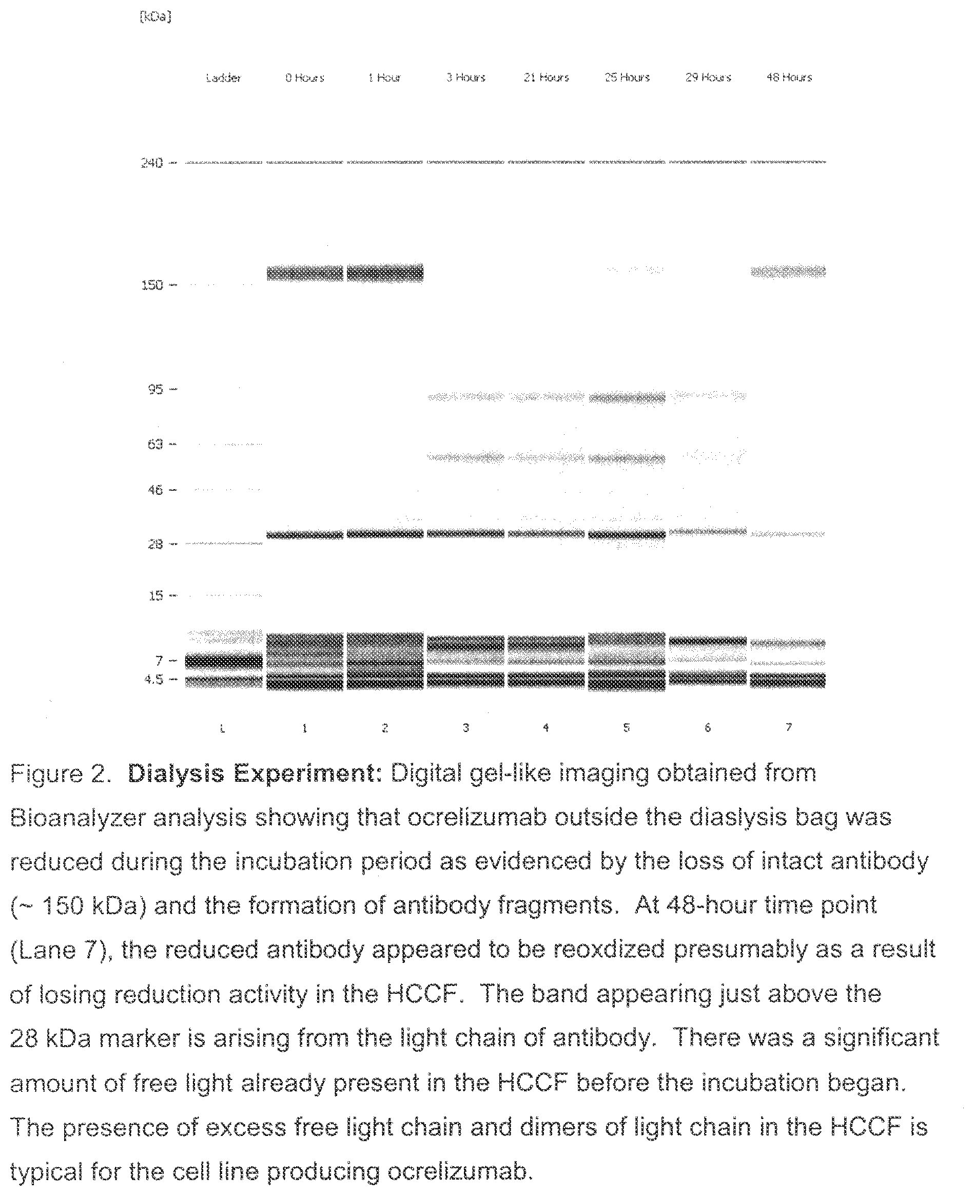
[0037] FIG. 2. Dialysis Experiment: Digital gel-like imaging obtained from Bioanalyzer analysis (each lane representing a time point) showing that ocrelizumab outside the dialysis bag was reduced during the incubation period. This is evidenced by the loss of intact antibody (.about.150 kDa) and the formation of antibody fragments depicted in the Figure. At the 48-hour time point (Lane 7), the reduced antibody appeared to be reoxidized, presumably as a result of loosing reduction activity in the Harvested Cell Culture Fluid (HCCF). The band appearing just above the 28 kDa marker arose from the light chain of antibody. There was a significant amount of free light already present in the HCCF before the incubation began. The presence of excess free light chain and dimers of light chain in the HCCF is typical for the cell line producing ocrelizumab.
[0038] FIG. 3. Free Thiol Levels from Dialysis Experiment: Purified ocrelizumab in phosphate buffered saline (PBS) was inside the dialysis bag and HCCF containing ocrelizumab was outside the bag. Free thiols inside (boxes) and outside (diamonds) the dialysis bag reached comparable levels within a few hours, indicating a good exchange of small molecule components in the HCCF between inside and outside the dialysis bag.
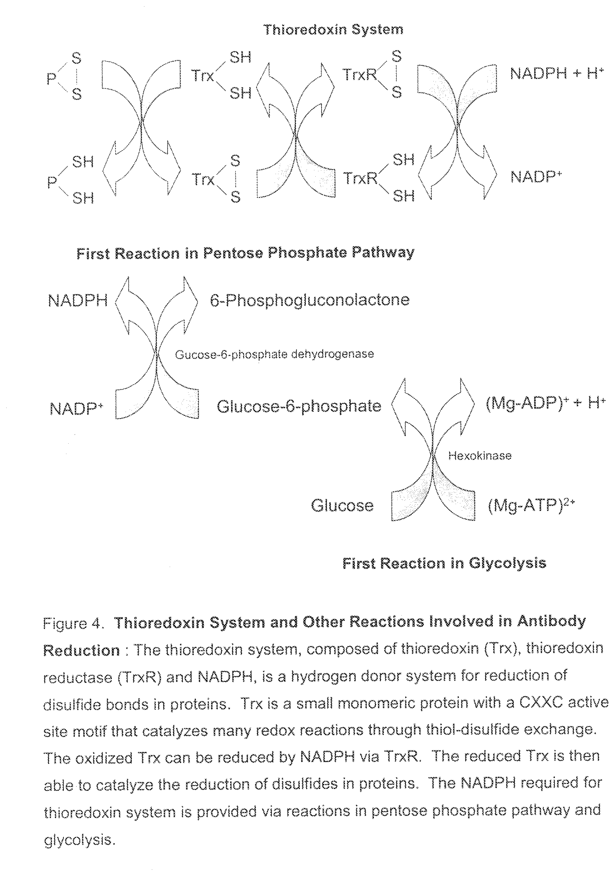
[0039] FIG. 4. Thioredoxin System and Other Reactions Involved in Antibody Reduction: The thioredoxin system, comprising thioredoxin (Trx), thioredoxin reductase (TrxR) and NADPH, functions as a hydrogen donor system for reduction of disulfide bonds in proteins. Trx is a small monomeric protein with a CXXC active site motif that catalyzes many redox reactions through thiol-disulfide exchange. The oxidized Trx can be reduced by NADPH via TrxR. The reduced Trx is then able to catalyze the reduction of disulfides in proteins. The NADPH required for thioredoxin system is provided via reactions in pentose phosphate pathway and glycolysis.
[0040] FIG. 5. In Vitro Activity of Thioredoxin System: Digital gel-like image from Bioanalyzer analysis (each lane representing a time point) demonstrating that incubation of intact ocrelizumab (1 mg/mL) with 0.1 mM TrxR (rat liver), 5 mM Trx (human), and 1 mM NADPH in PBS resulted in the complete reduction of ocrelizumab; the ocrelizumab was completely reduced in less than 21 hours.
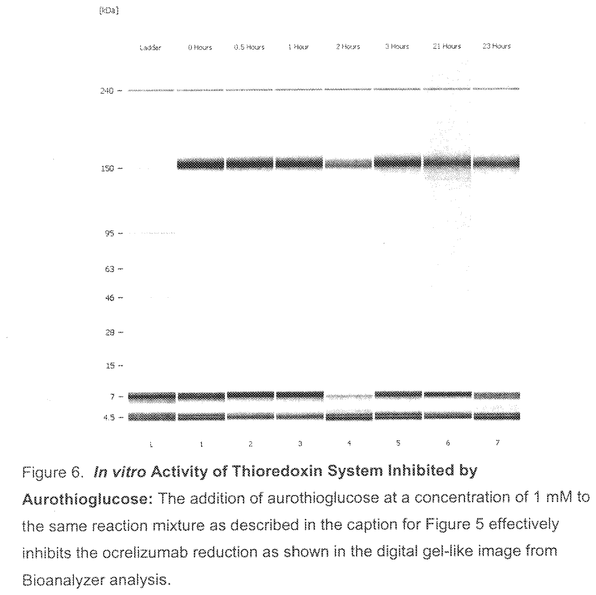
[0041] FIG. 6. In Vitro Activity of Thioredoxin System Inhibited by Aurothioglucose: The addition of aurothioglucose (ATG) to the same reaction mixture as described in the caption for FIG. 5, above, effectively inhibited the ocrelizumab reduction. This is seen by the digital gel-like image from Bioanalyzer analysis (each lane representing a time point).
[0042] FIG. 7. In vitro Activity of Thioredoxin System Inhibited by Aurothiomalate: The addition of aurothiomalate (ATM) at a concentration of 1 mM to the same reaction mixture as described in the caption for FIG. 5, above, effectively inhibited the ocrelizumab reduction. This is seen by the digital gel-like image from Bioanalyzer analysis (each lane representing a time point).
[0043] FIG. 8. In Vitro Activity of Thioredoxin System: Digital gel-like image from Bioanalyzer analysis (each lane representing a time point) showing that incubation of intact ocrelizumab (1 mg/mL) with 0.1 mM TrxR (rat liver), 5 mM Trx (human), and 1 mM NADPH in 10 mM histidine sulfate buffer resulted in the reduction of ocrelizumab in less than 1 hour.
[0044] FIG. 9. In vitro Activity of Thioredoxin System Inhibited by CuSO.sub.4: The addition of CuSO.sub.4 at a concentration of 50 .mu.M to the same reaction mixture as described in the caption for FIG. 8 effectively inhibited the ocrelizumab reduction as shown in the digital gel-like image from Bioanalyzer analysis (each lane representing a time point).
[0045] FIG. 10. Ocrelizumab Reduction: Digital gel-like image from Bioanalyzer analysis (each lane representing a time point) showing that ocrelizumab was reduced in an incubation experiment using HCCF from a homogenized CCF generated from a 3-L fermentor.
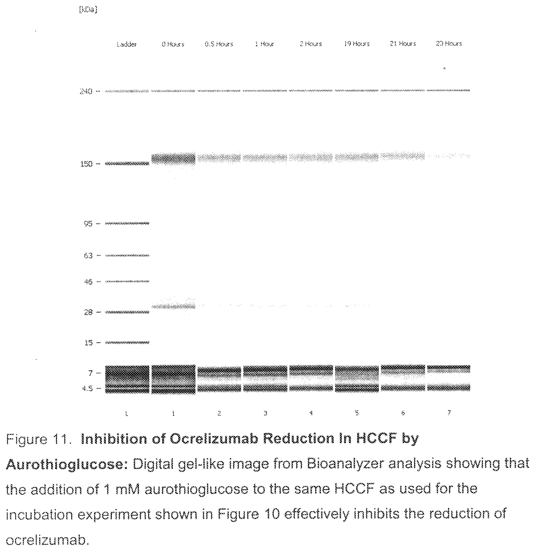
[0046] FIG. 11. Inhibition of Ocrelizumab Reduction In HCCF by Aurothioglucose: Digital gel-like image from Bioanalyzer analysis (each lane representing a time point) showing that the addition of 1 mM aurothioglucose to the same HCCF as used for the incubation experiment as shown in FIG. 10 inhibited the reduction of ocrelizumab.
[0047] FIG. 12. Inhibition of Ocrelizumab Reduction In HCCF by Aurothiomalate: Digital gel-like image from Bioanalyzer (each lane representing a time point) analysis indicating that the addition of 1 mM aurothiomalate to the same HCCF as used for the incubation experiment shown in FIG. 10 inhibited the reduction of ocrelizumab.
[0048] FIG. 13. Losing Reduction Activity in HCCF: The HCCF from one of the large scale manufacturing runs for ocrelizumab (the "beta" run) that was subject to several freeze/thaw cycles demonstrated no ocrelizumab reduction when used in an incubation experiment. This was shown by Bioanalyzer analysis (each lane representing a time point), and can be contrasted to the antibody reduction seen previously in the freshly thawed HCCF from the same fermentation batch.
[0049] FIG. 14. The Lost Reduction Activity in HCCF Restored by Addition of NADPH: The reduction of ocrelizumab was observed again in the Bioanalyzer assay (each lane representing a time point) after the addition of NADPH at a concentration of 5 mM into the HCCF where the reduction activity has been eliminated under the conditions described above in FIG. 13.
[0050] FIG. 15. The Lost Reduction Activity in HCCF Restored by Addition of Glucose-6-Phosphate: The reduction of ocrelizumab was observed again in the Bioanalyzer assay (each lane representing a time point) after the addition of G6P at a concentration of 10 mM into the HCCF where the reduction activity has been eliminated due to the treatment described above in FIG. 13.
[0051] FIG. 16. Ocrelizumab Reduction: A digital gel-like image from Bioanalyzer analysis showing that ocrelizumab was reduced in an incubation experiment using a HCCF from a large scale manufacturing run (the "alpha" run).
[0052] FIG. 17. EDTA Inhibits Ocrelizumab Reduction: Digital gel-like image from Bioanalyzer analysis (each lane representing a time point) showing that the reduction of ocrelizumab was inhibited in an incubation experiment using a HCCF from the alpha run with EDTA added at a concentration of 20 mM to the HCCF whose reducing activity is demonstrated in FIG. 16.
[0053] FIG. 18. The Lost Reduction Activity in "Beta Run" HCCF Restored by Addition of Glucose--Phosphate but No Inhibition of Reduction by EDTA: The reduction of ocrelizumab was observed in the Bioanalyzer assay (each lane representing a time point) after the addition of G6P at a concentration of 5 mM and 20 mM EDTA into the HCCF whose reduction activity had been lost (see FIG. 13). In contrast to the results shown in FIG. 17, the presence of EDTA did not block the reduction of ocreliumab.
[0054] FIG. 19. Inhibition of Ocrelizumab Reduction: by (i) addition of EDTA, (ii) addition of CuSO.sub.4, or (iii) adjustment of pH to 5.5. All three different methods, (1) addition of EDTA, (2) addition of CuSO.sub.4, and (3) adjustment of pH to 5.5, used independently, were effective in inhibiting ocrelizumab reduction. This was demonstrated by the depicted quantitative Bioanalyzer results that showed that nearly 100% intact (150 kDa) antibody remained in the protein A elution pools. In contrast, ocrelizumab was completely reduced in the control HCCF after 20 hours of HCCF hold time.
[0055] FIG. 20. Inhibition of Ocrelizumab Reduction by Air Sparging: Sparging the HCCF with air was effective in inhibiting ocrelizumab disulfide bond reduction. This was demonstrated by the quantitative Bioanalyzer results showing that nearly 100% intact (150 kDa) antibody remained in the protein A elution pools. In contrast, ocrelizumab was almost completely reduced in the control HCCF after 5 hours of sparging with nitrogen.
[0056] FIG. 21 shows the V.sub.L(SEQ ID NO. 24) amino acid sequence of an anti-Her2 antibody (Trastuzumab).
[0057] FIG. 22 shows the V.sub.H (SEQ ID No. 25) amino acid sequence of an anti-Her2 antibody (Trastuzumab).
[0058] FIG. 23 is a schematic showing some steps of a typical large scale manufacturing process.
[0059] FIG. 24 is a digital gel-like image from Bioanalyzer analysis: 2H7 (Variant A)+1 mM NADPH+5 .mu.M thioredoxin+0.1 .mu.M thioredoxin reductase (recombinant) in 10 mM histidine sulfate.
[0060] FIG. 25 is a digital gel-like image from Bioanalyzer analysis: 2H7 (Variant A)+1 mM NADPH+5 .mu.M thioredoxin+0.1 .mu.M thioredoxin reductase (recombinant) in 1 mM histidine sulfate+1 mM ATG.
[0061] FIG. 26 is a digital gel-like image from Bioanalyzer analysis: 2H7 (Variant A)+1 mM NADPH+5 .mu.M thioredoxin+0.1 .mu.M thioredoxin reductase (recombinant) in 10 mM histidine sulfate+0.6 sM ATG (6:1 ATG:TrxR).
[0062] FIG. 27 is a digital gel-like image from Bioanalyzer analysis: 2H7 (Variant A)+1 mM NADPH+5 .mu.M thioredoxin+0.1 .mu.M thioredoxin reductase (recombinant) in 10 mM histidine sulfate+0.4 .mu.M ATG (4:1 ATG:TrxR).
[0063] FIG. 28 is a digital gel-like image from Bioanalyzer analysis: 2H7 (Variant A)+1 mM NADPH+5 .mu.M thioredoxin+0.1 .mu.M thioredoxin reductase (recombinant) in 10 mM histidine sulfate+0.2 M ATG (2:1 ATG:TrxR).
[0064] FIG. 29 is a digital gel-like image from Bioanalyzer analysis: 2H7 (Variant A)+1 mM NADPH+5 .mu.M thioredoxin+0.1 .mu.M thioredoxin reductase (recombinant) in 10 mM histidine sulfate+0.1 mM autothiomalate (ATM).
[0065] FIG. 30 is a digital gel-like image from Bioanalyzer analysis: 2H7 (Variant A)+1 mM NADPH+5 .mu.M thioredoxin+0.1 .mu.M thioredoxin reductase (recombinant) in 10 mM histidine sulfate+0.01 mM autothiomalate (ATM).
[0066] FIG. 31 is a digital gel-like image from Bioanalyzer analysis: 2H7 (Variant A)+1 mM NADPH+5 .mu.M thioredoxin+0.1 .mu.M thioredoxin reductase (recombinant) in 10 mM histidine sulfate+20 .mu.M CuSO.sub.4 (4:1 Cu.sup.2+:Trx).
[0067] FIG. 32 is a digital gel-like image from Bioanalyzer analysis: 2H7 (Variant A)+1 mM NADPH+5 .mu.M thioredoxin+0.1 .mu.M thioredoxin reductase (recombinant) in 10 mM histidine sulfate+10 .mu.M CuSO.sub.4 (2:1 Cu.sup.2+:Trx).
[0068] FIG. 33 is a digital gel-like image from Bioanalyzer analysis: 2H7 (Variant A)+1 mM NADPH+5 .mu.M thioredoxin+0.1 .mu.M thioredoxin reductase (recombinant) in 10 mM histidine sulfate+5 .mu.M CuSO.sub.4 (1:1 Cu.sup.2+:Trx).
[0069] FIG. 34 is a digital gel-like image from Bioanalyzer analysis: 2H7 (Variant A)+1 mM NADPH+5 .mu.M thioredoxin+0.1 M thioredoxin reductase (recombinant) in 10 mM histidine sulfate+532 .mu.M crystamine (20:1 cystamine:2H7 disulfide).
[0070] FIG. 35 is a digital gel-like image from Bioanalyzer analysis: 2H7 (Variant A)+1 mM NADPH+5 .mu.M thioredoxin+0.1 .mu.M thioredoxin reductase (recombinant) in 10 mM histidine sulfate+266 .mu.M cystamine (10:1 cystamine:2H7 disulfide).
[0071] FIG. 36 is a digital gel-like image from Bioanalyzer analysis: 2H7 (Variant A)+1 mM NADPH+5 .mu.M thioredoxin+0.1 .mu.M thioredoxin reductase (recombinant) in 10 mM histidine sulfate+133 .mu.M cystamine (5:1 cystamine:2H7 disulfide).
[0072] FIG. 37 is a digital gel-like image from Bioanalyzer analysis: 2H7 (Variant A)+1 mM NADPH+5 .mu.M thioredoxin+0.1 .mu.M thioredoxin reductase (recombinant) in 10 mM histidine sulfate+26.6 .mu.M cystamine (1:1 cystamine:2H7 disulfide).
[0073] FIG. 38 is a digital gel-like image from Bioanalyzer analysis: 2H7 (Variant A)+1 mM NADPH+5 .mu.M thioredoxin+0.1 .mu.M thioredoxin reductase (recombinant) in 10 mM histidine sulfate (pH=7.6)+2.6 mM cysteine.
[0074] FIG. 39 is a digital gel-like image from Bioanalyzer analysis: 2H7 (Variant A)+1 mM NADPH+5 M thioredoxin+0.1 .mu.M thioredoxin reductase (recombinant) in 10 mM histidine sulfate+2.6 mM GSSG (oxidized glutathione).
[0075] FIG. 40 Reconstructed enzymatic reduction system. 1 mg/ml 2H7 (Variant A)+10 .mu.g/mL hexokinase, 50 .mu.g/mL glucose-6-phosphate dehydrogenase, 5 .mu.M thioredoxin, 0.1 .mu.M thioredoxin reductase, 2 mM glucose, 0.6 mM ATP, 2 mM Mg.sup.2+, and 2 mM NADP in 50 mM histidine sulfate buffer at pH=7.38.
[0076] FIG. 41 The thioredoxin system requires NADPH. 1 mg/ml 2H7 (Variant A)+5 .mu.M thioredoxin, 0.1 .mu.M thioredoxin reductase, and 2 mM NADP in 50 mM histidine sulfate buffer at pH=7.38.
DETAILED DESCRIPTION OF THE PREFERRED EMBODIMENTS
I. Definitions
[0077] In the present invention, in the context of proteins, including antibodies, in general, or with regard to any specific protein or antibody, the term "reduction" is used to refer to the reduction of one or more disulfide bonds of the protein or antibody. Thus, for example, the terms "ocrelizumab reduction" is used interchangeably with the term "ocrelizumab disulfide bond reduction" and the term "antibody (Ab) reduction" is used interchangeably with the term "antibody (Ab) disulfide bond reduction."
[0078] The terms "reduction" or "disulfide bond reduction" are used in the broadest sense, and include complete and partial reduction and reduction of some or all of the disulfide bonds, interchain or intrachain, present in a protein such as an antibody.
[0079] By "protein" is meant a sequence of amino acids for which the chain length is sufficient to produce the higher levels of tertiary and/or quaternary structure. This is to distinguish from "peptides" or other small molecular weight drugs that do not have such structure. Typically, the protein herein will have a molecular weight of at least about 15-20 kD, preferably at least about 20 kD. Examples of proteins encompassed within the definition herein include all mammalian proteins, in particular, therapeutic and diagnostic proteins, such as therapeutic and diagnostic antibodies, and, in general proteins that contain one or more disulfide bonds, including multi-chain polypeptides comprising one or more inter- and/or intrachain disulfide bonds.
[0080] The term "therapeutic protein" or "therapeutic polypeptide" refers to a protein that is used in the treatment of disease, regardless of its indication or mechanism of action. In order for therapeutic proteins to be useful in the clinic it must be manufactured in large quantities. "Manufacturing scale" production of therapeutic proteins, or other proteins, utilize cell cultures ranging from about 400 L to about 80,000 L, depending on the protein being produced and the need. Typically such manufacturing scale production utilizes cell culture sizes from about 400 L to about 25,000 L. Within this range, specific cell culture sizes such as 4,000 L, about 6,000 L, about 8,000, about 10,000, about 12,000 L, about 14,000 L, or about 16,000 L are utilized.
[0081] The term "therapeutic antibody" refers to an antibody that is used in the treatment of disease. A therapeutic antibody may have various mechanisms of action. A therapeutic antibody may bind and neutralize the normal function of a target associated with an antigen. For example, a monoclonal antibody that blocks the activity of the of protein needed for the survival of a cancer cell causes the cell's death. Another therapeutic monoclonal antibody may bind and activate the normal function of a target associated with an antigen. For example, a monoclonal antibody can bind to a protein on a cell and trigger an apoptosis signal. Yet another monoclonal antibody may bind to a target antigen expressed only on diseased tissue; conjugation of a toxic payload (effective agent), such as a chemotherapeutic or radioactive agent, to the monoclonal antibody can create an agent for specific delivery of the toxic payload to the diseased tissue, reducing harm to healthy tissue. A "biologically functional fragment" of a therapeutic antibody will exhibit at least one if not some or all of the biological functions attributed to the intact antibody, the function comprising at least specific binding to the target antigen.
[0082] The term "diagnostic protein" refers to a protein that is used in the diagnosis of a disease.
[0083] The term "diagnostic antibody" refers to an antibody that is used as a diagnostic reagent for a disease. The diagnostic antibody may bind to a target antigen that is specifically associated with, or shows increased expression in, a particular disease. The diagnostic antibody may be used, for example, to detect a target in a biological sample from a patient, or in diagnostic imaging of disease sites, such as tumors, in a patient. A "biologically functional fragment" of a diagnostic antibody will exhibit at least one if not some or all of the biological functions attributed to the intact antibody, the function comprising at least specific binding to the target antigen.
[0084] "Purified" means that a molecule is present in a sample at a concentration of at least 80-90% by weight of the sample in which it is contained.
[0085] The protein, including antibodies, which is purified is preferably essentially pure and desirably essentially homogeneous (i.e. free from contaminating proteins etc.).
[0086] An "essentially pure" protein means a protein composition comprising at least about 90% by weight of the protein, based on total weight of the composition, preferably at least about 95% by weight.
[0087] An "essentially homogeneous" protein means a protein composition comprising at least about 99% by weight of protein, based on total weight of the composition.
[0088] As noted above, in certain embodiments, the protein is an antibody. "Antibodies" (Abs) and "immunoglobulins" (Igs) are glycoproteins having the same structural characteristics. While antibodies exhibit binding specificity to a specific antigen, immunoglobulins include both antibodies and other antibody-like molecules which generally lack antigen specificity. Polypeptides of the latter kind are, for example, produced at low levels by the lymph system and at increased levels by myelomas.
[0089] The term "antibody" is used in the broadest sense and specifically covers monoclonal antibodies (including full length antibodies which have an immunoglobulin Fc region), antibody compositions with polyepitopic specificity, bispecific antibodies, diabodies, and single-chain molecules such as scFv molecules, as well as antibody fragments (e.g., Fab, F(ab'), and Fv).
[0090] The term "monoclonal antibody" as used herein refers to an antibody obtained from a population of substantially homogeneous antibodies. i.e., the individual antibodies comprising the population are identical except for possible mutations, e.g., naturally occurring mutations, that may be present in minor amounts. Thus, the modifier "monoclonal" indicates the character of the antibody as not being a mixture of discrete antibodies. In certain embodiments, such a monoclonal antibody typically includes an antibody comprising a polypeptide sequence that binds a target, wherein the target-binding polypeptide sequence was obtained by a process that includes the selection of a single target binding polypeptide sequence from a plurality of polypeptide sequences. For example, the selection process can be the selection of a unique clone from a plurality of clones, such as a pool of hybridoma clones, phage clones, or recombinant DNA clones. It should be understood that a selected target binding sequence can be further altered, for example, to improve affinity for the target, to humanize the target binding sequence, to improve its production in cell culture, to reduce its immunogenicity in vivo, to create a multispecific antibody, etc., and that an antibody comprising the altered target binding sequence is also a monoclonal antibody of this invention. In contrast to polyclonal antibody preparations which typically include different antibodies directed against different determinants (epitopes), each monoclonal antibody of a monoclonal antibody preparation is directed against a single determinant on an antigen. In addition to their specificity, monoclonal antibody preparations are advantageous in that they are typically uncontaminated by other immunoglobulins.
[0091] The modifier "monoclonal" indicates the character of the antibody as being obtained from a substantially homogeneous population of antibodies, and is not to be construed as requiring production of the antibody by any particular method. For example, the monoclonal antibodies to be used in accordance with the present invention may be made by a variety of techniques, including, for example, the hybridoma method (e.g., Kohler et al., Nature, 256: 495 (1975); Harlow et al., Antibodies: A Laboratory Manual, (Cold Spring Harbor Laboratory Press, 2nd ed. 1988); Hammerling et al., in: Monoclonal Antibodies and T-Cell Hybridomas 563-681 (Elsevier, N.Y., 1981)), recombinant DNA methods (see, e.g., U.S. Pat. No. 4,816,567), phage display technologies (see, e.g., Clackson et al., Nature, 352: 624-628 (1991); Marks et al., J. Mol. Biol. 222: 581-597 (1992); Sidhu et al., J. Mol. Biol. 338(2): 299-310 (2004); Lee et al., J. Mol. Biol. 340(5): 1073-1093 (2004); Fellouse, Proc. Natl. Acad. Sci. USA 101(34): 12467-12472 (2004); and Lee et al., J. Immunol. Methods 284(1-2): 119-132(2004), and technologies for producing human or human-like antibodies in animals that have parts or all of the human immunoglobulin loci or genes encoding human immunoglobulin sequences (see, e.g., WO98/24893; WO96/34096; WO96/33735; WO91/10741; Jakobovits et al., Proc. Nat. Acad. Sci. USA 90: 2551 (1993); Jakobovits et al., Nature 362: 255-258 (1993); Bruggemann et al., Year in Immunol. 7:33 (1993); U.S. Pat. Nos. 5,545,807; 5,545,806; 5,569,825; 5,625,126; 5,633,425; 5,661,016; Marks et al., BioTechnology 10: 779-783 (1992); Lonberg et al., Nature 368: 856-859 (1994); Morrison, Nature 368: 812-813 (1994); Fishwild et al., Nature Biotechnol. 14: 845-851 (1996); Neuberger, Nature Biotechnol. 14: 826 (1996) and Lonberg and Huszar, Intern. Rev. Immunol. 13: 65-93 (1995).
[0092] The monoclonal antibodies herein specifically include "chimeric" antibodies in which a portion of the heavy and/or light chain is identical with or homologous to corresponding sequences in antibodies derived from a particular species or belonging to a particular antibody class or subclass, while the remainder of the chain(s) is identical with or homologous to corresponding sequences in antibodies derived from another species or belonging to another antibody class or subclass, as well as fragments of such antibodies, so long as they exhibit the desired biological activity (U.S. Pat. No. 4,816,567; and Morrison et al., Proc. Nat. Acad. Sci. USA 81:6851-6855 (1984)).
[0093] "Humanized" forms of non-human (e.g., murine) antibodies are chimeric antibodies that contain minimal sequence derived from non-human immunoglobulin. In one embodiment, a humanized antibody is a human immunoglobulin (recipient antibody) in which residues from a hypervariable region of the recipient are replaced by residues from a hypervariable region of a non-human species (donor antibody) such as mouse, rat, rabbit, or nonhuman primate having the desired specificity, affinity, and/or capacity. In some instances, framework region (FR) residues of the human immunoglobulin are replaced by corresponding non-human residues. Furthermore, humanized antibodies may comprise residues that are not found in the recipient antibody or in the donor antibody. These modifications may be made to further refine antibody performance. In general, a humanized antibody will comprise substantially all of at least one, and typically two, variable domains, in which all or substantially all of the hypervariable loops correspond to those of a non-human immunoglobulin, and all or substantially all the FRs are those of a human immunoglobulin sequence. The humanized antibody optionally will also comprise at least a portion of an immunoglobulin constant region (Fc), typically that of a human immunoglobulin. For further details, see Jones et al., Nature 321:522-525 (1986); Riechmann et al., Nature 332:323-329 (1988); and Presta, Curr. Op. Struct. Biol. 2:593-596 (1992). See also the following review articles and references cited therein: Vaswani and Hamilton, Ann, Allergy, Asthma &Immunol. 1:105-115 (1998); Harris, Biochem. Soc. Transactions 23:1035-1038 (1995); Hurle and Gross. Curr. Op. Biotech. 5:428-433 (1994). The humanized antibody includes a Primatized.TM. antibody wherein the antigen-binding region of the antibody is derived from an antibody produced by immunizing macaque monkeys with the antigen of interest.
[0094] A "human antibody" is one which possesses an amino acid sequence which corresponds to that of an antibody produced by a human and/or has been made using any of the techniques for making human antibodies as disclosed herein. This definition of a human antibody specifically excludes a humanized antibody comprising non-human antigen-binding residues.
[0095] An "affinity matured" antibody is one with one or more alterations in one or more CDRs/HVRs thereof which result in an improvement in the affinity of the antibody for antigen, compared to a parent antibody which does not possess those alteration(s). Preferred affinity matured antibodies will have nanomolar or even picomolar affinities for the target antigen. Affinity matured antibodies are produced by procedures known in the art. Marks et al., Bio/Technology 10:779-783 (1992) describes affinity maturation by V.sub.H and V.sub.L domain shuffling. Random mutagenesis of CDR/HVR and/or framework residues is described by: Barbas et al., Proc Nat. Acad. Sci. USA 91:3809-3813 (1994); Schier et al., Gene 169:147-155 (1995); Yelton et al., J. Immunol. 155:1994-2004 (1995); Jackson et al., J. Immunol. 154(7):3310-9 (1995); and Hawkins et at, J. Mol. Biol. 226:889-896 (1992).
[0096] The "variable region" or "variable domain" of an antibody refers to the amino-terminal domains of the heavy or light chain of the antibody. The variable domain of the heavy chain may be referred to as "V.sub.H." The variable domain of the light chain may be referred to as "V.sub.L." These domains are generally the most variable parts of an antibody and contain the antigen-binding sites.
[0097] The term "variable" refers to the fact that certain portions of the variable domains differ extensively in sequence among antibodies and are used in the binding and specificity of each particular antibody for its particular antigen. However, the variability is not evenly distributed throughout the variable domains of antibodies. It is concentrated in three segments called complementarity-determining regions (CDRs) or hypervariable regions (HVRs) both in the light-chain and the heavy-chain variable domains. The more highly conserved portions of variable domains are called the framework regions (FR). The variable domains of native heavy and light chains each comprise four FR regions, largely adopting a beta-sheet configuration, connected by three CDRs, which form loops connecting, and in some cases forming part of, the beta-sheet structure. The CDRs in each chain are held together in close proximity by the FR regions and, with the CDRs from the other chain, contribute to the formation of the antigen-binding site of antibodies (see Kabat et al., Sequences of Proteins of Immunological Interest, Fifth Edition, National Institute of Health, Bethesda, Md. (1991)). The constant domains are not involved directly in the binding of an antibody to an antigen, but exhibit various effector functions, such as participation of the antibody in antibody-dependent cellular toxicity.
[0098] The "light chains" of antibodies (immunoglobulins) from any vertebrate species can be assigned to one of two clearly distinct types, called kappa (.kappa.) and lambda (k), based on the amino acid sequences of their constant domains.
[0099] Depending on the amino acid sequences of the constant domains of their heavy chains, antibodies (immunoglobulins) can be assigned to different classes. There are five major classes of immunoglobulins: IgA, IgD, IgE, IgG and IgM, and several of these may be further divided into subclasses (isotypes), e.g., IgG.sub.1, IgG.sub.2, IgG.sub.3, IgG.sub.4, IgA.sub.1, and IgA.sub.2. The heavy chain constant domains that correspond to the different classes of immunoglobulins are called a, d, e, g, and m, respectively. The subunit structures and three-dimensional configurations of different classes of immunoglobulins are well known and described generally in, for example, Abbas et al., Cellular and Mol. Immunology, 4th ed. (2000). An antibody may be part of a larger fusion molecule, formed by covalent or non-covalent association of the antibody with one or more other proteins or peptides.
[0100] The terms "full length antibody," "intact antibody" and "whole antibody" are used herein interchangeably to refer to an antibody in its substantially intact form, not antibody fragments as defined below. The terms particularly refer to an antibody with heavy chains that contain the Fc region.
[0101] "Antibody fragments" comprise only a portion of an intact antibody, wherein the portion retains at least one, and as many as most or all, of the functions normally associated with that portion when present in an intact antibody. In one embodiment, an antibody fragment comprises an antigen binding site of the intact antibody and thus retains the ability to bind antigen. In another embodiment, an antibody fragment, for example one that comprises the Fc region, retains at least one of the biological functions normally associated with the Fc region when present in an intact antibody, such as FcRn binding, antibody half life modulation, ADCC function and complement binding. In one embodiment, an antibody fragment is a monovalent antibody that has an in vivo half life substantially similar to an intact antibody. For example, such an antibody fragment may comprise an antigen binding arm linked to an Fc sequence capable of conferring in vivo stability to the fragment.
[0102] Papain digestion of antibodies produces two identical antigen-binding fragments, called "Fab" fragments, each with a single antigen-binding site, and a residual "Fc" fragment, whose name reflects its ability to crystallize readily. Pepsin treatment yields an F(ab').sub.2 fragment that has two antigen-combining sites and is still capable of cross-linking antigen.
[0103] The Fab fragment contains the heavy- and light-chain variable domains and also contains the constant domain of the light chain and the first constant domain (CH1) of the heavy chain. Fab' fragments differ from Fab fragments by the addition of a few residues at the carboxy terminus of the heavy chain CH1 domain including one or more cysteines from the antibody hinge region. Fab'-SH is the designation herein for Fab' in which the cysteine residue(s) of the constant domains bear a free thiol group. F(ab')2 antibody fragments originally were produced as pairs of Fab' fragments which have hinge cysteines between them. Other chemical couplings of antibody fragments are also known.
[0104] "Fv" is the minimum antibody fragment which contains a complete antigen-binding site. In one embodiment, a two-chain Fv species consists of a dimer of one heavy- and one light-chain variable domain in tight, non-covalent association. In a single-chain Fv (scFv) species, one heavy- and one light-chain variable domain can be covalently linked by a flexible peptide linker such that the light and heavy chains can associate in a "dimeric" structure analogous to that in a two-chain Fv species. It is in this configuration that the three CDRs of each variable domain interact to define an antigen-binding site on the surface of the V.sub.H-V.sub.L dimer. Collectively, the six CDRs confer antigen-binding specificity to the antibody. However, even a single variable domain (or half of an Fv comprising only three CDRs specific for an antigen) has the ability to recognize and bind antigen, although at a lower affinity than the entire binding site.
[0105] "Single-chain Fv" or "scFv" antibody fragments comprise the V.sub.H and V.sub.L domains of an antibody, wherein these domains are present in a single polypeptide chain. Generally, the scFv polypeptide further comprises a polypeptide linker between the V.sub.H and V.sub.L domains which enables the scFv to for the desired structure for antigen binding. For a review of scFv see Pluckthun, in The Pharmacology of Monoclonal Antibodies, vol. 113, Rosenburg and Moore eds., Springer-Verlag, New York, pp. 269-315 (1994).
[0106] The term "diabodies" refers to small antibody fragments with two antigen-binding sites, which fragments comprise a heavy-chain variable domain (V.sub.H) connected to a light-chain variable domain (Vt) in the same polypeptide chain (V.sub.H--V.sub.L). By using a linker that is too short to allow pairing between the two domains on the same chain, the domains are forced to pair with the complementary domains of another chain and create two antigen-binding sites. Diabodies may be bivalent or bispecific. Diabodies are described more fully in, for example, EP 404,097; WO93/1161; Hudson et al., (2003) Nat. Med. 9:129-134; and Hollinger et al., Proc. Natl. Acad. Sci. USA 90: 6444-6448 (1993). Triabodies and tetrabodies are also described in Hudson et al., (2003) Nat. Med. 9:129-134.
[0107] The antibody may bind to any protein, including, without limitation, a member of the HER receptor family, such as HER1 (EGFR), HER2, HER3 and HER4; CD proteins such as CD3, CD4, CD8, CD19, CD20, CD21, CD22, and CD34; cell adhesion molecules such as LFA-1, Mol, p150,95, VLA-4, ICAM-1, VCAM and av/p3 integrin including either a or P or subunits thereof (e.g. anti-CD11a, anti-CD18 or anti-CD11b antibodies); growth factors such as vascular endothelial growth factor (VEGF); IgE; blood group antigens; flk2/flt3 receptor, obesity (OB) receptor, and protein C. Other exemplary proteins include growth hormone (GH), including human growth hormone (hGH) and bovine growth hormone (bGH); growth hormone releasing factor, parathyroid hormone; thyroid stimulating hormone; lipoproteins; .alpha.-1-antitrypsin; insulin A-chain; insulin B-chain; proinsulin; follicle stimulating hormone; calcitonin; luteinizing hormone; glucagon; clotting factors such as factor VIIIC, factor, tissue factor, and von Willebrands factor, anti-clotting factors such as Protein C; atrial natriuretic factor, lung surfactant; a plasminogen activator, such as urokinase or tissue-type plasminogen activator (t-PA); bombazine; thrombin; tumor necrosis factor-.alpha. and -.beta.; enkephalinase; RANTES (regulated on activation normally T-cell expressed and secreted); human macrophage inflammatory protein (MIP-1-.alpha.); serum albumin such as human serum albumin (HSA); mullerian-inhibiting substance; relaxin A-chain; relaxin B-chain; prorelaxin; mouse gonadotropin-associated peptide; DNase; inhibin; activin; receptors for hormones or growth factors; an integrin; protein A or D; rheumatoid factors; a neurotrophic factor such as bone-derived neurotrophic factor (BDNF), neurotrophin-3, -4, -5, or -6 (NT-3, NT-4, NT-5, or NT-6), or a nerve growth factor such as NGF-.beta.; platelet-derived growth factor (PDGF); fibroblast growth factor such as aFGF and bFGF; epidermal growth factor (EGF); transforming growth factor (TGF) such as TGF-.alpha. and TGF-.beta., including TGF-.beta.1, TGF-.beta.2, TGF-.beta.3, TGF-.beta.4, or TGF-.beta.5; insulin-like growth factor-I and -II (IGF-I and IGF-II); des(1-3)-IGF-I (brain IGF-I); insulin-like growth factor binding proteins (IGFBPs); erythropoietin (EPO); thrombopoietin (TPO); osteoinductive factors; immunotoxins; a bone morphogenetic protein (BMP); an interferon such as interferon-.alpha., -.beta., and -.gamma. colony stimulating factors (CSFs), e.g., M-CSF, GM-CSF, and G-CSF; interleukins (ILs), e.g., IL-1 to IL-10; superoxide dismutase; T-cell receptors; surface membrane proteins; decay accelerating factor (DAF); a viral antigen such as, for example, a portion of the AIDS envelope; transport proteins; homing receptors; addressins; regulatory proteins; immunoadhesins; antibodies; and biologically active fragments or variants of any of the above-listed polypeptides. Many other antibodies and/or other proteins may be used in accordance with the instant invention, and the above lists are not meant to be limiting.
[0108] A "biologically functional fragment" of an antibody comprises only a portion of an intact antibody, wherein the portion retains at least one, and as many as most or all, of the functions normally associated with that portion when present in an intact antibody. In one embodiment, a biologically functional fragment of an antibody comprises an antigen binding site of the intact antibody and thus retains the ability to bind antigen. In another embodiment, a biologically functional fragment of an antibody, for example one that comprises the Fc region, retains at least one of the biological functions normally associated with the Fc region when present in an intact antibody, such as FcRn binding, antibody half life modulation, ADCC function and complement binding. In one embodiment, a biologically functional fragment of an antibody is a monovalent antibody that has an in vivo half life substantially similar to an intact antibody. For example, such a biologically functional fragment of an antibody may comprise an antigen binding arm linked to an Fc sequence capable of conferring in vivo stability to the fragment.
[0109] The terms "thioredoxin inhibitor" and "Trx inhibitor" are used interchangeably, and include all agents and measures effective in inhibiting thioredoxin activity. Thus, thioredoxin (Trx) inhibitors include all agents and measures blocking any component of the Trx, G6PD and/or hexokinase enzyme systems. In this context, "inhibition" includes complete elimination (blocking) and reduction of thioredoxin activity, and, consequently, complete or partial elimination of disulfide bond reduction in a protein, such as an antibody.
[0110] An "isolated" antibody is one which has been identified and separated and/or recovered from a component of its natural environment. Contaminant components of its natural environment are materials which would interfere with research, diagnostic or therapeutic uses for the antibody, and may include enzymes, hormones, and other proteinaceous or nonproteinaceous solutes. In some embodiments, an antibody is purified (1) to greater than 95% by weight of antibody as determined by, for example, the Lowry method, and in some embodiments, to greater than 99% by weight; (2) to a degree sufficient to obtain at least 15 residues of N-terminal or internal amino acid sequence by use of, for example, a spinning cup sequenator, or (3) to homogeneity by SDS-PAGE under reducing or nonreducing conditions using, for example, Coomassie blue or silver stain. Isolated antibody includes the antibody in situ within recombinant cells since at least one component of the antibody's natural environment will not be present. Ordinarily, however, isolated antibody will be prepared by at least one purification step.
[0111] The terms "Protein A" and "ProA" are used interchangeably herein and encompasses Protein A recovered from a native source thereof, Protein A produced synthetically (e.g. by peptide synthesis or by recombinant techniques), and variants thereof which retain the ability to bind proteins which have a C.sub.H2/C.sub.H3 region, such as an Fc region. Protein A can be purchased commercially from Repligen, GE Healthcare and Fermatech. Protein A is generally immobilized on a solid phase support material. The term "ProA" also refers to an affinity chromatography resin or column containing chromatographic solid support matrix to which is covalently attached Protein A.
[0112] The term "chromatography" refers to the process by which a solute of interest in a mixture is separated from other solutes in a mixture as a result of differences in rates at which the individual solutes of the mixture migrate through a stationary medium under the influence of a moving phase, or in bind and elute processes.
[0113] The term "affinity chromatography" and "protein affinity chromatography" are used interchangeably herein and refer to a protein separation technique in which a protein of interest or antibody of interest is reversibly and specifically bound to a biospecific ligand. Preferably, the biospecific ligand is covalently attached to a chromatographic solid phase material and is accessible to the protein of interest in solution as the solution contacts the chromatographic solid phase material. The protein of interest (e.g., antibody, enzyme, or receptor protein) retains its specific binding affinity for the biospecific ligand (antigen, substrate, cofactor, or hormone, for example) during the chromatographic steps, while other solutes and/or proteins in the mixture do not bind appreciably or specifically to the ligand. Binding of the protein of interest to the immobilized ligand allows contaminating proteins or protein impurities to be passed through the chromatographic medium while the protein of interest remains specifically bound to the immobilized ligand on the solid phase material. The specifically bound protein of interest is then removed in active form from the immobilized ligand with low pH, high pH, high salt, competing ligand, and the like, and passed through the chromatographic column with the elution buffer, free of the contaminating proteins or protein impurities that were earlier allowed to pass through the column. Any component can be used as a ligand for purifying its respective specific binding protein, e.g. antibody.
[0114] The terms "non-affinity chromatography" and "non-affinity purification" refer to a purification process in which affinity chromatography is not utilized. Non-affinity chromatography includes chromatographic techniques that rely on non-specific interactions between a molecule of interest (such as a protein, e.g. antibody) and a solid phase matrix.
[0115] A "cation exchange resin" refers to a solid phase which is negatively charged, and which thus has free cations for exchange with cations in an aqueous solution passed over or through the solid phase. A negatively charged ligand attached to the solid phase to form the cation exchange resin may, e.g., be a carboxylate or sulfonate. Commercially available cation exchange resins include carboxy-methyl-cellulose, sulphopropyl (SP) immobilized on agarose (e.g. SP-SEPHAROSE FAST FLOW.TM. or SP-SEPHAROSE HIGH PERFORMANCE.TM., from GE Healthcare) and sulphonyl immobilized on agarose (e.g. S-SEPHAROSE FAST FLOW.TM. from GE Healthcare). A "mixed mode ion exchange resin" refers to a solid phase which is covalently modified with cationic, anionic, and hydrophobic moieties. A commercially available mixed mode ion exchange resin is BAKERBOND ABX.TM. (J. T. Baker, Phillipsburg, N.J.) containing weak cation exchange groups, a low concentration of anion exchange groups, and hydrophobic ligands attached to a silica gel solid phase support matrix.
[0116] The term "anion exchange resin" is used herein to refer to a solid phase which is positively charged, e.g. having one or more positively charged ligands, such as quaternary amino groups, attached thereto. Commercially available anion exchange resins include DEAE cellulose, QAE SEPHADEX.TM. and FAST Q SEPHAROSE.TM. (GE Healthcare).
[0117] A "buffer" is a solution that resists changes in pH by the action of its acid-base conjugate components. Various buffers which can be employed depending, for example, on the desired pH of the buffer are described in Buffers. A Guide for the Preparation and Use of Buffers in Biological Systems, Gueffroy, D., ed. Calbiochem Corporation (1975). In one embodiment, the buffer has a pH in the range from about 2 to about 9, alternatively from about 3 to about 8, alternatively from about 4 to about 7 alternatively from about 5 to about 7. Non-limiting examples of buffers that will control the pH in this range include MES, MOPS, MOPSO, Tris, HEPES, phosphate, acetate, citrate, succinate, and ammonium buffers, as well as combinations of these.
[0118] The "loading buffer" is that which is used to load the composition comprising the polypeptide molecule of interest and one or more impurities onto the ion exchange resin. The loading buffer has a conductivity and/or pH such that the polypeptide molecule of interest (and generally one or more impurities) is/are bound to the ion exchange resin or such that the protein of interest flows through the column while the impurities bind to the resin.
[0119] The "intermediate buffer" is used to elute one or more impurities from the ion exchange resin, prior to eluting the polypeptide molecule of interest. The conductivity and/or pH of the intermediate buffer is/are such that one or more impurity is eluted from the ion exchange resin, but not significant amounts of the polypeptide of interest.
[0120] The term "wash buffer" when used herein refers to a buffer used to wash or re-equilibrate the ion exchange resin, prior to eluting the polypeptide molecule of interest. Conveniently, the wash buffer and loading buffer may be the same, but this is not required.
[0121] The "elution buffer" is used to elute the polypeptide of interest from the solid phase. The conductivity and/or pH of the elution buffer is/are such that the polypeptide of interest is eluted from the ion exchange resin.
[0122] A "regeneration buffer" may be used to regenerate the ion exchange resin such that it can be re-used. The regeneration buffer has a conductivity and/or pH as required to remove substantially all impurities and the polypeptide of interest from the ion exchange resin.
[0123] The term "substantially similar" or "substantially the same," as used herein, denotes a sufficiently high degree of similarity between two numeric values (for example, one associated with an antibody of the invention and the other associated with a reference/comparator antibody), such that one of skill in the art would consider the difference between the two values to be of little or no biological and/or statistical significance within the context of the biological characteristic measured by said values (e.g., Kd values). The difference between said two values is, for example, less than about 50%, less than about 40%, less than about 30%, less than about 20%, and/or less than about 10% as a function of the reference/comparator value.
[0124] The phrase "substantially reduced," or "substantially different," as used herein with regard to amounts or numerical values (and not as reference to the chemical process of reduction), denotes a sufficiently high degree of difference between two numeric values (generally one associated with a molecule and the other associated with a reference/comparator molecule) such that one of skill in the art would consider the difference between the two values to be of statistical significance within the context of the biological characteristic measured by said values (e.g., Kd values). The difference between said two values is, for example, greater than about 10%, greater than about 20%, greater than about 30%, greater than about 40%, and/or greater than about 50% as a function of the value for the reference/comparator molecule.
[0125] The term "vector," as used herein, is intended to refer to a nucleic acid molecule capable of transporting another nucleic acid to which it has been linked. One type of vector is a "plasmid," which refers to a circular double stranded DNA into which additional DNA segments may be ligated. Another type of vector is a phage vector. Another type of vector is a viral vector, wherein additional DNA segments may be ligated into the viral genome. Certain vectors are capable of autonomous replication in a host cell into which they are introduced (e.g., bacterial vectors having a bacterial origin of replication and episomal mammalian vectors). Other vectors (e.g., non-episomal mammalian vectors) can be integrated into the genome of a host cell upon introduction into the host cell, and thereby are replicated along with the host genome. Moreover, certain vectors are capable of directing the expression of genes to which they are operatively linked. Such vectors are referred to herein as "recombinant expression vectors," or simply, "expression vectors." In general, expression vectors of utility in recombinant DNA techniques are often in the form of plasmids. In the present specification, "plasmid" and "vector" may be used interchangeably as the plasmid is the most commonly used form of vector.
[0126] "Percent (%) amino acid sequence identity" with respect to a reference polypeptide sequence is defined as the percentage of amino acid residues in a candidate sequence that are identical with the amino acid residues in the reference polypeptide sequence, after aligning the sequences and introducing gaps, if necessary, to achieve the maximum percent sequence identity, and not considering any conservative substitutions as part of the sequence identity. Alignment for purposes of determining percent amino acid sequence identity can be achieved in various ways that are within the skill in the art, for instance, using publicly available computer software such as BLAST, BLAST-2, ALIGN or Megalign (DNASTAR) software. Those skilled in the art can determine appropriate parameters for aligning sequences, including any algorithms needed to achieve maximal alignment over the full length of the sequences being compared. For purposes herein, however, % amino acid sequence identity values are generated using the sequence comparison computer program ALIGN-2. The ALIGN-2 sequence comparison computer program was authored by Genentech, Inc., and the source code has been filed with user documentation in the U.S. Copyright Office, Washington D.C., 20559, where it is registered under U.S. Copyright Registration No. TXU510087. The ALIGN-2 program is publicly available from Genentech, Inc., South San Francisco, Calif., or may be compiled from the source code. The ALIGN-2 program should be compiled for use on a UNIX operating system, preferably digital UNIX V4.0D. All sequence comparison parameters are set by the ALIGN-2 program and do not vary.
[0127] In situations where ALIGN-2 is employed for amino acid sequence comparisons, the % amino acid sequence identity of a given amino acid sequence A to, with, or against a given amino acid sequence B (which can alternatively be phrased as a given amino acid sequence A that has or comprises a certain % amino acid sequence identity to, with, or against a given amino acid sequence B) is calculated as follows:
100 times the fraction X/Y [0128] where X is the number of amino acid residues scored as identical matches by the sequence alignment program ALIGN-2 in that program's alignment of A and B, and [0129] where Y is the total number of amino acid residues in B.
[0130] It will be appreciated that where the length of amino acid sequence A is not equal to the length of amino acid sequence B, the % amino acid sequence identity of A to B will not equal the % amino acid sequence identity of B to A. Unless specifically stated otherwise, all % amino acid sequence identity values used herein are obtained as described in the immediately preceding paragraph using the ALIGN-2 computer program.
[0131] "Percent (%) nucleic acid sequence identity" is defined as the percentage of nucleotides in a candidate sequence that are identical with the nucleotides in a reference Factor D-encoding sequence, after aligning the sequences and introducing gaps, if necessary, to achieve the maximum percent sequence identity. Alignment for purposes of determining percent nucleic acid sequence identity can be achieved in various ways that are within the skill in the art, for instance, using publicly available computer software such as BLAST. BLAST-2, ALIGN or Megalign (DNASTAR) software. Those skilled in the art can determine appropriate parameters for measuring alignment, including any algorithms needed to achieve maximal alignment over the full length of the sequences being compared. Sequence identity is then calculated relative to the longer sequence, i.e. even if a shorter sequence shows 100% sequence identity wit a portion of a longer sequence, the overall sequence identity will be less than 100%.
[0132] "Treatment" refers to both therapeutic treatment and prophylactic or preventative measures. Those in need of treatment include those already with the disorder as well as those in which the disorder is to be prevented. "Treatment" herein encompasses alleviation of the disease and of the signs and symptoms of the particular disease.
[0133] A "disorder" is any condition that would benefit from treatment with the protein. This includes chronic and acute disorders or diseases including those pathological conditions which predispose the mammal to the disorder in question. Non-limiting examples of disorders to be treated herein include carcinomas and allergies.
[0134] "Mammal" for purposes of treatment refers to any animal classified as a mammal, including humans, non-human higher primates, other vertebrates, domestic and farm animals, and zoo, sports, or pet animals, such as dogs, horses, cats, cows, etc. Preferably, the mammal is human.
[0135] An "interfering RNA" or "small interfering RNA (siRNA)" is a double stranded RNA molecule less than about 30 nucleotides in length that reduces expression of a target gene. Interfering RNAs may be identified and synthesized using known methods (Shi Y., Trends in Genetics 19(1):9-12 (2003), WO/2003056012 and WO2003064621), and siRNA libraries are commercially available, for example from Dharmacon, Lafayette, Colo. Frequently, siRNAs can be successfully designed to target the 5' end of a gene.
II. Compositions and Methods of the Invention
[0136] The practice of the present invention will employ, unless otherwise indicated, conventional techniques of molecular biology and the like, which are within the skill of the art. Such techniques are explained fully in the literature. See e.g., Molecular Cloning: A Laboratory Manual, (J. Sambrook et al., Cold Spring Harbor Laboratory, Cold Spring Harbor, N.Y., 1989); Current Protocols in Molecular Biology (F. Ausubel et al., eds., 1987 updated); Essential Molecular Biology (T. Brown ed., IRL Press 1991); Gene Expression Technology (Goeddel ed., Academic Press 1991); Methods for Cloning and Analysis of Eukaryotic Genes (A. Bothwell et al., eds., Bartlett Publ. 1990); Gene Transfer and Expression (M. Kriegler, Stockton Press 1990); Recombinant DNA Methodology II (R. Wu et al., eds., Academic Press 1995); PCR: A Practical Approach (M. McPherson et al., IRL Press at Oxford University Press 1991); Oligonucleotide Synthesis (M. Gait ed., 1984); Cell Culture for Biochemists (R. Adams ed., Elsevier Science Publishers 1990); Gene Transfer Vectors for Mammalian Cells (J. Miller & M. Calos eds., 1987); Mammalian Cell Biotechnology (M. Butler ed., 1991); Animal Cell Culture (J. Pollard et al., eds., Humana Press 1990); Culture of Animal Cells, 2.sup.nd Ed. (R. Freshney et al., eds., Alan R. Liss 1987); Flow Cytometry and Sorting (M. Melamed et al., eds., Wiley-Liss 1990); the series Methods in Enzymology (Academic Press, Inc.); Wirth M. and Hauser H. (1993); Immunochemistry in Practice, 3rd edition, A. Johnstone & R. Thorpe, Blackwell Science, Cambridge, Mass., 1996; Techniques in Immunocytochemistry, (G. Bullock & P. Petrusz eds., Academic Press 1982, 1983, 1985, 1989); Handbook of Experimental Immunology, (D. Weir & C. Blackwell, eds.); Current Protocols in Immunology (J. Coligan et al., eds. 1991); Immunoassay (E. P. Diamandis & T. K. Christopoulos, eds., Academic Press, Inc., 1996); Goding (1986) Monoclonal Antibodies: Principles and Practice (2d ed) Academic Press, New York; Ed Harlow and David Lane, Antibodies A laboratory Manual, Cold Spring Harbor Laboratory, Cold Spring Harbor, N.Y., 1988; Antibody Engineering, 2.sup.nd edition (C. Borrebaeck, ed., Oxford University Press, 1995); and the series Annual Review of Immunology; the series Advances in Immunology.
[0137] 1. Prevention of Disulfide Bond Reduction
[0138] The present invention concerns methods for the prevention of the reduction of disulfide bonds of proteins during recombinant production. In particular, the invention concerns methods for preventing the reduction of disulfide bonds of recombinant proteins during processing following fermentation. The methods of the invention are particulary valuable for large scale production of disulfide bond containing proteins, such as at a manufacturing scale. In one embodiment, the methods of the invention are useful for large scale protein production at a scale of greater than 5,000 L.
[0139] It has been experimentally found that disulfide bond reduction occurs during processing of the Harvested Cell Culture Fluid (HCCF) produced during manufacturing of recombinant proteins that contain disulfide bonds. Typically, this reduction is observed after cell lysis, especially mechanical cell lysis during harvest operations, when it reaches a certain threshold, such as, for example, from about 30% to about 70%, or from about 40% to about 60%, or from about 50% to about 60% total cell lysis. This threshold will vary, depending on the nature of the protein (e.g. antibody) produced, the recombinant host, the production system, production parameters used, and the like, and can be readily determined experimentally.
[0140] Theoretically, such reduction might result from a variety of factors and conditions during the manufacturing process, and might be caused by a variety of reducing agents. The present invention is based, at least in part, on the recognition that the root cause of this reduction is an active thioredoxin (Trx) or thioredoxin-like system in the HCCF.
[0141] The Trx enzyme system, composed of Trx, thioredoxin reductase (TrxR) and NADPH, is a hydrogen donor system for reduction of disulfide bonds in proteins. Trx is a small monomeric protein with a CXXC active site motif that catalyzes many redox reactions through thiol-disulfide exchange. The oxidized Trx can be reduced by NADPH via TrxR. The reduced Trx is then able to catalyze the reduction of disulfides in proteins. The NADPH required for thioredoxin system is provided via reactions in pentose phosphate pathway and glycolysis. The results presented herein demonstrate that NADPH, which is required for activity of the Trx system is provided by glucose-6-phosphate dehyrogenase (G6PD) activity, which generates NADPH from glucose and ATP by hexokinase (see FIG. 4). These cellular enzymes (Trx system, G6PD, and hexokinase) along with their substrates are released into the CCF upon cell lysis, allowing reduction to occur. Accordingly, disulfide reduction can be prevented by inhibitors of the Trx enzyme system or upstream enzyme systems providing components for an active Trx system, such as G6PD and hexokinase activity.
[0142] For further details of these enzyme systems, or regarding other details of protein production, see, for example: Babson, A. L. and Babson, S. R. (1973) Kinetic Colorimetric Measurement of Serum Lactate Dehydrogenase Activity. Clin. Chem. 19: 766-769; Michael W. Laird et al., "Optimization of BLyS Production and Purification from Escherichia coli," Protein Expression and Purification 39:237-246 (2005); John C. Joly et al., "Overexpression of Escherichia coli Oxidoreductases Increases Recombinant Insulin-like Growth Factor-I Accumulation," Proc. Natl. Acad. Sci. USA 95:2773-2777 (March 1998); Dana C. Andersen et al., "Production Technologies for Monoclonal Antibodies and Their Fragments," Current Opinion in Biotechnology 15:456-462 (2004); Yariv Mazor et al., "Isolation of Engineered, Full-length Antibodies from Libraries Expressed in Escherichia coli," Nature Biotech. 25, 563-565 (1 Jun. 2007); Laura C. Simmons et al., "Expression of Full-length Immunoglobulins in Escherichia coli: Rapid and Efficient Production of Aglycosylated Antibodies," Journal of Immunological Methods 263:133-147 (2002); Paul H. Bessette et al., "Efficient Folding of Proteins with Multiple Disulfide Bonds in the Escherichia coli cytoplasm," Proc. Nat. Acad. Sci. 96(24):13703-08 (1999); Chaderjian, W. B., Chin, E. T., Harris, R. J., and Etcheverry, T. M., (2005) "Effect of copper sulfate on performance of a serum-free CHO cell culture process and the level of free thiol in the recombinant antibody expressed," Biotechnol. Prog. 21: 550-553; Gordon G., Mackow M. C., and Levy H. R., (1995)"On the mechanism of interaction of steroids with human glucose 6-phosphate dehydrogenase," Arch. Biochem. Biophys. 318: 25-29; Gromer S., Urig S., and Becker K., (2004) "TheTrx System--From Science to Clinic," Medicinal Research Reviews, 24: 40-89; Hammes G. G. and Kochavi D., (1962a) "Studies of the Enzyme Hexokinase. I. Steady State Kinetics at pH 8," J. Am. Chem. Soc. 84:2069-2073; Hammes G. G. and Kochavi D., (1962b) "Studies of the Enzyme Hexokinase. III. The Role of the Metal Ion," J. Am. Chem. Soc. 84:2076-2079; Johansson C., Lillig C. H., and Holmgren A., (2004) "Human Mitochondrial Glutaredoxin Reduces S-Glutathionylated Proteins with High Affinity Accepting Electrons from Either Glutathione or Thioredoxin Reductase," J. Biol. Chem. 279:7537-7543; Legrand, C., Bour, J. M., Jacob, C., Capiaumont J., Martial, A., Marc, A., Wudtke, M., Kretzmer, G., Demangel, C., Duval, D., and Hache J., (1992) "Lactate Dehydrogenase (LDH) Activity of the Number of Dead Cells in the Medium of Cultured Eukaryotic Cells as Marker," J. Biotechnol., 25: 231-243; McDonald, M. R., (1955) "Yeast Hexokinase: ATP+Hexose-->Hexose-6-phosphate+ADP," Methods in Enzymology, 1: 269-276. Academic Press, NY; Sols, A., DelaFuente, G., Villar-Palasi, C., and Asensio, C., (1958) "Substrate Specificity and Some Other Properties of Bakers' Yeast Hexokinase," Biochim Biophys Acta 30: 92-101; Kirkpatrick D. L, Kuperus M., Dowdeswell M., Potier N., Donald L. J., Kunkel M., Berggren M., Angulo M., and Powis G., (1998) "Mechanisms of inhibition of the Trx growth factor system by antitumor 2-imidazolyl disulfides," Biochem. Pharmacol. 55: 987-994; Kirkpatrick D. L, Watson S., Kunkel M., Fletcher S., Ulhaq S., and Powis G., (1999) "Parallel syntheses of disulfide inhibitors of the Trx redox system as potential antitumor agents," Anticancer Drug Des. 14: 421-432; Milhausen, M., and Levy, H. R., (1975) "Evidence for an Essential Lysine in G6PD from Leuconostoc mesenteroides," Eur. J. Biochem. 50: 453-461; Pleasants, J. C., Guo, W., and Rabenstein, D. L., (1989) "A comparative study of the kinetics of selenol/diselenide and thiol/disulfide exchange reactions," J. Am. Chem. Soc. I11: 6553-6558; Whitesides, G. M., Lilburn, J. E., and Szajewski, R. P., (1977) "Rates of thioldisulfide interchange reactions between mono- and dithiols and Ellman's reagent," J. Org. Chem. 42: 332-338; and Wipf P., Hopkins T. D., Jung J. K., Rodriguez S., Birmingham A., Southwick E. C., Lazo J. S., and Powis G, (2001) "New inhibitors of the Trx-TrxR system based on a naphthoquinone spiroketal natural product lead," Bioorg. Med. Chem. Lett. 11: 2637-2641.
[0143] According to one aspect of the present invention, disulfide bond reduction can be prevented by blocking any component of the Trx, G6PD and hexokinase enzyme systems. Inhibitors of these enzyme systems are collectively referred to herein as "thioredoxin inhibitors," or "Trx inhibitors." The Trx inhibitors are typically added to the cell culture fluid (CCF), which contains the recombinant host cells and the culture media, and/or to the harvested cell culture fluid (HCCF), which is obtained after harvesting by centrifugation, filtration, or similar separation methods. The HCCF lacks intact host cells but typically contains host cell proteins and other contaminants, including DNA, which are removed in subsequent purification steps. Thus, the Trx inhibitors may be added before harvest and/or during harvest, preferably before harvest.
[0144] Alternatively or in addition other, non-specific methods can also be used to prevent the reduction of disulfide bond reduction following fermentation during the recombinant production of recombinant proteins, such as air sparging or pH adjustment. Certain reduction inhibition methods contemplated herein are listed in the following Table 1.
TABLE-US-00001 TABLE 1 Reduction Inhibition Methods Method.sup.1 Purpose Addition of EDTA, EGTA, or To inhibit hexokinase citrate Addition of sorbose-1-phosphate, To inhibit hexokinase polyphosphates, 6-deoxy-6- fluoroglucose, 2-C-hydroxy- methylglucose, xylose, or lyxose Addition of epiandrosterone or To inhibit G6PD dehydroepiandrosterone (DHEA) Addition of pyridoxal 5'-phosphate To inhibit G6PD or 1-fluoro-2,4-dinitrobenzene Addition of metal ions such as To inhibit Trx system Cu.sup.2+, Zn.sup.2+ Hg.sup.2+, Co.sup.2+, or Mn.sup.2+ Addition of alkyl-2-imidazolyl To inhibit Trx disulfides and related compounds (e.g., 1 methylpropyl-2-imidazolyl disulfide.sup.2) or naphthoquinone spiroketal derivatives (e.g. palmarumycin CP.sub.1.sup.2) Addition of aurothioglucose (ATG) To inhibit TrxR or aurothiomalate (ATM) Air sparging To deplete G6P and NADPH; oxidizing agent Cystine Oxidizing agent Oxidized glutathione Oxidizing agents pH Adjustment to below 6.0 To reduce thiol-disulfide exchange rate and Trx system activity .sup.1Applied to CCF prior to harvest or in HCCF immediately after harvest. .sup.2Currently not available commercially.
[0145] "Trx inhibitors" for use in the methods of the present invention include, without limitation, (1) direct inhibitors of Trx, such as alkyl-2-imidazolyl disulfides and related compounds (e.g., 1 methylpropyl-2-imidazolyl disulfide) (Kirkpatrick et al., 1998 and 1999, supra) and naphthoquinone spiroketal derivatives (e.g., palmarumycin CP) (Wipf et al., 2001, supra); (2) specific inhibitors of TrxR, including gold complexes, such as aurothioglucose (ATG) and aurothiomalate (ATM) (see, e.g., the review by Gromer et al., 2004), which are examples of irreversible inhibitors of TrxR; (3) metal ions, such as Hg.sup.2+, Cu.sup.2+, Zn.sup.2+, Co.sup.2+, and Mn.sup.2+, which can form readily complexes with thiols and selenols, and thus can be used in embodiments of the instant invention as inhibitors of TrxR or Trx; (4) inhibitors of G6PD, such as, for example, pyridoxal 5'-phosphate and 1 fluoro-2,4 dinitrobenzene (Milhausen and Levy 1975, supra), certain steroids, such as dehydroepiandrosterone (DHEA) and epiandrosterone (EA) (Gordon et al., 1995, supra); and (4) inhibitors of hexokinase activity (and thereby production of G6P for the G6PD), including chelators of metal ions, e.g. Mg.sup.2+, such as EDTA, and compounds that react with SH groups, sorbose-1-phosphate, polyphosphates, 6-deoxy-6-fluoroglucose, 2-C-hydroxy-methylglucose, xylose and lyxose (Sols et al., 1958, supra; McDonald, 1955, supra); further hexokinase inhibitors are disclosed in U.S. Pat. No. 5,854,067 entitled "Hexokinase Inhibitors." It will be understood that these inhibitors are listed for illustration only. Other Trx inhibitors exists and can be used, alone or in various combinations, in the methods of the present invention.
[0146] "Trx inhibitors" for use in the methods of the present invention also include reagents whereby the reduction of recombinantly produced antibodies or proteins may be reduced or prevented by decreasing the levels of enzymes of the Trx system, the pentose phosphate pathway or hexokinase at various points during the production campaign. In some embodiments, this reduction of enzyme levels may be accomplished by the use of targeted siRNAs, antisense nucleotides, or antibodies. To design targeted siRNAs or antisense nucleotides to the genes as found in CHO cells, these gene sequences are available from public databases to select sequences for targeting enzymes in different organisms. See Example 9 below for examples of the genes of the E. coli and mouse Trx system.
[0147] In addition to using inhibitors discussed above, it is also possible in certain embodiments of the instant invention to prevent the reduction of a recombinant protein to be purified by sparging the HCCF with air to maintain an oxidizing redox potential in the HCCF. This is a non-directed measure that can deplete glucose, G6P and NADPH by continuously oxidizing the reduced forms of Trx and TrxR. Air sparging of the HCCF tank can be performed, for example, with an air flow of about 100 liters to about 200 liters, such as, for example, 150 liters per minutes. Air sparging can be performed to reach an endpoint percentage of saturation; for example, air sparging can be continued until the HCCF is about 100% saturated with air, or it can be continued until the HCCR is about 30% saturated with air, or until it is between about 100% saturated to about 30% saturated with air. The minimum amount of dissolved oxygen (dO.sub.2) required for the desired inhibitory effect also depends on the antibody or other recombinant protein produced. Thus, for example, about 10% dO.sub.2 (or about 10 sccm for continuous stream) will have the desired effect during the production of antibody 2H7 (Variant A), while Apomab might require a higher (about 30%) dO.sub.2.
[0148] In further embodiments of the instant invention, another non-directed method usable to block the reduction of the recombinant protein is lowering the pH of the HCCF. This embodiment takes advantage of particularly slow thiol-disulfide exchange at lower pH values (Whitesides et al., 1977, supra; Pleasants et al., 1989, supra). Therefore, the activity of the Trx system is significantly lower at pH values below 6, and thus the reduction of the recombinant protein, such as ocrelizumab, can be inhibited.
[0149] The non-directed approaches can also be combined with each other and/or with the use of one or more Trx inhibitors.
[0150] Disulfide bond reduction can be inhibited (i.e., partially or fully blocked) by using one or more Trx inhibitors and/or applying non-directed approaches following completion of the cell culture process, preferably to CCF prior to harvest or in the HCCF immediately after harvest. The optimal time and mode of application and effective amounts depend on the nature of the protein to be purified, the recombinant host cells, and the specific production method used. Determination of the optimal parameters is well within the skill of those of ordinary skill in the art.
[0151] For example, in a mammalian cell culture process, such as the CHO antibody production process described in the Examples herein, if cupric sulfate (CuSO.sub.4 in the form of pentahydrate or the anhydrous form) is used as a Trx inhibitor, it can be added to supplement the CCF or HCCF in the concentration range of from about 5 .mu.M to about 100 .mu.M, such as from about 10 .mu.M to about 80 .mu.M, preferably from about 15 .mu.M to about 50 .mu.M. Since some cell cultures already contain copper (e.g. about 0.04 .mu.M CuSO.sub.4 for the CHO cell cultures used in the Examples herein), this amount is in addition to the copper, if any, already present in the cell culture. Any copper (II) salt can be used instead of CuSO.sub.4 as long as solubility is not an issue. For example, copper acetate and copper chloride, which are both soluble in water, can be used instead of CuSO.sub.4. The minimum effective concentration may also depend on the antibody produced and the stage where the inhibitor is used. Thus, for example, when cupric sulfate is added pre-lysis, for antibody 2H7 (Variant A) the minimum effective concentration is about 30 .mu.M, for Apomab is about 75 .mu.M, and for antibody Variant C (see Table 2) is about 50 .mu.M. When cupric sulfate is added in CC medium, for antibody 2H7 (Variant A) the minimum effective concentration is about 15 .mu.M, for Apomab is about 25 .mu.M, and for antibody Variant C is about 20 .mu.M. One typical minimal CuSO.sub.4 inhibitor concentration of 2.times.Trx concentration (or Trx equivalence).
[0152] EDTA can be used in a wide concentration range, depending on the extent of cell lysis, the recombinant host cell used, and other parameters of the production process. For example, when using CHO or other mammalian host cells, EDTA can be typically added in a concentration of between about 5 mM to about 60 mM, such as from about 10 mM to about 50 mM, or from about 20 mM to about 40 mM, depending on the extent of cell lysis. For lower degree of cell lysis, lower concentrations of EDTA will suffice, while for a cell lysis of about 75%-100%, the required EDTA concentration is higher, such as, for example, from about 20 mM to about 40 mM. The minimum effective concentration may also depend on the antibody produced. Thus, for example, for antibody 2H7 (Variant A) the minimum effective EDTA concentration is about 10 mM.
[0153] DHEA as a Trx inhibitor is typically effective at a lower concentration, such as for example, in the concentration range from about 0.05 mM to about 5 mM, preferably from about 0.1 mM to about 2.5 mM.
[0154] Other Trx inhibitors, such as aurothioglucose (ATG) and aurothiomalate (ATM) inhibit reduction of disulfide bonds in the .mu.M concentration range. Thus, for example, ATG or ATM may be added in a concentration between about 0.1 mM to about 1 mM. While the minimum inhibitory concentration varies depending on the actual conditions, for ATG and ATM typically it is around 4.times.TrxR concentration.
[0155] It is noted that all inhibitors can be used in an excess amount, therefore, it is not always necessary to know the amount of Trx or TrxR in the system.
[0156] In a preferred embodiment, the mammalian host cell used in the manufacturing process is a chinese hamster ovary (CHO) cell (Urlaub et al., Proc. Nat. Acad Sci. USA 77:4216 (1980)). Other mammalian host cells include, without limitation, monkey kidney CV1 line transformed by SV40 (COS-7, ATCC CRL 1651); human embryonic kidney line (293 or 293 cells subcloned for growth in suspension culture), Graham et al., J. Gen Virol. 36:59 (1977)); baby hamster kidney cells (BHK, ATCC CCL 10); mouse sertoli cells (TM4, Mather, Biol. Reprod 23:243-251 (1980)); monkey kidney cells (CV1 ATCC CCL 70); African green monkey kidney cells (VERO-76, ATCC CRL-1587); human cervical carcinoma cells (HELA, ATCC CCL 2); canine kidney cells (MDCK, ATCC CCL 34); buffalo rat liver cells (BRL 3A, ATCC CRL 1442); human lung cells (W138, ATCC CCL 75); human liver cells (Hep G2, HB 8065); mouse mammary tumor (MMT 060562, ATCC CCL51); TRI cells (Mather et al., Annals N.Y. Acad. Sci. 383:44-68 (1982)); MRC 5 cells; FS4 cells; a human hepatoma line (Hep G2); and myeloma or lymphoma cells (e.g. Y0, J558L, P3 and NS0 cells)(see U.S. Pat. No. 5,807,715).
[0157] A preferred host cell for the production of the polypeptides herein is the CHO cell line DP12 (CHO K1 dhfr.sup.-). This is one of the best known CHO cell lines, widely used in laboratory practice (see, for example, EP 0,307,247, published Mar. 15, 1989). In addition, other CHO-K1 (dhfr) cell lines are known and can be used in the methods of the present invention.
[0158] The mammalian host cells used to produce peptides, polypeptides and proteins can be cultured in a variety of media. Commercially available media such as Ham's F10 (Sigma), Minimal Essential Medium ((MEM), Sigma), RPMI-1640 (Sigma), and Dulbecco's Modified Eagle's Medium ((DMEM, Sigma) are suitable for culturing the host cells. In addition, any of the media described in Ham and Wallace (1979), Meth. in Enz. 58:44, Barnes and Sato (1980), Anal. Biochem. 102:255, U.S. Pat. No. 4,767,704; 4,657,866; 4,927,762; or 4,560,655; WO 90/03430; WO 87/00195; U.S. Pat. No. Re. 30,985; or U.S. Pat. No. 5,122,469, the disclosures of all of which are incorporated herein by reference, may be used as culture media for the host cells. Any of these media may be supplemented as necessary with hormones and/or other growth factors (such as insulin, transferrin, or epidermal growth factor), salts (such as sodium chloride, calcium, magnesium, and phosphate), buffers (such as HEPES), nucleosides (such as adenosine and thymidine), antibiotics (such as Gentamycin.TM. drug), trace elements (defined as inorganic compounds usually present at final concentrations in the micromolar range), and glucose or an equivalent energy source. Any other necessary supplements may also be included at appropriate concentrations that would be known to those skilled in the art. The culture conditions, such as temperature, pH, and the like, are those previously used with the host cell selected for expression, and will be apparent to the ordinarily skilled artisan.
[0159] A protocol for the production, recovery and purification of recombinant antibodies in mammalian, such as CHO, cells may include the following steps:
[0160] Cells may be cultured in a stirred tank bioreactor system and a fed batch culture, procedure is employed. In a preferred fed batch culture the mammalian host cells and culture medium are supplied to a culturing vessel initially and additional culture nutrients are fed, continuously or in discrete increments, to the culture during culturing, with or without periodic cell and/or product harvest before termination of culture. The fed batch culture can include, for example, a semi-continuous fed batch culture, wherein periodically whole culture (including cells and medium) is removed and replaced by fresh medium. Fed batch culture is distinguished from simple batch culture in which all components for cell culturing (including the cells and all culture nutrients) are supplied to the culturing vessel at the start of the culturing process. Fed batch culture can be further distinguished from perfusion culturing insofar as the supernate is not removed from the culturing vessel during the process (in perfusion culturing, the cells are restrained in the culture by, e.g., filtration, encapsulation, anchoring to microcarriers etc. and the culture medium is continuously or intermittently introduced and removed from the culturing vessel).
[0161] Further, the cells of the culture may be propagated according to any scheme or routine that may be suitable for the particular host cell and the particular production plan contemplated. Therefore, a single step or multiple step culture procedure may be employed. In a single step culture the host cells are inoculated into a culture environment and the processes are employed during a single production phase of the cell culture. Alternatively, a multi-stage culture can be used. In the multi-stage culture cells may be cultivated in a number of steps or phases. For instance, cells may be grown in a first step or growth phase culture wherein cells, possibly removed from storage, are inoculated into a medium suitable for promoting growth and high viability. The cells may be maintained in the growth phase for a suitable period of time by the addition of fresh medium to the host cell culture.
[0162] In certain embodiments, fed batch or continuous cell culture conditions may be devised to enhance growth of the mammalian cells in the growth phase of the cell culture. In the growth phase cells are grown under conditions and for a period of time that is maximized for growth. Culture conditions, such as temperature, pH, dissolved oxygen (dO.sub.2) and the like, are those used with the particular host and will be apparent to the ordinarily skilled artisan. Generally, the pH is adjusted to a level between about 6.5 and 7.5 using either an acid (e.g., CO.sub.2) or a base (e.g., Na2CO.sub.3 or NaOH). A suitable temperature range for culturing mammalian cells such as CHO cells is between about 30.degree. C. to 38.degree. C., and a suitable dO.sub.2 is between 5-90% of air saturation.
[0163] At a particular stage the cells may be used to inoculate a production phase or step of the cell culture. Alternatively, as described above the production phase or step may be continuous with the inoculation or growth phase or step.
[0164] The cell culture environment during the production phase of the cell culture is typically controlled. Thus, if a glycoprotein is produced, factors affecting cell specific productivity of the mammalian host cell may be manipulated such that the desired sialic acid content is achieved in the resulting glycoprotein. In a preferred aspect, the production phase of the cell culture process is preceded by a transition phase of the cell culture in which parameters for the production phase of the cell culture are engaged. Further details of this process are found in U.S. Pat. No. 5,721,121, and Chaderjian et al., Biotechnol. Prog. 21(2):550-3 (2005), the entire disclosures of which are expressly incorporated by reference herein.
[0165] Following fermentation proteins are purified. Procedures for purification of proteins from cell debris initially depend on the site of expression of the protein. Some proteins can be caused to be secreted directly from the cell into the surrounding growth media; others are made intracellularly. For the latter proteins, the first step of a purification process involves lysis of the cell, which can be done by a variety of methods, including mechanical shear, osmotic shock, or enzymatic treatments. Such disruption releases the entire contents of the cell into the homogenate, and in addition produces subcellular fragments that are difficult to remove due to their small size. These are generally removed by differential centrifugation or by filtration. The same problem arises, although on a smaller scale, with directly secreted proteins due to the natural death of cells and release of intracellular host cell proteins and components in the course of the protein production run.
[0166] Once a clarified solution containing the protein of interest has been obtained, its separation from the other proteins produced by the cell is usually attempted using a combination of different chromatography techniques. These techniques separate mixtures of proteins on the basis of their charge, degree of hydrophobicity, or size. Several different chromatography resins are available for each of these techniques, allowing accurate tailoring of the purification scheme to the particular protein involved. The essence of each of these separation methods is that proteins can be caused either to move at different rates down along column, achieving a physical separation that increases as they pass further down the column, or to adhere selectively to the separation medium, being then differentially eluted by different solvents. In some cases, the desired protein is separated from impurities when the impurities specifically adhere to the column, and the protein of interest does not, that is, the protein of interest is present in the "flow-through." Thus, purification of recombinant proteins from the cell culture of mammalian host cells may include one or more affinity (e.g. protein A) and/or ion exchange chromatographic steps.
[0167] Ion exchange chromatography is a chromatographic technique that is commonly used for the purification of proteins. In ion exchange chromatography, charged patches on the surface of the solute are attracted by opposite charges attached to a chromatography matrix, provided the ionic strength of the surrounding buffer is low. Elution is generally achieved by increasing the ionic strength (i.e. conductivity) of the buffer to compete with the solute for the charged sites of the ion exchange matrix. Changing the pH and thereby altering the charge of the solute is another way to achieve elution of the solute. The change in conductivity or pH may be gradual (gradient elution) or stepwise (step elution). In the past, these changes have been progressive; i.e., the pH or conductivity is increased or decreased in a single direction.
[0168] For further details of the industrial purification of therapeutic antibodies see, for example, Fahmer et al., Biotechnol. Genet. Eng. Rev. 18:301-27 (2001), the entire disclosure of which is expressly incorporated by reference herein.
[0169] In addition to mammalian host cells, other eukaryotic organisms can be used as host cells for expression of the recombinant protein. For expression in yeast host cells, such as common baker's yeast or Saccharomyces cerevisiae, suitable vectors include episomally-replicating vectors based on the 2-micron plasmid, integration vectors, and yeast artificial chromosome (YAC) vectors. Other yeast suitable for recombinant production of heterologous proteins include Schizosaccharomyces pombe (Beach and Nurse, Nature, 290: 140 (1981); EP 139,383 published 2 May 1985); Kluyveromyces hosts (U.S. Pat. No. 4,943,529; Fleer et al., Bio/Technology, 2: 968 975 (1991)) such as, e.g., K. lactis (MW98-8C, CBS683, CBS4574; Louvencourt et al, J. Bacteriol., 737 (1983)), K. fragilis (ATCC 12,424), K. bulgaricus (ATCC 16,045), K. wickeramii (ATCC 24,178), K. waltii (ATCC 56,500), K. drosophilarum (ATCC 36,906; Van den Berg et a, Bio/Technology, 8: 135 (1990)), K. thermotolerans, and K. marxianus; yarrowia (EP 402,226); Pichia pastoris (EP 183,070; Sreekrishna et al., J. Basic Microbiol., 28: 265 278 (1988)); Candida; Trichoderma reesia (EP 244,234); Neurospora crassa (Case et al, Proc. Natl. Acad. Sci. USA, 76: 5259 5263 (1979)); Schwanniomyces such as Schwanniomyces occidentalis (EP 394,538 published 31 Oct. 1990); and filamentous fungi such as, e.g., Neurospora, Penicillium, Tolypocladium (WO 91/00357 published 10 Jan. 1991), and Aspergillus hosts such as A. nidulans (Ballance et al., Biochem. Biophys. Res. Commun., 112: 284 289 (1983); Tilbum et al., Gene, 26: 205 221 (1983); Yelton et al., Proc. Natd. Acad. Sci. USA, 81: 1470 1474 (1984)) and A. niger (Kelly and Hynes, EMBO J., 4: 475 479 (1985)). Methylotropic yeasts are suitable herein and include, but are not limited to, yeast capable of growth on methanol selected from the genera consisting of Hansenula, Candida, Kloeckera, Pichia, Saccharomyces, Torulopsis, and Rhodotorula. A list of specific species that are exemplary of this class of yeasts may be found in C. Anthony, The Biochemistry of Methylotrophs, 269 (1982). Expression systems for the listed and other yeasts are well known in the art and/or are commercially available.
[0170] For expression in insect host cells, such as Sf9 cells, suitable vectors include baculoviral vectors. For expression in plant host cells, particularly dicotyledonous plant hosts, such as tobacco, suitable expression vectors include vectors derived from the Ti plasmid of Agrobacterium tumefaciens.
[0171] The methods of the present invention also extend to cultures of prokaryotic host cells. Prokaryotic host cells suitable for expressing antibodies and other proteins to be protected by means of the instant invention include Archaebacteria and Eubacteria, such as Gram-negative or Gram-positive organisms. Examples of useful bacteria include Escherichia (e.g., E. coli), Bacilli (e.g., B. subtilis), Enterobacteria, Pseudomonas species (e.g., P. aeruginosa), Salmonella typhimurium, Serratia marcescans, Klebsiella, Proteus, Shigella, Rhizobia, Vitreoscilla, or Paracoccus. In one embodiment, gram-negative cells are used. Examples of E. coli strains include strain W3110 (Bachmann, Cellular and Molecular Biology, vol. 2 (Washington, D.C.: American Society for Microbiology, 1987), pp. 1190-1219; ATCC Deposit No. 27,325) and derivatives thereof, including strain 33D3 having genotype W3110 AfhuA (AtonA) ptr3 lac Iq lacL8 AompT.DELTA.(nmpc-fepE) degP41 kanR (U.S. Pat. No. 5,639,635). Other strains and derivatives thereof, such as E. coli 294 (ATCC 31,446), E. coli B, E. coli 1776 (ATCC 31,537) and E. coli RV308 (ATCC 31,608) are also suitable. These examples are illustrative rather than limiting. Methods for constructing derivatives of any of the above-mentioned bacteria having defined genotypes are known in the art and described in, for example, Bass et al., Proteins, 8:309-314 (1990). It is generally necessary to select the appropriate bacteria taking into consideration replicability of the replicon in the cells of a bacterium. For example, E. coli, Serratia, or Salmonella species can be suitably used as the host when well known plasmids such as pBR322, pBR325, pACYC177, or pKN410 are used to supply the replicon. Typically the host cell should secrete minimal amounts of proteolytic enzymes, and additional protease inhibitors may desirably be incorporated in the cell culture.
[0172] Methods for the production, recovery and purification of recombinant proteins from non-mammalian host cell cultures are also well known in the art. If the polypeptide is produced in a non-mammalian cell, e.g., a microorganism such as fungi or E. coli, the polypeptide will be recovered inside the cell or in the periplasmic space (Kipriyanov and Little, Molecular Biotechnology, 12: 173 201 (1999); Skerra and Pluckthun, Science, 240: 1038 1040 (1988)). Hence, it is necessary to release the protein from the cells to the extracellular medium by extraction such as cell lysis. Such disruption releases the entire contents of the cell into the homogenate, and in addition produces subcellular fragments that are difficult to remove due to their small size. These are generally removed by differential centrifugation or by filtration.
[0173] Cell lysis is typically accomplished using mechanical disruption techniques such as homogenization or head milling. While the protein of interest is generally effectively liberated, such techniques have several disadvantages (Engler, Protein Purification Process Engineering, Harrison eds., 37 55 (1994)). Temperature increases, which often occur during processing, may result in inactivation of the protein. Moreover, the resulting suspension contains a broad spectrum of contaminating proteins, nucleic acids, and polysaccharides. Nucleic acids and polysaccharides increase solution viscosity, potentially complicating subsequent processing by centrifugation, cross-flow filtration, or chromatography. Complex associations of these contaminants with the protein of interest can complicate the purification process and result in unacceptably low yields. Improved methods for purification of heterologous polypeptides from microbial fermentation broth or homogenate are described, for example, in U.S. Pat. No. 7,169,908, the entire disclosure of which is expressly incorporated herein by reference.
[0174] It is emphasized that the fermentation, recovery and purification methods described herein are only for illustration purposes. The methods of the present invention can be combined with any manufacturing process developed for the production, recovery and purification of recombinant proteins.
[0175] 2. Antibodies
[0176] In a preferred embodiment, the methods of the present invention are used to prevent the reduction of inter- and/or intrachain disulfide bonds of antibodies, including therapeutic and diagnostic antibodies. Antibodies within the scope of the present invention include, but are not limited to: anti-HER2 antibodies including Trastuzumab (HERCEPTIN.RTM.) (Carter et al., Proc. Nat. Acad. Sci. USA, 89:4285-4289 (1992), U.S. Pat. No. 5,725,856); anti-CD20 antibodies such as chimeric anti-CD20 "C2B8" as in U.S. Pat. No. 5,736,137 (RITUXAN.RTM.), a chimeric or humanized variant of the 2H7 antibody as in U.S. Pat. No. 5,721,108B1, or Tositumomab (BEXXAR.RTM.); anti-IL-8 (St John et al., Chest, 103:932 (1993), and International Publication No. WO 95/23865); anti-VEGF antibodies including humanized and/or affinity matured anti-VEGF antibodies such as the humanized anti-VEGF antibody huA4.6.1 AVASTIN@(Kim et al., Growth Factors, 7:53-64 (1992), International Publication No. WO 96/30046, and WO 98/45331, published Oct. 15, 1998); anti-PSCA antibodies (WO01/40309); anti-CD40 antibodies, including S2C6 and humanized variants thereof (WO00/75348); anti-CD11a (U.S. Pat. No. 5,622,700, WO 98/23761, Steppe et al., Transplant Intl. 4:3-7 (1991), and Hourmant et al., Transplantation 58:377-380 (1994)); anti-IgE (Presta et al., J. Immunol. 151:2623-2632 (1993), and International Publication No. WO 95/19181); anti-CD18 (U.S. Pat. No. 5,622,700, issued Apr. 22, 1997, or as in WO 97/26912, published Jul. 31, 1997); anti-IgE (including E25, E26 and E27; U.S. Pat. No. 5,714,338, issued Feb. 3, 1998 or U.S. Pat. No. 5,091,313, issued Feb. 25, 1992, WO 93/04173 published Mar. 4, 1993, or International Application No. PCT/US98/13410 filed Jun. 30, 1998, U.S. Pat. No. 5,714,338); anti-Apo-2 receptor antibody (WO 98/51793 published Nov. 19, 1998); anti-TNF-.alpha. antibodies including cA2 (REMICADE@), CDP571 and MAK-195 (See, U.S. Pat. No. 5,672,347 issued Sep. 30, 1997, Lorenz et al., J. Immunol. 156(4):1646-1653 (1996), and Dhainaut et al., Crit. Care Med. 23(9):1461-1469 (1995)); anti-Tissue Factor (TF)(European Patent No. 0 420 937 B1 granted Nov. 9, 1994); anti-human .alpha..sub.4.beta..sub.7 integrin (WO 98/06248 published Feb. 19, 1998); anti-EGFR (chimerized or humanized 225 antibody as in WO 96/40210 published Dec. 19, 1996); anti-CD3 antibodies such as OKT3 (U.S. Pat. No. 4,515,893 issued May 7, 1985); anti-CD25 or anti-tac antibodies such as CHI-621 (SIMULECT.RTM.) and (ZENAPAX.RTM.)(See U.S. Pat. No. 5,693,762 issued Dec. 2, 1997); anti-CD4 antibodies such as the cM-7412 antibody (Choy et al., Arthritis Rheum 39(1):52-56 (1996)); anti-CD52 antibodies such as CAMPATH-1H (Riechmann et al., Nature 332:323-337 (1988)); anti-Fc receptor antibodies such as the M22 antibody directed against Fc.gamma.RI as in Graziano et al., J. Immunol. 155(10):4996-5002 (1995); anti-carcinoembryonic antigen (CEA) antibodies such as hMN-14 (Sharkey et al., Cancer Res. 55(23Suppl): 5935s-5945s (1995); antibodies directed against breast epithelial cells including huBrE-3, hu-Mc 3 and CHL6 (Ceriani et al., Cancer Res. 55(23): 5852s-5856s (1995); and Richman et al., Cancer Res. 55(23 Supp): 5916s-5920s (1995)); antibodies that bind to colon carcinoma cells such as C242 (Litton et al., Eur J. Immunol. 26(1):1-9 (1996)); anti-CD38 antibodies, e.g. AT 13/5 (Ellis et al., J. Immunol. 155(2):925-937 (1995)); anti-CD33 antibodies such as Hu M195 (Jurcic et al., Cancer Res 55(23 Suppl): 5908s-5910s (1995) and CMA-676 or CDP771; anti-CD22 antibodies such as LL2 or LymphoCide (Juweid et al., Cancer Res 55(23 Suppl): 5.sup.899s-5907s (1995)); anti-EpCAM antibodies such as 17-IA (PANOREXO); anti-GplbIIla antibodies such as abciximab or c7E3 Fab (REOPRO@); anti-RSV antibodies such as MEDI-493 (SYNAGISO); anti-CMV antibodies such as PROTOVIR@; anti-HIV antibodies such as PRO542; anti-hepatitis antibodies such as the anti-Hep B antibody OSTAVIR.RTM.; anti-CA 125 antibody OvaRex; anti-idiotypic GD3 epitope antibody BEC2; anti-avP3 antibody VITAXIN@; anti-human renal cell carcinoma antibody such as ch-G250; ING-1; anti-human 17-IA antibody (3622W94); anti-human colorectal tumor antibody (A33); anti-human melanoma antibody R24 directed against GD3 ganglioside; anti-human squamous-cell carcinoma (SF-25); and anti-human leukocyte antigen (HLA) antibodies such as Smart ID10 and the anti-HLA DR antibody Oncolym (Lym-1). The preferred target antigens for the antibody herein are: HER2 receptor. VEGF, IgE, CD20, CD11a, and CD40.
[0177] Many of these antibodies are widely used in clinical practice to treat various diseases, including cancer.
[0178] In certain specific embodiments, the methods of the present invention are used for the production of the following antibodies and recombinant proteins.
[0179] Anti-CD20 Antibodies
[0180] Rituximab (RITUXAN.RTM.) is a genetically engineered chimeric murine/human monoclonal antibody directed against the CD20 antigen. Rituximab is the antibody called "C2B8" in U.S. Pat. No. 5,736,137 issued Apr. 7, 1998 (Anderson et al.). Rituximab is indicated for the treatment of patients with relapsed or refractory low-grade or follicular, CD20-positive, B cell non-Hodgkin's lymphoma. In vitro mechanism of action studies have demonstrated that rituximab binds human complement and lyses lymphoid B cell lines through complement-dependent cytotoxicity (CDC) (Reff et al., Blood 83(2):435-445 (1994)). Additionally, it has significant activity in assays for antibody-dependent cellular cytotoxicity (ADCC). More recently, rituximab has been shown to have anti-proliferative effects in tritiated thymidine incorporation assays and to induce apoptosis directly, while other anti-CD19 and CD20 antibodies do not (Maloney et al., Blood 88(10):637a (1996)). Synergy between rituximab and chemotherapies and toxins has also been observed experimentally. In particular, rituximab. sensitizes drug-resistant human B cell lymphoma cell lines to the cytotoxic effects of doxorubicin, CDDP, VP-16, diphtheria toxin and ricin (Demidem et al. Cancer Chemotherapy & Radiopharmaceuticals 12(3):177-186 (1997)). In vivo preclinical studies have shown that rituximab depletes B cells from the peripheral blood, lymph nodes, and bone marrow of cynomolgus monkeys, presumably through complement and cell-mediated processes (Reff et al., Blood 83(2):435-445 (1994)).
[0181] Patents and patent publications concerning CD20 antibodies include U.S. Pat. Nos. 5,776,456, 5,736,137, 6,399,061, and 5,843,439, as well as U.S. patent application Nos. US 2002/0197255A, US 2003/0021781A1, US 2003/0082172 A1, US 2003/0095963 A1, US 2003/0147885 A1 (Anderson et al.); U.S. Pat. No. 6,455,043B and WO00/09160 (Grillo-Lopez, A.); WO00/27428 (Grillo-Lopez and White); WO00/27433 (Grillo-Lopez and Leonard); WO00/44788 (Braslawsky et al.); WO01/10462 (Rastetter, W.); WO01/10461 (Rastetter and White); WO01/10460 (White and Grillo-Lopez); U.S. application No. US2002/0006404 and WO02/04021 (Hanna and Hariharan); U.S. application No. US2002/0012665 A1 and WO01/74388 (Hanna, N.); U.S. application No. US 2002/0058029 A1 (Hanna, N.); U.S. application No. US 2003/0103971 A1 (Hariharan and Hanna); U.S. application No. US2002/0009444A1, and WO01/80884 (Grillo-Lopez, A.); WO01/97858 (White, C.); U.S. application No. US2002/0128488A1 and WO02/34790 (Reff, M.); WO02/060955 (Braslawsky et al.); WO2/096948 (Braslawsky et al.); WO02/079255 (Reff and Davies); U.S. Pat. No. 6,171,586B1, and WO98/56418 (Lam et al.); WO98/58964 (Raju, S.); WO99/22764 (Raju, S.); WO99/51642, U.S. Pat. No. 6,194,551B1, U.S. Pat. No. 6,242,195B1, U.S. Pat. No. 6,528,624B1 and U.S. Pat. No. 6,538,124 (Idusogie et al.); WO00/42072 (Presta, L.); WO00/67796 (Curd et al.); WO01/03734 (Grillo-Lopez et al.); U.S. application No. US 2002/0004587A1 and WO01/77342 (Miller and Presta); U.S. application No. US2002/0197256 (Grewal, I.); U.S. application No. US 2003/0157108 A1 (Presta, L.); U.S. Pat. Nos. 6,090,365B1, 6,287,537B1, 6,015,542, 5,843,398, and 5,595,721, (Kaminski et al.); U.S. Pat. Nos. 5,500,362, 5,677,180, 5,721,108, and 6,120,767 (Robinson et al.); U.S. Pat. No. 6,410,391B1 (Raubitschek et al.); U.S. Pat. No. 6,224,866B1 and WO00/20864 (Barbera-Guillem, E.); WO01/13945 (Barbera-Guillem, E.); WO00/67795 (Goldenberg); U.S. application No. US 2003/01339301 A1 and WO00/74718 (Goldenberg and Hansen); WO00/76542 (Golay et al.); WO01/72333 (Wolin and Rosenblatt); U.S. Pat. No. 6,368,596B1 (Ghetie et al.); U.S. application No. US2002/0041847 A1, (Goldenberg, D.); U.S. application No. US2003/0026801A1 (Weiner and Hartmann); WO02/102312 (Engleman, E.); U.S. patent application No. 2003/0068664 (Albitar et al.); WO03/002607 (Leung, S.); WO 03/049694 and US 2003/0185796 A1 (Wolin et al.) WO03/061694 (Sing and Siegall); US 2003/0219818 A1 (Bohen et al.); US 2003/0219433 A1 and WO 03/068821 (Hansen et al.) each of which is expressly incorporated herein by reference. See, also, U.S. Pat. No. 5,849,898 and EP application no. 330,191 (Seed et al.); U.S. Pat. No. 4,861,579 and EP332,865A2 (Meyer and Weiss); U.S. Pat. No. 4,861,579 (Meyer et al.) and WO95/03770 (Bhat et al.).
[0182] Publications concerning therapy with Rituximab include: Perotta and Abuel "Response of chronic relapsing ITP of 10 years duration to Rituximab" Abstract #3360 Blood 10(1)(part 1-2): p. 88B (1998); Stashi et al., "Rituximab chimeric anti-CD20 monoclonal antibody treatment for adults with chronic idopathic thrombocytopenic purpura" Blood 98(4):952-957 (2001); Matthews, R. "Medical Heretics" New Scientist (7 Apr. 2001); Leandro et al., "Clinical outcome in 22 patients with rheumatoid arthritis treated with B lymphocyte depletion" Ann Rheum Dis 61:833-888 (2002); Leandro et al., "Lymphocyte depletion in rheumatoid arthritis: early evidence for safety, efficacy and dose response. Arthritis & Rheumatism 44(9): S370 (2001); Leandro et al., "An open study of B lymphocyte depletion in systemic lupus erythematosus", Arthritis & Rheumatism 46(1):2673-2677 (2002); Edwards and Cambridge "Sustained improvement in rheumatoid arthritis following a protocol designed to deplete B lymphocytes" Rheumatology 40:205-211 (2001); Edwards et al., "B-lymphocyte depletion therapy in rheumatoid arthritis and other autoimmune disorders" Biochem. Soc. Trans. 30(4):824-828 (2002); Edwards et al., "Efficacy and safety of Rituximab, a B-cell targeted chimeric monoclonal antibody: A randomized, placebo controlled trial in patients with rheumatoid arthritis. Arthritis & Rheumatism 46(9): S197 (2002); Levine and Pestronk "IgM antibody-related polyneuropathies: B-cell depletion chemotherapy using Rituximab" Neurology 52: 1701-1704 (1999); DeVita et al., "Efficacy of selective B cell blockade in the treatment of rheumatoid arthritis" Arthritis & Rheumatism 46:2029-2033 (2002); Hidashida et al., "Treatment of DMARD-Refractory rheumatoid arthritis with rituximab." Presented at the Annual Scientific Meeting of the American College of Rheumatology; October 24-29; New Orleans, La. 2002; Tuscano, J. "Successful treatment of Infliximab-refractory rheumatoid arthritis with rituximab" Presented at the Annual Scientific Meeting of the American College of Rheumatology; October 24-29; New Orleans, La. 2002. Sarwal et al., N. Eng. J. Med. 349(2):125-138 (Jul. 10, 2003) reports molecular heterogeneity in acute renal allograft rejection identified by DNA microarray profiling.
[0183] In various embodiments, the invention provides pharmaceutical compositions comprising humanized 2H7 anti-CD20 antibodies. In specific embodiments, the humanized 2H7 antibody is an antibody listed in Table 2.
TABLE-US-00002 TABLE 2 Humanized anti-CD20 Antibody and Variants Thereof 2H7 V.sub.L V.sub.H Full L chain Full H chain variant SEQ ID NO. SEQ ID NO. SEQ ID NO. SEQ ID NO. A 1 2 6 7 B 1 2 6 8 C 3 4 9 10 D 3 4 9 11 F 3 4 9 12 G 3 4 9 13 H 3 5 9 14 I 1 2 6 15
[0184] Each of the antibody variants A, Band I of Table 2 comprises the light chain variable sequence (V.sub.L):
TABLE-US-00003 (SEQ ID NO: 1) DIQMTQSPSSLSASVGDRVTITCRASSSVSYMHWYQQKPGKAPKPLIYAP SNLASGVPRFSGSGSGTDFILTISSLQPEDFATYYCQQWSFNPPITGQGT IKVEIKR;
and
[0185] the heavy chain variable sequence (V.sub.H):
TABLE-US-00004 (SEQ ID NO: 2) EVQLVESGGGLVQPGGSLRLSCAASGYTFTSYNMHWVRWRQAPGKGLEWV GAIYPGNGDTSYNQKFKGRFTISVDKSKNTLYLQMNSLRAEDTAVYYCAR VVYYSNSYWYFDVWGQGTLVTVSS.
[0186] Each of the antibody variants C, D, F and G of Table 2 comprises the light chain variable sequence (V.sub.L):
TABLE-US-00005 (SEQ ID NO: 3) DIQMNSPSSLSASVGDIVITITCRASSSVSYLHWYQQKPGKAPKPLIYAP SNLASGVPSRFSGSGSGTDFILTISSLQPEDFATYYCQQWAFNPPTFGQG TKVEIKR,
and
[0187] the heavy chain variable sequence (V.sub.H):
TABLE-US-00006 (SEQ ID NO: 4) EVQLVESGGGLVQPGGSLRLSCAASGYTFTSYNMHWVRQAPGKGLEW VGAIYPGNGATSYNQKFKGRFTISVDKSKNTLYLQMNSLRAEDTAVY YCARVVYYSASYWYFDVWGQGTLVTVSS.
[0188] The antibody variant Hof Table 2 comprises the light chain variable sequence (V.sub.L) of SEQ ID NO:3 (above) and the heavy chain variable sequence (V.sub.H):
TABLE-US-00007 (SEQ ID NO: 5) EVQLVESGGGLVQPGGSLRLSCAASGYTFTSYNMHWVRQAPGKGLEW VGAIYPGNGATSYNQKFKGRFTISVDKSKNTLYLQMNSLRAEDTAVY YCARVVYYSYRYWYFDVWGQGTLVTVSS
[0189] Each of the antibody variants A, B and I of Table 2 comprises the full length light chain sequence:
TABLE-US-00008 (SEQ ID NO: 6) DIQMTQSPSSLSASVGDRVTITCRASSSVSYMHWYQQKPGKAPKPLIYAP SNLASGVPSRFSGSGSGMFTLTISSLQPEDFATYYCQQWSFNPPTFGQGT KVEIKRTVAAPSVFIFPPSDEQLKSGTASVVCLLNNFYPREAKVQWKVDN ALQSGNSQESVTEQDSKDSTYSLSSTLTLSKADYEKHKVYACEVTHQGLS SPVTKSFNRGEC.
[0190] Variant A of Table 2 comprises the full length heavy chain sequence:
TABLE-US-00009 (SEQ ID NO: 7) EVQLVESGGGLVQPGGSLRLSCAASGYTFTSYNMHWVRQAPGKGLEWVGA IYPGNGDTSYNQKFKGRFTISVDKSKNTLYLQMNSLRAEDTAVYYCARVV YYSNSYWYFDVWGQGTLVTVSSASTKGPSVFPLAPSSKSTSGGTAALGCL VKDYFPEPVTVSWNSGALTSGVHTFPAVLQSSGLYSLSSVVTVPSSSLGT QTYICNVNHKPSNTKVDKKVEPKSCDKTHTCPPCPAPELLGGPSVFLFPP KPKDTLMISRTPEVTCVVVDVSHEDPEVKFNWYVDGVEVHNAKTKPREEQ YNSTYRVVSVLTVLHQDWLNGKEYKCKVSNKALPAPIEKTISKAKGQPRE PQVYTLPPSREEMTKNQVSLTCLVKGFYPSDIAVEWESNGQPENNYKTTP PVLDSDGSFFLYSKLTVDKSRWQQGNVFSCSVMHEALHNHYTQKSLSLSP GK.
[0191] Variant B of Table 2 comprises the full length heavy chain sequence:
TABLE-US-00010 (SEQ ID NO: 8) EVQLVESGGGLVQPGGSLRLSCAASGYTFTSYNMHWVRQAPGKGLEWVGA IYPGNGDTSYNQKFKGRFTISVDKSKNTLYLQMNSLRAEDTAVYYCARVV YYSNSYWYFDVWGQGTLVTVSSASTKGPSVFPLAPSSKSTSGGTAALGCL VKDYFPEPVTVSWNSGALTSGVHTFPAVLQSSGLYSLSSVVTVPSSSLGT QTYICNVNHKPSNTKVDKKVEPKSCDKTHTCPPCPAPELLGGPSVFLFPP KPKDTLMISRTPEVTCVVVDVSHEDPEVKFNWYVDGVEVHNAKTKPREEQ YNATYRVVSVLTVLHQDWLNGKEYKCKVSNKALPAPIAATISKAKGQPRE PQVYTLPPSREEMTKNQVSLTCLVKGFYPSDIAVEWESNGQPENNYKTTP PVLDSDGSFFLYSKLTVDKSRWQQGNVFSCSVMHEALHNHYTQKSLSLSP GK.
[0192] Variant I of Table 2 comprises the full length heavy chain sequence:
TABLE-US-00011 (SEQ ID NO: 15) EVQLVESGGGLVQPGGSLRLSCAASGYTFTSYNMHWVRQAPGKGLEWVGA IYPGNGDTSYNQKFKGRFTISVDKSKNTLYLQMNSLRAEDTAVYYCARVV YYSNSYWYFDVWGQGTLVTVSSASTKGPSVFPLAPSSKSTSGGTAALGCL VKDYFPEPVTVSWNSGALTSGVHTFPAVLQSSGLYSLSSVVTVPSSSLGT QTYICNVNHKPSNTKVDKKVEPKSCDKTHTCPPCPAPELLGGPSVFLFPP KPKDTLMISRTPEVTCVVVDVSHEDPEVKFNWYVDGVEVHNAKTKPREEQ YNATYRVVSVLTVLHQDWLNGKEYKCKVSNAALPAPIAATISKAKGQPRE PQVYTLPPSREEMTKNQVSLTCLVKGFYPSDIAVEWESNGQPENNYKTTP PVLDSDGSFFLYSKLTVDKSRWQQGNVFSCSVMHEALHNHYTQKSLSLSP GK.
[0193] Each of the antibody variants C, D, F, G and H of Table 2 comprises the full length light chain sequence:
TABLE-US-00012 (SEQ ID NO: 9) DIQMTQSPSSLSASVGDRVTITCRASSSVSYLHWYQQKPGKAPKPLIYAP SNLASGVPSRFSGSGSGTDFTLTISSLQPEDFATYYCQQWAFNPPTFGQG TKVEIKRTVAAPSVFIFPPSDEQLKSGTASVVCLLNNFYPREAKVQWKVD NALQSGNSQESVTEQDSKDSTYSLSSTLTLSKADYEKHKVYACEVTHQGL SSPVTKSFNRGEC.
[0194] Variant C of Table 2 comprises the full length heavy chain sequence:
TABLE-US-00013 (SEQ ID NO: 10) EVQLVESGGGLVQPGGSLRLSCAASGYTFTSYNMHWVRQAPGKGLEWVGA IYPGNGATSYNQKFKGRFTISVDKSKNTLYLQMNSLRAEDTAVYYCARVV YYSASYWYFDVWGQGTLVTVSSASTKGPSVFPLAPSSKSTSGGTAALGCL VKDYFPEPVTVSWNSGALTSGVHTFPAVLQSSGLYSLSSVVTVPSSSLGT QTYICNVNHKPSNTKVDKKVEPKSCDKTHTCPPCPAPELLGGPSVFLFPP KPKDTLMISRTPEVTCVVVDVSHEDPEVKFNWYVDGVEVHNAKTKPREEQ YNATYRVVSVLTVLHQDWLNGKEYKCKVSNKALPAPIAATISKAKGQPRE PQVYTLPPSREEMTKNQVSLTCLVKGFYPSDIAVEWESNGQPENNYKTTP PVLDSDGSFFLYSKLTVDKSRWQQGNVFSCSVMHEALHNHYTQKSLSLSP GK.
[0195] Variant D of Table 2 comprises the full length heavy chain sequence:
TABLE-US-00014 (SEQ ID NO: 11) EVQLVESGGGLVQPGGSLRLSCAASGYTFTSYNMHWVRQAPGKGLEWVGAI YPGNGATSYNQKFKGRFTISVDKSKNTLYLQMNSLRAEDTAVYYCARVVYY SASYWYFDVWGQGTLVTVSSASTKGPSVFPLAPSSKSTSGGTAALGCLVKD YFFEPVTVSWNSGALTSGVHTFPAVLQSSGLYSLSSVVTVPSSSLGTQTYI CNVNHKPSNTKVDKKEPKSCDKTHTCPPCPAPELLGGPSVFLFPPKPKDTL MISRTPEVTCVVVDVSHEDPEVKFNWYVDGVEVHNAKTKPREEQYNATYRV VSVLTVLHQDWLNGKEYKCAVSNKALPAPIEATISKAKGQPREPQVYTLPP SREEMTKNQVSLTCLVKGFYPSDIAVEWESNGQPENNYKTTPPVLDSDGSF FLYSKLTVDKSRWQQGNVFSCSVMHEALHNHYTQKSLSLSPGK.
[0196] Variant F of Table 2 comprises the full length heavy chain sequence:
TABLE-US-00015 (SEQ ID NO: 12) EVQLVESGGGLVQPGGSLRLSCAASGYTFTSYNMHWVRQAPGKGLEWVGA IYPGNGATSYNQKFKGRFTISVDKSKNTLYWMNSLRAEDTAVYYCARVVY YSASYWYFDVWGQGTLVTVSSASTKGPSVFPLAPSSKSTSGGTAALGCLV KDYFPEPVTVSWNSGALTSGVHTFPAVLQSSGLYSLSSVVTVPSSSLGTQ TYICNVNHKPSNTKVDKKVEPKSCDKTHTCPPCPAPELLGGPSVFLFPPK PKDTLMISRTPEVTCVVVDVSHEDPEVKFNWYVDGVEVHNAKTKPREEQY NATYRVVSVLTVLHQDWLNGKEYKCKVSNAALPAPIAATISKAKGQPREP QVYTLPPSREEMTKNQVSLTCLVKGFYPSDIAVEWESNGQPENNYKTTPP VLDSDGSFFLYSKLTVDKSRWQQGNVFSCSVMHEALHNHYTQKSLSLSPG K.
[0197] Variant G of Table 2 comprises the full length heavy chain sequence:
TABLE-US-00016 (SEQ ID NO: 14) EVQLVESGGGLVQPGGSLRLSCAASGYTFTSYNMHWVRQAPGKGLEWVGA IYPGNGATSYNQKFKGRFTISVDKSKNTLYWMNSLRAEDTAVYYCARVVY YSYRYWYFDVWGQGTLVTVSSASTKGPSVFPLAPSSKSTSGGTAALGCLV KDYFPEPVTVSWNSGALTSGVHTFPAVLQSSGLYSLSSVVTVPSSSLGTQ TYICNVNHKPSNTKVDKKVEPKSCDKTHTCPPCPAPELLGGPSVFLFPPK PKDTLMISRTPEVTCVVVDVSHEDPEVKFNWYVDGVENHNAKTKPREEQY NATYRVVSVLTVLHQDWLNGKEYKCKVSNAALPAPIAATISKAKGQPREP QVYTLPSREEMTKNQVSLTCLVKGFYPSDIAVEWESNGQPENNYKTTPPV LDSDGSFFLYSKLTVDKSRWQQGNVFSCSVMHEALHNHYTQKSLSLSPG K.
[0198] Variant H of Table 2 comprises the full length heavy chain sequence:
TABLE-US-00017 (SEQ ID NO: 13) EVQLVESGGGLVQPGGSLRLSCAASGYTFTSYNMHWVRQAPGKGLEWVGA IYPGNGATSYNQKFKGRFTISVDKSKNTLYLQMNSLRAEDTAVYYCARVV YYSASYWYFDVWGQGTLVTVSSASTKGPSVFPLAPSSKSTSGGTAALGCL VKDYFPEPVTVSWNSGALTSGVHTFPAVLQSSGLYSLSSVVTVPSSSLGT QTYICNVNHKPSNTKVDKKVEPKSCDKTKTCPPCPAPELLGGPSVFLFPP KPKDTLMISRTPEVTCVVVDVSHEDPEVKFNWYVDGVEVHNAKTKPREEQ YNATYRVVSVLTVLHQDWLNGKEYKCKVSNAALPAPIAATISKAKGQPRE PQVYTLPPSREEMTKNQVSLTCLVKGFYPSDIAVEWESNGQPENNYKTTP PVLDSDQSFFLYSKLTVDKSRWQQGNVFSCSVMHEALHWHYTQKSLSLSP GK.
[0199] In certain embodiments, the humanized 2H7 antibody of the invention further comprises amino acid alterations in the IgG Fc and exhibits increased binding affinity for human FcRn over an antibody having wild-type IgG Fc, by at least 60 fold, at least 70 fold, at least 80 fold, more preferably at least 100 fold, preferably at least 125 fold, even more preferably at least 150 fold to about 170 fold.
[0200] The N-glycosylation site in IgG is at Asn297 in the C.sub.H2 domain. Humanized 2H7 antibody compositions of the present invention include compositions of any of the preceding humanized 2H7 antibodies having a Fc region, wherein about 80-100% (and preferably about 90-99%) of the antibody in the composition comprises a mature core carbohydrate structure which lacks fucose, attached to the Fc region of the glycoprotein. Such compositions were demonstrated herein to exhibit a surprising improvement in binding to Fc(RIIIA(F158), which is not as effective as Fc(RIIIA (V158) in interacting with human IgG. Fc(RIIIA (F158) is more common than Fc(RIIIA (V158) in normal, healthy African Americans and Caucasians. See Lehrnbecher et al., Blood 94:4220 (1999). Historically, antibodies produced in Chinese Hamster Ovary Cells (CHO), one of the most commonly used industrial hosts, contain about 2 to 6% in the population that are nonfucosylated. YB2/0 and Lec13, however, can produce antibodies with 78 to 98% nonfucosylated species. Shinkawa et al., J Bio. Chem. 278 (5), 3466-347 (2003), reported that antibodies produced in YB2/0 and Lec13 cells, which have less FUT8 activity, show significantly increased ADCC activity in vitro. The production of antibodies with reduced fucose content are also described in e.g., Li et al., (GlycoFi) "Optimization of humanized IgGs in glycoengineered Pichia pastoris" in Nature Biology online publication 22 Jan. 2006; Niwa R. et al., Cancer Res. 64(6):2127-2133 (2004); US 2003/0157108 (Presta); U.S. Pat. No. 6,602,684 and US 2003/0175884 (Glycart Biotechnology); US 2004/0093621, US 2004/0110704, US 2004/0132140 (all of Kyowa Hakko Kogyo).
[0201] A bispecific humanized 2H7 antibody encompasses an antibody wherein one arm of the antibody has at least the antigen binding region of the H and/or L chain of a humanized 2H7 antibody of the invention, and the other arm has V region binding specificity for a second antigen. In specific embodiments, the second antigen is selected from the group consisting of CD3, CD64, CD32A, CD16, NKG2D or other NK activating ligands.
[0202] Anti-HER2 Antibodies
[0203] A recombinant humanized version of the murine HER2 antibody 4D5 (huMAb4D5-8, rhuMAb HER2, trastuzumab or HERCEPTIN.RTM.; U.S. Pat. No. 5,821,337) is clinically active in patients with HER2-overexpressing metastatic breast cancers that have received extensive prior anticancer therapy (Baselga et al., J. Clin. Oncol. 14:737-744 (1996)). Trastuzumab received marketing approval from the Food and Drug Administration (FDA) Sep. 25, 1998 for the treatment of patients with metastatic breast cancer whose tumors overexpress the HER2 protein. In November 2006, the FDA approved Herceptin as part of a treatment regimen containing doxorubicin, cyclophosphamide and paclitaxel, for the adjuvant treatment of patients with HER2-positive, node-positive breast cancer.
[0204] In one embodiment, the anti-HER2 antibody comprises the following V.sub.L and V.sub.H domain sequences:
[0205] humanized 2C4 version 574 antibody Vt (SEQ ID NO:16)
TABLE-US-00018 DIQMTQSPSSLSASVGDRVTITCKASQDVSIGVAWYQQKPGKAPKLLIYS ASYRYTGVPSRFSGSGSGTDFTLTISSLQPEDFATYYCQQYYIYPYTFGQ GTKVEIK.
[0206] and humanized 2C4 version 574 antibody V.sub.H (SEQ ID NO:17)
TABLE-US-00019 EVQLVESGGGLVQPGGSLRLSCAASGFTFTDYTMDWVRQAPGKGLEWVA DVNPNSGGSIYNQRFKGRFTLSVDRSKNTLYLQMNSLRAEDTAVYYCAR NLGPSFYFDYWGQGTLVTVSS.
[0207] In another embodiment, the anti-HER2 antibody comprises the V.sub.L (SEQ ID NO:18) and V.sub.H (SEQ ID NO:19) domain sequences of trastuzumab as shown in FIG. 21 and FIG. 22, respectively.
[0208] Other HER2 antibodies with various properties have been described in Tagliabue et al., Int. J. Cancer 47:93-937 (1991); McKenzie et al. Oncogene 4:543-548 (1989); Maier et al., Cancer Res. 51:5361-5369 (1991); Bacus et al., Molecular Carcinogenesis 3:350-362 (1990); Stancovski et al., PNAS (USA) 88:8691-8695 (1991); Bacus et al., Cancer Research 52:2580-2589 (1992); Xu et al., Int. J. Cancer 53:401-408 (1993); WO94/00136; Kasprzyk et al., Cancer Research 52:2771-2776 (1992); Hancock et al., Cancer Res. 51:4575-4580 (1991); Shawver et al., Cancer Res. 54:1367-1373 (1994); Arteaga et al., Cancer Res. 54:3758-3765 (1994); Harwerth et al., J. Biol. Chem. 267:15160-15167 (1992); U.S. Pat. No. 5,783,186; and Klapper et al., Oncogene 14:2099-2109 (1997).
[0209] Anti-VEGF Antibodies
[0210] The anti-VEGF antibodies may, for example, comprise the following sequences:
[0211] In one embodiment, the anti-VEGF antibody comprises the following V.sub.L sequence (SEQ ID NO:20):
TABLE-US-00020 DIQMTQTTSS LSASLGDRVI ISCSASQDIS NYLNWYQQKP DGTVKVLIYF TSSLHSGVPS RFSGSGSGTD YSLTISNLEP EDIATYYCQQ YSTVPWTFGG GTKLEIKR;
and
[0212] the following V.sub.H sequence (SEQ ID NO:21):
TABLE-US-00021 EIQLVQSGPE LKQPGETVRI SCKASGYTFT NYGMNWVKQA PGKGLKWMGW INTYTGEPTY AADFKRRFTF SLETSASTAY LQISNLKNDD TATYFCAKYP HYYGSSHWYF DVWGAGTTVT VSS.
[0213] In another embodiment, the anti-VEGF antibody comprises the following V.sub.L sequence (SEQ ID NO:22):
TABLE-US-00022 DIQMTQSPSS LSASVGDRVT ITCSASQDIS NYLNWYQQKP GKAPKVLIYF TSSLHSGVPS RFSGSGSGTD FTLTISSLQP EDFATYYCQQ YSTVPWTFGQ GTKVEIKR;
and the following V.sub.H sequence (SEQ ID NO:23):
TABLE-US-00023 EVQLVESGGG LVQPGGSLRL SCAASGYTFT NYGMNWVRQA PGKGLEWVGW INTYTGEVFY AADFKRAFTF SLDTSKSTAY LQMNSLRAED TAVYYCAKYP HYYGSSHWYF DVWGQGTLVT VSS.
[0214] In a third embodiment, the anti-VEGF antibody comprises the following V.sub.L sequence (SEQ ID NO:24):
TABLE-US-00024 DIQLTQSPSS LSASVGDRVT ITCSASQDIS NYLNWYQQKP GKAPKVLIYF TSSLHSGVPS RFSGSGSGTD FTLTISSLQP EDFATYYCQQ YSTVPWTFGQ GTKVEIKR;
and
[0215] the following V.sub.H sequence (SEQ ID NO:25):
TABLE-US-00025 EVQLVESGGG LVQPGGSLRL SCAASGYDFT HYGMNWVRQA PGKGLEWVGW INTYTGEPTY AADFKRRFTF SLDTSKSTAY LQMNSLRAED TAVYYCAKYP YYYGTSHWYF DVWGQGTLVT VSS.
[0216] Anti-CD11a Antibodies
[0217] The humanized anti-CD11a antibody efalizumab or Raptiva.RTM. (U.S. Pat. No. 6,037,454) received marketing approval from the Food and Drug Administration on Oct. 27, 2003 for the treatment for the treatment of psoriasis. One embodiment provides for an anti-human CD11a antibody comprising the V.sub.L and V.sub.H sequences of HuMHM24 below:
TABLE-US-00026 V.sub.L (SEQ ID NO: 26 ): DIQMTQSPSSLSASVGDRVTITCRASKTISKYLAWYQQKPGKAPKLLIYS GSTLQSGVPSRFSGSGSGTDFTLTISSLQPEDFATYYCQQHNEYPLTFGQ GTKVEIKR; and V.sub.H (SEQ ID NO: 27): EVQLVESGGGLVQPGGSLRLSCAASGYSFTGHWMNWVRQAPGKGLEWVGM IHPSDSETRYNQKFKDRFTISVDKSKNTLYLQMNSLRAEDTAVYYCARGI YFYGTTYFDYWGQGTLVTVSS.
[0218] The anti-human CD11a antibody may comprise the V.sub.H of SEQ ID NO:27 and the full length L chain of HuMHM24 having the sequence of:
TABLE-US-00027 (SEQ ID NO: 28) DIQMTQSPSSLSASVGDRVTITCRASKTISKYLAWYQQKPGKAPKLLIYS GSTLQSGVPSRFSGSGSGTDFTLTESSUREDFATYYCQQHNEYPLTFGQ GTKVEIKRTVAAPSVFIFPPSDEQLKSGTASVVCLLNNFYPREAKVQWKV DNALQSGNSQESVTEQDSKDSTYSLSSTLTLSKADYEKHKVYACEVTHQG LSSPVTKSFNRGEC,
or the L chain above with the H chain having the sequence of:
TABLE-US-00028 (SEQ ID NO: 29) EVQLVESGGGLVQPGGSLRLSCAASGYSFTGHWMNWRQAPGKGLEWVGM IHPSDSETRYNQKFKDRFTISVDKSKNFTLYLQMNSLRAEDTAVYYCARGI YFYGTTYFDYWGQGTLVTVSSASTKGPSVFPLAPSSKSTSGGTAALGCLV KDYFPEPVTVSWNSGALTSGVHTFPAVLQSSGLYSLSSVVTVPSSSLGTQ TYICNVNHICPSNTKVDKKVEPKSCDKTHTCPPCPAPELLGGPSVFLFPPK PKDTLMISRTPEVTCVVVDVSHEDPEVKFNWYVDGVEVHNAKTKPREEQY NSTYRVVSVLTVLHQDWLNGKEYKCKVSNKALPAPIEKTISKAKGQPREP QVYTLPPSREEMTKNQVSLTCLVKGFYPSDIAVEWESNGQPENNYKTTPP VLDSDGSFFLYSKLTVDKSRWQQGNVFSCSVMHEALHNHYTQKSLSLSPG K.
[0219] Antibodies to the DR5 receptor (anti-DR5) antibodies can also be produced in accordance with the present invention. Such anti-DR5 antibodies specifically include all antibody variants disclosed in PCT Publication No. WO 2006/083971, such as the anti-DR5 antibodies designated Apomabs. 1.1, 2.1, 3.1, 4.1, 5.1, 5.2, 5.3, 6.1, 6.2, 6.3, 7.1, 7.2, 7.3, 8.1, 8.3, 9.1, 1.2, 2.2, 3.2, 4.2, 5.2, 6.2, 7.2, 8.2, 9.2, 1.3, 2.2, 3.3, 4.3, 5.3, 6.3, 7.3, 8.3, 9.3, and 25.3, especially Apomab 8.3 and Apomab 7.3, preferably Apomab 7.3. The entire content of WO 2006/083971 is hereby expressly incorporated by reference.
[0220] 3. Other Disulfide-Containing Proteins
[0221] In addition to antibodies, the methods of the present invention find utility in the manufacturing of other polypeptides including disulfide bonds. Representative examples of such polypeptides include, without limitation, the following therapeutic proteins: tissue plasminogen activators (t-PAs), such as human tissue plasminogen activator (htPA, alteplase, ACTIVASE.RTM.), a thrombolytic agent for the treatment of myocardial infarction; a TNKase.TM., a ht-PA variant with extended half-life and fibrin specificity for single-bolus administration; recombinant human growth hormone (rhGH, somatropin, NUTROPIN.RTM., PROTROPIN.RTM.) for the treatment of growth hormone deficiency in children and adults; and recombinant human deoxyribonuclease I (DNase I) for the treatment of cystic fibrosis (CF).
[0222] Examples of disulfide-containing biologically important proteins include growth hormone, including human growth hormone and bovine growth hormone; growth hormone releasing factor; parathyroid hormone; thyroid stimulating hormone; lipoproteins: alpha-1-antitrypsin; insulin A-chain; insulin B-chain; proinsulin; follicle stimulating hormone; calcitonin; luteinizing hormone; glucagon; clotting factors such as factor VIIIC, factor IX, tissue factor, and von Willebrands factor, anti-clotting factors such as Protein C; atrial natriuretic factor, lung surfactant; a plasminogen activator, such as urokinase or human urine or tissue-type plasminogen activator (t-PA); bombesin; thrombin; hemopoietic growth factor, tumor necrosis factor-alpha and -beta; enkephalinase; RANTES (regulated on activation normally T-cell expressed and secreted); human macrophage inflammatory protein (MIP-1-alpha); a serum albumin such as human serum albumin; Muellerian-inhibiting substance; relaxin A-chain; relaxin B-chain; prorelaxin; mouse gonadotropin-associated peptide; a microbial protein, such as beta-lactamase; DNase; IgE; a cytotoxic T-lymphocyte associated antigen (CTLA), such as CTLA-4; inhibin; activin; vascular endothelial growth factor (VEGF); receptors for hormones or growth factors; Protein A or D; rheumatoid factors; a neurotrophic factor such as bone-derived neurotrophic factor (BDNF), neurotrophin-3, -4, -5, or -6 (NT-3, NT-4, NT-5, or NT-6), or a nerve growth factor such as NGF-.beta.; platelet-derived growth factor (PDGF); fibroblast growth factor such as aFGF and bFGF; epidermal growth factor (EGF); transforming growth factor (TGF) such as TGF-alpha and TGF-beta, including TGF-.beta.1, TGF-.beta.2, TGF-.beta.3, TGF-.beta.4, or TGF-.beta.5; insulin-like growth factor-I and -II (IGF-I and IGF-II); des(1-3)-IGF-I (brain IGF-I), insulin-like growth factor binding proteins; CD proteins such as CD3, CD4, CD8, CD19, CD20, CD34, and CD40; erythropoietin; osteoinductive factors; immunotoxins; a bone morphogenetic protein (BMP); an interferon such as interferon-alpha, -beta, and -gamma; colony stimulating factors (CSFs), e.g., M-CSF, GM-CSF, and G-CSF; interleukins (ILs), e.g., IL-1 to IL-10; superoxide dismutase; T-cell receptors; surface membrane proteins; decay accelerating factor, viral antigen such as, for example, a portion of the AIDS envelope; transport proteins; homing receptors; addressins; regulatory proteins; integrins such as CD11a, CD11b, CD11c, CD18, an ICAM, VLA-4 and VCAM; a tumor associated antigen such as HER2, HER3 or HER4 receptor, and fragments of any of the above-listed polypeptides.
[0223] 4. General Methods for the Recombinant Production of Antibodies
[0224] The antibodies and other recombinant proteins herein can be produced by well known techniques of recombinant DNA technology. Thus, aside from the antibodies specifically identified above, the skilled practitioner could generate antibodies directed against an antigen of interest, e.g., using the techniques described below.
[0225] Antigen Selection and Preparation
[0226] The antibody herein is directed against an antigen of interest. Preferably, the antigen is a biologically important polypeptide and administration of the antibody to a mammal suffering from a disease or disorder can result in a therapeutic benefit in that mammal. However, antibodies directed against nonpolypeptide antigens (such as tumor-associated glycolipid antigens; see U.S. Pat. No. 5,091,178) are also contemplated. Where the antigen is a polypeptide, it may be a transmembrane molecule (e.g. receptor) or ligand such as a growth factor. Exemplary antigens include those proteins described in section (3) below. Exemplary molecular targets for antibodies encompassed by the present invention include CD proteins such as CD3, CD4, CD8, CD19, CD20, CD22, CD34, CD40; members of the ErbB receptor family such as the EGF receptor, HER2, HER3 or HER4 receptor; cell adhesion molecules such as LFA-1, Mac1, p150,95, VLA-4, ICAM-1, VCAM and .alpha.v/.beta.3 integrin including either a or p subunits thereof (e.g. anti-CD11a, anti-CD18 or anti-CD11b antibodies); growth factors such as VEGF; IgE; blood group antigens; flk2/flt3 receptor; obesity (OB) receptor; mpl receptor; CTLA-4; protein C, or any of the other antigens mentioned herein. Antigens to which the antibodies listed above bind are specifically included within the scope herein.
[0227] Soluble antigens or fragments thereof, optionally conjugated to other molecules, can be used as immunogens for generating antibodies. For transmembrane molecules, such as receptors, fragments of these (e.g. the extracellular domain of a receptor) can be used as the immunogen. Alternatively, cells expressing the transmembrane molecule can be used as the immunogen. Such cells can be derived from a natural source (e.g. cancer cell lines) or may be cells which have been transformed by recombinant techniques to express the transmembrane molecule.
[0228] Other antigens and forms thereof useful for preparing antibodies will be apparent to those in the art.
[0229] Polyclonal Antibodies
[0230] Polyclonal antibodies are preferably raised in animals by multiple subcutaneous (sc) or intraperitoneal (ip) injections of the relevant antigen and an adjuvant. It may be useful to conjugate the antigen to a protein that is immunogenic in the species to be immunized, e.g., keyhole limpet hemocyanin, serum albumin, bovine thyroglobulin, or soybean trypsin inhibitor using a bifunctional or derivatizing agent, for example, maleimidobenzoyl sulfosuccinimide ester (conjugation through cysteine residues), N-hydroxysuccinimide (through lysine residues), glutaraldehyde, succinic anhydride, SOCl.sub.2, or R.sup.1N.dbd.C.dbd.NR, where R and R.sup.1 are different alkyl groups.
[0231] Animals are immunized against the antigen, immunogenic conjugates, or derivatives by combining, e.g., 100 .mu.g or 5 .mu.g of the protein or conjugate (for rabbits or mice, respectively) with 3 volumes of Freund's complete adjuvant and injecting the solution intradermally at multiple sites. One month later the animals are boosted with 1/5 to 1/10 the original amount of antigen or conjugate in Freund's complete adjuvant by subcutaneous injection at multiple sites. Seven to 14 days later the animals are bled and the serum is assayed for antibody titer. Animals are boosted until the titer plateaus. Preferably, the animal is boosted with the conjugate of the same antigen, but conjugated to a different protein and/or through a different cross-linking reagent. Conjugates also can be made in recombinant cell culture as protein fusions. Also, aggregating agents such as alum are suitably used to enhance the immune response.
[0232] Monoclonal Antibodies
[0233] Monoclonal antibodies may be made using the hybridoma method first described by Kohler et al., Nature. 256:495 (1975), or may be made by recombinant DNA methods (U.S. Pat. No. 4,816,567).
[0234] In the hybridoma method, a mouse or other appropriate host animal, such as a hamster or macaque monkey, is immunized as hereinabove described to elicit lymphocytes that produce or are capable of producing antibodies that will specifically bind to the protein used for immunization. Alternatively, lymphocytes may be immunized in vitro. Lymphocytes then are fused with myeloma cells using a suitable fusing agent, such as polyethylene glycol, to form a hybridoma cell (Goding, Monoclonal Antibodies: Principles and Practice, pp. 59-103 (Academic Press, 1986)).
[0235] The hybridoma cells thus prepared are seeded and grown in a suitable culture medium that preferably contains one or more substances that inhibit the growth or survival of the unfused, parental myeloma cells. For example, if the parental myeloma cells lack the enzyme hypoxanthine guanine phosphoribosyl transferase (HGPRT or HPRT), the culture medium for the hybridomas typically will include hypoxanthine, aminopterin, and thymidine (HAT medium), which substances prevent the growth of HGPRT-deficient cells.
[0236] Preferred myeloma cells are those that fuse efficiently, support stable high-level production of antibody by the selected antibody-producing cells, and are sensitive to a medium such as HAT medium. Among these, preferred myeloma cell lines are murine myeloma lines, such as those derived from MOPC-21 and MPC-11 mouse tumors available from the Salk Institute Cell Distribution Center, San Diego, Calif. USA, and SP-2 or X63-Ag8-653 cells available from the American Type Culture Collection, Rockville, Md. USA. Human myeloma and mouse-human heteromyeloma cell lines also have been described for the production of human monoclonal antibodies (Kozbor, J. Immunol., 133:3001 (1984); Brodeur et al., Monoclonal Antibody Production Techniques and Applications, pp. 51-63 (Marcel Dekker, Inc., New York, 1987)).
[0237] Culture medium in which hybridoma cells are growing is assayed for production of monoclonal antibodies directed against the antigen. Preferably, the binding specificity of monoclonal antibodies produced by hybridoma cells is determined by immunoprecipitation or by an in vitro binding assay, such as radioimmunoassay (RIA) or enzyme-linked immunoabsorbent assay (ELISA).
[0238] After hybridoma cells are identified that produce antibodies of the desired specificity, affinity, and/or activity, the clones may be subcloned by limiting dilution procedures and grown by standard methods (Goding, Monoclonal Antibodies: Principles and Practice, pp. 59-103 (Academic Press, 1986)). Suitable culture media for this purpose include, for example, D-MEM or RPMI-1640 medium. In addition, the hybridoma cells may be grown in vivo as ascites tumors in an animal.
[0239] The monoclonal antibodies secreted by the subclones are suitably separated from the culture medium, ascites fluid, or serum by conventional immunoglobulin purification procedures such as, for example, Protein A-Sepharose, hydroxyapatite chromatography, gel electrophoresis, dialysis, or affinity chromatography. Preferably the Protein A chromatography procedure described herein is used.
[0240] DNA encoding the monoclonal antibodies is readily isolated and sequenced using conventional procedures (e.g., by using oligonucleotide probes that are capable of binding specifically to genes encoding the heavy and light chains of the monoclonal antibodies). The hybridoma cells serve as a preferred source of such DNA. Once isolated, the DNA may be placed into expression vectors, which are then transfected into host cells such as E. coli cells, simian COS cells, Chinese hamster ovary (CHO) cells, or myeloma cells that do not otherwise produce immunoglobulin protein, to obtain the synthesis of monoclonal antibodies in the recombinant host cells.
[0241] The DNA also may be modified, for example, by substituting the coding sequence for human heavy- and light-chain constant domains in place of the homologous murine sequences (U.S. Pat. No. 4,816,567; Morrison, et al., Proc. Natl Acad. Sci. USA, 81:6851 (1984)), or by covalently joining to the immunoglobulin coding sequence all or part of the coding sequence for a non-immunoglobulin polypeptide.
[0242] Typically such non-immunoglobulin polypeptides are substituted for the constant domains of an antibody, or they are substituted for the variable domains of one antigen-combining site of an antibody to create a chimeric bivalent antibody comprising one antigen-combining site having specificity for an antigen and another antigen-combining site having specificity for a different antigen.
[0243] In a further embodiment, monoclonal antibodies can be isolated from antibody phage libraries generated using the techniques described in McCafferty et al., Nature, 348:552-554 (1990). Clackson et al., Nature, 352:624-628 (1991) and Marks et al., J. Mol. Biol., 222:581-597 (1991) describe the isolation of murine and human antibodies, respectively, using phage libraries. Subsequent publications describe the production of high affinity (nM range) human antibodies by chain shuffling (Marks et al., Bio/Technology, 10:779-783 (1992)), as well as combinatorial infection and in vivo recombination as a strategy for constructing very large phage libraries (Waterhouse et al. Nuc. Acids. Res., 21:2265-2266 (1993)). Thus, these techniques are viable alternatives to traditional hybridoma techniques for isolation of monoclonal antibodies.
[0244] Humanized and Human Antibodies
[0245] A humanized antibody has one or more amino acid residues introduced into it from a source which is non-human. These non-human amino acid residues are often referred to as "import" residues, which are typically taken from an "import" variable domain. Humanization can be essentially performed following the method of Winter and co-workers (Jones et al., Nature, 321:522-525 (1986); Riechmann et al., Nature, 332:323-327 (1988); Verhoeyen et al., Science, 239:1534-1536 (1988)), by substituting rodent CDRs or CDR sequences for the corresponding sequences of a human antibody. Accordingly, such "humanized" antibodies are chimeric antibodies (U.S. Pat. No. 4,816,567) wherein substantially less than an intact human variable domain has been substituted by the corresponding sequence from a non-human species.
[0246] In practice, humanized antibodies are typically human antibodies in which some CDR residues and possibly some FR residues are substituted by residues from analogous sites in rodent antibodies.
[0247] The choice of human variable domains, both light and heavy, to be used in making the humanized antibodies is very important to reduce antigenicity. According to the so-called "best-fit" method, the sequence of the variable domain of a rodent antibody is screened against the entire library of known human variable-domain sequences. The human sequence which is closest to that of the rodent is then accepted as the human FR for the humanized antibody (Sims et al., J. Immunol., 151:2296 (1993)). Another method uses a particular framework derived from the consensus sequence of all human antibodies of a particular subgroup of light or heavy chains. The same framework may be used for several different humanized antibodies (Carter et al., Proc. Nat. Acad. Sci. USA, 89:4285 (1992); Presta et al., J. Immnol., 151:2623 (1993)).
[0248] It is further important that antibodies be humanized with retention of high affinity for the antigen and other favorable biological properties. To achieve this goal, according to a preferred method, humanized antibodies are prepared by a process of analysis of the parental sequences and various conceptual humanized products using three-dimensional models of the parental and humanized sequences. Three-dimensional immunoglobulin models are commonly available and are familiar to those skilled in the art. Computer programs are available which illustrate and display probable three-dimensional conformational structures of selected candidate immunoglobulin sequences. Inspection of these displays permits analysis of the likely role of the residues in the functioning of the candidate immunoglobulin sequence, i.e., the analysis of residues that influence the ability of the candidate immunoglobulin to bind its antigen. In this way, FR residues can be selected and combined from the recipient and import sequences so that the desired antibody characteristic, such as increased affinity for the target antigen(s), is achieved. In general, the CDR residues are directly and most substantially involved in influencing antigen binding.
[0249] Alternatively, it is now possible to produce transgenic animals (e.g., mice) that are capable, upon immunization, of producing a full repertoire of human antibodies in the absence of endogenous immunoglobulin production. For example, it has been described that the homozygous deletion of the antibody heavy-chain joining region (J.sub.H) gene in chimeric and germ-line mutant mice results in complete inhibition of endogenous antibody production. Transfer of the human germ-line immunoglobulin gene array in such germ-line mutant mice will result in the production of human antibodies upon antigen challenge. See, e.g., Jakobovits et al., Proc. Natl. Acad. Sci. USA, 90:2551 (1993); Jakobovits et al., Nature, 362:255-258 (1993); Bruggermann et al., Year in Immuno., 7:33 (1993); and Duchosal et al., Nature 355:258 (1992). Human antibodies can also be derived from phage-display libraries (Hoogenboom et al., J. Mol. Biol., 227:381 (1991); Marks et al., J. Mol. Biol., 222:581-597 (1991); Vaughan et al., Nature Biotech 14:309 (1996)).
[0250] Antibody Fragments
[0251] Various techniques have been developed for the production of antibody fragments. Traditionally, these fragments were derived via proteolytic digestion of intact antibodies (see, e.g., Morimoto et al., Journal of Biochemical and Biophysical Methods 24:107-117 (1992) and Brennan et al., Science, 229:81 (1985)). However, these fragments can now be produced directly by recombinant host cells. For example, the antibody fragments can be isolated from the antibody phage libraries discussed above. Alternatively, Fab'-SH fragments can be directly recovered from E. coli and chemically coupled to form F(ab').sub.2 fragments (Carter et al., Bio/Technology 10:163-167 (1992)). According to another approach, F(ab').sub.2 fragments can be isolated directly from recombinant host cell culture. Other techniques for the production of antibody fragments will be apparent to the skilled practitioner. In other embodiments, the antibody of choice is a single chain Fv fragment (scFv) (see WO 93/16185).
[0252] Multispecific Antibodies
[0253] Multispecific antibodies have binding specificities for at least two different antigens. While such molecules normally will only bind two antigens (i.e. bispecific antibodies, BsAbs), antibodies with additional specificities such as trispecific antibodies are encompassed by this expression when used herein.
[0254] Methods for making bispecific antibodies are known in the art. Traditional production of full length bispecific antibodies is based on the coexpression of two immunoglobulin heavy chain-light chain pairs, where the two chains have different specificities (Millstein et al., Nature, 305:537-539 (1983)). Because of the random assortment of immunoglobulin heavy and light chains, these hybridomas (quadromas) produce a potential mixture of 10 different antibody molecules, of which only one has the correct bispecific structure. Purification of the correct molecule, which is usually done by affinity chromatography steps, is rather cumbersome, and the product yields are low. Similar procedures are disclosed in WO 93/08829, and in Traunecker et al., EMBO J., 10:3655-3659 (1991).
[0255] According to another approach described in WO96/27011, the interface between a pair of antibody molecules can be engineered to maximize the percentage of heterodimers which are recovered from recombinant cell culture. The preferred interface comprises at least a part of the C.sub.H3 domain of an antibody constant domain. In this method, one or more small amino acid side chains from the interface of the first antibody molecule are replaced with larger side chains (e.g. tyrosine or tryptophan). Compensatory "cavities" of identical or similar size to the large side chain(s) are created on the interface of the second antibody molecule by replacing large amino acid side chains with smaller ones (e.g. alanine or threonine). This provides a mechanism for increasing the yield of the heterodimer over other unwanted end-products such as homodimers.
[0256] Bispecific antibodies include cross-linked or "heteroconjugate" antibodies. For example, one of the antibodies in the heteroconjugate can be coupled to avidin, the other to biotin. Such antibodies have, for example, been proposed to target immune system cells to unwanted cells (U.S. Pat. No. 4,676,980), and for treatment of HIV infection (WO 91/00360, WO 92/200373, and EP 03089). Heteroconjugate antibodies may be made using any convenient cross-linking methods. Suitable cross-linking agents are well known in the art, and are disclosed in U.S. Pat. No. 4,676,980, along with a number of cross-linking techniques.
[0257] Techniques for generating bispecific antibodies from antibody fragments have also been described in the literature. For example, bispecific antibodies can be prepared using chemical linkage. Brennan et al., Science. 229: 81 (1985) describe a procedure wherein intact antibodies are proteolytically cleaved to generate F(ab').sub.2 fragments. These fragments are reduced in the presence of the dithiol complexing agent sodium arsenite to stabilize vicinal dithiols and prevent intermolecular disulfide formation. The Fab' fragments generated are then converted to thionitrobenzoate (TNB) derivatives. One of the Fab'-TNB derivatives is then reconverted to the Fab'-thiol by reduction with mercaptoethylamine and is mixed with an equimolar amount of the other Fab'-TNB derivative to form the bispecific antibody. The bispecific antibodies produced can be used as agents for the selective immobilization of enzymes.
[0258] Recent progress has facilitated the direct recovery of Fab'-SH fragments from E. coli, which can be chemically coupled to form bispecific antibodies. Shalaby et al., J. Exp. Med., 175: 217-225 (1992) describe the production of a fully humanized bispecific antibody F(ab').sub.2 molecule. Each Fab' fragment was separately secreted from E. coli and subjected to directed chemical coupling in vitro to form the bispecific antibody. The bispecific antibody thus formed was able to bind to cells overexpressing the ErbB2 receptor and normal human T cells, as well as trigger the lytic activity of human cytotoxic lymphocytes against human breast tumor targets.
[0259] Various techniques for making and isolating bispecific antibody fragments directly from recombinant cell culture have also been described. For example, bispecific antibodies have been produced using leucine zippers. Kostelny et al., J. Immunol., 148(5):1547-1553 (1992). The leucine zipper peptides from the Fos and Jun proteins were linked to the Fab' portions of two different antibodies by gene fusion. The antibody homodimers were reduced at the hinge region to form monomers and then re-oxidized to form the antibody heterodimers. This method can also be utilized for the production of antibody homodimers. The "diabody" technology described by Hollinger et al., Proc. Natl. Acad. Sci. USA, 90:6444-6448 (1993) has provided an alternative mechanism for making bispecific antibody fragments. The fragments comprise a heavy-chain variable domain (V.sub.H) connected to a light-chain variable domain (V.sub.L) by a linker which is too short to allow pairing between the two domains on the same chain. Accordingly, the V.sub.H and V.sub.L domains of one fragment are forced to pair with the complementary V.sub.L and V.sub.H domains of another fragment, thereby forming two antigen-binding sites. Another strategy for making bispecific antibody fragments by the use of single-chain Fv (sFv) dimers has also been reported. See Gruber et al., J. Immunol., 152:5368 (1994). Alternatively, the antibodies can be "linear antibodies" as described in Zapata et al., Protein Eng. 8(10):1057-1062 (1995). Briefly, these antibodies comprise a pair of tandem Fd segments (V.sub.H-C.sub.H1-V.sub.H-C.sub.H1) which form a pair of antigen binding regions. Linear antibodies can be bispecific or monospecific.
[0260] Antibodies with more than two valencies are contemplated. For example, trispecific antibodies can be prepared. Tutt et al., J. Immunol. 147: 60 (1991).
[0261] Immunoadhesins
[0262] The simplest and most straightforward immunoadhesin design combines the binding domain(s) of the adhesin (e.g. the extracellular domain (ECD) of a receptor) with the hinge and Fc regions of an immunoglobulin heavy chain. Ordinarily, when preparing the immunoadhesins of the present invention, nucleic acid encoding the binding domain of the adhesin will be fused C-terminally to nucleic acid encoding the N-terminus of an immunoglobulin constant domain sequence, however N-terminal fusions are also possible.
[0263] Typically, in such fusions the encoded chimeric polypeptide will retain at least functionally active hinge, C.sub.H2 and C.sub.H3 domains of the constant region of an immunoglobulin heavy chain. Fusions are also made to the C-terminus of the Fc portion of a constant domain, or immediately N-terminal to the C.sub.H1 of the heavy chain or the corresponding region of the light chain. The precise site at which the fusion is made is not critical; particular sites are well known and may be selected in order to optimize the biological activity, secretion, or binding characteristics of the immunoadhesin.
[0264] In a preferred embodiment, the adhesin sequence is fused to the N-terminus of the Fc domain of immunoglobulin G.sub.1 (IgG.sub.1). It is possible to fuse the entire heavy chain constant region to the adhesin sequence. However, more preferably, a sequence beginning in the hinge region just upstream of the papain cleavage site which defines IgG Fc chemically (i.e. residue 216, taking the first residue of heavy chain constant region to be 114), or analogous sites of other immunoglobulins is used in the fusion. In a particularly preferred embodiment, the adhesin amino acid sequence is fused to (a) the hinge region and C.sub.H2 and C.sub.H3 or (b) the C.sub.H1, hinge, C.sub.H2 and C.sub.H3 domains, of an IgG heavy chain.
[0265] For bispecific immunoadhesins, the immunoadhesins are assembled as multimers, and particularly as heterodimers or heterotetramers. Generally, these assembled immunoglobulins will have known unit structures. A basic four chain structural unit is the form in which IgG, IgD, and IgE exist. A four chain unit is repeated in the higher molecular weight immunoglobulins; IgM generally exists as a pentamer of four basic units held together by disulfide bonds. IgA globulin, and occasionally IgG globulin, may also exist in multimeric form in serum. In the case of multimer, each of the four units may be the same or different.
[0266] Various exemplary assembled immunoadhesins within the scope herein are schematically diagrammed below: [0267] AC.sub.L-AC.sub.L; [0268] AC.sub.H-(AC.sub.H, AC.sub.L-AC.sub.H, AC.sub.L-V.sub.HC.sub.H, or V.sub.LC.sub.L-AC.sub.H); [0269] AC.sub.L-AC.sub.H-(AC.sub.L-AC.sub.H, AC.sub.L-V.sub.HC.sub.H, V.sub.LC.sub.L-AC.sub.H, or V.sub.LC.sub.L-V.sub.HC.sub.H) [0270] AC.sub.L-V.sub.HC.sub.H-(AC.sub.H, or AC.sub.L-V.sub.HC.sub.H, or V.sub.LC.sub.L-AC.sub.H); [0271] V.sub.LC.sub.L-AC.sub.H-(AC.sub.L-V.sub.HC.sub.H, or V.sub.LC.sub.L-AC.sub.H); and [0272] (A-Y).sub.n-(V.sub.LC.sub.L-V.sub.HC.sub.H).sub.2,
[0273] wherein each A represents identical or different adhesin amino acid sequences;
[0274] V.sub.L is an immunoglobulin light chain variable domain;
[0275] V.sub.H is an immunoglobulin heavy chain variable domain;
[0276] C.sub.L is an immunoglobulin light chain constant domain;
[0277] C.sub.H is an immunoglobulin heavy chain constant domain;
[0278] n is an integer greater than 1;
[0279] Y designates the residue of a covalent cross-linking agent.
[0280] In the interests of brevity, the foregoing structures only show key features; they do not indicate joining (J) or other domains of the immunoglobulins, nor are disulfide bonds shown.
[0281] However, where such domains are required for binding activity, they shall be constructed to be present in the ordinary locations which they occupy in the immunoglobulin molecules.
[0282] Alternatively, the adhesin sequences can be inserted between immunoglobulin heavy chain and light chain sequences, such that an immunoglobulin comprising a chimeric heavy chain is obtained. In this embodiment, the adhesin sequences are fused to the 3' end of an immunoglobulin heavy chain in each arm of an immunoglobulin, either between the hinge and the C.sub.H2 domain, or between the C.sub.H2 and C.sub.H3 domains. Similar constructs have been reported by Hoogenboom; et al., Mol. Immunol. 28:1027-1037 (1991).
[0283] Although the presence of an immunoglobulin light chain is not required in the immunoadhesins of the present invention, an immunoglobulin light chain might be present either covalently associated to an adhesin-immunoglobulin heavy chain fusion polypeptide, or directly fused to the adhesin. In the former case, DNA encoding an immunoglobulin light chain is typically coexpressed with the DNA encoding the adhesin-immunoglobulin heavy chain fusion protein. Upon secretion, the hybrid heavy chain and the light chain will be covalently associated to provide an immunoglobulin-like structure comprising two disulfide-linked immunoglobulin heavy chain-light chain pairs. Methods suitable for the preparation of such structures are, for example, disclosed in U.S. Pat. No. 4,816,567, issued 28 Mar. 1989.
[0284] Immunoadhesins are most conveniently constructed by fusing the cDNA sequence encoding the adhesin portion in-frame to an immunoglobulin cDNA sequence. However, fusion to genomic immunoglobulin fragments can also be used (see, e.g. Aruffo et al., Cell 61:1303-1313 (1990); and Stamenkovic et al., Cell 66:1133-1144 (1991)). The latter type of fusion requires the presence of Ig regulatory sequences for expression. cDNAs encoding IgG heavy-chain constant regions can be isolated based on published sequences from cDNA libraries derived from spleen or peripheral blood lymphocytes, by hybridization or by polymerase chain reaction (PCR) techniques. The cDNAs encoding the "adhesin" and the immunoglobulin parts of the immunoadhesin are inserted in tandem into a plasmid vector that directs efficient expression in the chosen host cells.
[0285] The following examples are offered for illustrative purposes only, and are not intended to limit the scope of the present invention in any way.
[0286] All patent and literature references cited in the present specification are hereby incorporated by reference in their entirety.
EXAMPLES
[0287] Commercially available reagents referred to in the examples were used according to manufacturer's instructions unless otherwise indicated. The source of those cells identified in the following examples, and throughout the specification, by ATCC accession numbers is the American Type Culture Collection, Manassas, Va.
Example 1
[0288] Description of Materials and Methods
[0289] The following materials and methods were used in Examples 2-8 below.
[0290] Materials
[0291] Materials and devices used in the experiments described in the experimental examples include: stainless steel vials (mini-tanks, Flow Components, Dublin, Calif.; short (50 cc) and tall (55 cc)); dialysis tubing (Spectra/Por, 6-8000 MWCO, cat. #132645), 0.22 .mu.m filter (Millipore Millipak Gamma Gold cat. # MPGL04GH2); phosphate buffered saline (PBS, EMD, cat. #6506); ethylenediaminetetraacetic acid (EDTA, Sigma, cat. # E4884); .alpha.-nicotinamide adenine t0 dinucleotide phosphate (NADPH, Calbiochem, cat. #481973); dehydroepiandrosterone (DHEA, TCI, cat. # D0044); cupric sulfate (Sigma, cat. # C8027), glucose-6-phosphate (G6P, Calbiochem, cat. #346764); aurothioglucose (ATG, USP, cat. #1045508); aurothiomalate (ATM, Alfa Aesar, cat. #39740); reduced glutathione (GSH, J. T. Baker, cat. # M770-01); monobromobimane (mBB, Fluka, cat. #69898); histidine (J. T. Baker, cat. #2080-05); sodium sulfate (J. T. Baker, cat. #3897-05); Trx (Sigma, cat. # T8690); TrxR (Sigma, cat. # T9698). All chemicals and reagents were used as received with no further purification. Stock solutions of EDTA (250 mM, pH 7.5), CuSO.sub.4 (10 mM), ATG (30 mM), ATM (30 mM), NADPH (75 mM), G6P (300 mM) were prepared for use in the mini-tank time course studies.
[0292] Generation of Cell Culture Fluid (CCF)
[0293] In order to generate ocrelizumab CCF for the various reduction studies, a representative small-scale fermentation process was utilized similar to the methods described previously (Chaderjian et al., 2005). Briefly, 3 liter glass stirred-tank Applikon.RTM. bioreactors fitted with pitched blade impellers were used for the inoculum-train and production cultures with the ocrelizumab media components. The bioreactors were outfitted with calibrated dissolved oxygen (DO), pH and temperature probes. DO, pH, temperature, and agitation rate were controlled using digital control units to the defined parameters of the ocrelizumab manufacturing process. The working volume for both the inoculum-train and production cultures was 1.5 L. Daily samples were analyzed on a NOVA Bioprofile blood gas analyzer to ensure the accuracy of the on-line value for pH and dissolved oxygen as well as to monitor the glucose, lactate, ammonium, glutamine, glutamate, and sodium concentrations in the cultures. Daily samples were also taken to monitor cell growth, viability, and titer. Cell growth was measured both by viable cell counts using a ViCell as well as on a packed cell volume (PCV) basis. Culture viability was determined by trypan blue exclusion on a ViCell instrument. Supernatant samples were assayed by an HPLC-based method to measure ocrelizumab titer values.
[0294] Harvested Cell Culture Fluid (HCCF) Preparation
[0295] Complete lysis of CCF was achieved by high pressure homogenization using a Microfluidics HC-8000 homogenizer. The pressure regulator of the instrument was set to 4,000-8,000 psi, and the CCF was pulled in through the homogenizer to obtain complete cell lysis (membrane breakage) after a single pass. The CCF homogenate was collected once water was purged through the system. The homogenate was transferred to centrifuge bottles and centrifuged in a Sorval RC-3B rotor centrifuge at 4,500 rpm for 30 minutes at 20.degree. C. The centrate was decanted and then depth filtered followed by 0.22 .mu.m sterile filtration using a peristaltic pump with silicon tubing to generate the final HCCF from the homogenized CCF (100% cell lysis). Alternatively, the CCF was centrifuged straight from the fermentor without any homogenization and then the centrate was filtered with a sterile 0.22 .mu.m filter to generate the HCCF.
[0296] Mini-Tank Handling
[0297] A laminar flow hood was used in handling all mini-tanks and all materials used in the HCCF incubation experiments were either autoclaved or rinsed using 70% isopropanol to minimize bacterial contamination.
[0298] Lactate Dehydrogenase Assay
[0299] For lactate dehydrogenase assay, see Babson & Babson (1973) and Legrand et al., (1992), which are hereby incorporated by reference.
[0300] Dialysis Experiment
[0301] A dialysis experiment was carried out in order to determine whether the components causing reduction of ocrelizumab were small molecules or macromolecules (i.e. enzymes). A sample of 3 mL of purified and formulated ocrelizumab (30.2 mg/mL) was dialyzed against 1 L of phosphate buffered saline (PBS, 10 mM pH 7.2) for 24 hours and the PBS was changed after 8 hours. The concentration of the ocrelizumab sample was then adjusted to 1 mg/mL using the absorbance at 280 nm. Aliquots were stored at -70.degree. C. prior to use. Dialysis tubing was hydrated overnight in a 0.05% azide solution and rinsed with sterile water prior to use. The HCCF obtained from homogenization of CCF from a 3-L fermentor was thawed and filtered through a 0.22 .mu.m Millipak filter using a peristaltic pump. Six short mini-tanks were filled with 30 mL of HCCF each. To each mini-tank, 500 .mu.L of ocrelizumab sample in sealed dialysis tubing was added. The mini-tanks were sealed and loaded into a bench top mixer (Bamstead Lab-Line MAX Q 4000) operating at 35 rpm and ambient temperature. For each time-point, one mini-tank was removed from the mixer, and aliquots of the HCCF (in the mini-tank) and ocrelizumab sample (in the dialysis bag) were taken and stored at -70.degree. C. until analyzed with the free thiol assay and the Bioanalyzer assay (described below).
[0302] Test Inhibitors for Reduction in a Small-Scale In Vitro System
[0303] A tall mini-tank was filled with 27 mL of HCCF. Depending on the experiment design, various reagents (NADPH, G6P, inhibitors of G6PD or TrxR) were added to the desired concentration, and the final volume in the mini-tank was brought to 30 mL with PBS (10 mM pH 7.2). The mini-tanks were sealed and loaded into a bench top mixer running at 35 rpm and ambient temperature. At each-time point for sampling, the exteriors of the mini-tanks were sterilized with 70% IPA and opened in a laminar flow hood for the removal of an aliquot. The mini-tanks were then re-sealed and loaded back into the bench top mixer. All aliquots were stored at -70.degree. C. until analyzed with the free thiol assay and Bioanalyzer assay (described below).
[0304] In Vitro Trx/TrxRreductase Studies
[0305] A commercial TrxR (rat liver) solution (4 .mu.M) was diluted with water to yield a 2.86 .mu.M solution. Lyophilized Trx (human) was reconstituted with PBS (10 mM, pH 7.2) yielding a 500 .mu.M solution. A solution of 20 mM NADPH and 10 mM ATG and ATM solutions were prepared in water.
[0306] In a black polypropylene 1.5 mL micro centrifuge tube, 437 .mu.L PBS, 25 .mu.L NADPH, 16 .mu.L formulated ocrelizumab solution (30.2 mg/mL) and 5 .mu.L Trx were gently mixed. The reaction was initiated by the addition of 17.5 .mu.L TrxR. The reaction was incubated at room temperature for 24 hours. Aliquots of 20 .mu.L were taken at each sampling time-point and stored at -70.degree. C. until analyzed by the Bioanalyzer assay (see below). Controls were performed to determine if the enzymatic pathway was active when an enzyme was omitted by substituting an equal volume of PBS for either Trx and/or TrxR in the reaction mixture.
[0307] Inhibition of the Trx system was demonstrated using the same reaction conditions described above with the addition of 5 .mu.L ATG or ATM. To demonstrate the inhibition of Trx system by Cu.sup.2+, 2.5 .mu.L of CuSO.sub.4 (10 mM) was added to reaction mixture using the same enzymes but a different buffer (10 mM histidine, 10 mM Na.sub.2SO.sub.4, 137 mM NaCl, 2.5 mM KCl, pH 7.0) to prevent formation of insoluble Cu.sub.3(PO.sub.4).sub.2.
[0308] Free Thiol Assay
[0309] A standard curve using GSH was generated in PBS (10 mM, pH 6.020.05). From a 110-mM GSH solution, standards were prepared at concentrations of 0, 5.5, 11, 22, 44, 55, 110 and 550 .mu.M through serial dilution. From an acetonitrile stock solution of mBB (10 mM stored at -20.degree. C.), a 100 .mu.M solution of mBB was prepared in PBS (10 mM, pH 10.00.05) and stored away from light.
[0310] In a black, flat bottomed 96 well plate, 100 .mu.L of mBB was dispensed into each well. For the standard curve, 10 .mu.L of standard GSH solution was added yielding a working pH of 8.0.+-.0.2. For samples, 10 .mu.L of sample was added to the wells. All wells were prepared in triplicate. The plate was incubated at room temperature for 1 hour in the dark then read using a fluorescence plate reader (Molecular Devices SpectraMax.RTM. Gemini XS) with an excitation wavelength of 390 nm and an emission wavelength of 490 nu. A linear standard curve was generated using the average result of the three standard wells plotted versus GSH concentration. Free thiol levels in samples were calculated from the linear equation of the standard curve using the average value of the three sample wells.
[0311] Bioanalyzer Assay
[0312] Capillary electrophoresis measurements were acquired using the Agilent 2100 Bioanalyzer. Sample preparation was carried out as described in the Agilent Protein 230 Assay Protocol (manual part number G2938-90052) with minor changes. HCCF samples were diluted, 1:4 and Protein A samples were diluted to 1.0 g/L with water prior to preparation. For HCCF samples at the denaturing step, 24 .mu.L of a 50 mM iodoacetamide (IAM), 0.5% SDS solution was added in addition to the 2 .mu.L of denaturing solution provided. For Protein A samples, 0.5% SDS with no IAM and 2 .mu.L of denaturing solution were used. Digital gel-like images were generated using Agilent 2100 Expert software.
[0313] Stock Solutions for HCCF Hold Time Studies
[0314] Three separate stock solutions were used in the lab scale HCCF hold time studies: (1) 250 mM stock solution of EDTA (pH 7.4) prepared using EDTA, disodium dihydrate (Mallinckrodt, cat. #7727-06 or Sigma, cat. # E-5134) and EDTA, tetrasodium dihydrate (Sigma, cat. #E-6511), (2) 50 mM stock solution of cupric sulfate pentahydrate (CuSO.sub.4, Sigma, cat. # C-8027), and (3) 1 M acetic acid solution (Mallinckrodt, cat. # V193).
[0315] Inhibitor Additions and Cell Culture Fluid (CCF) Blending
[0316] A stock solution of either 250 mM EDTA or 50 mM CuSO.sub.4 was added to the CCF prior to homogenization to evaluate a range of final concentrations to prevent antibody disulfide reduction. Once the final HCCF was generated from the homogenized CCF, these solutions were then mixed with the HCCF generated from the non-homogenized CCF (also containing EDTA or CuSO.sub.4) in order to dilute and decrease the total level of cell lysis to below the 100% maximum. Alternatively, a stock solution of 1 M acetic acid was added to a final blended HCCF solution (homogenized CCF and non-homogenized CCF) to decrease the pH of the solution to prevent antibody disulfide reduction.
[0317] Approximately 30-50 mL of each HCCF solution (containing EDTA, CuSO.sub.4, acetic acid, or no addition for the control) was held in a 50 mL 316L stainless steel vial. The vial was sealed with a clamp, and the solution was not aerated or agitated. The vial was stored at room temperature (18-22.degree. C.). At pre-determined time points, the solution was removed and purified over a lab scale protein A affinity resin.
[0318] Similar results can be obtained with other oxidizing agents, such as, for example, cystine and oxidized glutathione.
[0319] Air Sarging
[0320] To evaluate air sparging of the HCCF generated from homogenized CCF to prevent antibody disulfide reduction, 3-L glass or 15-L stainless steel vessels were utilized. Approximately 1-5 L of HCCF was 0.22 .mu.m sterile filtered into each sterilized vessel. Experimental conditions were maintained at 18-22.degree. C. and 50 (15-L fermentor) or 275 rpm (3-L fermentor) agitation either with or without pH control by the addition of carbon dioxide. Solutions were either sparged with air to increase the dissolved oxygen level to air saturation or with nitrogen (control) to remove any dissolved oxygen in solution. Gas flow to each vessel was variable dependent upon whether a constant aeration rate was used or a minimum level of dissolved oxygen was maintained. At pre-determined time points, 25-50 mL samples were removed from both vessels and purified over a lab scale protein A affinity resin prior to analysis.
[0321] Protein A Processing
[0322] Antibody in harvested cell culture fluid samples can be captured and purified using a specific affinity chromatography resin. Protein A resin (Millipore, Prosep-.nu.A High Capacity) was selected as the affinity resin for antibody purification. The resin was packed in a 0.66 cm inner diameter glass column (Omnifit) with a 14 cm bed height resulting in a 4.8 mL final column volume. Chromatography was performed using an AKTA Explorer 100 chromatography system (GE Healthcare).
[0323] The resin was exposed to buffers and HCCF at a linear flow rate between 350-560 cm/hr. The resin was equilibrated with 25 mM Tris, 25 mM NaCl, 5 mM EDTA, pH 7.1. For each purification, the resin was loaded between 5-15 mg antibody per mL of resin. The antibody concentration in the HCCF was determined using an immobilized protein A HPLC column (Applied Biosystems, POROS A). After loading, the resin was washed with 25 mM Tris, 25 mM NaCl, 5 mM EDTA, 0.5 M TMAC, pH 7.1, and then the antibody was eluted using 0.1M acetic acid, pH 2.9. Elution pooling was based on UV absorbance at 280 nm measured inline after the column. The purified elution pools were pH-adjusted using 1 M Sodium HEPES to pH 5.0-5.5. After regeneration of the resin with 0.1M phosphoric acid, the same or similar packed resins were used for subsequent purification of other HCCF solutions.
[0324] The antibody concentration in the purified protein A pool was measured using UV spectrometry at 280 nm. The purified protein A elution pools were analyzed by the Bioanalyzer assay to quantitate the percentage of intact antibody at 150 kDa molecular weight.
Example 2
[0325] Dialysis Experiment
[0326] A dialysis experiment was designed and carried out to determine if the reduction of ocrelizumab was caused by small reducing molecules or macromolecules (e.g., enzymes). In this dialysis experiment, purified intact ocrelizumab was placed in a dialysis bag with a molecular weight cut off (MWCO) of 7000 and incubated the dialysis bag in HCCF containing ocrelizumab in a stainless steel mini-tank. As shown in FIGS. 1 and 2, the ocrelizumab inside the bag was not reduced after the incubation period (FIG. 1), whereas the ocrelizumab outside the bag in the HCCF was significantly reduced soon after the incubation started. This was evidenced by the loss of intact ocrelizumab (.about.150 kDa) and the formation of ocrelizumab fragments (various combinations of heavy and light chains) (FIG. 2). The mass spectrometry analysis of the ocrelizumab in the protein A elution pools from the reduced manufacturing runs indicated that those observed fragments were formed by reduction of only the inter-chain disulfide bonds.
[0327] The free thiol measurement showed that no free thiols were present inside the dialysis bag at the beginning of the incubation; however the levels of free thiols inside and outside the dialysis bag become comparable in less than five hours after the incubation started, indicating that the small molecule components in the HCCF are fully equilibrated inside and outside the dialysis bag (FIG. 3). Since the reduction was observed only outside but not inside the dialysis bag with a MWCO of 7000 Da, the molecular weight of the reducing molecule(s) must be greater than 7000 Da. Thus, an enzymatic reaction is responsible for the reduction of ocrelizumab.
Example 3
[0328] Reduction of Ocrelizumab (rhuMAb 2H7, Variant A) by Trx/TrxR In Vitro
[0329] The Trx system was tested for its ability to reduce ocrelizumab in vitro by incubating intact ocrelizumab with Trx, TrxR, and NADPH. The Bioanalyzer results indicate that ocrelizumab was reduced in vitro by the Trx system (FIG. 5). The rate of reduction in this in vitro system appears to be slower than that in the HCCF (for example when compared to the reduction shown in FIG. 2). This is likely due to lower concentrations of the enzymes (Trx and Trx-R) and/or the buffer system used in the in vitro reaction because reaction rate of Trx system is dependent on both the enzyme concentrations and buffer systems.
Example 4
[0330] Inhibitors of the Trx System
[0331] (i) Inhibition of Reduction of Recombinant Antibody by Cupric Sulfate
[0332] Cupric sulfate is known for its ability to provide oxidizing redox potential and has been used in the cell culture processes to minimize free thiol (i.e., minimize unpaired cysteine) levels in recombinant antibody molecules (Chaderjian et al., 2005, supra). Cupric sulfate was tested for efficacy in inhibiting the Trx system in vitro and the subsequent reduction of ocrelizumab. In this in vitro reduction experiment, the buffer system was changed from PBS to histidine sulfate to avoid the formation of insoluble Cu.sub.3(PO.sub.4).sub.2. FIG. 8 shows that ocrelizumab was readily reduced by the Trx system in the histidine sulfate buffer (even faster than in PBS buffer). The addition of CuSO.sub.4 to this reaction clearly inhibits the ocrelizumab reduction (FIG. 9).
[0333] (ii) Inhibition of Reduction of Recombinant Antibody in HCCF by ATG and ATM
[0334] Two commercially available specific inhibitors of TrxR, aurothioglucose (ATG) and aurothiomalate (ATM), were tested for their ability to inhibit the Trx system in vitro and the reduction of ocrelizumab. Both ATG and ATM can effectively inhibit the reduction of ocrelizumab in the assay described above (see FIGS. 6 and 7). The addition of aurothioglucose or aurothiomalate, at a concentration of 1 mM to the same reaction mixture as described in the caption for FIG. 5 effectively inhibited the ocrelizumab reduction as shown in the digital gel-like image from Bioanalyzer analysis.
[0335] If the Trx system was active in the HCCF and reduced ocrelizumab as observed in the manufacturing runs resulting in reduced antibody molecules or in the lab scale experiments, both gold compounds (ATG and ATM) should be able to inhibit the reduction of ocrelizumab in HCCF. FIG. 10 shows that ocrelizumab was readily reduced in an HCCF from homogenized CCT generated from a 3-L fermentor after a period of incubation. However, the ocrelizumab reduction event was completely inhibited when either 1 mM ATG or ATM was added to the HCCF (FIGS. 11 and 12). These results demonstrated that the Trx system is active in the HCCF and is directly responsible for the reduction of ocrelizumab.
Example 5
[0336] The Source of NADPH for Trx System Activity and the Roles of G6P and Glucose in Reduction Mechanism
[0337] The reduction of disulfides by the Trx system requires the reducing equivalents from NADPH (FIG. 4). The main cellular metabolic pathway that provides NADPH for all reductive biosynthesis reactions is the pentose phosphate pathway. For the antibody reduction event to occur, the enzymes in this pathway must be still active in the HCCF in order to keep the Trx system active. At a minimum, the first step in the pentose phosphate pathway (catalyzed by G6PD) must be active to reduce NADP.sup.+ to NADPH while converting G6P to 6-phosphogluconolactone. In addition, G6P is most likely produced from glucose and adenosine 5'-triphosphate (ATP) by the hexokinase activity in HCCF. The overall mechanism of ocrelizumab reduction is summarized in FIG. 4.
[0338] The reducing activity in the HCCF appeared to be transitory in some cases and may be inhibited over time under certain storage conditions or after multiple freeze/thaw cycles. HCCF that has fully lost reducing activity provided an opportunity to explore the role of NADPH and G6P in the reduction of ocrelizumab by Trx system.
[0339] An HCCF from a large scale manufacturing run (the "beta" run) was subjected to several freeze/thaw cycles and used in an experiment designed to measure reduction; no ocrelizumab reduction was observed (FIG. 13) despite its ability to bring about antibody reduction seen previously in freshly-thawed HCCF from this same fermentation. NADPH was added to this non-reducing HCCF at a concentration of 5 mM and the reduction event returned (FIG. 14). Therefore, the Trx system is still intact and active in the HCCF where reduction no longer occurs, and capable of reducing protein and/or antibody if supplied with cofactors. Additionally, the reducing activity was lost over time as the NADPH source was depleted (presumably due to the oxidation of NADPH by all of the reductive reactions that compete for NADPH), and not because the Trx system was degraded or inactivated.
[0340] This was verified by another experiment. 10 mM G6P was added to a HCCF that had been repeatedly freeze-thawed from the beta run. This G6P addition reactivated the Trx system which subsequently reduced ocrelizumab in the HCCF incubation experiment (FIG. 15). This demonstrated that the reduction of ocrelizumab in the HCCF was caused by the activities of both the Trx system and G6PD. Furthermore. G6PD is still active in a repeatedly freeze/thawed HCCF of the beta run; the loss of reduction activity in this a repeatedly freeze/thawed HCCF beta run appears to be due to the depletion of G6P, which thus eliminated the conversion of NADP.sup.+ to NADPH.
[0341] In our studies, we have observed that EDTA can effectively inhibit the ocrelizumab reduction in the HCCF incubation experiment. As shown in FIG. 16, the ocrelizumab was reduced after incubating the HCCF from a 12,000 L scale ocrelizumab manufacturing run (not repeatedly freeze/thawed and no loss of reducing activity) at ambient temperature for more than 19 hours. However, the reduction was completely inhibited when 20 mM EDTA was added to the 12 kL HCCF and held in a separate stainless steel minitank (FIG. 17). In the first step of glycolysis, the hexokinase catalyzes the transfer of phosphate group from Mg2+-ATP to glucose, a reaction that requires the complexation of Mg2+ with ATP (Hammes & Kochavi, 1962a & 1962b, supra). Since EDTA is a metal ion chelator, especially for Mg2+, it can be an effective inhibitor of hexokinase. The observation that an excess amount of EDTA can effectively block the reduction indicates the involvement of hexokinase (i.e. providing G6P) in the mechanism of ocrelizumab reduction. Without being bound by this, or any other theory, EDTA blocks the reduction of ocrelizumab by eliminating the hexokinase activity and thereby reducing the G6P level available for G6PD, and subsequently the NADPH level available for the Trx system.
[0342] Although EDTA is every effective in blocking the reduction of ocrelizumab in fresh HCCF, it was unable to prevent the reduction of ocerlizumab in the beta run HCCF in which the Trx system activity was lost then reactivated by the addition of G6P. For example, the reduction of ocrelizumab was observed in an HCCF incubation experiment in which 5 mM G6P and 20 mM EDTA (final concentrations) were added to the beta run HCCF that had fully lost reducing activity (FIG. 18). However, no reduction was seen in the control incubation experiment in which no G6P and EDTA were added. Without being bound by this or any other theory, the EDTA used in this manner may therefore inhibit neither the Trx system nor the G6PD, and may function as an inhibitor for hexokinase, which produces the G6P for the G6PD. Without G6P, the Trx system would not be supplied with the necessary NADPH for activity.
Example 6
[0343] Inhibition of Reduction of Recombinant Antibody by DHEA
[0344] Dehydroepiandrosterone (DHEA), as well as other similar G6PD inhibitors, effectively blocks G6PD activity (Gordon et al., 1995, supra). G6PD inhibitors also prevent the reduction of an antibody in HCCF, for example, ocrelizumab, by blocking the generation of NADPH. The ability of DHEA to inhibit the reduction of orcelizumab is demonstrated in an HCCF incubation experiment. Adding DHEA to a HCCF prevents antibody reduction.
[0345] DHEA is typically used in the concentration range from about 0.05 mM to about 5 mM. DHEA is also typically used in the concentration range from about 0.1 mM to about 2.5 mM.
Example 7
[0346] Inhibition of Reduction of Recombinant Antibody by (i) EDTA, (ii) Cupric Sulfate, and (iii) Acetic Acid Additions
[0347] Four different HCCFs were stored and held in the stainless steel vials. The solutions were similar in the amount of cell lysis, which were generated by diluting HCCF from homogenized CCF with HCCF from non-homogenized CCF. For example, 150 mL of the first lysed solution was mixed with 50 mL of the second solution, respectively. The four HCCF mixtures evaluated in this study contained either: (1) 20 mM EDTA, (2) 30 .mu.M CuSO.sub.4, (3) 15 mM acetic acid (pH 5.5), and (4) no chemical inhibitor was added for the control solution. The ocrelizumab antibody from all four mixtures was purified immediately (t=0 hr) using protein A chromatography and then again after 20 hr and 40 hr of storage in the stainless steel vials. Purified protein A elution pools were analyzed by the Bioanalyzer assay to quantitate the percentage of intact antibody (150 kDa). The results showed that greater than 90% intact antibody was present in all four mixtures at the initial time point (FIG. 19). However, at the 20 hr time point, intact antibody was not detected in the control mixture (without any addition) indicating reduction of the antibody disulfide bonds. In the three other mixtures, over 90% intact antibody was still detected at both 20 hr and 40 hr time points, demonstrating the prevention of disulfide bond reduction by all three inhibitors tested.
Example 8
[0348] Inhibition of Reduction of Recombinant Antibody by Air Sparging the HCCF
[0349] One HCCF mixture generated from homogenized CCF was stored and held in two separate 10 L stainless steel fermentors. One vessel was sparged with air while the other vessel was sparged with nitrogen gas. The ocrelizumab antibody was purified immediately (t=0 hr) from the initial mixture using protein A chromatography. At selected time points, 50 mL samples were removed from each vessel and the antibody was purified using protein A chromatography. Purified protein A elution pools were then analyzed by the Bioanalyzer assay to quantitate the percentage of intact antibody at 150 kDa. The results showed that approximately 85% intact antibody was present in the initial solution (FIG. 20), indicating some early reduction of the antibody disulfide bonds prior to exposure to oxygen (i.e. sparged air in the fermentor). Once the mixture was sparged with air for two hours, greater than 90% intact antibody was measured for the remainder of the 36 hr study. In contrast, when the mixture was sparged with nitrogen gas, the antibody reduction event continued as measured at 2 hr (28% 150 kDa peak) and 6 hr (5% 150 kDa peak). These results demonstrated the prevention of disulfide bond reduction in the antibody when the HCCF mixture generated from homogenized CCF was exposed to oxygen.
Example 9
[0350] Design of Targeted siRNA or Antisense Nucleotide Trx Inhibitors
[0351] The design of targeted siRNAs or antisense nucleotides to the genes as found in CHO cells may be done by using publicly available sequences such as those for E. coli thioredoxin TrxA (SEQ ID NO:30), E. coli thioredoxin reductase TrxB (SEQ ID NO:31); mouse thioredoxin 1 (SEQ ID NO:32), mouse thioreodoxin 2 (SEQ ID NO:33), mouse thioredoxin reductase 1 (SEQ ID NO:34), and mouse thioredoxin reductase 2 (SEQ ID NO:35). One of ordinary skill in the art can use these sequences to select sequences to design Trx inhibitors for targeting enzymes in different organisms and/or cells, such as CHO cells.
[0352] The sequence of E. coli Thioredoxin TrxA is:
TABLE-US-00029 (SEQ ID NO: 30) ATG TTA CAC CAA CAA CGA AAC CAA CAC GCC AGG CTT ATT CCT GTG GAG TCA TAT ATG AGC GAT AAA ATT ATT CAC CTG ACT GAC GAC AGT TTT GAC ACG GAT GTA CTC AAA GCG GAG GGG CCC ATC CTC GTC GAT TTC TGG GCA GAG TGG TGC GGT CCG TGC AAA ATG ATC GCC CCG ATT CTG GAT GAA ATC GCT GAC GAA TAT CAG GGC AAA CTG ACC GTT GCA AAA CTG AAC ATC GAT CAA AAC CCT GGC ACT GCG CCG AAA TAT GGC ATC CGT GGT ATC CCG ACT CTG CTG CTG TTC AAA AAC GGT GAA GTG GCG GCA ACC AAA GTG GGT GCA CTG TCT AAA GGT CAG TTG AAA GAG TTC CTC GAC GCT AAC CTG GCG TAA.
[0353] The sequence of E. coli Thioredoxin TrxB is:
TABLE-US-00030 (SEQ ID NO: 31) ATG GGC ACG ACC AAA CAC AGT AAA CTG CTT ATC CTG GGT TCA GGC CCG GCG GGA TAC ACC GCT GCT GTC TAC GCG GCG CGC GCC AAC CTG CAA CCT GTG CTG ATT ACC GGC ATG GAA AAA GGC GGC CAA CTG ACC ACC ACC ACG GAA GTG GAA AAC TGG CCT GGC GAT CCA AAC GAT CTG ACC GGT CCG TTA TTA ATG GAG CGC ATG CAC GAA CAT GCC ACC AAG TTT GAA ACT GAG ATC ATT TTT GAT CAT ATC AAC AAG GTG GAT CTG CAA AAC CGT CCG TTC CGT CTG AAT GGC GAT AAC GGC GAA TAC ACT TGC GAC GCG CTG ATT ATT GCC ACC GGA GCT TCT GCA CGC TAT CTC GGC CTG CCC TCT GAA GAA GCC TTT AAA GGC CGT GGG GTT TCT GCT TGT GCA ACC TGC GAC GGT TTC TTC TAT CGC AAC CAG AAA GTT GCG GTC ATC GGC GGC GGC AAT ACC GCG GTT GAA GAG GCG TTG TAT CTG TCT AAC ATC GCT TCG GAA GTG CAT CTG ATT CAC CGC CGT GAC GGT TTC CGC GCG GAA AAA ATC CTC ATT AAG CGC CTG ATG GAT AAA GTG GAG AAC GGC AAC ATC ATT CTG CAC ACC AAC CGT ACG CTG GAA GAA GTG ACC GGC GAT CAA ATG GGT GTC ACT GGC GTT CGT CTG CGC GAT ACG CAA AAC AGC GAT AAC ATC GAG TCA CTC CAC GTT GCC GGT CTG TTT GTT GCT ATC GGT CAC AGC CCG AAT ACT GCG ATT TTC GAA GGG CAG CTG GAA CTG GAA AAC GGC TAC ATC AAA GTA CAG TCG GGT ATT CAT GGT AAT GCC ACC CAG ACC AGC ATT CCT GGC GTC TTT GCC GCA GGC GAC GTG ATG GAT CAC ATT TAT CGC CAG GCC ATT ACT TCG GCC GGT ACA GGC TGC ATG GCA GCA CTT GAT GCG GAA CGC TAC CTC GAT GGT TTA GCT GAC GCA AAA TAA.
[0354] The sequence of mouse thioredoxin 1 is:
TABLE-US-00031 (SEQ ID NO: 32) ATGGTGAAGCTGATCGAGAGCAAGGAAGCTTTTCAGGAGGCCCTGGCCGC CGCGGGAGACAAGCTTGTCGTGGTGGACTTCTCTGCTACGTGGTGTGGAC CTTGCAAAATGATCAAGCCCTTCTTCCATTCCCTCTGTGACAAGTATTCC AATGTGGTGTTCCTTGAAGTGGATGTGGATGACTGCCAGGATGTTGCTGC AGACTGTGAAGTCAAATGCATGCCGACCTTCCAGTTTTATAAAAAGGGTC AAAAGGTGGGGGAGTTCTCCGGTGCTAACAAGGAAAAGCTTGAAGCCTCT ATTACTGAATATGCCTAA,
[0355] The sequence of mouse thioreodoxin 2 is:
TABLE-US-00032 (SEQ ID NO: 33) ATGGCTCAGCGGCTCCTCCTGGGGAGGTTCCTGACCTCAGTCATCTCCAG GAAGCCTCCTCAGGGTGTGTGGGCTTCCCTCACCTCTAAGACCCTGCAGA CCCCTCAGTACAATGCTGGTGGTCTAACAGTAATGCCCAGCCCAGCCCGG ACAGTACACACCACCAGAGTCTGTTTGACGACCTTTAACGTCCAGGATGG ACCTGACTTTCAAGACAGAGTTGTCAACAGTGAGACACCAGTTGTTGTGG ACTTTCATGCACAGTGGTGTGGCCCCTGCAAGATCCTAGGACCGCGGCTA GAGAAGATGGTCGCCAAGCAGCACGGGAAGGTGGTCATGGCCAAAGTGGA CATTGACGATCACACAGACCTTGCCATTGAATATGAGGTGTCAGCTGTGC CTACCGTGCTAGCCATCAAGAACGGGGACGTGGTGGACAAGTTTGTGGGG ATCAAGGACGAGGACCAGCTAGAAGCCTTCCTGAAGAAGCTGATTGGCTG A,
[0356] The sequence of mouse thioredoxin reductase 1 is:
TABLE-US-00033 (SEQ ID NO: 34) ATGAATGGCTCCAAAGATCCCCCTGGGTCCTATGACTTCGACCTGATCAT CATTGGAGGAGGCTCAGGAGGACTGGCAGCAGCTAAGGAGGCAGCCAAAT TTGACAAGAAAGTGCTGGTCTTGGATTTTGTCACACCGACTCCTCTTGGG ACCAGATGGGGTCTCGGAGGAACGTGTGTGAATGTGGGTTGCATACCTAA GAAGCTGATGCACCAGGCAGCTTTGCTCGGACAAGCTCTGAAAGACTCGC GCAACTATGGCTGGAAAGTCGAAGACACAGTGAAGCATGACTGGGAGAAA ATGACGGAATCTGTGCAGAGTCACATCGGCTCGCTGAACTGGGGCTACCG CGTAGCTCTCCGGGAGAAAAAGGTCGTCTATGAGAATGCTTACGGGAGGT TCATTGGTCCTCACAGGATTGTGGCGACAAATAACAAAGGTAAAGAAAAA ATCTATTCAGCAGAGCGGTTCCTCATCGCCACAGGTGAGAGGCCCCGCTA CCTGGGCATCCCTGGAGACAAAGAGTACTGCATCAGCAGTGATGATCTTT TCTCCTTGCCTTACTGCCCGGGGAAGACCCTAGTAGTTGGTGCATCCTAT GTCGCCTTGGAATGTGCAGGATTTCTGGCTGGTATCGGCTTAGACGTCAC TGTAATGGTGCGGTCCATTCTCCTTAGAGGATTTGACCAAGACATGGCCA ACAAAATCGGTGAACACATGGAAGAACATGGTATCAAGTTTATAAGGCAG TTCGTCCCAACGAAAATTGAACAGATCGAAGCAGGAACACCAGGCCGACT CAGGGTGACTGCTCAATCCACAAACAGCGAGGAGACCATAGAGGGCGAAT TTAACACAGTGTTGCTGGCGGTAGGAAGAGATTCTTGTACGAGAACTATT GGCTTAGAGACCGTGGGCGTGAAGATAAACGAAAAAACCGGAAAGATACC CGTCACGGATGAAGAGCAGACCAATGTGCCTTACATCTACGCCATCGGTG ACATCCTGGAGGGGAAGCTAGATGCTGACTCCCGTAGCCATCCAGGCGGG GAGATTGCTGGCTCAGAGGCTGTATGGAGGCTCCAATGTCAAATGTGACT ATGACAATGTCCCAACGACTGTATTTACTCCTTTGGAATATGGCTGTTGT GGCCTCTCTGAAGAAAAAGCCGTAGAGAAATTTGGGGAAGAAAATATTGA AGTTTACCATAGTTTCTTTTGGCCATTGGAATGGACAGTCCCATCCCGGG ATAACAACAAATGTTATGCAAAAATAATCTGCAACCTTAAAGACGATGAA CGTGTCGTGGGCTTCCACGTGCTGGGTCCAAACGCTGGAGAGGTGACGCA GGGCTTTGCGGCTGCGCTCAAGTGTGGGCTGACTAAGCAGCAGCTGGACA GCACCATCGGCATCCACCCGGTCTGTGCAGAGATATTCACAACGTTGTCA GTGACGAAGCGCTCTGGGGGAGACATCCTCCAGTCTGGCTGCTGA
[0357] The sequence of mouse thioredoxin reductase 2 is:
TABLE-US-00034 (SEQ ID NO: 35) ATGGCGGCGATGGTGGCGGCGATGGTGGCGGCGCTGCGTGGACCCAGCAG GCGCTTCCGGCCGCGGACACGGGCTCTGACACGCGGGACAAGGGGCGCGG CGAGTGCAGCGGGAGGGCAGCAGAGCTTTGATCTCTTGGTGATCGGTGGG GGATCCGGTGGCCTAGCTTGTGCCAAGGAAGCTGCTCAGCTGGGAAAGAA GGTGGCTGTGGCTGACTATGTGGAACCTTCTCCCCGAGGCACCAAGTGGG GCCTTGGTGGCACCTGTGTCAACGTGGGTTGCATACCCAAGAAGCTGATG CATCAGGCTGCACTGCTGGGGGGCATGATCAGAGATGCTCACCACTATGG CTGGGAGGTGGCCCAGCCTGTCCAACACAACTGGAAGACAATGGCAGAAG CCGTGCAAAACCATGTGAAATCCGAACTGGGGTCATCGCGTCCAACTGCA GGACAGGAAAGTCAAGTACTTTAACATCAAAGCCAGCTTTGTGGATGAGC ACACAGCGCGGTGTGGACAAAGGCGGGAAGGCGACTCTGCTTTCAGCTGA GCACATTGTCATTGCTACAGGAGGACGGCCAAGGTACCCCACACAAGTCA AAGGAGCCCTGGAATATGGAATCACAAGTGACGACATCTTCTGGCTGAAG GAGTCCCCTGGGAAAACGTTGGTGGTTGGAGCCAGCTATGTGGCCCTAGA GTGTGCTGGCTTCCTCACTGGAATTGGACTGGATACCACTGTCATGATGC GCAGCATCCCTCTCCGAGGCTTTGACCAGCAAATGTCATCTTTGGTCACA GAGCACATGGAGTCTCATGGCACCCAGTTCCTGAAAGGCTGTGTCCCCTC CCACATCAAAAAACTCCCAACTAACCAGCTGCAGGTCACTTGGGAGGATC ATGCTTCTGGCAAGGAAGACACAGGCACCTTTGACACTGTCCTGTGGGCC ATAGGGCGAGTTCCAGAAACCAGGACTTTGAATCTGGAGAAGGCTGGCAT CAGTACCAACCCTAAGAATCAGAAGATTATTGTGGATGCCCAGGAGGCTA CCTCTGCCCCACATCTATGCCATTGGAGATGTTGCTGAGGGGCGGCCTGA GCTGACGCCCACAGCTATCAAGGCAGGAAAGCTTGGCTCAGCGGCTCTTT GGGAAATCCTCAACCTTAATGGATTACAGCAATGTCCCACAACTGCTTTA CACCACTGGAGTATGGCTGTGTGGGGCTGTCTGAGGAGGAGGCTGTGGCT CTCCATGGCCAGGAGCATGTAGAGGTTTACCATGCATATTATAAGCCCCT AGAGTTCACGGTGGCGGATAGGGATGCATCACAGTGCTACATAAAGATGG TATGCATGAGGGAGCCCCCACAACTGGTGCTGGGCCTGCACTTCCTTGGC CCCAACGCTGGAGAAGTCACCCAAGGATTTGCTCTTGGGATCAAGTGTGG GGCTTCATATGCACAGGTGATGCAGACAGTAGGGATCCATCCCACCTGCT CTGAGGAGGTGGTCAAGCTGCACATCTCCAAGCGCTCCGGCCTGGAGCCT ACTGTGACTGGTTGCTGA.
Example 10
[0358] In Vitro Trx/Trx Reductase Studies
[0359] Materials and Methods
[0360] A commercial TrxR (rat liver) solution (4 .mu.M) was diluted with water to yield a 2.86 .mu.M solution. Lyophilized Trx (human) was reconstituted with PBS (10 mM, pH 7.2) yielding a 500 .mu.M solution. A solution of 20 mM NADPH and 10 mM ATG and ATM solutions were prepared in water.
[0361] In a black polypropylene 1.5 mL micro centrifuge tube, 437 .mu.L reaction buffer (10 mM histidine, 10 mM Na2SO4, 137 mM NaCl, 2.5 mM KCl, pH 7.0), 25 .mu.L NADPH, 16 .mu.L formulated ocrelizumab solution (30.2 mg/mL) and 5 .mu.L Trx were gently mixed. The reaction was initiated by the addition of 17.5 .mu.L TrxR. The reaction was incubated at room temperature for 24 hours. Aliquots of 20 .mu.L were taken at each sampling time-point and stored at -70.degree. C. until analyzed by the Bioanalyzer assay.
[0362] Inhibition of the Trx system was demonstrated using the same reaction conditions described above with the addition of various inhibitors.
[0363] 1. In Vitro Activity of Thioredoxin System
[0364] FIG. 24 shows a digital gel-like image from Bioanalyzer analysis (each lane representing a time point) showing that incubation of intact ocrelizumab ("2H7," a humanized anti-CD20 antibody, referred to as "Variant A" above) (1 mg/mL) with 0.1 .mu.M TrxR (rat liver), 5 .mu.M Trx (human) and 1 mM NADPH in 10 mM histidine sulfate buffer results in the reduction of ocrelizumab in less than one hour.
[0365] 2. In Vitro Activity of Thioredoxin System Inhibited by Aurothioglucose
[0366] Aurothioglucose (ATG) was added to the ocrelizumab mixture described above, at the following concentrations: 1 mM; 0.6 .mu.M (6:1 ATG:TrxR); 0.4 .mu.M (4:1 ATG:TrxR); and 0.2 .mu.M (2:1 ATG:TrxR).
[0367] As attested by the digital gel-like images from Bioanalyzer analysis shown in FIGS. 25-27, aurothioglucose added at concentrations 1 mM, 0.61 .mu.M, and 0.4 .mu.M effectively inhibits the reduction of ocrelizumab by the thioredoxin system. However, as shown in FIG. 28, under these experimental conditions aurothioglucose added at a concentration of 0.2 .mu.M cannot inhibit ocrelizumab reduction after 24 hours.
[0368] 3. In Vitro Activity of Thioredoxin System Inhibited by Aurothiomalate
[0369] Aurothiomalate (ATM) was added to the ocrelizumab mixture described above, at concentrations of 0.1 mM and 0.01 mM. As attested by the digital gel-like images from Bioanalyzer analysis shown in FIGS. 29 and 30, ATM effectively inhibits the reduction of ocrelizumab by the thioredoxin system at both concentrations tested.
[0370] 4. In Vitro Activity of Thioredoxin System Inhibited by CuSO.sub.4
[0371] CuSO.sub.4 was added to the ocrelizumab mixture described above, at concentrations of 20 .mu.M (4:1 Cu.sup.2+:Trx); 10 .mu.m (2:1 Cu.sup.2+:Trx); and 5 .mu.M (1:1 Cu.sup.2+:Trx). As shown in FIGS. 31-33, CuSO.sub.4 effectively inhibits thioredoxin-induced reduction of ocrelizumab at concentrations of 20 M and 10 .mu.M (FIGS. 31 and 32), but the 5 .mu.M concentration is insufficient to result in a complete inhibition of reduction (FIG. 33).
[0372] 5. In Vitro Activity of Thioredoxin System Inhibited by Cystamine
[0373] Cystamine was added to the ocrelizumab mixture describe above at the following concentrations: 532 .mu.M (20:1 cystamine:2H7 (Variant A) disulfide); 266 .mu.M (10:1 cystamine:2H7 (Variant A) disulfide); 133 .mu.M (5:1 cystamine:2H7 disulfide); and 26.6 .mu.M (1:1 cystamine:2H7 (Variant A) disulfide). As shown in FIGS. 34-37, cystamine effectively inhibits thioredoxin-induced reduction of ocrelizumab at concentrations of 532 .mu.M (20:1 cystamine:2H7 (Variant A) disulfide) and 266 .mu.M (10:1 cystamine:2H7 (Variant A)) (FIGS. 34 and 35) but the 133 .mu.M (5:1 cystamine:2H7 (Variant A) disulfide) and 26.6 .mu.M (1:1 cystamine:2H7 (Variant A) disulfide) concentrations are insufficient to inhibit the reduction of ocrelizumab after 24 hours (FIGS. 36 and 37).
[0374] 6. In Vitro Activity of Thioredoxin System Inhibited by Cystine
[0375] Cystine was added to the ocrelizumab mixture described above at a concentration of 2.6 mM. As shown in FIG. 38, at this concentration cystine effectively inhibits reduction of ocrelizumab by the thioredoxin system. It is noted that the minimum effective concentration of cystine (just as the effective minimum concentration of other inhibitors) depends on the actual circumstances, and might be different for different proteins, such as antibodies, and might vary depending on the timing of addition. Thus, for example, if cystine is added pre-lysis, the minimum effective concentration for antibody 2H7 (Variant A) is about 1.3 mM, for Apomab about 1 mM and for antibody Variant C about 4.5 mM. When cystine is added in the cell culture medium, the minimum effective concentration typically is somewhat higher, and is about 5.2 mM for 2H7 (Variant A), 6 mM for Apomab and 9 mM for antibody Variant C. Usually, for cystine, cystamine and oxidized glutathione (see below) the minimum effective inhibitory concentration is about 40.times. of the antibody concentration (in .mu.M).
[0376] 7. In Vitro Activity of Thioredoxin System Inhibited by Oxidized Glutathione (GSSG)
[0377] GSSG was added to the ocrelizumab mixture described above at a concentration of 2.6 mM. As shown in FIG. 39, at this concentration GSSG effectively inhibits reduction of ocrelizumab by the thioredoxin system. It is noted, however, that the minimum effective concentration of oxidize glutathione (just as that of the other inhibitors) depends on the actual circumstances, such as, for example, on the nature of the protein (e.g. antibody) produced and the timing of addition. For example, for antibody 2H7 (Variant A) the minimum effective concentration is about 1.3 mM for addition prior to lysis.
[0378] 8. In Vitro Activity of Enzymatic Reduction System
[0379] FIG. 40 shows a digital gel-like image from Bioanalyzer analysis (each lane representing a time point) showing that incubation of intact ocrelizumab ("2H7," a humanized anti-CD20 antibody, Variant A)(1 mg/mL) with 10 .mu.g/mL hexokinase, 50 .mu.g/mL glucose-6-phosphate dehydrogenase, 5 .mu.M thioredoxin, 0.1 .mu.M thoredoxin reductase, 2 mM glucose, 0.6 mM ATP, 2 mM Mg.sup.2+, and 2 mM NADP in 50 mM histidine sulfate buffered at pH 7.38 results in the reduction of ocrelizumab in about one hour. Addition of 0.1 mM HDEA, a known glucose-6-phosphate dehydrogenase inhibitor does not inhibit the reduction.
[0380] 9. In Vitro Activity of Enzymatic Reduction System Requires NADPH
[0381] As shown in the digital gel-like image from Bioanalyzer analysis of FIG. 41, incubation of intact ocrelizumab (1 mg/mL) with 5 .mu.M thioredoxin, 0.1 .mu.M thioredoxin reductase, and 2 mM NADP in 50 mM histidine sulfate buffer at pH 7.38 does not result in the reduction of the ocrelizumab antibody. Reduction of ocrelizumab could not occur without hexokinase and glucose-6-phosphate dehydrogenase and their substrates to generate NADPH.
[0382] The invention illustratively described herein can suitably be practiced in the absence of any element or elements, limitation or limitations that is not specifically disclosed herein. Thus, for example, the terms "comprising," "including," "containing," etc. shall be read expansively and without limitation. Additionally, the terms and expressions employed herein have been used as terms of description and not of limitation, and there is no intention in the use of such terms and expressions of excluding any equivalent of the invention shown or portion thereof, but it is recognized that various modifications are possible within the scope of the invention claimed. Thus, it should be understood that although the present invention has been specifically disclosed by preferred embodiments and optional features, modifications and variations of the inventions embodied herein disclosed can be readily made by those skilled in the art, and that such modifications and variations are considered to be within the scope of the inventions disclosed herein. The inventions have been described broadly and generically herein. Each of the narrower species and subgeneric groupings falling within the generic disclosure also form the part of these inventions. This includes within the generic description of each of the inventions a proviso or negative limitation that will allow removing any subject matter from the genus, regardless or whether or not the material to be removed was specifically recited. In addition, where features or aspects of an invention are described in terms of the Markush group, those schooled in the art will recognize that the invention is also thereby described in terms of any individual member or subgroup of members of the Markush group. Further, when a reference to an aspect of the invention lists a range of individual members, as for example, `SEQ ID NO:1 to SEQ ID NO:100, inclusive,` it is intended to be equivalent to listing every member of the list individually, and additionally it should be understood that every individual member may be excluded or included in the claim individually.
[0383] The steps depicted and/or used in methods herein may be performed in a different order than as depicted and/or stated. The steps are merely exemplary of the order these steps may occur. The steps may occur in any order that is desired such that it still performs the goals of the claimed invention.
[0384] From the description of the invention herein, it is manifest that various equivalents can be used to implement the concepts of the present invention without departing from its scope. Moreover, while the invention has been described with specific reference to certain embodiments, a person of ordinary skill in the art would recognize that changes can be made in form and detail without departing from the spirit and the scope of the invention. The described embodiments are considered in all respects as illustrative and not restrictive. It should also be understood that the invention is not limited to the particular embodiments described herein, but is capable of many equivalents, rearrangements, modifications, and substitutions without departing from the scope of the invention. Thus, additional embodiments are within the scope of the invention and within the following claims.
[0385] All U.S. patents and applications; foreign patents and applications; scientific articles; books; and publications mentioned herein are hereby incorporated by reference in their entirety as if each individual patent or publication was specifically and individually indicated to be incorporated by reference, including any drawings, figures and tables, as though set forth in full.
Sequence CWU 1
1
351107PRTArtificial SequenceSynthetic Construct 1Asp Ile Gln Met
Thr Gln Ser Pro Ser Ser Leu Ser Ala Ser Val Gly1 5 10 15Asp Arg Val
Thr Ile Thr Cys Arg Ala Ser Ser Ser Val Ser Tyr Met 20 25 30His Trp
Tyr Gln Gln Lys Pro Gly Lys Ala Pro Lys Pro Leu Ile Tyr 35 40 45Ala
Pro Ser Asn Leu Ala Ser Gly Val Pro Ser Arg Phe Ser Gly Ser 50 55
60Gly Ser Gly Thr Asp Phe Thr Leu Thr Ile Ser Ser Leu Gln Pro Glu65
70 75 80Asp Phe Ala Thr Tyr Tyr Cys Gln Gln Trp Ser Phe Asn Pro Pro
Thr 85 90 95Phe Gly Gln Gly Thr Lys Val Glu Ile Lys Arg 100
1052122PRTArtificial SequenceSynthetic Construct 2Glu Val Gln Leu
Val Glu Ser Gly Gly Gly Leu Val Gln Pro Gly Gly1 5 10 15Ser Leu Arg
Leu Ser Cys Ala Ala Ser Gly Tyr Thr Phe Thr Ser Tyr 20 25 30Asn Met
His Trp Val Arg Gln Ala Pro Gly Lys Gly Leu Glu Trp Val 35 40 45Gly
Ala Ile Tyr Pro Gly Asn Gly Asp Thr Ser Tyr Asn Gln Lys Phe 50 55
60Lys Gly Arg Phe Thr Ile Ser Val Asp Lys Ser Lys Asn Thr Leu Tyr65
70 75 80Leu Gln Met Asn Ser Leu Arg Ala Glu Asp Thr Ala Val Tyr Tyr
Cys 85 90 95Ala Arg Val Val Tyr Tyr Ser Asn Ser Tyr Trp Tyr Phe Asp
Val Trp 100 105 110Gly Gln Gly Thr Leu Val Thr Val Ser Ser 115
1203107PRTArtificial SequenceSynthetic Construct 3Asp Ile Gln Met
Thr Gln Ser Pro Ser Ser Leu Ser Ala Ser Val Gly1 5 10 15Asp Arg Val
Thr Ile Thr Cys Arg Ala Ser Ser Ser Val Ser Tyr Leu 20 25 30His Trp
Tyr Gln Gln Lys Pro Gly Lys Ala Pro Lys Pro Leu Ile Tyr 35 40 45Ala
Pro Ser Asn Leu Ala Ser Gly Val Pro Ser Arg Phe Ser Gly Ser 50 55
60Gly Ser Gly Thr Asp Phe Thr Leu Thr Ile Ser Ser Leu Gln Pro Glu65
70 75 80Asp Phe Ala Thr Tyr Tyr Cys Gln Gln Trp Ala Phe Asn Pro Pro
Thr 85 90 95Phe Gly Gln Gly Thr Lys Val Glu Ile Lys Arg 100
1054122PRTArtificial SequenceSynthetic Construct 4Glu Val Gln Leu
Val Glu Ser Gly Gly Gly Leu Val Gln Pro Gly Gly1 5 10 15Ser Leu Arg
Leu Ser Cys Ala Ala Ser Gly Tyr Thr Phe Thr Ser Tyr 20 25 30Asn Met
His Trp Val Arg Gln Ala Pro Gly Lys Gly Leu Glu Trp Val 35 40 45Gly
Ala Ile Tyr Pro Gly Asn Gly Ala Thr Ser Tyr Asn Gln Lys Phe 50 55
60Lys Gly Arg Phe Thr Ile Ser Val Asp Lys Ser Lys Asn Thr Leu Tyr65
70 75 80Leu Gln Met Asn Ser Leu Arg Ala Glu Asp Thr Ala Val Tyr Tyr
Cys 85 90 95Ala Arg Val Val Tyr Tyr Ser Ala Ser Tyr Trp Tyr Phe Asp
Val Trp 100 105 110Gly Gln Gly Thr Leu Val Thr Val Ser Ser 115
1205122PRTArtificial SequenceSynthetic Construct 5Glu Val Gln Leu
Val Glu Ser Gly Gly Gly Leu Val Gln Pro Gly Gly1 5 10 15Ser Leu Arg
Leu Ser Cys Ala Ala Ser Gly Tyr Thr Phe Thr Ser Tyr 20 25 30Asn Met
His Trp Val Arg Gln Ala Pro Gly Lys Gly Leu Glu Trp Val 35 40 45Gly
Ala Ile Tyr Pro Gly Asn Gly Ala Thr Ser Tyr Asn Gln Lys Phe 50 55
60Lys Gly Arg Phe Thr Ile Ser Val Asp Lys Ser Lys Asn Thr Leu Tyr65
70 75 80Leu Gln Met Asn Ser Leu Arg Ala Glu Asp Thr Ala Val Tyr Tyr
Cys 85 90 95Ala Arg Val Val Tyr Tyr Ser Tyr Arg Tyr Trp Tyr Phe Asp
Val Trp 100 105 110Gly Gln Gly Thr Leu Val Thr Val Ser Ser 115
1206213PRTArtificial SequenceSynthetic Construct 6Asp Ile Gln Met
Thr Gln Ser Pro Ser Ser Leu Ser Ala Ser Val Gly1 5 10 15Asp Arg Val
Thr Ile Thr Cys Arg Ala Ser Ser Ser Val Ser Tyr Met 20 25 30His Trp
Tyr Gln Gln Lys Pro Gly Lys Ala Pro Lys Pro Leu Ile Tyr 35 40 45Ala
Pro Ser Asn Leu Ala Ser Gly Val Pro Ser Arg Phe Ser Gly Ser 50 55
60Gly Ser Gly Thr Asp Phe Thr Leu Thr Ile Ser Ser Leu Gln Pro Glu65
70 75 80Asp Phe Ala Thr Tyr Tyr Cys Gln Gln Trp Ser Phe Asn Pro Pro
Thr 85 90 95Phe Gly Gln Gly Thr Lys Val Glu Ile Lys Arg Thr Val Ala
Ala Pro 100 105 110Ser Val Phe Ile Phe Pro Pro Ser Asp Glu Gln Leu
Lys Ser Gly Thr 115 120 125Ala Ser Val Val Cys Leu Leu Asn Asn Phe
Tyr Pro Arg Glu Ala Lys 130 135 140Val Gln Trp Lys Val Asp Asn Ala
Leu Gln Ser Gly Asn Ser Gln Glu145 150 155 160Ser Val Thr Glu Gln
Asp Ser Lys Asp Ser Thr Tyr Ser Leu Ser Ser 165 170 175Thr Leu Thr
Leu Ser Lys Ala Asp Tyr Glu Lys His Lys Val Tyr Ala 180 185 190Cys
Glu Val Thr His Gln Gly Leu Ser Ser Pro Val Thr Lys Ser Phe 195 200
205Asn Arg Gly Glu Cys 2107452PRTArtificial SequenceSynthetic
Construct 7Glu Val Gln Leu Val Glu Ser Gly Gly Gly Leu Val Gln Pro
Gly Gly1 5 10 15Ser Leu Arg Leu Ser Cys Ala Ala Ser Gly Tyr Thr Phe
Thr Ser Tyr 20 25 30Asn Met His Trp Val Arg Gln Ala Pro Gly Lys Gly
Leu Glu Trp Val 35 40 45Gly Ala Ile Tyr Pro Gly Asn Gly Asp Thr Ser
Tyr Asn Gln Lys Phe 50 55 60Lys Gly Arg Phe Thr Ile Ser Val Asp Lys
Ser Lys Asn Thr Leu Tyr65 70 75 80Leu Gln Met Asn Ser Leu Arg Ala
Glu Asp Thr Ala Val Tyr Tyr Cys 85 90 95Ala Arg Val Val Tyr Tyr Ser
Asn Ser Tyr Trp Tyr Phe Asp Val Trp 100 105 110Gly Gln Gly Thr Leu
Val Thr Val Ser Ser Ala Ser Thr Lys Gly Pro 115 120 125Ser Val Phe
Pro Leu Ala Pro Ser Ser Lys Ser Thr Ser Gly Gly Thr 130 135 140Ala
Ala Leu Gly Cys Leu Val Lys Asp Tyr Phe Pro Glu Pro Val Thr145 150
155 160Val Ser Trp Asn Ser Gly Ala Leu Thr Ser Gly Val His Thr Phe
Pro 165 170 175Ala Val Leu Gln Ser Ser Gly Leu Tyr Ser Leu Ser Ser
Val Val Thr 180 185 190Val Pro Ser Ser Ser Leu Gly Thr Gln Thr Tyr
Ile Cys Asn Val Asn 195 200 205His Lys Pro Ser Asn Thr Lys Val Asp
Lys Lys Val Glu Pro Lys Ser 210 215 220Cys Asp Lys Thr His Thr Cys
Pro Pro Cys Pro Ala Pro Glu Leu Leu225 230 235 240Gly Gly Pro Ser
Val Phe Leu Phe Pro Pro Lys Pro Lys Asp Thr Leu 245 250 255Met Ile
Ser Arg Thr Pro Glu Val Thr Cys Val Val Val Asp Val Ser 260 265
270His Glu Asp Pro Glu Val Lys Phe Asn Trp Tyr Val Asp Gly Val Glu
275 280 285Val His Asn Ala Lys Thr Lys Pro Arg Glu Glu Gln Tyr Asn
Ser Thr 290 295 300Tyr Arg Val Val Ser Val Leu Thr Val Leu His Gln
Asp Trp Leu Asn305 310 315 320Gly Lys Glu Tyr Lys Cys Lys Val Ser
Asn Lys Ala Leu Pro Ala Pro 325 330 335Ile Glu Lys Thr Ile Ser Lys
Ala Lys Gly Gln Pro Arg Glu Pro Gln 340 345 350Val Tyr Thr Leu Pro
Pro Ser Arg Glu Glu Met Thr Lys Asn Gln Val 355 360 365Ser Leu Thr
Cys Leu Val Lys Gly Phe Tyr Pro Ser Asp Ile Ala Val 370 375 380Glu
Trp Glu Ser Asn Gly Gln Pro Glu Asn Asn Tyr Lys Thr Thr Pro385 390
395 400Pro Val Leu Asp Ser Asp Gly Ser Phe Phe Leu Tyr Ser Lys Leu
Thr 405 410 415Val Asp Lys Ser Arg Trp Gln Gln Gly Asn Val Phe Ser
Cys Ser Val 420 425 430Met His Glu Ala Leu His Asn His Tyr Thr Gln
Lys Ser Leu Ser Leu 435 440 445Ser Pro Gly Lys 4508452PRTArtificial
SequenceSynthetic Construct 8Glu Val Gln Leu Val Glu Ser Gly Gly
Gly Leu Val Gln Pro Gly Gly1 5 10 15Ser Leu Arg Leu Ser Cys Ala Ala
Ser Gly Tyr Thr Phe Thr Ser Tyr 20 25 30Asn Met His Trp Val Arg Gln
Ala Pro Gly Lys Gly Leu Glu Trp Val 35 40 45Gly Ala Ile Tyr Pro Gly
Asn Gly Asp Thr Ser Tyr Asn Gln Lys Phe 50 55 60Lys Gly Arg Phe Thr
Ile Ser Val Asp Lys Ser Lys Asn Thr Leu Tyr65 70 75 80Leu Gln Met
Asn Ser Leu Arg Ala Glu Asp Thr Ala Val Tyr Tyr Cys 85 90 95Ala Arg
Val Val Tyr Tyr Ser Asn Ser Tyr Trp Tyr Phe Asp Val Trp 100 105
110Gly Gln Gly Thr Leu Val Thr Val Ser Ser Ala Ser Thr Lys Gly Pro
115 120 125Ser Val Phe Pro Leu Ala Pro Ser Ser Lys Ser Thr Ser Gly
Gly Thr 130 135 140Ala Ala Leu Gly Cys Leu Val Lys Asp Tyr Phe Pro
Glu Pro Val Thr145 150 155 160Val Ser Trp Asn Ser Gly Ala Leu Thr
Ser Gly Val His Thr Phe Pro 165 170 175Ala Val Leu Gln Ser Ser Gly
Leu Tyr Ser Leu Ser Ser Val Val Thr 180 185 190Val Pro Ser Ser Ser
Leu Gly Thr Gln Thr Tyr Ile Cys Asn Val Asn 195 200 205His Lys Pro
Ser Asn Thr Lys Val Asp Lys Lys Val Glu Pro Lys Ser 210 215 220Cys
Asp Lys Thr His Thr Cys Pro Pro Cys Pro Ala Pro Glu Leu Leu225 230
235 240Gly Gly Pro Ser Val Phe Leu Phe Pro Pro Lys Pro Lys Asp Thr
Leu 245 250 255Met Ile Ser Arg Thr Pro Glu Val Thr Cys Val Val Val
Asp Val Ser 260 265 270His Glu Asp Pro Glu Val Lys Phe Asn Trp Tyr
Val Asp Gly Val Glu 275 280 285Val His Asn Ala Lys Thr Lys Pro Arg
Glu Glu Gln Tyr Asn Ala Thr 290 295 300Tyr Arg Val Val Ser Val Leu
Thr Val Leu His Gln Asp Trp Leu Asn305 310 315 320Gly Lys Glu Tyr
Lys Cys Lys Val Ser Asn Lys Ala Leu Pro Ala Pro 325 330 335Ile Ala
Ala Thr Ile Ser Lys Ala Lys Gly Gln Pro Arg Glu Pro Gln 340 345
350Val Tyr Thr Leu Pro Pro Ser Arg Glu Glu Met Thr Lys Asn Gln Val
355 360 365Ser Leu Thr Cys Leu Val Lys Gly Phe Tyr Pro Ser Asp Ile
Ala Val 370 375 380Glu Trp Glu Ser Asn Gly Gln Pro Glu Asn Asn Tyr
Lys Thr Thr Pro385 390 395 400Pro Val Leu Asp Ser Asp Gly Ser Phe
Phe Leu Tyr Ser Lys Leu Thr 405 410 415Val Asp Lys Ser Arg Trp Gln
Gln Gly Asn Val Phe Ser Cys Ser Val 420 425 430Met His Glu Ala Leu
His Asn His Tyr Thr Gln Lys Ser Leu Ser Leu 435 440 445Ser Pro Gly
Lys 4509213PRTArtificial SequenceSynthetic Construct 9Asp Ile Gln
Met Thr Gln Ser Pro Ser Ser Leu Ser Ala Ser Val Gly1 5 10 15Asp Arg
Val Thr Ile Thr Cys Arg Ala Ser Ser Ser Val Ser Tyr Leu 20 25 30His
Trp Tyr Gln Gln Lys Pro Gly Lys Ala Pro Lys Pro Leu Ile Tyr 35 40
45Ala Pro Ser Asn Leu Ala Ser Gly Val Pro Ser Arg Phe Ser Gly Ser
50 55 60Gly Ser Gly Thr Asp Phe Thr Leu Thr Ile Ser Ser Leu Gln Pro
Glu65 70 75 80Asp Phe Ala Thr Tyr Tyr Cys Gln Gln Trp Ala Phe Asn
Pro Pro Thr 85 90 95Phe Gly Gln Gly Thr Lys Val Glu Ile Lys Arg Thr
Val Ala Ala Pro 100 105 110Ser Val Phe Ile Phe Pro Pro Ser Asp Glu
Gln Leu Lys Ser Gly Thr 115 120 125Ala Ser Val Val Cys Leu Leu Asn
Asn Phe Tyr Pro Arg Glu Ala Lys 130 135 140Val Gln Trp Lys Val Asp
Asn Ala Leu Gln Ser Gly Asn Ser Gln Glu145 150 155 160Ser Val Thr
Glu Gln Asp Ser Lys Asp Ser Thr Tyr Ser Leu Ser Ser 165 170 175Thr
Leu Thr Leu Ser Lys Ala Asp Tyr Glu Lys His Lys Val Tyr Ala 180 185
190Cys Glu Val Thr His Gln Gly Leu Ser Ser Pro Val Thr Lys Ser Phe
195 200 205Asn Arg Gly Glu Cys 21010452PRTArtificial
SequenceSynthetic Construct 10Glu Val Gln Leu Val Glu Ser Gly Gly
Gly Leu Val Gln Pro Gly Gly1 5 10 15Ser Leu Arg Leu Ser Cys Ala Ala
Ser Gly Tyr Thr Phe Thr Ser Tyr 20 25 30Asn Met His Trp Val Arg Gln
Ala Pro Gly Lys Gly Leu Glu Trp Val 35 40 45Gly Ala Ile Tyr Pro Gly
Asn Gly Ala Thr Ser Tyr Asn Gln Lys Phe 50 55 60Lys Gly Arg Phe Thr
Ile Ser Val Asp Lys Ser Lys Asn Thr Leu Tyr65 70 75 80Leu Gln Met
Asn Ser Leu Arg Ala Glu Asp Thr Ala Val Tyr Tyr Cys 85 90 95Ala Arg
Val Val Tyr Tyr Ser Ala Ser Tyr Trp Tyr Phe Asp Val Trp 100 105
110Gly Gln Gly Thr Leu Val Thr Val Ser Ser Ala Ser Thr Lys Gly Pro
115 120 125Ser Val Phe Pro Leu Ala Pro Ser Ser Lys Ser Thr Ser Gly
Gly Thr 130 135 140Ala Ala Leu Gly Cys Leu Val Lys Asp Tyr Phe Pro
Glu Pro Val Thr145 150 155 160Val Ser Trp Asn Ser Gly Ala Leu Thr
Ser Gly Val His Thr Phe Pro 165 170 175Ala Val Leu Gln Ser Ser Gly
Leu Tyr Ser Leu Ser Ser Val Val Thr 180 185 190Val Pro Ser Ser Ser
Leu Gly Thr Gln Thr Tyr Ile Cys Asn Val Asn 195 200 205His Lys Pro
Ser Asn Thr Lys Val Asp Lys Lys Val Glu Pro Lys Ser 210 215 220Cys
Asp Lys Thr His Thr Cys Pro Pro Cys Pro Ala Pro Glu Leu Leu225 230
235 240Gly Gly Pro Ser Val Phe Leu Phe Pro Pro Lys Pro Lys Asp Thr
Leu 245 250 255Met Ile Ser Arg Thr Pro Glu Val Thr Cys Val Val Val
Asp Val Ser 260 265 270His Glu Asp Pro Glu Val Lys Phe Asn Trp Tyr
Val Asp Gly Val Glu 275 280 285Val His Asn Ala Lys Thr Lys Pro Arg
Glu Glu Gln Tyr Asn Ala Thr 290 295 300Tyr Arg Val Val Ser Val Leu
Thr Val Leu His Gln Asp Trp Leu Asn305 310 315 320Gly Lys Glu Tyr
Lys Cys Lys Val Ser Asn Lys Ala Leu Pro Ala Pro 325 330 335Ile Ala
Ala Thr Ile Ser Lys Ala Lys Gly Gln Pro Arg Glu Pro Gln 340 345
350Val Tyr Thr Leu Pro Pro Ser Arg Glu Glu Met Thr Lys Asn Gln Val
355 360 365Ser Leu Thr Cys Leu Val Lys Gly Phe Tyr Pro Ser Asp Ile
Ala Val 370 375 380Glu Trp Glu Ser Asn Gly Gln Pro Glu Asn Asn Tyr
Lys Thr Thr Pro385 390 395 400Pro Val Leu Asp Ser Asp Gly Ser Phe
Phe Leu Tyr Ser Lys Leu Thr 405 410 415Val Asp Lys Ser Arg Trp Gln
Gln Gly Asn Val Phe Ser Cys Ser Val 420 425 430Met His Glu Ala Leu
His Asn His Tyr Thr Gln Lys Ser Leu Ser Leu 435 440 445Ser Pro Gly
Lys 45011452PRTArtificial SequenceSynthetic Construct 11Glu Val Gln
Leu Val Glu Ser Gly Gly Gly Leu Val Gln Pro Gly Gly1 5 10 15Ser Leu
Arg Leu Ser Cys Ala Ala Ser Gly Tyr Thr Phe Thr Ser Tyr 20 25 30Asn
Met His Trp Val Arg Gln Ala Pro Gly Lys Gly Leu Glu Trp Val 35 40
45Gly Ala Ile Tyr Pro Gly Asn Gly
Ala Thr Ser Tyr Asn Gln Lys Phe 50 55 60Lys Gly Arg Phe Thr Ile Ser
Val Asp Lys Ser Lys Asn Thr Leu Tyr65 70 75 80Leu Gln Met Asn Ser
Leu Arg Ala Glu Asp Thr Ala Val Tyr Tyr Cys 85 90 95Ala Arg Val Val
Tyr Tyr Ser Ala Ser Tyr Trp Tyr Phe Asp Val Trp 100 105 110Gly Gln
Gly Thr Leu Val Thr Val Ser Ser Ala Ser Thr Lys Gly Pro 115 120
125Ser Val Phe Pro Leu Ala Pro Ser Ser Lys Ser Thr Ser Gly Gly Thr
130 135 140Ala Ala Leu Gly Cys Leu Val Lys Asp Tyr Phe Pro Glu Pro
Val Thr145 150 155 160Val Ser Trp Asn Ser Gly Ala Leu Thr Ser Gly
Val His Thr Phe Pro 165 170 175Ala Val Leu Gln Ser Ser Gly Leu Tyr
Ser Leu Ser Ser Val Val Thr 180 185 190Val Pro Ser Ser Ser Leu Gly
Thr Gln Thr Tyr Ile Cys Asn Val Asn 195 200 205His Lys Pro Ser Asn
Thr Lys Val Asp Lys Lys Val Glu Pro Lys Ser 210 215 220Cys Asp Lys
Thr His Thr Cys Pro Pro Cys Pro Ala Pro Glu Leu Leu225 230 235
240Gly Gly Pro Ser Val Phe Leu Phe Pro Pro Lys Pro Lys Asp Thr Leu
245 250 255Met Ile Ser Arg Thr Pro Glu Val Thr Cys Val Val Val Asp
Val Ser 260 265 270His Glu Asp Pro Glu Val Lys Phe Asn Trp Tyr Val
Asp Gly Val Glu 275 280 285Val His Asn Ala Lys Thr Lys Pro Arg Glu
Glu Gln Tyr Asn Ala Thr 290 295 300Tyr Arg Val Val Ser Val Leu Thr
Val Leu His Gln Asp Trp Leu Asn305 310 315 320Gly Lys Glu Tyr Lys
Cys Ala Val Ser Asn Lys Ala Leu Pro Ala Pro 325 330 335Ile Glu Ala
Thr Ile Ser Lys Ala Lys Gly Gln Pro Arg Glu Pro Gln 340 345 350Val
Tyr Thr Leu Pro Pro Ser Arg Glu Glu Met Thr Lys Asn Gln Val 355 360
365Ser Leu Thr Cys Leu Val Lys Gly Phe Tyr Pro Ser Asp Ile Ala Val
370 375 380Glu Trp Glu Ser Asn Gly Gln Pro Glu Asn Asn Tyr Lys Thr
Thr Pro385 390 395 400Pro Val Leu Asp Ser Asp Gly Ser Phe Phe Leu
Tyr Ser Lys Leu Thr 405 410 415Val Asp Lys Ser Arg Trp Gln Gln Gly
Asn Val Phe Ser Cys Ser Val 420 425 430Met His Glu Ala Leu His Asn
His Tyr Thr Gln Lys Ser Leu Ser Leu 435 440 445Ser Pro Gly Lys
45012452PRTArtificial SequenceSynthetic Construct 12Glu Val Gln Leu
Val Glu Ser Gly Gly Gly Leu Val Gln Pro Gly Gly1 5 10 15Ser Leu Arg
Leu Ser Cys Ala Ala Ser Gly Tyr Thr Phe Thr Ser Tyr 20 25 30Asn Met
His Trp Val Arg Gln Ala Pro Gly Lys Gly Leu Glu Trp Val 35 40 45Gly
Ala Ile Tyr Pro Gly Asn Gly Ala Thr Ser Tyr Asn Gln Lys Phe 50 55
60Lys Gly Arg Phe Thr Ile Ser Val Asp Lys Ser Lys Asn Thr Leu Tyr65
70 75 80Leu Gln Met Asn Ser Leu Arg Ala Glu Asp Thr Ala Val Tyr Tyr
Cys 85 90 95Ala Arg Val Val Tyr Tyr Ser Ala Ser Tyr Trp Tyr Phe Asp
Val Trp 100 105 110Gly Gln Gly Thr Leu Val Thr Val Ser Ser Ala Ser
Thr Lys Gly Pro 115 120 125Ser Val Phe Pro Leu Ala Pro Ser Ser Lys
Ser Thr Ser Gly Gly Thr 130 135 140Ala Ala Leu Gly Cys Leu Val Lys
Asp Tyr Phe Pro Glu Pro Val Thr145 150 155 160Val Ser Trp Asn Ser
Gly Ala Leu Thr Ser Gly Val His Thr Phe Pro 165 170 175Ala Val Leu
Gln Ser Ser Gly Leu Tyr Ser Leu Ser Ser Val Val Thr 180 185 190Val
Pro Ser Ser Ser Leu Gly Thr Gln Thr Tyr Ile Cys Asn Val Asn 195 200
205His Lys Pro Ser Asn Thr Lys Val Asp Lys Lys Val Glu Pro Lys Ser
210 215 220Cys Asp Lys Thr His Thr Cys Pro Pro Cys Pro Ala Pro Glu
Leu Leu225 230 235 240Gly Gly Pro Ser Val Phe Leu Phe Pro Pro Lys
Pro Lys Asp Thr Leu 245 250 255Met Ile Ser Arg Thr Pro Glu Val Thr
Cys Val Val Val Asp Val Ser 260 265 270His Glu Asp Pro Glu Val Lys
Phe Asn Trp Tyr Val Asp Gly Val Glu 275 280 285Val His Asn Ala Lys
Thr Lys Pro Arg Glu Glu Gln Tyr Asn Ala Thr 290 295 300Tyr Arg Val
Val Ser Val Leu Thr Val Leu His Gln Asp Trp Leu Asn305 310 315
320Gly Lys Glu Tyr Lys Cys Lys Val Ser Asn Ala Ala Leu Pro Ala Pro
325 330 335Ile Ala Ala Thr Ile Ser Lys Ala Lys Gly Gln Pro Arg Glu
Pro Gln 340 345 350Val Tyr Thr Leu Pro Pro Ser Arg Glu Glu Met Thr
Lys Asn Gln Val 355 360 365Ser Leu Thr Cys Leu Val Lys Gly Phe Tyr
Pro Ser Asp Ile Ala Val 370 375 380Glu Trp Glu Ser Asn Gly Gln Pro
Glu Asn Asn Tyr Lys Thr Thr Pro385 390 395 400Pro Val Leu Asp Ser
Asp Gly Ser Phe Phe Leu Tyr Ser Lys Leu Thr 405 410 415Val Asp Lys
Ser Arg Trp Gln Gln Gly Asn Val Phe Ser Cys Ser Val 420 425 430Met
His Glu Ala Leu His Asn His Tyr Thr Gln Lys Ser Leu Ser Leu 435 440
445Ser Pro Gly Lys 45013452PRTArtificial SequenceSynthetic
Construct 13Glu Val Gln Leu Val Glu Ser Gly Gly Gly Leu Val Gln Pro
Gly Gly1 5 10 15Ser Leu Arg Leu Ser Cys Ala Ala Ser Gly Tyr Thr Phe
Thr Ser Tyr 20 25 30Asn Met His Trp Val Arg Gln Ala Pro Gly Lys Gly
Leu Glu Trp Val 35 40 45Gly Ala Ile Tyr Pro Gly Asn Gly Ala Thr Ser
Tyr Asn Gln Lys Phe 50 55 60Lys Gly Arg Phe Thr Ile Ser Val Asp Lys
Ser Lys Asn Thr Leu Tyr65 70 75 80Leu Gln Met Asn Ser Leu Arg Ala
Glu Asp Thr Ala Val Tyr Tyr Cys 85 90 95Ala Arg Val Val Tyr Tyr Ser
Ala Ser Tyr Trp Tyr Phe Asp Val Trp 100 105 110Gly Gln Gly Thr Leu
Val Thr Val Ser Ser Ala Ser Thr Lys Gly Pro 115 120 125Ser Val Phe
Pro Leu Ala Pro Ser Ser Lys Ser Thr Ser Gly Gly Thr 130 135 140Ala
Ala Leu Gly Cys Leu Val Lys Asp Tyr Phe Pro Glu Pro Val Thr145 150
155 160Val Ser Trp Asn Ser Gly Ala Leu Thr Ser Gly Val His Thr Phe
Pro 165 170 175Ala Val Leu Gln Ser Ser Gly Leu Tyr Ser Leu Ser Ser
Val Val Thr 180 185 190Val Pro Ser Ser Ser Leu Gly Thr Gln Thr Tyr
Ile Cys Asn Val Asn 195 200 205His Lys Pro Ser Asn Thr Lys Val Asp
Lys Lys Val Glu Pro Lys Ser 210 215 220Cys Asp Lys Thr His Thr Cys
Pro Pro Cys Pro Ala Pro Glu Leu Leu225 230 235 240Gly Gly Pro Ser
Val Phe Leu Phe Pro Pro Lys Pro Lys Asp Thr Leu 245 250 255Met Ile
Ser Arg Thr Pro Glu Val Thr Cys Val Val Val Asp Val Ser 260 265
270His Glu Asp Pro Glu Val Lys Phe Asn Trp Tyr Val Asp Gly Val Glu
275 280 285Val His Asn Ala Lys Thr Lys Pro Arg Glu Glu Gln Tyr Asn
Ala Thr 290 295 300Tyr Arg Val Val Ser Val Leu Thr Val Leu His Gln
Asp Trp Leu Asn305 310 315 320Gly Lys Glu Tyr Lys Cys Lys Val Ser
Asn Ala Ala Leu Pro Ala Pro 325 330 335Ile Ala Ala Thr Ile Ser Lys
Ala Lys Gly Gln Pro Arg Glu Pro Gln 340 345 350Val Tyr Thr Leu Pro
Pro Ser Arg Glu Glu Met Thr Lys Asn Gln Val 355 360 365Ser Leu Thr
Cys Leu Val Lys Gly Phe Tyr Pro Ser Asp Ile Ala Val 370 375 380Glu
Trp Glu Ser Asn Gly Gln Pro Glu Asn Asn Tyr Lys Thr Thr Pro385 390
395 400Pro Val Leu Asp Ser Asp Gly Ser Phe Phe Leu Tyr Ser Lys Leu
Thr 405 410 415Val Asp Lys Ser Arg Trp Gln Gln Gly Asn Val Phe Ser
Cys Ser Val 420 425 430Met His Glu Ala Leu His Trp His Tyr Thr Gln
Lys Ser Leu Ser Leu 435 440 445Ser Pro Gly Lys
45014452PRTArtificial SequenceSynthetic Construct 14Glu Val Gln Leu
Val Glu Ser Gly Gly Gly Leu Val Gln Pro Gly Gly1 5 10 15Ser Leu Arg
Leu Ser Cys Ala Ala Ser Gly Tyr Thr Phe Thr Ser Tyr 20 25 30Asn Met
His Trp Val Arg Gln Ala Pro Gly Lys Gly Leu Glu Trp Val 35 40 45Gly
Ala Ile Tyr Pro Gly Asn Gly Ala Thr Ser Tyr Asn Gln Lys Phe 50 55
60Lys Gly Arg Phe Thr Ile Ser Val Asp Lys Ser Lys Asn Thr Leu Tyr65
70 75 80Leu Gln Met Asn Ser Leu Arg Ala Glu Asp Thr Ala Val Tyr Tyr
Cys 85 90 95Ala Arg Val Val Tyr Tyr Ser Tyr Arg Tyr Trp Tyr Phe Asp
Val Trp 100 105 110Gly Gln Gly Thr Leu Val Thr Val Ser Ser Ala Ser
Thr Lys Gly Pro 115 120 125Ser Val Phe Pro Leu Ala Pro Ser Ser Lys
Ser Thr Ser Gly Gly Thr 130 135 140Ala Ala Leu Gly Cys Leu Val Lys
Asp Tyr Phe Pro Glu Pro Val Thr145 150 155 160Val Ser Trp Asn Ser
Gly Ala Leu Thr Ser Gly Val His Thr Phe Pro 165 170 175Ala Val Leu
Gln Ser Ser Gly Leu Tyr Ser Leu Ser Ser Val Val Thr 180 185 190Val
Pro Ser Ser Ser Leu Gly Thr Gln Thr Tyr Ile Cys Asn Val Asn 195 200
205His Lys Pro Ser Asn Thr Lys Val Asp Lys Lys Val Glu Pro Lys Ser
210 215 220Cys Asp Lys Thr His Thr Cys Pro Pro Cys Pro Ala Pro Glu
Leu Leu225 230 235 240Gly Gly Pro Ser Val Phe Leu Phe Pro Pro Lys
Pro Lys Asp Thr Leu 245 250 255Met Ile Ser Arg Thr Pro Glu Val Thr
Cys Val Val Val Asp Val Ser 260 265 270His Glu Asp Pro Glu Val Lys
Phe Asn Trp Tyr Val Asp Gly Val Glu 275 280 285Val His Asn Ala Lys
Thr Lys Pro Arg Glu Glu Gln Tyr Asn Ala Thr 290 295 300Tyr Arg Val
Val Ser Val Leu Thr Val Leu His Gln Asp Trp Leu Asn305 310 315
320Gly Lys Glu Tyr Lys Cys Lys Val Ser Asn Ala Ala Leu Pro Ala Pro
325 330 335Ile Ala Ala Thr Ile Ser Lys Ala Lys Gly Gln Pro Arg Glu
Pro Gln 340 345 350Val Tyr Thr Leu Pro Pro Ser Arg Glu Glu Met Thr
Lys Asn Gln Val 355 360 365Ser Leu Thr Cys Leu Val Lys Gly Phe Tyr
Pro Ser Asp Ile Ala Val 370 375 380Glu Trp Glu Ser Asn Gly Gln Pro
Glu Asn Asn Tyr Lys Thr Thr Pro385 390 395 400Pro Val Leu Asp Ser
Asp Gly Ser Phe Phe Leu Tyr Ser Lys Leu Thr 405 410 415Val Asp Lys
Ser Arg Trp Gln Gln Gly Asn Val Phe Ser Cys Ser Val 420 425 430Met
His Glu Ala Leu His Asn His Tyr Thr Gln Lys Ser Leu Ser Leu 435 440
445Ser Pro Gly Lys 45015452PRTArtificial SequenceSynthetic
Construct 15Glu Val Gln Leu Val Glu Ser Gly Gly Gly Leu Val Gln Pro
Gly Gly1 5 10 15Ser Leu Arg Leu Ser Cys Ala Ala Ser Gly Tyr Thr Phe
Thr Ser Tyr 20 25 30Asn Met His Trp Val Arg Gln Ala Pro Gly Lys Gly
Leu Glu Trp Val 35 40 45Gly Ala Ile Tyr Pro Gly Asn Gly Asp Thr Ser
Tyr Asn Gln Lys Phe 50 55 60Lys Gly Arg Phe Thr Ile Ser Val Asp Lys
Ser Lys Asn Thr Leu Tyr65 70 75 80Leu Gln Met Asn Ser Leu Arg Ala
Glu Asp Thr Ala Val Tyr Tyr Cys 85 90 95Ala Arg Val Val Tyr Tyr Ser
Asn Ser Tyr Trp Tyr Phe Asp Val Trp 100 105 110Gly Gln Gly Thr Leu
Val Thr Val Ser Ser Ala Ser Thr Lys Gly Pro 115 120 125Ser Val Phe
Pro Leu Ala Pro Ser Ser Lys Ser Thr Ser Gly Gly Thr 130 135 140Ala
Ala Leu Gly Cys Leu Val Lys Asp Tyr Phe Pro Glu Pro Val Thr145 150
155 160Val Ser Trp Asn Ser Gly Ala Leu Thr Ser Gly Val His Thr Phe
Pro 165 170 175Ala Val Leu Gln Ser Ser Gly Leu Tyr Ser Leu Ser Ser
Val Val Thr 180 185 190Val Pro Ser Ser Ser Leu Gly Thr Gln Thr Tyr
Ile Cys Asn Val Asn 195 200 205His Lys Pro Ser Asn Thr Lys Val Asp
Lys Lys Val Glu Pro Lys Ser 210 215 220Cys Asp Lys Thr His Thr Cys
Pro Pro Cys Pro Ala Pro Glu Leu Leu225 230 235 240Gly Gly Pro Ser
Val Phe Leu Phe Pro Pro Lys Pro Lys Asp Thr Leu 245 250 255Met Ile
Ser Arg Thr Pro Glu Val Thr Cys Val Val Val Asp Val Ser 260 265
270His Glu Asp Pro Glu Val Lys Phe Asn Trp Tyr Val Asp Gly Val Glu
275 280 285Val His Asn Ala Lys Thr Lys Pro Arg Glu Glu Gln Tyr Asn
Ala Thr 290 295 300Tyr Arg Val Val Ser Val Leu Thr Val Leu His Gln
Asp Trp Leu Asn305 310 315 320Gly Lys Glu Tyr Lys Cys Lys Val Ser
Asn Ala Ala Leu Pro Ala Pro 325 330 335Ile Ala Ala Thr Ile Ser Lys
Ala Lys Gly Gln Pro Arg Glu Pro Gln 340 345 350Val Tyr Thr Leu Pro
Pro Ser Arg Glu Glu Met Thr Lys Asn Gln Val 355 360 365Ser Leu Thr
Cys Leu Val Lys Gly Phe Tyr Pro Ser Asp Ile Ala Val 370 375 380Glu
Trp Glu Ser Asn Gly Gln Pro Glu Asn Asn Tyr Lys Thr Thr Pro385 390
395 400Pro Val Leu Asp Ser Asp Gly Ser Phe Phe Leu Tyr Ser Lys Leu
Thr 405 410 415Val Asp Lys Ser Arg Trp Gln Gln Gly Asn Val Phe Ser
Cys Ser Val 420 425 430Met His Glu Ala Leu His Asn His Tyr Thr Gln
Lys Ser Leu Ser Leu 435 440 445Ser Pro Gly Lys
45016107PRTArtificial SequenceSynthetic Construct 16Asp Ile Gln Met
Thr Gln Ser Pro Ser Ser Leu Ser Ala Ser Val Gly1 5 10 15Asp Arg Val
Thr Ile Thr Cys Lys Ala Ser Gln Asp Val Ser Ile Gly 20 25 30Val Ala
Trp Tyr Gln Gln Lys Pro Gly Lys Ala Pro Lys Leu Leu Ile 35 40 45Tyr
Ser Ala Ser Tyr Arg Tyr Thr Gly Val Pro Ser Arg Phe Ser Gly 50 55
60Ser Gly Ser Gly Thr Asp Phe Thr Leu Thr Ile Ser Ser Leu Gln Pro65
70 75 80Glu Asp Phe Ala Thr Tyr Tyr Cys Gln Gln Tyr Tyr Ile Tyr Pro
Tyr 85 90 95Thr Phe Gly Gln Gly Thr Lys Val Glu Ile Lys 100
10517119PRTArtificial SequenceSynthetic Construct 17Glu Val Gln Leu
Val Glu Ser Gly Gly Gly Leu Val Gln Pro Gly Gly1 5 10 15Ser Leu Arg
Leu Ser Cys Ala Ala Ser Gly Phe Thr Phe Thr Asp Tyr 20 25 30Thr Met
Asp Trp Val Arg Gln Ala Pro Gly Lys Gly Leu Glu Trp Val 35 40 45Ala
Asp Val Asn Pro Asn Ser Gly Gly Ser Ile Tyr Asn Gln Arg Phe 50 55
60Lys Gly Arg Phe Thr Leu Ser Val Asp Arg Ser Lys Asn Thr Leu Tyr65
70 75 80Leu Gln Met Asn Ser Leu Arg Ala Glu Asp Thr Ala Val Tyr Tyr
Cys 85 90 95Ala Arg Asn Leu Gly Pro Ser Phe Tyr Phe Asp Tyr Trp Gly
Gln Gly 100 105 110Thr Leu Val Thr Val Ser Ser
11518107PRTArtificial SequenceSynthetic Construct 18Asp Ile Gln Met
Thr Gln Ser Pro Ser Ser Leu
Ser Ala Ser Val Gly1 5 10 15Asp Arg Val Thr Ile Thr Cys Arg Ala Ser
Gln Asp Val Asn Thr Ala 20 25 30Val Ala Trp Tyr Gln Gln Lys Pro Gly
Lys Ala Pro Lys Leu Leu Ile 35 40 45Tyr Ser Ala Ser Phe Leu Tyr Ser
Gly Val Pro Ser Arg Phe Ser Gly 50 55 60Ser Arg Ser Gly Thr Asp Phe
Thr Leu Thr Ile Ser Ser Leu Gln Pro65 70 75 80Glu Asp Phe Ala Thr
Tyr Tyr Cys Gln Gln His Tyr Thr Thr Pro Pro 85 90 95Thr Phe Gly Gln
Gly Thr Lys Val Glu Ile Lys 100 10519120PRTArtificial
SequenceSynthetic Construct 19Glu Val Gln Leu Val Glu Ser Gly Gly
Gly Leu Val Gln Pro Gly Gly1 5 10 15Ser Leu Arg Leu Ser Cys Ala Ala
Ser Gly Phe Asn Ile Lys Asp Thr 20 25 30Tyr Ile His Trp Val Arg Gln
Ala Pro Gly Lys Gly Leu Glu Trp Val 35 40 45Ala Arg Ile Tyr Pro Thr
Asn Gly Tyr Thr Arg Tyr Ala Asp Ser Val 50 55 60Lys Gly Arg Phe Thr
Ile Ser Ala Asp Thr Ser Lys Asn Thr Ala Tyr65 70 75 80Leu Gln Met
Asn Ser Leu Arg Ala Glu Asp Thr Ala Val Tyr Tyr Cys 85 90 95Ser Arg
Trp Gly Gly Asp Gly Phe Tyr Ala Met Asp Tyr Trp Gly Gln 100 105
110Gly Thr Leu Val Thr Val Ser Ser 115 12020108PRTArtificial
SequenceSynthetic Construct 20Asp Ile Gln Met Thr Gln Thr Thr Ser
Ser Leu Ser Ala Ser Leu Gly1 5 10 15Asp Arg Val Ile Ile Ser Cys Ser
Ala Ser Gln Asp Ile Ser Asn Tyr 20 25 30Leu Asn Trp Tyr Gln Gln Lys
Pro Asp Gly Thr Val Lys Val Leu Ile 35 40 45Tyr Phe Thr Ser Ser Leu
His Ser Gly Val Pro Ser Arg Phe Ser Gly 50 55 60Ser Gly Ser Gly Thr
Asp Tyr Ser Leu Thr Ile Ser Asn Leu Glu Pro65 70 75 80Glu Asp Ile
Ala Thr Tyr Tyr Cys Gln Gln Tyr Ser Thr Val Pro Trp 85 90 95Thr Phe
Gly Gly Gly Thr Lys Leu Glu Ile Lys Arg 100 10521123PRTArtificial
SequenceSynthetic Construct 21Glu Ile Gln Leu Val Gln Ser Gly Pro
Glu Leu Lys Gln Pro Gly Glu1 5 10 15Thr Val Arg Ile Ser Cys Lys Ala
Ser Gly Tyr Thr Phe Thr Asn Tyr 20 25 30Gly Met Asn Trp Val Lys Gln
Ala Pro Gly Lys Gly Leu Lys Trp Met 35 40 45Gly Trp Ile Asn Thr Tyr
Thr Gly Glu Pro Thr Tyr Ala Ala Asp Phe 50 55 60Lys Arg Arg Phe Thr
Phe Ser Leu Glu Thr Ser Ala Ser Thr Ala Tyr65 70 75 80Leu Gln Ile
Ser Asn Leu Lys Asn Asp Asp Thr Ala Thr Tyr Phe Cys 85 90 95Ala Lys
Tyr Pro His Tyr Tyr Gly Ser Ser His Trp Tyr Phe Asp Val 100 105
110Trp Gly Ala Gly Thr Thr Val Thr Val Ser Ser 115
12022108PRTArtificial SequenceSynthetic Construct 22Asp Ile Gln Met
Thr Gln Ser Pro Ser Ser Leu Ser Ala Ser Val Gly1 5 10 15Asp Arg Val
Thr Ile Thr Cys Ser Ala Ser Gln Asp Ile Ser Asn Tyr 20 25 30Leu Asn
Trp Tyr Gln Gln Lys Pro Gly Lys Ala Pro Lys Val Leu Ile 35 40 45Tyr
Phe Thr Ser Ser Leu His Ser Gly Val Pro Ser Arg Phe Ser Gly 50 55
60Ser Gly Ser Gly Thr Asp Phe Thr Leu Thr Ile Ser Ser Leu Gln Pro65
70 75 80Glu Asp Phe Ala Thr Tyr Tyr Cys Gln Gln Tyr Ser Thr Val Pro
Trp 85 90 95Thr Phe Gly Gln Gly Thr Lys Val Glu Ile Lys Arg 100
10523123PRTArtificial SequenceSynthetic Construct 23Glu Val Gln Leu
Val Glu Ser Gly Gly Gly Leu Val Gln Pro Gly Gly1 5 10 15Ser Leu Arg
Leu Ser Cys Ala Ala Ser Gly Tyr Thr Phe Thr Asn Tyr 20 25 30Gly Met
Asn Trp Val Arg Gln Ala Pro Gly Lys Gly Leu Glu Trp Val 35 40 45Gly
Trp Ile Asn Thr Tyr Thr Gly Glu Pro Thr Tyr Ala Ala Asp Phe 50 55
60Lys Arg Arg Phe Thr Phe Ser Leu Asp Thr Ser Lys Ser Thr Ala Tyr65
70 75 80Leu Gln Met Asn Ser Leu Arg Ala Glu Asp Thr Ala Val Tyr Tyr
Cys 85 90 95Ala Lys Tyr Pro His Tyr Tyr Gly Ser Ser His Trp Tyr Phe
Asp Val 100 105 110Trp Gly Gln Gly Thr Leu Val Thr Val Ser Ser 115
12024108PRTArtificial SequenceSynthetic Construct 24Asp Ile Gln Leu
Thr Gln Ser Pro Ser Ser Leu Ser Ala Ser Val Gly1 5 10 15Asp Arg Val
Thr Ile Thr Cys Ser Ala Ser Gln Asp Ile Ser Asn Tyr 20 25 30Leu Asn
Trp Tyr Gln Gln Lys Pro Gly Lys Ala Pro Lys Val Leu Ile 35 40 45Tyr
Phe Thr Ser Ser Leu His Ser Gly Val Pro Ser Arg Phe Ser Gly 50 55
60Ser Gly Ser Gly Thr Asp Phe Thr Leu Thr Ile Ser Ser Leu Gln Pro65
70 75 80Glu Asp Phe Ala Thr Tyr Tyr Cys Gln Gln Tyr Ser Thr Val Pro
Trp 85 90 95Thr Phe Gly Gln Gly Thr Lys Val Glu Ile Lys Arg 100
10525123PRTArtificial SequenceSynthetic Construct 25Glu Val Gln Leu
Val Glu Ser Gly Gly Gly Leu Val Gln Pro Gly Gly1 5 10 15Ser Leu Arg
Leu Ser Cys Ala Ala Ser Gly Tyr Asp Phe Thr His Tyr 20 25 30Gly Met
Asn Trp Val Arg Gln Ala Pro Gly Lys Gly Leu Glu Trp Val 35 40 45Gly
Trp Ile Asn Thr Tyr Thr Gly Glu Pro Thr Tyr Ala Ala Asp Phe 50 55
60Lys Arg Arg Phe Thr Phe Ser Leu Asp Thr Ser Lys Ser Thr Ala Tyr65
70 75 80Leu Gln Met Asn Ser Leu Arg Ala Glu Asp Thr Ala Val Tyr Tyr
Cys 85 90 95Ala Lys Tyr Pro Tyr Tyr Tyr Gly Thr Ser His Trp Tyr Phe
Asp Val 100 105 110Trp Gly Gln Gly Thr Leu Val Thr Val Ser Ser 115
12026108PRTArtificial SequenceSynthetic Construct 26Asp Ile Gln Met
Thr Gln Ser Pro Ser Ser Leu Ser Ala Ser Val Gly1 5 10 15Asp Arg Val
Thr Ile Thr Cys Arg Ala Ser Lys Thr Ile Ser Lys Tyr 20 25 30Leu Ala
Trp Tyr Gln Gln Lys Pro Gly Lys Ala Pro Lys Leu Leu Ile 35 40 45Tyr
Ser Gly Ser Thr Leu Gln Ser Gly Val Pro Ser Arg Phe Ser Gly 50 55
60Ser Gly Ser Gly Thr Asp Phe Thr Leu Thr Ile Ser Ser Leu Gln Pro65
70 75 80Glu Asp Phe Ala Thr Tyr Tyr Cys Gln Gln His Asn Glu Tyr Pro
Leu 85 90 95Thr Phe Gly Gln Gly Thr Lys Val Glu Ile Lys Arg 100
10527121PRTArtificial SequenceSynthetic Construct 27Glu Val Gln Leu
Val Glu Ser Gly Gly Gly Leu Val Gln Pro Gly Gly1 5 10 15Ser Leu Arg
Leu Ser Cys Ala Ala Ser Gly Tyr Ser Phe Thr Gly His 20 25 30Trp Met
Asn Trp Val Arg Gln Ala Pro Gly Lys Gly Leu Glu Trp Val 35 40 45Gly
Met Ile His Pro Ser Asp Ser Glu Thr Arg Tyr Asn Gln Lys Phe 50 55
60Lys Asp Arg Phe Thr Ile Ser Val Asp Lys Ser Lys Asn Thr Leu Tyr65
70 75 80Leu Gln Met Asn Ser Leu Arg Ala Glu Asp Thr Ala Val Tyr Tyr
Cys 85 90 95Ala Arg Gly Ile Tyr Phe Tyr Gly Thr Thr Tyr Phe Asp Tyr
Trp Gly 100 105 110Gln Gly Thr Leu Val Thr Val Ser Ser 115
12028214PRTArtificial SequenceSynthetic Construct 28Asp Ile Gln Met
Thr Gln Ser Pro Ser Ser Leu Ser Ala Ser Val Gly1 5 10 15Asp Arg Val
Thr Ile Thr Cys Arg Ala Ser Lys Thr Ile Ser Lys Tyr 20 25 30Leu Ala
Trp Tyr Gln Gln Lys Pro Gly Lys Ala Pro Lys Leu Leu Ile 35 40 45Tyr
Ser Gly Ser Thr Leu Gln Ser Gly Val Pro Ser Arg Phe Ser Gly 50 55
60Ser Gly Ser Gly Thr Asp Phe Thr Leu Thr Ile Ser Ser Leu Gln Pro65
70 75 80Glu Asp Phe Ala Thr Tyr Tyr Cys Gln Gln His Asn Glu Tyr Pro
Leu 85 90 95Thr Phe Gly Gln Gly Thr Lys Val Glu Ile Lys Arg Thr Val
Ala Ala 100 105 110Pro Ser Val Phe Ile Phe Pro Pro Ser Asp Glu Gln
Leu Lys Ser Gly 115 120 125Thr Ala Ser Val Val Cys Leu Leu Asn Asn
Phe Tyr Pro Arg Glu Ala 130 135 140Lys Val Gln Trp Lys Val Asp Asn
Ala Leu Gln Ser Gly Asn Ser Gln145 150 155 160Glu Ser Val Thr Glu
Gln Asp Ser Lys Asp Ser Thr Tyr Ser Leu Ser 165 170 175Ser Thr Leu
Thr Leu Ser Lys Ala Asp Tyr Glu Lys His Lys Val Tyr 180 185 190Ala
Cys Glu Val Thr His Gln Gly Leu Ser Ser Pro Val Thr Lys Ser 195 200
205Phe Asn Arg Gly Glu Cys 21029451PRTArtificial SequenceSynthetic
Construct 29Glu Val Gln Leu Val Glu Ser Gly Gly Gly Leu Val Gln Pro
Gly Gly1 5 10 15Ser Leu Arg Leu Ser Cys Ala Ala Ser Gly Tyr Ser Phe
Thr Gly His 20 25 30Trp Met Asn Trp Val Arg Gln Ala Pro Gly Lys Gly
Leu Glu Trp Val 35 40 45Gly Met Ile His Pro Ser Asp Ser Glu Thr Arg
Tyr Asn Gln Lys Phe 50 55 60Lys Asp Arg Phe Thr Ile Ser Val Asp Lys
Ser Lys Asn Thr Leu Tyr65 70 75 80Leu Gln Met Asn Ser Leu Arg Ala
Glu Asp Thr Ala Val Tyr Tyr Cys 85 90 95Ala Arg Gly Ile Tyr Phe Tyr
Gly Thr Thr Tyr Phe Asp Tyr Trp Gly 100 105 110Gln Gly Thr Leu Val
Thr Val Ser Ser Ala Ser Thr Lys Gly Pro Ser 115 120 125Val Phe Pro
Leu Ala Pro Ser Ser Lys Ser Thr Ser Gly Gly Thr Ala 130 135 140Ala
Leu Gly Cys Leu Val Lys Asp Tyr Phe Pro Glu Pro Val Thr Val145 150
155 160Ser Trp Asn Ser Gly Ala Leu Thr Ser Gly Val His Thr Phe Pro
Ala 165 170 175Val Leu Gln Ser Ser Gly Leu Tyr Ser Leu Ser Ser Val
Val Thr Val 180 185 190Pro Ser Ser Ser Leu Gly Thr Gln Thr Tyr Ile
Cys Asn Val Asn His 195 200 205Lys Pro Ser Asn Thr Lys Val Asp Lys
Lys Val Glu Pro Lys Ser Cys 210 215 220Asp Lys Thr His Thr Cys Pro
Pro Cys Pro Ala Pro Glu Leu Leu Gly225 230 235 240Gly Pro Ser Val
Phe Leu Phe Pro Pro Lys Pro Lys Asp Thr Leu Met 245 250 255Ile Ser
Arg Thr Pro Glu Val Thr Cys Val Val Val Asp Val Ser His 260 265
270Glu Asp Pro Glu Val Lys Phe Asn Trp Tyr Val Asp Gly Val Glu Val
275 280 285His Asn Ala Lys Thr Lys Pro Arg Glu Glu Gln Tyr Asn Ser
Thr Tyr 290 295 300Arg Val Val Ser Val Leu Thr Val Leu His Gln Asp
Trp Leu Asn Gly305 310 315 320Lys Glu Tyr Lys Cys Lys Val Ser Asn
Lys Ala Leu Pro Ala Pro Ile 325 330 335Glu Lys Thr Ile Ser Lys Ala
Lys Gly Gln Pro Arg Glu Pro Gln Val 340 345 350Tyr Thr Leu Pro Pro
Ser Arg Glu Glu Met Thr Lys Asn Gln Val Ser 355 360 365Leu Thr Cys
Leu Val Lys Gly Phe Tyr Pro Ser Asp Ile Ala Val Glu 370 375 380Trp
Glu Ser Asn Gly Gln Pro Glu Asn Asn Tyr Lys Thr Thr Pro Pro385 390
395 400Val Leu Asp Ser Asp Gly Ser Phe Phe Leu Tyr Ser Lys Leu Thr
Val 405 410 415Asp Lys Ser Arg Trp Gln Gln Gly Asn Val Phe Ser Cys
Ser Val Met 420 425 430His Glu Ala Leu His Asn His Tyr Thr Gln Lys
Ser Leu Ser Leu Ser 435 440 445Pro Gly Lys 45030384DNAEscherichia
coli 30atgttacacc aacaacgaaa ccaacacgcc aggcttattc ctgtggagtt
atatatgagc 60gataaaatta ttcacctgac tgacgacagt tttgacacgg atgtactcaa
agcggacggg 120gcgatcctcg tcgatttctg ggcagagtgg tgcggtccgt
gcaaaatgat cgccccgatt 180ctggatgaaa tcgctgacga atatcagggc
aaactgaccg ttgcaaaact gaacatcgat 240caaaaccctg gcactgcgcc
gaaatatggc atccgtggta tcccgactct gctgctgttc 300aaaaacggtg
aagtggcggc aaccaaagtg ggtgcactgt ctaaaggtca gttgaaagag
360ttcctcgacg ctaacctggc gtaa 38431966DNAEscherichia coli
31atgggcacga ccaaacacag taaactgctt atcctgggtt caggcccggc gggatacacc
60gctgctgtct acgcggcgcg cgccaacctg caacctgtgc tgattaccgg catggaaaaa
120ggcggccaac tgaccaccac cacggaagtg gaaaactggc ctggcgatcc
aaacgatctg 180accggtccgt tattaatgga gcgcatgcac gaacatgcca
ccaagtttga aactgagatc 240atttttgatc atatcaacaa ggtggatctg
caaaaccgtc cgttccgtct gaatggcgat 300aacggcgaat acacttgcga
cgcgctgatt attgccaccg gagcttctgc acgctatctc 360ggcctgccct
ctgaagaagc ctttaaaggc cgtggggttt ctgcttgtgc aacctgcgac
420ggtttcttct atcgcaacca gaaagttgcg gtcatcggcg gcggcaatac
cgcggttgaa 480gaggcgttgt atctgtctaa catcgcttcg gaagtgcatc
tgattcaccg ccgtgacggt 540ttccgcgcgg aaaaaatcct cattaagcgc
ctgatggata aagtggagaa cggcaacatc 600attctgcaca ccaaccgtac
gctggaagaa gtgaccggcg atcaaatggg tgtcactggc 660gttcgtctgc
gcgatacgca aaacagcgat aacatcgagt cactcgacgt tgccggtctg
720tttgttgcta tcggtcacag cccgaatact gcgattttcg aagggcagct
ggaactggaa 780aacggctaca tcaaagtaca gtcgggtatt catggtaatg
ccacccagac cagcattcct 840ggcgtctttg ccgcaggcga cgtgatggat
cacatttatc gccaggccat tacttcggcc 900ggtacaggct gcatggcagc
acttgatgcg gaacgctacc tcgatggttt agctgacgca 960aaataa
96632318DNAMus musculus 32atggtgaagc tgatcgagag caaggaagct
tttcaggagg ccctggccgc cgcgggagac 60aagcttgtcg tggtggactt ctctgctacg
tggtgtggac cttgcaaaat gatcaagccc 120ttcttccatt ccctctgtga
caagtattcc aatgtggtgt tccttgaagt ggatgtggat 180gactgccagg
atgttgctgc agactgtgaa gtcaaatgca tgccgacctt ccagttttat
240aaaaagggtc aaaaggtggg ggagttctcc ggtgctaaca aggaaaagct
tgaagcctct 300attactgaat atgcctaa 31833501DNAMus musculus
33atggctcagc ggctcctcct ggggaggttc ctgacctcag tcatctccag gaagcctcct
60cagggtgtgt gggcttccct cacctctaag accctgcaga cccctcagta caatgctggt
120ggtctaacag taatgcccag cccagcccgg acagtacaca ccaccagagt
ctgtttgacg 180acctttaacg tccaggatgg acctgacttt caagacagag
ttgtcaacag tgagacacca 240gttgttgtgg actttcatgc acagtggtgt
ggcccctgca agatcctagg accgcggcta 300gagaagatgg tcgccaagca
gcacgggaag gtggtcatgg ccaaagtgga cattgacgat 360cacacagacc
ttgccattga atatgaggtg tcagctgtgc ctaccgtgct agccatcaag
420aacggggacg tggtggacaa gtttgtgggg atcaaggacg aggaccagct
agaagccttc 480ctgaagaagc tgattggctg a 501341494DNAMus musculus
34atgaatggct ccaaagatcc ccctgggtcc tatgacttcg acctgatcat cattggagga
60ggctcaggag gactggcagc agctaaggag gcagccaaat ttgacaagaa agtgctggtc
120ttggattttg tcacaccgac tcctcttggg accagatggg gtctcggagg
aacgtgtgtg 180aatgtgggtt gcatacctaa gaagctgatg caccaggcag
ctttgctcgg acaagctctg 240aaagactcgc gcaactatgg ctggaaagtc
gaagacacag tgaagcatga ctgggagaaa 300atgacggaat ctgtgcagag
tcacatcggc tcgctgaact ggggctaccg cgtagctctc 360cgggagaaaa
aggtcgtcta tgagaatgct tacgggaggt tcattggtcc tcacaggatt
420gtggcgacaa ataacaaagg taaagaaaaa atctattcag cagagcggtt
cctcatcgcc 480acaggtgaga ggccccgcta cctgggcatc cctggagaca
aagagtactg catcagcagt 540gatgatcttt tctccttgcc ttactgcccg
gggaagaccc tagtagttgg tgcatcctat 600gtcgccttgg aatgtgcagg
atttctggct ggtatcggct tagacgtcac tgtaatggtg 660cggtccattc
tccttagagg atttgaccaa gacatggcca acaaaatcgg tgaacacatg
720gaagaacatg gtatcaagtt tataaggcag ttcgtcccaa cgaaaattga
acagatcgaa 780gcaggaacac caggccgact cagggtgact gctcaatcca
caaacagcga ggagaccata 840gagggcgaat ttaacacagt gttgctggcg
gtaggaagag attcttgtac gagaactatt 900ggcttagaga ccgtgggcgt
gaagataaac gaaaaaaccg gaaagatacc cgtcacggat 960gaagagcaga
ccaatgtgcc ttacatctac gccatcggtg acatcctgga ggggaagcta
1020gagctgactc ccgtagccat ccaggcgggg agattgctgg ctcagaggct
gtatggaggc 1080tccaatgtca aatgtgacta tgacaatgtc ccaacgactg
tatttactcc tttggaatat 1140ggctgttgtg gcctctctga agaaaaagcc
gtagagaaat ttggggaaga aaatattgaa 1200gtttaccata gtttcttttg
gccattggaa tggacagtcc
catcccggga taacaacaaa 1260tgttatgcaa aaataatctg caaccttaaa
gacgatgaac gtgtcgtggg cttccacgtg 1320ctgggtccaa acgctggaga
ggtgacgcag ggctttgcgg ctgcgctcaa gtgtgggctg 1380actaagcagc
agctggacag caccatcggc atccacccgg tctgtgcaga gatattcaca
1440acgttgtcag tgacgaagcg ctctggggga gacatcctcc agtctggctg ctga
1494351578DNAMus musculus 35atggcggcga tggtggcggc gatggtggcg
gcgctgcgtg gacccagcag gcgcttccgg 60ccgcggacac gggctctgac acgcgggaca
aggggcgcgg cgagtgcagc gggagggcag 120cagagctttg atctcttggt
gatcggtggg ggatccggtg gcctagcttg tgccaaggaa 180gctgctcagc
tgggaaagaa ggtggctgtg gctgactatg tggaaccttc tccccgaggc
240accaagtggg gccttggtgg cacctgtgtc aacgtgggtt gcatacccaa
gaagctgatg 300catcaggctg cactgctggg gggcatgatc agagatgctc
accactatgg ctgggaggtg 360gcccagcctg tccaacacaa ctggaagaca
atggcagaag ccgtgcaaaa ccatgtgaaa 420tccttgaact ggggtcatcg
cgtccaactg caggacagga aagtcaagta ctttaacatc 480aaagccagct
ttgtggatga gcacacagtt cgcggtgtgg acaaaggcgg gaaggcgact
540ctgctttcag ctgagcacat tgtcattgct acaggaggac ggccaaggta
ccccacacaa 600gtcaaaggag ccctggaata tggaatcaca agtgacgaca
tcttctggct gaaggagtcc 660cctgggaaaa cgttggtggt tggagccagc
tatgtggccc tagagtgtgc tggcttcctc 720actggaattg gactggatac
cactgtcatg atgcgcagca tccctctccg aggctttgac 780cagcaaatgt
catctttggt cacagagcac atggagtctc atggcaccca gttcctgaaa
840ggctgtgtcc cctcccacat caaaaaactc ccaactaacc agctgcaggt
cacttgggag 900gatcatgctt ctggcaagga agacacaggc acctttgaca
ctgtcctgtg ggccataggg 960cgagttccag aaaccaggac tttgaatctg
gagaaggctg gcatcagtac caaccctaag 1020aatcagaaga ttattgtgga
tgcccaggag gctacctctg ttccccacat ctatgccatt 1080ggagatgttg
ctgaggggcg gcctgagctg acgcccacag ctatcaaggc aggaaagctt
1140ctggctcagc ggctctttgg gaaatcctca accttaatgg attacagcaa
tgttcccaca 1200actgtcttta caccactgga gtatggctgt gtggggctgt
ctgaggagga ggctgtggct 1260ctccatggcc aggagcatgt agaggtttac
catgcatatt ataagcccct agagttcacg 1320gtggcggata gggatgcatc
acagtgctac ataaagatgg tatgcatgag ggagccccca 1380caactggtgc
tgggcctgca cttccttggc cccaacgctg gagaagtcac ccaaggattt
1440gctcttggga tcaagtgtgg ggcttcatat gcacaggtga tgcagacagt
agggatccat 1500cccacctgct ctgaggaggt ggtcaagctg cacatctcca
agcgctccgg cctggagcct 1560actgtgactg gttgctga 1578
D00001
D00002
D00003
D00004
D00005
D00006
D00007
D00008
D00009
D00010
D00011
D00012
D00013
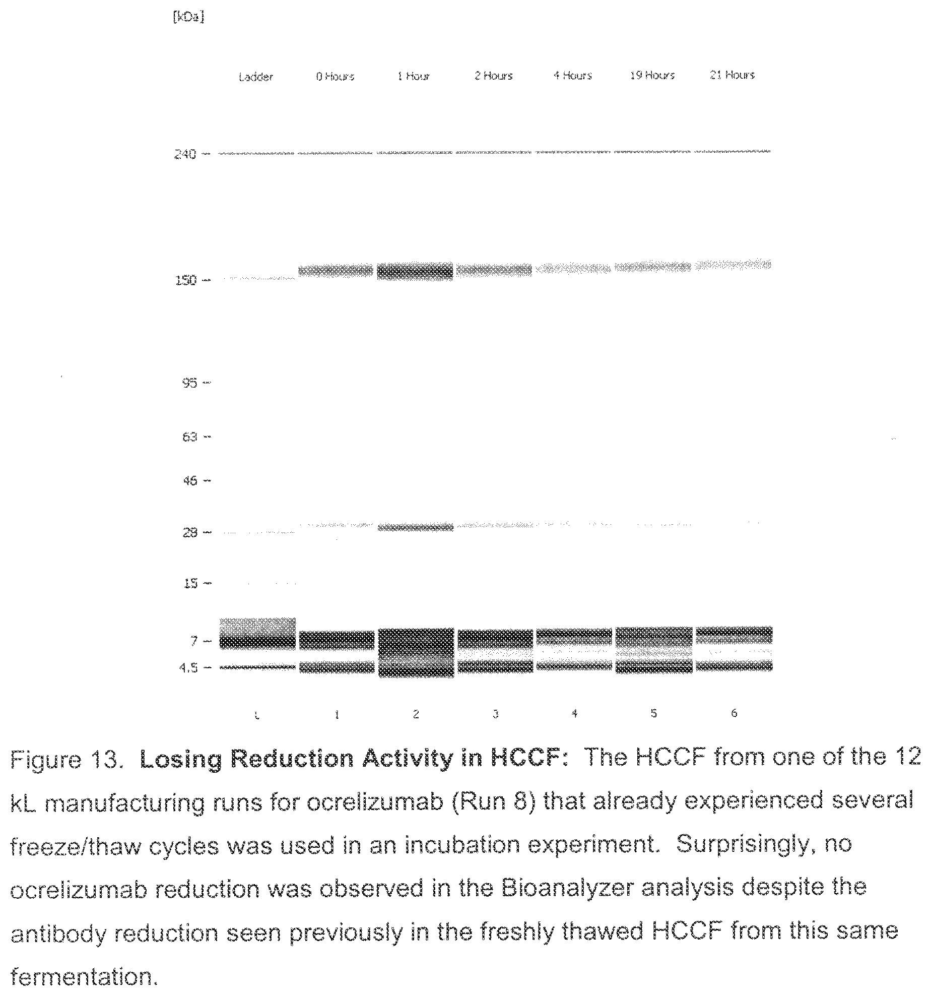
D00014

D00015
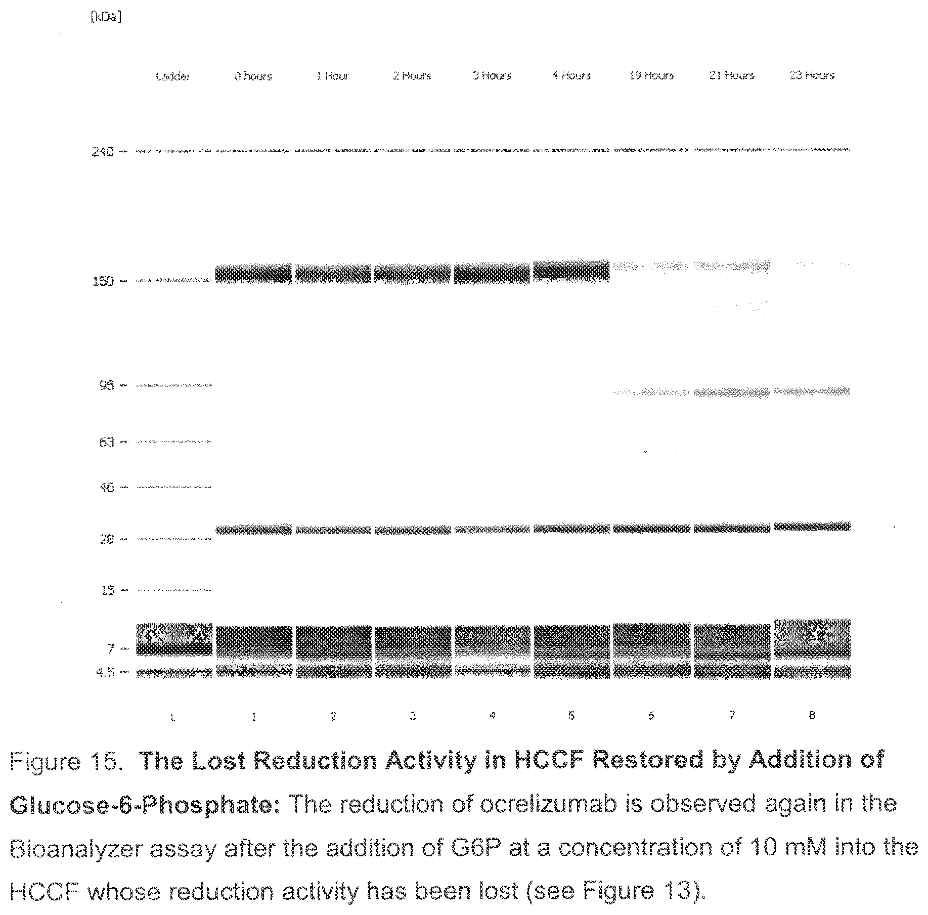
D00016
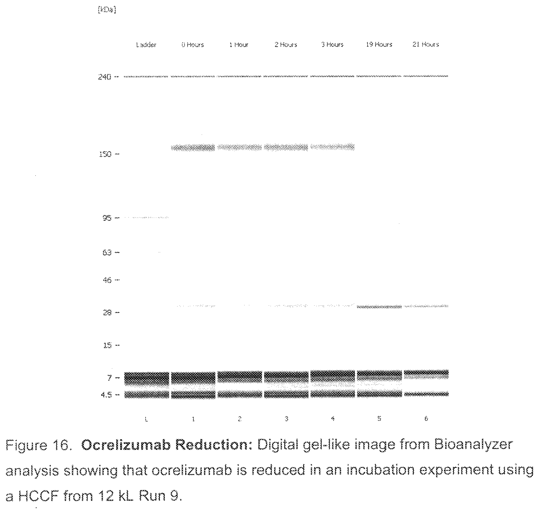
D00017

D00018
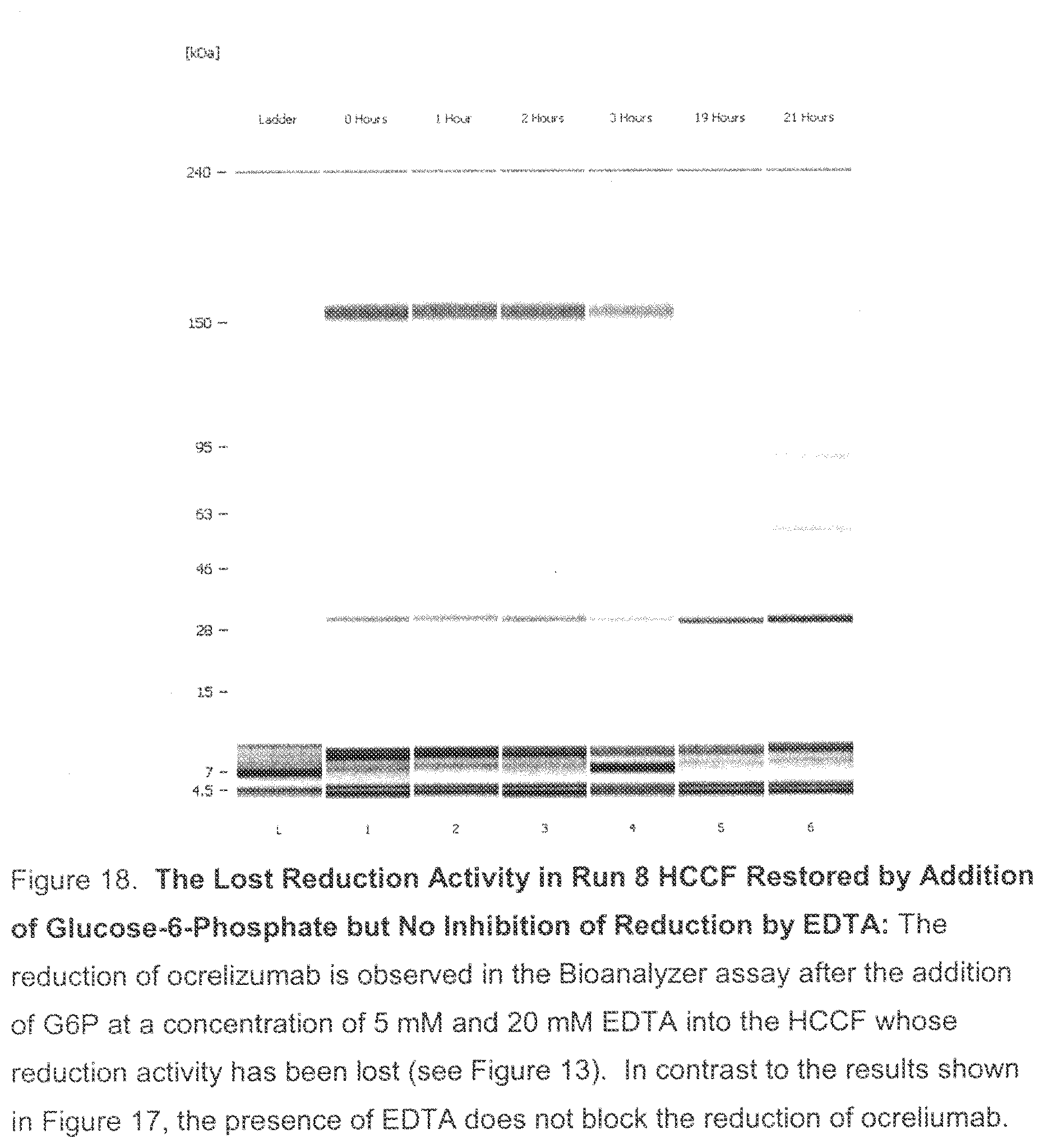
D00019

D00020

D00021

D00022

D00023

D00024
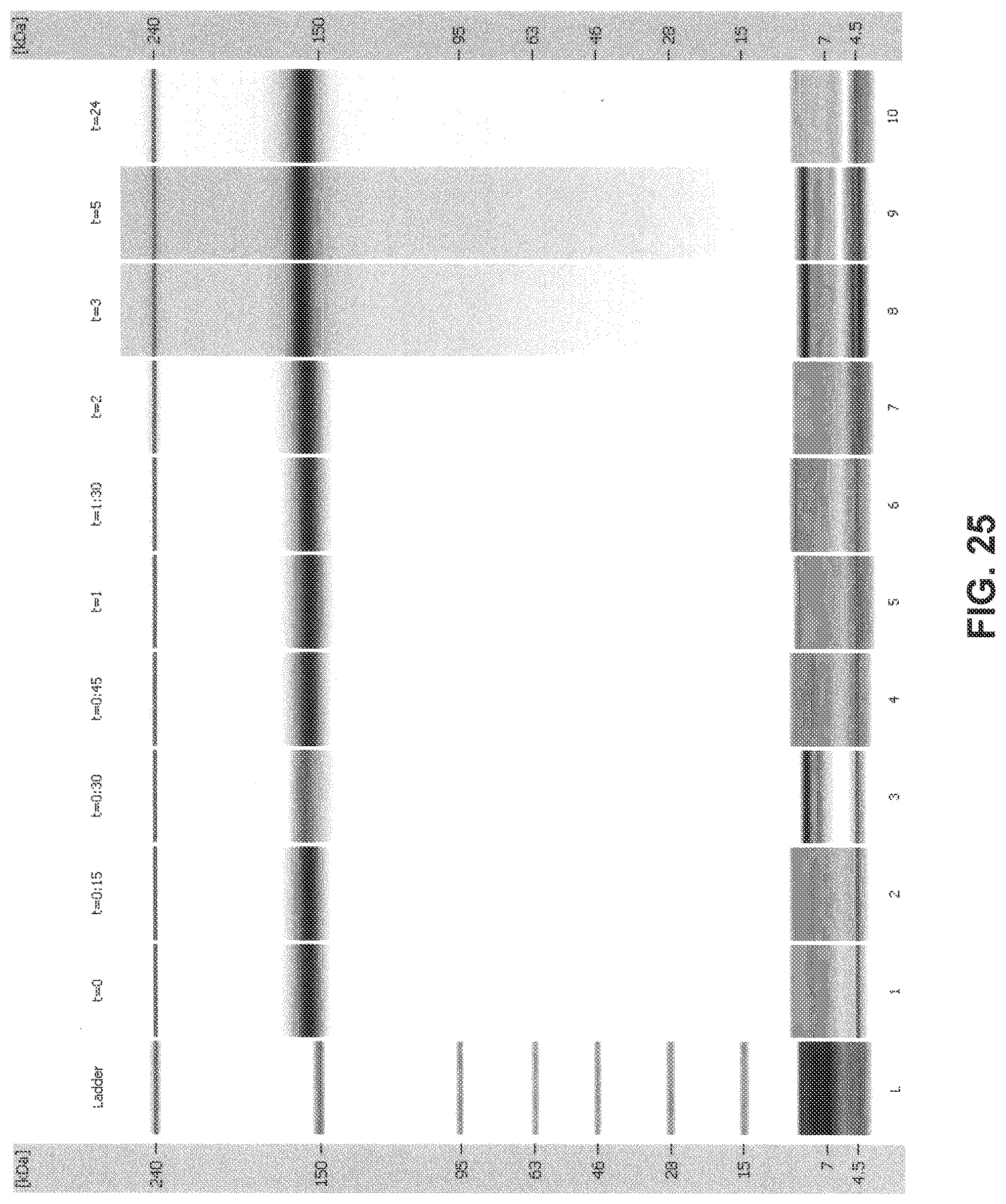
D00025
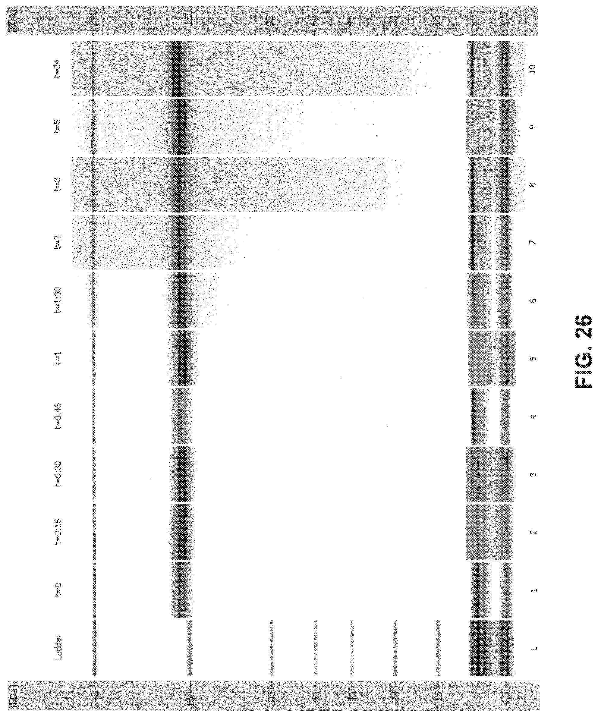
D00026

D00027

D00028

D00029
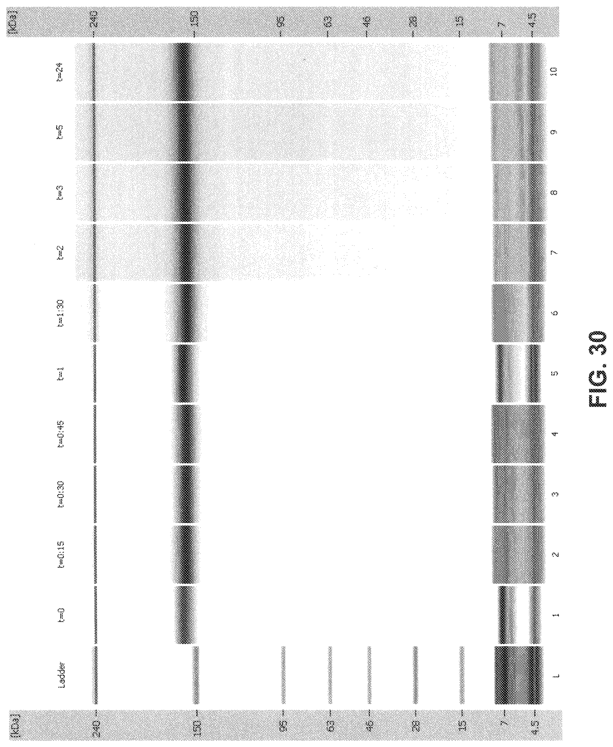
D00030
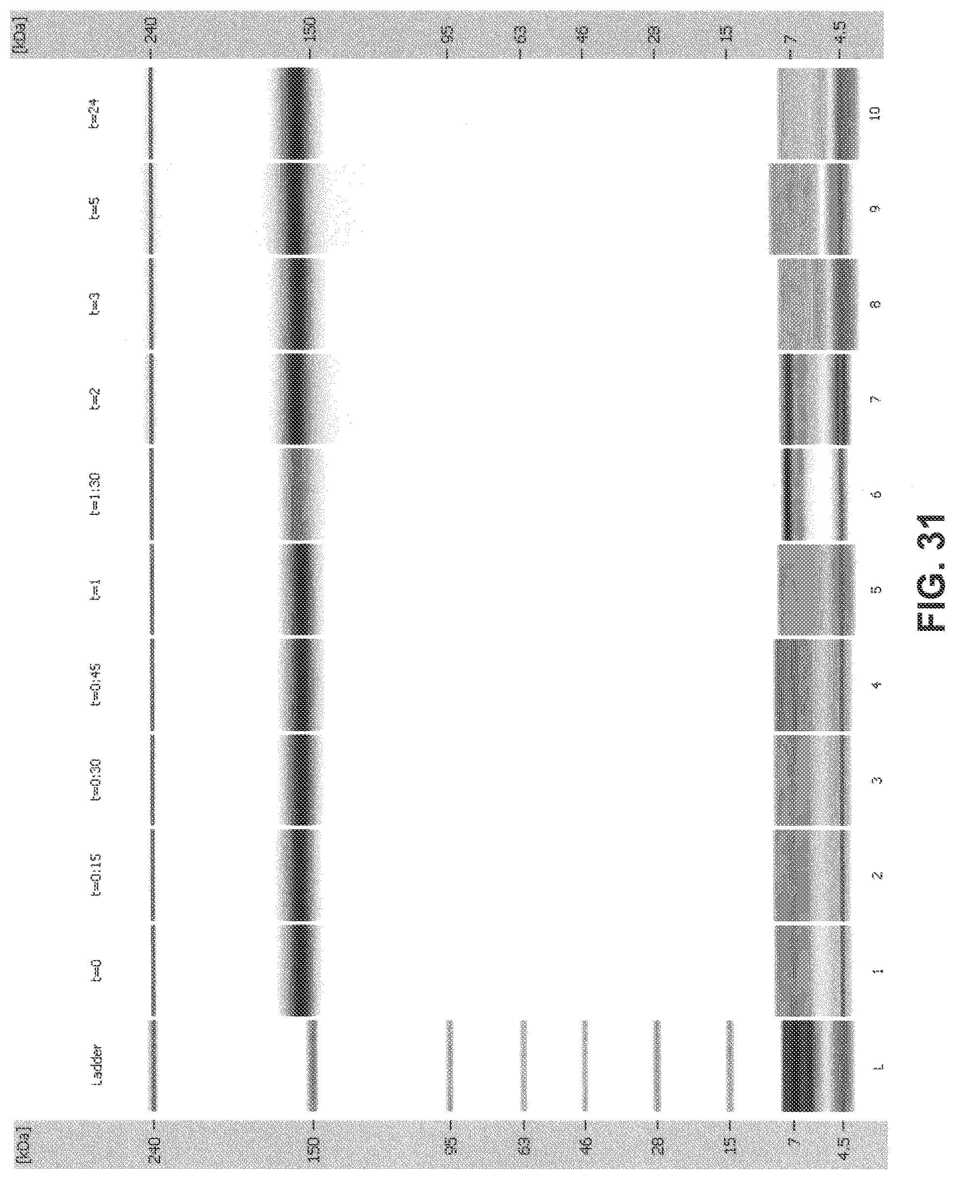
D00031
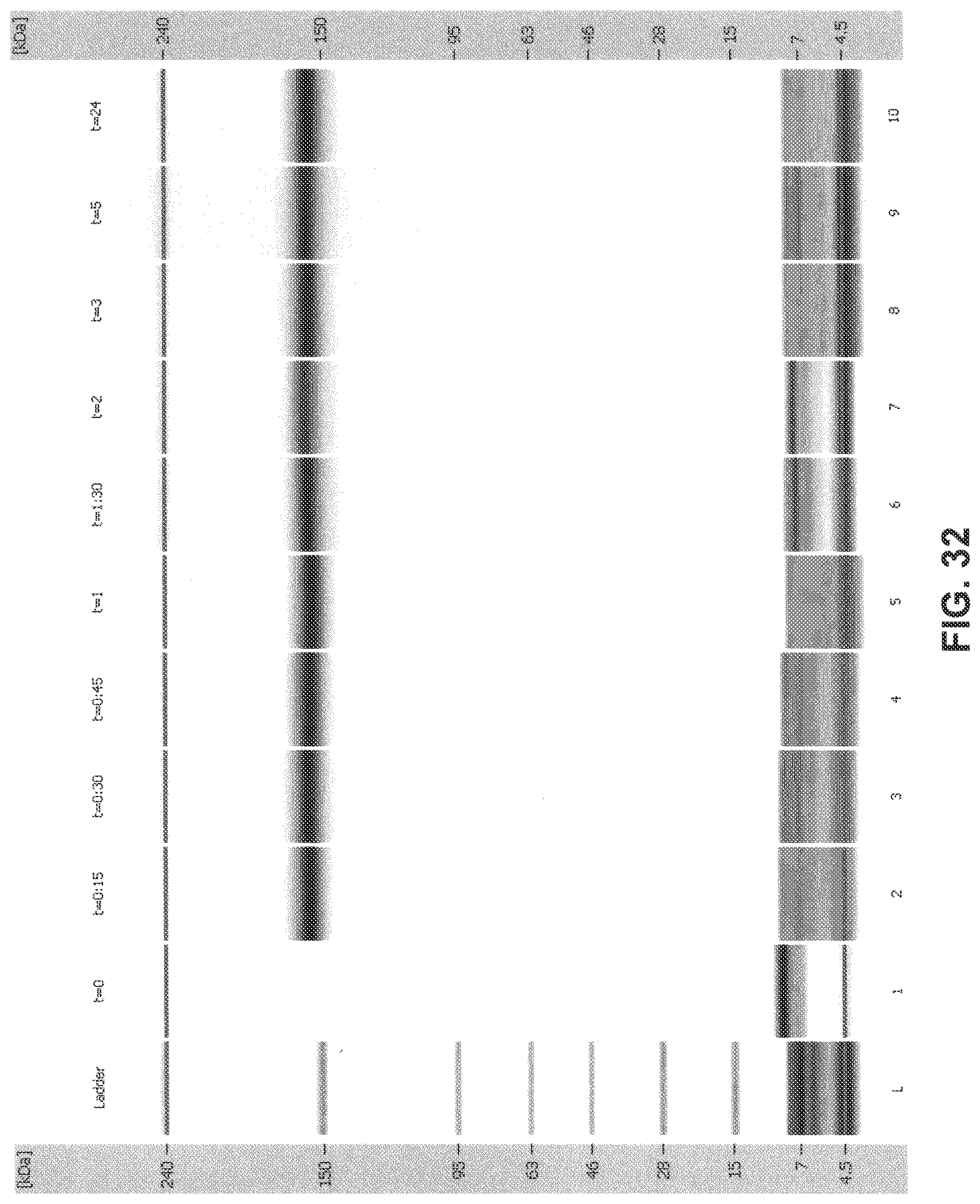
D00032

D00033
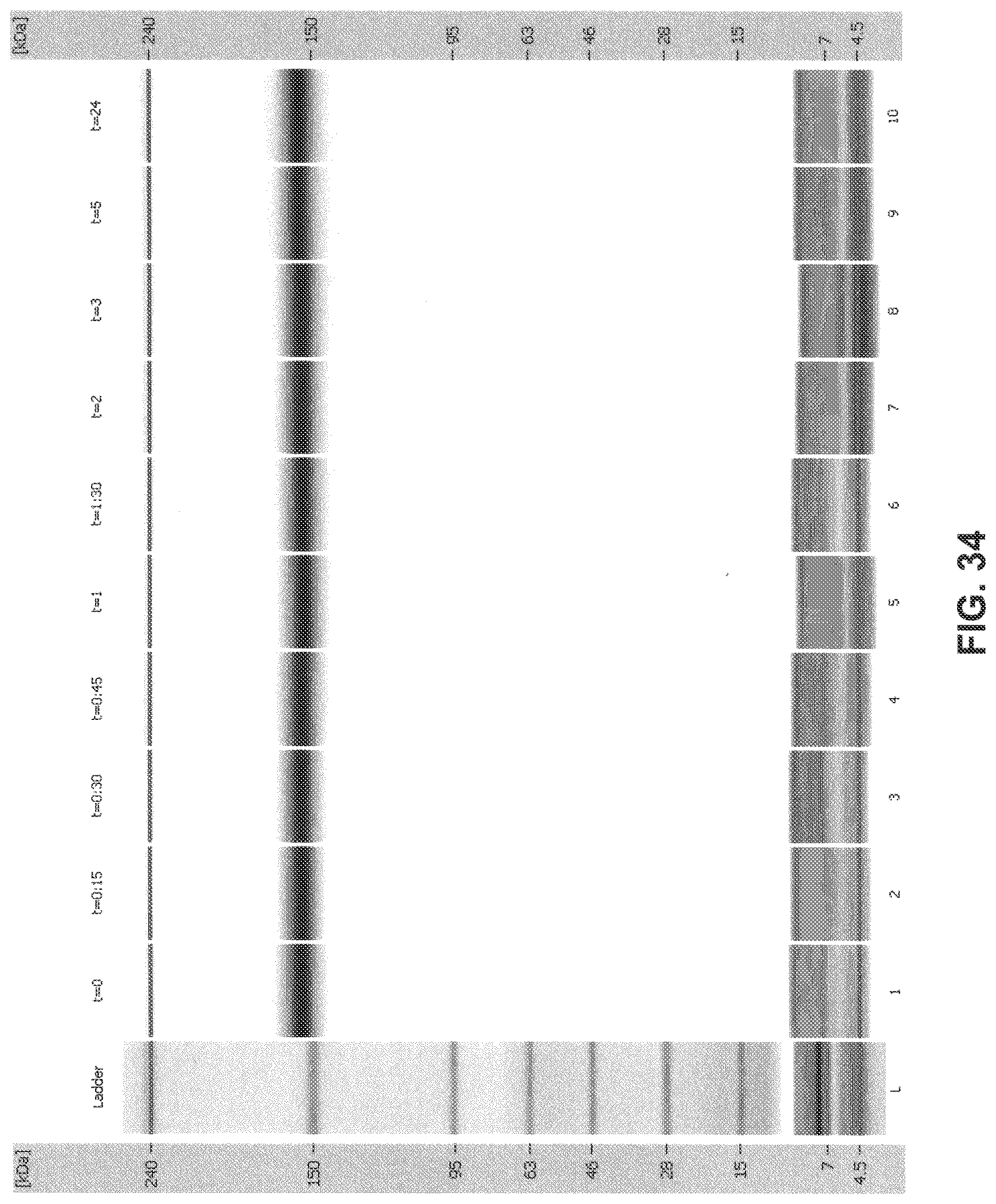
D00034
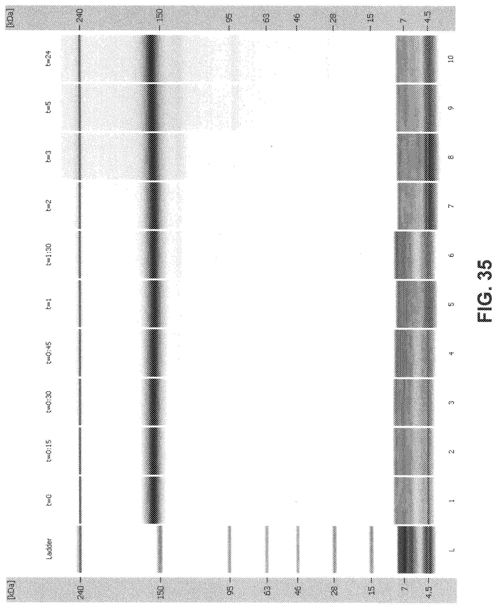
D00035

D00036
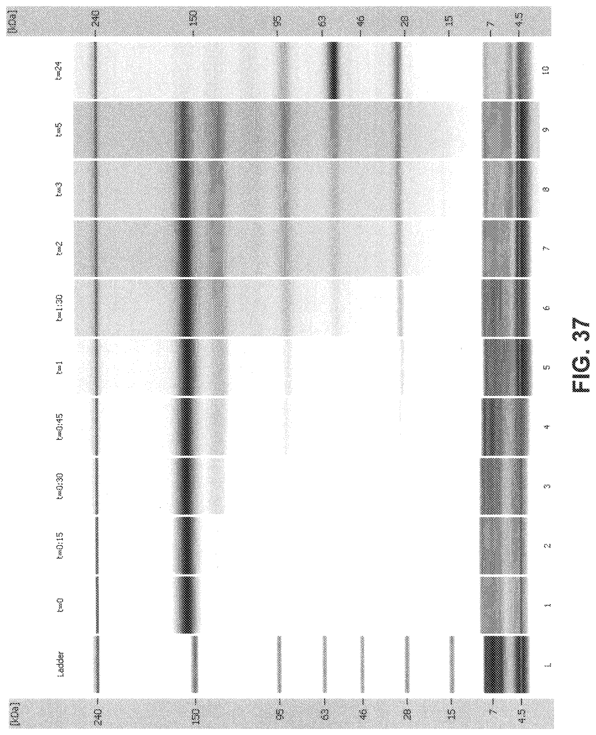
D00037

D00038
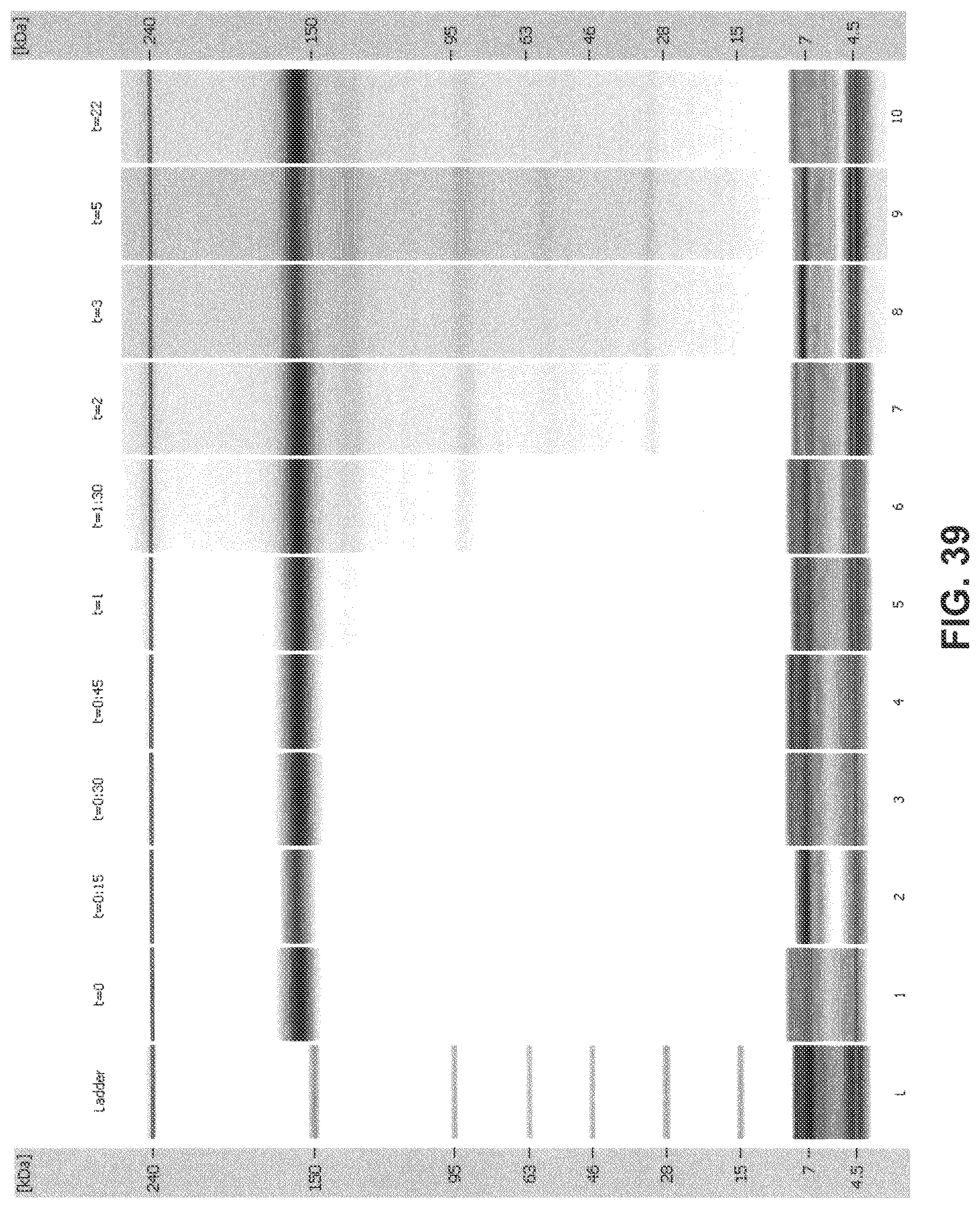
D00039

D00040
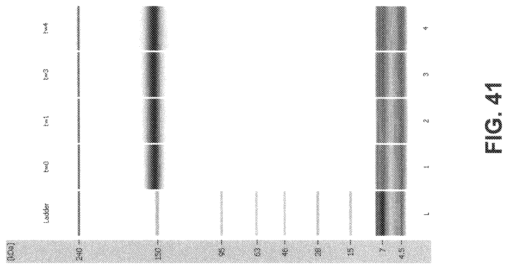
S00001
XML
uspto.report is an independent third-party trademark research tool that is not affiliated, endorsed, or sponsored by the United States Patent and Trademark Office (USPTO) or any other governmental organization. The information provided by uspto.report is based on publicly available data at the time of writing and is intended for informational purposes only.
While we strive to provide accurate and up-to-date information, we do not guarantee the accuracy, completeness, reliability, or suitability of the information displayed on this site. The use of this site is at your own risk. Any reliance you place on such information is therefore strictly at your own risk.
All official trademark data, including owner information, should be verified by visiting the official USPTO website at www.uspto.gov. This site is not intended to replace professional legal advice and should not be used as a substitute for consulting with a legal professional who is knowledgeable about trademark law.